Subcutaneous Fat Shows Higher Thyroid Hormone Receptor-α1 Gene Expression Than Omental Fat
Upload
independentCategory
view
2download
0
Metabolically active portion of fat-free mass: a cellular bodycomposition level modeling analysis
ZiMian Wang1, Stanley Heshka1, Jack Wang1, Dympna Gallagher1, Paul Deurenberg2, ZhaoChen3, and Steven B. Heymsfield11Obesity Research Center, St. Luke’s-Roosevelt Hospital, Columbia University College ofPhysicians and Surgeons, New York, New York2Nutrition Consultant, Singapore3Mel and Enid Zuckerman Arizona College of Public Health, University of Arizona, Tucson, Arizona
AbstractThe proportion of fat-free mass (FFM) as body cell mass (BCM) is highly related to whole bodyresting energy expenditure. However, the magnitude of BCM/FFM may have been underestimatedin previous studies. This is because Moore’s equation [BCM (kg) =0.00833 × total body potassium(in mmol)], which was used to predict BCM, underestimates BCM by ~ %. The aims of the presentstudy were to develop a theoretical BCM/FFM model at the cellular level and to explore the influencesof sex, age, and adiposity on the BCM/FFM. Subjects were 112 adults who had the followingmeasurements: total body water by 2H2O or 3H2O dilution; extracellular water by NaBr dilution;total body nitrogen by in vivo neutron activation analysis; and bone mineral by dual-energy X-rayabsorptiometry. FFM was calculated using a multicomponent model and BCM as the differencebetween FFM and the sum of extracellular fluid and solids. The developed theoretical model revealedthat the proportion of BCM to FFM is mainly determined by water distribution (i.e., E/I, the ratio ofextracellular to intracellular water). A significant correlation (r = 0.90, P < 0.001) was presentbetween measured and model-predicted BCM/FFM for all subjects pooled. Measured BCM/FFM[mean (SD)] was 0.584 ± 0.041 and 0.529 ± 0.041 for adult men and women (P < 0.001), respectively.A multiple linear regression model showed that there are independent significant associations of sex,age, and fat mass with BCM/FFM.
Keywordsbody cell mass; extracellular water; intracellular water; nutrition status; total body potassium
At the molecular body composition level, fat-free mass (FFM) includes water, protein, bonemineral, soft tissue mineral, and glycogen. At the cellular level, these FFM compartments areorganized into body cell mass (BCM), extracellular fluid (ECF), and extracellular solids (ECS)(24). According to Moore et al. (15), BCM is a “component of body composition containingthe oxygen-exchanging, potassium-rich, glucose-oxidizing, and work-performing tissue.”
Both BCM and FFM are considered “metabolically active” components and are often usedinterchangeably to adjust for between-individual differences in resting energy expenditure(REE). Accordingly, interpretation of REE results may vary depending on the adjustingcomponent BCM or FFM.
Address for correspondence: ZM. Wang, Obesity Research Center, 1090 Amsterdam Ave., 14th Floor, New York, NY 10025 (e-mail:[email protected]).
NIH Public AccessAuthor ManuscriptAm J Physiol Endocrinol Metab. Author manuscript; available in PMC 2009 August 9.
Published in final edited form as:Am J Physiol Endocrinol Metab. 2007 January ; 292(1): E49–E53. doi:10.1152/ajpendo.00485.2005.
NIH
-PA Author Manuscript
NIH
-PA Author Manuscript
NIH
-PA Author Manuscript

Forbes (6) was one of the first investigators to explore the association between BCM and FFM.Forbes’s study was based on two assumptions: FFM contains 68.1 mmol of potassium/kg andBCM contains 120 mmol of potassium/kg. The fraction of FFM as BCM was thus assumedstable at 68.1/120 or 0.57 (6). However, later studies showed that the potassium concentrationof FFM and the potassium concentration of BCM suggested by Forbes were not accurate. First,the total body potassium mass (TBK)-to-FFM ratio (TBK/FFM) often differs from the assumedvalue of 68.1 mmol/kg. Many investigators have consistently found lower potassiumconcentrations in FFM, 54–59 mmol/kg for women and 59–62 mmol/kg for men (4). Second,the potassium content of BCM is not 120 mmol/kg. Both experimental and modeling estimatesindicate a mean TBK/BCM of 109 mmol/kg (1,27). These observations suggest that the BCMcontent of FFM may not be constant.
In addition, a growing number of experimental studies reveal that the proportion of FFM asBCM varies with some biological factors such as age and adiposity (12,14,19). Gallagher etal. (9) reported a sex difference in BCM/FFM, with men and women at 0.51 ± 0.05 and 0.48± 0.05, respectively. Wang et al. (20) reported differences between African Americans, Asians,and Caucasians in the fraction of FFM as BCM.
However, all of these previous studies have a now-recognized inherent limitation. BCM wascalculated from TBK using Moore’s equation (15), BCM (kg) = 0.00833 × TBK (mmol), whichunderestimates BCM by ~ 11% (1,27). Thus previous studies may be inaccurate in theirassessment of the BCM-FFM relationship, notably in underestimating the magnitude of BCM/FFM.
The aim of the present study was to develop a cellular-level BCM/FFM model that providesnew insights into the quantitative associations between BCM and FFM and to identify themajor determinants of variation in BCM/FFM. Specifically, the present study was designed toexplore the magnitude and variation of the BCM content of FFM in healthy adults.
SUBJECTS AND METHODSSubjects
The subject pool consisted of male and female adults who were recruited through local sources,including flyers posted in the medical center and newspaper advertisements. Each subjectcompleted a medical history, physical examination, and screening blood studies to establishthe absence of disease. Subjects participated in recreational physical activities but were notactively engaged in any sports training programs. All participants signed an informed consentthat was approved by the hospital’s Institutional Review Board.
Body Composition MeasurementsConsenting subjects were studied after an overnight fast. Body weight was measured (±0.1 kg)with a digital scale (Weight Tronix, New York, NY) and height (±0.5 cm) with a wall-mountedstadiometer (Holtain, Crosswell, UK). Each subject completed five studies: 3H2O or 2H2Odilution for total body water (TBW); prompt-γ in vivo neutron activation (IVNA) for total bodynitrogen (TBN); dual-energy X-ray absorptiometry (DEXA) for body fat and BMC; sodiumbromide (NaBr) dilution for extracellular water (ECW), and whole body 40K counting for TBK.
TBW—Each subject completed either tritium (3H2O) or deuterium (2H2O) dilution(17). 3H2O dilution volume was measured at the Body Composition Unit of St. Luke’s-Roosevelt Hospital, and 2H2O dilution volume was measured at the Brookhaven NationalLaboratory, Upton, NY. The precision was 1.5% [coefficient of variation (CV)] for dilutionvolume estimation at the two laboratories. The 3H2O and 2H2O dilution volume (in liters) was
Wang et al. Page 2
Am J Physiol Endocrinol Metab. Author manuscript; available in PMC 2009 August 9.
NIH
-PA Author Manuscript
NIH
-PA Author Manuscript
NIH
-PA Author Manuscript

then converted into TBW mass by multiplying with the correction factor for nonaqueoushydrogen exchange and water density at 36°C [TBW (kg) =0.96 × 0.994 × 3H2O or 2H2Odilution volume (in liters)].
TBN—TBN was quantified using the prompt-γ IVNA facilities at Brookhaven NationalLaboratory with a CV of 2.7% (3). The TBN (in kg) was then converted to total body protein(TBPro, in kg) as TBPro = 6.25 × TBN (2).
Bone mineral content—A DEXA scanner (Lunar DPX with software version 3.6; Madison,WI) was used in this study to estimate bone mineral content (BMC). The technique used X-rays of two distinct energy levels (40 and 70 keV) that are differently attenuated by BMC, fat,and fat-free soft tissues (16). BMC measured by DEXA represents ashed bone. Bone mineral(Mo) produces 0.9582 g of BMC due to loss of labile components, including bound water andCO2 with combustion. BMC was thus converted to bone mineral (Mo) as Mo = BMC/0.9582.The DEXA system CV is 1.3% for Mo estimation (10).
ECW—ECW was measured by NaBr dilution with a CV of 1.4%. The bromide dilution volume(in liters) was then converted to ECW (in kg) by correcting for the Gibbs-Donnan effect (0.95),and the penetration of bromide into the intracellular space of erythrocytes (0.90) [ECW (kg)=0.95 × 0.90 × NaBr dilution volume (in liters)] (17).
ECW/ICW—TBW is equal to the sum of two compartments: ECW and intracellular water(ICW). The distribution of water between the ECW and ICW was calculated as the E/I ratio
(1)
FFM and fat—At the molecular level, FFM can be expressed as the sum of five components:water, protein, bone mineral, soft-tissue mineral (Ms), and glycogen (24):
(2)
where TBW is measured by 3H2O or 2H2O dilution (17); TBPro is calculated from TBNmeasured by prompt-γ IVNA, TBPro = 6.25 × TBN (2); Mo is measured by DEXA (13); Msis calculated from TBW as Ms = 0.0129 × TBW (25); and glycogen is calculated from TBNas glycogen = 0.275 × TBN (11). Body fat mass was calculated as the difference between bodyweight and FFM.
BCM—At the cellular level, FFM is equal to the sum of BCM, extracellular fluid (ECF), andextracellular solids (ECS) (24). BCM can thus be expressed as
(3)
where ECF is calculated from ECW as ECF = (1/0.98) ×ECW, where 0.98 is the mean hydrationof ECF (21); and ECS is calculated from Mo as ECS = 1.732 × Mo (26). The measurementsof BCM and FFM were then used to calculate the ratio BCM/FFM.
Statistical AnalysisGroup results are presented as means (SD). Differences in body composition between men andwomen were tested using Student’s t-test, and P < 0.05 was considered statistically significant.Simple linear regression analysis was used to describe the association between measured BCM/
Wang et al. Page 3
Am J Physiol Endocrinol Metab. Author manuscript; available in PMC 2009 August 9.
NIH
-PA Author Manuscript
NIH
-PA Author Manuscript
NIH
-PA Author Manuscript

FFM and model-predicted BCM/FFM. A multiple linear regression model [BCM/FFM = α +β1 × sex + β2 × age + β3 × %fat + ∊] was applied to analyze the correlation of measured BCM/FFM with biological factors including sex, age, and percent fat. Effect size was expressed asη2 (eta squared). Means of individual differences between measured and model-predictedBCM/FFM ratios were tested for significance by Student’s t-tests. Data were analyzed usingSAS v. 8.2 (SAS Institute, Cary, NC).
RESULTSDevelopment of BCM/FFM Model
At the cellular body composition level, FFM is composed of three compartments: BCM, ECF,and ECS. The primary BCM/FFM model can be written as
(4)
In the next stage of model development, our aim was to resolve Eq. 4 into relevant compartmentratios. ICW and ECW are the largest compartments of BCM and ECF, respectively. As notedin our previous study (21), BCM can be expressed as ICW/a and ECF as ECW/b, where a andb are the proportions of BCM and ECF as water, respectively. Similarly, extracellular solidscan be expressed as a function of TBW, ECS = c × TBW = c × (ICW + ECW), where c is theratio of ECS to TBW. In addition, both ICW and ECW are water compartments, and ECW canbe expressed as a function of ICW, ECW = (E/I) × ICW, where E/I is the ratio of ECW to ICW.
Equation 4 can thus be converted and simplified to a secondary cellular level BCM/FFM modelas
(5)
Simplified BCM/FFM ModelEquation 5 reveals that BCM/FFM is determined by four factors: fraction of BCM as ICW (a);fraction of ECF as ECW (b); ratio of ECS to TBW (c); and ratio of ECW to ICW (E/I). Eachof the four determinants may vary within a range for healthy adults (21). The approximatemean value and variation range of each determinant in adults is summarized in Table 1. Onecan thus estimate the influence of each determinant on BCM/FFM value if a determinant takesits extreme values and the other three determinants take their mean value.
Of the four determinants, as Table 1 shows, water distribution (i.e., E/I) has the largest influenceon BCM/FFM value. Comparatively, the influences of a, b, and c on BCM/FFM are muchsmaller than the influence of E/I. Therefore, the mean magnitudes (i.e., a = 0.70, b = 0.98, andc = 0.14) are assumed for modeling purposes to be stable in healthy adults. Equation 5 can thusbe converted to a simplified model:
(6)
Wang et al. Page 4
Am J Physiol Endocrinol Metab. Author manuscript; available in PMC 2009 August 9.
NIH
-PA Author Manuscript
NIH
-PA Author Manuscript
NIH
-PA Author Manuscript

Equation 6 shows that BCM/FFM vs. E/I is a concave curve, as shown in Fig. 1. Although thissimplified model is a decreasing nonlinear function, the nonlinear component is small over theusual range of E/I values.
In the following sections, we use measured BCM/FFM values to evaluate the developed BCM/FFM model in a sample of men and women.
Sample CharacteristicsThe study group consisted of 112 adult subjects, 14 men and 98 women, ranging in age from22 to 74 yr, body weight from 42.1 to 105.7 kg, and BMI from 16.8 to 34.7 kg/m2 (Table 2).The women were overweight as a group, with a BMI of 28.3 ± 4.9 kg/m2, whereas the men’sBMI was within the normal range, 21.5 ± 2.3 kg/m2. Three race/ethnicity groups wererepresented in the sample: African American (n = 55), Caucasian (n = 46), and Hispanic (n =11).
Measured and Predicted BCM/FFMFFM and BCM were measured on the basis of Eq. 2 and Eq 3, respectively, and waterdistribution was calculated on the basis of Eq. 1 (Table 2).
The individual E/I values were applied in Eq. 6 to predict individual BCM/FFM in the menand women. The model-predicted values of BCM/FFM were highly correlated with thecorresponding measured BCM/FFM values for all subjects pooled (r = 0.89, P < 0.001; Fig.2). The measured and predicted mean values also did not differ: 0.585 ± 0.043 vs. 0.584 ±0.041 (paired t-test, P = 0.76) for the men and 0.530 ± 0.044 vs. 0.529 ± 0.041 (paired t-test,P = 0.44) for the women. The variance of the differences between measured and predictedBCM/FFM in the pooled sample is only 21% that of BCM/FFM, indicating that the simplifiedmodel (i.e., Eq. 6) effectively accounts for ~ 80% of the variability in BCM/FFM.
Association Between BCM/FFM and Biological Variables After Adjusting for E/IAn interesting question is whether variation in E/I accounts for reported associations of BCM/FFM with age, sex, fat mass and similar variables (9,12,14,19,20). We first applied a generallinear regression model to analyze the associations of measured BCM/FFM with biologicalfactors including sex, race (i.e., African American, Caucasian, Hispanic), age, fat mass, andheight. The model accounted for 33% of the total variability, but only fat mass had anindependent significant association with BCM/FFM (P < 0.001). We then added E/I to themodel to determine whether any independent associations persisted after adjusting for E/I. Thismodel accounted for 89% of the variance. The E/I variable showed the strongest associationby far with BCM/FFM (P < 0.001, η2 = 0.55), but fat mass remained significant (P < 0.001,η2 = 0.09), and height also reached significance (P < 0.002, η2 = 0.01). The significantcoefficients for fat mass and height indicate that there are independent associations of thesevariables with BCM/FFM after variation in E/I is taken into account.
DISCUSSIONA theoretical BCM/FFM model was developed in the present study. The model withempirically derived coefficients indicates that water distribution is strongly associated withBCM/FFM. This finding helps to explain the subsequent observations of sex, age, and adiposityeffects on BCM/FFM. Another finding of the present study was that the BCM/FFM values aremuch higher than those reported by previous studies: 0.584 ± 0.041 vs. 0.51 ± 0.05 for menand 0.529 ± 0.041 vs. 0.48 ± 0.05 for women (9).
Wang et al. Page 5
Am J Physiol Endocrinol Metab. Author manuscript; available in PMC 2009 August 9.
NIH
-PA Author Manuscript
NIH
-PA Author Manuscript
NIH
-PA Author Manuscript

E/I and BCM/FFMPrevious studies reported variation in BCM related to FFM. Mazariegos et al. (14) and Kyleet al. (12) observed a lower BCM/FFM in older adults vs. young adults. Gallagher et al. (9)and Wang et al. (20) found that the fraction of FFM as BCM appears smaller in women thanin men. With increasing adiposity there was a corresponding reduction in BCM relative to FFM(19). Although these studies indicate sex, age, and adiposity influences on BCM/FFM,mechanistic explanations were not provided.
Our simplified model (i.e., Eq. 6) reveals that water distribution is the major determinant ofBCM relative to FFM. When E/I increases from 0.70 to 1.00, for example, BCM/FFMcorrespondingly decreases from 0.60 to 0.52 (Fig. 1). Women as a group have a higher E/Ivalue than men; elderly women may have a higher E/I value than young women; and obesewomen have a higher E/I than women with normal body weight. The whole body, as well aseach organ and tissue, is composed of two compartments, cells (including ICW and residuals)and non-cells (including ECW and solids). The E/I ratio can thus be considered as a substitutionfor the ratio of non-cells to cells. As sex, age, and adiposity influence E/I on a group basis,according to our simplified model, BCM/FFM appears smaller in women than in men, and themagnitudes of BCM/FFM decrease with increasing adiposity and age in elderly subjects.Generally speaking, any biological and pathological factors, which affect water distribution,may influence the BCM/FFM.
BCM/FFM and REEBody composition has a quantitative association with whole body resting energy expenditure(REE), a major component of total energy expenditure (23). Whole body REE (in kcal/day)can be expressed as the sum of energy expended by individual organs and tissues with thefollowing general model:
(7)
where Mi is individual organ-tissue mass (in kg), i is a specific organ-tissue component (i = 1,2, … n), and ki is the specific resting metabolic rate of individual organs and tissues (inkcal·kg−1·day−1) (4). On the basis of Eq. 7, our group measured metabolically active organs(liver, brain, heart, and kidneys) and two other tissues (skeletal muscle and adipose tissue) byMRI in a cohort of young healthy adults. The predicted REE was nearly identical to REEmeasured by indirect calorimetry (8).
When applied to older subjects, however, the REE predicted by equation 7 was higher than themeasured REE by ~ 11% (P < 0.001) (7). Our previous study found that older subjects have asmaller fraction of FFM as BCM (14). As multiple regression analysis revealed, the BCM/FFM value was lower with greater age (β = − 0.24, P = 0.005) (17). We added a new variable(the fraction of FFM as BCM) to equation 7 (22). A modified theoretical REE model was thusdeveloped as
(8)
where 0.58 is BCM/FFM value in Reference Man, and the corresponding BCM/FFM is 0.56in Reference Women (17). Equation 8 was then applied to predict REE in adult men and womenaged 23–88 yr (22). We found that REE predicted by Eq. 8 was correlated with measured REE(r = 0.92, P < 0.001), and the mean difference between measured REE and predicted REE for
Wang et al. Page 6
Am J Physiol Endocrinol Metab. Author manuscript; available in PMC 2009 August 9.
NIH
-PA Author Manuscript
NIH
-PA Author Manuscript
NIH
-PA Author Manuscript

the whole group was not significant. This study shows that the REE prediction model performedbetter when BCM/FFM is included.
If age is included as a variable in estimating REE along with the predictions of Eq. 8, the roleof the BCM/FFM model remains highly significant (P < 0.001), indicating that its contributionto the estimation of REE is independent of its association with age.
In conclusion, the ratio of BCM to FFM is not a constant. According to our modeling analysis,the variation in BCM/FFM is primarily dependent on water distribution. The larger the E/I,the smaller the BCM/FFM and vice versa. The magnitude of BCM/FFM is one of the majordeterminants of whole body REE.
AcknowledgmentsGRANTS
This study was supported by National Institute of Diabetes and Digestive and Kidney Diseases Grant DK-42618 andP30-DK-26687.
REFERENCES1. Cohn SH, Vaswani AN, Yasumura S, Yuen K, Ellis KJ. Assessment of cellular mass and lean body
mass by noninvasive nuclear techniques. J Lab Clin Med 1985;105:305–311. [PubMed: 3973466]2. Cuningham JJ. N × 6.25: recognizing a bivariate expression for protein balance in hospitalized patients.
Nutrition 1994;10:124–127. [PubMed: 8025364]3. Dilmanian, FA.; Weber, DA.; Yasumura, S.; Kamen, Y.; Kindofsky, L.; Heymsfield, SB.; Pierson,
RN., Jr; Wang, J.; Kehayias, JJ.; Ellis, KJ. The performance of the BNL delayed-and prompt-gammaneutron activation systems at Brookhaven National Laboratory. In: Yasumura, S.; Harrison, JE.;McNeill, KG.; Woodhead, AD.; Dilmanian, FA., editors. Advances in In Vivo Body CompositionStudies. NY: Plenum: 1990. p. 309-315.
4. Elia, M. Organ and tissue contribution to metabolic rate. In: Kinney, JM., editor. Energy Metabolism:Tissue Determinants and Cellular Corollaries. New York: Raven; 1992. p. 61-77.
5. Ellis, JE. Whole-body counting and neutron activation analysis. In: Heymsfield, SB.; Lohman, TG.;Wang, ZM.; Going, SB., editors. Human Body Composition. Vol. 2nd ed.. Champaign, IL: HumanKinetics; 2005. p. 51-62.
6. Forbes, GB. Human Body Composition. New York: Springer-Verlag; 1987.7. Gallagher D, Allen A, Wang ZM, Heymsfield SB, Krasnow N. Smaller organ tissue mass in the elderly
fails to explain lower resting metabolic rate. In: In Vivo Body Composition Studies, edited by YasumuraS, Wang J, and Pierson RN Jr. Ann NY Acad Sci 2000;904:449–455. [PubMed: 10865788]
8. Gallagher D, Belmonte D, Deurenberg P, Wang ZM, Krasnow N, Pi-Sunyer FX, Heymsfield SB.Organ-tissue mass measurement by magnetic resonance imaging allows accurate in vivo modeling ofresting energy expenditure and metabolically active tissue mass. Am J Physiol Endocrinol Metab1998;275:E249–E258.
9. Gallagher D, Visser M, Wang ZM, Harris T, Pierson RN Jr, Heymsfield SB. Metabolically activecomponent of fat-free body mass: influences of age, adiposity, and gender. Metabolism 1996;45:992–997. [PubMed: 8769358]
10. Heymsfield SB, Waki M, Kehayias JJ, Lichtman S, Dilmanian FA, Kamen Y, Wang J, Pierson RNJr. Chemical and elemental analysis of humans in vivo using improved body composition models.Am J Physiol Endocrinol Metab 1991;261:E190–E198.
11. Kehayias JJ, Heymsfield SB, LoMonte AF, Wang J, Pierson RN Jr. In vivo determination of bodyfat by measuring total body carbon. Am J Clin Nutr 1991;53:1339–1344. [PubMed: 2035460]
12. Kyle UG, Genton L, Hans D, Karsegard L, Slosman DO, Pichard C. Age-related differences in fat-free mass, skeletal muscle, body cell mass and fat mass between 18 and 94 years. Eur J Clin Nutr2001;55:663–672. [PubMed: 11477465]
Wang et al. Page 7
Am J Physiol Endocrinol Metab. Author manuscript; available in PMC 2009 August 9.
NIH
-PA Author Manuscript
NIH
-PA Author Manuscript
NIH
-PA Author Manuscript
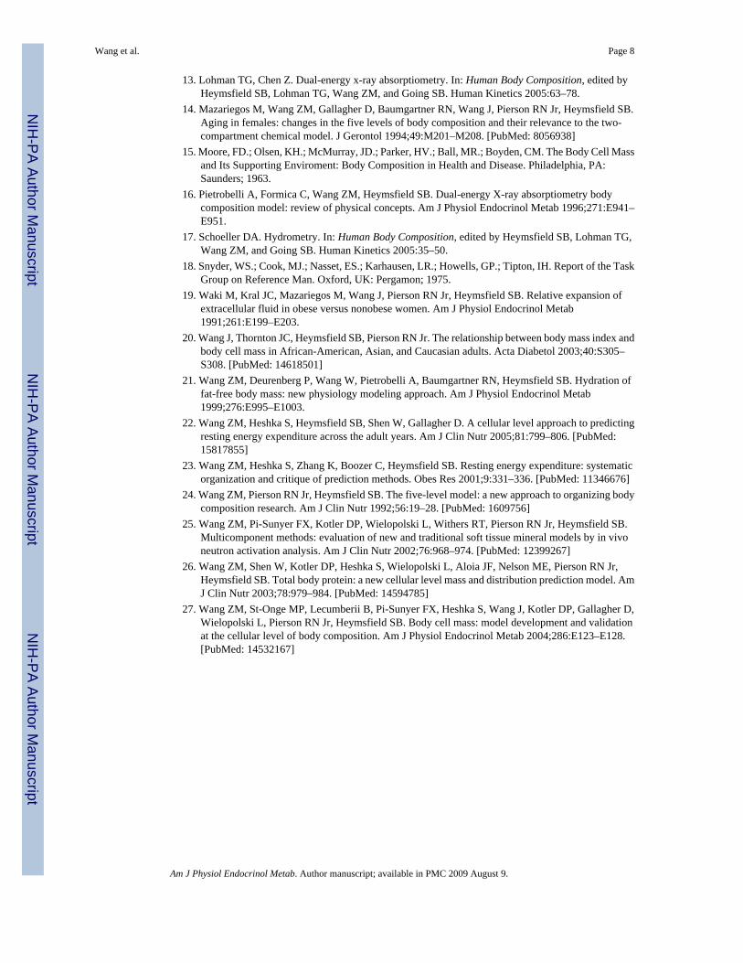
13. Lohman TG, Chen Z. Dual-energy x-ray absorptiometry. In: Human Body Composition, edited byHeymsfield SB, Lohman TG, Wang ZM, and Going SB. Human Kinetics 2005:63–78.
14. Mazariegos M, Wang ZM, Gallagher D, Baumgartner RN, Wang J, Pierson RN Jr, Heymsfield SB.Aging in females: changes in the five levels of body composition and their relevance to the two-compartment chemical model. J Gerontol 1994;49:M201–M208. [PubMed: 8056938]
15. Moore, FD.; Olsen, KH.; McMurray, JD.; Parker, HV.; Ball, MR.; Boyden, CM. The Body Cell Massand Its Supporting Enviroment: Body Composition in Health and Disease. Philadelphia, PA:Saunders; 1963.
16. Pietrobelli A, Formica C, Wang ZM, Heymsfield SB. Dual-energy X-ray absorptiometry bodycomposition model: review of physical concepts. Am J Physiol Endocrinol Metab 1996;271:E941–E951.
17. Schoeller DA. Hydrometry. In: Human Body Composition, edited by Heymsfield SB, Lohman TG,Wang ZM, and Going SB. Human Kinetics 2005:35–50.
18. Snyder, WS.; Cook, MJ.; Nasset, ES.; Karhausen, LR.; Howells, GP.; Tipton, IH. Report of the TaskGroup on Reference Man. Oxford, UK: Pergamon; 1975.
19. Waki M, Kral JC, Mazariegos M, Wang J, Pierson RN Jr, Heymsfield SB. Relative expansion ofextracellular fluid in obese versus nonobese women. Am J Physiol Endocrinol Metab1991;261:E199–E203.
20. Wang J, Thornton JC, Heymsfield SB, Pierson RN Jr. The relationship between body mass index andbody cell mass in African-American, Asian, and Caucasian adults. Acta Diabetol 2003;40:S305–S308. [PubMed: 14618501]
21. Wang ZM, Deurenberg P, Wang W, Pietrobelli A, Baumgartner RN, Heymsfield SB. Hydration offat-free body mass: new physiology modeling approach. Am J Physiol Endocrinol Metab1999;276:E995–E1003.
22. Wang ZM, Heshka S, Heymsfield SB, Shen W, Gallagher D. A cellular level approach to predictingresting energy expenditure across the adult years. Am J Clin Nutr 2005;81:799–806. [PubMed:15817855]
23. Wang ZM, Heshka S, Zhang K, Boozer C, Heymsfield SB. Resting energy expenditure: systematicorganization and critique of prediction methods. Obes Res 2001;9:331–336. [PubMed: 11346676]
24. Wang ZM, Pierson RN Jr, Heymsfield SB. The five-level model: a new approach to organizing bodycomposition research. Am J Clin Nutr 1992;56:19–28. [PubMed: 1609756]
25. Wang ZM, Pi-Sunyer FX, Kotler DP, Wielopolski L, Withers RT, Pierson RN Jr, Heymsfield SB.Multicomponent methods: evaluation of new and traditional soft tissue mineral models by in vivoneutron activation analysis. Am J Clin Nutr 2002;76:968–974. [PubMed: 12399267]
26. Wang ZM, Shen W, Kotler DP, Heshka S, Wielopolski L, Aloia JF, Nelson ME, Pierson RN Jr,Heymsfield SB. Total body protein: a new cellular level mass and distribution prediction model. AmJ Clin Nutr 2003;78:979–984. [PubMed: 14594785]
27. Wang ZM, St-Onge MP, Lecumberii B, Pi-Sunyer FX, Heshka S, Wang J, Kotler DP, Gallagher D,Wielopolski L, Pierson RN Jr, Heymsfield SB. Body cell mass: model development and validationat the cellular level of body composition. Am J Physiol Endocrinol Metab 2004;286:E123–E128.[PubMed: 14532167]
Wang et al. Page 8
Am J Physiol Endocrinol Metab. Author manuscript; available in PMC 2009 August 9.
NIH
-PA Author Manuscript
NIH
-PA Author Manuscript
NIH
-PA Author Manuscript

Fig. 1.Proportion of fat-free mass (FFM) as body cell mass (BCM) by the simplified model (i.e., Eq.6) on the ordinate and the ratio E/I as extracellular water (ECW) to intracellular water (ICW)on the abscissa.
Wang et al. Page 9
Am J Physiol Endocrinol Metab. Author manuscript; available in PMC 2009 August 9.
NIH
-PA Author Manuscript
NIH
-PA Author Manuscript
NIH
-PA Author Manuscript

Fig. 2.Proportion of FFM as BCM predicted by the simplified model (i.e., Eq. 6) on the ordinate andthe measured BCM/FFM on the abscissa. Model-predicted BCM/FFM = 0.933 × measuredBCM/FFM + 0.037, r = 0.90, P < 0.001; n = 112 adults. Line of identity is shown in the figure.
Wang et al. Page 10
Am J Physiol Endocrinol Metab. Author manuscript; available in PMC 2009 August 9.
NIH
-PA Author Manuscript
NIH
-PA Author Manuscript
NIH
-PA Author Manuscript

NIH
-PA Author Manuscript
NIH
-PA Author Manuscript
NIH
-PA Author Manuscript
Wang et al. Page 11
Table 1Influence of model determinants on BCM/FFM value
Determinant Mean Magnitude ofDeterminant
Variation Range ofDeterminant
Influence on BCM/FFMValue
a 0.70 0.69–0.71 0.548–0.541
b 0.98 0.97–0.99 0.542–0.546
c 0.14 0.12–0.16 0.552–0.537
E/I 0.91 0.54–1.40 0.651–0.447
E/I, ratio of extracellular water (ECW) to intracellular water (ICW) (ECW/ICW); a, fraction of body cell mass (BCM) as water (ICW/BCM); b, fractionof extracellular fluid as water (ECW/ECF); c, ratio of extracellular solids to total body water (ECS/TBW); FFM, fat-free mass. The influence of eachdeterminant on BCM/FFM value can be estimated if a determinant takes its extreme values and the other 3 determinants take their mean values.
Am J Physiol Endocrinol Metab. Author manuscript; available in PMC 2009 August 9.

NIH
-PA Author Manuscript
NIH
-PA Author Manuscript
NIH
-PA Author Manuscript
Wang et al. Page 12
Table 2Baseline characteristics and body composition in 112 adult subjects
Total (n = 112) Men (n = 14) Women (n = 98) P
Age, yr 43.0 (10.6) 40.5 (11.2) 43.3 (10.5) 0.39
Body wt, kg 73.8 (15.0) 65.1 (7.6) 75.1 (15.4) <0.001
Height, cm 164.1 (7.7) 174.2 (4.8) 162.6 (6.9) <0.001
BMI, kg/m2 27.1 (5.2) 21.5 (2.3) 28.3 (4.9) <0.001
%Fat 34.0 (11.7) 12.5 (6.5) 37.0 (8.6) <0.001
TBW, kg 33.2 (5.7) 40.6 (5.2) 32.2 (4.9) <0.001
TBN, kg 1.42 (0.26) 1.76 (0.23) 1.37 (0.22) <0.001
BMC, kg 2.56 (0.42) 2.82 (0.31) 2.52 (0.42) <0.004
FFM, kg 45.2 (6.8) 55.1 (6.8) 43.8 (5.5) <0.001
ECW, kg 16.0 (2.9) 17.5 (3.2) 15.8 (2.8) 0.084
TBK, mmol 2,690 (487) 3,396 (488) 2,589 (396) <0.001
BCM, kg 24.2 (4.5) 32.2 (4.7) 23.1 (3.2) <0.001
E/I 0.958 (0.204) 0.765 (0.159) 0.986 (0.195) <0.001
BCM/FFM (measured) 0.535 (0.045) 0.584 (0.041) 0.529 (0.041) <0.001
BCM/FFM (predicted) 0.537 (0.047) 0.585 (0.043) 0.530 (0.044) <0.001
Δ −0.002 (0.021) −0.001 (0.009) −0.002 (0.022)
Values are means (SD). BCM, body cell mass; BCM/FFM, proportion of FFM as BCM; BMC, bone mineral content; BMI, body mass index; TBK, totalbody potassium; TBN, total body nitrogen. P, Student’s t-test between men and women; Δ, difference between measured and predicted BCM/FFM.
Am J Physiol Endocrinol Metab. Author manuscript; available in PMC 2009 August 9.
Copyright © 2022 FDOKUMEN