Mesenchymal stem or stromal cells: a review of clinical applications and manufacturing practices
20
REVIEW Mesenchymal stem or stromal cells: a review of clinical applications and manufacturing practices Ratti Ram Sharma, 1 Kathryn Pollock, 2 Allison Hubel, 3 and David McKenna 4 Mesenchymal stem cells (MSCs) have recently gener- ated great interest in the fields of regenerative medicine and immunotherapy due to their unique biologic proper- ties. In this review we attempt to provide an overview of the current clinical status of MSC therapy, primarily focusing on immunomodulatory and regenerative or tissue repair applications of MSCs. In addition, current manufacturing is reviewed with attention to variation in practices (e.g., starting material, approach to culture and product testing). There is considerable variation among the 218 clinical trials assessed here; variations include proposed mechanisms of action, optimal dosing strategy, and route of administration. To ensure the greatest likelihood of success in clinical trials as the field progresses, attention must be given to the optimi- zation of MSC culture. MESENCHYMAL STEM CELLS Cellular therapy has evolved quickly over the past decade with valuable experience gained in both preclinical research and clinical trials. Both embryonic and nonembryonic stem cells have been explored as poten- tial therapeutic strategies for a number of diseases. One group of adult stem cells, mesenchymal stem or stromal cells (MSCs), has generated great interest in the fields of regenerative medicine and immunotherapy due to their unique biologic properties. MSCs were first discovered in 1968 by Friedenstein and colleagues 1 as adherent fibroblast-like cells in the bone marrow (BM) capable of differentiating into bone. It was subsequently shown that MSCs could be isolated from various tissues such as BM, adipose tissue (AT), 2 and umbilical cord blood (UCB). 3 These cells can be expanded in vitro, which allows them to rapidly reach the desired cell counts for use in vivo. Using somewhat different strategies, several laboratories have identified, isolated, and cultured MSCs with specific properties. 4-6 In an effort to better characterize MSCs, the Interna- tional Society for Cellular Therapy defined MSCs by the following three criteria: 7 1. MSCs must be adherent to plastic under standard tissue culture conditions; 2. MSCs must express certain cell surface markers such as CD73, CD90, and CD105, and lack expres- sion of other markers including CD45, CD34, CD14, CD11b, CD79α, or CD19 and HLA-DR surface molecules; 3. MSCs must have the capacity to differentiate into osteoblasts, adipocytes, and chondroblasts under defined in vitro conditions. This definition is fairly nonspecific and does little to distinguish MSCs from the classical fibroblasts. 8 In this review we attempt to provide an overview of the current clinical status of MSC therapy, primarily focusing on immunomodulatory and regenerative or tissue repair applications of MSCs. In addition, current manufacturing is reviewed with attention to variation in practices (e.g., starting material, approach to culture and product testing). ABBREVIATIONS: AT = adipose tissue; BM = bone marrow; CFU-F(s) = colony-forming unit fibroblast precursor(s); GMP = Good Manufacturing Practices; HSC(s) = hematopoietic stem cell(s); IGF = insulin-like growth factor; MSC(s) = mesenchymal stem cell(s); PDGF(s) = platelet-derived growth factor(s); UCB = umbilical cord blood. From the 1 Department of Transfusion Medicine, Post graduate Institute of Medical Education and Research, Chandı ¯garh, India; and the 2 Department of Biomedical Engineering, the 3 Department of Mechanical Engineering, and the 4 Department of Laboratory Medicine & Pathology, University of Minnesota, Saint Paul, Minnesota. Address reprint requests to: David H. McKenna, MD, Molecular & Cellular Therapeutics, 1900 Fitch Avenue, Saint Paul, MN 55108; e-mail: [email protected]. Funded by the Indian Council of Medical Research, New Delhi, India. Received for publication March 1, 2013; revision received August 5, 2013, and accepted August 9, 2013. doi: 10.1111/trf.12421 TRANSFUSION **;**:**-**. Volume **, ** ** TRANSFUSION 1
-
Upload
independent -
Category
Documents
-
view
4 -
download
0
Transcript of Mesenchymal stem or stromal cells: a review of clinical applications and manufacturing practices
R E V I E W
Mesenchymal stem or stromal cells: a review of clinicalapplications and manufacturing practices
Ratti Ram Sharma,1 Kathryn Pollock,2 Allison Hubel,3 and David McKenna4
Mesenchymal stem cells (MSCs) have recently gener-ated great interest in the fields of regenerative medicineand immunotherapy due to their unique biologic proper-ties. In this review we attempt to provide an overview ofthe current clinical status of MSC therapy, primarilyfocusing on immunomodulatory and regenerative ortissue repair applications of MSCs. In addition, currentmanufacturing is reviewed with attention to variation inpractices (e.g., starting material, approach to cultureand product testing). There is considerable variationamong the 218 clinical trials assessed here; variationsinclude proposed mechanisms of action, optimal dosingstrategy, and route of administration. To ensure thegreatest likelihood of success in clinical trials as thefield progresses, attention must be given to the optimi-zation of MSC culture.
MESENCHYMAL STEM CELLS
Cellular therapy has evolved quickly over the past decadewith valuable experience gained in both preclinicalresearch and clinical trials. Both embryonic andnonembryonic stem cells have been explored as poten-tial therapeutic strategies for a number of diseases. Onegroup of adult stem cells, mesenchymal stem or stromalcells (MSCs), has generated great interest in the fields ofregenerative medicine and immunotherapy due to theirunique biologic properties. MSCs were first discoveredin 1968 by Friedenstein and colleagues1 as adherentfibroblast-like cells in the bone marrow (BM) capable ofdifferentiating into bone. It was subsequently shown thatMSCs could be isolated from various tissues such as BM,adipose tissue (AT),2 and umbilical cord blood (UCB).3
These cells can be expanded in vitro, which allows themto rapidly reach the desired cell counts for use in vivo.Using somewhat different strategies, several laboratorieshave identified, isolated, and cultured MSCs with specificproperties.4-6
In an effort to better characterize MSCs, the Interna-tional Society for Cellular Therapy defined MSCs by thefollowing three criteria:7
1. MSCs must be adherent to plastic under standardtissue culture conditions;
2. MSCs must express certain cell surface markerssuch as CD73, CD90, and CD105, and lack expres-sion of other markers including CD45, CD34, CD14,CD11b, CD79α, or CD19 and HLA-DR surfacemolecules;
3. MSCs must have the capacity to differentiate intoosteoblasts, adipocytes, and chondroblasts underdefined in vitro conditions.
This definition is fairly nonspecific and does little todistinguish MSCs from the classical fibroblasts.8 In thisreview we attempt to provide an overview of the currentclinical status of MSC therapy, primarily focusing onimmunomodulatory and regenerative or tissue repairapplications of MSCs. In addition, current manufacturingis reviewed with attention to variation in practices (e.g.,starting material, approach to culture and producttesting).
ABBREVIATIONS: AT = adipose tissue; BM = bone marrow;
CFU-F(s) = colony-forming unit fibroblast precursor(s);
GMP = Good Manufacturing Practices; HSC(s) = hematopoietic
stem cell(s); IGF = insulin-like growth factor;
MSC(s) = mesenchymal stem cell(s); PDGF(s) = platelet-derived
growth factor(s); UCB = umbilical cord blood.
From the 1Department of Transfusion Medicine, Post graduate
Institute of Medical Education and Research, Chandı̄garh, India;
and the 2Department of Biomedical Engineering, the3Department of Mechanical Engineering, and the 4Department
of Laboratory Medicine & Pathology, University of Minnesota,
Saint Paul, Minnesota.
Address reprint requests to: David H. McKenna, MD,
Molecular & Cellular Therapeutics, 1900 Fitch Avenue, Saint
Paul, MN 55108; e-mail: [email protected].
Funded by the Indian Council of Medical Research, New
Delhi, India.
Received for publication March 1, 2013; revision received
August 5, 2013, and accepted August 9, 2013.
doi: 10.1111/trf.12421
TRANSFUSION **;**:**-**.
Volume **, ** ** TRANSFUSION 1
CLINICAL STATUS
Based on current literature,9 it is thought that MSCs exerttheir therapeutic effects by several mechanisms including:
1. The ability to home to sites of inflammation aftertissue injury;
2. The ability to differentiate into various cell types;3. The ability to secrete multiple bioactive molecules
capable of stimulating recovery of injured cells andinhibiting inflammation;
4. The lack of immunogenicity and the ability toperform immunomodulatory functions.
These four potential modes of therapeutic efficacyhave been demonstrated in various preclinical animalmodel studies.10 However, this review focuses primarily onclinical applications of MSCs in humans.
The first clinical trial using culture-expanded MSCswas carried out in 1995; in this study, 15 hematooncologypatients received injections of autologous (BM-MSCs)cells as part of a safety and feasibility study.11 Sincethen, the use of MSCs has been further explored. Asof October 2012, the clinical trials database (http://www.clinicaltrials.gov) showed 218 clinical trials usingMSCs for a wide range of therapeutic applications(Table 1) internationally. Most of these trials are in Phase I(safety studies, n = 42), Phase II (proof of concept for effi-cacy in human patients, n = 57), or combined Phases I andII studies (n = 105). Only a small number of these trials arein Phase III (comparing a newer treatment to the standardor best known treatment, n = 8) or combined Phases IIand III (n = 6). The disease conditions and phase of trialsare listed in Table 1 and their sources are summarized inFig. 1. In general, MSCs appear to be well tolerated; mosttrials report a lack of any adverse effects except for mild ortransient peri-injection effects.10 Encouraging results fromthese clinical trials have increased research into MSCtherapy for a variety of clinical disorders such as acutemyocardial infarction, stroke, liver cirrhosis, amyotrophiclateral sclerosis, graft-versus-host disease (GVHD), solidorgan transplant rejection, and autoimmune disorders.
Immunomodulatory effects of MSCsMSCs have unique immunologic characteristics, whichpromote their survival and growth in allogeneic or xeno-geneic environments.12,13 They express very low levels ofmajor histocompatibility complex (MHC) Class I antigensand do not express MHC Class II antigens orcostimulatory molecules such as CD40, CD80, andCD86.14 These features protect them from alloreactivenatural killer (NK) cell–mediated lysis.15 In addition,human MSCs express HLA-G, a nonclassical MHC Class Iantigen, which may prevent the immune response againstMSCs (as shown by blocking experiments), although itsexpression seems to decrease in culture.16 Culture condi-
tions may also affect MSC immunogenicity due to inter-nalization of certain protein molecules of the culturemedium.17 However, patients receiving treatment withallogeneic human MSCs did not show antiallogeneic MSCantibody production or T-cell priming.18 The precisemechanisms underlying MSC immunomodulation arestill not fully understood, although direct cell-to-cellcontact and/or release of soluble immunosuppressivefactors may play major roles. MSCs can potentially inter-act with a wide range of immune cells, including T lym-phocytes, B lymphocytes, NK cells, and dendritic cells.MSCs act on both the adaptive and the innate immunesystems by suppressing T cells,17 suppressing dendriticcell maturation,19,20 reducing B-cell activation and prolif-eration,21,22 inhibiting proliferation and cytotoxicity of NKcells,23 and promoting the generation of regulatory T cellsvia an interleukin (IL)-10 mechanism.24,25 Secretion ofprostaglandins and growth factors such as vascular endo-thelial growth factor, keratinocyte growth factor, andhepatocyte growth factors is also thought to influenceimmunomodulation and repair of various tissues.26
When influenced by inflammatory cytokines, MSCsare capable of migrating to inflamed tissues and modulat-ing the local inflammatory reactions at two levels via theireffects on both innate and adaptive immunity.24,27 Onelevel occurs locally via the secretion of mediators thatinhibit the proliferation of immune cells in the vicinity ofMSCs. The second induces a systemic response—either ananti-inflammatory Th-2 immune activation or in someinstances, the generation of regulatory T cells. In addition,MSCs may recruit and support growth of local autologousstem cells inside the injured tissues, thus promoting cellsurvival and tissue repair.28
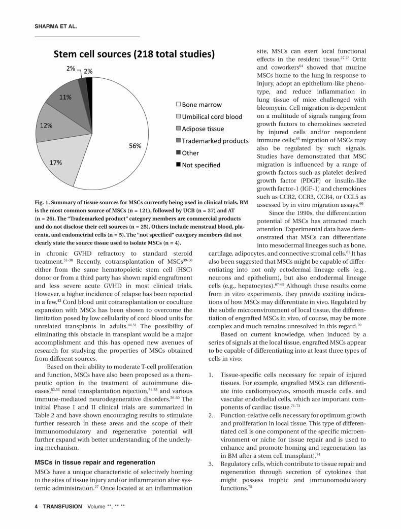
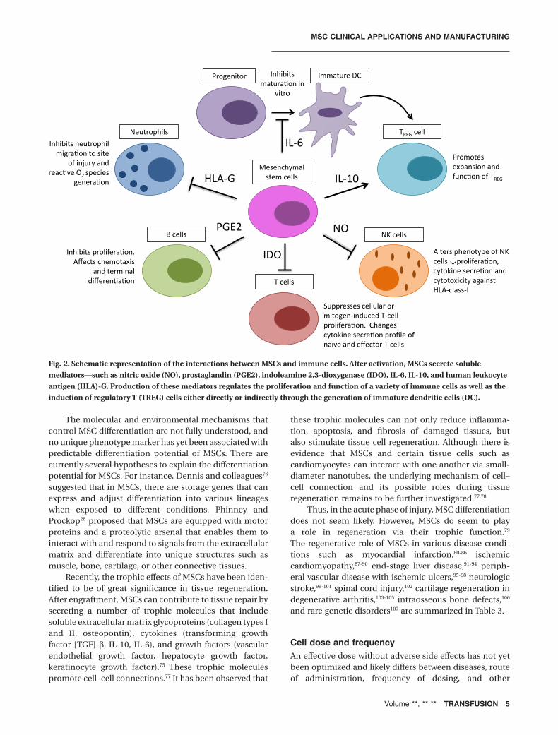
Clinical applications of MSCs are growing rapidlyas research progresses and the wide range of MSC-dependent influences on the immune system are furtherdelineated. Figure 2 summarizes the current cell–cellinteractions of MSCs with the immune system. Clinical-grade ex vivo expanded MSCs have been used to treat BMand organ transplant rejection and inflammatory andauto- and alloimmune diseases (such as systemic collagenabnormalities and GVHD).
The most significant results on the immunosuppres-sive effects of MSCs so far have been observed in thetreatment of acute GVHD after allogeneic stem celltransplantation. The first case of ex vivo expandedhaploidentical MSC infusion in a patient with severeGrade IV GVHD of the gut and liver resulted in a strikingimprovement of the disease.29 A Phase II study reportedthat 30 of 55 patients had a complete response and ninepatients showed improvement indicating that irrespectiveof the donor, MSC infusion might be an effective therapyfor patients with steroid-resistant acute GVHD.30 Sincethese studies were performed, several others have pro-duced encouraging responses, both in acute and
SHARMA ET AL.
2 TRANSFUSION Volume **, ** **
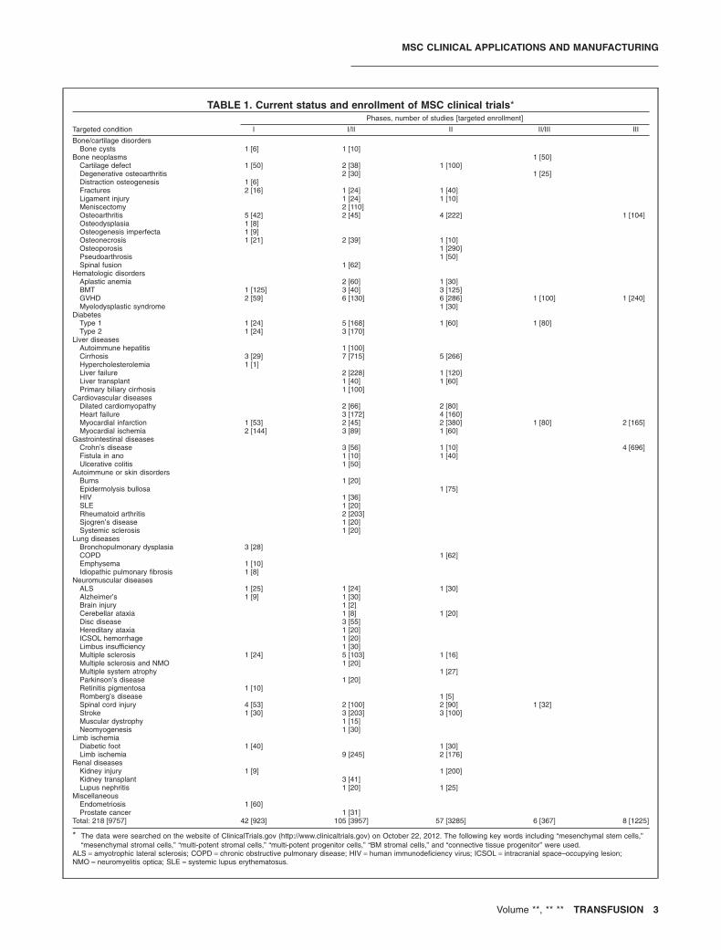
TABLE 1. Current status and enrollment of MSC clinical trials*
Targeted condition
Phases, number of studies [targeted enrollment]
I I/II II II/III III
Bone/cartilage disordersBone cysts 1 [6] 1 [10]
Bone neoplasms 1 [50]Cartilage defect 1 [50] 2 [38] 1 [100]Degenerative osteoarthritis 2 [30] 1 [25]Distraction osteogenesis 1 [6]Fractures 2 [16] 1 [24] 1 [40]Ligament injury 1 [24] 1 [10]Meniscectomy 2 [110]Osteoarthritis 5 [42] 2 [45] 4 [222] 1 [104]Osteodysplasia 1 [8]Osteogenesis imperfecta 1 [9]Osteonecrosis 1 [21] 2 [39] 1 [10]Osteoporosis 1 [290]Pseudoarthrosis 1 [50]Spinal fusion 1 [62]
Hematologic disordersAplastic anemia 2 [60] 1 [30]BMT 1 [125] 3 [40] 3 [125]GVHD 2 [59] 6 [130] 6 [286] 1 [100] 1 [240]Myelodysplastic syndrome 1 [30]
DiabetesType 1 1 [24] 5 [168] 1 [60] 1 [80]Type 2 1 [24] 3 [170]
Liver diseasesAutoimmune hepatitis 1 [100]Cirrhosis 3 [29] 7 [715] 5 [266]Hypercholesterolemia 1 [1]Liver failure 2 [228] 1 [120]Liver transplant 1 [40] 1 [60]Primary biliary cirrhosis 1 [100]
Cardiovascular diseasesDilated cardiomyopathy 2 [66] 2 [80]Heart failure 3 [172] 4 [160]Myocardial infarction 1 [53] 2 [45] 2 [380] 1 [80] 2 [165]Myocardial ischemia 2 [144] 3 [89] 1 [60]
Gastrointestinal diseasesCrohn’s disease 3 [56] 1 [10] 4 [696]Fistula in ano 1 [10] 1 [40]Ulcerative colitis 1 [50]
Autoimmune or skin disordersBurns 1 [20]Epidermolysis bullosa 1 [75]HIV 1 [36]SLE 1 [20]Rheumatoid arthritis 2 [203]Sjogren’s disease 1 [20]Systemic sclerosis 1 [20]
Lung diseasesBronchopulmonary dysplasia 3 [28]COPD 1 [62]Emphysema 1 [10]Idiopathic pulmonary fibrosis 1 [8]
Neuromuscular diseasesALS 1 [25] 1 [24] 1 [30]Alzheimer’s 1 [9] 1 [30]Brain injury 1 [2]Cerebellar ataxia 1 [8] 1 [20]Disc disease 3 [55]Hereditary ataxia 1 [20]ICSOL hemorrhage 1 [20]Limbus insufficiency 1 [30]Multiple sclerosis 1 [24] 5 [103] 1 [16]Multiple sclerosis and NMO 1 [20]Multiple system atrophy 1 [27]Parkinson’s disease 1 [20]Retinitis pigmentosa 1 [10]Romberg’s disease 1 [5]Spinal cord injury 4 [53] 2 [100] 2 [90] 1 [32]Stroke 1 [30] 3 [203] 3 [100]Muscular dystrophy 1 [15]Neomyogenesis 1 [30]
Limb ischemiaDiabetic foot 1 [40] 1 [30]Limb ischemia 9 [245] 2 [176]
Renal diseasesKidney injury 1 [9] 1 [200]Kidney transplant 3 [41]Lupus nephritis 1 [20] 1 [25]
MiscellaneousEndometriosis 1 [60]Prostate cancer 1 [31]
Total: 218 [9757] 42 [923] 105 [3957] 57 [3285] 6 [367] 8 [1225]
* The data were searched on the website of ClinicalTrials.gov (http://www.clinicaltrials.gov) on October 22, 2012. The following key words including “mesenchymal stem cells,”“mesenchymal stromal cells,” “multi-potent stromal cells,” “multi-potent progenitor cells,” “BM stromal cells,” and “connective tissue progenitor” were used.
ALS = amyotrophic lateral sclerosis; COPD = chronic obstructive pulmonary disease; HIV = human immunodeficiency virus; ICSOL = intracranial space–occupying lesion;NMO = neuromyelitis optica; SLE = systemic lupus erythematosus.
MSC CLINICAL APPLICATIONS AND MANUFACTURING
Volume **, ** ** TRANSFUSION 3
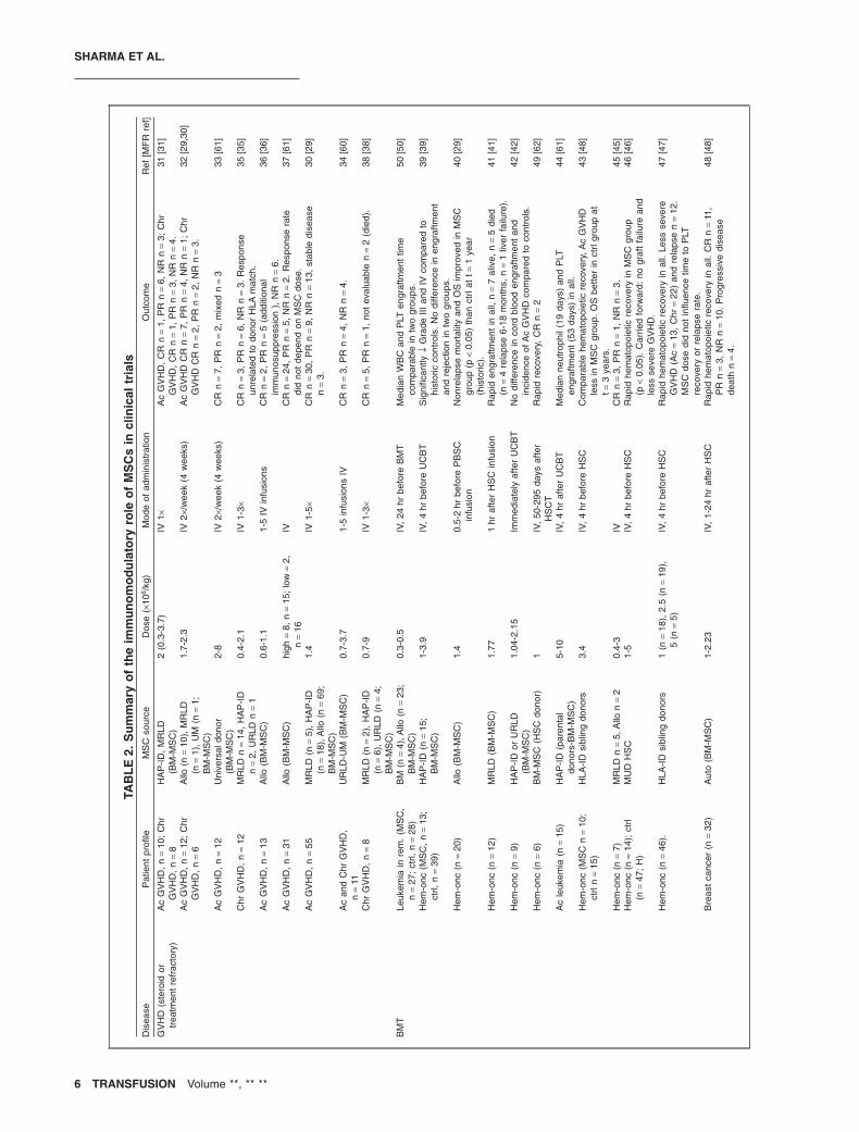
in chronic GVHD refractory to standard steroidtreatment.31-38 Recently, cotransplantation of MSCs39-50
either from the same hematopoietic stem cell (HSC)donor or from a third party has shown rapid engraftmentand less severe acute GVHD in most clinical trials.However, a higher incidence of relapse has been reportedin a few.43 Cord blood unit cotransplantation or cocultureexpansion with MSCs has been shown to overcome thelimitation posed by low cellularity of cord blood units forunrelated transplants in adults.44,51 The possibility ofeliminating this obstacle in transplant would be a majoraccomplishment and this has opened new avenues ofresearch for studying the properties of MSCs obtainedfrom different sources.
Based on their ability to moderate T-cell proliferationand function, MSCs have also been proposed as a thera-peutic option in the treatment of autoimmune dis-eases,52,53 renal transplantation rejection,54,55 and variousimmune-mediated neurodegenerative disorders.56-60 Theinitial Phase I and II clinical trials are summarized inTable 2 and have shown encouraging results to stimulatefurther research in these areas and the scope of theirimmunomodulatory and regenerative potential willfurther expand with better understanding of the underly-ing mechanism.
MSCs in tissue repair and regenerationMSCs have a unique characteristic of selectively homingto the sites of tissue injury and/or inflammation after sys-temic administration.27 Once located at an inflammation
site, MSCs can exert local functionaleffects in the resident tissue.27,28 Ortizand coworkers64 showed that murineMSCs home to the lung in response toinjury, adopt an epithelium-like pheno-type, and reduce inflammation inlung tissue of mice challenged withbleomycin. Cell migration is dependenton a multitude of signals ranging fromgrowth factors to chemokines secretedby injured cells and/or respondentimmune cells;65 migration of MSCs mayalso be regulated by such signals.Studies have demonstrated that MSCmigration is influenced by a range ofgrowth factors such as platelet-derivedgrowth factor (PDGF) or insulin-likegrowth factor-1 (IGF-1) and chemokinessuch as CCR2, CCR3, CCR4, or CCL5 asassessed by in vitro migration assays.66
Since the 1990s, the differentiationpotential of MSCs has attracted muchattention. Experimental data have dem-onstrated that MSCs can differentiateinto mesodermal lineages such as bone,
cartilage, adipocytes, and connective stromal cells.61 It hasalso been suggested that MSCs might be capable of differ-entiating into not only ectodermal lineage cells (e.g.,neurons and epithelium), but also endodermal lineagecells (e.g., hepatocytes).67-69 Although these results comefrom in vitro experiments, they provide exciting indica-tions of how MSCs may differentiate in vivo. Regulated bythe subtle microenvironment of local tissue, the differen-tiation of engrafted MSCs in vivo, of course, may be morecomplex and much remains unresolved in this regard.70
Based on current knowledge, when induced by aseries of signals at the local tissue, engrafted MSCs appearto be capable of differentiating into at least three types ofcells in vivo:
1. Tissue-specific cells necessary for repair of injuredtissues. For example, engrafted MSCs can differenti-ate into cardiomyocytes, smooth muscle cells, andvascular endothelial cells, which are important com-ponents of cardiac tissue.71-73
2. Function-relative cells necessary for optimum growthand proliferation in local tissue. This type of differen-tiated cell is one component of the specific microen-vironment or niche for tissue repair and is used toenhance and promote homing and regeneration (asin BM after a stem cell transplant).74
3. Regulatory cells, which contribute to tissue repair andregeneration through secretion of cytokines thatmight possess trophic and immunomodulatoryfunctions.75
Fig. 1. Summary of tissue sources for MSCs currently being used in clinical trials. BM
is the most common source of MSCs (n = 121), followed by UCB (n = 37) and AT
(n = 26). The “Trademarked product” category members are commercial products
and do not disclose their cell sources (n = 25). Others include menstrual blood, pla-
centa, and endometrial cells (n = 5). The “not specified” category members did not
clearly state the source tissue used to isolate MSCs (n = 4).
SHARMA ET AL.
4 TRANSFUSION Volume **, ** **
The molecular and environmental mechanisms thatcontrol MSC differentiation are not fully understood, andno unique phenotype marker has yet been associated withpredictable differentiation potential of MSCs. There arecurrently several hypotheses to explain the differentiationpotential for MSCs. For instance, Dennis and colleagues76
suggested that in MSCs, there are storage genes that canexpress and adjust differentiation into various lineageswhen exposed to different conditions. Phinney andProckop28 proposed that MSCs are equipped with motorproteins and a proteolytic arsenal that enables them tointeract with and respond to signals from the extracellularmatrix and differentiate into unique structures such asmuscle, bone, cartilage, or other connective tissues.
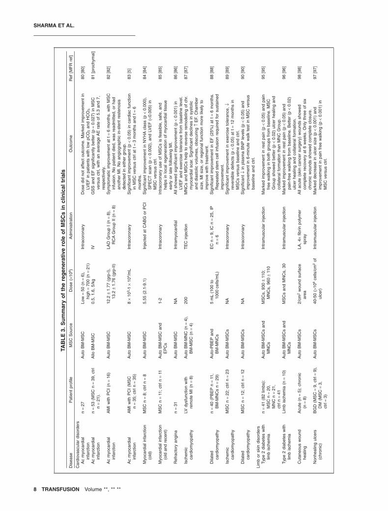
Recently, the trophic effects of MSCs have been iden-tified to be of great significance in tissue regeneration.After engraftment, MSCs can contribute to tissue repair bysecreting a number of trophic molecules that includesoluble extracellular matrix glycoproteins (collagen types Iand II, osteopontin), cytokines (transforming growthfactor [TGF]-β, IL-10, IL-6), and growth factors (vascularendothelial growth factor, hepatocyte growth factor,keratinocyte growth factor).75 These trophic moleculespromote cell–cell connections.77 It has been observed that
these trophic molecules can not only reduce inflamma-tion, apoptosis, and fibrosis of damaged tissues, butalso stimulate tissue cell regeneration. Although there isevidence that MSCs and certain tissue cells such ascardiomyocytes can interact with one another via small-diameter nanotubes, the underlying mechanism of cell–cell connection and its possible roles during tissueregeneration remains to be further investigated.77,78
Thus, in the acute phase of injury, MSC differentiationdoes not seem likely. However, MSCs do seem to playa role in regeneration via their trophic function.79
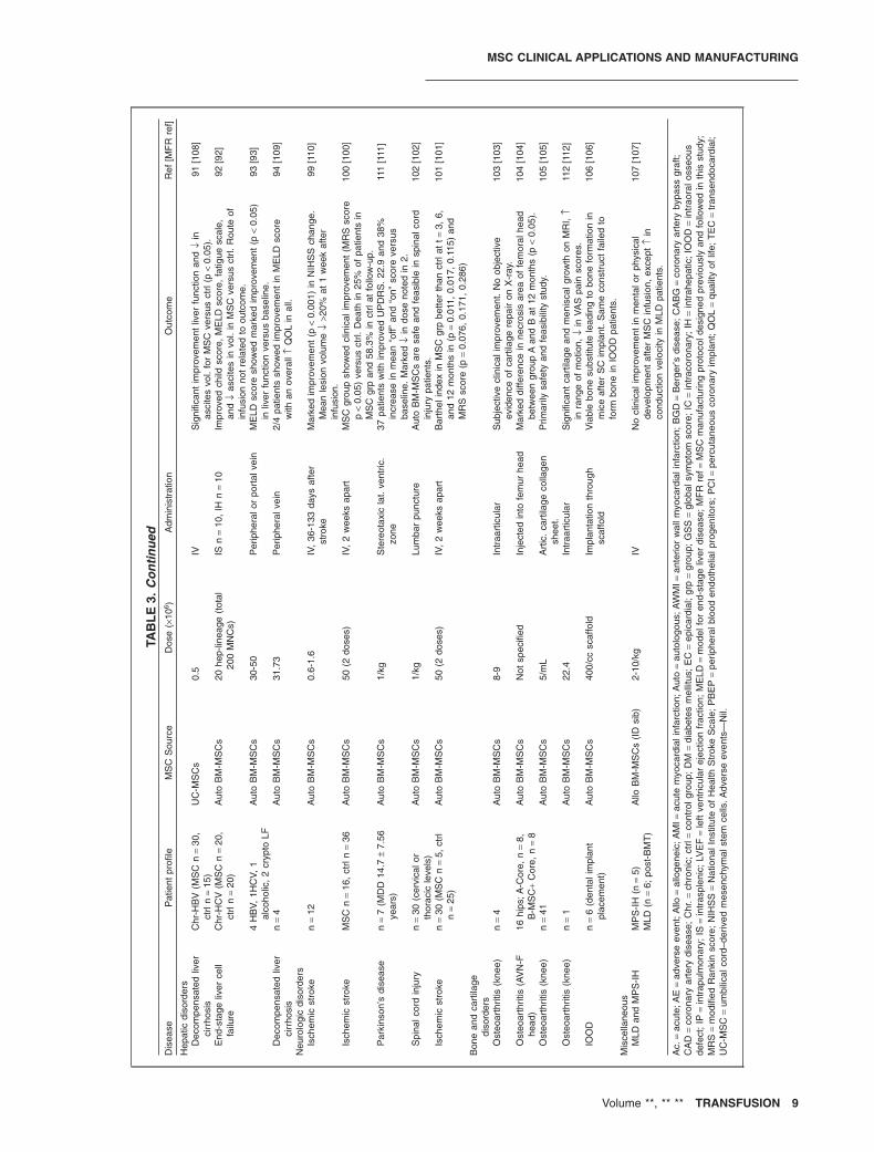
The regenerative role of MSCs in various disease condi-tions such as myocardial infarction,80-86 ischemiccardiomyopathy,87-90 end-stage liver disease,91-94 periph-eral vascular disease with ischemic ulcers,95-98 neurologicstroke,99-101 spinal cord injury,102 cartilage regeneration indegenerative arthritis,103-105 intraosseous bone defects,106
and rare genetic disorders107 are summarized in Table 3.
Cell dose and frequencyAn effective dose without adverse side effects has not yetbeen optimized and likely differs between diseases, routeof administration, frequency of dosing, and other
Progenitor Immature DC
TREG cell
NK cells
T cells
B cells
Neutrophils
Mesenchymalstem cells
Inhibits matura on in
vitro
Promotes expansion and func on of TREG
Alters phenotype of NK cells ↓prolifera on, cytokine secre on and cytotoxicity against HLA-class-I
Suppresses cellular or mitogen-induced T-cell prolifera on. Changes cytokine secre on profile of naïve and effector T cells
Inhibits prolifera on. Affects chemotaxis
and terminal differen a on
Inhibits neutrophil migra on to site
of injury and reac ve O2 species
genera on HLA-G
PGE2
IDO
NO
IL-6
IL-10
Fig. 2. Schematic representation of the interactions between MSCs and immune cells. After activation, MSCs secrete soluble
mediators—such as nitric oxide (NO), prostaglandin (PGE2), indoleamine 2,3-dioxygenase (IDO), IL-6, IL-10, and human leukocyte
antigen (HLA)-G. Production of these mediators regulates the proliferation and function of a variety of immune cells as well as the
induction of regulatory T (TREG) cells either directly or indirectly through the generation of immature dendritic cells (DC).
MSC CLINICAL APPLICATIONS AND MANUFACTURING
Volume **, ** ** TRANSFUSION 5
TAB
LE
2.S
um
mar
yo
fth
eim
mu
no
mo
du
lato
ryro
leo
fM
SC
sin
clin
ical
tria
lsD
isea
seP
atie
ntpr
ofile
MS
Cso
urce
Dos
e(×
106 /
kg)
Mod
eof
adm
inis
trat
ion
Out
com
eR
ef[M
FR
ref]
GV
HD
(ste
roid
ortr
eatm
ent
refr
acto
ry)
Ac
GV
HD
,n
=10
;C
hrG
VH
D,
n=
8H
AP
-ID
,M
RLD
(BM
-MS
C)
2(0
.3-3
.7)
IV1×
Ac
GV
HD
,C
Rn
=1,
PR
n=
6,N
Rn
=3;
Chr
GV
HD
,C
Rn
=1,
PR
n=
3,N
Rn
=4.
31[3
1]
Ac
GV
HD
,n
=12
;C
hrG
VH
D,
n=
6A
llo(n
=10
),M
RLD
(n=
1),
UM
(n=
1;B
M-M
SC
)
1.7-
2.3
IV2×
/wee
k(4
wee
ks)
Ac
GV
HD
CR
n=
7,P
Rn
=4,
NR
n=
1;C
hrG
VH
DC
Rn
=2,
PR
n=
2,N
Rn
=3.
32[2
9,30
]
Ac
GV
HD
,n
=12
Uni
vers
aldo
nor
(BM
-MS
C)
2-8
IV2×
/wee
k(4
wee
ks)
CR
n=
7,P
Rn
=2,
mix
edn
=3
33[6
1]
Chr
GV
HD
,n
=12
MR
LDn
=14
,H
AP
-ID
n=
2,U
RLD
n=
10.
4-2.
1IV
1-3×
CR
n=
3,P
Rn
=6,
NR
n=
3.R
espo
nse
unre
late
dto
dono
rH
LAm
atch
.35
[35]
Ac
GV
HD
,n
=13
Allo
(BM
-MS
C)
0.6-
1.1
1-5
IVin
fusi
ons
CR
n=
2,P
Rn
=5
(add
ition
alim
mun
osup
pres
sion
),N
Rn
=6.
36[3
6]
Ac
GV
HD
,n
=31
Allo
(BM
-MS
C)
high
=8,
n=
15;
low
=2,
n=
16IV
CR
n=
24,
PR
n=
5,N
Rn
=2.
Res
pons
era
tedi
dno
tde
pend
onM
SC
dose
.37
[61]
Ac
GV
HD
,n
=55
MR
LD(n
=5)
,H
AP
-ID
(n=
18),
Allo
(n=
69;
BM
-MS
C)
1.4
IV1-
5×C
Rn
=30
,P
Rn
=9,
NR
n=
13,
stab
ledi
seas
en
=3.
30[2
9]
Ac
and
Chr
GV
HD
,n
=11
UR
LD-U
M(B
M-M
SC
)0.
7-3.
71-
5in
fusi
ons
IVC
Rn
=3,
PR
n=
4,N
Rn
=4.
34[6
0]
Chr
GV
HD
,n
=8
MR
LD(n
=2)
,H
AP
-ID
(n=
6),
UR
LD(n
=4;
BM
-MS
C)
0.7-
9IV
1-3×
CR
n=
5,P
Rn
=1,
not
eval
uabl
en
=2
(die
d).
38[3
8]
BM
TLe
ukem
iain
rem
.(M
SC
,n
=27
;ct
rl,n
=28
)B
M(n
=4)
,Allo
(n=
23;
BM
-MS
C)
0.3-
0.5
IV,
24hr
befo
reB
MT
Med
ian
WB
Can
dP
LTen
graf
tmen
ttim
eco
mpa
rabl
ein
two
grou
ps.
50[5
0]
Hem
-onc
(MS
C,
n=
13;
ctrl,
n=
39)
HA
P-I
D(n
=15
;B
M-M
SC
)1-
3.9
IV,
4hr
befo
reU
CB
TS
igni
fican
tly↓
Gra
deIII
and
IVco
mpa
red
tohi
stor
icco
ntro
ls.
No
diffe
renc
ein
engr
aftm
ent
and
reje
ctio
nin
two
grou
ps.
39[3
9]
Hem
-onc
(n=
20)
Allo
(BM
-MS
C)
1.4
0.5-
2hr
befo
reP
BS
Cin
fusi
onN
onre
laps
em
orta
lity
and
OS
impr
oved
inM
SC
grou
p(p
<0.
05)
than
ctrl
att=
1ye
ar(h
isto
ric).
40[2
9]
Hem
-onc
(n=
12)
MR
LD(B
M-M
SC
)1.
771
hraf
ter
HS
Cin
fusi
onR
apid
engr
aftm
ent
inal
l,n
=7
aliv
e,n
=5
died
(n=
4re
laps
e6-
18m
onth
s,n
=1
liver
failu
re).
41[4
1]
Hem
-onc
(n=
9)H
AP
-ID
orU
RLD
(BM
-MS
C)
1.04
-2.1
5Im
med
iate
lyaf
ter
UC
BT
No
diffe
renc
ein
cord
bloo
den
graf
tmen
tan
din
cide
nce
ofA
cG
VH
Dco
mpa
red
toco
ntro
ls.
42[4
2]
Hem
-onc
(n=
6)B
M-M
SC
(HS
Cdo
nor)
1IV
,50
-295
days
afte
rH
SC
TR
apid
reco
very
,C
Rn
=2
49[6
2]
Ac
leuk
emia
(n=
15)
HA
P-I
D(p
aren
tal
dono
rs-B
M-M
SC
)5-
10IV
,4
hraf
ter
UC
BT
Med
ian
neut
roph
il(1
9da
ys)
and
PLT
engr
aftm
ent
(53
days
)in
all.
44[6
1]
Hem
-onc
(MS
Cn
=10
;ct
rln
=15
)H
LA-I
Dsi
blin
gdo
nors
3.4
IV,
4hr
befo
reH
SC
Com
para
ble
hem
atop
oiet
icre
cove
ry,A
cG
VH
Dle
ssin
MS
Cgr
oup.
OS
bette
rin
ctrl
grou
pat
t=3
year
s.
43[4
8]
Hem
-onc
(n=
7)M
RLD
n=
5,A
llon
=2
0.4-
3IV
CR
n=
3,P
Rn
=1,
NR
n=
3.45
[45]
Hem
-onc
(n=
14);
ctrl
(n=
47;
H)
MU
DH
SC
1-5
IV,
4hr
befo
reH
SC
Rap
idhe
mat
opoi
etic
reco
very
inM
SC
grou
p(p
<0.
05).
Car
ried
forw
ard:
nogr
aft
failu
rean
dle
ssse
vere
GV
HD
.
46[4
6]
Hem
-onc
(n=
46).
HLA
-ID
sibl
ing
dono
rs1
(n=
18),
2.5
(n=
19),
5(n
=5)
IV,
4hr
befo
reH
SC
Rap
idhe
mat
opoi
etic
reco
very
inal
l.Le
ssse
vere
GV
HD
(Ac
=13
,C
hr=
22)
and
rela
pse
n=
12.
MS
Cdo
sedi
dno
tin
fluen
cetim
eto
PLT
reco
very
orre
laps
era
te.
47[4
7]
Bre
ast
canc
er(n
=32
)A
uto
(BM
-MS
C)
1-2.
23IV
,1-
24hr
afte
rH
SC
Rap
idhe
mat
opoi
etic
reco
very
inal
l.C
Rn
=11
,P
Rn
=3,
NR
n=
10.
Pro
gres
sive
dise
ase
deat
hn
=4.
48[4
8]
SHARMA ET AL.
6 TRANSFUSION Volume **, ** **

TAB
LE
2.C
on
tin
ued
Dis
ease
Pat
ient
profi
leM
SC
sour
ceD
ose
(×10
6 /kg
)M
ode
ofad
min
istr
atio
nO
utco
me
Ref
[MF
Rre
f]
MS
and
amyo
trop
hic
late
rals
cler
osis
Sec
.pr
og.
MS
(n=
10)
Aut
o(B
M-M
SC
)1.
6IV
Impr
ovem
ent
invi
sual
acui
ty(p
<0.
003)
,V
ER
(0.0
2),
and
ON
thic
knes
s(0
.06)
.N
oim
prov
emen
tin
CV,
VF,
MV,
orR
NF
thic
knes
s.
60[2
9]
MS
(n=
10)
Aut
o(B
M-M
SC
)30
-50
5m
Lin
trat
heca
l,5
mL
ED
SS
:im
prov
ed(n
=5)
,st
abili
zed
(n=
1),
wor
sene
d(n
=1)
.N
ora
diol
ogic
evid
ence
ofim
prov
emen
tby
Gad
scan
.AE
(n=
1)in
form
ofse
izur
e/m
ilden
ceph
alop
athy
.
56[5
6]
MS
(n=
10)
Aut
o(B
M-M
SC
)8.
76In
trat
heca
lE
DS
S:
impr
oved
(n=
1),
stab
ilize
d(n
=4)
,w
orse
ned
(n=
5).
Fun
c.as
sess
men
t:im
prov
ed(n
=6)
,st
atus
quo
(n=
1),
dete
riora
ted
(n=
3).A
tt=
12m
onth
s,no
sign
sof
impr
ovem
ent
via
MR
I.
57[5
7]
MS
A(M
SC
n=
16;
ctrl
n=
17)
Aut
o(B
M-M
SC
)4
×10
7 /pa
tient
*IA
(1×
107
each
ICA
,2
×10
7do
min
ant
VA
)S
mal
ler
incr
ease
inM
SC
grou
pfo
rto
tal
UM
SA
RS
and
UM
SA
RS
Par
tII
com
pare
dto
ctrl.
58[5
9]
MS
A(M
SC
,n
=11
;ct
rl,n
=18
)A
uto
(BM
-MS
C)
4×
107
/pat
ient
*IA
(1×
107
each
ICA
,2
×10
7do
min
ant
VA
)S
igni
fican
tin
crea
sein
tota
lUM
SA
RS
(p<
0.00
1)fo
rM
SC
grou
pco
mpa
red
toco
ntro
l.59
[59]
Livi
ngre
late
dre
nal
tran
spla
ntfo
ren
d-st
age
rena
ldi
seas
e
(MS
C+
Std
CN
I,n
=53
;M
SC
+lo
wC
NI,
n=
53;
CN
In
=53
),to
tal=
159
Aut
o(B
M-M
SC
)1-
2IV
2×(p
osto
pat
perf
usio
n,2
wee
ksla
ter)
Low
inci
denc
eof
acut
ere
ject
ion
(7.5
%vs
.21
.6%
),le
ssop
port
unis
ticin
fect
ion,
and
bette
rre
nalf
unct
ion
att=
1ye
arin
MS
Cgr
oup
com
pare
dto
ctrl.
Ove
rall
patie
ntan
dgr
aft
surv
ival
sim
ilar
inbo
thgr
oups
.
54[4
8]
(MS
C,
n=
2;ct
rl,n
=3)
Aut
o(B
M-M
SC
)1.
7or
2.0
IV(7
days
post
op)
Ren
albi
opsy
and
func
tions
att=
1ye
arw
ere
norm
alin
MS
Cpa
tient
s.55
[60]
Sys
tem
iclu
pus/
eryt
hem
atos
usA
ctiv
eS
LE(n
=15
)A
llo(B
M-M
SC
)1
IVA
ll(1
3pa
tient
s>
1ye
ar)
impr
oved
with
MS
Ctr
eatm
ent.
Mar
ked
decr
ease
inth
eS
LED
AI
scor
e(1
2.2
±3.
3to
3.2
±2.
8)an
d24
hour
s’pr
otei
nuria
(250
5.0
±13
23.9
to85
8.0
±80
0.7
mg/
24hr
,p
<0.
05.
Dec
reas
edan
ti-ds
DN
Ale
vels
.
52[6
3]
Trea
tmen
tre
frac
tory
(n=
40)
Allo
(BM
-MS
C)
1IV
All
show
edst
able
rem
issi
onat
t=12
-18
mon
ths
with
impr
ovem
ent
inse
rolo
gic
mar
kers
and
rena
lfun
ctio
n.
53[5
3]
*D
ose
isex
pres
sed
asto
tald
ose
rath
erth
anpe
rkg
body
wei
ght.
Ac.
=ac
ute;
AE
=ad
vers
eev
ent;
Allo
=al
loge
neic
;Aut
o=
auto
logo
us;
Chr
.=ch
roni
c;C
NI=
calc
ineu
rinin
hibi
tor;
cont
.=co
ntro
lgro
up;
CR
=co
mpl
ete
resp
onse
;E
DS
S=
Exp
ande
dD
isab
ility
Sta
tus
Sca
le;
HA
P-
ID=
hapl
oide
ntic
al;
hem
-onc
=he
mat
oonc
olog
y;IA
=in
traa
rter
ial;
ICA
=in
tern
alca
rotid
arte
ry;
Leuk
s=
leuk
emia
patie
nts;
MF
Rre
f=M
SC
man
ufac
turin
gpr
otoc
olde
sign
edpr
evio
usly
and
follo
wed
inth
epr
esen
tst
udy,
adve
rse
even
t—1
(one
stud
yR
ef.55
);M
RLD
=m
atch
edre
late
ddo
nor;
MS
=m
ultip
lesc
lero
sis;
MU
D=
mat
ched
unre
late
ddo
nor;
NR
=no
resp
onse
;O
S=
over
alls
urvi
val;
PR
=pa
rtia
lres
pons
e;P
rog.
=P
rogr
essi
ve;
rem
.=re
mis
sion
;S
ec.=
seco
ndar
y;S
LED
AI=
SLE
dise
ase
activ
ityin
dex;
UC
-MS
Cs
=um
bilic
alco
rdde
rived
mes
ench
ymal
stem
cells
;U
MS
AR
=un
ified
mul
tiple
syst
emat
roph
yra
ting
scal
e;U
RLD
-UM
=un
rela
ted
unm
atch
eddo
nor
(uni
vers
aldo
nor)
;V
A=
vert
ebra
lart
ery;
VE
R=
visu
alev
oked
resp
onse
.
MSC CLINICAL APPLICATIONS AND MANUFACTURING
Volume **, ** ** TRANSFUSION 7
TAB
LE
3.S
um
mar
yo
fth
ere
gen
erat
ive
role
of
MS
Cs
incl
inic
altr
ials
Dis
ease
Pat
ient
profi
leM
SC
Sou
rce
Dos
e(×
106 )
Adm
inis
trat
ion
Out
com
eR
ef[M
FR
ref]
Car
diov
ascu
lar
diso
rder
sA
cm
yoca
rdia
lin
farc
tion
n=
27A
uto
BM
-MS
CLo
w=
50(n
=6)
,hi
gh=
700
(n=
21)
Intr
acor
onar
yD
ose
did
not
affe
ctou
tcom
e.M
arke
dim
prov
emen
tin
LVE
Fin
patie
nts
with
low
pCO
2an
dH
CO
3.80
[80]
Ac
myo
card
ial
infa
rctio
nn
=53
(MS
Cn
=39
,ct
rln
=21
).A
lloB
M-M
SC
0.5,
1.6,
5/kg
IVG
SS
and
EF
sign
ifica
ntly
bette
r(p
=0.
027)
inM
SC
vers
usct
rl,w
ithan
aver
age
AE
rate
of5.
3an
d7,
resp
ectiv
ely.
81[p
roch
ymal
]
Ac
myo
card
ial
infa
rctio
nA
MI
with
PC
I(n
=16
)A
uto
BM
-MS
C12
.2±
1.77
(grp
-I),
13.2
±1.
76(g
rp-I
I)LA
DG
roup
I(n
=8)
,R
CA
Gro
upII
(n=
8)S
ympt
omat
icim
prov
emen
tat
t=6
mon
ths.
with
MS
Cin
fusi
on.
No
patie
ntdi
ed,
was
read
mitt
ed,
orha
dan
othe
rM
I.N
oan
giog
raph
icin
-ste
ntre
sten
osis
dete
cted
inei
ther
grou
p.
82[8
2]
Ac
myo
card
ial
infa
rctio
nA
MI
with
PC
I(M
SC
n=
35;
ctrl
n=
35)
Aut
oB
M-M
SC
8×
103 -
1×
104 /
mL
Intr
acor
onar
yS
igni
fican
tim
prov
emen
t(p
<0.
05)
inca
rdia
cfu
nctio
nin
MS
Cve
rsus
ctrl
att=
3m
onth
san
dt=
6m
onth
s.
83[5
]
Myo
card
iali
nfar
ctio
n(o
ld)
MS
Cn
=8;
ctrl
n=
8A
uto
BM
-MS
C5.
55(2
.1-9
.1)
Inje
cted
atC
AB
Gor
PC
IS
igni
fican
tim
prov
emen
tin
NY
HA
clas
s(p
<0.
000)
,S
PE
CT
scan
(p<
0.00
2),
and
LVE
F(<
0.00
5)in
MS
Cve
rsus
ctrl.
84[8
4]
Myo
card
iali
nfar
ctio
n(o
ldan
dre
cent
)M
SC
n=
11;
ctrl
n=
11A
uto
BM
-MS
Can
dE
PC
s1-
2In
trac
oron
ary
Intr
acor
onar
yus
eof
MS
Cs
isfe
asib
le,
safe
,an
dhe
lps
inlo
calr
egen
erat
ion
ofm
yoca
rdia
ltis
sue
early
orla
tefo
llow
ing
MI.
85[8
5]
Ref
ract
ory
angi
nan
=31
Aut
oB
M-M
SC
NA
Intr
amyo
card
ial
All
show
edsi
gnifi
cant
impr
ovem
ent
(p<
0.00
1)in
LVE
Fan
dex
erci
seto
lera
nce
from
base
line
leve
l.86
[86]
Isch
emic
card
iom
yopa
thy
LVdy
sfun
ctio
nw
ithre
mot
eM
I(n
=8)
Aut
oB
M-M
NC
(n=
4),
BM
-MS
C(n
=4)
200
TE
Cin
ject
ion
MN
Cs
and
MS
Cs
help
tore
vers
ere
mod
elin
gof
chr.
myo
card
ials
car.
Sig
nific
ant
decl
ines
insy
stol
ican
ddi
asto
licvo
lum
es,
obsc
urin
g↑
EF.
Cha
mbe
rsi
ze,
MI
size
,or
regi
onal
func
tion
mor
elik
ely
toim
prov
ew
ithtr
eatm
ent.
87[8
7]
Dila
ted
card
iom
yopa
thy
n=
40(P
BE
Pn
=11
,B
M-M
NC
sn
=29
)A
uto-
PB
EP
and
BM
-MN
Cs
5m
L(1
00to
1000
cells
/mL)
EC
n=
9,IC
n=
25,
IPn
=6
Sig
nific
ant
impr
ovem
ent
inE
F(2
5%)
att=
6m
onth
s.R
epea
ted
stem
cell
infu
sion
requ
ired
for
sust
aine
dim
prov
emen
t.
88[8
8]
Isch
emic
card
iom
yopa
thy
MS
Cn
=22
;ct
rln
=23
Aut
oB
M-M
SC
sN
AIn
trac
oron
ary
Sig
nific
ant
impr
ovem
ent
inex
erci
seto
lera
nce,
↓re
vers
ible
defe
cts
(p<
0.05
)at
t=12
mon
ths
inM
SC
sve
rsus
base
line
and
ctrl.
89[8
9]
Dila
ted
card
iom
yopa
thy
MS
Cn
=12
;ct
rln
=12
Aut
oB
M-M
SC
sN
AIn
trac
oron
ary
Sig
nific
ant
↓in
plas
ma
BN
Ple
vels
(p<
0.05
)an
dim
prov
emen
tin
6-m
inut
ew
alk
test
inM
SC
vers
usba
selin
ean
dct
rl.
90[9
0]
Lim
bor
skin
diso
rder
sTy
pe2
diab
etes
with
limb
isch
emia
n=
41(8
2lim
bs):
MS
Cn
=20
,M
NC
n=
21,
ctrl
n=
41
Aut
oB
M-M
SC
san
dM
NC
sM
SC
s,93
0±
110;
MN
Cs,
960
±11
0In
tram
uscu
lar
inje
ctio
nM
arke
dim
prov
emen
tin
rest
pain
(p<
0.05
)an
dpa
infr
eew
alki
ngin
both
grou
psfr
omba
selin
e.M
SC
Gro
upsh
owed
bette
r(p
<0.
02)
ulce
rhe
alin
gan
dco
llate
ralf
orm
atio
nth
anM
NC
Gro
up.
95[9
5]
Type
2di
abet
esw
ithlim
bis
chem
iaLi
mb
isch
emia
(n=
10)
Aut
oB
M-M
SC
san
dM
NC
sM
SC
san
dM
NC
s,30
Intr
amus
cula
rin
ject
ion
Mar
ked
impr
ovem
ent
inre
stpa
in(p
<0.
05)
and
pain
-fre
ew
alki
ngfr
omba
selin
e.B
ette
r(p
<0.
02)
ulce
rhe
alin
gan
dco
llate
ralf
orm
atio
n.
96[9
6]
Cut
aneo
usw
ound
heal
ing
Acu
te(n
=5)
;ch
roni
c(n
=8)
Aut
oB
M-M
SC
s2/
cm2
wou
ndsu
rfac
ear
eaLA
,4×
fibrin
poly
mer
spra
yA
llac
ute
(ski
nca
ncer
surg
ery)
wou
nds
show
edco
mpl
ete
reco
very
at6
wee
ks.
Onl
yth
ree
ofsi
xch
roni
cw
ound
ssh
owed
com
plet
ecl
osur
e.
98[9
8]
Non
heal
ing
ulce
rs(c
hron
ic)
BG
D(M
SC
=9,
ctrl
=9)
,D
M(M
SC
=3,
ctrl
=3)
Aut
oB
M-M
SC
s40
-50
(>10
6ce
lls/c
m2
oful
cer)
Intr
amus
cula
rin
ject
ion
Mar
ked
decr
ease
inul
cer
size
(p<
0.00
1)an
dim
prov
emen
tin
pain
free
wal
king
(p<
0.00
1)in
MS
Cve
rsus
ctrl.
97[9
7]
SHARMA ET AL.
8 TRANSFUSION Volume **, ** **
TAB
LE
3.C
on
tin
ued
Dis
ease
Pat
ient
profi
leM
SC
Sou
rce
Dos
e(×
106 )
Adm
inis
trat
ion
Out
com
eR
ef[M
FR
ref]
Hep
atic
diso
rder
sD
ecom
pens
ated
liver
cirr
hosi
sC
hr-H
BV
(MS
Cn
=30
,ct
rln
=15
)U
C-M
SC
s0.
5IV
Sig
nific
ant
impr
ovem
ent
liver
func
tion
and
↓in
asci
tes
vol.
for
MS
Cve
rsus
ctrl
(p<
0.05
).91
[108
]
End
-sta
geliv
erce
llfa
ilure
Chr
-HC
V(M
SC
n=
20,
ctrl
n=
20)
Aut
oB
M-M
SC
s20
hep-
linea
ge(t
otal
200
MN
Cs)
ISn
=10
,IH
n=
10Im
prov
edch
ildsc
ore,
ME
LDsc
ore,
fatig
uesc
ale,
and
↓as
cite
sin
vol.
inM
SC
vers
usct
rl.R
oute
ofin
fusi
onno
tre
late
dto
outc
ome.
92[9
2]
4H
BV,
1HC
V,1
alco
holic
,2
cryp
toLF
Aut
oB
M-M
SC
s30
-50
Per
iphe
ralo
rpo
rtal
vein
ME
LDsc
ore
show
edm
arke
dim
prov
emen
t(p
<0.
05)
inliv
erfu
nctio
nve
rsus
base
line.
93[9
3]
Dec
ompe
nsat
edliv
erci
rrho
sis
n=
4A
uto
BM
-MS
Cs
31.7
3P
erip
hera
lvei
n2/
4pa
tient
ssh
owed
impr
ovem
ent
inM
ELD
scor
ew
ithan
over
all↑
QO
Lin
all.
94[1
09]
Neu
rolo
gic
diso
rder
sIs
chem
icst
roke
n=
12A
uto
BM
-MS
Cs
0.6-
1.6
IV,
36-1
33da
ysaf
ter
stro
keM
arke
dim
prov
emen
t(p
<0.
001)
inN
IHS
Sch
ange
.M
ean
lesi
onvo
lum
e↓
>20%
at1
wee
kaf
ter
infu
sion
.
99[1
10]
Isch
emic
stro
keM
SC
n=
16,
ctrl
n=
36A
uto
BM
-MS
Cs
50(2
dose
s)IV
,2
wee
ksap
art
MS
Cgr
oup
show
edcl
inic
alim
prov
emen
t(M
RS
scor
ep
<0.
05)
vers
usct
rl.D
eath
in25
%of
patie
nts
inM
SC
grp
and
58.3
%in
ctrl
atfo
llow
-up.
100
[100
]
Par
kins
on’s
dise
ase
n=
7(M
DD
14.7
±7.
56ye
ars)
Aut
oB
M-M
SC
s1/
kgS
tere
otax
icla
t.ve
ntric
.zo
ne37
patie
nts
with
impr
oved
UP
DR
S.
22.9
and
38%
incr
ease
inm
ean
“off”
and
“on”
scor
eve
rsus
base
line.
Mar
ked
↓in
dose
note
din
2.
111
[111
]
Spi
nalc
ord
inju
ryn
=30
(cer
vica
lor
thor
acic
leve
ls)
Aut
oB
M-M
SC
s1/
kgLu
mba
rpu
nctu
reA
uto
BM
-MS
Cs
are
safe
and
feas
ible
insp
inal
cord
inju
rypa
tient
s.10
2[1
02]
Isch
emic
stro
ken
=30
(MS
Cn
=5,
ctrl
n=
25)
Aut
oB
M-M
SC
s50
(2do
ses)
IV,
2w
eeks
apar
tB
arth
elin
dex
inM
SC
grp
bette
rth
anct
rlat
t=3,
6,an
d12
mon
ths
in(p
=0.
011,
0.01
7,0.
115)
and
MR
Ssc
ore
(p=
0.07
6,0.
171,
0.28
6)
101
[101
]
Bon
ean
dca
rtila
gedi
sord
ers
Ost
eoar
thrit
is(k
nee)
n=
4A
uto
BM
-MS
Cs
8-9
Intr
aart
icul
arS
ubje
ctiv
ecl
inic
alim
prov
emen
t.N
oob
ject
ive
evid
ence
ofca
rtila
gere
pair
onX
-ray
.10
3[1
03]
Ost
eoar
thrit
is(A
VN
-Fhe
ad)
16hi
ps;A
-Cor
e,n
=8,
B-M
SC
+C
ore,
n=
8A
uto
BM
-MS
Cs
Not
spec
ified
Inje
cted
into
fem
urhe
adM
arke
ddi
ffere
nce
inne
cros
isar
eaof
fem
oral
head
betw
een
grou
pA
and
Bat
12m
onth
s(p
<0.
05).
104
[104
]
Ost
eoar
thrit
is(k
nee)
n=
41A
uto
BM
-MS
Cs
5/m
LA
rtic
.ca
rtila
geco
llage
nsh
eet.
Prim
arily
safe
tyan
dfe
asib
ility
stud
y.10
5[1
05]
Ost
eoar
thrit
is(k
nee)
n=
1A
uto
BM
-MS
Cs
22.4
Intr
aart
icul
arS
igni
fican
tca
rtila
gean
dm
enis
calg
row
thon
MR
I,↑
inra
nge
ofm
otio
n,↓
inV
AS
pain
scor
es.
112
[112
]
IOO
Dn
=6
(den
tali
mpl
ant
plac
emen
t)A
uto
BM
-MS
Cs
400/
ccsc
affo
ldIm
plan
tatio
nth
roug
hsc
affo
ldV
iabl
ebo
nesu
bstit
ute
lead
ing
tobo
nefo
rmat
ion
inm
ice
afte
rS
Cim
plan
t.S
ame
cons
truc
tfa
iled
tofo
rmbo
nein
IOO
Dpa
tient
s.
106
[106
]
Mis
cella
neou
sM
LDan
dM
PS
-IH
MP
S-I
H(n
=5)
MLD
(n=
6;po
st-B
MT
)A
lloB
M-M
SC
s(I
Dsi
b)2-
10/k
gIV
No
clin
ical
impr
ovem
ent
inm
enta
lor
phys
ical
deve
lopm
ent
afte
rM
SC
infu
sion
,ex
cept
↑in
cond
uctio
nve
loci
tyin
MLD
patie
nts.
107
[107
]
Ac.
=ac
ute;
AE
=ad
vers
eev
ent;
Allo
=al
loge
neic
;AM
I=ac
ute
myo
card
iali
nfar
ctio
n;A
uto
=au
tolo
gous
;AW
MI=
ante
rior
wal
lmyo
card
iali
nfar
ctio
n;B
GD
=B
erge
r’sdi
seas
e;C
AB
G=
coro
nary
arte
ryby
pass
graf
t;C
AD
=co
rona
ryar
tery
dise
ase;
Chr
.=ch
roni
c;ct
rl=
cont
rolg
roup
;D
M=
diab
etes
mel
litus
;E
C=
epic
ardi
al;
grp
=gr
oup;
GS
S=
glob
alsy
mpt
omsc
ore;
IC=
intr
acor
onar
y;IH
=in
trah
epat
ic;
IOO
D=
intr
aora
loss
eous
defe
ct;
IP=
intr
apul
mon
ary;
IS=
intr
aspl
enic
;LV
EF
=le
ftve
ntric
ular
ejec
tion
frac
tion;
ME
LD=
mod
elfo
ren
d-st
age
liver
dise
ase;
MF
Rre
f=M
SC
man
ufac
turin
gpr
otoc
olde
sign
edpr
evio
usly
and
follo
wed
inth
isst
udy;
MR
S=
mod
ified
Ran
kin
scor
e;N
IHS
S=
Nat
iona
lIns
titut
eof
Hea
lthS
trok
eS
cale
;P
BE
P=
perip
hera
lblo
oden
doth
elia
lpro
geni
tors
;P
CI=
perc
utan
eous
coro
nary
impl
ant;
QO
L=
qual
ityof
life;
TE
C=
tran
send
ocar
dial
;U
C-M
SC
=um
bilic
alco
rd–d
eriv
edm
esen
chym
alst
emce
lls.A
dver
seev
ents
—N
il.
MSC CLINICAL APPLICATIONS AND MANUFACTURING
Volume **, ** ** TRANSFUSION 9

variables. Based on the review of currently applied dosesin various clinical trials,29-61,70,80-107,111-113 the clinical dosetypically ranges from 0.5 × 10⁶ to 5 × 10⁶ MSCs/kg bodyweight of the recipient. Testing of high (8 × 10⁶ MSCs/kg)as well as low doses (2 × 10⁶ MSCs/kg) in patients withsteroid-refractory acute GVHD37 did not reveal significantdifferences in response rate or relapse of the primarydisease. Similarly, the MSC dose did not affect platelet(PLT) and neutrophil engraftment in post-BMT hemato-oncology patients.47 However, repeated infusion of MSCsat certain intervals seems to influence the outcome insome studies.30,32,33,36,38,53 Larger randomized trials areneeded to determine therapeutic doses and dosing regi-mens for MSCs in various clinical settings.
MSC manufacturingWith MSCs entering into the clinical arena, the develop-ment of production methods in accordance with currentGood Manufacturing Practices (GMP) and current GoodTissue Practices is required in the United States. Similarregulations are in place in other countries around theworld. Pamphilon and Szczepiorkowski114 and others115
have provided a thorough summary of these regulatoryrequirements.
Donor, cell sources, and culture processesMSCs have been derived from several tissue sources (BM,AT, and UCB) listed in Fig. 1 and applied in both autolo-gous and allogeneic settings. With evidence suggestingimmune-privileged status, a single allogeneic MSC donormay serve for multiple recipients, raising the demand forwell-characterized and even “qualified” donors.116 Thescreening and testing of donors for MSCs (e.g., healthquestionnaire, viral testing) is similar to that for other cell-or tissue-based products. The age of the donor seems to beimportant, with BM from children containing a higherconcentration of colony-forming unit fibroblast precur-sors (CFU-Fs) than that from adults.117 Moreover,increased donor age seems to be directly correlated todetrimental effects in terms of proliferation andmultipotency of MSCs.118 The donor should have noabnormalities or risk of abnormalities possibly involvingMSCs, which may currently be difficult to assess. No spe-cific regulatory requirement exists for this matter, but theissue should be considered carefully particularly when asingle or few universal donors are used for many patients.
Isolation of BM-MSCsThe majority of MSC clinical trials published to date(n = 121) have used BM as the source for the MSCs. BM isremoved from the donor’s posterior superior iliac spine orcrest using an Illinois needle, or equivalent aspiration
needle, in a heparin-containing syringe.119 The sample issubsequently processed by density gradient centrifuga-tion, direct plating, or different enrichment strategies.118
Numerous attempts to enrich MSCs from BM by othermethods such as immunomagnetic-based depletion orenrichment strategies have been performed. Selectionmarkers include STRO-1, CD49a, CD105, CD133, CD146,CD271, SSEA-4, antifebrin microbeads, aptamers, andaldehyde dehydrogenase activity.120-123 However, nomarker has proven capable of discriminating multipotent,highly proliferating MSCs from other less potent lineage-committed cells. Thus, the most common procedures forobtaining MSCs in clinical-scale numbers utilize densitygradient centrifugation for isolation or direct plating toseparate mesenchymal and hematopoietic cells by theiradhesion to plastic cell culture surfaces.
Donor age, as mentioned earlier, and aspirate qualityhave been shown to influence MSC numbers.117,118 Thefrequency of MSCs is approximately 1 per 1 × 10⁶ nucle-ated cells in adult BM and 1 per 1 × 10⁴ nucleated cells inUCB.120-124 The number of MSCs has been noted todecrease with age, with a 10-fold decrease from birthto teenage and another 10-fold decrease from teenage toelderly.121-125
Isolation of AT-derived MSCsThe discovery of multipotent MSCs within AT has estab-lished a second major source of MSCs (n = 26).126 Besides acomparable degree of mesodermal differentiation poten-tial, AT-derived MSCs also appear to have higher frequen-cies (100-1000× BM) and a high potential for angiogenesisor vasculogenesis compared to that of BM.127 In mostcases, lipoaspirates have been used as starting material.Liposuction procedures may yield volumes ranging frommilliliters to liters of tissue.128 The most commonlyemployed procedure, tumescent liposuction, involves thepreprocedure infusion of saline solutions containinganesthetics and adrenaline as vasoconstrictors. Thisapproach gives better cell yields than ultrasound-assistedliposuction, which has been shown to compromise recov-ery as well as expansion capacity of MSCs.129 For obtainingsmaller volumes of tissue, machine and syringe aspirationas well as excision can be used instead.130 Further process-ing steps include removal of cellular debris, oil, excessiveblood cells, proteins, and components of the extracellularmatrix followed by extensive washing to obtain higherpurity of the desired fraction.131 To isolate MSCs from theother tissues, enzymatic treatment is used. Subsequently,centrifugation is performed to remove the adipocyte frac-tion and pellet the preadipocyte stromal vascular fraction.This fraction is a heterogeneous mixture of cells, includingMSCs as well as endothelial, muscle, fibroblastic and mastcells, pericytes, and preadipocytes. After the initial adher-ence step, all nonadherent cells are discarded by extensive
SHARMA ET AL.
10 TRANSFUSION Volume **, ** **
washing, and the remaining adherent cells appear asfibroblastoid cells. These are cultured for approximately10 days until a 60% to 70% confluent monolayer has devel-oped. Cells can then be split to initiate subsequent culturepassage.
To standardize the process, automated devices havebeen developed to assist in separation and culture. A “bagwithin a bag” device, composed of an inner mesh and anouter sealed bag, assists to separate the tissue fractionfrom the contaminating fluid fraction.132 A completelyclosed system (Celuton system, Cytori Therapeutics, SanDiego, CA), which can be used at the patient’s bedside,performs the aspiration, washing, and concentration ofthe stromal vascular fraction.133 Cells resulting from thisprocess, however, can only be regarded as enriched withMSCs. Only a proportion of approximately 1:1000 cellswithin the stromal vascular fraction will give rise to CFUs,equivalent to MSCs.5 Admittedly, most studies have usedspecimens obtained from young and healthy subjectsundergoing aesthetic liposuction. To address the effects ofage and comorbidity on stem cell frequencies, DiMuzioand Tulenko134 correlated factors such as advanced age(>70 years), obesity, renal failure, and vascular disease andfound no significant differences.
Isolation of UCB-derived MSCsFresh (i.e., not frozen and thawed) UCB is the thirdcommon source for isolating MSCs for clinical use(n = 37). The standard process employed for obtainingUCB is gravity-assisted collection after cannulation of oneof the umbilical veins (after delivery of the placenta)under aseptic conditions. This product is then typicallyprocessed within 24 hours of collection in a similarmanner to BM. Various collection methods result in vari-able cell yield and viability of MSCs obtained; the successrate in isolating and further expanding MSCs depends onthe volume of blood collected, the cell content, and thetime between collection and processing,135 which high-lights the need for minimal delay between delivery andharvesting. Related cell sources envisioned for clinicalapplications include neonatal tissues such as the amnioticmembrane, the placenta, and Wharton’s jelly of theumbilical cord.136 These sources, like UCB, are of interestdue to their relatively unlimited supply of more primitiveMSCs with minimal ethical or legal concerns related totissue sourcing.
MSC expansion
Culture mediumThe optimal basal medium for culturing MSCs has not yetbeen determined. Whereas some investigators favorusing α-minimum essential medium,136-139 others favorDulbecco’s modified Eagle’s medium.61,135,140 The critical
ingredient in MSC expansion medium seems to be serumas a source of nutrients, hormones, and growth factors.
Fetal bovine serumFetal bovine serum (FBS) has historically been consideredessential for obtaining high-quantity and quality MSCswith ex vivo expansion.141 However, concerns for the use ofFBS do exist and include risk of transfer of immunogenicxenoproteins as well as transmission of infectious agents,especially transmissible spongiform encephalopathy.142
Accordingly, the European Medicines Agency (EMEA) rec-ommends that “when manufacturers have a choice theuse of materials from ‘non transmissible spongiformencephalopathy relevant animal species’ or non-animalorigin is preferred.”143,144 If FBS is deemed necessary forculture, extensively tested FBS can be sourced from quali-fied herds (i.e., animals from countries considered free ofrisk of variant Creutzfeldt-Jakob disease). Interestingly,FBS-derived proteins have been shown to be internalizedby MSCs145 and to be immunogenic, possibly compromis-ing the clinical effectiveness of MSCs.17,146 Accordingly,many in the cell therapy community have already begunimplementing non-FBS supplements for large-scale pro-duction of MSCs. However, the role of culture ingredients(including FBS) in maintaining MSC immunomodulatoryand regenerative properties is still poorly understood, andthus it may be too premature to exclude FBS from MSCculture.
Human supplementsAlthough acceptable FBS batches are available and arebeing used for clinical-grade manufacturing of MSCs, theconcern outlined above has paved the way for alternativesupplements, including human-derived supplements. Ofcourse, a completely chemically defined medium wouldbe optimal for clinical-scale expansion,147 but this has yetto be achieved or implemented. Several working groupshave tried to optimize culture media by adding humanserum, plasma, or PLT-derived factors. Pooled human PLTlysate (obtained from buffy coat–derived PLT-rich plasma)has growth factors and mitogens released from alphagranules of PLTs during PLT activation either by thrombinor by cell fragmentation during repeated freeze-thawcycles. Among these potent mediators released from PLTsare epidermal growth factors, basic fibroblast growthfactor, PDGFs, TGF-β1, and IGF. These factors enhanceproliferation of bone cells and chondrocytes, as well asMSCs, highlighting the role of PLTs in processes such aswound healing and tissue repair.148-151 However, Marx andcolleagues152 have observed that regenerative effects ofPLT derivatives show extensive variation due to thedependence of growth factor concentration on PLTcontent, preparation method, white blood cell (WBC)contamination, and mechanisms of PLT growth factorrelease.153 Recent literature shows a definite advantage
MSC CLINICAL APPLICATIONS AND MANUFACTURING
Volume **, ** ** TRANSFUSION 11

of PLT lysate over FBS with regard to MSC prolifera-tion and cloning efficiency and a similar MSCimmunophenotype.154 Thus, human PLT lysate mayreplace FBS in many cell culture systems previouslythought to strictly depend on the presence of FBS due tobetter reproducibility of the lysate preparation protocolwithout considerable lot-to-lot variation.
Other alternatives to FBS include pooled humanserum, blood group AB human serum, and PLT-derivedfactors, which have been developed by a variety of proto-cols.155 If an allogeneic source were to be used, large-scaleclinical production involving pooled human blood deriva-tives may require several donors (i.e., to neutralize donor-specific variations and to mimic an off-the-shelf batch).Both blood group AB human serum and thrombin-activated PLT releasate in plasma compared to FBS havebeen found to be superior in expanding AT-MSCs. Somestudies using allogeneic human serum have reportedsuccess in isolating and expanding MSCs from BMwith preserved differentiation and immune-suppressiveproperties;156-158 others have observed reduced growthassociated with advanced senescence, concluding thatautologous serum would be favorable.159,160
Other additivesThe growth factor requirements of MSCs have not beendefined. However, some growth factors, such as PDGF, epi-dermal growth factor, TGF-β, and IGF have been tested inculture.161,162 A variety of protocols describe adding fibro-blast growth factors to FBS-supplemented medium forexpanding MSCs to increase their proliferation rate andmaintain multilineage differentiation potential.163 Othersindicate that factors like dexamethasone164 or lithium,which both stimulate Wnt signaling, can enhance prolif-eration of MSCs.165
Oxidative stress can impair MSC qualities. Enhancingthe concentration of selenium or selenite has been shownto reduce cell damage induced by reactive oxygenspecies.166 Likewise, caloric restriction mimicked in vitroby lowering the glucose content has been shown to accel-erate MSC proliferation while preventing senescence.167
Contradicting these results, telomerase-immortalizedMSCs respond to higher glucose concentrations withenhanced proliferation and osteogenic differentiation.168
Finally, Sotiropoulou and colleagues139 indicate that usingastabilized dipeptide form of L-glutamine (GlutaMAX, LifeTechnologies, Carlsbad, CA) supports better cell growthcompared to using L-glutamine.
Cell seeding densityPlating density has emerged as a critical issue for MSCexpansion. Due to the adherent nature of MSCs, platingdensity is an important variable to ensure a good expan-sion rate and to maintain necessary cellular functions.The initial mononuclear cell (MNC) plating density is
extremely variable; published clinical trials30-61,70,80-107,111-113
have involved both high densities (e.g., 1.70 × 10⁵ MNCs/cm2) and lower densities (e.g., 5.0 × 10⁴ MNCs/cm2). Onsubsequent passages, the plating density should bedecreased.48 The choice of cell density remains critical atthis stage, and use of a low (e.g., 1.0 × 10³-5.0 × 10³ MSCs/cm2) or very low (e.g., 1.0 × 101-5.0 × 101 MSCs/cm2)plating density may better maintain a high proliferationrate and multipotentiality of MSCs.169
A consequence of seeding density and length ofculture is the proliferative age of MSCs. MSCs have arestricted lifespan and reach a senescent state in whichcellular functions become diminished and the risk foracquiring mutations116,170,171 and inflammatory phenotypeincreases, making them unfit for therapeutic use. Passagenumbers are most commonly used to represent prolifera-tive age; however, passage numbers (in contrast to popu-lation doublings) do not describe the critical de factoproliferation history when optimal or maximum lengthof culture is not yet well defined.171 Closed-systembioreactors may have limitations to scale-up due to size orculture capacity.
Devices for expansionMSCs grow as adherent cells until reaching confluencyand are then further expanded by serial passaging. There-fore, the number of cells that can be harvested in an exvivo expansion culture is determined by the surface areaof the culture platform. Typically MSCs are cultivatedin conventional monolayer cultures. To achieve a largesurface area, multilayered cell factories are used.172,173
This approach is labor-intensive and cost-consuming.Alternatively, it is possible to expand MSCs by usingbioreactors.174,175 As closed systems should be preferredin a GMP setting, Rojewski and colleagues176 report afully automated bioreactor allowing GMP-compliantmanufacturing.
Oxygen tensionIt has been shown that the most primitive stem cells pro-liferate and maintain “stemness” under low O2 concentra-tion (e.g., 5%), which is closer to physiologic values.177 LowO2 conditions limit oxidative damage and, thus, mayreduce cytogenetic abnormalities.178 Most MSC trials havenot involved cells expanded under low O2 culture condi-tions. However, a trial involving ischemia-tolerant MSCsfor treatment of lung injury is in the planning stage.179
Storage and cryopreservationMSCs for clinical use are most commonly frozen in 10%dimethyl sulfoxide within an electrolyte solution (e.g.,PlasmaLyteA) and a protein source (e.g., human serumalbumin). The freezing rate is typically 1°C/min throughphase change, followed by 2 to 3°C/min until roughly
SHARMA ET AL.
12 TRANSFUSION Volume **, ** **

−100°C, at which point the cells are placed in liquid nitro-gen or vapor-phase liquid nitrogen. This procedure isbased on cryopreservation of HSCs and lymphoid cellsand is not optimized for MSCs. MSCs in the frozen statecan be transported in liquid nitrogen dry shippers (orequivalent).180
Quality control testingQuality control testing usually includes viability,immunophenotyping, sterility and mycoplasma testing,and endotoxin level. Viability can be assessed by a varietyof assays, including trypan blue, acridine orange-propidium iodide, and 7-aminoactinomycin-D with anaccepted specification of at least 70%. Immunopheno-typing typically follows the International Society for Cel-lular Therapy criteria,7 which includes CD73, CD90, andCD105 as positive markers. Samples for sterility andmycoplasma testing are drawn at various time points inmanufacturing, such as before culture (i.e., from startingmaterial), during culture, and after culture and beforefreeze. Automated methods are often used for sterility(bacterial and fungal culture), and several approaches tomycoplasma testing exist (e.g., culture, polymerase chainreaction). Endotoxin content can be evaluated severalways but most often involves limulus amebocyte lysate–based method (chromogenic, turbidometric, etc.). Theupper limit for endotoxin is 5 EU/kg/dose for most modesof administration.181
Assays of function and potencyThe determination of assays of function or potency maybe guided by the presumed mechanism of action. Severalgeneral and mechanism-specific examples are high-lighted below.
Trilineage differentiationThe standard pathways of MSC differentiation followosteogenic, chondrogenic, and adipogenic lineages andhave been elaborately reported in a large number of pub-lications.61,131,163 This potency assay should be performed ifmesenchymal (connective) tissue repair is intended. Thelack of evidence of their true biologic role in vivo becomesthe limitation of this assay as the hallmark for stem cellcharacteristics of self-renewal and differentiation hasbeen not accomplished so far.
ImmunomodulationHuman MSC surface molecules such as HLA Class I, Thy-1(CD90), vascular cell adhesion molecule (CD106), inter-cellular adhesion molecule-1 and -2, activated WBC adhe-sion molecule (ALCAM, CD166), lymphocyte functionalantigen-3, and various integrins indicate interaction withcognate ligands on T cells.182 In contrast to an expected
induction of T-cell response against allogeneic MSCs,T-cell alloreactivity is inhibited by MSCs in mixed lympho-cyte cultures or lymphocyte proliferation induced bymitogens, such as phytohemagglutinin or concanavalin A,and are currently accepted in vitro assays to assess inhibi-tory effect of MSCs on T-cell proliferation.108,182 Somestudies have shown that although MSCs in high concen-trations (10-40 MSCs per 100 responder lymphocytes)inhibit, low MSC concentrations (≤1 MSC per responderlymphocyte) may stimulate lymphocyte proliferation inmixed lymphocyte cultures.113 These findings stress theimportance of determining an optimal MSC dose toachieve intended outcomes.
Regulation of hematopoiesisThe potential benefit of cotransplantation of MSCs withHSCs has been demonstrated.39 This effect can be assayedin vitro in coculture experiments using HSCs and MSCsand thus may be a relevant functional or potency assay forthis therapeutic indication.124
Senescence and genomic stabilityMSCs have limited lifespan in vitro and enter senescenceafter multiple passages (25-30 population doublings) inculture. Therefore, for clinical use of MSCs, genomic sta-bility is a major concern during long-term cultures asthere is always a risk of cell transformation due to replica-tive senescence. Cells begin to show telomere shortening,lose a part of their differentiation potential, and exhibit analtered cytokine secretion profile.183,184
All these changes appear to be a continuous processand are hypothesized to start as early as the first passage.However, so far the evidence favoring transformation islow.171 Karyotyping and comparative genomic hybridiza-tion have low sensitivity to detect these abnormalities anddo not appear to be relevant controls, neither indicatingthe real risk of MSC transformation or their senescentstatus. Thus, real relevant controls for transformation andsenescence could refer to genes or molecules involved inthe senescence and transformation pathways, such asp53, p21, and p16 and may ensure the safety of theproduct.185
ClonogenicityThe CFU assay is a suitable tool for evaluating the self-renewal capacity of cells. Friedenstein and colleagues186
were the first to describe an assay system to study CFU-Fin various hematopoietic cell populations. Analysis ofCFU-Fs frequency in BM aspirates requires adequate dilu-tion, minimal manipulation, and low seeding density toget true colony counts in the sample as has been observedpreviously.123,187
SUMMARY
MSC-based therapies are quickly evolving with more than200 clinical trials for a variety of therapeutic indications
MSC CLINICAL APPLICATIONS AND MANUFACTURING
Volume **, ** ** TRANSFUSION 13
ranging from immunomodulation to tissue repair andregeneration. The field is still in its infancy, however, withlack of consensus on several fronts including: proposedmechanisms of action, optimal dosing strategy, and routeof administration. Perhaps most concerning is the broadrange of approaches to culture and the poorly understoodimpact of variables such as source material, medium,supplements, culture technique, and so forth. It may betoo premature to suggest standardization of manufactur-ing, as this may limit the field and overall optimization ofmanufacturing. However, to ensure the greatest likelihoodof success of MSCs in the clinical arena, attention (includ-ing from that of granting agencies) should be placed onoptimization of culture.
ACKNOWLEDGMENTS
The authors acknowledge the support of Department of Health
Research, Indian Council of Medical Research, Ansari Nagar New
Delhi, India, for awarding the fellowship to RRS under their fel-
lowship program in the Department of Laboratory Medicine and
Pathology, Division of Transfusion Medicine, and Clinical Cell
Therapy, Laboratory of Molecular and Cellular Therapeutics, Uni-
versity of Minnesota, Saint Paul, MN.
CONFLICT OF INTEREST
The authors report no conflicts of interest or funding sources.
REFERENCES
1. Friedenstein AJ, Petrakova KV, Kurolesova AI, et al. Het-
erotopic of bone marrow. Analysis of precursor cells for
osteogenic and hematopoietic tissues. Transplantation
1968;6:230-47.
2. Zuk PA, Zuh M, Ashjian P, et al. Human adipose tissue is a
source of multipotent stem cells. Mol Biol Cell 2002;13:
4279-95.
3. In’t Anker PS, Scherjon SA, Kleijburg-van Dder Keur C,
et al. Isolation of mesenchymal stem cells of fetal or
maternal origin from human placenta. Stem Cells 2004;
22:1338-45.
4. Wagner W, Wein F, Seckinger A, et al. Comparative char-
acteristics of mesenchymal stem cells from human bone
marrow, adipose tissue, and umbilical cord blood. Exp
Hematol 2005;33:1402-16.
5. Kern S, Eichler H, Stoeve J, et al. Comparative analysis of
mesenchymal stem cells from bone marrow, umbilical
cord blood, or adipose tissue. Stem Cells 2006;24:
1294-301.
6. Bochev I, Elmadjian G, Kyurkchiev D, et al. Mesenchymal
stem cells from human bone marrow or adipose tissue
differently modulate mitogen-stimulated B-cell immuno-
globulin production in vitro. Cell Biol Int 2008;32:
384-93.
7. Dominici M, Le Blanc K, Mueller I, et al. Minimal criteria
for defining multipotent mesenchymal stromal cells. The
International Society for Cellular Therapy position state-
ment. Cytotherapy 2006;8:315-17.
8. Haniffa MA, Collin MP, Buckley CD, et al. Mesenchymal
stem cells: the fibroblasts’ new clothes? Haematologica
2009;94:258-63.
9. Wang S, Qu X, Zhao RC. Clinical applications of mesen-
chymal stem cells. J Hematol Oncol 2012;5:19.
10. Otto WR, Wright NA. Mesenchymal stem cells: from
experiment to clinic. Fibrogenesis Tissue Repair 2011;
4:20.
11. Lazarus HM, Haynesworth SE, Gerson SL, et al. Ex vivo
expansion and subsequent infusion of human bone
marrow-derived stromal progenitor cells: implications for
therapeutic use. Bone Marrow Transplant 1995;16:
557-64.
12. Liechty KW, MacKenzie TC, Shaaban AF, et al. Human
mesenchymal stem cells engraft and demonstrate site-
specific differentiation after in utero transplantation in
sheep. Nat Med 2000;6:1282-6.
13. Popp FC, Eggenhofer E, Renner P, et al. Mesenchymal
stem cells can induce long-term acceptance of solid
organ allografts in synergy with low-dose mycophenolate.
Transpl Immunol 2008;20:55-60.
14. Tse WT, Pendleton JD, Beyer WM, et al. Suppression of
allogeneic T-cell proliferation by human marrow stromal
cells: implications in transplantation. Transplantation
2003;75:389-97.
15. Sotiropoulou PA, Perez SA, Gritzapis AD, et al. Interac-
tions between human mesenchymal stem cells and
natural killer cells. Stem Cells 2006;24:74-85.
16. Nasef A, Mathieu N, Chapel A, et al. Immunosuppressive
effects of mesenchymal stem cells: involvement of
HLA-G. Transplantation 2007;84:231-7.
17. Sundin M, Ringden O, Sundberg B, et al. No alloantibod-
ies against mesenchymal stromal cells, but presence of
anti-fetal calf serum antibodies, after transplantation in
allogeneic hematopoietic stem cell recipients.
Haematologica 2007;92:1208-15.
18. Di Nicola M, Carlo-Stella C, Magni M, et al. Human bone
marrow stromal cells suppress T lymphocyte proliferation
induced by cellular or nonspecific mitogenic stimuli.
Blood 2002;99:3838-43.
19. Zhang W, Ge W, Li C, et al. Effects of mesenchymal stem
cells on differentiation, maturation, and function of
human monocyte-derived dendritic cells. Stem Cells Dev
2004;13:263-71.
20. Zhang B, Liu R, Shi D, et al. Mesenchymal stem cells
induce mature dendritic cells into a novel Jagged-2-
dependent regulatory dendritic cell population. Blood
2009;113:46-57.
21. Corcione A, Benvenuto F, Ferretti E, et al. Human mesen-
chymal stem cells modulate B-cell functions. Blood 2006;
107:367-72.
SHARMA ET AL.
14 TRANSFUSION Volume **, ** **
22. Asari S, Itakura S, Ferreri K, et al. Mesenchymal stem cells
suppress B-cell terminal differentiation. Exp Hematol
2009;37:604-15.
23. Spaggiari GM, Capobianco A, Becchetti S, et al. Mesen-
chymal stem cell-natural killer cell interactions: evidence
that activated NK cells are capable of killing MSCs,
whereas MSCs can inhibit IL-2-induced NK-cell prolifera-
tion. Blood 2006;107:1484-90.
24. Aggarwal S, Pittenger MF. Human mesenchymal stem
cells modulate allogeneic immune cell responses. Blood
2005;105:1815-22.
25. English K, Ryan JM, Tobin L, et al. Cell contact, prosta-
glandin E(2) and transforming growth factor beta 1 play
non redundant roles in human mesenchymal stem cell
induction of CD4 + CD25(High) forkhead box P3+ regula-
tory T cells. Clin Exp Immunol 2009;156:149-60.
26. Matthay MA, Thompson BT, Read EJ, et al. Therapeutic
potential of mesenchymal stem cells for severe acute lung
injury. Chest 2010;138:965-72.
27. Devine SM, Cobbs C, Jennings M, et al. Mesenchymal
stem cells distribute to a wide range of tissues following
systemic infusion into nonhuman primates. Blood 2003;
101:2999-3001.
28. Phinney DG, Prockop DJ. Concise review: mesenchymal
stem / multipotent stromal cells: the state of
transdifferentiation and modes of tissue repair—current
views. Stem Cells 2007;25:2896-902.
29. Le Blanc K, Rasmusson I, Sundberg B, et al. Treatment of
severe acute graft-versus-host disease with third party
haploidentical mesenchymal stem cells. Lancet 2004;363:
1439-41.
30. Le Blanc K, Frassoni F, Ball L, et al. Mesenchymal stem
cells for treatment of steroid-resistant, severe, acute graft-
versus-host disease: a phase II study. Lancet 2008;371:
1579-86.
31. Pérez-Simon JA, López-Villar O, Andreu EJ, et al. Mesen-
chymal stem cells expanded in vitro with human serum
for the treatment of acute and chronic graft-versus-host
disease: results of a phase I/II clinical trial.
Haematologica 2011;96:1072-6.
32. Herrmann R, Sturm M, Shaw K, et al. Mesenchymal
stromal cell therapy for steroid-refractory acute and
chronic graft versus host disease: a phase 1 study.
Int J Hematol 2012;95:182-8.
33. Prasad VK, Lucas KG, Kleiner GI, et al. Efficacy and safety
of ex vivo cultured adult human mesenchymal stem cells
(Prochymal™) in pediatric patients with severe refractory
acute graft-versus-host disease in a compassionate use
study. Biol Blood Marrow Transplant 2011;17:534-41.
34. Arima N, Nakamura F, Fukunaga A, et al. Single intra-
arterial injection of mesenchymal stromal cells for treat-
ment of steroid-refractory acute graft-versus-host disease:
a pilot study. Cytotherapy 2010;12:265-8.
35. Zhang LS, Liu QF, Huang K, et al. [Mesenchymal stem
cells for treatment of steroid-resistant chronic
graft-versus-host disease]. Zonghua Nei Ke Za Zhi 2009;
48:542-6.
36. Zhou H, Guo M, Bian C, et al. Efficacy of bone marrow-
derived mesenchymal stem cells in the treatment of
sclerodermatous chronic graft-versus-host disease:
clinical report. Biol Blood Marrow Transplant 2010;16:
403-12.
37. Kebriaei P, Isola L, Bahceci E, et al. Adult human mesen-
chymal stem cells added to corticosteroid therapy for the
treatment of acute graft-versus-host disease. Biol Blood
Marrow Transplant 2009;15:804-11.
38. Ringdén O, Uzunel M, Rasmusson I, et al. Mesenchymal
stem cells for treatment of therapy-resistant graft-versus-
host disease. Transplantation 2006;81:1390-7.
39. Hou RQ, Wang J, Kong Y, et al. Transfusion of mesenchy-
mal stem cells combined with haploidentical HSCT
improves hematopoietic microenvironment. Zhongguo
Shi Yan Xue Ye Xue Za Zhi 2010;18:155-60.
40. Baron F, Lechanteur C, Willems E, et al.
Cotransplantation of mesenchymal stem cells might
prevent death from graft-versus-host disease (GVHD)
without abrogating graft-versus-tumor effects after HLA-
mismatched allogeneic transplantation following
nonmyeloablative conditioning. Biol Blood Marrow Trans-
plant 2010;16:838-47.
41. Zhang X, Li JY, Cao K, et al. Cotransplantation of HLA-
identical mesenchymal stem cells and hematopoietic
stem cells in Chinese patients with hematologic diseases.
Int J Lab Hematol 2010;32:256-64.
42. Wu T, Bai H, Wang CB, et al. Autologous bone marrow-
derived mesenchymal stem cells and peripheral blood
stem cells cotransplantation in treatment of hematologi-
cal malignant diseases. Zhonghua Nei Ke Za Zhi 2009;48:
392-5.
43. Ning H, Yang F, Jiang M, et al. The correlation between
cotransplantation of mesenchymal stem cells and higher
recurrence rate in hematologic malignancy patients:
outcome of a pilot clinical study. Leukemia 2008;22:593-9.
44. MacMillan ML, Blazar BR, DeFor TE, et al. Transplanta-
tion of ex-vivo culture-expanded parental haploidentical
mesenchymal stem cells to promote engraftment in pedi-
atric recipients of unrelated donor umbilical cord blood:
results of a phase I-II clinical trial. Bone Marrow Trans-
plant 2009;43:447-54.
45. Müller I, Kordowich S, Holzwarth C, et al. Application of
multipotent mesenchymal stromal cells in pediatric
patients following allogeneic stem cell transplantation.
Blood Cells Mol Dis 2008;40:25-32.
46. Ball LM, Bernardo ME, Roelofs H, et al. Cotransplantation
of ex vivo expanded mesenchymal stem cells accelerates
lymphocyte recovery and may reduce the risk of graft
failure in haploidentical hematopoietic stem-cell trans-
plantation. Blood 2007;110:2764-7.
47. Lazarus HM, Koc ON, Devine SM, et al. Cotransplantation
of HLA-identical sibling culture-expanded mesenchymal
MSC CLINICAL APPLICATIONS AND MANUFACTURING
Volume **, ** ** TRANSFUSION 15
stem cells and hematopoietic stem cells in hematologic
malignancy patients. Biol Blood Marrow Transplant 2005;
11:389-98.
48. Koç ON, Gerson SL, Cooper BW, et al. Rapid hematopoi-
etic recovery after coinfusion of autologous-blood stem
cells and culture-expanded marrow mesenchymal stem
cells in advanced breast cancer patients receiving high-
dose chemotherapy. J Clin Oncol 2000;18:307-16.
49. Meuleman N, Tondreau T, Ahmad I, et al. Infusion of
mesenchymal stromal cells can aid hematopoietic recov-
ery following allogeneic hematopoietic stem cell
myeloablative transplant: a pilot study. Stem Cells Dev
2009;18:1247-52.
50. Liu K, Chen Y, Zeng Y, et al. Coinfusion of mesenchymal
stromal cells facilitates platelet recovery without increas-
ing leukemia recurrence in haploidentical hematopoietic
stem cell transplantation: a randomized, controlled clini-
cal study. Stem Cells Dev 2011;20:1679-85.
51. de Lima M, McNiece I, Robinson SN, et al. Cord-blood
engraftment with ex vivo mesenchymal-cell coculture.
N Engl J Med 2012;367:2305-15.
52. Liang J, Zhang H, Hua B, et al. Allogenic mesenchymal
stem cells transplantation in refractory systemic lupus
erythematosus: a pilot clinical study. Ann Rheum Dis
2010;69:1423-9.
53. Sun L, Akiyama K, Zhang H, et al. Mesenchymal stem cell
transplantation reverses multiorgan dysfunction in sys-
temic lupus erythematosus mice and humans. Stem Cells
2009;27:1421-32.
54. Tan J, Wu W, Xu X, et al. Induction therapy with autolo-
gous mesenchymal stem cells in living-related kidney
transplants: a randomized controlled trial. JAMA 2012;
307:1169-77.
55. Perico N, Casiraghi F, Introna M, et al. Autologous mesen-
chymal stromal cells and kidney transplantation: a pilot
study of safety and clinical feasibility. Clin J Am Soc
Nephrol 2011;6:412-22.
56. Yamout B, Hourani R, Salti H, et al. Bone marrow mesen-
chymal stem cell transplantation in patients with multiple
sclerosis: a pilot study. J Neuroimmunol 2010;227:185-9.
57. Mohyeddin Bonab M, Yazdanbakhsh S, Lotfi J, et al. Does
mesenchymal stem cell therapy help multiple sclerosis
patients? Report of a pilot study. Iran J Immunol 2007;4:
50-7.
58. Lee PH, Lee JE, Kim HS, et al. A randomized trial of mes-
enchymal stem cells in multiple system atrophy. Ann
Neurol 2012;72:32-40.
59. Lee PH, Kim JW, Bang OY, et al. Autologous mesenchymal
stem cell therapy delays the progression of neurological
deficits in patients with multiple system atrophy. Clin
Pharmacol Ther 2008;83:723-30.
60. Connick P, Kolappan M, Crawley C, et al. Autologous
mesenchymal stem cells for the treatment of secondary
progressive multiple sclerosis: an open-label phase 2a
proof-of-concept study. Lancet Neurol 2012;11:150-6.
61. Pittenger MF, Mackay AM, Beck SC, et al. Multilineage
potential of adult human mesenchymal stem cells.
Science 1999;284:143-7.
62. Meuleman N, Tondreau T, Delforge A, et al. Human
marrow mesenchymal stem cell culture: serum-free
medium allows better expansion than classical alpha-
MEM medium. Eur J Haematol 2006;76:309-16.
63. Capelli C, Domenghini M, Borleri G, et al. Human platelet
lysate allows expansion and clinical grade production of
mesenchymal stromal cells from small samples of bone
marrow aspirates or marrow filter washouts. Bone
Marrow Transplant 2007;40:785-91.
64. Ortiz LA, Gambelli F, McBride C, et al. Mesenchymal stem
cell engraftment in lung is enhanced in response to
bleomycin exposure and ameliorates its fibrotic effects.
Proc Natl Acad Sci U S A 2003;100:8407-11.
65. Spaeth E, Klopp A, Dembinski J, et al. Inflammation and
tumor microenvironments: defining the migratory itiner-
ary of mesenchymal stem cells. Gene Ther 2008;15:730-8.
66. Yagi H, Soto-Gutierrez A, Parekkadan B, et al. Mesenchy-
mal stem cells: Mechanisms of immunomodulation and
homing. Cell Transplant 2010;19:667-79.
67. Jiang Y, Jahagirdar BN, Reinhardt RL, et al. Pluripotency
of mesenchymal stem cells derived from adult marrow.
Nature 2002;418:41-9.
68. Lee OK, Kuo TK, Chen WM, et al. Isolation of multipotent
mesenchymal stem cells from umbilical cord blood.
Blood 2004;103:1669-75.
69. Tomita Y, Makino S, Hakuno D, et al. Application of mes-
enchymal stem cell derived cardiomyocytes as bio-
pacemakers: current status and problems to be solved.
Med Biol Eng Comput 2007;45:209-20.
70. Bianco P, Cao X, Frenette PS, et al. The meaning, the
sense and the significance: translating the science of mes-
enchymal stem cells into medicine. Nat Med 2013;19:
35-42.
71. Gojo S, Gojo N, Takeda Y, et al. In vivo
cardiovasculogenesis by direct injection of isolated adult
mesenchymal stem cells. Exp Cell Res 2003;288:51-9.
72. Barbash IM, Chouraqui P, Baron J, et al. Systemic delivery
of bone marrow-derived mesenchymal stem cells to the
infarcted myocardium: feasibility, cell migration, and
body distribution. Circulation 2003;108:863-8.
73. Psaltis PJ, Zannettino AC, Worthley SG, et al. Concise
review: mesenchymal stromal cells: potential for cardio-
vascular repair. Stem Cells 2008;26:2201-10.
74. Petrie Aronin CE, Tuan RS. Therapeutic potential of the
immunomodulatory activities of adult mesenchymal stem
cells. Birth Defects Res C Embryo Today 2010;90:
67-74.
75. Ankrum J, Karp JM. Mesenchymal stem cell therapy: two
steps forward, one step back. Trends Mol Med 2010;16:
203-9.
76. Dennis JE, Merriam A, Awadallah A, et al. A quadri-
potential mesenchymal progenitor cell isolated from the
SHARMA ET AL.
16 TRANSFUSION Volume **, ** **

marrow of an adult mouse. J Bone Miner Res 1999;14:
700-9.
77. Plotnikov EY, Khryapenkova TG, Vasileva AK, et al. Cell-
to-cell cross-talk between mesenchymal stem cells and
cardiomyocytes in co-culture. J Cell Mol Med 2008;12:
1622-31.
78. Cselenyak A, Pankotai E, Horvath EM, et al. Mesenchymal
stem cells rescue cardiomyoblasts from cell death in an in
vitro ischemia model via direct cell-to-cell connections.
BMC Cell Biol 2010;11:29.
79. Van Poll D, Parekkadan B, Cho CH, et al. Mesenchymal
stem cell-derived molecules directly modulate hepatocel-
lular death and regeneration in vitro and in vivo.
Hepatology 2008;47:1634-43.
80. Miettinen JA, Salonen RJ, Ylitalo K, et al. The effect of
bone marrow microenvironment on the functional prop-
erties of the therapeutic bone marrow-derived cells in
patients with acute myocardial infarction. J Transl Med
2012;10:66.
81. Hare JM, Traverse JH, Henry TD, et al. A randomized,
double-blind, placebo-controlled, dose-escalation study
of intravenous adult human mesenchymal stem cells
(prochymal) after acute myocardial infarction. J Am Coll
Cardiol 2009;54:2277-86.
82. Yang Z, Zhang F, Ma W, et al. A novel approach to trans-
planting bone marrow stem cells to repair human myo-
cardial infarction: delivery via a noninfarct-relative artery.
Cardiovasc Ther 2010;28:380-5.
83. Chen SL, Fang WW, Qian J, et al. Improvement of cardiac
function after transplantation of autologous bone marrow
mesenchymal stem cells in patients with acute myocar-
dial infarction. Chin Med J (Engl) 2004;117:1443-8.
84. Mohyeddin-Bonab M, Mohamad-Hassani MR,
Alimoghaddam K, et al. Autologous in vitro expanded
mesenchymal stem cell therapy for human old myocar-
dial infarction. Arch Iran Med 2007;10:467-73.
85. Katritsis DG, Sotiropoulou PA, Karvouni E, et al.
Transcoronary transplantation of autologous mesenchy-
mal stem cells and endothelial progenitors into infarcted
human myocardium. Catheter Cardiovasc Interv 2005;65:
321-9.
86. Friis T, Haack-Sørensen M, Mathiasen AB, et al. Mesen-
chymal stromal cell derived endothelial progenitor treat-
ment in patients with refractory angina. Scand Cardiovasc
J 2011;45:161-8.
87. Williams AR, Trachtenberg B, Velazquez DL, et al.
Intramyocardial stem cell injection in patients with isch-
emic cardiomyopathy: functional recovery and reverse
remodeling. Circ Res 2011;108:792-6.
88. Guhathakurta S, Subramanyan UR, Balasundari R, et al.
Stem cell experiments and initial clinical trial of cellular
cardiomyoplasty. Asian Cardiovasc Thorac Ann 2009;17:
581-6.
89. Chen S, Liu Z, Tian N, et al. Intracoronary transplantation
of autologous bone marrow mesenchymal stem cells for
ischemic cardiomyopathy due to isolated chronic
occluded left anterior descending artery. J Invasive
Cardiol 2006;18:552-6.
90. Wang JA, Xie XJ, He H, et al. A prospective, randomized,
controlled trial of autologous mesenchymal stem cells
transplantation for dilated cardiomyopathy. Zhonghua
Xin Xue Guan Bing Za Zhi 2006;34:107-10.
91. Zhang Z, Lin H, Shi M, et al. Human umbilical cord mes-
enchymal stem cells improve liver function and ascites in
decompensated liver cirrhosis patients. J Gastroenterol
Hepatol 2012;27(Suppl 2):112-20.
92. Amer ME, El-Sayed SZ, El-Kheir WA, et al. Clinical and
laboratory evaluation of patients with end-stage liver cell
failure injected with bone marrow-derived hepatocyte-
like cells. Eur J Gastroenterol Hepatol 2011;23:936-41.
93. Kharaziha P, Hellström PM, Noorinayer B, et al. Improve-
ment of liver function in liver cirrhosis patients after
autologous mesenchymal stem cell injection: a phase I-II
clinical trial. Eur J Gastroenterol Hepatol 2009;21:1199-
205.
94. Mohamadnejad M, Alimoghaddam K, Mohyeddin-Bonab
M, et al. Phase 1 trial of autologous bone marrow mesen-
chymal stem cell transplantation in patients with decom-
pensated liver cirrhosis. Arch Iran Med 2007;10:459-66.
95. Lu D, Chen B, Liang Z, et al. Comparison of bone marrow
mesenchymal stem cells with bone marrow-derived
mononuclear cells for treatment of diabetic critical limb
ischemia and foot ulcer: a double-blind, randomized,
controlled trial. Diabetes Res Clin Pract 2011;92:26-36.
96. Lasala GP, Silva JA, Gardner PA, et al. Combination stem
cell therapy for the treatment of severe limb ischemia:
safety and efficacy analysis. Angiology 2010;61:551-6.
97. Dash NR, Dash SN, Routray P, et al. Targeting nonhealing
ulcers of lower extremity in human through autologous
bone marrow-derived mesenchymal stem cells. Rejuvena-
tion Res 2009;12:359-66.
98. Falanga V, Iwamoto S, Chartier M, et al. Autologous bone
marrow-derived cultured mesenchymal stem cells deliv-
ered in a fibrin spray accelerate healing in murine and
human cutaneous wounds. Tissue Eng 2007;13:1299-312.
99. Honmou O, Houkin K, Matsunaga T, et al. Intravenous
administration of auto serum-expanded autologous mes-
enchymal stem cells in stroke. Brain 2011;134(Pt 6):1790-
807.
100. Lee JS, Hong JM, Moon GJ, et al.; STARTING collabora-
tors. A long-term follow-up study of intravenous autolo-
gous mesenchymal stem cell transplantation in patients
with ischemic stroke. Stem Cells 2010;28:1099-106.
101. Bang OY, Lee JS, Lee PH, et al. Autologous mesenchymal
stem cell transplantation in stroke patients. Ann Neurol
2005;57:874-82.
102. Pal R, Venkataramana NK, Bansal A, et al. Ex vivo-
expanded autologous bone marrow-derived mesenchy-
mal stromal cells in human spinal cord injury/paraplegia:
a pilot clinical study. Cytotherapy 2009;11:897-911.
MSC CLINICAL APPLICATIONS AND MANUFACTURING
Volume **, ** ** TRANSFUSION 17
103. Davatchi F, Abdollahi BS, Mohyeddin M, et al. Mesenchy-
mal stem cell therapy for knee osteoarthritis. Preliminary
report of four patients. Int J Rheum Dis 2011;14:211-15.
104. Chang T, Tang K, Tao X, et al. Treatment of early avascular
necrosis of femoral head by core decompression com-
bined with autologous bone marrow mesenchymal stem
cells transplantation. Zhongguo Xiu Fu Chong Jian Wai Ke
Za Zhi 2010;24:739-43.
105. Wakitani S, Okabe T, Horibe S, et al. Safety of autologous
bone marrow-derived mesenchymal stem cell transplan-
tation for cartilage repair in 41 patients with 45 joints fol-
lowed for up to 11 years and 5 months. J Tissue Eng
Regen Med 2011;5:146-50.
106. Meijer GJ, de Bruijn JD, Koole R, et al. Cell based bone
tissue engineering in jaw defects. Biomaterials 2008;29:
3053-61.
107. Koç ON, Day J, Nieder M, et al. Allogeneic mesenchymal
stem cell infusion for treatment of metachromatic leuko-
dystrophy (MLD) and Hurler syndrome (MPS-IH). Bone
Marrow Transplant 2002;30:215-22.
108. Bartmann C, Rohde E, Schallmoser K, et al. Two steps to
functional mesenchymal stromal cells for clinical applica-
tion. Transfusion 2007;47:1426-35.
109. Wexler SA, Donaldson C, Denning-Kendall P, et al. Adult
bone marrow is a rich source of human mesenchymal
“stem” cells but umbilical cord and mobilized adult blood
are not. Br J Haematol 2003;121:368-74.
110. Kim S, Honmou O, Kato K, et al. Neural differentiation
potential of peripheral blood- and bone-marrow-derived
precursor cells. Brain Res 2006;1123:27-33.
111. Venkataramana NK, Kumar SK, Balaraju S, et al. Open-
labeled study of unilateral autologous bone-marrow-
derived mesenchymal stem cell transplantation in
Parkinson’s disease. Transl Res 2010;155:62-70.
112. Centeno CJ, Busse D, Kisiday J, et al. Increased knee carti-
lage volume in degenerative joint disease using percuta-
neously implanted, autologous mesenchymal stem cells.
Pain Physician 2008;11:343-53.
113. Le Blanc K, Tammik L, Sundberg B, et al. Mesenchymal
stem cells inhibit and stimulate mixed lymphocyte cul-
tures and mitogenic responses independently of the
major histocompatibility complex. Scand J Immunol
2003;57:11-20.
114. Pamphilon DH, Zbigniew M. Szczepiorkowski. Regulation
and accreditation in cellular therapy. In: Murphy MF,
Pamphilon DH, editors. Practical transfusion medicine.
3rd ed. West Sussex, UK: Wiley-Blackwell; 2009. p. 409-20.
115. Rohde E, Schallmoser K, Bartmann C, et al. GMP-
compliant propagation of human multipotent mesenchy-
mal stromal cells. In: Gad SC, editor. Pharmaceutical
manufacturing handbook: regulations and quality.
Hoboken (NJ): John Wiley and Sons; 2008. p. 97-115.
116. Sensebé L, Bourin P, Tarte K. Good manufacturing prac-
tices production of mesenchymal stem/stromal cells.
Hum Gene Ther 2011;22:19-26.
117. Baxter MA, Wynn RF, Jowitt SN, et al. Study of telomere
length reveals rapid aging of human marrow stromal
cells following in vitro expansion. Stem Cells 2004;22:
675-82.
118. Stolzing A, Jones E, McGonagle D, et al. Age-related
changes in human bone marrow-derived mesenchymal
stem cells: consequences for cell therapies. Mech Ageing
Dev 2008;129:163-73.
119. Bain BJ. Bone marrow aspiration. J Clin Pathol 2001;54:
657-63.
120. Guo KT, Schaefer R, Paul A, et al. A new technique for the
isolation and surface immobilization of mesenchymal
stem cells from whole bone marrow using high-specific
DNA aptamers. Stem Cells 2006;24:2220-31.
121. Gentry T, Foster S, Winstead L, et al. Simultaneous isola-
tion of human BM hematopoietic, endothelial and mes-
enchymal progenitor cells by flow sorting based on
aldehyde dehydrogenase activity: implications for cell
therapy. Cytotherapy 2007;9:259-74.
122. Sorrentino A, Ferracin M, Castelli G, et al. Isolation and
characterization of CD146(+) multipotent mesenchymal
stromal cells. Exp Hematol 2008;36:1035-46.
123. Caplan AI. Adult mesenchymal stem cells for tissue engi-
neering versus regenerative medicine. J Cell Physiol 2007;
213:341-7.
124. Lu LL, Liu YJ, Yang SG, et al. Isolation and characteriza-
tion of human umbilical cord mesenchymal stem cells
with hematopoiesis-supportive function and other poten-
tials. Haematologica 2006;91:1017-26.
125. Caplan AI. Why are MSCs therapeutic? New data: new
insight. J Pathol 2009;217:318-24.
126. Schaffler A, Buchler C. Concise review: adipose tissue-
derived stromal cells—basic and clinical implications for
novel cell-based therapies. Stem Cells 2007;25:
818-27.
127. Moseley TA, Zhu M, Hedrick MH. Adipose-derived stem
and progenitor cells as fillers in plastic and reconstructive
surgery. Plast Reconstr Surg 2006;118(suppl 3):
121S-8S.
128. Gimble JM, Katz AJ, Bunnell BA. Adipose-derived stem
cells for regenerative medicine. Circ Res 2007;100:1249-
60.
129. Oedayrajsingh-Varma MJ, van Ham SM, Knippenberg M,
et al. Adipose tissue-derived mesenchymal stem cell yield
and growth characteristics are affected by the tissue-
harvesting procedure. Cytotherapy 2006;8:166-77.
130. Dubois SG, Floyd EZ, Zvonic S, et al. Isolation of human
adipose-derived stem cells from biopsies and liposuction
specimens. Methods Mol Biol 2008;449:69-79.
131. Zuk PA, Zhu M, Mizuno H, et al. Multilineage cells from
human adipose tissue: implications for cell-based thera-
pies. Tissue Eng 2001;7:211-28.
132. Katz AJ, Hedrick MH, Llull R, et al. A novel device for the
simple and efficient refinement of liposuctioned tissue.
Plast Reconstr Surg 2001;107:595-7.
SHARMA ET AL.
18 TRANSFUSION Volume **, ** **
133. Duckers HJ, Pinkernell K, Milstein AM, et al. The Bedside
Celution™ system for isolation of adipose derived
regenerative cells. EuroIntervention 2006;2:
395-8.
134. DiMuzio P, Tulenko T. Tissue engineering applications to
vascular bypass graft development: the use of adipose-
derived stem cells. J Vasc Surg 2007;45(suppl A):A99-103.
395-398.
135. Bieback K, Kern S, Klüter H, et al. Critical parameters for
the isolation of mesenchymal stem cells from umbilical
cord blood. Stem Cells 2004;22:625-34.
136. Wang HS, Hung SC, Peng ST, et al. Mesenchymal stem
cells in the Wharton’s jelly of the human umbilical cord.
Stem Cells 2004;22:1330-7.
137. Sekiya I, Larson BL, Smith JR, et al. Expansion of human
adult stem cells from bone marrow stroma: conditions
that maximize the yields of early progenitors and evaluate
their quality. Stem Cells 2002;20:530-41.
138. Both SK, van der Muijsenberg AJ, van Blitterswijk CA,
et al. A rapid and efficient method for expansion of
human mesenchymal stem cells. Tissue Eng 2007;13:3-9.
139. Sotiropoulou PA, Perez SA, Salagianni M, et al. Character-
ization of the optimal culture conditions for clinical scale
production of human mesenchymal stem cells. Stem
Cells 2006;24:462-71.
140. Jaiswal N, Haynesworth SE, Caplan AI, et al. Osteogenic
differentiation of purified, culture-expanded human mes-
enchymal stem cells in vitro. J Cell Biochem 1997;64:
295-312.
141. Caterson EJ, Nesti LJ, Danielson KG, et al. Human
marrow-derived mesenchymal progenitor cells: isolation,
culture expansion, and analysis of differentiation. Mol
Biotechnol 2002;20:245-56.
142. Asher DM. Bovine sera used in the manufacture of
biologicals: current concerns and policies of the U.S.
Food and Drug Administration regarding the transmis-
sible spongiform encephalopathies. Dev Biol Stand 1999;
99:41-4.
143. European Agency for the Evaluation of Medicinal Prod-
ucts, Committee for Proprietary Medicinal Products. Note
for guidance on the use of bovine serum in the manufac-
ture of human biological medicinal products. EMEA
CPMP/BWP/1793/02; 2003.
144. European Agency for the Evaluation of Medicinal Prod-
ucts. Note for guidance on minimising the risk of trans-
mitting animal spongiform encephalopathy agents via
human and veterinary medicinal products. EMEA/410/01
rev3; 2011.
145. Spees JL, Gregory CA, Singh H, et al. Internalized antigens
must be removed to prepare hypoimmunogenic mesen-
chymal stem cells for cell and gene therapy. Mol Ther
2004;9:747-56.
146. Horwitz EM, Gordon PL, Koo WK, et al. Isolated alloge-
neic bone marrow-derived mesenchymal cells engraft
and stimulate growth in children with osteogenesis
imperfecta: implications for cell therapy of bone. Proc
Natl Acad Sci U S A 2002;99:8932-7.
147. Mannello F, Tonti GA. Concise review: no breakthroughs
for human mesenchymal and embryonic stem cell
culture: conditioned medium, feeder layer, or feeder-free;
medium with fetal calf serum, human serum, or enriched
plasma; serum-free, serum replacement nonconditioned
medium, or adhoc formula? All that glitters is not gold!
Stem Cells 2007;25:1603-9.
148. Gruber R, Varga F, Fischer MB, et al. Platelets stimulate
proliferation of bone cells: involvement of platelet-
derived growth factor, microparticles and membranes.
Clin Oral Implants Res 2002;13:529-35.
149. Graziani F, Ivanovski S, Cei S, et al. The in vitro effect of
different PRP concentrations on osteoblasts and fibro-
blasts. Clin Oral Implants Res 2006;17:212-19.
150. Lucarelli E, Beccheroni A, Donati D, et al. Platelet-derived
growth factors enhance proliferation of human stromal
stem cells. Biomaterials 2003;24:3095-100.
151. Kilian O, Flesch I, Wenisch S, et al. Effects of platelet
growth factors on human mesenchymal stem cells and
human endothelial cells in vitro. Eur J Med Res 2004;9:
337-44.
152. Marx RE, Carlson ER, Eichstaedt RM, et al. Platelet-rich
plasma: growth factor enhancement for bone grafts. Oral
Surg Oral Med Oral Pathol Oral Radiol Endod 1998;85:
638-46.
153. Lucarelli E, Fini M, Beccheroni A, et al. Stromal stem cells
and platelet-rich plasma improve bone allograft integra-
tion. Clin Orthop Relat Res 2005;435:62-8.
154. Bieback K, Hecker A, Kocaömer A, et al. Human alterna-
tives to fetal bovine serum for the expansion of mesen-
chymal stromal cells from bone marrow. Stem Cells 2009;
27:2331-41.
155. Kocaoemer A, Kern S, Kluter H, et al. Human AB serum
and thrombin-activated plateletrich plasma are suitable
alternatives to fetal calf serum for the expansion of mes-
enchymal stem cells from adipose tissue. Stem Cells 2007;
25:1270-8.
156. Yamaguchi M, Hirayama F, Wakamoto S, et al. Bone
marrow stromal cells prepared using AB serum and bFGF
for hematopoietic stem cells expansion. Transfusion 2002;
42:921-7.
157. Anselme K, Broux O, Noel B, et al. In vitro control of
human bone marrow stromal cells for bone tissue engi-
neering. Tissue Eng 2002;8:941-53.
158. Le Blanc K, Samuelsson H, Lonnies L, et al. Generation of
immunosuppressive mesenchymal stem cells in
allogeneic human serum. Transplantation 2007;84:
1055-9.
159. Shahdadfar A, Fronsdal K, Haug T, et al. In vitro expan-
sion of human mesenchymal stem cells: choice of serum
is a determinant of cell proliferation, differentiation, gene
expression and transcriptome stability. Stem Cells 2005;
23:1357-66.
MSC CLINICAL APPLICATIONS AND MANUFACTURING
Volume **, ** ** TRANSFUSION 19
160. Gregory CA, Reyes E, Whitney MJ, et al. Enhanced
engraftment of mesenchymal stem cells in a cutaneous
wound model by culture in allogenic species-specific
serum and administration in fibrin constructs. Stem Cells
2006;24:2232-43.
161. Kratchmarova I, Blagoev B, Haack-Sorensen M, et al.
Mechanism of divergent growth factor effects in mesen-
chymal stem cell differentiation. Science 2005;308:1472-7.
162. Ng F, Boucher S, Koh S, et al. PDGF, TGF-b and FGF sig-
naling is important for differentiation and growth of mes-
enchymal stem cells (MSCs): transcriptional profiling can
identify markers and signaling pathways important in
differentiation of MSC into adipogenic, chondrogenic and
osteogenic lineages. Blood 2008;112:295-307.
163. Tsutsumi S, Shimazu A, Miyazaki K, et al. Retention of
multilineage differentiation potential of mesenchymal
cells during proliferation in response to FGF. Biochem
Biophys Res Commun 2001;288:413-19.
164. Kogler G, Sensken S, Wernet P. Comparative generation
and characterization of pluripotent unrestricted somatic
stem cells with mesenchymal stem cells from human
cord blood. Exp Hematol 2006;34:1589-95.
165. De Boer J, Wang HJ, Van Blitterswijk C. Effects of Wnt
signaling on proliferation and differentiation of human
mesenchymal stem cells. Tissue Eng 2004;10:393-401.
166. Ebert R, Ulmer M, Zeck S, et al. Selenium supplementa-
tion restores the antioxidative capacity and prevents cell
damage in bone marrow stromal cells in vitro. Stem Cells
2006;24:1226-35.
167. Stolzing A, Coleman N, Scutt A. Glucose-induced replica-
tive senescence in mesenchymal stem cells. Rejuvenation
Res 2006;9:31-5.
168. Li YM, Schilling T, Benisch P, et al. Effects of high glucose
on mesenchymal stem cell proliferation and differentia-
tion. Biochem Biophys Res Commun 2007;363:
209-15.
169. Colter DC, Sekiya I, Prockop DJ. Identification of a sub-
population of rapidly self-renewing and multipotential
adult stem cells in colonies of human marrow stromal
cells. Proc Natl Acad Sci U S A 2001;98:7841-5.
170. Banfi A, Muraglia A, Dozin B, et al. Proliferation kinetics
and differentiation potential of ex vivo expanded human
bone marrow stromal cells: Implications for their use in
cell therapy. Exp Hematol 2000;28:707-15.
171. Prockop DJ, Brenner M, Fibbe WE, et al. Defining the
risks of mesenchymal stromal cell therapy. Cytotherapy
2010;12:576-8.
172. Skmoser K, Rohde E, Reinisch A, et al. Rapid large-scale
expansion of functional mesenchymal stem cells from
unmanipulated bone marrow without animal serum.
Tissue Eng Part C Methods 2008;14:185-96.
173. Sensebé L. Clinical grade production of mesenchymal
stem cells. Biomed Mater Eng 2008;18:S3-S10.
174. Chen X, Xu H, Wan C, et al. Bioreactor expansion of
human adult bone marrow-derived mesenchymal
stemcells. Stem Cells 2006;24:2052-9.
175. Gastens MH, Goltry K, Prohaska W, et al. Good manufac-
turing practice-compliant expansion of marrow-derived
stem and progenitor cells for cell therapy. Cell Transplant
2007;16:685-96.
176. Rojewski M, Dausend J, Schmidtke-Schrezenmeier G,
et al. A standardized protocol for GMP compliant expan-
sion of MSC from BM in an animal component free
system. Vox Sang 2010;99:43.
177. Grayson WL, Zhao F, Izadpanah R, et al. Effects of
hypoxia on human mesenchymal stem cell expansion
and plasticity in 3D constructs. J Cell Physiol 2006;207:
331-9.
178. Berniakovich I, Giorgio M. Low oxygen tension maintains
multipotency, whereas normoxia increases differentiation
of mouse bone marrow stromal cells. Int J Mol Sci 2013;
14:2119-34.
179. McGuigan D. Current news releases. Source: December
21; Stemedica Cell Technologies, Inc. Dec 12, 2012. [cited
2012 Dec 27]. Available from: http://www.stemedica.com.
180. Hanna J, Hubel A. Preservation of stem cells. Organogen-
esis 2009;5:134-7.
181. Munson TE. Guideline for validation of the LAL test as an
end-product endotoxin test for human and biological
products. Prog Clin Biol Res 1985;189:211-20.
182. Bifari F, Lisi V, Mimiola E, et al. Immune modulation by
mesenchymal stem cells. Transfus Med Hemother 2008;
35:194-204.
183. Kim J, Kang JW, Park JH, et al. Biological characterization
of long-term cultured human mesenchymal stem cells.
Arch Pharm Res 2009;32:117-26.
184. Wagner W, Horn P, Castoldi M, et al. Replicative senes-
cence of mesenchymal stem cells: a continuous and orga-
nized process. PloS One 2008;3:e2213.
185. Tarte K, Gaillard J, Lataillade JJ, et al.; Société Française de
Greffe de Moelle et Thérapie Cellulaire. Clinical-grade
production of human mesenchymal stromal cells: occur-
rence of aneuploidy without transformation. Blood 2010;
115:1549-53.
186. Friedenstein AJ, Deriglasova UF, Kulagina NN, et al. Pre-
cursors for fibroblasts in different populations of hemato-
poietic cells as detected by the in vitro colony assay
method. Exp Hematol 1974;2:83-92.
187. Schallmoser K, Rohde E, Reinisch A, et al. Rapid large-
scale expansion of functional mesenchymal stem cells
from unmanipulated bone marrow without animal
serum. Tissue Eng Part C Methods 2008;14:185-96.
SHARMA ET AL.
20 TRANSFUSION Volume **, ** **