Functional studies of mesenchymal stem cells derived from adult human adipose tissue
Upload
independentCategory
view
1download
0
� ����� ��� � ��
Polyurethane/polylactide-based biomaterials combined with rat olfactory
bulb-derived glial cells and adipose-derived mesenchymal stromal cells for
neural regenerative medicine applications
Jakub Grzesiak, Krzysztof Marycz, Dariusz Szarek, Paulina Bednarz,
Jadwiga Laska
PII: S0928-4931(15)00232-5
DOI: doi: 10.1016/j.msec.2015.03.050
Reference: MSC 5373
To appear in: Materials Science & Engineering C
Received date: 17 November 2014
Revised date: 4 February 2015
Accepted date: 23 March 2015
Please cite this article as: Jakub Grzesiak, Krzysztof Marycz, Dariusz Szarek, PaulinaBednarz, Jadwiga Laska, Polyurethane/polylactide-based biomaterials combined withrat olfactory bulb-derived glial cells and adipose-derived mesenchymal stromal cells forneural regenerative medicine applications, Materials Science & Engineering C (2015), doi:10.1016/j.msec.2015.03.050
This is a PDF file of an unedited manuscript that has been accepted for publication.As a service to our customers we are providing this early version of the manuscript.The manuscript will undergo copyediting, typesetting, and review of the resulting proofbefore it is published in its final form. Please note that during the production processerrors may be discovered which could affect the content, and all legal disclaimers thatapply to the journal pertain.

AC
CEPTED
MAN
USC
RIP
T
ACCEPTED MANUSCRIPT
Polyurethane/polylactide-based biomaterials combined with rat olfactory bulb-derived
glial cells and adipose-derived mesenchymal stromal cells for neural regenerative
medicine applications.
Jakub Grzesiaka*
, Krzysztof Marycza, Dariusz Szarek
b, Paulina Bednarz
c and Jadwiga Laska
d.
a - Electron Microscopy Laboratory, University of Environmental and Life Sciences,
Kozuchowska 5b, 51-631 Wroclaw, Poland.
b - Department of Neurosurgery, Lower Silesia Specialist Hospital of T. Marciniak. Emergency
Medicine Center, Traugutta 116, 50-420 Wroclaw, Poland.
c - State Higher Vocational School in Tarnów, Mickiewicza 8, 33-100 Tarnów, Poland.
d - AGH University of Science and Technology, Faculty of Materials Science and Ceramics,
Mickiewicza 30, 30-059 Kraków, Poland.
* - corresponding author, Email: [email protected], tel: +4871 3205 888, fax: +4871
3205 854.
Abstract
Research concerning the elaboration and application of biomaterial which may support the
nerve tissue regeneration is currently one of the most promising directions. Biocompatible
polymer devices are noteworthy group among the numerous types of potentially attractive
biomaterials for regenerative medicine application. Polylactides and polyurethanes may be
utilized for developing devices for supporting the nerve regeneration, like nerve guide
conduits or bridges connecting the endings of broken nerve tracts. Moreover, the combination
of these biomaterial devices with regenerative cell populations, like stem or precursor cells
should significantly improve the final therapeutic effect. Therefore, the composition and
structure of final device should support the proper adhesion and growth of cells destined for

AC
CEPTED
MAN
USC
RIP
T
ACCEPTED MANUSCRIPT
clinical application. In current research, the three polymer mats elaborated for connecting the
broken nerve tracts, made from polylactide, polyurethane and their blend were evaluated both
for physical properties and in vitro, using the olfactory-bulb glial cells and mesenchymal stem
cells. The evaluation of Young's modulus, wettability and roughness of obtained materials
showed the differences between analyzed samples. The analysis of cell adhesion, proliferation
and morphology showed that the polyurethane-polylactide blend was the most neutral for
cells in culture, while in the pure polymer samples there were significant alterations observed.
Our results indicated that polyurethane-polylactide blend is an optimal composition for
culturing and delivery of glial and mesenchymal stem cells.
Keywords
Biomaterial, polyurethane, polylactide, glial cells, mesenchymal stem cells
Introduction
The regeneration of nerve tissue is still a big challenge for medicine. In central nervous
system injuries, the natural restoration of its functions after the tissue damage is very limited
[1]. In peripheral nerve injuries, the regeneration is limited by the size of tissue loss, where
the critical size nerve gap exclude the possibility for natural recovery without applying the
surgical procedures, like nerve graft or biomaterial implantation [2,3]. Autologous of
allogeneic nerve grafts are not the optimal therapy since the number of donors or the quantity
of source tissue is limited [4]. Currently the most promising therapeutic strategy is aiming at
application of biomaterial as a physical connector and a support of regenerative processes due
to its physico-chemical properties [5,6,7]. Numerous studies have shown that the implantation
of various biomaterials for neural tissue treatment improves the quality of regeneration or
even induces this process, normally not seen without using a biomaterial device [8,9].

AC
CEPTED
MAN
USC
RIP
T
ACCEPTED MANUSCRIPT
Materials developed for this purpose include tissue-gap fillers, scaffolds, cell and
pharmaceutical carriers and others [10]. The physicochemical characteristics of biomaterial
determines its compatibility with particular types of tissue, so it should be adjusted thoroughly
for specific cells type, giving the best reflection of targeted tissue [11]. For neural tissue
applications, for instance, the material should be rather soft as the neural cells prefer the
materials with low Young's modulus [12]. On the other hand, the optimal biomaterial should
not be too soft as it should also be suitable for manipulations during the neurosurgical
procedures. Synthetic polymers are promising materials for manufacturing modern medical
devices and implants [13-15]. Due to their low immunogenicity, acceptable biocompatibility
and biodegradability they are in a scope of advanced study by many research teams. Among
various substances, polylactide derivatives becomes promising materials. Recently,
polylactides have been widely used in pharmaceutical industry, medicine and biotechnology
[16]. They are also characterized by non toxic degradation products and mechanical durability
of membranes obtained from them [17]. However, the main limitation of polylactide-based
implants is their relative high stiffness, which hinders their manipulations during precise
surgical procedures. Therefore, modification of polylactide with other polymers might
improve mechanical properties of such blend and thus could be the solution for this problem.
Beside the polylactide-based materials, polyurethanes are another commonly used substances
in medical applications and devices like indwelling catheters, aortic balloons or mammary
implants [18]. The polymer’s most characteristic properties are significant flexibility,
biocompatibility, modifiability and non-toxic degradation products. It was shown during in
vitro experiments that cellular response strongly depended on the properties of the
polyurethane surface, therefore, its biological activity and attractiveness can be adjusted for
specific application [19,20]. Polylactide/polyurethane blends have been also studied recently
[21-24]. In all cited papers strengthening of mechanical properties, and increased elasticity,

AC
CEPTED
MAN
USC
RIP
T
ACCEPTED MANUSCRIPT
comparing to pure PLA has been noticed. Depending on the polyurethane chemical structure
the blend can be partly or fully degradable [25,26]. The polyurethane used in our research has
been evaluated in vivo by Hausner et al., concerning its influence on peripheral nerve
regeneration, with no negative influence of degradation observed [27].
In our earlier research tubular implants made of polyurethane/polylactide blend, designed for
peripheral nerve regeneration were evaluated in vivo, which confirmed their fine
biocompatibility and supportive character for nerve repair process [28]. The material is fully
degradable, nontoxic, and the degradation occurs gradually during ca. 3 years. Preliminary
studies of degradability of PU/PLA blends have been described in our previous research [29].
However, the significant improvement of regeneration could be obtained by applying both the
biomaterial covered with stem or precursor cells. As it was shown in numerous studies, the
application of various cell populations in neural tissue treatment results in significant increase
of regeneration and in decrease of inflammation and other negative processes seen due to the
tissue injury [30-32]. The most common are neuron progenitor cells, neural stem cells,
Schwann cells and olfactory bulb glial cells, which are rich in neural precursor cells including
oligodendrocyte precurors [33,34]. Another cell population used often in regenerative
medicine approaches is adult mesenchymal stem cell (MSCs). These cells, because of their
self-renewal and multilineage properties become a promising tool in the field of regenerative
medicine [35].
This research is focused on the analysis of the ability of polyurethane, polylactide and their
blend for supporting the olfactory bulb-derived glial cell and mesenchymal stem cell adhesion
and proliferation in vitro. The therapeutic strategy involving the utility of both biomaterial and
stem or progenitor cells necessitate the healthy, proper cell populations for being safe and
effective. The physical properties of biomaterials which may influence cell adhesion,
morphology and proliferation activity were therefore evaluated, while the wettability,
AC
CEPTED
MAN
USC
RIP
T
ACCEPTED MANUSCRIPT
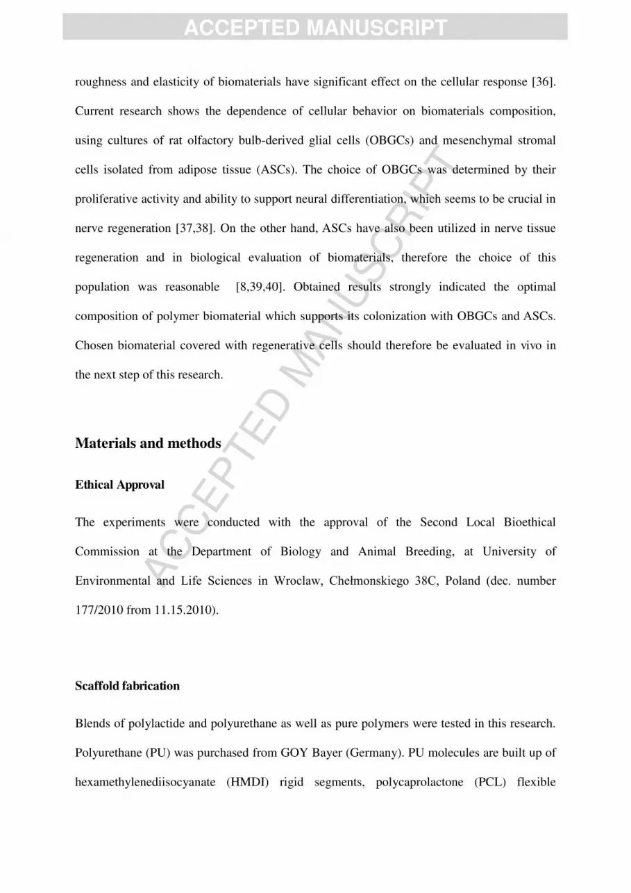
roughness and elasticity of biomaterials have significant effect on the cellular response [36].
Current research shows the dependence of cellular behavior on biomaterials composition,
using cultures of rat olfactory bulb-derived glial cells (OBGCs) and mesenchymal stromal
cells isolated from adipose tissue (ASCs). The choice of OBGCs was determined by their
proliferative activity and ability to support neural differentiation, which seems to be crucial in
nerve regeneration [37,38]. On the other hand, ASCs have also been utilized in nerve tissue
regeneration and in biological evaluation of biomaterials, therefore the choice of this
population was reasonable [8,39,40]. Obtained results strongly indicated the optimal
composition of polymer biomaterial which supports its colonization with OBGCs and ASCs.
Chosen biomaterial covered with regenerative cells should therefore be evaluated in vivo in
the next step of this research.
Materials and methods
Ethical Approval
The experiments were conducted with the approval of the Second Local Bioethical
Commission at the Department of Biology and Animal Breeding, at University of
Environmental and Life Sciences in Wroclaw, Chełmonskiego 38C, Poland (dec. number
177/2010 from 11.15.2010).
Scaffold fabrication
Blends of polylactide and polyurethane as well as pure polymers were tested in this research.
Polyurethane (PU) was purchased from GOY Bayer (Germany). PU molecules are built up of
hexamethylenediisocyanate (HMDI) rigid segments, polycaprolactone (PCL) flexible

AC
CEPTED
MAN
USC
RIP
T
ACCEPTED MANUSCRIPT
segments and isosorbitol as a chain extender (Figure 1). Polylactide (PLA), consisting of 80%
poly-L-lactide and 20% poly-DL-lactide was purchased from Purac (Netherlands). Both used
polymers were of biomedical grade and were used without further purification. The N,N-
dimethylformamide (DMF) of analytical grade was purchased from POCh (Poland).
Pure polyurethane and polylactide were prepared by dissolving polymer in
dimethylformamide to obtain a 10 wt. % solution. PU/PLA blends with the weight ratio 50/50
were prepared by dissolving both polymers in dimethylformamide to obtain a 10 wt. %
solution, and then casting on glass Petri dishes. Dissolving the polymers usually required at
least 48h stirring at ~50°C. The films were dried at 50°C under vacuum for 48 hours, then
sterilised with the use of the H2O2 cold plasma technique (Sterrad, ASP, J&J, USA), after
which the 15 mm diameter discs were cut for biological tests.
Figure 1. Structure of investigated polyurethane.
Evaluation of physical and mechanical properties of the films
The water contact angle of obtained PU/PLA films was measured using sessile drop method
using the Drop Shape Analysis System (DSA Mk2, Krüss, Germany). Ten measurements per
AC
CEPTED
MAN
USC
RIP
T
ACCEPTED MANUSCRIPT
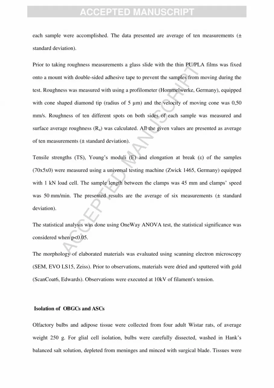
each sample were accomplished. The data presented are average of ten measurements (±
standard deviation).
Prior to taking roughness measurements a glass slide with the thin PU/PLA films was fixed
onto a mount with double-sided adhesive tape to prevent the samples from moving during the
test. Roughness was measured with using a profilometer (Hommelwerke, Germany), equipped
with cone shaped diamond tip (radius of 5 µm) and the velocity of moving cone was 0,50
mm/s. Roughness of ten different spots on both sides of each sample was measured and
surface average roughness (Ra) was calculated. All the given values are presented as average
of ten measurements (± standard deviation).
Tensile strengths (TS), Young’s moduli (E) and elongation at break (ε) of the samples
(70x5x0) were measured using a universal testing machine (Zwick 1465, Germany) equipped
with 1 kN load cell. The sample length between the clamps was 45 mm and clamps’ speed
was 50 mm/min. The presented results are the average of six measurements (± standard
deviation).
The statistical analysis was done using OneWay ANOVA test, the statistical significance was
considered when p<0.05.
The morphology of elaborated materials was evaluated using scanning electron microscopy
(SEM, EVO LS15, Zeiss). Prior to observations, materials were dried and sputtered with gold
(ScanCoat6, Edwards). Observations were executed at 10kV of filament's tension.
Isolation of OBGCs and ASCs
Olfactory bulbs and adipose tissue were collected from four adult Wistar rats, of average
weight 250 g. For glial cell isolation, bulbs were carefully dissected, washed in Hank’s
balanced salt solution, depleted from meninges and minced with surgical blade. Tissues were

AC
CEPTED
MAN
USC
RIP
T
ACCEPTED MANUSCRIPT
then placed in collagenase (1mg/mL, Sigma) and incubated for 10 minutes at 37°C, after
which it was further minced using syringe needles (18G, 20G and 22G), according to the
protocol used before [41]. At the next stage, the enzyme was inhibited by addition of medium
containing fetal bovine serum (DMEM F12/Ham’s with 10% of FBS and 1% of
penicillin/streptomycin/amphotericin b; all purchased from Sigma). Solutions were
centrifuged at 200g for 3 minutes, and cell pellets were resuspended in fresh medium and
placed in T-25 flasks at concentration of 5x10³
cells/cm2. Cultures were maintained in 37°C
5% CO2 humidified incubator for 3 days, during which no manipulations or observations were
made. After 3 days, medium was partially replaced (half of medium volume was discarded
and replaced with fresh), and cells were cultured for next 2 days. When cells adopted their
normal morphology at day 5, they were harvested and placed in cultures with elaborated
polymer membranes in 24-well plates at concentration of 1 x 10⁵ cells per well.
Adipose-derived mesenchymal stromal stem cells were isolated from adipose tissue samples
obtained from adult rats of Wistar bred. The procedure included washing the tissue with
Hank’s salt, mincing with scissors and incubation in collagenase (5mg/mL, Sigma), according
to the protocol used before [42]. After digestion, tissue samples were centrifuged at 1200 x g
for 10 minutes to obtain three layers with mononuclear cell pellet on the bottom. Superficial
layer of oil and undigested tissue was discarded with collagenase solution, and cell pellet was
suspended in culture medium (DMEM F12/Ham’s with 10% of FBS and 1% of
penicillin/streptomycin/amphotericin b) and seeded in T-25 flasks at concentration of 5x10³
cells/cm2. So prepared cultures were maintained in 37°C 5% CO2 humidified incubator until
almost full cell confluence (3-4 days). After one passage, cells were harvested and seeded on
elaborated materials in 24-well plates at concentration of 1x10⁵ cells per well.

AC
CEPTED
MAN
USC
RIP
T
ACCEPTED MANUSCRIPT
Immunophenotyping of OBGCs and ASCs
Before the experiment, cell phenotype was investigated using fluorescence inverted
microscope (Axio Observer A1, Zeiss), with immunofluorescence staining applications. For
OBGCs, p75 and glial fibrillary acidic protein (GFAP) markers presence was investigated,
while for ASCs, CD29, CD44, CD73b and CD105 was confirmed. In addition, hematopoietic
markers in ASCs - CD45 was excluded.
Procedure included washing in PBS, fixation in 4% paraformaldehyde, permeabilization and
blocking with 0.05% triton x-100/4% bovine serum albumin (15 min in room temperature).
Primary antibodies were incubated with cells for one hour in 37°C.
For glial cell labeling, followed antibodies were used: anti-nerve growth factor receptor
(NGFR p75) polyclonal antibody produced in rabbit, concentration 2µg/ml, Sigma-Aldrich;
anti-GFAP antibody produced in mouse, concentration 2.5 µg/ml, Sigma-Aldrich; secondary
antibodies used were goat anti-rabbit IgG- atto594 and anti-mouse IgG atto-594,
concentrations 1µg/ml, Sigma-Aldrich.
Primary antibodies used for labeling of adipose stem cells included: anti-CD29 antibody
produced in mouse, concentration 2µg/ml, Sigma-Aldrich; anti-CD73 polyclonal antibody
produced in rabbit, concentration 2µg/ml, Sigma-Aldrich; anti-CD105 polyclonal antibody
produced in rabbit, concentration 2.5 µg/ml, Sigma-Aldrich; anti-CD45 mouse monoclonal
antibody, concentration 2µg/ml, Sigma-Aldrich; anti-CD44 polyclonal antibody produced in
sheep, concentration 2µg/ml, R'nD Systems). Secondary antibodies used were: goat anti-
mouse IgG atto-594 and goat anti-rabbit IgG atto-488, concentration 1µg/ml, Sigma Aldrich;
donkey anti-sheep IgG NL557, concentration 1.5 µg/ml, R'n'D Systems.
After the immunostaining, cells were counterstained with DAPI (5µg/ml) for 5 minutes and
observed under the inverted, fluorescent microscope (AxioObserverA1, Zeiss).

AC
CEPTED
MAN
USC
RIP
T
ACCEPTED MANUSCRIPT
Cell culture on biomaterials
The evaluation of cell attachment rate was performed 6 hours after the inoculation of cells on
materials. Briefly, the culture media were completely collected, and the number of suspended
non-adherent cells was quantified using the Thoma counting chamber and light microscope.
Experimental cultures were maintained for 7 days, with one medium change after four days.
Cultures were evaluated at day 1, 3, and 7 of culture. For proliferative activity assessments,
Alamar Blue assay was performed. Briefly, culture media were discarded and replaced with
fresh containing 10% of resazurin dye for 2 hour of incubation, after which the media were
collected, placed in 96-well plate and measured with microplate spectroscopic reader (BMG
Labtech). Absorbance was determined at the wavelength of 600 nm, with distraction of
background absorbance at 690 nm. The results were calculated to ‘proliferative factor’ (PF),
which is the ratio of viable cells with regards to control cells cultured in pure culture
polystyrene. Experiment was repeated four times, each time using cells from different
individual. The results were analyzed statistically using the Anova OneWay test, statistical
significance was considered when p<0.05. After the Alamar Blue assay, cultures were fixed in
4% ice-cold paraformaldehyde and stained. For fluorescent microscopy observations, cells
were permeabilized with 0,1% Triton X-100 for 10 minutes at room temperature, stained with
phalloidin (conjugated with 488-AlexaFluor) for visualization of actin filaments and with
DAPI for evaluation of nuclei. Additionally, OBGCs were stained with anti-p75 (NGFR)
antibody conjugated to alexa fluor-594 fluorophore for detection of nerve growth factor
receptor-expressing cells. Observations were done using AxioObserver.A1 (Zeiss) inverted
fluorescent microscope and documented with Cannon PowerShot digital camera (Cannon).
Additionally, samples were fixed with 2,5% glutaraldehyde, dehydrated in ethanol, sputtered
with gold and observed with scanning electron microscope (EVO LS15, Zeiss).
AC
CEPTED
MAN
USC
RIP
T
ACCEPTED MANUSCRIPT
Results and discussion
Morphology, roughness, water contact angle and biomechanical properties of the scaffolds
The majority of current effort to understand the interaction between cell and biomaterial have
focused among others on surface physicochemical properties. Over the past decade the
microstructure of biomaterials and their influence on cellular response has been extensively
investigated. In our research we applied biomaterials obtained by blending two very different
in nature polymers – polyurethane and polylactide. The obtained biomaterials were
characterized by different morphological and topographical methods. We found that the
morphology and topography of investigated materials could be regulated through chemical
combination between PU and PLA. We demonstrated that addition of PLA to PU increased
the number and the size of pores in the films. Our data stands in good agreement with Mi et
al. findings, where similar observation was performed [19]. In pure PU material the most of
the pores were not larger than few microns, in contrast to the PU/PLA blend where the
number of pores and their sizes were prominently higher. From the biological aspect, increase
in these parameters improves the communication between cells and facilitates the exchange of
metabolites [43]. Therefore it may be assumed that PU/PLA blend will give the most optimal
biological results. The pure PLA material was characterized by the low number of uniform,
submicron-sized pores (Figure 2).

AC
CEPTED
MAN
USC
RIP
T
ACCEPTED MANUSCRIPT
Figure 2. The surface morphology of pure polyurethane (A,B), PU/PLA blend (C,D), and pure
polylactide (E,F) in SEM; scale bars indicated on the micrographs.
Surface roughness of particular biomaterials has been reported as a factor determining cells
adhesion, expansiveness as well as proliferation. In addition, surface roughness might regulate
cellular phenotypic activities, including neurite extension and osteoblastic differentiation

AC
CEPTED
MAN
USC
RIP
T
ACCEPTED MANUSCRIPT
[44,45]. We found that roughness of our investigated biomaterials might be increased through
addition of PLA to the pure PU, although the differences were statistically insignificant. We
measured the highest roughness in PU/PLA blend, while the lowest was noticed in the pure
PU (below 1 μm). The roughness of pure polylactide surface was below 2 μm, which shows
that the highest roughness may be achieved only in specific combination of both polymers,
while the intermediate surface roughness was observed in pure PLA (Table 1). Our findings
additionally were confirmed by the qualitative analysis of surface by electron microscopic
observations. In study performed by Feng and Ye, roughness of polymer blends was also
modified by the changes in polymer ratio [46]. Similarly, as in the case of surface roughness,
wettability of the particular material also significantly influences cellular response. In our
research we found that wettability of both PU as well as PLA significantly differed (p<0.05).
We demonstrated that pure PU was characterized by water contact angles (WCA) equal to
88,6°±1,34°, whereas in PLA material WCA was on the lowest level (79,85°±1,06°).
Interestingly, the hybrid material PU/PLA exhibited much higher water contact angle
(95,4±1,7°, Table 1). We can summarize, then, that surface of pure PU was more hydrophobic
than this of pure PLA, and mixing the two polymers together caused increase of the
hydrophobicity. However such increase should not be very significant for cell attachment and
their growth in culture, as these parameters usually change when the water contact values are
over 110° [47]. The evaluation of Young's modulus showed that the presence of PLA in
experimental PU/PLA blend significantly increased the material’s tensile strength, in relation
to pure PU material (Table 1). The pure PLA showed the Young's modulus over 3000 MPa,
which resulted in high stiffness and hampered the manipulations. Therefore, it can be
concluded that the addition of PLA to PU in 5:5 w/w improves the mechanical properties of
the material, increasing its mechanical toughness with slight decrease in elasticity, as it was
also observed by Wang et al. [48]. This parameter has significant influence on cell

AC
CEPTED
MAN
USC
RIP
T
ACCEPTED MANUSCRIPT
attachment, growth and behavior and it is widely known that neural cells prefer the
environment of low tensile strength [49-51].
Wettability [°] 88,65+1,34 95,4+1,70 79,85+1,06 p<0,05
Roughness [µm] 0,32+0,06 5,5+0,85 1,26+0,93 p>0,05
Young's modulus [MPa] 37,5+3 196,25+14 3435+250,5 p<0,05
PU PU/PLA PLA p value
Table 1. The results from analysis of water contact angle and roughness of experimental
surfaces, and the Young's modulus determined in both materials. The p value shows statistical
significance of the differences between groups (p<0.05).
Cell culture on biomaterials
Evaluation of cell attachment rate revealed no significant difference between experimental
groups and polystyrene control - over 95% of cells adhered to the surface. After the first 24
hours, glial cells cultured on pure PU showed decrease in proliferative activity in relation to
the control polystyrene cultures with no statistical significance. In PU/PLA blend and in pure
PLA we observed the increase in proliferative activity of glial cells after the first day in
relation to control polystyrene well culture, with statistical significance noticed in sample
PLA (p<0,05). The differences between groups were not statistically significant at the first
day. Cells cultured on pure PLA films showed the increase in proliferative activity after the
first 24 hours of culture, in relation to polystyrene well control, which is not a typical
response on lactic acid present due to material degradation [52]. At day three, the proliferation
factor of glial cells was below 1 in all investigated groups, with the highest value in PU/PLA
blend. This decrease was statistically significant in all groups (p<0,05), but there was no

AC
CEPTED
MAN
USC
RIP
T
ACCEPTED MANUSCRIPT
difference between groups. While the materials differed in composition, and there were
components specific for only two of three groups (PLA was in PLA and PU/PLA but not in
PU; similarly for PU), this decrease was probably not caused by the materials' degradation, so
it may be connected with the release of dimethylformamide traces. It was already shown that
this substance can inhibit cell growth, or even induce apoptosis in cultured cells [53,54]. After
seven days in culture cells in PU sample showed decreased PF value in relation to control
(p<0,05). The PF value obtained for pure PLA sample was also significantly decreased in
relation to control (p<0,05). Glial cells from PU/PLA sample showed the PF value slightly
above 1, with no significant difference between control (p>0,05). Results from day seven
were significantly different between all experimental groups (p<0,05). Our results showed
that the most neutral material for cell proliferation was PU/PLA blend, while obtained PF
values were the less variable and different from the control (Figure 3). We assume that the
differences between the proliferation rate could result from different materials' surface
characteristics rather than from the release of materials' degradation products, as was
observed in other studies [19,50]. In experiment performed by Mi et al. [19], the proliferation
assay indicated the influence of PLA content on the proliferative activity of fibroblasts
cultured on different PU/PLA scaffolds, but it was also explained by the surface topography.
In ASCs cultures, pure PU and PU/PLA blend showed low influence on cell proliferation,
where the PF values were slightly below 1 during all time points (p>0,05). The differences
between PU and PU/PLA were not statistically significant. In pure PLA sample, cells showed
constant decrease during 7 days of culture. The proliferative activity in PLA was significantly
different from control group and from the other groups in the first and the last day of
experiment (p<0,05). There was no significant difference between groups at day 3 (Figure 3).
The less prominent differences between groups may be explained by the high plasticity of
adipose stem cells. As they are less developed and more primitive than OBGC, they could

AC
CEPTED
MAN
USC
RIP
T
ACCEPTED MANUSCRIPT
adopt faster to the different culture conditions. The significant difference observed in PLA
sample probably resulted from dramatic increase in Young's modulus noticed in this sample.
Figure 3. The proliferation factor (PF) of olfactory bulb glial cells (OBGC) and adipose-
derived mesenchymal stem cells (ASCs) cultured with pure polyurethane (PU),
polyurethane/polylactide blend (PU/PLA) and pure polylactide (PLA) during 7 days of
culture.
Glial cells cultured on experimental films exhibited diverse morphology and ultrastructure.
Investigated cells maintained on pure PU showed altered morphology, with clustered growth
pattern and low number of p75 - positive cells (Figure 4 A1, A2). Detailed analysis performed
by means of SEM revealed low number of bipolar, elongated cells (Figure 4 A3). This
undesired cellular behavior might result from low Young's modulus of pure PU, as it is known
that this parameter significantly influences the cellular adhesion and growth pattern [55,56].
In PU/PLA blend cells were characterized by proper morphology, with significant number of
p75 - positive cells, bipolar shape present on fibroblast-like cells (Figure 4 B1-B3). In pure
PLA samples, cells showed significant clustering processes and p75 - positive cells were not
observed (Figure 4 C1-C3). Cells in control polystyrene cultures showed typical morphology

AC
CEPTED
MAN
USC
RIP
T
ACCEPTED MANUSCRIPT
for olfactory bulb-derived glial cells, with numerous p75-positive cells, uniform growth
pattern and prominent elongated, bipolar cells (Figure 4, D1-D3) [57].
Figure 4. Olfactory bulb glial cells after seven days of culture on pure PU (row A), PU/PLA
blend (row B), pure PLA (row C) and in control polystyrene well (row D). Fluorescent
staining of p75 and nuclei (column 1, p75 in red, nuclei in white/grey), and of actin and nuclei
AC
CEPTED
MAN
USC
RIP
T
ACCEPTED MANUSCRIPT
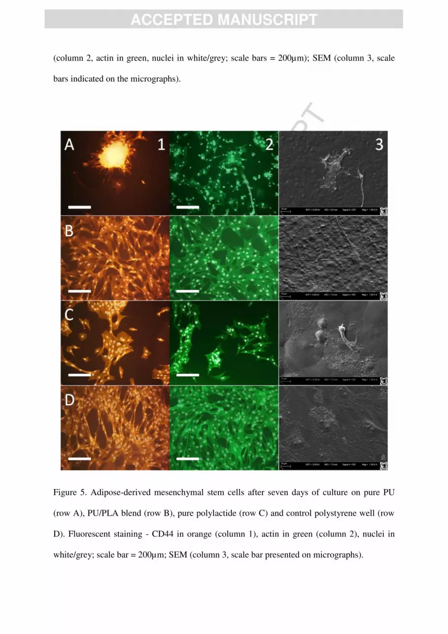
(column 2, actin in green, nuclei in white/grey; scale bars = 200µm); SEM (column 3, scale
bars indicated on the micrographs).
Figure 5. Adipose-derived mesenchymal stem cells after seven days of culture on pure PU
(row A), PU/PLA blend (row B), pure polylactide (row C) and control polystyrene well (row
D). Fluorescent staining - CD44 in orange (column 1), actin in green (column 2), nuclei in
white/grey; scale bar = 200µm; SEM (column 3, scale bar presented on micrographs).

AC
CEPTED
MAN
USC
RIP
T
ACCEPTED MANUSCRIPT
Mesenchymal stem cells cultured on pure PU showed negative morphological changes, with
visible cell clusters, apoptotic bodies and necrotic cells (Figure 5A). The optimal culture
environment for MSCs requires the proper surface for cell adhesion, while the lack of suitable
adhesion sites may result in cell death [58,59]. Therefore, our results showed that pure PU
does not constitute the optimal surface for cell growth, both for glial and mesenchymal stem
cells. ASCs cultured on PU/PLA blend maintained their normal appearance, with big single
nuclei, flattened, elongated form and numerous cell-cell connections. No degenerative
changes were observed in this sample (Figure 5B). Cells cultured on pure PLA showed altered
growth pattern, with visible clustering of cells of proper morphology (Figure 5C). ASCs in
control polystyrene culture created monolayer composed of cells with typical morphology for
mesenchymal cells (Figure 5D) [42]. Despite the differences in morphology between analyzed
groups, cells maintained the presence of CD44 in all investigated samples (Figure 5A1, B1,
C1, D1). This protein is involved in cell-to-cell communication, migration and environmental
recognition [60]. While the mechanism by which the mesenchymal stem cells induce the
regeneration is considerably depended on those activities, the presence of CD44 on
mesenchymal stem cells is probably important for maintaining their therapeutic abilities.
Conclusions
In summary, the PU/PLA blend exhibited positive influence on olfactory bulb-derived glial
cells and on adipose-derived mesenchymal stromal cells, in comparison to pure PU and pure
PLA films. The addition of polylactide increased the proliferative activity of cells, improved
cell adhesion and overcame the negative influence of pure PU on cellular morphology.
Therefore, we postulate that polymer films obtained from polyurethane and polylactide may

AC
CEPTED
MAN
USC
RIP
T
ACCEPTED MANUSCRIPT
be found as promising tool in further medical experiments on nerve tissue regeneration,
involving the utility of both biomaterial and cellular therapy.
Acknowledgement
This research was financially supported by the National Center of Science, Poland (grant No
2011/01/B/STB8/07795). Authors declare no conflict of interest in this manuscript.
References
1. Sian-Marie L, Rothwell NJ and Gibson RM. The role of inflammation in CNS injury and
disease. Br J Pharmacol 2006; 147(suppl 1): S232-S240.
2. Daly W, Yao L, Zeugolis D, et al. A biomaterials approach to peripheral nerve
regeneration: bridging the peripheral nerve gap and enhancing functional recovery. J R Soc
Interface 2012; 9: 202-221.
3. Matsumoto K, Ohnishi K, Kiyotani T, et al. Peripheral nerve regeneration across an 80-mm
gap bridged by a polyglicolic acid (PGA)-collagen tube filled with laminin-coated collagen
fibers: a histological and electrophysiological evaluation of regenerated nerves. Brain Res
2000; 868: 315-328.
4. Nichols CM, Brenner MJ, Fox IK, et al. Effects of motor versus sensory nerve grafts on
peripheral nerve regeneration. Exp Neurol 2004; 190: 347-355.
5. Teixeira AI, Duckworth JK and Hermanson O. Getting the right stuff: controlling neural
stem cell state and fate in vivo and in vitro with biomaterials. Cell Res 2007; 17: 56-61.
AC
CEPTED
MAN
USC
RIP
T
ACCEPTED MANUSCRIPT
6. Orive G, Anitua E, Pedraz JL, et al. Biomaterials for promoting brain protection, repair and
regeneration. Nat Rev Neurosci 2009; 10: 682-692.
7. Subramanian A, Krishnan UM and Sethuraman S. Development of biomaterial scaffold for
nerve tissue engineering: biomaterial mediated neural regeneration. J Biomed Sci 2009; 16:
108.
8. Neuss S, Apel C, Buttler P, et al. Assessment of stem cell/biomaterial combinations for
stem cell-based tissue engineering. Biomaterials 2008; 29: 302-313.
9. Hadlock T, Sundback C, Hunter D, et al. A polymer foam conduit seeded with Schwann
cells promotes guided peripheral nerve regeneration. Tissue Eng 2000; 6: 119-127.
10. Keatch RP, Schor AM, Vorstius JB, et al. Biomaterials in regenerative medicine:
engineering to recapitulate the natural. Curr Opin Biotechnol 2012; 23: 579-582.
11. Perez RA, Won JE, Knowles JC, et al. Naturally and synthetic smart composite
biomaterials for tissue regeneration. Adv Drug Deliv Rev 2013; 65: 471-496.
12. Leipzig ND and Shoichet MS. The effect of substrate stiffness on adult neural stem cell
behavior. Biomaterials 2009; 30: 6867-6878.
13. Tian H, Tang Z, Zhuang X, et al. Biodegradable synthetic polymers: preparation,
functionalization and biomedical application. Progress in Polymer Science 2012; 37: 237-
280.
14. Gunatillake PA and Adhikari R. Biodegradable synthetic polymers for tissue engineering.
Eur Cell Mater 2003; 5: 1-16.
15. Gunatillake P, Mayadunne R and Adhikari R. Recent developments in biodegradable
synthetic polymers. Biotechnol Annu Rev 2006; 12: 301-347.
AC
CEPTED
MAN
USC
RIP
T
ACCEPTED MANUSCRIPT
16. Nampoothiri KM, Nimisha RN and Rojan PJ. An overview of the recent developments in
polylactide (PLA) research. Bioresource Technol 2010; 101: 8493-8501.
17. Yang F, Murugan R, Ramakrishna S, et al. Fabrication of nano-structured porous PLLA
scaffold intended for nerve tissue engineering. Biomaterials 2004; 25: 1891-1900.
18. Burke A and Hasirci N. Biomaterials: From Molecules to Engineered Tissue.
Polyurethanes in biomedical applications. Springer US, 2004, p. 83-101.
19. Mi HY, Salick MR, Jing X, et al. Characterization of thermoplastic
polyurethane/polylactic acid (TPU/PLA) tissue engineering scaffolds fabricated by
microcellular injection molding. Mater Sci Eng: C 2013; 33: 4767-4776.
20. Lamba NMK, Woodhouse KA, Cooper SL (1998) Polyurethanes in biomedical
applications. CRC Press LLC.
21. Feng F, Ye L. Morphologies and mechanical properties of polylactide/thermoplastic
polyurethane elastomer blends. J Applied Polym Sci 2011; 119: 2778-2783.
22. Feng F, Zhao X, Ye L. Structure and properties of ultradrawn polylactide/thermoplastic
polyurethane elastomer blends. J Macromol Sci B: Physics 2011, 50: 1500-1507.
23. Liu GC, He YS, Zeng JB, Xua Y, Wang YZ. In situ formed crosslinked polyurethane
toughened polylactide. Polym Chem 2014; 5: 2530-2539.
24. Hilmer K, Bruning K, Fritz HG, Zgaverdea AC. Process for producing blends made of
polylactides (PLAS) and of thermoplastic polyurethanes (TPUS) US Patent: US 8633283 B2.
25. Han JJ, Huang HX. Preparation and characterization of biodegradable
polylactide/thermoplastic polyurethane elastomer blends. J Applied Polym Sci 2011; 120:
3217-3223.

AC
CEPTED
MAN
USC
RIP
T
ACCEPTED MANUSCRIPT
26. Gurunathan T, Mohanty S, Nayak SK. Preparation and performance evaluation of castor
oil-based polyurethane prepolymer/polylactide blends. J Mater Sci 2014; 49: 8016–8030.
27. Hausner T, Schmidhammer R, Zandieh S, et al. Nerve regeneration using tubular scaffolds
from biodegradable polyurethane. Acta Neurochir Suppl 2007; 100: 69-72.
28. Szarek D, Marycz K, Laska J, et al. Assessment of in vivo behavior of polymer tube nerve
grafts simultaneously with the peripheral nerve regeneration process using scanning electron
microscopy technique. Scanning 2013; 35: 232-245.
29. Gajowy J, Bednarz P, Laska J. Degradation of polymer mixture of polyurethane and
polylactide in simulated biological environment. Engineering of Biomaterials 2009; 13: 89-
91.
30. Davies JE, Huang C, Proschel C, et al. Astrocytes derived from glial-restricted precursors
promote spinal cord repair. J Biol 2006; 5:7.
31. Shrestha B, Coykendall K, Li Y, et al. Repair of injured spinal cord using biomaterial
scaffolds and stem cells. Stem Cell Research & Therapy 2014; 5: 91.
32. Delcroix GJR, Schiller PC, Benoit JP, et al. Adult cell therapy for brain neuronal damages
and the role of tissue engineering. Biomaterials 2010; 31: 2105-2120.
33. Yang Y, Chen X, Ding F, et al. Biocompatibility evaluation of silk fibroin with peripheral
nerve tissues and cells in vitro. Biomaterials 2007; 28: 1643-1652.
34. Lakard S, Herlem G, Propper A, et al. Adhesion and proliferation of cells on new
polymers modified biomaterials. Bioelectrochemistry 2004; 23: 579-582.
35. Gimble J and Guilak F. Adipose-derived adult stem cells: isolation, characterization, and
differentiation potential. Cytotherapy. 2003; 5:362-369

AC
CEPTED
MAN
USC
RIP
T
ACCEPTED MANUSCRIPT
36. De Guzman RC and VandeVord PJ. Variations in astrocyte and fibroblast response due to
biomaterial particulates in vitro. J Biomed Mater Res A 2008; 85: 14-24.
37. Kueh JL, Li D, Raisman G, et al. Directionality and bipolarity of olfactory ensheathing
cells on electrospun nanofibers. Nanomedicine (Lond) 2012; 7: 1211-1224.
38. Zhu Y, Cao L, Su Z, et al. Olfactory ensheathing cells: attractant of neural progenitor
migration to olfactory bulb. Glia 2010; 58: 716-729.
39. Willerth SM and Sakiyama-Elbert SE. Combining stem cells and biomaterial scaffolds for
constructing tissues and cell delivery. StemBook 2008,
http://www.ncbi.nlm.nih.gov/books/NBK27050.
40. Thesleff T, Lehtimaki K, Niskakangas T, et al. Cranioplasty with adipose-derived stem
cells and biomaterial: a novel method for cranial reconstruction. Neurosurgery 2011; 68:
1535-1540.
41. Marycz K, Szarek D, Grzesiak J, et al. Influence of modified alginate hydrogels on
mesenchymal stem cells and olfactory bulb-derived glial cells cultures. Bio-Med Mater Eng
2014; 24: 1625-1637.
42. Grzesiak J, Marycz K, Czogała J, et al. Comparison of behavior, morphology and
morphometry of equine and canine adipose derived mesenchymal stem cells in culture. Int J
Morphol 2011; 29: 1012-1017.
43. Lee OJ, Ju HW, Kim JH, et al. Development of artificial dermis using 3D electrospun silk
fibroin nanofiber matrix. J Biomed Nanotechnol 2014; 10: 1294-1303.
44. Benavente JM, Mogami H, Sakurai T, et al. Evaluation of silicon nitride as a substrate for
culture of PC12 cells: an interfacial model for functional studies in neurons. PLoS One 2014;
9: e90189.

AC
CEPTED
MAN
USC
RIP
T
ACCEPTED MANUSCRIPT
45. Czarnecki JS, Jolivet S, Kundrat ME, et al. Cellular Automata simulation of osteoblast
growth on microfibrous carbon-based scaffolds. Tissue Eng Part A. Epub ahead of print 29
May 2014, doi: 10.1089/ten.tea.2013.0387.
46. Feng F and Ye L. Morphologies and mechanical properties of polylactide/thermoplastic
polyurethane elastomer blends. J Appl Polym Sci 2011; 119: 2778-2783.
47. Dowling DP, Miller IS, Ardhaoui M, et al. Effect of surface wettability and topography on
the adhesion of osteosarcoma cells on plasma-modified polystyrene. J Biomater Appl 2011;
26: 327-347.
48. Wang W, Ping P, Chen X, et al. Polylactide-based polyurethane and its shape-memory
behavior. Eur Polymer J 2006; 42: 1240-1249.
49. Schneider A, Francius G, Obeid R, et al. Polyelectrolyte multilayers with a tunable
young's modulus: influence of film stiffness on cell adhesion. Langmuir 2006; 22: 1193-
1200.
50. Saha K, Keung AJ, Irwin EF, et al. Substrate modulus directs neural stem cell behavior.
Biophys J 2008; 95: 4426-4438.
51. Leipzig ND and Shoichet MS. The effect of substrate stiffness on adult neural stem cell
behavior. Biomaterials 2009; 30: 6867-6878.
52. Evans GRD, Brandt K, Katz S, et al. Bioactive poly(l-lactic acid) conduits seeded with
Schwann cells for peripheral nerve regeneration. Biomaterials 2002; 23: 841-848.
53. Li XN, Du ZW, Huang Q, et al. Growth-inhibitory and differentiation-inducing activity of
dimethylformamide in cultured human malignant glioma cells. Neurosurgery 1997; 40: 1250-
1259.

AC
CEPTED
MAN
USC
RIP
T
ACCEPTED MANUSCRIPT
54. Boyle CC and Hickman JA. Toxin-induced increase in survival factor receptors:
modulation of the threshold for apoptosis. Cancer Res 1997; 57: 2404-2409.
55. Yeung T, Georges PC, Flanagan LA, et al. Effects of substrate stiffness on cell
morphology, cytoskeletal structure, and adhesion. Cell Motil Cytoskeleton 2005; 60: 24-34.
56. Pek YS, Wan ACA and Ying JY. The effect of matrix stiffness on mesenchymal stem cell
differentiation in a 3D thixotropic gel. Biomaterials 2010; 31: 385-391.
57. Honore A, Le Corre S, Derambure C, et al. Isolation, characterization, and genetic
profiling of subpopulations of olfactory ensheathing cells from the olfactory bulb. Glia 2012;
60: 404-413.
58. Marycz K, Śmieszek A, Grzesiak J, et al. Application of bone marrow and adipose-
derived mesenchymal stem cells for testing the biocompatibility of metal-based biomaterials
functionalized with ascorbic acid. Biomed Mater 2013; 8: 065004.
59. Walsh MF, Thamilselvan V, Grotelueschen R, et al. Absence of adhesion triggers
differential FAK and SAPKp38 signals in SW620 human colon cancer cells that may inhibit
adhesiveness and lead to cell death. Cell Physiol Biochem 2003; 13: 135-146.
60. Khaldoyanidi S. Directing stem cell homing. Cell Stem Cell 2008; 2: 198-200.

AC
CEPTED
MAN
USC
RIP
T
ACCEPTED MANUSCRIPT
Highlights
1. Polyurethane-polylactide blends exhibit different characteristics from pure polymers.
2. Pure PU and PLA negatively influence on morphology of glial and mesenchymal cells.
3. PU/PLA blend was neutral for glial and mesenchymal cell proliferation and morphology.
Copyright © 2022 FDOKUMEN