Platelets secrete stromal cell-derived factor 1 and recruit bone marrow-derived progenitor cells to...
13
The Journal of Experimental Medicine ARTICLE JEM © The Rockefeller University Press $8.00 Vol. 203, No. 5, May 15, 2006 1221–1233 www.jem.org/cgi/doi/10.1084/jem.20051772 1221 Proliferation of endothelial cells (ECs) and vas- cular smooth muscle cells (SMCs) is essential for cardiovascular development (1), but also contributes to the repair and remodeling of the injured vessel wall (2). The major trigger of vascular injury is atherosclerosis, which is to- day the most important cause of morbidity in the Western world. Spontaneous rupture of the atherosclerotic plaque and vascular interven- tions for atherothrombotic disease (e.g., balloon angioplasty) induce EC damage. Disruption of the endothelial integrity initiates proliferation of ECs, which promotes reendothelialization of the vascular lesion (3, 4), but also triggers local accumulation of SMCs, leading to inti- mal hyperplasia and occlusion of the diseased vessel (2). At first, neointimal ECs and SMCs were be- lieved to originate exclusively from adjacent cells within the vessel wall that migrate to the site of injury and start to proliferate (3). However, Platelets secrete stromal cell–derived factor 1α and recruit bone marrow–derived progenitor cells to arterial thrombi in vivo Steffen Massberg, 1 Ildiko Konrad, 1 Katrin Schürzinger, 1 Michael Lorenz, 1 Simon Schneider, 1 Dietlind Zohlnhoefer, 1 Katharina Hoppe, 1 Matthias Schiemann, 2,3 Elisabeth Kennerknecht, 1 Susanne Sauer, 1 Christian Schulz, 1 Sandra Kerstan, 1 Martina Rudelius, 4 Stefan Seidl, 4 Falko Sorge, 1 Harald Langer, 10 Mario Peluso, 5 Pankaj Goyal, 6 Dietmar Vestweber, 7 Nikla R. Emambokus, 8 Dirk H. Busch, 2,3 Jon Frampton, 9 and Meinrad Gawaz 10 1 Deutsches Herzzentrum and Medizinische Klinik, Technical University of Munich, D-80636 Munich, Germany 2 Institute for Medical Microbiology, Immunology, and Hygiene, 3 Clinical Cooperation Group Vaccinology, National Research Center for Environment and Health (GSF), and 4 Institute of Pathology, Klinikum rechts der Isar, Technical University of Munich, D-81675 Munich, Germany 5 Procorde GmbH, D-82152 Martinsried, Germany 6 Institut für Prophylaxe und Epidemiologie der Kreislaufkrankheiten, LMU München, D-80336 Munich, Germany 7 Max-Planck Institute of Molecular Biomedicine, University of Münster, D-48149 Münster, Germany 8 Harvard Medical School, Children’s Hospital, Boston, MA 02115 9 Institute for Biomedical Research, Birmingham University Medical School, Birmingham B15 2TT, England, UK 10 Innere Medizin III, Universität Tübingen, D-72076 Tübingen, Germany The accumulation of smooth muscle and endothelial cells is essential for remodeling and repair of injured blood vessel walls. Bone marrow–derived progenitor cells have been implicated in vascular repair and remodeling; however, the mechanisms underlying their recruitment to the site of injury remain elusive. Here, using real-time in vivo fluorescence microscopy, we show that platelets provide the critical signal that recruits CD34 + bone marrow cells and c-Kit + Sca-1 + Lin − bone marrow–derived progenitor cells to sites of vascular injury. Correspondingly, specific inhibition of platelet adhesion virtually abrogated the accumulation of both CD34 + and c-Kit + Sca-1 + Lin − bone marrow–derived progenitor cells at sites of endothelial disruption. Binding of bone marrow cells to platelets involves both P-selectin and GPIIb integrin on platelets. Unexpectedly, we found that activated platelets secrete the chemokine SDF-1, thereby supporting further primary adhesion and migration of progenitor cells. These findings establish the platelet as a major player in the initiation of vascular remodeling, a process of fundamental importance for vascular repair and pathological remodeling after vascular injury. CORRESPONDENCE Steffen Massberg: [email protected] Abbreviations used: BM-PC, BM-derived PC; DCF, dichloro- fluorescein; EC, endothelial cell; IVM, intravital video fluorescence microscopy; KSL, c-Kit + Sca-1 + Lin − ; PC, pro- genitor cell; PSGL, P-selectin glycoprotein ligand; SDF-1α, stromal cell–derived factor 1α; SMC, smooth muscle cell. S. Massberg’s present address is CBR Institute for Biomedical Research and Department of Pathology, Harvard Medical School, Boston, MA 02115. The online version of this article contains supplemental material. on January 25, 2015 jem.rupress.org Downloaded from Published April 17, 2006 http://jem.rupress.org/content/suppl/2006/04/17/jem.20051772.DC1.html Supplemental Material can be found at:
-
Upload
independent -
Category
Documents
-
view
0 -
download
0
Transcript of Platelets secrete stromal cell-derived factor 1 and recruit bone marrow-derived progenitor cells to...
The
Journ
al o
f Exp
erim
enta
l M
edic
ine
ARTICLE
JEM © The Rockefeller University Press $8.00
Vol. 203, No. 5, May 15, 2006 1221–1233 www.jem.org/cgi/doi/10.1084/jem.20051772
1221
Proliferation of endothelial cells (ECs) and vas-cular smooth muscle cells (SMCs) is essential for cardiovascular development (1), but also contributes to the repair and remodeling of the injured vessel wall (2). The major trigger of vascular injury is atherosclerosis, which is to-day the most important cause of morbidity in the Western world. Spontaneous rupture of the
atherosclerotic plaque and vascular interven-tions for atherothrombotic disease (e.g., balloon angioplasty) induce EC damage. Disruption of the endothelial integrity initiates proliferation of ECs, which promotes reendothelialization of the vascular lesion (3, 4), but also triggers local accumulation of SMCs, leading to inti-mal hyperplasia and occlusion of the diseased vessel (2).
At fi rst, neointimal ECs and SMCs were be-lieved to originate exclusively from adjacent cells within the vessel wall that migrate to the site of injury and start to proliferate (3). However,
Platelets secrete stromal cell–derived factor 1α and recruit bone marrow–derived progenitor cells to arterial thrombi in vivo
Steff en Massberg,1 Ildiko Konrad,1 Katrin Schürzinger,1 Michael Lorenz,1 Simon Schneider,1 Dietlind Zohlnhoefer,1 Katharina Hoppe,1 Matthias Schiemann,2,3 Elisabeth Kennerknecht,1 Susanne Sauer,1 Christian Schulz,1 Sandra Kerstan,1 Martina Rudelius,4 Stefan Seidl,4 Falko Sorge,1 Harald Langer,10 Mario Peluso,5 Pankaj Goyal,6 Dietmar Vestweber,7 Nikla R. Emambokus,8 Dirk H. Busch,2,3 Jon Frampton,9 and Meinrad Gawaz10
1Deutsches Herzzentrum and Medizinische Klinik, Technical University of Munich, D-80636 Munich, Germany2Institute for Medical Microbiology, Immunology, and Hygiene, 3Clinical Cooperation Group Vaccinology, National Research
Center for Environment and Health (GSF), and 4Institute of Pathology, Klinikum rechts der Isar, Technical University
of Munich, D-81675 Munich, Germany5Procorde GmbH, D-82152 Martinsried, Germany6Institut für Prophylaxe und Epidemiologie der Kreislaufkrankheiten, LMU München, D-80336 Munich, Germany7Max-Planck Institute of Molecular Biomedicine, University of Münster, D-48149 Münster, Germany8Harvard Medical School, Children’s Hospital, Boston, MA 021159Institute for Biomedical Research, Birmingham University Medical School, Birmingham B15 2TT, England, UK
10Innere Medizin III, Universität Tübingen, D-72076 Tübingen, Germany
The accumulation of smooth muscle and endothelial cells is essential for remodeling
and repair of injured blood vessel walls. Bone marrow–derived progenitor cells have been
implicated in vascular repair and remodeling; however, the mechanisms underlying their
recruitment to the site of injury remain elusive. Here, using real-time in vivo fl uorescence
microscopy, we show that platelets provide the critical signal that recruits CD34+ bone
marrow cells and c-Kit+ Sca-1+ Lin− bone marrow–derived progenitor cells to sites of
vascular injury. Correspondingly, specifi c inhibition of platelet adhesion virtually abrogated
the accumulation of both CD34+ and c-Kit+ Sca-1+ Lin− bone marrow–derived progenitor
cells at sites of endothelial disruption. Binding of bone marrow cells to platelets involves
both P-selectin and GPIIb integrin on platelets. Unexpectedly, we found that activated
platelets secrete the chemokine SDF-1𝛂, thereby supporting further primary adhesion and
migration of progenitor cells. These fi ndings establish the platelet as a major player in the
initiation of vascular remodeling, a process of fundamental importance for vascular repair
and pathological remodeling after vascular injury.
CORRESPONDENCE
Steffen Massberg:
Abbreviations used: BM-PC,
BM-derived PC; DCF, dichloro-
fl uorescein; EC, endothelial
cell; IVM, intravital video
fl uorescence microscopy; KSL,
c-Kit+ Sca-1+ Lin−; PC, pro-
genitor cell; PSGL, P-selectin
glycoprotein ligand; SDF-1α,
stromal cell–derived factor 1α;
SMC, smooth muscle cell.S. Massberg’s present address is CBR Institute for Biomedical
Research and Department of Pathology, Harvard Medical
School, Boston, MA 02115.
The online version of this article contains supplemental material.
on January 25, 2015jem
.rupress.orgD
ownloaded from
Published April 17, 2006
http://jem.rupress.org/content/suppl/2006/04/17/jem.20051772.DC1.html Supplemental Material can be found at:
1222 RECRUITMENT OF PROGENITOR CELLS TO THROMBI IN VIVO | Massberg et al.
recently BM-derived progenitor cells (BM-PCs) have been implicated in remodeling and repair of the injured vessel wall (4–9). Indeed, BM-PCs appear to give rise to substantial num-bers of neointimal ECs and SMCs after endothelial denudation (4–9). However, the signals that target BM-PCs to foci of vascular injury have not been understood thus far.
The fi rst response to vascular injury is platelet adhesion to the exposed subendothelium (10–13). Here, we show that platelet adhesion not only triggers vascular thrombosis, lead-ing to myocardial infarction and ischemic stroke, but also represents the critical step for the targeting of BM-PCs to sites of endothelial disruption. Using real-time in vivo double fl uorescence microscopy of the mouse carotid artery, we demonstrate that CD34+ and c-Kit+ Sca-1+ Lin− (KSL) BM-PCs directly adhere to platelets after vascular injury in a process that involves platelet P-selectin and GPIIb integrin. Once activated, platelets secrete the chemokine SDF-1α, which supports primary adhesion of PCs on the surface of arterial thrombi in vivo. BM-PCs recruited to platelet aggre-gates give rise to neointimal cells, indicating that accumula-tion of BM-PCs in arterial thrombi may contribute to vascular repair and, eventually, to pathological remodeling. Together, the present results identify a central role of platelets for the targeting of BM-PCs to the arterial intima, a process of fundamental importance for vascular repair and pathologi-cal remodeling after vascular injury.
RESULTS
BM-PCs are recruited rapidly to sites
of endothelial denudation
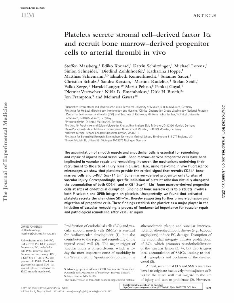
Because primitive long-term repopulating mouse hemato-poietic PCs are enriched among CD34+ BM cells (14), we fi rst addressed the accumulation of mouse CD34+ BM-PCs to foci of vascular injury. We injured the carotid artery of C57BL/6J mice (12) and infused CD34+ cells tagged with dichlorofl uorescein (DCF). We used intravital video fl uores-cence microscopy (IVM) to directly visualize and quantify the dynamic process of BM-PC accumulation. In the absence of vascular injury, CD34+ BM-PCs rolled along short dis-tances of the intact vessel wall; however, fi rm adhesion was not detected. In contrast, numerous CD34+ BM-PCs were recruited to the vascular wall within 5 min after endothelial denudation, reaching a plateau 60 min after vascular injury (375 ± 83 adherent cells/mm2 60 min after injury, P < 0.05 vs. baseline; Fig. 1, a and b).
CD34 expression on mouse BM-PCs has been demon-strated to vary with the activation status of the precursor cells (15) and considerable amounts of mouse long-term reconsti-tuting BM-PCs reside in the CD34−/low cell fraction (16). Hence, we next analyzed the recruitment of phenotypically defi ned mouse hematopoietic BM-PCs (16). We isolated KSL cells by fl uorescence-activated cell sorting (purity of KSL >98%; Fig. 1 c). DCF-tagged KSL cells were infused i.v. and visualized by intravital microscopy before and after ca-rotid injury. Like CD34+ cells, KSL BM-PCs did not fi rmly adhere to the intact vessel wall. However, after endothelial
disruption, KSL cells readily attached to the site of injury (1,110 ± 428 adherent KSL cells/mm2 30 min after injury; P < 0.05 vs. uninjured vessel wall; Fig. 1, d and e). This dem-onstrates for the fi rst time in vivo that BM-PCs are recruited rapidly to sites of vascular injury.
BM-PCs do not adhere directly to subendothelial matrix
proteins under high arterial shear
Next, we elucidated the determinants that initiate BM-PC recruitment to the subendothelium. Adhesion of CD34+ or
Figure 1. Recruitment of BM-PCs to the injured carotid artery.
(a) CD34+ BM-PC adhesion before and after carotid injury was monitored
by in vivo microscopy. *, P < 0.05 vs. baseline (pre). (b) The microphoto-
graphs show representative in vivo fl uorescence microscopy images of
CD34+ BM-PCs at distinct time points. Bars, 50 μm. (c) Purifi cation of
c-Kit+ Sca-1+ Lin− PCs. Lin− cells were stained with propidium iodide (PI),
FITC-conjugated anti–Sca-1 (Ly 6A/E) antibody, and PE-conjugated anti–
c-Kit (CD117) antibody. Viable (PI−) c-Kit+ Sca-1+ cells were sorted. After
sorting, the purity of c-Kit+ Sca-1+ Lin− cells was >98%. (d) Assessment
of c-Kit+ Sca-1+ Lin− BM-PC adhesion before and after carotid injury by
in vivo microscopy. *, P < 0.05 vs. no injury. (e) The microphotographs
show representative in vivo fl uorescence microscopy images of c-Kit+
Sca-1+ Lin− BM-PCs before and after vascular injury. Bars represent 50 μm.
Arrows in b and e indicate nonadherent BM-PCs, arrowheads indicate
adherent BM-PCs. Data are means ± SEM.
on January 25, 2015jem
.rupress.orgD
ownloaded from
Published April 17, 2006
JEM VOL. 203, May 15, 2006 1223
ARTICLE
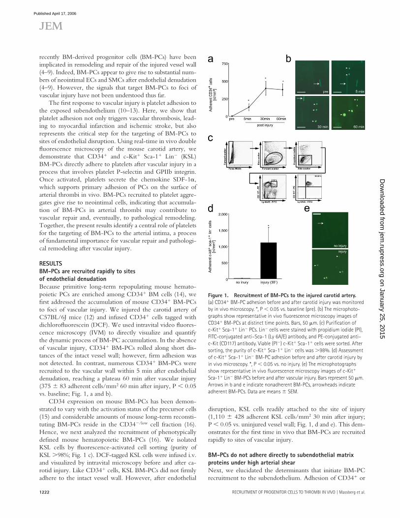
KSL BM-PCs was monitored in a parallel plate fl ow cham-ber at a wall shear rate of 1,000 s−1. To our surprise, nei-ther vitronectin, nor fi bronectin, fi brinogen, or collagen, the major constituents of extracellular matrices exposed at sites of vascular injury, promoted considerable BM-PC adhesion under fl ow (Fig. 2 a). Correspondingly, fl ow cytometric ex-amination of mouse CD34+ or KSL BM-PCs failed to dem-onstrate expression of adhesion molecules that would allow adhesion of fl owing cells to the subendothelial matrix under arterial shear conditions, such as GPIb-V-IX or GPVI (Fig. 2 b). However, as reported previously by others, α4 inte-grin (CD49d) and P-selectin glycoprotein ligand (PSGL)-1
were the major adhesion receptors present on the surface of CD34+ and KSL BM-PCs (17, 18) (Fig. 2 b). PSGL-1 and α4 integrin mediate PC homing to the endothelial lin-ing of BM microvessels; however, they are not suffi cient to promote cellular adhesion to subendothelial matrices. Col-lectively, the latter results imply that BM-PCs are not able to adhere directly to the exposed extracellular matrix after vascular injury.
Platelets interact with BM-PCs in vitro and at sites
of vascular injury in vivo
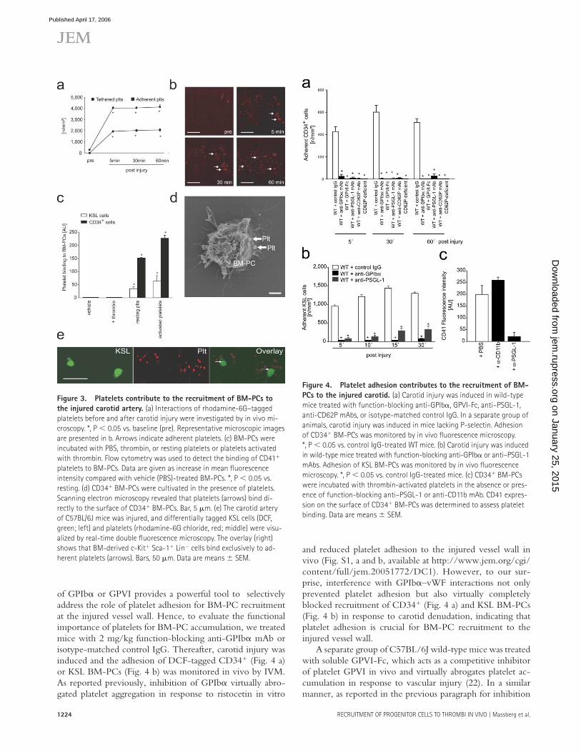
One of the fi rst responses to vascular injury is platelet adhe-sion and aggregation at the site of endothelial denudation (Fig. 3, a and b) (11, 12, 19, 20). To evaluate the potential role of platelets for the recruitment of PCs to the injured ves-sel wall, we investigated whether BM-PCs are able to inter-act with platelets. Using fl ow cytometry, we observed that resting and particularly thrombin-activated platelets bind to BM-derived CD34+ and KSL cells in vitro (Fig. 3 c). Scan-ning electron microscopy revealed that platelets interact di-rectly with BM-PCs (Fig. 3 d).
To identify whether similar interactions also occur under physiological shear conditions, mouse platelets were allowed to adhere to collagen-coated coverslips and the adhesion of CD34+ or KSL BM-PCs was monitored in a parallel plate fl ow chamber at a wall shear rate of 1,000 s−1 as described in previous paragraphs (Fig. 2 a). BM-PCs did not attach to collagen-coated coverslips in the absence of platelets (Fig. 2 a, Col). However, when the collagen matrix was covered with platelets, BM-PCs readily adhered in the presence of arterial shear conditions (Fig. 2 a, Plts).
To fi nd out whether similar interactions between BM-PCs and platelets are present in vivo, we injured the carotid artery of mice. Diff erentially tagged KSL cells (DCF) and platelets (rhodamine-6G) were infused i.v. Platelet–KSL in-teractions at the site of the vascular lesion were monitored by real-time double fl uorescence IVM. Although platelet adhe-sion occurred within seconds after endothelial disruption, KSL BM-PCs started to adhere to the injured vessel wall 5–10 min after induction of vascular injury. Interestingly, we observed that KSL BM-PCs bound exclusively to adherent platelets, whereas no adherent KSL cells were detected at ar-eas devoid of platelet adhesion (Fig. 3 e).
Platelet adhesion is critically involved in BM-PC recruitment
after vascular injury
Because these fi ndings were very suggestive, we subse-quently addressed whether the presence of adherent platelets might actually be mandatory for the recruitment of BM-PCs. Platelet adhesion to the subendothelium involves the platelet vWF receptor GPIbα and the major platelet collagen receptor GPVI (10–12, 21). Correspondingly, inhibition of platelet GPIbα or GPVI by function-blocking mAb almost completely prevents platelet adhesion at sites of vascular in-jury (12). Because the platelet adhesion receptors GPIbα or GPVI are not expressed by BM-PCs (Fig. 2 b), inhibition
Figure 2. BM-PCs do not adhere directly to subendothelial matrix
proteins under arterial shear conditions. (a) The adhesion of BM-PCs
(2 × 10⁴/ml KSL cells or CD34+ cells) to coverslips coated with vitronectin
(Vn; Becton Dickinson), collagen (Col; Becton Dickinson), fi brinogen (Fb;
Sigma-Aldrich), fi bronectin (Fn; Sigma-Aldrich), or to surface adherent
platelets was assessed in a transparent fl ow chamber at a wall shear rate
of 1,000 s−1 as described elsewhere (reference 22). The number of adher-
ent BM-PCs is given per mm2 surface. *, P < 0.05 vs. collagen. (b) To de-
termine the adhesion receptors present on the surface of BM-PCs, KSL or
CD34+ cells were incubated with fl uorophore-labeled anti-αIIb, anti-α4,
anti–PSGL-1, anti-α2β1, anti-GPIbα, or anti-GPVI mAb, or irrelevant iso-
type-matched control antibody and directly analyzed on a FACSCalibur.
Data are given as difference between the mean fl uorescence intensity
[arbitrary units] obtained with the specifi c antibodies and the signal ob-
tained with the corresponding irrelevant isotype-matched control antibody.
Data are means ± SEM.
on January 25, 2015jem
.rupress.orgD
ownloaded from
Published April 17, 2006
1224 RECRUITMENT OF PROGENITOR CELLS TO THROMBI IN VIVO | Massberg et al.
of GPIbα or GPVI provides a powerful tool to selectively address the role of platelet adhesion for BM-PC recruitment at the injured vessel wall. Hence, to evaluate the functional importance of platelets for BM-PC accumulation, we treated mice with 2 mg/kg function-blocking anti-GPIbα mAb or isotype-matched control IgG. Thereafter, carotid injury was induced and the adhesion of DCF-tagged CD34+ (Fig. 4 a) or KSL BM-PCs (Fig. 4 b) was monitored in vivo by IVM. As reported previously, inhibition of GPIbα virtually abro-gated platelet aggregation in response to ristocetin in vitro
and reduced platelet adhesion to the injured vessel wall in vivo (Fig. S1, a and b, available at http://www.jem.org/cgi/content/full/jem.20051772/DC1). However, to our sur-prise, interference with GPIbα–vWF interactions not only prevented platelet adhesion but also virtually completely blocked recruitment of CD34+ (Fig. 4 a) and KSL BM-PCs (Fig. 4 b) in response to carotid denudation, indicating that platelet adhesion is crucial for BM-PC recruitment to the injured vessel wall.
A separate group of C57BL/6J wild-type mice was treated with soluble GPVI-Fc, which acts as a competitive inhibitor of platelet GPVI in vivo and virtually abrogates platelet ac-cumulation in response to vascular injury (22). In a similar manner, as reported in the previous paragraph for inhibition
Figure 3. Platelets contribute to the recruitment of BM-PCs to
the injured carotid artery. (a) Interactions of rhodamine-6G–tagged
platelets before and after carotid injury were investigated by in vivo mi-
croscopy. *, P < 0.05 vs. baseline (pre). Representative microscopic images
are presented in b. Arrows indicate adherent platelets. (c) BM-PCs were
incubated with PBS, thrombin, or resting platelets or platelets activated
with thrombin. Flow cytometry was used to detect the binding of CD41+
platelets to BM-PCs. Data are given as increase in mean fl uorescence
intensity compared with vehicle (PBS)-treated BM-PCs. *, P < 0.05 vs.
resting. (d) CD34+ BM-PCs were cultivated in the presence of platelets.
Scanning electron microscopy revealed that platelets (arrows) bind di-
rectly to the surface of CD34+ BM-PCs. Bar, 5 μm. (e) The carotid artery
of C57BL/6J mice was injured, and differentially tagged KSL cells (DCF,
green; left) and platelets (rhodamine-6G chloride, red; middle) were visu-
alized by real-time double fl uorescence microscopy. The overlay (right)
shows that BM-derived c-Kit+ Sca-1+ Lin− cells bind exclusively to ad-
herent platelets (arrows). Bars, 50 μm. Data are means ± SEM.
Figure 4. Platelet adhesion contributes to the recruitment of BM-
PCs to the injured carotid. (a) Carotid injury was induced in wild-type
mice treated with function-blocking anti-GPIbα, GPVI-Fc, anti–PSGL-1,
anti-CD62P mAbs, or isotype-matched control IgG. In a separate group of
animals, carotid injury was induced in mice lacking P-selectin. Adhesion
of CD34+ BM-PCs was monitored by in vivo fl uorescence microscopy.
*, P < 0.05 vs. control IgG-treated WT mice. (b) Carotid injury was induced
in wild-type mice treated with function-blocking anti-GPIbα or anti–PSGL-1
mAbs. Adhesion of KSL BM-PCs was monitored by in vivo fl uorescence
microscopy. *, P < 0.05 vs. control IgG-treated mice. (c) CD34+ BM-PCs
were incubated with thrombin-activated platelets in the absence or pres-
ence of function-blocking anti–PSGL-1 or anti-CD11b mAb. CD41 expres-
sion on the surface of CD34+ BM-PCs was determined to assess platelet
binding. Data are means ± SEM.
on January 25, 2015jem
.rupress.orgD
ownloaded from
Published April 17, 2006
JEM VOL. 203, May 15, 2006 1225
ARTICLE
of GPIbα, blockade of GPVI abrogated the recruitment of precursor cells in response to carotid denudation (Fig. 4 a). These data demonstrate for the fi rst time that platelet adhe-sion is strictly required for targeting of CD34+ and KSL BM-PCs to foci of vascular injury in vivo.
Platelets promote BM-PC recruitment after vascular injury
by expression of P-selectin
Upon adhesion, platelets become activated and expose P- selectin. CD34+ and KSL BM-PCs express PSGL-1 (Fig. 2 b), the major ligand of P-selectin, and roll on recombinant P-selectin in vitro (Fig. S2, available at http://www.jem.org/cgi/content/full/jem.20051772/DC1). Hence, we next evaluated the signifi cance of P-selectin and PSGL-1 for BM-PC–platelet interactions. We incubated activated platelets with CD34+ BM-PCs in the presence of function-blocking anti–mouse PSGL-1 mAb 4RA10 (23). Notably, platelet binding to BM-PCs was greatly reduced after inhibition of PSGL-1 in vitro, as determined by fl ow cytometry (Fig. 4 c). To further substantiate the role of P-selectin and PSGL-1 for BM-PC recruitment in vivo, CD34+ or KSL BM-PCs were transfused into mice pretreated with mAbs directed against P-selectin or PSGL-1. Inhibition of P-selectin or PSGL-1 signifi cantly reduced the recruitment of BM- derived CD34+ (Fig. 4 a) or KSL BM-PCs (Fig. 4 b) after carotid injury. Similarly, BM-PC accumulation to the in-jured vessel walls was signifi cantly reduced in P-selectin– defi cient mice (Fig. 4 a).
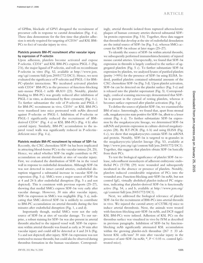
Platelets mediate BM-PC recruitment by release of SDF-1𝛂Recently, the CXC chemokine SDF-1α has been implicated in attracting blood-borne PCs to the vascular intima (24, 25). Hence, we asked whether SDF-1α might contribute to PC accumulation on arterial thrombi at sites of vascular injury. First, we evaluated the distribution of SDF-1α in the vessel wall in response to endothelial denudation. Although SDF-1α was not detected in intact carotid arteries, endothelial dis-ruption triggered a substantial increase in vascular SDF-1α expression (Fig. 5 a). SMCs were a major source of SDF-1α at 4 and 24 h after endothelial disruption (Fig. 5 a and not depicted). This is consistent with previous reports (25–27), showing that medial SMCs express SDF-1α very early after vascular damage. However, 30 min after vascular injury, SDF-1α expression in SMCs was negligible (Fig. 5 a), indi-cating that SMC-derived SDF-1α is unlikely to contribute to BM-PC accumulation on arterial thrombi during the fi rst minutes after endothelial disruption (Fig. 1, a and d).
Importantly though, medial SMCs were not the only source of SDF-1α at sites of vascular damage. To our sur-prise, a robust staining for SDF-1α was also present in arterial thrombi attached to the injured vessel wall. SDF-1α expres-sion within arterial thrombi was found as early as 30 min after vascular injury and could still be detected at 4 and 24 h (Fig. 5 a and not depicted) after injury. SDF-1α expression was not restricted to mouse thrombi, but could also be observed during thrombus formation in the human vasculature. Correspond-
ingly, arterial thrombi isolated from ruptured atherosclerotic plaques of human coronary arteries showed substantial SDF-1α protein expression (Fig. 5 b). Together, these data suggest that thrombi that develop at the site of endothelial denudation are the initial source of SDF-1α (Fig. 5 a), whereas SMCs ac-count for SDF-1α release at later stages (25–27).
To identify the source of SDF-1α within arterial thrombi, we subsequently performed immunohistochemistry of injured mouse carotid arteries. Unexpectedly, we found that SDF-1α expression in thrombi is largely confi ned to the surface of ag-gregated platelets (Fig. 5 c). To further substantiate SDF-1α expression by platelets, we analyzed lysates of purifi ed platelets (purity >99%) for the presence of SDF-1α using ELISA. In-deed, purifi ed platelets contained substantial amounts of the CXC chemokine SDF-1α (Fig. 5 d). Upon platelet activation, SDF-1α can be detected on the platelet surface (Fig. 5 e) and is released into the platelet supernatant (Fig. 5). Correspond-ingly, confocal scanning microscopy demonstrated that SDF-1α is present in the cytosol of resting platelets, whereas it becomes surface expressed after platelet activation (Fig. 5 g).
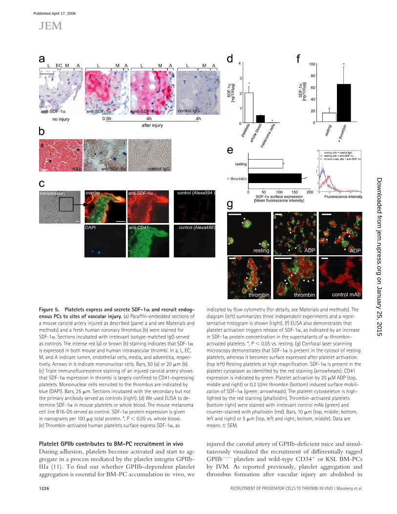
To defi ne the source of platelet SDF-1α, we examined the BM of mice. Interestingly, we found that, apart from stromal cells, megakaryocytes stain positive for SDF-1α, albeit to a lesser extent (Fig. 6 a). To further substantiate SDF-1α expres-sion by the megakaryocytic lineage, we determined SDF-1α mRNA and protein expression in culture-derived megakary-ocytes (28). By RT-PCR (Fig. 6 b) and using ELISA (Fig. 6 c), we show that megakaryocytes contain SDF-1α mRNA and protein. Notably, SDF-1α is targeted to α-granules in the megakaryocytic/platelet lineage (Fig. S3, available at http://www.jem.org/cgi/content/full/jem.20051772/DC1). Together, this suggests that platelets obtain SDF-1α basically from their PCs.
To test the biological signifi cance of platelet SDF-1α re-lease, subconfl uent monolayers of adherent embryonic endo-thelial PCs (T17B) (29) were wounded and subsequently incubated in the absence or presence of platelets. Notably, platelets induced considerable migration of PCs into the wounded area. Function-blocking anti–SDF-1α mAb, but not control IgG, virtually abolished platelet-induced PC migra-tion, indicating that platelet-derived SDF-1α is functionally active (Fig. S4, a and b, available at http://www.jem.org/cgi/content/full/jem.20051772/DC1).
Next, we addressed the biological relevance of platelet SDF-1α for the recruitment of BM-PCs into arterial thrombi in vivo. We injured the carotid artery of C57BL/6J mice to induce arterial thrombosis. Next, the animals were treated with function-blocking anti–SDF-1α mAb, and DCF-tagged KSL BM-PCs were infused. Adhesion of KSL PCs on the thrombus surface was visualized in vivo by IVM as described in previous paragraphs. Inhibition of SDF-1α by function-blocking mAb signifi cantly attenuated KSL accumulation within the growing platelet-rich thrombus (267 ± 33 ad-herent KSL cells/mm2 30 min after vascular injury in the presence of anti–SDF-1α mAb; *, P < 0.05 vs. control IgG-treated mice).
on January 25, 2015jem
.rupress.orgD
ownloaded from
Published April 17, 2006
1226 RECRUITMENT OF PROGENITOR CELLS TO THROMBI IN VIVO | Massberg et al.
Platelet GPIIb contributes to BM-PC recruitment in vivo
During adhesion, platelets become activated and start to ag-gregate in a process mediated by the platelet integrin GPIIb-IIIa (11). To fi nd out whether GPIIb-dependent platelet aggregation is essential for BM-PC accumulation in vivo, we
injured the carotid artery of GPIIb-defi cient mice and simul-taneously visualized the recruitment of diff erentially tagged GPIIb−/− platelets and wild-type CD34+ or KSL BM-PCs by IVM. As reported previously, platelet aggregation and thrombus formation after vascular injury are abolished in
Figure 5. Platelets express and secrete SDF-1𝛂 and recruit endog-
enous PCs to sites of vascular injury. (a) Paraffi n-embedded sections of
a mouse carotid artery injured as described (panel a and see Materials and
methods) and a fresh human coronary thrombus (b) were stained for
SDF-1α. Sections incubated with irrelevant isotype-matched IgG served
as controls. The intense red (a) or brown (b) staining indicates that SDF-1α
is expressed in both mouse and human intravascular thrombi. In a, L, EC,
M, and A indicate lumen, endothelial cells, media, and adventitia, respec-
tively. Arrows in b indicate mononuclear cells. Bars, 50 (a) or 20 μm (b).
(c) Triple immunofl uorescence staining of an injured carotid artery shows
that SDF-1α expression in thrombi is largely confi ned to CD41-expressing
platelets. Mononuclear cells recruited to the thrombus are indicated by
blue (DAPI). Bars, 25 μm. Sections incubated with the secondary but not
the primary antibody served as controls (right). (d) We used ELISA to de-
termine SDF-1α in mouse platelets or whole blood. The mouse melanoma
cell line B16-D5 served as control. SDF-1α protein expression is given
in nanograms per 100 μg total protein. *, P < 0.05 vs. whole blood.
(e) Thrombin-activated human platelets surface express SDF-1α, as
indicated by fl ow cytometry (for details, see Materials and methods). The
diagram (left) summarizes three independent experiments and a repre-
sentative histogram is shown (right). (f) ELISA also demonstrates that
platelet activation triggers release of SDF-1α, as indicated by an increase
in SDF-1α protein concentration in the supernatants of α-thrombin–
activated platelets. *, P < 0.05 vs. resting. (g) Confocal laser scanning
microscopy demonstrates that SDF-1α is present in the cytosol of resting
platelets, whereas it becomes surface expressed after platelet activation.
(top left) Resting platelets at high magnifi cation. SDF-1α is present in the
platelet cytoplasm as identifi ed by the red staining (arrowheads); CD41
expression is indicated by green. Platelet activation by 20 μM ADP (top,
middle and right) or 0.2 U/ml thrombin (bottom) induced surface mobili-
zation of SDF-1α (green; arrowheads). The platelet cytoskeleton is high-
lighted by the red staining (phalloidin). Thrombin-activated platelets
(bottom right) were stained with irrelevant control mAb (green) and
counter-stained with phalloidin (red). Bars, 10 μm (top, middle; bottom,
left and right) or 5 μm (top, left and right; bottom, middle). Data are
means ± SEM.
on January 25, 2015jem
.rupress.orgD
ownloaded from
Published April 17, 2006
JEM VOL. 203, May 15, 2006 1227
ARTICLE
mice lacking GPIIb integrin (13). Remarkably, the defect in GPIIb-dependent platelet aggregation translated into a pro-found and signifi cant reduction in CD34+ BM-PC accumu-lation compared with wild-type animals (P < 0.05; Fig. 7 a). Likewise, adhesion of KSL PCs was signifi cantly attenuated in mice lacking GPIIb integrin (128 ± 16 or 964 ± 393 ad-herent KSL cells/mm2 15 min after vascular injury in GPIIb-defi cient or wild-type mice, respectively).
During aggregation, clustering of GPIIb-IIIa transduces inward signals that may aff ect important platelet functions including the release of chemokines (30, 31). Hence, we next asked whether, beyond promoting platelet aggregation, GPIIb-IIIa might contribute to platelet SDF-1α exposure. Platelets were incubated with soluble fi brinogen in the pres-ence or absence of bivalent mAb antifi brinogen (IgG1), which enhances receptor cross-linking (Fig. 7 b). Interestingly, SDF-1α release was signifi cantly increased when GPIIb-IIIa
was clustered by combined fi brinogen/antifi brinogen incu-bation. Similarly, mouse anti-GPIIb mAb 7E3 signifi cantly increased SDF-1α release in the presence of anti–mouse mAb (IgG) that binds mAb 7E3. This indicates that engagement of GPIIb during platelet aggregation by itself initiates platelet SDF-1α release. Correspondingly, the expression of SDF-1α
Figure 6. Megakaryocytes express SDF-1𝛂. (a) Paraffi n-embedded
sections of mouse femura were stained with anti–SDF-1α. SDF-1α was
expressed not only in stromal cells (arrows) but also in megakaryocytes
(arrowheads). Bars, 25 μm. (b) Megakaryocytic and platelet SDF-1α mRNA
expression was determined using RT-PCR. The mRNA expression of GPIIb
integrin and β-actin served as controls. The primer sequences and their
corresponding product sizes and annealing temperatures are in Table S1.
(c) ELISA was used to determine SDF-1α in mouse megakaryocytes or
mouse BM. SDF-1α protein expression is given in nanograms per 100 μg
total protein. Data are means ± SEM.
Figure 7. GPIIb-dependent platelet aggregation promotes BM-PC
recruitment during arterial thrombosis in vivo and contributes to
SDF-1𝛂 release in vitro. (a) To fi nd out whether platelet GPIIb contrib-
utes to BM-PC recruitment in vivo, carotid injury was induced in mice
lacking GPIIb. Adhesion of CD34+ BM-PCs was monitored by in vivo fl uo-
rescence microscopy. *, P < 0.05 vs. wild type. (b) Platelets were incu-
bated with medium, thrombin, 100 μg/ml soluble fi brinogen, 5 μg/ml
mAb anti-fi brinogen, or a combination of both for 30 min. Platelet SDF-1α
release was determined by ELISA as described in Materials and methods.
Similar experiments were performed with anti-GPIIb mAb 7E3 (whole IgG,
5 μg/ml) in the presence or absence of 5 μg/ml mAb goat anti–mouse
IgG. *, P < 0.05 vs. resting platelets. (c) The carotid artery of wild-type
(WT) or GPIIb-defi cient mice was injured as described in Materials and
methods. Paraffi n-embedded sections were stained for SDF-1α. L, M, and
A indicate lumen, media, and adventitia, respectively. Arrows identify
SDF-1α deposited at the site of vascular injury. Bars, 20 μm. Data are
means ± SEM.
on January 25, 2015jem
.rupress.orgD
ownloaded from
Published April 17, 2006
1228 RECRUITMENT OF PROGENITOR CELLS TO THROMBI IN VIVO | Massberg et al.
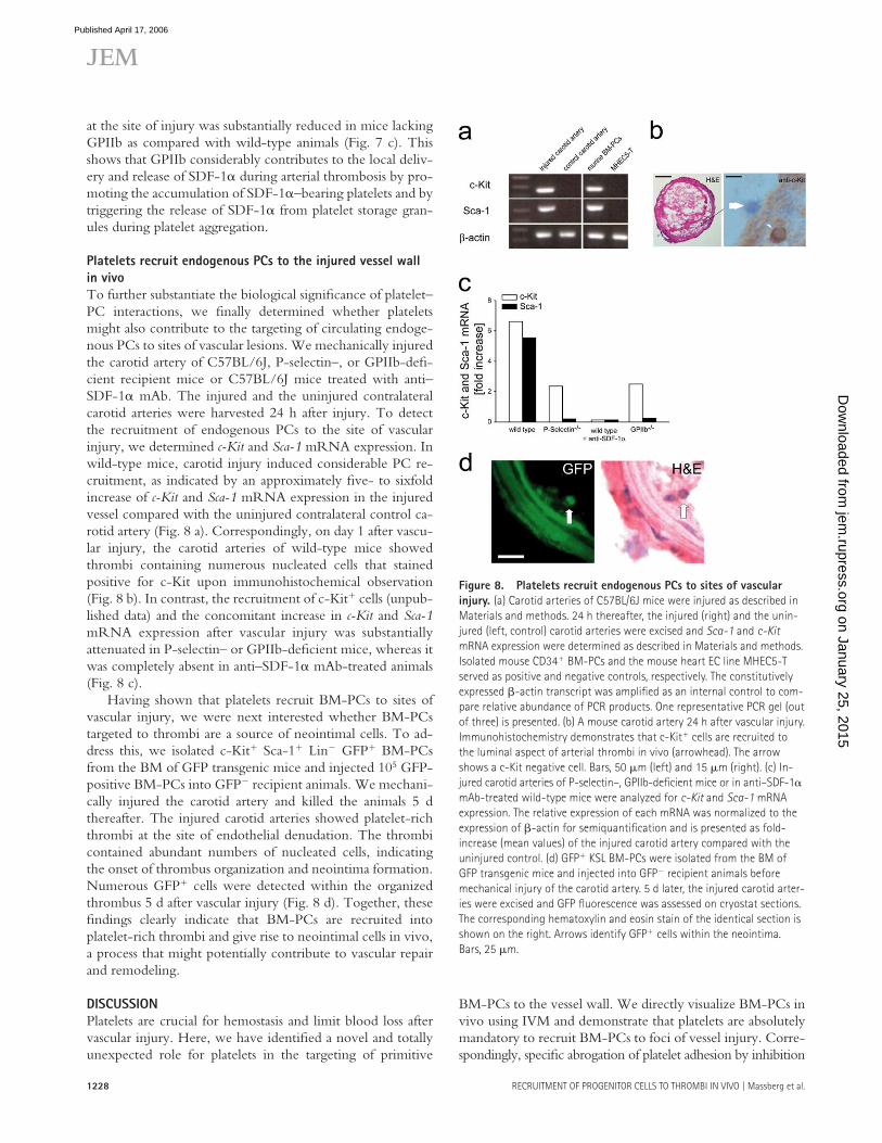
at the site of injury was substantially reduced in mice lacking GPIIb as compared with wild-type animals (Fig. 7 c). This shows that GPIIb considerably contributes to the local deliv-ery and release of SDF-1α during arterial thrombosis by pro-moting the accumulation of SDF-1α–bearing platelets and by triggering the release of SDF-1α from platelet storage gran-ules during platelet aggregation.
Platelets recruit endogenous PCs to the injured vessel wall
in vivo
To further substantiate the biological signifi cance of platelet–PC interactions, we fi nally determined whether platelets might also contribute to the targeting of circulating endoge-nous PCs to sites of vascular lesions. We mechanically injured the carotid artery of C57BL/6J, P-selectin–, or GPIIb-defi -cient recipient mice or C57BL/6J mice treated with anti–SDF-1α mAb. The injured and the uninjured contralateral carotid arteries were harvested 24 h after injury. To detect the recruitment of endogenous PCs to the site of vascular injury, we determined c-Kit and Sca-1 mRNA expression. In wild-type mice, carotid injury induced considerable PC re-cruitment, as indicated by an approximately fi ve- to sixfold increase of c-Kit and Sca-1 mRNA expression in the injured vessel compared with the uninjured contralateral control ca-rotid artery (Fig. 8 a). Correspondingly, on day 1 after vascu-lar injury, the carotid arteries of wild-type mice showed thrombi containing numerous nucleated cells that stained positive for c-Kit upon immunohistochemical observation (Fig. 8 b). In contrast, the recruitment of c-Kit+ cells (unpub-lished data) and the concomitant increase in c-Kit and Sca-1 mRNA expression after vascular injury was substantially attenuated in P-selectin– or GPIIb-defi cient mice, whereas it was completely absent in anti–SDF-1α mAb-treated animals (Fig. 8 c).
Having shown that platelets recruit BM-PCs to sites of vascular injury, we were next interested whether BM-PCs targeted to thrombi are a source of neointimal cells. To ad-dress this, we isolated c-Kit+ Sca-1+ Lin− GFP+ BM-PCs from the BM of GFP transgenic mice and injected 105 GFP-positive BM-PCs into GFP− recipient animals. We mechani-cally injured the carotid artery and killed the animals 5 d thereafter. The injured carotid arteries showed platelet-rich thrombi at the site of endothelial denudation. The thrombi contained abundant numbers of nucleated cells, indicating the onset of thrombus organization and neointima formation. Numerous GFP+ cells were detected within the organized thrombus 5 d after vascular injury (Fig. 8 d). Together, these fi ndings clearly indicate that BM-PCs are recruited into platelet-rich thrombi and give rise to neointimal cells in vivo, a process that might potentially contribute to vascular repair and remodeling.
D I S C U S S I O N
Platelets are crucial for hemostasis and limit blood loss after vascular injury. Here, we have identifi ed a novel and totally unexpected role for platelets in the targeting of primitive
BM-PCs to the vessel wall. We directly visualize BM-PCs in vivo using IVM and demonstrate that platelets are absolutely mandatory to recruit BM-PCs to foci of vessel injury. Corre-spondingly, specifi c abrogation of platelet adhesion by inhibition
Figure 8. Platelets recruit endogenous PCs to sites of vascular
injury. (a) Carotid arteries of C57BL/6J mice were injured as described in
Materials and methods. 24 h thereafter, the injured (right) and the unin-
jured (left, control) carotid arteries were excised and Sca-1 and c-Kit
mRNA expression were determined as described in Materials and methods.
Isolated mouse CD34+ BM-PCs and the mouse heart EC line MHEC5-T
served as positive and negative controls, respectively. The constitutively
expressed β-actin transcript was amplifi ed as an internal control to com-
pare relative abundance of PCR products. One representative PCR gel (out
of three) is presented. (b) A mouse carotid artery 24 h after vascular injury.
Immunohistochemistry demonstrates that c-Kit+ cells are recruited to
the luminal aspect of arterial thrombi in vivo (arrowhead). The arrow
shows a c-Kit negative cell. Bars, 50 μm (left) and 15 μm (right). (c) In-
jured carotid arteries of P-selectin–, GPIIb-defi cient mice or in anti–SDF-1α
mAb-treated wild-type mice were analyzed for c-Kit and Sca-1 mRNA
expression. The relative expression of each mRNA was normalized to the
expression of β-actin for semiquantifi cation and is presented as fold-
increase (mean values) of the injured carotid artery compared with the
uninjured control. (d) GFP+ KSL BM-PCs were isolated from the BM of
GFP transgenic mice and injected into GFP− recipient animals before
mechanical injury of the carotid artery. 5 d later, the injured carotid arter-
ies were excised and GFP fl uorescence was assessed on cryostat sections.
The corresponding hematoxylin and eosin stain of the identical section is
shown on the right. Arrows identify GFP+ cells within the neointima.
Bars, 25 μm.
on January 25, 2015jem
.rupress.orgD
ownloaded from
Published April 17, 2006
JEM VOL. 203, May 15, 2006 1229
ARTICLE
of GPVI or GPIbα, both of which are not expressed by BM-PCs, virtually abolished the accumulation of PCs at sites of endothelial disruption. We provide evidence that platelets attract BM-PCs to the injured vessel wall in a multi-step pro-cess involving the following: (a) surface exposure of platelet P-selectin; (b) GPIIb-dependent platelet aggregate forma-tion; and, highly unexpected, (c) the release of the CXC che-mokine SDF-1α by activated platelets (Fig. 9). Together, the present study identifi es the mechanism underlying the selec-tive recruitment of circulating PCs to the vascular intima, a process that plays a fundamental role for the repair and regen-eration of vascular tissue after vessel injury, but also during cardiovascular development (1, 2).
Platelet P-selectin and SDF-1𝛂 act in concert to recruit PCs
to the injured vessel wall in vivo
P-selectin expressed on the platelet surface has previously been involved in the accumulation of mature leukocytes, in-cluding monocytes, neutrophils, and lymphocytes (32, 33). Here, we provide direct in vivo evidence that platelet P-se-lectin is also central for the targeting of primitive precursor cells to foci of vascular injury. Correspondingly, inhibition or loss of P-selectin or its ligand PSGL-1 drastically reduced BM-PC accumulation on the surface of adherent/aggregated platelets in vivo. Notably, platelet aggregation and arterial thrombus formation were not aff ected by lack or inhibition of P-selectin, indicating that the decrease of BM-PC accu-
mulation was not the result of a loss of platelet accumulation at the site of vessel injury.
Although P-selectin is required for initial tethering/roll-ing of BM-PCs, the subsequent fi rm arrest of BM-PCs is regulated by chemokines. In particular, the CXC chemokine SDF-1α, which is critical for mobilization and homing of BM-PCs (34), is required to arrest PCs under fl ow in vitro (35). Only recently, SDF-1α has been involved in the pro-cess of vascular remodeling and intimal hyperplasia (24) by inducing recruitment of PCs to the vascular wall. Originally, SMCs cells have been identifi ed as the major resource of SDF-1α in both atherosclerotic lesions (27) and after vascular injury (25, 26). Here, we have identifi ed another important source of SDF-1α at sites of vascular lesions. Highly unex-pected, we found that, apart from SMCs, platelets and their megakaryocytic precursors abundantly express SDF-1α. Activated platelets rapidly release SDF-1α from internal stores, which is functionally active and promotes the migra-tion of primitive PCs in vitro. Thus, in addition to SDF-1α expressed by vascular SMCs, platelet recruitment during ar-terial thrombosis is associated with a substantial and rapid rise in the local concentrations of SDF-1α released into the mi-croenvironment of sites of vascular injury.
SDF-1α deposited by activated platelets and SDF-1α re-leased by injured SMCs (25) appear to act in a sequential manner. Correspondingly, we show here that, 30 min after vascular injury, the platelet-rich thrombus is the major source
Figure 9. Adherent platelets recruit BM-PCs to injured vessel wall.
Within minutes after vessel injury, platelets adhere to the exposed suben-
dothelium in a process involving GPVI and GPIbα-IX. Adherent/activated
platelets surface express P-selectin and release SDF-1α after engagement
of GPIIb integrin, thereby initiating BM-PC recruitment within minutes
after vessel injury. Within the ensuing hours and days after endothelial
disruption, apoptotic SMCs appear to account for the long-term SDF-1α
release (reference 25). Hence, SDF-1α delivered by platelets early on and
by SMCs at later stages act in concert to promote the entry of BM-PCs at
sites of vascular damage.
on January 25, 2015jem
.rupress.orgD
ownloaded from
Published April 17, 2006
1230 RECRUITMENT OF PROGENITOR CELLS TO THROMBI IN VIVO | Massberg et al.
of SDF-1α at the site of vascular injury, whereas 4 h after vascular injury, SDF-1α is abundantly expressed in both SMCs and the thrombus. This is consistent with a recent re-port showing that SDF-1α expression in vascular SMCs oc-curs within 6–24 h after vascular injury, whereas it is negligible within the fi rst hours after endothelial disruption (25). We show here that inhibition of SDF-1α signifi cantly reduces BM-PC adhesion to the growing thrombus as early as 5–30 min after vascular injury. Hence, it is tempting to speculate that platelets constitute the initial and presumably more short-lived source of SDF-1α that fi rst directs BM-PCs to the site of vessel damage. In contrast, SMCs appear to account for the long-term SDF-1α release over the ensuing days and weeks after vessel injury required to sustain the process of vascular remodeling and repair (24, 25). The proposed temporal rela-tionship of SDF-1α release by platelets and SMCs, and their relative contributions to the recruitment of BM-PCs at sites of vessel injury is illustrated in Fig. 9.
GPIIb-dependent platelet aggregation modulates
the recruitment of PCs to the injured vessel wall
We report here that GPIIb-dependent platelet aggregation not only initiates arterial thrombosis (13), but also plays a fundamental role for the recruitment of primitive PCs at sites of vascular injury. GPIIb may directly promote adhesion of BM-PCs into the growing thrombus by forming a link be-tween platelets and PCs using a GPIIb-dependent bridging mechanism previously reported to occur during platelet–EC interactions (36). Moreover, GPIIb might also operate indi-rectly in that cross-linking of platelets through GPIIb forms a structure in which PCs get trapped. Finally, we report here that engagement of GPIIb during platelet aggregation initi-ates platelet SDF-1α release. Hence, distinct GPIIb-depen-dent pathways are likely to act sequentially to promote BM-PC recruitment at sites of vessel injury.
BM-PCs recruited to arterial thrombi give rise
to neointimal cells
There is increasing evidence that KSL or CD34+ BM-PCs give rise to ECs and SMCs in the vascular (neo-) intima in response to vessel injury (4, 6–8). Here, we demonstrate that GFP+ BM-PCs recruited to platelet-rich thrombi grow into neointimal cells. Notably, it has been reported recently that platelet-derived growth factors, including PDGF-BB and VEGF, may modulate PC diff erentiation in vitro (7, 37). We show here that, apart from growth factors, activated platelets release the CXC chemokine SDF-1α. Beyond its role for PC recruitment to sites of vessel injury, SDF-1α has been dem-onstrated to support BM-PC survival and proliferation (38). In addition, SDF-1α appears to be involved in the diff eren-tiation of c-Kit+ cells into endothelial precursors (39). Although we did not defi ne the progeny of KSL or CD34+ BM-PCs recruited to thrombi in vivo, it is tempting to spec-ulate, based on the fi ndings outlined here, that platelets estab-lish a microenvironment permissive for BM-PC proliferation and early diff erentiation after vascular injury in vivo.
To conclude, PCs normally circulate within the blood in a “surveillance mode,” encountering only brief contacts with the vascular endothelium. Requirement of tissue renewal or tissue repair is communicated to circulating PCs by adher-ent/aggregated platelets. We propose that platelet-dependent BM-PC recruitment is essential for vascular repair and local wound healing and/or tissue regeneration at sites of vessel damage. However, it may also contribute to undesirable PC recruitment and proliferation in the process of neointima formation after vascular injury and during atheroprogression (4, 6, 9).
MATERIALS AND METHODSAnimals. C57BL/6J mice were purchased from Charles River Laboratories.
Transgenic C57BL/6J mice ubiquitously expressing enhanced GFP under
the control of the chicken β-actin promoter and CMV enhancer were ob-
tained from The Jackson Laboratory. P-selectin–defi cient mice were gener-
ated and provided by A.L. Beaudet (Baylor College of Medicine, Houston,
TX). GPIIb (αIIb integrin)-defi cient mice were generated as described previ-
ously (40). All experimental procedures performed on animals met the re-
quirements of the German legislation on protection of animals and were
approved by the Government of Bavaria/Germany.
Monoclonal antibodies. 4RA10 (function-blocking anti–mouse PSGL-1)
were raised as described previously (22, 23, 41). Rat anti–mouse GPIbα
mAb was obtained from Emfret Analytics. Function-blocking anti–mouse/
human SDF-1α (clone 79014) was obtained from R&D Systems. Anti-α2β1
mAb (clone: BMA2.1) was obtained from Chemicon. mAb 7E3 (gift from
B. Coller, The Rockefeller University, New York, NY) inhibits fi brinogen
binding to GPIIb-IIIa.
Separation and labeling of CD34+ BM-PCs, KSL BM-PCs, and
platelets. For separation of BM-PCs, mice (C57BL/6J or GFP+) were
killed by an overdose of pentobarbital, and both femura were harvested
from each animal. Single cell suspensions of the BM were obtained by fl ush-
ing the femura with PBS using a 21-gauge needle. CD34+ BM-PCs were
separated as described previously (42). Primitive KSL BM-PCs were purifi ed
according to a previously published protocol (6) (for details, see supplemen-
tal Materials and methods, available at http://www.jem.org/cgi/content/
full/jem.20051772/DC1). For IVM, CD34+ or KSL BM-PCs were fl uores-
cently labeled with 5-carboxyfl uorescein diacetate succinimidyl ester (DCF).
For each experiment, 107 fl uorescent CD34+ BM-PCs or 1.5 × 10⁵ fl uo-
rescent KSL cells were infused intravenously. Mouse platelets were isolated
from whole blood as described previously (12, 19). For IVM, platelets were
tagged with rhodamine-6G or DCF as reported previously (12, 19).
BM-PC adhesion to purifi ed proteins under fl ow conditions. BM-
PCs were isolated as described in the previous paragraph. BM-PC (2 × 10⁴/
ml) adhesion to coverslips coated with BSA, vitronectin (Becton Dickinson),
fi brinogen (Sigma-Aldrich), collagen (Becton Dickinson), or recombinant
mouse P-selectin, generated as described previously (23, 41), was assessed in
a transparent fl ow chamber at a wall shear rate of 1,000 s−1 as described else-
where (22). In a separate set of experiments, platelets (4 × 10⁸/ml) were
allowed to adhere to collagen-coated coverslips before perfusion with
BM-PCs. Where indicated, function-blocking anti–PSGL-1 mAb (5 μg/ml)
was added to the BM-PC suspension.
Characterization of BM-PC adhesion receptors and determination
of platelet-binding by fl ow cytometry. BM-PCs (10 × 103 CD34+ or
KSL cells/ml) resuspended in PBS were incubated with fl uorophore-labeled
anti-CD41 (αIIb integrin, clone: MWReg 30), anti-CD49d (α4 integrin,
clone: 9C10), anti-CD162 (PSGL-1, clone: 2PH1) (all from Becton Dickin-
son), anti-GPIbα (Xia.B2, Emfret) or anti-GPVI mAb (2G3, generated as
on January 25, 2015jem
.rupress.orgD
ownloaded from
Published April 17, 2006
JEM VOL. 203, May 15, 2006 1231
ARTICLE
described (22)), or irrelevant isotype-matched control antibody for 30 min
at room temperature and directly analyzed on a FACSCalibur (Becton
Dickinson). In a separate set of experiments, CD34+ or KSL BM-PCs (2 ×
104/ml) were coincubated with 108/ml resting mouse platelets or platelets
activated with mouse thrombin (2 U/ml, Sigma-Aldrich) for 15 min. Where
indicated function-blocking anti-CD11b (M1/70, BD Biosciences) or anti-
PSGL-1 mAb (4RA10, 5 μg/ml) was added to the cell suspension. PBS-
incubated BM-PCs in the absence or presence of thrombin served as controls.
Thereafter, the samples were incubated with FITC-labeled anti–mouse
CD41 (MW Reg30) for 30 min to detect platelets attached to BM-PCs at
room temperature and directly analyzed.
Assessment of BM-PCs and platelet adhesion and BM-PC–platelet
interaction in response to carotid injury. Platelet and BM-PC (CD34+
or c-Kit+ Sca-1+ Lin−) adhesion dynamics before and after vascular injury
were monitored in vivo by use of IVM. Carotid injury was induced in anes-
thetized C57BL6/J, P-selectin–, or GPIIb-defi cient mice as described previ-
ously (12, 20). Before and after vascular injury, diff erentially fl uorescent-tagged
cells (platelets and BM-PCs) were visualized using a Zeiss Axiotech micro-
scope (20× water immersion objective, W 20×/0.5, Zeiss) with a 100W
HBO mercury lamp for epi-illumination as reported previously (12, 20).
Where indicated, C57BL6/J mice were pretreated with 2 mg/kg body
weight RB40.34 (anti–mouse P-selectin), 4RA10 (anti–mouse PSGL-1), rat
anti–mouse GPIbα, or anti–mouse/human SDF-1α (clone 79014) 5 min
before intravital microscopy (n = 4–6 per group). BM-PC adhesion to the
carotid artery after infusion of irrelevant isotype-matched IgG served as
control (n = 10).For direct visualization of platelet–KSL cell interactions at the site of
vascular lesion diff erentially fl uorescent-tagged cells (rhodamine-tagged
platelets and DCF-tagged KSL BM-PCs) were infused i.v. KSL binding to
adherent platelets was analyzed 5 min after induction of carotid injury by
real-time double fl uorescence IVM using a BX51WI microscope (Olympus),
equipped with an Olympus MT20 monochromator.
Assessment of SDF-1𝛂 expression by immunohistochemistry. To
determine the presence of SDF-1α in megakaryocytes and aggregated plate-
lets, paraffi n-embedded sections of mouse femura, injured mouse carotid
arteries (WT or GPIIb-defi cient, 0.5, 4, and 24 h after endothelial denudation),
and a fresh thrombus isolated from an occluded human coronary artery were
cut into 2-μm sections and incubated with anti–mouse SDF-1α (clone
79018; R&D Systems), or anti–human SDF-1α (polyclonal goat; R&D
Systems), respectively, and stained using APAAP chemMATE or DAB
chemMATE detection kits (both obtained from DakoCytomation). For
immunofl uorescence microscopy, cryostat sections of injured mouse carotid
arteries (24 h after endothelial denudation) were incubated with anti–mouse
CD41 mAb (MW Reg30; BD Biosciences), anti–mouse SDF-1α mAb
(clone 79014; R&D Systems), Alexa488-labeled goat anti–rat IgG, Alexa594-
labeled goat anti–mouse IgG (both obtained from Invitrogen), and DAPI
(D1306; Invitrogen). Fluorescence images were obtained using a Leica
fl uorescence microscope.
Determination of platelet and megakaryocytic SDF-1𝛂 protein and
mRNA expression. Platelet SDF-1α release was determined in vitro by
enzyme-linked immunosorbent assay (R&D Systems). In brief, mouse SDF-
1α standard lysates obtained from isolated resting mouse platelets, whole
blood, or mouse BM cells were incubated in SDF-1α mAb precoated mi-
croplates and reacted with a polyclonal anti–SDF-1α–horseradish peroxidase
antibody (all obtained from R&D Systems). For detection a tetramethyl ben-
zidine peroxidase substrate was used. The mouse melanoma cell line D5
served as a control. Absorbance was read at 450 nm and the background was
corrected. SDF-1α concentrations were calculated with standards and are
adjusted to 100 μg total protein.
To determine SDF-1α expression by the megakaryocytic lineage, we
generated megakaryocytes from mouse CD34+ cells as reported previously
(28) (for details see supplemental Materials and methods). Megakaryocytic
SDF-1α expression was assessed by ELISA as described in the previous para-
graphs. In addition, megakaryocytic SDF-1α mRNA expression was deter-
mined using RT-PCR. The mRNA expression of GPIIb integrin and
β-actin served as controls. The primer sequences and their corresponding
product sizes and annealing temperatures are shown in the Table S1 (avail-
able at http://www.jem.org/cgi/content/full/jem.20051772/DC1).
Assessment of SDF-1𝛂 release upon platelet activation. To determine
the eff ect of platelet activation on SDF-1α release, human platelets (0.5 ×
109 cells/ml) were incubated with 2 U/ml α-thrombin or PBS for 30 min.
To determine the eff ects of GPIIb receptor cross-linking, human platelets
were incubated with 100 μg/ml of soluble fi brinogen (Sigma-Aldrich) in
the presence or absence of 5 μg/ml of bivalent mAb antifi brinogen (2C2-
G7; BD Biosciences), or with 5 μg/ml of mouse anti-GPIIb mAb 7E3 in the
presence or absence of 5 μg/ml of anti–mouse IgG mAb (DakoCytoma-
tion). SDF-1α release into the supernatants of resting or activated platelets
was determined by ELISA as described in previous paragraphs.
To analyze the surface exposure of SDF-1α in response to platelet acti-
vation, resting or thrombin-activated human platelets were incubated with
mouse anti–SDF-1α mAb (R&D Systems) or irrelevant isotype-matched
control antibody for 30 min at room temperature, followed by staining with
FITC-labeled rabbit anti–mouse IgG antibody (Becton Dickinson). The
samples were directly analyzed on a FACSCalibur (Becton Dickinson).
To defi ne the distribution of SDF-1α in resting and activated platelets,
human platelets (2 × 10⁸ cells/ml) were stimulated with 2 U/ml α-throm-
bin, 5 μM ADP, or PBS for 60 min. The platelets were fi xed and stained
with FITC-labeled anti-CD41 (P2; Beckman Coulter), monoclonal (R&D
Systems) or polyclonal anti–SDF-1α antibody (goat; R&D Systems), Alexa
Fluor 488–tagged goat anti–mouse, Alexa 568–tagged rabbit anti–goat,
Alexa Fluor 488–tagged rabbit anti–goat (all obtained from Invitrogen), or
Rhodamine-Phalloidin (R415; Invitrogen) as indicated. Confocal immuno-
fl uorescence analysis was performed using a LSM510 META confocal laser
microscope equipped with the LSM510 META software (Carl Zeiss
MicroImaging, Inc.).
Incorporation of BM-PCs into the neointima. GFP+ CD34+ BM-PCs
or KSL GFP+ CD34+ BM-PCs were isolated from the BM of GFP transgenic
mice as described in previous paragraphs and injected into C57BL/6J recipient
animals (n = 5; 105 cells per experiment). We mechanically injured the carotid
artery. After 5 d, the injured carotid arteries were excised. GFP fl uorescence
was detected on cryostat sections by standard fl uorescence microscopy.
Recruitment of endogenous PCs to the injured carotid artery. To
determine recruitment of endogenous circulating PCs, carotid arteries of
C57BL/6J (n = 3), P-selectin–, (n = 4) or GPIIb-defi cient mice (n = 2) or
in anti–SDF-1α mAb-treated wild-type mice (n = 2) were injured as de-
scribed in previous paragraphs and c-Kit and Sca-1 mRNA expression was
assessed by RT-PCR (see supplemental Materials and methods). The consti-
tutively expressed β-actin transcript was amplifi ed as an internal control to
compare relative abundance of PCR products, and the relative expression of
each mRNA was normalized to the expression of β-actin for semiquantifi ca-
tion. To detect c-Kit positive paraffi n-embedded sections of injured mouse,
carotid arteries (24 h after endothelial denudation) were incubated with
anti–mouse c-Kit mAb (2B8; BD Biosciences) and stained using APAAP
chemMATE mouse detection kit (DakoCytomation).
Statistical analysis. Comparisons between group means were performed
using one-way analysis of variance on Ranks. Data represent mean ± SEM.
A value of P < 0.05 was regarded as signifi cant.
Online supplemental material. Online supplemental Materials and
methods provides additional methodological information in the following
categories: separation of BM-PCs; eff ects of anti-GPIbα mAb on platelet
function; transfection of DAMI cells; scanning electron microscopy; gen-
eration of mouse megakaryocytes; in vitro PC migration; and recruitment
on January 25, 2015jem
.rupress.orgD
ownloaded from
Published April 17, 2006
1232 RECRUITMENT OF PROGENITOR CELLS TO THROMBI IN VIVO | Massberg et al.
of endogenous PCs. Table S1 contains primer sequences, correspond-
ing product sizes, and annealing temperatures. Fig. S1 shows the eff ects of
anti- GPIbα mAb on platelet function. Fig. S2 addresses BM-PC binding
to recombinant P-selectin. Fig. S3 illustrates the subcellular localization of
SDF-1α in megakaryocytes. Fig. S4 investigates the eff ect of platelet SDF-
1α on PC migration. All online supplemental material is available at http://
www.jem.org/cgi/content/full/jem.20051772/DC1.
We are indebted to K. Langenbrink and H. Wehnes for their excellent technical
assistance. Histomorphometry was performed with the skillful help of R. Hegenloh
with the support of Dr. R. Brandl (Br 1583/1-2).
The study was supported by grants from the Bayerische Forschungsstiftung
and the Deutsche Forschungsgemeinschaft DFG (Ma 2186/3-1 to S. Massberg) and
the Wellcome Trust (to N.R. Emambokus, J. Frampton). S. Massberg is a Heisenberg
fellow of the DFG (Ma 2186/4-1). K. Schürzinger and P. Goyal were graduate
students supported by the DFG Graduate Student Program GRK 438.
The authors have no confl icting fi nancial interests.
Submitted: 1 September 2005
Accepted: 27 March 2006
R E F E R E N C E S 1. Carmeliet, P. 2000. Mechanisms of angiogenesis and arteriogenesis.
Nat. Med. 6:389–395. 2. Schwartz, S.M. 1997. Smooth muscle migration in atherosclerosis and
restenosis. J. Clin. Invest. 100:S87–S89. 3. Carmeliet, P., L. Moons, J.M. Stassen, M. De Mol, A. Bouche, J.J. van
den Oord, M. Kockx, and D. Collen. 1997. Vascular wound healing and neointima formation induced by perivascular electric injury in mice. Am. J. Pathol. 150:761–776.
4. Werner, N., J. Priller, U. Laufs, M. Endres, M. Bohm, U. Dirnagl, and G. Nickenig. 2002. Bone marrow-derived progenitor cells modulate vascular reendothelialization and neointimal formation: eff ect of 3-hy-droxy-3-methylglutaryl coenzyme a reductase inhibition. Arterioscler. Thromb. Vasc. Biol. 22:1567–1572.
5. Jiang, S., L. Walker, M. Afentoulis, D.A. Anderson, L. Jauron-Mills, C.L. Corless, and W.H. Fleming. 2004. Transplanted human bone mar-row contributes to vascular endothelium. Proc. Natl. Acad. Sci. USA. 101:16891–16896.
6. Sata, M., A. Saiura, A. Kunisato, A. Tojo, S. Okada, T. Tokuhisa, H. Hirai, M. Makuuchi, Y. Hirata, and R. Nagai. 2002. Hematopoietic stem cells diff erentiate into vascular cells that participate in the patho-genesis of atherosclerosis. Nat. Med. 8:403–409.
7. Simper, D., P.G. Stalboerger, C.J. Panetta, S. Wang, and N.M. Caplice. 2002. Smooth muscle progenitor cells in human blood. Circulation. 106:1199–1204.
8. Shimizu, K., S. Sugiyama, M. Aikawa, Y. Fukumoto, E. Rabkin, P. Libby, and R.N. Mitchell. 2001. Host bone-marrow cells are a source of donor intimal smooth-muscle-like cells in murine aortic transplant arteriopathy. Nat. Med. 7:738–741.
9. Sata, M., K. Tanaka, and R. Nagai. 2003. Origin of smooth muscle progenitor cells: diff erent conclusions from diff erent models. Circulation. 107:e106–e107.
10. Chen, J., and J.A. Lopez. 2005. Interactions of platelets with subendo-thelium and endothelium. Microcirculation. 12:235–246.
11. Ruggeri, Z.M. 1997. Mechanisms initiating platelet thrombus forma-tion. Thromb. Haemost. 78:611–616.
12. Massberg, S., M. Gawaz, S. Gruner, V. Schulte, I. Konrad, D. Zohlnhofer, U. Heinzmann, and B. Nieswandt. 2003. A crucial role of glycoprotein VI for platelet recruitment to the injured arterial wall in vivo. J. Exp. Med. 197:41–49.
13. Massberg, S., K. Schurzinger, M. Lorenz, I. Konrad, C. Schulz, N. Plesnila, E. Kennerknecht, M. Rudelius, S. Sauer, S. Braun, et al. 2005. Platelet adhesion via glycoprotein IIb integrin is critical for atheropro-gression and focal cerebral ischemia. An in vivo study in mice lacking glycoprotein IIb. Circulation. 112:1180–1188.
14. Morel, F., A. Galy, B. Chen, and S.J. Szilvassy. 1998. Equal distribution of competitive long-term repopulating stem cells in the CD34+ and
CD34− fractions of Thy-1low Lin−/low Sca-1+ bone marrow cells. Exp. Hematol. 26:440–448.
15. Sato, T., J.H. Laver, and M. Ogawa. 1999. Reversible expression of CD34 by murine hematopoietic stem cells. Blood. 94:2548–2554.
16. Osawa, M., K. Hanada, H. Hamada, and H. Nakauchi. 1996. Long-term lymphohematopoietic reconstitution by a single CD34-low/nega-tive hematopoietic stem cell. Science. 273:242–245.
17. Mazo, I.B., J.C. Gutierrez-Ramos, P.S. Frenette, R.O. Hynes, D.D. Wagner, and U.H. von Andrian. 1998. Hematopoietic progenitor cell rolling in bone marrow microvessels: parallel contributions by endothe-lial selectins and vascular cell adhesion molecule 1. J. Exp. Med. 188:465–474.
18. Frenette, P.S., S. Subbarao, I.B. Mazo, U.H. von Andrian, and D.D. Wagner. 1998. Endothelial selectins and vascular cell adhesion mol-ecule-1 promote hematopoietic progenitor homing to bone marrow. Proc. Natl. Acad. Sci. USA. 95:14423–14428.
19. Massberg, S., K. Brand, S. Gruner, S. Page, E. Muller, I. Muller, W. Bergmeier, T. Richter, M. Lorenz, I. Konrad, et al. 2002. A critical role of platelet adhesion in the initiation of atherosclerotic lesion formation. J. Exp. Med. 196:887–896.
20. Moers, A., B. Nieswandt, S. Massberg, N. Wettschureck, S. Gruner, I. Konrad, V. Schulte, B. Aktas, M.P. Gratacap, M.I. Simon, et al. 2003. G(13) is an essential mediator of platelet activation in hemostasis and thrombosis. Nat. Med. 9:1418–1422.
21. Cruz, M.A., J. Chen, J.L. Whitelock, L.D. Morales, and J.A. Lopez. 2005. The platelet glycoprotein Ib-von Willebrand factor interaction activates the collagen receptor α2β1 to bind collagen: activation-dependent conformational change of the α2-I domain. Blood. 105:1986–1991.
22. Massberg, S., I. Konrad, A. Bultmann, C. Schulz, G. Munch, M. Peluso, M. Lorenz, S. Schneider, F. Besta, I. Muller, et al. 2004. Soluble gly-coprotein VI dimer inhibits platelet adhesion and aggregation to the injured vessel wall in vivo. FASEB J. 18:397–399.
23. Pendl, G.G., C. Robert, M. Steinert, R. Thanos, R. Eytner, E. Borges, M.K. Wild, J.B. Lowe, R.C. Fuhlbrigge, T.S. Kupper, et al. 2002. Immature mouse dendritic cells enter infl amed tissue, a process that re-quires E- and P-selectin, but not P-selectin glycoprotein ligand 1. Blood. 99:946–956.
24. Schober, A., S. Knarren, M. Lietz, E.A. Lin, and C. Weber. 2003. Crucial role of stromal cell-derived factor-1α in neointima formation after vascular injury in apolipoprotein E-defi cient mice. Circulation. 108:2491–2497.
25. Zernecke, A., A. Schober, I. Bot, P. von Hundelshausen, E.A. Liehn, B. Mopps, M. Mericskay, P. Gierschik, E.A. Biessen, and C. Weber. 2005. SDF-1α/CXCR4 axis is instrumental in neointimal hyperplasia and re-cruitment of smooth muscle progenitor cells. Circ. Res. 96:784–791.
26. Nuhrenberg, T.G., R. Voisard, F. Fahlisch, M. Rudelius, J. Braun, J. Gschwend, M. Kountides, T. Herter, R. Baur, V. Hombach, et al. 2005. Rapamycin attenuates vascular wall infl ammation and progenitor cell promoters after angioplasty. FASEB J. 19:246–248.
27. Abi-Younes, S., A. Sauty, F. Mach, G.K. Sukhova, P. Libby, and A.D. Luster. 2000. The stromal cell-derived factor-1 chemokine is a potent platelet agonist highly expressed in atherosclerotic plaques. Circ. Res. 86:131–138.
28. Ungerer, M., M. Peluso, A. Gillitzer, S. Massberg, U. Heinzmann, C. Schulz, G. Munch, and M. Gawaz. 2004. Generation of functional cul-ture-derived platelets from CD34+ progenitor cells to study transgenes in the platelet environment. Circ. Res. 95:e36–e44.
29. Hatzopoulos, A.K., J. Folkman, E. Vasile, G.K. Eiselen, and R.D. Rosenberg. 1998. Isolation and characterization of endothelial progeni-tor cells from mouse embryos. Development. 125:1457–1468.
30. Shattil, S.J., and M.H. Ginsberg. 1997. Integrin signaling in vascular biology. J. Clin. Invest. 100:S91–S95.
31. May, A.E., T. Kalsch, S. Massberg, Y. Herouy, R. Schmidt, and M. Gawaz. 2002. Engagement of glycoprotein IIb/IIIa (α(IIb)β3) on plate-lets upregulates CD40L and triggers CD40L-dependent matrix degrada-tion by endothelial cells. Circulation. 106:2111–2117.
32. Wagner, D.D., and P.C. Burger. 2003. Platelets in infl ammation and thrombosis. Arterioscler. Thromb. Vasc. Biol. 23:2131–2137.
on January 25, 2015jem
.rupress.orgD
ownloaded from
Published April 17, 2006
JEM VOL. 203, May 15, 2006 1233
ARTICLE
33. Diacovo, T.G., K.D. Puri, R.A. Warnock, T.A. Springer, and U.H. von Andrian. 1996. Platelet-mediated lymphocyte delivery to high en-dothelial venules. Science. 273:252–255.
34. Lapidot, T., and I. Petit. 2002. Current understanding of stem cell mobilization: the roles of chemokines, proteolytic enzymes, adhesion molecules, cytokines, and stromal cells. Exp. Hematol. 30:973–981.
35. Peled, A., V. Grabovsky, L. Habler, J. Sandbank, F. Arenzana-Seisdedos, I. Petit, H. Ben Hur, T. Lapidot, and R. Alon. 1999. The chemokine SDF-1 stimulates integrin-mediated arrest of CD34(+) cells on vascular endothelium under shear fl ow. J. Clin. Invest. 104:1199–1211.
36. Bombeli, T., B.R. Schwartz, and J.M. Harlan. 1998. Adhesion of acti-vated platelets to endothelial cells: evidence for a GPIIbIIIa-dependent bridging mechanism and novel roles for endothelial intercellular adhe-sion molecule 1 (ICAM-1), αvβ3 integrin, and GPIbα. J. Exp. Med. 187:329–339.
37. Hu, Y., Z. Zhang, E. Torsney, A.R. Afzal, F. Davison, B. Metzler, and Q. Xu. 2004. Abundant progenitor cells in the adventitia contribute to atherosclerosis of vein grafts in ApoE-defi cient mice. J. Clin. Invest. 113:1258–1265.
38. Ponomaryov, T., A. Peled, I. Petit, R.S. Taichman, L. Habler, J. Sandbank, F. Arenzana-Seisdedos, A. Magerus, A. Caruz, N. Fujii, et al. 2000. Induction of the chemokine stromal-derived factor-1 following DNA dam-age improves human stem cell function. J. Clin. Invest. 106:1331–1339.
39. De Falco, E., D. Porcelli, A.R. Torella, S. Straino, M.G. Iachininoto, A. Orlandi, S. Truff a, P. Biglioli, M. Napolitano, M.C. Capogrossi, and M. Pesce. 2004. SDF-1 involvement in endothelial phenotype and ischemia-induced recruitment of bone marrow progenitor cells. Blood. 104:3472–3482.
40. Emambokus, N.R., and J. Frampton. 2003. The glycoprotein IIb mol-ecule is expressed on early murine hematopoietic progenitors and regu-lates their numbers in sites of hematopoiesis. Immunity. 19:33–45.
41. Bosse, R., and D. Vestweber. 1994. Only simultaneous blocking of the L- and P-selectin completely inhibits neutrophil migration into mouse peritoneum. Eur. J. Immunol. 24:3019–3024.
42. Dimmeler, S., A. Aicher, M. Vasa, C. Mildner-Rihm, K. Adler, M. Tiemann, H. Rutten, S. Fichtlscherer, H. Martin, and A.M. Zeiher. 2001. HMG-CoA reductase inhibitors (statins) increase endothelial progenitor cells via the PI 3-kinase/Akt pathway. J. Clin. Invest. 108:391–397.
on January 25, 2015jem
.rupress.orgD
ownloaded from
Published April 17, 2006