Transplanting Neural Progenitor Cells into a Chronic Dorsal ...
Upload
independentCategory
view
1download
0
17
Mesenchymal Stromal Cells and Neural Stem Cells Potential for Neural Repair in Spinal Cord
Injury and Human Neurodegenerative Disorders
Dasa Cizkova* Institute of Neurobiology, Center of Excellence for Brain Research,
Slovak Academy of Sciences, Kosice, Slovakia
1. Introduction
Spinal cord injury represents a serious neurodegenerative condition mostly characterized by inflammation, demyelination, loss of neurons and glial cells. Patients who suffer from spinal cord trauma show limited functional recovery, which frequently leads to deficit of multiple sensory, motor and autonomic systems resulting to clinical signs of partial or complete paralysis with prominent spasticity and rigidity (Cizkova et al. 2007). Because of the limited regenerative capacity of the adult CNS due to the inhibitory molecules, decrease of trophic factor support and scar tissue formation, the current functional treatments for SCI are not successful (Rowland et al. 2008). However, emerging research evidences on regenerative medicine involving adult and neural stem cells has put much attention on the development of cell based therapies which could promote regeneration of lesioned CNS (Barnabe-Heider & Frisen, 2008; Goldman, 2005). One of the most important factors for the stem cells candidates that are being used in transplantation strategies, is their compatibility with the host tissue. Therefore, preferential criteria for stem cells transplantation in clinical trials are their ability to be used as autologous transplant to avoid moral and ethical dilemma as well as immunosuppressive therapy. Mesenchymal stem cells (MSCs) fulfill all these criteria and can be easily isolated from patient’s bone marrow or adipose tissue. However, in many cases their beneficial effect in regard to the treatment of neurodegenerative disorders is most likely due to paracrine (Zacharek et al. 2007) or immunomodulatory effects (Djouad et al. 2003), rather than by direct cell replacement (Jorgensen, 2009). Therefore, other sources of autologous stem cells, such as „Schwann cells” derived from peripheral nerve, „Olfactory ensheating cells” (OECs) (Papastefanaki et al. 2007; Raisman et al. 2011) from olfactory bulb, or even allogenic
*Norbert Zilka3, Zuzana Kazmerova3, Lucia Slovinska1, Ivo Vanicky1, Ivana Novotna-Grulova1, Viera Cigankova2, Milan Cizek2 and Michal Novak3 1Institute of Neurobiology, Center of Excellence for Brain Research, Slovak Academy of Sciences, Kosice, Slovakia 2University of Veterinary Medicine and Pharmacy , Kosice, Slovakia 3Institute of Neuroimunology, Center of Excellence for Brain Research, Slovak Academy of Sciences, Bratislava, Slovakia

Neural Stem Cells and Therapy
360
embryonic or neural stem cells have been involved in different studies to replace lost/impaired neural population. Particularly, rapidly improving neural stem cells (NSCs) research has been providing encouraging evidence that stem cells derived from nerve tissue can repair CNS structure and perhaps even function which is impaired by various neurodegenerative disorders. NSCs that can self-renew, are multipotent cells committed to generate a neural phenotype, thus making them easier to differentiate into the desired sources of neuronal or pro-oligodendroglial cells that may be applied for further transplantation strategies. The accuracy of both autologous vs allogenic cell based approaches was confirmed in recent studies where application of adult and neural stem cells into injured spinal cord or to a wide variety of neurodegenerative diseases led to improvement of functional outcome in animal models through replacement of damaged or dead motor neurons and thereby remyelination of spared axons and modulation of inflammation (Louro & Pearse, 2008; Kim & de Vellis, 2009; Nandoe Tewarie et al. 2009). As with any cell therapy in CNS, it is important to realize that more complex issues need to be considered, such as: the selection of cell source, effective delivery strategies, optimal dosing of stem cells, proper timing and safety guarantees of stem cells based treatment.
Here we have tried to outline the most important basic issues of MSC, NSC research in regard to their therapeutic potential to repair or enhance plasticity in neurodegenerative disorders, with main focus on SCI. The following sections summarize the MSCs and NSCs fundamental biological properties, their potential sources and perspective advantages in cell-based therapies.
2. Mesenchymal stem cells
Mesenchymal stem cells, also called bone marrow stromal cells represent a heterogeneous population of the cells derived from the non-blood forming fraction of bone marrow . They are able to differentiate into bone, tendon, cartilage and fat (Pittenger et al. 1999) or under specific condition into neuronal, muscle, liver cells (Keilhoff et al. 2006; Yu et al. 2007; Greco & Rameshwar, 2008) as well as epithelial cells of lung, skin, kidney and gastrointestinal tract (Herzog et al. 2003). The first evidence for the existence of non-hematopoietic stem cells derived from bone marrow has been available from Friedenstein’s work in 1970s (Friedenstein et al. 1976). Friedenstein isolated cells from bone marrow and plated them on plastic culture dish. After 4 hours, he removed the medium with non-adherent cells (mostly containing hematopoietic stem cells) and observed that a small number of cells with spindle-shape morphology remained adhered to the Petri dish and form foci of two or four cells. After the 2-4 days, the adherent cells started to multiply and attained spindle-shaped morphology (Friedenstein et al. 1976). From a physiological point of view, MSCs represent a major population of bone marrow stromal cells, that by the continuous release of EPO (erythropoietin-EPO) and granulocyte-colony formation stimulating factor (granulocyte colony stimulating factor G-CSF), promote survival, division and differentiation of hematopoietic precursor/stem cells (Cui et al. 2009). Since then non-hematopoietic stem cells have been identified in many other organs and tissues including skin, skeletal muscle, teeth, adipose tissue, testis, gut, liver and ovarian epithelium (Kerkis et al. 2006; Guan et al. 2006; Zuk et al. 2002).

Mesenchymal Stromal Cells and Neural Stem Cells Potential for Neural Repair in Spinal Cord Injury and Human Neurodegenerative Disorders
361
2.1 Isolation of MSCs from bone marrow and adipose tissue
MSCs can be isolated by aspiration of bone marrow from the diaphysis of the tibia or femur in rats, mice, which represent only 0,001-0,01% of the total population of nucleated cells (Pittenger et al. 1999). In humans, bone marrow derived MSCs (BM-MSCs) are mainly obtained from superior iliac crest of pelvis (Digirolamo et al. 1999). In-vitro cultivation of MSCs is very simple because of their plastic adherence, their extensive proliferative capacity and ability to create single-cell-derived colonies (Colter et al. 2000). There is a possibility for MSCs exploitation in autologous transplantations to prevent immunological response or rejection of implanted cells. Compared to embryonic stem cells, MSCs have reportedly low tumorigenic potential and they are capable to migrate toward tumors (Loebinger et al. 2009) and into the sites of neural lesions (Chen et al. 2008). Another source of mesenchymal stem cells represents the adipose tissue. Adipose tissue-derived mesenchymal stem cells (AT-MSCs) are also multipotent, plastic adherent, have similar CD markers as BM-MSCs and under specific condition they are able to differentiate into cells of the mesodermal, osteogenic, chondrogenic, adipogenic and myogenic lineages and even into cells with neuron-like morphology (Zuk et al. 2002). Moreover, isolation of AT-MSCs is easier (by liposuction); less painful and number of obtained cells is much higher in comparison to BM-MSCs (Lin et al. 2008). In spite of this, MSCs obtained from bone marrow represent the main source of stem cells in preclinical and clinical studies until now.
2.1.1 Morphology and phenotype of MSCs
According to the morphology, MSCs are classified into two groups: spindle-shaped type, also called very small rapidly self-renewal round cells (RSCs) (Colter et al. 2001) and flattened type (Mets & Verdonk, 1981) known as a mature MSCs (mMSCs). RSCs are characterised by rapid rate of replication after low density plating, potential for multilineage differentiation and by the presence of specific cell surface epitopes which are not found at mMSCs stage, such as: vascular endothelial growth factor receptor-2 (FLK-1), TRK (a nerve growth factor receptor), transferrin receptor and annexin II (lipocortine 2) (Colter et al. 2001). Unlike, mMSCs are characterised by large-scale and flatted morphology, lower property of replication and higher ratio of cytoplasm-to-nucleus when compared to RSCs. Moreover, MSCs express several positive cell surface molecules that allow us to distinguish them from the hematopoietic stem cells such as: β-integrins (CD29), CD44, α-integrins (CD49a, CD49b), CD61, P-selectin (CD62), CD90 (thy-1), CD105, CD106 (VCAM-1) and CD166 (Majumdar et al. 2003; Docheva et al. 2007), collagen type I and IV, laminin, fibronectin; chemokine receptors: CXCR5,6-R, CCR1,7,9-R; CX3CL1-R; growth factor receptors: TGFβ-R, PDGF-R, NGF-R, FGF-R; and cytokine receptors: IL1,3,4,6,7,15-R, TNFα-R (Dominici et al. 2006; Stagg, 2007). The immune phenotype of cultured MSCs is described as MHC class I+, MHC class II-, CD40-, CD80- and CD86-. This phenotype is regarded as non-immunogenic and suggests that MSCs might be effective in inducing tolerance (Javazon et al. 2001). It has been documented that during aging, MSCs undergo several changes and thereby lose their differentiation capacity and decrease production of specific proteins and factors responsible for cell differentiation such as bone morphogenic protein (BMP-7), alkaline phosphatase, G-CSF (granulocyte colony-stimulating factor), LIF (leukemia inhibitory factor) and stem cell factor (SCF). Moreover, differentiation potential of MSCs is

Neural Stem Cells and Therapy
362
down regulated from the 6th passage on and the mean length of telomeres is shortened after 9th passage revealing morphological abnormalities typical of the Hayflick model of cellular aging (Bonab et al. 2006). According to these evidences it is very important to realize the fact that mesenchymal stem cells which are applied in regenerative medicine should be used in early passages where currently their rapid proliferation and increased differentiation capacity are utilized.
2.1.2 Trophic properties of MSCs
Several reports suggest that application of MSCs in neurodegenerative disorders led to neuroprotective effect and to the replacement of diseased and damaged cells and tissues in the most affected area. Profuse scientific investigations revealed that the main effect of the neuroprotection and neuroregeneration is mediated by specific neurotrophic molecules and cytokines that are directly produced by MSCs. It has been also shown that these factors can support neuronal cell survival and regenerate nerve fibers at the lesion sites (Mahmood et al. 2004). In vitro studies have confirmed the presence of various neurotrophic factors produced by MSCs, including nerve growth factor (NGF), brain-derived neurotrophic factor (BDNF), glial cell line-derived neurotrophic factor (GDNF), ciliary neurotrophic factor (CNTF) and neurotrophin-3 (NT-3) (Chen et al. 2005; Kurozumi et al. 2005). Measurement of 56 separate subclones derived from human MSCs showed that differences in neurotrophin’s production between single cell clones can vary in a huge range (from 167 to 2000-fold) and expression of these neuro-regulatory molecules was able to promote survival and neurite outgrowth in the SH-SY5Y neuroblastoma cell line. Consecutive selection of the most producing single cell derived clones can lead to better exploitation of MSCs in regenerative and cell replacement medicine (Crigler et al. 2006). Moreover, MSCs also constitutively express several interleukins including IL-6, IL-7, IL-8, IL-11, IL-12, IL-14, IL-15, macrophage or granulocyte-macrophage colony stimulating factor (M-CSF, GM-CSF), stromal cell-derived factor 1α (SDF-1α) (Crigler et al. 2006), Flt-3 ligand and stem cell factor (Majumdar et al. 1998) that can play an important role in immunomodulatory processes.
2.1.3 Immunomodulatory effect of MSCs
Recent studies demonstrate that MSCs command with the ability to modulate an immune response depending on the stimulus to which they are exposed. Their dual ability, to suppress and/or activate immune responses, can lead to modulation of the reaction of broad range of immune cells, including T cells, B cells, NK cells and antigen-presenting cells (Stagg, 2007). It is assumed that the main effect of immunosuppresion is evoked by soluble factors that are produced by MSCs or immune cells, such as: hepatocyte growth factor, indoleamine 2, 3-dioxygenase (IDO), prostaglandin E2, TGF-β1, nitric oxide and IL10. It has been also observed that MSCs use different mechanisms that are responsible for inhibition of function and proliferation of immune cells (Nauta & Fibbe, 2007). INFγ play a crucial role in regulation of MSC-mediated immunosuppresion. INFγ induce MSCs to release prostaglandins and IDO, which causes depletion of tryptophan, an essential factor for lymphocyte proliferation (Aggarwal & Pittenger, 2005). The similar suppressive effect on T-cell proliferation was also suggested in the presence of TGF-β and hepatocyte growth factor, which are constitutively produced by MSCs (Di Nicola et al. 2002). Cocultivation of MSCs

Mesenchymal Stromal Cells and Neural Stem Cells Potential for Neural Repair in Spinal Cord Injury and Human Neurodegenerative Disorders
363
with lymphocytes revealed that MSCs don’t constitutively secrete suppressive factors but provide a dynamic cross-talk between MSCs and lymphocytes (Augello et al. 2005). MSCs can interfere with dendritic cells (DCs) differentiation, maturation and function. It has been observed that MSCs had an inhibitory effect on differentiation of monocytes and CD34+ progenitors into CD1a+-DCs by skewing of their differentiation property toward macrophages (Nauta & Fibbe, 2007). At the same time, immature DCs were unable to induce T cells activation in the presence of MSCs. Cocultivation of MSCs with NK cells showed that allogeneic MSCs could inhibit IL-2 and IL-15-induced proliferation of resting NK cells and either MSCs are able to suppress the proliferation and cytokine production of IL-15 stimulated NK cells via soluble factors. Suggesting that there is also the existence of different mechanisms for MSC-mediated NK cell suppression demonstrated experiments where after inhibition of both soluble factors - PGE2 and TGF-β produced by MSCs complete restoration of proliferation capacity of NK cells was observed (Sotiropoulou et al. 2006).
3. Application of MSCs in neurodegenerative diseases
Transplantation of autologous or allogenic mesenchymal stem cells has been considered as a potential therapeutic approach to a wide variety of neurodegenerative diseases such as Alzheimer's disease (AD), Parkinson disease (PD), sclerosis multiplex (SM), amyotrophic lateral sclerosis (ALS), spinal cord injury (SCI) or stroke.
3.1 Utilization of MSCs in therapy for SCI
Traumatic injury to the spinal cord initiates a cascade of reactive changes, which results in permanent damage and loss of neurological function below the lesion site (Rowland et al., 2008). The inflammatory events, together with ischemia, Ca2+ influx into cells, edema, and progressive hemorrhagic necrosis significantly contribute to secondary injury, which causes progressive cavitation and loss of spinal tissue (Kwon et al. 2010). The expression of adverse neurite growth-inhibitory molecules in the extracellular matrix (Fawcett, 2006; Schwab, 2004) together with lack of trophic factor support and the discontinuity of axonal projections caused by progressive tissue cyst formation pose multifactor obstacles contributing to the loss of spinal cord regeneration and inability to find an effective therapy (Nagahara & Tuszynski, 2011). However, by addressing aspects, such as neutralization of growth inhibitors Nogo-A, CSPGs, delivery of various trophic factors or utilizing stem cells/progenitors, a considerable progress has been made in enhancing the growth of injured adult axons (Bradbury et al. 2002). The widespread use of stem cell therapy has shown that transplantation of MSCs can improve recovery after stroke (Chopp & Li, 2002), promote remyelinization (Akiyama et al. 2002), as well as contribute to partial recovery of locomotor function in animal models of spinal cord injury (SCI) (Cizkova et al. 2006) (Sykova & Jendelova, 2005; Arboleda et al. 2011; Forostyak et al. 2011). Thus, achieved progress in animal SCI models utilizing MSCs made it possible for translating preclinical findings to human clinical trials. For example, transplantation of unmanipulated autologous bone marrow in patients with subacute and chronic SCI resulted into improvement of motor/or sensory functions within 3 months. Although, implantation of autologous bone marrow cells appears to be safe, it is necessary to follow up patients outcome data, for more than 2 years (Sykova et al. 2006; Pal et al. 2009; Moviglia et al. 2009). While there is evidence
Neural Stem Cells and Therapy
364
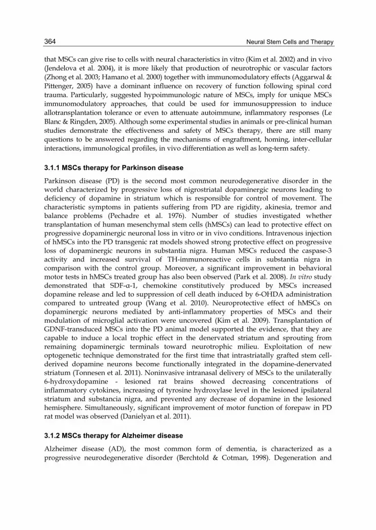
that MSCs can give rise to cells with neural characteristics in vitro (Kim et al. 2002) and in vivo (Jendelova et al. 2004), it is more likely that production of neurotrophic or vascular factors (Zhong et al. 2003; Hamano et al. 2000) together with immunomodulatory effects (Aggarwal & Pittenger, 2005) have a dominant influence on recovery of function following spinal cord trauma. Particularly, suggested hypoimmunologic nature of MSCs, imply for unique MSCs immunomodulatory approaches, that could be used for immunosuppression to induce allotransplantation tolerance or even to attenuate autoimmune, inflammatory responses (Le Blanc & Ringden, 2005). Although some experimental studies in animals or pre-clinical human studies demonstrate the effectiveness and safety of MSCs therapy, there are still many questions to be answered regarding the mechanisms of engraftment, homing, inter-cellular interactions, immunological profiles, in vivo differentiation as well as long-term safety.
3.1.1 MSCs therapy for Parkinson disease
Parkinson disease (PD) is the second most common neurodegenerative disorder in the world characterized by progressive loss of nigrostriatal dopaminergic neurons leading to deficiency of dopamine in striatum which is responsible for control of movement. The characteristic symptoms in patients suffering from PD are rigidity, akinesia, tremor and balance problems (Pechadre et al. 1976). Number of studies investigated whether transplantation of human mesenchymal stem cells (hMSCs) can lead to protective effect on progressive dopaminergic neuronal loss in vitro or in vivo conditions. Intravenous injection of hMSCs into the PD transgenic rat models showed strong protective effect on progressive loss of dopaminergic neurons in substantia nigra. Human MSCs reduced the caspase-3 activity and increased survival of TH-immunoreactive cells in substantia nigra in comparison with the control group. Moreover, a significant improvement in behavioral motor tests in hMSCs treated group has also been observed (Park et al. 2008). In vitro study demonstrated that SDF-α-1, chemokine constitutively produced by MSCs increased dopamine release and led to suppression of cell death induced by 6-OHDA administration compared to untreated group (Wang et al. 2010). Neuroprotective effect of hMSCs on dopaminergic neurons mediated by anti-inflammatory properties of MSCs and their modulation of microglial activation were uncovered (Kim et al. 2009). Transplantation of GDNF-transduced MSCs into the PD animal model supported the evidence, that they are capable to induce a local trophic effect in the denervated striatum and sprouting from remaining dopaminergic terminals toward neurotrophic milieu. Exploitation of new optogenetic technique demonstrated for the first time that intrastriatally grafted stem cell-derived dopamine neurons become functionally integrated in the dopamine-denervated striatum (Tonnesen et al. 2011). Noninvasive intranasal delivery of MSCs to the unilaterally 6-hydroxydopamine - lesioned rat brains showed decreasing concentrations of inflammatory cytokines, increasing of tyrosine hydroxylase level in the lesioned ipsilateral striatum and substancia nigra, and prevented any decrease of dopamine in the lesioned hemisphere. Simultaneously, significant improvement of motor function of forepaw in PD rat model was observed (Danielyan et al. 2011).
3.1.2 MSCs therapy for Alzheimer disease
Alzheimer disease (AD), the most common form of dementia, is characterized as a progressive neurodegenerative disorder (Berchtold & Cotman, 1998). Degeneration and

Mesenchymal Stromal Cells and Neural Stem Cells Potential for Neural Repair in Spinal Cord Injury and Human Neurodegenerative Disorders
365
dysfunction of the neurons and decline of synaptic function and plasticity mostly in brain regions responsible for memory and learning, as hippocampus, entorhinal cortex, basal forebrain and neocortical association cortices, are the most incident symptoms that generally characterize AD (DeKosky et al. 1996). There is no cure or early preclinical diagnostic assay available for Alzheimer’s disease. Currently, most prevalent is symptomatic therapy, which is not able to stop the progression of the disease. Therefore, Alzheimer’s disease is still being recognized as an unmet medical need. In 1906, Dr. Alois Alzheimer, identified two specific features that are mostly figured in AD human brain, neurofibrillary tangles and amyloid plaques. Deep investigation in the study of the main structural components responsible for the creation of two pathological hallmarks in AD brain, uncovered inherence of tau protein in NFT and amyloid beta peptide in amyloid plaques. Several years later, it was demonstrated that strong neuroinflammation occurs in AD brain (Novak et al. 1993) (Dickson et al. 1988; Zilka et al. 2006; Zilkova et al. 2006).
Application of stem cells in AD preclinical studies brought in last years several positive results. Taking advantage of stem cells immunomodulatory and trophic properties and their transplantation into AD transgenic animal models showed that they are the most appropriate tool for the achievement of functional restoration of damaged cells and in the same manner for the replacement by healthy one (Blurton-Jones et al. 2009; Hampton et al. 19 2010; Lee et al. 2010). Recent developments in stem cell technology raise the prospect of cell therapy for human neurodegenerative tauopathies. Transplantation of the neural stem cells or administration of mesenchymal stem cells isolated either from human umbilical cord or from the bone marrow has produced beneficial effects in several independent animal models of AD (Blurton-Jones et al. 2009). Above mentioned reports have shown that the neuroprotective effect of stem cells may be mediated 1) by their ability to produce various trophic factors that contribute to functional recovery or 2) by activation of neuroinflammatory pathways. In vitro studies show that MSCs can prevent tau mediated cell death in the Alzheimer’s cell model. It has been confirmed that MSCs have significant impact on tau cell death cascade and can ameliorate toxic effect of misfolded truncated tau that is considered to be driving force behind neurofibrillary degeneration. Therefore it may be suggested that the cell neuroprotective therapy rather than cell replacement therapy represents prospective strategy for treatment of Alzheimer’s disease and related tauopathies (Zilka et al. 2011).
4. Neural stem cells
The human brain contains roughly 100 billion neurons, of which several thousands die every day, representing the loss of millions of nerve cells across the life span. For this reason, it has been believed for a long time, that adult mammalian central nervous system (CNS) is rather rigid structure, unable to repair itself following diseases or injury. However, in some brain regions dead neurons could be replaced and potentially could contribute to the regeneration of damaged nerve tissue (Graziadei & Graziadei, 1979). Therefore, a number of controversial issues concerning possible CNS plasticity was raised and broadly discussed. Finally, in the 1960s and 1970s, most of the uncertainties were addressed and neuroscience’s central tenets the ‘no new neurons’ doctrine, was reconsidered following the key-revolutionary discovery of Joseph Altman (Altman, 1962; Altman & Das, 1965),

Neural Stem Cells and Therapy
366
documenting thymidine-H3-labelled neurons and neuroblasts in the adult rat brain. From now on a huge effort has gone into unraveling and understanding the fundamental mechanisms of adult CNS regeneration in mammals.
4.1 Neural stem cells definition and origin
It took almost twenty years of dedicated research involving a large number of scientific experiments which clearly confirmed ongoing neurogenesis not only in songbirds (Nottebohm, 1981), but also in rodents, non-human primates and humans, in whom new imaging techniques, such as bromodeoxyuridin (BrdU) labeling, etc, enabling identification of proliferating cells were applied (Eriksson et al. 1998). All these studies jointly confirmed that new functional neurons are generated in the adult mammalian, including human CNS in two discrete areas: i) in the hippocampus, the subgranular zone (SGZ) of the dentate gyrus, which is an important center of our memory (Gage, 2000; Alvarez-Buylla et al. 2002) and, ii) in subventricular zone (SVZ), representing a thin layer of cells lining along the lateral cerebral ventricles, where a nerve cells essential for olfaction are generated (Gage, 2000; Lledo et al. 2006). In both areas, neurogenesis progresses as a complex multi-stage process, which starts with the proliferation, followed by migration and terminal differentiation (Abrous et al. 2005). The current knowledge of self-renewing and multipotent neural stem cells is largely defined by in vitro, as well as in vivo evidences documenting their ability to generate the main progeny of the nervous system: neurons, astrocytes and oligodendrocytes (Gage, 2000). NSCs reside in specific anatomical microenvironments that are called neurogenic niches; small islands where neurons and glial cells are continuously generated (Doetsch et al. 1999). However, neurogenic regions (SVZ, SGZ) must meet following criteria: 1) contain neural precursors (NPCs) that are generated in, 2) neurogenic niches, providing cell-cell contacts and diffusible factors for terminal neural differentiation, and 3) provide neurogenic potential (thus, ability of NPCs that are implanted in a neurogenic areas to generate neurons, while when implanted into other brain location they give rise to glia). Another interesting pool of neural precursor cells is represented by astrocytes found within the germinal layers of the adult brain. It has been broadly documented that these astrocytes retain the stem cell properties throughout the life span, and are involved in both neuro- and glio-genesis (Alvarez-Buylla et al. 2001; Gotz & Huttner, 2005; Mori et al. 2005).
4.1.1 Neurogenesis mediated by pathological conditions; Properties of non-neurogenic areas
Normal adult neurogenesis produces a limited number of newly generated functional cells that primarily serves to maintain physiological tissue homeostasis in specific CNS systems. Initially, the neurogenic processes have been expected to be rather stable, moreover insensitive to external stimuli. However, this view has been changed, due to the growing evidence documenting that SVZ and SGZ are responding to a various local or global signals generated from nerve tissue damage. For example, neurogenesis in both neurogenic zones is increased in animal experimental models of ischemia/stroke (Zhang et al. 2008) as well as in humans suffering from stroke (Curtis et al. 2007), epileptic seizures (Grote & Hannan, 2007) and multiple sclerosis (Nait-Oumesmar et al. 2007). Furthermore, neurogenesis is increased

Mesenchymal Stromal Cells and Neural Stem Cells Potential for Neural Repair in Spinal Cord Injury and Human Neurodegenerative Disorders
367
in human cases and animal models of Huntington's disease while it is reduced in Alzheimer's and Parkinson's disease as well as in depression and stress (Elder et al. 2006; Grote & Hannan, 2007). Stem cells with the potential to generate new neurons that could replace dying neurons in neurodegenerative diseases or CNS injuries reside also in other areas of the adult CNS, indicating to the possibility that endogenous sources of NSCs can be mobilized also from non-neurogenic regions (Minger, 2007). These NSCs have been demonstrated in brain areas such as septum, striatum or even in the spinal cord, but so far it was not clearly established whether these stem cells are capable of differentiation to the final functional neurons (Liu & Martin, 2003; Wiltrout et al. 2007). Furthermore, it has been suggested that ependymal cells (ECs) adjacent to the SVZ of the lateral ventricles, may mimic the characteristics of NSCs (Johansson et al. 1999; Doetsch et al. 1999). A study by Coskun et al. (Coskun et al. 2008) documented that this may be the case, because the subpopulation of ependymal cells, CD133+/CD24-, exhibited features of quiescent NSCs in vitro, i.e., self-renewal and multipotency as well as participation in neurogenesis in vivo after injury. In this relation, the occurrence of ependymal cell layer covering CNS ventricular system including the areas around the third, fourth ventricles, and the central canal (CC) of the spinal cord supports suggestion, that also these regions may retain similar quiescent NSCs as those which were identified in the lateral ventricles (Weiss et al. 1996).
4.1.2 Neurogenic potential in the spinal cord and stimulatory factors
There is increasing evidence that the CC ependymal cell region, which is regarded as presumptive neurogenic area of adult spinal cord, contains a limited number of neural stem cells. Once implanted in the animals, they differentiate into oligodendrocytes and astrocytes (Mothe & Tator, 2005) while, under in vitro conditions, they give rise to both neurons and glia (Yamamoto et al. 2001). On the other hand, neuronal or glial fate of grafted ECs is highly depended on the host neurogenic/non–neurogenic microenvironment (Shihabuddin et al. 2000). These contradictory findings are often explained in regard to beneficial (in vitro) or inhibitory (in vivo) conditions directly influencing neuronal or glial fate (Weiss et al. 1996). Furthermore, after pathological condition such as spinal cord injury, most of the newly dividing intrinsic ependymal stem cells migrate toward damaged tissue, where they develop into macroglial cells, while only few cells retain primitive nestin-like phenotype (Johansson et al. 1999; Cizkova et al. 2009a). Likewise, a significant number of neural progenitors could be activated also in other regions of the parenchyma (Horner et al. 2000) (Kehl et al. 1997). However, it remains unclear whether these progenitors develop into functional neurons.
A stimulatory effect on spinal progenitors may be obtained also after physiological stimulation, when experimental animals are exposed to an enrichment environment or physical activity. Previous experiments have shown that mice providing systematic exercise in a running wheel had twice more new hippocampal neurons than controls (Gomez-Pinilla et al. 2001). Beside this, it has been confirmed that voluntary exercise can increase levels of brain-derived neurotrophic factor (BDNF) and other growth factors, which stimulate neurogenesis, improve learning, mental performance (Gomez-Pinilla et al. 2001) and may mobilize gene expression profiles that could be beneficial for CNS plasticity processes (Neeper et al. 1995). These data were further confirmed in latter studies showing that enhanced physical activity in adult rats induces an endogenous ependymal cell response leading to increased proliferation, although in more attenuated manner if compared with

Neural Stem Cells and Therapy
368
SCI (Cizkova et al. 2009b) (Fig.1). Indeed, there is one group of studies that favor the fact that ECs might contribute to de novo neuronal differentiation following CNS injury (Ke et al. 2006; Danilov et al. 2006), while others refuse this suggestion (Zai & Wrathall, 2005). Based on these findings, it is un-doubtful that the adult spinal cord retain a certain reservoir of neural precursors, which can under various specific conditions stimulate and promote the recovery of injured spinal cord.
Fig. 1. Schematic illustration of BrdU IR in the thoracic spinal cord section (Th8) of the control, SCI or Running group. Note, the highest BrdU expression in the CC canal, and around the lesion site of SCI group, different distribution patterns of BrdUpositive nuclei in the ependyma between SCI and Running group, and increased BrdU response in the parenchyma of the SC in both groups. Below each schematic drawing, a panel revealing BrdU–IR in the corresponding ventral white matter is performed. (A-D) Fluorescence microscopy images of occasionally occurring nestin-positive cell bodies (green) with processes, found in the close vicinity to the CC gray matter, dorsal horn or adjacent to lesion site.
4.1.3 Molecular mechanisms of neurogenesis
Neurogenesis is understood as a complex process that is regulated by a wide variety of important signaling molecules such as: growth factors, cytokines, and neurotransmitters.

Mesenchymal Stromal Cells and Neural Stem Cells Potential for Neural Repair in Spinal Cord Injury and Human Neurodegenerative Disorders
369
Their primary function is to mediate a balance between proliferation, migration and survival of NSCs within the neurogenic niche. The most important growth factors affecting cell division are: FGF (fibroblast growth factor), VEGF (vascular endothelial growth factor), EGF (epidermal growth factor / epidermal growth factor), PDGF (platelet-derived growth factor) and BDNF (brain derived neurotrophic factor). Therefore, endogenous neurogenesis can be stimulated by intraventricular infusion of mitogenic factors such as EGF, bFGF, TGFβ (transforming growth factor β) that stimulate the proliferation activity in the SVZ and thus restore the nervous tissue (Kuhn et al. 1997). Nitric oxide (NO), erythropoietin, bone morphogenetic protein (BMP Bone Morphogenetic Protein) and Wnt proteins (Wiltrout et al. 2007) also play an important role in regulating neurogenesis. BMP and its receptor that are expressed by the SVZ cells promoting differentiation of the NSCs toward glial phenotype are blocked by Noggin, which is produced by ECs and in contrast drives differentiation into neurons (Lim et al. 2000). The most important regulatory neurotransmitters include GABA (γ-aminobutyric acid) and glutamate, which maintain homeostasis of newly formed neurons (Platel et al. 2007). GABA decreases the proliferation of neuroblasts and NSCs, whereas glutamate stimulates their division. It is noteworthy that in all types of damaged nerve tissue which is associated with glutamate excitotoxicity an increased neurogenesis, is documented. GABA is synthesized and released by neuroblasts and activates GABAA receptor, causing loss of proliferation of neuroblasts and astrocytes. We can conclude that GABA acts as a negative modulator inhibiting cell division, which means that with increased number of neuroblasts there is a higher amount of released of GABA and more GABAA receptors are activated (Bordet et al. 2007).
5. Transplantation strategies utilizing NSCs
Neural progenitors isolated from vertebrate central nervous system (CNS) represent valuable source of cells that hold particular promise for treating a variety of human neurological diseases such as spinal cord injury (Goldman, 2005). Due to the pathological events and limited ability of the spinal cord to repair itself, therapeutic approaches are focused either on: i) stimulation of endogenous neuronal plasticity and mobilization of oligodendoglial progenitors (Azari et al. 2005; Fawcett, 2006; Yang et al. 2006) or ii) development of an effective cell selection techniques to gain desired NSCs progeny used for cell-replacement therapy (Faulkner & Keirstead, 2005; Hofstetter et al. 2005; Keirstead et al. 2005). However, an important issue due to the pathological nature of spinal cord damage it is important to select the most convenient strategy involving desired cellular pools for transplantation. For example, spinal ischemia-induced spastic paraplegia which is associated with a selective loss of small inhibitory interneurons, would necessarily involve implantation of neuronal progenitors. On the other hand, diseases or spinal cord trauma, with different pathological outcome, resulting in demyelination of axons followed by destruction of long descending tracts would rather require transplantation of myelin-producing cells such as oligodendroglial cells, Schwann cells or Olfactory ensheating cells (Keirstead et al. 2005; Keilhoff et al. 2006; Pearse et al. 2007; Raisman, 2007). Since a well-documented repertoire of specific surface markers for cells of NSCs at different developmental stages have been identified, it may be possible to identify factors which affect their commitment to oligodendroglial cells or neurons and combine this with optimal sorting methods (Deng & Poretz, 2003; Pruszak et al. 2007; Uchida et al. 2000). In particular, magnetic cell separation using specific monoclonal antibodies (e.g. A2B5, PSA-NCAM) conjugated to nanoparticles allowing positive retention or negative dilution of

Neural Stem Cells and Therapy
370
selected cells provide a feasible approach for experimental cell enrichment of desired oligodendroglial progeny, which may be used in future trials for cell-based therapies to treat spinal cord injury (Cizkova et al. 2009a). These studies have shown that MACs technology enable us to gain about a 5 to 9 fold increase of immature, mature oligodendrocytes content (NG2+, RIP+, MBP+) when compared to amount of oligodendroglial cells acquired from unseparated population (Fig.2). A great deal of attention has been given to NSCs isolated from various regions of CNS, including embryonic and adult spinal cord, that could differentiate into desired oligodendrocytes and myelinate host axons in various pre-clinical animal models of SCI (Tarasenko et al. 2007; Kakinohana et al. 2004). For example, NSCs derived from human fetal brain improved recovery after contusion SCI either in severe combined immunodeficiency (SCID) or myelin–deficient shivered mice (Cummings et al. 2005). Highly purified oligodendrocyte progenitors could be generated also from human embryonic stem cells (hESCs) (Nistor et al. 2005; Cloutier et al. 2006). Based on their remyelination properties described in preclinical animal SCI models, the Geron Corporation has initiated a first clinical trial (Phase I) by transplanting hESC-derived oligodendrocyte
Fig. 2. Immature neurons expressing βIII-tubulin (green) occurred in both, unseparated (A) and separated NSC population (B), but higher number of immature NG2+ oligodendrocytes (red, A, B) and mature RIP+ oligodendrocytes (green C, D) was found after MACs (B, D) (compare A with B and C with D).
Mesenchymal Stromal Cells and Neural Stem Cells Potential for Neural Repair in Spinal Cord Injury and Human Neurodegenerative Disorders
371
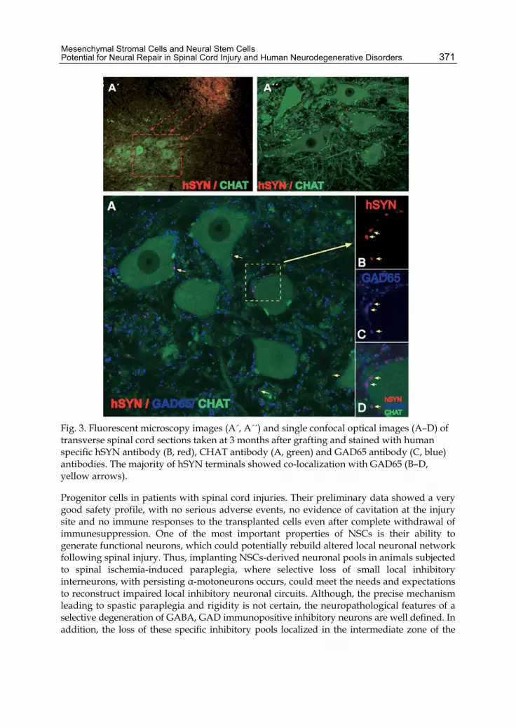
Fig. 3. Fluorescent microscopy images (A´, A´´) and single confocal optical images (A–D) of transverse spinal cord sections taken at 3 months after grafting and stained with human specific hSYN antibody (B, red), CHAT antibody (A, green) and GAD65 antibody (C, blue) antibodies. The majority of hSYN terminals showed co-localization with GAD65 (B–D, yellow arrows).
Progenitor cells in patients with spinal cord injuries. Their preliminary data showed a very good safety profile, with no serious adverse events, no evidence of cavitation at the injury site and no immune responses to the transplanted cells even after complete withdrawal of immunesuppression. One of the most important properties of NSCs is their ability to generate functional neurons, which could potentially rebuild altered local neuronal network following spinal injury. Thus, implanting NSCs-derived neuronal pools in animals subjected to spinal ischemia-induced paraplegia, where selective loss of small local inhibitory interneurons, with persisting α-motoneurons occurs, could meet the needs and expectations to reconstruct impaired local inhibitory neuronal circuits. Although, the precise mechanism leading to spastic paraplegia and rigidity is not certain, the neuropathological features of a selective degeneration of GABA, GAD immunopositive inhibitory neurons are well defined. In addition, the loss of these specific inhibitory pools localized in the intermediate zone of the

Neural Stem Cells and Therapy
372
spinal grey matter, ultimately leads to an increase in the monosynaptic reflex and near-complete loss in spinal polysynaptic activity. A challenging study done in collaboration with anesthesiology research laboratory at University of California San Diego, has shown that NSCs derived from human fetal spinal cord grafted into a rat model of ischemic spastic paraplegia resulted into a progressive recovery of motor function with correlative improvement in motor evoked potentials (Cizkova et al. 2007). Of note, transplanted NSCs became integrated into host neuronal circuits and displayed an extensive axo-dendritic outgrowth and active rostrocaudal/dorsoventral migration for about 8-12 weeks. Furthermore, intense hSYN immunoreactivity was identified within the grafts and in the vicinity of persisting α-motoneurons. These hSYN immunoreactive synaptic terminals expressed GAD65 immunoreactivity in 40-45% of human grafted cells, referring to their inhibitory fate (Fig. 3). All together, these data conclude that functional recovery was associated with long term survival of grafted neurons with GABAergic phenotype that most probably contributed to suppression of spasticity (Cizkova et al. 2007). Similarly, human hNT neurons (teratocarcinoma cell line) or rat spinal neuronal precursors (SNPs), grafted into ischemic spinal segments depleted of inhibitory neurons, restore local inhibitory tone and ameliorate spasticity (Marsala et al. 2004). In addition, when human derived NSCs were treated with a cocktail of growth factors and later transplanted into the injured spinal cord, they differentiated preferentially into cholinergic neurons (Wu et al. 2009). Although, it seems that NSCs are a powerful source of neural progenitors that are constitutively secreting a variety of growth stimulating factors (NGF, BDNF, GDNF), they are often genetically modified to further enhance their potential and secrete additional factors such as neurotrophin 3 (NT-3), or are combined with antibodies that neutralize ciliary neurothrophic factor (CNTF), in an attempt to attenuate astrocytic differentiation (Ishii et al. 2006).
6. Acknowledgments
We acknowledge the financial support given by MVTS-COST BM-1002- NANONET, the Grant Agency of the Slovak Academy of Sciences VEGA 2/0114/11 and the Center of Excellence for Brain Research Slovak Academy of Sciences and the project No 26110230036 University of Veterinary Medicine and Pharmacy (Nove studijne programy a vzdelavanie na Univerzite veterinarneho lekarstva v Kosiciach, Operacny program Vzdelavanie).
7. References
Abrous, D. N.; Koehl, M.& Le Moal, M. (2005). Adult neurogenesis: from precursors to network and physiology. Physiol Rev, Vol. 85, No. 2, (April 2005), pp 523-569, ISSN 0031-9333
Aggarwal, S .& Pittenger, M. F. (2005). Human mesenchymal stem cells modulate allogeneic immune cell responses. Blood, Vol. 105, No. 4, (February 2005), pp 1815-1822, ISSN 0006-4971
Akiyama, Y.; Radtke, C.; Honmou, O.& Kocsis, J. D. (2002). Remyelination of the spinal cord following intravenous delivery of bone marrow cells. Glia, Vol. 39, No. 3, (September 2002), pp 229-236, ISSN 0894-1491
Altman, J. (1962). Are new neurons formed in the brains of adult mammals? Science, Vol. 135, (March 1962), pp 1127-1128, ISSN 0036-8075

Mesenchymal Stromal Cells and Neural Stem Cells Potential for Neural Repair in Spinal Cord Injury and Human Neurodegenerative Disorders
373
Altman, J.&Das, G. D. (1965). Autoradiographic and histological evidence of postnatal hippocampal neurogenesis in rats. J Comp Neurol, Vol. 124, No. 3, pp 319-335, ISSN 0021-9967
Alvarez-Buylla, A.; Garcia-Verdugo, J. M.& Tramontin, A. D. (2001). A unified hypothesis on the lineage of neural stem cells. Nat Rev Neurosci, Vol. 2, No. 4, (April 2001), pp 287-293, ISSN 1471-0048
Alvarez-Buylla, A.; Seri, B.& Doetsch, F. (2002). Identification of neural stem cells in the adult vertebrate brain. Brain Res Bull, Vol. 57, No. 6, (April 2002), pp 751-758, ISSN 0361-9230
Arboleda, D.; Forostyak, S.; Jendelova, P.; Marekova, D.; Amemori, T.; Pivonkova, H.; Masinova, K.& Sykova, E. (2011). Transplantation of Predifferentiated Adipose-Derived Stromal Cells for the Treatment of Spinal Cord Injury. Cell Mol Neurobiol, (June 2011), ISSN 0272-4340
Augello, A.; Tasso, R.; Negrini, S. M.; Amateis, A.; Indiveri, F.; Cancedda, R.& Pennesi, G. (2005). Bone marrow mesenchymal progenitor cells inhibit lymphocyte proliferation by activation of the programmed death 1 pathway. Eur J Immunol, Vol. 35, No. 5, (May 2005), (May 2005), pp 1482-1490, ISSN 0014-2980
Azari, M. F.; Profyris, C.; Zang, D. W.; Petratos, S.& Cheema, S. S. (2005). Induction of endogenous neural precursors in mouse models of spinal cord injury and disease. Eur J Neurol, Vol. 12, No. 8, (August 2005), pp 638-648, ISSN 1351-5101
Barnabe-Heider, F. & Frisen, J. (2008). Stem cells for spinal cord repair. Cell Stem Cell, Vol. 3, No. 1, (Jul 2008), pp 16-24, ISSN 1875-9777
Berchtold, N. C. & Cotman, C. W. (1998). Evolution in the conceptualization of dementia and Alzheimer's disease: Greco-Roman period to the 1960s. Neurobiol Aging, Vol. 19, No. 3, (May-June 1998), pp 173-189, ISSN 0197-4580
Bjorklund, A. & Svendsen, C. (1999). Stem cells. Breaking the brain-blood barrier. Nature, Vol. 397, No. 6720, pp 569-570, ISSN 0028-0836
Blurton-Jones, M.; Kitazawa, M.; Martinez-Coria, H.; Castello, N. A.; Muller, F. J.; Loring, J. F.; Yamasaki, T. R.; Poon, W. W.; Green, K. N.& LaFerla, F. M. (2009). Neural stem cells improve cognition via BDNF in a transgenic model of Alzheimer disease. Proc Natl Acad Sci U S A, Vol. 106, No. 32, (August 2009), pp 13594-13599, ISSN 1091-6490
Bonab, M. M.; Alimoghaddam, K.; Talebian, F.; Ghaffari, S. H.; Ghavamzadeh, A.& Nikbin, B. (2006). Aging of mesenchymal stem cell in vitro. BMC Cell Biol, Vol. 7, (March 2006), pp 14, ISSN 1471-2121
Bordet, R.; Lestage, P.& Onteniente, B. (2007). [The concept of neuroprotective agents as a treatment modulator in the development of brain diseases]. Therapie, Vol. 62, No. 6, (November/December 2007), pp 463-472, ISSN 0040-5957
Bradbury, E. J.; Moon, L. D.; Popat, R. J.; King, V. R.; Bennett, G. S.; Patel, P. N.; Fawcett, J. W.& McMahon, S. B. (2002). Chondroitinase ABC promotes functional recovery after spinal cord injury. Nature, Vol. 416, No. 6881, (April 2002), pp 636-640, ISSN 0028-0836
Cizkova, D.; Cizek, M.; Nagyova, M.; Slovinska, L.; Novotna, I.; Jergova, S.; Radonak, J.; Hlucilova, J.& Vanicky, I. (2009a). Enrichment of rat oligodendrocyte progenitor cells by magnetic cell sorting. J Neurosci Methods, Vol. 184, No. 1, (October 2009), pp 88-94, ISSN 1872-678X
Cizkova, D.; Kakinohana, O.; Kucharova, K.; Marsala, S.; Johe, K.; Hazel, T.; Hefferan, M. P.& Marsala, M. (2007). Functional recovery in rats with ischemic paraplegia after

Neural Stem Cells and Therapy
374
spinal grafting of human spinal stem cells. Neuroscience, Vol. 147, No. 2, (June 2007), pp 546-560, ISSN 0306-4522
Cizkova, D.; Nagyova, M.; Slovinska, L.; Novotna, I.; Radonak, J.; Cizek, M.; Mechirova, E.; Tomori, Z.; Hlucilova, J.; Motlik, J.; Sulla, I., Jr. & Vanicky, I. (2009b). Response of ependymal progenitors to spinal cord injury or enhanced physical activity in adult rat. Cell Mol Neurobiol, Vol. 29, No. 6-7, (September 2009), pp 999-1013, ISSN 1573-6830
Cizkova, D.; Rosocha, J.; Vanicky, I.; Jergova, S.& Cizek, M. (2006). Transplants of human mesenchymal stem cells improve functional recovery after spinal cord injury in the rat. Cell Mol Neurobiol, Vol. 26, No. 7-8, (November 2006), pp 1165-1178, ISSN 0272-4340
Cloutier, F.; Siegenthaler, M. M.; Nistor, G.& Keirstead, H. S. (2006). Transplantation of human embryonic stem cell-derived oligodendrocyte progenitors into rat spinal cord injuries does not cause harm. Regen Med, Vol. 1, No. 4, (July 2006), pp 469-479, ISSN 1746-0751
Colter, D. C.; Class, R.; DiGirolamo, C. M.& Prockop, D. J. (2000). Rapid expansion of recycling stem cells in cultures of plastic-adherent cells from human bone marrow. Proc Natl Acad Sci U S A, Vol. 97, No. 7, (March 2000), pp 3213-3218, ISSN 0027-8424
Colter, D. C.; Sekiya, I.& Prockop, D. J. (2001). Identification of a subpopulation of rapidly self-renewing and multipotential adult stem cells in colonies of human marrow stromal cells. Proc Natl Acad Sci U S A, Vol. 98, No. 14, (July 2001), pp 7841-7845, ISSN 0027-8424
Coskun, V.; Wu, H.; Blanchi, B.; Tsao, S.; Kim, K.; Zhao, J.; Biancotti, J. C.; Hutnick, L.; Krueger, R. C., Jr.; Fan, G.; de Vellis, J.& Sun, Y. E. (2008). CD133+ neural stem cells in the ependyma of mammalian postnatal forebrain. Proc Natl Acad Sci U S A, Vol. 105, No. 3, (January 2008), pp 1026-1031, ISSN 1091-6490
Crigler, L.; Robey, R. C.; Asawachaicharn, A.; Gaupp, D.& Phinney, D. G. (2006). Human mesenchymal stem cell subpopulations express a variety of neuro-regulatory molecules and promote neuronal cell survival and neuritogenesis. Exp Neurol, Vol. 198, No. 1, (March 2006), (March 2006), pp 54-64, ISSN 0014-4886
Cui, X.; Chopp, M.; Zacharek, A.; Roberts, C.; Lu, M.; Savant-Bhonsale, S. & Chen, J. (2009). Chemokine, vascular and therapeutic effects of combination Simvastatin and BMSC treatment of stroke. Neurobiol Dis, Vol. 36, No. 1, (October 2009), pp 35-41, ISSN 09699961
Cummings, B. J.; Uchida, N.; Tamaki, S. J.; Salazar, D. L.; Hooshmand, M.; Summers, R.; Gage, F. H.& Anderson, A. J. (2005). Human neural stem cells differentiate and promote locomotor recovery in spinal cord-injured mice. Proc Natl Acad Sci U S A, Vol. 102, No. 39, (September 2005), pp 14069-14074, ISSN 0027-8424
Curtis, M. A.; Kam, M.; Nannmark, U.; Anderson, M. F.; Axell, M. Z.; Wikkelso, C.; Holtas, S.; van Roon-Mom, W. M.; Bjork-Eriksson, T.; Nordborg, C.; Frisen, J.; Dragunow, M.; Faull, R. L.& Eriksson, P. S. (2007). Human neuroblasts migrate to the olfactory bulb via a lateral ventricular extension. Science, Vol. 315, No. 5816, (March 2007), pp 1243-1249, ISSN 0036-8075
Danielyan, L.; Schafer, R.; von Ameln-Mayerhofer, A.; Bernhard, F.; Verleysdonk, S.; Buadze, M.; Lourhmati, A.; Klopfer, T.; Schaumann, F.; Schmid, B.; Koehle, C.; Proksch, B.; Weissert, R.; Reichardt, H. M.; van den Brandt, J.; Buniatian, G. H.; Schwab, M.; Gleiter, C. H.& Frey, W. H., 2nd. (2011). Therapeutic efficacy of

Mesenchymal Stromal Cells and Neural Stem Cells Potential for Neural Repair in Spinal Cord Injury and Human Neurodegenerative Disorders
375
intranasally delivered mesenchymal stem cells in a rat model of Parkinson disease. Rejuvenation Res, Vol. 14, No. 1, (February 2011), pp 3-16, ISSN 1557-8577
Danilov, A. I.; Covacu, R.; Moe, M. C.; Langmoen, I. A.; Johansson, C. B.; Olsson, T.& Brundin, L. (2006). Neurogenesis in the adult spinal cord in an experimental model of multiple sclerosis. Eur J Neurosci, Vol. 23, No. 2, (January 2006), pp 394-400, ISSN 0953-816X
DeKosky, S. T.; Scheff, S. W.& Styren, S. D. (1996). Structural correlates of cognition in dementia: quantification and assessment of synapse change. Neurodegeneration, Vol. 5, No. 4, (December 1996), pp 417-421, ISSN 1055-8330
Deng, W.&Poretz, R. D. (2003). Oligodendroglia in developmental neurotoxicity. Neurotoxicology, Vol. 24, No. 2, (March 2003), pp 161-178, ISSN 0161-813X
Di Nicola, M.; Carlo-Stella, C.; Magni, M.; Milanesi, M.; Longoni, P. D.; Matteucci, P.; Grisanti, S.& Gianni, A. M. (2002). Human bone marrow stromal cells suppress T-lymphocyte proliferation induced by cellular or nonspecific mitogenic stimuli. Blood, Vol. 99, No. 10, (May 2002), pp 3838-3843, ISSN 0006-4971
Dickson, D. W.; Farlo, J.; Davies, P.; Crystal, H.; Fuld, P.& Yen, S. H. (1988). Alzheimer's disease. A double-labeling immunohistochemical study of senile plaques. Am J Pathol, Vol. 132, No. 1, (July 1988), pp 86-101, ISSN 0002-9440
Digirolamo, C. M.; Stokes, D.; Colter, D.; Phinney, D. G.; Class, R.& Prockop, D. J. (1999). Propagation and senescence of human marrow stromal cells in culture: a simple colony-forming assay identifies samples with the greatest potential to propagate and differentiate. Br J Haematol, Vol. 107, No. 2, (November 1999), pp 275-281, ISSN 0007-1048
Djouad, F.; Plence, P.; Bony, C.; Tropel, P.; Apparailly, F.; Sany, J.; Noel, D.& Jorgensen, C., 2003. Immunosuppressive effect of mesenchymal stem cells favors tumor growth in allogeneic animals. In: Blood, vol. 102. no. 10, pp. 3837-3844, ISSN 0006-4971
Doetsch, F.; Caille, I.; Lim, D. A.; Garcia-Verdugo, J. M.& Alvarez-Buylla, A. (1999). Subventricular zone astrocytes are neural stem cells in the adult mammalian brain. Cell, Vol. 97, No. 6, pp 703-716, ISSN 0092-8674
Docheva, D.; Popov, C.; Mutschler, W.& Schieker, M. (2007). Human mesenchymal stem cells in contact with their environment: surface characteristics and the integrin system. J Cell Mol Med, Vol. 11, No. 1, (January/February 2007), pp 21-38, ISSN 1582-1838
Dominici, M.; Le Blanc, K.; Mueller, I.; Slaper-Cortenbach, I.; Marini, F.; Krause, D.; Deans, R.; Keating, A.; Prockop, D.& Horwitz, E. (2006). Minimal criteria for defining multipotent mesenchymal stromal cells. The International Society for Cellular Therapy position statement. Cytotherapy, Vol. 8, No. 4, pp 315-317, ISSN 1465-3249
Elder, G. A.; De Gasperi, R.& Gama Sosa, M. A. (2006). Research update: neurogenesis in adult brain and neuropsychiatric disorders. Mt Sinai J Med, Vol. 73, No. 7, (November 2006), pp 931-940, ISSN 0027-2507
Eriksson, P. S.; Perfilieva, E.; Bjork-Eriksson, T.; Alborn, A. M.; Nordborg, C.; Peterson, D. A.& Gage, F. H. (1998). Neurogenesis in the adult human hippocampus. Nat Med, Vol. 4, No. 11, (November 1998), pp 1313-1317, ISSN 1078-8956
Faulkner, J.&Keirstead, H. S. (2005). Human embryonic stem cell-derived oligodendrocyte progenitors for the treatment of spinal cord injury. Transpl Immunol, Vol. 15, No. 2, (December 2005), pp 131-142, ISSN 0966-3274
Fawcett, J. W. (2006). Overcoming inhibition in the damaged spinal cord. J Neurotrauma, Vol. 23, No. 3-4, (March/April 2006), pp 371-383, 0897-7151 ISSN 0897-7151

Neural Stem Cells and Therapy
376
Forostyak, S.; Jendelova, P.; Kapcalova, M.; Arboleda, D.& Sykova, E. (2011). Mesenchymal stromal cells prolong the lifespan in a rat model of amyotrophic lateral sclerosis. Cytotherapy, (July 2011), ISSN 1477-2566
Friedenstein, A. J.; Gorskaja, J. F.& Kulagina, N. N. (1976). Fibroblast precursors in normal and irradiated mouse hematopoietic organs. Exp Hematol, Vol. 4, No. 5, (September 1976), pp 267-274, ISSN 0301-472X
Gage, F. H. (2000). Mammalian neural stem cells. Science, Vol. 287, No. 5457, (February 2000), pp 1433-1438, ISSN 0036-8075
Goldman, S. (2005). Stem and progenitor cell-based therapy of the human central nervous system. Nat Biotechnol, Vol. 23, No. 7, (July,2005), pp 862-871, ISSN 1087-0156
Gomez-Pinilla, F.; Ying, Z.; Opazo, P.; Roy, R. R.& Edgerton, V. R. (2001). Differential regulation by exercise of BDNF and NT-3 in rat spinal cord and skeletal muscle. Eur J Neurosci, Vol. 13, No. 6, (March 2001), pp 1078-1084, ISSN 0953-816X
Gotz, M.&Huttner, W. B. (2005). The cell biology of neurogenesis. Nat Rev Mol Cell Biol, Vol. 6, No. 10, (Pctober 2005), pp 777-788, ISSN 1471-0072
Graziadei, P. P.&Graziadei, G. A. (1979). Neurogenesis and neuron regeneration in the olfactory system of mammals. I. Morphological aspects of differentiation and structural organization of the olfactory sensory neurons. J Neurocytol, Vol. 8, No. 1, (February 1979), pp 1-18, ISSN 0300-4864
Greco, S. J.&Rameshwar, P. (2008). Microenvironmental considerations in the application of human mesenchymal stem cells in regenerative therapies. Biologics, Vol. 2, No. 4, pp 699-705, ISSN 1177-5475
Grote, H. E.&Hannan, A. J. (2007). Regulators of adult neurogenesis in the healthy and diseased brain. Clin Exp Pharmacol Physiol, Vol. 34, No. 5-6, (May/June 2007), pp 533-545, ISSN 0305-1870
Guan, K.; Nayernia, K.; Maier, L. S.; Wagner, S.; Dressel, R.; Lee, J. H.; Nolte, J.; Wolf, F.; Li, M.; Engel, W.& Hasenfuss, G. (2006). Pluripotency of spermatogonial stem cells from adult mouse testis. Nature, Vol. 440, No. 7088, (April 2006), pp 1199-1203, ISSN 1476-4687
Hamano, K.; Li, T. S.; Kobayashi, T.; Kobayashi, S.; Matsuzaki, M.& Esato, K. (2000). Angiogenesis induced by the implantation of self-bone marrow cells: a new material for therapeutic angiogenesis. Cell Transplant, Vol. 9, No. 3, (May/June 2000), pp 439-443, ISSN 0963-6897
Hampton, D. W.; Webber, D. J.; Bilican, B.; Goedert, M.; Spillantini, M. G.& Chandran, S. (2010). Cell-mediated neuroprotection in a mouse model of human tauopathy. J Neurosci, Vol. 30, No. 30, (July 2010), pp 9973-9983, ISSN 1529-2401
Herzog, E. L.; Chai, L.& Krause, D. S. (2003). Plasticity of marrow-derived stem cells. Blood, Vol. 102, No. 10, (November 2003), pp 3483-3493, ISSN 0006-4971
Hofstetter, C. P.; Holmstrom, N. A.; Lilja, J. A.; Schweinhardt, P.; Hao, J.; Spenger, C.; Wiesenfeld-Hallin, Z.; Kurpad, S. N.; Frisen, J.& Olson, L. (2005). Allodynia limits the usefulness of intraspinal neural stem cell grafts; directed differentiation improves outcome. Nat Neurosci, Vol. 8, No. 3, (March 2005), pp 346-353, ISSN 1097-6256
Horner, P. J.; Power, A. E.; Kempermann, G.; Kuhn, H. G.; Palmer, T. D.; Winkler, J.; Thal, L. J.& Gage, F. H. (2000). Proliferation and differentiation of progenitor cells throughout the intact adult rat spinal cord. J Neurosci, Vol. 20, No. 6, (March 2000), pp 2218-2228, ISSN 1529-2401

Mesenchymal Stromal Cells and Neural Stem Cells Potential for Neural Repair in Spinal Cord Injury and Human Neurodegenerative Disorders
377
Chen, J. R.; Cheng, G. Y.; Sheu, C. C.; Tseng, G. F.; Wang, T. J.& Huang, Y. S. (2008). Transplanted bone marrow stromal cells migrate, differentiate and improve motor function in rats with experimentally induced cerebral stroke. J Anat, Vol. 213, No. 3, (September 2008), pp 249-258, ISSN 1469-7580
Chen, Q.; Long, Y.; Yuan, X.; Zou, L.; Sun, J.; Chen, S.; Perez-Polo, J. R.& Yang, K. (2005). Protective effects of bone marrow stromal cell transplantation in injured rodent brain: synthesis of neurotrophic factors. J Neurosci Res, Vol. 80, No. 5, (June 2005), pp 611-619, ISSN 0360-4012
Chopp, M.&Li, Y. (2002). Treatment of neural injury with marrow stromal cells. Lancet Neurol, Vol. 1, No. 2, (June 2002), pp 92-100, ISSN 1474-4422
Ishii, K.; Nakamura, M.; Dai, H.; Finn, T. P.; Okano, H.; Toyama, Y.& Bregman, B. S. (2006). Neutralization of ciliary neurotrophic factor reduces astrocyte production from transplanted neural stem cells and promotes regeneration of corticospinal tract fibers in spinal cord injury. J Neurosci Res, Vol. 84, No. 8, (December 2006), pp 1669-1681, ISSN 0360-4012
Javazon, E. H.; Colter, D. C.; Schwarz, E. J.& Prockop, D. J. (2001). Rat marrow stromal cells are more sensitive to plating density and expand more rapidly from single-cell-derived colonies than human marrow stromal cells. Stem Cells, Vol. 19, No. 3, (May 2001), pp 219-225, ISSN 1066-5099
Jendelova, P.; Herynek, V.; Urdzikova, L.; Glogarova, K.; Kroupova, J.; Andersson, B.; Bryja, V.; Burian, M.; Hajek, M.& Sykova, E. (2004). Magnetic resonance tracking of transplanted bone marrow and embryonic stem cells labeled by iron oxide nanoparticles in rat brain and spinal cord. J Neurosci Res, Vol. 76, No. 2, (April 2004), pp 232-243, ISSN 0360-4012
Johansson, C. B.; Momma, S.; Clarke, D. L.; Risling, M.; Lendahl, U.& Frisen, J. (1999). Identification of a neural stem cell in the adult mammalian central nervous system. Cell, Vol. 96, No. 1, (January 1999), pp 25-34, ISSN 0092-8674
Jorgensen, C. (2009). Link between cancer stem cells and adult mesenchymal stromal cells: implications for cancer therapy. Regen Med, Vol. 4, No. 2, (March 2009), pp 149-152, ISSN 1746-076X
Kakinohana, O.; Cizkova, D.; Tomori, Z.; Hedlund, E.; Marsala, S.; Isacson, O.& Marsala, M. (2004). Region-specific cell grafting into cervical and lumbar spinal cord in rat: a qualitative and quantitative stereological study. Exp Neurol, Vol. 190, No. 1, (November 2004), pp 122-132, ISSN 0014-4886
Ke, Y.; Chi, L.; Xu, R.; Luo, C.; Gozal, D.& Liu, R. (2006). Early response of endogenous adult neural progenitor cells to acute spinal cord injury in mice. Stem Cells, Vol. 24, No. 4, (April 2006), pp 1011-1019, ISSN 1066-5099
Kehl, L. J.; Fairbanks, C. A.; Laughlin, T. M.& Wilcox, G. L. (1997). Neurogenesis in postnatal rat spinal cord: a study in primary culture. Science, Vol. 276, No. 5312, (April 1997), pp 586-589, ISSN 0036-8075
Keilhoff, G.; Goihl, A.; Langnase, K.; Fansa, H.& Wolf, G. (2006). Transdifferentiation of mesenchymal stem cells into Schwann cell-like myelinating cells. Eur J Cell Biol, Vol. 85, No. 1, (January 2006), pp 11-24, ISSN 0171-9335
Keirstead, H. S.; Nistor, G.; Bernal, G.; Totoiu, M.; Cloutier, F.; Sharp, K.& Steward, O. (2005). Human embryonic stem cell-derived oligodendrocyte progenitor cell transplants remyelinate and restore locomotion after spinal cord injury. J Neurosci, Vol. 25, No. 19, (May 2005), pp 4694-4705, ISSN 1529-2401

Neural Stem Cells and Therapy
378
Kerkis, I.; Kerkis, A.; Dozortsev, D.; Stukart-Parsons, G. C.; Gomes Massironi, S. M.; Pereira, L. V.; Caplan, A. I.& Cerruti, H. F. (2006). Isolation and characterization of a population of immature dental pulp stem cells expressing OCT-4 and other embryonic stem cell markers. Cells Tissues Organs, Vol. 184, No. 3-4, pp 105-116, ISSN 1422-6421
Kim, B. J.; Seo, J. H.; Bubien, J. K.& Oh, Y. S. (2002). Differentiation of adult bone marrow stem cells into neuroprogenitor cells in vitro. Neuroreport, Vol. 13, No. 9, pp 1185-1188, ISSN 0959-4965
Kim, S. U.&de Vellis, J. (2009). Stem cell-based cell therapy in neurological diseases: a review. J Neurosci Res, Vol. 87, No. 10, (August 2009), pp 2183-2200, ISSN 1097-4547
Kim, Y. J.; Park, H. J.; Lee, G.; Bang, O. Y.; Ahn, Y. H.; Joe, E.; Kim, H. O.& Lee, P. H. (2009). Neuroprotective effects of human mesenchymal stem cells on dopaminergic neurons through anti-inflammatory action. Glia, Vol. 57, No. 1, (January 2009), pp 13-23, ISSN 1098-1136
Krampera, M.; Cosmi, L.; Angeli, R.; Pasini, A.; Liotta, F.; Andreini, A.; Santarlasci, V.; Mazzinghi, B.; Pizzolo, G.; Vinante, F.; Romagnani, P.; Maggi, E.; Romagnani, S.& Annunziato, F. (2006). Role for interferon-gamma in the immunomodulatory activity of human bone marrow mesenchymal stem cells. Stem Cells, Vol. 24, No. 2, (February 2006), pp 386-398, ISSN 1066-5099
Kuhn, H. G.; Winkler, J.; Kempermann, G.; Thal, L. J.& Gage, F. H. (1997). Epidermal growth factor and fibroblast growth factor-2 have different effects on neural progenitors in the adult rat brain. J Neurosci, Vol. 17, No. 15, (August 1997), pp 5820-5829, ISSN 0270-6474
Kurozumi, K.; Nakamura, K.; Tamiya, T.; Kawano, Y.; Ishii, K.; Kobune, M.; Hirai, S.; Uchida, H.; Sasaki, K.; Ito, Y.; Kato, K.; Honmou, O.; Houkin, K.; Date, I.& Hamada, H. (2005). Mesenchymal stem cells that produce neurotrophic factors reduce ischemic damage in the rat middle cerebral artery occlusion model. Mol Ther, Vol. 11, No. 1, (January 2005), pp 96-104, ISSN 1525-0016
Kwon, B. K.; Okon, E. B.; Plunet, W.; Baptiste, D.; Fouad, K.; Hillyer, J.; Weaver, L. C.; Fehlings, M. G.& Tetzlaff, W. (2010). A systematic review of directly applied biologic therapies for acute spinal cord injury. J Neurotrauma, Vol. 28, No. 8, (August 2010), pp 1589-1610, ISSN 1557-9042
Le Blanc, K.&Ringden, O. (2005). Immunobiology of human mesenchymal stem cells and future use in hematopoietic stem cell transplantation. Biol Blood Marrow Transplant, Vol. 11, No. 5, (May 2005), pp 321-334, ISSN 1083-8791
Lee, J. K.; Jin, H. K.; Endo, S.; Schuchman, E. H.; Carter, J. E.& Bae, J. S. (2010). Intracerebral transplantation of bone marrow-derived mesenchymal stem cells reduces amyloid-beta deposition and rescues memory deficits in Alzheimer's disease mice by modulation of immune responses. Stem Cells, Vol. 28, No. 2, (February 2010), pp 329-343, ISSN 1549-4918
Lim, D. A.; Tramontin, A. D.; Trevejo, J. M.; Herrera, D. G.; Garcia-Verdugo, J. M.& Alvarez-Buylla, A. (2000). Noggin antagonizes BMP signaling to create a niche for adult neurogenesis. Neuron, Vol. 28, No. 3, (December 2000), pp 713-726, ISSN 0896-6273
Lin, G.; Garcia, M.; Ning, H.; Banie, L.; Guo, Y. L.; Lue, T. F.& Lin, C. S. (2008). Defining stem and progenitor cells within adipose tissue. Stem Cells Dev, Vol. 17, No. 6, (December 2008), pp 1053-1063, ISSN 1557-8534

Mesenchymal Stromal Cells and Neural Stem Cells Potential for Neural Repair in Spinal Cord Injury and Human Neurodegenerative Disorders
379
Liu, Z.&Martin, L. J. (2003). Olfactory bulb core is a rich source of neural progenitor and stem cells in adult rodent and human. J Comp Neurol, Vol. 459, No. 4, (May 2003), pp 368-391, ISSN 0021-9967
Lledo, P. M.; Alonso, M.& Grubb, M. S. (2006). Adult neurogenesis and functional plasticity in neuronal circuits. Nat Rev Neurosci, Vol. 7, No. 3, (March 2006), pp 179-193, ISSN 1471-003X
Loebinger, M. R.; Eddaoudi, A.; Davies, D.& Janes, S. M. (2009). Mesenchymal stem cell delivery of TRAIL can eliminate metastatic cancer. Cancer Res, Vol. 69, No. 10, (May 2009), pp 4134-4142, ISSN 1538-7445
Louro, J.&Pearse, D. D. (2008). Stem and progenitor cell therapies: recent progress for spinal cord injury repair. Neurol Res, Vol. 30, No. 1, (February 2008), pp 5-16, ISSN 0161-6412
Mahmood, A.; Lu, D.& Chopp, M. (2004). Marrow stromal cell transplantation after traumatic brain injury promotes cellular proliferation within the brain. Neurosurgery, Vol. 55, No. 5, (November 2004), pp 1185-1193, ISSN 1524-4040
Majumdar, M. K.; Keane-Moore, M.; Buyaner, D.; Hardy, W. B.; Moorman, M. A.; McIntosh, K. R.& Mosca, J. D. (2003). Characterization and functionality of cell surface molecules on human mesenchymal stem cells. J Biomed Sci, Vol. 10, No. 2, (March/April 2003), pp 228-241, ISSN 1021-7770
Majumdar, M. K.; Thiede, M. A.; Mosca, J. D.; Moorman, M.& Gerson, S. L. (1998). Phenotypic and functional comparison of cultures of marrow-derived mesenchymal stem cells (MSCs) and stromal cells. J Cell Physiol, Vol. 176, No. 1, (July 1998), pp 57-66, ISSN 0021-9541
Marsala, M.; Kakinohana, O.; Yaksh, T. L.; Tomori, Z.; Marsala, S.& Cizkova, D. (2004). Spinal implantation of hNT neurons and neuronal precursors: graft survival and functional effects in rats with ischemic spastic paraplegia. Eur J Neurosci, Vol. 20, No. 9, (November 2004), pp 2401-2414, ISSN 0953-816x
Mets, T.&Verdonk, G. (1981). In vitro aging of human bone marrow derived stromal cells. Mech Ageing Dev, Vol. 16, No. 1, (May 1981), pp 81-89, ISSN 0047-6374
Minger, S. (2007). Interspecies SCNT-derived human embryos--a new way forward for regenerative medicine. Regen Med, Vol. 2, No. 2, (March 2007), pp 103-106, ISSN 1746-076X
Mori, H.; Kanemura, Y.; Onaya, J.; Hara, M.; Miyake, J.; Yamasaki, M.& Kariya, Y. (2005). Effects of heparin and its 6-O-and 2-O-desulfated derivatives with low anticoagulant activity on proliferation of human neural stem/progenitor cells. J Biosci Bioeng, Vol. 100, No. 1, (July 2005), pp 54-61, ISSN 1389-1723
Mothe, A. J.&Tator, C. H. (2005). Proliferation, migration, and differentiation of endogenous ependymal region stem/progenitor cells following minimal spinal cord injury in the adult rat. Neuroscience, Vol. 131, No. 1, pp 177-187, ISSN 0306-4522
Moviglia, G. A.; Varela, G.; Brizuela, J. A.; Moviglia Brandolino, M. T.; Farina, P.; Etchegaray, G.; Piccone, S.; Hirsch, J.; Martinez, G.; Marino, S.; Deffain, S.; Coria, N.; Gonzales, A.; Sztanko, M.; Salas-Zamora, P.; Previgliano, I.; Aingel, V.; Farias, J.; Gaeta, C. A.; Saslavsky, J.& Blasseti, N. (2009). Case report on the clinical results of a combined cellular therapy for chronic spinal cord injured patients. Spinal Cord, Vol. 47, No. 6, (June 2009), pp 499-503, ISSN 1476-5624
Nagahara, A. H.&Tuszynski, M. H. (2011). Potential therapeutic uses of BDNF in neurological and psychiatric disorders. Nat Rev Drug Discov, Vol. 10, No. 3, (March 2011), pp 209-219, ISSN 1474-1784

Neural Stem Cells and Therapy
380
Nait-Oumesmar, B.; Picard-Riera, N.; Kerninon, C.; Decker, L.; Seilhean, D.; Hoglinger, G. U.; Hirsch, E. C.; Reynolds, R.& Baron-Van Evercooren, A. (2007). Activation of the subventricular zone in multiple sclerosis: evidence for early glial progenitors. Proc Natl Acad Sci U S A, Vol. 104, No. 11, (March 2007), pp 4694-4699, ISSN 0027-8424
Nandoe Tewarie, R. S.; Hurtado, A.; Bartels, R. H.; Grotenhuis, A.& Oudega, M. (2009). Stem cell-based therapies for spinal cord injury. J Spinal Cord Med, Vol. 32, No. 2, pp 105-114, ISSN 1079-0268
Nauta, A. J.&Fibbe, W. E. (2007). Immunomodulatory properties of mesenchymal stromal cells. Blood, Vol. 110, No. 10, (November 2007), pp 3499-3506, ISSN 0006-4971
Neeper, S. A.; Gomez-Pinilla, F.; Choi, J.& Cotman, C. (1995). Exercise and brain neurotrophins. Nature, Vol. 373, No. 6510, (January 1995), pp 109, ISSN 0028-0836
Nistor, G. I.; Totoiu, M. O.; Haque, N.; Carpenter, M. K.& Keirstead, H. S. (2005). Human embryonic stem cells differentiate into oligodendrocytes in high purity and myelinate after spinal cord transplantation. Glia, Vol. 49, No. 3, (February 2005), pp 385-396, ISSN 0894-1491
Nottebohm, F. (1981). A brain for all seasons: cyclical anatomical changes in song control nuclei of the canary brain. Science, Vol. 214, No. 4527, (December 1981), pp 1368-1370, ISSN 0036-8075
Novak, M.; Kabat, J.& Wischik, C. M. (1993). Molecular characterization of the minimal protease resistant tau unit of the Alzheimer's disease paired helical filament. Embo J, Vol. 12, No. 1, (January 1993), pp 365-370, ISSN 0261-4189
Pal, R.; Venkataramana, N. K.; Bansal, A.; Balaraju, S.; Jan, M.; Chandra, R.; Dixit, A.; Rauthan, A.; Murgod, U.& Totey, S. (2009). Ex vivo-expanded autologous bone marrow-derived mesenchymal stromal cells in human spinal cord injury/paraplegia: a pilot clinical study. Cytotherapy, Vol. 11, No. 7, pp 897-911, ISSN 1477-2566
Papastefanaki, F.; Chen, J.; Lavdas, A. A.; Thomaidou, D.; Schachner, M.& Matsas, R. (2007). Grafts of Schwann cells engineered to express PSA-NCAM promote functional recovery after spinal cord injury. Brain, Vol. 130, No. Pt 8, (August 2007), pp 2159-2174, ISSN 1460-2156
Park, H. J.; Lee, P. H.; Bang, O. Y.; Lee, G.& Ahn, Y. H. (2008). Mesenchymal stem cells therapy exerts neuroprotection in a progressive animal model of Parkinson's disease. J Neurochem, Vol. 107, No. 1, (October 2008), pp 141-151, ISSN 1471-4159
Pearse, D. D.; Sanchez, A. R.; Pereira, F. C.; Andrade, C. M.; Puzis, R.; Pressman, Y.; Golden, K.; Kitay, B. M.; Blits, B.; Wood, P. M.& Bunge, M. B. (2007). Transplantation of Schwann cells and/or olfactory ensheathing glia into the contused spinal cord: Survival, migration, axon association, and functional recovery. Glia, Vol. 55, No. 9, (July 2007), pp 976-1000, ISSN 0894-1491
Pechadre, J. C.; Larochelle, L.& Poirier, L. J. (1976). Parkinsonian akinesia, rigidity and tremor in the monkey. Histopathological and neuropharmacological study. J Neurol Sci, Vol. 28, No. 2, (June 1976), pp 147-157, ISSN 0022-510X
Pittenger, M. F.; Mackay, A. M.; Beck, S. C.; Jaiswal, R. K.; Douglas, R.; Mosca, J. D.; Moorman, M. A.; Simonetti, D. W.; Craig, S.& Marshak, D. R. (1999). Multilineage potential of adult human mesenchymal stem cells. Science, Vol. 284, No. 5411, (April 1999), pp 143-147, ISSN 0036-8075

Mesenchymal Stromal Cells and Neural Stem Cells Potential for Neural Repair in Spinal Cord Injury and Human Neurodegenerative Disorders
381
Platel, J. C.; Lacar, B.& Bordey, A. (2007). GABA and glutamate signaling: homeostatic control of adult forebrain neurogenesis. J Mol Histol, Vol. 38, No. 4, (August 2007), pp 303-311, ISSN 1567-2379
Prockop, D. J. (1997). Marrow stromal cells as stem cells for nonhematopoietic tissues. Science, Vol. 276, No. 5309, (April 1997), pp 71-74, ISSN 0036-8075
Pruszak, J.; Sonntag, K. C.; Aung, M. H.; Sanchez-Pernaute, R.& Isacson, O. (2007). Markers and methods for cell sorting of human embryonic stem cell-derived neural cell populations. Stem Cells, Vol. 25, No. 9, (September 2007), pp 2257-2268, ISSN 1549-4918
Raisman, G. (2007). Repair of spinal cord injury by transplantation of olfactory ensheathing cells. C R Biol, Vol. 330, No. 6-7, (June/July 2007), pp 557-560, ISSN 1631-0691
Raisman, G.; Carlstedt, T.; Choi, D.& Li, Y. (2011). Clinical prospects for transplantation of OECs in the repair of brachial and lumbosacral plexus injuries: Opening a door. Exp Neurol, (May2011), ISSN 1090-2430
Rowland, J. W.; Hawryluk, G. W.; Kwon, B.& Fehlings, M. G. (2008). Current status of acute spinal cord injury pathophysiology and emerging therapies: promise on the horizon. Neurosurg Focus, Vol. 25, No. 5, pp E2, ISSN 1092-0684
Shihabuddin, L. S.; Horner, P. J.; Ray, J.& Gage, F. H. (2000). Adult spinal cord stem cells generate neurons after transplantation in the adult dentate gyrus. J Neurosci, Vol. 20, No. 23, (December 200), pp 8727-8735, ISSN 1529-2401
Schwab, M. E. (2004). Nogo and axon regeneration. Curr Opin Neurobiol, Vol. 14, No. 1, (February 2004), pp 118-124, ISSN 0959-4388
Sotiropoulou, P. A.; Perez, S. A.; Gritzapis, A. D.; Baxevanis, C. N.& Papamichail, M. (2006). Interactions between human mesenchymal stem cells and natural killer cells. Stem Cells, Vol. 24, No. 1, (January 2006), pp 74-85, ISSN 1066-5099
Stagg, J. (2007). Immune regulation by mesenchymal stem cells: two sides to the coin. Tissue Antigens, Vol. 69, No. 1, (January 2007), pp 1-9, ISSN 0001-2815
Sykova, E.; Homola, A.; Mazanec, R.; Lachmann, H.; Konradova, S. L.; Kobylka, P.; Padr, R.; Neuwirth, J.; Komrska, V.; Vavra, V.; Stulik, J.& Bojar, M. (2006). Autologous bone marrow transplantation in patients with subacute and chronic spinal cord injury. Cell Transplant, Vol. 15, No. 8-9, (January 2006), pp 675-687, ISSN 0963-6897
Sykova, E. &J endelova, P. (2005). Magnetic resonance tracking of implanted adult and embryonic stem cells in injured brain and spinal cord. Ann N Y Acad Sci, Vol. 1049, (May 2005), pp 146-160, ISSN 0077-8923
Tarasenko, Y. I.; Gao, J.; Nie, L.; Johnson, K. M.; Grady, J. J.; Hulsebosch, C. E.; McAdoo, D. J.& Wu, P. (2007). Human fetal neural stem cells grafted into contusion-injured rat spinal cords improve behavior. J Neurosci Res, Vol. 85, No. 1, pp 47-57, ISSN 0360-4012
Tonnesen, J.; Parish, C. L.; Sorensen, A. T.; Andersson, A.; Lundberg, C.; Deisseroth, K.; Arenas, E.; Lindvall, O.& Kokaia, M. (2011). Functional integration of grafted neural stem cell-derived dopaminergic neurons monitored by optogenetics in an in vitro Parkinson model. PLoS One, Vol. 6, No. 3, (March 2011), pp e17560, ISSN 1932-6203
Uchida, N.; Buck, D. W.; He, D.; Reitsma, M. J.; Masek, M.; Phan, T. V.; Tsukamoto, A. S.; Gage, F. H.& Weissman, I. L. (2000). Direct isolation of human central nervous system stem cells. Proc Natl Acad Sci U S A, Vol. 97, No. 26, (December 2000), pp 14720-14725, ISSN 0027-8424
Wang, F.; Yasuhara, T.; Shingo, T.; Kameda, M.; Tajiri, N.; Yuan, W. J.; Kondo, A.; Kadota, T.; Baba, T.; Tayra, J. T.; Kikuchi, Y.; Miyoshi, Y.& Date, I. (2010). Intravenous administration of mesenchymal stem cells exerts therapeutic effects on

Neural Stem Cells and Therapy
382
parkinsonian model of rats: focusing on neuroprotective effects of stromal cell-derived factor-1alpha. BMC Neurosci, Vol. 11, pp 52, ISSN 1471-2202
Weiss, S.; Dunne, C.; Hewson, J.; Wohl, C.; Wheatley, M.; Peterson, A. C.& Reynolds, B. A. (1996). Multipotent CNS stem cells are present in the adult mammalian spinal cord and ventricular neuroaxis. J Neurosci, Vol. 16, No. 23, (December 1996), pp 7599-7609, ISSN
Wiltrout, C.; Lang, B.; Yan, Y.; Dempsey, R. J.& Vemuganti, R. (2007). Repairing brain after stroke: a review on post-ischemic neurogenesis. Neurochem Int, Vol. 50, No. 7-8, (June 2007), pp 1028-1041, ISSN 0270-6474
Wu, Y.; Shatapathy, C. C.& Minger, S. L. (2009). Isolation, in vitro cultivation and characterisation of foetal liver cells. Methods Mol Biol, Vol. 481, pp 181-192, ISSN 1064-3745
Yamamoto, S.; Yamamoto, N.; Kitamura, T.; Nakamura, K.& Nakafuku, M. (2001). Proliferation of parenchymal neural progenitors in response to injury in the adult rat spinal cord. Exp Neurol, Vol. 172, No. 1, (November 2001), pp 115-127, ISSN 0014-4886
Yang, H.; Lu, P.; McKay, H. M.; Bernot, T.; Keirstead, H.; Steward, O.; Gage, F. H.; Edgerton, V. R.& Tuszynski, M. H. (2006). Endogenous neurogenesis replaces oligodendrocytes and astrocytes after primate spinal cord injury. J Neurosci, Vol. 26, No. 8, (February 2006), pp 2157-2166, ISSN 1529-2401
Yu, Y.; Yao, A. H.; Chen, N.; Pu, L. Y.; Fan, Y.; Lv, L.; Sun, B. C.; Li, G. Q.& Wang, X. H. (2007). Mesenchymal stem cells over-expressing hepatocyte growth factor improve small-for-size liver grafts regeneration. Mol Ther, Vol. 15, No. 7, (July 2007), pp 1382-1389, ISSN 1525-0024
Zacharek, A.; Chen, J.; Cui, X.; Li, A.; Li, Y.; Roberts, C.; Feng, Y.; Gao, Q.& Chopp, M. (2007). Angiopoietin1/Tie2 and VEGF/Flk1 induced by MSC treatment amplifies angiogenesis and vascular stabilization after stroke. J Cereb Blood Flow Metab, Vol. 27, No. 10, (October 2007), pp 1684-1691, ISSN 0271-678X
Zai, L. J.&Wrathall, J. R. (2005). Cell proliferation and replacement following contusive spinal cord injury. Glia, Vol. 50, No. 3, (May 2005), pp 247-257, ISSN 0894-1491
Zhang, R. L.; Zhang, Z. G.& Chopp, M. (2008). Ischemic stroke and neurogenesis in the subventricular zone. Neuropharmacology, Vol. 55, No. 3, (September 2008), pp 345-352, ISSN 0028-3908
Zhong, C.; Qin, Z.; Zhong, C. J.; Wang, Y.& Shen, X. Y. (2003). Neuroprotective effects of bone marrow stromal cells on rat organotypic hippocampal slice culture model of cerebral ischemia. Neurosci Lett, Vol. 342, No. 1-2, (May 2003), pp 93-96, ISSN 0304-3940
Zilka, N.; Ferencik, M.& Hulin, I. (2006). Neuroinflammation in Alzheimer's disease: protector or promoter? Bratisl Lek Listy, Vol. 107, No. 9-10, pp 374-383, ISSN 0006-9248
Zilka, N.; Zilkova, M.; Kazmerova, Z.; Sarissky, M.; Cigankova, V.& Novak, M. (2011). Mesenchymal stem cells rescue the Alzheimer's disease cell model from cell death induced by misfolded truncated tau. Neuroscience, Vol. 193, No (July 14), pp, 330–337, ISSN 1873-7544
Zilkova, M.; Koson, P.& Zilka, N. (2006). The hunt for dying neurons: insight into the neuronal loss in Alzheimer's disease. Bratisl Lek Listy, Vol. 107, No. 9-10, (2006), pp 366-373, ISSN 0006-9248
Zuk, P. A.; Zhu, M.; Ashjian, P.; De Ugarte, D. A.; Huang, J. I.; Mizuno, H.; Alfonso, Z. C.; Fraser, J. K.; Benhaim, P.& Hedrick, M. H. (2002). Human adipose tissue is a source of multipotent stem cells. Mol Biol Cell, Vol. 13, No. 12, (November 2002), pp 4279-4295, ISSN 1059-1524
Copyright © 2022 FDOKUMEN