Destination B2 Destination Grammar & Vocabulary C1&C2 Destination

Kinin-B2 Receptor Activity Determines the DifferentiationFate of Neural Stem Cells*
Received for publication, August 2, 2012, and in revised form, November 5, 2012 Published, JBC Papers in Press, November 6, 2012, DOI 10.1074/jbc.M112.407197
Cleber A. Trujillo‡1, Priscilla D. Negraes‡1, Telma T. Schwindt‡1, Claudiana Lameu‡, Cassiano Carromeu§2,Alysson R. Muotri§3, João B. Pesquero¶, Debora M. Cerqueira�, Micheli M. Pillat‡4, Héllio D. N. de Souza‡5,Lauro T. Turaça¶, José G. Abreu�, and Henning Ulrich‡6
From the ‡Departamento de Bioquímica, Instituto de Química, Universidade de São Paulo, São Paulo, Brazil 05508-000, the§Departments of Pediatrics and Cellular and Molecular Medicine, University of California at San Diego, San Diego, California92093, the ¶Departamento de Biofísica, Universidade Federal de São Paulo, SP, Brazil 04023-062, and the �Instituto de CiênciasBiomédicas, Universidade Federal do Rio de Janeiro, Rio de Janeiro, Brazil 21941-902
Background: Recent studies point at functions of bradykinin in the CNS including neuromodulation and neuroprotection.Results: Bradykinin augments neurogenesis of neural stem cells from embryonic telencephalon, whereas bradykinin receptorinhibition promotes gliogenesis.Conclusion:Bradykinin acts as switch for phenotype determination using an in vitro system ofmigrating cells, closely reflectingconditions of cortex development.Significance: Novel functions are described for bradykinin with therapeutic relevance.
Bradykinin is not only important for inflammation and bloodpressure regulation, but also involved in neuromodulation andneuroprotection. Here we describe novel functions for brady-kinin and the kinin-B2 receptor (B2BkR) in differentiation ofneural stemcells. In the presence of theB2BkRantagonistHOE-140 during rat neurosphere differentiation, neuron-specific�3-tubulin and enolase expression was reduced together withan increase in glial protein expression, indicating that brady-kinin-induced receptor activity contributes to neurogenesis. Inagreement, HOE-140 affected in the same way expression levelsof neural markers during neural differentiation of murine P19and human iPS cells. Kinin-B1 receptor agonists and antago-nists did not affect expression levels of neural markers, suggest-ing that bradykinin-mediated effects are exclusively mediatedvia B2BkR. Neurogenesis was augmented by bradykinin in themiddle and late stages of the differentiation process. Chronictreatment with HOE-140 diminished eNOS and nNOS as well asM1–M4 muscarinic receptor expression and also affected puri-nergic receptor expression and activity. Neurogenesis, gliogen-esis, and neuralmigrationwere altered during differentiation of
neurospheres isolated from B2BkR knock-out mice. Wholemount in situ hybridization revealed the pres-ence of B2BkRmRNA throughout the nervous system in mouseembryos, and less �3-tubulin and more glial proteins wereexpressed in developing and adult B2BkR knock-out mice brains.As a underlying transcriptional mechanism for neural fate deter-mination, HOE-140 induced up-regulation of Notch1 and Stat3gene expression. Because pharmacological treatments did notaffect cell viability and proliferation, we conclude that bradykinin-induced signaling provides a switch for neural fate determinationand specification of neurotransmitter receptor expression.
The central nervous system is originated fromamonolayer ofneuroepithelial cells fromwhich single neural progenitors arise,proliferate, and differentiate into a complex neural network(1–3). One of the most important steps during brain develop-ment is the generation of cellular diversity, i.e. the decision toform neurons or glial cells. This dynamic process is tightly reg-ulated by spatial and temporal patterns (4, 5). The mechanismsunderlying progenitor proliferation and differentiation duringdevelopment are related to both extrinsic and intrinsic factors(6). Extrinsic factors, including neurotransmitters, cytokines,hormones and growth factors, have been shown to influencethe acquisition of neuronal or glial phenotypes (7, 8). Thesediffusible factors activate membrane-bound receptors, whichact asmorphogens and regulate the progress of neural differen-tiation (9).One factor that may play a role in neural differentiation that
has not been previously studied in this context is bradykinin(Bk).7 Kinins are biologically active peptides released into the
* This work was supported, in whole or in part, by National Institutes of HealthDirector’s New Innovator Award Program Grant 1-DP2-OD006495-01 (toA. R. M.), research grants from Fundação de Amparo à Pesquisa do Estadode São Paulo (FAPESP) (number 2006/61285-9), Conselho Nacional deDesenvolvimento Científico e Tecnológico (CNPq), and the Provost’s Officefor Research of the University of São Paulo Programa de Incentivo à Pes-quisa, Project numbers 2011.1.9333.1.3 and NAPNA-USP, Brazil (to H. U.).
1 Supported by fellowships from FAPESP.2 Supported by a fellowship from the International Rett Syndrome Founda-
tion, 2517.3 Supported by the Emerald Foundation and the California Institute for
Regenerative Medicine Grant TR2-01814.4 Supported by a fellowship from Coordenação de Aperfeiçoamento de Pes-
soal de Nível Superior (CAPES).5 Supported by a fellowship from Conselho Nacional de Desenvolvimento
Cientifico e Tecnológico.6 To whom correspondence should be addressed: Av. Prof. Lineu Prestes 748,
CEP 05508 –900, São Paulo, SP, Brazil. Tel.: 55-11-3091-8512; Fax: 55-11-3815-5579; E-mail: [email protected].
7 The abbreviations used are: Bk, bradykinin; NPC, neural progenitor cell; iPS,induced pluripotent stem; ACE, angiotensin-converting enzyme; ASS,argininosuccinate synthetase; eNOS, endothelial nitric-oxide synthase;nNOS, neuronal nitric-oxide synthase; GFAP, glial fibrillary acidic protein;NO, nitric oxide; CNS, central nervous system.
THE JOURNAL OF BIOLOGICAL CHEMISTRY VOL. 287, NO. 53, pp. 44046 –44061, December 28, 2012© 2012 by The American Society for Biochemistry and Molecular Biology, Inc. Published in the U.S.A.
44046 JOURNAL OF BIOLOGICAL CHEMISTRY VOLUME 287 • NUMBER 53 • DECEMBER 28, 2012
by guest on April 22, 2016
http://ww
w.jbc.org/
Dow
nloaded from

plasma or interstitial fluid after proteolytic cleavage of kinino-gens by kallikreins. The kallikrein-kinin system is best knownfor its involvement in cardiovascular homeostasis, coagulation,inflammation, pain, and development (10–12). Moreover,there are also effects on neuronal physiology of Bk and relatedkinins (13, 14). B1 (B1BkR) and B2 (B2BkR) G protein-coupledreceptors are present in the CNS and participate in many sig-naling cascades and physiological consequences including NOformation and glutamate release (15–18).Previously, we have shown that Bk secretion and B2BkR
expression are regulated during in vitro neuronal differentia-tion of P19 embryonal carcinoma cells. Receptor expressionand activity as well as generation of Bk rose with ongoing neu-ronal differentiation. Carbachol-induced intracellular calciumtransients and gene expression of muscarinic receptors weresuppressed following chronic treatment of differentiating cellswith HOE-140, a specific B2BkR-antagonist (19). Thus, B2BkRactivity was essential for differentiation of P19 cells into neu-rons with a cholinergic phenotype.Here we report novel functions for Bk in phenotype determi-
nation whether a neural progenitor cell (NPC) differentiatesinto a neuron or a glial cell. Three in vitro differentiation mod-els, P19 mouse embryonal carcinoma cells, rat NPCs, andhuman induced pluripotent stem cells were used to demon-strate the importance of B2BkR in neural fate and neurotrans-mitter receptor expression determination. As an underlyingmechanism, we found that migration of NPCs was largelyrestricted when B2BkR activity was inhibited. These resultswere confirmed in migration assays with neurospheresobtained from B2BkR knock-out mice, which also revealedreduced migration. We also observed a strong expression ofB2BkR in the developing mouse brain, and reduced �3-tubulinexpression in B2BkR knock-out embryos. Together, theseresults indicate a novel function of Bk in the determination ofcell fate in the process of neural differentiation.
EXPERIMENTAL PROCEDURES
Animals—This work was approved by the Ethics on AnimalCare and Use Committee of the Instituto de Química of theUniversidade de São Paulo. Wistar Hannover rats, wild typeand B2BkR�/� C57BL/6 mice (provided by Instituto deQuímica and Center for Development of Experimental Modelsfor Medicine and Biology, UNIFESP, respectively), were usedfor neural progenitor isolation and neurosphere formation.Animals were housed under optimal light, temperature, andhumidity conditions, with food and water provided ad libitum.Timed-pregnant animals were obtained by overnight mating.The efficiency of mating was confirmed by the presence ofsperm after vaginal smear or appearance of the vaginal plug.Comparison of the B2BkR�/� mice was made with their wild-type littermates. Following 14 (rats) and 12.5 days (mice) ofgestation, females were sacrificed in a chamber with a saturatedCO2 atmosphere. Genotyping of the B2BkR�/� mice was per-formed using polymerase chain reaction (PCR) of genomicDNA extracted from tails. Detailed genotyping procedure andprimers for PCR have been previously described (20).Cortical Primary Culture—Newborn rats were decapitated
and their brains removed aseptically in ice-cold phosphate-
buffered saline (PBS). Briefly, after removal of meninges, thecerebral cortex was dissected and dissociated by incubationwith 0.05% trypsin solution at 37 °C for 5 min followed by lighttrituration. After cell counting, cells were plated in DMEM/F-12 (Life Technologies) with 10% fetal bovine serum (FBS) at adensity of 3 � 105 cells/ml in poly-L-lysine (1 mg/ml) pre-treated dishes. The medium was replaced every other day for 7days, and the cells remained in the incubator at 37 °C with con-trolled humidity and 5% CO2.Neurosphere Culture and Differentiation—NPCs were iso-
lated from telencephalon of E14 rats or E12.5 mice embryos,using techniques previously described (21). After brain dissec-tion, telencephalonwas subjected tomechanical and enzymaticdissociation. Cells were grown in suspension at a density of 2�105 cells/ml in DMEM/F-12 in the presence of 100 IU/ml ofpenicillin, 100 �g/ml of streptomycin, 2 mM L-glutamine, 5�g/ml of heparin, 20 ng/ml of FGF-2, 20 ng/ml of EGF, and 2%B-27 (Life Technologies) at 37 °C in 95% humidity and 5%CO2.Cultures were grown for 10 days with one passage prior to neu-ral differentiation. For differentiation studies, primary wholeneurospheres were allowed to attach to poly-L-lysine andlaminin-coated coverslips or culture flasks with DMEM/F-12,2%B-27 in the absence of FGF-2 and EGF. Progenitor cells weredifferentiated for 7 days and treated with 1 �MHOE-140 (Toc-ris Bioscience) or 1 �M Bk (Tocris Bioscience). The migrationassay was evaluated on the seventh day of differentiation as thedistance of the foremost cells to the neurosphere boundary.Neurospheres of similar diameter were used in this assay.P19 Embryonal Carcinoma Cell Culture and Neural Dif-
ferentiation—P19 mouse embryonic carcinoma cells weregrown and differentiated as described previously (19, 21). Inbrief, for the induction of neural differentiation, 1 �M all-trans-retinoic acid was added to 5 � 105 cells/ml, kept in suspensionto form embryoid bodies (DMEM supplemented with 2 mM
glutamine, 2 mM sodium pyruvate, 2.4 �g/ml of sodium bicar-bonate, 5 �g/ml of insulin, 30 �g/ml of human apo-transferrin,100 mM ethanolamine, 30 nM sodium selenite, 100 IU/ml ofpenicillin, 100 mg/ml of streptomycin, and 10 mM HEPES, pH7.4). After 2 days of treatment, embryoid bodies were trans-ferred to culture flasks, and the medium was replaced withDMEM supplemented with 10% FBS to allow cell adhesion.After another 2 days, the medium was replaced by definedmedium andmaintained until the end of differentiation (day 8).Human iPS Cell Formation and Neural Differentiation—
The human-induced pluripotent stem (iPS) cell lineage wasobtained and characterized as described previously (22).Human fibroblasts were generated from dermal biopsies ofhealthy individuals following informed consent under proto-cols approved by the University of California, San Diego.Briefly, fibroblasts were infected with retrovirus containingOCT4, c-MYC, KLF4, and SOX2 human cDNAs (23). After 2days, fibroblasts were plated on mitotically inactivated mouseembryonic fibroblasts (Millipore) with human embryonic stemcell medium. Following formation of iPS cell colonies, theywere directly transferred intoMatrigel-coated dishes (BD) con-taining mTeSR1 (StemCell Technologies). After embryoidbody formation in low-adherence dishes in the absence ofFGF-2, cell aggregates were allowed to attach to polyornithine-
Kinin-B2 Receptors Modulate Neural Differentiation
DECEMBER 28, 2012 • VOLUME 287 • NUMBER 53 JOURNAL OF BIOLOGICAL CHEMISTRY 44047
by guest on April 22, 2016
http://ww
w.jbc.org/
Dow
nloaded from
and laminin-coated dishes in DMEM/F-12 (Life Technologies)supplemented with 1% N2 (Life Technologies). Followingrosette visualization, they were dissociated with accutase (Mil-lipore) and plated into coated dishes with NPC medium(DMEM/F-12 supplemented with 0.5% N2; 1% B-27 andFGF-2) to achieve a homogeneous population of NPC. Neuraldifferentiation was induced with 1 �M retinoic acid in NPCmedium in the absence of FGF-2 for 3 weeks.Mature embryoidbodies were dissociated and plated in polyornithine- andlaminin-coated dishes in NPC media without FGF-2.Immunocytochemistry—Immunofluorescence procedures
have been described in detail elsewhere (24, 25). Plated neuro-spheres were fixed in 4% paraformaldehyde (PFA) for 20 minand then blocked/permeabilized in 3% FBS, 0.1% Triton X-100in PBS for 30 min. After 2 h of incubation with primary anti-bodies against �3-tubulin (Sigma), MAP-2 (Cell Signaling),S100� (Calbiochem), nestin (Millipore), GFAP (DAKO) at1:500 dilutions, and against B2BkR (1:1000, BD) in PBS with 3%FBS, 0.1% Triton X-100, NPCs were washed, and anti-mouseAlexa 555-conjugated and anti-rabbit Alexa 488-conjugatedsecondary antibodies (Life Technologies) at 1:500 dilutionswere added. After washing with PBS, DAPI solution (Sigma; 0.3�g/ml) was used as a nuclear stain. Coverslips were mounted,and slides were analyzed under a fluorescence microscope(Axiovert 200, Zeiss).BrdU Incorporation Assay—Cell proliferation was measured
following incubation with 0.2 �M 5-bromo-2-deoxyuridine(BrdU; Sigma) for 14 h.Antigen retrieval was performed follow-ing fixation of cells with 4% PFA. Cells were incubated for 30min in 1.5 MHCl, washed in PBS, and incubated for 2 h with ratanti-BrdU (1:500, Abcam). Alexa 488-conjugated secondaryantibodies were used at 1:500 dilutions. After washing withPBS, DAPI solution (0.3 �g/ml) was used as a nuclear stain.Slides weremounted and analyzed by fluorescencemicroscopy.In this assay, only migrated cells were considered for analysis.The percentages of BrdU-positive cells were calculated as theratio of immunolabeled cells over the total number of DAPI-stained cells.Western Blot Analysis—In vitro neural-differentiated cells
obtained from different sources or cells from cortical primarycultures were washed once with PBS then incubated in RIPAlysis buffer (50 mM Tris-HCl, pH 7.4, 150 mM NaCl, 1 mM
EDTA, 0.5% sodium deoxycholate, 0.1% Nonidet P-40 supple-mented with protease inhibitors mixture (Amresco)). Cellswere harvested and homogenized on ice. The lysates were thencentrifuged for 10 min at 14,000 � g. The concentration ofsoluble protein in the supernatant was determined by using theBradford reagent. For Western blot analysis, 10 �g of solubleprotein extracts were separated in a 10% SDS-PAGE, trans-ferred to nitrocellulose membranes, and immunoblotted usingantibodies against �3-tubulin (1:1000, Sigma), GFAP (1:1000,DAKO), tyrosine hydroxylase (1:1000, Millipore), 5-hy-droxytryptamine (1:1000, Abcam), GAD65 (glutamic aciddecarboxylase, 1:1000, Millipore), and �-actin (1:2000, Sigma).Horseradish peroxidase-conjugated secondary antibodies wereadded (1:2000, Jackson ImmunoResearch), and antibody bind-ing was detected by using the enhanced chemiluminescenceLuminol reagent (Santa Cruz). Autoradiography films were
exposed to the membranes and developed using a Kodak filmprocessor. Band intensities were determined by densitometryand reported as ratios of neuronal and glial markers over �-ac-tin contents. Densitometry analysis was performed usingImageJ software (NIH). Background values were subtractedfrom all densitometric determinations.Flow Cytometry Analysis—Flow cytometry procedures were
in agreement with previously published protocols (24, 26).Neurospheres and cortical primary cultures were centrifugedfor 5 min at 200 � g and dissociated to a single cell suspension.Cells were fixed for 20 min in ice-cold 1% PFA in PBS, washedwith PBS supplemented with 2% FBS, and incubated for 2 hwith primary antibodies specific for neural markers (�3-tu-bulin, GFAP, nestin, and neuronal specific enolase (NSE,BioMeda, Foster City, CA)) at 1:500 dilutions. Following awashing step with PBS, cells were incubated with 1:500 Alexa488- or 555-conjugated secondary antibodies (Life Technolo-gies) and then analyzed on a flow cytometer (Fc500, BeckmanCoulter, Fullerton, CA). An argon laser line was used for fluo-rescence excitation (FL1 525 nm and FL2 575 nm, band passfilter). Fifty-thousand events were acquired per sample withfluorescence measured in logarithmic scales. Background fluo-rescence was measured using unlabeled cells and cells labeledwith secondary antibody alone and used to set gating parame-ters between positive and negative cell populations. Forwardand side light-scatter gates were used to exclude cell aggregatesand small debris.Data were analyzed using the Cyflogic software and plotted
in a histogram format. All histograms were smoothed by thesoftware. Fluorescence gates were set below 2% of blank histo-gramand events corresponding to a fluorescence signal exceed-ing this percentage were considered as positive events. Theresults are reported as mean � S.D. of positively stained cells.TUNEL Assay—The effect of HOE-140 treatment on NPC
viability was determined using the In Situ Cell Death DetectionKit (Roche Applied Science), according to the protocol pro-vided by the manufacturer. For the negative control, instead ofbeing incubated with the TUNEL reaction mixture, cells werekept in the absence of terminal transferase. For the positivecontrol, cells were incubated with DNase I (3 units/ml,Ambion) for 10 min at room temperature. Thirty-thousandevents were acquired in a flow cytometer (Beckman Coulter,Fc500) and analyzed with the Cyflogic software.Reverse Transcription and Quantitative Polymerase Chain
Reaction—Total neurosphere RNA was extracted using theTRIzol reagent (Life Technologies). Following DNase I treat-ment, 3 �g of RNA was reverse transcribed into cDNA usingSuperScript II Reverse Transcriptase (Life Technologies).Quantitative SYBR Green real-time PCR was performed withthe StepOnePlus Instrument (LifeTechnologies). Each 25�l ofSYBRGreen reaction consisted of 25 ng of cDNA, 12.5�l of 2�SYBR Green Universal PCR Master Mix (Life Technologies),and 200 nM of each forward and reverse primers. Unless other-wise stated, primer sequences were designed using PrimerExpress Software and can be found in Table 1. Real-time PCRwere performed using the temperature protocol 50 °C for 2min, 95 °C for 10 min, and 50 cycles of 95 °C for 15 s and 60 °Cfor 1 min, followed by a dissociation curve protocol for evalua-
Kinin-B2 Receptors Modulate Neural Differentiation
44048 JOURNAL OF BIOLOGICAL CHEMISTRY VOLUME 287 • NUMBER 53 • DECEMBER 28, 2012
by guest on April 22, 2016
http://ww
w.jbc.org/
Dow
nloaded from

tion of the specificity of the amplicon produced in each reac-tion. A distinct peak indicated that a single DNA sequence wasamplified during PCR.Standard curves were measured for each primer set and
cDNA sample to verify the efficiency of the reaction. As theefficiency of all reactions was �95%, the 2���Ct parameter wasused for relative quantification of gene expression. The datashown were obtained from three independent samples andRT-PCR real-time reactions were prepared in triplicates foreach analyzed gene. Glyceraldehyde-3-phosphate dehydro-genase (GAPDH) gene expression was determined as endoge-nous control.Calcium Imaging by Confocal Microscopy—Intracellular cal-
cium transients were measured by fluorescence imaging of dif-ferentiated cells using the calcium indicator dye fluo-3 AM asdescribed elsewhere (19). Differentiated neurospheres wereloaded with 5 �M fluo-3 AM in 0.5% DMSO and 0.1% of F-127pluronic acid for 1 h at 37 °C. After three washes with culturemedium, cells were placed in awarm chamber and fluorescenceemissions were captured by a LSM 510-Meta confocal micro-scope (Zeiss). Following chronic treatment with HOE-140, theinhibitor was removed from the cell culture 1 h prior to calciummeasurements by medium change and washing the cell layersfive times. Fluo-3 AM was excited using the 488 nm line ofargon ion laser, and the emitted light was detected at 515–530nm using a band-pass filter. Time kinetics of free intracellularcalcium ([Ca2�]i) variations were constructed from over 300images collected in 1-s intervals. The fluorescence intensities(F) were calibrated in a solution containing 5 mM ionophore(Fmax) and 10 mM EGTA (Fmin) to provide an estimation of the
absolute change in the intracellular calcium concentrationusing the following equation: [Ca2�]i � Kd[(F � Fmin)/(Fmax �F)]; assuming a 450 nM Kd for fluo-3 AM. [Ca2�]i levels of cellpopulations prior and following stimulation were calculatedusing the average value of at least five fields of observation inindependent experiments.Whole Mount in Situ Hybridization—Whole mount in situ
hybridizations were adapted from a protocol described else-where (27). In summary, mouse embryos were fixed in 4% PFA,treated with proteinase K, re-fixed with 4% PFA, 0.1% glutaral-dehyde and hybridized overnight with 1 �g of digoxigenin-la-beledRNAsense and antisense probes. After thewash, embryoswere treated with a solution containing 10% goat serum, 1%Boehringer Block, and 0.1% Tween 20 in PBS at 4 °C for 2 h andthen incubated overnight with anti-digoxigenin alkaline phos-phatase antibodies at 4 °C. Finally, the embryos were washed in0.1% BSA and stained overnight with alkaline phosphatase sub-strate at 4 °C. B2BkR sense (5�-GGACTCCCTACAACACA-GAAC-3�) and antisense (5�-GGACAAAGAGGTTCTCC-AGTG-3�) probes were generated by linearization and in vitrotranscription of pBluescript II KS-B2BkR (NM_009747.2) withXbaI/T3 and XhoI/T7, respectively.Statistical Analyses—The results were expressed as
mean � S.D. from three or more independent experiments,unless otherwise stated. Statistical comparisons between dif-ferent treatments were done by either a Student’s t test orone-way analysis of variance by using GraphPad Prism 5.1software (Graph-Pad Software Inc.). For quantification ofimmunolabeled BrdU� cells, a minimum of 300 and up to800 cells per sample was analyzed using ImageJ software. For
TABLE 1Primer sequences and amplicon sizes (base pairs, bp) of cDNAs coding for neurotransmitter receptors, nitric oxide-related enzymes, neuralmarkers, transcription factors, and GAPDH used for real-time PCR
Gene Forward primer (5�-3�) Reverse primer (5�-3�) bp Ref.
P2X1 GAGAGTCGGGCCAGGACTTC GCGAATCCCAAACACCTTGA 233 66P2X2 TCCCTCCCCCACCTAGTCAC CACCACCTGCTCAGTCAGAGC 149 66P2X3 CTGCCTAACCTCACCGACAAG AATACCCAGAACGCCACCC 150 66P2X4 CCCTTTGCCTGCCCAGATAT CCGTACGCCTTGGTGAGTGT 145 66P2X5 GGATGCCAATGTTGAGGTTGA TCCTGACGAACCCTCTCCAGT 81 66P2X6 CCCAGAGCATCCTTCTGTTCC GGCACCAGCTCCAGATCTCA 152 66P2X7 GCACGAATTATGGCACCGTC CCCCACCCTCTGTGACATTCT 171 66P2Y1 AACCGTGATGTGACCACTGA TTCAACTTGTCCGTTCCACA 216 67P2Y2 TGCTGGGTCTGCTTTTTGCT ATCGGAAGGAGTAATAGA 209 67P2Y4 TCGATTTGCAAGCCTTCTCT CCATAGGAGACCAGGGTGAT 215 67P2Y6 TGCTGCTACCCCCAGTTTAC TGGCATAGAAGAGGAAGCGT 246 67P2Y12 CTGTTTTTTGCTGGGCTCATC GCGGATCTGGAAGAAAATCCT 60P2Y13 GGATGCAGGGCTTCAACAA GCAGCTGTGTCATCCGAGTGT 60P2Y14 GGTGGGTTTCGCCTCATGT CCTCAGGTGACCGGCATCT 56M1 mAChR CTGTCACGGTCATGTGTACACTGT CCGGGCTCGGTTTTCTGT 62M2 mAChR CAAAAATGGCAGGCATGATG GGCCCAGAGGATGAAGGAAA 59M3 mAChR CGACGTGGTGTGATGATTGG ATGGCAGGAGCCCATAGGA 62M4 mAChR CACCAAACCCCTCACCTATCC CGATCATGAGACCTGCCATCT 59M5 mAChR CCATAATCCTGTCCGAATGGA ATCCCGTGGCATTAGTTAGCA 64eNOS GACTTTTAAGGAAGTAGCCAATGCA CCATACAGGATAAGTCGCCTTCAC 93nNOS CCAATGTTCACAAAAAACGAGTCT TCGGCTGGACTTAGGGCTTT 77Argininosuccinatesynthetase
TGCACTCTATGAGGACCGCTATC AGGCCTGGCGAGAGAGGTGCCTAG 49
B1BkR CCAGGGTTCGTCATCACTATCTG GCAAAAGGAAGAAGGACAAGACTAA 73 68B2BkR CCCTTCCTCTGGGTCCTCTT CAGAACACGCTGAGGACAAAGA 65 68�3-Tubulin GAGACCTACTGCATCGACAATGAAG GCTCATGGTAGCAGACACAAGG 111 25GFAP AAGAGTGGTATCGGTCCAAGTTTG CAGTTGGCGGCGATAGTCAT 107 25S100� TGGTTGCCCTCATTGATGTCT CCCATCCCCATCTTCGTCC 179GAPDH TGCACCACCAACTGCTTAG GGATGCAGGGATGATGTTC 117 66Ngn1 CAGTAGTCCCTCGGCTTCAG AAGCAGGGTGTCGTATGGAG 102 69Notch1 CACCACGCCTCTCCCACCTGCCTGTAGC TGCCTGTGTGCTTAGTGTGCCCGGAGTC 209 70Stat3 TATCTTGGCCCTTTGGAATG GTGGGGATACCAGGATGTTG 284 71NeuroD1 CTTCCCGGTGCATCCCTACTCCTACC AGGAAGGGCTGGTGCAATCAGTTAGG 167 70
Kinin-B2 Receptors Modulate Neural Differentiation
DECEMBER 28, 2012 • VOLUME 287 • NUMBER 53 JOURNAL OF BIOLOGICAL CHEMISTRY 44049
by guest on April 22, 2016
http://ww
w.jbc.org/
Dow
nloaded from

flow cytometry, a minimum of 30,000 cells was analyzed persample. The criteria for statistical significance were set atp 0.05.
RESULTS
B2BkR and Neural-specific Protein Expression Profile duringNeurosphere Differentiation—Rat telencephalon cells were cul-tured in growthmedium to allow neural stem cells andNPCs toproliferate and form neurospheres (Fig. 1A). Consistent withMartins et al. (24), undifferentiated neurospheres expressedhigh levels of GFAP and nestin, in some cases co-expressed inthe same cell. Following induction of differentiation, the num-ber of nestin-positive cells in the outer layers of migrating cellsdecreased, whereas cells within the neurosphere remainedundifferentiated (24). Neuron-specific protein �3-tubulin, and
astrocyte-specific s100� were expressed at high levels in differ-entiated cells (Fig. 1B). Cells elongated in a radial pattern withintense staining for GFAP and nestin. Network-forming differ-entiated cells were located most distally (Fig. 1C). Double-im-munostaining against MAP-2 and B2BkR on day 7 of differen-tiation revealed that the B2BkR was expressed by matureneurons, as shown in Fig. 1D.Analysis by flow cytometry clearly confirmed the difference
in the expression of proteins specific for un- or differentiatedneurospheres (Fig. 1E). Expression of the neuronal marker�3-tubulin was detected in 35 and 76% of undifferentiated anddifferentiated cells, respectively. GFAP and nestin were presentin 19 and 42% of undifferentiated cells, respectively. In differ-entiated cells, the expression of GFAP increased to 36%, andnestin expression was reduced to 23% of the cell population.
FIGURE 1. In vitro neural progenitor differentiation. A, neural progenitor was obtained from rat embryo telencephalon (E14) induced for 7 days to prolifer-ation for formation of neurospheres. Upper panel, phase-contrast image of primary undifferentiated neurosphere (NPC). Lower panel, nestin is highly expressedin undifferentiated neurospheres. B, typical immunofluorescence images of neurospheres on day 7 of differentiation. Differentiated neurospheres expressspecific protein markers for progenitor cells (nestin), astrocytes (GFAP and s100�), and neurons (�3-tubulin). C, radial cell migration pattern and neuralmaturation. The radial migration observed near the neurospheres consists mainly of precursor cells and astrocytes, whereas neuronal migration occurs to forma distal network. D, detection of co-expression of B2BkR and MAP-2, indicating that B2BkR are expressed in mature neurons. E, flow cytometry analysis of neuralmarkers expression of undifferentiated (red lines) and differentiated (blue lines) neurospheres. Events with higher fluorescence as those in the control histo-grams (within the area delimited by bars) were considered positive and quantified in the table below. The data shown are representative of at least threeindependent experiments.
Kinin-B2 Receptors Modulate Neural Differentiation
44050 JOURNAL OF BIOLOGICAL CHEMISTRY VOLUME 287 • NUMBER 53 • DECEMBER 28, 2012
by guest on April 22, 2016
http://ww
w.jbc.org/
Dow
nloaded from

Throughout differentiation, percentages of neuronal and glialphenotypes increased in the cell population, whereas percent-ages of NPCs decreased.B2BkR Inhibition during Differentiation Alters Expression
and Activity of Neurotransmitter Receptors—A large number ofmembrane receptors are expressed to initiate complex sets ofsequential transcriptional events important for cell fate deter-mination. The expression of kinin, purinergic, and muscarinicreceptors during rat differentiation was quantitatively evalu-ated by real-time PCR. The expression of the B1BkR was lowerthan the detection limits of the methodology employed,whereas B2BkR expression decreased initially and increasedduring later differentiation. The transcriptional levels of angio-tensin-converting enzyme (ACE) mRNA controlling lifetime ofbiologically active kinins remained stable (Fig. 2A). Chronictreatment of differentiating rat neurospheres with HOE-140, aspecific antagonist of the B2BkR, significantly decreased thegene expression of B2BkR (Fig. 2B). The expression of othercomponents of the kallikrein-kinin system in neurospheres andBk release were already reported in a previous publication ofour group (24). Quantitative real-time PCR analysis revealed asignificant increased expression of rat purinergic P2X2, P2X3,and P2X4 receptor subunits and decreased expression of P2X5,P2X6, P2X7, P2Y2, and P2Y12 subtypes (Fig. 2C). The expres-sion of M1–M4 muscarinic receptors decreased followingchronic treatment with HOE-140, corroborating previous data
obtained from P19 cells (19) and supporting the existence of aninterrelationship between cholinergic and kallikrein-kinin sys-tems (Fig. 2D). Thus, Bk influences the expression of purinergicand cholinergic receptors during neural differentiation. Wealso investigated the presence of transcripts of endothelialand neuronal nitric-oxide synthase (eNOS and nNOS) andargininosuccinate synthetase (ASS) along neural differentia-tion. Considering the role of NO in neural differentiationand proliferation (28–30), the inhibitory effects of HOE-140 on gene expression of eNOS, nNOS, and ASS furtherindicate functions for the B2BkR during neural differentia-tion (Fig. 2E).HOE-140-induced effects on iono- and metabotropic recep-
tors in differentiated rat neurospheres were also studied byusing calcium imaging. HOE-140 was completely removedfrom the cells following several washes 1 h before the beginningof the experiment. ATP- and UTP-induced receptor responsesdiminished in the presence ofHOE-140, reflected by changes in[Ca2�]i peak values from�1695� 190 to�1044� 279 nM (p�0.0371) and from �1320 � 126 to �703 � 189 nM (p � 0.0055),respectively (Fig. 3). Effects of chronic B2BkR blockade onmus-carinic receptor activity were even more evident. Muscarine-induced [Ca2�]i transients with peak values of�2023� 304 nMwere reduced to �243 � 205 nM (p � 0.0016) when HOE-140was present during the course of differentiation. B2BkR activitywas also reduced by 37% following chronic blockade of
FIGURE 2. Gene expression of components of the kallikrein-kinin system and neurotransmitter receptors after chronic treatment with 1 �M HOE-140along rat neural differentiation. A, B1BkR gene expression could not be detected during 14 days of neurosphere differentiation, whereas B2BkR andangiotensin-converting enzyme (ACE) expression showed an initial reduction after differentiation induction. B2BkR expression increased again during the finaldifferentiation. B, B2BkR gene expression after NPC treatment with HOE-140 during 7 days. C, specific B2BkR inhibition for 7 days caused an alteration inionotropic and metabotropic purinergic receptor gene expression. D and E, chronic HOE-140 treatment led to reduced muscarinic receptor gene expressionas well as key proteins involved in nitric oxide formation (neuronal nitric-oxide synthase, nNOS; endothelial nitric-oxide synthase, eNOS; argininosuccinatesynthetase, ASS). The data are representative for three independent experiments conducted in triplicate and shown as mean � S.D. (*, p 0.05).
Kinin-B2 Receptors Modulate Neural Differentiation
DECEMBER 28, 2012 • VOLUME 287 • NUMBER 53 JOURNAL OF BIOLOGICAL CHEMISTRY 44051
by guest on April 22, 2016
http://ww
w.jbc.org/
Dow
nloaded from

kinin-B2 receptors followed bywash-out of the antagonist priorto calcium measurements (control � �596 � 109 nM; treatedwith HOE-140 � �407 � 59 nM) (p � 0.0058). The observedchanges did not affect all signaling systems as no significantchanges in glutamate-induced [Ca2�]i peak values wereobserved in cells treated with HOE-140 (p � 0.1688).Effects of Bradykinin and HOE-140 on Cell Death and
Proliferation—We evaluated the effects of B2BkR activationand inhibition on cellular proliferation and whether HOE-140treatment would induce cell death or have any visible effects oncellular morphology and cell viability. To this end, rat neuro-spheres were differentiated for 7 days in the absence or pres-ence of 1 �M HOE-140 (Fig. 4) and analyzed by TUNEL stain-
ing. Flow cytometry analysis revealed that the chronictreatment with 1 �M HOE-140 did not affect the number ofTUNEL� cells when compared with control experiments(5.1 � 0.3% TUNEL� cells) (Fig. 4A). The effect of B2BkRblockade on cell proliferation was analyzed by the BrdU incor-poration assay (Fig. 4B). Approximately 11% of the cells on day7 of differentiation were proliferative. Similar values wereobtained in cells co-treated with 1 �M HOE-140 and 1 �M Bk(9.8 � 1.3%) or treated with HOE-140 alone (12.5 � 1.7%).However, in the presence of Bk, cell proliferation was inhibitedby 40% (5.4 � 2.7% of the cell population; p � 0.0187).Bradykinin Favors Neurogenesis in Distinct Cell Models—
The progress of neural differentiation is closely related to cellmigration andneuron-glia interactions (31–33). In this process,different factors act on neural progenitor cells for defining theirfate. Thus, we studied whether the effects of B2BkR activationor blockade would influence migration prior to neuronal andglial maturation. Seven days after rat neurospheres were platedonto adherent surfaces in medium deprived of growth factors;
FIGURE 3. Effects of chronic B2BkR blockade on neurotransmitter-in-duced [Ca2�]i transients in differentiated rat neurospheres. A, represent-ative images following stimulation by 100 �M ATP, 100 �M UTP, 1 �M Bk, 100�M muscarine, or 100 �M glutamate in cells differentiated for 7 days in theabsence or presence of 1 �M HOE-140. HOE-140 was removed from cell cul-tures by medium change and washing the cell layers five times 1 h beforestaining cells with fluo-3 AM. [Ca2�]i levels were monitored using calciumimaging by confocal microscopy, calculated using the average value of atleast five fields of observation and represented in a color gradient. B, kineticsof [Ca2�]i transients. Arrows indicate the time point of agonist application (Fovalues represent basal [Ca2�]i levels of nonstimulated cells). C, mean values of[Ca2�]i peak amplitudes in differentiated neurospheres pre-treated or notwith 1 �M HOE-140 were calculated as described under “Experimental Proce-dures” and shown as mean � S.D. (n � 3) (*, p 0.05 by Student’s t test. ATP,p � 0.0371; Bk, p � 0.0058; muscarine, p � 0.0016; UTP, p � 0.0055; gluta-mate, p � 0.1688).
FIGURE 4. Effects of B2BkR inhibition on rat neural progenitor cell deathand proliferation. A, the images show the cellular morphology of rat NPCsdifferentiated for 7 days in the presence or absence of 1 �M HOE-140. Percent-ages of cells on day 7 of differentiation undergoing cell death were deter-mined by flow cytometry using the TUNEL assay. For negative control, weused only the marker reagent. For the positive control, NPCs were treatedwith DNase I to induce DNA strand breaks and verify their positive staining.Thirty-thousand events were acquired in a flow cytometer (Beckman Coulter,Fc500) and analyzed with the Cyflogic software. Cell death measured by theTUNEL assay was not significantly altered in the presence of 1 �M HOE-140(n � 2). Scale bars � 20 �m. B, immunodetection of BrdU (0.2 �M) after a 14-hpulse in differentiated neurospheres in the presence of 1 �M Bk in the absenceor presence of 1 �M HOE-140. BrdU incorporating nuclei are shown in green.The graph shows the quantification of proliferation in different treatments asthe ratio of BrdU� over DAPI� cells. The percentage of proliferating BrdU�
cells is significantly lower in NPCs treated with bradykinin. Six fields wereevaluated for each treatment (*, p 0.05). Scale � 50 �m.
Kinin-B2 Receptors Modulate Neural Differentiation
44052 JOURNAL OF BIOLOGICAL CHEMISTRY VOLUME 287 • NUMBER 53 • DECEMBER 28, 2012
by guest on April 22, 2016
http://ww
w.jbc.org/
Dow
nloaded from
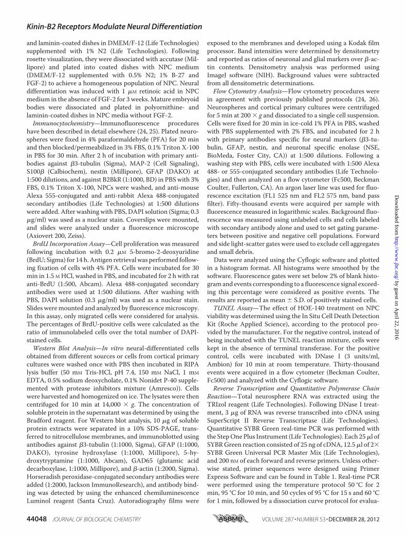
NPCs presented a radial migration pattern closely linked to agradient of maturation (Fig. 5). Fig. 5A shows representativeimages of differentiated neurospheres, where the regionenclosed between the dotted lines comprises95% ofmigratingcells. Cells migrated 15% farther from the edge of neurospheres
in the presence of Bk compared with control cultures. Con-versely, blockade of B2BkR byHOE-140 treatment resulted in a25% smaller migration distance despite displaying the sameradial pattern. These results suggest that alteration ofmigrationmay also influence neurogenesis and gliogenesis.
FIGURE 5. Bradykinin enhances neurogenesis, whereas HOE-140 promotes gliogenesis in neurosphere differentiation. A, phase-contrast images rep-resenting radial migration pattern after 7 days of neural differentiation in the presence of 1 �M bradykinin (Bk) or 1 �M HOE-140. The region enclosed betweenthe dotted lines comprises 95% of migrated cells. Scale � 100 �m. B, neural markers gene expression was changed upon B2BkR inhibition. Note that the GFAPand S100� expression levels were increased, whereas �3-tubulin expression was decreased. The data are representative of three independent experimentsconducted in triplicate and show as mean � S.D. (n � 4) (*, p 0.05). C, immunostaining of rat neurospheres differentiated in the presence of 1 �M Bk or 1 �M
HOE-140. Scale, 20 �m. D, transcription factor and neural marker gene expression was changed upon B2BkR inhibition or activation. The data are representativeof three independent experiments conducted in triplicate and shown as mean � S.D. (*, p 0.05 by Student’s t test compared with control data. Ngn1(neurogenin 1), Bk, p � 0.8304; HOE-140, p � 0.0098; notch 1, Bk, p � 0.0139; HOE-140 p � 0.0038; Stat3, Bk, p � 0.0178; HOE-140, p � 0.0008; NeuroD1, Bk, p �0.0302). E, flow cytometry analysis of GFAP, �3-tubulin, neuronal specific enolase (NSE), and nestin expression in rat neurospheres differentiated for 7 days inthe presence of Bk, HOE-140 or captopril, and Bk. Representative histograms compare expression levels of neural markers in differentiated rat neurospheres,treated with Bk, HOE-140, and captopril � Bk. F, analysis of �3-tubulin, GFAP, and S100� expression in mouse neurospheres differentiated for 7 days in thepresence of 1 �M bradykinin or 1 �M MEN-11270 (MEN), a B2BkR antagonist. The data shown are representative of at least two independent experiments. Theblank histograms in gray reveal fluorescence emission data in the absence of primary antibodies. The data shown are representative of at least five independentexperiments.
Kinin-B2 Receptors Modulate Neural Differentiation
DECEMBER 28, 2012 • VOLUME 287 • NUMBER 53 JOURNAL OF BIOLOGICAL CHEMISTRY 44053
by guest on April 22, 2016
http://ww
w.jbc.org/
Dow
nloaded from

Additionally, chronic treatment of NPCs with HOE-140decreased the expression of �3-tubulin by 25 � 13% (p �0.0477), whereas GFAP and S100� expression levels were sig-nificantly increased by 65 � 9 (p � 0.0024) and 31 � 8% (p �0.0032), respectively (Fig. 5B). These results indicate, for thefirst time, an important role of the B2BkR in neural fate deter-mination, where inhibition of B2BkR activity favors gliogenesisover neurogenesis. B2BkR-mediated effects were confirmedby microscopic analysis of immunostained cells (Fig. 5C).Although gliogenesis was reduced, neurogenesis visualized by�3-tubulin expressionwasmuchmore evident in neurospherestreated with 1 �M Bk throughout the course of differentiationwhen compared with neurospheres differentiated in theabsence of the Bk or in the presence of HOE-140.We further investigated the expression of neurogenic and
transcription factors genes, such as ngn1 (neurogenin 1),notch1, Stat3 (signal transducer and activator of transcription3), andNeuroD1, in differentiatedNPCs in the absence or pres-ence of HOE-140 or Bk. Real-time PCR revealed that the treat-ment with HOE-140 significantly increased the expression ofgenes related to gliogenesis (notch 1 and Stat3), whereas in thepresence of Bk a significant difference of NeuroD1 expressionwas obtained compared with the control group (Fig. 5D). Con-versely, Ngn1 expression levels were decreased with HOE-140treatment and notch 1 levels diminished after Bk treatment.Flow cytometry analysis revealed that the expression of the
neuronal markers �3-tubulin and NSE following Bk treatmentwere increased from 69.4� 7.3 to 87.1� 3.0% and 59.8� 3.9 to78.6� 4.1%, respectively (Fig. 5E). Co-treatment with captopriland Bk greatly increased the percentage of NSE� cells, reaching87.5 � 4.8% after ACE inhibition, given by the increased avail-ability of Bk. In contrast, prolonged activation of B2BkR
decreased the glial population from 38.0 � 2.7 to 20.8 � 8.2%,whereas the population of nestin� cells did not show any sig-nificant variation, remaining at 22%. Chronic treatment withHOE-140 also altered the phenotypic population features; how-ever, this treatment showed a bias of gliogenesis. The percent-age of GFAP� cells almost doubled from 38.0 � 2.7 to 67.2 �5.3%. Percentages of nestin� cells did not change significantlyunder Bk treatment. Similar results were obtained by flowcytometry analysis of mouse NPCs differentiated in the pres-ence of Bk or MEN-11270 (another B2BkR specific inhibitor)(Fig. 5F).Effects of Bk on neural fate determinationmay depend on the
time of application, i.e. its action could be more evident at thebeginning or end of differentiation, considering other externaland internal factors participating in this process. Thus, Bk wasadded and removed at specific times during differentiation:0–2, 2–4, or 4–7 days. Quantification of glia and neuronalpopulations by flow cytometry revealed that most significantfavoring of neurogenesis by Bk occurred in intermediate andlate days of differentiation (Fig. 6). Although discrete, theexpression of �3-tubulin during this period peaked (84.8 �5.2%) and was comparable with those obtained by chronictreatment with Bk along the whole course of differentiation.This may be related mainly to the migration of cells from neu-rospheres, which is enhanced at intermediate and late stages ofdifferentiation.On the other hand, the decreasedGFAP expres-sion wasmost evident when cells were treated with Bk betweendays 4 and 7 (11.3 � 4.1%), in agreement with reversal of theproliferation blockade at the end of differentiation in the pres-ence of HOE-140.Additionally to immunocytochemistry and flow cytometry,
we usedWestern blot analysis to evaluate relative protein con-
FIGURE 6. Role of B2BkR on different days of rat neurospheres differentiation. Bradykinin was applied between days 0 –2, 2– 4, and 4 –7, and was removedat the end of each period. Representative flow cytometry histograms comparing the expression of neural markers in control differentiated neurospheres(black) or at different times of treatment with 1 �M bradykinin (Bk), between days 0 –2 (yellow), 2– 4 (blue), and 4 –7 (red). The most significant effects on neuraldifferentiation were observed in intermediate and final phases. The data shown are representative of at least four independent experiments.
Kinin-B2 Receptors Modulate Neural Differentiation
44054 JOURNAL OF BIOLOGICAL CHEMISTRY VOLUME 287 • NUMBER 53 • DECEMBER 28, 2012
by guest on April 22, 2016
http://ww
w.jbc.org/
Dow
nloaded from

tents of�3-tubulin andGFAP in different cellmodels (Fig. 7). Inaccordance with the results so far shown,Western blot analysisshowed no change in the number of neuronal and glial proteinsin primary cortical cultures previously treated with HOE-140or Bk. We noticed a clear increase in �3-tubulin protein aftertreatmentwith Bk or captopril�Bk in rat neurosphere,murineP19, and human iPS cell differentiation. The opposite effect,increased GFAP content, could be observed after differentia-tion of the same models of rat, mouse, and human cells in thepresence of the specific blocker of B2BkR, HOE-140 (Fig. 7,A–D). To verify that promotion of neurogenesis resulted fromB2BkR activation, neurospheres from the B2BkR knock-outmice were differentiated in the presence of 1 �M Bk or 1 �M
HOE-140. After these treatments we found no significantchange in neural marker protein content, confirming that neu-ral differentiationwas specificallymodulated by B2BkR and notby possible side effects of HOE-140 treatment (Fig. 7E). Thepresence of Bk along differentiation did not induce any changesof subpopulation-specific neurotransmitter expression. Im-
munoblotting against tyrosine hydroxylase (dopaminergicmarker), 5-hydroxytryptamine (serotoninergic marker), andGAD65 (GABAergic marker) in neurospheres did not revealany differences in expression of these markers (Fig. 7F).Neurogenesis Is Favored via B2BkR Only in Differentiating
Cells—For confirming specific neurogenic roles of Bk, weinvestigated whether these effects occurs only in the differenti-ation process or could also be observed in differentiated neuralcells. For this purpose, we used cortical primary cultured cellsof newborn rats treated for 7 days without or with 1 �M Bk or 1�M HOE-140. Flow cytometry analysis of cortical primary cul-tures pre-treated with Bk or HOE-140 did not reveal anychange in percentages of neuronal and glial cells (Fig. 8A).Thus, the occurrence of neurons and glia remained constantregardless of treatment, suggesting that the effect of neuronalcell enrichment by Bk via B2BkR occurs only during the processof differentiation.Considering the influence of the B2BkR inmodulating neural
differentiation by promoting neurogenesis, we assessedwhether this effect would be caused by enzymatic cleavage ofBk with consequent formation of metabolites and activation ofthe B1BkR. In this context, rat neurospheres were plated anddifferentiated in the presence of 1 �M Lys-[des-Arg9]-Bk, anagonist of the B1BkR, or 1 �M R-715, a specific B1BkR antago-nist. After this period, we performed flow cytometry analysis toquantify neural marker expression (Fig. 8B). There was nochange in the number of cells expressing neural markers�3-tu-bulin and GFAP. Thus, the phenotypic fate determination dur-ing neural differentiation does not appear to be influenced bythe B1BkR. It is noteworthy that mRNA transcription codingfor B1BkR during neural differentiation could not be detectedin real-time PCR analysis.Differentiated Neurospheres Derived from B2BkR�/� Mice
Show a Reduction in Neural Migration—Further confirmationof modulation of neurogenesis by Bk and its receptor wasobtained in B2BkR knock-out mice. The use of B2BkR�/� miceto obtain neurospheres allowed further study of the process ofcell differentiation and neuralmigration. To verify the homozy-gosis in knock-out mice, genomic DNA was extracted fromsmall biopsies of the animals and amplified by PCRwith specificprimers for B2BkR and rate genes. B2BkR�/� mice-derivedneurospheres revealed the same growth rates of wild-type neu-rospheres, without visiblemorphological changes. After platingand induction of neural differentiation of B2BkR�/� neuro-spheres, we observed the same radial pattern, although withdecreased migration when compared with control neuro-spheres from wild-type animals (Fig. 9). The quantification ofcell migration between the dotted lines is shown in Fig. 9A. Inaddition, immunocytochemical analysis revealed the same pat-tern characterized mainly by radial GFAP� cells and a lowmigration of�3-tubulin� cells inB2BkR�/�mice neurospheres(Fig. 9B). The immunostaining also reveals less �3-tubulin�
cells (72%) and a high content of GFAP� cells (47%) inB2BkR�/�.Developmental Expression of B2BkR and Its Effect on Neural
Marker Expression during Brain Development—The B2BkR isubiquitously and constitutively expressed in adult healthy tis-sues. To assess whether it is also expressed in developing mice,
FIGURE 7. Western blot analysis of neural marker expression. A, immuno-blots of protein extracts from newborn rat cortical primary culture after treat-ment with 1 �M Bk, 1 �M HOE-140, or 50 mM captopril � 1 �M
bradykinin. B, densitometry of protein extracts from differentiated rat neuro-spheres (n � 4). C, densitometry of protein extracts from human-inducedpluripotent stem cells (n � 2). D, densitometry of protein extracts from differ-entiated mouse P19 carcinoma cells (n � 3). E, densitometry of proteinextracts of differentiated mouse neurospheres obtained from B2BkR knock-out mice (n � 2). F, immunoblots of protein extracts from differentiated ratneurospheres after treatment with 1 �M bradykinin along differentiation (TH,tyrosine hydroxylase; 5-HT, 5-hydroxytryptamine; GAD65, glutamic aciddecarboxylase).
Kinin-B2 Receptors Modulate Neural Differentiation
DECEMBER 28, 2012 • VOLUME 287 • NUMBER 53 JOURNAL OF BIOLOGICAL CHEMISTRY 44055
by guest on April 22, 2016
http://ww
w.jbc.org/
Dow
nloaded from

expression ofB2BkRwas determined in embryos removed frompregnant dams at various neurogenic developmental timepoints (E9.5–E12.5) by whole mount in situ hybridization withantisense RNA probes (Fig. 10). Mouse B2BkR transcripts weredetected in neural cells at day E9.5, starting in the optic vesicle(Fig. 10A), then increasing their expression pattern to thewholenervous system at days E11.5 (Fig. 10B) and E12.5 (Fig. 10C).Negative controls with B2BkR sense probes did not reveal anyspecific labeling (Fig. 10D). Here, we show for the first time thatB2BkR is strongly expressed in the developing mouse brain,including telencephalon, diencephalon, and ventral region ofmidbrain and hindbrain aswell as in the spinal cord. For furtheranalysis of the role of B2BkR in developing brains, we verifiedthe expression of �3-Tubulin in the telencephalon and cortexat several time points duringWT and B2BkR�/� mice develop-ment (E9.5-adult) (Fig. 10E). The developing knock-out micebrains showed significantly less expression of �3-Tubulin fromE11.5 until adulthood (*, p 0.05; adult, p � 0.0334; E9.5, p �0.0861; E11.5, p� 0.4349; E14.5, p� 0.0004; E17.5, p� 0.0008;
P0, p � 0.0001). Adult B2BkR�/� brain express more glialmarkers, such as GFAP (*, p 0.05; adult, p � 0.0083) andS100� (*, p 0.05; adult, p � 0.0001) (Fig. 10F). These dataindicate that the B2BkR�/� brain expresses less neuronalmarker and higher levels of glial markers, indicating that Bk-induced actions occur not only during in vitro neural differen-tiation, but are also important for in vivo neurogenesis.
DISCUSSION
Bk actions in neurogenesis are suggested based on its partic-ipation in determining the cholinergic phenotype of differenti-ating cells (19), induction of calcium waves (34, 35), neuriteformation (36–38), and cell migration. Moreover, Bertram etal. (39) demonstrated increased migration of human mono-cytes induced by Bk. In glioma cells, Lu et al. (40) reportedaugmented migration in the presence of Bk, but this effectwas reproduced by B1BkR agonists. In another study,increased migration of chondrosarcoma cells was related tothe Bk-activated signaling cascade (41). In summary, Bk or
FIGURE 8. Flow cytometry analysis of �3-tubulin and GFAP expression in cortical primary culture and neurosphere differentiated in the presence of1 �M Lys-[des-Arg9]-bradykinin and R-715. A, cortical cells of newborn rats were cultured in the absence or presence of 1 �M bradykinin or 1 �M HOE-140 for7 days. Representative flow cytometry histograms comparing the expression of neural markers in differentiated neurospheres (black), treated with bradykinin(blue) or HOE-140 (red) (n � 4). B, representative flow cytometry histograms comparing the expression of neural markers in differentiated neurospheres (black),treated with a B1BkR inhibitor, R-715 (blue), or a B1BkR agonist, Lys-[des-Arg9]-bradykinin (red) (n � 3). The blank histogram represents data obtained in theabsence of primary antibodies.
Kinin-B2 Receptors Modulate Neural Differentiation
44056 JOURNAL OF BIOLOGICAL CHEMISTRY VOLUME 287 • NUMBER 53 • DECEMBER 28, 2012
by guest on April 22, 2016
http://ww
w.jbc.org/
Dow
nloaded from

its degradation products participate via B1BkR or B2BkRactivation in processes similar to those occurring duringneurogenesis, such as neurite outgrowth, cell migration, andmaturation.
In this context, several other factors can participate in earlycell fate determination induced by Bk, including hormones (42)and amyloid-� precursor protein (43). Gallego and co-workers(42) demonstrated that inhibiting hormone signaling pre-vented the differentiation of embryonic stem cells aggregatesinto neuroectodermal cells. Porayette and co-workers (43)showed that the inhibition of amyloid-� precursor protein for-mation significantly suppressed human embryonic stem cellproliferation and promoted NPC formation. Interestingly,there is evidence that sex steroids alter B2BkR expression, andthat Bk affects amyloid-� precursor protein processing (44, 45)and increases its secretion. Moreover, due to possible regula-tion of production and secretion of hormones, growth factorsand other substances by Bk, both, direct and indirect effectsevoked by this peptide in neural fate determination are possible.In this regard, further investigation of the changes in musca-rinic and cholinergic receptor expression and activity in condi-tions of chronic B2BkR inhibition will provide clues on thesemechanisms.Here we have defined novel functions for Bk and its receptor
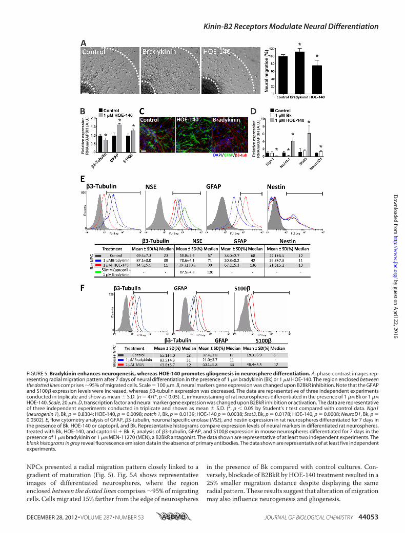
using rat embryonic telencephalon neurospheres as an in vitromodel for early cortex neurogenesis and gliogenesis (Fig. 11).Besides intracellular calcium signaling, Bk promotes NO pro-duction, essential for the progress of neurogenesis. In agree-ment with a recently published study of our group, any inter-ference with the production of arginine, the substrate for NOproduction, or with NOS activity interferes with the differenti-ation process (30). Subsequently, deficient B2BkR signaling inthe presence of HOE-140, resulting in impaired neurogenesis,
FIGURE 9. Neural migration and differentiation of neurospheres obtained from B2BkR�/� mice. Differentiation of neurospheres obtained from embry-onic telencephalon (E12.5) of wild type (mNPC wt) and B2BkR knock-out mice (B2BkR�/� mNPC). A, phase-contrast images of radial migration pattern after 7days of neural differentiation. The region enclosed between the dotted lines comprises 95% of cells that migrated. Scale, 200 �m (n � 2). B, immunofluores-cence staining of dissociated B2BkR�/� mNPC against �3-tubulin and GFAP protein revealed an increase in the number of glial cells compared with wild typemNPC. Scale, 20 �m (n � 2) (*, p 0.05).
FIGURE 10. Expression pattern of B2BkR during mouse embryo develop-ment and neuronal marker expression in telecephalons from B2BkRknock-out and wild-type mice. Whole mount in situ hybridization of mouseembryos with B2BkR antisense probe (A–C) and B2BkR sense probe (used as acontrol, D). At stage E9.5, B2BkR expression is restricted to the optic vesicle (A),strong B2BkR expression was observed in the developing nervous system (Band C), fb, forebrain; hb, hindbrain; mb, midbrain; ov, optic vesicle; sc, spinalcord. E, �3-tubulin neuronal marker gene expression during WT and B2BkR�/�
mouse brain development. The B2BkR�/� embryos express less of the markerduring several time points of brain development (n � 6) (*, p 0.05 by two-way analysis of variance with Bonferroni post-test compared with WT. Adult,p � 0.0334; E9.5, p � 0.0861; E11.5, p � 0.4349; E14.5, p � 0.0004; E17.5, p �0.0008; P0, p � 0.0001). F, GFAP and S100� glial marker gene expression of WTand B2BkR�/� mice brain. Adult B2BkR�/� brain reveal more gene expressionof glial proteins, such as GFAP (*, p 0.05 by two-way analysis of variance,Adult, p � 0.0083) and S100� (*, p 0.05 by two-way analysis of variance,Adult, p � 0.0001).
Kinin-B2 Receptors Modulate Neural Differentiation
DECEMBER 28, 2012 • VOLUME 287 • NUMBER 53 JOURNAL OF BIOLOGICAL CHEMISTRY 44057
by guest on April 22, 2016
http://ww
w.jbc.org/
Dow
nloaded from
is also reflected by down-regulated expression of NOS and thestep-limiting enzyme ASS.B2BkR expression was evident throughout differentiation of
neurospheres into neurons and glial cells accompanied byreduction of expression of the neural progenitor marker nestinand an increase in expression of neuronal �3-tubulin and NSEas well as of GFAP and S100�, identifying glial cells. Bk wasreleased into the culture medium during phenotypic transitionof undifferentiated cells into specialized neural cells (24). Fur-ther evidence for a functional kallikrein-kinin system is given bythe expression of ACE, limiting Bk half-life in the extracellularfluid; however, the B1BkR could not be detected, both onexpression and activity levels.These data agree with previous work of our laboratory sug-
gesting the presence of an autocrine loop systemof Bk secretionand receptor activation during neuronal differentiation of P19embryonal carcinoma cells, in which the blockade of receptoractivation suppressed Bk liberation into the medium and led toinhibition ofM1–M3muscarinic receptor expression in neuro-nal-differentiated P19 cells (19). Based on these observationsduring differentiation of an embryonic cell model, we ques-tioned now whether these B2BkR functions are also present inan in vitromodel closely reflecting conditions occurring duringembryonic cortex development in a network of migrating cells.As found during in vitro neurogenesis of P19 cells, gene
expression of M1–M4 receptors and muscarine-induced[Ca2�]i transients were reduced following inhibition of B2BkR
activity during neurosphere differentiation. Phenotypicchanges observed in neurospheres differentiated in the pres-ence of HOE-140 included alterations in purinergic receptorexpression and activities. Suppression of P2X5 andP2X6 recep-tor subunit expression, known to be regulated during neuronaldevelopment (46), is consistent with an inhibitory effect ofB2BkR blockade on the progress of neurogenesis. Scemes et al.(47) reported a reduction in neural outgrowth by blocking P2Y1receptor activity during neurosphere differentiation, whereasthe relative population of neurons and glial cells remainedunchanged (48). These results are in agreement with the down-regulation of P2Y1 receptor expression due to HOE-140 treat-ment and subsequent decreased neural migration, agreeingwith important roles of this receptor in neural proliferation andmigration (49).There are growing evidence that points at regulatory func-
tions of NO in the development of the CNS, including cell pro-liferation and fate determination (50–52). The mechanism ofregulating proliferation/differentiation depends on the NOSisoform involved in NO production (29). In this context,expression of enzymes of the citrulline-NO cycle includingeNOS, nNOS, and argininosuccinate synthetase was alsodown-regulated in the presence of HOE-140. As a possiblemechanism, B2BkR activity controls key events includingexpression of the machinery necessary for NO formation,which is essential for cell fate determination and guidance ofmaturation into neurons expressing specific neurotransmitterreceptors (50).Effects of B2BkR inhibition on final neural phenotype deter-
mination did not result in increased cell death rate or in thepermanence of differentiating cells in the progenitor stage.Moreover, neurogenesis, measured by an increase in the num-ber of �3-tubulin� cells, augmented with the distance of mi-gration from undifferentiated neurosphere cell aggregates,whereas cells thatmigrated less showed higher labeling for nes-tin and GFAP. Therefore, migration is linked directly to neuro-nal differentiation, and gliogenesis yet occurs due to prolifera-tion of GFAP� cells. A direct participation of B2BkR in cellmigration was confirmed with neurospheres isolated fromB2BkR knock-out mice, where just as in the presence of HOE-140 migrated distances were reduced. On the other hand,changes in the percentages of �3-tubulin� and GFAP� cellsinduced by chronic treatment with HOE-140 were notobserved in primary cultures of postnatal cortex neurons indi-cating that effects only occur during neural development andnot when final neural fate determination and differentiationhave already happened.A possible molecular mechanism for Bk-induced neural fate
determination can be delineated by the expression of neuralmarkers and transcription factors related to neurogenesis/glio-genesis switches in vivo. Wnt activation in proliferating neuralprogenitors followed by up-regulation ofNgn1 expression pro-motes the expression of genes related to neurogenesis such asNeuroD1 (53). At the same time, gliogenesis controlled byNgn1is induced by activation of Stat3 and expression of GFAP (54).Actually, the cooperation between Smad, Stat, and p300 pro-tein is particularly effective for promoting gliogenesis in NPCs(55, 56). Associated to this molecular machinery, notch 1 regu-
FIGURE 11. Bradykinin promotes neurogenesis via B2BkR activation. Fol-lowing plating, neural stem cells spontaneously differentiate into neurons(red) and glial cells (green). However, when B2BkR activity is blocked by HOE-140, the progress of neurogenesis is inhibited. The addition of Bk to NPCcultures decreases proliferation and promotes migration and neural differen-tiation following activation of NeuroD1 and down-regulation of Notch1expression, whereas specific inhibition of the B2BkR reduces neurogenesisand augments gliogenesis following up-regulation of Notch1 and Stat3 anddown-regulation of Ngn (neurogenin 1) expression. The increase in neurogen-esis of NPCs by Bk is yet enhanced in the presence of captopril, an inhibitorACE, augmenting the half-time of this peptide in the culture medium. Neuro-genic actions exerted by Bk also involved NO production, because expressionof key enzymes of the NO-citrulline cycle was down-regulated as result ofB2BkR inhibition (30).
Kinin-B2 Receptors Modulate Neural Differentiation
44058 JOURNAL OF BIOLOGICAL CHEMISTRY VOLUME 287 • NUMBER 53 • DECEMBER 28, 2012
by guest on April 22, 2016
http://ww
w.jbc.org/
Dow
nloaded from

lates interactions between physically adjacent cells and its acti-vation leads to a potent inhibition of neurogenesis, whereascommitting the cells to an astrocyte phenotype (57, 58). In thiscontext, activation and inhibition of B2BkR can interfere withthe expression and activity of some of these transcription fac-tors, thereby changing cell fate. However, the cause-conse-quence relationship between B2BkR downstream signaling andthe expression of neurogenic genes is not well understood.Neurogenesis was even more enhanced when Bk was added
to the culture medium together with captopril, increasing Bkhalf-life. In fact, this observation reveals new strategies forstrengthening neurogenesis, even in the adult organism follow-ing insults like stroke and in neurodegenerative diseases. Inview of that, stable B2BkR agonists and ACE inhibitors maygain therapeutic applications for cellular therapy. It is expectedthat these compounds will also induce endogenous neurogen-esis and provide adequate niches for transplanted stem cells tosurvive.Less �3-tubulin expression during development of B2BkR
knock-out animals points to crucial participation of B2BkRduring in vivo neurogenesis, being in line with previous resultsshowing that neurogenic activities of exogenously added kal-likrein or kallikrein gene transfer in an animal model dependson B2BkR activity (59–61). Xia et al. (61) suggested that theinsertion of tissue kallikrein genes by viral infection in newbornmice promotes ischemic neuroprotection by stimulating glialmigration, neurogenesis, and inhibition of apoptosis in theinjured area, mainly related to increased levels of phospho-Akt,Bcl-2, andNO, in addition to decreased activation of caspase-3.The observed effects can be explained by the increased avail-ability of Bk and subsequent activation of B2BkR, because theywere reversed by pretreatment with HOE-140. Such neuropro-tective features were recently described for an in vitromodel ofhippocampal neurons where Bk reversed apoptosis induced byNMDA-mediated excitotoxicity (62).Bk-induced changes in neural fate determination do not
involve alterations in populations of excitatory glutamatergicand inhibitory GABAergic neurons nor of dopaminergic neu-rons as judged by comparison of global expression levels ofneurotransmitters. These results agree with those of calciumimaging assays showing no interference with glutamate recep-tor activity following chronic treatment with HOE-140 alongdifferentiation. On the other hand, purinergic and muscarinicacetylcholine-receptor expression and activity were affected bythe presence of HOE-140. These results are again in line withthe suggestion for neurogenic actions of Bk, having inmind thatboth receptor systems contribute to the progress of neuronaldifferentiation (63, 64).França et al. (65) showed that expression of B2BkR increases
during early rat organogenesis (E8) and stabilizes during fetalgrowth (E15). Most importantly, besides being stronglyexpressed in the whole nervous system during the neurogenicstage of embryo development, B2BkR-induced neurogenesisand inhibition of gliogenesis were conserved throughout differ-ent models of neurogenesis, even in iPS cells reprogrammed topluripotency from adult somatic cells. Our work provides newtools for directing differentiating cells into homogeneous pop-ulations of neurons in vitro for posterior transplantation. In this
regard the results obtained with human iPS cells are extremelyvaluable. In summary, neurogenic properties of Bk describedherein may open novel avenues for therapy of neurodevelop-mental and neurodegenerative diseases.
REFERENCES1. Rakic, P. (1988) Specification of cerebral cortical areas. Science 241,
170–1762. Bayer, S. A., and Altman, J. (1991) Neocortical Development, First Ed.,
Raven Press, New York3. Noctor, S. C., Martínez-Cerdeño, V., Ivic, L., and Kriegstein, A. R. (2004)
Cortical neurons arise in symmetric and asymmetric division zones andmigrate through specific phases. Nat. Neurosci. 7, 136–144
4. Pearson, B. J., and Doe, C. Q. (2004) Specification of temporal identity inthe developing nervous system. Annu. Rev. Cell Dev. Biol. 20, 619–647
5. Campbell, K. (2005) Cortical neuron specification. It has its time andplace. Neuron 46, 373–376
6. Muotri, A. R., and Gage, F. H. (2006) Generation of neuronal variabilityand complexity. Nature 441, 1087–1093
7. Gross, R. E.,Mehler,M. F.,Mabie, P. C., Zang, Z., Santschi, L., and Kessler,J. A. (1996) Bonemorphogenetic proteins promote astroglial lineage com-mitment by mammalian subventricular zone progenitor cells.Neuron 17,595–606
8. Cameron, H. A., Hazel, T. G., and McKay, R. D. (1998) Regulation ofneurogenesis by growth factors and neurotransmitters. J. Neurobiol. 36,287–306
9. Buznikov, G. A., Shmukler, Y. B., and Lauder, J. M. (1996) From oocyte toneuron. Do neurotransmitters function in the same way throughout de-velopment? Cell Mol. Neurobiol. 16, 537–559
10. Bhoola, K. D., Figueroa, C. D., and Worthy, K. (1992) Bioregulation ofkinins. Kallikreins, kininogens, and kininases. Pharmacol. Rev. 44, 1–80
11. Madeddu, P., Emanueli, C., and El-Dahr, S. (2007)Mechanisms of disease.The tissue kallikrein-kinin system in hypertension and vascular remodel-ing. Nat. Clin. Pract. Nephrol. 3, 208–221
12. Sainz, I. M., Pixley, R. A., and Colman, R.W. (2007) Fifty years of researchon the plasma kallikrein-kinin system. From protein structure and func-tion to cell biology and in vivo pathophysiology. Thromb. Haemost. 98,77–83
13. Walker, K., Perkins, M., and Dray, A. (1995) Kinins and kinin receptors inthe nervous system. Neurochem. Int. 26, 1–16; discussion 17–26
14. Raidoo, D. M., and Bhoola, K. D. (1998) Pathophysiology of the kallikrein-kinin system inmammalian nervous tissue. Pharmacol. Ther. 79, 105–127
15. Calixto, J. B., Cabrini, D. A., Ferreira, J., and Campos, M.M. (2000) Kininsin pain and inflammation. Pain 87, 1–5
16. Kansui, Y., Fujii, K., Goto, K., and Abe, I. (2002) Bradykinin enhancessympathetic neurotransmission in rat blood vessels. Hypertension 39,29–34
17. Eurin, J., Barthélemy,C.,Masson, F., Soualmia,H., Sarfati, E., andCarayon,A. (2002) Bradykinin-induced neuropeptide Y release by human pheo-chromocytoma tissue. Neuropeptides 36, 257–262
18. Wang, H., Kohno, T., Amaya, F., Brenner, G. J., Ito, N., Allchorne, A., Ji,R. R., andWoolf, C. J. (2005) Bradykinin produces pain hypersensitivity bypotentiating spinal cord glutamatergic synaptic transmission. J. Neurosci.25, 7986–7992
19. Martins, A. H., Resende, R. R., Majumder, P., Faria, M., Casarini, D. E.,Tárnok, A., Colli, W., Pesquero, J. B., and Ulrich, H. (2005) Neuronaldifferentiation of P19 embryonal carcinoma cells modulates kinin B2 re-ceptor gene expression and function. J. Biol. Chem. 280, 19576–19586
20. Borkowski, J. A., Ransom, R. W., Seabrook, G. R., Trumbauer, M., Chen,H., Hill, R. G., Strader, C. D., and Hess, J. F. (1995) Targeted disruption ofa B2 bradykinin receptor gene in mice eliminates bradykinin action insmooth muscle and neurons. J. Biol. Chem. 270, 13706–13710
21. Negraes, P. D., Schwindt, T. T., Trujillo, C. A., and Ulrich, H. (2011)Neural differentiation of P19 carcinoma cells and primary neurospheres,cell morphology, proliferation, viability and functionality. Curr. Protoc.Stem. Cell Biol. 2, 2D.9
22. Marchetto, M. C., Carromeu, C., Acab, A., Yu, D., Yeo, G. W., Mu, Y.,
Kinin-B2 Receptors Modulate Neural Differentiation
DECEMBER 28, 2012 • VOLUME 287 • NUMBER 53 JOURNAL OF BIOLOGICAL CHEMISTRY 44059
by guest on April 22, 2016
http://ww
w.jbc.org/
Dow
nloaded from

Chen, G., Gage, F. H., and Muotri, A. R. (2010) A model for neural devel-opment and treatment of Rett syndrome using human induced pluripo-tent stem cells. Cell 143, 527–539
23. Takahashi, K., Tanabe, K., Ohnuki, M., Narita, M., Ichisaka, T., Tomoda,K., and Yamanaka, S. (2007) Induction of pluripotent stem cells from adulthuman fibroblasts by defined factors. Cell 131, 861–872
24. Martins, A. H., Alves, J. M., Trujillo, C. A., Schwindt, T. T., Barnabé, G. F.,Motta, F. L, Guimaraes, A. O., Casarini, D. E., Mello, L. E., Pesquero, J. B.,and Ulrich, H. (2008) Kinin-B2 receptor expression and activity duringdifferentiation of embryonic rat neurospheres. Cytometry A 73, 361–368
25. Schwindt, T. T., Trujillo, C. A., Negraes, P. D., Lameu, C., and Ulrich, H.(2011) Directed differentiation of neural progenitors into neurons is ac-companied by altered expression of P2X purinergic receptors. J.Mol. Neu-rosci. 44, 141–146
26. McLaren, F. H., Svendsen, C. N., Van der Meide, P., and Joly, E. (2001)Analysis of neural stem cells by flow cytometry, cellular differentiationmodifies patterns of MHC expression. J. Neuroimmunol. 112, 35–46
27. Henrique, D., Adam, J., Myat, A., Chitnis, A., Lewis, J., and Ish-Horowicz,D. (1995) Expression of a � homologue in prospective neurons in thechick. Nature 375, 787–790
28. Estrada, C., and Murillo-Carretero, M. (2005) Nitric oxide and adult neu-rogenesis in health and disease. Neuroscientist 11, 294–307
29. Cárdenas, A.,Moro,M.A., Hurtado,O., Leza, J. C., and Lizasoain, I. (2005)Dual role of nitric oxide in adult neurogenesis. Brain. Res. Brain. Res. Rev.50, 1–6
30. Lameu, C., Trujillo, C. A., Schwindt, T. T., Negraes, P. D., Pillat, M. M.,Morais, K. L., Lebrun, I., and Ulrich, H. (2012) Interactions between theNO-citrulline cycle and BDNF in differentiation of neural stem cells.J. Biol. Chem. 287, 29690–29701
31. Hagg, T. (2005) Molecular regulation of adult CNS neurogenesis, an inte-grated view. Trends. Neurosci. 28, 589–595
32. Ma, D. K.,Ming, G. L., and Song, H. (2005) Glial influences on neural stemcell development, cellular niches for adult neurogenesis.Curr. Opin. Neu-robiol. 15, 514–520
33. Ghashghaei, H. T., Lai, C., and Anton, E. S. (2007) Neuronal migration inthe adult brain. Are we there yet? Nat. Rev. Neurosci. 8, 141–151
34. Purkiss, J., Murrin, R. A., Owen, P. J., and Boarder, M. R. (1991) Lack ofphospholipase D activity in chromaffin cells, bradykinin-stimulated phos-phatidic acid formation involves phospholipase C in chromaffin cells butphospholipase D in PC12 cells. J. Neurochem. 57, 1084–1087
35. Bush, A. B., Borden, L. A., Greene, L. A., and Maxfield, F. R. (1991) Nervegrowth factor potentiates bradykinin-induced calcium influx and releasein PC12 cells. J. Neurochem. 57, 562–574
36. Kozlowski, M. R., Rosser, M. P., Hall, E., and Longden, A. (1989) Effects ofbradykinin on PC-12 cell differentiation. Peptides 10, 1121–1216
37. Reber, B. F., and Schindelholz, B. (1996) Detection of a trigger zone ofbradykinin-induced fast calcium waves in PC12 neurites. Pflugers. Arch.432, 893–903
38. Lorenzon, P., Zacchetti, D., Codazzi, F., Fumagalli, G., Meldolesi, J., andGrohovaz, F. (1995) Ca2� waves in PC12 neurites. A bidirectional, recep-tor-oriented form of Ca2� signaling. J. Cell Biol. 129, 797–804
39. Bertram, C.M., Baltic, S., Misso, N. L., Bhoola, K. D., Foster, P. S., Thomp-son, P. J., and Fogel-Petrovic, M. (2007) Expression of kinin B1 and B2receptors in immature, monocyte-derived dendritic cells and bradykinin-mediated increase in intracellular Ca2� and cell migration. J. LeukocyteBiol. 81, 1445–1454
40. Lu, D. Y., Leung, Y. M., Huang, S. M., andWong, K. L. (2010) Bradykinin-induced cell migration and COX-2 production mediated by the brady-kinin B1 receptor in glioma cells. J. Cell Biochem. 110, 141–150
41. Yang, W. H., Chang, J. T., Hsu, S. F., Li, T. M., Cho, D. Y., Huang, C. Y.,Fong, Y. C., and Tang, C. H. (2010) Bradykinin enhances cell migration inhuman chondrosarcoma cells through Bk receptor signaling pathways.J. Cell Biochem. 109, 82–92
42. Gallego,M. J., Porayette, P., Kaltcheva,M.M.,Meethal, S. V., andAtwood,C. S. (2009) Opioid and progesterone signaling is obligatory for early hu-man embryogenesis. Stem Cells Dev. 18, 737–740
43. Porayette, P., Gallego, M. J., Kaltcheva, M. M., Bowen, R. L., VadakkadathMeethal, S., andAtwood, C. S. (2009) Differential processing of amyloid-�
precursor protein directs human embryonic stem cell proliferation anddifferentiation into neuronal precursor cells. J. Biol. Chem. 284,23806–23817
44. Nitsch, R. M., Kim, C., and Growdon, J. H. (1998) Vasopressin and brady-kinin regulate secretory processing of the amyloid protein precursor ofAlzheimer’s disease. Neurochem. Res. 23, 807–814
45. Racchi, M., Ianna, P., Binetti, G., Trabucchi, M., and Govoni, S. (1998)Bradykinin-induced amyloid precursor protein secretion. A protein ki-nase C-independent mechanism that is not altered in fibroblasts frompatients with sporadic Alzheimer’s disease. Biochem. J. 330, 1271–1275
46. Burnstock, G., and Ulrich, H. (2011) Purinergic signaling in embryonicand stem cell development. Cell Mol. Life. Sci. 68, 1369–1394
47. Scemes, E., Duval, N., and Meda, P. (2003) Reduced expression of P2Y1receptors in connexin43-null mice alters calcium signaling and migrationof neural progenitor cells. J. Neurosci. 23, 11444–11452
48. Lin, J. H., Takano, T., Arcuino, G., Wang, X., Hu, F., Darzynkiewicz, Z.,Nunes, M., Goldman, S. A., and Nedergaard, M. (2007) Purinergic signal-ing regulates neural progenitor cell expansion and neurogenesis.Dev. Biol.302, 356–366
49. Ulrich, H., Abbracchio, M. P., and Burnstock, G. (2012) Extrinsic puriner-gic regulation of neural stem/progenitor cells, implications for CNS de-velopment and repair. Stem Cell Rev. 8, 755–767
50. Packer, M. A., Stasiv, Y., Benraiss, A., Chmielnicki, E., Grinberg, A.,West-phal, H., Goldman, S. A., and Enikolopov, G. (2003)Nitric oxide negativelyregulates mammalian adult neurogenesis. Proc. Natl. Acad. Sci. U.S.A.100, 9566–9571
51. Gibbs, S. M. (2003) Regulation of neuronal proliferation and differentia-tion by nitric oxide.Mol. Neurobiol. 27, 107–120
52. Covacu, R., Danilov, A. I., Rasmussen, B. S., Hallén, K., Moe,M. C., Lobell,A., Johansson, C. B., Svensson, M. A., Olsson, T., and Brundin, L. (2006)Nitric oxide exposure diverts neural stem cell fate from neurogenesis to-wards astrogliogenesis. Stem Cells 24, 2792–2800
53. Ma, Q., Fode, C., Guillemot, F., and Anderson, D. J. (1999) Neurogenin1and neurogenin2 control two distinct waves of neurogenesis in developingdorsal root ganglia. Genes Dev. 13, 1717–1728
54. Sun, Y.,Nadal-Vicens,M.,Misono, S., Lin,M.Z., Zubiaga, A.,Hua, X., Fan,G., and Greenberg, M. E. (2001) Neurogenin promotes neurogenesis andinhibits glial differentiation by independent mechanisms. Cell 104,365–376
55. Nakashima, K., Wiese, S., Yanagisawa, M., Arakawa, H., Kimura, N., Hi-satsune, T., Yoshida, K., Kishimoto, T., Sendtner, M., and Taga, T. (1999)Developmental requirement of gp130 signaling in neuronal survival andastrocyte differentiation. J. Neurosci. 19, 5429–5434
56. Foshay, K. M., and Gallicano, G. I. (2008) Regulation of Sox2 by STAT3initiates commitment to the neural precursor cell fate. Stem Cells Dev. 17,269–278
57. Morrison, S. J., Perez, S. E., Qiao, Z., Verdi, J. M., Hicks, C., Weinmaster,G., and Anderson, D. J. (2000) Transient Notch activation initiates anirreversible switch from neurogenesis to gliogenesis by neural crest stemcells. Cell 101, 499–510
58. Tanigaki, K., Nogaki, F., Takahashi, J., Tashiro, K., Kurooka, H., andHonjo, T. (2001) Notch1 and Notch3 instructively restrict bFGF-respon-sive multipotent neural progenitor cells to an astroglial fate. Neuron 29,45–55
59. Ling, L., Hou, Q., Xing, S., Yu, J., Pei, Z., and Zeng, J. (2008) Exogenouskallikrein enhances neurogenesis and angiogenesis in the subventricularzone and the peri-infarction region and improves neurological functionafter focal cortical infarction in hypertensive rats. Brain Res. 1206, 89–97
60. Xia, C. F., Yin, H., Borlongan, C. V., Chao, L., andChao, J. (2004) Kallikreingene transfer protects against ischemic stroke by promoting glial cell mi-gration and inhibiting apoptosis. Hypertension 43, 452–459
61. Xia, C. F., Yin, H., Yao, Y. Y., Borlongan, C. V., Chao, L., andChao, J. (2006)Kallikrein protects against ischemic stroke by inhibiting apoptosis andinflammation and promoting angiogenesis and neurogenesis.Hum. Gene.Ther. 17, 206–219
62. Martins, A. H., Alves, J. M., Perez, D., Carrasco, M., Torres-Rivera, W.,Eterovic, V. A., Ferchmin, P. A., and Ulrich, H. (2012) Kinin-B2 receptormediated neuroprotection after NMDA excitotoxicity is reversed in the
Kinin-B2 Receptors Modulate Neural Differentiation
44060 JOURNAL OF BIOLOGICAL CHEMISTRY VOLUME 287 • NUMBER 53 • DECEMBER 28, 2012
by guest on April 22, 2016
http://ww
w.jbc.org/
Dow
nloaded from

presence of kinin-B1 receptor agonists. PloS One 7, e3075563. Resende, R. R., Alves, A. S., Britto, L. R., and Ulrich, H. (2008) Role of
acetylcholine receptors in proliferation and differentiation of P19 embry-onal carcinoma cells. Exp. Cell Res. 314, 1429–1443
64. Resende, R. R., Britto, L. R., and Ulrich, H. (2008) Pharmacological prop-erties of purinergic receptors and their effects on proliferation and induc-tion of neuronal differentiation of P19 embryonal carcinoma cells. Int. J.Dev. Neurosci. 26, 763–777
65. França, C. E., Vicari, C. F., Piza, A. M., Geroldo, E. A., Beçak, M. L., Beçak,W., Stocco, R. C., and Lindsey, C. J. (2010) The kinin B(2) receptor genestructure, product processing and expression in adult and fetal rats. Evi-dence for gene evolution. Genet. Mol. Res. 9, 215–230
66. Ueno, S., Yamada,H.,Moriyama, T., Honda, K., Takano, Y., Kamiya,H.O.,andKatsuragi, T. (2002)Measurement of dorsal root ganglion P2XmRNAby SYBR Green fluorescence. Brain. Res. Brain. Res. Protoc. 10, 95–101
67. Lugo-Garcia, L., Filhol, R., Lajoix, A. D., Gross, R., Petit, P., and Vignon, J.(2007) Expression of purinergic P2Y receptor subtypes by INS-1 insuli-noma beta-cells. A molecular and binding characterization. Eur. J. Phar-
macol. 568, 54–6068. Bortone, F., Santos, H. A., Albertini, R., Pesquero, J. B., Costa, M. S.,
and Silva, J. A., Jr. (2008) Low level laser therapy modulates kininreceptors mRNA expression in the subplantar muscle of rat paw sub-jected to carrageenan-induced inflammation. Int. Immunopharmacol.8, 206–210
69. Wang, L., Zhang, Z. G., Zhang, R. L., Jiao, Z. X., Wang, Y., Pourabdollah-Nejad, D., LeTourneau, Y., Gregg, S. R., andChopp,M. (2006)Neurogenin1 mediates erythropoietin enhanced differentiation of adult neural pro-genitor cells. J. Cereb. Blood. Flow. Metab. 26, 556–564
70. Zheng, H., Zeng, Y., Chu, J., Kam, A. Y., Loh, H. H., and Law, P. Y. (2010)Modulations of NeuroD activity contribute to the differential effects ofmorphine and fentanyl on dendritic spine stability. J. Neurosci. 30,8102–8110
71. Lakner, A. M., Moore, C. C., Gulledge, A. A., Schrum, L. W. (2010)Daily genetic profiling indicates JAK/STAT signaling promotes earlyhepatic stellate cell transdifferentiation. World J. Gastroenterol. 16,5047–5056
Kinin-B2 Receptors Modulate Neural Differentiation
DECEMBER 28, 2012 • VOLUME 287 • NUMBER 53 JOURNAL OF BIOLOGICAL CHEMISTRY 44061
by guest on April 22, 2016
http://ww
w.jbc.org/
Dow
nloaded from
UlrichMicheli M. Pillat, Héllio D. N. de Souza, Lauro T. Turaça, José G. Abreu and Henning
Cassiano Carromeu, Alysson R. Muotri, João B. Pesquero, Débora M. Cerqueira, Cleber A. Trujillo, Priscilla D. Negraes, Telma T. Schwindt, Claudiana Lameu,
CellsKinin-B2 Receptor Activity Determines the Differentiation Fate of Neural Stem
doi: 10.1074/jbc.M112.407197 originally published online November 6, 20122012, 287:44046-44061.J. Biol. Chem.
10.1074/jbc.M112.407197Access the most updated version of this article at doi:
Alerts:
When a correction for this article is posted•
When this article is cited•
to choose from all of JBC's e-mail alertsClick here
http://www.jbc.org/content/287/53/44046.full.html#ref-list-1
This article cites 70 references, 18 of which can be accessed free at
by guest on April 22, 2016
http://ww
w.jbc.org/
Dow
nloaded from
Copyright © 2022 FDOKUMEN