Structure–function studies of Tityus serrulatus Hypotensin-I (TsHpt-I): A new agonist of B2 kinin...
10
Structure–function studies of Tityus serrulatus Hypotensin-I (TsHpt-I): A new agonist of B 2 kinin receptor Thiago Verano-Braga a, b,1 , Flávia Figueiredo-Rezende a,1 , Marcella N. Melo a , Roberto Q. Lautner b , Enéas R.M. Gomes c , Leonor T. Mata-Machado b , Antonela Murari b , Cibele Rocha-Resende a , Maria Elena de Lima a , Sílvia Guatimosim c , Robson A.S. Santos b , Adriano M.C. Pimenta a, * a Laboratório de Venenos e Toxinas Animais, Departamento de Bioquímica e Imunologia, Instituto de Ciências Biológicas, Universidade Federal de Minas Gerais, Av. Antônio Carlos, 6627, 31270-901 Belo Horizonte, Minas Gerais, Brazil b Laboratório de Hipertensão, Departamento de Fisiologia e Biofísica, Instituto de Ciências Biológicas, Universidade Federal de Minas Gerais, Av. Antônio Carlos, 6627, 31270-901 Belo Horizonte, Minas Gerais, Brazil c Laboratório de Eletrofisiologia Celular, Departamento de Fisiologia e Biofísica, Instituto de Ciências Biológicas, Universidade Federal de Minas Gerais, Av. Antônio Carlos, 6627, 31270-901 Belo Horizonte, Minas Gerais, Brazil article info Article history: Received 28 October 2009 Received in revised form 12 March 2010 Accepted 8 April 2010 Available online 22 April 2010 Keywords: Bradykinin-potentiating peptide Kinin receptor agonist Tityus serrulatus Hypotensins Structure minimization abstract In order to better understand the relationship between the primary structure of TsHpt-I – a bradykinin-potentiating peptide (BPP) isolated from the venom of the yellow scorpion Tityus serrulatus, with a non-canonical Lys residue prior to the conservative Pro-Pro doublet – and its cardiovascular effects, a series of ladder peptides were synthesized using the C-terminal portion of TsHpt-I as a template. All synthetic peptides having the Pro-Pro doublet at their C-terminal were able to potentiate the hypotensive effect of bradykinin. Conversely, only those analogues having Lys residue could induce a transient hypotension when intravenously administrated in male rats, indicating that the positive charge located toward the radical of this amino acid residue is crucial for this cardiovascular effect. Differently from all known BPPs, TsHpt-I acts as an agonist of the B 2 receptor and does not inhibit angiotensin-converting enzyme. The capacity of this peptide to activate this subtype of kinin receptor, releasing NO, was also affected by the absence of Lys’ side-chain positive charge. Moreover, this study has demonstrated that the minimization of the primary structure of TsHpt-I does not significantly alter the biological effects of this native peptide, which could be of interest for biotechnological purposes. Ó 2010 Elsevier Ltd. All rights reserved. 1. Introduction The kallikrein–kinin system is involved in the regulation of smooth-muscle contraction, endothelium-dependent vasodilatation, nociception and inflammation. Bradykinin (BK) and kallidin (Lys-BK), components of this system may generate desArg 9 -BK and desArg 10 -Lys-BK, respectively, by the action of carboxypeptidases (Kaplan et al., 2002; Kuoppala et al., 2000). These peptides activate kinin receptors in the endothelial cells, triggering the NO synthesis, a potent vasodilator agent. The broad spectrum of kinin actions is mediated by G protein-coupled receptors, pharmacologically classified as B 1 and B 2 subtypes, which have seven transmembrane domains. BK and Lys-BK have a higher affinity for the B 2 receptor (B 2 R), while desArg 9 -BK and desArg 10 -Lys-BK preferentially bind to the B 1 receptor (B 1 R). Once activated, these receptors induce the generation of the second * Corresponding author. Tel.: þ55 31 3409 2662; fax: þ55 31 3409 2614. E-mail address: [email protected] (A.M.C. Pimenta). 1 These authors have equally contributed to this paper. Contents lists available at ScienceDirect Toxicon journal homepage: www.elsevier.com/locate/toxicon 0041-0101/$ – see front matter Ó 2010 Elsevier Ltd. All rights reserved. doi:10.1016/j.toxicon.2010.04.006 Toxicon 56 (2010) 1162–1171
Transcript of Structure–function studies of Tityus serrulatus Hypotensin-I (TsHpt-I): A new agonist of B2 kinin...

ilable at ScienceDirect
Toxicon 56 (2010) 1162–1171
Contents lists ava
Toxicon
journal homepage: www.elsevier .com/locate/ toxicon
Structure–function studies of Tityus serrulatus Hypotensin-I (TsHpt-I):A new agonist of B2 kinin receptor
Thiago Verano-Braga a,b,1, Flávia Figueiredo-Rezende a,1, Marcella N. Melo a,Roberto Q. Lautner b, Enéas R.M. Gomes c, Leonor T. Mata-Machado b, Antonela Murari b,Cibele Rocha-Resende a, Maria Elena de Lima a, Sílvia Guatimosim c, Robson A.S. Santos b,Adriano M.C. Pimenta a,*
a Laboratório de Venenos e Toxinas Animais, Departamento de Bioquímica e Imunologia, Instituto de Ciências Biológicas, Universidade Federal de Minas Gerais, Av.Antônio Carlos, 6627, 31270-901 Belo Horizonte, Minas Gerais, Brazilb Laboratório de Hipertensão, Departamento de Fisiologia e Biofísica, Instituto de Ciências Biológicas, Universidade Federal de Minas Gerais, Av. Antônio Carlos,6627, 31270-901 Belo Horizonte, Minas Gerais, Brazilc Laboratório de Eletrofisiologia Celular, Departamento de Fisiologia e Biofísica, Instituto de Ciências Biológicas, Universidade Federal de Minas Gerais, Av. AntônioCarlos, 6627, 31270-901 Belo Horizonte, Minas Gerais, Brazil
a r t i c l e i n f o
Article history:Received 28 October 2009Received in revised form 12 March 2010Accepted 8 April 2010Available online 22 April 2010
Keywords:Bradykinin-potentiating peptideKinin receptor agonistTityus serrulatusHypotensinsStructure minimization
* Corresponding author. Tel.: þ55 31 3409 26622614.
E-mail address: [email protected] (A.M.C. P1 These authors have equally contributed to this
0041-0101/$ – see front matter � 2010 Elsevier Ltddoi:10.1016/j.toxicon.2010.04.006
a b s t r a c t
In order to better understand the relationship between the primary structure of TsHpt-I –a bradykinin-potentiating peptide (BPP) isolated from the venom of the yellow scorpionTityus serrulatus, with a non-canonical Lys residue prior to the conservative Pro-Prodoublet – and its cardiovascular effects, a series of ladder peptides were synthesized usingthe C-terminal portion of TsHpt-I as a template. All synthetic peptides having the Pro-Prodoublet at their C-terminal were able to potentiate the hypotensive effect of bradykinin.Conversely, only those analogues having Lys residue could induce a transient hypotensionwhen intravenously administrated in male rats, indicating that the positive charge locatedtoward the radical of this amino acid residue is crucial for this cardiovascular effect.Differently from all known BPPs, TsHpt-I acts as an agonist of the B2 receptor and does notinhibit angiotensin-converting enzyme. The capacity of this peptide to activate thissubtype of kinin receptor, releasing NO, was also affected by the absence of Lys’ side-chainpositive charge. Moreover, this study has demonstrated that the minimization of theprimary structure of TsHpt-I does not significantly alter the biological effects of this nativepeptide, which could be of interest for biotechnological purposes.
� 2010 Elsevier Ltd. All rights reserved.
9 10
1. IntroductionThe kallikrein–kinin system is involved in the regulationof smooth-muscle contraction, endothelium-dependentvasodilatation, nociception and inflammation. Bradykinin(BK) and kallidin (Lys-BK), components of this system may
; fax: þ55 31 3409
imenta).paper.
. All rights reserved.
generate desArg -BK and desArg -Lys-BK, respectively, bythe action of carboxypeptidases (Kaplan et al., 2002;Kuoppala et al., 2000). These peptides activate kininreceptors in the endothelial cells, triggering the NOsynthesis, a potent vasodilator agent.
The broad spectrum of kinin actions is mediated by Gprotein-coupled receptors, pharmacologically classified asB1 and B2 subtypes, which have seven transmembranedomains. BK and Lys-BK have a higher affinity for the B2receptor (B2R), while desArg9-BK and desArg10-Lys-BKpreferentially bind to the B1 receptor (B1R). Once activated,these receptors induce the generation of the second

T. Verano-Braga et al. / Toxicon 56 (2010) 1162–1171 1163
messengers inositol-1,4,5-triphosphate (IP3) and diac-ylglycerol (DAG), raising the intracellular concentration ofcalcium ions (Ca2þ), which, in turn, activates the nitricoxide (NO)/cyclic Guanosine monophosphate (cGMP)pathway that mediates the strong hypotensive effect ofkinins. In contrast to B2R, which is expressed constitutivelyin many tissues, B1R is up-regulated under pathophysio-logical conditions, being almost undetectable in normalstate (Blaukat, 2003; Marceau et al., 1997; Nishizuka, 1992;Regoli et al., 1993; Regoli and Barabe, 1980).
Bradykinin-potentiating peptides (BPPs), previouslydescribed by Ferreira et al. (Ferreira, 1965; Ferreira et al.,1970), are low molecular weight peptides that potentiatepharmacological effects of bradykinin (BK). These peptidesusually inhibit the angiotensin-converting enzyme (ACE),which is a crucial enzyme for blood pressure regulation(Soffer, 1976). This biological feature has led to the develop-ment of captopril, the first commercial ACE inhibitor (ACEI),currently used for treating cardiovascular diseases, such ashypertension (Cushman et al., 1979; Cushman and Ondetti,1980; Ondetti et al., 1977). Such biotechnological perspec-tivehasputBPPs inevidence, raising interest in the search fornew peptides from different sources of animal venoms, suchas snakes (Gomes et al., 2007; Ianzer et al., 2004; Jia et al.,2003; Menin et al., 2008; Murayama et al., 1997; Ondettiet al., 1971), spiders, scorpions and other arthropods(Ferreira et al., 1993,1996; Meki et al., 1995; Pimenta and DeLima, 2005; Sosnina et al., 1990; Verano-Braga et al., 2008).Most of BPPs bear a modified amino acid residue at theN-terminal position, pyroglutamic acid (Pyr), as well as thetriplet Ile-Pro-Pro at the C-terminal (Table 1).
Facilities in using micro-scale analytical techniques,such as mass spectrometry and proteomics, have led to
Table 1Primary sequences of some BPPs from different animal sources (snakes,spiders, scorpions and amphibians).
BPPs Species Sequences
BPP-9aa Bothrops jararaca <EWPRPQIPP_
BPP-10bb,d Bothrops jararaca <ENWPRPQIPP_
BPP-10ca,b,d Bothrops jararaca <ENWPHPQIPP_
BPP-11ab Bothrops jararaca <EWPRPTPQIPP_
BPP-11bc,d Bothrops jararaca <EGRAPGPPIPP_
BPP-12be Bothrops jararaca <EWGRPPGPPIPP_
BPP-IIIf Bothrops neuwiedi <EGGWPRPGPEIPP_
Lm-BPP 1g Lachesis muta WPPRPQIPP_
Phypo Xah Phyllomedusahypochondrialis
<EFRPSYQIPP_
BPP-Si Scaptocosa raptoria <EAPWPDTISPP_
K12j Buthus occitanus LRDYANRVINGGPVEAAGPPA
TsHpt-Ik Tityus serrulatus AEIDFSGIPEDIIKQIKETNAKPPA
Peptide Tl Tityus serrulatus KKDGYPVEYDRAY
<E, pyroglutamic acid. Alignments were obtained from PP residues.a Ferreira et al., 1970.b Ondetti et al., 1971.c Murayama et al., 1997.d Ianzer et al., 2004.e Hayashi et al., 2003.f Ferreira et al., 1998.g Soares et al., 2005.h Conceição et al., 2007.i Ferreira et al., 1996.j Meki et al., 1995.k Verano-Braga et al., 2008.l Ferreira et al., 1993.
a novel structure-to-function approach to prospect bioac-tive molecules in animal venoms (Conceição et al., 2007;Escoubas et al., 2008; Favreau et al., 2008; Ferreira et al.,1998; Pimenta et al., 2001; Pimenta and De Lima, 2005;Soares et al., 2005). Using this approach, our group wasable to isolate a new family of BPPs, called Tityus serrulatusHypotensins (TsHpt), from T. serrulatus scorpion venom(Verano-Braga et al., 2008). TsHpt-I, a member of thisfamily, is a random-coiled linear peptide with 25 aminoacid residues (Table 1). This peptide was able to bothpotentiate the hypotensive effect of BK and to inducea direct vasorelaxing effect, independently of BK, byendothelium- and nitric oxide (NO)-dependent mecha-nisms in rat aortic ring preparation (Verano-Braga et al.,2008). We demonstrated that TsHpt-I is not an ACE inhib-itor, which could be explained by some crucial differencesin the primary structure, when compared to classical BPPs.Such structural differences lay mainly in the presence ofa hydrophilic and positively charged residue (Lys) prior toPro-Pro doublet, instead of the hydrophobic and unchargedresidue (Ile) and in the absence of pyroglutamic acid asN-terminal residue, as found in the classical BPPs (Table 1).We also demonstrated that the observed two-phasedpharmacological activity of TsHpt-I is located toward itsC-terminal portion. However, the crucial residues and theminimal structure able to maintain these pharmacologicalfeatures were still unknown, as well as the mode of actionof this peptide.
In the present work, we aimed to perform a completestructure–function study to evaluate the amino acid resi-dues that are crucial for both BK-dependent and indepen-dent effects of TsHpt-I, as well as the minimum peptidicstructure able to keep these observed biological effects. Wealso tested the ability of TsHpt-I and its synthetic analoguesto act as agonists of the kinin receptors, which couldexplain the BK-independent observed effect of TsHpt-I.
2. Materials and methods
2.1. Reagents
Bradykinin (BK) was purchased from Bachem (Torrance,USA). BPP-9a and Captopril were purchased from SIGMA(St. Louis, USA). All reagents used in peptide synthesis werepurchased from Novabiochem (Darmstadt, DE).
2.2. Peptide synthesis
TsHpt-I analogues (Table 2) were synthesized usingmanual solid-phase synthesis by Fmoc/tBu strategy(Atherton and Sheppard, 1989). The final deprotectedpeptides were purified by the HPLC system withSupelcosil� C-18 semi-preparative column (Supelco, Bel-lefonte, USA) or Source� 5RPC ST 4.6/150 (GE Health CareBio-Sciences AB, Björkgatan, SE).
2.3. Mass spectrometry analyses
All synthetic peptides were analyzed byMALDI TOF/TOF(Bruker Daltonics, Billerica, USA) or ESI-Q-TOF Micro massspectrometers (Micromass/Waters, Manchester, UK) to
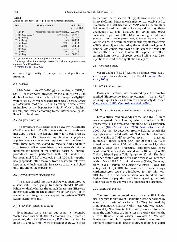
Table 2Amino acid sequences of TsHpt-I and its synthetic analogues.
Name Primary structure Molecularweight (Da)a
TsHpt-Ib AEIDFSGIPEDIIKQIKETNAKPPA 2725.09TsHpt-I[KETNAKPPA]b KETNAKPPA 953.51TsHpt-I[KPPA] KPPA 411.50TsHpt-I[PPA] PPA 283.33TsHpt-I[KPP] KPP_ 340.42TsHpt-I[acKPP] acKPP_ 382.44TsHpt-I[KP] KP__ 243.31TsHpt-I[PP] PP_ 212.25TsHpt-I[PA] PA 186.21
ac: Lys residue with its radical group acetylated.a Average values from average masses. Da: Daltons. Alignments were
obtained from PP residues.b Verano-Braga et al., 2008.
T. Verano-Braga et al. / Toxicon 56 (2010) 1162–11711164
ensure a high quality of the synthesis and purificationmethods.
2.4. Animals
Male Wistar rats (200–300 g) and wild-type C57BL/6J(25–30 g) mice were provided by the CEBIO/UFMG. Thedouble knockout mice for both kinin receptors (B1/B2
�/�)were gifted by Dr. Michael Bader fromMax Delbrück Centerfor Molecular Medicine, Berlin, Germany. Animals weremaintained at the Departamento de Fisiologia e Biofísica(UFMG) and treated according to the international guide-lines for animal care.
2.5. Surgical procedure
The day before the experiments, a polyethylene catheter(PE-10 connected to PE-50) was inserted into the abdom-inal aorta through the femoral artery for blood pressuremeasurements. For intravenous injections of the peptides,a polyethylene catheter was implanted into the femoralvein. These catheters, closed by metallic pins and filledwith isotonic saline, were driven subcutaneously into theinterscapular region of the animals’ backs. All surgicalprocedures were performed with rats under tri-bromoethanol (2.5%) anesthesia (1 mL/100 g, intraperito-neally applied). After recovery from anesthesia, rats werekept in individual cages with free access to water and chowuntil the end of the experiment.
2.6. Arterial pressure measurements
The mean arterial pressure (MAP) was monitored bya solid-state strain gauge transducer (Model TP-200T,Nihon Kohden), whereas the animals’ heart rates (HR) weredetermined with an HR counter (Model CP-640G) or ona computer through a data acquisition system (CODAS,Dataq Instruments Inc.).
2.7. Bradykinin-potentiating assay
Experiments were carried out using normotensiveWistar male rats (200–300 g) according to a procedurepreviously described (Paula et al., 1995). Initially, two BKdoses (1.0 and 2.0 nmol) were injected in bolus i.v. in order
to measure the respective BK hypotensive responses. Aninterval of 2 min between each injectionwas established toguarantee the stabilization of MAP and FC parameters.Following the administration of a unique dose of syntheticanalogues (10.0 nmol dissolved in 100 mL NaCl 0.9%),successive injections of BK (1.0 nmol) in regular intervals(every 10 min) were performed, followed by measuringDMAP values, to determine whether the hypotensive effectof BK (1.0 nmol) was affected by the synthetic analogues. Apeptide was considered having a BPP effect if it was ablestatistically to increase 1 nmol BK hypotensive effect.Animals from the control group received saline (NaCl 0.9%)injections instead of the synthetic analogues.
2.8. Aortic ring assay
Vasorelaxant effects of synthetic peptides were evalu-ated as previously described for TsHpt-I (Verano-Bragaet al., 2008).
2.9. ACE inhibition assay
Plasma ACE activity was measured by a fluorometricmethod (Fluorescence Spectrophotometer – Varian, USA)utilizing Hip-His-Leu as substrate as previously described(Santos et al., 1985; Verano-Braga et al., 2008).
2.10. Nitric oxide measurement in isolated cardiomyocytes
Left ventricle cardiomyocytes of WT and B1/B2�/� mice
were enzymatically isolated by using a solution of colla-genase type II (1 mg/mL) (Worthington, USA) as previouslydescribed (Dias-Peixoto et al., 2008; Lauton-Santos et al.,2007). For the NO detection, freshly isolated ventricularmyocytes were loaded with DAF-2FM diacetate (4-amino-5methylamino-20,70-difluororofluorescein diacetate(Molecular Probes, Eugene, USA)) for 30 min at 37 �C, ata final concentration of 10 mM in Hepes-buffered Tyrode’ssolution. After this procedure, cardiomyocytes werewashed for 30 min and stimulated with a 100 nmol/L of BK,TsHpt-I, TsHpt-I[KPP] or TsHpt-I[acKPP] for 15 min. The fluo-rescence related with the nitric oxide release was recordedwith a Meta LSM 510 confocal system (Zeiss, Germany)from CEMEL (Instituto de Ciências Biológicas, UFMG). Theantagonist of B2R, HOE-140, was used in some assays.Cardiomyocytes were pre-incubated for 15 min withHOE-140 (in a final concentration, one hundred timeshigher than the peptides used). Data regarding the nitricoxide release were analyzed as a fluorescent pixel/area.
2.11. Statistical analyses
The results are presented here as mean � SEM. Statis-tical analyses for in vitro ACE inhibition were performed byone-way analysis of variance (ANOVA) followed bya nonparametric Kruskal–Wallis test. One-way ANOVAfollowed by Newman–Keuls test was performed to deter-mine the significance of differences on DMAP values on thein vivo BK-potentiating assays. Two-way ANOVA withBonferroni multiple comparisons post-test was used tocompare concentration–response curves obtained in aortic

T. Verano-Braga et al. / Toxicon 56 (2010) 1162–1171 1165
rings. The vasodilator effect of synthetic peptides wasexpressed as a percentage decrease in maximal contractioninduced by phenylephrine. Student’s t-test was used tocomparemaximal values for the relaxant effect (Emax). One-way ANOVA following Bonferroni post-test was used foranalyzing the data of fluorescent pixels related to nitricoxide release in cardiomyocytes loaded with DAF-FM. Allstatistical analyses were considered significant whenP < 0.05.
3. Results
3.1. Structure minimization of TsHpt-I
We have recently demonstrated the importance of theC-terminal portion for TsHpt-I pharmacological effects(Verano-Braga et al., 2008). Although it was clear that theTsHpt-I anti-hypertensive activity was endothelium- andNO-dependent, the detailed mode of action of this peptidewas still unknown. Thus, using this portion as a model wesynthesized a series of peptide ladder analogues to evaluatethe smallest peptide able to maintain these biologicalfeatures. We also intended to evaluate here which aminoacids residues are crucial for maintaining the cardiovas-cular effects observed for native TsHpt-I. Accordingly, wesynthesized some TsHpt-I analogues, as follows: TsHpt-I[KPPA]; TsHpt-I[PPA]; TsHpt-I[KPP]; TsHpt-I[acKPP], inwhich thepositive charge in Lys side chain was neutralized by acet-ylation; TsHpt-I[KP]; TsHpt-I[PP] and TsHpt-I[PA] (Table 2).
3.2. Bradykinin-potentiating assay
The ability of each synthetic peptide to potentiate thehypotensive effect of BK is represented in Fig. 1. Asobserved, TsHpt-I[KPPA], TsHpt-I[KPP], TsHpt-I[acKPP], TsHpt-I[PPA] and TsHpt-I[PP] could potentiate BKwith a long-lastingeffect (over 2 h). Conversely, TsHpt-I[PA] and TsHpt-I[KP],both containing only one Pro residue, were not able topotentiate BK (Fig. 1F and G).
As observed for the native peptide TsHpt-I (Verano-Braga et al., 2008), the analogues TsHpt-I[KPPA], TsHpt-I[KPP] and TsHpt-I[KP] could induce a transient BK-indepen-dent hypotension (Fig. 1A, B and G, white bars). Thishypotensive effect could not be observed for the analoguesTsHpt-I[acKPP], TsHpt-I[PPA], TsHpt-I[PP] and TsHpt-I[PA](Fig. 1C–F), which lack the positively charged side chain,either by acetylation or by Lys absence.
3.3. Aortic ring assay
In order to better understand the physiological pathfrom which the synthetic peptides caused such hypoten-sive effect, organ-bath assays with endothelium-containing(black circle) and endothelium-denuded (white circle)aortic rings, pre-contracted with phenylephrine, were used(Fig. 2). TsHpt-I[KPPA], TsHpt-I[KPP], TsHpt-I[acKPP] and TsHpt-I[PPA] were tested and all these analogues produceda concentration-dependent vasorelaxing effect (Fig. 2,black circles). The vasorelaxation induced by these peptideswas abolished in endothelium-denuded vessels (Fig. 2,white circles). Maximal values (%) for the relaxant effect of
TsHpt-I[KPPA] (Emax) were 17.44 � 1.27 and 1.85 � 6.85 forvessel with (Eþ) or without (E�) endothelium, respectively.The Emax values for TsHpt-I[KPP] were 19.56 � �2.01 (Eþ)and 1.02 � �0.44 (E�) and for its analogue without theradical’s positive charge, TsHpt-I[acKPP], Emax values were9.97 � 1.51 (Eþ) and 0.88 � 0.84 (E�). Finally, Emax valuesfor TsHpt-I[PPA], were 14.48 � 1.49 (Eþ) and �1.13 � 0.41(E�).
The endothelium-dependent relaxation induced bythese peptides was abolished in the presence of L-NAME(100 mM), an inhibitor of the nitric oxide (NO) synthase(Fig. 3), as observed by the new values of Emax, 1.33 � 6.54;�0.44 � 0.63; 1.06 � 0.56; �0.39 � 0.58 for TsHpt-I[KPPA],TsHpt-I[KPP], TsHpt-I[acKPP] and TsHpt-I[PPA], respectively.These results indicate the participation of NO in vaso-relaxation induced by synthetic peptides.
3.4. ACE inhibition assay
In vitro assay using rat plasma as source of ACE wasperformed to ascertain whether the cardiovascular effectsinduced by the synthetic peptides could result from ACEinhibition. Also, as we wished to verify if the presence/absence of the Lys residue would somehow induce diver-gent inhibitory results, TsHpt-I[KPP] and TsHpt-I[PPA] werethen tested (Fig. 4). Two potent ACE inhibitors, captopril(Ferreira et al., 1970; Ondetti et al., 1971) and BPP-9a(Mueller et al., 2005), were used as positive controls andtwo BPPs proved to be unable to inhibit this enzyme,TsHpt-I (Verano-Braga et al., 2008) and BPP-7a (Ianzeret al., 2004), were used as negative controls. All peptideswere used in a dose–response curve, i.e. from 10�5 to 10�9
M. As expected, both captopril and BPP-9a inhibited ACEwith high efficiency (IC50 ¼ 3.5 � 10�8 mol/L and2.5�10�8 mol/L, respectively). Conversely, BPP-7a, TsHpt-I,TsHpt-I[KPP], TsHpt-I[PPA] were not able to inhibit thisenzyme, even in high concentrations, e.g. 10�5 mol/L(Fig. 4).
3.5. Nitric oxide measurement in cardiomyocytes stimulatedwith BK, TsHpt-I and its structurally minimized analogue
Ventricular cardiomyocytes express constitutively bothB1R and B2R (Couture et al., 2001; Lauton-Santos et al.,2007). In order to analyze whether the NO releaseinduced by TsHpt-I was related to the B2R activation, car-diomyocytes were chiefly loaded with DAF-FM and stim-ulated with the peptide in the absence and presence ofHOE-140, an antagonist of B2R. As shown in Fig. 5A and C,TsHpt-I was able to induce NO release in these cells, whoseeffect was abolished by HOE-140. This result suggests thatTsHpt-I is an agonist of B2R.
Cardiomyocytes were also stimulated with TsHpt-I[KPP]and TsHpt-I[acKPP], at the same conditions used for experi-ments with native peptide. Results shown in Fig. 5B indi-cate that only the peptide containing the positive charge inthe side chain of the Lys residue (TsHpt-I[KPP]) was able toinduce NO release, when compared with BK and TsHpt-I. Asobserved for native TsHpt-I, HOE-140 was also able toinhibit NO release by TsHpt-I[KPP], as depicted in Fig. 5C.

Fig. 1. Bradykinin-potentiation assay. BK (1 and 2 nmol) was injected prior to the administration of a single dose of the indicated synthetic peptides (10 nmol).After i.v. injection of (A) TsHpt-I[KPPA]; (B) TsHpt-I[KPP]; (C) TsHpt-I[acKPP]; (D) TsHpt-I[PPA]; (E) TsHpt-I[PP]; (F) TsHpt-I[PA] and (G) TsHpt-I[KP], doses of BK (1 nmol)were administrated every 10 min. MAP values were measured to evaluate if the peptide could potentiate the hypotensive effect of BK (1 nmol). MAP, meanarterial pressure; DMAP ¼ MAPfinal � MAPinitial; *P < 0.05; **P < 0.01; ***P < 0.001, comparing with values from BK (1 nmol) prior to peptide injections (n ¼ 5).
T. Verano-Braga et al. / Toxicon 56 (2010) 1162–11711166

Fig. 2. Vasodilator effects of synthetic peptides in aortic rings fromWistar rats containing (Eþ) or lacking functional endothelium (E�). Each point represents themean � SEM generated from at least 4 independent experiments. Effects on aortic rings (Eþ and E�), when testing the following peptides: (A) TsHpt-I[KPPA]; (B)TsHpt-I[PPA]; (C) TsHpt-I[KPP]; (D) TsHpt-I[acKPP]. *P < 0.05; **P < 0.01; ***P < 0.001, when comparing the related points in both curves.
T. Verano-Braga et al. / Toxicon 56 (2010) 1162–1171 1167
The role of kinin receptors in nitric oxide releaseinduced by TsHpt-I and TsHpt-I[KPP] was further investi-gated by using cardiomyocytes isolated from doubleknockout B1/B2
�/� mice. As previously described (Dias-Peixoto et al., 2008), angiotensin 1–7 (Ang(1–7)) caninduce NO release in cardiomyocytes throughMAS receptoractivation and, for this reason, it was used as a positivecontrol. Fig. 5D shows that only Ang(1–7) induced NOrelease; both TsHpt-I and TsHpt-I[KPP] were unable torelease NO.
4. Discussion
Following the discovery of BK effects’ enhancement bya peptide (Ferreira et al., 1970), several BPPs have beenisolated and biochemically and pharmacologically charac-terized. These peptides vary in size and amino acidsequences, rendering some difficulties in correlating theirprimary structure with their respective biological activities.When comparing the amino acid sequences of BPPs iso-lated from snake venoms, such correlations are undoubt-edly easier to be performed, since most of them possessa pyroglutamic acid (Pyr) residue at the N-terminal and thetriplet Ile-Pro-Pro at the C-terminal (Ferreira et al., 1998;Ianzer et al., 2004; Murayama et al., 1997; Ondetti et al.,
1971) (Table 1). However, when analyzing the primarystructure of BPPs isolated from scorpion venoms, this taskis far more complex, since the lack of other molecularfeature that could be used to compare such peptides withBPPs is evidenced, although some peptides have thedoublet Pro-Pro at C-terminal, e.g. peptide K12 (Meki et al.,1995) and TsHpt-I (Verano-Braga et al., 2008) (Table 1).Whether Peptide T (Ferreira et al., 1993) (Table 1) isincluded in this analysis, such task becomes even moredifficult, since this peptide shares no molecular featurewith other known BPPs. In this case, the only correlationbetween Peptide T and all known BPPs is made throughtheir biological activity.
Using a nonamer synthetic peptide called TsHpt-I[KET-NAKPPA] (Table 2), a TsHpt-I synthetic analogue, wedemonstrated that the C-terminal of TsHpt-I, which carriesa Pro-Pro doublet, is somehow important for TsHpt-I bio-logical activity (Verano-Braga et al., 2008).
The importance of C-terminal region of some classicalBPPs was also observed by Pimenta et al. (2007). Whenstudying the individual variability of Bothrops jararacavenom, these authors found, only in female snakes, fourpeptides originated by enzymatic cleavage of C-terminalportion of the previously well-characterized BPP-9a, BPP-10b, BPP-10c and BPP-11a (Table 1). These fragments

Fig. 3. Vasodilator effects of synthetic peptides in aortic rings from Wistar rats in the absence (control) or presence of L-NAME. Each point represents themean � SEM generated from at least 4 independent experiments. Effects on aortic rings in the absence and presence of L-NAME of the following peptides: (A)TsHpt-I[KPPA]; (B) TsHpt-I[PPA]; (C) TsHpt-I[KPP]; (D) TsHpt-I[acKPP]. *P < 0.05; **P < 0.01; ***P < 0.001, when comparing the related points in both curves.
Fig. 4. ACE inhibition in vitro assay. Inhibition of ACE activity by captopril, BPP-9a, TsHpt-I, TsHpt-I[KPP] and TsHpt-I[PPA] in different concentrations (10�5–10�9 M).Captopril and BPP-9a were used as positive controls and BPP-7a and TsHpt-I as negative controls. *P < 0.05 comparing to 100% ACE activity (control (þ)).
T. Verano-Braga et al. / Toxicon 56 (2010) 1162–11711168

Fig. 5. Nitric oxide measurement in cardiomyocytes. The isolated ventricular cardiomyocytes were loaded with DAF-FM and stimulated with different peptides.The fluorescence related with NO release was analyzed by confocal microscopy. (A) Cardiomyocytes were incubated with bradykinin and TsHpt-I in the presenceor absence of HOE. (B) Ability of TsHpt-I, TsHpt-I[KPP] and TsHpt-I[acKPP] to induce NO release. (C) The NO release by the tripeptide TsHpt-I[KPP] is also blocked bythe addition of HOE. (D) Cardiomyocytes isolated from double knockout B1/B2�/� mice were incubated with TsHpt-I, TsHpt-I[KPP] and BK. As cardiomyocytesexpress constitutively MAS receptor (Dias-Peixoto et al., 2008), Ang-(1–7) was used as a positive control. Bar graph shows average increase in DAF fluorescenceexpressed as pixels/area. *P < 0.05 comparing to control and **P < 0.01 comparing to (*) and non-significative when compared to control.
T. Verano-Braga et al. / Toxicon 56 (2010) 1162–1171 1169
peptides do not have the Gln-Ile-Pro-Pro sequence, foundin the original BBPs, and their capacity to potentiate BK isstrongly reduced (Pimenta et al., 2007). Gomes et al. (2007)evaluated some BPPs identified in Crotalus durissus terri-ficus venom gland cDNA library and demonstrated thatpeptides without the Pro-Pro doublet lack the ability topotentiate BK. However, the presence of the doublet Pro-Pro at the C-terminal is not mandatory for a peptide topotentiate BK. Mueller et al. (2005) demonstrated that,despite lacking the Pro-Pro doublet, a synthetic analogue ofBPP-9a, [1-Pro]BPP-9a (Pro-Trp-Pro-Arg-Pro-Asn-Ile-Ala-Pro) could potentiate BK, although loosing approximately40% of its potency. In the sameway, Ahb1 (Pyr-Lys-Trp-Asp-
Pro) could enhance the hypotensive effect of this hormoneon anesthetized rats (Gomes et al., 2007).
Our current findings demonstrated that this C-terminalPro-Pro doublet is crucial for the ability of TsHpt-I topotentiate the BK effect. As we have previously demon-strated that the ultimate C-terminal Ala residue of TsHpt-Iis quickly removed by carboxypeptidase Y in in vitroexperiments (Verano-Braga et al., 2008), probably aftera short time of TsHpt-I[KPPA] and TsHpt-I[PPA] in plasmathese peptides would be hydrolyzed by this class ofenzymes, releasing Lys-Pro-Pro and Pro-Pro, respectively.Thus, it is reasonable to assume that the BK potentiationeffects induced by TsHpt-I[KPPA] and TsHpt-I[PPA] are also

T. Verano-Braga et al. / Toxicon 56 (2010) 1162–11711170
caused by their proteolytic products TsHpt-I[KPP] and TsHpt-I[PP], respectively.
Mueller et al. (2005) used thewell-characterized BPP-9a(Table 1) to synthesize several analogues, varying in sizeand amino acid sequences. These authors suggested thatthe BK potentiation by BPPs is caused not only by ACEinhibition, corroborating the findings published by Ianzeret al. (2007). Our results point toward the same direction,since TsHpt-I and its synthetic analogues could potentiatethe hypotensive effect of BK, despite their inability toinhibit this enzyme. The mode of action by which the BPPspotentiate the BK effect is currently being addressed by ourand other groups and will be published elsewhere.
It is worth mentioning that TsHpt-I also induce a tran-sient and important hypotension when i.v. administrated.We hypothesized that due to a structural resemblance ofa C-terminal triplet (Lys-Pro-Pro), found in C-terminalportion of TsHpt-I, with the N-terminal of BK (Arg-Pro-Pro),this rapid and transient effect could be related with theactivation of BK receptors by this tripeptide (Lys-Pro-Pro),with the positive charge of the Lys residue playing a crucialrole in this activation. This hypothesis could be confirmedby the results obtained for synthetic analogues inwhich thepositive charge of Lys was neutralized by acetylation orsimply without the Lys residue, whichwas unable to inducethe transient hypotension previously observed (Fig.1, whitebars). The absence of the positive charge in Lys side chainalso abolished the NO release by stimulated car-diomyocytes (Fig. 5B), suggesting that this charge isessential for the interaction of these peptides with thekinin receptors. It is important in drawing attention to thefact that the presence of the positively charged Lys residueprior to the Pro-Pro doublet is a diverging structural pointof TsHpt-I and the classical BPPs (Table 1) and we assumethat it is one of the reasons for classical BPPs do not directlyactivate B2R. Nevertheless, it is worth mentioning that,despite the need of the Lys residue to such direct effect, thepresence of this amino acid prior the Pro-Pro doublet, doesnot account for the lack of ACE inhibition effect (Fig. 4).
5. Conclusions
As previously demonstrated, both native TsHpt-I andthe analogue TsHpt-I[KETNAKPPA] are able to decrease themean arterial pressure and to induce vasorelaxation fromrats in in vivo and ex vivo experiments, respectively(Verano-Braga et al., 2008). Additionally, it was alsodemonstrated that at least two pharmacological eventsoccur in the presence of such peptides: i) a fast and tran-sient hypotension that seemed to be related to NO releaseand BK-independent and; ii) a delayed BK potentiationeffect. In this study, we showed that the fast and transienthypotension occurs, indeed, due to NO release, mediated byTsHpt-I agonist effect on B2R. Such effect is also mediatedby synthetic TsHpt-I[KPP] analogue but not by TsHpt-I[acKPP],showing that the positive charge located toward the Lysside chain is crucial for this effect. Furthermore, we couldalso demonstrate that the structural minimization ofTsHpt-I did not significantly alter the cardiovascular effectsof this peptide, as shown by in vivo and ex vivo experiments.Additionally, we verified that the doublet Pro-Pro is
important to maintain the BK potentiation effect. Inconclusion, the knowledge of structure–function relation-ships of TsHpt-I by studying its synthetic minimizedpeptides can be of interest in order to envisage new anti-hypertensive drugs or even to better understand thephysiological events involved in cardiovascularhomeostasis.
Conflict of interest statement
There are no conflicts of interest.
Acknowledgments
We are greatly in debt to Júlio César dos Reis and JoséRoberto Silva for their technical assistance. This work wassupported by MCT-FINEP/FAPEMIG (Rede ProteomaNacional), FAPEMIG (Rede Mineira de Estudos de Estruturae Função de Biomoléculas and PPM), INCTTOX, CAPES andCNPq.
References
Atherton, E., Sheppard, R.C., 1989. Solid Phase Peptide Synthesis: APractical Approach. Oxford University Press, Oxford.
Blaukat, A., 2003. Structure and signalling pathways of kinin receptors.Andrologia 35, 17–23.
Conceição, K., Konno, K., Melo, R.L., Antoniazzi, M.M., Jared, C., Sciani, J.M.,Conceição, I.M., Prezoto, B.C., Camargo, A.C.M., Pimenta, D.C., 2007.Isolation and characterization of a novel bradykinin potentiatingpeptide (BPP) from the skin secretion of Phyllomedusa hypochon-drialis. Peptides 28, 515–523.
Couture, R., Harrisson, M., Vianna, R.M., Cloutier, F., 2001. Kinin receptorsin pain and inflammation. Eur. J. Pharmacol. 429, 161–176.
Cushman, D.W., Cheung, H.S., Sabo, E.F., Rubin, B., Ondetti, M.A., 1979.Development of specific inhibitors of angiotensin I convertingenzyme (kininase II). Fed. Proc. 38, 2778–2782.
Cushman, D.W., Ondetti, M.A., 1980. Inhibitors of angiotensin-convertingenzyme. Prog. Med. Chem. 17, 41–104.
Dias-Peixoto, M.F., Santos, R.A.S., Gomes, E.R., Alves, M.N., Almeida, P.W.,Greco, L., Rosa, M., Fauler, B., Bader, M., Alenina, N., Guatimosim, S.,2008. Molecular mechanisms involved in the angiotensin-(1–7)/Massignaling pathway in cardiomyocytes. Hypertension 52, 542–548.
Escoubas, P., Quinton, L., Nicholson, G.M., 2008. Venomics: unravellingthe complexity of animal venoms with mass spectrometry. J. MassSpectrom. 43, 279–295.
Favreau, P., Menin, L., Michalet, S., Perret, F., Cheneval, O., Stöcklin, M.,Bulet, P., Stöcklin, R., 2008. Mass spectrometry strategies for venommapping and peptide sequencing from crude venoms: case applica-tions with single arthropod specimen. Toxicon 47, 676–687.
Ferreira, S.H., 1965. A bradykinin-potentiating factor (BPF) present in thevenom of Bothrops jararaca. Br. J. Pharmacol. 24, 163–169.
Ferreira, S.H., Bartelt, D.C., Greene, L.J., 1970. Isolation of bradykinin-potentiating peptides from Bothrops jararaca venom. Biochemistry 9,2583–2593.
Ferreira, L.A.F., Alves, E.W., Henriques, O.B., 1993. Peptide T, a novel bra-dykinin potentiator isolated from Tityus serrulatus scorpion venom.Toxicon 31, 941–947.
Ferreira, L.A.F., Alves, W.E., Lucas, M.S., Habermehl, G.G., 1996. Isolationand characterization of a bradykinin potentiating peptide (BPP-S)isolated from Scaptocosa raptoria venom. Toxicon 34, 599–603.
Ferreira, L.A.F., Galle, A., Raida, M., Schrader, M., Lebrun, I., Habermehl, G.G., 1998. Isolation: analysis and properties of three bradykinin-potentiating peptides (BPP-II, BPP-III, and BPP-V) from Bothropsneuwiedi venom. J. Protein Chem. 17, 285–289.
Gomes, C.L., Konno, K., Conceição, I.M., Ianzer, D., Yamanouye, N.,Prezoto, B.C., Assakura, M.T., Rádis-Baptista, G., Yamane, T., Santos, R.A.S., Camargo, A.C.M., Hayashi, M.A.F., 2007. Identification of novelbradykinin-potentiating peptides (BPPs) in the venom gland ofa rattlesnake allowed the evaluation of the structure–function rela-tionship of BPPs. Biochem. Pharmacol. 74, 1350–1360.

T. Verano-Braga et al. / Toxicon 56 (2010) 1162–1171 1171
Hayashi, M.A., Murbach, A.F., Ianzer, D., Portaro, F.C., Prezoto, B.C.,Fernandez, B.L., Silveira, P.F., Silva, C.A., Pires, R.S., Britto, L.R., Dive, V.,Camargo, A.C.M., 2003. The C-type natriuretic peptide precursor ofsnake brain contains highly specific inhibitors of the angiotensin-converting enzyme. J. Neurochem. 85, 969–977.
Ianzer, D., Konno, K., Marques-Porto, R., Portaro, F.C.V., Stöcklin, R.,Camargo, A.C.M., Pimenta, D.C., 2004. Identification of five newbradykinin potentiating peptides (BPPs) from Bothrops jararacacrude venom by using electrospray ionization tandem mass spec-trometry after a two-step liquid chromatography. Peptides 25,1085–1092.
Ianzer, D., Santos, R.A.S., Etelvino, G.M., Xavier, C.H., Santos, J.A.,Mendes, E.P., Machado, L.T., Prezoto, B.C., Dive, V., Camargo, A.C.,2007. Do the cardiovascular effects of angiotensin-converting enzyme(ACE) I involve ACE-independent mechanisms? New insights fromproline-rich peptides of Bothrops jararaca. J. Pharmacol. Exp. Ther.322, 795–805.
Jia, Y.H., Li, D.S., Zhu, S.W., Zhang, L.Y., Ding, L.S., Wang, W.Y., Xiong, Y.L.,2003. Characterization of a new bradykinin-potentiating peptide(TmF) from Trimeresurus mucrosquamatus. Acta Biochim. Biophys. Sin.35, 619–623.
Kaplan, A.P., Joseph, K., Silverberg, M., 2002. Pathways for bradykininformation and inflammatory disease. J. Allergy Clin. Immunol. 109,195–209.
Kuoppala, A., Lindstedt, K.A., Saarinen, J., Kovanen, P.T., Kokkonen, J.O.,2000. Inactivation of bradykinin by angiotensin-converting enzymeand by carboxypeptidase N in human plasma. Am. J. Physiol. HeartCirc. Physiol. 278, H1069–H1074.
Lauton-Santos, S., Guatimosim, S., Castro, C.H., Oliveira, F.A., Almeida, A.P.,Dias-Peixoto, M.F., Gomes, M.A., Pessoa, P., Pesquero, J.L., Pesquero, J.B., Bader, M., Cruz, J.S., 2007. Kinin B1 receptor participates in thecontrol of cardiac function in mice. Life Sci. 81, 814–822.
Marceau, F., Larriveé, J.F., Saint-Jacques, E., Bacharov, D.R., 1997. The kininB1 receptor: an inducible G protein coupled receptor. Can. J. Physiol.Pharmacol. 75, 725–730.
Meki, A.R., Nassar, A.Y., Rochat, H., 1995. A bradykinin-potentiatingpeptide (peptide K12) isolated from the venom of Egyptian scorpionButhus occitanus. Peptides 16, 1359–1365.
Menin, L., Perchu�c, A., Favreau, P., Perret, F., Michalet, S., Schöni, R.,Wilmer, M., Stöcklin, R., 2008. High throughput screening of brady-kinin-potentiating peptides in Bothrops moojeni snake venom usingprecursor ion mass spectrometry. Toxicon 51, 1288–1302.
Mueller, S., Gothe, R., Siems, W.D., Vietinghoff, G., Paegelow, I.,Reissmann, S., 2005. Potentiation of bradykinin actions by analoguesof the bradykinin potentiating nonapeptide BPP9alpha. Peptides 26,1235–1247.
Murayama, N., Hayashi, M.A., Ohi, H., Ferreira, L.A., Hermann, V.V.,Saito, H., Fujita, Y., Higuchi, S., Fernandes, B.L., Yamane, T., Camargo, A.C.M., 1997. Cloning and sequence analysis of a Bothrops jararaca cDNA
encoding a precursor of seven bradykinin-potentiating peptides anda C-type natriuretic peptide. Proc. Natl. Acad. Sci. U.S.A. 94, 1189–1193.
Nishizuka, Y., 1992. Intracellular signaling by hydrolysis of phospholipidsand activation of protein kinase C. Science 258, 607–614.
Ondetti, M.A., Williams, N.J., Sabo, E.F., Pluscec, J., Weaver, E.R., Kocy, O.,1971. Angiotensin-converting enzyme inhibitors from the venom ofBothrops jararaca. Isolation, elucidation of structure, and synthesis.Biochemistry 10, 4033–4039.
Ondetti, M.A., Rubin, B., Cushman, D.W., 1977. Design of specific inhibitorsof angiotensin-converting enzyme: new class of orally active anti-hypertensive agents. Science 196, 441–444.
Paula, R.D., Lima, C.V., Khosla, M.C., Santos, R.A.S., 1995. Angiotensin-(1–7)potentiates the hypotensive effect of bradykinin in conscious rats.Hypertension 26, 1154–1159.
Pimenta, A.M.C., Stöcklin, R., Favreau, P., Bougis, P.E., Martin-Eauclaire, M.F., 2001. Moving pieces in a proteomic puzzle: mass fingerprinting oftoxic fractions from the venom of Tityus serrulatus (Scorpiones,Buthidae). Rapid Commun. Mass Spectrom. 15, 1562–1572.
Pimenta, A.M.C., De Lima, M.E., 2005. Small peptides, big world:biotechnological potential in neglected bioactive peptides fromarthropod venoms. J. Pept. Sci. 11, 670–676.
Pimenta, D.C., Prezoto, B.C., Konno, K., Melo, R.L., Furtado, M.F.,Camargo, A.C.M., Serrano, S.M.T., 2007. Mass spectrometric analysis ofthe individual variability of Bothrops jararaca venom peptide fraction.Evidence for sex-based variation among the bradykinin-potentiatingpeptides. Rapid Commun. Mass Spectrom. 21, 1034–1042.
Regoli, D., Barabe, J., 1980. Pharmacology of bradykinin and related kinins.Pharmacol. Rev. 32, 1–46.
Regoli, D., Jukie, D., Gobeil, F., Rhaleb, N.E., 1993. Receptors for bradykininand related kinins: a critical analysis. Can. J. Physiol. Pharmacol. 71,556–567.
Santos, R.A.S., Krieger, E.M., Greene, L.J., 1985. An improved fluorometricassay of rat serum and plasma converting enzyme. Hypertension 7,244–252.
Soares, M.R., Oliveira-Carvalho, A.L., Wermelinger, L.S., Zingali, R.B., Ho, P.L., Junqueira-de-Azevedo, I.L.M., Diniz, M.R.V., 2005. Identification ofnovel bradykinin-potentiating peptides and C-type natriureticpeptide from Lachesis muta venom. Toxicon 46, 31–38.
Soffer, R.L., 1976. Angiotensin-converting enzyme and the regulation ofvasoactive peptides. Annu. Rev. Biochem. 45, 73–94.
Sosnina, N.A., Golubenko, Z., Akhunov, A.A., Kugaevskaia, E.V., Eliseeva, I.E., Orekhovich, V.N., 1990. Bradykinin-potentiating peptides from thespider Latrodectus tredecimguttatus-inhibitors of carboxycathepsinand of a preparation of karakurt venom kininase. Dokl. Akad. NaukSSSR 315, 236–239.
Verano-Braga, T., Rocha-Resende, C., Silva, D.M., Ianzer, D., Martin-Eauclaire, M.F., Bougis, P.E., De Lima, M.E., Santos, R.A.S., Pimenta, A.M.C., 2008. Tityus serrulatus hypotensins: a new family of peptidesfrom scorpion venom. Biochem. Biophys. Res. Commun. 371, 515–520.