Meriolins, a New Class of Cell Death Inducing Kinase Inhibitors with Enhanced Selectivity for...
13
Meriolins, a New Class of Cell Death–Inducing Kinase Inhibitors with Enhanced Selectivity for Cyclin-Dependent Kinases Karima Bettayeb, 1 Oscar M. Tirado, 2 Se ´verine Marionneau-Lambot, 3 Yoan Ferandin, 1 Olivier Lozach, 1 Jonathan C. Morris, 4 Silvia Mateo-Lozano, 2 Peter Drueckes, 5,6 Christoph Scha ¨chtele, 5 Michael H.G. Kubbutat, 5 Franc ¸ois Liger, 7 Bernard Marquet, 7 Benoı ˆt Joseph, 7 Aude Echalier, 8 Jane A. Endicott, 8 Vicente Notario, 2 and Laurent Meijer 1 1 Centre National de la Recherche Scientifique, Cell Cycle Group & UPS2682, Station Biologique, Roscoff, Bretagne, France; 2 Laboratory of Experimental Carcinogenesis, Lombardi Comprehensive Cancer Center, Georgetown University Medical Center, Washington, District of Columbia; 3 Cance´ropo ˆle Grand Ouest, Nantes, France; 4 Department of Chemistry, School of Chemistry and Physics, University of Adelaide, Adelaide, Australia; 5 ProQinase GmbH, Freiburg, Germany; 6 Novartis Institutes for BioMedical Research, Expertise Platform Kinases, Basel, Switzerland; 7 ICBMS, UMR 5246, Universite´ Lyon 1, Laboratoire de Chimie Organique 1, Villeurbanne, France; and 8 University of Oxford, Laboratory of Molecular Biophysics, Department of Biochemistry, Oxford, United Kingdom Abstract Protein kinases represent promising anticancer drug targets. We describe here the meriolins, a new family of inhibitors of cyclin-dependent kinases (CDK). Meriolins represent a chem- ical structural hybrid between meridianins and variolins, two families of kinase inhibitors extracted from various marine invertebrates. Variolin B is currently in preclinical evaluation as an antitumor agent. A selectivity study done on 32 kinases showed that, compared with variolin B, meriolins display enhanced specificity toward CDKs, with marked potency on CDK2 and CDK9. The structures of pCDK2/cyclin A/variolin B and pCDK2/cyclin A/meriolin 3 complexes reveal that the two inhibitors bind within the ATP binding site of the kinase, but in different orientations. Meriolins display better anti- proliferative and proapoptotic properties in human tumor cell cultures than their parent molecules, meridianins and variolins. Phosphorylation at CDK1, CDK4, and CDK9 sites on, respectively, protein phosphatase 1A, retinoblastoma protein, and RNA polymerase II is inhibited in neuroblastoma SH-SY5Y cells exposed to meriolins. Apoptosis triggered by meriolins is accompanied by rapid Mcl-1 down-regulation, cytochrome c release, and activation of caspases. Meriolin 3 potently inhibits tumor growth in two mouse xenograft cancer models, namely, Ewing’s sarcoma and LS174T colorectal carcinoma. Meriolins thus constitute a new CDK inhibitory scaffold, with promising antitumor activity, derived from molecules initially isolated from marine organisms. [Cancer Res 2007;67(17):8325–34] Introduction Altered protein phosphorylation is a frequent feature in nume- rous human diseases. This last decade has therefore witnessed considerable efforts to identify, optimize, and evaluate pharmaco- logic inhibitors of protein kinases (reviews in refs. 1, 2). Currently, f60 kinase inhibitors are undergoing clinical evaluation against cancers, inflammation, diabetes, and neurodegenerative diseases. Among the 518 human kinases, cyclin-dependent kinases (CDK) have attracted considerable interest given their involvement in many essential physiologic pathways and aberrant activities in multiple human diseases, especially in cancer and neurodegener- ative diseases (review in ref. 3). Consequently, many pharmacologic inhibitors of CDKs have been found to display promising anti- tumor and/or neuroprotective activities (reviews in refs. 4–8). To our knowledge, eight CDK inhibitors are currently undergoing clinical evaluation as anticancer drugs [flavopiridol (Alvocidib), R -roscovitine (CYC202, Seliciclib), R547, SNS-032, PD-0332991, AZD5438, AG-024322 and AT7519]. All CDK inhibitors identified to date are ATP-competitive, and many have been co-crystallized with a CDK target (review in ref. 1). The selectivity of these pharmacologic inhibitors is a matter of extensive investigation using a wide variety of methods (review in ref. 9). Although a few kinase inhibitors are rather unselective (like staurosporine), many display a definite specificity profile, but all inhibit several kinases. For example, many CDK inhibitors also inhibit glycogen synthase kinase-3 (GSK-3; ref. 10) and sometimes casein kinase 1 (CK1). Multitarget inhibitors may find appropriate medicinal use because they are less likely to allow resistance to develop. We recently identified meridianins, a family of 3-(2-amino- pyrimidine)indoles, as a promising kinase-inhibitory scaffold (11). Meridianins are natural products initially extracted from Aplidium meridianum, an Ascidian from the South Atlantic (12). Meridianin derivatives have been synthesized by various groups (13–15). Although some meridianins inhibit various kinases such as CDKs, GSK-3, cyclic nucleotide-dependent kinases, and CK1, they dis- played modest antiproliferative effects. Meridianins share some structural similarity with variolins, another family of marine natural compounds containing a central pyrido[3¶,2¶:4,5]pyrrolo[1,2-c ] pyrimidine core substituted with a 2-aminopyrimidine ring. Vario- lins were initially extracted from Kirkpatrickia variolosa , a rare Antarctic sponge (16, 17). Total synthesis by Morris (18, 19) and others (20, 21) allowed easy access to the variolins. Variolin B and deoxyvariolin B (PM01218) display potent cytotoxicity against several human cancer cell lines (17, 22, 23); as a result, PM01218 is being investigated by PharmaMar as a potential antitumor drug. Recently, as this work was in progress, variolin B and deoxyvariolin B were reported to inhibit CDKs (23). In this article, we have exploited the chemical similarity between meridianins and variolins to synthesize a hybrid structure, the meriolins. Surprisingly, meriolins display potent inhibitory activity and relative selectivity toward CDKs and also exhibit better Note: Supplementary data for this article are available at Cancer Research Online (http://cancerres.aacrjournals.org/). Requests for reprints: L. Meijer, Centre National de la Recherche Scientifique, Cell Cycle Group & UPS2682, Station Biologique, B.P. 74, 29682 Roscoff Cedex, Bretagne, France. Phone: 330-298-29-23-39; Fax: 330-298-29-23-42; E-mail: [email protected]. I2007 American Association for Cancer Research. doi:10.1158/0008-5472.CAN-07-1826 www.aacrjournals.org 8325 Cancer Res 2007; 67: (17). September 1, 2007 Research Article
Transcript of Meriolins, a New Class of Cell Death Inducing Kinase Inhibitors with Enhanced Selectivity for...

Meriolins, a New Class of Cell Death–Inducing Kinase Inhibitors
with Enhanced Selectivity for Cyclin-Dependent Kinases
Karima Bettayeb,1
Oscar M. Tirado,2
Severine Marionneau-Lambot,3
Yoan Ferandin,1
Olivier Lozach,1
Jonathan C. Morris,4
Silvia Mateo-Lozano,2
Peter Drueckes,5,6
Christoph Schachtele,5
Michael H.G. Kubbutat,5
Francois Liger,7
Bernard Marquet,7
Benoıt Joseph,7
Aude Echalier,8
Jane A. Endicott,8
Vicente Notario,2
and Laurent Meijer1
1Centre National de la Recherche Scientifique, Cell Cycle Group & UPS2682, Station Biologique, Roscoff, Bretagne, France; 2Laboratory ofExperimental Carcinogenesis, Lombardi Comprehensive Cancer Center, Georgetown University Medical Center, Washington, Districtof Columbia; 3Canceropole Grand Ouest, Nantes, France; 4Department of Chemistry, School of Chemistry and Physics, University ofAdelaide, Adelaide, Australia; 5ProQinase GmbH, Freiburg, Germany; 6Novartis Institutes for BioMedical Research, Expertise PlatformKinases, Basel, Switzerland; 7ICBMS, UMR 5246, Universite Lyon 1, Laboratoire de Chimie Organique 1, Villeurbanne, France; and8University of Oxford, Laboratory of Molecular Biophysics, Department of Biochemistry, Oxford, United Kingdom
Abstract
Protein kinases represent promising anticancer drug targets.We describe here the meriolins, a new family of inhibitors ofcyclin-dependent kinases (CDK). Meriolins represent a chem-ical structural hybrid between meridianins and variolins, twofamilies of kinase inhibitors extracted from various marineinvertebrates. Variolin B is currently in preclinical evaluationas an antitumor agent. A selectivity study done on 32 kinasesshowed that, compared with variolin B, meriolins displayenhanced specificity toward CDKs, with marked potency onCDK2 and CDK9. The structures of pCDK2/cyclin A/variolin Band pCDK2/cyclin A/meriolin 3 complexes reveal that thetwo inhibitors bind within the ATP binding site of the kinase,but in different orientations. Meriolins display better anti-proliferative and proapoptotic properties in human tumorcell cultures than their parent molecules, meridianins andvariolins. Phosphorylation at CDK1, CDK4, and CDK9 sites on,respectively, protein phosphatase 1A, retinoblastoma protein,and RNA polymerase II is inhibited in neuroblastoma SH-SY5Ycells exposed to meriolins. Apoptosis triggered by meriolins isaccompanied by rapid Mcl-1 down-regulation, cytochrome crelease, and activation of caspases. Meriolin 3 potently inhibitstumor growth in two mouse xenograft cancer models, namely,Ewing’s sarcoma and LS174T colorectal carcinoma. Meriolinsthus constitute a new CDK inhibitory scaffold, with promisingantitumor activity, derived from molecules initially isolatedfrom marine organisms. [Cancer Res 2007;67(17):8325–34]
Introduction
Altered protein phosphorylation is a frequent feature in nume-rous human diseases. This last decade has therefore witnessedconsiderable efforts to identify, optimize, and evaluate pharmaco-logic inhibitors of protein kinases (reviews in refs. 1, 2). Currently,f60 kinase inhibitors are undergoing clinical evaluation againstcancers, inflammation, diabetes, and neurodegenerative diseases.
Among the 518 human kinases, cyclin-dependent kinases (CDK)have attracted considerable interest given their involvement inmany essential physiologic pathways and aberrant activities inmultiple human diseases, especially in cancer and neurodegener-ative diseases (review in ref. 3). Consequently, many pharmacologicinhibitors of CDKs have been found to display promising anti-tumor and/or neuroprotective activities (reviews in refs. 4–8). Toour knowledge, eight CDK inhibitors are currently undergoingclinical evaluation as anticancer drugs [flavopiridol (Alvocidib),R-roscovitine (CYC202, Seliciclib), R547, SNS-032, PD-0332991,AZD5438, AG-024322 and AT7519]. All CDK inhibitors identifiedto date are ATP-competitive, and many have been co-crystallizedwith a CDK target (review in ref. 1). The selectivity of thesepharmacologic inhibitors is a matter of extensive investigationusing a wide variety of methods (review in ref. 9). Although a fewkinase inhibitors are rather unselective (like staurosporine), manydisplay a definite specificity profile, but all inhibit several kinases.For example, many CDK inhibitors also inhibit glycogen synthasekinase-3 (GSK-3; ref. 10) and sometimes casein kinase 1 (CK1).Multitarget inhibitors may find appropriate medicinal use becausethey are less likely to allow resistance to develop.
We recently identified meridianins, a family of 3-(2-amino-pyrimidine)indoles, as a promising kinase-inhibitory scaffold (11).Meridianins are natural products initially extracted from Aplidiummeridianum , an Ascidian from the South Atlantic (12). Meridianinderivatives have been synthesized by various groups (13–15).Although some meridianins inhibit various kinases such as CDKs,GSK-3, cyclic nucleotide-dependent kinases, and CK1, they dis-played modest antiproliferative effects. Meridianins share somestructural similarity with variolins, another family of marine naturalcompounds containing a central pyrido[3¶,2¶:4,5]pyrrolo[1,2-c]pyrimidine core substituted with a 2-aminopyrimidine ring. Vario-lins were initially extracted from Kirkpatrickia variolosa , a rareAntarctic sponge (16, 17). Total synthesis by Morris (18, 19) andothers (20, 21) allowed easy access to the variolins. Variolin B anddeoxyvariolin B (PM01218) display potent cytotoxicity againstseveral human cancer cell lines (17, 22, 23); as a result, PM01218is being investigated by PharmaMar as a potential antitumor drug.Recently, as this work was in progress, variolin B and deoxyvariolinB were reported to inhibit CDKs (23).
In this article, we have exploited the chemical similarity betweenmeridianins and variolins to synthesize a hybrid structure, themeriolins. Surprisingly, meriolins display potent inhibitory activityand relative selectivity toward CDKs and also exhibit better
Note: Supplementary data for this article are available at Cancer Research Online(http://cancerres.aacrjournals.org/).Requests for reprints: L. Meijer, Centre National de la Recherche Scientifique, Cell
Cycle Group & UPS2682, Station Biologique, B.P. 74, 29682 Roscoff Cedex, Bretagne,France. Phone: 330-298-29-23-39; Fax: 330-298-29-23-42; E-mail: [email protected].
I2007 American Association for Cancer Research.doi:10.1158/0008-5472.CAN-07-1826
www.aacrjournals.org 8325 Cancer Res 2007; 67: (17). September 1, 2007
Research Article

antiproliferative and proapoptotic properties in cell cultures thantheir ‘‘inspirational parent’’ molecules. Meriolins are particularlypotent inhibitors of CDK2 and CDK9. The crystal structures ofmeriolin 3 and variolin B in complex with CDK2/cyclin A revealedthat the two molecules bind in very different orientations in theATP-binding pocket of the kinase. Meriolins prevent phosphory-lation at CDK1-, CDK4-, and CDK9-specific sites in neuroblastomaSH-SY5Y cells and induce the rapid degradation of the survivalfactor Mcl-1. Meriolin 3 potently inhibits tumor growth in twomouse xenograft models: Ewing’s sarcoma and LS174T colorectalcarcinoma. Meriolins thus constitute a new kinase-inhibitoryscaffold with promising antitumor activity.
Materials and Methods
ChemistrySynthesis of meridianins, variolins, and meriolins. Meridianins A
and G were synthesized as described previously (15). Variolin B and deoxy-
variolin B were synthesized in eight and six steps, respectively, as
described previously (18, 19). The synthesis of meriolins will be described
in detail in a separate article.9 Briefly, meriolins were prepared as follows.
Acylation of 7-azaindole derivatives (7-azaindole, 4-methoxy-7-azaindole, and
4-ethoxy-7-azaindole) in the presence of acetic anhydride and trifluoroacetic
acid afforded the 3-acetyl-7-azaindole derivatives in 55% to 84% yield. N-
benzenesulfonylation of the latter compounds was carried out in the pre-
sence of benzenesulfonyl chloride and sodium hydride in tetrahydrofuran to
give 3-acetyl-1-benzenesulfonyl-7-azaindole derivatives in 68% to 90% yield.
Treatment of the protected 7-azaindoles with N,N-dimethylformamide
dimethyl acetal in N,N-dimethylformamide afforded the corresponding
enaminones in 68% to 78% yield. Final formation of the pyrimidic nucleus
occurred by heating of enaminones in the presence of guanidine HCl and
anhydrous potassium carbonate in 2-methoxyethanol to afford meriolins 1, 3,
and 4 in 63% to 75% yield. O-demethylation of meriolin 3 was done in 48%
hydrobromic acid/acetic acid to afford meriolin 2 in 90% yield.
CrystallographyExpression, purification, and co-crystallization of human CDK2/
cyclin A with meriolin 3 and variolin B. Thr160-phosphorylated CDK2/
cyclin A (pCDK2/cyclin A) was purified as described previously (24) and
concentrated to 13 mg/mL in 40 mmol/L HEPES (pH, 7.0), 200 mmol/L
NaCl, 0.01% monothioglycerol. The protein solution was incubated for
20 min on ice with meriolin 3 (1.8 mmol/L) or variolin B (2.0 mmol/L)
before setting up hanging-drop crystallization trials. The reservoir solution
contained 0.6 to 0.8 mol/L KCl, 0.9 to 1.2 mol/L (NH4)2SO4, and 100 mmol/L
HEPES (pH, 7.0). Orthorhombic crystals grew within 3 weeks at 4jC.
Crystals were briefly soaked in 8 mol/L sodium formate before being cryo-
cooled in liquid nitrogen.
X-ray crystallography data collection and processing, structuresolution, and refinement. Data were collected from a single crystal on
ESRF ID14-EH-2 beamline at 100K. Data processing and integration were
carried out using MOSFLM and SCALA (25). The structures were solved
by molecular replacement with MOLREP using a well-refined structure
of pCDK2/cyclin A10 as the search model. Two pCDK2/cyclin A
heterodimers were found in the asymmetrical unit. Strong electron
density corresponding to the bound inhibitor was seen at the ATP site
after rigid body refinement of the two structures. Inhibitor model
building and library generation for the two inhibitors were done usingthe CCP4 software suite (25). Alternate cycles of rebuilding in Coot (26)and refinement in Refmac (27) were carried out to obtain the finalmodels.
Protein Kinase AssaysBuffers. Buffer A: 10 mmol/L MgCl2, 1 mmol/L EGTA, 1 mmol/L DTT,
25 mmol/L Tris-HCl (pH, 7.5), 50 Ag heparin/mL. Buffer C: 60 mmol/L h-glycerophosphate, 15 mmol/L p-nitrophenylphosphate, 25 mmol/L MOPS
(pH, 7.2), 5 mmol/L EGTA, 15 mmol/L MgCl2, 1 mmol/L DTT, 1 mmol/L
sodium vanadate, 1 mmol/L phenylphosphate.
Kinase preparations and assays. Kinase activities were assayed in
buffer A or C at 30jC at a final ATP concentration of 15 Amol/L. Blank
values were subtracted, and activities were expressed in percent of the
maximal activity, i.e., in the absence of inhibitors. Controls were done with
appropriate dilutions of DMSO.CDK1/cyclin B (M phase starfish oocytes, native) and CDK5/p25
(human, recombinant) were prepared as previously described (10). Kinase
activity was assayed in buffer C, with 1 mg histone H1/mL, in the
presence of 15 Amol/L [g-33P] ATP (3,000 Ci/mmol; 10 mCi/mL) in a final
volume of 30 AL. After 30 min incubation at 30jC, 25-AL aliquots of
supernatant were spotted onto 2.5 � 3-cm pieces of Whatman P81
phosphocellulose paper, and 20 s later, the filters were washed five times
( for at least 5 min each time) in a solution of 10 mL phosphoric acid/L of
water. The wet filters were counted in the presence of 1 mL ACS
(Amersham) scintillation fluid.
CDK2/cyclin A (human, recombinant, expressed in insect cells) was
assayed as described for CDK1/cyclin B.
CDK9/cyclin T (human, recombinant, expressed in insect cells) was
assayed as for CDK1/cyclin B using a pRB fragment (amino acids 773–928;
3.5 Ag/assay) as substrate.
GSK-3a/b (porcine brain, native, affinity purified) was assayed as
described for CDK1, but in buffer A and using a GSK-3–specific substrate
(GS-1: YRRAAVPPSPSLSRHSSPHQSpEDEEE; Sp stands for phosphorylated
serine; ref. 28).CK1d/e (porcine brain, native, affinity purified) was assayed as described
for CDK1, but using the CK1-specific peptide substrate RRKHAAIGSpAY-
SITA (29).ProQinase protein kinase assays. All protein kinases were expressed
in Sf9 insect cells as human recombinant glutathione S-transferase-fusion
proteins or His-tagged proteins by means of the baculovirus expression
system. Kinases were purified and assayed as described (28; Supplementary
Materials).
Cell BiologyAntibodies and chemicals. AcDEVDafc and Q-VD-OPh were purchased
from MPbiomedicals. Cell Titer 96 containing the MTS reagent and CytoTox
96 kits were purchased from Promega. The protease inhibitor cocktail was
from Roche. Unless otherwise stated, the nonlisted reagents were from
Sigma.Monoclonal antibody against actin was obtained from Calbiochem.
Monoclonal antibodies against cytochrome c and retinoblastoma protein
(Rb) were purchased from BD Biosciences. Polyclonal antibody against
phospho-Ser249/Thr252-Rb was provided by Biosource. Polyclonal antibody
against phospho-Thr320-protein phosphatase 1a (PP1a) and monoclonal
antibody against caspase-9 were from Cell Signaling. Polyclonal antibodies
against RNA polymerase II and phospho-Ser2-RNA polymerase II were
supplied by Covance Research Products. Polyclonal antibody against Mcl-1
was obtained from Santa Cruz Biotechnology.
Cell lines and culture conditions. SH-SY5Y human neuroblastoma,
Huh7 human hepatocarcinoma and LS 174T human colorectal adenocar-
cinoma cell lines were grown in DMEM (Invitrogen). The HCT116 human
colorectal carcinoma cell line was grown in McCoy’s 5a medium from the
American Type Culture Collection. The F1 rat biliary epithelial cell line
was grown on Williams’ Medium E from Invitrogen supplemented with
2 mmol/L L-glutamine from Lonza. The HEK 293 human embryonic kidney
cell line was grown in MEM from Invitrogen. Human foreskin primary
fibroblasts (kindly provided by Dr. Gilles Ponzio, Faculte de Medecine,
INSERM U634, Nice, France) were grown in DMEM supplemented with
2 mmol/L L-glutamine and 20 mmol/L HEPES. All the media were
supplemented with antibiotics (penicillin-streptomycin) from Lonza and
10% volume of FCS from Invitrogen. Cells were cultured at 37jC with 5%
9 A. Echalier et al., submitted.10 Unpublished results.
Cancer Research
Cancer Res 2007; 67: (17). September 1, 2007 8326 www.aacrjournals.org

CO2. Drug treatments were done on exponentially growing cultures at the
indicated time and concentrations. Control experiments were carried out
using appropriate dilutions of DMSO. KMS-11, multiple myeloma adherent
cell line, and GBM, a multiform glioblastoma primary culture cells were
cultured in RPMI 1640 supplemented, respectively, with 5% or 10% fetal
calf serum, 2 mmol/L glutamine, antibiotics (100 IU/mL penicillin and
100 Ag/mL streptomycin), and 10 Amol/L 2-h-mercaptoethanol (Life Tech-
nologies). Cells were subcultured at confluency after dispersal with 0.025%
trypsin in 0.02% EDTA. Their survival was estimated by the neutral red
uptake assay.9
Cell death and cell viability assessments. Cell viability was determinedby measuring the reduction of 3-(4,5-dimethylthiazol-2-yl)-5-(3-carboxyme-
thoxyphenyl)-2-(4-sulfophenyl)-2H -tetrazolium (MTS). Cell death was
determined by measuring the level of lactate dehydrogenase (LDH) activity
released upon cell lysis. Both procedures have been previously described in
detail (30).
Caspase assay. Caspase activity was measured by determining the
fluorescence released from the AcDEVDafc synthetic substrate after itsdirect addition to the culture medium, detergent lysis, and incubation at
37j. This method is devised for a 96-multiwell plate format. It allows kinetic
determinations of caspase activation and the characterization of multiple
drugs simultaneously (30).Electrophoresis and Western blotting. Cells were resuspended and
lysed in homogenization buffer [60 mmol/L h-glycerophosphate, 15 mmol/L
p-nitrophenyl phosphate, 25 mmol/L MOPS (pH, 7.2), 15 mmol/L EGTA,15 mmol/L MgCl2, 1 mmol/L DTT, 1 mmol/L sodium vanadate, 1 mmol/L
NaF, 1 mmol/L phenylphosphate, 0.1% Nonidet P-40, and protease inhibitor
cocktail] and sonicated. After centrifugation (14,000 rpm for 15 min at 4jC),
Figure 1. A, structures of meridianins, variolins, and meriolins. Selectivity of meriolin 3 (B ) and variolin B (C). The two latter compounds were run at increasingconcentrations on a ProQinase selectivity panel of 30 kinases. IC50 values are presented in micromoles per liter. ATP concentration used in the kinase assays was1 Amol/L.
Meriolins Inhibit CDKs
www.aacrjournals.org 8327 Cancer Res 2007; 67: (17). September 1, 2007

the protein concentration was determined in the supernatants by the
Bradford protein assay (Bio-Rad). To study cytochrome c release from the
mitochondria, a 0.05% digitonin cytosolic extraction was done (30).
Whole cell extracts were prepared in buffer containing 100 mmol/L
Tris-HCl (pH, 6.8), 1 mmol/L EDTA, 2% SDS, and protease inhibitor cock-
tail. Following heat denaturation for 5 min, proteins were separated on
10% or 7% NuPAGE pre-cast Bis-Tris or Tris-acetate polyacrylamide
mini gels (Invitrogen) with MOPS SDS (all but cytochrome c , RNA poly-
merase II, and phospho-Ser2-RNA polymerase II Western blots), MES SDS
(cytochrome c), or Tris-acetate SDS (RNA polymerase II and phospho-
Ser2-RNA polymerase II) running buffer depending on protein size. Proteins
were transferred to 0.45-Am nitrocellulose filters (Schleicher and Schuell).
These were blocked with 5% low-fat milk in TBS–Tween 20, incubated for
1 h with antibodies (anti-actin: 1:2,000) or overnight at 4jC (cytochrome c :
1:500), Rb (1:500), phospho-Rb (1:500), phospho-Thr320-PP1a (1:1,000), RNA
polymerase II (1:500), phospho-Ser2-RNA polymerase II (1:500), Mcl-1
(1:500), and caspase-9 (1:1,000) and analyzed by enhanced chemilumines-
cence (ECL, Amersham).
In vivo Antitumor ActivityEwing’s sarcoma. Experiments to evaluate the antitumor activity of
meriolin 3 were carried out essentially as described (31) under protocols
approved by the Georgetown University Animal Care and Use Committee,
using immunodeficient male (5–6 weeks old) athymic nude (BALB/c nu/nu)mice purchased from the U.S. National Cancer Institute, which were housed
in the animal facilities of the Georgetown University Division ofComparative Medicine. Mice were injected s.c. into the right posteriorflank with 4 � 106 A4573 Ewing’s sarcoma cells in 100 AL of Matrigelbasement membrane matrix (BD Biosciences). Once tumors reached amean volume of about 195 mm3, mice were randomized into two groups(six animals per group), and treatment was initiated. One group was treatedwith meriolin 3, dissolved in DMSO, and given as a single daily i.p. injection,at a dose of 1 mg/kg, for either 5 days or two 5-day series with a 2-day breakin between. The control group received i.p. injections of DMSO followingan identical schedule. Tumor growth was followed for up to 4 weeks afterthe first injection. Tumor volumes were calculated by the formulaV = (1/2)a � b2, where a is the longest tumor axis, and b is the shortesttumor axis. Whenever tumors reached the maximum volume allowed byinstitutional tumor burden guidelines, animals were sacrificed by asphyx-iation with CO2. Tumors were immediately excised from euthanizedanimals and measured. Data are given as mean F SD. Statistical analysisof differences between groups was done by a one-way ANOVA, followed byan unpaired Student’s t test.
LS174T colorectal carcinoma. The antitumor activity of meriolin 3 was
investigated by protocols approved by the Canceropole Grand-OuestAnimal Care and Use Committee using immunodeficient female (5 weeks
old) athymic nude (NMRI) mice purchased from Janvier. Mice were injected
s.c. into the right flank with 4 � 106 colorectal LS174T cells in 100 ALof RPMI (Invitrogen). After 8 days, mice were randomized into three groups( five animals per group), and treatment was initiated. Meriolin 3, dissolved
Table 1. Effects of meridianins, variolins, and meriolins on six protein kinases and on the survival of neuroblastoma SH-SY5Ycells (A) and effects of meriolin 3 on the survival of various cell lines (B)
A
Compound CDK1 CDK2 CDK5 CDK9 GSK3 CK1 SH-SY5Y
Meridianin A 2.50 3.10 3.00 2.40 1.30 1.10 > 30.00
Meridianin G 150.00 >10 140.00 >10 350.00 9.00 >30.00
Variolin B 0.06 0.08 0.09 0.026 0.07 0.005 0.24Deoxyvariolin B 0.34 0.21 0.43 0.041 0.71 0.022 0.22
Meriolin 1 0.78 0.09 0.51 0.026 0.63 0.20 0.67
Meriolin 2 0.057 0.018 0.050 0.018 0.40 0.05 0.41Meriolin 3 0.17 0.011 0.17 0.006 0.23 0.20 0.073
Meriolin 4 0.01 0.007 0.005 0.007 0.03 0.10 0.081
B
Cell line Cell survival (IC50, Amol/L; MTS reduction)
LS 174T (colorectal adenocarcinoma) 0.13
HCT116 (colon) 0.94
KMS-11 (myeloma) 0.12
GBM (glioma) 0.14Huh7 (hepatoma) 0.12
F1 (hepatoma) 0.26
SH-SY5Y (neuroblastoma) 0.072
HEK293 (embryon kidney) 0.38Human foreskin fibroblasts 8.00
NOTE: (A) Compounds were tested at various concentrations on CDK1/cyclin B, CDK2/cyclin A, CDK5/p25, CDK7/cyclin H, CDK9/cyclin T, GSK-3a/h,and CK1y/q as described in Materials and Methods. IC50 values, calculated from the dose-response curves (average of triplicate values), are reported inmicromoles per liter (data for meridianins from ref. 19). The compounds were tested at various concentrations for their effects on SH-SY5Y cells survival
after 48 h incubation as estimated using the MTS reduction assay. IC50 values, calculated from the dose-response curves, are reported in Amol/L. (B)
Meriolin 3 was tested at various concentrations for its effects on nine different cell lines. Cell survival was estimated 48 h after the addition of meriolin 3
using the MTS reduction assay. IC50 values (average of triplicate values) are reported in micromoles per liter.
Cancer Research
Cancer Res 2007; 67: (17). September 1, 2007 8328 www.aacrjournals.org

in 30% DMSO in 0.9% NaCl, was given as a single daily i.p. injection at a doseof 2 or 5 mg/kg for two 5-day series with a 2-day break in between. The
control group received i.p. injections of DMSO/NaCl following an identical
schedule. Tumor sizes were measured daily. Tumor volumes were calculated
by the formula V = (1/2)a � b2, where a is the longest tumor axis, and b isthe shortest tumor axis. Data are given as mean F SD. Whenever tumors
reached the maximum volume allowed by institutional tumor burden
guidelines (3,000 mm3), animals were sacrificed by cervical dislocation.
Animals were autopsied, and organs were examined macroscopically.
Results
Meriolins inhibit protein kinases, with selectivity towardCDKs. Both meridianins (11) and variolins (17, 22, 23) showdistinct antiproliferative and proapoptotic activities. These effectsmay be correlated with their inhibitory activities toward CDKs andother protein kinases (11, 23). The chemical similarity betweenmeridianins and variolins prompted us to synthesize an aza-indolehybrid structure, which we designated as a meriolin (Fig. 1A).Representatives of the three chemical families were initially testedon six purified protein kinases (CDK1/cyclin B, CDK2/cyclin A,CDK5/p25, CDK9/cyclin T, GSK-3a/h and CK1y/q; Table 1A).The moderate activity of meridianins (11) was confirmed. VariolinB was identified as a potent inhibitor of all CDKs and GSK-3,whereas deoxyvariolin B was 5- to 10-fold less potent. Interestingly,variolin B and deoxyvariolin B were very potent inhibitors ofCK1, in fact, more potent than all reported CK1 inhibitors (29).Meriolins were very potent inhibitors of CDKs, GSK-3, andCK1. CDK2 and CDK9 seemed to be particularly sensitive tomeriolins.
To evaluate their selectivity, we tested meriolin 3 and variolin Bon a selection of 30 kinases from the ProQinase selectivity panel
(Fig. 1B and C). The highest inhibitory activity of meriolin 3 wasmostly restricted to CDKs, CDK9 being particularly sensitive underthese conditions. Inhibitory activity was observed at higher dosesfor other kinases (Fig. 1). In contrast, variolin B did not display sucha striking selectivity for CDKs, although CDK9 was also a good target.Meriolin 3–CDK2/cyclin A and variolin B–CDK2/cyclin A
co-crystal structures. We next investigated the molecular mecha-nism of action of meriolin 3 and variolin B by co-crystallizationwith pCDK2/cyclin A (Fig. 2). A full description of the co-crystalstructures and comparison with previously described CDK2/inhibitors structures will be presented elsewhere.9 Meriolin andvariolin B both occupy the kinase ATP binding site located betweenthe smaller NH2-terminal and large COOH-terminal lobes (Fig. 2A).Meriolin 3 makes two hydrogen bonds to the CDK2 backbonewithin the hinge sequence that links the two lobes of the kinase: itaccepts a hydrogen bond from the backbone nitrogen of Leu83 anddonates a hydrogen bond to the backbone oxygen of Glu81 (Fig. 2B).In addition, meriolin 3 makes two hydrogen bonds with the sidechains of Lys33 and Glu51 (Fig. 2B).
Although variolin B and meriolin 3 share a substantial portionof their scaffold, the two molecules are oriented differently withinthe ATP binding cleft. Variolin B, like meriolin 3, makes twoequivalent hydrogen bonds with the CDK2 hinge and also interactswith the side chain of Lys33 through its hydroxyl group. In addition,two ordered water molecules (Fig. 2B, red stars) are discernible in thevariolin B–bound active site, and one interacts with Ile10 main chainamine. However, the presence of a third ring fused to the variolin Bindole creates a planar structure that is too large to be accommo-dated at the back of the ATP binding site if variolin B were to adoptthe meriolin 3 binding mode. As a result, the indole moiety commonto both inhibitors is flipped 180j between the two bound structures.
Figure 2. Co-crystal structure of meriolin 3 and variolin B with CDK2/cyclin A. A, structure of pCDK2/cyclin A in ribbon diagram representation. Yellow, CDK2(with variolin B) and in orange (with meriolin 3). The PSTAIRE helix (residues 45–57) is shown in magenta and the activation segment (residues 145–172) is in green.Cyclin A is shown in blue for variolin B structure and in cyan for meriolin 3 structure. Meriolin 3 is shown in magenta at the ATP binding site and variolin B in cyan.Meriolin 3, variolin B, and phospho-Thr160 are shown in ball-and-stick representation. B, details of the interactions of meriolin 3 and variolin B with the ATP bindingpocket of pCDK2/cyclin A. Meriolin 3, variolin B, and selected residues of CDK2 (from meriolin 3 structure) are shown in ball-and-stick representation in magenta, cyan,and yellow, respectively.
Meriolins Inhibit CDKs
www.aacrjournals.org 8329 Cancer Res 2007; 67: (17). September 1, 2007

Meriolins induce apoptotic cell death. We next compared thethree families of compounds for their ability to induce cell death inneuroblastoma SH-SY5Y cells as measured with an MTS reductionassay (Table 1A ; Fig. 3A). Because MTS reduction is occasionallyobserved under conditions in which cell death does not occur, wealso used an independent cell death assay, the lactate dehydroge-nase (LDH) release assay (Fig. 3B). Dose-response curves showedthat meriolins are more potent than variolin B in terms of theconcentration required to reduce cell survival (MTS reduction;Table 1A ; Fig. 3A) or in terms of cell death induction (LDH release;Fig. 3B).
To confirm that the induction of cell death by meriolins was not aspecific property of SH-SY5Y cells, we tested meriolin 3 on dif-ferent human cancer cell lines (Table 1B). A 48-h exposure induced adose-dependent reduction of cell survival in all cell lines. However,normal human foreskin primary fibroblasts were much less sensitive.
Cell death induced by meriolins and variolin B was accompaniedby a dose-dependent release of cytochrome c (Fig. 3C) and
activation of effector caspases (Fig. 3D). Meriolins were morepotent than variolin B at triggering both cytochrome c release andcaspase activation.
We next analyzed the effects of meriolins 3 and 4 and variolin Bon the phosphorylation of various proteins at CDK-specific sites.Although meriolins induced a modest reduction in Rb levels(Supplementary Figure), they strongly and dose-dependentlyreduced Rb phosphorylation at CDK4-specific sites (Ser249/Thr252;ref. 32; Fig. 4A). PP1a phosphorylation by CDK1 at Thr320 is areported marker of CDK1 activity (33, 34). Meriolins were verypotent at inhibiting CDK1-specific Thr320 phosphorylation of PP1ain a dose-dependent manner, whereas variolin B was only activeat the highest (1 Amol/L) concentration tested (Fig. 4B). RNApolymerase II is phosphorylated by CDK9 on Ser2. Although onlymeriolin 4 induced a modest reduction in RNA polymerase II level(Supplementary Figure), all three compounds inhibited CDK9-specific Ser2 phosphorylation of RNA polymerase II in a dose-dependent manner (Fig. 4C). Meriolins were more active than
Figure 3. Induction of cell death in SH-SY5Y cells by meriolins 3 and 4 and variolin B. A, SH-SY5Y cells were exposed for 48 h to increasing concentrations of thethree compounds. Cell survival was estimated by the MTS reduction assay and is expressed in % of survival in untreated cells. Average F SE of one out of threeindependent experiments, done in triplicates. B, a similar experiment was done, but LDH release was assayed 24 h after the addition of the compounds. Average F SEof three independent measurements per experiment. C, Induction of the apoptosis pathway: SH-SY5Y cells were exposed for 24 h to increasing concentrationsof the three compounds. Cells were then harvested and fractionated into a nuclear pellet and a cytoplasmic supernatant. The latter was resolved by SDS-PAGEfollowed by Western blotting using an anti-cytochrome c antibody. D, induction of caspases activation. Cells were exposed for 24 h to increasing concentrations ofthe three compounds. DEVDase activity was measured as arbitrary fluorescence units. Points, mean of at least three independent determinations; bars, SE.Sts, staurosporine 0.25 Amol/L.
Cancer Research
Cancer Res 2007; 67: (17). September 1, 2007 8330 www.aacrjournals.org

variolin B. We next tested the effects of the three compounds onthe level of the short-lived survival factor Mcl-1 (Fig. 4D). Westernblot analysis showed that meriolins induced an almost completedisappearance of Mcl-1, whereas variolin B was essentially inactive.In vivo antitumor activity of meriolin 3. A nude mouse tumor
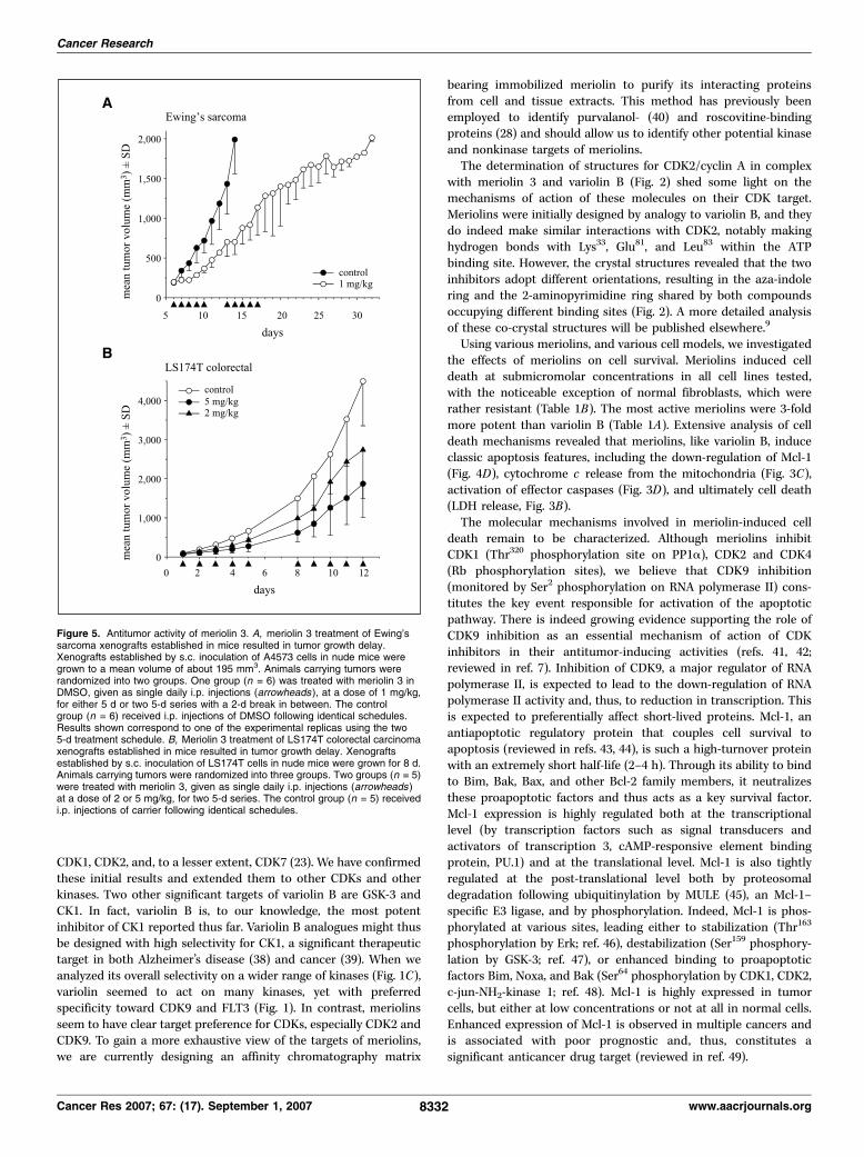
xenograft model system (31) was used to evaluate the antitumoreffect of meriolin 3 in vivo . Results showed that as early as 3 daysafter treatment initiation, meriolin 3 had slowed tumor growth byabout 54% (Fig. 5A). Five days later (day 14), when tumors incontrol animals reached an average volume about 2,000 mm3, andanimals had to be sacrificed, tumors in meriolin 3–treated animalshad reached an average volume of only f700 mm3 (f65% tumorgrowth inhibition). The inhibitory effect of meriolin 3 on tumorgrowth was also manifested by the fact that tumors in meriolin3–treated animals took an additional period of about 20 days toreach the maximum volume allowed by institutional tumor burdenstandards (Fig. 5A). We used a second nude mouse tumor xenograftmodel to confirm these in vivo antitumor effects (Fig. 5B). LS174Tcolorectal cells were injected in mice, and once small tumors wereestablished, animals were injected daily (2 � 5 days sequence) withmeriolin 3 at a final concentration of 2 or 5 mg/kg. At the end
of the experiment, meriolin 3 had induced 59% (5 mg/kg) or 40%(2 mg/kg) inhibition of tumor growth (Fig. 5B).
Discussion
Compared with terrestrial microorganisms and plants, marineorganisms constitute a relatively untapped source of bioactivemolecules. In recent years, however, an exponentially growingnumber of original structures derived from marine microorgan-isms, plants, and animals have been identified, some of which arereaching clinical stages and progressing to the pharmaceuticalmarket (reviews in refs. 35–37).
We here report on meriolins, a family of kinase inhibitorsdesigned from two different classes of marine natural products, themeridianins (Ascidian) and the variolins (Sponge). Meridianins hadbeen reported as modest and rather unselective kinase inhibitors(11). Variolins have been extensively studied following thediscovery of their potent antiproliferative and cell death–inducingproperties. They are currently undergoing preclinical studies aspotential antitumor agents (PharmaMar). As this work was inprogress, variolin B and deoxyvariolin B were reported to inhibit
Figure 4. Inhibition of phosphorylation at CDK-specific sites and down-regulation of Mcl-1. SH-SY5Y cells were exposed to increasing concentrations of meriolin 3,meriolin 4, or variolin B for 24 h, and proteins were resolved by SDS-PAGE followed by Western blotting with antibodies phospho-Rb (A ), phospho-Thr320 PP1a(B), phospho-Ser2-RNA polymerase II (C ), and Mcl-1 (D ). Western blotting with anti-actin antibodies was used as a loading control (Supplementary Materials).Etp, etoposide, 12.5 Amol/L. Representative of two to three independent experiments.
Meriolins Inhibit CDKs
www.aacrjournals.org 8331 Cancer Res 2007; 67: (17). September 1, 2007
CDK1, CDK2, and, to a lesser extent, CDK7 (23). We have confirmedthese initial results and extended them to other CDKs and otherkinases. Two other significant targets of variolin B are GSK-3 andCK1. In fact, variolin B is, to our knowledge, the most potentinhibitor of CK1 reported thus far. Variolin B analogues might thusbe designed with high selectivity for CK1, a significant therapeutictarget in both Alzheimer’s disease (38) and cancer (39). When weanalyzed its overall selectivity on a wider range of kinases (Fig. 1C),variolin seemed to act on many kinases, yet with preferredspecificity toward CDK9 and FLT3 (Fig. 1). In contrast, meriolinsseem to have clear target preference for CDKs, especially CDK2 andCDK9. To gain a more exhaustive view of the targets of meriolins,we are currently designing an affinity chromatography matrix
bearing immobilized meriolin to purify its interacting proteinsfrom cell and tissue extracts. This method has previously beenemployed to identify purvalanol- (40) and roscovitine-bindingproteins (28) and should allow us to identify other potential kinaseand nonkinase targets of meriolins.
The determination of structures for CDK2/cyclin A in complexwith meriolin 3 and variolin B (Fig. 2) shed some light on themechanisms of action of these molecules on their CDK target.Meriolins were initially designed by analogy to variolin B, and theydo indeed make similar interactions with CDK2, notably makinghydrogen bonds with Lys33, Glu81, and Leu83 within the ATPbinding site. However, the crystal structures revealed that the twoinhibitors adopt different orientations, resulting in the aza-indolering and the 2-aminopyrimidine ring shared by both compoundsoccupying different binding sites (Fig. 2). A more detailed analysisof these co-crystal structures will be published elsewhere.9
Using various meriolins, and various cell models, we investigatedthe effects of meriolins on cell survival. Meriolins induced celldeath at submicromolar concentrations in all cell lines tested,with the noticeable exception of normal fibroblasts, which wererather resistant (Table 1B). The most active meriolins were 3-foldmore potent than variolin B (Table 1A). Extensive analysis of celldeath mechanisms revealed that meriolins, like variolin B, induceclassic apoptosis features, including the down-regulation of Mcl-1(Fig. 4D), cytochrome c release from the mitochondria (Fig. 3C),activation of effector caspases (Fig. 3D), and ultimately cell death(LDH release, Fig. 3B).
The molecular mechanisms involved in meriolin-induced celldeath remain to be characterized. Although meriolins inhibitCDK1 (Thr320 phosphorylation site on PP1a), CDK2 and CDK4(Rb phosphorylation sites), we believe that CDK9 inhibition(monitored by Ser2 phosphorylation on RNA polymerase II) cons-titutes the key event responsible for activation of the apoptoticpathway. There is indeed growing evidence supporting the role ofCDK9 inhibition as an essential mechanism of action of CDKinhibitors in their antitumor-inducing activities (refs. 41, 42;reviewed in ref. 7). Inhibition of CDK9, a major regulator of RNApolymerase II, is expected to lead to the down-regulation of RNApolymerase II activity and, thus, to reduction in transcription. Thisis expected to preferentially affect short-lived proteins. Mcl-1, anantiapoptotic regulatory protein that couples cell survival toapoptosis (reviewed in refs. 43, 44), is such a high-turnover proteinwith an extremely short half-life (2–4 h). Through its ability to bindto Bim, Bak, Bax, and other Bcl-2 family members, it neutralizesthese proapoptotic factors and thus acts as a key survival factor.Mcl-1 expression is highly regulated both at the transcriptionallevel (by transcription factors such as signal transducers andactivators of transcription 3, cAMP-responsive element bindingprotein, PU.1) and at the translational level. Mcl-1 is also tightlyregulated at the post-translational level both by proteosomaldegradation following ubiquitinylation by MULE (45), an Mcl-1–specific E3 ligase, and by phosphorylation. Indeed, Mcl-1 is phos-phorylated at various sites, leading either to stabilization (Thr163
phosphorylation by Erk; ref. 46), destabilization (Ser159 phosphory-lation by GSK-3; ref. 47), or enhanced binding to proapoptoticfactors Bim, Noxa, and Bak (Ser64 phosphorylation by CDK1, CDK2,c-jun-NH2-kinase 1; ref. 48). Mcl-1 is highly expressed in tumorcells, but either at low concentrations or not at all in normal cells.Enhanced expression of Mcl-1 is observed in multiple cancers andis associated with poor prognostic and, thus, constitutes asignificant anticancer drug target (reviewed in ref. 49).
Figure 5. Antitumor activity of meriolin 3. A, meriolin 3 treatment of Ewing’ssarcoma xenografts established in mice resulted in tumor growth delay.Xenografts established by s.c. inoculation of A4573 cells in nude mice weregrown to a mean volume of about 195 mm3. Animals carrying tumors wererandomized into two groups. One group (n = 6) was treated with meriolin 3 inDMSO, given as single daily i.p. injections (arrowheads ), at a dose of 1 mg/kg,for either 5 d or two 5-d series with a 2-d break in between. The controlgroup (n = 6) received i.p. injections of DMSO following identical schedules.Results shown correspond to one of the experimental replicas using the two5-d treatment schedule. B, Meriolin 3 treatment of LS174T colorectal carcinomaxenografts established in mice resulted in tumor growth delay. Xenograftsestablished by s.c. inoculation of LS174T cells in nude mice were grown for 8 d.Animals carrying tumors were randomized into three groups. Two groups (n = 5)were treated with meriolin 3, given as single daily i.p. injections (arrowheads )at a dose of 2 or 5 mg/kg, for two 5-d series. The control group (n = 5) receivedi.p. injections of carrier following identical schedules.
Cancer Research
Cancer Res 2007; 67: (17). September 1, 2007 8332 www.aacrjournals.org

We propose that meriolins act through at least three mechanisms:(a) by inhibiting CDK9, they transiently reduce both RNA polymeraseII level (Supplementary Figure) and activity (Fig. 4C) and, conse-quently, down-regulate Mcl-1 expression levels (Fig. 4D); (b) byinhibiting CDK1, they reduce sequestering of proapoptotic factors byMcl-1; and (c) by inhibiting CDK1 and CDK2, they block cell cycleprogression both at the G1-S and G2-M transition, possibly leading toE2F-1–dependent apoptosis (41). As a consequence ofMcl-1 reductionand inhibition of its sink effects, proapoptotic regulators are liberatedand trigger cell death. This CDK1/CDK2/CDK9 inhibitionmechanismmight be general for antitumor CDK inhibitors that are developed forclinical use against cancers (7, 41, 42).
Our results showed that meriolin 3 has potent antitumor activity.The A4573 Ewing’s tumor xenograft model has been usedpreviously to test the antitumor activity of roscovitine, anotherCDK inhibitor (31). In this regard, it seems that, in this xenograftmodel system with the same type of treatment schedule, meriolin 3is a more potent inhibitor of tumor growth than roscovitine.At the time control animals had to be sacrificed to follow ins-titutional tumor burden guidelines, roscovitine had caused anf85% inhibition of tumor growth (39), whereas meriolin 3 had anf65% inhibition. However, it is important to point out thatroscovitine was used at a concentration 50-fold higher than that ofmeriolin 3 (50 mg/kg versus 1 mg/kg, respectively), indicating thatmeriolin 3 is still f40-fold more efficient at inhibiting the growth
of Ewing’s sarcoma xenografts and strongly suggesting thatmeriolin 3 may be an effective cancer therapeutic agent. Similarpromising in vivo antitumor effects were observed with lowconcentrations of meriolin 3 using LS174T colorectal carcinoma innude mice. These experiments are only exploratory; meriolins needto be optimized, and their administration needs to be exploredfurther with respect to dosing, timing, and combination with otherdrugs. Nevertheless, this family of small-molecular-weight com-pounds, derived from marine natural products, as pharmacologicinhibitors of CDKs, display promising antitumor activity thatrequires further studies.
Acknowledgments
Received 5/17/2007; revised 7/3/2007; accepted 7/11/2007.Grant support: EEC (FP6-2002-Life Sciences & Health, PRO-KINASE Research
Project; L. Meijer, M. Kubbutat, and J. Endicott), ‘‘Canceropole Grand-Ouest’’ grant(L. Meijer and S. Marionneau-Lambot), the Australian Research Council (J.C. Morris),and US NIH grant PO1-CA74175 (O.M. Tirado and V. Notario). K. Bettayeb wassupported by a fellowship from the Ministere de la Recherche.
The costs of publication of this article were defrayed in part by the payment of pagecharges. This article must therefore be hereby marked advertisement in accordancewith 18 U.S.C. Section 1734 solely to indicate this fact.
This article is dedicated to Dr. George R. Pettit, a pioneer in anticancer marinenatural products, with friendship and respect. We are grateful to Dr. M. Clement forproviding the cell survival data for GBM cells and KMS-11 cells, to Dr. G. Ponzio forthe human foreskin primary fibroblasts, to C. Guillouzo and J. Boix for cell lines, andto the beamline scientists at the ESRF (ID14-2) for providing excellent facilities forcrystal data collection.
Meriolins Inhibit CDKs
www.aacrjournals.org 8333 Cancer Res 2007; 67: (17). September 1, 2007
References
1. Noble ME, Endicott JA, Johnson LN. Protein kinaseinhibitors: insights into drug design from structure.Science 2004;303:1800–5.2. Weinmann H, Metternich R. Drug discovery processfor kinase inhibitors. Chembiochem 2005;6:455–9.3. Malumbres M, Barbacid M. Mammalian cyclin-depen-dent kinases. Trends Biochem Sci 2005;30:630–41.4. Knockaert M, Greengard P, Meijer L. Pharmacologicalinhibitors of cyclin-dependent kinases. Trends Pharma-col Sci 2002;23:417–25.5. Fischer PM, Gianella-Borradori A. Recent progress inthe discovery and development of cyclin-dependentkinase inhibitors. Exp Opin Investig Drugs 2005;14:457–77.6. Misra RN. Clinical progress of selective cyclin-dependent kinase (CDK) inhibitors. Drugs Future 2006;31:43–52.7. Shapiro GI. Cyclin-dependent kinase pathways astargets for cancer treatment. J Clin Oncol 2006;24:1770–83.8. Smith PJ, Yue E, editors. CDK inhibitors of cyclin-dependent kinases as anti-tumor agents. Monographson enzyme inhibitors, vol. 2. Boca Raton, Taylor &Francis, 2006; 448 pp.9. Bach S, Blondel M, Meijer L. Evaluation of CDKinhibitors’ selectivity: from affinity chromatography toyeast genetics. In E. Yue and P.J. Smith, editors.Monographs on enzyme inhibitors, vol. 2. CDK inhib-itors and their potential as anti-tumor agents. CRCPress, Taylor & Francis, chapter 5, 2006, p. 103–19.10. Leclerc S, Garnier M, Hoessel R, et al. Indirubinsinhibit glycogen synthase kinase-3h and CDK5/p25, twokinases involved in abnormal tau phosphorylation inAlzheimer’s disease—a property common to most CDKinhibitors? J Biol Chem 2001;276:251–60.11. Gompel M, Leost M, Bal de Kier Joffe E, et al.Meridianins, a new family of protein kinase inhibitorsisolated from the Ascidian Aplidium meridianum . BioorgMed Chem Lett 2004;14:1703–7.12. Franco LH, Bal de Kier Joffe E, Puricelli L, Tatian M,Seldes AM, Palermo JA. Indole alkaloids from the tunicateAplidium meridianum . J Nat Prod 1998;61:1130–2.
13. Fresneda PM, Molina P, Delgado S, Bleda JA.Synthetic studies towards the 2-aminopyrimidine alka-loids variolins and meridianins from marine origin.Tetrahedron Lett 2000;41:4777–80.14. Jiang B, Yang CG. Synthesis of indolylpyrimidines viacross-coupling of indolylboronic acid with chloropyr-imidines: facile synthesis of meridianin D. Heterocycles2000;53:1489–98.15. Franco LH, Palermo JA. Synthesis of 2-(pyrimidin-4-yl)indoles. Chem Pharm Bull 2003;51:975–7.16. Perry NB, Ettouati L, Litaudon M, et al. Alkaloidsfrom the Antarctic sponge Kirkpatrickia varialosa . Part1: variolin B, a new antitumour and antiviral compound.Tetrahedron 1994;50:3987–92.17. Trimurtulu G, Faulkner DJ, Perry NB, et al. Alkaloidsfrom the Antarctic sponge Kirkpatrickia varialosa . Part2: variolin A and N (3¶)-methyl tetrahydrovariolin B.Tetrahedron 1994;50:3993–4000.18. Anderson RJ, Morris JC. Total synthesis of variolin B.Tetrahedron Lett 2001;42:8697–9.19. Anderson RJ, Hill JB, Morris JC. Concise totalsyntheses of variolin B and deoxyvariolin B. J Org Chem2005;70:6204–12.20. Molina P, Fresneda PM, Delgado S. Carbodiimide-mediated preparation of the tricyclic pyrido[3¶,2¶:4,5]-pyrrolo[1,2-c ]pyrimidine ring system and its applicationto the synthesis of the potent antitumoral marinealkaloid variolin B and analog. J Org Chem 2003;68:489–99.21. Ahaidar A, Fernandez D, Danelon G, et al. Totalsyntheses of variolin B and deoxyvariolin B. J Org Chem2003;68:10020–9.22. Erba E, Balconi G, Faretta M. et al. 1996. Cell cyclephase perturbation and apoptosis induced by variolin B,a novel antitumor agent of marine origin. Proc Am AssCancer Res 1996;37:28.23. Simone M, Erba E, Damia G, et al. Variolin B and itsderivate deoxy-variolin B: new marine natural com-pounds with cyclin-dependent kinase inhibitor activity.Eur J Cancer 2005;41:2366–77.24. Brown NR, Noble ME, Endicott JA, Johnson LN. Thestructural basis for specificity of substrate and recruit-ment peptides for cyclin-dependent kinases. Nat CellBiol 1999;1:438–43.
25. Collaborative Computational Project, Number 4. TheCCP4 suite: programs for protein crystallography. ActaCryst 1994;D50:760–3.26. Emsley P, Cowtan K. Coot: model-building tools formolecular graphics. Acta Cryst 2004;D60:2126–32.27. Murshudov GN, Vagin AA, Dodson EJ. Refinement ofmacromolecular structures by the maximum-likelihoodmethod. Acta Cryst 1997;D53:240–55.28. Bach S, Knockaert M, Lozach O, et al. Roscovitinetargets: protein kinases and pyridoxal kinase. J BiolChem 2005;280:31208–19.29. Reinhardt J, Ferandin Y, Meijer L. Purification ofnative, active casein kinase 1 (CK1) by affinitychromatography on immobilised axin fragment. ProteinExpr Purif 2007;54:101–9.30. Ribas J, Bettayeb K, Ferandin Y, et al. 7-bromoindir-ubin-3¶-oxime induces caspase-independent cell death.Oncogene 2006;25:6304–18.31. Tirado OM, Mateo-Lozano S, Notario V. Roscovitineis an effective inducer of apoptosis of Ewing’s sarcomafamily tumor cells in vitro and in vivo . Cancer Res 2005;65:9320–7.32. Zarkowska T, Mittnacht S. Differential phosphoryla-tion of the retinoblastoma protein by G1-S cyclin-dependent kinases. J Biol Chem 1997;272:12738–46.33. Kwon YG, Lee SY, Choi Y, Greengard P, Nairn AC. Cellcycle-dependent phosphorylation of mammalian pro-tein phosphatase 1 by cdc2 kinase. Proc Natl Acad SciU S A 1997;94:2168–73.34. Payton M, Chung G, Yakowec P, et al. Discovery andevaluation of dual CDK1 and CDK2 inhibitors. CancerRes 2006;66:4299–308.35. Nakao Y, Fusetani N. Enzyme inhibitors from marineinvertebrates. J Nat Prod 2007;70:689–710.36. Newman DJ, Cragg GM. Marine natural products andrelated compounds in clinical and advanced preclinicaltrials. J Nat Prod 2004;67:1216–38.37. Blunt JW, Copp BR, Hu WP, Munro MHG, NorthcotePT, Prinsep MR. Marine natural products. Nat Prod Rep2007;24:31–86.38. Flajolet M, He G, Heiman M, Lin A, Nairn AC,Greengard P. Regulation of Alzheimer’s disease amyloid-h formation by casein kinase I. Proc Natl Acad Sci U S A2007;104:4159–64.

Cancer Research
Cancer Res 2007; 67: (17). September 1, 2007 8334 www.aacrjournals.org
39. Knippschild U, Wolff S, Giamas G, et al. The role ofthe casein kinase 1 (CK1) family in different signalingpathways linked to cancer development. Onkologie2005;28:508–14.40. Knockaert M, Gray N, Damiens E, et al. Intracellulartargets of cyclin-dependent kinase inhibitors: identifi-cation by affinity chromatography using immobilisedinhibitors. Chem Biol 2000;7:411–22.41. Cai D, Byth KF, Shapiro GI. AZ703, an imidazo[1,2-a ]pyridine inhibitor of cyclin-dependent kinases 1 and2, induces E2F-1–dependent apoptosis enhanced bydepletion of cyclin-dependent kinase 9. Cancer Res 2006;66:435–44.
42. Cai D, Latham VM, Jr., Zhang X, Shapiro GI.Combined depletion of cell cycle and transcriptionalcyclin-dependent kinase activities induces apoptosis incancer cells. Cancer Res 2006;66:9270–80.43. Yang-Yen HF. Mcl-1: a highly regulated cell death andsurvival controller. J Biomed Sci 2006;13:201–4.44. Adams JM, Cory S. The Bcl-2 apoptotic switch in can-cer development and therapy. Oncogene 2007;26:1324–37.45. Zhong Q, Gao W, Du F, Wang X. Mule/ARF-BP1, a BH3-only E3 ubiquitin ligase, catalyzes the polyubiquitinationof Mcl-1 and regulates apoptosis. Cell 2005;121:1085–95.46. Domina AM, Vrana JA, Gregory MA, Hann SR, CraigRW. MCL1 is phosphorylated in the PEST region and
stabilized upon ERK activation in viable cells, and atadditional sites with cytotoxic okadaic acid or taxol.Oncogene 2004;23:5301–15.47. Maurer U, Charvet C, Wagman AS, Dejardin E, GreenDR. Glycogen synthase kinase-3 regulates mitochondrialouter membrane permeabilization and apoptosis bydestabilization of MCL-1. Mol Cell 2006;21:749–60.48. Kobayashi S, Lee SH, Meng XW, et al. Serine 64phosphorylation enhances the antiapoptotic function ofMcl-1. J Biol Chem 2007;282:18407–17.49. Mandelin AM II, Pope RM. Myeloid cell leukemia-1 asa therapeutic target. Expert Opin Ther Targets 2007;11:363–73.

SUPPLEMENTARY MATERIAL
Meriolins, a new class of cell death-inducing kinase inhibitors with enhanced selectivity for
cyclin-dependent kinases Karima BETTAYEB, Oscar M. TIRADO, Séverine MARIONNEAU-LAMBOT, Yoan
FERANDIN, Olivier LOZACH, Jonathan C. MORRIS, Silvia MATEO-LOZANO,
Peter DRUECKES, Christoph SCHÄCHTELE, Michael H.G. KUBBUTAT, François
LIGER, Bernard MARQUET, Benoît JOSEPH, Aude ECHALIER, Jane A.
ENDICOTT, Vicente NOTARIO and Laurent MEIJER
MATERIAL AND METHODS
ProQinase protein kinase assays. All protein kinases were expressed in Sf9 insect cells as
human recombinant GST-fusion proteins or His-tagged proteins by means of the
baculovirus expression system. Kinases were purified by affinity chromatography using
either GSH-agarose (Sigma) or Ni-NTH-agarose (Qiagen). The purity and identity of
each kinase was checked by SDS-PAGE/Coomassie staining and by Western blot
analysis. A proprietary protein kinase assay (33PanQinase® Activity Assay) was used to
assay the recombinant enzymes. All kinase assays were performed in 96-well
FlashPlatesTM from Perkin Elmer/NEN (Boston, MA, USA) in a 50 µl reaction volume
using a BeckmanCoulter/Sagian robotic system. The reaction cocktail was pipetted in
four steps in the following order: (i) 20 µl of assay buffer, (ii) 5 µl of ATP solution (in
H2O), (iii) 5 µl of test compound (in 10 % DMSO) and (iv) 10 µl of substrate / 10 µl of
enzyme solution (premixed). The assays for all kinases (except for PKC, see below)
contained 60 mM HEPES-NaOH, pH 7.5, 3 mM MgCl2, 3 mM MnCl2, 3 µM Na-
orthovanadate, 1.2 mM DTT, 50 µg/ml PEG20000, 1 µM [γ-33P]-ATP (approx. 5 x 10⁵
cpm per well). The final DMSO concentration was 1% in all assays. PKC assays
contained 60 mM HEPES-NaOH, pH 7.5, 1 mM EDTA, 1.25 mM EGTA, 5 mM MgCl2,

1.32 mM CaCl2, 5 µg/ml phosphatidylserine, 1 µg/ml 1.2 dioleyl-glycerol, 1.2 mM DTT,
50 µg/ml PEG20000, 1 µM [γ -33P]-ATP (approx. 5 x1005 cpm per well). The reaction
cocktails were incubated at 30 °C for 80 minutes. The reaction was stopped with 50 µl of
2 % (v/v) H3PO4, plates were aspirated and washed two times with 200 µl H2O or 200 µl
0.9 % (w/v) NaCl. Incorporation of 33Pi was determined with a microplate scintillation
counter (Microbeta, Wallac). With the residual activities (in %) obtained for each
concentration the compound IC50 values were calculated using Prism 3.03 for Windows
(Graphpad, San Diego, CA, USA). The model used was “sigmoidal response (variable
slope)” with parameters “top” fixed at 100 % and “bottom” at 0 %.
RESULTS
We have analyzed the effects of meriolins 3 and 4 and variolin B on the
phosphorylation of various proteins at CDK-specific sites.
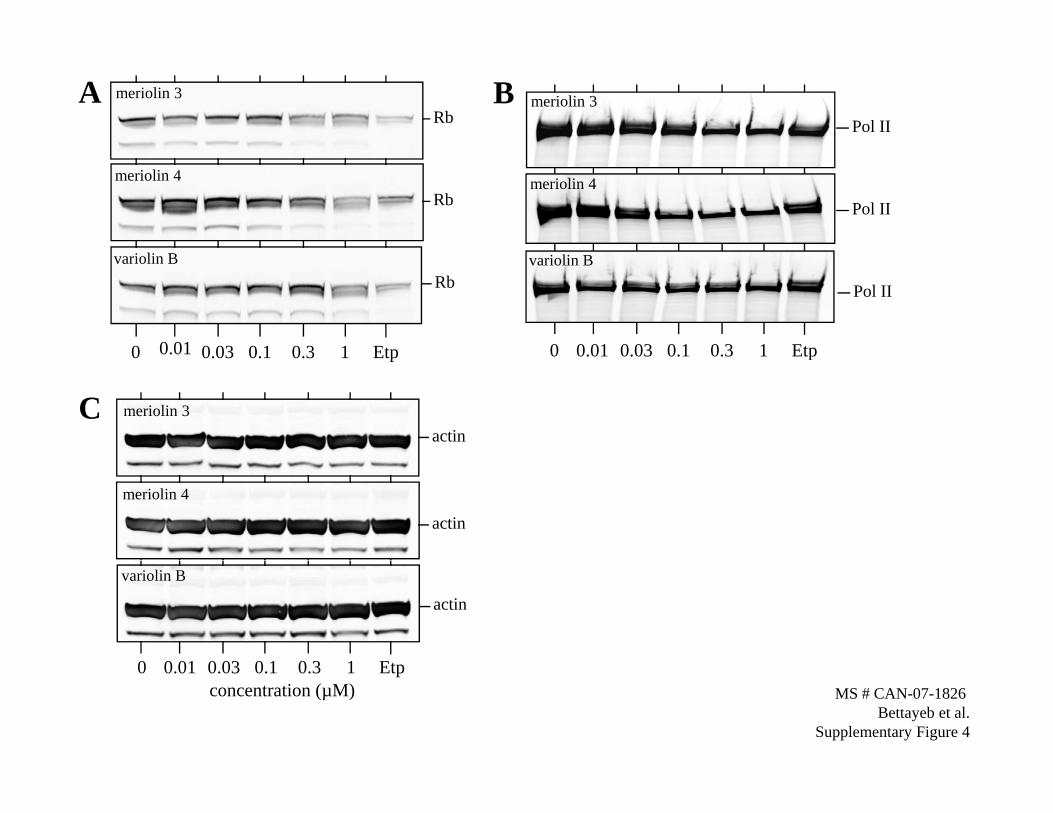
Although meriolins induced a modest reduction in Rb levels (Supplementary Fig.
S4A), they strongly and dose-dependently reduced Rb phosphorylation at CDK4-specific
sites (Ser249/Thr252) (Fig. 4A).
RNA polymerase II is phosphorylated by CDK9 on Ser2. Although only meriolin
4 induced a modest reduction in RNA polymerase II level (Supplementary Fig. S4B), all
three compounds inhibited CDK9-specific Ser2 phosphorylation of RNA polymerase II
in a dose-dependent manner (Fig. 4C).
Supplementary Figure 4. Inhibition of phosphorylation at CDK-specific sites. SH-
SY5Y cells were exposed to increasing concentrations of meriolins 3, meriolin 4 or
variolin B for 24 h and proteins were resolved by SDS-PAGE followed by Western
blotting with antibodies directed against Rb (S4A) and RNA polymerase II (S4B).
Western blotting with anti-actin antibodies was used as a loading control (S4C).
Rbmeriolin 3A
Pol II
B meriolin 3
Rbmeriolin 4
i li
Pol IImeriolin 4
Rb
0 0.01 0 03 0 1 0 3 1 Etp
variolin B
Pol II
0 0.01 0.03 0.1 0.3 1 Etp
variolin B
0 0.03 0.1 0.3 1 Etp
actinmeriolin 3C
0 0.01 0.03 0.1 0.3 1 Etp
actin
meriolin 4
actin
variolin B
0 0 01 0 03 0 1 0 3 1 Et0 0.01 0.03 0.1 0.3 1 Etpconcentration (µM) MS # CAN-07-1826
Bettayeb et al.Supplementary Figure 4