Mercury in water and biomass of microbial communities in hot springs of Yellowstone National Park,...
12
Mercury in water and biomass of microbial communities in hot springs of Yellowstone National Park, USA Susan A. King a,b, * , Sabrina Behnke a,c , Kim Slack a,b , David P. Krabbenhoft d , D. Kirk Nordstrom e , Mark D. Burr c , Robert G. Striegl f a Chemical Engineering Department, Montana State University, 318 Cobleigh Hall, Bozeman, MT 59717-3920, USA b Thermal Biology Institute, Montana State University, Bozeman, MT 59717-3920, USA c Center for Biofilm Engineering, Montana State University, Bozeman, MT 59717-3920, USA d US Geological Survey, 8505 Research Way, Middleton, WI 53562, USA e US Geological Survey, 3215 Marine Street, Suite E127, Boulder, CO 80303, USA f US Geological Survey, P.O. Box 25046, MS413, Denver Federal Center, Lakewood, CO 80225-0046, USA Abstract Ultra-clean sampling methods and approaches typically used in pristine environments were applied to quantify concen- trations of Hg species in water and microbial biomass from hot springs of Yellowstone National Park, features that are geologically enriched with Hg. Microbial populations of chemically-diverse hot springs were also characterized using mod- ern methods in molecular biology as the initial step toward ongoing work linking Hg speciation with microbial processes. Molecular methods (amplification of environmental DNA using 16S rDNA primers, cloning, denatured gradient gel elec- trophoresis (DGGE) screening of clone libraries, and sequencing of representative clones) were used to examine the dom- inant members of microbial communities in hot springs. Total Hg (THg), monomethylated Hg (MeHg), pH, temperature, and other parameters influential to Hg speciation and microbial ecology are reported for hot springs water and associated microbial mats. Several hot springs indicate the presence of MeHg in microbial mats with concentrations ranging from 1 to 10 ng g 1 (dry weight). Concentrations of THg in mats ranged from 4.9 to 120,000 ng g 1 (dry weight). Combined data from surveys of geothermal water, lakes, and streams show that aqueous THg concentrations range from l to 600 ng L 1 . Species and con- centrations of THg in mats and water vary significantly between hot springs, as do the microorganisms found at each site. Ó 2006 Published by Elsevier Ltd. 1. Introduction Geothermal features (volcanoes, geysers, hot springs, fumaroles) are known as geologic sources of Hg to the environment (Nraigu, 1989; Christen- son and Mroczek, 2003; Rytuba, 2005). Releases of Hg from geological sources has been found in both gaseous and aqueous forms, but recent research focuses almost entirely on gaseous Hg emissions (Gustin et al., 1996; Engle and Gustin, 2002; Christenson and Mroczek, 2003). While a few researchers have used ultra-clean sampling tech- niques to investigate THg concentrations in water 0883-2927/$ - see front matter Ó 2006 Published by Elsevier Ltd. doi:10.1016/j.apgeochem.2006.08.004 * Corresponding author. Address: Chemical Engineering Department, Montana State University, 318 Cobleigh Hall, Bozeman, MT 59717-3920, USA. E-mail address: [email protected] (S.A. King). Applied Geochemistry 21 (2006) 1868–1879 www.elsevier.com/locate/apgeochem Applied Geochemistry
-
Upload
independent -
Category
Documents
-
view
2 -
download
0
Transcript of Mercury in water and biomass of microbial communities in hot springs of Yellowstone National Park,...

Applied Geochemistry 21 (2006) 1868–1879
www.elsevier.com/locate/apgeochem
AppliedGeochemistry
Mercury in water and biomass of microbial communities inhot springs of Yellowstone National Park, USA
Susan A. King a,b,*, Sabrina Behnke a,c, Kim Slack a,b, David P. Krabbenhoft d,D. Kirk Nordstrom e, Mark D. Burr c, Robert G. Striegl f
a Chemical Engineering Department, Montana State University, 318 Cobleigh Hall, Bozeman, MT 59717-3920, USAb Thermal Biology Institute, Montana State University, Bozeman, MT 59717-3920, USA
c Center for Biofilm Engineering, Montana State University, Bozeman, MT 59717-3920, USAd US Geological Survey, 8505 Research Way, Middleton, WI 53562, USA
e US Geological Survey, 3215 Marine Street, Suite E127, Boulder, CO 80303, USAf US Geological Survey, P.O. Box 25046, MS413, Denver Federal Center, Lakewood, CO 80225-0046, USA
Abstract
Ultra-clean sampling methods and approaches typically used in pristine environments were applied to quantify concen-trations of Hg species in water and microbial biomass from hot springs of Yellowstone National Park, features that aregeologically enriched with Hg. Microbial populations of chemically-diverse hot springs were also characterized using mod-ern methods in molecular biology as the initial step toward ongoing work linking Hg speciation with microbial processes.Molecular methods (amplification of environmental DNA using 16S rDNA primers, cloning, denatured gradient gel elec-trophoresis (DGGE) screening of clone libraries, and sequencing of representative clones) were used to examine the dom-inant members of microbial communities in hot springs. Total Hg (THg), monomethylated Hg (MeHg), pH, temperature,and other parameters influential to Hg speciation and microbial ecology are reported for hot springs water and associatedmicrobial mats.
Several hot springs indicate the presence of MeHg in microbial mats with concentrations ranging from 1 to 10 ng g�1 (dryweight). Concentrations of THg in mats ranged from 4.9 to 120,000 ng g�1 (dry weight). Combined data from surveys ofgeothermal water, lakes, and streams show that aqueous THg concentrations range from l to 600 ng L�1. Species and con-centrations of THg in mats and water vary significantly between hot springs, as do the microorganisms found at each site.� 2006 Published by Elsevier Ltd.
1. Introduction
Geothermal features (volcanoes, geysers, hotsprings, fumaroles) are known as geologic sources
0883-2927/$ - see front matter � 2006 Published by Elsevier Ltd.doi:10.1016/j.apgeochem.2006.08.004
* Corresponding author. Address: Chemical EngineeringDepartment, Montana State University, 318 Cobleigh Hall,Bozeman, MT 59717-3920, USA.
E-mail address: [email protected] (S.A. King).
of Hg to the environment (Nraigu, 1989; Christen-son and Mroczek, 2003; Rytuba, 2005). Releasesof Hg from geological sources has been found inboth gaseous and aqueous forms, but recentresearch focuses almost entirely on gaseous Hgemissions (Gustin et al., 1996; Engle and Gustin,2002; Christenson and Mroczek, 2003). While afew researchers have used ultra-clean sampling tech-niques to investigate THg concentrations in water

S.A. King et al. / Applied Geochemistry 21 (2006) 1868–1879 1869
(McCleskey et al., 2004) and air samples (Hall et al.,2006) associated with geothermal features, mostresearch on Hg in geothermal systems occurredbefore incorporation of clean sampling protocolsfor trace metal collections (Patterson and Settle,1976). This raises concerns for environmental man-agers regarding the application of the historic data-sets in watersheds containing or receivinggeothermal waters. Moreover, investigations of org-ano-Hg compounds in geothermal systems are rarerand focused primarily on gaseous emissions fromhot springs (Hirner et al., 1998). As a result, impor-tant biogeochemical processes and transport mecha-nisms of Hg are poorly understood in geothermalsystems. Subsequently, policy-makers are unableto make informed decisions in regions containinggeothermal environments because geochemical andmicrobial data do not exist.
Extensive research in non-geothermal aquaticsystems has demonstrated the importance of micro-bial processes in the cycling of Hg. Methylation ofHg(II) to the more toxic, bioaccumulated form,MeHg, is facilitated by microorganisms, primarilySO4-reducing bacteria (Compeau and Bartha,1985; King et al., 2000). Bacteria can also degradeMeHg via oxidation or reduction pathways (Orem-land et al., 1995; Marvin-DiPasquale and Orem-land, 1998). Reduction of Hg(II) to Hg(0) andMeHg to Hg(0) is achieved by microorganismsusing the mer A and/or mer B genes. The reductivepathways are believed to have evolved in geother-mal environments where microorganisms neededto find a mechanism to survive and thrive in thepresence of high concentrations of toxic trace metals(Nazaret et al., 1994; Barkay et al., 2003; Barkayand Wagner-Dobler, 2005). Once again, little infor-mation is currently available to evaluate microbialinteractions with Hg in geothermal water.
Recent advances in molecular analytical tech-niques (RNA- and DNA-based polymerase chainreaction (PCR) coupled with DGGE, cloning,sequencing and phylogenic analysis) have improvedour ability to describe and characterize microbialcommunity structure in geothermal and other natu-ral environments that are not amenable to tradi-tional culturing techniques (Ferris et al., 1996;Burr et al., 2006). Likewise, advances in analyticaltechniques of atomic fluorescence detection andmass spectrometry have improved the ability todetect and quantify low-concentrations of Hg spe-cies and reaction pathways in the environment(Olson and DeWild, 1999; DeWild et al., 2002).
Here these powerful tools are employed togetherin a novel setting—geothermal features—with thegoals of:
• increasing current knowledge of Hg concentra-tions and speciation in geothermal water andbiomass,
• evaluating important biotic and abiotic controlson Hg biogeochemistry, and
• increasing understanding of Hg–microbe interac-tions in geothermal environments.
In this paper, the authors relate initial findings ofHg distribution and speciation in chemically diversehot springs in Yellowstone National Park anddescribe the molecular methods used to examinemicrobial communities in a subset of hot springsof representative geochemical diversity.
2. Field sites
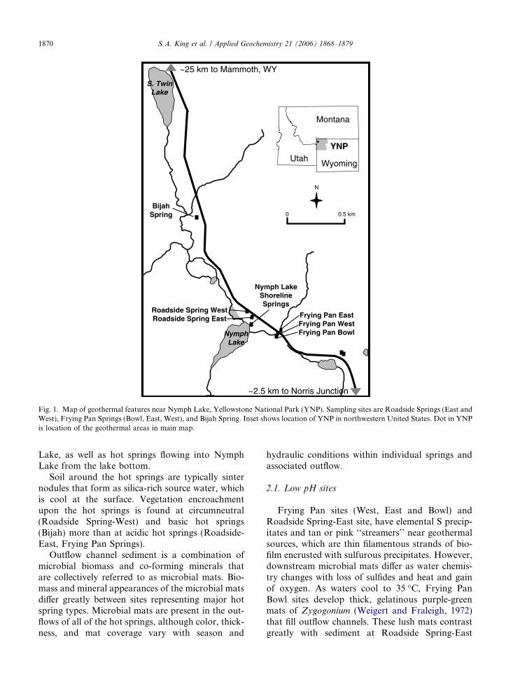
The physical and chemical diversity among geo-thermal features in Yellowstone National Park(YNP) is unsurpassed among the world’s geother-mal regions. The US Geological Survey (USGS)investigations of water chemistry of YNP hotsprings, geysers, and streams (Ball et al., 2002;McCleskey et al., 2004) reveal that water chemistryvaries greatly between hot springs in close proxim-ity. A small subset of hot springs were studied thatrepresent the broadest possible range of YNP hotspring geochemistry contained within a singlewatershed. Reconnaissance work included searchingfor hot springs that demonstrated obvious microbialactivity and evidence of Hg methylation. An under-lying assumption of this work is that biotic pro-cesses are the primary mechanism of Hgmethylation, though this hypothesis is unprovenfor hot springs.
Nymph Lake is part of the watershed drained bythe Gibbon River. The watershed lies along anorth–south fault stretching from Norris GeyserBasin to Mammoth Hot Springs. Geothermal chem-istry is predominantly acidic at the southerly end ofthe fault and basic at the north end. The sitesincluded both acid-sulfate hot springs and neutralpH-chloride hot springs draining to the easternshore of Nymph Lake (Fig. 1). These springs havewidely varying temperature, pH, and water chemis-try (Table 1) that create ideal ‘‘field laboratories’’for the investigations. There are also several hotsprings and fumaroles on the west side of Nymph
BijahSpring
NymphLake
Roadside Spring WestRoadside Spring East Frying Pan East
Frying Pan WestFrying Pan Bowl
Nymph Lake Shoreline Springs
0 0.5 km
N
Montana
UtahWyoming
YNP
S. TwinLake
~25 km to Mammoth, WY
~2.5 km to Norris Junction
Fig. 1. Map of geothermal features near Nymph Lake, Yellowstone National Park (YNP). Sampling sites are Roadside Springs (East andWest), Frying Pan Springs (Bowl, East, West), and Bijah Spring. Inset shows location of YNP in northwestern United States. Dot in YNPis location of the geothermal areas in main map.
1870 S.A. King et al. / Applied Geochemistry 21 (2006) 1868–1879
Lake, as well as hot springs flowing into NymphLake from the lake bottom.
Soil around the hot springs are typically sinternodules that form as silica-rich source water, whichis cool at the surface. Vegetation encroachmentupon the hot springs is found at circumneutral(Roadside Spring-West) and basic hot springs(Bijah) more than at acidic hot springs (Roadside-East, Frying Pan Springs).
Outflow channel sediment is a combination ofmicrobial biomass and co-forming minerals thatare collectively referred to as microbial mats. Bio-mass and mineral appearances of the microbial matsdiffer greatly between sites representing major hotspring types. Microbial mats are present in the out-flows of all of the hot springs, although color, thick-ness, and mat coverage vary with season and
hydraulic conditions within individual springs andassociated outflow.
2.1. Low pH sites
Frying Pan sites (West, East and Bowl) andRoadside Spring-East site, have elemental S precip-itates and tan or pink ‘‘streamers’’ near geothermalsources, which are thin filamentous strands of bio-film encrusted with sulfurous precipitates. However,downstream microbial mats differ as water chemis-try changes with loss of sulfides and heat and gainof oxygen. As waters cool to 35 �C, Frying PanBowl sites develop thick, gelatinous purple-greenmats of Zygogonium (Weigert and Fraleigh, 1972)that fill outflow channels. These lush mats contrastgreatly with sediment at Roadside Spring-East
Table 1Hot springs aqueous geochemistry
BijahSpring
Frying PanBowl Spring
Frying PanSpring-East
RoadsideSpring-West
Frying PanSpring-West
RoadsideSpring-East
T (�C) 79.0 36.4 80 65 56 60pH 8.6 2.3 2.2 6.4 2.6 3.5Fe(II) (mg L�1) 0.24 n/c n/c 0.01 n/c 0.25SO4 (mg L�1) 105 n/c n/c 73.2 n/c 220H2S (mg L�1) <0.002 n/c n/c 0.15 n/c 0.004Alkalinity-HCO3 (meq L�1) 290 n/c n/c 170 n/c b.d.l.Cl (mg L�1) 177 n/c n/c 454 n/c 167
This paper reports samples as below detection limit (b.d.l.) when the concentration is less than the larger of the daily detection limit ormethod detection limit.n/c indicates that a water sample was not collected for the parameter indicated.
S.A. King et al. / Applied Geochemistry 21 (2006) 1868–1879 1871
(pH 3.5, T = 60 �C), where a powdery film of yel-low-orange to silver-gray precipitates form as watercools.
2.2. Circumneutral to basic pH sites
Roadside Spring-West (pH 6.5, T = 65 �C) hasdark silver-gray precipitates at the source, butdownstream mats take on a rich chocolate browncolor as Fe oxides form. Thereafter, peach-coloredprecipitates blend with the chocolate brown mat.
Bijah Spring is hotter and more alkaline (pH 8.7,T = 79 �C) than the other hot springs in this studyand sediment near the source appears to be fusedsinter nodules. As outflow water cools to 65 �C, abright blue-green mat develops that transitions intoa fluvial fan with a thin, rust-colored microbial matcovering the hard sinter. Macrophytes and lightgreen algal mats were present below the rust-coloredcommunity as the water cooled even further. Theoutflow channels of all hot springs were unfrozen,with adult flies living on the mats during the Decem-ber 2004 survey of potential research sites.
3. Methods and materials
3.1. Field collections
Datasets combined from McCleskey et al. (2004),Krabbenhoft (unpublished data), and this studyshow that filtered THg concentrations in water varygreatly among YNP hot springs, geysers, andstreams (Fig. 2). To avoid cross-contamination ofsamples from high Hg to low Hg sites, acceptedclean sampling protocols (Patterson and Settle,1976; Olson and DeWild, 1999) were used through-out sample collection and laboratory analyses, withno reuse of filter holders or sampling lines between
springs. Solids and water samples were collectedand placed into pre-cleaned Teflon or glass bottleswith Hg-free certification. Distance from hot springsource, temperature, and microbial appearance werenoted at each water and sediment sampling location.
Hot springs source waters were collected in a 1 Lgrab sample and filtered the same day throughquartz fiber filters ashed at 550 �C for 4 h. Filtratewas collected into acid-washed Teflon bottles or cer-tified Hg-free bottles, preserved with low-Hg HCl,and shipped overnight to the USGS Mercury Labo-ratory (Middleton, Wisconsin) for THg and MeHganalyses. Methods for temperature, pH, sulfide,SO4, alkalinity and Cl are described in McCleskeyet al. (2004).
Surface sediment and mats were collected fromthe upper 1 cm depth in hot springs and outflowsin locations having distinctive coloration suggestingdiffering geochemical conditions relative toupstream mats. Samples were placed on dry ice untiltransported to the Montana State University labo-ratory, where they were thawed in a laminar flowhood, homogenized, and split for THg, MeHg andDNA analyses, and dry weight and loss on ignitiondeterminations.
3.2. Mercury analyses
Filtered and unfiltered water samples were ana-lyzed for MeHg using distillation and ethylationand chromatographic separation with cold vaporatomic fluorescence spectrometry (CVAFS) detec-tion as described in DeWild et al. (2002). Filteredand unfiltered water samples were analyzed forTHg using EPA method 1631 described in Olsonand DeWild (1999). A method detection limit of0.04 ng L�1 was determined for THg and MeHg,and daily detection limits were calculated as well.

010
020
030
040
050
060
070
0
Beowolf E. Source Water, 10/21/04
Beowolf Outlet, 10/21/04Bijah Spring Source, 12/3/04Bijah Spring Outlet, 12/3/04
Cinder Pool, 05/30/03Dragon Spring Source, 12/7/04Dragon Spring Outlet, 12/7/04
Ear Spring, 09/04/03Frying Pan East Pool, 12/3/04
Frying Pan Spring, 06/18/03
Frying Pan West Pool, 12/3/04Green Spring, 09/04/03
Hazel Lake (NW side), 06/07/03Hazel Lake (S side), 06/07/03
Jade Pool, 09/05/03Kaolin Spring, 03wa122, 06/01/03
Nymph Lake, 09/04/03Nymph Lake, N side, 06/18/03
Nymph-Inflow, 09/04/03
Octopus, 09/04/03Potts Basin - Site 352, 09/05/03
Queens Laundry, 06/06/03Ragged Hills Milky Way, 06/17/03
Ragged Hills Verde Spring, 06/17/03Roadside Spring East, 12/03/04
Roadside Spring West, 09/04/03Roadside Spring West, 12/3/04
Sulfur Cauldron, 06/05/03Titanic Spring, 05/30/03
Total Mercury in Filtered Water (ng L-1)
Fig. 2. Total Hg in filtered waters of Yellowstone National Park hot springs as sampled by S.A. King (2004), D.P. Krabbenhoft(September, 2003), and D.K. Nordstrom (June, 2003).
1872 S.A. King et al. / Applied Geochemistry 21 (2006) 1868–1879
Methylmercury in mat material was extracted byadditions of KBr, CuSO4, and methylene chlorideaccording to DeWild et al. (2002). Back extractionof MeHg into reagent water and detection usingdirect ethylation/purge and trap/chromatographicseparation/pyrolysis was carried out by CVAFS.This extraction procedure eliminates known inter-ferences from organic matter, recalcitrant solids,and sulfides, as well as greatly reducing potentialMeHg artifact generation from high concentrationsof inorganic Hg interactions with organic com-pounds. The method detection limit is 0.08 ng g�1.All concentrations for sediment MeHg are reportedas dry weight.
Concentrations of THg in mat material weredetermined according to published methods (Olson
and DeWild, 1999). An aliquot of homogenizedmat material was digested and oxidized with aquaregia overnight. The sample was diluted with 5%bromine monochloride, pre-reduced with hydroxyl-amine hydrochloride to remove free halogens, andHg(II) was reduced to Hg(0) with stannous chloride.The Hg(0) produced was purged and trapped onto aAu trap, thermally desorbed, and detected usingCVAFS. The detection limit was 0.3 ng per diges-tion bomb using this method. Percent relative stan-dard deviation for field replicate samples rangedfrom 9% at sites with fine, homogeneous mixturesof biomass and mineral precipitates to 80% at siteswith coarse, heterogeneous mixtures of biomassand sinter nodules. All concentrations for THg arereported as dry weight.

S.A. King et al. / Applied Geochemistry 21 (2006) 1868–1879 1873
3.3. DNA extraction and PCR reaction
Microbial populations were identified using theapproach described by Burr et al. (2006) expandingupon the pioneering work of Ferris et al. (1996).DNA was extracted from mat samples using theDNA FastDNA� SPIN Kit for Soil (Q-Biogene)by following the manufacturer’s instructions. DNAextracts from environmental samples or purified cul-tured clones were amplified with a PTC-100 Pro-grammable Thermal Controller from MJ Researchusing 15–30 ng of DNA. The non-GC clamp prim-ers, 1070F: ATGGCTGTCGTCAGCT (bacterial)and 1392 R:ACGGGCGGTGTGTAC (universal)(Ferris et al., 1996), and the 2X Master Mix (Pro-mega) were mixed with 15–30 ng of DNA in nucle-ase-free water provided by the Wizard� Plus
Minipreps DNA Purification System (Promega) orDES solution provided in the FastDNA� SPINKit for Soil (Q-Biogene). Primers were synthesizedby IDT� Integrated DNA Technologies and usedat a concentration of 12 lM. To avoid contamina-tion, all reaction batches were measured and mixedin the laminar flow hood. A negative control,consisting of a sample to which DNA template wasnot added, was included in all experiments. Thetemperature and cycling programs used foramplification of the 16S rDNA are described asfollows: Program 1: (1) 94 �C for 2 min; (2) 94 �Cfor 45 s; (3) 55 �C for 45 s; (4) 72 �C for 45 s; (5).Repeat 2–4 for 25 cycles; (6) 72 �C for 7 min; (7)4 �C cooling and hold. DNA from sediment at hotsprings was sometimes difficult to amplify with pro-gram 1. Therefore, to qualitatively comparemicrobial diversity between all hot springs, a two-stage program was used to amplify DNA fromenvironmental samples. In this program, 5 lL ofthe resulting PCR product from the first stage ofProgram 2 were used as a template for the reactionsfor the second stage. Program 2 (two-stage PCR): (1)94 �C for 8 min, (2) 94 �C for 40 s, (3) 55 �C for 30 s,(4) 72 �C for 30 s, (5) Repeat 2–4, 10· (first stage)and (5) Repeat 2–4, 30 cycles (second stage), (6)72 �C for 5:00 min, (7) 4 �C cooling and hold. ThePCR products were visualized on 1.5% agarose gelsand run for 30 min at 80 V in 1X Tris-Acetate-EDTA buffer. Gels were stained with ethidium bro-mide solution and rinsed in water for 10 min. PCRproducts from environmental samples or purifiedcultured clones were purified using a QIAquick GelExtraction Kit (Qiagen Inc.) according to manufac-turer’s instructions.
3.4. Cloning
Cloning of the environmental DNA was per-formed with TOPO� TA Cloning� Kit (Invitrogen)and the OneShot� Chemical Transformation kit fol-lowing the manufacturer’s instructions. After a 24 hincubation period at 37 �C, single white colonieswere restreaked onto LB agar plates containing50 lg mL�1 ampicillin and 40 lL X-gal. After thenext incubation period, single white colonies weregrown in liquid medium containing kanamycin toassure presence of the cloned fragment. The Wiz-ard� Plus Minipreps DNA Purification System(Promega) was used to isolate and purify the recom-binant plasmids.
3.5. Denaturing gradient gel electrophoresis
DGGE gel was prepared with a denaturing gradi-ent of urea and formamide with the Bio-Rad DCo-deTM Universal Mutation Detection System (BioRadLaboratories) as recommended in the user manual.Electrophoresis was run at 60 V for 16–18 h. ThePCR for DGGE was performed with a GC clampon either the primer 1070F or 1392R (Burr et al.,2006). Gels were stained with Sybr�Gold (Molecu-larprobes) for about 20 min (10 lL Sybr�Gold,200 lL EDTA, 1000 lL in 100 mL with nanopurewater). Subsequently, the gel was scanned with aFluorChemTM 8800 fluorescence imager (AlphaInno-tec) and analyzed with AlphaEase Software (Alpha-Innotech) by means of 1D lane densitometry.
The identity of environmental DNA fragmentswas confirmed by DGGE analysis of randomlyselected clones side by side with the original PCRproducts of the of environmental DNA reaction.The 16S rRNA gene inserts from plasmids repre-senting the diversity of the sampled communitywere then sequenced.
3.6. Sequencing
Plasmids were sequenced by Laragen, Inc. (10755Venice Blvd., Los Angeles, CA 90034) or NevadaGenomics Center (NGC) (University of Nevada,Reno, NV). Sequences were provided either in .seqor .ab1 file format and processed with the Sequen-cherTM Software. BLAST searches against the data-base of the National Center for BiotechnologyInformation (http://www.ncbi.nlm.nih.gov/BLAST/)were performed to identify the organisms mostclosely related to the cloned environmental 16S

1874 S.A. King et al. / Applied Geochemistry 21 (2006) 1868–1879
rRNA gene fragments, which represent the dominantorganisms in the sampled YNP springs.
4. Results and discussion
The impetus for this study was that preliminarycollections of hot spring mats, aquatic vegetationand insects at the east shore of Nymph Lake (Stri-egl, unpublished) showed evidence of methylationand MeHg bioaccumulation (Table 2). Concentra-tions of THg were found to be elevated in watersamples from two hot springs on the east shorelineof Nymph Lake. Moreover, MeHg in filamentousmicrobial biomass in the same two hot springsaccounted for 5–10% of the THg, whereas less than0.1% of Hg was as MeHg in the hot spring water.The bioconcentration factors were 1.2 and 12 formicrobial communities in these two hot springs.
The preliminary survey in December 2004 ofwater in several Yellowstone hot springs and out-flows showed wide ranges of THg. Concentrationsof THg in hot springs source water varied by oneorder of magnitude between sites (Table 2). Aque-ous MeHg concentrations were generally below
Table 2Mercury concentrations in filtered water and microbial mats from hot
Site name and sampling location Water MeHg (ng L�1)
Nymph L. Shoreline Spring #1, at source 0.026a
Nymph L. Shoreline Spring #2, at source 0.43Bijah Spring, at source b.d.l.Bijah Spring, 47 m from source n/cBijah Spring, 61.5 m from source n/cBijah Spring, 92 m from source n/cFrying Pan Bowl Spring, purple-green mat n/cFrying Pan East Spring, at source b.d.l.Frying Pan East Spring, dark green mat n/cFrying Pan East Spring, yellow green mat n/cFrying Pan West Spring, at source b.d.l.Frying Pan West Spring, 10 m from source n/cFrying Pan West Spring, 25 m from source n/cRoadside East Spring, at source b.d.l.Roadside East Spring, 7 m from source n/cRoadside East Spring, 13 m from source n/cRoadside East Spring, 20 m from source n/cRoadside West Spring, at source 0.08Roadside West Spring, 1.5 m from source n/cRoadside West Spring, 4.0 m from source n/cRoadside West Spring, 5.5 m from source n/cRoadside West Spring, 7 m from source n/c
b.d.l. indicates below detection limit. n/c indicates that a sample was nPercent relative standard deviations for MeHg analyses of replicate sammatrices. Percent relative standard deviations for THg analyses of repNone of the samples reported here were flagged.
a Detection limit for water, MeHg = 0.013 ng L�1, for THg = 0.2 ngb Detection limit for biomass, MeHg = 0.08 ng g�1, for THg = 0.3 ng
the detection limit. Regression analyses indicatedpoor correlation between pH, temperature and Hgspecies concentrations in water. The highest waterconcentrations of THg were observed at a site witha pH of 6.4 (Roadside Springs-West), whereas thelowest concentration was found at a site with apH of 8.6 (Bijah Spring).
Based on chemical modeling (MINEQL+) of cin-nabar solubility at increasing temperatures across arange of pH, higher concentrations of aqueous Hgare predicted in geothermal water of lowest pHand highest temperature. However, the results pre-sented here do not support this hypothesis. Filteredwater samples of Roadside West had higher concen-trations of THg than Frying Pan Springs water(Table 2).
A comparison of microbial mat THg concentra-tions between Roadside West and Frying PanSprings also suggests that aqueous phase Hg com-plexation maybe important at Roadside West, butprecipitation is a controlling process at all springs(Table 2). The data suggest that THg is rapidlyremoved from Frying Pan Springs water by amor-phous S precipitates. The churning water of Frying
springs
Water THg (ng L�1) Mat MeHg (ng g�1) Mat THg (ng g�1)
170 0.31b 6.3520 0.53 4.915 n/c n/cn/c 0.86 3100n/c 1.3 160n/c 0.16 380n/c 9.2 12,00056 0.12 14,000n/c 3.0 17,000n/c 2.5 120,00046 0.36 22,000n/c 0.08 2800n/c 0.17 12,00033 0.12 1600n/c 0.11 1200n/c 1.5 9000n/c 1.0 13,000200 1.2 46,000n/c 0.75 37,000n/c 0.22 16,000n/c 1.9 52,000n/c 2.4 4100
ot collected.ples range from 10.2% to 15.6% among a wide range of sample
licate samples are required to be less than 5% or data is flagged.
L�1.g�1.

S.A. King et al. / Applied Geochemistry 21 (2006) 1868–1879 1875
Pan West pool are murky with yellow-tan precipi-tates and the Frying Pan outflow channel mats showevidence of the same precipitates deposited over sin-ter nodules.
Concentrations of Hg in microbial mats varied by5 orders of magnitude for THg and 3 orders of mag-nitude for MeHg (Fig. 2; Table 2). Concentrations ofMeHg in microbial mats were generally comparableto other ecosystems without point sources of inor-ganic Hg (Krabbenhoft et al., 1995, 2000; Gilmouret al., 1998; King, 2000). The highest concentrationsof MeHg were observed in the thick purple-greenFrying Pan Bowl mats that fill the channel flowinginto the Frying Pan West pool. These mats arenoticeably higher in organic matter than other hotsprings mats, which may lead to conditions favoringmicrobial Hg methylation or greater accumulationof MeHg, or both processes. Continuing investiga-tions of geochemical conditions and prokaryoticand eukaryotic organisms will identify microbialpopulations that may influence the speciation andseasonal distribution of Hg in these hot springs.
The DGGE images of DNA from hot springsmicrobial communities indicate great diversitybetween sites of differing pH and temperature
Fig. 3. DGGE image of Roadside Spring-West sites microbial communresult of two-stage PCR amplification of DNA extracted from hot springindicates the relative diversity of microbial communities in hot spring sPan Springs sites compared with other sites. Arrows on the right pointThe order of lanes corresponds to the following sites: A—Bijah Springs,Spring-East, E—Frying Pan Spring-West Pool, F—Roadside Spring-W
(Fig. 3). Sites of lowest pH (Frying Pan Springssites; Lanes B, C and E) showed fewer gel bandsin their DGGE images, indicating a lower diversityrelative to sites with neutral pH (Bijah, Lane A,Roadside Springs, Lanes D and F, Fig. 5). Furtherwork at these sites will continue to investigate thisdiversity by sequencing of individual clone librariesformed from extracted microbial mat DNA. BijahSprings showed many distinct bands, but far fewerthan typical soil and sediment of pH 8.6. This dif-ference is attributed to the high temperatures ofthe Bijah Springs source water that likely limitedcolonization to thermophilic microorganisms.Additionally, DGGE profiles for Roadside Westand Bijah Springs communities are more similarto each other than to profiles of Frying PanSprings, which is possibly attributed to a greatersimilarity in geochemical conditions at the two for-mer sites (Table 1).
DGGE bands representing unique clones for theRoadside West and Bijah communities are num-bered and their correspondence to the communityprofile are presented in Figs. 4 and 5, respectively.Sequencing results verify that microbial communi-ties nearest the Roadside West and Bijah hot spring
ity DNA and corresponding clone isolates DNA. Each lane is thes mat/sediments. The number of dark bands on the DGGE image
ediments. The arrows on the left point to similar bands at Fryingto similar bands at Roadside Springs compared with other sites.B—Frying Pan Bowl, C—Frying Pan Spring-East, D—Roadsideest.

Fig. 4. DGGE image of Roadside Spring-West sites microbial community DNA and corresponding clone isolates DNA. Most closelyrelated microorganism of each clone is listed in Table 3. The image shows single clones randomly picked from the clone library and thecorresponding communities in environmental DNA from the RSF sampling site (RSF 1 = 1.5 m from source, RSF 3 = 5.5 m from source,RSF 4 = 7 m from source).
Fig. 5. DGGE image of Bijah Spring sites microbial community DNA and corresponding clone isolates DNA. Most closely relatedmicroorganism of each clone is listed in Table 4. The image shows single clones randomly picked from the clone library and thecorresponding bands in environmental DNA from the Bijah sampling sites (Bijah 1 = 47 m from source, Bijah = 61.5 m from source,Bijah 3 = 92 m from source).
1876 S.A. King et al. / Applied Geochemistry 21 (2006) 1868–1879
sources differed, yet Chloroflexus was found to be acommon organism in each (Tables 3 and 4). The sul-fidic hot spring bacterium NPE, a Chloroflexus
reported by Nubel et al. (2001) and found byD.M. Ward (personal communication) in otherYNP hot springs, is a filamentous anoxygenic pho-toheterotrophic bacterium utilizing organic mole-cules that are produced by other microbes,
although some strains can use hydrogen or sulfideas electron donors, and thus, are chemolithotrophic.
The Frying Pan Bowl site (Table 5) has no organ-isms common to either of the higher pH hot springs.The sequences from clone libraries had best matcheswith Mycobacterium and other actinobacteria,organisms commonly found in soil. Literaturesearches did not find any previous reports of

Table 3Microbial communities at Roadside Spring-West and outflow as determined by 16S rDNA amplification and sequencing of clone librariesdeveloped from microbial mat DNA samples collected 12/03/04
DGGE Band ID Most similar organism Similarity (%) Accession #
RSF1 Environmental sample DNA1 Uncultured Geothermobacterium 99 AY882756.12 Sulfidic hot spring bacterium 100 AJ308502.13 Unidentified thermus OPB32 100 AF027021
RSF3 Environmental sample DNA4 Sulfidic hot spring bacterium 100 AJ308502.15 Uncultured bacterium 98 X844746 Uncultured bacterium 99 AB240225.17 Uncultured chloroflexi bacterium 96 AY222298.18 Uncultured bacterium 100 AF407718.1
RSF4 Environmental sample DNA9 Oscillochloris sp. 97 AF146831.2
10 Uncultured bacterium clone B25 96 AF40771811 Uncultured soil bacterium clone 349 98 AY493984.112 Uncultured soil bacterium clone 349 97 AY493984.1
According to method of Burr et al. (2006) using DGGE screening of clones.
Table 4Microbial communities at Bijah Spring and outflow as determined by 16S rDNA amplification and sequencing of clone libraries developedfrom microbial mat DNA samples collected 12/03/04
DGGE Band ID Most similar organism Similarity (%) Accession #
Bijah 1 Environmental sample DNA1 Uncultured bacterium clone O1aB8 99 AY1932912 Uncultured organism clone MB1003c8 99 AY8977553 Uncultured firmicute clone SM2H09 99 AF4457454 Roseiflexus castenholzii 96 AB041226.15 Uncultured bacterium clone 01aA90 99 AY193179.16 Thermotoga petrophila 98 AJ872269.1
Bijah 2 Environmental sample DNA7 Uncultured bacterium clone 1700a2-40 99 AY917299.18 Uncultured planctomycete 97 BX2947609 Synechococcus sp. C1 97 AF132772
Bijah 3 Environmental sample DNA10 Uncultured bacterium clone SM1C09 98 AF44566611 Uncultured Chloroflexaceae bacterium clone Hs2_48 99 AF421750.212 Uncultured bacterium clone JC2701W_8
According to method of Burr et al. (2006) using DGGE screening of clones.
S.A. King et al. / Applied Geochemistry 21 (2006) 1868–1879 1877
thermophilic organisms that related to these bestmatches, rather they are usually mesophilicmicrobes often associated with human diseases orsoil samples. The Frying Pan Bowl samples werecollected from mats ranging from 18 to 60 �C,which suggests a wide range of temperature toler-ance for this indigenous community. The resultsare in agreement with Walker et al. (2005) who haverecently shown that 47% of the microbial speciesfound in acidic surface soil of Norris Geyser Basin,YNP, were members of the bacterial division of
Actinobacteria, and previously unidentified Myco-
bacterium spp. were the most abundant species.The data show that generally less than 1% of
inorganic Hg was present as MeHg in mats (Table2). However, whether microbial mats are methylat-ing the abundant inorganic Hg is unclear. Bysequencing the 16S rDNA of purified culturedclones of the microbial mat from Roadside WestSpring, an organism was identified that was closelyrelated to Geothermobacterium sp. (99% similarity),which belongs to an early lineage of hyperthermo-

Table 5Microbial Communities at Frying Pan Bowl as determined byPCR and sequencing of clone libraries developed from microbialmat samples collected 12/03/04
‘‘Zygogonium’’ mat characterization:Most similar organism
Similarity(%)
Accession#
Mycobacterium nebraskense 98 AY368456Uncultured bacterium 99 AY725260Mycobacterium sp. JS623 99 AY162028Uncultured actinobacterium 97 AY743693Chloroplast Haslea nipkowii 98 AF514850
According to method of Ferris et al. (1996), without using DGGEscreening.
1878 S.A. King et al. / Applied Geochemistry 21 (2006) 1868–1879
philic SO4-reducing bacteria that also containsThermodesulfobacterium. This finding indicates thepotential for Hg methylation via SO4 reduction, asThermodesulfobacterium is a chemolithotrophicSO4-reducing bacterium. Thermodesulfobacterium
has also been reported in Joseph’s Coat Spring,YNP by Inskeep et al. (2005), and this genus is typ-ically found in aquatic systems associated with vol-canic hot springs, deep-sea hydrothermal sulfides,and other marine environments.
There is some evidence that microorganisms otherthan SO4 reducers may methylate Hg when utilizingterminal electron acceptors other that SO4 (Warneret al., 2003). Therefore, it is possible that othermicrobial populations may influence MeHg concen-trations in these hot springs through methylationand demethylation processes or by impacts on theS cycle. The interactions between SO4-reducingand S-oxidizing microorganisms, as well as SO4 con-centrations (Gilmour and Henry, 1991), may beimportant influences on Hg biogeochemistry in thesehot springs. Additionally, the strong binding ofHg(II) by S precipitates (S0, HgS, FeS, Sx) and aque-ous ligands (sulfides, thiosulfates, polysulfides) mayinfluence the bioavailability of Hg(II) to methylatingpopulations (Benoit et al., 1999; Jay et al., 2000).
5. Conclusions
This study reports some of the first concentra-tions for THg and MeHg in microbial mats of hotsprings and the initial characterization of the corre-sponding microbial populations that colonize thesesprings. Inorganic Hg concentrations are high rela-tive to non-geothermal streams and rivers, butMeHg concentrations are comparable to otheraquatic systems. Work in progress is focusing oncharacterizing the Hg speciation in water and gasphases, quantifying transport rates, and identifying
microbial populations in hot springs and outflowchannels.
Precipitation processes appear to dominate theaccumulation of inorganic Hg in hot springs and theiroutflows. Low pH, high temperatures, and high con-centrations of sulfides, thiosulfates and polysulfidesmay keep aqueous concentrations of THg elevatedin geothermal sources, but cooling leads to precipita-tion and/or adsorption and accumulation of THg insediment of the outflow channels. Concentrationsof MeHg in water are low, but MeHg accumulationis associated with microbial mat material.
Microbial communities differ between hotsprings of varying temperature and geochemistry.Sites of similar chemistry share similar DGGEbands and sequencing results verify that Chlorofl-
exus was present at two hot springs of slightly differ-ent temperature and water chemistry. Microbialdiversity appears to be lower at sites with lowerpH and S0 precipitates. Additionally, the dominantmicrobial populations of these hot springs differfrom hot springs with higher pH. Development ofclone libraries from amplified 16S rDNA will formthe basis for future 16S rDNA sequencing and iden-tification of organisms within the mat communities.
Acknowledgements
The authors thank Jesse Bennett and Jordan Jac-obson for assisting with field work in YellowstoneNational Park, John DeWild and personnel of theUSGS Mercury Lab (Middleton, WI) for support-ing Hg analyses, and Yellowstone Park ResearchPermit Office for access to Park Service informationresources and great field support. Additional thanksto T. Barkay and M. Hines for critiques and guid-ance in preparing the manuscript. Funding for thisresearch is from the Thermal Biology Institute atMontana State University, through NASA awardNAG5-8807: Center for Studying Life in ExtremeEnvironments.
References
Ball, J.W., McCleskey, R.B., Nordstrom, D.K., Holloway, J.M.,Verplanck, P.L., 2002. Water-chemistry data for selectedsprings, geysers, and streams in Yellowstone National Park,Wyoming, 1999–2000. US Geological Survey Open-FileReport 02-382.
Barkay, T., Wagner-Dobler, I., 2005. Microbial transformationsof mercury: potentials, challenges, and achievements incontrolling mercury toxicity in the environment. Adv. Appl.Microbiol. 57, 1–53.

S.A. King et al. / Applied Geochemistry 21 (2006) 1868–1879 1879
Barkay, T., Miller, S.M., Summers, A.O., 2003. Bacterialmercury resistance from atoms to ecosystems. FEMS Micro-biol. Rev. 27, 355–384.
Benoit, J.B., Gilmour, C.C., Mason, R.P., Heyes, A., 1999.Sulfide controls on mercury speciation and bioavailability tomethylating bacteria in sediment pore waters. Environ. Sci.Technol. 33, 951–957.
Burr, M.D., Clark, S.J., Spear, C.R., Camper, A.K., 2006.Denaturing gradient gel electrophoresis (DGGE) can rapidlydisplay the bacterial diversity contained in 16S rDNA clonelibraries. Microbial Ecol. 51, 479–486.
Christenson, B.W., Mroczek, E.K., 2003. Potential reactionpathways of Hg in some New Zealand hydrothermal envi-ronments. In: Simmons, S.F., Graham, I. (Eds.), Volcanic,Geothermal, and Ore-forming Fluids: Rulers and Witnessesof Process within the Earth, Special Publication 10. Society ofEconomic Geologists, pp. 111–132 (Chapter 7).
Compeau, G., Bartha, R., 1985. Sulfate-reducing bacteria:principle methylators of mercury in anoxic estuarine sedi-ments. Appl. Environ. Microbiol. 50, 498–502.
DeWild, J.F., Olson, M.L., Olund, S.D., 2002. Determination ofmethyl mercury by aqueous phase ethylation, followed by gaschromatographic separation with cold vapor atomic fluores-cence detection. US Geological Survey Open-File Report 01-445.
Engle, M.A., Gustin, M.S., 2002. Scaling of atmospheric mercuryemissions from three naturally enriched areas: Flowery Peak,Nevada; Peavine Peak, Nevada; and Long Valley Caldera,California. Sci. Total Environ. 290, 91–104.
Ferris, M.J., Muyzer, G., Ward, D.M., 1996. Denaturinggradient gel electrophoresis profiles of 16S rRNA-definedpopulations inhabiting a hot spring microbial mat commu-nity. Appl. Environ. Microbiol. 62, 340–346.
Gilmour, C.C., Henry, E.A., 1991. Mercury methylation inaquatic systems affected by acid deposition. Environ. Pollut.71, 131–169.
Gilmour, C.C., Riedel, G.S., Ederington, M.C., Bell, J.T., Benoit,J.M., Gill, G.A., Stordahl, M.C., 1998. Methylmercuryconcentrations and production rates across a trophic gradientin the northern Everglades. Biogeochemistry 40, 327–345.
Gustin, M.S., Taylor, G.E., Leonard, T.L., 1996. Atmosphericmercury concentrations associated with geologically andanthropogenically enriched sites in central western Nevada.Environ. Sci. Technol. 30, 2572–2579.
Hall, B.D., Olson, M.L., Frontiera, R.R., Krabbenhoft, D.P.,Rudolph, T., Gross, D.S., Schauer, J.J., 2006. Measurementsof atmospheric mercury in Yellowstone National Park. Sci.Total Environ. 367, 354–366.
Hirner, A.V., Feldmann, J., Krupp, E., Grumping, R., Goguel,R., Cullen, W.R., 1998. Metal(loid)organic compounds ingeothermal gases and waters. Org. Geochem. 29, 1765–1778.
Inskeep, W.P., Ackerman, G.G., Taylor, W.P., Kozubal, M.,Korf, S., Macur, R.E., 2005. On the energetics of chemolith-otrophy in nonequilibrium systems: case studies of geothermalsprings in Yellowstone National Park. Geobiology 3, 297–317.
Jay, J.A., Morel, F.M.M., Hemond, H.F., 2000. Mercuryspeciation in the presence of polysulfides. Environ. Sci.Technol. 34, 2196–2200.
King, S.A., 2000. Mercury speciation and distribution in theeverglades nutrient removal treatment wetland, Ph.D. Dis-sertation, University of Wisconsin-Madison.
King, J.K., Kostka, J.E., Frischer, M.E., Saunders, F.M., 2000.Sulfate-reducing bacteria methylate mercury at variable ratesin pure culture and marine sediments. Appl. Environ.Microbiol. 66, 2430–2437.
Krabbenhoft, D.P., Benoit, J.M., Babiarz, C.L., Hurley, J.P.,Andren, A.W., 1995. Mercury cycling in the Allequash CreekWatershed, Northern Wisconsin. Water Air Soil Pollut. 80,425–433.
Krabbenhoft, D.P., Olson, M.L., DeWild, J.F., Clow, D.W.,Streigl, R.G., Dornblaser, M.M., VanMetre, P., 2000. Mer-cury loading and methylmercury production and cycling inhigh-altitude lakes from the western United States. Water AirSoil Pollut. Focus 2, 233–249.
Marvin-DiPasquale, M.C., Oremland, R.S., 1998. Bacterialmethylmercury degradation in Florida Everglades peat sed-iment. Environ. Sci. Technol. 32, 2556–2563.
McCleskey, R.B., Ball, J.W., Nordstrom, D.K., Holloway, J.M.,Taylor, H.E., 2004. Water-chemistry data for selected springs,geysers, and streams in Yellowstone National Park, Wyoming,2001–2002. US Geological Survey Open-File Report 2004-1316.
Nazaret, S., Jeffrey, W.H., Saouter, E., Von Haven, R., Barkay,T., 1994. merA gene expression in aquatic environmentsmeasured by mRNA production and Hg(II) volatilization.Appl. Environ. Microbiol. 60, 4059–4065.
Nraigu, J.O., 1989. A global assessment of natural sources ofatmospheric metals. Nature 338, 47–49.
Nubel, U., Bateson, M.M., Madigan, M.T., Kuhl, M., Ward,D.M., 2001. Diversity and distribution in hypersaline micro-bial mats of bacteria related to Chloroflexus spp. Appl.Environ. Microbiol. 67, 4365–4371.
Olson, M.L., DeWild, J.F., 1999. Low-level collection techniquesand species-specific analytical methods for mercury in water,sediment, and biota. US Geological Survey Water-ResourceInvestigation Report 99-4018b.
Oremland, R., Miller, L.G., Dowdle, P., Connell, T., Barkay, T.,1995. Methylmercury oxidation degradation potentials incontaminated and pristine sediments of the Carson River,Nevada. Appl. Environ. Microbiol. 61, 2745–2753.
Patterson, C.C., Settle, D.M., 1976. The reduction of order ofmagnitude errors in lead analyses of biological materials andnatural waters by evaluating and controlling the extent andsources of industrial lead contamination introduced duringsample collection and analysis. In: LaFleur, P. (Ed.), Accu-racy in Trace Analysis: Sampling, Sample Handling, andAnalysis. National Bureau of Standards Special Publication422, pp. 321–351.
Rytuba, J.J., 2005. Geogenic and mining sources of mercury tothe environment. In: Parsons, M.B., Percival, J.B. (Eds.),Mercury: Sources, Measurements, Cycles, and Effects. Min-eralogical Association of Canada Short Course Series, vol. 34,pp. 21–41 (Chapter 2).
Walker, J.J., Spear, J.R., Pace, N.R., 2005. Geobiology of amicrobial endolithic community in the Yellowstone geother-mal environment. Nature 434, 1011–1014.
Warner, K.A., Roden, E.E., Bonzongo, J.-C., 2003. Microbialmercury transformations in anoxic freshwater sedimentsunder iron-reducing and other electron-accepting conditions.Environ. Sci. Technol. 37, 2159–2165.
Weigert, R.G., Fraleigh, P.C., 1972. Ecology of Yellowstonethermal effluent systems: net primary production and speciesdiversity of a successional blue-green algal mat. Limnol.Oceanog. 17, 215–228.