Membrane Fusion is Spatially Controlled by Modification of Phosphoinositides
8
Spatial Regulation of Membrane Fusion Controlled by Modification of Phosphoinositides Fabrice Dumas 1,2,3 , Richard D. Byrne 1 , Ben Vincent 4 , Tina M. C. Hobday 1 , Dominic L. Poccia 4 *, Banafshe ´ Larijani 1 * 1 Cell Biophysics Laboratory, Lincoln’s Inn Fields Laboratories, Cancer Research UK, London, United Kingdom, 2 CNRS, IPBS (Institut de Pharmacologie et de Biologie Structurale), Toulouse, France, 3 Universite ´ de Toulouse, UPS, IPBS (Institut de Pharmacologie et de Biologie Structurale), Toulouse, France, 4 Department of Biology, Amherst College, Amherst, Massachusetts, United States of America Abstract Membrane fusion plays a central role in many cell processes from vesicular transport to nuclear envelope reconstitution at mitosis but the mechanisms that underlie fusion of natural membranes are not well understood. Studies with synthetic membranes and theoretical considerations indicate that accumulation of lipids characterised by negative curvature such as diacylglycerol (DAG) facilitate fusion. However, the specific role of lipids in membrane fusion of natural membranes is not well established. Nuclear envelope (NE) assembly was used as a model for membrane fusion. A natural membrane population highly enriched in the enzyme and substrate needed to produce DAG has been isolated and is required for fusions leading to nuclear envelope formation, although it contributes only a small amount of the membrane eventually incorporated into the NE. It was postulated to initiate and regulate membrane fusion. Here we use a multidisciplinary approach including subcellular membrane purification, fluorescence spectroscopy and Fo ¨ rster resonance energy transfer (FRET)/two-photon fluorescence lifetime imaging microscopy (FLIM) to demonstrate that initiation of vesicle fusion arises from two unique sites where these vesicles bind to chromatin. Fusion is subsequently propagated to the endoplasmic reticulum-derived membranes that make up the bulk of the NE to ultimately enclose the chromatin. We show how initiation of multiple vesicle fusions can be controlled by localised production of DAG and propagated bidirectionally. Phospholipase C (PLCc), GTP hydrolysis and (phosphatidylinsositol-(4,5)-bisphosphate (PtdIns(4,5)P 2 ) are required for the latter process. We discuss the general implications of membrane fusion regulation and spatial control utilising such a mechanism. Citation: Dumas F, Byrne RD, Vincent B, Hobday TMC, Poccia DL, et al. (2010) Spatial Regulation of Membrane Fusion Controlled by Modification of Phosphoinositides. PLoS ONE 5(8): e12208. doi:10.1371/journal.pone.0012208 Editor: Richard Steinhardt, University of California, Berkeley, United States of America Received June 14, 2010; Accepted July 20, 2010; Published August 17, 2010 Copyright: ß 2010 Dumas et al. This is an open-access article distributed under the terms of the Creative Commons Attribution License, which permits unrestricted use, distribution, and reproduction in any medium, provided the original author and source are credited. Funding: This work was funded by the core funding of Cancer Research UK and Amherst College FRAP award. The funders had no role in study design, data collection and analysis, decision to publish, or preparation of the manuscript. Competing Interests: The authors have declared that no competing interests exist. * E-mail: [email protected] (BL); [email protected] (DLP) Introduction Membrane fusion is required for many cell processes from vesicular transport to nuclear envelope reconstitution at mitosis. Historically, the role of lipids and lipid modifications in fusion has been based on model membranes in which an intermediate or hemifusion state, promoted by the localised reorganisation of lipids of negative curvature, leads to a transient fusion pore and eventually to complete fusion [1]. Recent work has integrated roles for both protein signalling and lipid modification in natural membrane fusion [1,2,3,4,5]. We have isolated a natural membrane vesicle fraction (MV1) from cytoplasm of fertilised oocytes. This membrane population consists of .50% phosphoinositides, is .100-fold enriched in a phosphatidylinositol-specific phospholipase C (PI-PLCc) and is essential for membrane fusion leading to nuclear envelope formation [3,6]. Using cell-free oocyte extracts to assemble nuclear envelopes from fusion of discrete membrane vesicle populations [7], we have shown that the early signalling events involve activation of a tyrosine kinase [8] which in turn activates PLCc in MV1 [3]. Subsequent formation of diacylglycerol (DAG) alters the lamellar structure of these precursor membranes, facilitating their fusion with the endoplasmic reticulum (ER)- derived membranes that contribute most of the nuclear envelope [3,5,9]. Here we show, using FRET by two-photon FLIM and three dimensional reconstructions of immobilised nuclei, that the two pole regions to which MV1 membrane vesicles bind are the sites of initiation of fusion with adjacent (ER) membranes, and that further fusion propagates away from the poles to complete enclosure of the chromatin. Using inhibitors, we show that this process is dependent on PtdIns(4,5)P 2 , PLC and GTPase activity. We discuss how spatial control of membrane fusion may be regulated by regional binding of a potentially fusogenic membrane vesicle population and the novel consequences of such a mechanism. Materials and Methods Buffer and reagents Lytechinus pictus sea urchins were purchased from Marinus (Long Beach, CA), 4-heptadecyl-7-hydroxycoumarin, BODIPY-C12 and DiIC12, from Invitrogen, U73122 and U73343 from Calbiochem, GTPc[S] (guanosine 5-[c-thio]triphosphate) from Sigma, and caged-GTP from Jena Bioscience. Recombinant SKIP proteins (Skeletal muscle and Kidney enriched Inositol Phosphatase, a PLoS ONE | www.plosone.org 1 August 2010 | Volume 5 | Issue 8 | e12208
-
Upload
prosoparlam -
Category
Documents
-
view
2 -
download
0
Transcript of Membrane Fusion is Spatially Controlled by Modification of Phosphoinositides

Spatial Regulation of Membrane Fusion Controlled byModification of PhosphoinositidesFabrice Dumas1,2,3, Richard D. Byrne1, Ben Vincent4, Tina M. C. Hobday1, Dominic L. Poccia4*, Banafshe
Larijani1*
1Cell Biophysics Laboratory, Lincoln’s Inn Fields Laboratories, Cancer Research UK, London, United Kingdom, 2CNRS, IPBS (Institut de Pharmacologie et de Biologie
Structurale), Toulouse, France, 3Universite de Toulouse, UPS, IPBS (Institut de Pharmacologie et de Biologie Structurale), Toulouse, France, 4Department of Biology,
Amherst College, Amherst, Massachusetts, United States of America
Abstract
Membrane fusion plays a central role in many cell processes from vesicular transport to nuclear envelope reconstitution atmitosis but the mechanisms that underlie fusion of natural membranes are not well understood. Studies with syntheticmembranes and theoretical considerations indicate that accumulation of lipids characterised by negative curvature such asdiacylglycerol (DAG) facilitate fusion. However, the specific role of lipids in membrane fusion of natural membranes is notwell established. Nuclear envelope (NE) assembly was used as a model for membrane fusion. A natural membranepopulation highly enriched in the enzyme and substrate needed to produce DAG has been isolated and is required forfusions leading to nuclear envelope formation, although it contributes only a small amount of the membrane eventuallyincorporated into the NE. It was postulated to initiate and regulate membrane fusion. Here we use a multidisciplinaryapproach including subcellular membrane purification, fluorescence spectroscopy and Forster resonance energy transfer(FRET)/two-photon fluorescence lifetime imaging microscopy (FLIM) to demonstrate that initiation of vesicle fusion arisesfrom two unique sites where these vesicles bind to chromatin. Fusion is subsequently propagated to the endoplasmicreticulum-derived membranes that make up the bulk of the NE to ultimately enclose the chromatin. We show how initiationof multiple vesicle fusions can be controlled by localised production of DAG and propagated bidirectionally. PhospholipaseC (PLCc), GTP hydrolysis and (phosphatidylinsositol-(4,5)-bisphosphate (PtdIns(4,5)P2) are required for the latter process. Wediscuss the general implications of membrane fusion regulation and spatial control utilising such a mechanism.
Citation: Dumas F, Byrne RD, Vincent B, Hobday TMC, Poccia DL, et al. (2010) Spatial Regulation of Membrane Fusion Controlled by Modification ofPhosphoinositides. PLoS ONE 5(8): e12208. doi:10.1371/journal.pone.0012208
Editor: Richard Steinhardt, University of California, Berkeley, United States of America
Received June 14, 2010; Accepted July 20, 2010; Published August 17, 2010
Copyright: ! 2010 Dumas et al. This is an open-access article distributed under the terms of the Creative Commons Attribution License, which permitsunrestricted use, distribution, and reproduction in any medium, provided the original author and source are credited.
Funding: This work was funded by the core funding of Cancer Research UK and Amherst College FRAP award. The funders had no role in study design, datacollection and analysis, decision to publish, or preparation of the manuscript.
Competing Interests: The authors have declared that no competing interests exist.
* E-mail: [email protected] (BL); [email protected] (DLP)
Introduction
Membrane fusion is required for many cell processes fromvesicular transport to nuclear envelope reconstitution at mitosis.Historically, the role of lipids and lipid modifications in fusion hasbeen based on model membranes in which an intermediate orhemifusion state, promoted by the localised reorganisation of lipidsof negative curvature, leads to a transient fusion pore andeventually to complete fusion [1]. Recent work has integrated rolesfor both protein signalling and lipid modification in naturalmembrane fusion [1,2,3,4,5].We have isolated a natural membrane vesicle fraction (MV1)
from cytoplasm of fertilised oocytes. This membrane populationconsists of .50% phosphoinositides, is .100-fold enriched in aphosphatidylinositol-specific phospholipase C (PI-PLCc) and isessential for membrane fusion leading to nuclear envelopeformation [3,6]. Using cell-free oocyte extracts to assemblenuclear envelopes from fusion of discrete membrane vesiclepopulations [7], we have shown that the early signalling eventsinvolve activation of a tyrosine kinase [8] which in turn activatesPLCc in MV1 [3]. Subsequent formation of diacylglycerol (DAG)alters the lamellar structure of these precursor membranes,facilitating their fusion with the endoplasmic reticulum (ER)-
derived membranes that contribute most of the nuclear envelope[3,5,9].Here we show, using FRET by two-photon FLIM and three
dimensional reconstructions of immobilised nuclei, that the twopole regions to which MV1 membrane vesicles bind are the sites ofinitiation of fusion with adjacent (ER) membranes, and thatfurther fusion propagates away from the poles to completeenclosure of the chromatin. Using inhibitors, we show that thisprocess is dependent on PtdIns(4,5)P2, PLC and GTPase activity.We discuss how spatial control of membrane fusion may beregulated by regional binding of a potentially fusogenic membranevesicle population and the novel consequences of such amechanism.
Materials and Methods
Buffer and reagentsLytechinus pictus sea urchins were purchased from Marinus (Long
Beach, CA), 4-heptadecyl-7-hydroxycoumarin, BODIPY-C12 andDiIC12, from Invitrogen, U73122 and U73343 from Calbiochem,GTPc[S] (guanosine 5-[c-thio]triphosphate) from Sigma, andcaged-GTP from Jena Bioscience. Recombinant SKIP proteins(Skeletal muscle and Kidney enriched Inositol Phosphatase, a
PLoS ONE | www.plosone.org 1 August 2010 | Volume 5 | Issue 8 | e12208

phosphoinositide 5-phosphatase) were a generous gift from E.Rosivatz and R. Woscholski. Nuclear preparation buffer (SXN),TN (Tris/NaCl buffer) and egg lysis buffer [10] were prepared asdescribed previously [11]. DABCO antifade was from Sigma andprepared at 2.5% (w/v) in LB. The ATP-generating system (ATP-GS) is 1 mM ATP, 20 mM creatine phosphate and 1 mg/mlcreatine kinase in LB.
Nuclei and egg extractsIsolation and permeabilisation of sperm nuclei were adapted
from methods described previously [7,12]. Nuclei demembranatedwith 0.1% Triton X-100 were resuspended in freezing buffer[SXN supplemented with 0.16% (w/v) BSA and 16.5% (v/v)glycerol], frozen in liquid nitrogen and stored at 280uC. S10cytoplasmic (G1 phase) extracts from eggs at 10 min postfertilization, membrane vesicles (MVs) and subfractions MV1and MV2, and 150,000 g supernantant cytosolic egg extracts(S150) were prepared as previously described [13]
Fluorescent labellingDemembranated nuclei were incubated 1 hour at 4uC with
5 mM hydroxycoumarin in TN buffer to label nuclear enveloperemnants (NERs). Nuclei were then collected by centrifugation(1000 g, 2 min.). Stock solutions of fluorescent probes wereprepared in Wesson Oil (BODIPY-C12, 20 mM) or in MeOH(DiIC12, 10 mM). The amount of lipid was measured byphosphorus titration before adding the fluorophores in order toensure that the probe/lipid ratio in the resulting vesicles was lessthan 1 mole %. MV0, MV1 or MV2 vesicles were mixed withfluorescent probes and vortexed for 5 min at room temperature.The samples were then centrifuged at 100,000 g for 30 min toremove the non-inserted fluorescent probes and resuspended inS150 cytosol.
Binding and fusion assaysTo a 1.5 ml Eppendorf tube, 10 ml of BODIPY-C12 labelled
MVs, 10 ml of diIC12 labelled MVs in S150, 1.2 ml of ATP-generating system and 2 ml of demembranated sperm nuclei wereadded. The mixture was incubated at room temperature for 1 h.The unbound vesicles were removed by centrifugation through0.5 M sucrose (1000 g, 3 min.) and the purified nuclei with boundvesicles were suspended in 4 ml S150 cytosolic egg extractsupplemented with 1 mM caged-GTP, 1 ml of 2.5% DABCOand 5 ml 1% low melting point agarose at 30uC. The mixture wasimmediately mounted on a MattekH dish and viewed under a100X oil-immersion objective. Binding was confirmed by surfacecoating of fluorescent membranes on nuclei. Fusion was triggeredby UV (Hg lamp) illumination of the sample for 2 secondsinducing the photoactivation of the GTP. Lifetime of theBODIPY-C12 (donor) was measured before (t = 0) and afterGTP activation (t = 5, 15, 30, 45 and 60 minutes). Hydroxycou-marin fluorescence of NERs was recorded during activation by theHg source. U73122 (30 mM), U73343 (30 mM) and GTPc[S](2 mM) inhibitors were applied just before mounting the sampleson MattekH dishes and fusion was initiated 15 min after accordingto Byrne et al (2005) [14]. Caged GTP was used in all experimentsexcept GTPc[S] assays. Alternatively, MVs were incubated withSKIP purified protein for 1 hour at room temperature prior tobinding. The activity of the purified proteins was checkedaccording to Schmid et al. (2004) [15]. Each experiment wasrepeated a minimum of three times (n = 3). The images are arepresentative of one experiment.
Fluorescence lifetime imaging microscopy (FLIM)All FLIM measurements were undertaken with a modified TE
2000-E inverted microscope. Fluorescence lifetime measurementswere performed with an SPC 830 time-correlated single photoncounting (TCSPC) electronic card (Becker and Hickl, Germany).A mode-locked tuneable Ti-sapphire laser (Mira 900; Coherent)pumped by a solid-state diode laser (Verdi; Coherent) was used.For two-photon excitation of BODIPY-C12, the laser was tuned at890 nm and pumped at 6W. The Ti-sapphire laser generates 125-fs pulses with a repetition rate of 76.26 MHz and an averagepower output of 450 mW. The laser beam was focused with a100X oil immersion objective lens (Nikon). Fluorescence wasdetected through the same objective in a descanned configurationwith a fast photomultiplier (Hamamatsu 7400) after filtering with abandpass filter (510–610 nm, Chroma Technology Corp). Acqui-sition times of the order of 60 s at low excitation power were usedto achieve sufficient photon statistics for fitting (i.e. 100–10000photons per pixel), while avoiding either pulse pile-up orphotobleaching. Epifluorescence intensity images of both donorand acceptor were acquired with the mercury lamp source of theTE 2000-E microscope and fluorescence detected by a cooledCCD camera (Hamamatsu ORCA-ER). The cubes set in the TE2000-E microscope turret were FITC (Nikon Ltd.) for BODIPY-C12 and G-2A (Nikon Ltd.) for the diIC12.
Results
The cell-free assay to assemble nuclear membranes consists of acytoplasmic extract of fertilised eggs (S10) and sperm nucleidemembranated with 0.1% Triton X-100, which leaves remnantsof the sperm nuclear envelope at the tip and base of the nucleus.MV1 binds exclusively to these regions [7,16], SupportingInformation (Fig S1). The majority of bound vesicles howeverare derived from the ER (MV2), which bind over the entiresurface, not just at the poles [17].Taking advantage of this difference, we initially labelled two sets
of total membrane vesicles from S10 (MV0s), one with a donorfluorophore (BODIPY-C12) and the other with an acceptorfluorophore (DiIC12). The characterisation of these is described inMethodsS1. Nuclear envelope remnants (NER) were labelled withhydroxycoumarin (arrows, Fig. 1A). Since FRET is only possiblebetween molecules that are in close proximity (1–10 nm), it is areliable indicator of membrane fusion which permits the donorand acceptor to interact within a common continuous bilayer.The nuclei were mounted on a cover slip with low-melting
agarose to prevent movement prior to initiation of membranefusion and confocal imaging. To accurately set the time ofinitiation, 2 mM caged GTP was included prior to embedding inagarose and after photo-activation by a UV source, fusion kineticswere assessed by the decrease in lifetime of the donor.A reference point at t = 0 was taken in the absence of GTP
(Supporting Information Movie S1). By 5 minutes post-activation,initiation of FRET occurred in the regions of the NERs or poles(arrows) and proceeded laterally into the regions occupied only bythe ER-derived vesicles. At 15 minutes the progression of theFRET signal can be seen in Supporting Information Movie S2. By45–60 minutes a maximum FRET signal was attained around theentire nucleus as a complete envelope was formed by successivevesicle fusions, The time for enclosure is consistent with previousdeterminations [3]. Fig. 1B plots donor lifetime in the polar andequatorial quadrants as the reaction proceeds. By 5 minutes thedonor lifetime decreased from 3.2 to 2.560.15 ns at the poles,remaining unchanged in the equatorial regions. Upon completionof fusion the entire nuclear envelope had a lifetime of 2.560.15 ns
DAGdirectional Membrane Fusion
PLoS ONE | www.plosone.org 2 August 2010 | Volume 5 | Issue 8 | e12208

Figure 1. GTP-induced fusion is bi-polarised. (A) S10s containing total MVs (MV0) were independently labelled with either BODIPY-C12 (donor)or diIC12 (acceptor) and mixed together. Sperm nuclei and ATP-GS (ATP) were added. Nuclear envelope remnants of the nuclei were pre-labelledwith hydroxycoumarin. Epifluorescence patterns of labelled nuclei with bound MVs were visualised by phase contrast and two-photon fluorescencemicroscopy using a 100X objective. MVs were bound around the entire periphery of the nucleus. The nuclear envelope remnants mark the formerapex and base of the sperm nucleus (white arrowheads). Fluorescence lifetime of BODIPY was measured before (t = 0) and after (t = 5, 15, 30, 45 and60 minutes) the induction of NE formation by photo activation of caged-GTP. (B) Quantification of FRET FLIM images. For analyses, nuclei weredivided in four quadrants: p1 and p2 correspond to the poles of the nuclei that include NER while e1 and e2 correspond to the equatorial regions.The averaged mean lifetime was for each quadrant was plotted for each time point showing that MVs fusion is initiated in the polar quadrants andpropagates toward the equator. Errors bars correspond to the standard deviation from 7 independent experiments.doi:10.1371/journal.pone.0012208.g001
DAGdirectional Membrane Fusion
PLoS ONE | www.plosone.org 3 August 2010 | Volume 5 | Issue 8 | e12208

and a FRET efficiency of 0.7. Thus MV2 vesicle fusion apparentlydoes not proceed unless adjacent to previously fused vesicles.To visualise the polarised progression of fusion in three
dimensions, confocal Z-stacks were obtained and a 3-Drepresentation was reconstructed. Fig. 2 shows initiation sites offusion in the middle set of stacks containing the NERs and thepolarised progression of fusion to almost entirely envelop the
spherical nucleus by 30 minutes (Supplementary InformationMovie S3).Substitution of non-hydrolysable GTPc-S inhibited fusion (Fig.
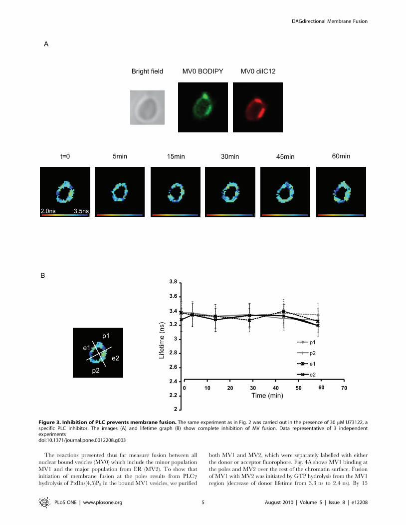
S2). To show that the GTP-initiated fusion was effected by thehydrolysis of PtdIns(4,5)P2, the PLC inhibitor U73122 wasincluded. Fig. 3 shows that inhibition of PLC prevented thedecrease of the donor lifetime, which remained at 3.360.16 ns.
Figure 2. 3D view of nuclei visualised by FLIM. (A) Stack measurements of a nucleus: the first image corresponds to the fluorescence lifetime ofthe BODIPY measured at the top of a nucleus 15 minutes after photo-activation of caged GTP. Ten successive layers of the same nucleus wereobtained. For each layer the focus of the objective was moved 0.25 mm along the Z-axis. Since the acquisition of one image lasts for 1 minute, the lastimage corresponding to the bottom of the nucleus was measured 25 minutes after the induction of NE formation. (B) 3D reconstructions from the Z-stacks of the same nucleus to form fluorescence lifetime 3D views. The indicated times correspond to the time elapsed after photo activation whenthe first image of each stack was measured.doi:10.1371/journal.pone.0012208.g002
DAGdirectional Membrane Fusion
PLoS ONE | www.plosone.org 4 August 2010 | Volume 5 | Issue 8 | e12208
The reactions presented thus far measure fusion between allnuclear bound vesicles (MV0) which include the minor populationMV1 and the major population from ER (MV2). To show thatinitiation of membrane fusion at the poles results from PLCchydrolysis of PtdIns(4,5)P2 in the bound MV1 vesicles, we purified
both MV1 and MV2, which were separately labelled with eitherthe donor or acceptor fluorophore. Fig. 4A shows MV1 binding atthe poles and MV2 over the rest of the chromatin surface. Fusionof MV1 with MV2 was initiated by GTP hydrolysis from the MV1region (decrease of donor lifetime from 3.3 ns to 2.4 ns). By 15
Figure 3. Inhibition of PLC prevents membrane fusion. The same experiment as in Fig. 2 was carried out in the presence of 30 mM U73122, aspecific PLC inhibitor. The images (A) and lifetime graph (B) show complete inhibition of MV fusion. Data representative of 3 independentexperimentsdoi:10.1371/journal.pone.0012208.g003
DAGdirectional Membrane Fusion
PLoS ONE | www.plosone.org 5 August 2010 | Volume 5 | Issue 8 | e12208

Figure 4. DAG is required for vectorial progression of fusion. (A) The same experiments as in Fig. 2 were performed using non-ER vesicles(MV1) labelled with BODIPY-C12 and ER vesicles (MV2) labelled with diIC12. Membrane fusion induces both a decrease of the lifetime and a spreadingof BODIPY-C12 all around the nucleus. (B) Same experiment as in Fig. 4A carried out in the presence of 30 mM of U73122, indicating that fusion of thenon-ER with the ER membranes requires PLC activity. (C) Same experiment as Fig. 4A using SKIP pre-treated MV1 vesicles. Dephosphorylation ofPtdIns(4,5)P2 to PtdIns(4)P inhibits fusion.doi:10.1371/journal.pone.0012208.g004
DAGdirectional Membrane Fusion
PLoS ONE | www.plosone.org 6 August 2010 | Volume 5 | Issue 8 | e12208

minutes the fusion wave started to spread laterally to MV2. WhenMV1 was pre-treated with the PLC inhibitor U73122, membranefusion was blocked (Fig. 4B). The inactive U73343 analogue didnot prevent fusion (Fig. S4).If MV1 was pre-treated with a recombinant phosphoinositide 5-
phosphatase (SKIP) to deplete the substrate for PLC, membranefusion was also prevented (Fig. 4C). SKIP dephosphorylatesPtdIns(4,5)P2 to PtdIns(4)P which is not recognised by PLCc andthus DAG cannot be produced. We show in Fig 4C a sperm nucleuswhich was not completely decondensed so its conical shape clearlydefines the apical and basal poles. Pre-treatment of MV0 with SKIPalso inhibited membrane fusion as expected (Fig. S3A). In a controlparallel reaction, denatured SKIP failed to block membrane fusionand nuclear envelope formation (Fig. S3B).Inhibition results are summarised in Table 1. These results
strongly support our model of nuclear envelope formationinvolving GTP regulation of PLC hydrolysis of PtdIns(4,5)P2[3,4,5] and add for the first time confirmation of the prediction ofbipolarised spatial control of fusion initiation.
Discussion
In this paper we provide evidence that fusion initiation leadingto nuclear envelope formation is GTP triggered, and requiresPtdIns(4,5)P2 and PLC activity in the MV1 fraction. Fusion ispropagated bi-directionally from the sites of initiation and involvesfusion of MV1 with MV2 as well as successive fusions with moreMV2 to complete NE formation in the cell-free assay. Theproperties of the non-ER derived MV1 therefore are consistentwith its role as a potentially fusogenic vesicle population regulatedby GTP hydrolysis and resulting in heterotypic fusion with othervesicles by localised production of DAG.These results lead to several questions: what is the origin of the
polarisation, how is fusion propagated after initiation, and is themechanism for fusion a general one? We discuss and offerspeculation on these questions in turn below.Since non-ER derived MV1 is a major source of the fusogenic
DAG (and location of PLCc in these cells), its restricted binding tothe apex and base of the sperm nucleus lead to bi-directionality offusion. The specificity of binding to the NER regions is likely to
result from non-random packing of chromosomes in the spermnucleus. MV1 binding requires NERs [18] which themselves havean unusual lipid composition and physical properties [19]. Wehave shown regions with similar properties present in the spermnuclei of a wide range of animals suggesting they serve as nuclearmembrane organising regions [20]. It remains to be demonstratedthat each chromosome contains one or a few such structures whichmight also be used during mitotic NE reassembly.Our data show that the mechanism of propagation of fusion
commences from the polar MV1vesicles, which fuse with ER-derived vesicles. Subsequently ER vesicles fuse with one anothertowards the equatorial regions. The nuclear envelope precursorvesicles in vitro and in vivo are approximately 0.5 mm and thenucleus about 4 mm in diameter. Surface area calculations suggestthat minimally 125 vesicles must fuse to envelop the nucleus withtwo bilayers [21]. If each NE were initiated by a single MV1vesicle at the poles, the lipid contribution of MV1 to the completednuclear envelope could be,1% of the total. Since nearly all PLCcis associated with MV1 and is .100-fold enriched in MV1 as wellas its substrate PtdIns(4,5)P2 [3], it is likely that DAG formed inthis compartment rapidly diffuses into the ER-derived membraneswith successive fusions, continually lowering the DAG concentra-tion until it is below the 4% estimated from synthetic systems to berequired for fusion [22]. Due to this dilution, eventually thefusogenicity of the forming envelope towards successive vesicleswould likely decrease, and the process might become self-limiting.To determine if a decrease in the rate of successive fusionsaccompanies propagation requires a much a higher resolutionmethod than used here.The non-ER derived MV1 vesicle fraction enriched in PLCc is
found in vesicles in the cortex of oocytes [3] whereas the ER in vivois usually a continuous membrane structure [23] that is of necessityvesiculated during preparation of cell extracts. In vivo, it is likelythat tubules or continuous sheets of ER envelop the chromosomesat telophase. Thus the role of the non-ER fusogenic MV1 vesicleswould be to seal gaps in the enveloping ER [5]. Local DAGconcentrations could decline through dilution, chemical modifi-cation or both, returning the membrane to a non-fusigenic state.A complete description of the spatial rearrangements of MV1
during the cell cycle has yet to be made. It is however clear thatthere are many more non-ER vesicles enriched in PLCc than arenecessary to facilitate NE formation [21]. It will therefore be ofinterest to determine whether such potentially fusogenic vesiclesare mobilised to participate in other membrane fusion eventsduring mitosis or interphase [24]. A novel aspect of thismechanism is that the specificity of localisation of DAG woulddepend on its delivery through the recruitment of potentiallyfusogenic vesicles to the sites of fusion rather than by its generationwithin a domain of one or both partner membranes to be fused.We have shown that membrane fusion can involve a vectorial
progression with a specific origin and direction. This vectorialprogression is dependent on the formation of localised DAGderived from polyphosphoinositide modification in one of thepartner membranes. The interplay of localised lipids and proteinrecruitment will be of importance to explore in a variety of naturalmembrane fusions.
Supporting Information
Methods S1
Found at: doi:10.1371/journal.pone.0012208.s001 (0.10 MBDOC)
Figure S1 Confocal images of sperm nuclei, nuclear enveloperemnants and bound MVs. (A) Input sperm nuclei (extracted with
Table 1. Summary of membrane fusion effectors andinhibitors.
Treatment Fusion
ATP 2
GTP +
GTPcS 2
U73122 + GTP 2
U73343 + GTP +
SKIP all MVs + GTP 2
SKIP non-ER MVs + GTP 2
Boiled SKIP all MVs + GTP +
MVs independently labelled with either BODIPY-C12 (donor) or diIC12(acceptor) were bound in vitro to sperm nuclei with ATP-GS (ATP). MV fusionwas induced by photo activation of caged GTP and assessed by FLIMmeasurements as presented in Fig. 2, 3 and 4. The inhibitors U73122, U73343(30 mM) or GTPc[S] (2 mM) were applied just before mounting the sample andafter 15 minutes of incubation GTP was activated. For inhibition of PtdIns(4,5)P2phosphatase, MVs (either all MVs or only non-ER MV1) were incubated with SKIPpurified proteins for 1 hour at room temperature prior to binding.doi:10.1371/journal.pone.0012208.t001
DAGdirectional Membrane Fusion
PLoS ONE | www.plosone.org 7 August 2010 | Volume 5 | Issue 8 | e12208

0.1% Triton X-100). DNA stained with propidium iodide [23] anddetergent resistant membranes of the nuclear envelope remnantsstained with diOC6 located at the acrosomal and centriolar fossaregions (apex and base) of the undecondensed nucleus. (B)Decondensed sperm nucleus with bound membrane vesicles fromfertilised egg extract (S10). DNA stained with Hoechst 33334(blue) and membranes stained with diOC6 (green). Nucleiincubated in S10 with ATP have decondensed chromatin andbound vesicles. (C) Nuclei with bound vesicles separated fromunbound vesicles remaining in the S10 following purificationthrough 0.5 M sucrose. The majority of bound vesicles are fromthe ER-derived population (MV2), which binds over the entiresurface. The binding of non-ER derived membrane vesicle (MV1)occurs only in the regions of the NERs.Found at: doi:10.1371/journal.pone.0012208.s002 (2.26 MB EPS)
Figure S2 GTPcS does not induce fusion. The same experimentpresented in Figure 1 was carried out using GTPcS instead ofcaged GTP. Under these conditions the lifetime of the donorremains constant indicating that there is no fusion of the labelledvesicles.Found at: doi:10.1371/journal.pone.0012208.s003 (0.57 MB EPS)
Figure S3 Pre-treatment of MVs with SKIP inhibits the fusionprocess. (A) Total S10 membranes (MV0) were pre-treated withSKIP as described in Experimental Procedures. Pre-treatedmembranes were independently labelled with either BODIPYC12(donor) or diIC12 (acceptor) and both bound in vitro to spermnuclei with ATP-GS (ATP). MV fusion was induced by photoactivation of caged GTP and assessed by FLIM measurements aspresented in Figures 2 to 4. (B) As a control the same experimentas in (A) was performed with denatured SKIP (5 minutes at100oC). Denaturation of SKIP abolished the inhibition of fusion.
Found at: doi:10.1371/journal.pone.0012208.s004 (0.88 MB EPS)
Figure S4 U73343, an inactive analogue of U73122, does notinhibit fusion. To confirm the specificity of the U73122 inhibitor,the same experiment as presented in Figure 4 was preformed usingthe U73343 analogue. No inhibition of fusion was observed.Found at: doi:10.1371/journal.pone.0012208.s005 (0.88 MB EPS)
Movie S1 3D Movie of nuclei visualised by FLIM at 0 minutesprior to activation of caged GTP.Found at: doi:10.1371/journal.pone.0012208.s006 (7.65 MBMOV)
Movie S2 3D Movie of nuclei visualised by FLIM at 15 minutesafter photo-activation of caged GTP.Found at: doi:10.1371/journal.pone.0012208.s007 (7.76 MBMOV)
Movie S3 3D Movie of nuclei visualised by FLIM at 30 minutesafter photo-activation of caged GTP.Found at: doi:10.1371/journal.pone.0012208.s008 (7.71 MBMOV)
Acknowledgments
The authors would like to thank R. Woscholski and E. Rosivatz for theirhelp with purified SKIP.
Author Contributions
Conceived and designed the experiments: FD DP BL. Performed theexperiments: FD RDB BV TMH. Analyzed the data: FD. Contributedreagents/materials/analysis tools: FD DP. Wrote the paper: FD DP BL.
References
1. Chernomordik LV, Kozlov MM (2008) Mechanics of membrane fusion. NatStruct Mol Biol 15: 675–683.
2. Gruenberg J (2001) The endocytic pathway: a mosaic of domains. Nat Rev MolCell Biol 2: 721–730.
3. Byrne RD, Garnier-Lhomme M, Han K, Dowicki M, Michael N, et al. (2007)PLCgamma is enriched on poly-phosphoinositide-rich vesicles to control nuclearenvelope assembly. Cell Signal 19: 913–922.
4. Poccia D, Larijani B (2009) Phosphatidylinositol metabolism and membranefusion. Biochem J 418: 233–246.
5. Larijani B, Poccia DL (2009) Nuclear envelope formation: mind the gaps. AnnuRev Biophys 38: 107–124.
6. Byrne RD, Poccia DL, Larijani B (2009) Role of phospholipase C in nuclearenvelope assembly. Clin Lipidol 4: 103–112.
7. Cameron LA, Poccia DL (1994) In vitro development of the sea urchin malepronucleus. Dev Biol 162: 568–578.
8. Byrne RD, Larijani B, Poccia DL (2009) Tyrosine kinase regulation of nuclearenvelope assembly. Adv Enzyme Regul 49: 148–156.
9. Barona T, Byrne RD, Pettitt TR, Wakelam MJ, Larijani B, et al. (2005)Diacylglycerol induces fusion of nuclear envelope membrane precursor vesicles.J Biol Chem 280: 41171–41177.
10. Abbott B, Abbott R, Adhikari R, Ageev A, Allen B, et al. (2005) Limits ongravitational-wave emission from selected pulsars using LIGO data. Phys RevLett 94: 181103.
11. Collas P, Poccia D (1998) Methods for studying in vitro assembly of malepronuclei using oocyte extracts from marine invertebrates: sea urchins and surfclams. Methods Cell Biol 53: 417–452.
12. Cothren CC, Poccia DL (1993) Two steps required for male pronucleusformation in the sea urchin egg. Exp Cell Res 205: 126–133.
13. Larijani B, Barona TM, Poccia DL (2001) Role for phosphatidylinositol innuclear envelope formation. Biochem J 356: 495–501.
14. Byrne RD, Barona TM, Garnier M, Koster G, Katan M, et al. (2005) Nuclearenvelope assembly is promoted by phosphoinositide-specific phospholipase C
with selective recruitment of phosphatidylinositol-enriched membranes.Biochem J 387: 393–400.
15. Schmid AC, Wise HM, Mitchell CA, Nussbaum R, Woscholski R (2004) Type IIphosphoinositide 5-phosphatases have unique sensitivities towards fatty acidcomposition and head group phosphorylation. FEBS Lett 576: 9–13.
16. Byrne RD, Zhendre V, Larijani B, Poccia DL (2009) Nuclear envelopeformation in vitro: a sea urchin egg cell-free system. Methods Mol Biol 464:207–223.
17. Collas P, Poccia D (1996) Distinct egg cytoplasmic membrane vesicles differingin binding and fusion properties contribute to sea urchin male pronuclearenvelopes formed in vitro. J Cell Sci 109: 1275–1283.
18. Collas P, Poccia D (1995) Lipophilic organizing structures of sperm nuclei targetmembrane vesicle binding and are incorporated into the nuclear envelope. DevBiol 169: 123–135.
19. Garnier-Lhomme M, Byrne RD, Hobday TM, Gschmeissner S, Woscholski R,et al. (2009) Nuclear envelope remnants: fluid membranes enriched in sterolsand polyphosphoinositides. PLoS ONE 4: e4255.
20. Collas P, Poccia D (1996) Conserved binding recognition elements of spermchromatin, sperm lipophilic structures and nuclear envelope precursor vesicles.Eur J Cell Biol 71: 22–32.
21. Larijani B, Poccia D (2007) Protein and lipid signaling in membrane fusion:nuclear envelope assembly. Signal Transduction 7: 142–153.
22. Villar AV, Alonso A, Goni FM (2000) Leaky vesicle fusion induced byphosphatidylinositol-specific phospholipase C: observation of mixing of vesicularinner monolayers. Biochemistry 39: 14012–14018.
23. Terasaki M, Jaffe LA (1991) Organization of the sea urchin egg endoplasmicreticulum and its reorganization at fertilization. J Cell Biol 114: 929–940.
24. Gould GW, Lippincott-Schwartz J (2009) New roles for endosomes: fromvesicular carriers to multi-purpose platforms. Nat Rev Mol Cell Biol 10:287–292.
DAGdirectional Membrane Fusion
PLoS ONE | www.plosone.org 8 August 2010 | Volume 5 | Issue 8 | e12208