Microanatomical and immunohistochemical study of the human radial nerve at the antecubital fossa
Upload
independentCategory
view
2download
0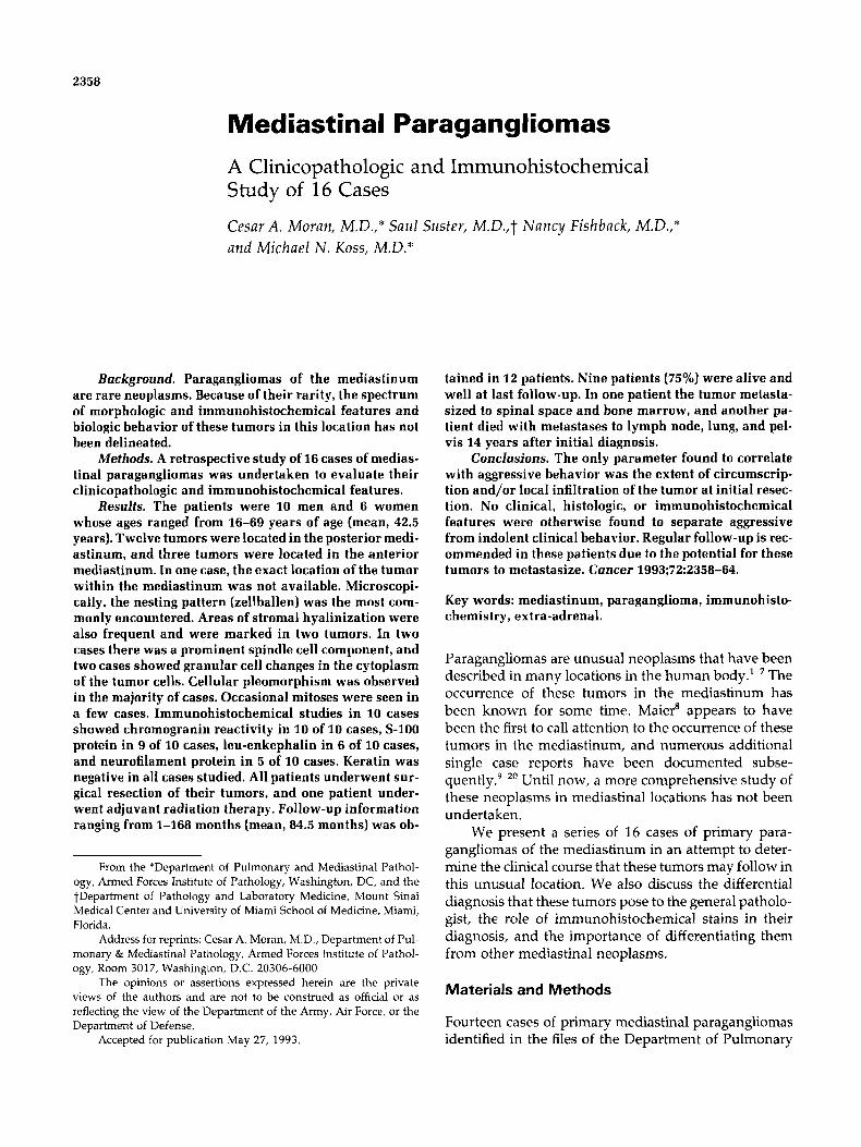
2358
Mediastinal Paragangliomas A Clinicopathologic and Immunohistochemical Study of 16 Cases
Cesar A. Moran, M.D.,* Saul Suster, M.D.,t Nancy Fishback, M.D.,* and Michael N . Koss, M.D.*
Background. Paragangliomas of the mediastinum are rare neoplasms. Because of their rarity, the spectrum of morphologic and immunohistochemical features and biologic behavior of these tumors in this location has not been delineated.
Methods. A retrospective study of 16 cases of medias- tinal paragangliomas was undertaken to evaluate their clinicopathologic and immunohistochemical features.
Results. The patients were 10 men and 6 women whose ages ranged from 16-69 years of age (mean, 42.5 years). Twelve tumors were located in the posterior medi- astinum, and three tumors were located in the anterior mediastinum. In one case, the exact location of the tumor within the mediastinum was not available. Microscopi- cally, the nesting pattern (zellballen) was the most com- monly encountered. Areas of stromal hyalinization were also frequent and were marked in two tumors. In two cases there was a prominent spindle cell component, and two cases showed granular cell changes in the cytoplasm of the tumor cells. Cellular pleomorphism was observed in the majority of cases. Occasional mitoses were seen in a few cases. Immunohistochemical studies in 10 cases showed chromogranin reactivity in 10 of 10 cases, S-100 protein in 9 of 10 cases, leu-enkephalin in 6 of 10 cases, and neurofilament protein in 5 of 10 cases. Keratin was negative in all cases studied. All patients underwent sur- gical resection of their tumors, and one patient under- went adjuvant radiation therapy. Follow-up information ranging from 1-168 months (mean, 84.5 months) was ob-
From the *Department of Pulmonary and Mediastinal Pathol- ogy, Armed Forces Institute of Pathology, Washington, DC, and the tDepartment of Pathology and Laboratory Medicine, Mount Sinai Medical Center and University of Miami School of Medicine, Miami, Florida.
Address for reprints: Cesar A. Moran, M.D., Department of Pul- monary & Mediastinal Pathology, Armed Forces Institute of Pathol- ogy, Room 3017, Washington, D.C. 20306-6000
The opinions or assertions expressed herein are the private views of the authors and are not to be construed as official or as reflecting the view of the Department of the Army, Air Force, or the Department of Defense.
Accepted for publication May 27, 1993.
tained in 12 patients. Nine patients (75%) were alive and well at last follow-up. In one patient the tumor metasta- sized to spinal space and bane marrow, and another pa- tient died with metastases to lymph node, lung, and pel- vis 14 years after initial diagnosis.
Conclusions. The only parameter found to correlate with aggressive behavior was the extent of circumscrip- tion and/or local infiltration of the tumor at initial resec- tion. No clinical, histologic, or immunohistochemical features were otherwise found to separate aggressive from indolent clinical behavior. Regular follow-up is rec- ommended in these patients due to the potential for these tumors to metastasize. Cancer 1993;72:2358-64.
Key words: mediastinum, paraganglioma, immunohisto- chemistry, extra-adrenal.
Paragangliomas are unusual neoplasms that have been described in many locations in the human body.'-' The occurrence of these tumors in the mediastinum has been known for some time. Maier' appears to have been the first to call attention to the occurrence of these tumors in the mediastinum, and numerous additional single case reports have been documented subse- q~ently.'-~' Until now, a more comprehensive study of these neoplasms in mediastinal locations has not been undertaken.
We present a series of 16 cases of primary para- gangliomas of the mediastinum in an attempt to deter- mine the clinical course that these tumors may follow in this unusual location. We also discuss the differential diagnosis that these tumors pose to the general patholo- gist, the role of immunohistochemical stains in their diagnosis, and the importance of differentiating them from other mediastinal neoplasms.
Materials and Methods
Fourteen cases of primary mediastinal paragangliomas identified in the files of the Department of Pulmonary
Mediastinal Paragangliornas/Moran et al. 2359
and Mediastinal Pathology, Armed Forces Institute of Pathology, Washington, DC, and two cases from the files of the Department of Pathology of Mount Sinai Medical Center of Miami, Miami, Florida, form the basis of the current study.
Hematoxylin and eosin-stained tissue sections were available for review in all cases. Selected cases were stained for reticulin. For immunohistochemical studies, formalin-fixed, paraffin-embedded tissue sections were incubated with antibodies against broad-spectrum ker- atin (Dako, Santa Barbara, CA; 1:800), S-100 protein (Enzo; 1:800), chromogranin (Enzo, New York, NY; 1 :BOO), leu-enkephalin (Immunonuclear Corp., Still- water, MN; 1:2000), and neurofilament protein (Lab System, Helsinki, Finland; 1 :BOO) by the avidin-biotin complex technique.’l Nonimmune rabbit and mouse serum was substituted for the antibody as negative con- trols. Appropriate positive controls were run concur- rently for every antibody tested.
Clinical information and follow-up was obtained from the patients’ records or by contacting the referring physician.
Results
Clinical Features
The clinical features in our patients are presented in Table 1. The patients’ ages ranged from 16-69 years (mean, 42.5 years). There were 10 men and 6 women. Eight patients were asymptomatic; three patients pre- sented with functional symptoms including headache, hypertension, and sweating; and three presented with symptoms of cough, chest pain, and difficulty in swal- lowing and/or breathing. No clinical history was avail- able in two patients.
Twelve tumors were located in the posterior medi- astinum, and three tumors were located in the anterior mediastinum. The exact location of the tumor within the mediastinum could not be determined in one case. Multicentric neoplasms were observed in one patient who had both mediastinal and abdominal tumors. No history of a primary tumor elsewhere was obtained in any of our cases.
Fifteen patients were treated by surgical resection alone, and one patient was treated by surgical resection and adjuvant radiation therapy.
Gross Findings
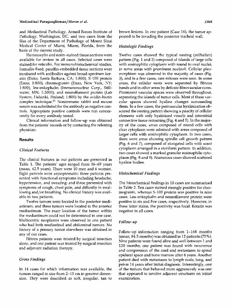
In 14 cases for which information was available, the tumors ranged in size from 2-10 cm in greatest dimen- sion. They were described as soft, irregular, tan to
brown lesions. In one patient (Case 16), the tumor ap- peared to be invading the posterior tracheal wall.
Histologic Findings
Twelve cases showed the typical nesting (zellballen) pattern (Fig. 1 and 2) composed of islands of large cells with eosinophilic cytoplasm with round to oval nuclei, in some areas with prominent nucleoli. Cellular pleo- morphism was observed in the majority of cases (Fig. 3), and in a few cases, rare mitoses were seen. In some areas, the cellular nests were separated by fibrous bands and in other areas by delicate fibrovascular cores. Prominent vascular spaces were observed throughout, separating the islands of tumor cells. Most of these vas- cular spaces showed hyaline changes surrounding them. In a few cases, the perivascular hyalinization ob- scured the nesting pattern showing a paucity of cellular elements with only hyalinized vessels and interstitial connective tissue remaining (Fig. 4 and 5). In the major- ity of the cases, areas composed of round cells with clear cytoplasm were admixed with areas composed of larger cells with eosinophilic cytoplasm. In two cases, there were areas showing spindle cell growth pattern (Fig. 6 and 7), composed of elongated cells with scant cytoplasm arranged in a storiform pattern. In addition, two cases showed a marked granular eosinophilic cyto- plasm (Fig. 8 and 9). Numerous cases showed scattered hyaline bodies.
Histochemical Findings
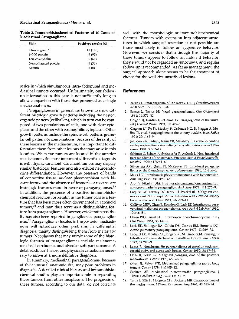
The histochemical findings in 10 cases are summarized in Table 2. Ten cases stained strongly positive for chro- mogranin, whereas S-100 protein was positive in nine cases. Leu-enkephalin and neurofilament protein were positive in six and five cases, respectively. However, in these latter stains, the positivity was focal. Keratin was negative in all cases.
Follow-up information ranging from 1-1 68 months (mean, 84.5 months) was obtained in 12 patients (75%). Nine patients were found alive and well between 1 and 120 months; one patient was found with recurrence and compression of the cord and metastases to spinal epidural space and bone marrow after 6 years. Another patient died with metastases to lymph node, lung, and pelvis 14 years after initial diagnosis. Interestingly, one of the tumors that behaved more aggressively was one that appeared to involve adjacent structures on initial examination.

2360 CANCER October 15, 1993, Volume 72, No. 8
Table 1. Clinical Features of 16 Patients With Mediastinal Paragangliomas
Patient Age no. Sex (yr) History
Size Site (cm) Treatment Follow-up
1 M 37 Asymptomatic Posterior 4 Surgery A & W l O y r 2 M 33 Chest pain Posterior N/A Surgery, radiation A & W 2 mo 3 F 64 N/A Posterior 4.5 Surgery N/A 4 F 30 Chestpain Posterior 5 F 20 Asymptomatic Posterior 6 M 22 Sweating, hypertension, 2 Posterior
7 M 57 Asymptomatic Posterior 8 M 41 Headaches, hypertension Anterior 9 M 16 Palpitations, diaphoresis Posterior
10 F 69 Asymptomatic NOS 11 F 43 Asymptomatic Posterior 12 M 30 Asymptomatic Posterior 13 F 45 Asymptomatic Anterior
abdominal lesions
5.4 Surgery N/A 4 Surgery A & W 5 y r 5 Surgery A & W 2 m o
10 Surgery A & W 3 y r 4 Surgery A & W 2 y r 3.8 Surgery A & W 2 y r 2 Surgery A & W 1 8 m o
7.2 Surgery A & W l m o 5 Surgery A & W 2 y r
6.5 Surgery N/A
14 M 52 Asymptomatic Anterior 3.5 Surgery N/A 15 M 61 N/A Posterior N/A Surgery At 6 yr, alive with recurrence;
metastases to spinal epidural space and bone marrow
16 M 50 Cough, difficulty breathing Posterior involving posterior 8 Surgery Died at 14 yr with & swallowing tracheal wall metastases to pelvis,
lung
A & W: alive and well; N/A: not available; NOS: not otherwise specified.
Discussion
Paragangliomas are unusual neoplasms of ubiquitous distribution. To date, no definitive morphologic criteria exist that correlate with the clinical outcome of these tumors.” Paragangliomas in association with other neoplasms (part of the multiple endocrine neoplasia syndrome) and reports of multicentricity have been well-documented23~27; therefore, care should be exer- cised in labeling multicentric neoplasms as metastases from a malignant tumor. Metastases from paraganglio- mas are those that take place in sites where paraganglia are not normally present.
Immunohistochemical and DNA flow cytometric analyses have been performed in an attempt to estab- lish criteria that may be used to predict progn~sis.’~~’~ The presence of S-100 protein and glial fibrillary acid protein-positive sustentacular cells appears to be asso- ciated with benign clinical behavior, whereas DNA in- dex and synthetic phase fraction analysis have been claimed to be helpful for predicting clinical outcome.
Mediastinal paragangliomas in particular, although rare, have been the subject of numerous reports. Olson and Salyer,17 in a presentation of four cases, reviewed 41 cases previously described in the literature. The au- thors found that 8 patients had died of tumor, 4 of them with metastases, and 19 patients were alive and well
without evidence of tumor 5 months to 21 years after therapy. Gallivan et al.,” in a report of an intrathoracic paravertebral malignant paraganglioma, reviewed 30 cases of posterior mediastinal paragangliomas (para- vertebral paragangliomas). The authors found that these tumors were slightly predominant in men with an
Figure 1. Low magnification of a mediastinal paraganglioma showing a nesting (zellballen) pattern (H & E, original magnification X75).

Mediastinal Paragangliomas/Moran et al . 2361
Figure 2. Mediastinal Paraganglioma showing a Pseudoglandular appearance of nests of tumor cells due to artifactual lumen formation (H & E, original magnification X200).
Figure 4. Mediastinal paraganglioma showing an overgrowth of small vessels that totally obscure the underlying paraganglionic elements (H & E, original magnification XlOO).
average of 29 years. Fifteen patients (48%) had clinical symptoms related to excess catecholamine secretion, and 13 patients were alive with no evidence of tumor at an average of 2.2 years after surgery. Seven of these patients (23%) were reported to have had multiple tu- mors, i.e., abdominal paragangliomas, as well. From re- view of the literature, it appears that mediastinal para- gangliomas seem to more often affect the anterior medi- astinum. In addition, tumors arising in an anterior location are claimed to be more often associated with older age, larger size, and lower incidence of functional- ity, and are less amenable to surgical resection.
In the current study, we have reviewed the clinical, pathologic, and immunohistochemical features of 16 patients with mediastinal paragangliomas. The pa- tients’ ages ranged from 16-69 years (mean, 42.5 years). There were 10 men and 6 were women. Three of
our patients presented with symptoms of functioning tumors including headache, hypertension, and sweat- ing, whereas in approximately 50% of our patients, the tumor was found incidentally on chest radiographic studies. The posterior mediastinal location was the most frequent, with 12 neoplasms located in this com- partment and 3 in the anterior mediastinum. In one case, the exact location of the neoplasm within the me- diastinum was not determined. Follow-up information in 12 patients (75%) ranging from 1-168 months (mean, 84.5 months) indicates that 9 patients were alive and well and 1 patient had recurrence and metastases to spinal epidural space and bone marrow. One patient died with metastases to lymph node, lung, and pelvis 14 years after initial diagnosis. Microscopically, the typi- cal nesting (zellballen) pattern appears to be the most commonly encountered. However, tumors showing
Figure 3. Mediastinal paraganglioma displaying cytologic atypia with marked nuclear pleomorphism and occasional multinucleated forms (H & E, original magnification X400).
Figure 5. Extensive areas of hyalinization of the stroma are seen separating small, scattered tumor islands (H & E, original magnification X200).

2362 CANCER October 15, 1993, Volume 72, No. 8
Figure 6 . Mediastinal paraganglioma showing areas composed of round cells admixed with areas of spindle cells (H & E, original magnification XlOO).
spindle cell features and granular cell morphology may also be seen, although less frequently. The majority of our cases showed areas of cellular pleomorphism, and a few cases showed rare mitoses. Nevertheless, neither of these histologic features correlated with clinical behav- ior, as has been the experience of other authors.” Immu- nohistochemically, chromogranin appears to be the most reliable immunoassay to demonstrate the neuroen- docrine nature of these neoplasms, although S- 100 protein provides and important test to demonstrate the presence of sustentacular cells. These results are in agreement with other immunohistochemical studies of paragangliomas of the head and neck area.30 Leu-enke- phalin and neurofilament protein have also been used
Figure 8. Mediastinal paraganglioma displaying granular cell features (H & E, original magnification X200).
in the diagnosis of paragangliomas. However, in our series, approximately 50% of the neoplasms showed positive reaction for these antibodies. Although keratin positivity has been reported in paragangl ioma~,~~,~~ it was negative in all of our cases. The results of our immu- nohistochemical studies showed no clear correlation with the clinical course of these tumors.
One factor that may play a more important role in the behavior of these neoplasms is the extent of the tumor at the time of diagnosis. This is borne out by the fact that in one case (Case 16), in which the tumor be- haved more aggressively, it appeared to be infiltrative and somewhat extensive at the time of resection. Well- circumscribed lesions in which complete surgical resec- tion can be accomplished is a feature more likely to be accompanied by a favorable clinical course. It is also important to mention that there was one case in our
Figure 7. Higher magnification of the spindle cell component seen in a mediastinal paraganglioma showing focal storiform formations (H & E, original magnification X200).
Figure 9. Higher magnification of a paraganglioma with granular cell features. The cells appear oncocytic and show lateral displacement of the nuclei (H & E, original magnification X400).
Mediastinal Paragangliomas/Moran et al . 2363
Table 2. Immunohistochemical Features of 10 Cases of Mediastinal Paraganglioma
Stain Positives results ( o h )
Chromogranin S-100 protein Leu-enkephalin Neurofilament protein Keratin
series in which simultaneous intra-abdominal and me- diastinal tumors occurred. Unfortunately, our follow- up information in this case is not sufficiently long to allow comparison with those that presented as a single mediastinal mass.
Paragangliomas in general are known to show dif- ferent histologic growth patterns including the nested, organoid pattern (zellballen), which in turn can be com- posed of two populations of cells, one with clear cyto- plasm and the other with eosinophilic cytoplasm. Other growth patterns include the spindle cell pattern, granu- lar cell pattern, or combinations. Because of the rarity of these lesions in the mediastinum, it is important to dif- ferentiate them from other lesions that may arise in this location. When the tumors are located in the anterior mediastinum, the most important differential diagnosis is with thymic carcinoid. Carcinoid tumors may display similar histologic features and also exhibit neuroendo- crine differentiation. However, the presence of bands of connective tissue, nuclear pleomorphism with bi- zarre forms, and the absence of festoons or rosettes are histologic features more in favor of paragangli~mas.~~ In addition, the presence of a positive immunohisto- chemical reaction for keratin in the tumor cells is a fea- ture that has been more often documented in carcinoid tumors,34 and may thus serve as a distinguishing fea- ture from paraganglioma. However, cytokeratin positiv- ity has also been reported in gangliocytic paraganglio- mas.32 Paragangliomas located in the posterior mediasti- num will introduce other problems in differential diagnosis, mainly distinguishing them from metastatic tumors. Neoplasms that may mimic some of the histo- logic features of paragangliomas include melanoma, renal cell carcinoma, and alveolar soft part sarcoma. A detailed clinical history and physical evaluation is neces- sary to arrive at a more definitive diagnosis.
In summary, mediastinal paragangliomas, because of their unusual anatomic site, may pose problems in diagnosis. A detailed clinical history and immunohisto- chemical studies play an important role in separating these tumors from other neoplasms. The prognosis of these tumors, according to our data, do not correlate
well with the morphologic or immunohistochemical features. Tumors with extension into adjacent struc- tures in which surgical resection is not possible are those most likely to follow an aggressive behavior. However, we consider that although the majority of these tumors appear to follow an indolent behavior, they should not be regarded as innocuous, and regular follow-up is recommended. As far as management, the surgical approach alone seems to be the treatment of choice for the well-circumscribed lesions.
References
1. Barnes L. Paraganglioma of the larynx. ORL Otorhinolaryngol Relat Spec 1991; 53:220-34.
2. Barnes L, Taylor SR. Vagal paragangliomas. Clin Otolaryngol
3. Colgan TJ, Dardick 1, O’Connel G. Paraganglioma of the vulva. Int J Gynecol Pathol 1991; 10203-8.
4. Grignon DJ, Ro JY, Mackay B, Ordonez NG, El-Naggar A, Mo- lina TJ, et al. Paraganglioma of the urinary bladder. Hum Pathol
5. Jamjoon ZA, Sadiq S, Naim VR, Malabary T. Cerebello-pontine angle paraganglioma simulating an acoustic neurinoma. BrJNeu- rosurg 1991; 5:307-12.
6. Schimid C, Beham A, Steindorfer P, Aubock L. Non-functional paraganglioma of the stomach. Virchows Arch A Pathol Anat His-
7. Silverstein AM, Quint DJ, McKeever PE. Intradural paragang- lioma of the thoracic spine. A m ] Neuroradiol 1990; 11:614-6.
8. Maier HC. Intrathoracic pheochromocytoma with hypertension. Ann Surg 1949; 130:1059-65.
9. Aron V, Nicoloff DM. Intrathoracic paraganglioma arising from aorticosympathetic paraganglion. Arch Surg 1976; 111275-9.
10. Enquist SW, Tormey DC, Jenis EH, Warkel RL. Malignant che- modectoma of the superior mediastinum with elevated urinary homovanilic acid. Chest 1974; 66:209-11.
11. Gallivan MEV, Chun B, Rowden G, Lack EE. lntrathoracic para- vertebral malignant paraganglioma. Arch Pathol Lab Med 1980;
12. Green WO, Basset FH. Intrathoracic pheochromocytoma. A m J Clin Pathol 1961; 35:142-6.
13. Lack EE, Stillinger RA, Calvin DB, Groves RM, Burnette DG. Aortic-pulmonary paraganglioma. Cancer 1979; 43:269-78.
14. Lacquet LK, Moulijn AC, Jongerius CM, Limburg M, Rensing JB. Intrathoracic chemodectoma with multiple localizations. Thorax
15. Lattes R. Nonchromaffin paraganglioma of ganglion nodosum, carotid body, and aortic-arch bodies. Cancer 1950; 3:667-94.
16. Odze R, Begin LR. Malignant paraganglioma of the posterior mediastinum. Cancer 1990; 65:564-9.
17. Olson JL, Salyer WR. Mediastinal paraganglioma (aortic body tumor). Cancer 1978; 41:2405-12.
18. Pachter MR. Mediastinal nonchromaffin paraganglioma. J Thorac Cardiovasc Surg 1963; 45:152-9.
19. Tama L, Ellis H, Hodgson CH, Dockerty MB. Chemodectoma of the mediastinum. J Thorac Cardiovasc Surg 1962; 43:585-94.
1991; 16:376-82.
1991; 22:1162-9.
topathol 1990; 417:261-6.
104:46-51.
1977; 32~203-9.
2364 CANCER October 15, 1993, Volume 7 2 , No. 8
20.
21
22
23
24.
25.
26.
27.
Victor S, Vijay K, Andoppan P, Bharanthy S, Rao S, Reddy KC. Malignant mediastinal chemodectoma. Chest 1975; 68:583-4. Hsu SM, Raine L. The use of avidin-viotin-peroxidase complex (ABC) in diagnostic and research pathology. In: DeLellis RA, editor. Advances in immunohistochemistry. New York: Masson.
Linnoila RI, Keiser HR, Steinberg SM, Lack EE. Histopathology of benign versus malignant sympathoadrenal paragangliomas. Hum Pathol 1990; 21:1168-80. Carney JA, Sheps SG, Go VLW, Gordon H. The triad of gastric leiomyosarcoma, functioning extra-adrenal paraganglioma and pulmonary chondroma. N Engl J Med 1977; 296:1517-8. Dajee A, Dajee H, Hinrichs S, Lillington G. Pulmonary chon- droma, extra-adrenal paraganglioma, and gastric leiomyosar- coma. J Thorac Cardiovasc Surg 1982; 84:377-81. Grace MP, Batist G, Grace WR, Gillosley JF. Aorticopulmonary paraganglioma and gastric leiomyoblastoma in a young woman. Am J Med 1981; 70:1288-92. Haber S. Retroperitoneal and mediastinal chemodectoma. AIR
Maier HC, Humphreys GH 11. lntrathoracic pheochromocy- toma: including a case of multiple paragangliomas of the non- functional type. J Thorac Surg 1958; 36:625-41.
1984:3 1-42.
1964; 92:1029-41.
28. Achilles E, Padberg BC, Holl K, Kloppel G, Schroder S. Immu- nohistochemistry of paragangliomas: value of staining for S-100 protein and glial fibrillary protein in diagnosis and prognosis. Histopathology 1991; 18:453-8. Sauter ER, Hollier LH, Bolton JS, Ochsner JL, Sardi A. Prognos- tic value of DNA flow cytometry in paragangliomas of the ca- rotid body. J Surg Oncol 1991; 46:151-3.
30. Martinez-Madrigal F, Bosq J, Micheau C, Nevet P, Luboinski 8. Paragangliomas of the head and neck: immunohistochemical analysis of 16 cases in comparison with neuro-endocrine carci- nomas. Pathol Res Pract 1991; 187:814-23.
31. Collina G, Maiorama A, Trentin GP. Duodenal gangliocytic par- aganglioma: case report with immunohistochemical study on the expression of keratin polypeptides. Histopathology 1991;
32. Burke AP, Helwig EB. Gangliocytic paraganglioma. Am J Clin
33. Rosai J, Higa E. Mediastinai endocrine neoplasm of probable thymic origin related to carcinoid tumor. Cancer 1972; 29:1061- 74.
34. Googe PB, Ferry JA, Bhan AK, Dickersin GR, Pilch BZ, Good- man M. A comparison of paraganglioma, carcinoid, and small cell carcinoma of the larynx. Arch Pathol Lab Med 1988;
29.
19:476-8.
Pathol 1989; 92~1-9.
112:809-15.
Copyright © 2022 FDOKUMEN


![[Immunohistochemical studies of cultured cells from bone marrow and aseptic inflammation]](https://static.fdokumen.com/doc/165x107/63360d7764d291d2a302b9a2/immunohistochemical-studies-of-cultured-cells-from-bone-marrow-and-aseptic-inflammation.jpg)