Influence of Gating System Parameters of Die-Cast Molds on ...

Structure 14, 1411–1423, September 2006 ª2006 Elsevier Ltd All rights reserved DOI 10.1016/j.str.2006.07.006
Mechanism of Gating and Ion Conductivity ofa Possible Tetrameric Pore in Aquaporin-1
Jin Yu,1 Andrea J. Yool,2 Klaus Schulten,1
and Emad Tajkhorshid1,*1Theoretical and Computational Biophysics GroupBeckman InstituteUniversity of Illinois at Urbana-Champaign405 N. Mathews Ave.Urbana, Illinois 618012Department of PhysiologyDepartment of PharmacologyProgram in NeuroscienceUniversity of ArizonaTucson, Arizona 85724
Summary
While substrate permeation through monomeric pores
of aquaporins is well characterized, little is knownabout the possible tetrameric pore. AQP1 has been
suggested to function as an ion channel upon cGMPactivation, although this idea has been controversial.
Taking a theoretical and experimental approach, wedemonstrate that the current might arise through the
tetrameric pore and propose a plausible mechanismfor conduction and gating. In response to simulated
ion permeation, immediate hydration of the putativecentral pore was facilitated by moderate conforma-
tional changes of pore-lining residues. cGMP is foundto interact with an unusually arginine-rich, cytoplas-
mic loop (loop D) facilitating its outward motion, whichis hypothesized to trigger the opening of a cytoplasmic
gate. Physiological analyses of wild-type AQP1 and
a designed mutant in which two arginines of the gatingloop are replaced by alanine provide experimental
support for identifying a key component of the pro-posed mechanism.
Introduction
Aquaporins (AQPs) are members of a family of selectivemembrane channels abundantly present in all domainsof life (Agre et al., 1998; Borgnia et al., 1999; Heymannand Engel, 1999). They are generally known for endow-ing a high transmembrane permeability to water (Agre,2004; Preston and Agre, 1991). However, their involve-ment in diverse cellular functions, including permeationof small molecules other than water (Agre et al., 2002), aswell as cell-cell communication (Bok et al., 1982), hasbeen suggested. Aquaporin-1 (AQP1) was the first mem-ber of the family to be functionally characterized as a wa-ter channel (King and Arge, 1996; Preston et al., 1992).As is predicted for other AQPs, it forms homotetramers(see Figure 1) in cellular membranes (Heymann et al.,1998; Heymann and Engel, 1999; Murata et al., 2000)with each monomer essentially functioning as an inde-pendent water pore (de Groot et al., 2001; Tajkhorshidet al., 2002).
*Correspondence: [email protected]
AQPs are exceptional among membrane proteins withrespect to the abundance of atomic resolution structuralinformation that is available. The structure of mamma-lian AQP1 (Murata et al., 2000; Sui et al., 2001) andAQP0 (Gonen et al., 2005, 2004; Harries et al., 2004), bac-terial GlpF (Fu et al., 2000; Tajkhorshid et al., 2002) andAqpZ (Savage et al., 2003), archaeal AqpM (Lee et al.,2005), and plant spPIP2 (Tornroth-Horsefield et al.,2006) have been solved at high resolutions, and thestructures of several other AQPs are expected to beavailable soon. These structures in combination withthe numerous theoretical studies (de Groot and Grub-muller, 2001; Grayson et al., 2003; Jensen et al., 2001,2003; Tajkhorshid et al., 2002; Wang et al., 2005; Zhuet al., 2001, 2004) have provided a detailed picture ofthe mechanisms of substrate permeation and selectivityof intrasubunit water pores in AQPs. One particularly in-triguing property of AQPs is their ability to exclude pro-tons (Pohl et al., 2001; Saparov et al., 2005) while allow-ing water to pass, a problem that has attracted muchattention from theoreticians (Chakrabarti et al., 2004;de Groot et al., 2003; Ilan et al., 2004; Jensen et al.,2003; Tajkhorshid et al., 2002). In contrast, the role ofthe putative tetrameric central pore (Figure 1) remainspoorly understood. Tetramerization is an essential fea-ture of AQPs. The observation that the four monomerseach have a functionally independent pore of water,but require a tetrameric organization for function, couldsuggest a synergistic benefit of oligomerization and,thus, compel further analysis of the potential role ofthe central pore in AQPs.
A subset of AQP channels including AQP1 have beenfound to function as ion channels (Saparov et al., 2001;Yool and Stamer, 2002; Yool and Weinstein, 2002). How-ever, the endogenous mechanisms regulating activationremains to be understood. Furthermore, physiologicalrelevance for ion-channel function of AQPs has notbeen well established yet; only a recent study (Boassa,et al., 2006) has reported physiological significance forAQP1-mediated ion conduction in chroid plexus. Priorwork has shown that AQP1 channels can carry nonse-lective cationic currents after stimulation with proteinkinase A (Yool et al., 1996) or by increased intracellularcGMP but not cAMP (Anthony et al., 2000). Curiously,a comparison of the total water and ion fluxes, refer-enced to unitary conductance values for each, showedthat the large ionic conductance response (typically 2–10 mA) seen in Xenopus oocytes was carried by only aminor proportion of total water channels present in themembrane (Yool and Weinstein, 2002). Ion substitutionexperiments and analyses of reversal potentials demon-strated that the current response is associated witha nonselective cation conductance, having approxi-mately equal permeability to Na+, K+, and Cs+, lowerbut measurable permeability to tetraethylammoniumion, and no appreciable Cl2 permeability (Yool et al.,1996). When purified AQP1 protein was reconstitutedin lipid bilayers, a cGMP-dependent cationic channelfunction was observed, which depended on an intactAQP1 carboxyl terminal domain and was not activated

Structure1412Structure1412
by cAMP; however, it is important to note that the frac-tion of water channels that were available to functionas gated ion channels in the bilayer was even lowerthan that seen in oocytes, approaching one per millionand prompting the speculation that AQP1 ion channelfunction is an accident of protein misfolding (Saparovet al., 2001). This low open channel probability calledinto question the possible functional relevance of theion channel, yet mutagenesis studies suggested theionic conductance depended on conserved sequencedomains in AQP1. Regions of the AQP1 molecule(such as the carboxyl terminal domain) are highly con-served in amino acid sequence in AQP1 proteins acrossspecies yet apparently are not essential for the water-channel function (Zeidel et al., 1994). When key carboxylterminal residues are altered by site-directed mutagen-esis, the AQP1 ionic current activation is impaired, but
Figure 1. Tetrameric Architecture of AQP1
Cytoplasmic (top) and side (bottom) views of the simulated AQP1
tetramer in lipid bilayer and water. Water and lipid were removed
to create the view in the top panel. The four AQP1 monomers are
shown in different colors. Water is known to move through the indi-
vidual water pores formed by the monomers, while the tetrameric
central pore has been proposed to function as a nucleotide-gated
ion channel. The tetramer was imbedded in a POPE lipid bilayer
(dark green stick representation) and solvated by water (light blue
stick representation) on both sides of the membrane. In the side
view (lower panel), the front monomer is removed for clarity. Side
chains of residues lining the cytoplasmic and periplasmic constric-
tion regions of the central pore, respectively, (VAL52, LEU56,
LEU172, and PHE176 in the bovine AQP1 sequence) are shown in
van der Waals (VDW) representation. The size of the central pore
and the water pores are depicted with the channel radius profile cal-
culated by the program HOLE (Smart et al., 1996), purple for the cen-
tral pore and yellow for the water pores. The tetramer is aligned
along the z axis in the present study, as shown in the bottom view.
water permeability is retained (Boassa and Yool, 2003).Amino acid sequence conservation in structures thatare apparently not needed for water-channel functioncan be deduced to confer some other functional advan-tage. The C-terminal sequence of AQP1 shows someinteresting though incomplete similarities with the se-quences of other cGMP binding proteins (Boassa andYool, 2002). Binding studies characterized a low affinitybinding site for cGMP in AQP1-expressing membranes,and electrophysiological data provided a pharmacologi-cal profile for AQP1 ion-channel activation and inhibitionthat was consistent with that of other cGMP-gated ionchannels (Anthony et al., 2000). Recent work character-izing AQP1 channels natively expressed in choroidplexus has confirmed the gated ion channel functionand provided evidence for a physiologically relevantcontribution to the regulation of cerebral spinal fluid pro-duction (Boassa et al., 2006). Further support for theconcept that AQPs can function as ion channels comesfrom other work showing that the pH-sensitive AQP6(Liu et al., 2005; Yasui et al., 1999) and the tyrosine-kinase-sensitive Drosophila aquaporin-related proteinBIB (big brain) (Yanochko and Yool, 2002, 2004) functionas ion channels showing properties of total membraneconductance amplitudes and voltage independencesimilar to that of AQP1 when expressed in oocytes butwith contrasting mechanisms of activation and differentionic selectivities (AQP1 and BIB are cationic; AQP6 isanionic). These differences rule out the alternative hy-pothesis of a ubiquitous endogenous oocyte current.
Part of the uncertainty regarding the ion-channel func-tion of AQP1 has stemmed from the fact that singlechannel properties and responsiveness to intracellularcGMP can vary with the expression system. For exam-ple, in oocytes, batch-to-batch variability in the ampli-tude of the AQP1 ionic current response to forskolinhas been observed by our lab and others, a complicationthat has been suggested post hoc to have arisen in partfrom crosstalk between complex intracellular signalingsystems in the oocyte system and perhaps from interac-tions with yet-unidentified accessory proteins (Yool andStamer, 2002). Expression system specific differencesin AQP1 ion-channel function are clear: when reconsti-tuted in bilayers, purified AQP1 protein showed a re-duced unitary conductance and a substantially loweropen probability (Saparov et al., 2001) as comparedwith AQP1 channels in oocytes. In transfected AQP1-expressing HEK cells, no ion-channel currents weremeasured in response to cGMP (Tsunoda et al., 2004).In contrast, native AQP1 channels in choroid plexusshow a robust ion channel conductance in response tocGMP that is lost after AQP1 knockdown by small inter-fering RNAs (Boassa et al., 2006). Pancreatic secretorygranules generate a cationic conductance response tocGMP that depends on AQP1 coassembly with othermembers of a protein-signaling complex (Abu-Hamdahet al., 2004). The possibility that AQP1 ion-channel gat-ing depends on convergent processes of signaling andprotein-protein interactions is consistent with all thedata currently available.
Identification of Mg2+ binding sites at the 4-fold sym-metry axis of the tetramer of GlpF (Fu et al., 2000),another member of the AQP family, supports the notionof ion flux through the central pore. The tetrameric
Gating and Ion Conduction of AQP1 Tetrameric Pore1413
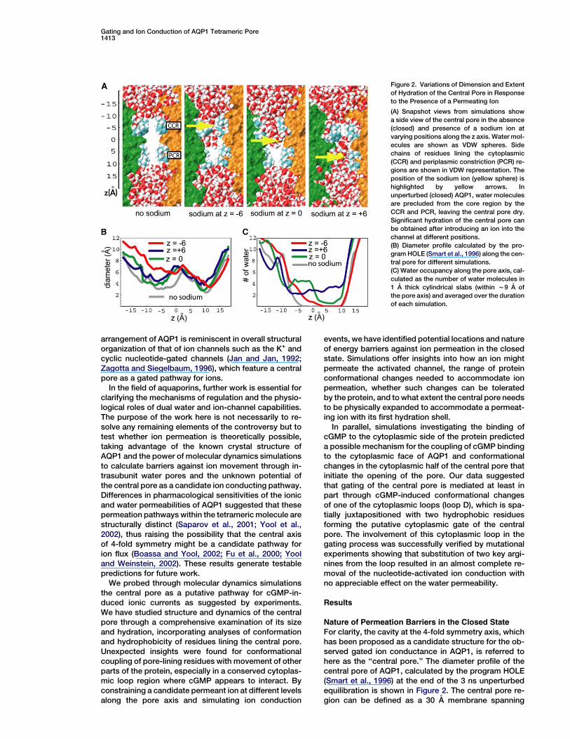
Figure 2. Variations of Dimension and Extent
of Hydration of the Central Pore in Response
to the Presence of a Permeating Ion
(A) Snapshot views from simulations show
a side view of the central pore in the absence
(closed) and presence of a sodium ion at
varying positions along the z axis. Water mol-
ecules are shown as VDW spheres. Side
chains of residues lining the cytoplasmic
(CCR) and periplasmic constriction (PCR) re-
gions are shown in VDW representation. The
position of the sodium ion (yellow sphere) is
highlighted by yellow arrows. In
unperturbed (closed) AQP1, water molecules
are precluded from the core region by the
CCR and PCR, leaving the central pore dry.
Significant hydration of the central pore can
be obtained after introducing an ion into the
channel at different positions.
(B) Diameter profile calculated by the pro-
gram HOLE (Smart et al., 1996) along the cen-
tral pore for different simulations.
(C) Water occupancy along the pore axis, cal-
culated as the number of water molecules in
1 A thick cylindrical slabs (within w9 A of
the pore axis) and averaged over the duration
of each simulation.
arrangement of AQP1 is reminiscent in overall structuralorganization of that of ion channels such as the K+ andcyclic nucleotide-gated channels (Jan and Jan, 1992;Zagotta and Siegelbaum, 1996), which feature a centralpore as a gated pathway for ions.
In the field of aquaporins, further work is essential forclarifying the mechanisms of regulation and the physio-logical roles of dual water and ion-channel capabilities.The purpose of the work here is not necessarily to re-solve any remaining elements of the controversy but totest whether ion permeation is theoretically possible,taking advantage of the known crystal structure ofAQP1 and the power of molecular dynamics simulationsto calculate barriers against ion movement through in-trasubunit water pores and the unknown potential ofthe central pore as a candidate ion conducting pathway.Differences in pharmacological sensitivities of the ionicand water permeabilities of AQP1 suggested that thesepermeation pathways within the tetrameric molecule arestructurally distinct (Saparov et al., 2001; Yool et al.,2002), thus raising the possibility that the central axisof 4-fold symmetry might be a candidate pathway forion flux (Boassa and Yool, 2002; Fu et al., 2000; Yooland Weinstein, 2002). These results generate testablepredictions for future work.
We probed through molecular dynamics simulationsthe central pore as a putative pathway for cGMP-in-duced ionic currents as suggested by experiments.We have studied structure and dynamics of the centralpore through a comprehensive examination of its sizeand hydration, incorporating analyses of conformationand hydrophobicity of residues lining the central pore.Unexpected insights were found for conformationalcoupling of pore-lining residues with movement of otherparts of the protein, especially in a conserved cytoplas-mic loop region where cGMP appears to interact. Byconstraining a candidate permeant ion at different levelsalong the pore axis and simulating ion conduction
events, we have identified potential locations and natureof energy barriers against ion permeation in the closedstate. Simulations offer insights into how an ion mightpermeate the activated channel, the range of proteinconformational changes needed to accommodate ionpermeation, whether such changes can be toleratedby the protein, and to what extent the central pore needsto be physically expanded to accommodate a permeat-ing ion with its first hydration shell.
In parallel, simulations investigating the binding ofcGMP to the cytoplasmic side of the protein predicteda possible mechanism for the coupling of cGMP bindingto the cytoplasmic face of AQP1 and conformationalchanges in the cytoplasmic half of the central pore thatinitiate the opening of the pore. Our data suggestedthat gating of the central pore is mediated at least inpart through cGMP-induced conformational changesof one of the cytoplasmic loops (loop D), which is spa-tially juxtapositioned with two hydrophobic residuesforming the putative cytoplasmic gate of the centralpore. The involvement of this cytoplasmic loop in thegating process was successfully verified by mutationalexperiments showing that substitution of two key argi-nines from the loop resulted in an almost complete re-moval of the nucleotide-activated ion conduction withno appreciable effect on the water permeability.
Results
Nature of Permeation Barriers in the Closed State
For clarity, the cavity at the 4-fold symmetry axis, whichhas been proposed as a candidate structure for the ob-served gated ion conductance in AQP1, is referred tohere as the ‘‘central pore.’’ The diameter profile of thecentral pore of AQP1, calculated by the program HOLE(Smart et al., 1996) at the end of the 3 ns unperturbedequilibration is shown in Figure 2. The central pore re-gion can be defined as a 30 A membrane spanning

Structure1414
region (from z = 215 A to z = 15 A) (Figures 1 and 2). Forcrystal structures obtained in the absence of cGMP, thecentral pore is presumed to be closed. In this state, thecentral pore can be characterized by two major constric-tion regions: one with a diameter of about 3.1 A in the cy-toplasmic half, formed by side chains of LEU172 andPHE176 (cytoplasmic constriction region, CCR), andthe other with a diameter of about 2.4 A in the periplas-mic half of the channel, lined by side chains of VAL52and LEU56 (periplasmic constriction region, PCR) (Fig-ure 2). All the amino acids lining the two constriction re-gions are hydrophobic. In our equilibration simulations,the CCR and the PCR effectively keep water moleculesfrom entering the central pore, thus acting as gate-keepers in creating a dehydrated cavity (about 20 Along) spanning the region between CCR and PCR (seeFigure 2A).
The central pore is mainly lined with hydrophobic sidechains. Therefore, a permeating ion would have to beaccompanied by its first hydration shell as there are nopolar/charged residues that would be expected to com-pensate for the desolvation energy cost. While certainregions of the central pore are large enough in theclosed state to accommodate a hydrated ion, in thetwo major constriction regions, spatial constraints areunlikely to allow an ion to pass along with its hydrationshell. Therefore, we would expect that any reasonablegating mechanism would have to trigger conformationalchanges in these regions that would allow water mole-cules to penetrate and fully hydrate the central pore inadvance of ion conduction.
Simulations with a sodium ion constrained at differentsites inside the pore were performed to investigate theextent of conformational changes within the centralpore that could arise in response to ion permeation.The simulations lasted each about 10 ns, a timescalecomparable to that of a cation passing through thegated channel as estimated from experiments (Anthonyet al., 2000) measuring a 150 pS conductivity at a drivingforce of w100 mV.
After placement of a sodium ion in the middle of thecentral pore (z w 0 A), water molecules from the cyto-plasmic side quickly penetrated the CCR and filled thecytoplasmic half within 100 ps. Further penetration ofwater into the periplasmic half of the channel was pre-vented by side chains of LEU56, thus leaving the PCRdehydrated for most part of the simulations. The diame-ter profile and hydration pattern of the partially hydratedpore observed at the end of the simulation are shown inFigures 2B and 2C, respectively. The diameter profile in-dicated that the presence of an ion in the middle of thepore results in a larger expansion of the cytoplasmicside as compared with the periplasmic side. Visual ex-amination of the trajectories suggests that the observedexpansion is correlated to intrusion of water, which pen-etrates more rapidly from the cytoplasmic side, indicat-ing that the CCR is more flexible than the PCR.
Upon constraining a sodium ion at the CCR (z w26 A), the partially hydrated ion rapidly recruited severalwater molecules from the cytoplasmic side and becamefully hydrated. Soon after, the cytoplasmic half of thepore was partially hydrated. However, water moleculeswere stopped at the PCR by side chains of LEU56. Itseems that this side chain constitutes to a key barrier
against full hydration of the entire pore, preventing watermolecules from penetrating the PCR both from the cyto-plasmic and the periplasmic sides. Nevertheless, watermolecules from the hydrated cytoplasmic half channeloccasionally and transiently populated the PCR duringthe simulation. An example is captured in the snapshotshown in the right panel of Figure 3A. The diameter pro-file (Figure 2B) at the end of this simulation indicates anexpansion of about 4 A of the cytoplasmic side of thepore. There is no discernible expansion in the PCR,though, and water molecules entering from the cyto-plasmic side seem to only induce slight conformationalchanges in the PCR.
Figure 3. Partial Opening of the Central Pore in Response to Occu-
pancy by an Ion
Views of the cytoplasmic loops D in AQP1 are shown in snapshots
from simulations after 3 ns equilibration of the unperturbed AQP1
(closed state, left panels) and after 10 ns simulation of AQP1 with
a sodium ion (yellow sphere) constrained at the CCR (quasiopen,
right panels). (A) Side view of the central pore. Water molecules
are shown in VDW representation. Residues LEU172 and PHE176
at the CCR and VAL52 and LEU56 at the PCR, as well as water are
shown in VDW representation. (B) View of the central pore from
the cytoplasmic side. On each loop D the position of the Ca atom
of GLY168 is highlighted as a small sphere and was used to measure
the separation of the loops induced by the presence of Na+ in the
pore. (C) Separation of the cytoplasmic loops D, calculated as the
distance between the Ca atoms of opposing GLY168 residues on
the loops D in diagonally opposite subunits, in different simulations.
(D) Potential of mean force (PMF) associated with ion permeation
through the closed AQP1 and through a ‘‘quasiopen’’ pore, the latter
established after 10 ns simulation with a sodium ion at the CCR.

Gating and Ion Conduction of AQP1 Tetrameric Pore1415
With a sodium ion constrained at the PCR (z w6 A),water molecules were drawn immediately from the peri-plasmic side into the pore. These water molecules grad-ually penetrated the pore all the way through to the CCRand joined water molecules on the cytoplasmic side af-ter about 2 ns, thus leading to a hydrated channel. Al-though the limited timescale of the simulations mightnot be sufficient for a fully developed response of theprotein, comparison of the hydration patterns observedin the two simulations, with an ion positioned at eitherthe PCR or the CCR, suggests again that the presenceof an ion inside the pore triggers conformationalchanges that are larger in the cytoplasmic half than inthe periplasmic half of the pore. These observationsare consistent with the idea that cytoplasmic eventsgate the channel.
Gating Role of Cytoplasmic LoopsSince cGMP is a cytoplasmic messenger, its putativebinding site and the resulting conformational changesrelevant to gating of the AQP1 ion conductance are likelyto be initiated by ligand-protein interactions at the cyto-plasmic side (Boassa and Yool, 2002). In order to identifyregions that might be involved in binding and gating ofthe central pore, we inspected the cytoplasmic surfaceof the protein for flexible regions. As expected, no largeconformational changes of the transmembrane seg-ments were observed, even after placement of the ioninside the central pore. We did, however, observe con-certed large motions of one of the cytoplasmic loops(loop D, residues 161–169 in each monomer of bovineAQP1), a major constituent of the vestibular entranceto the central pore. Together, the four central loopsfrom the four monomers act as a cap that can physicallyocclude the inner vestibule of the central pore. Shown inFigures 3A and 3B are snapshots taken from simulationsof AQP1 with and without an ion constrained at the CCR.A large separation between the D loops was found to ac-company the small conformational changes of the CCR.LEU172, located at the CCR, sits at the region linking he-lix V to loop D (in the unperturbed AQP1, the loop ends atthe first turn of the membrane-spanning helix). Aftera few nanoseconds of simulation with a sodium ion atthe CCR, an outward motion of the loop and unwindingof the first turn of helix V were observed. During this pro-cess, side chains of LEU172 started to separate fromeach other, thus leading to a widening of the CCR andallowing water to penetrate. The coupling of the pres-ence of an ion in the CCR and the widening and hydra-tion of the pore appears to be a key element for the gat-ing process. It is well known that the size of hydrophobicconstriction sites is an important factor in hydration(Beckstein and Sansom, 2003, 2004; Beckstein et al.,2004). A hydrophobic constriction site can act as an ef-ficient barrier to ion and water permeation if its diameteris less than the diameter of an ion with a first hydrationshell (Beckstein and Sansom, 2003, 2004; Becksteinet al., 2004). Preliminary simulations show that removalof the bulky side chains from the two constriction re-gions results in a rapid hydration of the pore even inthe absence of an ion. Interestingly, the calculated diam-eter of PCR in this mutant is only about 5 A, yet a full hy-dration of the central pore is observed in less than 1 ns.The diameter of CCR in the mutant is about 7 A, which is
interestingly the same as the diameter of CCR in ourquasiopen state.
The hydration of such narrow regions indicates thatpores formed by proteins, which are lined by irregularlyshaped and highly fluctuating amino acid side chains,might behave very differently from perfectly cylindricalnanopores used to calibrate minimum radii needed forhydration (Beckstein and Sansom, 2003, 2004; Beck-stein et al., 2004).
The concerted outward motion of the loops D, effec-tively opening the cytoplasmic vestibule of the centralpore, can be clearly seen in Figure 3A. In other simula-tions performed in the presence of a sodium ion, similarmotions of the loops were observed. As a measure ofseparation of the four D loops, we show in Figure 3Cthe distance between GLY168:Ca atoms from loops Din diagonally positioned subunits. The most significantopening of the inner vestibule, reflected by an increasein the separation between GLY168 residues from 6.5 Ato over 20 A, was observed in simulations in which anion was constrained either at the CCR or at the centerof the pore. It seems that there is a strong coupling be-tween the widening of the CCR and the outward motionsof the D loops.
In addition to the bovine AQP1 crystal structure (Suiet al., 2001) used for models simulated in this study,the crystal structure of human AQP1 has also beensolved (Murata et al., 2000). Comparison of structuresof human and bovine AQP1 proteins, which are highlyconserved in primary sequences, clearly shows thatloop D has been captured in two distinct conformationsin the two structures (Figure 4A). The conformationscould reflect a capacity of loop D to undergo a flippingmotion, which is comparable to the outward shift ofloop D observed in our simulations. The different confor-mations of the loop in the structures of bovine and hu-man AQP1 may be merely due to different crystal con-tacts; however, they might also indicate that D loopsare flexible enough to adopt two intrinsically stable con-formations under in vivo conditions.
Energetics of Ion Permeation throughthe Central Pore
In order to study full permeation of an ion through thecentral pore, we applied the method of steered molecu-lar dynamics (SMD) (Isralewitz et al., 2001) to pull a so-dium ion through the central pore and to investigatehow it interacts with the protein. This model was usedto assess the protein conformational changes and thedisplacement of water molecules that might accompanyan ion permeation event. In all pulling experiments, thepermeating ion brought along some additional watermolecules while passing through the originally dehy-drated pore (wfour water molecules within the first hy-dration shell of the ion).
From the SMD trajectories, we calculated the poten-tial of mean force (PMF), i.e., the free energy profile ofion permeation. The PMF of the unperturbed AQP1 cen-tral pore, representing a closed channel, is shown ingray in Figure 3D. A high free energy barrier amountingto over 30 kcal/mol spans the core region of the centralpore. The region between the two constriction regionspresents a hydrophobic environment that would notfavor a permeating ion. The free energy profile shows

Structure1416
Figure 4. The Proposed Gating Role of Loop
D Mediated through Interaction of cGMP with
an Arginine Array
(A) Comparison of two different conforma-
tions of loop D in the published crystal struc-
tures of bovine AQP1 (Sui et al., 2001) (col-
ored) and human AQP1 (Murata et al., 2000)
(dark gray).
(B) Sequence alignment of loop D in human
AQPs. An extensive polyarginine motif is
present only in AQP1.
(C) Binding of cGMPs to loop D shown in
a snapshot taken after a 10 ns simulation of
AQP1 with four cGMP molecules. The cGMP
molecules are colored according to atom
types; the initial positions of the nucleotides
at the perimeter of the cytoplasmic face of
the tetramer are shown with dark diamonds.
Arginine residues at the cytoplasmic surface
of AQP1 are shown in magenta stick repre-
sentations. The right panel is a close-up,
slightly rotated view showing the interaction
of one of the bound cGMP molecules with ar-
ginines on loop D.
a broad peak with the maximum at the middle of thetransmembrane pore consistent with an absence ofany stabilizing interaction for an ion within a dehydratedpore.
As described in the previous section, placement of anion at the CCR caused water molecules from the cyto-plasmic region to enter the pore. Water molecules couldnot, however, penetrate the PCR and fully hydrate theentire pore, as shown in Figures 2 and 3. Since dehydra-tion introduces a substantial barrier to ion permeation,one predicts that partial hydration of the cytoplasmichalf, as visualized at the end of this simulation, shouldsignificantly reduce the magnitude of the energy barrier.The more permissive state of the central pore can beconsidered as a model for a channel in a quasiopen (seethe right panel of Figure 3A) state. In order to test whetherthe ion permeation through this quasiopen channel isenergetically different from permeation through theclosed channel, the respective PMF was calculated(Figure 3D). The details of SMD simulations are de-scribed in Supplemental Data (available with this articleonline). Results of the simulations showed that in thequasiopen state, the cytoplasmic contribution to thebarrier is virtually eliminated, and the residual barrier atthe PCR shows an amplitude that is approximately halfof that seen for a closed channel. The almost completeremoval of the CCR barrier is due to the hydration ofthis region. In the periplasmic half, on the other hand,the drop of the barrier height is very likely caused by par-tial penetration of water molecules from the cytoplasmicside (as exemplified by the snapshot shown in the rightpanel of Figure 3A). Subtle conformational changes ofthe side chains of LEU56 and VAL52 triggered by watermolecules at the PCR would reduce the barrier further.These results support the hypothesis that the gating
process involves the opening of two hydrophobic gates,one in each half channel, whose hydration will reducethe energy barrier, thus allowing ions to pass.
In a few test simulations, a sodium ion was placed atdifferent regions of the quasiopen channel but was al-lowed to move freely after an initial phase of constraintlasting a few picoseconds. An ion placed at z = +10,i.e., in the periplasmic half of the channel, spontaneouslywent through the channel (in the 2z direction) to thecytoplasmic side within 1 ns; an ion placed initially atz = +12 immediately exited the channel to the periplas-mic side, i.e., no permeation was observed. The simula-tions demonstrate that once the ion has crossed thebarrier (at +10 % z % +12) of the quasiopen channel, per-meation through the rest of the channel can proceedrapidly.
We also performed simulations in which a sodium ionwas repeatedly pulled through the central pore, in orderto see whether partial hydration of the pore induced byprimary ion permeation events could facilitate subse-quent permeation events. Profiles of water occupancyand ion trajectories calculated from these simulationsare provided in Supplemental Data.
cGMP Binding and the Gating Mechanism
In order to investigate the effects of the reported chan-nel-activating ligand, cGMP, at the cytoplasmic face ofAQP1, we performed simulations in which four cGMPmolecules were added near the perimeter of the tetra-mer, at each of the four C termini of the monomers(Figure 4C). The distal segment of the C terminus (resi-dues 250–269) is missing from the crystal structure ofthe monomer but does not appear from experimentalwork to be necessary for the gating of AQP1 ion con-ductance (Boassa and Yool, 2002).

Gating and Ion Conduction of AQP1 Tetrameric Pore1417
After about 2 ns in this simulation (Figure 4C), two ofthe cGMP molecules have diffused from the peripheryof the tetramer to the center where they directly interactwith the D loops. By simultaneously interacting withmultiple amino acids on the cytoplasmic surface, the nu-cleotide molecules facilitate an outward motion of loopD away from the inner vestibule of the central pore(movies showing these events are provided as Supple-mental Data). Compared with the initial conformationof the loops, shown in Figure 4A, the loops moved signif-icantly after the binding of cGMP ligands. The outwardmotion of the D loops and the apparent binding of thenucleotides were not an enduring effect. The nucleotideposition and orientation fluctuated, and an associatedtransient opening of the cytoplasmic entrance of thepore did not persist enough to induce hydration ofthe central pore during the 10 ns simulation. However,the two cGMP molecules remained close to theirrespective loops for the duration of the simulation.
During the simulations, the other two cGMP mole-cules diffused freely along the cytoplasmic surface ofthe protein but did not establish any interaction withthe D loops during the 10 ns simulation. One cGMP mol-ecule simply diffused away from the tetramer, while theother one stayed at the periphery of the tetramer, nearperipheral arginine residues.
Examination of the interactions suggests that cGMPmolecules preferentially interact with regions rich inarginine residues. The interaction is consistent with thenegative charge of the phosphate group and the hydro-gen-bonding capacity of the cyclic nucleotide. A total ofseven arginine residues are located on the cytoplasmicsurface of each AQP1 monomer, with each loop D carry-ing four. The arginine-rich loop D shows features consis-tent with a role in cGMP binding and activation of thecentral pore channel. Furthermore, it is possible thatthe nucleotide ligands interact with multiple arginine do-mains in loop D and the C terminus. Obviously, we arenot capturing the entire binding and gating of the centralpore in a 10 ns simulation. However, our simulationsclearly indicate that cGMP has a strong tendency tointeract with arginine-rich regions at the cytoplasmicside of the protein. Combined with binding of cGMP toits specific binding site, which might be located on theperiphery of the tetramer, the nonspecific interactionwith loop D can result in a large outward motion ofthe latter. We hypothesize that such conformationalchanges in turn affect the CCR barrier in the centralpore. It is intriguing that within the family of mammalianAQPs, AQP1 is unique in having the most consecutivearginine residues in the loop D sequence (Figure 4B).
Further work is needed to definitively link the pro-posed binding of cGMP to loop D and the activation ofthe ion conduction in AQP1. Our simulations indicatethat there is an intrinsic affinity between the nucleotidesand loop D, especially with the arginine array, and sug-gest that nucleotide binding might facilitate an outwardmotion of the loop D, which in turn might enable the ex-pansion of the CCR of the central pore, and subsequentsteps of hydration and permeation.
Mutational Studies of the Gating Domain
The results of our simulations suggested that a flexiblepolyarginine loop (loop D) located at the intracellular
face of the putative central pore acts as a candidate‘‘gating’’ domain at the central pore. This role of loop Dwas tested by site-directed mutagenesis and biologicalassays comparing ion-channel and water-channel prop-erties of wild-type human AQP1 and a mutant constructin which two of the arginines were replaced by alanines.These sites, R159 and R160 in human AQP1 used for bi-ological assays, are identical to R161 and R162 in thebovine AQP1 protein used for our simulations.
The substitution of arginines at positions 159 and 160with alanine (R159A+R160A) had no effect on the mea-sured osmotic water permeability; the hypotonic swell-ing response of oocytes expressing mutant channelswas not significantly different from that with wild-type(Figure 5A). These results indicate that the double muta-tion does not prevent channels from assembling andtargeting the plasma membrane with an efficiency com-parable to that of wild-type. However, the net ionic con-ductance response to increased intracellular cGMP wasgreatly impaired in the mutant (Figure 5B). The meanconductance of R159A+R160A-expressing oocytes wassignificantly less than that of the wild-type and not sig-nificantly different from control oocytes that did notcontain AQP1. Current amplitudes monitored as a func-tion of time (applying repeated brief voltage stepsto +40mV at 6 s intervals) illustrate a representative re-sponse of wild-type but not mutant channels to sodiumnitroprusside (SNP) (Figure 5C). For constructing cur-rent-voltage relationships, current traces were mea-sured over a range of voltage steps (2110 to +40 mV,from a holding potential of 240 mV) at the initiation ofthe recording (time 0) and at 8 min after extracellular ap-plication of SNP (Figure 5D). Conductances were calcu-lated from the slope of the current-voltage relationships(Figure 5E). The magnitudes of the ionic conductance re-sponses that were induced by SNP were calculated bysubtracting the initial conductance (measured at timezero) from the final conductance (measured 7 min afterSNP); these net ionic conductances are summarizedfor statistical comparison in Figure 5B and illustratethe effective elimination of the current by the doublearginine mutation.
Discussion
The interior of the central cavity of AQP1 is quite hydro-phobic and cannot provide a permeant ion with any sta-bilizing interactions. If this region serves as the ion poreof AQP1, an ion can only permeate it without sheddingits first hydration shell, as even partial dehydration ofthe ion would result in a considerable energy cost. Inthe absence of cGMP, a closed state of the centralpore of AQP1 appears to be effectively furnished by ste-ric and hydrophobic barriers against water penetrationat the two major constriction regions flanking the cyto-plasmic and periplasmic sides of the central pore, thusensuring a dry cavity. In our model, hydration of the cen-tral pore requires widening of the two constriction re-gions as a prerequisite for ion permeation. Destabilizingeither of the constriction regions, as was done in oursimulations through artificially inserting an ion intothe bottleneck regions, might facilitate the entrance ofwater molecules into the cavity, hence facilitating ionpermeation.

Structure1418
Figure 5. Effects of Mutations of Arginine
Residues in the Loop D of AQP1 on Water
Permeability and cGMP-Induced Cationic
Conductances
(A) Histogram summary of swelling in 50%
hypotonic saline, standardized to the mean
response of oocytes expressing wild-type
AQP1. Wild-type and mutant R159A+R160A
AQP1 channels were assembled and ex-
pressed in the oocyte membrane with com-
parable efficiency, as evidenced by osmotic
water permeability. Control oocytes showed
no discernable osmotic water permeability.
Data are mean 6 SE; n values are shown in
italics.
(B) Histogram summary of net conductances
measured from the slope of the current-volt-
age relationship for currents measured at
8 min after extracellular application of SNP
after subtraction of the initial conductance
(see text for details). The double arginine mu-
tations significantly impaired the ionic con-
ductance response. Data are mean 6 SE; n
values are shown in italics. Statistical signifi-
cance (panels [A] and [B]) was determined by
analysis of variance and post-hoc Student’s t
test for unpaired data with unequal variance.
NS, not significantly different from wild-type;
double asterisk and asterisk, significantly
different from wild-type with p < 0.005 and
p < 0.05, respectively; pound sign, not signif-
icantly different from control.
(C–E) Representative ionic currents recorded
during before and after application of sodium
nitroprusside (SNP: inducing increased intra-
cellular cGMP) in oocytes expressing wild-
type and R159A+R160A AQP1 channels and
in control oocytes. (C) The activation of the
response was monitored with repeated brief
(200 ms) steps to +40 mV from holding poten-
tial of 240 mV; SNP at a final concentration of
1.6 mM was added at the time indicated (ar-
row). (D) Currents measured for a range of
voltage steps (2110 to +40 mV; holding po-
tential 240 mV) for the same oocytes illus-
trated in panel (C). (E) Plot of current ampli-
tude as a function of voltage for traces
shown in panel (D).
Our simulations revealed that subtle conformationalchanges of amino acid side chains lining the centralpore, especially at the CCR, were sufficient to triggerthe hydration of the pore, which removes a major barrieragainst ion permeation. Selected mutations of aminoacids lining the constriction regions of the channelwould be predicted to impact the ease of hydration ofthe pore and might alter the open probability of thechannel. Experimental tests of these predictions willbe a compelling avenue for future research. Based onthe simulation results, the CCR seems to be conforma-tionally more flexible than the PCR, in that placementof an ion anywhere along the central channel axis caneasily initiate the hydration of the cytoplasmic half ofthe channel. This observation is consistent with a gatingmechanism that initiates channel opening from the cyto-plasmic side, as would be expected in the case ofcGMP, which is an intracellular second messenger. Wepropose that cGMP binding at loop D causes conforma-tional changes at the CCR and serves as at least one ofthe steps involved in activation of ionic conductance
through the putative central pore. Previous studieshave shown that wetting of a channel is very sensitiveto its radius (Allen et al., 2002; Beckstein and Sansom,2003, 2004; Beckstein et al., 2004; Sotomayor andSchulten, 2004). Therefore, even a small perturbationand partial opening of the pore at the CCR may bring wa-ter into the cavity, as observed in our simulations. Thehydration of the cytoplasmic half channel might in turnfacilitate the opening of the PCR, as was also observedalthough transiently, in our simulations; the full hydra-tion of the entire pore could represent an open ion-con-ducting AQP1 channel.
Results presented here, although limited by the shorttimescale of the simulations, suggest that permeation ofcations through a central pore pathway is theoreticallypossible in AQP1 and offer new insights into regionsthat could be involved in the gating of the channel. Deci-phering the contributions of hydrophobic barriers withinthe putative central pore to the properties of ion perme-ation in AQP1 will be of interest in future studies combin-ing the strengths of theoretical modeling, molecular
Gating and Ion Conduction of AQP1 Tetrameric Pore1419
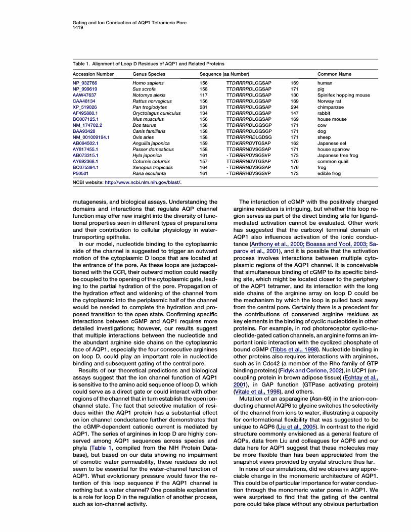
Table 1. Alignment of Loop D Residues of AQP1 and Related Proteins
Accession Number Genus Species Sequence (aa Number) Common Name
NP_932766 Homo sapiens 156 TTDRRRRDLGGSAP 169 human
NP_999619 Sus scrofa 158 TTDRRRRDLGGSAP 171 pig
AAW47637 Notomys alexis 117 TTDRRRRDLGGSAP 130 Spinifex hopping mouse
CAA48134 Rattus norvegicus 156 TTDRRRRDLGGSAP 169 Norway rat
XP_519026 Pan troglodytes 281 TTDRRRRDLGGSAP 294 chimpanzee
AF495880.1 Oryctolagus cuniculus 134 TTDRRRRDLGGSAP 147 rabbit
BC007125.1 Mus musculus 156 TTDRRRRDLGGSAP 169 house mouse
NM_174702.2 Bos taurus 158 TTDRRRRDLGGSGP 171 cow
BAA93428 Canis familiaris 158 TTDRRRRDLGGSGP 171 dog
NM_001009194.1 Ovis aries 158 TTDRRRRRDLGDSG 171 sheep
AB094502.1 Anguilla japonica 159 TTDKRRRDVTGSAP 162 Japanese eel
AY817455.1 Passer domesticus 158 TTDRRRNDVSGSAP 171 house sparrow
AB073315.1 Hyla japonica 161 - TDRRRRDVSGSVP 173 Japanese tree frog
AY692368.1 Coturnix coturnix 157 TTDRRRNDVTGSAP 170 common quail
BC075384.1 Xenopus tropicalis 164 - TDRRRNDVSGSAP 176 frog
P50501 Rana esculenta 161 - TDRRRHDVSGSVP 173 edible frog
NCBI website: http://www.ncbi.nlm.nih.gov/blast/.
mutagenesis, and biological assays. Understanding thedomains and interactions that regulate AQP channelfunction may offer new insight into the diversity of func-tional properties seen in different types of preparationsand their contribution to cellular physiology in water-transporting epithelia.
In our model, nucleotide binding to the cytoplasmicside of the channel is suggested to trigger an outwardmotion of the cytoplasmic D loops that are located atthe entrance of the pore. As these loops are juxtaposi-tioned with the CCR, their outward motion could readilybe coupled to the opening of the cytoplasmic gate, lead-ing to the partial hydration of the pore. Propagation ofthe hydration effect and widening of the channel fromthe cytoplasmic into the periplasmic half of the channelwould be needed to complete the hydration and pro-posed transition to the open state. Confirming specificinteractions between cGMP and AQP1 requires moredetailed investigations; however, our results suggestthat multiple interactions between the nucleotide andthe abundant arginine side chains on the cytoplasmicface of AQP1, especially the four consecutive arginineson loop D, could play an important role in nucleotidebinding and subsequent gating of the central pore.
Results of our theoretical predictions and biologicalassays suggest that the ion channel function of AQP1is sensitive to the amino acid sequence of loop D, whichcould serve as a direct gate or could interact with otherregions of the channel that in turn establish the open ion-channel state. The fact that selective mutation of resi-dues within the AQP1 protein has a substantial effecton ion channel conductance further demonstrates thatthe cGMP-dependent cationic current is mediated byAQP1. The series of arginines in loop D are highly con-served among AQP1 sequences across species andphyla (Table 1, compiled from the NIH Protein Data-base), but based on our data showing no impairmentof osmotic water permeability, these residues do notseem to be essential for the water-channel function ofAQP1. What evolutionary pressure would favor the re-tention of this loop sequence if the AQP1 channel isnothing but a water channel? One possible explanationis a role for loop D in the regulation of another process,such as ion-channel activity.
The interaction of cGMP with the positively chargedarginine residues is intriguing, but whether this loop re-gion serves as part of the direct binding site for ligand-mediated activation cannot be evaluated. Other workhas suggested that the carboxyl terminal domain ofAQP1 also influences activation of the ionic conduc-tance (Anthony et al., 2000; Boassa and Yool, 2003; Sa-parov et al., 2001), and it is possible that the activationprocess involves interactions between multiple cyto-plasmic regions of the AQP1 channel. It is conceivablethat simultaneous binding of cGMP to its specific bind-ing site, which might be located closer to the peripheryof the AQP1 tetramer, and its interaction with the longside chains of the arginine array on loop D could bethe mechanism by which the loop is pulled back awayfrom the central pore. Certainly there is a precedent forthe contributions of conserved arginine residues askey elements in the binding of cyclic nucleotides in otherproteins. For example, in rod photoreceptor cyclic-nu-cleotide-gated cation channels, an arginine forms an im-portant ionic interaction with the cyclized phosphate ofbound cGMP (Tibbs et al., 1998). Nucleotide binding inother proteins also requires interactions with arginines,such as in Cdc42 (a member of the Rho family of GTPbinding proteins) (Fidyk and Cerione, 2002), in UCP1 (un-coupling protein in brown adipose tissue) (Echtay et al.,2001), in GAP function (GTPase activating protein)(Vitale et al., 1998), and others.
Mutation of an asparagine (Asn-60) in the anion-con-ducting channel AQP6 to glycine switches the selectivityof the channel from ions to water, illustrating a capacityfor conformational flexibility that was suggested to beunique to AQP6 (Liu et al., 2005). In contrast to the rigidstructure commonly envisioned as a general feature ofAQPs, data from Liu and colleagues for AQP6 and ourdata here for AQP1 suggest that these molecules maybe more flexible than has been appreciated from thesnapshot views provided by crystal structure thus far.
In none of our simulations, did we observe any appre-ciable change in the monomeric architecture of AQP1.This could be of particular importance for water conduc-tion through the monomeric water pores in AQP1. Wewere surprised to find that the gating of the centralpore could take place without any obvious perturbation
Structure1420
of the water pores, suggesting that there would be littleeffect on the constitutive osmotic water permeability ofthe channel. This observation supports the idea that ionpermeation and water permeation through AQP1 arefunctionally independent of each other, and one canbe regulated without affecting the other.
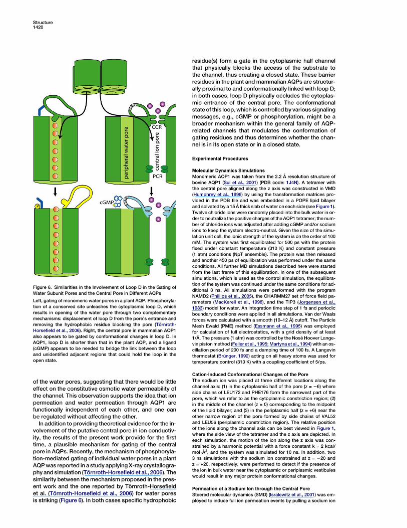
In addition to providing theoretical evidence for the in-volvement of the putative central pore in ion conductiv-ity, the results of the present work provide for the firsttime, a plausible mechanism for gating of the centralpore in AQPs. Recently, the mechanism of phosphoryla-tion-mediated gating of individual water pores in a plantAQP was reported in a study applying X-ray crystallogra-phy and simulation (Tornroth-Horsefield et al., 2006). Thesimilarity between the mechanism proposed in the pres-ent work and the one reported by Tornroth-Horsefieldet al. (Tornroth-Horsefield et al., 2006) for water poresis striking (Figure 6). In both cases specific hydrophobic
Figure 6. Similarities in the Involvement of Loop D in the Gating of
Water Subunit Pores and the Central Pore in Different AQPs
Left, gating of monomeric water pores in a plant AQP. Phosphoryla-
tion of a conserved site unleashes the cytoplasmic loop D, which
results in opening of the water pore through two complementary
mechanisms: displacement of loop D from the pore’s entrance and
removing the hydrophobic residue blocking the pore (Tornroth-
Horsefield et al., 2006). Right, the central pore in mammalian AQP1
also appears to be gated by conformational changes in loop D. In
AQP1, loop D is shorter than that in the plant AQP, and a ligand
(cGMP) appears to be needed to bridge the link between the loop
and unidentified adjacent regions that could hold the loop in the
open state.
residue(s) form a gate in the cytoplasmic half channelthat physically blocks the access of the substrate tothe channel, thus creating a closed state. These barrierresidues in the plant and mammalian AQPs are structur-ally proximal to and conformationally linked with loop D;in both cases, loop D physically occludes the cytoplas-mic entrance of the central pore. The conformationalstate of this loop, which is controlled by various signalingmessages, e.g., cGMP or phosphorylation, might be abroader mechanism within the general family of AQP-related channels that modulates the conformation ofgating residues and thus determines whether the chan-nel is in its open state or in a closed state.
Experimental Procedures
Molecular Dynamics Simulations
Monomeric AQP1 was taken from the 2.2 A resolution structure of
bovine AQP1 (Sui et al., 2001) (PDB code: 1J4N). A tetramer with
the central pore aligned along the z axis was constructed in VMD
(Humphrey et al., 1996) by using the transformation matrices pro-
vided in the PDB file and was embedded in a POPE lipid bilayer
and solvated by a 15 A thick slab of water on each side (see Figure 1).
Twelve chloride ions were randomly placed into the bulk water in or-
der to neutralize the positive charges of the AQP1 tetramer; the num-
ber of chloride ions was adjusted after adding cGMP and/or sodium
ions to keep the system electro-neutral. Given the size of the simu-
lation unit cell, the ionic strength of the system is on the order of 100
mM. The system was first equilibrated for 500 ps with the protein
fixed under constant temperature (310 K) and constant pressure
(1 atm) conditions (NpT ensemble). The protein was then released
and another 450 ps of equilibration was performed under the same
conditions. All further MD simulations described here were started
from the last frame of this equilibration. In one of the subsequent
simulations, which is used as the control simulation, the equilibra-
tion of the system was continued under the same conditions for ad-
ditional 3 ns. All simulations were performed with the program
NAMD2 (Phillips et al., 2005), the CHARMM27 set of force field pa-
rameters (MacKerell et al., 1998), and the TIP3 (Jorgensen et al.,
1983) model for water. An integration time step of 1 fs and periodic
boundary conditions were applied in all simulations. Van der Waals
forces were calculated with a smooth (10–12 A) cutoff. The Particle
Mesh Ewald (PME) method (Essmann et al., 1995) was employed
for calculation of full electrostatics, with a grid density of at least
1/A. The pressure (1 atm) was controlled by the Nose Hoover Lange-
vin piston method (Feller et al., 1995; Martyna et al., 1994) with an os-
cillation period of 200 fs and a damping time of 100 fs. A Langevin
thermostat (Brunger, 1992) acting on all heavy atoms was used for
temperature control (310 K) with a coupling coefficient of 5/ps.
Cation-Induced Conformational Changes of the Pore
The sodium ion was placed at three different locations along the
channel axis: (1) in the cytoplasmic half of the pore (z = 26) where
side chains of LEU172 and PHE176 form the narrowest part of the
pore, which we refer to as the cytoplasmic constriction region; (2)
in the middle of the channel (z = 0) corresponding to the midpoint
of the lipid bilayer; and (3) in the periplasmic half (z = +6) near the
other narrow region of the pore formed by side chains of VAL52
and LEU56 (periplasmic constriction region). The relative position
of the ions along the channel axis can be best viewed in Figure 1,
where the side view of the tetramer and the z axis are depicted. In
each simulation, the motion of the ion along the z axis was con-
strained by a harmonic potential with a force constant k = 2 kcal/
mol$A2, and the system was simulated for 10 ns. In addition, two
3 ns simulations with the sodium ion constrained at z = 220 and
z = +20, respectively, were performed to detect if the presence of
the ion in bulk water near the cytoplasmic or periplasmic vestibules
would result in any major protein conformational changes.
Permeation of a Sodium Ion through the Central Pore
Steered molecular dynamics (SMD) (Isralewitz et al., 2001) was em-
ployed to induce full ion permeation events by pulling a sodium ion

Gating and Ion Conduction of AQP1 Tetrameric Pore1421
through the central pore all the way from one side of the membrane
to the other side. A harmonic constraint (k = 2 kcal/mol$A2) moving at
a velocity of v = 0.04 A/ps was used to pull the ion along the channel
axis (z direction). Each pulling simulation took about 1 ns.
Simulating full permeation events provides a complete dynamical
picture of how an ion passes through and affects the whole channel.
However, since the pulling process is comparatively fast, it may not
necessarily trigger stable conformational changes in the channel. To
enhance the ion permeation effect on the channel, we performed
four successive SMD simulations on the system. In other words,
the same pore was used for successive permeations of four ions.
In each simulation, an ion was pulled from the cytoplasmic side (at
z = 220) to the periplasmic side (at z = +20). Successive simulations
were separated by an equilibration phase (w500 ps), during which
the ion was constrained to the cytoplasmic side.
SMD simulations applying slower pulling speeds (v = 0.01 A/ps)
were used to reconstruct the potential of mean force (PMF) of ion
permeation. The procedure, which followed closely one reported
earlier (Jensen et al., 2002), is described in Supplemental Data. After
10 ns of equilibration with a sodium ion at the cytoplasmic constric-
tion region, water entered and partially hydrated the pore, resulting
in a configuration that we refer to as the ‘‘quasiopen’’ state. To verify
whether the induced hydration has any facilitatory effect on ion con-
duction, the PMF of ion permeation through this partially hydrated
channel was also calculated (details given in Supplemental Data).
Simulations of cGMP Binding
In order to study the effect of cGMP binding to AQP1, four cGMP
molecules were placed at the cytoplasmic side of the equilibrated
AQP1 system near the perimeter of the tetramer. The initial positions
of the cGMP molecules were inferred from the superposition of the
crystal structure of a cGMP bound carboxy-terminal fragment of
HCN2 (potassium/sodium hyperpolarization-activated cyclic nucle-
otide-gated channel 2; solved at 1.9 A, PDB code: 1Q3E) (Zagotta
et al., 2003), with the C terminus (residues 229–249; with 250–269
missing) of AQP1, according to their sequence alignment as re-
ported earlier (Anthony et al., 2000; Zagotta et al., 2003). After simu-
lating the system for 500 ps with the protein and the cGMPs con-
strained in position, nucleotides were released to allow free
diffusion, and the simulation continued for another 500 ps, while
the Ca atoms of the protein remained constrained. Subsequently,
all atoms of the system were freed, and the simulation continued
for additional 10 ns, during which significant displacements of
cGMP molecules on the cytoplasmic surface of the protein were ob-
served. During the simulations, diffusion of the cGMP molecules and
their interactions with the protein were monitored.
Experimental Measurement of Water and Ion Conduction
Oocyte preparation and injection was done as described previously
(Anthony et al., 2000). In brief, oocytes of Xenopus laevis were
treated in collagenase and injected with either 50 nl of water (control
oocytes); or 50 nl of water containing 1 ng AQP1 wild-type or 1 ng
AQP1 mutant cRNA. Oocytes were maintained at 18�C in ND96 cul-
ture medium (96 mM NaCl, 2 mM KCl, 1.8 mM CaCl2, 2.5 mM Na-py-
ruvate, 5 mM HEPES [pH 7.6]) to allow the expression of AQP1 chan-
nels. Wild-type human AQP1 cDNA was provided by Dr. Peter Agre.
Site-directed mutations were generated from custom oligonucleo-
tide primers with the Stratagene (La Jolla, CA) Quik-Change kit.
The desired mutations in the correct reading frame were confirmed
by DNA sequencing. Osmotic swelling was analyzed from volume
changes recorded by video camera (Scion Image), initiated with
the transfer of AQP1-expressing or control oocytes at time zero
into 50% hypotonic saline (NaCl 50 mM, MgCl2 5 mM, HEPES 5
mM [pH 7.3]). Data were analyzed as the rate of change in volume
with time, standardized to the initial volume at time zero.
For two-electrode voltage-clamp recordings, electrodes were
filled with 3 M KCl. The recording saline contained 100 mM NaCl,
5 mM MgCl2, and 5 mM HEPES (pH 7.3). After the initial recording,
generation of intracellular cGMP was induced by extracellular appli-
cation of the nitric oxide donor, sodium nitroprusside (SNP), at a final
concentration (1–10 mM) adjusted empirically for each batch of
oocytes in order to activate the AQP1 channels without background
endogenous currents. Recordings were made with a GeneClamp
amplifier and pClamp software (Axon Instruments, Foster City, CA).
Supplemental Data
Supplemental Data include a figure showing the hydration of the
central pore as a result of successive pullings of a sodium ion and
the details of the method used to reconstruct the potentials of
mean force from the SMD simulations and are available at http://
www.structure.org/cgi/content/full/14/9/1411/DC1/.
Acknowledgments
The present work was supported by grants from the National
Institutes of Health P41-RR05969, R01-GM067887, and R01-
GM059986. The authors also acknowledge computer time provided
at the National Science Foundation centers by the grant NRAC
MCA93S028. Molecular images in this paper were generated with
the molecular graphics program VMD (Humphrey et al., 1996).
Received: March 10, 2006
Revised: July 11, 2006
Accepted: July 13, 2006
Published: September 12, 2006
References
Abu-Hamdah, R., Cho, W.J., Cho, S.J., Jeremic, A., Kelly, M., Ilie,
A.E., and Jena, B.P. (2004). Regulation of the water channel aqua-
porin-1: isolation and reconstitution of the regulatory complex.
Cell Biol. Int. 28, 7–17.
Agre, P. (2004). Aquaporin water channels (nobel lecture). Angew.
Chem. Int. Ed. Engl. 43, 4278–4290.
Agre, P., Bonhivers, M., and Borgnia, M.J. (1998). The aquaporins,
blueprints for cellular plumbing systems. J. Biol. Chem. 273,
14659–14662.
Agre, P., King, L.S., Yasui, M., Guggino, W.B., Ottersen, O.P., Fu-
jiyoshi, Y., Engel, A., and Nielsen, S. (2002). Aquaporin water chan-
nels—from atomic structure to clinical medicine. J. Physiol. 542,
3–16.
Allen, R., Melchionna, S., and Hansen, J. (2002). Intermittent perme-
ation of cylindrical nanopores by water. Phys. Rev. Lett. 89, 175502.
Anthony, T., Brooks, H., Boassa, D., Leonov, S., Yanochko, G., Re-
gan, J., and Yool, A. (2000). Cloned human aquaporin-1 is a cyclic
GMP-gated ion channel. Mol. Pharmacol. 57, 576–588.
Beckstein, O., and Sansom, M.S.P. (2003). Liquid-vapor oscillations
of water in hydrophobic nanopores. Proc. Natl. Acad. Sci. USA 100,
7063–7068.
Beckstein, O., and Sansom, M.S. (2004). The influence of geometry,
surface character, and flexibility on the permeation of ions and water
through biological pores. Phys. Biol. 1, 42–52.
Beckstein, O., Tai, K., and Sansom, M.S.P. (2004). Not ions alone:
barriers to ion permeation in nanopores and channels. J. Am.
Chem. Soc. 126, 14694–14695.
Boassa, D., and Yool, A. (2002). A fascinating tail: cGMP activation of
aquaporin-1 ion channels. Trends Pharmacol. Sci. 23, 558–562.
Boassa, D., and Yool, A. (2003). Single amino acids in the carboxyl
terminal domain of aquaporin-1 ion channels contribute to cGMP-
dependent activation. BMC Physiol. 3, 12.
Boassa, D., Stamer, W.D., and Yool, A.J. (2006). Ion channel function
of aquaporin-1 natively expressed in choroid plexus. J. Neurosci.
7811–7819.
Bok, D., Dockstader, J., and Horwitz, J. (1982). Immunocytochemi-
cal localization of the lens main intrinsic polypeptide (MIP26) in com-
municating junctions. J. Cell Biol. 92, 213–220.
Borgnia, M., Nielsen, S., Engel, A., and Agre, P. (1999). Cellular and
molecular biology of the aquaporin water channels. Annu. Rev. Bio-
chem. 68, 425–458.
Brunger, A.T. (1992). X-PLOR, Version 3.1: A System for X-Ray Crys-
tallography and NMR (New Haven, CT: Yale University Press).
Chakrabarti, N., Tajkhorshid, E., Roux, B., and Pomes, R. (2004). Mo-
lecular basis of proton blockage in aquaporins. Structure 12, 65–74.

Structure1422
de Groot, B.L., and Grubmuller, H. (2001). Water permeation across
biological membranes: mechanism and dynamics of aquaporin-1
and GlpF. Science 294, 2353–2357.
de Groot, B.L., Engel, A., and Grubmuller, H. (2001). A refined struc-
ture of human aquaporin-1. FEBS Lett. 504, 206–211.
de Groot, B.L., Frigato, T., Helms, V., and Grubmuller, H. (2003). The
mechanism of proton exclusion in the aquaporin-1 water channel. J.
Mol. Biol. 333, 279–293.
Echtay, K., Bienengraeber, M., and Klingenberg, M. (2001). Role of
intrahelical arginine residues in functional properties of uncoupling
protein (ucp1). Biochemistry 40, 5243–5248.
Essmann, U., Perera, L., Berkowitz, M.L., Darden, T., Lee, H., and
Pedersen, L.G. (1995). A smooth particle mesh Ewald method. J.
Chem. Phys. 103, 8577–8593.
Feller, S.E., Zhang, Y.H., Pastor, R.W., and Brooks, B.R. (1995). Con-
stant pressure molecular dynamics simulation—the Langevin piston
method. J. Chem. Phys. 103, 4613–4621.
Fidyk, N., and Cerione, R. (2002). Understanding the catalytic mech-
anism of GTPase-activating proteins: demonstration of the impor-
tance of switch domain stabilization in the stimulation of GTP hydro-
lysis. Biochemistry 41, 15644–15653.
Fu, D., Libson, A., Miercke, L.J.W., Weitzman, C., Nollert, P., Krucin-
ski, J., and Stroud, R.M. (2000). Structure of a glycerol conducting
channel and the basis for its selectivity. Science 290, 481–486.
Gonen, T., Sliz, P., Kistler, J., Cheng, Y., and Walz, T. (2004). Aqua-
porin-0 membrane junctions reveal the structure of a closed water
pore. Nature 429, 193–197.
Gonen, T., Cheng, Y., Sliz, P., Hiroaki, Y., Fujiyoshi, Y., Harrison,
S.C., and Walz, T. (2005). Lipidprotein interactions in double-layered
two-dimensional AQP0 crystals. Nature 438, 633–638.
Grayson, P., Tajkhorshid, E., and Schulten, K. (2003). Mechanisms of
selectivity in channels and enzymes studied with interactive molec-
ular dynamics. Biophys. J. 85, 36–48.
Harries, W.E.C., Akhavan, D., Miercke, L.J.W., Khademi, S., and
Stroud, R. (2004). The channel architecture of aquaporin 0 at a 2.2-
angstrom resolution. Proc. Natl. Acad. Sci. USA 101, 14045–14050.
Heymann, J.B., and Engel, A. (1999). Aquaporins: phylogeny, struc-
ture, and physiology of water channels. News Physiol. Sci. 14, 187–
193.
Heymann, J.B., Agre, P., and Engel, A. (1998). Progress on the struc-
ture and function of aquaporin 1. J. Struct. Biol. 121, 191–206.
Humphrey, W., Dalke, A., and Schulten, K. (1996). VMD: visual mo-
lecular dynamics. J. Mol. Graph. 14, 33–38.
Ilan, B., Tajkhorshid, E., Schulten, K., and Voth, G.A. (2004). The
mechanism of proton exclusion in aquaporin channels. Proteins
55, 223–228.
Isralewitz, B., Gao, M., and Schulten, K. (2001). Steered molecular
dynamics and mechanical functions of proteins. Curr. Opin. Struct.
Biol. 11, 224–230.
Jan, L., and Jan, Y. (1992). Structural elements involved in specific
K+ channel functions. Annu. Rev. Physiol. 54, 537–555.
Jensen, M.Ø., Tajkhorshid, E., and Schulten, K. (2001). The mecha-
nism of glycerol conduction in aquaglyceroporins. Structure 9,
1083–1093.
Jensen, M.Ø., Park, S., Tajkhorshid, E., and Schulten, K. (2002). En-
ergetics of glycerol conduction through aquaglyceroporin GlpF.
Proc. Natl. Acad. Sci. USA 99, 6731–6736.
Jensen, M.Ø., Tajkhorshid, E., and Schulten, K. (2003). Electrostatic
tuning of permeation and selectivity in aquaporin water channels. Bi-
ophys. J. 85, 2884–2899.
Jorgensen, W.L., Chandrasekhar, J., Madura, J.D., Impey, R.W., and
Klein, M.L. (1983). Comparison of simple potential functions for sim-
ulating liquid water. J. Chem. Phys. 79, 926–935.
King, L., and Arge, P. (1996). Pathophysiology of the aquaporin wa-
ter channels. Annu. Rev. Physiol. 58, 619–648.
Lee, J.K., Kozono, D., Remis, J., Kitagawa, Y., Agre, P., and Stroud,
R. (2005). Structural basis for water conductance by the archaeal
aquaporin AqpM. Proc. Natl. Acad. Sci. USA 102, 18932–18937.
Liu, K., Kozono, D., Kato, Y., Agre, P., and Yasui, A.H.M. (2005). Con-
version of aquaporin 6 from an anion channel to a water-selective
channel by a single amino acid substitution. Proc. Natl. Acad. Sci.
USA 102, 2192–2197.
MacKerell, A.D., Jr., Bashford, D., Bellott, M., Dunbrack, R.L., Jr.,
Evanseck, J., Field, M.J., Fischer, S., Gao, J., Guo, H., Ha, S., et al.
(1998). All-atom empirical potential for molecular modeling and dy-
namics studies of proteins. J. Phys. Chem. B 102, 3586–3616.
Martyna, G.J., Tobias, D.J., and Klein, M.L. (1994). Constant pres-
sure molecular dynamics algorithms. J. Chem. Phys. 101, 4177–
4189.
Murata, K., Mitsuoka, K., Hirai, T., Walz, T., Agre, P., Heymann, J.B.,
Engel, A., and Fujiyoshi, Y. (2000). Structural determinants of water
permeation through aquaporin-1. Nature 407, 599–605.
Phillips, J.C., Braun, R., Wang, W., Gumbart, J., Tajkhorshid, E., Villa,
E., Chipot, C., Skeel, R.D., Kale, L., and Schulten, K. (2005). Scalable
molecular dynamics with NAMD. J. Comput. Chem. 26, 1781–1802.
Pohl, P., Saparov, S.M., Borgnia, M.J., and Agre, P. (2001). Highly se-
lective water channel activity measured by voltage clamp: analysis
of planar lipid bilayers reconstituted with purified AqpZ. Proc. Natl.
Acad. Sci. USA 98, 9624–9629.
Preston, G.M., and Agre, P. (1991). Isolation of the cDNA for erythro-
cyte integral membraneprotein of 28 kilodaltons: member of an an-
cient channel family. Proc. Natl. Acad. Sci. USA 88, 11110–11114.
Preston, G.M., Carroll, T.P., Guggino, W.B., and Agre, P. (1992). Ap-
pearance of water channels in Xenopus oocytes expressing red cell
CHIP28 protein. Science 256, 385–387.
Saparov, S.M., Kozono, D., Rothe, U., Agre, P., and Pohl, P. (2001).
Water and ion permeation of aquaporin-1 in planar lipid bilayers. J.
Biol. Chem. 276, 31515–31520.
Saparov, S., Tsunoda, S., and Pohl, P. (2005). Proton exclusion by an
aquaglyceroprotein: a voltage clamp study. Biol. Cell. 97, 545–550.
Savage, D.F., Egea, P.F., Robles-Colmenares, Y., O’Connell, J.D., III,
and Stroud, R.M. (2003). Architecture and selectivity in aquaporins:
2.5 A X-ray structure of aquaporin Z. PLoS Biol. 1, 334–340.
Smart, O.S., Neduvelil, J.G., Wang, X., Wallace, B.A., and Sansom,
M.S.P. (1996). HOLE: a program for the analysis of the pore dimen-
sions of ion channel structural models. J. Mol. Graph. 14, 354–360.
Sotomayor, M., and Schulten, K. (2004). Molecular dynamics study
of gating in the mechanosensitive channel of small conductance
MscS. Biophys. J. 87, 3050–3065.
Sui, H., Han, B.-G., Lee, J.K., Walian, P., and Jap, B.K. (2001). Struc-
tural basis of waterspecific transport through the AQP1 water chan-
nel. Nature 414, 872–878.
Tajkhorshid, E., Nollert, P., Jensen, M.Ø., Miercke, L.J.W., O’Con-
nell, J., Stroud, R.M., and Schulten, K. (2002). Control of the selectiv-
ity of the aquaporin water channel family by global orientational tun-
ing. Science 296, 525–530.
Tibbs, G., Liu, D., Leypold, B., and Siegelbaum, S. (1998). A state-in-
dependent interaction between ligand and a conserved arginine res-
idue in cyclic nucleotide-gated channels reveals a functional polarity
of the cyclic nucleotide binding site. Biol. Chem. 273, 4497–4505.
Tornroth-Horsefield, S., Wang, Y., Hedfalk, K., Johanson, U., Karls-
son, M., Tajkhorshid, E., Neutze, R., and Kjellbom, P. (2006). Struc-
tural mechanism of plant aquaporin gating. Nature 439, 688–694.
Tsunoda, S., Wiesner, B., Lorenz, D., Rosenthal, W., and Pohl, P.
(2004). Aquaporin-1, nothing but a water channel. J. Biol. Chem.
279, 11364–11367.
Vitale, N., Moss, J., and Vaughan, M. (1998). Molecular characteriza-
tion of the GTPase-activating domain of ADP-ribosylation factor do-
main protein 1 (ard1). J. Biol. Chem. 273, 2553–2560.
Wang, Y., Schulten, K., and Tajkhorshid, E. (2005). What makes an
aquaporin a glycerol channel: a comparative study of AqpZ and
GlpF. Structure 13, 1107–1118.
Yanochko, G., and Yool, A. (2002). Regulated cationic channel func-
tion in Xenopus oocytes expressing Drosophila big brain. J. Neuro-
sci. 22, 2530–2540.

Gating and Ion Conduction of AQP1 Tetrameric Pore1423
Yanochko, G., and Yool, A. (2004). Block by extracellular divalent
cations of Drosophila big brain channels expressed in Xenopus
oocytes. Biophys. J. 86, 1470–1478.
Yasui, M., Hazama, A., Kwon, T.-H., Nielsen, S., Guggino, W.B., and
Agre, P. (1999). Rapid gating and anion permeability of an intracellu-
lar aquaporin. Nature 402, 184–187.
Yool, A., and Stamer, W. (2002). Novel roles for aquaporins as gated
ion channels. In Molecular Insights into Ion Channel Biology in
Health and Disease, Volume 32, E.E. Bittar and R.A. Maue, eds. (Am-
sterdam, The Netherlands: Elsevier Science), pp. 351–379.
Yool, A.J., and Weinstein, A.M. (2002). New roles for old holes: ion
channel function in aquaporin-1. News Physiol. Sci. 17, 68–72.
Yool, A., Stamer, W., and Regan, J. (1996). Forskolin simulation of
water and cation permeability in aquaporin 1 water channels. Sci-
ence 273, 1216–1218.
Yool, A., Brokl, O.H., Pannabecker, T.L., Dantzler, W.H., and Stamer,
W.D. (2002). Tetraethylammonium block of water flux in Aquaporin-1
channels expressed in kidney thin limbs of Henle’s loop and a kid-
ney-derived cell line. BMC Physiol. 2, 4.
Zagotta, W., and Siegelbaum, S. (1996). Structure and function of cy-
clic nucleotide gate channels. Annu. Rev. Neurosci. 58, 619–648.
Zagotta, W., Olivier, N., Black, K., Young, E., Olson, R., and Gouaux,
J. (2003). Structural basis for modulation and agonist specificity of
HCN pacemaker channels. Nature 425, 200–205.
Zeidel, M.L., Nielsen, S., Smith, B.L., Ambudkar, S.V., Maunsbach,
A.B., and Agre, P. (1994). Ultrastructure, pharmacological inhibition,
and transport selectivity of aquaporin channel-forming integral pro-
tein in proteoliposomes. Biochemistry 33, 1606–1615.
Zhu, F., Tajkhorshid, E., and Schulten, K. (2001). Molecular dynamics
study of aquaporin-water channel in a lipid bilayer. FEBS Lett. 504,
212–218.
Zhu, F., Tajkhorshid, E., and Schulten, K. (2004). Theory and simula-
tion of water permeation in aquaporin-1. Biophys. J. 86, 50–57.
Copyright © 2022 FDOKUMEN