Assembly of Nsp1 Nucleoporins Provides Insight into Nuclear Pore Complex Gating
14
Assembly of Nsp1 Nucleoporins Provides Insight into Nuclear Pore Complex Gating Ramya Gamini 1,2 , Wei Han 1 , John E. Stone 1 , Klaus Schulten 1,2,3 * 1 Beckman Institute, University of Illinois at Urbana-Champaign, Champaign, Illinois, United States of America, 2 Center for Biophysics and Computational Biology, University of Illinois at Urbana-Champaign, Champaign, Illinois, United States of America, 3 Department of Physics, University of Illinois at Urbana-Champaign, Champaign, Illinois, United States of America Abstract Nuclear pore complexes (NPCs) form gateways for material transfer across the nuclear envelope of eukaryotic cells. Disordered proteins, rich in phenylalanine-glycine repeat motifs (FG-nups), form the central transport channel. Understanding how nups are arranged in the interior of the NPC may explain how NPC functions as a selectivity filter for transport of large molecules and a sieve-like filter for diffusion of small molecules (,9 nm or 40 kDa). We employed molecular dynamics to model the structures formed by various assemblies of one kind of nup, namely the 609-aa-long FG domain of Nsp1 (Nsp1-FG). The simulations started from different initial conformations and geometrical arrangements of Nsp1-FGs. In all cases Nsp1-FGs collectively formed brush-like structures with bristles made of bundles of 2–27 nups, however, the bundles being cross-linked through single nups leaving one bundle and joining a nearby one. The degree of cross-linking varies with different initial nup conformations and arrangements. Structural analysis reveals that FG-repeats of the nups not only involve formation of bundle structures, but are abundantly present in cross-linking regions where the epitopes of FG-repeats are highly accessible. Large molecules that are assisted by transport factors (TFs) are selectively transported through NPC apparently by binding to FG-nups through populated FG-binding pockets on the TF surface. Therefore, our finding suggests that TFs bind concertedly to multiple FGs in cross-linking regions and break-up the bundles to create wide pores for themselves and their cargoes to pass. In addition, the cross-linking between Nsp1-FG bundles, arising from simulations, is found to set a molecular size limit of ,9 nm (40 kDa) for passive diffusion of molecules. Our simulations suggest that the NPC central channel, near the periphery where tethering of nups is dominant, features brush- like moderately cross-linked bundles, but in the central region, where tethering loses its effect, features a sieve-like structure of bundles and frequent cross-links. Citation: Gamini R, Han W, Stone JE, Schulten K (2014) Assembly of Nsp1 Nucleoporins Provides Insight into Nuclear Pore Complex Gating. PLoS Comput Biol 10(3): e1003488. doi:10.1371/journal.pcbi.1003488 Editor: Helmut Grubmu ¨ ller, Max Planck Institute for Biophysical Chemistry, Germany Received June 25, 2013; Accepted January 12, 2014; Published March 13, 2014 Copyright: ß 2014 Gamini et al. This is an open-access article distributed under the terms of the Creative Commons Attribution License, which permits unrestricted use, distribution, and reproduction in any medium, provided the original author and source are credited. Funding: This work was supported by grant(s) from the National Institute of Health P41-GM104601. The authors gladly acknowledge computer time provided by Blue Waters petascale supercomputer at the National Center for Supercomputing (NCSA) (NSF OCI 07-25070) and Extreme Science and Engineering Discovery Environment (XSEDE) via XSEDE Resources Allocation Committee grant MCA93S028. The funders had no role in study design, data collection and analysis, decision to publish, or preparation of the manuscript. Competing Interests: The authors have declared that no competing interests exist. * E-mail: [email protected] Introduction Nuclear pore complexes (NPCs) are large protein assemblies embedded in the nuclear membrane that provide the only conduit for exchange of molecules between cytoplasm and nucleoplasm, a process defined as nucleocytoplasmic transport (NCT). While the membrane bound nucleus of a eukaryotic cell protectively envelopes the genetic material, separating it from the cytoplasm, processes such as DNA transcription require access to the genetic material in the interior of the nucleus by a myriad of molecules, including large molecules such as proteins. Two modes exist for the NCT: passive diffusion of molecules, smaller than 40 kDa or 9–10 nm in diameter [1], and selective transport into and out of the nucleus for larger molecules such as proteins, RNA, ribosomal subunits, the larger molecules being recognized by transport factors (TFs), carrier proteins, which shuttle back and forth ferrying their cargo through the NPC. NPCs are giant complexes having molecular masses ranging from ,65 MDa (in yeast) to ,125 MDa (in higher eukaryotes) that are made of copies of 30 different proteins termed nucleoporins (nups) [2–5]. Recently, a detailed 3D model for the position and abundance of each nup in the Saccharomyces (S.) cerevisiae NPC structure was proposed based on experimental data obtained from molecular, biochemical and structural studies of the NPCs and their components [6,7]. In the modeled structure, the scaffold of the NPC is formed by two protein subcomplexes that, through linker proteins, anchor a set of FG-containing nups [6]. The complex has 8-fold symmetry about its central axis and 2-fold symmetry about the equatorial plane such that each nup is repeated 8-, 16-, 32-, or 48-fold. Altogether, the NPC forms a multi-protein complex of nearly ,450 proteins [4–7]. About one-third of all pore proteins constitute the barrier proteins also known as FG-nups (F and G represent amino acids phenylalanine and glycine, respectively). FG-nups are intrinsically disordered, rich in FG-repeat motifs [4,5,8–10] and form the central transport channel of the NPC extending, however, also into the cytoplasmic and nucleoplasmic space. While the FG-nups extending toward cytoplasm and nucleoplasm sides are asymmetric PLOS Computational Biology | www.ploscompbiol.org 1 March 2014 | Volume 10 | Issue 3 | e1003488
-
Upload
independent -
Category
Documents
-
view
2 -
download
0
Transcript of Assembly of Nsp1 Nucleoporins Provides Insight into Nuclear Pore Complex Gating

Assembly of Nsp1 Nucleoporins Provides Insight intoNuclear Pore Complex GatingRamya Gamini1,2, Wei Han1, John E. Stone1, Klaus Schulten1,2,3*
1 Beckman Institute, University of Illinois at Urbana-Champaign, Champaign, Illinois, United States of America, 2 Center for Biophysics and Computational Biology,
University of Illinois at Urbana-Champaign, Champaign, Illinois, United States of America, 3 Department of Physics, University of Illinois at Urbana-Champaign, Champaign,
Illinois, United States of America
Abstract
Nuclear pore complexes (NPCs) form gateways for material transfer across the nuclear envelope of eukaryotic cells.Disordered proteins, rich in phenylalanine-glycine repeat motifs (FG-nups), form the central transport channel.Understanding how nups are arranged in the interior of the NPC may explain how NPC functions as a selectivity filterfor transport of large molecules and a sieve-like filter for diffusion of small molecules (,9 nm or 40 kDa). We employedmolecular dynamics to model the structures formed by various assemblies of one kind of nup, namely the 609-aa-long FGdomain of Nsp1 (Nsp1-FG). The simulations started from different initial conformations and geometrical arrangements ofNsp1-FGs. In all cases Nsp1-FGs collectively formed brush-like structures with bristles made of bundles of 2–27 nups,however, the bundles being cross-linked through single nups leaving one bundle and joining a nearby one. The degree ofcross-linking varies with different initial nup conformations and arrangements. Structural analysis reveals that FG-repeats ofthe nups not only involve formation of bundle structures, but are abundantly present in cross-linking regions where theepitopes of FG-repeats are highly accessible. Large molecules that are assisted by transport factors (TFs) are selectivelytransported through NPC apparently by binding to FG-nups through populated FG-binding pockets on the TF surface.Therefore, our finding suggests that TFs bind concertedly to multiple FGs in cross-linking regions and break-up the bundlesto create wide pores for themselves and their cargoes to pass. In addition, the cross-linking between Nsp1-FG bundles,arising from simulations, is found to set a molecular size limit of ,9 nm (40 kDa) for passive diffusion of molecules. Oursimulations suggest that the NPC central channel, near the periphery where tethering of nups is dominant, features brush-like moderately cross-linked bundles, but in the central region, where tethering loses its effect, features a sieve-like structureof bundles and frequent cross-links.
Citation: Gamini R, Han W, Stone JE, Schulten K (2014) Assembly of Nsp1 Nucleoporins Provides Insight into Nuclear Pore Complex Gating. PLoS ComputBiol 10(3): e1003488. doi:10.1371/journal.pcbi.1003488
Editor: Helmut Grubmuller, Max Planck Institute for Biophysical Chemistry, Germany
Received June 25, 2013; Accepted January 12, 2014; Published March 13, 2014
Copyright: � 2014 Gamini et al. This is an open-access article distributed under the terms of the Creative Commons Attribution License, which permitsunrestricted use, distribution, and reproduction in any medium, provided the original author and source are credited.
Funding: This work was supported by grant(s) from the National Institute of Health P41-GM104601. The authors gladly acknowledge computer time provided byBlue Waters petascale supercomputer at the National Center for Supercomputing (NCSA) (NSF OCI 07-25070) and Extreme Science and Engineering DiscoveryEnvironment (XSEDE) via XSEDE Resources Allocation Committee grant MCA93S028. The funders had no role in study design, data collection and analysis,decision to publish, or preparation of the manuscript.
Competing Interests: The authors have declared that no competing interests exist.
* E-mail: [email protected]
Introduction
Nuclear pore complexes (NPCs) are large protein assemblies
embedded in the nuclear membrane that provide the only conduit
for exchange of molecules between cytoplasm and nucleoplasm,
a process defined as nucleocytoplasmic transport (NCT). While
the membrane bound nucleus of a eukaryotic cell protectively
envelopes the genetic material, separating it from the cytoplasm,
processes such as DNA transcription require access to the genetic
material in the interior of the nucleus by a myriad of molecules,
including large molecules such as proteins. Two modes exist for
the NCT: passive diffusion of molecules, smaller than 40 kDa or
9–10 nm in diameter [1], and selective transport into and out of
the nucleus for larger molecules such as proteins, RNA, ribosomal
subunits, the larger molecules being recognized by transport
factors (TFs), carrier proteins, which shuttle back and forth
ferrying their cargo through the NPC.
NPCs are giant complexes having molecular masses ranging
from ,65 MDa (in yeast) to ,125 MDa (in higher eukaryotes)
that are made of copies of 30 different proteins termed
nucleoporins (nups) [2–5]. Recently, a detailed 3D model for the
position and abundance of each nup in the Saccharomyces (S.)
cerevisiae NPC structure was proposed based on experimental data
obtained from molecular, biochemical and structural studies of the
NPCs and their components [6,7]. In the modeled structure, the
scaffold of the NPC is formed by two protein subcomplexes that,
through linker proteins, anchor a set of FG-containing nups [6].
The complex has 8-fold symmetry about its central axis and 2-fold
symmetry about the equatorial plane such that each nup is
repeated 8-, 16-, 32-, or 48-fold. Altogether, the NPC forms a
multi-protein complex of nearly ,450 proteins [4–7].
About one-third of all pore proteins constitute the barrier
proteins also known as FG-nups (F and G represent amino acids
phenylalanine and glycine, respectively). FG-nups are intrinsically
disordered, rich in FG-repeat motifs [4,5,8–10] and form the
central transport channel of the NPC extending, however, also into
the cytoplasmic and nucleoplasmic space. While the FG-nups
extending toward cytoplasm and nucleoplasm sides are asymmetric
PLOS Computational Biology | www.ploscompbiol.org 1 March 2014 | Volume 10 | Issue 3 | e1003488

in distribution, the FG-nups (eg., Nsp1) of the central region
have copies distributed symmetrically around the scaffold of the
NPC and are critical for bidirectional NCT. The FG-nups are
present in different lengths (a few to several hundred amino
acids long) and exhibit different amino acid composition and
repeat motifs, typically FG, GLFG, or FxFG motifs (x being any
amino acid, largely serine) separated by linker regions of 10–20
hydrophilic amino acids [4,9,10]; their different spatial localiza-
tions render the central channel heterogeneous. These nups play
a central role in selective transport of the NPC and this
selectivity is attained by bearing specific binding sites (the FG
motifs) for the TFs that undergo multiple, low-affinity interac-
tions with nups as they permeate their way through the channel
[11–17].
Given that the central transport channel is filled with hetero-
geneous and natively unstructured FG-nups that are not suscep-
tible to crystallization for structure determination, the important
question how the unstructured FG nups are assembled in the
interior to promote efficient and selective gating could not be
answered unequivocally.
Several models have been proposed so far to explain the
structure of the interior of the NPC. According to the virtual gate
model [4,18], the tethered, unstructured FG-repeat proteins form
an entropic barrier that repels non-specific cargo, preventing it
from reaching the interior of the channel, whereas TFs that
interact with the FG-repeats of nups in the NPC interior channel
have higher probability of entering and permeating the channel.
An alternative model, namely the polymer brush model [19,20],
is based on the above model suggesting that the FG-nups form
extended brush-like polymers with inter-linked bristles that
reversibly collapse upon binding of TFs. TFs pass the channel
by repetitive binding and unbinding to the nups’ FG repeats until
they reach the exit of the pore [21].
In contrast, the selective phase/hydrogel model [22] proposes
that FG nups in the central channel achieve a sieve-like meshwork
through weak inter-repeat FG-FG hydrophobic interactions to
form a hydrogel within the central channel [23]. TFs bind to the
FG-repeats via low-affinity interactions which transiently melt
these FG-FG cross-links of the meshwork, allowing thus only TFs
to permeate the channel. Furthermore, non-specific cargo that
cannot bind is filtered out [23,24].
Another model, the reduction of dimensionality (ROD) model,
proposes that the TFs act as ferries, carrying cargo and sliding on
the surface of phenylalanine-glycine (FG) motifs by interacting
with the FGs according to a 2D random walk rather than a 3D
diffusion [25].
The ‘forest’ model, later modified to the tube gate model
[26,27], proposes the interior of the NPC to be a hydrogel and the
periphery to be brush-like, featuring two separate zones of traffic
with distinct physiochemical properties. In their study, based on
the hydrodynamic radius of individual nups localized in different
regions of the NPC, the respective authors classify nups as (a)
‘shrubs’ that form cohesive domains with collapsed-coil and low
charge content; (b) ‘extended coils’ that form high-charge content,
non-cohesive domains; (c) ‘trees’ having features of both (a) and
(b). Thus, a ‘forest-like’ structure of FG-nups is formed in the
NPC. In this model, the collapsed-coil domains are hypothesized
to form a transport zone 1 in the central pore and the extended-
coil domains form a peripheral zone 2. The forest model is based
on the observations of individual nup conformations, not con-
sidering interactions between nups.
In a recent study on the role of nups in NPC gating,
Tagliazucchi et al [28] deduced a potential of mean force of TF
transport from the amino acid (positive, negative, hydrophobic)
distribution and claimed that this potential can explain the use of
TFs for traffic across the nuclear envelope. The claim is based,
however, on the assumption that nups are individually random
and do not form any structures in the channel interior as suggested
by the earlier models of the NPC interior discussed above. The
authors also do not attribute an explicit role to TFs interacting
with nups through FG motifs.
Most models propose an FG network as the main mechanism
for pore selectivity and address to a much smaller degree a role for
the actual molecular structures formed by the disordered nups of
the NPC. Likely, the qualitative differences of the models can be
reconciled with the heterogeneity found along entire transport
pathways through the NPC that furnishes different environments
on the cytoplasmic side, in the interior and on the nucleoplasm
side of the NPC.
To identify computationally the structure formed by the nups in
the interior of the NPC pore, Miao and Schulten [20] carried out
molecular dynamics (MD) simulations of nup polymers, using
coarse-grained MD. Unlike the other approaches arriving at
models for NPC gating, their approach included details of
individual amino acids and, more importantly, included the
interactions between nup polymer chains. In the simulations these
chains were end-grafted to a flat surface in a 565 array. The MD
simulations suggest that the nups have a strong tendency to form
bundles of typically 2–6 proteins and that the bundles are inter-
linked in a mesh-like manner as single nups cross frequently from
one bundle to another one. From this behavior results a brush
structure that is made of bundles of interlinked bristles. This
structure can explain to some degree the gating of NPC pores: the
structure can be passed by small biomolecules readily while larger
biomolecules need to alter the structure to pass through. This role,
namely of cutting through the mesh-like structure, could be played
by transport factors.
While the structure model of Miao and Schulten is highly
suggestive, the study had obvious shortcomings imposed at the
time by limitations in available computer power. One shortcoming
is the short length (100-aa) of nups employed that is in contrast
Author Summary
Cells of higher life forms separate their genomes from therest of the cell in a nucleus that surrounds the genome bya nuclear envelope. Hundreds of pores, each a complexmade of many proteins, assure traffic into and out of thenucleus through highly selective transport: small biomol-ecules can pass unhindered, whereas large biomoleculesneed to associate with proteins called transport factors, topass. Little is known about how the nuclear porecomplexes function, a key impediment to observationbeing their huge size and the disordered nature of thepore interior. We investigated computationally what kindof structure the nuclear pore proteins (nups) form. In thecomputation we place many nups, each a 600 amino acid-long protein, into arrangements considered representativefor the nuclear pore, and simulate the subsequentmolecular behavior. We find that the nups form bundlesof 2–27 proteins, the bundles being cross-linked when asingle nup leaves a bundle and joins an adjacent one. Thefinding suggests an adaptive molecular mesh arrangementof nups in the nuclear pore and explains how selectivetransport is accomplished, namely that passage of suffi-ciently small molecules is unhindered by the cross-linking,but that large molecules need the assistance of transportfactors to melt the cross-linking.
Gating by the Nuclear Pore Complex
PLOS Computational Biology | www.ploscompbiol.org 2 March 2014 | Volume 10 | Issue 3 | e1003488

to the actual, much longer (about 600-aa) lengths of many nups. A
second shortcoming is that only a single type of nup tethering was
investigated, namely tethering to a flat surface. A third short-
coming is that the simulated ensemble started with all nups
initially in a highly ordered, namely fully-extended, straight chain
conformation.
Enjoying today in the form of petascale computers tremen-
dously more computer power than was available to Miao and
Schulten we have performed for the present study microsecond-
long simulations on various conformational ensembles of the 609-
aa-long FG domain of Nsp1 (hence termed Nsp1-FG), a key nup
in the NPC central channel. Three different ensembles were
investigated, namely ensembles of untethered Nsp1-FGs, of Nsp1-
FGs tethered to a flat surface as in the Miao and Schulten study,
and of Nsp1-FGs tethered to a ring-like surface that is qualitatively
similar to the geometry of the NPC. Most essential, however, is that
the present study explores Nsp1-FG ensembles in different initial
protein conformations, namely fully-extended conformations as
assumed by Miao and Schulten as well as random polymer-like
conformations. Nevertheless, the new MD simulations support
the structural model as seen in the simulations of Miao and
Schulten, in which 100-aa-long fragments of Nsp1-FG form
bundles linked through single proteins crossing between bundles,
but reveals more physical characteristics and a high degree of
heterogeneity of the model. For example, the new and more
extensive simulations demonstrate that the actual cross-linked
bundle system depends to a significant degree on the actual
tethering of nups’ ends as well as on the initial nups con-
formation. A further key finding is that the FG motifs
distributed in the cross-linking regions have highly accessible
side groups that furnish potential binding epitopes for TFs to
assist in cargo transport.
Results
To elucidate how the NPC poses a barrier for transport of
molecules into and out of the nucleus, we thought to characterize
through simulations the general structural features in the NPC
central channel, arising from the assembly of disordered proteins.
We investigated the assembly using only one type of FG-nups,
namely Nsp1. Nsp1 is an FxFG-rich nup present in the central
channel and has been investigated as a model protein in previous
studies [20,24,26]. The nup is present in 32 copies in both the
upper and lower half of the NPC equatorial plane; its location is
,30 nm from the center of the NPC [29]. The large number of
copies and its spatial location inside the central channel renders
Nsp1 critical for bi-directional nucleo-cytoplasmic transport
(NCT). The 609-aa-long FG-domain of this protein, namely
Nsp1-FG, was described through coarse-grained simulations
that investigated the structures formed by different ensembles of
Nsp1-FGs.
In two first simulations, the proteins were tethered to a gold ring
surface (simulations wild-type_ring and mutant_ring).
For the initial protein conformations we assumed fully-extended
Nsp1-FGs as also adopted in the earlier study by Miao et al [20].
However, rather than a 100-aa-long fragment of FG-domain
simulated in their study, we used in the present study the full-
length (609-aa-long) FG-domain. The two simulations (wild-
type_ring and mutant_ring) differ in that the first employs
wild-type Nsp1-FG, whereas the second employs a mutant Nsp1-
FG, with all its phenylalanines and glycines replaced by alanines.
In a third simulation, Nsp1-FGs were tethered to a flat gold
surface (simulation random_array). In this case the proteins
were assumed initially in a random polymer conformation. In a
fourth simulation the effect of end-tethering was investigated by
simulating Nsp1-FGs untethered and initially homogeneously
distributed in bulk solvent in random polymer conformations
(simulation random_bath). All ensembles contain the protein at
a concentration that is characteristic for the interior of the NPC
channel. The simulations carried out are listed in Table 1.
For simulation wild-type_ring, Nsp1-FGs were grafted to
a gold nano-ring that matches the pore dimension of the pore
system designed by an experimental group [21,29], engineered to
mimic the NPC geometry. The system was simulated for 1 ms after
which time the initially fully stretched Nsp1-FGs formed brush-like
bundle structures (see Figure 1), the same structure as reported in
an earlier study of 100-aa-long fragments of FG-domains [20].
Such structures can potentially function as a selective barrier to
transport, supporting the virtual-gate model of nuclear pore gating
[1,20]. Key for the gating mechanism is that the bundles are
interconnected via single Nsp1-FG chains cross-linking adjacent
bundles (see Figure 1(c) and Figure 1(e)).
We analyzed the observed brush-like bundle structures through
computation of the bundle-thickness distribution. Bundle thickness
is determined in terms of the number of different Nsp1-FG chains
present in a bundle. For the brush-like structure as shown in
Figure 1, bundles with few chains are most favored entropically,
yet the bundle thickness distribution shows that there arise also
thick bundles that consist of more than fourteen chains (Figure 2).
To test which role FG-repeats play in the formation of the
structure shown in Figure 1, we simulated a mutant system
replacing all phenylalanines and glycines in Nsp1-FG by alanines
(simulation mutant_ring). The assembly structure of mutant
Nsp1-FGs in the nanopore model resulting from 1-ms simulations
is very similar to that seen for wild-type (WT) Nsp1-FGs. The
similarity is also reflected in the bundle distribution shown in
Figure 2 that shows a bimodal shape with thin and thick mutant
Nsp1-FG bundles arising. The dynamics of Nsp1-FG assembly
during the simulation and a detailed 360-degree view of the brush-
like structures that resulted from the 1-ms simulation are provided
in Videos S1 and S2, respectively.
The initially fully- extended and straight-chain Nsp1-FGs,
simulated in [20] and in the present simulations (wild-
type_ring and mutant_ring), are highly ordered at the
outset. These simulations may lead to trapped and, thus,
unrealistic states that are in quasi-equilibrium, calling into
question if the assembly structure shown in Figure 1 is
representative for the NPC interior. Therefore, we modeled in
simulation random_array end-tethered Nsp1-FGs with a grid
spacing of 6 nm, similar to that in the gold ring geometry, but
assumed, for the initial state, random rather than straight (Nsp1-
FG) conformations. The simulation involved again the full-length
(609-aa) Nsp1 FG-domain, Nsp1-FG. From the present simulation
resulted, nevertheless, a brush-like structure of bundles with the
bundle distribution shown in Figure 2. However, the bundles
formed exhibit more cross-links than seen in the earlier simulation
[20] (see Figure 3). A view of the formation, during simulation
random_array, of brush-like structures with cross-linked
bristles and a 360-degree view of the final conformation are
shown in Videos S3 and S4, respectively.
The brush-like structure resulting for ensembles of closely
spaced Nsp1-FGs can similarly be characterized by brush height.
For the end-tethered Nsp1-FGs in simulations wild-type_
ring, mutant_ring and random_array we monitored the
average brush height, namely the end-to-end distance of the main-
chain Ca beads of the Nsp1-FG chains in the z-direction (Figure 4).
The average was taken over all Nsp1-FG chains in the given
simulation system. The Nsp1-FG chains in the final structures for
Gating by the Nuclear Pore Complex
PLOS Computational Biology | www.ploscompbiol.org 3 March 2014 | Volume 10 | Issue 3 | e1003488

both 1-ms simulations, namely wild-type_ring and mutant_
ring, exhibit similar average height of about 80 nm. Interestingly,
we observed that in both cases the average height decayed over time
in a similar fashion on the time-scales of the simulations. The Nsp1-
FG chains in the final structure of the 1-ms simulation random_
array exhibited an average height of 60 nm.
In order to investigate how the final structure of closely spaced
Nsp1-FGs is affected by end-tethering, we carried out a simulation
of initially fully disordered Nsp1-FGs freely floating in a bath
(random_bath) at a protein density similar to that in
random_array. These Nsp1-FGs formed again bundles (see
Figure 5), but in this case with many more links between bundles
(formed by single Nsp1-FGs crossing between bundles) than seen
in case of simulations wild-type_ring, mutant_ring and
random_array. The resulting mesh-like structure can be clearly
distinguished from the brush-like structures of wild-type_
ring, mutant_ring and random_array through the
bundle thickness distribution shown in Figure 2 as the bundles
arising in simulation random_bath exhibit at most fifteen chains
per bundle. Videos S5 and S6 show the dynamics of Nsp1-FGs as
seen in simulation random_bath as well as a 360-degree view of
the resulting structure, respectively.
Given the different structures formed by Nsp1-FG assemblies, as
they result from the present simulations and represent likely the
interior of the NPC, one wonders if any specific interactions, in
particular, hydrophobic FG-FG interactions, favor the structures
seen. We determined, therefore, for the different types of amino
acids involved in the formation of bundles how often particular
amino acids arise in bundles. For simulation wild-type_ring
all types of amino acids as well as FG motifs exhibit a similar
propensity to be involved in bundle formation, suggesting that FGs
are not particularly critical for the formation of these structures
(Figure S1). This is also supported by simulation mutant_ring,
in which the FG motif was replaced by alanines, and by an earlier
study [20]. Moreover, all amino acids, including both hydrophilic
and hydrophobic residues, are equally favorable for the brush-like
structures of the wild-type_ring. Actually, no structures
arising from simulations wild-type_ring, mutant_ring,
random_array or random_bath exhibit particular amino
acid preferences. Apparently, formation of the observed bundle
structures is not sequence-specific and likely comes about through
contributions from both hydrophobic and hydrophilic interactions.
However, Dolker et al [30] showed hydrophilic residues in the
linker regions to play a key role in the nup aggregation process and
structure, whereas the role of aromatic phenylalanine residues is
less dominant.
To further characterize the structural details of bundles, we
performed a so-called reverse CG calculation [31–33] on a
fragment comprising cross-linked bundles arising in the final
structure of simulation wild-type_ring. Such calculation
assigns an all-atom (AA) model consistent with a CG model. We
then employed this AA model as a starting point of a 100-ns
equilibration simulation (simulation fragment_AA). These
fragments of bundles increased bundle thickness during the
equilibration (see Figure S2), likely due to the absence of end-
tethering of proteins resulting from the fragmentation applied.
Inter-chain backbone-backbone hydrogen bonds (HBs) frequently
formed (,30–50%) for most amino acids within the bundles, and
played a critical role for the stability of the bundles (Figure 6a).
Secondary structure analysis, on the other hand, revealed no
apparent preference (,5%) for either a-helix or b-sheet formation
for any amino acid. To assess the role of FG and FxFG motifs on
nup assembly, we determined the solvent accessible surface area
(SASA) for individual amino acids; the results are shown in
Figure 6b for the FG and FxFG motifs present in a bundle region
or in a cross-linking region. The SASA values from both CG and
AA simulations show that both FxFGs and FGs adopt a higher
degree of solvent accessibility when present in a cross-linking
region than compared to SASA values arising for these motifs in a
bundle region (Figure 6b); in this respect FxFG and FG motifs
behave very similarly (Figure 6b).
In order to determine in how far the structures of Nsp1-FG
assemblies described above provide a barrier against the diffusion
of molecules through them, we computed the size of passing
molecules as described in Methods and illustrated in Figure 7.
Table 2 lists for the structures resulting from simulations
wild-type_ring, mutant_ring, random_array, and
random_bath the average radius for the cargo molecules that
can diffuse through the structures. Large pores are available for
diffusive passage when Nsp1-FGs are assembled into bundle
structures with few cross-links; in this case molecules with a radius
as large as ,77 A can pass, whereas mesh-like bundle structures
with many cross-links furnish only relatively small pores for
molecules to pass through, namely only ones with radii smaller
than 43–50 A.
Our results suggest structural properties of closely interacting
Nsp1-FG proteins. We contrast these properties with the structural
property of an isolated Nsp1-FG protein. The structures exhibited
by a single untethered Nsp1-FG protein monitored in a 1:1 ms CG
MD simulation showed very different features from the structures
resulting from the ensembles of closely spaced Nsp1-FG
proteins in simulations wild-type_ring, mutant_ring and
Table 1. Systems simulated.
Name Simulated system Time (ns) Simulated particles System size (A3)
wild-type_ring 120 fully extended, wild-type Nsp1-FGs grafted on a goldnano-pore
1000 15,453,214 CG-beads 12446124461627
mutant_ring 120 fully extended, mutant (FGRAA) Nsp1-FGs grafted on agold nano-pore
1000 16,019,434 CG-beads 12446124461627
random_array 565 array of worm-like chain Nsp1-FGs grafted on a goldsubstrate
1000 1,097,433 CG-beads 290629061075
random_bath 120 worm-like chain Nsp1-FGs, freely floating in a water bath 1000 3,091,910 CG-beads 75567556755
fragment_AA 11 fragments of Nsp1-FGs from the resulting final structureof simulation wild-type_ring, freely floating in a water bath
100 967,595 atoms 12164146755
In the left column, conventional names of the simulations, as used throughout the paper, are defined. In column ‘‘Simulated particles (CG-beads or atoms)’’, the totalnumber of coarse-grained beads or atoms simulated is given, adding up particles describing protein, gold substrate and solvent (water and ions).doi:10.1371/journal.pcbi.1003488.t001
Gating by the Nuclear Pore Complex
PLOS Computational Biology | www.ploscompbiol.org 4 March 2014 | Volume 10 | Issue 3 | e1003488

random_array. An initially fully-extended, untethered single
Nsp1-FG is found to coil up into a globule-like structure, with its
radius of gyration (Rg) decreasing from over 300 A (corresponding
to the initially fully-extended form) to 65 A (corresponding to the
final, coiled form) as shown in Figure S3. Similar globule-like
structures were also observed in earlier studies of dynamics of
individual, end-tethered short fragments of Nsp1-FGs [20].
Discussion
The structure of the NPC central channel interior, made up of
intrinsically disordered nups with FG-repeats, governs transport
through the NPC. The structure formed by the assembly of
tethered nups determines the size of small molecules that can
passively diffuse through. The structure also interacts, likely
through the FG repeats, with the transport factors that carry
various cargoes through the NPC.
To characterize the structure of the channel interior, we
investigated the assembly using one type of FG-nups, namely
Nsp1-FG, that is present in the central channel. We studied, using
CG MD simulations, the dynamics of Nsp1-FGs starting from
three different initial states: tethered, straight-chain conformations;
tethered, random-chain conformations; and untethered, random-
chain conformations, simulating each system for 1 ms. We
observed in all cases the formation of brush-like bundles linked
through single Nsp1-FGs crossing between bundles. This assembly
structure is very similar to one seen in prior simulations of initially
fully-extended, Nsp1-FG fragments (100-aa-long), that were
closely grafted (2.6 nm apart compared to 6 nm-spacing in the
present study) in an array-like arrangement [20]. On the other
hand, a new finding of the current study is that frequency of
crossings between bundles and bundle thickness depend on protein
length, on the geometry of the simulated volume, on the degree of
tethering, and, particularly, on the initial conformations of the
proteins.
Figure 1. Initial and final configuration of simulated wild-typeNsp1-FGs grafted to a gold ring (simulation wild-type_ring).(a) Initial configuration. Shown are fully-extended, wild-type Nsp1-FGsgrafted on the ring, the geometry of which matches that of anexperimentally constructed nanodevice mimicking an NPC as reportedin [21,29]. Colors distinguish 120 wild-type Nsp1-FGs grafted on the ringin three concentric rows. (b) Close-up view of grafted ends of the Nsp1-FG chains. The gold nano-ring is cut open to expose the C-termini,shown as red spheres, fixed to the gold ring, as well as the terminalparts of the Nsp1-FG chains. (c) Snapshot of the (1 ms) end of simulationwild-type_ring. One can recognize that the Nsp1-FG chains,shown in surface representation, have formed brush-like bundles. (d)Close-up view of the structure in (a). Shown is a region as marked. Theclose-up view reveals the initially straight conformation of the Nsp1-FGchains; bumps in the surface of the individual chains correspond toamino acid side groups. (e) Close-up view of a segment of (c). The viewreveals the brush-like bundles formed by the Nsp1-FG chains. Arrowspoint to cross-links between bundles formed when single Nsp1-FGchains cross from one bundle to another bundle. As a result of suchcross-links the bundles form a mesh of thick (bundles made of severalNsp1-FG chains) and thin (cross-links made of single Nsp1-FG chains)segments. Video S1 shows how during simulation wild-type_ring
the initially completely extended Nsp1-FG chains assume randomconformations and form bundles as those seen here. Video S2 providesa three-dimensional view, reached through rotating the system in frontof the viewer, of the conformation reached in simulation wild-
type_ring after 1 ms, namely the conformation depicted in (c) and (e).doi:10.1371/journal.pcbi.1003488.g001
Figure 2. Bundle thickness distribution. Bundle thickness isdetermined by the number of Nsp1-FG chains involved in a bundle.Shown is here the distribution of these numbers for the simulationscarried out. The frequencies with which chain numbers arise wereaveraged for the last 30 ns of the four 1-ms simulations wild-
type_ring, mutant_ring, random_array, and random_bath.Bundles with fewer than ten Nsp1-FG chains favor a mesh-like structure,namely bundles with frequent cross-links, whereas bundles with morethan ten Nsp1-FG chains exhibit brush-like structures with relatively fewcross-links. Green represents the frequency distribution of Nsp1-FGs forsimulation wild-type_ring, red for mutant_ring, cyan forrandom_array, and purple for random_bath.doi:10.1371/journal.pcbi.1003488.g002
Gating by the Nuclear Pore Complex
PLOS Computational Biology | www.ploscompbiol.org 5 March 2014 | Volume 10 | Issue 3 | e1003488

An interesting observation is that FGs may not be the only
factor that plays a role in the formation of bundles. In our present
mutant study (simulation mutant_ring) and that reported in
[20], basically the same structures of brush-like bundles with cross-
linking nups are formed as with wild-type Nsp1-FGs, suggesting
that factors other than FG motifs could also contribute to the
formation of bundle structures. This finding is also supported by
our analysis showing that all kinds of amino acids have similar
propensity to arise in the bundles formed. This finding is in
contrast to a study of cross-linking arising in a hydrogel-like
structure suggested for the selective phase model: in such structure
low affinity inter-FG hydrophobic interactions, supposedly, are
responsible for cross-linking [23,24].
We also note that Miller et al [34] showed through EM
experiments that another nup, namely Nup153, is less aggrega-
tion-prone when having its FG motifs either deleted or mutated
into AG. However, owing to different nups and mutations
investigated in their study and ours, we are unable to address
the discrepancy between the two studies.
The FG motifs not only play a role in bundle formation but also
assist in transport of TF through the NPC channel. A recent study
showed that the presence of unbound and, thus, freely available
FG-repeat motifs helps binding all possible sites of the TF and is
required for efficient cargo transport [35]. In line with that study, a
portion of the FxFG and FG motifs are found in the present
study, through a SASA analysis in the framework of both CG and
all-atom simulations, to be highly accessible to solvent in the cross-
linking regions between the bundles. Accordingly TFs, dotted with
FG binding sites, should bind to the multiple FGs arising in the
cross-linking regions and, perhaps, tear the bundles apart in a
zipper-like fashion.
Our interpretation, based on the structural features observed for
tethered, intrinsically disordered FG-nups of the transport
channel, agrees with the other views [18–21,23–27,36] in that
the nups form characteristic quasi-stable, i.e., slowly varying,
structures bearing FG motifs, with which TFs are known to
interact. Thus, interaction between TF surfaces and nups is
structured and cannot solely be described properly by employing
the potential of mean force, as suggested by Tagliazucchi et. al,
which does not include the assembly of nups in the interior of the
channel [28]. Experiments employing single-molecule studies that
deduced spatial density plots for TF-FG-repeat interactions have
Figure 3. Initial and final configuration of a simulated array of wild-type Nsp1-FGs grafted to a gold substrate (simulationrandom_array). (a) Initial configuration. Shown is the 565 array of wild-type Nsp1-FGs grafted with their C-terminal ends to a gold substrate. Theproteins are placed initially in random polymer-like conformations obtained computationally through a description of non-overlapping worm-likechains. Colors distinguish the 25 grafted wild-type Nsp1-FGs. (b) Close-up view of grafted ends of the Nsp1-FG chains in an array. Shown as redspheres are C-terminal ends of the Nsp1-FG chains fixed to the gold substrate, as well as the terminal segments of the Nsp1-FG chains. (c) Snapshot ofthe (1 ms) end of simulation random_array. One can see that the end-tethered, randomly placed (matching a worm-like chain model) Nsp1-FGs,shown in surface representation, form brush-like structures as in case of simulation wild-type_ring, but with a higher density of cross-linkscompared to the gold ring case shown in Figure 1. (d) Close-up view of a segment of (c). The view reveals cross-linked Nsp1-FG bundles. Arrows pointto cross-links between bundles formed when Nsp1-FG chains cross from one bundle to another bundle. Video S3 shows how during simulationrandom_array the initially completely random conformation of Nsp1-FG chains assume a mesh-like structure by cross-linking between thinbundles as those seen here. Video S4 provides a 360-degree view of the conformation reached in simulation random_array after 1 ms, namely theconformation depicted in (c) and (d).doi:10.1371/journal.pcbi.1003488.g003
Gating by the Nuclear Pore Complex
PLOS Computational Biology | www.ploscompbiol.org 6 March 2014 | Volume 10 | Issue 3 | e1003488

also shown that the interaction sites are not evenly distributed in
the NPC; instead, they form spatial clusters inside and outside the
central nuclear pore [37].
Other models have been suggested for the structures formed by
nups in the NPC. For example, experiment and simulation suggest
that nups aggregate as amyloid like fibers, in which b-sheet rich
sheets form through inter-chain backbone-backbone hydrogen
bonding and then through stacking of these sheets. Indeed solid-
state NMR data are consistent with that such amyloid-like
structures in gel-like aggregates formed by Nsp1-FGs mainly
through interactions between hydrophilic residues [38]. Likewise,
all-atom simulations of ensembles involving the small peptide
FSFG showed that the hydrophobic residue phenylalanine can
form amyloid-like aggregates [30], a feature apparently also
consistent with a high-resolution EM study of nup aggregates [34].
While the MARTINI force field [39] employed in the present
study is unable to model b-sheets, our all-atom simulations based
on a reverse CG calculation as described above is capable of
yielding b-sheet formation, and indeed resulted in considerable
inter-chain backbone-backbone hydrogen bonds in the bundles,
but yielded only a small degree of b-sheet structure. Clearly, the
structural variety of actual nups aggregates in the NPC still needs
to be investigated further through experimental and computation-
al means.
A recent model [27] for the structure inside a NPC transport
channel proposed that the central region is gel-like and the
periphery is brush-like. The authors postulate two zones, a central
and a peripheral zone, for trafficking of molecules in and out of the
NPC. Similarly, we model the structure of the entire transport
channel of the NPC based on our simulation results. In our study,
we did not simulate the entire region of the channel interior as this
is computationally still too expensive presently. Instead, we
simulated systems of Nsp1-FGs starting from different initial states
and based on the results suggests a heterogenous model for the
NPC central channel: (i) at the periphery, i.e., close to the scaffold,
the FG nups are end-tethered to the scaffold through linker
proteins; (ii) in the middle region, FG nups adopt natively
disordered conformations filling up the central volume. The salient
features of nups in a real NPC channel arise in our model through
end-tethering of nups, protein density, random conformations for
initial states, and a full-length real FG-nup found in the NPC,
namely the Nsp1-FG domain. Therefore, we interpret our
simulation results then to imply that the nups form different
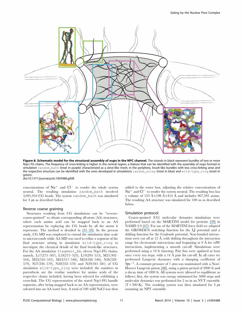
structures in different regions of the central channel (see Figure 8).
The region in the periphery of the central channel, away from the
center, should be represented by a system of tethered nups as in
simulation random_array, where brush-like bundles with less
cross-linking arise. The NPC inner diameter is about 30 nm and
unstructured nups that are several hundred amino acids long span
this volume of an open pore. Since tethering effects should be
minimal in the central region due to the large distance from the
NPC wall, where nups are actually tethered, we expect that the
structure found in this region is similar to that seen in simulation
random_bath, exhibiting bundles with a high degree of cross-
Figure 4. Height of Nsp1-FG chains. (a) Time evolution of the average height. The height is shown for 1-ms simulations for the end-tetheredNsp1-FGs in simulations wild-type_ring (green line), mutant_ring (red line), and random_array (cyan line). The heights are calculated asthe average end-to-end distance in the z-direction, the average being taken over all chains in a given simulation system. (b) Snapshot of the (1 ms)end of simulation wild-type_ring (see also Figure 1(c)), mutant_ring, and random_array (see also Figure 3(c)). Videos show how duringsimulation wild-type_ring, the fully extended Nsp1-FG chains form brush-like, strongly cross-linked bundles (Video S1) and how in simulationrandom_array, the worm-like chain Nsp1-FGs form non-brush-like bundles (Video S3). The mutated Nsp1-FG chains (FG-to- AA) arising insimulation mutant_ring form brush-like bundles with similar average brush height as seen to arise for wild-type Nsp1-FGs in simulation wild-
type_ring.doi:10.1371/journal.pcbi.1003488.g004
Gating by the Nuclear Pore Complex
PLOS Computational Biology | www.ploscompbiol.org 7 March 2014 | Volume 10 | Issue 3 | e1003488

linking forming a mesh. In this region, we observed the pore size to
be 9–10 nm (Table 2) which agrees with the experimentally
determined size limit for passive diffusion of small molecules.
In the present study we modeled a homogeneous system of nups
consisting of only Nsp1-FGs. FG-nups in the real NPC central
channel are heterogeneous; FG-nups vary across the channel
volume in length, amino acid composition, FG repeat motifs (FG,
FxFG and GLFG), composition and spacing of the linker regions.
However, both simulation of a short, 100-aa-long fragment of
Nsp1-FG in [20] and the current simulation of a full length Nsp1-
FG (609-aa-long) led to the same brush-like structure model,
suggesting that the interlinked brush-like structure discovered are
not sensitive to varying length and type of proteins. Also, nups are
known to be intrinsically disordered, interacting with each other
through FG-repeats distributed throughout their entire sequences.
Such disorder and interactions between FG-repeats were also
captured in the present model. Therefore, we suggest that FG nup
heterogeneity does not yield qualitatively different nup assembly
structures.
In addition to the structural features of the NPC, it is necessary
for an understanding of the transport mechanism of the NPC to
examine the dynamics of assembly and conformational change of
the nup proteins. Transport arises on a millisecond timescale [40],
which is beyond the microsecond time scale accessible to large
scale MD simulation and performed in the present study. Thus, we
are unable to infer information of dynamics of the transport
process from the current simulations. Instead, our simulation
results should be interpreted as quasi-static pictures of dynamic
assembly structures of the nups.
The major unsolved problem regarding our still limited
knowledge of the NPC and NCT is how specific large cargoes
with the assistance of TFs manage to pass the NPC. Clearly the
NPC being one of the largest and at the same time one of
the most dynamic macromolecular systems in eukaryotic cells
still holds great secrets and offers opportunity for great
discoveries.
Our suggestion here of the principal assembly structure of
disordered nups in the NPC, even if completely true, does not
imply yet how transport factors can melt the assembly structure for
passage. Straightforward MD simulations, even when simplified
through coarse-graining, cannot bring about answers as the
systems and processes that need to be simulated are much too
large and much too slow, respectively. This calamity is actually
a bonus as the combination of theory, experiment, and
simulation needed is intellectually more rewarding than a
straightforward tour de force purely computational strategy. But
in pursuing the role of transport factors one needs to be open-
minded about the possibility that yet unsuspected mechanisms
play a major role.
Methods
In this section we describe the systems simulated and the
molecular dynamics protocols employed in the present study. We
note here that all molecular images in this article were rendered
using the molecular visualization software VMD [41].
Fully extended Nsp1-FGs on a gold nanoporeThe FG-repeat domain of wild-type Nsp1, Nsp1-FG, was built
from Saccharomyces (S.) cerevisiae Nsp1 sequence 1–609 (Swiss-Prot
P14907) by using the 2004.03 release of Chemical Computing
Group’s Molecular Operating Environment (MOE) software. The
backbone dihedral angles (phi, psi) were set to (1800, 1800) so that
an unstructured straight Nsp1-FG was obtained. In case of
Figure 5. Initial and final configuration of simulated wild-type Nsp1-FGs in a solvent bath (simulation random_bath). (a) Initialconfiguration. Shown is the periodic (see simulation conditions as described in Methods) system of wild-type Nsp1-FGs, freely floating in a solventbath (water and ions). The initial random conformations match a polymer melt modeled from worm-like chains. Colors distinguish 120 freely floatingNsp1-FG chains. Neighboring boxes in x- and y- directions are shown with the Nsp1-FGs colored in grey. (b) Snapshot of the (1 ms) end of simulationrandom_bath. The Nsp1-FG chains, shown in surface representations, are seen to form a porous mesh of cross-linked Nsp1-FG bundles. (c) Close-upview of the structure in (b). Shown is a region as marked. The view reveals a system of short bundles that are frequently cross-linked; arrows point tothe cross-links between bundles. Video S5 shows how during simulation random_bath, the initially completely random Nsp1-FGs assume the finalstructure seen here. Video S6 provides a 360-degree view of the conformation reached in simulation random_bath after 1 ms, namely of theconformation depicted in (b).doi:10.1371/journal.pcbi.1003488.g005
Gating by the Nuclear Pore Complex
PLOS Computational Biology | www.ploscompbiol.org 8 March 2014 | Volume 10 | Issue 3 | e1003488

simulation wild-type_ring, we tested this model through
comparison to experiments reported in [21,29] that have
engineered, using nanotechnology, an artificial pore channel
employing nuclear pore proteins inside a gold ring. The system
dimension and protein density were chosen to imitate volume-wise
the interior of the NPC. We reproduced the gold ring dimensions
adopted in [29] and distributed over the ring tethering points
holding the C-terminal ends of full-length Nsp1-FG domain
proteins (609-aa-long). As shown in Figure 1, 120 fully-extended
wild-type Nsp1-FG chains were grafted to the ring in three
concentric rows with 6 nm spacing between adjacent Nsp1-FGs
[29]. The C-terminus of the end-tethered Nsp1-FGs was modified
by adding five cystine residues that remained fixed to the gold
surface throughout the simulation. In [20,21,42], these cystine
residues formed thiol linkages with the gold substrate. With the C-
terminus of each chain attached to the gold-ring and the rest of the
chain fully-extended in the direction shown in Figure 1, the whole
system was coarse-grained and solvated with CG water in a box
large enough to prevent proteins from interacting with their
periodic images. A total of 100 mM NaCl was added to the water
box, adjusting the relative concentrations of Naz and Cl{ to
render the whole system neutral. The resulting system simulation
wild-type_ring has 15,453,214 CG beads. For the simula-
tion mutant_ring we constructed the ring system as above and
replaced all phenylalanines and glycines of the Nsp1-FGs by
alanines. The resulting system described in simulation mutant_
ring has 16,019,434 CG beads. Both systems were simulated for
1 ms using coarse-grained molecular dynamics simulations as
described below.
Generating random conformations for Nsp1-FGsTo introduce disorder in Nsp1-FG chains, initial random Nsp1-
FG conformations needed as starting points for simulations
random_array and random_bath were modeled according
to the widely used worm-like chain model [43]. The assignment of
the random conformations proceeded in two steps. In a first step,
the main-chain Ca beads of Nsp1-FG constituting the protein
backbone were modeled as random homo-polymers constructing
the backbone from a self-avoiding walk (SAW) procedure [44]. In
this procedure, the SAW is directed under two local geometric
restraints, namely keeping a fixed distance of 3.7 A between
neighboring Ca beads and keeping a fixed angle of 1270 for three
adjacent Ca beads. The stated values are used in the MARTINI
force field [39] for polypeptide chains with coil conformations.
Any two Ca beads were considered to be in close contact if their
distance is shorter than 8 A. We discarded any conformations of
backbone chains with beads in close contact within one chain
(intra) or between different chains (inter). In the second step,
CG side-chains of amino acids were grafted on to the resulting
backbone chains.
The geometries of the side-chains and of the gold nano-particles
were modeled with standard parameters in the MARTINI force
field [39,45]. The MARTINI CG force field has been extensively
optimized in order to correctly model partition between water and
non-polar solvent for amino acids including phenylalanine, glycine
and alanine that are of interest in the current study. Moreover,
multi-site representation of large amino acids is employed in
MARTINI in order to realistically model special geometric features,
such as aromatic rings in phenylalanine, which are essential for side-
chain interactions. In the past few years, the MARTINI force field
has been successfully applied in numerous studies of peptide and
peptide-lipid interactions, as well as in studies of the assembly of
micelles and bilayers around membrane proteins [46]. However,
due to lack of treatment of backbone-backbone hydrogen bond
interactions, the MARTINI force field is unable to model formation
of certain type of structures, such as b-sheets.
Random Nsp1-FGs end-tethered on a 2D arrayFor modeling end-tethered Nsp1-FGs in simulation rando-
m_array, we employed again for the construction of a random
initial state the worm-like chain model described above and chose
the last five Ca beads of cysteine residues 610–614 as the starting
points for the SAW. In the experimental systems [21] nups are
tethered to the gold substrate by means of thiol bonds to cysteine
residues added to the C-terminus. In order to be consistent with
the description adopted for the ring-like geometry, the five Ca
beads of each Nsp1-FG chain were stretched toward the z-
direction and placed on a 565 grid in the x,y-plane, with a grid
spacing of 6 nm. The full-length worm-like chain was then
modeled as described above, such that 25 Nsp1-FGs were placed
as shown in Figure 3. The whole system was coarse-grained
according to the MARTINI force field [39,45] and solvated
Figure 6. Structural features of bundle regions and cross-linking regions observed in coarse-grained and all-atommolecular simulations. (a) Probability of amino acids to form inter-chain backbone-backbone hydrogen bonds (HB) when located insidethe bundle regions (brown bars) or inside the cross-linking regions(blue bars). Shown are the results obtained from the all-atomsimulation. All amino acids located outside the bundle regions areconsidered to be inside the cross-linking regions. The bars labelled FGand FxFG denote the probabilities for the first phenylalanine residues inFG and FxFG motifs, respectively. (b) Solvent accessible surface area(SASA) of the first phenylalanine residues that are located either insidethe bundle regions (labeled ‘‘bundle regions’’) or inside the cross-linking regions (labeled ‘‘cross-linking regions’’). SASA values from CGsimulations wild-type_ring, random_array and random_bath
(summarized in Table 1) are compared with SASA values from the all-atom simulation fragment_AA (Table 1) which comprises of elevenNsp1-FG fragments of the final cross-linked bundle structure resultingfrom the CG simulation wild-type_ring.doi:10.1371/journal.pcbi.1003488.g006
Gating by the Nuclear Pore Complex
PLOS Computational Biology | www.ploscompbiol.org 9 March 2014 | Volume 10 | Issue 3 | e1003488

with CG water. The system was then ionized with 100 mM
NaCl, adjusting again the relative concentrations of Naz and Cl{
to render the whole system neutral. The resulting simulation
random_array involves 1,097,433 CG beads. The system was
simulated for 1 ms as described below.
Freely floating random Nsp1-FGs in a bathFor simulation random_bath, the first three Ca beads of each
Nsp1-FG chain, treated as a rigid body, were chosen as starting
points. They were randomly placed in simulation boxes with
random orientations. The full-length random conformation Nsp1-
FGs were then modeled as worm-like chains as described above,
with 120 self-avoiding Nsp1-FGs being placed in a box of volume
725 A6725 A6725 A to match the concentration of Nsp1-FGs as
in the simulations wild-type_ring and random_array (see
Figure 5). The whole system was coarse-grained according to the
MARTINI force field [39] and solvated in a CG water box.
100 mM NaCl was then added to the sytem, adjusting the relative
Figure 7. Schematic algorithm for calculating pore sizes. The figure shows a schematic depiction of the algorithm employed in calculating thepore sizes listed in Table 2. The pore size is defined through the radius of the largest spherical cargo capable of passing through the final structureresulting from a 1-ms simulation of a system of Nsp1-FGs. A search for the largest cargo starts on one side of the system, the latter shown in grey. Thecargo (black sphere) of a certain size moves towards the other side while the algorithm probes if the cargo can pass. The panel at right illustrateswhat is measured by the algorithm, namely the radius of the largest cargo that can pass through along a probing direction. As shown in this panel,there are two bottlenecks, the second smaller one of which determines how large a ball can pass through the obstacles and, therefore, characterizesthe pore size of obstacles.doi:10.1371/journal.pcbi.1003488.g007
Table 2. Average pore size.
Name Average pore size ± std.dev A
wild-type_ring 77.3260.92
mutant_ring 77.3560.94
random_array 50.3861.40
random_bath 43.3361.33
The pore size is defined through the radius of the largest spherical cargocapable of passing through the final structure of a simulated system of Nsp1-FGs. The radius was determined according to the algorithm presented inMethods and illustrated in Figure 7. The average was taken over the last 30 nsof the 1-ms simulation for each of the systems wild-type_ring,mutant_ring, random_array, and random_bath.doi:10.1371/journal.pcbi.1003488.t002
Gating by the Nuclear Pore Complex
PLOS Computational Biology | www.ploscompbiol.org 10 March 2014 | Volume 10 | Issue 3 | e1003488
concentrations of Naz and Cl{ to render the whole system
neutral. The resulting simulation random_bath involved
3,091,910 CG beads. The system random_bath was simulated
for 1 ms as described below.
Reverse coarse grainingStructures resulting from CG simulations can be ‘‘reverse-
coarse-grained’’ to obtain corresponding all-atom (AA) structures,
where each amino acid can be mapped back to an AA
representation by replacing the CG beads by all the atoms it
represents. The method is detailed in [31–33]. In the present
study, CG MD was employed to extend the simulation time scale
to microseconds while AA MD was used to refine a segment of the
final structure arising in simulation wild-type_ring to
investigate the chemical details of the final brush-like structures.
For the AA simulation fragment_AA, eleven Nsp1-FG chains,
namely, L17(275–507), L18(275–523), L19(293–513), M21(302–
544), M22(316–545), M23(317–548), M24(326–546), N26(328–
579), N27(336–579), N28(332–570) and N29(345–581) of CG
simulation wild-type_ring were included; the numbers in
parenthesis are the residue numbers for amino acids of the
respective chains included, having been selected for exhibiting a
cross-link. The CG representation of the stated Nsp1-FG bundle
segments, after being mapped back to an AA representation, were
solvated into an AA water box. A total of 100 mM NaCl was then
added to the water box, adjusting the relative concentrations of
Naz and Cl{ to render the system neutral. The resulting box has
a volume of 123 A6198 A6414 A and includes 967,595 atoms.
The resulting AA structure was simulated for 100 ns as described
below.
Simulation protocolCoarse-grained (CG) molecular dynamics simulations were
performed based on the MARTINI model for proteins [39] in
NAMD 2.9 [47]. For use of the MARTINI force field we adapted
the GROMACS switching function for the LJ potential and a
shifting function for the Coulomb potential. Non-bonded interac-
tions were cut off at 12 A, with shifting throughout the interaction
range for electrostatic interactions and beginning at 9 A for vdW
interactions, implementing a smooth cut-off. Simulations were
performed using a 10 fs timestep. Pair lists were updated at least
once every ten steps, with a 14 A pair list cut-off. In all cases we
performed Langevin dynamics with a damping coefficient of
5ps{1. A constant pressure of 1 atm was maintained with a Nose-
Hoover Langevin piston [48], using a piston period of 2000 fs and
a decay time of 1000 fs. All systems were allowed to equilibrate as
follows: first, the system was energy minimized for 5000 steps and
molecular dynamics was performed for 2 ns in an NVT ensemble
(T = 300 K). The resulting system was then simulated for 1 msassuming an NPT ensemble.
Figure 8. Schematic model for the structural assembly of nups in the NPC channel. The strands in black represent bundles of two or moreNsp1-FG chains. The frequency of cross-linking is higher in the central region, a feature that can be identified with the assembly of nups formed insimulation random_bath (inset in purple) characterized as a sieve-like mesh; in the periphery, brush-like bundles with less cross-linking arise andthe respective structure can be identified with the ones developed in simulations random_array (inset in blue) and wild-type_ring (inset ingreen).doi:10.1371/journal.pcbi.1003488.g008
Gating by the Nuclear Pore Complex
PLOS Computational Biology | www.ploscompbiol.org 11 March 2014 | Volume 10 | Issue 3 | e1003488

The all-atom simulations were performed using NAMD 2.9
[47]; non-bonded interactions were cut off at 12 A, with a
switching function beginning at 10 A and implementing a smooth
cutoff. The simulations involved multiple timestepping, with a base
timestep of 1 fs, short-range interactions calculated every step, and
long-range electrostatics every two steps. Electrostatic forces were
evaluated through the particle-mesh Ewald method [47] with a
grid density of 1.0 A23. The AA system was first energy
minimized for 5000 steps and then was simulated for 100 ns
assuming an NPT ensemble (T = 300 K). Periodic boundary
conditions were assumed for all (CG and all-atom) simulations.
Analysis of bundlesAs described in Results, the simulated Nsp1-FGs formed strands
that we refer to as bundles. We define a bundle as a linearly
arranged cluster of multiple parallel Nsp1-FG chains. In such a
cluster, every amino acid in a segment of one Nsp1-FG chain is in
contact with at least one amino acid of another Nsp1-FG chain. In
order to provide a quantitative characterization of these bundles
we applied graph theory to identify bundle segments in a given
configuration of the Nsp1-FG chains. In this approach, each
amino acid is represented by a node in a graph. If the distance of
two amino acids A and B from different chains is shorter than 6 A,
the corresponding nodes in the graph are connected with an edge.
In addition, the nodes for amino acids adjacent in sequence to the
two amino acids A and B forming an edge are considered to be
connected to the nodes for amino acids A and B. Therefore,
bundle segments can be identified by examining connected
components in a graph as constructed above. For this purpose
we employed the breadth-first search algorithm [49].
Thickness of a bundle is determined as the number of different
Nsp1-FG chains that belong to the bundle. Another bundle
characteristic determined here is the fraction of each type of amino
acid involved in a bundle.
Pore characterizationNsp1-FG polymers self-assemble, as shown in Results, into
network structures. In order to assess how these structures are
related to gating in the NPC we determine the size limit of
spherical particles being capable geometrically to pass through the
structures. This size limit defines the pore size, namely as the
largest possible radius of passing particles. Figure 7 illustrates how
the pore size was determined by us algorithmically. Starting from
one side of the network structure, spheres of various sizes are
moved towards the other side. The analysis was applied to
the final structures resulting from simulations random_array,
random_bath, wild-type_ring, and mutant_ring; in
the case of the latter two simulations structures are formed with
Nsp1-FGs arranged in a ring-like arrangement. In these cases, the
spheres were moved radially from the inside of the Nsp1-FG ring
structure to its outside.
We implemented the above analysis through an algorithm in
which the simulation box with the network structure was mapped
into a cubic lattice. We then determined for each grid point, x, in
the lattice the radius r(x) of the largest sphere that could be placed
on this grid point without sterical clash with any Nsp1-FG chain;
this radius property was calculated as the shortest distance
between the grid point and protein beads. A sphere is considered
capable of moving in the lattice along a given pathway, if the size
of the sphere does not exceed the bottleneck of the pathway, i.e.,
the smallest r(x) of the grid points on the pathway. There could be
multiple pathways for a sphere to permeate from one side to the
other side, each pathway having its own bottleneck. To
characterize the largest sphere that could permeate the network
structures we search for the pathway for which the bottleneck is
the widest among all the possible pathways. The pathway search
was performed using Dijkstra’s algorithm [50]. To illustrate a
typical outcome of the pore analysis, we show in Video S7 a 360-
degree view of all possible pores identified in the final structure
resulting from simulation random_array.
Supporting Information
Figure S1 Propensity for certain amino acids to beinvolved in the formation of bundles. The probability for
different kinds of amino acids to be involved in the formation of
bundles as determined from an average over the last 30 ns of the
1-ms simulations wild-type_ring (green), mutant_ring
(red), random_array (cyan), and random_bath (purple). G*
refers to glycines and F* refers to the phenylalanines that are not
included in FGs of FG-repeat motifs.
(TIF)
Figure S2 Initial and final configuration of an all-atomsimulation of part of the final configuration of simula-tion wild-type_ring (simulation fragment_AA). (a)
Initial configuration of the all-atom model, which is a part of the
large CG model as shown in Figure 1(e) (see Methods for how the
system is reverse coarse-grained). (b) Final configuration after 100-
ns all-atom simulation. The fragments coming from different
protein chains in the original CG model are shown in different
colors.
(TIF)
Figure S3 Coiling of an initially fully-extended, unteth-ered Nsp1 segment. The time evolution of the radius of
gyration (Rg) is shown for a 1:1-ms CG simulation.
(TIF)
Video S1 This video presents the dynamics of simulatedwild-type Nsp1-FGs grafted to a gold ring (simulationwild-type_ring). The colors distinguish 120 wild-type Nsp1-
FGs grafted on the ring in three concentric rows. The video shows
how during the simulation the end-tethered, fully-extended, wild-
type Nsp1-FGs assume quickly random conformations and form
brush-like bundles (video corresponds to Figure 1).
(MP4)
Video S2 This video provides a 360-degree view of thefinal conformation reached after 1 ms in simulationwild-type_ring. The video reveals the brush-like bundles
formed by Nsp1-FG chains. One can also see that few of these
bundles cross-link (video corresponds to Figure 1c and Figure 1e).
(MP4)
Video S3 This video presents the dynamics of an arrayof wild-type Nsp1-FGs grafted to a gold surface (simu-lation random_array). The colors distinguish the 25 wild-
type Nsp1-FGs simulated. The video shows how during the
simulation the chains form brush-like structures. The bundles
formed exhibit more cross-linking between themselves than seen in
case of simulation wild-type_ring (video corresponds to
Figure 3).
(MP4)
Video S4 This video provides a 360-degree view of thefinal conformation reached after 1 ms in simulationrandom_array. The video reveals the brush-like bundles
formed by Nsp1-FG chains. One can also see that the bundles
cross-link rather frequently (video corresponds to Figure 3c and
Figure 3d).
(MP4)
Gating by the Nuclear Pore Complex
PLOS Computational Biology | www.ploscompbiol.org 12 March 2014 | Volume 10 | Issue 3 | e1003488

Video S5 This video shows the dynamics of simulated,initially random, wild-type Nsp1-FGs, freely floating in asolvent bath (water and ions) (simulation random_bath).The colors distinguish the 120 wild-type Nsp1-FGs simulated. The
video shows how during the simulations the untethered, initially
random chains form brush-like structures with a degree of cross-
linking much higher than in case of simulations wild-type_
ring and random_array (video corresponds to Figure 5).
(MP4)
Video S6 This video provides a 360-degree view offinal conformation reached after 1 ms in simulationrandom_bath. The video reveals the bundles formed by
Nsp1-FG chains. The bundles are cross-linked with high
frequency, in fact, more frequently than either in case of
simulation ‘wild-type_ring or simulation random_array
(video corresponds to Figure 5c and Figure 5d).
(MP4)
Video S7 This video provides a 360-degree view of allpores in the simulated array of Nsp1-FG tethered to aplane. Shown in blue are Nsp1-FG proteins that form brush-like
structures interconnected with cross-links. Shown in red are the
pores that can accommodate spherical objects with diameters
greater than 7 nm. The pores were identified by means of the
algorithm described in Methods. The narrow regions connecting
wide regions correspond to the bottlenecks for passive diffusion of
molecules.
(MP4)
Author Contributions
Conceived and designed the experiments: RG KS. Performed the
experiments: RG. Analyzed the data: RG WH. Contributed reagents/
materials/analysis tools: JES WH. Wrote the paper: RG WH KS.
References
1. Rout MP, Blobel G (1993) Isolation of the yeast nuclear pore complex. J Cell
Biol 123: 771–783.
2. Reichelt R, Holzenburg A, Buhle E, Jarnik M, Engel A, et al. (1990) Correlation
between structure and mass distribution of the nuclear pore complex and of
distinct pore complex components. J Cell Biol 110: 883–894.
3. Yang Q, Rout MP, Akey CW (1998) Three-dimensional architecture of the
isolated yeast nuclear pore complex: functional and evolutionary implications.
Mol Cell 1: 223–34.
4. Rout MP, Aitchison JD, Suprapto A, Hjertaas K, Zhao Y, et al. (2000) The yeast
nuclear pore complex: Composition, architecture, and transport mechanism.
J Cell Biol 148: 635–651.
5. Cronshaw JM, Krutchinsky AN, Zhang W, Chait BT, Matunis MJ (2002)
Proteomic analysis of the mammalian nuclear pore complex. J Cell Biol 158:
915–927.
6. Alber F, Dokudovskaya S, Veenhoff LM, Zhang W, Kipper J, et al. (2007)
Determining the architectures of macromolecular assemblies. Nature 450: 683–
694.
7. Alber F, Dokudovskaya S, Veenhoff LM, Zhang W, Kipper J, et al. (2007) The
molecular architecture of the nuclear pore complex. Nature 450: 695–701.
8. Macara IG (2001) Transport into and out of the nucleus. Microbiol Mol Biol
Rev 65: 570–594.
9. Denning DP, Patel SS, Uversky V, Fink AL, Rexach M (2003) Disorder in the
nuclear pore complex: The FG repeat regions of nucleoporins are natively
unfolded. Proc Natl Acad Sci USA 100: 2450–2455.
10. Denning DP, Rexach M (2007) Rapid evolution exposes the boundaries of
domain structure and function in natively unfolded FG nucleoporins. Mol Cell
Proteomics 6: 272–282.
11. Liu SM, Stewart M (2005) Structural basis for the high-affinity binding of
nucleoporin Nup1p to the Saccharomyces cerevisiae importin-b homologue,
Kap95p. J Mol Biol 349: 515–525.
12. Morrison J, Yang JC, Stewart M, Neuhaus D (2003) Solution NMR study of the
interaction between NTF2 and nucleoporin FxFG repeats. J Mol Biol 333: 587–
603.
13. Bednenko J, Cingolani G, Gerace L (2003) Importin-b contains a COOH-
terminal nucleoporin binding region important for nuclear transport. J Cell Biol
162: 391–401.
14. Bayliss R, Littlewood T, Strawn LA, Wente SR, Stewart M (2002) GLFG and
FxFG nucleoporins bind to overlapping sites on importin-b. J Biol Chem 277:
50597–50606.
15. Isgro TA, Schulten K (2005) Binding dynamics of isolated nucleoporin repeat
regions to importin-b. Structure 13: 1869–1879.
16. Isgro TA, Schulten K (2007) Association of nuclear pore FG-repeat domains to
NTF2 import and export complexes. J Mol Biol 366: 330–345.
17. Isgro TA, Schulten K (2007) Cse1p binding dynamics reveal a novel binding
pattern for FG-repeat nucleoporins on transport receptors. Structure 15: 977–
991.
18. Rout MP, Aitchison JD, Magnasco MO, Chait BT (2003) Virtual gating and
nuclear transport: the hole picture. Trends Cell Biol 13: 622–628.
19. Lim RY, Huang NP, Koser J, Deng J, Lau KA, et al. (2006) Flexible
phenylalanine-glycine nucleoporins as entropic barriers to nucleocytoplasmic
transport. Proc Natl Acad Sci USA 103: 9512–7.
20. Miao L, Schulten K (2009) Transport-related structures and processes of the
nuclear pore complex studied through molecular dynamics. Structure 17: 449–
459.
21. Lim RY, Fahrenkrog B, Koser J, Schwarz-Herion K, Deng J, et al. (2007)
Nanomechanical basis of selective gating by the nuclear pore complex. Science
318: 640–643.
22. Ribbeck K, Gorlich D (2002) The permeability barrier of nuclear pore
complexes appears to operate via hydrophobic exclusion. EMBO J 21: 2664–
2671.
23. Frey S, Richter RP, Gorlich D (2006) FG-rich repeats of nuclear pore proteins
form a three-dimensional meshwork with hydrogel-like properties. Science 314:
815–7.
24. Frey S, Gorlich D (2007) A saturated FG-repeat hydrogel can reproduce the
permeability properties of nuclear pore complexes. Cell 130: 512–
523.
25. Peters R (2005) Translocation through the nuclear pore complex: Selectivity and
speed by reduction-of-dimensionality. Traffic 6: 421–427.
26. Patel SS, Belmont BJ, Sante JM, Rexach MF (2007) Natively unfolded
nucleoporins gate protein diffusion across the nuclear pore complex. Cell 129:
83–96.
27. Yamada J, Phillips JL, Patel S, Goldfien G, Calestagne-Morelli A, et al. (2010)
A bimodal distribution of two distinct categories of intrinsically disordered
structures with separate functions in FG nucleoporins. Mol Cell Proteomics 9:
2205–22204.
28. Tagliazucchi M, Peleg O, Kroger M, Rabin Y, Szleifer I (2013) Effect of charge,
hydrophobicity, and sequence of nucleoporins on the translocation of model
particles through the nuclear pore complex. Proc Natl Acad Sci USA 110: 3363–
3368.
29. Peleg O, Lim RYH (2010) Converging on the function of intrinsically disordered
nucleoporins in the nuclear pore complex. Biol Chem 391: 719–730.
30. Dolker N, Zachariae U, Grubmuller H (2010) Hydrophilic linkers and polar
contacts affect aggregation of FG repeat peptides. Biophys J 98: 2653–2661.
31. Shih AY, Freddolino PL, Sligar SG, Schulten K (2007) Disassembly of nanodiscs
with cholate. Nano Lett 7: 1692–1696.
32. Freddolino PL, Arkhipov A, Shih AY, Yin Y, Chen Z, et al. (2008) Application
of residue-based and shape-based coarse graining to biomolecular simulations.
In: Voth GA, editor, Coarse-Graining of Condensed Phase and Biomolecular
Systems, Chapman and Hall/CRC Press, Taylor and Francis Group, chapter
20. pp. 299–315.
33. Shih AY, Freddolino PL, Arkhipov A, Sligar SG, Schulten K (2008) Molecular
modeling of the structural properties and formation of high-density lipoprotein
particles. In: Feller S, editor, Current Topics in Membranes: Computational
Modeling of Membrane Bilayers, Elsevier, chapter 11. pp. 313–342.
34. Milles S, Bui KH, Koehler C, Eltsov M, Beck M, et al. (2013) Facilitated
aggregation of fg nucleoporins under molecular crowding conditions. EMBO J
14: 178–183.
35. Moussavi-Baygi R, Jamali Y, Karimi R, Mofrad MRK (2011) Brownian
dynamics simulation of nucleocytoplasmic transport: A coarse-grained model for
the functional state of the nuclear pore complex. PLoS Comput Biol 7:
e1002049.
36. Miao L, Schulten K (2010) Probing a structural model of the nuclear pore
complex channel through molecular dynamics. Biophys J 98: 1658–1667.
37. Yang W (2011) ‘natively unfolded’ nucleoporins in nucleocytoplasmic transport:
Clustered or evenly distributed? Nucleus 2: 10–16.
38. Ader C, Frey S, Maas W, Schmidt HB, Gorlich D, et al. (2010) Amyloid-like
interactions within nucleoporin FG hydrogels. PNAS 107: 6281–
6285.
39. Monticelli L, Kandasamy S, Periole X, Larson R, Tieleman P, et al (2008) The
MARTINI coarse grained forcefield: Extension to proteins. J Chem Theor
Comp 4: 819–834.
40. Ma J, Yang W (2010) Three-dimensional distribution of transient interactions in
the nuclear pore complex obtained from single-molecule snapshots. Proc Natl
Acad Sci USA 107: 73057310.
Gating by the Nuclear Pore Complex
PLOS Computational Biology | www.ploscompbiol.org 13 March 2014 | Volume 10 | Issue 3 | e1003488

41. Humphrey W, Dalke A, Schulten K (1996) VMD - Visual Molecular Dynamics.
J Mol Graphics 14: 33–38.42. Schoch RL, Kapinos LE, Lim R (2012) Nuclear transport receptor binding
avidity triggers a selfhealing collapse transition in FG-nucleoporin molecular
brushes. Proc Natl Acad Sci USA 109: 16911–16.43. Doi M, Edwards SF (1999) The Theory of Polymer Dynamics. New York:
Oxford University Press.44. Flory PJ (1953) Principles of polymer chemistry. Cornell University Press.
45. Kyrychenkoa A, Karpushinaa GV, Bogatyrenkoa SI, Kryshtala AP, Doroshen-
koa AO (2011) Preparation, structure, and a coarse-grained molecular dynamicsmodel for dodecanethiol-stabilized gold nanoparticles. Comput Theor Chem
977: 34–39.
46. Seo M, Rauscher S, Pomes R, Tieleman DP (2012) Improving internal peptide
dynamics in the coarse-grained MARTINI model: Toward large-scalesimulations of amyloid- and elastin-like peptides. J Chem Theor Comp 8:
1774–1785.
47. Phillips JC, Braun R, Wang W, Gumbart J, Tajkhorshid E, et al. (2005) Scalablemolecular dynamics with NAMD. J Comp Chem 26: 1781–1802.
48. Feller SE, Zhang Y, Pastor RW, Brooks BR (1995) Constant pressure moleculardynamics simulation: The Langevin piston method. J Chem Phys 103: 4613–4621.
49. Cormen TH, Leiserson CE, Rivest RL, Stein C, editors (2001) Introduction to
Algorithms, 2nd edition. Cambridge, MA: MIT Press.50. Dijkstra EW (1959) A note on two problems in connexion with graphs.
Numerische mathematik 1: 269–271.
Gating by the Nuclear Pore Complex
PLOS Computational Biology | www.ploscompbiol.org 14 March 2014 | Volume 10 | Issue 3 | e1003488