Altered expression and distribution of aquaporin-9 in the liver of rat with obstructive extrahepatic...
36
Altered Expression and Distribution of Aquaporin-9 in the Liver of Rat with Obstructive Extrahepatic Cholestasis Giuseppe Calamita 1* , Domenico Ferri 2 , Patrizia Gena 1 , Flavia I. Carreras 3 , Giuseppa E. Liquori 2 , Piero Portincasa 4 , Raúl A. Marinelli 3 , Maria Svelto 1 1 Dipartimento di Fisiologia Generale ed Ambientale, 2 Dipartimento di Zoologia, and 4 Dipartimento di Medicina Interna e Medicina Pubblica, Università degli Studi di Bari, Bari, Italy; 3 Instituto de Fisiología Experimental, Universidad Nacional de Rosario, Rosario, Argentina Running head: AQP9 in BDL rat liver * Corresponding Author: Giuseppe Calamita, Ph.D. Dipartimento di Fisiologia Generale ed Ambientale Università degli Studi di Bari Via Amendola, 165/A 70126 Bari, Italy E-mail: [email protected] Tel. +39 0805442928 Fax +39 0805443388 Articles in PresS. Am J Physiol Gastrointest Liver Physiol (July 31, 2008). doi:10.1152/ajpgi.90226.2008 Copyright © 2008 by the American Physiological Society.
Transcript of Altered expression and distribution of aquaporin-9 in the liver of rat with obstructive extrahepatic...

Altered Expression and Distribution of Aquaporin-9 in the Liver of
Rat with Obstructive Extrahepatic Cholestasis
Giuseppe Calamita1*, Domenico Ferri2, Patrizia Gena1, Flavia I. Carreras3,
Giuseppa E. Liquori2, Piero Portincasa4, Raúl A. Marinelli3, Maria Svelto1
1Dipartimento di Fisiologia Generale ed Ambientale, 2Dipartimento di Zoologia, and 4Dipartimento di Medicina Interna e Medicina Pubblica, Università degli Studi di
Bari, Bari, Italy; 3Instituto de Fisiología Experimental, Universidad Nacional de
Rosario, Rosario, Argentina
Running head: AQP9 in BDL rat liver
*Corresponding Author:
Giuseppe Calamita, Ph.D.
Dipartimento di Fisiologia Generale ed Ambientale
Università degli Studi di Bari
Via Amendola, 165/A
70126 Bari, Italy
E-mail: [email protected]
Tel. +39 0805442928
Fax +39 0805443388
Articles in PresS. Am J Physiol Gastrointest Liver Physiol (July 31, 2008). doi:10.1152/ajpgi.90226.2008
Copyright © 2008 by the American Physiological Society.

Calamita et al. AQP9 in BDL rat liver 2
Abstract
Rat hepatocytes express aquaporin-9 (AQP9), a basolateral channel permeable to
water, glycerol and other small neutral solutes. While liver AQP9 is known for
mediating the uptake of sinusoidal blood glycerol its relevance in bile secretion
physiology and pathophysiology remains elusive. Here, we evaluated whether
defective expression of AQP9 is associated to secretory dysfunction of rat hepatocytes
following bile duct ligation (BDL). By immunoblotting, one-day BDL resulted in a
slight decrease of AQP9 protein in basolateral membranes and a simultaneous increase
of AQP9 in intracellular membranes. This pattern was steadily accentuated in the
subsequent days of BDL as at 7-days BDL basolateral membrane AQP9 decreased by
85% whereas intracellular AQP9 increased by 115%. However, the AQP9
immunoreactivity of the total liver membranes from day 7 of BDL rats was reduced by
49% compared to the sham counterpart. Results were confirmed by
immunofluorescence and immunogold electron microscopy and consistent with
biophysical studies showing considerable decrease of the basolateral membrane water
and glycerol permeabilities of cholestatic hepatocytes. The AQP9 mRNA was slightly
reduced only at day 7 of BDL indicating that the dysregulation was mainly occurring
at a posttranslational level. The altered expression of liver AQP9 during BDL was not
dependent on insulin, a hormone known to regulate negatively AQP9 at a
transcriptional level, since insulinemia was unchanged in 7-days BDL rats. Overall,
these results suggest that extrahepatic cholestasis leads to downregulation of AQP9 in
the hepatocyte basolateral plasma membrane and dysregulated aquaporin channels
contribute to bile flow dysfunction of cholestatic hepatocyte.
Keywords: aquaglyceroporin, AQP9, water channel, bile, BDL

Calamita et al. AQP9 in BDL rat liver 3
Introduction
Aquaporins (AQPs) represent a widespread family of membrane channels that
permeate only water (orthodox aquaporins) or small uncharged solutes such as
glycerol in addition to water (aquaglyceroporins). AQPs have been found to be
relevant to multiple physiologic processes and diverse clinical dysfunctions (18). Rat
hepatocytes express three AQP proteins: AQP8, 9 and 11 (see (37) for a Review).
AQP8 features multiple subcellular localizations being composed of an intracellular
pool residing permanently in the hepatocyte smooth endoplasmic reticulum and
mitochondria (8) and a choleretic pool shuttling between cytoplasm (subapical
vesicles) and canalicular membrane under control of choleretic stimuli (2, 10, 17).
AQP9, an aquaglyceroporin of broad selectivity, resides exclusively on the basolateral
membrane where, during starvation, it is believed to mediate the uptake of
gluconeogenetic glycerol from portal bloodstream (3, 23, 38). AQP11 is an unusual
AQP found in the endoplasmic reticulum (33) and whose water channel function is
controversial (12, 46).
Bile secretion by hepatocytes involves the movement of water from portal
bloodstream into bile canaliculus in response to transient osmotic gradients generated
by active solute transport (31). While evidence has been provided suggesting
facilitation of osmotic water transport during canalicular bile secretion by apical
AQP8 (17, 30), no direct information is currently available regarding the possible
water channel function of hepatocyte AQP9 and its potential relevance to primary bile
formation in health and disease.
Extrahepatic cholestasis is an abnormal condition characterized by biliary
obstruction leading to reduction of bile flow and impairment of various transport
mechanisms in both basolateral and canalicular membranes of hepatocytes (25).

Calamita et al. AQP9 in BDL rat liver 4
Dysregulation in the molecular expression of hepatocyte membrane transporters in
obstructive cholestasis have been extensively investigated using a model of bile duct
ligation (BDL) in the rat. Interestingly, our recent work finding downregulated
expression of hepatocyte AQP8 in BDL rats suggested pathophysiological relevance
for liver AQPs in obstructive cholestasis (4). Thus, the main purpose of this study was
to determine the effect of BDL on the basolateral membrane osmotic water
permeability and expression and subcellular distribution of AQP9 in rat liver.

Calamita et al. AQP9 in BDL rat liver 5
Materials and Methods
Animals and Treatment. Adult male Wistar rats (250-300 g; 3 months old; Harlan,
Italy) were maintained on a standard diet and water ad libitum, and housed in a
temperature- and humidity-controlled environment under a constant 12-hour light-
dark cycle. Under ether anesthesia, the common bile duct was double-ligated and cut
between the ligatures. Control animals underwent a sham operation that consisted of
exposure, but not ligation, of the common bile duct. After 1, 3, and 7 days of ligation,
the animals were sacrificed and livers harvested for evaluation. Each experimental
(BDL) or control (sham) group consisted of three to five rats. Dysregulated bile flow
was verified by assaying the serum alkaline phosphatase activity and total bilirubin
level as previously described (4). The protocol was conducted according to the Guide
Principles for the care and use of laboratory animals.
Semi-quantitative RT-PCR. Livers were removed from sacrificed mice and frozen in
liquid nitrogen. Total RNA from livers of sham operated or BDL rats was isolated by
the TRIzol reagent (Invitrogen, San Diego, CA) following the manufacturer’s
protocol. The samples of total RNA were submitted to semi-quantitative RT-PCR as
previously described (2) using the rat AQP9 primers AQP9-start (5’-
ATGCCTTCTGAGAAGGACGG-3’) and AQP9-stop (5’-
CTACATGATGACACTGAGCT-3’) that lead to the amplification of a 885 bp
fragment of DNA. RT-PCR reactions were normalized against the ß-actin expression
(2).
Preparations of liver subcellular membrane fractions. Cholestatic and sham-operated
livers were homogenized by 15 up-down strokes with a loose fitting Dounce

Calamita et al. AQP9 in BDL rat liver 6
homogenizer in 4 volumes of 0.3 M sucrose, containing 0.1 mM
phenylmethylsulfonyl fluoride and 0.1 mM leupeptin (Sigma, St. Louis, MO). Total
liver homogenates were subjected to low-speed centrifugation at 800g for 10 minutes
to obtain post nuclear supernatants, and then subjected to centrifugation at 200,000g
for 60 minutes, yielding the total liver membrane fraction (10). Fractions enriched in
total plasma membranes, basolateral membranes or intracellular microsomal
membranes were prepared from liver homogenates by differential centrifugation on
discontinuous sucrose gradients as previously described (4, 10, 17). Proteins in
plasma membrane fraction were assayed according to the method of Lowry (28).
Enrichment and purity of plasma membranes were comparable to those reported
previously (4, 30, 32).
Preparation of basolateral membrane vesicles and stopped flow light scattering
measurement of water and glycerol permeabilities. The basolateral membrane
vesicles were prepared as previously described (30). The size of vesicles was
determined with an N5 Submicron Particle Size Analyzer (Beckman Coulter Inc.,
Palo Alto, CA) and by transmission electron microscopy. The time course of vesicle
volume change was followed from changes in intensity of scattered light at the
wavelength of 450 nm using a Jasco FP-6200 (Jasco, Tokyo, Japan) stopped-flow
reaction analyzer which has a 1.6 ms dead time and 99% mixing efficiency in < 1 ms.
Vesicle osmotic water permeability was measured by light scattering at 20 °C as
previously described (1). Briefly, 35 µl of a concentrated vesicle suspension was
diluted into 2.5 ml of a hypotonic (220 mOsm) isolation medium (124 mM mannitol,
70 mM sucrose, 20 mM Tris-HCl, 1 mM EDTA and 5 mM EGTA, pH 7.4). One of
the syringes of the stopped-flow apparatus was filled with the specimen suspension,

Calamita et al. AQP9 in BDL rat liver 7
whereas the other was filled with the same buffer to which mannitol was added to
reach a final osmolarity of 500 mOsm to establish a hypertonic gradient (140 mOsm)
upon mixing. The final protein concentration after mixing was of 100 µg/ml.
Immediately, after applying a hypertonic gradient, water outflow occurs, and the
vesicles shrink, causing an increase in scattered light intensity. The data were fitted to
a single exponential function and the related rate constant (Ki, s-1) of the water efflux
out of the analyzed specimen was measured. The osmotic water permeability
coefficient (Pf), an index reflecting the osmotic water permeability of the vesicular
membrane, was deduced from the Ki as described (42), using the equation:
Pf = Ki · V0/Av ·Vw ·∆C,
where Ki is the fitted exponential rate constant, V0 is the initial mean vesicle volume,
Av is the mean vesicle surface, Vw is the molar volume of water, and ∆C is the
osmotic gradient. The medium osmolarity was verified by a vapor-pressure
osmometer (Wescor Inc., Logan, UT).
For measurement of glycerol permeability, vesicles were subjected to a 150 mM
inwardly directed gradient of glycerol as previously reported by Yang and coworkers
(47). Glycerol permeability (Pgly; cm/s) was computed using the equation:
Pgly = 1/[(S/V)τ]
where S/V is surface-to-volume ratio and τ is the exponential time constant fitted to
the vesicle swelling phase of light scattering time course corresponding to glycerol
entry.
Immunoblotting. Plasma membrane fractions were subjected to SDS-polyacrylamide
gel electrophoresis and transferred to polyvinyl difluoride membranes (NENTM Life
Science Products Inc., Boston, MA). After blocking and washing, blots were

Calamita et al. AQP9 in BDL rat liver 8
incubated overnight at 4ºC with rabbit affinity-purified antibodies against AQP9 (1
µg/ml; Alpha Diagnostics International, San Antonio, Tx, USA) or for 2 h at RT with
a 1:5,000 dilution of goat antibodies against Na+,K+-ATPase (Calbiochem-
Novabiochem, La Jolla, CA). The blots were washed and then incubated for 1 h at RT
with corresponding horseradish peroxidase-conjugated secondary antibodies. Protein
bands were detected by enhanced chemiluminescence detection system (ECL,
Amersham, Little Chalfont, UK). Autoradiographs were obtained by exposing the
membranes to Kodak XAR films, and the bands were quantified by densitometry
using Gel-Pro32 software (Gel-Pro Analyzer, Media Cybernetics, Silver Spring, MD).
Immunohistochemistry. Cholestatic and sham-operated rats were sacrificed after ether
anesthesia and their livers were quickly removed, sliced and fixed overnight by
immersion with 4% paraformaldehyde. For the immunoperoxidase light microscopy,
samples of liver were quickly processed to be included in a hydrophilic resin
(Technovit 8100, Heraeus-Kulzer, Wehrheim, Germany) and processed as previously
reported (1). For the immunofluorescence experiments, after washing, the livers were
incubated overnight in PBS added with 30% sucrose and embedded in Optimal
Cutting Temperature embedding medium (Bio-Optica, Milan, Italy). Frozen sections
were cut to a thickness of 4 µm. The sections were washed in PBS for 10 min and
then blocked with PBS–gelatin 0.1% for 15 min at RT before being incubated with
the rabbit affinity purified antibodies against the rat AQP9 (3 µg/ml) for 2 h at RT.
Negative controls were performed by omitting the primary antibody. After three 5
min washes in PBS–gelatin 0.1%, the sections were incubated with FITC–conjugated
goat anti-rabbit secondary antibody (Molecular Probes, Eugene, OR) for 1 h at RT.
The sections were again washed one time in PBS added with NaCl 2.7% followed by

Calamita et al. AQP9 in BDL rat liver 9
2x10 min washes in PBS. The coverslips were mounted using a mounting medium
consisting of glycerol 50%, TRIS-HCl 0.2 M, pH 8.0 and n-propyl gallate 2.5%. The
sections fluorescence intensity was quantified and displayed graphically using a Leica
DMRXA photomicroscope equipped with a CCD camera (Princeton Instruments,
New York, NY).
Immunogold electron microscopy. Samples of liver were fixed in a mixture of 3%
paraformaldehyde and 1% glutaraldehyde in 0.1 M saline phosphate buffer (PBS), at
pH 7.4, for 4 h at 4 °C. Some specimens were postfixed in 1% OsO4 in PBS for 30
min at 4 °C. Fixed specimens were dehydrated in ethanol then embedded in Epon
(Taab, Reading, UK). For immunoelectron microscopy, ultrathin sections of osmicated
samples were oxidized with sodium metaperiodate to restore specific labeling. Both
osmicated and nonosmicated sections were treated with 0.05 M glycine in PBS buffer
for 15 min at RT. Grids were incubated for 30 min at RT with 1% BSA in PBS
containing 0.2 % gelatin (PBG) and then placed on a drop of AQP9 antibodies (10
µg/ml of PBG) overnight at 4 °C. The grids were then incubated in 1:10 10-nm gold-
conjugated anti-rabbit IgG (Sigma) in PBG for 1 h at RT and lightly stained with
uranyl acetate and lead-citrate. Finally, the grids were observed with a Zeiss EM 109
electron microscope. Immunolabeling controls were performed as in
immunofluorescence.
Statistical analysis. Experiments with each group of animals were performed at least
in triplicate. Means±SE were calculated based on three to five independent
preparations. Data were analysed statistically using the Student’s t test. Results were
considered statistically significant when P < 0.05.

Calamita et al. AQP9 in BDL rat liver 10
Results
Ligation of the common bile duct resulted in extrahepatic cholestasis, as indicated by
the increased serum levels of total bilirubin and alkaline phosphatase. Levels of total
serum bilirubin increased to 6.7±0.7 mg/dL (sham-operated rats, 0.6±0.2 mg/dL) and
serum alkaline phosphatase to 1,093±184 U/L (sham-operated rats, 434±57 U/L) at
day 7 after BDL.
Effect of BDL on AQP9 mRNA expression. Semi-quantitative RT-PCR analysis was
performed to study the effect of BDL on AQP9 mRNA expression. Compared with
sham-operated controls, levels of AQP9 mRNA were unchanged on day 1 and 3 BDL
but slightly decreased (-35%) on day 7 BDL (Fig. 1A,B). The AQP9 mRNA
expression was normalized against that of the housekeeper gene β-actin (Fig. 1A).
Effect of BDL on AQP9 protein expression and subcellular distribution.
Immunoblotting with liver total, plasma and intracellular membrane fractions were
performed to evaluate the effect of BDL on AQP9 protein expression and subcellular
localization. One-day BDL did not affect the plasma membrane expression of AQP9
whereas a 25% reduction was seen at day-3 of BDL. After 7 days of BDL, there was a
remarkable reduction of basolateral membrane AQP9 (-85%) (Fig. 2A,D). The AQP9
protein expression was compared to that of the Na+,K+-adenosine triphosphatase (α1
subunit) (Fig. 2C,D), a plasma membrane protein remaining unaltered during BDL
(11). Of note, the immunolabeling of the intracellular membrane fraction increased
steadily from day 1 BDL and became remarkable at day 7 BDL (+115%) while the
weak intracellular immunoreactivity to AQP9 did not change in the sham livers.
Overall, the AQP9 immunoreactivity detected in the total liver membrane fraction

Calamita et al. AQP9 in BDL rat liver 11
from day 7 of BDL rats was reduced by 49% (P < 0.01) when compared to the sham
counterpart (Fig. 2E,F). This finding suggests that the basolateral membrane insertion
of AQP9 in cholestatic hepatocytes is impaired and the overall liver AQP9 protein is
considerably reduced after 7 days of BDL (Fig. 2B,D). BDL did not alter the yield of
total membrane proteins (data not shown). The downregulation of basolateral AQP9
was not due to insulin, a hormone exerting a negative effect on the expression of liver
AQP9 (23), as 7 days of BDL did not change the insulinemia levels of the BDL and
sham rats (0.28±0.1 vs 0.29±0.1 mU/I, respectively).
BDL reduces both the water and glycerol permeability of the hepatocyte basolateral
membrane. Functional experiments of stopped flow light scattering were performed
to evaluate whether the BDL-induced downregulation of basolateral AQP9 caused a
reduction in basolateral osmotic water permeability. To do that, basolateral membrane
vesicles prepared from livers of sham or 7-days BDL rats were subjected rapidly (1
ms) to a hypertonic osmotic gradient of 140 mOsm and the resulting time course of
vesicle shrinkage was followed from the change in scattered light. Consistent with the
BDL-induced decrease of basolateral membrane AQP9 seen above and suggesting a
role for AQP9 in the osmotic permeability of hepatocyte basolateral membrane, the
osmotic water permeability coefficient (Pf) of the vesicles obtained from cholestatic
livers was significantly lower than the one measured with the vesicles from control
livers (68.3±9.5 and 112±7.2 µm/s, respectively; P<0.01)(Fig. 3A,B). No change in
scattered light was seen when vesicles were mixed with isosmotic buffer, proving the
absence of mixing artifact (Fig. 3B).
To verify whether the BDL-induced downregulation of AQP9 was also accompanied
by a reduction of the facilitated glycerol transport in hepatocyte basolateral plasma

Calamita et al. AQP9 in BDL rat liver 12
membrane, glycerol permeability was compared in basolateral membrane vesicles
from livers of sham or BDL rats 7 days after surgery. Glycerol permeability was
measured by light scattering following a 150 mM inwardly directed gradient of
glycerol as previously described by Yang and coworkers (47). Experiments were
performed at 20 °C. Fig. 4A shows representative light scattering data, with the slow
decrease in scattered light intensity corresponding to glycerol influx into vesicles.
Consistent with the osmotic water permeability studies there was significant reduction
in basolateral glycerol permeability coefficient (Pgly) in the liver of the BDL versus
sham rats (9.23±1.5 and 14.6±2.6 10-6 cm/s, respectively; P<0.01) (Fig. 4B).
Effect of bile duct ligation on the immunocytochemical distribution of AQP9 in rat
hepatocytes. Immunofluorescence and immunoperoxidase analyses with serial liver
sections were performed to analyze at a subcellular level the effect of BDL on the
expression and subcellular distribution of AQP9. By immunofluorescence, and in line
with previous reports (7, 34, 35), livers from sham-operated rats showed AQP9 in the
basolateral membrane of hepatocytes (Fig. 5A; arrows). No immunoreactivity was
seen in the canalicular membrane (Fig. 5A, inset; arrowhead). No labeling was
observed in normal (no surgery) liver sections by omitting the primary antibody (Fig.
5B). When compared with the corresponding basal conditions (day 0), the AQP9
immunofluorescent pattern remained unaltered in the liver of the sham-operated rats
at 1, 3 and 7 days (Fig. 5C,E,G) whereas it changed markedly in the BDL liver at day
1 and thereafter (Fig. 5D,F,H). At day 1 of BDL (Fig. 5D), AQP9 was observed both
over the basolateral membrane (inset; arrow) and within the intracellular compartment
(double arrows). After 3 days of BDL (Fig. 5F), the intracellular immunostaining
increased (double arrows) whereas the basolateral membrane reactivity was reduced

Calamita et al. AQP9 in BDL rat liver 13
(inset; arrow). Suggesting mistrafficking of AQP9 in hepatocyte at 7-days BDL, the
immunofluorescence was mostly restricted within the cytoplasmic compartment (Fig.
5D, double arrow). This pattern of distribution was confirmed by immunoperoxidase
light microscopy (Fig. 6).
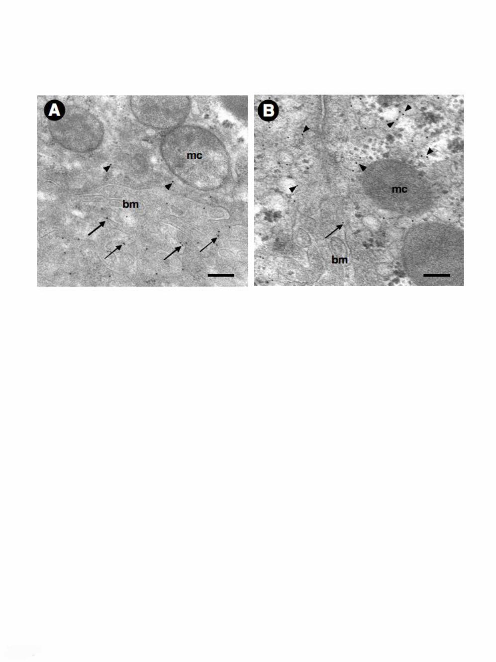
The altered subcellular distribution of hepatocyte AQP9 in BDL rats was also
analyzed at an ultrastructural level by immunogold electron microscopy (Fig. 7). In
line with the above immunocytochemical studies, cholestatic hepatocytes (day 7 of
BDL) showed predominant immunoreactivity within the cytoplasmic compartment
associated with weak staining at the basolateral membrane (Fig. 7B). This pattern was
significantly different from the one of the control hepatocytes where the gold particles
were mostly located over the basolateral plasma membrane (Fig. 7A).

Calamita et al. AQP9 in BDL rat liver 14
Discussion
Extrahepatic cholestasis is a pathologic condition caused by rapidly developing
(acute) or long-term (chronic) interruption in the excretion of bile (i.e., bile solutes
and water) into bile canaliculus (6). After showing impaired expression of canalicular
AQP8 water channels in the liver of BDL rats (4), here we evaluated AQP9 as
possible basolateral water channel contributing to bile flow dysfunction of cholestatic
hepatocyte. The major finding is that extrahepatic obstruction causes strong reduction
of both basolateral AQP9 and osmotic water permeability suggesting that basolateral
AQP9 also functions as water channel and is pathophysiologically relevant to
extrahepatic cholestasis.
There is experimental evidence indicating that rat hepatocyte basolateral osmotic
water transport is AQP-mediated (30). As no other water channels are found in the rat
hepatocyte basolateral membrane (see (37) for a review), it is reasonable to
hypothesize that AQP9 facilitates the sinusoidal movement of water across the
hepatocyte basolateral membrane. Bile secretion by hepatocytes requires the
osmotically-driven transcellular transport of water (i.e. across basolateral and
canalicular plasma membrane domains), thus AQP9 is expected to mediate the
sinusoidal water transport during the formation of bile.
Hepatocyte AQP9 is currently known for mediating the uptake of plasma glycerol
deriving from adipose lipolysis during starvation (3, 23, 38), a feature in line with our
finding that the BDL-induced downregulation of liver AQP9 is also accompanied
with a reduction in the glycerol permeability coefficient (Pgly) of the hepatocyte
basolateral plasma membrane (Fig. 4). Hepatocyte AQP9 would therefore act as water
or glycerol channel, depending on the physiologic condition. The water channel
activity of AQP9 may be relevant to primary bile formation, a function that may

Calamita et al. AQP9 in BDL rat liver 15
explain the rapid shifts of hepatocyte volume characterizing the so-called
hepatocellular hydration state, an efficient mechanism of short-term control of
canalicular secretion (16). The involvement of AQP9 in canalicular bile formation is
also supported by the downregulation to which such AQP undergoes in the liver of
BDL rat. A similar dysregulation characterizes the water channel at the canalicular
side, AQP8 (4). The mechanisms involved in the defective basolateral AQP9
expression in extrahepatic cholestasis may be specific for this particular bile secretory
disorder, since as we recently reported, hepatocyte AQP9 appears unaffected in
sepsis-associated cholestasis induced by lipopolysaccharides (26) and estrogen-
induced hepatocellular cholestasis (5), two forms of intraheaptic cholestasis. The
recent observation that AQP9 is downregulated at both mRNA and protein levels in
the liver of neonatal (but not adult) rats treated with the synthetic estrogen
diethylstilbestrol has been interpreted as due to the higher susceptibility to estrogen-
induced changes characterizing newborn animals compared to their adult counterparts
(45).
The observation that the 85% decrease of basolateral membrane AQP9 is associated
with a 40% decrease of the basolateral membrane osmotic water permeability is a
valuable information in the understanding of the mechanisms of bile formation and
consistent with the moderate water conductance characterizing AQP9 (3, 30). This
suggests, as shown before (30), that the lipid membrane pathway may provide a non-
negligible contribution to the overall water permeability of the hepatocyte basolateral
membrane. A similar explanation was given to describe the canalicular membrane
water permeability (5, 26, 30). A 40% decrease of basolateral membrane osmotic
water permeability may be sufficient to impair the efficient coupling between osmotic
solutes and water transport during bile formation. This possibility is supported by our

Calamita et al. AQP9 in BDL rat liver 16
recent work showing that a 22% decrease of the canalicular membrane osmotic
permeability is associated with a 58% decrease of canalicular bile flow (5). As seen
for apical AQP8 (4), the rate of primary bile secretion may be a factor influencing the
expression and distribution of AQP9 in hepatocyte. It is therefore conceivable to
hypothesize that basolateral AQP9 is reduced following the impairment of the
transient osmotic gradient caused by the defective sinusoidal bile salt uptake
characterizing extrahepatic cholestasis (11). Since canalicular expression of AQP8, an
aquaporin having a considerably higher water channel activity than AQP9, is
decreased after bile duct ligation (4, 5), it is objectively not obvious to independently
establish the exact importance of liver AQP9 in extrahepatic cholestasis. This
difficulty may also explain why no apparent bile formation defects were seen in
AQP9 knockout mice (38). Downregulation of AQP9 may be secondary to defective
primary bile secretion in the light of a recent work showing normal plasma levels of
alkaline phosphatase and cholesterol, two biochemical parameters of cholestasis, in
AQP9 null mice (38).
Our data suggest, as previously for canalicular AQP8 in BDL rats (4), a post-
translational mechanism (e.g., increased protein degradation as a consequence of
subcellular misrouting) for basolateral AQP9 downregulation in obstructive
cholestasis. Altered plasma membrane targeting is a frequent mechanism of
dysregulation affecting diverse hepatocyte transporters in cholestasis, including the
basolateral Na+/bile acid cotransporter (9, 11, 40, 41). However, a novelty of the
physiological process of primary bile formation raising from this work is the fact that
loss of AQP9 occurs in the context of other sinusoidal membrane proteins being
targeted correctly and/or increasing in expression after bile duct ligation (i.e., the
multidrug resistance-associated proteins 3, Mrp3 (44). Similar to canalicular AQP8

Calamita et al. AQP9 in BDL rat liver 17
(5, 26) and AQPs expressed in cells other than hepatocytes (27, 29, 39) misrouted
AQP9 may be targeted for proteolysis through the proteasome and/or lysosome
system. In this scenario, a role for insulin, a hormone regulating negatively AQP9 at a
gene level (3, 23), can be ruled out since both insulinemia and AQP9 transcript levels
of the BDL rats did not change significantly during cholestasis.
Under the cholestasis conditions, a large amount of biliary lipids including
cholesterol, bile acids, phospholipids, and bilirubin, would accumulate within the
hepatocyte. Indeed, the complex changes of both liver metabolism and functions with
cholestasis are partly dependent on retention of hydrophobic bile salts (i.e. tauro- and
glicochenodeoxycholate, major bile salts in rat bile) and reduced hepatic
detoxification capacity (19, 21). In turn, this status might activate injuring pathways
which might cause the decrease of antioxidant defences including glutathione levels,
stimulate glutathione efflux from hepatocytes (15) and induce necrosis by activating
the mitochondrial membrane permeability transition (14). Of note, maintenance of
intracellular glutathione levels is important also for the regulation of bile formation
(13). These factors worsen hepatic oxidative stress, decrease glutathione stores and
impair the detoxification defences (22, 43). Indeed, in a recent study, we could show
that in the liver, ongoing cholestasis was associated with early oxidative changes (36).
Further investigation is needed to evaluate whether these factors influence the
expression and distribution of AQP9 in hepatocytes as well.
Because impaired hepatic fatty acid metabolism has been shown in either long- (21)
and short-term (24) extrahepatic cholestasis it is conceivable to think that
dysfunctional expression of hepatic AQP9 induced by bile duct ligation may influence
fatty acid metabolism in the liver. Although this was not the primary aim of our study
we believe unlikely a direct involvement of AQP9 in trygliceride synthesis since 1)

Calamita et al. AQP9 in BDL rat liver 18
increased plasma triglyceride levels are found in rats with bile duct ligation,
suggesting increased export of esterified fatty acids (20), and 2) plasma levels of
glycerol and triglycerides are markedly increased in AQP9 null mice (38).
In conclusion, this work finds that hepatocyte basolateral AQP9 protein is
downregulated in obstructive extrahepatic cholestasis. Such reduction is associated
with a considerable decrease of the basolateral membrane osmotic water permeability.
As expected, a decrease of the basolateral glycerol permeability is also observed.
Besides to serving as glycerol facilitator, hepatocyte AQP9 may also function as a
water channel in bile formation and secretion. Because hepatic AQP8 and AQP9 are
both downregulated following BDL it is conceivable to hypothesize that a defective
expression of aquaporin water channels contributes to primary bile secretory
dysfunction in the cholestatic liver. Of note, the identification of novel
pharmacological approaches can be speculated, in which biliary epithelia are the
primary target cell in patients with cholestatic diseases.
Acknowledgements
The Authors are grateful to Michele Persichella for his skillful technical assistance.
Grants
This work was supported by grants PRIN-Cofin 2006 from MiUR “Ministero
dell’Università e della Ricerca” (G.C., P.P.), “Fondi Ateneo Ricerca Scientifica” from

Calamita et al. AQP9 in BDL rat liver 19
the University of Bari, Italy (G.C., M.S., P.P.) and PICT 05-31670 from Agencia
Nacional de Promoción Científica y Tecnológica (R.A.M.).

Calamita et al. AQP9 in BDL rat liver 20
References
1. Calamita G, Ferri D, Gena P, Liquori GE, Cavalier A, Thomas D, and Svelto
M. The inner mitochondrial membrane has aquaporin-8 water channels and is
highly permeable to water. J Biol Chem 280: 17149-17153, 2005.
2. Calamita G, Mazzone A, Bizzoca A, Cavalier A, Cassano G, Thomas D, and
Svelto M. Expression and immunolocalization of the aquaporin-8 water channel
in rat gastrointestinal tract. Eur J Cell Biol 80: 711-719, 2001.
3. Carbrey JM, Gorelick-Feldman DA, Kozono D, Praetorius J, Nielsen S, and
Agre P. Aquaglyceroporin AQP9: solute permeation and metabolic control of
expression in liver. Proc Natl Acad Sci U S A 100: 2945-2950, 2003.
4. Carreras FI, Gradilone SA, Mazzone A, Garcia F, Huang BQ, Ochoa JE,
Tietz PS, Larusso NF, Calamita G, and Marinelli RA. Rat hepatocyte
aquaporin-8 water channels are down-regulated in extrahepatic cholestasis.
Hepatology 37: 1026-1033, 2003.
5. Carreras FI, Lehmann GL, Ferri D, Tioni MF, Calamita G, and Marinelli
RA. Defective hepatocyte aquaporin-8 expression and reduced canalicular
membrane water permeability in estrogen-induced cholestasis. Am J Physiol
Gastrointest Liver Physiol 292: G905-912, 2007.
6. Elferink RO. Cholestasis. Gut 52 Suppl 2: ii42-48, 2003.
7. Elkjaer M, Vajda Z, Nejsum LN, Kwon T, Jensen UB, Amiry-Moghaddam
M, Frokiaer J, and Nielsen S. Immunolocalization of AQP9 in liver,
epididymis, testis, spleen, and brain. Biochem Biophys Res Commun 276: 1118-
1128, 2000.

Calamita et al. AQP9 in BDL rat liver 21
8. Ferri D, Mazzone A, Liquori GE, Cassano G, Svelto M, and Calamita G.
Ontogeny, distribution, and possible functional implications of an unusual
aquaporin, AQP8, in mouse liver. Hepatology 38: 947-957, 2003.
9. Fricker G, Landmann L, and Meier PJ. Extrahepatic obstructive cholestasis
reverses the bile salt secretory polarity of rat hepatocytes. J Clin Invest 84: 876-
885, 1989.
10. Garcia F, Kierbel A, Larocca MC, Gradilone SA, Splinter P, LaRusso NF,
and Marinelli RA. The water channel aquaporin-8 is mainly intracellular in rat
hepatocytes, and its plasma membrane insertion is stimulated by cyclic AMP. J
Biol Chem 276: 12147-12152, 2001.
11. Gartung C, Ananthanarayanan M, Rahman MA, Schuele S, Nundy S,
Soroka CJ, Stolz A, Suchy FJ, and Boyer JL. Down-regulation of expression
and function of the rat liver Na+/bile acid cotransporter in extrahepatic
cholestasis. Gastroenterology 110: 199-209, 1996.
12. Gorelick DA, Praetorius J, Tsunenari T, Nielsen S, and Agre P. Aquaporin-
11: a channel protein lacking apparent transport function expressed in brain. BMC
Biochem 7: 14, 2006.
13. Grattagliano I, Portincasa P, Palmieri VO, and Palasciano G. Contribution of
canalicular glutathione efflux to bile formation. From cholestasis associated
alterations to pharmacological intervention to modify bile flow. Curr Drug
Targets Immune Endocr Metabol Disord 5: 153-161, 2005.
14. Guldutuna S, Zimmer G, Leuschner M, Bhatti S, Elze A, Deisinger B,
Hofmann M, and Leuschner U. The effect of bile salts and calcium on isolated
rat liver mitochondria. Biochim Biophys Acta 1453: 396-406, 1999.

Calamita et al. AQP9 in BDL rat liver 22
15. Gumpricht E, Devereaux MW, Dahl RH, and Sokol RJ. Glutathione status of
isolated rat hepatocytes affects bile acid-induced cellular necrosis but not
apoptosis. Toxicol Appl Pharmacol 164: 102-111, 2000.
16. Haussinger D, Schmitt M, Weiergraber O, and Kubitz R. Short-term
regulation of canalicular transport. Semin Liver Dis 20: 307-321, 2000.
17. Huebert RC, Splinter PL, Garcia F, Marinelli RA, and LaRusso NF.
Expression and localization of aquaporin water channels in rat hepatocytes.
Evidence for a role in canalicular bile secretion. J Biol Chem 277: 22710-22717,
2002.
18. King LS, Kozono D, and Agre P. From structure to disease: the evolving tale of
aquaporin biology. Nat Rev Mol Cell Biol 5: 687-698, 2004.
19. Krahenbuhl S. Alterations in mitochondrial function and morphology in chronic
liver disease: pathogenesis and potential for therapeutic intervention. Pharmacol
Ther 60: 1-38, 1993.
20. Krahenbuhl S and Brass EP. Fuel homeostasis and carnitine metabolism in rats
with secondary biliary cirrhosis. Hepatology 14: 927-934, 1991.
21. Krahenbuhl S, Fischer S, Talos C, and Reichen J. Ursodeoxycholate protects
oxidative mitochondrial metabolism from bile acid toxicity: dose-response study
in isolated rat liver mitochondria. Hepatology 20: 1595-1601, 1994.
22. Krahenbuhl S, Talos C, Lauterburg BH, and Reichen J. Reduced
antioxidative capacity in liver mitochondria from bile duct ligated rats.
Hepatology 22: 607-612, 1995.
23. Kuriyama H, Shimomura I, Kishida K, Kondo H, Furuyama N, Nishizawa
H, Maeda N, Matsuda M, Nagaretani H, Kihara S, Nakamura T, Tochino Y,
Funahashi T, and Matsuzawa Y. Coordinated regulation of fat-specific and

Calamita et al. AQP9 in BDL rat liver 23
liver-specific glycerol channels, aquaporin adipose and aquaporin 9. Diabetes 51:
2915-2921, 2002.
24. Lang C, Schafer M, Serra D, Hegardt F, Krahenbuhl L, and Krahenbuhl S.
Impaired hepatic fatty acid oxidation in rats with short-term cholestasis:
characterization and mechanism. J Lipid Res 42: 22-30, 2001.
25. Lee J and Boyer JL. Molecular alterations in hepatocyte transport mechanisms
in acquired cholestatic liver disorders. Semin Liver Dis 20: 373-384, 2000.
26. Lehmann GL, Carreras FI, Soria LR, Gradilone SA, and Marinelli RA. LPS
induces the TNF{alpha}-mediated down-regulation of rat liver aquaporin-8: role
in sepsis-associated cholestasis. Am J Physiol Gastrointest Liver Physiol, 2008.
27. Leitch V, Agre P, and King LS. Altered ubiquitination and stability of
aquaporin-1 in hypertonic stress. Proc Natl Acad Sci U S A 98: 2894-2898, 2001.
28. Lowry OH, Rosebrough NJ, Farr AL, and Randall RJ. Protein measurement
with the Folin phenol reagent. J Biol Chem 193: 265-275, 1951.
29. Madrid R, Le Maout S, Barrault MB, Janvier K, Benichou S, and Merot J.
Polarized trafficking and surface expression of the AQP4 water channel are
coordinated by serial and regulated interactions with different clathrin-adaptor
complexes. Embo J 20: 7008-7021, 2001.
30. Marinelli RA, Tietz PS, Caride AJ, Huang BQ, and LaRusso NF. Water
transporting properties of hepatocyte basolateral and canalicular plasma
membrane domains. J Biol Chem 278: 43157-43162, 2003.
31. Masyuk AI and LaRusso NF. Aquaporins in the hepatobiliary system.
Hepatology 43: S75-81, 2006.

Calamita et al. AQP9 in BDL rat liver 24
32. Meier PJ, Sztul ES, Reuben A, and Boyer JL. Structural and functional
polarity of canalicular and basolateral plasma membrane vesicles isolated in high
yield from rat liver. J Cell Biol 98: 991-1000, 1984.
33. Morishita Y, Matsuzaki T, Hara-chikuma M, Andoo A, Shimono M,
Matsuki A, Kobayashi K, Ikeda M, Yamamoto T, Verkman A, Kusano E,
Ookawara S, Takata K, Sasaki S, and Ishibashi K. Disruption of aquaporin-11
produces polycystic kidneys following vacuolization of the proximal tubule. Mol
Cell Biol 25: 7770-7779, 2005.
34. Nicchia GP, Frigeri A, Nico B, Ribatti D, and Svelto M. Tissue distribution
and membrane localization of aquaporin-9 water channel: evidence for sex-linked
differences in liver. J Histochem Cytochem 49: 1547-1556, 2001.
35. Nihei K, Koyama Y, Tani T, Yaoita E, Ohshiro K, Adhikary LP, Kurosaki I,
Shirai Y, Hatakeyama K, and Yamamoto T. Immunolocalization of aquaporin-
9 in rat hepatocytes and Leydig cells. Arch Histol Cytol 64: 81-88, 2001.
36. Portincasa P, Grattagliano I, Testini M, Caruso ML, Wang DQ, Moschetta
A, Calamita G, Vacca M, Valentini AM, Renna G, Lissidini G, and
Palasciano G. Parallel intestinal and liver injury during early cholestasis in the
rat: modulation by bile salts and antioxidants. Free Radic Biol Med 42: 1381-
1391, 2007.
37. Portincasa P, Palasciano G, Svelto M, and Calamita G. Aquaporins in the
hepatobiliary tract. Which, where and what they do in health and disease. Eur J
Clin Invest 38: 1-10, 2008.
38. Rojek AM, Skowronski MT, Fuchtbauer EM, Fuchtbauer AC, Fenton RA,
Agre P, Frokiaer J, and Nielsen S. Defective glycerol metabolism in aquaporin
9 (AQP9) knockout mice. Proc Natl Acad Sci U S A 104: 3609-3614, 2007.

Calamita et al. AQP9 in BDL rat liver 25
39. Sidhaye V, Hoffert JD, and King LS. cAMP has distinct acute and chronic
effects on aquaporin-5 in lung epithelial cells. J Biol Chem 280: 3590-3596,
2005.
40. Song JY, Van Noorden CJ, and Frederiks WM. The involvement of altered
vesicle transport in redistribution of Ca2+, Mg2+-ATPase in cholestatic rat liver.
Histochem J 30: 909-916, 1998.
41. Stieger B, Meier PJ, and Landmann L. Effect of obstructive cholestasis on
membrane traffic and domain-specific expression of plasma membrane proteins
in rat liver parenchymal cells. Hepatology 20: 201-212, 1994.
42. van Heeswijk MP and van Os CH. Osmotic water permeabilities of brush
border and basolateral membrane vesicles from rat renal cortex and small
intestine. J Membr Biol 92: 183-193, 1986.
43. Vendemiale G, Grattagliano I, Lupo L, Memeo V, and Altomare E. Hepatic
oxidative alterations in patients with extra-hepatic cholestasis. Effect of surgical
drainage. J Hepatol 37: 601-605, 2002.
44. Villanueva SS, Ruiz ML, Ghanem CI, Luquita MG, Catania VA, and
Mottino AD. Hepatic synthesis and urinary elimination of acetaminophen
glucuronide are exacerbated in bile duct-ligated rats. Drug Metab Dispos 36: 475-
480, 2008.
45. Wellejus A, Jensen HE, Loft S, and Jonassen TE. Expression of Aquaporin 9
in Rat Liver and Efferent Ducts of the Male Reproductive System After Neonatal
Diethylstilbestrol Exposure. J Histochem Cytochem, 2007.
46. Yakata K, Hiroaki Y, Ishibashi K, Sohara E, Sasaki S, Mitsuoka K, and
Fujiyoshi Y. Aquaporin-11 containing a divergent NPA motif has normal water
channel activity. Biochim Biophys Acta 1768: 688-693, 2007.

Calamita et al. AQP9 in BDL rat liver 26
47. Yang B, Zhao D, and Verkman AS. Evidence against functionally significant
aquaporin expression in mitochondria. J Biol Chem 281: 16202-16206, 2006.
Calamita et al. AQP9 in BDL rat liver 27
Legends to figures
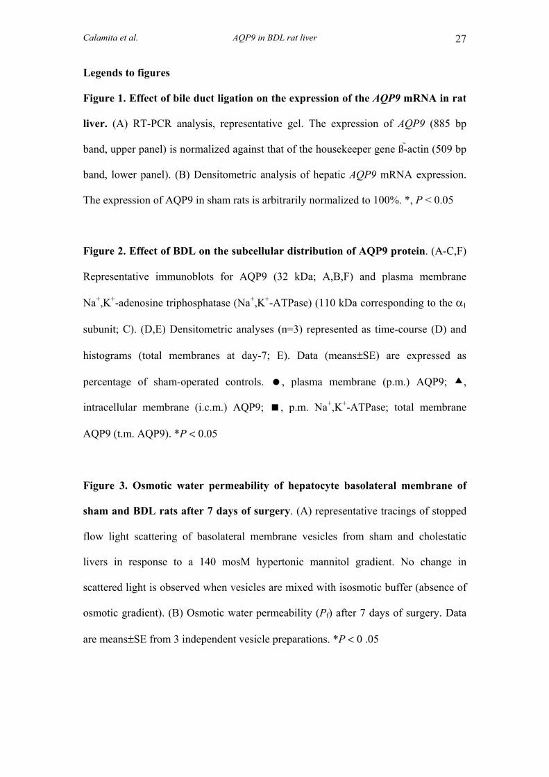
Figure 1. Effect of bile duct ligation on the expression of the AQP9 mRNA in rat
liver. (A) RT-PCR analysis, representative gel. The expression of AQP9 (885 bp
band, upper panel) is normalized against that of the housekeeper gene ß-actin (509 bp
band, lower panel). (B) Densitometric analysis of hepatic AQP9 mRNA expression.
The expression of AQP9 in sham rats is arbitrarily normalized to 100%. *, P < 0.05
Figure 2. Effect of BDL on the subcellular distribution of AQP9 protein. (A-C,F)
Representative immunoblots for AQP9 (32 kDa; A,B,F) and plasma membrane
Na+,K+-adenosine triphosphatase (Na+,K+-ATPase) (110 kDa corresponding to the α1
subunit; C). (D,E) Densitometric analyses (n=3) represented as time-course (D) and
histograms (total membranes at day-7; E). Data (means±SE) are expressed as
percentage of sham-operated controls. , plasma membrane (p.m.) AQP9; ,
intracellular membrane (i.c.m.) AQP9; , p.m. Na+,K+-ATPase; total membrane
AQP9 (t.m. AQP9). *P < 0.05
Figure 3. Osmotic water permeability of hepatocyte basolateral membrane of
sham and BDL rats after 7 days of surgery. (A) representative tracings of stopped
flow light scattering of basolateral membrane vesicles from sham and cholestatic
livers in response to a 140 mosM hypertonic mannitol gradient. No change in
scattered light is observed when vesicles are mixed with isosmotic buffer (absence of
osmotic gradient). (B) Osmotic water permeability (Pf) after 7 days of surgery. Data
are means±SE from 3 independent vesicle preparations. *P < 0 .05

Calamita et al. AQP9 in BDL rat liver 28
Figure 4. Glycerol permeability of hepatocyte basolateral plasma membrane of
BDL rats. (A) representative tracings of stopped flow light scattering of basolateral
membrane vesicles from sham and BDL livers in response to a 150 mM inwardly
directed gradient of glycerol at 20 °C. The initial increase in light scattering results
from osmotic water efflux (vesicle shrinkage), followed by a slower decrease caused
by glycerol entry. (B) glycerol permeability (Pgly) after 7 days of BDL and sham
surgeries. Data are means±SE from 3 rats per each experimental condition. *P < 0 .05
Figure 5. Immunofluorescent distribution of AQP9 in the liver of BDL rats.
(A,C,E,G) liver sections from sham-operated rats. At 0-days BDL, AQP9
immunofluorescence is seen over the basolateral membrane of hepatocytes (A,
arrows) and no reactivity is noted in the canalicular membrane (A, inset; arrowhead).
Such profile of immunoreactivity is maintained in the livers of 1-, 3- and 7-days of
sham rats (C,E,G, respectively). (B) Negative control. At day 1 of BDL (D), AQP9
immunoreactivity is seen both over the basolateral membrane (inset; arrow) and
within the intracellular compartment (double arrows). At day 3 of BDL (F), while the
intracellular staining of AQP9 is steadily present (double arrows) the basolateral
membrane labeling appears reduced both as extension and intensity (inset; arrow). At
7-days BDL (H) the plasma membrane staining is very weak (single arrow) whereas
extensive clouds of immunoreactivity are seen within the cytoplasmic compartment
(double arrows). cv, centrolobular vein.
Figure 6. Effect of bile duct ligation on the immunohistochemical distribution of
AQP9 in rat hepatocytes. (A,C,E,G) liver sections from sham-operated rats. At day
0, AQP9 immunolabeling (brown staining) is seen over the basolateral membrane of

Calamita et al. AQP9 in BDL rat liver 29
hepatocytes (A, arrows; inset). Such profile of immunoreactivity is maintained in the
livers of 1-, 3- and 7-days of sham rats (C,E,G, respectively). (B) Negative control
(absence of AQP9 antibody). (D) Day 1 of BDL. Low AQP9 reactivity is seen over
the basolateral membrane of hepatocyte (arrows). Labeling is observed over the
intracellular compartment (double arrows). Areas within the hepatic acinus show
hepatocytes in their way to be decomposed (red dashed circle). These areas are
surrounded by hepatocytes with weak (or absent) AQP9 reactivity (asterisks). (F) Day
3 of BDL. The labeling of the sinusoidal membrane (arrows; inset) is reduced as to
the basal condition whereas the intracellular reactivity is increasingly evident (double
arrows; inset). Hepatocytes with no immunoreactivity are seen within the acinus
(asterisks). (H) Day 7 of BDL. Intracellular AQP9 staining predominates in nearly all
hepatocytes (double arrows). Mithotic hepatocytes with considerable intracellular
staining are often observed within the acinus (red dashed square). Magnification
X500 (insets, X1000). cv, centrolobular vein.
Fig. 7. Ultrastructural localization of hepatocyte AQP9 in 7-days BDL rats by
immunogold electron microscopy. (A) hepatocyte of sham-operated rat liver. AQP9
immunogold labeling is seen at the microvilli of the basolateral plasma membrane
(single arrows). Poorly appreciable staining is seen within the intracellular
compartment (arrowheads). (B) hepatocyte of BDL rat. Predominant immunolabeling
is observed intracellularly (arrowheads) whereas minor staining is encountered at the
basolateral membrane compartment (single arrow). mc, mitochondrion; bm,
basolateral membrane; bars, 300 nm.