Recent advances in the regulation of cholangiocyte proliferation and function during extrahepatic...
18
Recent advances in the regulation of cholangiocyte proliferation and function during extrahepatic cholestasis S.S. Glaser a,b,* , P. Onori f , C. Wise b , F. Yang b,d , M. Marzioni e , D. Alvaro g , A. Franchitto h , R. Mancinelli h , G. Alpini a,b,c , Md. K. Munshi b , and E. Gaudio h,** S.S. Glaser: [email protected]; E. Gaudio: [email protected] a Digestive Disease Research Center, Scott & White, TX, United States b Department of Medicine, Division of Gastroenterology, Scott & White and Texas A&M Health Science Center, College of Medicine, Temple, TX, United States c Central Texas Veterans Health Care System, Temple, TX, United States d Shengjing Hospital, China Medical University, Shenyang City, Liaoning Province, China e Department of Gastroenterology, Universita' Politecnica delle Marche, Ancona, Italy f Department of Experimental Medicine, University of L'Aquila, L'Aquila, Italy g Gastroenterology, University of Rome “La Sapienza”, Rome, Italy h Department of Human Anatomy, University of Rome “La Sapienza”, Rome, Italy Abstract Bile duct epithelial cells (i.e., cholangiocytes), which line the intrahepatic biliary epithelium, are the target cells in a number of human cholestatic liver diseases (termed cholangiopathies). Cholangiocyte proliferation and death is present in virtually all human cholangiopathies. A number of recent studies have provided insights into the key mechanisms that regulate the proliferation and function of cholangiocytes during the pathogenesis of cholestatic liver diseases. In our review, we have summarised the most important of these recent studies over the past 3 years with a focus on those performed in the animal model of extrahepatic bile duct ligation. In the first part of the review, we provide relevant background on the biliary ductal system. We then proceed with a general discussion of the factors regulating biliary proliferation performed in the cholestatic animal model of bile duct ligation. Further characterisation of the factors that regulate cholangiocyte proliferation and function will help in elucidating the mechanisms regulating the pathogenesis of biliary tract diseases in humans and in devising new treatment approaches for these devastating diseases. Keywords Bile duct ligation; Cholangiocyte; Cholestatic liver diseases; Extrahepatic cholestasis; Proliferation * Corresponding author at: Digestive Disease Research Center, Texas A&M Health Science Center, 702 SW H.K. Dodgen Loop, Temple, TX 76504, United States. Tel.: +1 254 742 7058; fax: +1 254 724 5944. ** Corresponding author at: Department of Human Anatomy, University of Rome “La Sapienza”, Via Alfonso Borelli 50 00161 Rome, Rome 00161, Italy. Tel.: +39 06 4991 8060; fax: +39 06 4991 8062. Conflict of interest statement: None declared. NIH Public Access Author Manuscript Dig Liver Dis. Author manuscript; available in PMC 2011 April 1. Published in final edited form as: Dig Liver Dis. 2010 April ; 42(4): 245–252. doi:10.1016/j.dld.2010.01.008. NIH-PA Author Manuscript NIH-PA Author Manuscript NIH-PA Author Manuscript
Transcript of Recent advances in the regulation of cholangiocyte proliferation and function during extrahepatic...

Recent advances in the regulation of cholangiocyte proliferationand function during extrahepatic cholestasis
S.S. Glasera,b,*, P. Onorif, C. Wiseb, F. Yangb,d, M. Marzionie, D. Alvarog, A. Franchittoh, R.Mancinellih, G. Alpinia,b,c, Md. K. Munshib, and E. Gaudioh,**S.S. Glaser: [email protected]; E. Gaudio: [email protected] Digestive Disease Research Center, Scott & White, TX, United Statesb Department of Medicine, Division of Gastroenterology, Scott & White and Texas A&M HealthScience Center, College of Medicine, Temple, TX, United Statesc Central Texas Veterans Health Care System, Temple, TX, United Statesd Shengjing Hospital, China Medical University, Shenyang City, Liaoning Province, Chinae Department of Gastroenterology, Universita' Politecnica delle Marche, Ancona, Italyf Department of Experimental Medicine, University of L'Aquila, L'Aquila, Italyg Gastroenterology, University of Rome “La Sapienza”, Rome, Italyh Department of Human Anatomy, University of Rome “La Sapienza”, Rome, Italy
AbstractBile duct epithelial cells (i.e., cholangiocytes), which line the intrahepatic biliary epithelium, are thetarget cells in a number of human cholestatic liver diseases (termed cholangiopathies). Cholangiocyteproliferation and death is present in virtually all human cholangiopathies. A number of recent studieshave provided insights into the key mechanisms that regulate the proliferation and function ofcholangiocytes during the pathogenesis of cholestatic liver diseases. In our review, we havesummarised the most important of these recent studies over the past 3 years with a focus on thoseperformed in the animal model of extrahepatic bile duct ligation. In the first part of the review, weprovide relevant background on the biliary ductal system. We then proceed with a general discussionof the factors regulating biliary proliferation performed in the cholestatic animal model of bile ductligation. Further characterisation of the factors that regulate cholangiocyte proliferation and functionwill help in elucidating the mechanisms regulating the pathogenesis of biliary tract diseases in humansand in devising new treatment approaches for these devastating diseases.
KeywordsBile duct ligation; Cholangiocyte; Cholestatic liver diseases; Extrahepatic cholestasis; Proliferation
* Corresponding author at: Digestive Disease Research Center, Texas A&M Health Science Center, 702 SW H.K. Dodgen Loop, Temple,TX 76504, United States. Tel.: +1 254 742 7058; fax: +1 254 724 5944. ** Corresponding author at: Department of Human Anatomy,University of Rome “La Sapienza”, Via Alfonso Borelli 50 00161 Rome, Rome 00161, Italy. Tel.: +39 06 4991 8060; fax: +39 06 49918062.Conflict of interest statement: None declared.
NIH Public AccessAuthor ManuscriptDig Liver Dis. Author manuscript; available in PMC 2011 April 1.
Published in final edited form as:Dig Liver Dis. 2010 April ; 42(4): 245–252. doi:10.1016/j.dld.2010.01.008.
NIH
-PA Author Manuscript
NIH
-PA Author Manuscript
NIH
-PA Author Manuscript

1. IntroductionThe liver, the largest internal organ of the body, is composed of two types of epithelial cells:(i) hepatocytes; and (ii) cholangiocytes [1]. Cholangiocytes line the intrahepatic andextrahepatic bile duct system of the liver [1]. The bile ductules and ducts comprise a branchedsystem of interconnected tubes [1–3], which collect bile secreted at the canalicular membranesof hepatocytes [4], and deliver it to the gallbladder or the duodenum [1,5]. Althoughcholangiocytes represent a small proportion (3 to 5%) of the cells of the liver [1,5,6], thesecells play an important pathophysiological role in the modification of the composition of bileduring the transit in the bile ducts, which involves the secretion and absorption of water,electrolytes and other organic solutes from hepatocellular bile [1,5–10]. The modification ofbile by cholangiocytes is regulated by a number of gastrointestinal hormones, which has beenrecently reviewed [11,12]. The regulation of cholangiocyte bicarbonate secretion is regulatedby the gastrointestinal hormone secretin [5,12]. Cholangiocytes are the only cell types in theliver that express the secretin receptor (SR) [13], which is of importance to the function of thebiliary epithelium in normal and pathological conditions [5,8,14–18]. In large (but not small)cholangiocytes secretin stimulates increases in intracellular cyclic adenosine 3′,5′-monophosphate (cAMP) levels [14,16,19,20] and induces the opening of the Cl− channel(cystic fibrosis transmembrane conductance regulator, CFTR) [15], which leads to theactivation of the Cl−/HCO3
− anion exchanger 2 (AE2) [21] and secretion of bicarbonate in bile[5].
Cholangiocytes are the target cells of a number of diseases termed cholangiopathies. Thisdisease class is made up of inherited disorders [Alagille syndrome and cystic fibrosis (CF)],autoimmune disorders [primary sclerosing cholangitis (PSC), primary biliary cirrhosis (PBC),autoimmune cholangitis (AIC), allograft rejection, graft-versus-host disease (GVHD)],infections (cholangitis due to bacteria, fungi, parasites or viruses), drug-induced injury,ischaemic injury and diseases of unknown aetiology (biliary atresia and idiopathic vanishingbile duct syndromes) [22]. Cholangiopathies are predominantly characterised by a bile duct-directed inflammatory response that leads to bile duct injury associated with biliaryproliferation in the early stage of the disease course [22]. If the biliary injury is chronic therewill be increased bile duct loss, biliary fibrosis and the increased incidence of bile duct cancer(i.e., cholangiocarcinoma) [22].
2. Anatomical and morphological features of the biliary treeThe intrahepatic biliary epithelium is divided into extrahepatic and intrahepatic bile ducts [2,3,23,24]. The intrahepatic bile ductal system consists of the portion of (i) bile canaliculi, smallspaces localised between two adjacent hepatocytes (0.5–2 mm) forming a three-dimensionalnetwork that continues in (ii) bile ductules (canals of Hering), localised at the periphery of thehepatic lobule and characterised by 3–4 cholangiocytes, that form the junction betweenhepatocytes and cholangiocytes (ductular–canalicular junction) allowing the confluence of thebile in (iii) bile ducts (interlobular bile ducts) localised in the portal space. Interlobular bileducts progressively continue in larger ducts until the right and left hepatic ducts, that, at thelevel of the hylus, determine the origin of extrahepatic biliary tree [25–27]. According toLudwig, the human intrahepatic biliary epithelium is divided into small bile ductules (<15μm), interlobular ducts (15–10 μm), septal ducts (100–300 μm), segmental ducts (400–800μm) and hepatic ducts (>800 μm) [24]. The rodent intrahepatic bile duct system has beenrecently classified into small ducts (<15 μm in external diameter) lined by small cholangiocytes(approximately 8 μm in diameter characterised by high nucleus/cytoplasm ratio) and large bileducts (>15 μm in diameter characterised by low nucleus/cytoplasm ratio) lined by largecholangiocytes (approximately 15 μm in diameter) [2,16,19,25,28]. These studies have alsoshown that a significant relationship exists between cholangiocyte area and external bile duct
Glaser et al. Page 2
Dig Liver Dis. Author manuscript; available in PMC 2011 April 1.
NIH
-PA Author Manuscript
NIH
-PA Author Manuscript
NIH
-PA Author Manuscript

diameter, with small bile ducts lined by small cholangiocytes and large ducts lined by largecholangiocytes [2,16,19]. The latter finding is particularly relevant since it allows for the directmapping of studies obtained in isolated small and large cholangiocytes to different portions(i.e., small and large) of the intrahepatic biliary epithelium ex situ [2,14–19]. In support of themorphological heterogeneity of the biliary epithelium, Masyuk et al. have reconstructed theintrahepatic biliary system that resembles a tree, with the common and hepatic ductscorresponding to the trunk, the intrahepatic bile ducts corresponding to the large branches andthe small ductules corresponding to the smallest tree limbs of the tree [29].
3. Cholangiocyte proliferation in response to bile duct ligationA number of studies have defined three types of cholangiocyte proliferation: “typical”,“atypical” and oval cell proliferation [30]. “Typical” cholangiocyte proliferation is ahyperplastic reaction, which induces an increase in the number of intrahepatic bile ducts (witha well-defined lumen) confined to portal areas [5,31]. “Atypical” cholangiocyte proliferationis commonly seen in patients with prolonged cholestatic liver diseases such as PBC or PSCand is characterised by irregular proliferation of intrahepatic bile ducts sprouting into periportaland parenchymal regions and occasionally forming anastomosing cords with adjacenthepatocytes [32,33]. Oval cell proliferation takes place in the early stages of chemicallyinduced hepatocarcinogenesis and is characterised by a disorganised proliferation of biliarystructures with a poorly defined lumen [34]. Oval cell proliferation is not discussed in thisreview.
In animal models, “typical” cholangiocyte proliferation is achieved by a number ofexperimental manoeuvres, including BDL (Fig. 1) [5], partial hepatectomy [35], acute carbontetrachloride (CCl4) treatment [17,18] and chronic feeding of α-naphthylisothiocyanate(ANIT) [36], or bile salts [37]. In these hyperplastic models, cholangiocyte proliferation isclosely associated with increased SR gene expression and secretin-stimulated cAMP levels[5,13,17,18,35–38]. cAMP, which is generated by adenylyl cyclases (AC), plays an importantrole in the modulation of cholangiocyte function [35,39–42]. A recent study by Strazzaboscoet al. demonstrates that differential expression of AC isoforms mediate the secretory functionsof small and large cholangiocytes [41]. This study demonstrated that large cholangiocyteresponsiveness to secretin was mediated by the expression of AC8 [41]. A number of animalmodels that mimic cholestatic liver diseases and liver injury have been utilised to expand ourknowledge related to the mechanisms of cholangiocyte proliferation [1,12,43]. Of these modelsof bile duct injury, the BDL model has been the most commonly used. A number of coordinatefactors (stimulatory or inhibitory) have been shown to regulate cholangiocyte growth in thecholestatic BDL model. It has been shown that increased biliary pressure is a trigger for thestimulation or inhibition of these putative growth factors [44,45]. A recent study has shown[45] that increased biliary and portal hypertension (induced by the first), represent keyproliferative triggers for the growth of bile ducts and hepatocytes. Similar to findings in humancholangiopathies (e.g., PBC and PSC), recent studies in rats have demonstrated that “typical”cholangiocyte proliferation occurs within a limited range of duct sizes [16,18,46]. In rats withBDL, enhanced cholangiocyte proliferative capacity is restricted to large bile ducts [18,46]. Inan experimental animal model of bile duct damage, CCl4 induces loss of large ducts and lossof large duct secretion [17,18,47]. To compensate for the loss of duct function due to this toxin[17,18], small cholangiocytes proliferate and develop de novo secretory activity due to denovo expression of SR [17,18]. A hallmark of large cholangiocyte proliferation induced byBDL in rats is the increased SR expression and subsequent secretory activity [18,46]. A recentstudy in mice with BDL demonstrated that similar to rats, the mouse intrahepatic biliaryepithelium is morphologically and functionally heterogeneous [16]. These findings are ofimportance due to increased availability and usage of transgenic mouse models for studyingcholestatic liver disease pathogenesis [16]. The study indicates that the mouse is a suitable
Glaser et al. Page 3
Dig Liver Dis. Author manuscript; available in PMC 2011 April 1.
NIH
-PA Author Manuscript
NIH
-PA Author Manuscript
NIH
-PA Author Manuscript

model for defining the heterogeneous responses of cholangiocytes during cholestasis andbiliary damage [16]. Since SR is only expressed by cholangiocytes in the liver [13], changesin the functional expression of this receptor have been suggested as a pathophysiological toolfor evaluating changes in the degree of cholangiocyte growth/loss [5,16–18,35–40,46].Proliferating cholangiocytes acquire a neuroendocrine phenotype and secrete and respond toa number of hormones, neuropeptides and neurotransmitters [43,48–50]. The formation of aneuroendocrine compartment predominated by cholangiocytes represents a unique opportunityfor cholangiocytes to regulate their own proliferation via autocrine pathways and forcholangiocytes to influence other nearby cell types, such as vascular endothelial cells, portalfibroblasts and hepatic stellate cells [43,49]. A number of recent studies have highlighted andexpanded our knowledge of the concept that proliferating cholangiocytes displayneuroendocrine features [43,49].
4. Update on neuroendocrine regulation of biliary proliferation during BDLOver the past several years, a number of studies have explored the neuroendocrine regulationof cholangiocyte proliferation during extrahepatic cholestasis. The neuroendocrine factorscontributing to cholangiocyte proliferation have been previously reviewed [43,48–50]. Wediscuss here the most recent advances which are highlighted in Tables 1 and 2.
4.1. Neuropeptides and neurotransmittersCGRP (calcitonin gene-related peptide) is a potent vasodilator peptide that participates in theregulation of vascular tone and regional organ blood flow [51,52]. We have recentlydemonstrated that hepatic sensory innervation and cholangiocyte expression of α-CGRP (Fig.2) play a key role in the regulation of cholangiocyte proliferation during cholestasis inducedby BDL [53]. Knockout of αα CGRP decreases intrahepatic bile duct mass and inhibitscholangiocyte proliferation in BDL mice [53]. Both α- and β-CGRP stimulated proliferationof isolated BDL cholangiocytes by activation of protein kinase A (PKA) and cAMP responseelement binding (CREB) [53]. These studies indicate that sensory innervation plays a role inthe regulation of biliary proliferation and other sensory neuropeptides may play a role in chronicinflammation during cholangiopathies
The aminergic peptide and neurotransmitter histamine regulates many functions in the body,such as neurogenic functions, inflammatory responses, allergic responses, and gastric secretion[54–56]. Normal and BDL rat cholangiocytes express all of the G-protein coupled histaminereceptor subtypes (HRH1, HRH2, HRH3 and HRH4) [57]. Following BDL, the expression ofHR3R is significantly increased in proliferating cholangiocytes. Activation of HR3R by thechronic administration of the agonist (R)-(α)-(−)-methylhistamine dihydrobromide (RAMH)to rats for 7 days after BDL resulted in a decrease in the growth of the biliary tree with nodifference in the rate of apoptosis [57]. In addition, administration of histamine to this animalmodel of cholestasis also resulted in a decrease in cholangiocyte proliferation, and blockinghistamine actions by using the selective HR3R antagonist thioperamide maleate resulted in apartial reversal of these effects [57]. Both in vivo and in vitro, RAMH inhibition ofcholangiocyte growth was associated with downregulation of cAMP-dependent PKA–ERK1/2-Elk-1 signalling pathway [57].
4.2. Glucagon-like peptide-1 (GLP-1)GLP-1 is secreted by a number of neuroendocrine cell types and plays a role in sustaining beta-cell survival in experimental models of diabetes and induces the transdifferentiation ofpancreatic ductal cells [58,59]. GLP-1 and the GLP-1 receptor specific agonist exendin-4 hadsimilar effects on cholangiocytes by stimulating proliferation in vivo in normal rats and inisolated cholangiocytes from normal and BDL rats [60]. GLP-1 receptor was significantly
Glaser et al. Page 4
Dig Liver Dis. Author manuscript; available in PMC 2011 April 1.
NIH
-PA Author Manuscript
NIH
-PA Author Manuscript
NIH
-PA Author Manuscript

upregulated during BDL compared to sham operated animals [60]. Cholangiocytes from BDLbut not normal rats express the message for the precursor for GLP-1, preproglucagon, whichis a finding that suggests that GLP-1 is an important player in biliary growth during cholestasis[60]. In fact, administration of the GLP-1R antagonist, exendin-9–39 significantly decreasedductal mass and biliary functional activity in BDL rats [60]. The pro-proliferative effect ofGLP-1 was mediated through phosphoinositide 3-kinase (PI3K), cAMP/PKA and Ca2+-CaMKII α (calmodulin-dependent protein kinase α) signalling mechanisms [60]. Themechanisms by which GLP-1 regulates cholangiocyte growth are depicted in Fig. 3. Morerecently, exendin-4 has been shown to protect cholangiocytes from apoptosis in in vivo and invitro models of cholangiocyte apoptosis [61]. In vitro, exendin-4 preventedglycochenodeoxycholic acid-induced Bax mitochondrial translocation, cytochrome c releaseand caspase 3 activities, which was blocked by inhibition of PI3K [61]. In vivo, exendin-4administration prevented the increase in TUNEL, positive cholangiocytes and the loss of bileducts that is observed in BDL rats treated with CCl4 [61]. These findings suggest that exendin-4can correct the dysregulated balance between cholangiocyte proliferation and death duringcholestasis [61]. Further studies are required to determine if exendin-4 will be effective forpreventing the progression of cholangiopathies towards ductopenia.
4.3. ProgesteroneWe have recently demonstrated that the steroid hormone progesterone stimulates theproliferation of both male and female cholangiocytes [48]. Cholangiocytes express the PR-Bnuclear receptor and several membrane receptors for progesterone (PRGMC1, PRGMC2, andmPRalpha) [48]. Chronic administration of progesterone increased the number of bile ducts ofnormal rats [48]. Administration of an anti-progesterone antibody inhibited cholangiocytegrowth stimulated by BDL [48]. Interestingly, normal and BDL cholangiocytes expressed thebiosynthetic pathway (i.e., steroidogenic acute regulatory protein or STAR, 3β-hydroxysteroiddehydrogenase or 3β-HSD, and cytochrome P450 side-chain cleavage or p450scc) for andsecrete progesterone [48] (Fig. 4). In vitro, supernatants collected from normal and BDLcholangiocytes increased cholangiocyte proliferation, which was partially inhibited bypreincubation with anti-progesterone and inhibition of progesterone steroidogenesis withaminoglutethimide prevented cholangiocyte proliferation [48]. These findings provide furthersupport for the concept that neuroendocrine autocrine/paracrine mechanisms play a key rolein the modulation of cholangiocyte proliferative responses to cholestasis
4.4. Follicle-stimulating hormone (FSH)FSH, also called gonadotropin because it stimulates the gonads, is produced in the anteriorpituitary gland of the brain [62]. We found that cholangiocytes expressed the FSH receptor(FSHR) and secreted FSH [31]. Chronic administration of FSH to normal rats increased,whereas administration of antide (a gonadotropin-releasing hormone antagonist that blocksFSH secretion) and anti-FSH to normal rats decreased cholangiocyte proliferation andsecretory responses [31]. In vitro, FSH increased cholangiocyte proliferation, cAMP levels,and ERK1/2 and Elk-1 phosphorylation, which were prevented by preincubation with anti-FSH [31]. Silencing of FSH expression also decreases basal cholangiocyte proliferationsuggesting that FSH is a key autocrine factor regulating biliary mass [31]. These findings haveimportant pathological implications since modulation of the expression and secretion of FSHmay be used to modulate cholangiocyte proliferation during cholestatic liver diseases
4.5. Angiogenic factorsVascular endothelial growth factor (VEGF) is a mitogen for vascular endothelial cells forvascular cells and regulates vascular pathophysiology [63,64]. We have previously shown thatVEGF-A and VEGF-C are secreted by cholangiocytes and play an important role in the
Glaser et al. Page 5
Dig Liver Dis. Author manuscript; available in PMC 2011 April 1.
NIH
-PA Author Manuscript
NIH
-PA Author Manuscript
NIH
-PA Author Manuscript

regulation of cholangiocyte proliferation and apoptosis during cholestasis and biliary injuryinduced by hepatic artery ligation [65,66]. We have also shown that the bile acid TC preventscholangiocyte death by apoptosis, and the loss of proliferative and functional responses ofcholangiocytes in response to CCl4 and cholinergic or adrenergic denervation [47,67,68]. Also,taurocholic acid feeding prevents tumour necrosis factor (TNF)-alpha-induced damage ofcholangiocytes by a PI3K-mediated pathway [69].
A recent study has provided additional evidence that VEGF plays a role in both the protectionof cholangiocytes from damage in an experimental model of cholestasis [70]. In an animalmodel of cholestasis and biliary damage induced by caffeic acid, the feeding of the protectivebile acid, taurocholate prevented bile duct damage, which was associated with increasedcholangiocyte VEGF-A, VEGF-C, VEGFR-2 and VEGFR-3 expression [70].
4.6. Other factors regulating cholangiocyte proliferation during cholestasis4.6.1. Endocannabinoid system—The endocannabinoid system has been implicated inthe pathogenesis of liver fibrosis and portal hypertension [71]. Recent evidence also indicatesthat the endocannabinoid system plays a role in regulating cholangiocyte proliferation duringcholestasis induced by BDL [72]. Chronic treatment of rats with BDL with anandamidedecreased cholangiocyte proliferation and induced the accumulation of reactive oxygenspecies, upregulated the expression of TRX1, Ref1, c-Fos and c-Jun expression, increased thenuclear localisation of TRX1 and increased AP-1 transcriptional activity [72]. These effectsoccurred via the activation of the cannabinoid receptor, Cb2 [72]. This work demonstrated thatmodulation of the endocannabinoid system and/or the ROS/TRX1/Ref1/AP-1 pathway mayhave therapeutic implications for the treatment of early stage cholestatic liver diseasescharacterised by cholangiocyte proliferation [72].
4.6.2. CD44 and hyaluronic acid—CD44 is a multifunctional cell adhesion molecule,which takes part in cell–cell and cell–matrix interactions [73,74]. Hyaluronic acid (HA), themain component of extracellular matrices, is the primary ligand of CD44 [75]. High levels ofhepatic CD44 expression have been observed in patients with PSC [76]. He et al. provideevidence that suggests that the proliferative cholangiocytes lining the intrahepatic ducts are animportant source of hepatic CD44 [77]. CD44-positive cholangiocytes were closely associatedwith extracellular hyaluronan accumulated in the portal tracts of BDL livers [77]. Theydemonstrated in vitro that cholangiocyte proliferation was stimulated by hyaluronan treatment,and blocked by siRNA for CD44 or anti-CD44 antibody [77]. CD44–hyaluronan interactionsmay play a pathogenic role in the development of cholestatic liver diseases by enhancing biliaryproliferation [77].
4.6.3. Insulin-like growth factor-1—Previous studies [78,79] have shown that IGF1 playsa key role in mediating cholangiocyte proliferation after BDL and in protecting cholangiocytesfrom the cytotoxic effect of hydrophobic bile salts. More recently, we evaluated the expressionof IGF1 isoforms in rat cholangiocytes, and evaluated their involvement in cell proliferationor damage induced by BDL or hydrophobic bile salts [80]. In both hepatocytes andcholangiocytes, the ‘locally acting’ IGF1 isoform (XO6108) and ‘circulating’ IGF1 isoform(NM 178866) [81] represent respectively 44% and 52% of the total IGF1 [80]. Basal mRNAsfor both ‘locally acting’ and ‘circulating’ IGF1 isoforms are significantly higher in hepatocytescompared to cholangiocytes [80]. After BDL for 3 h, the ‘locally acting’ IGF1 isoformdecreases threefold in hepatocytes but remains stable in cholangiocytes with respect to sham-controls [80]. After 1 week of BDL, hepatocytes displays a further fivefold decrease of ‘locallyacting’ IGF1 mRNA. In contrast, cholangiocytes show an eightfold increase of the ‘locallyacting’ IGF1 mRNA. The effect of BDL for 3 h on IGF1 isoforms was reproduced in vitro byincubation with glycochenodeoxycholate (GCDC) [80]. The cytotoxic effects (inhibition of
Glaser et al. Page 6
Dig Liver Dis. Author manuscript; available in PMC 2011 April 1.
NIH
-PA Author Manuscript
NIH
-PA Author Manuscript
NIH
-PA Author Manuscript

proliferation and induction of apoptosis) of GCDC on isolated cholangiocytes were morepronounced after silencing (siRNA) of ‘locally acting’ than ‘circulating’ IGF1 isoform [80].Therefore, these findings demonstrate that rat cholangiocytes express the ‘locally acting’ IGF1isoform, which decreases during cell damage and increases during cell mitosis [80]. The‘locally acting’ IGF1 is more active than the ‘circulating’ isoform in protecting cholangiocytesfrom GCDC-induced cytotoxicity [80]. These findings indicate that, besides muscle and neuraltissues, also in liver cells the ‘locally acting’ IGF1 isoform is important in modulating responseof cholangiocytes to damage.
4.6.4. Ezrin–radixin–moesin-binding phosphoprotein—Ezrin–radixin–moesin-binding phosphoprotein 50 (EBP50) is inducible by oestrogen and alters cell proliferation. Thisstudy demonstrated the expression and role of EBP50 in ductular reaction in normal humanliver, human cholangiopathies (i.e., CF, PBC and PSC) and BDL rats [82]. The studydemonstrated that in normal human liver, EBP50 is expressed in the canalicular membranesof hepatocytes and, together with ezrin and CFTR, in the apical domains of cholangiocytes[82]. In human cholangiopathies and BDL rats, EBP50 was redistributed to the cytoplasmicand nuclear compartments [82]. There was transient increase of EBP50 in rat cholangiocytesafter BDL, whereas such expression was downregulated in ovariectomised rats [82]. Thesestudies demonstrated that the expression and distribution of EBP50 (regulated by oestrogens)contribute to the proliferative responses of cholangiocytes [82].
4.6.5. Death receptor 5—Takeda et al. have demonstrated that the death signalling pathwaymediated by TNF-related apoptosis-inducing ligand (TRAIL) receptor 2/death receptor 5(DR5) contributes to the pathogenesis of biliary cirrhosis [83]. Administration of an agonisticanti-DR5 monoclonal antibody triggered cholangiocyte apoptosis, cholangitis and cholestaticliver injury reminiscent of PSC [83]. BDL upregulates DR5 expression on cholangiocytes,sensitising them to the effects of DR5 stimulation [83]. DR5 and TRAIL expression were alsofound to be elevated in cholangiocytes of human PSC and PBC patient samples suggesting thatmodulation of DR5 death signalling might be a therapeutic option for chronic cholestatic liverdiseases [83]
4.6.6. Foxl1 (winged helix transcription factor)—Foxl1 has been previously shown tobe dramatically induced in cholangiocytes by both BDL and in response to 3,5-diethoxycarbonyl-1,4-dihydrocollidine diet [84]. In Foxl1 knockout mice with BDL, theyobserved an increase in parenchymal necrosis and significantly impaired cholangiocyte andhepatocyte proliferation [85]. In addition, there was decreased expression of Wnt3a and Wnt7bexpression along with reduced expression of the β-catenin target gene Cyclin D1 [85]. Thiswork suggests that Foxl1 is an upstream mediator of β-catenin-induced cholangiocyteproliferation during cholestasis [85].
4.6.7. Integrin αvβ6—The expression of αvβ6 is markedly increased on cholangiocytes inresponse to extrahepatic obstruction by BDL and drives fibrogenesis [86,87]. Inhibition ofαvβ6 by EMD527040 reduced cholangiocyte proliferation and inhibits the progression ofprimary and secondary biliary fibrosis [87]. The regulation of cholangiocyte proliferation mayprovide a means to prevent biliary fibrosis during chronic cholestatic liver diseases.
5. Summary and future directionsWe have reviewed recent studies that address factors that regulate cholangiocyte proliferationduring extrahepatic cholestasis induced by BDL. Cholangiocyte proliferation is closelyassociated with transdifferentiation to a neuroendocrine phenotype. Future studies will benecessary to determine the role that proliferating cholangiocytes play in the pathogenesis ofbiliary fibrosis during cholestasis and how cholangiocytes interact with other cell types of the
Glaser et al. Page 7
Dig Liver Dis. Author manuscript; available in PMC 2011 April 1.
NIH
-PA Author Manuscript
NIH
-PA Author Manuscript
NIH
-PA Author Manuscript

liver such as hepatic stellate cells during cholestatic liver diseases. Our developing knowledgeof the fundamental factors that control cholangiocyte proliferation during cholestasis will aidin the development of therapies for the treatment of chronic cholestatic liver diseases.
AcknowledgmentsPortions of the studies discussed here were supported partly by a grant award from Scott & White and NIH RO1DK081442 to Shannon Glaser, by the Dr. Nicholas C., Hightower Centennial Chair of Gastroenterology from Scott& White, the VA Research Career Scientist Award, a VA Merit Award and the NIH grants DK76898, DK58411 andDK62975 to Gianfranco Alpini, by University and Federate Athenaeum funds from University of Rome “La Sapienza”to Eugenio Gaudio and MIUR grants: PRIN #2007, prot. 2007HPT7BA 003 to Domenico Alvaro.
References1. Alpini, G.; Prall, RT.; LaRussoF, NF. The pathobiology of biliary epithelia. In: Arias, IM.; Boyer, JL.;
Chisari, FV.; Fausto, N.; Jakoby, W.; Schachter, D.; Shafritz, DA., editors. The liver; biology &pathobiology. 4th. Philadelphia, PA: Lippincott Williams & Wilkins; 2001. p. 421-35.
2. Kanno N, LeSage G, Glaser S, et al. Functional heterogeneity of the intrahepatic biliary epithelium.Hepatology 2000;31:555–61. [PubMed: 10706542]
3. Sasaki H, Schaffner F, Popper H. Bile ductules in cholestasis: morphologic evidence for secretion andabsorption in man. Lab Invest 1967;16:84–95. [PubMed: 4164461]
4. Nathanson MH, Boyer JL. Mechanisms and regulation of bile secretion. Hepatology 1991;14:551–66.[PubMed: 1874500]
5. Alpini G, Lenzi R, Sarkozi L, et al. Biliary physiology in rats with bile ductular cell hyperplasia.Evidence for a secretory function of proliferated bile ductules. J Clin Invest 1988;81:569–78. [PubMed:2448343]
6. Racanelli V, Rehermann B. The liver as an immunological organ. Hepatology 2006;43:S54–62.[PubMed: 16447271]
7. Glaser S, Rodgers RE, Phinizy JL, et al. Gastrin inhibits secretin-induced ductal secretion by interactionwith specific receptors on rat cholangiocytes. Am J Physiol Gastrointest Liver Physiol1997;273:G1061–70.
8. Glaser S, Francis H, DeMorrow S, et al. Heterogeneity of the intrahepatic biliary epithelium. World JGastroenterol 2006;12:3523–36. [PubMed: 16773709]
9. Alvaro D, Cho WK, Mennone A, et al. Effect of secretion on intracellular pH regulation in isolated ratbile duct epithelial cells. J Clin Invest 1993;92:1314–25. [PubMed: 8397224]
10. Alvaro D, Alpini G, Jezequel AM, et al. Role and mechanisms of action of acetylcholine in theregulation of rat cholangiocyte secretory functions. J Clin Invest 1997;100:1349–62. [PubMed:9294100]
11. Esteller A. Physiology of bile secretion. World J Gastroenterol 2008;14:5641–9. [PubMed: 18837079]12. Kanno N, LeSage G, Glaser S, et al. Regulation of cholangiocyte bicarbonate secretion. Am J Physiol
Gastrointest Liver Physiol 2001;281:G612–25. [PubMed: 11518673]13. Alpini G, Ulrich CD 2nd, Phillips JO, et al. Upregulation of secretin receptor gene expression in rat
cholangiocytes after bile duct ligation. Am J Physiol 1994;266:G922–8. [PubMed: 7515577]14. Alpini G, Roberts S, Kuntz SM, et al. Morphological, molecular, and functional heterogeneity of
cholangiocytes from normal rat liver. Gastroenterology 1996;110:1636–43. [PubMed: 8613073]15. Alpini G, Ulrich C, Roberts S, et al. Molecular and functional heterogeneity of cholangiocytes from
rat liver after bile duct ligation. Am J Physiol Gastrointest Liver Dis 1997;272:G289–97.16. Glaser S, Gaudio E, Rao A, et al. Morphological and functional heterogeneity of the mouse
intrahepatic biliary epithelium. Lab Invest 2009;89:456–69. [PubMed: 19204666]17. LeSage G, Benedetti A, Glaser S, et al. Acute carbon tetrachloride feeding selectively damages large,
but not small, cholangiocytes from normal rat liver. Hepatology 1999;29:307–19. [PubMed:9918904]
Glaser et al. Page 8
Dig Liver Dis. Author manuscript; available in PMC 2011 April 1.
NIH
-PA Author Manuscript
NIH
-PA Author Manuscript
NIH
-PA Author Manuscript

18. LeSage G, Glaser S, Marucci L, et al. Acute carbon tetrachloride feeding induces damage of largebut not small cholangiocytes from BDL rat liver. Am J Physiol Gastrointest Liver Physiol1999;276:G1289–301.
19. Alpini G, Glaser S, Robertson W, et al. Large but not small intrahepatic bile ducts are involved insecretin-regulated ductal bile secretion. Am J Physiol Gastrointest Liver Physiol 1997;272:G1064–74.
20. Ueno Y, Alpini G, Yahagi K, et al. Evaluation of differential gene expression by microarray analysisin small and large cholangiocytes isolated from normal mice. Liver Int 2003;23:449–59. [PubMed:14986819]
21. Banales JM, Arenas F, Rodriguez-Ortigosa CM, et al. Bicarbonate-rich choleresis induced by secretinin normal rat is taurocholate-dependent and involves AE2 anion exchanger. Hepatology2006;43:266–75. [PubMed: 16440368]
22. Xia X, DeMorrow S, Francis H, et al. Cholangiocyte injury and ductopenic syndromes. Semin LiverDis 2007;27:401–12. [PubMed: 17979076]
23. Schaffner F, Popper H. Electron microscopic studies of normal and proliferated bile ductules. Am JPathol 1961;38:393–410. [PubMed: 13747220]
24. Ludwig J. New concepts in biliary cirrhosis. Semin Liver Dis 1987;7:293–301. [PubMed: 3324348]25. Gaudio E, Carpino G, Cardinale V, et al. New insights into liver stem cells. Dig Liver Dis
2009;41:455–62. [PubMed: 19403350]26. Gaudio E, Marinozzi G, Carpino F, et al. Scanning electron microscopic stereo views of rat liver
following bile duct ligation. Ultramicroscopy 1980;5:420.27. Carpino F, Gaudio E, Marinozzi G, et al. A scanning and transmission electron microscopic study of
experimental extrahepatic cholestasis in the rat. J Submicrosc Cytol 1981;13:581–98. [PubMed:7334553]
28. Benedetti A, Bassotti C, Rapino K, et al. A morphometric study of the epithelium lining the ratintrahepatic biliary tree. J Hepatol 1996;24:335–42. [PubMed: 8778202]
29. Masyuk TV, Ritman EL, LaRusso NF. Quantitative assessment of the rat intrahepatic biliary systemby three-dimensional reconstruction. Am J Pathol 2001;158:2079–88. [PubMed: 11395385]
30. Roskams T, Van den Oord JJ, De Vos R, et al. Neuroendocrine features of reactive bile ductules incholestatic liver disease. Am J Pathol 1990;137:1019–25. [PubMed: 1700614]
31. Mancinelli R, Onori P, Gaudio E, et al. Follicle-stimulating hormone increases cholangiocyteproliferation by an autocrine mechanism via camp-dependent phosphorylation of ERK1/2 and Elk-1.Am J Physiol Gastrointest Liver Physiol 2009;297:G11–26. [PubMed: 19389804]
32. Desmet V, Roskams T, Van Eyken P. Ductular reaction in the liver. Pathol Res Pract 1995;191:513–24. [PubMed: 7479372]
33. Sirica AE, Gainey TW, Mumaw VR. Ductular hepatocytes. Evidence for a bile ductular cell originin furan-treated rats. Am J Pathol 1994;145:375–83. [PubMed: 8053495]
34. Sirica AE. Biology of biliary epithelial cells. Prog Liver Dis 1992;10:63–87. [PubMed: 1296238]35. LeSage G, Glaser S, Gubba S, et al. Regrowth of the rat biliary tree after 70% partial hepatectomy is
coupled to increased secretin-induced ductal secretion. Gastroenterology 1996;111:1633–44.[PubMed: 8942744]
36. LeSage G, Glaser S, Ueno Y, et al. Regression of cholangiocyte proliferation after cessation of ANITfeeding is coupled with increased apoptosis. Am J Physiol Gastrointest Liver Physiol2001;281:G182–90. [PubMed: 11408271]
37. Alpini G, Glaser S, Ueno Y, et al. Bile acid feeding induces cholangiocyte proliferation and secretion:evidence for bile acid-regulated ductal secretion. Gastroenterology 1999;116:179–86. [PubMed:9869616]
38. LeSage G, Glaser S, Alpini G. Regulation of cholangiocyte proliferation. Liver 2001;21:73–80.[PubMed: 11318975]
39. Francis H, Glaser S, Ueno Y, et al. cAMP stimulates the secretory and proliferative capacity of therat intrahepatic biliary epithelium through changes in the PKA/Src/MEK/ERK1/2 pathway. J Hepatol2004;41:528–37. [PubMed: 15464232]
Glaser et al. Page 9
Dig Liver Dis. Author manuscript; available in PMC 2011 April 1.
NIH
-PA Author Manuscript
NIH
-PA Author Manuscript
NIH
-PA Author Manuscript

40. Glaser S, Benedetti A, Marucci L, et al. Gastrin inhibits cholangiocyte growth in bile duct-ligatedrats by interaction with cholecystokinin-b/gastrin receptors via d-myo-inositol 1,4,5-triphosphate-,Ca(2+)-, and protein kinase C alpha-dependent mechanisms. Hepatology 2000;32:17–25. [PubMed:10869284]
41. Strazzabosco M, Fiorotto R, Melero S, et al. Differentially expressed adenylyl cyclase isoformsmediate secretory functions in cholangiocyte subpopulation. Hepatology 2009;50:244–52. [PubMed:19444869]
42. Minagawa N, Nagata J, Shibao K, et al. Cyclic AMP regulates bicarbonate secretion in cholangiocytesthrough release of ATP into bile. Gastroenterology 2007;133:1592–602. [PubMed: 17916355]
43. Alvaro D, Mancino MG, Glaser S, et al. Proliferating cholangiocytes: a neuroendocrine compartmentin the diseased liver. Gastroenterology 2007;132:415–31. [PubMed: 17241889]
44. Slott PA, Liu MH, Tavoloni N. Origin, pattern, and mechanism of bile duct proliferation followingbiliary obstruction in the rat. Gastroenterology 1990;99:466–77. [PubMed: 1694804]
45. Azmaiparashvili E, Kordzaia D, Dzidziguri D. Biliary hypertension as the cell proliferation triggerin bile duct ligated rats. Georgian Med News 2009:111–6. [PubMed: 19359736]
46. Alpini G, Glaser S, Ueno Y, et al. Heterogeneity of the proliferative capacity of rat cholangiocytesafter bile duct ligation. Am J Physiol Gastrointest Liver Physiol 1998;274:G767–75.
47. Marucci L, Alpini G, Glaser S, et al. Taurocholate feeding prevents CCl4-induced damage of largecholangiocytes through PI3-kinase-dependent mechanism. Am J Physiol Gastrointest Liver Physiol2003;284:G290–301. [PubMed: 12388182]
48. Glaser S, DeMorrow S, Francis H, et al. Progesterone stimulates the proliferation of female and malecholangiocytes via autocrine/paracrine mechanisms. Am J Physiol Gastrointest Liver Physiol2008;295:G124–36. [PubMed: 18511743]
49. Glaser S, Gaudio E, Miller T, et al. Cholangiocyte proliferation and liver fibrosis. Expert Rev MolMed 2009;11:e7. [PubMed: 19239726]
50. Marzioni M, Fava G, Benedetti A. Nervous and neuroendocrine regulation of the pathophysiologyof cholestasis and of biliary carcinogenesis. World J Gastroenterol 2006;12:3471–80. [PubMed:16773704]
51. Tippins JR. CGRP: a novel neuropeptide from the calcitonin gene is the most potent vasodilatorknown. J Hypertens Suppl 1986;4:S102–5. [PubMed: 3553470]
52. Wimalawansa SJ. Calcitonin gene-related peptide and its receptors: molecular genetics, physiology,pathophysiology, and therapeutic potentials. Endocr Rev 1996;17:533–85. [PubMed: 8897024]
53. Glaser S, Ueno Y, DeMorrow S, et al. Knockout of alpha-calcitonin gene-related peptide reducescholangiocyte proliferation in bile duct ligated mice. Lab Invest 2007;87:914–26. [PubMed:17618297]
54. Hou YF, Zhou YC, Zheng XX, et al. Modulation of expression and function of toll-like receptor 3 inA549 and H292 cells by histamine. Mol Immunol 2006;43:1982–92. [PubMed: 16406095]
55. Jancso G, Santha P, Horvath V, et al. Inhibitory neurogenic modulation of histamine-inducedcutaneous plasma extravasation in the pigeon. Regul Pept 2000;95:75–80. [PubMed: 11062335]
56. Parsons ME, Ganellin CR. Histamine and its receptors. Br J Pharmacol 2006;147(Suppl 1):S127–35.[PubMed: 16402096]
57. Francis H, Franchitto A, Ueno Y, et al. H3 histamine receptor agonist inhibits biliary growth of BDLrats by downregulation of the cAMP-dependent PKA/ERK1/2/Elk-1 pathway. Lab Invest2007;87:473–87. [PubMed: 17334413]
58. Drucker DJ. Glucagon-like peptides: regulators of cell proliferation, differentiation, and apoptosis.Mol Endocrinol 2003;17:161–71. [PubMed: 12554744]
59. Bulotta A, Hui H, Anastasi E, Bertolotto, et al. Cultured pancreatic ductal cells undergo cell cyclere-distribution and beta-cell-like differentiation in response to glucagon-like peptide-1. J MolEndocrinol 2002;29:347–60. [PubMed: 12459036]
60. Marzioni M, Alpini G, Saccomanno S, et al. Glucagon-like peptide-1 and its receptor agonistexendin-4 modulate cholangiocyte adaptive response to cholestasis. Gastroenterology2007;133:244–55. [PubMed: 17631146]
61. Marzioni M, Alpini G, Saccomanno S, et al. Exendin-4, a glucagon-like peptide 1 receptor agonist,protects cholangiocytes from apoptosis. Gut 2009;58:990–7. [PubMed: 18829977]
Glaser et al. Page 10
Dig Liver Dis. Author manuscript; available in PMC 2011 April 1.
NIH
-PA Author Manuscript
NIH
-PA Author Manuscript
NIH
-PA Author Manuscript

62. Ulloa-Aguirre A, Uribe A, Zarinan T, et al. Role of the intracellular domains of the human FSHreceptor in G(alphas) protein coupling and receptor expression. Mol Cell Endocrinol2007;260-262:153–62. [PubMed: 17045734]
63. Larrivee B, Karsan A. Signaling pathways induced by vascular endothelial growth factor. Int J MolMed 2000;5:447–56. review. [PubMed: 10762646]
64. Larrivee B, Lane DR, Pollet I, et al. Vascular endothelial growth factor receptor-2 induces survivalof hematopoietic progenitor cells. J Biol Chem 2003;278:22006–13. [PubMed: 12668684]
65. Gaudio E, Barbaro B, Alvaro D, et al. Vascular endothelial growth factor stimulates rat cholangiocyteproliferation via an autocrine mechanism. Gastroenterology 2006;130:1270–82. [PubMed:16618418]
66. Gaudio E, Barbaro B, Alvaro D, et al. Administration of r-VEGF-A prevents hepatic artery ligation-induced bile duct damage in bile duct ligated rats. Am J Physiol Gastrointest Liver Physiol2006;291:G307–17. [PubMed: 16574985]
67. Marzioni M, Ueno Y, Glaser S, et al. Cytoprotective effects of taurocholic acid feeding on the biliarytree after adrenergic denervation of the liver. Liver Int 2007;27:558–68. [PubMed: 17403196]
68. Marzioni M, LeSage G, Glaser S, et al. Taurocholate prevents the loss of intrahepatic bile ducts dueto vagotomy in bile duct-ligated rats. Am J Physiol Gastrointest Liver Physiol 2003;284:G837–52.[PubMed: 12684215]
69. Ueno Y, Francis H, Glaser S, et al. Taurocholic acid feeding prevents tumor necrosis factor-alpha-induced damage of cholangiocytes by a PI3k-mediated pathway. Exp Biol Med (Maywood)2007;232:942–9. [PubMed: 17609511]
70. Mancinelli R, Onori P, Gaudio E, et al. Taurocholate feeding to bile duct ligated rats prevents caffeicacid-induced bile duct damage by changes in cholangiocyte VEGF expression. Exp Biol Med(Maywood) 2009;234:462–74. [PubMed: 19234059]
71. Jimenez W. Endocannabinoids and liver disease. Hepatology 2005;41:983–5. [PubMed: 15841444]72. DeMorrow S, Francis H, Gaudio E, et al. Anandamide inhibits cholangiocyte hyperplastic
proliferation via activation of thioredoxin 1/redox factor 1 and AP-1 activation. Am J PhysiolGastrointest Liver Physiol 2008;294:G506–19. [PubMed: 18096608]
73. Bartolazzi A, Nocks A, Aruffo A, et al. Glycosylation of CD44 is implicated in CD44-mediated celladhesion to hyaluronan. J Cell Biol 1996;132:1199–208. [PubMed: 8601595]
74. Bajorath J. Molecular organization, structural features, and ligand binding characteristics of CD44,a highly variable cell surface glycoprotein with multiple functions. Proteins 2000;39:103–11.[PubMed: 10737932]
75. Mikami T, Saegusa M, Mitomi H, et al. Significant correlations of e-cadherin, catenin, and CD44variant form expression with carcinoma cell differentiation and prognosis of extrahepatic bile ductcarcinomas. Am J Clin Pathol 2001;116:369–76. [PubMed: 11554165]
76. Xu B, Broome U, Ericzon BG, et al. High frequency of autoantibodies in patients with primarysclerosing cholangitis that bind biliary epithelial cells and induce expression of CD44 and productionof interleukin 6. Gut 2002;51:120–7. [PubMed: 12077104]
77. He Y, Wu GD, Sadahiro T, et al. Interaction of CD44 and hyaluronic acid enhances biliary epithelialproliferation in cholestatic livers. Am J Physiol Gastrointest Liver Physiol 2008;295:G305–12.[PubMed: 18556418]
78. Alvaro D, Metalli VD, Alpini G, et al. The intrahepatic biliary epithelium is a target of the growthhormone/insulin-like growth factor 1 axis. J Hepatol 2005;43:875–83. [PubMed: 16083987]
79. Drudi Metalli V, Mancino MG, Mancino A, et al. Bile salts regulate proliferation and apoptosis ofliver cells by modulating the IGF1 system. Dig Liver Dis 2007;39:654–62. [PubMed: 17531559]
80. Gatto M, Drudi-Metalli V, Torrice A, et al. Insulin-like growth factor-1 isoforms in rat hepatocytesand cholangiocytes and their involvement in protection against cholestatic injury. Lab Invest2008;88:986–94. [PubMed: 18607346]
81. Gil-Pena H, Garcia-Lopez E, Alvarez-Garcia O, et al. Alterations of growth plate and abnormalinsulin-like growth factor I metabolism in growth-retarded hypokalemic rats: effect of growthhormone treatment. Am J Physiol Renal Physiol 2009;297:F639–45. [PubMed: 19587145]
Glaser et al. Page 11
Dig Liver Dis. Author manuscript; available in PMC 2011 April 1.
NIH
-PA Author Manuscript
NIH
-PA Author Manuscript
NIH
-PA Author Manuscript

82. Fouassier L, Rosenberg P, Mergey M, et al. Ezrin–radixin–moesin-binding phosphoprotein (ebp50),an estrogen-inducible scaffold protein, contributes to biliary epithelial cell proliferation. Am J Pathol2009;174:869–80. [PubMed: 19234136]
83. Takeda K, Kojima Y, Ikejima K, et al. Death receptor 5 mediated-apoptosis contributes to cholestaticliver disease. Proc Natl Acad Sci U S A 2008;105:10895–900. [PubMed: 18667695]
84. Sackett SD, Li Z, Hurtt R, et al. Foxl1 is a marker of bipotential hepatic progenitor cells in mice.Hepatology 2009;49:920–9. [PubMed: 19105206]
85. Sackett SD, Gao Y, Shin S, et al. Foxl1 promotes liver repair following cholestatic injury in mice.Lab Invest 2009;89:1387–96. [PubMed: 19841618]
86. Wang B, Dolinski BM, Kikuchi N, et al. Role of alphavbeta6 integrin in acute biliary fibrosis.Hepatology 2007;46:1404–12. [PubMed: 17924447]
87. Patsenker E, Popov Y, Stickel F, et al. Inhibition of integrin alphavbeta6 on cholangiocytes blockstransforming growth factor-beta activation and retards biliary fibrosis progression. Gastroenterology2008;135:660–70. [PubMed: 18538673]
List of abbreviations
AIC autoimmune cholangitis
BDL Bile duct ligation
CaMKII α calmodulin-dependent protein kinase α
cAMP cyclic adenosine 3′,5′-monophosphate
CCl4 carbon tetrachloride
CF cystic fibrosis
CFTR cystic fibrosis transmembrane conductance regulator
CGRP calcitonin gene-related peptide
CREB cAMP response element binding
ERK1/2 extracellular signal-regulated kinase
FSH follicle-stimulating hormone
GCDC glycochenodeoxycholate
GLP-1 glucagon-like peptide-1
GVHD graft-versus-host disease
3β-HSD 3β-hydroxysteroid dehydrogenase
IGF-1 insulin-like growth factor-1
PBC primary biliary cirrhosis
PI3K phosphoinositide 3-kinase
PKA protein kinase A
PSC primary sclerosing cholangitis
p450scc cytochrome P450 side-chain cleavage
SR secretin receptor
STAR steroidogenic acute regulatory protein
TNF tumor necrosis factor
VEGF vascular endothelial growth factor
Glaser et al. Page 12
Dig Liver Dis. Author manuscript; available in PMC 2011 April 1.
NIH
-PA Author Manuscript
NIH
-PA Author Manuscript
NIH
-PA Author Manuscript

Fig. 1.Immunolocalisation of cytokeratin-19 (CK-19) in proliferating cholangiocytes in the bile ductligation (BDL) animal model. Original magnification 40×.
Glaser et al. Page 13
Dig Liver Dis. Author manuscript; available in PMC 2011 April 1.
NIH
-PA Author Manuscript
NIH
-PA Author Manuscript
NIH
-PA Author Manuscript
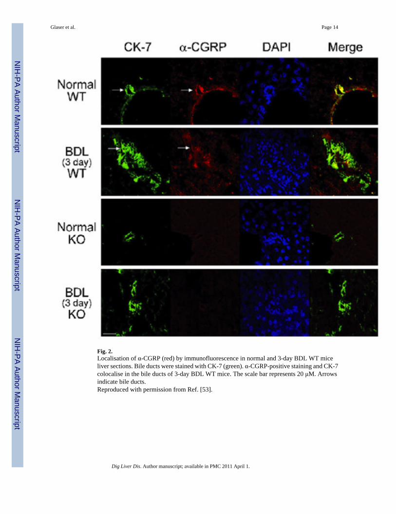
Fig. 2.Localisation of α-CGRP (red) by immunofluorescence in normal and 3-day BDL WT miceliver sections. Bile ducts were stained with CK-7 (green). α-CGRP-positive staining and CK-7colocalise in the bile ducts of 3-day BDL WT mice. The scale bar represents 20 μM. Arrowsindicate bile ducts.Reproduced with permission from Ref. [53].
Glaser et al. Page 14
Dig Liver Dis. Author manuscript; available in PMC 2011 April 1.
NIH
-PA Author Manuscript
NIH
-PA Author Manuscript
NIH
-PA Author Manuscript

Fig. 3.Proposed sequence of intracellular events associated to GLP-1R activation in cholangiocytes.GLP-1R activation sustains cell growth enhancing the activation state of the PI3K and cAMP/PKA cascades. Cell proliferation is also elicited by the extracellular Ca2+-dependent activationof CaMKIIα that can modulate cholangiocyte proliferation both directly and by cross-talkingwith the cAMP/PKA cascade. In contrast, GLP-1R activation does not result in any change ofERK1/2 activation state and does not increase the IP3-PKCα signalling.Reproduced with permission from Ref. [60].
Glaser et al. Page 15
Dig Liver Dis. Author manuscript; available in PMC 2011 April 1.
NIH
-PA Author Manuscript
NIH
-PA Author Manuscript
NIH
-PA Author Manuscript

Fig. 4.Immunofluorescence for key proteins in the progesterone steroidogenesis pathway[steroidogenic acute regulatory protein (StAR), P450 side-chain cleavage (p450scc), and 3β-hydroxysteroid dehydrogenase (3β-HSD)] in liver sections from normal and BDL female andmale rats demonstrates that bile ducts express these steroidogenesis pathway proteins (redstaining). Colocalisation with CK-19 (green staining, a cholangiocyte-specific marker) of thebile ducts expressing StAR, p450scc, and 3β-HSD is also visible.Reproduced with permission from Ref. [48].
Glaser et al. Page 16
Dig Liver Dis. Author manuscript; available in PMC 2011 April 1.
NIH
-PA Author Manuscript
NIH
-PA Author Manuscript
NIH
-PA Author Manuscript

NIH
-PA Author Manuscript
NIH
-PA Author Manuscript
NIH
-PA Author Manuscript
Glaser et al. Page 17
Table 1
Neuroendocrine regulation of cholangiocyte growth.
Regulatory factors Effect on cholangiocytegrowth
Second messenger/transduction pathways References
Calcitonin gene-related peptide Sensory innervation and α-and β-CGRP stimulatebiliary growth
Activation PKA and CREB [53]
RAMH (HRH3 agonist) Activation of HR3Rdecrease biliaryhyperplasia
Downregulation of cAMP-dependent PKA–ERK1/2-Elk-1 signalling
[57]
GLP-1 receptor agonistexendin-4
Activation of GLP-1receptors stimulates biliarygrowth in normal and BDLrats
Activation of PI3K, cAMP/PKA and Ca2+-CaMKII α signalling mechanisms
[60]
GLP-1 receptor agonistexendin-4
In vivo, exendin-4 preventsCCl4-induced biliaryapoptosis
Exendin-4 prevents apoptosis-induced Baxmitochondrial translocation, cytochrome crelease and increased caspase 3 activity
[61]
Progesterone a. Progesteroneinduces biliaryhyperplasia innormal rats
b. Administrationof anti-progesteroneantibodyinhibitedcholangiocytegrowthstimulated byBDL
a. Cholangiocytes expressed thebiosynthetic pathway (STAR,3β-HSD, p450scc) for andsecrete progesterone
b. Inhibition of progesteronesteroidogenesis prevents biliaryhyperplasia
[48]
Follicle-stimulating hormone FSH increases biliaryproliferation, whereasblockage of cholangiocyteFSH secretion decreasescholangiocyte proliferation
a. FSH increased cholangiocyteproliferation by cAMP-dependent phosphorylation ofERK1/2 and Elk-1
b. Silencing of cholangiocyte FSHexpression decreasescholangiocyte proliferation
[31]
Taurocholic acid a. VEGFstimulatesbiliaryhyperplasia
b. Taurocholicacid protectfrom caffeicacid-inducedapoptosis
Increased cholangiocyte VEGF expression [47,63,67–70]
BDL: bile duct ligation; CGRP: calcitonin gene-related peptide; CREB: cAMP response element binding; CaMKII α: calmodulin-dependent proteinkinase α; ERK1/2: extracellular signal-regulated kinase 1/2; FSH: follicle-stimulating hormone; GLP-1: glucagon-like peptide-1; HR: histaminereceptor; 3β-HSD: 3β-hydroxysteroid dehydrogenase; PKA: protein kinase A; p450scc: cytochrome P450 side-chain cleavage; PI3K: phosphoinositide3-kinase; RAMH: (R)-(α)-(−)-methylhistamine dihydrobromide; STAR: steroidogenic acute regulatory protein; and VEGF: vascular endothelialgrowth factor.
Dig Liver Dis. Author manuscript; available in PMC 2011 April 1.

NIH
-PA Author Manuscript
NIH
-PA Author Manuscript
NIH
-PA Author Manuscript
Glaser et al. Page 18
Table 2
Other factors regulating cholangiocyte proliferation during cholestasis.
Regulatory factors Effect on cholangiocytegrowth
Second messenger/transduction pathways References
Endocannabinoid system Chronic treatment of ratswith BDL withanandamide decreasedcholangiocyte proliferation
Accumulation of reactive oxygen species;upregulation of the expression of TRX1,Ref1, c-Fos, and c-Jun expression; increasein the nuclear localisation of TRX1; increasein AP-1 transcriptional activity
[72]
CD44 and hyaluronic acid Cholangiocyteproliferation is stimulatedby hyaluronan treatment,and blocked by siRNA forCD44 or anti-CD44antibody
Autocrine loop. Cholangiocytes are animportant source of hepatic CD44
[77]
Insulin-like growth factor-1 Modulates response ofcholangiocytes to damage.Promote cholangiocytegrowth
Autocrine loop. In liver cells the ‘locallyacting’ IGF1 isoform is important inmodulating response of cholangiocytes todamage
[78–80]
Ezrin–radixin–moesin-binding phosphoprotein Contribute to theproliferative responses ofcholangiocytes. Organisesand regulates bile secretoryproteins in cholangiocytes
The expression and distribution of EBP50(regulated by oestrogens) contribute to theproliferative responses of cholangiocytes
[82]
Death receptor 5 BDL upregulates DR5expression oncholangiocytes sensitisingthem to the effects of DR5stimulation
[83]
Foxl1 (winged helix transcription factor) a. Foxl1expressionincreases incholangiocytesafter BDL
b. In Foxl1 KOBDL mice,there isreducedcholangiocytegrowth
Decreased expression of Wnt3a and Wnt7bexpression along with reduced expression ofthe β-catenin target gene Cyclin D1
[84,85]
Integrin αvβ6 a. The expressionof avb6increases incholangiocytesin response toBDL
b. Inhibition ofavb6 byEMD527040reducedcholangiocyteproliferation
Adhesion to fibronectin, auto/paracrineTGF-β1 activation
[86,87]
HA: hyaluronic acid; BDL: bile duct ligation; DR5: death receptor 5; EBP50: ezrin–radixin–moesin-binding phosphoprotein 50; IGF-1: insulin-likegrowth factor; and TGF-β1: transforming growth factor-β1.
Dig Liver Dis. Author manuscript; available in PMC 2011 April 1.