Mechanical regulation of cardiac development
15
REVIEW ARTICLE published: 21 August 2014 doi: 10.3389/fphys.2014.00318 Mechanical regulation of cardiac development Stephanie E. Lindsey 1 , Jonathan T. Butcher 1 and Huseyin C. Yalcin 2 * 1 Department of Biomedical Engineering, Cornell University, Ithaca, NY, USA 2 Department of Mechanical Engineering, Dogus University, Istanbul, Turkey Edited by: Michiko Watanabe, Case Western Reserve University School of Medicine, USA Reviewed by: Indika Rajapakse, University of Michigan, USA Jennifer Lynn Ross, University of Massachusetts Amherst, USA Russell Norris, Medical University of South Carolina, USA *Correspondence: Huseyin C. Yalcin, Department of Mechanical Engineering, Dogus University, G Blok 508B, Acibadem Zeamet Sokak, Kadikoy 34722, Istanbul, Turkey e-mail: [email protected] Mechanical forces are essential contributors to and unavoidable components of cardiac formation, both inducing and orchestrating local and global molecular and cellular changes. Experimental animal studies have contributed substantially to understanding the mechanobiology of heart development. More recent integration of high-resolution imaging modalities with computational modeling has greatly improved our quantitative understanding of hemodynamic flow in heart development. Merging these latest experimental technologies with molecular and genetic signaling analysis will accelerate our understanding of the relationships integrating mechanical and biological signaling for proper cardiac formation. These advances will likely be essential for clinically translatable guidance for targeted interventions to rescue malforming hearts and/or reconfigure malformed circulations for optimal performance. This review summarizes our current understanding on the levels of mechanical signaling in the heart and their roles in orchestrating cardiac development. Keywords: hemodynamics, congenital heart defects, heart development, mechanotransduction, shear stress INTRODUCTION The heart is the first functional organ to develop in the embryo, convecting nutrients to surrounding tissues to facilitate growth. As the embryo grows, the heart transforms from a linear valve- less tube to a multi-chambered structure complete with 4 fibrous valves (Srivastava and Olson, 2000; Bartman and Hove, 2005). Changes in pressure, strain and wall shear stress (WSS) accom- pany cardiac morphogenesis and orchestrate molecular and cel- lular responses that help coordinate downstream tissue changes. Congenital heart defects (CHDs) form when cardiac morpho- genetic processes are disrupted. CHDs affect 1–2% of newborn children and are the leading cause of death in infants under 1 year of age. CHDs represent the single largest class of birth defects and account for approximately 25% of all human congenital abnor- malities (Roger et al., 2011). Despite their prevalence, the etiology of many CHDs remains unknown. While clinical and experi- mental research has identified multiple genetic mutations that participate in the formation of CHDs, they fail to fully account for the disease phenotype. Extreme locus heterogeneity and lack of a distinct genotype–phenotype correlation have limited causative gene discovery (Yuan et al., 2013). Recent insights into the molec- ular mechanisms of heart development have shown that a given structural CHD is often linked to multiple loci. A variety of phe- notypes is often observed in families with a specific gene mutation (Nemer, 2008). Family members with the same mutation may present with an atrial septal defect, tetralogy of fallot, or ven- tricular septal defect (Bruneau, 2008). Conversely, mutations in different genes may cause an identical malformation (Fahed et al., 2013). Zaidi and colleagues recently identified new point muta- tions in hundreds of genes that together may contribute to only approximately 10% of CHDs (Zaidi et al., 2013). Oyen et al. deter- mined the risk of an infant presenting with a CHD in families with prior history of CHDs was only 2–4%, suggesting CHDs largely occur in families without history of disease (Oyen et al., 2009). In addition to genetic factors, environmental factors such as drug exposure and hemodynamic patterning contribute to the devel- opment of CHDs (Huang et al., 2010). Mechanical perturbation of blood flow can induce diseased phenotypes (Hogers et al., 1999; Sedmera et al., 1999, 2002; Miller et al., 2003; Reckova et al., 2003; Groenendijk et al., 2005; deAlmeida et al., 2007). Blood flow exerts forces on surrounding tissue. These forces include, the force normal to the walls from blood pressure, the associated circumferential stress that occurs as the walls stretch in response to pressure and the frictional force exerted by flow along the walls, WSS (Gjorevski and Nelson, 2010; Samsa et al., 2013)(Figure 1). Local mechanical properties of cardiac tissue as well as its loading-induced residual stress govern structural deformation in response to mechanical stimuli. Mechanical sig- nals induce gene expression and differentiation on a cellular level, translating molecular level events into tissue-level defor- mations that guide embryo development (Wang et al., 2009; Mammoto and Ingber, 2010; Yalcin et al., 2011; Bharadwaj et al., 2012; Wyczalkowski et al., 2012). This review summarizes car- diac morphogenesis and the role of mechanical signaling in its effectuation. BASIC STEPS OF HEART DEVELOPMENT While cardiac morphogenesis varies across vertebrate species, major landmarks can be broken down into four basic steps, namely, heart tube formation, looping, trabeculation and valve formation/septation. Major developmental events are summa- rized below as well as in Table 1 and in Figure 2. Tables 2, 3 evaluate changes in heart rate and blood pressure in humans and in common, animal models of cardiovascular development. www.frontiersin.org August 2014 | Volume 5 | Article 318 | 1
-
Upload
independent -
Category
Documents
-
view
0 -
download
0
Transcript of Mechanical regulation of cardiac development
REVIEW ARTICLEpublished: 21 August 2014
doi: 10.3389/fphys.2014.00318
Mechanical regulation of cardiac developmentStephanie E. Lindsey1, Jonathan T. Butcher1 and Huseyin C. Yalcin2*
1 Department of Biomedical Engineering, Cornell University, Ithaca, NY, USA2 Department of Mechanical Engineering, Dogus University, Istanbul, Turkey
Edited by:
Michiko Watanabe, Case WesternReserve University School ofMedicine, USA
Reviewed by:
Indika Rajapakse, University ofMichigan, USAJennifer Lynn Ross, University ofMassachusetts Amherst, USARussell Norris, Medical University ofSouth Carolina, USA
*Correspondence:
Huseyin C. Yalcin, Department ofMechanical Engineering, DogusUniversity, G Blok 508B, AcibademZeamet Sokak, Kadikoy 34722,Istanbul, Turkeye-mail: [email protected]
Mechanical forces are essential contributors to and unavoidable components of cardiacformation, both inducing and orchestrating local and global molecular and cellularchanges. Experimental animal studies have contributed substantially to understandingthe mechanobiology of heart development. More recent integration of high-resolutionimaging modalities with computational modeling has greatly improved our quantitativeunderstanding of hemodynamic flow in heart development. Merging these latestexperimental technologies with molecular and genetic signaling analysis will accelerateour understanding of the relationships integrating mechanical and biological signaling forproper cardiac formation. These advances will likely be essential for clinically translatableguidance for targeted interventions to rescue malforming hearts and/or reconfiguremalformed circulations for optimal performance. This review summarizes our currentunderstanding on the levels of mechanical signaling in the heart and their roles inorchestrating cardiac development.
Keywords: hemodynamics, congenital heart defects, heart development, mechanotransduction, shear stress
INTRODUCTIONThe heart is the first functional organ to develop in the embryo,convecting nutrients to surrounding tissues to facilitate growth.As the embryo grows, the heart transforms from a linear valve-less tube to a multi-chambered structure complete with 4 fibrousvalves (Srivastava and Olson, 2000; Bartman and Hove, 2005).Changes in pressure, strain and wall shear stress (WSS) accom-pany cardiac morphogenesis and orchestrate molecular and cel-lular responses that help coordinate downstream tissue changes.Congenital heart defects (CHDs) form when cardiac morpho-genetic processes are disrupted. CHDs affect 1–2% of newbornchildren and are the leading cause of death in infants under 1 yearof age. CHDs represent the single largest class of birth defects andaccount for approximately 25% of all human congenital abnor-malities (Roger et al., 2011). Despite their prevalence, the etiologyof many CHDs remains unknown. While clinical and experi-mental research has identified multiple genetic mutations thatparticipate in the formation of CHDs, they fail to fully account forthe disease phenotype. Extreme locus heterogeneity and lack of adistinct genotype–phenotype correlation have limited causativegene discovery (Yuan et al., 2013). Recent insights into the molec-ular mechanisms of heart development have shown that a givenstructural CHD is often linked to multiple loci. A variety of phe-notypes is often observed in families with a specific gene mutation(Nemer, 2008). Family members with the same mutation maypresent with an atrial septal defect, tetralogy of fallot, or ven-tricular septal defect (Bruneau, 2008). Conversely, mutations indifferent genes may cause an identical malformation (Fahed et al.,2013). Zaidi and colleagues recently identified new point muta-tions in hundreds of genes that together may contribute to onlyapproximately 10% of CHDs (Zaidi et al., 2013). Oyen et al. deter-mined the risk of an infant presenting with a CHD in families with
prior history of CHDs was only 2–4%, suggesting CHDs largelyoccur in families without history of disease (Oyen et al., 2009).In addition to genetic factors, environmental factors such as drugexposure and hemodynamic patterning contribute to the devel-opment of CHDs (Huang et al., 2010). Mechanical perturbationof blood flow can induce diseased phenotypes (Hogers et al., 1999;Sedmera et al., 1999, 2002; Miller et al., 2003; Reckova et al., 2003;Groenendijk et al., 2005; deAlmeida et al., 2007).
Blood flow exerts forces on surrounding tissue. These forcesinclude, the force normal to the walls from blood pressure, theassociated circumferential stress that occurs as the walls stretchin response to pressure and the frictional force exerted by flowalong the walls, WSS (Gjorevski and Nelson, 2010; Samsa et al.,2013) (Figure 1). Local mechanical properties of cardiac tissueas well as its loading-induced residual stress govern structuraldeformation in response to mechanical stimuli. Mechanical sig-nals induce gene expression and differentiation on a cellularlevel, translating molecular level events into tissue-level defor-mations that guide embryo development (Wang et al., 2009;Mammoto and Ingber, 2010; Yalcin et al., 2011; Bharadwaj et al.,2012; Wyczalkowski et al., 2012). This review summarizes car-diac morphogenesis and the role of mechanical signaling in itseffectuation.
BASIC STEPS OF HEART DEVELOPMENTWhile cardiac morphogenesis varies across vertebrate species,major landmarks can be broken down into four basic steps,namely, heart tube formation, looping, trabeculation and valveformation/septation. Major developmental events are summa-rized below as well as in Table 1 and in Figure 2. Tables 2, 3evaluate changes in heart rate and blood pressure in humans andin common, animal models of cardiovascular development.
www.frontiersin.org August 2014 | Volume 5 | Article 318 | 1

Lindsey et al. Mechanobiology of heart development
FIGURE 1 | Biomechanical forces in cardiac wall maturation.
Biomechanical forces are important for normal developmental patterning.Forces exerted on the wall from blood flow include (A) pressure, a force
perpendicular to the vessel wall, (B) shear stress, the frictional force parallelto the vessel wall, and (C) circumferential stress that occurs as the wallsstretch. Arrows indicate force vectors. Adapted from Samsa et al. (2013).
During early stages of cardiogenesis, two bilateral mesoder-mal heart forming fields, also known as the primary heart fields(PHF), fuse at the ventral midline, to form the tubular heart(Rana et al., 2013). At this stage, the cardiac wall is com-posed of an inner endocardial layer and outer myocardial sheath.Separating these two layers is a gelatinous acellular hyaline matrixcalled the cardiac jelly. The cells in this newly formed linear hearttube will primarily contribute to the developing atria, atrioven-tricular (AV) canal, and the left ventricle (Meilhac et al., 2004;Zaffran et al., 2004).
Expansion and elongation of the heart tube initiates the nextstage of cardiac formation: looping. Elongation begins with theaddition of cardiac progenitor cells to either end of the tubularheart. This population of highly proliferative mesodermal tis-sue is commonly called the second heart field (SHF) (Dyer andKirby, 2009). At the arterial pole, these cells will contribute tothe outflow tract (OFT), right ventricle, and the interventricularseptum and at the venous pole, they contribute to the devel-oping atria and the atrial septum (Verzi et al., 2005; Lockhartet al., 2011). Looping occurs in three phases: dextral looping orc-looping, formation of the primitive s-loop, and formation ofthe mature s-loop (Manner, 2004). Asynchronous contractionsbegin with the formation of the linear tubular heart, but becomecoordinated once looping starts to propel blood (Goenezen et al.,2012). At the C-loop stage, the once straight heart tube becomesa looped structure with a morphologically distinct primitiveatrium, primitive ventricle, and primitive OFT. During S-looping,regional wall thickenings of cardiac jelly form endocardial cush-ions (ECs) in the AV canal and OFT regions. At this stage, ECsfunction as primitive valves by closing off the lumen during con-traction, facilitating unidirectional pulsatile flow (Butcher et al.,2007).
Ventricular segment trabeculation is initiated toward the laststages of looping. During cardiac trabeculation, cardiac jelly isdisplaced from the ventricle’s outer curvature and endocardialextensions grow toward the myocardial layer (Moorman and
Christoffels, 2003). These extensions then form a network ofluminal projections called trabeculae. Trabeculae consist ofmyocardial cells covered by an endocardial layer (Samsa et al.,2013); greatly increasing surface area, as well as myocardialmass, and wall stiffness (Yang et al., 1994). The increase insurface area boosts nutrition and oxygen uptake in the embry-onic myocardium prior to coronary vascularization withoutincreasing heart size. This enhancement leads to an increasein cardiac output as well (Liu et al., 2010). Trabecular com-paction then follows, and contributes to the formation of theventricular septum and to the thickening of the ventricularcompact layer (Goenezen et al., 2012). As the compact layergrows in size and complexity, it replaces trabeculae as themajor contractile force. Compact myocardium ultimately pro-vides most of the myocardial mass and therefore contractileforce in the mature heart (Wessels and Sedmera, 2003). Duringchamber growth, the ventricular conduction system differen-tiates as well. This differentiation is marked by an apparentreversal in the sequence of ventricular activation (Chuck et al.,1997). The immature base-to-apex pattern of epicardial activa-tion switches to a mature apex-to-base pattern (Gourdie et al.,2003).
Septation of the atrium and the ventricle transforms the heartinto a four-chambered organ. Septation occurs via trabecularcompaction and the fusion of ECs (van den Berg and Moorman,2009). ECs are protrusions inside the AV canal and OFT andwork as primitive valves, performing a sphincter-like functionuntil mature valves develop (Butcher et al., 2007). ECs con-sist of mesenchymal progenitor cells with overlying endocardialendothelial cells. The EC mesenchyme is derived from the AVcanal and OFT endocardial endothelial cells after an endothelial-to-mesenchymal transition (Person et al., 2005), with minor andtransient contributions from epicardial or neural crest derivedcells. The AV ECs fuse and condense into mitral and tricuspidvalve leaflets whereas the distal OFT ECs are remodeled into thesemilunar valve leaflets (Combs and Yutzey, 2009).
Frontiers in Physiology | Biophysics August 2014 | Volume 5 | Article 318 | 2

Lindsey et al. Mechanobiology of heart development
Table 1 | Comparative cardiovascular development across animal models.
Human [weeks] Mouse (E) Chick (HH) Zebrafish (hpf) Major events in heart development
22 [3 weeks] 7–8 7–10 Fusion of paired heart tubes
22 [3 weeks] 7.5–8.5 10 24–36 First appearance of myofibrils in myocytes
First myocardial contractions
Cardiac looping (mouse E 8.5)
24 [3.5 weeks] 8–8.5 9–12+ 22 First blood flow through heart
26 [3.5+ weeks] 9–11 11–12 First ventricular trabeculations
28 [4 weeks] 10–12 13–22 60 First definable endocardial cushions (chick 28)
29 [4 weeks] 11–13.5 15–23 First appearance atrial septum prium
31 [4.5 weeks] 12 24–28 First appearance primordial semilunar valves, start AV septation
33 [4.5+ weeks] 12–13 25–28 96 Completion AV septum
35 [5 weeks] 13–15 26–31 Completion intraventricvular septation
37–43 [5+ weeks to 6 weeks] 27–34 105 Maturation semilunar valves
Mouse staging is based on embryonic day (E), chick is Hamburger–Hamilton staging (HH), zebrafish is hours post-fertilization (hpf). Adapted from Lindsey and
Butcher (2011).
BIOPHYSICAL MECHANISMS OF HEART TUBE FORMATIONThe underlying mechanics of heart tube formation is not fullyunderstood. Extracellular matrix (ECM) fiber interactions andcell adhesions are important early mechanical stimuli. Throughfibronectin and mesodermal cell tracking, Zamir et al. showedthat convective tissue motions contribute significantly to totalcell displacement (Zamir et al., 2006). Cell autonomous dis-placements (i.e., cell movement relative to the ECM) decreasedgradually after egression from the primitive streak, with caud-ual cells actively moving faster than cranial cells. The track-ing of ECM tags relative to endocardial progenitors, however,revealed little active motility of endocardial cells relative to itssurrounding ECM (Aleksandrova et al., 2012). In a study thatinvestigated migration of the bilateral heart fields to the embry-onic midline, Vamer et al. showed that endodermal shorteningaround the anterior intestinal portal is responsible for move-ment of cardiac mesoderm and endoderm toward the midline(Varner and Taber, 2012). Endodermal shortening is driven bycytoskeletal contraction, and can be arrested by blebbistatin,a myosin-II inhibitor. Shortening was shown to decrease bothtissue stiffness and mechanical tension. These findings high-light the importance of large-scale tissue deformations in trans-porting cells to their appropriate positions during heart tubeformation.
As a cell engages ECM and actively applies force on its sur-rounding matrix, it senses local elastic resistance of the ECMand nearby cells (Buxboim et al., 2010). ECM or tissue elas-ticity has an influential role in regulating cell functions suchas migration and proliferation (Hadjipanayi et al., 2009; Wineret al., 2009). Cell migration and proliferation are modulatedby cell-generated, actin-myosin forces that both depend onand influence matrix elasticity. Embryonic tissue elasticity maytherefore be more strongly influenced by cellular cytoskeletonthan in adult. Forces exerted on the cell by the ECM mayaffect the expression of matrix-sensitive genes (Buxboim et al.,2010). Cells can also respond to the stiffness of their sur-rounding matrix. For example, cardiomyocyte contraction andmyocyte architecture development is optimized on substrate
elasticity mimicking that of the developing myocardial environ-ment (Engler et al., 2008; Majkut et al., 2013). This data suggeststhat, cell-ECM interactions are important to the placement of car-diac progenitor cells and their subsequent differentiation duringcardiogenesis.
Forces initiating the furling of cells into the primitive hearttube are not well-understood. One hypothesis is that the cylin-drical bending of a sheet of epithelial cells could result in theformation of a hollow tube (Taber, 1998). The key to this the-oretical model is the asymmetric contraction of actin micro-filaments surrounding the apices of the epithelial cells, causingthe normally cylindrical cells to become more wedge shaped.This shape change eventually forces the apical surface of thesheet into the inner curvature of the developing tube. The abil-ity of individual cells to resist apical–basal elongation duringthis contraction would result in a more substantial bending ofthe sheet (Bartman and Hove, 2005). Confocal laser scanningmicroscopic studies have revealed that the early cardiac epithe-lial cells do in fact have such a microfilament arrangement(Shiraishi et al., 1992), and pharmacological inhibition of themicrofilament results in failed heart tube formation (Ettensohn,1985).
INITIATION OF THE HEART BEATSpontaneous action potentials can be detected in the newlyformed chick heart tube at Hamburger–Hamilton (HH) stage9 before myocyte contraction begins (Kamino et al., 1981).Starting around early HH10, the tubular heart beats irregu-larly and slowly without effective blood flow. By the end ofstage HH10, a clear heart rhythm is present (Hogers et al.,1995). The frequency of this rhythm increases and bloodflow can be detected as early as HH11 (Granados-Riveronand Brook, 2012). Effective blood flow starts during loopingat HH12 and at which time a peristaltic wavelike contrac-tion pattern is observed along the length of the heart tube,from the atrium to the OFT (Taber, 2006). By HH20, a vig-orous circulation is established (Granados-Riveron and Brook,2012).
www.frontiersin.org August 2014 | Volume 5 | Article 318 | 3
Lindsey et al. Mechanobiology of heart development
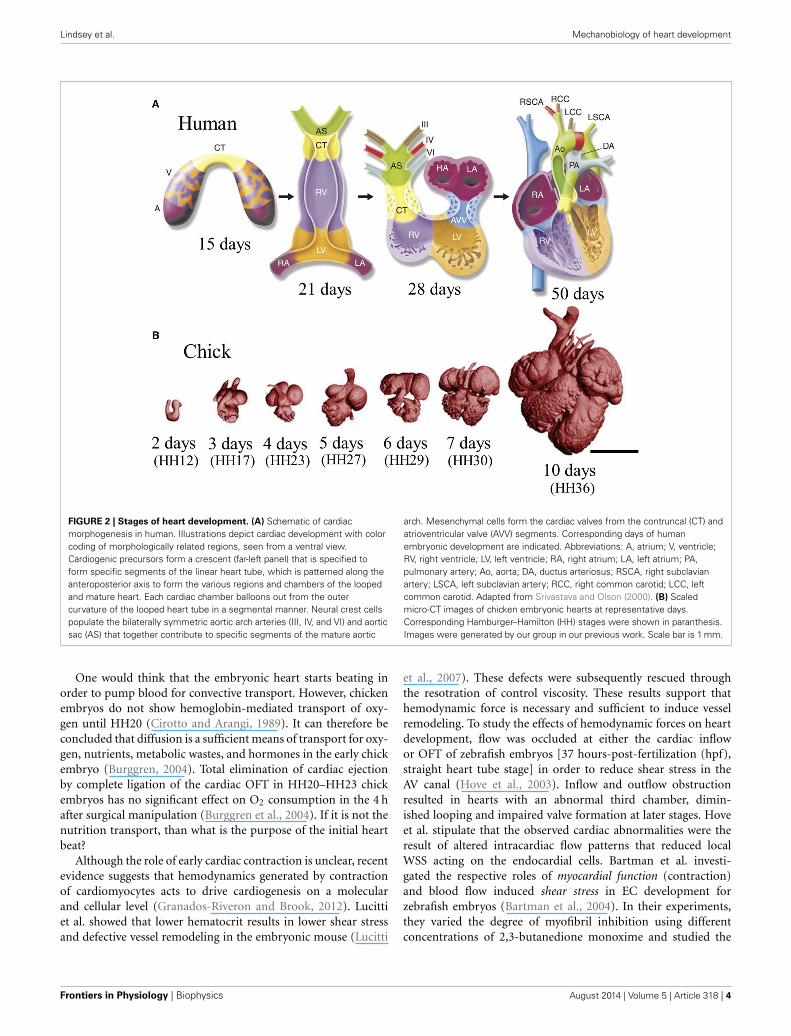
FIGURE 2 | Stages of heart development. (A) Schematic of cardiacmorphogenesis in human. Illustrations depict cardiac development with colorcoding of morphologically related regions, seen from a ventral view.Cardiogenic precursors form a crescent (far-left panel) that is specified toform specific segments of the linear heart tube, which is patterned along theanteroposterior axis to form the various regions and chambers of the loopedand mature heart. Each cardiac chamber balloons out from the outercurvature of the looped heart tube in a segmental manner. Neural crest cellspopulate the bilaterally symmetric aortic arch arteries (III, IV, and VI) and aorticsac (AS) that together contribute to specific segments of the mature aortic
arch. Mesenchymal cells form the cardiac valves from the contruncal (CT) andatrioventricular valve (AVV) segments. Corresponding days of humanembryonic development are indicated. Abbreviations: A, atrium; V, ventricle;RV, right ventricle; LV, left ventricle; RA, right atrium; LA, left atrium; PA,pulmonary artery; Ao, aorta; DA, ductus arteriosus; RSCA, right subclavianartery; LSCA, left subclavian artery; RCC, right common carotid; LCC, leftcommon carotid. Adapted from Srivastava and Olson (2000). (B) Scaledmicro-CT images of chicken embryonic hearts at representative days.Corresponding Hamburger–Hamilton (HH) stages were shown in paranthesis.Images were generated by our group in our previous work. Scale bar is 1 mm.
One would think that the embryonic heart starts beating inorder to pump blood for convective transport. However, chickenembryos do not show hemoglobin-mediated transport of oxy-gen until HH20 (Cirotto and Arangi, 1989). It can therefore beconcluded that diffusion is a sufficient means of transport for oxy-gen, nutrients, metabolic wastes, and hormones in the early chickembryo (Burggren, 2004). Total elimination of cardiac ejectionby complete ligation of the cardiac OFT in HH20–HH23 chickembryos has no significant effect on O2 consumption in the 4 hafter surgical manipulation (Burggren et al., 2004). If it is not thenutrition transport, than what is the purpose of the initial heartbeat?
Although the role of early cardiac contraction is unclear, recentevidence suggests that hemodynamics generated by contractionof cardiomyocytes acts to drive cardiogenesis on a molecularand cellular level (Granados-Riveron and Brook, 2012). Lucittiet al. showed that lower hematocrit results in lower shear stressand defective vessel remodeling in the embryonic mouse (Lucitti
et al., 2007). These defects were subsequently rescued throughthe resotration of control viscosity. These results support thathemodynamic force is necessary and sufficient to induce vesselremodeling. To study the effects of hemodynamic forces on heartdevelopment, flow was occluded at either the cardiac inflowor OFT of zebrafish embryos [37 hours-post-fertilization (hpf),straight heart tube stage] in order to reduce shear stress in theAV canal (Hove et al., 2003). Inflow and outflow obstructionresulted in hearts with an abnormal third chamber, dimin-ished looping and impaired valve formation at later stages. Hoveet al. stipulate that the observed cardiac abnormalities were theresult of altered intracardiac flow patterns that reduced localWSS acting on the endocardial cells. Bartman et al. investi-gated the respective roles of myocardial function (contraction)and blood flow induced shear stress in EC development forzebrafish embryos (Bartman et al., 2004). In their experiments,they varied the degree of myofibril inhibition using differentconcentrations of 2,3-butanedione monoxime and studied the
Frontiers in Physiology | Biophysics August 2014 | Volume 5 | Article 318 | 4
Lindsey et al. Mechanobiology of heart development
resulting effects on blood flow. Myocardial function decreasedin dose dependent manner with myofiber inhibition, and thepercentage of embryos that formed endocardial rings at 48 hpfalso decreased. Interestingly, at very high concentrations, blood
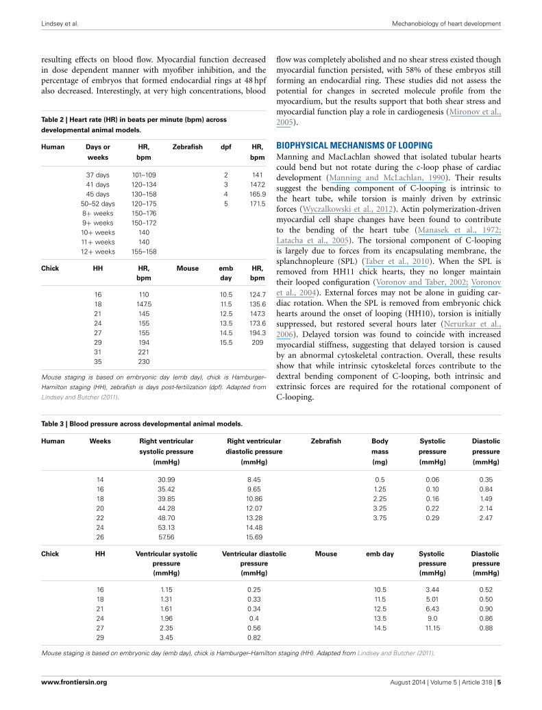
Table 2 | Heart rate (HR) in beats per minute (bpm) across
developmental animal models.
Human Days or HR, Zebrafish dpf HR,
weeks bpm bpm
37 days 101–109 2 14141 days 120–134 3 147.245 days 130–158 4 165.9
50–52 days 120–175 5 171.58+ weeks 150–1769+ weeks 150–17210+ weeks 14011+ weeks 14012+ weeks 155–158
Chick HH HR, Mouse emb HR,
bpm day bpm
16 110 10.5 124.718 147.5 11.5 135.621 145 12.5 147.324 155 13.5 173.627 155 14.5 194.329 194 15.5 20931 22135 230
Mouse staging is based on embryonic day (emb day), chick is Hamburger–
Hamilton staging (HH), zebrafish is days post-fertilization (dpf). Adapted from
Lindsey and Butcher (2011).
flow was completely abolished and no shear stress existed thoughmyocardial function persisted, with 58% of these embryos stillforming an endocardial ring. These studies did not assess thepotential for changes in secreted molecule profile from themyocardium, but the results support that both shear stress andmyocardial function play a role in cardiogenesis (Mironov et al.,2005).
BIOPHYSICAL MECHANISMS OF LOOPINGManning and MacLachlan showed that isolated tubular heartscould bend but not rotate during the c-loop phase of cardiacdevelopment (Manning and McLachlan, 1990). Their resultssuggest the bending component of C-looping is intrinsic tothe heart tube, while torsion is mainly driven by extrinsicforces (Wyczalkowski et al., 2012). Actin polymerization-drivenmyocardial cell shape changes have been found to contributeto the bending of the heart tube (Manasek et al., 1972;Latacha et al., 2005). The torsional component of C-loopingis largely due to forces from its encapsulating membrane, thesplanchnopleure (SPL) (Taber et al., 2010). When the SPL isremoved from HH11 chick hearts, they no longer maintaintheir looped configuration (Voronov and Taber, 2002; Voronovet al., 2004). External forces may not be alone in guiding car-diac rotation. When the SPL is removed from embryonic chickhearts around the onset of looping (HH10), torsion is initiallysuppressed, but restored several hours later (Nerurkar et al.,2006). Delayed torsion was found to coincide with increasedmyocardial stiffness, suggesting that delayed torsion is causedby an abnormal cytoskeletal contraction. Overall, these resultsshow that while intrinsic cytoskeletal forces contribute to thedextral bending component of C-looping, both intrinsic andextrinsic forces are required for the rotational component ofC-looping.
Table 3 | Blood pressure across developmental animal models.
Human Weeks Right ventricular Right ventricular Zebrafish Body Systolic Diastolic
systolic pressure diastolic pressure mass pressure pressure
(mmHg) (mmHg) (mg) (mmHg) (mmHg)
14 30.99 8.45 0.5 0.06 0.3516 35.42 9.65 1.25 0.10 0.8418 39.85 10.86 2.25 0.16 1.4920 44.28 12.07 3.25 0.22 2.1422 48.70 13.28 3.75 0.29 2.4724 53.13 14.4826 57.56 15.69
Chick HH Ventricular systolic Ventricular diastolic Mouse emb day Systolic Diastolic
pressure pressure pressure pressure
(mmHg) (mmHg) (mmHg) (mmHg)
16 1.15 0.25 10.5 3.44 0.5218 1.31 0.33 11.5 5.01 0.5021 1.61 0.34 12.5 6.43 0.9024 1.96 0.4 13.5 9.0 0.8627 2.35 0.56 14.5 11.15 0.8829 3.45 0.82
Mouse staging is based on embryonic day (emb day), chick is Hamburger–Hamilton staging (HH). Adapted from Lindsey and Butcher (2011).
www.frontiersin.org August 2014 | Volume 5 | Article 318 | 5
Lindsey et al. Mechanobiology of heart development
FOCE GENERATION OF THE TUBULAR HEARTIn the tubular heart, cyclic generation of traveling mechanicalwaves sweep from the venous to arterial end generating a uni-directional blood flow. These traveling mechanical waves weretraditionally considered myocardial peristaltic waves (Xavier-Neto et al., 2007) and tubular embryonic heart was acceptedto work like a technical roller peristaltic pump (Manner et al.,2010). Experiments on zebrafish embryos offer an alternativetheory (Forouhar et al., 2006): (I) While peak flow velocity gen-erated by a roller peristaltic pump corresponds to the speed of acompression wave, peak ventricular inflow velocity of an embry-onic zebrafish heart recorded exceeds the speed of a travelingcontraction wave. (II) In the zebrafish embryo, the relationshipbetween cardiac contraction frequency and the flow rate is non-linear and exceeds the maximum flow rate possible for a rollerperistaltic pump. (III) Roller peristaltic pumps are marked bynon-stationary sites of active compression that move in a uni-form direction along the length of a flexible tube whereas theearly embryonic zebrafish heart possesses a single stationary cen-ter of active myocardial contractions at its venous pole. Severalother studies support these findings. The tubular embryonic heartdoes not function as a technical roller peristaltic pump (Hu andClark, 1989; Butcher et al., 2007), but may be considered a valve-less “Liebau pump” (also known as suction or impedance pump)(Manner et al., 2010). Valveless pumping can be achieved exper-imentally by periodically compressing the asymmetric site of afluid filled tube made up of stiff and soft elastic sections (Liebau,1955, 1956; Ottesen, 2003; Hickerson et al., 2005). Here, peri-odic compression at an asymmetric site in the soft elastic regionleads to unidirectional flow. In order to be considered Liebaudriven flow, the tube must have a flexible wall and a finite lengthwith active compression stemming from a small asymmetric non-central section. Flow generated by Liebau pumps is typicallypulsatile (Manner et al., 2010).
Forouhar et al. identified a single site of active myocardialcontractions as well as a non-linear relationship between con-traction frequency and flow rate in the embryonic zebrafishheart (Forouhar et al., 2006). They concluded that tubularembryonic hearts work as Liebau pumps rather than peristalticpumps. Previous work by our group investigated pumpingmechanisms at various stages of embryonic chick develop-ment (Butcher et al., 2007). At HH17 blood velocity exceedsthat of tissue velocity, and wave propagation is initiated by asingle myocardial source, supporting the Liebau pump hypoth-esis. At HH25 the embryo utilizes a piston-pumping mecha-nism. Piston pumping is defined as a volume change–drivenpropulsion of fluid, with the orifice working to throttle theoutlet flow. HH21 embryos don’t fall into one pumping cate-gory, but rather act as a transition stage between two pumpingstyles. This same transition can be seen in the pumping mech-anism of zebrafish embryonic hearts at 36 hpf (Johnson et al.,2013).
Although the early embryonic heart does not function as atechnical roller peristaltic pump, it still incorporates peristalticmechanisms. Taber et al. showed that peristaltic heart tubes cangenerate pulsatile blood flow as a result of the presence of ECs atthe inflow and outflow of the ventricular loop (Taber et al., 2007).
Traveling contractile waves generate pressure and flow valuesan order of magnitude greater in tubes with valvular protru-sions. In their peristalsis model with ECs, flow velocities exceededthe contractile wave speed by 75%. These results highlight theimportance of ECs in pulsatile pump force generation.
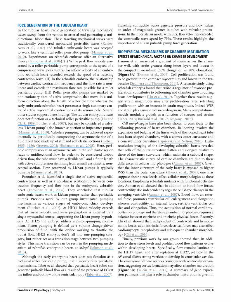
BIOPHYSICAL MECHANISMS OF CHAMBER MATURATIONEFFECTS OF MECHANICAL FACTORS ON CHAMBER DEVELOPMENTDamon et al. measured a gradient of strain across the cham-ber wall, with strain greatest along inner layers and lowest inthe compact myocardium (50% elongation vs. 20% elongation)(Figure 3A) (Damon et al., 2009). Cell proliferation was foundto be greatest in the compact myocardium and lowest in the tra-beculae (Sedmera and Thompson, 2011). A separate study usingzebrafish embryos found that erbb2, a regulator of myocyte pro-liferation, contributes to ballooning and chamber growth duringheart development (Liu et al., 2010). Together these results sug-gest strain magnitudes may alter proliferation rates, retardingproliferation with an increase in strain magnitude. Indeed WSSand strain play a major role in cardiogenesis. Many computationalmodels modulate growth as a function of stresses and strains(Taber, 2009; Buskohl et al., 2012b; Rugonyi, 2013).
Cell morphologies have also been shown to contribute to theballooning process of heart chambers. Ballooning involves theexpansion and bulging of the linear walls of the looped heart tubeinto bean-shaped chambers, with a convex outer curvature anda concave inner curvature (Gjorevski and Nelson, 2010). High-resolution imaging of the developing zebrafish hearts revealedthat cells of the outer curvature flatten and elongate relative tothose of the inner curvature, which maintain a cuboidal shape.The characteristic curves of cardiac chambers are due to thesedifferences in cellular morphologies (Auman et al., 2007). Giventhat the inner curvature of the early heart is exposed to higherWSS than the outer curvature (Hierck et al., 2008), one maysuppose shear stress levels affect cellular morphologies at theselocations. Employing zebrafish mutants with functional deficien-cies, Auman et al. showed that in addition to blood flow forces,contractility also independently regulate cell shape changes in theemerging ventricle (Auman et al., 2007). Blood flow, an exter-nal force, promotes ventricular cell enlargement and elongationwhereas contractility, an internal force, restricts ventricular cellsize and elongation. Thus, the acquisition of normal cardiomy-ocyte morphology and therefore chamber morphology, requires abalance between extrinsic and intrinsic physical forces. Recently,Chi et al. showed that, independent of contractile and hemody-namic forces, as an intrinsic force, electrical forces may also affectcardiomyocyte morphology and subsequent chamber morphol-ogy (Chi et al., 2010).
Finally, previous work by our group showed that, in addi-tion to shear stress levels and profiles, blood flow patterns evolvewithin developing hearts. Specifically, flow remains laminar inthe HH17 heart, and after septation at HH27, jet flow in theAV canal allows strong vortices to develop in ventricular cavities.The emergence of these vortices coincides with ventricular expan-sion, suggesting vortex formation may affect chamber maturation(Figure 3B) (Yalcin et al., 2011). A summary of gene expres-sion pathways that play a role in chamber maturation is given in
Frontiers in Physiology | Biophysics August 2014 | Volume 5 | Article 318 | 6

Lindsey et al. Mechanobiology of heart development
FIGURE 3 | Mechanical forces driving chamber development. (A) Acomputational model showing that inner trabecular layers undergo greaterstrain that outer compact layer in ventricular chamber. AV, atrioventricularcanal; V, ventricle; OT, outflow tract. Adapted from Damon et al. (2009).(B) Velocity streamlines in left AV canal for HH27 and for HH30 chickenembryos, generated with computational fluid dynamics. Strong vorticesdevelop in the ventricular cavity, possibly necessary for the expansion ofthe chambers. Adapted from Yalcin et al. (2011). (C) Signaling pathways inventricular chamber maturation. NOTCH, ERBB, and Ephrin play role intrabeculation whereas NOTCH, BMP, and FGF play role in compactmyocardium development. Adapted from Samsa et al. (2013). (D) Proposedventricular chamber development model. Circulatory flows cause expansionof the ventricles. Pressure induces a gradient in circumferential strain in thetrabeculae and compact myocardium. This gradient cause a difference inmyocyte proliferation as well as different gene expression profiles. Prepresents pressure, ω represents circulations, and ε representcircumferential strain.
Figure 3C. Figure 3D proposes a model of chamber developmentbased on the aforementioned studies.
CHAMBER MATURATIONIn an effort to understand the contribution of hemodynamicforces on the development of CHDs, many animal models andexperimental techniques have been developed. Two commonmodels of flow manipulation in the chick embryo are conotrun-cal banding (CTB) and left atrial ligation (LAL). In CTB, a knot istied around the conotruncus region of the OFT at HH18 in order
to induce an outflow flow constriction (Clark et al., 1984). In LAL,the left atria is partially ligated at HH20–21, obstructing flow tothe ventricle (Sedmera et al., 1999). CTB results in an immedi-ate increase in pressure and is therefore considered an increasedafterload model. LALs shift blood flow to the right side of theheart and are considered a model of increased right ventricularand decreased left ventricular preload (Sedmera et al., 1999).
Tobita et al. measured flow velocities in the AV canal andexamined wall deformation patterns in the right and left ventri-cle (RV and LV, respectively) following LAL (Tobita and Keller,2000). Decreases in max as well as average velocity were evidentimmediately following LAL. At HH31 (post-septation), averageRV inflow velocity was found to be higher in LAL embryos, whilemaximum LV inflow velocity was lower. These results suggest flowis redirected to the right side of the heart after LAL. Increasedright ventricular flow accelerated the onset of RV circumferentialstrain patterns, and decreased flow in LV abolished these patterns.Kowalski et al. investigated immediate alterations in blood flowpatterns and WSS levels following LAL through the use of a com-putational fluid dynamics (CFD) model (Kowalski et al., 2014).Intracardiac flow patterns are immediately altered following LAL,resulting in a decrease in WSS levels at the left AV canal as wellas at the left side of the common ventricle (Kowalski et al., 2014).WSS level decreased from 10 dynes/cm2 to 5 dynes/cm2 at theselocations for LAL embryos. Using optical coherence tomography,Rugonyi et al. studied immediate changes in blood flow velocitiesand wall motions in the OFT region of HH18 chick embryos fol-lowing CTB (Rugonyi et al., 2008). CTB resulted in an increasein the blood velocity in the banded part of the OFT due to flowconstriction at that region. Strain patterns revealed that mechan-ically altered (banded) embryos exerted more energy to maintaingrowth.
Sedmera et al. investigated morphological abnormalities dueto hemodynamic alterations resulting from LAL and CTB(Sedmera et al., 1999). Following CTB, a thickening and dila-tion of the compact myocardium and trabeculae in the LV wasobserved. LAL resulted in hypoplasia of the left heart structures,most likely due to decreased flow, with compensatory overde-velopment on the right side, most likely the result of increasedflow. LAL was shown to decrease myocyte proliferation rates in LV(Sedmera et al., 2002). DeAlmeida et al. also showed that partialclipping of the right atrial appendage increases blood flow to LVleading to an increase in chamber volume and myocardial massbased on myocyte proliferation in both LAL and normal embryos(deAlmeida et al., 2007). Similarly, addition of fibroblast growthfactor-2 (FGF2), a known stimulant of embryonic myocyte divi-sion was shown to increase cellular proliferation and abate thediseased phenotype (deAlmedia and Sedmera, 2009).
Effects of hemodynamic alterations from CTB and LAL on thedeveloping conduction system were investigated by Reckova et al.(2003). CTB resulted in premature emergence of the mature apex-to-base activation sequence, whereas LAL resulted in delayedtransition to a mature activation sequence. Biomechanics play acritical role in induction and patterning of the cardiac conduc-tion system. Sedmera et al. studied the effects of hemodynamicalterations on the expression of endothelin converting enzyme(ECE) protein, which is involved in the inductive recruitment of
www.frontiersin.org August 2014 | Volume 5 | Article 318 | 7

Lindsey et al. Mechanobiology of heart development
Purkinje fibers in embryonic chick conduction systems (Sedmeraet al., 2008). Following LAL, ECE expression was down-regulatedin LVs with decreased preload and up-regulated in RVs withincreased preload. Hearts cultured without hemodynamic load-ing showed decreased activation of the conduction pathway(Sankova et al., 2010). This primitive phenotype was rescued fol-lowing artificial loading of the ventricles via a droplet of siliconeoil. Together these findings suggest that loading is a necessarycomponent of conduction system formation and maturation.
HEART VALVE AND OUTFLOW TRACT DEVELOPMENTHEART VALVE MORPHOGENESISDuring cardiac looping, TBX3 (Moorman et al., 2004) andNotch1 (Luna-Zurita et al., 2010) act to localize myocardial-endocardial signals to the AV and OFT regions. In the first stageof this process, a subset of endocardial cells lining these twozones transform into a mesenchymal phenotype and invade thecardiac jelly, a process known as endocardial to mesenchymaltransformation (EMT) (Runyan and Markwald, 1983; Eisenbergand Markwald, 1995; Camenisch et al., 2002; Tavares et al., 2006;Butcher and Markwald, 2007; Combs and Yutzey, 2009). Theinvasive, proliferating mesenchyme progressively remodels thehyaluronan matrix, replacing it with proteoglycans, matricellu-lar proteins, and eventually structural proteins such as collagenI (Norris et al., 2004; Person et al., 2005). These amorphous,compliant, cellularized masses, now dubbed cushions, continueto grow and extend into the lumen space (Schroeder et al., 2003)
Two cushions (superior and inferior) form initially in the AVcanal at HH16 (E9.5 in mouse), followed by the appearance oftwo mural/lateral cushions on the left and right side of the AVcanal at HH19 (Snarr et al., 2008). The superior and inferiorAV cushions fuse together by HH26 (E12 in mouse) forming aseptation of the AV canal that joins with the ventricular septumand the protruding atrial cap. The lateral portions of this fusedmass undergo continual remodeling to form valves, as do the leftand right mural cushions. During the remodeling of primitive AVvalves into fibrous leaflets, the AV myocardium forms a fold atits junction with the ventricular myocardium creating a substrateon which the AV cushions can extend. The cushions extend alongtheir substrate through the expansion of a proliferation zone inthe subepithelial portion of the AV cushions (Sugi et al., 2003).Fenestrations develop as a result of the elongating cushions andthe ventricular tissue underneath the cushion tissue delaminates,resulting in primitive leaflets that are continuous with developingpapillary muscles and simultaneous expansion of the ventricularOFT (Wenink and Gittenberger-de Groot, 1986). The myocar-dial tissue of the AV valves disappears and they condense intofibrous leaflets (de Lange et al., 2004). Thin strands of elongatedmuscle remain tethered to the valve tissue with thickened trabec-ular aspects on the ventricular myocardial wall. These structuresbecome the tendinous chords and papillary muscles of the maturevalve (Icardo and Colvee, 1995). Epicardially derived fibroblastshave also been shown to contribute to the mural AV leaflets indeveloping murine hearts (Wessels et al., 2012).
The OFT differs in that endocardial cells along the entirelumen undergo EMT. Paired bulges emanating in proximal (justoutside the right ventricle) and distal zones (just after a “dogleg”
bend in the OFT) become cushions around HH22/E10, while therest of the cardiac jelly regresses. The proximal/conal cushionsare alternatively referred to as the septal/sinistroventral and pari-etal/dorsodextral ridges (Ya et al., 1998; Qayyum et al., 2001).A third distal cushion ridge subsequently forms (HH25/E11).These growing cushions fuse at the midline, forming two tortuouslumens. Between HH26/E11.5 and HH30/E13, the distal dorsalcushion of the OFT aligns with the proximal left cushion alongthe inner heart curvature, continuous with the superior cushionof the AV canal. Simultaneously, a wishbone shaped ridge of mes-enchyme invades the OFT in a spiraling pattern, separating it intoleft and right portions and dividing the outflow cushions into twogroups of three. While the fused proximal cushions myocardializeand form the muscular infundibulum, which separates the rightand left ventricular outlets, the distal cushions become the rudi-ments of the pulmonary and aortic outlet valves (Qayyum et al.,2001). For the remodeling of primitive outflow valves into fibrousleaflets, unlike the AV valves, which formed through delamina-tion from the muscular walls, valves of the OFT form through aprocess of excavation or hollowing of the cushion’s aortic side.Cushion excavation begins at HH29/ED13 with a small depres-sion in the arterial face of the cusps. The endothelium lining theaortic surface of the valves becomes thickened with rounded cellsthat flake and undergo apoptosis, while the ventricular epithe-lium remains flat and elongated (Garcia-Martinez et al., 1991).The deepening furrow condenses the fibrous matrix around it,creating thin cusps of tissue that are attached in an arc patterncalled the commissures (Butcher and Markwald, 2007).
HEART VALVE HEMODYNAMICSBlood flow guides cardiac morphogenesis, sculpting tissue by pro-moting growth in response to increased demands. Less than 48 hafter incubation, the presence of two blood streams is apparentin the embryonic chicken heart. A spiraling complex is created,as the force of the larger stream pulls the smaller stream aroundit, changing the mechanical environment of the developing heart(Jaffee, 1965). The rapid growth of the endothelial tube inside theearly heart is a result of an increase in blood pressure (Chang,1932). In this way blood flow, shear stress and stretching forcesare thought to influence the duration of vessel growth and theirmorphological characteristics (Taber and Eggers, 1996). Duringthis time of heart formation, shear stress is greatest in the innercurvature and sites of lumen constrictions, corresponding to theAV canal and OFT where the ECs form and develop into func-tioning valves (Groenendijk et al., 2004). The velocity profileof flow through the primitive valves begins with a Poiseuilleparabola (HH17), before resembling plug flow; cushions ariseshortly after peak inflow velocity and extend perpendicularly tothe direction of flow (Figures 4A,B, left) (Yalcin et al., 2011;Bharadwaj et al., 2012). As the heart continues to grow, the energyextended in pulsatile flow increases from one-third to two-thirdsof total energy between HH18 and 29 (Clark et al., 1986). Ajet profile characterizes the AV region of HH27 chick embryos(Figure 4A, right). Figure 4A, right, highlights circulating eddies,which form beneath the cushion surface. WSS levels increase from80 dynes/cm2 at HH23 to 250 dynes/cm2 at HH27 (Figure 4B).As the embryo’s pressure, cardiac output and WSS increase, the
Frontiers in Physiology | Biophysics August 2014 | Volume 5 | Article 318 | 8

Lindsey et al. Mechanobiology of heart development
FIGURE 4 | Mechanical forces driving AV valve morphogenesis. (A)
Velocity streamlines in AV canal for HH23 (unseptated) and for HH27(septated) chicken embryos, generated with computational fluid dynamics. InHH23, flow profile is laminar parallel streamlines, whereas in HH27 it is jetflow with circulating eddies beneath AV cushions (shown with arrrow). LA,left atria; RA, right atria; LV, left ventricle; RV, right ventricle; AVC,atrioventricular cushion (B) WSS levels for HH23 and HH27 embryos. Highstress levels are localized to the mid cushion region. Adapted form Yalcinet al. (2011). (C) Hemodynamic-driven AV cushion growth and remodelingcomputational model. Based on this model, the pressure distribution on the
AV cushion cause leaflet-like elongation in the direction of flow, whereasshear tractions regulated the remodeling of tissue near the cushion surfacesuggesting WSS is important in mechanotransduction. Adapted from Buskohlet al. (2012b). (D) Proposed AV valve morphogenesis model. WSS on cushionsurface regulate gene expression for valve development. Pressure on thecushion surface cause leaflet-like elongation and circulatory flows beneaththe cushion helps detachment of the leaflet from the myocardial wall. Growthfactor expression levels were indicated via color gradation as shown withcolored triangles. Adapted from Chiu et al. (2010). τ represents shear stress,P represents pressure, and ω represents circulations.
cushions elongate to form thin fibrous leaflets with increasedECM proteins and greater mechanical stiffness (Butcher et al.,2007; Biechler et al., 2010; Buskohl et al., 2012a). Strain energydensity increases linearly with AV valve leaflet length (Buskohlet al., 2012a).
While the importance of biomechanics in the formation of thevalve leaflets has been acknowledged for some time, only recentlyhave individual roles for pressure and WSS been proposed.Buskohl et al. created a computational model in which a finite ele-ment model was coupled to a fluid dynamics model, highlighting
www.frontiersin.org August 2014 | Volume 5 | Article 318 | 9

Lindsey et al. Mechanobiology of heart development
interaction between the two (Figure 4C) (Buskohl et al., 2012b).Buskhol’s iterative computational approach involved determiningpressure and velocity profiles, transferring flow-induced pressureand shear tractions on the AV cushion surface, and updatingthe fluid simulations after inelastic deformation had taken place.Fluid shear tractions were found not to significantly alter cush-ion volume, but rather functioned as the driver for cushionsurface remodeling. While pressure was found to be negligiblein terms of tissue deformation, shear traction forces from thetop-center cushion region greatly contributed to tissue elonga-tion. Pressure was responsible for tissue resorption on the inflowside of AV cushions and expansion on the outflow side (Buskohlet al., 2012b). Regulation of the cushion surface, particularlythe leading edge of valve leaflets, may be heavily influenced bymechanotransduction.
Altered hemodynamic flow patterns during critical periodsof development have been shown to lead to a variety of car-diac abnormalities, many of which influence valve formation(Hogers et al., 1997; Sedmera et al., 1999; Hove et al., 2003).Maintenance of circulatory energy efficiency and pressure are crit-ical for development (Lucitti et al., 2005). Sedmera et al. foundthat alterations in ventricular load influenced AV valve morpho-genesis as well as trabeculation patterns (Sedmera et al., 1999).In embryos with increased ventricular afterload (CTB), right AVvalve morphology no longer took the form of a muscular flapbut rather resembled a bicuspid structure. Previous work by ourgroup showed that photoablation of the superior AV cushions ofHH24 chick embryos immediately altered AV canal hemodynam-ics (i.e., increased regurgitation), resulting in stunted ventricularand valvular growth in 48 h (Yalcin et al., 2010). Shear stressand shear stress-induced or repressed gene expression are impor-tant factors in remodeling of the cardiac cushions (Hove et al.,2003; Groenendijk et al., 2005; Vermot et al., 2009). Hove et al.attributed abnormal third chamber development in mechanicallyperturbed zebrafish embryos to a reduction in shear stress onECs (Hove et al., 2003). Bartman et al. argue observed pheno-types were not the result of a decrease in shear alone. Their 2004study found that 58% of zebrafish embryos treated with a myofib-ril inhibitor (2,3-butanedione monoxime) were still able to forman endocardial ring, affirming that blood flow is not required forthe initial steps of cushion formation (Bartman et al., 2004).
HEMODYNAMICS AND GENE EXPRESSION FOR VALVE DEVELOPMENTWhile hemodynamics undoubtedly plays a large role in valvulo-genesis, the role of hemodynamic signaling remains a point ofcontention. Though changes in shear stress have been found topresage the development of cardiac malformations (Hogers et al.,1997, 1999; Hove et al., 2003; Yashiro et al., 2007), other studiesclaim myocardial function surpasses shear stress as a major epi-genetic factor (Bartman et al., 2004). Endocardial cells lining theluminal cushion surface may promote valvular morphogenesis bycoupling mechanical stimuli and molecular signaling pathways(Butcher et al., 2007).
A number of studies have demonstrated the shear sensitivity ofvalvular morphogens in vivo. Endothelin-1 (ET-1) and endothe-lial nitric oxide synthase (NOS-3) are shear stress responsive geneslinked to cardiovascular development by knockout mice that
display a wide range of cardiovascular defects (Yanagisawa et al.,1998). Critical periods of cardiovascular remodeling (HH20–HH30) were marked by ET-1 and KLF2/NOS-3 restriction tonarrow sites (Groenendijk et al., 2004). ET-1 was downregulatedby shear stress, while KLF2 and NOS-3 were upregulated by shear.Observed patterns changed with vitelline vein obstruction andcardiovascular malformations ensued (Groenendijk et al., 2005).In addition to the aforementioned genes, the expression patternsof potent valvular morphogens, such as transforming growth fac-tor β (TGFβ), bone morphogenetic protein (BMP), and vascularendothelial growth factor (VEGF) are spatially and temporallyrestricted in a manner that suggests hemodynamic regulation(Figure 4D) (Butcher and Nerem, 2007; Chiu et al., 2010). TGFβ
appears in the endocardium of the valve forming regions aroundHH20, when flow transitions from its laminar poiseuille flowto more plug-like flow and rapidly increases in velocity (Yalcinet al., 2011). In vitro analyses suggest that TGFβ3 induces cellmigration, invasion, and matrix condensation; BMP2 inducesinvasion; VEGFA inhibits invasion but increases migration (Tanet al., 2011). TGFβ3 was found to induce myofibroblastic differen-tiation in a dose-dependent manner, whereas VEGFA and BMP2did not, suggesting that hemodynamic forces work through thesetranscription factors in the remodeling of the AV valves. Using anin vitro system, Tan et al. were able to show the flow-regulateddevelopment of the fibrous AV valves is dependent on rhoAexpression (Tan et al., 2013). This body of work is depicted inFigure 4D.
THE ROLE OF HEMODYNAMICS IN FORMATION OF IN THE GREATVESSELSThe great vessels are derived from the pharyngeal arch arter-ies. A total of six arch pairs sequentially emerge, regress, orremodel throughout OFT development. While arch arteries I andII ultimately regress, the third arch pair forms the mature bra-chiocephalic arteries. The mature aortic arch is derived from theright fourth arch in chicken and the left fourth arch in mam-malian embryos (Wang et al., 2009). The sixth arch pair formspulmonary artery and ductus arteriosus. The formation of thetruncus arteriosus separates the pulmonary artery and right ven-tricular OFT from the aorta and left ventricular OFT posteriorly(Qayyum et al., 2001; Hu et al., 2009).
Hemodynamic forces initiate extensive remodeling of the sym-metric aortic arch system in a highly asymmetric fashion (Huet al., 2009). Yashiro et al. investigated the mechanisms behindregression of the right VI aortic arch and persistence of left VIarch in mice (Yashiro et al., 2007). Using a variety of experi-mental and genetic mutant models, they found that, the geneticprogramme, including the expression of Pitx2, induces a dynamicmorphological change in the OFT, which in turn generates a dif-ferential distribution of blood flow. Mice lacking the asymmetricenhancer PITX2 failed to adopt the spiral structure of the OFTand in these embryos OFT remained linear. Changes in OFTgeometry led to differential flow distribution, successive signal-ing abnormalities and further changes in geometry. In normalembryos, increased blood flow in the left VI arch sufficientlyinduces PDGFR and VEGFR2 signaling and subsequently main-tains arterial structure. Decreased flow in the right VI arch artery
Frontiers in Physiology | Biophysics August 2014 | Volume 5 | Article 318 | 10

Lindsey et al. Mechanobiology of heart development
leads to vessel regression. Wang et al. discovered that increases inWSS corresponded with increases in arch artery diameters in day3 (HH18) and day 4 (HH24) chick embryos (Wang et al., 2009).CFD analysis revealed that HH21 embryos had elevated, ratherthan intermediate WSS compared to both HH18 and HH24embryos (Kowalski et al., 2013) suggesting periods of vascularremodeling may be preceded by acute increases in WSS. Hu et al.showed that altered hemodynamic flow in the aortic arch arter-ies due to LAL was associated with a range of cardiac defects (Huet al., 2009). Overall, these studies suggest hemodynamic forcesplay a significant role in asymmetric remodeling of aortic archnetwork. Characterization of aberrant hemodynamic flow may bea resource in understanding OFT and arch network abnormali-ties, which account for 50% of infants with a CHD (Roger et al.,2011).
HEMODYNAMICS AND CONGENITAL HEART DEFECTSThe embryonic heart adapts ventricular geometry and func-tion to optimize mechanical efficiency (Lin and Taber, 1995).Distinctions between gene and hemodynamic-related abnormali-ties are not well defined. Though chromosomes linked to specificdefects have been identified, they rarely provide a full picture,with only 10–15 percent of ventricular outflow malformationsloosely associated to a chromosomal abnormality (McBride et al.,2009). While genetic mutations are associated with CHDs, genescannot be manipulated to produce CHDs in the same way as flowcan be perturbed to produce disease phenotypes. The use of com-putational modeling in the study of abnormal development hasled to a more thorough analysis of disease etiologies and theirunderlying mechanisms. In this section, the pathology of twomajor CHDs associated with abnormal hemodynamic patteringand valve formation are explored: bicuspid aortic valve (BAV) andhypoplastic left heart syndrome (HLHS).
BAV is the most common congenital anomaly of the heart,marked by two aortic valve leaflets rather than three. This twocusped configuration constrains the patient, as the free edges ofthe bicuspid valve are more straight than round and offer limitedmobility. The leaflets are usually of unequal size with a raphe, orseam-like union, apparent in the larger leaflet (Yener et al., 2002).BAV is frequently associated with aortic valve stenosis, regurgita-tion and endocarditis, though these symptoms develop well aftervalve formation. Excessive length of one or both cusps results inabnormal contact which in turn leads to fibrous thickening thatwill later become diffuse and calcified. Stenosis usually developsin bicuspid valves containing no redundant cusp tissue, whilevalve incompetence is associated with redundancy and endocardi-tis. The large calcific deposits associated with BAV are unusualbefore the age of 30 and very prevalent thereafter (Roberts, 1970).In a 2003 study of 44 bicuspid aortic valves, BAV patients withoutsignificant stenosis or regurgitation were found to have a largeraortic annulus, aortic sinus and proximal ascending aorta whencompared to normal tricuspid valves. The peak aortic velocity(Nkomo et al., 2003) and peak systolic wall velocity in the antero-lateral region of the ascending aorta (Bauer et al., 2006) were alsofound to be higher in BAV patients than controls. This flow hasbeen classified as helical, or flow composed of a forward compo-nent along the long axis of the aorta and a rotational component,
moving circumferentially along that same axis (Bissell et al., 2013;Lorenz et al., 2014). A significant increase in absolute peak helic-ity is present during systole of BAV patients, with a substantiallygreater distribution of mean helicity in the aorta (Lorenz et al.,2014).
Recent advances in cardiac imaging, have allowed scientists tomap these regions and collect 3D spatial visualizations of flowpatterns over time (Bissell et al., 2013; Lorenz et al., 2014). Inthis way, temporal evolution of complex flow patterns over timecan be studied and linked to aortic function. Bissell et al. foundthat patients with BAV presented with predominantly abnormalright-handed helical flow in the ascending aorta, larger ascend-ing aortas, higher helical flow, elevated systolic angle and elevatedsystolic WSS. In their study of 69 BAV patients, left-handed heli-cal flow, normal flow, and complex flow occurred in 4, 11, and13%, respectively, whereas right-handed helical flow occurredin 72% of patients. Although flow patterns differed, distensi-bility, aortic strain, and pulse wave velocity of the aorta weresimilar across all groups. Flow abnormalities initiate aortopathyas a means of maintaining optimal WSS values (Bissell et al.,2013). Through the use of fluid structure interaction models,(Chandra et al., 2012) degree of leaflet calcification is linked toorifice area, oscillatory shear index and temporal shear magni-tude. While the regular tricuspid and non-coronary BAV leafletsshared similar shear stress characteristics, the base of fused BAVleaflet fibrosa differed greatly. The temporal shear index of fusedleaflets was heavily modulated by degree of calcification, with6- to 16-fold increases seen over BAVs ranging from normal toseverely calcified. Results support a mechano-sensored model ofcalcified aortic valve disease in the BAV patients (Chandra et al.,2012).
HLHS is characterized by acute underdevelopment of the leftventricle. No strong genetic correlation exists. In a study of 83HLHS patients, nine had underlying chromosomal abnormali-ties, four had single gene defects, 10 had one or more extracardiacanomaly and two were patients of insulin-dependent moth-ers (Natowicz et al., 1988). Disease formation is thought toresult from diminished flow to the left ventricle and aortic OFT.Retrograde aortic flow may play a role in impaired developmentof the aortic root and ascending aorta (Simpson and Sharland,1997). Cardiac defects associated with HLHS include mitral valvehypoplasia or mitral stenosis coincident with left heart obstruc-tion, hypoplastic left ventricle, aortic atresia, hypoplastic aorta,and coarctation of the aorta. Out of 96 HLHS patients, 12.5 per-cent exhibited dysplastic aortic valvular stenosis, 37.5 percentwere found to have malfunctioning aortic and mitral valves, 50percent presented with abnormal AV valves (Ilbawi et al., 2007).To date, the LAL remains the only experimental model to fullyrecapulate these disease phenotypes (Sedmera et al., 1999, 2002;Tobita et al., 2002). Changes in WSS as shown by Kowalski et al.may be responsible for underdevelopment of left heart structures(Kowalski et al., 2013). Variations in heart rate and AV inflowvelocity were acute to non-existent in these models (Tobita et al.,2002). Mechanical manipulation of ventricular filling dates backto Harh et al. (1973). Harh et al. inserted a nylon device intothe left AV canal, thereby reducing ejection volume from theventricle to ascending aorta (Harh et al., 1973). Failure of the
www.frontiersin.org August 2014 | Volume 5 | Article 318 | 11

Lindsey et al. Mechanobiology of heart development
cushions to differentiate into fibrous leaflets led to hypoplasia.Narrowing resulted in a reversed atrial shunt. Though Harh et al.observed disease phenotypes, they were unable to fully charac-terize mechanical changes. Combining experimental results andcomputational models presents a promising way of understandthe mechanisms through which changes may arise.
CONCLUSIONS AND FUTURE DIRECTIONSMechanical forces are essential drivers of cardiac morphogene-sis, transforming the linear heart tube into a multichamberedunidirectional machine capable of adapting to environmentaldemands. CHDs arise when the heart is prevented from followingthe normal pathways of development; understanding the intricatemechanisms involved in heart development is necessary for theadvancement of clinical solutions. The majority of CHDs resultfrom improper positioning of the cardiac OFT, impaired remod-eling of the cushions into valve leaflets, or abnormal remodelingof the arches into great vessels. Quantitative imaging modalitiesprovide a gateway into elucidating CHD formation. Integratingthese imaging modalities with CFD and growth modeling willgreatly strengthen our inference of causative mechanical forcesof CHDs, such as BAV and HLHS. Targeted surgical techniques,including laser ablation, can help scientists distinguish betweenthe effects of flow and tissue deformation alone. In vitro 3D cul-ture and bioreactor studies work to further our understandingof how specific mechanical forces influence multi-scale biologi-cal responses. As our insight into CHD etiology improves, so willour ability to effectively and targetedly restore proper remodelingof cardiac tissues.
ACKNOWLEDGMENTSThis research was supported by funding from the AmericanHeart Association, the National Science Foundation, the LeDucqFoundation, and The Hartwell Foundation as well as by a MarieCurie International Reintegration Grant within the 7th EuropeanCommunity Framework Programme, (IRG276987 to HuseyinC. Yalcin), and by The Scientific and Technological ResearchCouncil of Turkey, TUBITAK (112M148 to Huseyin C. Yalcin and112M895 to Huseyin C. Yalcin).
REFERENCESAleksandrova, A., Czirok, A., Szabo, A., Filla, M. B., Hossain, M. J., Whelan,
P. F., et al. (2012). Convective tissue movements play a major role in avianendocardial morphogenesis. Dev. Biol. 363, 348–361. doi: 10.1016/j.ydbio.2011.12.036
Auman, H. J., Coleman, H., Riley, H. E., Olale, F., Tsai, H. J., and Yelon, D. (2007).Functional modulation of cardiac form through regionally confined cell shapechanges. PLoS Biol. 5:e53. doi: 10.1371/journal.pbio.0050053
Bartman, T., and Hove, J. (2005). Mechanics and function in heart morphogenesis.Dev. Dyn. 233, 373–381. doi: 10.1002/dvdy.20367
Bartman, T., Walsh, E. C., Wen, K. K., McKane, M., Ren, J., Alexander, J., et al.(2004). Early myocardial function affects endocardial cushion development inzebrafish. PLoS Biol. 2:E129. doi: 10.1371/journal.pbio.0020129
Bauer, M., Siniawski, H., Pasic, M., Schaumann, B., and Hetzer, R. (2006). Differenthemodynamic stress of the ascending aorta wall in patients with bicuspidand tricuspid aortic valve. J. Card. Surg. 21, 218–220. doi: 10.1111/j.1540-8191.2006.00219.x
Bharadwaj, K. N., Spitz, C., Shekhar, A., Yalcin, H. C., and Butcher, J. T. (2012).Computational fluid dynamics of developing avian outflow tract heart valves.Ann. Biomed. Eng. 40, 2212–2227. doi: 10.1007/s10439-012-0574-8
Biechler, S. V., Potts, J. D., Yost, M. J., Junor, L., Goodwin, R. L., and Weidner,J. W. (2010). Mathematical modeling of flow-generated forces in an in vitrosystem of cardiac valve development. Ann. Biomed. Eng. 38, 109–117. doi:10.1007/s10439-009-9824-9
Bissell, M. M., Hess, A. T., Biasiolli, L., Glaze, S. J., Loudon, M., Pitcher, A., et al.(2013). Aortic dilation in bicuspid aortic valve disease: flow pattern is a majorcontributor and differs with valve fusion type. Circ. Cardiovasc. Imaging 6,499–507. doi: 10.1161/CIRCIMAGING.113.000528
Bruneau, B. G. (2008). The developmental genetics of congenital heart disease.Nature 451, 943–948. doi: 10.1038/nature06801
Burggren, W., Khorrami, S., Pinder, A., and Sun, T. (2004). Body, eye, andchorioallantoic vessel growth are not dependent on cardiac output level inday 3-4 chicken embryos. Am. J. Physiol. Regul. Integr. Comp. Physiol. 287,R1399–R1406. doi: 10.1152/ajpregu.00086.2004
Burggren, W. W. (2004). What is the purpose of the embryonic heart beat? or howfacts can ultimately prevail over physiological dogma. Physiol. Biochem. Zool.77, 333–345. doi: 10.1086/422230
Buskohl, P. R., Gould, R. A., and Butcher, J. T. (2012a). Quantification of embry-onic atrioventricular valve biomechanics during morphogenesis. J. Biomech. 45,895–902. doi: 10.1016/j.jbiomech.2011.11.032
Buskohl, P. R., Jenkins, J. T., and Butcher, J. T. (2012b). Computational simulationof hemodynamic-driven growth and remodeling of embryonic atrioventricularvalves. Biomech. Model. Mechanobiol. 11, 1205–1217. doi: 10.1007/s10237-012-0424-5
Butcher, J. T., and Markwald, R. R. (2007). Valvulogenesis: the movingtarget. Philos. Trans. R. Soc. Lond. B Biol. Sci. 362, 1489–1503. doi:10.1098/rstb.2007.2130
Butcher, J. T., McQuinn, T. C., Sedmera, D., Turner, D., and Markwald, R.R. (2007). Transitions in early embryonic atrioventricular valvular functioncorrespond with changes in cushion biomechanics that are predictable by tis-sue composition. Circ. Res. 100, 1503–1511. doi: 10.1161/CIRCRESAHA.107.148684
Butcher, J. T., and Nerem, R. M. (2007). Valvular endothelial cells and themechanoregulation of valvular pathology. Philos. Trans. R. Soc. Lond. B Biol.Sci. 362, 1445–1457. doi: 10.1098/rstb.2007.2127
Buxboim, A., Ivanovska, I. L., and Discher, D. E. (2010). Matrix elasticity, cytoskele-tal forces and physics of the nucleus: how deeply do cells “feel” outside and in?J. Cell Sci. 123, 297–308. doi: 10.1242/jcs.041186
Camenisch, T. D., Molin, D. G. M., Person, A., Runyan, R. B., Gittenberger-deGroot, A. C., McDonald, J. A., et al. (2002). Temporal and distinct TGFβ lig-and requirements during mouse and avian endocardial cushion morphogenesis.Dev. Biol. 248, 170–181. doi: 10.1006/dbio.2002.0731
Chandra, S., Rajamannan, N. M., and Sucosky, P. (2012). Computationalassessment of bicuspid aortic valve wall-shear stress: implications for cal-cific aortic valve disease. Biomech. Model. Mechanobiol. 11, 1085–1096. doi:10.1007/s10237-012-0375-x
Chang, C. (1932). On the reaction of the endocardium to the blood stream inthe embryonic heart, with special reference to the endocardial thickenings inthe atrioventricular canal and the bulbus cordis. Anat. Rec. 51, 253–265. doi:10.1002/ar.1090510305
Chi, N. C., Bussen, M., Brand-Arzamendi, K., Ding, C., Olgin, J. E., Shaw,R. M., et al. (2010). Cardiac conduction is required to preserve cardiacchamber morphology. Proc. Natl. Acad. Sci. U.S.A. 107, 14662–14667. doi:10.1073/pnas.0909432107
Chiu, Y. N., Norris, R. A., Mahler, G., Recknagel, A., and Butcher, J. T. (2010).Transforming growth factor beta, bone morphogenetic protein, and vascularendothelial growth factor mediate phenotype maturation and tissue remodelingby embryonic valve progenitor cells: relevance for heart valve tissue engineering.Tissue Eng. Part A 16, 3375–3383. doi: 10.1089/ten.tea.2010.0027
Chuck, E. T., Freeman, D. M., Watanabe, M., and Rosenbaum, D. S. (1997).Changing activation sequence in the embryonic chick heart. implicationsfor the development of the his-purkinje system. Circ. Res. 81, 470–476. doi:10.1161/01.RES.81.4.470
Cirotto, C., and Arangi, I. (1989). How do avian embryos breathe? oxygen transportin the blood of early chick embryos. Comp. Biochem. Physiol. A Comp. Physiol.94, 607–613.
Clark, E. B., Hu, N., Dummett, J. L., Vandekieft, G. K., Olson, C., and Tomanek, R.(1986). Ventricular function and morphology in chick embryo from stages 18to 29. Am. J. Physiol. 250, H407–H413.
Frontiers in Physiology | Biophysics August 2014 | Volume 5 | Article 318 | 12

Lindsey et al. Mechanobiology of heart development
Clark, E. B., Hu, N., and Rosenquist, G. C. (1984). Effect of conotrun-cal constriction on aortic-mitral valve continuity in the stage 18, 21 and24 chick embryo. Am. J. Cardiol. 53, 324–327. doi: 10.1016/0002-9149(84)90447-8
Combs, M. D., and Yutzey, K. E. (2009). Heart valve development: regula-tory networks in development and disease. Circ. Res. 105, 408–421. doi:10.1161/CIRCRESAHA.109.201566
Damon, B. J., Remond, M. C., Bigelow, M. R., Trusk, T. C., Xie, W., Perucchio, R.,et al. (2009). Patterns of muscular strain in the embryonic heart wall. Dev. Dyn.238, 1535–1546. doi: 10.1002/dvdy.21958
deAlmeida, A., McQuinn, T., and Sedmera, D. (2007). Increased ventric-ular preload is compensated by myocyte proliferation in normal andhypoplastic fetal chick left ventricle. Circ. Res. 100, 1363–1370. doi:10.1161/01.RES.0000266606.88463.cb
deAlmedia, A., and Sedmera, D. (2009). Fibroblast growth factor-2 regulates pro-liferation of cardiac myocytes in normal and hypoplastic left ventricles inthe developing chick. Cardiol. Young 19, 159–169. doi: 10.1017/S1047951109003552
de Lange, F. J., Moorman, A. F., Anderson, R. H., Manner, J., Soufan, A. T., de Gier-de Vries, C., et al. (2004). Lineage and morphogenetic analysis of the cardiacvalves. Circ. Res. 95, 645–654. doi: 10.1161/01.RES.0000141429.13560.cb
Dyer, L. A., and Kirby, M. L. (2009). The role of secondary heart field in cardiacdevelopment. Dev. Biol. 336, 137–144. doi: 10.1016/j.ydbio.2009.10.009
Eisenberg, L. M., and Markwald, R. R. (1995). Molecular regulation of atrioventric-ular valvuloseptal morphogenesis. Circ. Res. 77, 1–6. doi: 10.1161/01.RES.77.1.1
Engler, A. J., Carag-Krieger, C., Johnson, C. P., Raab, M., Tang, H. Y., Speicher, D.W., et al. (2008). Embryonic cardiomyocytes beat best on a matrix with heart-like elasticity: scar-like rigidity inhibits beating. J. Cell Sci. 121, 3794–3802. doi:10.1242/jcs.029678
Ettensohn, C. A. (1985). Mechanisms of epithelial invagination. Q. Rev. Biol. 60,289–307. doi: 10.1086/414426
Fahed, A. C., Gelb, B. D., Seidman, J. G., and Seidman, C. E. (2013). Geneticsof congenital heart disease: the glass half empty. Circ. Res. 112, 707–720. doi:10.1161/CIRCRESAHA.112.300853
Forouhar, A. S., Liebling, M., Hickerson, A., Nasiraei-Moghaddam, A., Tsai, H. J.,Hove, J. R., et al. (2006). The embryonic vertebrate heart tube is a dynamicsuction pump. Science 312, 751–753. doi: 10.1126/science.1123775
Garcia-Martinez, V., Sanchez-Quintana, D., and Hurle, J. M. (1991). Histochemicaland ultrastructural changes in the extracellular matrix of the developing chicksemilunar heart valves. Acta Anat. 142, 87–96. doi: 10.1159/000147166
Gjorevski, N., and Nelson, C. M. (2010). The mechanics of development: modelsand methods for tissue morphogenesis. Birth Defects Res. C Embryo Today 90,193–202. doi: 10.1002/bdrc.20185
Goenezen, S., Rennie, M. Y., and Rugonyi, S. (2012). Biomechanics of earlycardiac development. Biomech. Model. Mechanobiol. 11, 1187–1204. doi:10.1007/s10237-012-0414-7
Gourdie, R. G., Harris, B. S., Bond, J., Justus, C., Hewett, K. W., O’Brien, T. X., et al.(2003). Development of the cardiac pacemaking and conduction system. BirthDefects Res. C Embryo Today 69, 46–57. doi: 10.1002/bdrc.10008
Granados-Riveron, J. T., and Brook, J. D. (2012). The impact of mechani-cal forces in heart morphogenesis. Circ. Cardiovasc. Genet. 5, 132–142. doi:10.1161/CIRCGENETICS.111.961086
Groenendijk, B. C., Hierck, B. P., Gittenberger-De Groot, A. C., and Poelmann, R. E.(2004). Development-related changes in the expression of shear stress respon-sive genes KLF-2, ET-1, and NOS-3 in the developing cardiovascular system ofchicken embryos. Dev. Dyn. 230, 57–68. doi: 10.1002/dvdy.20029
Groenendijk, B. C., Hierck, B. P., Vrolijk, J., Baiker, M., Pourquie, M. J.,Gittenberger-de Groot, A. C., et al. (2005). Changes in shear stress-related geneexpression after experimentally altered venous return in the chicken embryo.Circ. Res. 96, 1291–1298. doi: 10.1161/01.RES.0000171901.40952.0d
Hadjipanayi, E., Mudera, V., and Brown, R. A. (2009). Close dependence of fibrob-last proliferation on collagen scaffold matrix stiffness. J. Tissue Eng. Regen. Med.3, 77–84. doi: 10.1002/term.136
Harh, J. Y., Paul, M. H., Gallen, W. J., Friedberg, D. Z., and Kaplan, S. (1973).Experimental production of hypoplastic left heart syndrome in the chickembryo. Am. J. Cardiol. 31, 51–56. doi: 10.1016/0002-9149(73)90810-2
Hickerson, A., Rinderknecht, D., and Gharib, M. (2005). Experimental behavior ofa valveless impedance pump. Exp. Fluids 38, 534–540. doi: 10.1007/s00348-005-0946-z
Hierck, B. P., Van der Heiden, K., Poelma, C., Westerweel, J., and Poelmann, R.E. (2008). Fluid shear stress and inner curvature remodeling of the embry-onic heart. choosing the right lane!. ScientificWorldJournal 8, 212–222. doi:10.1100/tsw.2008.42
Hogers, B., DeRuiter, M. C., Baasten, A. M., Gittenberger-de Groot, A. C.,and Poelmann, R. E. (1995). Intracardiac blood flow patterns related tothe yolk sac circulation of the chick embryo. Circ. Res. 76, 871–877. doi:10.1161/01.RES.76.5.871
Hogers, B., DeRuiter, M. C., Gittenberger-de Groot, A. C., and Poelmann, R.E. (1997). Unilateral vitelline vein ligation alters intracardiac blood flow pat-terns and morphogenesis in the chick embryo. Circ. Res. 80, 473–481. doi:10.1161/01.RES.80.4.473
Hogers, B., DeRuiter, M. C., Gittenberger-de Groot, A. C., and Poelmann, R. E.(1999). Extraembryonic venous obstructions lead to cardiovascular malforma-tions and can be embryolethal. Cardiovasc. Res. 41, 87–99. doi: 10.1016/S0008-6363(98)00218-1
Hove, J. R., Koster, R. W., Forouhar, A. S., Acevedo-Bolton, G., Fraser, S. E., andGharib, M. (2003). Intracardiac fluid forces are an essential epigenetic factor forembryonic cardiogenesis. Nature 421, 172–177. doi: 10.1038/nature01282
Hu, N., Christensen, D. A., Agrawal, A. K., Beaumont, C., Clark, E. B., andHawkins, J. A. (2009). Dependence of aortic arch morphogenesis on intracar-diac blood flow in the left atrial ligated chick embryo. Anat. Rec. 292, 652–660.doi: 10.1002/ar.20885
Hu, N., and Clark, E. B. (1989). Hemodynamics of the stage 12 to stage 29 chickembryo. Circ. Res. 65, 1665–1670. doi: 10.1161/01.RES.65.6.1665
Huang, J. B., Liu, Y. L., Sun, P. W., Lv, X. D., Du, M., and Fan, X. M. (2010).Molecular mechanisms of congenital heart disease. Cardiovasc. Pathol. 19,e183–e193. doi: 10.1016/j.carpath.2009.06.008
Icardo, J. M., and Colvee, E. (1995). Atrioventricular valves of the mouse: III. col-lagenous skeleton and myotendinous junction. Anat. Rec. 243, 367–375. doi:10.1002/ar.1092430311
Ilbawi, A. M., Spicer, D. E., Bharati, S., Cook, A., and Anderson, R. H. (2007).Morphologic study of the ascending aorta and aortic arch in hypoplastic lefthearts: surgical implications. J. Thorac. Cardiovasc. Surg. 134, 99–105. doi:10.1016/j.jtcvs.2007.01.070
Jaffee, O. C. (1965). Hemodynamic factors in the development of the chick embryoheart. Anat. Rec. 151, 69–75. doi: 10.1002/ar.1091510107
Johnson, B. M., Garrity, D. M., and Dasi, L. P. (2013). The transitional car-diac pumping mechanics in the embryonic heart. Cardiovasc. Eng. Technol. 4,246–255. doi: 10.1007/s13239-013-0120-3
Kamino, K., Hirota, A., and Fujii, S. (1981). Localization of pacemaking activityin early embryonic heart monitored using voltage-sensitive dye. Nature 290,595–597. doi: 10.1038/290595a0
Kowalski, W. J., Dur, O., Wang, Y., Patrick, M. J., Tinney, J. P., Keller, B. B.,et al. (2013). Critical transitions in early embryonic aortic arch patterning andhemodynamics. PLoS ONE 8:e60271. doi: 10.1371/journal.pone.0060271
Kowalski, W. J., Teslovich, N. C., Menon, P. G., Tinney, J. P., Keller, B. B., andPekkan, K. (2014). Left atrial ligation alters intracardiac flow patterns and thebiomechanical landscape in the chick embryo. Dev. Dyn. 243, 652–662. doi:10.1002/dvdy.24107
Latacha, K. S., Remond, M. C., Ramasubramanian, A., Chen, A. Y., Elson, E. L., andTaber, L. A. (2005). Role of actin polymerization in bending of the early hearttube. Dev. Dyn. 233, 1272–1286. doi: 10.1002/dvdy.20488
Liebau, G. (1955). Principles of blood circulation of the heart. Z. Kreislaufforsch.44, 677–684.
Liebau, G. (1956). Possibility of movement of blood in cardiovascular system with-out cardiac and venous valve function. Verh. Dtsch. Ges. Kreislaufforsch. 22,354–359.
Lin, I. E., and Taber, L. A. (1995). A model for stress-induced growth in thedeveloping heart. J. Biomech. Eng. 117, 343–349. doi: 10.1115/1.2794190
Lindsey, S. E., and Butcher, J. T. (2011). The cycle of form and function in car-diac valvulogenesis. Aswan Heart Cent. Sci. Pract Ser. 2, 10. doi: 10.5339/ahcsps.2011.10
Liu, J., Bressan, M., Hassel, D., Huisken, J., Staudt, D., Kikuchi, K., et al. (2010).A dual role for ErbB2 signaling in cardiac trabeculation. Development 137,3867–3875. doi: 10.1242/dev.053736
Lockhart, M., Wirrig, E., Phelps, A., and Wessels, A. (2011). Extracellular matrixand heart development. Birth Defects Res. Part A Clin. Mol. Teratol. 91, 535–550.doi: 10.1002/bdra.20810
www.frontiersin.org August 2014 | Volume 5 | Article 318 | 13
Lindsey et al. Mechanobiology of heart development
Lorenz, R., Bock, J., Barker, A. J., von Knobelsdorff-Brenkenhoff, F., Wallis,W., Korvink, J. G., et al. (2014). 4D flow magnetic resonance imagingin bicuspid aortic valve disease demonstrates altered distribution of aorticblood flow helicity. Magn. Reson. Med. 71, 1542–1553. doi: 10.1002/mrm.24802
Lucitti, J. L., Jones, E. A., Huang, C., Chen, J., Fraser, S. E., and Dickinson, M. E.(2007). Vascular remodeling of the mouse yolk sac requires hemodynamic force.Development 134, 3317–3326. doi: 10.1242/dev.02883
Lucitti, J. L., Tobita, K., and Keller, B. B. (2005). Arterial hemodynamics andmechanical properties after circulatory intervention in the chick embryo. J. Exp.Biol. 208, 1877–1885. doi: 10.1242/jeb.01574
Luna-Zurita, L., Prados, B., Grego-Bessa, J., Luxan, G., del Monte, G., Benguria, A.,et al. (2010). Integration of a notch-dependent mesenchymal gene program andBmp2-driven cell invasiveness regulates murine cardiac valve formation. J. Clin.Invest. 120, 3493–3507. doi: 10.1172/JCI42666
Majkut, S., Idema, T., Swift, J., Krieger, C., Liu, A., and Discher, D. E. (2013).Heart-specific stiffening in early embryos parallels matrix and myosin expres-sion to optimize beating. Curr. Biol. 23, 2434–2439. doi: 10.1016/j.cub.2013.10.057
Mammoto, T., and Ingber, D. E. (2010). Mechanical control of tissue and organdevelopment. Development 137, 1407–1420. doi: 10.1242/dev.024166
Manasek, F. J., Burnside, M. B., and Waterman, R. E. (1972). Myocardial cell shapechanges as a mechanism of embryonic heart looping. Dev. Biol. 29, 349–371.doi: 10.1016/0012-1606(72)90077-2
Manner, J. (2004). On rotation, torsion, lateralization, and handedness of theembryonic heart loop: new insights from a simulation model for the heart loopof chick embryos. Anat. Rec. A Discov. Mol. Cell. Evol. Biol. 278, 481–492. doi:10.1002/ar.a.20036
Manner, J., Wessel, A., and Yelbuz, T. M. (2010). How does the tubular embry-onic heart work? looking for the physical mechanism generating unidirectionalblood flow in the valveless embryonic heart tube. Dev. Dyn. 239, 1035–1046.doi: 10.1002/dvdy.22265
Manning, A., and McLachlan, J. C. (1990). Looping of chick embryo hearts in vitro.J. Anat. 168, 257–263.
McBride, K. L., Zender, G. A., Fitzgerald-Butt, S. M., Koehler, D., Menesses-Diaz, A., Fernbach, S., et al. (2009). Linkage analysis of left ventricularoutflow tract malformations (aortic valve stenosis, coarctation of the aorta,and hypoplastic left heart syndrome). Eur. J. Hum. Genet. 17, 811–819. doi:10.1038/ejhg.2008.255
Meilhac, S. M., Esner, M., Kelly, R. G., Nicolas, J. F., and Buckingham, M. E. (2004).The clonal origin of myocardial cells in different regions of the embryonicmouse heart. Dev. Cell 6, 685–698. doi: 10.1016/S1534-5807(04)00133-9
Miller, C. E., Wong, C. L., and Sedmera, D. (2003). Pressure overload alters stress-strain properties of the developing chick heart. Am. J. Physiol. Heart Circ.Physiol. 285, H1849–H1856. doi: 10.1152/ajpheart.00384.2002
Mironov, V., Visconti, R. P., and Markwald, R. R. (2005). On the role of shear stressin cardiogenesis. Endothelium 12, 259–261. doi: 10.1080/10623320500476708
Moorman, A. F., and Christoffels, V. M. (2003). Cardiac chamber formation: devel-opment, genes, and evolution. Physiol. Rev. 83, 1223–1267. doi: 10.1152/phys-rev.00006.2003
Moorman, A. F. M., Soufan, A. T., Hagoort, J., De Boer, P. A. J., and Christoffels, V.M. (2004). Development of the building plan of the heart. Ann. N.Y. Acad. Sci.1015, 171–181. doi: 10.1196/annals.1302.014
Natowicz, M., Chatten, J., Clancy, R., Conard, K., Glauser, T., Huff, D., et al.(1988). Genetic disorders and major extracardiac anomalies associated with thehypoplastic left heart syndrome. Pediatrics 82, 698–706.
Nemer, M. (2008). Genetic insights into normal and abnormal heart development.Cardiovasc. Pathol. 17, 48–54. doi: 10.1016/j.carpath.2007.06.005
Nerurkar, N. L., Ramasubramanian, A., and Taber, L. A. (2006). Morphogeneticadaptation of the looping embryonic heart to altered mechanical loads. Dev.Dyn. 235, 1822–1829. doi: 10.1002/dvdy.20813
Nkomo, V. T., Enriquez-Sarano, M., Ammash, N. M., Melton, L. J. 3rd., Bailey,K. R., Desjardins, V., et al. (2003). Bicuspid aortic valve associated with aor-tic dilatation: a community-based study. Arterioscler. Thromb. Vasc. Biol. 23,351–356. doi: 10.1161/01.ATV.0000055441.28842.0A
Norris, R. A., Kern, C. B., Wessels, A., Moralez, E. I., Markwald, R. R., andMjaatvedt, C. H. (2004). Identification and detection of the periostin gene incardiac development. Anat. Rec. A Discov. Mol. Cell. Evol. Biol. 281A, 1227–1233.doi: 10.1002/ar.a.20135
Ottesen, J. T. (2003). Valveless pumping in a fluid-filled closed elastic tube-system:one-dimensional theory with experimental validation. J. Math. Biol. 46,309–332. doi: 10.1007/s00285-002-0179-1
Oyen, N., Poulsen, G., Boyd, H. A., Wohlfahrt, J., Jensen, P. K., and Melbye,M. (2009). Recurrence of congenital heart defects in families. Circulation 120,295–301. doi: 10.1161/CIRCULATIONAHA.109.857987
Person, A. D., Klewer, S. E., and Runyan, R. B. (2005). Cell biology of car-diac cushion development. Int. Rev. Cytol. 243, 287–335. doi: 10.1016/S0074-7696(05)43005-3
Qayyum, S. R., Webb, S., Anderson, R. H., Verbeek, F. J., Brown, N. A., andRichardson, M. K. (2001). Septation and valvar formation in the outflow tractof the embryonic chick heart. Anat. Rec. 264, 273–283. doi: 10.1002/ar.1162
Rana, M. S., Christoffels, V. M., and Moorman, A. F. (2013). A molecular andgenetic outline of cardiac morphogenesis. Acta Physiol. 207, 588–615. doi:10.1111/apha.12061
Reckova, M., Rosengarten, C., deAlmeida, A., Stanley, C. P., Wessels, A.,Gourdie, R. G., et al. (2003). Hemodynamics is a key epigenetic factor indevelopment of the cardiac conduction system. Circ. Res. 93, 77–85. doi:10.1161/01.RES.0000079488.91342.B7
Roberts, W. C. (1970). The congenitally bicuspid aortic valve. A study of 85 autopsycases. Am. J. Cardiol. 26, 72–83. doi: 10.1016/0002-9149(70)90761-7
Roger, V. L., Go, A. S., Lloyd-Jones, D. M., Adams, R. J., Berry, J. D., Brown,T. M., et al. (2011). Heart disease and stroke statistics–2011 update: areport from the american heart association. Circulation 123, e18–e209. doi:10.1161/CIR.0b013e3182009701
Rugonyi, S. (2013). Strain-induced tissue growth laws: applications to embryoniccardiovascular development. J. Appl. Mech. Eng. S11:001. doi: 10.4172/2168-9873.S11-001
Rugonyi, S., Shaut, C., Liu, A., Thornburg, K., and Wang, R. K. (2008). Changesin wall motion and blood flow in the outflow tract of chick embryonic heartsobserved with optical coherence tomography after outflow tract banding andvitelline-vein ligation. Phys. Med. Biol. 53, 5077–5091. doi: 10.1088/0031-9155/53/18/015
Runyan, R. B., and Markwald, R. R. (1983). Invasion of mesenchyme intothree-dimensional collagen gels: a regional and temporal analysis of interac-tion in embryonic heart tissue. Dev. Biol. 95, 108–114. doi: 10.1016/0012-1606(83)90010-6
Samsa, L. A., Yang, B., and Liu, J. (2013). Embryonic cardiac chamber maturation:trabeculation, conduction, and cardiomyocyte proliferation. Am. J. Med. Genet.C Semin. Med. Genet. 163C, 157–168. doi: 10.1002/ajmg.c.31366
Sankova, B., Machalek, J., and Sedmera, D. (2010). Effects of mechanical loading onearly conduction system differentiation in the chick. Am. J. Physiol. Heart Circ.Physiol. 298, H1571–H1576. doi: 10.1152/ajpheart.00721.2009
Schroeder, J. A., Jackson, L. F., Lee, D. C., and Camenisch, T. D. (2003). Form andfunction of developing heart valves: coordination by extracellular matrix andgrowth factor signaling. J. Mol. Med. 81, 392–403. doi: 10.1007/s00109-003-0456-5
Sedmera, D., Harris, B. S., Grant, E., Zhang, N., Jourdan, J., Kurkova, D., et al.(2008). Cardiac expression patterns of endothelin-converting enzyme (ECE):implications for conduction system development. Dev. Dyn. 237, 1746–1753.doi: 10.1002/dvdy.21572
Sedmera, D., Hu, N., Weiss, K. M., Keller, B. B., Denslow, S., and Thompson, R. P.(2002). Cellular changes in experimental left heart hypoplasia. Anat. Rec. 267,137–145. doi: 10.1002/ar.10098
Sedmera, D., Pexieder, T., Rychterova, V., Hu, N., and Clark, E. B. (1999).Remodeling of chick embryonic ventricular myoarchitecture under experimen-tally changed loading conditions. Anat. Rec. 254, 238–252.
Sedmera, D., and Thompson, R. P. (2011). Myocyte proliferation in the developingheart. Dev. Dyn. 240, 1322–1334. doi: 10.1002/dvdy.22650
Shiraishi, I., Takamatsu, T., Minamikawa, T., and Fujita, S. (1992). 3-D obser-vation of actin filaments during cardiac myofibrinogenesis in chick embryousing a confocal laser scanning microscope. Anat. Embryol. 185, 401–408. doi:10.1007/BF00188551
Simpson, J. M., and Sharland, G. K. (1997). Natural history and outcome of aorticstenosis diagnosed prenatally. Heart 77, 205–210. doi: 10.1136/hrt.77.3.205
Snarr, B. S., Kern, C. B., and Wessels, A. (2008). Origin and fate of cardiacmesenchyme. Dev. Dyn. 237, 2804–2819. doi: 10.1002/dvdy.21725
Srivastava, D., and Olson, E. N. (2000). A genetic blueprint for cardiac develop-ment. Nature 407, 221–226. doi: 10.1038/35025190
Frontiers in Physiology | Biophysics August 2014 | Volume 5 | Article 318 | 14
Lindsey et al. Mechanobiology of heart development
Sugi, Y., Ito, N., Szebenyi, G., Myers, K., Fallon, J. F., Mikawa, T., et al. (2003).Fibroblast growth factor (FGF)-4 can induce proliferation of cardiac cushionmesenchymal cells during early valve leaflet formation. Dev. Biol. 258, 252–263.doi: 10.1016/S0012-1606(03)00099-X
Taber, L. A. (1998). Mechanical aspects of cardiac development. Prog. Biophys. Mol.Biol. 69, 237–255. doi: 10.1016/S0079-6107(98)00010-8
Taber, L. A. (2006). Biophysical mechanisms of cardiac looping. Int. J. Dev. Biol. 50,323–332. doi: 10.1387/ijdb.052045lt
Taber, L. A. (2009). Towards a unified theory for morphomechanics. Philos. Trans.A Math. Phys. Eng. Sci. 367, 3555–3583. doi: 10.1098/rsta.2009.0100
Taber, L. A., and Eggers, D. W. (1996). Theoretical study of stress-modulatedgrowth in the aorta. J. Theor. Biol. 180, 343–357. doi: 10.1006/jtbi.1996.0107
Taber, L. A., Voronov, D. A., and Ramasubramanian, A. (2010). The role of mechan-ical forces in the torsional component of cardiac looping. Ann. N.Y. Acad. Sci.1188, 103–110. doi: 10.1111/j.1749-6632.2009.05089.x
Taber, L. A., Zhang, J., and Perucchio, R. (2007). Computational model for the tran-sition from peristaltic to pulsatile flow in the embryonic heart tube. J. Biomech.Eng. 129, 441–449. doi: 10.1115/1.2721076
Tan, H., Biechler, S., Junor, L., Yost, M. J., Dean, D., Li, J., et al. (2013). Fluid flowforces and rhoA regulate fibrous development of the atrioventricular valves.Dev. Biol. 374, 345–356. doi: 10.1016/j.ydbio.2012.11.023
Tan, H., Junor, L., Price, R. L., Norris, R. A., Potts, J. D., and Goodwin, R. L.(2011). Expression and deposition of fibrous extracellular matrix proteins incardiac valves during chick development. Microsc. Microanal. 17, 91–100. doi:10.1017/S1431927610094365
Tavares, A. L. P., Mercado-Pimentel, M. E., Runyan, R. B., and Kitten, G. T. (2006).TGF?-mediated RhoA expression is necessary for epithelial-mesenchymaltransition in the embryonic chick heart. Dev. Dyn. 235, 1589–1598. doi:10.1002/dvdy.20771
Tobita, K., and Keller, B. B. (2000). Right and left ventricular wall deformation pat-terns in normal and left heart hypoplasia chick embryos. Am. J. Physiol. HeartCirc. Physiol. 279, H959–H969.
Tobita, K., Schroder, E. A., Tinney, J. P., Garrison, J. B., and Keller, B. B.(2002). Regional passive ventricular stress-strain relations during developmentof altered loads in chick embryo. Am. J. Physiol. Heart Circ. Physiol. 282,H2386–H2396. doi: 10.1152/ajpheart.00879.2001
van den Berg, G., and Moorman, A. F. (2009). Concepts of cardiac development inretrospect. Pediatr. Cardiol. 30, 580–587. doi: 10.1007/s00246-008-9369-y
Varner, V. D., and Taber, L. A. (2012). Not just inductive: a crucial mechanical rolefor the endoderm during heart tube assembly. Development 139, 1680–1690.doi: 10.1242/dev.073486
Vermot, J., Forouhar, A. S., Liebling, M., Wu, D., Plummer, D., Gharib, M.,et al. (2009). Reversing blood flows act through klf2a to ensure normal valvu-logenesis in the developing heart. PLoS Biol. 7:e1000246. doi: 10.1371/jour-nal.pbio.1000246
Verzi, M. P., McCulley, D. J., De Val, S., Dodou, E., and Black, B. L. (2005). The rightventricle, outflow tract, and ventricular septum comprise a restricted expressiondomain within the secondary/anterior heart field. Dev. Biol. 287, 134–145. doi:10.1016/j.ydbio.2005.08.041
Voronov, D. A., Alford, P. W., Xu, G., and Taber, L. A. (2004). The role of mechanicalforces in dextral rotation during cardiac looping in the chick embryo. Dev. Biol.272, 339–350. doi: 10.1016/j.ydbio.2004.04.033
Voronov, D. A., and Taber, L. A. (2002). Cardiac looping in experimentalconditions: effects of extraembryonic forces. Dev. Dyn. 224, 413–421. doi:10.1002/dvdy.10121
Wang, Y., Dur, O., Patrick, M. J., Tinney, J. P., Tobita, K., Keller, B. B., et al.(2009). Aortic arch morphogenesis and flow modeling in the chick embryo.Ann. Biomed. Eng. 37, 1069–1081. doi: 10.1007/s10439-009-9682-5
Wenink, A. C., and Gittenberger-de Groot, A. C. (1986). Embryology of the mitralvalve. Int. J. Cardiol. 11, 75–84. doi: 10.1016/0167-5273(86)90201-9
Wessels, A., and Sedmera, D. (2003). Developmental anatomy of the heart: atale of mice and man. Physiol. Genomics 15, 165–176. doi: 10.1152/physiolge-nomics.00033.2003
Wessels, A., van den Hoff, M. J. B., Adamo, R. F., Phelps, A. L., Lockhart, M. M.,Sauls, K., et al. (2012). Epicardially derived fibroblasts preferentially contribute
to the parietal leaflets of the atrioventricular valves in the murine heart. Dev.Biol. 366, 111–124. doi: 10.1016/j.ydbio.2012.04.020
Winer, J. P., Janmey, P. A., McCormick, M. E., and Funaki, M. (2009). Bonemarrow-derived human mesenchymal stem cells become quiescent on soft sub-strates but remain responsive to chemical or mechanical stimuli. Tissue Eng. PartA 15, 147–154. doi: 10.1089/ten.tea.2007.0388
Wyczalkowski, M. A., Chen, Z., Filas, B. A., Varner, V. D., and Taber, L. A. (2012).Computational models for mechanics of morphogenesis. Birth Defects Res. CEmbryo Today 96, 132–152. doi: 10.1002/bdrc.21013
Xavier-Neto, J., Castro, R. A., Sampaio, A. C., Azambuja, A. P., Castillo, H. A.,Cravo, R. M., et al. (2007). Parallel avenues in the evolution of hearts andpumping organs. Cell. Mol. Life Sci. 64, 719–734. doi: 10.1007/s00018-007-6524-1
Ya, J., van den Hoff, M. J., de Boer, P. A., Tesink-Taekema, S., Franco, D., Moorman,A. F., et al. (1998). Normal development of the outflow tract in the rat. Circ. Res.82, 464–472. doi: 10.1161/01.RES.82.4.464
Yalcin, H. C., Shekhar, A., McQuinn, T. C., and Butcher, J. T. (2011). Hemodynamicpatterning of the avian atrioventricular valve. Dev. Dyn. 240, 23–35. doi:10.1002/dvdy.22512
Yalcin, H. C., Shekhar, A., Nishimura, N., Rane, A. A., Schaffer, C. B., and Butcher,J. T. (2010). Two-photon microscopy-guided femtosecond-laser photoabla-tion of avian cardiogenesis: noninvasive creation of localized heart defects.Am. J. Physiol. Heart Circ. Physiol. 299, H1728–H1735. doi: 10.1152/ajp-heart.00495.2010
Yanagisawa, H., Yanagisawa, M., Kapur, R. P., Richardson, J. A., Williams, S. C.,Clouthier, D. E., et al. (1998). Dual genetic pathways of endothelin-mediatedintercellular signaling revealed by targeted disruption of endothelin convertingenzyme-1 gene. Development 125, 825–836.
Yang, M., Taber, L. A., and Clark, E. B. (1994). A nonliner poroelastic modelfor the trabecular embryonic heart. J. Biomech. Eng. 116, 213–223. doi:10.1115/1.2895722
Yashiro, K., Shiratori, H., and Hamada, H. (2007). Haemodynamics determined bya genetic programme govern asymmetric development of the aortic arch. Nature450, 285–288. doi: 10.1038/nature06254
Yener, N., Oktar, G. L., Erer, D., Yardimci, M. M., and Yener, A. (2002). Bicuspidaortic valve. Ann. Thorac. Cardiovasc. Surg. 8, 264–267.
Yuan, S., Zaidi, S., and Brueckner, M. (2013). Congenital heart disease: emergingthemes linking genetics and development. Curr. Opin. Genet. Dev. 23, 352–359.doi: 10.1016/j.gde.2013.05.004
Zaffran, S., Kelly, R. G., Meilhac, S. M., Buckingham, M. E., and Brown, N. A.(2004). Right ventricular myocardium derives from the anterior heart field. Circ.Res. 95, 261–268. doi: 10.1161/01.RES.0000136815.73623.BE
Zaidi, S., Choi, M., Wakimoto, H., Ma, L., Jiang, J., Overton, J. D., et al. (2013). Denovo mutations in histone-modifying genes in congenital heart disease. Nature498, 220–223. doi: 10.1038/nature12141
Zamir, E. A., Czirok, A., Cui, C., Little, C. D., and Rongish, B. J. (2006). Mesodermalcell displacements during avian gastrulation are due to both individual cell-autonomous and convective tissue movements. Proc. Natl. Acad. Sci. U.S.A.103, 19806–19811. doi: 10.1073/pnas.0606100103
Conflict of Interest Statement: The authors declare that the research was con-ducted in the absence of any commercial or financial relationships that could beconstrued as a potential conflict of interest.
Received: 15 May 2014; accepted: 03 August 2014; published online: 21 August 2014.Citation: Lindsey SE, Butcher JT and Yalcin HC (2014) Mechanical regulation ofcardiac development. Front. Physiol. 5:318. doi: 10.3389/fphys.2014.00318This article was submitted to Biophysics, a section of the journal Frontiers inPhysiology.Copyright © 2014 Lindsey, Butcher and Yalcin. This is an open-access article dis-tributed under the terms of the Creative Commons Attribution License (CC BY). Theuse, distribution or reproduction in other forums is permitted, provided the originalauthor(s) or licensor are credited and that the original publication in this jour-nal is cited, in accordance with accepted academic practice. No use, distribution orreproduction is permitted which does not comply with these terms.
www.frontiersin.org August 2014 | Volume 5 | Article 318 | 15