Mécanismes moléculaires de la survie à long terme chez ...
411
HAL Id: tel-01697901 https://tel.archives-ouvertes.fr/tel-01697901v2 Submitted on 1 Feb 2018 HAL is a multi-disciplinary open access archive for the deposit and dissemination of sci- entific research documents, whether they are pub- lished or not. The documents may come from teaching and research institutions in France or abroad, or from public or private research centers. L’archive ouverte pluridisciplinaire HAL, est destinée au dépôt et à la diffusion de documents scientifiques de niveau recherche, publiés ou non, émanant des établissements d’enseignement et de recherche français ou étrangers, des laboratoires publics ou privés. Mécanismes moléculaires de la survie à long terme chez Propionibacterium freudenreichii Flavia Figueira Aburjaile To cite this version: Flavia Figueira Aburjaile. Mécanismes moléculaires de la survie à long terme chez Propionibac- terium freudenreichii. Alimentation et Nutrition. Agrocampus Ouest, 2015. Français. NNT : 2015NSARB273. tel-01697901v2
-
Upload
khangminh22 -
Category
Documents
-
view
0 -
download
0
Transcript of Mécanismes moléculaires de la survie à long terme chez ...

HAL Id: tel-01697901https://tel.archives-ouvertes.fr/tel-01697901v2
Submitted on 1 Feb 2018
HAL is a multi-disciplinary open accessarchive for the deposit and dissemination of sci-entific research documents, whether they are pub-lished or not. The documents may come fromteaching and research institutions in France orabroad, or from public or private research centers.
L’archive ouverte pluridisciplinaire HAL, estdestinée au dépôt et à la diffusion de documentsscientifiques de niveau recherche, publiés ou non,émanant des établissements d’enseignement et derecherche français ou étrangers, des laboratoirespublics ou privés.
Mécanismes moléculaires de la survie à long terme chezPropionibacterium freudenreichii
Flavia Figueira Aburjaile
To cite this version:Flavia Figueira Aburjaile. Mécanismes moléculaires de la survie à long terme chez Propionibac-terium freudenreichii. Alimentation et Nutrition. Agrocampus Ouest, 2015. Français. �NNT :2015NSARB273�. �tel-01697901v2�

Mots-clés : P. freudenreichii, survie à long terme, RNA-Seq, métabolome, VBNC.
Keywords: Propionibacterium freudenreichii, long-term survival, RNA-Seq, metabolomics, viable but nonculturable.
Mécanismes moléculaires de la survie à long terme chez
Propionibacterium freudenreichiiM
écan
ism
es m
oléc
ulai
res
de la
sur
vie
à lo
ng te
rme
chez
Pro
pion
ibac
teriu
m fr
eude
nrei
chii
Michel GAUTIERProfesseur, AGROCAMPUS OUEST, UMR INRA AO STLO / présidentMuriel COCAIGN-BOUSQUETDirectrice de recherche, INRA Toulouse / rapporteurArtur SILVAProfesseur, université fédérale de Parà, Brésil / rapporteur
Vasco Ariston de CARVALHO AZEVEDOProfesseur, UFMG Belo Horizonte, Brésil / directeur de thèseYves LE LOIRDirecteur de recherche, UMR INRA-AO STLO / directeur de thèseHélène FALENTINIngénieure de recherche, UMR INRA-AO STLO / co encadrante
Thèse AGROCAMPUS OUESTsous le label de l’Université Européenne de Bretagnepour pour
obtenir le titre de DOCTEUR D’AGROCAMPUS OUEST
Spécialité Biochimie, Biologie moléculaire et cellulaire
Thès
e B-
273
— 2
015-
28 •
FIG
UEIR
A Fl
avia
Flavia FIGUEIRA ABURJAILE 9 décembre 2015•
AGROCAMPUS OUEST • Institut supérieur des sciences agronomiques, agroalimentaires, horticoles et du paysage65 rue de Saint-Brieuc – CS84215 – F-35042 Rennes CedexTél. : 02 23 48 50 00www.agrocampus-ouest.fr
Mécanismes moléculaires de la survie à long terme chez Propioni-bacterium freudenreichii
Propionibacterium freudenreichii est une bactérie très utili-sée par l’industrie laitière. Elle appartient aux Actinomycètes connus pour leur survie pendant de longues périodes, dans des conditions environnementales défavorables. Pour mieux comprendre ce phénomène, la caractérisation phénotypique de 8 souches de P. freudenreichii a été réalisée sur 11 jours dans un milieu en carence nutritionnelle. Le taux de survie bactérienne a été mesuré par densité optique, par énuméra-tion et évaluation de la viabilité cellulaire. En outre, l’absence de lyse cellulaire a été évaluée par PCR quantitative. La crois-sance de P. freudenreichii a été décrite en phases exponen-tielle, stationnaire, stationnaire tardive et survie à long terme. Dans nos conditions expérimentales pendant la période de survie à long terme, les bactéries sont restées viables. La caractérisation phénotypique a montré que P. freudenreichii CIRM-BIA138 était la plus résistante à la carence nutrition-nelle et entrait dans un état viable mais non-cultivable. Cette souche a été utilisée pour une étude fonctionnelle par RNA-Seq ainsi que pour des analyses biochimiques sur les surna-geants de culture, en phases exponentielle et stationnaire. L’association de ces données transcriptomiques et métabolo-miques a permis de déduire les stratégies impliquées dans la survie de cette bactérie. La préparation à l’état de dormance, la diminution du métabolisme et l’utilisation de sources alter-natives d’énergie semblent impliquées dans l’adaptation et la persistence de P. freudenreichii CIRM-BIA138 en carence nutritionnelle durant de longues périodes.
Molecular mechanisms of long-term survival in Propionibacterium freudenreichii
Propionibacterium freudenreichii is a dairy bacterium belonging to the Actinobacteria group, which is known to survive for long periods in harsh environmental conditions. In order to investi-gate the long-term survival phenomenon in P. freudenreichii, 8 strains were phenotypically characterized for a period of 11 days in nutrient shortage condition. Bacterial survival rate was assessed by optical density, CFU counting and live-dead cellular viability. In addition, the absence of cell lysis was evaluated by quantitative PCR. P. freudenreichii growth phases were classifi ed as exponential, stationary, late stationary and long-term survival. Moreover, it was observed that bacterial viability was maintained during long-term survival. Phenotypical characterization indica-ted that P. freudenreichii CIRM-BIA138 was more resistant to nutrient shortage being able to enter into a viable but noncultu-rable dormant state. In addition, functional studies of this strain were conducted by RNA-Seq on cultures sampled in exponential and stationary growth phases. Concomitantly, several bioche-mical analyses were carried out on the culture supernatant. An integrative approach of metabolomic and transcriptomic data allowed us to infer strategies associated with the survival of this bacterium, such as preparation for the dormant state, slow down of metabolic activity and utilization of alternative sources of ener-gy, which altogether might allow P. freudenreichiiCIRM-BIA 138 to adapt and persist through nutrient shortage for long periods.
RÉSUMÉ ABSTRACT
ÉCOLE DOCTORALE • Vie-Agro-Santé (VAS)LABORATOIRE D’ACCUEIL • UMR 1253 INRA - AGROCAMPUS OUEST Science et Technologie du Lait et de l’Œuf (STLO) THÈSE EN COTUTELLE • UFMG, Belo Horizonte, Brasil
INRA 2012
Marie-France PILETMaître de conférences, ONIRIS, Nantes / examinatrice

Mécanismes moléculaires de la survie à long terme chez
Propionibacterium freudenreichii
soutenue le 9 décembre 2015 devant la commission d’Examen
Composition du jury :Rapporteurs :Muriel Cocaign-Bousquet (Directrice de recherche, INRA, Toulouse, France)Artur Silva (Professeur, UFPA, Belém, Brésil)
Co-encadrante de Thèse :Hélène Falentin (Ingénieur de recherche, INRA, Rennes, France)
Directeurs de Thèse :Vasco Ariston de Carvalho Azevedo (Professeur, UFMG, Belo Horizonte, Brésil)Yves Le Loir (Directeur de recherche, INRA, Rennes, France)
AGRO-ALIMENTAIRES, HORTICOLES ET DU PAYSAGE
Spécialité : Spécialité Biochimie, Biologie moléculaire et cellulaire
Ecole Doctorale : « Vie Agro Santé »
présentée par :
Membres :Marie-France Pilet (Maître de conférences, ONIRIS, Nantes; Examinatrice)Michel Gautier (Professeur, AGROCAMPUS OUEST, Rennes, France; Président)
THESE / AGROCAMPUS OUEST
Flavia Figueira Aburjaile
Sous le label de l’Université Européenne de Bretagne
pour obtenir le diplôme de :
DOCTEUR DE L'INSTITUT SUPERIEUR DES SCIENCES AGRONOMIQUES,

2
UNIVERSIDADE FEDERAL DE MINAS GERAIS
INSTITUTO DE CIÊNCIAS BIOLÓGICAS
PROGRAMA INTERUNIDADES DE PÓS-GRADUAÇÃO
EM BIOINFORMÁTICA
Tese
Mecanismos moleculares de sobrevivência em longo prazo em
Propionibacterium freudenreichii
Orientada: Flavia Figueira Aburjaile
Orientadores: Prof. Dr. Vasco Ariston de Carvalho Azevedo
Dr. Yves Le Loir
Rennes
2015

3
Flavia Figueira Aburjaile
Mecanismos moleculares de sobrevivência em longo prazo em
Propionibacterium freudenreichii
Rennes
2015
Tese apresentada ao Programa
Interunidades de Pós-Graduação em
Bioinformática da Universidade Federal
de Minas Gerais, como requisito parcial
para obtenção do título de Doutor em
Bioinformática.
Orientadores: Prof. Dr. Vasco Azevedo
Dr. Yves Le Loir

4
“Foi o tempo que dedicaste à tua rosa que a fez tão importante”.
« C'est le temps que tu as perdu pour ta rose qui fait ta rose si importante ».
Antoine de Saint-Exupéry

5
AGRADECIMENTOS
- Agradeço a Deus por ter me dado persistência e determinação durante estes quatro
anos de trabalho seguindo em frente com os meus objetivos e não desanimando com as
dificuldades;
- Ao meu orientador brasileiro, Vasco Azevedo, por todo o aprendizado e ensinamento
durante estes anos;
- Ao meu orientador francês, Yves Le Loir, pelo recebimento em seu Instituto de
Pesquisa;
- Aos membros da banca pela disponibilidade em avaliarem este trabalho de Tese;
- A Hélène Falentin e aos membros do INRA de Rennes, pela ajuda e disponibilidade;
- A Anne Pinto por todo o acolhimento, paciência e ajuda durante a redação da Tese;
- Paulette e Jessica, técnicas, pela preparação de todos os meios de cultura e de
materiais essenciais utilizados nos experimentos;
- À Pós-graduação em Bioinformática, por todo o suporte na parte administrativa do
processo desde o início da seleção de doutorado;
- As agências de fomento CNPq, CAPES e CAPES-COFECUB pelo auxílio financeiro
concedido no Brasil e na França durante este período de Tese;
- A todos os “LGCMistas”, pelos aprendizados dentro e fora do laboratório, pelas
amizades e pelas conquistas. Não vou descrever nomes, pois vocês todos são muito
especiais!
- Aos meus colegas e amigos que fiz durante a minha caminhada em Rennes,
estrangeiros e brasileiros, aos laços que se criaram e que continuarão, com certeza, nos
próximos anos. E a frase que resume toda esta trajetória, está em meu livro favorito, O
pequeno príncipe: “Tu te tornas eternamente responsável por aquilo que cativas
(Antoine de Saint-Exupéry)”;
- Aos meus queridos amigos e amigas de infância, de colégio, de faculdade e de estágio,
que me apoiaram em todos os momentos, de perto ou de longe

6
- E principalmente à minha família, pelo apoio e amor incondicional em todos os
momentos, em especial aos meus pais, Regina e Samir e aos “meus gêmeos”, queridos
irmãos: Arthur e Renata.
7
REMERCIEMENTS
Je remercie :
- Dieu de m’avoir donné la persistance et la détermination au cours de ces quatre
années de travail, qui m’ont permis de suivre sans cesse mes objectifs et de ne pas
baisser les bras face aux difficultés;
- mon directeur de Thèse brésilien, Vasco Azevedo, tout l'apprentissage et
l'enseignement au long de ces années;
- mon directeur de Thèse français, Yves Le Loir, pour l’accueil au sein de l’ Institut de
recherche;
- les membres du jury, leur disponibilité dans l'évaluation de cette Thèse;
- Hélène Falentin et aux membres d’INRA de Rennes, pour l'aide et la disponibilité;
- Anne Pinto pour l’accueil, la patience et l'aide pendant la rédaction de la Thèse;
- Paulette et Jessica, techniciens, pour la préparation de tous les milieux de culture et
des matériaux essentiels utilisés dans les manipulations;
- le secrétariat de Bioinformatique, pour le support dans la partie administrative du
processus depuis le début de la sélection de doctorat;
- les organismes de financement CNPq, CAPES et CAPES-COFECUB, le soutien
financier accordé au Brésil et en France au cours de cette période de Thèse;
- tous les étudiants du laboratoire brésilien, les «LGCMistas», grâce à l’expérience
acquise à l'intérieur et à l'extérieur du laboratoire, à l’ amitié et aux réalisations. Je ne
vais pas citer tous vos noms, car vous êtes tous très importants!
- les collègues et amis que j’ai connus lors de mon séjour à Rennes, étrangers et
brésiliens, ces amitiés qui ont été créés se poursuivront, bien sûr, dans les prochaines
années. Et la phrase qui peut résumer toute cette trajectoire, extraite de mon livre
préféré, Le Petit Prince: « Tu deviens responsable pour toujours de ce que tu as
apprivoisé » (Antoine de Saint-Exupéry);
- mes chers amis et amies d'enfance, de collège, de faculté et de stage, qui m’ont
toujours soutenue, de près ou de loin

8
- et surtout ma famille pour leur soutien et leur amour inconditionnel, mes parents
surtout, Regina et Samir et « mes jumeaux », chers frère et soeur : Arthur et Renata.

9
RESUMO
Propionibacterium freudenreichii é uma bactéria amplamente utilizada na indústria de
lactícinios, na síntese de biomoléculas e na fabricação de produtos probióticos. Esta
bactéria é pertencente ao grupo de Actinobacterias, que são conhecidas por sua
sobrevivência por períodos prolongados a condições ambientais adversas. Para uma
melhor compreensão do fenômeno de sobrevivência à longo-prazo, a caracterização
fenotípica de 8 linhagens de P. freudenreichii foi realizada neste trabalho durante um
período de 11 dias em um meio de carência nutricional. A taxa de sobrevivência
bacteriana foi monitorada através da densidade óptica, da enumeração de colônias e da
viabilidade celular (kit live/dead). Além disso, a ausência de lise celular foi avaliada
utilizando-se a Real-time quantitative PCR (qPCR). Neste trabalho, as fases de
crescimento de P. freudenreichii foram descritas e classificadas em: exponencial,
estacionária, estacionária tardia e sobrevivência à longo-prazo. Em nossas condições
experimentais, observou-se que durante o período de sobrevivência à longo prazo, as
bactérias mantiveram-se viáveis. A caracterização fenotípica indica que, dentre as 8
linhagens, P. freudenreichii CIRM-BIA 138 demonstrou maior resistência a escassez
nutricional e suas células entraram em um estado de dormência denominado viáveis,
mas não cultiváveis (VBNC). Esta linhagem CIRM-BIA 138 foi utilizada para um
estudo funcional através da abordagem de RNA-Seq, em fase de crescimento
exponencial e estacionária. Concomitante, um conjunto de análises bioquímicas
(dosagem de aminoácidos, de ácidos orgânicos e de açucares) foi realizado a partir do
sobrenadante durante a cinética bacteriana. A associação destes dados transcriptômicos
e metabolômicos permitiu inferir estratégias envolvidas na sobrevivência desta bactéria.
A preparação para um estado de dormência, a diminuição do metabolismo e a utilização
de fontes alternativas de energia estão envolvidas na adaptação e na persistência de P.
freudenreichii CIRM-BIA 138 em escassez nutricional durante longos períodos.
Palavras-chave: Propionibacterium freudenreichii, mecanismos de sobrevivência à
longo prazo, RNA-Seq, metabolômica, VBNC.

10
ABSTRACT
Propionibacterium freudenreichii is a bacterium widely used in the dairy industry, for
biomolecule synthesis, and in the probiotic market. This species belongs to the
Actinobacteria group, which is known to survive for long periods in harsh
environmental conditions. Therefore, in order to better understand the long-term
survival phenomenon of P. freudenreichii, 8 strains were phenotypically characterized in
a laboratory medium for a period of 11 days, in condition of nutrient shortage. Bacterial
survival rate was assessed by optical density, plate counting, and by assessing cellular
viability (live/dead). In addition, the absence of cell lysis was evaluated by quantitative
PCR (qPCR). The P. freudenreichii growth phases were classified as exponential,
stationary, late stationary and long-term survival. Under the experimental conditions, it
was observed that bacterial viability was maintained during the long-term survival.
Phenotypical characterization indicated that P. freudenreichii CIRM-BIA138 was more
resistant to nutrient shortage and that this strain was able to enter into a viable but
nonculturable dormant state (VBNC). Therefore, functional studies of this strain were
conducted by RNA-Sequencing on cultures sampled in the exponential and stationary
growth phases. Concomitantly, a series of biochemical studies (quantitation of amino
acids, organic acids and sugars) were carried out on the bacterial supernatant.
The integration of metabolome and transcriptome data allowed us to infer strategies
associated with the survival of this bacterium, such as preparation for the dormant state,
slow down of the metabolic activity and utilization of alternative sources of energy,
which altogether might allow P. freudenreichii CIRM-BIA 138 to adapt and persist
throughout nutrient shortage for long periods.
Keywords: Propionibacterium freudenreichii, long-term survival mechanism, RNA-Seq, metabolomics, VBNC.

11
RÉSUMÉ
La Propionibacterium freudenreichii est une bactérie largement utilisée dans l’industrie
laitière, dans la synthèse de biomolécules et dans la fabrication de produits probiotiques.
Cette bactérie appartient au groupe des Actinomycètes, qui sont connues pour leur
survie pendant des périodes prolongées dans des conditions environnementales
défavorables. Pour mieux comprendre ce phénomène de survie à long terme, la
caractérisation phénotypique de 8 souches de P. freudenreichii a été réalisée dans ce
travail sur une période de 11 jours dans un milieu en carence nutritionnelle. Le taux de
survie bactérienne a été mesuré par densité optique, par énumération et évaluation de la
viabilité cellulaire (live/dead). En outre, l’absence de lyse cellulaire a été évaluée par
PCR quantitative (qPCR). Dans ce travail, la croissance de P. freudenreichii ont été a été
décrite et divisée en phases exponentielle, stationnaire, stationnaire tardive et survie à
long terme. Dans nos conditions expérimentales, il a été observé que, pendant la période
de survie à long terme, les bactéries restaient viables. La caractérisation phénotypique
des 8 souches a montré que P. freudenreichii CIRM-BIA138 était la plus résistante à la
carence nutritionnelle et que les cellules bactériennes entraient dans un état de dormance
qualifié d’état viable mais non-cultivable (VBNC). Cette souche CIRM-BIA138 a été
utilisée pour une étude fonctionnelle par une approche RNA-Seq, sur les phases de
croisssance exponentielle et stationnaire. Parallèlement, un ensemble d’analyses
biochimiques (dosage d’acides aminés, d’acides organiques et de sucres) a été réalisé
sur les surnageants de culture au cours de la cinétique. L’association de ces données
transcriptomiques et métabolomiques a permis de déduire quelques stratégies
impliquées dans la survie de cette bactérie. La préparation à l’état de dormance, la
diminution du métabolisme et l’utilisation de sources alternatives d’énergie semblent
impliqués dans l’adaptation et la persistence de P. freudenreichii CIRM-BIA138 en
carence nutritionnelle pendant de longues périodes.
Mots-clés : Propionibacterium freudenreichii, mécanismes de survie à long terme,
RNA-Seq, métabolomique, VBNC.

12
Sumário / Sommaire
LISTA DE FIGURAS ..................................................................................................... 15
LISTE DES FIGURES ................................................................................................... 16
LISTA DE TABELAS .................................................................................................... 17
LISTE DES TABLEAUX............................................................................................... 18
LISTA DE ABREVIATURAS........................................................................................ 19
LISTE DE ABREVIATIONS UTILISEES .................................................................... 21
APRESENTAÇÃO / PRÉSENTATION......................................................................... 23
Colaborações .................................................................................................................. 24
Collaborations................................................................................................................. 25
Prefácio ........................................................................................................................... 26
Préambule ....................................................................................................................... 29
Objetivos / Objectifs ....................................................................................................... 32
Objetivos......................................................................................................................... 33
Objectifs.......................................................................................................................... 34
CAPÍTULO 1: REVISÃO DA LITERATURA.............................................................. 35
CHAPITRE 1 : RÉVISION DE LA LITTÉRATURE.................................................... 35
1. Gênero Propionibacterium .................................................................................. 36
1.1. Ecologia, fisiologia e classificação .............................................................. 36
1.2. Caracterização e importância do microrganismo em estudo:Propionibacterium freudenreichii ........................................................................... 37
2. Aplicações industriais de Propionibacterium freudenreichii .............................. 39
2.1. Indústria de queijos PPC (“pasta cozida e prensada”) ................................. 42
2.2. Otimização da produção de vitamina B12 ................................................... 48
2.3. Obtenção do ácido propiônico...................................................................... 50
2.4. Probiótico ..................................................................................................... 52
2.4.1. Histórico e definição do termo probiótico ............................................ 52
2.4.2. Controle da microbiota ......................................................................... 53
2.4.3. Imunomodulação................................................................................... 55
2.4.4. Apoptose induzida por ácidos graxos de cadeia curta .......................... 56
2.4.5. Molécula ácido 1,4-di-hidroxi-2-naftóico (DHNA) ............................. 58
2.4.6. Capacidade de adaptação ...................................................................... 59
3. Genoma de Propionibacterium freudenreichii .................................................... 61
4. Transcriptômica de Propionibacterium freudenreichii........................................ 65

13
5. Principais vias metabólicas de Propionibacterium freudenreichii ...................... 67
INTRODUCTION BIBLIOGRAPHIQUE..................................................................... 69
1. Genre Propionibacterium ....................................................................................... 70
1.1. Classification, écologie et physiologie............................................................. 70
1.2. La caractérisation et l'importance du micro-organisme étudié :Propionibacterium freudenreichii ........................................................................... 71
2. Applications industrielles de Propionibacterium freudenreichii ............................ 73
2.1. Industrie des fromages PPC (pâte pressée cuite) ............................................. 76
2.2. Optimisation de la production de vitamine B12............................................... 82
2.3. L'obtention d’acide propionique....................................................................... 84
2.4. Probiotique ....................................................................................................... 86
2.4.1 L'histoire et la définition du terme probiotique .................................... 86
2.4.2 Contrôle du microbiote ......................................................................... 87
2.4.3. Immunomodulation ................................................................................... 89
2.4.4. Apoptose induite par les acides gras à chaîne courte................................. 90
2.4.5. Molécule acide 1,4-dihydroxy-2-naphtoïque (DHNA) ............................. 92
2.4.6. Capacité d’adaptation ................................................................................ 93
3. Génomique de Propionibacterium freudenreichii .................................................. 95
4. Transcriptomique de Propionibacterium freudenreichii ......................................... 98
5. Les principales voies métaboliques de Propionibacterium freudenreichii ........... 100
CAPÍTULO 2 : RESULTADOS ................................................................................... 102
CHAPITRE 2 : RÉSULTATS....................................................................................... 102
SEÇÃO I: Caracterização do fenótipo de linhagens de Propionibacterium freudenreichii...................................................................................................................................... 103
SECTION I : Caractérisation du phénotype des souches de Propionibacteriumfreudenreichii ................................................................................................................ 103
1. Introdução.......................................................................................................... 104
1. Introduction........................................................................................................... 106
2. Artigo 1: The long-term survival of Propionibacterium freudenreichii in acontext of nutrient shortage. ..................................................................................... 108
SEÇÃO II: Utilização da genômica funcional para a compreensão do fenômeno desobrevivência à longo prazo em Propionibacterium freudenreichii............................. 134
SECTION II : L'utilisation de la génomique fonctionnelle pour comprendre lephénomène de la survie à long terme chez Propionibacterium freudenreichii ............ 134
1. Introdução.......................................................................................................... 135

14
1. Introduction ....................................................................................................... 137
2. Artigo 2: Adaptation of Propionibacterium freudenreichii to long-term survivalin case of gradual nutritional shortage ...................................................................... 139
CAPÍTULO 3: Discussão geral, conclusão e perspectivas.......................................... 258
CHAPITRE 3 : Discussion générale, conclusion et perspectives ................................ 258
Discussão geral ............................................................................................................. 259
Discussion générale ...................................................................................................... 264
Conclusão ..................................................................................................................... 271
Conclusion .................................................................................................................... 272
Perspectivas .................................................................................................................. 274
Perspectives .................................................................................................................. 278
Referências/ Références ............................................................................................... 283
Anexos / Annexes ......................................................................................................... 310
Anexo 1: Capítulo de livro: Genomics publicado em A Text Book of Biotechnology(disponível em www.smgebooks.com) ......................................................................311
Annexe 1 : Chapitre du livre : Genomics publié dans A Text Book of Biotechnology(disponible en www.smgebooks.com) .......................................................................311
Anexo 2: Capítulo de livro: RNA-seq: Reveling biological insights in Bacteria aceitoem Next Generation Sequencing (disponível em breve em InTech - Open AccessPublisher). ................................................................................................................. 333
Annexe 2 : Chapitre du livre : RNA-seq: Reveling biological insights in Bacteriaaccepté dans Next Generation Sequencing (bientôt disponible sur InTech - OpenAccess Publisher)...................................................................................................... 333
Anexo 4: Nova montagem e anotação do genoma de Propionibacteriumfreudenreichii CIRM-BIA138................................................................................... 377
Annexe 4 : Nouvel assemblage et annotation du génome de Propionibacteriumfreudenreichii CIRM-BIA138................................................................................... 378
Anexo 5: Dados genômicos do Genomes Online Database (GOLD) ...................... 379
Annexe 5 : Données génomiques du Genomes Online Database (GOLD).............. 381
Anexo 6: Manuscrito em redação - Survival strategies of Gram-positive bacteria .. 383
Annexe 6 : Manuscrit en cours - Survival strategies of Gram-positive bacteria .... 383
Anexo 7: Curriculum vitae........................................................................................ 401
Annexe 7 : Curriculum vitae..................................................................................... 401

15
LISTA DE FIGURAS
Figura 1: Aspecto pleomórfico de P. freudenreichii.........................................................37
Figura 2: Produção de leite de vaca no território francês................................................42
Figura 3: Processos de transformação de queijos PPC....................................................45
Figura 4: P. freudenreichii em um queijo Emmental.......................................................46
Figura 5: Esquema da reação de hidrólise de triacilglicerídeos.......................................46
Figura 6: Representação da fórmula química do ácido propiônico.................................50
Figura 7: Publicações no PubMed com as palavras-chave “probiotics bacteria” ao
decorrer dos últimos anos................................................................................................53
Figura 8: Espécies microbianas dominantes em porções do trato gastrointestinal..........54
Figura 9: Indução da apoptose a partir de SCFAs produzidos por bactérias propiônicas
no cólon...........................................................................................................................56
Figura 10: Combinação dos dados genômicos, transcriptômicos e proteômicos............66
Figura 11: Via metabólica de Wood-Werkman em P. freudenreichii acoplada ao ciclo de
Krebs................................................................................................................................67
Figura 12: Esquema de processos metabólicos envolvidos na maturação dos queijos
PPC visando a obtenção de compostos aromáticos.........................................................68
Figura 13: Número total de projetos por ano depositados no GOLD............................379
Figura 14: Distribuição de projetos genômicos em bactérias de acordo com sua
classificação...................................................................................................................379
Figura 15: Relevância dos projetos bacterianos divididos de acordo com sua
categoria.........................................................................................................................379
Figura 16: Projetos bacterianos organizados de acordo com sua filogenia...................380
Figura 17: Porcentagem de projetos depositados no GOLD de acordo com seu gênero
bacteriano.......................................................................................................................380

16
Figure 14 : Distribution des projets génomiques dans les bactéries selon leur classement
........................................................................................................................................381
Figure 15 : La pertinence des projets bactériens présentés selon leur catégorie............381
Figure 16 : Projets bactériens organisé selon leur phylogénie.......................................382
Figure 17 : Pourcentage des projets déposés dans GOLD selon leur genre bactérien...382
LISTE DES FIGURES
Figure 1 : Aspect pléomorphe de P. freudenreichii...........................................................71
Figure 2 : La production du lait de vache sur le territoire français..................................76
Figure 3 : Processus de transformation des fromages PPC.............................................79
Figure 4 : P. freudenreichii dans le fromage Emmental..................................................80
Figure 5 : Schéma de réaction d'hydrolyse de triacylglycérides.....................................80
Figure 6 : Représentation de la formule chimique de l'acide propionique......................84
Figure 7 : Publications dans PubMed avec les mots clés « probiotics bacteria » au cours
des dernières années.........................................................................................................87
Figure 8 : Espèces microbiennes dominantes dans les parties du tractus gastro-
intestinal............................................................................................................................88
Figure 9 : Induction de l'apoptose à partir de SCFAs produites par les bactéries
propioniques dans le côlon..............................................................................................90
Figure 10 : Combinaison des données génomiques, transcriptomiques et
protéomiques...................................................................................................................99
Figure 11 : Voie métabolique de Wood-Werkman chez P. freudenreichii liée au cycle de
Krebs..............................................................................................................................100
Figure 12 : Schéma des processus métaboliques impliqués dans l’affinage des fromages
PPC visant l’obtention des composés aromatiques........................................................101
Figure 13 : Nombre total de projets par an déposés dans le GOLD..............................381

17
LISTA DE TABELAS
Tabela 1: Ação microbiana nos alimentos fermentados. ................................................ 40
Tabela 2: Principais fermentadores para a indústria de laticínios................................... 41
Tabela 3: Produção de queijos PPC em toneladas. ......................................................... 43
Tabela 4: Etapas para a fabricação de queijos. ............................................................... 44
Tabela 5: Genomas depositados de P. freudenreichii pela equipe do STLO. ................. 64

18
LISTE DES TABLEAUX
Tableau 1 : L’action microbienne dans les aliments laitiers fermentés...........................74
Tableau 2 : Principaux ferments pour l’industrie laitière et fromagère...........................75
Tableau 3 : La production des fromages PPC en tonnes.................................................77
Tableau 4 : Les étapes de la fabrication des fromages....................................................78
Tableau 5 : Génomes de P. freudenreichii déposés par le STLO....................................97

19
LISTA DE ABREVIATURAS
Inglês Português
ATP
BLAST
bp
cDNA
CFU
CIRM-BIA
DNA
DHNA
EMBL-EBI
FAO
GOLD
GRAS
INRA
KEGG
LGCM
mRNA
NGS
OD
adenosine triphosphate
basic local alignment search tool
base pairs
complementary DNA
colony forming unit
biological resource centre fullydedicated to bacteria of food interest
deoxyribonucleic acid
1.4-dihydroxy-2-naphthoic acid
European Molecular BiologyLaboratory - EuropeanBioinformatics Institute
food and agriculture organization
genomes online database
generally recognised as safe
national institute of agronomicresearch
kyoto encyclopedia of genes andgenomes
laboratory of cellular and moleculargenetics
messenger RNA
next-generation sequencing
optical density
adenosina trifosfato
ferramenta de busca de alinhamentolocal básico
pares de bases
DNA complementar
unidade formadoras de colônias
centro de recursos biológicosespecializado em bactérias de interessealimentar
ácido desoxirribonucleico
ácido 1,4-dihidróxi 2-naftóico
laboratório europeu de biologiamolecular - instituto europeu debioinformática
organização de alimentação e daagricultura
banco de dados de genomasdisponíveis (online)
geralmente reconhecido como seguro
instituto nacional de pesquisaagronômica
enciclopédia kyoto de genes egenomas
laboratório de genética celular emolecular
RNA mensageiro
sequenciamento de próxima geração
densidade óptica

20
ORFs
PCR
PPC
qPCR
QPS
RNA-Seq
rRNA
RT-PCR
SCFA
STLO
tRNA
VBNC
WHO
YEL
open reading frame
polymerase chain reaction
cooked pressed paste
real-time quantitative PCR
qualified presumption of safety
RNA sequencing
ribosomal ribonucleic acid
reverse transcription - polymerasechain reaction
short-chain fatty acids
science and technology of milk andeggs
transfer ribonucleic acid
viable but non-culturable
world health organization
yeast extract lactate
quadro aberto de leitura
reação em cadeia da polimerase
massa prensada cozida
PCR quantitativa em tempo real
presunção qualificada de segurança
sequenciamento de cDNA
ácido ribonucleico ribossômico
reverse transcriptase - réaction enchaîne par polymérase
ácidos graxos de cadeia curta
ciência e tecnologia do leite e dos ovos
ácido ribonucleico transportador
viáveis mas não cultiváveis
organização mundial da saúde
extrato de levedura-lactato

21
LISTE DE ABREVIATIONS UTILISEES
Anglais Français
ATP
BLAST
bp
cDNA
CFU
CIRM-BIA
DNA
DHNA
EMBL-EBI
FAO
GOLD
GRAS
INRA
KEGG
LGCM
mRNA
NGS
OD
adenosine triphosphate
basic local alignment search tool
base pairs
complementary DNA
colony forming unit
biological resource centre fullydedicated to bacteria of foodinterest
deoxyribonucleic acid
1.4-dihydroxy-2-naphthoic acid
european molecular biologylaboratory - europeanbioinformatics institute
food and agriculture organization
genomes online database
generally recognised as safe
national institute for agriculturalresearch
kyoto encyclopedia of genes andgenomes
laboratory of cellular andmolecular genetics
messenger RNA
next-generation sequencing
optical density
adénosine-5′-triphosphate
outil de recherche de based'alignement local
paires de bases
ADN complémentaire
unité formant colonie (UFC)
centre international de ressourcesmicrobiennes - bactéries d'intérêtalimentaire
acide désoxyribonucléique (ADN)
l'acide 1,4-dihydroxy -2-naphtoïque
laboratoire européen de biologiemoléculaire - Institut européen de bio-informatique
organisation des Nations Unies pourl'alimentation et l'agriculture
base de données de génomes en ligne
présentant une inocuité sanitaire /généralement reconnu comme sûr
institut national de la rechercheagronomique
encyclopédie de Kyoto des gènes etdes génomes
laboratoire de génétique cellulaire etmoléculaire
ARN messager
séquençage de prochaine génération
densité optique

22
ORFs
PCR
PPC
qPCR
QPS
RNA-Seq
rRNA
RT-PCR
SCFA
STLO
tRNA
VBNC
WHO
YEL
open reading frame
polymerase chain reaction
cooked pressed paste
real-time quantitative PCR
qualified presumption of safety
RNA sequencing
ribosomal ribonucleic acid
reverse transcription - polymerasechain reaction
short-chain fatty acids
science and technology of milkand eggs
transfer ribonucleic acid
viable but non-culturable
world health organization
yeast extract lactate
cadre ouvert de lecture
réaction en chaîne de la polymérase
pâte pressée cuite
PCR quantitative en temps réel
présomption d'innocuité reconnue
séquençage à ARN
acide ribonucléique ribosomique
transcriptase reversa seguida da reaçãoem cadeia da polimerase
acides gras à chaîne courte
science & technologie du lait & del'œuf
acides ribonucléiques de transfert
viables mais non cultivables
organisation mondiale de la santé
bouillon lactate et extrait de levure

23
APRESENTAÇÃO / PRÉSENTATION

24
Colaborações
Este trabalho de tese foi realizado em Cotutela de acordo com as convenções
propostas pelos estabelecimentos francês e brasileiro respectivamente, Agrocampus
Ouest, representado pelo Sr. Grégoire Thomas e Universidade Federal de Minas Gerais
– UFMG, representado pelo seu Reitor, na época, Professor Clélio Campolina Diniz. A
tese foi realizada no laboratório de Science & technologie du lait et de l'œuf (STLO)
localizado no Agrocampus Ouest (UMR 1253 - Institut National de la Recherche
Agronomique) em Rennes (França) e no laboratório de Genética Celular e Molecular
(LGCM) situado no Instituto de Ciências Biológicas na Universidade Federal de Minas
Gerais (UFMG) em Belo Horizonte (Brasil). O desenvolvimento das atividades foi
coordenado na França pelo Dr. Yves Le Loir e no Brasil pelo Prof. Dr. Vasco Azevedo.
O convênio estabelecido em Cotutela permite a obtenção de um duplo diploma entre as
instituições conveniadas, que são: Instituto Superior de Ciências Agrícolas, Agro-
Alimentaires, Hortícolas e de Paisagem (Agrocampus Ouest), como representante
francesa, e a UFMG, como representante brasileira.
Desde 1979, vários projetos foram realizados em colaboração franco-brasileira,
envolvendo o Ministério da Educação (Brasil) através da CAPES, e o Ministério das
Relações Exteriores e Européias e o Ministério do Ensino Superior e da Pesquisa
(França), através do COFECUB. O financiamento da tese foi possível graças a este
programa CAPES-COFECUB (Coordenação de Aperfeiçoamento de Pessoal de Nível
Superior – Comitê Francês de Avaliação da Cooperação Universitária com o Brasil) que
permitiram o desenvolvimento deste doutorado durante 18 mêses em Rennes (França).
Este programa tem como objetivo, o intercâmbio científico de alunos da pós-graduação
entre instituições de ensino superior do Brasil e da França, bem como visa a formação
de recursos humanos nestes países.

25
Collaborations
Cette thèse a été réalisée en co-tutelle selon les conventions proposées par les
établissements français et brésilien, respectivement Agrocampus Ouest, representé par
M. Grégoire Thomas et Universidade Federal de Minas Gerais – UFMG, representé par
son recteur, à l'époque le Professeur Clélio Campolina Diniz. La thèse a été réalisée au
laboratoire de Science & technologie du lait et de l'œuf (STLO) situé à Agrocampus
Ouest (UMR 1253, à l’Institut National de la Recherche Agronomique) à Rennes
(France) et au laboratoire de Génétique Cellulaire et Moléculaire situé dans l’Institut de
Sciences Biologiques à l’UFMG à Belo Horizonte (Brésil). Le développement des
activités a été coordonné en France par le Dr. Yves Le Loir et au Brésil par le Prof. Dr
Vasco Azevedo. L'accord établi en co-tutelle permet d'obtenir un double diplôme entre
les institutions partenaires que sont l’Institut Supérieur des Sciences Agronomiques,
Agroalimentaires, Horticoles et du Paysage (Agrocampus Ouest) du côté français, et
l’UFMG du côté brésilien.
Depuis 1979, plusieurs projets ont été réalisés dans le cadre de la coopération
franco-brésilienne, avec le Ministère de l'Éducation (Brésil), par le biais de la CAPES,
et du Ministère des Affaires Etrangères et Européennes et du Ministère de
l'Enseignement Supérieur et de la Recherche (France), représenté par COFECUB. Le
financement de la thèse a été possible grâce au programme CAPES-COFECUB
(Coordination pour le Perfectionnement du Personnel de l’Enseignement Supérieur –
Comité français d’évaluation de la coopération universitaire et scientifique avec le
Brésil) qui a permis le développement de ce doctorat pendant 18 mois à Rennes
(France). Ce programme vise l'échange scientifique des étudiants universitaires dans les
établissements d'enseignement supérieur du Brésil et de France, ainsi que la formation
des ressources humaines dans ces pays.

26
Prefácio
No final do século XVI, bactérias foram descritas pela primeira vez por Antoine
van Leeuwenhoek. Algumas bactérias foram exploradas à partir de experimentos
realizados pelo Robert Koch, o qual as descreveu inicialmente como patógenos de
animais e de homens, que viria a ser, mais tarde, denominado de Mycobacterium
pseudotuberculosis (bacilo de Koch). Na mesma linha, trabalhos do Louis Pasteur,
pesquisador francês, contribuíram para a compreensão das bactérias, seres procariotos e
ubíquos, capazes de crescer em diferentes temperaturas, ambientes, intensidades de luz
e presença e/ou ausência de oxigênio. Isto demonstra a capacidade de adaptação destes
seres e sua aptidão de enfrentar condições favoráveis ou desfavoráveis. Um componente
chave para esta aptidão é a proliferação e sobrevivência bacteriana por longos períodos
(Long-Term Survival). Existem diferentes graus de sobrevivência desde a fase de
latência de uma bactéria até o período denominado de “sobrevivência a longo prazo”.
Apesar de muito utilizado na literatura, este conceito de “sobrevivência a longo prazo”
está associado a persistência bacteriana em diferentes frações de tempo, como: horas,
dias, meses e até anos. Esta sobrevivência a longo prazo é observada em bactérias
isoladas de ambientes variados, tais como: solo, queijo, água, fósseis e de humanos
(Greenblatt et al. 2004a; Lemunier et al. 2005; Finkel 2006a).
Dentre as bactérias que apresentam esta capacidade de sobreviver por longos
períodos, estão aquelas pertencentes ao Filo Actinobacteria. Estudos de sobrevivência à
longo prazo já foram realizados com diferentes espécies, como: Micrococcus luteus
(Greenblatt et al. 2004a), Mycobacterium bovis (Fine et al. 2011), M. tuberculosis
(Primm et al. 2000), M. leprae (Wheat et al. 2014), Bifidobacterium lactis (Olszewska,
Staniewski and Łaniewska-Trokenheim 2012) e Propionibacterium freudenreichii
(Dalmasso et al. 2012a). Dentro deste grupo bacteriano citado acima, temos bactérias
que são classificadas como patogênicas e não-patogênicas. As bactérias patogênicas são
mais estudadas, uma vez que são agentes de doenças e infecções de importância
mundial. Uma pesquisa rápida no PubMed revela que termos como “pathogens bacteria
survival” apresentam mais de 8400 resultados, enquanto que “probiotics bacteria
survival” o retorno foi de 1074 resultados (acesso em http://www.ncbi.nlm.nih.gov/
pubmed em 15-08-2015).
Para que bactérias patogênicas possam sobreviver é necessário que estas
desenvolvam certas habilidades como aderir, invadir, colonizar o hospedeiro, evadir ao
sistema imune e adquirir nutrientes a partir do hospedeiro, permitindo assim a adaptação

27
a diversos ambientes, muitas vezes hostis. Esse fenômeno de sobrevivência por longos
períodos, já foi relatado em um modelo de infecção por M. leprae, o qual demonstrou
que estas bactérias são capazes de reter a sua virulência in vivo (nu/nu mouse model) e
estas permanecerem viáveis por até 8 meses dentro de cistos de ameba. Isto reforça a
idéia de que estas bactérias podem persistir em vários hospedeiros ou ambientes (Wheat
et al. 2014).
Em bactérias não-patogênicas, o estudo da sobrevivência apresenta um interesse
biotecnológico, uma vez que as bactérias são utilizadas na agricultura, alimentação,
medicina animal e humana. Na indústria alimentícia, sobretudo de laticínios, esta
sobrevivência é essencial para manter uma atividade bacteriana nos produtos, por
exemplo, probióticos que promovem funções benéficas ao hospedeiro (Lan et al. 2007).
Neste contexto, é interessante um bom entendimento sobre as estratégias de
sobrevivência em bactérias não-patogênicas, principalmente de interesse biotecnólogico
na indústria alimentícia, como por exemplo, bactérias propiônicas. Estas últimas são
alvo de pesquisa, desde o final dos anos 80, do laboratório de Science & Technologie du
Lait & de l'Oeuf (STLO), uma unidade mista de pesquisa situada no Agrocampus Ouest
e INRA, e foi o instituto associado a este trabalho de Tese. Estudos recentemente
realizados em P. freudenreichii, demonstraram que esta bactéria resiste a diferentes
temperaturas, como nas etapas importantes de maturação dos queijos de massa prensada
e cozida (PPC). Estas bactérias mantém-se vivas em concentrações elevadas até o final
da maturação e armazenamento destes produtos (Dalmasso et al. 2012a). Nos últimos
anos, muitas pesquisas de biologia molecular têm sido desenvolvidas para a
caracterização de P. freudenreichii, no intuito de melhorar a nossa compreensão a
respeito da biologia desta bactéria, além de aprimorar a sua utilização em processos de
fabricação de queijos ou em outras aplicações biotecnológicas (Parayre et al. 2007;
Falentin et al. 2010c; Dalmasso et al. 2011). Além disso, P. freudenreichii é bom
candidato probiótico, uma vez que ela sobrevive ao longo do trato gastrointestinal,
exerce uma atividade imunomoduladora, produz substâncias antibacterianas e entra em
competição com outras bactérias patogênicas por nutrientes e sítios de ligação (Jan,
Rouault and Maubois 2000; Leverrier et al. 2003a; Lan et al. 2007).
Visando uma melhor compreensão da sobrevivência desta bactéria, o presente
trabalho de tese aborda o estudo e a caracterização de estratégias de sobrevivência em P.
freudenreichii.

28
O manuscrito é constítuido em três capítulos: (I) revisão da literatura, (II)
resultados e (III) discussão, conclusão e perspectivas. A primeira seção do trabalho é
composta por uma revisão da literatura sobre o gênero Propionibacterium e sua espécie
P. freudenreichii, auxiliando a nossa compreensão a respeito da biologia desta bactéria.
Ao longo dos últimos anos, o STLO já abordou o estudo da adaptação do P.
freudenreichii a diferentes estresses ou ambientes, como em cólon de hospedeiro
(modelo de porco) (Saraoui et al. 2013a), ou em processos de maturação de queijos PPC
(Dalmasso et al. 2012a). Desse modo, os estudos da equipe STLO objetivaram a
diferenciação das espécies de P. freudenreichii em subespécies, testando a capacidade
para fermentação de lactose e redução de nitrato, além de analisarem a produção de
compostos aromáticos. Isto permite selecionar uma linhagem de interesse na fabricação
de queijos, além de ampliar conhecimentos gerais a cerca da fisiologia e metabolismo
de P. freudenreichii (Thierry et al. 2011).
Assim, seguindo uma linha de pesquisa que visa melhor compreensão biológica
deste microrganismo, o segundo capítulo deste manuscrito apresenta os resultados
obtidos ao longo destes 4 anos de tese, sendo subdivido em duas seções: (I)
caracterização fenotípica em diferentes fases do crescimento bacteriano durante um
período de escassez de nutrientes de 11 dias; e (II) estudo da genômica funcional de P.
freudenreichii para investigação ao nível molecular do fenômeno de adaptação à fase
estacionária. Para isso, a metodologia utilizada foi o RNA-Seq, a qual permitiu analisar
a expressão gênica global em fase exponencial e em fase estacionária de P.
freudenreichii. Esta abordagem permitiu identificar os genes envolvidos na entrada da
fase estacionária e previamente ao estado de dormência, em uma linhagem
anteriormente selecionada por sua capacidade de sobrevivência à longo prazo. Como
complemento a este estudo, os dados transcriptômicos foram associados aos dados
bioquímicos (in vitro) gerados analisando a cinética do conteúdo das culturas em
aminoácidos, ácidos orgânicos e açucares, resultado do metabolismo desta espécie.
Estas seções são destinadas a identificar as estratégias que permitem à P. freudenreichii
de sobreviver à longo-prazo em condições de carência nutricional.
Como último capítulo, teremos a discussão geral deste trabalho, seguido de
conclusões e perspectivas.

29
Préambule
À la fin du XVIe siècle, les bactéries ont d'abord été décrites pour la première
fois par Antoine van Leeuwenhoek. Certaines bactéries ont été explorées à partir
d'expériences menées par Robert Koch, qui les décrit d’abord comme des pathogènes
d’animaux et de l’homme, plus tard , ces bactéries recevront le nom de Mycobacterium
pseudotuberculosis (bacille de Koch). Dans le même domaine, les travaux de Louis
Pasteur contribuèrent à la compréhension des bactéries qui sont des êtres procaryotes et
ubiquistes, capables de croître à différentes températures, dans différents
environnements, à des intensités de lumière variables et en présence ou absence
d’oxygène. Cela démontre la capacité d’adaptation de ces organismes et leur aptitude à
affronter des conditions favorables ou défavorables. Une composante clé à cette aptitude
est la prolifération et la survie bactérienne pendant de longues périodes (Long-Term
Survival). Il existe différents degrés de survie dès la phase de latence d’une bactérie
jusqu’à la période appelée « la survie à long terme ». Bien que cette expression soit très
utilisée en littérature, ce concept de « survie à long terme » est associé à la persistance
bactérienne à diverses échelles de temps, comme : l’heure, le jour, le mois et même
l’année. Cette survie à long terme s’applique aux bactéries isolées d’environnements
très variés , tels que: le sol, le fromage, l’eau, les fossiles et les êtres humains
(Greenblatt et al. 2004a; Lemunier et al. 2005; Finkel 2006a).
Certaines des bactéries particulièrement reconnues pour leur capacité de survie à
long terme appartiennent au phylum des Actinobacteria. Les études de survie à long
terme ont été réalisées sur différentes espèces, telles que : Micrococcus luteus
(Greenblatt et al. 2004a), Mycobacterium bovis (Fine et al. 2011), M. tuberculosis
(Primm et al. 2000), M. leprae (Wheat et al. 2014), Bifidobacterium lactis (Olszewska,
Staniewski and Łaniewska-Trokenheim 2012) et Propionibacterium freudenreichii
(Dalmasso et al. 2012a). Dans le groupe bactérien mentionné ci-dessus, il y a aussi bien
des bactéries pathogènes que non-pathogènes. Les bactéries pathogènes sont plus
étudiées, car elles sont des agents de maladies et d’infections d’importance mondiale.
Une recherche rapide sur PubMed révèle que des termes tels que « pathogens bacteria
survival » présentent plus de 8400 entrées, tandis que « probiotics bacteria survival » en
donnent 1074 (accès www.ncbi.nlm.nih.gov/PubMed en 15-08 -2015).
Pour que les bactéries pathogènes puissent survivre, il faut que celles-ci
développent certaines capacités telles qu’adhérer, envahir, coloniser son hôte, échapper
au système immunitaire et acquérir des nutriments à partir de l’hôte, leur permettant

30
ainsi de s’adapter à différents environnements, souvent hostiles. Ce phénomène de
survie pendant de longues périodes a déjà été décrit dans un modèle d'infection pour M.
leprae, qui démontre que ces bactéries sont capables de maintenir leur virulence, in
vivo, sur modèle murin et qu’elles restent viables jusqu'à huit mois dans des kystes chez
les amibes. Cela renforce l'idée que ces bactéries peuvent persister dans plusieurs hôtes
voire plusieurs environnements (Wheat et al. 2014).
Dans les bactéries non-pathogènes, l’étude de la survie présente un intérêt
biotechnologique, puisque que les bactéries sont utilisées dans l'agriculture,
l'alimentation, la médecine vétérinaire et humaine. Dans l'industrie alimentaire, en
particulier des produits laitiers, cette survie est essentielle pour maintenir une activité
bactérienne dans les produits, par exemple, pour les probiotiques qui expriment des
fonctions bénéfiques pour l'hôte (Lan et al. 2007).
Dans ce contexte, il est intéressant de bien comprendre les stratégies de survie
des bactéries non-pathogènes, surtout d’intérêt biotechnologique dans l’industrie
alimentaire, comme par exemple les bactéries propioniques. Depuis la fin des années
1980, celles-ci sont la cible de recherche du laboratoire de Science & Technologie du
Lait et de l'Oeuf (STLO), une unité mixte de recherche (UMR) Agrocampus Ouest et
INRA, où s’est déroulé ce travail de thèse. Des études récentes effectuées sur P.
freudenreichii ont montré que cette bactérie est résistante à des températures variables,
comme dans les étapes importantes d’affinage des fromages à pâte pressée cuite (PPC).
Ces bactéries restent vivantes, à des concentrations élevées, jusqu'à la fin de l’affinage
et pendant le stockage de ces produits (Dalmasso et al. 2012a). Au cours des dernières
années, de nombreuses recherches de biologie moléculaire ont été mises au point pour la
caractérisation de P. freudenreichii, afin d'améliorer notre compréhension biologique de
cette bactérie, ainsi que pour améliorer son utilisation dans le processus de fabrication
fromagère ou d’autres applications biotechnologiques (Parayre et al. 2007; Falentin et
al. 2010c; Dalmasso et al. 2011). Par ailleurs, P. freudenreichii est une bonne candidate
probiotique, car elle survit dans le tractus gastro-intestinal, exerce une activité
immunomodulatrice, produit des substances antibactériennes et entre en compétition
avec d'autres bactéries pathogènes pour les nutriments et les sites de liaison (Jan,
Rouault and Maubois 2000; Leverrier et al. 2003a; Lan et al. 2007).
Pour améliorer notre compréhension de la survie de cette bactérie, ce travail de
thèse aborde l'étude et la caractérisation des stratégies de survie chez P. freudenreichii.

31
Le manuscrit est constitué de 3 chapitres : (I) révision de la littérature, (II) les
résultats et (III) la discussion, la conclusion et les perspectives. La première partie est
composé d’un introduction bibliographique sur le genre Propionibacterium et l’espèce
P. freudenreichii nous aider à comprendre le phénomène de survie à long terme chez P.
freudenreichii.
Au cours de ces dernières années, le STLO a déjà abordé l’étude de l’adaptation
de P. freudenreichii à différents stress ou dans différents environnements, tels que le
côlon de l’hôte (modèle du porc) (Saraoui et al. 2013a), ou dans le processus d’affinage
des fromages PPC (Dalmasso et al. 2012a). De même, les recherches de l’équipe STLO
se sont focalisées sur la différenciation des espèces de P. freudenreichii en sous-espèces,
en testant la capacité de fermentation de lactose et la réduction de nitrate, en plus
d’analyser la production des composants aromatiques. Ceci permet de sélectionner une
souche d’intérêt dans la fabrication des fromages, en plus d’élargir les connaissances
générales liées à la physiologie et au métabolisme de P. freudenreichii (Thierry et al.
2011).
Ainsi, suivant une ligne de recherche qui vise une meilleure compréhension
biologique de ce microorganisme, le deuxième chapitre de ce manuscrit présente les
résultats obtenus au cours de ces quatre années de thèse, qui sont subdivisés en deux
sections: (i) la caractérisation phénotypique à différentes phases de croissance
bactérienne pendant une période de 11 jours de carence nutritionnelle; et (ii) l'étude de
la génomique fonctionnelle de P. freudenreichii pour aborder au niveau moléculaire le
phénomène d’adaptation à la phase stationnaire. Pour cela, la méthode utilisée a été le
RNA-Seq, qui a permis l'analyse globale de l'expression des gènes en phase
exponentielle et stationnaire de P. freudenreichii. Cela à permis d'identifier les gènes
impliqués dans l'entrée en phase stationnaire et précédant l’état de dormance, dans une
souche précédemment sélectionnée pour sa capacité de survie à long terme. En plus de
cette étude, les données transcriptomiques ont été associées à des données biochimiques
(in vitro) générées en analysant l’évolution du contenu des cultures en acides aminés,
acides organiques et sucres résultant du métabolisme de cette espèce. Ces sections
étaient destinées à identifier des stratégies qui permettent à P. freudenreichii de survivre
à long terme en condition de carence nutritionnelle.
Le dernier chapitre, comprend une discussion générale de ce travail, suivie des
conclusions et perspectives.

32
Objetivos / Objectifs
33
Objetivos
Geral
Esta tese visa a identificar os mecanismos de sobrevivência a longo prazo em
Propionibacterium freudenreichii que permitem sua adaptação à períodos prolongados
durante a carência nutricional.
Específicos
Avaliar e caracterizar o fenótipo (morfologia, viabilidade e integridade de
membrana) de 8 linhagens de P. freudenreichii, a fim de aferir a variabilidade
entre as linhagens na capacidade de sobrevivência a longo prazo e de
selecionar a linhagem que apresenta a melhor sobrevida;
Identificar a presença de células viáveis, mas não cultiváveis (VBNC) em P.
freudenreichii através da associação de diferentes metodologias: UFC,
microscopia de epifluorescência e qPCR;
Identificar genes que são diferencialmente expressos em fases de
crescimento exponencial e estacionária de P. freudenreichii;
Analisar os genes possivelmente envolvidos no mecanismo de
sobrevivência de P. freudenreichii de acordo com a técnica de RNA-Seq;
Identificar, através de dados transcriptômicos e bioquímicos da linhagem
selecionada (P. freudenreichii CIRM-BIA 138), quais são os genes presentes
no organismo e quais são os fatores ambientais implicados para o
desencadeamento de mecanismos moleculares que permitem a
sobrevivência desta bactéria durante um período de carência nutricional;
Correlacionar os resultados obtidos na análise do transcriptoma com os
dados bioquímicos (dosagem de ácidos orgânicos, aminoácidos e açúcares)
para validar a atividade metabólica de P. freudenreichii.

34
Objectifs
Généraux
Cette thèse vise à identifier les mécanismes de survie à long-terme chez
Propionibacterium freudenreichii qui permettent l’adaptation à de longues périodes de
carence nutritionnelle.
Spécifiques
• Évaluer et caractériser le phénotype (la morphologie, la viabilité et l’intégrité
membranaire) de 8 souches de P. freudenreichii, afin d’évaluer la variabilité
inter-souches dans la capacité de survie à long terme et de sélectionner la souche
présentant la meilleure survie du panel ;
• Identifier la présence de cellules viables mais non cultivables (VBNC) en P.
freudenreichii par l’association de différentes méthodologies : UFC, microscopie
épifluorescence et PCRq ;
• Identifier des gènes qui sont différentiellement exprimés dans les phases de
croissance exponentielle et stationnaire de P. freudenreichii ;
• Analyser les gènes possiblement impliqués dans le mécanisme de survie à
long-terme de P. freudenreichii selon la technique de RNA-Seq ;
• Identifier, à travers les données transcriptomiques et biochimiques de la souche
sélectionnée (P. freudenreichii CIRM-BIA 138), quels sont les gènes présents et
les facteurs environnementaux impliqués dans le déclenchement des mécanismes
moléculaires permettant la survie de cette bactérie pendant une période de
carence nutritionnelle ;
• Corréler les résultats obtenus dans l'analyse du transcriptome avec les données
biochimiques (dosage d’acides organiques et aminés et des sucres) pour valider
l'activité métabolique de P. freudenreichii.
35
CAPÍTULO 1: REVISÃO DA
LITERATURA
CHAPITRE 1 : RÉVISION DE LA
LITTÉRATURE

36
Esta parte do manuscrito é dedicada a apresentação de dados gerais sobre o
gênero Propionibacterium e a apresentação, mais detalhada, da espécie P.
freudenreichii, relativo a sua utilização na indústria, suas propriedades metabólicas e
probióticas, e sua capacidade de resistir aos diferentes estresses.
1. Gênero Propionibacterium
1.1. Ecologia, fisiologia e classificação
Em 1906, Von Freudenreichi e Orla-Jensen, relataram o primeiro isolamento de
bactérias propiônicas a partir do queijo Emmental (von Freudenreich and Orla-Jensen
1906). Em 1909, o nome do gênero foi estabelecido por Orla-Jensen, sendo então
denominado de Propionibacterium (Cummins and Johnson 1986), o qual foi proposto
devido a capacidade desta bactéria de produzir ácido propiônico durante o processo de
fermentação (Orla-Jensen 1909). Posteriormente, em 1946, Douglas e Gunter
propuseram a inserção de bactérias pertencentes ao grupo de Corynebacterium acnes
dentro do gênero de Propionibacterium. E, mais tarde em 1963, outros autores
confirmaram esta reclassificação, sendo então C. acnes uma espécie incluida do genêro
Propionibacterium (Moore and Cato 1963).
Bactérias do genêro Propionibacterium são bactérias Gram-positiva, pertencente
à ordem Actinomycetales, dentro do filo Actinobacteria. Elas são imovéis e não formam
esporos. Este gênero pode ser dividido em dois grandes grupos de acordo com o seu
habitat: (i) lactícinios, os quais encontramos bactérias propiônicas lácticas; (ii)
microbiota comensal da pele e/ou de membranas mucosas, os quais encontramos
bactérias cutâneas ou comensais. No grupo de bactérias propiônicas lácticas, podemos
encontrar diferentes espécies, as quais as mais conhecidas são: P. freudenreichii, P.
thoenii, P. jensenii e P. acidipropionici. Estas espécies não são patogênicas para os seres
humanos, e geralmente, são isoladas de produtos alimentares fermentados, como:
silagem, fermentação de olivas, azeitonas e destilaria de rum. Em casos mais raros,
podem ser isolados do meio-ambiente, por exemplo, em solos, e de animais, em especial
no trato intestinal de ratos (Mantere-Alhonen 1982; Balows 1992). Outras espécies
como: P. cyclohexanicum (Kusano et al. 1997) e P. microaerophilum (Koussémon et al.
2001) também foram recentemente descritas e classificadas pertencentes a este grupo de
bactérias propiônicas lácticas não patogênicas, apesar de não serem isoladas de produtos
lactéos, mas de alimentos variados ou de subprodutos. Exemplos destas ocorrências são:
37
P. cyclohexanicum encontrada em suco fermentado de laranja, e P. microaerophilum
descrita em águas residuais de lagares de azeite. Entretanto, no grupo de
Propionibacterium comensal ou cutânea, existem três espécies, tais como: P. acnes, P.
avidum e P. propionicum, causando patologias em seres humanos. As espécies P.
lymphophilum e P. granulosum pertencem também a classificação de comensais, porém
representam um grupo distinto por estarem, filogeneticamente, mais distante de outros
grupos (Meile et al. 1999).
1.2. Caracterização e importância do microrganismo em estudo:
Propionibacterium freudenreichii
Em condições laboratoriais, o crescimento de Propionibacterium freudenreichii
ocorre geralmente em condições ótimas no meio YEL (Yeast Extract Lactate) proposto
por Malik e colaboradores (Malik, Reinbold and Vedamuthu 1968) e a uma temperatura
de incubação de 30°C. Esta bactéria é anaeróbia facultativa que pode ser encontrada in
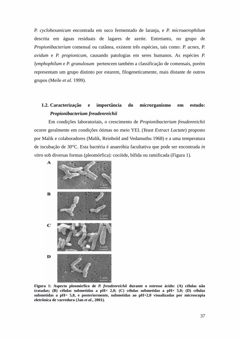
vitro sob diversas formas (pleomórfica): cocóide, bífida ou ramificada (Figura 1).
Figura 1: Aspecto pleomórfico de P. freudenreichii durante o estresse ácido: (A) células nãotratadas; (B) células submetidas a pH= 2,0; (C) células submetidas a pH= 5,0; (D) célulassubmetidas a pH= 5,0, e posteriormente, submetidas ao pH=2,0 visualizadas por microscopiaeletrônica de varredura (Jan et al., 2001).
38
Antigamente, a espécie P. freudenreichii foi divida em duas sub-espécies: P.
freudenreichii subsp. freudenreichii, que não é capaz de fermentar lactose, mas pode
reduzir nitrato; e a P. freudenreichii subsp. shermanii, que fermenta lactose, porém não é
capaz de reduzir o nitrato (Cummins and Johnson 1986). Porém, estudos recentes
demonstram que existem linhagens de P. freudenreichii que podem conter os dois
fenótipos: lactose e nitrato redutase positiva ou lactose e nitrato redutase negativa (de
Carvalho, Gautier and Grimont 1994; Vorobjeva 1999; Dalmasso et al. 2011). Além
disso, no genoma completo de P. freudenreichii CIRM-BIA 1, foi observado que o gene
lacZ, responsável pela atividade beta-galactosidase, esta localizado numa região de
transposase e que existem pseudogenes na região do genoma que codifica a informação
necessária à redução de nitrato (Falentin et al. 2010a). Atualmente, não há uma
classificação de subespécies ao nível fenotípico e outras investigações para explorar a
genômica desta bactéria devem ser efetuadas. Isto é grande importância na indústria
alimentícia, uma vez que estas propriedades bioquímicas podem alterar a qualidade final
do produto, por exemplo, queijos PPC.
P. freudenreichii é um microrganismo seguro (saúde-sanitária) e possui dois
importantes status para sua utilização: o Generally Recognized As Safe (GRAS)
outorgado pelo órgão administrativo de alimentos e drogas (Food and Drug
Administration – FDA, nos Estados Unidos) (Mogensen et al. 2002) e o Qualified
Presumption of Safety (QPS) concedido pela autoridade européia de segurança dos
alimentos (European Food Safety Authority - EFSA) (EFSA experts 2008). Estes status
são aprovados por autoridades do setor que visam controlar a qualidade dos produtos,
garantindo a segurança e a autorização destas bactérias na indústria de alimentos, como
por exemplo, na fabricação de queijos Emmental. Estas bactérias são amplamente
encontradas na natureza, incluindo o trato gastrointestinal, e estão presentes em muitos
alimentos fermentados por possuírem características que as permitem de resistir ao pH
ácido (Jan, Rouault and Maubois 2000; Huang and Adams 2004; Suomalainen et al.
2006) e a baixas temperaturas no processo de maturação de queijos (Dalmasso et al.
2012a). A concentração destas bactérias, geralmente, encontrada no Emmental são na
ordem de 109 UFC/g. Estas bactérias possuem qualidades que são de grande
importância para diversas aplicações na área industrial, as quais serão abordadas com
mais detalhes na próxima seção.

39
2. Aplicações industriais de Propionibacterium freudenreichii
Os alimentos fermentados, em particular os lactícinios, são consumidos pelo
homem há milhões de anos. Desde o tempo da Idade Média, o processo de fabricação de
queijos do tipo Emmental nos Alpes da região de Savoie é descrito em manuscritos. No
século XIX, aparecem os primeiros relatos sobre “frutières”, que são locais de
exploração e transformação do leite em queijo, onde os agricultores, em conjunto,
agrupam o "fruto” de suas produções de verão. Em seguida, no século XX, uma forte
industrialização foi observada, conduzindo a uma deslocalização da produção destes
queijos.
Assim como o Emmental, todos os laticínios que foram anteriormente iniciados
empiricamente, começaram a se beneficiar de avanços no campo da microbiologia e da
tecnologia de alimentos, o que permitiu um melhor controle, diversificação e segurança
de suas produções.
As principais espécies bacterianas utilizadas hoje para a produção de alimentos
fermentados são pertencentes aos gêneros: Bifidobacterium, Lactobacillus,
Lactococcus, Leuconostoc, Streptococcus e Propionibacterium (Tabela 1 e 2). Estes
gêneros exercem papéis importantes na indústria de laticínios, na síntese de
biomoléculas, bem como no desenvolvimento de produtos probióticos.
Neste contexto, P. freudenreichii desempenha um papel importante na fabricação
e, particularmente, na maturação de queijos PPC. Esta espécie é também utilizada por
sua capacidade de produzir bacteriocinas, compostos do ácido linoléico, do ácido
propiônico, propileno, trealose e vitaminas do complexo B, como por exemplo, a
vitamina B12 (Piao et al. 2004; Poonam et al. 2012). Algumas linhagens possuem
também propriedades probióticas e podem modular a composição da microbiota
intestinal produzindo fatores bifidogênicos, ou modular ao nível de inflamação no
contexto de doenças inflamatórias intestinais crônicas. Estas bactérias ainda são
interessantes por serem capazes de resistir ao estresse do trato gastrointestinal.
Como descrito anteriormente, esta bactéria apresenta capacidade de adaptação à
estresses de natureza variável, como: pH, temperatura e sais biliares. Esta robusticidade
confere uma vantagem a esta espécie que a permite de permanecer viva e de manter
suas atividades metabólicas em diversos ambientes. Estas características são
importantes para suas aplicações em indústrias alimentícias, particularmente, na
indústria de queijos PPC; e em tecnologia, para a síntese de biomoléculas e/ou para

40
alimento/saúde (probióticos), todas estas aplicações serão apresentadas com detalhes
nesta seção.
Tabela 1: Ação microbiana nos alimentos fermentados (tabela adaptada de
http://mastervrv.free.fr/S3/BA/Cours/BA-FERMENTATION.pdf).
Efeitos sob o produto Exemplos Microrganismos implicados
Modificação da textura
Coagulação do leite Diversas bactérias lácticas
Textura aveludada em laticíniosfermentados
Bactérias lácticas (dextranes)
Formação de olhaduras em queijos Bactérias propiônicas
Aromatização
Gosto amanteigado (diacetil) Lactococcus lactis diacetylactis
Gosto típico de iogurte (acetaldeídoe ácido láctico)
Lactobacillus delbrueckiibulgaricus
Desenvolvimento do gosto de cacau Diversas bactérias
ColoraçãoEscurecimento da massa de queijos
PPC* por propionato de cálcioBactérias propiônicas
Alteração do gostoAcidificação do repolho Lactobacillus
Desacidificação do vinho Bactérias malolácticos
Estabilizaçãomicrobiana
Acidificação Bactérias lácticas e acéticas
*PPC: queijos de massa cozida e prensada.

41
Tabela 2: Principais fermentadores para a indústria de laticínios (tabela adaptada dehttp://mastervrv.free.fr/S3/BA/Cours/BA-FERMENTATION.pdf).
Fermentadores Aplicações
Lactobacillus bulgaricusStreptococcus thermophilus
Bifidobacterium sp.Iogurtes
Lactobacillus acidophilus Leite fermentado
Lactobacillus caucasius Kéfir (bebida fermentada)
Streptococcus cremosnisStreptococcus lactis
Streptococcus diacetylactisLeuconostoc cremonisLactobacillus cremonis
Lactobacillus lactis
Etapas iniciais (coalhamento) damaioria dos queijos
Queijos não afinados (massa fresca)Manteiga
Propionibacterium freudenreichii ssp shermaniiPropionibacterium freudenreichii ssp. freudenreichii
Maturação de queijos PPC*
*PPC: queijos de massa cozida e prensada.
Nos paragráfos seguintes, iremos apresentar algumas informações sobre a importância
de P. freudenreichii em aplicações industriais:
Queijos PPC as bactérias propiônicas são necessárias para a maturação dos
queijos PPC. Sua sobrevivência é necessária para garantir o sabor e a tipicidade
destes queijos.
Biomoléculas bactérias propiônicas pemitem a produção de diversas
biomoléculas de interesse industrial, como por exemplo, o ácido propiônico e a
vitamina B12, produtos obtidos durante o metabolismo destas bactérias.
Probióticos a manutenção da atividade metabólica de P. freudenreichii nos
produtos probióticos é de grande importância, uma vez que esta bacéria pode
atuar no trato gastrointestinal, como por exemplo, através da imunomodulação.
Para isto, é necessário que as bactérias sobrevivam por longos períodos
passando por etapas de transformação e armanezamento, sem grandes alterações
na viabilidade e atividade esperada para o produto final.

42
2.1. Indústria de queijos PPC (“pasta cozida e prensada”)
Os queijos são mundialmente produzidos e são classificados de acordo com suas
características, tais como: consistência da pasta, teor de gordura, tipo de leite utilizado,
tipo de coagulação, tipo de casca e tempo de cura. P. freudenreichii é utilizada em
conjunto com outras culturas bacterianas, principalmente, na fabricação de queijos
Emmental. Este grupo é o terceiro segmento mais importante na indústria de queijos,
sendo chamado de “pasta cozida e prensada” (PPC, do francês: pâte pressée cuite), e
composto por outros queijos, como o queijo Comté. A denominação PPC corresponde
aos queijos, os quais a coagulação e desidratação são extensas e que, em seguida, são
prensados no momento da moldagem para remover a quantidade máxima de soro do
leite, nomeados também de "queijos duro". Após a etapa de coagulação e moldagem, a
coalhada é “cozida” em uma etapa de alto aquecimento, acima de 50°C durante 30 à 60
minutos no soro, o que permite de obter, após a desidratação e prensagem, um elevado
extrato seco e uma conservação destes queijos por mais tempo.
De acordo com o Centre National Interprofessionnel de l'Economie Laitière, em
2014, a França produziu mais de 260.000 toneladas de Emmental. A região do Grand
Ouest da França, contemplando a Bretanha, País de Loire e Baixa Normandia,
representa dois terços desta produção (CNIEL, 2014). Destaque para a região da
Bretanha que é a primeira em produção de leite de vaca et de Emmental na França
(Figura 2).
Figura 2: Produção de leite de vaca no território francês (La géographie du lait em 2013 – EnquêteAnnuelle Latière).

43
Estes produtos são muito importantes para a economia de lactícinios da França,
particularmente o Emmental, que é um grande representante deste setor (Tabela 3).
Tabela 3: Produção de queijos PPC em toneladas (L’Enquête Annuelle Latière).
Queijos PPC 2011 2012 2013
Emmental 256.980 253.225 252.892
Comté AOP 58.114 57.561 59.459
Beaufort AOP 5.044 4.919 4.787
Gruyère IGP - 2.679 2.929
Autres PPC - 8.913 7.938
Total PPC 333.258 327.298 328.006
Para compreender o processo de fabricação destes queijos PPC, a tabela 4
resume algumas etapas importantes e a figura 3, ilustra todo o processo.

44
Tabela 4: Etapas para a fabricação de queijos.
Etapas Descrição
PasteurizaçãoAquecimento do leite a temperaturas elevadas, esta etapa visa a eliminação de microorganismos indesejáveis.
Porém, esta etapa não ocorre para queijos feito com leite cru ou pasteurizado.
Coagulação
Esta etapa visa a concentração da proteína do leite, podendo ser divida em duas etapas: (I) ácida e (II) enzimática.A primeira ocorre pela atividade da flora microbiana do leite, ou pela adição do extrato de renina (coalho) ou
femento. Já a coagulação ácida ocorre a partir da produção de ácido láctico por bactérias, ou pela adição de ácidosorgânicos ao leite.
MoldagemA coalhada obtida no processo anterior é colocada em diferentes moldes, conforme o queijo desejado.
Desidratação O objetivo desta etapa é separar o soro de leite da coalhada, o que prolonga sua conservação. Alguns queijospodem ser consumidos após este processo.
SalgaOs queijos frescos resultantes são desenformados e salgados, com um sal fino ou no banho de salmoura saturada.
Isto garante uma ação anti-séptica, melhoria na conservação e também adiciona o sabor aos queijos.
MaturaçãoEsta final pode demorar dias ou até mêses de acordo com a variedade do queijo. Estes serão armazenados em
“caves”, local o qual terão a temperatura e umidade controlada.

45
Figura 1: Processos de transformação de queijos PPC (http://www.produits-laitiers.com/). O processo de fabricação dos queijos PPC, inicia-se pela coleta do leitepor meio de máquinas automatizadas em fazendas de gado leiteiro. Em seguida, este leite é refrigerado e analisado, de acordo com as normas de segurança doconsumidor, visando garantir um produto final de boa qualidade. Este leite é transportado em caminhões isotérmicos até a indústria de laticínios, onde começamtodas as etapas de tratamento e fabricação de queijos.
ESQUEMA DE PRODUÇÃODOS QUEIJOS PPC
COAGULAÇÃO QUEBRA DACOALHADA EAGITAÇÃO
MOLDAGEM DESIDRATAÇÃO
SALGA
MATURAÇÃO EMCAVA FRIA 8-15º
MATURAÇÃO EMCAVA QUENTE 16-23º
AQUECIMENTO/ AGITAÇÃO
COLETA
REFRIGERAÇÃOTRANSPORTE
ANÁLISE
ARMAZENAMENTO
PASTEURIZAÇÃOOU NÃO DO LEITE
COAGULAÇÃO
ANÁLISE
ANÁLISE

46
O Emmental é um queijo de peso, cujo as unidades pesam entre 60 e 120 kg
(grande em volume de peso). É necessário 12 litros de leite para obter 1kg deste queijo
(CNIEL,2015). Durante o processo de maturação, muitos parâmetros variam, tais como:
temperatura, pH e atividade de água. P. freudenreichii exerce um papel importante nesta
maturação (Figura 4), produzindo acetato e propionato através da fermentação, que
estão envolvidos no sabor do Emmental, e CO2 responsável pela “abertura do queijo”
por meio da formação de olhaduras na massa (Langsrud and Reinbold 1973; Meurice et
al. 2004).
Figura 4: P. freudenreichii em um queijo Emmental (©INRA).
P. freudenreichii desempenha também um papel importante na composição do
aroma e sabor em alguns lactícinios, dentre eles o Emmental. Isto porque durante o
processo de maturação prolongada deste queijo, P. freudenreichii garante a
ocorrerrência das reações de lipólise. A lipólise contribui para a formação de sabores e é
realizada através de esterases, as quais são capazes de efetuar à hidrólise dos
triacilglicerídeos do leite liberando ácidos graxos de cadeia curta, de cadeia média e de
cadeia longa (Figura 5). Estes últimos é que conferem aos queijos suas diferentes
sensações gustatórias, como por exemplo, sabor forte, picante e/ou salgado (Berdagué et
al. 1990).
Figura 5: Esquema da reação de hidrólise de triacilglicerídeos.

47
Estudos como o de Dherbécourt e colaboradores, identificaram 23 esterases
(putative esterases) em P. freudenreichii, potencialmente envolvidas neste processo de
maturação. Esta identificação foi realizada a partir da combinação de métodos clássicos
e do estudo da genômica desta bactéria (Dherbécourt et al. 2008). Através da
abordagem genômica, as ferramentas bioinformáticas e os métodos utilizados foram: (i)
busca automática de esterases por meio de homologia, (ii) busca por motivos protéicos
no InterProScan, (iii) busca manual por homologia e (iv) busca manual por motivos
específicos através do Prosite. Estes métodos foram essenciais para uma primeira
triagem de enzimas de interesse. Assim, 12 genes candidatos que codificam as
potenciais esterases apresentando os motivos GXSXG característicos de α/β hidrolase
foram selecionados e clonados em E. coli. Testes zimográficos também foram realizados
no intuito de caracterizar a atividade destas esterases. Assim, foi constatado que as 12
enzimas, em P. freudenreichii, desempenham papéis impotantes na lipólise no queijo
Emmental.
Sabemos que P. freudenreichii sobrevive a diversos ambientes e estresses de
maneira linhagem-dependente. A importância deste fenômeno atrae estudos sobre as
estratégias que permitem a sobrevivência e manutenção da atividade metabólica por
longos períodos. Assim, no intuito de explorar estes mecanismos de sobrevivência à
longo prazo, estudos que associam experimentos microbiológicos (in vitro) relacionado
com aplicações industriais e biotecnológicas são de uma grande importância. Estes
estudos serão descritos nas próximas seções.

48
2.2. Otimização da produção de vitamina B12
Em 1911, o termo “vitamina” foi proposto por Casimur Funk para designar as
aminas que são essencias para a vida (vita-amina). Posteriormente, esta nomenclatura
começou a ser utilizada também para substâncias orgânicas essenciais, sem valor
energético mas ativas em pequenas quantidades, contribuindo para a manutenção do
metabolismo de um organismo (Florent 1986; Vandamme 1992).
A vitamina B12, também denominada de cobalamina ou cianocobalamina, foi
isolada com primazia em 1948, tendo sido sintetizada somente em 1973. Isto ocorreu
devida a dificuldade de obtenção do produto, e sua síntese necessita de diversas etapas
de processamento até o rendimento final da molécula (Vandamme 1989). Como esta
vitamina está implicada em processos essenciais para a regulação do metabolismo de
animais e seres humanos, como a formação e maturação de hemácias e a manutenção do
sistema nervoso, estratégias alternativas que visam a otimização de sua produção são de
grande interesse.
Neste contexto, a obtenção de vitamina B12 utilizando microorganismos tem se
tornado uma solução promissora para o mercado industrial. Dentre as bactérias
produtoras, destaca-se a P. freudenreichii, devido ao seu bom rendimento de vitamina
B12. Assim, uma concentração de 5,12 mg/L pode ser obtida no queijo após 168h de
fabricação (Marwaha, Sethi and Kennedy 1983) . Estudos visando aprimorar a produção
de vitamina B12 em P. freudenreichii mostraram que é possível alcançar um rendimento
de até 6,35 mg/L após 192h de fermentação (García Hernández and Pérez Mendoza
1983).
Novas abordagens tem sido propostas para a biossíntese de vitamina B12 ao
decorrer das últimas décadas. A obtenção de vitamina B12 por meio da fermentação de
P. freudenreichii foi analisada, no trabalho de Paio e colaboradores, de acordo com o seu
desempenho sob a expressão de genes, como: cobA, cbiLF e cbiEGH, envolvidos na
síntese de vitamina B12, poderia aumentar a produção de vitamina B12 (Piao et al.
2004). Outros estudos com o mesmo objetivo, como o trabalho de Murooka e
colaboradores, expressaram e amplificaram os genes do operon cob e cbi, para o
aumento da biossíntese de cobalaminas durante o processo de fermentação (Murooka et
al. 2005).
Para otimizar a produção de vitamina B12, um outro estudo por Wang e
colaboradores com enfoque no efeito do ácido propiónico e do 5,6-
dimetilbenzimidazole durante a biossíntese desta vitamina no processo de cultivo de P.

49
freudenreichii (Wang et al. 2015). Foi visto que a concentração de ácido propiônico
varia da fase inicial (10-20 g/L) à fase tardia (20-30 g/L). Isto conduz a um feed-back
negativo para a biossíntese de vitamina B12, mas que pode ser alterado com a inclusão
de 5,6-dimetilbenzimidazole (Wang et al. 2015). Assim, um controle e uma adaptação
detalhada das condições de cultivo bacteriano são essenciais para aobtenção de grandes
quantidades de vitamina B12.

50
2.3. Obtenção do ácido propiônico
O ácido propiônico é um ácido graxo de cadeia curta, saturado, de massa
molecular 74,08 g/mol, também conhecido como ácido propílico, ácido propanóico ou
C₃H₆O₂ (GESTIS Substance Database – disponível em http://gestis-en.itrust.de/nxt/
gateway.dll/gestis_en/012590.xml?f=templates$fn=default.htm$3.0) (Figura 6).
Figura 6: Representação da fórmula química do ácido propiônico.
Este ácido é amplamente utilizado em setores industriais, como: petroquímicos,
cosméticos, plásticos, herbicidas e conservantes. Neste contexto, a biologia sintética é
um setor inovador que envolve a biologia e a engenharia, com o intuito de desenvolver
novas funções e sistemas, por exemplo, a utilização de uma determinada via bioquímica
para a obtenção de um dado material por meio de uma fonte renovável (Parizzi et al.
2012).
A fermentação é um dos processos mais utilizados na indústria para a obtenção
de diversos produtos e subprodutos, entre eles, o ácido propiônico. Atualmente,
estratégias alternativas visando a obtenção deste produto foram desenvolvidas
utilizando diferentes fontes de carbono, tais como a fermentação da cana-de-açúcar
(sacarose), estas estratégias buscam uma redução no custo e uma melhoria na produção
deste ácido. As bactérias propiônicas são capazes de sintetizar o ácido propiônico e elas
representam uma fonte interessante para tal utilidade. Assim, estudos que visam a
fisiologia e metabolismo de Propionibacterias foram realizados, dentre outros, no intuito
de identificar as melhores espécies e as melhores linhagens para uma utilização
industrial (Thierry et al. 2011).
A fermentação de lactato, através do metabolismo inicial de P. freudenreichii,
gera produtos como acetato, propionato e CO2, que desempenham papéis variados e
importantes para as indústrias alimentares e biotecnológicas (Meurice et al. 2004). Por
exemplo, o propionato de cálcio é um produto oriundo do ácido propiônico, tendo a
capacidade de atuar como conservante, sendo capaz de combater bactérias
desfavoráveis e fungos em alimentos.

51
A partir destas observações sobre o ácido propiônico que Stowers e
colaboradores avaliaram 17 linhagens do gênero Propionibacterium e definiram
parâmetros optimais de produção e de rendimento de ácido propiônico em fermentador.
Com base nestes parâmetros, linhagens das espécies de P. acidipropionici e P.
freudenreichii produziram altas quantidades de ácido propiônico (Stowers, Cox and
Rodriguez 2014).
Porém, ainda é difícil a obtenção de um bom rendimento de ácido propiônico a
partir de Propionibacteria, uma vez que a geração de subprodutos do seu metabolismo,
como ácido succínico, ácido láctico, e ácido acético diminui o rendimento do ácido
propiônico (Stowers, Cox and Rodriguez 2014). Neste intuito, é importante a realização
de estudos para aumentar a eficácia da fabricação deste produto. Esta produção lança
oportunidades em setores como o de química ambiental, através da produção de
propileno (C3H6), precursor de ácido propiônico, um dos hidrocarbonetos utilizados na
indústria petroquímica. A polimeração do propileno forma o polipropileno, um
composto muito utilizado em diversas indústrias para gerar combustíveis, embalagens
descartáveis, produtos têxteis, eletrodomésticos e mercado automobilístico (Ammar et
al. 2014). Assim, bactérias que sintetizam o ácido propiônico a um alto rendimento são
objetos de interesse crescente, devido ao desenvolvimento da química ambiental e de
um mercado econômico favorável para produtos orgânicos.

52
2.4. ProbióticoComo falado anteriormente, diversas bactérias, em especial bactérias lácticas e
propiônicas, tem sido alvo de interesse devido as suas propriedades probióticas. Um
exemplo disso é Propionibacterium freudenreichii que é amplamente estudada por sua
capacidade de inibir microrganismos indesejáveis, modular a microbiota do cólon
através da produção de 1,4-di-hidroxi-2-naftóico (DHNA), um fator bifidogênico (Pérez
Chaia, Zarate and Oliver 1999), e à sobreviver a diferentes estresses (Isawa et al. 2002;
Okada et al. 2013), em particular aos estresses encontrados durante a passagem pelo
trato gastrointestinal (Jan et al. 2001; Leverrier et al. 2003a; Saraoui et al. 2013a). P.
freudenreichii é também investigada por desencadear a apoptose em células
cancerígenas do cólon, aderir à células epiteliais do intestino e muco, estimulando assim
atividades imunomodulatórias e a proliferação celular no hospedeiro (Ouwehand et al.
2002; Zarate, Chaia and Oliver 2002). Nesta seção, iremos apresentar uma pequena
síntese de estudos que abordam as diversas propriedades probióticas atribuídas à P.
freudenreichii.
2.4.1. Histórico e definição do termo probiótico
Embora os benefícios de alguns alimentos fermentados para a saúde sejam
estudados há muitos anos (trabalhos de Metchnikoff no início do século XX), o termo
probiótico teve origem em 1965, quando Lilly e Stillwell, relacionaram a sobrevivência
de determinados microrgansimos com o efeito benéfico de seus produtos (Lilly and
Stillwell 1965). Atualmente, os probióticos são definidos como microrganismos vivos
que se administrados de maneira adequada, promovem benefícios à saúde do hospedeiro
(Bron, van Baarlen and Kleerebezem 2012). Devida a sua importância, publicações no
PubMed vem ganhando destaque ao longo dos útimos anos (Figura 7).

53
Figura 7: Publicações no PubMed com as palavras-chave “probiotics-bacteria” ao decorrer dosúltimos anos.
Além disso, para a utilização de probióticos em humanos e para que as
reivindicações de saúde sejam aceitas, são necessários rigorosos testes in vitro e in vivo,
os quais visam garantir a qualidade e segurança destes microrganismos antes de suas
possíveis aplicações tanto na prevenção ou no tratamento de enfermidades quanto em
suplementos alimentares. Algumas espécies bacterianas como Lactobacillus
paracasei, L. johnsonii, L. fermentum, L. plantarum, L. rhamnosus, Bifidobacterium
longum e P. freudenreichii são objetos de estudo por apresentarem capacidades que
promovem a melhoria da saúde humana, incluindo o controle e a restauração do
equilíbrio da microbiota intestinal (Parvez et al. 2006; Cousin et al. 2012; Santos Rocha
et al. 2012; Luerce et al. 2014).
2.4.2. Controle da microbiota
Bactérias propiônicas podem exercer efeitos na microbiota dos animais e/ou de
seres humanos, seja inibindo microrganismos não-desejáveis e/ou aumento a
proliferação de populações de Bifidobacterium sp., bactérias que exercem funções
importantes no trato digestivo (Picard et al. 2005). O seu aumento apresenta uma série
de vantagens fisiológicas e clinicas para a saúde humana e/ou animal, tais como: (i)
acelera o tempo de passagem pelo trato gastrointestinal; (ii) exerce o efeito de barreira,
já que produz substâncias que bloqueiam os sítios de ligação de patógenos; (iii) exerce
efeito sobre o sistema imunológico e em doenças do trato-gastrointestinal; (iv) reduz o
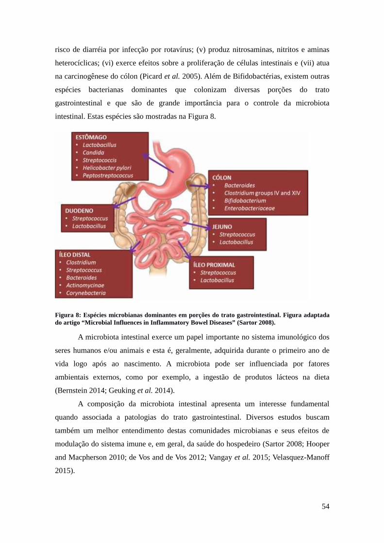
54
risco de diarréia por infecção por rotavírus; (v) produz nitrosaminas, nitritos e aminas
heterocíclicas; (vi) exerce efeitos sobre a proliferação de células intestinais e (vii) atua
na carcinogênese do cólon (Picard et al. 2005). Além de Bifidobactérias, existem outras
espécies bacterianas dominantes que colonizam diversas porções do trato
gastrointestinal e que são de grande importância para o controle da microbiota
intestinal. Estas espécies são mostradas na Figura 8.
Figura 8: Espécies microbianas dominantes em porções do trato gastrointestinal. Figura adaptadado artigo “Microbial Influences in Inflammatory Bowel Diseases” (Sartor 2008).
A microbiota intestinal exerce um papel importante no sistema imunológico dos
seres humanos e/ou animais e esta é, geralmente, adquirida durante o primeiro ano de
vida logo após ao nascimento. A microbiota pode ser influenciada por fatores
ambientais externos, como por exemplo, a ingestão de produtos lácteos na dieta
(Bernstein 2014; Geuking et al. 2014).
A composição da microbiota intestinal apresenta um interesse fundamental
quando associada a patologias do trato gastrointestinal. Diversos estudos buscam
também um melhor entendimento destas comunidades microbianas e seus efeitos de
modulação do sistema imune e, em geral, da saúde do hospedeiro (Sartor 2008; Hooper
and Macpherson 2010; de Vos and de Vos 2012; Vangay et al. 2015; Velasquez-Manoff
2015).

55
2.4.3. ImunomodulaçãoA imunomodulação é uma propriedade importante de bactérias probióticas,
permitindo a indução e regulação de respostas imunes, sendo útil na prevenção e no
tratamento de câncer, de doenças auto-imunes e no controle de microrganismos
indesejáveis (Ouwehand et al. 2009; Sathish et al. 2013).
A associação de bactérias propiônicas à bactérias lácticas et bifidobactérias
podem exercer efeitos benéficos em patologias do trato gastrointestinal em crianças e
idosos. Neste contexto, bactérias propiônicas agem indiretamente favorecendo o
crescimento e a manutenção de concentrações elevadas de bifidobactérias durante um
período de tratamento (Mantere-Alhonen 1995). As bactérias propiônicas parecem
somente promover um efeito probiótico através da imunomodulação (Cousin et al.
2010; Le Marechal et al. 2015). O efeito imunomodulador de linhagens probióticas é
comumente associado a sua capacidade de adesão ao epitélio intestinal. Esta adesão
permite a permanência destas bactérias por mais tempo no trato digetivo, durante o
trânsito intestinal. Esta capacidade de adesão pode ser um critério de seleção de
probióticos e algumas linhagens de P. freudenreichii desencadearam uma série de
estudos neste campo (Ouwehand et al. 2002). A implicação de proteínas de superfície
no papel imunomodulatório destas bactérias foi testado através da caracterização
proteômica “da superfície” de P. freudenreichii. Foi demonstrado também que algumas
proteínas de superfície, extraídas de linhagens de P. freudenreichii e purificadas, foram
capazes de reproduzir, in vitro, uma resposta anti-inflamatória observada com uma
linhagem completa (Le Maréchal et al. 2015).

56
2.4.4. Apoptose induzida por ácidos graxos de cadeia curta
As bactérias propiônicas são conhecidas por sua capacidade de produzir ácidos
graxos de cadeia curta (SCFA – short chain fatty acids), entre eles, o propionato e o
acetato que são os principais produtos da fermentação de P. freudenreichii. Foi
observado que estes SCFA apresentam efeitos interessantes sob a fisiologia das células
epiteliais intestinais (Jan et al. 2002; Pérez Chaia and Zarate 2005).
Os SCFA, e principalmente o butirato, constituem uma fonte de energia
privilegiada, um verdadeiro “combustível” para as células epitaliais intestinais. Em
1995, Hague e colaboradores demonstraram que estes mesmos SCFA, por exemplo, o
butirato de sódio, são também capazes de induzir a apoptose em cancerígenas do cólon
(Hague et al. 1995). Posteriormente, o trabalho de Jan e colaboradores em 2002,
demonstra que in vitro as linhagens de P. acidipropionici e P. freudenreichii são também
capazes de induzir a apoptose em linhagens de células humanas de câncer colorretal,
como: HT29 e Caco2. Este efeito também foi observado em linhagens celulares
humanas de câncer de cólo de útero (HeLa). Os SCFA induzem sinais característicos de
apoptose, tais que: (i) perda do potential transmembranar das mitocôndrias, (ii) geração
de espécies reativas de oxigênio, (iii) indução da cascata caspase-3 e (iv) condensação
da cromatina ao nível nuclear. O mecanismo geral de apoptose através de SCFA é
esquematizada na Figura 9. Além disso, esta atividade pode ser exercida in vivo, já que
as linhagens se mantém vivas e metabolicamente ativas dentro do intestino humano (Jan
et al. 2002).
Figura 9: Indução da apoptose a partir de SCFAs produzidos por bactérias propiônicas no cólon.

57
Além disso, foi proposto que as bactérias propiônicas poderiam complementar o
arsenal de opções terapêuticas disponíveis na prevenção e no tratamento de câncer de
cólon (Lan et al. 2008). Recentemente, esta mesma equipe de pesquisa propôs uma
nova formulação de um leite fermentado com algumas linhagens de P. freudenreichii,
visando uma dieta preventiva ou um suplemento na alimentação de pessoas que
apresentam pré-disposição a este tipo de câncer (Cousin et al. 2012).

58
2.4.5. Molécula ácido 1,4-di-hidroxi-2-naftóico (DHNA)
A molécula ácido 1,4-di-hidroxi-2-naftóico (DHNA) apresenta um acentuado
efeito bifidogênico, uma vez que favorece o crescimento de bifidobactérias, propriedade
importante para um prebiótico. Os prebióticos são hidratos de carbono de cadeia curta
não-digeríveis por enzimas digestivas em seres humanos. Assim, o crescimento de P.
freudenreichii, que produz DHNA, influencia diretamente no crescimento de
bifidobactérias, fazendo de P. freudenreichii uma bactéria que possui atividades pro e
pre-bióticos. Esta bactéria contribui também para o equilíbrio da flora intestinal,
favorecendo o crescimento de bactérias benéficas. De outra maneira, o DHNA permite
reduzir a inflamação no cólon (Quigley, Hudson and Englyst 1999; Okada et al. 2006).
In vivo, a administração de DHNA provoca alterações do sistema imune intestinal de
camundongos, modificando a resposta imune inata em células intestinais (Okada et al.
2013).
Assim, DHNA é uma molécula promissora, uma vez que pode ser utilizada no
tratamento de colite. Em um modelo de colite em camundongos, sua administração
provoca uma melhoria na histologia do epitélio intestinal, uma supressão da migração
de linfócitos e da síntese de interleucinas pró-inflamatórias (Okada et al. 2013). Neste
contexto, pesquisas suplementares devem ser realizadas objetivando uma otimização de
DHNA para aplicações terapêuticas e para demonstrar que as quantidades produzidas in
situ por P. freudenreichii poderiam conduzir aos mesmos efeitos.
59
2.4.6. Capacidade de adaptação
Esta subseção é destinada a capacidade de sobrevivência de P. freudenreichii e
sua capacidade de adaptação a diferentes estresses. Esta será dividida em duas partes:
tolerância ao estresse térmico e ao estresse digestivo. A tolerância a estes estresses é
uma propriedade muito importante em bactéria propiônica para sua manutenção e sua
viabilidade, otimizando assim sua utilização como probióticos (Leverrier et al. 2003a).
Tolerância ao estresse térmico:
As bactérias propiônicas sobrevivem a uma ampla gama de temperaturas. No
caso das etapas da maturação de queijos PPC, estas suportam temperaturas baixas
(4ºC), médias (30ºC) e altas (52-53º). Estas temperaturas podem variar de acordo com
as etapas do processamento e o tipo de queijo a ser produzido. Para o queijo Gruyère
tradicional, há uma etapa de cozimento que submete as bactérias a uma temperatura
bem elevada, entre 52-53ºC durante 30 minutos. A termotolerância à 55ºC de diferentes
linhagens utilizadas na fabricação Gruyère foi estudada através da abordagem
proteômica e os perfis obtidos por eletroforese 2D revelaram diferentes respostas de
acordo com as linhagens termotolerantes e termossensíveis (Anastasiou et al. 2006).
Em espécies termotolerantes, proteínas associadas ao processo de fermentação
propiônico, metabolismo de aminoácidos, nucleotídeos e fosforilação oxidativa foram
superexpressas. Em particular, as chaperonas moleculares e os compostos antioxidantes
de tiol são mais induzidas pelo calor. Entretanto, observou-se que para a adaptação
térmica as vias de transdução de sinal, biossíntese e manutenção da parede celular
contribuíram para a manutenção do processo de fermentação (Anastasiou et al. 2006).
Outros estudos abordando bactérias propiônicas relataram também que chaperonas
moleculares e proteínas de choque térmico são induzidas em muitas situações de
estresse (Jan et al. 2001; Leverrier et al. 2003a). Esta diferença na expressão de
proteínas de diversas linhagens de bactérias propiônicas, ressaltam o argumento de que
existem fenótipos diferentes dentro da mesma espécie (Anastasiou et al. 2006). Esta
forma de tolerância é muito importante em P. freudenreichii para manter a
sobrevivência bacteriana e garantir a fluidez do processo de fabricação de queijos do
tipo PPC. Ela consititue também uma vantagem incontestável para sua utilização como
probiótico, se associamos sua resistência aos estresses digestivos (Leverrier et al.
2003a).

60
Tolerância ao estresse digestivo:
Para que um probiótico exerça sua função é necessário a manter sua atividade
metabólica e sobrevivência após a passagem pelo trato gastrointestinal. O trabalho de
Lan e colaboradores demonstraram esta capacidade em P freudenreichii, demonstrando
a viabilidade destas bactérias no trato digestivo e no cólon de camundongos (Lan et al.
2007). Este estudo confirmou também a tolerância ao pH ácido de algumas linhagens
específicas de P. freudenreichii, como já relatado em estudos anteriores sobre a
resistência ao estresse ácido (Jan, Rouault and Maubois 2000) e também a tolerância aos
sais biliares (Warminska-Radyko, Laniewska-Moroz and Babuchowski 2002).
Experimentos de proteômica, através de eletroforese 2D, revelaram 24 proteínas
induzidas durante a adaptação de P. freudenreichii em meio contendo sais biliares. Entre
elas, estavam proteínas de transdução de sinal e fatores sigma alternativos (Leverrier et
al. 2003a).
Para avaliar a atividade metabólica de P. freudenreichii, uma RT-PCR
quantitativa utilizando uma sonda TaqMan e selecionando o RNAm codificante para a
enzima transcarboxilase foi desenvolvido. Este método não invasivo e eficiente foi
utilizado para provar a sobrevivência e a atividade metabólica de P. freudenreichii no
trato intestinal de camundongos (Hervé et al. 2007). Outros métodos buscando avaliar a
atividade metabólica de P. freudenreichii no queijo foram desenvolvidos no intuito de
caracterizar as linhagens quanto à sua sobrevivência. Neste contexto, um método de RT-
PCR quantitativa utilizando SyberGreen foi optimizado para a quantificação de células e
de marcação de RNAm (gènes 16S, tuf, groL) de P. freudenreichii e de Lactobacillus
paracasei, durante o processo de fabricação de queijos PPC (Falentin et al. 2010c). Este
método permitiu quantificar a viabilidade celular, bem como instiga estudos para
investigar como ocorre a sobrevivência de P. freudenreichii.

61
3. Genoma de Propionibacterium freudenreichii
Através das tecnologias de sequenciamento de próxima geração (NGS), foi
possível a obtenção do sequenciamento de vários genomas de bactérias propiônicas, o
que permitiu um melhor conhecimento biológico da espécie. Estes genomas
apresentam, em geral, um alto conteúdo GC, em torno de 67% (Falentin et al. 2010a) e
algumas linhagens podem apresentar plasmídeos em sua composição (Luijk et al. 2002).
Dados provenientes do sequenciamento podem ser utilizados em estudos comparativos
e/ou evolutivos visando compreender a genômica, ao nível estrutural e ao nível
funcional deste microorganismo.
Em 2010, o primeiro genoma completo de P. freudenreichii foi o da linhagem
CIRM-BIA1, o qual foi sequenciado utilizando a metodologia tradicional de Sanger
(Falentin et al. 2010a). A montagem inicial deste genoma foi realizada sem sequências
repetitivas, usando o pacote do software Phred/Phrap/Consed (www.phrap.org)
(Vallenet et al. 2008; Falentin et al. 2010a). Posteriormente, para o fechamento de gaps,
foi necessário a utilização do kit GC sequence finishing (GEhealthcare) e o kit
transposition bombs, técnicas que permitiram a obtenção do primeiro genoma completo
de P. freudenreichii pela equipe do STLO - INRA de Rennes, France (Falentin et al.
2010a). Este genoma possui um cromossomo circular de 2,7 Mb, alto conteúdo de GC
(67%) e encontra-se disponível no banco de dados Genbank, com o número de acesso
FN806773 (Falentin et al. 2010a).
Ao longo dos últimos anos, outros 21 genomas de P. freudenreichii foram
sequenciados por esta equipe. O sequenciamento destes genomas foi realizado através
da plataforma Illumina, utilizando bibliotecas paired-ends, com exceção da linhagem
CIRM-BIA 9 que utilizou single reads. A montagem destes 21 genomas foi obtida
através da abordagem de novo, e em seguida, os scaffolds foram ordenados por
comparação ao genoma de referência completo CIRM-BIA1 (Falentin et al. 2010a;
Loux et al. 2015a). Os detalhes da montagem destes 21 genomas são apresentados na
Tabela 5. Todos os genomas foram anotados na plataforma AGMIAL, que é uma
interface gráfica para anotação de genomas completos e incompletos de procariotos. Os
21 genomas foram depositados no banco de dados do EMBL-EBI e estes dados
econtram-se disponíveis em http://www.ebi.ac.uk/ena/data/view/Taxon:Propio
nibacterium % 20freudenreichii) (Loux et al. 2015a). Dentre estes 21 genomas, inclui P.
freudenreichii CIRM-BIA138, uma das linhagens utilizadas durante este trabalho de
tese para a análise do RNA-Seq.

62
Porém, a maioria destes novos genomas de P. freudenreichii são ainda
incompletos, ou seja, em estado draft, isto se deve a dificuldade na etapa de montagem
de genomas associada a presença de um elevado número de sequências repetitivas
(transpóson).
Análises bioinformáticas foram realizadas em diferentes genomas de P.
freudenreichii, com o intuito de identificar os genes envolvidos: (i) na resistência a
diferentes pH e a presença de sais biliares; (ii) na síntese de vitaminas e aminoácidos e
(iii) no armazenamento de carbono. Estas análises demonstram que ela é uma bactéria
versátil, adotada de um amplo arsenal de genes para sobreviver em ambientes
desfavoráveis (Falentin et al. 2010a; Loux et al. 2015a). Estes dados contribuíram para
a explicação da capacidade de P. freudenreichii manter uma atividade metabólica em
condições de estresse e de sobreviver a longo prazo. Isto explica em parte sua
viabilidade em ambientes que simulam o trato gastrointestinal em humanos e animais.
Estas análises genômicas foram também utilizadas para identificar genes
potencialmente associados a sua atividade imunomodulatória.
Outros estudos revelaram a presença de mutações pontuais e ilhas
genômicas,variando de uma linhagem a outra. Isto demonstra a plasticidade
cromossômica desta espécie, que resulta, provavelmente, de transferências horizontais,
duplicações, transposições e mutações acumuladas. A análise destas variações entre
linhagens, acoplada a uma caracterização fenotípica completa de sua capacidade a
metabolizar açucares permitiu estabelecer uma ligação entre esta plasticidade e o efeito
de que determinadas linhagens de P. freudenreichii utilizam, preferencialmente,
determinadas fontes de carbono. Estas análises abrem também novas perspectivas para o
melhoramento de seleção e utilização destas bactérias para a indústria de queijos e de
probióticos (Loux et al. 2015a).
O genoma completo de P. freudenreichii CIRM-BIA1 foi também analisado e
comparado ao de P. acnes, no intuito de identificaros fatores que diferenciam estas duas
espécies próximas. Esta análise também foi realizada para identificar o que estava
subjacente as capacidades probióticas de determinadas linhagens de P. freudenreichii
(Falentin et al. 2010a).
As análises genômicas estruturais baseadas no DNA são completadas à análises
funcionais através de estudos sobre o RNA, proteínas e produtos do metabolismo desta
bactéria, respectivamente, abordagens de transcriptômica, proteômica e metabolômica.
Isto será ilustrado no segundo capítulo da seção de resultados deste manuscrito de tese:

63
utilização da genômica funcional para a compreensão do fenômeno de sobrevivência à
longo prazo em P. freudenreichii.

64
Tabela 5: Genomas depositados de P. freudenreichii pela equipe do STLO.Number in CIRM-BIA
collection (Rennes,France)
Locus_tag Isolated from (date) Bioproject Raw reads accessionnumber
Readquantity
Readlength
Sequencing depth
Number ofscaffolds>1000 bp
Cumulative contiglength
N50 Number ofN
segments
Number ofcomplete
genes
9 PFCIRM9 Emmental cheese (1989) PRJEB6427 ERS638416 26 013 151 37 370 204 2 533 825 21 887 0 2 308
118 PFCIRM118 Gruyère cheese (1973) PRJEB6428 ERS638410 51 926 392 37 739 41 2 583 496 193 767 59 2 325
119 PFCIRM119 Gruyère cheese (1973) PRJEB6430 ERS638409 58 084 132 37 827 46 2 644 402 128 788 91 2 422
121 PFCIRM121 Swiss cheese (1937) PRJEB6431 ERS638414 52 432 380 37 746 59 2 561 530 86 244 68 2 304
122 PFCIRM122 NCIB (1992) PRJEB6432 ERS638421 54 950 090 37 782 78 2 614 705 73 741 70 2 348
123 PFCIRM123 Morbier cheese (1992) PRJEB6438 ERS638423 54 523 668 37 776 47 2 604 827 114 519 78 2 377
125 PFCIRM125 Emmental cheese (1992) PRJEB6434 ERS638418 54 868 488 37 781 43 2 511 448 238 430 59 2 272
127 PFCIRM127 Emmental cheese (1992) PRJEB6435 ERS638428 55 178 920 37 785 57 2 567 344 81 280 53 2 330
129 PFCIRM129 Emmental cheese (1992) PRJEB4826 ERS638425 52 269 870 37 744 59 2 591 314 123 180 59 2 338
134 PFCIRM134 NA PRJEB6441 ERS638419 50 523 582 37 719 47 2 635 396 153 798 152 2 318
135 PFCIRM135 ewe raw milk (1994) PRJEB6442 ERS638422 50 649 408 37 721 47 2 620 043 114 923 75 2 343
138 PFCIRM138 Emmental cheese (1992) PRJEB6433 ERS638424 58 100 372 37 827 72 2 573 580 80 515 110 2 304
139 PFCIRM139 Emmental cheese (1992) PRJEB6436 ERS638426 60 700 312 37 864 45 2 589 117 106 963 53 2 362
456 PFCIRM456 raw milk Raclette cheese (1992) PRJEB6445 ERS638427 49 094 398 37 699 38 2 502 068 131 281 61 2 253
508 PFCIRM508 Gruyère (1973) PRJEB6429 ERS638411 50 259 834 37 715 47 2 578 004 141 315 93 2 302
512 PFCIRM512 raw milk Morbier cheese (1994) PRJEB6439 ERS638413 63 808 750 37 908 45 2 580 419 153 592 57 2 349
513 PFCIRM513 egyptian ras cheese (1995) PRJEB6440 ERS638415 62 230 134 37 886 48 2 659 761 179 407 54 2 416
514 PFCIRM514 hay (1994) PRJEB6443 ERS638417 51 481 488 37 733 51 2 521 626 121 592 66 2 285
516 PFCIRM516 nepalian yack cheese (2000) PRJEB6444 ERS638420 61 823 266 37 880 62 2 630 828 125 877 205 2 342
527 PFCIRM527 Fribourg cheese (1992) PRJEB6437 ERS638412 69 806 362 37 993 63 2 576 369 104 864 57 2 344
1025 PFCIRM1025 Emmental cheese (1992) PRJEB6446 ERR738371 61 323 626 81 1 910 104 2 699 685 62 523 19 2 308

65
4. Transcriptômica de Propionibacterium freudenreichiiNos últimos 10 anos, vários estudos abordaram a expressão gênica em
Propionibacterium, no intuito de melhor compreender e de explicar como ocorre a
regulação destes genes neste grupo bacteriano. Estes dados transcriptômicos são
depositados em bancos de dados, como o EMBL-EBI (http://www.ebi.ac.uk/genomes/),
criados para permitir o acesso público, fornecendo dados de genoma e transcriptoma de
diferentes bactérias. Estes trabalhos nos auxiliam a compreender como as bactérias
propiônicas se adaptam e regulam a expressão de genes em resposta a diferentes
condições ambientais.
Espécies como P. acnes e P. freudenreichii são de grande importância neste
gênero, uma vez que P. acnes é comensal e faz parte da microbiota da pele; enquanto
que P. freudenreichii, como falado anteriormente, é de interesse industrial de laticínios
no setor de queijos PPC e de interesse biotecnológico para a síntese de biomoléculas e
por suas propriedades probióticas. Os primeiros estudos de P. freudenreichii foram
realizados com as técnicas de microarranjo e RT-qPCR. Estes estudos demonstraram a
sobrevivência destas bactérias à temperaturas variadas durante o processo de maturação
de queijos PPC, assim como sua adaptação metabólica (in situ) no cólon de porcos
(Dalmasso et al. 2012a; Saraoui et al. 2013a). A fim de ampliar os conhecimentos a
cerca da sobrevivência desta bactéria, utilizamos o RNA-Seq, uma abordagem
transcriptômica mais compleeta, associada à uma análise metabolômica direcionada.
Nós apresentaremos na seção II de resultados deste manuscrito, um artigo a respeito da
análise de P. freudenreichii CIRM-BIA 138, integrando seus dados transcriptômicos e
metabolômicos obtidos in vitro.
A associação destes dados a níveis de RNA e bioquímica, aos dados proteômicos
já obtidos de P. freudenreichii, permitirá de abordar o estudo funcional desta bactéria e
nos esclarecerá estas capacidades de adaptação e de sobrevivência (Figura 10).

66
Figura 10: Esquema de correlação dos dados de genoma, transcriptoma e proteoma para acompreensão do metaboloma desta bactéria.

67
5. Principais vias metabólicas de Propionibacterium freudenreichii
As bactérias propiônicas obtém sua energia através de diversos substratos de
carbono através da fermentação propiônica. Em P. freudenreichii, a associação dos
ciclos de Krebs e de Wood-Werkman são características da espécie.
O ciclo de Krebs, denominado também de ciclo do ácido cítrico ou de ciclo dos
ácidos tricarboxílicos (TCA - termo em inglês), corresponde a reações metabólicas que
ocorrem no citoplasma da célula bacteriana. Este ciclo é uma via anfipática
apresentando etapas de (i) degradação dos metabólitos para a geração de energia –ATP –
(via catabólica), e (ii) produção de precursores para a biossíntese (via anabólica)
(Nelson et al. 2008).
Em P. freudenreichii, o ciclo de Wood-Werkman, sendo conhecido também
como via do carbono central, é um componente central do metabolismo. Este ciclo
permite a formação de propionato à partir do piruvato (Wood 1981). Durante a sua
fermentação, as bactérias propiônicas são capazes de utilizar o lactato e de produzir,
como citado anteriormente, determinados subprodutos de interesse para a composição
do Emmental. A figura 11 ilustra o metabolismo de P. freudenreichii, baseado no ciclo
de Wood-Werkman e sua associação ao ciclo de Krebs.
Figura 11 : Via metabólica de Wood-Werkman em P. freudenreichii acoplada ao ciclo de Krebs.Figura adaptada do artigo: A Temporal -omic Study of Propionibacterium freudenreichii CIRM-BIA1T Adaptation Strategies in Conditions Mimicking Cheese Ripening in the Cold (disponível emhttp://journals.plos.org/plosone/article?id=10.1371/journal.pone.0029083).

68
A reação global descrita nestas bactérias segue o seguinte equilíbrio: (Crow
1986)
3 lactato → 2 propionato + 1 acetato + 1 CO2 + 1 H2O (via de Fitz)
P. freudenreichii tem um papel fundamental na maturação de queijos PPC e no
desenvolvimento de seus sabores (Figura 12). Também as diversas vias metabólicas
resuultam na formação de compostos aromáticos, os quais foram pesquisados,
especialmente, combinando estudos genômicos e metabolômicos. Diversas vias
metabólicas, como o metabolismo da glicólise, da lipólise, dos aminoácidos e do
armazenamento de energia foram reconstruídos integralmente para determinadas
linhagens. Estes trabalhos foram alvos de muitos artigos e revisões detalhadas (Thierry
et al. 2011).
Figura 12: Esquema de processos metabólicos envolvidos na maturação dos queijos PPC visando aobtenção de compostos aromáticos.

69
INTRODUCTIONBIBLIOGRAPHIQUE

70
Cette partie du manuscrit est consacrée à la présentation de données générales
sur le genre Propionibacterium et à celle, plus détaillée, de l’espèce P. freudenreichii,
concernant son application dans l’industrie, ses propriétés métaboliques et probiotiques,
et sa capacité à résister aux différents stress.
1. Genre Propionibacterium
1.1. Classification, écologie et physiologie
En 1906, Von Freudenreichi et Orla-Jensen ont rapporté le premier isolement de
bactéries propioniques à partir de fromage Emmental (von Freudenreich and Orla-
Jensen 1906). En 1909, le nom du genre Propionibacterium a été proposé par Orla-
Jensen (Cummins and Johnson, 1986), en raison de la capacité de cette bactérie à
produire de l'acide propionique pendant le processus de fermentation (Orla-Jensen
1909). Plus tard, en 1946, Douglas et Gunter ont proposé l'inclusion de bactéries
appartenant à l’espèce Corynebacterium acnes au sein du genre Propionibacterium. Et,
par la suite, en 1963, d'autres auteurs ont confirmé cette reclassification, faisant de C.
acnes une espèce du genre Propionibacterium (Moore and Cato 1963).
Les bactéries du genre Propionibacterium sont des bactéries à Gram positif,
appartenant à l'ordre des Actinomycétales, dans le phylum Actinomycètes. Elles sont
immobiles et ne forment pas de spores. Ce genre peut être divisé en deux grands
groupes selon son habitat : (i) les produits laitiers, dans lesquels on trouve les bactéries
propioniques laitières ; (ii) la peau et/ou les muqueuses, sur laquelle on trouve
les bactéries cutanées ou commensales. Le groupe des bactéries propioniques laitières,
comprend différentes espèces, dont les plus connues sont P. freudenreichii, P. thoenii, P.
jensenii et P. acidipropionici. Ces espèces ne sont pas pathogènes pour l’homme et sont
généralement isolées des produits fermentés alimentaires, comme l'ensilage, la
fermentation d'olives, les olives, la distillerie de rhum. Quelles études rapportent leur
présence dans des échantillons de l'environnement, par exemple, des sols, et des
animaux notamment dans le tractus intestinal des rats (Cummins and Johnson 1986;
Mantere-Alhonen 1982; Balows 1992). D'autres espèces telles que P. cyclohexanicum
(Kusano et al. 1997) et P. microaerophilum (Koussémon et al. 2001) ont été plus
récemment décrites et classées dans des groupe des bactéries propioniques laitières non-
pathogènes, bien qu’ayant été isolées non pas de produits laitiers, mais d’aliments

71
avariés ou de sous-produits. Des exemples de ces occurrences sont P. cyclohexanicum,
trouvé dans le jus d’orange fermenté et P. microaerophilum, décrit dans les eaux
usées provenant de pressoirs à olives. Toutefois, dans le groupe de Propionibacterium
commensal ou cutané, il y a trois espèces, dont certaines comme P. acnes, P. avidum et
P. propionicum, provoquent des pathologies chez l'homme. Les espèces P.
lymphophilum et P. granulosum appartiennent aussi à la classification des
commensales, néanmoins elles représentent un groupe distinct, étant
phylogénétiquement loin des autres groupes (Meile et al. 1999).
1.2. La caractérisation et l'importance du micro-organisme étudié :
Propionibacterium freudenreichii
Les conditions optimales de laboratoire pour la croissance de P. freudenreichii
sont le milieu YEL (bouillon lactate et extrait de levure), proposé par Malik et
collaborateurs (Malik, Reinbold and Vedamuthu 1968), et à une température
d’incubation de 30°C. Il s’agit d’une bactérie anaérobie facultative qui peut être trouvée
in vitro sous diverses formes (pléomorphe) : cocciforme, bifide ou ramifiée (Figure 1).
Figure 1 : Aspect pléomorphe de P. freudenreichii pendant le stress acide : (A) cellules non traitées;(B) cellules soumises à un pH = 2,0; (C) cellules soumises à un pH = 5,0; (D) cellules soumises à unpH = 5,0 et soumis ensuite à un pH = 2,0, visualisées par le microscopie électronique à balayage(Jan et al., 2001).
72
Auparavant, l’espèce P. freudenreichii était divisée en deux sous-espèces : P.
freudenreichii subsp. freudenreichii, qui est incapable de fermenter le lactose, mais peut
réduire le nitrate ; et P. freudenreichii subsp. shermanii, qui fermente le lactose sans
réduire le nitrate (Cummins and Johnson, 1986). Cependant, des études récentes
montrent qu'il existe des souches de P. freudenreichii qui présentent les deux
phénotypes : le lactose et le nitrate réductase positif ou le lactose et le nitrate réductase
négative (de Carvalho, Gautier and Grimont 1994; Vorobjeva 1999; Dalmasso et al.
2011). En outre, dans le génome complet de P. freudenreichii CIRM-BIA 1, il a été
observé que le gène lacZ, responsable de l'activité beta-galactosidase, est situé dans une
région de transposase et qu’il y a des pseudogènes dans la région du génome qui code
l’information nécessaire à la réduction du nitrate (Falentin et al. 2010a). Actuellement,
il n'y a pas de classification des sous-espèces de P. freudenreichii au niveau
phénotypique et d'autres recherches pour explorer le génome de cette bactérie doivent
encore être effectuées. Ceci est d'une grande importance dans l'industrie alimentaire,
puisque que ces propriétés biochimiques peuvent modifier la qualité finale d’un produit,
en l’occurrence les fromages PPC.
P. freudenreichii présente une parfaite innocuité sanitaire et possède deux statuts
importants pour son utilisation : le Generally Recognized As Safe (GRAS), délivré par
l’Agence américaine des produits alimentaires et médicaux (Food and Drug
Administration - FDA) (Mogensen et al. 2002) ; et la Qualified Presumption of Safety
(QPS), délivré par l'Autorité européenne de sécurité des aliments (European Food
Safety Authority - EFSA) (EFSA EXPERTS, 2008). Ces statuts sont approuvés par les
autorités du secteur visant à contrôler la qualité des produits, ce qui garantit la sécurité
et autorise l'utilisation de ces bactéries dans l'industrie alimentaire, par exemple, dans la
fabrication du fromage Emmental. Ces bactéries sont très largement répandues dans
la nature, on les trouve dans le tractus gastro-intestinal si elles ont été ingérées, et elles
sont aussi présentes dans de nombreux aliments fermentés, du fait de leurs
caractéristiques qui les rendent résistantes au pH acide (Jan, Rouault and Maubois 2000;
Huang and Adams 2004; Suomalainen et al. 2006), et aux basses températures dans le
processus d’affinage des fromages PPC (Dalmasso et al. 2012a). La concentration
généralement rencontrée dans l’Emmental pour ces bactéries est de l’ordre de 109
UFC/g. Ces bactéries possèdent des qualités très importantes pour diverses applications
dans le domaine industriel, qui seront présentées dans la section suivante.

73
2. Applications industrielles de Propionibacterium freudenreichii
Les aliments fermentés, en particulier les produits laitiers fermentés, sont
consommés par l’homme depuis plusieurs milliers d’années. Depuis le Moyen Age, la
fabrication des fromages de type Emmental dans les alpages de Savoie est décrite dans
des manuscrits. Au XIXe siècle, apparaissent les premiers rapports sur les "fruitières"
qui sont des lieux d'exploitation et de transformation du lait en fromage, regroupant des
producteurs mettant en commun le « fruit » de leur production d’été. Puis, au XXe
siècle, une forte industrialisation a été observée, conduisant à une délocalisation de la
production de ces fromages.
A l’instar de l’Emmental, tous les produits laitiers qui auparavant étaient
produits de manière plutôt empirique ont bénéficié des progrès de la microbiologie et de
la technologie alimentaire, ce qui a permis de mieux maîtriser, diversifier et sécuriser
leur production.
Les principales espèces de bactéries utilisées aujourd’hui pour la production de
produits laitiers fermentés appartiennent aux genres : Bifidobacterium, Lactobacillus,
Lactococcus, Leuconostoc, Streptococcus et Propionibacterium (Tableaux 1 et 2). Ces
genres jouent des rôles importants dans l'industrie laitière, aussi bien comme dans la
synthèse de biomolécules, que comme dans le développement de produits probiotiques.
Dans ce contexte, P. freudenreichii joue un rôle important dans la fabrication et
particulièrement dans l'affinage des fromages PPC. Cette espèce est aussi utilisée pour
sa capacité à produire des bactériocines, des composés de l'acide linoléique, de l'acide
propionique, du propène, du tréhalose et des vitamines du groupe B, comme par
exemple, la vitamine B12 (Piao et al. 2004; Poonam et al. 2012). Certaines souches
possèdent également des propriétés probiotiques et peuvent moduler la composition du
microbiote intestinal en produisant des facteurs bifidogènes, ou moduler le niveau
d’inflammation dans le contexte de maladies inflammatoires chroniques de l’intestin
(MICI). Elles sont d’autant plus intéressantes qu’elles sont capables de résister au stress
du tractus gastro-intestinal.
Comme décrit précédemment, cette bactérie présente la capacité de s'adapter à
des stress de nature variable, tels que le pH, la température et les sels biliaires. Cette
robustesse confère un avantage à cette espèce qui lui permet de rester viable et de
maintenir des activités métaboliques dans divers environnements. Ces caractéristiques
sont importantes pour ses applications dans l’industrie alimentaire, tout particulièrement
dans l’industrie fromagère des PPC ; en technologie, pour la synthèse de

74
biomolécules et/ou pour l’aliment-santé (probiotiques), toutes ces applications seront
présentées en détail dans cette section.
Tableau 1 : L’action microbienne dans les aliments laitiers fermentés (tableau adaptéhttp://mastervrv.free.fr/S3/BA/Cours/BA-FERMENTATION.pdf).
Effets sur le produit Exemples Microrganismes impliqués
Modification de latexture
Coagulation du lait Plusieurs bactéries lactiques
Texture filante ou veloutée de produitslaitiers frais fermentés
Bactéries lactiques (dextranes)
Formation des «trous» dans le fromage Bactéries propioniques
Aromatisation
Goût typique du beurre (diacétyle) Lactococcus lactis diacetylactis
Goût typique du yaourt (acétaldéhyde etacide lactique)
Lactobacillus delbrueckiibulgaricus
Développement du goût du cacao Plusieurs bactéries
ColorationBrunissement de la pâte des fromages
PPC* par le propionate de calciumBactéries propioniques
Modification du goûtAcidification du chou (choucroute) Lactobacillus
Désacidification du vin Bactéries malolactiques
Stabilisationmicrobienne
Acidification Bactéries lactiques et acétiques
*PPC = pâte pressée cuite

75
Tableau 2 : Principaux ferments pour l’industrie laitière et fromagère (tableau adaptéhttp://mastervrv.free.fr/S3/BA/Cours/BA-FERMENTATION.pdf).
Ferments Applications
Lactobacillus bulgaricusStreptococcus thermophilus
Bifidobacterium sp.Yaourts
Lactobacillus acidophilus Laits fermentés
Lactobacillus caucasius Kéfir (boisson fermentée)
Streptococcus cremosnisStreptococcus lactis
Streptococcus diacetylactisLeuconostoc cremonisLactobacillus cremonis
Lactobacillus lactis
Premières étapes (caillage) de la plupart desfromages
Fromages non affinés à pâte fraîcheBeurre
Propionibacterium shermaniiPropionibacterium freudenreichii
L’affinage des fromages PPC*
*PPC = pâte pressée cuite
Dans les paragraphes à suivre, nous présentons quelques données sur l'importance de P.
freudenreichii dans les applications industrielles :
• Fromages PPC les bactéries propioniques sont nécessaires pour l’affinage des
fromages PPC. Leur survie est nécessaire pour assurer la saveur et la typicité de ces
fromages.
• Biomolécules bactéries propioniques permettent la production de diverses
biomolécules d'intérêt industriel, comme par exemple, l'acide propionique et la vitamine
B12, produits du métabolisme de ces bactéries.
• Probiotiques le maintien de l’activité métabolique de P. freudenreichii dans les
produits probiotiques est d'une grande importance, car cette bactérie peut agir sur le
tractus gastro-intestinal via, par exemple, l’immunomodulation. Pour cela, il est
nécessaire que les bactéries survivent pendant de longues périodes en passant par des
étapes de transformation et de stockage, sans altérations majeures sur la viabilité et
l’activité visée.

76
2.1. Industrie des fromages PPC (pâte pressée cuite)
Les fromages sont produits partout dans le monde et sont classés en fonction de
leurs caractéristiques, tels que la consistance de la pâte, la teneur en matière grasse, le
type de lait utilisé, le type de coagulation, le type de croûte et le temps d’affinage.
P. freudenreichii est utilisé conjointement avec d'autres cultures de bactéries,
principalement dans la fabrication du fromage Emmental. Ce groupe est le troisième
segment le plus important dans l'industrie du fromage, et est appelé « pâte pressée
cuite» (PPC), il comporte d’autres fromages, comme le Comté. Le terme PPC
correspond aux fromages dont la coagulation et la déshydratation sont prolongées et qui
sont ensuite pressés au moment du moulage pour éliminer la quantité maximum de
lactosérum, on les appelle également "fromages à pâte dure". Après l’étape de
coagulation et de tranchage, le caillé est « cuit » par une étape de chauffage au-dessus
de 50°C pendant 30 à 60 minutes dans le sérum, ce qui permet d'obtenir, après
égouttage et pressage, un extrait sec élevé et une plus longue conservation.
Selon le Centre National Interprofessionnel de l'Economie Laitière en 2014, la
France a produit plus de 260.000 tonnes d’Emmental. La région Grand Ouest de la
France, comprenant la Bretagne, le Pays de Loire et la Basse-Normandie, représente les
deux tiers de cette production (CNIEL, 2014). La Bretagne est la première région
productrice de lait de vache et d’Emmental en France (Figure 2).
Figure 1 : La production du lait de vache sur le territoire français (La géographie du lait en 2013 -Enquête Annuelle Latière).
Ces produits sont très importants pour l'économie laitière de la France,
particulièrement l'Emmental, qui est le grand représentant de ce secteur (Tableau 3).
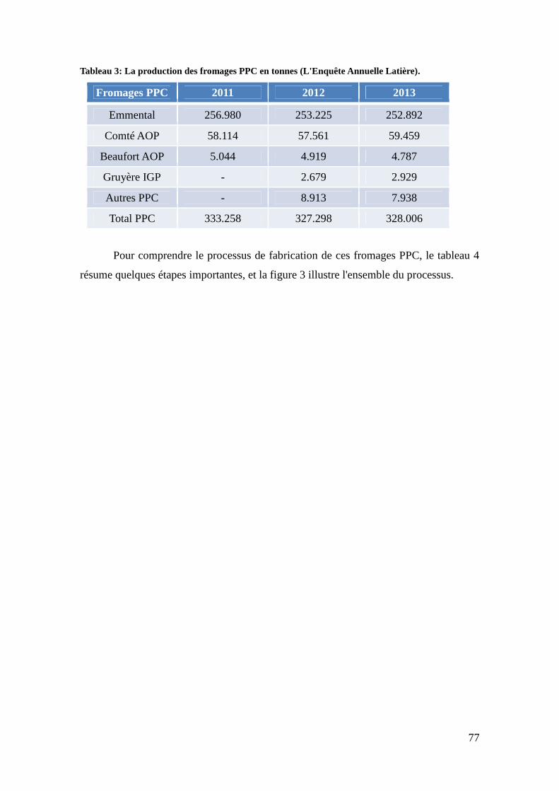
77
Tableau 3: La production des fromages PPC en tonnes (L'Enquête Annuelle Latière).
Fromages PPC 2011 2012 2013
Emmental 256.980 253.225 252.892
Comté AOP 58.114 57.561 59.459
Beaufort AOP 5.044 4.919 4.787
Gruyère IGP - 2.679 2.929
Autres PPC - 8.913 7.938
Total PPC 333.258 327.298 328.006
Pour comprendre le processus de fabrication de ces fromages PPC, le tableau 4
résume quelques étapes importantes, et la figure 3 illustre l'ensemble du processus.
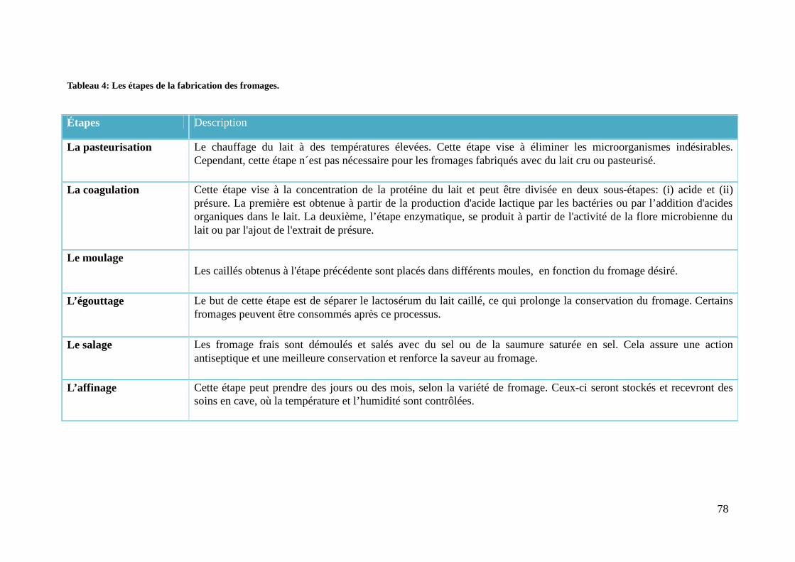
78
Tableau 4: Les étapes de la fabrication des fromages.
Étapes Description
La pasteurisation Le chauffage du lait à des températures élevées. Cette étape vise à éliminer les microorganismes indésirables.Cependant, cette étape n´est pas nécessaire pour les fromages fabriqués avec du lait cru ou pasteurisé.
La coagulation Cette étape vise à la concentration de la protéine du lait et peut être divisée en deux sous-étapes: (i) acide et (ii)présure. La première est obtenue à partir de la production d'acide lactique par les bactéries ou par l’addition d'acidesorganiques dans le lait. La deuxième, l’étape enzymatique, se produit à partir de l'activité de la flore microbienne dulait ou par l'ajout de l'extrait de présure.
Le moulageLes caillés obtenus à l'étape précédente sont placés dans différents moules, en fonction du fromage désiré.
L’égouttage Le but de cette étape est de séparer le lactosérum du lait caillé, ce qui prolonge la conservation du fromage. Certainsfromages peuvent être consommés après ce processus.
Le salage Les fromage frais sont démoulés et salés avec du sel ou de la saumure saturée en sel. Cela assure une actionantiseptique et une meilleure conservation et renforce la saveur au fromage.
L’affinage Cette étape peut prendre des jours ou des mois, selon la variété de fromage. Ceux-ci seront stockés et recevront dessoins en cave, où la température et l’humidité sont contrôlées.

79
Figure 3 : Processus de transformation des fromages PPC (http://www.produits-laitiers.com/). Le processus de fabrication des fromages PPC commence par la
collecte du lait dans les machines automatiques des exploitations laitières. Ensuite, ce lait est refroidi et analysé conformément à la réglementation pour la sécurité
des consommateurs, afin d'assurer un produit final de bonne qualité. Ce lait est transporté dans des camions citernes isothermes à l'industrie laitière où
commencent toutes les étapes de traitement et de fabrication du fromage.

80
L’Emmental est un fromage de poids dont les meules pèsent entre 60 et 120 kg. Il
faut 12 litres de lait pour obtenir 1 kg de ce fromage (CNIEL, 2015). Pendant son affinage
de nombreux paramètres varient : la température, le pH et l'activité de l'eau. P. freudenreichii
joue un rôle important dans son affinage (Figure 4), produisant par fermentation de l'acétate
et du propionate, qui participent la flaveur de l'Emmental, et du CO2, responsable de
l'ouverture du fromage par la formation de trous dans la pâte (les « yeux ») (Langsrud and
Reinbold 1973; Meurice et al. 2004).
Figure 4 : P. freudenreichii dans le fromage Emmental (© INRA).
P. freudenreichii joue ainsi un rôle important dans la composition de l’arôme et de la
saveur de certains produits laitiers, parmi lesquels l’Emmental. En effet, pendant l’affinage
prolongé de ce fromage, P. freudenreichii assure des réactions de lipolyse. La lipolyse
contribue à la formation de saveurs et est réalisée par des esterases qui sont capables
d'effectuer l'hydrolyse des triacylglycérides du lait en libérant des acides gras à chaîne
courte, à chaîne moyenne et à chaîne longue (Figure 5). Ceux sont eux qui donnent aux
fromages leurs différentes sensations gustatives, comme par exemple, une saveur forte,
épicée et/ou salée (Berdagué et al. 1990).
Figure 5 : Schéma de réaction d'hydrolyse de triacylglycérides.
Les études de Dherbécourt et collaborateurs ont identifié 23 estérases (estérases
putatives) chez P. freudenreichii, potentiellement impliquées dans ce processus d’affinage.

81
Cette identification s’est faite à partir de la combinaison de méthodes classiques et de l'étude
génomique de cette bactérie (Dherbécourt et al. 2008). Pour l’approche génomique, les
outils bioinformatiques et les méthodes utilisés ont été : (i) la recherche automatique
d’estérases par homologie, (ii) la recherche des motifs protéiques dans l’InterProScan, (iii)
la recherche manuelle par homologie et (iv) la recherche manuelle faite par motifs
spécifiques grâce à Prosite. Ces méthodes ont été indispensables pour une première sélection
d’enzymes d'intérêt. Ainsi 12 gènes candidats codant pour des estérases potentielles
présentant des motifs GXSXG caractéristiques des α/β hydrolases ont été sélectionnés et
clonés dans E. coli. Des zymographes ont également été effectués afin de caractériser
l'activité de ces estérases. L'inactivation et la complémentation des deux gènes d'estérases
codant pour des protéines ancrées en surface de P. freudenreichii ont révélé que le gène
pf279 était responsable de la lipolyse. De plus, l'analyse du génome de 21 souches de P.
freudenreichii a montré que ce gène pouvait présenter des mutations non-sens dans quelques
souches qui ne présentent pas d'activité lipolytique (Abeijon Mukdsi et al. 2014).
Nous avons déjà vu que P. freudenreichii survivait dans divers environnements et
contraintes de façon souche dépendante. L’importance de ce phénomène donne de l’attrait à
l’étude des stratégies qui permettent la survie et le maintien d’une activité métabolique
pendant de longues périodes. Afin d'exploiter ces mécanismes de survie à long terme, les
études qui associent des expériences microbiologiques (in vitro) en lien avec des
applications industrielles et biotechnologiques sont d'une grande importance. Ces études
seront décrites dans les sections suivantes.
82
2.2. Optimisation de la production de vitamine B12
En 1911, le terme « vitamine » a été proposé par Casimur Funk pour désigner les
amines qui sont essentiels à la vie (vita-amine). Plus tard, cette nomenclature a commencé à
être utilisée également pour les substances organiques essentielles, sans valeur énergétique
mais actives en petites quantités, contribuant à l’entretien du métabolisme d'un organisme
(Florent 1986; Vandamme 1992).
La vitamine B12, aussi appelée cobalamine ou cyanocobalamine, a été isolée pour la
première fois en 1948 mais elle n’a été synthétisée qu'en 1973. Elle est en effet difficile à
obtenir et sa synthèse comporte plusieurs étapes pour obtenir la molécule finale (Vandamme
1989). Comme cette vitamine est impliquée dans des processus essentiels pour la régulation
du métabolisme des animaux et des humains comme la formation et maturation des
érythrocytes et la maintenance du système nerveux, les stratégies alternatives visant à
l’optimisation de sa production sont d'un grand intérêt.
Dans ce contexte, l'obtention de la vitamine B12 en utilisant des micro-organismes
est devenue une solution prometteuse pour le marché industriel. Parmi les bactéries qui en
produisent, P. freudenreichii se distingue en raison de son bon rendement. Ainsi, une
concentration de 5,12 mg/L peut être obtenue dans le fromage après 168h de fabrication
(Marwaha, Sethi and Kennedy 1983). Des études d’amélioration de la production de
vitamine B12 chez P. freudenreichii ont montré qu'il est possible de parvenir à un
rendement allant jusqu'à 6,35 mg/L après 192h de fermentation (García Hernández and
Pérez Mendoza 1983).
De nouvelles approches ont donc été proposées pour la biosynthèse de la vitamine
B12 au cours des décennies. L'obtention de la vitamine B12 par fermentation de P.
freudenreichii a été analysée dans le travail de Piao et collaborateurs, qui ont montré qu’en
jouant sur l'expression des gènes cobA, cbiLF et cbiEGH, impliqués dans la synthèse de la
vitamine B12, on pouvait augmenter la production de vitamine B12 (Piao et al. 2004).
D'autres études ayant le même but, comme le travail de Murooka et collaborateurs, ont
exprimé et amplifié les gènes de l'opéron cob et cbi, pour augmenter la biosynthèse des
cobalamines pendant le processus de fermentation (Murooka et al. 2005).
Pour optimiser la production de vitamine B12, une autre étude a été réalisée par
Wang et collaborateurs se concentrant sur l'effet de l'acide propionique et du 5,6-
diméthylbenzimidazole au cours de la biosynthèse de cette vitamine au cours de la culture
de P. freudenreichii (Wang et al. 2015). Il a été montré que la concentration d'acide
propionique varie de la phase initiale (10 à 20 g/L) à la phase tardive (20 à 30 g/L). Ceci

83
conduit à une rétroaction négative sur la biosynthèse de la vitamine B12, mais peut être
modifié par l’addition de 5,6-dimetilbenzimidazole (Wang et al. 2015). Ainsi, un contrôle et
une adaptation fine des conditions de culture sont essentiels pour l’obtention de grandes
quantités de vitamine B12.

84
2.3. L'obtention d’acide propionique
L'acide propionique est un acide gras àchaîne courte, saturé, de masse moléculaire
74,08 g/mol, également connu sous le nom d’acide propanoïque ou C₃H₆O₂ (GESTIS
Substance Database – disponible en http://gestisen.itrust.de/nxt/gateway.dll/gestis_en
/012590.xml?f=templates$fn=default.htm$3.0) (Figure 6).
Figure 6 : Représentation de la formule chimique de l'acide propionique.
Cet acide est largement utilisé dans les industries suivantes: la pétrochimie, les
produits cosmétiques, les plastiques, les herbicides et les agents de conservation. Dans ce
contexte, la biologie synthétique est un secteur innovant, impliquant la biologie et
l'ingénierie, qui permet de développer de nouvelles fonctions et de nouveaux systèmes, par
exemple, l’utilisation d’une voie biochimique spécifique pour obtenir un matériau à travers
une source d'énergie renouvelable (Parizzi et al. 2012).
La fermentation est l’un des procédés les plus utilisés dans l'industrie pour
l'obtention de divers produits et sous-produits, parmi lesquels l'acide propionique.
Actuellement, plusieurs stratégies visant l'obtention de ce produit ont été développées en
utilisant différentes sources de carbone, telles que la fermentation du sucre de canne
(saccharose), ces stratégies recherchent une réduction des coûts et une amélioration de la
production de cet acide. Les bactéries propioniques sont capables de synthétiser l'acide
propionique et elles représentent une source d'intérêt pour cet usage. Aussi, des études visant
la physiologie et le métabolisme des Propionibacteries ont été réalisées afin, entre autres,
d'identifier les meilleures espèces et les meilleures souches pour une utilisation industrielle
(Thierry et al. 2011).
La fermentation de lactate, par le métabolisme initial de P. freudenreichii, génère des
produits, tels que l'acétate, le propionate et le CO2, qui jouent des rôles variés et importants
pour les industries alimentaires et de biotechnologie (Meurice et al. 2004). Par exemple, le
propionate de calcium est un produit dérivé de l'acide propionique ayant la capacité d'agir
comme agent de conservation, capable de lutter contre les bactéries défavorables et les
champignons dans les aliments.
C’est à partir de ces observations sur l'acide propionique que Stowers et
collaborateurs ont évalué 17 souches de Propionibacterium et ont défini des paramètres

85
optimaux de production et de rendement pour l'acide propionique en fermenteur. En suivant
ces paramètres, des souches des espèces P. acidipropionici et P. freudenreichii produisent
des quantités élevées d'acide propionique (Stowers, Cox and Rodriguez 2014).
Cependant, il est toujours difficile d'obtenir un bon rendement d’acide propionique à
partir de Propionibacteria, car la génération de sous-produits dérivés de son métabolisme,
tels que l'acide succinique, l'acide lactique, l'acide acétique réduisent le rendement de l'acide
propionique (Stowers, Cox and Rodriguez 2014). Dans cette optique, il est important de
mener des études pour augmenter l'efficacité de la production de ce produit. Cette
production trouve aussi des débouchés en chimie verte à travers de la production de
propylène (C3H6), précurseur de l'acide propionique, un des hydrocarbures utilisés dans
l’industrie pétrochimique. La polymérisation du propylène conduit au polypropylène, un
composé largement utilisé dans diverses industries pour générer des carburants, des
emballages jetables, des textiles, utilisés dans les appareils électro-ménagers et l’industrie
automobile (Ammar et al. 2014). Ainsi, les bactéries qui synthétisent l'acide propionique à
fort rendement font l’objet d’un intérêt croissant, en raison de l’essor de la chimie verte et
d’un marché économique porteur pour les produits bios-sourcés.

86
2.4. Probiotique
Nous avons déjà évoqué que plusieurs bactéries, notamment des bactéries lactiques
et propioniques sont l’objet d'intérêt en raison de leurs propriétés probiotiques. A ce titre, P.
freudenreichii est largement étudié pour sa capacité à inhiber certains microorganismes
indésirables, à moduler la microflore du côlon par la production de 1,4-dihydroxy-2-
naphtoïque (DHNA), un facteur bifidogène, (Pérez Chaia, Zarate and Oliver 1999) et à
survivre à différents stress (Isawa et al. 2002; Okada et al. 2013), notamment les stress
rencontrés pendant le transit au sein du tube digestif (Jan et al. 2001; Leverrier et al. 2003a;
Saraoui et al. 2013b). P. freudenreichii est également investigué à déclencher l'apoptose dans
les cellules cancéreuses du côlon, à adhérer aux cellules épithéliales intestinales et au
mucus, stimulant ainsi des activités immunomodulatrices et la prolifération cellulaire chez
l’hôte (Ouwehand et al. 2002; Zárate et al. 2002). Dans cette section, nous présenterons une
courte synthèse des études portant sur les diverses propriétés probiotiques attribuées à P.
freudenreichii.
2.4.1 L'histoire et la définition du terme probiotique
Bien que les bénéfices de certains aliments fermentés pour la santé soient étudiés
depuis longtemps (travaux de Metchnikoff au début du XXe siècle), l’origine du terme
probiotique date de 1965, lorsque Lilly et Stillwell ont associé la survie de certains
microorganismes à l'effet bénéfique de leurs produits (Lilly and Stillwell 1965).
Actuellement, les probiotiques sont définis comme des microorganismes vivants qui,
lorsqu'ils sont administrés d'une manière appropriée, apportent des effets positifs sur la santé
de l'hôte (Bron, van Baarlen and Kleerebezem 2012). En raison de leur importance, les
publications sur PubMed les concernant ont pris de l'ampleur au cours des dernières années
(Figure 7).

87
Figure 7 : Publications dans PubMed avec les mots clés « probiotics bacteria » au cours des dernièresannées.
Cependant, pour une utilisation des probiotiques chez l'homme et pour que des
allégations santé soient acceptées, il est nécessaire d'effectuer des essais rigoureux in vitro et
in vivo, qui visent à démontrer la qualité et la sécurité de ces organismes avant leurs
applications dans la prévention ou le traitement de maladies et la supplémentation
alimentaire. Quelques espèces bactériennes comme Lactobacillus paracasei, L. johnsonii, L.
fermentum, L. plantarum, L. rhamnosus, Bifidobacterium longum et P. freudenreichii font
l’objet d’étude sur leurs capacités à améliorer la santé humaine, notamment par le contrôle
et la restauration de l’équilibre du microbiote intestinal (Parvez et al. 2006; Cousin et al.
2012; Santos Rocha et al. 2012; Luerce et al. 2014).
2.4.2 Contrôle du microbiote
Les bactéries propioniques peuvent avoir un effet sur la microflore des animaux et/ou
des humains en inhibant des microorganismes indésirables et/ou en augmentant la
prolifération des populations de Bifidobacterium sp., des bactéries qui jouent un rôle positif
sur le tube digestif (Picard et al. 2005). Leur augmentation présente un certain nombre
d'avantages physiologiques et cliniques pour la santé humaine et/ou animale, tels que : (i)
accélérer le temps de passage à travers le tractus gastro-intestinal ; (ii) exercer un effet de
barrière, car elles produisent des substances qui bloquent les sites de liaison de pathogènes ;
(iii) exercer des effets sur le système immunitaire et dans les maladies du tractus gastro-
intestinale ; (iv) réduire le risque de diarrhée lors d’infection à rotavirus ; (v) exercer des

88
effets sur la prolifération des cellules intestinales et (vi) empêcher la carcinogenèse du côlon
(Picard et al. 2005). En plus des Bifidobactéries, il existe d'autres espèces bactériennes
dominantes qui colonisent différentes parties du tractus gastro-intestinal et qui sont d'une
grande importance pour le contrôle du microbiote intestinal. Ces espèces sont présentées
dans la Figure 8.
Figure 8 : Espèces microbiennes dominantes dans les différentes parties du tractus gastro-intestinal.Figure adaptée de l'article "Influences microbiennes dans les maladies inflammatoires chroniques del'intestin" (Sartor 2008).
Le microbiote intestinal joue un rôle important dans le système immunitaire chez les
humains et/ ou les animaux et il est généralement acquis au cours de la première année qui
suit la naissance. Il peut être influencé par des facteurs environnementaux externes, tels que
la consommation de produits laitiers dans l'alimentation (Bernstein 2014; Geuking et al.
2014).
La composition du microbiote intestinal présente un intérêt fondamental de par ses
liens avec certaines pathologies gastro-intestinales. De nombreuses études cherchent ainsi à
améliorer notre compréhension de ces communautés microbiennes et de leur effet de
modulation du système immunitaire et la santé de l’hôte en général (Sartor 2008; Hooper
and Macpherson 2010; de Vos and de Vos 2012; Vangay et al. 2015; Velasquez-Manoff
2015).

89
2.4.3. Immunomodulation
L'immunomodulation est une propriété importante des bactéries probiotiques,
permettant l’induction et la régulation de la réponse immunitaire. Elle est aussi utile pour la
prévention ou le traitement du cancer, des maladies auto-immunes et dans le contrôle des
microorganismes indésirables (Ouwehand et al. 1999; Sathish et al. 2013).
L’association de bactéries propioniques à des bactéries lactiques et des
bifidobactéries peut avoir des effet bénéfiques sur des pathologies gastro-intestinales chez
les enfants et les personnes âgées. Dans ce cadre, les bactéries propioniques agiraient
indirectement en favorisant la croissance et le maintien à des concentrations élevées des
bifidobactéries au cours de la période du traitement (Mantere-Alhonen 1995). Les bactéries
propioniques seules semblent aussi pouvoir exercer un effet probiotique à travers une
immunomodulation (Cousin et al. 2010; Le Maréchal et al. 2015). L'effet
immunomodulateur des souches probiotiques est souvent associé à leur capacité d’adhésion
à l´épithélium intestinal. Cette adhésion leur permet de se maintenir plus longtemps dans le
tractus digestif, lors du transit. Cette capacité d'adhésion peut être un critère de sélection de
probiotiques et certaines souches de P. freudenreichii ont fait l’objet d'études dans ce
domaine (Ouwehand et al. 2002). L’implication des protéines de surface dans le rôle
immunomodulateur de ces bactéries a été testée par la caractérisation protéomique du
« surfaçome » de P. freudenreichii. Il a ainsi été montré que certaines protéines de surface,
extraites de souches de P. freudenreichii et purifiées, étaient capables de reproduire, in vitro,
une réponse anti-inflammatoire observée avec la souche complète (Le Maréchal et al. 2015).

90
2.4.4. Apoptose induite par les acides gras à chaîne courte
Les bactéries propioniques sont connues pour leur capacité à produire des acides gras
à chaîne courte (SCFA pour short chain fatty acids), tels que le propionate et l'acétate qui
sont les principaux produits de fermentation de P. freudenreichii. Il a été observé que ces
SCFA ont des effets intéressants sur la physiologie des cellules épithéliales intestinales (Jan
et al. 2002; Pérez Chaia and Zarate 2005).
Les SCFA, et principalement le butyrate, constituent une source d’énergie
privilégiée, un véritable carburant, pour les cellules épithéliales intestinales. En 1995, Hague
et colaborateurs ont montré que ces mêmes SCFA, par exemple, le butyrate de sodium, sont
également capables au contraire d'induire l'apoptose dans les cellules cancéreuses du colon
(Hague et al. 1995). Par la suite, les travaux de Jan et collaborateurs en 2002, ont montré in
vitro que des souches de P. acidipropionici et P. freudenreichii sont elles aussi capables
d'induire l'apoptose dans des lignées de cellules humaines de cancer colorectal, telles
que HT29 et Caco2. Cet effet a aussi été observé sur des lignées cellulaires humaines de
cancer du col de l’utérus (HeLa). Les SCFA induisent des signes caractéristiques de
l'apoptose, tels que : (i) la perte du potentiel transmembranaire des mitochondries, (ii) la
génération d’espèces réactives de l’oxygène, (iii) d’induction de la cascade caspase-3 et (iv)
la condensation de la chromatine au niveau nucléaire. Le mécanisme général de l'apoptose
via les SCFA est schématisé sur la Figure 9. Par ailleurs, cette activité pourrait s’exercer in
vivo car ces souches restent vivantes et métaboliquement actives dans l'intestin humain (Jan
et al. 2002).
Figure 9 : Induction de l'apoptose à partir de SCFAs produites par les bactéries propioniques dans lecôlon.

91
Il a ainsi été proposé que les bactéries propioniques pourraient venir compléter
l’arsenal d’options thérapeutiques disponibles dans la prévention ou le traitement du cancer
du colon (Lan et al. 2008). Plus récemment, la même équipe a proposé une nouvelle
formulation d'un lait fermenté avec certaines souches de P. freudenreichii, visant une
alimentation préventive ou un supplément dans l’alimentation de personnes qui sont
prédisposées à ce type de cancer (Cousin et al. 2012).

92
2.4.5. Molécule acide 1,4-dihydroxy-2-naphtoïque (DHNA)
Le composant 1,4-dihydroxy-2-naphtoïque (DHNA) présente un effet bifidogène
marqué (i.e., il favorise la croissance des Bifidobactéries) et constitue de ce fait un
prébiotique. Les prébiotiques sont généralement des oligosaccharides non digérés par les
enzymes digestives chez l'homme. Ainsi, la croissance de P. freudenreichii, qui produit du
DHNA, influe directement sur la croissance des bifidobactéries, faisant de P. freudenreichii
une bactérie combinant des activités pro- et pré-biotiques. Elle contribue ainsi à l’équilibre
de la flore intestinale, en favorisant le développement de bactéries bénéfiques. Le DHNA
permettrait par ailleurs de réduire l'inflammation du colon (Quigley, Hudson and Englyst
1999; Okada et al. 2006). In vivo, l’administration de DHNA entraîne aussi des
modifications du système immunitaire intestinal chez la souris, en modifiant la réponse
immunitaire innée chez les cellules intestinales (Okada et al. 2013).
De même, le DHNA est une molécule prometteuse qui pourrait être utilisée dans le
traitement de la colite. Dans un modèle de colite induite chez la souris, son administration
entraîne une amélioration dans l'histologie de l’épithélium instestinal, une suppression de la
migration des lymphocytes et de la synthèse d’interleukines pro-inflammatoires (Okada et
al. 2013). Dans ce contexte, des recherches supplémentaires doivent être effectuées visant à
une optimisation du DHNA pour des applications thérapeutiques et pour montrer que les
quantités produites in situ par P. freudenreichii pourraient aboutir aux mêmes effets.

93
2.4.6. Capacité d’adaptation
Cette sous-section fait le point sur la capacité de survie de P. freudenreichii et sa
capacité d’adaptation à différents stress. Elle sera divisée en deux parties: la tolérance au
stress thermique et au stress digestif. La tolérance à ces stress est une propriété très
importante chez les bactéries propioniques pour leur maintien et leur viabilité, ce qui
optimise leur utilisation comme probiotiques (Leverrier et al. 2003a).
Tolérance au stress termique:
Les bactéries propioniques survivent à une large gamme de températures. Dans les
étapes d'affinage des fromages PPC, elles résistent autant à basse température (4°C), qu’à
moyenne (30°C) et haute température (52-53ºC). Ces températures varient en fonction des
différentes étapes du processus de production et du type de fromage. Pour le fromage
Gruyère traditionnel, il y a une étape de cuisson soumettant les bactéries à une température
très élevée, entre 52-53ºC, pendant 30 minutes. La thermotolérance à 55°C de différents
souches utilisées dans la fabrication de Gruyère a été étudiée par une approche protéomique
et les profiles obtenus en électrophorèse 2D ont révélé différentes réponses selon que les
souches sont thermotolérantes ou thermosensibles (Anastasiou et al. 2006). Dans les espèces
thermotolérantes, les protéines associées au processus de la fermentation propionique, au
métabolisme des acides aminés, aux nucléotides et à la phosphorylation oxydative étaient
surexprimés. En particulier, les chaperons moléculaires et les composés antioxydants de type
thiol sont induits par la chaleur. Toutefois, il a aussi été observé que, lors de l’adaptation
thermique, les voies de la transduction du signal, la biosynthèse et le maintien de la paroi
cellulaire contribuent à maintenir le processus de fermentation (Anastasiou et al. 2006).
D’autres études portant sur les bactéries propioniques ont également rapporté que les
chaperons moléculaires et des protéines de choc thermique sont induites dans de nombreuses
situations de stress (Jan et al. 2001; Leverrier et al. 2003a). Cette différence dans
l’expression des protéines des diverses souches de bactéries propioniques renforce
l'argument selon lequel il y a différents phénotypes au sein de la même espèce (Anastasiou
et al. 2006). Cette forme de tolérance est importante chez P. freudenreichii pour maintenir la
survie bactérienne et assurer le bon déroulement du processus de fabrication des fromages
de type PPC. Elle constitue en outre un avantage indéniable pour son utilisation comme
probiotique, si on y associe sa résistance aux stress digestifs (Leverrier et al. 2003b).

94
Tolérance au stress digestif:
Pour qu’un probiotique exerce sa fonction, il faut maintenir son activité métabolique
et sa survie après son passage à travers le tractus gastro-intestinal. Le travail de Lan et
collaborateurs a démontré cette capacité chez P. freudenreichii en démontrant la viabilité des
bactéries dans le tractus digestif et dans le colon de la souris (Lan et al. 2007). Cette étude a
également confirmé la tolérance au pH acide de certaines souches spécifiques de P.
freudenreichii, comme cela avait déjà été rapporté dans les études précédentes portant sur la
résistance au stress acide (Jan, Rouault and Maubois 2000), mais aussi sur la tolérance aux
sels biliaires (Warminska-Radyko, Laniewska-Moroz and Babuchowski 2002). Des
expériences de protéomique, utilisant l'électrophorèse 2D, ont montré 24 protéines induites
pendant l'adaptation de P. freudenreichii dans un milieu contenant des sels biliaires. On
compte parmi elles des protéines de transduction du signal et des facteurs sigma alternatifs.
Pour évaluer l'activité métabolique de P. freudenreichii, une RT-PCR quantitative
utilisant une sonde TaqMan et ciblant l’ARNm codant pour l’enzyme transcarboxylase a été
développée. Cette méthode non-invasive et efficace a été utilisée pour prouver la survie et
l’activité métabolique de P. freudenreichii dans le tractus intestinal des souris (Hervé et al.
2007). D’autres méthodes cherchant à évaluer l'activité métabolique de P.freudenreichii dans
le fromage ont aussi été développées dans le but de caractériser ces souches pour leur survie.
Dans ce contexte, une méthode de RT-PCR quantitative utilisant du SyberGreen a été
optimisée pour la quantification de cellules et d’ARNm marqueurs (gènes 16S, tuf, groL) de
P. freudenreichii et de Lactobacillus paracasei, pendant le processus de production des
fromages PPC (Falentin et al. 2010). Cette méthode a permis de quantifier la viabilité
cellulaire, et d’aller plus loin dans les études concernant la survie de P. freudenreichii.

95
3. Génomique de Propionibacterium freudenreichii
Grâce aux technologies de séquençage de nouvelle génération (NGS), il a été
possible d’obtenir le séquençage de plusieurs génomes de bactéries propioniques, ce qui a
conduit à une meilleure connaissance biologique de l'espèce. Ces génomes présentent, en
général, une teneur élevée en GC, environ 67% (Falentin et al. 2010a) et certaines souches
peuvent héberger en outre des plasmides (Luijk et al. 2002). Les données provenant du
séquençage peuvent être utilisées dans les études comparatives et/ou évolutives visant à
comprendre la génomique, au niveau structurel et au niveau fonctionnel de ce
microorganisme.
En 2010, le STLO a publié le premier génome complet de P. freudenreichii (souche
CIRM-BIA1) qui a été séquencée en utilisant la méthode traditionnelle de Sanger (Falentin
et al. 2010a). L’assemblage initial de ce génome a été effectué sans séquences répétitives, en
utilisant le logiciel Phred/Phrap/CONSED (www.phrap.org) (Vallenet et al. 2008; Falentin et
al. 2010a). Ensuite, pour la clôture des gaps, il a été nécessaire d'utiliser le kit sequence GC
finishing (GEhealthcare) et le kit transposition bombs (Falentin et al. 2010a). Ce génome
présente un chromosome circulaire de 2,7 Mb, de haute teneur en GC (67%) et est
disponible dans la base de données Genbank, avec le numéro d'accès FN806773 (Falentin et
al. 2010a).
Depuis, 21 autres génomes de P. freudenreichii ont été séquencés par l’équipe. Leur
séquençage a été réalisé par la méthode Illumina, en utilisant la bibliothèque paired-ends,
sauf pour la souche CIRM-BIA 9 qui a utilisé la bibliothèque single reads. L'assemblage de
ces 21 génomes a été mené par une approche de novo, puis les scaffolds ont été ordonnés par
comparaison avec le génome de référence complet CIRM-BIA1 (Falentin et al. 2010a; Loux
et al. 2015a). Les détails de l'assemblage de ces 21 génomes sont présentés dans le Tableau
5. Tous les génomes ont été annotés sur la plate-forme AGMIAL, qui est une interface
graphique pour l'annotation de génomes complets et incomplets des procaryotes. Les 21
génomes ont été déposés dans la base de données de EBI-EMBL et ces données sont
disponibles en suivant le lien : http://www.ebi.ac.uk/ena/data/view/Taxon:Propionibacte
rium%20freudenreichii (Loux et al. 2015b). Ces 21 génomes comprennent notamment celui
P. freudenreichii CIRM-BIA138, une des souches utilisées au cours de ces travaux de thèse
pour l’analyse en RNA-Seq.
Cependant, la plupart de ces nouveaux génomes de P. freudenreichii sont encore
incomplets, à l’état de drafts, cela est dû à la difficulté de l’étape d'assemblage liée à la
présence de nombreuses séquences répétées (transposons).

96
Des analyses bioinformatiques ont été réalisées sur ces différents génomes de P.
freudenreichii, afin d’identifier les gènes impliqués dans (i) la résistance aux différents pH et
à la présence des sels biliaires; (ii) la synthèse des vitamines et des acides aminés et (iii) le
stockage du carbone. Ces analyses montrent qu'il s’agit d’une bactérie versatile, dotée d’un
large arsenal de gènes pour survivre dans des environnements défavorables (Falentin et al.
2010a; Loux et al. 2015b). Ces données contribuent à expliquer la capacité de P.
freudenreichii à maintenir une activité métabolique en conditions de stress et de survie à
long terme. Ceci explique pour partie sa viabilité dans des environnements qui simulent le
tractus gastro-intestinal chez les humains et les animaux. Ces analyses génomiques ont aussi
été largement utilisées pour identifier des gènes potentiellement liés à son activité
immunomodulatrice.
D'autres études ont révélé la présence de mutations ponctuelles et d’îlots
génomiques, variables d’une souche à l’autre. Ceci montre la plasticité chromosomique de
cette espèce, et résulte probablement de transferts horizontaux, duplications, transpositions
et mutations accumulées. L’analyse de ces variations inter-souches, couplée à une
caractérisation phénotypique complète de leur capacité à métaboliser les sucres a permis
d'’établir un lien entre cette plasticité et le fait que certaines souches de P. freudenreichii
utilisent, préférentiellement, certaines sources de carbone. Ces analyses ouvrent aussi de
nouvelles perspectives pour améliorer leur sélection et leur utilisation pour l’industrie
fromagère et des probiotiques (Loux et al. 2015b).
Le génome complet de P. freudenreichii CIRM-BIA 1 a aussi été analysé et comparé
à celui de P. acnes afin d’identifier les facteurs différenciant ces deux espèces proches. Cette
analyse a aussi été menée pour cerner ce qui sous-tend les capacités probiotiques de
certaines souches de P. freudenreichii (Falentin et al. 2010a).
Ces analyses génomiques structurelles basées sur l'ADN sont complétées d’analyses
fonctionnelles à travers des études portant sur l’ARN, les protéines et les produits de
métabolisme de cette bactérie (respectivement, des approches transcriptomique,
protéomique et métabolomique). Cela est illustré dans le deuxième chapitre de la section de
résultats de ce manuscrit thèse : l'utilisation de la génomique fonctionnelle pour comprendre
le phénomène de la survie à long terme chez P. freudenreichii.

97
Tableau 5 : Génomes de P. freudenreichii déposés par le STLO.
Number in CIRM-BIAcollection (Rennes,
France)
Locus_tag Isolated from (date) Bioproject Raw reads accessionnumber
Readquantity
Readlength
Sequencing depth
Number ofscaffolds>1000 bp
Cumulative contiglength
N50 Number ofN
segments
Number ofcomplete
genes
9 PFCIRM9 Emmental cheese (1989) PRJEB6427 ERS638416 26 013 151 37 370 204 2 533 825 21 887 0 2 308
118 PFCIRM118 Gruyère cheese (1973) PRJEB6428 ERS638410 51 926 392 37 739 41 2 583 496 193 767 59 2 325
119 PFCIRM119 Gruyère cheese (1973) PRJEB6430 ERS638409 58 084 132 37 827 46 2 644 402 128 788 91 2 422
121 PFCIRM121 Swiss cheese (1937) PRJEB6431 ERS638414 52 432 380 37 746 59 2 561 530 86 244 68 2 304
122 PFCIRM122 NCIB (1992) PRJEB6432 ERS638421 54 950 090 37 782 78 2 614 705 73 741 70 2 348
123 PFCIRM123 Morbier cheese (1992) PRJEB6438 ERS638423 54 523 668 37 776 47 2 604 827 114 519 78 2 377
125 PFCIRM125 Emmental cheese (1992) PRJEB6434 ERS638418 54 868 488 37 781 43 2 511 448 238 430 59 2 272
127 PFCIRM127 Emmental cheese (1992) PRJEB6435 ERS638428 55 178 920 37 785 57 2 567 344 81 280 53 2 330
129 PFCIRM129 Emmental cheese (1992) PRJEB4826 ERS638425 52 269 870 37 744 59 2 591 314 123 180 59 2 338
134 PFCIRM134 NA PRJEB6441 ERS638419 50 523 582 37 719 47 2 635 396 153 798 152 2 318
135 PFCIRM135 ewe raw milk (1994) PRJEB6442 ERS638422 50 649 408 37 721 47 2 620 043 114 923 75 2 343
138 PFCIRM138 Emmental cheese (1992) PRJEB6433 ERS638424 58 100 372 37 827 72 2 573 580 80 515 110 2 304
139 PFCIRM139 Emmental cheese (1992) PRJEB6436 ERS638426 60 700 312 37 864 45 2 589 117 106 963 53 2 362
456 PFCIRM456 raw milk Raclette cheese (1992) PRJEB6445 ERS638427 49 094 398 37 699 38 2 502 068 131 281 61 2 253
508 PFCIRM508 Gruyère (1973) PRJEB6429 ERS638411 50 259 834 37 715 47 2 578 004 141 315 93 2 302
512 PFCIRM512 raw milk Morbier cheese (1994) PRJEB6439 ERS638413 63 808 750 37 908 45 2 580 419 153 592 57 2 349
513 PFCIRM513 egyptian ras cheese (1995) PRJEB6440 ERS638415 62 230 134 37 886 48 2 659 761 179 407 54 2 416
514 PFCIRM514 hay (1994) PRJEB6443 ERS638417 51 481 488 37 733 51 2 521 626 121 592 66 2 285
516 PFCIRM516 nepalian yack cheese (2000) PRJEB6444 ERS638420 61 823 266 37 880 62 2 630 828 125 877 205 2 342
527 PFCIRM527 Fribourg cheese (1992) PRJEB6437 ERS638412 69 806 362 37 993 63 2 576 369 104 864 57 2 344
1025 PFCIRM1025 Emmental cheese (1992) PRJEB6446 ERR738371 61 323 626 81 1 910 104 2 699 685 62 523 19 2 308

98
4. Transcriptomique de Propionibacterium freudenreichii
Au cours des 10 dernières années, plusieurs études ont porté sur l'expression des
gènes chez Propionibacterium afin de mieux comprendre et d'expliquer comment se
produit leur régulation dans ce groupe bactérien. Ces données transcriptomiques sont
déposées dans des bases, comme l'EMBL-EBI (http://www.ebi.ac.uk/genomes/), créées
pour permettre l'accès public, qui fournissent des données du génome et du
transcriptome de différentes bactéries. Ces travaux nous aident à comprendre comment
les bactéries propioniques s’adaptent et régulent l'expression des gènes en réponse à
différentes conditions environnementales.
Des espèces comme P. acnes et P. freudenreichii sont d'une grande importance
dans ce genre, car P. acnes est commensale et fait partie de la flore de la peau ; quant à
P. freudenreichii, nous avons déjà mentionné plus haut qu’elle est d'intérêt pour
l’industrie laitière dans le secteur des fromages PPC et d'intérêt biotechnologique pour
la synthèse de biomolécules et pour ses propriétés probiotiques. Les premières études de
transcriptomiques sur P. freudenreichii ont été réalisées avec les techniques de puce
d’ADN et RT-qPCR. Ces études ont montré la survie et l’adaptation de ces bactéries à
des températures variées pendant le processus d’affinage des fromages PPC, ainsi que
leur adaptation métabolique (in situ) dans le côlon de porcs (Dalmasso et al. 2012b;
Saraoui et al. 2013b). Afin d'accroître les connaissances sur la survie de cette bactérie,
nous avons utilisé le RNA-Seq, une approche transcriptomique plus exhaustive, couplé
à une analyse métabolomique ciblée. Nous présenterons, dans la section II des résultats
de ce manuscrit, un article concernant une analyse de la souche P. freudenreichii CIRM-
BIA138, intégrant ses données transcriptomiques et métabolomiques obtenues in vitro.
L’association de ces données aux niveaux ARN et biochimique, aux données
protéomiques déjà obtenues concernant P. freudenreichii permettra d’aborder l’étude
fonctionnelle de cette bactérie et nous éclairera sur ses capacités d’adaptation et de
survie (Figure 10).

99
Figure 10 : Combinaison des données des données génomiques, transcriptomiques et protéomiques.

100
5. Les principales voies métaboliques de Propionibacterium freudenreichii
Les bactéries propioniques tirent leur énergie de divers substrats carbonés par la
fermentation propionique. Chez P. freudenreichii, l’association des cycles de Krebs et
de Wood-Werkman sont caractéristiques de l’espèce.
Le cycle de Krebs, appelé également cycle de l’acide citrique ou cycle de l’acide
tricarboxylique (TCA, en anglais), correspond à des réactions métaboliques qui se
produisent dans le cytoplasme des cellules bactériennes. Ce cycle est une voie
amphibolique présentant des étapes de (i) dégradation des métabolites pour générer de
l'énergie – ATP – (voie catabolique) et (ii) production de précurseurs pour la biosynthèse
(voie anabolique) (Nelson et al. 2008).
Chez P. freudenreichii, le cycle Wood-Werkman, également connu sous le nom
de voie de carbone central, est une composante centrale du métabolisme. Ce cycle
permet la formation de propionate à partir du pyruvate (Wood 1981). Au cours de la
fermentation, les bactéries propioniques sont capables d'utiliser le lactate et de produire,
comme cité précédemment, certains sous-produits d’intérêt pour la composition de
l'Emmental. La Figure 11 illustre le métabolisme de P. freudenreichii, basé sur le cycle
Wood-Werkman et son association au cycle de Krebs.
Figure 11 : Voie métabolique de Wood-Werkman chez P. freudenreichii liée au cycle de Krebs.Figure adaptée de l’article: A Temporal -omic Study of Propionibacterium freudenreichii CIRM-BIA1T Adaptation Strategies in Conditions Mimicking Cheese Ripening in the Cold(http://journals.plos.org/plosone/article?id=10.1371/journal.pone.0029083).

101
La réaction globale décrite dans ces bactéries suit l'équilibre suivant: (Crow
1986)
3 lactate → 2 propionate + 1 acetate + 1 CO2 + 1 H2O (voie de Fitz)
P. freudenreichii jouant un rôle majeur dans la maturation des fromages PPC et
le développement de leur flaveur (Figure 12). Aussi les diverses voies métaboliques
aboutissant à la formation de composés d’arôme ont-elles été recherchées, notamment
en combinant des études génomiques et métabolomiques. Diverses voies métaboliques
(métabolisme de la glycolyse, de la lipolyse, des acides aminés, de stockage d’énergie)
ont ainsi été reconstruites intégralement, pour certaines souches. Ces travaux ont fait
l’objet de nombreuses articles et de revues détaillées (Thierry et al. 2011).
Figure 12 : Schéma des processus métaboliques impliqués dans l’affinage des fromages PPC visantl’obtention des composés aromatiques.
102
CAPÍTULO 2 : RESULTADOS
CHAPITRE 2 : RÉSULTATS

103
SEÇÃO I: Caracterização do fenótipo delinhagens de Propionibacteriumfreudenreichii
SECTION I : Caractérisation duphénotype des souches dePropionibacterium freudenreichii

104
1. Introdução
A primeira seção de resultados será apresentada na forma de artigo científico. O
objetivo deste estudo foi avaliar a variabilidade entre as linhagens e de caracterizar os
fenótipos das células de P. freudenreichii durante a sobrevivência a longo prazo.
Este manuscrito descreve, inicialmente, a caracterização fenotípica de 8
linhagens de P. freudenreichii, em meio de cultura sem a adição de nutriente durante um
período de 11 dias, sendo os 8 últimos correspondentes à uma carência nutricional.
Durante este período, cada linhagem foi monitorada por densidade óptica, enumeração
de colônias (ágar) e observação morfológica das colônias. A viabilidade e integridade da
membrana também foram avaliadas. Para isso, foi utilizado o kit live/dead
acompanhado de uma observação em microscopia de epifluorescência, e nós
verificamos a ausência de lise celular qPCR com primers específicos para o gene alvo
tuf.
Após 2 dias de cultura a população atingiu 9 log10 UFC/mL. Após 11 dias as 8
linhagens de P. freudenreichii, mantiveram uma população elevada, de
aproximadamente 7.8 a 8.2 log10 UFC/mL. O lactato, principal composto de carbono foi
totalmente consumido entre 2 e 3 dias após a inoculação, e as bactérias tiveram que lidar
com um período de estresse nutricional. A integração dos métodos utilizados permitiu
comparar os níveis de sobrevivência e serviu como base para a triagem das 8 linhagens.
A linhagem CIRM-BIA 138 é aquela que tem mostrado uma melhor sobrevivência à 11
dias após a inoculação. As pesquisas desta tese foram então focadas nesta linhagem.
Existem três principais mecanismos que permitem a sobrevivência bacteriana,
quando as bactérias estão expostas à condições ambientais desfavoráveis: (i) a
esporulação, (ii) a morte celular programada e (iii) dormência. Já está estabelecido que
P. freudenreichii não esporula. Quanto ao mecanismo de morte celular programada, os
restos celulares de parte da população lisada fornecem nutrientes para a subpopulação
não lisada, permitindo assim que determinadas espécies bacterianas enfrentem as
condições de carência nutricional. Os resultados de qPCR mostram uma estabilidade da
população bacteriana à 9 log10 e nos levam a concluir que P. freudenreichii CIRM-138
não usa a morte celular como mecanismo para a sobrevivência a longo prazo para
manter a sua viabilidade, uma vez que a lise celular não é detectável nestas culturas.
Portanto, nossa hipótese é que esta bactéria permanece viável por causa da sua
entrada em estado de dormência. Os resultados da microscopia de epifluorescência
mostraram que metade das células bacterianas de todas as linhagens apresentaram

105
membranas permeabilizadas após 11 dias de cultura. Durante o período de
sobrevivência, uma perda de 1 log10 UFC/mL foi observada em comparação aos
resultados de quantificação da população por qPCR. A comparação dos resultados de
contagem de colônias e de qPCR, assim como a ausência de lise sugerem que a 11 dias
de cultura, 90% da população bacteriana perderam a capacidade de formar colónias em
ágar, mas que uma boa parte das células são, no entanto, viáveis. A integração dos
resultados da caracterização fenotípica sugerem que esta bactéria atinge um estado
VBNC no momento o qual começa o período de sobrevivência a longo prazo. Este
estado é descrito em muitas bactérias Gram-positivas e Gram-negativas, sendo uma
estratégia para a sobrevivência nos períodos de estresse, tais como a temperatura, o pH e
a disponibilidade de nutrientes variados.
Este trabalho mostra pela primeira vez um mecanismo de dormência abordado
em P. freudenreichii, cujas células entram em estado VBNC para garantirem sua
sobrevivência a longo prazo em condições de carência nutricional. Isso aumenta o nosso
conhecimento nesta espécie de lactéos e abre perspectivas para novos estudos. Novas
abordagens integradas, baseadas em técnicas omics devem ser realizadas para
compreender o mecanismo de dormência e a regulação metabólica bacteriana durante
este período de sobrevivência a longo prazo em P. freudenreichii (ver secção - Secção II
deste manuscrito de tese).

106
1. IntroductionLa première section des résultats sera présentée sous forme d'article
scientifique. L'objectif de cette étude a été d´évaluer la variabilité inter-souches et de
caractériser les phénotypes des cellules de P. freudenreichii pendant la survie à long
terme.
Ce manuscrit décrit d’abord la caractérisation phénotypique de 8 souches de P.
freudenreichii, en milieu de culture sans ajout sur une période de 11 jours dont les 8
derniers correspondent à une carence nutritionnelle. Pendant cette période, chaque
souche a fait l’objet d’un suivi par densité optique, par dénombrement sur gélose et en
observant la morphologie des colonies. La viabilité et l'intégrité membranaire ont aussi
été évaluées. Pour cela, nous avons utilisé le kit live/dead suivi d'une observation en
microscopie à épifluorescence et nous avons vérifié l'absence de lyse cellulaire par PCR
quantitative en ciblant le gène tuf.
Après 2 jours de culture la population atteignait 9 log10 UFC/mL. Après 11 jours
les 8 souches de P. freudenreichii, maintenaient une population élevée, d´environ 7.8 à
8.2 log10 UFC/mL. Le lactate, principal composé carboné étant épuisé entre 2 et 3 jours
après inoculation, et les bactéries ont dû faire face à une période de stress nutritionnel.
L'intégration des méthodes utilisées a permis de comparer les niveaux de survie et a
servi de base au le criblage des 8 souches. La souche CIRM-BIA 138 est celle qui a
montré la meilleure survie à 11 jours après inoculation. Les recherches de la thèse se
sont ensuite focalisées sur cette souche.
Il y a trois mécanismes majeurs qui permettent la survie bactérienne lorsque les
bactéries sont exposés à des conditions environnementales défavorables : (i) la
sporulation, (ii) la mort cellulaire programmée et (iii) la dormance. Il est déjà établi que
P. freudenreichii ne sporule pas. Lors du mécanisme de mort programmée, les débris
cellulaires d'une partie de la population lysée fournissent des nutriments pour la sous-
population non-lysée, et permettent à certaines espèces bactériennes de faire face à des
conditions de carence nutritionnelle. Les résultats de PCRq montrant une stabilité de la
population bactérienne à 9 log10 nous amènent à conclure que P. freudenreichii CIRM-
138 n’utilise pas la mort cellulaire programmée comme mécanisme de survie à long
terme pour maintenir sa viabilité, car aucune lyse cellulaire n'est détectable dans les
cultures.

107
Nous émettons donc l'hypothèse que cette bactérie reste viable en raison de son
entrée en état de dormance. Les résultats de la microscopie à épifluorescence ont
démontré que la moitié des cellules bactériennes de toutes les souches présentait des
membranes perméabilisées après 11 jours de culture. Pendant la période de survie, une
perte de 1 log10 UFC/mL a été observée par rapport aux résultats de quantification de la
population par qPCR. La comparaison des résultats de dénombrement et de qPCR, ainsi
que l'absence de lyse suggèrent qu'à 11 jours de culture, 90 % de la population
bactérienne a perdu sa capacité à former des colonies sur gélose mais qu'une bonne
partie des cellules sont néanmoins viables. L'intégration des résultats de la
caractérisation phénotypique suggère que cette bactérie atteint un état VBNC au
moment où commence la période de survie à long terme. Cet état est décrit chez de
nombreuses bactéries à Gram positif et à Gram négatif, comme étant une stratégie pour
la survie dans des périodes de stress tels que la température, le pH et la disponibilité de
nutriments variés.
Ce travail montre pour la première fois qu'un mécanisme de dormance se met en
place chez P. freudenreichii, dont les cellules entrent en état en VBNC pour assurer leur
survie à long terme en condition de carence nutritionnelle. Cela accroît nos
connaissances sur cette espèce laitière et ouvre des perspectives pour de nouvelles
études. De nouvelles approches intégrées, basées sur des techniques omiques devraient
être menées pour comprendre le mécanisme de dormance et la régulation métabolique
bactérienne au cours de cette période de survie à long terme chez P. freudenreichii (voir
article - section II de ce manuscrit de thèse).

108
2. Artigo 1: The long-term survival of Propionibacterium freudenreichii in acontext of nutrient shortage.
Short running head: P. freudenreichii survival
Flavia Figueira Aburjaile1,2,3, Marie-Noëlle Madec2,3, Sandrine Parayre2,3,
Anderson Miyoshi1, Vasco Azevedo1, Yves Le Loir2,3†, Hélène Falentin2,3†
1 Department of General Biology, Federal University of Minas Gerais, Belo Horizonte,
Minas Gerais, Brazil
2 INRA, UMR 1253, Science et Technologie du Lait et de l’Oeuf, 35000, Rennes,
France
3 AGROCAMPUS OUEST, UMR1253, UMR Science et Technologie du Lait et de
l’Oeuf, 35000, Rennes, France
Correspondance: Hélène Falentin, INRA, UMR 1253, Science et Technologie du Lait et
de l’OEuf, Rennes, France, e-mail: [email protected].
†: YLL and HF share credit as senior co-authors

109
Abstract
Aims: Propionibacterium freudenreichii is an actinobacterium widely used in dairy
industry during the ripening process of Swiss-type cheeses and which presents probiotic
properties. PF is reportedly a hardy bacterium, able to survive during the cheese-making
process and when subjected to digestive stresses. During this study the long-term
survival of P. freudenreichii was investigated for 11 days by means of phenotypic
characterization in a culture medium without the addition of any nutrients.
Methods and Results: For 11 days, in a non-nutrient supplemented culture medium,
eight strains were monitored by measuring their optical density, counting colony-
forming units (CFU) and using LIVE/DEAD staining and microscopy observation.
Under these conditions, all strains displayed high survival rates in the culture medium,
their culturability reaching more the 9 log10 CFU/mL after two days. After 11 days, this
value ranged from 7.8 to 8.2 log10 CFU/mL depending on the strain, and at least 50% of
the P. freudenreichii population displayed an intact envelope. Since lysis of part of a
bacterial population may be a microbial strategy to recover nutrients, in CIRM-BIA 138
(the strain with the highest population at day 11), cell lysis was assessed by quantifying
intact bacterial cells using qPCR targeting the housekeeping gene tuf. No lysis was
observed.
Conclusion: Taken together, our results suggest that P. freudenreichii strains use a
viable but nonculturable (VBNC) state to adapt to the long term survival phase.
Significance and Impact of the study: Assessing the viability of P. freudenreichii and
understanding their mechanisms for survival should be of great interest regarding their
potential probiotic applications.

110
Keywords: adaptation; bacteria; growth phases; culturability; long-term survival; viable
but nonculturable; Propionibacterium freudenreichii.
Introduction
Propionibacterium freudenreichii is an Actinobacterium that is responsible for the
opening and aroma of Swiss-type cheeses. PF is also used as a source of probiotics
because PF has bifidogenic properties (Isawa et al., 2002), PF is resistant to digestive
stress (Jan et al., 2000), and some strains are endowed with anti-inflammatory
capabilities that could be used to prevent inflammatory bowel diseases (Okada et al.,
2013; Le Maréchal et al., 2015). Its metabolic activity is characterized by its ability to
ferment various carbohydrates, the main product being propionic acid (Houwen et al.,
1991; Loux et al., 2015). PF is reportedly a hardy bacterial species, which can stay alive
and metabolically active for long periods under stressful conditions (i.e. cave-aged
Swiss-type cheeses at cool temperatures) (Dalmasso et al., 2012). The genome sequence
of the type-strain CIRM-BIA1 revealed the genetic basis for its hardiness (Falentin et
al., 2010a). In the laboratory, PF can be stored in a spent medium at room temperature
for more than 6 months and remains culturable if inoculated in a fresh medium.
Actinobacteria are known to be hardy and extremely resistant to nutrient shortage
(Greenblatt et al., 2004). However, except for the Mycobacterium genus which has been
extensively explored (Ghodbane 2014), little is known about the mechanisms
underlying the long term survival of this class.
During their growth and survival, Gram-positive and Gram-negative bacteria pass
through different physiological phases (Kolter et al., 1993; Hicks, 2005; Finkel, 2006;
Bruno & Freitag, 2011; Wen et al., 2013). In some bacterial species, conventional

111
exponential and stationary phases are followed successively by the death phase and a
long-term survival phase (Finkel, 2006; Helmus et al., 2012). Long-term survival (LTS)
can be defined as a phase during which the bacteria develop mechanisms to adapt and
remain viable, even after a long period of nutritional shortage (Finkel, 2006; Meena &
Rajni, 2010; Bharati et al., 2012; Justice et al., 2014): (i) sporulation (Fimlaid and Shen,
2015), (ii) quiescence/dormancy or (iii) programmed cell death (Finkel, 2006;
Rittershaus et al., 2013). During sporulation, a vegetative bacterial cell differentiates
into a metabolically dormant cell-type known as a spore, which can reawaken when
favourable conditions return (Fimlaid and Shen, 2015). Quiescence is characterized by
decelerated or arrested growth in a viable non-replicating state (Rittershaus et al., 2013),
which can evolve to a viable but nonculturable (VBNC) state (Ramamurthy et al.,
2014). In programmed cell death (bacterial apoptosis), the death and lysis of part of a
bacterial population may be profitable to surviving cells, which can catabolize the
detritus. Cell death and lysis can be observed by monitoring the fall in the viable count
using standard plating assays associated with a leakage of intracellular content that can
be assessed biochemically (lactate dehydrogenase quantification) (Wilkinson et al.,
1994) or using qPCR methods (Falentin et al., 2010b).
PF is a non-sporulating species, but PF is as yet unknown whether it survives by
adopting a strategy of quiescence/dormancy or lysis of part of the bacterial population.
During this study, we investigated the LTS (OD, culturability, membrane permeability
and lysis) of eight P. freudenreichii strains throughout an 11-day period of culture
without the addition of any nutrients, in order to investigate inter-strain variability in
long-term survival capacities and to better understand the strategies used to ensure
survival.
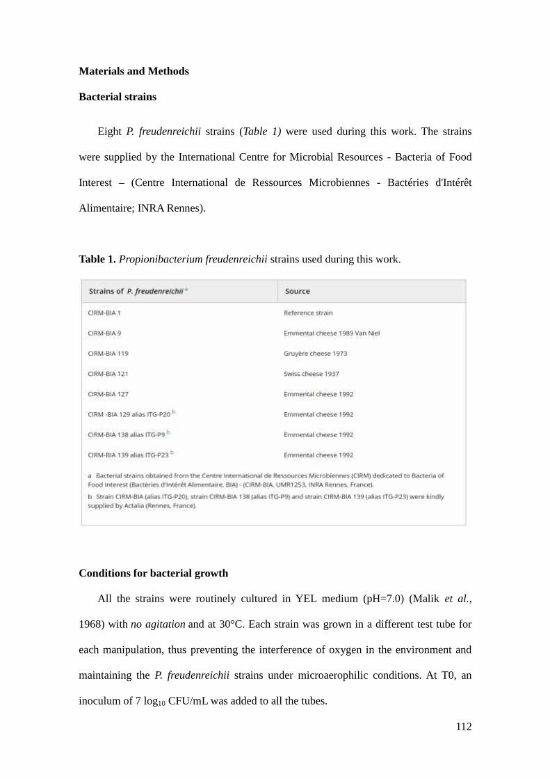
112
Materials and Methods
Bacterial strains
Eight P. freudenreichii strains (Table 1) were used during this work. The strains
were supplied by the International Centre for Microbial Resources - Bacteria of Food
Interest – (Centre International de Ressources Microbiennes - Bactéries d'Intérêt
Alimentaire; INRA Rennes).
Table 1. Propionibacterium freudenreichii strains used during this work.
Conditions for bacterial growth
All the strains were routinely cultured in YEL medium (pH=7.0) (Malik et al.,
1968) with no agitation and at 30°C. Each strain was grown in a different test tube for
each manipulation, thus preventing the interference of oxygen in the environment and
maintaining the P. freudenreichii strains under microaerophilic conditions. At T0, an
inoculum of 7 log10 CFU/mL was added to all the tubes.

113
Growth kinetics were followed by Optical Density measurements using a Model
DU 640 spectrophotometer (Beckman Coulter, Fullerton, California, USA) at 650 nm
(OD650) and by counting the number of CFU (CFU/mL) using the micromethod as
described previously (Baron et al., 2006). For enumerations, all the P. freudenreichii
strains were plated in YEL agar culture medium at 30°C, under microaerophilic
conditions, until the visualization of colonies (6 days). In all cases, two technical
replicates and two biological replicates were performed.
Epifluorescence microscopy
The eight strains were monitored for 11 days using the LIVE/DEAD®
BacLight™ Bacterial Viability Kit – Molecular Probes® (Life Technologies, USA), as
recommended by the supplier. Microscopic observations were performed at each of the
time points used for the OD650 and CFU/mL measurements. Briefly, SYTO9 (which
stains all cells green, regardless of their membrane integrity) and propidium iodide
(which stains permeabilized cells red) were mixed at a ratio of 1: 1 (5µL SYTO 9 and 5
µL propidium iodide). An 0.5µL aliquot of the mix was added to 100µL of the bacterial
culture, vortexed, and incubated in the dark for 15 minutes. The bacteria were then
analysed using epifluorescence microscopy with a phase contrast Nikon G11
microscope (Nikon D300) and Camera Control Pro 2 software (Nikon, Japan). For each
sample, six photos (three photos with a x40 objective and three with a x100 objective)
were analysed so as to visually estimate the percentage of permeabilized cells (red / red
+ green cells) at each time point.

114
DNA extraction of non-lysed cells
The DNA extraction method only enables the recovery of DNA from non-lysed
cells. Samples were collected at three time points (79h, 144h and 168h post-inoculation)
during the growth of P. freudenreichii CIRM-BIA 138, as described previously (Parayre
et al., 2007). Briefly, cells were harvested from 1 mL of the culture sample by
centrifugation (5000g, 10 min.). The cell pellets were re-suspended in lysis buffer
(Qiagen, Courtaboeuf, France) supplemented with lysozyme (Promega, France;
8mg/sample) and mutanolysin (Sigma, France; 20U/sample). These samples were
incubated for 30 min. at 37°C for 1 h, before being treated with proteinase K
(0.5mg/sample) prior to purification using the DNeasy Tissue Kit, as recommended by
the supplier (Qiagen, Courtaboeuf, France). Three biological replicates and two
technical replicates were used for each time point.
Quantification of bacterial cells using quantitative PCR
To determine any cell lysis of CIRM-BIA 138 after entry into stationary phase and
just before entering the LTS phase, quantitative PCR (qPCR) was used to quantify the
non-lysed bacterial cells throughout growth, according to the method described by
Falentin et al. (2010b). Briefly, the qPCR targeted tuf, a gene present in a single copy in
the P. freudenreichii chromosome, using primers designed on the tuf sequence (F:
CGAACGAGTTCCACTGCGGGT; R: GCAACATCGGCACCATCGGAC).
Amplifications were performed exactly as described previously. Cycle thresholds (Ct)
were determined after manual adjustment of the threshold within the linear part of
qPCR logarithmic amplification curves. CIRM-BIA 138 samples were collected at 79,
144, and 168 hours for qPCR determination. Each experiment was carried out in
triplicate (independent biological replicates). We checked by a spiking experiment that

115
lysed cells could not contaminate qPCR results. A sample of entire cells (8 log10 CFU)
was spiked with (9 log10 CFU) lyzed cells (the lysis was perfomed as described below).
Extraction and qPCR on entire cells and spiked sample gave the same Ct. The qPCR
results will give a quantification of entire cells only.

116
Results
Growth and survival rate of Propionibacterium freudenreichii under laboratory
conditions.
The growth of the eight P. freudenreichii strains was monitored
spectrophotometrically by measuring the optical density at 650 nm (OD650), and by
plate counting (CFU/mL) throughout an 11 day period. Strains were inoculated at 7
log10 CFU/mL in fresh YEL medium. Both of the monitoring methods showed that P.
freudenreichii growth curves had roughly similar profiles despite variations in terms of
their final culturability at 11 days. The curves established over a prolonged incubation
revealed several phases, as shown for CIRM-BIA129 and CIRM-BIA138 (Figure 1. See
all growth curves in the supplemental data, Figure S1). Under the conditions applied,
the cultures entered the exponential growth phase to reach a maximum OD650 of 3 to 4
(for CIRM-BIA 138) and a population of 9 to 9.6 log10 CFU/mL between 50 h and 72 h.
The OD650 curves then decreased steadily for about 100h, which corresponded to entry
into the stationary phase, and reached a plateau of culturability at around 168 h, for a
population of 8 to 8.7 log10 CFU/mL, depending on the strain (Figure 1) but always with
a lower OD650. The latter phase was designated the long term survival (LTS) phase. The
other six P. freudenreichii growth curves were similar (Supplementary data Figure S1).
In terms of culturability, all the strains survived well with a population of around 8 log10
CFU/mL after 11 days in the spent medium, i.e. without the addition of any nutrients
(Figure 2, Table S1). Interestingly, two strains, CIRM-BIA121 and CIRM-BIA138,
survived better than the six others (Figure 2) and maintained an average population of
8.2 log10 CFU/ mL at day 11, suggesting a strain-dependency in ability of P.
freudenreichii for long-term survival. On Petri plates, colonies of P. freudenreichii
maintained the same size and aspect whereas photon microscopy images revealed

117
bacteria of the same size (not shown) but single cells in the exponential phase and
groups of two to five cells in the stationary phase.
Figure 1: Growth curves of P. freudenreichii strains CIRM-BIA129 and CIRM-BIA138
determined at OD650 (upper panels), and plate counting (CFU ml−1, lower panels).
Figure 2: Viability of the eight P. freudenreichii strains (in CFU ml−1) after 11 days in
the spent culture medium (YEL) based on a mean of two technical replicates and two
biological replicates. Upper bars are standard deviations.

118
Evaluation of cell envelope permeability
LIVE/DEAD staining assays showed that the numbers of intact cells (stained green
by SYTO9) remained stable (100% index) for each of the eight strains throughout the
exponential and early stationary phases (Table S2). The numbers of intact cells then
started decreasing at 50 or 150h post-inoculation and dropped to 80% and 58.6% for
CIRM-BIA 129 and CIRM-BIA 138, respectively, 11 days post-inoculation. The
numbers of cells with a non-permeabilized membrane (stained in green) decreased from
50 h to 150 h onwards, depending on the strains, to reach a minimum proportion of 50%
at day 11 (Figure 3), suggesting that the number of dying or dead (but non-lysed) cells
increased during the stationary and LTS phases.
Figure 3: (A) Proportion of non-permeabilized cells during the growth and survival of
P. freudenreichii strains CIRM-BIA129 (a) and 138 (b) measured by LIVE/DEAD
assay followed by epifluroscence microscopy (mean values of three epifluorescence
microscopy fields at x40 magnification, and three fields at x100 magnification).
Representative epifluorescence microscopy views of P. freudenreichii (CIRM-BIA 138)

119
cultures after LIVE/DEAD staining during exponential growth (c) and after 11 days of
growth (d).
Assessment of lysis by quantifying bacterial cells using qPCR
Because both plate counting assays and epifluorescence microscopy showed a
reduction in culturability and an increase in membrane permeabilization, quantitative
PCR was used to assess bacterial lysis. CIRM-BIA 138 (the strain with the highest
population at day 11) cells were pelleted at three different time points before and after
the observed loss of culturability. Quantitative PCR targeting the tuf gene was carried
out on the DNA extracted from the pelleted cells. Under these conditions, lysed cells
cannot normally be recovered (Falentin et al., 2012). The copy number of the tuf gene
did not vary significantly in the CIRM-BIA138 culture, demonstrating that the bacterial
cells did not undergo lysis when entering the LTS phase (Figure 4).
Figure 4: Quantification of bacterial cells during the growth of P. freudenreichii CIRM-
BIA138. Each point gives the copy number of tuf gene determined by qPCR on DNA
extracted from P. freudenreichii CIRM-BIA138 (three biological replicates).

120
Discussion
P. freudenreichii has a high survival rate at day 11
P. freudenreichii belongs to the Actinobacteria, a group of Gram positive bacteria
with high GC content genomes. This group is known for its considerable versatility
(Gao & Gupta, 2012), and includes members that can survive under extreme conditions
(Greenblatt et al., 2004). P. freudenreichii is reported to be a hardy species widely used
in the cheese-making industry and is also well-documented for its probiotic potential
(Jan et al., 2000). Genome sequence analysis has revealed the genetic basis for its
hardiness: (i) its ability to store polyphosphates as an energy reserve and to utilize them
instead of ATP, (ii) its ability to store glycogen as a carbon source, and (iii) several
copies of chaperones to cope with different stresses (Falentin et al. 2010a). The eight P.
freudenreichii strains studied here survived well during a period of 11 days under
nutritional shortage, according to the optical density and plate counting assays
performed (around 8 log10 CFU/mL). In YEL medium, lactate - the preferential carbon
source for PF- was exhausted two days after inoculation (Dalmasso et al. 2012).
LIVE/DEAD staining followed by epifluorescence microscopy ascertained that more
than 50% of the P. freudenreichii populations had an intact (non-permeabilized)
membrane at this date. The growth curves for CIRM-BIA 129 and 138 (Fig. 1)
displayed some fluctuations during long-term incubation. We suggest that intermittent
cryptic regrowth might be responsible for this variability. These cryptic growths may
result from some specific metabolisms induced to adapt to starvation or the growth of a
sub-population of mutants derived from the inoculated strain and with a growth
advantage that was explained in the review by Finkel et al. (2006).

121
The best surviving strain did not use partial lysis to sustain viability
Previous studies had shown that some lactic acid bacteria present in dairy
products can lyse under difficult conditions and that the cell debris released into the
culture medium may serve as a nutrient source to ensure survival of the non-lysed sub-
population (Lortal et al., 1997; Deutsch et al., 2002; Lortal & Chapot-Chartier, 2005). A
study of the autolysis of Lactobacillus and dairy propionic bacteria showed that this
autolysis is associated with several factors, including among others the type of strain,
autolysins, pH, temperature and ionic strength (Lortal et al., 1997). For CIRM-BIA 138,
the quantification of intact cells using quantitative PCR showed that under the study
conditions applied here, lysis could be ruled out. Whether this is a general feature of P.
freudenreichii still needs to be determined. The absence of lysis (i) could be confirmed
by quantitative PCR on cells exposed to propidium monoazide, so as to prevent any
contamination by DNA from dead cells, (ii) needs to be confirmed in other P.
freudenreichii strains and under different conditions before it is generally accepted.
P. freudenreichii probably enters a viable but nonculturable (VBNC) state
The experimental data reported here offer a range of presumptions suggesting
that P. freudenreichii enters into a VBNC state. VBNC is a survival strategy adopted by
numerous bacteria in response to adverse conditions such as starvation, extreme
temperature, pH, oxygen stress, antibiotic pressure, etc. Once in a VBNC state, the
bacteria no longer grow in routine laboratory medium and conditions, but they
nevertheless remain viable and can be resuscitated when more favourable conditions are
provided (Ramamurthy et al., 2014). The methods generally used to detect VBNC
bacteria include bright field microscopy with nalidixic acid; fluorescent microscopy,

122
and especially use of the BacLight LIVE/DEAD kit; and molecular techniques (DNA or
RNA) (Oliver, 2009; Fakruddin et al., 2013). All of the eight P. freudenreichii strains
studied here displayed a loss of culturability of ~1 log10 CFU/mL (Table S1 and Figure
5), when this parameter was compared between 50-72 hours and 11 days post-
inoculation. LIVE/DEAD staining showed that about half of the bacterial cells had a
permeabilized membrane. Using quantitative PCR, an absence of lysis was shown on
strain CIRM-BIA138. These findings suggest that at least a part of the P. freudenreichii
population entered a VBNC state and that the entry into LTS is concomitant with entry
into a VBNC state, at least for 1 log10 (i.e. 90%) CFU/mL. Since the total bacterial
population was stained either green (50%) or red (50%), we suggest that VBNC cells
(90%) can take up both colours and that LIVE/DEAD staining cannot be used to
quantify VBNC cells.
Figure 5: Fate of CIRM-BIA138 bacterial cells between 50-72 h post-inoculation and
the long-term survival phase 11 days post-inoculation assessed by (a) culturability on

123
Petri plates, (b) LIVE/DEAD staining followed by epifluorescence and (c)
quantification of intact cells by quantitative PCR.
The VBNC state has been described in various Gram positive bacteria, such as
Enterococcus faecalis, Enterococcus hirae, Listeria monocytogenes, and
Mycobacterium tuberculosis (Oliver et al., 2009), and in Gram negative bacteria such as
Vibrio cholerae (Gonzales-Escalona et al. 2006), Escherichia coli (Darcan et al., 2009)
and Helicobacter pylori (Casasola-Rodríguez et al., 2013). Many of the studies on the
VBNC state have focused on pathogenic bacteria (Ramamurthy et al. 2014). This state
has also recently been reported for bacteria of food interest and particularly in ripened
cheese, suggesting a role for these bacteria in the organoleptic qualities of cheeses
(Martin-Platero et al. 2008, Desfossés-Foucault et al. 2012, Ruggirello et al. 2014).
In conclusion, our findings suggest that one strategy for bacterial survival is
dormancy, rather than partial bacterial lysis, in order to sustain the remaining cells.
During the past decade, many studies have investigated the bacterial mechanisms
underlying LTS in various fields: environmental, industrial, medical and
biotechnological microbiology. This study represents the first work involving the
phenotypic characterization of P. freudenreichii over an 11-day period without the
addition of nutrients, and revealed different phases of growth, membrane
permeabilization and entry into dormancy and a VBNC state to ensure LTS. A VBNC
state may however have an important impact on the technological uses of these bacteria,
as either starters in cheese-making or probiotics, since viability and metabolic activity
are important traits in their wide range of potential uses. This therefore opens the way
for further investigations on a molecular basis of the LTS of P. freudenreichii. The
availability of genome sequences (Loux et al., 2015) will greatly assist such
investigations using comparative genomics and transcriptomics approaches in order to

124
understand the organization, genes and metabolic pathways required for LTS. Assessing
viability and understanding the mechanisms underlying the survival of P. freudenreichii
should be of considerable interest regarding the development of effective bacterial
probiotics.
Acknowledgements
This work was supported by the CAPES-COFECUB French-Brazilian Cooperation
Programme. We also wish to thank the Institut National de la Recherche
Agronomique (INRA, UMR 1253, STLO, Rennes, France) for its financial and practical
support. Our thanks also go to Professor Mary Bret and Vicky Hawken for English
revision of the paper.
Conflict of Interest section
The authors have no conflict of interests to declare.

125
References
Baron, F., Cochet, M.-F., Ablain, W., Grosset, N., Madec, M.-N., Gonnet, F., Jan, S. andGautier, M. (2006) Rapid and cost-effective method for micro-organismenumeration based on miniaturization of the conventional plate-countingtechnique. Le Lait 86, 251–257.
Bharati, B.K., Sharma, I.M., Kasetty, S., Kumar, M., Mukherjee, R. and Chatterji, D.(2012) A full-length bifunctional protein involved in c-di-GMP turnover isrequired for long-term survival under nutrient starvation in Mycobacteriumsmegmatis. Microbiology 158,1415–1427.
Brandhorst, D., Brandhorst, H., Hering, B.J. and Bretzel, R.G. (1999) Long-termsurvival, morphology and in vitro function of isolated pig islets under differentculture conditions. Transplantation 67,1533–1541.
Bruno, J.C. and Freitag, N.E. (2011) Listeria monocytogenes adapts to long-termstationary phase survival without compromising bacterial virulence. FEMSMicrobiol Lett 323,171–179.
Casasola-Rodríguez, B., Orta de Velásquez, M.T., Luqueño-Martínez, V.G. and Monje-Ramírez, I. (2013) Quantification of Helicobacter pylori in the viable butnonculturable state by quantitative PCR in water disinfected with ozone. WaterSci Technol J Int Assoc Water Pollut Res 68,2468–2472.
Dalmasso, M., Aubert, J., Briard-Bion, V., Chuat V., Deutsch S.-M., Even, S., Falentin,H., Jan, G., Jardin, J., Maillard, M.-B., Parayre, S., Piot, M., Tanskanen, J.,Thierry, A. (2012) A Temporal -omic Study of Propionibacterium freudenreichiiCIRM-BIA1T Adaptation Strategies in Conditions Mimicking Cheese Ripeningin the Cold. PLoS ONE 7, e29083.
Darcan, C., Ozkanca, R., Idil, O. and Flint, K.P. (2009) Viable but non-culturable state(VBNC) of Escherichia coli related to EnvZ under the effect of pH, starvationand osmotic stress in sea water. Pol J Microbiol Pol Tow Mikrobiol Pol SocMicrobiol 58,307–317.
Desfossés-Foucault, É., Dussault-Lepage, V., Le Boucher, C., Savard, P., LaPointe, G.,Roy, D. (2012) Assessment of probiotic viability during Cheddar cheesemanufacture and ripening using propidium monoazide-PCR quantification.Front Microbiol, 3:1-11.
Deutsch, S.-M., Ferain, T., Delcour, J. and Lortal, S. (2002) Lysis of lysogenic strains ofLactobacillus helveticus in Swiss cheeses and first evidence of concomitantStreptococcus thermophilus lysis. Int Dairy J 12,591–600.
Fakruddin, M., Mannan, K.S.B. and Andrews, S. (2013) Viable but Non CulturableBacteria: Food Safety and Public Health Perspective. ISRN Microbiol 2013,1–6.
Falentin, H., Deutsch, S.-M., Jan, G., Loux, V., Thierry, A., Parayre, S., Maillard, M.-B.,Dherbécourt, J., Cousin, F.J., Jardin, J., Siguier, P., Couloux, A., Barbe, V.,Vacherie, B., Wincker, P., Gibrat, J.-F., Gaillardin, C., Lortal, S. (2010a) The
126
Complete Genome of Propionibacterium freudenreichii CIRM-BIA1T, a HardyActinobacterium with Food and Probiotic Applications. PLoS ONE 5:e11748.
Falentin, H., Postollec, F., Parayre, S., Henaff, N., Le Bivic, P., Richoux, R., Thierry, A.and Sohier, D. (2010b) Specific metabolic activity of ripening bacteriaquantified by real-time reverse transcription PCR throughout Emmental cheesemanufacture. Int J Food Microbiol 144,10–19.
Falentin, H., Henaff, N., Le Bivic, P., Deutsch, S.-M., Parayre, S., Richoux, R., Sohier,D., Thierry, A., Lortal, S. and Postollec, F. (2012) Reverse transcriptionquantitative PCR revealed persistency of thermophilic lactic acid bacteriametabolic activity until the end of the ripening of Emmental cheese. FoodMicrobiol 29,132–140.
Fimlaid, K.A., Shen, A.(2015) Diverse mechanisms regulate sporulation sigma factoractivity in the Firmicutes. Curr Opin Microbiol 24,88–95.
Finkel, S.E. (2006) Long-term survival during stationary phase: evolution and theGASP phenotype. Nat Rev Microbiol 4,113–120.
Gao, B. and Gupta, R.S.(2012) Phylogenetic Framework and Molecular Signatures forthe Main Clades of the Phylum Actinobacteria. Microbiol Mol Biol Rev 76,66–112.
Ghodbane, R., Medie, F.M., Lepidi, H., Nappez, C., Drancourt, M. (2014) Long-termsurvival of tuberculosis complex mycobacteria in soil. Microbiology 160,496–501.
Gonzalez-Escalona, N., Fey, A., Hofle, M.G., Espejo, R.T., Guzman, C.A. (2006)Quantitative reverse transcription polymerase chain reaction analysis of Vibriocholerae cells entering the viable but non-culturable state and starvation inresponse to cold shock. Environ Microbiol, 8:658–666.
Greenblatt, C.L., Baum, J., Klein, B.Y., Nachshon, S., Koltunov, V. and Cano, R.J.(2004) Micrococcus luteus - Survival in Amber. Microb Ecol 48,120–127.
Helmus, R.A., Liermann, L.J., Brantley, S.L. and Tien, M. (2012) Growth advantage instationary-phase (GASP) phenotype in long-term survival strains of Geobactersulfurreducens. FEMS Microbiol Ecol 79,218–228.
Hicks, W.M. (2005) Recovery from long-term stationary phase and stress survival inEscherichia coli require the L-isoaspartyl protein carboxyl methyltransferase atalkaline pH. Microbiology 151,2151–2158.
Houwen, F.P., Dijkema, C., Stams, A.J.M. and Zehnder, A.J.B (1991) Propionatemetabolism in anaerobic bacteria; determination of carboxylation reactions with13C-NMR spectroscopy. Biochim Biophys Acta BBA - Bioenerg 1056,126–132.
Isawa, K., Hojo, K., Yoda, N., Kamiyama, T., Makino, S., Saito, M., Sugano, H.,Mizoguchi, C., Kurama, S., Shibasaki, M., Endo, N., Sato, Y. (2002) Isolationand Identification of a New Bifidogenic Growth Stimulator Produced byPropionibacterium freudenreichii ET-3. Biosci Biotechnol Biochem 66,679–681.

127
Jan, G., Rouault, A. and Maubois, J.-L. (2000) Acid stress susceptibility and acidadaptation of Propionibacterium freudenreichii subsp. shermanii. Le Lait80,325–336.
Justice, S.S., Harrison, A., Becknell, B. and Mason, K.M. (2014) Bacterialdifferentiation, development, and disease: mechanisms for survival. FEMSMicrobiol Lett 360,1–8.
Kolter, R., Siegele, D.A. and Tormo, A. (1993) The Stationary Phase of The BacterialLife Cycle. Annu Rev Microbiol 47,855–874.
Le Maréchal, C., Peton, V., Plé, C., Vroland, C., Jardin, J., Briard-Bion, V., Durant, G.,Chuat, V., Loux, V., Foligné, B., Deutsch, S.-M., Falentin, H., Jan, G.(2015)Surface proteins of Propionibacterium freudenreichii are involved in its anti-inflammatory properties. J Proteomics 113,447–461.
Lortal, S. and Chapot-Chartier, M.-P. (2005) Role, mechanisms and control of lacticacid bacteria lysis in cheese. Int Dairy J 15,857–871.
Lortal, S., Lemée, R. and Valence, F. (1997) Autolysis of thermophilic lactobacilli anddairy propionibacteria: a review. Le Lait 77,133–150.
Loux, L., Mariadassou, M., Almeida, S., Chiapello, H., Hammani, A., Buratti, J. J,Gendrault, A., Barbe, V., Aury, J.-M., Deutsch, S.-M., Parayre, S., Madec, M.-N., Chuat, V., Jan, G., Peterlongo, P., Azevedo, V., Le Loir, Y., Falentin, H.(2015) Mutations and genomic islands explain the strain dependancy of sugarutilization among 21 strains of Propionibacterium freudenreichii. BMCGenomics 16,296.
Malik, A.C., Reinbold, G.W. and Vedamuthu, E.R. (1968) An evaluation of thetaxonomy of Propionibacterium. Can J Microbiol 14,1185–1191.
Martin-Platero, A.M., Valdivia, E., Maqueda, M., Martin-Sanchez, I., Martinez-Bueno,M. (2008) Polyphasic approach to bacterial dynamics during the ripening ofSpanish farmhouse cheese, using culture-dependent and -independent methods.Appl Environ Microbiol, 74:5662–5673.
Meena, L.S., and Rajni, (2010) Survival mechanisms of pathogenic Mycobacteriumtuberculosis H37Rv. FEBS J 277,2416–2427.
Okada, Y., Tsuzuki, Y., Narimatsu, K., Sato, H., Ueda, T., Hozumi, H., Sato, S., Hokari,R., Kurihara, C., Komoto, S., Watanabe, C., Tomita, K., Kawaguchi, A., Nagao,S., Miura, S.(2013) 1,4-Dihydroxy-2-naphthoic acid from Propionibacteriumfreudenreichii reduces inflammation in interleukin-10-deficient mice with colitisby suppressing macrophage-derived proinflammatory cytokines. J Leukoc Biol94,473–480.
Oliver, J.D. (2009) Recent findings on the viable but nonculturable state in pathogenicbacteria. FEMS Microbiol Rev. 34,415-25.
Parayre, S., Falentin, H., Madec, M.-N., Sivieri, K., Le Dizes, A.-S., Sohier, D. andLortal, S. (2007) Easy DNA extraction method and optimisation of PCR-

128
Temporal Temperature Gel Electrophoresis to identify the predominant high andlow GC-content bacteria from dairy products. J Microbiol Methods 69,431–441.
Ramamurthy, T., Ghosh, A., Pazhani, G.P. and Shinoda, S. (2014) Current Perspectiveson Viable but Non-Culturable (VBNC) Pathogenic Bacteria. Front Public Health2.
Rittershaus, E.S.C., Baek, S.-H. and Sassetti, C.M. (2013) The Normalcy of Dormancy:Common Themes in Microbial Quiescence. Cell Host Microbe 13,643–651.
Ruggirello, M., Dolci, P., Cocolin, L. (2014) Detection and viability of Lactococcuslactis throughout cheese ripening. PLoS ONE 2014, 9:e114280.
Wen, J., Karthikeyan, S., Hawkins, J., Anantheswaran, R.C. and Knabel, S.J.(2013)Listeria monocytogenes responds to cell density as it transitions to the long-term-survival phase. Int J Food Microbiol 165,326–331.
Wilkinson, M.G., Guinee, T.P. and Fox, P.F. (1994) Factors which may influence thedetermination of autolysis of starter bacteria during cheddar cheese ripening. IntDairy J 4,141–160.

129
Supporting Information
Additional supporting information can be found in the online version of this article.
Figure S1. Kinetics of growth and survival measured by optical density in eight strains
of Propionibacterium freudenreichii.

130
Table S1. Kinetics of the growth and survival of eight P. freudenreichii strains
measured by culturability in Petri dishes.
Table S1Exp 1 Exp 1 Exp 1 Exp 1
Time (h) Cirm 1 Cirm 9 Cirm 121 Cirm 1380,00 1,33E+07 1,86E+07 1,28E+07 9,65E+067,00 1,39E+07 1,45E+07 1,19E+07 1,11E+07
24,30 1,49E+08 1,58E+08 1,42E+08 5,60E+0731,30 1,47E+08 1,42E+08 3,86E+08 3,18E+0848,30 3,51E+08 1,33E+09 5,76E+08 1,20E+0955,30 1,43E+09 2,83E+09 1,95E+09 1,55E+0972,30 1,08E+09 1,18E+09 3,55E+09 2,60E+09
97,00 5,51E+08 1,35E+09 8,26E+08 1,20E+09168,00 2,45E+08 7,22E+08 2,95E+08 2,59E+08192,30 1,59E+08 6,39E+08 1,35E+08 4,73E+08216,30 1,56E+08 4,08E+08 2,62E+08 1,47E+09240,30 1,34E+08 1,41E+08 1,01E+08 1,40E+08264,30 1,01E+08 1,14E+08 2,00E+08 1,89E+08
8,0 8,1 8,3 8,3
-average day 11 1,14E+008 8,058105595-standard error day 11 4,64E+007 7,666202446
Exp 1max day 11 2,00E+008 Cirm 121min day 11 1,01E+008 Cimr1
Exp 2 Exp 2 Exp 2 Exp 2Time (h) Cimr 119 Cirm 127 Cirm 129 Cirm 139
0,00 1,50E+07 2,55E+07 2,05E+07 6,45E+07
24,00 2,80E+08 6,10E+08 1,65E+08 4,43E+08
47,30 7,68E+08 1,09E+09 7,03E+08 1,51E+0954,30 1,07E+09 1,49E+09 7,00E+08 2,13E+0971,30 8,11E+08 3,27E+09 1,07E+09 4,35E+09
95,00 8,50E+08 4,25E+09 4,75E+08 2,03E+09167,30 1,24E+08 6,40E+08 5,60E+08 6,03E+08191,30 1,08E+08 4,58E+08 1,13E+08 5,78E+08215,30 1,65E+08 3,00E+08 1,03E+08 2,93E+08239,00 1,05E+08 1,57E+08 1,04E+08 1,45E+08263,00 1,15E+08 1,43E+08 8,28E+07 8,53E+07
8,1 8,2 7,9 7,9
Exp 2max day 11 1,43E+008 Cirm 127min day 11 8,28E+007 Cirm 129

131
Exp 3 Exp 3 Exp 3 Exp 3Time (h) Cimr 9 Cirm 121 Cirm 127 Cirm 138
0,00 1,23E+07 2,35E+07 1,63E+07 1,14E+07
24,00 3,88E+08 4,73E+08 3,33E+08 4,83E+0831,30 8,38E+08 1,87E+09 2,43E+08 3,53E+0847,00 1,31E+09 2,78E+09 1,09E+09 1,30E+0953,00 1,27E+09 2,03E+09 7,03E+08 7,05E+0871,00 1,36E+09 1,03E+09 1,25E+09 2,98E+09
95,00 1,46E+09 1,32E+09 1,75E+09 1,52E+09168,00 5,75E+08 2,45E+08 1,70E+08 2,33E+08192,00 4,60E+08 3,35E+08 1,65E+08 1,43E+08216,00 4,58E+08 4,13E+08 1,50E+08 1,77E+08240,00 3,95E+08 3,28E+08 1,20E+08 1,16E+08263,30 1,39E+08 9,23E+07 1,20E+08 1,29E+08
8,1 8,0 8,1 8,1
Exp 3max day 11 1,39E+008 Cimr 9min day 11 9,23E+007 Cirm 121
Exp 4 Exp 4 Exp 4 Exp 4 Exp 4Time (h) Cimr 1 Cirm 119 Cirm 129 Cirm 139
0,00 1,24E+07 1,48E+07 1,80E+07 1,27E+07
23,00 1,31E+08 1,58E+08 3,13E+07 1,42E+0829,00 7,00E+08 3,08E+08 1,98E+08 4,50E+0847,00 1,35E+09 8,05E+08 5,95E+08 1,47E+0953,00 1,48E+09 1,05E+09 6,68E+08 1,26E+0971,00 1,03E+09 9,55E+08 9,13E+08 2,80E+09
94,30 4,55E+08 8,70E+08 9,15E+08 9,75E+08166,00 2,58E+08 7,28E+08 1,73E+08 5,85E+08189,30 1,13E+08 1,50E+08 7,25E+07 4,18E+08212,00 6,43E+07 1,19E+08 4,50E+07 4,38E+08235,30 7,33E+07 1,00E+08 8,83E+07 3,80E+08259,00 6,15E+07 1,45E+08 7,78E+06 1,05E+08
7,8 8,2 6,9 8,0
Exp 4max day 11 1,45E+008 Cirm 119min day 11 7,78E+006 Cirm 129

132
Table S2. Proportion of non-permeabilized cells during the growth and survival of eight
P. freudenreichii strains measured by LIVE/DEAD staining followed by epifluroscence
microscopy.
Exp 1 Exp 1 Exp 1 Exp 1Time (h) Cimr1 Cirm 9 Cirm 121 Cirm 138
0,00 100,0% 100,0% 100,0% 100,0%7,00 100,0% 100,0% 100,0% 100,0%
24,30 98,00% 90,00% 98,00% 98,00%31,30 100,00% 100,00% 100,00% 100,00%48,30 100,00% 100,00% 100,00% 100,00%55,30 100,00% 100,00% 100,00% 100,00%72,30 100,00% 99,00% 98,00% 100,00%97,00 99,00% 100,00% 100,00% 99,00%168,00 85,00% 95,00% 90,00% 95,00%192,30 40,00% 64,00% 57,50% 54,00%216,30 54,40% 74,25% 46,00% 67,33%
240,30 37,50% 64,50% 45,66% 63,50%
264,30 51,33% 56,83% 32,40% 58,55%
Exp 2 Exp 2 Exp 2 Exp 2Time (h) Cimr 119 Cirm 127 Cirm 129 Cirm 139
0,00 90,6% 96,3% 98,0% 94,0%24,00 96,5% 98,6% 95,4% 100,0%47,30 96,90% 98,80% 100,00% 100,00%54,30 99,83% 100,00% 99,50% 100,00%71,30 97,00% 100,00% 97,75% 99,83%95,00 95,00% 99,00% 67,85% 100,00%167,30 71,05% 95,33% 75,50% 90,22%191,30 69,70% 87,71% 78,62% 82,33%215,30 68,00% 79,16% 68,88% 85,33%239,00 55,03% 55,42% 77,00% 84,16%263,00 59,37% 49,50% 77,86% 68,25%
Exp 3 Exp 3 Exp 3 Exp 3Time (h) Cirm 9 Cirm 121 Cirm 127 Cirm 138
0,00 98,0% 100,0% 100,0% 100,0%24,00 100,0% 92,0% 100,0% 89,5%30,00 98,76% 99,23% 88,07% 99,08%47,00 97,27% 100,00% 100,00% 99,61%53,00 100,00% 99,25% 98,58% 99,90%71,00 100,00% 100,00% 100,00% 99,00%95,00 93,27% 78,50% 95,80% 87,16%168,00 95,20% 72,60% 76,33% 88,16%192,00 85,33% 87,00% 83,66% 78,83%216,00 84,57% 81,58% 77,58% 78,60%240,00 71,50% 84,33% 51,85% 49,00%
263,30 61,60% 77,00% 57,83% 61,50%

133
Exp 4 Exp 4 Exp 4 Exp 4Time (h) Cirm 1 Cirm 119 Cirm 129 Cirm 139
0,00 97,7% 97,0% 98,5% 96,0%23,00 100,0% 98,5% 100,0% 100,0%29,00 100,00% 98,80% 95,00% 99,00%47,00 100,00% 99,00% 94,71% 98,50%53,00 100,00% 99,66% 98,83% 100,00%71,00 98,42% 98,00% 100,00% 100,00%94,30 98,33% 87,50% 95,60% 97,83%166,00 97,83% 61,50% 69,58% 66,33%189,30 69,00% 59,50% 63,00% 90,66%212,00 67,00% 71,75% 74,70% 90,66%235,30 39,88% 68,33% 78,87% 91,08%
259,00 62,00% 68,16% 65,85% 77,16%

134
SEÇÃO II: Utilização da genômica
funcional para a compreensão do
fenômeno de sobrevivência à longo prazo
em Propionibacterium freudenreichii
SECTION II : L'utilisation de la
génomique fonctionnelle pour
comprendre le phénomène de la survie à
long terme chez Propionibacterium
freudenreichii

135
1. Introdução
Esta segunda parte dos resultados é também apresentada sob forma de artigo
científico. Este artigo segue os resultados obtidos no estudo da caracterização fenotípica
apresentada na seção I de resultados deste manuscrito de tese.
O objetivo deste estudo foi conhecer o metabolismo e o transcriptoma de P.
freudenreichii antes e depois da entrada em fase estacionária para compreender sua
adaptação fisiológica à sobrevivência a longo prazo. Durante a cinética de crescimento e
a sobrevivência durante 11 dias (Secção I - Resultados) da linhagem CIRM-BIA138, o
sobrenadante da cultura foi amostrado para dosar as quantidades residuais de lactato,
lactose, glicose, frutose, manose do sobrenadante de cultura bem como de produtos da
fermentação: piruvato, propionato, acetato e succinato. Em paralelo, o RNAm destas
células foram extraídos e sequenciados através da técnica de RNA-Seq à 1, 3 e 9 dias
após a inoculação.
A análise dos dados de sequenciamento por mapeamento foi possível apenas nos
pontos de 1 dia e 3 dias após a inoculação, correspondentes à fase exponencial e a
entrada na fase estacionária, respectivamente. Isto ocorreu porque por motivos de
qualidade e quantidade de ARN, não foi possível obter resultados das amostras de 9
dias. A análise dos genes diferencialmente expressos por EdgeR mostrou que 912 genes
foram expressos diferencialmente entre a fase exponencial e o início da fase estacionária
do crescimento bacteriano, entre eles, 458 genes são induzidos e reprimidos 454.
Para a obtenção do perfil metabolômico desta linhagem, os dados de expressão
gênica global de P. freudenreichii CIRM-BIA138 foram combinados aos dados obtidos
pela dosagem de metabólitos durante todo este período de 11 dias.
Os resultados bioquímicos demonstram que P. freudenreichii CIRM-BIA138
utiliza toda a glicose, manose e frutose no início da fase exponencial de crescimento até
a exaustão destes compostos. Esta consumação é seguida de uma libertação e recaptação
de piruvato, metabolito intermediário destes açúcares. Durante esta mesma fase de
crescimento, observa-se um consumo de lactato até a exaustão à entrada na fase
estacionária e uma produção de propionato e de acetato em quantidade equimolar. Os
dados de RNA-Seq demostraram uma redução da expressão nos processos de
transcrição, tradução e secreção de proteínas à 3 dias após a inoculação (entrada em fase
estacionária) em comparação com a fase exponencial. Isto pode acontecer devido a uma
carência de lactato no início da fase estacionária. Além disso, a análise de vias

136
metabólicas diferencialmente expressas revelou que a glicólise, a fosforilação oxidativa,
o ciclo de Wood-Werkman, a síntese de peptidoglicano e de ácidos graxos foram
reprimidos à 3 dias de cultura. Em contraste, à 3 dias após a inoculação, o gene de
síntese da cápsula de polissacárideo é induzida, assim como as vias de degradação de
inositol, arabinose, asparagina, glicina, alanina, polifosfatos e, provavelmente, a via de
degradação de putrescina ao GABA através do glutamato.
Assim, integrando neste estudo os dados transcriptômicos e metabolômicos, foi
possível encontrar pistas sobre os mecanismos envolvidos na adaptação à carência
nutricional que permitir a sobrevivência a longo prazo em P. freudenreichii CIRM-
BIA138. As vias reprimidas indicam, provavelmente, uma redução da atividade
metabólica desta bactéria para a sua entrada em dormência. Estes resultados abrem
caminhos para outros estudos mais direcionados para compreender melhor os
mecanismos envolvidos na estratégia de adaptação para a sobrevivência a longo prazo
em P. freudenreichii.

137
1. Introduction
Cette deuxième partie des résultats est aussi présentée sous forme d’article
scientifique. Cet article fait suite aux résultats obtenus dans l’étude de la caractérisation
phénotypique présentée dans la section I de résultats de ce manuscrit de thèse.
L'objectif de cette étude était d'appréhender le métabolisme et le transcriptome
de P. freudenreichii avant et après à entrée en phase stationnaire pour comprendre son
adaptation physiologique à la survie à long terme. Au cours de la cinétique de
croissance et de survie pendant 11 jours (section I - Résultats) de la souche CIRM-
BIA138, le surnageant de culture a été échantillonné pour doser les quantités résiduelles
de lactate, lactose, glucose, fructose, mannose du milieu de culture ainsi que les produits
de la fermentation : pyruvate, propionate, acétate et succinate. En parallèle, l'ARNm des
cellules a été extrait et séquencé (RNA-Seq) à 1, 3 et 9 jours après inoculation.
L'analyse des données de séquencage par mapping n'a été possible, pour des
raisons de qualité et de quantité d'ARN que sur les points 1j et 3j après inoculation
correspondant à la phase exponentielle et à l'entrée en phase stationnaire
respectivement. L'analyse des gènes différentiellement exprimés par EdgeR a montré
que 912 gènes étaient différentiellement exprimés entre la phase exponentielle et le
début de la phase stationnaire de la croissance bactérienne, parmi eux, 458 gènes sont
induits et 454 réprimés.
Pour obtenir le profil métabolomique de la souche, les données de l'expression
génique globale de P. freudenreichii CIRM-BIA138 ont été combinées aux données
obtenues par dosage de métabolites au cours de toute cette période de 11 jours.
Les résultats biochimiques montrent que P. freudenreichii CIRM-BIA138 utilise
tout le glucose, le mannose et le fructose au tout début de la phase exponentielle de
croissance jusqu’à épuisement de ces composés. Cette consommation est suivie d'un
relargage puis d'une reconsommation de pyruvate, métabolite intermédiaire de ces
sucres. Pendant cette même phase de croissance, on observe une consommation du
lactate jusqu'à épuisement à l'entrée en phase stationnaire et une production de
propionate et d'acétate en quantité équimolaire. Les données biochimiques de la phase
exponentielle sont en accord avec celles décrites dans la bibliographie. Pendant la phase
stationnaire et la survie à long-terme le pyruvate présent dans le milieu est
progressivement consommé jusqu'à épuisement à 11 jours après inoculation. Les
données RNA-Seq ont montré une réduction d’expression dans les processus de
transcription, traduction et sécrétion de protéines à 3 jours après inoculation (entrée en

138
phase stationnaire) en comparaison avec la phase exponentielle. Cela peut se produire
en raison de la carence en lactate au début de la phase stationnaire. De plus, l'analyse
des voies métaboliques différentiellement exprimées a montré que la glycolyse, la
phosphorylation oxydative, le cycle de Wood-Werkman, la synthèse de peptidoglycane
et d'acides gras étaient réprimés à 3 jours de culture. En revanche, à 3 jours après
inoculation, le gène de synthèse de capsule polysaccharidique est induit ainsi que les
voies de dégradation de l'inositol, de l'arabinose, de l'asparagine, de la glycine, de
l'alanine, des polyphosphates et probablement la voie de dégradation de la putrescine en
GABA via le glutamate.
Ainsi, en intégrant dans cette étude les données transcriptomiques et
métabolomiques, il a été possible de trouver des pistes concernant les mécanismes
impliqués dans l'adaptation à la carence nutritionnelle qui permettraient la survie à long
terme chez P. freudenreichii CIRM-BIA138. Les voies réprimées indiquent
probablement une réduction de l'activité métabolique de cette bactérie en vue de son
entrée en dormance. Ces résultats ouvrent la voie à d’autres études plus ciblées pour
mieux cerner les mécanismes impliqués dans cette stratégie d’adaptation à la survie à
long terme chez P. freudenreichii.

139
2. Artigo 2: Adaptation of Propionibacterium freudenreichii to long-term survivalin case of gradual nutritional shortage
Flavia Figueira Aburjaile1,2,3, Marine Rohmer4, Hugues Parrinello4, Marie-
Bernadette Maillard2,3, Eric Beaucher2,3, Gwénaële Henry2,3, Aurélie Nicolas2,3,
Marie-Noëlle Madec2,3, Anne Thierry2,3, Sandrine Parayre2,3, Stéphanie-Marie
Deutsch2,3, Muriel Cocaign-Bousquet5,6,7, Anderson Miyoshi1, Vasco Azevedo1, Yves
Le Loir2,3†, Hélène Falentin2,3†
1 Department of General Biology, Federal University of Minas Gerais, Belo Horizonte,
Minas Gerais, Brazil
2 INRA, UMR 1253, Science et Technologie du Lait et de l’ Œuf , 35000, Rennes, France
3 AGROCAMPUS OUEST, UMR1253, UMR Science et Technologie du Lait et de l’Œuf,
35000, Rennes, France
4 UMS BioCampus- MGX Montpellier GenomiX, Institut de Génomique Fonctionelle, 141,
rue de la Cardonnille, 34094, Montpellier Cedex 05, France
5Université de Toulouse, INSA, UPS, INP, LISBP, Toulouse, France
6 INRA, UMR792 Ingénierie des Systèmes Biologiques et des Procédés, Toulouse, France
7 CNRS, UMR5504, Toulouse, France
Corresponding author: Hélène Falentin, INRA, UMR 1253, Science et Technologie du
Lait et de l’Œuf, Rennes, France, e-mail: [email protected].
†: YLL and HF share credit for senior co-authorship
140
Abstract
Background
Propionibacterium freudenreichii is an Actinobacterium widely used in the dairy
industry as a ripening culture for Swiss-type cheeses, for vitamin B12 production
and some strains display probiotic properties. It is reportedly a hardy bacterium,
able to survive the cheese-making process and digestive stresses.
Results
During this study, P. freudenreichii CIRM-BIA 138, which has a generation time of
5H in Yeast Extract Lactate medium at 30°C under microaerophylic conditions, was
incubated for 11 days (9 days after entry into stationary phase) in a culture medium,
without any adjunct during the incubation. The carbon and free amino acids sources
available in the medium, and the organic acids produced by the strain, were
monitored throughout growth and survival. Although lactate (the prefered carbon
source for P. freudenreichii) was exhausted three days after inoculation, the strain
sustained a high population level of 9.3 log10 CFU/mL. Its physiological adaptation
was investigated by RNA-seq analysis and revealed a complete disruption of
metabolism at the entry into stationary phase as compared to exponential phase.
Conclusions
P. freudenreichii adapts its metabolism during entry into stationary phase by down-
regulating oxidative phosphorylation, glycolysis, and the Wood-Werkman cycle by
exploiting new nitrogen (glutamate, glycine, alanine) sources, by down-regulating
the transcription, translation and secretion of protein. Utilization of polyphosphates
and rearrangement at chromosome level were suggested.
Keywords: Propionibacterium freudenreichii, long-term survival, stationary phase,
RNA-seq, adaptation.

141
Background
Propionibacterium freudenreichii is an Actinobacterium widely used in the dairy
industry and responsible for aroma development and opening in Swiss-type cheeses.
Some strains are also used as probiotics because they produce bifidogenic compounds
[1], they are resistant to digestive stress [2] and they may be endowed with anti-
inflammatory capabilities and could be used to prevent inflammatory bowel diseases [3]
[4]. P. freudenreichii core metabolism leads to propionic acid as the main end-product
[5] [6].
The genetic bases for hardiness have already been well-documented. P. freudenreichii
can produce ATP and NAD(P)H via fermentation utilizing an unique metabolic pathway
(the Wood-Werkman cycle), leading to propionic acid. Under anaerobiosis, P.
freudenreichii can also use electron acceptors other than O2 (such as humic acid or
nitrate) to produce ATP during anaerobic respiration [7] [8]. Several genes involved in
polyphosphate and pyrophosphate utilization are found in the genome of CIRM-BIA1,
the type strain for P. freudenreichii, suggesting a storage of energy in these forms [9].
However, these genes have never yet been found to be induced under any growth
conditions.
P. freudenreichii can stay alive and metabolically active for long periods even under
stressful conditions like gastro-intestinal tract environment or in cold condition. When
placed at stationary phase in the intestinal tract (colon) of a piglet, CIRM-BIA1 was
able to ferment some atypical carbon sources present in the gut (gluconate and
propanediol), enabling its survival under these harsh conditions. Both of these catabolic
pathways were reconstructed in silico from the genomic data and were found to be fully
expressed in the colon of piglets [10]. Another transcriptomic study on the adaptation of

142
P. freudenreichii strains to the cold in the absence of carbon shortage showed that strains
slowed down their cellular metabolism, displayed cold stress responses, and rerouted
their carbon metabolism toward trehalose and glycogen synthesis [11]. In addition,
proteomic studies on different P. freudenreichii strains have highlighted the role of
chaperones during exposure to acids, bile salts or NaCl [12] [13] [14]. The genomic
analysis of P. freudenreichii strains thus revealed the duplication of several chaperone
genes.
In a previous study [15], we carried out the phenotypic characterization of P.
freudenreichii over an 11-day period without the addition of nutrients, and revealed
different phases of growth, membrane permeabilization and entry into dormancy and a
viable but non-cultivable (VBNC) state to ensure Long-Term Survival (LTS). How P.
freudenreichii prepares the LTS phase remains unknown and no data are currently
available on the metabolism of P. freudenreichii in stationary phase in the event of a
gradual nutritional shortage.
The objective of the present study was therefore to identify the pathways used by P.
freudenreichii to cope with starvation and enter to the LTS phase. The strategy
adopted here combined the quantification of sugars, acids and free amino acids in
the supernatant of an 11-day culture without the addition of nutrients, and an RNA-
seq analysis of bacterial cells sampled at three different time points during P.
freudenreichii culture. This work was designed to compare both biochemical
quantifications (acids, amino-acids and sugars) and transcriptomic data between the
exponential and stationary phases in order to gain an in-depth view of P.
freudenreichii adaptation to LTS.

143
Results and Discussion
The objective of the study was to identify the metabolic pathways used by P.
freudenreichii to cope with starvation and 'prepare' entry into the LTS phase.
Enumeration, DO, pH
The results of bacterial enumerations and pH measurements (Figure 1A) confirmed that
CIRM-BIA 138 survived well (around 8 log10 CFU/mL) during 11 days, even under
conditions of nutritional shortage, as it had been observed during a previous study [15].
When inoculated at 7 log10 CFU/mL, the P. freudenreichii culture entered the
exponential growth phase to reach a maximum population of 9.3 log10 CFU/mL 3d post-
inoculation, corresponding to entry into the stationary phase. The pH was very slightly
affected by growth since the most acidic point was 6.7. At entry into stationary phase, a
slight decline in the population was observed until it stabilized at around 8.5 log10
CFU/mL as from 9d post-inoculation.
Carbon source utilization, organic acid utilization and production
CIRM-BIA138 can degrade glucose, glycerol, mannose, galactose, inositol, erythritol,
adonitol, esculine, lactose, xylitol, gluconate, D-fructose and L-arabinose [5]. In this
study, CIRM-BIA138 was cultivated in Yeast Extract Lactate, a growth medium
containing 120 mM lactate. Small quantities of several other carbon sources were also
identified: 0.42 mM lactose, 0.15 mM glucose, 0.07 mM fructose, 0.03 mM mannose.
Inositol and arabinose were not detected and probably below the detection limit of 0.5
mM. P. freudenreichii CIRM-BIA138 used lactose, glucose, mannose and fructose at the
beginning of growth until they were completely exhausted at 24h post-inoculation
(Figure 1B). These degradation capacities are in line with those previously described for

144
this strain [5]. The degradation of these sugars was concomitant with the release of
pyruvate into the medium, this being one of their intermediary metabolites. From 24h
post inoculation onwards, pyruvate was consumed by the strain. Levels of other
compounds such as propionate, acetate and succinate rose gradually in the medium over
time during growth, to reach a plateau at 3d post-inoculation (Figure1C). The
stoichiometry of the conversion was 3 mol of lactate into 1 mol of acetate and 2 mol of
propionate, and is in line with those described previously [16]. Lactate levels decreased
as early as the onset of growth because of its consumption by P. freudenreichii. Lactate
was exhausted 3d post-inoculation and pyruvate 11d post-inoculation. Among all the
carbon sources quantified during this experiment, pyruvate was the only carbon source
detectable in the medium when P. freudenreichii entered the stationary phase.
Overview of differentially expressed genes between exponential and stationary
phases
After filtration to eliminate reads of poor quality, sequencing generated
100,450,770 reads (FastQC results), which included 13,502,207 non-aligned reads
and 37,018,311 reads aligned on coding sequences. The remaining reads
corresponded to those mapping on two different sequences (1,147,269 ambiguous
reads according an htseq-count with a union parameter), on tRNA, rRNA, or non
coding RNA (small RNA, non-sense RNA, 5' and 3 ' UTR). Only reads mapping on
coding sequences were analysed further. Overall, 912 genes were found to be
differentially expressed (adjusted pvalue<0.05) between 1d (exponential phase) and
3d post-inoculation (start of stationary phase) (458 genes induced (Additional file 1:
Table S1), 454 genes repressed (Additional file 2: Table S2)). Differentially
expressed genes (adjusted pvalues<0.05, pvalues available in Table S1, S2 and S3)
represented 40% of the protein-coding genes in the CIRM-BIA138 genome. The

145
genes most affected by entry into stationary phase was those implicated in
carbohydrate metabolism with 26 genes of this functional category being induced
and 41 repressed. Translation was the second most affected category, with two
genes induced and 56 repressed. Transport/binding was the third most affected
category, with 28 genes induced and 19 repressed. Transcription was the fourth
most affected category, with 29 genes induced and 17 repressed. Membrane
bioenergetics was the fifth most affected category, with nine genes induced and 36
repressed. Amino acid metabolism category was also affected, with 11 genes
induced and 32 repressed. Lastly, the metabolism of coenzyme and prosthetic
groups was also affected, with 11 genes induced and 29 repressed (Figure 2). Most
induction and repression observed from RNAseq analysis were confirmed by RT-
qPCR analysis (pavalue<0.05,Table S3). To validate RNAseq results, we performed
RT-qPCR and we have included the 9d post inoculation sample. In most cases, the
tendency (induction versus repression) seen at 3d post inoculation was confirmed at
9d. A massive number of differentially expressed genes (63 repressed and 177
induced) were annotated as having an 'unknown function'. 39% of induced genes at
stationary phase were of unknown function. The genome of CIRM-BIA 138
contains 27 % of genes encoding protein of unknown function. So, results suggested
that genes expressed at the entry into stationary phase are enriched in genes without
function, thus reflecting our lack of knowledge on bacterial stationary phase
metabolism. All induction and repression discussed below are statistically
significant at adjusted pvalue<0.05 for RNAseq and at pvalue<0.05 for RT-qPCR.
Slowdown of the metabolism leading to dormancy
RNA extraction and quantification throughout growth and survival showed that RNA
levels gradually declined. The total/depleted RNA quantities corresponding to 2 mL of

146
culture were 60µg/290 ng at 1d post-inoculation (exponential growth phase) for 15 log10
CFU, 22 µg/225 ng at 3d post-inoculation (entry into stationary phase) 19 log10 CFU
and 15 µg/90 ng at 11d post-inoculation 18 log10 CFU. The translation machinery was
also reduced at 3d post-inoculation. Indeed, expression of the translation initiation
factor IF-3, and the elongation factors Tu and G encoded by genes infC, tuf, fusA, were
2, 1.8 and 3-fold repressed, respectively. The expression of ribosomal proteins was
repressed from 1.5 to 16 fold (see Table S2, for repression fold and pvalue of each
gene). Taken together, these repressions reflected a slowdown of the cellular machinery
in line with the process of dormancy recently described [15]. This slowdown can be
explained by the scarcity of carbon sources when P. freudenreichii enters the stationary
phase at 3d post-inoculation. The main glycolytic pathways producing reducing
equivalents were repressed during this phase. In YEL medium, glycolysis, the pentose
phosphate pathway and the Wood-Werkman cycle (fermentation) are three ways for P.
freudenreichii to produce NADH and NADPH reducing equivalents. ATP and precursor
metabolites are required for the biosynthesis of essential compounds (amino acids,
purine, pyrimidine, glycerol 3 phosphate, fatty acids, N-acetyl glucosamine, vitamins).
Here, under the conditions we applied, glucose was exhausted at 3d post-inoculation,
and the genes involved in glycolysis were repressed. Some of them were particularly
strongly down-regulated, such as sglT, the glucose transporter with a fold change of 4,
and pfkA, pfp, fba1 and eno2 with fold changes of 2.2, 3.1, 1.9 and 3.4, respectively.
The repression of sglT was confirmed by RT-qPCR results (pvalue<0.05) with a
repression fold of 6 at 3d and 88 at 9d post-inoculation (Table S3).
The Wood-Werkman cycle is specific to certain propionic acid-producing bacteria.
It plays a central role in propionic acid fermentation, the principal carbon metabolic
pathway in dairy propionibacteria. This pathway provides major sources of reducing

147
equivalents: NADPH required for biosynthesis reactions and NADH necessary for
oxidative phosphorylation. Through this pathway, pyruvate is converted into
propionate. Pyruvate is initially converted into succinate by the successive steps of
the tricarboxylic cycle (TCA). At 3d post-inoculation, gene-set enrichment analysis
identified the “Citrate Cycle” as one of the most down-regulated pathways
(ko00020, fold change of 2.1, pval=2.7e-04). All the corresponding genes (mdh,
fumC and sdh) were repressed, with fold changes of 1.9, 2.1 and 8.6, respectively,
in stationary phase compared to exponential phase (see Table S2). Succinate is then
converted into succinyl-CoA, methyl malonyl CoA, propanoyl CoA and propionate
by specific enzymes. Transcripts corresponding to the 12S, 5S and 1.3S subunits of
the well-studied methylmalonyl-CoA carboxyltransferase were repressed with fold
changes of 2.3, 1.8 and 2.9, respectively. RT-qPCR results confirmed the repression
of mmda, encoding the 12S subunit with a fold change of 2Down-regulation of the
Wood-Werkman cycle is probably involved in maintaining the redox balance.
Although glycolysis and the Wood Werkman cycle were down-regulated, all genes
in the pentose phosphate pathway (except the rpiB3 gene, induced with a fold
change of 2.9) were stably expressed between 1d and 3 d post-inoculation.
Changes in oxidative phosphorylation
Several transcripts (nuoA,B,C,D,E,F,I,J,K,L,M,N genes) encoding the different chains
of NADH-quinone oxidoreductase (responsible for the release of electrons and H+
contained in the NADH molecule) involved in aerobic respiration were down-regulated
at 3d post-inoculation, with a fold change of between 3.8 and 6.3. The
PFCIRM138_05930 gene encoding an iron-sulphur protein was also down-regulated,
with a fold change of 6 in RNAseq analysis and 4.5 at 3d and 5.5 at 9d post- inoculation
according to RT-qPCR results. Likewise, sdhA, A3, B, B3, C1, and C2, encoding the

148
different subunits of succinate dehydrogenase, were down-regulated with fold changes
of between 4.4 and 8.8 (for details, see Supplementary Table S1 and S2). Repression of
sdhC1 was confirmed by RT-qPCR results with a repression with a fold of 9 (Table S3).
By contrast, both the dmsC and dmsB genes, encoding the anaerobic dimethyl
sulphoxide reductase chains C and B, were induced with fold changes of 13.7 and 4.7,
respectively. The latter protein handles the final transfer of electrons to various
sulphoxide and N-oxide compounds. Anaerobiosis-inducible dimethyl sulphoxide
reductases play a key role in bacterial adaptation to anaerobic conditions in bacteria and
serve as terminal reductases using DMSO as a terminal electron acceptor [17]. Nitrate,
sulphur or ferrous ions can act as electron acceptors. Accordingly, the gene encoding the
permease protein of the nitrate ABC transporter ssuC was induced with a fold change of
4.4, the gene citT2 encoding a sodium:sulphate symporter was induced with a fold
change of 4.3, and the feuC and feuS genes encoding the ferrous ABC transporter were
induced with fold-changes of 2.5 and 2.8. Some subunits of nitrate reductase, namely
those encoded by narH and narJ, were induced by more than 22-fold. RT-qPCR results
confirmed the induction of narH with a lesser fold of 3.5. During growth, the headspace
of the tube contained air and was therefore a source of O2. Although the culture was
grown without agitation, the medium contained traces of O2, which were probably used
as a terminal electron acceptor during the exponential growth phase. At 3d post-
inoculation, the CO2 released by the bacteria likely saturated the headspace of the tube
and the P. freudenreichii were in anoxic conditions. This might modify the redox
balance due to the lower availability of reducing equivalents, and thus explain the
down-regulation of oxidative phosphorylation. At the pathway level, gene-set
enrichment analysis (see Materials and Methods) showed that oxidative phosphorylation
was indeed the most significantly down-regulated (ko00190, fold change of 4.4 and

149
pval=1.2e-12) of all of the pathways down-regulated during the study. A similar down-
regulation had been observed when P. freudenreichii was placed in the colon of piglets
under anoxic conditions [10]. To assess whether the oxydative repression is due to
traces of O2 in the headspace or due to the metabolism at the entry into stationary
phase, cultures growing in bioreactors under strictly controlled conditions can be
considered.
The changes thus observed suggest profound metabolic reprogramming in response
to starvation. Similarly, during our study, specific catabolic pathways were induced
(see below).
Cell wall
The cell envelope of Gram positive bacteria such as P. freudenreichii comprises the
inner cell (cytoplasmic) membrane and the cell wall of the bacterium composed of
peptidoglycan and various associated compounds (proteins, polysaccharides, teichoic
acids) that differ from one species to another. In P. freudenreichii, some strains are
known to possess a surface exopolysaccharidic (EPS) layer composed of (1→3,1→2)-β-
D-glucan [18].
Peptidoglycan (PG) is essential to maintaining cell shape and also providing
mechanical protection against osmotic pressure. It is also involved in cell division
process. PG is a three-dimensional network made up of N-acetylglucosamine
(GlcNAc) and N-acetylmuramic acid(MurNAc), The carboxyl groups of MurNAc
are substituted by a short peptides, interconnecting the chains together. The
biosynthesis of PG includes cytoplasmic steps mediated by MurA to F, and
thereafter the transfer through the membrane via the transferases MarY and MurG,
and finally the binding of new material to cell wall. At entry into stationary phase,

150
the PG biosynthesis pathway is significantly down-regulated. The genes encoding
MurA and MurB that are involved in the formation of UDP-N-acetylmuramate were
down-regulated, with a fold changes of 1.8 for both. The murC, D, E, and F genes
encoding cytoplasmic enzymes responsible for the sequential adjunction of amino-
acids to UDP-N-acetylmuramate, leading to the formation of UDP-MurNAc-
pentapeptide, were also down-regulated (with fold changes of 2.3, 1.6, 1.2, and 2,
respectively) as well as the gene ddlA reponsible for the formation of the D-alanyl-
D-alanine dipeptide is down regulated (fold changes of 2,9). Two proteins MraY
and MurG ensure the transfer of the phospho--MurNAc-pentapeptide moity of the
UDP-MurNAc-pentapeptide to the membrane acceptor and the addition of GlcNAc,
leading to the formation of GlcNAc-MurNAc-pentapeptide on the lipid carrier.
The mraY gene encoding UDP- MurNAc-pentapeptide phosphotransferase was
repressed with a fold change of 3.4, whereas expression of the murG gene remained
stable. Finally, the enzymes responsible of the late steps of the biosynthesis of the
PG are also down regulated: fstI (fold change of 1,9),mrc/ponA (FC of 1,5) Taken
together, our results showed a down-regulation of peptidoglycan synthesis, which
agreed with the growth arrest and entry into dormancy observed for CIRM-BIA138.
The CIRM-BIA138 strain produces a surface β-D-glucan polysaccharide [18]. A single
gtfF gene is responsible for synthesis of this polysaccharide. The gtfF gene is strongly
induced in stationary growth phase compared to the exponential phase, with a fold
change of 6.1, which might lead to an overproduction of surface β-D-glucan
polysaccharide. The induction was confirmed with a fold change of 3.3 at 3d but not at
9d post-inoculation by RT-qPCR results. Such EPS production might protect the
bacteria against the unfavourable conditions encountered in stationary phase and

151
therefore prepare them for long-term survival, as has previously been shown in other
food species such as Oenococcus oeni [19].
The cell wall of P. freudenreichii is also coated with proteins that are anchored via
an SLH domain. SlpE, SlpF and SlpG are three of these proteins. Interestingly,
during this study, the slpE gene was found to be strongly up-regulated (fold change
of 12.9); whereas slpG and slpF were down-regulated (fold change of 2.8 and 2.2
respectively). According to RT-qPCR results, the induction of slpE is transient since
it was confirmed at 3d with a fold change of 5.5 but not at 9d post-inoculation. As
of yet, the physiological role of these proteins has not been elucidated, but these
results suggest differing roles in long-term survival for SlpE and the other two
proteins.
The bacterial membrane mostly comprises a protein-embedded phospholipid
bilayer. In CIRM-BIA138, the fatty acid biosynthesis pathway was strongly
repressed in stationary phase compare to exponential phase. The fabF and fabH
genes encoding the enzyme charging the acetyl residue from acetyl-CoA to acyl-
carrier protein (Acp) were repressed, with respective fold changes of 6.3 and 6.9.
RT-qPCR results confirmed repression of fabF with a fold-change of 8.2 at 3d and
6.6 at 9d post-inoculation.The acp gene was repressed, with a fold change of 5.9. In
the same way, the fabD genes encoding the enzyme charging the malonyl residue
from malonoyl-CoA to Acp was repressed, with a 3.7-fold change. Genes such as
fabG or inhA, which are responsible for the further elongation of fatty acids, were
repressed by 1.9 and 1.7, respectively. Bacteria can produce fatty acids
anaerobically. Since most fatty acids in bacterial cells are used for membrane
phospholipid synthesis, growth arrest at the entry into stationary phase at 3d

152
compared to 1d post-inoculation (see CFU count, Figure 1) might limit the need for
fatty acids, and lead to the down-regulation we observed here.
Diversification of nutrients
At 3d post-inoculation, glucose, lactose, and lactate were exhausted. P.
freudenreichii therefore needed to recruit other catabolic pathways to produce
NADH and NADPH reducing equivalents, ATP and precursor metabolites required
for the survival. Inositol and arabinose pathways were induced. Gene-set
enrichment analysis revealed that 'Pentose and glucuronate interconversions' were
the most markedly induced KEGG pathways (ko00040; fold change of 1.7, pval
0.003). In this large pathway, the degradation of arabinose appeared to be the only
one to be induced.
Arabinose
Most P. freudenreichii strains can degrade L-arabinose, and the degradation pathway has
previously been described [5]. L-arabinose enters the cell via a
xylose/ribose/arabinose/galactoside ABC transporter encoded by rbsBA that is not
differentially expressed. L-arabinose is sequentially converted to L-ribulose, L- ribulose
5-phosphate, and D-xylulose 5-phosphate by the action of the L-arabinose isomerase
encoded by araA, L-ribulokinase encoded by araB and ribulose-5- phosphate 4-
epimerase encoded by araD and araD1, which in our study were induced 2.2, 2.2, 1.1
and 2.9 fold, respectively. However, biochemical quantification failed to detect
arabinose which is probably present at a concentration lower than the detection limit of
0.5 mM (like glucose, lactose, fructose, mannose) in the medium.
Inositol

153
Inositol is a six-fold cyclohexane alcohol. Inositol is found in many foods (particularly
in fruits) and is probably present in the yeast extract contained in YEL culture medium.
Inositol is transported into the cell by a transporter encoded by iolT1, iolT2 and iolT3,
which were found to be induced with respective fold changes of 11, 3.2 and 1.7, at the
entry into stationary phase compared to exponential phase (Table S2). The induction of
iolT1 was induced 2.4 at 3d and 58.3 at 9d according to RT-qPCR results. In the cell,
inositol is transformed into 2-keto-inositol by inositol dehydrogenase that is encoded by
the iol, iolG2 genes, induced with fold changes of 5.5 and 2.1, respectively. 2-keto-
inositol is then transformed into 2,3-di-keto-4-deoxy inositol by 2-keto inositol
dehydratase encoded by iolE3, which was found to be induced by a 2.2 fold change.
2,3-di-keto-4-deoxy inositol is then transformed into 2-deoxy-5-keto gluconic acid by
the product of the iolB gene repressed with a fold change of 2.1. The iolC and iolD
genes, whose products convert 2-deoxy-5-keto gluconic acid into malonic
semialdehyde, were not differentially expressed. The induction of inositol degradation
was not detected by GAGE, probably because the steps in this pathways are not fully
described in the KEGG orthology map for P. freudenreichii species. The inositol
degradation pathway had previously been annotated manually in all sequenced strains
able to degrade inositol [20], and this revealed a high number of paralogous genes at
different steps in the pathway (Figure 3), suggesting that the pathway is essential for
this species. During our study, we were able for the first time to demonstrate the
expression and induction of the inositol degradation pathway in P. freudenreichii.
Unfortunately, inositol was undetected in the culture medium (data not shown).

154
Use of asparagine
Biochemical analysis of the culture medium showed that asparagine was totally
consumed between 1d and 3d post-inoculation (1.3 mM consumption, Figure 4). The
KEGG and Metacyc pathways report that in bacteria, asparagine is degraded into
aspartate that is further transformed into fumarate or succinate. Despite asparagine
exhaustion in the CIRM-BIA138 supernatant, the transcripts of asmA and asmB
enabling the transformation of asparagine into aspartate were both repressed at 3d post-
inoculation compared to 1d post-inoculation, with fold changes of 2.1 and 1.5,
respectively. In P. freudenreichii, the fate of asparagine between 1d and 3d post-
inoculation remains unknown [21]. The use of asparagine as an energy supply has
previously been suggested [18]. Asparagine is described as being co-metabolized along
with aspartate and lactate in type strain P. freudenreichii CIRM-BIA1 [22]. However,
the quantification of free aspartate did not support this idea in the P. freudenreichii
CIRM-BIA138 strain. The concentration of free aspartate in the medium was not
significantly different between 1d and 3d post-inoculation (t-test, P value >0.05). The
majority (i.e. 70% of the 100 P. freudenreichii isolates previously tested) displayed very
low levels of aspartate activity [22]. In CIRM-BIA138, the lack of aspartate
consumption could be explained by the lack of the dcuA gene enabling the transport of
aspartate into the cell.
Putrescine
Putrescine is a deleterious biogenic amine that is often present in mature cheeses. In
general, Swiss-type cheeses containing the P. freudenreichii species do not exhibit high
levels of putrescine [24]. Whether this is due to the degradation of putrescine by P.
freudenreichii remains to be demonstrated. The degradation pathway for putrescine has

155
not yet been described in P. freudenreichii. During the present study, the
PFCIRM138_100015 gene, putatively encoding a putrescine importer, was induced with
a fold change of 3.7 at entry into stationary phase. In E. coli, putrescine degradation is
supported by the gadA and gadB gene products and involves steps that are shared with
those of glutamate. The glutamate decarboxylase encoded by gadB was 5-fold more
induced at 3d compared to 1d post-inoculation. RT-qPCR results confirmed a strong and
progressive induction of gadB with a fold change of 3 at 3d and of 114 at 9d post-
inoculation. The glutamate concentration did not differ statistically at 3d and 1d post-
inoculation (t-test, P value >0.05), suggesting that degraded glutamate arose from the
prior degradation of putrescine. This induction was also consistent with that observed in
E. coli for the gadA and gadB genes during entry into stationary phase. It has been
shown that glutamate decarboxylase activity increases the survival of Listeria
monocytogenes, because intracellular glutamate is stored and converted into γ-
aminobutyric acid (GABA), and extracellular glutamate is coupled to an antiporter in
the gad system [25]. The CIRM-BIA138 genome contains two genes encoding a GABA
transferase: PFCIRM138_10030 (gabT), which was repressed with a fold change of
6.6, and PFCIRM138_04365, which was induced with a fold change of 10.6. However,
neither GABA nor putrescine were detected in the medium despite a quantification limit
of 25 µM (data not shown). The induction of putrescine importer
(PFCIRM138_100015 ) and gadB at d3 and d9 is not sufficient to know whether these
both genes were involved in the metabolism of stationary phase and whether glutamate
metabolism could be linked to an hypothetical putrescine degradation by P.
freudenreichii.

156
Use of glycine
The dosage of of free glycine in the medium revealed a 0.42 mM consumption between
1d and 3d post-inoculation (Figure 4). The dadA2 gene encoding glycine oxidase,
which catalyses glycine oxidative deamination was induced at 3d compared to 1d post-
inoculation, with a fold change of 13 according to RNAseq analysis but the induction
was not statistically significant according to RT-qPCR result. This gene is the only one
revealing a discrepency between RNAseq data and RT-qPCR analysis. However we can
conclude to a maintain of dadA2 expression at 3d and even at 9d post-inoculation.
During osmotic stress induced by a high NaCl concentration in a medium containing
glycine betaine (also named trimethylglycine), it was found that Propionibacterium
acidipropionici accumulates glycine betaine in order to balance osmolarity and enable
adaptation [26]. Other studies have also shown that in P. freudenreichii, glycine and
proline exert protective effects against high osmolarity throughout bacterial growth
[21]. Internalization of glycine could be a way to cope with osmotic conditions
encountered at the entry of stationnary phase.
Use of alanine
Alanine was the free amino acid that was most consumed (1.17 mM between 1d and 3d
post-inoculation). This was in line with induction of the ald gene encoding an alanine
dehydrogenase (2.7 fold change), which deaminates L-alanine into pyruvate. The
induction of ald throughout growth has already been observed in stationary phase
induced by low temperature in the absence of nutrient shortage [6]. We showed here that
such ald induction also occurred in a context of nutrient shortage.
157
The role of proline
Biochemical analysis between 1d post-inoculation and 3d post-inoculation an almost
two-fold increase in proline was measured in the surpernatant (0.87 mM released)
(Figure 4). As no gene for proline synthesis was induced at 3d post-inoculation, this
suggests that proline was released due to the degradation of proteins and peptides in the
medium. This was consistent with the action of a proline iminopeptidase (Pip) which
cleaves amino-terminal proline residues from peptides and has previously been
described in cheese [27].
Chaperone and detox
Protein chaperones protect other proteins against aggregation and denaturation. In P.
freudenreichii, chaperones are strongly expressed in the case of acidic or bile salt
stresses [31] or under cold conditions [6]. At 3d post-inoculation, the three sequences
hsp20 1, hsp20 2 and hsp20 3 encoding heat shock proteins were induced with fold
changes of 6.7, 6.9 and 2.5, respectively. The copZ gene encoding a copper chaperone
was induced with a fold change of 2.5. The cspB gene encoding a cold shock protein
was induced with a 2.7 fold change. The number of chaperones induced seems low
when compared to the large number of chaperone-encoding genes described in the
literature for P. freudenreichii. The limited induction of chaperone genes might reflect
the efficient adaptation of P. freudenreichii to nutritional shortage. Alternatively, and
unlike osmotic or heat-shock stress, nutritional shortage may not induce protein
misfolding or denaturation and thus not cause the expression of many chaperone genes.

158
Competence
In several species, natural competence enables the bacteria to cope with stresses or
nutritional shortage. There are three prevailing hypotheses regarding the benefits of
DNA uptake and recombination in bacteria (for a review, see [32] [33]): (i) DNA is used
as a nutrient source; (ii) DNA is used to improve the efficiency of natural selection (by
acquiring new genetic information); and (iii) DNA is used as a template to repair
damaged chromosomal DNA. In the case of P. freudenreichii, a gene encoding DNA
transfer protein (PFCIRM138_00630) was induced with a fold change of 14.7, and dprA
encoding a DNA processing /uptake protein was induced with a fold change of 27.3 at
3d post inoculation. Two contiguous genes encoding mobilization protein
(PFCIRM138_07015, PFCIRM138_07020) were induced with fold changes of 49.4 and
10.8 respectively, one gene (PFCIRM138_07570) encoding a conjugative relaxase was
induced with a 24-fold change and lastly the comEC gene involved in competence was
induced with a fold change of 17.4 at 3 d post-inoculation. PFCIRM138_07015
induction was confirmed by RT-qPCR result with a fold change of 23.6 at 3d and 26.7 at
9d post-inoculation. P. freudenreichii is known to have evolved more by recombination
than by mutations [34] and possesses a high copy number of insertion sequences and
integrase genes in its genome [9]. Experiments with DNA shuffling performed to
improve the production yields of vitamin B12 also showed a high capacity for genome
rearrangement in P. freudenreichii [35]. Here, several genes involved in natural
competence were found to be induced under the conditions applied. However, neither
natural competence nor conjugation has been described in this species to date. Cultures
of CIRM-BIA138 in presence of high concentration of insertional plasmid (data not
shown) failed to illustrate natural competence.

159
Utilization of pyrophosphate
P. freudenreichii can accumulate inorganic polyphosphate (polyP) as an energy reserve
whereas most bacteria utilize ATP [9]. The ability to use polyphosphate as an energy
reserve has been shown to be specific to bacteria adapted to difficult environments. The
CIRM-BIA138 genome possesses 20 genes encoding enzymes which use polyphosphate
or pyrophosphate. At the start of the stationary phase during our study (at 3d post-
inoculation), three phosphorylases were induced when compared to exponential growth
phase at 1d post-inoculation: (i) ppx5, encoding an exopolyphosphatase with a 19.8 fold
change, (ii) ppa encoding an inorganic pyrophosphatase with a 1.6 fold change, and (iii)
PFCIRM138_07685 encoding a NUDIX hydrolase with a 3 fold change. The phoH
gene, encoding a phosphate-starvation inducible protein, was induced with a 2.5 fold
change. In Corynebacterium glutamicum, another Actinobacterium that is
phylogenetically close to P. freudenreichii, previous microarray results showed an
induction of the phoH gene with a fold change ranging from 1.1 to 6.8 under Pi-limiting
conditions compared to non-limiting conditions [28]. In many species, strains with a
mutation in the gene involved in polyphosphate (ppk) synthesis are unable to survive
during stationary phase [29]. Taken together, these results suggested a possible
phosphate exhaustion of the medium, causing P. freudenreichii to use polyphosphates
stored in its cytoplasm during stationary phase to cover its phosphate requirements.
Since the phosphate starvation response is critical for the persistence of Mycobacterium
tuberculosis (another Actinobacterium) in the lung [30], the role of phosphate starvation
in the entry of P. freudenreichii into dormancy needs to be further explored, e.g. by
quantifying extracellular phosphate and intracellular polyphosphate and by directed
mutagenesis on key enzymes in the polyphosphate synthesis pathway.

160
Conclusions
At entry into stationary phase, P. freudenreichii adapts its metabolism to nutritional
shortage and slows down its metabolism. Genes involved in oxidative
phosphorylation and fermentation (via the WoodWerkman cycle) are repressed, in
line with the lack of lactate in the medium, enabling the entry into dormancy. By
contrast, P. freudenreichii diversifies its source of nutrients and appears to utilize
amino acids which differ from those used during the exponential phase according to
RNAseq analysis, RT-qPCR and amino acids quantification. To meet its energy
needs P. freudenreichii probably utilizes polyphosphate, because several
phosphatases were found to be induced during this study. These results therefore
provide an analysis of P. freudenreichii adaptation during entry into stationary phase
by means of comprehensive gene expression analysis using RNA-Seq combined
with targeted biochemical quantifications. They shed light on important molecular
mechanisms that might be involved in the long-term survival of P. freudenreichii,
and open avenues for further investigation on its survival strategies.

161
Methods
Bacterial strains
P. freudenreichii CIRM-BIA138 strain was used during this work. The strain was
supplied by the International Centre for Microbial Resources-Bacteria of Food Interest
(Centre International de Ressources Microbiennes-Bactéries d'Intérêt Alimentaire;
INRA, Rennes).
Conditions for bacterial growth
The strain was cultured in YEL medium (pH=7.0) [36] with no agitation at 30°C for 11
days. The strain was grown in a different assay tube for each manipulation, thus
preventing any interference of oxygen in the environment and maintaining the strain
under microaerophilic conditions. At T0, the medium was inoculated at107 CFU/mL.
The growth kinetics were followed using Optical Density measurements with a Model
DU 640 spectrophotometer (Beckman Coulter, Fullerton, California, USA) at 650 nm
(OD650), and the CFU (CFU/mL) were counted using the micromethod described
previously [37]. P. freudenreichii enumerations were carried out on YEL agar at 30°C
under microaerophilic conditions, until the visualization of colonies (6 days). Two
technical replicates and two biological replicates were performed for growth curves
analysis, and three biological replicates were performed for RNA-seq and biochemical
analysis. Aliquots of the culture were sampled for RNA extraction, pH measurements
and biochemical analysis (see above) on the day of inoculation, and 24h (1 day), 72h (3
days) and 216h (9 days) post-inoculation.
162
RNA extraction and quality control
1 ml of each culture was mixed with 2 volumes of RnaProtect (Qiagen, Hilden,
Germany), left for 5 min. at room temperature and then centrifuged (8,000 g, 10 min at
room temperature). The supernatant was removed and the pellet stored at -80°C until
total RNA was extracted. The pellets were thawed on ice, suspended in 200 µL lysis
buffer (50 mM Tris-HCl, 1 mM EDTA; pH 8.0) containing 20 mg/mL lysozyme (MP
Biomedicals, Illkirch, France) and 50 U/mL mutanolysin (Sigma, Saint Quentin
Fallavier, France), and incubated for 15 min at 24°C. The suspensions were then
transferred to two millilitre tubes containing 50 mg zirconium beads (diameter: 0.1 mm;
BioSpec Products, Bartlesville, OK) and 100 µL SDS (10%). The tubes were shaken
twice for 90 s at 30 Hz with a bead beater (MM301; Retsch, Haan, Germany), being
chilled on ice for 2 min. between the shaking steps. RNA extraction was then performed
using an RNeasy minikit (Qiagen) and the Qiacube extraction robot (Qiagen), according
to the manufacturer's instructions. RNA were suspended in 50 µL RNase-free water and
treated with DNase (DNA-free; Ambion, Cambridgeshire, United Kingdom) according
to the supplier's instructions, and then stored at -80°C until use. The RNA was then
quantified and the contamination of RNA by proteins was assessed
spectrophotometrically using a NanoDrop ND-1000 spectrophotometer (NanoDrop
Technologies, Inc., Rockland, DE). RNA quality was evaluated using an Agilent 2100
Bioanalyzer (Agilent Technologies, Santa Clara, CA, USA). All the RNA samples from
spent medium had a RIN value higher than 7.5, indicative of good rRNA integrity. RNA
was then depleted using RiboZero Magnetic kit for Gram positive bacteria (Epicentre,
Madison,WI, USA) according to the manufacturer's instructions. Depleted RNA quality
was evaluated using an Agilent 2100 Bioanalyzer (Agilent Technologies, Santa Clara,
CA, USA) and nanoarrays for prokaryotes.

163
Sequencing of mRNA
The cDNA libraries were prepared from the depleted RNA for each of the three
repetitions of the exponential phase (1d post-inoculation), stationary phase (3d post-
inoculation) and long-term survival (9d post-inoculation) and then prepared for
sequencing using the Illumina TruSeq Stranded mRNA LT Sample Preparation kit. The
nine oriented RNA-seq libraries were prepared using the TruSeq Stranded mRNA
Sample Preparation kit, according to the manufacturer's instructions. Qualitative and
quantitative library validations were performed on a DNA 1000 Agilent chip as well as
using quantitative PCR with SYBR Green (Applied Biosystem 7500). The libraries
were sequenced on an Illumina HiSeq 2000 as 50 bp reads using the Sequence By
Synthesis technique at the Montpellier GenomiX facility. Image analyses and base
calling were performed using the Illumina HiSeq Control Software and Real-Time
Analysis component. Demultiplexing was performed using Illumina's sequencing
analysis software (CASAVA 1.8.2). Data quality was assessed using FastQC from the
Babraham Institute and the Illumina software SAV (Sequencing Analysis Viewer). The
concentration in the samples corresponding to long-term survival (9d post inoculation)
were too low for sequencing and revealed contamination with genomic DNA, so they
were not further analysed.
RNA-seq analysis
Sequences were mapped with Bowtie [38] and counted with htseq-count [39] using the
ABIMS Roscoff platform. A list of differentially expressed genes was generated using
an R software package: SARtools https://github.com/PF2-pasteur-fr/SARTools
developed by Institut Pasteur Paris that embedded Deseq2 [40] and EdgeR [41]
(adjusted Pvalues<0.05). A comparison of the results obtained with either Deseq2 or

164
EdgeR produced a more exhaustive list using EdgeR. Most of the differentially
expressed genes from Deseq2 were present in the EdgeR results. For this reason, only
the results obtained using EdgeR are discussed below.
The protein sequences of the strain were subjected to a search against the Kyoto
Encyclopedia of Genes and Genomes Pathway database (KEGG) using GhostKOALA
(http://www.kegg.jp/ghostkoala/) to retrieve KEGG orthology (KO) identifiers. 1143 out
of 2304 genes were annotated automatically. Differentially expression data were
associated with these KO identifiers and underwent gene set enrichment analysis on
KEGG orthology pathways using the GAGE package [42] of R software to highlight
pathways that were significantly differentially expressed (pval <0.1).
RT-qPCR validation
cstA, eno1 and sdaA were used as housekeeping genes. Relative quantification relates
the PCR signal of the target transcript at 3d and 9d post-inoculation to that of
inoculation time as control. Before, RT-qPCR, a supplementary DNAse treatment was
applied to total RNA from 0, 3d and 9d post-inoculation samples. cDNA were obtained
from a retrotranscription step using iScript cDNA synthesis kit (Bio-Rad, Marne la
Coquette, France). cDNAs were amplified as follow: each PCR mixture included 5µl of
1/50 diluted cDNA, 3µl of 200 nM of primers, 8 µl of IQ Sybr Green supermix (Bio-
Rad, Marne la Coquette, France). Amplifications were carried out on a CFX96 Real
Time System (Bio-Rad) for 5 minutes at 95°C and 40 cycles of 2 steps consisting of 15
seconds at 95°C and 60 seconds at 60°C. The relative quantification of the mRNA levels
of the target genes was determined using CFX Manager. The amount of target was
normalized to cstA, eno1 and sdaA genes because they were revealed as stably
expressed according to the software: Delta Cq = Cq (target gene) – Cq (housekeeping

165
gene), where Cq represents the cycle number required to reach a defined threshold
target abundance.
Metabolome analysis
The samples used for this study had previously been frozen at -20°C. These
analyses were performed on the following samples: (i) YEL culture medium, (ii)
exponential (1d post-inoculation), (iii) stationary (3d post-inoculation), (iv) late
stationary (9d post-inoculation) and (v) long-term survival (11d post-inoculation), with
three biological replicates of each sample. The YEL culture medium was used as a
control.
Amino acid assays
Proteins and peptides in the samples were precipitated by adding solid
sulphosalicylic acid to 5% (w/vol), holding at 4°C for 1 h, and centrifuged (5,000 g, 15
min). The supernatant was filtered through a 0.45 μm pore size filter and diluted with a
0.2 mol/L lithium citrate buffer (pH 2.2) before injection. Amino acid analyses were
then carried out using cation exchange chromatography on a Biochrom 30 AA analyser
(Biochrom Ltd, Cambridge, UK) according to the method described by Moore et al.
(1958) with lithium citrate buffers as eluents and the ninhydrin post-column reaction
system. Data were recovered using the control software Biosys and chromatographic
data were processed with EZChrom Elite.
Quantification of lactate, pyruvate, acetate, citrate and propionate
Lactic, propionic, acetic, succinic and pyruvic acids were quantified in culture
supernatants using an HPLC Aminex A-6 ion exchange column (Bio-Rad, Hercules,

166
CA) at 60°C with 0.005 M H2SO4 at an eluent flow rate of 0.4 ml min-1. Acids were
detected by UV (210 nm) and /or refractometry (RI2031 plus, Jasco).
• Quantification of arabinose and inositol
Concentrations were measured in culture supernatants by high performance liquid
chromatography (Agilent Technologies 1200 Series, Waldbronn, Germany) using a
HPX87H+ Biorad column and the following conditions: a temperature of 48 °C, eluent
H2SO4 (5 mM) at a flow rate of 0.5 mL/min, and dual detection (refractometer and
UV).
Quantification of glucose, mannose, fructose and lactose
The supernatant of culture medium was deproteinized using Vivaspin 10 kDa
filters. Glucose, mannose, fructose, and lactose were separated on a CarboPacMA1 (4 x
250 mm) analytical column (preceded by a corresponding guard column 50 x 4 mm)
with 16 mM NaOH as the eluent and a flow rate of 1 mL/min using an ICS-3000
chromatography/detector module (Dionex) and a range of 1 µC. A solution containing
each of the four sugars (glucose, mannose, fructose and lactose) (Sigma-Aldrich) at 2,
5,10, 20 and 40 mg/L (linearity range) was used as a standard. Detection and
quantification were performed using amperometry and expressed in µC.
Availability of data and materials
The dataset supporting the conclusions of this article is available in the GEO
repository, [GSE75553 in
http://www.ncbi.nlm.nih.gov/geo/query/acc.cgi?acc=GSE75553].
167
Competing interests
The authors declare that they have no competing interests.
Authors' contributions
FA and HF wrote the paper. SMD and AT participated in its compilation. GH, EB,
and MBM quantified the amino acids, sugars and organic acids. HP compiled the
library and performed the sequencing. MR undertook the demultiplexing and
quality control of the reads obtained. MNM cared for the strain. SP performed
quantitative PCR. AN performed the statistical analyses. HF mapped the RNA-Seq
data and reconstructed the pathways. AM, VA and YLL supervised the work.
Acknowledgements
We are very grateful to the ABIMS platform (CNRS Roscoff, France) for providing
scientific computing environment using Galaxy, and assistance with the RNA-seq
analysis. This work was supported by the CAPES-COFECUB French-Brazilian
Cooperation Programme. We also wish to thank the Institut National de la Recherche
Agronomique (INRA, UMR 1253, STLO, Rennes, France) for its financial and practical
support. Our thanks also go to Vicky Hawken for her English revision of the paper.

168
Bibliography
1. Isawa K, Hojo K, Yoda N, Kamiyama T, Makino S, Saito M, Sugano H, MizoguchiC, Kurama S, Shibasaki M, Endo N, Sato Y: Isolation and identification of a newbifidogenic growth stimulator produced by Propionibacterium freudenreichii ET-3.Biosci Biotechnol Biochem 2002, 66:679–681.
2. Jan G, Rouault A, Maubois JL: Acid stress susceptibility and acid adaptation ofPropionibacterium freudenreichii subsp shermanii. Lait 2000, 80:325–336.
3. Okada Y, Tsuzuki Y, Narimatsu K, Sato H, Ueda T, Hozumi H, Sato S, Hokari R,Kurihara C, Komoto S, Watanabe C, Tomita K, Kawaguchi A, Nagao S, Miura S: 1,4-Dihydroxy-2-naphthoic acid from Propionibacterium freudenreichii reducesinflammation in interleukin-10-deficient mice with colitis by suppressingmacrophage-derived proinflammatory cytokines. J Leukoc Biol 2013, 94:473–480.
4. Le Marechal C, Peton V, Ple C, Vroland C, Jardin J, Briard-Bion V, Durant G, ChuatV, Loux V, Foligne B, Deutsch S-M, Falentin H, Jan G: Surface proteins ofPropionibacterium freudenreichii are involved in its anti-inflammatory properties.J Proteomics 2015, 113:447–461.
5. Loux V, Mariadassou M, Almeida S, Chiapello H, Hammani A, Buratti J, GendraultA, Barbe V, Aury J-M, Deutsch S-M, Parayre S, Madec M-N, Chuat V, Jan G,Peterlongo P, Azevedo V, Le Loir Y, Falentin H: Mutations and genomic islands canexplain the strain dependency of sugar utilization in 21 strains ofPropionibacterium freudenreichii. BMC Genomics 2015, 16:296.
6. Dalmasso M, Aubert J, Briard-Bion V, Chuat V, Deutsch S-M, Even S, Falentin H,Jan G, Jardin J, Maillard M-B, Parayre S, Piot M, Tanskanen J, Thierry A: A temporal -omic study of Propionibacterium freudenreichii CIRM-BIA1T adaptation strategiesin conditions mimicking cheese ripening in the cold. PLoS ONE 2012, 7:e29083.
7. Benz M, Schink B, Brune A: Humic acid reduction by Propionibacteriumfreudenreichii and other fermenting bacteria. Appl Environ Microbiol 1998,64:4507–4512.
8. de Freitas R, Madec M-N, Chuat V, Maillard M-B, Mukdsi MCA, Falentin H, deCarvalho AF, Valence F, Thierry A: New insights about phenotypic heterogeneitywithin Propionibacterium freudenreichii argue against its division into subspecies.Dairy Sci Technol 2015, 95:465–477.
9. Falentin H, Deutsch S-M, Jan G, Loux V, Thierry A, Parayre S, Maillard M-B,Dherbécourt J, Cousin FJ, Jardin J, Siguier P, Couloux A, Barbe V, Vacherie B, WinckerP, Gibrat J-F, Gaillardin C, Lortal S: The complete genome of Propionibacteriumfreudenreichii CIRM-BIA1T, a hardy Actinobacterium with food and probioticapplications. PLoS ONE 2010, 5:e11748.
10. Saraoui T, Parayre S, Guernec G, Loux V, Montfort J, Le Cam A, Boudry G, Jan G,Falentin H: A unique in vivo experimental approach reveals metabolic adaptationof the probiotic Propionibacterium freudenreichii to the colon environment. BMCGenomics 2013, 14:911.

169
11. Dalmasso M, Aubert J, Even S, Falentin H, Maillard M-B, Parayre S, Loux V,Tanskanen J, Thierry A: Accumulation of intracellular glycogen and trehalose byPropionibacterium freudenreichii under conditions mimicking cheese ripening inthe cold. Appl Environ Microbiol 2012, 78:6357–6364.
12. Leverrier P, Dimova D, Pichereau V, Auffray Y, Boyaval P, Jan G: Susceptibilityand adaptive response to bile salts in Propionibacterium freudenreichii:physiological and proteomic analysis. Appl Environ Microbiol 2003, 69:3809–3818.
13. Leverrier P, Fremont Y, Rouault A, Boyaval P, Jan G: In vitro tolerance todigestive stresses of propionibacteria: influence of food matrices. Food Microbiol2005, 22:11–18.
14. Leverrier P, Vissers JP, Rouault A, Boyaval P, Jan G: Mass spectrometryproteomic analysis of stress adaptation reveals both common and distinct responsepathways in Propionibacterium freudenreichii. ArchMicrobiol 2004, 181:215–230.
15. Aburjaile FF, Madec M-N, Parayre S, Miyoshi A, Azevedo VA, Le Loir Y, FalentinH: The long-term survival of Propionibacterium freudenreichii in a context ofnutrient shortage. J Appl Microbiol 2015,120:432-440.
16. Piveteau P: Metabolism of lactate and sugars by dairy propionibacteria: Areview. Lait 1999, 79:23–41.
17. Jacobsen I, Hennig-Pauka I, Baltes N, Trost M, Gerlach G-F: Enzymes Involved inAnaerobic Respiration Appear To Play a Role in Actinobacillus pleuropneumoniaeVirulence. Infect Immun 2004, 73:226–234.
18. Deutsch SM, Bivic PL, Herve C, Madec MN, LaPointe G, Jan G, Loir YL, FalentinH: Correlation of the capsular phenotype in Propionibacterium freudenreichii withthe level of expression of gtf, a unique polysaccharide synthase-encoding gene. ApplEnviron Microbiol 2010, 76:2740–2746.
19. Dols-Lafargue M, Lee HY, Le Marrec C, Heyraud A, Chambat G, Lonvaud-Funel A:Characterization of gtf, a glucosyltransferase gene in the genomes of Pediococcusparvulus and Oenococcus oeni, two bacterial species commonly found in wine. ApplEnviron Microbiol 2008, 74:4079–4090.
20. Loux V, Mariadassou M, Almeida S, Chiapello H, Hammani A, Buratti J, GendraultA, Barbe V, Aury J-M, Deutsch S-M, Parayre S, Madec M-N, Chuat V, Jan G,Peterlongo P, Azevedo V, Le Loir Y, Falentin H: Mutations and genomic islands canexplain the strain dependency of sugar utilization in 21 strains ofPropionibacterium freudenreichii. BMC Genomics 2015, 16.
21. Gagnaire V, Jardin J, Rabah H, Briard-Bion V, Jan G: Emmental cheeseenvironment enhances Propionibacterium freudenreichii stress tolerance. Plos One2015, 10:e0135780.
22. Crow VL: Metabolism of aspartate by Propionibacterium-freudenreichii subspshermanii - effect on lactate fermentation. Appl Environ Microbiol 1986, 52:359–365.

170
23. Blasco L, Kahala M, Tupasela T, Joutsjoki V: Determination of aspartase activityin dairy Propionibacterium strains. Fems Microbiol Lett 2011, 321:10–13.
24. EFSA Panel on Biological Hazards (BIOHAZ): Scientific opinion on scientificopinion on risk based control of biogenic amine formation in fermented foods.EFSA J 2011, 9:2393.
25. Cotter PD, Gahan CGM, Hill C: A glutamate decarboxylase system protectsListeria monocytogenes in gastric fluid. Mol Microbiol 2001, 40:465–475.
26. Kylma AK, Jokela J, Leisola M: Effect of glycine betaine on osmoadaptation ofPropionibacterium acidipropionici cultivated in elevated osmolarities. ApplMicrobiol Biotechnol 2000, 54:705–710.
27. Fox PF, O’Connor TP, McSweeney PLH: Cheese: physical, biochemical, andnutritional aspects. Adv Food Nutr Res 1996, 39:163–328.
28. Ishige T, Krause M, Bott M, Wendisch VF, Sahm H: The phosphate starvationstimulon of Corynebacterium glutamicum determined by DNA microarrayanalyses. J Bacteriol 2003, 185:4519–4529.
29. Grillo-Puertas M, Villegas JM, Rintoul MR, Rapisarda VA: Polyphosphatedegradation in stationary phase triggers biofilm formation via luxS quorumsensing system in Escherichia coli. PLoS ONE 2012, 7:e50368.
30. Rifat D, Bishai WR, Karakousis PC: Phosphate depletion: a novel trigger forMycobacterium tuberculosis persistence. Infect Dis 2009, 200:1126–1135.
31. Jan G, Leverrier P, Pichereau V, Boyaval P: Changes in protein synthesis andmorphology during acid adaptation of Propionibacterium freudenreichii. ApplEnviron Microbiol 2001, 67:2029–2036.
32. Claverys J-P, Prudhomme M, Martin B: Induction of competence regulons as ageneral response to stress in Gram-Positive bacteria. Annu Rev Microbiol 2006,60:451–475.
33. Vos M: Why do bacteria engage in homologous recombination? TrendsMicrobiol 2009, 17:226–232.
34. Dalmasso M, Nicolas P, Falentin H, Valence F, Tanskanen J, Jatila H, Salusjärvi T,Thierry A: Multilocus sequence typing of Propionibacterium freudenreichii. Int JFood Microbiol 2011, 145:113–120.
35. Zhang Y, Liu J-Z, Huang J-S, Mao Z-W: Genome shuffling of Propionibacteriumshermanii for improving vitamin B12 production and comparative proteomeanalysis. J Biotechnol 2010, 148:139–143.
36. Malik AC, Reinbold GW, Vedamuthu ER: Evaluation of the taxonomy of thePropionibacterium. Canandian Journal of Microbiology 1968:1185–1191.
37. Baron F, Cochet MF, Ablain W, Grosset N, Madec MN, Gonnet F, Jan S, Gautier M:Rapid and cost-effective method for microorganism enumeration based on
171
miniaturization of the conventional plate-counting technique. Lait 2006, 86:251–257.
38. Langmead B, Trapnell C, Pop M, Salzberg SL: Ultrafast and memory-efficientalignment of short DNA sequences to the human genome. Genome Biol 2009,10:R25.
39. Anders S, Pyl PT, Huber W: HTSeq--a Python framework to work with high-throughput sequencing data. Bioinformatics 2015, 31:166–169.
40. Love MI, Huber W, Anders S: Moderated estimation of fold change anddispersion for RNA-seq data with DESeq2. Genome Biol 2014,15:550.
41. Robinson MD, McCarthy DJ, Smyth GK: edgeR: a Bioconductor package fordifferential expression analysis of digital gene expression data. Bioinformatics 2010,26:139–140.
42. Luo W, Friedman MS, Shedden K, Hankenson KD, Woolf PJ: GAGE: generallyapplicable gene set enrichment for pathway analysis. BMC Bioinformatics 2009,10:161.

172
Figure 1: DO, CFU, pH, sugar utilization and organic acid consumption and production during
the growth and long term survival over 11 days of CIRM-BIA138 in YEL medium. (A)
Bacterial enumeration (CFU/mL), and pH (B) Quantification of sugars (glucose: pink stars,
mannose: purple bold pipes, fructose: light blue dashes, lactose: green pipes) for conversion to
pyruvate (blue bow tie). (C) Quantification of acids (succinate: dark blue square, lactate: orange
diamond, acetate: red circle, propionate: green triangle). The vertical dotted line indicates entry
into stationary phase (3d post-inoculation).
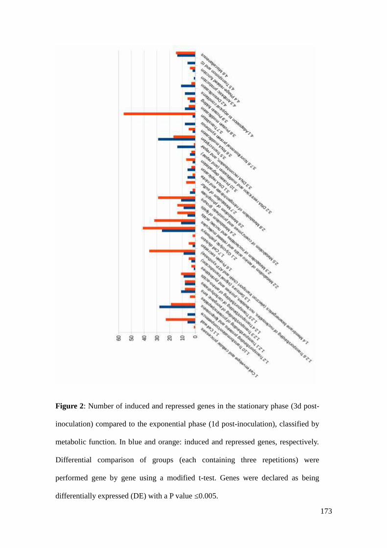
173
Figure 2: Number of induced and repressed genes in the stationary phase (3d post-
inoculation) compared to the exponential phase (1d post-inoculation), classified by
metabolic function. In blue and orange: induced and repressed genes, respectively.
Differential comparison of groups (each containing three repetitions) were
performed gene by gene using a modified t-test. Genes were declared as being
differentially expressed (DE) with a P value ≤0.005.

174
Figure 3: Schematic view of the metabolism at the entry into stationary phase
compared to exponential phase. Induced / repressed / 'non differentially expressed'
genes are symbolized with green / red / black arrows respectively. (P value ≤0.005).

175
Figure 4: Concentrations of free amino acids in the medium (mM) during growth
and stationary phases. In dark blue at the time of inoculation, in red at 1-day post
inoculation, in green at 3-day post inoculation, in purple at 9-day post-inoculation,
in light blue at 11-day post-inoculation. Significant consumptions discussed in the
text (serine, asparagine, glycine, alanine) correspond to the difference in
concentration at 1d and 3d post inoculation (t-test, Pvalue <0.05).

176
Table S1 : Induced genes in stationary phase compared to exponential phase.
Name Descript Function FClog2FoldChange pvalue padj
mraZ Protein mraZ 1.1 Cell wall 1,839 0,879 0,01458547 0,03763326gtfF Polysaccharide synthase 1.1 Cell wall 6,169 2,625 0,0011783 0,00431847slpE Surface protein with SLH domain 1.1 Cell wall 12,903 3,69 2,7762E-07 2,965E-06PFCIRM138_06855 Hypothetical outer membrane protein 1.1 Cell wall 4,992 2,32 0,00269909 0,00896146gtfD Glycosyl transferase, family 2 1.1 Cell wall 2,274 1,185 1,7102E-05 0,00011405PFCIRM138_07670 nucleoside-diphosphate-sugar epimerases 1.1 Cell wall 2,194 1,134 2,3011E-05 0,00014509
PFCIRM138_00630 DNA transfert protein
1.10Transformation/competence 14,752 3,883 2,1136E-05 0,00013617
dprA DNA processing / uptake protein
1.10Transformation/competence 27,362 4,774 1,0898E-07 1,2959E-06
PFCIRM138_07015 Mobilisation protein
1.10Transformation/competence 49,447 5,628 4,7645E-40 5,2124E-37
PFCIRM138_07020 Mobilisation protein
1.10Transformation/competence 10,855 3,44 6,5035E-22 1,423E-19
PFCIRM138_07570 Conjugative relaxase domain protein, TrwC/TraI family
1.10Transformation/competence 24,025 4,586 2,7937E-06 2,4065E-05
comEC ComE operon protein 3
1.10Transformation/competence 17,39 4,12 1,2834E-06 1,2E-05
PFCIRM138_00130 Permease
1.2 Transport/bindingproteins and 20,821 4,38 7,3817E-07 7,1465E-06

177
lipoproteins
PFCIRM138_00900 ABC transporter (Permease)
1.2 Transport/bindingproteins andlipoproteins 12,247 3,614 7,9844E-05 0,0004261
sufS Cysteine desulphurases, SufS
1.2 Transport/bindingproteins andlipoproteins 1,956 0,968 0,00877986 0,02440956
sufCABC-type transport system involved in Fe-S cluster assembly, ATPasecomponent, SufC
1.2 Transport/bindingproteins andlipoproteins 1,926 0,945 0,01558108 0,039707
sufD FeS assembly protein SufD
1.2 Transport/bindingproteins andlipoproteins 2,114 1,08 0,00534908 0,01603257
phnL ATP binding protein of ABC transporter
1.2 Transport/bindingproteins andlipoproteins 15,223 3,928 1,0113E-06 9,5378E-06
PFCIRM138_02385 permease protein
1.2 Transport/bindingproteins andlipoproteins 50,478 5,658 1,6649E-06 1,5115E-05
PFCIRM138_02395 ABC transporter related precursor
1.2 Transport/bindingproteins andlipoproteins 12,372 3,629 0,00015522 0,00075138
PFCIRM138_03530
Binding-protein-dependent transport systems inner membranecomponent
1.2 Transport/bindingproteins andlipoproteins 2,246 1,168 0,00035727 0,00156031
yceJ Transporter protein
1.2 Transport/bindingproteins andlipoproteins 16,874 4,077 2,9259E-05 0,00017882
PFCIRM138_05125
Binding-protein-dependent transport systems inner membranecomponent
1.2 Transport/bindingproteins andlipoproteins 10,809 3,434 0,00017228 0,00081767
PFCIRM138_05130
Binding-protein-dependent transport systems inner membranecomponent
1.2 Transport/bindingproteins andlipoproteins 13,72 3,778 3,2365E-05 0,00019401
PFCIRM138_05560 Transporter protein
1.2 Transport/bindingproteins and 6,834 2,773 0,00103039 0,00382765

178
lipoproteins
PFCIRM138_06235 Phosphocarrier protein, HPr family 8.A.8.1.1
1.2 Transport/bindingproteins andlipoproteins 1,736 0,796 0,02059267 0,04945857
PFCIRM138_06470 ABC-type transporter, permease component
1.2 Transport/bindingproteins andlipoproteins 3,067 1,617 0,00136804 0,00490701
PFCIRM138_06475 BP protein of siderophore ABC transporter (ISVH:undef)
1.2 Transport/bindingproteins andlipoproteins 3,578 1,839 0,00071747 0,00280327
PFCIRM138_07645 transmembrane efflux protein
1.2 Transport/bindingproteins andlipoproteins 19,201 4,263 1,4006E-05 9,6067E-05
PFCIRM138_08270 ABC transporter ATP-binding protein
1.2 Transport/bindingproteins andlipoproteins 8,213 3,038 0,00100895 0,00375439
lmrB Permease (Lincomycin resistance protein )
1.2 Transport/bindingproteins andlipoproteins 13,929 3,8 0,00012318 0,00061956
PFCIRM138_08660 secretory protein
1.2 Transport/bindingproteins andlipoproteins 10,949 3,453 0,00020008 0,00093742
PFCIRM138_09385
Arsenite efflux pump ACR3 and related permeases (Arsenite effluxpump ACR3 or related permease)
1.2 Transport/bindingproteins andlipoproteins 3,001 1,586 0,0013183 0,00477556
PFCIRM138_09450 major facilitator superfamily drug:H+ antiporter 2.A.1.3.x
1.2 Transport/bindingproteins andlipoproteins 6,824 2,771 2,1795E-19 2,3844E-17
cynX Cyanate MSF transporter
1.2 Transport/bindingproteins andlipoproteins 3,305 1,725 0,00830715 0,02339258
pimH Integral membrane efflux protein
1.2 Transport/bindingproteins andlipoproteins 4,234 2,082 0,00105323 0,00390588
hflC2 Stomatin/prohibitin1.2 Transport/bindingproteins and 3,522 1,816 3,0476E-09 4,9394E-08

179
lipoproteins
PFCIRM138_10665 ABC transporter ATP binding
1.2 Transport/bindingproteins andlipoproteins 2,084 1,06 0,00756706 0,02162374
PFCIRM138_10695 IM-ABC protein of drug, protein or lipid ABC transporter (DPL:LLP)
1.2 Transport/bindingproteins andlipoproteins 3,161 1,66 0,00117113 0,00430662
PFCIRM138_12260 transporter, MFS superfamily protein
1.2 Transport/bindingproteins andlipoproteins 11,237 3,49 7,5623E-05 0,00040654
PFCIRM138_05030 Cobalt permease
1.2.3 Transport/bindingof inorganic ions 2,753 1,461 0,01803761 0,04454435
PFCIRM138_05035 ABC-type transporter, ATPase component
1.2.3 Transport/bindingof inorganic ions 2,48 1,31 0,00253841 0,00849243
mntH Manganese transport protein1.2.3 Transport/bindingof inorganic ions 7,261 2,86 8,9333E-05 0,00047203
PFCIRM138_08475 nitrate transporter
1.2.3 Transport/bindingof inorganic ions 1,88 0,911 0,00799075 0,02261805
ssuC permease protein of nitrate ABC transporter1.2.3 Transport/bindingof inorganic ions 4,413 2,142 0,00047049 0,00196081
PFCIRM138_08620 Heavy metal transport/detoxification protein
1.2.3 Transport/bindingof inorganic ions 3,759 1,91 6,4909E-07 6,4263E-06
PFCIRM138_08625 Cation-transporting ATPase
1.2.3 Transport/bindingof inorganic ions 4,248 2,087 5,1528E-08 6,5548E-07
PFCIRM138_09635 divalent anion:Na+ symporter 2.A.47.3.x
1.2.3 Transport/bindingof inorganic ions 2,399 1,262 5,8332E-05 0,00032393
feuS binding protein of iron ABC transporter1.2.3 Transport/bindingof inorganic ions 2,805 1,488 0,00559075 0,01668836
feuC inner membrane protein of iron ABC transporter1.2.3 Transport/bindingof inorganic ions 2,468 1,303 0,01488094 0,03826028
PFCIRM138_11450 Sodium:sulfate symporter transmembrane region
1.2.3 Transport/bindingof inorganic ions 4,306 2,106 0,0059313 0,01753638
PFCIRM138_04135 permease of the major facilitator superfamily
1.2.4 Transport/bindingof carbohydrates 3,582 1,841 2,0847E-06 1,8467E-05
PFCIRM13 Membrane permease (sugar transporter) 1.2.4 Transport/binding 9,736 3,283 6,3679E-21 8,7081E-19

180
8_04640 of carbohydrates
ptsG/ptsMPTS system mannose-specific EIIBCA component (EIIBCA-Man) (EII-Man/EIII-Man)
1.2.4 Transport/bindingof carbohydrates 2,025 1,018 0,00072156 0,00281422
ptsI PTS enzyme I1.2.4 Transport/bindingof carbohydrates 2,309 1,207 0,0001149 0,00058876
yaaU metabolite transport protein YaaU1.2.4 Transport/bindingof carbohydrates 4,656 2,219 0,00050151 0,00205875
gudP D-galactonate transporter1.2.4 Transport/bindingof carbohydrates 10,093 3,335 5,6868E-05 0,00031741
iolT1 iolT1 (Major myo-inositol transporter iolT1)1.2.4 Transport/bindingof carbohydrates 11,044 3,465 1,9084E-05 0,00012577
araH2inner membrane protein of ribose/xylose/arabinose/galactoside ABCtransporter
1.2.4 Transport/bindingof carbohydrates 3,838 1,94 0,0083652 0,02352578
PFCIRM138_00815 Glutamate/gamma-aminobutyrate anti-porter (membrane protein)
1.2.5 Transport/bindingof amino-acids 8,595 3,104 3,0355E-05 0,0001833
PFCIRM138_04335 polar amino acid ABC transporter, ATP-binding protein
1.2.5 Transport/bindingof amino-acids 33,689 5,074 8,5244E-07 8,2165E-06
PFCIRM138_04340 polar amino acid ABC transporter, binding protein component
1.2.5 Transport/bindingof amino-acids 10,43 3,383 0,00016304 0,00078057
PFCIRM138_04345 Polar amino acid ABC transporter, inner membrane subunit
1.2.5 Transport/bindingof amino-acids 14,928 3,9 3,0824E-05 0,00018528
PFCIRM138_04350 polar amino acid ABC transporter, inner membrane protein
1.2.5 Transport/bindingof amino-acids 16,067 4,006 3,041E-05 0,0001833
lysE Lysine exporter protein, membran protein1.2.5 Transport/bindingof amino-acids 4,953 2,308 0,01065826 0,02875496
PFCIRM138_07335 polar amino acid ABC transporter, binding protein
1.2.5 Transport/bindingof amino-acids 3,294 1,72 0,00149863 0,00531441
PFCIRM138_07665 Amino acid permease-associated region
1.2.5 Transport/bindingof amino-acids 2,317 1,212 0,00040015 0,00171335
PFCIRM138_10015 Putrescine importer
1.2.5 Transport/bindingof amino-acids 3,541 1,824 1,1329E-09 2,0486E-08
cytX Hydroxymethylpyrimidine transporter CytX
1.2.6 Transport/bindingof nucleosides,nucleotides, purinesand pyrimidines 3,892 1,961 0,01324767 0,03458939

181
PFCIRM138_08430 sensor kinase - Membrane protein
1.3 Sensors (signaltransduction) 5,331 2,414 2,2078E-05 0,00014042
PFCIRM138_10375 Sensory transduction histidine kinase
1.3 Sensors (signaltransduction) 1,555 0,637 0,00918295 0,02536905
PFCIRM138_11140 Two-component system sensor kinase
1.3 Sensors (signaltransduction) 1,953 0,966 0,00048526 0,00201087
PFCIRM138_01505 Dioxygenase
1.4 Membranebioenergetics (electrontransport chain andATP synthase) 1,987 0,99 0,00871079 0,02434126
PFCIRM138_07220 Cytochrome c biogenesis protein transmembrane region
1.4 Membranebioenergetics (electrontransport chain andATP synthase) 13,347 3,738 9,8363E-05 0,00051243
dmsC Anaerobic dimethyl sulfoxide reductase chain C
1.4 Membranebioenergetics (electrontransport chain andATP synthase) 13,75 3,781 0,000144 0,00070486
dmsB Anaerobic dimethyl sulfoxide reductase, chain B
1.4 Membranebioenergetics (electrontransport chain andATP synthase) 4,764 2,252 0,00987035 0,02696171
PFCIRM138_08930 NAD(P)H-dependent FMN reductase
1.4 Membranebioenergetics (electrontransport chain andATP synthase) 2,962 1,567 3,4375E-06 2,8707E-05
merCoenzyme F420-dependent N5,N10-methylenetetrahydromethanopterin reductase
1.4 Membranebioenergetics (electrontransport chain andATP synthase) 3,124 1,643 2,7322E-07 2,9448E-06
PFCIRM138_09375 alkylglycerone-phosphate synthase (FAD linked oxidase-like protein)
1.4 Membranebioenergetics (electrontransport chain andATP synthase) 10,084 3,334 2,5391E-14 9,9207E-13
PFCIRM138_10905
NADPH:quinone reductase and related Zn-dependentoxidoreductases
1.4 Membranebioenergetics (electron 1,718 0,781 0,01159156 0,030892

182
transport chain andATP synthase)
PFCIRM138_11130 NADPH:quinone reductase related Zn-dependent oxidoreductase
1.4 Membranebioenergetics (electrontransport chain andATP synthase) 2,623 1,391 2,2365E-05 0,00014184
PFCIRM138_06860 Peptidase C60, sortase A and B precursor 1.6 Protein secretion 22,196 4,472 1,2374E-05 8,7054E-05PFCIRM138_07165 Tat pathway signal sequence domain protein 1.6 Protein secretion 15,425 3,947 1,3403E-05 9,2323E-05
PFCIRM138_00105 protein of unknown function
10.2 Protein ofunknown functionsimilar to proteins fromother organisms 2,054 1,039 0,01220287 0,03224623
PFCIRM138_05230 Zinc-containing alcohol dehydrogenase
2 Intermediarymetabolism 15,475 3,952 3,329E-23 9,1049E-21
gntK gluconokinase
2.1.1 Specificcarbohydrate metabolicpathway 2,572 1,363 1,1071E-05 7,9608E-05
galK1 Galactokinase
2.1.1 Specificcarbohydrate metabolicpathway 1,999 1 0,01117885 0,03001144
PFCIRM138_00835 Inositol 2-dehydrogenase
2.1.1 Specificcarbohydrate metabolicpathway 7,458 2,899 0,00060532 0,00242127
PFCIRM138_00840 sugar (xylose) phosphate isomerase/epimerase
2.1.1 Specificcarbohydrate metabolicpathway 14,326 3,841 0,00016245 0,00077946
iolH IolH (Myo-inositol catabolism IolH protein) (isomerase:epimerase)
2.1.1 Specificcarbohydrate metabolicpathway 23,005 4,524 3,6816E-05 0,00021713
glgA glycosyltransferase (glycogen synthase)
2.1.1 Specificcarbohydrate metabolicpathway 2,264 1,179 4,0563E-05 0,00023604
iolT2 iolT2 (myo-inositol transporter iolT2)2.1.1 Specificcarbohydrate metabolic 3,219 1,686 0,00447323 0,01370789

183
pathway
iol myo-inositol 2-dehydrogenase
2.1.1 Specificcarbohydrate metabolicpathway 5,559 2,475 0,00085922 0,00326954
PFCIRM138_03930 Glycoside hydrolase
2.1.1 Specificcarbohydrate metabolicpathway 2,345 1,229 0,0184331 0,04526557
adh alcohol dehydrogenase
2.1.1 Specificcarbohydrate metabolicpathway 10,194 3,35 0,00036667 0,001595
PFCIRM138_04360 hydroxyacid dehydrogenase
2.1.1 Specificcarbohydrate metabolicpathway 14,233 3,831 7,0972E-05 0,00038627
aldB Lactaldehyde dehydrogenase
2.1.1 Specificcarbohydrate metabolicpathway 2,665 1,414 1,13E-05 8,0798E-05
PFCIRM138_06500 Beta-glucosidase
2.1.1 Specificcarbohydrate metabolicpathway 1,621 0,696 0,01711601 0,04270219
PFCIRM138_07275 hexulose-6-phosphate isomerase
2.1.1 Specificcarbohydrate metabolicpathway 3,863 1,95 0,00708094 0,02033216
malQ24-alpha-glucanotransferase (Amylomaltase) (Disproportionatingenzyme) (D-enzyme)
2.1.1 Specificcarbohydrate metabolicpathway 1,529 0,613 0,01739521 0,04319038
galE3 UDP-glucose 4-epimerase
2.1.1 Specificcarbohydrate metabolicpathway 11,493 3,523 0,00010621 0,0005494
iolG2iolG2 (Myo-inositol catabolism IolG2 protein) (Inositol 2-dehydrogenase)
2.1.1 Specificcarbohydrate metabolicpathway 2,114 1,08 0,0116371 0,03097563
iolE3 Myo-inositol catabolism protein
2.1.1 Specificcarbohydrate metabolicpathway 2,26 1,176 0,00100895 0,00375439
araD1 L-ribulose-5-phosphate 4-epimerase2.1.1 Specificcarbohydrate metabolic 2,869 1,52 0,007871 0,02233689
184
pathway
araA L-arabinose isomerase
2.1.1 Specificcarbohydrate metabolicpathway 2,187 1,129 0,01676094 0,04215279
fba2 Fructose-bisphosphate aldolase class I2.1.2 Main glycolyticpathways 3,651 1,868 5,6726E-09 8,6795E-08
rpiB3 Ribose-5-phosphate isomerase 32.1.2 Main glycolyticpathways 2,879 1,526 3,1493E-06 2,6503E-05
acyP Putative acylphosphatase2.1.2 Main glycolyticpathways 5,76 2,526 0,0016336 0,00574651
araB L-ribulokinase2.1.2 Main glycolyticpathways 2,158 1,11 1,2915E-05 8,999E-05
eno2 Enolase 22.1.2 Main glycolyticpathways 3,442 1,783 1,2064E-08 1,7598E-07
PFCIRM138_07350 hydroxypyruvate isomerase
2.1.4 Substrate-specificentries to carbohydratemetabolic pathway 15,014 3,908 1,0201E-05 7,4646E-05
leuC3-isopropylmalate dehydratase large subunit (Isopropylmalateisomerase) (Alpha-IPM isomerase) (IPMI)
2.2 Metabolism ofamino acids andrelated molecules 2,413 1,271 0,00049172 0,00202233
gadB Glutamate decarboxylase
2.2 Metabolism ofamino acids andrelated molecules 4,997 2,321 2,3166E-06 2,0194E-05
aroFChorismate synthase (5-enolpyruvylshikimate-3-phosphatephospholyase)
2.2 Metabolism ofamino acids andrelated molecules 1,569 0,65 0,01874808 0,0457311
metKS-adenosylmethionine synthetase (Methionine adenosyltransferase)(AdoMet synthetase) (MAT)
2.2 Metabolism ofamino acids andrelated molecules 1,608 0,685 0,02029457 0,04884985
ald Alanine dehydrogenase
2.2 Metabolism ofamino acids andrelated molecules 2,668 1,416 6,0251E-05 0,0003329
dadA2 DadA, Glycine/D-amino acid oxidases
2.2 Metabolism ofamino acids andrelated molecules 12,994 3,7 4,9033E-06 3,9442E-05

185
PFCIRM138_04365 aminotransferase
2.2 Metabolism ofamino acids andrelated molecules 10,633 3,41 0,00021657 0,00100392
PFCIRM138_05235 Carbamoylphosphate synthase large subunit CarB
2.2 Metabolism ofamino acids andrelated molecules 4,228 2,08 7,2777E-06 5,5677E-05
aroA
3-phosphoshikimate 1-carboxyvinyltransferase(phosphoenolpyruvate:3-phosphoshikimate5-O-(1-carboxyvinyl)-transferase) (EPSP synthase)
2.2 Metabolism ofamino acids andrelated molecules 1,672 0,742 0,00276818 0,00912165
dapA1 Dihydrodipicolinate synthetase
2.2 Metabolism ofamino acids andrelated molecules 38,828 5,279 2,6405E-07 2,8602E-06
leuA1 2-isopropylmalate synthase
2.2 Metabolism ofamino acids andrelated molecules 1,676 0,745 0,00347887 0,01090968
ppx Ppx/GppA phosphatase family
2.3 Metabolism ofnucleotides and nucleicacids 1,62 0,696 0,02002399 0,04827567
nrdR Transcriptional repressor nrdR
2.3 Metabolism ofnucleotides and nucleicacids 1,728 0,789 0,01925179 0,04669946
thiD1Phosphomethylpyrimidine kinase/ hydroxymethylpyrimidinephosphokinase
2.3 Metabolism ofnucleotides and nucleicacids 4,07 2,025 0,0111065 0,02989055
dgt Deoxyguanosinetriphosphate triphosphohydrolase (dGTPase)
2.3 Metabolism ofnucleotides and nucleicacids 1,822 0,866 0,00312999 0,00996859
prt phosphoribosyltransferase
2.3 Metabolism ofnucleotides and nucleicacids 6,778 2,761 0,00026501 0,00119308
pcaB2 Adenylosuccinate lyase
2.3 Metabolism ofnucleotides and nucleicacids 11,911 3,574 0,00021726 0,00100499
pf1655 Carboxylic ester hydrolase 2.4 Metabolism of lipids 1,511 0,595 0,01741038 0,04319038pf169 Carboxylic ester hydrolase 2.4 Metabolism of lipids 2,308 1,207 0,00380039 0,01182825

186
PFCIRM138_03645 Esterase family protein 2.4 Metabolism of lipids 65,916 6,043 4,4128E-11 1,0495E-09
caiACrotonobetainyl-CoA dehydrogenase (Crotonobetainyl-CoAreductase) 2.4 Metabolism of lipids 15,443 3,949 1,006E-05 7,386E-05
caiB Crotonobetainyl-CoA:carnitine CoA-transferase 2.4 Metabolism of lipids 3,281 1,714 0,00683535 0,0197566caiC Crotonobetaine/carnitine-CoA ligase 2.4 Metabolism of lipids 12,384 3,63 4,7644E-06 3,8467E-05
uppSUndecaprenyl pyrophosphate synthase (di-trans,poly-cis-decaprenylcistransferase) 2.4 Metabolism of lipids 1,789 0,839 0,00412185 0,01270227
glxK/garK Glycerate kinase GlxK/GarK 2.4 Metabolism of lipids 2,794 1,482 2,8322E-08 3,8731E-07plsC2 PlsC, Phosphate acyltransferases 2.4 Metabolism of lipids 2,069 1,049 0,00527008 0,01581748
chlI Magnesium chelatase, subunit ChlI
2.5 Metabolism ofcoenzymes andprosthetic groups 8,331 3,058 5,9295E-05 0,00032845
PFCIRM138_04810 Isochorismatase hydrolase
2.5 Metabolism ofcoenzymes andprosthetic groups 1,848 0,886 0,01286699 0,03379709
hemX Membrane-bound transport protein (HemX protein)
2.5 Metabolism ofcoenzymes andprosthetic groups 16,735 4,065 2,6276E-05 0,00016287
pdxA2 4-hydroxythreonine-4-phosphate dehydrogenase
2.5 Metabolism ofcoenzymes andprosthetic groups 2,549 1,35 0,00083498 0,00319352
pncA pyrazinamidase/nicotinamidase
2.5 Metabolism ofcoenzymes andprosthetic groups 2,501 1,322 3,4305E-06 2,8707E-05
cbiO3 ABC transporter, ATP-binding protein
2.5 Metabolism ofcoenzymes andprosthetic groups 5,571 2,478 0,00229833 0,00776041
cbiQ1ABC-type transporter, permease components (ABC-type cobalttransport system, permease component CbiQ)
2.5 Metabolism ofcoenzymes andprosthetic groups 9,615 3,265 0,00026681 0,00119871
mog Molybdopterin biosynthesis Mog protein
2.5 Metabolism ofcoenzymes andprosthetic groups 2,792 1,481 0,00011522 0,00058903
thiC thiamine-phosphate diphosphorylase 2.5 Metabolism of 2,361 1,239 0,0156067 0,039707

187
coenzymes andprosthetic groups
PFCIRM138_09275 NADH-flavin reductase
2.5 Metabolism ofcoenzymes andprosthetic groups 1,801 0,849 0,0066313 0,01926864
bioB Biotin synthase
2.5 Metabolism ofcoenzymes andprosthetic groups 7,499 2,907 4,262E-27 1,5542E-24
ppx5 Exopolyphosphatase2.6 Metabolism ofphosphate 19,842 4,31 9,256E-06 6,8885E-05
ppa Inorganic pyrophosphatase2.6 Metabolism ofphosphate 1,579 0,659 0,01239683 0,03267982
PFCIRM138_07685 NUDIX hydrolase
2.6 Metabolism ofphosphate 3,022 1,595 0,00035576 0,00155682
phoH Phosphate starvation-inducible protein PhoH-like protein2.6 Metabolism ofphosphate 2,521 1,334 4,7091E-05 0,00027115
ntcA2 Global nitrogen regulator NtcA
2.8 Metabolism ofnitrogen/nitrate andnitrite 1,732 0,792 0,00180183 0,00626775
PFCIRM138_04265 Nitrate reductase, alpha subunit
2.8 Metabolism ofnitrogen/nitrate andnitrite 5,948 2,572 0,00096256 0,00361871
narH Respiratory nitrate reductase, beta subunit
2.8 Metabolism ofnitrogen/nitrate andnitrite 22,415 4,486 3,9715E-07 4,1183E-06
narJ Nitrate reductase delta subunit
2.8 Metabolism ofnitrogen/nitrate andnitrite 22,951 4,52 3,0582E-06 2,5835E-05
narI Respiratory nitrate reductase, gamma subunit
2.8 Metabolism ofnitrogen/nitrate andnitrite 16,346 4,031 7,471E-06 5,6957E-05
glnB Nitrogen regulatory protein P-II
2.8 Metabolism ofnitrogen/nitrate andnitrite 4,518 2,176 0,00150854 0,00534091
dnaE2 DNA polymerase III alpha subunit 3.1 DNA replication 2,304 1,204 0,00804479 0,0227416
188
priA Primosomal protein N 3.1 DNA replication 2,979 1,575 5,5968E-09 8,6311E-08gyrB2 DNA gyrase subunit B 3.1 DNA replication 2,435 1,284 1,9573E-05 0,00012784hels Helicase 3.1 DNA replication 2,469 1,304 5,1448E-07 5,2115E-06PFCIRM138_08320 helicase 3.1 DNA replication 2,774 1,472 6,7193E-08 8,2594E-07dnaB Replicative DNA helicase 3.1 DNA replication 1,552 0,635 0,01620308 0,04103201PFCIRM138_09485
Membrane protease subunits, stomatin/prohibitin homologs(Membrane protease subunit, stomatin/prohibitin homolog)
3.10 Proteindegradation 2,516 1,331 1,4334E-05 9,77E-05
pepC Aminopeptidase C, Bleomycin hydrolase,3.10 Proteindegradation 1,601 0,679 0,0092136 0,02542164
tagA 3-Methyladenine DNA glycosylase I
3.2 DNA restriction andmodification (andrepair) 3,915 1,969 0,01295853 0,03397163
sms DNA repair protein
3.2 DNA restriction andmodification (andrepair) 1,904 0,929 0,00144176 0,00513773
sbcD SbcD, DNA repair exonuclease
3.2 DNA restriction andmodification (andrepair) 2,785 1,478 2,8782E-06 2,4504E-05
PFCIRM138_10795 exonuclease
3.2 DNA restriction andmodification (andrepair) 1,828 0,87 0,01437546 0,03722308
PFCIRM138_00325 Recombinase
3.3 DNArecombination, andrepair 77,776 6,281 6,6498E-07 6,4955E-06
gin Resolvase
3.3 DNArecombination, andrepair 1,823 0,866 0,01302721 0,03409513
PFCIRM138_00650 Recombinase
3.3 DNArecombination, andrepair 7,929 2,987 9,3006E-18 7,2678E-16
recG ATP-dependent DNA helicase RecG
3.3 DNArecombination, andrepair 1,74 0,799 0,00307427 0,0098197
dinP DNA-directed DNA polymerase 3.3 DNA 5,915 2,564 0,0006119 0,00243761

189
recombination, andrepair
PFCIRM138_01865 Helicase protein
3.3 DNArecombination, andrepair 1,806 0,853 0,00091498 0,00345169
recA RecA (Recombinase A)
3.3 DNArecombination, andrepair 2,019 1,014 0,0023503 0,00790909
PFCIRM138_03835 Bifunctional DNA primase/polymerase
3.3 DNArecombination, andrepair 18,618 4,219 6,2207E-43 1,3611E-39
rmuC DNA recombination protein
3.3 DNArecombination, andrepair 3,851 1,945 2,0143E-10 4,3208E-09
sbcC ATP-dependent dsDNA exonuclease SbcC
3.3 DNArecombination, andrepair 1,712 0,776 0,00302292 0,00971241
alkAAlkA, 3-methyladenine DNA glycosylase/8-oxoguanine DNAglycosylase
3.3 DNArecombination, andrepair 7,574 2,921 0,00061275 0,00243761
PFCIRM138_10165 ATP-dependent DNA helicase
3.3 DNArecombination, andrepair 2,875 1,524 6,3437E-07 6,3091E-06
urvD UvrD/REP helicase / ATP-dependent DNA helicase
3.3 DNArecombination, andrepair 1,534 0,617 0,0186472 0,04558667
holA DNA polymerase III, delta subunit
3.3 DNArecombination, andrepair 3,066 1,617 1,1811E-07 1,3969E-06
hrdD2 RNA polymerase principal sigma factor HrdD3.5.1 Transcriptioninitiation 6,278 2,65 3,3471E-10 6,909E-09
hrdD1 RNA polymerase principal sigma factor HrdD3.5.1 Transcriptioninitiation 8,762 3,131 0,00020197 0,00094223
rpoE RNA polymerase sigma -70 factor, sigma-E factor3.5.1 Transcriptioninitiation 1,875 0,907 0,01422102 0,03686681
marR3 Transcriptional regulator, MarR family 3.5.2 Transcription 3,689 1,883 0,01723654 0,04295393

190
regulationPFCIRM138_00405 Transcriptional regulator
3.5.2 Transcriptionregulation 3,214 1,684 6,4352E-06 4,993E-05
arsR1 Transcriptional regulator, ArsR family protein3.5.2 Transcriptionregulation 6,938 2,794 1,0324E-05 7,5048E-05
dtxR Iron-dependent repressor3.5.2 Transcriptionregulation 1,512 0,596 0,01689512 0,0424186
PFCIRM138_00890 transcription regulator
3.5.2 Transcriptionregulation 3,484 1,801 6,3071E-06 4,9285E-05
arsR4 Transcriptional regulator, ArsR family3.5.2 Transcriptionregulation 4,063 2,022 3,0203E-08 4,1047E-07
pspA Phage shock protein A3.5.2 Transcriptionregulation 1,849 0,887 0,00592449 0,01753638
PFCIRM138_02505 Helix-turn-helix XRE-family like proteins.
3.5.2 Transcriptionregulation 11,769 3,557 4,248E-17 2,6556E-15
arsR3 Transcriptional regulators (Bacterial regulatory protein, arsR family)3.5.2 Transcriptionregulation 9,029 3,174 0,00028321 0,00126462
tetR1 Transcriptional regulator, TetR family3.5.2 Transcriptionregulation 3,006 1,588 7,0767E-05 0,00038613
merR2 Transcriptional regulator, MerR3.5.2 Transcriptionregulation 38,449 5,265 8,6754E-07 8,3253E-06
hemR HemR3.5.2 Transcriptionregulation 3,469 1,795 0,00848484 0,02380106
regX #N/D3.5.2 Transcriptionregulation 1,621 0,697 0,02065629 0,04955698
padR PadR, Transcriptional regulator PadR-like family3.5.2 Transcriptionregulation 7,325 2,873 5,5452E-21 8,0885E-19
PFCIRM138_06840 transcriptional regulator ArsR
3.5.2 Transcriptionregulation 6,121 2,614 2,917E-09 4,763E-08
PFCIRM138_07310 Transcriptional regulator PadR family
3.5.2 Transcriptionregulation 3,263 1,706 5,5842E-05 0,00031248
lacI2 Transcriptional regulator, LacI family protein3.5.2 Transcriptionregulation 3,31 1,727 0,00174572 0,00611924
PFCIRM138_08435 Two-component system, regulatory protein
3.5.2 Transcriptionregulation 2,948 1,56 0,00297648 0,00959137
191
lysR1 Transcriptional regulator, LysR family protein3.5.2 Transcriptionregulation 4,075 2,027 7,0714E-06 5,4288E-05
PFCIRM138_09520 Regulatory protein, MarR
3.5.2 Transcriptionregulation 3,825 1,936 0,00578025 0,01720705
PFCIRM138_10090 two-component system response regulator
3.5.2 Transcriptionregulation 2,183 1,126 0,01610133 0,04082237
PFCIRM138_10370 anti-sigma factor
3.5.2 Transcriptionregulation 1,776 0,829 0,00445461 0,01366998
whiB2 transcription factor WhiB3.5.2 Transcriptionregulation 2,029 1,021 0,00235321 0,00790909
tetR2 Transcriptional regulator, TetR/AcrR family3.5.2 Transcriptionregulation 1,757 0,813 0,01694616 0,04247217
PFCIRM138_11280 Silent information regulator protein Sir2 NAD-dependent deacetylase
3.5.2 Transcriptionregulation 2,118 1,083 0,00929407 0,02561138
PFCIRM138_12035 MarR-family transcriptional regulator
3.5.2 Transcriptionregulation 4,754 2,249 0,00176852 0,00617148
hrcA Heat-inducible transcription repressor hrcA3.5.2 Transcriptionregulation 4,506 2,172 4,2318E-12 1,2183E-10
rpoZDNA-directed RNA polymerase omega chain (RNAP omega subunit)(Transcriptase omega chain) (RNA polymerase omega subunit)
3.5.3 Transcriptionelongation 1,805 0,852 0,00879443 0,02441906
PFCIRM138_10430 Transcription termination factor Rho
3.5.4 Transcriptiontermination 2,007 1,005 0,0006899 0,00271004
PFCIRM138_10140 DEAD/DEAH box helicase domain protein 3.6 RNA modification 1,599 0,677 0,01455087 0,03763167trmU tRNA (5-methylaminomethyl-2-thiouridylate)-methyltransferase 3.6 RNA modification 2,571 1,362 0,00022788 0,00104969infB Translation initiation factor IF-2 3.7 Protein synthesis 2,009 1,007 0,00195859 0,00671962
rplS 50S ribosomal protein L193.7.1 Ribosomalproteins 1,882 0,912 0,00473468 0,01438816
rpmB Ribosomal protein L283.7.1 Ribosomalproteins 3,235 1,694 2,0683E-05 0,00013389
rpsO 30S ribosomal protein S153.7.1 Ribosomalproteins 2,799 1,485 9,3926E-06 6,9429E-05
rplK 50S ribosomal protein L113.7.1 Ribosomalproteins 1,802 0,85 0,01296449 0,03397163
rpmA 50S ribosomal protein L27 3.7.1 Ribosomal 2,126 1,088 0,00050893 0,00208527

192
proteinsPFCIRM138_04330
translation initiation inhibitor involved in amino acid or purinemetabolism
3.7.3 Translationinitiation 5,469 2,451 0,00589094 0,01748898
prfA Peptide chain release factor 1 (RF-1)3.7.5 Translationtermination 2,113 1,079 0,00119823 0,00437685
PFCIRM138_06630 Isochorismatase hydrolase
3.7.6 Nonribosomalprotein synthesis 2,021 1,015 0,01212049 0,03206726
trxA4 Thioredoxine 3.8 Protein modification 3,519 1,815 2,0155E-05 0,00013125gst Glutathione S-transferase 3.8 Protein modification 2,489 1,315 3,3022E-05 0,00019634trxB Thioredoxin reductase 3.8 Protein modification 2,696 1,431 0,00071228 0,00278797PFCIRM138_06730 AarF 3.8 Protein modification 3,156 1,658 1,2202E-09 2,1583E-08PFCIRM138_07240 Thioredoxin 3.8 Protein modification 16,925 4,081 5,1511E-05 0,00029198htrA4 Serine protease 3.8 Protein modification 3,344 1,742 7,2864E-09 1,0995E-07msrA peptide-methionine (S)-S-oxide reductase 3.8 Protein modification 1,818 0,862 0,01140032 0,03048767PFCIRM138_12360 Amino acid-binding ACT 3.8 Protein modification 1,909 0,933 0,00133699 0,00482729copZ Copper chaperone CopZ 3.9 Protein folding 2,555 1,353 2,856E-05 0,00017553hsp20 3 Heat shock protein 20 3 (20 kDa chaperone 3) 3.9 Protein folding 6,775 2,76 2,3131E-15 1,1247E-13hsp20 1 Heat shock protein 20 1 (20 kDa chaperone 1) 3.9 Protein folding 6,955 2,798 6,2693E-15 2,8578E-13hsp20 2 Heat shock protein 20 2 (20 kDa chaperone 2) 3.9 Protein folding 2,555 1,353 0,00099365 0,00371643
cspB Cold shock protein CspB4.1 Adaptation toatypical conditions 2,664 1,414 2,4027E-06 2,0779E-05
PFCIRM138_04080 drug exporters of the RND superfamily
4.1 Adaptation toatypical conditions 1,768 0,822 0,00882506 0,02447305
osmC OsmC protein4.1 Adaptation toatypical conditions 2,669 1,416 4,9954E-05 0,00028463
PFCIRM138_04225 Multidrug resistance transporter, MFS superfamily protein
4.1 Adaptation toatypical conditions 16,316 4,028 1,252E-05 8,78E-05
PFCIRM138_04645 Cyclase family protein
4.1 Adaptation toatypical conditions 35,816 5,163 6,7683E-11 1,5589E-09
katA Catalase 4.1 Adaptation to 2,129 1,09 0,00057545 0,00232305

193
atypical conditions
dps Starvation-inducible DNA-binding protein4.1 Adaptation toatypical conditions 13,789 3,785 7,7027E-21 9,9139E-19
pspC Possible stress-response transcriptional regulator protein PspC4.1 Adaptation toatypical conditions 4,094 2,034 2,6719E-08 3,6767E-07
copA Copper-Cadmimum efflux ATPase 4.2 Detoxification 1,695 0,762 0,00195938 0,00671962drrA Daunorubicin resistance ATP-binding protein DrrA 4.2 Detoxification 20,788 4,378 4,2764E-06 3,4913E-05PFCIRM138_03890 Drug resistance transporter 4.2 Detoxification 6,494 2,699 0,00060844 0,00242933PFCIRM138_04715 Aldo/keto reductases, related to diketogulonate reductase 4.2 Detoxification 3,531 1,82 1,9954E-08 2,8167E-07ahpF Alkyl hydroperoxide reductase subunit F 4.2 Detoxification 1,883 0,913 0,01456765 0,03763167PFCIRM138_06410 Beta-lactamase-like 4.2 Detoxification 6,756 2,756 0,00266337 0,00886978matE Multi antimicrobial extrusion protein MatE 4.2 Detoxification 8,165 3,029 0,00011774 0,0005991PFCIRM138_06835 Hemerythrin HHE cation binding region 4.2 Detoxification 4,86 2,281 5,3397E-11 1,2429E-09
sodAIron/Manganese superoxide dismutase (Superoxide dismutase[Mn/Fe]) (SODM) 4.2 Detoxification 9,425 3,237 1,7633E-14 7,0149E-13
PFCIRM138_09705 Drug resistance transporter 4.2 Detoxification 4,418 2,143 0,00087606 0,00332205pcaC1 4-carboxymuconolactone decarboxylase 4.2 Detoxification 7,442 2,896 0,00347011 0,01090891PFCIRM138_00720 Isochorismatase hydrolase
4.3 Antibioticproduction 1,742 0,8 0,00459481 0,01400202
PFCIRM138_03830 Primase
4.4 Phage-relatedfunction 18,861 4,237 1,6746E-32 9,3895E-30
PFCIRM138_03840 Gp42
4.4 Phage-relatedfunction 6,283 2,651 5,6222E-08 7,0697E-07
PFCIRM138_00080 Transposase 4.5 Transposon and IS 7,242 2,856 5,3402E-05 0,00030115tnpA Transposase 4.5 Transposon and IS 2,485 1,314 1,721E-05 0,00011411PFCIRM138_06175 Transposase for IS3514b 4.5 Transposon and IS 15,427 3,947 1,0869E-09 1,9985E-08tnp Transposase 4.5 Transposon and IS 1,905 0,93 0,00868827 0,02430937

194
tnp7109-55 Transposase 4.5 Transposon and IS 3,073 1,62 5,4012E-05 0,0003038TnpD Transposase IS3/IS911 4.5 Transposon and IS 6,567 2,715 1,9076E-06 1,7106E-05PFCIRM138_00060 Dehydrogenase 4.6 Miscellaneous 6,654 2,734 0,00075901 0,00293411PFCIRM138_02755 acetyltransferase 4.6 Miscellaneous 12,363 3,628 0,00075805 0,00293411PFCIRM138_03090 LMBE-related protein 4.6 Miscellaneous 1,705 0,77 0,01026047 0,02781214PFCIRM138_05685 Zinc-containing alcohol dehydrogenase superfamily 4.6 Miscellaneous 3,604 1,85 0,00585402 0,01740297PFCIRM138_07990 GCN5-related N-acetyltransferase 4.6 Miscellaneous 1,742 0,801 0,00487411 0,01476462PFCIRM138_08590 Methyltransferase 4.6 Miscellaneous 8,446 3,078 0,00074434 0,00288761PFCIRM138_09515 Acetyltransferase 4.6 Miscellaneous 4,163 2,058 0,01734942 0,04313696PFCIRM138_10260 acetyltransferase 4.6 Miscellaneous 5,637 2,495 8,6977E-15 3,8061E-13PFCIRM138_10305 Glycosyl transferase 4.6 Miscellaneous 14,016 3,809 9,6463E-05 0,00050373PFCIRM138_10670 Methyltransferase 4.6 Miscellaneous 3,148 1,654 0,00083633 0,00319352PFCIRM138_10815 acetyltransferase 4.6 Miscellaneous 6,42 2,682 1,804E-05 0,00011925PFCIRM138_11765 Metal dependent phosphohydrolase 4.6 Miscellaneous 11,77 3,557 1,3418E-06 1,2493E-05obg GTPase 4.6 Miscellaneous 1,61 0,687 0,01497509 0,03845716PFCIRM138_12295 Metal-dependent hydrolase 4.6 Miscellaneous 3,582 1,841 0,00239099 0,00802374
PFCIRM138_00330 protein of unknown function
5.1 Protein of unknownfunction similar to otherproteins from the sameorganism 58,871 5,879 4,0881E-06 3,3627E-05
PFCIRM138_00620 protein of unknown function
5.1 Protein of unknownfunction similar to other 81,837 6,355 2,8665E-09 4,7158E-08

195
proteins from the sameorganism
PFCIRM138_00960 Hypothetical membrane protein
5.1 Protein of unknownfunction similar to otherproteins from the sameorganism 1,732 0,792 0,0169054 0,0424186
PFCIRM138_03905 protein of unknown function
5.1 Protein of unknownfunction similar to otherproteins from the sameorganism 5,637 2,495 0,00121989 0,00444855
PFCIRM138_05220 Hypothetical secreted protein
5.1 Protein of unknownfunction similar to otherproteins from the sameorganism 2,715 1,441 1,6849E-05 0,00011309
PFCIRM138_00050 Hypothetical membrane protein
5.2 Protein of unknownfunction similar toproteins from otherorganisms 7,687 2,942 0,00031592 0,00139926
PFCIRM138_00310 Hypothetical protein
5.2 Protein of unknownfunction similar toproteins from otherorganisms 2,413 1,271 0,00392799 0,01217343
PFCIRM138_00355 protein of unknown function
5.2 Protein of unknownfunction similar toproteins from otherorganisms 40,353 5,335 5,1216E-07 5,2115E-06
PFCIRM138_00410 Hypothetical protein
5.2 Protein of unknownfunction similar toproteins from otherorganisms 2,872 1,522 8,5136E-06 6,4013E-05
PFCIRM138_00535 Hypothetical protein
5.2 Protein of unknownfunction similar toproteins from otherorganisms 1,689 0,757 0,01559171 0,039707
PFCIRM138_00585 protein of unknown function
5.2 Protein of unknownfunction similar to 20,592 4,364 1,6995E-05 0,00011371

196
proteins from otherorganisms
PFCIRM138_00590 protein of unknown function
5.2 Protein of unknownfunction similar toproteins from otherorganisms 2,617 1,388 2,1856E-05 0,00013942
PFCIRM138_00605 protein of unknown function
5.2 Protein of unknownfunction similar toproteins from otherorganisms 13,578 3,763 5,7204E-05 0,00031848
PFCIRM138_00765 Hypothetical protein
5.2 Protein of unknownfunction similar toproteins from otherorganisms 2,107 1,075 0,00036578 0,00159427
PFCIRM138_01260 Hypothetical protein
5.2 Protein of unknownfunction similar toproteins from otherorganisms 1,81 0,856 0,00204639 0,00697431
PFCIRM138_01840 Hypothetical membrane protein
5.2 Protein of unknownfunction similar toproteins from otherorganisms 3,313 1,728 4,9837E-06 3,9797E-05
PFCIRM138_01925 Hypothetical protein
5.2 Protein of unknownfunction similar toproteins from otherorganisms 1,81 0,856 0,00837936 0,02353536
PFCIRM138_01975 Hypothetical protein
5.2 Protein of unknownfunction similar toproteins from otherorganisms 2,351 1,233 9,1072E-05 0,00047786
PFCIRM138_02050 Hypothetical protein
5.2 Protein of unknownfunction similar toproteins from otherorganisms 4,451 2,154 5,8951E-10 1,1216E-08
PFCIRM138_03095 Hypothetical protein
5.2 Protein of unknownfunction similar to 2,491 1,317 0,00038423 0,0016582

197
proteins from otherorganisms
PFCIRM138_03570 Hypothetical protein
5.2 Protein of unknownfunction similar toproteins from otherorganisms 11,847 3,566 1,365E-20 1,6592E-18
PFCIRM138_03720 Hypothetical protein
5.2 Protein of unknownfunction similar toproteins from otherorganisms 2,399 1,262 0,01883697 0,04588504
PFCIRM138_03875 Hypothetical protein
5.2 Protein of unknownfunction similar toproteins from otherorganisms 3,961 1,986 0,0030585 0,00978362
PFCIRM138_04010 Hypothetical protein
5.2 Protein of unknownfunction similar toproteins from otherorganisms 2,499 1,321 2,7596E-05 0,00017008
PFCIRM138_04055 Hypothetical protein
5.2 Protein of unknownfunction similar toproteins from otherorganisms 4,562 2,19 4,0056E-11 9,6311E-10
PFCIRM138_04395 Hypothetical protein
5.2 Protein of unknownfunction similar toproteins from otherorganisms 6,539 2,709 0,00016438 0,00078531
PFCIRM138_04790 protein of unknown function
5.2 Protein of unknownfunction similar toproteins from otherorganisms 6,498 2,7 1,5613E-15 7,9444E-14
PFCIRM138_05045 BioY family protein. Membrane protein
5.2 Protein of unknownfunction similar toproteins from otherorganisms 4,248 2,087 2,1616E-08 3,0317E-07
PFCIRM138_05295 Hypothetical protein
5.2 Protein of unknownfunction similar to 2,134 1,094 0,00083032 0,00318167

198
proteins from otherorganisms
PFCIRM138_06350 Hypothetical protein
5.2 Protein of unknownfunction similar toproteins from otherorganisms 1,977 0,983 0,00401692 0,01243144
PFCIRM138_06370 Hypothetical protein
5.2 Protein of unknownfunction similar toproteins from otherorganisms 1,736 0,796 0,0186894 0,04563884
PFCIRM138_06970 protein of unknown function
5.2 Protein of unknownfunction similar toproteins from otherorganisms 4,74 2,245 0,00620485 0,01822309
PFCIRM138_07000 protein of unknown function
5.2 Protein of unknownfunction similar toproteins from otherorganisms 2,702 1,434 7,4494E-05 0,00040245
PFCIRM138_07025 protein of unknown function
5.2 Protein of unknownfunction similar toproteins from otherorganisms 3,428 1,777 9,4008E-09 1,3993E-07
PFCIRM138_07065 protein of unknown function
5.2 Protein of unknownfunction similar toproteins from otherorganisms 2,644 1,403 0,00011444 0,00058776
PFCIRM138_07110 protein of unknown function
5.2 Protein of unknownfunction similar toproteins from otherorganisms 2,558 1,355 0,0001788 0,00084337
PFCIRM138_07195 protein of unknown function
5.2 Protein of unknownfunction similar toproteins from otherorganisms 15,105 3,917 1,1091E-05 7,9608E-05
PFCIRM138_07680 Hypothetical membrane protein
5.2 Protein of unknownfunction similar to 1,965 0,975 0,00015077 0,00073472
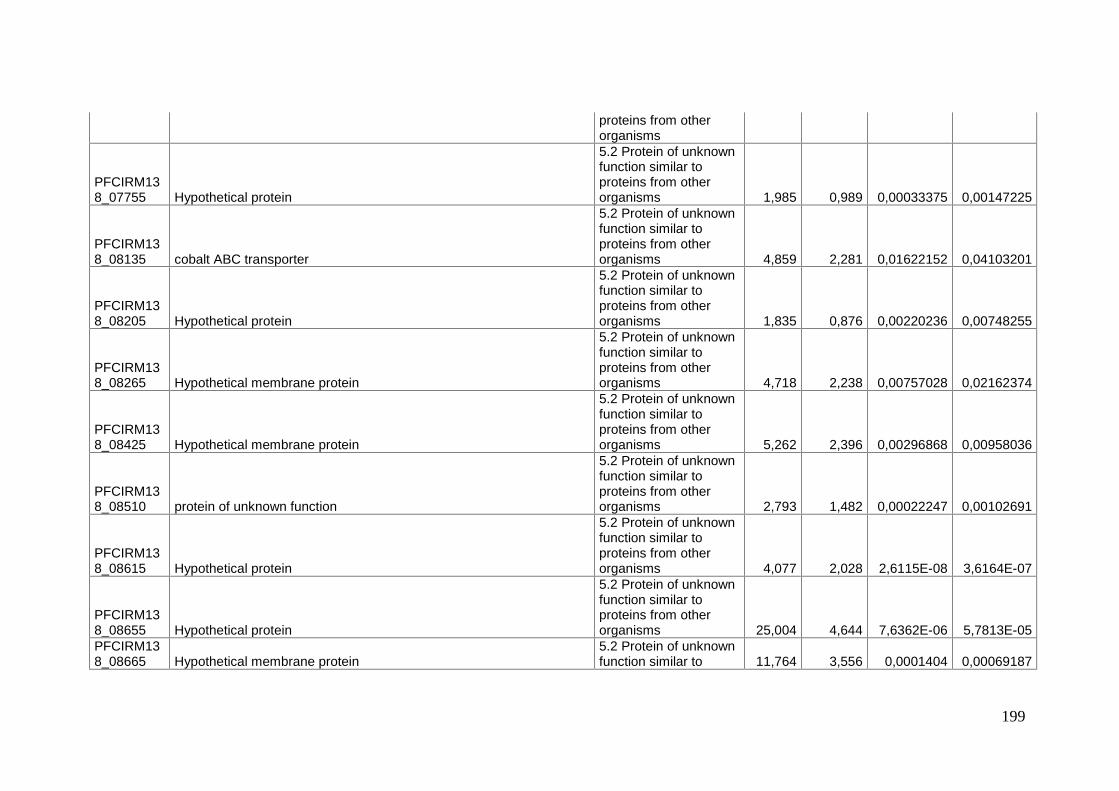
199
proteins from otherorganisms
PFCIRM138_07755 Hypothetical protein
5.2 Protein of unknownfunction similar toproteins from otherorganisms 1,985 0,989 0,00033375 0,00147225
PFCIRM138_08135 cobalt ABC transporter
5.2 Protein of unknownfunction similar toproteins from otherorganisms 4,859 2,281 0,01622152 0,04103201
PFCIRM138_08205 Hypothetical protein
5.2 Protein of unknownfunction similar toproteins from otherorganisms 1,835 0,876 0,00220236 0,00748255
PFCIRM138_08265 Hypothetical membrane protein
5.2 Protein of unknownfunction similar toproteins from otherorganisms 4,718 2,238 0,00757028 0,02162374
PFCIRM138_08425 Hypothetical membrane protein
5.2 Protein of unknownfunction similar toproteins from otherorganisms 5,262 2,396 0,00296868 0,00958036
PFCIRM138_08510 protein of unknown function
5.2 Protein of unknownfunction similar toproteins from otherorganisms 2,793 1,482 0,00022247 0,00102691
PFCIRM138_08615 Hypothetical protein
5.2 Protein of unknownfunction similar toproteins from otherorganisms 4,077 2,028 2,6115E-08 3,6164E-07
PFCIRM138_08655 Hypothetical protein
5.2 Protein of unknownfunction similar toproteins from otherorganisms 25,004 4,644 7,6362E-06 5,7813E-05
PFCIRM138_08665 Hypothetical membrane protein
5.2 Protein of unknownfunction similar to 11,764 3,556 0,0001404 0,00069187

200
proteins from otherorganisms
PFCIRM138_08685 Hypothetical membrane protein
5.2 Protein of unknownfunction similar toproteins from otherorganisms 17,315 4,114 8,5125E-05 0,00045193
PFCIRM138_08690 Hypothetical protein
5.2 Protein of unknownfunction similar toproteins from otherorganisms 6,558 2,713 0,00303668 0,00972803
PFCIRM138_09090 Hypothetical membrane protein
5.2 Protein of unknownfunction similar toproteins from otherorganisms 2,448 1,292 2,116E-05 0,00013617
PFCIRM138_09425 Hypothetical membrane protein
5.2 Protein of unknownfunction similar toproteins from otherorganisms 9,849 3,3 0,0001539 0,0007483
PFCIRM138_09600 protein of unknown function
5.2 Protein of unknownfunction similar toproteins from otherorganisms 6,055 2,598 2,8939E-17 1,8623E-15
PFCIRM138_09605 protein of unknown function
5.2 Protein of unknownfunction similar toproteins from otherorganisms 2,469 1,304 2,0402E-05 0,00013246
PFCIRM138_10080 Hypothetical transmembrane protein
5.2 Protein of unknownfunction similar toproteins from otherorganisms 1,606 0,684 0,00811851 0,0229204
PFCIRM138_10150 Hypothetical protein
5.2 Protein of unknownfunction similar toproteins from otherorganisms 1,779 0,831 0,00957208 0,02624524
PFCIRM138_10325 Hypothetical protein
5.2 Protein of unknownfunction similar to 33,738 5,076 3,0234E-05 0,00018325

201
proteins from otherorganisms
PFCIRM138_10340 protein of unknown function
5.2 Protein of unknownfunction similar toproteins from otherorganisms 8,002 3 0,0002369 0,0010776
dedA protein of unknown function
5.2 Protein of unknownfunction similar toproteins from otherorganisms 2,589 1,373 0,0190562 0,04627633
PFCIRM138_10635 Hypothetical protein
5.2 Protein of unknownfunction similar toproteins from otherorganisms 6,25 2,644 1,5135E-14 6,1324E-13
PFCIRM138_10675 Hypothetical protein
5.2 Protein of unknownfunction similar toproteins from otherorganisms 3,512 1,812 0,00010459 0,00054226
PFCIRM138_10820 Hypothetical membrane protein
5.2 Protein of unknownfunction similar toproteins from otherorganisms 19,805 4,308 2,7134E-05 0,00016771
PFCIRM138_11125 Hypothetical protein
5.2 Protein of unknownfunction similar toproteins from otherorganisms 2,24 1,163 2,328E-05 0,00014637
PFCIRM138_11265 Hypothetical protein
5.2 Protein of unknownfunction similar toproteins from otherorganisms 1,811 0,857 0,00369512 0,01151699
PFCIRM138_11285 Hypothetical membrane protein
5.2 Protein of unknownfunction similar toproteins from otherorganisms 5,125 2,357 0,00362367 0,01131041
PFCIRM138_11965 Hypothetical protein
5.2 Protein of unknownfunction similar to 6,126 2,615 0,00077752 0,00298983

202
proteins from otherorganisms
PFCIRM138_12135 Hypothetical protein
5.2 Protein of unknownfunction similar toproteins from otherorganisms 30,564 4,934 1,7357E-06 1,5693E-05
PFCIRM138_12190 membrane protein
5.2 Protein of unknownfunction similar toproteins from otherorganisms 1,905 0,93 0,00135085 0,00485328
PFCIRM138_00045 Hypothetical secreted protein
6 Protein of unknownfunction, withoutsimilarity to otherproteins 7,742 2,953 0,00137045 0,00490759
PFCIRM138_00340 Protein of unknown function
6 Protein of unknownfunction, withoutsimilarity to otherproteins 36,263 5,18 1,8097E-06 1,6295E-05
PFCIRM138_00350 Protein of unknown function
6 Protein of unknownfunction, withoutsimilarity to otherproteins 18,263 4,191 1,9215E-05 0,00012625
PFCIRM138_00495 Hypothetical protein
6 Protein of unknownfunction, withoutsimilarity to otherproteins 2,522 1,335 6,3462E-06 4,9415E-05
PFCIRM138_00540 Hypothetical protein
6 Protein of unknownfunction, withoutsimilarity to otherproteins 3,007 1,588 9,3287E-07 8,8745E-06
PFCIRM138_00685 Hypothetical protein
6 Protein of unknownfunction, withoutsimilarity to otherproteins 6,202 2,633 0,00040607 0,00173531
PFCIRM138_00775 Hypothetical protein
6 Protein of unknownfunction, without 1,796 0,845 0,02003396 0,04827567

203
similarity to otherproteins
PFCIRM138_00860 Protein of unknown function
6 Protein of unknownfunction, withoutsimilarity to otherproteins 8,159 3,028 0,00100393 0,00374847
PFCIRM138_01055 Hypothetical protein
6 Protein of unknownfunction, withoutsimilarity to otherproteins 3,515 1,813 9,0062E-06 6,7255E-05
PFCIRM138_01100 Hypothetical protein
6 Protein of unknownfunction, withoutsimilarity to otherproteins 40,584 5,343 6,0332E-07 6,0277E-06
PFCIRM138_01105 Hypothetical protein
6 Protein of unknownfunction, withoutsimilarity to otherproteins 3,755 1,909 1,0305E-05 7,5048E-05
PFCIRM138_01110 Hypothetical protein
6 Protein of unknownfunction, withoutsimilarity to otherproteins 2,533 1,341 0,00034502 0,00151891
PFCIRM138_01255 Protein of unknown function
6 Protein of unknownfunction, withoutsimilarity to otherproteins 2,826 1,499 0,01473584 0,03797647
PFCIRM138_01415 Hypothetical protein
6 Protein of unknownfunction, withoutsimilarity to otherproteins 14,972 3,904 1,2887E-05 8,999E-05
PFCIRM138_01860 Hypothetical protein
6 Protein of unknownfunction, withoutsimilarity to otherproteins 3,019 1,594 0,01039925 0,02812554
PFCIRM138_01945 Hypothetical protein
6 Protein of unknownfunction, without 5,918 2,565 0,0004425 0,0018652
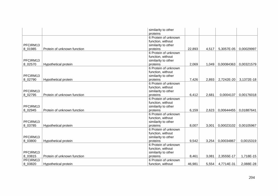
204
similarity to otherproteins
PFCIRM138_01985 Protein of unknown function
6 Protein of unknownfunction, withoutsimilarity to otherproteins 22,893 4,517 5,3057E-05 0,00029997
PFCIRM138_02570 Hypothetical protein
6 Protein of unknownfunction, withoutsimilarity to otherproteins 2,069 1,049 0,00084363 0,00321579
PFCIRM138_02790 Hypothetical protein
6 Protein of unknownfunction, withoutsimilarity to otherproteins 7,426 2,893 2,7242E-20 3,1372E-18
PFCIRM138_02795 Protein of unknown function
6 Protein of unknownfunction, withoutsimilarity to otherproteins 6,412 2,681 0,0004137 0,00176018
PFCIRM138_02945 Protein of unknown function
6 Protein of unknownfunction, withoutsimilarity to otherproteins 6,159 2,623 0,00644455 0,01887641
PFCIRM138_03785 Hypothetical protein
6 Protein of unknownfunction, withoutsimilarity to otherproteins 8,007 3,001 0,00023102 0,00105967
PFCIRM138_03800 Hypothetical protein
6 Protein of unknownfunction, withoutsimilarity to otherproteins 9,542 3,254 0,00034867 0,0015319
PFCIRM138_03815 Protein of unknown function
6 Protein of unknownfunction, withoutsimilarity to otherproteins 8,461 3,081 2,3555E-17 1,718E-15
PFCIRM138_03820 Hypothetical protein
6 Protein of unknownfunction, without 46,981 5,554 4,7714E-31 2,088E-28
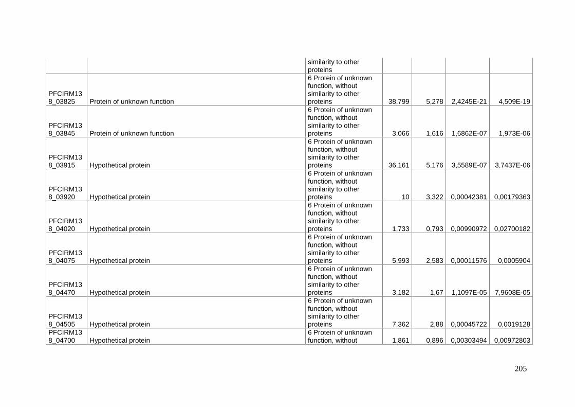
205
similarity to otherproteins
PFCIRM138_03825 Protein of unknown function
6 Protein of unknownfunction, withoutsimilarity to otherproteins 38,799 5,278 2,4245E-21 4,509E-19
PFCIRM138_03845 Protein of unknown function
6 Protein of unknownfunction, withoutsimilarity to otherproteins 3,066 1,616 1,6862E-07 1,973E-06
PFCIRM138_03915 Hypothetical protein
6 Protein of unknownfunction, withoutsimilarity to otherproteins 36,161 5,176 3,5589E-07 3,7437E-06
PFCIRM138_03920 Hypothetical protein
6 Protein of unknownfunction, withoutsimilarity to otherproteins 10 3,322 0,00042381 0,00179363
PFCIRM138_04020 Hypothetical protein
6 Protein of unknownfunction, withoutsimilarity to otherproteins 1,733 0,793 0,00990972 0,02700182
PFCIRM138_04075 Hypothetical protein
6 Protein of unknownfunction, withoutsimilarity to otherproteins 5,993 2,583 0,00011576 0,0005904
PFCIRM138_04470 Hypothetical protein
6 Protein of unknownfunction, withoutsimilarity to otherproteins 3,182 1,67 1,1097E-05 7,9608E-05
PFCIRM138_04505 Hypothetical protein
6 Protein of unknownfunction, withoutsimilarity to otherproteins 7,362 2,88 0,00045722 0,0019128
PFCIRM138_04700 Hypothetical protein
6 Protein of unknownfunction, without 1,861 0,896 0,00303494 0,00972803

206
similarity to otherproteins
PFCIRM138_04710 Hypothetical protein
6 Protein of unknownfunction, withoutsimilarity to otherproteins 2,875 1,524 2,1237E-05 0,00013626
PFCIRM138_04740 Hypothetical protein
6 Protein of unknownfunction, withoutsimilarity to otherproteins 15,023 3,909 1,6108E-07 1,8949E-06
PFCIRM138_04775 protein of unknown function
6 Protein of unknownfunction, withoutsimilarity to otherproteins 3,834 1,939 0,0032166 0,01021468
PFCIRM138_05060 Hypothetical protein
6 Protein of unknownfunction, withoutsimilarity to otherproteins 2,658 1,41 0,00327233 0,01036158
PFCIRM138_05135 Protein of unknown function
6 Protein of unknownfunction, withoutsimilarity to otherproteins 8,584 3,102 0,00029126 0,00129528
PFCIRM138_05225 Hypothetical protein
6 Protein of unknownfunction, withoutsimilarity to otherproteins 11,52 3,526 1,0332E-18 9,5632E-17
PFCIRM138_05240 Hypothetical protein
6 Protein of unknownfunction, withoutsimilarity to otherproteins 2,958 1,565 0,01699199 0,04248969
PFCIRM138_05605 Hypothetical protein
6 Protein of unknownfunction, withoutsimilarity to otherproteins 5,103 2,351 0,00026846 0,0012012
PFCIRM138_05680 Hypothetical protein
6 Protein of unknownfunction, without 3,232 1,693 0,00613998 0,01805681

207
similarity to otherproteins
PFCIRM138_06165 Hypothetical protein
6 Protein of unknownfunction, withoutsimilarity to otherproteins 5,277 2,4 6,5848E-09 1,0005E-07
PFCIRM138_06170 Protein of unknown function
6 Protein of unknownfunction, withoutsimilarity to otherproteins 7,138 2,836 0,00014478 0,00070711
PFCIRM138_06315 Hypothetical protein
6 Protein of unknownfunction, withoutsimilarity to otherproteins 18,264 4,191 1,4142E-05 9,6699E-05
PFCIRM138_06360 Protein of unknown function
6 Protein of unknownfunction, withoutsimilarity to otherproteins 2,088 1,062 0,00187852 0,00648298
PFCIRM138_06635 Hypothetical protein
6 Protein of unknownfunction, withoutsimilarity to otherproteins 1,534 0,618 0,01811254 0,04467896
PFCIRM138_06780 Protein of unknown function
6 Protein of unknownfunction, withoutsimilarity to otherproteins 29,47 4,881 5,3936E-06 4,2603E-05
PFCIRM138_06785 Protein of unknown function
6 Protein of unknownfunction, withoutsimilarity to otherproteins 10,352 3,372 0,00023019 0,0010581
PFCIRM138_06790 Protein of unknown function
6 Protein of unknownfunction, withoutsimilarity to otherproteins 26,06 4,704 1,4786E-05 0,00010016
PFCIRM138_06815 Hypothetical protein
6 Protein of unknownfunction, without 2,101 1,071 0,0006017 0,0024112

208
similarity to otherproteins
PFCIRM138_06820 Hypothetical protein
6 Protein of unknownfunction, withoutsimilarity to otherproteins 8,02 3,004 1,8565E-16 1,0689E-14
PFCIRM138_06875 Hypothetical protein
6 Protein of unknownfunction, withoutsimilarity to otherproteins 2,551 1,351 0,00132553 0,00479382
PFCIRM138_06955 Hypothetical protein
6 Protein of unknownfunction, withoutsimilarity to otherproteins 5,932 2,568 0,00212844 0,00724264
PFCIRM138_06990 protein of unknown function
6 Protein of unknownfunction, withoutsimilarity to otherproteins 4,812 2,267 8,3428E-08 9,9748E-07
PFCIRM138_07105 Hypothetical protein
6 Protein of unknownfunction, withoutsimilarity to otherproteins 4,688 2,229 3,5802E-13 1,224E-11
PFCIRM138_07155 Protein of unknown function
6 Protein of unknownfunction, withoutsimilarity to otherproteins 6,541 2,71 0,00073586 0,00286486
PFCIRM138_07160 protein of unknown function
6 Protein of unknownfunction, withoutsimilarity to otherproteins 18,185 4,185 3,7496E-05 0,00021995
PFCIRM138_07190 protein of unknown function
6 Protein of unknownfunction, withoutsimilarity to otherproteins 10,206 3,351 0,0001789 0,00084337
PFCIRM138_07200 protein of unknown function
6 Protein of unknownfunction, without 9,477 3,244 0,00016107 0,00077454
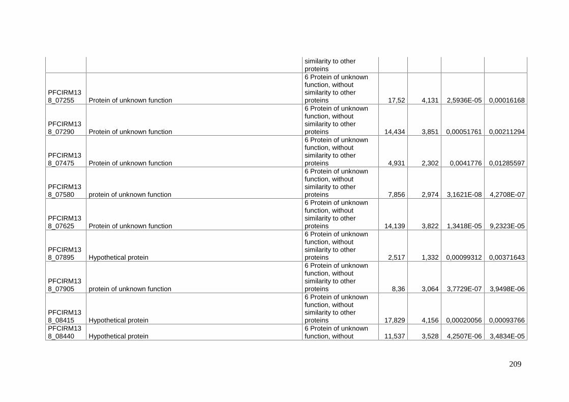
209
similarity to otherproteins
PFCIRM138_07255 Protein of unknown function
6 Protein of unknownfunction, withoutsimilarity to otherproteins 17,52 4,131 2,5936E-05 0,00016168
PFCIRM138_07290 Protein of unknown function
6 Protein of unknownfunction, withoutsimilarity to otherproteins 14,434 3,851 0,00051761 0,00211294
PFCIRM138_07475 Protein of unknown function
6 Protein of unknownfunction, withoutsimilarity to otherproteins 4,931 2,302 0,0041776 0,01285597
PFCIRM138_07580 protein of unknown function
6 Protein of unknownfunction, withoutsimilarity to otherproteins 7,856 2,974 3,1621E-08 4,2708E-07
PFCIRM138_07625 Protein of unknown function
6 Protein of unknownfunction, withoutsimilarity to otherproteins 14,139 3,822 1,3418E-05 9,2323E-05
PFCIRM138_07895 Hypothetical protein
6 Protein of unknownfunction, withoutsimilarity to otherproteins 2,517 1,332 0,00099312 0,00371643
PFCIRM138_07905 protein of unknown function
6 Protein of unknownfunction, withoutsimilarity to otherproteins 8,36 3,064 3,7729E-07 3,9498E-06
PFCIRM138_08415 Hypothetical protein
6 Protein of unknownfunction, withoutsimilarity to otherproteins 17,829 4,156 0,00020056 0,00093766
PFCIRM138_08440 Hypothetical protein
6 Protein of unknownfunction, without 11,537 3,528 4,2507E-06 3,4834E-05

210
similarity to otherproteins
PFCIRM138_08445 Protein of unknown function
6 Protein of unknownfunction, withoutsimilarity to otherproteins 35,068 5,132 2,2273E-07 2,4613E-06
PFCIRM138_08450 Protein of unknown function
6 Protein of unknownfunction, withoutsimilarity to otherproteins 5,264 2,396 0,00290053 0,00945808
PFCIRM138_08455 Protein of unknown function
6 Protein of unknownfunction, withoutsimilarity to otherproteins 15,732 3,976 7,589E-05 0,00040698
PFCIRM138_08535 Hypothetical protein
6 Protein of unknownfunction, withoutsimilarity to otherproteins 6,391 2,676 0,00179745 0,00626246
PFCIRM138_08575 Hypothetical protein
6 Protein of unknownfunction, withoutsimilarity to otherproteins 4,394 2,136 1,0378E-14 4,4524E-13
PFCIRM138_08580 Hypothetical protein
6 Protein of unknownfunction, withoutsimilarity to otherproteins 4,685 2,228 2,066E-06 1,8375E-05
PFCIRM138_08585 Hypothetical protein
6 Protein of unknownfunction, withoutsimilarity to otherproteins 4,993 2,32 0,00048621 0,00201103
PFCIRM138_08680 Hypothetical protein
6 Protein of unknownfunction, withoutsimilarity to otherproteins 5,209 2,381 0,00161249 0,00568136
PFCIRM138_08975 Hypothetical protein
6 Protein of unknownfunction, without 2,048 1,034 0,01846948 0,04530406

211
similarity to otherproteins
PFCIRM138_09120 Hypothetical protein
6 Protein of unknownfunction, withoutsimilarity to otherproteins 6,319 2,66 2,2515E-13 8,2105E-12
PFCIRM138_09125 Protein of unknown function
6 Protein of unknownfunction, withoutsimilarity to otherproteins 10,349 3,371 1,5412E-17 1,1628E-15
PFCIRM138_09230 Hypothetical protein
6 Protein of unknownfunction, withoutsimilarity to otherproteins 12,115 3,599 2,8213E-06 2,4208E-05
PFCIRM138_09235 Hypothetical protein
6 Protein of unknownfunction, withoutsimilarity to otherproteins 12,44 3,637 1,5483E-12 5,0407E-11
PFCIRM138_09340 Hypothetical protein
6 Protein of unknownfunction, withoutsimilarity to otherproteins 9,116 3,188 0,00049039 0,00202096
PFCIRM138_09610 Protein of unknown function
6 Protein of unknownfunction, withoutsimilarity to otherproteins 20,668 4,369 5,2006E-07 5,2437E-06
PFCIRM138_09655 Protein of unknown function
6 Protein of unknownfunction, withoutsimilarity to otherproteins 7,342 2,876 0,00023686 0,0010776
PFCIRM138_09660 Hypothetical protein
6 Protein of unknownfunction, withoutsimilarity to otherproteins 3,091 1,628 3,761E-05 0,00022003
PFCIRM138_09675 Hypothetical protein
6 Protein of unknownfunction, without 3,674 1,877 0,00295749 0,00958036

212
similarity to otherproteins
PFCIRM138_09695 Protein of unknown function
6 Protein of unknownfunction, withoutsimilarity to otherproteins 5,343 2,418 0,00257269 0,00859397
PFCIRM138_09700 protein of unknown function
6 Protein of unknownfunction, withoutsimilarity to otherproteins 28,614 4,839 4,0353E-08 5,2555E-07
PFCIRM138_10335 protein of unknown function
6 Protein of unknownfunction, withoutsimilarity to otherproteins 4,065 2,023 0,00876188 0,02439059
PFCIRM138_10470 Hypothetical protein
6 Protein of unknownfunction, withoutsimilarity to otherproteins 2,212 1,145 7,5248E-05 0,00040552
PFCIRM138_10730 Hypothetical secreted protein
6 Protein of unknownfunction, withoutsimilarity to otherproteins 7,306 2,869 9,032E-05 0,00047505
PFCIRM138_10740 Hypothetical secreted protein
6 Protein of unknownfunction, withoutsimilarity to otherproteins 4,653 2,218 5,8076E-08 7,2612E-07
PFCIRM138_10825 Hypothetical protein
6 Protein of unknownfunction, withoutsimilarity to otherproteins 2,289 1,195 0,01000081 0,02721613
PFCIRM138_10945 Hypothetical protein
6 Protein of unknownfunction, withoutsimilarity to otherproteins 1,587 0,666 0,01389227 0,03605728
PFCIRM138_11080 Hypothetical protein
6 Protein of unknownfunction, without 5,281 2,401 0,00244981 0,00820856

213
similarity to otherproteins
PFCIRM138_11150 Hypothetical protein
6 Protein of unknownfunction, withoutsimilarity to otherproteins 1,785 0,836 0,01560696 0,039707
PFCIRM138_11315 Hypothetical protein
6 Protein of unknownfunction, withoutsimilarity to otherproteins 3,456 1,789 0,00118544 0,00433735
PFCIRM138_11410 Hypothetical membrane protein
6 Protein of unknownfunction, withoutsimilarity to otherproteins 7,12 2,832 0,00039519 0,00169879
PFCIRM138_11585 Hypothetical protein
6 Protein of unknownfunction, withoutsimilarity to otherproteins 5,681 2,506 0,00231847 0,00781634
PFCIRM138_11770 Protein of unknown function
6 Protein of unknownfunction, withoutsimilarity to otherproteins 3,746 1,905 0,00488555 0,01476462
PFCIRM138_11920 Protein of unknown function
6 Protein of unknownfunction, withoutsimilarity to otherproteins 3,976 1,991 0,01790837 0,04427515
PFCIRM138_12160 Hypothetical protein
6 Protein of unknownfunction, withoutsimilarity to otherproteins 4,339 2,117 0,00289119 0,00945188
PFCIRM138_12320 Protein of unknown function
6 Protein of unknownfunction, withoutsimilarity to otherproteins 2,612 1,385 0,01647353 0,0416199
PFCIRM138_07965 protein of unknown function
6.2 Protein of unknownfunction similar to 2,414 1,271 0,00021458 0,0009968

214
proteins from otherorganisms
PFCIRM138_07250 protein of unknown function
7.2 Protein of unknownfunction similar toproteins from otherorganisms 2,252 1,171 0,01141197 0,03048767
PFCIRM138_08485 protein of unknown function
8.2 Protein of unknownfunction similar toproteins from otherorganisms 2,193 1,133 0,00017041 0,00081055
PFCIRM138_07140 protein of unknown function
9.2 Protein of unknownfunction similar toproteins from otherorganisms 2,139 1,097 0,01049979 0,0283624

215
Table S2: Repressed genes in stationnary phase compared to exponential phase.
Name Descript Function FC 1/FClog2FoldChange pvalue padj
rmlC (cps)dTDP-4-dehydrorhamnose reductase / dTDP-4-dehydrorhamnose 3,5-epimerase
1 Cell envelope andcellular processes 0,349 2,86532951 -1,52 3,4728E-10 7,0388E-09
glmU UDP-N-acetylglucosamine pyrophosphorylase 1.1 Cell wall 0,63 1,58730159 -0,667 0,01478679 0,03806294
murCUDP-N-acetylmuramate--L-alanine ligase (UDP-N-acetylmuramoyl-L-alanine synthetase) 1.1 Cell wall 0,425 2,35294118 -1,234 2,2735E-06 1,9898E-05
murD
UDP-N-acetylmuramoylalanine--D-glutamate ligase(UDP-N- acetylmuramoyl-L-alanyl-D-glutamatesynthetase) (D-glutamic acid- adding enzyme) 1.1 Cell wall 0,596 1,67785235 -0,746 0,00654238 0,01908631
mraYPhospho-N-acetylmuramoyl-pentapeptide-transferase(UDP- MurNAc-pentapeptide phosphotransferase) 1.1 Cell wall 0,291 3,43642612 -1,783 2,9208E-10 6,145E-09
murF
UDP-N-acetylmuramoyl-tripeptide--D-alanyl-D-alanineligase (UDP- MurNAc-pentapeptide synthetase) (D-alanyl-D-alanine-adding enzyme) 1.1 Cell wall 0,507 1,97238659 -0,979 0,00069815 0,00273757
murE
UDP-N-acetylmuramoylalanyl-D-glutamate--2,6-diaminopimelate ligase (UDP-N-acetylmuramyl-tripeptidesynthetase) (Meso- diaminopimelate-adding enzyme)(UDP-MurNAc-tripeptide synthetase) 1.1 Cell wall 0,527 1,89753321 -0,924 0,0006803 0,00268196
ftsICell division protein FtsI (penicillin-binding protein 2)(Peptidoglycan glycosyltransferase) 1.1 Cell wall 0,515 1,94174757 -0,957 0,00032724 0,00144646
PFCIRM138_02375 cell-wall peptidases, NlpC/P60 family secreted protein 1.1 Cell wall 0,192 5,20833333 -2,384 3,529E-18 2,8598E-16ddlA D-alanine--D-alanine ligase (D-alanylalanine synthetase) 1.1 Cell wall 0,336 2,97619048 -1,576 2,9725E-10 6,1942E-09dpm dolichyl-phosphate beta-D-mannosyltransferase 1.1 Cell wall 0,367 2,72479564 -1,448 2,8712E-05 0,00017597
MurAUDP-N-acetylglucosamine 1-carboxyvinyltransferase(Enoylpyruvate transferase) 1.1 Cell wall 0,555 1,8018018 -0,849 0,00063216 0,00251028
PFCIRM138_03660 Rhamnosyltransferase , Glycosyl transferase family 2 1.1 Cell wall 0,597 1,67504188 -0,744 0,00348714 0,01091539pbpA Penicillin-binding protein 1.1 Cell wall 0,318 3,14465409 -1,654 1,8129E-07 2,0987E-06mrc/ponA penicillin-binding protein (peptidoglycan 1.1 Cell wall 0,645 1,5503876 -0,632 0,01277471 0,03363546

216
glycosyltransferase)
slpG Surface layer protein G 1.1 Cell wall 0,355 2,81690141 -1,494 1,0062E-08 1,4876E-07
murBUDP-N-acetylenolpyruvoylglucosamine reductase (UDP-N- acetylmuramate dehydrogenase) 1.1 Cell wall 0,551 1,81488203 -0,859 0,00189094 0,00650532
slpF surface layer protein F (S-layer protein F) 1.1 Cell wall 0,447 2,23713647 -1,162 1,3114E-05 9,1088E-05gtfB Glycosyltransferase 1.1 Cell wall 0,577 1,73310225 -0,794 0,00456797 0,01393965tagG2 ABC-type polysaccharide export systems 1.1 Cell wall 0,379 2,63852243 -1,401 3,5163E-06 2,9253E-05
tagG1ABC-type polysaccharide export systems, permeasecomponent 1.1 Cell wall 0,518 1,93050193 -0,948 0,00012993 0,00065053
gtfA Glycosyltransferase, family 2 1.1 Cell wall 0,344 2,90697674 -1,539 4,5625E-08 5,907E-07glf, rfb UDP-galactopyranose mutase 1.1 Cell wall 0,472 2,11864407 -1,083 0,00054009 0,00218836
tagOUndecaprenyl-phosphate alpha-N-acetylglucosaminyltransferase 1.1 Cell wall 0,539 1,85528757 -0,89 0,00115377 0,00425707
PFCIRM138_00755 Hypoxanthine/guanine permease 2.A.40.7.2
1.2Transport/bindingproteins andlipoproteins 0,521 1,9193858 -0,94 0,02000989 0,04827567
PFCIRM138_00850 Transporter, major facilitator family 2.A.1.52.3
1.2Transport/bindingproteins andlipoproteins 0,181 5,52486188 -2,469 4,6211E-15 2,1513E-13
PFCIRM138_01645 ATP-binding protein ABC transporter
1.2Transport/bindingproteins andlipoproteins 0,366 2,73224044 -1,451 0,00309331 0,00986612
PFCIRM138_02940 transporter
1.2Transport/bindingproteins andlipoproteins 0,34 2,94117647 -1,557 5,2582E-09 8,2179E-08
PFCIRM138_03110
BP protein of monosaccharide ABC transporter(MOS:undef)
1.2Transport/bindingproteins andlipoproteins 0,325 3,07692308 -1,623 1,2191E-05 8,6046E-05
PFCIRM1 Alpha-ketoglutarate permease 2.A.1.6.2 1.2 0,399 2,50626566 -1,326 0,0029664 0,00958036

217
38_03760 Transport/bindingproteins andlipoproteins
PFCIRM138_04250 ABC transporter permease protein
1.2Transport/bindingproteins andlipoproteins 0,496 2,01612903 -1,011 0,00665541 0,01928747
PFCIRM138_04695 Xanthine/uracil permease
1.2Transport/bindingproteins andlipoproteins 0,489 2,04498978 -1,031 0,00875932 0,02439059
PFCIRM138_04945 Redox protein export
1.2Transport/bindingproteins andlipoproteins 0,62 1,61290323 -0,69 0,01649198 0,0416199
PFCIRM138_05530 ATP-binding protein of ABC transporter
1.2Transport/bindingproteins andlipoproteins 0,462 2,16450216 -1,113 4,8661E-05 0,00027872
PFCIRM138_05955 ABC transporter ATP-binding protein
1.2Transport/bindingproteins andlipoproteins 0,499 2,00400802 -1,002 0,00328542 0,010388
PFCIRM138_07330
polar amino acid ABC transporter, binding proteincomponent
1.2Transport/bindingproteins andlipoproteins 0,45 2,22222222 -1,153 0,00013505 0,00066855
oppDABC2 protein of oligopeptide ABC transporter(OPN:undef)
1.2Transport/bindingproteins andlipoproteins 0,283 3,5335689 -1,82 1,2652E-12 4,1943E-11
oppC IM protein of oligopeptide ABC transporter (OPN:undef)
1.2Transport/bindingproteins andlipoproteins 0,299 3,34448161 -1,742 4,4537E-12 1,2655E-10
oppB IM protein of oligopeptide ABC transporter (OPN:undef) 1.2 0,358 2,79329609 -1,482 2,4867E-08 3,4656E-07

218
Transport/bindingproteins andlipoproteins
PFCIRM138_09060 Permease component of cobalt ABC transporter
1.2Transport/bindingproteins andlipoproteins 0,482 2,0746888 -1,054 0,01199629 0,03181561
PFCIRM138_09845
BP protein of metallic cation (Zn2+ Mn2+) ABCtransporter
1.2Transport/bindingproteins andlipoproteins 0,395 2,53164557 -1,341 0,00015826 0,00076273
PFCIRM138_10105 Preprotein translocase SecA subunit
1.2Transport/bindingproteins andlipoproteins 0,5 2 -1,001 0,00168797 0,00592823
PFCIRM138_11820 ABC2 protein of ABC transporter (ART:REG)
1.2Transport/bindingproteins andlipoproteins 0,567 1,76366843 -0,819 0,00148618 0,00527884
tatCSec-independent protein translocase protein TatChomolog
1.2.1Transport/binding ofproteins/peptides 0,466 2,14592275 -1,103 1,5475E-05 0,00010451
htrA2 secreted serine protease, trypsin-like serine proteases
1.2.1Transport/binding ofproteins/peptides 0,414 2,41545894 -1,273 0,00044328 0,0018652
bopA solute binding protein of the ABC transport system
1.2.1Transport/binding ofproteins/peptides 0,471 2,12314225 -1,087 0,00053912 0,00218836
PFCIRM138_11405 ATP-binding component ABC transporter
1.2.1Transport/binding ofproteins/peptides 0,572 1,74825175 -0,805 0,01324727 0,03458939
feoB Ferrous iron uptake protein B 9.A.8.1.x
1.2.3Transport/binding ofinorganic ions 0,367 2,72479564 -1,445 0,00036855 0,00159997
PFCIRM138_05990 TrkA, potassium transporter
1.2.3Transport/binding of 0,636 1,57232704 -0,653 0,00697931 0,02006666
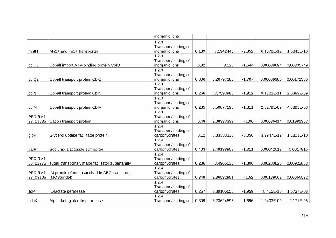
219
inorganic ions
mntH Mn2+ and Fe2+ transporter
1.2.3Transport/binding ofinorganic ions 0,139 7,1942446 -2,852 6,1578E-12 1,6842E-10
cbiO1 Cobalt import ATP-binding protein CbiO
1.2.3Transport/binding ofinorganic ions 0,32 3,125 -1,644 0,00088694 0,00335749
cbiQ2 Cobalt transport protein CbiQ
1.2.3Transport/binding ofinorganic ions 0,306 3,26797386 -1,707 0,00039985 0,00171335
cbiN Cobalt transport protein CbiN
1.2.3Transport/binding ofinorganic ions 0,266 3,7593985 -1,912 9,1322E-11 2,0389E-09
cbiM Cobalt transport protein CbiM
1.2.3Transport/binding ofinorganic ions 0,285 3,50877193 -1,811 2,6279E-09 4,3893E-08
PFCIRM138_11535 Cation transport protein
1.2.3Transport/binding ofinorganic ions 0,48 2,08333333 -1,06 0,00686414 0,01981363
glpF Glycerol uptake facilitator protein,
1.2.4Transport/binding ofcarbohydrates 0,12 8,33333333 -3,056 3,9947E-12 1,1811E-10
galP Sodium:galactoside symporter
1.2.4Transport/binding ofcarbohydrates 0,403 2,48138958 -1,311 0,00042013 0,0017815
PFCIRM138_02775 sugar transporter, major facilitator superfamily
1.2.4Transport/binding ofcarbohydrates 0,286 3,4965035 -1,806 0,00280826 0,00922833
PFCIRM138_03105
IM protein of monosaccharide ABC transporter(MOS:undef)
1.2.4Transport/binding ofcarbohydrates 0,349 2,86532951 -1,52 0,00189062 0,00650532
lldP L-lactate permease
1.2.4Transport/binding ofcarbohydrates 0,257 3,89105058 -1,959 8,415E-10 1,5737E-08
csbX Alpha-ketoglutarate permease1.2.4Transport/binding of 0,309 3,23624595 -1,696 1,2403E-09 2,171E-08

220
carbohydrates
slgTSodium/glucose cotransporter (Na(+)/glucose symporter)
2.A.21.3.2
1.2.4Transport/binding ofcarbohydrates 0,247 4,048583 -2,016 2,1526E-05 0,00013771
PFCIRM138_09590
PTS system, Lactose/Cellobiose or galactitol specific IIBsubunit
1.2.4Transport/binding ofcarbohydrates 0,475 2,10526316 -1,075 0,01727657 0,04300471
gluAATP-binding protein of glutamate ABC transporter (PAO :undef : Glutamate )
1.2.5Transport/binding ofamino-acids 0,603 1,65837479 -0,729 0,01697461 0,04248969
PFCIRM138_05630 Oligopeptide transporter, OPT family protein.
1.2.5Transport/binding ofamino-acids 0,529 1,89035917 -0,918 0,00026243 0,00118634
PFCIRM138_05890 Amino acid permease. membrane protein
1.2.5Transport/binding ofamino-acids 0,425 2,35294118 -1,235 1,715E-05 0,00011405
PFCIRM138_06700 ABC-type transport systems, periplasmic component
1.2.5Transport/binding ofamino-acids 0,189 5,29100529 -2,401 2,252E-06 1,9791E-05
metN Methionine import ATP-binding protein metN
1.2.5Transport/binding ofamino-acids 0,189 5,29100529 -2,405 1,6269E-11 4,0915E-10
PFCIRM138_06710 ABC transporter, permease protein
1.2.5Transport/binding ofamino-acids 0,19 5,26315789 -2,397 2,9778E-14 1,143E-12
PFCIRM138_07320
polar amino acid ABC transporter, inner membranecomponent
1.2.5Transport/binding ofamino-acids 0,289 3,46020761 -1,79 3,7466E-12 1,123E-10
PFCIRM138_07325 polar amino acid ABC transporter, binding protein
1.2.5Transport/binding ofamino-acids 0,482 2,0746888 -1,053 1,1802E-05 8,3568E-05
PFCIRM138_10275
binding protein of oligopeptide ABC transporter (OPN :undef : Oligopeptides)
1.2.5Transport/binding ofamino-acids 0,341 2,93255132 -1,55 8,149E-08 9,7967E-07
PFCIRM138_10280 permease protein of oligopeptide ABC transporter
1.2.5Transport/binding of 0,538 1,85873606 -0,895 0,01001421 0,02721876

221
amino-acids
argK Kinase ArgK
1.2.5Transport/binding ofamino-acids 0,36 2,77777778 -1,473 6,4451E-05 0,00035521
carA Carbamoyl-phosphate synthase small chain
1.2.6Transport/binding ofnucleosides,nucleotides, purinesand pyrimidines 0,605 1,65289256 -0,726 0,00517053 0,01554
codB Permease for cytosine/purines, uracil, thiamine, allantoin
1.2.6Transport/binding ofnucleosides,nucleotides, purinesand pyrimidines 0,365 2,73972603 -1,452 0,00053094 0,00216329
adkAdenylate kinase (EC 2.7.4.3) (ATP-AMPtransphosphorylase)
1.2.6Transport/binding ofnucleosides,nucleotides, purinesand pyrimidines 0,358 2,79329609 -1,482 8,9583E-06 6,7126E-05
PFCIRM138_03945 Forkhead-associated protein
1.3 Sensors (signaltransduction) 0,634 1,57728707 -0,658 0,01006328 0,02731819
PFCIRM138_03950 Forkhead-associated protein
1.3 Sensors (signaltransduction) 0,375 2,66666667 -1,416 4,6256E-05 0,00026704
PFCIRM138_06685
Sensor protein, ATP-binding region, ATPase-like:Histidine kinase
1.3 Sensors (signaltransduction) 0,492 2,03252033 -1,023 0,00116254 0,00428222
PFCIRM138_09785 Sensor protein, ATPase-like:Histidine kinase
1.3 Sensors (signaltransduction) 0,321 3,1152648 -1,638 1,1946E-10 2,6138E-09
PFCIRM138_09790 two-component response regulator
1.3 Sensors (signaltransduction) 0,421 2,37529691 -1,249 4,3636E-06 3,5493E-05
sdhB3 Succinate dehydrogenase
1.4 Membranebioenergetics(electron transportchain and ATPsynthase) 0,172 5,81395349 -2,539 9,6739E-11 2,138E-09
sdhA3 Succinate dehydrogenase flavoprotein subunit1.4 Membranebioenergetics 0,218 4,58715596 -2,195 2,5141E-11 6,1121E-10

222
(electron transportchain and ATPsynthase)
sdhC2 Succinate dehydrogenase cytochrome B-558 subunit
1.4 Membranebioenergetics(electron transportchain and ATPsynthase) 0,227 4,40528634 -2,136 2,2604E-12 6,966E-11
mgtE magnesium (Mg2+) transporter
1.4 Membranebioenergetics(electron transportchain and ATPsynthase) 0,615 1,62601626 -0,702 0,01194171 0,0317093
fdxA Ferredoxin
1.4 Membranebioenergetics(electron transportchain and ATPsynthase) 0,35 2,85714286 -1,516 7,4299E-08 8,9815E-07
nuoN NADH dehydrogenase I chain N
1.4 Membranebioenergetics(electron transportchain and ATPsynthase) 0,165 6,06060606 -2,595 2,7031E-13 9,3881E-12
nuoM NADH dehydrogenase I chain M
1.4 Membranebioenergetics(electron transportchain and ATPsynthase) 0,157 6,36942675 -2,67 1,707E-12 5,3858E-11
nuoL NADH dehydrogenase (quinone)
1.4 Membranebioenergetics(electron transportchain and ATPsynthase) 0,201 4,97512438 -2,314 2,59E-09 4,3592E-08
nuoK NADH dehydrogenase I chain K
1.4 Membranebioenergetics(electron transport 0,199 5,02512563 -2,329 1,5742E-11 4,005E-10

223
chain and ATPsynthase)
nuoJNADH-quinone oxidoreductase chain J (NADHdehydrogenase I, chain J)
1.4 Membranebioenergetics(electron transportchain and ATPsynthase) 0,237 4,21940928 -2,075 3,6112E-08 4,7886E-07
nuoINADH-quinone oxidoreductase subunit I (NADHdehydrogenase I subunit I) (NDH-1 subunit I)
1.4 Membranebioenergetics(electron transportchain and ATPsynthase) 0,246 4,06504065 -2,023 1,2231E-09 2,1583E-08
nuoHNADH-quinone oxidoreductase subunit H (NADHdehydrogenase I subunit H)
1.4 Membranebioenergetics(electron transportchain and ATPsynthase) 0,314 3,18471338 -1,673 1,2256E-06 1,1509E-05
nuoGNADH-quinone oxidoreductase chain G (NADHdehydrogenase I, chain G)
1.4 Membranebioenergetics(electron transportchain and ATPsynthase) 0,288 3,47222222 -1,795 5,0931E-06 4,0523E-05
nuoFNADH-quinone oxidoreductase chain F (NADHdehydrogenase I, chain F) (NDH-1, chain F)
1.4 Membranebioenergetics(electron transportchain and ATPsynthase) 0,196 5,10204082 -2,354 1,2923E-08 1,8726E-07
nuoE NADH-quinone oxidoreductase chain E
1.4 Membranebioenergetics(electron transportchain and ATPsynthase) 0,217 4,60829493 -2,206 2,5645E-12 7,7934E-11
nuoDNADH-quinone oxidoreductase chain D (EC 1.6.99.5)(NADH dehydrogenase I, chain D)
1.4 Membranebioenergetics(electron transportchain and ATP 0,164 6,09756098 -2,607 4,9573E-12 1,3906E-10
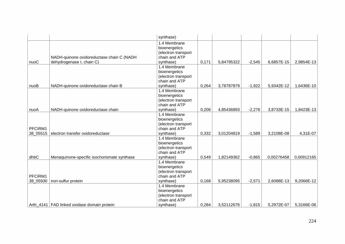
224
synthase)
nuoCNADH-quinone oxidoreductase chain C (NADHdehydrogenase I, chain C)
1.4 Membranebioenergetics(electron transportchain and ATPsynthase) 0,171 5,84795322 -2,545 6,6857E-15 2,9854E-13
nuoB NADH-quinone oxidoreductase chain B
1.4 Membranebioenergetics(electron transportchain and ATPsynthase) 0,264 3,78787879 -1,922 5,9342E-12 1,6436E-10
nuoA NADH-quinone oxidoreductase chain
1.4 Membranebioenergetics(electron transportchain and ATPsynthase) 0,206 4,85436893 -2,276 3,8733E-15 1,8423E-13
PFCIRM138_05515 electron transfer oxidoreductase
1.4 Membranebioenergetics(electron transportchain and ATPsynthase) 0,332 3,01204819 -1,589 3,2108E-08 4,31E-07
dhbC Menaquinone-specific isochorismate synthase
1.4 Membranebioenergetics(electron transportchain and ATPsynthase) 0,549 1,82149362 -0,865 0,00276458 0,00912165
PFCIRM138_05930 iron-sulfur protein
1.4 Membranebioenergetics(electron transportchain and ATPsynthase) 0,168 5,95238095 -2,571 2,6088E-13 9,2066E-12
Arth_4141 FAD linked oxidase domain protein
1.4 Membranebioenergetics(electron transportchain and ATPsynthase) 0,284 3,52112676 -1,815 5,2972E-07 5,3166E-06

225
sdhC1 Succinate dehydrogenase subunit C
1.4 Membranebioenergetics(electron transportchain and ATPsynthase) 0,116 8,62068966 -3,107 4,269E-21 6,6719E-19
sdhA Succinate dehydrogenase, subunit A
1.4 Membranebioenergetics(electron transportchain and ATPsynthase) 0,113 8,84955752 -3,147 1,8194E-18 1,5923E-16
sdhB Succinate dehydrogenase, subunit B
1.4 Membranebioenergetics(electron transportchain and ATPsynthase) 0,115 8,69565217 -3,114 5,1373E-16 2,8101E-14
moxR ATPases MoxR family
1.4 Membranebioenergetics(electron transportchain and ATPsynthase) 0,477 2,09643606 -1,069 0,00012147 0,00061238
atpF ATP synthase B chain (F0F1 ATP synthase subunit B)
1.4 Membranebioenergetics(electron transportchain and ATPsynthase) 0,554 1,80505415 -0,852 0,00200057 0,00682877
atpH ATP synthase delta chain
1.4 Membranebioenergetics(electron transportchain and ATPsynthase) 0,509 1,96463654 -0,975 0,0001822 0,00085548
atpAATP synthase subunit alpha (ATPase subunit alpha)(ATP synthase F1 sector subunit alpha)
1.4 Membranebioenergetics(electron transportchain and ATPsynthase) 0,412 2,42718447 -1,279 2,3908E-05 0,00014989
atpG ATP synthase gamma chain (ATP synthase F1 sector 1.4 Membrane 0,41 2,43902439 -1,287 1,1699E-05 8,311E-05
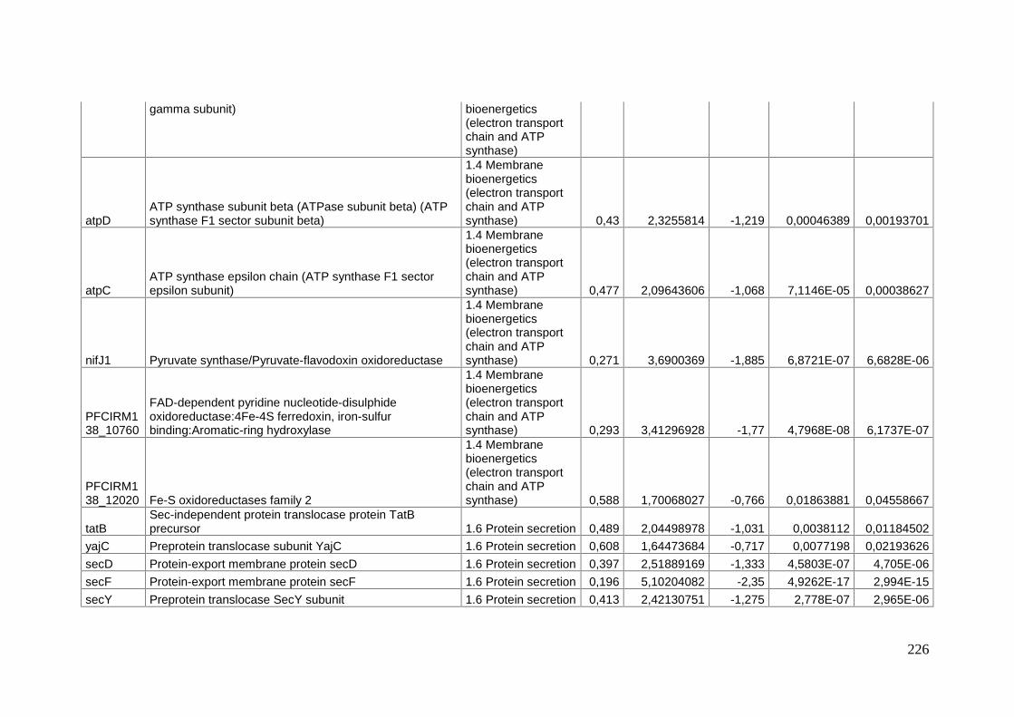
226
gamma subunit) bioenergetics(electron transportchain and ATPsynthase)
atpDATP synthase subunit beta (ATPase subunit beta) (ATPsynthase F1 sector subunit beta)
1.4 Membranebioenergetics(electron transportchain and ATPsynthase) 0,43 2,3255814 -1,219 0,00046389 0,00193701
atpCATP synthase epsilon chain (ATP synthase F1 sectorepsilon subunit)
1.4 Membranebioenergetics(electron transportchain and ATPsynthase) 0,477 2,09643606 -1,068 7,1146E-05 0,00038627
nifJ1 Pyruvate synthase/Pyruvate-flavodoxin oxidoreductase
1.4 Membranebioenergetics(electron transportchain and ATPsynthase) 0,271 3,6900369 -1,885 6,8721E-07 6,6828E-06
PFCIRM138_10760
FAD-dependent pyridine nucleotide-disulphideoxidoreductase:4Fe-4S ferredoxin, iron-sulfurbinding:Aromatic-ring hydroxylase
1.4 Membranebioenergetics(electron transportchain and ATPsynthase) 0,293 3,41296928 -1,77 4,7968E-08 6,1737E-07
PFCIRM138_12020 Fe-S oxidoreductases family 2
1.4 Membranebioenergetics(electron transportchain and ATPsynthase) 0,588 1,70068027 -0,766 0,01863881 0,04558667
tatBSec-independent protein translocase protein TatBprecursor 1.6 Protein secretion 0,489 2,04498978 -1,031 0,0038112 0,01184502
yajC Preprotein translocase subunit YajC 1.6 Protein secretion 0,608 1,64473684 -0,717 0,0077198 0,02193626secD Protein-export membrane protein secD 1.6 Protein secretion 0,397 2,51889169 -1,333 4,5803E-07 4,705E-06secF Protein-export membrane protein secF 1.6 Protein secretion 0,196 5,10204082 -2,35 4,9262E-17 2,994E-15secY Preprotein translocase SecY subunit 1.6 Protein secretion 0,413 2,42130751 -1,275 2,778E-07 2,965E-06

227
ftsQ Cell division protein FtsQ 1.7 Cell division 0,375 2,66666667 -1,415 0,00013391 0,00066741PFCIRM138_04995 ATPase involved in cell division 1.7 Cell division 0,479 2,08768267 -1,061 2,4266E-05 0,0001517
gab
Aldehyde dehydrogenase (Succinate-semialdehydedehydrogenase) (NAD-dependent aldehydedehydrogenase)
2.1 Metabolism ofcarbohydrates andrelated molecules 0,529 1,89035917 -0,919 0,00593897 0,01753638
pfp (pfk) pyrophosphate phosphofructokinase
2.1 Metabolism ofcarbohydrates andrelated molecules 0,318 3,14465409 -1,652 2,173E-07 2,4253E-06
lpd
Dihydrolipoyl dehydrogenase (E3 component of alphaketo acid dehydrogenase complexes) (Dihydrolipoamidedehydrogenase)
2.1 Metabolism ofcarbohydrates andrelated molecules 0,504 1,98412698 -0,989 0,0008952 0,00338291
PFCIRM138_04560 Aldose 1-epimerase
2.1 Metabolism ofcarbohydrates andrelated molecules 0,357 2,80112045 -1,488 6,0157E-08 7,4363E-07
iolT3 iolT3 (myo-inositol transporter iolT3)
2.1 Metabolism ofcarbohydrates andrelated molecules 0,564 1,77304965 -0,827 0,01230528 0,03247763
pta Phosphate acetyltransferase
2.1 Metabolism ofcarbohydrates andrelated molecules 0,23 4,34782609 -2,122 8,9026E-14 3,3015E-12
ackA Acetate kinase
2.1 Metabolism ofcarbohydrates andrelated molecules 0,204 4,90196078 -2,291 8,1723E-19 8,1277E-17
glmS
Glucosamine--fructose-6-phosphate aminotransferase (Hexosephosphate aminotransferase, D-fructose-6-phosphate amidotransferase)
2.1 Metabolism ofcarbohydrates andrelated molecules 0,613 1,63132137 -0,706 0,0068971 0,01988255
glpKGlycerol kinase (ATP:glycerol 3-phosphotransferase)(Glycerokinase) (GK)
2.1.1 Specificcarbohydratemetabolic pathway 0,157 6,36942675 -2,674 4,4048E-10 8,6826E-09
citECitrate lyase beta chain (Citrase beta chain) (Citrate(Pro-3S)-lyase beta chain)
2.1.1 Specificcarbohydratemetabolic pathway 0,582 1,71821306 -0,782 0,0029122 0,00948198
lacZ Beta-galactosidase (Lactase) LacZ
2.1.1 Specificcarbohydratemetabolic pathway 0,308 3,24675325 -1,699 9,7588E-07 9,2434E-06

228
glpBAnaerobic glycerol-3-phosphate dehydrogenase subunitB
2.1.1 Specificcarbohydratemetabolic pathway 0,501 1,99600798 -0,998 0,00012019 0,00060873
glpCAnaerobic glycerol-3-phosphate dehydrogenase subunitC
2.1.1 Specificcarbohydratemetabolic pathway 0,462 2,16450216 -1,116 3,0035E-05 0,00018255
otsATrehalose-6-phosphate synthase (Alpha,alpha-trehalose-phosphate synthase)
2.1.1 Specificcarbohydratemetabolic pathway 0,616 1,62337662 -0,698 0,00748217 0,02142799
rmlA(rfbA) Glucose-1-phosphate thymidylyltransferase
2.1.1 Specificcarbohydratemetabolic pathway 0,629 1,58982512 -0,669 0,00798454 0,02261805
galK2 Galactokinase
2.1.1 Specificcarbohydratemetabolic pathway 0,541 1,84842884 -0,886 0,00017825 0,00084337
araM L-arabinose utilization protein
2.1.1 Specificcarbohydratemetabolic pathway 0,355 2,81690141 -1,493 8,2974E-05 0,00044172
iolA(msdA)
iolA (Myo-inositol catabolism IolA protein) (Methylmalonicacid semialdehyde dehydrogenase)
2.1.1 Specificcarbohydratemetabolic pathway 0,539 1,85528757 -0,893 0,00264533 0,00882316
iolB iolB (Myo-inositol catabolism IolB protein)
2.1.1 Specificcarbohydratemetabolic pathway 0,464 2,15517241 -1,108 0,00048368 0,00200815
PFCIRM138_05815
Methylmalonyl-CoA carboxytransferase 5S subunit.(transcarboxylase 5S) 505 bp
2.1.1 Specificcarbohydratemetabolic pathway 0,546 1,83150183 -0,873 0,0054605 0,01634416
mmdAMethylmalonyl-CoA carboxytransferase 12S subunit(EC2.1.3.1) (Transcarboxylase 12S subunit). 610 bp
2.1.1 Specificcarbohydratemetabolic pathway 0,417 2,39808153 -1,261 0,00012662 0,00063544
PFCIRM138_05825 Methylmalonyl-CoA carboxyltransferase 12S subunit
2.1.1 Specificcarbohydratemetabolic pathway 0,447 2,23713647 -1,161 0,00023995 0,00108923
bccpMethylmalonyl-CoA carboxytransferase , 1.3S subunit(Transcarboxylase, 1.3S subunit). 123bp
2.1.1 Specificcarbohydratemetabolic pathway 0,344 2,90697674 -1,54 6,6959E-06 5,1769E-05

229
galE2 UDP-glucose 4-epimerase
2.1.1 Specificcarbohydratemetabolic pathway 0,549 1,82149362 -0,866 0,00038876 0,00167442
PFCIRM138_07970 inositol-1-phosphate synthase
2.1.1 Specificcarbohydratemetabolic pathway 0,319 3,13479624 -1,65 1,9053E-07 2,1827E-06
treS Trehalose synthase
2.1.1 Specificcarbohydratemetabolic pathway 0,501 1,99600798 -0,997 0,00011277 0,00058058
pccB Propionyl-CoA carboxylase beta chain
2.1.1 Specificcarbohydratemetabolic pathway 0,319 3,13479624 -1,65 6,2064E-10 1,1707E-08
galTGalactose-1-phosphate uridylyltransferase (UDP-glucose---hexose-1-phosphate uridylyltransferase)
2.1.1 Specificcarbohydratemetabolic pathway 0,539 1,85528757 -0,893 0,00560477 0,0167074
tpi1 triosephosphate isomerase 12.1.2 Main glycolyticpathways 0,505 1,98019802 -0,985 0,00658728 0,0191917
pgk Phosphoglycerate kinase2.1.2 Main glycolyticpathways 0,449 2,22717149 -1,154 0,00023461 0,00107391
pyk1 Pyruvate kinase 12.1.2 Main glycolyticpathways 0,47 2,12765957 -1,088 0,00053814 0,00218836
gmpPhosphoglycerate mutase (D-phosphoglycerate 2,3-phosphomutase)
2.1.2 Main glycolyticpathways 0,507 1,97238659 -0,979 0,00123669 0,0044948
ldh1 L-Lactate dehydrogenase2.1.2 Main glycolyticpathways 0,466 2,14592275 -1,101 6,1813E-06 4,8476E-05
fba1 Fructose-bisphosphate aldolase class II2.1.2 Main glycolyticpathways 0,53 1,88679245 -0,915 0,00770202 0,02191421
pfkA 6-phosphofructokinase2.1.2 Main glycolyticpathways 0,457 2,18818381 -1,13 7,0413E-05 0,00038516
gmpB phosphoglycerate mutase2.1.2 Main glycolyticpathways 0,464 2,15517241 -1,107 0,00493771 0,01488114
mdh Malate dehydrogenase 2.1.3 TCA cycle 0,522 1,91570881 -0,938 0,00276718 0,00912165fumC Fumarate hydratase, class-II 2.1.3 TCA cycle 0,47 2,12765957 -1,089 0,00044967 0,00188482icd Putative isocitrate/isopropylmalate dehydrogenase 2.1.3 TCA cycle 0,485 2,06185567 -1,045 0,0004114 0,00175468cat Coenzyme A transferase (Putative succinyl-CoA or 2.1.4 Substrate- 0,421 2,37529691 -1,247 0,00011893 0,00060373

230
butyryl-CoA:coenzyme A transferase) specific entries tocarbohydratemetabolic pathway
rmlB(rfbB) DTDP-glucose 4,6-dehydratase
2.1.4 Substrate-specific entries tocarbohydratemetabolic pathway 0,492 2,03252033 -1,023 6,695E-05 0,00036806
argH Argininosuccinate lyase (Arginosuccinase)
2.2 Metabolism ofamino acids andrelated molecules 0,479 2,08768267 -1,061 3,7005E-05 0,00021766
argJ
Arginine biosynthesis bifunctional protein argJ(Glutamate N-acetyltransferase) (Ornithineacetyltransferase) (Ornithine transacetylase) (OATase)
2.2 Metabolism ofamino acids andrelated molecules 0,347 2,88184438 -1,526 2,9771E-07 3,162E-06
argC
N-acetyl-gamma-glutamyl-phosphate reductase (AGPR)(N- acetyl-glutamate semialdehyde dehydrogenase)(NAGSA dehydrogenase)
2.2 Metabolism ofamino acids andrelated molecules 0,256 3,90625 -1,963 1,7231E-12 5,3858E-11
hisG ATP phosphoribosyltransferase
2.2 Metabolism ofamino acids andrelated molecules 0,633 1,57977883 -0,66 0,00677543 0,01960931
hisE Phosphoribosyl-ATP pyrophosphohydrolase
2.2 Metabolism ofamino acids andrelated molecules 0,501 1,99600798 -0,998 0,00289431 0,00945188
trpD Anthranilate phosphoribosyltransferase
2.2 Metabolism ofamino acids andrelated molecules 0,271 3,6900369 -1,882 4,6178E-13 1,5544E-11
PFCIRM138_02155 Lysine decarboxylase
2.2 Metabolism ofamino acids andrelated molecules 0,463 2,15982721 -1,112 1,1428E-05 8,1446E-05
PFCIRM138_02175 aminotransferase
2.2 Metabolism ofamino acids andrelated molecules 0,317 3,15457413 -1,658 8,9783E-11 2,0252E-09
hisCHistidinol-phosphate aminotransferase (Imidazole acetol-phosphate transaminase)
2.2 Metabolism ofamino acids andrelated molecules 0,518 1,93050193 -0,948 0,00016639 0,00079316
hisB Imidazoleglycerol-phosphate dehydratase (IGPD)2.2 Metabolism ofamino acids and 0,44 2,27272727 -1,186 0,00010079 0,00052382

231
related molecules
hisA
Phosphoribosyl isomerase A (1-(5-phosphoribosyl)-5-[(5-phosphoribosylamino)methylideneamino] imidazole-4-carboxamide isomerase) (Phosphoribosylformimino-5-aminoimidazole carboxamide ribotide isomerase) (N-(5 -phosphoribosyl)anthranilate isomerase) (PRAI)
2.2 Metabolism ofamino acids andrelated molecules 0,5 2 -1,001 0,00037469 0,00162341
carB(pyrA)
Carbamoyl-phosphate synthase large chain (Carbamoyl-phosphate synthetase ammonia chain)
2.2 Metabolism ofamino acids andrelated molecules 0,397 2,51889169 -1,332 5,4695E-05 0,00030685
metHmethionine synthase (5-methyltetrahydrofolate:L-homocysteine S-methyltransferase)
2.2 Metabolism ofamino acids andrelated molecules 0,474 2,10970464 -1,076 1,9411E-05 0,00012716
ilvC
Ketol-acid reductoisomerase (Acetohydroxy-acidisomeroreductase) (Alpha-keto-beta-hydroxylacilreductoisomerase)
2.2 Metabolism ofamino acids andrelated molecules 0,208 4,80769231 -2,262 6,3096E-17 3,7312E-15
ilvD Dihydroxy-acid dehydratase
2.2 Metabolism ofamino acids andrelated molecules 0,46 2,17391304 -1,122 2,6212E-05 0,00016287
asnB Asparagine synthase (Glutamine-hydrolyzing)
2.2 Metabolism ofamino acids andrelated molecules 0,672 1,48809524 -0,574 0,0171151 0,04270219
ansA L-asparaginase I
2.2 Metabolism ofamino acids andrelated molecules 0,472 2,11864407 -1,082 8,953E-05 0,00047203
ask Aspartokinase (Aspartate kinase)
2.2 Metabolism ofamino acids andrelated molecules 0,557 1,79533214 -0,845 0,00405315 0,01252584
cys2 cysteine synthase 2
2.2 Metabolism ofamino acids andrelated molecules 0,057 17,5438596 -4,135 1,4784E-14 6,1033E-13
PFCIRM138_09945 alanine racemase
2.2 Metabolism ofamino acids andrelated molecules 0,444 2,25225225 -1,172 0,00146153 0,00519973
tyrB Aspartate transaminase (Aminotransferase)
2.2 Metabolism ofamino acids andrelated molecules 0,418 2,3923445 -1,259 3,3558E-05 0,00019898

232
gabT 4-aminobutyrate aminotransferase
2.2 Metabolism ofamino acids andrelated molecules 0,152 6,57894737 -2,714 5,2032E-19 5,4212E-17
lysA Diaminopimelate decarboxylase (DAP decarboxylase)
2.2 Metabolism ofamino acids andrelated molecules 0,552 1,8115942 -0,857 0,00051744 0,00211294
thrA/hom Homoserine dehydrogenase
2.2 Metabolism ofamino acids andrelated molecules 0,411 2,43309002 -1,283 1,7656E-07 2,0549E-06
serB/thrHPhosphoserine phosphatase/homoserinephosphotransferase bifunctional protein
2.2 Metabolism ofamino acids andrelated molecules 0,36 2,77777778 -1,475 5,9958E-08 7,4363E-07
bkdB
dihydrolipoyllysine-residue (2-methylpropanoyl)transferase. Lipoamide acyltransferasecomponent of branched-chain alpha-keto aciddehydrogenase complex (E2) (Dihydrolipoamidebranched chain transacylase)
2.2 Metabolism ofamino acids andrelated molecules 0,375 2,66666667 -1,416 2,0794E-07 2,3399E-06
bkdA2
2-oxoisovalerate dehydrogenase subunit beta (EC1.2.4.4) (Branched- chain alpha-keto acid dehydrogenaseE1 component beta chain) (BCKDH E1-beta) Pyruvatedehydrogenase E1 component subunit beta
2.2 Metabolism ofamino acids andrelated molecules 0,341 2,93255132 -1,554 1,5683E-08 2,2428E-07
bkdA1
2-oxoisovalerate dehydrogenase subunit alpha(Branched- chain alpha-keto acid dehydrogenase E1component alpha chain) (BCKDH E1-alpha)
2.2 Metabolism ofamino acids andrelated molecules 0,524 1,90839695 -0,932 0,00010967 0,00056593
argG Argininosuccinate synthase
2.2 Metabolism ofamino acids andrelated molecules 0,324 3,08641975 -1,625 2,3102E-07 2,5274E-06
PFCIRM138_11035 Chorismate mutase
2.2 Metabolism ofamino acids andrelated molecules 0,446 2,24215247 -1,166 7,3782E-05 0,00039959
cysE Serine acetyltransferase
2.2 Metabolism ofamino acids andrelated molecules 0,55 1,81818182 -0,863 0,00324044 0,01027548
cys1 Cysteine synthase 1
2.2 Metabolism ofamino acids andrelated molecules 0,585 1,70940171 -0,774 0,00184305 0,0063706
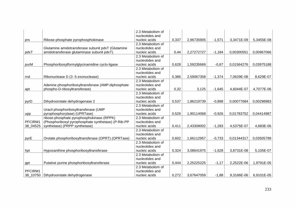
233
prs Ribose-phosphate pyrophosphokinase
2.3 Metabolism ofnucleotides andnucleic acids 0,337 2,96735905 -1,571 3,3471E-09 5,3455E-08
pdxTGlutamine amidotransferase subunit pdxT (Glutamineamidotransferase glutaminase subunit pdxT)
2.3 Metabolism ofnucleotides andnucleic acids 0,44 2,27272727 -1,184 0,00300551 0,00967066
purM Phosphoribosylformylglycinamidine cyclo-ligase
2.3 Metabolism ofnucleotides andnucleic acids 0,628 1,59235669 -0,67 0,01564276 0,03975188
rnd Ribonuclease D (3 -5 exonuclease)
2.3 Metabolism ofnucleotides andnucleic acids 0,386 2,59067358 -1,374 7,0629E-08 8,629E-07
aptAdenine phosphoribosyltransferase (AMP:diphosphatephospho-D-ribosyltransferase)
2.3 Metabolism ofnucleotides andnucleic acids 0,32 3,125 -1,645 4,6044E-07 4,7077E-06
pyrD Dihydroorotate dehydrogenase 2
2.3 Metabolism ofnucleotides andnucleic acids 0,537 1,86219739 -0,898 0,00077684 0,00298983
uppUracil phosphoribosyltransferase (UMPpyrophosphorylase) (UPRTase)
2.3 Metabolism ofnucleotides andnucleic acids 0,526 1,90114068 -0,926 0,01783752 0,04414987
PFCIRM138_04525
ribose-phosphate pyrophosphokinase (RPPK)(Phosphoribosyl pyrophosphate synthetase) (P-Rib-PPsynthetase) (PRPP synthetase)
2.3 Metabolism ofnucleotides andnucleic acids 0,411 2,43309002 -1,283 4,5375E-07 4,683E-06
pyrE Orotate phosphoribosyltransferase (OPRT) (OPRTase)
2.3 Metabolism ofnucleotides andnucleic acids 0,602 1,66112957 -0,733 0,01344317 0,03505799
hpt Hypoxanthine phosphoribosyltransferase
2.3 Metabolism ofnucleotides andnucleic acids 0,324 3,08641975 -1,628 3,8731E-08 5,105E-07
gpt Putative purine phosphoribosyltransferase
2.3 Metabolism ofnucleotides andnucleic acids 0,444 2,25225225 -1,17 2,2522E-06 1,9791E-05
PFCIRM138_10750 Dihydroorotate dehydrogenase
2.3 Metabolism ofnucleotides andnucleic acids 0,272 3,67647059 -1,88 9,3166E-06 6,9101E-05

234
guaB2Inosine-5 -monophosphate dehydrogenase (IMPdehydrogenase) (IMPDH) (IMPD) / GMP reductase
2.3 Metabolism ofnucleotides andnucleic acids 0,476 2,10084034 -1,07 0,00014279 0,00070049
purHPhosphoribosylaminoimidazolecarboxamideformyltransferase (AICAR transformylase)
2.3 Metabolism ofnucleotides andnucleic acids 0,398 2,51256281 -1,33 3,3022E-05 0,00019634
ugpQ2 Glycerophosphoryl diester phosphodiesterase2.4 Metabolism oflipids 0,241 4,14937759 -2,052 3,7194E-10 7,4661E-09
cdsA Phosphatidate cytidylyltransferase2.4 Metabolism oflipids 0,516 1,9379845 -0,955 0,00058516 0,0023579
inhA Enoyl-[acyl-carrier-protein] reductase (NADH)2.4 Metabolism oflipids 0,591 1,69204738 -0,758 0,00825452 0,02327434
fabG 3-oxoacyl-[acyl-carrier protein] reductase2.4 Metabolism oflipids 0,529 1,89035917 -0,92 0,00967845 0,02650153
plsC3 Acyltransferase PlsC2.4 Metabolism oflipids 0,592 1,68918919 -0,756 0,00480094 0,01456929
PFCIRM138_02490 Phosphatidylglycerophosphate synthase
2.4 Metabolism oflipids 0,631 1,58478605 -0,665 0,01122571 0,0301003
pgs2CDP-diacylglycerol--glycerol-3-phosphate 3-phosphatidyl-transferase
2.4 Metabolism oflipids 0,376 2,65957447 -1,409 0,00013478 0,00066855
PPA2234 Long-chain-fatty-acid--CoA ligase/synthetase2.4 Metabolism oflipids 0,574 1,74216028 -0,802 0,0029171 0,00948383
pf1861 Carboxylic ester hydrolase2.4 Metabolism oflipids 0,485 2,06185567 -1,045 9,3149E-05 0,00048759
fabF(fabB)
3-oxoacyl-[acyl-carrier-protein] synthase ( Beta-ketoacyl-ACP synthase)
2.4 Metabolism oflipids 0,159 6,28930818 -2,652 2,6775E-17 1,8307E-15
acpP Acyl carrier protein (ACP)2.4 Metabolism oflipids 0,169 5,91715976 -2,569 1,049E-18 9,5632E-17
fabH 3-oxoacyl-(Acyl-carrier-protein) synthase III2.4 Metabolism oflipids 0,145 6,89655172 -2,782 1,2113E-22 2,9449E-20
fabD Malonyl CoA-acyl carrier protein transacylase2.4 Metabolism oflipids 0,271 3,6900369 -1,884 4,0889E-12 1,1929E-10
accA Acetyl-CoA carboxylase2.4 Metabolism oflipids 0,598 1,6722408 -0,742 0,00763265 0,0217451
PFCIRM138_00065 2-dehydropantoate 2-reductase
2.5 Metabolism ofcoenzymes and 0,324 3,08641975 -1,624 4,8218E-09 7,645E-08

235
prosthetic groups
pntB NADH dehydrogenase
2.5 Metabolism ofcoenzymes andprosthetic groups 0,345 2,89855072 -1,533 4,9616E-06 3,9766E-05
pntA NAD(P)(+) transhydrogenase (AB-specific).
2.5 Metabolism ofcoenzymes andprosthetic groups 0,331 3,02114804 -1,595 9,0372E-07 8,6347E-06
coaD
Phosphopantetheine adenylyltransferase (Pantetheine-phosphate adenylyltransferase) (PPAT) (Dephospho-CoApyrophosphorylase)
2.5 Metabolism ofcoenzymes andprosthetic groups 0,55 1,81818182 -0,862 0,00465722 0,01417245
dxr
1-deoxy-D-xylulose 5-phosphate reductoisomerase (DXPreductoisomerase) (1-deoxyxylulose-5-phosphatereductoisomerase) (2-C- methyl-D-erythritol 4-phosphatesynthase)
2.5 Metabolism ofcoenzymes andprosthetic groups 0,569 1,75746924 -0,814 0,00346053 0,01089446
pdxS Pyridoxal biosynthesis lyase pdxS
2.5 Metabolism ofcoenzymes andprosthetic groups 0,342 2,92397661 -1,549 2,0466E-07 2,3202E-06
thiL Thiamine monophosphate kinase
2.5 Metabolism ofcoenzymes andprosthetic groups 0,61 1,63934426 -0,714 0,00273067 0,00905258
cobUCobU Bifunctional cobalamin biosynthesis pyrophosphateenzyme
2.5 Metabolism ofcoenzymes andprosthetic groups 0,355 2,81690141 -1,493 0,00937282 0,02575884
cbiA Cobyrinic acid A,C-diamide synthase, CbiA
2.5 Metabolism ofcoenzymes andprosthetic groups 0,487 2,05338809 -1,037 0,003179 0,01010995
cobA2 Cob(I)alamin adenosyltransferase
2.5 Metabolism ofcoenzymes andprosthetic groups 0,414 2,41545894 -1,273 0,01829595 0,04502985
cbiP/cobQ Cobyric acid synthase CbiP/CobQ
2.5 Metabolism ofcoenzymes andprosthetic groups 0,464 2,15517241 -1,108 0,00590639 0,01751107
idsA Heptaprenyl diphosphate synthase component II
2.5 Metabolism ofcoenzymes andprosthetic groups 0,194 5,15463918 -2,363 2,839E-17 1,8623E-15
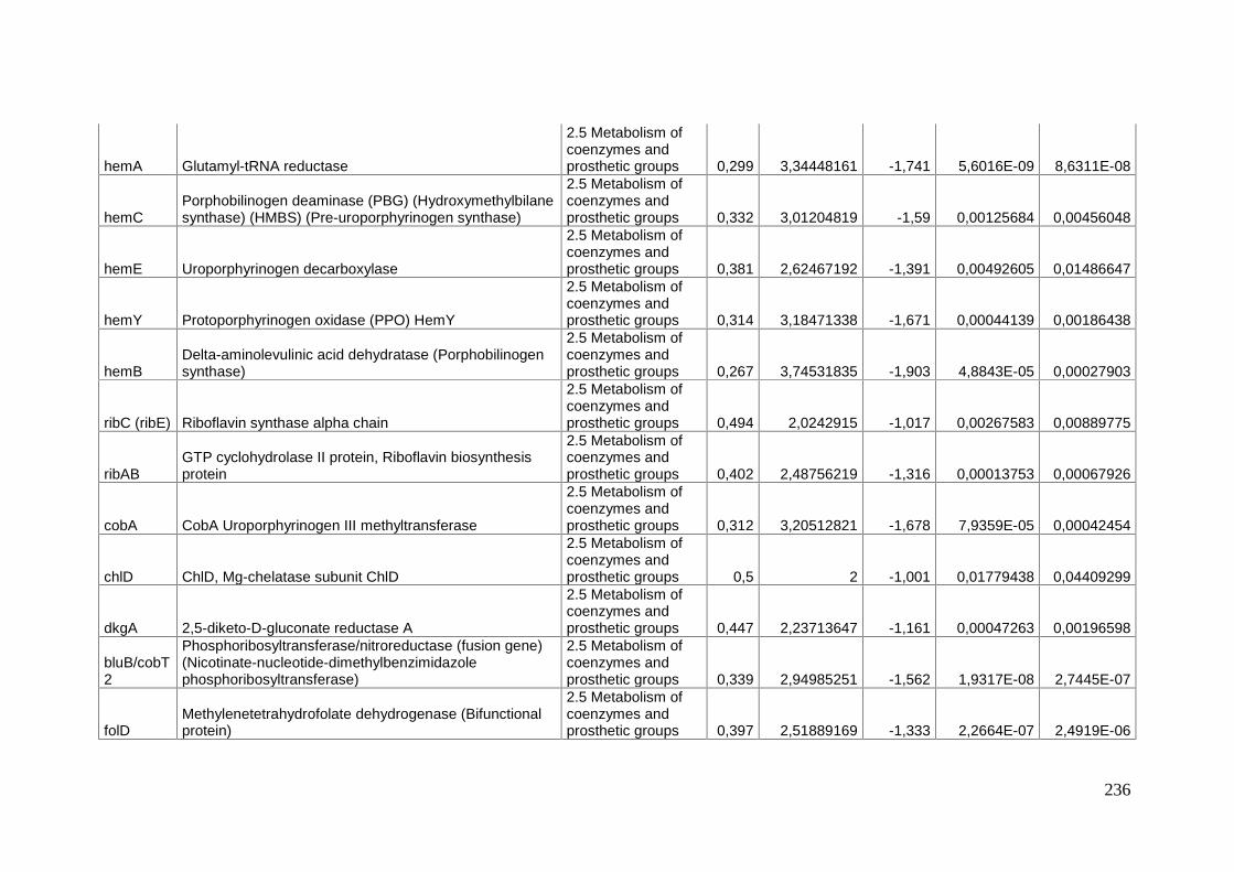
236
hemA Glutamyl-tRNA reductase
2.5 Metabolism ofcoenzymes andprosthetic groups 0,299 3,34448161 -1,741 5,6016E-09 8,6311E-08
hemCPorphobilinogen deaminase (PBG) (Hydroxymethylbilanesynthase) (HMBS) (Pre-uroporphyrinogen synthase)
2.5 Metabolism ofcoenzymes andprosthetic groups 0,332 3,01204819 -1,59 0,00125684 0,00456048
hemE Uroporphyrinogen decarboxylase
2.5 Metabolism ofcoenzymes andprosthetic groups 0,381 2,62467192 -1,391 0,00492605 0,01486647
hemY Protoporphyrinogen oxidase (PPO) HemY
2.5 Metabolism ofcoenzymes andprosthetic groups 0,314 3,18471338 -1,671 0,00044139 0,00186438
hemBDelta-aminolevulinic acid dehydratase (Porphobilinogensynthase)
2.5 Metabolism ofcoenzymes andprosthetic groups 0,267 3,74531835 -1,903 4,8843E-05 0,00027903
ribC (ribE) Riboflavin synthase alpha chain
2.5 Metabolism ofcoenzymes andprosthetic groups 0,494 2,0242915 -1,017 0,00267583 0,00889775
ribABGTP cyclohydrolase II protein, Riboflavin biosynthesisprotein
2.5 Metabolism ofcoenzymes andprosthetic groups 0,402 2,48756219 -1,316 0,00013753 0,00067926
cobA CobA Uroporphyrinogen III methyltransferase
2.5 Metabolism ofcoenzymes andprosthetic groups 0,312 3,20512821 -1,678 7,9359E-05 0,00042454
chlD ChlD, Mg-chelatase subunit ChlD
2.5 Metabolism ofcoenzymes andprosthetic groups 0,5 2 -1,001 0,01779438 0,04409299
dkgA 2,5-diketo-D-gluconate reductase A
2.5 Metabolism ofcoenzymes andprosthetic groups 0,447 2,23713647 -1,161 0,00047263 0,00196598
bluB/cobT2
Phosphoribosyltransferase/nitroreductase (fusion gene)(Nicotinate-nucleotide-dimethylbenzimidazolephosphoribosyltransferase)
2.5 Metabolism ofcoenzymes andprosthetic groups 0,339 2,94985251 -1,562 1,9317E-08 2,7445E-07
folDMethylenetetrahydrofolate dehydrogenase (Bifunctionalprotein)
2.5 Metabolism ofcoenzymes andprosthetic groups 0,397 2,51889169 -1,333 2,2664E-07 2,4919E-06

237
menBNaphthoate synthase (Dihydroxynaphthoic acidsynthetase)
2.5 Metabolism ofcoenzymes andprosthetic groups 0,46 2,17391304 -1,12 2,9891E-05 0,00018218
cbiL CbiL Precorrin-2 C20-methyltransferase
2.5 Metabolism ofcoenzymes andprosthetic groups 0,43 2,3255814 -1,218 0,00013492 0,00066855
cbiF CbiF Precorrin-4 C11-methyltransferase
2.5 Metabolism ofcoenzymes andprosthetic groups 0,332 3,01204819 -1,592 1,5932E-05 0,00010726
cysG/cbiX CysG/CbiX
2.5 Metabolism ofcoenzymes andprosthetic groups 0,441 2,2675737 -1,181 0,00086698 0,00329333
folC Folylpolyglutamate synthase (bifunctionnal enzyme)
2.5 Metabolism ofcoenzymes andprosthetic groups 0,656 1,52439024 -0,608 0,01364176 0,03553355
PFCIRM138_06220 NUDIX hydrolase
2.6 Metabolism ofphosphate 0,392 2,55102041 -1,353 1,5664E-06 1,434E-05
PFCIRM138_08215 Inorganic pyrophosphatase
2.6 Metabolism ofphosphate 0,478 2,09205021 -1,064 0,00076125 0,0029376
cysW Sulfate transport system permease protein CysW2.7 Metabolism ofsulfur 0,081 12,345679 -3,62 2,4676E-07 2,6862E-06
cysTSulphate transport system permease protein 2:SulfateABC transporter, permease protein CysT
2.7 Metabolism ofsulfur 0,097 10,3092784 -3,368 6,5968E-07 6,4725E-06
PFCIRM138_00925 Sulfate-binding protein precursor
2.7 Metabolism ofsulfur 0,118 8,47457627 -3,078 4,7385E-05 0,00027212
nirA2/sir2 Sulfite reductase [ferredoxin]2.7 Metabolism ofsulfur 0,076 13,1578947 -3,717 1,1963E-09 2,1456E-08
cysH Putative phosphoadenosine phosphosulfate reductase2.7 Metabolism ofsulfur 0,051 19,6078431 -4,3 2,4806E-17 1,7508E-15
mmcV/cysD
Sulfate adenylyltransferase subunit 2 (Sulfate adenylatetransferase) (SAT) (ATP-sulfurylase small subunit)(Mitomycin biosynthesis protein V)
2.7 Metabolism ofsulfur 0,035 28,5714286 -4,827 7,1865E-27 2,2463E-24
cysNSulfate adenylyltransferase, large subunit/ ATP-sulfurylase, subunit 1 (ATP:sulfate adenylyltransferase)
2.7 Metabolism ofsulfur 0,033 30,3030303 -4,932 1,7165E-32 9,3895E-30
norB Nitric-oxide reductase subunit B (nitric-oxide reductase)2.8 Metabolism ofnitrogen/nitrate and 0,475 2,10526316 -1,073 0,00068892 0,00271004

238
nitrite
glnA1 Glutamine synthetase
2.8 Metabolism ofnitrogen/nitrate andnitrite 0,543 1,84162063 -0,882 0,00408826 0,01261651
dnaN DNA polymerase III, beta chain 3.1 DNA replication 0,493 2,02839757 -1,019 0,0005583 0,00225798PFCIRM138_07260 Exonuclease 3.1 DNA replication 0,393 2,54452926 -1,346 5,3832E-06 4,2603E-05PFCIRM138_09150 DeaD/DeaH box helicase 3.1 DNA replication 0,583 1,71526587 -0,779 0,00197229 0,00675333
pepP Xaa-Pro aminopeptidase I3.10 Proteindegradation 0,583 1,71526587 -0,78 0,00361727 0,01130657
PFCIRM138_02300 Zinc metallopeptidase
3.10 Proteindegradation 0,562 1,77935943 -0,831 0,00608512 0,01791958
PFCIRM138_03755
Amidohydrolase (Peptidase M20D) (Putative metal-dependent amidase/aminoacylase/carboxypeptidase)
3.10 Proteindegradation 0,418 2,3923445 -1,26 0,00551416 0,01648223
map Methionine aminopeptidase (MAP) (Peptidase M)3.10 Proteindegradation 0,315 3,17460317 -1,669 1,4872E-06 1,3673E-05
PFCIRM138_10230 Secreted protease with a PDZ domain
3.10 Proteindegradation 0,312 3,20512821 -1,682 1,7693E-11 4,3992E-10
pepN Aminopeptidase N , Lysyl aminopeptidase3.10 Proteindegradation 0,521 1,9193858 -0,94 0,0039231 0,01217343
clpP2ATP-dependent Clp protease proteolytic subunit 2(Endopeptidase Clp 2)
3.10 Proteindegradation 0,583 1,71526587 -0,777 0,0181493 0,04471922
PFCIRM138_05540 nuclease (RecB family)
3.2 DNA restrictionand modification(and repair) 0,639 1,56494523 -0,646 0,01158533 0,030892
nth Putative endonuclease III
3.2 DNA restrictionand modification(and repair) 0,536 1,86567164 -0,9 0,00227571 0,0076968
PFCIRM138_06795 Hypothetical protein
3.2 DNA restrictionand modification(and repair) 0,466 2,14592275 -1,1 0,00888308 0,02460276
recB Exodeoxyribonuclease V beta chain RecB
3.3 DNArecombination, andrepair 0,657 1,52207002 -0,607 0,01511099 0,03871528
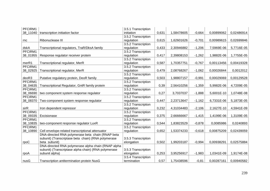
239
PFCIRM138_11040 transcription initiation factor
3.5.1 Transcriptioninitiation 0,631 1,58478605 -0,664 0,00899062 0,02486914
rnc Ribonuclease III3.5.2 Transcriptionregulation 0,615 1,62601626 -0,701 0,00989615 0,02699846
dskA Transcriptional regulators, TraR/DksA family3.5.2 Transcriptionregulation 0,433 2,30946882 -1,206 7,5969E-06 5,7716E-05
PFCIRM138_01955 Response regulator receiver protein
3.5.2 Transcriptionregulation 0,417 2,39808153 -1,262 1,9882E-06 1,7755E-05
merR1 Transcriptional regulator, MerR3.5.2 Transcriptionregulation 0,587 1,70357751 -0,767 0,00113456 0,00419328
PFCIRM138_02925 Transcriptional regulator, MerR
3.5.2 Transcriptionregulation 0,479 2,08768267 -1,062 0,00026844 0,0012012
deoR3 Putative regulatory protein, DeoR family3.5.2 Transcriptionregulation 0,503 1,98807157 -0,991 0,00029069 0,00129528
PFCIRM138_04635 Transcriptional Regulator, GntR family protein
3.5.2 Transcriptionregulation 0,39 2,56410256 -1,359 5,9982E-06 4,7209E-05
PFCIRM138_06690 two component system response regulator
3.5.2 Transcriptionregulation 0,27 3,7037037 -1,888 5,6001E-10 1,0748E-08
PFCIRM138_08370 Two-component system response regulator
3.5.2 Transcriptionregulation 0,447 2,23713647 -1,162 6,7331E-06 5,1873E-05
sirR Iron dependent repressor3.5.2 Transcriptionregulation 0,232 4,31034483 -2,106 2,1627E-10 4,5941E-09
PFCIRM138_09335 Excisionase
3.5.2 Transcriptionregulation 0,375 2,66666667 -1,415 1,4199E-06 1,3109E-05
PFCIRM138_10835 two-component response regulator LuxR
3.5.2 Transcriptionregulation 0,544 1,83823529 -0,878 0,0085986 0,0240893
PFCIRM138_10890 Cell envelope-related transcriptional attenuator
3.5.2 Transcriptionregulation 0,652 1,53374233 -0,618 0,00875209 0,02439059
rpoC
DNA-directed RNA polymerase beta chain (RNAP betasubunit) (Transcriptase beta chain) (RNA polymerasebeta subunit)
3.5.3 Transcriptionelongation 0,502 1,99203187 -0,994 0,00938291 0,02575884
rpoA
DNA-directed RNA polymerase alpha chain (RNAP alphasubunit) (Transcriptase alpha chain) (RNA polymerasesubunit alpha)
3.5.3 Transcriptionelongation 0,253 3,95256917 -1,983 1,0341E-09 1,9174E-08
nusG Transcription antitermination protein NusG3.5.4 Transcriptiontermination 0,57 1,75438596 -0,81 0,00287161 0,00940582

240
spoU1 rRNA methylase3.6 RNAmodification 0,392 2,55102041 -1,35 6,7802E-05 0,0003718
PFCIRM138_05905 tRNA/rRNA methyltransferase
3.6 RNAmodification 0,481 2,07900208 -1,057 0,00020819 0,00096917
rsmC rRNA (guanine-N2-)-methyltransferase3.6 RNAmodification 0,589 1,69779287 -0,764 0,01885313 0,04588504
PFCIRM138_10045 GatB_Yqey, GatB/Yqey
3.6 RNAmodification 0,651 1,53609831 -0,62 0,01167347 0,0310347
orn Oligoribonuclease3.6 RNAmodification 0,33 3,03030303 -1,6 4,508E-10 8,8068E-09
gatC
Glutamyl-tRNA(Gln) amidotransferase subunit C(Aspartyl/glutamyl-tRNA(Asn/Gln) amidotransferasesubunit C)
3.7 Proteinsynthesis 0,437 2,28832952 -1,194 8,5304E-05 0,00045193
gatBAspartyl/glutamyl-tRNA(Asn/Gln) amidotransferasesubunit B (Asp/Glu-ADT subunit B)
3.7 Proteinsynthesis 0,343 2,9154519 -1,544 3,9131E-08 5,1268E-07
rplY Ribosomal protein L253.7.1 Ribosomalproteins 0,308 3,24675325 -1,701 1,4762E-09 2,5634E-08
rplT 50S ribosomal protein L203.7.1 Ribosomalproteins 0,276 3,62318841 -1,858 5,1494E-08 6,5548E-07
rpmI 50S ribosomal protein L353.7.1 Ribosomalproteins 0,331 3,02114804 -1,593 2,713E-09 4,497E-08
rpsA 30S ribosomal protein S13.7.1 Ribosomalproteins 0,503 1,98807157 -0,992 0,00452294 0,01384083
rpsR 30S ribosomal protein S183.7.1 Ribosomalproteins 0,487 2,05338809 -1,038 0,0007434 0,00288761
rplI 50S ribosomal protein L93.7.1 Ribosomalproteins 0,46 2,17391304 -1,12 0,00066158 0,00261288
rpsK 30S ribosomal protein S113.7.1 Ribosomalproteins 0,493 2,02839757 -1,019 0,00059883 0,00240409
rplA 50S ribosomal protein L13.7.1 Ribosomalproteins 0,536 1,86567164 -0,899 0,0063055 0,01849387
rplJ 50S ribosomal protein L103.7.1 Ribosomalproteins 0,064 15,625 -3,963 2,5466E-21 4,509E-19
rplL 50S ribosomal protein L7/L123.7.1 Ribosomalproteins 0,063 15,8730159 -3,991 2,679E-21 4,509E-19

241
rpsG 30S ribosomal protein S73.7.1 Ribosomalproteins 0,625 1,6 -0,678 0,01500913 0,03849939
rpsJ 30S ribosomal protein S103.7.1 Ribosomalproteins 0,36 2,77777778 -1,474 3,5353E-08 4,7166E-07
rplC 50S ribosomal protein L33.7.1 Ribosomalproteins 0,339 2,94985251 -1,561 1,1246E-09 2,0486E-08
rplD 50S ribosomal protein L43.7.1 Ribosomalproteins 0,358 2,79329609 -1,481 7,9489E-09 1,1913E-07
rplW 50S ribosomal protein L233.7.1 Ribosomalproteins 0,265 3,77358491 -1,914 1,0791E-14 4,5404E-13
rplB 50S ribosomal protein L23.7.1 Ribosomalproteins 0,193 5,18134715 -2,37 2,124E-15 1,0562E-13
rpsS 30S ribosomal protein S193.7.1 Ribosomalproteins 0,178 5,61797753 -2,493 2,4001E-18 2,0198E-16
rplV 50S ribosomal protein L223.7.1 Ribosomalproteins 0,2 5 -2,325 4,933E-11 1,1606E-09
rpsC 30S ribosomal protein S33.7.1 Ribosomalproteins 0,18 5,55555556 -2,47 8,7151E-12 2,2974E-10
rplP 50S ribosomal protein L163.7.1 Ribosomalproteins 0,135 7,40740741 -2,892 7,5514E-14 2,8487E-12
rpmC 50S ribosomal protein L293.7.1 Ribosomalproteins 0,131 7,63358779 -2,929 2,4445E-13 8,7682E-12
rpsQ 30S ribosomal protein S173.7.1 Ribosomalproteins 0,165 6,06060606 -2,601 1,9039E-10 4,1245E-09
rplN 50S ribosomal protein L143.7.1 Ribosomalproteins 0,203 4,92610837 -2,303 3,4743E-10 7,0388E-09
rplX 50S ribosomal protein L243.7.1 Ribosomalproteins 0,19 5,26315789 -2,393 4,2376E-10 8,4289E-09
rplE 50S ribosomal protein L53.7.1 Ribosomalproteins 0,183 5,46448087 -2,454 2,2501E-09 3,8165E-08
rpsN1,rpsZ 30S ribosomal protein S14 type Z
3.7.1 Ribosomalproteins 0,145 6,89655172 -2,783 1,3472E-08 1,9393E-07
rpsH 30S ribosomal protein S83.7.1 Ribosomalproteins 0,097 10,3092784 -3,358 1,557E-15 7,9444E-14
rplF 50S ribosomal protein L63.7.1 Ribosomalproteins 0,122 8,19672131 -3,032 1,5666E-12 5,0407E-11

242
rplR ribosomal protein L183.7.1 Ribosomalproteins 0,113 8,84955752 -3,141 7,6417E-12 2,0642E-10
rpsE 30S ribosomal protein S53.7.1 Ribosomalproteins 0,164 6,09756098 -2,608 5,3134E-08 6,7201E-07
rpmD 50S ribosomal protein L303.7.1 Ribosomalproteins 0,132 7,57575758 -2,926 2,1034E-09 3,5955E-08
rplO 50S ribosomal protein L153.7.1 Ribosomalproteins 0,101 9,9009901 -3,305 9,1233E-12 2,3764E-10
rpsD 30S ribosomal protein S43.7.1 Ribosomalproteins 0,346 2,89017341 -1,531 3,8986E-06 3,2189E-05
rplQ 50S ribosomal protein L173.7.1 Ribosomalproteins 0,24 4,16666667 -2,057 8,8665E-11 2,0208E-09
rspI 30S ribosomal protein S93.7.1 Ribosomalproteins 0,629 1,58982512 -0,668 0,01958507 0,04740248
tyrSTyrosyl-tRNA synthetase (Tyrosine--tRNA ligase)(TyrRS)
3.7.2 Aminoacyl-tRNA synthetases 0,551 1,81488203 -0,86 0,00078642 0,00301876
pheTPhenylalanyl-tRNA synthetase beta chain(Phenylalanine-- tRNA ligase beta chain) (PheRS)
3.7.2 Aminoacyl-tRNA synthetases 0,572 1,74825175 -0,805 0,00659853 0,01919891
proS Prolyl-tRNA synthetase (Proline--tRNA ligase) (ProRS)3.7.2 Aminoacyl-tRNA synthetases 0,622 1,60771704 -0,684 0,01208965 0,03202441
thrSThreonyl-tRNA synthetase (Threonine--tRNA ligase)(ThrRS)
3.7.2 Aminoacyl-tRNA synthetases 0,303 3,30033003 -1,723 1,5531E-11 3,9978E-10
glnS Glutaminyl-tRNA synthetase3.7.2 Aminoacyl-tRNA synthetases 0,421 2,37529691 -1,247 3,9158E-07 4,0799E-06
hisSHistidyl-tRNA synthetase (Histidine--tRNA ligase)(HisRS)
3.7.2 Aminoacyl-tRNA synthetases 0,499 2,00400802 -1,004 3,7977E-05 0,00022158
aspSAspartyl-tRNA synthetase (Aspartate--tRNA ligase)(AspRS)
3.7.2 Aminoacyl-tRNA synthetases 0,469 2,13219616 -1,093 1,3341E-05 9,2323E-05
serS1 Seryl-tRNA synthetase3.7.2 Aminoacyl-tRNA synthetases 0,398 2,51256281 -1,331 1,3601E-06 1,261E-05
ileS Isoleucyl-tRNA synthetase (Isoleucine--tRNA ligase)3.7.2 Aminoacyl-tRNA synthetases 0,327 3,05810398 -1,613 5,0045E-09 7,8775E-08
argS Arginyl-tRNA synthetase (Arginine--tRNA ligase) (ArgRS)3.7.2 Aminoacyl-tRNA synthetases 0,614 1,6286645 -0,705 0,01027066 0,02781214
gltXGlutamyl-tRNA synthetase (Glutamate--tRNA ligase)(GluRS)
3.7.2 Aminoacyl-tRNA synthetases 0,543 1,84162063 -0,881 0,00044697 0,00187712

243
glyQS Glycyl-tRNA synthetase (Glycine--tRNA ligase) (GlyRS)3.7.2 Aminoacyl-tRNA synthetases 0,263 3,80228137 -1,926 1,9314E-09 3,3275E-08
trpS Tryptophanyl-tRNA synthetase3.7.2 Aminoacyl-tRNA synthetases 0,601 1,66389351 -0,733 0,01114231 0,02995009
metGMethionyl-tRNA synthetase (Methionine--tRNA ligase)(MetRS)
3.7.2 Aminoacyl-tRNA synthetases 0,508 1,96850394 -0,977 0,00013296 0,0006642
valS Valyl-tRNA synthetase (Valine--tRNA ligase) (ValRS)3.7.2 Aminoacyl-tRNA synthetases 0,466 2,14592275 -1,101 4,3426E-05 0,00025136
infC Translation initiation factor IF-33.7.3 Translationinitiation 0,505 1,98019802 -0,985 0,00225997 0,00766639
fusA Elongation factor G (EF-G)3.7.4 Translationelongation 0,271 3,6900369 -1,886 4,9822E-10 9,6469E-09
tuf Elongation factor Tu3.7.4 Translationelongation 0,547 1,82815356 -0,87 0,00731936 0,02098919
frrRibosome recycling factor (Ribosome-releasing factor)(RRF)
3.7.5 Translationtermination 0,514 1,94552529 -0,961 0,00035543 0,00155682
hflC1 Stomatin/prohibitin homolog3.8 Proteinmodification 0,437 2,28832952 -1,193 6,5556E-07 6,4611E-06
PFCIRM138_00200
dolichyl-phosphate-mannose-proteinmannosyltransferase (Glycosyl transferase, family 39)
3.8 Proteinmodification 0,455 2,1978022 -1,135 8,3235E-06 6,2799E-05
pknB Serine/threonine protein kinase3.8 Proteinmodification 0,369 2,7100271 -1,44 2,0423E-07 2,3202E-06
trxA2 Thioredoxin3.8 Proteinmodification 0,165 6,06060606 -2,602 2,3871E-06 2,0726E-05
fkbPFK506-binding protein (peptidyl-prolyl cis-transisomerase)
3.8 Proteinmodification 0,457 2,18818381 -1,129 2,269E-05 0,00014348
ffh GTP binding signal recognition particle protein 3.9 Protein folding 0,677 1,47710487 -0,564 0,01935583 0,04689984groL2(groEL2)(hsp60)
60 kDa chaperonin 2 (Protein Cpn60 2) (groEL protein 2)(Heat shock protein 60 2) 3.9 Protein folding 0,486 2,05761317 -1,041 0,00348033 0,01090968
groS1(groES1)(hsp10)
10 kDa chaperonin 1 (Protein Cpn10 1) (groES protein 1)(Heat shock 10 1) 3.9 Protein folding 0,394 2,53807107 -1,342 0,00049046 0,00202096
groL1(groEL1)
60 kDa chaperonin 1 (Protein Cpn60 1) (groEL protein 1)(Heat shock protein 60 1) 3.9 Protein folding 0,304 3,28947368 -1,72 2,8762E-06 2,4504E-05
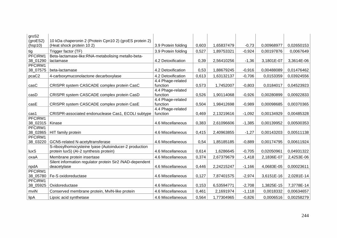
244
groS2(groES2)(hsp10)
10 kDa chaperonin 2 (Protein Cpn10 2) (groES protein 2)(Heat shock protein 10 2) 3.9 Protein folding 0,603 1,65837479 -0,73 0,00968977 0,02650153
tig Trigger factor (TF) 3.9 Protein folding 0,527 1,89753321 -0,924 0,00197876 0,0067649PFCIRM138_01290
Beta-lactamase-like:RNA-metabolising metallo-beta-lactamase 4.2 Detoxification 0,39 2,56410256 -1,36 3,1801E-07 3,3614E-06
PFCIRM138_07575 beta-lactamase 4.2 Detoxification 0,53 1,88679245 -0,916 0,00488089 0,01476462pcaC2 4-carboxymuconolactone decarboxylase 4.2 Detoxification 0,613 1,63132137 -0,706 0,0153359 0,03924556
casC CRISPR system CASCADE complex protein CasC4.4 Phage-relatedfunction 0,573 1,7452007 -0,803 0,0184017 0,04523923
casD CRISPR system CASCADE complex protein CasD4.4 Phage-relatedfunction 0,526 1,90114068 -0,926 0,00280899 0,00922833
casE CRISPR system CASCADE complex protein CasE4.4 Phage-relatedfunction 0,504 1,98412698 -0,989 0,00098685 0,00370365
cas1 CRISPR-associated endonuclease Cas1, ECOLI subtype4.4 Phage-relatedfunction 0,469 2,13219616 -1,092 0,00134929 0,00485328
PFCIRM138_02315 Kinase 4.6 Miscellaneous 0,383 2,61096606 -1,385 0,00139952 0,00500353PFCIRM138_02865 HIT family protein 4.6 Miscellaneous 0,415 2,40963855 -1,27 0,00143203 0,00511138PFCIRM138_03220 GCN5-related N-acetyltransferase 4.6 Miscellaneous 0,54 1,85185185 -0,889 0,00174795 0,00611924
luxSS-ribosylhomocysteine lyase (Autoinducer-2 productionprotein luxS) (AI-2 synthesis protein) 4.6 Miscellaneous 0,614 1,6286645 -0,705 0,02050961 0,04931322
oxaA Membrane protein insertase 4.6 Miscellaneous 0,374 2,67379679 -1,418 2,1836E-07 2,4253E-06
npdASilent information regulator protein Sir2 /NAD-dependentdeacetylase 4.6 Miscellaneous 0,446 2,24215247 -1,166 4,0683E-05 0,00023611
PFCIRM138_05780 Fe-S oxidoreductase 4.6 Miscellaneous 0,127 7,87401575 -2,974 3,6151E-16 2,0281E-14PFCIRM138_05925 Oxidoreductase 4.6 Miscellaneous 0,153 6,53594771 -2,708 1,3825E-15 7,3778E-14mviN Conserved membrane protein, MviN-like protein 4.6 Miscellaneous 0,461 2,1691974 -1,118 0,0018332 0,00634657lipA Lipoic acid synthetase 4.6 Miscellaneous 0,564 1,77304965 -0,826 0,0006516 0,00258279

245
PFCIRM138_09940 Von Willebrand factor, type A 4.6 Miscellaneous 0,325 3,07692308 -1,621 4,4755E-06 3,6268E-05PFCIRM138_10155 PP2Cc, Serine/threonine phosphatases, 4.6 Miscellaneous 0,627 1,59489633 -0,674 0,0196066 0,04740248PFCIRM138_10615 dehydrogenase/reductase 4.6 Miscellaneous 0,661 1,5128593 -0,597 0,01863681 0,04558667PFCIRM138_11500 Glycosyltransferase 4.6 Miscellaneous 0,52 1,92307692 -0,944 0,00117522 0,00431439PFCIRM138_11855 oxidoreductase 4.6 Miscellaneous 0,43 2,3255814 -1,218 3,2795E-05 0,00019605
PFCIRM138_12335 protein of unknown function
5.1 Protein ofunknown functionsimilar to otherproteins from thesame organism 0,402 2,48756219 -1,315 9,4569E-06 6,9669E-05
PFCIRM138_00015 Hypothetical protein
5.2 Protein ofunknown functionsimilar to proteinsfrom otherorganisms 0,591 1,69204738 -0,759 0,00762589 0,0217451
PFCIRM138_00185 Hypothetical protein
5.2 Protein ofunknown functionsimilar to proteinsfrom otherorganisms 0,355 2,81690141 -1,494 3,2154E-09 5,173E-08
PFCIRM138_00660 Hypothetical membrane protein
5.2 Protein ofunknown functionsimilar to proteinsfrom otherorganisms 0,492 2,03252033 -1,024 0,0004143 0,00176018
PFCIRM138_01150 Hypothetical protein
5.2 Protein ofunknown functionsimilar to proteinsfrom otherorganisms 0,559 1,78890877 -0,839 0,01546312 0,03952488
PFCIRM1 Hypothetical protein 5.2 Protein of 0,494 2,0242915 -1,018 0,00017924 0,00084337

246
38_01300 unknown functionsimilar to proteinsfrom otherorganisms
PFCIRM138_01745 Hypothetical protein
5.2 Protein ofunknown functionsimilar to proteinsfrom otherorganisms 0,35 2,85714286 -1,517 2,9818E-06 2,5287E-05
PFCIRM138_01750 Hypothetical protein
5.2 Protein ofunknown functionsimilar to proteinsfrom otherorganisms 0,309 3,23624595 -1,692 3,4592E-05 0,00020456
PFCIRM138_01755 Hypothetical protein
5.2 Protein ofunknown functionsimilar to proteinsfrom otherorganisms 0,446 2,24215247 -1,164 1,472E-05 0,00010002
PFCIRM138_01760 Hypothetical protein
5.2 Protein ofunknown functionsimilar to proteinsfrom otherorganisms 0,523 1,91204589 -0,935 0,01895944 0,04609251
PFCIRM138_02105 Hypothetical protein
5.2 Protein ofunknown functionsimilar to proteinsfrom otherorganisms 0,618 1,61812298 -0,695 0,01661365 0,04187864
PFCIRM138_02305 Hypothetical protein
5.2 Protein ofunknown functionsimilar to proteinsfrom otherorganisms 0,35 2,85714286 -1,515 0,0028286 0,00927883
PFCIRM138_02320 UPF0210 protein
5.2 Protein ofunknown function 0,415 2,40963855 -1,27 2,0854E-07 2,3399E-06

247
similar to proteinsfrom otherorganisms
PFCIRM138_02810 Hypothetical protein
5.2 Protein ofunknown functionsimilar to proteinsfrom otherorganisms 0,484 2,0661157 -1,047 0,00058972 0,0023719
PFCIRM138_02830 Hypothetical protein
5.2 Protein ofunknown functionsimilar to proteinsfrom otherorganisms 0,493 2,02839757 -1,021 0,00122682 0,00446635
PFCIRM138_02950 Hypothetical protein
5.2 Protein ofunknown functionsimilar to proteinsfrom otherorganisms 0,52 1,92307692 -0,942 0,00133948 0,00482829
PFCIRM138_03420 Hypothetical protein
5.2 Protein ofunknown functionsimilar to proteinsfrom otherorganisms 0,5 2 -1,001 0,00015646 0,00075568
PFCIRM138_03690 Hypothetical transmembrane protein
5.2 Protein ofunknown functionsimilar to proteinsfrom otherorganisms 0,553 1,80831826 -0,854 0,00030891 0,00137099
PFCIRM138_03855 Hypothetical membrane protein
5.2 Protein ofunknown functionsimilar to proteinsfrom otherorganisms 0,621 1,61030596 -0,686 0,01606597 0,04077997
PFCIRM138_05525 Hypothetical membrane protein
5.2 Protein ofunknown functionsimilar to proteins 0,419 2,38663484 -1,256 1,5751E-06 1,436E-05

248
from otherorganisms
PFCIRM138_05935 Hypothetical protein
5.2 Protein ofunknown functionsimilar to proteinsfrom otherorganisms 0,179 5,58659218 -2,484 2,1105E-11 5,1884E-10
PFCIRM138_06015 Hypothetical protein
5.2 Protein ofunknown functionsimilar to proteinsfrom otherorganisms 0,458 2,18340611 -1,126 0,00014176 0,000697
PFCIRM138_06085 Putative uncharacterized protein
5.2 Protein ofunknown functionsimilar to proteinsfrom otherorganisms 0,584 1,71232877 -0,776 0,00930584 0,02561156
PFCIRM138_07050 Hypothetical membrane protein
5.2 Protein ofunknown functionsimilar to proteinsfrom otherorganisms 0,595 1,68067227 -0,748 0,00507038 0,01525997
PFCIRM138_07405 Hypothetical protein
5.2 Protein ofunknown functionsimilar to proteinsfrom otherorganisms 0,284 3,52112676 -1,818 0,00650729 0,01900928
PFCIRM138_07760 Hypothetical membrane protein
5.2 Protein ofunknown functionsimilar to proteinsfrom otherorganisms 0,458 2,18340611 -1,127 0,00012091 0,00061098
PFCIRM138_08100 Hypothetical protein
5.2 Protein ofunknown functionsimilar to proteinsfrom other 0,55 1,81818182 -0,862 0,00227597 0,0076968

249
organisms
PFCIRM138_08555 Hypothetical protein
5.2 Protein ofunknown functionsimilar to proteinsfrom otherorganisms 0,255 3,92156863 -1,972 0,00065493 0,00259129
PFCIRM138_08630 Hypothetical protein
5.2 Protein ofunknown functionsimilar to proteinsfrom otherorganisms 0,566 1,76678445 -0,82 0,01379083 0,0358365
PFCIRM138_08980 Hypothetical protein
5.2 Protein ofunknown functionsimilar to proteinsfrom otherorganisms 0,362 2,76243094 -1,466 7,0988E-08 8,629E-07
PFCIRM138_10000 Hypothetical protein
5.2 Protein ofunknown functionsimilar to proteinsfrom otherorganisms 0,55 1,81818182 -0,863 0,00155047 0,00548049
PFCIRM138_10235 Hypothetical protein
5.2 Protein ofunknown functionsimilar to proteinsfrom otherorganisms 0,253 3,95256917 -1,98 1,8996E-07 2,1827E-06
PFCIRM138_10240 Hypothetical secreted and membrane protein
5.2 Protein ofunknown functionsimilar to proteinsfrom otherorganisms 0,244 4,09836066 -2,037 7,8508E-12 2,0948E-10
PFCIRM138_10400 Hypothetical protein
5.2 Protein ofunknown functionsimilar to proteinsfrom otherorganisms 0,499 2,00400802 -1,004 0,00175393 0,00613034

250
PFCIRM138_10520 Hypothetical secreted protein
5.2 Protein ofunknown functionsimilar to proteinsfrom otherorganisms 0,445 2,24719101 -1,167 0,00024499 0,00110979
PFCIRM138_10550 Hypothetical protein
5.2 Protein ofunknown functionsimilar to proteinsfrom otherorganisms 0,592 1,68918919 -0,757 0,00596512 0,01758988
PFCIRM138_10765 Hypothetical protein
5.2 Protein ofunknown functionsimilar to proteinsfrom otherorganisms 0,614 1,6286645 -0,703 0,01133687 0,03036117
PFCIRM138_11190 Hypothetical membrane protein
5.2 Protein ofunknown functionsimilar to proteinsfrom otherorganisms 0,554 1,80505415 -0,851 0,00092164 0,00347082
PFCIRM138_11325 Hypothetical secreted protein
5.2 Protein ofunknown functionsimilar to proteinsfrom otherorganisms 0,599 1,66944908 -0,74 0,00274834 0,00909738
PFCIRM138_11530 Hypothetical protein
5.2 Protein ofunknown functionsimilar to proteinsfrom otherorganisms 0,45 2,22222222 -1,152 0,00015461 0,00075007
PFCIRM138_11550 Hypothetical protein
5.2 Protein ofunknown functionsimilar to proteinsfrom otherorganisms 0,602 1,66112957 -0,731 0,00647959 0,01895366
PFCIRM1 Hypothetical protein 5.2 Protein of 0,484 2,0661157 -1,046 0,00181393 0,0062998

251
38_11810 unknown functionsimilar to proteinsfrom otherorganisms
PFCIRM138_12275 Hypothetical secreted protein
5.2 Protein ofunknown functionsimilar to proteinsfrom otherorganisms 0,603 1,65837479 -0,73 0,00454104 0,01387682
PFCIRM138_00435 Hypothetical protein
6 Protein ofunknown function,without similarity toother proteins 0,429 2,33100233 -1,22 0,00023681 0,0010776
PFCIRM138_01880 Hypothetical protein
6 Protein ofunknown function,without similarity toother proteins 0,646 1,54798762 -0,631 0,01081575 0,02914391
PFCIRM138_02445 Hypothetical protein
6 Protein ofunknown function,without similarity toother proteins 0,57 1,75438596 -0,811 0,00335633 0,01059689
PFCIRM138_02765 Protein of unknown function
6 Protein ofunknown function,without similarity toother proteins 0,286 3,4965035 -1,807 0,00293814 0,00953805
PFCIRM138_04485 Hypothetical protein
6 Protein ofunknown function,without similarity toother proteins 0,498 2,00803213 -1,006 0,01671466 0,04208477
PFCIRM138_04545 Hypothetical protein
6 Protein ofunknown function,without similarity toother proteins 0,445 2,24719101 -1,168 0,00026335 0,00118804
PFCIRM138_05150 Hypothetical protein
6 Protein ofunknown function,without similarity to 0,594 1,68350168 -0,753 0,00693117 0,01995447

252
other proteins
PFCIRM138_05205 Hypothetical protein
6 Protein ofunknown function,without similarity toother proteins 0,606 1,65016502 -0,721 0,01284854 0,03378918
PFCIRM138_05510 Protein of unknown function
6 Protein ofunknown function,without similarity toother proteins 0,21 4,76190476 -2,252 0,00157402 0,00555478
PFCIRM138_06570 Hypothetical protein
6 Protein ofunknown function,without similarity toother proteins 0,338 2,95857988 -1,567 1,0521E-05 7,6224E-05
PFCIRM138_06675 Hypothetical protein
6 Protein ofunknown function,without similarity toother proteins 0,635 1,57480315 -0,656 0,00665084 0,01928747
PFCIRM138_06720 Hypothetical protein
6 Protein ofunknown function,without similarity toother proteins 0,576 1,73611111 -0,797 0,00346043 0,01089446
PFCIRM138_07920 protein of unknown function
6 Protein ofunknown function,without similarity toother proteins 0,579 1,72711572 -0,789 0,01375217 0,03577853
PFCIRM138_08640 Hypothetical protein
6 Protein ofunknown function,without similarity toother proteins 0,564 1,77304965 -0,826 0,00444145 0,01364873
PFCIRM138_09065 Hypothetical protein
6 Protein ofunknown function,without similarity toother proteins 0,318 3,14465409 -1,654 1,1699E-08 1,7179E-07
PFCIRM138_10065 Hypothetical protein
6 Protein ofunknown function,without similarity to 0,345 2,89855072 -1,533 3,8435E-06 3,1855E-05

253
other proteins
PFCIRM138_11045 Hypothetical protein
6 Protein ofunknown function,without similarity toother proteins 0,502 1,99203187 -0,994 5,059E-05 0,00028751
PFCIRM138_11050 Hypothetical protein
6 Protein ofunknown function,without similarity toother proteins 0,422 2,36966825 -1,244 0,00038207 0,00165211
PFCIRM138_11490 Hypothetical membrane protein
6 Protein ofunknown function,without similarity toother proteins 0,559 1,78890877 -0,839 0,00296424 0,00958036
PFCIRM138_12015 Hypothetical protein
6 Protein ofunknown function,without similarity toother proteins 0,524 1,90839695 -0,933 0,00112543 0,00416657

254
Table S3: RT-qPCR results for validation of RNAseq results.
Rnaseq RT-qPCR validation
Induction(I)
Repression(R) Fold Target Sample
MeanCq
MeanEfficiencyCorrected
CqNormalizedExpression
RelativeNormalizedExpression Fold Pvalue
SignificativityP value <0.05
Forwardprimer
Reverseprimer Reference
5930
1d post-inoculati
on 17.1 16.71 23.43029 1 1 N/A
R 6 5930
3d post-inoculat
ion22.4
1 21.91 5.22024 0.2228
-4.48836
0.021089 Yes
AAGATCCTGCCGACCTATGA
TCCACACGGAGTTGTA
CGG this paper
5930
9d post-inoculati
on23.3
8 22.85 4.28595 0.18292
-5.46676
0.021204 Yes
7015
1d post-inoculati
on 24.8 24.9 0.08008 1 1 N/A
I 49.4 7015
3d post-inoculat
ion23.2
7 23.37 1.89254 23.6326223.63262
0.000142 Yes
ACCCCTACGGAACGCAAG
GAGCAATCTGGTTGAC
GTTGTT this paper
7015
9d post-inoculati
on23.7
5 23.86 2.13738 26.6900226.69002
0.027467 Yes
argG
1d post-inoculati
on22.0
4 21.57 0.80639 1 1 N/A
R 3.1 argG
3d post-inoculat
ion26.4
4 25.88 0.33294 0.41287
-2.42205
0.007666 Yes
GACCATCAACGGCAAGAAGT
TCGTAGGCGATGAAC
AACAG
Dalmasso2012
PlosOne
argG9d post-inoculati
27.67 27.09 0.22701 0.28151
-3.55
0.040364 Yes

255
on 23
fabF
1d post-inoculati
on19.7
8 19.59 3.18311 1 1 N/A
R 6.3 fabF
3d post-inoculat
ion 25.9 25.66 0.38881 0.12215
-8.18678
0.025484 Yes
GGTGCCCTCACAC
CTCTC
GATGACGCCGATTCC
ACT this paper
fabF
9d post-inoculati
on26.2
5 26.01 0.48128 0.1512
-6.61386
0.03531 Yes
gadB
1d post-inoculati
on27.5
5 28.26 0.00785 1 1 N/A
I 5 gadB
3d post-inoculat
ion28.9
6 29.71 0.02349 2.99262.9926
0.010501 Yes
ATCTCGGAGGACCACAAGG
GTACATGCCGGTGTA
GGTGA this paper
gadB
9d post-inoculati
on24.4
8 25.11 0.89795 114.41965
114.4196
50.0377
62 Yes
gtfF
1d post-inoculati
on 30.1 27 0.01875 1 1 N/A
I 6.1 gtfF
3d post-inoculat
ion31.5
7 28.31 0.06173 3.292233.29223
0.011346 Yes
CGACGATTCCGACAAGCCCGAATAC
TGATCTCGTCGCTGTCGAGGATC
AC
Deutsch etal. AEM
2010
gtfF
9d post-inoculati
on35.0
6 31.45 0.01109 0.59131
-1.69116
0.486605 Not
iolT1
1d post-inoculati
on27.3
3 27.76 0.01108 1 1 N/A
I 11 iolT1
3d post-inoculat
ion29.0
5 29.5 0.02699 2.435662.43566
0.014506 Yes
GCTCGATCCCATTC
CCACC
TGTTGGGTGTCTCGG
GGT this paper

256
iolT1
9d post-inoculati
on25.1
8 25.58 0.64701 58.3860958.38609
0.019236 Yes
narH
1d post-inoculati
on 30.4 28.61 0.00613 1 1 N/A
I 22 narH
3d post-inoculat
ion31.7
1 29.84 0.02142 3.495993.49599
0.006567 Yes
GTGGGACCTCATCTCCACCT
CGTACCAGACCATCGGCATC this paper
narH
9d post-inoculati
on28.1
5 26.49 0.344 56.1389756.13897
0.075888 Not
sdhC1
1d post-inoculati
on19.1
7 19.49 3.42533 1 1 N/A
R 8.6 sdhC1
3d post-inoculat
ion25.2
6 25.68 0.38255 0.11168
-8.95383
0.000378 Yes
CACATGGTCGGAAATCTCAA
AGATCCACAGACCAC
CCTCA this paper
sdhC1
9d post-inoculati
on32.3
8 32.92 0.004 0.00117
-856.9302
6 N/A Not
slgT
1d post-inoculati
on 22.8 21.9 0.64339 1 1 N/A
R 4 slgT
3d post-inoculat
ion28.7
6 27.62 0.09946 0.15458
-6.46897
0.000094 Yes
TCGGATACTGGACCACCAA
ACGATGAAGGGGATG
AACAG this paper
slgT
9d post-inoculati
on33.3
8 32.06 0.00723 0.01124
-88.99659
0.00001 Yes
slpEF2R2
1d post-inoculati
on27.7
1 26.9 0.02012 1 1 N/A
I 12.9slpEF2R2
3d post-inoculat 28.3 27.47 0.1102 5.47841
5.47841
0.017765 Yes
ATCGTGATGCGTTG
CATCCGAGAACGTCT this paper
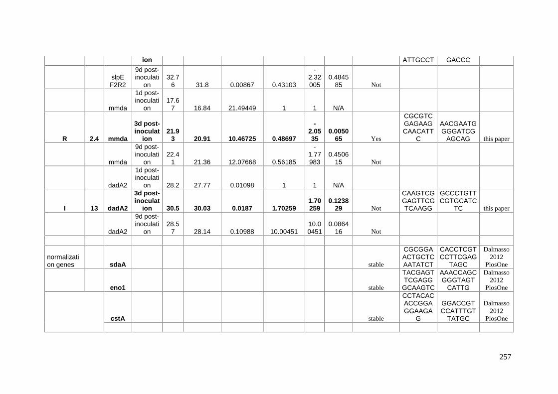
257
ion ATTGCCT GACCC
slpEF2R2
9d post-inoculati
on32.7
6 31.8 0.00867 0.43103
-2.32005
0.484585 Not
mmda
1d post-inoculati
on17.6
7 16.84 21.49449 1 1 N/A
R 2.4 mmda
3d post-inoculat
ion21.9
3 20.91 10.46725 0.48697
-2.0535
0.005065 Yes
CGCGTCGAGAAGCAACATT
C
AACGAATGGGGATCG
AGCAG this paper
mmda
9d post-inoculati
on22.4
1 21.36 12.07668 0.56185
-1.77983
0.450615 Not
dadA2
1d post-inoculati
on 28.2 27.77 0.01098 1 1 N/A
I 13 dadA2
3d post-inoculat
ion 30.5 30.03 0.0187 1.702591.70259
0.123829 Not
CAAGTCGGAGTTCGTCAAGG
GCCCTGTTCGTGCATC
TC this paper
dadA2
9d post-inoculati
on28.5
7 28.14 0.10988 10.0045110.00451
0.086416 Not
normalization genes sdaA stable
CGCGGAACTGCTCAATATCT
CACCTCGTCCTTCGAG
TAGC
Dalmasso2012
PlosOne
eno1 stable
TACGAGTTCGAGG
GCAAGTC
AAACCAGCGGGTAGT
CATTG
Dalmasso2012
PlosOne
cstA stable
CCTACACACCGGAGGAAGA
G
GGACCGTCCATTTGT
TATGC
Dalmasso2012
PlosOne

258
CAPÍTULO 3: Discussão geral,conclusão e perspectivas
CHAPITRE 3 : Discussion générale,conclusion et perspectives

259
Discussão geral
O objetivo geral desta tese foi de melhorar a nossa compreensão dos
mecanismos envolvidos no fenômeno sobrevivência a longo prazo em P. freudenreichii.
Uma das principais características da espécie é, de fato, uma boa sobrevivência a longo
prazo, e este é um critério favorável à sua utilização em vários setores da indústria, tais
como: a produção de queijos PPC, especialmente o Emmental, um dos produtos mais
consumidos na França (Dalmasso et al. 2012a), e o desenvolvimento de produtos
probióticos com efeitos benéficos para a saúde humana (Cousin et al. 2012). A síntese
de biomoléculas, tais como ácido propiônico e vitamina B12 (Van Wyk, Witthuhn and
Britz 2011; Wang and Yang 2013) é uma outra utilização industrial importante para esta
espécie. Mas esta última é realizada sob condições de cultura altamente controlados
para manter uma atividade metabólica ótima e a problemática de sobrevivência a longo
prazo não está no escopo destas aplicações.
Neste trabalho, mostramos, através da análise do perfil de expressão de genes e
da produção e consumação de metabólitos, que P. freudenreichii é capaz de reduzir o
seu metabolismo, colocar-se em um estado de dormência e utilizar vias alternativas de
energia. Estes resultados serão discutidos nos parágrafos seguintes.
Caracterização fenotípica de P. freudenreichii:
Estudos anteriores relatam que P. freudenreichii é uma bactéria versátil que
sobrevive a vários estresses ambientais (Jan, Rouault and Maubois 2000; Dalmasso et
al. 2012b; Saraoui et al. 2013b). Além disso, P. freudenreichii é capaz de utilizar
diferentes fontes de carbono como substrato e pode armazenar certos compostos para a
sobrevivência a longo prazo (Thierry et al. 2011). Para uma melhor compreensão desta
bactéria, nós realizamos a caracterização fenotípica de 8 linhagens de P. freudenreichii,
durante um período de carência nutricional. Sabendp-se que, em condições ótimas, esta
bactéria tem um tempo de geração de 5h (Falentin et al. 2010a), assumiu-se que um
período de 11 dias (com uma 1 amostragem por dia) durante o qual a bactéria está
sujeita ao estresse nutricional permitiu separar e caracterizar bem as diferentes fases de
crescimento, até uma fase de sobrevivência a longo prazo. Testes de sobrevivência a
longo prazo já foram realizados em outras bactérias indicando uma caracterização num
tempo, significativamente, mais longo, como por exemplo, Finkel e Kolter que
consideraram uma duração de um ano para a avaliação da sobrevivência em Escherichia
coli (Finkel 2006b). Com certeza seria interessante fazer análises mais longas, mas um

260
dos objetivos desta tese foi de tentar elucidar os mecanismos que permitiram a
adaptação de P. freudenreichii à esta fase de crescimento.
A capacidade de sobrevivência a longo prazo varia em função do género, da
espécie ou mesmo da linhagem estudada. Os estudos fenotípicos de 8 linhagens
demonstraram que o conjunto de todas as linhagens de P. freudenreichii apresentaram
uma boa sobrevivência a longo prazo, no entanto, com algumas variações entre as
linhagens. Estudar este fenômeno durante um período de 6 ou 12 meses, nos teria
permitido de determinar durante quanto tempo era possível a esta bactéria de sobreviver
em carência nutricional e, sobretudo, poderia ter revelado e ou enfatizado as diferenças
entre as linhagens durante a sobrevivência a longo prazo. Esta capacidade poderia de
fato tornar-se um critério para a seleção de linhagens para aplicações em queijos ou
probióticas de P. freudenreichii.
Dormência em P. freudenreichii:
Muitas bactérias são capazes de entrar em um estado de dormência permitindo
uma maior resistência e uma melhor sobrevivência a ambientes desfavoráveis
(Rittershaus, Baek and Sassetti 2013b). Esta adaptação é caracterizada pela manutenção
da viabilidade, no entanto, associada a uma perda de cultivabilidade em condições
padrão laboratoriais. Este estado também é descrito pelo termo de estado VBNC (Viable
But Non Culturable). O estado VBNC constitui uma estratégia muito importante para
algumas espécies bacterianas, Gram positivas ou negativas, para a sobrevivência
durante períodos prolongados de carência. Muitos estudos científicos têm sido
dedicados a este tema. Uma busca PubMed mostra que o termo "viable but
nonculturable bacteria" totaliza em 385 resultados. Dentre eles, 64 artigos sobre
bactérias patogénicas ("VBNC" AND "pathogen") e 78 artigos sobre bactérias em
estado VBNC em alimentos ou na água ("VBNC" AND "food"). Nesta última pesquisa,
a maioria dos artigos tratavam-se patógenos alimentares (Campylobacter sp., E. coli,
Vibrio sp., Salmonella sp.) e alguns poucos, eventualmente, sobre as bactérias de
interesse alimentar (food grade). Três artigos relacionados com probióticos. Esta
diferença de tratamento ou de interesse entre estas áreas é em parte explicado pelo fato
de que muitas bactérias patogênicas são capazes de induzir a mesma infecção ou
intoxicação mesmo estando em um estado VBNC, e geralmente, essas bactérias (não
cultiváveis) não são detectadas por técnicas microbiológicas convencionais, como a
contagem das UFC. Os estudos sobre probióticos são realizados para desenvolver a sua

261
importância na formulação destes produtos, uma vez que o número de bactérias viáveis
no produto final é um critério levado em consideração para os efeitos probióticos sobre
o hospedeiro. Em um estudo realizado por Davis e colaboradores, demonstrou-se que as
bactérias que haviam propriedades probióticas entravam em estado VBNC e que elas só
podiam ser quantificadas corretamente através de métodos alternativos, tais como
hibridação in situ fluorescente, qPCR, RT-PCR, propídio monoazida-PCR e/ou a
separação de células ativadas por fluorescência (Davis 2014). Desta mesma maneira,
confirmamos o estado VBNC em P. freudenreichii CIRM-BIA138, linhagem mais
resistente a carência nutricional, combinando diferentes metodologias, tais como a
contagem de UFC, a microscopia de epi-fluorescência e qPCR.
Genes diferencialmente expressos em fases de crescimento exponencial e
estacionária de P. freudenreichii:
Para aprofundar os estudos relacionados com a sobrevivência a longo prazo
desta bactéria, foi realizada a análise do perfil transcricional da linhagem P.
freudenreichii CIRM- BIA138 utilizando a técnica de RNA-Seq, durante um período de
carência nutricional. Inicialmente, escolhemos três pontos de crescimento bacteriano
para realizar esta análise: exponencial, estacionária e estacionária tardia. Para obter
estas amostras, realizamos a cultura bacteriana, a extração de RNA total e a depleção de
RNAr (RNA ribossômico). Em seguida, sequenciamos o cDNA destas amostras pela
plataforma Illumina HiSeq 2000, uma tecnologia de sequenciamento de próxima
geração. Infelizmente, observa-se que não foi possível obter bons resultados de
sequenciamento nas amostras da fase estacionária tardia. Isto, porque os resultados das
3 replicatas desta última fase apresentaram um perfil de contaminação por DNA
genômico, uma vez que as reads estavam alinhadas de maneira uniforme e não-
orientada, impedindo assim a interpretação dos dados de sequenciamento. No entanto, a
análise de RNA-Seq entre as fases de crescimento bacteriano exponencial e estacionária
nos permitiu identificar 912 genes diferencialmente expressos e potencialmente
envolvidos na preparação e adaptação da linhagem à carência nutricional.
Atualmente, um dos poucos estudos de transcriptoma sobre o tema de
sobrevivência a longo prazo é o realizado sobre o patógeno Listeria monocytogenes que
provoca infecções de origem alimentar. A cultura de L. monocytogenes foi amostrada
em diferentes fases do desenvolvimento (13h, 17h, 24h e 168h, 336h), num meio de
cultura de triptona de soja ágar com extrato de levedura (Becton Dickinson, MD). Este

262
estudo foi realizado por meio de técnicas de microarranjo e RT-qPCR que revelaram
genes induzidos envolvidos em: (i) síntese de aminoácidos, de ácidos graxos, de
proteínas, de fosfolípidos, de envelope celular e de ribonucleótideos ; (ii) na transcrição;
(iii) na desintoxicação; (iv) no transporte de proteínas; e (v) na divisão celular. Estes
metabolismos altamente expressos durante a fase logarítmica são reprimidos durante o
período de sobrevivência a longo prazo (Wen et al. 2011).
Observou-se ainda que os genes envolvidos na síntese de proteínas foram
reprimidos, o que indica também, que esta bactéria, entra em estado de dormência após
períodos prolongados (Wen et al. 2011). Apesar de L. monocytogenes ser patogênica, a
estratégia de sobrevivência adotada é semelhante a adotada por P. freudenreichii. Os
resultados do transcriptoma de P. freudenreichii CIRM-BIA138 também revelaram que,
na fase estacionária, a bactérias começa a se preparar para um estado de dormência que
lhe permite sobreviver e de utilizar fontes alternativas para obter e armazenar a energia.
Além disso, os nossos resultados demonstram uma redução no metabolismo de
P. freudenreichii, uma vez que os genes envolvidos na fosforilação oxidativa e no ciclo
de Wood-Werkman estão reprimidos durante a fase estacionária. Estes resultados estão
de acordo com o trabalho de Saraoui e colaboradores que mostram que, em P.
freudenreichii CIRM-BIA1, as vias da glicólise, do ciclo de Wood-Werkman e
fosforilação oxidativa foram reprimidos em fase estacionária no cólon de suínos
(Saraoui et al. 2013a).
Mecanismos moleculares envolvidos na sobrevivência a longo prazo de
P. freudenreichii:
Através de dados transcriptômicos e bioquímicos de P. freudenreichii CIRM-
BIA138, obtivemos pistas que indicam quais genes estão ativos nesta bactéria e quais
são os fatores ambientais envolvidos no desencadeamento de mecanismos moleculares
que preparam a sobrevivência bacteriana a longo prazo, durante um período de carência
nutricional.
Durante este período, a linhagem P. freudenreichii CIRM-BIA138 é também
capaz de explorar novas fontes de energia, tais como aminoácidos (como o glutamato,
glicina e alanina), a arabinose e o inositol que são fontes de carbono alternativas. Para
equilibrar o seu metabolismo, na ausência de oxigénio, esta bactéria utiliza
provavelmente o polifosfato e o pirofosfato para armazenar a energia, os quais os genes
estão presentes no genoma desta espécie (Falentin et al. 2010a). Assim, neste trabalho,

263
os genes envolvidos na síntese de polifosfatos são induzidos durante a fase estacionária
para a utilização de ATP. Além disso, nesta última fase, uma diminuição na atividade de
transcrição, tradução e secreção de proteínas foi observada. Outros parâmetros
experimentais também foram analisados neste estudo, como a dosagem de compostos
bioquímicos, demonstrando a influência destes últimos sobre o metabolismo de P.
freudenreichii CIRM-BIA138, durante a carência nutricional. Alterações na síntese e no
consumo de ácidos orgânicos, de açúcares e de aminoácidos também foram observados.
Isto demonstra a capacidade de P. freudenreichii CIRM-BIA138 se adaptar ao ambiente,
e contribui para uma melhor compreensão do conhecimento biológico desta espécie.
Todos estes dados fenotípicos, transcriptômicos e metabolômicos de P.
freudenreichii CIRM-BIA138 são de grande importância para a compreensão desta
bactéria, especialmente, durante as fases importantes encontradas, como por exemplo,
durante a maturação de queijos PPC. No entanto, nas condições de maturação, P.
freudenreichii atinge rapidamente uma fase estacionária de crescimento, mas raramente
associada a uma carência de carbono. Este tipo de trabalho já havia sido começado na
equipe, portanto com análise transcricional por microarranjo para identificar estratégias
de adaptação de P. freudentreichii nas condições de fabricação de queijos (Dalmasso et
al. 2012a). Nossos resultados de RNA-Seq vieram para complementar estes dados.
Além disso, nosso estudo metabolômico foi muito direcionado a determinados
compostos. Seria interessante fazer um estudo metabolômico mais abrangentes como o
trabalho recente de Le Boucher (Le Boucher et al. 2013). Este último trabalho
demonstrou a capacidade da espectrometria de massa – metabolic fingerprinting – a
detectar alterações induzidas pelo metabolismo bacteriano no queijo durante 0, 8 e 48
h. Este estudo encontrou variações em 45 metabólitos, tais como aminoácidos,
vitaminas e metabólitos voláteis (Le Boucher et al. 2013).
A interdisciplinaridade e os diferentes métodos aplicados neste trabalho de tese
permitiram de identificar mecanismos que poderiam explicar o fenômeno de
sobrevivência a longo prazo nesta bactéria. Uma destas estratégias de sobrevivência
parece ser o uso de vias alternativas de energia para manter a atividade metabólica.
Nestas etapas, a bactéria se prepara a entrar em um estado de dormência para enfrentar
um período de estresse nutricional e sobreviver a longo prazo, enquanto aguarda a
ocorrência de condições mais favoráveis.

264
Discussion générale
L'objectif général de cette thèse était d’améliorer notre compréhension des
mécanismes impliqués dans le phénomène de survie à long terme chez P. freudenreichii.
Une des principales caractéristiques de l’espèce est, en effet, sa bonne survie à long
terme et ceci est un critère favorable à son utilisation dans divers secteurs de l'industrie
tels que : la production des fromages PPC, en particulier l'Emmental, l’un des produits
les plus consommés en France (Dalmasso et al. 2012a) et le développement de produits
probiotiques ayant des effets bénéfiques sur la santé humaine (Cousin et al. 2012). La
synthèse de biomolécules, comme l'acide propionique et la vitamine B12 (Van Wyk,
Witthuhn and Britz 2011; Wang and Yang 2013) est une autre utilisation industrielle
d’importance pour cette espèce. Mais celle-ci s’effectue dans des conditions de culture
hautement contrôlées permettant de maintenir une activité métabolique optimale et la
problématique de survie à long terme est de fait hors champs pour ces applications.
Dans ce travail, nous avons montré, à travers l’analyse du profil d'expression des
gènes et de la production et consommation de métabolites, que P. freudenreichii est
capable de réduire son métabolisme, de se mettre dans un état de dormance et d'utiliser
des voies d'énergie alternatives. Ces résultats seront discutés dans les paragraphes
suivants.
Caractérisation phénotypique de P. freudenreichii:
Des études antérieures rapportent que P. freudenreichii est une bactérie versatile
qui survit à divers stress environnementaux (Jan, Rouault and Maubois 2000; Dalmasso
et al. 2012a; Saraoui et al. 2013a). En outre, P. freudenreichii est capable d'utiliser
différentes sources de carbone comme substrat et peut stocker certains composés pour
sa survie à long terme (Thierry et al. 2011). Aussi, pour une meilleure compréhension
de cette bactérie, nous avons effectué la caractérisation phénotypique de 8 souches de P.
freudenreichii, au cours d’une période de carence nutritionnelle. Sachant que, dans des
conditions optimales, cette bactérie a un temps de génération de 5h (Falentin et al.
2010b), nous avons supposé qu’une période de 11 jours (avec un 1 échantillonnage par
jour) durant laquelle la bactérie est soumise au stress nutritionnel permettait de séparer
et bien caractériser les différentes phases de croissance, jusqu’à une phase de survie à
long terme. Des essais de survie à long terme avaient déjà été réalisés chez d'autres
bactéries indiquant une caractérisation sur un temps nettement plus long, comme par
example, Finkel et Kolter qui ont consideré une durée d’un an pour l’évaluation de la

265
survie chez Escherichia coli (Finkel 2006b). Il serait sûrement intéressant de disposer
d’essais plus longs, mais un des buts de cette thèse était de tenter d’élucider les
mécanismes qui permettait à l’adaptation de P. freudenreichii à cette phase de
croissance.
La capacité de survie à long terme varie en fonction du genre, de l'espèce, voire
de la souche étudiée. Les études phénotypiques sur 8 souches ont montré que
l’ensemble des souches de P. freudenreichii présentait une bonne survie à long terme
avec cependant quelques variations inter-souches. Etudier ce phénomène sur une durée
de 6 ou 12 mois, nous aurait permis de déterminer pendant combien de temps encore il
était possible à cette bactérie de survivre en carence nutritionnelle et, surtout, aurait
peut-être permis de révéler et ou accentuer des différences inter-souches en matière de
survie à long terme. Cette capacité pourrait en effet devenir un critère de sélection de
souches pour les applications fromagères ou probiotiques de P. freudenreichii.
La dormance chez P. freudenreichii :
De nombreuses bactéries sont capables d’entrer dans un état de dormance
permettant une plus grande résistance et une meilleure survie à des environnements
défavorables (Rittershaus, Baek and Sassetti 2013b). Cette adaptation se caractérise par
le maintien de la viabilité associée cependant à une perte de cultivabilité en conditions
de laboratoire classiques. Cet état est également décrit sous le terme d’état VBNC (pour
Viable But Non Culturable). L’état VBNC constitue une stratégie très importante chez
certaines espèces bactériennes, à Gram positif ou négatif, pour la survie pendant de
longues périodes de carence. De nombreuses études scientifiques lui ont été consacrées.
Une recherche PubMed montre que le terme « viable but nonculturable bacteria »
totalise 385 résultats. Parmi eux, 64 articles concernent les bactéries pathogènes
(« VBNC » AND « pathogen ») et 78 articles portent sur les bactéries en état VBNC
dans l’aliment ou l’eau (« VBNC » AND « food »). Dans cette dernière recherche, la
majorité des articles ont trait aux pathogènes alimentaires (Campylobacter sp., E. coli,
Vibrio sp., Salmonella sp.) et très peu, finalement, portent sur des bactéries d’intérêt
alimentaire (food grade). Trois articles ont trait aux probiotiques. Cette différence de
traitement ou d’intérêt entre ces divers domaines s’explique entre autres par le fait que
beaucoup de bactéries pathogènes sont capables d’induire une infection ou une
intoxication même à partir d’un état VBNC, et généralement ces bactéries (non
cultivables) ne sont pas détectées par des techniques de microbiologie classique, telles

266
que le comptage des UFC. Les études portant sur les probiotiques sont amenées à se
développer de par leur importance pour la formulation de ces produits, car le nombre de
bactéries viables dans le produit final est un critère pris en compte pour l’effet
probiotique sur l’hôte. Dans une étude de Davis et collaborateurs, il a été démontré que
les bactéries ayant des propriétés probiotiques se mettaient en état VBNC et qu’elles ne
pouvaient être quantifiées correctement qu’à travers des méthodes alternatives
comme l’hybridation in situ en fluorescence, la PCRq, la RT-PCR, le propidium
monoazide-PCR et/ou la séparation des cellules activées par fluorescence (Davis 2014).
De même, nous avons confirmé l’état VBNC chez P. freudenreichii CIRM-BIA138, la
souche la plus résistante à la carence nutritionnelle, en combinant différentes
méthodologies telles que la numération des UFC, la microscopie à épi-fluorescence et
la PCRq.
Gènes différentiellement exprimés dans les phases de croissance
exponentielle et stationnaire de P. freudenreichii :
Pour approfondir les études liées à la survie à long terme de cette bactérie, nous
avons effectué l'analyse du profil transcriptionnel de la souche P. freudenreichii CIRM-
BIA138 en utilisant la technique de RNA-Seq, au cours d’une période de carence
nutritionnelle. Tout d’abord, nous avons choisi trois points de la croissance bactérienne
pour effectuer cette analyse : exponentiel, stationnaire et stationnaire tardif. Pour obtenir
ces échantillons, nous avons réalisé une culture bactérienne, une extraction des ARN
totaux et une déplétion des ARNr (ARN ribosomiques). Ensuite, nous avons séquencé le
cDNA de ces échantillons par la plateforme Illumina HiSeq 2000, une technologie de
séquençage de nouvelle génération. Malheureusement, nous avons observé qu’il n’était
pas possible d’obtenir de bons résultats de séquençage sur les échantillons de la phase
stationnaire tardive. En effet, les résultats des 3 replicates de cette dernière phase ont
présenté un profil de contamination par de l’ADN génomique, car les reads se sont
alignés de manière uniforme et non orientée, empêchant ainsi l’interprétation des
résultats de séquençage. Cependant, l'analyse du RNA-Seq entre les phases de
croissance bactérienne exponentielle et stationnaire nous a permis d'identifier 912 gènes
différentiellement exprimés et potentiellement associés à la préparation et l’adaptation
de la souche à la carence nutritionnelle.
Actuellement, l'une des rares études de transcriptome sur le thème de la survie à
long terme est celle menée sur le pathogène Listeria monocytogenes qui provoque des

267
infections d'origine alimentaire. La culture de L. monocytogenes a été échantillonnée à
différentes phases de développement (13h, 17h, 24h et 168h, 336h), dans un milieu de
culture gélose tryptone soja avec de l'extrait de levure (Becton Dickinson, MD). Cette
étude a été réalisée à travers des techniques utilisant des puces à ADN et la RT-qPCR
qui ont révélé des gènes inductibles impliqués dans : (i) la synthèse d’acides aminés,
d’acides gras, de protéines, de phospholipides, de l’enveloppe cellulaire et de
ribonucléotides; (ii) la transcription ; (iii) la détoxification ; (iv) le transport de
protéines ; et (v) la division cellulaire. Ces métabolismes fortement exprimés pendant la
phase logarithmique sont réprirmés pendant la période de survie à long terme (Wen et
al. 2011).
Il a également été observé que les gènes impliqués dans la synthèse des protéines
ont été réprimés, ce qui indique aussi, chez cette bactérie, l’entrée en état de dormance
après des périodes prolongées (Wen et al. 2011). Bien que L. monocytogenes soit
pathogène, la stratégie de survie qu’elle adopte est similaire à celle adoptée par P.
freudenreichii. Les résultats du transcriptome de P. freudenreichii CIRM-BIA138 ont
également révélé que, dans la phase stationnaire, la bactérie commence à se préparer
pour un état de dormance qui lui permet de survivre et d’utiliser des sources alternatives
pour obtenir et stocker de l'énergie.
En outre, nos résultats démontrent une réduction du métabolisme de P.
freudenreichii, puisque les gènes impliqués dans la phosphorylation oxydative et le
cycle Wood-Werkman sont réprimés pendant la phase stationnaire. Ces résultats sont en
accord avec le travail de Saraoui et collaborateurs qui montrent que, chez P.
freudenreichii CIRM-BIA1, les voies de glycolyse, du cycle de Wood-Werkman et de la
phosphorylation oxydative étaient régulé négativement en phase stationnaire dans un
côlon de cochons (Saraoui et al. 2013a).
Mécanismes moléculaires impliqués dans la survie à long terme de P.
freudenreichii :
À travers des données transcriptomiques et biochimiques de P. freudenreichii
CIRM-BIA138, nous avons obtenu des pistes indiquant quels gènes sont actifs dans
cette bactérie et quels sont les facteurs environmentaux impliqués dans le
déclenchement des mécanismes moléculaires qui préparent la survie bactérienne à long
terme, au cours d’une période de carence nutritionnelle.

268
Au cours de cette période, la souche P. freudenreichii CIRM-BIA138 est aussi
capable d'explorer de nouvelles sources d'énergie, telles que des acides aminés (comme
le glutamate, la glycine et l’alanine), l'arabinose et l'inositol qui sont des sources de
carbone alternatives. Pour équilibrer son métabolisme, en l'absence d'oxygène, cette
bactérie utilise probablement le polyphosphate et pyrophosphate pour stockér l’énergie
et qui les gènes sont présentes dans le génome de cette espèce (Falentin et al. 2010b).
Ainsi dans ce travail, les gènes impliqués dans la synthèse de polyphosphates sont
induits au cours de la phase stationnaire pour utiliser l’ATP. En outre, dans cette
dernière phase, une diminution de l'activité de la transcription, de la traduction et de la
sécrétion des protéines a été observée. D’autres paramètres expérimentaux ont aussi été
analysés dans ce travail, comme le dosage des composés biochimiques, montrant
l'influence de ces derniers sur le métabolisme de P. freudenreichii CIRM-BIA138, au
cours de la carence nutritionnelle. Des altérations dans la synthèse et la consommation
d'acides organiques, de sucres et d’acides aminés ont également été observés. Cela
démontre la capacité de P. freudenreichii CIRM-BIA138 à s’adapter à l'environnement,
et contribue à une meilleure compréhension de la connaissance biologique de cette
espèce.
Toutes ces données phénotypiques, transcriptomiques et métabolomiques de P.
freudenreichii CIRM-BIA138 sont d'une grande importance pour la compréhension de
cette bactérie notamment pendant des étapes importantes rencontrées, par exemple, au
cours de l’affinage des fromages PPC. Toutefois, dans les conditions d'affinage, P.
freudenreichii atteint rapidement une phase stationnaire de croissance mais rarement
liée à une carence carbone. Ce type de travail avait déjà commencé dans l’équipe lors de
l’analyse transcriptionnelle par puce à ADN pour identifier les stratégies d'adaptation de
P. freudentreichii dans les conditions de fabrication fromagère (Dalmasso et al. 2012a).
Nos résultats RNA-Seq viennent complétés ces données. En outre, notre étude
métabolomique était très ciblée sur certains composés. Il serait intéressant de faire une
étude métabolomique plus exhaustive à l’instar des travaux récemment menés par Le
Boucher (Le Boucher et al. 2013). Ce dernier travail a montré la capacité de la
spectrométrie de masse - metabolic fingerprinting – à détecter les modifications induites
par le métabolisme bactérien dans le fromage au cours de 0, 8 et 48 h. Cette étude a
révélé des variations sur 45 métabolites, tels que les acides aminés, les métabolites
volatils et les vitamines (Le Boucher et al. 2013).

269
L'interdisciplinarité et les différentes méthodes appliquées dans ce travail de
thèse ont permis d'identifier des mécanismes qui pourraient expliquer le phénomène de
survie à long terme de cette bactérie. Une de ces stratégies de survie semble être
l’utilisation de voies énergétiques alternatives permettant le maintien de l’activité
métabolique. Dans ces étapes, la bactérie se prépare à entrer en dormance pour affronter
une période de stress nutritionnel et survivre à long terme, en attendant la survenue de
conditions plus favorables.

270
Conclusão/ Conclusion

271
Conclusão
Neste presente trabalho de tese, apresentamos, pela primeira vez, a identificação
de estratégias de sobrevivência a longo-prazo em P. freudenreichii, combinando as
abordagens de microbiologia, genômica e transcriptômica. Desde a caracterização
fenotípica à analise funcional do genoma de P. freudenreichii, nossos estudos
demonstraram que esta bactéria diversifica suas fontes de energia, em seguida,
diminuem o seu metabolismo energético para entrar em um estado de dormência, o qual
elas são capazes de sobreviver por períodos prolongados. Além disso, este trabalho de
tese fornece informações sobre a biologia do grupo Propionibacteria lactéos.

272
Conclusion
Dans ce travail de thèse, nous avons présenté, pour la première fois,
l'identification de stratégies de survie à long terme chez P. freudenreichii, en combinant
des approches microbiologique, génomique et transcriptiomique. De la caractérisation
phénotypique à l'analyse fonctionnelle du génome de P. freudenreichii, nos études ont
permis de démontrer que cette bactérie diversifie ses sources d’énergie, puis diminue
son métabolisme énergétique pour entrer dans un état de dormance pendant lequel elle
est capable de survivre durant de longues périodes. En outre, ce travail de thèse fournit
des informations sur la biologie du groupe de Propionibacteries laitières.

273
Perspectivas/ Perspectives

274
Perspectivas
Os resultados obtidos ao longo deste trabalho de tese nos permitiram a melhor
compreender o fenômeno de sobrevivência a longo prazo em P. freudenreichii. Eles
abrem várias perspectivas. De um modo, os dados gerados podem ser completados e
explorados para melhorar o nosso conhecimento da genômica de P. freudenreichii
(tópicos 1 e 2 abaixo). E de outro modo, ainda há muito a ser feito na análise de
sobrevivência a longo prazo e diversas pistas poderiam ser utilizadas para completar
este trabalho (tópicos 3-7).
(I) Sequência completa do genoma de P. freudenreichii CIRM-BIA138:
Como uma primeira abordagem, podemos considerar que um genoma completo
(isto é, sem gaps) não é essencial, se podemos extrair as informações necessárias das
análises de transcriptômica e metabolômica desta bactéria. No entanto, um genoma
completamente montado permite uma melhor anotação e, assim, nos dará uma melhor
qualidade de informação genética sobre o organismo.
Para obter o genoma completo de P. freudenreichii CIRM-BIA138, uma opção
seria realizar um mapeamento óptico do genoma desta linhagem. Isto permitiria a
superação de dificuldades encontradas na etapa de montagem deste genoma, como por
exemplo, a presença de uma grande quantidade de transposases. O mapeamento óptico
de fato dá uma visão geral do genoma, auxiliando a reorganização de contigs e ao
fechamento de gaps. Além disso, é possível combinar diferentes programas de
montagem para melhorar o processo como um todo. De outra maneira, como já foi
demonstrado em estudos precedentes (Garber et al. 2011), e para complementar as
abordagens anteriores, é possível utilizar os dados do transcriptoma provenientes do
RNA-Seq para alinhar os transcritos no genoma e fechar os gaps. Os dados de RNA-
Seq também podem auxiliar no processo de anotação do genoma, utilizando programas
como o ReadXplorer, que permite o alinhamento dos dados de expressão de cada gene
sob o genoma para refinar a anotação (Hilker et al. 2014).
(II) Identificação de novos elementos no genoma de P. freudenreichii CIRM-BIA
138:
Os dados de RNA-Seq permitem a descoberta de novos transcritos que não
foram identificados e anotados em versões anteriores do genoma. Uma análise detalhada

275
permitirá também de identificar pequenos RNAs não-codificantes em P. freudenreichii
CIRM-BIA138. A tecnologia de RNA-Seq revelou-se eficaz para a detecção de
pequenos RNAs em Pseudomonas aeruginosa (Gómez-Lozano et al. 2012), e para a
descoberta de novos transcritos em várias espécies bacterianas (McClure et al. 2013).
Programas de bioinformática, como Rockhopper, através de uma interface gráfica
amigável, permite que o usuário entre com genomas de referência e modifique as
configurações, tais como: leituras paired-end ou mate-pair; alinhamento de leituras de
várias replicatas; número de replicatas e condições experimentais diferentes utilizadas
para a análise de RNA-Seq (McClure et al. 2013).
(III) Complemetação dos dados de RNA-Seq na fase estacionária tardia de P.
freudenreichii CIRM-BIA138:
Durante a análise de RNA-Seq da linhagem P. freudenreichii CIRM-BIA138, a
quantidade de RNA extraída de amostras associadas com a fase de sobrevivência a
longo prazo foi muito baixa para permitir um sequenciamento de qualidade. Além disso,
as amostras da fase estacionária tardia foram contaminadas com DNA genómico. Isto
limitou severamente os resultados obtidos. Por conseguinte, seria vantajoso obter uma
maior quantidade de RNA e a melhorar a sua qualidade, aumentando o volume da
cultura bacteriana amostrada (por exemplo 25 mL) visando melhorar, a partir disso, as
próximas etapas de extração e de depleção dos rRNA certificando-se de remover o DNA
genômico por tratamentos com DNase.
(IV) Mecanismo de dormência na espécie P. freudenreichii:
Muitas bactérias Gram-positivas e Gram-negativas entram em dormência na fase
de sobrevivência a longo prazo. Como vimos, dentre as 8 linhagens de P. freudenreichii
testadas, todas sobrevivem bem e suas populações permanecem em níveis elevados
durante o período de carência nutricional. No entanto, observamos que a linhagem
CIRM-BIA138 apresenta um nível de sobrevivência maior do que outras linhagens.
Esta linhagem foi objeto de uma análise mais aprofundada (demonstração do estado
VBNC e RNA-Seq). Neste contexto, seria interessante complementar os resultados
obtidos nesta tese, explorando a capacidade de sobrevivência a longo prazo e o estado
VBNC em outras linhagens. Isso talvez permita identificar linhagens de P.
freudenreichii com baixa sobrevivência a longo prazo, e em seguida, por genômica
comparativa os genes envolvidos (por exemplo, pela sua ausência ou por serem

276
pseudogenes) nesta sobrevivência inferior. Do mesmo modo, outras formas de stress
(por exemplo, calor, ácido ou osmótico) podem ser aplicados a culturas em estado de
sobrevivência a longo prazo e carência nutricional. Isto permitiria a identificar os
fenômenos de adaptação e resistência cruzada que poderiam nos esclarecer sobre os
mecanismos implementados e nos fornecer uma melhor explicação do comportamento
da espécie em condições de fabricação de queijos.
Além disso, este estudo pode ser complementado pela construção de mutantes
visando a interromper os genes envolvidos no desencadeamento da dormência em P.
freudenreichii. Trabalho abordando esta metodologia (construção de mutantes) já foi
realizado para mostrar o envolvimento do gene clpX em S. typhimurium (Kusumoto,
Miyashita and Kawamoto 2013) e rpoS em E. coli (Boaretti et al. 2003) na
sobrevivência a longo prazo nestas espécies.
(V) Reanimação na espécie P. freudenreichii:
Ao sair do estado VBNC, muitas espécies bacterianas passam por um estado
chamado de ressuscitação permitindo-lhes retomar o crescimento normal. A reanimação
(ou ressuscitação), por vezes, requer condições especiais de crescimento. Da mesma
forma, alguns genes parecem estar diretamente envolvidos neste fenômeno. Assim, em
M. luteus, tem sido mostrado que as células são reanimadas depois da produção do fator
Rpf (Ressuscitation Promoting Factor) (Greenblatt et al. 2004a). A investigação deste
estado de reanimação nas células de P. freudenreichii seria de grande interesse para a
conclusão deste trabalho. No entanto, é necessário realizar experimentos adicionais que
comprovem que as células são ressuscitadas após o estado VBNC. Isto requer a
identificação de condições de cultura que provocam a liberação do estado VBNC e da
retomada do crescimento normal.
(VI) Sobrevivência a longo prazo em P. freudenreichii CIRM-BIA138 na matriz de
queijo:
Os nossos resultados permitem uma melhor compreensão dos mecanismos
moleculares envolvidos na sobrevivência a longo prazo de P. freudenreichii submetida a
uma carência nutricional. Esses resultados, obtidos in vitro e em condições de
laboratório, devem ser complementadas e validadas, estudando o comportamento de P.
freudenreichii CIRM-BIA138 na matriz de queijo. Esta linhagem foi isolada à partir do
queijo Emmental. A matriz de queijo é complexa, tem propriedades físico-químicas
277
específicas e as bactérias são imóveis, então, seria interessante estudar o comportamento
de CIRM-BIA138 neste contexto. Isso iria avaliar, em primeiro lugar, a sua
sobrevivência a longo prazo na matriz de queijo e, por outro, verificar se os mecanismos
que se suspeita importante na transição para a fase estacionária tardia também estão
envolvidos na sobrevivência a longo prazo em queijo.
(VII) Ativação e regulação de vias metabólicas em P. freudenreichii CIRM-BIA138:
Após o consumo total de lactato, o seu substrato preferencial, P. freudenreichii
CIRM-BIA138 ativa várias outras vias metabólicas para garantir a sua sobrevivência.
Seria interessante entender como estas vias alternativas são ativadas. Todas elas estão
ativas simultaneamente ou são induzidas somente quando o lactato está esgotado? Neste
caso, eles são induzidos por concomitante ou sequencial? Seria interessante, neste caso,
identificar os estímulos e mecanismos reguladores. Uma opção é a construção de
mutantes de genes envolvidos e induzidos no início de cada um dos processos
metabólicos específicos. Por exemplo, para ativar o metabolismo de inositol,
poderíamos tentar determinar se P. freudenreichii reconhece especificamente, a presença
de inositol no meio e, em caso afirmativo, como esta bactéria distingue o estímulo no
meio e reorienta o seu metabolismo. Isto poderia envolver a construção de genes
mutantes com iolT1 (Major myo-inositol transporter iolT1), iolT2 (Myo-inositol
transporter iolT2) e iolT3 (Myo-inositol transporteriol iolT3). O acompanhamento por
RT-qPCR, de outros genes envolvidos no metabolismo de inositol, e dosagens
bioquímicas, da concentração de inositol nos sobrenadantes de cultura, podem ser
realizadas nestes mutantes e podem ser comparadas a linhagem selvagem.

278
Perspectives
Les résultats obtenus au cours de ce travail de thèse nous ont permis de mieux
cerner le phénomène de survie à long terme chez P. freudenreichii. Ils ouvrent aussi
plusieurs perspectives. D’une part, les données générées peuvent être complétées et
exploitées pour améliorer notre connaissance sur la génomique de P. freudenreichii
(points 1 et 2 ci-dessous). D’autre part, beaucoup reste encore à faire concernant
l’analyse de la survie à long terme et plusieurs pistes pourraient permettre de compléter
ce travail (points 3 à 7).
(I) Achever la séquence du génome de P. freudenreichii CIRM-BIA138 :
En première approche, on peut considérer qu’un génome complet (c’est-à-dire,
sans gaps) n’est pas indispensable si l’on peut en extraire les informations nécessaires
aux analyses transcriptomique et métabolomique de cette bactérie. Cependant, un
génome entièrement assemblé permettrait une meilleure annotation et donnerait ainsi
une meilleure qualité d'informations génétiques sur l’organisme.
Pour obtenir le génome complet de P. freudenreichii CIRM-BIA138, une option
serait d’effectuer une cartographie optique du génome de cette souche. Ceci permettrait
de pallier les difficultés rencontrées dans l’étape d'assemblage de ce génome, dues à la
présence d'une grande quantité de transposases. La cartographie optique donne en effet
un aperçu général du génome, aide à la réorganisation de contigs et à la fermeture des
gaps. En outre, il est possible de combiner différents programmes d'assemblage pour
améliorer l’ensemble du processus. Par ailleurs, comme cela a déjà été démontré dans
de précédentes études (Garber et al. 2011), et pour complémenter les précédentes
approches, il est possible d’utiliser les données transcriptomiques dérivées du RNA-Seq
pour aligner les transcrits sur le génome et pour fermer les gaps. Les données de RNA-
Seq peuvent également aider au processus d'annotation du génome, en utilisant des
programmes tels que ReadXplorer qui permettent l'alignement des données d'expression
de chaque gène sur le génome pour en affiner l’annotation (Hilker et al. 2014).
(II) L’identification de nouveaux éléments dans le génome de P. freudenreichii
CIRM-BIA 138 :
Les données de RNA-Seq permettent la découverte de nouveaux transcripts qui
n’ont pas été détectés et annotés sur les précédentes versions du génome. Une analyse
dédiée permettrait également d'identifier de petits ARN non codants chez P.

279
freudenreichii CIRM-BIA138. La technique de RNA-Seq s’est révélée efficace pour la
détection de petits ARNs chez Pseudomonas aeruginosa (Gómez-Lozano et al. 2012),
ainsi que pour la découverte de nouveaux transcripts dans plusieurs espèces
bactériennes (McClure et al. 2013). Des programmes bioinformatiques, comme
Rockhopper, grâce à une interface graphique conviviale, permettent à l'utilisateur d’y
entrer des génomes de référence et de modifier des paramètres, tels que : les lectures
paired-end ou mate-pair ; l’alignement des lectures de plusieurs réplicats ; le nombre
des replicats et de conditions expérimentales différentes utilisés pour les analyses de
RNA-Seq (McClure et al. 2013).
(III) Compléter les données de RNA-Seq sur la phase stationnaire tardive de P.
freudenreichii CIRM-BIA138 :
Au cours de l’analyse RNA-Seq de la souche P. freudenreichii CIRM-BIA138,
la quantité d'ARN extraite des échantillons liés à la phase de survie à long terme a été
trop faible pour permettre un séquençage de qualité. De plus, les échantillons de la
phase stationnaire tardive ont été contaminés par de l'ADN génomique. Ceci a fortement
limité la portée des résultats obtenus. Il serait donc intéressant d’obtenir une plus grande
quantité d'ARN et d’en améliorer la qualité, en augmentant le volume de culture
bactérienne échantillonné (par ex. 25 mL) en vue d’améliorer, par la suite, les étapes
d'extraction et de déplétion des ARNr en veillant à éliminer l’ADN génomique par des
traitements à la DNase.
(IV) Le mécanisme de dormance dans l’espèce P. freudenreichi :
De nombreuses bactéries à Gram positif et à Gram négatif entrent en dormance
dans la phase de survie à long terme. Comme nous l'avons vu, parmi les 8 souches de P.
freudenreichii testées, toutes survivent bien et leurs populations se maintiennent à des
taux élevés au cours de la période de carence nutritionnelle. Nous avons cependant
observé que la souche CIRM-BIA138 présentait un niveau de survie supérieur aux
autres souches. Cette souche a fait l’objet d’une analyse plus poussée (démonstration de
l’état VBNC et RNA-Seq). Dans ce contexte, il serait intéressant de compléter les
résultats obtenus dans cette thèse, en explorant la capacité de survie à long terme et
l'état VBNC d’autres souches. Ceci permettrait peut-être d’identifier des souches de P.
freudenreichii présentant une faible survie à long terme puis d’identifier par génomique
comparative les gènes impliqués (par exemple, par leur absence ou leur

280
pseudogénisation) dans cette moindre survie. De même, d’autres stress (par exemple,
thermique, acide, ou osmotique) pourraient être appliqués aux cultures en état de survie
à long terme et carence nutritionnelle. Ceci permettrait d’identifier des phénomènes
d’adaptation et de résistance croisées qui pourraient nous éclairer sur les mécanismes
mis en œuvre et mieux expliquer le comportement de l’espèce en conditions de
fabrication fromagère.
En outre, cette étude pourrait être complétée par la construction de mutants
visant à interrompre les gènes impliqués dans le déclenchement de la dormance chez P.
freudenreichii. Des travaux de ce type (construction de mutants) ont déjà été menés pour
montrer l’implication du gène clpX chez S. typhimurium (Kusumoto, Miyashita and
Kawamoto 2013) et rpoS chez E. coli (Boaretti et al. 2003) dans la survie à long terme
chez ces espèces.
(V) La réanimation dans l’espèce P. freudenreichii :
Au sortir de l'état VBNC, de nombreuses espèces bactériennes passent par un
état appelé réanimation leur permettant de reprendre une croissance normale. La
réanimation (ou ressuscitation) nécessite parfois des conditions de croissance
particulières. De même, certains gènes semblent directement impliqués dans ce
phénomène. Ainsi, chez M. luteus, il a été montré que les cellules sont réanimées après
la production du facteur Rpf (Ressuscitation Promoting Factor) (Greenblatt et al.
2004a). L’investigation de cet état de ressuscitation dans les cellules de P. freudenreichii
serait d'un grand intérêt pour compléter ce travail. Cependant, il est nécessaire
d'effectuer des expériences supplémentaires prouvant que les cellules sont réanimées
après l'état VBNC. Cela requiert d’identifier les conditions de culture qui déclenchent la
sortie de l’état VBNC ainsi que la reprise d’une croissance normale.
(VI) La survie à long terme chez P. freudenreichii CIRM-BIA138 en matrice
fromagère :
Nos résultats permettent une meilleure compréhension des mécanismes
moléculaires impliqués dans la survie à long terme de P. freudenreichii soumis à une
carence nutritionnelle. Ces résultats, obtenus in vitro et en conditions de laboratoire,
devraient être complétés et validés en étudiant le comportement de P. freudenreichii
CIRM-BIA138 en matrice fromagère. Cette souche a été isolée à partir du fromage
Emmental. La matrice fromagère est complexe, a des propriétés physico-chimiques

281
particulières et les bactéries y sont immobilisées, il serait donc intéressant d’étudier le
comportement de CIRM-BIA138 dans ce contexte. Ceci permettrait d’évaluer, d’une
part, sa survie à long terme en matrice fromagère et, d’autre part, de vérifier si les
mécanismes que l’on suspecte importants dans la transition vers la phase stationnaire
tardive sont aussi impliqués dans la survie à long terme en fromage.
(VII) L'activation et la régulation des voies métaboliques chez P. freudenreichii
CIRM-BIA138 :
Après consommation totale du lactate, son substrat préférentiel, P. freudenreichii
CIRM-BIA138 active plusieurs autres voies métaboliques pour assurer sa survie. Il
serait intéressant de comprendre comment ces voies métaboliques alternatives sont
activées. Sont-elles toutes actives en même temps ou sont-elles induites seulement
lorsque le lactate est épuisé ? Dans ce cas, sont-elles induites de façon concommitante
ou séquentielle ? Il serait intéressant dans ce cas d’en identifier les stimuli et
mécanismes régulateurs. Une option consisterait à construire des mutants des gènes
impliqués et induits au début de chaque processus métabolique spécifique. Par exemple,
pour l’activation du métabolisme de l’inositol, nous pourrions essayer de déterminer si
P. freudenreichii arrive à sentir spécifiquement la présence d’inositol dans le milieu et,
si oui, comment cette bactérie reconnaît ce stimulus dans le milieu et réoriente son
métabolisme. Ceci pourrait passer par la construction de mutants avec les gènes iolT1
(Major myo-inositol transporter iolT1), iolT2 (Myo-inositol transporter iolT2) et iolT3
(Myo-inositol transporter iolT3). Le suivi, par RT-qPCR, des autres gènes impliqués
dans le métabolisme de l’inositol et, par dosage biochimique, de la concentration
d'inositol dans les surnageants de culture pourra ensuite être mené sur ces mutants et
comparé à la souche sauvage.
282
Referências/ Références

283
Referências/ Références
Abeijon Mukdsi MC, Falentin H, Maillard M-B et al. The Secreted Esterase ofPropionibacterium freudenreichii Has a Major Role in Cheese Lipolysis. Appl EnvironMicrobiol 2014;80:751–6.
Amit Singh AKG. Glycogenomics of Mycobacterium tuberculosis. Mycobact Dis2014;04, DOI: 10.4172/2161-1068.1000175.
Ammar EM, Jin Y, Wang Z et al. Metabolic engineering of Propionibacteriumfreudenreichii: effect of expressing phosphoenolpyruvate carboxylase on propionic acidproduction. Appl Microbiol Biotechnol 2014;98:7761–72.
Anastasiou R, Leverrier P, Krestas I et al. Changes in protein synthesis during thermaladaptation of Propionibacterium freudenreichii subsp. shermanii. Int J Food Microbiol2006;108:301–14.
Arentz G, Weiland F, Oehler MK et al. State of the art of 2D DIGE. Proteomics ClinAppl 2014, DOI: 10.1002/prca.201400119.
Backofen R, Amman F, Costa F et al. Bioinformatics of prokaryotic RNAs. RNA Biol2014;11:470–83.
Balows A ed. The Prokaryotes: A Handbook on the Biology of Bacteria: Ecophysiology,Isolation, Identification, Applications. New York: Springer-Verlag, 1992.
Berdagué J, Grappin R, Delacroix-Buchet A et al. Caractérisation de l’Emmental“Grand-Cru” français. I. Composition physico-chimique. Le Lait 1990;70:1–14.
Bernstein CN. Antibiotics, Probiotics and Prebiotics in IBD. In: Lewis JD, RuemmeleFM, Wu GD (eds.). Nestlé Nutrition Institute Workshop Series. Vol 79. Basel: S.KARGER AG, 2014, 83–100.
Bharati BK, Sharma IM, Kasetty S et al. A full-length bifunctional protein involved inc-di-GMP turnover is required for long-term survival under nutrient starvation inMycobacterium smegmatis. Microbiology 2012;158:1415–27.
Boaretti M, Lleò MM, Bonato B et al. Involvement of rpoS in the survival ofEscherichia coli in the viable but non-culturable state. Environ Microbiol 2003;5:986–96.
Boehm M, Slack F. A Developmental Timing MicroRNA and Its Target Regulate LifeSpan in C. elegans. Science 2005;310:1954–7.
Bourassa L, Camilli A. Glycogen contributes to the environmental persistence andtransmission of Vibrio cholerae. Mol Microbiol 2009;72:124–38.

284
Bron PA, van Baarlen P, Kleerebezem M. Emerging molecular insights into theinteraction between probiotics and the host intestinal mucosa. Nat Rev Microbiol2012;10:66–78.
Buchanan RE. Life Phases in a Bacterial Culture. J Infect Dis 1918;23:109–25.
Burand JP, Hunter WB. RNAi: Future in insect management. J Invertebr Pathol2013;112, Supplement 1:S68–74.
Carvalheira M, Oehmen A, Carvalho G et al. Survival strategies of polyphosphateaccumulating organisms and glycogen accumulating organisms under conditions of loworganic loading. Bioresour Technol 2014;172:290–6.
de Carvalho AF, Gautier M, Grimont F. Identification of diary Propionibacteriumspecies by rRNA gene restriction patterns. Res Microbiol 1994;145:667–76.
Champomier Vergès M-C, Zuñiga M, Morel-Deville F et al. Relationships betweenarginine degradation, pH and survival in Lactobacillus sakei. FEMS Microbiol Lett1999;180:297–304.
Chien A-C, Hill NS, Levin PA. Cell Size Control in Bacteria. Curr Biol 2012;22:R340–9.
Cousin FJ, Jouan-Lanhouet S, Dimanche-Boitrel M-T et al. Milk fermented byPropionibacterium freudenreichii induces apoptosis of HGT-1 human gastric cancercells. PloS One 2012;7:e31892.
Cousin FJ, Mater DDG, Foligné B et al. Dairy propionibacteria as human probiotics: Areview of recent evidence. Dairy Sci Technol 2010;91:1–26.
Creecy JP, Conway T. Quantitative bacterial transcriptomics with RNA-seq. Curr OpinMicrobiol 2015;23:133–40.
Crow VL. Metabolism of Aspartate by Propionibacterium freudenreichii subsp.shermanii: Effect on Lactate Fermentation. Appl Environ Microbiol 1986;52:359–65.
Cummins C, Johnson J. The genus Propionibacterium. The prokaryotes.
Dalmasso M, Aubert J, Briard-Bion V et al. A Temporal -omic Study ofPropionibacterium freudenreichii CIRM-BIA1T Adaptation Strategies in ConditionsMimicking Cheese Ripening in the Cold. Gilbert JA (ed.). PLoS ONE 2012a;7:e29083.
Dalmasso M, Aubert J, Briard-Bion V et al. Correction: A Temporal -omic Study ofPropionibacterium freudenreichii CIRM-BIA1T Adaptation Strategies in ConditionsMimicking Cheese Ripening in the Cold. PLoS ONE2012b;7:10.1371/annotation/e0ff065d – a52d – 44f2–8727 – 328393ed60b6.
Dalmasso M, Nicolas P, Falentin H et al. Multilocus sequence typing ofPropionibacterium freudenreichii. Int J Food Microbiol 2011;145:113–20.

285
Davis C. Enumeration of probiotic strains: Review of culture-dependent and alternativetechniques to quantify viable bacteria. J Microbiol Methods 2014;103:9–17.
Del Chierico F, Ancora M, Marcacci M et al. Choice of next-generation sequencingpipelines. Bacterial Pangenomics. Springer, 2015, 31–47.
Dewi Puspita I, Uehara M, Katayama T et al. Resuscitation Promoting Factor (Rpf)from Tomitella biformata AHU 1821T Promotes Growth and Resuscitates Non-DividingCells. Microbes Environ 2013;28:58–64.
Dherbécourt J, Falentin H, Canaan S et al. A genomic search approach to identifyesterases in Propionibacterium freudenreichii involved in the formation of flavour inEmmental cheese. Microb Cell Factories 2008;7:16.
Diaper JP, Edwards C. Survival of Staphylococcus aureus in lakewater monitored byflow cytometry. Microbiol Read Engl 1994;140 ( Pt 1):35–42.
EFSA experts. The maintenance of the list of QPS microorganisms intentionally addedto food or feed - Scientific Opinion of the Panel on Biological Hazards. EFSA Journal.2008.
Falentin H, Deutsch S-M, Jan G et al. The Complete Genome of Propionibacteriumfreudenreichii CIRM-BIA1T, a Hardy Actinobacterium with Food and ProbioticApplications. Ahmed N (ed.). PLoS ONE 2010a;5:e11748.
Falentin H, Deutsch S-M, Jan G et al. The Complete Genome of Propionibacteriumfreudenreichii CIRM-BIA1T, a Hardy Actinobacterium with Food and ProbioticApplications. PLoS ONE 2010b;5:e11748.
Falentin H, Postollec F, Parayre S et al. Specific metabolic activity of ripening bacteriaquantified by real-time reverse transcription PCR throughout Emmental cheesemanufacture. Int J Food Microbiol 2010c;144:10–9.
Fine AE, Bolin CA, Gardiner JC et al. A Study of the Persistence of Mycobacteriumbovis in the Environment under Natural Weather Conditions in Michigan, USA, A Studyof the Persistence of Mycobacterium bovis in the Environment under Natural WeatherConditions in Michigan, USA. Vet Med Int Vet Med Int 2011;2011, 2011:e765430.
Finkel SE. Long-term survival during stationary phase: evolution and the GASPphenotype. Nat Rev Microbiol 2006a;4:113–20.
Finkel SE. Long-term survival during stationary phase: evolution and the GASPphenotype. Nat Rev Microbiol 2006b;4:113–20.
Finkel SE. Long-term survival during stationary phase: evolution and the GASPphenotype. Nat Rev Microbiol 2006c;4:113–20.
Florent. Vitamins. Biotechnology. 1986:119–58.

286
Fouquier d’Herouel A, Wessner F, Halpern D et al. A simple and efficient method tosearch for selected primary transcripts: non-coding and antisense RNAs in the humanpathogen Enterococcus faecalis. Nucleic Acids Res 2011;39:e46–e46.
Garber M, Grabherr MG, Guttman M et al. Computational methods for transcriptomeannotation and quantification using RNA-seq. Nat Methods 2011;8:469–77.
García Hernández F, Pérez Mendoza JL. [Fermentation of agro-industrial wastes forobtaining metabolites of industrial interest]. Bol Estud Méd Biológicos 1983;32:177–95.
Geisel N, Vilar JMG, Rubi JM. Optimal Resting-Growth Strategies of MicrobialPopulations in Fluctuating Environments. PLoS ONE 2011;6:e18622.
Geuking MB, Köller Y, Rupp S et al. The interplay between the gut microbiota and theimmune system. Gut Microbes 2014;5:411–8.
Gómez-Lozano M, Marvig RL, Molin S et al. Genome-wide identification of novelsmall RNAs in Pseudomonas aeruginosa. Environ Microbiol 2012;14:2006–16.
Greenblatt CL, Baum J, Klein BY et al. Micrococcus luteus - Survival in Amber.Microb Ecol 2004a;48:120–7.
Greenblatt CL, Baum J, Klein BY et al. Micrococcus luteus - Survival in Amber.Microb Ecol 2004b;48:120–7.
Haas BJ, Chin M, Nusbaum C et al. How deep is deep enough for RNA-Seq profiling ofbacterial transcriptomes? BMC Genomics 2012;13:734.
Hague A, Elder DJ, Hicks DJ et al. Apoptosis in colorectal tumour cells: induction bythe short chain fatty acids butyrate, propionate and acetate and by the bile saltdeoxycholate. Int J Cancer J Int Cancer 1995;60:400–6.
Helmus RA, Liermann LJ, Brantley SL et al. Growth advantage in stationary-phase(GASP) phenotype in long-term survival strains of Geobacter sulfurreducens. FEMSMicrobiol Ecol 2012;79:218–28.
Hervé C, Fondrevez M, Chéron A et al. Transcarboxylase mRNA: a marker whichevidences P. freudenreichii survival and metabolic activity during its transit in thehuman gut. Int J Food Microbiol 2007;113:303–14.
Hilker R, Stadermann KB, Doppmeier D et al. ReadXplorer—visualization and analysisof mapped sequences. Bioinformatics 2014:btu205.
Hooper LV, Macpherson AJ. Immune adaptations that maintain homeostasis with theintestinal microbiota. Nat Rev Immunol 2010;10:159–69.
Huang Y, Adams MC. In vitro assessment of the upper gastrointestinal tolerance ofpotential probiotic dairy propionibacteria. Int J Food Microbiol 2004;91:253–60.

287
Isawa K, HOJO K, YODA N et al. Isolation and Identification of a New BifidogenicGrowth Stimulator Produced by Propionibacterium freudenreichii ET-3. BiosciBiotechnol Biochem 2002;66:679–81.
Jan G, Belzacq A-S, Haouzi D et al. Propionibacteria induce apoptosis of colorectalcarcinoma cells via short-chain fatty acids acting on mitochondria. Cell Death Differ2002;9:179–88.
Jan G, Leverrier P, Pichereau V et al. Changes in Protein Synthesis and Morphologyduring Acid Adaptation of Propionibacterium freudenreichii. Appl Environ Microbiol2001;67:2029–36.
Jan G, Rouault A, Maubois J-L. Acid stress susceptibility and acid adaptation ofPropionibacterium freudenreichii subsp. shermanii. Le Lait 2000;80:325–36.
Johnson DS, Mortazavi A, Myers RM et al. Genome-wide mapping of in vivo protein-DNA interactions. Science 2007;316:1497–502.
Kato M, Chen X, Inukai S et al. Age-associated changes in expression of small,noncoding RNAs, including microRNAs, in C. elegans. Rna 2011;17:1804–20.
Koussémon M, Combet-Blanc Y, Patel BK et al. Propionibacterium microaerophilumsp. nov., a microaerophilic bacterium isolated from olive mill wastewater. Int J SystEvol Microbiol 2001;51:1373–82.
Kröger C, Dillon SC, Cameron ADS et al. The transcriptional landscape and smallRNAs of Salmonella enterica serovar Typhimurium. Proc Natl Acad Sci2012;109:E1277–86.
Kusano K, Yamada H, Niwa M et al. Propionibacterium cyclohexanicum sp. nov., a newacid-tolerant omega-cyclohexyl fatty acid-containing propionibacterium isolated fromspoiled orange juice. Int J Syst Bacteriol 1997;47:825–31.
Kusumoto A, Miyashita M, Kawamoto K. Deletion in the C-terminal domain of ClpXdelayed entry of Salmonella enterica into a viable but non-culturable state. ResMicrobiol 2013;164:335–41.
Lan A, Bruneau A, Bensaada M et al. Increased induction of apoptosis byPropionibacterium freudenreichii TL133 in colonic mucosal crypts of humanmicrobiota-associated rats treated with 1,2-dimethylhydrazine. Br J Nutr2008;100:1251–9.
Lan A, Bruneau A, Philippe C et al. Survival and metabolic activity of selected strainsof Propionibacterium freudenreichii in the gastrointestinal tract of human microbiota-associated rats. Br J Nutr 2007;97:714.
Langsrud and Reinbold. Flavor development and microbiology of Swiss cheese- Areview. III. Ripening and flavor production. J Milk Food Technol 1973;36:593–609.

288
Le Boucher C, Courant F, Jeanson S et al. First mass spectrometry metabolicfingerprinting of bacterial metabolism in a model cheese. Food Chem 2013;141:1032–40.
Le Marechal C, Peton V, Ple C et al. Surface proteins of Propionibacteriumfreudenreichii are involved in its anti-inflammatory properties. J Proteomics2015;113:447–61.
Le Maréchal C, Peton V, Plé C et al. Surface proteins of Propionibacteriumfreudenreichii are involved in its anti-inflammatory properties. J Proteomics2015;113:447–61.
Lemunier M, Francou C, Rousseaux S et al. Long-term survival of pathogenic andsanitation indicator bacteria in experimental biowaste composts. Appl EnvironMicrobiol 2005;71:5779–86.
Leverrier P, Dimova D, Pichereau V et al. Susceptibility and adaptive response to bilesalts in Propionibacterium freudenreichii: physiological and proteomic analysis. ApplEnviron Microbiol 2003a;69:3809–18.
Leverrier P, Dimova D, Pichereau V et al. Susceptibility and Adaptive Response to BileSalts in Propionibacterium freudenreichii: Physiological and Proteomic Analysis. ApplEnviron Microbiol 2003b;69:3809–18.
Lilly DM, Stillwell RH. PROBIOTICS: GROWTH-PROMOTING FACTORSPRODUCED BY MICROORGANISMS. Science 1965;147:747–8.
Loux L, Mariadassou M, Almeida S et al. Mutations and genomic islands explain thestrain dependancy of sugar utilization among 21 strains of Propionibacteriumfreudenreichii. BMC Genomics 2015a.
Loux V, Mariadassou M, Almeida S et al. Mutations and genomic islands can explainthe strain dependency of sugar utilization in 21 strains of Propionibacteriumfreudenreichii. BMC Genomics 2015b;16:296.
Luerce T, Gomes-Santos A, Rocha C et al. Anti-inflammatory effects of Lactococcuslactis NCDO 2118 during the remission period of chemically induced colitis. GutPathog 2014;6:33.
Luijk NV, Stierli MP, Schwenninger SM et al. Genetics and molecular biology ofpropionibacteria. Le Lait 2002;82:14.
Malik AC, Reinbold GW, Vedamuthu ER. Evaluation of the taxonomy of thePropionibacterium. Canandian Journal of Microbiology. 1968:1185–91.
Mantere-Alhonen S. Die Propionibakterien der Molkereiindustrie aisDarmkanalmikroben. Meijeritiet Aikak 1982;40.

289
Mantere-Alhonen S. Propionibacteria used as probiotics - A review. Le Lait1995;75:447–52.
Martin JA, Wang Z. Next-generation transcriptome assembly. Nat Rev Genet2011;12:671–82.
Marwaha SS, Sethi RP, Kennedy JF. Influence of 5,6-dimethylbenzimidazole (DMB) onvitamin B12 biosynthesis by strains of Propionibacterium. Enzyme Microb Technol1983;5:361–4.
McClure R, Balasubramanian D, Sun Y et al. Computational analysis of bacterial RNA-Seq data. Nucleic Acids Res 2013:gkt444.
Meile L, Dasen G, Miescher S et al. Classification of propionic acid bacteria andapproaches to applied genetics. Le Lait 1999;79:71–8.
Meurice G, Jacob D, Deborde C et al. Whole genome sequencing project of a dairyPropionibacterium freudenreichii subsp. shermanii genome: progress and firstbioinformatic analysis. Le Lait 2004;84:15–24.
Mogensen G, SALMINEN;S, O’BRIEN;J et al. Inventory of Microorganisms with aDocumented History of Use in Food. Brussels, BELGIQUE: International DairyFederation, 2002.
Moore W, Cato E. VALIDITY OF PROPIONIBACTERIUM ACNES (GILCHRIST)DOUGLAS AND GUNTER COMB. NOV. Journal of Bacteriology. 1963:870–4.
Mulyukin AL, Soina VS, Demkina EV et al. Formation of resting cells by non-spore-forming microorganisms as a strategy of long-term survival in the environment. In:Hoover RB, Rozanov AY, Lipps JH (eds.). 2003, 208–18.
Murooka Y, Piao Y, Kiatpapan P et al. Production of tetrapyrrole compounds andvitamin B12 using genetically engineering of Propionibacterium freudenreichii. Anoverview. Le Lait 2005;85:14.
Nelson DL, Nelson DL, Lehninger AL et al. Lehninger Principles of Biochemistry. NewYork: W.H. Freeman, 2008.
Okada Y, Tsuzuki Y, Miyazaki J et al. Propionibacterium freudenreichii component 1.4-dihydroxy-2-naphthoic acid (DHNA) attenuates dextran sodium sulphate induced colitisby modulation of bacterial flora and lymphocyte homing. Gut 2006;55:681–8.
Okada Y, Tsuzuki Y, Narimatsu K et al. 1,4-Dihydroxy-2-naphthoic acid fromPropionibacterium freudenreichii reduces inflammation in interleukin-10-deficient micewith colitis by suppressing macrophage-derived proinflammatory cytokines. J LeukocBiol 2013;94:473–80.

290
Olszewska M, Staniewski B, Łaniewska-Trokenheim Ł. Cell Viability ofBifidobacterium lactis Strain in Long-Term Storage Butter Assessed with the PlateCount and Fluorescence Techniques. Czech J Food Sci 2012;30:421–8.
Orla-Jensen. Die Hauptlinien des natürlichen Bakteriensystems nebst einer Uebersichtder Gärungsphänomene., 1909.
Ouwehand AC, Kirjavainen PV, Grönlund M-M et al. Adhesion of probiotic micro-organisms to intestinal mucus. Int Dairy J 1999;9:623–30.
Ouwehand AC, Suomalainen T, Tölkkö S et al. In vitro adhesion of propionic acidbacteria to human intestinal mucus. Le Lait 2002;82:8.
Ouwehand AC, Tiihonen K, Saarinen M et al. Influence of a combination ofLactobacillus acidophilus NCFM and lactitol on healthy elderly: intestinal and immuneparameters. Br J Nutr 2009;101:367–75.
Papenfort K, Vogel J. Regulatory RNA in Bacterial Pathogens. Cell Host Microbe2010;8:116–27.
Parayre S, Falentin H, Madec M-N et al. Easy DNA extraction method and optimisationof PCR-Temporal Temperature Gel Electrophoresis to identify the predominant highand low GC-content bacteria from dairy products. J Microbiol Methods 2007;69:431–41.
Parizzi LP, Grassi MCB, Llerena LA et al. The genome sequence of Propionibacteriumacidipropionici provides insights into its biotechnological and industrial potential. BMCGenomics 2012;13:562.
Park PJ. ChIP–seq: advantages and challenges of a maturing technology. Nat Rev Genet2009;10:669–80.
Parvez S, Malik KA, Ah Kang S et al. Probiotics and their fermented food products arebeneficial for health. J Appl Microbiol 2006;100:1171–85.
Pérez Chaia A, Zarate G. Dairy propionibacteria from milk or cheese diets remainviable and enhance propionic acid production in the mouse cecum. Le Lait 2005;85:85–98.
Pérez Chaia A, Zarate G, Oliver G. The probiotic properties of propionibacteria. Le Lait1999;79:175–85.
Piao Y, Yamashita M, Kawaraichi N et al. Production of vitamin B12 in geneticallyengineered Propionibacterium freudenreichii. J Biosci Bioeng 2004;98:167–73.
Picard C, Fioramonti J, Francois A et al. Review article: bifidobacteria as probioticagents – physiological effects and clinical benefits. Aliment Pharmacol Ther2005;22:495–512.

291
Pinto AC, Sá PHCG de, Ramos RTJ et al. Differential transcriptional profile ofCorynebacterium pseudotuberculosis in response to abiotic stresses. BMC Genomics2014;15:14.
Poonam, Pophaly SD, Tomar SK et al. Multifaceted attributes of dairy propionibacteria:a review. World J Microbiol Biotechnol 2012;28:3081–95.
Primm TP, Andersen SJ, Mizrahi V et al. The Stringent Response of Mycobacteriumtuberculosis Is Required for Long-Term Survival. J Bacteriol 2000;182:4889–98.
Quigley ME, Hudson GJ, Englyst HN. Determination of resistant short-chaincarbohydrates (non-digestible oligosaccharides) using gas–liquid chromatography. FoodChem 1999;65:381–90.
Ramamurthy T, Ghosh A, Pazhani GP et al. Current Perspectives on Viable but Non-Culturable (VBNC) Pathogenic Bacteria. Front Public Health 2014;2, DOI:10.3389/fpubh.2014.00103.
Rao NN, Gómez-García MR, Kornberg A. Inorganic Polyphosphate: Essential forGrowth and Survival. Annu Rev Biochem 2009;78:605–47.
Ratcliff WC, Hoverman M, Travisano M et al. Disentangling direct and indirect fitnesseffects of microbial dormancy. Am Nat 2013;182:147–56.
Rifat D, Bishai WR, Karakousis PC. Phosphate depletion: a novel trigger forMycobacterium tuberculosis persistence. J Infect Dis 2009;200:1126–35.
Rittershaus ESC, Baek S-H, Sassetti CM. The Normalcy of Dormancy: CommonThemes in Microbial Quiescence. Cell Host Microbe 2013a;13:643–51.
Rittershaus ESC, Baek S, Sassetti CM. The Normalcy of Dormancy. Cell Host Microbe2013b;13:643–51.
Robertson G, Hirst M, Bainbridge M et al. Genome-wide profiles of STAT1 DNAassociation using chromatin immunoprecipitation and massively parallel sequencing.Nat Methods 2007;4:651–7.
Roszak DB, Colwell RR. Survival strategies of bacteria in the natural environment.Microbiol Rev 1987;51:365–79.
Santos Rocha C, Lakhdari O, Blottière HM et al. Anti-inflammatory properties of dairylactobacilli: Inflamm Bowel Dis 2012;18:657–66.
Saraoui T, Parayre S, Guernec G et al. A unique in vivo experimental approach revealsmetabolic adaptation of the probiotic Propionibacterium freudenreichii to the colonenvironment. BMC Genomics 2013a;14:911.

292
Saraoui T, Parayre S, Guernec G et al. A unique in vivo experimental approach revealsmetabolic adaptation of the probiotic Propionibacterium freudenreichii to the colonenvironment. BMC Genomics 2013b;14:911.
Sartor RB. Microbial Influences in Inflammatory Bowel Diseases. Gastroenterology2008;134:577–94.
Sathish JG, Sethu S, Bielsky M-C et al. Challenges and approaches for the developmentof safer immunomodulatory biologics. Nat Rev Drug Discov 2013;12:306–24.
Savic DJ, McShan WM. Long-term survival of Streptococcus pyogenes in rich media ispH-dependent. Microbiology 2012;158:1428–36.
Seyednasrollah F, Laiho A, Elo LL. Comparison of software packages for detectingdifferential expression in RNA-seq studies. Brief Bioinform 2013, DOI:10.1093/bib/bbt086.
Sharma CM, Hoffmann S, Darfeuille F et al. The primary transcriptome of the majorhuman pathogen Helicobacter pylori. Nature 2010;464:250–5.
S. Kaprelyants A, V. Mukamolova G, Ruggiero A et al. Resuscitation-promoting Factors(Rpf): In Search of Inhibitors. Protein Pept Lett 2012;19:1026–34.
Slavchev G, Michailova L, Markova N. Stress-induced L-forms of Mycobacteriumbovis: a challenge to survivability. New Microbiol 2013;36:157–66.
Stowers CC, Cox BM, Rodriguez BA. Development of an industrializable fermentationprocess for propionic acid production. J Ind Microbiol Biotechnol 2014;41:837–52.
Suomalainen T, Lagström H, Mättö J et al. Influence of whey-based fruit juicecontaining Lactobacillus rhamnosus on intestinal well-being and humoral immuneresponse in healthy adults. LWT - Food Sci Technol 2006;39:788–95.
Thierry A, Deutsch S-M, Falentin H et al. New insights into physiology and metabolismof Propionibacterium freudenreichii. Int J Food Microbiol 2011;149:19–27.
Tjaden B. De novo assembly of bacterial transcriptomes from RNA-seq data. GenomeBiol 2015;16:1.
Vallenet D, Nordmann P, Barbe V et al. Comparative Analysis of Acinetobacters: ThreeGenomes for Three Lifestyles. Hall N (ed.). PLoS ONE 2008;3:e1805.
Vandamme E. Biotechnology of vitamins, pigments and growth factors. Elsevier.1989:257–84.
Vandamme EJ. Production of vitamins, coenzymes and related biochemicals bybiotechnological processes. J Chem Technol Biotechnol 1992;53:313–27.

293
Vangay P, Ward T, Gerber JS et al. Antibiotics, Pediatric Dysbiosis, and Disease. CellHost Microbe 2015;17:553–64.
Van Wyk J, Witthuhn RC, Britz TJ. Optimisation of vitamin B12 and folate productionby Propionibacterium freudenreichii strains in kefir. Int Dairy J 2011;21:69–74.
Velasquez-Manoff M. Gut Microbiome: The Peacekeepers. Nature 2015;518:S3–11.
Vergara-Irigaray M, Fookes MC, Thomson NR et al. RNA-seq analysis of the influenceof anaerobiosis and FNR on Shigella flexneri. BMC Genomics 2014;15:438.
von Freudenreich and Orla-Jensen. Uber die in emmentalerkäse stattfindenepropionsäure-gärung. Bakteriol 1906;17:529–46.
Vorobjeva LI. Propionibacteria. Kluwer Acad Publ 1999.
de Vos WM, de Vos EAJ. Role of the intestinal microbiome in health and disease: fromcorrelation to causation. Nutr Rev 2012;70 Suppl 1:S45–56.
Wang P, Zhang Z, Jiao Y et al. Improved propionic acid and 5,6-dimethylbenzimidazolecontrol strategy for vitamin B12 fermentation by Propionibacterium freudenreichii. JBiotechnol 2015;193:123–9.
Wang Z, Yang S-T. Propionic acid production in glycerol/glucose co-fermentation byPropionibacterium freudenreichii subsp. shermanii. Bioresour Technol 2013;137:116–23.
Warminska-Radyko I, Laniewska-Moroz L, Babuchowski A. Possibilities forstimulation of Bifidobacterium growth by propionibacteria. Le Lait 2002;82:10.
Watson SP, Clements MO, Foster SJ. Characterization of the starvation-survivalresponse of Staphylococcus aureus. J Bacteriol 1998;180:1750–8.
Wayne LG, Sohaskey CD. Nonreplicating persistence of mycobacterium tuberculosis.Annu Rev Microbiol 2001;55:139–63.
Wen J, Deng X, Li Z et al. Transcriptomic Response of Listeria monocytogenes duringthe Transition to the Long-Term-Survival Phase. Appl Environ Microbiol 2011;77:5966–72.
Wen J, Karthikeyan S, Hawkins J et al. Listeria monocytogenes responds to cell densityas it transitions to the long-term-survival phase. Int J Food Microbiol 2013;165:326–31.
Wheat WH, Casali AL, Thomas V et al. Long-term survival and virulence ofMycobacterium leprae in amoebal cysts. PLoS Negl Trop Dis 2014;8:e3405.
Wilkinson JF. The problem of energy-storage compounds in bacteria. Exp Cell Res1959;7, Supplement:111–30.
294
Wood HG. Metabolic cycles in the fermentation by propionic acid bacteria. Curr TopCell Regul 1981;18:255–87.
Zarate G, Chaia AP, Oliver G. Some characteristics of practical relevance of the beta-galactosidase from potential probiotic strains of⬚Propionibacterium acidipropionici⬚.Anaerobe 2002;8:259–67.
Zárate G, Morata De Ambrosini V, Perez Chaia A et al. Some factors affecting theadherence of probiotic Propionibacterium acidipropionici CRL 1198 to intestinalepithelial cells. Can J Microbiol 2002;48:449–57.
Zinser ER, Kolter R. Mutations enhancing amino acid catabolism confer a growthadvantage in stationary phase. J Bacteriol 1999;181:5800–7.
Zinser ER, Kolter R. Escherichia coli evolution during stationary phase. Res Microbiol2004;155:328–36.
Zoephel J, Randau L. RNA-Seq analyses reveal CRISPR RNA processing andregulation patterns. Biochem Soc Trans 2013;41:1459–63.
Ammar EM, Jin Y, Wang Z et al. Metabolic engineering of Propionibacteriumfreudenreichii: effect of expressing phosphoenolpyruvate carboxylase onpropionic acid production. Appl Microbiol Biotechnol 2014;98:7761–72.
Anastasiou R, Leverrier P, Krestas I et al. Changes in protein synthesis during thermaladaptation of Propionibacterium freudenreichii subsp. shermanii. Int J FoodMicrobiol 2006;108:301–14.
Arentz G, Weiland F, Oehler MK et al. State of the art of 2D DIGE. Proteomics ClinAppl 2014, DOI: 10.1002/prca.201400119.
Backofen R, Amman F, Costa F et al. Bioinformatics of prokaryotic RNAs. RNA Biol2014;11:470–83.
Balows A ed. The Prokaryotes: A Handbook on the Biology of Bacteria: Ecophysiology,Isolation, Identification, Applications. New York: Springer-Verlag, 1992.
Barinov A, Loux V, Hammani A et al. Prediction of surface exposed proteins inStreptococcus pyogenes, with a potential application to other Gram-positivebacteria. PROTEOMICS 2009;9:61–73.
Batista PJ, Chang HY. Long Noncoding RNAs: Cellular Address Codes in Developmentand Disease. Cell 2013;152:1298–307.
Baum JA, Bogaert T, Clinton W et al. Control of coleopteran insect pests through RNAinterference. Nat Biotechnol 2007;25:1322–6.
Berdagué J, Grappin R, Delacroix-Buchet A et al. Caractérisation de l’Emmental“Grand-Cru” français. I. Composition physico-chimique. Le Lait 1990;70:1–14.
295
Bernstein CN. Antibiotics, Probiotics and Prebiotics in IBD. In: Lewis JD, RuemmeleFM, Wu GD (eds.). Nestlé Nutrition Institute Workshop Series. Vol 79. Basel: S.KARGER AG, 2014, 83–100.
den Besten G, van Eunen K, Groen AK et al. The role of short-chain fatty acids in theinterplay between diet, gut microbiota, and host energy metabolism. J Lipid Res2013;54:2325–40.
Bisanz JE, Macklaim JM, Gloor GB et al. Bacterial metatranscriptome analysis of aprobiotic yogurt using an RNA-Seq approach. Int Dairy J 2014;39:284–92.
Bischler T, Siew Tan H, Nieselt K et al. Differential RNA-seq (dRNA-seq) forannotation of transcriptional start sites and small RNAs in Helicobacter pylori.Methods 2015, DOI: 10.1016/j.ymeth.2015.06.012.
Boaretti M, Lleò MM, Bonato B et al. Involvement of rpoS in the survival ofEscherichia coli in the viable but non-culturable state. Environ Microbiol2003;5:986–96.
Boden D, Pusch O, Lee F et al. Human immunodeficiency virus type 1 escape fromRNA interference. J Virol 2003;77:11531–5.
Boehm M, Slack F. A Developmental Timing MicroRNA and Its Target Regulate LifeSpan in C. elegans. Science 2005;310:1954–7.
Brendehaug J, Langsrud T. Amino Acid Metabolism in Propionibacteria: Resting CellsExperiments with Four Strains. J Dairy Sci 1985;68:281–9.
Bron PA, van Baarlen P, Kleerebezem M. Emerging molecular insights into theinteraction between probiotics and the host intestinal mucosa. Nat Rev Microbiol2012;10:66–78.
Burand JP, Hunter WB. RNAi: Future in insect management. J Invertebr Pathol2013;112, Supplement 1:S68–74.
Caboche S, Audebert C, Lemoine Y et al. Comparison of mapping algorithms used inhigh-throughput sequencing: application to Ion Torrent data. BMC Genomics2014;15:264.
de Carvalho AF, Gautier M, Grimont F. Identification of diary Propionibacteriumspecies by rRNA gene restriction patterns. Res Microbiol 1994;145:667–76.
Casals F, Idaghdour Y, Hussin J et al. Next-generation sequencing approaches forgenetic mapping of complex diseases. J Neuroimmunol 2012;248:10–22.
Chi SW, Zang JB, Mele A et al. Argonaute HITS-CLIP decodes microRNA–mRNAinteraction maps. Nature 2009;460:479–86.
Cho S, Cho Y, Lee S et al. Current Challenges in Bacterial Transcriptomics. GenomicsInform 2013;11:76.

296
Chuang L-Y, Chang H-W, Tsai J-H et al. Features for computational operon predictionin prokaryotes. Brief Funct Genomics 2012:els024.
Chu Y, Corey DR. RNA sequencing: platform selection, experimental design, and datainterpretation. Nucleic Acid Ther 2012;22:271–4.
Cousin FJ, Jouan-Lanhouet S, Dimanche-Boitrel M-T et al. Milk fermented byPropionibacterium freudenreichii induces apoptosis of HGT-1 human gastriccancer cells. PloS One 2012;7:e31892.
Cousin FJ, Mater DDG, Foligne B et al. Dairy propionibacteria as human probiotics: Areview of recent evidence. Dairy Sci Technol 2010a, DOI: 10.1051/dst/2010032.
Cousin FJ, Mater DDG, Foligné B et al. Dairy propionibacteria as human probiotics: Areview of recent evidence. Dairy Sci Technol 2010b;91:1–26.
Creecy JP, Conway T. Quantitative bacterial transcriptomics with RNA-seq. Curr OpinMicrobiol 2015;23:133–40.
Crow VL. Metabolism of Aspartate by Propionibacterium freudenreichii subsp.shermanii: Effect on Lactate Fermentation. Appl Environ Microbiol1986;52:359–65.
Cummins C, Johnson J. The genus Propionibacterium. The prokaryotes.
Dai L, Gao X, Guo Y et al. Bioinformatics clouds for big data manipulation. Biol Direct2012;7:43.
Dalmasso M, Aubert J, Briard-Bion V et al. A Temporal -omic Study ofPropionibacterium freudenreichii CIRM-BIA1T Adaptation Strategies inConditions Mimicking Cheese Ripening in the Cold. Gilbert JA (ed.). PLoSONE 2012a;7:e29083.
Dalmasso M, Aubert J, Briard-Bion V et al. Correction: A Temporal -omic Study ofPropionibacterium freudenreichii CIRM-BIA1T Adaptation Strategies inConditions Mimicking Cheese Ripening in the Cold. PLoS ONE2012b;7:10.1371/annotation/e0ff065d – a52d – 44f2–8727 – 328393ed60b6.
Dalmasso M, Nicolas P, Falentin H et al. Multilocus sequence typing ofPropionibacterium freudenreichii. Int J Food Microbiol 2011;145:113–20.
Davis C. Enumeration of probiotic strains: Review of culture-dependent and alternativetechniques to quantify viable bacteria. J Microbiol Methods 2014;103:9–17.
Del Chierico F, Ancora M, Marcacci M et al. Choice of next-generation sequencingpipelines. Bacterial Pangenomics. Springer, 2015, 31–47.
Derveaux S, Vandesompele J, Hellemans J. How to do successful gene expressionanalysis using real-time PCR. Methods 2010;50:227–30.
de Sá PH, Veras AA, Carneiro AR et al. The impact of quality filter for RNA-Seq. Gene2015;563:165–71.

297
Dherbécourt J, Falentin H, Canaan S et al. A genomic search approach to identifyesterases in Propionibacterium freudenreichii involved in the formation offlavour in Emmental cheese. Microb Cell Factories 2008;7:16.
Dugar G, Herbig A, Förstner KU et al. High-resolution transcriptome maps revealstrain-specific regulatory features of multiple Campylobacter jejuni isolates.2013.
EFSA experts. The maintenance of the list of QPS microorganisms intentionally addedto food or feed - Scientific Opinion of the Panel on Biological Hazards. EFSAJournal. 2008.
Errington J. Bacillus subtilis sporulation: regulation of gene expression and control ofmorphogenesis. Microbiol Rev 1993;57:1–33.
Faith JJ, Guruge JL, Charbonneau M et al. The long-term stability of the human gutmicrobiota. Science 2013;341:1237439.
Falentin H, Deutsch S-M, Jan G et al. The Complete Genome of Propionibacteriumfreudenreichii CIRM-BIA1T, a Hardy Actinobacterium with Food and ProbioticApplications. Ahmed N (ed.). PLoS ONE 2010a;5:e11748.
Falentin H, Deutsch S-M, Jan G et al. The Complete Genome of Propionibacteriumfreudenreichii CIRM-BIA1T, a Hardy Actinobacterium with Food and ProbioticApplications. PLoS ONE 2010b;5:e11748.
Falentin H, Postollec F, Parayre S et al. Specific metabolic activity of ripening bacteriaquantified by real-time reverse transcription PCR throughout Emmental cheesemanufacture. Int J Food Microbiol 2010c;144:10–9.
Fatica A, Bozzoni I. Long non-coding RNAs: new players in cell differentiation anddevelopment. Nat Rev Genet 2014;15:7–21.
Fine AE, Bolin CA, Gardiner JC et al. A Study of the Persistence of Mycobacteriumbovis in the Environment under Natural Weather Conditions in Michigan, USA,A Study of the Persistence of Mycobacterium bovis in the Environment underNatural Weather Conditions in Michigan, USA. Vet Med Int Vet Med Int2011;2011, 2011:e765430.
Finkel SE. Long-term survival during stationary phase: evolution and the GASPphenotype. Nat Rev Microbiol 2006a;4:113–20.
Finkel SE. Long-term survival during stationary phase: evolution and the GASPphenotype. Nat Rev Microbiol 2006b;4:113–20.
Florent. Vitamins. Biotechnology. 1986:119–58.
Förstner KU, Vogel J, Sharma CM. READemption–A tool for the computationalanalysis of deep-sequencing-based transcriptome data. Bioinformatics2014:btu533.
298
Fouquier d’Herouel A, Wessner F, Halpern D et al. A simple and efficient method tosearch for selected primary transcripts: non-coding and antisense RNAs in thehuman pathogen Enterococcus faecalis. Nucleic Acids Res 2011;39:e46–e46.
Garber M, Grabherr MG, Guttman M et al. Computational methods for transcriptomeannotation and quantification using RNA-seq. Nat Methods 2011;8:469–77.
García Hernández F, Pérez Mendoza JL. [Fermentation of agro-industrial wastes forobtaining metabolites of industrial interest]. Bol Estud Méd Biológicos1983;32:177–95.
Gasiunas G, Barrangou R, Horvath P et al. Cas9–crRNA ribonucleoprotein complexmediates specific DNA cleavage for adaptive immunity in bacteria. Proc NatlAcad Sci 2012;109:E2579–86.
Gentleman RC, Carey VJ, Bates DM et al. Bioconductor: open software developmentfor computational biology and bioinformatics. Genome Biol 2004;5:R80.
Geuking MB, Köller Y, Rupp S et al. The interplay between the gut microbiota and theimmune system. Gut Microbes 2014;5:411–8.
Gómez-Lozano M, Marvig RL, Molin S et al. Genome-wide identification of novelsmall RNAs in Pseudomonas aeruginosa. Environ Microbiol 2012;14:2006–16.
Gophna U, Ron EZ. Virulence and the heat shock response. Int J Med Microbiol IJMM2003;292:453–61.
Gordon JI, Faith JJ. METHODS OF LOW ERROR AMPLICON SEQUENCING (LEA-Seq) AND THE USE THEREOF. Google Patents, 2014.
Gottesman S, Storz G. Bacterial Small RNA Regulators: Versatile Roles and RapidlyEvolving Variations. Cold Spring Harb Perspect Biol 2011;3, DOI:10.1101/cshperspect.a003798.
Grainger DC, Aiba H, Hurd D et al. Transcription factor distribution in Escherichia coli:studies with FNR protein. Nucleic Acids Res 2007;35:269–78.
Greenblatt CL, Baum J, Klein BY et al. Micrococcus luteus - Survival in Amber.Microb Ecol 2004;48:120–7.
Haas BJ, Chin M, Nusbaum C et al. How deep is deep enough for RNA-Seq profiling ofbacterial transcriptomes? BMC Genomics 2012;13:734.
Haas BJ, Papanicolaou A, Yassour M et al. De novo transcript sequence reconstructionfrom RNA-seq using the Trinity platform for reference generation and analysis.Nat Protoc 2013;8:1494–512.
Hafner M, Landthaler M, Burger L et al. Transcriptome-wide identification of RNA-binding protein and microRNA target sites by PAR-CLIP. Cell 2010;141:129–41.

299
Hague A, Elder DJ, Hicks DJ et al. Apoptosis in colorectal tumour cells: induction bythe short chain fatty acids butyrate, propionate and acetate and by the bile saltdeoxycholate. Int J Cancer J Int Cancer 1995;60:400–6.
Hamon E, Horvatovich P, Bisch M et al. Investigation of Biomarkers of Bile Tolerancein Lactobacillus casei Using Comparative Proteomics. J Proteome Res2012;11:109–18.
Hannon GJ, Rossi JJ. Unlocking the potential of the human genome with RNAinterference. Nature 2004;431:371–8.
Harrington ED, Jensen LJ, Bork P. Predicting biological networks from genomic data.FEBS Lett 2008;582:1251–8.
Havens MA, Duelli DM, Hastings ML. Targeting RNA splicing for disease therapy.Wiley Interdiscip Rev RNA 2013;4:247–66.
Haycocks JRJ, Sharma P, Stringer AM et al. The Molecular Basis for Control of ETECEnterotoxin Expression in Response to Environment and Host. PLoS Pathog2015;11:e1004605.
Heidrich N, Dugar G, Vogel J et al. Investigating CRISPR RNA Biogenesis andFunction Using RNA-seq. CRISPR Methods Protoc 2015:1–21.
Herbig A, Nieselt K. nocoRNAc: characterization of non-coding RNAs in prokaryotes.BMC Bioinformatics 2011;12:40.
Hervé C, Fondrevez M, Chéron A et al. Transcarboxylase mRNA: a marker whichevidences P. freudenreichii survival and metabolic activity during its transit inthe human gut. Int J Food Microbiol 2007;113:303–14.
Hilker R, Stadermann KB, Doppmeier D et al. ReadXplorer—visualization and analysisof mapped sequences. Bioinformatics 2014:btu205.
Hooper LV, Macpherson AJ. Immune adaptations that maintain homeostasis with theintestinal microbiota. Nat Rev Immunol 2010;10:159–69.
Huang Y, Adams MC. In vitro assessment of the upper gastrointestinal tolerance ofpotential probiotic dairy propionibacteria. Int J Food Microbiol 2004;91:253–60.
Humphrys MS, Creasy T, Sun Y et al. Simultaneous Transcriptional Profiling ofBacteria and Their Host Cells. PLoS ONE 2013;8:e80597.
Innocenti N, Golumbeanu M, d’Hérouël AF et al. Whole genome mapping of 5’RNAends in bacteria by tagged sequencing: A comprehensive view in Enterococcusfaecalis. ArXiv Prepr ArXiv14101925 2014.
Isabella VM, Clark VL. Deep sequencing-based analysis of the anaerobic stimulon inNeisseria gonorrhoeae. BMC Genomics 2011;12:51.

300
Isawa K, HOJO K, YODA N et al. Isolation and Identification of a New BifidogenicGrowth Stimulator Produced by Propionibacterium freudenreichii ET-3. BiosciBiotechnol Biochem 2002;66:679–81.
Ishino Y, Shinagawa H, Makino K et al. Nucleotide sequence of the iap gene,responsible for alkaline phosphatase isozyme conversion in Escherichia coli, andidentification of the gene product. J Bacteriol 1987;169:5429–33.
Jan G, Belzacq A-S, Haouzi D et al. Propionibacteria induce apoptosis of colorectalcarcinoma cells via short-chain fatty acids acting on mitochondria. Cell DeathDiffer 2002;9:179–88.
Jan G, Leverrier P, Pichereau V et al. Changes in Protein Synthesis and Morphologyduring Acid Adaptation of Propionibacterium freudenreichii. Appl EnvironMicrobiol 2001;67:2029–36.
Jan G, Rouault A, Maubois J-L. Acid stress susceptibility and acid adaptation ofPropionibacterium freudenreichii subsp. shermanii. Le Lait 2000;80:325–36.
Jansen R, Embden J, Gaastra W et al. Identification of genes that are associated withDNA repeats in prokaryotes. Mol Microbiol 2002;43:1565–75.
Jinek M, Chylinski K, Fonfara I et al. A programmable dual-RNA–guided DNAendonuclease in adaptive bacterial immunity. Science 2012;337:816–21.
Johnson DS, Mortazavi A, Myers RM et al. Genome-wide mapping of in vivo protein-DNA interactions. Science 2007;316:1497–502.
Kahramanoglou C, Seshasayee ASN, Prieto AI et al. Direct and indirect effects of H-NSand Fis on global gene expression control in Escherichia coli. Nucleic Acids Res2011;39:2073–91.
Kalia VC. Quorum Sensing vs Quorum Quenching: A Battle with No End in Sight.Springer, 2014.
Karimi P, Islami F, Anandasabapathy S et al. Gastric Cancer: Descriptive Epidemiology,Risk Factors, Screening, and Prevention. Cancer Epidemiol Biomark Prev PublAm Assoc Cancer Res Cosponsored Am Soc Prev Oncol 2014;23:700–13.
Kato M, Chen X, Inukai S et al. Age-associated changes in expression of small,noncoding RNAs, including microRNAs, in C. elegans. Rna 2011;17:1804–20.
Kato M, Slack FJ. Ageing and the small, non-coding RNA world. Ageing Res Rev2013;12:429–35.
Koboldt DC, Steinberg KM, Larson DE et al. The Next-Generation SequencingRevolution and Its Impact on Genomics. Cell 2013;155:27–38.
Kocevar N, Hudler P, Komel R. The progress of proteomic approaches in searching forcancer biomarkers. New Biotechnol 2013;30:319–26.

301
Koussémon M, Combet-Blanc Y, Patel BK et al. Propionibacterium microaerophilumsp. nov., a microaerophilic bacterium isolated from olive mill wastewater. Int JSyst Evol Microbiol 2001;51:1373–82.
Kratz A, Carninci P. The devil in the details of RNA-seq. Nat Biotechnol 2014;32:882–4.
Kröger C, Dillon SC, Cameron ADS et al. The transcriptional landscape and smallRNAs of Salmonella enterica serovar Typhimurium. Proc Natl Acad Sci2012;109:E1277–86.
Kulaev IS, Vagabov VM. Polyphosphate metabolism in micro-organisms. Adv MicrobPhysiol 1983;24:83–171.
Kunin V, Sorek R, Hugenholtz P. Evolutionary conservation of sequence and secondarystructures in CRISPR repeats. Genome Biol 2007;8:R61.
Kusano K, Yamada H, Niwa M et al. Propionibacterium cyclohexanicum sp. nov., a newacid-tolerant omega-cyclohexyl fatty acid-containing propionibacterium isolatedfrom spoiled orange juice. Int J Syst Bacteriol 1997;47:825–31.
Kusumoto A, Miyashita M, Kawamoto K. Deletion in the C-terminal domain of ClpXdelayed entry of Salmonella enterica into a viable but non-culturable state. ResMicrobiol 2013;164:335–41.
Lan A, Bruneau A, Bensaada M et al. Increased induction of apoptosis byPropionibacterium freudenreichii TL133 in colonic mucosal crypts of humanmicrobiota-associated rats treated with 1,2-dimethylhydrazine. Br J Nutr2008;100:1251–9.
Lan A, Bruneau A, Philippe C et al. Survival and metabolic activity of selected strainsof Propionibacterium freudenreichii in the gastrointestinal tract of humanmicrobiota-associated rats. Br J Nutr 2007;97:714.
Langsrud and Reinbold. Flavor development and microbiology of Swiss cheese- Areview. III. Ripening and flavor production. J Milk Food Technol 1973;36:593–609.
Le Boucher C, Courant F, Jeanson S et al. First mass spectrometry metabolicfingerprinting of bacterial metabolism in a model cheese. Food Chem2013;141:1032–40.
Lee JT, Bartolomei MS. X-inactivation, imprinting, and long noncoding RNAs in healthand disease. Cell 2013;152:1308–23.
Leimena MM, Ramiro-Garcia J, Davids M et al. A comprehensive metatranscriptomeanalysis pipeline and its validation using human small intestine microbiotadatasets. BMC Genomics 2013;14:530.
Le Maréchal C, Peton V, Plé C et al. Surface proteins of Propionibacteriumfreudenreichii are involved in its anti-inflammatory properties. J Proteomics2015;113:447–61.
302
Lemunier M, Francou C, Rousseaux S et al. Long-term survival of pathogenic andsanitation indicator bacteria in experimental biowaste composts. Appl EnvironMicrobiol 2005;71:5779–86.
Leverrier P, Dimova D, Pichereau V et al. Susceptibility and adaptive response to bilesalts in Propionibacterium freudenreichii: physiological and proteomic analysis.Appl Environ Microbiol 2003a;69:3809–18.
Leverrier P, Dimova D, Pichereau V et al. Susceptibility and Adaptive Response to BileSalts in Propionibacterium freudenreichii: Physiological and ProteomicAnalysis. Appl Environ Microbiol 2003b;69:3809–18.
Lewis K. Programmed Death in Bacteria. Microbiol Mol Biol Rev 2000;64:503–14.
Lilly DM, Stillwell RH. PROBIOTICS: GROWTH-PROMOTING FACTORSPRODUCED BY MICROORGANISMS. Science 1965;147:747–8.
Li S, Tighe SW, Nicolet CM et al. Multi-platform assessment of transcriptome profilingusing RNA-seq in the ABRF next-generation sequencing study. Nat Biotechnol2014;32:915–25.
Liu Y, Zhou J, White KP. RNA-seq differential expression studies: more sequence ormore replication? Bioinformatics 2014;30:301–4.
Loux L, Mariadassou M, Almeida S et al. Mutations and genomic islands explain thestrain dependancy of sugar utilization among 21 strains of Propionibacteriumfreudenreichii. BMC Genomics 2015a.
Loux V, Mariadassou M, Almeida S et al. Mutations and genomic islands can explainthe strain dependency of sugar utilization in 21 strains of Propionibacteriumfreudenreichii. BMC Genomics 2015b;16:296.
Luerce T, Gomes-Santos A, Rocha C et al. Anti-inflammatory effects of Lactococcuslactis NCDO 2118 during the remission period of chemically induced colitis.Gut Pathog 2014;6:33.
Luijk NV, Stierli MP, Schwenninger SM et al. Genetics and molecular biology ofpropionibacteria. Le Lait 2002;82:14.
Macklaim JM, Fernandes AD, Bella JMD et al. Comparative meta-RNA-seq of thevaginal microbiota and differential expression by Lactobacillus iners in healthand dysbiosis. Microbiome 2013;1:12.
Malik AC, Reinbold GW, Vedamuthu ER. Evaluation of the taxonomy of thePropionibacterium. Canandian Journal of Microbiology. 1968:1185–91.
Mamanova L, Andrews RM, James KD et al. FRT-seq: amplification-free, strand-specific transcriptome sequencing. Nat Methods 2010;7:130–2.
Mamanova L, Turner DJ. Low-bias, strand-specific transcriptome Illumina sequencingby on-flowcell reverse transcription (FRT-seq). Nat Protoc 2011;6:1736–47.

303
Mantere-Alhonen S. Die Propionibakterien der Molkereiindustrie aisDarmkanalmikroben. Meijeritiet Aikak 1982;40.
Mantere-Alhonen S. Propionibacteria used as probiotics - A review. Le Lait1995;75:447–52.
Mantere-Alhonen S, Mâkinen E. A new sour milk product containing Propionibaeteriumfreudenreiehii and Lactobaeillus aeidophilus. Meijeritiet Aikak 1986;40:49–58.
Margulies M, Egholm M, Altman WE et al. Genome sequencing in microfabricatedhigh-density picolitre reactors. Nature 2005;437:376–80.
Martin JA, Wang Z. Next-generation transcriptome assembly. Nat Rev Genet2011;12:671–82.
Marwaha SS, Sethi RP, Kennedy JF. Influence of 5,6-dimethylbenzimidazole (DMB) onvitamin B12 biosynthesis by strains of Propionibacterium. Enzyme MicrobTechnol 1983;5:361–4.
McClure R, Balasubramanian D, Sun Y et al. Computational analysis of bacterial RNA-Seq data. Nucleic Acids Res 2013a:gkt444.
McClure R, Balasubramanian D, Sun Y et al. Computational analysis of bacterial RNA-Seq data. Nucleic Acids Res 2013b;41:e140–e140.
McGettigan PA. Transcriptomics in the RNA-seq era. Curr Opin Chem Biol 2013;17:4–11.
Meile L, Dasen G, Miescher S et al. Classification of propionic acid bacteria andapproaches to applied genetics. Le Lait 1999;79:71–8.
Meurice G, Jacob D, Deborde C et al. Whole genome sequencing project of a dairyPropionibacterium freudenreichii subsp. shermanii genome: progress and firstbioinformatic analysis. Le Lait 2004;84:15–24.
Meyrand M, Guillot A, Goin M et al. Surface Proteome Analysis of a Natural Isolate ofLactococcus lactis Reveals the Presence of Pili Able to Bind Human IntestinalEpithelial Cells. Mol Cell Proteomics 2013;12:3935–47.
Mogensen G, SALMINEN;S, O’BRIEN;J et al. Inventory of Microorganisms with aDocumented History of Use in Food. Brussels, BELGIQUE: International DairyFederation, 2002.
Moore W, Cato E. VALIDITY OF PROPIONIBACTERIUM ACNES (GILCHRIST)DOUGLAS AND GUNTER COMB. NOV. Journal of Bacteriology. 1963:870–4.
Mulder NJ, Akinola RO, Mazandu GK et al. Using biological networks to improve ourunderstanding of infectious diseases. Comput Struct Biotechnol J 2014;11:1–10.

304
Murooka Y, Piao Y, Kiatpapan P et al. Production of tetrapyrrole compounds andvitamin B12 using genetically engineering of Propionibacterium freudenreichii.An overview. Le Lait 2005;85:14.
Myers KS, Park DM, Beauchene NA et al. Defining bacterial regulons using ChIP-seqmethods. Methods 2015.
Myers KS, Yan H, Ong IM et al. Genome-scale analysis of Escherichia coli FNRreveals complex features of transcription factor binding. PLoS Genet2013;9:e1003565.
Navarro E, Serrano-Heras G, Castaño MJ et al. Real-time PCR detection chemistry.Clin Chim Acta 2015;439:231–50.
Ndimba BK, Ndimba RJ, Johnson TS et al. Biofuels as a sustainable energy source: anupdate of the applications of proteomics in bioenergy crops and algae. JProteomics 2013;93:234–44.
Nelson DL, Nelson DL, Lehninger AL et al. Lehninger Principles of Biochemistry. NewYork: W.H. Freeman, 2008.
Ngounou Wetie AG, Sokolowska I, Woods AG et al. Protein–protein interactions:switch from classical methods to proteomics and bioinformatics-basedapproaches. Cell Mol Life Sci 2014;71:205–28.
Okada Y, Tsuzuki Y, Miyazaki J et al. Propionibacterium freudenreichii component 1.4-dihydroxy-2-naphthoic acid (DHNA) attenuates dextran sodium sulphateinduced colitis by modulation of bacterial flora and lymphocyte homing. Gut2006;55:681–8.
Okada Y, Tsuzuki Y, Narimatsu K et al. 1,4-Dihydroxy-2-naphthoic acid fromPropionibacterium freudenreichii reduces inflammation in interleukin-10-deficient mice with colitis by suppressing macrophage-derived proinflammatorycytokines. J Leukoc Biol 2013;94:473–80.
Olszewska M, Staniewski B, Łaniewska-Trokenheim Ł. Cell Viability ofBifidobacterium lactis Strain in Long-Term Storage Butter Assessed with thePlate Count and Fluorescence Techniques. Czech J Food Sci 2012;30:421–8.
Orla-Jensen. Die Hauptlinien des natürlichen Bakteriensystems nebst einer Uebersichtder Gärungsphänomene., 1909.
Osmundson J, Dewell S, Darst SA. RNA-Seq Reveals Differential Gene Expression inStaphylococcus aureus with Single-Nucleotide Resolution. PLoS ONE2013;8:e76572.
Otto A, Becher D, Schmidt F. Quantitative proteomics in the field of microbiology.PROTEOMICS 2014;14:547–65.
Ouwehand AC, Kirjavainen PV, Grönlund M-M et al. Adhesion of probiotic micro-organisms to intestinal mucus. Int Dairy J 1999;9:623–30.

305
Ouwehand AC, Suomalainen T, Tölkkö S et al. In vitro adhesion of propionic acidbacteria to human intestinal mucus. Le Lait 2002;82:8.
Ouwehand AC, Tiihonen K, Saarinen M et al. Influence of a combination ofLactobacillus acidophilus NCFM and lactitol on healthy elderly: intestinal andimmune parameters. Br J Nutr 2009;101:367–75.
Papenfort K, Vogel J. Regulatory RNA in Bacterial Pathogens. Cell Host Microbe2010;8:116–27.
Parayre S, Falentin H, Madec M-N et al. Easy DNA extraction method and optimisationof PCR-Temporal Temperature Gel Electrophoresis to identify the predominanthigh and low GC-content bacteria from dairy products. J Microbiol Methods2007;69:431–41.
Parizzi LP, Grassi MCB, Llerena LA et al. The genome sequence of Propionibacteriumacidipropionici provides insights into its biotechnological and industrialpotential. BMC Genomics 2012;13:562.
Park PJ. ChIP–seq: advantages and challenges of a maturing technology. Nat Rev Genet2009;10:669–80.
Parvez S, Malik KA, Ah Kang S et al. Probiotics and their fermented food products arebeneficial for health. J Appl Microbiol 2006;100:1171–85.
Passalacqua KD, Varadarajan A, Ondov BD et al. Structure and Complexity of aBacterial Transcriptome. J Bacteriol 2009;191:3203–11.
Patenge N, Pappesch R, Khani A et al. Genome-wide analyses of small non-codingRNAs in streptococci. Front Genet 2015;6:189.
Pérez Chaia A, Zarate G. Dairy propionibacteria from milk or cheese diets remainviable and enhance propionic acid production in the mouse cecum. Le Lait2005;85:85–98.
Pérez Chaia A, Zarate G, Oliver G. The probiotic properties of propionibacteria. Le Lait1999;79:175–85.
Perkins TT, Kingsley RA, Fookes MC et al. A strand-specific RNA-Seq analysis of thetranscriptome of the typhoid bacillus Salmonella typhi. PLoS Genet2009;5:e1000569.
Piao Y, Yamashita M, Kawaraichi N et al. Production of vitamin B12 in geneticallyengineered Propionibacterium freudenreichii. J Biosci Bioeng 2004;98:167–73.
Picard C, Fioramonti J, Francois A et al. Review article: bifidobacteria as probioticagents – physiological effects and clinical benefits. Aliment Pharmacol Ther2005;22:495–512.
Pinto AC, Melo-Barbosa HP, Miyoshi A et al. Review Application of RNA-seq to revealthe transcript profile in bacteria. Genet Mol Res 2011;10:1707–18.

306
Pinto AC, Ramos RTJ, Silva WM et al. The core stimulon of Corynebacteriumpseudotuberculosis strain 1002 identified using ab initio methodologies. IntegrBiol 2012;4:789.
Pinto AC, Sá PHCG de, Ramos RTJ et al. Differential transcriptional profile ofCorynebacterium pseudotuberculosis in response to abiotic stresses. BMCGenomics 2014;15:14.
Poonam, Pophaly SD, Tomar SK et al. Multifaceted attributes of dairy propionibacteria:a review. World J Microbiol Biotechnol 2012;28:3081–95.
Primm TP, Andersen SJ, Mizrahi V et al. The Stringent Response of Mycobacteriumtuberculosis Is Required for Long-Term Survival. J Bacteriol 2000;182:4889–98.
Quigley ME, Hudson GJ, Englyst HN. Determination of resistant short-chaincarbohydrates (non-digestible oligosaccharides) using gas–liquidchromatography. Food Chem 1999;65:381–90.
Ramos RT, Carneiro AR, Baumbach J et al. Analysis of quality raw data of secondgeneration sequencers with Quality Assessment Software. BMC Res Notes2011;4:130.
Rao VS, Srinivas K, Sujini GN et al. Protein-Protein Interaction Detection: Methodsand Analysis. Int J Proteomics 2014;2014:1–12.
Rittershaus ESC, Baek S, Sassetti CM. The Normalcy of Dormancy. Cell Host Microbe2013;13:643–51.
Robertson G, Hirst M, Bainbridge M et al. Genome-wide profiles of STAT1 DNAassociation using chromatin immunoprecipitation and massively parallelsequencing. Nat Methods 2007;4:651–7.
Sabin LR, Delás MJ, Hannon GJ. Dogma Derailed: The Many Influences of RNA onthe Genome. Mol Cell 2013a;49:783–94.
Sabin LR, Delás MJ, Hannon GJ. Dogma Derailed: The Many Influences of RNA onthe Genome. Mol Cell 2013b;49:783–94.
Sanchez B, Champomier-Verges M-C, Stuer-Lauridsen B et al. Adaptation andResponse of Bifidobacterium animalis subsp. lactis to Bile: a Proteomic andPhysiological Approach. Appl Environ Microbiol 2007;73:6757–67.
Sanger F, Nicklen S, Coulson AR. DNA sequencing with chain-terminating inhibitors.Proc Natl Acad Sci U S A 1977;74:5463–7.
Santos Rocha C, Lakhdari O, Blottière HM et al. Anti-inflammatory properties of dairylactobacilli: Inflamm Bowel Dis 2012;18:657–66.
Saraoui T, Parayre S, Guernec G et al. A unique in vivo experimental approach revealsmetabolic adaptation of the probiotic Propionibacterium freudenreichii to thecolon environment. BMC Genomics 2013a;14:911.

307
Saraoui T, Parayre S, Guernec G et al. A unique in vivo experimental approach revealsmetabolic adaptation of the probiotic Propionibacterium freudenreichii to thecolon environment. BMC Genomics 2013b;14:911.
Sartor RB. Microbial Influences in Inflammatory Bowel Diseases. Gastroenterology2008;134:577–94.
Sathish JG, Sethu S, Bielsky M-C et al. Challenges and approaches for the developmentof safer immunomodulatory biologics. Nat Rev Drug Discov 2013;12:306–24.
Sendler E, Johnson GD, Krawetz SA. Local and global factors affecting RNAsequencing analysis. Anal Biochem 2011;419:317–22.
Seyednasrollah F, Laiho A, Elo LL. Comparison of software packages for detectingdifferential expression in RNA-seq studies. Brief Bioinform 2013, DOI:10.1093/bib/bbt086.
Sharma CM, Hoffmann S, Darfeuille F et al. The primary transcriptome of the majorhuman pathogen Helicobacter pylori. Nature 2010;464:250–5.
Singh SS, Singh N, Bonocora RP et al. Widespread suppression of intragenictranscription initiation by H-NS. Genes Dev 2014;28:214–9.
Sittka A, Lucchini S, Papenfort K et al. Deep sequencing analysis of small noncodingRNA and mRNA targets of the global post-transcriptional regulator, Hfq. PLoSGenet 2008;4:e1000163.
Sorek R, Cossart P. Prokaryotic transcriptomics: a new view on regulation, physiologyand pathogenicity. Nat Rev Genet 2010;11:9–16.
Storz G, Vogel J, Wassarman KM. Regulation by Small RNAs in Bacteria: ExpandingFrontiers. Mol Cell 2011;43:880–91.
Stowers CC, Cox BM, Rodriguez BA. Development of an industrializable fermentationprocess for propionic acid production. J Ind Microbiol Biotechnol 2014;41:837–52.
Stringer AM, Currenti S, Bonocora RP et al. Genome-scale analyses of Escherichia coliand Salmonella enterica AraC reveal noncanonical targets and an expanded coreregulon. J Bacteriol 2014;196:660–71.
Suomalainen T, Lagström H, Mättö J et al. Influence of whey-based fruit juicecontaining Lactobacillus rhamnosus on intestinal well-being and humoralimmune response in healthy adults. LWT - Food Sci Technol 2006;39:788–95.
Thierry A, Deutsch S-M, Falentin H et al. New insights into physiology and metabolismof Propionibacterium freudenreichii. Int J Food Microbiol 2011;149:19–27.
Thierry A, Maillard M-B, Richoux R et al. Propionibacterium freudenreichii strainsquantitatively affect production of volatile compounds in Swiss cheese. Le Lait2005;85:18.

308
Tjaden B. De novo assembly of bacterial transcriptomes from RNA-seq data. GenomeBiol 2015;16:1.
Vallenet D, Nordmann P, Barbe V et al. Comparative Analysis of Acinetobacters: ThreeGenomes for Three Lifestyles. Hall N (ed.). PLoS ONE 2008;3:e1805.
Vandamme E. Biotechnology of vitamins, pigments and growth factors. Elsevier.1989:257–84.
Vandamme EJ. Production of vitamins, coenzymes and related biochemicals bybiotechnological processes. J Chem Technol Biotechnol 1992;53:313–27.
van Dijk EL, Auger H, Jaszczyszyn Y et al. Ten years of next-generation sequencingtechnology. Trends Genet 2014a;30:418–26.
van Dijk EL, Auger H, Jaszczyszyn Y et al. Ten years of next-generation sequencingtechnology. Trends Genet 2014b;30:418–26.
Vangay P, Ward T, Gerber JS et al. Antibiotics, Pediatric Dysbiosis, and Disease. CellHost Microbe 2015;17:553–64.
Van Verk MC, Hickman R, Pieterse CM et al. RNA-Seq: revelation of the messengers.Trends Plant Sci 2013;18:175–9.
Van Wyk J, Witthuhn RC, Britz TJ. Optimisation of vitamin B12 and folate productionby Propionibacterium freudenreichii strains in kefir. Int Dairy J 2011;21:69–74.
Velasquez-Manoff M. Gut Microbiome: The Peacekeepers. Nature 2015;518:S3–11.
Vergara-Irigaray M, Fookes MC, Thomson NR et al. RNA-seq analysis of the influenceof anaerobiosis and FNR on Shigella flexneri. BMC Genomics 2014;15:438.
von Freudenreich and Orla-Jensen. Uber die in emmentalerkäse stattfindenepropionsäure-gärung. Bakteriol 1906;17:529–46.
Vorobjeva LI. Propionibacteria. Kluwer Acad Publ 1999.
de Vos WM, de Vos EAJ. Role of the intestinal microbiome in health and disease: fromcorrelation to causation. Nutr Rev 2012;70 Suppl 1:S45–56.
Wade JT, Struhl K, Busby SJ et al. Genomic analysis of protein–DNA interactions inbacteria: insights into transcription and chromosome organization. MolMicrobiol 2007;65:21–6.
Wang KC, Chang HY. Molecular mechanisms of long noncoding RNAs. Mol Cell2011;43:904–14.
Wang P, Zhang Z, Jiao Y et al. Improved propionic acid and 5,6-dimethylbenzimidazolecontrol strategy for vitamin B12 fermentation by Propionibacteriumfreudenreichii. J Biotechnol 2015;193:123–9.
309
Wang Z, Yang S-T. Propionic acid production in glycerol/glucose co-fermentation byPropionibacterium freudenreichii subsp. shermanii. Bioresour Technol2013a;137:116–23.
Wang Z, Yang S-T. Propionic acid production in glycerol/glucose co-fermentation byPropionibacterium freudenreichii subsp. shermanii. Bioresour Technol2013b;137:116–23.
Warminska-Radyko I, Laniewska-Moroz L, Babuchowski A. Possibilities forstimulation of Bifidobacterium growth by propionibacteria. Le Lait 2002;82:10.
Warren AS, Aurrecoechea C, Brunk B et al. RNA-Rocket: an RNA-Seq analysisresource for infectious disease research. Bioinformatics 2015:btv002.
Wen J, Deng X, Li Z et al. Transcriptomic Response of Listeria monocytogenes duringthe Transition to the Long-Term-Survival Phase. Appl Environ Microbiol2011;77:5966–72.
Westermann AJ. 1, Gorski, SA and Vogel, J.(2012) Dual RNA-seq of pathogen and host.Nat Rev Microbiol 10:618–30.
Wheat WH, Casali AL, Thomas V et al. Long-term survival and virulence ofMycobacterium leprae in amoebal cysts. PLoS Negl Trop Dis 2014;8:e3405.
Wiegand S, Dietrich S, Hertel R et al. RNA-Seq of Bacillus licheniformis: activeregulatory RNA features expressed within a productive fermentation. BMCGenomics 2013;14:667.
Wood HG. Metabolic cycles in the fermentation by propionic acid bacteria. Curr TopCell Regul 1981;18:255–87.
Yan Y, Su S, Meng X et al. Determination of sRNA Expressions by RNA-seq inYersinia pestis Grown In Vitro and during Infection. PLoS ONE 2013;8:e74495.
Ye K, Shijo M, Miyano K et al. Metabolic Pathway of Propionibacterium Growing withOxygen: Enzymes, 13C NMR Analysis, and Its Application for Vitamin B12Production with Periodic Fermentation. Biotechnol Prog 1999;15:201–7.
Zarate G, Chaia AP, Oliver G. Some characteristics of practical relevance of the beta-galactosidase from potential probiotic strains of⬚Propionibacteriumacidipropionici⬚. Anaerobe 2002;8:259–67.
Zárate G, Morata De Ambrosini V, Perez Chaia A et al. Some factors affecting theadherence of probiotic Propionibacterium acidipropionici CRL 1198 to intestinalepithelial cells. Can J Microbiol 2002;48:449–57.
Zoephel J, Randau L. RNA-Seq analyses reveal CRISPR RNA processing andregulation patterns. Biochem Soc Trans 2013;41:1459–63.

310
Anexos / Annexes
311
Anexo 1: Capítulo de livro: Genomics publicado em A Text Book of Biotechnology(disponível em www.smgebooks.com)
Annexe 1 : Chapitre du livre : Genomics publié dans A Text Book of Biotechnology(disponible en www.smgebooks.com)
GenomicsFlavia Figueira Aburjaile1, Mariana Passos Santana1, Marcus Vinicius Canário Viana1, Wanderson
Marques Silva1, Edson Luiz Folador1,2, Artur Silva2 and Vasco Azevedo1
Aburjaile FF1,Santana MP1, Viana MVC1, Silva WM1, Folador EL1, Silva A2 and Azevedo V1
1Universidade Federal de Minas Gerais, UFMG, Belo Horizonte, Brazil2Universidade Federal do Pará, Bélem, Brazil
*Corresponding author: Vasco Azevedo, Universidade Federal de Minas Gerais/Instituto de CiênciasBiológicas, Minas Gerais, Brazil, Email: [email protected]
Published Date: February 15, 2015
Abstract
In the last few years, the technologies have revolutionized new areas in science
especially those involving genomics, proteomics and transcriptomics. This chapter
approach each of these areas showing their concepts, importance and applications.
Keywords: genomics, structural genomics, functional genomics, transcriptomics,
proteomics.
Genomics
Structural genomics
Introduction
Structural genomics is the analysis of sequence and structure of the genome
elements such as genes, regulators and mobile elements. Sequencing is necessary to
obtain this information. The identification of variants among genomes has applications
in evolution studies, health, biotechnology and the comprehension of relation between
genotype and phenotype. Knowledge of genome sequence is fundamental to obtain this
kind of information, and sequencing is the process of characterization of nucleotide

312
sequence at a region (Targeted Genome Sequencing, TGS) or in the entire genome
(Whole Genome Sequencing, WGS).
The first complete genome sequence of an organism was achieved in 1995, from
the Gram-negative Haemofilus influenzae. Since then, the number of sequenced
genomes is increasing, mainly because of new technologies that reduced the required
cost and time, and new bioinformatics tools able to process the volume of data
generated. Initiated formally in 1990, the Human Genome Project (HGP) was an
international collaborative project, that sequenced the human genome in 13 years at a
cost of approximately US $ 2.7 billion (http://www.genome.gov/12011238).
Genome sequencing
The procedure for sequencing complete genomes involves fragmentation of the
DNA molecule, creation of genomic libraries, reading nucleotide sequence of each
fragment and reassembles the genome by alignment of the reads. Since the 1970s,
different methods have been developed to determine the nucleotide sequence of a DNA
molecule.
Sequencing generations and technologies
In 1977, Sanger and Nicklen published a DNA sequencing method by chain
termination. This method required the creation of libraries by insertion of DNA
fragments to be sequenced into cloning vectors (plasmids or artificial chromosomes)
and then amplified in vivo. Four sequencing reactions are performed for each fragment,
each one containing a proportion of one nucleotide without 3’ OH group (dideoxy
nucleotides), which causes termination of synthesis of a new DNA strands in random
positions.
The generated fragments are separated by size on gel electrophoresis and the
nucleotide sequence is determined by the last nucleotide of each fragment (Sanger,
Nicklen and Coulson, 1977). Over the years, improvements have been incorporated into
the method, such as in vitro amplification and automation by thermal cyclers, labeling
the amplified fragments with fluorescent dideoxynucleotides (ddNTP's), as well as
automatic sequencing based on reading fragments by capillary electrophoresis. The
sequenced fragments generated by this method have a maximum size between 800 and
1000 bases. According to the National Human Genome Research Institute (NHGRI), in
2001, the cost to sequence 1 Mb (1,000,000 bases) and the complete human genome
313
were respectively U$ 5,292 and U$ 95,293,072. In 2007, the cost was reduced to U$
397 and U$ 7,147 (http://www.genome.gov/sequencingcosts/). The amount of data
generated by chain termination technology was inadequate for sequencing projects and
to obtain complete genomes, even with the reduction of required costs and time.
In 2005, Next Generation Sequencing (NGS) platforms were launched,
belonging to the second generation. These platforms are characterized by (i) the use of
genomic libraries independent of cloning and (ii) a higher throughput compared to the
first generation, by means of parallel sequencing of thousands or millions of fragments.
However, the necessity of amplification in the preparation of the libraries can bias the
quantification of fragments, insert sequence errors during the replication and increase
the complexity of sample preparation. In addition, the smaller size of the reads and the
large amount of generated data make the assembly of a more complex genome and
creates a demand for computational structures with greater processing and storage
capacity. In 2014, the utilization of these platforms, made the cost for sequencing 1Mb
and a human genome decreased to, respectively, U$ 102 and U$ 3,063 million in 2007
to $ 0.05 to U$ 4905 (http://www.genome.gov/sequencingcosts/). The first released
NGS technology was pyrosequencing technology. The sequencing reaction occurs in
emulsion oil droplets distributed in wells of a fiber optic slide. Each oil droplet contain
one bead linked to a single library fragment and PCR reagents. The reaction receives a
flow of each nucleotide at a time followed by a wash step. The incorporation of a
nucleotide is detected by the releasing of pyrophosphate. This technology is used in 454
Genome Sequencer platform (Roche), launched in 2005, being able to generate 200.00
reads of 110 base pairs (bp), and generating approximately 20MB of data. In 2007,
Illumina/Solexa launched 1G Genetic Analyzer platform. In this technology, the
fragments connected to adapters are denatured and its ends hybridized to
complementary sequence adapters present on the surface of a solid blade.
Complementary strand are synthesized by extension of the adapter attached to a solid
plate. Also in 2007, Applied Biosystems launched SOLiD platform, based on ligation
sequencing. A new complementary DNA strand is synthesized by ligation of an
oligonucleotide to a primer and cleavage of oligonucleotide 3' end, releasing a
fluorescent color, which represents the first two nucleotides of the incorporated
oligonucleotide. The new DNA strand is removed by cleavage of the last nucleotide
primer and a new cycle begins, initiated one position behind in relation to previous
cycle. A color code and the sequence of each cycle colors are used to determine the

314
nucleotide sequence. In 2010, Ion Torrent (Applied Biosystems) released the Personal
Genome Machine, which sequencing chemistry is similar to pyrosequencing, however
the incorporation of nucleotides is detected by pH variation. This technology uses a
semiconductor in place of fluorescence and a scanning camera, resulting in higher
speed, lower cost and smaller equipment. Since then, updates of each platform were
launched, in order to increase the size of fragment sequences and reduce the required
time, cost and labor.
The third generation is characterized by (i) non-interruption of the sequencing
reaction to detect each nucleotide incorporated (flow type of each nucleotide), (ii) larger
reads and (iii) sequencing of a single molecule. This has reduced the sequencing time
and simplified genome assembly in relation to the second generation, as reads sizes
exceeds 1000pb. This generation technologies are based on imaging of singles
molecules of DNA polymerase as they synthesize a single molecule of DNA; threading
DNA polymerase to or next a nanopore and detection of nucleotide passage; or imaging
of individual DNA strands by advanced microscopy. One example is the Platform II
PacBio RS (Pacific Biosciences), launched in 2013, that generate reads longer than
20kb.
Genomic libraries
The DNA library preparation is one of the fundamental steps for the success of a
genome project. Despite the variation in protocols, the similarities are fragmentation of
the sample to sizes between 50 and 500 nucleotides, selection of fragments by size,
ligation to adapters. The adapters often contain elements for ligation on a solid surface,
primer annealing sites for amplification and sequencing, and a barcode sequence that
makes possible to sequence samples of different sources.
In single-end or fragments libraries, adapters are ligated to one fragment,
generating one read. In paired-end libraries, the ends of a DNA fragment are read,
generating two reads, separated by a gap of known size. In mate-pair libraries, DNA
fragments are connected to an adapter, circularized, and their ends are read, generating
two reads, separated by a gap of known size. The difference between paired-end and
mate-pair library is circularization and larger fragments of the second one. The latter
two libraries provide an approximate distance between two reads, and this information
facilitates the genome assembly and makes it possible to detect genome deletions,
inversions, duplications and nucleotide insertions.

315
Applications
In studies with the genomic DNA, NGS technologies have been used in
sequencing of complete genomes or fragments, analysis of genetic variation, chromatin
immunoprecipitation followed by sequencing (ChIP-Seq), epigenetic markers,
identification of genes associated with disease, genetic screening, monitoring of
infectious agents, forensic research, metagenomics and agrigenomics. Thousands of
sequencing projects and millions of genomes were released
(http://www.genomics.cn/en/index). Among the RNA sequencing applications are
analysis of gene expression and single cell transcriptome. In gene expression studies,
NGS made the prior knowledge of genes unnecessary to the identification and
quantification of transcripts, alternative splicing and sequence variations. The various
applications of NGS led to the launch of benchtop sequencers, cheaper, faster, easier to
use and accessible to small laboratories. These include GS Junior (Roche), Mi Seq
(Illumina) and Personal Genome Machine (Life Technologies).
Genome Assembly
After sequencing the genome is reconstructed from fragments reads, in a process
called genome assembly. The assembly is performed by means of alignment and overlap
of reads to generate contigs (Figure 1), and contigs overlap. It is important that all
nucleotides of a genome are represented among the sequence reads. The theoretical (or
expected) coverage of a genome refers to the average expected times a nucleotide is
sequenced. The calculation is performed by multiplying the size of the reads by the
number of reads, and dividing the result by the size of the sequenced genome. The depth
of coverage refers to the number of times a particular nucleotide of the genome is
represented in the reads. This number varies because the fragment library preparation,
sequencing and the process of assembling the genome generate deviations in the
distribution of reads. The breadth of coverage is the percentage of a given genome
sequenced by a number of reads.

316
Figure 1 – Assembly of contigs sequences by reads alignment.
Data analysis
The first assembly step is data analysis. Raw data from sequencer is converted to
file formats used by assembly software. The main formats are FASTQ and FASTA.
FASTA (Fast Alignment) format stores nucleotide sequences (Figure 2), while FASTQ
stores nucleotide sequence and quality scores. Removal of poor quality sequences, and
barcode reads is required, usually performed automatically by the assembly software.
Reads quality is measured by the Phred index, a measure of error probability in
identification of each nucleotide in a read.
Figure 2 – Fasta file containing two sequences (multifasta file).
Assembly
To assembly a genome is necessary to find overlaps between reads and generate
a consensus, or contiguous, sequence. The assembly software uses algorithms that
create substrings of each read, called k-mers, and test alignments with k-mers of other
reads (Figure 3) to generate contiguous sequences. The use of paired libraries facilitates
the assembly process because the known distance among the pairs of readings. For
example, if each paired reading belongs to a different contig, it is possible to determine

317
the distance and orientation between these two contigs. A genome can be assembled
using de novo or ab initio methods and by reference method.
Figure 3 – (A) k-mers of 4 nucleotides based on the read sequence “aaccgg”. (B) Alignment of the
reads “aaccgg” and “ccggtt” based on k-mers.
The de novo assembly is performed only by reads overlapping, not requiring the
use of a reference genome mapping (Figure 1). The most commonly used algorithms are
the Greedy, OLC (Overlap-consensus) and De Bruijn graph. This method allows
identification of absent regions in a reference genome. The difficulty of this method is
repetitive regions where reads can be aligned in wrong places.
In the assembly by reference, the reads are mapped in a phylogenetically close
genome. This method has lower computational cost and is useful in identifying SNPs,
insertions and deletions between the reference and the new genomes (Figure 4). Another
advantage is the resolution of repetitive regions, because the reads are distributed at the
reference regions. However, absent sequences in the reference genome will not be
mapped.
Figure 4 – Mapping reads on a reference genome.
Scaffolding and closure of gaps

318
The contigs have to be ordered and oriented by overlaps between their ends (de
novo assembly), mapping on reference genome or optical mapping. The latter is done
by mapping restriction sites in vitro and comparison to the contigs. A contig is called
scaffold after its position and orientation is defined. The not identified overlaps between
the contigs and, mainly, uncovered regions cause gaps in the assembly. Gaps can be
closed in silico by overlapping scaffold ends, extension of scaffold by paired reads
libraries, mapping reads in another reference genome in the region equivalent to the
gap. The closing is accomplished by in vitro primer design which is long to the edge of
two adjacent scaffolds and subsequent amplification of the region of the gap, usually by
the Sanger method by generating sequences which can cover or extend the gap ends of
the scaffolds. The third-generation sequencing platforms which generate long readings
are becoming obsolete closing gaps in vitro, mainly due to the cost required. If a
genome is still contains gaps after assembly, it is called a draft. The most commonly
used parameters to measure the quality of an assembly is the number of contigs,
minimum and maximum contig size and N50. In a good assembly, the number of
contigs and its sizes is, respectively, the minimum and the maximum possible. The N50
is the size of the contig which the sum with the others, in descending order of size, is
greater than or equal to 50% of the assembled genome.
Genome Annotation (automatic and manual steps)
After assembly, the structure and function of genome elements as genes,
regulators and other non-coding sequences have to be identified to posterior analysis.
This process is called genome annotation. Gene prediction can be made empirically, by
similarity with known sequences in databases and proteins, or ab initio, by
computational techniques, using algorithms to find stop codons, coding sequences
(CDS), start codons. Normally, the algorithms to predict coding sequences are based on
Markov chain models. In Prokaryotes, gene prediction is based on Open Reading
Frames (ORFs) in the six possible frames. An ORF is a sequence that initiates at a start
codon and finish at the nearest 3’ stop codon. The prediction of false positives, incorrect
start codons and overlapping genes are the main problems of this method. This problem
can be minimized by searching for ribosome binding sites (RBS) to indicate a true start
site and homologies in closely-related organisms.
The occurrence of introns and alternative splicing makes Eukaryotic gene
prediction more complex. The prediction is based on recognizing signal functions on

319
DNA strand and posterior accurate prediction of gene structure and organization.
Sequences are classified as coding and non-coding and functions are assigned by
alignments with functionally-related documented sequences, involved in transcription,
translation and splicing.
After prediction, annotation can be done automatically, by align similarity with
sequences at databases and transferring the information about their function to the new
sequence. Databases can hold information of nucleic acids (e.g. NCBI, EMBL), protein
sequences (e.g. UniProtKB), domains (e.g. Pfam) and structures (e.g. Protein Data
Bank), metabolic pathways, gene expression (e.g. dbEST), gene ontology and
evolutionary relationships. Different databases are integrated by automatically
annotation services, as provided by RAST (Rapid Annotation using Subsystem
Technology). Automatic annotation can provide a great amount of information about a
new genome from its sequence. However, not all databases are fully curated, updated or
have standard terminology. Manual curation is done by checking the results for different
databases to identify protein domains and functions, correct start codon positions, gene
names, gene products and to identification of frameshift.
After annotation, the genome can be submitted to a database, as submission is a
pre-requisite to publication in scientific journals. Once this data is available, it can be
used researches about genetic diversity, evolution, biotechnology and health.
Comparative Genomics
After sequencing, assembly and annotation of a genome, different analyzes will
try to find out to remove as much information as possible from a genome of an
organism. In this context, there is the comparative genomics in order to seek from
genomic data, which are the between intra and inter-specific relationships. This can be
studied using the homology the genes, loci, functions, processes and categories.
Currently, there are a number of tools available for the comparison of genomes
at the level of their genes and proteins, among them: BLAST (Altschul et al., 1997), but
their visualization is somewhat limited, hindering an overview of the genomes studied.
However, emerged over the last decades other tools, which integrate different databases
and integrate visualization and their processing, widely used by bioinformatics are they:
CGView (Grant JR, Stothard P, 2008) and BLAST Ring Image Generator (BRIG). With
these tools you can see the structure and organization of genes in one or more genomes
through a graphical interface (Figure 6). The other programs, such as the PIPS, are

320
already able to report data pathogenicity, improving the entire information and
providing the user various relevant information to do exploration of the body (Soares S
et al., 2012).
All of these tools require a computing power to process a large amount of data,
since genomes can be processed genomes from of simple organisms (prokaryotes) to
more complex organisms (eukaryotes). Another important detail to highlight is the fact
that numerous incomplete genomes are being deposited in the National Center for
Biotechnology Information (NCBI). Incomplete genomes are those assigned "draft",
which will have a percentage of genetic information lost one time analyzed using these
tools, this is because part of the "gap" cannot be correlated to anything since it is said to
be unknown. Therefore, we emphasize the importance of finalizing a genome in all
steps, assembly and annotation, thus avoiding the loss of biological information
(Barbosa EGV, 2014).
Functional genomics
Transcriptomics
Concepts and evolution
Transcriptomics is a global study of gene expression levels of all the genes in
an organism (genome-wide expression profiling), in order words, a set of detectably
transcripts in a given cell. Transcripts can be modulated diversed factors, such as
environment, time, diseases and response to adverse processes.
The first idea of transcriptome emerged in the late 70's, described as the Dot-
Blot technique in order to use nucleic acids for the simultaneous analysis of some genes
in arrays (Kafatos et al., 1979). In 1995, with the evolution of different technologies,
this method has been enhanced by improving their level of sensitivity in optical
detection (Brown et. al.). After that, in 2002, the Affymetrix launch the identification of
previouslly unknown genes and detection of splice variants. Finally, in 2008, the RNA-
seq technique revolutionized history with sequencing from new generation
technologies.
Different RNAs

321
Total RNA is the combination of coding and noncoding; being the first
comprises the messenger RNA (mRNA), transcript that carries the code information for
proteins synthesis. In prokaryotes, the mRNA can be transcribed in operons or single
genes and contain the exact transcript, and in eukaryotes, the mRNA has fragments that
are translated in proteins (exons) and other regulatory regions (introns) that are removed
by a process call splicing.
The noncoding RNAs are a combination of different classes: ribosomal RNA
(rRNA), transfer RNA (tRNA), small RNA (sRNA) and long noncoding RNA
(lncRNA). This topic will focuses in the non-coding ones, since the coding ones
represents less than 2% of the total transcripts of human cells, and to be more precisely
the emphases will be given to the regulatory RNAs, which are need to maintain genetic
integrity and gene expression in a fine tune. There is still a lot to find out about those
types of RNA and as a result of the use of new technologies there set are continually
rising.
The tRNAs were known to be responsible for transport specific amino acids for
protein synthesis and also recognize their specific codons, but recently another function
were attribute to this RNA, when they are exposed to environmental stress in
eukaryotes. After cleaved by ribonucleases, tRNAs derived fragments are able to
inhibits translation and consequently forms stress granules where the non-translated
mRNAs are storages (Kato and Slack, 2013). Therewith, now the tRNAs are also
recognized as a new class of regulatory RNAs in response to stress.
The sRNA are responsible for mediate post-transcriptional gene silence (PTGS)
and transcriptional gene silence (TGS). In PTGS, the target is cleaved or
transcriptionally repress after is bound with the small RNA. TGS is a protective
mechanism of genome integrity that keeps a heterochromatic state through DNA
methylation or histone modifications (Sabin, Délas and Hannon, 2013a). One of the
most known type of sRNA, in eukaryotes, are the miRNA, noncoding RNAs
responsible for regulates the expression of genes in a sequencing specific matter. Others
types are siRNA, piRNA and sRNAs (prokaryotes). The siRNAs are double-stranded
RNA (dsRNA), like miRNA, which directs transcriptional silence of targets, participates
in histone modification, heterochromatin formation. Piwi-interacting RNA (piRNA) is a
defense system against transposons that acts through deposition of modifications in
histones, inducing a heterochromatic state. The piRNAs are present in the genome in
clusters and are a germ line specific class from both male and female organisms, and

322
different from the others classes of sRNAs, piRNA requires the Piwi protein to be
generated (Sabin, Délas and Hannon, 2013b).
In prokaryotes, regulatory noncoding RNAs are transcripts of 50 to 250
nucleotides known as sRNAs that can either bound to mRNA and regulates gene
expression or bind to proteins and alter their function. sRNAs can act on a target in cis
or trans, and are involved in stress response, virulence, quorum sensing, DNA
elimination, among others. A recently discover mechanism is the clustered regularly
interspaced short palindromic repeat (CRISPR) pathway which are responsible for
direct sequence-specific cleavage of foreign DNA (Sabin, Délas and Hannon, 2013b).
Another type of regulatory RNA are the lncRNAs, transcripts with proximally
200 nucleotides or more that is not translate into proteins. They include different classes
of RNA like enhancer RNAs (eRNA), small nucleolar RNA (snoRNA), intergenic and
overlapping transcripts (Batista and Chang, 2013). Independent of the class, the lncRNA
are cell specific with high selective pressure, considering the genetic sequences, the
promoters and there function structure, and also can function in cis and in trans,
affecting neighbor genes or more distant ones (Fatica and Bozzoni, 2014).
An important aspect of the lncRNAs is that they interact with diverse proteins,
forms nucleus domains, interacting two distinct chromosomal regions and modulates the
chromatin (Wang and Chang, 2011). The lncRNA can act as a regulatory element,
having an influence in histone modification, activating and repressing, even silence
alleles of several genes (genomic imprinting) (Lee and Bartolomei, 2013). One example
is XIST, a lncRNA involved in the inactivation of the X chromosome in mammals, and
aberrant XIST showed to be involved in human cancer and an alteration on a XIST
regulator (TSIX – antisense noncoding regulator) result in alteration X inactivation and
embryonic lethality (Fatica and Bozzoni, 2014). lncRNA are also involved in the
development of central nervous system, heart among other organs and environment
responses.
The field of study of the ncRNA is just getting started and there is still a lot to
learn about then and what they influence in the eukaryote and prokaryotes life, and
because of that the scientific world is focusing all efforts on the study of those
transcripts, the real responsible for the complexly of organisms.
Methodology of study
Advantages and disadvantages

323
Microarray X RNA-seq
Microarray and RNA-seq are experimental techniques to measure the expression
levels of the transcripts. These technologies have driven the functional genomics
research in different organisms, prokaryotes and eukaryotes, for studies of physiological
or pathological conditions. Microarrays allow a quantification of nucleic acids (mRNA
form of genomic DNA or cDNA) by hybridization to complementary array. However,
the RNA-seq technique, by sequencing of RNAs, can apply the same protocol for
various interests, such as total RNA, mRNA, small RNA, such as miRNA, tRNA and
rRNA.
There are a number of advantages and disadvantages about these two techniques.
Microarray techniques are limited to detect transcripts, whose genomic sequences are
their known; however, the microarrays are excellent for specific identification of known
variants, for example in diagnostic testing, such as: cystic fibrosis. Nevertheless, RNA-
seq is used for detection of known and unknown transcripts sequences and detects splicing and
variants, especially you used for further depth studies in any organism, prokaryotes and
eukaryotes.
Real Time PCR
Real time PCR is a golden standard technique of gene expression that detects the
product as soon it amplifies. The sample (RNA of study) is first reverse transcribed and
then a mix similar with the conversional PCR plus a fluorescent agent is added in the
sample for amplification. Real time PCR are applied in a variety of biological fields
such as microbiology, veterinary science, pharmacology, biotechnology among others
and can be useful in detecting SNP variation, allelic discrimination, forensic studies,
quantification of pathogens and genetically modified organisms (GMOs), food quality,
besides gene expression and ncRNA analysis (Derveaux, Vandesompele and Hellemans,
2010; Navarro et al., 2015).
This methodology has some advantages over the conversional PCR considering
specially the measure of the DNA concentration, and another relevant fact is that the
results obtained can be either qualitative or quantitative (qPCR), different from de
conventional. Real time PCR is widely used technique and so became necessary an
experimental protocol with detail information, being a simple and efficient instrument
of research is require a maximal quality control so the results can be more reliable and

324
possible to compare (Navarro et al., 2015). Real-time PCR is very good for the few
genes already known, however when do you want to see all a comprehensive analysis
already exist other techniques are more suitable and effective.
Applied biotechnology: Looking in to the future
In the past, the focuses of genetics studies were or in the genome or in proteins, but
the discovery of new classes of RNA and what they can do opened up a whole world of
different approaches to old problems. Besides the knowledge of the biology of the
specie at a given moment and situation, obtained especially by the mRNA, now the
RNA is also being used in medical treatment, agriculture improvement, and in others
fields involving biotechnology.
The development of global transcriptome profile using next generation sequencing
as a functional genomics tool allows a better selection of possible target for further
studies. With that in mind plus the fact that constant improvement and discoveries are
being made in the field of science, the techniques applied now that focused on RNA
made incredible breakthrough and are a promise in various fields of research.
Ageing
The use of noncoding RNA like miRNA was first tested in the improvement of
lifespan by Boehm and Slack in Caenorhabditis elegans and from that different authors
pursued this topic (Boehm and Slack., 2005). The miRNAs modulates protein
homeostasis, mitochondrial metabolism, stress response, including DNA damage (Kato
and Slack, 2013). Using new sequencing technologies among other, researchers
discovered that many C. elegans miRNA including lin-4 changed their expression
during life and in the absence of lin-4 the animals decreased their lifespan (Kato et al.
2011). This and other breakthroughs demonstrated that miRNAs as capable of predicting
the longevity and remaining lifespan, and that possibly others ncRNAs are also involved
in ageing (Kato and Slack, 2013).
Gene function and vaccine production
Next generation sequencing became an extremely important tool for the
understanding of gene function, since the costs of sequencing for the whole genome and
transcriptome is more cheap and fast than previously methodologies. The study of the
transcriptome allows us to understand what happens with the microorganism and the
325
host during the infection and select potential virulent factors with more precision. This
selection is possible thanks to studies of different expression in environment (e.g.
culture medium) that simulates the conditions faced by the pathogen or by a double
transcriptome study where both the microorganism and the host are analyzed in vitro or
in vivo.
Insect management
Insects pests are a major problem in agriculture and the use of pesticides are slowly
decreasing since it is toxic for humans, fact that brought the necessity for nontoxic
approaches. RNA interference (RNAi), a process of specific gene silence based on
dsRNA, was susceptibly tested in deleting essential genes in insects and by doing so
caused lethality (Baum et al., 2007). Both miRNA and siRNA are pathways endogenous
of insects and plants, and the siRNA are recognizing as a major antivirus defense
mechanism of insects. Even so this strategy proved to have potential not only in killing
pests but also in the control of diseases transmitted by insects (Burand and Hunter 2013).
The problem encounter for this method was the way of delivery, since dsRNA apply do
not replicate in the host and because of that need to be constantly supplied. Two popular
approaches are the use of RNAi pesticides that are safe for humans and beneficial
insects, and use of transgenic plants that already produce dsRNAs (Burand and Hunter,
2013) but in this case brings further problems that will not be discussed here.
Disease therapy
The RNAi therapy shown to be effective in treating a variety of disease from viral
disease to single nucleotide polymorphisms (SNP) and oncogenes, but as mentioned in
the topic above the delivery and possible toxicity of the RNAi are still a concern
(Hannon and Rossi, 2004).
Human immunodeficiency virus (HIV) was one of the first target by RNAi in the
attempt to silence an infectiousness targets, but since it is a highly mutate virus there is
still a lot to figure it out until this approach be successful. A second method were then
tested to improve the effectiveness of the treatment which instead of focusing on the
virus the target were a host receptor require for the infection (Boden et al., 2003). The
use of RNAi to treat disease are in progress and apart from HIV others illness are also in
experimental testing and among then are hepatitis C virus (HCV), amyotrophic lateral

326
sclerosis (ALS), Huntington, different types of cancer and more (Hannon and Rossi,
2004; Havens, Duelli and Hastings, 2013).
Approaches that manipulates splicing also function as disease therapy by correcting
aberrant splicing or inducing then. Mutation that disrupt or create new splice sites are
very common, in both intronic and exonic location, and are known to cause genetic
diseases (e.g. Duchenne muscular dystrophy, Miyoshi myopathy, Cancer) (Havens,
Duelli and Hastings, 2013).
Proteomics
The development of new technologies, involved in next-generation sequencing
technology and access the complete genomes has promoted an increase of information
about genomic data for several organisms. Thus, the functional genomics come with
aim of complementing genomic data, where through of transcriptomic and proteomic
studies, has promoted the description of the gene and protein functions as well their
interactions. However, unlike of genomic and transcriptomic studies, the proteomic try
to evaluate the protein expression at a given condition. Thus, a proteomic study
provides valuable information about: protein synthesis, qualitative and quantitative
information of protein product, post-translational modifications, protein-protein
interaction and subcellular localization.
Currently, due the great advances both in classical and high-throughput
proteomic technologies, new advances has been observed in the identification of targets
for infectious diseases, inflammatory diseases, chronic conditions, cancer, drug
discovery. Furthermore, through of comparative proteomic screening, between two
conditions, i.e normal and diseased cells, allow the identification of differentially
expressed proteins, that can promoted the identification of novel targets and biomarkers
(Heffner et al., 2014; Wu et al., 2008).
The proteomic allows the analysis of the gene expression on a large scale; this
study is basically divided into two steps: (i) separation of the protein extract through of
the intrinsic fundamental properties of each protein and (ii) identification of proteins
through of the sequencing of peptides predigested enzymatically. However, the success
of a proteomic study is due the complimentarily by bioinformatics analysis; where
several softwares are developed to auxiliary in the data analysis, i.e., correlation with
genomic data, furthermore, several public data repository for proteomics data were

327
created. Thus, proteomics is presented as a multidisciplinary discipline in the
investigation medical, biologic and biochemistry.
Gel-based proteomic
Gel-based methods like gel electrophoresis are routinely used in proteomic
analysis to separation of molecules or peptides. Two-dimensional gel electrophoresis (2-
DE) is a classical proteome technique, applied to resolution of protein from complex
protein mixture. 2-DE is used to separate proteins in two dimensions: (i) during the
process of isoelectric focalization (IEF) the proteins are separated according to the
isoelectric point (pI) and (ii) using a SDS-PAGE the proteins are separate by molecular
weight. After the electrophoretic resolution the spot/protein are submitted to tryptic
digestion and identified by mass spectrometry (MS) (Görg et al., 2004). Thus, the
combination of the 2-DE with the technique MALDI-TOF-MS (matrix-assisted laser
desorption/ionization – time of flight), has promoted great insight in the study
proteomics.
The 2-DE analysis allows the generation of proteins maps, where spots/proteins
differentially expressed are visualized, as well the presence of post-translational
modifications (PTM) and isoform of proteins. However, a disadvantage of 2-DE
classical is gel-to-gel variation, thus to suppress this variation the two-dimensional
fluorescence difference gel electrophoresis (2-D DIGE) was developed (Unlü et al.,
1997). This technique allows the simultaneous separation of two samples different pre-
labeled with cyanine dye (CyDye) fluorophores in the same 2-DE. Furthermore, the
great advance of this technique is the realization of a multiplex assay, where an internal
standard is used for normalization of data that minimize the experimental variation
between gels and favor the quantitative analysis of the proteins (Arentz et al. 2014).
Gel-free proteomic
Due the great advances in the biotechnological, new technologies also has been
developed in proteomic area, where several advances have been observed in recent
years, in order to address the weaknesses observed in 2-DE. Thus, the development of
gel-free methods, such as liquid chromatography (LC) MS/MS promoted a great
advance in the proteomic field, where are obtained higher sensitivity, accuracy and
resolution in proteomic study of large-scale. In this approach normally utilize a
microcapillary reverse phase (RP) µLC system to separate the protein extract previously

328
cleaved into peptides and in tandem analyzed by MS/MS, using electrospray ionization
(ESI) coupled the mass analyzer (time-of-flight (TOF), quadrupole and ion trap). Thus,
LC-MS/MS analysis associated the database searching has promoted a high-throughput
proteomic analysis (Roe and Griffin, 2006).
The abundance changes of proteins present in complex biological mixtures is a
major challenge in the proteomic study, thus with the introduction of gel-free methods,
several workflows to quantification have been developed such as: (1) Chemical
labeling: utilize thiol-specific tags to covalently label in cysteine residues through of the
isotope-coded affinity tag (ICAT) reagent approach, in addition other approach used to
quantification using the chemical labeling is the technique iTRAQ, where is based in
the labeling of an amine reactive, isobaric, isotope tag. (2) Metabolic labeling:
metabolic precursors (amino acids, carbohydrates, organic salts) are introduces in the
culture media and changed the molecular weight of the proteins, these metabolic
precursors are labeling, through of the technique of stable isotope labeling of amino
acids in cell culture (SILAC). (3) Label-free quantification: no label type is used to
quantify the samples. The quantification is based on (i) spectral counting; where the
quantification is achieved by total number of MS/MS spectra for a protein, and (ii)
intensity of the chromatographic peak area under the curve or intensity of the precursor
ion MS spectra (Otto, Becher and Schmidt, 2014).
Proteomic in applied microbiology and biotechnology
The proteomic study offer a global survey of cellular changes in different
physiological states at protein level, thus several studies using distinct or combined
proteomic approach has been applied both in the applied microbiology
and biotechnology.
In the microbiology field several proteomic studies have been performed with
lactic bacteria, due the importance of this group of bacteria in the aliment industry, act
as components of probiotic products and mainly in the fermentation process of yoghurt
and cheese as well in the long-term storage of these products. Thus, proteomic studies
related to knowledge of the process adaptive of Lactobacillus, Propionibacterium and
Bifidobacterium, to acid condition and bile salts, showed a set of proteins that favor the
survival and adapt theses bacteria in theses environmental that are present in the gastro-
intestinal tract (Leverrier et al., 2003; Sanchez et al., 2007; Hamon et al., 2012). In
addition, the combination of bioinformatics tools and proteomic techniques has been
329
applied to explore the host–bacteria interaction process, through of the characterization
of surface-exposed proteins main protein fraction used by bacteria to promote the
interaction with receptors of the host-cell (Barinov et al., 2009; Meyrand et al., 2013).
However, these studies favor the selection of strain bacterial that can be used in the
alimentary industry.
The genomic data from cancer patients has promoted information about
variations, mutations and molecular pathways this disease. However, these alterations
reflect in the gene expression of the cell cancer and consequently affected the function
and stability of the proteins coding by these genes. Thus, proteomic study allow unveil
this alteration and identify proteins involved in this pathway. In addition, currently the
proteomic has been applied in the research of potential biomarker to have utilized in
diagnostic of various type of cancer. In this type of study the biological samples
evaluated from blood specimens like serum or plasma, thus quantitative proteomic both
label-based and label-free has promoted the identification of several regulated
differentially proteins, when compared the normal cell and tumor cell (Barinov et al.,
2009; Kocevar, Hudler and Komel, 2013; Meyrand et al., 2013).
Currently with the technology advances, great efforts have been directed to the
study of sustainable energy using mainly the bioenergetics process, which aims at
energy production from biological materials and photosynthetic organism. A of the
applied bioenergetics process is at production of biofuels. Thus, several proteomic
studies are used to characterize the main raw material used to produce biofuels.
Proteomic study performed with the plants: Sorghum sugarcane and maize, and algae
and cyanobacteria that also are used in biofuels fabrication, have promoted the
characterization of proteins present in different subcellular compartments, involved in
several biological process as well the identification of proteins associated the resistance
and adaptation to several stress conditions. These studies combined different proteomic
approach and the results obtained in these studies theses several source used in the bio-
ethanol production, promote information about of the physiology of this organism as
well the identification of targets that can be used in its improvement main during the
cultivation in different climatic conditions, favoring the biofuels production (Ndimba et
al., 2013).
Application to a bacterial protein-protein interaction

330
With the advent of second and third generation of sequencing technologies of
there has a significant reduction of cost and time, and simultaneously happend an
amount of sequenced genomes. With that, the genome sequencing became more
accessible the genomes sequencing, therefore increasing the amount of biological data
such as DNA, RNA and proteins available in public databases about of various
organisms (Casals et al., 2012; Koboldt et al., 2013; van Dijk et al., 2014a).
DNA, RNA and proteins do not act individually in a living organism they work
together interacting with each other other, physicaly or regulatory, in such a way they
can execute their biological processes. Knowing whereby these elements are grouped
and how they interact is a relevant area of study and research, allowing the
understanding of a given organism, disease or biological process at systemic level.
Therefore, the data generated from DNA, RNA, proteins and other molecules, permit
researchers to broaden the fields of research to better understand organisms and/or
diseases, search for new pharmaceuticals, veterinary, agricultural and biotechnology
compounds.
An important tool for understanding how these molecules act in complex
biological systems are protein-protein interactions (PPI) that, pair-to-pair, form a
complex set of biological interactions called interactome (Ngounou Wetie et al., 2014;
Rao et al., 2014). A PPI is composed of nodes that represent proteins (A and B), and
edges that connect these proteins (nodes). The edge that connects both proteins do not
have source and destination, thus forming a non-directional interaction, being
equivalent the interpretation that the protein interacts with protein B or that protein B
interacts with the protein A (Figure 5).
Figure 5: Representation of the interaction between two proteins using the graphs theory where the
nodes A and B are connected by an edge.
Each interaction has two elements, called binary, and several binary interactions
form a complex network of PPI that may represent all interactions of an organism or
pathogen-host (Figure 6). The literature describes methods for the identification of PPI,
being classified as: (i) experimental - yeast-two-hybrid (Y2H), protein chip, tandem
affinity purification followed by mass spectrometry (TAP-MS); - biophysical - atomic
force microscopy (AFM), analytical ultracentrifugation (UC), nuclear magnetic
A B

331
resonance (NMR) or (ii) computational - three-dimensional structure, text mining,
phylogenetic profile, phylogenetic tree, gene co-localization, interolog mapping-based
and machine learning.
Figure 6: Example of a protein-protein interaction network, made up of several pairs of interaction.
In time, the methods can be classified by data generated as low-throughput or
high-throughput. High-throughput methods are able to detect greater amount of
interactions, but have low specificity, while the low-throughput methods, which test
specific interactions, have high specificity. More specific is the method, most sure has
that the interaction occurs within the organism and, the more sensitive is the method, the
more interactions are known. Experimental and computational methods complement
each other, both being necessary for the scientific community to better explore the data
(Harrington, Jensen and Bork, 2008; Mulder et al., 2014).
Regardless of the method used, a network interaction can provide researchers
with more knowledge about an organism, making possible to think about new biological
hypotheses and direct new experiments. In this sense, though indirectly, network
interaction can be used for biotechnological purposes. By better knowing the organism,
this can be genetically modified and have his behavior changed, aiming at greater

332
efficiency or quality in the production of a product of our interest, as well as, can be
genetically modified to produce or metabolize a product that is harmful to human,
animal health or the environment, such as bio-remediation of soils. Aided by a network
of PPI this is possible.
Assuming that some gene, óperon or protein involved in a biotechnological
process of interest be known, with the help of a PPI network, it is possible to identify
the proteins that interact with (partner) the protein of interest. By identifying proteins an
interaction, may also be known their functions allowing to better understand the
biological process or even the biochemical pathway involved in biotechnological
process of interest. This information, aggregated to the knowledge about the organism,
can help a researcher to understand the organism at systemic level, allowing new
hypotheses to be tested in the laboratory. Still, with a network of interaction topological
analysis can performed to identify biologically important proteins as proteins Hubs that
have a high degree of interaction with other proteins, and proteins Betweenness that
make the connection between the functional modules in a network of interaction. These
both protein class, Hub and Betweenness, are considered essential proteins (Mulder et
al., 2014).
Information of interactions, protein Hubs and betweenness allies proteomic
experiments or information to RNA-Seq, which test a condition of biotechnological
interest and measure the expression of transcripts or proteins, extend the prospects of
identifying targets or metabolic pathways relevant. Besides allowing know which
existing proteins and the expression of these, it is possible to meet with who these
proteins interact and work together for the realization of biotechnological condition
tested.

333
Anexo 2: Capítulo de livro: RNA-seq: Reveling biological insights in Bacteria aceitoem Next Generation Sequencing (disponível em breve em InTech - Open AccessPublisher).
Annexe 2 : Chapitre du livre : RNA-seq: Reveling biological insights in Bacteriaaccepté dans Next Generation Sequencing (bientôt disponible sur InTech - OpenAccess Publisher).
MARIANA P SANTANA¹; FLAVIA F ABURJAILE¹; MARIANA T D PARISE¹;SANDEEP TIWARI¹; ARTUR SILVA²; VASCO AZEVEDO¹*; ANNE CYBELEPINTO¹
*Corresponding Author: Vasco AzevedoE-mail: [email protected], Telephone +55(31)34092610 Fax: +55(31)34092610
¹Instituto de Ciências Biológicas-ICB/UFMG. Departamento de Biologia Geral.Avenida Antônio Carlos, 6627, Pampulha. Belo Horizonte, Minas Gerais, Brasil. Caixa-postal: 486/CEP: 31270-901²Centro de Ciências Biológicas, Departamento de Genética. Universidade Federal doPará, Campus do Guamá, Guamá. Belém, Pará, Brasil. Caixa-postal: 8607/CEP: 66075-900
Abstract: New technologies are being constantly released and those improvementscarry by then bring advance to not only on transcriptome, the focus of this book chapter,but also to diverse areas of biological research. Since the announcement and applicationof the RNA-seq approach, discoveries are being made in this field and when weconsider bacterial species, this progress proceeded a few years behind. However, withthe application of RNA-seq derivatives approaches, we can reveal biologicals insightsin Bacteria world and thus aspire to uncover the mysteries involving gene expression,organization and others functional genomics features.
Keywords: RNA-seq, bacteria, transcriptomics, bioinformatic analysis workflow.
1. IntroductionThe RNA-seq technology has driven advances in gene expression analysis,
through the new generation sequencing platforms, being versatile, powerful, ensuringquality results with accuracy and reproducibility never reached before. This technologygenerates information that designates significance to the set of transcripts(transcriptome), opening up possibilities to understand cell behavior in differentenvironments. RNA is a component of excellence within the cell, since it plays differentroles as messenger regulatory molecule, carrier and is essential for maintenance ofhousekeeping genes (Creecy and Conway 2015).
In 2005, the first new generation of sequencing technology was released and hassince been evolving rapidly (Margulies et al. 2005). After starting the process of geneexpression analysis in bacteria (Passalacqua et al. 2009; Perkins et al. 2009), which a
334
more accessible cost, shorter time experiment and without probe, the technology tookoff and today overlaps other tools used for this purpose, such as the microarraytechnology, till now extremely used for this type of analysis.
2. Applications of RNA-seqThe understanding of the transcriptome is essential to the knowledge of
functional genomics of an organism. The development of next-generation sequencing(NGS) impact different areas, such as medical and industrial, and have gone through arevolutionary process where different approaches have emerged in the field ofmicrobiology and molecular biology, among them the RNA-seq technique, in order toaid understand and bring answers to the bacterial domain investigations. In this section,we will detail some of these applications that are part of our current context.
2.1. The medical fieldThe implication of these NGS technologies in medicine allowed the expansion in
the field of diagnostic, treatment and prevention, especially concerning theunderstanding of bacterial diseases. One of their major applications have beenquantification of expression levels of each transcript under different conditions thatsimulate the intracellular environment, such as the work of Pinto and collaborates(2014) in order to understand the relationship between host-pathogen (Pinto et al. 2014).The validation of this technique to the transcriptome of the pathogenic bacteria as theirhost was demonstrated in the work of Westermann and collaborates (2012) through thedual RNA-seq, that simultaneously analysed gene expression of the pathogen and host(Westermann et al. 2012). This allows us to better understand the systems biologybetween bacteria and their host, helping scientists to develop drugs and vaccines.
Another field that has been much explored is the research involvingmetatranscriptome, as scientists seek to comprehend the composition and regulation ofmicrobial ecosystems (Leimena et al. 2013; Maklaim et al. 2013) and pursuing this,they use the RNA-seq technique to generate a large volume of data what grants largereliability and allows a broad interpretation of the data. The study Leimena andcollaborates (2013) validated RNA-seq technique in the microbiota of human’s smallintestine with ileostomy, aiming to understand the interactions involved in this microbialecosystem and how these relationships can be associated with diseases (Leimena et al.2013). Transcriptome analysis pipelines (see topic 5) can be used for differentexperimental designs, applied for many bacteria, besides those on the medical field.
2.2. The industrial fieldIndustrial applications have been developed over the past few years, mainly
around the probiotic industry, since it brings gains to the world economy. The workBisanz and collaborates (2014) shows metatranscriptome of probiotic yogurt throughRNA-seq technique (Bisanz et al. 2014), seeking the understanding of metabolicactivities that allows the survival of this organism in the products. The results show theadaptive capacity of this bacterium as well as the variation in differential geneexpression according to the taste or storage life of the product (Bisanz et al. 2014).

335
Studies such as these are of great importance, because enrich the knowledge of theindustrial field, opening new perspectives for an attractive area in the marketplace,which results in an improvement in the quality of the final product to be delivered to theconsumer.
In addition to the probiotic market, another major importance sector is theproduction and synthesis of biomolecules by bacteria. Studies such as Wiegand andcollaborates (2013) sought to understand through the RNA-seq technique the regulatoryRNAs during fermentation of Bacillus licheniformis. This study identified activegenomic regions, which in turn contribute to the efficiency and optimization of thefermentation process, which can promote the industrial production of exoenzyme andantibiotics (Wiegand et al. 2013).
Microorganisms produce antioxidant molecules can be used in thepharmaceutical and cosmetic industry, as well as other compounds, such as propionate,applicable in the production of chemicals aids and produced by Propionibacteriumfreudenreichii ssp. shermanii, considered valuable on the Food Industry (Wang andYang 2013b). In this scope, the RNA-seq technology is very promising and itsapplication is able to bring advances polls in these studies.
3. RNA-seq and derivative techniques3.1. RNA-seq
The RNA-seq technology is able to directly and quantitatively identify allRNAs: coding and non-coding; rare and abundant; smaller and larger. This methodprovides the transcription start site information (TSS), untranslated region (UTRs),detection of unknown open reading frames (ORFs), improving quality in genomicannotation (Sorek and Cossart 2010), and also allows the distinction between primarytranscript and processed (dRNA-seq) (Sharma et al. 2010).
The major constraint is to ensure representatives for rare transcripts, and in thiscase, the recommendation is to increase the representation of reads per library (Haas etal. 2012) or enhance these transcripts eliminating the ribosomal (rRNA) and transfer(tRNA), that are in abundance in the cell representing about 95% of total RNA(Bischeler et al. 2015).
Despite being considered the gold standard in gene expression analysis, for someresearchers is complicated to define this technology as the gold standard, a method thatpresents itself in different platforms and address different strategies, showingadvantages and disadvantages. However, they do not question the superiority of thetechnology compared to other in the past (Kratz and Carnin 2014).
Even with all superiority, we cannot rule out the need for biological replicatesand depth of sequencing, hence the results may achieve greater reliability andreproducibility (Sendler, Johson and Krawetz 2011). Differentially expressed genes arebetter appraised when we have samples with more biological replicates compared to anenhanced depth with less replicas (Liu, Zhou and White 2014).
Transcriptomics studies have provided a revolution in the bacterial environment.Different bacterial species have been targeted for RNA-seq studies (Sharma et al. 2010;Isabella and Clark 2011; Pinto et al. 2014; Patenge et al. 2015) and gene expression-

336
based discovery has transformed the scientific paradigm of these organisms. Thedetection of an unexpected amount of coding genes in Helicobacter pylori demonstratedthat despite having a small compact genome the transcriptome of this bacteria isextremely complex (Sharma et al. 2010).
A surprising result was the detection of a large number of Transcription StartSites (TSS) never achieved before by any technology, aside from derivative RNA-seqtechnology like the differential RNA-seq (dRNA-seq), which was able to differentiateprimary transcripts that exhibit triphosphate end from processed transcripts that presentsmonophosphate end as rRNAs and tRNAs. In this case, to enrich mRNA, the strategywas the treatment of total RNA samples with exonuclease enzymes that degradenucleotide monophosphate. This strategy identified 5'UTR end, operons, antisensetranscription, revealing a new perception in the organization of the bacterialtranscriptome and a new vision for analysis of individual genes (Sharma et al. 2010).
These results obtained allows us to infer a role of 5'UTR regions, and acorrelation between size and cell function was proposed by the researches, whom foundthat larger size is related to pathogenicity (Sharma et al. 2010). These results show howlittle knowledge we have regarding microorganisms, believed to be the simplest form oflife, and yet proves to be more complex than previously anticipated which still a lot todiscover.
A RNA-seq application that has been widely used in bacterial genomes arestudies focused to identify smalls RNAs (sRNA). These elements are regulators ofvarious biological processes and were initially and mostly studied in Escherichia coli(Gottesman and Storz 2011). However, with the advances in technology, it has beenpossible to identify and characterize small RNA in a variety of bacterial species(Papenfort and Vogel 2010; Sharma et al. 2010; Kroger et al. 2012) . Yan andcollaborates (2013) identified in Yersinia pestis an expression profile of sRNA in vitroand in vivo, which allowed the identification of new sRNAs and the recognition of geneexpression modulation during the infection process, improving the understanding oftranscription regulation mechanisms of this organism (Yan et al. 2013). The importanceof these studies involving sRNA also includes assistance in research related withantibiotics therapies, a study in initial development albeit which a lot of knowledge tobe better exploited (Storz, Vogel and Wassarman 2011).
RNA-seq has been used in different areas and situations, advanced studies withthe technology can detect details in the cell expression (van Dijk et al. 2014b). Evenwith the difficulties in separating eukaryotic and prokaryotic material, it was possible todistinguish the simultaneous expression profile between the host-pathogen responsesthrough dual transcriptome studies. This work allowed unknots the host responseagainst the bacterial infection and virulence factors enabled to determine the infectiousprocess (Humphyrs et al. 2013). These studies contribute to the research in the field ofbiological infection, considering diverse pathogens with different life cycles andmethods of infection, granting crucial knowledge for studies of diagnostics andvaccines, as metatranscriptomics study.
After a relatively short time in the market, RNA-seq can accurately revealstructural and functional elements of the bacteria. The mapping of transcripts in the

337
genome can refine the annotation or even identify new regions, improving the quality ofthe studied genome, compared to regions previously annotated by predictors orassembled by a ab initio approach (Martin and Wang 2011; Tjaden 2015), and can evencheck out the abundance of transcript expression.
Data arising from a quality genome tends to collaborate with more promisingresults, answering the biological question being analysed by researchers. In search of agenome quality, ab initio transcripts assembly or even a hybrid approach, which usesboth the reference genome as a reference assembly, become an auspicious endeavour tosolve many problems encountered in the genome and complicated to adjust (Martin andWang 2011).
Pinto e collaborates (2012) conducted a study in Corynebacteriumpseudotuberculosis adopting ab initio assembly, and therefore was possible to identifydifferences in the expression of active genes in different environmental conditions,allowing the detection of possible new virulence factors involved in pathogenicity,making it reportedly targets for vaccine development, diagnostic or treatment againstcaseous lymphadenitis disease caused by this bacterium (Pinto et al. 2012).
These results infer the importance of this technology and the possibility of goingfurther with a tool that aims to improve and probably will expand the field of analysis,making the results increasingly closer to the bacterial molecular reality.
3.2. tagRNA-seqBacterial RNA can be divided in two groups: primary or processed transcripts.
Primary transcripts are represented by the presence of 5’ tri-phosphate (5’PPP) whichincludes messengers RNA (mRNA) and small RNAs (sRNA), and processed transcriptsare those carrying 5’ mono-phosphate (5’P) such as mature ribosomal RNA (rRNA) andtransfer RNA (tRNA).
Transcriptome studies usually focuses on mRNAs or sRNAs and because of that,depletion or enrichment reactions are needed to eliminate specially the rRNAs, sincethey represent proximally 95% of the total bacterial transcriptome (Bischler et al. 2015).A recently developed approach called dRNA-seq (Sharma et al. 2010) revolutionizedthe research of the primary transcripts by consider the 5’ difference between the primaryand processed groups, as mentioned previously (see topic 3.1).
RNAs are very stable and during the whole preparation, considering the “whet-lab” experiments, some transcripts are partially or totally degraded. 5’PPP or 5’P areone of the mechanisms of protection against exonucleases and the first degradedportion. During that process, information are lost and some primary transcripts ended upwith 5’P and so being treated akin as a processed transcript and consequently beingeliminated during the dRNA-seq technique. Considering this flaw, a new methodologywas created to overcame this problem by tagging and clustering the two groups togetherin a RNA-seq derived approach named tagRNA-seq (Innocenti et al. 2014). Thistechnique also consider the difference between processed and primary transcripts, butinstead of degrade the processed ones, two different ligation reactions are implementedwith two different markers (PSS-tag and TSS-tag). Figure 1 exhibits briefly themethodology considering the three main steps: (1) the first reaction tags (PSS-tag) on

338
the processed transcripts; (2) treatment with tobacco alkaline phosphatase (TAP), wherethe 5’PPP loses two phosphate, which allow the third step; (3) the second ligationreaction (TSS-tag) on the primary transcripts. After those steps, the transcripts aresequenced and due to the different markers, they can be distinct and compared(Innocenti et al. 2014).
Figure 1. The three main steps of the tagRNA-seq approach. (1) The first ligationreaction, during which the attachment of the PSS-tag (blue) to the processed transcripts(5’P); (2) Treatment with tobacco alkaline phosphatase (TAP), turning triphosphate tomonophosphate groups; (3) The second ligation, corresponding to the TSS-tag (yellow)marker on the previously 5’PPP group (primary transcripts). The different markersallow the differentiation of the triphosphate and monophosphate groups aftersequencing.
This methodology was first described in Enterococcus faecalis (Innocenti et al.2014) and was based in other technique 5’tagRACE (Fouquier d’Herouel et al. 2011), a5’RACE derived method. The results provided by tagRNA-seq improved the annotationof the E. faecalis genome for having identified or correct several genome portions,including non-coding and coding regions. This study also compared different libraries toprove the effectiveness of this innovative approach and with this provided a newmethod capable to differentiate primary and processed RNAs, and suited to bettercomprehend the genetic information of bacteria as other groups (Innocenti et al. 2014).
dRNA-seq and tagRNA-seq are approaches that allow a new view of thetranscriptome by selecting the primary transcripts for sequencing or differentiate theprimaries from the processed transcripts for a larger insight in the transcriptome. Thosestate-of-the-art techniques promises a better understanding of RNA structures like TSS,5’UTR, promoters among others besides the knowledge of non-annotated genes andsmall RNAs.

339
3.3. FRT-seq (Flowcell Reverse Transcription Sequencing)Flowcell reverse transcription sequencing (FRT-seq) is a new and improved
methodology derived from the RNA-seq technology that was created for Illuminasequencers. Unlike RNA-seq, FRT-seq does not require amplification by PCR, a stepthat usually introduces bias in the results by displaying an erroneous view of thequantity of some RNA species (Mamanova and Turner 2011). Another important featureof this Illumina sequencing methodology is the ability to generate strand specificinformation, use of pair-end library and the need of a considerable initial amount ofRNA template. The PCR free amplification is a major step forward a morecomprehensive library, akin the original one, and also without formation ofintermolecular priming artefacts, among others errors, and probably will be a fairly usedtechnique in a near future (Mamanova et al. 2010; Mamanova and Turner 2011). Thethird generation sequencing platforms like Nanopore and PacBio also uses amplificationfree approaches but neither are currently being broadly used since they still exhibitssequencing errors.
The FRT-seq technique comprises the fragmentation of the template (e.g.mRNA) followed by ligation of adapters in both 3’end and 5’end, responsible for thehybridization of the template with oligonucleotides on the flowcell surface. The nextsteps performed are the quantification, the reverse transcription and then the sequencereaction (Mamanova et al. 2010; Mamanova and Turner 2011).
This approach can be applied for booth eukaryotes and prokaryotes although theamount of published papers involving eukaryotes are more substantial. Considering theBacteria world, we can quote papers involving Salmonella enterica (Kroger et al. 2012)and Shigella fleneri (Vergara-Irigaray et al. 2014). The FRT-seq methodology waspracticed as a complementary approach to describe the transcriptional landscape of thespecies, and in both cases, FRT-seq showed greater sensitivity and an excellentconcordance when compared to others approaches and replicates.
The S. enterica paper showed that FRT-seq is as efficient as any othersequencing methodology, such as RNA-seq and dRNA-seq techniques (Figure 2) (Table1) (Kroger et al. 2012). Figure 2 compares 9 different RNA libraries (TEX (1, 2, 3);RNA-seq (1, 2, 3, *); FRT-seq (depleted and not depleted). TEX is a dRNA-seqmethodology (see topic 3.1 and 3.2) and together with the first three RNA-seqbiological replicates were sequenced in a 454 (1 and 2) or a Illumina GAII (3)sequencer. The charts relates the percentages of different RNA species and shows thatthe FRT-seq libraries show similar results or even better than the others approaches.The data presented on Table 2 also supports this claim, especially considering the totalnumber of reads and the uniquely mapped reads achieved by FRT-seq libraries.
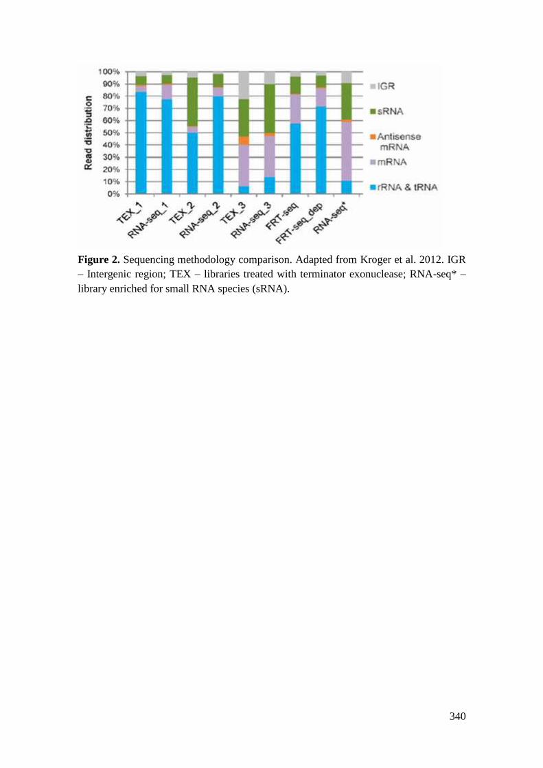
340
Figure 2. Sequencing methodology comparison. Adapted from Kroger et al. 2012. IGR– Intergenic region; TEX – libraries treated with terminator exonuclease; RNA-seq* –library enriched for small RNA species (sRNA).

341
Table 1. Sequencing statistics. Adapted from Kroger et al 2012.
LibrarySequencingtechnology
DescriptionTotal
number
Numberof reads
(notmapped)
Numberof reads(uniquel
ymapped)
Percentuniquelymappedreads[%]
Minimumfold
coverage#
TEX_1 454
dRNA-seqlibrary
biologicalreplicate 1
161,031
72,623 88,408 54.90 1.11
RNA-seq_1
454
RNA-seqlibrary
biologicalreplicate 1
248,993
83,030 165,963 66.65 2.03
TEX_2 454
dRNA-seqlibrary
biologicalreplicate 2
111,462 10,785 100,677 90.32 2.16
RNA-seq_2
454
RNA-seqlibrary
biologicalreplicate 2
93,337 38,577 54,760 58.67 0.61
TEX_3Illumina
GAII
dRNA-seqlibrary
biologicalreplicate 3
1,738,867
122,0581,211,42
669.67 20.99
RNA-seq_3
IlluminaGAII
RNA-seqlibrary
biologicalreplicate 3
2,148,563
136,8711,360,11
363.30 21.16
RNA-seq*
IlluminaHiSeq
RNA-seqlibrary
biologicalreplicate 4
3,750,797
164,6582,596,01
069.21 25.11
FRT-seq
IlluminaGAII
FRT-seqlibrary
biologicalreplicate 5
18,563,218
4,203,715
2,456,792
13.23 16.42
FRT-seq dep
IlluminaGAII
FRT-seqlibrary
biologicalreplicate 5
rRNAdepleted
24,585,564
9,652,397
4,093,744
16.65 27.77
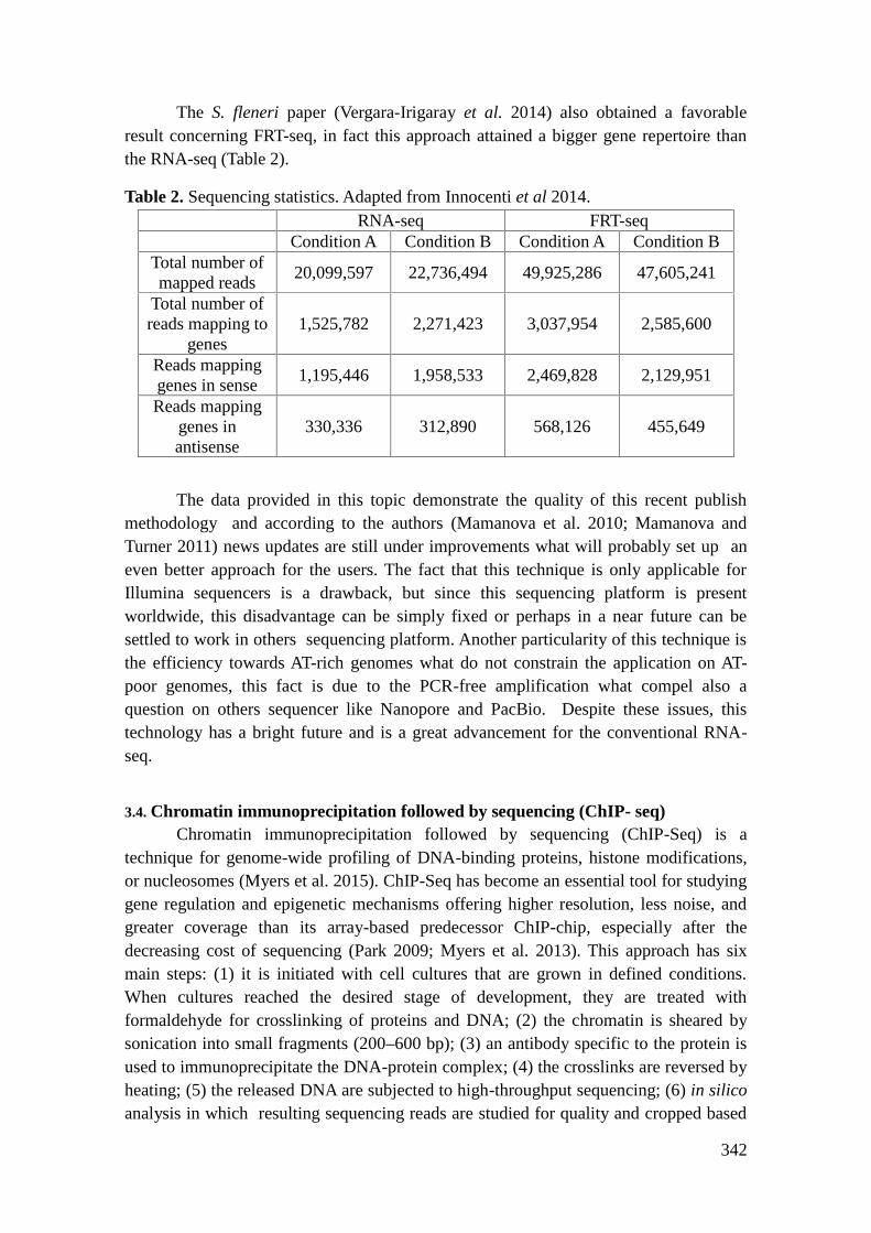
342
The S. fleneri paper (Vergara-Irigaray et al. 2014) also obtained a favorableresult concerning FRT-seq, in fact this approach attained a bigger gene repertoire thanthe RNA-seq (Table 2).
Table 2. Sequencing statistics. Adapted from Innocenti et al 2014.RNA-seq FRT-seq
Condition A Condition B Condition A Condition BTotal number ofmapped reads
20,099,597 22,736,494 49,925,286 47,605,241
Total number ofreads mapping to
genes1,525,782 2,271,423 3,037,954 2,585,600
Reads mappinggenes in sense
1,195,446 1,958,533 2,469,828 2,129,951
Reads mappinggenes inantisense
330,336 312,890 568,126 455,649
The data provided in this topic demonstrate the quality of this recent publishmethodology and according to the authors (Mamanova et al. 2010; Mamanova andTurner 2011) news updates are still under improvements what will probably set up aneven better approach for the users. The fact that this technique is only applicable forIllumina sequencers is a drawback, but since this sequencing platform is presentworldwide, this disadvantage can be simply fixed or perhaps in a near future can besettled to work in others sequencing platform. Another particularity of this technique isthe efficiency towards AT-rich genomes what do not constrain the application on AT-poor genomes, this fact is due to the PCR-free amplification what compel also aquestion on others sequencer like Nanopore and PacBio. Despite these issues, thistechnology has a bright future and is a great advancement for the conventional RNA-seq.
3.4. Chromatin immunoprecipitation followed by sequencing (ChIP- seq)Chromatin immunoprecipitation followed by sequencing (ChIP-Seq) is a
technique for genome-wide profiling of DNA-binding proteins, histone modifications,or nucleosomes (Myers et al. 2015). ChIP-Seq has become an essential tool for studyinggene regulation and epigenetic mechanisms offering higher resolution, less noise, andgreater coverage than its array-based predecessor ChIP-chip, especially after thedecreasing cost of sequencing (Park 2009; Myers et al. 2013). This approach has sixmain steps: (1) it is initiated with cell cultures that are grown in defined conditions.When cultures reached the desired stage of development, they are treated withformaldehyde for crosslinking of proteins and DNA; (2) the chromatin is sheared bysonication into small fragments (200–600 bp); (3) an antibody specific to the protein isused to immunoprecipitate the DNA-protein complex; (4) the crosslinks are reversed byheating; (5) the released DNA are subjected to high-throughput sequencing; (6) in silicoanalysis in which resulting sequencing reads are studied for quality and cropped based

343
on quality of the reads (Johnson et al. 2007; Robertson et al. 2007; Park 2009). Thecropped reads are then aligned to a reference genome. Afterward, areas of enrichment inthe ChIP-seq data is identified and represent where the TF binds throughout thegenome, usually called as peaks. CisGenome, MOSAiCs and MACS are some knownalgorithms that have been utilized for bacterial ChIP-seq analysis (Gentleman et al.2004; Park 2009). After peaks are associated with genes downstream, a number ofbioinformatics analyses can be carried out, including identification and analysis ofmotifs, differential analysis and association with expression data for deep understandingof bacterial regulon as shown in Figure 3 (Myers et al. 2015).
Figure 3. ChIP-seq sample preparation and analysis. Adapted from Myers et al. 2015.
As the whole-genome transcription profiling cannot reveal whether the influenceof the Transcription Factors (TF) on RNA levels is direct or indirect, this requiresidentification of Transcription Factors binding within the appropriate promoter region.The ChIP-seq provide information about where transcription factors are bind, thus, byintegrating ChIP methods and transcription profiling, it is possible to identify all direct

344
regulatory targets of a TF for a given condition. For example, work carried out byStringer and collaborates (2014) on araC gene of Escherichia coli, and Salmonellaenterica identified direct regulatory targets of AraC, including 5 novel target genes:ytfQ, ydeN, ydeM, ygeA, and polB (Stringer et al. 2014). Though ChIP-seq has beenused only in moderation to study bacterial systems in a few bacterial species, such as,Vibrio harveyi, V. cholerae, Rhodobacter sphaeroides, Mycobacterium tuberculosis, S.enterica, and Caulobacter crescentus (Kahramanoglou et al. 2011; Myers et al. 2013,2015; Singh et al. 2014; Haycocks et al. 2015) it is used for the identification of novelregulatory interactions, even for well-studied proteins (Grainger et al. 2007).
ChIP-seq could be an efficient tool in combination with RNA-seq to get thedetail information of bacterial transcription regulation and how bacteria respond todifferent external conditions.
3.5. RNA Immunoprecipitation Sequencing (RIP-seq)RNA Immunoprecipitation (RIP) is the study of intracellular RNA and protein
binding; it is a tool to understand the dynamic process of post-transcriptional regulatorynetworks. In this technique, an antibody against a protein of interest is used to recoverRNA species bound to the protein and since the sequence information of RNA speciesbound to a specific protein is often desired, an approach combining RNAimmunoprecipitation with sequencing technology (RIP-seq) was created (Chu andCorey 2012). The main challenges of RIP-seq is the cross linking step, which isrelatively inefficient and only small amount of RNA is available for the libraryconstruction (Hafner et al. 2010; Chu and Corey 2012). After that, treatment withendonuclease elucidates the specific binding sites within the RNA as they will beprotected from digestion, followed by the RNA protein complexes purification usingelectrophoresis and high throughput sequencing (Chi et al. 2009; Chu and Corey 2012),and finally the data obtained from the sequencer are analyzed using bioinformatics tool.The first work using the RIP-seq based technique was carried in Salmonella by Sittkaand collaborates (2008) (Sittka et al. 2008). In this analysis, they used RNA-bindingproperty of Hfq protein, as result many new sRNA were discover that were not reportedin previous studies (Cho et al. 2013). Thus, RIP-Seq could be an efficient tool for theidentification of bacterial non-coding RNAs.
3.6. LEA-seq (Low Error Amplicon Sequencing)The LEA-seq technique (Low Error Amplicon Sequecing) emerged in 2013 and
was developed and patented by Gordon and Faith (2014) (Gordon and Faith 2014). Thismethod was created in order to improve the quality and the depth of sequencing runs,since the mass production data produced by NGS has caused a high rate of errors in thesequencing, due to algorithms or platform reading length problems (Gordon and Faith2014).
LEA-seq is a nucleic acids sequencing technique that identifies events that occurat low frequency, seeking to understand mutation events. The three basic steps forimplementing this technique are: (1) linear PCR, (2) exponential PCR and (3)

345
sequencing. Whereas this technique is performed based on the bacterial 16S sequencing,in which PCR carries numerous times, and each PCR amplified uses specific primersfor each linear molecule (Gordon and Faith 2014).
The LEA-seq technique is a quantitative method and presents the advantages ofthe generation and reading, which permits the formation of a consensus and eliminatingerrors for each molecule. Currently, the available techniques do not enable errordetection in sequencing or whether there is a really variation in the sequence of thatmicroorganism. The multiple sequencing through the LEA-seq technique allows forbetter quality and precision about the organism.
The study of Faith and collaborates (2013) aimed to identify the composition ofthe fecal microbiota of adults, the understanding of the role of these bacterial speciesand theirs therapeutic potential for intestinal diseases. This technique allowed not onlywork with a large number of samples (over 500 isolates), as well as contribute to a fastand accurate analysis of the data (Faith et al. 2013).
Researchers have a continuous interest in improving this technique, since it canbe used for clinical investigation due to their high accuracy in cases, for example,patients with genetic mutations or somatic mutations again. LEA-seq can assist in thesearch and knowledge of the intestinal microbiota, as it may reveal its composition,opening up prospects for diagnosis, treatment and prevention of gastrointestinal tractdiseases.
3.7. CRISPR (Clustered Regularly Interspaced Short Palindromic Repeats)Ishino and collaborates (1987) described CRISPR for the first time (Ishino et al.
1987). This system has been identified in 40% of bacterial genomes so far (Kunin,Sorek and Hugenholtz 2007) and they are defined as short repetitions of grouped bases.Although, the determination of the CRISPR locus and the characterization of adjacentgenes, known as cas genes, responsible for the function of CRISPR, only happened in2002 (Jansen et al. 2002). The system CRISPR/Cas uses small non-coding RNAs inassociation with Cas proteins. The Cas9 is a nuclease, which cleaves DNA in theselected region, so that the CRISPR system/Cas9 can be used for editing genomes.
The activity of the CRISPR/Cas involves three main mechanisms: (1)acquisition, the step which the DNA fragment was inserted into the CRISPR locus in thegenome of interest of the study; (2) transcription, which the CRISPR locus will betranscribed and processed; (3) interference, which occur the ejection of nucleic acids;all those mechanisms contributes for bacterial persistence in the environment (Gasiunaset al. 2012; Jinek et al. 2012). Furthermore, the CRISPR provides mechanisms to limitthe spread of antibiotic resistance or virulence factors, but the study carried by Gophnaand collaborates (2015) demonstrates that even through there is different measurementsto evaluate the horizontal gene transfer is not possible to identify a correlation betweenthe CRISPR/Cas system and the evolution of the species, so that changes occur only atthe population level (Gophna and Ron 2003).
RNA-seq helped annotation transcription of regions, mainly non-coding, as wellas enables the identification of CRISPR elements in prokaryotes (Heidrich et al. 2015).

346
The CRISPR system can also be used as a tool in studies centred in gene regulation,since this system is able to activate or repress genes.
The working of Zoephel and Randau (2013) discusses how the structure ofCRISPR can affect the maturation of the RNA and thus influence the functionally of theCRISPR/Cas system (Zoephel and Randau 2013). The RNA-seq approach was used toevaluate differential gene expression in S. aureus, a pathogen of major importance, andwas capable to identify the CRISPR in these strains, and investigating their possiblerole, since these regions shows an adaptive response to infection (Osmundson, Dewelland Darst 2013). Thus, we have seen how important the use of RNA-seq approach tothe magnification of knowledge and function in prokaryotes.
4. RNA sequencing platforms
The RNA-seq approach can be applied in different next-generation sequencingplatforms and the results obtained by then are proportional to the machine capability. InTable 3 a comparison are being made with some of the most currently employedplatforms (Li et al. 2014).
Table 3. Different Next Generations sequencing platforms in the study of RNA-seq.Adopted and modified from Li et al. 2014.
CompanyName Instrument Version
RunTime
(Hours)
ReadLengths(Mean)
ReadsPer Run
(Millions)Applications
Illumina HiSeq 2000High
Output132 50 6,000
Gene expression,Splice junction
detection, variantcalling, fusion
Illumina HiSeq 2500High
Output132 50 6,000
Gene expression,Splice junction
detection, variantcalling, fusion
Illumina MiSeq v2 kit 39 250 30Splice junction
detection, variantcalling,
LifeTechnologi
esPGM 318 Chip 7.3 176 6
Splice junctiondetection, variant
callingLife
Technologies
ProtonProton I
chip2-4 81 70
Gene expression,Splice junction
detection, variantcalling
PacificBiosciences
RS RS 0.5-2 1,289 0.03
Splice junctiondetection, variant
calling, full-lengthgene coverage
Roche 454GS
FLX+20 686 1
Splice junctiondetection

347
5. Bioinformatic analysis
Experimental investigations in prokaryotes has been facilitated, extended andcomplemented by computational approaches (Backofen et al. 2014). Huge amounts ofdata has been generated by RNA-seq experiments and needs to be stored and analysedusing computational techniques and tools (McGettigan 2013). This amount has becomea bottleneck to bioinformatic analysis and to biologists, since today's transcriptomeanalysis consist of experiments and data evaluation (Backofen et al. 2014). Extractbiological information from RNA-seq datasets requires bioinformatic knowledge andtools, making the software choice an important issue to successful RNA-seq analysis(Pinto et al. 2011; Backofen et al. 2014).
According to Chierico and collaborates (2015) (Del Chierico et al. 2015) andPinto and collaborates (2011) (Pinto et al. 2011) RNA-seq process can be understood asa five-step process: (1) isolation of total RNA of the organism; (2) mRNA enrichment;(3) synthesis of cDNA; (4) NGS sequencing, which returns raw data; (5) bioinformaticanalysis (Pinto et al. 2011). A flowchart of this process can be seen in Figure 4.
Figure 4. RNA-seq five-step process.
This session focuses on bioinformatic analysis and available computationaltools. Based on literature review (Pinto et al. 2011; McClure et al. 2013b; Backofen etal. 2014 ; Del Chierico et al. 2015 ; Tjaden 2015), bioinformatic analysis can becomprehended as the extraction of biological information from the sequencing of rawdata and classified/divided (Figure 5).
Figure 5. Bioinformatic analysis workflow.
5.1. Bioinformatic workflow
Quality check step aims to increase the accuracy of the results by removingsequences that can contain errors (de Sá et al. 2015), trimming sequences introduced inthe library preparation step, as adapters and poly(A)-tails (Forstner, Vogel and Sharma2014), and removing reads with low phred quality . However, regarding that, the usageof poor-quality bases can lead to less precise results (Ramos et al. 2011), andconsidering this, quality check can affect drastically the next steps.
Some RNA-Seq pipelines implements quality checking, like ReaDemption(Forstner, Vogel and Sharma 2014), which performs quality trimming, removes adapters

348
and poly(A) tails, and discards reads shorter than a given cut-off (the default cut-off is12 nucleotides (nt)). Quality Assessment software (Ramos et al. 2011) evaluates qualitybased on quality-graph analysis and estimated coverage. According to Backofen andcollaborates (2014) (Backofen et al. 2014), FastQC(http://www.bioinformatics.babraham. ac.uk/projects/fastqc/) is a commonly used toolto check reads quality determining the quality profile of the reads. Software suites canbe also used to this purpose, FASTX-Toolkit (http://hannonlab.cshl.edu/fastx_toolkit/)provides tools to remove sequences attached in previous steps and other pre-processingstrategies of raw data.
After quality check, if there is a reference genome mapping step will be done,otherwise de novo assembly. Mapping consists of producing the transcriptome mapthough aligning reads to a reference genome (Pinto et al. 2011) aiming to detect thewright position of reads and distinguish between sequencing errors and geneticvariations (Caboche et al. 2014). Lots of mapping softwares have been releaseddiffering in their algorithms, memory management, velocity and computational costback (Backofen et al. 2014) making the choice of a mapping tool a challenge. McClureand collaborates (2013) (McClure et al. 2013b) made a comparison between SOAP2,BWA, Bowtie and Bowtie2 aligners using 75 RNA-seq experiment data, and thecomparison of mapping algorithms applied to IonTorrent data can be seen in (Cabocheet al. 2014). After mapping quality can be evaluated, ReadXplorer software offersquality classification of read mapping to provide information about quality and quantityto each single read mapping (Hilker et al. 2014). This approach is recommended when ahigh-quality genome is available as reference, and if it is unavailable, transcripts shouldbe assembled de novo (Tjaden 2015).
De novo assembly can be used when investigating poorly studied organisms(Haas et al. 2012), complex microbial communities or uncultivable organisms (Tjaden2015). Both DNA and RNA must be assembled, but transcriptome assembly issignificantly different than genome assembly (Haas et al. 2013), thus it is important touse RNA assemblers. Tjaden (2015) (Tjaden 2015) affirms that assemblers should bespecifically designed to prokaryotes, owing to the different challenges eukaryotic andprokaryotic transcriptome faces. Bacterial genomes are often denser than eukaryotic,considering the proximity of the genes, neighbouring bacterial transcripts can overlapmaking it difficult to identify appropriately transcript boundaries and non-codingeukaryotic RNAs models are not appropriate to detect bacterial small regulatory RNAs(Tjaden 2015). An assembly comparison of three different softwares (Trinity,SOAPdenovo2 and Rockhooper 2) using data from 9 different bacteria can be seen in(Tjaden 2015).
When reference mapping or de novo assembly is done, data can be analysedstructurally and differentially. Differential analyses main purpose is to determine thedifference expression between different growth conditions or treatments(Seyednasrollah, Laiho and Elo 2013). Several softwares have been released for thispurpose, but there is not a consensus about best practices, what makes difficult tochoose a tool or method to use. Aiming to help in this issue Seyednasrollah andcollaborates (2013) (Seyednasrollah, Laiho and Elo 2013) made a comparison among

349
eight differential expression software packages using two real datasets publiclyavailable. Softwares that analyses differential expression can be based on Poissonmethod (DEGseq and Myrna), negative binomial method (edgeR and DEseq) or in othermethods (Pinto et al. 2011; Seyednasrollah, Laiho and Elo 2013). Pinto and collaborates(2011) (Pinto et al. 2011) recommends using DEseq or edgeR when analysingreplicates.
Transcriptome annotation and classification can be accentuated in structuralanalysis, evaluating transcripts regarding the genomic region they have been associatedto and classified in: protein-coding, non-coding and intergenic regions (Backofen et al.2014). Aiming to predict ncRNA transcripts several computational methods has beendeveloped, Herbig and Nieselt (2011) (Herbig and Nieselt 2011) highlights SIPHT,sRNAFinder, sRNAscanner, NOCORNAr and sRNAPredict softwares. NOCORNArdistinguish itself by prediction and characterization of ncRNAs in bacteria (Herbig andNieselt 2011).
Assess transcripts concerning genomic regions relies on transcript annotation.Computational approach is convenient to be used due its velocity and precisioncompared to manual annotation; however, human supervision of the results isconsidered important to avoid false-positives or missing features (Creecy and Conway2015). In this technique some main structures must be detected: 5' transcript ends, 3'transcript ends, TSS and operon (Backofen et al. 2014; Creecy and Conway 2015).
a. Transcript boundaries identificationAnnotation of transcript boundaries is important to operon identification and
regulatory analyses (Creecy and Conway 2015). Identifying 5' UTR is not alwayspossible, a significant number of transcripts lacking 5' UTR were funded in bacteria andcalled leaderless transcripts, in this, transcripts translation start site and transcriptionstart site remains in almost the same position (Backofen et al. 2014). Annotation of 3'UTR is important to get the whole analytical value of RNA-seq data, Creecy andConway (2014) (Creecy and Conway 2015) affirm that current best 3' ends detectionmethod is to search correlation between replicates data, highlighting that, the softwarepackage TransTermHP can find intrinsic terminators successfully.
b. TSS identificationTSS annotation can assist ncRNA annotation and polycistronic transcripts
(Backofen et al. 2014); according to Creecy and Conway (2013) (Creecy and Conway2015), it is essential to discover unknown transcripts and to analyse operon, 5' UTR andpromoters architecture. Although, there is not a well-established strategies to TSSidentification, owing scarce knowledge about transcription start sites in bacteria Due tocomputational developments in both computational analyses and “wet-lab”experiments, TSS annotation becomes feasible (Backofen et al. 2014). TSSAR is adRNA-seq data-based tool for rapid annotation of TSS that considers dRNA-seq librarystatistics (Amman et al. 2014), and according to Backofen and collaborates (2014)(Backofen et al. 2014), the main advantage remains in statistical analysis presented asan easy-to-use web service. TSSpredator tool presents an automated TSS detection and

350
classification from RNA-seq data performing a genome wide comparative prediction ofTSS (Dugar et al. 2013. A comparison among manual annotation, TSSpredator andTSSAR annotation can be seen in (Amman et al. 2014).
c. Operon identificationThe operon represents clusters of co-transcribed genes regulated by the same
regulatory sequence and co-transcribed in a single mRNA. This structure has a hugebiological importance improving functional gene annotation and giving importantinformation to studies in drug targeting, functional analyses and antibiotic resistance(Chuang et al. 2012). To handle operon occurrence complexity, it should be detected byoperon architecture (i.e. 5' ends and 3' ends) and have a sufficient read coverage toconnect promoters and terminators. A strong indication that an operon is real is if atleast 90% bases of the reads is covered (Creecy and Conway 2015). Chuang andcollaborates (2012) (Chuang et al. 2012) classify computational methods to predictoperons and evaluates 15 algorithms in respect to accuracy, specificity and sensitivity.
5.2. RNA-seq Pipeline tools
Not all pipeline tools contemplate hole RNA-seq workflow described earlier, tohelp in the tool choice a software functionalities comparison were developed and can beseen in Table 4. To additional support, important issues about each softwares aredescribed forward.
Table 4. Software comparison.
ToolQualityCheck
MappingDe novoassembly
Differentialanalyses
Rockhopper(McClure et al.
2013)- x x x
Rockhopper 2(Tjaden B. 2015)
- - x x
RNA-Rocket(Warren et al.
2015)x x - x
READemption(Förstner, Vogel,
Sharma 2014)x x - x
ReadXplorer(Hilker et al.
2014)x - - x
Rockhopper is a system designed specifically to bacterial transcriptome RNA-seq data analyses. A novel approach to map transcripts is implemented in this software(similar to Bowtie2 approach). Mapping normalization is performed followed bytranscripts assembly, identification of transcript boundaries, quantification oftranscript abundance, testing for differential gene expression and operon prediction.

351
Analysis results are presented using Integrative Genome Viewer, allowing differentexperiments to be viewed simultaneously (McClure et al. 2013b).
Rockhopper 2 is a comprehensive system focused in de novo assembly thatsupports differential analyses and transcripts abundance quantification, according toTjaden (2015) (Tjaden 2015) it does not require high-performance computers and canrun on personal computers. Rockhopper 2 implements a novel de novo assemblyalgorithm to bacterial transcriptomes, the algorithm works in two stages: (1) candidatetranscripts are assembled using a found k-mer; (2) sequencing reads are mapped tocandidate transcripts aiming to filter candidate transcripts to high-quality finaltranscripts. Concerning differential analyses, Rockhopper 2 first normalize each RNA-seq dataset enabling to compare different experiments or samples (Tjaden 2015).
RNA-Rocket aims to simplify the process of aligning RNA-seq data to areference genome and generate quantitative transcript profiles. It is built on Galaxy toprovide the necessary tools and services to process RNA-seq data, some benefits of itsusage are the possibility of sharing results across research groups, the support of batchanalyses for multiple samples and the integration of tools and projects integrating datafrom the PATRIC platform (Warren et al. 2015).
READemption pipeline aims to integrate individual RNA-seq analyses tasks andputting it into a friendly tool with a command line interface, this tool was primarilydeveloped to analyse bacterial transcriptome. In order to use full modern computerscapacity and reduces run time READemption offers parallel data processing. Firstperforms quality polyA and adapters trimming followed by mapping, coveragecalculation, gene expression quantification, differential gene expression analyses andplotting. The software is able to analyse RNA-seq data from Illumina and 454platforms.
ReadXplorer offers straightforward visualizations and analysis functions builtaround its unique read mapping classification. Analysis as TSS and operon detection,differential expression, RPKM value and read count calculations are available inReadXplorer and can be exported to Microsoft Excel files. Read mapping classificationsorts read mappings into three different classes: perfect match, best match and commonmatch. This classification is incorporated in all analyses functions.
5.3. Bioinformatic ChallengesThrough bibliographic research (Dai et al. 2012; Mc Clure et al. 2013b;
McGetttigan 2013; Van Verk et al. 2013; Forstner, Vogel and Sharma 2014; Tjaden2015), it has been concluded that bioinformatics has many challenges related tocomputational issues. RNA-seq experiments generate large amounts of data that must becomputationally processed, analyzed, stored and retrieved using a great deal ofcomputational power. In addition to the computational issues, it is important to take intoaccount that not all bioinformatic researchers have extensive computational experience:this makes the lack of user-friendly tools a problem for some users and an import issuefor developers. However, great computers, excellent bioinformatic researchers and userfriendly tools do not guarantee successful analysis. The software selected must beappropriate to each biological question and to the organisms studied. Even with all

352
questions presented here, RNA-seq analysis has been very successful in recent years.This success can lead us to imagine the wonderful possibilities for RNA-seqbioinformatic analyses in the future.
References1. Creecy JP, Conway T. Quantitative bacterial transcriptomics with RNA-seq. Curr
Opin Microbiol 2015;23:133–40.2. Margulies M, Egholm M, Altman WE, Attiya S, Bader JS, Bemben LA, et al.
Genome sequencing in microfabricated high-density picolitre reactors. Nature 2005Sep 15;437(7057):376–80.
3. Perkins TT, Kingsley RA, Fookes MC, Gardner PP, James KD, Yu L, et al. A strandspecific RNA-Seq analysis of the transcriptome of the typhoid bacillus Salmonellatyphi. PLoS Genet 2009 Jul;5(7):e1000569.
4. Passalacqua KD, Varadarajan A, Ondov BD, Okou DT, Zwick ME, Bergman NH.Structure and complexity of a bacterial transcriptome. J Bacteriol 2009 May15;191(10):3203–11.
5. Pinto AC, Sá PHCG de, Ramos RTJ, Barbosa S, Barbosa HPM, Ribeiro AC, et al.Differential transcriptional profile of Corynebacterium pseudotuberculosis inresponse to abiotic stresses. BMC Genomics 2014 Jan 9;15(1):14.
6. Westermann AJ, Gorski, SA and Vogel J. Dual RNA-seq of pathogen and host. NatRev Microbiol 2012;10:618–30.
7. Macklaim JM, Fernandes AD, Bella JMD, Hammond J-A, Reid G, Gloor GB.Comparative meta-RNA-seq of the vaginal microbiota and differential expressionby Lactobacillus iners in health and dysbiosis. Microbiome 2013 Apr 12;1(1):12.
8. Leimena MM, Ramiro-Garcia J, Davids M, van den Bogert B, Smidt H, Smid EJ, etal. A comprehensive metatranscriptome analysis pipeline and its validation usinghuman small intestine microbiota datasets. BMC Genomics 2013;14:530.
9. Bisanz JE, Macklaim JM, Gloor GB, Reid G. Bacterial metatranscriptome analysisof a probiotic yogurt using an RNA-Seq approach. Int Dairy J 2014;39(2):284–92.
10. Wiegand S, Dietrich S, Hertel R, Bongaerts J, Evers S, Volland S, et al. RNA-Seqof Bacillus licheniformis: active regulatory RNA features expressed within aproductive fermentation. BMC Genomics 2013;14(1):667.
11. Wang Z, Yang S-T. Propionic acid production in glycerol/glucose co-fermentationby Propionibacterium freudenreichii subsp. shermanii. Bioresour Technol2013;137:116–23.
12. Sorek R, Cossart P. Prokaryotic transcriptomics: a new view on regulation,physiology and pathogenicity. Nat Rev Genet 2010 Jan;11(1):9–16.
13. Sharma CM, Hoffmann S, Darfeuille F, Reignier J, Findeiß S, Sittka A, et al. Theprimary transcriptome of the major human pathogen Helicobacter pylori. Nature.2010 Mar 11;464(7286):250–5.
14. Haas BJ, Chin M, Nusbaum C, Birren BW, Livny J. How deep is deep enough forRNA-Seq profiling of bacterial transcriptomes? BMC Genomics 2012 Dec 27;13(1):734.

353
15. Bischler T, Siew Tan H, Nieselt K, Sharma CM. Differential RNA-seq (dRNA-seq)for annotation of transcriptional start sites and small RNAs in Helicobacter pylori.Methods [Internet]. 2015 Jul 6 [cited 2015 Jul 6]; Available from:http://www.sciencedirect.com/science/article/pii/S1046202315002546
16. Kratz A, Carninci P. The devil in the details of RNA-seq. Nat Biotechnol 2014Sep;32(9):882–4.
17. Sendler E, Johnson GD, Krawetz SA. Local and global factors affecting RNAsequencing analysis. Anal Biochem 2011;419(2):317–22.
18. Liu Y, Zhou J, White KP. RNA-seq differential expression studies: more sequenceor more replication? Bioinformatics 2014 Feb 1;30(3):301–4.
19. Isabella VM, Clark VL. Deep sequencing-based analysis of the anaerobic stimulonin Neisseria gonorrhoeae. BMC Genomics 2011 Jan 20;12(1):51.
20. Patenge N, Pappesch R, Khani A, Kreikemeyer B. Genome-wide analyses of smallnon-coding RNAs in streptococci. Front Genet 2015;6:189.
21. Gottesman S, Storz G. Bacterial small RNA regulators: versatile roles and rapidlyevolving variations. Cold Spring Harb Perspect Biol [Internet]. 2011 Dec [cited2015 Jul 2];3(12). Available from:http://www.ncbi.nlm.nih.gov/pmc/articles/PMC3225950/
22. Papenfort K, Vogel J. Regulatory RNA in bacterial pathogens. Cell Host Microbe2010 Jul 22;8(1):116–27.
23. Kröger C, Dillon SC, Cameron ADS, Papenfort K, Sivasankaran SK, Hokamp K, etal. The transcriptional landscape and small RNAs of Salmonella enterica serovarTyphimurium. Proc Natl Acad Sci 2012 May 15;109(20):E1277–86.
24. Yan Y, Su S, Meng X, Ji X, Qu Y, Liu Z, et al. Determination of sRNA expressionsby RNA-seq in Yersinia pestis grown in vitro and during infection. PLoS ONE 2013Sep 11;8(9):e74495.
25. Storz G, Vogel J, Wassarman KM. Regulation by small RNAs in bacteria:expanding frontiers. Mol Cell 2011 Sep 16;43(6):880–91.
26. Van Dijk EL, Auger H, Jaszczyszyn Y, Thermes C. Ten years of next-generationsequencing technology. Trends Genet 2014;30(9):418–26.
27. Humphrys MS, Creasy T, Sun Y, Shetty AC, Chibucos MC, Drabek EF, et al.Simultaneous transcriptional profiling of bacteria and their host cells. PLoS ONE2013 Dec 4;8(12):e80597.
28. Martin JA, Wang Z. Next-generation transcriptome assembly. Nat Rev Genet 2011Sep 7;12(10):671–82.
29. Tjaden B. De novo assembly of bacterial transcriptomes from RNA-seq data.Genome Biol 2015;16(1):1.
30. Pinto AC, Ramos RTJ, Silva WM, Rocha FS, Barbosa S, Miyoshi A, et al. The corestimulon of Corynebacterium pseudotuberculosis strain 1002 identified using abinitio methodologies. Integr Biol 2012;4(7):789.
31. Innocenti N, Golumbeanu M, d’ Hérouël AF, Lacoux C, Bonnin RA, Kennedy SP,et al. Whole genome mapping of 5’RNA ends in bacteria by tagged sequencing: acomprehensive view in Enterococcus faecalis. ArXiv Prepr ArXiv14101925[Internet]. 2014 [cited 2014 Dec 15]; Available from: http://arxiv.org/abs/1410.1925

354
32. Fouquier d’Herouel A, Wessner F, Halpern D, Ly-Vu J, Kennedy SP, Serror P, et al.A simple and efficient method to search for selected primary transcripts: non-codingand antisense RNAs in the human pathogen Enterococcus faecalis. Nucleic AcidsRes 2011 Apr 1;39(7):e46–e46.
33. Mamanova L, Turner DJ. Low-bias, strand-specific transcriptome Illuminasequencing by on-flowcell reverse transcription (FRT-seq). Nat Protoc 2011Nov;6(11):1736–47.
34. Mamanova L, Andrews RM, James KD, Sheridan EM, Ellis PD, Langford CF, et al.FRT-seq: amplification-free, strand-specific transcriptome sequencing. Nat Methods2010 Feb;7(2):130–2.
35. Vergara-Irigaray M, Fookes MC, Thomson NR, Tang CM. RNA-seq analysis of theinfluence of anaerobiosis and FNR on Shigella flexneri. BMC Genomics 2014 Jun6;15(1):438.
36. Myers KS, Park DM, Beauchene NA, Kiley PJ. Defining bacterial regulons usingChIP-seq methods. Methods [Internet]. 2015 [cited 2015 Jul 17]; Available from:http://www.sciencedirect.com/science/article/pii/S1046202315002285
37. Myers KS, Yan H, Ong IM, Chung D, Liang K, Tran F, et al. Genome-scaleanalysis of Escherichia coli FNR reveals complex features of transcription factorbinding. PLoS Genet 2013;9(6):e1003565.
38. Park PJ. ChIP–seq: advantages and challenges of a maturing technology. Nat VerGenet 2009;10(10):669–80.
39. Johnson DS, Mortazavi A, Myers RM, Wold B. Genome-wide mapping of in vivoprotein-DNA interactions. Science 2007;316(5830):1497–502.
40. Robertson G, Hirst M, Bainbridge M, Bilenky M, Zhao Y, Zeng T, et al. Genome-wide profiles of STAT1 DNA association using chromatin immunoprecipitation andmassively parallel sequencing. Nat Methods 2007;4(8):651–7.
41. Gentleman RC, Carey VJ, Bates DM, Bolstad B, Dettling M, Dudoit S, et al.Bioconductor: open software development for computational biology andbioinformatics. Genome Biol 2004;5(10):R80.
42. Stringer AM, Currenti S, Bonocora RP, Baranowski C, Petrone BL, Palumbo MJ, etal. Genome-scale analyses of Escherichia coli and Salmonella enterica AraC revealnoncanonical targets and an expanded core regulon. J Bacteriol 2014;196(3):660–71.
43. Haycocks JRJ, Sharma P, Stringer AM, Wade JT, Grainger DC. The molecular basisfor control of ETEC enterotoxin expression in response to environment and host.PLoS Pathog 2015 Jan 8;11(1):e1004605.
44. Singh SS, Singh N, Bonocora RP, Fitzgerald DM, Wade JT, Grainger DC.Widespread suppression of intragenic transcription initiation by H-NS. Genes Dev2014 Feb 1;28(3):214–9.
45. Kahramanoglou C, Seshasayee ASN, Prieto AI, Ibberson D, Schmidt S,Zimmermann J, et al. Direct and indirect effects of H-NS and Fis on global geneexpression control in Escherichia coli. Nucleic Acids Res 2011 Mar 1;39(6):2073–91.

355
46. Wade JT, Struhl K, Busby SJ, Grainger DC. Genomic analysis of protein–DNAinteractions in bacteria: insights into transcription and chromosome organization.Mol Microbiol 2007;65(1):21–6.
47. Grainger DC, Aiba H, Hurd D, Browning DF, Busby SJW. Transcription factordistribution in Escherichia coli: studies with FNR protein. Nucleic Acids Res 2007Jan 1;35(1):269–78.
48. Chu Y, Corey DR. RNA sequencing: platform selection, experimental design, anddata interpretation. Nucleic Acid Ther 2012;22(4):271–4.
49. Hafner M, Landthaler M, Burger L, Khorshid M, Hausser J, Berninger P, et al.Transcriptome-wide identification of RNA-binding protein and microRNA targetsites by PAR-CLIP. Cell 2010;141(1):129–41.
50. Chi SW, Zang JB, Mele A, Darnell RB. Argonaute HITS-CLIP decodesmicroRNA–mRNA interaction maps. Nature 2009;460(7254):479–86.
51. Sittka A, Lucchini S, Papenfort K, Sharma CM, Rolle K, Binnewies TT, et al. Deepsequencing analysis of small noncoding RNA and mRNA targets of the globalposttranscriptional regulator, Hfq. PLoS Genet 2008;4(8):e1000163.
52. Cho S, Cho Y, Lee S, Kim J, Yum H, Kim SC, et al. Current challenges in bacterialtranscriptomics. Genomics Inform 2013;11(2):76.
53. Gordon JI, Faith JJ. Methods of low error amplicon sequencing (LEA-Seq) and theuse thereof [Internet]. Google Patents; 2014 [cited 2015 Jul 14]. Available from:https://www.google.com/patents/US20140357499
54. Faith JJ, Guruge JL, Charbonneau M, Subramanian S, Seedorf H, Goodman AL, etal. The long-term stability of the human gut microbiota. Science2013;341(6141):1237439.
55. Ishino Y, Shinagawa H, Makino K, Amemura M, Nakata A. Nucleotide sequence ofthe iap gene, responsible for alkaline phosphatase isozyme conversion inEscherichia coli, and identification of the gene product. J Bacteriol1987;169(12):5429–33.
56. Kunin V, Sorek R, Hugenholtz P. Evolutionary conservation of sequence andsecondary structures in CRISPR repeats. Genome Biol 2007;8(4):R61.
57. Jansen R, Embden J, Gaastra W, Schouls L, others. Identification of genes that areassociated with DNA repeats in prokaryotes. Mol Microbiol 2002;43(6):1565–75.
58. Gasiunas G, Barrangou R, Horvath P, Siksnys V. Cas9–crRNA ribonucleoproteincomplex mediates specific DNA cleavage for adaptive immunity in bacteria. ProcNatl Acad Sci 2012;109(39):E2579–86.
59. Jinek M, Chylinski K, Fonfara I, Hauer M, Doudna JA, Charpentier E. Aprogrammable dual-RNA–guided DNA endonuclease in adaptive bacterialimmunity. Science 2012;337(6096):816–21.
60. Gophna U, Ron EZ. Virulence and the heat shock response. Int J MedMicrobiolIJMM 2003 Feb;292(7-8):453–61.
61. Heidrich N, Dugar G, Vogel J, Sharma CM. Investigating CRISPR RNA biogenesisand function using RNA-seq. CRISPR Methods Protoc 2015;1–21.
62. Zoephel J, Randau L. RNA-Seq analyses reveal CRISPR RNA processing andregulation patterns. Biochem Soc Trans 2013 Dec;41(6):1459–63.

356
63. Osmundson J, Dewell S, Darst SA. RNA-Seq reveals differential gene expression inStaphylococcus aureus with single-nucleotide resolution. PLoS ONE 2013 Oct7;8(10):e76572.
64. Li S, Tighe SW, Nicolet CM, Grove D, Levy S, Farmerie W, et al. Multi-platformassessment of transcriptome profiling using RNA-seq in the ABRF next-generationsequencing study. Nat Biotechnol 2014 Aug 24;32(9):915–25.
65. Backofen R, Amman F, Costa F, Findei S, Richter AS, Stadler PF. Bioinformaticsof prokaryotic RNAs. RNA Biol 2014;11(5):470–83.
66. McGettigan PA. Transcriptomics in the RNA-seq era. Curr Opin Chem Biol 2013Feb; 17(1):4–11.
67. Pinto AC, Melo-Barbosa HP, Miyoshi A, Silva A, Azevedo V. Review applicationof RNA-seq to reveal the transcript profile in bacteria. Genet Mol Res2011;10(3):1707–18.
68. Del Chierico F, Ancora M, Marcacci M, Camma C, Putignani L, Conti S. Choice ofnext-generation sequencing pipelines. Bacterial Pangenomics [Internet]. Springer;2015 [cited 2015 Jul 14]. p. 31–47. Available from:http://link.springer.com/protocol/10.1007/978-1-4939-1720-4_3
69. McClure R, Balasubramanian D, Sun Y, Bobrovskyy M, Sumby P, Genco CA, et al.Computational analysis of bacterial RNA-Seq data. Nucleic Acids Res 2013 Aug1;41(14):e140–e140.
70. De Sá PH, Veras AA, Carneiro AR, Pinheiro KC, Pinto AC, Soares SC, et al. Theimpact of quality filter for RNA-Seq. Gene 2015;563(2):165–71.
71. Förstner KU, Vogel J, Sharma CM. READemption–A tool for the computationalanalysis of deep-sequencing-based transcriptome data. Bioinformatics 2014;btu533.
72. Ramos RT, Carneiro AR, Baumbach J, Azevedo V, Schneider MP, Silva A.Analysis of quality raw data of second generation sequencers with QualityAssessment Software. BMC Res Notes 2011 Apr 18;4(1):130.
73. Caboche S, Audebert C, Lemoine Y, Hot D. Comparison of mapping algorithmsused in high-throughput sequencing: application to Ion Torrent data. BMCGenomics 2014;15(1):264.
74. Hilker R, Stadermann KB, Doppmeier D, Kalinowski J, Stoye J, Straube J, et al.Read‐Xplorer—visualization and analysis of mapped sequences. Bioinformatics2014;btu205.
75. Haas BJ, Papanicolaou A, Yassour M, Grabherr M, Blood PD, Bowden J, et al. Denovo transcript sequence reconstruction from RNA-seq using the Trinity platformfor reference generation and analysis. Nat Protoc 2013;8(8):1494–512.
76. Seyednasrollah F, Laiho A, Elo LL. Comparison of software packages for detectingdifferential expression in RNA-seq studies. Brief Bioinform [Internet]. 2013 Dec 2[cited 2014 Apr 30]; Available from:http://bib.oxfordjournals.org/cgi/doi/10.1093/bib/bbt086
77. Herbig A, Nieselt K. nocoRNAc: characterization of non-coding RNAs inprokaryotes. BMC Bioinformatics 2011;12(1):40.
78. Amman F, Wolfinger MT, Lorenz R, Hofacker IL, Stadler PF, Findei S. TSSAR:TSS annotation regime for dRNA-seq data. BMC Bioinformatics 2014;15(1):89.

357
79. Dugar G, Herbig A, Förstner KU, Heidrich N, Reinhardt R, Nieselt K, et al. High-resolution transcriptome maps reveal strain-specific regulatory features of multipleCampylobacter jejuni isolates. 2013 [cited 2015 Jul 14]; Available from:http://dx.plos.org/10.1371/journal.pgen.1003495
80. Chuang L-Y, Chang H-W, Tsai J-H, Yang C-H. Features for computational operonprediction in prokaryotes. Brief Funct Genomics 2012;els024.
81. Warren AS, Aurrecoechea C, Brunk B, Desai P, Emrich S, Giraldo-Calderón GI, etal. RNA-Rocket: an RNA-Seq analysis resource for infectious disease research.Bioinformatics 2015;btv002.
82. Van Verk MC, Hickman R, Pieterse CM, Van Wees SC. RNA-Seq: revelation of themessengers. Trends Plant Sci 2013;18(4):175–9.
83. Dai L, Gao X, Guo Y, Xiao J, Zhang Z, others. Bioinformatics clouds for big datamanipulation. Biol Direct 2012;7(1):43.

358
Anexo 3: Artigo: Value of a newly sequenced bacterial genome
Annexe 3 : Article : Value of a newly sequenced bacterial genome
WJBC - World Journal of Biological Chemistry
Submit a Manuscript: http://www.wjgnet.com/esps/ World J Biol Chem 2014 May 26;
5(2): 161-168Help Desk: http://www.wjgnet.com/esps/helpdesk.aspx ISSN 1949-8454
(online)
DOI: 10.4331/wjbc.v5.i2.161
© 2014 Baishideng Publishing Group Inc. All rights reserved.
REVIEW
Value of a newly sequenced bacterial genome
Eudes GV Barbosa, Flavia F Aburjaile, Rommel TJ Ramos, Adriana R Carneiro, Yves
Le Loir, Jan Baumbach, Anderson Miyoshi, Artur Silva, Vasco Azevedo
Eudes GV Barbosa, Flavia F Aburjaile, Anderson Miyoshi, Vasco Azevedo, Laboratório
de Genética Celular e Molecular, Instituto de Ciências Biológicas, Universidade Federal
de Minas Gerais, Belo Horizonte, 31270-901 MG, Brazil
Eudes GV Barbosa, Jan Baumbach, Department of Mathematics and Computer
Science, University of Southern Denmark, 5230 Odense, Denmark
Flavia F Aburjaile, Yves Le Loir, INRA, UMR1253, Science et Technologie du Lait et
de l’OEuf, F-35042 - Rennes, France
Rommel TJ Ramos, Adriana R Carneiro, Artur Silva, Laboratório de Polimorfismo de
DNA, Instituto de Ciências Biológicas, Univeridade Federal do Pará, Belém 66075-110,
Brazil
Author contributions: All authors contributed extensively to the work presented in this
review.

359
Supported by Coordenação de Aperfeiçoamento de Pessoal de Nível Superior (CAPES)
in Brazil, processes BEX 12954-12-8 and 11517-12-3, to Barbosa EGV and Aburjaile
FF
Correspondence to: Vasco Azevedo, MD, PhD, Laboratório de Genética Celular e
Molecular, Instituto de Ciências Biológicas, Univeridade Federal de Minas Gerais, Av.
Antônio Carlos 6627 Pampulha, Belo Horizonte 31270-901, Brazil.
Telephone: +55-31-34092873 Fax: +55-31-34092610
Received: December 11, 2013 Revised: January 14, 2014
Accepted: April 3, 2014
Published online: May 26, 2014
Abstract
Next-generation sequencing (NGS) technologies have made high-throughput
sequencing available to mediumand small-size laboratories, culminating in a tidal wave
of genomic information. The quantity of sequenced bacterial genomes has not only
brought excitement to the field of genomics but also heightened expectations that NGS
would boost antibacterial discovery and vaccine development. Although many possible
drug and vaccine targets have been discovered, the success rate of genome-based
analysis has remained below expectations. Furthermore, NGS has had consequences for
genome quality, resulting in an exponential increase in draft (partial data) genome
deposits in public databases. If no further interests are expressed for a particular
bacterial genome, it is more likely that the sequencing of its genome will be limited to a
draft stage, and the painstaking tasks of completing the sequencing of its genome and
annotation will not be undertaken. It is important to know what is lost when we settle
for a draft genome and to determine the “scientific value” of a newly sequenced
genome. This review addresses the expected impact of newly sequenced genomes on
antibacterial discovery and vaccinology. Also, it discusses the factors that could be
leading to the increase in the number of draft deposits and the consequent loss of
relevant biological information.
Keywords: Next-generation sequencing; Drafts; Prokaryotic genomes; Computational
tools; Omics.
360
Core tip: Next-generation sequencing (NGS) technologies have made high-throughput
sequencing available to medium-and small-size laboratories, culminating in a tidal wave
of genomic information. The quantity of bacterial genomes has not only brought
excitement to the field of genomics, it has also heightened expectations that NGS would
boost antibacterial discovery and vaccine development. Although many possible drug
and vaccine targets have been discovered, the success rate of genome-based analysis has
remained below expectations. Furthermore, NGS has consequences for genome quality,
resulting in an exponential increase in draft genome deposits in public databases. This
review will address the expected impact of newly sequenced genomes on antibacterial
discovery and vaccinology, as well as the impact of NGS on draft bacterial genomes.
Barbosa EGV, Aburjaile FF, Ramos RTJ, Carneiro AR, Le Loir Y, Baumbach J, Miyoshi
A, Silva A, Azevedo V. Value of a newly sequenced bacterial genome. World J Biol
Chem
Barbosa EGV et al. Value of a newly sequenced bacterial genome 2014; 5(2): 161-168
Available from:
URL: http://www.wjgnet.com/1949-8454/full/v5/i2/161.htm
DOI: http://dx.doi.org/10.4331/wjbc.v5.i2.161
INTRODUCTION
Since its release in 2005, next-generation sequencing (NGS) has been
responsible for a drastic reduction in the price of genome sequencing and for a tidal
wave of genetic information [1]. NGS technologies have made highthroughput
sequencing available to medium- and smallsize laboratories. The new possibility of
generating a large number of sequenced bacterial genomes not only brought excitement
to the field of genomics but also heightened expectations that the development of
vaccines and the search for new antibacterial targets would be boosted. Nevertheless,
these expectations were shown to be naïve. The complexity of host-bacteria interactions
and the large diversity of bacterial genetic products have been shown to play greater
roles in vaccine development and antibacterial discovery [2-4].
Additionally, as with any methodology, NGS presents its own drawbacks.
Among the new sequencing technologies the most consolidated in the market are the
454 GS FLX platform (Roche), Illumina (Genome Analyzer) and SOLiD (Life

361
Technologies)[5,6]. These devices are capable of generating millions of reads, providing
high coverage genomic but with a drawback, reads are considerably smaller than the
ones produced by Sanger methodology[7,8]. While Sanger methodology produces reads
ranging from 800 to 1000 bases, NGS platforms produces reads ranging from 50
(SOLiD V3) to 2 × 150 bases (Illumina)[9]. The small amount of information contained
in each read makes it difficult to completely assemble a genome using exclusively
computational tools [10, 11]. Therefore small reads made the genome assembly process
a quite more laborious task.
In recent years, approaches that use hybrid assemblies were developed to
facilitate the assembly process. They take advantage of high read quality of second
generation sequencers, i.e., Illumina (Genome Analyzer), and longer read lengths from
third generation sequencers, i.e., SMRT sequencers (Pacific Biosciences) and Ion
Torrent PGM[12,13]. Although empirically logical, this kind of approach wasn’t
facilitated due to the lack of integration between sequencers.
In order to improving and verifying quality genome is essential to know which
combination of sequencing data, computer algorithms, and parameters can produce the
highest quality assembly[14,15]. Also, it is necessary to know the more likely type of
error data a sequencer platform will present. For instance, Illumina and SOLiD are more
likely to present nucleotide substitution, while 454 GS FLX and Ion Torrent are more
likely to present indels [16]. Nearly none bioinformatic system has been developed to
integrate reads from different sequencers into a single assembly [12, 17]. This new
developed approaches aim to reduce the manual intervention in finishing genomes,
since repetitive regions may be solved using an hybrid approach.
Although NGS is directly responsible for considerable growth in the size of
genomic databases, it has also been indirectly responsible for a decrease in genome
quality [1, 10]. The number of draft genome (partial data) deposits in public databases
has grown exponentially since 2005 (Figure 1). In general, if no further studies will be
developed using a particular organism’s genome, it is more likely to be deposited as a
draft genome. Otherwise, the painstaking tasks of improving and finishing the genome
(complete data) must be undertaken [18].

362
Figure 1. Number of complete genome and draft genome (partial data) deposits in public databases.
This review will address the “scientific value” of a newly sequenced genome
and the amount of insight it can provide. We will address the factors that could be
leading to the increase in the number of draft deposits and the consequent loss of
relevant biological information. Additionally, we will summarize the expectations
created by NGS technologies regarding vaccine development and antibacterial
discovery.
Overview of Sequencing and assembly
For 30 years, sequencing technologies based on Sanger chemistry dominated the
market. Although sequencing had undergone numerous improvements over the years,
gene cloning techniques were still necessary to obtain genomic DNA sequences.
Therefore, the time and cost required to obtain a complete genome sequence remained
high. Moreover, the capacity of parallel sequencing was quite limited [19-21]. NGS
platforms made it possible to sequence complete prokaryotic genomes using massively
parallel sequencing more rapidly and at a lower cost [20, 22].
Although NGS has facilitated sequencing processes, its relatively smaller reads
make the assembly process a computational challenge [10, 11]. The main limitation of
short-read assembly methods is their inability to resolve repetitive regions of the
genome without paired libraries [11]. The assembly of repetitive regions was an
important issue even before the introduction of NGS platforms; shorter reads only made
the problem worse.

363
In 2001, Kececioglu et al [23] argued about the impossibility of correctly
assembling regions of the genome that contain identical copies of a sequence. Usually,
long DNA repeats are not exact copies. They contain small differences that could, in
principle, permit their correct assembly. Nevertheless, a major difficulty arises from
sequencing errors. Assembly software must accept imperfect sequencing alignments to
avoid missing genuine connections between sequences [22]. With the small amount of
information within each read adding to the inherent sequencing error, it is difficult to
separate true differences within repeated sequences from sequencing errors.
A study by Phillippy et al [24] revealed that the majority of contig ends in draft
genomes were associated with repeated regions. They concluded that it was possible to
categorize the majority of mis-assembly events into two general classes: (1) repeat
collapse or expansion; and (2) sequence rearrangement and inversion. Each of these
classes exhibits specific mis-assembly signatures: the first class is the result of incorrect
assembly in repetitive regions, including fewer or additional copies; the second class is
the result of the rearrangement of multiple repeated copies, which is caused by the
insertion of a read between them. The second class may be considered more influential
because, if not fixed, it might be interpreted as a real biological rearrangement event
[25, 26]. If the assembler cannot resolve the region between two genomic fragments, a
gap is formed. Gaps may occur due to: (1) an intrinsic characteristic of the sequencing
platform that leads to incomplete or incorrect information; or (2) the inability of an
assembly algorithm to handle regions of low complexity or repeated DNA [18, 27, 28].
The process of identifying and closing these gaps is quite laborious and requires
additional manual intervention.
Gap closure processes usually involve the design of primers flanking the gap
region to perform semiautomated sequencing of the unrepresented parts of the
genome[28]. Several bioinformatics methodologies have been developed to facilitate
gap closure. IMAGE is a tool that uses de Bruijn methodology to fill gaps with short
reads that are aligned with flanking regions of the gap and were not used in the
assembly[28]. In 2011, Cerdeira et al[29] generated a similar strategy by using CLC
Genomics Workbench for the recursive alignment of unused short reads from the
SOLiD platform. GapFiller is another tool that uses local alignment; its main advantage
is the use of paired reads to estimate gap size and allows define the type of paired
library: reverse-reverse, forward-forward, reverse-forward and forward-reverse [30].

364
From a purely practical standpoint, assembly tools are not required to produce a
perfectly finished genome as an output. Their main function is to reduce the sequencing
reads to a manageable number of contigs [26]. The process of finishing a genome,
ensuring that gaps are closed and the gene order is correct, requires human
decisionmaking. Therefore, the lack of fully automated processes constitutes a
bottleneck in generating complete genomes.
“Scientific Value” of a Newly sequenced genome
The value of a newly sequenced genome can be assessed using many different
metrics. If publications are considered the main “currency” within the scientific
community, there has been a considerable decrease in the value of new sequences over
the last four decades.
The introduction of Sanger methodology in 1977 was one of the main landmarks
in the early stages of the genomic era[31]. During the first years of using Sanger
sequencing, a sequence of no more than 1000 nucleotides was sufficient for a work to
be accepted in a journal such as Cell (current impact factor: 32.40) or Nature (current
impact factor: 36.28)[32-34]. In 1980, the shotgun DNA sequencing methodology was
introduced, enabling the sequencing of longer DNA fragments [35]. Complete bacterial
operons were sequenced and published in journals such as Molecular Microbiology
(current impact factor: 5.01) and Proceedings of the National Academy of Sciences
(PNAS -current impact factor: 9.68) [36-38].
A combination of DNA sequencing improvements and the newly developed
TIGR Assembler [39] culminated in the publication of the first complete bacterial
genomes in 1995. Papers containing the complete nucleotide sequences of Haemophilus
influenzae Rd (1830137 base pairs) and Mycoplasma genitalium (580070 base pairs)
were both published in Science (current impact factor: 31.20)[40,41].
Almost 20 years later, a paper containing the sequence of a prokaryotic genome
alone may be published in the Genome Announcement section of the Journal of
Bacteriology (current impact factor: 3.82) or in Standards in Genomic Sciences (SIGS -
has not been published sufficiently long to receive an impact factor). A recent article by
Smith even refers to the not-so-distant “death” of the “genome paper”, noting that the
space for genome publication may come to an end soon [42].
The publication impact of newly sequenced genomes decreased following DNA
sequencing improvements, and the reason is no mystery. High-impact journals only

365
publish groundbreaking original scientific research or results of outstanding scientific
importance. To produce a higher-impact publication, more information must be
extracted from genomes. For instance, several genomes may be examined in a
comparative genomic analysis or pangenomic study[43,44], or an analysis may focus on
the presence or absence of specific markers or on small differences between DNA
sequences[26,45]. In this context, the genome becomes a stepping stone to the main
goal, the comparative analysis. As the basis of the analysis, the genome sequence
remains important. Nevertheless, it may not be of sufficient importance for one to
undertake the painstaking task of completing the genome sequence.
What Is Lost When we Opt for a Draft Ganome?
Over the years, arguments have been presented in favor both of complete
genomes[41,46] and of the superior “tradeoff ” that a draft genome represents[47]. The
discussion has been centered around two main points: (1) to provide the greatest amount
of useful data, sequences must be as complete as possible; and (2) draft genomes
(partial data) are sufficient for most scientific contexts.
The issue at stake is the extra money and manpower necessary to finish a
genome. Is the additional information contained in a finished genome worth the
investment? To answer this question, one must identify the information that is lost from
a draft and analyze the quality of data that is generated using drafts. Furthermore, it is
necessary to understand the limits of draft genome use.
The first issue to consider is whether it is possible to properly identify all of an
organism’s genes in a draft genome. Gene characterization consists of the following: (1)
gene prediction with the identification of an open reading frame (ORF); and (2) the
functional annotation of the gene product. The main gene identification problems in
drafts are associated with the partial or complete loss of ORFs [10]. Such errors may
lead either to over-annotation, due to the annotation of multiple fragments originating
from the same ORF, or to under-annotation, possibly due to the absence of partial or
entire domains from the ORF [10]. These problems affect genomic analyses, causing
errors due to missing ORFs that are not annotated or due to multiple fragments that
belong to the same ORF but are annotated separately. In other words, the mere absence
of a gene from a draft cannot be considered definitive proof of its absence from the
organism’s genome[10,41].

366
The pangenomic approach is one type of analysis that may be impaired by
reliance on draft genomes, because many genes in a draft may be misidentified due to
fragmentation.
Pangenomic projects attempt to characterize the gene pool of a bacterial species
as the genes that are present in all strains (the “core genome”) and the genes that are
present in only a few species (the “dispensable genome”)[43].
Horizontal gene transfer (HGT) analysis is another approach that cannot be
performed using drafts. HGT is one of the main sources of variability among bacteria
because it allows the acquisition of several new genes [36,37]. There is evidence that
most gaps in genomic sequences are associated with transposases, insertion sequences
and integrases, structures that usually flank a genomic island [48]. Another approach
that may be impaired by reliance on drafts is phylogenomics, which aims to reconstruct
both the vertical and lateral gene transfer processes of a bacterial species using a whole-
genome analysis [49].
Although not strictly related to drafts, the functional annotation of genes is
another feature that is usually neglected when we opt for a draft genome (Figure 2).
Complete genomes may also present this problem because the quality of functional
annotation is related to the amount of effort dedicated to a genome. DNA sequence is
being generated much more rapidly than it can be analyzed; thus, a large proportion of
the sequence information in databases has been annotated solely by automatic
algorithms [50]. It is disturbing that although automatic annotation algorithms have
improved over the years, misannotation has increased over time [50]. The misannotation
of a reference strain is particularly harmful because the error will likely be propagated
to other genomes. In our attempts to exploit the full potential of NGS, we risk having
databases filled with incomplete and/or incorrect genomic data.

367
Figure 2. General workflow during sequencing process a bacterial genome.
Because the purpose of many sequencing projects is to identify a small number
of differences between a newly sequenced genome and the sequence of a closely related
species, a large number of genomes are left as drafts [26]. Considering the constant
evolution of organisms, a sequenced genome represents a snapshot in the biological
history of a species. Therefore, a single finished genome might be useful for decades of
future studies. By opting for draft genomes, we may be shutting down the full gamut of
future scientific analysis.
Vaccine Development
Genomic information was expected to boost vaccine discovery. In an attempt to
measure the impact of genomic information on this field, Prachi et al[2] analyzed all the
patent applications that contained genomic information.
They observed that there was an enormous increase in such applications shortly
after the first complete genomes were released, but since 2002, there has been a
continuous decrease. The authors attributed this decrease to more stringent legal
requirements, which call for empirical evidence to complement in silico data.

368
The initial increase in patent applications containing genomic information was
related to the development of a new paradigm in vaccine development. In 2000,
Rappuoli [51] described the “reverse vaccinology” (RV) concept, in which he proposed
inverting the traditional process of antigen identification. Instead of identifying the
antigenic components of a pathogenic organism using serological or biochemical
methods, RV uses the organism’s genome to predict all of its protein antigens. RV
approaches mainly focus on secreted proteins because they are more likely to induce
immune responses. Secreted proteins are involved in several processes that modulate
the host-pathogen relationship, such as cell adhesion and
invasion, as well as resistance to stress conditions[52-54].
Over the years, several methodologies have been developed to predict secreted
proteins and to evaluate their potential immunological properties.
In 2010, Vaxign was released as the first vaccine design tool with a web
interface (http://www.violinet.org/vaxign/). Vaxign allows users to submit their own
sequences to perform vaccine target predictions. The Vaxign predictions have been
consistent with existing reports for organisms such as Mycobacterium tuberculosis and
Neisseria meningitides [55]. Another vaccine design tool is MED (Mature Epitope
Density -http://med.mmci.uni-saarland. de/). MED attempts to select the more
promising vaccine targets by identifying proteins with higher concentrations of epitopes
[56]. There are also tools exclusively for protein epitope prediction, such as Immune
Epitope Analysis (http://tools.immuneepitope.org/main/) and Vaxitope
(http://www.violinet.org/vaxign/vaxitop/index.php). Because a large number of bacterial
genomes are already available, reverse vaccinology is quite accessible and inexpensive.
Nevertheless, as has been previously discussed [57,58], the expectations for reverse
vaccinology techniques do not correspond to reality, given the small number of vaccines
have been developed using the bacterial genome sequences available[59]. This occurs
because there are also several factors that are involved in the host response during
infection, for example, the production of antibodies by the immune system.
Antibacterial Discovery
The period between the 1930s and the 1960s is known as the “golden age” of
antibiotic discovery [11, 60]. During this period, most of the known classes of
antibiotics were discovered. These discoveries involved screening natural products
regardless of their mechanisms of action. After most of the low-hanging fruits were

369
harvested, the rate of antibacterial discovery decreased, culminating in a slowdown
beginning in the 1990s [61].
Hopes for turning this void into a rapid acceleration accompanied the completion
of the first bacterial genome sequences. The goal was to use comparative genomic
analysis to identify potential targets present in a desirable spectrum (e.g., the bacteria
responsible for upper respiratory tract infections) [3, 4, 62]. It was naive to assume that
having the genome sequences would be sufficient for this level of discovery; a possible
drug target must undergo numerous stages, from discovery through human clinical tests,
and it is not possible to develop drugs for all potential targets [3,62]. Nevertheless, the
prospect of exploring hundreds of potential targets revived the interest of
pharmaceutical companies.
After some years of trials, several companies ended their target-based programs
because of a lack of productivity. Despite reports of multi-resistant bacterial strains, the
efforts to discover new antibacterial targets were again reduced [63, 64]. Although
genomics has not been able to reverse the lack of new antibiotic development, it has
significantly improved screening methodologies. Genomics has facilitated high-
throughput drug campaigns, which are being used to determine the mechanisms of
action of antibacterial compounds and bacterial resistance mechanisms [4].
Conclusion
Several next-generation platforms have been developed in recent decades, as
well as bioinformatics programs to an enhancement of performance and optimization
omics techniques. Is not yet possible to integrate reads from different sequencers into a
single assembly [17,23]. This newly developed approach aims to reduce the amount of
manual intervention needed to complete a genome sequence by using a hybrid approach
to resolve repetitive regions.
Improvements are expected not only in sequencing platforms but also in
assemblers. Recently, two groups assessed the quality of the currently available
assemblers. The 2011 Assemblathon was the first competition among assemblers [65].
For this competition, simulated data were generated and groups of assemblers were
asked to blindly assemble it. The use of simulated data poses a problem in determining
the applicability of the results to other data sets. The 2012 GAGE (Genome Assembly
Gold-Standard Evaluations) competition for assembling real data resulted in the
following conclusions: (1) the data quality has a greater influence on the final outcome

370
than the assembler itself; and (2) the results do not support the current measures of
correctness (related to contiguity)[26].
There is a large gap between the availability of genomic sequences in databases
and the commercial production of vaccines and antibiotics in recent years, especially in
the fields of investment and success (“expected return”). Drug development for all
potential targets and effective vaccines has produced limited success. In contrast, there
has been an acceleration in the discovery of new targets due to the refinement of
bioinformatics tools for this purpose, such as epitope mapping and searching for
secreted proteins. However, the major problems facing vaccine and antibiotic
development, such as resistance mechanisms and host immune responses, remain
unsolved.
Genome analysis constitutes a strategy for the expansion and diversification of
the pharmacology and vaccinology sectors. This methodology can be used to explore a
large number of targets and to reduce the costs of molecular and immunological tests.
Finally, to improve the production of antibiotics and vaccines, it is necessary to know
more about bacterial regulatory pathways. New interactome and microbiome studies
must be implemented to assist this search.
Acknowledgements
This work involved the collaboration of various institutions, including the Genomics
and Proteomics Network of the State of Pará of the Federal University of Pará (Rede
Paraense de Genômica e Proteômica da Universidade Federal do Pará), the Amazon
Research Foundation (Fundação Amazônia Paraense - FAPESPA), the National Council
for Scientific and Technological Development (Conselho Nacional de Desenvolvimento
Científico e Tecnológico -CNPq), the Brazilian Federal Agency for the Support and
Evaluation of Graduate Education (Coordenação de Aperfeiçoamento de Pessoal de
Nível Superior -CAPES) and the Minas Gerais Research Foundation (Fundação de
Amparo à Pesquisa do estado de Minas Gerais).
FootnotesSupported by Coordenação de Aperfeiçoamento de Pessoal de Nível Superior (CAPES)in Brazil, processes BEX 12954-12-8 and 11517-12-3, to Barbosa EGV and AburjaileFF.
P- Reviewers: Bhattacharya SK, Faik A S- Editor: Ma YJ L- Editor: A E- Editor: Lu YJ

371
References
1. Zhang J, Chiodini R, Badr A, Zhang G. The impact of next-generation sequencing ongenomics. J Genet Genomics. 2011;38:95–109. [PMC free article] [PubMed]
2. Prachi P, Donati C, Masciopinto F, Rappuoli R, Bagnoli F. Deep sequencing in pre-and clinical vaccine research. Public Health Genomics. 2013;16:62–68. [PubMed]
3. Pucci MJ. Use of genomics to select antibacterial targets. BiochemPharmacol. 2006;71:1066–1072.[PubMed]
4. Mills SD. When will the genomics investment pay off for antibacterialdiscovery? Biochem Pharmacol.2006;71:1096–1102. [PubMed]
5. Pareek CS, Smoczynski R, Tretyn A. Sequencing technologies and genomesequencing. J Appl Genet.2011;52:413–435. [PMC free article] [PubMed]
6. Liu L, Li Y, Li S, Hu N, He Y, Pong R, Lin D, Lu L, Law M. Comparison of next-generation sequencing systems. J Biomed Biotechnol. 2012;2012:251364. [PMC freearticle] [PubMed]
7. Metzker ML. Sequencing technologies-the next generation. Nat RevGenet. 2010;11:31–46. [PubMed]
8. Magi A, Benelli M, Gozzini A, Girolami F, Torricelli F, Brandi ML. Bioinformaticsfor Next Generation Sequencing Data. Genes. 2010;1:294–307. [PMC freearticle] [PubMed]
9. Loman NJ, Misra RV, Dallman TJ, Constantinidou C, Gharbia SE, Wain J, Pallen MJ.Performance comparison of benchtop high-throughput sequencing platforms. NatBiotechnol. 2012;30:434–439.[PubMed]
10. Klassen JL, Currie CR. Gene fragmentation in bacterial draft genomes: extent,consequences and mitigation. BMC Genomics. 2012;13:14. [PMC freearticle] [PubMed]
11. Miller JR, Koren S, Sutton G. Assembly algorithms for next-generation sequencingdata. Genomics.2010;95:315–327. [PMC free article] [PubMed]
12. Bashir A, Klammer AA, Robins WP, Chin CS, Webster D, Paxinos E, Hsu D, AshbyM, Wang S, Peluso P, et al. A hybrid approach for the automated finishing of bacterialgenomes. Nat Biotechnol.2012;30:701–707. [PMC free article] [PubMed]
13. Ribeiro FJ, Przybylski D, Yin S, Sharpe T, Gnerre S, Abouelleil A, Berlin AM,Montmayeur A, Shea TP, Walker BJ, et al. Finished bacterial genomes from shotgunsequence data. Genome Res. 2012;22:2270–2277. [PMC free article] [PubMed]

372
14. Baker M. De novo genome assembly: what every biologist should know. NatureMethods. 2012;9:333–337.
15. Salzberg SL, Phillippy AM, Zimin A, Puiu D, Magoc T, Koren S, Treangen TJ,Schatz MC, Delcher AL, Roberts M, et al. GAGE: A critical evaluation of genomeassemblies and assembly algorithms. Genome Res. 2012;22:557–567. [PMC freearticle] [PubMed]
16. Faircloth BC, Glenn TC. Not all sequence tags are created equal: designing andvalidating sequence identification tags robust to indels. PLoSOne. 2012;7:e42543. [PMC free article] [PubMed]
17. Diguistini S, Liao NY, Platt D, Robertson G, Seidel M, Chan SK, Docking TR, BirolI, Holt RA, Hirst M, et al. De novo genome sequence assembly of a filamentous fungususing Sanger, 454 and Illumina sequence data. Genome Biol. 2009;10:R94. [PMC freearticle] [PubMed]
18. Chain PS, Grafham DV, Fulton RS, Fitzgerald MG, Hostetler J, Muzny D, Ali J,Birren B, Bruce DC, Buhay C, et al. Genomics. Genome project standards in a new eraof sequencing. Science. 2009;326:236–237. [PMC free article] [PubMed]
19. Shendure J, Mitra RD, Varma C, Church GM. Advanced sequencing technologies:methods and goals.Nat Rev Genet. 2004;5:335–344. [PubMed]
20. Shendure J, Porreca GJ, Reppas NB, Lin X, McCutcheon JP, Rosenbaum AM, WangMD, Zhang K, Mitra RD, Church GM. Accurate multiplex polony sequencing of anevolved bacterial genome. Science.2005;309:1728–1732. [PubMed]
21. Richardson P. Special Issue: Next Generation DNASequencing. Genes. 2010;1:385–387.
22. Munroe DJ, Harris TJ. Third-generation sequencing fireworks at Marco Island. NatBiotechnol.2010;28:426–428. [PubMed]
23. Kececioglu J, Ju J. Separating repeats in DNA sequence assembly. Proceedings ofthe 5th International Conference on Computational Biology, 2001. New York: ACM;2001. pp. 176–183.
24. Phillippy AM, Schatz MC, Pop M. Genome assembly forensics: finding the elusivemis-assembly.Genome Biol. 2008;9:R55. [PMC free article] [PubMed]
25. Soares SC, Abreu VA, Ramos RT, Cerdeira L, Silva A, Baumbach J, Trost E, TauchA, Hirata R, Mattos-Guaraldi AL, et al. PIPS: pathogenicity island predictionsoftware. PLoS One. 2012;7:e30848.[PMC free article] [PubMed]

373
26. Ricker N, Qian H, Fulthorpe RR. The limitations of draft assemblies forunderstanding prokaryotic adaptation and evolution. Genomics. 2012;100:167–175. [PubMed]
27. Pop M. Genome assembly reborn: recent computational challenges. BriefBioinform. 2009;10:354–366.[PMC free article] [PubMed]
28. Tsai IJ, Otto TD, Berriman M. Improving draft assemblies by iterative mapping andassembly of short reads to eliminate gaps. Genome Biol. 2010;11:R41. [PMC freearticle] [PubMed]
29. Cerdeira LT, Carneiro AR, Ramos RT, de Almeida SS, D’Afonseca V, SchneiderMP, Baumbach J, Tauch A, McCulloch JA, Azevedo VA, et al. Rapid hybrid de novoassembly of a microbial genome using only short reads: Corynebacteriumpseudotuberculosis I19 as a case study. J Microbiol Methods.2011;86:218–223. [PubMed]
30. Boetzer M, Pirovano W. Toward almost closed genomes with GapFiller. GenomeBiol. 2012;13:R56.[PMC free article] [PubMed]
31. Sanger F, Nicklen S, Coulson AR. DNA sequencing with chain-terminatinginhibitors. Proc Natl Acad Sci USA. 1977;74:5463–5467. [PMC free article] [PubMed]
32. de Boer HA, Gilbert SF, Nomura M. DNA sequences of promoter regions for rRNAoperons rrnE and rrnA in E. coli. Cell. 1979;17:201–209. [PubMed]
33. Nakamura K, Inouye M. DNA sequence of the gene for the outer membranelipoprotein of E. coli: an extremely AT-rich promoter. Cell. 1979;18:1109–1117. [PubMed]
34. Porter AG, Barber C, Carey NH, Hallewell RA, Threlfall G, Emtage JS. Completenucleotide sequence of an influenza virus haemagglutinin gene from clonedDNA. Nature. 1979;282:471–477. [PubMed]
35. Messing J, Crea R, Seeburg PH. A system for shotgun DNA sequencing. NucleicAcids Res.1981;9:309–321. [PMC free article] [PubMed]
36. Brown NL, Misra TK, Winnie JN, Schmidt A, Seiff M, Silver S. The nucleotidesequence of the mercuric resistance operons of plasmid R100 and transposon Tn501:further evidence for mer genes which enhance the activity of the mercuric iondetoxification system. Mol Gen Genet. 1986;202:143–151.[PubMed]
37. Postle K, Good RF. DNA sequence of the Escherichia coli tonB gene. Proc NatlAcad Sci USA.1983;80:5235–5239. [PMC free article] [PubMed]

374
38. Overduin P, Boos W, Tommassen J. Nucleotide sequence of the ugp genes ofEscherichia coli K-12: homology to the maltose system. Mol Microbiol. 1988;2:767–775. [PubMed]
39. Sutton GG, White O, Adams MD and Kerlavage A. TIGR Assembler: A new tool forassembling large shotgun sequencing projects. Genome Sci Technol. 1995;1 Suppl1:S9–S19.
40. Fleischmann RD, Adams MD, White O, Clayton RA, Kirkness EF, Kerlavage AR,Bult CJ, Tomb JF, Dougherty BA, Merrick JM. Whole-genome random sequencing andassembly of Haemophilus influenzae Rd. Science. 1995;269:496–512. [PubMed]
41. Fraser CM, Eisen JA, Nelson KE, Paulsen IT, Salzberg SL. The value of completemicrobial genome sequencing (you get what you pay for) J Bacteriol. 2002;184:6403–6405; discusion 6405. [PMC free article][PubMed]
42. Smith DR. Death of the genome paper. Front Genet. 2013;4:72. [PMC freearticle] [PubMed]
43. Medini D, Donati C, Tettelin H, Masignani V, Rappuoli R. The microbial pan-genome. Curr Opin Genet Dev. 2005;15:589–594. [PubMed]
44. Soares SC, Silva A, Trost E, Blom J, Ramos R, Carneiro A, Ali A, Santos AR, PintoAC, Diniz C, et al. The pan-genome of the animal pathogen Corynebacteriumpseudotuberculosis reveals differences in genome plasticity between the biovar ovis andequi strains. PLoS One. 2013;8:e53818. [PMC free article] [PubMed]
45. Jakobsen TH, Hansen MA, Jensen PØ, Hansen L, Riber L, Cockburn A, Kolpen M,Rønne Hansen C, Ridderberg W, Eickhardt S, et al. Complete genome sequence of thecystic fibrosis pathogen Achromobacter xylosoxidans NH44784-1996 complies withimportant pathogenic phenotypes. PLoS One. 2013;8:e68484.[PMC freearticle] [PubMed]
46. Parkhill J. In defense of complete genomes. Nat Biotechnol. 2000;18:493–494. [PubMed]
47. Branscomb E, Predki P. On the high value of low standards. JBacteriol. 2002;184:6406–6409; discussion 6409. [PMC free article] [PubMed]
48. Kingsford C, Schatz MC, Pop M. Assembly complexity of prokaryotic genomesusing short reads. BMC Bioinformatics. 2010;11:21. [PMC free article] [PubMed]
49. Dagan T. Phylogenomic networks. Trends Microbiol. 2011;19:483–491. [PubMed]
50. Schnoes AM, Brown SD, Dodevski I, Babbitt PC. Annotation error in publicdatabases: misannotation of molecular function in enzyme superfamilies. PLoS ComputBiol. 2009;5:e1000605. [PMC free article][PubMed]
375
51. Rappuoli R. Reverse vaccinology. Curr Opin Microbiol. 2000;3:445–450. [PubMed]
52. Wooldridge K. Bacterial secreted proteins: secretory mechanisms and role inpathogenesis. United States: Caister Academic Press; 2009. pp. 300–315.
53. Simeone R, Bottai D, Brosch R. ESX/type VII secretion systems and their role inhost-pathogen interaction. Curr Opin Microbiol. 2009;12:4–10. [PubMed]
54. Stavrinides J, McCann HC, Guttman DS. Host-pathogen interplay and the evolutionof bacterial effectors. Cell Microbiol. 2008;10:285–292. [PubMed]
55. He Y, Xiang Z, Mobley HL. Vaxign: the first web-based vaccine design program forreverse vaccinology and applications for vaccine development. J BiomedBiotechnol. 2010;2010:297505.[PMC free article] [PubMed]
56. Santos AR, Pereira VB, Barbosa E, Baumbach J, Pauling J, Röttger R, Turk MZ,Silva A, Miyoshi A, Azevedo V. Mature Epitope Density--a strategy for target selectionbased on immunoinformatics and exported prokaryotic proteins. BMCGenomics. 2013;14 Suppl 6:S4. [PMC free article] [PubMed]
57. Seib KL, Zhao X, Rappuoli R. Developing vaccines in the era of genomics: adecade of reverse vaccinology. Clin Microbiol Infect. 2012;18 Suppl 5:109–116. [PubMed]
58. Tettelin H. The bacterial pan-genome and reverse vaccinology. GenomeDyn. 2009;6:35–47. [PubMed]
59. Donati C, Rappuoli R. Reverse vaccinology in the 21st century: improvements overthe original design.Ann N Y Acad Sci. 2013;1285:115–132. [PubMed]
60. Walsh C. Where will new antibiotics come from? Nat Rev Microbiol. 2003;1:65–70. [PubMed]
61. Silver LL. Challenges of antibacterial discovery. Clin Microbiol Rev. 2011;24:71–109.[PMC free article] [PubMed]
62. Payne DJ, Gwynn MN, Holmes DJ, Pompliano DL. Drugs for bad bugs: confrontingthe challenges of antibacterial discovery. Nat Rev Drug Discov. 2007;6:29–40. [PubMed]
63. Projan SJ. Why is big Pharma getting out of antibacterial drug discovery? Curr OpinMicrobiol.2003;6:427–430. [PubMed]
64. Bush K, Pucci MJ. New antimicrobial agents on the horizon. BiochemPharmacol. 2011;82:1528–1539.[PubMed]

376
65. Earl D, Bradnam K, St John J, Darling A, Lin D, Fass J, Yu HO, Buffalo V, ZerbinoDR, Diekhans M, et al. Assemblathon 1: a competitive assessment of de novo short readassembly methods. Genome Res.2011;21:2224–2241. [PMC free article] [PubMed]

377
Anexo 4: Nova montagem e anotação do genoma de Propionibacterium
freudenreichii CIRM-BIA138
Com o intuito de finalizar o genoma alvo da tese, referente a linhagem de
Propionibacterium freudenreichii CIRM 138, foi proposto como parte deste trabalho
uma nova montagem e anotação deste genoma. Para isso, diferentes abordagens foram
realizadas na etapa de montagem: a primeira tentativa foi realizada com a associação do
programa CLC Workbench 7.0 com CONTIGuator 2.7 (scaffolding por referência com a
CIRM-BIA1). Posteriormente ao alinhamento dos dados brutos gerou-se um scaffold e e
estes foram alinhados contra o genoma de referência (CIRM-BIA1). O fechamento
manual de gaps foi realizado com o auxílio do programa CLC Workbench 7.0 e pelo
banco de dados do Basic Local Alignment Search Tool (BLAST), e, finalmente, 28
contigs foram obtidos como resultado final deste genoma. Outras abordagens de
montagem de genomas, tais que o SPAdes 3.5 e scaffolding de novo com o SSPACE 2,
também foram testadas com o objetivo de finalizá-lo, porém não apresentaram
resultados promissores.
A anotação deste genoma foi realizada em duas etapas: automática e manual. A
anotação automática foi executada pelo RAST Server (Rapid Annotations using
Subsystems Technology - http://rast.nmpdr.org/rast.cgi) e a anotação manual foi
realizada através do programa Artemis - Genome Browser and Annotation Tool, a partir
do arquivo .embl. Neste arquivo, foram verificadas as posições corretas dos códons
iniciais e de parada, além da curadoria de todas as sequências codificantes: CDS
(Coding Sequences), por meio da combinação de diversos bancos de dados, como:
BLAST (http://blast.ncbi.nlm.nih.gov/Blast.cgi), Uniprot (http://www.uniprot.org),
PFAM (http://pfam.xfam.org/), InterProScan 5 (http://www.ebi.ac.uk/Tools/pfa/
iprscan5/) e transporter classification database (http://www.tcdb.org/).

378
Annexe 4 : Nouvel assemblage et annotation du génome de Propionibacterium
freudenreichii CIRM-BIA138
Afin de finaliser le génome cible de la thèse relative à la souche de
Propionibacterium freudenreichii CIRM-BIA138, ont été proposé dans le cadre de ce
travail, une nouvelle assemblage et annotation de ce génome. A cet effet, différentes
approches ont été réalisés dans l'étape de l'assemblage: la première tentative a été
réalisée avec l'association du logiciel CLC Workbench 7.0 com CONTIGuator 2.7
(scaffolding par référence avec la souche de Propionibacterium freudenreichii CIRM-
BIA1), ensuite l'alignement des données brutes généré un scaffold et aussi ils ont été
obtenus contre le génome de référence (CIRM-BIA1). La clôture manuelle des gaps a
été effectuée avec l'aide d'un logiciel CLC Workbench 7.0 et avec la base de données du
Basic Local Alignment Search Tool (BLAST), et enfin 28 contigs ont été obtenues
comme le résultat final de ce génome. D'autres approches ont été testés avec l'objectif
de leur finaliser, comme le SPAdes 3.5 et scaffolding de novo avec le SSPACE 2, mais
ne montrent pas des résultats prometteurs.
L'annotation de ce génome a été réalisée en deux étapes : automatique et
manuelle. L’annotation automatique a été effectué par RAST Server (Rapid Annotations
using Subsystems Technology - http://rast.nmpdr.org/rast.cgi) et l'annotation manuelle a
été réalisée en utilisant le programme Artemis - Genome Browser and Annotation Tool,
à partir de l'archive .embl. Dans ce fichier, les positions correctes des codons initiales et
des codons d'arrêt ont été trouvés, ainsi que le vérification du génome des toutes les
séquences codantes : CDS (Coding Sequences) en combinant plusieurs bases de
données, tels que PFAM (http://pfam.xfam.org/), InterProScan 5
(http://www.ebi.ac.uk/Tools/pfa/iprscan5/) et transporter classification database
(http://www.tcdb.org/http://www.tcdb.org/).

379
Anexo 5: Dados genômicos do Genomes Online Database (GOLD)
Projetos distribuídos de acordo com o seu domínio
Figura 13: Número total de projetos por ano depositados no GOLD(https://gold.jgi.doe.gov/statistics).
Distribuição de projetos genômicos em bactérias
Figura 14: Distribuição de projetos genômicos em bactérias de acordo com sua classificação(https://gold.jgi-psf.org/distribution).
Relevância dos projetos bacterianos
Figura 15: Relevância dos projetos bacterianos divididos de acordo com sua categoria (disponívelem https://gold.jgi-psf.org/statistics acesso em 11/06/15).
ARCHAEABACTERIAEUCARIOTOVIRUSMETAGENOMICA
FRAGMENTOS DO GENOMA (2,5%)
RESEQUENCIAMENTO (0,8%)
TRANSCRIPTOMA (0,2%)
GENOMA COMPLETAMENTE SEQUENCIADO (96,3%)
OUTROS (0,2%)
MÉDICAPATÓGENO DE HUMANOSPROJETO DE MICROBIOMA HUMANO (HMP)MEIO AMBIENTEAGRICULTURABIOTECNOLOGIAÁRVORE DA VIDAOUTROS

380
Filogenia dos projetos bacterianos
Figura 16: Projetos bacterianos organizados de acordo com sua filogenia (disponível emhttps://gold.jgi-psf.org/statistics acesso em 11/06/15).
Depósito de genomas no GOLD segundo o genêro bacteriano
Figura 17: Porcentagem de projetos depositados no GOLD de acordo com seu gênero bacteriano(disponível em https://gold.jgi-psf.org/distribution acesso em 11/06/15).
ACTINOBACTERIAFIRMICUTESPROTEOBACTERIAOUTROS

381
Annexe 5 : Données génomiques du Genomes Online Database (GOLD)
Projets distribués selon leur domaine
Figure 13 : Nombre total de projets par an déposés dans le GOLD (https://gold.jgi-psf.org/distribution).
Distribution des projets génomiques dans les bactéries
Figure 14 : Distribution des projets génomiques dans les bactéries selon leur classement(https://gold.jgi-psf.org/distribution).
Pertinence des projets bactériens
Figure 15: La pertinence des projets bactériens présentés selon leur catégorie (disponible enhttps://gold.jgi-psf.org/statistics accès 06.11.15).
ARCHAEABACTÉRIEEUCARYOTEVIRUSMÉTAGÉNOMIQUE
FRAGMENTS DU GÉNOME (2,5%)
RE-SEQUENÇAGE (0,8%)
TRANSCRIPTOME (0,2%)
GENOME COMPLETEMENT SEQUENCE (96,3%)
AUTRES (0,2%)
MEDICALPATHOGENE HUMAINEPROJET MICROBIOME HUMAINENVIRONNEMENTAGRICULTUREBIOTECHNOLOGIEARBRE DE LA VIEAUTRES

382
Phylogénie des projets bactériens
Figure 16 : Projets bactériens organisé selon leur phylogénie (disponible en https://gold.jgi-psf.org/statistics accès 06.11.15).
Projets déposés dans GOLD selon leur genre bactérien
Figure 17 : Pourcentage des projets déposés dans GOLD selon leur genre bactérien (disponible enhttps://gold.jgi-psf.org/distribution accès en 11/6/15).
ACTINOBACTERIAFIRMICUTESPROTEOBACTERIAAUTRES

383
Anexo 6: Manuscrito em redação - Survival strategies of Gram-positive bacteria
Annexe 6 : Manuscrit en cours - Survival strategies of Gram-positive bacteria
Survival strategies of Gram-positive bacteria (Review)
Flavia Figueira Aburjaile1,2,3, Hélène Falentin2,3, Anderson Miyoshi1, Yves Le Loir2,3,
Vasco Azevedo1
1 Department of General Biology, Federal University of Minas Gerais, BeloHorizonte, Minas Gerais, Brazil2 INRA, UMR 1253, Science et Technologie du Lait et de l’Œuf, 35000, Rennes,France3 AGROCAMPUS OUEST, UMR1253, UMR Science et Technologie du Lait et del’Œuf, 35000, Rennes, France
Abstract
Bacteria have been subject of studies, since antiquity, can cause disease to humans,
animals and plants, and they can be used for therapeutic and biotechnology applications
due to their beneficial properties, such as probiotics. Due to a multitude of information
that covers this topic, text mining methods that allow automated extraction of
information for the investigation of these organisms have become essential. In this
review the text-mining with the LitProf program allowed the selection of a well labored
content by combining biological concepts and growth phases and strategies of gram-
positive bacteria that allow their persistence and viability for longer periods. Further we
will discuss the criteria for the classification of bacterial growth phases and the
expression of various phenotypes, while passing through a series of physiological and
morphological cellular adjustments aimed at maintaining the survival of these
populations. Therefore, regulation of bacterial metabolism is needed, for example,
nutrient storage. Furthermore, strategies that allow bacterial survival to various stresses,
such as programmed cell death and dormancy will be addressed in this discussion.
Bacteria that utilize dormancy are also capable of maintaining viable but not culturable
in unfavorable situations can be revived and set their conditions for bacterial growth
when exposed to favorable conditions again. In the future, the manipulation and
regulation of bacterial strategies will allow technological advances in diverse industrial
sectors such as agrifood, pharmaceutical, cosmetic, biomedical and forestry.
Keywords: bacteria, Gram-positive, long-term survival, survival strategies.

384
Introduction
Bacteria are ubiquitous microorganisms, known for their ability to multiply and
adapt in many environments and growth conditions (Roszak, DB, e Colwell, RR, de
1987; Tempest e Wouters, 1981; M. Neijssel, e W. Zevenboom, 1983). For decades,
bacterial survival has been a subject of great interest in various fields of microbiology
and these studies crosslinked with many other disciplines such as ecology, genetics,
bioinformatics, biotechnology, food, epidemiology or infectiology. A better
understanding of bacterial survival can be helpful in human, animal or plant health
issues as well as in the biotechnology or food industry. With regard to survival
capacities, bacteria can indeed be divided into two groups: (1) pathogenic bacteria, that
are able to cause infections, and whose threat prompt the need for decontamination
procedures, vaccines and drugs and (2) non-pathogenic bacteria, some of which are
useful for industrial purpose like the production of biomolecules or the development of
probiotic products, which in some applications require a good survival to maintain
activity.
Bacterial growth comprises different phases (lag, exponential, stationary, and
death) along which the bacterial population copes with continuously changing
conditions with, among others, high and low availability of nutrients (Helmus et al.,
2012). Bacteria are thus able to switch on or off or to modulate the expression of many
mechanisms to ensure their homeostasis.
To overcome harsh conditions, some bacterial species sporulate. This strategy
has been widely documented and reviewed in detailed elsewhere (Palop, MañAs and
CondóN 1999). Besides, some non-sporulating species are nevertheless able to survive
for long periods of time and their growth enters a phase called “long-term survival”
(LTS). In 1998, Watson and collaborators defined ‘long-term survival’ as a period of
survival in which bacteria can respond to the stimulus of hunger and remain alive and
viable. For other authors, this phase of survival is characterized, in addition to
starvation, by a resistance to various stresses (Wen et al. 2011). According to Finkel
(Finkel 2006a), this is a period in which the bacteria undergo competition for nutrients
and most likely have reduced metabolic capacity and exhibit a particlar phenotype.
During the LTS phase, many phenotypic changes can indeed be observed: a reduced
metabolism, biofilm formation, changes in shape and size of bacterial cells, changes in
size and color of colonies, and or viable but nonculturable phenotype. These changes
are part of various strategies bacterial species have evolved to ensure a longer viability.

385
Depending on the study or the species considered, LTS spans a range of time that goes
from hours to years (Zambrano and Kolter 1993; Finkel 2006a).
Several strategies bacteria can be used to survive for long periods, for example
by using different substrates available in the medium, followed by the ability to enter a
resting state for long periods (with low metabolic activity) or by entering a reversible
cell stasis, after which these bacteria can resume cellular multiplication when conditions
are favorable (Greenblatt et al. 2004b).
In this review article, we will address the strategies Gram-positive bacteria exert
during LTS periods, aiming at their metabolic maintenance and proliferation capacity.
Sporulation will not be considered here.
Evolution of research dealing with long-term survivalResearch works dated from 1967 mark the beginning of the scientific
community’s interest for bacterial survival in extended periods. Since then, a gradual
increase in publications can be observed with this issue in PubMed
(http://www.ncbi.nlm.nih.gov/pubmed) (Figure 1).
Figure 1: The data in PubMed results by year in 01/09/2015
(http://www.ncbi.nlm.nih.gov/pubmed/?term=long-term+survival+bacteria).
Among the hundreds of references found in databases, the terminology used to
describe LTS is quite variable. A short text mining study applied to the terms associated
to LTS was carried out using the LitProf program (Chassaubel and Sher, 2002). The
literature profiles were generated by extracting the frequencies of Gram positive- and
Pubmed - "long term survival bacteria"

386
LTS-related terms from hundreds of abstracts stored in the PubMed database. The terms
were filtered on the basis of both repetitive occurrence and co-occurrence and clustered
with regard to their frequency values. This resulted in a picture of the functional
relationship among the lists of LTS-related terms (Figure 2). Data analysis revealed
strong relationship between the LTS-related terms and the terms associated to the
description of growth phases, dormancy, resuscitation, etc. It also clearly shows a link
with the ability of bacteria to trigger a series of mechanisms involved in the adaptation
in response to adverse environmental factors. Interestingly, the results show associations
between bacterial virulence and persistence, which is relevant, e.g. in nosocomial
infections, and between cell-lysis products and food, which is relevant, e.g. the
involvement of lactic acid bacteria in chesse ripening. Most the associations revealed by
this data analysis will be presented in the following paragraphs.
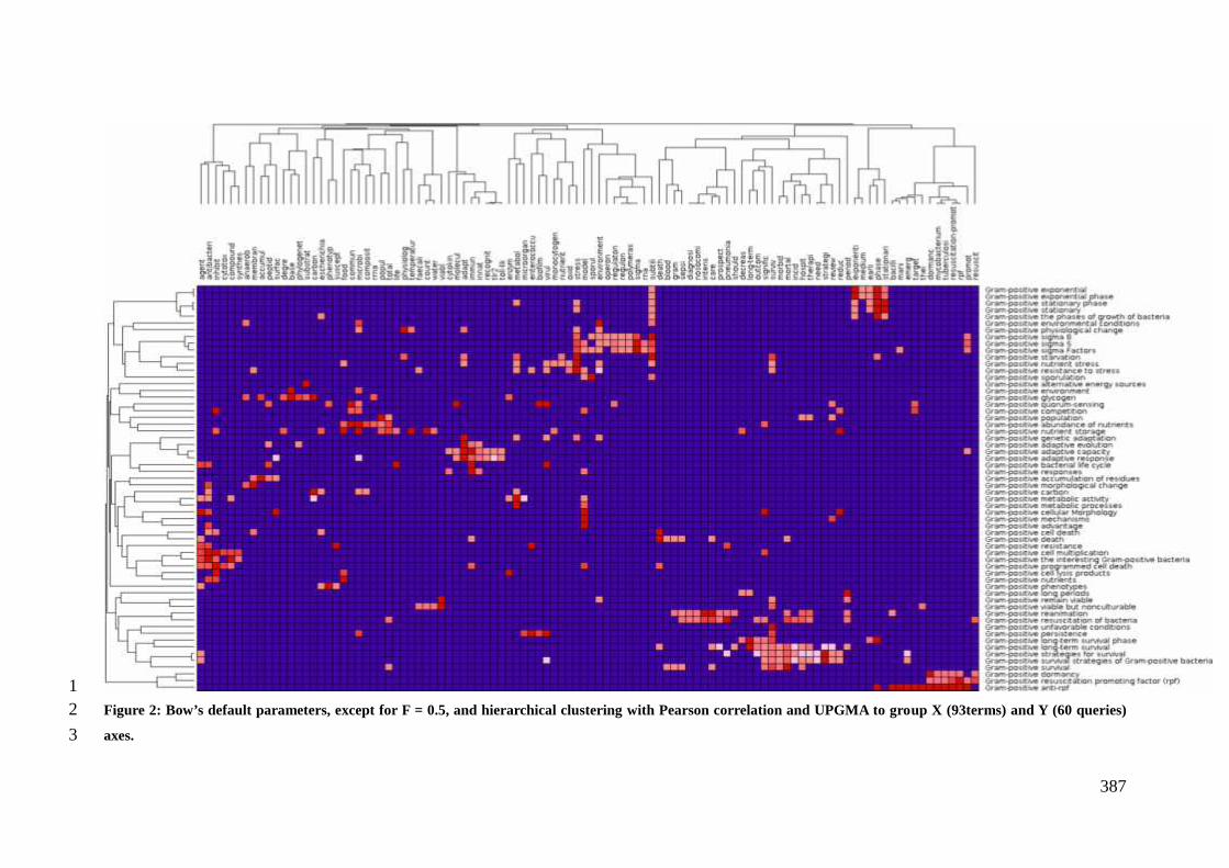
387
1Figure 2: Bow’s default parameters, except for F = 0.5, and hierarchical clustering with Pearson correlation and UPGMA to group X (93terms) and Y (60 queries)2
axes.3
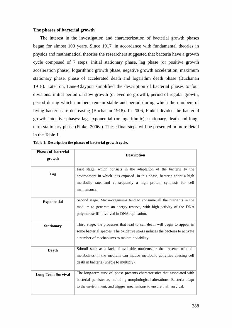
388
The phases of bacterial growth
The interest in the investigation and characterization of bacterial growth phases
began for almost 100 years. Since 1917, in accordance with fundamental theories in
physics and mathematical theories the researchers suggested that bacteria have a growth
cycle composed of 7 steps: initial stationary phase, lag phase (or positive growth
acceleration phase), logarithmic growth phase, negative growth acceleration, maximum
stationary phase, phase of accelerated death and logarithm death phase (Buchanan
1918). Later on, Lane-Claypon simplified the description of bacterial phases to four
divisions: initial period of slow growth (or even no growth), period of regular growth,
period during which numbers remain stable and period during which the numbers of
living bacteria are decreasing (Buchanan 1918). In 2006, Finkel divided the bacterial
growth into five phases: lag, exponential (or logarithmic), stationary, death and long-
term stationary phase (Finkel 2006a). These final steps will be presented in more detail
in the Table 1.
Table 1: Description the phases of bacterial growth cycle.
Phases of bacterial
growthDescription
LagFirst stage, which consists in the adaptation of the bacteria to the
environment in which it is exposed. In this phase, bacteria adopt a high
metabolic rate, and consequently a high protein synthesis for cell
maintenance.
Exponential Second stage. Micro-organisms tend to consume all the nutrients in the
medium to generate an energy reserve, with high activity of the DNA
polymerase III, involved in DNA replication.
Stationary Third stage, the processes that lead to cell death will begin to appear in
some bacterial species. The oxidative stress induces the bacteria to activate
a number of mechanisms to maintain viability.
Death Stimuli such as a lack of available nutrients or the presence of toxic
metabolites in the medium can induce metabolic activities causing cell
death in bacteria (unable to multiply).
Long-Term-Survival The long-term survival phase presents characteristics that associated with
bacterial persistence, including morphological alterations. Bacteria adapt
to the environment, and trigger mechanisms to ensure their survival.

389
In Figure 3, we briefly present the five important bacterial phases: (1) lag phase,
(2) exponential phase, (3) stationary phase, (4) death phase and (5) long-term survival.
Figure 3: Bacterial growth shows about the same phases but the time scale ranges from hoursto
years depending on the species.
Bacterial phenotypes in long term survival
Cellular and colony morphological changes
During the LTS, Gram positive bacteria can undergo morphological changes, at
the cellular or the colony level (Table 2).
In bacteria, nutrient availability has a great impact on bacterial cell size (Chien
et al., 2012). Under stress conditions such as starvation encountered in LTS phase, some
Gram positive bacteria can enter into a cell-wall deficient state also known as L-form
conversion. This phenomenon is well-described in pathogenic bacteria of
Mycobacterium genus. In M. bovis (Slavchev; Michailoca; Markova, 2013), or M.
tuberculosis (Markova et al., 2012), L-form conversion was shown to be an adaptive
strategy to survive and reproduce under unfavorable conditions. In other Gram positive
species, a reduction in cell size is observed when the population enters the LTS phase.
This was shown in S. aureus which indeed harbours a decrease in cell size in starvation
survival. This phenotype is accompanied by an increase in resistance to acid shock and
oxidative stress (Watson et al., 1998). Similarly, in Listeria monocytogenes cellular

390
morphology can change from bacilli to cocci during the transition to LTS phase, leading
to an increased baro- and thermotolerance (Wen et al., 2011). Morphological changes
were also observed during acid adaptation in the food grade bacterium
Propionibacterium freudenreichii, with a shift from pleomorphic rods arranged in
Chinese characters to bacteria with a segmented aspect and shorter cells (Jan et al.,
2001).
Alterations of phenotype at the cell level can also have an impact at the colony
level, namely in the size and color of the colonies (Helmus et al. 2012). A change in the
size of colonies and a decline of the growth rate can be observed in Bacillus subtilis,
during nutrient shortage (Chien; Hill; Levin, 2012). In Streptococcus pyogenes, after a
long period (4 to 14 weeks) in stationary phase and nutrient shortage, culturable cells
formed two colony phenotypes: atypical large colonies and microcolonies, which
showed extensive changes at the proteome level compared to the parental strain (Wood
et al., 2005).
Table 2: Morphological alterations in bacterial cells or colonies.
Bacteria Morphological alterations Reference
Listeria
monocytogenes
The structure and the form of the cells change
during the period of transition from early
stationary phase to long-term survival
(Wen et al. 2011)
Mycobacterium
bovis
In stressful conditions, Mycobacterium bovis
displays different morphologies, such as L- forms
(cell wall-deficient cells), as a way to survive in
unfavorable environments
(Slavchev,
Michailova and
Markova 2013)
Staphylococcus
aureus
Changes in morphology with a decrease in cell
size between the hunger and survival phases
(Watson, Clements
and Foster 1998)
Streptococcus
pyogenes
After four weeks in stressful conditions, two
different colony phenotypes are observed, most
likely due to some selective pressure of the
environment in two phases of life cycle: atypical
large colonies and small colonies
(Wood 1981)

391
Growth rate changes
Bacteria were early shown to be able to sense and recognize unfavorable
conditions in the environment and thus to enter into a state where bacterial replication is
inhibited until favorable conditions are re-established (in Wayne and Sohaskey, 2001:
HJ Corper, Cohn ML, 1933). This capacity is an important strategy that allows bacterial
survival. In Bacillus subtilis, alterations in cell division are directly correlated with
nutrient availability and growth rate (Chien, Hill and Levin, 2012). To store energy,
which correspond to a reduction in some metabolic pathways, such as the glycolytic
pathway, in turn of decreasing the production of ATP (Savic and McShan 2012). As we
can see other species, after a stress period, still require a period of latency for the
morphological changes to occur, even after reestablishing favorable conditions (Roszak
and Colwell 1987). This is because a part of the bacterial population needs more time to
recover and return to being competitive after being exposed to the period of stress. The
study of Geisel and colleagues shows the relationship between stress resistance and
growth rate of various microorganisms. This study revealed that the cells remain in a
vegetative state or enter into the dormant state, to decrease bacterial metabolism under
unfavorable conditions, which allows these bacteria to adapt and survive in the
environment (Geisel, Vilar and Rubi 2011).
Genetic adaptation
In some bacterial species, LTS phase induces a growth advantage in stationary
phase (GASP) phenomenon. GASP is defined as fluctuations in viable cell numbers
during stationary phase growth, with a rapid loss of viability followed by an increase in
viable counts (see above, Figure 3). In the stationary phase, the bacterial cells that
undergo the GASP phenomenon are able to grow displace, the parent cells and become
the dominant population in the culture. It is triggered by intense competition for
nutrients and actually reflects the prevalent state of bacteria in their natural
environment. GASP phenomenon is due to a series of genetic changes, occurring while
the bacterial population enters the stationary phase. In parallel, physiological and
structural alterations occur and induce a higher tolerance to diverse environmental
stresses (Sewell et al., 2011). It was first reported and is well-documented in E. coli, the
Gram negative model bacterium (Zinser, Erik R, Kolter, 2004; Zinster and Kolter, 1999;
Bacun-Druzina et al. 2011). The GASP cells are able to transfer their advantage to

392
daughter cells, providing a competitive advantage (Finkel 2006a), in low concentrations
of nutrients or in physical and chemical conditions that are unfavorable for growth. In
contrast, only a few studies report on GASP phenomenon in Gram positive bacteria.
One of the GASP associated phenotypes, namely the drop in cell counts at the beginning
of stationary phases followed by an increase in cell counts, was shown in Enterococcus
feacium. GASP however seems to be species-specific since it was not observed in S.
aureus cultures (Sewell et al., 2011). In Bacillus subtilis, a phenomenon comparable to
GASP was observed and linked to a hypermutable subpopulation that emerge during the
stationary phase. This phenomenon, also called stationary–phase mutagenesis involves
comA and comK genes (associated to prokaryotic differentiation) and is recA-
independent. It leads to mutations that arise during the stationary-phase, in bacterial
cells that do not divide and that are subjected to non lethal stresses, such as nutrient
shortage (Sung and Yasbin, 2002).
Regulation of bacterial metabolism
The maintenance and regulation of bacterial metabolism requires the action of
several proteins involved in functions, such as transport and synthesis. In this section,
we will highlight the factors that allow the control of these activities for the use of
alternative energy sources, and the storage of nutrients and for the maintenance of
intracellular homeostasis.
Use of atypical nitrogen and carbon source
When the entry in LTS is due to nutritional shortage, some species adapt their
metabolism to use other carbon and nitrogen sources. Such adaptation can be operated
through the use of acetate or citrate as an alternative electron donor to produce energy,
as shown in the Gram negative bacterium Geobacter sulfurreducens (Helmus et al.
2012). In Gram positive bacteria, M. luteus can use succinic acid as a substrate to
produce energy. Lactobacillus sakei survival is linked to the degradation of arginine
(Champomier Vergès et al. 1999), while L. monocytogenes can consume products from
cell lysis to remain viable (Wen et al. 2013).
Nutrient StorageSome species of Gram-positive bacteria store various compounds, such as a
polysaccharide, poly-β-hydroxybutyrate, lipids, polyphosphate or other elements, which

393
can be used to maintain their viability during prolonged periods of nutrient shortage
(Wilkinson 1959).
In general, when bacterial cells are subjected to nutrient shortage in carbon,
nitrogen and phosphorus, "survival" responses are induced. In such conditions, bacteria
can use cell reserves like glycogen or polyphosphate (Carvalheira et al. 2014) and/or
cell lysis products in the LTS population (Roszak and Colwell 1987). Inorganic
polyphosphate (polyP) is critical to bacterial survival in adverse conditions (Rao,
Gómez-García and Kornberg 2009) as shown in Mycobacterium sp. During phosphate
shortage, Mycobacterium tuberculosis induces the ppk1 gene, which encodes a
polyphosphate kinase and relA, which boosts the response to these stress so that the
bacterial cells remain viable (Rifat, Bishai and Karakousis 2009).
In environments that do not present a broad range of available nutrients, bacteria
are able to store some elements to prepare their survival phase, as shown in Vibrio
cholera and Mycobacterium tuberculosis, which can store glucose in the form of
glycogen (Bourassa and Camilli 2009; Amit Singh 2014). They can also do so when
they are subjected to various stresses (nitrogen intermediates, reactive oxygen, low pH,
and vital element starvation). When the carbon substrate is exhausted in the medium,
these reserve substrates are utilized as energy source (Amit Singh 2014).
In LTS transition, L. monocytogenes upregulates the expression of genes related
to energy metabolism, cell envelope structure, and transport. The upregulation of
compatible solute transporters is thought to be involved in the accumulation of cellular
solutes, which in turn lowers intracellular water activity and thus increases stress
resistance (Greenblatt et al. 2004b; Wen et al. 2011).
Maintenance of pH and osmoprotection
Bacterial adaptation to nutrient shortage results in the increase of some
substrates, which can also be harmful to the cell. An increase in intracellular glucose can
decrease bacterial survival as this will directly lower the pH of the medium because the
bacteria cannot metabolize the large amounts of lactic acid produced. Some S. pyogenes
strains are able to survive for long periods most likely thanks to mechanisms that allow
them to escape these unfavorable conditions and remain viable, either by buffering
systems or a similar mechanism for the maintenance of pH level (Savic and McShan
2012).

394
In L. monocytogenes, storage of osmoprotectants like glycine, betaine and
trehalose allows viability. Also, upregulation of the transporter genes and the expression
of genes involved in energy metabolic pathways is crucial for survival (Greenblatt et al.
2004a; Wen et al. 2011).
Different strategies for survival
In this section, we discuss the best known mechanisms involved in LTS in
Gram-positive bacteria, such as programmed cell death (PCD) and dormancy, which
may help us understand this puzzle.
Programmed cell death
In Gram-positive bacteria, concepts such as programmed cell death have begun
to emerge as a mechanism that allow bacterial populations to survive. Some authors
suggest this as an evolutionary process that has allowed bacteria to adapt. Dynamic
cellular stress responses cause destabilization bacterial population in varying conditions
by requiring activation of quorum-sensing systems to trigger programmed cell death
(PCD) (Finkel 2006c).
Quorum-sensing is observed in Gram-positive bacteria as a facilitator in
response to stress conditions. The perception of stimuli in the medium activates specific
signaling pathways, which initiates the process (Bharati et al. 2012). A large number of
quiescent cells are responsible for the storage of carbon. In L. monocytogenes, this
mechanism of quorum-sensing has been proposed as a trigger for activities that balance
growth and death in the population by regulating gene expression between periods of
long-term survival. This is possible because these bacteria are able to perceive the
conditions to initiate programmed cell death, increasing the availability of nutrients to
the surviving cells (Wen et al. 2013).
In addition to this mechanism, in this review, we will also focus on another very
important phenomenon: dormancy.
Dormancy
Bacteria can to regulate their metabolic activity in order to maintain the viability
of the population in periods without or without low cell multiplication and in periods of
competition in extreme environments with low availability of nutrients (Ratcliff et al.

395
2013). This step of dormancy or quiescence corresponds to a “resting period” or a
“period of reversible phenotypic change” and is triggered in response to extreme
conditions of growth (Roszak and Colwell, 1987). It has been well-studied in
pathogenic bacteria where, during an infection, the resulting slowly growing bacteria
are able to tolerate both the host immune response and prolonged antibiotic exposure.
This is typically the case in chronic infections such as tuberculosis caused by the Gram
positive pathogen M. tuberculosis.
Dormancy (or quiescence) may be defined as a state of resistance to non-
favorable conditions, or stresses where the viable population size is maintained
throughout the period of stress, and where bacteria maintain a metabolic capacity, their
membrane potential, and do not undergo obvious morphological differentiation
(Rittershaus, Baek and Sassetti 2013a). During the dormancy period, cell replication
and DNA synthesis are blocked, while the synthesis of RNA and proteins continues, so
that bacterial metabolic activity remains viable until the stress is over (Roszak and
Colwell, 1987). Besides M. tuberculosis, Micrococcus luteus was shown to enter in
dormancy over long periods when subjected to starvation (Greenblatt et al. 2004b).
Viable but nonculturable state
The entry in a Viable but nonculturable (VBNC) state is a strategy in response to
adverse conditions described in various bacteria in situations of nutrient stress or
atypical temperatures (Mulyukin et al. 2003). This concept emerged a few decades ago
as an explanation for how bacteria can survive for long periods in adverse conditions
(Roszak and Colwell, 1987). The VBNC state can be either a state in which bacteria
await death or where they wait for a stimulus so that cellular metabolic activities can
return to normal (Mulyukin et al. 2003).
It was found that in many species of pathogenic bacteria of VBNC state is
present, so that they are able to express their virulence and remain viable. Furthermore,
it was shown that bacteria have been VBNC the resuscitation promoting factor
(Ramamurthy et al 2014).
Resuscitation
Different methodologies that have been carried out in order to observe and
characterize the phenomenon of “resuscitation bacterial”, seeks to restore conditions to

396
stabilize the population using suitable media, pH and temperature (Roszak and Colwell,
1987).
To get out of the state of dormancy, bacteria can initiate a form of recovery after
a period of latency as reported in M. luteus (Greenblat et al. 2004b). This phase is
named resuscitation and is also observed in S. aureus, which however requires a large
amount and variety of nutrients to resume its metabolic activities (Diaper and Edwards
1994).
Resuscitation Promoting Factor (Rpf) is present in most bacteria with high G +
C content. The Rpf are a group of secreted proteins, that belong to 29 families in
genomes of actinomycetes (Dewi Puspita et al. 2013). In M. tuberculosis, it has been
reported that anti-rpf antibodies are able to inhibit bacterial growth. Similarly,
exogenous rpf added to old cultures leads to a reactivation of bacterial growth. Other
models of infection in vitro were made and guaranteed performance of this group of
proteins for resuscitation of cells (Kaprelysants et al. 2012).
Studies show that the analysis of the transcriptome of "resuscitated” cells of L.
monocytogenes suggests an alteration in the metabolism to bacteria in their exponential
phase, clearly showing that the bacterium is able to go through different conditions and
remain viable (Wen et al. 2011). For this, it is necessary that genes associated with the
synthesis of cell envelope components, proteins, amino acids and ribonucleotides are
rapidly expressed (Wen et al. 2011).
What is to come? (Future directions)To date, most studies on long-term survival were carried out using classical
microbiology techniques and mostly described the consequences of their phenotypes
(size, color, persistence). However, is it possible, at a genomic level, to predict the
ability of a bacterium to survive based on its gene content or based on the bacterial
metabolic pathways?
New sequencing technologies and the advent of omic technologies, such as
genomics, transcriptomics, proteomics and metagenomics, open avenues for research on
the survival and adaptability of Gram-positive bacteria. Integrating information from
different fields will allow to better understand how these mechanisms are regulated.
This is of major interest to develop new products such as drugs, vaccines, or select more
robust probiotics.

397
New research in bacterial survival is of great interest e.g. for the design of
therapies that interfere with pathogenesis and persistence of bacteria, which are major
concerns in clinical microbiology and food safety. A better understanding of LTS
mechnaisms could lead to the development of drugs that act even on dormant bacterial
cells and impair the establishment of LTS in a pathological context, or in indwelling
devices in hospitalized patients. Another application is to enhance LTS to optimize the
efficiency of probiotic bacteria since some probiotic species are reportedly fragile (e.g.
Bifodobacterium sp.) although their application requires a good survival during
manufacturing and storage of products until the end consumer.
Author and Contributors
The authors declare that there are no conflicts of interest.
Funding
CAPES/COFECUB program supported this workThis work processes BEX 11517-12-3and the Brazilian funding agencies CAPES and CNPq.
References
Amit Singh, Anil Kumar Gupta. Glycogenomics of Mycobacterium tuberculosis.Mycobacterial Diseases, v. 04, n. 06, 2014. Disponível em:<http://www.omicsonline.org/open-access/glycogenomics-of-mycobacterium-tuberculosis-2161-1068-4-175.php?aid=35840>. Acesso em: 25 set. 2015.
Bhrati, B. K. et al. A full-length bifunctional protein involved in c-di-GMP turnover isrequired for long-term survival under nutrient starvation in Mycobacterium smegmatis.Microbiology, v. 158, n. Pt_6, p. 1415–1427, 1 jun. 2012. Acesso em: 25 jun. 2014.
Bourassa, Lori; Camilli, Andrew. Glycogen contributes to the environmental persistenceand transmission of Vibrio cholerae. Molecular Microbiology, PMID:19226328PMCID: PMC2704980, v. 72, n. 1, p. 124–138, abr. 2009.
Buchanan, R. E. Life Phases in a Bacterial Culture. The Journal of Infectious Diseases,v. 23, n. 2, p. 109–125, 1 ago. 1918.
Carvalheira, Mónica et al. Survival strategies of polyphosphate accumulating organismsand glycogen accumulating organisms under conditions of low organic loading.Bioresource Technology, v. 172, p. 290–296, nov. 2014.
Champomier Vergès, Marie-Christine et al. Relationships between arginine degradation,pH and survival in Lactobacillus sakei. FEMS Microbiology Letters, v. 180, n. 2, p.297–304, nov. 1999. Acesso em: 25 jun. 2014.

398
Chaussabel, Damien; Sher, Alan. Mining microarray expression data by literatureprofiling. Genome Biology, PMID: 12372143PMCID: PMC134484, v. 3, n. 10, p.research0055.1–research0055.16, 2002.
Chien, An-Chun; Hill, Norbert S.; Levin, Petra Anne. Cell Size Control in Bacteria.Current Biology, v. 22, n. 9, p. R340–R349, 8 maio 2012.
Dewi Puspita, Indun et al. Resuscitation Promoting Factor (Rpf) from Tomitellabiformata AHU 1821T Promotes Growth and Resuscitates Non-Dividing Cells.Microbes and Environments, v. 28, n. 1, p. 58–64, 2013. Acesso em: 12 set. 2014.
Diaper, J. P.; Edwards, C. Survival of Staphylococcus aureus in lakewater monitored byflow cytometry. Microbiology (Reading, England), PMID: 7512871, v. 140 ( Pt 1), p.35–42, jan. 1994.
Finkel, Steven E. Long-term survival during stationary phase: evolution and the GASPphenotype. Nature Reviews Microbiology, v. 4, n. 2, p. 113–120, fev. 2006a. Acesso em:16 abr. 2014.
Finkel, Steven E. Long-term survival during stationary phase: evolution and the GASPphenotype. Nature Reviews Microbiology, v. 4, n. 2, p. 113–120, fev. 2006b. Acessoem: 25 jun. 2014.
Geisel, Nico; Vilar, Jose M. G.; Rubi, J. Miguel. Optimal Resting-Growth Strategies ofMicrobial Populations in Fluctuating Environments. PLoS ONE, v. 6, n. 4, p. e18622,15 abr. 2011.
Greenblatt, C. L. et al. Micrococcus luteus - Survival in Amber. Microbial Ecology, v.48, n. 1, p. 120–127, 28 maio 2004a.
Greenblatt, C.L. et al. Micrococcus luteus - Survival in Amber. Microbial Ecology, v.48, n. 1, p. 120–127, set. 2004b. Acesso em: 28 abr. 2014.
Helmus, Ruth A. et al. Growth advantage in stationary-phase (GASP) phenotype inlong-term survival strains of Geobacter sulfurreducens. FEMS Microbiology Ecology, v.79, n. 1, p. 218–228, jan. 2012. Acesso em: 16 abr. 2014.
Jan, Gwénaël et al. Changes in Protein Synthesis and Morphology during AcidAdaptation of Propionibacterium freudenreichii. Applied and EnvironmentalMicrobiology, PMID: 11319077PMCID: PMC92832, v. 67, n. 5, p. 2029–2036, maio2001.
Markova, Nadya; Slavchev, Georgi; Michailova, Lilia. Unique biological properties ofMycobacterium tuberculosis L-form variants: impact for survival under stress.International Microbiology: The Official Journal of the Spanish Society forMicrobiology, PMID: 22847267, v. 15, n. 2, p. 61–68, jun. 2012.
Mulyukin, Andrei L. et al. Formation of resting cells by non-spore-formingmicroorganisms as a strategy of long-term survival in the environment. 30 jan. 2003,[S.l: s.n.], 30 jan. 2003. p. 208–218. Disponível em:<http://proceedings.spiedigitallibrary.org/proceeding.aspx?articleid=889034>. Acessoem: 25 jun. 2014.

399
Palop, Alfredo; Mañas, Pilar; Condón, Santiago. Sporulation temperature and heatresistance of Bacillus Spores: a Review. Journal of Food Safety, v. 19, n. 1, p. 57–72,abr. 1999.
Ramamurthy, Thandavarayan et al. Current Perspectives on Viable but Non-Culturable(VBNC) Pathogenic Bacteria. Frontiers in Public Health, v. 2, 31 jul. 2014. Disponívelem: <http://journal.frontiersin.org/article/10.3389/fpubh.2014.00103/abstract>. Acessoem: 7 abr. 2015.
Rao, Narayana N.; Gómez-García, María R.; Kornberg, Arthur. InorganicPolyphosphate: Essential for Growth and Survival. Annual Review of Biochemistry, v.78, n. 1, p. 605–647, 2009.
Ratcliff, William C. et al. Disentangling direct and indirect fitness effects of microbialdormancy. The American Naturalist, v. 182, n. 2, p. 147–156, ago. 2013. Acesso em: 25jun. 2014.
Rifat, Dalin; Bishai, William R.; Karakousis, Petros C. Phosphate depletion: a noveltrigger for Mycobacterium tuberculosis persistence. The Journal of Infectious Diseases,PMID: 19686042, v. 200, n. 7, p. 1126–1135, 1 out. 2009.
Rittershaus, Emily S.C.; Baek, Seung-Hun; Sassetti, Christopher M. The Normalcy ofDormancy: Common Themes in Microbial Quiescence. Cell Host & Microbe, v. 13, n.6, p. 643–651, jun. 2013. Acesso em: 13 ago. 2014.
Roszak, D B; Colwell, R R. Survival strategies of bacteria in the natural environment.Microbiological Reviews, PMID: 3312987PMCID: PMC373117, v. 51, n. 3, p. 365–379, set. 1987.
Savic, D. J.; Mcshan, W. M. Long-term survival of Streptococcus pyogenes in richmedia is pH-dependent. Microbiology, v. 158, n. Pt_6, p. 1428–1436, 1 jun. 2012.Acesso em: 25 jun. 2014.
Sewell, D.; Laird, K.; Phillips, C. Growth advantage in stationary phase phenomenon inGram-positive bacteria. Journal of Hospital Infection, v. 78, n. 1, p. 73–75, maio 2011.
S. Kaprelyants, Arseny et al. Resuscitation-promoting Factors (Rpf): In Search ofInhibitors. Protein & Peptide Letters, v. 19, n. 10, p. 1026–1034, 1 set. 2012. Acessoem: 12 set. 2014.
Slavchev, Georgi; Michailova, Lilia; Markova, Nadya. Stress-induced L-forms ofMycobacterium bovis: a challenge to survivability. The New Microbiologica, PMID:23686122, v. 36, n. 2, p. 157–166, abr. 2013.
Sung, Huang-Mo; Yasbin A, Ronald E. Adaptive, or stationary-phase, mutagenesis, acomponent of bacterial differentiation in Bacillus subtilis. Journal of Bacteriology,PMID: 12270822PMCID: PMC139596, v. 184, n. 20, p. 5641–5653, out. 2002.
Watson, S. P.; Clements, M. O.; Foster, S. J. Characterization of the starvation-survivalresponse of Staphylococcus aureus. Journal of Bacteriology, PMID: 9537371 PMCID:PMC107086, v. 180, n. 7, p. 1750–1758, abr. 1998.

400
Wayne, L. G.; Sohaskey, C. D. Nonreplicating persistence of mycobacteriumtuberculosis. Annual Review of Microbiology, PMID: 11544352, v. 55, p. 139–163,2001.
Wen, J. et al. Transcriptomic Response of Listeria monocytogenes during the Transitionto the Long-Term-Survival Phase. Applied and Environmental Microbiology, v. 77, n.17, p. 5966–5972, 1 set. 2011. Acesso em: 25 jun. 2014.
Wen, Jia et al. Listeria monocytogenes responds to cell density as it transitions to thelong-term-survival phase. International Journal of Food Microbiology, v. 165, n. 3, p.326–331, ago. 2013. Acesso em: 25 jun. 2014.
Wilkinson, J. F. The problem of energy-storage compounds in bacteria. ExperimentalCell Research, The Cytochemistry of Enzymes and Antigens. v. 7, Supplement, p. 111–130, 1959.
Wood, D. N. et al. Persistence of Streptococcus pyogenes in Stationary-Phase Cultures.Journal of Bacteriology, v. 187, n. 10, p. 3319–3328, 15 maio 2005. Acesso em: 25 abr.2014.
Zinser, Erik R; Kolter, Roberto. Escherichia coli evolution during stationary phase.Research in Microbiology, v. 155, n. 5, p. 328–336, jun. 2004. Acesso em: 25 jun. 2014.
Zinser, Erik R; Kolter, Roberto. Mutations enhancing amino acid catabolism confer agrowth advantage in stationary phase. Journal of Bacteriology, PMID:10482523PMCID: PMC94102, v. 181, n. 18, p. 5800–5807, set. 1999.
401
Anexo 7: Curriculum vitae
Annexe 7 : Curriculum vitae
Address to access this CV: http://lattes.cnpq.br/2016464855117057
Personal Information
Full name Flavia Figueira Aburjaile
Name used inBibliographicCitations
ProfessionalAdresss
e-mail [email protected]
Formal Education
2011 - 2015 Doctorate in Bioinformatics.Universidade Federal de Minas Gerais, UFMG, Belo Horizonte,Brazil
(Co-guardianship) Institut National de la Recherche Agronomique(Advisor: Yves Le Loir)Title: Mecanismos moleculares de sobrevivência em longo prazo emPropionibacterium freudenreichiiAdvisor: Vasco Ariston de Carvalho AzevedoScholarship from: Coordenação de Aperfeiçoamento de Pessoal deNível Superior/COFECUBKnowledge areas: Bioinformatics and Molecular Genetics inMicroorganisms.
2007 - 2011 Bachelor’s in Biomedicine.Universidade Fundação Mineira de Educação e Cultura, FUMEC,Belo Horizonte, BrasilStudy abroad sandwich option: Université de Provence Aix Marseille I(Advisor : Jean-Pierre Roll)Title: Teste do efeito dos probióticos frente à PseudomonasaeruginosaAdvisor: Adriana dos Santos e Flávio Henrique Barbosa
ABURJAILE, F. F.; FIGUEIRA, F.A.; ABURJAILE, F.; FIGUEIRA,F.;FLÁVIA; FIGUEIRA ABURJAILE, F.; Flávia Aburjaile; ABURJAILE,FF; Aburjaile, Flávia; ABURJAILE, FLÁVIA FIGUEIRA
Universidade Federal de Minas Gerais, Instituto de Ciências BiológicasAv. Antônio Carlos, 6627, Laboratório de Genética Celular e Molecular,Bloco Q3, sala 259Pampulha - Belo Horizonte31270910, MG - BrazilPhone number: 31 3409-2873

402
Complementary Education
2014 - 2014 Short Course in Advanced seq annotation and use of pipelinesEGene2.Universidade Federal de Minas Gerais, UFMG, Belo Horizonte, Brasil
2014 - 2014 Short Course in PATRIC.Universidade Federal de Minas Gerais, UFMG, Belo Horizonte, Brasil
2014 - 2014 Short Course in Genomics for Working with Pathogens.EMBL European Bioinformatics Institute, EMBL-EBI, UFMG, BeloHorizonte, Brasil
2014 - 2014 Short Course in GO terms and annotations.Universidade Federal de Minas Gerais, UFMG, Belo Horizonte, Brasil
2012 - 2012 Short Course in Introduction to BioNumerics program.Universidade Federal de Minas Gerais, UFMG, Belo Horizonte, Brasil
2012 - 2012 Short Course in Introduction to Perl languages.Universidade Federal de Minas Gerais, UFMG, Belo Horizonte, Brasil
2012 - 2012 Short Course in Molecular Methods for Microbial Diagnosis,Genomic.Universidade Federal de Minas Gerais, UFMG, Belo Horizonte, Brasil
2012 - 2012 Short Course em Rna-seq course.Universidade Federal de Minas Gerais, UFMG, Belo Horizonte, Brasil
2012 - 2012 Short Course em International Course in Automated Functional Annot.Centro de Investigaciones Príncipe Felipe, CIPF, Espanha
2011 - 2011 Short Course in Next Generation Sequencing: SOLiD.Universidade Federal do Paraná, UFPR, Curitiba, Brasil
2011 - 2011 Short Course in assembly and annotation of bacterial genomes.Universidade Federal de Minas Gerais, UFMG, Belo Horizonte, Brasil
2011 - 2011 Short Course in II Winter Course of Biochemistry and MolecularBiology.Universidade Federal do Paraná, UFPR, Curitiba, Brasil
2011 - 2011 Short Course in molecular biology transformation.Universidade Federal do Paraná, UFPR, Curitiba, Brasil
2011 - 2011 Short Course in IV Winter Course of Genetics.Universidade Federal do Paraná, UFPR, Curitiba, Brasil
2010 - 2010 Short Course in Stage intensif de Langue française.

403
Université de Provence Aix Marseille I, Aix-Marceille I, Marseille ,França
2009 - 2009 Short Course in Arterial Blood Gas Analysis.Medical Association of Minas Gerais, AMMG, Brazil, AMMG, Brasil
2008 - 2009 French. Alliance Française, BH, Brazil.
Areas of expertise
Languages
1. English Understanding Functional, Speaking Functional, Writing Basic, Reading
Fluent
2. Spanish Understanding Functional, Speaking Functional, Writing Basic, Reading
Fluent
3. French Understanding Fluent, Speaking Functional, Writing Functional, Reading
Fluent
Prêmios e títulos
2013 BioVis 2013 - Data Contest Biology Experts Pick, IEEE Computer Society
S,T &A ProductionBibliographic ProductionArticles Published in Scientific Journals
1. BARBOSA, EUDES G. V., ABURJAILE, F. F., RAMOS, ROMMEL T.J.,CARNEIRO, ADRIANA R., LOIR, Y. L., BAUMBACH, J., MIYOSHI, ANDERSON,SILVA, ARTUR, AZEVEDO, VASCO. Value of a newly sequenced bacterial genome.World Journal of Biological Chemistry. , v.5, p.161 - 168, 2014.
2. SOARES, SIOMAR C., SILVA, ARTUR, TROST, EVA, BLOM, JOCHEN, RAMOS,ROMMEL, CARNEIRO, ADRIANA, ALI, AMJAD, SANTOS, ANDERSON R.,PINTO, ANNE C., DINIZ, CARLOS, BARBOSA, EUDES G. V., DORELLA,FERNANDA A., ABURJAILE, FLÁVIA, ROCHA, FLÁVIA S., NASCIMENTO,KARINA K. F., GUIMARÃES, LUÍS C., ALMEIDA, SINTIA, HASSAN, SYED S.,BAKHTIAR, SYEDA M., PEREIRA, ULISSES P., ABREU, VINICIUS A. C.,SCHNEIDER, MARIA P. C., MIYOSHI, ANDERSON, TAUCH, ANDREAS,
1. Bioinformatics
2. Microbiology
3. Genetics
4. Parasitology
5.
404
AZEVEDO, VASCO. The Pan-Genome of the Animal Pathogen Corynebacteriumpseudotuberculosis Reveals Differences in Genome Plasticity between the Biovar ovisand equi Strains. Plos One. , v.8, p.e53818-e53818, 2013.
3. DORELLA, FERNANDA A, GALA-GARCIA, ALFONSO, PINTO, ANNE C,SARROUH, BOUTROS, ANTUNES, CAMILA A, RIBEIRO, DAYANA, Aburjaile,Flávia, FIAUX, KARINA K, Guimarães, Luis C, SEYFFERT, NUBIA, EL-AOUAR,RACHID A, SILVA, RENATA, HASSAN, SYED S, CASTO, THIAGO LP,MARQUES, WANDERSON S, RAMOS, ROMMEL, CARNEIRO, ADRIANA, SÁ,PABLO DE, MIYOSHI, ANDERSON, AZEVEDO, VASCO, SILVA, ARTUR.Progression of 'OMICS' methodologies for understanding the pathogenicity ofCorynebacterium pseudotuberculosis: the Brazilian experience. Computational andStructural Biotechnology Journal. , v.6, p.1 - 7, 2013.
4. RAMOS, ROMMEL THIAGO JUCÁ, CARNEIRO, ADRIANA RIBEIRO,SOARES, SIOMAR DE CASTRO, SANTOS, ANDERSON RODRIGUES DOS,ALMEIDA, SINTIA, GUIMARÃES, LUIS, FIGUEIRA, FLÁVIA, BARBOSA,EUDES, TAUCH, ANDREAS, AZEVEDO, VASCO, SILVA, ARTUR. Tips and tricksfor the assembly of a Corynebacterium pseudotuberculosis genome using asemiconductor sequencer. Microbial Biotechnology (Online). , v.n/a, p.n/a - n/a, 2012.
5. Soares, Siomar C., Trost, Eva, Ramos, Rommel T.J., Carneiro, Adriana R., Santos,Anderson R., Santos, A.R., Pinto, Anne C., ABURJAILE, F. F., Barbosa, Eudes, Ali,Amjad, DINIZ, CARLOS A.A., Hassan, Syed S., FIAUX, KARINA, Guimarães, LuisC., Bakhtiar, Syeda M., PEREIRA, ULISSES, Almeida, Sintia S., Abreu, Vinícius A.C.,ROCHA, FLÁVIA S., DORELLA, FERNANDA A., Miyoshi, Anderson, Silva, Artur,Azevedo, Vasco, Tauch, Andreas. Genome sequence of Corynebacteriumpseudotuberculosis biovar equi strain 258 and prediction of antigenic targets to improvebiotechnological vaccine production. Journal of Biotechnology. , v.166, p.1 - 7, 2012.
6. SILVA, A., LOPES, T., RAMOS, R. T. J, CARNEIRO, A., DORELLA, F., Rocha, F.S., SANTOS, A. R., LIMA, A. R. J., GUIMARAES, L. C., Barbosa, E. G. V.,RIBEIRO, D., FIAUX, K., DINIZ, C. A. A., ABREU, V. A. C., ALMEIDA, S. S.,HASSAN, S., ALI, A., BAKHTIAR, S., ABURJAILE, F. F., PINTO, A. C., Soares, S.C., PEREIRA, U., Schneider, M. P. C, MIYOSHI, A., EDMAN, J., SPIER, S,AZEVEDO, V.. Complete genome sequence of Corynebacterium pseudotuberculosisCp267 strain isolated from a Llama. Journal of Bacteriology (Print). , v.194, p.3567 -3568, 2012.
7. Lopes, T., Silva, A., Thiago, R., Carneiro, A., Dorella, F. A., Rocha, F. S., dos Santos,A. R., Lima, A. R. J., Guimaraes, L. C., Barbosa, E. G. V., BARBOSA, E. G. V.,Ribeiro, D., Fiaux, K. K., Diniz, C. A. A., de Abreu, V. A. C., de Almeida, S. S., Hassan,S. S., Ali, A., ABURJAILE, F. F., Bakhtiar, S. M., Pinto, A. C., Soares, S. d. C.,Pereira, U. d. P., Schneider, M. P. C., Miyoshi, A., Edman, J., Spier, S., Azevedo, V.Complete Genome Sequence of Corynebacterium pseudotuberculosis Strain Cp267,Isolated from a Llama. Journal of Bacteriology (Print). , v.194, p.3567 - 3568, 2012.
8. Pethick, F. E., Lainson, A. F., YAGA, R., FLOCKHART, A., Smith, D. G. E.,DONACHIE, W., Cerdeira, L. T., Silva, A., BOL, E., LOPES, T. S., Barbosa, M. S.,PINTO, A. C., dos Santos, A. R., SOARES, S. C., Almeida, S. S., Guimaraes, L. C.,
405
ABURJAILE, F. F., Abreu, V. A. C., RIBEIRO, D., FIAUX, K. K., Diniz, C. A. A.,BARBOSA, E. G. V., Pereira, U. P., Hassan, S. S., Ali, A., Bakhtiar, S. M., DORELLA,F. A., DORELLA, F. A., CARNEIRO, A. R., Ramos, R. T. J., Rocha, F. S., Schneider,M. P. C., MIYOSHI, A., AZEVEDO, V., Fontaine, M. C. Complete Genome Sequenceof Corynebacterium pseudotuberculosis Strain 1/06-A, Isolated from a Horse in NorthAmerica. Journal of Bacteriology (Print). , v.194, p.4476 - 4476, 2012.
9. Pethick, F. E., Lainson, A. F., Yaga, R., Flockhart, A., Smith, D. G. E., Donachie, W.,CERDEIRA, L. T., SILVA, A., Bol, E., LOPES, T. S., Barbosa, M. S., PINTO, A. C.,dos Santos, A. R., SOARES, S. C., ALMEIDA, S. S., Guimaraes, L. C., ABURJAILE,F. F., ABREU, V. A. C., RIBEIRO, D., Fiaux, K. K., Diniz, C. A. A., BARBOSA, E. G.V., Pereira, U. P., Hassan, S. S., ALI, A., Bakhtiar, S. M., DORELLA, F. A.,CARNEIRO, A. R., RAMOS, R. T. J., Rocha, F. S., SCHNEIDER, M. P. C., MIYOSHI,A., AZEVEDO, V., AZEVEDO, V., Fontaine, M. C. Complete Genome Sequences ofCorynebacterium pseudotuberculosis Strains 3/99-5 and 42/02-A, Isolated from Sheepin Scotland and Australia, Respectively. Journal of Bacteriology (Print). , v.194, p.4736- 4737, 2012.
10. Santos, A.R., Carneiro, A. R., Ramos, R. T. J., DALL'AGNOL, H., PINTO, A. C.,de Castro Soares, S., SANTOS, A. R., Guimaraes, L. C., Almeida, S. S., BARAUNA,R. A., DAS GRACAS, D. A., FRANCO, L. C., Ali, A., Hassan, S. S., NUNES, C. I. P.,Barbosa, M. S., ABURJAILE, F. F., Fiaux, K. K., Barbosa, E. G. V., Bakhtiar, S. M.,VILELA, D., NOBREGA, F., DOS SANTOS, A. L., CAREPO, M. S. P., Azevedo, V.,Schneider, M. P. C., PELLIZARI, V. H., SILVA, A. Genome Sequence ofExiguobacterium antarcticum B7, Isolated from a Biofilm in Ginger Lake, King GeorgeIsland, Antarctica. Journal of Bacteriology (Print). , v.194, p.6689 - 6690, 2012.
11. SANTOS, A. R., CARNEIRO, A. R., GALA-GARCIA, A., PINTO, A. C., BARH,D., BARBOSA, E. G. V., ABURJAILE, F. F., DORELLA, F. A., ROCHA, F. S.,GUIMARAES, L. C., Zurita-Turk, Meritxell, RAMOS, R. T. J., ALMEIDA, S. S.,ALMEIDA, S., SOARES, S. C., PEREIRA, U. P., ABREU, V. A. C., SILVA, A.,MIYOSHI, A., AZEVEDO, V.. The Corynebacterium pseudotuberculosis in silicopredicted pan-exoproteome. BMC Genomics. , v.13, p.S5 - S6, 2012.
12. CERDEIRA, L., Schneider, M. P. C, PINTO, A. C., ALMEIDA, S. S., SANTOS, A.R., Barbosa, E. G. V., ALI, A., ABURJAILE, F. F., ABREU, V. A. C., GUIMARAES,L. C., Soares, S. C., DORELLA, F., Rocha, F. S., BOL, E., LOPES, T., Barbosa, M. S.,CARNEIRO, A., Juca Ramos, R. T., Coimbra, N. A. d. R, LIMA, A. R. J., Barh, D,Tiwari, S., RAJA, R., ZAMBARE, V., Ghosh, P., Trost, E., Tauch, A, MIYOSHI, A.,AZEVEDO, V., SILVA, A.. Complete Genome Sequence of Corynebacteriumpseudotuberculosis Strain CIP 52.97, Isolated from a Horse in Kenya. Journal ofBacteriology (Print). , v.193, p.7025 - 7026, 2011.
13. HASSAN, SYED SHAH, GUIMARÃES, LUIS CARLOS, PEREIRA, ULISSESDE PÁDUA, ISLAM, ARSHAD, ALI, AMJAD, BAKHTIAR, SYEDA MARRIAM,RIBEIRO, DAYANA, RODRIGUES DOS SANTOS, ANDERSON, SOARES,SIOMAR DE CASTRO, DORELLA, FERNANDA, PINTO, ANNE CYBELLE,SCHNEIDER, MARIA PAULA CRUZ, BARBOSA, MARIA SILVANIRA,ALMEIDA, SÍNTIA, ABREU, VINÍCIUS, ABURJAILE, FLÁVIA, CARNEIRO,ADRIANA RIBEIRO, CERDEIRA, LOUISE TEIXEIRA, FIAUX, KARINA,

406
BARBOSA, EUDES, DINIZ, CARLOS, ROCHA, FLAVIA S., RAMOS, ROMMELTHIAGO JUCÁ, JAIN, NEHA, TIWARI, SANDEEP, BARH, DEBMALYA,MIYOSHI, ANDERSON, MÜLLER, BORNA, SILVA, ARTUR, AZEVEDO, VASCO.Complete genome sequence of Corynebacterium pseudotuberculosis biovar ovis strainP54B96 isolated from antelope in South Africa obtained by rapid next generationsequencing technology. STAND GENOMIC SCI. , v.7, p.189 - 199, 2012.
14. SILVA, A., RAMOS, R. T. J., RIBEIRO CARNEIRO, A., CYBELLE PINTO, A., deCastro Soares, S., RODRIGUES SANTOS, A., SILVA ALMEIDA, S., Guimaraes, L.C., FIGUEIRA ABURJAILE, F., VIEIRA BARBOSA, E. G., ALVES DORELLA, F.,SOUZA ROCHA, F., SOUZA LOPES, T., KAWASAKI, R., GOMES SA, P., DAROCHA COIMBRA, N. A., TEIXEIRA CERDEIRA, L., SILVANIRA BARBOSA, M.,CRUZ SCHNEIDER, M. P., MIYOSHI, A., SELIM, S. A. K., MOAWAD, M. S.,AZEVEDO, V. Complete Genome Sequence of Corynebacterium pseudotuberculosisCp31, Isolated from an Egyptian Buffalo. Journal of Bacteriology (Print). , v.194,p.6663 - 6664, 2012.
15. Ramos, R. T. J., Silva, A., CARNEIRO, A. R., PINTO, A. C., Soares, S. d. C.,Santos, A. R., Almeida, S. S., Guimaraes, L. C., ABURJAILE, F. F., BARBOSA, E. G.V., DORELLA, F. A., DORELLA, F. A., Rocha, F. S., Cerdeira, L. T., Barbosa, M. S.,Tauch, A., EDMAN, J., SPIER, S., MIYOSHI, A., Schneider, M. P. C., AZEVEDO, V..Genome Sequence of the Corynebacterium pseudotuberculosis Cp316 Strain, Isolatedfrom the Abscess of a Californian Horse. Journal of Bacteriology (Print). , v.194, p.6620- 6621, 2012.
16. Hassan, S. S., SCHNEIDER, M. P. C., RAMOS, R. T. J., CARNEIRO, A. R.,RANIERI, A., Guimaraes, L. C., ALI, A., Bakhtiar, S. M., Pereira, U. d. P., dos Santos,A. R., Soares, S. d. C., DORELLA, F., PINTO, A. C., RIBEIRO, D., Barbosa, M. S.,ALMEIDA, S., ABREU, V., ABURJAILE, F., FIAUX, K., BARBOSA, E., DINIZ, C.,ROCHA, F. S., SAXENA, R., Tiwari, S., ZAMBARE, V., Ghosh, P., PACHECO, L. G.C., DOWSON, C. G., KUMAR, A., BARH, D., MIYOSHI, A., AZEVEDO, V., SILVA,A.. Whole-Genome Sequence of Corynebacterium pseudotuberculosis Strain Cp162,Isolated from Camel. Journal of Bacteriology (Print). , v.194, p.5718 - 5719, 2012.
17. OLIVEIRA, L. C., SARAIVA, T. D. L., SOARES, S. C., RAMOS, R. T. J., SA, P.H. C. G., CARNEIRO, A. R., MIRANDA, F., FREIRE, M., RENAN, W., JUNIOR, A.F. O., SANTOS, A. R., PINTO, A. C., SOUZA, B. M., CASTRO, C. P., Diniz, C. A. A.,ROCHA, C. S., MARIANO, D. C. B., DE AGUIAR, E. L., FOLADOR, E. L., Barbosa,E. G. V., ABURJAILE, F. F., GONCALVES, L. A., GUIMARAES, L. C., AZEVEDO,M., AGRESTI, P. C. M., SILVA, R. F., Tiwari, S., ALMEIDA, S. S., Hassan, S. S.,PEREIRA, V. B., ABREU, V. A. C., Pereira, U. P., Dorella, F. A., CARVALHO, A. F.,PEREIRA, F. L., LEAL, C. A. G., FIGUEIREDO, H. C. P., SILVA, A., MIYOSHI, A.,AZEVEDO, V.. Genome Sequence of Lactococcus lactis subsp. lactis NCDO 2118, aGABA-Producing Strain. Genome Announcements. , v.2, p.e00980-14 - e00980-14,2014.
18. SANTOS, ANDERSON R, CARNEIRO, ADRIANA, GALA-GARCÍA,ALFONSO, PINTO, ANNE, BARH, DEBMALYA, BARBOSA, EUDES,ABURJAILE, FLÁVIA, DORELLA, FERNANDA, ROCHA, FLÁVIA,GUIMARÃES, LUIS, Zurita-Turk, Meritxell, RAMOS, ROMMEL, ALMEIDA,

407
SINTIA, SOARES, SIOMAR, PEREIRA, ULISSES, ABREU, VINÍCIUS C, SILVA,ARTUR, MIYOSHI, ANDERSON, AZEVEDO, VASCO. The Corynebacteriumpseudotuberculosis in silico predicted pan-exoproteome. BMC Genomics. , v.13, p.S6,2012.
19. ABURJAILE, S. B., ABURJAILE, F. F., BARBOSA, F. H. F.;Barbosa, Flávio H.F.;BARBOSA, F. Variação da concentração de minerais em aquíferos confinados deindústria de exploração de águas minerais naturais no município de Aparecida-SP.Ciência Equatorial. , v.1, p.13 - 17, 2011.
20. BARBOSA, F. H. F.;Barbosa, Flávio H. F.;BARBOSA, F, BARBOSA, L.,BAMBIRRA, Leandro Henrique Silva, ABURJAILE, F. F.Produção de substâncias envolvidas no fenômeno de antagonismo bacteriano. Revistade Biologia e Ciências da Terra. , v.11, p.1 - 10, 2011.
21. BARBOSA, F. H. F.;Barbosa, Flávio H. F.;BARBOSA, F, BARBOSA, L.,BAMBIRRA, Leandro Henrique Silva, ABURJAILE, F. F.Probióticos microrganismos a favor da vida. Revista de Biologia e Ciências da Terra. ,v.11, p.11 - 21, 2011.
22. PEREIRA, ULISSES DE PÁDUA, RODRIGUES DOS SANTOS, ANDERSON,HASSAN, SYED SHAH, ABURJAILE, FLÁVIA FIGUEIRA, SOARES, SIOMARDE CASTRO, RAMOS, ROMMEL THIAGO JUCÁ, CARNEIRO, ADRIANARIBEIRO, GUIMARÃES, LUÍS CARLOS, SILVA DE ALMEIDA, SINTIA, DINIZ,CARLOS AUGUSTO ALMEIDA, BARBOSA, MARIA SILVANIRA, GOMES DESÁ, PABLO, ALI, AMJAD, BAKHTIAR, SYEDA MARRIAM, DORELLA,FERNANDA ALVES, ZERLOTINI, ADHEMAR, ARAÚJO, FLÁVIO MARCOSGOMES, LEITE, LAURA RABELO, OLIVEIRA, GUILHERME, MIYOSHI,ANDERSON, SILVA, ARTUR, AZEVEDO, VASCO, FIGUEIREDO, HENRIQUECÉSAR PEREIRA. Complete genome sequence of Streptococcus agalactiae strainSA20-06, a fish pathogen associated to meningoencephalitis outbreaks. Standards inGenomic Sciences. , v.8, p.188 - 197, 2013.
Articles accepted for publication
1. ABURJAILE, FF, MADEC, M., PARAYRE, S., AZEVEDO, V., MIYOSHI, A., LELOIR, Y. , FALENTIN, H.. Propionibacterium freudenreichii long-term survival innutrient shortage. Journal of Applied Microbiology (Print). , 2015.
Book chapters published
1. Roukos, Dimitrios H, SILVA, ARTUR, Carneiro, Adriana R, Aburjaile, Flávia,Guimarães, Luis C, Ramos, Rommel TJ, Castro, Thiago LP, Abreu, Vinicius, Silva,Wanderson M, Schneider, Paula, AZEVEDO, VASCO. Comprehensive whole genomeand transcriptome analysis for novel diagnostics In: Next-Generation Sequencing &Molecular Diagnostics. 1 ed.London: Future Medicine, p. 64-76, 2013Home page: http://www.futuremedicine.com/doi/book/10.2217/9781780841861

408
2. ABURJAILE, F. F., SANTANA, M. P., VIANA, M. V. C., Silva, Wanderson M,FOLADOR, E., SILVA, A., AZEVEDO, V.. Genomics In: A Textbook ofBiotechnology.1 ed.Texas : SMGROUP, 2015, p. 20-40.ISBN: 9780996274531
Book chapters accepted for publication
1. Santana, M., Aburjaile, F.F., Parise, M., Tiwari, S., Pinto, A and Azevedo V. RNA-seq: Reveling biological insights in Bacteria, InTech - Next Generation Sequencing,2015.
Articles in Newspapers/Magazines
1. Soares, S. C., Silva, A, RAMOS, R. T. J, CERDEIRA, L., ALI, A., SANTOS, A. R.,PINTO, A. C., Cassiano, AAM, FERNANDES, J. G. A., ABURJAILE, F. F.,CARNEIRO, A. R., GUIMARAES, L. C., Barbosa, E. G. V., ALMEIDA, S. S.,ABREU, V. A. C., MIYOSHI, A., AZEVEDO, V.. Plasticidade Genômica e EvoluçãoBacteriana. Microbiologia in Foco, 26 CBM. Foz do Iguaçu, p.31 - 38, 2011.
Innovation
Research Project2009 - 2009 Projeto ManAli (Food handlers)
ConcludedIntegrantes: Flavia Figueira Aburjaile, Fernanda Diniz Prates, Juliana Fernandes, KeniaCassia Pinto Gazola, Adriana dos Santos
Event Production
1. ABURJAILE, F. F.Member of the Organizing Committee of the II Congress of Microbiology andParasitology “Oswino Penna Sobrinho”, 2009.
2. ABURJAILE, F. F.Member of the Organizing Committee of Academic Week the Health Sciences Collegein Universidade Fumec, 2008.
Complementary education in France :Poster presentation:• November 04 to 06 (2013) 2nd Colloque de Génomique Environnementale(Rennes)Curses Training:• October 24 to 26 (2012) Blast2Go (Automatic Functional Annotation andData Mining) –Valence – Spain• April 18 and 19 (2013) Training: MISTER (Rennes)• 21 and May 22 (2013) Training: MIGALE - GALAXY (Jouy-en-Josas)

409
French and English curses:• October 1st to December 21 (2012) French course (B1) at CIREFE univ.Rennes 2 (4 hours per week)• February 4 to May 7 (2013) French classes (B2) at CIREFE univ. Rennes2(4 hours per week)• April 4 (2013) English training• May 13 to 17 (2013) Training: Scientific Communication in English andFrench• September 30 to December 20 (2013) French course (C1) at CIREFE univ.Rennes2 (4 hours per week)
Subjects and Seminar:• January 22 (2013)Mastery of scientific information (module 1) - École VAS- Beaulieu• 24 and 25 January (2013) Mastery of scientific information (module 2) -École VAS - Beaulieu• January 29 (2013) CM: scientific publication (module 3) - École VAS -Beaulieu• September 19 (2013) NGS Seminar, Ilumina (Rennes)
Total of production
Bibliographic ProductionArticles published in scientific journal – 22Articles accepted in journals - 1Chapters published - 3Articles published in annals of events – 2Presentations in Events (Congress) - 17
Academic AdvisoriesEvent organization - 2Participation in events (congress, symposium, other) - 60