High-Altitude, Long-Endurance UAVs vs. Satellites - CiteSeerX
Upload
independentCategory
view
1download
0
578 IEEE TRANSACTIONS ON NEURAL SYSTEMS AND REHABILITATION ENGINEERING, VOL. 19, NO. 5, OCTOBER 2011
Measuring Increase in Synchronizationto Identify Muscle Endurance Limit
Dinesh K. Kumar, Senior Member, IEEE, Sridhar P. Arjunan, Member, IEEE, and Ganesh R. Naik, Member, IEEE
Abstract—Changes in surface electromyogram (sEMG) spectralcontent are commonly associated with localized muscle fatigue.However, the significance of the changes is only evident duringpair-wise comparison and these can only be used for comparisonbetween the rested and fatigued muscle and cannot be used foridentifying the limit of muscle endurance without having therested data for comparison. This is due to the large variationsbetween sEMG at different levels of strengths of contraction, andbetween different people. This is further compounded when thecontraction is not isometric but is cyclic because there is largevariation of sEMG within each cycle. This research has developeda new sEMG based method for studying muscle fatigue and foridentifying the limit of muscle endurance. It is based on motor unitsynchronization and is called increase in synchronization (IIS)index. IIS index measures the level of independence between twochannels of sEMG recorded from the muscle and is the log of thedeterminant of the global matrix (��� � ) which is generated byperforming independent component analysis on the two channels.The experimental results for biceps brachii demonstrate thatwhen the muscle was rested, the two channels had a high degreeof independence and the IIS index was greater than � � (range� �� to � ��). However, the channels became dependent as
the muscles progressively fatigued and IIS index became less than� (range � to � �) at the limit of muscle endurance.
This was irrespective of the contraction being isometric or cyclic,or of the level of muscle contraction.
Index Terms—Motor unit synchronization, muscle endurance,muscle fatigue, surface electromyogram (sEMG).
I. INTRODUCTION
L OCALIZED muscle fatigue is a state when the ability ofa skeletal muscle to contract or produce force is highly di-
minished while the neuro-pathways are intact and this conditionis local to a set of muscles. This generally occurs after sustainedor intense muscle contraction. While the relationship betweensEMG and muscle fatigue has been studied for over 50 years,some of the more recent work has been reviewed in [1]–[3], [9],[10], and [22]–[24].
Changes in sEMG due to muscle fatigue are because ofvarious factors. One factor is the accumulation of lactic acidin the muscle which causes a reduction in the action potentialconduction velocity. There is also an increase in cell hyper-polarization period with muscle fatigue. Central mechanism isalso known to play an important role in muscle fatigue, one
Manuscript received January 23, 2010; revised May 18, 2010; accepted July09, 2011. Date of publication August 15, 2011; date of current version October07, 2011.
The authors are with the Biosignals Lab, School of Electrical and Com-puter Engineering, RMIT University, Melbourne 3001, Australia (e-mail:[email protected]; [email protected]; [email protected]).
Digital Object Identifier 10.1109/TNSRE.2011.2163527
explanation for which is based on the nonlinear motor unitrecruitment pattern in response to pain sensation due to musclefatigue [27]. Another impact of sustained contraction leadingto muscle fatigue is the recruitment of fast twitch muscle fibers.There is also an increase in the synchronization of motor unitswithin the muscle with the onset of muscle fatigue [2], [5]. Theoverall effect of these factors on sEMG is the spectral shiftresulting in reduced median frequency and increased magnitudeof sEMG [1], [3], [6].
Researchers have considered various signal features of sEMGsuch as root mean square (RMS), median frequency [3], wavelettransforms [7], [8], fractal dimension [9], and normalized spec-tral moments [23], [24] to identify the changes of sEMG dueto muscle fatigue and to determine the endurance limit of themuscle. While the changes in the features of sEMG with musclefatigue have been demonstrated by number of researchers, thesehave generally not been accepted as reliable measures [11]. Onemajor limiting factor of using sEMG to identify muscle fatigueis that measuring reduction in frequency, increase in magnitudeor changes to other features of sEMG can only be done rel-ative to the activity of the rested muscle. [19]. The spectrumand the magnitude of sEMG is not only affected by factorsassociated with muscle fatigue such as muscle conduction ve-locity (MCV) and recruitment pattern, but is also affected bythe level of force of muscle contraction. Also, there are largeinter-subject variations. The other limiting factor is that thesemeasures are largely suitable for isometric contractions only be-cause during dynamic contraction, the spectrum and magnitudeof sEMG fluctuate continuously with the variation in the loadand length of the muscle [2].
The above issues have prevented the widespread applicationsof sEMG for studying muscle fatigue and for determiningthe muscle endurance limit. A number of applications suchas sports training, training of defense and security personnel,and occupational health could greatly benefit from reliableidentification of muscle fatigue status from sEMG. There isa need to determine features of sEMG which can be usedto reliably measure muscle fatigue, can identify the limit ofmuscle endurance and which are suitable for both isometricand dynamic muscle contraction.
This research has proposed and tested a novel method to iden-tify the endurance limit of the muscle due to sustained musclecontraction from sEMG. This approach is suitable for studyingmuscle fatigue and identifying the limit of muscle endurancewithout requiring any comparison with the rested muscle. It isbased on measuring the increase in synchronization betweenmultiple channels of sEMG during muscle contraction, and istermed as “increase in synchronization index” or IIS index.
1534-4320/$26.00 © 2011 IEEE

KUMAR et al.: MEASURING INCREASE IN SYNCHRONIZATION TO IDENTIFY MUSCLE ENDURANCE LIMIT 579
II. INCREASE IN SYNCHRONIZATION (IIS) TECHNIQUE
A. Motor Units Synchronization
Muscle fibers are organized in the muscle into motor units. Amotor unit (MU) consists of a motor neuron and all the musclefibers innervated by this neuron. The potential from all musclefibers of a motor unit is referred to as the motor unit action po-tential (MUAP). The amplitude of the MUAP depends upon thetypes, size and number of muscle fibers in the motor unit. SEMGis the result of the interferential summation of all MUAPs in therecording field [3].
Motor unit activation pattern is referred to as motor re-cruitment. This follows the size principle with larger motorunits being recruited at higher force levels [3]. Muscle controluses a combination of rate coding and motor recruitment [12].Changes in motor unit recruitment patterns are associated withmuscle fatigue, with increased synchronization associated withthe onset of localized muscle fatigue [2]. This may occur dueto the central mechanism to manage pain [27], which alterscentral drive leading to synaptic input that is common to morethan one neuron, or due to reduction in conduction velocity ora combination of both. Kleine et al. [6] posit synchronizationmust be responsible for the spectral shift to lower frequenciesthat is not attributable to conduction velocity change.
Fling et al. [20] studied MU synchronization using needleelectrodes and identified that MU synchronization is dependenton the force of contraction and is different for first dorsal in-terosseus (FDI) and Biceps Brachii muscles. Dartnall et al. [5]have reported that eccentric exercise causes increase in the MUsynchronization and coherence during low-force contractions ofthe human biceps brachii muscle. Holtermann et al. [26] have re-ported the changes in MU synchronization with muscle fatigueusing sub-band skewness of sEMG based on the sampling ofthe large MU during the initial and final phase of the recording.Signal dependence [14], [15], recurrence quantification analysis[13] and cross correlation of motor unit pairs [2] are some of themethods reported for quantifying MU synchronization. How-ever, these works have not developed an index that can be usedto identify when the muscle is fatigued and is at the limit ofendurance.
An alternate measure of synchronization is based onmeasuring independence, because synchronization betweendifferent sources is inversely proportional to independence [14].This paper hypothesizes that fatigue will increase synchro-nization of MU and decrease independence between muscleactivities recorded from different sections of the muscle. One ofthe most suitable measures of dependency of different channelsis based on the use of independent component analysis (ICA)[14], [15] and this has been used in this study. It is described inthe next section.
B. Independent Component Analysis (ICA) for MeasuringIndependence
ICA is a blind source separation technique suitable for sepa-rating sources that are independent [16]. It is an iterative tech-nique used to estimate statistically independent source signalsfrom a given set of their linear mixtures .
If the mixing process is assumed to be linear, it can be ex-pressed as
(1)
where is an scalar matrix representing the unknownmixing coefficients and is referred to as the transfer or mixingmatrix, and is the array of sources and is the arrays of
recordings. ICA estimates a linear transformation matrix,(referred to as unmixing matrix) which can be used to estimatethe independent sources such that
(2)
where is an estimate of the independent sources. Thesources are exactly recovered when is the inverse of ,subject to order permutation and scale change. is the globalmatrix and is the product of the mixing matrix, , with theestimated unmixing matrix, . If , and the separationis accurate, then this matrix, , will have only one nonzero(or strongly dominating) element in each row and each column[16]. Under ideal conditions, if all the sources are independent,after sorting the order ambiguity between the mixing andunmixing matrix, this global matrix, , should be a unit matrixwhose determinant is unity [15], [16]. However, if the record-ings are not all independent, the determinant of this matrix isno longer unity.
Mathematically, vectors in (real space) are linearly de-pendent if and only if the determinant of the matrix formed bythe vectors is zero [17]. Thus, if there is high level of depen-dence, the value of the determinant will be very small and closeto zero. This can be used as an indicator of the dependence ofthe input to ICA. One shortcoming of the above is that this re-quires prior knowledge of the mixing matrix, , which is notpossible in real applications. An alternate option to estimateis to use the sub-band ICA.
1) Sub-Band Decomposition ICA: Sub-band decompositionICA (SDICA) is a technique to estimate the Global matrix whenthe mixing matrix is not known. It considers each source to berepresented as the sum of independent and dependent subcom-ponents in time or frequency bands [16]. Below is the explana-tion of SDICA for frequency based subcomponents.
Each wide-band recording, when filtered by narrow subband filters results in sub-band components and can be rep-resented by a linear summation of the sub-components suchas
(3)
where is the array of recordings and is the output of theth band-pass filter ( ) of the array of record-
ings and is referred to as the ith sub-band component. ICA isperformed on each sub-band component and this generatesunmixing or separating matrices: , is the un-mixing matrix for the th sub-band, and is a square matrix ofsize . This has been illustrated in Fig. 1.
The separating matrices for every two consecutive sub-bands,sub-band “ ” and sub-band “ ” are paired and each pair is

580 IEEE TRANSACTIONS ON NEURAL SYSTEMS AND REHABILITATION ENGINEERING, VOL. 19, NO. 5, OCTOBER 2011
Fig. 1. Sub-band decomposition ICA where ���� is a set of all recordings, eachof length � (100 ms for cyclic, and 1000 ms for isometric) and corresponds tothe sensor data, correspond to the �th sub-band signals of the sensor data and� is the unmixing matrix of the �th sub-band.
used to compute a matrix, [(4)] [14]–[16]. For sub-bands,this resulted in pairs, for
(4)
This results in matrices. The matrices, are squarematrices ( ) and these have to be analyzed to identify “ ”which corresponds to the Global matrix. This matrix representsthe highest level of independence, and is determined based onthe criterion that independence is measured by the presence ofonly a single dominant element in each row and column. Therepresenting the most independent consecutive sub-bands canbe identified based on the sum of error in each row and columnafter removing the dominant cells. The matrix with lowest erroris considered as the global matrix, for the set of recordings .Once the global matrix is obtained, the Frobenius normalizeddeterminant value of is computed for each window to removethe Eigen value variations which happens due to iterative natureof ICA [29].
The Frobenius norm of can be described as
(5)
where is the element of .generated during ICA is an indicator of the dependence
of the different channels. The value is close to unity for in-dependent sources, and is close to zero for dependent sources[14], [15]. In this study, it is hypothesized that when a muscle isrested, will be close to unity and will be nearly zero whenthe muscle is fatigued and at the limit of endurance. Based onthe observed exponential nature of , has been de-fined as the IIS index [14], [15] and its relationship with musclefatigue has been studied.
III. METHODS
A. SEMG Recording
SEMG signals were recorded using a proprietary Delsys(Boston, MA) sEMG acquisition system. The system supportsbipolar recording and had a fixed gain of 1000, CMRR of 92 dB
Fig. 2. (a) Placement of electrodes. (b) Illustration of the experimental setup.(c) Representation of data segments: � corresponds to the start of the exercise,and � corresponds to the end of the exercise, and the limit of endurance. �is the first segment and is located between � and � , while � is the finalsegment of the sEMG recording and is located between � and � .
and bandwidth of 20–450 Hz, with 12 dB/ octave roll-off. Thesampling rate was fixed at 1000 samples/s, and the resolutionwas 16 bits/ sample. Bipolar electrodes manufactured by Delsys(Boston, MA) were placed on the participant’s skin over thebiceps brachii. These were active electrodes with two silverbars (1 mm wide and 10 mm long) mounted directly on thepreamplifier with fixed inter-electrode distance of 10 mm.
Two bipolar electrodes were placed [refer Fig. 2(a)] on theanterior of the arm above the biceps and in line between theanticubital fossa (depression in the front of the elbow—lateralto the biceps brachii tendon) and the acromion process (part ofthe scapula which extends over the shoulder), at one-third dis-tance from the anticubital fossa [2]. The distance between thetwo channels was maintained at 2 cm. Reference electrode wasplaced on the dorsal section and under the elbow. Prior to elec-trode placement, the skin area was cleaned with alcohol swabsand lightly exfoliated with paper towel to reduce skin impedanceand ensure good adhesion of the electrodes.
B. Experimental Protocol
Twenty seven subjects (22 male, five female aged 25–30) vol-unteered to participate in these trials. The experiments were ap-proved by the RMIT University Human Ethics Committee. Theexperiments were performed in accordance with Declaration ofHelsinki of 1975, as revised in 2004. During the experiments,the volunteers were seated on a sturdy and adjustable chair withtheir feet flat on the floor and their upper arm was rested onthe surface of an adjustable desk such that the forearm wasvertical [refer Fig. 2(b)]. The elbow was maintained at 90 . Awall mounted force sensor (S-type force sensor—INTERFACE

KUMAR et al.: MEASURING INCREASE IN SYNCHRONIZATION TO IDENTIFY MUSCLE ENDURANCE LIMIT 581
SM25) was attached to a comfortable hand sized ring with aflexible steel wire padded for comfort and the ring was held onthe palm of the participant. To determine the maximal volun-tary contraction (MVC), three maximal contractions of 5 s wereperformed with 120 s rest time between each effort. The partic-ipants pulled the ring and the force of contraction was recorded.The average of these readings was considered to be the MVC.If there were any outliers, the experiment was repeated.
Two sets of experiments were conducted on two differentdays. The first set of experiments corresponded to isometric con-traction while the second corresponded to cyclic contraction.
1) Experiment 1—Isometric Contraction: In the first set ofexperiments, the participants performed isometric contractionsat 50%, 75%, and 100% MVC. Participants were asked to per-form the contractions until they experienced extreme fatigue andpain. The pain was measured using a pain index scale (PIS)which ranged from 0 to 10 with PIS of 0 corresponding to “Nopain,” and 10 corresponding to “Maximum pain.” The subjectswere requested to report the pain only in the biceps muscle. Ascore of 8 and above corresponded to the limit of muscle en-durance. The duration of each contraction was referred to as theendurance period. This was found to be different for differentparticipants and for different levels of muscle contraction. Be-tween each contraction, the participants were given a rest periodwhich was minimum of 60 min, but as long as they required forthe pain level to become less than 4.
2) Experiment 2—Cyclic Contraction: In the second setof experiments, participants performed three sets of repeatedcyclic contractions using three different dumbbells corre-sponding to light, medium, and heavy while seated on anadjustable and firm seat chair and their feet were placed on theground. They were given a choice of weights, ranging from 1.2to 7.5 Kg, though majority of the participants selected 2.3 Kgcorresponding to “light,” 3.6 Kg corresponding to “medium,”and 5 Kg corresponding to “heavy.”
The participants were given an audio cue to start the eachcycle. A marker was used to mark the start and end of each cycle.Prior to the start of the experiment, the participants performedtrial runs to help pace them for the experiments. The participantswere encouraged to continue the exercise until they experiencedpain or fatigue. Subjective measurement using the 10 point scalepain index was performed and the rest period was given betweeneach experiment similar to the isometric contraction experimentas explained earlier in section Experiment 1—Isometric Con-traction. The total number of cycles in each exercise was re-ferred to as endurance cycles.
C. Data Analysis
Data analysis was performed offline using the MATLAB2009a software environment (The MathWorks Inc., Natick,MA). The first step was the temporal segmentation of the record-ings and this was followed by computation of the features.
1) Temporal Segmentation of Isometric Contraction: Inorder to compare the results across all subjects, the time axiswas normalized such that the full length corresponded to theendurance period and this was divided into five equal segments,
, , , , and , The start of the exercise waslabeled as and the end of the exercise was labeled as
TABLE IENDURANCE PERIODS FOR EACH EXPERIMENT
(the endurance limit). The first segment, corresponds tothe period near the start of the exercise and was the segmentbetween and , while is the segment corresponding tothe end of the exercise, and was the segment between and
[refer Fig. 2(c)].For isometric contractions, all sEMG recordings were ana-
lyzed using a nonoverlapping moving one second (1000 sam-ples) window [23] for the entire duration of the exercise. The av-erage of the feature of all the windows in the corresponding seg-ment was computed and labeled according to the correspondingsegment. The windows overlapping the boundary of the seg-ments were placed in the segment corresponding to where theoverlap was greater.
2) Temporal Segmentation of Cyclic Contraction: Win-dowed moving root mean square (MRMS) (nonoverlappingwindow size 100 ms) was computed for the sEMG to identify theenvelopes of activity for each cycle. To identify the start and endof muscle activation for each cycle, visual analysis was chosenand the onset of activity was defined as the point where the ac-tivity envelope increased above the background activity level asobservedvisuallyby the investigator [32].This was verifiedusingthe markers corresponding to the start and end of the cycle. Theduration of the exercise was measured in terms of number ofcyclic contractions from the start until the end of the exercise.
The peak of each cycle was identified by detecting the max-imum value of the MRMS within the activity burst associatedwith each cycle. Analysis was conducted for each cycle on a100 ms window [25] immediately following the peak of eachcycle [19]. This window size was chosen because it is the max-imum duration that can be considered wide sense stationary andis narrow enough to capture the spectrum of the recording [25].Similar to the analysis performed for isometric contraction, eachrecording was divided into five segments, , , , ,and , with each segment having approximately equal numberof envelopes. The actual number assigned to each segment wasgenerated by dividing the total number of cycles with 5 andthe remainder (of the division) being assigned to the segmentssequentially.
3) Computation of Features: The following features werecomputed.
Root Mean Square (RMS): (refer [28] for details)
(6)

582 IEEE TRANSACTIONS ON NEURAL SYSTEMS AND REHABILITATION ENGINEERING, VOL. 19, NO. 5, OCTOBER 2011
TABLE IIRESULTS FROM THREE-WAY ANOVA FOR EACH OF THE FOUR FEATURES. THREE FACTORS AND THEIR INTERACTIONS CONSIDERED ARE
SUBJECT, LEVEL OF CONTRACTION (% MVC) AND MUSCLE FATIGUE FACTOR (INITIAL AND FINAL SEGMENTS OF THE EXERCISE),FOR BOTH, ISOMETRIC AND CYCLIC CONTRACTION, AND FOR THE FOUR FEATURES OF SEMG
Median Frequency (MDF): (refer [22] for details)Normalized Spectral Moments (NSM): The spectral fatigue
indices ( ) proposed by Dimitrov [22]–[24] was based onthe normalized spectral moments using
(7)
where is the EMG power spectrum calculated usingFourier transform and and .
Increase in Synchronization (IIS) Index: As a first step, thesignal recordings of both channels were passed through a setof four filters; one low pass with the approximate 3 dB cutofffrequencies being at 125 and three band-pass filters with ap-proximate 3 dB cutoff frequencies at 125–250 Hz, 250–375 Hz,and 375–500 Hz. Fourth order butterworth filters with 125 Hzfrequency bandwidth were used and as shown in Fig. 1. This re-sulted in four subcomponents for each channel.
Similar to the other features, the temporal window for the IISindex was 1 s (though variation from 100 ms to 1 s gave similarresults) for isometric contraction and 100 ms for cyclic contrac-tion. The next step was the estimation of unmixing matrices forthe two set of recordings, repeated for each of the sub-bandsusing Fast ICA
(8)
where and (for ) are the unmixingmatrices of spectrally adjoining windows (Section II-B). Theglobal matrix was obtained by selecting the that corre-sponded to the minimum sum of error, which is the sum of allcells in the matrix after removing the dominant cells from everyrow. The Frobenius norm of was computed [(5)] to obtain
. Average of of all the time windows in the segmentwas computed to obtain , and IIS index corresponding tothe segment was obtained by computing , the normal-ized determinant of the global matrix (for more details, refer[14]–[16]).
4) Statistical Analysis: The statistical significance of the ef-fect and the relationship between the different factors on each ofthe four features of sEMG was studied. The first statistical testwas three-way analysis of variance (ANOVA) with interactionconducted for each of the four features and for both, isometricand cyclic contraction. The three factors were 1) difference be-tween subjects, 2) level of contraction (% MVC), and 3) musclefatigue. The factor, subject, was random. ANOVA model wasdesigned to identify the significance of the interaction due tothe effect of the individual factors and the effect of the differentfactors on each of the four features of sEMG. The result of thisanalysis would indicate which of these features is best able toidentify the impact due to the factor, and the relationship be-tween the different factors. Pair-wise -test was also performedto measure the significant change in each of the four features dueto muscle fatigue factor by performing pair-wise comparison ofsEMG of the rested and muscle near the endurance limit.
This was performed to compare this study with other sim-ilar studies that have used Pair-wise -test measure to test thestatistical significance. This test determines the significance inthe difference in sEMG between the rested muscle and muscleat the endurance limit when each experiment was consideredseparately.
IV. RESULTS
Table I is the summary of the endurance periods for theisometric contraction exercise and cyclic contraction exercise.These are the average duration of the exercise for each of thethree levels of isometric and cyclic muscle contraction. Whilethere were inter-subject variations, the table confirms that theendurance period decreases with increased level of musclecontraction.
Table II is the summary of the results from three factor maineffect and interaction ANOVA model for each of the four fea-tures and for both, isometric and cyclic contraction. The threefactors considered were 1) subject, 2) level of contraction (%MVC), and 3) muscle fatigue. The results show that there was

KUMAR et al.: MEASURING INCREASE IN SYNCHRONIZATION TO IDENTIFY MUSCLE ENDURANCE LIMIT 583
Fig. 3. Changes in features over the duration of isometric contraction exercise from one subject. (a) MDF. (b) RMS. (c) Spectral fatigue index (NSM5).(d) IIS index.
a significant interaction between the three factors for IIS index,and a significant interaction between level of force and musclefatigue on NSM5 and IIS index for both, isometric and cycliccontraction. There was also a significant interaction betweensubject and force of contraction on the RMS and MDF of sEMGfor isometric contraction ( ). Significant effect of thesefactors on RMS was also observed for the cyclic contraction( ). When considering the main effects, the level of con-traction (% MVC) had significant effect ( ) on NSM5.The results also show that only the IIS index of sEMG was sig-nificantly affected due to muscle fatigue factor ( ). Itwas affected by both, fatigue and force of contraction (
), but was not affected by the variation between subjects.The RMS of sEMG was significantly affected by both, the sub-ject variation and the level of force of contraction and this wasfor both, isometric and cyclic contraction.
TABLE IIIPAIRED �-TEST RESULTS TO TEST THE EFFECT OF MUSCLE FATIGUE FACTOR
(INITIAL AND FINAL SEGMENTS OF THE EXERCISE), FOR BOTH, ISOMETRIC AND
CYCLIC CONTRACTION FOR THE FOUR FEATURES OF SEMG (*� � ����)
Table III is the summary of the pair wise -test performed toidentify the difference between rested muscle and muscle at thelimit of endurance, when compared in pairs. From this table, it isobserved that when pair-wise comparison was conducted, all the

584 IEEE TRANSACTIONS ON NEURAL SYSTEMS AND REHABILITATION ENGINEERING, VOL. 19, NO. 5, OCTOBER 2011
Fig. 4. Changes in features over the duration of cyclic contraction exercise from one subject (a) MDF (b) RMS (c) Spectral fatigue index (NSM5) (d) IIS index.
features were significantly affected due to muscle fatigue, andthe effect was significant for both isometric and cyclic contrac-tions. Comparison between Tables II and III reveals that whilethe change in each feature was significant due to muscle fatiguefactor, it was only significant when comparing the sEMG of therested muscle with muscle at the limit of endurance for each ex-periment. Only IIS index changed significantly due to musclefatigue factor when the comparison was across different exper-iments and different subjects ( ).
Fig. 3 shows the changes in these features over the duration ofisometric contraction when each participant was considered in-dividually while Fig. 4 shows the changes over the duration ofcyclic contraction when each participant was considered indi-
vidually. In each of these figures, the axis corresponds to seg-ment based on the fraction of the endurance period. This repre-sentation normalizes the time scale and allows a comparison be-tween the different levels of muscle contraction and for differentparticipants. These figures are examples from one subject, andare representative of all subjects. Only the IIS index was plottedwhen all participants were considered together (Fig. 5) becausethe inter-subject variation for the other features was very large[1], [23] and the graphs were not readable.
From Fig. 3(d) and Fig. 4(d), it is observed that in all cases,there was a monotonic reduction in IIS index over the durationof the exercises for both isometric and cyclic contractions. Themean IIS index was (range and ) at the start of

KUMAR et al.: MEASURING INCREASE IN SYNCHRONIZATION TO IDENTIFY MUSCLE ENDURANCE LIMIT 585
Fig. 5. Mean changes in IIS index considering all subjects over the duration of (a) isometric contraction and (b) cyclic contraction.
the exercise for both isometric and cyclic contractions [Fig. 2(d),Fig. 3(d), Fig. 4(a) and (b)] and for all of the levels of musclecontraction and this reduced to (range to ) at
. While there was a reduction of IIS index over the entireduration of the exercise, the rate of reduction near the end of theexercise was much greater, and from , [Fig. 3(d), Fig. 4(d),Fig. 5(a) and (b)] the mean IIS index fell to (rangeto ) at the endurance limit. This decrease of IIS over theduration of the exercise and the difference in IIS between restedmuscle and the muscle at the limit of endurance was also con-firmed by the ANOVA statistical test.
From Tables II and III, it is also observed that the decreasein the IIS was significant ( ) irrespective of the subjectvariation. From Table III, it is observed that while the effect ofinter-subject variation was large for MDF, rms, and NSM5, butthis was not large for IIS index for both isometric and cyclic con-tractions ( ). A comparison between Fig. 3 and Table IIsuggests that while there was a significant change in IIS indexfor both muscle fatigue and level of contraction, the change dueto level of contraction was much smaller compared with changedue to muscle fatigue factor.
From Fig. 3(d), Fig. 4(d), and Fig. 5, it is observed that itis possible to use one threshold to identify the rested muscleand one threshold to identify the muscle near the limit of itsendurance using IIS index. For all cases (different loads, typeof contraction and different people), the IIS index was greaterthan when the muscle was rested and IIS index was lessthan when the muscle was near the limit of endurance.This was irrespective of the strength of the muscle contraction(50%–100% MVC) and the type (isometric or cyclic) of con-traction, and inter-subject variations. While there is a signifi-cant interaction between the factor of force of contraction andmuscle fatigue, the results indicate that the change in IIS indexwas much greater due to fatigue compared to change in the levelof force of muscle contraction.
These values can be cautiously considered as threshold valuesfor differentiating between the various states of muscle fatigue.No other index can provide such a threshold value that can beused to differentiate between rested muscles and muscles nearthe limit of the endurance.
V. DISCUSSION AND CONCLUSION
Fatigue has been associated with a spectral shift of sEMG to-wards lower frequencies and an increase in the magnitude ofthe sEMG signal [2], [7], [18], [19]. The experimental resultsshow that when pair-wise comparison was done (pair-wisetest), all features were significantly affected by muscle fatigue(rested muscle and muscle at the limit of endurance). However,the ANOVA results show that while there were changes in me-dian frequency (MDF), RMS, and NSM5 due to muscle fatigue,these effects were not significant when overall compares wasdone. But the change was significant when pair-wise compar-ison was conducted. Most of these features were highly influ-enced by other factors such as the levels of muscle activity andby inter-subject variations [23]. While all these features can beused to compare sEMG of the rested muscle and muscle at thelimit of endurance, these cannot be used to determine whethera muscle was rested or near endurance limit without the corre-sponding data of the rested muscle being available. Hence nothreshold value of these features can be considered to differen-tiate between the rested muscle and the muscle at its endurancelimit unless the pre- and post-fatigue data of the same experi-ment are considered.
Another limitation of the features reported in literature is thatthese studies have mostly been conducted for isometric musclecontraction [11]. While the pair-wise test did show a signifi-cant effect due to muscle fatigue, the issue of identifying fatigueduring cyclic contraction is compounded due to the large vari-ation of sEMG within each cycle, and thus the need for carefulsegmentation [6], [19].

586 IEEE TRANSACTIONS ON NEURAL SYSTEMS AND REHABILITATION ENGINEERING, VOL. 19, NO. 5, OCTOBER 2011
Synchronicity of motor units has been mentioned in literatureto be closely related to muscle fatigue. However it has never be-fore been reported as a measure of muscle fatigue. This researchhas demonstrated a measurable and significant effect of musclefatigue on the IIS index, a measure of synchronicity betweentwo channels of sEMG [14], [15].
It is observed from the results that at the start of the exer-cise, when the muscle was rested, the average IIS index washigh (average ) and this dropped to a low value (average
) near the limit of the endurance and when the muscle wascompletely fatigued ( measured on pain scale of 10), irre-spective of the strength of muscle contraction, and irrespectiveof the contraction being isometric or cyclic. From these results,the threshold value of IIS index of ( )indicates the muscle that is fully rested (close to the start) whilethreshold value of ( ) indicates that themuscle is fatigued and at the endurance limit ( on 10 painscale). These threshold values are not dependent on inter-sub-ject, inter-experiment variations and are valid for both isometricand cyclic contractions of biceps brachii muscle.
ANOVA results show significant interaction between thechanges of IIS index due to force of contraction and musclefatigue, indicating that IIS index should be considered to indicatemuscle at the limit of endurance for a given level of force ofmuscle contraction. However, the results indicate that the changein IIS index due to force is relatively small. While work by Flinget al. [20] has demonstrated impact of other factors such as exer-cise training and muscle size on synchronization, the change dueto fatigue measured by IIS index is an order of magnitude greaterthan reported in [20] and the authors expect this technique to besuitable for distinguishing between the muscle at relaxed stateand the state of the muscle at the endurance limit even whenthere are differences such as exercise training and muscle size.However, this requires further investigation. Further, this studyhas only been conducted for the biceps brachii and there is needto determine the IIS index of other muscles to further validatethis technique. There is also the need for evaluating the currentlyaccepted models underlying the explanations of muscle fatigueagainst these experimental results.
ACKNOWLEDGMENT
The authors would like to thank Prof. P. Zeephongsekul,RMIT University for his advice and assistance on statisticalanalysis of the data.
REFERENCES
[1] G. M. Hagg, “Interpretation of EMG spectral alterations and alter-ation indexes at sustained contraction,” J. App. Physiol., vol. 73, pp.1211–1217, 1992.
[2] R. Merletti and S. Roy, “Myoelectric and mechanical manifestationsof muscle fatigue in voluntary contractions,” J. Orthop. Sports Phys.Ther., vol. 24, no. 6, pp. 342–53, 1996.
[3] R. Merletti and P. Parker, Electromyography. New York: Wiley,2004.
[4] D. F. Stegeman and W. H. J. B. Linssen, “Muscle fiber action poten-tial changes and surface EMG: A simulation study,” J. Electromyogr.Kines., vol. 2, pp. 130–140, 1992.
[5] T. J. Dartnall, M. A. Nordstrom, and J. G. Semmler, “Motor unitsynchronization is increased in biceps bracii after exercise-induceddamage to elbow flexor muscles,” J. Neurophysiol., vol. 99, pp.1008–1019, 2008.
[6] B. U. Kleine, D. F. Stegeman, D. Mund, and C. Anders, “Influence ofMoto neuron firing synchronization on SEMG characteristics in depen-dence of electrode position,” J. Appl. Physiol., vol. 91, pp. 1588–1599,2001.
[7] D. K. Kumar, N. D. Pah, and A. Bradley, “Wavelet analysis of surfaceelectromyography,” IEEE Trans. Neural Syst. Rehabil. Eng., vol. 11,no. 4, pp. 400–406, Dec. 2003.
[8] M. Dimitrios, I. Hostens, G. Papaioannou, and H. Ramon, “Dynamicmuscle fatigue detection using self-organizing maps,” Appl. SoftComput., vol. 5, no. 4, pp. 391–398, 2005.
[9] G. Wang, X. M. Reng, L. Li, and Z. Z. Wang, “Multifractal analysisof surface EMG signals for assessing muscle fatigue during static con-traction,” J. Zhejiang Univ.–Sci., vol. 8, pp. 910–915, 2007.
[10] Yassierli and M. A. Nussbaum, “Utility of traditional and alternativeEMG-based measures of fatigue during low-moderate level isometricefforts,” J. Electromyogr. Kines., vol. 18, no. 1, pp. 44–53, 2008.
[11] T. Oberg, L. Sandsjo, and R. Kadefors, “Subjective and objective evalu-ation of shoulder muscle fatigue,” Ergonomics, vol. 37, pp. 1323–1333,1994.
[12] C. G. Kukulka and H. P. Clamann, “Comparison of the recruitmentand discharge properties of motor units in human brachial biceps andadductor pollicis during isometric contractions,” Brain Res., vol. 219,pp. 45–55, 1981.
[13] D. Farina, L. Fattorini, F. Felici, and G. Filligoi, “Nonlinear surfaceEMG analysis to detect changes of motor unit conduction velocity andsynchronization,” J. Appl. Physiol., vol. 93, pp. 1753–63, 2002.
[14] G. R. Naik, D. K. Kumar, and H. Weghorn, “ICA based identification ofsources in sEMG,” in Proc. IEEE 3rd Int. Conf. Intell. Sensors, SensorNetworks Inf. Process., 2007, pp. 619–624.
[15] G. R. Naik, D. K. Kumar, V. Yadav, K. Wheeler, and S. P. Arjunan,“Testing of motor unit synchronization model for localized musclefatigue,” in Proc. 31st IEEE Int. Conf. EMBS Conf., 2009, pp. 360–363.
[16] A. Cichocki and S. Amari, Adaptive Blind Signal and Image Pro-cessing. New York: Wiley, 2002.
[17] C. D. Meyer, Matrix Analysis and Applied Linear Algebra. Philadel-phia: SIAM, 2000.
[18] G. T. Allison and T. Fujiwara, “The relationship between EMG medianfrequency and low frequency band amplitude changes at different levelsof muscle capacity,” Clin. Biomechan., vol. 17, no. 6, pp. 464–469,2002.
[19] P. Bonato, S. H. Roy, M. Knaflitz, and C. J. De Luca, “Time-frequencyparameters of the surface myoelectric signal for assessing muscle fa-tigue during cyclic dynamic contractions,” IEEE Trans. Biomed. Eng.,vol. 48, no. 7, pp. 745–753, Jul. 2001.
[20] B. W. Fling, A. Christie, and G. Kamen, “Motor unit synchronizationin FDI and biceps brachii muscles of strength-trained males,” J. Elec-tromyogr. Kines., vol. 19, no. 5, pp. 800–809, 2002.
[21] P. W. Hodges and B. H. Bui, “A comparison of computer-basedmethods for the determination of onset of muscle contractionusing electromyography,” Electromyogr. Motor Control-Electroen-cephalogr. Clin. Neurophysiol., vol. 101, no. 6, pp. 511–519, 1996.
[22] M. Gonzalez-Izal, A. Malanda, I. Navarro-Amezqueta, E. M. Goros-tiaga, F. Mallor, J. Ibanez, and M. Izquierdo, “EMG spectral indicesand muscle power fatigue during dynamic contractions,” J. Elec-tromyogr. Kines., vol. 20, no. 2, pp. 233–240, 2010.
[23] G. V. Dimitrov, T. I. Arabadzhiev, K. N. Mileva, J. L. Bowtell, N.Crichton, and N. A. Dimitrova, “Muscle fatigue during dynamic con-tractions assessed by new spectral indices,” Med. Sci. Sports Exercise,vol. 38, pp. 1971–1979, 2006.
[24] N. A. Dimitrova, T. I. Arabadzhiev, J.-Y. Hogrel, and G. V. Dimitrov,“Fatigue analysis of interference EMG signals obtained from bicepsbrachii during isometric voluntary contraction at various force levels,”J. Electromyogr. Kines., vol. 19, no. 2, pp. 252–258, 2009.
[25] V. P. Singh, D. K. Kumar, B. Polus, and S. Fraser, “Strategies to iden-tify changes in SEMG due to muscle fatigue during cycling,” J. Med.Eng. Technol., vol. 31, no. 2, pp. 144–151, 2007.
[26] A. Holtermann, C. Gronlind, J. S. Karlsson, and K. Roeleveld, “Motorunit synchronization with fatigue: Described with a novel sEMGmethod based on large motor unit samples,” J. Electromyogr. Kines.,vol. 19, pp. 232–241, 2009.
[27] A. C. Gibson and T. D. Noakes, “Evidence for complex system inte-gration and dynamic neural regulation of skeletal muscle recruitmentduring exercise in humans,” Br. J. Sports Med., vol. 38, pp. 797–806,2004.
KUMAR et al.: MEASURING INCREASE IN SYNCHRONIZATION TO IDENTIFY MUSCLE ENDURANCE LIMIT 587
[28] J. V. Basmajian and C. J. De Luca, Muscles Alive: Their FunctionsRevealed by Electromyography. Baltimore, MD: Williams Wilkins,1985, pp. 201–22.
[29] S. S. Raj, P. Jain, and D. Babbar, “Image denoising using a novel Frobe-nius norm filter for a class of noises,” Int. J. Comput. Appl., vol. 10, no.5, pp. 8–12, 2010.
[30] L. E. Daly and G. J. Bourke, Interpretation and Uses of Medical Sta-tistics, 5th ed. New York: Wiley, 2000.
[31] T. R. Stiger, A. S. Kosinski, H. X. Barnhart, and D. G. Kleinbaum,“ANOVA for repeated ordinal data with small sample size? A compar-ison of ANOVA, MANOVA, WLS, and GEE methods by simulation,”Commun. Stat.: Simulat. Computat., vol. 27, pp. 357–375, 1998.
[32] G. L. Moseley, M. K. Nicholas, and P. W. Hodges, “Does anticipationof back pain predisposes to back trouble?,” Brain, vol. 127, no. 10, pp.2339–2347, 2004.
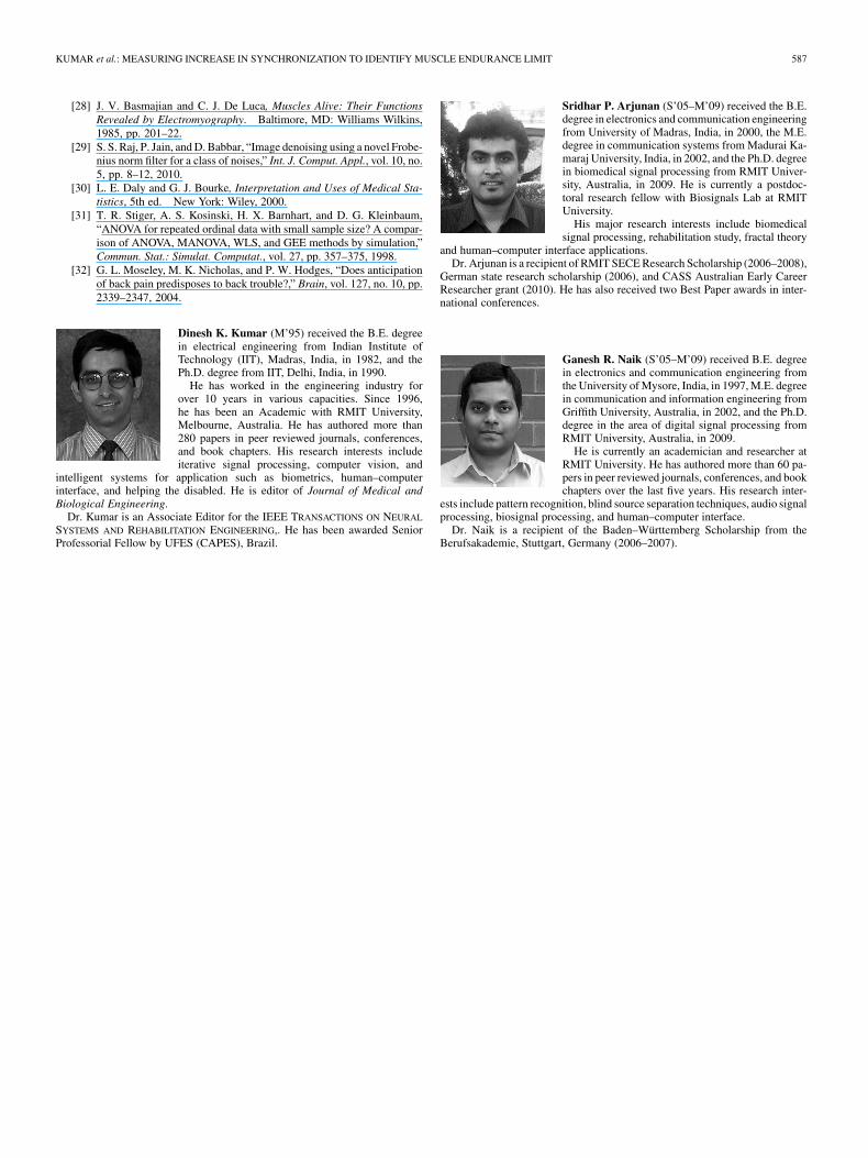
Dinesh K. Kumar (M’95) received the B.E. degreein electrical engineering from Indian Institute ofTechnology (IIT), Madras, India, in 1982, and thePh.D. degree from IIT, Delhi, India, in 1990.
He has worked in the engineering industry forover 10 years in various capacities. Since 1996,he has been an Academic with RMIT University,Melbourne, Australia. He has authored more than280 papers in peer reviewed journals, conferences,and book chapters. His research interests includeiterative signal processing, computer vision, and
intelligent systems for application such as biometrics, human–computerinterface, and helping the disabled. He is editor of Journal of Medical andBiological Engineering.
Dr. Kumar is an Associate Editor for the IEEE TRANSACTIONS ON NEURAL
SYSTEMS AND REHABILITATION ENGINEERING,. He has been awarded SeniorProfessorial Fellow by UFES (CAPES), Brazil.
Sridhar P. Arjunan (S’05–M’09) received the B.E.degree in electronics and communication engineeringfrom University of Madras, India, in 2000, the M.E.degree in communication systems from Madurai Ka-maraj University, India, in 2002, and the Ph.D. degreein biomedical signal processing from RMIT Univer-sity, Australia, in 2009. He is currently a postdoc-toral research fellow with Biosignals Lab at RMITUniversity.
His major research interests include biomedicalsignal processing, rehabilitation study, fractal theory
and human–computer interface applications.Dr. Arjunan is a recipient of RMIT SECE Research Scholarship (2006–2008),
German state research scholarship (2006), and CASS Australian Early CareerResearcher grant (2010). He has also received two Best Paper awards in inter-national conferences.
Ganesh R. Naik (S’05–M’09) received B.E. degreein electronics and communication engineering fromthe University of Mysore, India, in 1997, M.E. degreein communication and information engineering fromGriffith University, Australia, in 2002, and the Ph.D.degree in the area of digital signal processing fromRMIT University, Australia, in 2009.
He is currently an academician and researcher atRMIT University. He has authored more than 60 pa-pers in peer reviewed journals, conferences, and bookchapters over the last five years. His research inter-
ests include pattern recognition, blind source separation techniques, audio signalprocessing, biosignal processing, and human–computer interface.
Dr. Naik is a recipient of the Baden–Württemberg Scholarship from theBerufsakademie, Stuttgart, Germany (2006–2007).
Copyright © 2022 FDOKUMEN