Supersonic Line Broadening and the Gas Dynamical Evolution of Giant HII Regions
Upload
independentCategory
view
0download
0
J. Chim. Phys. ( 1 999) 96. 161 6-1623 0 EOP Sciences, Las Ulis
Measurement of the dissociation-equilibrium constants for low affinity antibiotic binding interaction with bacterial ribosomes by the T2 (CPMG)
and line-broadening methods
L. verdierl, J. ~harbi-~enarous'", G. ~ertho', P. ~ a u v a i s ~ and J.-P. ~ i raudl , '
' Universite Rene Descartes Paris V, Laboratoire de Chimie et Biochimie Pharmacologiques et Toxicologiques, UMR 8601 du CNRS, 45 rue des Saint-Peres, 75270 Paris cedex 06, France
Universite Denis Diderot Paris VII, UFR chimie, 2 place Jussieu, 75251 Paris cedex 05, France Hoechst Marion Roussel, 102 route de Noisy, 93235 Romainville cedex, France
' Correspondence and reprints.
R ~ S U M ~ ~ Nous avons mesun5 la constante de dissociation, Kd correspondant B I'interaction
faible antibiotique-ribosome bactirien pour des antibiotiques de diffkrentes classes, un macrolide (roxithromycine), un kktolide (HMR 3647) et une lincosamide (clindamycine) avec des ribosomes de diffkrentes souches bactkriennes (E. coli, Staphylococcus aureus sensible ou rksistant A l'erythromycin) par deux mkthodes: I'une basee sur la variation des largeurs de raies et I'autre sur les temps de relaxation transversaux T2 en utilisant une sequence CPMG.
Mots-CIQ. Constante de dissociation, T2 (CPMG), largeur de raies, antibiotique, ribosomes.
ABSTRACT In this study the dissociation constants of the low antibiotic-ribosomes interaction
were determined by the T2 (CPMG), the Cam-Purcell-Meiboom-Gill spin-echo decay rate and the line-broadening methods. Three MLSB antibiotics were studied, a macrolide roxithromycin, a ketolide HMR 3647 and a lincosamide clindamycin for their weak interaction with three bacterial ribosomes, E. coli, Staphylococcus aureus sensitive and resistant to erythromycin.
Key-wards. Dissociation constant, T2 (CPMG), line-broadening, antibiotic, riljosomes

Kd measurement for weak antibiotic-ribosome interaction
INTRODUCTION
MLSB (Macrolide, Lincosamide and Streptogramine B) antibiotics inhibit protein biosynthesis in the elongation step by binding to 50s bacterial ribosomes [I]. There appear to be two stages to this binding: a weak interaction which can be detected by NMR spectroscopy [2] and a stronger interaction (Kd = 10-7-10-9 M) detected by equilibrium dialysis and related methods [3]. It was postulated that the binding to the bacterial ribosomes occurs in a two step process, the first step involving a low affinity pre-inhibition binding site and the second one being the strong interaction responsible for the protein biosynthesis inhibition [4].
The weak binding of macrolides to ribosomes E. coli has been characterized extensively by line-broadening and transferred NOESY (TRNOESY) experiments [2,4], the interaction being a fast exchange on the NMR time scale. To well characterize the weak interaction, we have to determine the low aff~nity antibiotics binding to ribosomes by direct measurement of the dissociation constant (Kd).
There are three main approaches for the NMR measurement of the equilibrium dissociation constant Kd [S]: (i) binding-induced line-broadening in the NMR spectra, (ii) Tip (the decay rate of spin-locked magnetization in the rotating frame), (iii) T;! (CPMG) methods. In this study, we have determined the dissociation constants of the low antibiotic-ribosomes interaction by the T2 (CPMG) and line- broadening methods. Three drugs MLSB antibiotics were studied: (i) a macrolide, roxithromycin [6], (ii) a ketolide, HMR 3647 leader of a new class of antibiotics [7], (iii) a lincosamide, clindamycin [S]. Their weak interaction with three bacterial ribosomes, E. coli, Staphylococcus aureus sensitive and resistant to erythromycin was compared to the biological activity.
EXPERIMENTAL
Ribosome Preparation. The E. coli MRE 600 ribosomes were prepared at Hoechst Marion Roussel (HMR) as described [9] by a tangential ultra filtration technique. S. aureus ribosomes were also prepared at HMR according to a protocol based on high pressure lysis. The ribosomal purity is given by one A260 unity (absorption at 260 nm) which corresponds to 24 pmol ml-1 of 70s ribosomes. 01 1UC4 (HMR) is a sensitive S. aureus to macrolides and 0 1 1.CB20 (HMR) is a MLSB (methylation of ~ 2 0 5 8 in N-6) S. aureus resistant to erythromycin and oxacilline.
NMR Spectroscopy. The antibiotics were dissolved in an aqueous NaDzP04- Na2DP04 buffer (0.05 M), with KC1 (0.2 M) at physiological apparent pH 7.6. To

1618 L. Verdier et a/.
improve its solubility in aqueous solution, the sample is first dissolved in a minute amount of CDjOD, and in this way concentrations of 2.5 mM can be attained.
The experiments were run at 5 0 0 MHz, at 293 K, on a Bruker A M X 5 0 0 spectrometer equipped with a Silicon Graphics workstation.
Spin-spin relaxation (T2) measurements [ l o ] were made using the (CPMG) pulse sequence. The data (after 12 experiments) are plotted as a function of echo evolution time and fitted to a single exponential using a non-linear least squares fitting program. T2 values are reported as time constants of these exponentials.
METHODOLOGY
Equilibrium binding constant determination. Consider a ligand, A, which binds to a macromolecule, R, to form a I : 1 complex A-R. The dissociation constant, Kd, is defined by
A + R , ken, A-R k off
[AI[Rl - k off K d = - [ARI kon
All the fast-exchange N M R parameters can be cast in the following form: Pobs -Pf = ([AR] 1 [A]T)(Pb - Pf) where Pobs = Avobs or l IT2 & while Pf and Pb are the corresponding parameters of the free and bound states, respectively.
The variition of (Pobs -Pf) as a fbnction of [A]T (with [R]T constant) can be analysed in terms of two unknowns (Pot,, -Pf) and Kd by the eq. (1) simplified for a large excess of the free ligand [ 5 ] ,
I [RI~l(Pobs - Pf) = (Kd + [Al~) l (Pb - Pf) I (1) RT and AT are the total concentrations of the macromolecule and ligand respectively. Thus, a plot of [R]rI(Pobs - Pf) versus [A]T allows l/(Pb - Pf) to be obtained from the slope and Kd from the intercept on the y-axis (Fig. 1). With a large excess of the binding ligand, [A] - [A]T - [A]o, the equilibrium binding constant Kd
can be determined from the behaviour of any one of the observable N M R parameters as a function of the initial concentrations of the species involved [5]. The fast- exchange NMR parameters, P can be Av, Ah, IIT2 and l/T1.
[ Rh Fig. 1 The binding constant Kd can be obtained from the vertical intercept of a linear titration plot of eq. ( 1 ) as a function of the concentration
pObs-pf ~d
of h e binding & a n . T. -R pamete r fobs is the line-broadening Av or the transverse
pb - pf relaxation time 1IT2.

Kd measurement for weak antibiotic-ribosome interaction 1619
The binding cons tan t Kd can thus be de termined (i) from l ine-broadening exper iment fol lowing the difference be tween the observed l inewidth at half-height of a l igand r e sonance and the l inewidth of the same nuclei in the free l igand, P0bs = Avobs or (ii) from measuremen t of the Carr -Purce l l -Meiboom-Gi l l ( C P M G ) spin-echo decay rate. In the l imiting case of fast exchange there is a comple te averaging of bo th the re laxat ion rates of the free and bound l igands,
1 /72obs = CLf/T2 f + ctb/72 b Pobs = 1 /T2 obs where a f and a b are the cor responding mole fractions of the free and bound states, respect ively. T h e b ind ing constant Kd is thus obtained from the linear p lo t (Fig. 1).
R E S U L T S A N D D I S C U S S I O N
The initial ant ibiot ic concentrat ion [A]x was 0.5 m M in an aqueous buffer solut ion with r ibosome concent ra t ion [ R ] T constant , and increments of added antibiotic varied in the range o f a few mil l imolar .
Line-Broadening Method. The exper iments were performed with a concentrat ion of E. coli 70S r i bosomes fixed at 1.6 u M for roxi thromycin and c l indamycin and at 0.8 \iM for H M R 3 6 4 7 . The l ine-broadening of the ketol ide, H M R 3647 was too large to a l low l igand s ignals to be detected when r ibosome concent ra t ion was 1.6 u M . The rat io of antibiot ic to r ibosome varied from 300 to 1500 and from 600 to 3000 , respect ively. The l inewidth was measured at different concentra t ions . W e obtained a l inear plot of the l inewidth variation, A v 0 b s versus the antibiot ic concent ra t ion [ A ] T (Fig. 2) .
Fig. 2 ' H - N M R antibiotic line-broadening measured and plotted as a function of H M R 3 6 4 7 antibiotic concentration [ A ] T with ribosome concentration [ R ] T constant (E. coli 0.8 uM)
The different va lues o f the low affinity dissociat ion constant , Kd = 3.4 10" 3 , 0.9 10-3 and 4.0 10" 3 obta ined for roxi thromycin, H M R 3647 (Fig. 2) and c l indamycin ,
- respect ively are in the range of the interaction which can be detected by N M R

1620 L. Verdier eta/.
spectroscopy. They are closely related to the activity of these antibiotics tested against bacteria. As evidenced from the change of experimental conditions (0.8 pM of ribosomes) for the ketolide and from its Kd value (0.9 10-3) for low affinity interaction with ribosomes, the ketolide constitutes a distinct class compared with the other antibiotics tested. This result suggests that chemical exchange for ketolides is slower than for previously studied antibiotics. Another parameter which has to be considered in this method is the magnetic field inhomogeneity. As a result of the difficulty in measuring the intrinsic resonance linewidths, the broadening data did not allow a reliable determination of the binding constant but differential line- broadening was used to identify the contact sites between antibiotics and ribosomes.
T2 (CPMG) Method By adding antibiotic in increments and measuring T2 for every antibiotic concentration, we calculated the Kd with eq. 1 . Typically, the concentration of the ribosomes was 1.6 pM for E. coli and 0.02 pM for sensitive and resistant S. auveus. With increasing antibiotic concentration, the resulting relaxation time T2 also increased, because ab progressively decreased (and af increased) as the low affinity binding sites became occupied [lo].
[AIT (mmolll)
Fig. 3 Kd determination of antibiotics binding to ribosomes with eq. 1 (0) for HMR 3647*, (0) for roxithromycin and (+) for clindamycin. a) E. coli (1.6 pM), b) erythromycin-sensitive S. uureus (E.S.S.A) (0.02 pM) and c) erythromycin-resistant S. aurcus (E.R.S.A) (0.02 pM)
Figure 3 shows titration plots of the observed selective T2 of the 3"-Me protons of roxithromycin, the 6-OMe protons of HMR 3647 and the S-Me protons of clindamycin with different antibiotic/ribosomes ratios. Kd values were reported in Table 1. In agreement with line-broadening experiments, the antibiotic protons relaxed slower (enhanced T2) with a relative increase in antibiotic concentration. Accurate measurement of binding affinity is obtained with this method because the

Kd measurement for weak antibiotic-ribosome interaction 1621
magnetic field inhomogeneity is reduced by spin-echo sequence in the T2 (CPMG) experiment.
Table 1. Kd, TZb and Avb values for antibiotics binding to ribosomes, E. coli (1.6 pM), E.S.S.A (0.02 pM) and E.R.S.A (0.02 pM).
RIBOSOMES E. coli E. S. S. A E.R.S.A
Antibiotic Kd T2b,ps vb,Hz Kd T2blps vb,I.Iz Kd T~L,PS vb,Hz Rox 1.1.10-3 95 3700 3.2.10-3 9 40000 7.2.10-3 4 80000 HMR3647 7.1 10-4* 80 4500 6.9 104 10 37000 5.2 10-3 2 133000 Clinda 5.010-3 75 5100 2.010-3 2 1950002.010~3 4 93000 * The ketolide HMR 3647 is tested with 0.8 pM of E. coli ribosomes.
The values of the low affinity of the antibiotics binding to the E. coli ribosomes (Table 1) show that the ketolide has a higher binding affinity (lower Kd) for the weak interaction site of the E. coli (and sensitive S. aureus) ribosomes than the other antibiotics tested. In the same time, the ketolide displayed a significantly better overall antibiotic activity. This indicates that the measured dissociation equilibrium constants are typical of weak interaction due to ribosomal activity.
'0 0.5 1 1.5 2 2.5 3 [AIT (mmolll)
0 0.5 1 1.5 2 2.5 3 [AIT (mmolll
Fig. 4 Comparison of plots obtained with eq. 1 for HMR 3647 with a) E. coli (0.8 pM) and E.S.SA (0.02 pM) in dotted line and b) E S.SA (0.02 pM) and E.R.S.A (0.02 pM) in dotted line. (0) 6- OMe group, (0) 6-Me group, (') 15-Me group and (A) for 12-Me group.
Similar experiments carried out with S, aureus differed from the previous ones with E. coli ribosomes since a more pronounced broadening did not allow a reliable determination of the binding constant due to the sensitivity limit for the measurement of NMR relaxation parameters. The important broadening observed
1622 L. Verdier eta/.
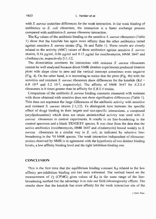
with S. aureus underline differences for the weak interaction. In the weak binding of antibiotics to E. coli ribosomes, the interaction is a faster exchange process compared with antibiotics-S, aureus ribosome interaction.
The Kd values of the antibiotic binding to the sensitive S. aureus ribosomes (Table 1 ) show that the ketolide has again more affinity than the other antibiotics tested against sensitive S. aureus strains (Fig. 3b and Table 1). These results are closely related to the activity (MIC) values of these antibiotics against sensitive S. aureus strains, 0.16 pg/ml, 0.06 pg/ml and 0.12 pg/ml for roxithromycin, HMR 3647 and clindamycin, respectively [ I 1,121.
The dissociation constants for interaction with resistant S. aureus ribosomes cannot be well analyzed because direct NMR titration experiments produced titration plots with slope close to zero and the vertical intercept sometimes even negative (Fig. 4). On the other hand, it is interesting to notice that the plots (Fig. 4b) with the sensitive and resistant S. aureus ribosomes show differences for the ketolide (Kd =
6.9 10-4 and 5.2 10-3, respectively). The affinity of HMR 3647 for ES.S.A ribosomes is 8 times greater than its affinity for E.R.S.A strains.
Comparison of the antibiotic-& aureus binding constants measured with resistant with those obtained with sensitive does not show significative differences (Table 1). This does not represent the large differences of the antibiotic activity with sensitive and resistant S. aureus strains [11,12]. To distinguish here between the specific effect of drugs binding to their targets and non-specific interactions, a compound (erytlyalosamine) which does not retain antimicrobial activity was used with S. aureus ribosomes in control experiments. It results in no line-broadening in the control spectrum and a blank TRNOESY spectra. It was clear from the data that the active antibiotics (roxithromycin, HMR 3647 and clindamycin) bound weakly to S. aureus ribosomes in a similar way to E. coli, as indicated by selective line- broadening in the 1H NMR spectra. The weak interaction independent of ribosome- strains observed by NMR is in agreement with the hypothesis of two distinct binding levels, a low affinity binding level and the tight inhibition binding one.
CONCLUSION
This is the first time that the equilibrium binding constant Kd related to the low affinity pre-inhibition binding site has been estimated. The method based on the measurement of T2 (CPMG) gives values of Kd in the same range of the line- broadening method but the advantage is to rule out field inhomogeneity effects. The results show that the ketolide has more affinity for the weak interaction site of the

Kd measurement for weak antibiotic-ribosome interaction 1623
bacterial r ibosomes than the other ant ibiot ics tested. The values o f weak affinity are
c losely related to the activity o f the M L S antibiotics against bacter ia p rov id ing a
re la t ionship be tween weak interaction and affinity.
The important b roadening observed in the s tudy with S. aureus r i b o s o m e s shows
that for the weak b inding of ant ibiot ics , the interaction is a s lower exchange process
compared with E. coli r ibosomes . The dimethylat ion of adenine 2058 in 23S r R N A
which renders bacteria resistant to the M L S antibiotics [13] should not interfere with
the weak interaction. The values of Kd for roxi thromycin, H M R 3647 and
c l indamycin are found in the same range with the three bacterial r ibosomes , E. coli,
S. aureus sensi t ive and resistant to e ry thromycin what is in ag reemen t wi th the
hypothes i s o f two distinct b ind ing levels.
R E F E R E N C E S
1 M e n n i n g e r J. R. ( 1995) J. Bas & Clin. Phys & Pharm 6, 229-247.
2 Ber tho G., Gharb i -Benarous J., Ladam P., Delaforge M., Giraul t J. P. (1998)
Bioorg. Med. Chem. 6, 2 0 9 - 2 2 1 .
3 Pestka S. (1974) Antimicrob. Agents Chemother. 6, 474-478 .
4 Barber J., Gyi J. I., Pye D. A. ( 1 9 9 1 ) 7 . Chem. Soc, Chem. Commun. 1249-1252.
5 N i F. ( 1994) Progr. Nuct. Mag. Reson. Spectrosc. 26, 517-606 .
6 Gharb i -Benarous J., Delaforge M. , Jankowski C. K., Giraul t J. P. (1991) J. Med.
Chem. 3 4 , 1117-1125.
7 Agour idas C , Denis A., Auge r J. M. , Benedet t i Y., Bonnefoy A., Bretin F. ,
Chan to t J. F., Dussara t A., Froment in C , Goin D'Ambrières S., Lachaud S., Laur in
P., Le Martret O., Loyau V., Tessot N . (1998) J. Med. Chem. 4 1 , 4080-4100 .
8 Bi rkenmeyer R. D., Kagan F. (1970) J. Med. Chem. 1 3 , 616-619 .
9 Jelenc P. (1980) Anal. Biochem. 1 0 5 , 369-374 .
10 Dubois B. W., Evers A. S. (1992) Biochemistry 3 1 , 7069-7076 .
11 Jones R. N. , B iedenbach D. J. ( 1997) Diagn. Microbiol. Infect. Dis 27 , 7-12.
12 Fournet M. P. , Barre J., Zini R., Deforges L., Duval J., T i l l ement J. P. (1987) J.
Pharm. Pharmacol. 3 9 , 319-322 .
13 Go ldman R. C , Kadam S. K. (1989) Antimicrob. Agents Chemother. 3 3 , 1058-
1066.
Copyright © 2022 FDOKUMEN