Matrix-Assisted Laser Desorption Ionization-Time of Flight Mass Spectrometry of Lipopeptide...
11
10.1128/AEM.68.12.6210-6219.2002. 2002, 68(12):6210. DOI: Appl. Environ. Microbiol. Franke, Neena Mehta and Swaranjit Singh Cameotra Joachim Vater, Bärbel Kablitz, Christopher Wilde, Peter Petroleum Sludge C-1 Isolated from Bacillus subtilis in Whole Cells and Culture Filtrates of Spectrometry of Lipopeptide Biosurfactants Ionization-Time of Flight Mass Matrix-Assisted Laser Desorption http://aem.asm.org/content/68/12/6210 Updated information and services can be found at: These include: REFERENCES http://aem.asm.org/content/68/12/6210#ref-list-1 This article cites 38 articles, 5 of which can be accessed free at: CONTENT ALERTS more» articles cite this article), Receive: RSS Feeds, eTOCs, free email alerts (when new http://journals.asm.org/site/misc/reprints.xhtml Information about commercial reprint orders: http://journals.asm.org/site/subscriptions/ To subscribe to to another ASM Journal go to: on May 25, 2014 by guest http://aem.asm.org/ Downloaded from on May 25, 2014 by guest http://aem.asm.org/ Downloaded from
-
Upload
independent -
Category
Documents
-
view
0 -
download
0
Transcript of Matrix-Assisted Laser Desorption Ionization-Time of Flight Mass Spectrometry of Lipopeptide...
10.1128/AEM.68.12.6210-6219.2002.
2002, 68(12):6210. DOI:Appl. Environ. Microbiol. Franke, Neena Mehta and Swaranjit Singh CameotraJoachim Vater, Bärbel Kablitz, Christopher Wilde, Peter Petroleum Sludge
C-1 Isolated fromBacillus subtilisin Whole Cells and Culture Filtrates of Spectrometry of Lipopeptide BiosurfactantsIonization-Time of Flight Mass Matrix-Assisted Laser Desorption
http://aem.asm.org/content/68/12/6210Updated information and services can be found at:
These include:
REFERENCEShttp://aem.asm.org/content/68/12/6210#ref-list-1This article cites 38 articles, 5 of which can be accessed free at:
CONTENT ALERTS more»articles cite this article),
Receive: RSS Feeds, eTOCs, free email alerts (when new
http://journals.asm.org/site/misc/reprints.xhtmlInformation about commercial reprint orders: http://journals.asm.org/site/subscriptions/To subscribe to to another ASM Journal go to:
on May 25, 2014 by guest
http://aem.asm
.org/D
ownloaded from
on M
ay 25, 2014 by guesthttp://aem
.asm.org/
Dow
nloaded from
APPLIED AND ENVIRONMENTAL MICROBIOLOGY, Dec. 2002, p. 6210–6219 Vol. 68, No. 120099-2240/02/$04.00�0 DOI: 10.1128/AEM.68.12.6210–6219.2002Copyright © 2002, American Society for Microbiology. All Rights Reserved.
Matrix-Assisted Laser Desorption Ionization–Time of Flight MassSpectrometry of Lipopeptide Biosurfactants in Whole Cells and
Culture Filtrates of Bacillus subtilis C-1 Isolated fromPetroleum Sludge
Joachim Vater,1* Barbel Kablitz,1 Christopher Wilde,1 Peter Franke,2 Neena Mehta,3 andSwaranjit Singh Cameotra3
Institut fur Chemie, Arbeitsgruppe Biochemie und Molekulare Biologie, Technische Universitat Berlin,D-10587 Berlin,1 and Institut fur Biochemie, Freie Universitat Berlin, D-14195 Berlin,2 Germany,
and Institute of Microbial Technology, Sector 39A, Chandigarh-160 036, India3
Received 6 May 2002/Accepted 1 August 2002
An innovative method was developed for rapid sensitive detection and efficient structural characterization oflipopeptide biosurfactants by matrix-assisted laser desorption ionization–time of flight (MALDI-TOF) massspectrometry by using whole microbial cells and crude culture filtrates as targets in combination with surfacetension measurements. This was done for a bacterial strain that was isolated from petroleum sludge andefficiently produces biosurfactants. This organism was identified by using biochemical, physiological, andgenetic parameters as a Bacillus subtilis strain, designated B. subtilis C-1. This assignment was supported by amass spectrometric investigation of the secondary metabolite spectrum determined by whole-cell MALDI-TOFmass spectrometry, which revealed three lipopeptide complexes, the surfactins, the iturins, and the fengycins,which are well-known biosurfactants produced by B. subtilis strains. These compounds were structurallycharacterized by in situ structure analysis by using postsource decay MALDI-TOF mass spectrometry. Theisoforms were separated by miniaturized high-resolution reversed-phase high-performance liquid chromatog-raphy for mass spectrometric characterization. Iturin compounds which contain unusual fatty acid compo-nents were detected.
Biosurfactants are a structurally diverse group of surface-active molecules synthesized by microorganisms (5, 7, 9, 10, 32,36). They have unique amphipathic properties derived fromtheir complex structures, which include a hydrophilic moietyand a hydrophobic portion. The most efficient biosurfactantsreduce the surface tension of water from 72 dynes/cm to valuesin the range of 25 to 30 dynes/cm. Biosurfactant productioncan be determined by measuring the change in surface tensionof cell-free culture broth.
Microbial surfactants have commonly been classified intothe following categories: (i) glycolipids, (ii) lipopeptides, (iii)fatty acids, neutral lipids, and phospholipids, (iv) polymericsurfactants, and (v) particulate biosurfactants (5, 7, 9, 10, 32,36). The lipopeptides are an interesting class of microbial sur-factants (36) because of their manifold attractive properties.Members of this group often possess antibiotic activity as well.
Bacillus subtilis strains produce a broad spectrum of bioac-tive peptides with great potential for biotechnological and bio-pharmaceutical applications. A well-known class of such com-pounds includes the lipopeptides surfactin (1, 13, 14, 17, 18),fengycin (35), and the iturin compounds (3) (iturins [26], my-cosubtilins [25], and bacillomycins [27]), which are amphiphilicmembrane-active biosurfactants and peptide antibiotics withpotent antimicrobial activities. All these agents occur as fam-
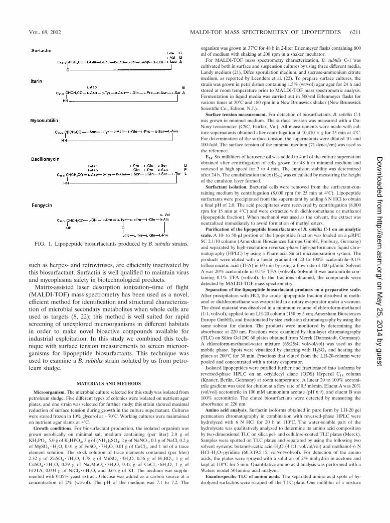
ilies of closely related isoforms which differ in the length andbranching of the fatty acid side chains and in the amino acidsubstitutions in the peptide rings (20, 36). The surfactin anditurin compounds are cyclic lipoheptapeptides which contain a�-hydroxy fatty acid and a �-amino fatty acid, respectively, aslipophilic components. Fengycin is a lipodecapeptide with a�-hydroxy fatty acid in its side chain. The structures of thesebiosurfactants are shown in Fig. 1.
Comparative studies of the interfacial and emulsifying prop-erties of the lipopeptide biosurfactants produced by B. subtilishave been performed by Deleu et al. (6). All these compoundsefficiently reduce the interfacial tension at the oil-water inter-face; the surfactins have the greatest effects, while the iturinsand fengycins have lower surfactant strength. In particular,surfactin (30, 36) is one of the most thoroughly studied andbest-characterized biosurfactants. Bacillus licheniformis (9, 11,12, 39) and Bacillus pumilus (23) also produce lipopeptidessimilar to surfactin. These agents are natural compounds thathave industrial importance and have attracted increasing bio-technological and pharmaceutical interest. They are distin-guished by excellent surface- and membrane-active propertiesalong with superior emulsifying and foaming properties (6, 31),which can be utilized in food biotechnology and in the agricul-tural sector. The lipopeptides belonging to the iturin family arepotent antifungal agents which can be used as biopesticides forplant protection. In recent studies important biopharmaceuti-cal applications of surfactins have been demonstrated. Surfac-tin shows potent antiviral and antimycoplasma activities (37,38). Viruses which are covered by a lipoprotein membrane,
* Corresponding author. Mailing address: Institut fur Chemie, Ar-beitsgruppe Biochemie und Molekulare Biologie, Technische Univer-sitat Berlin, Franklinstr. 29, D-10587 Berlin, Germany. Phone: 49-30-31425609. Fax: 49-30-31424783. E-mail: [email protected].
6210
on May 25, 2014 by guest
http://aem.asm
.org/D
ownloaded from
such as herpes- and retroviruses, are efficiently inactivated bythis biosurfactant. Surfactin is well qualified to maintain virusand mycoplasma safety in biotechnological products.
Matrix-assisted laser desorption ionization–time of flight(MALDI-TOF) mass spectrometry has been used as a novel,efficient method for identification and structural characteriza-tion of microbial secondary metabolites when whole cells areused as targets (8, 22); this method is well suited for rapidscreening of unexplored microorganisms in different habitatsin order to make novel bioactive compounds available forindustrial exploitation. In this study we combined this tech-nique with surface tension measurements to screen microor-ganisms for lipopeptide biosurfactants. This technique wasused to examine a B. subtilis strain isolated by us from petro-leum sludge.
MATERIALS AND METHODS
Microorganism. The microbial culture selected for this study was isolated frompetroleum sludge. Five different types of colonies were isolated on nutrient agarplates, and one strain was selected for further study; this strain showed maximalreduction of surface tension during growth in the culture supernatant. Cultureswere stored frozen in 10% glycerol at �70°C. Working cultures were maintainedon nutrient agar slants at 4°C.
Growth conditions. For biosurfactant production, the isolated organism wasgrown aerobically on minimal salt medium containing (per liter) 2.0 g ofKH2PO4, 5.0 g of K2HPO4, 3 g of (NH4)2SO4, 2 g of NaNO3, 0.1 g of NaCl, 0.2 gof MgSO4 � H2O, 0.01 g of FeSO4 � 7H2O, 0.01 g of CaCl2, and 1 ml of a traceelement solution. The stock solution of trace elements contained (per liter)2.32 g of ZnSO4 � 7H2O, 1.78 g of MnSO4 � 4H2O, 0.56 g of H3BO3, 1 g ofCuSO4 � 5H2O, 0.39 g of Na2MoO4 � 7H2O, 0.42 g of CoCl2 � 6H2O, 1 g ofEDTA, 0.004 g of NiCl2 � 6H2O, and 0.66 g of KI. The medium was supple-mented with 0.05% yeast extract. Glucose was added as a carbon source at aconcentration of 2% (wt/vol). The pH of the medium was 7.1 to 7.2. The
organism was grown at 37°C for 48 h in 2-liter Erlenmeyer flasks containing 800ml of medium with shaking at 200 rpm in a shaker incubator.
For MALDI-TOF mass spectrometry characterization, B. subtilis C-1 wascultivated both in surface and suspension cultures by using three different media,Landy medium (21), Difco sporulation medium, and sucrose-ammonium citratemedium, as reported by Leenders et al. (22). To prepare surface cultures, thestrain was grown in petri dishes containing 1.5% (wt/vol) agar agar for 24 h andstored at room temperature prior to MALDI-TOF mass spectrometric analysis.Fermentation in liquid media was carried out in 500-ml Erlenmeyer flasks forvarious times at 30°C and 180 rpm in a New Brunswick shaker (New BrunswickScientific Co., Edison, N.J.).
Surface tension measurement. For detection of biosurfactants, B. subtilis C-1was grown in minimal medium. The surface tension was measured with a Du-Nouy tensiometer (CSC, Fairfax, Va.). All measurements were made with cul-ture supernatants obtained after centrifugation at 10,410 � g for 25 min at 4°C.For determination of the surface tension, the supernatants were diluted 10- and100-fold. The surface tension of the minimal medium (71 dynes/cm) was used asthe reference.
E24. Six milliliters of kerosene oil was added to 4 ml of the culture supernatantobtained after centrifugation of cells grown for 48 h in minimal medium andvortexed at high speed for 3 to 4 min. The emulsion stability was determinedafter 24 h. The emulsification index (E24) was calculated by measuring the heightof the emulsion layer formed.
Surfactant isolation. Bacterial cells were removed from the surfactant-con-taining medium by centrifugation (8,000 rpm for 25 min at 4°C). Lipopeptidesurfactants were precipitated from the supernatant by adding 6 N HCl to obtaina final pH of 2.0. The acid precipitates were recovered by centrifugation (8,000rpm for 15 min at 4°C) and were extracted with dichloromethane or methanol(lipopeptide fraction). When methanol was used as the solvent, the extract wasneutralized immediately to avoid formation of methyl esters.
Purification of the lipopeptide biosurfactants of B. subtilis C-1 on an analyticscale. A 10- to 50-�l portion of the lipopeptide fraction was loaded on a �RPCSC 2.1/10 column (Amersham Biosciences Europe GmbH, Freiburg, Germany)and separated by high-resolution reversed-phase high-performance liquid chro-matography (HPLC) by using a Pharmacia Smart microseparation system. Theproducts were eluted with a linear gradient of 20 to 100% acetonitrile–0.1%trifluoroacetic acid (TFA) in 60 min by using a flow rate of 100 �l/min. SolventA was 20% acetonitrile in 0.1% TFA (vol/vol). Solvent B was acetonitrile con-taining 0.1% TFA (vol/vol). In the fractions obtained, the compounds weredetected by MALDI-TOF mass spectrometry.
Separation of the lipopeptide biosurfactant products on a preparative scale.After precipitation with HCl, the crude lipopeptide fraction dissolved in meth-anol or dichloromethane was evaporated in a rotary evaporator under a vacuum.The dried material was dissolved in a minimum volume of chloroform-methanol(1:1, vol/vol), applied to an LH-20 column (150 by 5 cm; Amersham BiosciencesEurope GmbH), and fractionated by size exclusion chromatography by using thesame solvent for elution. The products were monitored by determining theabsorbance at 220 nm. Fractions were examined by thin-layer chromatography(TLC) on Silica Gel DC 60 plates obtained from Merck (Darmstadt, Germany).A chloroform-methanol-water mixture (65:25:4, vol/vol/vol) was used as themobile phase. Spots were visualized by charring with H2SO4 and heating theplates at 200°C for 30 min. Fractions that eluted from the LH-20-column werepooled and concentrated with a rotary evaporator.
Isolated lipopeptides were purified further and fractionated into isoforms byreversed-phase HPLC on an octyldecyl silane (ODS) Hypersil C18 column(Knauer, Berlin, Germany) at room temperature. A linear 20 to 100% acetoni-trile gradient was used for elution at a flow rate of 0.5 ml/min. Eluent A was 20%(vol/vol) acetonitrile in 100 mM ammonium acetate (pH 6.9), and eluent B was100% acetonitrile. The eluted biosurfactants were detected by measuring theabsorbance at 220 nm.
Amino acid analysis. Surfactin isoforms obtained in pure form by LH-20 gelpermeation chromatography in combination with reversed-phase HPLC werehydrolyzed with 6 N HCl for 20 h at 110°C. The water-soluble part of thehydrolysate was qualitatively analyzed to determine its amino acid compositionby two-dimensional TLC on silica gel- and cellulose-coated TLC plates (Merck).Samples were spotted on TLC plates and separated by using the following twosolvent systems: butanol-acetic acid-H2O (4:1:1, vol/vol/vol) and methanol–6 NHCl–H2O–pyridine (60:3:19.5:15, vol/vol/vol/vol). For detection of the aminoacids, the plates were sprayed with a solution of 2% ninhydrin in acetone andkept at 110°C for 5 min. Quantitative amino acid analysis was performed with aWaters model 501amino acid analyzer.
Enantiospecific TLC of amino acids. The separated amino acid spots of hy-drolyzed surfactins were scraped off the TLC plate. One milliliter of a mixture
FIG. 1. Lipopeptide biosurfactants produced by B. subtilis strains.
VOL. 68, 2002 MALDI-TOF MASS SPECTROMETRY OF LIPOPEPTIDES 6211
on May 25, 2014 by guest
http://aem.asm
.org/D
ownloaded from
containing methanol and water (1:1, vol/vol) was added to each sample. Thesilica gel was removed by centrifugation. The extracted amino acids were spottedon chiral TLC plates (Merck) along with standard L and D amino acids (SigmaChemical Co., St. Louis, Mo.). The solvent system used was acetonitrile-meth-anol-water (4:1:1, vol/vol/vol). The spots were detected by spraying the plateswith a solution of 2% ninhydrin in acetone.
MALDI-TOF mass spectrometry analysis. MALDI-TOF mass spectra wererecorded by using a Bruker Daltonik Reflex MALDI-TOF instrument containinga 337-nm nitrogen laser for desorption and ionization. For mass spectrometricanalysis of isolated lipopeptide biosurfactants, 1- to 2-�l portions of fractionsobtained after gel filtration on Sephadex LH-20 or after reversed-phase HPLCwere each mixed with an equal volume of matrix medium (a saturated solutionof �-cyano-4-hydroxycinnamic acid in 70% aqueous acetonitrile containing 0.1%[vol/vol] TFA). For product analysis with whole bacterial cells, cell material waspicked from agar plates, spotted onto the sample target, covered with matrixmedium, and air dried. Positive-ion detection and the reflector mode were used.The acceleration and reflector voltages were 20 and 23.4 kV in pulsed ionextraction mode. A molecular mass gate of 300 Da improved the measurementby filtering out most matrix ions. Postsource decay (PSD) mass spectra wereobtained with the same samples.
RESULTS
Growth of and biosurfactant production by B. subtilis C-1 atdifferent temperatures. A bacterial strain that produced bio-surfactants was isolated from petroleum sludge. This organismis a gram-positive, spore-forming, rod-like, oval bacterial strainwhich has been characterized by the Deutsche Sammlung vonMikroorganismen und Zellkulturen GmbH Identification Ser-vice as a member of the B. subtilis group. This classification wasbased mainly on 16S ribosomal DNA data and the riboprintpattern. Partial sequencing of the 16S ribosomal DNA re-vealed 99.8% identity with B. subtilis. In addition, some bio-chemical and physiological parameters were determined whichsupport this classification. This organism was designated B.subtilis C-1. It was cultivated in minimal medium at 25, 30, and45°C for 48 h at 200 rpm in a shaker incubator. Growth curvesat all temperatures were determined by monitoring the opticaldensity at 600 nm at different times. Optimal growth was ob-served at 30°C. At 25 and 30°C the stationary phase wasreached after 30 h. Biosurfactant compounds were detected bysurface tension measurement. The surface tension values areshown in Table 1. The data obtained indicate that there wasefficient biosurfactant production which was higher at 25 and30°C than at 45°C.
The dry biomass (0.33 g/100 ml) and the crude biosurfactantmass (54.7 mg/100 ml) obtained after growth of the organismat 30°C were significantly greater than the dry biomass and thecrude biosurfactant mass obtained after the organism wasgrown at 25 and 45°C (Table 2).
The data shown in Table 2 also demonstrate that the culture
supernatant was able to emulsify oil. The highest E24 value wasobserved at 30°C. On the basis of these data further studies toisolate and characterize the biosurfactant(s) produced wereperformed by growing the isolated B. subtilis strain at 30°C indifferent culture media.
Innovative method for detection and in situ structure anal-ysis of lipopeptide biosurfactants by MALDI-TOF mass spec-trometry of whole bacterial cells and of the crude fermentationbroth. To detect and identify the biosurfactants produced byB. subtilis C-1, we developed an innovative mass spectromet-ric method. The products were investigated by performingMALDI-TOF mass spectrometry of whole bacterial cells. Todo this, B. subtilis C-1 was grown on agar plates at 30°C for 24 hby using Landy medium, Difco sporulation medium, or ammo-nium citrate-sucrose medium. Samples of cells were pickedfrom the agar plates and embedded in an �-cyanocinnamicacid matrix solution directly on the target. After air drying, thesecondary metabolite spectrum of the strain was determinedwith a Bruker Daltonix Reflex MALDI-TOF mass spectrom-eter.
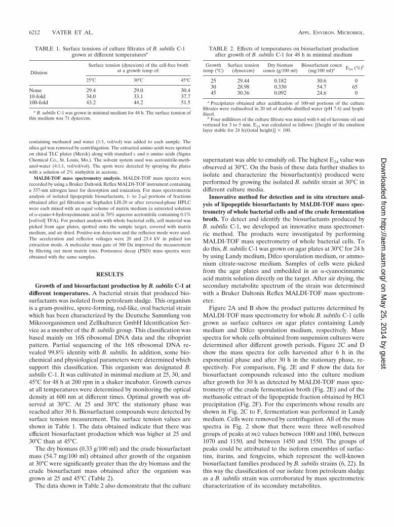
Figure 2A and B show the product patterns determined byMALDI-TOF mass spectrometry for whole B. subtilis C-1 cellsgrown as surface cultures on agar plates containing Landymedium and Difco sporulation medium, respectively. Massspectra for whole cells obtained from suspension cultures weredetermined after different growth periods. Figure 2C and Dshow the mass spectra for cells harvested after 6 h in theexponential phase and after 30 h in the stationary phase, re-spectively. For comparison, Fig. 2E and F show the data forbiosurfactant compounds released into the culture mediumafter growth for 30 h as detected by MALDI-TOF mass spec-trometry of the crude fermentation broth (Fig. 2E) and of themethanolic extract of the lipopeptide fraction obtained by HClprecipitation (Fig. 2F). For the experiments whose results areshown in Fig. 2C to F, fermentation was performed in Landymedium. Cells were removed by centrifugation. All of the massspectra in Fig. 2 show that there were three well-resolvedgroups of peaks at m/z values between 1000 and 1060, between1070 and 1150, and between 1450 and 1550. The groups ofpeaks could be attributed to the isoform ensembles of surfac-tins, iturins, and fengycins, which represent the well-knownbiosurfactant families produced by B. subtilis strains (6, 22). Inthis way the classification of our isolate from petroleum sludgeas a B. subtilis strain was corroborated by mass spectrometriccharacterization of its secondary metabolites.
TABLE 1. Surface tensions of culture filtrates of B. subtilis C-1grown at different temperaturesa
Dilution
Surface tension (dynes/cm) of the cell-free brothat a growth temp of:
25°C 30°C 45°C
None 29.4 29.0 30.410-fold 34.0 33.1 37.7100-fold 43.2 44.2 51.5
a B. subtilis C-1 was grown in minimal medium for 48 h. The surface tension ofthis medium was 71 dynes/cm.
TABLE 2. Effects of temperatures on biosurfactant productionafter growth of B. subtilis C-1 for 48 h in minimal medium
Growthtemp (°C)
Surface tension(dynes/cm)
Dry biomassconcn (g/100 ml)
Biosurfactant concn(mg/100 ml)a E24 (%)b
25 29.44 0.182 30.6 030 28.98 0.330 54.7 6545 30.36 0.092 24.6 0
a Precipitates obtained after acidification of 100-ml portions of the culturefiltrates were redissolved in 20 ml of double-distilled water (pH 7.4) and lyoph-ilized.
b Four milliliters of the culture filtrate was mixed with 6 ml of kerosine oil andvortexed for 3 to 5 min. E24 was calculated as follows: [(height of the emulsionlayer stable for 24 h)/(total height)] � 100.
6212 VATER ET AL. APPL. ENVIRON. MICROBIOL.
on May 25, 2014 by guest
http://aem.asm
.org/D
ownloaded from
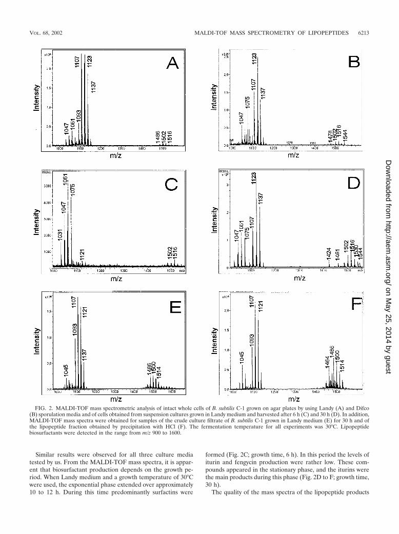
Similar results were observed for all three culture mediatested by us. From the MALDI-TOF mass spectra, it is appar-ent that biosurfactant production depends on the growth pe-riod. When Landy medium and a growth temperature of 30°Cwere used, the exponential phase extended over approximately10 to 12 h. During this time predominantly surfactins were
formed (Fig. 2C; growth time, 6 h). In this period the levels ofiturin and fengycin production were rather low. These com-pounds appeared in the stationary phase, and the iturins werethe main products during this phase (Fig. 2D to F; growth time,30 h).
The quality of the mass spectra of the lipopeptide products
FIG. 2. MALDI-TOF mass spectrometric analysis of intact whole cells of B. subtilis C-1 grown on agar plates by using Landy (A) and Difco(B) sporulation media and of cells obtained from suspension cultures grown in Landy medium and harvested after 6 h (C) and 30 h (D). In addition,MALDI-TOF mass spectra were obtained for samples of the crude culture filtrate of B. subtilis C-1 grown in Landy medium (E) for 30 h and ofthe lipopeptide fraction obtained by precipitation with HCl (F). The fermentation temperature for all experiments was 30°C. Lipopeptidebiosurfactants were detected in the range from m/z 900 to 1600.
VOL. 68, 2002 MALDI-TOF MASS SPECTROMETRY OF LIPOPEPTIDES 6213
on May 25, 2014 by guest
http://aem.asm
.org/D
ownloaded from
of B. subtilis C-1 obtained for whole cells and for the crude,unfractionated culture supernatant is similar to the quality ofthe spectra obtained with solutions of the purified compounds.The results which are summarized in Fig. 2 demonstrate thatby using MALDI-TOF mass spectrometry lipopeptide biosur-factants can be detected in minutes with high sensitivity, pre-cision, and excellent resolution without a requirement fortime-consuming isolation and chromatographic separation ofthe compounds.
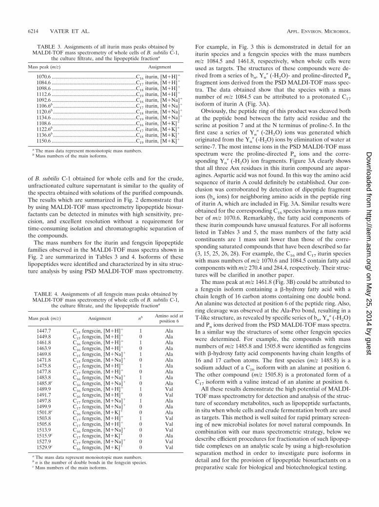
The mass numbers for the iturin and fengycin lipopeptidefamilies observed in the MALDI-TOF mass spectra shown inFig. 2 are summarized in Tables 3 and 4. Isoforms of theselipopeptides were identified and characterized by in situ struc-ture analysis by using PSD MALDI-TOF mass spectrometry.
For example, in Fig. 3 this is demonstrated in detail for aniturin species and a fengycin species with the mass numbersm/z 1084.5 and 1461.8, respectively, when whole cells wereused as targets. The structures of these compounds were de-rived from a series of bn, Yn� (-H2O)- and proline-directed Pn
fragment ions derived from the PSD MALDI-TOF mass spec-tra. The data obtained show that the species with a massnumber of m/z 1084.5 can be attributed to a protonated C17
isoform of iturin A (Fig. 3A).Obviously, the peptide ring of this product was cleaved both
at the peptide bond between the fatty acid residue and theserine at position 7 and at the N terminus of proline-5. In thefirst case a series of Yn� (-2H2O) ions was generated whichoriginated from the Yn� (-H2O) ions by elimination of water atserine-7. The most intense ions in the PSD MALDI-TOF massspectrum were the proline-directed Pn ions and the corre-sponding Yn� (-H2O) ion fragments. Figure 3A clearly showsthat all three Asx residues in this iturin compound are aspar-agines. Aspartic acid was not found. In this way the amino acidsequence of iturin A could definitely be established. Our con-clusion was corroborated by detection of dipeptide fragmentions (bn ions) for neighboring amino acids in the peptide ringof iturin A, which are included in Fig. 3A. Similar results wereobtained for the corresponding C16 species having a mass num-ber of m/z 1070.6. Remarkably, the fatty acid components ofthese iturin compounds have unusual features. For all isoformslisted in Tables 3 and 5, the mass numbers of the fatty acidconstituents are 1 mass unit lower than those of the corre-sponding saturated compounds that have been described so far(3, 15, 25, 26, 28). For example, the C16 and C17 iturin specieswith mass numbers of m/z 1070.6 and 1084.5 contain fatty acidcomponents with m/z 270.4 and 284.4, respectively. Their struc-tures will be clarified in another paper.
The mass peak at m/z 1461.8 (Fig. 3B) could be attributed toa fengycin isoform containing a �-hydroxy fatty acid with achain length of 16 carbon atoms containing one double bond.An alanine was detected at position 6 of the peptide ring. Also,ring cleavage was observed at the Ala-Pro bond, resulting in aT-like structure, as revealed by specific series of bn, Yn� (-H2O)and Pn ions derived from the PSD MALDI-TOF mass spectra.In a similar way the structures of some other fengycin specieswere determined. For example, the compounds with massnumbers of m/z 1485.8 and 1505.8 were identified as fengycinswith �-hydroxy fatty acid components having chain lengths of16 and 17 carbon atoms. The first species (m/z 1485.8) is asodium adduct of a C16 isoform with an alanine at position 6.The other compound (m/z 1505.8) is a protonated form of aC17 isoform with a valine instead of an alanine at position 6.
All these results demonstrate the high potential of MALDI-TOF mass spectrometry for detection and analysis of the struc-ture of secondary metabolites, such as lipopeptide surfactants,in situ when whole cells and crude fermentation broth are usedas targets. This method is well suited for rapid primary screen-ing of new microbial isolates for novel natural compounds. Incombination with our mass spectrometric strategy, below wedescribe efficient procedures for fractionation of such lipopep-tide complexes on an analytic scale by using a high-resolutionseparation method in order to investigate pure isoforms indetail and for the provision of lipopeptide biosurfactants on apreparative scale for biological and biotechnological testing.
TABLE 3. Assignments of all iturin mass peaks obtained byMALDI-TOF mass spectrometry of whole cells of B. subtilis C-1,
the culture filtrate, and the lipopeptide fractiona
Mass peak (m/z) Assignment
1070.6 .................................................................C16 iturin, [M�H]�
1084.6 .................................................................C17 iturin, [M�H]�
1098.6 .................................................................C18 iturin, [M�H]�
1112.6 .................................................................C19 iturin, [M�H]�
1092.6 .................................................................C16 iturin, [M�Na]�
1106.6b................................................................C17 iturin, [M�Na]�
1120.6b................................................................C18 iturin, [M�Na]�
1134.6 .................................................................C19 iturin, [M�Na]�
1108.6 .................................................................C16 iturin, [M�K]�
1122.6b................................................................C17 iturin, [M�K]�
1136.6b................................................................C18 iturin, [M�K]�
1150.6 .................................................................C19 iturin, [M�K]�
a The mass data represent monoisotopic mass numbers.b Mass numbers of the main isoforms.
TABLE 4. Assignments of all fengycin mass peaks obtained byMALDI-TOF mass spectrometry of whole cells of B. subtilis C-1,
the culture filtrate, and the lipopeptide fractiona
Mass peak (m/z) Assignment nb Amino acid atposition 6
1447.7 C15 fengycin, [M�H]� 1 Ala1449.8 C15 fengycin, [M�H]� 0 Ala1461.8 C16 fengycin, [M�H]� 1 Ala1463.9 C16 fengycin, [M�H]� 0 Ala1469.8 C15 fengycin, [M�Na]� 1 Ala1471.8 C15 fengycin, [M�Na]� 0 Ala1475.8 C17 fengycin, [M�H]� 1 Ala1477.8 C17 fengycin, [M�H]� 0 Ala1483.8 C16 fengycin, [M�Na]� 1 Ala1485.8c C16 fengycin, [M�Na]� 0 Ala1489.9 C16 fengycin, [M�H]� 1 Val1491.7 C16 fengycin, [M�H]� 0 Val1497.8 C17 fengycin, [M�Na]� 1 Ala1499.9 C17 fengycin, [M�Na]� 0 Ala1501.8c C16 fengycin, [M�K]� 0 Ala1503.8 C17 fengycin, [M�H]� 1 Val1505.8 C17 fengycin, [M�H]� 0 Val1513.9 C16 fengycin, [M�Na]� 0 Val1515.9c C17 fengycin, [M�K]� 0 Ala1527.9 C17 fengycin, [M�Na]� 0 Val1529.9c C16 fengycin, [M�K]� 0 Val
a The mass data represent monoisotopic mass numbers.b n is the number of double bonds in the fengycin species.c Mass numbers of the main isoforms.
6214 VATER ET AL. APPL. ENVIRON. MICROBIOL.
on May 25, 2014 by guest
http://aem.asm
.org/D
ownloaded from
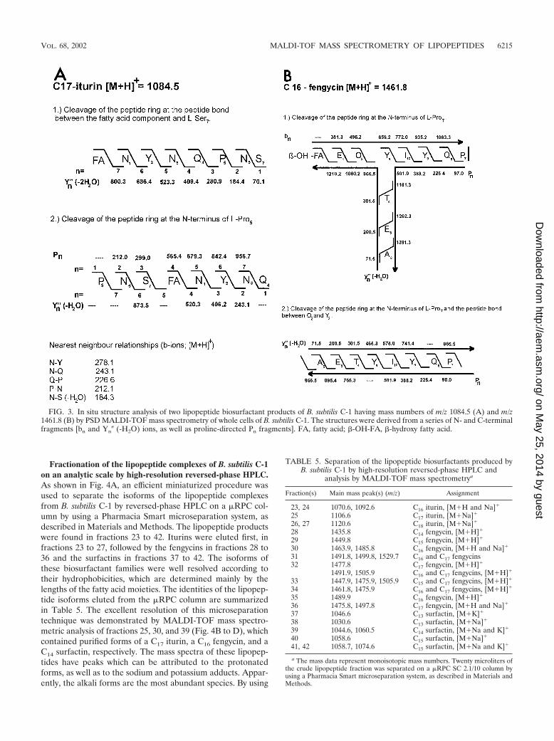
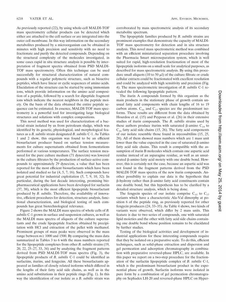
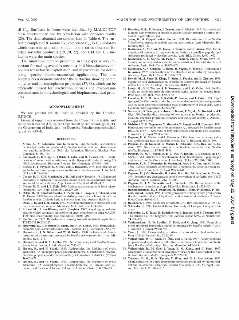
Fractionation of the lipopeptide complexes of B. subtilis C-1on an analytic scale by high-resolution reversed-phase HPLC.As shown in Fig. 4A, an efficient miniaturized procedure wasused to separate the isoforms of the lipopeptide complexesfrom B. subtilis C-1 by reversed-phase HPLC on a �RPC col-umn by using a Pharmacia Smart microseparation system, asdescribed in Materials and Methods. The lipopeptide productswere found in fractions 23 to 42. Iturins were eluted first, infractions 23 to 27, followed by the fengycins in fractions 28 to36 and the surfactins in fractions 37 to 42. The isoforms ofthese biosurfactant families were well resolved according totheir hydrophobicities, which are determined mainly by thelengths of the fatty acid moieties. The identities of the lipopep-tide isoforms eluted from the �RPC column are summarizedin Table 5. The excellent resolution of this microseparationtechnique was demonstrated by MALDI-TOF mass spectro-metric analysis of fractions 25, 30, and 39 (Fig. 4B to D), whichcontained purified forms of a C17 iturin, a C16 fengycin, and aC14 surfactin, respectively. The mass spectra of these lipopep-tides have peaks which can be attributed to the protonatedforms, as well as to the sodium and potassium adducts. Appar-ently, the alkali forms are the most abundant species. By using
FIG. 3. In situ structure analysis of two lipopeptide biosurfactant products of B. subtilis C-1 having mass numbers of m/z 1084.5 (A) and m/z1461.8 (B) by PSD MALDI-TOF mass spectrometry of whole cells of B. subtilis C-1. The structures were derived from a series of N- and C-terminalfragments [bn and Yn� (-H2O) ions, as well as proline-directed Pn fragments]. FA, fatty acid; �-OH-FA, �-hydroxy fatty acid.
TABLE 5. Separation of the lipopeptide biosurfactants produced byB. subtilis C-1 by high-resolution reversed-phase HPLC and
analysis by MALDI-TOF mass spectrometrya
Fraction(s) Main mass peak(s) (m/z) Assignment
23, 24 1070.6, 1092.6 C16 iturin, [M�H and Na]�
25 1106.6 C17 iturin, [M�Na]�
26, 27 1120.6 C18 iturin, [M�Na]�
28 1435.8 C14 fengycin, [M�H]�
29 1449.8 C15 fengycin, [M�H]�
30 1463.9, 1485.8 C16 fengycin, [M�H and Na]�
31 1491.8, 1499.8, 1529.7 C16 and C17 fengycins32 1477.8 C17 fengycin, [M�H]�
1491.9, 1505.9 C16 and C17 fengycins, [M�H]�
33 1447.9, 1475.9, 1505.9 C15 and C17 fengycins, [M�H]�
34 1461.8, 1475.9 C16 and C17 fengycins, [M�H]�
35 1489.9 C16 fengycin, [M�H]�
36 1475.8, 1497.8 C17 fengycin, [M�H and Na]�
37 1046.6 C13 surfactin, [M�K]�
38 1030.6 C13 surfactin, [M�Na]�
39 1044.6, 1060.5 C14 surfactin, [M�Na and K]�
40 1058.6 C15 surfactin, [M�Na]�
41, 42 1058.7, 1074.6 C15 surfactin, [M�Na and K]�
a The mass data represent monoisotopic mass numbers. Twenty microliters ofthe crude lipopeptide fraction was separated on a �RPC SC 2.1/10 column byusing a Pharmacia Smart microseparation system, as described in Materials andMethods.
VOL. 68, 2002 MALDI-TOF MASS SPECTROMETRY OF LIPOPEPTIDES 6215
on May 25, 2014 by guest
http://aem.asm
.org/D
ownloaded from
this rapid, efficient procedure purified isoforms could be ob-tained for extensive structural characterization.
Purification on a preparative scale and structural charac-terization of the surfactin biosurfactant products of B. subtilisC-1. For provision of lipopeptide biosurfactants on a prepar-ative scale, B. subtilis C-1 was grown at 30°C for 48 h. Onaverage, approximately 100 mg of crude material per liter ofculture supernatant could be recovered after extraction of acidprecipitates with dichloromethane. This product was fraction-ated and purified by gel permeation chromatography on aSephadex LH-20 column. For example, purification of the sur-factin compounds which showed the highest biosurfactant ac-tivities among the lipopeptides produced by B. subtilis C-1 isdescribed below. The collected fractions were examined byTLC as described in Materials and Methods. Surfactin ap-peared as a single elongated spot on the TLC plates at Rf
values of 0.62 (solvent A) and 0.52 (chloroform-methanol-water, 65:25:4 [vol/vol/vol]; solvent B). Several of the fractionsobtained contained essentially pure surfactin, and the yield wasapproximately 25 to 30 mg of the entire surfactin complex perliter of culture supernatant; the surfactin was characterized byamino acid and mass spectrometric analyses.
Hydrolyzed samples of purified surfactin were subjected toone-dimensional and two-dimensional TLC. TLC analysis withtwo different solvent systems resulted in four spots whichmatched the spots for the standard amino acids leucine and/orisoleucine, valine, glutamic acid, and aspartic acid which werechromatographed on the same plate (data not shown). Leucineand isoleucine could not be separated on TLC plates. Thehydrolysate of surfactin was also applied to a Waters model501 amino acid analyzer. The biosurfactant product containedGlu, Asp, Val, Ile, and Leu at a ratio of 1:1:1:1:3. Enantiospe-cific TLC on chiral TLC plates revealed the presence of boththe L and D forms of Leu, whereas for Glu, Asp, Val, and Ile,only the L forms were detected.
The surfactin biosurfactant complex was investigated byMALDI-TOF mass spectrometry. Pure fractions of the com-pound obtained by LH-20 gel permeation chromatographyproduced mass peaks at m/z 1016.8, 1030.8, 1044.8, and 1058.8at a ratio of 1:5.5:8.3:5.2, indicating that the surfactin was amixture of structural analogs with mass differences of 14 Da.The components of the surfactin complex were separated byreversed-phase HPLC on an ODS Hypersil C18 column. TheHPLC chromatogram (data not shown) had three prominent,
FIG. 4. Separation of the biosurfactant products of B. subtilis C-1 on an analytical scale by high-resolution reversed-phase HPLC of a 20-�laliquot of the methanolic extract of the lipopeptide fraction obtained by precipitation from the culture filtrate with concentrated HCl on a �RPCSC 2.1/10 column by using a Pharmacia Smart microseparation system. (A) HPLC chromatogram. Lipopeptides were eluted with a gradient of 20to 100% acetonitrile in the presence of 0.1% TFA. (B to D) MALDI mass spectra of aliquots of fractions 25, 30, and 39, respectively.
6216 VATER ET AL. APPL. ENVIRON. MICROBIOL.
on May 25, 2014 by guest
http://aem.asm
.org/D
ownloaded from
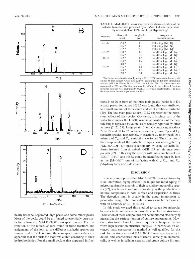
nearly baseline, separated large peaks and some minor peaks.Most of the peaks could be attributed to essentially pure sur-factin isoforms by MALDI-TOF mass spectrometry. The dis-tribution of the molecular ions found in these fractions andassignment of the ions to the different surfactin species aresummarized in Table 6. From the mass spectrometric data it isapparent that the surfactin isoforms eluted according to theirhydrophobicities. For the small peak A that appeared in frac-
tions 24 to 26 in front of the three main peaks (peaks B to D),a main parent ion at m/z 1016.7 was found that was attributedto a small amount of the sodium adduct of a valine-7 surfactin(20). The low-mass peak at m/z 1032.7 represented the potas-sium adduct of this species. Obviously, in a minor part of thesurfactin complex the Leu/Ile residue at position 7 of the pep-tide ring is replaced by valine, as previously reported by otherauthors (2, 20, 29). Large peaks B and C comprising fractions27 to 29 and 30 to 32 contained essentially pure C13 and C14
surfactin species, respectively. In fractions 37 to 39 (peak D) amixture of C14 and C15 surfactins was found. The structure ofthe components of the surfactin complex was investigated byPSD MALDI-TOF mass spectrometry by using surfactin iso-forms isolated from B. subtilis OKB 105 as reference com-pounds (22). In this way the species with mass numbers of m/z1030.7, 1044.7, and 1058.7 could be identified by their bn ionsas the [M�Na]� ions of surfactins with C13, C14, and C15
�-hydroxy fatty acid side chains.
DISCUSSION
Recently, we reported that MALDI-TOF mass spectrometryis an innovative, highly efficient technique for rapid typing ofmicroorganisms by analysis of their secondary metabolite spec-tra (22), which is also well suited for studying the production ofnatural compounds in both surface and suspension cultures.The detection limit is usually in the upper femtomolar topicomolar range. The molecular masses can be determinedwith an accuracy of 0.01 to 0.02%.
In this study we used this method to screen for microbialbiosurfactants and to characterize their molecular structures.Production of these compounds can be monitored efficiently bymeasuring the surface tension of culture supernatants. How-ever, structural characterization requires techniques that in-volve high-resolution structure analysis. In particular, an ad-vanced mass spectrometric method is well qualified for thistask. In this study we used MALDI-TOF mass spectrometry todetect and characterize biosurfactants directly in microbialcells, as well as in cellular extracts and crude culture filtrates.
TABLE 6. MALDI-TOF mass spectrometric characterization of thesurfactin biosurfactants produced by B. subtilis C-1 after separation
by reversed-phase HPLC on ODS Hypersil C18a
Fractions Mass peak(m/z)
Amplitude(cm)
Assignment(surfactin species)
24–26 994.2 4.5 Val-7 C13, [M�H]�
1016.7 14.8 Val-7 C13, [M�Na]�
1032.7 3.6 Val-7 C13, [M�K]�
27–29 1030.7 14.8 Leu/Ile-7 C13, [M�Na]�
1046.7 5.0 Leu/Ile-7 C13, [M�K]�
30–32 1022.7 1.3 Leu/Ile-7 C14, [M�H]�
1044.7 14.9 Leu/Ile-7 C14, [M�Na]�
1060.7 8.0 Leu/Ile-7 C14, [M�K]�
37–39 1036.7 1.1 Leu/Ile-7 C15, [M�H]�
1044.7 1.2 Leu/Ile-7 C14, [M�Na]�
1058.7 12.4 Leu/Ile-7 C15, [M�Na]�
a Surfactins were fractionated by using a 20 to 100% acetonitrile linear gradi-ent for 60 min. Eluent A was 20% (vol/vol) acetonitrile in 100 mM ammoniumacetate (pH 6.9). Eluent B was 100% acetonitrile. The UV absorbance wasmonitored at 220 nm. The flow rate was 0.5 ml/min. In the collected fractionssurfactin isoforms were identified by MALDI-TOF mass spectrometry. The massdata represent monoisotopic mass numbers.
FIG. 4—Continued.
VOL. 68, 2002 MALDI-TOF MASS SPECTROMETRY OF LIPOPEPTIDES 6217
on May 25, 2014 by guest
http://aem.asm
.org/D
ownloaded from
As previously reported (22), by using whole-cell MALDI-TOFmass spectrometry cellular products can be detected whicheither are attached to the cell surface or are integrated into theouter cell membrane. In this way information on the secondarymetabolites produced by a microorganism can be obtained inminutes with high precision and sensitivity with no need tofractionate and purify the detected compounds. Depending onthe structural complexity of the molecules investigated, insome cases rapid in situ structure analysis is possible by inter-pretation of fragment spectra obtained from PSD MALDI-TOF mass spectrometry. Often this technique can be usedsuccessfully for structural characterization of natural com-pounds with a regular polymeric structure, such as bioactivepeptides, which have linear or cyclic sequences of amino acids.Elucidation of the structure can be started by using immoniumions, which provide information on the amino acid composi-tion of a peptide, followed by a search for dipeptide fragmentions which indicate the nearest neighbors in the peptide moi-ety. On the basis of the data obtained the entire peptide se-quence can be estimated. In essence, MALDI-TOF mass spec-trometry is a powerful tool for investigating large biologicalstructures and solutions with complex compositions.
This novel method was used for characterization of a bac-terial strain isolated by us from petroleum sludge, which wasidentified by its genetic, physiological, and morphological fea-tures as a B. subtilis strain designated B. subtilis C-1. As Tables1 and 2 show, this organism was found to be an efficientbiosurfactant producer based on surface tension measure-ments for culture supernatants obtained from fermentationsperformed at various temperatures. The surface tension mea-sured for the pure culture medium (71 dynes/cm) was loweredin the culture filtrates by the production of surface-active com-pounds to approximately 29 dynes/cm, a value that has beenreported for the most efficient biosurfactants which have beenisolated and studied so far (4, 5, 7, 16). Such compounds havegreat potential for industrial exploitation (5, 7, 9, 10, 32). Inparticular, during the last decade interesting, promising bio-pharmaceutical applications have been developed for surfactin(37, 38), which is the most efficient lipopeptide biosurfactantproduced by B. subtilis. Therefore, the availability of innova-tive, efficient procedures for detection, structure analysis, func-tional characterization, and biological testing of such com-pounds has great biotechnological relevance.
Figure 2 shows the MALDI mass spectra of whole cells of B.subtilis C-1 grown in surface and suspension cultures, as well asthe MALDI mass spectra of aliquots of the culture superna-tants and the crude lipopeptide fraction obtained by precipi-tation with HCl and extraction of the pellet with methanol.Prominent groups of mass peaks were observed in the massrange between m/z 1000 and 1500. By comparing the mass datasummarized in Tables 3 to 6 with the mass numbers reportedfor the lipopeptide complexes from other B. subtilis strains (19,20, 22, 25–27, 33, 34) and by analyzing the fragment patternsderived from PSD MALDI-TOF mass spectra (Fig. 3), thelipopeptide products of B. subtilis C-1 could be identified assurfactins, iturins, and fengycins. All these biosurfactants ap-peared as families of closely related isoforms which differed inthe lengths of their fatty acid side chains, as well as in theamino acid substitutions in their peptide rings (Fig. 1). In thisway the identification of our isolate as a B. subtilis strain was
corroborated by mass spectrometric analysis of its secondarymetabolite spectrum.
The lipopeptide families produced by B. subtilis strains areprominent examples that demonstrate the capacity of MALDI-TOF mass spectrometry for detection and in situ structureanalysis. This novel mass spectrometric method was combinedwith an efficient miniaturized separation procedure involvingthe Pharmacia Smart microseparation system, which is wellsuited for rapid, high-resolution fractionation of most of thelipopeptide isoforms on a small scale for analytical purposes, asdescribed for mass spectrometric analysis. By using this proce-dure small aliquots (10 to 50 �l) of the culture filtrate or crudecellular extracts could be fractionated with excellent resolutionand could be analyzed with high sensitivity and precision (Fig.4). The mass spectrometric investigation of B. subtilis C-1 re-vealed the following lipopeptide pattern.
The iturin A compounds formed by this organism as themain products in the stationary phase of growth contain un-usual fatty acid components with chain lengths of 16 to 19carbon atoms. C16 and C17 species are the predominant iso-forms. These results are different from the data obtained byHourdou et al. (15) and Peypoux et al. (26) in their extensivestudies of iturin compounds. The B. subtilis strains used bythese authors produce iturins with saturated �-amino C14 toC16 fatty acid side chains (15, 26). The fatty acid componentsof our isolate resemble those found in mycosubtilins (15, 25,28). All of them showed mass numbers which are 1 mass unitlower than the value expected in the case of saturated �-aminofatty acid side chains. This result is compatible with the as-sumption of iturin B molecules which contain one aspartic acidresidue instead of an asparagine residue, as well as an unsat-urated �-amino fatty acid moiety with one double bond. How-ever, this is certainly not the case, because an aspartic acid wasnot found in the fragment patterns derived from the PSDMALDI-TOF mass spectra of the new iturin compounds. An-other possibility to explain our data is the hypothesis that�-hydroxy rather than �-amino fatty acid constituents containone double bond, but this hypothesis has to be clarified by adetailed structure analysis, which is being done.
The fengycin species of our isolate comprise C15 to C17
variants which have a characteristic Ala-Val dimorphy at po-sition 6 of the peptide ring, as previously reported for otherfengycin producers (24, 33–35). As Table 4 shows, two kinds ofvariants were observed, which differ by 2 mass units. Thisfeature is due to two series of compounds, one with saturatedlipid moieties and the other with fatty acid side chains contain-ing one double bond whose position remains to be elucidatedby further studies.
Testing of the biological activities and development of in-dustrial applications for these interesting compounds requirethat they be isolated on a preparative scale. To do this, efficienttechniques, such as solid-phase extraction and dispersion andgel permeation and adsorption chromatography in combina-tion with preparative reversed-phase HPLC, are available. Inthis paper we report on a two-step procedure for the fraction-ation of the surfactin lipopeptide complex of B. subtilis C-1,which is the predominant biosurfactant product in the expo-nential phase of growth. Surfactin isoforms were isolated inpure form by a combination of gel permeation chromatogra-phy on Sephadex LH-20 and reversed-phase HPLC on Hyper-
6218 VATER ET AL. APPL. ENVIRON. MICROBIOL.
on May 25, 2014 by guest
http://aem.asm
.org/D
ownloaded from
sil C18. Surfactin isoforms were identified by MALDI-TOFmass spectrometry and by correlation with previous results(20). The data obtained are summarized in Table 6. The sur-factin complex of B. subtilis C-1 comprised C13 to C15 isoformswhich occurred at a ratio similar to the ratios observed forother surfactin producers (19, 20, 22), and C14 and C15 sur-factins were the main species.
The innovative method presented in this paper is very im-portant for making available new microbial biosurfactant com-pounds for industrial exploitation, and in particular, for devel-oping specific biopharmaceutical applications. This hasrecently been demonstrated for the surfactins showing potentantivirus and antimycoplasma properties (37, 38), which can beefficiently utilized for inactivation of virus and mycoplasmacontaminants in biotechnological and biopharmaceutical prod-ucts.
ACKNOWLEDGMENTS
We are grateful for the facilities provided by the Director,IMTECH.
Financial support was received from the Council for Scientific andIndustrial Research (CSIR), the Department of Biotechnology (DBT),the Government of India, and the Deutsche Forschungsgemeinschaft(grant Va 63/4-4).
REFERENCES
1. Arima, K., A. Kakinuma, and G. Tamura. 1968. Surfactin, a crystallinepeptidelipid surfactant produced by Bacillus subtilis: isolation, characteriza-tion and its inhibition of fibrin clot formation. Biochem. Biophys. Res.Commun. 31:488–494.
2. Baumgart, F., B. Kluge, C. Ullrich, J. Vater, and D. Ziessow. 1991. Identi-fication of amino acid substitutions in the lipopeptide surfactin using 2DNMR spectroscopy. Biochem. Biophys. Res. Commun. 177:998–1005.
3. Besson, F., F. Peypoux, G. Michel, and L. Delcambe. 1978. Identification ofantibiotics of iturin group in various strains of Bacillus subtilis. J. Antibiot.(Tokyo) 31:284–288.
4. Cooper, D. G., C. R. Macdonald, J. B. Duff, and N. Kosaric. 1981. Enhancedproduction of surfactin from Bacillus subtilis by continuous product removaland metal cation additions. Appl. Environ. Microbiol. 42:408–412.
5. Cooper, D. G., and J. E. Zajic. 1980. Surface-active compounds from micro-organisms. Adv. Appl. Microbiol. 26:229–256.
6. Deleu, M., H. Razafindralambo, Y. Popineau, P. Jacques, P. Thonart, andM. Paquot. 1999. Interfacial and emulsifying properties of lipopeptides fromBacillus subtilis. Colloids Surf. A Physicochem. Eng. Aspects 152:3–10.
7. Desai, J. D., and I. M. Banat. 1997. Microbial production of surfactants andtheir commercial potential. Microbiol. Mol. Biol. Rev. 61:47–64.
8. Erhard, M., H. von Dohren, and P. Jungblut. 1997. Rapid typing and elu-cidation of new secondary metabolites of intact cyanobacteria using MALDI-TOF mass spectrometry. Nat. Biotechnol. 15:906–909.
9. Fiechter, A. 1992. Biosurfactants: moving towards industrial application.TIBTECH 10:208–217.
10. Haferburg, D., R. Hommel, R. Claus, and H.-P. Kleber. 1986. Extracellularmicrobial lipids as biosurfactants. Adv. Biochem. Eng. Biotechnol. 33:53–93.
11. Horowitz, S., J. N. Gilbert, and W. M. Griffin. 1990. Isolation and charac-terization of a surfactant produced by Bacillus licheniformis 86. J. Ind. Mi-crobiol. 6:243–248.
12. Horowitz, S., and W. M. Griffin. 1991. Structural analysis of Bacillus licheni-formis 86 surfactant. J. Ind. Microbiol. 7:45–52.
13. Hosono, K., and H. Suzuki. 1983. Acylpeptides, the inhibitors of cyclicadenosine 3�,5�-monophosphate phosphodiesterase. I. Purification, physico-chemical properties and structures of fatty acid residues. J. Antibiot. (Tokyo)36:667–673.
14. Hosono, K., and H. Suzuki. 1983. Acylpeptides, the inhibitors of cyclicadenosine 3�,5�-monophosphate phosphodiesterase. II. Amino acid se-quence and location of lactone linkage. J. Antibiot. (Tokyo) 36:674–678.
15. Hourdou, M.-L., F. Besson, I. Tenoux, and G. Michel. 1989. Fatty acids and�-amino acid syntheses in strains of Bacillus subtilis producing iturinic anti-biotics. Lipids 24:940–944.
16. Jenny, K., O. Kappeli, and A. Fiechter. 1991. Biosurfactants from Bacilluslicheniformis: structural analysis and characterization. Appl. Microbiol. Bio-technol. 36:5–13.
17. Kakinuma, A., M. Hori, M. Isono, G. Tamura, and K. Arima. 1969. Deter-mination of amino acid sequence in surfactin, a crystalline peptide lipidsurfactant produced by Bacillus subtilis. Agric. Biol. Chem. 33:971–972.
18. Kakinuma, A., H. Sugino, M. Isono, G. Tamura, and K. Arima. 1969. De-termination of fatty acid in surfactin and elucidation of the total structure ofsurfactin. Agric. Biol. Chem. 33:973–976.
19. Kakinuma, A., A. Ouchida, T. Shima, H. Sugino, M. Isono, G. Tamura, andK. Arima. 1969. Confirmation of the structure of surfactin by mass spec-trometry. Agric. Biol. Chem. 33:1669–1671.
20. Kowall, M., J. Vater, B. Kluge, T. Stein, P. Franke, and D. Ziessow. 1998.Separation and characterization of surfactin isoforms produced by Bacillussubtilis OKB 105. J. Colloid Interface Sci. 203:1–8.
21. Landy, M., G. H. Warren, S. B. Rosenman, and L. G. Colio. 1948. Bacillo-mycin, an antibiotic from Bacillus subtilis active against pathogenic fungi.Proc. Soc. Exp. Biol. Med. 67:539–541.
22. Leenders, F., T. H. Stein, B. Kablitz, P. Franke, and J. Vater. 1999. Rapidtyping of Bacillus subtilis strains by their secondary metabolites using matrix-assisted laser desorption/ionization mass spectrometry of intact cells. RapidCommun. Mass Spectrom. 13:943–949.
23. Naruse, N., O. Tenmyo, S. Kobaru, H. Kamei, T. Miyaki, M. Konishi, and T.Oki. 1990. Pumilacidin, a complex of new antiviral antibiotics—production,isolation, chemical properties, structure and biological activity. J. Antibiot.(Tokyo) 43:267–280.
24. Nishikori, T., H. Naganawa, Y. Muraoka, T. Aoyagi, and H. Umezawa. 1986.Plipastatins: new inhibitors of phopholipase A2, produced by Bacillus cereusBMG302-fF67. II. Structure of fatty acid residue and amino acid sequence.J. Antibiot. (Tokyo) 39:745–754.
25. Peypoux, F., G. Michel, and L. Delcambe. 1976. Structure de la mycosubti-line, antibiotique isole de Bacillus subtilis. Eur. J. Biochem. 63:391–398.
26. Peypoux, F., M. Guinand, G. Michel, L. Delcambe, B. C. Das, and E. Le-derer. 1978. Structure of iturin A, a peptidolipid antibiotic from Bacillussubtilis. Biochemistry 17:3992–3996.
27. Peypoux, F., M.-T. Pommier, B. C. Das, F. Besson, L. Delcambe, and G.Michel. 1984. Structures of bacillomycin D and bacillomycin L peptidolipidantibiotics from Bacillus subtilis. J. Antibiot. (Tokyo) 77:1600–1604.
28. Peypoux, F., M. T. Pommier, D. Marion, M. Ptak, B. C. Das, and G. Michel.1986. Revised structure of mycosubtilin, a peptidolipid antibiotic from Ba-cillus subtilis. J. Antibiot. (Tokyo) 39:636–641.
29. Peypoux, F., J.-M. Bonmatin, H. Labbe, B. C. Das, M. Ptak, and G. Michel.1991. Isolation and characterization of a new variant of surfactin, the [Val-7]surfactin. Eur. J. Biochem. 202:101–106.
30. Peypoux, F., J. M. Bonmatin, and J. Wallach. 1999. Recent trends in thebiochemistry of surfactin. Appl. Microbiol. Biotechnol. 51:553–563.
31. Razafindralambo, H., Y. Popineau, M. Deleu, C. Hbid, P. Jacques, P. Tho-nart, and M. Paquot. 1998. Foaming properties of lipopeptides produced byBacillus subtilis: effects of lipid and peptide structural attributes. J. Agric.Food Chem. 46:911–916.
32. Rosenberg, E. 1986. Microbial surfactants. Crit. Rev. Biotechnol. 3:109–132.33. Schneider, J. 1999. Doctoral thesis. University of Cologne, Cologne, Ger-
many.34. Schneider, J., K. Taraz, H. Budzikiewicz, P. Jacques, and P. Thonart. 1999.
The structure of two fengycins from Bacillus subtilis S499. Z. Naturforsch.54:859–865.
35. Vanittanakom, N., W. Loeffler, U. Koch, and G. Jung. 1986. Fengycin–anovel antifungal lipopeptide antibiotic produced by Bacillus subtilis F-29-3.J. Antibiot. (Tokyo) 39:888–901.
36. Vater, J. 1986. Lipopeptides, an attractive class of microbial surfactants.Prog. Colloid Polymer Sci. 72:12–18.
37. Vollenbroich, D., G. Pauli, M. Ozel, and J. Vater. 1997. Antimycoplasmaproperties and application in cell culture of surfactin, a lipopeptide antibioticfrom Bacillus subtilis. Appl. Environ. Microbiol. 63:44–49.
38. Vollenbroich, D., M. Ozel, J. Vater, R. M. Kamp, and G. Pauli. 1997.Mechanism of inactivation of enveloped viruses by the biosurfactant surfac-tin from Bacillus subtilis. Biologicals 25:289–297.
39. Yakimov, M. M., K. N. Timmis, V. Wray, and H. L. Fredrickson. 1995.Characterization of a new lipopeptide surfactant produced by thermotoler-ant and halotolerant subsurface Bacillus licheniformis BAS 50. Appl. Envi-ron. Microbiol. 61:1706–1713.
VOL. 68, 2002 MALDI-TOF MASS SPECTROMETRY OF LIPOPEPTIDES 6219
on May 25, 2014 by guest
http://aem.asm
.org/D
ownloaded from