Maternal and Neonatal Polyunsaturated Fatty Acid ... - MDPI
19
Citation: Heath, R.J.; Klevebro, S.; Wood, T.R. Maternal and Neonatal Polyunsaturated Fatty Acid Intake and Risk of Neurodevelopmental Impairment in Premature Infants. Int. J. Mol. Sci. 2022, 23, 700. https:// doi.org/10.3390/ijms23020700 Academic Editors: Kiran Panickar and Ashu Johri Received: 10 December 2021 Accepted: 6 January 2022 Published: 9 January 2022 Publisher’s Note: MDPI stays neutral with regard to jurisdictional claims in published maps and institutional affil- iations. Copyright: © 2022 by the authors. Licensee MDPI, Basel, Switzerland. This article is an open access article distributed under the terms and conditions of the Creative Commons Attribution (CC BY) license (https:// creativecommons.org/licenses/by/ 4.0/). International Journal of Molecular Sciences Review Maternal and Neonatal Polyunsaturated Fatty Acid Intake and Risk of Neurodevelopmental Impairment in Premature Infants Rory J. Heath 1 , Susanna Klevebro 2 and Thomas R. Wood 3,4,5, * 1 Emergency Medicine Department, Derriford Hospital, University Hospitals Plymouth NHS Foundation Trust, Plymouth PL68DH, UK; [email protected] 2 Department of Clinical Science and Education, Södersjukhuset, Karolinska Institutet, 11883 Stockholm, Sweden; [email protected] 3 Department of Pediatrics, University of Washington, Seattle, WA 98195, USA 4 Center on Human Development and Disability, University of Washington, Seattle, WA 98195, USA 5 Institute for Human and Machine Cognition, Pensacola, FL 32502, USA * Correspondence: [email protected] Abstract: The N3 and N6 long chain polyunsaturated fatty acids (LCPUFA) docosahexaenoic acid (DHA) and arachidonic acid (AA) are essential for proper neurodevelopment in early life. These fatty acids are passed from mother to infant via the placenta, accreting into fetal tissues such as brain and adipose tissue. Placental transfer of LCPUFA is highest in the final trimester, but this transfer is abruptly severed with premature birth. As such, efforts have been made to supplement the post-natal feed of premature infants with LCPUFA to improve neurodevelopmental outcomes. This narrative review analyzes the current body of evidence pertinent to neurodevelopmental outcomes after LCPUFA supplementation in prematurely born infants, which was identified via the reference lists of systematic and narrative reviews and PubMed search engine results. This review finds that, while the evidence is weakened by heterogeneity, it may be seen that feed comprising 0.3% DHA and 0.6% AA is associated with more positive neurodevelopmental outcomes than LCPUFA-deplete feed. While no new RCTs have been performed since the most recent Cochrane meta-analysis in 2016, this narrative review provides a wider commentary; the wider effects of LCPUFA supplementation in prematurely born infants, the physiology of LCPUFA accretion into preterm tissues, and the physiological effects of LCPUFA that affect neurodevelopment. We also discuss the roles of maternal LCPUFA status as a modifiable factor affecting the risk of preterm birth and infant neurodevelopmental outcomes. To better understand the role of LCPUFAs in infant neurodevelopment, future study designs must consider absolute and relative availabilities of all LCPUFA species and incorporate the LCPUFA status of both mother and infant in pre- and postnatal periods. Keywords: polyunsaturated fatty acid; fatty acid; docosahexaenoic acid; arachidonic acid; brain; infant; pregnancy; premature birth; preterm infant; premature infant; neurodevelopment 1. Introduction The N3 and N6 long-chain polyunsaturated fatty acid (LCPUFA) families comprise fatty acids with double carbon bonds at the N3 or N6 positions, respectively. The N3 family includes alpha-linolenic acid (ALA), eicosapentaenoic acid (EPA), and docosahexaenoic acid (DHA), while important N6 members include linoleic acid (LA) and arachidonic acid (AA). Humans cannot synthesise ALA and LA de novo, thus these LCPUFA are termed ‘dietarily essential’. Some conversion within the series of N3 or N6 fatty acids occurs endogenously, thus DHA and AA can be synthesized from their respective precursors. Synthesis occurs via enzymatic elongation and desaturation by 5- and 6-desaturase enzymes, but the efficiency of this process may be affected by genetic polymorphisms of the FADS gene [1]. While infants express these enzymes in utero, the capacity to synthesise AA and DHA from their precursors is inadequate to meet the LCPUFA demands of rapid infant development and Int. J. Mol. Sci. 2022, 23, 700. https://doi.org/10.3390/ijms23020700 https://www.mdpi.com/journal/ijms
-
Upload
khangminh22 -
Category
Documents
-
view
2 -
download
0
Transcript of Maternal and Neonatal Polyunsaturated Fatty Acid ... - MDPI

Citation: Heath, R.J.; Klevebro, S.;
Wood, T.R. Maternal and Neonatal
Polyunsaturated Fatty Acid Intake
and Risk of Neurodevelopmental
Impairment in Premature Infants. Int.
J. Mol. Sci. 2022, 23, 700. https://
doi.org/10.3390/ijms23020700
Academic Editors: Kiran Panickar
and Ashu Johri
Received: 10 December 2021
Accepted: 6 January 2022
Published: 9 January 2022
Publisher’s Note: MDPI stays neutral
with regard to jurisdictional claims in
published maps and institutional affil-
iations.
Copyright: © 2022 by the authors.
Licensee MDPI, Basel, Switzerland.
This article is an open access article
distributed under the terms and
conditions of the Creative Commons
Attribution (CC BY) license (https://
creativecommons.org/licenses/by/
4.0/).
International Journal of
Molecular Sciences
Review
Maternal and Neonatal Polyunsaturated Fatty Acid Intake andRisk of Neurodevelopmental Impairment in Premature InfantsRory J. Heath 1, Susanna Klevebro 2 and Thomas R. Wood 3,4,5,*
1 Emergency Medicine Department, Derriford Hospital, University Hospitals Plymouth NHS Foundation Trust,Plymouth PL68DH, UK; [email protected]
2 Department of Clinical Science and Education, Södersjukhuset, Karolinska Institutet,11883 Stockholm, Sweden; [email protected]
3 Department of Pediatrics, University of Washington, Seattle, WA 98195, USA4 Center on Human Development and Disability, University of Washington, Seattle, WA 98195, USA5 Institute for Human and Machine Cognition, Pensacola, FL 32502, USA* Correspondence: [email protected]
Abstract: The N3 and N6 long chain polyunsaturated fatty acids (LCPUFA) docosahexaenoic acid(DHA) and arachidonic acid (AA) are essential for proper neurodevelopment in early life. Thesefatty acids are passed from mother to infant via the placenta, accreting into fetal tissues such as brainand adipose tissue. Placental transfer of LCPUFA is highest in the final trimester, but this transfer isabruptly severed with premature birth. As such, efforts have been made to supplement the post-natalfeed of premature infants with LCPUFA to improve neurodevelopmental outcomes. This narrativereview analyzes the current body of evidence pertinent to neurodevelopmental outcomes afterLCPUFA supplementation in prematurely born infants, which was identified via the reference lists ofsystematic and narrative reviews and PubMed search engine results. This review finds that, while theevidence is weakened by heterogeneity, it may be seen that feed comprising 0.3% DHA and 0.6% AAis associated with more positive neurodevelopmental outcomes than LCPUFA-deplete feed. While nonew RCTs have been performed since the most recent Cochrane meta-analysis in 2016, this narrativereview provides a wider commentary; the wider effects of LCPUFA supplementation in prematurelyborn infants, the physiology of LCPUFA accretion into preterm tissues, and the physiological effectsof LCPUFA that affect neurodevelopment. We also discuss the roles of maternal LCPUFA statusas a modifiable factor affecting the risk of preterm birth and infant neurodevelopmental outcomes.To better understand the role of LCPUFAs in infant neurodevelopment, future study designs mustconsider absolute and relative availabilities of all LCPUFA species and incorporate the LCPUFAstatus of both mother and infant in pre- and postnatal periods.
Keywords: polyunsaturated fatty acid; fatty acid; docosahexaenoic acid; arachidonic acid; brain;infant; pregnancy; premature birth; preterm infant; premature infant; neurodevelopment
1. Introduction
The N3 and N6 long-chain polyunsaturated fatty acid (LCPUFA) families comprisefatty acids with double carbon bonds at the N3 or N6 positions, respectively. The N3 familyincludes alpha-linolenic acid (ALA), eicosapentaenoic acid (EPA), and docosahexaenoic acid(DHA), while important N6 members include linoleic acid (LA) and arachidonic acid (AA).Humans cannot synthesise ALA and LA de novo, thus these LCPUFA are termed ‘dietarilyessential’. Some conversion within the series of N3 or N6 fatty acids occurs endogenously,thus DHA and AA can be synthesized from their respective precursors. Synthesis occurs viaenzymatic elongation and desaturation by 5- and 6-desaturase enzymes, but the efficiencyof this process may be affected by genetic polymorphisms of the FADS gene [1]. Whileinfants express these enzymes in utero, the capacity to synthesise AA and DHA from theirprecursors is inadequate to meet the LCPUFA demands of rapid infant development and
Int. J. Mol. Sci. 2022, 23, 700. https://doi.org/10.3390/ijms23020700 https://www.mdpi.com/journal/ijms

Int. J. Mol. Sci. 2022, 23, 700 2 of 19
growth [2–5]. Therefore, infants are dependent on preformed AA and DHA supplied bythe placenta in utero, by breastmilk in the postnatal period, or by exogenous nutrition afterpreterm birth.
This review aims to explore the current evidence surrounding LCPUFA supplemen-tation to improve neurodevelopmental outcomes after preterm birth. We discuss thesurrounding context of LCPUFA: how these fatty acids accrete into fetal tissues duringgestation and after birth and how the physical nature of these LCPUFA molecules andtheir subsequent metabolism into bioactive mediators exert their wide-ranging effectsupon infant brain development. This context provides a base from which we may viewthe effects of changes to infant LCPUFA status through the current standard of care orthrough interventional trials (described in Supplementary Table S1), and it speculates uponguidance for practice and future research. Our scope extends to suggest that further clinicaland research considerations may be needed to make LCPUFA provision in the pretermperiod more physiological for the infant, accounting for placental physiology, maternalLCPUFA status, and wider contexts of LCPUFA availability.
This narrative review synthesizes evidence from articles identified in the referencelists of key preceding narrative and systematic reviews. New articles were identified viasearches using the PubMed search engine of its constituent databases, MEDLINE andPubMed Central, and through active work in this research field amongst the authors.Keywords searched included ‘preterm/premature infant’, ‘polyunsaturated fatty acids’,‘docosahexaenoic acid’, ‘arachidonic acid’, and ‘neurodevelopment’. A previous systematicreview of the effects of LCPUFA supplementation in preterm infants has been performedby the Cochrane Collaboration in 2016 [6]. All papers that were analyzed systematically inthe Cochrane review are included herein; a new systematic review is not warranted, as nonew RCTs assessing the effects of LCPUFA supplementation upon neurodevelopment havebeen performed. Importantly, while excluded from the Cochrane systematic review due tonot fulfilling RCT criteria, studies by Almaas et al. [7], Alshweki et al. [8], Collins et al. [9],Makrides et al. [10], and Smithers et al. [11] provide valuable wider insights into the fieldand are included in this narrative review.
2. Why Are DHA and AA Important in the Developing Brain?2.1. LCPUFA and Neurodevelopment
DHA and AA together comprise a quarter of all brain fatty acids, particularly contribut-ing to myelination and being enriched in neuronal synapses and cellular membranes [12],where their structure lends fluidity that aids neuronal functions such as neurotransmis-sion [13,14].
Placental transfer and accretion of fatty acids into infant tissues is highest during thefinal trimester of pregnancy, with an essential reserve of AA and DHA stored in adiposein preparation for the post-natal demands of neurological development. Premature birthsevers materno-fetal transfer, resulting in less LCPUFA availability relative to infants bornat term, which may compromise neurodevelopment.
DHA is neuroprotective in animal models of neonatal asphyxia [15] and appears toencourage brain growth, myelination, and overall survival in prematurely born pigs [16].In rats, improving LCPUFA availability to the offspring by supplementing the maternaldiet can reduce the neurological damage associated with neonatal hypoxic-ischaemic braininjury [17]. Autopsy studies of human infants fed LCPUFA-deficient formula demonstratethe substitution of alternative N6 and N9 fatty acids in neural tissues [18,19].
DHA is also richly incorporated into photoreceptors, and deficiency is associatedwith reduced visual acuity and impaired visual transduction [20]. Randomized trials haveshown reduced risk of severe retinopathy of prematurity (ROP) in preterm infants receivingenteral supplementation with DHA as well as a combination of DHA and AA [21,22]. ROPis a disease additionally associated with lower brain volumes and neurodevelopmentalscores at two years of age [23]. Furthermore, greater erythrocyte levels of DHA correlate

Int. J. Mol. Sci. 2022, 23, 700 3 of 19
with positive MRI findings of brain microstructure development and with improvedneurodevelopmental scores in infants born prematurely [24].
AA has multiple essential and wide-ranging roles in general infant development aswell; dietary AA deficiency has been associated with growth impairment, which improveson return of dietary supply [25]. Feeding infants DHA unopposed by AA has also beenassociated with reduced preterm infant growth [25,26].
Overall, infants born prematurely and/or at a low-birth weight, thus with reducedadipose stores of LCPUFA, are more likely to experience neurosensory impairment, achievelower levels of educational attainment, and have lower IQ scores [27].
2.2. LCPUFA Influence Inflammatory Signaling
AA exerts effects via its metabolism into bioactive molecules termed eicosanoids bythe enzymes cyclooxygenase (COX) and lipoxygenase (LOX) [28]. Increased metabolism ofAA into eicosanoids, such as prostaglandins, thromboxanes, and leukotrienes, occurs in re-sponse to infection or injury with consequent changes to vessel flow and permeability to aiddelivery of immune cells [28]. For this reason, AA is often considered a pro-inflammatoryagent. Conversely, DHA competes with AA for placement in cellular membranes and formetabolism by COX and LOX, and in doing so, it inhibits the production of eicosanoidsand thus can be thought of as grossly having anti-inflammatory actions [28].
However, the roles of AA and DHA within inflammation are not binary; some AA-derived metabolites regulate the process of inflammation resolution. The prostaglandinPGE2 promotes the initial inflammatory response but subsequently acts to inhibit LOXto reduce further production of pro-inflammatory prostaglandins [29]. Some eicosanoidsmay protect and repair vessel membranes after injury [12]. Furthermore, PGE2 contributesto a ‘class switching’ event that stimulates COX and LOX to metabolize AA and DHAinto lipid mediators such as lipoxins, resolvins, maresins, and neuroprotectins [29]. Thesemetabolites, termed specialized pro-resolving mediators (SPM), inhibit inflammation andpromote resolution to return tissues to pre-injury homeostasis [30,31]. While AA contributesto the production of lipoxins, the majority of SPMs are derived from DHA, includingneuroprotectin D1 (NPD1), which has been shown to protect against oxidative stress andinfluence cell survival [32].
Although excessive inflammation is associated with negative outcomes [33], controlledinflammation is a necessary response to infection or injury. Imbalances in the N3:N6 ratiomay therefore lead to domination of one fatty acid series over the other during competitionfor metabolism by COX and LOX, with consequent effects upon the availability of pro- oranti-inflammatory mediators and of pro-resolution agents.
While the balance of N3:N6 fatty acids have the potential to disrupt the inflammatoryprocess, there is not yet a broad base of evidence to understand how the amounts andtiming of LCPUFA administration may influence outcomes after premature birth. Withinthe available evidence, an observational study of LCPUFA blood levels of prematurelyborn infants associated low levels of DHA with an increased risk of chronic lung disease,while low levels of AA in the blood were associated with an increased risk of sepsis [34].On the other hand, two large, randomized trials have failed to demonstrate a positive effectof postnatal DHA supplementation on preterm lung development [35,36]. The complexityof the AA and DHA balance is further demonstrated in an analysis of the effect of LCPUFAlevels on ROP-risk in Sweden, concluding that higher levels of DHA were only associatedwith a reduced risk of ROP if the levels of AA were sufficiently high [37].
Premature birth itself may also be linked to inflammatory signaling, as illustratedby the actions of prostaglandins to precipitate preterm birth in sheep [38]. In humans,mothers whose blood content of AA + DHA was <1.6% of total blood fatty acids had10-fold higher risk of premature birth in comparison to mothers with LCPUFA levels>1.8% [39]. A recent study showed that the DHA and AA status of cord blood correlateswith levels of the inflammatory markers CRP and IL-6 of prematurely born infants [40],

Int. J. Mol. Sci. 2022, 23, 700 4 of 19
while inflammation in the fetal and early post-natal periods are linked with increasedmorbidity in later life [41,42].
3. LCPUFA Accretion into Fetal Tissues3.1. In Utero LCPUFA Accretion
The final trimester is associated with rapid growth of tissues, including adipose tissue,skeletal muscle, and brain tissue [43]. Between 31 weeks of gestation and term, the brainincreases in size from ~150 mL to ~400 mL [44], while brain weight increases by four-to five-fold [45]. This tissue growth is matched by an increased transfer of fatty acidsfrom mother to fetus via the placenta to provide both energy and structural substrate forbuilding new tissue. Furthermore, the placenta serves to selectively control the transferof both AA and DHA from mother to infant. In a process termed biomagnification, theplacenta transfers LCPUFA to the fetus even during circumstances of maternal LCPUFAdepletion, resulting in higher LCPUFA contents of fetal blood and tissues than those of themother [43].
AA transfer is high throughout pregnancy, with the biomagnification phenomenonmaintaining fetal blood AA at levels twice those of the mother; at the beginning of the thirdtrimester, the AA:DHA ratio is ~5.1 [46]. The rate of LCPUFA transfer across the placentaincreases significantly in the final trimester, increasing from an average of 6.1 mg of AA and2.3 mg of DHA per day in the 25th week of gestation to 95 mg AA and 42 mg of DHA perday in the final trimester [47]. The accelerated transfer of DHA in the third trimester resultsin fetal cord plasma DHA levels exceeding those of the mother at around 33 weeks [46].At term, the AA:DHA ratio as measured in fetal cord blood is ~2.5:1 [46]. Changes in brainLCPUFA composition correspond with these blood LCPUFA changes, and between thegestational ages of 8 to 40 weeks, the relative brain content of AA decreases (~11% to 8.6%)and DHA content increases (3.2% to 8.4%) in infants born to women eating a traditionaldiet rich in DHA and without excessive LA [48].
3.2. Post-Natal LCPUFA Accretion
Accretion of LCPUFA continues in the postnatal period and throughout childhoodup until the age of 18, but at a decreasing rate [49]. Maternal breastmilk is a rich sourceof LCPUFA in the postnatal period and contains ~0.6% AA and ~0.3% DHA; however,these concentrations show considerable variation with maternal diet [50] and geneticpolymorphisms [1]. Over the first six months of life, breast fed infants accumulate DHAat ~10 mg/day [51].
3.3. LCPUFA Accretion into Adipose
While much of the LCPUFA transferred to the fetus in the final trimester contributesto brain growth and neurological development, by term, 90% of all maternally derivedenergy, including LCPUFA, is deposited into adipose tissue to result in seven-fold moreDHA stored in adipose than in brain tissue [52], with the clinical sequela of this being thatthis DHA enrichment of adipose tissue is not afforded to prematurely born infants.
It is estimated that the deposition of N3 and N6 fatty acids into adipose tissue out-weighs that of brain and other neural tissues by ~46-fold, consuming 78% and 70% of allN6 and N3 PUFA transferred from mother to infant [53]. While total N3 and N6 content inadipose increases, their percentage relative to other fatty acids decreases; AA and DHAsimilarly decrease from ~4% to ~0.4% between 19 to 38 weeks of gestation [48]. Indeed, thestorage of both energy and LCPUFA is essential for ongoing tissue, brain, and neurologicaldevelopment in the post-natal period, in which a steady supply of nutrition from maternalbreastmilk is not guaranteed [54].
The importance of LCPUFA stored in adipose tissue is demonstrated by Cunnane et al.,who compared the LCPUFA tissue content of infants fed either breastmilk or LCPUFA-deficient formula in the postnatal period [51]. Formula-fed infants increased the LCPUFAcontent of their brains, but at the expense of their adipose tissue DHA stores, which were

Int. J. Mol. Sci. 2022, 23, 700 5 of 19
depleted to unmeasurable levels. On the other hand, the breastfed infants increased bothbrain and adipose contents of DHA [51]. These findings demonstrate that, while adiposestores of LCPUFA can be mobilized to supply the brain, they are likely to be inadequate tofully support neurodevelopment and must be augmented by ongoing nutritional LCPUFAsupport postnatally.
In the absence of normal intrauterine LCPUFA supply, the deficits incurred by prema-ture birth can be severe; an infant born at 35 weeks, despite being born at an appropriateweight for gestational age (AGA), will have half the LCPUFA content stored in their tissuesof an AGA infant born at term [55]. This ‘gap’ increases exponentially with the increase ofprematurity [46]; an infant born at 25 weeks may weigh 1300 g and have adipose stores10% of those of a term infant [53].
In summary, the final weeks of gestation are a key period for accretion of these fattyacids, with LCPUFA stores and adipose tissue mass effectively doubling. Premature birthdisadvantages infants two-fold, by limiting DHA accretion directly into neural tissues aswould happen in utero and by hindering the accumulation of adipose stores that the infantmust rely on to continue optimal post-natal brain development.
4. What Is the Evidence Linking Maternal and Neonatal PUFA Intake withNeurodevelopmental Outcomes?
There is a heterogeneous population of studies assessing the effects of LCPUFA supplemen-tation and neurodevelopmental outcomes of prematurely born infants. Supplementary Table S1aims to describe and summarize these studies with respect to the size, age, and weightof the preterm population studied, the concentrations of AA and DHA provided, thepresence and concentration of other LCPUFAs such as LA and ALA, and the duration ofsupplementation. The majority of studies measure neurodevelopment using the BayleyScales of Infant Development (BSID); however, additional tools such as the Ages and StagesQuestionnaire are used, while complementary studies of brain volume or visual acuity arealso employed.
4.1. Randomized Controlled Trials
Two randomized controlled trials (RCTs) have studied neurodevelopment with regardto varying AA:DHA or N6:N3 ratios in premature infants. Alshweki et al. [8] studied theeffects of formula feed containing a variable N6:N3 ratio upon neurodevelopment in apopulation of 60 premature (<32 weeks) or low birth weight (<1500 g) infants. FormulaN6:N3 ratios of 2:1 and 1:1 were produced by using a fixed dose of DHA (0.3%) and varyingAA content (0.3%/0.6%) and did not contain LCPUFA other than AA/DHA. These formulaswere provided for 12 months and compared against age-matched controls fed exclusivelyhuman milk. At 12 months, the infants fed an AA:DHA ratio of 2:1 had significantly greaterblood levels of AA in comparison to the 1:1 group and the breastfed group. Infants fedthe 2:1 formula scored similarly to breastfed infants and were superior to infants fed the1:1 formula on neurodevelopmental tests at 24 months of age [8]. These results suggestthat an AA:DHA ratio of 2:1 produces neurodevelopmental outcomes more similar tobreastfed infants in comparison to a ratio of 1:1 and that AA concentrations of 0.3% may besuboptimal for neurodevelopment.
The second trial to vary the AA:DHA ratio was the multicentre DINO trial [10].Preterm infants (<33 weeks) born in five different hospitals in Australia were randomizedto either a diet providing ‘high” amounts of DHA as 1% of fatty acids or a control dietcontaining a ‘standard’ 0.3% DHA concentration representing typical feed. Breastmilk wasencouraged and supplemented to achieve a ‘high’ DHA concentration, or supplementedformula feed was provided. Both breastmilk and formula were reported to contain AA as0.6% of fatty acids. The DINO study did not report the percentages or amounts of LA andALA included in the feeds. This study employed a DHA concentration of 1% to mimic theintrauterine DHA accumulation rate, and to simulate a pregnancy continued until term,infants were fed soon after premature birth until term corrected age (CA). Measurement

Int. J. Mol. Sci. 2022, 23, 700 6 of 19
of neurodevelopment at 18 months found no difference between the high- or standard-DHA groups with respect to the Motor Development Index (MDI) component of the BSID.However, the study found that fewer infants in the ‘high’ group scored below 70 points,indicating that ‘high’ DHA supplementation may have aided the infants at greatest riskof neurodevelopmental impairment, such as those born at greater immaturity or at thelowest birth weights [10]. Similarly, MDI scores were greater for low-birth weight infantsweighing <1250 g, but this result was not significant after adjustment [10]. Follow up at26 months CA found no effect of DHA supplementation on visual, language, or behavioraldevelopment [11], and no benefit to IQ was measured at 7 years of age [9].
Studies in Norway assessed the effects of LCPUFA-supplemented breastmilk fora median duration of 9 weeks to 141 premature infants weighing <1500 g and born ata mean GA of 28 weeks [56]. Breastmilk was supplemented to achieve AA and DHAconcentrations of 1% and 1.5% (of total fatty acids), respectively. Plasma DHA increasedin the intervention group and decreased in the control group, suggesting DHA utilizationbut with inadequate replacement in the control group. Plasma AA decreased in bothintervention and control groups, but more so in the control group, indicating depletion ofAA with inadequate dietary replacement. Similar consumption of LCPUFA stores havebeen found in the tissues of infants fed LCPUFA-depleted breastmilk [51]. Plasma DHAat discharge from the hospital was associated with the Bayley MDI and with measures of‘sustained attention’ [57]. Neurodevelopmental tests at 18 months found no difference inoverall test scores between groups; however, the LCPUFA-supplemented group scoredsignificantly higher on a sub-test assessing problem-solving ability. At 20 months of age,assessment during free-play found that infants in the LCPUFA group had greater ‘summaryattention ratings’ versus controls [57]. Later assessments at 26 months found no differencesbetween MDI scores, while at 8 years of age no anatomical changes on MRI [7,58] and nodifferences in IQ [59] were evident.
In addition to Alshweki et al., Clandinin et al. [60], and O’Connor et al. [61] studied theeffects of LCPUFA-supplemented feeds given for 12 months. Clandinin et al. [60] studied245 preterm infants born before 36 weeks GA weighing <1500 g. Two interventionalgroups of infants were fed formula containing 0.3% DHA and 0.6% AA to simulate theconcentrations found in breastmilk, while the feeds also contained LA and ALA withinranges of 17–19% and 1.5–2.5%, respectively, to achieve a N6:N3 ratio of ~9:1. Comparisonswere made against preterm babies ingesting formula deficient in both AA and DHA, andthey were made against healthy term-born breastfed infants. Neurodevelopment wasmeasured at 18 months using the Bayley MDI and Psychomotor Development Index (PDI)components. Preterm infants fed DHA-containing feeds achieved better MDI and PDIscores at 18 months than premature infants fed a control diet but did not score as high asthe breastfed term infants.
O’Connor et al. [61] studied the effects of formula containing 0.15–0.27% DHA and~0.4% AA provided for 12 months. These formulas included 16–20% LA and ~2.5% ALA toachieve a ~8:1 N6:N3 ratio. A large population of 470 infants, born <33 weeks and weighing750–1805 g, was recruited from multiple centers across two countries. At 12 months, therewere no differences in BSID scores between groups. Subgroup analyses found improvedMDI scores amongst infants supplemented with DHA at 12 months who had been born ata very low birth weight (<1250 g).
A further two studies evaluated the effects of 6 months of LCPUFA supplementa-tion in small groups of infants. Van Wezel-Meijler et al. [62] randomized 42 pre-terms(<34 wks GA) with low birth weights (<1750 g) to either LCPUFA-supplemented diets con-taining DHA (0.34%) and AA (0.68%) or to formula without LCPUFA, fed until 6 monthsCA. Neither feed contained any ALA or LA. MRI studies performed at 3 and 12 monthsfound no differences in myelination, and no differences between BSID scores were found at3, 6, 9, and 12 months. The percentages of AA and DHA in the intervention feed representthose of breastmilk, and are comparable to those used in Clandinin et al., the control groupin the DINO trial, and the 2:1 group of Alshweki et al.

Int. J. Mol. Sci. 2022, 23, 700 7 of 19
On the other hand, Fang et al. [63] used standard infant formula with an LA:ALAratio of 10:1, compared against the same formula with concentrations of 0.05% DHA and0.10% AA. The population studied was of larger and more mature preterm infants, bornbetween 30–37 weeks and weighing >2000 g, and the intervention lasted for 6 months.Although this study used lower DHA and AA concentrations relative to other groups andto maternal breastmilk, significantly higher MDI and PDI scores were found in the LCPUFAgroup at both 6- and 12-months CA.
A collection of studies led by Fewtrell et al. measured neurodevelopmental outcomesafter different durations of feeding with formulae of multiple LCPUFA compositions. In2002, Fewtrell et al. [64] published the neurodevelopmental outcomes at 9 and 18 monthsof infants who had been born at a mean GA of 30 weeks weighing 1300–1400 g and fedsupplemented formula from 10 days until discharge (for a mean duration of 37 days).DHA was provided at 0.17%, AA at 0.31%, LA at 12%, and ALA at 0.6% of fatty acids,and they were compared against formula deficient in AA and DHA but containing LAand ALA at 10% and 0.7% of fatty acids, respectively. In addition to the BSID secondedition (BSID-II), the group used Knobloch, Passamanick, and Sherrard’s DevelopmentalScreening Inventory (KPS). No differences in neurodevelopment measures were foundbetween groups at either 9 or 18 months; however, there was a significant interactionbetween gestational age at birth, diet, and MDI at 18 months of age. Infants born at greaterprematurity and fed LCPUFA formula had non-significantly better scores on both MDI andPDI at 18 months than infants fed LCPUFA depleted formula, but this relationship did notexist for infants born after 30 weeks [64].
Subsequently, the Fewtrell group (2004) studied a similar cohort of infants born at amean GA of 31 weeks and mean birthweight ~1500 g, extending the duration of feed until9 months CA [65]. A different formula fatty acid composition was employed, comprising0.5% DHA, 0.04% AA, 1.5% ALA, and 12.3% LA. Control formula was AA- and DHA-deficient, but it contained ALA at 1.6% and LA at 11.5%. Thus, despite an increase inDHA and ALA, reduced AA content, and extended intervention duration, no differencesin neurocognitive outcomes measured by the BSID-II were found at 9 or 18 months [65] orat follow up at 9 years [66].
Two older studies evaluated the sole provision of the N3 LCPUFA DHA and EPA.Carlson et al. (1996) [67] studied very low birthweight infants (<1390 g) born at a meangestation of 29 weeks who were provided feed containing 0.2% DHA from enrollmentat days 2–5 of life until 2 months post term. After this time, infants continued to befed AA- and DHA-deficient formula containing 34% LA. Both intervention and controlfeeds contained LA and ALA at a ratio of 6.5–7:1. No difference was found between theintervention group and infants fed a standard EPA- and DHA-deficient feed with regardsto cognitive development, tested by the 52-week (12-month) Fagan test, administered at92-wk PMA.
In a similar population of preterm infants <33 weeks GA (mean 29 weeks) and weigh-ing <1400 g (mean 1103 g), Werkman et al. [68] provided a commercial formula with orwithout the addition of marine oil to achieve concentrations of 0.2% DHA, provided until9 months CA. Both control and intervention feeds contained 2.4–4.8% ALA and 21% LA.Intelligence was measured via the Fagan test at 6.5, 9, and 12 months. These studies spec-ulate that the DHA-supplemented groups displayed some signs of superior informationprocessing capabilities [67,68].
These studies insofar have excluded infants with comorbidities associated with prema-ture birth. The UK based Dolphin [69] trial identified infants born prematurely (<31 weeks)and at risk for suboptimal neurodevelopment due to concomitant comorbidities such asIVH, WMI, hypoxic-ischaemic encephalopathy, or neuroimaging abnormalities. A dietarysupplement containing DHA, EPA, AA, choline, and uridine-5-monophosphate, the lat-ter two being critical components of phospholipids, was given for two years. DHA wasprovided at 1% of total daily intake of fatty acids, while no other supplemental LCPUFAwere described by the authors. A composite score including the BSID-III was used to

Int. J. Mol. Sci. 2022, 23, 700 8 of 19
measure neurocognitive changes at 24 months but found no significant group differences.Limitations included poor compliance with the intervention; only 66% of infants in theintervention group completed the full course of supplementation.
Finally, the Preemie Tots [70] trial sought to assess the effects of LCPUFA uponparentally-reported symptoms and behaviors of 31 infants at risk of developing AutisticSpectrum Disorder, born prematurely but aged between 18–38 months at the time of study.Parents evaluated their children using the BITSEA ASD scale after 90 days of supplemen-tation of N3, N6, and N9 LCPUFA, finding improvements in comparison to infants fedcanola (rapeseed) oil.
4.2. Reviews and Systematic Reviews
A 2016 Cochrane systematic review found no statistically significant relationshipbetween LCPUFA supplementation and neurodevelopment measured by BSID and indi-cated that the overall quality of evidence available was low [6]. Alternative meta-analysesby Wang et al. and Shulkin et al. both found benefits to MDI scores in preterm infantssupplemented with LCPUFA [71,72]. Lapillone and Moltu agreed that supplementationwith larger doses of DHA than standard is associated with greater neurological outcomesbetween 18 months and 2 years [73]. Most recently, Klevebro et al. discussed the seventrials analyzed in the Cochrane review with two additional trials that compared formulaewith different ratios of LCPUFA [74]. These reviews and meta-analyses shared similar criti-cisms of the available evidence, namely the lack of consistency between studies regardingmethodology, timing of supplementation, dosage of supplemental LCPUFAs, source ofLCPUFAs and the fatty acid composition of the control formula.
5. Can the Evidence Inform Future LCPUFA Supplementation Strategies?
Although the studies above share few consistent concentrations of AA and DHA,the majority employ concentrations resembling the recommendations of the EuropeanAcademy of Paediatrics and Child Health Foundation (2020) for formula to contain at least0.3% but preferably 0.5% DHA, while AA should be at least equal to DHA content [75].
5.1. Is Supplemental LCPUFA Better than None at All?
Alshweki et al. showed that a formula containing 0.3% DHA and 0.6% AA (AA:DHAratio 2:1) is at least better than LCPUFA-deficient formula in the absence of other LCPUFA(LA and/or ALA), demonstrating that these ratios were associated with neurodevelopmen-tal scores most closely resembling those of breast-fed infants [8]. Clandinin et al. providedthe same concentrations of AA and DHA as Alshweki et al., with an LA:ALA ratio of9:1, for 12 months to achieve better neurodevelopmental outcomes than infants fed noLCPUFA [60]. These positive results are not unanimous, however; van Wezel-Meijler et al.fed 0.34% DHA and 0.68% AA with no additional LCPUFA for 6 months but found nodifferences in physical or cognitive measures of neurodevelopment [62]. The obviouscaveat to be acknowledged is that preterm infants are exposed to a number of risk factorsfor neurodevelopmental impairment beyond differences in fatty acid accumulation.
5.2. Would Higher Doses of Supplemental LCPUFA Provide Greater Benefits to Neurodevelopment?
If 0.3% DHA and 0.6% AA appears to benefit infants, is there benefit to providinghigher concentrations to mimic the physiological LCPUFA transfer rate of the placentamore closely?
The concentrations of DHA and AA in standard feed are lower than the amountsof LCPUFA provided by the placenta in the final trimester, estimated at >6% of fattyacids for DHA and 14–20% of fatty acids for AA. The quantities of LCPUFA providedby standard formula feed is therefore around 10- to 50-fold less than the physiologicalexpectation during the post-natal period after preterm birth [76]. Current feed with lowLCPUFA content fails to meet the in-utero rates of DHA accretion [43,55] and is recognizedto contribute to large LCPUFA deficits even in term infants [51], hence it is expected to
Int. J. Mol. Sci. 2022, 23, 700 9 of 19
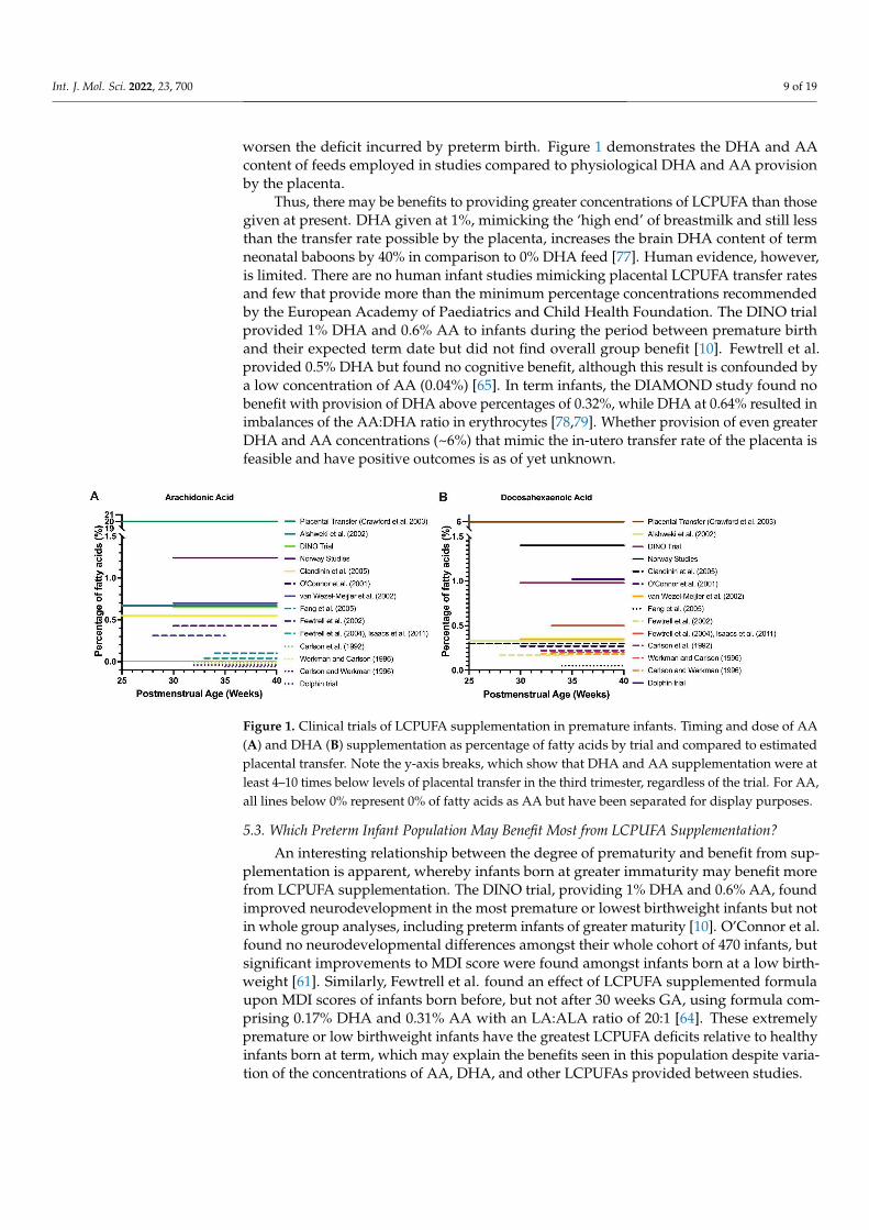
worsen the deficit incurred by preterm birth. Figure 1 demonstrates the DHA and AAcontent of feeds employed in studies compared to physiological DHA and AA provisionby the placenta.
Thus, there may be benefits to providing greater concentrations of LCPUFA than thosegiven at present. DHA given at 1%, mimicking the ‘high end’ of breastmilk and still lessthan the transfer rate possible by the placenta, increases the brain DHA content of termneonatal baboons by 40% in comparison to 0% DHA feed [77]. Human evidence, however,is limited. There are no human infant studies mimicking placental LCPUFA transfer ratesand few that provide more than the minimum percentage concentrations recommendedby the European Academy of Paediatrics and Child Health Foundation. The DINO trialprovided 1% DHA and 0.6% AA to infants during the period between premature birthand their expected term date but did not find overall group benefit [10]. Fewtrell et al.provided 0.5% DHA but found no cognitive benefit, although this result is confounded bya low concentration of AA (0.04%) [65]. In term infants, the DIAMOND study found nobenefit with provision of DHA above percentages of 0.32%, while DHA at 0.64% resulted inimbalances of the AA:DHA ratio in erythrocytes [78,79]. Whether provision of even greaterDHA and AA concentrations (~6%) that mimic the in-utero transfer rate of the placenta isfeasible and have positive outcomes is as of yet unknown.
Int. J. Mol. Sci. 2022, 23, x FOR PEER REVIEW 9 of 19
5.2. Would Higher Doses of Supplemental LCPUFA Provide Greater Benefits to Neurodevelopment?
If 0.3% DHA and 0.6% AA appears to benefit infants, is there benefit to providing higher concentrations to mimic the physiological LCPUFA transfer rate of the placenta more closely?
The concentrations of DHA and AA in standard feed are lower than the amounts of LCPUFA provided by the placenta in the final trimester, estimated at >6% of fatty acids for DHA and 14–20% of fatty acids for AA. The quantities of LCPUFA provided by standard formula feed is therefore around 10- to 50-fold less than the physiological expectation during the post-natal period after preterm birth [76]. Current feed with low LCPUFA content fails to meet the in-utero rates of DHA accretion [43,55] and is recognized to contribute to large LCPUFA deficits even in term infants [51], hence it is expected to worsen the deficit incurred by preterm birth. Figure 1 demonstrates the DHA and AA content of feeds employed in studies compared to physiological DHA and AA provision by the placenta.
Thus, there may be benefits to providing greater concentrations of LCPUFA than those given at present. DHA given at 1%, mimicking the ‘high end’ of breastmilk and still less than the transfer rate possible by the placenta, increases the brain DHA content of term neonatal baboons by 40% in comparison to 0% DHA feed [77]. Human evidence, however, is limited. There are no human infant studies mimicking placental LCPUFA transfer rates and few that provide more than the minimum percentage concentrations recommended by the European Academy of Paediatrics and Child Health Foundation. The DINO trial provided 1% DHA and 0.6% AA to infants during the period between premature birth and their expected term date but did not find overall group benefit [10]. Fewtrell et al. provided 0.5% DHA but found no cognitive benefit, although this result is confounded by a low concentration of AA (0.04%) [65]. In term infants, the DIAMOND study found no benefit with provision of DHA above percentages of 0.32%, while DHA at 0.64% resulted in imbalances of the AA:DHA ratio in erythrocytes [78,79]. Whether provision of even greater DHA and AA concentrations (~6%) that mimic the in-utero transfer rate of the placenta is feasible and have positive outcomes is as of yet unknown.
Figure 1. Clinical trials of LCPUFA supplementation in premature infants. Timing and dose of AA (A) and DHA (B) supplementation as percentage of fatty acids by trial and compared to estimated placental transfer. Note the y-axis breaks, which show that DHA and AA supplementation were at least 4–10 times below levels of placental transfer in the third trimester, regardless of the trial. For AA, all lines below 0% represent 0% of fatty acids as AA but have been separated for display purposes.
Figure 1. Clinical trials of LCPUFA supplementation in premature infants. Timing and dose of AA(A) and DHA (B) supplementation as percentage of fatty acids by trial and compared to estimatedplacental transfer. Note the y-axis breaks, which show that DHA and AA supplementation were atleast 4–10 times below levels of placental transfer in the third trimester, regardless of the trial. For AA,all lines below 0% represent 0% of fatty acids as AA but have been separated for display purposes.
5.3. Which Preterm Infant Population May Benefit Most from LCPUFA Supplementation?
An interesting relationship between the degree of prematurity and benefit from sup-plementation is apparent, whereby infants born at greater immaturity may benefit morefrom LCPUFA supplementation. The DINO trial, providing 1% DHA and 0.6% AA, foundimproved neurodevelopment in the most premature or lowest birthweight infants but notin whole group analyses, including preterm infants of greater maturity [10]. O’Connor et al.found no neurodevelopmental differences amongst their whole cohort of 470 infants, butsignificant improvements to MDI score were found amongst infants born at a low birth-weight [61]. Similarly, Fewtrell et al. found an effect of LCPUFA supplemented formulaupon MDI scores of infants born before, but not after 30 weeks GA, using formula com-prising 0.17% DHA and 0.31% AA with an LA:ALA ratio of 20:1 [64]. These extremelypremature or low birthweight infants have the greatest LCPUFA deficits relative to healthyinfants born at term, which may explain the benefits seen in this population despite varia-tion of the concentrations of AA, DHA, and other LCPUFAs provided between studies.

Int. J. Mol. Sci. 2022, 23, 700 10 of 19
5.4. Tailoring Supplemental DHA and AA to Mimic Dynamic Physiological Placental Supply andDevelopmental Demand
A further aspect to consider is the changing rate and ratio of placental LCPUFAtransfer across gestation. Martinez et al. showed that in early gestation, transfer of AAis predominant, while DHA transfer is delayed until the final weeks of gestation [49,80].This differential transfer of AA and DHA with time results in dynamic N6:N3 ratios, of 5:1at the beginning of the third trimester and ~2:1 by term [46]. The rate of transfer of DHAonly exceeds that of AA at 33 weeks’ gestation [46]. After birth, DHA accretion into braincontinues to peak between 2 and 3 years of age, while brain AA content remains relativelyunchanged [49].
Current feeding regimes for preterm infants provide static concentrations of supple-mental AA and DHA, but in pursuit of a greater depth of physiological mimicry, we maywish to vary formula AA and DHA concentrations with respect to infant CA to bettersimulate the dynamic functions of the placenta and demands of the infant. In this regard,it may be that prematurely born infants require several formulae during their postnatalperiod; an infant born at 25 weeks may require more AA than DHA, whilst an infant bornafter 33 weeks may require more DHA than AA. Formula provided after the prematureinfant’s ‘term’ date (corrected age) should resemble the LCPUFA composition of breastmilkfor ongoing postnatal nutrition. Looking onwards, the LCPUFA composition of weaningfoods and ongoing diet may be considered to be modifiable factors affecting LCPUFAassimilation and accumulation in childhood and later life.
5.5. Organ-Specific Effects of LCPUFA
It is apparent that LCPUFA availability has differential effects upon the development ofspecific organs. Although postnatal supplementation of DHA and AA seem to be beneficialfor neurodevelopment and retinal development in preterm infants [16,32], recent studieshave indicated a potentially opposing effect in pulmonary development, with no benefiton rates of—and a potentially increased risk for—bronchopulmonary dysplasia (BPD)in infants supplemented with LCPUFA [35,36,40]. This is despite pre-clinical evidenceand observational studies showing that lower postnatal levels of DHA are associatedwith increased risk of chronic lung disease [34,81]. Reasons for this are unknown, butsupplementation with DHA might lead to alterations in the levels of other LCPUFAsimportant for pulmonary development or an imbalance in fatty acid metabolites. Morewidely, supplementation with single specific fatty acids might have different effects ondifferent organs. We must refine our approach to LCPUFA supplementation to holisticallyoptimize the development of all organ systems.
5.6. Challenges for Appropriate LCPUFA Supplementation
Premature birth brings many uncertainties and unexpected physiological challengesfor the newborn, for which it is unprepared. Examples include exposure to air and in-creased oxygen tensions, infections, and varying types of brain and peripheral organ injury.LCPUFAs may be protective via their metabolism to eicosanoids and SPMs yet are alsosusceptible to peroxidation, which may propagate cellular damage [76,82,83]. We mayconsider whether more or less of individual LCPUFA or modifications of the N3:N6 ratiomay help or hinder both normal development and resilience against challenges associatedwith prematurity.
Regardless of the amount of supplemental DHA provided, we must also consider theeffectiveness of the route chosen to provide DHA to prematurely born infants. Administra-tion of high amounts of fatty acids such as DHA via the immaturely developed prematureinfant digestive system may be ineffective or problematic due to altered absorption. Addi-tionally, intolerance of oral feeding and gastrointestinal complications such as spontaneousintestinal perforation and necrotizing enterocolitis are common with increasing prematurity.Parenteral lipid supplementation is therefore common in these infants but also does notfully mimic or replace normal fatty acid metabolism.

Int. J. Mol. Sci. 2022, 23, 700 11 of 19
Overall, supplementation of LCPUFA is greatly nuanced and has the potential toboth benefit and harm the premature infant. Pragmatically, future research must alsoconsider that measurement of fatty acid levels in blood might not correlate with thelevels in specific tissues. It is evident that a greater understanding of changes in LCPUFAtransfer rates with gestational maturity and the effects of LCPUFA availability upon specifictissue development is needed to optimize the growth and development of prematurelyborn infants.
6. The Wider Contexts of LCPUFA SupplementationDietary LA Affects Infant AA and DHA Availability
In addition to large differences in AA and DHA concentrations across the trials todate, there is large variation between the concentrations of LA and ALA provided in bothcontrol and intervention formulae. Historical formulae have included LA and ALA withan assumption of adequate endogenous conversion to AA and DHA, but in reality, thisconversion is inadequate to meet infant demands. For instance, Cunnane et al. showed thatinfants fed DHA-deficient formula depleted their adipose stores to unmeasurable levelsand had less brain DHA accumulation than infants fed DHA-repleted formula [51]. Otherautopsy studies have shown reduced total DHA content of brains of infants fed formulathat was deficient in DHA, despite containing LA and ALA [18].
Similarly, Bockmann et al. showed that feeding LA-rich feed to preterm infantsalters the adipose LCPUFA composition to become dominated by LA and depleted ofAA and DHA. These results show that, while dietary ALA and LA are inadequate tosupply infant needs of DHA and AA, excessive LA is stored and persists in fetal adiposetissue [84]. Furthermore, these preterm infants had reduced AA and DHA bound tophosphatidylcholine in their blood, suggesting a reduction in total availability of AA andDHA to developing tissues, such as brain tissues.
Instead, excess LA in infant diet and infant tissues has the potential to alter theLCPUFA composition of multiple tissues, as well as the overall N6:N3 ratio. This ratio is ofsignificance due to metabolic competition at the level of the desaturase enzymes, wherebyexcesses of one series of LCPUFA may out-compete others for metabolism into downstreammetabolites, contributing to a relative deficiency of these other LCPUFA despite adequateoral intake. It is known that dietary PUFA composition can alter the N6:N3 balance oftissues in both animals and humans. Hsieh et al. demonstrated competition between AAand DHA in infant baboons who were given feed containing varying N6:N3 ratios byproviding a fixed percentage of AA and ‘standard’, ‘medium’, or ‘high’ percentages ofDHA. The baboons with the highest DHA intake demonstrated a reciprocal decrease inbrain AA content, representing competition between N3 and N6 fatty acids for placementwithin neuronal membranes [77]. Infant piglets fed formulae containing comparable ALA(N3) but with differences in LA (N6) content demonstrated changes in tissue LCPUFAcomposition to resemble those of their diets; piglets fed N6-rich formula had greater N6content of liver, blood, retina, and brain, but they also showed relative depletion of N3LCPUFA in comparison to piglets fed sow milk with a more equal N3:N6 ratio [85]. In afurther study of piglets by the same group, increased duration of LA-rich (30%) feedingassociated with progressive decreases in retinal and brain DHA and reciprocal increases inN6 LCPUFA [86].
Similarly, findings have been seen in human infants born >35 weeks GA provided totalparenteral nutrition (TPN) high in LA for a maximum of 12 days after birth. In these infants,hepatic levels of LA increased three-fold, while levels of DHA halved, with a consequentdramatic reduction in the N3:N6 ratio [87]. While this study found no change in brainDHA content at autopsy during its <11-day duration, the effects of more persistent dietarymanipulation of the N3:N6 ratio upon human neurodevelopment is unknown.
The DIAMOND [78,79] trial is the only study to provide a varying ratio of N3:N6and measure neurodevelopment. Term infants were fed formula containing a fixed doseof AA (0.64%) with varying percentages of DHA (0.32% DHA, 0.64% DHA, or 0.96%

Int. J. Mol. Sci. 2022, 23, 700 12 of 19
DHA) to achieve N6:N3 ratios of 2:1, 1:1, and 0.7:1. These DHA-supplemented groupswere compared against a control cohort fed formula containing 0% DHA and 0% AA. Allformulas contained similar amounts of LA (16.9–17.5% fatty acids) and ALA (1.61–1.68%fatty acids).
Infants in the DIAMOND trial that were fed formula containing DHA had improvedvisual acuity at 3 months and superior Bayley Scale of Infant Development (BSID-II)Motor Development Index (MDI) scores, emotional regulation, and language abilities at18 months in comparison to infants that were fed the formula containing 0% DHA [78,79].Furthermore, improved language and cognitive performance were seen between ages3–5 years [88], while at 9 years, MRI measures of connectivity, brain volume, and neuro-chemical levels were greater in children fed DHA-containing formula [89].
The DIAMOND studies generally found no benefit to greater concentrations of DHAin feed; the majority of benefits were found at a ratio of 2:1 or 1:1, with no significantbenefit found at lower N3:N6 ratios or higher DHA concentrations. At greater intakes of0.96% DHA, erythrocyte DHA increased with a reciprocal decrease in AA to reach a levelbelow those of infants fed AA-deplete formula, confirming the findings of other studiesdemonstrating interaction and displacement between series of LCPUFA [78].
Furthermore, there appear to be more complex synergistic effects between AA andDHA. The Mega Donna Mega trial analyzed DHA and AA serum concentrations withregard to risk of ROP development in premature infants and found that higher serum DHAonly conferred reduced ROP risk when AA was above a threshold concentration; the sameDHA concentration did not reduce ROP risk in infants with low serum AA [21].
The cumulative storage of and interaction between dietary AA, DHA, and their parentLCPUFAs likely represent an important confounding factor that may affect our interpre-tation of all LCPUFA-supplementation studies that report neurocognitive outcomes attimepoints in later childhood. After feeding until 9 months, Fewtrell et al. found noneurocognitive benefits a [65] or 18 months or at 9 years [65]. The studies from Norwaysupplemented breastmilk with AA and DHA for 9 weeks but found no overall differ-ences in neurodevelopment at 18 months, 26 months, or at 8 years [7,59]. Similarly, theDINO study, providing supplemented feeds until term corrected age, found no benefit at7 years of age [9]. Due to the role of adipose tissue to sequester LCPUFA and the ongoingcompetition between N3 and N6 LCPUFA families for metabolism and distribution intotissues or membranes, even the prolonged supplementation periods demonstrated in thestudies above are relatively short in comparison to the interim between the end of thesupplementation period and later neurocognitive measurements. This intervening period isunaccounted for by these studies, with infants returning to formula deficient of AA/DHAor of variable ALA/LA content before weaning onto a diet of uncharacterized LCPUFAcomposition. The LCPUFA composition of post-intervention formula and weaning foodsmay partly explain the lack of effect of LCPUFA-supplementation during the perinatalperiod upon neurocognitive outcomes measured years after birth. These foods shouldbe considered as important factors to be controlled, or recognized as being confounders,when designing future studies that will assess neurocognitive performance throughoutchildhood development.
In summary, there is evidence for an interaction between AA, DHA, and other es-sential LCPUFA in animal models and both preterm and term humans. The DIAMONDstudy is the only evaluation of the effects of the N3:N6 balance and found neurodevelop-mental benefits across multiple time points in childhood but is specific to infants born atterm [78,79].
7. The Wider Factors Affecting LCPUFA Status7.1. Maternal LCPUFA Status
The maternal fatty acids that supply the infant in utero and in breastmilk are derivedfrom the maternal diet and stores in adipose tissue [90]; the quality of fatty acids consumedand stored by a prospective or pregnant mother is a biologically plausible and modifiable

Int. J. Mol. Sci. 2022, 23, 700 13 of 19
factor affecting infant developmental outcomes after premature birth. The Western diet hasbecome accustomed to an increased consumption of N6 PUFA-rich foods, such as vegetableoils [91], a dietary change that is reflected in the fatty acid composition of maternal tissues.
In the 1980s, Clandinin et al. remarked that the N6 content of fetal adipose tissue washigher than previous studies, attributing this to changing maternal dietary habits to includemore vegetable oils [53], intakes of which now far exceed the nutritional requirement for N6LCPUFA of ~1% fatty acids [92]. Indeed, differences in fatty acid composition of breastmilkand adipose tissue have been seen between traditional and modern populations [50,90,93].
7.2. Genetic Polymorphisms
Aside from diet, genetic polymorphisms of the FADS gene affecting expression of thefatty acid desaturase enzymes that convert LCPUFA precursors to AA and DHA may affectmaternal LCPUFA synthesis and thus availability of LCPUFA to the infant. FADS genepolymorphisms may reduce the maternal ability to provide LCPUFA in breastmilk [1,94]and correlate with reduced infant IQ [95]. In case of genetic variants corresponding toreduced LCPUFA conversion, maternal intake of preformed AA and DHA, or specificsupplementation of formula, may be crucial to provide adequate AA and DHA to the infantboth in-utero and in the postnatal period [96].
7.3. Socioeconomic Status
Dietary LA also increases with decreasing socioeconomic status [92]. One of the twocohorts of the DIAMOND study represented families of lower socioeconomic status; infantsborn into these families demonstrated greater levels of erythrocyte AA than their peersborn into families living in wealthier areas [78], illustrating a potential relationship betweensocioeconomic status, maternal and infant intake of foods rich in N6 PUFA, and early lifedevelopment [74]. It is of interest that neurodevelopmental outcomes of infants whosemothers supplemented with DHA during pregnancy are more pronounced in familiesof lower education or of ‘poorer’ home environments [97,98]. It may be that families oflower SES may derive the most benefit from strategies to decrease dietary N6 intake and/orsupplement with N3 LCPUFA.
Maternal LCPUFA status may also affect the trajectory of infant development withregard to gestation length and in-utero development. A 2018 paper found that low maternalblood EPA and DHA concentrations increased risk of preterm birth by 10-fold [39], and arecent Cochrane review found that maternal N3 supplementation was associated with alower risk of both preterm (<37 weeks) and early preterm birth (<34 weeks), infant perinataldeath, and low birthweight, suggesting that optimal maternal LCPUFA status may preventpreterm birth and influence the risk of many neonatal comorbidities [99].
On the other hand, results from large cohort studies that assessed the overall dietaryLCPUFA intake during pregnancy have suggested that increases in the N6:N3 ratio areassociated with developmental delay. Kim et al. estimated the N3 and N6 PUFA intakes of960 mothers during pregnancy, and of their infants via breast milk and weaning feeds inthe postnatal period, to assess the effects of N3 and N6 intakes upon neurodevelopment at6 months as measured by BSID-II; greater LA:ALA and N6:N3 ratios were both associatedwith lower MDI and PDI scores in infants, while multiple logistic regression analysisshowed that infants of mothers with the highest LA:ALA ratios had >2 times the risk ofneurodevelopmental delay of infants whose mothers had the lowest LA:ALA ratios [100].Similar findings of a negative correlation between maternal N6:N3 ratio and languageability in infants born at term and not breastfed were found by Bernard et al. [101].
After birth, increased maternal N6 LCPUFA status may directly affect infant tissueLCPUFA composition via the breastmilk, while weaning onto a N6-rich modern dietmay contribute to ongoing competition between N6 and N3 fatty acids. The fatty acidcomposition of maternal milk and the wider food environment are unaccounted factorsthat may influence the results of trials assessing the long-term effects of DHA in thepost-natal period.

Int. J. Mol. Sci. 2022, 23, 700 14 of 19
In summary, it is evident that the maternal dietary intake and storage of LCPUFA, incombination with genetic factors, can affect the availability of these LCPUFA to developinginfants and affect the consequent fatty acid composition of infant tissues. Furthermore,a suboptimal maternal LCPUFA status may increase the risk of preterm birth, while apositive LCPUFA status may improve infant growth and thus reduce the risk of neonatalmorbidity. Whether maternal LCPUFA status affects neurodevelopment in preterm infantsis yet unknown and is largely unstudied, thus the authors welcome future studies incorpo-rating data of maternal LCPUFA status into calculations of absolute and relative LCPUFAavailability to the developing infant.
8. Conclusions
LCPUFA transfer from mother to fetus occurs via the placenta throughout pregnancy,with particularly high rates of DHA transfer during the final trimester. LCPUFA accreteinto brain tissue and are essential for neurodevelopment; however, the bulk of LCPUFAare stored in adipose tissue to provide vital endogenous LCPUFA stores in the postnatalperiod. Infants born before term do not receive this placental transfer of LCPUFA andhence can be severely LCPUFA-deficient in both brain and adipose tissue in comparison toterm-born infants.
The evidence supporting post-natal LCPUFA supplementation for prematurely borninfants is weakened by heterogeneity within the populations studied, durations of feedingand the concentrations of AA, DHA, LA, and ALA provided. While firm conclusionscannot be drawn, it is apparent that providing DHA at 0.3% and AA at 0.6% providesgreater benefit to the neurodevelopment of prematurely born infants than feeding LCPUFA-deficient formula.
Future studies should consider the absolute and relative availability of AA and DHAand the timing of these fatty acids with respect to normal placental physiology. Furthermore,the availability of other LCPUFA such as LA and ALA within infant formula should beseen as factors interacting with the relative availability of AA and DHA within the infant.More widely, the role of maternal LCPUFA status is poorly understood, but may directlyaffect the risk of premature birth and infant neurodevelopment.
Supplementary Materials: The following are available online at https://www.mdpi.com/article/10.3390/ijms23020700/s1.
Author Contributions: Conceptualization, R.J.H., S.K. and T.R.W.; writing—original draft preparation,R.J.H. and S.K.; writing—review and editing, R.J.H., S.K. and T.R.W.; visualization, R.J.H. and T.R.W.;supervision, T.R.W.; project administration, R.J.H. and T.R.W.; funding acquisition, T.R.W. All authorshave read and agreed to the published version of the manuscript.
Funding: T.R.W. is supported by start-up funds from the University of Washington Department ofPediatrics. The authors received no funding in direct support of this work.
Data Availability Statement: Not applicable.
Conflicts of Interest: The authors declare no conflict of interest.
References1. Xie, L.; Innis, S.M. Genetic Variants of the FADS1 FADS2 Gene Cluster Are Associated with Altered (n-6) and (n-3) Essential Fatty
Acids in Plasma and Erythrocyte Phospholipids in Women during Pregnancy and in Breast Milk during Lactation. J. Nutr. 2008,138, 2222–2228. [CrossRef]
2. Carnielli, V.P.; Wattimena, D.J.L.; Luijendijk, I.H.T.; Boerlage, A.; Degenhart, H.J.; Sauer, P.J.J. The Very Low Birth WeightPremature Infant Is Capable of Synthesizing Arachidonic and Docosahexaenoic Acids from Linoleic and Linolenic Acids. Pediatr.Res. 1996, 40, 169–174. [CrossRef]
3. Carnielli, V.P.; Simonato, M.; Verlato, G.; Luijendijk, I.; de Curtis, M.; Sauer, P.J.J.; Cogo, P.E. Synthesis of Long-Chain Polyunsatu-rated Fatty Acids in Preterm Newborns Fed Formula with Long-Chain Polyunsaturated Fatty Acids. Am. J. Clin. Nutr. 2007, 86,1323–1330. [CrossRef] [PubMed]

Int. J. Mol. Sci. 2022, 23, 700 15 of 19
4. Agostoni, C.; Buonocore, G.; Carnielli, V.P.; de Curtis, M.; Darmaun, D.; Decsi, T.; Domellöf, M.; Embleton, N.D.; Fusch, C.;Genzel-Boroviczeny, O.; et al. Enteral Nutrient Supply for Preterm Infants: Commentary from the European Society of PaediatricGastroenterology, Hepatology and Nutrition Committee on Nutrition. J. Pediatr. Gastroenterol. Nutr. 2010, 50, 85–91. [CrossRef]
5. Salem, N.; Wegher, B.; Mena, P.; Uauy, R. Arachidonic and Docosahexaenoic Acids Are Biosynthesized from Their 18-CarbonPrecursors in Human Infants. Proc. Natl. Acad. Sci. USA 1996, 93, 49–54. [CrossRef] [PubMed]
6. Moon, K.; Rao, S.C.; Schulzke, S.M.; Patole, S.K.; Simmer, K. Longchain Polyunsaturated Fatty Acid Supplementation in PretermInfants. Cochrane Database Syst. Rev. 2016, 2017. [CrossRef]
7. Almaas, A.N.; Tamnes, C.K.; Nakstad, B.; Henriksen, C.; Walhovd, K.B.; Fjell, A.M.; Due-Tonnessen, P.; Drevon, C.A.; Iversen, P.O.Long-Chain Polyunsaturated Fatty Acids and Cognition in VLBW Infants at 8 Years: An RCT. Pediatrics 2015, 135, 972–980.[CrossRef]
8. Alshweki, A.; Muñuzuri, A.P.; Baña, A.M.; José De Castro, M.; Andrade, F.; Aldamiz-Echevarría, L.; Sáenz De Pipaón, M.;Fraga, J.M.; Couce, M.L. Effects of Different Arachidonic Acid Supplementation on Psychomotor Development in Very PretermInfants; a Randomized Controlled Trial. Nutr. J. 2015, 14, 101. [CrossRef] [PubMed]
9. Collins, C.T.; Gibson, R.A.; Anderson, P.J.; McPhee, A.J.; Sullivan, T.R.; Gould, J.F.; Ryan, P.; Doyle, L.W.; Davis, P.G.;McMichael, J.E.; et al. Neurodevelopmental Outcomes at 7 Years’ Corrected Age in Preterm Infants Who Were Fed High-DoseDocosahexaenoic Acid to Term Equivalent: A Follow-up of a Randomised Controlled Trial. BMJ Open 2015, 5, e007314. [CrossRef][PubMed]
10. Makrides, M.; Gibson, R.A.; McPhee, A.J.; Collins, C.T.; Davis, P.G.; Doyle, L.W.; Simmer, K.; Colditz, P.B.; Morris, S.;Smithers, L.G.; et al. Neurodevelopmental Outcomes of Preterm Infants Fed High-Dose Docosahexaenoic Acid. JAMA 2009,301, 175. [CrossRef]
11. Smithers, L.G.; Collins, C.T.; Simmonds, L.A.; Gibson, R.A.; McPhee, A.; Makrides, M. Feeding Preterm Infants Milk with aHigher Dose of Docosahexaenoic Acid than That Used in Current Practice Does Not Influence Language or Behavior in EarlyChildhood: A Follow-up Study of a Randomized Controlled Trial. Am. J. Clin. Nutr. 2010, 91, 628–634. [CrossRef] [PubMed]
12. Hadley, K.B.; Ryan, A.S.; Forsyth, S.; Gautier, S.; Salem, N. The Essentiality of Arachidonic Acid in Infant Development. Nutrients2016, 8, 216. [CrossRef] [PubMed]
13. Hashimoto, M.; Hossain, S.; Shimada, T.; Shido, O. Docosahexaenoic Acid-Induced Protective Effect against Impaired Learning inAmyloid β-Infused Rats Is Associated with Increased Synaptosomal Membrane Fluidity. Clin. Exp. Pharmacol. Physiol. 2006,33, 934–939. [CrossRef]
14. Onuki, Y.; Morishita, M.; Chiba, Y.; Tokiwa, S.; Takayama, K. Docosahexaenoic Acid and Eicosapentaenoic Acid Induce Changesin the Physical Properties of a Lipid Bilayer Model Membrane. Chem. Pharm. Bull. 2006, 54, 68–71. [CrossRef]
15. Berman, D.R.; Liu, Y.; Barks, J.; Mozurkewich, E. Treatment with Docosahexaenoic Acid after Hypoxia-Ischemia ImprovesForepaw Placing in a Rat Model of Perinatal Hypoxia-Ischemia. Am. J. Obstet. Gynecol. 2010, 203, 385-e1. [CrossRef]
16. Buddington, R.K.; Chizhikov, V.V.; Iskusnykh, I.Y.; Sable, H.J.; Sable, J.J.; Holloway, Z.R.; Katzir, T.B.; van der Merwe, M.;Yakimkova, T.; Buddington, K.K.; et al. A Phosphatidylserine Source of Docosahexanoic Acid Improves Neurodevelopment andSurvival of Preterm Pigs. Nutrients 2018, 10, 637. [CrossRef]
17. Suganuma, H.; Arai, Y.; Kitamura, Y.; Hayashi, M.; Okumura, A.; Shimizu, T. Maternal Docosahexaenoic Acid-Enriched DietPrevents Neonatal Brain Injury. Neuropathology 2010, 30, 597–605. [CrossRef] [PubMed]
18. Farquharson, J.; Jamieson, E.C.; Logan, R.W.; Cockburn, F.; Ainslie Patrick, W. Infant Cerebral Cortex Phospholipid Fatty-AcidComposition and Diet. Lancet 1992, 340, 810–813. [CrossRef]
19. Farquharson, J.; Jamieson, E.C.; Abbasi, K.A.; Patrick, W.J.A.; Logan, R.W.; Cockbum, F. Effect of Diet on the Fatty AcidComposition of the Major Phospholipids of Infant Cerebral Cortex. Arch. Dis. Child. 1995, 72, 198–203. [CrossRef]
20. Uauy, R.D.; Birch, D.G.; Birch, E.E.; Tyson, J.E.; Hoffman, D.R. Effect of Dietary Omega-3 Fatty Acids on Retinal Function ofVery-Low-Birth-Weight Neonates. Pediatr. Res. 1990, 28, 485–492. [CrossRef] [PubMed]
21. Hellström, A.; Nilsson, A.K.; Wackernagel, D.; Pivodic, A.; Vanpee, M.; Sjöbom, U.; Hellgren, G.; Hallberg, B.; Domellöf, M.;Klevebro, S.; et al. Effect of Enteral Lipid Supplement on Severe Retinopathy of Prematurity: A Randomized Clinical Trial. JAMAPediatr. 2021, 175, 359–367. [CrossRef]
22. Bernabe-García, M.; Villegas-Silva, R.; Villavicencio-Torres, A.; Calder, P.C.; Rodríguez-Cruz, M.; Maldonado-Hernández, J.;Macías-Loaiza, D.; López-Alarcón, M.; Inda-Icaza, P.; Cruz-Reynoso, L. Enteral Docosahexaenoic Acid and Retinopathy ofPrematurity: A Randomized Clinical Trial Mariela Bernabe-García. J. Parenter. Enter. Nutr. 2019, 43, 874–882. [CrossRef] [PubMed]
23. Sveinsdóttir, K.; Ley, D.; Hövel, H.; Fellman, V.; Hüppi, P.S.; Smith, L.E.H.; Hellström, A.; Hansen Pupp, I. Relation of Retinopathyof Prematurity to Brain Volumes at Term Equivalent Age and Developmental Outcome at 2 Years of Corrected Age in VeryPreterm Infants. Neonatology 2018, 114, 46–52. [CrossRef]
24. Tam, E.W.Y.; Chau, V.; Barkovich, A.J.; Ferriero, D.M.; Miller, S.P.; Rogers, E.E.; Grunau, R.E.; Synnes, A.R.; Xu, D.; Foong, J.; et al.Early Postnatal Docosahexaenoic Acid Levels and Improved Preterm Brain Development. Pediatr. Res. 2016, 79, 723–730.[CrossRef]
25. Carlson, S.E.; Cooke, R.J.; Werkman, S.H.; Tolley, E.A. First Year Growth of Preterm Infants Fed Standard Compared to MarineOil N-3 Supplemented Formula. Lipids 1992, 27, 901–907. [CrossRef]
26. Carlson, S.E.; Werkman, S.H.; Peeples, J.M.; Wilson, W.M. Growth and Development of Premature Infants in Relation to Omega 3and Omega 6 Fatty Acid Status. World Rev. Nutr. Diet. 1994, 75, 63–69. [CrossRef] [PubMed]

Int. J. Mol. Sci. 2022, 23, 700 16 of 19
27. Hack, M.; Flannery, D.J.; Schluchter, M.; Cartar, L.; Borawski, E.; Klein, N. Outcomes in Young Adulthood for Very-Low-Birth-Weight Infants. N. Engl. J. Med. 2002, 346, 149–157. [CrossRef] [PubMed]
28. Calder, P.C. Polyunsaturated Fatty Acids and Inflammation: From Molecular Biology to the Clinic. Lipids 2003, 38, 343–352.[CrossRef] [PubMed]
29. Levy, B.D.; Clish, C.B.; Schmidt, B.; Gronert, K.; Serhan, C.N. Lipid Mediator Class Switching during Acute Inflammation: Signalsin Resolution. Nat. Immunol. 2001, 2, 612–619. [CrossRef] [PubMed]
30. Serhan, C.N.; Chiang, N.; Dalli, J.; Levy, B.D. Lipid Mediators in the Resolution of Inflammation. Cold Spring Harb. Perspect. Biol.2015, 7, a016311. [CrossRef]
31. Serhan, C.N.; Chiang, N.; van Dyke, T.E. Resolving Inflammation: Dual Anti-Inflammatory and pro-Resolution Lipid Mediators.Nat. Rev. Immunol. 2008, 8, 349–361. [CrossRef] [PubMed]
32. Bazan, N.G.; Calandria, J.M.; Gordon, W.C. Docosahexaenoic Acid and Its Derivative Neuroprotectin D1 Display NeuroprotectiveProperties in the Retina, Brain and Central Nervous System. Nestle Nutr. Inst. Workshop Ser. 2013, 77, 121–131. [CrossRef][PubMed]
33. Humberg, A.; Fortmann, I.; Siller, B.; Kopp, M.V.; Herting, E.; Göpel, W.; Härtel, C. Preterm Birth and Sustained Inflammation:Consequences for the Neonate. Semin. Immunopathol. 2020, 42, 451–468. [CrossRef]
34. Martin, C.R.; Dasilva, D.A.; Cluette-Brown, J.E.; Dimonda, C.; Hamill, A.; Bhutta, A.Q.; Coronel, E.; Wilschanski, M.;Stephens, A.J.; Driscoll, D.F.; et al. Decreased Postnatal Docosahexaenoic and Arachidonic Acid Blood Levels in PrematureInfants Are Associated with Neonatal Morbidities. J. Pediatr. 2011, 159, 743–749.e2. [CrossRef]
35. Collins, C.T.; Makrides, M.; McPhee, A.J.; Sullivan, T.R.; Davis, P.G.; Thio, M.; Simmer, K.; Rajadurai, V.S.; Travadi, J.;Berry, M.J.; et al. Docosahexaenoic Acid and Bronchopulmonary Dysplasia in Preterm Infants. N. Engl. J. Med. 2017,376, 1245–1255. [CrossRef]
36. Marc, I.; Piedboeuf, B.; Lacaze-Masmonteil, T.; Fraser, W.; Mâsse, B.; Mohamed, I.; Qureshi, M.; Afifi, J.; Lemyre, B.;Caouette, G.; et al. Effect of Maternal Docosahexaenoic Acid Supplementation on Bronchopulmonary Dysplasia-Free Survival inBreastfed Preterm Infants: A Randomized Clinical Trial. JAMA—J. Am. Med. Assoc. 2020, 324, 157–167. [CrossRef] [PubMed]
37. Hellström, A.; Pivodic, A.; Gränse, L.; Lundgren, P.; Sjöbom, U.; Nilsson, A.K.; Söderling, H.; Hård, A.-L.; Smith, L.E.H.;Löfqvist, C.A. Association of Docosahexaenoic Acid and Arachidonic Acid Serum Levels With Retinopathy of Prematurity inPreterm Infants. JAMA Netw. Open 2021, 4, e2128771. [CrossRef]
38. Wu, W.X.; Ma, X.H.; Coksaygan, T.; Chakrabarty, K.; Collins, V.; Rose, J.; Nathanielsz, P.W. Prostaglandin Mediates PrematureDelivery in Pregnant Sheep Induced by Estradiol at 121 Days of Gestational Age. Endocrinology 2004, 145, 1444–1452. [CrossRef]
39. Olsen, S.F.; Halldorsson, T.I.; Thorne-Lyman, A.L.; Strøm, M.; Gørtz, S.; Granstrøm, C.; Nielsen, P.H.; Wohlfahrt, J.; Lykke, J.A.;Langhoff-Roos, J.; et al. Plasma Concentrations of Long Chain N-3 Fatty Acids in Early and Mid-Pregnancy and Risk of EarlyPreterm Birth. EBioMedicine 2018, 35, 325–333. [CrossRef]
40. Hellström, A.; Hellström, W.; Hellgren, G.; Smith, L.E.H.; Puttonen, H.; Fyhr, I.M.; Sävman, K.; Nilsson, A.K.; Klevebro, S.Docosahexaenoic Acid and Arachidonic Acid Levels Are Associated with Early Systemic Inflammation in Extremely PretermInfants. Nutrients 2020, 12, 1996. [CrossRef]
41. Hofer, N.; Kothari, R.; Morris, N.; Müller, W.; Resch, B. The Fetal Inflammatory Response Syndrome Is a Risk Factor for Morbidityin Preterm Neonates. Am. J. Obstet. Gynecol. 2013, 209, 542.e1–542.e11. [CrossRef]
42. Kuban, K.C.K.; Joseph, R.M.; O’Shea, T.M.; Heeren, T.; Fichorova, R.N.; Douglass, L.; Jara, H.; Frazier, J.A.; Hirtz, D.;Rollins, J.V.; et al. Circulating Inflammatory-Associated Proteins in the First Month of Life and Cognitive Impairment at Age 10Years in Children Born Extremely Preterm. J. Pediatr. 2017, 180, 116–123.e1. [CrossRef] [PubMed]
43. Crawford, M.A.; Williams, G.; Hassam, A.G.; Whitehouse, W.L. Essential Fatty Acids and Fetal Brain Growth. Lancet 1976,307, 452–453. [CrossRef]
44. Hüppi, P.S.; Warfield, S.; Kikinis, R.; Barnes, P.D.; Zientara, G.P.; Jolesz, F.A.; Tsuji, M.K.; Volpe, J.J. Quantitative MagneticResonance Imaging of Brain Development in Premature and Mature Newborns. Ann. Neurol. 1998, 43, 224–235. [CrossRef]
45. Clandinin, M.T.; Chappell, J.E.; Leong, S.; Heim, T.; Swyer, P.R.; Chance, G.W. Intrauterine Fatty Acid Accretion Rates in HumanBrain: Implications for Fatty Acid Requirements. Early Hum. Dev. 1980, 4, 121–129. [CrossRef]
46. Bernhard, W.; Raith, M.; Koch, V.; Maas, C.; Abele, H.; Poets, C.F.; Franz, A.R. Developmental Changes in Polyunsaturated FetalPlasma Phospholipids and Feto-Maternal Plasma Phospholipid Ratios and Their Association with Bronchopulmonary Dysplasia.Eur. J. Nutr. 2016, 55, 2265–2274. [CrossRef] [PubMed]
47. Kuipers, R.S.; Luxwolda, M.F.; Offringa, P.J.; Rudi Boersma, E.; Dijck-Brouwer, D.A.J.; Muskiet, F.A.J. Fetal Intrauterine WholeBody Linoleic, Arachidonic and Docosahexaenoic Acid Contents and Accretion Rates. Prostaglandins Leukot. Essent. Fat. Acids2012, 86, 13–20. [CrossRef] [PubMed]
48. Kuipers, R.S.; Luxwolda, M.F.; Offringa, P.J.; Rudy Boersma, E.; Janneke Dijck-Brouwer, D.A.; Muskiet, F.A.J. Gestational AgeDependent Changes of the Fetal Brain, Liver and Adipose Tissue Fatty Acid Compositions in a Population with High Fish Intakes.Prostaglandins Leukot. Essent. Fat. Acids 2012, 86, 189–199. [CrossRef]
49. Martínez, M.; Mougan, I. Fatty Acid Composition of Human Brain Phospholipids during Normal Development. J. Neurochem.1998, 71, 2528–2533. [CrossRef]
50. Innis, S.M.; Kuhnlein, H.V. Long-Chain n-3 Fatty Acids in Breast Milk of Inuit Women Consuming Traditional Foods. Early Hum.Dev. 1988, 18, 185–189. [CrossRef]

Int. J. Mol. Sci. 2022, 23, 700 17 of 19
51. Cunnane, S.C.; Francescutti, V.; Brenna, J.T.; Crawford, M.A. Breast-Fed Infants Achieve a Higher Rate of Brain and Whole BodyDocosahexaenoate Accumulation than Formula-Fed Infants Not Consuming Dietary Docosahexaenoate. Lipids 2000, 35, 105–111.[CrossRef]
52. Clandinin, M.T.; Chappell, J.E.; Leong, S.; Heim, T.; Swyer, P.R.; Chance, G.W. Extrauterine Fatty Acid Accretion in Infant Brain:Implications for Fatty Acid Requirements. Early Hum. Dev. 1980, 4, 131–138. [CrossRef]
53. Clandinin, M.T.; Chappell, J.E.; Heim, T.; Swyer, P.R.; Chance, G.W. Fatty Acid Utilization in Perinatal de Novo Synthesis ofTissues. Early Hum. Dev. 1981, 5, 355–366. [CrossRef]
54. Cunnane, S.C.; Crawford, M.A. Survival of the Fattest: Fat Babies Were the Key to Evolution of the Large Human Brain. Comp.Biochem. Physiol. Part A Mol. Integr. Physiol. 2003, 136, 17–26. [CrossRef]
55. Innis, S.M. Essential Fatty Acid Transfer and Fetal Development. Placenta 2005, 26, 4–9. [CrossRef]56. Henriksen, C.; Haugholt, K.; Lindgren, M.; Aurvåg, A.K.; Rønnestad, A.; Grønn, M.; Solberg, R.; Moen, A.; Nakstad, B.;
Berge, R.K.; et al. Improved Cognitive Development among Preterm Infants Attributable to Early Supplementation of HumanMilk with Docosahexaenoic Acid and Arachidonic Acid. Pediatrics 2008, 121, 1137–1145. [CrossRef] [PubMed]
57. Westerberg, A.C.; Schei, R.; Henriksen, C.; Smith, L.; Veierød, M.B.; Drevon, C.A.; Iversen, P.O. Attention among Very Low BirthWeight Infants Following Early Supplementation with Docosahexaenoic and Arachidonic Acid. Acta Paediatr. Int. J. Paediatr. 2011,100, 47–52. [CrossRef]
58. Almaas, A.N.; Tamnes, C.K.; Nakstad, B.; Henriksen, C.; Grydeland, H.; Walhovd, K.B.; Fjell, A.M.; Iversen, P.O.; Drevon, C.A.Diffusion Tensor Imaging and Behavior in Premature Infants at 8 Years of Age, a Randomized Controlled Trial with Long-ChainPolyunsaturated Fatty Acids. Early Hum. Dev. 2016, 95, 41–46. [CrossRef]
59. Henriksen, C.; Almaas, A.N.; Westerberg, A.C.; Drevon, C.A.; Iversen, P.O.; Nakstad, B. Growth, Metabolic Markers, andCognition in 8-Year Old Children Born Prematurely, Follow-up of a Randomized Controlled Trial with Essential Fatty Acids. Eur.J. Pediatr. 2016, 175, 1165–1174. [CrossRef]
60. Clandinin, M.T.; van Aerde, J.E.; Merkel, K.L.; Harris, C.L.; Springer, M.A.; Hansen, J.W.; Diersen-Schade, D.A. Growth andDevelopment of Preterm Infants Fed Infant Formulas Containing Docosahexaenoic Acid and Arachidonic Acid. J. Pediatr. 2005,146, 461–468. [CrossRef]
61. O’Connor, D.L.; Hall, R.; Adamkin, D.; Auestad, N.; Castillo, M.; Connor, W.E.; Connor, S.L.; Fitzgerald, K.; Groh-Wargo, S.;Hartmann, E.E.; et al. Growth and Development in Preterm Infants Fed Long-Chain Polyunsaturated Fatty Acids: A Prospective,Randomized Controlled Trial. Pediatrics 2001, 108, 359–371. [CrossRef]
62. van Wezel-Meijler, G.; van der Knaap, M.S.; Huisman, J.; Jonkman, E.J.; Valk, J.; Lafeber, H.N. Dietary Supplementation ofLong-Chain Polyunsaturated Fatty Acids in Preterm Infants: Effects on Cerebral Maturation. Acta Paediatr. 2002, 91, 942–950.[CrossRef] [PubMed]
63. Fang, P.C.; Kuo, H.K.; Huang, C.; Ko, T.Y.; Chen, C.C.; Chung, M.Y. The Effect of Supplementation of Docosahexaenoic Acid andArachidonic Acid on Visual Acuity and Neurodevelopment in Larger Preterm Infants. Chang Gung Med. J. 2005, 28, 708–715.
64. Fewtrell, M.S.; Morley, R.; Abbott, R.A.; Singhal, A.; Isaacs, E.B.; Stephenson, T.; MacFadyen, U.; Lucas, A. Double-Blind,Randomized Trial of Long-Chain Polyunsaturated Fatty Acid Supplementation in Formula Fed to Preterm Infants. Pediatrics2002, 110, 73–82. [CrossRef] [PubMed]
65. Fewtrell, M.S.; Abbott, R.A.; Kennedy, K.; Singhal, A.; Morley, R.; Caine, E.; Jamieson, C.; Cockburn, F.; Lucas, A. Randomized,Double-Blind Trial of Long-Chain Polyunsaturated Fatty Acid Supplementation with Fish Oil and Borage Oil in Preterm Infants.J. Pediatr. 2004, 144, 471–479. [CrossRef]
66. Isaacs, E.B.; Ross, S.; Kennedy, K.; Weaver, L.T.; Lucas, A.; Fewtrell, M.S. 10-Year Cognition in Preterms after Random AssignmentTo Fatty Acid Supplementation in Infancy. Pediatrics 2011, 128, e890–e898. [CrossRef] [PubMed]
67. Carlson, S.E.; Werkman, S.H. A Randomized Trial of Visual Attention of Preterm Infants Fed Docosahexaenoic Acid until TwoMonths. Lipids 1996, 31, 91–97. [CrossRef]
68. Werkman, S.H.; Carlson, S.E. A Randomized Trial of Visual Attention of Preterm Infants Fed Docosahexaenoic Acid until NineMonths. Lipids 1996, 31, 91–97. [CrossRef]
69. Andrew, M.J.; Parr, J.R.; Montague-Johnson, C.; Laler, K.; Holmes, J.; Baker, B.; Sullivan, P.B. Neurodevelopmental Outcome ofNutritional Intervention in Newborn Infants at Risk of Neurodevelopmental Impairment: The Dolphin Neonatal Double-BlindRandomized Controlled Trial. Dev. Med. Child Neurol. 2018, 60, 897–905. [CrossRef]
70. Keim, S.A.; Gracious, B.; Boone, K.M.; Klebanoff, M.A.; Rogers, L.K.; Rausch, J.; Coury, D.L.; Sheppard, K.W.; Husk, J.; Rhoda, D.A.Ω-3 and Ω-6 Fatty Acid Supplementation May Reduce Autism Symptoms Based on Parent Report in Preterm Toddlers. J. Nutr.2018, 148, 227–235. [CrossRef]
71. Wang, Q.; Cui, Q.; Yan, C. The Effect of Supplementation of Long-Chain Polyunsaturated Fatty Acids during Lactation onNeurodevelopmental Outcomes of Preterm Infant from Infancy to School Age: A Systematic Review and Meta-Analysis. Pediatr.Neurol. 2016, 59, 54–61.e1. [CrossRef]
72. Shulkin, M.; Pimpin, L.; Bellinger, D.; Kranz, S.; Fawzi, W.; Duggan, C.; Mozaffarian, D. N-3 Fatty Acid Supplementation inMothers, Preterm Infants, and Term Infants and Childhood Psychomotor and Visual Development: A Systematic Review andMeta-Analysis. J. Nutr. 2018, 148, 409–418. [CrossRef]
73. Lapillonne, A.; Moltu, S.J. Long-Chain Polyunsaturated Fatty Acids and Clinical Outcomes of Preterm Infants. Ann. Nutr. Metab.2016, 69, 36–44. [CrossRef]

Int. J. Mol. Sci. 2022, 23, 700 18 of 19
74. Klevebro, S.; Juul, S.E.; Wood, T.R. A More Comprehensive Approach to the Neuroprotective Potential of Long-Chain Polyunsat-urated Fatty Acids in Preterm Infants Is Needed—Should We Consider Maternal Diet and the n-6:N-3 Fatty Acid Ratio? Front.Pediatr. 2020, 7, 533. [CrossRef]
75. Koletzko, B.; Bergmann, K.; Brenna, J.T.; Calder, P.C.; Campoy, C.; Clandinin, M.T.; Colombo, J.; Daly, M.; Decsi, T.;Demmelmair, H.; et al. Should Formula for Infants Provide Arachidonic Acid along with DHA? A Position Paper of the EuropeanAcademy of Paediatrics and the Child Health Foundation. Am. J. Clin. Nutr. 2019, 1–23. [CrossRef] [PubMed]
76. Crawford, M.A.; Golfetto, I.; Ghebremeskel, K.; Min, Y.; Moodley, T.; Poston, L.; Phylactos, A.; Cunnane, S.; Schmidt, W. ThePotential Role for Arachidonic and Docosahexaenoic Acids in Protection against Some Central Nervous System Injuries inPreterm Infants. Lipids 2003, 38, 303–315. [CrossRef] [PubMed]
77. Hsieh, A.T.; Anthony, J.C.; Diersen-Schade, D.A.; Rumsey, S.C.; Lawrence, P.; Li, C.; Nathanielsz, P.W.; Brenna, J.T. The Influenceof Moderate and High Dietary Long Chain Polyunsaturated Fatty Acids (LCPUFA) on Baboon Neonate Tissue Fatty Acids.Pediatr. Res. 2007, 61, 537–545. [CrossRef] [PubMed]
78. Birch, E.E.; Carlson, S.E.; Hoffman, D.R.; Fitzgerald-Gustafson, K.M.; Fu, V.L.N.; Drover, J.R.; Castañeda, Y.S.; Minns, L.;Wheaton, D.K.H.; Mundy, D.; et al. The DIAMOND (DHA Intake and Measurement of Neural Development) Study: A Double-Masked, Randomized Controlled Clinical Trial of the Maturation of Infant Visual Acuity as a Function of the Dietary Level ofDocosahexaenoic Acid. Am. J. Clin. Nutr. 2010, 91, 848–859. [CrossRef] [PubMed]
79. Drover, J.R.; Hoffman, D.R.; Castañeda, Y.S.; Morale, S.E.; Garfield, S.; Wheaton, D.H.; Birch, E.E. Cognitive Function in18-Month-Old Term Infants of the DIAMOND Study: A Randomized, Controlled Clinical Trial with Multiple Dietary Levels ofDocosahexaenoic Acid. Early Hum. Dev. 2011, 87, 223–230. [CrossRef]
80. Martinez, M. Tissue Levels of Polyunsaturated Fatty Acids during Early Human Development. J. Pediatr. 1992, 120, S129–S138.[CrossRef]
81. Rogers, L.K.; Valentine, C.J.; Pennell, M.; Velten, M.; Britt, R.D.; Dingess, K.; Zhao, X.; Welty, S.E.; Tipple, T.E. MaternalDocosahexaenoic Acid Supplementation Decreases Lung Inflammation in Hyperoxia-Exposed Newborn Mice. J. Nutr. 2011,141, 214–222. [CrossRef] [PubMed]
82. Crawford, M.A.; Costeloe, K.; Ghebremeskel, K.; Phylactos, A.; Skirvin, L.; Stacey, F. Are Deficits of Arachidonic and Docosahex-aenoic Acids Responsible for the Neural and Vascular Complications of Preterm Babies? Am. J. Clin. Nutr. 1997, 66, 1032S–1041S.[CrossRef]
83. Ramsden, C.E.; Hennebelle, M.; Schuster, S.; Keyes, G.S.; Johnson, C.D.; Kirpich, I.A.; Dahlen, J.E.; Horowitz, M.S.; Zamora, D.;Feldstein, A.E.; et al. Effects of Diets Enriched in Linoleic Acid and Its Peroxidation Products on Brain Fatty Acids, Oxylipins, andAldehydes in Mice. Biochim. Et Biophys. Acta-Mol. Cell Biol. Lipids 2018, 1863, 1206–1213. [CrossRef]
84. Böckmann, K.A.; von Stumpff, A.; Bernhard, W.; Shunova, A.; Minarski, M.; Frische, B.; Warmann, S.; Schleicher, E.; Poets, C.F.;Franz, A.R. Fatty Acid Composition of Adipose Tissue at Term Indicates Deficiency of Arachidonic and Docosahexaenoic Acidand Excessive Linoleic Acid Supply in Preterm Infants. Eur. J. Nutr. 2020, 60, 861–872. [CrossRef] [PubMed]
85. Hrboticky, N.; MacKinnon, M.J.; Innis, S.M. Effect of a Vegetable Oil Formula Rich in Linoleic Acid on Tissue Fatty Acid Accretionin the Brain, Liver, Plasma, and Erythrocytes of Infant Piglets. Am. J. Clin. Nutr. 1990, 51, 173–182. [CrossRef]
86. Hrboticky, N.; MacKinnon, M.J. Retina Fatty Acid Composition of Piglets Fed from Birth with a Linoleic Acid-Rich Vegetable-OilFormula for Infants. Am. J. Clin. Nutr. 1991, 53, 483–490. [CrossRef]
87. Martinez, M.; Ballabriga, A. Effects of Parenteral Nutrition with High Doses of Linoleate on the Developing Human Liver andBrain. Lipids 1987, 22, 133–138. [CrossRef]
88. Colombo, J.; Carlson, S.E.; Cheatham, C.L.; Shaddy, D.J.; Kerling, E.H.; Thodosoff, J.M.; Gustafson, K.M.; Brez, C. Long-TermEffects of LCPUFA Supplementation on Childhood Cognitive Outcomes. Am. J. Clin. Nutr. 2013, 98, 403–412. [CrossRef][PubMed]
89. Lepping, R.J.; Honea, R.A.; Martin, L.E.; Liao, K.; Choi, I.; Lee, P.; Papa, V.B.; Brooks, W.M.; Shaddy, D.J.; Carlson, S.E.; et al.Long-Chain Polyunsaturated Fatty Acid Supplementation in the First Year of Life Affects Brain Function, Structure, andMetabolism at Age Nine Years. Dev. Psychobiol. 2019, 61, 5–16. [CrossRef] [PubMed]
90. Luxwolda, M.F.; Kuipers, R.S.; Koops, J.H.; Muller, S.; de Graaf, D.; Dijck-Brouwer, D.A.J.; Muskiet, F.A.J. Interrelationshipsbetween Maternal DHA in Erythrocytes, Milk and Adipose Tissue. Is 1 Wt% DHA the Optimal Human Milk Content? Data fromFour Tanzanian Tribes Differing in Lifetime Stable Intakes of Fish. Br. J. Nutr. 2014, 111, 854–866. [CrossRef] [PubMed]
91. Jandacek, R.J. Linoleic Acid: A Nutritional Quandary. Healthcare 2017, 5, 25. [CrossRef]92. Raatz, S.K.; Conrad, Z.; Jahns, L. Trends in Linoleic Acid Intake in the United States Adult Population: NHANES 1999–2014.
Prostaglandins Leukot. Essent. Fat. Acids 2018, 133, 23–28. [CrossRef] [PubMed]93. Innis, S.M. Human Milk: Maternal Dietary Lipids and Infant Development. Proc. Nutr. Soc. 2007, 66, 397–404. [CrossRef]
[PubMed]94. Lattka, E.; Rzehak, P.; Szabó, É.; Jakobik, V.; Weck, M.; Weyermann, M.; Grallert, H.; Rothenbacher, D.; Heinrich, J.;
Brenner, H.; et al. Genetic Variants in the FADS Gene Cluster Are Associated with Arachidonic Acid Concentrations of HumanBreast Milk at 1.5 and 6 Mo Postpartum and Influence the Course of Milk Dodecanoic, Tetracosenoic, and Trans-9-OctadecenoicAcid Concentrations over duration of lactation. Am. J. Clin. Nutr. 2011, 93, 382–391. [CrossRef]
95. Steer, C.D.; Lattka, E.; Koletzko, B.; Golding, J.; Hibbeln, J.R. Maternal Fatty Acids in Pregnancy, FADS Polymorphisms, andChild Intelligence Quotient at 8 y of Age. Am. J. Clin. Nutr. 2013, 98, 1575–1582. [CrossRef] [PubMed]

Int. J. Mol. Sci. 2022, 23, 700 19 of 19
96. Lorenzo, I.S.; Tonato, A.M.C.; Puentes, A. de la G.; Nieto, A.; Herrmann, F.; Dieguez, E.; Castellote, A.I.; López-Sabater, M.C.;Rodríguez-Palmero, M.; Campoy, C. The Effect of an Infant Formula Supplemented with AA and DHA on Fatty Acid Levels ofInfants with Different FADS Genotypes: The COGNIS Study. Nutrients 2019, 11, 602. [CrossRef]
97. Gould, J.F.; Anderson, A.J.; Yelland, L.N.; Gibson, R.A.; Makrides, M. Maternal Characteristics Influence Response to DHA duringPregnancy. Prostaglandins Leukot. Essent. Fat. Acids 2016, 108, 5–12. [CrossRef] [PubMed]
98. Ramakrishnan, U.; Stinger, A.; Digirolamo, A.M.; Martorell, R.; Neufeld, L.M.; Rivera, J.A.; Schnaas, L.; Stein, A.D.; Wang, M.Prenatal Docosahexaenoic Acid Supplementation and Offspring Development at 18 Months: Randomized Controlled Trial. PLoSONE 2015, 10, e0120065. [CrossRef]
99. Middleton, P.; Gomersall, J.C.; Gould, J.F.; Shepherd, E.; Olsen, S.F.; Makrides, M. Omega-3 Fatty Acid Addition during Pregnancy.In Cochrane Database of Systematic Reviews; John Wiley and Sons Ltd.: Hoboken, NJ, USA, 2018; p. CD003402. [CrossRef]
100. Kim, H.; Kim, H.; Lee, E.; Kim, Y.; Ha, E.H.; Chang, N. Association between Maternal Intake of N-6 to n-3 Fatty Acid Ratioduring Pregnancy and Infant Neurodevelopment at 6 Months of Age: Results of the MOCEH Cohort Study. Nutr. J. 2017, 16, 23.[CrossRef]
101. Bernard, J.Y.; de Agostini, M.; Forhan, A.; de Lauzon-Guillain, B.; Charles, M.A.; Heude, B.; Annesi-Maesano, I.; Botton, J.;Charles, M.A.; Dargent-Molina, P.; et al. The Dietary N6:N3 Fatty Acid Ratio during Pregnancy Is Inversely Associated withChild Neurodevelopment in the EDEN Mother-Child Cohort. J. Nutr. 2013, 143, 1481–1488. [CrossRef]