Materials to promote recovery after stroke - DukeSpace
9
Materials to promote recovery after stroke Kevin Erning 1 and Tatiana Segura 1,2,3 Abstract Stroke is the leading cause of long-term disability with no current treatment addressing poststroke disability. The com- plex pathophysiology of stroke and the brain’ s limited potential for regeneration prevents sufficient endogenous repair for complete recovery. Although engineered materials provide an exciting opportunity to augment endogenous repair in conjunction with other therapies that address poststroke disability, much of the preclinical work in this arena is still in its infancy. Biomaterials can be used to enhance drug- or stem cell-sustained and targeted delivery. Moreover, materials can act as extracellular matrix-mimics and augment a pro-repair environment by addressing astrogliosis, inflammation, neuro- genesis, axonal sprouting, and angiogenesis. Lastly, there is a growing need to elucidate stroke repair mechanisms to identify novel targets to inform material design for brain repair after stroke. Addresses 1 Department of Biomedical Engineering, Duke University, 101 Science Dr, Durham, NC 27708, USA 2 Department of Neurology, Duke University, 311 Research Dr, Durham, NC 27710, USA 3 Department of Dermatology, Duke University, Duke University Medi- cal Center, Durham, NC 27710, USA Corresponding author: Segura, Tatiana. Department of Biomedical Engineering, Duke University, 101 Science Dr, Durham, NC 27708, USA. ([email protected]) Current Opinion in Biomedical Engineering 2020, 14:9 – 17 This review comes from a themed issue on Tissue Engineering and Regenerative Medicine: Brain Organoids/Brain Tissue Engineering — As Disease Model and For Understanding Edited by Ryan Gilbert and Bryan Pfister https://doi.org/10.1016/j.cobme.2020.04.002 2468-4511/© 2020 Elsevier Inc. All rights reserved. Keywords Stroke, Ischemia, Biomaterials, Brain repair. Ischemic stroke Stroke is the second leading cause of morbidity in the world and remains the leading cause of severe long-term disability [1]. Stroke affects approximately 795 000 people in the United States alone at a rate of one every 40 seconds [1]. The total annual cost of stroke is over $34 billion and is projected to grow to over $184 billion by 2030 [1,2]. This significant economic burden can be primarily attributed to direct medical costs related to stroke, whereas the remaining 30% of the cost is attributed to the indirect costs, such as diminishing productivity as a result of stroke-related disability [1]. Stroke is the result of sudden interrupted or reduced blood flow to the brain, depriving brain tissue of oxygen and nutrients. Ischemic stroke accounts for 87% of all strokes [1] and occurs when blood vessels occlusion leads to local oxygen deprivation, formation of an infarct that is associated with physical and cognitive disabilities [3e5]. The remaining 13% of stroke cases are hemor- rhagic [1]. Stroke is characterized by complex pathophysiology, which begins with a sudden energy deficit and hypoxic condition results in necrosis of neurons and glia that forms the stroke infarct [4]. Moreover, dying neurons uncontrollably release glutamate that causes further cell death by cellular excitotoxicity. The bloodebrain barrier (BBB) breaks down and triggers immune cell infiltration, release of free radicals and proteases. Substantial cell death and BBB compromise activates microglia, the local inflammatory cells, to express extracellular matrix (ECM)-degrading enzymes (i.e. matrix metalloproteases, hyaluronidase) that further degrades the brain tissue ECM and compromises its mechanical and biochemical integrity. The rapid cell death and ECM degradation leads to the formation of a stroke cavity. To limit growing stroke infarct and matrix degradation, astrocytes undergo astrogliosis where they form a glial scar that compartmentalizes the lesion (Figure 1a). Although this prevents the stroke infarct from growing, it may limit regeneration as glial scar thickness positively correlates with stroke severity [6]. There are currently two FDA-approved ischemic stroke interventions that are used within the acute time window, tissue plasminogen activator [7] and endovas- cular thrombectomy [8]. Both interventions focus on restoring blood flow by removing blood clot within the first few hours of stroke onset to prevent further infarct damage. Intravenously administered tissue plasminogen activator enzymatically degrades the thrombus, whereas endovascular thrombectomy is a surgical procedure to mechanically remove the thrombus in large vessels. Although the availability of these treatments has been effective for reperfusion, both these interventions have a very limited time window and they do not address the long-term neurological deficit resulting from stroke [7,8]. After the acute phase, the only available therapy to minimize stroke-related disability is rehabilitation. Low-intensity training can begin as early as 72 hours after stroke, followed by additional rehabilitation Available online at www.sciencedirect.com ScienceDirect Current Opinion in Biomedical Engineering www.sciencedirect.com Current Opinion in Biomedical Engineering 2020, 14:9 – 17
-
Upload
khangminh22 -
Category
Documents
-
view
1 -
download
0
Transcript of Materials to promote recovery after stroke - DukeSpace

Available online at www.sciencedirect.com
ScienceDirectCurrent Opinion in
Biomedical Engineering
Materials to promote recovery after strokeKevin Erning1 and Tatiana Segura1,2,3
AbstractStroke is the leading cause of long-term disability with nocurrent treatment addressing poststroke disability. The com-plex pathophysiology of stroke and the brain’s limited potentialfor regeneration prevents sufficient endogenous repair forcomplete recovery. Although engineered materials provide anexciting opportunity to augment endogenous repair inconjunction with other therapies that address poststrokedisability, much of the preclinical work in this arena is still in itsinfancy. Biomaterials can be used to enhance drug- or stemcell-sustained and targeted delivery. Moreover, materials canact as extracellular matrix-mimics and augment a pro-repairenvironment by addressing astrogliosis, inflammation, neuro-genesis, axonal sprouting, and angiogenesis. Lastly, there is agrowing need to elucidate stroke repair mechanisms to identifynovel targets to inform material design for brain repair afterstroke.
Addresses1 Department of Biomedical Engineering, Duke University, 101 ScienceDr, Durham, NC 27708, USA2 Department of Neurology, Duke University, 311 Research Dr,Durham, NC 27710, USA3 Department of Dermatology, Duke University, Duke University Medi-cal Center, Durham, NC 27710, USA
Corresponding author: Segura, Tatiana. Department of BiomedicalEngineering, Duke University, 101 Science Dr, Durham, NC 27708,USA. ([email protected])
Current Opinion in Biomedical Engineering 2020, 14:9–17
This review comes from a themed issue on Tissue Engineeringand Regenerative Medicine: Brain Organoids/Brain TissueEngineering — As Disease Model and For Understanding
Edited by Ryan Gilbert and Bryan Pfister
https://doi.org/10.1016/j.cobme.2020.04.002
2468-4511/© 2020 Elsevier Inc. All rights reserved.
KeywordsStroke, Ischemia, Biomaterials, Brain repair.
Ischemic strokeStroke is the second leading cause of morbidity in theworld and remains the leading cause of severe long-termdisability [1]. Stroke affects approximately 795 000people in the United States alone at a rate of one every40 seconds [1]. The total annual cost of stroke is over$34 billion and is projected to grow to over $184 billionby 2030 [1,2]. This significant economic burden can be
primarily attributed to direct medical costs related tostroke, whereas the remaining 30% of the cost is
www.sciencedirect.com
attributed to the indirect costs, such as diminishingproductivity as a result of stroke-related disability [1].Stroke is the result of sudden interrupted or reducedblood flow to the brain, depriving brain tissue of oxygenand nutrients. Ischemic stroke accounts for 87% of allstrokes [1] and occurs when blood vessels occlusionleads to local oxygen deprivation, formation of an infarct
that is associated with physical and cognitive disabilities[3e5]. The remaining 13% of stroke cases are hemor-rhagic [1].
Stroke is characterized by complex pathophysiology,which begins with a sudden energy deficit and hypoxiccondition results in necrosis of neurons and glia thatforms the stroke infarct [4]. Moreover, dying neuronsuncontrollably release glutamate that causes furthercell death by cellular excitotoxicity. The bloodebrainbarrier (BBB) breaks down and triggers immune cell
infiltration, release of free radicals and proteases.Substantial cell death and BBB compromise activatesmicroglia, the local inflammatory cells, to expressextracellular matrix (ECM)-degrading enzymes (i.e.matrix metalloproteases, hyaluronidase) that furtherdegrades the brain tissue ECM and compromises itsmechanical and biochemical integrity. The rapid celldeath and ECM degradation leads to the formation of astroke cavity. To limit growing stroke infarct andmatrix degradation, astrocytes undergo astrogliosiswhere they form a glial scar that compartmentalizes
the lesion (Figure 1a). Although this prevents thestroke infarct from growing, it may limit regenerationas glial scar thickness positively correlates with strokeseverity [6].
There are currently two FDA-approved ischemic strokeinterventions that are used within the acute timewindow, tissue plasminogen activator [7] and endovas-cular thrombectomy [8]. Both interventions focus onrestoring blood flow by removing blood clot within thefirst few hours of stroke onset to prevent further infarct
damage. Intravenously administered tissue plasminogenactivator enzymatically degrades the thrombus, whereasendovascular thrombectomy is a surgical procedure tomechanically remove the thrombus in large vessels.Although the availability of these treatments has beeneffective for reperfusion, both these interventions havea very limited time window and they do not address thelong-term neurological deficit resulting from stroke[7,8]. After the acute phase, the only available therapyto minimize stroke-related disability is rehabilitation.Low-intensity training can begin as early as 72 hours
after stroke, followed by additional rehabilitation
Current Opinion in Biomedical Engineering 2020, 14:9–17
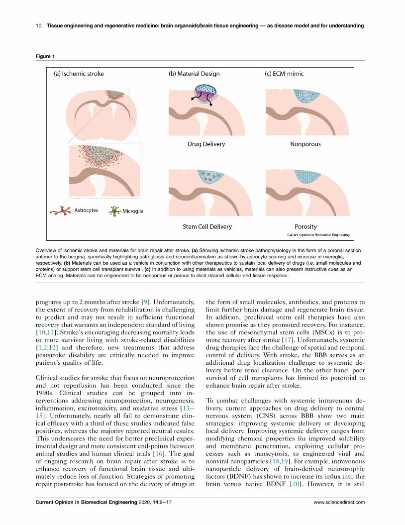
Figure 1
Overview of ischemic stroke and materials for brain repair after stroke. (a) Showing ischemic stroke pathophysiology in the form of a coronal sectionanterior to the bregma, specifically highlighting astrogliosis and neuroinflammation as shown by astrocyte scarring and increase in microglia,respectively. (b) Materials can be used as a vehicle in conjunction with other therapeutics to sustain local delivery of drugs (i.e. small molecules andproteins) or support stem cell transplant survival. (c) In addition to using materials as vehicles, materials can also present instructive cues as anECM analog. Materials can be engineered to be nonporous or porous to elicit desired cellular and tissue response.
10 Tissue engineering and regenerative medicine: brain organoids/brain tissue engineering — as disease model and for understanding
programs up to 2 months after stroke [9]. Unfortunately,the extent of recovery from rehabilitation is challengingto predict and may not result in sufficient functionalrecovery that warrants an independent standard of living[10,11]. Stroke’s encouraging decreasing mortality leadsto more survivor living with stroke-related disabilities[1,2,12] and therefore, new treatments that addresspoststroke disability are critically needed to improvepatient’s quality of life.
Clinical studies for stroke that focus on neuroprotectionand not reperfusion has been conducted since the1990s. Clinical studies can be grouped into in-terventions addressing neuroprotection, neurogenesis,inflammation, excitotoxicity, and oxidative stress [13e15]. Unfortunately, nearly all fail to demonstrate clin-ical efficacy with a third of these studies indicated falsepositives, whereas the majority reported neutral results.This underscores the need for better preclinical exper-imental design and more consistent end-points betweenanimal studies and human clinical trials [16]. The goal
of ongoing research on brain repair after stroke is toenhance recovery of functional brain tissue and ulti-mately reduce loss of function. Strategies of promotingrepair poststroke has focused on the delivery of drugs in
Current Opinion in Biomedical Engineering 2020, 14:9–17
the form of small molecules, antibodies, and proteins tolimit further brain damage and regenerate brain tissue.In addition, preclinical stem cell therapies have alsoshown promise as they promoted recovery. For instance,the use of mesenchymal stem cells (MSCs) is to pro-mote recovery after stroke [17]. Unfortunately, systemicdrug therapies face the challenge of spatial and temporalcontrol of delivery. With stroke, the BBB serves as anadditional drug localization challenge to systemic de-livery before renal clearance. On the other hand, poor
survival of cell transplants has limited its potential toenhance brain repair after stroke.
To combat challenges with systemic intravenous de-livery, current approaches on drug delivery to centralnervous system (CNS) across BBB show two mainstrategies: improving systemic delivery or developinglocal delivery. Improving systemic delivery ranges frommodifying chemical properties for improved solubilityand membrane penetration, exploiting cellular pro-cesses such as transcytosis, to engineered viral and
nonviral nanoparticles [18,19]. For example, intravenousnanoparticle delivery of brain-derived neurotrophicfactors (BDNF) has shown to increase its influx into thebrain versus native BDNF [20]. However, it is still
www.sciencedirect.com
Materials for stroke recovery Erning and Segura 11
challenging to sustain drug concentration in the brain forefficacy while minimizing systemic exposure. Incontrast, there are other administration techniques thatare more invasive but directly targets regions of interestwithin the CNS, such as intracerebral, intraventricular,or intrathecal. With the additional risk of a more invasivedelivery, local delivery methods avoid the BBB and re-sults in high drug concentration in CNS while mini-
mizing systemic exposure. However, drug clearance isstill a problem that demands multiple invasive bolusinjections to sustain drug concentration [18]. Localdelivery of stem cells has seen success in animal modelsand several clinical trials are now underway to assesssafety and efficacy towards reduced stroke disability[21,22]; however, cell death is still a major concern.
Biomaterials offer a promising treatment optiontogether with local delivery to prevent systemic expo-sure and sustain drug release in the brain (Figure 1b).
For instance, a hyaluronanemethylcellulose compositehydrogel implanted epicortically has showed sustaineddelivery of peptide and protein therapies in stroked rats[23,24] (Figure 2c). Other studies have shown thathydrogels can provide a platform for local drug deliverywith enhanced spatial and temporal control, working inconjunction with small molecule, peptide, protein, andstem cell therapies [25,26]. In the following sections,we present an alternative approach to promote recoveryafter stroke: the use of materials locally to support therepairing peri-infarct and the necrotic infarct tissue.
Biomaterials for brain repairIt is important to note that like other tissues, the brainattempts to repair itself after an ischemic event asevident by increase in endogenous neuroplasticity [27].However, this enhanced plasticity is transient and does
not always result in sufficient recovery after stroke. Inaddition, neurons have limited self-repair capability[28]. Nevertheless, the fact that pro-repair pathways areactivated after stroke offer an opportunity to augmenttheir outcomes. We propose that hydrogel materials,when placed in the affected area, can offer a platform toaugment these endogenous repair pathways throughretention of expressed proteins or delivery of comple-mentary activators/inhibitors. So far, biomaterials havebeen designed to address the key aspects of strokepathophysiology that are believed to prevent a pro-
repair environment or that need to promote furtherrepair, inflammation, astrogliosis, angiogenesis, neuro-genesis, and axonal sprouting.
Unlike previous examples where materials were strictlyused as drug delivery vehicles, material scaffolds canadditionally serve as ECM placeholders when deliveredlocally to the stroke infarct (Figure 1c). Materials actingas matrix-analog can promote recovery after stroke byproviding temporary ECM within the compromised
www.sciencedirect.com
stroke infarct ECM. Around the stroke infarct, astro-cytes have been shown to both promote and obstructstroke recovery. Astrogliosis, or formation of an astrocyticscar surrounding the stroke cavity, is a reactive neuro-toxic astrocyte phenotype that severely limits strokerecovery and loses its ability to promote neuronal sur-vival, axonal sprouting, and synaptogenesis [29]. How-ever, the scar or peri-infarct region is also where pro-
repair pathways are activated. Thus, placing a hydrogelECM material in the infarct core, also places it next tothe region where pro-repair pathways are activated, andendogenous repair is occurring.
InflammationNormally, the CNS is an immune-privileged site that isseparated from peripheral immune system by the BBB.However, ischemic injury activates microglia, the resi-dent immune cells of the CNS and they start exhibitingdifferential activated phenotypes that are analogous tothe simplified M1 proinflammatory and M2 prohealingphenotypes of macrophages [4]. Classically activated(M1) microglia release proinflammatory cytokines,
ROS, and nitrous oxide that induces reactive astrocytes,BBB breakdown and extravasation of peripheral immunecells [29]. However, selective ablation of microglia isalso detrimental as alternatively activated (M2) micro-glia are anti-inflammatory and promote brain repair byincreased production of anti-inflammatory cytokinesand neurotrophic factors [30]. Multiple studies haveshown that the degree of microglia activation, timingand degree of cytokine or factor expression can result inether positive or negative outcome after stroke[4,31,32]. Therefore, immunomodulation seeking to
enhance brain repair after stroke requires powerfulspatiotemporal control. Injection of materials into thestroke cavity has demonstrated that relatively simplematerials can have a dramatic effect on the number ofmacrophage/microglia in both the stroke and peri-infarctareas. Injection of in-situ gelling hyaluronic acid (HA)hydrogels [33], assembling ECM-derived materials[34,35] and peptide-based gels [36] into the stroke core,lower the number of microglia and in some cases switchthe phenotype of macrophages from M1 to M2 evenwithout the explicit delivery of anti-inflammatory
agents. In the case of HA hydrogels, mechanical prop-erties are important as materials that are >1000 Pa werefound to be proinflammatory [37]. For natural ECM-derived materials like urinary bladder matrix, lowerconcentration or a less stiff scaffold is important tomodulate macrophage and microglia number as well asM1 to M2 transition [34,35]. Furthermore, simplechanges to the hydrogel microstructure have also shownreduction of macrophage number in the infarct and peri-infarct tissue. Although a nanoporous HA gel reducesthe number of microphage/microglia in the peri-infarct
area, HA microporous annealed particles (MAP)reduce this number in both the infarct and peri-infarct
Current Opinion in Biomedical Engineering 2020, 14:9–17

Figure 2
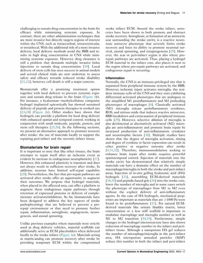
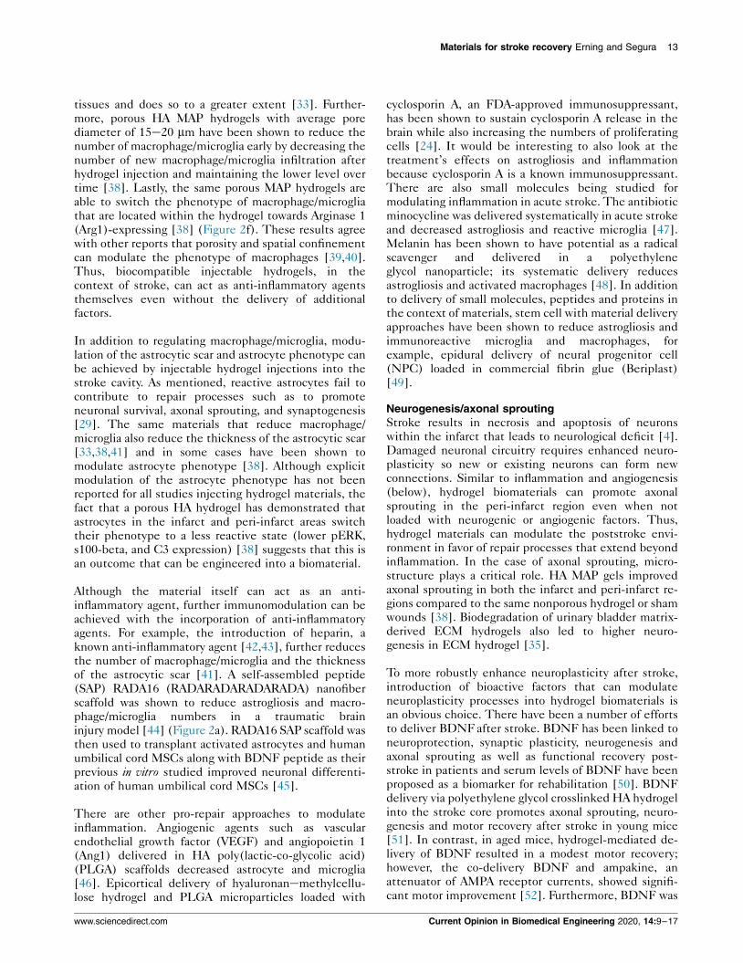
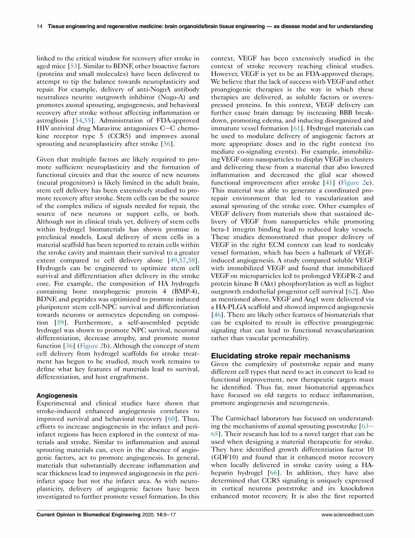
Material examples for brain repair after stroke. (a) RADA16 SAPNS: Nissl and DAPI-stained images of saline-injected lesion and SAPNS-injected lesionas well as quantification of astrocytes (GFAP positive) and macrophages (ED1 positive) in the lesion boundary zone at 2 days, 2 weeks, and 6 weeksafter treatment. Source: Adapted from Ref. [44]. (b) IKVAV SAP and human cortical progenitor: NeuN-stained images comparing GFP-labeled humancortical progenitors in stem cell delivery versus stem cell + SAP. Image shows increased proportion of NeuN-positive cells in SAP group. Source:Adapted from Ref. [36]. (c) BDNF-loaded hyaluronan–methylcellulose and PLGA nanoparticle scaffold composite: Behavioral studies after local BDNFdelivery via material vehicle showed improvement in hindlimb recovery after stroke injury. Source: Adapted from Ref. [23]. (d) Urinary bladder matrix-ECM hydrogel: immunostained images showing ECM hydrogel (Collagen I), cell nucleus (DAPI), macrophage/microglia (Iba-1), M1-like phenotype(CD86), and M2-like phenotype (CD206). Quantification of results showed that ECM hydrogel modulated neuroinflammation. Source: Adapted fromRef. [35]. (e) HA and clustered VEGF on heparin nanoparticle hydrogel: immunostained images of neurons (NF200 positive) and vessels (Glut-1positive) in stroke infarct showed gel + high cluster VEGF delivery resulted in enhanced angiogenesis, neurogenesis, and axonal sprouting. Source:Adapted from Ref. [41]. (f) MAP hydrogel: immunostained images of sham and MAP-treated stroke lesions showing gel, cell nucleus (DAPI),macrophage/microglia (CD11b), and pro-repair phenotype (Arg1). Quantification showed material-modulated neuroinflammation by increasing pro-repair macrophage/microglia in the stroke infarct. Source: Adapted from Ref. [38]. SAP, self-assembled peptide; SAPNS, self-assembled peptidenanofiber scaffold.
12 Tissue engineering and regenerative medicine: brain organoids/brain tissue engineering — as disease model and for understanding
Current Opinion in Biomedical Engineering 2020, 14:9–17 www.sciencedirect.com
Materials for stroke recovery Erning and Segura 13
tissues and does so to a greater extent [33]. Further-more, porous HA MAP hydrogels with average porediameter of 15e20 mm have been shown to reduce thenumber of macrophage/microglia early by decreasing thenumber of new macrophage/microglia infiltration afterhydrogel injection and maintaining the lower level overtime [38]. Lastly, the same porous MAP hydrogels areable to switch the phenotype of macrophage/microglia
that are located within the hydrogel towards Arginase 1(Arg1)-expressing [38] (Figure 2f). These results agreewith other reports that porosity and spatial confinementcan modulate the phenotype of macrophages [39,40].Thus, biocompatible injectable hydrogels, in thecontext of stroke, can act as anti-inflammatory agentsthemselves even without the delivery of additionalfactors.
In addition to regulating macrophage/microglia, modu-lation of the astrocytic scar and astrocyte phenotype can
be achieved by injectable hydrogel injections into thestroke cavity. As mentioned, reactive astrocytes fail tocontribute to repair processes such as to promoteneuronal survival, axonal sprouting, and synaptogenesis[29]. The same materials that reduce macrophage/microglia also reduce the thickness of the astrocytic scar[33,38,41] and in some cases have been shown tomodulate astrocyte phenotype [38]. Although explicitmodulation of the astrocyte phenotype has not beenreported for all studies injecting hydrogel materials, thefact that a porous HA hydrogel has demonstrated that
astrocytes in the infarct and peri-infarct areas switchtheir phenotype to a less reactive state (lower pERK,s100-beta, and C3 expression) [38] suggests that this isan outcome that can be engineered into a biomaterial.
Although the material itself can act as an anti-inflammatory agent, further immunomodulation can beachieved with the incorporation of anti-inflammatoryagents. For example, the introduction of heparin, aknown anti-inflammatory agent [42,43], further reducesthe number of macrophage/microglia and the thicknessof the astrocytic scar [41]. A self-assembled peptide
(SAP) RADA16 (RADARADARADARADA) nanofiberscaffold was shown to reduce astrogliosis and macro-phage/microglia numbers in a traumatic braininjury model [44] (Figure 2a). RADA16 SAP scaffold wasthen used to transplant activated astrocytes and humanumbilical cord MSCs along with BDNF peptide as theirprevious in vitro studied improved neuronal differenti-ation of human umbilical cord MSCs [45].
There are other pro-repair approaches to modulateinflammation. Angiogenic agents such as vascular
endothelial growth factor (VEGF) and angiopoietin 1(Ang1) delivered in HA poly(lactic-co-glycolic acid)(PLGA) scaffolds decreased astrocyte and microglia[46]. Epicortical delivery of hyaluronanemethylcellu-lose hydrogel and PLGA microparticles loaded with
www.sciencedirect.com
cyclosporin A, an FDA-approved immunosuppressant,has been shown to sustain cyclosporin A release in thebrain while also increasing the numbers of proliferatingcells [24]. It would be interesting to also look at thetreatment’s effects on astrogliosis and inflammationbecause cyclosporin A is a known immunosuppressant.There are also small molecules being studied formodulating inflammation in acute stroke. The antibiotic
minocycline was delivered systematically in acute strokeand decreased astrogliosis and reactive microglia [47].Melanin has been shown to have potential as a radicalscavenger and delivered in a polyethyleneglycol nanoparticle; its systematic delivery reducesastrogliosis and activated macrophages [48]. In additionto delivery of small molecules, peptides and proteins inthe context of materials, stem cell with material deliveryapproaches have been shown to reduce astrogliosis andimmunoreactive microglia and macrophages, forexample, epidural delivery of neural progenitor cell
(NPC) loaded in commercial fibrin glue (Beriplast)[49].
Neurogenesis/axonal sproutingStroke results in necrosis and apoptosis of neuronswithin the infarct that leads to neurological deficit [4].Damaged neuronal circuitry requires enhanced neuro-plasticity so new or existing neurons can form newconnections. Similar to inflammation and angiogenesis(below), hydrogel biomaterials can promote axonalsprouting in the peri-infarct region even when notloaded with neurogenic or angiogenic factors. Thus,hydrogel materials can modulate the poststroke envi-ronment in favor of repair processes that extend beyondinflammation. In the case of axonal sprouting, micro-
structure plays a critical role. HA MAP gels improvedaxonal sprouting in both the infarct and peri-infarct re-gions compared to the same nonporous hydrogel or shamwounds [38]. Biodegradation of urinary bladder matrix-derived ECM hydrogels also led to higher neuro-genesis in ECM hydrogel [35].
To more robustly enhance neuroplasticity after stroke,introduction of bioactive factors that can modulateneuroplasticity processes into hydrogel biomaterials isan obvious choice. There have been a number of effortsto deliver BDNFafter stroke. BDNF has been linked to
neuroprotection, synaptic plasticity, neurogenesis andaxonal sprouting as well as functional recovery post-stroke in patients and serum levels of BDNF have beenproposed as a biomarker for rehabilitation [50]. BDNFdelivery via polyethylene glycol crosslinked HA hydrogelinto the stroke core promotes axonal sprouting, neuro-genesis and motor recovery after stroke in young mice[51]. In contrast, in aged mice, hydrogel-mediated de-livery of BDNF resulted in a modest motor recovery;however, the co-delivery BDNF and ampakine, anattenuator of AMPA receptor currents, showed signifi-
cant motor improvement [52]. Furthermore, BDNF was
Current Opinion in Biomedical Engineering 2020, 14:9–17
14 Tissue engineering and regenerative medicine: brain organoids/brain tissue engineering — as disease model and for understanding
linked to the critical window for recovery after stroke inaged mice [53]. Similar to BDNF, other bioactive factors(proteins and small molecules) have been delivered toattempt to tip the balance towards neuroplasticity andrepair. For example, delivery of anti-NogoA antibodyneutralizes neurite outgrowth inhibitor (Nogo-A) andpromotes axonal sprouting, angiogenesis, and behavioralrecovery after stroke without affecting inflammation or
astrogliosis [54,55]. Administration of FDA-approvedHIV antiviral drug Maraviroc antagonizes CeC chemo-kine receptor type 5 (CCR5) and improves axonalsprouting and neuroplasticity after stroke [56].
Given that multiple factors are likely required to pro-mote sufficient neuroplasticity and the formation offunctional circuits and that the source of new neurons(neural progenitors) is likely limited in the adult brain,stem cell delivery has been extensively studied to pro-mote recovery after stroke. Stem cells can be the source
of the complex milieu of signals needed for repair, thesource of new neurons or support cells, or both.Although not in clinical trials yet, delivery of stem cellswithin hydrogel biomaterials has shown promise inpreclinical models. Local delivery of stem cells in amaterial scaffold has been reported to retain cells withinthe stroke cavity and maintain their survival to a greaterextent compared to cell delivery alone [49,57,58].Hydrogels can be engineered to optimize stem cellsurvival and differentiation after delivery in the strokecore. For example, the composition of HA hydrogels
containing bone morphogenic protein 4 (BMP-4),BDNF, and peptides was optimized to promote inducedpluripotent stem cell-NPC survival and differentiationtowards neurons or astrocytes depending on composi-tion [59]. Furthermore, a self-assembled peptidehydrogel was shown to promote NPC survival, neuronaldifferentiation, decrease atrophy, and promote motorfunction [36] (Figure 2b). Although the concept of stemcell delivery from hydrogel scaffolds for stroke treat-ment has begun to be studied, much work remains todefine what key features of materials lead to survival,differentiation, and host engraftment.
AngiogenesisExperimental and clinical studies have shown thatstroke-induced enhanced angiogenesis correlates toimproved survival and behavioral recovery [60]. Thus,efforts to increase angiogenesis in the infarct and peri-infarct regions has been explored in the context of ma-terials and stroke. Similar to inflammation and axonalsprouting materials can, even in the absence of angio-genic factors, act to promote angiogenesis. In general,
materials that substantially decrease inflammation andscar thickness lead to improved angiogenesis in the peri-infarct space but not the infarct area. As with neuro-plasticity, delivery of angiogenic factors have beeninvestigated to further promote vessel formation. In this
Current Opinion in Biomedical Engineering 2020, 14:9–17
context, VEGF has been extensively studied in thecontext of stroke recovery reaching clinical studies.However, VEGF is yet to be an FDA-approved therapy.We believe that the lack of success with VEGFand otherproangiogenic therapies is the way in which thesetherapies are delivered, as soluble factors or overex-pressed proteins. In this context, VEGF delivery canfurther cause brain damage by increasing BBB break-
down, promoting edema, and inducing disorganized andimmature vessel formation [61]. Hydrogel materials canbe used to modulate delivery of angiogenic factors atmore appropriate doses and in the right context (tomediate co-signaling events). For example, immobiliz-ing VEGFonto nanoparticles to display VEGF in clustersand delivering these from a material that also loweredinflammation and decreased the glial scar showedfunctional improvement after stroke [41] (Figure 2e).This material was able to generate a coordinated pro-repair environment that led to vascularization and
axonal sprouting of the stroke core. Other examples ofVEGF delivery from materials show that sustained de-livery of VEGF from nanoparticles while promotingbeta-1 integrin binding lead to reduced leaky vessels.These studies demonstrated that proper delivery ofVEGF in the right ECM context can lead to nonleakyvessel formation, which has been a hallmark of VEGF-induced angiogenesis. A study compared soluble VEGFwith immobilized VEGF and found that immobilizedVEGF on microparticles led to prolonged VEGFR-2 andprotein kinase B (Akt) phosphorylation as well as higher
outgrowth endothelial progenitor cell survival [62]. Alsoas mentioned above, VEGFand Ang1 were delivered viaa HA-PLGA scaffold and showed improved angiogenesis[46]. There are likely other features of biomaterials thatcan be exploited to result in effective proangiogenicsignaling that can lead to functional revascularizationrather than vascular permeability.
Elucidating stroke repair mechanismsGiven the complexity of poststroke repair and manydifferent cell types that need to act in concert to lead tofunctional improvement, new therapeutic targets mustbe identified. Thus far, most biomaterial approacheshave focused on old targets to reduce inflammation,promote angiogenesis and neurogenesis.
The Carmichael laboratory has focused on understand-
ing the mechanisms of axonal sprouting poststroke [63e65]. Their research has led to a novel target that can beused when designing a material therapeutic for stroke.They have identified growth differentiation factor 10(GDF10) and found that it enhanced motor recoverywhen locally delivered in stroke cavity using a HA-heparin hydrogel [66]. In addition, they have alsodetermined that CCR5 signaling is uniquely expressedin cortical neurons poststroke and its knockdownenhanced motor recovery. It is also the first reported
www.sciencedirect.com
Materials for stroke recovery Erning and Segura 15
gene associated with enhanced recovery in humans asseen by subpopulations of stroke patients with naturallyoccurring loss-of-function mutation in CCR5 [67].However, their studies solely focus on axon sproutingneurons after endogenous recovery poststroke. Next-generation transcriptomic and proteomic approachesneed to be used to identify relevant cellular players,pathways, and proteins for brain repair after stroke. This
will generate novel targets that can be help inform ourmaterial design.
ConclusionsStroke is a traumatic brain insult, which is the leadingcause of long-term disability in adults because of thebrain’s poor regenerative capacity. Current clinical treat-ments are reperfusion efforts that still leave patients withlong-term neurological deficits. Over the years, preclini-
cal research has identified therapeutic strategies toenhance endogenous brain repair after stroke. Unfortu-nately, clinical trial translational attempts have failedwhere systemically delivered therapeutic drugs arequickly cleared and transplanted stem cells have poorsurvival. More recent research in biomaterials for brainrepair after stroke have showed that innovative biomate-rial designs alone can reduce inflammation, glial scar for-mation, and increase cellular infiltration to stroke infarct.With the addition of drugs and stem cells therapeutics,the combined therapeutic-loaded biomaterials strategy
overcame challenges with systemic delivery and celltransplant survival, enhancing functional recovery asshown by a number of successfully examples in the field.Despite the encouraging results of biomaterial strategiesin tackling brain repair after stroke, it is difficult to predictwhether documented functional recovery found in rodentmodels can be translated in humans. This warrants abetter understanding of the mechanisms of brain repairafter stroke. By appreciating the mode of action of thesebiomaterial strategies, novel targets that can more holis-tically promote brain repair after stroke and result in
complete functional recovery will be discovered.
Local delivery using materials should not be viewed asbeing at odds with systemic delivery. Material-basedlocal delivery can be used in conjugation with systemicdelivery to use the advantages of both modes of delivery.For instance, systemic delivery of agents could be usedto treat early stages (i.e. reperfusion and neuro-protection) and/or be used to ‘prime’ the stroke envi-ronment for the ultimate delivery of a biomaterial topromote plasticity and recovery from stroke.
The application of biomaterial strategies for brain repairis in its nascent preclinical stages. However, the field ofbiomaterials to promote endogenous repair in othertissue systems such as bone and skin is substantiallymore developed and used clinically. Learning from thesestrategies and attracting a new generation of biomaterial
www.sciencedirect.com
scientist is necessary to bring the field closer to clinicaltranslation.
Conflict of interest statement
Nothing declared.
AcknowledgementsKE is credited with conception and writing of the manuscript. TS providedvaluable edits, suggestions, and feedback. This work was supported by theNational Institutes of Health (1R01NS094599). KE is a recipient of theWilliam M. “Monty” Reichert Fellowship granted by the Department ofBiomedical Engineering, Duke University, USA.
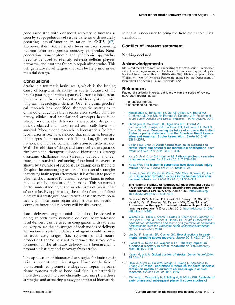
ReferencesPapers of particular interest, published within the period of review,have been highlighted as:
* of special interest* * of outstanding interest
1. Mozaffarian D, Benjamin EJ, Go AS, Arnett DK, Blaha MJ,Cushman M, Das SR, de Ferranti S, Després J-P, Fullerton HJ,et al.: Heart Disease and Stroke Statistics—2016 Update. 2015.
2. Ovbiagele B, Goldstein LB, Higashida RT, Howard VJ,Johnston SC, Khavjou OA, Lackland DT, Lichtman JH, Mohl S,Sacco RL, et al.: Forecasting the future of stroke in the UnitedStates: a policy statement from the American Heart Associ-ation and American Stroke Association. Stroke 2013, 44:2361–2375.
3. Barkho BZ, Zhao X: Adult neural stem cells: response tostroke injury and potential for therapeutic applications. CurrStem Cell Res Ther 2011, 6:327–338.
4. Xing C, Arai K, Lo EH, Hommel M: Pathophysiologic cascadesin ischemic stroke. Int J Stroke 2012, 7:378–385.
5. Heiss WD: The ischemic penumbra: how does tissue injuryevolve? Ann N Y Acad Sci 2012, 1268:26–34.
6. Huang L, Wu ZB, ZhuGe Q, Zheng WM, Shao B, Wang B, Sun F,Jin K: Glial scar formation occurs in the human brain afterischemic stroke. Int J Med Sci 2014, 11:344–348.
7. The national institute of neurological disorders and stroke rt-PA stroke study group: tissue plasminogen activator foracute ischemic stroke. N Engl J Med 1995. https://doi.org/10.1056/NEJM199512143332401.
8. Campbell BCV, Mitchell PJ, Kleinig TJ, Dewey HM, Churilov L,Yassi N, Yan B, Dowling RJ, Parsons MW, Oxley TJ, et al.:Endovascular therapy for ischemic stroke with perfusion-imaging selection. N Engl J Med 2015. https://doi.org/10.1056/NEJMoa1414792.
9. Winstein CJ, Stein J, Arena R, Bates B, Cherney LR, Cramer SC,Deruyter F, Eng JJ, Fisher B, Harvey RL, et al.: Guidelines foradult Stroke rehabilitation and recovery: a guideline for healthcareprofessionals from the American Heart Association/AmericanStroke Association. 2016.
10. Lin DJ, Finklestein SP, Cramer SC: New directions in treat-ments targeting stroke recovery. Stroke 2018, 49:3107–3114.
11. Kwakkel G, Kollen BJ, Wagenaar RC: Therapy impact onfunctional recovery in stroke rehabilitation. Physiotherapy1999, 85:377–391.
12. Katan M, Luft A: Global burden of stroke. Semin Neurol 2018,38:208–211.
13. Reis C, Akyol O, Ho WM, Araujo C, Huang L, Applegate R,Zhang JH: Phase I and phase II therapies for acute ischemicstroke: an update on currently studied drugs in clinicalresearch. BioMed Res Int 2017, 2017.
14. Minnerup J, Wersching H, Schilling M, Schäbitz WR: Analysis ofearly phase and subsequent phase III stroke studies of
Current Opinion in Biomedical Engineering 2020, 14:9–17
16 Tissue engineering and regenerative medicine: brain organoids/brain tissue engineering — as disease model and for understanding
neuroprotectants: outcomes and predictors for success. ExpTransl Stroke Med 2014, 6:1–6.
15. Rajah GB, Ding Y: Experimental neuroprotection in ischemicstroke: a concise review. Neurosurg Focus 2017, 42:1–8.
16. Corbett D, Carmichael ST, Murphy TH, Jones TA, Schwab ME,Jolkkonen J, Clarkson AN, Dancause N, Weiloch T, Johansen-Berg H, et al.: Enhancing the alignment of the preclinical andclinical stroke recovery research pipeline: consensus-basedcore recommendations from the stroke recovery and reha-bilitation roundtable translational working group. Neuro-rehabilitation Neural Repair 2017, 31:699–707.
17. Zheng H, Zhang B, Chhatbar PY, Dong Y, Alawieh A, Lowe F,Hu X, Feng W: Mesenchymal stem cell therapy in stroke: asystematic review of literature in pre-clinical and clinicalresearch. Cell Transplant 2018, 27:1723–1730.
18. Dong X: Current strategies for brain drug delivery. Thera-nostics 2018, 8:1481–1493.
19. Lu CT, Zhao YZ, Wong HL, Cai J, Peng L, Tian XQ: Currentapproaches to enhance CNS delivery of drugs across thebrain barriers. Int J Nanomed 2014, 9:2241–2257.
20. Harris NM, Ritzel R, Mancini NS, Jiang Y, Yi X, Manickam DS,BanksWA, Kabanov AV,McCullough LD, VermaR:Nano-particledelivery of brain derived neurotrophic factor after focal cere-bral ischemia reduces tissue injury and enhances behavioralrecovery. Pharmacol Biochem Behav 2016, 150:48–56.
21. Marei HE, Hasan A, Rizzi R, Althani A, Afifi N, Cenciarelli C,Caceci T, Shuaib A: Potential of stem cell-based therapy forischemic stroke. Front Neurol 2018, 9.
22. Borlongan CV: Concise review: stem cell therapy for strokepatients: are we there yet? Stem Cells Transl Med 2019, 8:983–988.
23* *. Obermeyer JM, Tuladhar A, Payne SL, Ho E, Morshead CM,
Shoichet MS: Local delivery of brain-derived neurotrophicfactor enables behavioral recovery and tissue repair instroke-injured rats. Tissue Eng 2019, 25:1175–1187.
BDNF was delivered via PLGA nanoparticles inhyaluronan–methylcellulose hydrogel epicortically above stroke lesioninduced mice. Local and sustained delivery of BDNF increasesneuroplasticity and vehicle lesion size and neuron loss. Overall, treat-ment resulted in improved motor recovery.
24. Tuladhar A, Morshead CM, Shoichet MS: Circumventing theblood–brain barrier: local delivery of cyclosporin A stimu-lates stem cells in stroke-injured rat brain. J Control release2015, 215:1–11.
25. Tuladhar A, Payne SL, Shoichet MS: Harnessing the potentialof biomaterials for brain repair after stroke. Front Mater 2018,5:1–25.
26. Nih LR, Carmichael ST, Segura T: Hydrogels for brain repairafter stroke: an emerging treatment option. Curr Opin Bio-technol 2016, 40:155–163.
27. Dobkin BH, Carmichael ST: The specific requirements ofneural repair trials for stroke. Neurorehabilitation Neural Repair2016, 30:470–478.
28. Björklund A, Lindvall O: Self-repair in the brain. Nature 2000,405:893–895.
29. Liddelow SA, Guttenplan KA, Clarke LE, Bennett FC, Bohlen CJ,Schirmer L, Bennett ML, Münch AE, Chung W-S, Peterson TC:Neurotoxic reactive astrocytes are induced by activatedmicroglia. Nature 2017, 541:481.
30. Szalay G, Martinecz B, Lénárt N, Környei Z, Orsolits B, Judák L,Császár E, Fekete R, West BL, Katona G: Microglia protectagainst brain injury and their selective elimination dysregu-lates neuronal network activity after stroke. Nat Commun2016, 7:11499.
31. yao Ao L, Yan YY, Zhou L, yuan Li C, Li WT, rong Fang W, Li Yman: Immune cells after ischemic stroke onset: rroles,migration, and target intervention. J Mol Neurosci 2018, 66:342–355.
Current Opinion in Biomedical Engineering 2020, 14:9–17
32. Chamorro Á, Meisel A, Planas AM, Urra X, Van De Beek D,Veltkamp R: The immunology of acute stroke. Nat Rev Neurol2012, 8:401–410.
33* *. Nih LR, Sideris E, Carmichael ST, Segura T: Injection of
microporous annealing particle (MAP) hydrogels in thestroke cavity reduces gliosis and inflammation and promotesNPC migration to the lesion. Adv Mater 2017, 29:1–8.
Hyaluronic acid hydrogel microparticles were annealed in situ to formmicroporous annealed particle (MAP) gel inside lesion of stroke-induced mice. Porous scaffold reduced astrocyte scarring, microgliainfiltration to stroke cavity while also promoting NPC migration andinfiltration into the scaffold.
34. Ghuman H, Massensini AR, Donnelly J, Kim SM, Medberry CJ,Badylak SF, Modo M: ECM hydrogel for the treatment ofstroke: characterization of the host cell infiltrate. Biomaterials2016, 91:166–181.
35* *. Ghuman H, Mauney C, Donnelly J, Massensini AR, Badylak SF,
Modo M: Biodegradation of ECM hydrogel promotes endog-enous brain tissue restoration in a rat model of stroke. ActaBiomater 2018, 80:66–84.
Porcine-derived urinary bladder matrix ECM was used to form inject-able ECM hydrogel delivered to the lesion of MCAO mice. They foundthat lower ECM hydrogel concentration leads to better immunomodu-lation, angiogenesis, and neurogenesis.
36*. Somaa FA, Wang TY, Niclis JC, Bruggeman KF, Kauhausen JA,
Guo H, McDougall S, Williams RJ, Nisbet DR, Thompson LH,et al.: Peptide-based scaffolds support human cortical pro-genitor graft integration to reduce atrophy and promotefunctional repair in a model of stroke. Cell Rep 2017, 20:1964–1977.
Laminin-derived IKVAV peptide was used to make self-assembledpeptide (SAP) scaffold loaded with progenitor cells and delivered tostroked mice. Peptide-based scaffold progenitor cell implants showedincreased neuronal differentiation, and enhanced electrophysiologicalproperties.
37. Lam J, Carmichael ST, Lowry WE, Segura T: Hydrogel design ofexperiments methodology to optimize hydrogel for iPSC-NPCculture. Adv Healthc Mater 2015, 4:534–539.
38* *. Sideris E, Yu A, Chen J, Carmichael ST, Segura T: Hyaluronic
acid particle hydrogels decrease cerebral atrophy and pro-mote pro-reparative astrocyte/axonal infiltration in the coreafter ischemic stroke. bioRxiv 2019. https://doi.org/10.1101/768291.
Hyaluronic acid MAP hydrogel was injected to stroke cavity. The au-thors demonstrated that their MAP gel reduced astrogliosis andmodulated neuroinflammation as shown by increase in microglia/macrophage pro-repair phenotype.
39. Sussman EM, Halpin MC, Muster J, Moon RT, Ratner BD:Porous implants modulate healing and induce shifts in localmacrophage polarization in the foreign body reaction. AnnBiomed Eng 2014, 42:1508–1516.
40. Jain N, Vogel V: Spatial confinement downsizes the inflam-matory response of macrophages. Nat Mater 2018, 17:1134.
41* *. Nih LR, Gojgini S, Carmichael ST, Segura T: Dual-function
injectable angiogenic biomaterial for the repair of brain tissuefollowing stroke. Nat Mater 2018, 17:642–651.
Hyaluronic acid-based hydrogel was crosslinked in situ with MMP-degradable peptide and decorated with RGD adhesion peptide,naked heparin nanoparticles and clustered VEGF on heparin nano-particles. Their material was injected into stroke cavity induced in miceand demonstrated reduced astrogliosis and inflammation, andincreased angiogenesis and neuron infiltration into the stroke cavity,which were colocalized.
42. Mousavi S, Moradi M, Khorshidahmad T, Motamedi M: Anti-in-flammatory effects of heparin and its derivatives: a system-atic review. Adv Pharmacol Sci 2015, 2015.
43. Poterucha TJ, Libby P, Goldhaber SZ: More than an anticoag-ulant: do heparins have direct anti-inflammatory effects?Thromb Haemostasis 2017, 117:437–444.
44. Guo J, Leung KKG, Su H, Yuan Q, Wang L, Chu TH, Zhang W,Pu JKS, Ng GKP, Wong WM, et al.: Self-assembling peptidenanofiber scaffold promotes the reconstruction of acutelyinjured brain. Nanomed Nanotechnol Biol Med 2009, 5:345–351.
www.sciencedirect.com

Materials for stroke recovery Erning and Segura 17
45. Shi W, Huang CJ, Xu XD, Jin GH, Huang RQ, Huang JF,Chen YN, Ju SQ, Wang Y, Shi YW, et al.: Transplantation ofRADA16-BDNF peptide scaffold with human umbilical cordmesenchymal stem cells forced with CXCR4 and activatedastrocytes for repair of traumatic brain injury. Acta Biomater2016, 45:247–261.
46. Ju R, Wen Y, Gou R, Wang Y, Xu Q: The experimental therapyon brain ischemia by improvement of local angiogenesis withtissue engineering in the mouse. Cell Transplant 2014, 23:83–95.
47. Yang Y, Salayandia VM, Thompson JF, Yang LY, Estrada EY,Yang Y: Attenuation of acute stroke injury in rat brain byminocycline promotes blood-brain barrier remodeling andalternative microglia/macrophage activation during recovery.J Neuroinflammation 2015, 12:1–15.
48. Liu Y, Ai K, Ji X, Askhatova D, Du R, Lu L, Shi J: Comprehensiveinsights into the multi-antioxidative mechanisms ofmelanin nanoparticles and their application to protect brainfrom injury in ischemic stroke. J Am Chem Soc 2017, 139:856–862.
49. Lee IH, Huang SS, Chuang CY, Liao KH, Chang LH, Chuang CC,Su YS, Lin HJ, Hsieh JY, Su SH, et al.: Delayed epiduraltransplantation of human induced pluripotent stem cell-derived neural progenitors enhances functional recoveryafter stroke. Sci Rep 2017, 7:1–12.
50. Luo W, Liu T, Li S, Wen H, Zhou F, Zafonte R, Luo X, Xu M,Black-Schaffer R, Wood LJ: The serum BDNF level offersminimum predictive value for motor function recovery afterstroke. Transl Stroke Res 2019, 10:342–351.
51. Cook DJ, Nguyen C, Chun HN, Llorente I L, Chiu AS,Machnicki M, Zarembinski TI, Carmichael ST: Hydrogel-deliv-ered brain-derived neurotrophic factor promotes tissuerepair and recovery after stroke. J Cerebr Blood Flow Metabol2017, 37:1030–1045.
52. Clarkson AN, Parker K, Nilsson M, Walker FR, Gowing EK:Combined ampakine and BDNF treatments enhance post-stroke functional recovery in aged mice via AKT-CREBsignaling. J Cerebr Blood Flow Metabol 2015, 35:1272–1279.
53* *. Houlton J, Zhou LYY, Barwick D, Gowing EK, Clarkson AN:
Stroke induces a BDNF-dependent improvement in cognitiveflexibility in aged mice. Neural Plast 2019, 2019:1460890.
Authors compared stroked young and aged mice and the effect ofBDNF decoy, TrkB-Fc, on behavioral recovery. They found that aBDNF-dependent improvement in stroke aged mice, even outper-forming sham aged mice.
54. Otero-Ortega L, Gómez-De Frutos MC, Laso-García F, Sánchez-Gonzalo A, Martínez-Arroyo A, Díez-Tejedor E, Gutiérrez-Fernández M: NogoA neutralization promotes axonal resto-ration after white matter injury in subcortical stroke. Sci Rep2017, 7:1–12.
55*. Rust R, Grönnert L, Gantner C, Enzler A, Mulders G, Weber RZ,
Siewert A, Limasale YDP, Meinhardt A, Maurer MA, et al.: Nogo-A targeted therapy promotes vascular repair and functionalrecovery following stroke. Proc Natl Acad Sci U S A 2019, 116:14270–14279.
Neutralization of Nogo-A or one of its receptor S1PR2 by antibodytreatment or knock-out improves angiogenesis and neuroprotectionafter stroke but does not reduce inflammation and astrogliosis.
www.sciencedirect.com
56* *. Joy MT, Ben Assayag E, Shabashov-Stone D, Liraz-Zaltsman S,
Mazzitelli J, Arenas M, Abduljawad N, Kliper E, Korczyn AD,Thareja NS, et al.: CCR5 is a therapeutic target for recoveryafter stroke and traumatic brain injury. Cell 2019, 176:1143–1157.e13.
FDA-approved HIV antiviral drug Maraviroc was systematically deliv-ered to CNS-injured mice and showed improved neural repair in bothstroke and traumatic brain injury. Maraviroc works as CCR5 antagonistthat increases neuroplasticity.
57. Ballios BG, Cooke MJ, Donaldson L, Coles BLK, Morshead CM,Van Der Kooy D, Shoichet MS: A hyaluronan-based injectablehydrogel improves the survival and integration of stem cellprogeny following transplantation. Stem Cell Reports 2015, 4:1031–1045.
58. Lam J, Lowry WE, Carmichael ST, Segura T: Delivery of iPS-NPCs to the stroke cavity within a hyaluronic acid matrixpromotes the differentiation of transplanted cells. Adv FunctMater 2014, 24:7053–7062.
59. Moshayedi P, Nih LR, Llorente IL, Berg AR, Cinkornpumin J,Lowry WE, Segura T, Carmichael ST: Systematic optimizationof an engineered hydrogel allows for selective control ofhuman neural stem cell survival and differentiation aftertransplantation in the stroke brain. Biomaterials 2016, 105:145–155.
60. Krupinski J, Kaluza J, Kumar P, Kumar S, Wang JM: Role ofangiogenesis in patients with cerebral ischemic stroke.Stroke 1994, 25:1794–1798.
61. Geiseler SJ, Morland C: The Janus face of VEGF in stroke. Int JMol Sci 2018, 19:1–20.
62. Aday S, Zoldan J, Besnier M, Carreto L, Saif J, Fernandes R,Santos T, Bernardino L, Langer R, Emanueli C: Synthetic mi-croparticles conjugated with VEGF 165 improve the survivalof endothelial progenitor cells via microRNA-17 inhibition.Nat Commun 2017, 8:747.
63. Overman JJ, Clarkson AN, Wanner IB, Overman WT, Eckstein I,Maguire JL, Dinov ID, Toga AW, Carmichael ST: A role forephrin-A5 in axonal sprouting, recovery, and activity-dependent plasticity after stroke. Proc Natl Acad Sci U S A2012, 109:1–10.
64. Li S, Overman JJ, Katsman D, Kozlov SV, Donnelly CJ, Twiss JL,Giger RJ, Coppola G, Geschwind DH, Carmichael ST: An age-related sprouting transcriptome provides molecular controlof axonal sprouting after stroke. Nat Neurosci 2010, 13:1496–1506.
65* *. Carmichael ST, Kathirvelu B, Schweppe CA, Nie EH: Molecular,
cellular and functional events in axonal sprouting afterstroke. Exp Neurol 2017, 287:384–394.
Authors investigated axonal sprouting after ischemic event in mice bylabeling neurons that undergo axonal sprouting using neuroanatomicaltracer injections. They observed three distinct patters of axonalsprouting: reactive, reparative, and unbounded.
66. Li S, Nie EH, Yin Y, Benowitz LI, Tung S, Vinters HV, Bahjat FR,Stenzel-Poore MP, Kawaguchi R, Coppola G: GDF10 is a signalfor axonal sprouting and functional recovery after stroke. NatNeurosci 2015, 18:1737.
67. Joy MT, Assayag E Ben, Shabashov-Stone D, Liraz-Zaltsman S,Mazzitelli J, Arenas M, Abduljawad N, Kliper E, Korczyn AD,Thareja NS: CCR5 is a therapeutic target for recovery afterstroke and traumatic brain injury. Cell 2019, 176:1143–1157.
Current Opinion in Biomedical Engineering 2020, 14:9–17