Mass Spectrometry-Based Proteomics for Pre-Eclampsia and Preterm Birth
34
Int. J. Mol. Sci. 2015, 16, 1-x manuscripts; doi:10.3390/ijms160x000x International Journal of Molecular Sciences ISSN 1422-0067 www.mdpi.com/journal/ijms Review Mass Spectrometry-Based Proteomics for Pre-Eclampsia and Preterm Birth Kai P. Law 1,2, *, Ting-Li Han 1,2 , Chao Tong 1,3, * and Philip N. Baker 1,2 1 China-Canada-New Zealand Joint Laboratory of Maternal and Fetal Medicine, Chongqing Medical University, Chongqing 400016, China; E-Mails: [email protected] (T.-L.H.); [email protected] (P.N.B.) 2 The Liggins Institute, University of Auckland, Auckland 1142, New Zealand 3 Department of Obstetrics, The First Affiliated Hospital of Chongqing Medical University, Chongqing 400016, China * Authors to whom correspondence should be addressed; E-Mails: [email protected] (K.P.L.); [email protected] (C.T.); Tel.: +86-23-6881-7925 (K.P.L. & C.T.); Fax: +86-23-8901-1865 (K.P.L. & C.T.). Academic Editor: William Chi-shing Cho Received: 31 March 2015 / Accepted: 04 May 2015 / Published: Abstract: Pregnancy-related complications such as pre-eclampsia and preterm birth now represent a notable burden of adverse health. Pre-eclampsia is a hypertensive disorder unique to pregnancy. It is an important cause of maternal death worldwide and a leading cause of fetal growth restriction and iatrogenic prematurity. Fifteen million infants are born preterm each year globally, but more than one million of those do not survive their first month of life. Currently there are no predictive tests available for diagnosis of these pregnancy-related complications and the biological mechanisms of the diseases have not been fully elucidated. Mass spectrometry-based proteomics have all the necessary attributes to provide the needed breakthrough in understanding the pathophysiology of complex human diseases thorough the discovery of biomarkers. The mass spectrometry methodologies employed in the studies for pregnancy-related complications are evaluated in this article. Top-down proteomic and peptidomic profiling by laser mass spectrometry, liquid chromatography or capillary electrophoresis coupled to mass spectrometry, and bottom-up quantitative proteomics and targeted proteomics by liquid chromatography mass spectrometry have been applied to elucidate protein biomarkers and biological mechanism of pregnancy-related complications. The proteomes of serum, urine, amniotic fluid, OPEN ACCESS
-
Upload
independent -
Category
Documents
-
view
1 -
download
0
Transcript of Mass Spectrometry-Based Proteomics for Pre-Eclampsia and Preterm Birth

Int. J. Mol. Sci. 2015, 16, 1-x manuscripts; doi:10.3390/ijms160x000x
International Journal of
Molecular Sciences ISSN 1422-0067
www.mdpi.com/journal/ijms
Review
Mass Spectrometry-Based Proteomics for Pre-Eclampsia and
Preterm Birth
Kai P. Law 1,2,*, Ting-Li Han 1,2, Chao Tong 1,3,* and Philip N. Baker 1,2
1 China-Canada-New Zealand Joint Laboratory of Maternal and Fetal Medicine,
Chongqing Medical University, Chongqing 400016, China;
E-Mails: [email protected] (T.-L.H.); [email protected] (P.N.B.) 2 The Liggins Institute, University of Auckland, Auckland 1142, New Zealand 3 Department of Obstetrics, The First Affiliated Hospital of Chongqing Medical University,
Chongqing 400016, China
* Authors to whom correspondence should be addressed;
E-Mails: [email protected] (K.P.L.); [email protected] (C.T.);
Tel.: +86-23-6881-7925 (K.P.L. & C.T.); Fax: +86-23-8901-1865 (K.P.L. & C.T.).
Academic Editor: William Chi-shing Cho
Received: 31 March 2015 / Accepted: 04 May 2015 / Published:
Abstract: Pregnancy-related complications such as pre-eclampsia and preterm birth now
represent a notable burden of adverse health. Pre-eclampsia is a hypertensive disorder
unique to pregnancy. It is an important cause of maternal death worldwide and a leading
cause of fetal growth restriction and iatrogenic prematurity. Fifteen million infants are born
preterm each year globally, but more than one million of those do not survive their first
month of life. Currently there are no predictive tests available for diagnosis of these
pregnancy-related complications and the biological mechanisms of the diseases have not
been fully elucidated. Mass spectrometry-based proteomics have all the necessary
attributes to provide the needed breakthrough in understanding the pathophysiology of
complex human diseases thorough the discovery of biomarkers. The mass spectrometry
methodologies employed in the studies for pregnancy-related complications are evaluated
in this article. Top-down proteomic and peptidomic profiling by laser mass spectrometry,
liquid chromatography or capillary electrophoresis coupled to mass spectrometry, and
bottom-up quantitative proteomics and targeted proteomics by liquid chromatography mass
spectrometry have been applied to elucidate protein biomarkers and biological mechanism
of pregnancy-related complications. The proteomes of serum, urine, amniotic fluid,
OPEN ACCESS

Int. J. Mol. Sci. 2015, 16 2
cervical-vaginal fluid, placental tissue, and cytotrophoblastic cells have all been
investigated. Numerous biomarkers or biomarker candidates that could distinguish
complicated pregnancies from healthy controls have been proposed. Nevertheless,
questions as to the clinically utility and the capacity to elucidate the pathogenesis of the
pre-eclampsia and preterm birth remain to be answered.
Keywords: pre-eclampsia; preterm birth; mass spectrometry; proteomics; protein biomarkers
1. Introduction
Most pregnant women experience minor discomforts during pregnancy, but for a minority of
women, serious complications may occur. These pregnancy complications range from ectopic
pregnancy, miscarriage, placenta praevia and placenta abruption, and pre-eclampsia, etc. In the
worst-case scenarios, these conditions may lead to morbidity, disability, and maternal or fetal deaths.
Hypertensive disorders of pregnancy affect around 10% of all pregnancies globally [1]. Pre-eclampsia
is the most conspicuous among pregnancy-related hypertensive disorders for its impact on maternal and
neonatal health. Other hypertensive disorders occur during pregnancy (e.g., gestational hypertension and
chronic hypertension) are usually not as acute or life-threatening. Pre-eclampsia, one of the leading causes
of maternal and perinatal mortality and morbidity worldwide, affects 2%–5% of all pregnancies [2].
Pre-eclampsia is characterized by hypertension (≥140/90 mmHg) and proteinuria (≥300 mg in a 24-h
urine) and its signs usually only manifest at the last trimester of pregnancy. The condition is often
associated with eclampsia and the HELLP syndrome, both of which are potentially life-threatening
serious complications. Eclampsia is characterized by grand mal-like seizures, whereas the HELLP
syndrome manifests itself with hemolysis, elevated liver enzymes, and low platelet counts. The
progression of the pre-eclampsia from mild to severe can be rapid, unexpected, and occasionally
fulminant. Maternal deaths can occur among the most severe cases. Despite intensive research for the
past decades, the pathogenesis of preeclampsia has not been fully elucidated. A leading hypothesis
(referred as the two-stage model) states that pre-eclampsia is initiated by disturbances in placentation
at the beginning of pregnancy, followed by generalized inflammation and progressive endothelial
damage (reviewed in [1–3]). However, pre-eclampsia is more likely a heterogeneous disorder [4].
Chronic hypertension, diabetes mellitus, obesity, nulliparity and adolescent pregnancy are associated
with the risk of onset of pre-eclampsia. Studies have also shown that low levels of serum
25-hydroxyvitamin D in pregnancy are associated with pre-eclampsia [5,6], although there is currently no
evidence supporting that supplementation of vitamin D lowers the risk on developing preeclampsia [7].
The first line of treatment includes α2-adrenergic receptor agonists, non-selective β-blockers, calcium
channel blockers, and vasodilators to deal primarily with the signs [8]. Unfortunately, the only
effective intervention is termination of pregnancy or delivery of the fetus and placenta.
The birth of an infant before 37 weeks of gestation is termed preterm birth. The obstetric precursors
leading to preterm birth are: (1) delivery for maternal or fetal indications, in which labor is either
induced or the infant is delivered by pre-labor caesarean section; (2) spontaneous preterm labor with
intact membranes; and (3) preterm premature rupture of the membranes irrespective of whether

Int. J. Mol. Sci. 2015, 16 3
delivery is vaginal or by caesarean section [9]. Preterm birth is associated with high risks of adverse
neonatal sequelae, including cerebral palsy, language and learning disabilities, and poor growth due to
the incomplete development of organs of the premature infants. Despite improvements in prenatal care,
the number of preterm births has been rising. It was estimated that 12.9 million births, or 9.6% of all
births worldwide, were preterm in 2005 [10]. The number of cases rose to 14.9 million, or 11.1% of all
live births worldwide, in 2010 [11]. More than 60% of the cases occurred in South Asia and
sub-Saharan Africa [11]. Furthermore, according to the more recent figures from the World Health
Organization (WHO), preterm birth complications are thought to be responsible for nearly 1 million
deaths in 2013 globally [12]. The situation is especially dire in low- and middle-income countries
where 98% of all neonatal deaths occur [11]. The causes of preterm birth are multifactorial and
complex. However, the most common associations with preterm birth include multiple pregnancies,
intra-amniotic infections, and chronic conditions, such as diabetes mellitus and hypertension. There is also
a genetic influence. However, the pathogenesis of preterm labor is not well understood and few of the
proposed biomarkers meet the criteria to be considered clinically useful for predicting preterm birth [13].
It is thought that preterm labor might represent early idiopathic activation of the normal labor process or
the results of pathological insults [9]. Recent studies have also linked vitamin D deficiency with the
likelihood of preterm birth and neonatal outcomes [14,15].
Mass spectrometry (MS) is an indispensable analytical technology to proteomics and metabolomics.
The technology has been employed in research and clinical settings to examine different diseases and
conditions, including discovery of protein and metabolite biomarker of gestational diseases [16–18],
and screen of inborn error of metabolism [19]. However, for the majority of the studies in reproductive
medicine, classical biochemical assays, such as Western blot, enzyme-linked immunosorbent assay
(ELISA), immunostaining, and real-time polymerase chain reaction (RT-PCR) remain the preferred
methods. The design of these studies has also been hypothesis-driven and derived from pathways
implicated in the pathogenesis of the conditions under investigation. While they have produced useful
information and insight, the hypothesis-driven approach alone has not bought us close to answer the
questions as to the pathogenesis or biomolecular mechanism of a diverse disease such as preeclampsia.
Researchers conducting clinical science do not often have knowledge and expertise in MS technologies
and have rarely taken advantage of the techniques, despite the increasing familiarity with systems
biology approaches and hypothesis-generating strategies. This work describes and evaluates the
advantages, limitations, and the recent advances of the MS methodologies that have been successfully
applied in pregnancy-related complications. Emphasis is placed on the studies of pre-eclampsia and
preterm birth.
2. An Overview of Proteomic Technologies
The proteome is the entire complement of proteins that is or can be expressed by a cell, tissue, or
organism. The study of proteomes and their functions is referred as proteomics. The proteome is highly
complex and dynamic since protein expression and turnover vary with cell type and the physiological
state of the cells. In addition, protein transformations occurring through post-translational
modifications (PTMs) (e.g., phosphorylation, glycosylation acetylation and methylation), and chemical
damages (e.g., glycation, oxidation, and nitration) further increase the diversity and heterogeneity of

Int. J. Mol. Sci. 2015, 16 4
the proteome. Furthermore, the dynamic range in expression of proteins in a biological system is
normally several orders in magnitude. The interested proteins are frequently of low concentration and
their activities are masked by high abundant proteins present in the system. Separation technologies for
protein or peptide fractionation, depletion and enrichment are essential to reduce sample complexity [20].
A range of commercial systems is now available for immunodepletion including Agilent Technologies’
Multiple Affinity Removal System (MARS) series of columns, and SigmaAldrich’s ProteoPrep20. That
said, other proteins are concomitantly removed during immunodepletion processes due to non-specific
binding to the depleted proteins. Systematic analysis has proven these methods increase protein
identifications and the representation of membrane and intracellular proteins analyzed by MS [21]. Even
with the extensive range of techniques available, the proteome has proven difficult to analyze.
Mass spectrometric methods allow researchers to acquire mass spectral profiles of biofluids, cells or
tissue samples. A series of mass spectra captures a snapshot of the proteome (or the metabolome) of a
biological system under a specific condition, and ideally, each of the mass spectral features (a set of
peaks in the mass spectrum) represents a biomolecule: a protein, a peptide or a metabolite. The change
in concentration or differential expression of those biomolecules between the case and the control
groups may indicate a discriminatory or diagnostic factor of the disease under investigation, or may
inform as to the pathophysiology. These discriminatory biomolecules have often been mislabeled as
“biomarkers”, when they should be called “biomarker candidates” [22]. These candidate biomarkers
should then be verified and validated independently, by different analytical techniques, and tested for
their sensitivity and specificity in a wider population. Very often one biomarker does not have the
specificity to distinguish one condition from another, and hence a set of several biomarkers is often
necessary as a determinant. A suitable analytical technique for biomarker discovery should be able to detect
all these potential biomarkers indiscriminately, quantitatively and simultaneously in a single experiment,
and be able to handle a large amount of samples in a relatively short time (i.e., high-throughput).
Studies of the proteome are classified as top-down (the analysis of intact proteins and peptides)
and bottom-up approaches (the analysis of proteolytic peptides that surrogate the proteins of interest).
The latter can be further categorized as discovery-based/shotgun (untargeted analysis) and targeted
proteomics (targeted analysis) [23]. Quantitative proteomic analyses by shotgun proteomics have been
typically performed with label-based methods [24–27], although label-free methods are gaining
popularity [28,29]. The label-based methods achieve differential analysis by tagging proteins or
peptides with light or heavy isotopes. These labelling methods were performed either in vivo, for example,
by stable isotope labelling by amino acids in cell culture (SILAC) [30,31], or in vitro, by methods including
dimethyl- [32,33] and 16O/18O-labelling [34], isotope-coded affinity tag (ICAT) [35,36], isobaric tags for
relative and absolute quantification (iTRAQ) [37] and tandem mass tags (TMT) [38] (Figure 1). In-vivo
techniques such as SILAC have higher quantification accuracy than in vitro approaches,
notwithstanding that applications are limited to cultured cells. These isotopic labelling methods are
also considered as MS1- (SILAC, ICAT, dimethyl- and 18O/16O-labeling), or MS2-based quantification
(iTRAQ and TMT) [39]. iTRAQ and TMT differ from other labelling methods since they are
multiplex approaches that use four, six, or eight isobaric tags to label tryptic peptides. The tagged
peptides from subdivisions in a set therefore have an identical molecular weight, but produce reporter
ions of different m/z during MS2 fragmentation. Furthermore, iTRAQ and TMT analysis can be
performed either on a laser mass spectrometer coupled to off-line liquid chromatography (LC)
Int. J. Mol. Sci. 2015, 16 5
separation, or by electrospray ionization on a Orbitrap, quadrupole time-of-flight (Q-ToF) or
quadrupole linear ion-trap (LTQ) mass spectrometer coupled to online LC separation. Major
limitations of iTRAQ and TMT are their relatively poor linearity and quantification accuracy [40,41],
consequent to the use of reporter ions for quantification and the issue of ratio suppression
(an underestimation of the relative change due to co-fragmentation of co-eluting ions with similar m/z).
Two advanced methods that seek to overcome the problem of ratio suppression have been developed
on the Orbitrap platform [42]. One method is based on a second stage of fragmentation (MS3) to
eliminate interference [43], and another uses proton-transfer ion-ion reaction to generate purified
precursors for further fragmentation [44]. However, many published iTRAQ works have been
performed on unsophisticated Q-ToF (e.g., QStar) systems. The inaccuracy of differential changes and
omission of low abundant proteins is likely high. Interpretation or construction of a biochemical model
of a complex disease based on iTRAQ LC-Q-ToF methods may not bear any biological significance,
before any changes in protein expression can be verified and validated (see later). Nevertheless, the
power of shotgun proteomics is not to determine candidate biomarkers, but to narrow down the search
of thousands of proteins potentially present in a biological system to around 50 or less proteins for
further investigation by targeted proteomics [45] and/or biochemical assays [46].
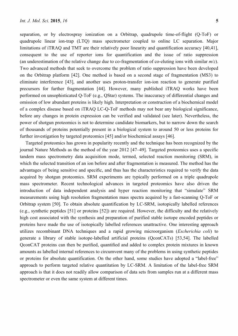
Targeted proteomics has grown in popularity recently and the technique has been recognized by the
journal Nature Methods as the method of the year 2012 [47–49]. Targeted proteomics uses a specific
tandem mass spectrometry data acquisition mode, termed, selected reaction monitoring (SRM), in
which the selected transition of an ion before and after fragmentation is measured. The method has the
advantages of being sensitive and specific, and thus has the characteristics required to verify the data
acquired by shotgun proteomics. SRM experiments are typically performed on a triple quadrupole
mass spectrometer. Recent technological advances in targeted proteomics have also driven the
introduction of data independent analysis and hyper reaction monitoring that “simulate” SRM
measurements using high resolution fragmentation mass spectra acquired by a fast-scanning Q-ToF or
Orbitrap system [50]. To obtain absolute quantification by LC-SRM, isotopically labelled references
(e.g., synthetic peptides [51] or proteins [52]) are required. However, the difficulty and the relatively
high cost associated with the synthesis and preparation of purified stable isotope encoded peptides or
proteins have made the use of isotopically labelled references unattractive. One interesting approach
utilizes recombinant DNA techniques and a rapid growing microorganism (Escherichia coli) to
generate a library of stable isotope-labelled artificial proteins (QconCATs) [53,54]. The labelled
QconCAT proteins can then be purified, quantified and added to complex protein mixtures in known
amounts as labelled internal references to circumvent many of the problems in using synthetic peptides
or proteins for absolute quantification. On the other hand, some studies have adopted a “label-free”
approach to perform targeted relative quantitation by LC-SRM. A limitation of the label-free SRM
approach is that it does not readily allow comparison of data sets from samples run at a different mass
spectrometer or even the same system at different times.

Int. J. Mol. Sci. 2015, 16 6
Figure 1. Commonly used label-based semi-quantitative proteomic methods. Two mains approaches are employed. Isotopic labels are
incorporated into proteins or peptides by biochemical of the cells using labelled lysine and/or arginine, or by chemical reactions with labelling
reagents. Quantitative data can be acquired via MS1 measurement, where the mass different between unlabeled and labeled peptides must at
least be 4 Da. Alternative methods use (4- to 10-plex) isobaric mass tags to chemically label tryptic peptides from groups of sample. The tags
contain four regions, a mass reporter region, a cleavable linker region, a mass normalization region and a protein reactive group. Upon
fragmentation of the isobaric peptides by CID or HCD, fragmentation of the tags gives rise to mass reporter ions for quantification. The m/z
values of reporter ion have been specifically designed at mass region away from typically interferences. Sequence information of the peptide
back bone is also obtained simultaneously. The fragment energy is typically set slightly higher to promote complete fragmentation of the tags.
Int. J. Mol. Sci. 2015, 16 7
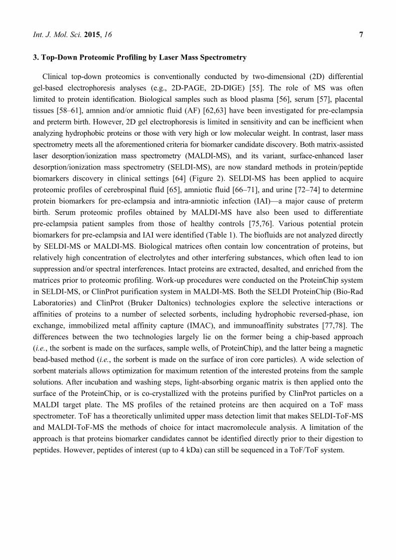
3. Top-Down Proteomic Profiling by Laser Mass Spectrometry
Clinical top-down proteomics is conventionally conducted by two-dimensional (2D) differential
gel-based electrophoresis analyses (e.g., 2D-PAGE, 2D-DIGE) [55]. The role of MS was often
limited to protein identification. Biological samples such as blood plasma [56], serum [57], placental
tissues [58–61], amnion and/or amniotic fluid (AF) [62,63] have been investigated for pre-eclampsia
and preterm birth. However, 2D gel electrophoresis is limited in sensitivity and can be inefficient when
analyzing hydrophobic proteins or those with very high or low molecular weight. In contrast, laser mass
spectrometry meets all the aforementioned criteria for biomarker candidate discovery. Both matrix-assisted
laser desorption/ionization mass spectrometry (MALDI-MS), and its variant, surface-enhanced laser
desorption/ionization mass spectrometry (SELDI-MS), are now standard methods in protein/peptide
biomarkers discovery in clinical settings [64] (Figure 2). SELDI-MS has been applied to acquire
proteomic profiles of cerebrospinal fluid [65], amniotic fluid [66–71], and urine [72–74] to determine
protein biomarkers for pre-eclampsia and intra-amniotic infection (IAI)—a major cause of preterm
birth. Serum proteomic profiles obtained by MALDI-MS have also been used to differentiate
pre-eclampsia patient samples from those of healthy controls [75,76]. Various potential protein
biomarkers for pre-eclampsia and IAI were identified (Table 1). The biofluids are not analyzed directly
by SELDI-MS or MALDI-MS. Biological matrices often contain low concentration of proteins, but
relatively high concentration of electrolytes and other interfering substances, which often lead to ion
suppression and/or spectral interferences. Intact proteins are extracted, desalted, and enriched from the
matrices prior to proteomic profiling. Work-up procedures were conducted on the ProteinChip system
in SELDI-MS, or ClinProt purification system in MALDI-MS. Both the SELDI ProteinChip (Bio-Rad
Laboratories) and ClinProt (Bruker Daltonics) technologies explore the selective interactions or
affinities of proteins to a number of selected sorbents, including hydrophobic reversed-phase, ion
exchange, immobilized metal affinity capture (IMAC), and immunoaffinity substrates [77,78]. The
differences between the two technologies largely lie on the former being a chip-based approach
(i.e., the sorbent is made on the surfaces, sample wells, of ProteinChip), and the latter being a magnetic
bead-based method (i.e., the sorbent is made on the surface of iron core particles). A wide selection of
sorbent materials allows optimization for maximum retention of the interested proteins from the sample
solutions. After incubation and washing steps, light-absorbing organic matrix is then applied onto the
surface of the ProteinChip, or is co-crystallized with the proteins purified by ClinProt particles on a
MALDI target plate. The MS profiles of the retained proteins are then acquired on a ToF mass
spectrometer. ToF has a theoretically unlimited upper mass detection limit that makes SELDI-ToF-MS
and MALDI-ToF-MS the methods of choice for intact macromolecule analysis. A limitation of the
approach is that proteins biomarker candidates cannot be identified directly prior to their digestion to
peptides. However, peptides of interest (up to 4 kDa) can still be sequenced in a ToF/ToF system.

Int. J. Mol. Sci. 2015, 16 8
Table 1. The Applications of MALDI-MS and SELDI-MS for Preeclampsia and Preterm Birth.
Technique Sample Cohort Sample Preparation Results Biological Implications Ref.
MALDI-MS
(ClinProt) Serum
Early-onset sPE: 11
CRL: 13
Sample extraction and
enrichment was performed
on HIC8 reverse phase coated
magnetic beads. 5 μL of serum
was incubated with 10 μL
MB-HIC8 binding buffer and
5 μL of MB-HIC8 bead slurry
for 1 min. After washing twice
with 100 μL of wash buffer,
proteins were eluted with
10 μL of elution buffer.
After MB-HIC8 extraction,
protein solutions, 0.5 μL of
protein solution was spotted
directly onto a stainless steel
target plate before matrix
solution was added.
The best differentiating signals
between the two sample groups
were found at m/z 13,715,
13,834, and 13,891. The
normalized intensities of these
ion signals were on average
lower in the PE group than in the
control group. The ion signals
were believed to belong to the
protein transthyretin and its
modified forms. Results were
consistent with that obtained
with SDS-PAGE analysis.
The reduction of transthyretin
concentrations is expected
during pregnancy due to plasma
volume expansion. Transthyretin
is synthesized by the liver and
also secreted by placental
trophoblasts where it binds
extracellular T4, which in turn
result in an increased
internalization of the
transthyretin-T4 complex. It has
been suggested that transthyretin
plays an important role in the
transfer of maternal thyroid
hormone to the fetal circulation,
which could have important
implications for fetal
development.
[75]

Int. J. Mol. Sci. 2015, 16 9
Table 1. Cont.
Technique Sample Cohort Sample Preparation Results Biological Implications Ref.
SELDI-MS
(WCX2
array)
Cerebrospina
l fluid (CSF)
sPE: 7
mPE: 8
CRL: 8
Dry on-chip protocol: 5-μL of
undiluted pooled CSF was dried
onto individual spots
A cluster of 4 peaks was
observed in the 15–16.3 kDa
region only from the CSF of the
patients. These peaks were
assigned to α- and β-chains of
hemoglobin, and their
glycosylated formed. The
presence of hemoglobin in CSF
as biomarkers was validated with
ELISA and spectrophotometry.
The reason for the observations
was not clear. The authors
suggested that the increase in
CSF hemoglobin might result
from increased and selective
trafficking of intact erythrocytes
across the blood-brain barrier
where they subsequently
lyse releasing their
hemoglobin content.
[65]
SELDI-MS
(Q10 array)
Amniotic
fluid (AF)
PE: 18
CHTN: 7
CRL: 16
Before sample loading, each
ProteinChip spot was incubated
twice with 5 µL binding buffer
in a humidity chamber at room
temperature for 5 min.
After equilibration, 5 µL of
sample, diluted 1:3 in binding
buffer, was added to each spot
and incubated in a humidity
chamber with shaking for
40 min. Each spot was washed
with 5 µL binding buffer for
2 min, followed by washing
with 5 µL of triple distilled
water, and air-dried.
2 peaks located at 17,399.1 and
28,023.3 Da were significantly
different. The former peak
distinguished women with PE
from control, and the latter peak
distinguished women with PE
and CHTN from controls. The
peaks were assigned to
hypothetical protein SBBI42 and
proapolipoprotein A-I. The
results were cross-validated with
Western blot.
It was suggested that the
increase in levels of
proapolipoprotein A-I in the AF
of women with PE may
represent a compensatory
mechanism to maintain levels of
apolipoprotein A-I and thereby
pulmonary surfactant and lung
compliance and development.
[70]

Int. J. Mol. Sci. 2015, 16 10
Table 1. Cont.
Technique Sample Cohort Sample Preparation Results Biological Implications Ref.
SELDI-MS
(H50 array)
Amniotic
fluid (AF)
PE: 10
CRL:10
AF was obtained by
transabdominal amniocentesis.
Protein chip arrays were placed
in a bioprocessor and pre-treated
with 50% methanol for 5 min.
2 μL of AF and 3 μL of protein
buffer were placed on individual
sample spots and incubated at
room temperature for 30 min.
Each sample spot was
equilibrated by adding 200 μL of
binding buffer. After shaking for
5 min at room temperature, the
buffer was removed, then, 5 μL
of sample mixture and 195 uL of
binding buffer were added to
each spot and incubated with
vigorous shaking for 30 min.
The arrays were washed with 3
times of 200 μL binding buffer,
followed by 2 times of 200 uL
distilled water. The protein chip
arrays were then air-dried.
5 protein peaks located at m/z
4,679, 9,080, 14,045, 14,345 and
28,087 were significantly
differentially expressed between
case and control groups. The
peak located at m/z 14,345 was
characterized as fragmented
albumin, whereas the peak
located at m/z 28,087 was
identified as apolipoprotein A-I.
The increased expression of
apolipoprotein A-I was
confirmed with ELISA.
The reason why albumin
fragment may be overexpressed
in these women is not
immediately apparent. One
possibility is that the increased
proteolytic activity against
albumin is an early phenomenon
that precedes the clinical
manifestations of the disease.
Apolipoprotein A-I is expressed
in the placenta and acts as a
receptor for cholesterol, which is
then transferred to the fetus.
The origin of apolipoprotein A-I
within AF remains unclear. Even
less well understood is why
apolipoprotein A-I is
overexpressed in second
trimester AF of women destined
to develop pre-eclampsia.
Whether the increase in
apolipoprotein A-I in the
AF precedes the increase seen in
maternal plasma and urine in
women with pre-eclampsia
is not known.
[71]

Int. J. Mol. Sci. 2015, 16 11
Table 1. Cont.
Technique Sample Cohort Sample Preparation Results Biological Implications Ref.
SELDI-MS
(Q10 array) Urine
sPE: 11
mPE: 7
CRL: 8
30 μL aliquots of individual
urine samples were mixed with
10 μL of sample buffer.
Following 30 min incubation at
room temperature, 160 μL of
binding buffer was added to each
sample. After equilibration of
the ProteinChip Array, 150 μL
of diluted sample mixture was
loaded onto each spot and
incubated with vigorous shaking
for 1 h. Each spot was washed
and air-dried.
4 discriminatory protein peaks
were identified at m/z 4,155, 6,044,
6,663 and 7,971. All of these
proteins had a lower concentration
in urine. The identities of the
protein biomarkers were not
assigned, but their discriminatory
power was tested with ROC.
[72]

Int. J. Mol. Sci. 2015, 16 12
Table 1. Cont.
Technique Sample Cohort Sample Preparation Results Biological Implications Ref.
SELDI-MS
(H4 and
H50 array)
Urine sPE: 38
CRL: 21
ProteinChip arrays were
incubated for 1 hour with the
samples (6 μL/spot) diluted
to 0.25 mg/mL total protein.
Following incubation,
unbound proteins were
removed by washing each
spot with the respective
buffer and dried.
At the end of exploratory phase, urine
proteomic profiles from the patients
with sPE exhibited 13 peaks
qualitatively different from that of the
controls. These peaks were assigned to
non-random cleavage products of serpin
peptidase inhibitor-1 (SERPINA1) and
albumin protein. Urine proteomic profile
score was tested against a
cross-sectional cohort (n = 206).
Performance was evaluated by ROC
(AUC = 0.92). The over-expression of
SERPINA1 in urine was also
verified with western blot.
Other studies have shown that minor
increases in levels of serum
SERPINA1 are associated with the
development of arterial hypertension
and an increased risk of
cardiovascular disease. The authors
suggested that by inhibiting the
activity of the kallikrein-kinin system,
an up-regulation of plasma
SERPINA1 favors the
renin-angiotensin system, leading to
systemic vasoconstriction and
hypertension. Urinary albumin
excretion is a hallmark of PE.
[74]
SELDI-MS
(NP20, H4
and IMAC
array)
Amniotic
fluid
(AF)
Preterm +IAI: 11
Preterm only: 11
CRL: 11
0.5 to 3.0 µg of
unfractionated protein from
AF was deposited on 3
different ProteinChip arrays
(normal-phase SiO2, a
reverse-phase hydrophobic,
and immobilized nickel
surfaces). The Chips were
incubated for 1 hour with the
sample followed by a 5-µL
water wash, and
subsequently dried.
A set of peaks located at 10- to
12-kDa was differentially expressed.
The peaks were observed on all 11
patients with subclinical IAI, in 2 of 11
with preterm delivery without IAI, and
in 0 of 11 with preterm labor and term
delivery without infection. The
signatures were identified polypeptides
derived from calgranulin B and a unique
fragment of insulin-like growth factor
binding protein 1 (IGFBP-1). Results
were validated by western blot.
The calgranulins are members of the
S-100 calcium-binding protein family,
expressed by macrophages and by
epithelial cells in acutely inflamed
tissues. The second candidate from
this cluster, a specific proteolytic
fragment of IGFBP-1, indicates a
potential protease-related mechanism
in response to infection. Intact
IGFBP-1 is the major IGFBP found in
AF and is synthesized by both fetal
membranes and maternal decidua.
[66]

Int. J. Mol. Sci. 2015, 16 13
Table 1. Cont.
Technique Sample Cohort Sample Preparation Results Biological Implications Ref.
SELDI-MS
(H4 array)
Amniotic
fluid
(AF)
Preterm (+WBC; +AFC): 21
Preterm (+WBC; −AFC): 7
Preterm (−WBC; +AFC): 8
Preterm (−WBC; −AFC): 24
CRL: 17
2 μL of AF diluted 10-fold
in PBS. After 1-hour
incubation in a humidified
box, the sample was
aspirated and the spots
washed individually with
25% aqueous acetonitrile
solution, air-dried.
Candidate makers were tested on a
separate set of 24 samples by blinding
independent examiners to the outcomes.
3 additional samples were used to assess
the possibility of storage artefacts and to
calculate intra- and inter-rater agreement
among the 3 investigators. 4 proteins,
neutrophil defensins-1 and -2, and
calgranulins A and C were found
distinctive and were validated with
western blot.
Neutrophil defensins
(α-defensins) belong to a family of
cationic antimicrobial peptides.
These key components of the
host-defense mechanism exert their
bactericidal activity by punching
pores into bacterial membranes.
[67]

Int. J. Mol. Sci. 2015, 16 14
Table 1. Cont.
Technique Sample Cohort Sample Preparation Results Biological Implications Ref.
SELDI-MS
(RS100)
Amniotic
fluid
(AF)
Preterm +IAI: 86
Preterm only: 86
CRL: 86
5 μL of anti-IGFBP-1 antibody or
control IgG solutions was loaded
onto the spot of pre-activated
ProteinChip arrays and covalently
coupled for 2 h at room
temperature in a humidity
chamber. Remaining reactive
groups were blocked for 1 h with
2 mg/mL BSA in
50 mM Tris-HCl. The spots
were washed 3 times with 10 μL
of PBS. Then, 5μL AF samples
were loaded on the
antibody-coated arrays and
incubated for 1 h in the humidity
chamber. Arrays were washed
three times with PBS and
rinsed once with water
before air-drying.
The ProteinChip array-based
immunoassay using SELDI showed that
IGFBP-1 was largely in a full-length
form in the AF of the patients with
preterm labor without IAI, but
significantly degraded in the AF pool of
the patients who delivered preterm with
IAI. This indicated a preferential
production of IGFBP-1 fragments in the
amniotic fluid of patients with IAI.
Consistent with the previous
study that the proteolytic
degradation of IGFBP-1 by
matrix metalloproteinases (MMPs)
and different MMPs generated
fragments of IGFBP-1 of
different masses.
[68]
SELDI-MS
(CM10 and
H50)
Amniotic
fluid
(AF)
Preterm +IAI: 60
CRL: 59
AF from each patient was diluted
in sterile PBS at a 1:10 dilution
and was added onto the
ProteinChip.
39 peaks were distinguishing patients
with preterm labor with IAI from those
with preterm lab our but subsequently
delivered at term. The study
did not seek to identify these mass
spectrometric features.
[69]
Q10: strong anion exchange; CM10/WCX: weak cation exchange; H: hydrophobic (reverse-phase); CRL: controls; PE: preeclampsia; sPE: severe preeclampsia;
mPE: mild preeclampsia; CHTN: chronic hypertension; +IAI: intra-amniotic infection; +AFC: positive amniotic fluid culture results; +WBC: white blood cell count >
100 cells/mm3; PBS: phosphate buffered saline; BSA: bovine serum albumin.

Int. J. Mol. Sci. 2015, 16 15
Figure 2. SELDI and Clinprot protein marker discovery workflow. Complex biological
fluid, such as serum, urine, cerebrospinal fluid, cell and tissue lysates, is applied directly
on SELDI ProteinChip or mixed with Clinprot magnetic beads. After incubation, unbound
proteins and other contaminants, such as salt and detergent, are washed off. Interested
proteins/peptides are captured onto their surface. The purified proteins/peptides are then
analyzed directly by MALDI-ToF-MS. The mass spectral profiles are then analyzed by
univariate or multivariate statistical data analysis for pattern recognition or classification.
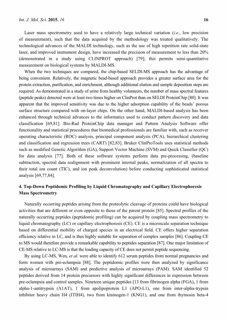
Int. J. Mol. Sci. 2015, 16 16
Laser mass spectrometry used to have a relatively large technical variation (i.e., low precision
of measurement), such that the data acquired by the methodology was treated qualitatively. The
technological advances of the MALDI technology, such as the use of high repetition rate solid-state
laser, and improved instrument design, have increased the precision of measurement to less than 20%
(demonstrated in a study using CLINPROT approach) [79]; this permits semi-quantitative
measurement on biological systems by MALDI-MS.
When the two techniques are compared, the chip-based SELDI-MS approach has the advantage of
being convenient. Relatively, the magnetic bead-based approach provides a greater surface area for the
protein extraction, purification, and enrichment, although additional elution and sample deposition steps are
required. As demonstrated in a study of urine from healthy volunteers, the number of mass spectral features
(peptide peaks) detected were at least two times higher on ClinProt than on SELDI ProteinChip [80]. It was
apparent that the improved sensitivity was due to the higher adsorption capability of the beads’ porous
surface structure compared with on-layer chips. On the other hand, MALDI-based analysis has been
enhanced through technical advances to the informatics used to conduct pattern discovery and data
classification [69,81]. Bio-Rad ProteinChip data manager and Pattern Analysis Software offer
functionality and statistical procedures that biomedical professionals are familiar with, such as receiver
operating characteristic (ROC) analysis, principal component analysis (PCA), hierarchical clustering
and classification and regression trees (CART) [82,83]. Bruker ClinProTools uses statistical methods
such as modified Genetic Algorithm (GA), Support Vector Machine (SVM) and Quick Classifier (QC)
for data analysis [77]. Both of these software systems perform data pre-processing, (baseline
subtraction, spectral data realignment with prominent internal peaks, normalization of all spectra to
their total ion count (TIC), and ion peak deconvolution) before conducting sophisticated statistical
analysis [69,77,84].
4. Top-Down Peptidomic Profiling by Liquid Chromatography and Capillary Electrophoresis
Mass Spectrometry
Naturally occurring peptides arising from the proteolytic cleavage of proteins could have biological
activities that are different or even opposite to those of the parent protein [85]. Spectral profiles of the
naturally occurring peptides (peptidomic profiling) can be acquired by coupling mass spectrometry to
liquid chromatography (LC) or capillary electrophoresis (CE). CE is a microscale separation technique
based on differential mobility of charged species in an electrical field. CE offers higher separation
efficiency relative to LC, and is thus highly suitable for separation of complex samples [86]. Coupling CE
to MS would therefore provide a remarkable capability to peptides separation [87]. One major limitation of
CE-MS relative to LC-MS is that the loading capacity of CE does not permit peptide sequencing.
By using LC-MS, Wen, et al. were able to identify 612 serum peptides from normal pregnancies and
form women with pre-eclampsia [88]. The peptidomic profiles were then analyzed by significance
analysis of microarrays (SAM) and predictive analysis of microarrays (PAM). SAM identified 52
peptides derived from 14 protein precursors with highly significant differences in expression between
pre-eclampsia and control samples. Nineteen unique peptides [13 from fibrinogen alpha (FGA), 1 from
alpha-1-antitrypsin (A1AT), 1 from apolipoprotein L1 (APO-L1), one from inter-alpha-trypsin
inhibitor heavy chain H4 (ITIH4), two from kininogen-1 (KNG1), and one from thymosin beta-4

Int. J. Mol. Sci. 2015, 16 17
(TMSB4), respectively] were selected for PAM prediction. On the training set, all pre-eclampsia
samples (n = 21) were predicted correctly, while three of the 21 (14.3%) control samples were false
positive. The sensitivity on the training set was 85.7% and the specificity was 100%, resulting in the
overall prediction accuracy of 92.9%. Similarly, on the testing set, the overall prediction accuracy is
90%, with sensitivity 80% and specificity 100%. Pathway analysis (Ingenuity Pathway Analysis
software) was performed on the 14 parental proteins of the 52-peptide markers. Liver X receptor
(LXR)/retinoid X receptor (RXR) activation, atherosclerosis signaling, IL-12 signaling and production in
macrophages, clathrin-mediated endocytosis signaling, production of nitric oxide and reactive oxygen
species in macrophages, acute phase response signaling, coagulation system, farnesoid X receptor
(FXR)/RXR activation and intrinsic prothrombin activation pathway are statistically significant
canonical pathways that were implicated in the pathophysiology of pre-eclampsia. Alternatively, by
using CE-MS, Carty, et al. were able to identify a urinary peptide pattern at gestational week 28 from
non-pregnant women, healthy pregnant, and those who had developed pre-eclampsia [89]. The peptides
were characterized by breakdown products of fibrinogen, collagen, and uromodulin (by LC-MS), which
could differentiate women who subsequently develop pre-eclampsia.
5. Bottom-Up Quantitative Proteomics
5.1. Serum and Plasma
The international SCreening fOr Pregnancy Endpoints (SCOPE) study used a combination of shotgun
and targeted proteomics and a biochemical assay (ELISA) to screen and pinpoint the plasma protein
biomarkers for predicting pre-eclampsia [90,91]. During the analytical pipeline development [90], the
researchers utilized an iTRAQ approach to screen early pregnancy plasma samples from women who
subsequently developed pre-eclampsia. The pooled plasma was first immunodepleted using either a
SepproÏgY 14-SuperMix Liquid Chromatography Column system, or Agilent Multiple Affinity
Removal LC Column—Human 14 (MARS Hu-14) to remove the most abundant proteins in the
plasma. The immunodepleted samples were then digested and labelled with 8-plex iTRAQ reagent.
The labelled samples were fractionated using high pH reversed-phase chromatography, and the
fractions were then analyzed either by online reversed-phase nano-LC coupled to a QStar XL qTOF
system, or by a MALDI TOF-TOF after off-line LC separation. 502 proteins were identified across 3
iTRAQ discovery experiments (319 proteins in the IgY 14-SuperMix- QSTAR data set, 331 in the IgY
14-SuperMix5800 data set and 189 in the MARS 14–5800 data set) and a total of 113 proteins altered
in abundance. Following application of stringent candidate protein selection criteria, two proteins:
pregnancy-specific beta-1-glycoprotein 9 (PSG9) and platelet basic protein (PBP, CXCL7), were
subjected to further assessment by SRM-based verification. In the targeted analysis phase, 108 plasma
samples, which include 16 early-onset preeclampsia (EO-PE), 42 late-onset preeclampsia (LO-PE),
42 controls, and eight technical replicates of a pooled sample, were immunodepleted on MARS Hu-14
immunodepletion column. The depleted plasma was then fractionated on an mRP-C18 column prior to
tryptic digestion. The tryptic peptides were then analyzed by LC-SRM on an uHPLC QqQ system. The
authors observed that six PSG specific peptides (from PSG-9 and PSG-5) were significantly different
between EO-PE and controls. The results were verified with ROC analysis and ELISA-based

Int. J. Mol. Sci. 2015, 16 18
quantification. On the other hand, only one of the PBP peptides measured was found significant.
Nevertheless, the authors noted that iTRAQ-based quantification has only a modest proteome
penetrance [90]. In their further study [91], the plasma proteins were tagged with 16O/18O-labelling
method. The samples were first depleted of the most abundant proteins using MARS Hu-14 columns.
Plasma proteins were then acetylated with sulfo-N-hydroxysuccinimide acetate prior to tryptic
digestion. 18O-label from H218O was incorporated into the peptides after incubation at 37 °C for 26 h with
trypsin. Furthermore, N-terminomics COFRADIC (COmbined FRActional DIagonal Chromatography)
platform was adopted from [92] to reduce the sample complexity prior to off-line LC separation and
MALDI-ToF/ToF analysis. Sixty-four proteins found in the discovery phase were chosen for
verification, including a number of reported markers for pre-eclampsia, such as endoglin [93], disintegrin
and metalloproteinase domain-containing protein 12 [94]. Additionally, nine proteins previously
identified in a cardiovascular biomarker study with biology relevant to pre-eclampsia and three
proteins (placental growth factor, soluble fms-like tyrosine kinase-1, and placental protein 13) with a
recognized association with pre-eclampsia were also taken forward to the verification experiments.
SRM assays were successfully developed for 51 out of the 76 selected proteins. Forty-four models
passed the verification stage, of which eight reached the target performance of 50% sensitivity at 20%
positive predictive value (PPV) for 5% prevalence in the validation set. These eight validated models
composed of mean arterial pressure (MAP) and 11 combinations of the proteins:
insulin-like growth factor acid labile subunit (IGFALS),
endoglin (ENG),
disintegrin and metalloproteinase domain-containing protein 12 (ADAM12),
serine peptidase inhibitor Kunitz type 1 (SPINT1),
melanoma cell adhesion molecule (MCAM),
selenoprotein P,
multimerin-2,
extracellular matrix protein 1,
microtubule-associated protein RP/EB family member 1 or 3,
fructose-bisphosphate aldolase A,
placental growth factor (PlGF)
The eight validated models all showed very similar performance for overall pre-eclampsia
prediction, but IGFALS carried the most predictive weight [91].
Similar approaches have also been adopted in independent investigations to determine serum
biomarkers for preeclampsia [95–97], albeit the scales of the cohorts were relatively small when
compared to the SCOPE study. In one study [95], the six most abundant proteins from the serum samples
were also first depleted with MARS, but the relative expression of the identified proteins was compared
by a label-free approach based on spectral counting. Four protein candidates, α-2-HS-glycoprotein
(AHSG), insulin-like growth factor binding protein, acid labile subunit (IGFBP-ALS), retinol binding
protein4 (RBP4) and alpha-1-microglobulin/bikunin (AMBP), were selected for further SRM assay.
The expression of IGFBP-ALS, AMBP and AHSG was higher in serum of the pre-eclampsia group,
whereas the expression of RBP4 was reduced when compared to the normal group. The increased
AHSG expression in serum was validated with ELISA [95].

Int. J. Mol. Sci. 2015, 16 19
In another study [96], peptide ligand library beads (PLLBs)-based affinity resin (ProteoMiner) was
employed for capturing the low abundance proteins. The isolated proteins were then separated by
SDS-gel and subjected in-gel digestion. LC-MS/MS analysis of the tryptic peptides was conducted on a
LTQ-Orbitrap mass spectrometer coupled to a nano-LC system. Relative quantitation of protein
expressions was achieved by spectral counting. Using two-fold or more changes as a determinant, it was
found that 31 proteins were up-regulated and 20 were down-regulated in patients with pre-eclampsia
comparing with healthy pregnant women. Among them, two proteins, chorionic somatomammotropin
hormone (CSH) and fibulin-1, were of particular interest, since they are low abundant proteins and are
seldom implicated in pre-eclampsia. The increased expression of apolipoprotein E (ApoE) in serum
from women with pre-eclampsia was verified with MS1 data using an isotope-encoded peptide
corresponding to a tryptic peptide of ApoE. Transthyretin (TTR), retinol-binding protein 4 (RBP4),
CSH and α-2-HS-glycoprotein (AHSG) were chosen for further validation by Western blot.
The immunoblot analysis showed that the concentrations of TTR and RBP4 were six and four times
respectively lower in the serum of patients with severe pre-eclampsia than in that in controls.
In contrast, the expression of CSH and AHSG were increased in severe pre-eclampsia. All
differentially expressed proteins were then analyzed using DAVID Bioinformatics Resources to
connect the proteins to biological processes and pathways. It was demonstrated that 17 of the
differentially expressed proteins were linked to complement and coagulation response and 15 were
acute phase response proteins. Six proteins were extracellular matrix proteins. The analysis also
revealed three major clusters of molecular function: seven proteins were endopeptidase inhibitors, five
were calcium-ion-binding proteins and eight were heparin-binding proteins. Collectively, these
observations implicate that acute phase, complement and coagulation responses play a major role in
pre-eclampsia [96].
5.2. Placental Tissue
While the SCOPE study aimed to determine circulating biomarkers for predictive or diagnostic
purposes, other researchers have chosen to perform comparative proteome analyses on human placenta
tissue between normal pregnancies and pregnancies complicated by pre-eclampsia, in order to determine
the biological processes involved in the development of the condition [98,99]. Placental tissue was
dissected from the maternal side of the placentas (in the central part, exclusive of calcified areas). After
homogenization, extraction and digestion, tryptic peptides were dimethyl-labelled. LC-MS/MS analysis
was performed on a LTQ-Orbitrap-Velos mass spectrometer coupled to a NanoACQUITY UPLC
system and 2636 unique proteins were expressed in the human placental tissue. Kyoto Encyclopedia of
Genes and Genomes (KEGG) pathway analyses were performed on the identified proteins to
determine the relevant pathways. Most of the identified pathways were consistent with the
development of placenta, including 297 proteins predicted to be involved in the metabolic pathways.
Further examination revealed that 171 proteins were differentially expressed between normal pregnancy
and pre-eclampsia; 147 proteins were down-regulated, while the remaining 24 proteins were up-regulated
in pre-eclampsia. Endoglin (ENG), ceruloplasmin (CP), mitochondrial superoxide dismutase (SOD),
transforming growth factor beta (TGF-β), are all thought to play key roles in the development of
pre-eclampsia; each was differentially expressed. Gene ontology (GO) analysis implicated oxygen

Int. J. Mol. Sci. 2015, 16 20
transport, regulation of cell death, and the aminoglycan metabolic and homeostatic processes in
the condition [98].
The authors then performed a comparative proteome analyses to determine the N-glyco- and
phospho-proteins that were differentially expressed in human placental plasma membrane [99].
Glycosylation and phosphorylation were chosen because these PTMs are known to be important in cell
signaling, development, migration and recognition. The placental tissue was homogenized in plasma
membrane isolation buffer. Enrichment and isolation of the plasma membrane was achieved by a
two-layer step sucrose gradient. The glycans were released from glycopeptides using PNGase F.
Phosphopeptides were enriched on Ti-doped mesoporous silica and 1027 N-glyco- and 2094 phospho- sites
were identified in the human placenta plasma membrane. KEGG pathway analyses identified pathways
related to the functions of the placental plasma membrane during pregnancy. Differences in
glycosylation and in phosphorylation between the case and control groups were found in seven and
42 protein peptides respectively, corresponding to five N-glyco- and 38 phosphoproteins; further KEGG
pathway analyses revealed differences in biological processes that included focal adhesion, shigellosis, and
bacterial invasion of epithelial cells. Furthermore, GO pathway analysis implicated a number of biological
processes as involved in pre-eclampsia, including negative regulation of DNA repair, sterol regulatory
element binding protein import into nucleus, and positive regulation of response to interferon-gamma.
The authors further investigated the placental mitochondria proteome because mitochondrial
dysfunction of the placenta occurs in pre-eclampsia [100]. After mitochondria isolation from the placenta
tissue, mitochondrial proteins were precipitated with cold acetone and then re-suspended in dissolution
buffer. After digestion, tryptic peptides were labelled with the iTRAQ regents, subsequently fractionated
by strong cation-exchange (SCX) chromatography and analyzed by a LTQ-Orbitrap mass spectrometer
coupled to a nano-LC system. 26 differentially expressed mitochondrial proteins were identified;
22 were down-regulated in pre-eclampsia, many of which are implicated in apoptosis, fatty acid
oxidation, the respiratory chain, reactive oxygen species generation, the tricarboxylic acid cycle and
oxidative stress. Due to the lack of suitable antibodies, only three of the proteins (transferrin receptor
(TFRC), mitochondrial protein peroxiredoxin III (PRDX3) and heat shock 10kDa protein 1 (HSPE1))
were verified with Western blot and immunohistochemical analysis. However, the results were
consistent with that of iTRAQ analysis [100].
5.3. Trophoblast Cells
The placenta predominantly consists of trophoblast cells, which have multiple functions, including
adhesion and invasion during early pregnancy and an important secretory role in late pregnancy.
Trophoblast cell isolation from placental tissue eliminates the problem of tissue heterogeneity of
placental tissue.
In one of the studies, laser capture microdissection was used to isolate placental trophoblastic cells.
Cellular proteins were extracted from the cellular lysates, and were labelled with ICAT reagents prior
to tryptic digestion and LC-MS/MS analysis. 70 proteins (note: eight entries were duplicated) showed
quantitative differences between pre-eclampsia and normal pregnancies (with a cut-off ratio of two);
31 were down-regulated in pre-eclampsia, while 39 were up-regulated. Significantly up-regulated
proteins included Fraser syndrome 1 isoform 2, pancreatic tumor-related protein and KIAA0226

Int. J. Mol. Sci. 2015, 16 21
protein; down-regulated proteins included laminin (expression in trophoblastic cells was verified with
Western blot) [101].
In another study, the proteins secreted by cultured cytotrophoblast cells from pre-eclampsia
complicated pregnancies were analyzed using a label-free approach on a LTQ-Orbitrap-Velos coupled
to a NanoAcquity system [102]. LC-MS/MS experiments were conducted on both pooled and
individualized culture supernatants of cytotrophoblastic cells. From the analysis of individualized
supernatant samples, 21 proteins were secreted significantly differently by cells from control and
pre-eclampsia pregnancies. One protein was detected exclusively in supernatant of control
cytotrophoblast cells and was identified as factor XIII chain A. This observation was further evaluated
with RT-PCR analysis and mRNA expression of factor XIII chain A was significantly decreased in
cytotrophoblastic cells from pre-eclampsia compared to the control.
5.4. Amniotic Fluid and Cervical-Vaginal Fluid
Clinical studies of AF and cervical-vaginal fluid (CVF) have been implicated in preterm labor and
other pregnancy-related complications, such as Down syndrome and Turner syndrome (see ref. [16]
for further discussion of the chemical constitutes of AF and CVF). In a study of the AF proteome,
iTRAQ was employed to identify biomarker candidates of spontaneous preterm birth (sPTB) [103].
In a cross-sectional study by selecting AF samples from 75 patients with an episode of uterine
contractions at preterm gestations and intact membranes. The cohort was classified into three groups:
(A) preterm labor without intra-amniotic infection (IAI) but delivered at term; (B) preterm labor
without IAI and delivered preterm within seven days of amnniocentesis; and (C) preterm labor with
IAI. The AF samples used were obtained from transabdominal amniocenteses performed for
evaluation of microbial status of the amniotic cavity. The samples were first depleted of albumin and
Immunoglobulin G using Vivapure immunoaffinity anti-human serum albumin capture resins, and
ultralink immobilized protein G. Acetone precipitation was also used to remove low molecular
interferences. Individual samples were digested and labelled with 4-plex iTRAQ reagents (the three
groups of samples were labelled with 115, 116, and 117 reagents and the pooled reference sample was
labelled with 114 reagent). The combined labelled samples were fractionated by SCX (six fractions)
and off-line LC separation before MALDI analysis. During the subsequent data analysis, the authors
also excluded the CID spectra, in which the 114-reference reporter ion was not observed, from the
determination of relative abundance of proteins. Overall, 77 proteins were up-regulated in preterm
labor with IAI (group C), whereas six proteins were down-regulated. Seventy-nine of the differentially
expressed proteins had been previously associated with infection/inflammation related preterm birth by
hypothesis-driven research studies. GO analysis further revealed that these differentially expressed
proteins have four main functions: (1) host defense; (2) mobility, localization and targeting;
(3) anti-apoptosis; and (4) metabolism/catabolism. Fewer proteins were differentially expressed on
comparison of preterm without IAI (group B), and delivered at term without IAI (group A).
Four proteins (resistin (RETN), thymosin-like 3 (TMSL3), antileukoproteinase (SLPI), and
growth-inhibiting protein 12 (lactoferrin, LTF)) were up-regulated, and two proteins
(latent-transforming growth factor β-binding protein isoform 1L (LTBP1) and mimecan precursor
(OGN)) were down-regulated. The altered AF resistin concentrations were verified with ELISA.

Int. J. Mol. Sci. 2015, 16 22
In a similar study [55], CVF was collected from subjects with sPTB, preterm labor without preterm
delivery (PTL), and asymptomatic control subjects. Patients with IAI at time of presentation and/or
placental histopathology at delivery were excluded. CVF proteins were analyzed by fluorescence
2D-DIGE and multidimensional LC-MS/MS analysis. For the LC-MS/MS analysis, individually
pooled samples were prepared from five maternal CVF samples, each of control, PTL and sPTB.
Non-protein interferences were removed by acetone precipitation. The tryptic peptides were separated
into 80 fractions by SCX chromatography and each fraction was then analyzed on a Q-TOF-2 mass
spectrometer coupled to a CapLC. Spectral counting was used to assess the relative abundance of a
protein. Twenty-eight of the identified proteins exhibited significant differences in pairwise and
progressive comparisons. Calgranulins A and B, annexins A3, S100 calcium-binding protein A7, and
epidermal fatty acid binding protein were abundant in CVF and differentially present in PTL and sPTB
samples, as were the serum proteins α-1-antitrypsin, α1-acid glycoprotein, haptoglobin precursor,
serotransferrin precursor (transferrin), and vitamin D binding protein precursor [55].
Quantification of protein biomarker candidates by targeted proteomics is hampered by the difficulties
associated with the preparation of labelled peptide references. One study took an opportunistic approach
to generate an entire labelled proteome, which was then used as internal standards to facilitate accurate
quantification of peptides, and proteins thereof, in a biological sample [104]. The method is termed
stable isotope labeled proteome (SILAP). The SILAP method is based on the absolute SILAC
approach [105], in which cultured cells are grown in stable isotope-labelled serum-free media with stable
isotope labelled lysine and leucine. To demonstrate the technical capability of SILAP, columnar
epithelial endocervical-1 (End1) and vaginal mucosal-2 (Vk2) cells were grown in SILAC conditions
to generate a SILAP library containing the secreted proteins. It was assumed that End1 and Vk2 cells
are “normal” transformed cells of human origin and have a secretory phenotype, and therefore, their
secreted proteins would model the proteins present in human CVF. The secreted proteome of the cells
were then determined by multidimensional LC-MS/MS analysis, after MARS Hu6 abundant proteins
depletion and SCX fractionation. In three independent experiments (replicates), 1211 proteins were
identified from the secreted proteome. Of the total 1211 proteins identified, 236 were detected in all
three replicates. Fifteen potential protein biomarkers for sPTB were chosen from the 236 proteins
based on the previous reports in sPTB or other pregnancy-related conditions. Stable isotope dilution
LC-SRM assays were then designed to conduct relative quantification of these candidate biomarkers in
term and sPTB human CVF samples spiked with the SILAP internal standard. Elevated expression of
desmoplakin isoform 1, stratifin, and thrombospondin 1 precursor in sPTB relative to the control group was
statistically significant. In particular, desmoplakin isoform 1 peptide was quantified to be 70.7-fold higher
in the sPTB samples as compared to the control samples. Furthermore, stratifin peptide (42.4-fold) and
thrombospondin 1 precursor peptide (5.1-fold) levels were significantly greater in the preterm birth
patient samples. While the data indicated these three proteins could serve as potential protein
biomarkers, the study was based on a relatively small cohort; further validation is required.

Int. J. Mol. Sci. 2015, 16 23
6. Comments
6.1. The Current Status of Proteomics for PreEclampsia and Preterm Birth
The technological advances of mass spectrometry and the associated techniques have provided a
unique opportunity to examine pregnancy-related complications such as pre-eclampsia and preterm
birth. A variety of proteomes has been examined by MALDI and SELDI-MS proteomic profiling.
The applications of MS-based top-down proteomics have clear advantages over 2D gel
electrophoresis, such as higher sensitivity to both hydrophobic and hydrophilic proteins. The use of a
ToF mass analyzer also allows detection of very high mass proteins. Shankar et al. predicted 10 years
ago that MALDI/SELDI-MS combined with pattern recognition would be essential to make use of the
method for diagnostics [106]. The availability of informatic tools has now made this possible.
Various bottom-up quantitative or comparative proteomic studies have been conducted using a
variety of label-based shotgun proteomic approaches. The iTRAQ technique has been by far the most
popular approach employed, despite known limitations. Although numerous protein candidates have
been discovered and reported, few of the proposed protein markers were actually validated. Only a
small number of studies further employed targeted proteomics or biochemical assays to verify
proposed biomarkers, and the applications of ELISA and Western blot were often limited by the
availability of antibodies. LC-SRM-based assays are complementary to Western blot and/or
immunohistochemical assays. Nevertheless, LC-SRM-based assays do not require suitable antibodies
that are often commercially unavailable. High precision and accuracy would still require labelled
reference peptides. Furthermore, some studies were based only on the data acquired from a relatively
small cohort or case-control samples. The resulting models are at best dubious. In contrast, the SCOPE
study established a powerful pipeline by combining both shotgun and targeted proteomics or
biochemical assays, to ascertain the sensitivity and specificity of the proposed biomarkers for clinical
diagnosis or probing the pathophysiological mechanisms. Other authors should note on the number of
SRM transitions to be measured per peptide in a proper LC-SRM experiment. Measurements based on
one SRM transition per peptide and/or one peptide per protein is less than ideal.
Meanwhile, challenges remain; the proteome is complex and many of the proteins of interest are of
low abundance; 85% of human serum proteins by mass are comprised of six proteins. These high
abundant proteins obscure potentially more biologically interesting low abundant proteins. Methods to
deplete the high abundant proteins have been employed for isolating and enriching low abundant
proteins in serum. A further issue concerns the variation in protein expression in different biological
compartments. Non-invasive biological fluids such as serum and urine may be useful to identify
diagnostic markers, but investigation of the pathogenesis of pre-eclampsia and other pregnancy-related
complications may be best conducted using placenta tissue or trophoblast cells. AF and CVF samples
may be particularly pertinent to the study of preterm birth.
6.2. Agreement between Observations of Studies and Their Possible Connections
Investigations on AFs by SELDI-MS have produced associated observations (Table 1). In contrast,
studies conducted by discovery-based shotgun proteomics using data dependent analysis (DDA)
produced disparate results. This can be contributed to the poor repeatability of DDA. Laboratories

Int. J. Mol. Sci. 2015, 16 24
using different methods and MS systems obviously produce a diverging set of observations. We
performed an analysis on the reported data to determine if there was any overlapping of observations
and identified 13 differentially expressed proteins were reported more than once in all the MS-based
proteomic studies for preeclampsia reviewed (Figure 3). These 13 proteins are involved in a variety of
biological processes, but six of them are known to involve in cell adhesion and extracellular matrix
organization. Plasminogen (or plasmin) was the only protein reported in serum, placental tissue, and
trophoblast cells. Furthermore, different forms of apolipoprotein were reported in serum, placental
tissue and AF for preeclampsia. Insulin-like growth factor and variants have also been reported not
only in the studies of pre-eclampsia, but also in preterm birth. Histone H4 was found in AFs and CVF
for preterm birth.
6.3. Clinical Assays of Biomarkers
The success of the SCOPE study has led to identification of a number of protein and metabolite
markers and clinical risk factors for predicting pre-eclampsia in nulliparous women [91,107–109].
The results have been translated to clinical assays. A predictive test based on proteins identified
through a LC-MS strategy is currently being validated through a phase IIa hospital-based study [110].
Further update can be seen at the IMPROvED web site (http://www.fp7-improved.eu/).
6.4. Future Directions
We anticipate that future investigations for pregnancy-related complications will take advantage of
recent technological advances in MS. Novel fragmentation techniques such as electron transfer
dissociation (ETD) may allow us to determine the protein identities or characteristics in top-down
proteomic experiments. The method can further facilitate analysis of labile PTMs. The authors
also recommend label-free approaches using data independent analysis (DIA), such as MSE and
all-ion-fragmentation, to enhance experimental repeatability. The recent introduction of Progenesis QI
for proteomics has made DIA data processing much more accessible. Biomarker verification by
targeted proteomics with conventional SRM and emerging hyper reaction monitoring (HRM), such as
multiplexed DIA and parallel reaction monitoring performed on high resolution Q-ToF and Orbitrap
systems, are expected to be more often reported in future studies.

Int. J. Mol. Sci. 2015, 16 25
Trophoblast Cells Proteome Placental Tissue Proteome Plasma/Serum Proteome
Most frequently reported proteins Found in Function
alpha-2-HS-glycoprotein serum Promotes endocytosis, influences the mineral phase of bone
Ceruloplasmin serum, placental tissue May play a role in fetal lung development or pulmonary antioxidant defense
Endoglin serum, placental tissue Crucial role in angiogenesis
Fibrinogen alpha chain serum Various cleavage products of fibrinogen and fibrin regulate cell adhesion and spreading
Fibronectin serum, placental tissue Cell adhesion and binding to various cell surfaces compounds
Fibulin-1 serum, placental tissue Binds to a number of extracellular matrix constituents including fibrinogen and fibronectin
Hemoglobin subunit alpha serum, placental tissue Oxygen transport
Embryonic hemoglobin in mammalian Hemoglobin subunit zeta serum, placental tissue
Plasminogen serum, placental tissue,
trophoblast cells
Acts as a proteolytic factor in a variety of other processes
including embryonic development, tissue remodeling
Pregnancy-specific beta-1-glycoprotein 3 serum, trophoblast cells Secreted products of the syncytiotrophoblast; known to be
extremely vital to development and health of a fetus Pregnancy-specific beta-1-glycoprotein 4 serum, trophoblast cells
Transthyretin serum, placental tissue Thyroid hormone-binding protein; thought to transport
thyroxine from the bloodstream to the brain
Vitronectin serum, placental tissue Cell adhesion and spreading factor
Figure 3. Most frequently reported differentially expressed proteins in the studies of preeclampsia. Cloud tags plots represent the number of
proteins and frequency of a protein reported in a proteome.
proteinsubunit
mitochondrial
isoformTransmembraneribosomal
domaincontainingHemoglobin
dehydrogenase
chainglycoprotein
alpharegion
cytoskeletal
Betahexosaminidase
Annexin
Interalphatrypsin
inhibitor
Trifunctional
factor
Cytochrome
flavoprotein Keratin
Programmed
ubiquinoneFragment
Phosphoacetylglucosamine
heavy
Plasminogenenzyme
Uncharacterized translation
Thioredoxinrelated
synthase
Sadenosylmethionine
complex
Thrombospondin1
proteinlike
homolog
receptor Serineargininerich
Bifunctional
Eukaryotic
Transportin1
Mannose1phosphate
LysosomeassociatedMacrophagecapping
Transforming
transfer
Trophoblast
UDPglucose
Proteinglutamine
Reticulocalbin1
Vitronectin
nucleotidebinding
SUMOprotein
initiation
Retinolbinding
Sideroflexin3
thioesterase
gamma1
phosphotyrosine
Tripeptidyl
oxidase
ElectronCytoskeletonassociated
Superoxide
Serpin
Twinfilin1
mutase
Peroxiredoxin1
UPF0317
nonerythrocytic
Interferoninduced
crosscomplementing
channel
Monoglyceride
Prothrombintranscript
betahydroxysteroid
phosphatase
Ryanodine
sidechain
kappa
alpha1reticulum
death
weight Spectrin
regulatory
S100A11
membrane
ATPdependent
zipper
splicing
beta1Haptoglobin
Scaffold
unc13
intracellular
Vtype
protease
soluble
Tissue
conductance
Putative
pathway
residenttype2
glycinerich
SoluteRanBP2
Serum
release
Sushi
C14orf159proton
54isomerase
purinecoenzyme
Guanine
gamma2
helicase
repair
Sialic
Histone
GIGSGT
Collagen
Catalase
lineage
growthExocyst
Fibulin1
emp24
ATPase kinase
activator
Copine
betac1
ITIH4
CASC5
ligase
albumin
ATrich
carrier
finger
antigen
liver
Early
Basic
Brain delta
acidic
proteinLaminin
chain
proteoglycan
Pregnancyspecific
isoformbeta1glycoprotein
precursor
sulfate
factorsubunit
Phosphatidylethanolaminebinding
PlasminogenHeparan
Serinethreonineprotein
kinasevariant
CoagulationFibronectin
receptortumorrelatedthreemodule
fragment Basement Transgelin2
Integrin
tractbinding
Thioredoxinheavy region
Serotransferrin
Human
membranespecific
Collagen
Mitogenactivated
Polypyrimidine
Metallothionein2
stimulation
preproprotein
Fibrillin
Annexin
lipoproteinrelated
Osteonidogen
syndrome
phosphataseChoriogonadotropin
Follistatinrelated
Immunoglobulin
Beta2microglobulin domaincontaining variable
version
structure
Antipneumococcal
regulatory
ribosomal
tenascin
dehydrogenase
gelatinbinding
DNAdependent
EGFlikedomain
light
Si00A11
Pancreatic
solution
membrane
Hypothetical
LOC56243
Aggrecanase1
a2macroglobulin
perlecan
XIIIb
Aphaactinin4
SNRPF
Migration
NidogenCysteinerich
KIAA0226
multiple
repeat
elongation
merosin
shock
epidermal
neuron
dJ34F711
Myosin
MHY11
partialADAMTSlike
CystatinM
Malate
domains
ßlike
motor
antibody
compact
Calnexin
1—human
growth
catalytic
Domain
Fibulin1 Porin
ADAMTS4
LAMA5
C9orf88 EGFlike
HSP60
Clathrin
density
6f11f22f2
ENO1P
ADMP1
Fraserdynein LaminCiliary
alpha
Actin
proteinComplement
factor
apolipoprotein
pregnancyspecific
subunit
inhibitor
beta1glycoprotein
peptidase
chain
fibrinogen
member
alpha2HSglycoprotein
subcomponent
somatomammotropinTransthyretin
microtubuleassociatedrepeatcontaining
Serum
Hemoglobin
alpha
Serpin
kininogen1
selenoprotein growth
component
metalloproteinase
amyloid
retinolbinding
insulinlike
fructosebisphosphate
alpha1microglobulinbikunin
Vitronectin
Hyaluronanbinding
Mannanbinding
Alpha2macroglobulin
HrelatedPlasminogen
interalphatrypsin domaincontaining
platelet
Pcomponent
clademultimerin2
thymosin
Galectin3binding
serine
ApolipoproteinL1
Pregnancy
peroxidase
Leucinerichproperdin
binding
Vitamin
melanoma
alpha1antitrypsin
Ceruloplasmin
protease
Glutathione
Lysozyme
extracellular moleculeplacental labile
Fibronectin
Coagulation
C4bbinding
Angiogenin
Plasma
retinol
disintegrin
hormone
Chorionic endoglin
Dbinding
matrix
ClusterinFibulin1
gamma
adhesion
Kunitz
heavy
aldolase
family
lectin
beta4
basic

Int. J. Mol. Sci. 2015, 16 26
7. Conclusions
We have chosen to discuss pre-eclampsia and preterm as two conditions that are the major causes of
perinatal morbidity and mortality. Obstetricians face the challenge of predicting and diagnosing both
conditions, particularly when women are in their first pregnancies, with no previous obstetric history.
MS-based proteomics has been applied in various investigations of pregnancy-related complications.
MS is a powerful alternative to conventional biochemical assays, but has not received adequate
attention. The technology has been used to study biological fluids and cells cultures, and has identified
various proteins that differentiate preeclampsia or preterm birth from healthy controls. Analysis of the
scientific literature reveals that an extensive network of proteins has been reported to be significant.
However, the overlapping of observations is relatively small on the same sample type or on different
biological compartments. Many of the observations could be considered as different aspects of the
disorders. Nevertheless, a pattern of biomarkers, for example, endoglin, α-2-HS-glycoprotein, fibrinogen,
plasminogen, different apolipoproteins and insulin-like growth factor binding protein are found in
various studies. Verifying these protein markers is essential for the clinical field to accept and use the
results from marker discovery studies; a phase IIa hospital-based clinical study has already started.
Acknowledgments
The authors would like to acknowledge the finical support from the Thousand People Project.
The authors would also like to acknowledge the finical support from PNB and T-LH for Gravida
(National Centre of Growth and Development, New Zealand).
Author Contributions
Kai P. Law, main author; Ting-Li Han, figures construction; Chao Tong, administration;
Philip N. Baker, supervision and editing.
Conflicts of Interest
The authors declare no conflict of interest.
References
1. Steegers, E.A.; von Dadelszen, P.; Duvekot, J.J.; Pijnenborg, R. Pre-eclampsia. Lancet 2010, 376,
631–644.
2. Powe, C.E.; Levine, R.J.; Karumanchi, S.A. Preeclampsia, a disease of the maternal endothelium:
The role of antiangiogenic factors and implications for later cardiovascular disease. Circulation
2011, 123, 2856–2869.
3. Huppertz, B. Placental origins of preeclampsia: Challenging the current hypothesis. Hypertension
2008, 51, 970–975.
4. Pecks, U.; Schutt, A.; Rower, C.; Reimer, T.; Schmidt, M.; Preschany, S.; Stepan, H.; Rath, W.;
Glocker, M.O. A mass spectrometric multicenter study supports classification of preeclampsia as
heterogeneous disorder. Hypertens Pregnancy 2012, 31, 278–291.

Int. J. Mol. Sci. 2015, 16 27
5. Bodnar, L.M.; Catov, J.M.; Simhan, H.N.; Holick, M.F.; Powers, R.W.; Roberts, J.M. Maternal
vitamin D deficiency increases the risk of preeclampsia. J. Clin. Endocrinol. Metab. 2007, 92,
3517–3522.
6. Baker, A.M.; Haeri, S.; Camargo, C.A., Jr.; Espinola, J.A.; Stuebe, A.M. A nested case-control
study of midgestation vitamin D deficiency and risk of severe preeclampsia. J. Clin. Endocrinol.
Metab. 2010, 95, 5105–5109.
7. De-Regil, L.M.; Palacios, C.; Ansary, A.; Kulier, R.; Pena-Rosas, J.P. Vitamin D
supplementation for women during pregnancy. Cochrane Database Syst. Rev. 2012, 2,
doi:10.1002/14651858.CD008873.pub2.
8. Brown, C.M.; Garovic, V.D. Drug treatment of hypertension in pregnancy. Drugs 2014, 74,
283–296.
9. Goldenberg, R.L.; Culhane, J.F.; Iams, J.D.; Romero, R. Epidemiology and causes of preterm
birth. Lancet 2008, 371, 75–84.
10. Stacy, B.; Wojdyla, D.; Say, L.; Betran, A.P.; Merialdi, M.; Requejo, J.H.; Rubens, C.;
Menon, R.; Look, P.F.V. The worldwide incidence of preterm birth: A systematic review of
maternal mortality and morbidity. Bull. World Health Organ. 2010, 88, 31–38.
11. Blencowe, H.; Cousens, S.; Oestergaard, M.Z.; Chou, D.; Moller, A.-B.; Narwal, R.; Adler, A.;
Vera Garcia, C.; Rohde, S.; Say, L.; et al. National, regional, and worldwide estimates of preterm
birth rates in the year 2010 with time trends since 1990 for selected countries: A systematic
analysis and implications. Lancet 2012, 379, 2162–2172.
12. WHO Preterm birth (Fact sheet N°363). Available online: http://www.who.int/mediacentre/
factsheets/fs363/en/ (accessed on 16 January 2015).
13. Conde-Agudelo, A.; Papageorghiou, A.T.; Kennedy, S.H.; Villar, J. Novel biomarkers for the
prediction of the spontaneous preterm birth phenotype: A systematic review and meta-analysis.
BJOG 2011, 118, 1042–1054.
14. Bodnar, L.M.; Platt, R.W.; Simhan, H.N. Early-pregnancy vitamin D deficiency and risk of
preterm birth subtypes. Obstet Gynecol. 2015, 125, 439–447.
15. Sablok, A.; Batra, A.; Thariani, K.; Batra, A.; Bharti, R.; Aggarwal, A.R.; Kabi, B.C.; Chellani, H.
Supplementation of vitamin D in pregnancy and its correlation with feto-maternal outcome. Clin.
Endocrinol. 2015, doi: 10.1111/cen.12751.
16. Kolialexi, A.; Mavrou, A.; Spyrou, G.; Tsangaris, G.T. Mass spectrometry-based proteomics in
reproductive medicine. Mass Spectrom Rev. 2008, 27, 624–634.
17. Kolialexi, A.; Anagnostopoulos, A.K.; Mavrou, A.; Tsangaris, G.T. Application of proteomics
for diagnosis of fetal aneuploidies and pregnancy complications. J. Proteomics 2009, 72, 731–739.
18. Fanos, V.; Atzori, L.; Makarenko, K.; Melis, G.B.; Ferrazzi, E. Metabolomics application in
maternal-fetal medicine. Biomed. Res. Int. 2013, 2013, doi:10.1155/2013/720514.
19. Schulze, A.; Lindner, M.; Kohlmuller, D.; Olgemoller, K.; Mayatepek, E.; Hoffmann, G.F.
Expanded newborn screening for inborn errors of metabolism by electrospray ionization-tandem
mass spectrometry: Results, outcome, and implications. Pediatrics 2003, 111, 1399–1406.
20. Ly, L.; Wasinger, V.C. Protein and peptide fractionation, enrichment and depletion: Tools for the
complex proteome. Proteomics 2011, 11, 513–534.

Int. J. Mol. Sci. 2015, 16 28
21. Smith, M.P.; Wood, S.L.; Zougman, A.; Ho, J.T.; Peng, J.; Jackson, D.; Cairns, D.A.;
Lewington, A.J.; Selby, P.J.; Banks, R.E. A systematic analysis of the effects of increasing degrees
of serum immunodepletion in terms of depth of coverage and other key aspects in top-down and
bottom-up proteomic analyses. Proteomics 2011, 11, 2222–2235.
22. Koulman, A.; Lane, G.A.; Harrison, S.J.; Volmer, D.A. From differentiating metabolites to
biomarkers. Anal. Bioanal. Chem. 2009, 394, 663–670.
23. Jorgensen, C.; Locard-Paulet, M. Analysing signalling networks by mass spectrometry. Amino
Acids 2012, 43, 1061–1074.
24. Becker, G.W. Stable isotopic labeling of proteins for quantitative proteomic applications. Brief.
Funct. Genomic Proteomic 2008, 7, 371–382.
25. Gevaert, K.; Impens, F.; Ghesquiere, B.; Van Damme, P.; Lambrechts, A.; Vandekerckhove, J.
Stable isotopic labeling in proteomics. Proteomics 2008, 8, 4873–4885.
26. Merrill, A.E.; Coon, J.J. Quantifying proteomes and their post-translational modifications by
stable isotope label-based mass spectrometry. Curr. Opin. Chem. Biol. 2013, 17, 779–786.
27. Zhou, Y.; Shan, Y.; Zhang, L.; Zhang, Y. Recent advances in stable isotope labeling based
techniques for proteome relative quantification. J. Chromatogr. A 2014, 1365, 1–11.
28. Wang, M.; You, J.; Bemis, K.G.; Tegeler, T.J.; Brown, D.P. Label-free mass spectrometry-based
protein quantification technologies in proteomic analysis. Brief. Funct. Genomic Proteomic 2008,
7, 329–339.
29. Neilson, K.A.; Ali, N.A.; Muralidharan, S.; Mirzaei, M.; Mariani, M.; Assadourian, G.; Lee, A.;
van Sluyter, S.C.; Haynes, P.A. Less label, more free: Approaches in label-free quantitative mass
spectrometry. Proteomics 2011, 11, 535–553.
30. Ong, S.E. The expanding field of SILAC. Anal. Bioanal. Chem. 2012, 404, 967–976.
31. Ong, S.E. Whole proteomes as internal standards in quantitative proteomics. Genome Med. 2010,
2, 49.
32. Zhou, Y.; Shan, Y.; Wu, Q.; Zhang, S.; Zhang, L.; Zhang, Y. Mass defect-based pseudo-isobaric
dimethyl labeling for proteome quantification. Anal. Chem. 2013, 85, 10658–10663.
33. Bamberger, C.; Pankow, S.; Park, S.K.; Yates, J.R., 3rd. Interference-free proteome
quantification with MS/MS-based isobaric isotopologue detection. J. Proteome Res. 2014, 13,
1494–1501.
34. Havlis, J.; Shevchenko, A. Absolute quantification of proteins in solutions and in polyacrylamide
gels by mass spectrometry. Anal. Chem. 2004, 76, 3029–3036.
35. Gygi, S.P.; Rist, B.; Gerber, S.A.; Turecek, F.; Gelb, M.H.; Aebersold, R. Quantitative analysis
of complex protein mixtures using isotope-coded affinity tags. Nat. Biotech. 1999, 17, 994–999.
36. Yi, E.C.; Li, X.J.; Cooke, K.; Lee, H.; Raught, B.; Page, A.; Aneliunas, V.; Hieter, P.;
Goodlett, D.R.; Aebersold, R. Increased quantitative proteome coverage with 13C/12C-based,
acid-cleavable isotope-coded affinity tag reagent and modified data acquisition scheme.
Proteomics 2005, 5, 380–387.
37. Unwin, R.D.; Griffiths, J.R.; Whetton, A.D. Simultaneous analysis of relative protein expression
levels across multiple samples using iTRAQ isobaric tags with 2D nano LC-MS/MS. Nat. Protoc.
2010, 5, 1574–1582.

Int. J. Mol. Sci. 2015, 16 29
38. Thompson, A.; Schafer, J.; Kuhn, K.; Kienle, S.; Schwarz, J.; Schmidt, G.; Neumann, T.;
Johnstone, R.; Mohammed, A.K.; Hamon, C. Tandem mass tags: A novel quantification
strategy for comparative analysis of complex protein mixtures by MS/MS. Anal. Chem. 2003, 75,
1895–1904.
39. Zhang, Y.; Fonslow, B.R.; Shan, B.; Baek, M.C.; Yates, J.R., 3rd. Protein analysis by
shotgun/bottom-up proteomics. Chem. Rev. 2013, 113, 2343–2394.
40. Ow, S.Y.; Salim, M.; Noirel, J.; Evans, C.; Rehman, I.; Wright, P.C. iTRAQ underestimation
in simple and complex mixtures: “The good, the bad and the ugly”. J. Proteome Res. 2009, 8,
5347–5355.
41. Altelaar, A.F.; Frese, C.K.; Preisinger, C.; Hennrich, M.L.; Schram, A.W.; Timmers, H.T.;
Heck, A.J.; Mohammed, S. Benchmarking stable isotope labeling based quantitative proteomics.
J. Proteomics 2013, 88, 14–26.
42. Christoforou, A.; Lilley, K.S. Taming the isobaric tagging elephant in the room in quantitative
proteomics. Nat. Methods 2011, 8, 911–913.
43. Ting, L.; Rad, R.; Gygi, S.P.; Haas, W. MS3 eliminates ratio distortion in isobaric multiplexed
quantitative proteomics. Nat. Methods 2011, 8, 937–940.
44. Wenger, C.D.; Lee, M.V.; Hebert, A.S.; McAlister, G.C.; Phanstiel, D.H.; Westphall, M.S.;
Coon, J.J. Gas-phase purification enables accurate, multiplexed proteome quantification with
isobaric tagging. Nat. Methods 2011, 8, 933–935.
45. Kaur, P.; Rizk, N.M.; Ibrahim, S.; Younes, N.; Uppal, A.; Dennis, K.; Karve, T.; Blakeslee, K.;
Kwagyan, J.; Zirie, M.; et al. iTRAQ-based quantitative protein expression profiling and MRM
verification of markers in type 2 diabetes. J. Proteome Res. 2012, 11, 5527–5539.
46. Hou, A.; Lan, W.; Law, K.P.; Khoo, S.C. J.; Tin, M.Q.; Tong, L. Evaluation of global differential
gene and protein expression in primary pterygium: S100A8 and S100A9 as possible drivers of a
signaling network. PLoS ONE 2014, 9, e97402.
47. Marx, V. Method of the year: Targeted proteomics. Nat. Methods 2013, 10, 19–22.
48. Picotti, P.; Bodenmiller, B.; Aebersold, R. Method of the year: Proteomics meets the scientific
method. Nat. Methods 2013, 10, 24–27.
49. Gillette, M.A.; Carr, S.A. Quantitative analysis of peptides and proteins in biomedicine by
targeted mass spectrometry. Nat. Methods 2013, 10, 28–34.
50. Law, K.P. Recent advances in mass spectrometry: Data independent analysis and hyper reaction
monitoring. Expert Rev. Proteomics 2013, 10, 551–566.
51. Gerber, S.A.; Rush, J.; Stemman, O.; Kirschner, M.W.; Gygi, S.P. Absolute quantification of
proteins and phosphoproteins from cell lysates by tandem MS. Proc. Natl. Acad. Sci. USA 2003,
100, 6940–6945.
52. Huillet, C.; Adrait, A.; Lebert, D.; Picard, G.; Trauchessec, M.; Louwagie, M.; Dupuis, A.;
Hittinger, L.; Ghaleh, B.; le Corvoisier, P.; et al. Accurate quantification of cardiovascular
biomarkers in serum using Protein Standard Absolute Quantification (PSAQ™) and selected
reaction monitoring. Mol. Cell Proteomics 2012, 11, doi:10.1074/mcp.M111.008235.
53. Brownridge, P.; Holman, S.W.; Gaskell, S.J.; Grant, C.M.; Harman, V.M.; Hubbard, S.J.;
Lanthaler, K.; Lawless, C.; OʼCualain, R.; Sims, P.; et al. Global absolute quantification of a
proteome: Challenges in the deployment of a QconCAT strategy. Proteomics 2011, 11, 2957–2970.

Int. J. Mol. Sci. 2015, 16 30
54. Carroll, K.M.; Simpson, D.M.; Eyers, C.E.; Knight, C.G.; Brownridge, P.; Dunn, W.B.;
Winder, C.L.; Lanthaler, K.; Pir, P.; Malys, N.; et al. Absolute quantification of the glycolytic
pathway in yeast: Deployment of a complete QconCAT approach. Mol. Cell Proteomics 2011, 10,
doi: 10.1074/mcp.M111.007633.
55. Pereira, L.; Reddy, A.P.; Jacob, T.; Thomas, A.; Schneider, K.A.; Dasari, S.; Lapidus, J.A.; Lu,
X.; Rodland, M.; Roberts, C.T., Jr.; et al. Identification of novel protein biomarkers of preterm
birth in human cervical-vaginal fluid. J. Proteome Res. 2007, 6, 1269–1276.
56. Wang, C.C.; Yim, K.W.; Poon, T.C.W.; Choy, K.W.; Chu, C.Y.; Lui, W.T.; Lau, T.K.;
Rogers, M.S.; Leung, T.N. Innate immune response by ficolin binding in apoptotic placenta is
associated with the clinical syndrome of preeclampsia. Clin. Chem. 2007, 53, 42–52.
57. Watanabe, H.; Hamada, H.; Yamada, N.; Sohda, S.; Yamakawa-Kobayashi, K.; Yoshikawa, H.;
Arinami, T. Proteome analysis reveals elevated serum levels of clusterin in patients with
preeclampsia. Proteomics 2004, 4, 537–543.
58. Jin, H.; Ma, K.D.; Hu, R.; Chen, Y.; Yang, F.; Yao, J.; Li, X.T.; Yang, P.Y. Analysis of
expression and comparative profile of normal placental tissue proteins and those in preeclampsia
patients using proteomic approaches. Anal. Chim. Acta 2008, 629, 158–164.
59. Gharesi-Fard, B.; Zolghadri, J.; Kamali-Sarvestani, E. Proteome differences of placenta between
pre-eclampsia and normal pregnancy. Placenta 2010, 31, 121–125.
60. Butt, R.H.; Lee, M.W.; Pirshahid, S.A.; Backlund, P.S.; Wood, S.; Coorssen, J.R. An initial
proteomic analysis of human preterm labor: Placental membranes. J. Proteome Res. 2006, 5,
3161–3172.
61. Chang, A.; Zhang, Z.; Zhang, L.; Gao, Y.; Zhang, L.; Jia, L.; Cui, S.; Wang, P. Proteomic
analysis of preterm premature rupture of membranes in placental tissue. Arch. Gynecol. Obstet
2013, 288, 775–784.
62. Vascotto, C.; Salzano, A.M.; DʼAmbrosio, C.; Fruscalzo, A.; Marchesoni, D.; di Loreto, C.;
Scaloni, A.; Tell, G.; Quadrifoglio, F. Oxidized transthyretin in amniotic fluid as an early marker
of preeclampsia. J. Proteome Res. 2007, 6, 160–170.
63. Park, S.J.; Yoon, W.G.; Song, J.S.; Jung, H.S.; Kim, C.J.; Oh, S.Y.; Yoon, B.H.; Jung, G.;
Kim, H.J.; Nirasawa, T. Proteome analysis of human amnion and amniotic fluid by
two-dimensional electrophoresis and matrix-assisted laser desorption/ionization time-of-flight
mass spectrometry. Proteomics 2006, 6, 349–363.
64. Hortin, G.L. The MALDI-TOF mass spectrometric view of the plasma proteome and peptidome.
Clin. Chem. 2006, 52, 1223–1237.
65. Norwitz, E.R.; Tsen, L.C.; Park, J.S.; Fitzpatrick, P.A.; Dorfman, D.M.; Saade, G.R.;
Buhimschi, C.S.; Buhimschi, I.A. Discriminatory proteomic biomarker analysis identifies free
hemoglobin in the cerebrospinal fluid of women with severe preeclampsia. Am. J. Obstet
Gynecol. 2005, 193, 957–964.
66. Gravett, M.G.; Novy, M.J.; Rosenfeld, R.G.; Reddy, A.P.; Jacob, T.; Turner, M.; McCormack, A.;
Lapidus, J.A.; Hitti, J.; Eschenbach, D.A.; et al. Diagnosis of intra-amniotic infection by
proteomic profiling and identification of novel biomarkers. JAMA 2004, 292, 462–469.
67. Buhimschi, I.A.; Christner, R.; Buhimschi, C.S. Proteomic biomarker analysis of amniotic fluid
for identification of intra-amniotic inflammation. BJOG 2005, 112, 173–181.

Int. J. Mol. Sci. 2015, 16 31
68. Bujold, E.; Romero, R.; Kusanovic, J.P.; Erez, O.; Gotsch, F.; Chaiworapongsa, T.; Gomez, R.;
Espinoza, J.; Vaisbuch, E.; Mee Kim, Y.; et al. Proteomic profiling of amniotic fluid in preterm
labor using two-dimensional liquid separation and mass spectrometry. J. Matern. Fetal Neonatal
Med. 2008, 21, 697–713.
69. Romero, R.; Espinoza, J.; Rogers, W.T.; Moser, A.; Nien, J.K.; Kusanovic, J.P.; Gotsch, F.;
Erez, O.; Gomez, R.; Edwin, S.; et al. Proteomic analysis of amniotic fluid to identify women
with preterm labor and intra-amniotic inflammation/infection: The use of a novel computational
method to analyze mass spectrometric profiling. J. Matern. Fetal Neonatal Med. 2008, 21, 367–388.
70. Park, J.S.; Oh, K.J.; Norwitz, E.R.; Han, J.S.; Choi, H.J.; Seong, H.S.; Kang, Y.D.; Park, C.W.;
Kim, B.J.; Jun, J.K.; et al. Identification of proteomic biomarkers of preeclampsia in amniotic
fluid using SELDI-TOF mass spectrometry. Reprod. Sci. 2008, 15, 457–468.
71. Oh, K.J.; Park, J.S.; Norwitz, E.R.; Kim, S.M.; Kim, B.J.; Park, C.W.; Jun, J.K.; Syn, H.C.
Proteomic biomarkers in second trimester amniotic fluid that identify women who are destined to
develop preeclampsia. Reprod. Sci. 2012, 19, 694–703.
72. Lee, S.M.; Park, J.S.; Norwitz, E.R.; Kim, S.M.; Kim, B.J.; Park, C.W.; Jun, J.K.; Syn, H.C.
Characterization of discriminatory urinary proteomic biomarkers for severe preeclampsia using
SELDI-TOF mass spectrometry. J. Perinat. Med. 2011, 39, 391–396.
73. Buhimschi, I.A.; Zhao, G.; Funai, E.; Saade, G.R.; Buhimschi, C. Proteomics analysis of urine in
preeclampsia (PE): A novel diagnosis for an old disease. Am. J. Obstet Gynecol 2005, 193, S15.
74. Buhimschi, I.A.; Zhao, G.; Funai, E.F.; Harris, N.; Sasson, I.E.; Bernstein, I.M.; Saade, G.R.;
Buhimschi, C.S. Proteomic profiling of urine identifies specific fragments of SERPINA1 and
albumin as biomarkers of preeclampsia. Am. J. Obstet Gynecol. 2008, 199, 551.e1–551.e16.
75. Pecks, U.; Seidenspinner, F.; Rower, C.; Reimer, T.; Rath, W.; Glocker, M.O. Multifactorial
analysis of affinity-mass spectrometry data from serum protein samples: A strategy to distinguish
patients with preeclampsia from matching control individuals. J. Am. Soc. Mass Spectrom 2010,
21, 1699–1711.
76. Glocker, M.O.; Rower, C.; Wolter, M.; Koy, C.; Reimer, T.; Pecks, U. Multiparametric Analysis
of Mass Spectrometry-Based Proteome Profiling in Gestation-Related Diseases. In Handbook of
Spectroscopy, 2nd ed.; Gauglitz, G., Moore, D.S., Eds.; Wiley VCH: Weinheim, Germany, 2014;
pp. 407–428.
77. Elssner, T.; Kostrzewa, M. CLINPROT—A MALDI-TOF MS based system for biomarker
discovery and analysis. Clin. Proteom 2006, 8, 167–179.
78. ProteinChip Arrays and Reagents: Sophisticated Tools for Differential Expression Profiling.
Available online: http://www.bio-rad.com/webroot/web/pdf/lsr/literature/Bulletin_5524.pdf
(accessed on 11 May 2015).
79. Pakharukova, N.A.; Pastushkova, L.K.; Trifonova, O.P.; Pyatnitsky, M.A.; Vlasova, M.A.;
Nikitin, I.P.; Moshkovsky, S.A.; Nikolayev, E.N.; Larina, I.M. Optimization of serum proteome
profiling of healthy humans. Hum. Physiol. 2009, 35, 350–356.
80. Fiedler, G.M.; Baumann, S.; Leichtle, A.; Oltmann, A.; Kase, J.; Thiery, J.; Ceglarek, U.
Standardized peptidome profiling of human urine by magnetic bead separation and matrix-assisted
laser desorption/ionization time-of-flight mass spectrometry. Clin. Chem. 2007, 53, 421–428.

Int. J. Mol. Sci. 2015, 16 32
81. Dalmasso, E.; Caseñas, D. Data Processing and Analysis Using ProteinChip® Data Manager
Software. In SELDI-TOF Mass Spectrometry; Clarke, C.H., McCarthy, D.L.B., Eds.; Springer:
New York, NY, USA, 2012; Volume 818, pp. 35–48.
82. ProteinChip Data Manager Software. Available online: http://www.bio-rad.com/webroot/web/
pdf/lsr/literature/Bulletin_5526B.pdf (accessed on 11 May 2015).
83. ProteinChip SELDI System: High-Throughput Protein Profiling. Available online: http://www.bio-
rad.com/webroot/web/pdf/lsr/literature/Bulletin_5530.pdf (accessed on 11 May 2015).
84. Biomarker Discovery Using SELDI Technology: A Guide to Data Processing and Analysis Using
ProteinChip® Data Manager Software. Available online: http://www.bio-rad.com/webroot/web/
pdf/lsr/literature/Bulletin_5814.pdf (accessed on 11 May 2015).
85. Martelli, C.; Iavarone, F.; Vincenzoni, F.; Cabras, T.; Manconi, B.; Desiderio, C.; Messana, I.;
Castagnola, M. Top-down peptidomics of bodily fluids. Peptidomics 2014, 1, 47–64.
86. Hommerson, P.; Khan, A.M.; de Jong, G.J.; Somsen, G.W. Ionization techniques in capillary
electrophoresis-mass spectrometry: Principles, design, and application. Mass Spectrom Rev. 2011,
30, 1096–10120.
87. Dakna, M.; He, Z.; Yu, W.C.; Mischak, H.; Kolch, W. Technical, bioinformatical and statistical
aspects of liquid chromatography-mass spectrometry (LC-MS) and capillary electrophoresis-mass
spectrometry (CE-MS) based clinical proteomics: A critical assessment. J. Chromatogr. B 2009,
877, 1250–1258.
88. Wen, Q.; Liu, L.Y.; Yang, T.; Alev, C.; Wu, S.; Stevenson, D.K.; Sheng, G.; Butte, A.J.;
Ling, X.B. Peptidomic Identification of Serum Peptides Diagnosing Preeclampsia. PLoS ONE
2013, 8, e65571.
89. Carty, D.M.; Siwy, J.; Brennand, J.E.; Zurbig, P.; Mullen, W.; Franke, J.; McCulloch, J.W.;
Roberts, C.T.; North, R.A.; Chappell, L.C.; et al. Urinary proteomics for prediction of
preeclampsia. Hypertension 2011, 57, 561–569.
90. Blankley, R.T.; Fisher, C.; Westwood, M.; North, R.; Baker, P.N.; Walker, M.J.; Williamson, A.;
Whetton, A.D.; Lin, W.; McCowan, L.; et al. A label-free selected reaction monitoring workflow
identifies a subset of pregnancy specific glycoproteins as potential predictive markers of
early-onset pre-eclampsia. Mol. Cell Proteomics 2013, 12, 3148–3159.
91. Myers, J.E.; Tuytten, R.; Thomas, G.; Laroy, W.; Kas, K.; Vanpoucke, G.; Roberts, C.T.;
Kenny, L.C.; Simpson, N.A.; Baker, P.N.; et al. Integrated proteomics pipeline yields novel
biomarkers for predicting preeclampsia. Hypertension 2013, 61, 1281–1288.
92. Mebazaa, A.; Vanpoucke, G.; Thomas, G.; Verleysen, K.; Cohen-Solal, A.; Vanderheyden, M.;
Bartunek, J.; Mueller, C.; Launay, J.M.; Van Landuyt, N.; et al. Unbiased plasma proteomics for
novel diagnostic biomarkers in cardiovascular disease: Identification of quiescin Q6 as a
candidate biomarker of acutely decompensated heart failure. Eur. Heart J. 2012, 33, 2317–2324.
93. Venkatesha, S.; Toporsian, M.; Lam, C.; Hanai, J.; Mammoto, T.; Kim, Y.M.; Bdolah, Y.;
Lim, K.H.; Yuan, H.T.; Libermann, T.A.; et al. Soluble endoglin contributes to the pathogenesis
of preeclampsia. Nat. Med. 2006, 12, 642–649.
94. Laigaard, J.; Sorensen, T.; Placing, S.; Holck, P.; Frohlich, C.; Wojdemann, K.R.; Sundberg, K.;
Shalmi, A.C.; Tabor, A.; Norgaard-Pedersen, B.; et al. Reduction of the disintegrin and
metalloprotease ADAM12 in preeclampsia. Obstet Gynecol. 2005, 106, 144–149.

Int. J. Mol. Sci. 2015, 16 33
95. Park, J.; Cha, D.H.; Lee, S.J.; Kim, Y.N.; Kim, Y.H.; Kim, K.P. Discovery of the serum
biomarker proteins in severe preeclampsia by proteomic analysis. Exp. Mol. Med. 2011, 43, 427–435.
96. Liu, C.; Zhang, N.; Yu, H.; Chen, Y.; Liang, Y.; Deng, H.; Zhang, Z. Proteomic analysis of
human serum for finding pathogenic factors and potential biomarkers in preeclampsia. Placenta
2011, 32, 168–174.
97. Kolla, V.; Jeno, P.; Moes, S.; Lapaire, O.; Hoesli, I.; Hahn, S. Quantitative proteomic (iTRAQ)
analysis of 1st trimester maternal plasma samples in pregnancies at risk for preeclampsia.
J. Biomed. Biotechnol 2012, 2012, doi:10.1155/2012/305964.
98. Wang, F.; Shi, Z.; Wang, P.; You, W.; Liang, G. Comparative proteome profile of human
placenta from normal and preeclamptic pregnancies. PLoS ONE 2013, 8, e78025.
99. Wang, F.; Wang, L.; Shi, Z.; Liang, G. Comparative N-glycoproteomic and phosphoproteomic
profiling of human placental plasma membrane between normal and preeclampsia pregnancies
with high-resolution mass spectrometry. PLoS ONE 2013, 8, e80480.
100. Shi, Z.; Long, W.; Zhao, C.; Guo, X.; Shen, R.; Ding, H. Comparative proteomics analysis
suggests that placental mitochondria are involved in the development of pre-eclampsia. PLoS
ONE 2013, 8, e64351.
101. Ma, K.; Jin, H.; Hu, R.; Xiong, Y.; Zhou, S.; Ting, P.; Cheng, Y.; Yang, Y.; Yang, P.; Li, X.
A proteomic analysis of placental trophoblastic cells in preeclampsia-eclampsia. Cell Biochem.
Biophys. 2014, 69, 247–258.
102. Epiney, M.; Ribaux, P.; Arboit, P.; Irion, O.; Cohen, M. Comparative analysis of secreted
proteins from normal and preeclamptic trophoblastic cells using proteomic approaches. J.
Proteomics 2012, 75, 1771–1777.
103. Romero, R.; Kusanovic, J.P.; Gotsch, F.; Erez, O.; Vaisbuch, E.; Mazaki-Tovi, S.; Moser, A.;
Tam, S.; Leszyk, J.; Master, S.R.; et al. Isobaric labeling and tandem mass spectrometry: A novel
approach for profiling and quantifying proteins differentially expressed in amniotic fluid in
preterm labor with and without intra-amniotic infection/inflammation. J. Matern. Fetal Neonatal
Med. 2010, 23, 261–280.
104. Shah, S.J.; Yu, K.H.; Sangar, V.; Parry, S.I.; Blair, I.A. Identification and quantification of
preterm birth biomarkers in human cervicovaginal fluid by liquid chromatography/tandem mass
spectrometry. J. Proteome Res. 2009, 8, 2407–2417.
105. Hanke, S.; Besir, H.; Oesterhelt, D.; Mann, M. Absolute SILAC for accurate quantitation of
proteins in complex mixtures down to the attomole level. J. Proteome Res. 2008, 7, 1118–1130.
106. Shankar, R.; Cullinane, F.; Brennecke, S.P.; Moses, E.K. Applications of proteomic
methodologies to human pregnancy research: A growing gestation approaching delivery?
Proteomics 2004, 4, 1909–1917.
107. Kenny, L.C.; Broadhurst, D.I.; Dunn, W.; Brown, M.; North, R.A.; McCowan, L.; Roberts, C.;
Cooper, G.J.; Kell, D.B.; Baker, P.N.; et al. Robust early pregnancy prediction of later
preeclampsia using metabolomic biomarkers. Hypertension 2010, 56, 741–749.
108. North, R.A.; McCowan, L.M.; Dekker, G.A.; Poston, L.; Chan, E.H.; Stewart, A.W.;
Black, M.A.; Taylor, R.S.; Walker, J.J.; Baker, P.N.; et al. Clinical risk prediction for
pre-eclampsia in nulliparous women: Development of model in international prospective cohort.
BMJ 2011, 342, d1875.

Int. J. Mol. Sci. 2015, 16 34
109. Myers, J.E.; Kenny, L.C.; McCowan, L.M.; Chan, E.H.; Dekker, G.A.; Poston, L.;
Simpson, N.A.; North, R.A.; consortium, S. Angiogenic factors combined with clinical risk
factors to predict preterm pre-eclampsia in nulliparous women: A predictive test accuracy study.
BJOG 2013, 120, 1215–1223.
110. Kenny, L. A multi-centre phase IIa clinical study of predictive testing for pre-eclampsia.
IMproved PRegnancy Outcomes Via Early Detection (IMPROVED). Pregnancy Hypertens 2013,
3, 60.
© 2015 by the authors; licensee MDPI, Basel, Switzerland. This article is an open access article
distributed under the terms and conditions of the Creative Commons Attribution license
(http://creativecommons.org/licenses/by/4.0/).