Mass spectrometry-based chemical mapping and profiling ...
17
Mass spectrometry-based chemical mapping and profiling toward molecular understanding of diseases in precision medicine Yechen Hu,† a Zhongcheng Wang,† a Liang Liu, ac Jianhua Zhu, a Dongxue Zhang, a Mengying Xu, a Yuanyuan Zhang, a Feifei Xu a and Yun Chen * ab Precision medicine has been strongly promoted in recent years. It is used in clinical management for classifying diseases at the molecular level and for selecting the most appropriate drugs or treatments to maximize efficacy and minimize adverse effects. In precision medicine, an in-depth molecular understanding of diseases is of great importance. Therefore, in the last few years, much attention has been given to translating data generated at the molecular level into clinically relevant information. However, current developments in this field lack orderly implementation. For example, high-quality chemical research is not well integrated into clinical practice, especially in the early phase, leading to a lack of understanding in the clinic of the chemistry underlying diseases. In recent years, mass spectrometry (MS) has enabled significant innovations and advances in chemical research. As reported, this technique has shown promise in chemical mapping and profiling for answering “what”, “where”, “how many” and “whose” chemicals underlie the clinical phenotypes, which are assessed by biochemical profiling, MS imaging, molecular targeting and probing, biomarker grading disease classification, etc. These features can potentially enhance the precision of disease diagnosis, monitoring and treatment and thus further transform medicine. For instance, comprehensive MS-based biochemical profiling of ovarian tumors was performed, and the results revealed a number of molecular insights into the pathways and processes that drive ovarian cancer biology and the ways that these pathways are altered in correspondence with clinical phenotypes. Another study demonstrated that quantitative biomarker mapping can be predictive of responses to immunotherapy and of survival in the supposedly homogeneous group of breast cancer patients, allowing for stratification of patients. In this context, our article attempts to provide an overview of MS-based chemical mapping and profiling, and a perspective on their clinical utility to improve the molecular understanding of diseases for advancing precision medicine. 1. General introduction of molecular understanding of diseases in precision medicine Precision medicine has been strongly promoted in recent years. 1 It refers to the precise diagnosis, monitoring, and treatment of disease. It is used in clinical management to classify diseases at the molecular level and select the most appropriate drugs or treatments to maximize their efficacy and minimize adverse effects. 2 Although there have been major advances in this eld, the benets conferred by precision medicine are currently insufficient in terms of their applications and outcomes for a few possible reasons. One reason may involve the lack of orderly implementation of recent developments in this eld. 3 Indeed, multiple layers of data can be obtained for any indi- vidual. However, the conuence of physical, biological, and chemical sciences is setting the stage for precision medicine. In recent decades, the practice of describing and dening diseases has been hyper-focused on physical signs and symptoms, which is the rst and most basic layer of precision medicine. Then, the convergence of biology and technology was captured. Whole- genome DNA sequencing and a variety of omics technologies can be used to dene aspects of each individual's biology. Unfortunately, chemical research, which is the foundation of life sciences, is oen neglected in this layered framework. An essential reason for this negligence is that high-quality chem- ical research is not well integrated into clinical practice, a School of Pharmacy, Nanjing Medical University, Nanjing, 211166, China. E-mail: [email protected] b State Key Laboratory of Reproductive Medicine, Key Laboratory of Cardiovascular & Cerebrovascular Medicine, Nanjing, 210029, China c Department of Pharmacy, Zhongnan Hospital of Wuhan University, Wuhan, 430071, China † These authors contributed equally to this work. Cite this: Chem. Sci. , 2021, 12, 7993 Received 15th January 2021 Accepted 15th April 2021 DOI: 10.1039/d1sc00271f rsc.li/chemical-science © 2021 The Author(s). Published by the Royal Society of Chemistry Chem. Sci. , 2021, 12, 7993–8009 | 7993 Chemical Science REVIEW Open Access Article. Published on 25 May 2021. Downloaded on 3/8/2022 8:41:00 PM. This article is licensed under a Creative Commons Attribution-NonCommercial 3.0 Unported Licence. View Article Online View Journal | View Issue
-
Upload
khangminh22 -
Category
Documents
-
view
0 -
download
0
Transcript of Mass spectrometry-based chemical mapping and profiling ...

ChemicalScience
REVIEW
Ope
n A
cces
s A
rtic
le. P
ublis
hed
on 2
5 M
ay 2
021.
Dow
nloa
ded
on 3
/8/2
022
8:41
:00
PM.
Thi
s ar
ticle
is li
cens
ed u
nder
a C
reat
ive
Com
mon
s A
ttrib
utio
n-N
onC
omm
erci
al 3
.0 U
npor
ted
Lic
ence
.
View Article OnlineView Journal | View Issue
Mass spectromet
aSchool of Pharmacy, Nanjing Medical Uni
[email protected] Key Laboratory of Reproductive Medi
Cerebrovascular Medicine, Nanjing, 210029cDepartment of Pharmacy, Zhongnan Hospit
China
† These authors contributed equally to th
Cite this: Chem. Sci., 2021, 12, 7993
Received 15th January 2021Accepted 15th April 2021
DOI: 10.1039/d1sc00271f
rsc.li/chemical-science
© 2021 The Author(s). Published by
ry-based chemical mapping andprofiling toward molecular understanding ofdiseases in precision medicine
Yechen Hu,†a Zhongcheng Wang,†a Liang Liu,ac Jianhua Zhu,a Dongxue Zhang,a
Mengying Xu,a Yuanyuan Zhang,a Feifei Xua and Yun Chen *ab
Precision medicine has been strongly promoted in recent years. It is used in clinical management for
classifying diseases at the molecular level and for selecting the most appropriate drugs or treatments to
maximize efficacy and minimize adverse effects. In precision medicine, an in-depth molecular
understanding of diseases is of great importance. Therefore, in the last few years, much attention has
been given to translating data generated at the molecular level into clinically relevant information.
However, current developments in this field lack orderly implementation. For example, high-quality
chemical research is not well integrated into clinical practice, especially in the early phase, leading to
a lack of understanding in the clinic of the chemistry underlying diseases. In recent years, mass
spectrometry (MS) has enabled significant innovations and advances in chemical research. As reported,
this technique has shown promise in chemical mapping and profiling for answering “what”, “where”,
“how many” and “whose” chemicals underlie the clinical phenotypes, which are assessed by biochemical
profiling, MS imaging, molecular targeting and probing, biomarker grading disease classification, etc.
These features can potentially enhance the precision of disease diagnosis, monitoring and treatment and
thus further transform medicine. For instance, comprehensive MS-based biochemical profiling of ovarian
tumors was performed, and the results revealed a number of molecular insights into the pathways and
processes that drive ovarian cancer biology and the ways that these pathways are altered in
correspondence with clinical phenotypes. Another study demonstrated that quantitative biomarker
mapping can be predictive of responses to immunotherapy and of survival in the supposedly
homogeneous group of breast cancer patients, allowing for stratification of patients. In this context, our
article attempts to provide an overview of MS-based chemical mapping and profiling, and a perspective
on their clinical utility to improve the molecular understanding of diseases for advancing precision
medicine.
1. General introduction of molecularunderstanding of diseases in precisionmedicine
Precisionmedicine has been strongly promoted in recent years.1
It refers to the precise diagnosis, monitoring, and treatment ofdisease. It is used in clinical management to classify diseases atthe molecular level and select the most appropriate drugs ortreatments to maximize their efficacy and minimize adverseeffects.2 Although there have been major advances in this eld,
versity, Nanjing, 211166, China. E-mail:
cine, Key Laboratory of Cardiovascular &
, China
al of Wuhan University, Wuhan, 430071,
is work.
the Royal Society of Chemistry
the benets conferred by precision medicine are currentlyinsufficient in terms of their applications and outcomes fora few possible reasons. One reason may involve the lack oforderly implementation of recent developments in this eld.3
Indeed, multiple layers of data can be obtained for any indi-vidual. However, the conuence of physical, biological, andchemical sciences is setting the stage for precision medicine. Inrecent decades, the practice of describing and dening diseaseshas been hyper-focused on physical signs and symptoms, whichis the rst andmost basic layer of precisionmedicine. Then, theconvergence of biology and technology was captured. Whole-genome DNA sequencing and a variety of omics technologiescan be used to dene aspects of each individual's biology.Unfortunately, chemical research, which is the foundation oflife sciences, is oen neglected in this layered framework. Anessential reason for this negligence is that high-quality chem-ical research is not well integrated into clinical practice,
Chem. Sci., 2021, 12, 7993–8009 | 7993
Chemical Science Review
Ope
n A
cces
s A
rtic
le. P
ublis
hed
on 2
5 M
ay 2
021.
Dow
nloa
ded
on 3
/8/2
022
8:41
:00
PM.
Thi
s ar
ticle
is li
cens
ed u
nder
a C
reat
ive
Com
mon
s A
ttrib
utio
n-N
onC
omm
erci
al 3
.0 U
npor
ted
Lic
ence
.View Article Online
especially in the early phase, leading to a lack of comprehensiveunderstanding of the chemistry underlying disease.4
These different layers of precision medicine highlight theinvolvement of many diverse scientic disciplines that must betaken into account. In the chemistry layer, novel chemicaltechnologies are rapidly progressing, and they are believed tosatisfy many aims of precision medicine. However, researchersare still working on ways to apply them to improve the molec-ular understanding of illness.
2. Mass spectrometry and chemicalmapping/profiling
In recent years, MS has led to great innovations and advances inchemical research.5 MS has shown potential for use in analyzingvarious chemical molecules because of its high sensitivity, highselectivity, and wide dynamic range. Its resolution can reach 1ppm, and its detection limit can be in the fmol range. In thiscontext, chemicals refer to small molecules, peptides, proteins,and other biomolecules constituting life. Different MS modali-ties can be adapted to identify chemicals of interest. This varietystems from rapid technological developments focusingprimarily on ion sources and mass analyzers. Commonly usedion sources include electrospray ionization (ESI), atmosphericchemical ionization (APCI), matrix-assisted laser desorption/ionization (MALDI), inductively coupled plasma (ICP), desorp-tion electrospray ionization (DESI), ion mobility (IM) and liquidextraction surface analysis (LESA), and mass analyzers includequadrupoles, time-of-ight (TOF), orbitraps, and ion traps.Each MS type has specic characteristics and adaptations. Asreported, MS techniques show promise for chemical mapping,for example, in biochemical proling, MS imaging, moleculartargeting and probing, and biomarker grading and disease
Fig. 1 MS-based chemical mapping and profiling in precision medicine
7994 | Chem. Sci., 2021, 12, 7993–8009
classication.6 These features may answer “what”, “where”,“how many” and “whose” chemicals underlie clinical pheno-types and thus possibly enhance the precision of disease diag-nosis, monitoring, treatment and further transform medicine(Fig. 1).7
3. Mass spectrometry-basedchemical mapping and profilingapproaches3.1. Comprehensive biochemical proling – “what”
Biochemical proling is used to monitor a variety of thechemical molecules of interest and screen for changes in therelative, rather than absolute, levels of these chemicals.8
Traditionally, it relies mainly on evolutionary “-omics” tech-niques, which are used to study various biological systemscomprehensively (e.g., cells, serum, tissues, and microorgan-isms).9,10 The era of precision medicine initially beneted fromwidespread genetic testing and the integration of genomic datawith this type of information. However, the fate of precisionmedicine involves more than peering at genomics data, whichnecessitates the inclusion of other chemical variables. MS-based biochemical proling primarily focuses on a compre-hensive understanding of how changes in protein and metab-olite levels affect complex signaling pathways and regulatorynetworks, referred to as MS-based proteomics andmetabolomics.
From the viewpoint of chemists, more emphasis is placed onthe methodology for effectively obtaining the chemical prolesof these molecules.
In recent clinical studies, metabolites and proteins were rstextracted from biological samples. Metabolite molecules can bedirectly imported into MS instruments for analysis (Fig. 2).
.
© 2021 The Author(s). Published by the Royal Society of Chemistry

Fig. 2 Current workflow of MS-based proteomics and metabolomics.
Review Chemical Science
Ope
n A
cces
s A
rtic
le. P
ublis
hed
on 2
5 M
ay 2
021.
Dow
nloa
ded
on 3
/8/2
022
8:41
:00
PM.
Thi
s ar
ticle
is li
cens
ed u
nder
a C
reat
ive
Com
mon
s A
ttrib
utio
n-N
onC
omm
erci
al 3
.0 U
npor
ted
Lic
ence
.View Article Online
Proteins can also be directly analyzed by MS (a top-down pro-teomics approach),11 but they are analyzedmore frequently aerenzymatic digestion into peptides (a bottom-up proteomicsapproach) due to the difficulties associated with ionization andfragmentation of intact proteins using the top-down strategy(Fig. 2).12 Relative quantitation is performed by label-free orisobaric labeling approaches. Following data acquisition by MS,computational algorithms are used to analyze the resulting ionspectra for target molecule detection and quantication. Todate, no other method can provide direct information about themolecular weights of a series of analytes simultaneously presentin a complex sample. In an MS analysis, thousands of identiedmolecules are very highly condensed, with concentrationsreaching up to six orders of magnitude.
These MS-based biochemical proling methods have beenwidely used in the identication or quantication of importantchemicals related to the occurrence and development of variousdiseases. For example, to identify the characteristics of COVID-19 patient serum, Guo et al. used stable isotope-labeled pro-teomics strategy TMTpro (16plex) and ultra-performance liquidchromatography/tandem MS (UPLC-MS/MS) untargetedmetabolomics approaches.13 A total of 894 proteins and 941metabolites (including 36 drugs and their metabolites) wereidentied and quantied, of which 93 proteins showed differ-ential expression in the sera of patients with severe COVID-19,and 204 metabolites in the COVID-19 patient sera correlatedwith disease severity. This study may provide possible optionsfor identifying potential blood biomarkers for future severityevaluation.13
Moreover, in a study of ovarian cancer, isobaric tags forrelative and absolute quantitation (iTRAQ) performed inconjunction with offline high-pH reverse-phase liquid chro-matography (RPLC) fractionation and online RPLC-MS/MS were
© 2021 The Author(s). Published by the Royal Society of Chemistry
applied to provide broad coverage for protein identication andquantication. Altogether, 9600 proteins were identied withhigh condence, and 3586 were quantied. The dynamic rangeof these proteins covered more than four orders of magnitude,ranging from low-level transcription factors to abundantstructural proteins.14 This study provides a detailed analysis ofthe molecular components and underlying mechanisms asso-ciated with ovarian cancer, as well as views on how the somaticgenome drives the cancer proteome and the associationbetween protein levels and clinical outcomes in high-gradeserous ovarian cancer (HGSC).14
In another example, to highlight the heterogeneity in early-stage hepatocellular carcinoma, He et al. used label-free quan-titative proteomics and identied 9252 proteins (9142 geneproducts) from 101 tumor and 98 non-tumor samples.15 Theseauthors found that inhibiting the expression of sterol O-acyl-transferase 1 (SOAT1) can effectively inhibit the proliferationand migration of hepatocellular carcinoma cells, which mighthelp improve the ve-year overall survival rate for patients withthis cancer, which is currently only 50–70%.15
To elucidate the proteomic characteristics and furtherunderstand the biochemical reasons for the low 5 year survivalrate of lung adenocarcinoma (LUAD) in Chinese people, themost common histological subtype of non-small cell lungcancer (NSCLC), Xu et al. conducted a comprehensive proteo-mics analysis of 103 Chinese patients with LUAD.16 In thisstudy, 8252 proteins in 49 paired samples were identied. Bycombining the proteome with transcriptome and whole-exomesequencing data, their integrative analysis revealed many keycancer-associated characteristics, such as tumor-related proteinvariants, distinct proteomics features, and clinical outcomes forpatients at an early stage or with EGFR and/or TP53 mutations,which enables a comprehensive understanding of LUAD andprovides opportunities for precise diagnosis and treatment.16
Recently, extracellular vehicles (EVs), especially exosomes, havegained increasing attention because they contain variousbiomarkers (e.g., proteins, lipids and metabolites) and providea source of relatively low-invasive/non-invasive specimens (e.g.,serum and urine).17 MS provides a powerful tool for character-ization of these molecules in EVs. For example, Hiltbrunneret al. found a couple of proteins overexpressed in bladder urineexosomes including TPP1, TMPRSS2, FOLR1, RALB and RAB35,while SLC4A1 with a lower expression.18
Protein posttranslational modications (PTMs) are highlyinvolved in critical biological processes. Changes in their levelsare always related to diseases. Because of their low naturalabundance, the comprehensive discovery and identication ofvarious PTMs in complex biological samples continues to posechallenges for MS-based proteomics technologies.19,20 Zhanget al. combined a peptide immunoaffinity enrichment strategyand MS to identify lysine acetylation (Kac) in the microbiomeand successfully characterized 52 host and 136 microbialprotein Kac sites that were differentially abundant in patientswith Crohn's disease (CD) versus controls. This microbiome-wide acetylome approach claried that aberrant Kac proteinchanges in the microbiome might be related to CD develop-ment.21 By using titanium dioxide (TiO2) for the efficient
Chem. Sci., 2021, 12, 7993–8009 | 7995

Chemical Science Review
Ope
n A
cces
s A
rtic
le. P
ublis
hed
on 2
5 M
ay 2
021.
Dow
nloa
ded
on 3
/8/2
022
8:41
:00
PM.
Thi
s ar
ticle
is li
cens
ed u
nder
a C
reat
ive
Com
mon
s A
ttrib
utio
n-N
onC
omm
erci
al 3
.0 U
npor
ted
Lic
ence
.View Article Online
enrichment of phosphopeptides, He et al. used MS to analyzeand quantitate the phosphoproteomic changes in a HBx-trans-genic mouse model of hepatocellular carcinoma (HCC), leadingto the proling of 22 539 phosphorylation sites in 5431proteins, and revealing elevated kinase activities of Src familykinases (SFKs), protein kinase C (PKCs), mitogen-activatedprotein kinases (MAPKs), and Rho-associated kinases (e.g.,ROCK2) in HCC.22 By identifying the main kinases in varioustumor tissues and the corresponding para-tissues, kinaseactivity can be targeted and extended to personalized medicine,and appropriate drug combinations can be used to benetindividual patients.22 Among all types of phosphorylation, thedysregulation of tyrosine phosphorylation (pTyr), which isnaturally less abundant (1.8% of total phosphorylation sites), isusually related to human health and disease.23,24 To analyzepTyr in depth, a series of MS studies have been carried out byYe's group, including differentiation of pTyr and other phos-phorylations using polyethylenimine-g-phenylguanidine (PEI-PG)-modied nanochannels,25 development of a Src homology 2(SH2)-domain-derived pTyr superbinder as the affinity reagentto systematic identify pTyr peptides from nine human celllines,26 and elucidation of the biological function of EphB4receptor tyrosine kinase by integrated transcriptome and pTyrproteome analyses followed by biochemical conrmation.27 Thelatter work provides new insights into the signaling networksdictating therapeutic response to lapatinib as well as a rationalefor co-targeting EphB4 in HER2-positive breast cancer.27
As another biochemical proling application, MS-basedmetabolomics techniques have been previously used inattempts to identify individuals with Parkinson's disease (PD).Stewart F. et al. accurately identied and quantied 71 metab-olites in the brain and 182 in serum and demonstrated changesin the brain and serum biochemistry of mice that developedprogressive brain synucleinopathy.28 Furthermore, Scotty et al.compared the concentrations of 282 LC/MS-quantied plasmametabolites between people with PD and unaffected controls(UC) with and without the LRRK2 mutation, revealing a clusterof 5 analytes such as caffeine, paraxanthine, and theophylline,showing the greatest differences as correlations of coffeeconsumption and neuroprotectants. These molecules may bemarkers of resistance to developing PD.29
Overall, MS-based biochemical proling has paved the wayfor the discovery and preliminary analysis of large-scalebiomarker chemical molecules that provide clinically relevantinformation. To validate the biomarker value, further imagingand quantication work to detect their distribution and levelclearly are needed.
3.2. Mass spectrometry imaging – “where”
Another clinical application of MS is imaging techniques tar-geting the spatial allocation and quantitative information ofchemicals in a sample prepared in a way that conserves their insitu distribution.30 This multi-perspective view from the wholebody to the subcellular level can guide our understanding of thecharacteristics of various chemical molecules and provide aneffective approach to track the progression of disease and the
7996 | Chem. Sci., 2021, 12, 7993–8009
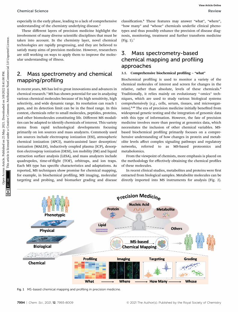
effectiveness of treatment (Fig. 3).31 Traditional imaging tech-nologies normally use magnetic (magnetic resonance imaging(MRI)), radioisotope (positron emission tomography (PET), andautoradiography), or optically active imaging probes (uores-cence imaging and immunohistochemistry (IHC)) to locatetarget molecules.32 However, there is general consensus on themajor issues with these technologies, such as the damage to thebody caused by radioisotopes, lack of MRI sensitivity, and theinstability of enzymes used in IHC and immunouorescence.Furthermore, discerning the precise origin of molecular signalsis not effortless, and some substances with similar structures orproperties are difficult to distinguish. Mass spectrometryimaging (MSI), also called imaging mass spectrometry (IMS),integrates the analytical performance of MS with the micro-information obtained by visualizing chemical distributions insamples of interest.33 It offers high spatial resolution, highquality resolution, and a wide quality detection range. It allowsfor the detection of many molecules, from the range of smallmolecules, such as lipids and metabolites, to biomolecules,such as peptides and proteins, simultaneously on a singleplatform.34 Moreover, the same sample can be subsequentlysubjected to histological examination.35
To date, numerous biological and clinical MSI applicationshave been reported. Among the MSI techniques based ondifferent ion sources, MALDI has been the most popular. Ina typical MALDI analysis, a sample is mixed with a matrix andco-crystallizes aer the solvent is removed. Subsequently, underlaser irradiation, the matrix receives a large amount of energy,which induces desorption and ionization of the molecules inthe sample. Then, a characteristic mass spectrum for eachmolecule is produced. Each laser spot is represented as a pixel,identied by its (x, y) coordinates. Finally, an image of thecomposition and the relative abundance and distribution of thetarget molecule in the sample is obtained by reconstructing theimage using professional image processing soware, in whichthe laser spots are shown as pixels in a gure.36 In terms of dataprocessing, sufficient memory is required to store the massiveamounts of data. The processing time and central processingunit (CPU) use must also be taken into consideration. Some-times, matrix-free methods were employed for imaging inor-ganic materials.37 Nie's group innovatively proposed a laserdesorption/ionization (LDI)-MSI approach to detect carbonnanotubes, graphene oxide and carbon nanodots in mice usingthe carbon cluster ‘ngerprint’ signals,38 and the applicationwas further extended to view in situ doxorubicin release fromnanocarriers like polyethylene glycol (PEG)–MoS2.39 They ach-ieved label-free simultaneous imaging of nanomaterials andreleased drugs. Another commonly used MSI technique issecondary ion MS (SIMS).40,41 In SIMS, the samples are bom-barded with a beam of energized primary ions to induce thedesorption of target molecules, which are usually analyzedusing a quadrupole or TOF mass analyzer. SIMS is a relativelydifficult ionization method to perform and usually can detectonly target molecules with a low mass range (limited to a fewthousand Daltons).42 However, supported by the small diameterof the ion beam, the lateral and depth resolution can be as low
© 2021 The Author(s). Published by the Royal Society of Chemistry
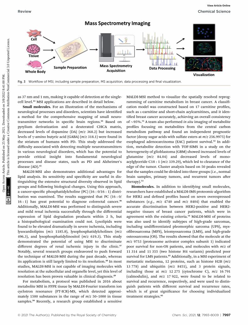
Fig. 3 Workflow of MSI, including sample preparation, MS acquisition, data processing and final visualization.
Review Chemical Science
Ope
n A
cces
s A
rtic
le. P
ublis
hed
on 2
5 M
ay 2
021.
Dow
nloa
ded
on 3
/8/2
022
8:41
:00
PM.
Thi
s ar
ticle
is li
cens
ed u
nder
a C
reat
ive
Com
mon
s A
ttrib
utio
n-N
onC
omm
erci
al 3
.0 U
npor
ted
Lic
ence
.View Article Online
as 37 nm and 1 nm, making it capable of detection at the single-cell level.43 MSI applications are described in detail below.
Small molecules. For an illustration of the mechanisms ofneurological processes and disorders, scientists have identieda method for the comprehensive mapping of small neuro-transmitter networks in specic brain regions.44 Based onpyrylium derivatization and a deuterated CHCA matrix,decreased levels of dopamine (DA) (m/z 368.2) but increasedlevels of g-amino butyric acid (GABA) (m/z 318.1) were found inthe striatum of humans with PD. This study addressed thedifficulty associated with detecting multiple neurotransmittersin various neurological disorders, which has the potential toprovide critical insight into fundamental neurologicalprocesses and disease states, such as PD and Alzheimer'sdisease (AD).45
MALDI-MSI also demonstrates additional advantages forlipid analysis. Its sensitivity and specicity are useful in dis-tinguishing the extensive structural diversity observed in lipidgroups and following biological changes. Using this approach,a cancer-specic phosphatidylcholine (PC) (16 : 0/16 : 1) distri-bution was examined. The results suggested that PC (16 : 0/16 : 1) has great potential to diagnose colorectal cancer.46
Additionally, MALDI-MSI was performed to distinguish severeand mild renal ischemia successfully through the differentialexpression of lipid degradation products within 2 h, buta histopathological examination could not. Lysolipids werefound to be elevated dramatically in severe ischemia, includinglysocardiolipins (m/z 1185.8), lysophosphatidylcholines (m/z496.3), and lysophosphatidylinositol (m/z 619.3). This studydemonstrated the potential of using MSI to discriminatedifferent degrees of renal ischemic injury in the clinic.47
Notably, several research groups endeavored to push forwardthe technique of MALDI-MSI during the past decade, whereasits application is still largely limited to its resolution.48 In moststudies, MALDI-MSI is not capable of imaging molecules withresolution at the subcellular and organelle level, yet this level ofresolution has been proven valuable in clinical diagnosis.49
For metabolism, a protocol was published in 2016 aboutmetabolite MSI in FFPE tissue by MALDI-Fourier transform ioncyclotron resonance (FT-ICR)-MS, which detected approxi-mately 1500 substances in the range of m/z 50–1000 in tissuesamples.50 Recently, a research group established a sensitive
© 2021 The Author(s). Published by the Royal Society of Chemistry
MALDI-MSI method to visualize the spatially resolved reprog-ramming of carnitine metabolism in breast cancer. A classi-cation model was constructed based on 17 carnitine proles,such as L-carnitine and short-chain acylcarnitines, and it iden-tied breast cancer accurately, achieving an overall consistencyof �95%.51 A team also performed in situ imaging of metaboliteproles focusing on metabolites from the central carbonmetabolism pathway and found an independent prognosticfactor (deoxy sugar acids with sulfate esters at m/z 256.9975) foresophageal adenocarcinoma (EAC) patient survival.52 In addi-tion, metabolite detection with TOF-SIMS in a study on theheterogeneity of glioblastoma (GBM) showed increased levels ofglutamine (m/z 84.04) and decreased levels of mono-acylglyceride C18 : 1 (m/z 339.29), which led to clearance of theedge of the tumor. Cluster analysis based on 50 peaks revealedthat the samples could be divided into three groups (i.e., normalbrain samples, primary tumors, and recurrent tumors aertherapy).53
Biomolecules. In addition to identifying small molecules,researchers have established aMALDI-IMS proteomic algorithmfor HER2 and dened proles based on seven overexpressedsubstances (e.g., m/z 4740 and m/z 8404) that enabled theaccurate discrimination between HER2-positive and HER2-negative tissues of breast cancer patients, which were inagreement with the existing criteria.54 MALDI-MSI of proteinswas also used to identify subtypes of high-grade sarcomas,including undifferentiated pleomorphic sarcoma (UPS), myx-obrosarcoma (MFS), leiomyosarcoma (LMS), and high-gradeosteosarcoma (OS). The results showed that the molecule at them/z 9753 (proteasome activator complex subunit 1) indicatedpoor survival for non-OS patients, and molecules with m/z of11 314 and 11 355 (two histone H4 variants) predicted poorsurvival for LMS patients.81 Additionally, in a MSI experiment ofmetastatic melanoma, 12 proteins, such as histone H2B (m/z13 778) and ubiquitin (m/z 8451), and 3 protein signals,including those at m/z 12 275 (cytochrome C), m/z 16 791(calmodulin), and m/z 17 922, were found to be related tosurvival and recurrence, respectively, and were used to distin-guish patients with different survival and recurrence rates,which is of great signicance for choosing individualizedtreatment strategies.82
Chem. Sci., 2021, 12, 7993–8009 | 7997
Chemical Science Review
Ope
n A
cces
s A
rtic
le. P
ublis
hed
on 2
5 M
ay 2
021.
Dow
nloa
ded
on 3
/8/2
022
8:41
:00
PM.
Thi
s ar
ticle
is li
cens
ed u
nder
a C
reat
ive
Com
mon
s A
ttrib
utio
n-N
onC
omm
erci
al 3
.0 U
npor
ted
Lic
ence
.View Article Online
Most interestingly, MSI has also contributed to the study oftumor heterogeneity. Proteomic patterns demonstrated that themolecules atm/z 3445 (alpha-defensin 1),m/z 4156,m/z 8416,m/z 11 368 (acetylated histone H4), and m/z 14 021 (histone H2A)can be used to distinguish tumor subpopulations in the clinic.In addition to inter-sample variations, intra-sample heteroge-neity (i.e., within an individual tumor sample) in patients withintestinal-type gastric cancer is higher than that in patients withprimary breast cancer.83
These examples all involve the detection of intact targetmolecules. In situ tryptic digestion is another method that cancircumvent the inefficient fragmentation of large proteins in thegas phase.84 This bottom-up method also allows both accuratemass and spatial information to be used to relate imaging datafor protein identication. Experimentally, a robotic sprayer isnormally used to apply a homogenous coating of enzyme acrosstissue sections. In an anaplastic glioma research, a set of gradeIII glioma samples was analyzed using this method withMALDI-MSI, and a cluster analysis yielded 3 main distinctpatient subgroups (mainly related to neoplasia, glioma withinammation, and neurogenesis) based on more than 2500proteins.85 Recently, 9 protein-related genes, such as SOX11 (m/z1321.635) and MUC4 (m/z 2057.934), as potential prognosticmarkers, were discovered in triple-negative breast cancer aertryptic digestion.86 However, the stability and efficiency of thetrypsin and the movement of the peptides aer digestion arestill challenges to overcome in bottom-up assays. Recently,mass-tagged probes have been increasingly employed for in situanalysis. The detection capability of MSI becomes particularlyattractive by probe conjugation to heavy metals, for example,lanthanide tags. Lanthanide-tagged mass probes are commonlydeveloped by labeling antibodies with lanthanide isotopes. Tworelated technologies based on lanthanide-tagged mass probes,termed as imaging mass cytometry (IMC) and multiplexed ionbeam imaging (MIBI) have been widely used for tumor micro-environment (TME) investigation.87 In detail, IMC, an approachthat combines mass cytometry with immunocytochemistry(ICC) and IHC techniques, was employed to simultaneously
Table 1 Common MSI techniques and their featuresa
Ion source Ionization type Samples m/z range (Da)S(
MALDI Endogenous, so Solid, liquid >100 000 5AP-MALDI Exogenous, so Solid, liquid >100 000 1SIMS Endogenous, hard Solid, liquid 1–5000 0DESI Exogenous, so Solid, liquid 100–500 1LA-ESI Exogenous, so Liquid >60 000 1LA-ICP Exogenous, hard Solid, liquid 50–500 1NIMS So Solid, liquid 100–5000 0AFAI Exogenous Solid 100–1000 3LESA So Solid 100–2000 1IMb So/hard Solid, liquid 100–2000 2
a Annotation: MALDI: Matrix Assisted Laser Desorption/Ionization; ASpectroscopy; DESI: Desorption Electrospray Ionization; LA-ESI: LaserCoupled Plasma; NIMS: Nanostructure-Initiator Mass Spectrometry; AAnalysis; IM: Ion Mobility Spectrometry. M: metabolite; L: lipid; E: elemsmall molecules. b It is a high-throughput ion separation technology baseld, which can be coupled with MS.
7998 | Chem. Sci., 2021, 12, 7993–8009
quantify the expression of 37 protein markers.88 MIBI, a methodrelying on SIMS, could achieve simultaneous detection ofmultiple proteins up to 100.89 On the other hand, peptide-tag-gedmass probes were designed and prepared for in situ labelingand detection of HER2 in cells and tissue samples.90,91 Lasercleavable probes were developed for co-localization imaging offour types of glycans (i.e., mannose, Galb group, N-acetylglu-cosamine and sialic acids, Neu5Aca2-6Gal(NAc)-R) in breastcancerous and paracancerous tissues.92 These successful inno-vations demonstrate the appeal and promise of MSI in the nearfuture. There are other MSI techniques that are rarely applied inreal-world clinical studies. We summarize them and their majorfeatures in Table 1. Currently, MSI cannot take the place of thecommonly used techniques such as IHC and MRI, even thoughit has great clinical application prospects. Some issues such ascomplex and long-time sample processing, high degree ofspecialization and expensive equipment really limit its clinicaluses. In addition, other challenges such as laborious datamanagement and analysis, lack of reproducibility and standardoperating procedures are also barriers for MSI to be a routineclinical technique.93 Therefore, there is still a need fortremendous effort to translate MSI from basic research toclinical application.
Finally, the above innovative studies mainly focus on single-omics or a class of chemical molecules such as neurotrans-mitters, lipids, metabolites and proteins. While it is usuallydifficult to understand diseases comprehensively at a single-omics layer, integrated MSI studies across multi-omics layersmay bemore attractive in future.94However, sample preparationand data processing are also thought to be the factors hinderingthe application of MSI in multi-omics imaging. Improvement ofhardware and soware performance and simplication ofsample preparation could be a solution.48
3.3. Specic molecular targeting and probing – “how many”
Most biochemical proling and mapping assays typically showhigh coefficients of variation because of their non-targeted
patial resolutionmm) Analytical targets References
–200 M, L, Pep, Pro and others 55 and 56–50 M, L, Pep, Pro and others 57–59.037–0.5 E, L, SCI, other SM 60–63–500 L, D, M, Pep, other SM 64–6600–500 L, M, Pro 67 and 68–10 E 69 and 70.15–50 D, M, Pep, Pro 71 and 7200 SM, whole-body molecular imaging 73 and 74000 D, Pep, L, other SM 75–770–200 L, M, D, Pep, Pro 78–80
P-MALDI: Atmospheric Pressure-MALDI; SIMS: Secondary Ion MassAblation Electrospray Ionization; LA-ICP: Laser Ablation InductivelyFAI: Air Flow-Assisted Ionization; LESA: Liquid Extraction Surfaceent; D: drug; Pep: peptide; Pro: protein; SCI: single cell imaging; SM:ed on the size, shape, and charge of molecular ions within an electric
© 2021 The Author(s). Published by the Royal Society of Chemistry

Fig. 4 Schematic representation of MS-targeted analysis of chemicals using protein as an example.
Fig. 5 Variousmass-tagged probes in MS-based targeted analysis. NP:nanoparticle; GNP-RMT: gold nanoparticle-rhodamine-based masstags.
Review Chemical Science
Ope
n A
cces
s A
rtic
le. P
ublis
hed
on 2
5 M
ay 2
021.
Dow
nloa
ded
on 3
/8/2
022
8:41
:00
PM.
Thi
s ar
ticle
is li
cens
ed u
nder
a C
reat
ive
Com
mon
s A
ttrib
utio
n-N
onC
omm
erci
al 3
.0 U
npor
ted
Lic
ence
.View Article Online
nature and are thus not suitable for routine clinical assays.Reducing the uncertainty and enhancing the accuracy of theinformation on targeted chemical molecules is at the forefrontof precision medicine.95 Data reliability and consistency are thebases for resolving higher-layer clinical challenges. MS-basedtargeted analysis can provide quantitative and solid informa-tion with regard to validated analysis protocols for denedchemicals.96 The most common approach, termed selectivereaction monitoring or multiple reaction monitoring (SRM/MRM), employs the isolation and fragmentation of targetchemicals and the quantication of their specic fragmentsupon the addition of internal standards. These chemicalmolecules can be detected within the fmol concentration rangein complex biological specimens, and abnormal values can beidentied in a short time. Therefore, it comes as no surprisethat MS-based targeted analysis has great potential for use inbiomarker grading and disease classication. For example,amino acids, acylcarnitines, organic acids in newborns andvitamin D (VD) groups in children have been routinely tested inclinical practice.97 These assays and their applications aredescribed in detail in the next section.
Here, we focus on the MS methodology that has beenrecently developed for targeted analysis. In the past few years,targeted proteomics has increasingly become a powerful tool inprotein-like marker analysis.98 In principle, a protein of interestis specically detected at the surrogate peptide level. Thegeneral protocol involves six steps: (1) sample pretreatment andprotein extraction, (2) protein enzymatic digestion, (3) selectionof suitable surrogate peptides as quantitative substitutes for thetarget protein, (4) chemically synthesizing internal standardpeptides with heavy stable isotopes, (5) approach developmentand verication, and nally (6) detection of surrogate peptidesusing SRM/MRM.99 The measurements of the surrogate peptidelevels represent those of the target protein. To date, much workhas been applied to protein and PTM quantication using thisapproach, such as quantication of the p53 family proteins,100
serum transferrin receptor (TfR),101 and histone methylation
© 2021 The Author(s). Published by the Royal Society of Chemistry
species.102 This MS-based approach bridges the gap between thepreliminary discovery of protein markers and their clinicalvalidation (Fig. 4).
The detection of serum thyroglobulin was one of the earliestapplications of targeted proteomics assays in clinical practice.Serum thyroglobulin levels are a signicant cancer biomarkerfor monitoring patients who receive treatment for differentiatedthyroid carcinoma.103 The limit of detection (LOQ) can be as lowas 0.15 ng mL�1. Furthermore, four MS-based assays conductedin four different centers showed better inter-assay agreementthan four different automated immunoassays at the same
Chem. Sci., 2021, 12, 7993–8009 | 7999

Chemical Science Review
Ope
n A
cces
s A
rtic
le. P
ublis
hed
on 2
5 M
ay 2
021.
Dow
nloa
ded
on 3
/8/2
022
8:41
:00
PM.
Thi
s ar
ticle
is li
cens
ed u
nder
a C
reat
ive
Com
mon
s A
ttrib
utio
n-N
onC
omm
erci
al 3
.0 U
npor
ted
Lic
ence
.View Article Online
center, demonstrating the consistency of these targeted pro-teomics assays. In addition to its application in thyroglobulinmeasures, MS-based targeted assays are increasingly used in theclinic.
Notably, even in the presence of direct MS-based targetedanalysis, the clinical evaluation of potential markers and eventhe clinical utility of approved markers are still tempered by theuncertainty stemming from the inherent nature of the targetmolecules. In this context, the use of mass-tagged probes thatcan selectively tag and facilitate the subsequent conversion oftarget information into quantitative MS responses has gainedincreasing interest (Fig. 5).104 Mass-tagged probes are small-molecule reagents (e.g., heavy metals, organic molecules, andpeptides) that usually have higher MS sensitivity and are easy tomanipulate. In general, there are three steps in molecularprobing studies: (1) design and preparation of the mass-taggedprobes, (2) addition of the probes to samples and binding of theprobes to targets, and (3) release of the mass tag and quanti-cation by MS.
For example, membrane proteins are very difficult to quan-tify directly due to their amphiphilic nature.105 Mass-taggedprobes can be used not only for mapping, but also for quanti-cation. The only necessary trick is the use of mass-conjugatedaptamers for target recognition. Aptamers are articial single-stranded DNA/RNA sequences or peptides that can fold intodistinct secondary and tertiary structures to make them suitablefor binding to certain targets with extremely high bindingaffinity and high specicity.106 Aptamers have been increasinglyinvolved in mass-tagged probe design. Using peptide–aptamerprobes, HER2 levels in BT474, SK-BR-3, MCF-7, and MDA-MB-231 cells were correlatively quantied as (10.1 � 2.63) � 105 percell, (9.43� 1.89)� 105 per cell, (0.56� 0.17)� 105 per cell, and(0.53 � 0.09) � 105 per cell, respectively.91
Targeting molecules with trace amounts in biologicalsamples is another challenge because their mass response islower than the LOQ for MS detection. The combination oftargets, probes, or signal amplication methods, such asenzymes, dendrimers, and nanoparticles, with effective targetrecognition can signicantly increase the assay sensitivity. Liuet al. designed an ultrasensitive detection assay for low-abundance protein thrombin and EpCAM using a mass tag ongold nanoparticles for signal amplication in addition toaptamer capture.107 The detection limit of this assay reached100 aM. In another study, Chen et al. used a peptide dendrimerto create a target signal amplication strategy. The signalintensity was �10-fold greater than that without signalamplication.108
In particular, some molecules, such as nucleic acids, arehard to be directly detected by MS due to their complicated andunresolved mass spectra. Nucleic acids consist of only fournucleotides, which means that the risk of producing similarmass spectra from different sequences may be potentiallygreater than that of proteins containing 20 amino acids.109
Although a couple of new techniques, such as MassARRAYSystem using MALDI-TOF MS, can analyze DNA within a massrange of approximately 4500–9000 Da and with a resolution of16 Da, they are still at an early stage of development.110
8000 | Chem. Sci., 2021, 12, 7993–8009
Comparatively, mass-tagged probes have advanced the appli-cation of MS in this eld. Bang et al. created a multiplex DNAdetection assay based on ICP-MS using lanthanide-labeledprobes.111 With this use of heavy metals, the detection capabilityof ICP-MS has become particularly attractive. The advantage ofthe lanthanide tag is that it is not prone to nonspecic bindingand is rare in biological samples. Moreover, it is also small,stable, and heat-resistant, making its application convenient.112
The method detection limits were determined to be 28 amol forHIV, 48 amol for HAV, and 19 amol for HBV.111 Another type ofprobe used for nucleic acid detection is a DNA–peptide probecontaining a tagged reporter peptide. A tryptic cleavage site forpeptide release and a DNA sequence complementary to thetarget miRNA were designed for miRNA detection. Thus, miR-21was evaluated as (4.56 � 1.99) � 108 copies per mg in normaltissue and (1.09 � 0.41) � 109 copies per mg in tumor tissue.112
Furthermore, this strategy using a multiplex DNA–peptideprobe was applied to prole the levels of ve different miRNAs(i.e., miR-21, miR-let7a, miR-200c, miR-125a, and miR-15b).113
The choice of the peptide is critical for these probes. There areseveral empirical principles related to the choice of peptideswith high responses: (1) the peptide length between 6 and 16amino acids, (2) no cysteine or methionine residues, (3) nosingle nucleotide polymorphism or PTMs, (4) no proline residueat the C-terminus with lysine or arginine residues, (5) nocontinuous sequence of lysine or arginine residues (RR, KK, RK,nor KR), and (6) no transmembrane region for membraneproteins.114 Most importantly, the combination of these probesand an amplication strategy allows DNA detection withina sensitivity range appropriate for biologically relevant studies.Therefore, it may be possible to include more nucleic acidmarkers in clinical practice guidelines and recommendationsin the future.
Although these assays have demonstrated great potential inprecision medicine, only a few assays have been translated todate. MS-based clinical assays are still a small portion ofcurrently approved assays, as mentioned below. Continued MStechnological and methodological advances for chemicalmolecules are necessary to sustain this clinical growth.
3.4. Sensitive biomarker grading and disease classication –
“whose”
As the milestones of MS-based chemical mapping pipelines,biomarker grading and disease classication come closest tomeeting the rationale-based goals of precision medicine.Unfortunately, the number of MS assays translated into routineclinical practice is much lower than that of other techniques, asdescribed above. The major reason is that the road fromhypothesis to technology dissemination in the form of clinicalmeasurement procedures is predictably long. Using the serumthyroglobulin assay as an example,115 more than 10 years passedbetween the rst proof-of-principle experiments and the mostrecent version of the assay. Currently, many MS-based assaysare on the road to clinical use. We describe both the clinicalavailability and potential applications of these assays in thissection.
© 2021 The Author(s). Published by the Royal Society of Chemistry

Table 2 HER2 testing and scoring criteria in breast cancera
IHC FISH MS
Denition An antibody-based, semi-quantitative method. Slides areincubated with an antibody directedagainst target protein, and theprotein is nally made visible witha chromogen (e.g.,diaminobenzidine, DAB) resultingin membrane staining
The nucleic acid probes labelleddirectly or indirectly withuorescein were hybridized with thenucleic acid sequences in thesamples to be tested according tothe principle of basecomplementary pairing. Use of thelabeled probe to calculate the HER2gene copy number within the nucleiof tumor cells
A powerful spectrum of chargedatoms, molecules and molecularfragments in order of their m/z. Itcan make matter particles form intoions and separate them, and thenanalyze aer the detection ofintensity
A/D A: 1. Easy to perform and store A: 1. More accurate, reliable,sensitive and reproducible than IHC
A: 1. Different types of biomoleculescan be measured, including lipids,protein etc.
2. Relatively cheap and less timeconsuming
2. The concordance rate amongobservers is higher than that of IHC
2. Modication states andmolecular complex can bequalitatively and quantitativelydetected
3. The protein level can be evaluatedin the context of tissue morphologyusing a microscope
D: 1. More time-consuming andmore expensive compared with IHC
3. High mass accuracy andresolution, high sensitivity,selectivity, multiplexing capability,versatility, and high concentrationranges
D: 1. Several factors may affect thequality of this assay, such as choiceof antibody, tissue xation, etc.
2. Interpretation of FISH assaysneeds well-trained personnel
D: poor at subcellular localization orspatial resolution of proteinexpression
2. Susceptible to considerable inter-observer variability to substantialdiscrepancies in resultinterpretation
Scoring criteria 0: no staining is observed ormembrane staining that isincomplete and is faint/barelyperceptible and within #10% oftumor cells
FISH (dual probe) (2018) Quasi-targeted proteomicsapproach using an aptamer–peptideprobe and RPLC-MS/MS
1+: incomplete membrane stainingthat is faint/barely perceptible andwithin >10% of tumor cells
Positive: 1. HER2/CEP17 ratio $2.0and average HER2 copy number$4.0
0: 7.33 � 3.41 nmol m�2
2+: weak to moderate completemembrane staining observed in>10% of tumor cells
2. HER2/CEP17 ratio $2.0 andaverage HER2 copy number <4.0,IHC 3+
1+: 15.8 � 4.42 nmol m�2
3+: circumferential membranestaining that is complete, intense,and within >10% of tumor cells
3. HER2/CEP17 ratio <2.0 andaverage HER2 copy number $6.0,IHC 2+, 3+
2+/FISH-negative: 18.4 � 7.21 nmolm�2
4. HER2/CEP17 ratio <2.0 andaverage HER2 copy number $4.0and <6.0, IHC 3+
2+/FISH-equivocal: 32.2 � 1.18nmol m�2
Negative: 1. HER2/CEP17 ratio <2.0and average HER2 copy number<4.0
2+/FISH-positive: 48.2 � 4.25 nmolm�2
2. HER2/CEP17 ratio $2.0 andaverage HER2 copy number <4.0,IHC 0, 1+, 2+
HER2 3+: 45.4 � 11.2 nmol m�2
3. HER2/CEP17 ratio <2.0 andaverage HER2 copy number $6.0,IHC 0, 1+4. HER2/CEP17 ratio <2.0 andaverage HER2 copy number $4.0and <6.0, IHC 0, 1+, 2+
a Annotation: A: advantages; D: disadvantages.
© 2021 The Author(s). Published by the Royal Society of Chemistry Chem. Sci., 2021, 12, 7993–8009 | 8001
Review Chemical Science
Ope
n A
cces
s A
rtic
le. P
ublis
hed
on 2
5 M
ay 2
021.
Dow
nloa
ded
on 3
/8/2
022
8:41
:00
PM.
Thi
s ar
ticle
is li
cens
ed u
nder
a C
reat
ive
Com
mon
s A
ttrib
utio
n-N
onC
omm
erci
al 3
.0 U
npor
ted
Lic
ence
.View Article Online

Table 3 Current available official publications about MS-based detection of biomarkers. First row: potential application, second row: officialapproval or recommendation, all others: officially approved laboratory developed test (LDT)a
Guideline Disease Biomarker MS TitleReleasedate Web
FDA COVID-19 SARS-CoV-2 MALDI-MS Emergency useauthorization (EUA)summary SARS-COV-2mass array test
2020 https://www.fda.gov/media/142548/download
FDA Serious infections inhospitalized patients (e.g.,bloodstream infections)
Candida auris (C. auris) MALDI-MS FDA authorizes new use oftest, rst to identify theemerging pathogenCandida auris
2018 https://www.fda.gov/news-events/press-announcements/fda-authorizes-new-use-test-rst-identify-emerging-pathogen-candida-auris
AMP Invasive fungal infections Filamentous fungi andmycobacteria
MALDI-MS Emerging and futureapplications of matrix-assisted laser desorptionionization time-of-ight(MALDI-TOF) massspectrometry in the clinicalmicrobiology laboratory:a report of the associationfor molecular pathology
2016 https://www.sciencedirect.com/science/article/pii/S1525157816301441?via%3Dihub
ESCMID Bloodstream infection (BSI) Bacteria, fungi, parasites,and viruses
MALDI-MS Bloodstream infections –standard and progress inpathogen diagnostics
2020 https://www.sciencedirect.com/science/article/abs/pii/S1198743X19306160?via%3Dihub
ESCMID Bloodstream infection (BSI) Pathogen MALDI-MS Microbiologicaldiagnostics of bloodstreaminfections in Europe – anESGBIES survey
2019 https://pubmed.ncbi.nlm.nih.gov/30980927/
CDC E. meningoseptica Elizabethkingia MALDI-MS Determination ofElizabethkingia diversity byMALDI-TOF massspectrometry and whole-genome sequencing
2017 https://wwwnc.cdc.gov/eid/article/23/2/16-1321-f1
CDC Corynebacteriumpseudodiphtheriticuminfection in children withcystic brosis (CF)
C. pseudodiphtheriticumstrains
MALDI-MS Outbreak ofCorynebacteriumpseudodiphtheriticuminfection in cystic brosispatients, France
2010 https://wwwnc.cdc.gov/eid/article/16/8/10-0193_article
CDC Amino acid disorders (e.g.,PKU, maple syrup urinedisease, andhomocystinuria), fatty acidoxidation disorders (e.g.,medium-chain acyl-CoAdehydrogenase [MCAD]deciency) and otherorganic acid disorders
Amino acids, fatty acids,and organic acids
MALDI-MS Using tandem massspectrometry for metabolicdisease screening amongnewborns
2001 https://www.cdc.gov/mmwr/preview/mmwrhtml/rr5003a1.htm
NACB Multiple inborn errors ofmetabolism
Amino acid, acylcarnitine,organic acid
MALDI-MS National academy ofclinical biochemistrylaboratory medicinepractice guidelines: follow-up testing for metabolicdisease identied byexpanded newbornscreening using tandemmass spectrometry;executive summary
2009 https://academic.oup.com/clinchem/article/55/9/1615/5629176
8002 | Chem. Sci., 2021, 12, 7993–8009 © 2021 The Author(s). Published by the Royal Society of Chemistry
Chemical Science Review
Ope
n A
cces
s A
rtic
le. P
ublis
hed
on 2
5 M
ay 2
021.
Dow
nloa
ded
on 3
/8/2
022
8:41
:00
PM.
Thi
s ar
ticle
is li
cens
ed u
nder
a C
reat
ive
Com
mon
s A
ttrib
utio
n-N
onC
omm
erci
al 3
.0 U
npor
ted
Lic
ence
.View Article Online
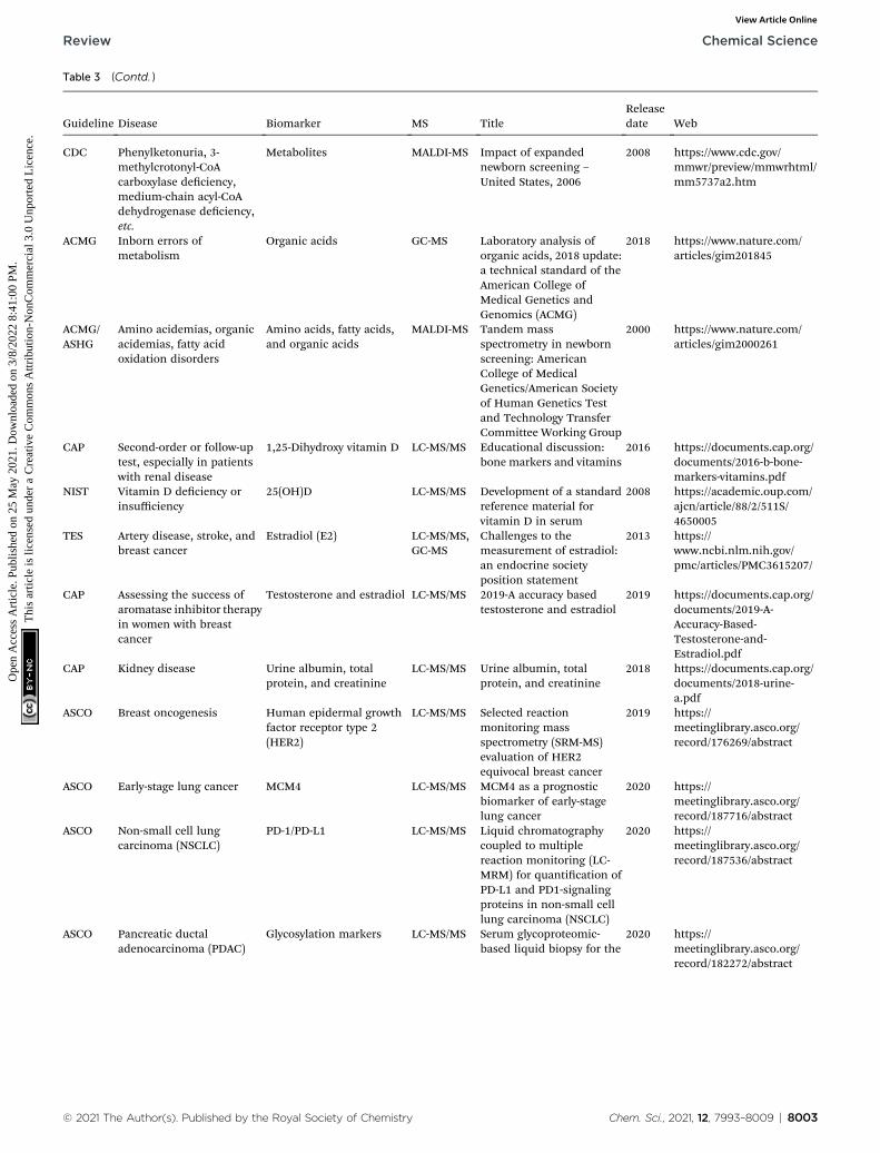
Table 3 (Contd. )
Guideline Disease Biomarker MS TitleReleasedate Web
CDC Phenylketonuria, 3-methylcrotonyl-CoAcarboxylase deciency,medium-chain acyl-CoAdehydrogenase deciency,etc.
Metabolites MALDI-MS Impact of expandednewborn screening –United States, 2006
2008 https://www.cdc.gov/mmwr/preview/mmwrhtml/mm5737a2.htm
ACMG Inborn errors ofmetabolism
Organic acids GC-MS Laboratory analysis oforganic acids, 2018 update:a technical standard of theAmerican College ofMedical Genetics andGenomics (ACMG)
2018 https://www.nature.com/articles/gim201845
ACMG/ASHG
Amino acidemias, organicacidemias, fatty acidoxidation disorders
Amino acids, fatty acids,and organic acids
MALDI-MS Tandem massspectrometry in newbornscreening: AmericanCollege of MedicalGenetics/American Societyof Human Genetics Testand Technology TransferCommittee Working Group
2000 https://www.nature.com/articles/gim2000261
CAP Second-order or follow-uptest, especially in patientswith renal disease
1,25-Dihydroxy vitamin D LC-MS/MS Educational discussion:bone markers and vitamins
2016 https://documents.cap.org/documents/2016-b-bone-markers-vitamins.pdf
NIST Vitamin D deciency orinsufficiency
25(OH)D LC-MS/MS Development of a standardreference material forvitamin D in serum
2008 https://academic.oup.com/ajcn/article/88/2/511S/4650005
TES Artery disease, stroke, andbreast cancer
Estradiol (E2) LC-MS/MS,GC-MS
Challenges to themeasurement of estradiol:an endocrine societyposition statement
2013 https://www.ncbi.nlm.nih.gov/pmc/articles/PMC3615207/
CAP Assessing the success ofaromatase inhibitor therapyin women with breastcancer
Testosterone and estradiol LC-MS/MS 2019-A accuracy basedtestosterone and estradiol
2019 https://documents.cap.org/documents/2019-A-Accuracy-Based-Testosterone-and-Estradiol.pdf
CAP Kidney disease Urine albumin, totalprotein, and creatinine
LC-MS/MS Urine albumin, totalprotein, and creatinine
2018 https://documents.cap.org/documents/2018-urine-a.pdf
ASCO Breast oncogenesis Human epidermal growthfactor receptor type 2(HER2)
LC-MS/MS Selected reactionmonitoring massspectrometry (SRM-MS)evaluation of HER2equivocal breast cancer
2019 https://meetinglibrary.asco.org/record/176269/abstract
ASCO Early-stage lung cancer MCM4 LC-MS/MS MCM4 as a prognosticbiomarker of early-stagelung cancer
2020 https://meetinglibrary.asco.org/record/187716/abstract
ASCO Non-small cell lungcarcinoma (NSCLC)
PD-1/PD-L1 LC-MS/MS Liquid chromatographycoupled to multiplereaction monitoring (LC-MRM) for quantication ofPD-L1 and PD1-signalingproteins in non-small celllung carcinoma (NSCLC)
2020 https://meetinglibrary.asco.org/record/187536/abstract
ASCO Pancreatic ductaladenocarcinoma (PDAC)
Glycosylation markers LC-MS/MS Serum glycoproteomic-based liquid biopsy for the
2020 https://meetinglibrary.asco.org/record/182272/abstract
© 2021 The Author(s). Published by the Royal Society of Chemistry Chem. Sci., 2021, 12, 7993–8009 | 8003
Review Chemical Science
Ope
n A
cces
s A
rtic
le. P
ublis
hed
on 2
5 M
ay 2
021.
Dow
nloa
ded
on 3
/8/2
022
8:41
:00
PM.
Thi
s ar
ticle
is li
cens
ed u
nder
a C
reat
ive
Com
mon
s A
ttrib
utio
n-N
onC
omm
erci
al 3
.0 U
npor
ted
Lic
ence
.View Article Online

Table 3 (Contd. )
Guideline Disease Biomarker MS TitleReleasedate Web
detection of pancreaticductal adenocarcinoma
ASCO Pancreatic cancer Alpha-1-acid glycoprotein 1(AGP1)
LC-MS/MS Alpha-1-acid glycoprotein 1(AGP1) as a novelbiomarker for pancreaticcancer
2019 https://meetinglibrary.asco.org/record/174117/abstract
a Annotation: FDA: Food and Drug Administration; AMP: Association for Molecular Pathology; ESCMID: European Society of Clinical Microbiologyand Infectious Diseases; CDC: Centers for Disease Control and Prevention; NACB: National Academy of Clinical Biochemistry; ACMG: AmericanCollege of Medical Genetics and Genomics; ASHG: American Society of Human Genetics; CAP: College of American Pathologists; NIST: TheNational Institute of Standards and Technology; TES: The Endocrine Society; ASCO: American Society of Clinical Oncology.
Chemical Science Review
Ope
n A
cces
s A
rtic
le. P
ublis
hed
on 2
5 M
ay 2
021.
Dow
nloa
ded
on 3
/8/2
022
8:41
:00
PM.
Thi
s ar
ticle
is li
cens
ed u
nder
a C
reat
ive
Com
mon
s A
ttrib
utio
n-N
onC
omm
erci
al 3
.0 U
npor
ted
Lic
ence
.View Article Online
In recent decades, as a revolutionary technology, newbornscreening by MS has been increasingly used and added topublic health policies, which has encouraged more newbornscreening studies for early treatment.116 Clinical MS testing,including the measurement of amino acids, fatty acid oxida-tion levels, and organic acids for screening more than 30inherited conditions, is relatively mature. Hundreds ofmetabolite molecules have been identied in blood spots andurine. Additionally, the acquisition of these samples is cost-effective, more secure and friendlier to infants. For example,mucopolysaccharide is a type of multisystem disease causedby a lysosomal storage disorder that can lead to death insevere cases. Some enzymes, especially a-L-iduronidase,117 arequantied in the blood by MS, and then, the screen-positivesamples are conrmed by gene sequencing. This method,together with gene sequencing technology, has been used innewborn screening at numerous institutions and hasbeneted patients in recent years.118
In addition to the chemicals analyzed in newborn screening,MS detection is the most commonly used method for VD assaysdue to the similar structures and characteristics of VD analogs.It is well known that VD deciency causes not only rickets, butalso autoimmune, cardio-cerebrovascular, and reproduction-related diseases. Among all VD metabolic compounds, 25-OH-D3 is the most reliable indicator of VD deciency because of itslong half-life and stability in circulation.119 Patients with VDdeciency detected by MS normally maintain serum 25-OH-D3
values less than 20 ng mL�1.120,121 In another study, CYP24A1was shown to be a key enzyme regulating the conversion of 25-OH-D3 into the VD3 metabolite 24,25-(OH)2D3.122 In CYP24A1-mutant idiopathic infantile hypercalcemia (IIH) serum, 24,25-(OH)2D3 is lower than 1.25 nmol L�1, which is only approxi-mately one-h of that of normal individuals.123
For some markers, the MS-based approach has shown betterperformance in disease grading and has begun to override thetraditional boundaries of disease classications. A typicalexample is HER2 (Table 2). IHC combined with uorescence insitu hybridization (FISH) is the most commonly used method ofgrading tumors in clinical practice, as recommended by theAmerican Society of Clinical Oncology (ASCO) and College ofAmerican Pathologists (CAP) guidelines in recent years.
8004 | Chem. Sci., 2021, 12, 7993–8009
Comparatively, MS quantication provides more accurateresults of HER2 and more reliable prognostic information(Table 3). Paolo Nuciforo's team quantied HER2 protein levelsin FFPE tissue samples and suggested that patients with anHER2 threshold of 740 amol mg�1 can benet from monoclonalantibody treatment in breast cancer.124 Chen's laboratorydeveloped an aptamer–peptide probe for MS assay, andmatched pairs of breast tissue samples were subjected toanalysis. The level of HER2 in the tissue was quantied accu-rately: the concentration of HER2 0 was 7.33 � 3.41 nmol m�2,the concentration of HER2 1+ was 15.8 � 4.42 nmol m�2, theconcentration of HER2 2+/FISH-negative was 18.4 � 7.21 nmolm�2, the concentration of HER2 2+/FISH-equivocal was 32.2 �1.18 nmol m�2, the concentration of HER2 2+/FISH-positive was48.2 � 4.25 nmol m�2, and the concentration of HER2 3+ was45.4 � 11.2 nmol m�2. The reference HER2 interval was calcu-lated from 3.52 (90% CI, 1.31–5.74) nmol m�2 to 19.9 (90% CI,17.7–22.2) nmol m�2. The samples with values exceeding thisrange were considered positive.91 Clinical HER2 testing ina more accurate manner may be achieved in the near future.False positives can be reduced, and treatment selection can bethus more precise.
Another study demonstrated that biomarker ngerprints canbe predictive of responses to immunotherapy and survival inthe supposedly homogeneous group of breast cancer patientsand allows for the stratication of patients.125 Proteins thatcontribute the most to the proteotype-based classication,including INPP4B, CDK1, and ERBB2, are associated with theestrogen receptor (ER) status, HER2 status, and tumor gradestatus. It was conrmed that the classication of breast cancersubtypes at the protein level can lead to more accurate patientstratication than the “conventional subtypes”.
In addition to these clinically approved markers, there aremany MS-based chemical molecules generated in clinicalstudies waiting for clinical translation. A very important linkbetween clinicians and researchers is required to shorten theexperimental cycle of these potential biomarkers and to subjectthem to hospital laboratory testing. Frataxin in platelets wassuggested for the diagnosis of the rare disease Friedreich'sataxia (FA).126 This assay, which is based on a two-dimensionalnano-UPLC, can sensitively and specically differentiate
© 2021 The Author(s). Published by the Royal Society of Chemistry

Review Chemical Science
Ope
n A
cces
s A
rtic
le. P
ublis
hed
on 2
5 M
ay 2
021.
Dow
nloa
ded
on 3
/8/2
022
8:41
:00
PM.
Thi
s ar
ticle
is li
cens
ed u
nder
a C
reat
ive
Com
mon
s A
ttrib
utio
n-N
onC
omm
erci
al 3
.0 U
npor
ted
Lic
ence
.View Article Online
between control groups and FA patients. The frataxin level inplatelets from the control groups was 9.4 � 2.6 pg mg�1 protein,whereas the level in the platelets from FA patients was 74.5%lower than that of the controls at 2.4 � 0.6 pg mg�1.
The diagnostic performance of both amyloid-b and Tau,which are indicative of AD, was evaluated.127,128 Tau in thecerebrospinal uid (CSF) of patients was signicantly greaterthan that in healthy controls (mean of the control ¼ 17 pmolL�1, and AD ¼ 29 pmol L�1). Plasma amyloid-b biomarkersobtained by immunoprecipitation coupled with MS have beenassayed. The cutoff value of CSF Ab1–42 was 544 ng L�1, which isbelow the abnormal Ab1–42 level. Another potential marker forAD diagnosis is plasma lipoproteome. Li et al. measured 120tryptic peptides from 79 plasma lipoproteins by a MS-basedtargeted analysis. Aer a proof-of concept case–control study ofAD patients and controls, much more differentially expressedtryptic peptides were found in plasma lipoproteins than inimmunodepleted plasma, suggesting that plasma lipoproteomemay be more suitable for AD diagnosis.129 As indicated bya dual-probe MS approach, breast cancer patients with highMUC1-specic terminal Gal/GalNAc showed signicantlyincreased metastatic potential and poorer prognosis than thosewith low expression. Specically, the amount of MUC1-specicterminal Gal/GalNAc was quantied to be (0.96 � 0.09) � 103
nmol m�2 in the normal tissue and (1.74 � 0.16) � 103 nmolm�2 in the tumor tissue (p < 0.001).130
In clinical testing, the other widely applied MS technique isMALDI-TOF MS, which has completely changed the routineidentication of bacteria, fungi, and viruses in clinical micro-biology laboratories.131 This technique can provide uniquesignatures for each microorganism. The chief advantages ofusing MALDI-TOF technology indicates that it may potentiallyreplace conventional microbe culture because it is reliable andmuch quicker (from several days to less than an hour).132
Specic standardized procedures have been established formicroorganisms, and the relevant systems have receivedapproval from official institutions and organizations, such asthe US FDA. Moreover, �15 000 prokaryotes and �70 000 fungihave been documented in MALDI-TOF-MS databases.133 MS hasgreater application prospects in the detection of clinicalmicrobial species in the clinic.
Though MS assays clearly produce more accurate resultsthan other approaches, they still have disadvantages, such ascostly instrumentation, resulting in low levels of use in pastlaboratory medicine. Currently, there is a great expansion in theuse of MS in clinical laboratories. We expect that large-scaleclinical trials will be established to determine the thresholds ofvarious chemicals for detecting different levels or types ofdiseases.
3.5. Others
In a variety of chemical molecules, there is one type of moleculethat we do not discuss in this review. In contrast to the chem-icals we presented here, most drugs are exogenous. The spatialdistribution and expression levels of these drugs and theirmetabolites in various tissues and organs of the body can
© 2021 The Author(s). Published by the Royal Society of Chemistry
provide powerful evidence for their pharmacokinetics, phar-macology, and toxicology. Specically, MS-based drug analysiscan provide the spatial distribution and expression levels ofdrugs and their metabolites in various tissues and organs of thebody. This information can be used in the study of drugmetabolism pharmacokinetics/pharmacodynamics, drugdistribution, toxicology, doping control, pain management,workplace drug testing, etc.134 All these characteristics can alsobe resolved by MS-based chemical mapping and prolingmethods, and we may describe them in detail elsewhere.
4. Conclusion and perspectives
In precision medicine, integrating emerging research on thechemical makeup of diseases with clinical data on individualpatients can drive the development of a more accurate classi-cation of diseases and, ultimately, enhance diagnosis andtreatment. However, there is a bumpy road between the masteryof chemical knowledge and its implementation in clinicalpractice. Previous studies have focused on higher-layer datawhile neglecting the basic chemical information underneath it.Fortunately, MS-based chemical mapping and prolingcompensates for each bump in this road, from molecularproling to disease grading. Although its involvement inprecision medicine is far from satisfactory, MS-based chemicalmapping/proling strategies have demonstrated potential andpromise in this eld. The urgent need is convincing chemists toparticipate in this evolution. Considering the conuence of therelevant information from physical and biological layers, webelieve the understanding of diseases will be achieved at themolecular level and that the gap between chemical research andclinical practice will be closed.
Author contributions
YH and ZW contributed equally to this work. All of the authorsparticipated in this review work. YC proposed and designed thereview. YH, ZW, LL, JZ, DZ and JZ collected the literatures andprepared the dra. MX, YZ and FX designed and constructedthe gures and tables. YC, YH, and ZW nally revised andproofed the review.
Conflicts of interest
There are no conicts to declare.
Acknowledgements
National Natural Science Foundation of China (21722504, and21675089), SEU-NJMU Cooperation Project (2242017K3DN12),SEU-NJMU-CPU Cooperation Project (2242019K3DNZ2),Primary Research & Development Plan of Jiangsu Province(BE2018725), and Science and Technology Development Fundof NJMU (NMUD2019009) are gratefully acknowledged.
Chem. Sci., 2021, 12, 7993–8009 | 8005
Chemical Science Review
Ope
n A
cces
s A
rtic
le. P
ublis
hed
on 2
5 M
ay 2
021.
Dow
nloa
ded
on 3
/8/2
022
8:41
:00
PM.
Thi
s ar
ticle
is li
cens
ed u
nder
a C
reat
ive
Com
mon
s A
ttrib
utio
n-N
onC
omm
erci
al 3
.0 U
npor
ted
Lic
ence
.View Article Online
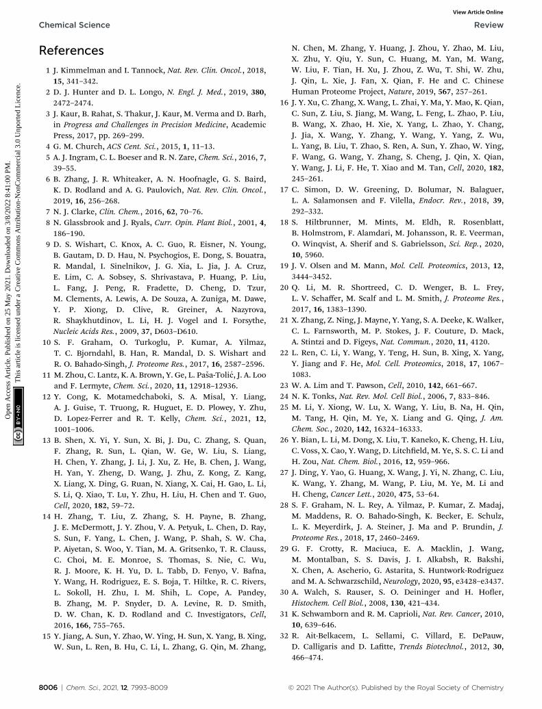
References
1 J. Kimmelman and I. Tannock, Nat. Rev. Clin. Oncol., 2018,15, 341–342.
2 D. J. Hunter and D. L. Longo, N. Engl. J. Med., 2019, 380,2472–2474.
3 J. Kaur, B. Rahat, S. Thakur, J. Kaur, M. Verma and D. Barh,in Progress and Challenges in Precision Medicine, AcademicPress, 2017, pp. 269–299.
4 G. M. Church, ACS Cent. Sci., 2015, 1, 11–13.5 A. J. Ingram, C. L. Boeser and R. N. Zare, Chem. Sci., 2016, 7,39–55.
6 B. Zhang, J. R. Whiteaker, A. N. Hoofnagle, G. S. Baird,K. D. Rodland and A. G. Paulovich, Nat. Rev. Clin. Oncol.,2019, 16, 256–268.
7 N. J. Clarke, Clin. Chem., 2016, 62, 70–76.8 N. Glassbrook and J. Ryals, Curr. Opin. Plant Biol., 2001, 4,186–190.
9 D. S. Wishart, C. Knox, A. C. Guo, R. Eisner, N. Young,B. Gautam, D. D. Hau, N. Psychogios, E. Dong, S. Bouatra,R. Mandal, I. Sinelnikov, J. G. Xia, L. Jia, J. A. Cruz,E. Lim, C. A. Sobsey, S. Shrivastava, P. Huang, P. Liu,L. Fang, J. Peng, R. Fradette, D. Cheng, D. Tzur,M. Clements, A. Lewis, A. De Souza, A. Zuniga, M. Dawe,Y. P. Xiong, D. Clive, R. Greiner, A. Nazyrova,R. Shaykhutdinov, L. Li, H. J. Vogel and I. Forsythe,Nucleic Acids Res., 2009, 37, D603–D610.
10 S. F. Graham, O. Turkoglu, P. Kumar, A. Yilmaz,T. C. Bjorndahl, B. Han, R. Mandal, D. S. Wishart andR. O. Bahado-Singh, J. Proteome Res., 2017, 16, 2587–2596.
11 M. Zhou, C. Lantz, K. A. Brown, Y. Ge, L. Pasa-Tolic, J. A. Looand F. Lermyte, Chem. Sci., 2020, 11, 12918–12936.
12 Y. Cong, K. Motamedchaboki, S. A. Misal, Y. Liang,A. J. Guise, T. Truong, R. Huguet, E. D. Plowey, Y. Zhu,D. Lopez-Ferrer and R. T. Kelly, Chem. Sci., 2021, 12,1001–1006.
13 B. Shen, X. Yi, Y. Sun, X. Bi, J. Du, C. Zhang, S. Quan,F. Zhang, R. Sun, L. Qian, W. Ge, W. Liu, S. Liang,H. Chen, Y. Zhang, J. Li, J. Xu, Z. He, B. Chen, J. Wang,H. Yan, Y. Zheng, D. Wang, J. Zhu, Z. Kong, Z. Kang,X. Liang, X. Ding, G. Ruan, N. Xiang, X. Cai, H. Gao, L. Li,S. Li, Q. Xiao, T. Lu, Y. Zhu, H. Liu, H. Chen and T. Guo,Cell, 2020, 182, 59–72.
14 H. Zhang, T. Liu, Z. Zhang, S. H. Payne, B. Zhang,J. E. McDermott, J. Y. Zhou, V. A. Petyuk, L. Chen, D. Ray,S. Sun, F. Yang, L. Chen, J. Wang, P. Shah, S. W. Cha,P. Aiyetan, S. Woo, Y. Tian, M. A. Gritsenko, T. R. Clauss,C. Choi, M. E. Monroe, S. Thomas, S. Nie, C. Wu,R. J. Moore, K. H. Yu, D. L. Tabb, D. Fenyo, V. Bafna,Y. Wang, H. Rodriguez, E. S. Boja, T. Hiltke, R. C. Rivers,L. Sokoll, H. Zhu, I. M. Shih, L. Cope, A. Pandey,B. Zhang, M. P. Snyder, D. A. Levine, R. D. Smith,D. W. Chan, K. D. Rodland and C. Investigators, Cell,2016, 166, 755–765.
15 Y. Jiang, A. Sun, Y. Zhao, W. Ying, H. Sun, X. Yang, B. Xing,W. Sun, L. Ren, B. Hu, C. Li, L. Zhang, G. Qin, M. Zhang,
8006 | Chem. Sci., 2021, 12, 7993–8009
N. Chen, M. Zhang, Y. Huang, J. Zhou, Y. Zhao, M. Liu,X. Zhu, Y. Qiu, Y. Sun, C. Huang, M. Yan, M. Wang,W. Liu, F. Tian, H. Xu, J. Zhou, Z. Wu, T. Shi, W. Zhu,J. Qin, L. Xie, J. Fan, X. Qian, F. He and C. ChineseHuman Proteome Project, Nature, 2019, 567, 257–261.
16 J. Y. Xu, C. Zhang, X. Wang, L. Zhai, Y. Ma, Y. Mao, K. Qian,C. Sun, Z. Liu, S. Jiang, M. Wang, L. Feng, L. Zhao, P. Liu,B. Wang, X. Zhao, H. Xie, X. Yang, L. Zhao, Y. Chang,J. Jia, X. Wang, Y. Zhang, Y. Wang, Y. Yang, Z. Wu,L. Yang, B. Liu, T. Zhao, S. Ren, A. Sun, Y. Zhao, W. Ying,F. Wang, G. Wang, Y. Zhang, S. Cheng, J. Qin, X. Qian,Y. Wang, J. Li, F. He, T. Xiao and M. Tan, Cell, 2020, 182,245–261.
17 C. Simon, D. W. Greening, D. Bolumar, N. Balaguer,L. A. Salamonsen and F. Vilella, Endocr. Rev., 2018, 39,292–332.
18 S. Hiltbrunner, M. Mints, M. Eldh, R. Rosenblatt,B. Holmstrom, F. Alamdari, M. Johansson, R. E. Veerman,O. Winqvist, A. Sherif and S. Gabrielsson, Sci. Rep., 2020,10, 5960.
19 J. V. Olsen and M. Mann, Mol. Cell. Proteomics, 2013, 12,3444–3452.
20 Q. Li, M. R. Shortreed, C. D. Wenger, B. L. Frey,L. V. Schaffer, M. Scalf and L. M. Smith, J. Proteome Res.,2017, 16, 1383–1390.
21 X. Zhang, Z. Ning, J. Mayne, Y. Yang, S. A. Deeke, K. Walker,C. L. Farnsworth, M. P. Stokes, J. F. Couture, D. Mack,A. Stintzi and D. Figeys, Nat. Commun., 2020, 11, 4120.
22 L. Ren, C. Li, Y. Wang, Y. Teng, H. Sun, B. Xing, X. Yang,Y. Jiang and F. He, Mol. Cell. Proteomics, 2018, 17, 1067–1083.
23 W. A. Lim and T. Pawson, Cell, 2010, 142, 661–667.24 N. K. Tonks, Nat. Rev. Mol. Cell Biol., 2006, 7, 833–846.25 M. Li, Y. Xiong, W. Lu, X. Wang, Y. Liu, B. Na, H. Qin,
M. Tang, H. Qin, M. Ye, X. Liang and G. Qing, J. Am.Chem. Soc., 2020, 142, 16324–16333.
26 Y. Bian, L. Li, M. Dong, X. Liu, T. Kaneko, K. Cheng, H. Liu,C. Voss, X. Cao, Y. Wang, D. Litcheld, M. Ye, S. S. C. Li andH. Zou, Nat. Chem. Biol., 2016, 12, 959–966.
27 J. Ding, Y. Yao, G. Huang, X. Wang, J. Yi, N. Zhang, C. Liu,K. Wang, Y. Zhang, M. Wang, P. Liu, M. Ye, M. Li andH. Cheng, Cancer Lett., 2020, 475, 53–64.
28 S. F. Graham, N. L. Rey, A. Yilmaz, P. Kumar, Z. Madaj,M. Maddens, R. O. Bahado-Singh, K. Becker, E. Schulz,L. K. Meyerdirk, J. A. Steiner, J. Ma and P. Brundin, J.Proteome Res., 2018, 17, 2460–2469.
29 G. F. Crotty, R. Maciuca, E. A. Macklin, J. Wang,M. Montalban, S. S. Davis, J. I. Alkabsh, R. Bakshi,X. Chen, A. Ascherio, G. Astarita, S. Huntwork-Rodriguezand M. A. Schwarzschild, Neurology, 2020, 95, e3428–e3437.
30 A. Walch, S. Rauser, S. O. Deininger and H. Hoer,Histochem. Cell Biol., 2008, 130, 421–434.
31 K. Schwamborn and R. M. Caprioli, Nat. Rev. Cancer, 2010,10, 639–646.
32 R. Ait-Belkacem, L. Sellami, C. Villard, E. DePauw,D. Calligaris and D. Latte, Trends Biotechnol., 2012, 30,466–474.
© 2021 The Author(s). Published by the Royal Society of Chemistry

Review Chemical Science
Ope
n A
cces
s A
rtic
le. P
ublis
hed
on 2
5 M
ay 2
021.
Dow
nloa
ded
on 3
/8/2
022
8:41
:00
PM.
Thi
s ar
ticle
is li
cens
ed u
nder
a C
reat
ive
Com
mon
s A
ttrib
utio
n-N
onC
omm
erci
al 3
.0 U
npor
ted
Lic
ence
.View Article Online
33 L. A. McDonnell and R. M. A. Heeren, Mass Spectrom. Rev.,2007, 26, 606–643.
34 E. H. Seeley and R. M. Caprioli, Trends Biotechnol., 2011, 29,136–143.
35 P. Chaurand, S. A. Schwartz, D. Billheimer, B. J. Xu,A. Crecelius and R. M. Caprioli, Anal. Chem., 2004, 76,1145–1155.
36 P. Rafols, D. Vilalta, J. Brezmes, N. Canellas, E. Del Castillo,O. Yanes, N. Ramirez and X. Correig, Mass Spectrom. Rev.,2018, 37, 281–306.
37 D. S. Peterson, Mass Spectrom. Rev., 2007, 26, 19–34.38 S. Chen, C. Xiong, H. Liu, Q. Wan, J. Hou, Q. He, A. Badu-
Tawiah and Z. Nie, Nat. Nanotechnol., 2015, 10, 176–182.39 J. Xue, H. Liu, S. Chen, C. Xiong, L. Zhan, J. Sun and Z. Nie,
Sci. Adv., 2018, 4, eaat9039.40 J. Lu, X. Hua and Y. T. Long, Analyst, 2017, 142, 691–699.41 H. W. Li, X. Hua and Y. T. Long, Anal. Bioanal. Chem., 2019,
411, 4025–4030.42 H. Tian, L. J. Sparvero, A. A. Amoscato, A. Bloom, H. Bayir,
V. E. Kagan and N. Winograd, Anal. Chem., 2017, 89, 4611–4619.
43 J. Malherbe, F. Penen, M. P. Isaure, J. Frank, G. Hause,D. Dobritzsch, E. Gontier, F. Horreard, F. Hillion andD. Schaumloffel, Anal. Chem., 2016, 88, 7130–7136.
44 M. Shariatgorji, A. Nilsson, E. Fridjonsdottir,T. Vallianatou, P. Kallback, L. Katan, J. Savmarker,I. Mantas, X. Zhang, E. Bezard, P. Svenningsson,L. R. Odell and P. E. Andren, Nat. Methods, 2019, 16,1021–1028.
45 M. Shariatgorji, A. Nilsson, R. J. Goodwin, P. Kallback,N. Schintu, X. Zhang, A. R. Crossman, E. Bezard,P. Svenningsson and P. E. Andren, Neuron, 2014, 84, 697–707.
46 N. Kurabe, T. Hayasaka, M. Ogawa, N. Masaki, Y. Ide,M. Waki, T. Nakamura, K. Kurachi, T. Kahyo,K. Shinmura, Y. Midorikawa, Y. Sugiyama, M. Setou andH. Sugimura, Cancer Sci., 2013, 104, 1295–1302.
47 T. C. van Smaalen, S. R. Ellis, N. E. Mascini, T. P. Siegel,B. Cillero-Pastor, L. M. Hillen, L. W. E. van Heurn,C. J. Peutz-Kootstra and R. M. A. Heeren, Anal. Chem.,2019, 91, 3575–3581.
48 K. Scupakova, B. Balluff, C. Tressler, T. Adelaja,R. M. A. Heeren, K. Glunde and G. Ertaylan, Clin. Chem.Lab. Med., 2020, 58, 914–929.
49 P. Agui-Gonzalez, S. Jahne and N. T. N. Phan, J. Anal. At.Spectrom., 2019, 34, 1355–1368.
50 A. Ly, A. Buck, B. Balluff, N. Sun, K. Gorzolka,A. Feuchtinger, K. P. Janssen, P. J. Kuppen, C. J. van deVelde, G. Weirich, F. Erlmeier, R. Langer, M. Aubele,H. Zitzelsberger, L. McDonnell, M. Aichler and A. Walch,Nat. Protoc., 2016, 11, 1428–1443.
51 C. Sun, F. Wang, Y. Zhang, J. Yu and X. Wang, Theranostics,2020, 10, 7070–7082.
52 A. Buck, A. Ly, B. Balluff, N. Sun, K. Gorzolka,A. Feuchtinger, K. P. Janssen, P. J. Kuppen, C. J. van deVelde, G. Weirich, F. Erlmeier, R. Langer, M. Aubele,
© 2021 The Author(s). Published by the Royal Society of Chemistry
H. Zitzelsberger, M. Aichler and A. Walch, J. Pathol., 2015,237, 123–132.
53 S. K. Gularyan, A. A. Gulin, K. S. Anufrieva, V. O. Shender,M. I. Shakhparonov, S. Bastola, N. V. Antipova,T. F. Kovalenko, Y. P. Rubtsov, Y. A. Latyshev,A. A. Potapov and M. S. Pavlyukov, Mol. Cell. Proteomics,2020, 19, 960–970.
54 B. Balluff, M. Elsner, A. Kowarsch, S. Rauser, S. Meding,C. Schuhmacher, M. Feith, K. Herrmann, C. Rocken,R. M. Schmid, H. Hoer, A. Walch and M. P. Ebert, J.Proteome Res., 2010, 9, 6317–6322.
55 R. M. Caprioli, T. B. Farmer and J. Gile, Anal. Chem., 1997,69, 4751–4760.
56 J. L. Norris and R. M. Caprioli, Chem. Rev., 2013, 113, 2309–2342.
57 V. V. Laiko, M. A. Baldwin and A. L. Burlingame, Anal.Chem., 2000, 72, 652–657.
58 M. Kompauer, S. Heiles and B. Spengler, Nat. Methods,2017, 14, 90–96.
59 M. Kompauer, S. Heiles and B. Spengler, Nat. Methods,2017, 14, 1156–1158.
60 J. Matsuo, S. Torii, K. Yamauchi, K. Wakamoto,M. Kusakari, S. Nakagawa, M. Fujii, T. Aoki and T. Seki,Appl. Phys. Express, 2014, 7, 056602.
61 T. B. Angerer, M. Dowlatshahi Pour, P. Malmberg andJ. S. Fletcher, Anal. Chem., 2015, 87, 4305–4313.
62 M. K. Passarelli, A. Pirkl, R. Moellers, D. Grinfeld,F. Kollmer, R. Havelund, C. F. Newman, P. S. Marshall,H. Arlinghaus, M. R. Alexander, A. West, S. Horning,E. Niehuis, A. Makarov, C. T. Dollery and I. S. Gilmore,Nat. Methods, 2017, 14, 1175–1183.
63 H. Tian, L. J. Sparvero, P. Blenkinsopp, A. A. Amoscato,S. C. Watkins, H. Bayir, V. E. Kagan and N. Winograd,Angew. Chem., Int. Ed., 2019, 58, 3156–3161.
64 E. Claude, E. A. Jones and S. D. Pringle, Methods Mol. Biol.,2017, 1618, 65–75.
65 J. M. Wiseman, D. R. Ifa, Q. Song and R. G. Cooks, Angew.Chem., Int. Ed., 2006, 45, 7188–7192.
66 J. M. Wiseman, D. R. Ifa, Y. Zhu, C. B. Kissinger,N. E. Manicke, P. T. Kissinger and R. G. Cooks, Proc. Natl.Acad. Sci. U. S. A., 2008, 105, 18120–18125.
67 P. Nemes and A. Vertes, Anal. Chem., 2007, 79, 8098–8106.68 F. van Geenen, F. W. Claassen, M. C. R. Franssen, H. Zuilhof
and M. W. F. Nielen, J. Am. Soc. Mass Spectrom., 2020, 31,249–256.
69 B. Paul, D. J. Hare, D. P. Bishop, C. Paton, V. T. Nguyen,N. Cole, M. M. Niedwiecki, E. Andreozzi, A. Vais,J. L. Billings, L. Bray, A. I. Bush, G. McColl, B. R. Roberts,P. A. Adlard, D. I. Finkelstein, J. Hellstrom, J. M. Hergt,J. D. Woodhead and P. A. Doble, Chem. Sci., 2016, 7, 6576.
70 J. S. Becker, M. Zoriy, A. Matusch, B. Wu, D. Salber, C. Palmand J. S. Becker, Mass Spectrom. Rev., 2010, 29, 156–175.
71 T. R. Northen, O. Yanes, M. T. Northen, D. Marrinucci,W. Uritboonthai, J. Apon, S. L. Golledge, A. Nordstromand G. Siuzdak, Nature, 2007, 449, 1033–1036.
72 M. P. Greving, G. J. Patti and G. Siuzdak, Anal. Chem., 2011,83, 2–7.
Chem. Sci., 2021, 12, 7993–8009 | 8007

Chemical Science Review
Ope
n A
cces
s A
rtic
le. P
ublis
hed
on 2
5 M
ay 2
021.
Dow
nloa
ded
on 3
/8/2
022
8:41
:00
PM.
Thi
s ar
ticle
is li
cens
ed u
nder
a C
reat
ive
Com
mon
s A
ttrib
utio
n-N
onC
omm
erci
al 3
.0 U
npor
ted
Lic
ence
.View Article Online
73 Z. Luo, J. He, Y. Chen, J. He, T. Gong, F. Tang, X. Wang,R. Zhang, L. Huang, L. Zhang, H. Lv, S. Ma, Z. Fu,X. Chen, S. Yu and Z. Abliz, Anal. Chem., 2013, 85, 2977–2982.
74 F. Tang, Y. Bi, J. He, T. Li, Z. Abliz and X. Wang, RapidCommun. Mass Spectrom., 2015, 29, 1178–1184.
75 J. G. Swales, J. W. Tucker, M. J. Spreadborough,S. L. Iverson, M. R. Clench, P. J. Webborn andR. J. Goodwin, Anal. Chem., 2015, 87, 10146–10152.
76 J. Sarsby, N. J. Martin, P. F. Lalor, J. Bunch and H. J. Cooper,J. Am. Soc. Mass Spectrom., 2014, 25, 1953–1961.
77 Z. Hall, Y. Chu and J. L. Griffin, Anal. Chem., 2017, 89, 5161–5170.
78 M. Sans, C. L. Feider and L. S. Eberlin, Curr. Opin. Chem.Biol., 2018, 42, 138–146.
79 T. Fu, J. Oetjen, M. Chapelle, A. Verdu, M. Szesny,A. Chaumot, D. Degli-Esposti, O. Geffard, Y. Clement,A. Salvador and S. Ayciriex, J. Mass Spectrom., 2020, 55,e4531.
80 J. A. McLean, W. B. Ridenour and R. M. Caprioli, J. MassSpectrom., 2007, 42, 1099–1105.
81 S. Lou, B. Balluff, M. A. de Graaff, A. H. Cleven, I. Briaire-deBruijn, J. V. Bovee and L. A. McDonnell, Proteomics, 2016,16, 1802–1813.
82 W. M. Hardesty, M. C. Kelley, D. Mi, R. L. Low andR. M. Caprioli, J. Proteomics, 2011, 74, 1002–1014.
83 B. Balluff, C. K. Frese, S. K. Maier, C. Schone, B. Kuster,M. Schmitt, M. Aubele, H. Hoer, A. M. Deelder, A. HeckJr, P. C. Hogendoorn, J. Morreau, A. F. Maarten Altelaar,A. Walch and L. A. McDonnell, J. Pathol., 2015, 235, 3–13.
84 D. J. Ryan, J. M. Spraggins and R. M. Caprioli, Curr. Opin.Chem. Biol., 2019, 48, 64–72.
85 E. Le Rhun, M. Duhamel, M. Wisztorski, J. P. Gimeno,F. Zairi, F. Escande, N. Reyns, F. Kobeissy, C. A. Maurage,M. Salzet and I. Fournier, Biochim. Biophys. Acta, ProteinsProteomics, 2017, 1865, 875–890.
86 L. Phillips, A. J. Gill and R. C. Baxter, Front. Oncol., 2019, 9,379.
87 F. J. Hartmann and S. C. Bendall, Nat. Rev. Rheumatol.,2020, 16, 87–99.
88 H. R. Ali, H. W. Jackson, V. R. T. Zanotelli, E. Danenberg,J. R. Fischer, H. Bardwell, E. Provenzano, H. R. Ali, M. AlSa'd, S. Alon, S. Aparicio, G. Battistoni,S. Balasubramanian, R. Becker, B. Bodenmiller,E. S. Boyden, D. Bressan, A. Bruna, B. Marcel, C. Caldas,M. Callari, I. G. Cannell, H. Casbolt, N. Chornay, Y. Cui,A. Dariush, K. Dinh, A. Emenari, Y. Eyal-Lubling, J. Fan,E. Fisher, E. A. Gonzalez-Solares, C. Gonzalez-Fernandez,D. Goodwin, W. Greenwood, F. Grimaldi, G. J. Hannon,O. Harris, S. Harris, C. Jauset, J. A. Joyce,E. D. Karagiannis, T. Kovacevic, L. Kuett, R. Kunes,A. Kupcu Yoldas, D. Lai, E. Laks, H. Lee, M. Lee,G. Lerda, Y. Li, A. McPherson, N. Millar, C. M. Mulvey,F. Nugent, C. H. O'Flanagan, M. Paez-Ribes, I. Pearsall,F. Qosaj, A. J. Roth, O. M. Rueda, T. Ruiz, K. Sawicka,L. A. Sepulveda, S. P. Shah, A. Shea, A. Sinha, A. Smith,S. Tavare, S. Tietscher, I. Vazquez-Garcıa, S. L. Vogl,
8008 | Chem. Sci., 2021, 12, 7993–8009
N. A. Walton, A. T. Wassie, S. S. Watson, S. A. Wild,E. Williams, J. Windhager, C. Xia, P. Zheng, X. Zhuang,O. M. Rueda, S.-F. Chin, S. Aparicio, C. Caldas,B. Bodenmiller and C. I. G. C. Team, Nature Cancer, 2020,1, 163–175.
89 M. Angelo, S. C. Bendall, R. Finck, M. B. Hale, C. Hitzman,A. D. Borowsky, R. M. Levenson, J. B. Lowe, S. D. Liu,S. Zhao, Y. Natkunam and G. P. Nolan, Nat. Med., 2014,20, 436–442.
90 L. Liu, Y. Q. Kuang, Z. C. Wang and Y. Chen, Chem. Sci.,2020, 11, 11298–11306.
91 W. Zhou, F. Xu, D. Li and Y. Chen, Clin. Chem., 2018, 64,526–535.
92 J. Han, X. Huang, H. Liu, J. Wang, C. Xiong and Z. Nie,Chem. Sci., 2019, 10, 10958–10962.
93 P. M. Vaysse, R. M. A. Heeren, T. Porta and B. Balluff,Analyst, 2017, 142, 2690–2712.
94 Y. Hasin, M. Seldin and A. Lusis, Genome Biol., 2017, 18, 83.95 Y. Song, T. Tian, Y. Shi, W. Liu, Y. Zou, T. Khajvand,
S. Wang, Z. Zhu and C. Yang, Chem. Sci., 2017, 8, 1736–1751.
96 V. Marx, Nat. Methods, 2013, 10, 19–22.97 K. L. Lynch, Clin. Chem., 2016, 62, 24–29.98 U. Kusebauch, D. S. Campbell, E. W. Deutsch, C. S. Chu,
D. A. Spicer, M. Y. Brusniak, J. Slagel, Z. Sun, J. Stevens,B. Grimes, D. Shteynberg, M. R. Hoopmann,P. Blattmann, A. V. Ratushny, O. Rinner, P. Picotti,C. Carapito, C. Y. Huang, M. Kapousouz, H. Lam, T. Tran,E. Demir, J. D. Aitchison, C. Sander, L. Hood,R. Aebersold and R. L. Moritz, Cell, 2016, 166, 766–778.
99 Y. Chen and L. Liu, Methods Mol. Biol., 2019, 1871, 265.100 W. Jiang, L. Liu and Y. Chen, Anal. Chem., 2018, 90, 3058–
3066.101 Y. Yu, J. Xu, Y. Liu and Y. Chen, J. Chromatogr. B: Anal.
Technol. Biomed. Life Sci., 2012, 902, 10–15.102 T. Zhang, W. Zhang, L. Liu and Y. Chen, Analyst, 2020, 145,
1376–1383.103 A. N. Hoofnagle, J. O. Becker, M. H. Wener and
J. W. Heinecke, Clin. Chem., 2008, 54, 1796–1804.104 H. Zhang, W. Yan and R. Aebersold, Curr. Opin. Chem. Biol.,
2004, 8, 66–75.105 C. C. Wu and J. R. Yates 3rd, Nat. Biotechnol., 2003, 21, 262–
267.106 T. Adachi and Y. Nakamura, Molecules, 2019, 24, 4229.107 R. Du, L. Zhu, J. Gan, Y. Wang, L. Qiao and B. Liu, Anal.
Chem., 2016, 88, 6767–6772.108 L. Liu, Y. Kuang, H. Yang and Y. Chen, Anal. Chim. Acta,
2019, 1069, 73–81.109 R. Matthiesen and F. Kirpekar, Nucleic Acids Res., 2009, 37,
e48.110 J. A. Ellis and B. Ong,Methods Mol. Biol., 2017, 1492, 77–94.111 T. C. de Bang and S. Husted, TrAC, Trends Anal. Chem.,
2015, 72, 45–52.112 F. Xu, T. Yang and Y. Chen, Anal. Chem., 2016, 88, 754–763.113 F. Xu, W. Zhou, J. Cao, Q. Xu, D. Jiang and Y. Chen,
Theranostics, 2017, 7, 2849–2862.
© 2021 The Author(s). Published by the Royal Society of Chemistry

Review Chemical Science
Ope
n A
cces
s A
rtic
le. P
ublis
hed
on 2
5 M
ay 2
021.
Dow
nloa
ded
on 3
/8/2
022
8:41
:00
PM.
Thi
s ar
ticle
is li
cens
ed u
nder
a C
reat
ive
Com
mon
s A
ttrib
utio
n-N
onC
omm
erci
al 3
.0 U
npor
ted
Lic
ence
.View Article Online
114 V. A. Fusaro, D. R. Mani, J. P. Mesirov and S. A. Carr, Nat.Biotechnol., 2009, 27, 190–198.
115 A. N. Hoofnagle and M. Y. Roth, J. Clin. Endocrinol. Metab.,2013, 98, 1343–1352.
116 D. J. Dietzen, P. Rinaldo, R. J. Whitley, W. J. Rhead,W. H. Hannon, U. C. Garg, S. F. Lo and M. J. Bennett,Clin. Chem., 2009, 55, 1615–1626.
117 J. L. Taylor, K. Clinard, C. M. Powell, C. Rehder, S. P. Young,D. Bali, S. E. Beckloff, L. M. Gehtland, A. R. Kemper, S. Lee,D. Millington, H. S. Patel, S. M. Shone, C. Woodell,S. J. Zimmerman, D. B. Bailey Jr and J. Muenzer, J.Pediatr., 2019, 211, 193–200.
118 C. R. Scott, S. Elliott, X. Hong, J. Y. Huang, A. B. Kumar,F. Yi, N. Pendem, N. K. Chennamaneni and M. H. Gelb, J.Pediatr., 2020, 216, 204–207.
119 L. Couchman and C. F. Moniz, Ther. Adv. MusculoskeletalDis., 2017, 9, 97–104.
120 J. E. Nelson, C. L. Roth, L. A. Wilson, K. P. Yates,B. Aouizerat, V. Morgan-Stevenson, E. Whalen,A. Hoofnagle, M. Mason, V. Gersuk, M. M. Yeh andK. V. Kowdley, Am. J. Gastroenterol., 2016, 111, 852–863.
121 Y. Shen, L. Pu, S. Si, X. Xin, M. Mo, B. Shao, J. Wu,M. Huang, S. Wang, X. Muyiduli, Z. Chen, W. Jiang andY. Yu, Clin. Nutr., 2020, 39, 1432–1439.
122 S. Christakos, P. Dhawan, A. Verstuyf, L. Verlinden andG. Carmeliet, Physiol. Rev., 2016, 96, 365–408.
123 G. Jones and M. Kaufmann, J. Steroid Biochem. Mol. Biol.,2016, 164, 110–114.
124 P. Nuciforo, S. Thyparambil, C. Aura, A. Garrido-Castro,M. Vilaro, V. Peg, J. Jimenez, R. Vicario, F. Cecchi,W. Hoos, J. Burrows, T. Hembrough, J. C. Ferreres,
© 2021 The Author(s). Published by the Royal Society of Chemistry
J. Perez-Garcia, J. Arribas, J. Cortes and M. Scaltriti, Mol.Oncol., 2016, 10, 138–147.
125 P. Bouchal, O. T. Schubert, J. Faktor, L. Capkova,H. Imrichova, K. Zoufalova, V. Paralova, R. Hrstka, Y. Liu,H. A. Ebhardt, E. Budinska, R. Nenutil and R. Aebersold,Cell Rep., 2019, 28, 832–843.
126 L. Guo, Q. Wang, L. Weng, L. A. Hauser, C. J. Strawser,A. G. Rocha, A. Dancis, C. Mesaros, D. R. Lynch andI. A. Blair, Anal. Chem., 2018, 90, 2216–2223.
127 T. McAvoy, M. E. Lassman, D. S. Spellman, Z. Ke,B. J. Howell, O. Wong, L. Zhu, M. Tanen, A. Struyk andO. F. Laterza, Clin. Chem., 2014, 60, 683–689.
128 A. Nakamura, N. Kaneko, V. L. Villemagne, T. Kato,J. Doecke, V. Dore, C. Fowler, Q. X. Li, R. Martins,C. Rowe, T. Tomita, K. Matsuzaki, K. Ishii, K. Ishii,Y. Arahata, S. Iwamoto, K. Ito, K. Tanaka, C. L. Mastersand K. Yanagisawa, Nature, 2018, 554, 249–254.
129 D. Li, F. Huang, Y. Zhao, P. W. Villata, T. J. Griffin, L. Zhang,L. Li and F. Yu, Clin. Proteomics, 2018, 15, 31.
130 B. Sun, F. Xu, Y. Zhang, Y. Hu and Y. Chen, Anal. Chem.,2020, 92, 8340–8349.
131 Y. Zhu, L. Qiao, M. Prudent, A. Bondarenko, N. Gasilova,S. B. Moller, N. Lion, H. Pick, T. Gong, Z. Chen, P. Yang,L. T. Lovey and H. H. Girault, Chem. Sci., 2016, 7, 2987–2995.
132 A. Croxatto, G. Prod'hom and G. Greub, FEMS Microbiol.Rev., 2012, 36, 380–407.
133 M. Welker, A. Van Belkum, V. Girard, J. P. Charrier andD. Pincus, Expert Rev. Proteomics, 2019, 16, 695–710.
134 B. Wen and M. Zhu, Drug Metab. Rev., 2015, 47, 71–87.
Chem. Sci., 2021, 12, 7993–8009 | 8009