Stenting of the arterial duct: a new approach to palliation for pulmonary atresia

Macrophages are targeted by rotavirus in experimental biliaryatresia and induce neutrophil chemotaxis via Mip2/Cxcl2
Sujit Kumar Mohanty, Cláudia A. P. Ivantes, Reena Mourya, Cristina Pacheco, and Jorge A.BezerraDepartments of Pediatrics and Pathology [S.K.M., R.M., J.A.B.], University of Cincinnati Collegeof Medicine, Cincinnati, OH, 45229; Department of Internal Medicine [C.A.P.I.], Federal Universityof Paraná, Curitiba, PR, 80045-070, Brazil; Department of Pathology [C.P.], Children’s Hospitalsand Clinics of Minnesota, Minneapolis, MN, 55404
AbstractBiliary atresia is an obstructive cholangiopathy of unknown etiology. Although the adaptiveimmune system has been shown to regulate the obstruction of bile ducts in a rotavirus-inducedmouse model, little is known about the virus-induced inflammatory response. Here, wehypothesized that cholangiocytes secrete chemoattractants in response to rotavirus. To test thishypothesis, we infected cholangiocyte and macrophage cell lines with rhesus rotavirus type A(RRV), quantified cytokines, and chemokines and measured the migration of splenocytes. We alsoused PCR and immunostaining to search for new cellular targets of RRV in the liver. We foundthat RRV-infected cholangiocytes induced the mRNA expression for chemokines, but conditionedmedia failed to promote chemotaxis of splenocytes. Analyzing livers after viral challenge, wedetected RRV in hepatic macrophages and demonstrated that media from RRV-infectedmacrophages have high concentrations of cytokines and chemokines, and induced chemotaxis ofneutrophils. Most notably, addition of anti-Mip2/Cxcl2 antibodies depleted this chemokine in theconditioned media and completely prevented neutrophil chemotaxis. In conclusion, infectedcholangiocytes did not promote chemotaxis of inflammatory cells. Investigating alternate cellulartargets of RRV, we detected the virus in hepatic macrophages, and found that infectedmacrophages promoted neutrophil chemotaxis by release of Mip2/Cxcl2 in response to RRV.
Biliary atresia, the most common cause of neonatal cholestasis, results from aninflammatory and fibrosing obstruction of extrahepatic bile ducts. The etiology is unknown,but studies in an experimental mouse model of rotavirus-induced biliary atresia indicate thatpathogenic mechanisms of disease begin with an injury to the biliary epithelium, followedby a robust inflammatory infiltration of the wall of extrahepatic bile ducts, obstruction of thelumen by inflammatory cells, and final progression to fibrosis (1). These pathologicalchanges in the liver and extrahepatic bile ducts closely resemble human biliary atresia (2,3).Studies of livers of infants at the time of diagnosis have long recognized an activation ofinflammatory cells (4), and a broad analysis of the hepatic gene expression profile identifieda prominent pro-inflammatory signature (5). Disruption of this signature by loss ofinterferon-gamma (IFNγ) or the loss of CD8+ lymphocytes in the mouse model largelyprevented duct obstruction and the phenotype of experimental biliary atresia (6,7).Interestingly, in vivo depletion of CD4+ lymphocytes or the genetic loss of interleukin-12,or the depletion of tumor necrosis factor-alpha (TNFα) later in the cause of biliary injury did
Corresponding author: Jorge A. Bezerra, M.D., Departments of Pediatrics and Pathology, Cincinnati Children’s Hospital MedicalCenter, 3333 Burnet Avenue, Cincinnati OH 45229-3039, Phone: 513-636-3008, Fax: 513-636-5581, [email protected]. and C.A.P.I. contributed equally to the work.
NIH Public AccessAuthor ManuscriptPediatr Res. Author manuscript; available in PMC 2011 April 1.
Published in final edited form as:Pediatr Res. 2010 April ; 67(4): 345–351. doi:10.1203/PDR.0b013e3181d22a73.
NIH
-PA Author Manuscript
NIH
-PA Author Manuscript
NIH
-PA Author Manuscript

not alter the progression to biliary atresia phenotype (7–9), which supported the co-existenceof accessory pathways regulating the pathology of extrahepatic bile ducts. Despite theprogress in deciphering key elements regulating duct obstruction and atresia, little is knownabout molecular circuits that are activated in early stages of the disease.
Cellular localization studies have shown that cholangiocytes are cellular targets in earlystages of disease following rotavirus administration in newborn mice (6,10). This infectioninduces the creation of an environment rich in chemokines, some of which probably derivefrom cholangiocytes, as suggested by increased expression of monocyte chemotactic protein1 (Mcp1), regulated upon activation, normal T expressed and secreted (Rantes), KC/Cxcl1,macrophage inflammatory protein 2 (Mip2/Cxcl2), and thymus and activation regulatedchemokine (Tarc) by a cholangiocyte cell line infected with RRV (11). Further, whenprimary cholangiocytes were submitted to flow cytometry, they were reported to express thesurface markers major histocompatibility complex-I and II and CD40, but they did notfunction as competent antigen-presenting cells (12). Based on its role as a cellular target ofRRV and as a source of inflammatory mediators, we hypothesized that cholangiocytessecrete chemoattractants to mononuclear cells in response to RRV. Testing this hypothesis,we found that conditioned media of RRV-infected cholangiocytes did not induce chemotaxisto mononuclear cells. Exploring alternative cellular sources, we found that hepaticmacrophages were targeted by RRV and that conditioned media from RRV-infectedmacrophages was rich in Mip2/Cxcl2, attracted neutrophils, and lost chemoattractantproperties after depletion of Mip2/Cxcl2.
METHODSCell culture and viral infection
The murine cholangiocyte cell line mCL, a SV40-large T antigen-transformed cell fromBalb/c mice (13), was cultured in Dulbecco’s Modified Eagle’s Medium (DMEM; Cellgro,Herndon, VA, USA) containing 10% heat-inactivated fetal bovine serum, 100 U/mLpenicillin, 100 μg/mL streptomycin (Invitrogen, Carlsbad, CA, USA), and 1% L-glutamine(Invitrogen) at 37°C in 5% CO2-humidified air. Raw 264.7 cells were obtained fromAmerican Type Culture Collection (Manassas, VA, USA) and cultured under the conditionsdescribed in the protocol PP0159 of the Alliance for Cellular Signaling(www.signaling-gateway.org). For viral infection, mCL and Raw 264.7 cells were plated ina 12-well dish at a density of 0.2×10⁶ (for mCL) or 0.5×10⁶ (for Raw 264.7) cells/well.After 2 days of culture, cells were washed twice and cultured for 1 hour in Earle’s BalancedSalt Solution-Ca++ (EBSS, Sigma-Aldrich, St Louis, USA) with 4 μg/mL of trypsincontaining live virus at a multiplicity of infection of 100 to ensure high infection rate ofplated cells. Cultured cells were then washed twice, and further incubated in serum-freeDMEM for variable intervals. At defined time points, the conditioned medium was aspiratedand stored at −20°C until the time of analysis and cells were used for RNA isolation. Forchemotaxis assays, the conditioned medium was transferred freshly from the culture wells tothe chemotaxis chambers.
Chemotaxis assayWedge chemotactic response was based on 48-well microchemotaxis chamber (NeuroProbe, Gaithersburg, MD USA) as described previously(14). In brief, the lower wellcontained 27 μl of conditioned media from RRV-infected or naïve mCL or Raw 264.7 cells.The upper chamber was filled with 50 μl of splenocytes or neutrophils (final concentrationof 2×10⁶ cells per mL) isolated from normal adult Balb/c mice. The two well and chamberwere separated by a 3.0 μm-pore size polycarbonate filter. The assay chambers wereincubated at 37°C in 5% CO2 for 45 min, at which time the chambers were disassembled,
Mohanty et al. Page 2
Pediatr Res. Author manuscript; available in PMC 2011 April 1.
NIH
-PA Author Manuscript
NIH
-PA Author Manuscript
NIH
-PA Author Manuscript

the membranes were washed, fixed in methanol, and stained with Diff-Quik staining kit(Fisher Scientific, Pittsburgh, PA, USA). The cells on the inferior surface of the membraneswere counted from 5 fields of each membrane at 400x magnification using a NikonLabophot 2 Microscope (Nikon, Melville, NY, USA), and the mean cell number wascalculated for individual wells; each experiment was performed in quadruplicate. fMLP1μM (Sigma-Aldrich) was added to separate wells as a positive control. All experimentswere repeated 2–3 times unless otherwise stated, and the results are expressed as mean cells/high power field ± standard error (S.E.).
Animal model and cell isolationBalb/c mice were injected with 0.9% saline solution (controls) or 1.5×10⁶ focus-formingunits (ffu) of RRV intraperitoneally within 24 hours of birth. Each group of RRV-or saline-injected mice contained 3–5 newborn mice per time point, unless otherwise specified in theresults or in figure legends. All mice were examined daily and the diagnosis of experimentalbiliary atresia was determined by the presence of jaundice in non fur-covered skin andacholic stools, as well as by the direct finding of lumenal obstruction in serial sections ofextrahepatic bile ducts harvested at 7 or 14 days after RRV infection and stained withhematoxylin-eosin, as described by us in a previous publication (6); the phenotype cannot beascertained 3 days after RRV infection because the first signs of cholestasis emerge at days5–6. At the time of sacrifice, livers were snap frozen in liquid N2 for analysis of mRNAexpression or used for isolation of mononuclear cells as described previously with minormodifications (8). In brief, livers were minced, passed through a nylon mesh, layered on 3mL of histopaque (Sigma, St Louis, USA), and centrifuged at 270 g. Freshly isolatedmononuclear cells were used in immunofluorescence assays or processed for RNA isolation(see below). The Institutional Animal Care and Use Committee of the Cincinnati Children’sResearch Foundation approved all animal protocols.
Immunofluorescence assayFor immunofluorescence, primary liver suspensions of mononuclear cells obtained 7 daysafter RRV infection were centrifuged, the supernatant was removed, and the cell pellet wascytospun onto a glass slide, fixed in cold 80% acetone × 10 min, and then in cold methanolfor 10 min. Fixed cells were blocked with 2% normal goat serum, incubated with anti-rotavirus and anti-F4/80 primary antibodies overnight at 4°C, washed, then incubated withTexas Red- and FITC-conjugated secondary antibodies. The cells/slides were washed twiceand mounted using permount/DAPI.
Chemokine and cytokine ELISAsThe concentration of chemokines and cytokines was determined in conditioned media usingcommercially available ELISA Quantikine sandwich enzyme immunoassay kits for TNFα,IFNγ, IFNα, IFNβ, IL1β, Mip2/Cxcl2, and KC/Cxcl1 (R&D Systems, Minneapolis, MN,USA), according to the manufacturer’s instructions. A range of 50–100 μL of media wasused for each well. The optical density was determined using the Spectramax Plusmicroplate reader (Molecular Devices Corp, Sunnyvale, CA, USA), and the concentrationwas calculated with the Softmax Pro v2.2.1 software (Molecular Devices Corp).
Chemokine immunodepletionMip2/Cxcl2 was depleted from conditioned media by incubation with 50 μg of mouseantibody against Mip2/Cxcl2 (R&D Systems) followed by incubation with CNBr-activatedprotein G sepharose overnight at 4°C, and washed to remove unbound products.Supernatants of RRV-infected or naïve macrophages in culture (1 mL of supernatantcollected after 12 h of culture) were incubated with the protein G-coupled antibody mixture
Mohanty et al. Page 3
Pediatr Res. Author manuscript; available in PMC 2011 April 1.
NIH
-PA Author Manuscript
NIH
-PA Author Manuscript
NIH
-PA Author Manuscript

and incubated for 2 hours at room temperature. Immune complexes were pelleted at 2500 gfor 3 minutes and the supernatant was collected for chemotaxis assay. The immunodepletionprotocol was also applied to supernatants without anti-Mip2/Cxcl2 antibody to serve ascontrols. The efficiency of Mip2/Cxcl2 immunodepletion was determined by ELISA (asdescribed by above) by measuring the concentration of the chemokine in supernatants beforeand after immunodepletion.
Real-time PCRRNA from the whole liver and mCL cells was isolated using Trizol (Invitrogen), reversetranscribed with the MMLV Reverse-Transcription kit from Invitrogen, and subjected toreal-time PCR in a Mx3000P (Stratagene, La Jolla, CA USA) as described previously (6,7)using specific primers for individual genes (Table 1).
Statistical analysisValues are expressed as mean ± S.E. and statistical significance was determined by 2-tailed,unpaired t test, with a significance set at P<0.05. Numbers of samples for individualexperiments are described in the results or figure legends.
RESULTSRRV infection induces the expression of chemokines by cholangiocytes
We have shown that RRV infects and replicates in biliary epithelial cells (6). To determinewhether the infection induces the expression of soluble mediators that localize theinflammatory response to the duct epithelium, we quantified the mRNA expression ofchemokines known to be important to the recruitment of neutrophils (Mip2/Cxcl2 and KC/Cxcl1) and T and NK cells (Rantes, Cxcl9, Cxcl10, Ccl12) following RRV infection of acholangiocyte cell line from Balb/c mice (15–17). We found that RRV infection ofcholangiocytes induces the expression of all chemokines several fold above RRV-naïvecholangiocytes (Figure 1). mRNA expression was uniformly higher at or beyond 6 hours ofculture for all chemokines, with KC/Cxcl1 and Rantes mRNA also increasing as early as 1hour after RRV challenge. Based on these data and on a previous report that thesechemokines are also expressed at the protein level following RRV infection of mCL cells(11), we determined the ability of conditioned media to induce chemotaxis to splenocytes.To our surprise, conditioned media obtained after 12 and 24 hours of culture did not attractsplenocytes after 15, 30, or 45 minutes of culture despite the expression of chemokines,whereas many splenocytes migrated through the inter-chamber membrane toward fMLP as apositive control (data not shown). Therefore, we explored the existence of other cellulartargets of RRV that could play a role in the production of soluble chemoattractants.
Hepatic macrophages are targeted by RRVTo examine whether liver non-epithelial cells are targeted by RRV in experimental biliaryatresia, we isolated hepatic mononuclear cells isolated by histopaque gradient at 3, 7, and 14days after injection of RRV into Balb/c newborn mice within 24 hours of birth. Mice wereasymptomatic 3 days after infection, but displayed jaundice and had acholic stools at 7 and14 days, as reported previously (6). Using RNA from the hepatic mononuclear cells, wedetected mRNA encoding the viral proteins NSP3 and VP6 at all time points (Figure 2A).Based on our previous report that hepatic lymphocytes do not express NSP3 or VP6 mRNAafter RRV challenge (6,7), we subjected hepatic mononuclear cells isolated after 7 days ofinfection to dual-fluorescence immunostaining using antibodies that identify macrophages(panF4/80) or RRV. We focused on panF4/80+ cells because macrophages from gut-associated lymphoid tissue have been previously reported to display rotavirus-specific
Mohanty et al. Page 4
Pediatr Res. Author manuscript; available in PMC 2011 April 1.
NIH
-PA Author Manuscript
NIH
-PA Author Manuscript
NIH
-PA Author Manuscript
proteins after oral challenge with rotavirus (18). We found that hepatic cells positive forpanF4/80+ (macrophages) also stained positive for RRV (Figure 2B). Consistent with thetargeting of macrophages by RRV, immunostaining of liver sections 7 days after RRVinfection detected panF4/80+ cells in portal spaces that also stained positive for RRV(Figure 2C). Altogether, these data identify hepatic macrophages as non-parenchymal cellsthat are targeted by RRV in experimental biliary atresia.
Soluble mediators from RRV-infected macrophages induce chemotaxis of inflammatorycells
To investigate the role of macrophages as a source of chemoattractants following RRVinfection, we made use of the macrophage cell line Raw 264.7. We did not use primary livermacrophages because of the very limited number of cells that can be isolated from verysmall neonatal livers. Further, Raw 264.7 cells have been used as a model system to studythe biology of the rotavirus nonstructural protein NSP4 (19). In initial experiments, one-hourexposure of Raw 264.7 cells to RRV resulted in Raw cells staining positive for RRV after 24hours (Figure 3A). Then, we collected conditioned media after 24 hours of culture of RRV-infected and naïve Raw 264.7 cells and used them in chemotaxis assays. The presence ofconditioned medium from RRV-infected Raw 264.7 cells induced migration of neutrophils(and to a minor degree migration of lymphocytes) toward lower wells of chemotaxischambers above the levels observed in chambers containing medium from RRV-naïve cells(Figure 3B). These data demonstrated that soluble mediators of chemotaxis are releasedfrom macrophages following RRV challenge, and formed a suitable experimental system tosearch for key inducers of chemotaxis.
Macrophage-derived Mip2/Cxcl2 induces migration of neutrophilsBecause cholangiocyte media did not induce chemotaxis despite the mRNA expression forchemokines, we determined the concentration of pro-inflammatory cytokines (TNFα, IFNγ,IFNα, IFNβ, and IL1β) and selected chemokines (Mip2/Cxcl2 and KC/Cxcl1) in media fromRaw 264.7 cells and cholangiocytes. We found prominent increases in TNFα, IFNα, IFNβ,and Mip2/Cxcl2 primarily in macrophages, with undetectable levels for TNFα, IFNγ, IFNα,IFNβ, and IL1β in media from cholangiocytes (Table 2 and Figure 4). Of note, theconcentration of Mip2/Cxcl2 in media from RRV-infected Raw 264.7 cells was 10 foldhigher than in RRV-infected cholangiocytes at 24 hours of culture (7130±0.50 pg/mL versus33±1 pg/mL, respectively; P<0.01). To examine whether the levels of Mip2/Cxcl2 increaseduring the development of experimental biliary atresia, we infected newborn mice with RRVand determined Mip2/Cxcl2 mRNA expression by real-time PCR. We found that Mip2/Cxcl2 mRNA increases at early phases of injury (3 days after RRV challenge; P<0.05), andremains variably elevated at the times of inflammatory obstruction (7 days) and atresia (14days) of bile ducts (Figure 5A). The significant increase in Mip2/Cxcl2 mRNA at earlyphase of duct injury was temporally linked to the infiltration of neutrophils in portal tracts ofRRV-infected mice (3 days; Figure 5B), which differs from the predominantly lymphocyticinfiltration at the time of duct obstruction (7 days; Figure 5B). Combining these data withthe well-established role of Mip2/Cxcl2 in chemotaxis of neutrophils (20), we hypothesizedthat macrophage-derived Mip2/Cxcl2 after RRV infection is a key inducer of chemotaxis.
To test this hypothesis, we performed new chemotaxis assays using conditioned media fromRRV-infected and naïve Raw 264.7 cells that were depleted of Mip2/Cxcl2. Depletion wasdemonstrated by the undetectable level of Mip2/Cxcl2 by ELISA after incubation ofconditioned media with anti-Mip2/Cxcl2 antibody (Figure 6A). Use of Mip2/Cxcl2-depletedmedia in lower wells of chemotaxis chambers kept the number of migrated neutrophils at thesame levels of conditioned media from RRV-naïve Raw 264.7 cells, while the Mip2/Cxcl2-containing media induced the typical surge in neutrophil migration (Figure 6B).
Mohanty et al. Page 5
Pediatr Res. Author manuscript; available in PMC 2011 April 1.
NIH
-PA Author Manuscript
NIH
-PA Author Manuscript
NIH
-PA Author Manuscript

DISCUSSIONWe found that macrophages, rather than cholangiocytes, produce chemoattractants inresponse to RRV infection. Using an in vitro infection system of a cholangiocyte cell linepreviously shown to be susceptible to RRV and to express an array of cytokines andchemokines (6,11,12), we did not find support for a direct role of these cells in thepromotion of chemotaxis of inflammatory cells. Searching for other cells with potential rolein the regulation of the inflammatory response in experimental biliary atresia, we detectedRRV in hepatic mononuclear cells 3–14 days after infection, and identified the virus inmacrophages. Using the macrophage cell line Raw 264.7 to examine the role of these cellsin production of chemoattractants to inflammatory cells, we found that they secrete pro-inflammatory cytokines previously linked to pathogenesis of biliary atresia [examples: IFNγand TNFα, ref (6,8,21)] and high levels of the chemokine Mip2/Cxcl2. Most notably,conditioned media from RRV-infected macrophages induced prominent migration ofneutrophils, which was dependent on Mip2/Cxcl2. These data identify macrophages as anew cellular target of RRV in experimental biliary atresia and point to its role as a source ofsoluble mediators that amplify the inflammatory population of the hepatobiliaryenvironment.
Cholangiocytes are key epithelial targets of RRV and undergo injury by hepatic NK andCD8+ cells during pathogenesis of experimental biliary atresia (6,7,10,22); hepatocytes arealso susceptible to RRV, but do so at a lower multiplicity of infection. The injury byinflammatory cells might relate to the recognition of viral epitopes in infected cells and/or toaberrant expression of MHC-associated molecules. However, recent work by anotherlaboratory showed that RRV-exposed cholangiocytes do not appear to function as antigen-presenting cells (12). Based on the findings of increased expression of chemokines bycholangiocytes, investigators have suggested that infected cells may play a role inimmunomodulation (11,12). Addressing this scenario, we first found an increase in themRNA expression for cytokines and chemokines, but the expression at the protein level waseither below the detectable levels by ELISA (for TNFα, IFNα, INFβ, INFγ, and IL1β) or stillvery low when compared to the levels produced by the macrophage cell line (for Mip2). Thereasons for the discrepancy between the levels of mRNA expression detected by real-timePCR and the protein levels are not obvious, but may include the variable rates of translationof mRNA transcripts. Regardless of the cause for the discrepancies, we did not find supportfor the release of biologically sufficient amounts of chemoattractants to inflammatory cellsby cholangiocytes. It is possible that the ongoing production of chemokines bycholangiocytes for expanded periods of time (beyond the 24 hours investigated in ourstudies) could generate a higher concentration of chemoattractants. However, we chose tostay within the constraints of 24 hours of infection in order to simulate early biologicalevents (i.e., periductal inflammation and cholangiocyte injury) induced in the model ofexperimental biliary atresia (6,23). The lack of support for a role of cholangiocytes inantigen presentation or as a source of chemoattractants does not negate the possibility thatcholangiocytes are critical elements of the pathogenesis of biliary atresia. For example,RRV-harboring cholangiocytes are cellular targets of NK and CD8+ cells. Further, theymust maintain mucosal continuity along the intra- and extra-hepatic biliary tracts. When thiscontinuity is disrupted by NK cell-mediated injury, duct damage occurs and the phenotypeof experimental biliary atresia emerges over time. If NK cells are depleted in vivo,cholangiocyte injury is negligible and the atresia phenotype is prevented (22).
The detection of RRV proteins in hepatic macrophages expands the types of cells that areinitially targeted by the virus in the neonatal liver, and formally implicates the innateimmune system in pathogenesis of experimental biliary atresia. It is possible that the RRVsignal observed in hepatic macrophages represents viral antigens that have been processed
Mohanty et al. Page 6
Pediatr Res. Author manuscript; available in PMC 2011 April 1.
NIH
-PA Author Manuscript
NIH
-PA Author Manuscript
NIH
-PA Author Manuscript

and are being presented on the cell surface. If this is correct, macrophages may serve asantigen-presenting cells to induce an adaptive response targeted to bile ducts. Our data addsan additional role for macrophages as producers of inflammatory mediators that recruitneutrophils to the site of infection. One of these mediators is Mip2/Cxcl2, as supported bythe induction of Mip2/Cxcl2-dependent chemotaxis of neutrophils by conditioned mediafrom RRV-infected Raw 264.7 cells and by the increased expression of Mip2/Cxcl2 mRNAin livers as early as 3 days after RRV infection. We do not know whether Mip2/Cxcl2-dependent chemoattraction of neutrophils is essential to the pathogenesis of bile duct injuryin vivo, but the data reported herein form the basis for this line of future investigation.
Patient-based studies have implicated macrophages in pathogenesis of biliary atresia. Theenriched expression of the lipopolisaccharide receptor CD14 in Kupffer cells and thepopulation of the livers of children with biliary atresia by activated macrophages suggestthat these cells contribute to the creation of a pro-inflammatory environment at diagnosisand/or after portoenterostomy (24,25). At least in one report, a greater population ofmacrophages in the affected liver was associated with poor outcome (25). Despite the lackof evidence of active viral infection at the time of diagnosis in most patients with biliaryatresia, a previous exposure to viral proteins may be sufficient for the induction of pro-inflammatory signals. For example, the non-structural protein 4 (NSP4) of RRV has beenshown to trigger the expression of inducible-nitric oxide synthase in ileal macrophages afterRRV infection (19). While the use of cell culture systems and the experimental model ofrotavirus-induced biliary atresia in newborn mice are powerful tools to understandcomponents of operative biological processes, it is important to recognize that findings fromeither system require validation in tissues of affected children. Further dissection of the roleof macrophages in pathogenesis of disease, either as an antigen-presenting cell or as one ofthe cellular targets of a viral insult, will also require the use of in vitro systems using humancells. This line of studies are likely to decipher how macrophages or macrophage-derivedsignals directly regulate mechanisms of disease and can be potential therapeutic targets toblock progression of disease to end-stage cirrhosis.
AcknowledgmentsThis work was supported by funding from the National Institutes of Health, grant number DK064008, to J.A.B.,and grant number DK078392, to the Integrative Morphology Core of the Digestive Disease Research Core Centerat Cincinnati Children’s Hospital Medical Center.
The authors thank Dr. Pranavkumar Shivakumar for his expert assistance with chemotaxis assays and with theanimal model of biliary atresia.
Abbreviations
Mip2 macrophage inflammatory protein 2
RRV rhesus rotavirus type A
References1. Bezerra JA. The next challenge in pediatric cholestasis: deciphering the pathogenesis of biliary
atresia. J Pediatr Gastroenterol Nutr. 2006; 43:S23–S29. [PubMed: 16819397]2. Petersen C, Biermanns D, Kuske M, Schakel K, Meyer-Junghanel L, Mildenberger H. New aspects
in a murine model for extrahepatic biliary atresia. J Pediatr Surg. 1997; 32:1190–1195. [PubMed:9269968]
3. Riepenhoff-Talty M, Schaekel K, Clark HF, Mueller W, Uhnoo I, Rossi T, Fisher J, Ogra PL. GroupA rotaviruses produce extrahepatic biliary obstruction in orally inoculated newborn mice. PediatrRes. 1993; 33:394–399. [PubMed: 8386833]
Mohanty et al. Page 7
Pediatr Res. Author manuscript; available in PMC 2011 April 1.
NIH
-PA Author Manuscript
NIH
-PA Author Manuscript
NIH
-PA Author Manuscript

4. Mack CL, Sokol RJ. Unraveling the pathogenesis and etiology of biliary atresia. Pediatr Res. 2005;57:87R–94R.
5. Bezerra JA, Tiao G, Ryckman FC, Alonso M, Sabla GE, Sneider B, Sokol RJ, Aronow BJ. Geneticinduction of proinflammatory immunity in children with biliary atresia. Lancet. 2002; 360:1653–1659. [PubMed: 12457789]
6. Shivakumar P, Campbell KM, Sabla GE, Miethke A, Tiao G, McNeal MM, Ward RL, Bezerra JA.Obstruction of extrahepatic bile ducts by lymphocytes is regulated by IFN-gamma in experimentalbiliary atresia. J Clin Invest. 2004; 114:322–329. [PubMed: 15286798]
7. Shivakumar P, Sabla G, Mohanty S, McNeal M, Ward R, Stringer K, Caldwell C, Chougnet C,Bezerra JA. Effector role of neonatal hepatic CD8+ lymphocytes in epithelial injury andautoimmunity in experimental biliary atresia. Gastroenterology. 2007; 133:268–277. [PubMed:17631148]
8. Mohanty SK, Shivakumar P, Sabla G, Bezerra JA. Loss of interleukin-12 modifies the pro-inflammatory response but does not prevent duct obstruction in experimental biliary atresia. BMCGastroenterol. 2006; 6:14. [PubMed: 16623951]
9. Tucker RM, Hendrickson RJ, Mukaida N, Gill RG, Mack CL. Progressive biliary destruction isindependent of a functional tumor necrosis factor-alpha pathway in a rhesus rotavirus-inducedmurine model of biliary atresia. Viral Immunol. 2007; 20:34–43. [PubMed: 17425419]
10. Allen SR, Jafri M, Donnelly B, McNeal M, Witte D, Bezerra J, Ward R, Tiao GM. Effect ofrotavirus strain on the murine model of biliary atresia. J Virol. 2007; 81:1671–1679. [PubMed:17121809]
11. Jafri M, Donnelly B, Bondoc A, Allen S, Tiao G. Cholangiocyte secretion of chemokines inexperimental biliary atresia. J Pediatr Surg. 2009; 44:500–550. [PubMed: 19302848]
12. Barnes BH, Tucker RM, Wehrmann F, Mack DG, Ueno Y, Mack CL. Cholangiocytes as immunemodulators in rotavirus-induced murine biliary atresia. Liver Int. 2009; 29:1253–1261. [PubMed:19040538]
13. Mano Y, Ishii M, Kisara N, Kobayashi Y, Ueno Y, Kobayashi K, Hamada H, Toyota T. Ductformation by immortalized mouse cholangiocytes: an in vitro model for cholangiopathies. LabInvest. 1998; 78:1467–1468. [PubMed: 9840621]
14. Filippi MD, Harris CE, Meller J, Gu Y, Zheng Y, Williams DA. Localization of Rac2 via the Cterminus and aspartic acid 150 specifies superoxide generation, actin polarity and chemotaxis inneutrophils. Nat Immunol. 2004; 5:744–751. [PubMed: 15170212]
15. Loetscher P, Seitz M, Clark-Lewis I, Baggiolini M, Moser B. Activation of NK cells by CCchemokines. Chemotaxis, Ca2+ mobilization, and enzyme release. J Immunol. 1996; 156:322–327. [PubMed: 8598480]
16. Murphy WJ, Tian ZG, Asai O, Funakoshi S, Rotter P, Henry M, Strieter RM, Kunkel SL, LongoDL, Taub DD. Chemokines and T lymphocyte activation: II. Facilitation of human T celltrafficking in severe combined immunodeficiency mice. J Immunol. 1996; 156:2104–2111.[PubMed: 8690898]
17. Taub DD, Longo DL, Murphy WJ. Human interferon-inducible protein-10 induces mononuclearcell infiltration in mice and promotes the migration of human T lymphocytes into the peripheraltissues and human peripheral blood lymphocytes-SCID mice. Blood. 1996; 87:1423–1431.[PubMed: 8608232]
18. Brown KA, Offit PA. Rotavirus-specific proteins are detected in murine macrophages in bothintestinal and extraintestinal lymphoid tissues. Microb Pathog. 1998; 24:327–331. [PubMed:9632536]
19. Borghan MA, Mori Y, El-Mahmoudy AB, Ito N, Sugiyama M, Takewaki T, Minamoto N.Induction of nitric oxide synthase by rotavirus enterotoxin NSP4: implication for rotaviruspathogenicity. J Gen Virol. 2007; 88:2064–2072. [PubMed: 17554041]
20. Haskill S, Peace A, Morris J, Sporn SA, Anisowicz A, Lee SW, Smith T, Martin G, Ralph P, SagerR. Identification of three related human GRO genes encoding cytokine functions. Proc Natl AcadSci USA. 1990; 87:7732–7736. [PubMed: 2217207]
Mohanty et al. Page 8
Pediatr Res. Author manuscript; available in PMC 2011 April 1.
NIH
-PA Author Manuscript
NIH
-PA Author Manuscript
NIH
-PA Author Manuscript

21. Erickson N, Mohanty SK, Shivakumar P, Sabla G, Chakraborty R, Bezerra JA. Temporal-spatialactivation of apoptosis and epithelial injury in murine experimental biliary atresia. Hepatology.2008; 47:1567–1577. [PubMed: 18393301]
22. Shivakumar P, Sabla GE, Whitington P, Chougnet CA, Bezerra JA. Neonatal NK cells target themouse duct epithelium via Nkg2d and drive tissue-specific injury in experimental biliary atresia. JClin Invest. 2009; 119:2281–2290. [PubMed: 19662681]
23. Carvalho E, Liu C, Shivakumar P, Sabla G, Aronow B, Bezerra JA. Analysis of the BiliaryTranscriptome in Experimental Biliary Atresia. Gastroenterology. 2005; 129:713–717. [PubMed:16083724]
24. Ahmed AF, Nio M, Ohtani H, Nagura H, Ohi R. In situ CD14 expression in biliary atresia:comparison between early and late stages. J Pediatr Surg. 2001; 36:240–243. [PubMed: 11150474]
25. Davenport M, Gonde C, Redkar R, Koukoulis G, Tredger M, Mieli-Vergani G, Portmann B,Howard ER. Immunohistochemistry of the liver and biliary tree in extrahepatic biliary atresia. JPediatr Surg. 2001; 36:1017–1025. [PubMed: 11431768]
Mohanty et al. Page 9
Pediatr Res. Author manuscript; available in PMC 2011 April 1.
NIH
-PA Author Manuscript
NIH
-PA Author Manuscript
NIH
-PA Author Manuscript
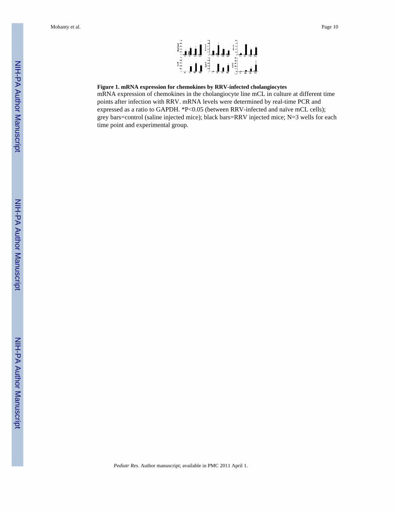
Figure 1. mRNA expression for chemokines by RRV-infected cholangiocytesmRNA expression of chemokines in the cholangiocyte line mCL in culture at different timepoints after infection with RRV. mRNA levels were determined by real-time PCR andexpressed as a ratio to GAPDH. *P<0.05 (between RRV-infected and naïve mCL cells);grey bars=control (saline injected mice); black bars=RRV injected mice; N=3 wells for eachtime point and experimental group.
Mohanty et al. Page 10
Pediatr Res. Author manuscript; available in PMC 2011 April 1.
NIH
-PA Author Manuscript
NIH
-PA Author Manuscript
NIH
-PA Author Manuscript

Figure 2. Detection of RRV in hepatic mononuclear cells and macrophagesPanel A depicts the expression of mRNA encoding for the RRV proteins NSP3 and VP6 inhepatic mononuclear cells isolated from mice 7 days after RRV challenge. Grey arrowspoint to no detection of RRV in hepatic mononuclear cells of normal saline-injectedcontrols; black bars=RRV injected mice; P<0.05; N=3–6 livers per time point and perexperimental group. Panels B and C depict dual-immunofluorescence signals identifyingRRV (red) in panF4/80+ cells (green) in purified hepatic mononuclear cells (panel B) orportal tracts (panel C) 7 days after RRV infection of newborn mice. Arrows point to doublepositive panF4/80+ and RRV+ cells.
Mohanty et al. Page 11
Pediatr Res. Author manuscript; available in PMC 2011 April 1.
NIH
-PA Author Manuscript
NIH
-PA Author Manuscript
NIH
-PA Author Manuscript

Figure 3. RRV infection of Raw 264.7 cells and induction of chemotaxis by conditioned mediaIn panel A, immunofluorescence detects RRV (red signal) 24 hours after Raw 264.7 cellswere exposed to RRV (right photograph); no signal is detected in cells not exposed to RRV(left photograph). Panel B depicts the numbers of neutrophils and lymphocytes that migratedto lower wells of chemotaxis chambers containing conditioned media from RRV-infected orcontrol (RRV-naïve) Raw 264.7 cells after 45 minutes of culture. *P<0.05; N=3 chambersper group; grey bars=neutrophils; black bars=lymphocytes.
Mohanty et al. Page 12
Pediatr Res. Author manuscript; available in PMC 2011 April 1.
NIH
-PA Author Manuscript
NIH
-PA Author Manuscript
NIH
-PA Author Manuscript

Figure 4. Production of cytokines and chemokines by mCL cholangiocytes and Raw 264.7 cellsConcentrations of cytokines and chemokines in conditioned media of RRV-infected andnaïve (controls) mCL cholangiocytes (panels A and B) and Raw 264.7 (panels C–F) cells atdifferent times of culture as determined by ELISA. Arrows point to no detectable level;P<0.05 (between RRV-infected and naïve cells); grey bars=control (saline injected mice);black bars=RRV injected mice; N=3 for each time point and experimental group.
Mohanty et al. Page 13
Pediatr Res. Author manuscript; available in PMC 2011 April 1.
NIH
-PA Author Manuscript
NIH
-PA Author Manuscript
NIH
-PA Author Manuscript

Figure 5. Hepatic expression of Mip2/Cxcl2 and inflammation of portal tracts following RRVchallengeIn panel A, realtime PCR shows increased hepatic expression of Mip2/Cxcl2 mRNA 3 daysafter RRV infection of newborn mice; P<0.05; grey bars=control (saline injected mice);black bars=RRV injected mice; N=3 at each time point and experimental group. In panel B,hematoxylin/eosin staining of liver sections shows accumulation of neutrophils at day 3(arrows; left panel) and lymphocytes at day 7 (arrows; right panel) after RRV infection.Insets represent magnification of area within rectangles; magnification: 400x.
Mohanty et al. Page 14
Pediatr Res. Author manuscript; available in PMC 2011 April 1.
NIH
-PA Author Manuscript
NIH
-PA Author Manuscript
NIH
-PA Author Manuscript

Figure 6. Mip2/Cxcl2-dependent chemotaxis of neutrophilsIn panel A, the concentration of Mip2/Cxcl2 is determined in conditioned media from RRV-infected or RRV-naïve Raw 264.7 cells that were incubated (or not) with anti-Mip2/Cxcl2antibody. Panel B depicts the number of neutrophils migrating to the lower wells ofchemotaxis chambers containing the respective conditioned media with or without anti-Mip2/Cxcl2 antibody. *P<0.05 showing significant differences between media with orwithout pre-incubation with anti-Mip2/Cxcl2 antibody; N=3 for each experimental group.
Mohanty et al. Page 15
Pediatr Res. Author manuscript; available in PMC 2011 April 1.
NIH
-PA Author Manuscript
NIH
-PA Author Manuscript
NIH
-PA Author Manuscript

NIH
-PA Author Manuscript
NIH
-PA Author Manuscript
NIH
-PA Author Manuscript
Mohanty et al. Page 16
Table 1
Oligonucleotide primers used for detection of different chemokines
Gene Primer Sequences Annealing Temp. (°C)
KC/Cxcl1 Forward: 5′-ACCGAAGTCATAGCCACACTC -3′Reverse: 5′-TGGGGACACCTTTTAGCATC -3′
52
Mip2/Cxcl2 Forward: 5′-CCCAGACAGAAGTCATAGCCAC -3′Reverse: 5′-GCCTTGCCTTTGTTCAGTATC -3′
52
TNFα Forward: 5′-AAGGGAGAGTGGTCAGTTGCC -3′Reverse: 5′-CCTCAGGGAAGAGTCTGGAAAGG -3′
55
Rantes Forward: 5′-GGAGTATTTCTACACCAGCAGC -3′Reverse: 5′-TCTTGAACCCACTTCTTCTCTG -3′
52
Cxcl9 Forward: 5′-GAGCTAGATAGACCTCACCAAG -3′Reverse: 5′-CCATTAGCACCATCTCTGA -3′
55
Cxcl10 Forward: 5′-TCGCTCAAGTGGCTGGGATG -3′Reverse: 5′-TAGGGAGGACAAGGAGGGTGTG -3′
55
Gapdh Forward: 5′-TGGTTTGACAATGAATACGGCTAC -3′Reverse: 5′-GGTGGGTGGTCCAAGGTTTC -3′
55
Rotavirus VP6 Forward: 5′-GCGGTAGCGGTGTTATTTCC -3′Reverse: 5′-TTGTTTTGCTTGCGTCGG -3′
55
Rotavirus NSP3 Forward: 5′-TGTCAAGAGAATACCTGGGAAATC -3′Reverse: 5′-GGAATCATCAACTTCAACTTCACC -3′
54
Ccl12 Forward: 5′-CACCATCAGTCCTCAGGTAT -3′Reverse: 5′-GGACGTGAATCTTCTGCTTA -3′
54
Temp = temperature
Pediatr Res. Author manuscript; available in PMC 2011 April 1.

NIH
-PA Author Manuscript
NIH
-PA Author Manuscript
NIH
-PA Author Manuscript
Mohanty et al. Page 17
Table 2
Presence or absence of protein levels of cytokines and chemokines in conditioned media obtained after 24hours of culture of RRV-infected Raw 264.7 and mCL cells. Protein levels were determined by ELISA.
Protein mCL cells Raw 264.7 cells
TNFα − +
IFNγ − −
IFNα − +
IFNβ − +
IL1β − −
Mip2/Cxcl2 + +
KC/Cxcl1 + −
“−“ Means not detectable
Pediatr Res. Author manuscript; available in PMC 2011 April 1.
Copyright © 2022 FDOKUMEN