Virus-specific intestinal IFN-γ producing T cell responses induced by human rotavirus infection and...
21
Virus-specific intestinal IFN-γ producing T cell responses induced by human rotavirus infection and vaccines are correlated with protection against rotavirus diarrhea in gnotobiotic pigs Lijuan Yuan * , Ke Wen 1 , Marli S.P. Azevedo, Ana M. Gonzalez, Wei Zhang, and Linda J. Saif Food Animal Health Research Program, Ohio Agricultural Research and Development Center, Department of Veterinary Preventive Medicine, The Ohio State University, 1680 Madison Avenue, Wooster, OH 44691, USA Abstract We examined rotavirus-specific IFN-γ producing CD4+, CD8+ and CD4+CD8+ T cell responses in gnotobiotic pigs infected with a virulent human rotavirus (VirHRV) or vaccinated with an attenuated (Att) HRV vaccine (AttHRV3x or AttHRV2x) or an AttHRV oral priming and 2/6-virus-like particle (VLP) intranasal boosting (AttHRV-2/6VLP) regimen. In VirHRV infected pigs, HRV-specific IFN- γ producing T cells reside primarily in ileum. AttHRV-2/6VLP induced similar frequencies of intestinal IFN-γ producing T cells as the VirHRV, whereas AttHRV3x or 2x vaccines were less effective. Protection rates against rotavirus diarrhea upon VirHRV challenge significantly correlated (r = 0.97–1.0, p < 0.005) with frequencies of intestinal IFN-γ producing T cells, suggesting their role in protective immunity. Keywords Human rotavirus vaccine; T cell responses; Gnotobiotic pigs 1. Introduction Group A rotaviruses are the single most important etiologic agent of dehydrating diarrhea in infants and young children worldwide, causing approximately 611,000 deaths yearly [1]. To improve the efficacy of rotavirus vaccines, an improved understanding of both B and T cell arms of rotavirus protective immunity is essential. The distribution and magnitude of antibody- secreting cell (ASC) and memory B cell responses to virulent (Vir) human rotavirus (HRV) Wa strain (P1A[8]G1), the attenuated (Att) Wa HRV or the combined vaccine regimen of AttHRV oral priming followed by 2/6-virus-like particle (2/6VLP) intranasal (IN) boosting (AttHRV-2/6VLP) have been delineated in our previous studies of gnotobiotic (Gn) pigs [2-6]. The protective role of rotavirus-specific IgA effector and memory B cells in intestinal lymphoid tissues and IgA antibodies in the serum, intestinal contents or feces against rotavirus diarrhea have been indicated in the Gn pig model of HRV diarrhea [3,5-8] and in studies of rotavirus natural infection of humans [9-11]. In humans, serum IgA and sometimes IgG antibody titers were correlated with protection after rotavirus natural infection [12]; but not * Corresponding author. Current address: Center for Molecular Medicine and Infectious Diseases, Department of Biomedical Sciences and Pathobiology, Virginia Polytechnic Institute and State University, 1410 Prices Fork Road, Blacksburg, VA 24061-0342, USA. Tel.: +1 540 231 9053; fax: +1 540 231 3426. E-mail address: [email protected] (L. Yuan). 1 Current address: Center for Molecular Medicine and Infectious Diseases, Department of Biomedical Sciences and Pathobiology, Virginia Polytechnic Institute and State University, 1410 Prices Fork Road, Blacksburg, VA 24061-0342, USA. NIH Public Access Author Manuscript Vaccine. Author manuscript; available in PMC 2009 June 19. Published in final edited form as: Vaccine. 2008 June 19; 26(26): 3322–3331. doi:10.1016/j.vaccine.2008.03.085. NIH-PA Author Manuscript NIH-PA Author Manuscript NIH-PA Author Manuscript
-
Upload
independent -
Category
Documents
-
view
1 -
download
0
Transcript of Virus-specific intestinal IFN-γ producing T cell responses induced by human rotavirus infection and...
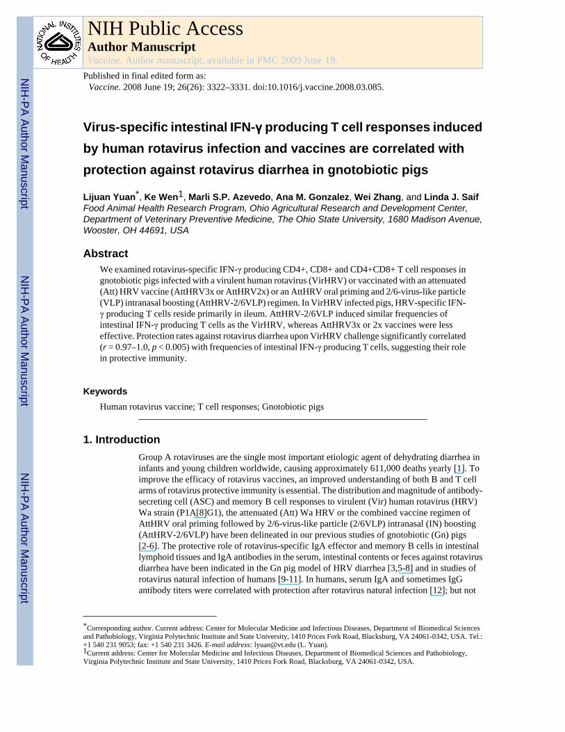
Virus-specific intestinal IFN-γ producing T cell responses inducedby human rotavirus infection and vaccines are correlated withprotection against rotavirus diarrhea in gnotobiotic pigs
Lijuan Yuan*, Ke Wen1, Marli S.P. Azevedo, Ana M. Gonzalez, Wei Zhang, and Linda J. SaifFood Animal Health Research Program, Ohio Agricultural Research and Development Center,Department of Veterinary Preventive Medicine, The Ohio State University, 1680 Madison Avenue,Wooster, OH 44691, USA
AbstractWe examined rotavirus-specific IFN-γ producing CD4+, CD8+ and CD4+CD8+ T cell responses ingnotobiotic pigs infected with a virulent human rotavirus (VirHRV) or vaccinated with an attenuated(Att) HRV vaccine (AttHRV3x or AttHRV2x) or an AttHRV oral priming and 2/6-virus-like particle(VLP) intranasal boosting (AttHRV-2/6VLP) regimen. In VirHRV infected pigs, HRV-specific IFN-γ producing T cells reside primarily in ileum. AttHRV-2/6VLP induced similar frequencies ofintestinal IFN-γ producing T cells as the VirHRV, whereas AttHRV3x or 2x vaccines were lesseffective. Protection rates against rotavirus diarrhea upon VirHRV challenge significantly correlated(r = 0.97–1.0, p < 0.005) with frequencies of intestinal IFN-γ producing T cells, suggesting their rolein protective immunity.
KeywordsHuman rotavirus vaccine; T cell responses; Gnotobiotic pigs
1. IntroductionGroup A rotaviruses are the single most important etiologic agent of dehydrating diarrhea ininfants and young children worldwide, causing approximately 611,000 deaths yearly [1]. Toimprove the efficacy of rotavirus vaccines, an improved understanding of both B and T cellarms of rotavirus protective immunity is essential. The distribution and magnitude of antibody-secreting cell (ASC) and memory B cell responses to virulent (Vir) human rotavirus (HRV)Wa strain (P1A[8]G1), the attenuated (Att) Wa HRV or the combined vaccine regimen ofAttHRV oral priming followed by 2/6-virus-like particle (2/6VLP) intranasal (IN) boosting(AttHRV-2/6VLP) have been delineated in our previous studies of gnotobiotic (Gn) pigs[2-6]. The protective role of rotavirus-specific IgA effector and memory B cells in intestinallymphoid tissues and IgA antibodies in the serum, intestinal contents or feces against rotavirusdiarrhea have been indicated in the Gn pig model of HRV diarrhea [3,5-8] and in studies ofrotavirus natural infection of humans [9-11]. In humans, serum IgA and sometimes IgGantibody titers were correlated with protection after rotavirus natural infection [12]; but not
*Corresponding author. Current address: Center for Molecular Medicine and Infectious Diseases, Department of Biomedical Sciencesand Pathobiology, Virginia Polytechnic Institute and State University, 1410 Prices Fork Road, Blacksburg, VA 24061-0342, USA. Tel.:+1 540 231 9053; fax: +1 540 231 3426. E-mail address: [email protected] (L. Yuan).1Current address: Center for Molecular Medicine and Infectious Diseases, Department of Biomedical Sciences and Pathobiology,Virginia Polytechnic Institute and State University, 1410 Prices Fork Road, Blacksburg, VA 24061-0342, USA.
NIH Public AccessAuthor ManuscriptVaccine. Author manuscript; available in PMC 2009 June 19.
Published in final edited form as:Vaccine. 2008 June 19; 26(26): 3322–3331. doi:10.1016/j.vaccine.2008.03.085.
NIH
-PA Author Manuscript
NIH
-PA Author Manuscript
NIH
-PA Author Manuscript
after vaccination with Rotashield™ [9,13], implying a potential role for other effectorsincluding T cell mediated immunity in the protection induced by this tetravalent reassortantrotavirus vaccine.
In studies of adult mice, CD8+ T cells were shown to provide the most important but notexclusive mechanism mediating clearance of a primary rotavirus infection [14]. Also in studiesof adult mice, CD4+ T cells were shown to be the only lymphocytes needed to protect miceagainst rotavirus infection after the mice were vaccinated with VP6 peptide vaccines [15,16].Despite a large number of studies on the role of T cells in mediating protection against rotavirusinfection in adult mice with various gene knockouts (protection against disease cannot beassessed in the adult mice model) [14-18], limited data is available on T cell immune responsesto rotavirus in humans [19,20] or other out-bred native hosts, i.e. calves and pigs [21-23].However, because of the potentially important role for CD4+ and CD8+ T cells in rotavirusimmunity, especially in heterotypic protection as suggested by studies of influenza virusinfections [24], it is important to quantify and characterize antigen-specific CD4+ and CD8+T cells in an animal model of rotavirus disease as well as in human clinical trials for thedevelopment of new rotavirus vaccines. The objective of this study was to identify the potentialcorrelation between HRV-specific IFN-γ producing or proliferating T cell responses withprotective immunity induced by rotavirus infection and vaccination using our well-establishedGn pig model of HRV infection and disease [25,26].
Production of IFN-γ within hours of antigenic restimulation is a functional characteristic ofvirus-specific effector-memory T cells [27]. We chose to use functional characteristics todefine T cell subpopulations in vitro to better reflect their in vivo functions. IFN-γ was recentlyidentified to be the only cytokine produced by restimulated CD4+ T cells from immunizedmice that directly inhibited rotavirus replication in vitro [28]. We postulated that protectiveefficacy against rotavirus is associated with IFN-γ producing T cell responses induced byrotavirus infection or vaccines. We used flow cytometry to detect intracellular accumulationof IFN-γ by CD4+, CD8+ and CD4+CD8+ T cells activated by intact homologous HRVantigen. This assay allows quantitation of HRV-specific IFN-γ producing T cells, at the singlecell level [29]. Studies of the interaction of rotavirus with human myeloid dendritic cells(MDCs) demonstrated that intact peripheral blood mononuclear cell (PMNC) populationscontaining antigen presenting cells are equally efficient compared to rotavirus-infected MDC,in stimulating IFN-γ-producing rotavirus-specific effector-memory T cells [30]. Furthermore,the frequencies and patterns of cytokines produced by effector-memory CD4+ T cells afterstimulation of peripheral blood MNC with the purified rhesus rotavirus (RRV) or MDCinfected with RRV were similar. Thus intracellular IFN-γ staining assays using purified intactrotavirus as stimulating antigen with mononuclear cell (MNC) populations can providecomparative information regarding the tissue distribution and magnitude of rotavirus-specificanti-viral T cell responses elicited by rotavirus infection and vaccines. The CD4+CD8+ doublepositive T cells are mainly found in swine and also in humans, nonhuman primates and mice[31,32]. Studies have suggested that CD4+CD8+ T cells can respond to recall antigens and areeffector-memory or memory T cells in swine [31,33] and humans [34]. The role of virus-specific double positive T cells in rotavirus immunity in pigs or humans has not been studiedbefore.
Compared to effector-memory T cells, memory T cell subpopulations are more difficult todefine due to the dynamics of the T cell progressive differentiation from effector to effector-memory (resting effector) to memory T cells [35]. Because of the lack of definitive memoryT cell surface markers for swine and the lack of other functional markers in general,proliferation has been the chosen parameter to measure memory T cell responses to rotavirusinfection and vaccination in humans and pigs [20,21]; however the role of proliferating T cellsin rotavirus protective immunity has not been defined. Immunofluorescent staining of
Yuan et al. Page 2
Vaccine. Author manuscript; available in PMC 2009 June 19.
NIH
-PA Author Manuscript
NIH
-PA Author Manuscript
NIH
-PA Author Manuscript
incorporated bromodeoxyuridine (BrdU) and flow cytometry is a high-resolution technique tomeasure T cell proliferation. Cytokine (i.e. IFN-γ)producing proliferating T cells can beassessed at the same time in a multicolor flow cytometry [36].
In this study, we determined the magnitude and distribution of rotavirus-specific IFN-γproducing or proliferating T cell responses in intestinal (ileum) and systemic (spleen) lymphoidtissues and blood in Gn pigs orally infected with a VirHRV (mimic natural infection) orvaccinated with two or three oral doses of live AttHRV (mimic currently licensed HRVvaccines) or a combined AttHRV-2/6VLP prime/boost vaccine. We found significantcorrelations between the magnitude of intestinal IFN-γ producing CD4+, CD8+ or CD4+CD8+ T cell responses induced by the VirHRV infection or the various rotavirus vaccines andprotection rates against rotavirus diarrhea upon challenging the Gn pigs with the VirHRV.
2. Materials and methods2.1. Viruses
The VirHRV Wa strain (G1P1A[8]) was passaged in Gn pigs and the pooled intestinal contentsof pigs from the 23rd passage were used for inoculation at a dose of ∼106 fluorescence formingunits (FFU). The virus titers were determined using a cell culture immunofluorescence (CCIF)assay [3]. The 50% infectious dose (ID50) and diarrhea dose (DD50) of Wa VirHRV wasdetermined previously in Gn pigs as ≤1 FFU [37]. The 37th cell culture passage of Wa AttHRVwas propagated in the African green monkey kidney cell line MA104 and used for vaccinationat a dose of 5×10⁷ FFU and as stimulating antigen (HRV antigen) in intracellular cytokinestaining of IFN-γ producing or proliferating T cells. The HRV antigen was semipurified fromAttHRV-infected MA104 cell-culture supernatants (titer = approximately 107 FFU/ml) bycentrifugation (112,700×g) through a 40% (w/w) sucrose cushion as described previously [5]and the protein concentration of the preparations was determined by Bio-Rad protein assay(Bio-Rad, Hercules, CA).
2.2. Virus-like particles with ISCOM adjuvantRotavirus 2/6-virus-like particles (2/6VLP) with VP2 derived from RF and VP6 from Wa strainrotaviruses were produced in Spodoptera frugiperda nine insect cells. The assembled VLPswere purified using CsCl2 gradients and characterized as previously described [38,39]. Theprotein concentration of the purified 2/6VLP was determined using the Bio-Rad protein assay(Bio-Rad). The endotoxin level in each 2/6VLP preparation was quantitated (<0.05 units/dose)with the Limulus amebocyte assay (Associates of Cape Cod, Inc., Woods Hole, Mass.). The2/6VLP (250μg/dose) were incorporated into immunostimulating complexes (ISCOM) (1.25mg/dose) as previously described [2]. Electron microscopy or immune electron microscopywas performed on each of the 2/6VLP-ISCOM preparations just prior to inoculation to confirmthe integrity of the vaccine.
2.3. Gnotobiotic pigs and experimental designGnotobiotic pigs were derived by hysterectomy of near term sows, maintained aseptically inisolation units and fed a sterile commercial infant formula (Similac®, Ross laboratories,Columbus, OH) as described previously [40]. Pigs were assigned randomly to 1 of 5 groups.All pigs were orally inoculated at 5–6 days of age with a single dose (∼105 FFU) of VirHRV(group 1, VirHRV, n = 5) or a priming dose (5×10⁷ FFU) of the AttHRV. The AttHRV primedpigs were boosted with two intranasal (IN) doses of 2/6VLP-ISCOM (group 2,AttHRV-2/6VLP, n = 4), or two or one oral booster doses of AttHRV (group 3, AttHRV3x,n = 8, or group 4, AttHRV2x, n = 4, respectively) at 10 and 21 days after the first inoculation.The 2/6VLP dose used in the AttHRV-2/6VLP vaccine regimen was 250μg 2/6VLPincorporated into 1.25 mg ISCOM matrix. Pigs mock-inoculated with diluent or ISCOM matrix
Yuan et al. Page 3
Vaccine. Author manuscript; available in PMC 2009 June 19.
NIH
-PA Author Manuscript
NIH
-PA Author Manuscript
NIH
-PA Author Manuscript

was included as controls (group 5, control, n = 8). Protective efficacies against diarrheaconferred by the VirHRV infection, AttHRV-2/6VLP, AttHRV3x, or 2x vaccines afterchallenge with the Wa VirHRV (1×10⁶ FFU) at postinoculation day (PID) 28 were assessedas previously reported [3,5,6]. The MNC from ileum, spleen and peripheral blood were isolatedfrom pigs euthanized at PID28 (PID27-29) for characterization of T cell responses. The animalexperimental protocol was reviewed and approved by the Institutional Laboratory Animal Careand Use Committee at The Ohio State University.
2.4. Collection of lymphoid tissues and isolation of MNCAfter euthanasia, the distal portion of the small intestine (ileum), spleen and blood werecollected [3]. The MNC were extracted from the ileum using EDTA and collagenase andenriched by discontinuous Percoll gradient [3]. The MNC of the spleen were extracted bymechanical separation and enriched by discontinuous Percoll gradient. Peripheral bloodlymphocytes (PBLs) were purified using Ficoll-PaqueTM plus (Amersham Biosciences,Uppsala, Sweden) [3,21]. The purified MNC from all tissues were resuspended in completemedium consisting of RPMI-1640 (Gibco, BRL) supplemented with 8% fetal bovine serum,20 mM HEPES (N-2-hydroxyethyl-piperazine-Nk-2-ethanesulphonic acid), 2 mM L-glutamine, 1 mM sodium pyruvate, 0.1 mM non-essential amino acids, 100μg/ml ofgentamicin, 10μg/ml of ampicillin and 50 mM 2-mercaptoethanol (E-RPMI).
2.5. Intracellular cytokine staining and flow cytometry analysis of frequencies of IFN-γproducing CD4+, CD8+ and CD4+CD8+ T cells
Flow cytometry was used to determine frequencies of HRV-specific IFN-γ producing CD4+,CD8+ and CD4+CD8+ T cells in the intestinal (ileum) and systemic (spleen) tissues and bloodin Gn pigs in each group. The MNC extracted from the pigs were restimulated in vitro in 12-well cell culture plates (Costar, Corning Incorporated, Corning, New York) with semi-purifiedHRV antigen (12μg/ml), positive control PHA (10μg/ml) or mock stimulated (medium-only)in E-RPMI media for 17 h at 37°C [29]. A protein transport inhibitor, Brefeldin A (10μg/ml;Sigma) was added for the last 5 h to block secretion of cytokines produced by the T cells. Ananti-human CD49d (0.5μg/ml) monoclonal antibody (MAb) (BD Pharmingen, San Diego, CA)was added to each sample as a costimulator [19], because an anti-porcine CD49d MAb wasnot available. The anti-human CD49d MAb cross-reacts with the molecules on porcineleukocytes [41]. The MNC were washed with staining buffer (3%FBS and 0.09% sodium azidein 1× DPBS (0.2 mg/ml KCl, 0.2 mg/ml KH2PO4, 8 mg/ml NaCl and 2.16 mg/mlNa2HPO4·7H2O, pH 7.2–7.4)), centrifuged at 500×g for 5min at 4°C before and after eachstaining step prior to permeabilizing the cells. The MNC (2×10⁶ cells/tube) were stained atroom temperature (RT) for 15 min with the following MAbs in sequential steps: (1) fluoresceinisothiocyanate (FITC) conjugated mouse anti-porcine CD4 (IgG2b, Clone 74-12-4, BDPharmingen) and purified mouse anti-porcine CD8α (IgG1, clone PT36B, VMRD, Inc,Pullman, WA), followed by (2) allophycocyanin (APC) conjugated rat anti-mouse IgG1 (BDPharmingen), and then (3) biotinylated mouse anti-porcine CD3 (IgG1, clone PPT3,SouthernBiotech, Birmingham, AL) and (4) peridinine chlorophyll protein (PerCP) conjugatedstreptavidin (BD Pharmingen). After staining of cell surface markers, the MNC werepermeabilized with BD cytofix/cytoperm™ buffer (BD pharmingen) for 15 min at RT. Thenthe MNC were washed with BD perm/wash buffer (BD pharmingen) and stained with R-phycoerythrin (PE) conjugated mouse anti-porcine IFN-γ (IgG1, clone P2G10, BDPharmingen) for 15 min at RT. The MNC were washed with perm/wash buffer and resuspendedin staining buffer and kept in the dark at 4°C before flow cytometry analysis the next day. Theappropriate isotype-matched irrelevant control antibodies (BD pharmingen, VMRD orSouthernBiotech) were included for MNC of each antigen-stimulation from each tissue in eachstaining as negative controls to set the quadrant markers for the bivariate dot plots. All MAbswere titrated and used at optimal concentrations. At least 20,000 cells were acquired on a
Yuan et al. Page 4
Vaccine. Author manuscript; available in PMC 2009 June 19.
NIH
-PA Author Manuscript
NIH
-PA Author Manuscript
NIH
-PA Author Manuscript
FACSCalibur flow cytometer (BD Biosciences). Flow cytometry data were analyzed usingCellQuest software (BD) or FlowJo 7.2.2 software (Tree Star, Ashland, Oregon). Only samplesin which the IFN-γ staining was at least twice that of the medium-stimulated controls(background) were considered positives. The frequencies of IFN-γ+CD4+, IFN-γ+CD8+ andIFN-γ+CD4+CD8+ T cells were expressed as percentages of total CD3+ T cells. All meanfrequencies in Fig. 4 are reported after subtraction of the background frequencies.
2.6. BrdU staining and flow cytometry analysis of proliferating CD4+ and CD8+ T cellsImmunofluorescent staining of incorporated BrdU is a high-resolution technique to measureT cell proliferation after virus infection or vaccination. Bromodeoxyuridine, a pyrimidineanalogue, is incorporated into the proliferating S-phase cells. After in vitro stimulation of2×106 MNC from each tissue with semipurified HRV antigen (12μg/ml), PHA (10μg/ml) ormock (medium-only) for 5 days, 10μg/ml BrdU were added to each cell culture for the last 18h. The MNC were harvested, washed with staining buffer and stained with the following MAbs:PE conjugated mouse anti-porcine CD4 (clone 74-12-4, BD Pharmingen), FITC conjugatedmouse anti-porcine CD8α (clone 76-2-11, BD Pharmingen) and biotinylated mouse anti-porcine CD3 (clone PPT3, SouthernBiotech, Birmingham, AL) followed by PerCP conjugatedstreptavidin (BD Pharmingen). After staining of the T cell surface markers, the MNC werepermeabilized with BD cytofix/cytoperm™ buffer for 15 min at RT. Then the MNC werewashed with BD perm/wash buffer and stained with APC conjugated anti-BrdU antibody (BDPharmingen) for 20 min at RT. Isotype matched control antibodies were included for MNC ofeach antigen-stimulation from each tissue in each staining. Four-color flow cytometry analysiswas performed to determine the frequencies of BrdU+CD3+CD4+ and BrdU+CD3+CD8+cells using a FACSCalibur Flow Cytometer and CellQuest™ Pro software following themanufacturer's instructions (Becton Dickinson). At least 20,000 cells were acquired.Frequencies of proliferating T cells from medium-only stimulated MNC (background) weresubtracted from the frequencies in HRV stimulated MNC. The HRV-specific proliferating Tcell responses were presented as the frequencies of BrdU+CD4+ or BrdU+CD8+ T cells amongtotal CD3+ MNC. After 5 days in vitro stimulation of MNC with HRV antigen or PHA, thefrequencies of CD4+CD8+ double positive T cells become very low to undetectable amongproliferating T cells. Thus CD4+CD8+ T cells were not analyzed as a separate subpopulationfor T cell proliferation.
2.7. BrdU and intracellular staining and flow cytometry analysis of IFN-γ or IL-13 producingCD4+ and CD8+ T cells among proliferating and non-proliferating T cells
A flow cytometry assay was developed to detect IFN-γ or IL-13 producing CD4+ and CD8+T cells among proliferating (BrdU+) or non-proliferating (BrdU−) CD3+ MNC. The MNCwere stimulated with HRV antigen, PHA or medium-only for 5 days as described above. Mouseanti-porcine CD3 (IgG1κ, clone PPT3) MAb conjugated with SpectralRed™ (SPRD,SouthernBiotech), mouse anti-porcine CD4 (IgG2bκ, clone 74-12-4) or CD8 (IgG1, clonePT36B) conjugated with biotin (SouthernBiotech) and then streptavidin conjugated with APC-Cy7 (eBioscience) were added in the initial two steps to stain T cell surface markers. The cellswere then permeabilized with cytofix/cytoperm™ buffer and stained for intracellular BrdU,IFN-γ and IL-13 with anti-BrdU conjugated with APC, anti-porcine IFN-γ PE (BD) and anti-human IL-13 FITC (IgG1, clone PVM13-1, eBioscience) MAbs, respectively. Isotype-matched control MAbs were included for MNC from each tissue in each staining. An upgradedfive-color FACSCalibur Flow Cytometer (the 3rd violet laser added by Cytek Flow CytometryProducts, CA), CellQuest™ Pro (BD) and Rainbow™ (Cytek) software were used to analyzethe stained cells. At least 20,000 cells were acquired. The stained cells were first gated for CD3and BrdU and then analyzed on CD4 vs. IFN-γ or IL-13 and CD8 vs. IFN-γ or IL-13 bivariatedot plots. The frequencies of IFN-γ producing CD4+ or CD8+ T cells were expressed aspercentages of total CD3+BrdU+ or CD3+BrdU− MNC.
Yuan et al. Page 5
Vaccine. Author manuscript; available in PMC 2009 June 19.
NIH
-PA Author Manuscript
NIH
-PA Author Manuscript
NIH
-PA Author Manuscript

2.8. Statistical analysisComparisons of frequencies of T cells among treatment groups or tissues were performed forall data presented in Figs. 4-6 using the Kruskal–Wallis test (SAS Institute Inc., Cary, NC).When differences among the groups or tissues were detected, the same test was used in apairwise fashion to clarify the nature of the differences. Spearman's correlation was used forassessment of association between mean frequencies of IFN-γ+ producing or proliferating Tcells and protection rates against rotavirus diarrhea. Protection rates among treatment groupswere compared using Fisher's Exact Test. Statistical significance was assessed at p < 0.05 forall comparisons.
3. Results3.1. Detection of HRV-specific IFN-γ+CD4+, IFN-γ+CD8+ and IFN-γ+CD4+CD8+ T cells
The intracellular staining and flow cytometry analysis for detection of HRV-specific IFN-γ-producing CD4+ and CD8+ T cells is illustrated in Fig. 1. The detection of HRV-specific IFN-γ-producing CD4+CD8+ double positive T cells is illustrated in Fig. 2. The three T cellsubpopulations CD4+, CD8+ and CD4+CD8+ were identified by gating through MNC andthen CD3+ lymphocytes (Figs. 1A and 2A). PHA and medium-only stimulated MNC wereincluded (for all samples in the study) as positive and background controls. Low frequenciesof spontaneous IFN-γ producing T cells were observed among the medium-only stimulatedMNC (right panels in Figs. 1B and 2B), which may reflect the presence of T cells thatconstitutively produce IFN-γ (background, non-HRV specific). Their frequencies weregenerally substantially lower than those among the HRV antigen-stimulated MNC (left panelsin Figs. 1B and 2B). Examples of IFN-γ producing CD4+, CD8+ and CD4+CD8+ T cellresponses among different inoculation groups are given in Fig. 3. Representative dot plots wereshown for ileum of one pig in the VirHRV, AttHRV-2/6VLP, AttHRV3x or control groups,respectively.
3.2. IFN-γ producing T cells induced by virulent HRV infection reside primarily in the intestineThe mean frequencies of HRV-specific IFN-γ+CD4+, IFN-γ+CD8+ and IFN-γ+CD4+CD8+T cells among the inoculation groups are depicted in Fig. 4. Mean frequencies of IFN-γproducing T cells in each tissue of each group were calculated using adjusted frequencies inwhich the frequencies from the medium-only stimulated MNC were subtracted from the HRVstimulated MNC. The IFN-γ+CD4+CD8+ T cell responses in the VirHRV group were notdetermined because the CD4 and CD8 were stained in separate tubes for this group. In theVirHRV infected pigs, higher frequencies of HRV-specific IFN-γ+CD4+ (statisticallysignificant) and IFN-γ+CD8+ T cells were detected in ileum compared to spleen (13-fold and15-fold higher, respectively) and blood (35-fold and 11-fold higher, respectively), indicatingthat the IFN-γ producing T cells after VirHRV infection reside primarily in the intestinallymphoid tissues at PID28, as we showed previously for memory B cells [5]. The pigs receivingAttHRV-2/6VLP or AttHRV3x vaccine had significantly higher frequencies of IFN-γ+CD4+,IFN-γ+CD8+ (for AttHRV-2/6VLP only) and IFN-γ+CD4+CD8+ T cells in spleen comparedto blood. Blood had the lowest frequencies of IFN-γ producing T cells compared to ileum andspleen in all inoculation groups (except for AttHRV3x) and the frequencies in VirHRV infectedor vaccinated pigs did not differ from that of the mock-inoculated control pigs, suggesting thatfew HRV-specific effector-memory T cells traffic through the circulation at PID 28 after HRVinfection or vaccination.
Yuan et al. Page 6
Vaccine. Author manuscript; available in PMC 2009 June 19.
NIH
-PA Author Manuscript
NIH
-PA Author Manuscript
NIH
-PA Author Manuscript

3.3. AttHRV-2/6VLP vaccine induced higher intestinal HRV-specific IFN-γ producing T cellresponses than the AttHRV3x and 2x vaccines
In pigs receiving the AttHRV-2/6VLP2x vaccine, frequencies of IFN-γ+CD4+ and IFN-γ+CD8+ T cells in ileum were slightly lower, but statistically similar to those of the VirHRV-infected pigs and they (including IFN-γ+CD4+CD8+ T cells) were higher or significantlyhigher than those of pigs vaccinated with the AttHRV3x or 2x vaccines and controls (Fig. 4).The AttHRV-2/6VLP vaccine is more effective in inducing intestinal IFN-γ producing T cellresponses and protection (Table 1) than the AttHRV vaccines, possibly due to the multiplemucosal induction sites involved in the AttHRV-2/6VLP prime/boost vaccine regimen (i.e.oral: gut associated lymphoid tissues [GALT] followed by IN: nasal associated lymphoidtissues [NALT]). In spleen, the AttHRV-2/6VLP vaccine induced significantly higher IFN-γ+CD4+ and IFN-γ+CD8+ T cell responses than the VirHRV, which may reflect the effect ofthe 2/6VLP booster doses. Antigen-specific T or B cells primed in NALT reside predominatelyin the lungs and in systemic over intestinal sites [42]. The AttHRV-2/6VLP vaccinated pigswere also the only inoculation group that developed significantly higher splenic IFN-γ+CD8+ T cell responses than controls. The frequencies of IFN-γ+CD4+ T cells in spleen ofAttHRV3x or 2x vaccinated pigs did not differ significantly from those of the VirHRV pigs,but they (including the IFN-γ+CD4+CD8+ T cells) were significantly higher than controls.The trend for the higher frequencies (not significantly vs. VirHRV group) of IFN-γ+ T cellresponses in spleen and blood of AttHRV3x pigs may be explained also by the booster doses.Higher rate of nasal virus shedding than fecal shedding was observed after the AttHRV oralinoculation in our previous studies [43]. This fact may also help to explain the higher IFN-γ+CD4+ and IFN-γ+CD4+CD8+ T cell responses observed in the systemic site (spleen) thanin the intestine (ileum) after inoculation with the AttHRV3x or 2x vaccine.
3.4. Frequencies of intestinal HRV-specific IFN-γ producing T cell responses correlated withprotection rates against HRV diarrhea
We analyzed the correlation between mean frequencies of IFN-γ producing T cells in thelymphoid tissues and blood and protection rates against diarrhea in all inoculation groups(Table 1). The protection rates against HRV diarrhea upon VirHRV challenge weresignificantly correlated with the mean frequencies of ileal IFN-γ+CD4, IFN-γ+CD8+ and IFN-γ+CD4+CD8+ double positive T cells in Gn pigs, demonstrating the important role of virus-specific intestinal IFN-γ producing T cells in protective immunity against rotavirus disease. Aslightly stronger correlation was found between protection rates and mean frequencies of IFN-γ+CD4+ or IFN-γ+CD4+CD8+ (r = 1.0, p < 0.0001) than IFN-γ+CD8+ T cell (r = 0.975, p =0.0048). There were no significant correlations between systemic IFN-γ producing T cellresponses and protection rates against diarrhea when all groups were included for calculationof the correlation coefficients (Table 1). However, when only the AttHRV groups and thecontrol group were included for the calculation, the mean frequencies of IFN-γ+CD4+ T cellsin both ileum and spleen of AttHRV3x and 2x inoculated pigs were significantly correlated(r = 1.0, p < 0.0001) with protection rates against diarrhea.
3.5. HRV-specific intestinal proliferating T cell responses were highest in the vaccinated pigsbut they were not correlated with protection
To examine the potential role of HRV-specific proliferating T cell responses in rotavirusprotective immunity, mean frequencies of HRV-specific BrdU+CD4+ and BrdU+CD8+ T cellsin ileum, spleen and blood of the five inoculation groups were compared (data not shown).Overall higher frequencies of BrdU+CD4+ than BrdU+CD8+ T cells were detected in alltissues of all groups, confirming that CD4+ T cells are the major proliferating T cell population.Unlike the IFN-γ producing T cell responses, the distribution of BrdU+CD4+ and BrdU+CD8+ T cells in VirHRV-infected pigs did not differ significantly among tissues at PID28. There
Yuan et al. Page 7
Vaccine. Author manuscript; available in PMC 2009 June 19.
NIH
-PA Author Manuscript
NIH
-PA Author Manuscript
NIH
-PA Author Manuscript
were also no significant differences in frequencies among tissues of the vaccinated or controlgroups. The AttHRV-2/6VLP vaccine induced the highest intestinal proliferating T cellresponses whereas AttHRV3x or 2x vaccine induced the highest systemic proliferating T cellresponses. However, there were no significant differences in frequencies of ileal BrdU+CD4+ or BrdU+CD8+ T cells among vaccine groups and there were no significant correlationsbetween protection rates against diarrhea and frequencies of BrdU+CD4+ or BrdU+CD8+ Tcells in any tissues (data not shown).
3.6. Low frequency of HRV-specific IFN-γ producing T cells were detected amongproliferating CD4+ and CD8+ T cells in spleen and blood
To help us understand why the magnitude of HRV-specific proliferating T cell responses wasnot directly associated with protection, the frequencies of IFN-γ producing T cells amongproliferating (BrdU+) and non-proliferating (BrdU−) CD4+ and CD8+ T cells were analyzedin Gn pigs receiving the AttHRV3x vaccine using a five-color flow cytometry developed laterin the study. This assay was only applied to pigs vaccinated with AttHRV3x vaccine whichwas the last treatment group for the study. The mean frequencies of HRV-specific BrdU+ orBrdU−, IFN-γ+CD4+ and IFN-γ+CD8+ T cells in ileum, spleen and blood are depicted in Fig.5, along with PHA stimulated and medium-only stimulated controls. Among HRV-specificproliferating T cells, no or very low frequencies of IFN-γ+CD4+ and IFN-γ+CD8+ T cellswere detected in ileum, but higher frequencies were detected in spleen and blood. Significantlyhigher frequencies of HRV-specific IFN-γ+CD4+ and IFN-γ+CD8+ T cells were detectedamong proliferating T cells compared to non-proliferating T cells in spleen (CD4+ only) andblood. Thus, after stimulation with recall antigen for 5 days, HRV-specific IFN-γ producingT cells are mainly found among proliferating T cells and resided primarily in spleen and blood.
3.7. Frequencies of total CD4+ and CD8+ T cells in blood were reduced in VirHRV infectedpigs compared to the vaccinated or control pigs at PID28
Frequencies of total CD3+CD4+ and CD3+CD8+ T cells among MNC were analyzed to assesswhether VirHRV infection and vaccination have an effect on total T cell frequencies in Gnpigs at PID28. The mean frequencies of total CD4+ and CD8+ T cells in ileum, spleen andblood in the VirHRV, AttHRV3x and control pigs are depicted in Fig. 6. The frequencies oftotal T cells in the AttHRV-2/6VLP and AttHRV2x groups were similar to those of AttHRV3xgroup for all the tissues, therefore data are not shown. The CD4/CD8 ratio in ileum of all piggroups were <1.0, and in spleen and blood were >1.5, but CD4/CD8 ratios did not differsignificantly among groups (data not shown). The frequencies of total CD4+ T cells in alltissues and CD8+ T cells in blood increased significantly in the AttHRV3x group, but not inthe VirHRV group, compared to the controls. These increases may reflect the effect of boosterdoses (repeated antigen stimulation) received by the vaccine group on the development/maturation of the T cell compartment in neonatal Gn pigs. However, increased total T cellnumbers did not correlate with increased protection rates against HRV diarrhea. For total CD8+ T cells, a noteworthy observation was the significantly reduced frequency in the blood ofVirHRV pigs compared to the controls. Total blood CD4+ T cells also showed the same trend,although it was not statistically significant. Frequencies of total CD4+CD8+ double positiveT cells were also compared among the vaccine groups and the controls. In ileum, thefrequencies of double positive T cells were increased or significantly increased in the vaccinegroups compared to the controls. In spleen and blood, the frequencies among groups did notdiffer significantly (data not shown).
4. DiscussionUsing intracellular staining and flow cytometry, we examined frequencies of HRV-specificIFN-γ producing T cells, HRV-specific proliferating T cells and total T cells in ileum, spleen
Yuan et al. Page 8
Vaccine. Author manuscript; available in PMC 2009 June 19.
NIH
-PA Author Manuscript
NIH
-PA Author Manuscript
NIH
-PA Author Manuscript
and blood of Gn pigs after VirHRV infection or inoculation with various vaccines. We alsoexamined frequencies of IFN-γ producing CD4+ and CD8+ T cells among proliferating andnon-proliferating T cells from the AttHRV3x vaccinated pigs. After VirHRV infection orvaccinations, the frequencies of intestinal HRV-specific IFN-γ producing CD4+ T cells in Gnpigs at the time of challenge (PID28) correlated significantly with protection rates againstVirHRV-induced diarrhea. IFN-γ producing T cells induced by VirHRV infection in Gn pigsreside primarily in the intestine as we showed previously for memory B cells [5]. This findingagrees with the existence of a population of extralymphoid effector-memory T cells poised forimmediate response to infection [44,45]. In mice, antigen-specific CD4+ effector-memory Tcells were found to reside primarily in the gut, lungs and salivary glands at 11–60 days post-antigen exposure and these cells readily produced IFN-γ upon antigen restimulation [44]. Ourstudy demonstrated a direct correlation between the existence of extralymphoid IFN-γproducing T cells and protection at the site of virus entry (intestine). In studies of mice,intestinal IFN-γ producing CD4+ T cells were suggested to be the only lymphocytes requiredfor protection against rotavirus infection after the mice were immunized with a chimeric VP6vaccine using attenuated E. coli labile toxin as an adjuvant [15,28,46,47]. Their data and ourscollectively emphasize the role of intestinal IFN-γ producing CD4+ T cells as correlates ofrotavirus protective immunity, possibly more important than CD8+ T cells. The finding thatIFN-γ producing T cell responses measured after in vitro antigen restimulation correlated withprotection against rotavirus diarrhea confirms that the memory responses detected in vitro arereflective of protective immunity in vivo. Intestinal IFN-γ producing CD4+CD8+ T cellsinduced by various HRV vaccines also significantly correlated with protection. To ourknowledge this is the first study showing that intestinal double positive T cells may play animportant role in rotavirus protective immunity. Intestinal double positive T cells in rhesusmacaques have been shown to be highly activated memory T cells with increased capacity toproduce cytokines [48].
AttHRV elicited stronger systemic than local IFN-γ producing T cell response which concurredwith the stronger systemic than local memory B cell response [5]. There were significantcorrelations between protection rates against diarrhea and IFN-γ+CD4+ T cell responses inileum as well as in spleen of pigs inoculated with the AttHRV vaccines. These results suggestthat systemic immune responses may also play a role in protective immunity after AttHRVvaccination. Such findings may have implications in identifying the immune determinant ofprotection conferred by the currently licensed live oral vaccines in humans.
In pigs, blood had the lowest frequencies of IFN-γ producing T cells compared to ileum andspleen in all pigs (except for CD8+ T cells in the AttHRV3x group) and the frequencies didnot differ among treatment groups; therefore, they did not reflect the magnitude of HRV-specific T cell responses in the ileum and spleen at PID 28. Correlations, therefore, wereanalyzed between mean frequencies of IFN-γ producing T cells and protection rates among allinoculation groups, instead of using the more direct approach (i.e. between IFN-γ producingT cell frequencies in blood at challenge and clinical signs post-challenge in the same pigs).Recent studies of T cell responses in human PBL showed that in rotavirus infected children,virus-specific CD4+ and CD8+ T cells that secreted IFN-γ were very low or undetectable[19,49]. Similar results were found in studies of rotavirus-infected juvenile rhesus macaquesmonkeys [50]. Studies of intestinal and systemic rotavirus-specific ASC and lymphocyteproliferation responses in Gn pigs at various time points indicated that blood mirrored the IgAASC and lymphocyte proliferation responses in the gut, but at a lower level, and could serveas a temporary “window” for monitoring intestinal immune responses to rotavirus. Acorrelation between ratios of HRV-specific IgA ASC and total IgA ASC in the blood and thesmall intestinal lamina propria was found in children; the study was done during annualrotavirus epidemic season [51]. Thus this window may be limited to a short time post-infection,when presumably the lymphocytes activated by rotavirus antigen in intestinal induction sites
Yuan et al. Page 9
Vaccine. Author manuscript; available in PMC 2009 June 19.
NIH
-PA Author Manuscript
NIH
-PA Author Manuscript
NIH
-PA Author Manuscript

traffic (homing) to the effector sites via the circulation [3,21]. At PID 28, the only timeexamined in this study, the frequencies of IFN-γ producing T cells in blood did not reflect themagnitude of intestinal T cell responses induced by infection or the various vaccines. Thus,studies of T and B cell responses to rotavirus infection or vaccination using blood samplesneed to carefully select the optimal time point; blood samples collected outside the temporary“window” will not accurately reflect the magnitude/patterns of the intestinal immune responsesto rotavirus infection or vaccines.
Studies by Reinhardt et al. [44] suggested that antigen recognition in the context of infectiongenerates memory T cells that are specialized to proliferate in secondary lymphoid tissues orto traffic and fight infection in the peripheral non-lymphoid tissues at the sites of microbialentry. In our study, HRV-specific proliferating CD4+ and CD8+ T cells in Gn pigs after HRVinfection or vaccination were distributed similarly among intestinal and systemic tissues andin the blood. The frequencies of HRV-specific proliferating T cells did not correlate withprotection. The low frequencies of IFN-γ+ T cells (∼1%) among proliferating T cells indicatethat only a very small percent of proliferating T cells have effector functions after restimulationwith HRV antigen, which may explain the lack of a correlation between protection andmagnitude of proliferating T cell responses. However, further studies including all other four-treatment groups are needed to determine if frequencies of HRV-specific proliferating IFN-γ+CD4+ or IFN-γ+CD8+ T cells are correlated with protection. We also attempted to measureHRV-specific IL-13 producing T cells between proliferating and non-proliferating T cells usingan anti-human IL-13 antibody (porcine IL-13 antibody is not available). IL-13 was indicatedto replace IL-4 as the major T helper (Th) 2 cytokine in swine [52]. The frequencies of CD4+IL-13+ or CD8+IL-13+ T cells were very low, only detectable in non-proliferating T cellsand did not differ significantly between medium-only stimulated and HRV stimulated MNC(data not shown). Similar low frequencies of CD4+IL-13+ T cells were reported in a study ofhuman adults and young children with natural HRV infection [19].
The proliferating T cells are a heterogeneous population. They include IFN-γ+ cells as wedetected, that are likely the effector T cells derived from memory T cells after antigen re-stimulation and may play a direct role in rotavirus immunity. The majority of proliferating Tcells, however, may be Th cells that have different functional capabilities (i.e. facilitate/regulateT or B cell response). The magnitude of Th cell responses is not likely to directly correlatewith protection because these cells are not cytotoxic T cells; although they are an intrinsic partof the immune responses induced by rotavirus infection or vaccination. Further studies areneeded to dissect the T cell types of HRV-specific proliferating T cells induced by infectionand vaccines to identify if a subpopulation of proliferating T cells correlates with the short-term (PID28) or longer-term protection.
The frequencies of blood CD4+ and CD8+ T cells were reduced or significantly reduced in theVirHRV infected pigs compared to mock-inoculated control pigs at PID28. A recent study ofchildren with acute rotavirus diarrhea also reported moderate to severe reduction in thefrequencies of CD4+ and CD8+ T cells from peripheral blood [53]. The reduction in frequencyof T cells in children was only detected in the acute-phase blood samples. In convalescent-phase blood samples (3 weeks after the acute sample) the frequencies of T lymphocytesrecovered to almost normal levels in most patients. This difference between rotavirus infectedGn pigs and children may be due in part to the Gn status of pigs that lack of commensalmicroflora or environmental antigen stimulation may have delayed the recovery from theVirHRV-induced T cell lymphopenia. The mechanism for the rotavirus-induced T celllymphopenia in pigs and children are unclear and requires further studies [53].
In conclusion, this study demonstrated a direct correlation between protection rates againstrotavirus diarrhea and the magnitude of HRV-specific IFN-γ producing CD4+, CD8+ and CD4
Yuan et al. Page 10
Vaccine. Author manuscript; available in PMC 2009 June 19.
NIH
-PA Author Manuscript
NIH
-PA Author Manuscript
NIH
-PA Author Manuscript
+CD8+ T cell responses in the intestine of Gn pigs induced by VirHRV infection and variousHRV vaccines. Our findings suggest that HRV-specific intestinal IFN-γ producing T cells arean important determinant of rotavirus immunity. This knowledge should improve ourunderstanding of rotavirus protective immunity and will facilitate development and evaluationof effective rotavirus vaccines.
AcknowledgementsWe thank Peggy Lewis for technical assistance and Rich McCormick and Dr. Juliette Hanson for animal care. Thiswork was supported by grants (R01A133561 to LS and R21AT002524 to LY) from the National Institutes of Health.Salaries and research support were provided by state and federal funds appropriated to the Ohio Agricultural Researchand Development Center, The Ohio State University.
References1. Parashar UD, Gibson CJ, Bresee JS, Glass RI. Rotavirus and severe childhood diarrhea. Emerg Infect
Dis 2006;12:304–6. [PubMed: 16494759]2. Iosef C, Van Nguyen T, Jeong K, Bengtsson K, Morein B, Kim Y, et al. Systemic and intestinal antibody
secreting cell responses and protection in gnotobiotic pigs immunized orally with attenuated Wa humanrotavirus and Wa 2/6-rotavirus-like-particles associated with immunostimulating complexes. Vaccine2002;20(13–14):1741–53. [PubMed: 11906761]
3. Yuan L, Ward LA, Rosen BI, To TL, Saif LJ. Systematic and intestinal antibody-secreting cellresponses and correlates of protective immunity to human rotavirus in a gnotobiotic pig model ofdisease. J Virol 1996;70(5):3075–83. [PubMed: 8627786]
4. Yuan L, Iosef C, Azevedo MS, Kim Y, Qian Y, Geyer A, et al. Protective immunity and antibody-secreting cell responses elicited by combined oral attenuated Wa human rotavirus and intranasal Wa2/6-VLPs with mutant Escherichia coli heat-labile toxin in gnotobiotic pigs. J Virol 2001;75(19):9229–38. [PubMed: 11533185]
5. Yuan L, Geyer A, Saif LJ. Short-term immunoglobulin A B-cell memory resides in intestinal lymphoidtissues but not in bone marrow of gnotobiotic pigs inoculated with Wa human rotavirus. Immunology2001;103(2):188–98. [PubMed: 11412306]
6. Gonzalez AM, Nguyen TV, Azevedo MS, Jeong K, Agarib F, Iosef C, et al. Antibody responses tohuman rotavirus (HRV) in gnotobiotic pigs following a new prime/boost vaccine strategy using oralattenuated HRV priming and intranasal VP2/6 rotavirus-like particle (VLP) boosting with ISCOM.Clin Exp Immunol 2004;135(3):361–72. [PubMed: 15008967]
7. To TL, Ward LA, Yuan L, Saif LJ. Serum and intestinal isotype antibody responses and correlates ofprotective immunity to human rotavirus in a gnotobiotic pig model of disease. J Gen Virol 1998;79(Pt 11):2661–72. [PubMed: 9820141]
8. Azevedo MS, Yuan L, Iosef C, Chang KO, Kim Y, Nguyen TV, et al. Magnitude of serum and intestinalantibody responses induced by sequential replicating and nonreplicating rotavirus vaccines ingnotobiotic pigs and correlation with protection. Clin Diagn Lab Immunol 2004;11(1):12–20.[PubMed: 14715539]
9. Gonzalez R, Franco M, Sarmiento L, Romero M, Schael IP. Serum IgA levels induced by rotavirusnatural infection, but not following immunization with the RRV-TV vaccine (Rotashield), correlatewith protection. J Med Virol 2005;76(4):608–12. [PubMed: 15977224]
10. Coulson BS, Grimwood K, Hudson IL, Barnes GL, Bishop RF. Role of coproanti-body in clinicalprotection of children during reinfection with rotavirus. J Clin Microbiol 1992;30(7):1678–84.[PubMed: 1321167]
11. Velazquez FR, Matson DO, Guerrero ML, Shults J, Calva JJ, Morrow AL, et al. Serum antibody asa marker of protection against natural rotavirus infection and disease. J Infect Dis 2000;182(6):1602–9. [PubMed: 11069230]
12. Jiang B, Gentsch JR, Glass RI. The role of serum antibodies in the protection against rotavirus disease:an overview. Clin Infect Dis 2002;34(10):1351–61. [PubMed: 11981731]
Yuan et al. Page 11
Vaccine. Author manuscript; available in PMC 2009 June 19.
NIH
-PA Author Manuscript
NIH
-PA Author Manuscript
NIH
-PA Author Manuscript
13. Ward RL, Bernstein DI. Lack of correlation between serum rotavirus antibody titers and protectionfollowing vaccination with reassortant RRV vaccines. US Rotavirus Vaccine Efficacy Group.Vaccine 1995;13(13):1226–32. [PubMed: 8578808]
14. Franco MA, Greenberg HB. Immunity to rotavirus infection in mice. J Infect Dis 1999;179(Suppl3):S466–9. [PubMed: 10099121]
15. McNeal MM, VanCott JL, Choi AH, Basu M, Flint JA, Stone SC, et al. CD4 T cells are the onlylymphocytes needed to protect mice against rotavirus shedding after intranasal immunization with achimeric VP6 protein and the adjuvant LT(R192G). J Virol 2002;76(2):560–8. [PubMed: 11752147]
16. McNeal MM, Stone SC, Basu M, Bean JA, Clements JD, Hendrickson BA, et al. Protection againstrotavirus shedding after intranasal immunization of mice with a chimeric VP6 protein does not requireintestinal IgA. Virology 2006;346(2):338–47. [PubMed: 16375942]
17. Franco MA, Greenberg HB. Immunity to rotavirus in T cell deficient mice. Virology 1997;238(2):169–79. [PubMed: 9400590]
18. McNeal MM, Rae MN, Ward RL. Evidence that resolution of rotavirus infection in mice is due toboth CD4 and CD8 cell-dependent activities. J Virol 1997;71(11):8735–42. [PubMed: 9343232]
19. Jaimes MC, Rojas OL, Gonzalez AM, Cajiao I, Charpilienne A, Pothier P, et al. Frequencies of virus-specific CD4(+) and CD8(+) T lymphocytes secreting gamma interferon after acute natural rotavirusinfection in children and adults. J Virol 2002;76(10):4741–9. [PubMed: 11967291]
20. Offit PA, Hoffenberg EJ, Pia ES, Panackal PA, Hill NL. Rotavirus-specific helper T cell responsesin newborns, infants, children, and adults. J Infect Dis 1992;165(6):1107–11. [PubMed: 1316412]
21. Ward LA, Yuan L, Rosen BI, To TL, Saif LJ. Development of mucosal and systemiclymphoproliferative responses and protective immunity to human group A rotaviruses in agnotobiotic pig model. Clin Diagn Lab Immunol 1996;3(3):342–50. [PubMed: 8705681]
22. Parsons KR, Hall GA, Bridger JC, Cook RS. Number and distribution of T lymphocytes in the smallintestinal mucosa of calves inoculated with rotavirus. Vet Immunol Immunopathol 1993;39(4):355–64. [PubMed: 7906907]
23. Chauhan RS, Singh NP. Cell-mediated immune response in rotavirus-infected calves: leucocytemigration inhibition assay. J Comp Pathol 1992;107(1):115–8. [PubMed: 1430345]
24. Nguyen HH, Moldoveanu Z, Novak MJ, van Ginkel FW, Ban E, Kiyono H, et al. Heterosubtypicimmunity to lethal influenza A virus infection is associated with virus-specific CD8(+) cytotoxic Tlymphocyte responses induced in mucosa-associated tissues. Virology 1999;254:50–60. [PubMed:9927573]
25. Yuan L, Saif LJ. Induction of mucosal immune responses and protection against enteric viruses:rotavirus infection of gnotobiotic pigs as a model. Vet Immunol Immunopathol 2002;87(3–4):147–60. [PubMed: 12072229]
26. Saif L, Yuan L, Ward L, To T. Comparative studies of the pathogenesis, antibody immune responses,and homologous protection to porcine and human rotaviruses in gnotobiotic piglets. Adv Exp MedBiol 1997;412:397–403. [PubMed: 9192046]
27. Mayer KD, Mohrs K, Crowe SR, Johnson LL, Rhyne P, Woodland DL, et al. The functionalheterogeneity of type 1 effector T cells in response to infection is related to the potential for IFN-gamma production. J Immunol 2005;174(12):7732–9. [PubMed: 15944275]
28. McNeal MM, Stone SC, Basu M, Clements JD, Choi AH, Ward RL. IFN-gamma is the only anti-rotavirus cytokine found after in vitro stimulation of memory CD4+ T cells from mice immunizedwith a chimeric VP6 protein. Viral Immunol 2007;20(4):571–84. [PubMed: 18158731]
29. He XS, Mahmood K, Maecker HT, Holmes TH, Kemble GW, Arvin AM, et al. Analysis of thefrequencies and of the memory T cell phenotypes of human CD8+ T cells specific for influenza Aviruses. J Infect Dis 2003;187:1075–84. [PubMed: 12660922]
30. Narvaez CF, Angel J, Franco MA. Interaction of rotavirus with human myeloid dendritic cells. J Virol2005;79(23):14526–35. [PubMed: 16282452]
31. Zuckermann FA. Extrathymic CD4/CD8 double positive T cells. Vet Immunol Immunopathol1999;72(1–2):55–66. [PubMed: 10614493]
32. Varas A, Jimenez E, Sacedon R, Rodriguez-Mahou M, Maroto E, Zapata AG, et al. Analysis of thehuman neonatal thymus: evidence for a transient thymic involution. J Immunol 2000;164(12):6260–7. [PubMed: 10843679]
Yuan et al. Page 12
Vaccine. Author manuscript; available in PMC 2009 June 19.
NIH
-PA Author Manuscript
NIH
-PA Author Manuscript
NIH
-PA Author Manuscript

33. Zuckermann FA, Husmann RJ. Functional and phenotypic analysis of porcine peripheral blood CD4/CD8 double-positive T cells. Immunology 1996;87(3):500–12. [PubMed: 8778040]
34. Nascimbeni M, Shin EC, Chiriboga L, Kleiner DE, Rehermann B. Peripheral CD4(+)CD8(+) T cellsare differentiated effector memory cells with antiviral functions. Blood 2004;104(2):478–86.[PubMed: 15044252]
35. Swain SL, Agrewala JN, Brown DM, Jelley-Gibbs DM, Golech S, Huston G, et al. CD4+ T-cellmemory: generation and multi-faceted roles for CD4+ T cells in protective immunity to influenza.Immunol Rev 2006;211:8–22. [PubMed: 16824113]
36. Mehta BA, Maino VC. Simultaneous detection of DNA synthesis and cytokine production instaphylococcal enterotoxin B activated CD4+ T lymphocytes by flow cytometry. J Immunol Methods1997;208(1):49–59. [PubMed: 9433460]
37. Ward LA, Rosen BI, Yuan L, Saif LJ. Pathogenesis of an attenuated and a virulent strain of group Ahuman rotavirus in neonatal gnotobiotic pigs. J Gen Virol 1996;77(Pt 7):1431–41. [PubMed:8757984]
38. Crawford SE, Labbe M, Cohen J, Burroughs MH, Zhou YJ, Estes MK. Characterization of virus-likeparticles produced by the expression of rotavirus capsid proteins in insect cells. J Virol 1994;68(9):5945–52. [PubMed: 8057471]
39. Yuan L, Geyer A, Hodgins DC, Fan Z, Qian Y, Chang KO, et al. Intranasal administration of 2/6-rotavirus-like particles with mutant Escherichia coli heat-labile toxin (LT-R192G) induces antibody-secreting cell responses but not protective immunity in gnotobiotic pigs. J Virol 2000;74(19):8843–53. [PubMed: 10982326]
40. Meyer RC, Bohl EH, Kohler EM. Procurement and maintenance of germ-free swine formicrobiological investigations. Appl Microbiol 1964;12:295–300. [PubMed: 14199016]
41. Summerfield A, McCullough KC. Porcine bone marrow myeloid cells: phenotype and adhesionmolecule expression. J Leukoc Biol 1997;62(2):176–85. [PubMed: 9261331]
42. Brandtzaeg P, Johansen FE. Mucosal B cells: phenotypic characteristics, transcriptional regulation,and homing properties. Immunol Rev 2005;206:32–63. [PubMed: 16048541]
43. Azevedo MS, Yuan L, Jeong KI, Gonzalez A, Nguyen TV, Pouly S, et al. Viremia and nasal andrectal shedding of rotavirus in gnotobiotic pigs inoculated with Wa human rotavirus. J Virol 2005;79(9):5428–36. [PubMed: 15827157]
44. Reinhardt RL, Khoruts A, Merica R, Zell T, Jenkins MK. Visualizing the generation of memory CD4T cells in the whole body. Nature 2001;410(6824):101–5. [PubMed: 11242050]
45. Masopust D, Vezys V, Marzo AL, Lefrancois L. Preferential localization of effector memory cellsin nonlymphoid tissue. Science 2001;291(5512):2413–7. [PubMed: 11264538]
46. Smiley KL, McNeal MM, Basu M, Choi AH, Clements JD, Ward RL. Association of IFN-{gamma}and IL-17 production in intestinal CD4+ T cells with protection against rotavirus shedding in miceintranasally immunized with VP6 and the adjuvant LT(R192G). J Virol. 2007
47. McNeal MM, Basu M, Bean JA, Clements JD, Choi AH, Ward RL. Identification of animmunodominant CD4(+) T cell epitope in the VP6 protein of rotavirus following intranasalimmunization of BALB/c mice. Virology. 2007
48. Pahar B, Lackner AA, Veazey RS. Intestinal double-positive CD4+CD8+ T cells are highly activatedmemory cells with an increased capacity to produce cytokines. Eur J Immunol 2006;36(3):583–92.[PubMed: 16506292]
49. Rojas OL, Gonzalez AM, Gonzalez R, Perez-Schael I, Greenberg HB, Franco MA, et al. Humanrotavirus specific T cells: quantification by ELISPOT and expression of homing receptors on CD4+T cells. Virology 2003;314(2):671–9. [PubMed: 14554094]
50. Sestak K, McNeal MM, Choi A, Cole MJ, Ramesh G, Alvarez X, et al. Defining T-cell-mediatedimmune responses in rotavirus-infected juvenile rhesus macaques. J Virol 2004;78(19):10258–64.[PubMed: 15367591]
51. Brown KA, Kriss JA, Moser CA, Wenner WJ, Offit PA. Circulating rotavirus-specific antibody-secreting cells (ASCs) predict the presence of rotavirus-specific ASCs in the human small intestinallamina propria. J Infect Dis 2000;182(4):1039–43. [PubMed: 10979897]
Yuan et al. Page 13
Vaccine. Author manuscript; available in PMC 2009 June 19.
NIH
-PA Author Manuscript
NIH
-PA Author Manuscript
NIH
-PA Author Manuscript

52. Bautista EM, Nfon C, Ferman GS, Golde WT. IL-13 replaces IL-4 in development of monocytederived dendritic cells (MoDC) of swine. Vet Immunol Immunopathol 2007;115(1–2):56–67.[PubMed: 17070934]
53. Wang Y, Dennehy PH, Keyserling HL, Tang K, Gentsch JR, Glass RI, et al. Rotavirus infection altersperipheral T-cell homeostasis in children with acute diarrhea. J Virol 2007;81(8):3904–12. [PubMed:17267507]
Yuan et al. Page 14
Vaccine. Author manuscript; available in PMC 2009 June 19.
NIH
-PA Author Manuscript
NIH
-PA Author Manuscript
NIH
-PA Author Manuscript
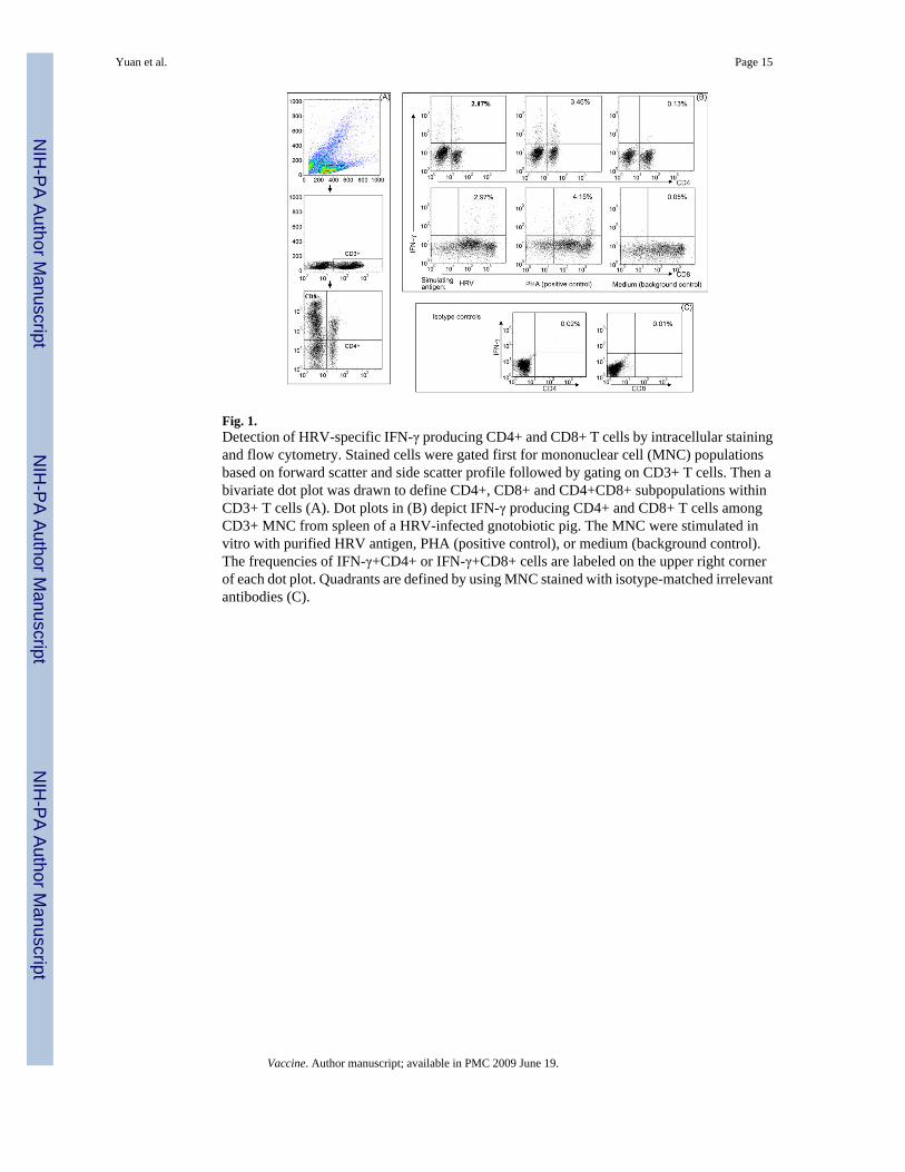
Fig. 1.Detection of HRV-specific IFN-γ producing CD4+ and CD8+ T cells by intracellular stainingand flow cytometry. Stained cells were gated first for mononuclear cell (MNC) populationsbased on forward scatter and side scatter profile followed by gating on CD3+ T cells. Then abivariate dot plot was drawn to define CD4+, CD8+ and CD4+CD8+ subpopulations withinCD3+ T cells (A). Dot plots in (B) depict IFN-γ producing CD4+ and CD8+ T cells amongCD3+ MNC from spleen of a HRV-infected gnotobiotic pig. The MNC were stimulated invitro with purified HRV antigen, PHA (positive control), or medium (background control).The frequencies of IFN-γ+CD4+ or IFN-γ+CD8+ cells are labeled on the upper right cornerof each dot plot. Quadrants are defined by using MNC stained with isotype-matched irrelevantantibodies (C).
Yuan et al. Page 15
Vaccine. Author manuscript; available in PMC 2009 June 19.
NIH
-PA Author Manuscript
NIH
-PA Author Manuscript
NIH
-PA Author Manuscript
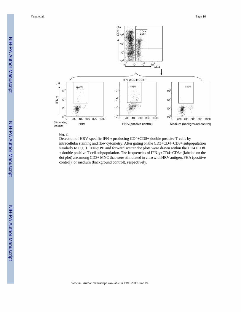
Fig. 2.Detection of HRV-specific IFN-γ producing CD4+CD8+ double positive T cells byintracellular staining and flow cytometry. After gating on the CD3+CD4+CD8+ subpopulationsimilarly to Fig. 1, IFN-γ PE and forward scatter dot plots were drawn within the CD4+CD8+ double positive T cell subpopulation. The frequencies of IFN-γ+CD4+CD8+ (labeled on thedot plot) are among CD3+ MNC that were stimulated in vitro with HRV antigen, PHA (positivecontrol), or medium (background control), respectively.
Yuan et al. Page 16
Vaccine. Author manuscript; available in PMC 2009 June 19.
NIH
-PA Author Manuscript
NIH
-PA Author Manuscript
NIH
-PA Author Manuscript
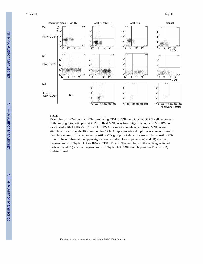
Fig. 3.Examples of HRV-specific IFN-γ producing CD4+, CD8+ and CD4+CD8+ T cell responsesin ileum of gnotobiotic pigs at PID 28. Ileal MNC was from pigs infected with VirHRV, orvaccinated with AttHRV-2/6VLP, AttHRV3x or mock-inoculated controls. MNC werestimulated in vitro with HRV antigen for 17 h. A representative dot plot was shown for eachinoculation group. The responses in AttHRV2x group (not shown) were similar to AttHRV3xgroup. The numbers at the upper right corners of dot plots of panels (A) and (B) are thefrequencies of IFN-γ+CD4+ or IFN-γ+CD8+ T cells. The numbers in the rectangles in dotplots of panel (C) are the frequencies of IFN-γ+CD4+CD8+ double positive T cells. ND,undetermined.
Yuan et al. Page 17
Vaccine. Author manuscript; available in PMC 2009 June 19.
NIH
-PA Author Manuscript
NIH
-PA Author Manuscript
NIH
-PA Author Manuscript

Fig. 4.HRV-specific IFN-γ producing T cell responses in Gn pigs. Data are presented as meanfrequency±standard error of the mean (n = 4–8). The frequencies are calculated by subtractingthe frequencies detected in medium-only stimulated MNC from HRV antigen-stimulatedMNC. Black bars, VirHRV1× group; white bars, AttHRV-2/6VLP group; light hatched bars,AttHRV3x; heavy hatched bars, AttHRV2x group; and vertical lined bars, mock-inoculatedcontrols. Different letters on top of bars indicate significant differences in frequencies amonggroups for the same cell type and tissue. Asterisks denote significant difference when comparedto ileum; and Δ indicates significant difference when compared to blood for the same cell typein the same group (Kruskal–Wallis test, p < 0.05).
Yuan et al. Page 18
Vaccine. Author manuscript; available in PMC 2009 June 19.
NIH
-PA Author Manuscript
NIH
-PA Author Manuscript
NIH
-PA Author Manuscript

Fig. 5.Mean frequencies of HRV-specific IFN-γ producing CD4+ and CD8+ T cells amongproliferating and non-proliferating T cells in pigs vaccinated with AttHRV3x. Data arepresented as mean frequency±standard error of the mean (n = 4). Black bars, ileum, hatchedbars, spleen; and white bars, blood. Asterisks denote significant difference between BrdU+and BrdU− IFN-γ producing T cells for the same tissue (Kruskal–Wallis test, p < 0.05).
Yuan et al. Page 19
Vaccine. Author manuscript; available in PMC 2009 June 19.
NIH
-PA Author Manuscript
NIH
-PA Author Manuscript
NIH
-PA Author Manuscript
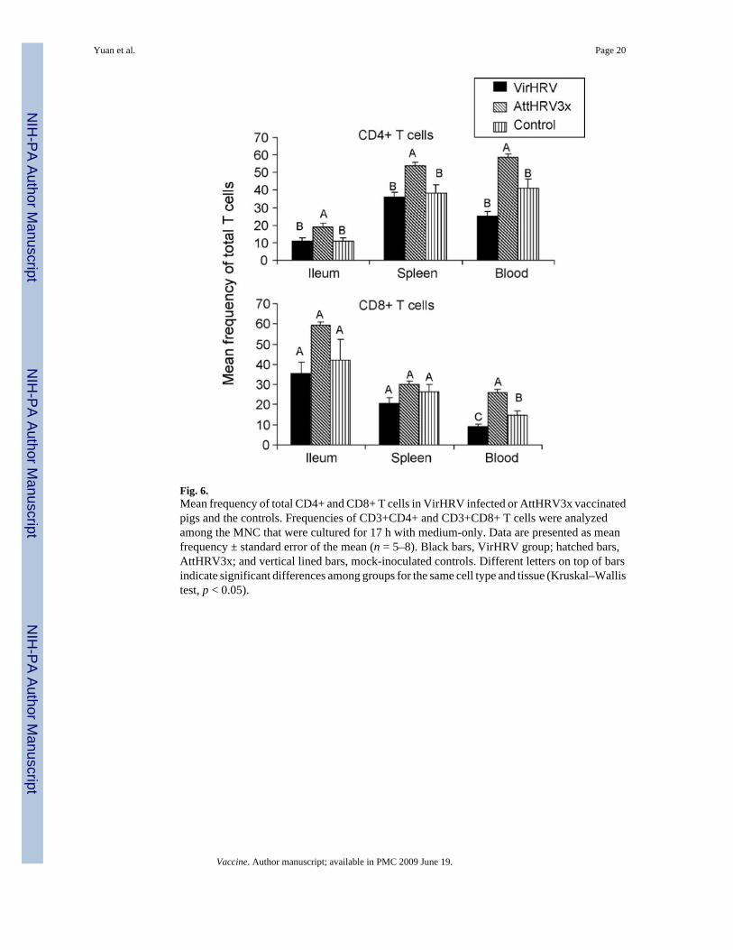
Fig. 6.Mean frequency of total CD4+ and CD8+ T cells in VirHRV infected or AttHRV3x vaccinatedpigs and the controls. Frequencies of CD3+CD4+ and CD3+CD8+ T cells were analyzedamong the MNC that were cultured for 17 h with medium-only. Data are presented as meanfrequency ± standard error of the mean (n = 5–8). Black bars, VirHRV group; hatched bars,AttHRV3x; and vertical lined bars, mock-inoculated controls. Different letters on top of barsindicate significant differences among groups for the same cell type and tissue (Kruskal–Wallistest, p < 0.05).
Yuan et al. Page 20
Vaccine. Author manuscript; available in PMC 2009 June 19.
NIH
-PA Author Manuscript
NIH
-PA Author Manuscript
NIH
-PA Author Manuscript

NIH
-PA Author Manuscript
NIH
-PA Author Manuscript
NIH
-PA Author Manuscript
Yuan et al. Page 21Ta
ble
1C
orre
latio
n be
twee
n m
agni
tude
of i
ntes
tinal
and
syst
emic
IFN
-γ p
rodu
cing
T c
ell r
espo
nses
and
pro
tect
ion
rate
s aga
inst
HR
V d
iarr
hea
Gro
up
Prot
ectio
nra
teag
ains
tH
RV
diar
rhea
a
Mea
n fr
eque
ncie
s of I
FN-γ
pro
duci
ng T
cel
ls in
ileu
mM
ean
freq
uenc
ies o
f IFN
-γ p
rodu
cing
T c
ells
in sp
leen
IFN
-γ+C
D4+
T c
ells
IFN
-γ+C
D8+
T c
ells
IFN
-γ+C
D4+
CD
8+ T
cel
lsIF
N-γ
+CD
4+ T
cel
lsIF
N-γ
+CD
8+ T
cel
lsIF
N-γ
+CD
4+C
D8+
T c
ells
VirH
RV
87%
Ab
1.40
Ac
1.49
And
d0.
11B
C0.
10B
Cnd
AttH
RV
-2/6
VLP
71%
A0.
82A
B1.
07A
0.76
A0.
51A
0.75
A0.
45A
AttH
RV
3x62
%A
0.28
B0.
07B
0.18
AB
0.61
AB
0.38
B0.
38A
AttH
RV
2x34
%A
B0.
18B
0.07
AB
0.15
AB
0.28
AB
0.01
C0.
42A
Con
trol
0%B
0.02
C0.
01B
0.01
B0.
05C
0.14
BC
0.02
B
Cor
rela
tion
coef
ficie
nte
r=1.
0 p
< 0.
0001
r = 0
.974
68 p
=0.
0048
r=1.
0 p
< 0.
0001
r = 0
.300
0 p
= 0.
6238
r = 0
.200
0 p
= 0.
7471
r = 0
.800
0 p
= 0.
2000
a At P
ID 2
8, su
bset
s of p
igs (
7–25
pig
s/gr
oup)
wer
e ch
alle
nged
ora
lly w
ith ∼
106
ID50
of V
irHR
V. R
ecta
l sw
abs w
ere
colle
cted
and
feca
l sco
res w
ere
obse
rved
from
pos
t-cha
lleng
e da
y (P
CD
) 1–6
for
asse
ssm
ent o
f dia
rrhe
a (f
ecal
scor
es o
f 0, n
orm
al; 1
, pas
ty; 2
, sem
i-liq
uid;
and
3, l
iqui
d). S
core
s of g
reat
er o
r equ
al to
2 w
ere
cons
ider
ed a
s dia
rrhe
a. T
he p
igs w
ere
cons
ider
ed a
s com
plet
ely
prot
ecte
dag
ains
t dia
rrhe
a up
on c
halle
nge
with
VirH
RV
onl
y w
hen
they
did
not
hav
e di
arrh
ea d
urin
g th
e en
tire
obse
rvat
ion
perio
d. C
ontro
l pig
s wer
e in
ocul
ated
with
dilu
ent o
r ISC
OM
mat
rix o
nly
and
chal
leng
edw
ith V
irHR
V. P
rote
ctio
n ra
te =
[1−(
perc
enta
ge o
f VirH
RV
-inoc
ulat
ed o
r vac
cina
ted
pigs
in e
ach
grou
p w
ith d
iarr
hea/
perc
enta
ge o
f con
trol p
igs w
ith d
iarr
hea)
]×10
0. P
rote
ctio
n da
ta fr
om p
rese
nt st
udy
and
prev
ious
ly p
ublis
hed
stud
ies [
3–6]
wer
e co
mbi
ned.
b Num
bers
with
diff
eren
t sup
ersc
ript c
apita
l let
ters
diff
er si
gnifi
cant
ly (F
ishe
r's e
xact
test
, p <
0.0
5).
c Mea
n fr
eque
ncie
s in
the
sam
e co
lum
n w
ith d
iffer
ent s
uper
scrip
t cap
ital l
ette
rs d
iffer
sign
ifica
ntly
(Kru
skal
–Wal
lis te
st, p
< 0
.05)
.
d nd, n
ot d
eter
min
ed.
e Cor
rela
tion
coef
ficie
nt b
etw
een
prot
ectio
n ra
tes a
nd m
ean
freq
uenc
ies o
f IFN
-γ p
rodu
cing
T c
ells
(Spe
arm
an's
corr
elat
ion)
.
Vaccine. Author manuscript; available in PMC 2009 June 19.