M31-A2 Performance Standards for Antimicrobial Disk and ...
107
M31-A2 Vol. 22 No. 6 Replaces M31-A Vol. 19 No. 11 Performance Standards for Antimicrobial Disk and Dilution Susceptibility Tests for Bacteria Isolated from Animals; Approved Standard—Second Edition This document provides the currently recommended techniques for antimicrobial agent disk and dilution susceptibility testing, criteria for quality control testing, and interpretive criteria for veterinary use. A standard for global application developed through the NCCLS consensus process.
-
Upload
khangminh22 -
Category
Documents
-
view
1 -
download
0
Transcript of M31-A2 Performance Standards for Antimicrobial Disk and ...

M31-A2 Vol. 22 No. 6
Replaces M31-A Vol. 19 No. 11
Performance Standards for Antimicrobial Disk and Dilution Susceptibility Tests for Bacteria Isolated from Animals; Approved Standard—Second Edition
This document provides the currently recommended techniques for antimicrobial agent disk and dilution susceptibility testing, criteria for quality control testing, and interpretive criteria for veterinary use. A standard for global application developed through the NCCLS consensus process.

NCCLS... Serving the World’s Medical Science Community Through Voluntary Consensus NCCLS is an international, interdisciplinary, nonprofit, standards-developing, and educational organization that promotes the development and use of voluntary consensus standards and guidelines within the healthcare community. It is recognized worldwide for the application of its unique consensus process in the development of standards and guidelines for patient testing and related healthcare issues. NCCLS is based on the principle that consensus is an effective and cost-effective way to improve patient testing and healthcare services.
In addition to developing and promoting the use of voluntary consensus standards and guidelines, NCCLS provides an open and unbiased forum to address critical issues affecting the quality of patient testing and health care.
PUBLICATIONS
An NCCLS document is published as a standard, guideline, or committee report.
Standard A document developed through the consensus process that clearly identifies specific, essential requirements for materials, methods, or practices for use in an unmodified form. A standard may, in addition, contain discretionary elements, which are clearly identified.
Guideline A document developed through the consensus process describing criteria for a general operating practice, procedure, or material for voluntary use. A guideline may be used as written or modified by the user to fit specific needs.
Report A document that has not been subjected to consensus review and is released by the Board of Directors.
CONSENSUS PROCESS
The NCCLS voluntary consensus process is a protocol establishing formal criteria for:
• the authorization of a project
• the development and open review of documents
• the revision of documents in response to comments by users
• the acceptance of a document as a consensus standard or guideline.
Most NCCLS documents are subject to two levels of consensus—“proposed” and “approved.” Depending on
the need for field evaluation or data collection, documents may also be made available for review at an intermediate (i.e., “tentative”) consensus level.
Proposed An NCCLS consensus document undergoes the first stage of review by the healthcare community as a proposed standard or guideline. The document should receive a wide and thorough technical review, including an overall review of its scope, approach, and utility, and a line-by-line review of its technical and editorial content.
Tentative A tentative standard or guideline is made available for review and comment only when a recommended method has a well-defined need for a field evaluation or when a recommended protocol requires that specific data be collected. It should be reviewed to ensure its utility.
Approved An approved standard or guideline has achieved consensus within the healthcare community. It should be reviewed to assess the utility of the final document, to ensure attainment of consensus (i.e., that comments on earlier versions have been satisfactorily addressed), and to identify the need for additional consensus documents.
NCCLS standards and guidelines represent a consensus opinion on good practices and reflect the substantial agreement by materially affected, competent, and interested parties obtained by following NCCLS’s established consensus procedures. Provisions in NCCLS standards and guidelines may be more or less stringent than applicable regulations. Consequently, conformance to this voluntary consensus document does not relieve the user of responsibility for compliance with applicable regulations.
COMMENTS
The comments of users are essential to the consensus process. Anyone may submit a comment, and all comments are addressed, according to the consensus process, by the NCCLS committee that wrote the document. All comments, including those that result in a change to the document when published at the next consensus level and those that do not result in a change, are responded to by the committee in an appendix to the document. Readers are strongly encouraged to comment in any form and at any time on any NCCLS document. Address comments to the NCCLS Executive Offices, 940 West Valley Road, Suite 1400, Wayne, PA 19087, USA.
VOLUNTEER PARTICIPATION
Healthcare professionals in all specialties are urged to volunteer for participation in NCCLS projects. Please contact the NCCLS Executive Offices for additional information on committee participation.

Volume 22 M31-A2
i
Performance Standards for Antimicrobial Disk and Dilution Susceptibility Tests for Bacteria Isolated from Animals; Approved Standard—Second Edition Abstract If the susceptibility of a bacterial pathogen to antimicrobial agents cannot be predicted based on the identity of the organism alone, in vitro antimicrobial susceptibility testing of the organism isolated from the disease processes in animals is indicated. Susceptibility testing is particularly necessary in those situations where the etiologic agent belongs to a bacterial species for which resistance to commonly used antimicrobial agents has been documented, or could arise. A variety of laboratory techniques can be used to measure the in vitro susceptibility of bacteria to antimicrobial agents. This document describes the standard agar disk diffusion method, as well as standard broth dilution (macrodilution and microdilution) and agar dilution techniques. It also includes a series of procedures designed to standardize test performance. The performance, applications, and limitations of the current NCCLS-recommended methods are described. The tabular information in this document presents the most current information for drug selection, interpretation, and quality control. Only a few compounds have veterinary-specific interpretive criteria; so, human interpretive criteria are used for the majority of compounds. As more veterinary-specific information becomes available, these changes will be incorporated into future revisions of this document. NCCLS. Performance Standards for Antimicrobial Disk and Dilution Susceptibility Tests for Bacteria Isolated from Animals; Approved Standard—Second Edition. NCCLS document M31-A2 (ISBN 1-56238-461-9). NCCLS, 940 West Valley Road, Suite 1400, Wayne, Pennsylvania 19087-1898, USA 2002.
THE NCCLS consensus process, which is the mechanism for moving a document through two or morelevels of review by the healthcare community, is an ongoing process. Users should expect revisededitions of any given document. Because rapid changes in technology may affect the procedures,methods, and protocols in a standard or guideline, users should replace outdated editions with thecurrent editions of NCCLS documents. Current editions are listed in the NCCLS Catalog, which isdistributed to member organizations, and to nonmembers on request. If your organization is not amember and would like to become one, and to request a copy of the NCCLS Catalog, contact theNCCLS Executive Offices. Telephone: 610.688.0100; Fax: 610.688.0700; E-Mail: [email protected];Website: www.nccls.org

Number 6 NCCLS
ii

M31-A2 ISBN 1-56238-461-9
ISSN 0273-3099
Performance Standards for Antimicrobial Disk and Dilution Susceptibility Tests for Bacteria Isolated from Animals; Approved Standard—Second Edition Volume 22 Number 6 Thomas R. Shryock, Ph.D., Chairholder Michael Apley, D.V.M., Ph.D. Ronald N. Jones, M.D. Donald H. Lein, D.V.M., Ph.D. Clyde Thornsberry, Ph.D. Robert D. Walker, Ph.D. Jeffrey L. Watts, Ph.D., R.M. (AAM) David G. White, Ph.D. Ching Ching Wu, D.V.M., Ph.D.

Number 6 NCCLS
iv
This publication is protected by copyright. No part of it may be reproduced, stored in a retrieval system, transmitted, or made available in any form or by any means (electronic, mechanical, photocopying, recording, or otherwise) without prior written permission from NCCLS, except as stated below. NCCLS hereby grants permission to reproduce limited portions of this publication for use in laboratory procedure manuals at a single site, for interlibrary loan, or for use in educational programs provided that multiple copies of such reproduction shall include the following notice, be distributed without charge, and, in no event, contain more than 20% of the document’s text.
Reproduced with permission, from NCCLS publication M31-A2—Performance Standards for Antimicrobial Disk and Dilution Susceptibility Tests for Bacteria Isolated from Animals; Approved Standard—Second Edition (ISBN 1-56238-461-9). Copies of the current edition may be obtained from NCCLS, 940 West Valley Road, Suite 1400, Wayne, Pennsylvania 19087-1898, USA.
Permission to reproduce or otherwise use the text of this document to an extent that exceeds the exemptions granted here or under the Copyright Law must be obtained from NCCLS by written request. To request such permission, address inquiries to the Executive Director, NCCLS, 940 West Valley Road, Suite 1400, Wayne, Pennsylvania 19087-1898, USA. Copyright ©2002. The National Committee for Clinical Laboratory Standards. Suggested Citation (NCCLS. Performance Standards for Antimicrobial Disk and Dilution Susceptibility Tests for Bacteria Isolated from Animals; Approved Standard—Second Edition. NCCLS document M31-A2 [ISBN 1-56238-461-9]. NCCLS, 940 West Valley Road, Suite 1400, Wayne, Pennsylvania 19087-1898 USA, 2002.) Tentative Standard August 1997 Approved Standard June 1999 Approved Standard—Second Edition May 2002 ISBN 1-56238-461-9 ISSN 0273-3099

Volume 22 M31-A2
v
Committee Membership Area Committee on Microbiology James H. Jorgensen, Ph.D. Chairholder
University of Texas Health Science Center San Antonio, Texas
Melvin P. Weinstein, M.D. Vice-Chairholder
Robert Wood Johnson Medical School New Brunswick, New Jersey
Subcommittee on Veterinary Antimicrobial Susceptibility Testing Thomas R. Shryock, Ph.D. Chairholder
Elanco Animal Health Greenfield, Indiana
Michael Apley, D.V.M., Ph.D. Iowa State University
Ames, Iowa Ronald N. Jones, M.D. The JONES Group/JMI
North Liberty, Iowa Donald H. Lein, D.V.M., Ph.D. Cornell University
Ithaca, New York Clyde Thornsberry, Ph.D. Focus Technologies
Franklin, Tennessee Robert D. Walker, Ph.D. FDA Center for Veterinary Medicine
Laurel, Maryland Jeffrey L. Watts, Ph.D., R.M. (AAM) Pharmacia Corporation
Kalamazoo, Michigan David G. White, Ph.D. FDA Center for Veterinary Medicine
Laurel, Maryland Ching Ching Wu, D.V.M., Ph.D. Purdue University
West Lafayette, Indiana Advisors Charles E. Benson, Ph.D. University of PA School of Veterinary Medicine
Kennett Square, Pennsylvania Scott Brown, D.V.M., Ph.D. Pharmacia Animal Health
Kalamazoo, Michigan William Fales, Ph.D.
University of Missouri Columbia, Missouri
John Glisson, D.V.M., M.A.M., Ph.D.
University of Georgia Athens, Georgia

Number 6 NCCLS
vi
Advisors (Continued) Lorraine J. Hoffman, Ph.D. Iowa State University
Ames, Iowa Kelly F. Lechtenberg, D.V.M., Ph.D. Midwest Veterinary Services, Inc.
Oakland, Nebraska Carol L. Lemons North Carolina College of Veterinary Medicine
Raleigh, North Carolina S. Blaine Leppanen Blaine Healthcare Associates, Inc.
Fair Oaks, California Jennifer Lorbach
Trek Diagnostics Systems, Inc. Westlake, Ohio
William E. Owens, Ph.D. Louisiana State University Agricultural Center
Homer, Louisiana Karen W. Post, D.V.M., M.S. Rollins Animal Disease Diagnostic Laboratory
Raleigh, North Carolina Dale E. Shuster, Ph.D. Schering-Plough Animal Health
Union, New Jersey Keith Sterner, D.V.M. Sterner Veterinary Clinic
Ionia, Michigan Tracy A. Dooley, M.L.T.(ASCP) Project Manager
NCCLS Wayne, Pennsylvania
Patrice E. Polgar Editor
NCCLS Wayne, Pennsylvania
Donna M. Wilhelm Assistant Editor
NCCLS Wayne, Pennsylvania

Volume 22 M31-A2
vii
Active Membership (As of 1 April 2002)
Sustaining Members Abbott Laboratories American Association for Clinical Chemistry Bayer Corporation Beckman Coulter, Inc. BD and Company bioMérieux, Inc. CLMA College of American Pathologists GlaxoSmithKline Nippon Becton Dickinson Co., Ltd. Ortho-Clinical Diagnostics, Inc. Roche Diagnostics, Inc. Professional Members AISAR-Associazione Italiana per lo Studio degli American Academy of Family Physicians American Association for Clinical Chemistry American Association for Respiratory Care American Chemical Society American Medical Technologists American Public Health Association American Society for Clinical Laboratory Science American Society of Hematology American Society for Microbiology American Type Culture Collection, Inc. Asociación Española Primera de Socorros (Uruguay) Asociacion Mexicana de Bioquimica Clinica A.C. Assn. of Public Health Laboratories Assoc. Micro. Clinici Italiani- A.M.C.L.I. British Society for Antimicrobial Chemotherapy CADIME-Camara De Instituciones De Diagnostico Medico Canadian Society for Medical Laboratory Science—Société Canadienne de Science de Laboratoire Médical Clinical Laboratory Management Association COLA College of American Pathologists
College of Medical Laboratory Technologists of Ontario College of Physicians and Surgeons of Saskatchewan ESCMID Fundación Bioquímica Argentina International Association of Medical Laboratory Technologists International Council for Standardization in Haematology International Federation of Clinical Chemistry Italian Society of Clinical Biochemistry and Clinical Molecular Biology Japan Society of Clinical Chemistry Japanese Committee for Clinical Laboratory Standards Joint Commission on Accreditation of Healthcare Organizations National Academy of Clinical Biochemistry National Society for Histotechnology, Inc. Ontario Medical Association Quality Management Program- Laboratory Service RCPA Quality Assurance Programs PTY Limited Sociedade Brasileira de Analises Clinicas Sociedade Brasileira de Patologia Clinica Sociedad Espanola de Bioquimica Clinica y Patologia Molecular Turkish Society of Microbiology Government Members Association of Public Health Laboratories Armed Forces Institute of Pathology BC Centre for Disease Control Centers for Disease Control and Prevention Centers for Medicare & Medicaid Services/CLIA Program Centers for Medicare & Medicaid Services Chinese Committee for Clinical Laboratory Standards Commonwealth of Pennsylvania Bureau of Laboratories Department of Veterans Affairs
Deutsches Institut für Normung (DIN) FDA Center for Devices and Radiological Health FDA Center for Veterinary Medicine FDA Division of Anti-Infective Drug Products Health Care Financing Administration/CLIA Program Health Care Financing Administration Iowa State Hygienic Laboratory Massachusetts Department of Public Health Laboratories National Association of Testing Authorities – Australia National Center of Infectious and Parasitic Diseases (Bulgaria) National Health Laboratory Service (South Africa) National Institute of Standards and Technology New York State Department of Health Ohio Department of Health Ontario Ministry of Health Pennsylvania Dept. of Health Saskatchewan Health-Provincial Laboratory Scientific Institute of Public Health; Belgium Ministry of Social Affairs, Public Health and the Environment Swedish Institute for Infectious Disease Control Thailand Department of Medical Sciences Industry Members AB Biodisk Abbott Laboratories Abbott Laboratories, MediSense Products Acrometrix Corporation Advanced Medicine, Inc. Agilent Technologies, Inc. Ammirati Regulatory Consulting Anaerobe Systems Asséssor AstraZeneca Aventis Axis-Shield POC AS Bayer Corporation – Elkhart, IN

Number 6 NCCLS
viii
Bayer Corporation – Tarrytown, NY Bayer Corporation – West Haven, CT Bayer Medical Ltd. BD BD Biosciences – San Jose, CA BD Consumer Products BD Diagnostic Systems BD Italia S.P.A. BD VACUTAINER Systems Beckman Coulter, Inc. Beckman Coulter, Inc. Primary Care Diagnostics Beckman Coulter K.K. (Japan) Bio-Development SRL Bio-Inova Life Sciences International Bio-Inova Life Sciences North America BioMedia Laboratories Sdn Bhd BioMérieux (NC) bioMérieux, Inc. (MO) Biometrology Consultants Bio-Rad Laboratories, Inc. Bio-Rad Laboratories, Inc. - France Biotest AG Blaine Healthcare Associates, Inc. Bristol-Myers Squibb Company Canadian External Quality Assessment Laboratory Capital Management Consulting, Inc. Carl Schaper Checkpoint Development Inc. Chiron Corporation ChromaVision Medical Systems, Inc. Chronolab Ag Clinical Design Group Inc. Clinical Laboratory Improvement Consultants Cognigen Community Medical Center (NJ) Control Lab (Brazil) Copan Diagnostics Inc. Cosmetic Ingredient Review Cubist Pharmaceuticals Dade Behring Inc. - Deerfield, IL Dade Behring Inc. - Glasgow, DE Dade Behring Inc. - Marburg, Germany Dade Behring Inc. - Sacramento, CA Dade Behring Inc. - San Jose, CA Diagnostic Products Corporation Eiken Chemical Company, Ltd. Electa Lab s.r.l. Enterprise Analysis Corporation Essential Therapeutics, Inc.
EXPERTech Associates, Inc. F. Hoffman-La Roche AG Fort Dodge Animal Health General Hospital Vienna (Austria) Gen-Probe GlaxoSmithKline Greiner Bio-One Inc. Helena Laboratories Home Diagnostics, Inc. Immunicon Corporation Instrumentation Laboratory International Technidyne Corporation IntraBiotics Pharmaceuticals, Inc. I-STAT Corporation Johnson and Johnson Pharmaceutical Research and Development, L.L.C. Kendall Sherwood-Davis & Geck LAB-Interlink, Inc. Laboratory Specialists, Inc. Labtest Diagnostica S.A. LifeScan, Inc. (a Johnson & Johnson Company) Lilly Research Laboratories Macemon Consultants Medical Device Consultants, Inc. Merck & Company, Inc. Minigrip/Zip-Pak Molecular Diagnostics, Inc. mvi Sciences (MA) Nabi Nichols Institute Diagnostics (Div. of Quest Diagnostics, Inc.) Nissui Pharmaceutical Co., Ltd. Nippon Becton Dickinson Co., Ltd. Norfolk Associates, Inc. Novartis Pharmaceuticals Corporation Ortho-Clinical Diagnostics, Inc. (Raritan, NJ) Ortho-Clinical Diagnostics, Inc. (Rochester, NY) Oxoid Inc. Paratek Pharmaceuticals Pfizer Inc Pharmacia Corporation Powers Consulting Services Premier Inc. Procter & Gamble Pharmaceuticals, Inc. The Product Development Group QSE Consulting Quintiles, Inc. Radiometer America, Inc. Radiometer Medical A/S David G. Rhoads Associates, Inc. Roche Diagnostics GmbH
Roche Diagnostics, Inc. Roche Laboratories (Div. Hoffmann-La Roche Inc.) Sarstedt, Inc. SARL Laboratoire Carron (France) Schering Corporation Schleicher & Schuell, Inc. Second Opinion Showa Yakuhin Kako Company, Ltd. Streck Laboratories, Inc. SurroMed, Inc. Sysmex Corporation (Japan) Sysmex Corporation (Long Grove, IL) The Clinical Microbiology Institute The Toledo Hospital (OH) Trek Diagnostic Systems, Inc. Versicor, Inc. Vetoquinol S.A. Visible Genetics, Inc. Vysis, Inc. Wallac Oy Wyeth-Ayerst Xyletech Systems, Inc. YD Consultant YD Diagnostics (Seoul, Korea) Trade Associations AdvaMed Association of Medical Diagnostic Manufacturers Japan Association Clinical Reagents Ind. (Tokyo, Japan) Medical Industry Association of Australia Associate Active Members 20th Medical Group (SC) 31st Medical Group/SGSL (APO, AE) 67th CSH Wuerzburg, GE (NY) 121st General Hospital (CA) Academisch Ziekenhuis-VUB (Belgium) Acadiana Medical Laboratories, LTD (LA) Adena Regional Medical Center (OH) Advocate Healthcare Lutheran General (IL) Akershus Central Hospital and AFA (Norway) Albemarle Hospital (NC) Allegheny General Hospital (PA)

Volume 22 M31-A2
ix
Allegheny University of the Health Sciences (PA) Allina Health System (MN) Alton Ochsner Medical Foundation (LA) American Medical Laboratories (VA) Arkansas Department of Health ARUP at University Hospital (UT) Armed Forces Research Institute of Medical Science (APO, AP) Associated Regional & University Pathologists (UT) Aurora Consolidated Laboratories (WI) Azienda Ospedale Di Lecco (Italy) Bay Medical Center (MI) Baystate Medical Center (MA) Bbaguas Duzen Laboratories (Turkey) Bermuda Hospitals Board Bo Ali Hospital (Iran) British Columbia Cancer Agency (Vancouver, BC, Canada) Broward General Medical Center (FL) Calgary Laboratory Services Carilion Consolidated Laboratory (VA) Cathay General Hospital (Taiwan) CB Healthcare Complex (Sydney, NS, Canada) Central Peninsula General Hospital (AK) Central Texas Veterans Health Care System Centre Hospitalier Regional del la Citadelle (Belgium) Centro Diagnostico Italiano (Milano, Italy) Champlain Valley Physicians Hospital (NY) Changi General Hospital (Singapore) Children’s Hospital (NE) Children’s Hospital & Clinics (MN) Children’s Hospital King's Daughters (VA) Children’s Hospital Medical Center (Akron, OH) Children’s Hospital of Philadelphia (PA) Children’s Medical Center of Dallas (TX) Clarian Health–Methodist Hospital (IN) Clendo Lab (Puerto Rico)
Clinical Laboratory Partners, LLC (CT) CLSI Laboratories (PA) Columbia Regional Hospital (MO) Commonwealth of Kentucky Community Hospital of Lancaster (PA) CompuNet Clinical Laboratories (OH) Cook Children’s Medical Center (TX) Covance Central Laboratory Services (IN) Danish Veterinary Laboratory (Denmark) Danville Regional Medical Center (VA) Delaware Public Health Laboratory Department of Health & Community Services (New Brunswick, Canada) DesPeres Hospital (MO) DeTar Hospital (TX) Detroit Health Department (MI) Dr. Everett Chalmers Hospital (New Brunswick, Canada) Doctors Hospital (Bahamas) Duke University Medical Center (NC) E.A. Conway Medical Center (LA) Eastern Maine Medical Center East Side Clinical Laboratory (RI) Elyria Memorial Hospital (OH) Emory University Hospital (GA) Esoterix Center for Infectious Disease (TX) Fairview-University Medical Center (MN) Federal Medical Center (MN) Florida Hospital East Orlando Foothills Hospital (Calgary, AB, Canada) Fort St. John General Hospital (Fort St. John, BC, Canada) Fox Chase Cancer Center (PA) Fresenius Medical Care/Spectra East (NJ) Fresno Community Hospital and Medical Center Frye Regional Medical Center (NC) Gambro Healthcare Laboratory Services (FL) Gateway Medical Center (TN) Geisinger Medical Center (PA) Grady Memorial Hospital (GA) Guthrie Clinic Laboratories (PA) Hahnemann University Hospital (PA)
Harris Methodist Erath County (TX) Harris Methodist Fort Worth (TX) Hartford Hospital (CT) Headwaters Health Authority (Alberta, Canada) Health Network Lab (PA) Health Partners Laboratories (VA) Heartland Regional Medical Center (MO) Highlands Regional Medical Center (FL) Hoag Memorial Hospital Presbyterian (CA) Holmes Regional Medical Center (FL) Holzer Medical Center (OH) Hospital for Sick Children (Toronto, ON, Canada) Hospital Sousa Martins (Portugal) Hotel Dieu Hospital (Windsor, ON, Canada) Houston Medical Center (GA) Huddinge University Hospital (Sweden) Hurley Medical Center (MI) Indiana State Board of Health Indiana University Institute of Medical and Veterinary Science (Australia) Instituto Scientifico HS. Raffaele (Italy) International Health Management Associates, Inc. (IL) Jackson Memorial Hospital (FL) Jersey Shore Medical Center (NJ) John C. Lincoln Hospital (AZ) John F. Kennedy Medical Center (NJ) John Peter Smith Hospital (TX) Kadlec Medical Center (WA) Kaiser Permanente Medical Care (CA) Kaiser Permanente (MD) Kantonsspital (Switzerland) Keller Army Community Hospital (NY) Kenora-Rainy River Regional Laboratory Program (Ontario, Canada) Kern Medical Center (CA) Kimball Medical Center (NJ) King Faisal Specialist Hospital (Saudi Arabia) King Khalid National Guard Hospital (Saudi Arabia) King’s Daughter Medical Center (KY)

Number 6 NCCLS
x
Klinični Center (Slovenia) Laboratories at Bonfils (CO) Laboratoire de Santé Publique du Quebec (Canada) Laboratório Fleury S/C Ltda. (Brazil) Laboratory Corporation of America (NJ) Laboratory Corporation of America (MO) LAC and USC Healthcare Network (CA) Lakeland Regional Medical Center (FL) Lancaster General Hospital (PA) Langley Air Force Base (VA) LeBonheur Children’s Medical Center (TN) Libero Instituto Univ. Campus BioMedico (Italy) Long Beach Memorial Medical Center (CA) Louisiana State University Medical Center Maccabi Medical Care and Health Fund (Israel) Magee Womens Hospital (PA) Malcolm Grow USAF Medical Center (MD) Manitoba Health (Winnipeg, Canada) Martin Luther King/Drew Medical Center (CA) Massachusetts General Hospital (Microbiology Laboratory) MDS Metro Laboratory Services (Burnaby, BC, Canada) Medical College of Virginia Hospital Medicare/Medicaid Certification, State of North Carolina Memorial Medical Center (IL) Memorial Medical Center (LA) Jefferson Davis Hwy Memorial Medical Center (LA) Napoleon Avenue Mescalero Indian Hospital (NM) Methodist Hospital (TX) Methodist Hospitals of Memphis (TN) MetroHealth Medical Center (OH) Michigan Department of Community Health Mississippi Baptist Medical Center Monte Tabor – Centro Italo - Brazileiro de Promocao (Brazil) Montreal Children’s Hospital (Canada)
Montreal General Hospital (Canada) MRL Pharmaceutical Services, Inc. (VA) MRL Reference Laboratory (CA) Nassau County Medical Center (NY) National Institutes of Health (MD) Naval Surface Warfare Center (IN) Nebraska Health System New Britain General Hospital (CT) New England Fertility Institute (CT) North Carolina State Laboratory of Public Health Northern Indiana Education Foundation North Kansas City Hospital (MO) North Shore – Long Island Jewish Health System Laboratories (NY) Northwestern Memorial Hospital (IL) O.L. Vrouwziekenhuis (Belgium) Ordre professionnel des technologists médicaux du Québec Ospedali Riuniti (Italy) The Ottawa Hospital (Ottawa, ON, Canada) Our Lady of Lourdes Hospital (NJ) Our Lady of the Resurrection Medical Center (IL) Pathology and Cytology Laboratories, Inc. (KY) The Permanente Medical Group (CA) Piedmont Hospital (GA) Pikeville Methodist Hospital (KY) Pocono Hospital (PA) Presbyterian Hospital of Dallas (TX) Queen Elizabeth Hospital (Prince Edward Island, Canada) Queensland Health Pathology Services (Australia) Quest Diagnostics Incorporated (CA) Quintiles Laboratories, Ltd. (GA) Regions Hospital Reid Hospital & Health Care Services (IN) Research Medical Center (MO) Rex Healthcare (NC) Rhode Island Department of Health Laboratories Riyadh Armed Forces Hospital (Saudi Arabia)
Royal Columbian Hospital (New Westminster, BC, Canada) Sacred Heart Hospital (MD) Saint Mary’s Regional Medical Center (NV) St. Alexius Medical Center (ND) St. Anthony Hospital (CO) St. Anthony’s Hospital (FL) St. Barnabas Medical Center (NJ) St-Eustache Hospital (Quebec, Canada) St. Francis Medical Ctr. (CA) St. John Hospital and Medical Center (MI) St. John Regional Hospital (St. John, NB, Canada) St. Joseph Hospital (NE) St. Joseph’s Hospital – Marshfield Clinic (WI) St. Joseph Mercy Hospital (MI) St. Luke’s Regional Medical Center (IA) St. Mary Medical Center (IN) St. Mary of the Plains Hospital (TX) St. Mary’s Hospital & Medical Center (CO) St. Paul’s Hospital (Vancouver, BC, Montreal) St. Vincent Medical Center (CA) Ste. Justine Hospital (Montreal, PQ, Canada) Salina Regional Health Center (KS) San Francisco General Hospital (CA) Santa Clara Valley Medical Center (CA) Seoul Nat’l University Hospital (Korea) Shanghai Center for the Clinical Laboratory (China) South Bend Medical Foundation (IN) Southwest Texas Methodist Hospital (TX) South Western Area Pathology Service (Australia) Southern Maine Medical Center Specialty Laboratories, Inc. (CA) Stanford Hospital and Clinics (CA) State of Washington Department of Health Stony Brook University Hospital (NY) Stormont-Vail Regional Medical Center (KS) Sun Health-Boswell Hospital (AZ)

Volume 22 M31-A2
xi
Sunrise Hospital and Medical Center (NV) Swedish Medical Center – Providence Campus (WA) Tampa General Hospital (FL) Temple University Hospital (PA) Tenet Odessa Regional Hospital (TX) The Toledo Hospital (OH) Touro Infirmary (LA) Trident Regional Medical Center (SC) Tripler Army Medical Center (HI) Truman Medical Center (MO) UCSF Medical Center (CA) UNC Hospitals (NC) University Hospital (Gent) (Belgium) University Hospitals of Cleveland (OH) The University Hospitals (OK) University of Alabama-Birmingham Hospital
University of Alberta Hospitals (Canada) University of Colorado Health Science Center University of Chicago Hospitals (IL) University of Illinois Medical Center University of the Ryukyus (Japan) University of Texas M.D. Anderson Cancer Center University of Virginia Medical Center University of Washington UZ-KUL Medical Center (Belgium) VA (Denver) Medical Center (CO) VA (Kansas City) Medical Center (MO) VA (Western NY) Healthcare System VA (San Diego) Medical Center (CA)
VA (Tuskegee) Medical Center (AL) VA Outpatient Clinic (OH) Vejle Hospital (Denmark) Washington Adventist Hospital (MD) Washoe Medical Center Laboratory (NV) West Jefferson Medical Center (LA) Wilford Hall Medical Center (TX) William Beaumont Hospital (MI) Williamsburg Community Hospital (VA) Winn Army Community Hospital (GA) Winnipeg Regional Health Authority (Winnipeg, Canada) Wishard Memorial Hospital (IN) Yonsei University College of Medicine (Korea) York Hospital (PA)
OFFICERS
BOARD OF DIRECTORS
F. Alan Andersen, Ph.D., President Cosmetic Ingredient Review Donna M. Meyer, Ph.D., President Elect CHRISTUS Health Emil Voelkert, Ph.D. Secretary Roche Diagnostics GmbH Gerald A. Hoeltge, M.D. Treasurer The Cleveland Clinic Foundation William F. Koch, Ph.D., Immediate Past President National Institute of Standards and Technology John V. Bergen, Ph.D., Executive Director
Susan Blonshine, RRT, RPFT, FAARC TechEd Wayne Brinster BD Kurt H. Davis, FCSMLS, CAE Canadian Society for Medical Laboratory Science Robert L. Habig, Ph.D. Ampersand Medical Thomas L. Hearn, Ph.D. Centers for Disease Control and Prevention Carolyn D. Jones, J.D., M.P.H. AdvaMed
Tadashi Kawai, M.D., Ph.D. International Clinical Pathology Center J. Stephen Kroger, M.D., FACP COLA Gary L. Myers, Ph.D. Centers for Disease Control and Prevention Barbara G. Painter, Ph.D. Bayer Corporation Ann M. Willey, Ph.D., J.D. New York State Department of Health Judith A. Yost, M.A., M.T.(ASCP) Centers for Medicare & Medicaid Services

Number 6 NCCLS
xii

Volume 22 M31-A2
xiii
Contents
Abstract ................................................................................................................................................... i
Committee Membership ......................................................................................................................... v
Active Membership .............................................................................................................................. vii
Foreword ............................................................................................................................................ xvii
1 Introduction ............................................................................................................................... 1 1.1 Scope............................................................................................................................. 1 1.2 Indications for Performing Susceptibility Testing ........................................................ 2 1.3 Performance Standards for Disk Diffusion Antimicrobial Susceptibility Tests for
Bacteria That Grow Aerobically .................................................................................. 2 1.4 Performance Standards for Dilution Antimicrobial Susceptibility Tests for Bacteria
That Grow Aerobically................................................................................................. 3 1.5 Limitations of Disk Diffusion and Dilution Methods ................................................... 3
2 Selection of Antimicrobial Agents for Routine Testing and Reporting .................................... 4 2.1 Definitions .................................................................................................................... 4 2.2 Selection of Antimicrobial Agents................................................................................ 6 2.3 Nonproprietary Names.................................................................................................. 7 2.4 Selection Guidelines ..................................................................................................... 7 2.5 Antimicrobial Classes ................................................................................................... 8 2.6 Guidelines for Routine Reporting ............................................................................... 11 2.7 Guidelines for Selective Reporting ............................................................................. 11 2.8 Interpretive Categories................................................................................................ 12
3 Disk Diffusion Susceptibility Tests......................................................................................... 13 3.1 Equivalent MIC Breakpoints ...................................................................................... 13 3.2 Methodologies............................................................................................................. 13
4 Broth and Agar Dilution Susceptibility Testing...................................................................... 21 4.1 Indications for Performing Broth and Agar Dilution Susceptibility Tests.................. 21 4.2 Methodologies............................................................................................................. 22
5 Quality Control Guidelines ..................................................................................................... 31 5.1 Purpose........................................................................................................................ 31 5.2 Quality Control Responsibilities................................................................................. 32 5.3 Suggested Reference Strains....................................................................................... 33 5.4 Common Sources of Error .......................................................................................... 35 5.5 Disk Diffusion Tests ................................................................................................... 36 5.6 Frequency of Quality Control Testing ........................................................................ 36 5.7 Dilution Susceptibility Tests....................................................................................... 38 5.8 Daily Testing............................................................................................................... 38 5.9 Weekly Testing ........................................................................................................... 39 5.10 Corrective Action........................................................................................................ 39 5.11 Reporting Patient Results When Out-of-Control Tests Occur .................................... 41
6 Specific Antimicrobial Resistance Tests ................................................................................. 41 6.1 Purpose........................................................................................................................ 41 6.2 β-Lactamase Tests ...................................................................................................... 41

Number 6 NCCLS
xiv
Contents (Continued)
6.3 Selecting a β-Lactamase Test ..................................................................................... 41 6.4 Detection of Extended-Spectrum, β-Lactamase-Producing, Gram-Negative Bacilli . 42 6.5 Detection of Resistant Staphylococci ......................................................................... 42 6.6 Detection of Resistant Enterococci ............................................................................. 43
7 Cumulative Antimicrobial Susceptibility Profile .................................................................... 44
References ............................................................................................................................................ 45
Appendix A. Antimicrobial Susceptibility Quality Control Record .................................................... 48
Appendix B1. Disk Diffusion Daily Quality Control Testing Protocol .............................................. 49
Appendix B2. Disk Diffusion Weekly Quality Control Testing Protocol............................................ 50
Appendix C1. Aerobic Dilution Daily Quality Control Testing Protocol ............................................ 51
Appendix C2. Aerobic Dilution Weekly Quality Control Testing Protocol ........................................ 52 Table 1. Comprehensive List of Antimicrobial Agents That Could Be Considered for Routine Testing by Veterinary Microbiology Laboratories..................................... 53 Table 2. Zone Diameter Interpretive Standards and Minimal Inhibitory Concentration (MIC) Breakpoints for Veterinary Pathogens ................................................................ 55 Table 3. Culture Collection Numbers for Organisms Used for Quality Control of Antimicrobial Susceptibility Tests ................................................................................. 60 Table 4. Acceptable Quality Control Ranges of Antimicrobial Disk Susceptibility Test
Zone Diameters (mm) for Reference Strains on Mueller-Hinton Medium Without Blood or Other Supplements .......................................................................................... 61
Table 5. Acceptable Quality Control Ranges of MICs (µg/mL) for Reference Strains ............... 63 Table 5A. Tentative Quality Control Ranges for the Agar Dilution Method of Testing
Campylobacter jejuni (ATCC® 33560) .......................................................................... 65 Table 6. Acceptable Quality Control Ranges for Haemophilus somnus and
Actinobacillus pleuropneumoniae .................................................................................. 66 Table 7. Suggested Modifications of Standard Methods for Susceptibility Testing of Some Fastidious and Special Problem Veterinary Pathogens.................................... 67 Table 8. Solvents and Diluents for Preparation of Stock Solutions of Antimicrobial
Agents Requiring Solvents Other Than Water ............................................................... 68 Table 9A. Screening and Confirmatory Tests for Extended-spectrum-β-lactamases (ESBLs) in Klebsiella pneumoniae, K. oxytoca, and Escherichia coli
(Zone Diameter) ............................................................................................................. 69 Table 9B. Screening and Confirmatory Tests for ESBLs in Klebsiella pneumoniae, K. oxytoca, and Escherichia coli (MIC) ......................................................................... 70

Volume 22 M31-A2
xv
Contents (Continued) Table 9C. Oxacillin-Salt Agar Screening Test for Staphylococcus aureus.....................................71 Table 9D. Screening Tests for High-Level Aminoglycoside Resistance (HLAR) and Vancomycin Resistance in Enterococcus spp. ............................................................... 72 Glossary 1. Antimicrobial Class, Antimicrobial Agents, and Antimicrobial Resistance
Mechanisms.................................................................................................................... 73 Glossary 2. Abbreviations for Antimicrobial Agents Incorporated into Disks or Susceptibility
Panels.............................................................................................................................. 75
Summary of Comments and Subcommittee Responses ........................................................................ 77
Summary of Delegate Comments and Subcommittee Responses ......................................................... 79
Related NCCLS Publications ................................................................................................................ 80

Number 6 NCCLS
xvi

Volume 22 M31-A2
xvii
Foreword This approved revision of the M31 standard represents a continuation of the collective efforts of the Subcommittee on Veterinary Antimicrobial Susceptibility Testing to produce a useful consensus document for standardized in vitro susceptibility testing of veterinary pathogens. The subcommittee has worked diligently to improve M31-A—Performance Standards for Antimicrobial Disk and Dilution Susceptibility Tests for Bacteria Isolated from Animals; Approved Standard, by incorporating relevant updates derived from NCCLS documents M2—Performance Standards for Antimicrobial Disk Susceptibility Tests, and M7—Methods for Dilution Antimicrobial Susceptibility Tests for Bacteria That Grow Aerobically, as well as referencing other relevant NCCLS documents. The subcommittee recognizes the ongoing NCCLS efforts that are necessary to maintain M2 and M7 as “state of the art” and expresses its appreciation to the Subcommittee on Antimicrobial Susceptibility Testing for its contributions. In addition, it is also appropriate to acknowledge the users of M31-A for their continued support and application of M31-A in their daily work routine. In the interval between the issuance of M31-A and the finalization of this revision, rapid and dynamic global events have placed even more importance on the essential role of antimicrobial susceptibility testing of bacteria isolated from animals. For example, judicious use guidelines (consensus directions for the appropriate use of antimicrobial agents in animals) have been developed worldwide by veterinary, industry, and public health organizations for both food and companion animals, that emphasize the critical need for obtaining susceptibility data using standardized test methods and interpretive criteria. M31-A2 is updated to meet these needs. Also, several additional antimicrobial agents have been reviewed and received veterinary-specific interpretive criteria approval from the subcommittee. M31-A2 features these new antimicrobial agents in Table 1, Group A (FDA-approved, NCCLS-approved, veterinary-specific interpretive criteria). Further revisions were made to designate FDA-approved, human interpretive criteria (Group B); FDA-approved drugs with no interpretive criteria (Group C); and, finally “AMDUCA-use” (Animal Medicinal Drug Use Clarification Act) products (Group D). Additionally, refinements to flexible label usage, and definitions for “susceptible” and “resistant” designations are now included. In conjunction with the elaboration of veterinary guidelines, a number of antimicrobial resistance monitoring programs throughout the world have begun to apply NCCLS standardized test methods to their work, with the expectation that susceptibility data can be compared across regions. As a first step toward worldwide applications of susceptibility testing using NCCLS methods, a new separate section lists quality control strain entries in various international repositories. Additionally, a glossary of antimicrobial agents and resistance mechanisms, and a listing of antimicrobial resistance tests have been assembled for the convenience of investigators interested in this area. Development of specific test methods and quality control values for several key agents effective against campylobacter have also been developed with the expectation that they will contribute to improved monitoring efforts. Finally, a flow sheet for quality control testing is now available that should aid laboratories to track their performance for increased proficiency. The NCCLS Subcommittee on Veterinary Antimicrobial Susceptibility Testing has revised the document to meet the need for specific testing standards, and to serve as an educational resource. It is our collective hope that diagnostic laboratory personnel, veterinarians, students, and allied professionals will benefit from this document. The user should be aware that, while M31 is primarily derived from NCCLS M2 and M7 which are “human” documents, this version incorporates more veterinary-specific information than before, although there is still ample opportunity for more. This standard should not be considered a static document; it will change as improved methods for testing veterinary pathogens become available and in response to changes in antimicrobial agent usage in veterinary medicine. The subcommittee is committed to making every effort to incorporate the latest information into future versions of the M31 standard. It is anticipated that future revisions will encompass those antimicrobial

Number 6 NCCLS
xviii
Foreword (Continued) agents used for enteric disease therapy or control. Until now, the inclusion of interpretive criteria was limited to systemic disease and mastitis; however, the subcommittee has now allowed the opportunity for inclusion of products that are useful in enteric diseases by incorporating new review criteria in NCCLS document M37—Development of In Vitro Susceptibility Testing Criteria and Quality Control Parameters for Veterinary Antimicrobial Agents. As before, prevention and growth promotion uses are not covered in M31-A2 with respect to interpretive criteria. The subcommittee anticipates that, as a result of the efforts of several working groups, additional fastidious pathogens will be included in future editions, such as the intestinal spirochetes, Haemophilus parasuis, and mycoplasma. A separate NCCLS working group is developing antimicrobial susceptibility testing guidelines for aquaculture pathogens. Additionally, the inclusion of antimicrobial agents not currently marketed in the United States is welcomed for future editions. As mentioned before, the inclusion of international entries in culture collections of quality control strains is viewed as the first step in this process, so that leading laboratories might begin to use NCCLS methods and compare them with their own national methods. Perhaps the most important future revision will be to shift antimicrobial agents from Group C or B, to A. Because approvals of new antimicrobial agents for veterinary medicine are foreseen to be a rare event, the subcommittee will shift its focus to the Generics Working Group to develop data in conformance with the M37 guideline in order to continue to advance generic drugs into Group A. As chairholder, I encourage and welcome input regarding ways to improve the document. In closing, I recognize the efforts of the Subcommittee on Veterinary Antimicrobial Susceptibility Testing in producing this revised document. I would like to particularly acknowledge the individual members of the Editorial Working Group. Their willingness to sacrifice significant amounts of their personal time for the editing process and to address controversial topics demonstrates a real commitment to the NCCLS process and the advancement of the veterinary and microbiology professions. In particular, I thank Jeff Watts for his pioneering leadership that was instrumental in developing M31-A and, now, in leading the revision process. I thank Bob Walker for his tremendous work in developing quality control values for susceptibility testing of campylobacter and serving on nearly all of the working groups that contributed the “nuts and bolts” of methods and quality control processes to the document. I thank Clyde Thornsberry and Ron Jones for allowing us to benefit from their experience in developing standards for testing antimicrobial agents for human use. I thank David White for contributing his expertise on resistance mechanisms. Finally, I would like to express my sincere appreciation to the members of the NCCLS Executive Offices’ staff for their ongoing support with the countless revisions, meetings, phone calls, and e-mails that were necessary to produce this document.
Thomas R. Shryock, Ph.D., Chairholder, Subcommittee on Veterinary
Antimicrobial Susceptibility Testing
Key Words Agar diffusion, agar dilution, antimicrobial agent, antimicrobial susceptibility, susceptibility testing, veterinary Mission Statement To develop and promote performance standards and interpretive criteria for in vitro antimicrobial susceptibility testing of bacteria isolated from animals.

Volume 22 M31-A2
An NCCLS global consensus standard. ©NCCLS. All rights reserved. 1
Performance Standards for Antimicrobial Disk and Dilution Susceptibility Tests for Bacteria Isolated from Animals;
Approved Standard—Second Edition 1 Introduction A variety of laboratory techniques can be used to measure the in vitro susceptibility of bacteria to antimicrobial agents. These include disk diffusion and broth and agar dilution techniques. This document includes a series of recommendations to help standardize the way these tests are actually performed. The performance, applications, and limitations of the currently recommended methods are described. Recommendations by the International Collaborative Study (ICS) and initial regulations proposed by the U.S. Food and Drug Administration have been reviewed, and the appropriate sections have been incorporated into this standard.1-3 This document describes current methodology applicable to therapeutic uses of antimicrobial agents used in veterinary medicine for diseases of animals as described in Section 2. It is the opinion of the subcommittee that the development of new, or modified, in vitro testing procedures to determine breakpoints or zone of inhibition interpretive criteria to accommodate specific field usage practices in livestock was not realistic for two reasons: First, there is no apparent variable which can be easily modified in the current procedure to reflect a key factor which will correlate to in vivo efficacy. For example, any alteration in the inoculum (which might be reflective of an initial low infectious dose early in an infectious process) would need to be validated with efficacy studies in animals. Furthermore, redesigning the current methodology would require the development of new quality control guidelines; possibly disks with lowered antimicrobial content, or extended dilutions on MIC dilution panels. Second, even if such a procedure were developed, how realistic is it to expect a laboratory to use it and explain the outcome to a veterinarian or other client? The subcommittee will continue to consider new developments in test methodologies and procedures, as well as revisions to interpretive criteria for therapeutic agents. With respect to antimicrobial agents used to promote or enhance animal growth, the beneficial effects of antimicrobial agents can neither be entirely ascribed to effects on the microbial gut flora, nor correlated to physiological effects on the animal. Thus, new in vitro techniques for prediction of clinical outcome will be considered only when a better understanding of the mode of action of antimicrobial agents in this situation becomes available. 1.1 Scope This document provides veterinary diagnostic laboratories with currently recommended antimicrobial agent disk and dilution susceptibility test methods for bacteria isolated from animals; criteria for quality control testing; and interpretive criteria. The interpretive criteria are intended only to support therapeutic label claims for animal antimicrobial agent use and do not apply to label claims for disease prevention or performance enhancement. Relatively few antimicrobial agents have had veterinary-specific interpretive criteria established, therefore human interpretive criteria are used for those antimicrobial agents lacking veterinary-specific interpretive criteria (see NCCLS documents M2—Performance Standards for Antimicrobial Disk Susceptibility Tests, M7— Methods for Dilution Antimicrobial Susceptibility Tests for Bacteria That Grow Aerobically, and M11—Methods for Antimicrobial Susceptibility Testing of Anaerobic Bacteria). As more veterinary-specific information becomes available, changes in the listing of the agents will be incorporated into future revisions of this document.

Number 6 NCCLS
An NCCLS global consensus standard. ©NCCLS. All rights reserved. 2
Additionally, the document provides a brief overview of the various antimicrobial classes and mechanisms of resistance to them, including specific tests for antimicrobial resistance. 1.2 Indications for Performing Susceptibility Testing Susceptibility testing is indicated for any bacterial pathogen that contributes to an infectious process requiring antimicrobial agent intervention, if its susceptibility cannot be reliably predicted from knowledge of the organism’s identity. Susceptibility tests are most often indicated when the causative organism is thought to belong to a bacterial species capable of possessing resistance mechanisms to commonly used antimicrobial agents. Mechanisms of resistance include production of drug-inactivating enzymes, alteration of drug targets, and altered uptake or efflux. Some organisms still have predictable susceptibility to antimicrobial agents, and empiric therapy is widely recognized. Susceptibility tests are seldom necessary when the infection is due to a microorganism known to be highly susceptible to a specific drug and to which resistance has not been reported (e.g., the continued susceptibility of Arcanobacterium pyogenes to penicillin). Susceptibility tests are also important in studies of the epidemiology of resistance and in studies of new antimicrobial agents. Colonies of each type of bacterial pathogen isolated from an infectious disease process should be selected from primary agar plates and tested for susceptibility. A statistical model for assessing sample size for bacterial colony selection has been proposed.4 Identification procedures are often performed at the same time as in vitro susceptibility tests. Mixtures of different types of microorganisms should not be tested on the same susceptibility test plate. The practice of conducting susceptibility tests directly with clinical material (e.g., normally sterile body fluids and urine) should be avoided. When the nature of the infection is not clear and the specimen contains mixed growth or normal flora, in which the organisms probably bear little relationship to the infectious process being treated, susceptibility tests are often unnecessary, and the results may be misleading and result in incorrect therapy. 1.3 Performance Standards for Disk Diffusion Antimicrobial Susceptibility Tests for Bacteria That Grow Aerobically In many veterinary diagnostic laboratories, the agar disk diffusion method is used for testing the common, rapidly growing bacterial pathogens. If results are to be reliable, the technical details of such procedures must be carefully standardized and controlled. Agar disk diffusion methods based solely on the presence or absence of a zone of inhibition, without regard to its magnitude as an indicator of the organism's susceptibility to the drug being tested, are not acceptable. The standardized method currently recommended by the NCCLS Subcommittee on Veterinary Antimicrobial Susceptibility Testing for agar disk diffusion testing was adapted from those methods in NCCLS document M2—Performance Standards for Antimicrobial Disk Susceptibility Tests, originally described by Bauer, et al.5 This is the most completely described method for which interpretive standards have been developed and supported by clinical and laboratory data. Reliable results can be obtained with disk diffusion tests that use standardized methodology and zone diameter measurement. These results may be correlated with minimal inhibitory concentration (MIC) among clinically susceptible and resistant species. Caution is urged if one attempts to use a regression line analysis to extrapolate MIC values from measurements of zones of inhibition because, in some cases, the relationship, while mathematically correct, cannot always be considered comparable to an MIC derived by actual dilution testing for a given isolate (see NCCLS document M37— Development of In Vitro Susceptibility Testing Criteria and Quality Control Parameters for Veterinary Antimicrobial Agents).

Volume 22 M31-A2
An NCCLS global consensus standard. ©NCCLS. All rights reserved. 3
1.4 Performance Standards for Dilution Antimicrobial Susceptibility Tests for Bacteria That Grow Aerobically Both broth and agar dilution techniques may be used to measure quantitatively the in vitro activity of an antimicrobial agent against a given bacterial culture. Basically, a series of tubes, wells, or plates is prepared with an agar or broth medium to which various concentrations of the antimicrobial agents are added. The tubes, wells, or plates are then inoculated with a suitably standardized suspension of the test organism. After overnight incubation at 35 °C, the tests are examined and the MIC is determined. The final result is influenced greatly by methodology, which must be carefully controlled if reproducible results (intralaboratory and interlaboratory) are to be achieved. Therefore, for the sake of standardization, there must be a reference method to which all other techniques can be compared. This document describes such standard agar dilution and standard broth dilution (macrodilution and microdilution) techniques. These techniques are taken from NCCLS document M7—Methods for Dilution Antimicrobial Susceptibility Tests for Bacteria That Grow Aerobically based largely on information gathered from the International Collaborative Study in Human Medicine.1 Although these methods are primarily standard reference methods, some are sufficiently practical to warrant their use both in clinical laboratories and in the research laboratory. If one of these tests is to be adopted as the routine method in an institution where it has not been previously used, an educational program to enable the clinicians and the staff to understand how to use the quantitative results should be carried out before the sole use of the test is initiated. The MIC obtained using a dilution test provides insight as to the concentration of antimicrobial agent needed at the site of infection to inhibit the infecting organism. The MIC, however, does not represent an absolute value. The “true” MIC is somewhere between the lowest test concentration that inhibits the organism’s growth (that is, the MIC reading) and the next lower test concentration. For example, using twofold dilutions if the MIC was determined to be 16 µg/mL, the “true” MIC would be between 16 and 8 µg/mL. Even under the best of controlled conditions, a dilution test may not yield the same end point each time it is performed. Generally, the acceptable reproducibility of the test is within one twofold dilution of the actual end point. To avoid greater variability, the dilution test must be standardized and carefully controlled as described herein. MICs have been determined using concentrations derived traditionally from serial twofold dilutions indexed to the log base 2 (e.g., 1, 2, 4, 8, 16 µg/mL, etc). Other dilution schemes have also been used, including use of as few as two widely separated or “breakpoint” concentrations between the usual values (e.g., 4, 16 µg/mL) or alternative dilution schemes (e.g., 100, 25, 12.5, 6.25, 3.12, 1.56 µg/mL, etc). The results from these alternative methods may be equally useful clinically; however, some are more difficult to control. When there is inhibition of growth at the lowest concentration tested, the true MIC value cannot be accurately determined, and this should be reported as equal to or less than the lowest concentration tested. To apply interpretive criteria when concentrations between the usual dilutions are tested, results falling between serial twofold dilutions should be rounded up to the next highest concentration (e.g., an MIC of 6 µg/mL would become 8 µg/mL). 1.5 Limitations of Disk Diffusion and Dilution Methods The disk diffusion and dilution methods described in this document have been standardized for testing the rapidly growing pathogens (including Staphylococcus spp., Pasteurella spp., and the Enterobacteriaceae) and modified for testing some fastidious organisms, such as A. pleuropneumoniae and Haemophilus spp. (see Table 7). Tentative agar dilution QC ranges for Campylobacter jejuni in Table 5A have been approved by the NCCLS Subcommittee on Veterinary Antimicrobial Susceptibility Testing until full compliance of NCCLS guidelines M23—Development of In Vitro Susceptibility Testing Criteria and Quality Control Parameters and M37—Development of In Vitro Susceptibility Testing Criteria and

Number 6 NCCLS
An NCCLS global consensus standard. ©NCCLS. All rights reserved. 4
Quality Control Parameters for Veterinary Antimicrobial Agents have been met. Studies are not yet adequate to develop reproducible, definitive standards to interpret disk tests with other microorganisms not listed in the tables that could require different media, different atmospheres, or indicate marked strain-to-strain variation in growth rate. Such organisms should not be tested by the disk diffusion method, because the results cannot be interpreted reliably. Dangerously misleading results can occur when certain antimicrobial agents are tested against specific organisms. These combinations include, but might not be limited to the following: first- and second-generation cephalosporins and aminoglycosides against Salmonella spp.; all β-lactam antimicrobial agents (except oxacillin, methicillin, and nafcillin) against methicillin-resistant staphylococci; cephalosporins, aminoglycosides (except high-level testing for resistance), clindamycin, and trimethoprim-sulfamethoxazole against enterococci; and cephalosporins against Listeria spp. In addition to the above pitfalls, some antimicrobial agents are associated with the emergence of resistance during prolonged therapy. Therefore, isolates that are initially susceptible can become resistant within three to four days after initiation of therapy. This occurs most frequently in Enterobacter spp., Citrobacter spp., and Serratia spp. with third-generation cephalosporins; in P. aeruginosa with all antimicrobial agents; and in staphylococci with fluoroquinolones. 2 Selection of Antimicrobial Agents for Routine Testing and Reporting 2.1 Definitionsa The selection of the most appropriate antimicrobial agents to test and to report is a decision best made by each laboratory in consultation with the pharmacy and veterinarians. The lists in Table 1 comprise agents of proven clinical efficacy for treatment of infections in that animal group, and which show acceptable in vitro test performance. Agents listed are currently approved for treatment or control of disease. Agents used for other purposes (such as prevention or growth promotion) are not listed, because the correlation of antimicrobial susceptibility tests for these uses cannot be established. Tests on selected agents can also be useful for epidemiological or research purposes. The subcommittee believes that it is both appropriate and necessary to provide the rationale used to limit the scope of this document to therapeutic and control uses of antimicrobial agents in veterinary medicine. For the most part, these limitations reflect antimicrobial uses in livestock and poultry used for food production, and do not necessarily apply to equine or small-animal medicine. Currently antimicrobial agents are approved by the U.S. FDA Center for Veterinary Medicine for certain indications; treatment (i.e., therapy), control, or prevention of disease; or growth promotion. The subcommittee has used the following definitions of antimicrobial uses, which are consistent with current regulatory interpretations in its deliberations: Control, n - Administration of an antimicrobial to animals, usually as a herd or flock, in which morbidity and/or mortality has exceeded baseline norms, i.e., early in the course of the onset of disease in the population; NOTES: a) It must be recognized that the initial treatment might be made empirically based on the urgency of the situation, the clinical judgment (including the most likely etiological agent), and experience of the veterinarian. However, a pretreatment culture and subsequent susceptibility information should be obtained as soon as possible to guide the appropriate use of an antimicrobial; b) Control does not necessarily consider the health status of a given individual in the population, as that will vary depending upon the disease progression. In other words, a range of clinical manifestations may be present in a group of animals. The objective is to control the dissemination of the disease within the group while
a Some of these definitions are found in NCCLS document NRSCL8—Terminology and Definitions for Use in NCCLS Documents. For complete definitions and detailed source information, please refer to the most current edition of that document.

Volume 22 M31-A2
An NCCLS global consensus standard. ©NCCLS. All rights reserved. 5
treating those with clinical signs. Certain exceptions, such as medication of one-day-old chicks, will be discussed later. Growth promotion, n - Administration of an antimicrobial, usually as a feed additive, over a period of time to growing animals that results in improved physiologic performance (i.e., weight gain, feed conversion, etc.); NOTE: Although this has been sometimes referred to as “subtherapeutic” use, it implies only a lower dosage and longer duration of medication than for a therapeutic use of a feed additive. Interpretive criteria//Breakpoint, n - MIC or zone diameter value used to indicate susceptible, intermediate, and resistant as defined in M2—Performance Standards for Antimicrobial Disk Susceptibility Tests; M7—Methods for Dilution Antimicrobial Susceptibility Tests for Bacteria That Grow Aerobically; and M11—Methods for Antimicrobial Susceptibility Testing of Anaerobic Bacteria.
For example, for antimicrobial X with interpretive criteria (µg/mL) of:
MIC (µg/mL) Zone Diameter (mm)Susceptible* ≤ 4 ≥ 20 Intermediate 8-16 15-19
Resistant† ≥32 ≤14 *“Susceptible breakpoint” is 4 µg/mL or 20 mm. †“Resistant breakpoint” is 32 µg/mL or 14 mm.
Minimal inhibitory concentration (MIC), n - The lowest concentration of an antimicrobial agent that prevents visible growth of a microorganism in an agar or broth dilution susceptibility test. Prevention/prophylaxis, n - Administration of an antimicrobial to exposed healthy animals considered to be at risk, but prior to expected onset of disease and for which no etiologic agent has yet been cultured; NOTE: Generally the usage is in a herd or flock situation and not an individual animal. Therapeutic, n - Administration of an antimicrobial to an animal, or group of animals, which exhibit frank clinical disease. The claims for therapeutic and control use, but not prevention, are similar enough to be grouped together in Table 1 without a distinction between their field usage for the following reason: the only difference between control and therapy is the timing of the administration, i.e., “early” in the course of a disease outbreak vs. “later.” In other words, the number of animals affected at a particular stage in the disease progression is the only difference. The similarities are that in each case there are clinical signs (morbidity and/or mortality), a cultured-confirmed pathogen, and an antibiogram available. Since the control mode implies that there are animals already in the clinical stage of disease, the use of therapeutic breakpoints is appropriate. Any differences in dose or duration of the medications used for therapy or control will already have been adjusted during the drug-approval process and do not need to be considered herein. Since there is no distinction between the two uses with regard to susceptibility data interpretation, it was decided that it was not necessary to have a separate table, nor to note “control” or “therapeutic” after the listing. This would simplify the laboratory’s responsibility in reporting their recommendations to the veterinarian and place the burden of appropriate product use on the veterinarian. Finally, the existing M37 guidelines can be applied to the development of the breakpoints and zone diameter interpretive criteria for control uses. Prevention use is viewed as different from control/therapeutic, because at the time of administration: 1) there is not a recovered pathogen; 2) there is no antibiogram for the pathogen; 3) there is no certainty that

Number 6 NCCLS
An NCCLS global consensus standard. ©NCCLS. All rights reserved. 6
a disease outbreak will occur; and 4) it is possible that antimicrobial agents would be used when they are not necessary. Thus, the application of therapeutic interpretations of susceptibility data was not justified. While a retrospective documentation of a given pathogen and susceptibility profile in a particular setting might provide guidance to a practitioner for future use of a particular antimicrobial, the laboratory cannot be directly involved in recommending the use of a product in a manner not within the scope of this subcommittee. In other words, the laboratory can provide data to the veterinarian once an agent has been cultured and in this way help to establish a database over time. However, since there is no certainty that the same pathogen will be the cause of the next outbreak, it is the responsibility of the veterinarian, not the laboratory, to make a decision on whether to use a product in prevention mode. An exception to the above is the situation with day-old chicks wherein an isolated etiologic agent and antibiogram would not be available, and the administration of an antimicrobial is actually more of a prevention mode. The subcommittee viewed this situation as an exception where the preventative aspect, along with the flock isolate history and a high degree of certainty of an outbreak occurring with the known pathogen, would allow the veterinarian to use clinical judgment to make a decision on which antimicrobial to administer. The role of the laboratory in this case would be to provide the veterinarian with retrospective information obtained on isolates from the flock. This would serve as the historical database for the next outbreak. The laboratory would not be expected to make a recommendation on which antimicrobial to administer. Growth promotion uses of antimicrobial agents have been determined to be outside the scope of the subcommittee and should not be included in laboratory reports. So far as is known, there is no correlation among a specific species of bacteria (or group of bacteria), their physiological effect on the animal, and how that is affected by antimicrobial agents. There is a complex microbial ecology issue involved in the growth promotion use of antimicrobial agents, which quite likely involves dynamic populations of bacteria. In other words, there is no one bacterial species that can be tested to predict whether an antimicrobial will enhance or inhibit the performance of a group of animals. Some antimicrobial agents are used for therapeutic or control purposes as well as growth promotion within the same animal species. Since there can be no in vitro testing for growth promoter uses, the control or therapeutic uses are to be evaluated as above. The testing of growth promotion antimicrobial agents against specific species of enteric bacteria was recognized as perhaps having research value in monitoring for antimicrobial resistance, but in no case could the results be correlated to a clinical outcome. For antimicrobial agents with enteric or diarrheal disease indications, there are a substantial number of antimicrobial agents used to treat a variety of enteric infections in animals, and every attempt to include them within NCCLS document M31 should be made in order to guide veterinarians in the proper selection of these agents. The interpretive criteria and breakpoints can now be included in the M31 tables and footnoted to indicate that the antimicrobial is used to treat enteric infections. 2.2 Selection of Antimicrobial Agents The lists of antimicrobial agents in Table 1 constitute current NCCLS recommendations for testing and reporting that are considered appropriate. The lists are based on various considerations, including micro-biological, clinical, and pharmacological factors, as well as clinical indications and efficacy. To avoid misinterpretation, routine reports to the laboratory client should include only those drugs appro-priate for therapeutic and control use, as suggested in Table 1. Agents may be added to or removed from these basic lists as conditions demand. Compounds listed in Table 1, Group A, Group B, and Group C are approved for use in the indicated animal species by the Food and Drug Administration-Center for Veterinary Medicine (FDA-CVM). However, the subcommittee feels that it is most prudent to report those compounds with veterinary- specific interpretive criteria (Group A) preferentially over those using human interpretive criteria. These

Volume 22 M31-A2
An NCCLS global consensus standard. ©NCCLS. All rights reserved. 7
compounds have demonstrated an acceptable level of agreement between susceptibility test results and clinical outcome. While compounds using human guidelines (Group B) may perform adequately, the relationship for all approved veterinary applications has not been demonstrated. Compounds listed in Table 1 - Group C are FDA-CVM-approved for use in the specific animal species but have neither veterinary-specific nor human-specific, NCCLS-approved interpretive criteria. These agents may be approved for use in other animal species and have veterinary-specific interpretive criteria in those animals. However, the "porting" of interpretive criteria from one animal species to another is not recommended due to differences in dosages and pharmacokinetics. Thus, these agents should be reported selectively before extra-label use agents (Group D) but after agents in Group B. Again, it is the responsibility of the laboratory client to use the compound appropriately in the indicated animal. The laboratory client assumes all responsibility for efficacy, safety, and residue avoidance with extra-label uses of antimicrobial agents. Given the limited number of antimicrobial agents approved for use in some animal species, “extra-label” use of antimicrobial agents is commonly practiced and Group D lists agents that commonly fall into this category. The U.S. Congress, in the Animal Medicinal Drug Use Clarification Act (AMDUCA), has defined extra-label use as the “use of an animal drug in a food-producing animal in a manner that is not in accordance with drug labeling. This includes, but is not limited to: use in species or for indications (disease or conditions) not listed in the labeling; use at a dosage level higher than those stated in the label; use of routes of administration other than those stated in the labeling; and failure to observe the stated withdrawal time.”6,7 While laboratory personnel should be familiar with extra-label use of antimicrobial agents in animals, it is the responsibility of the laboratory client (veterinarian/producer/herd owner) to use the compound appropriately in the animal. It is the responsibility of the laboratory client to use the agent appropriately for the various animal types or categories (e.g., calves, lactating dairy cattle). The laboratory client assumes all responsibility for efficacy, safety (including public health issues of antimicrobial-resistant, food-borne bacteria), and residue avoidance with extra-label uses of antimicrobial agents. The laboratory should be prepared to offer advice to the veterinarian to enable appropriate decisions to be made. Although the laboratory may choose to modify the list of antimicrobial agents it tests and reports, on the basis of public health concerns, it would need to be done in consultation with appropriate experts, based on good clinical judgment, and in accordance with recognized principles of judicious use, such as those advocated by the American Veterinary Medical Association.6,8,9
Drugs other than those approved for use in therapy may be tested to provide taxonomic data and epidemiological information. These results should not be reported in the clinical report. 2.3 Nonproprietary Names To minimize confusion, all antimicrobial agents should be referred to by nonproprietary names. To emphasize the relatedness of the many currently available drugs, they may be grouped together within classes as described below in Section 2.5 and in Glossary 1. 2.4 Selection Guidelines To make the routine susceptibility test relevant and practical, the number of agents tested should be limited. In general, routine tests should include only one representative of each group of related drugs (class) with activity against a nearly identical spectrum of organisms and for which interpretive results would be nearly always the same. The laboratory should report the specific drug tested and footnote other compounds represented by that drug.

Number 6 NCCLS
An NCCLS global consensus standard. ©NCCLS. All rights reserved. 8
Some of the selection boxes presented in Table 1 include certain agents or groups of agents with comparable clinical indications and activity against a similar but not identical spectrum of pathogens. Table 1 lists those drugs that should fulfill the basic requirements for routine use in most clinical veterinary laboratories. 2.5 Antimicrobial Classes The following antimicrobial agent overview is to be used for the selection and reporting of agents from some of the larger classes to be tested routinely. For additional in-depth information on the specific agents, the reader is referred to other texts.10,11 Antimicrobial class, subclass designations, and resistance mechanisms are listed in Glossary 1, and abbreviations for antimicrobial agents incorporated into disks or susceptibility panels are listed in Glossary 2. 2.5.1 β-Lactams β-lactam antimicrobial agents all share a common, central, four-membered β-lactam ring. The principal mode of action is inhibition of cell wall synthesis. Additional ring structures or substituent groups added to the β-lactam ring determine whether the agent is a penicillin, cephem (cephalosporin), carbapenem, or monobactam. The substitutions affect the antibacterial spectrum and the pharmacokinetics of the individual compounds. 2.5.1.1 Penicillins
The spectrum of penicillin is directed primarily against non-β-lactamase-producing, gram-positive, and some gram-negative bacteria. Ampicillin and amoxicillin (the aminobenzyl-penicillins) have activity against more gram-negative species, including non-β-lactamase-producing members of the Enterobacteriaceae. Carbenicillin and ticarcillin (carboxy-penicillins), as well as mezlocillin and piperacillin (ureido-penicillins), have a considerably expanded gram-negative spectrum, including activity against many Pseudomonas spp. Penicillinase-resistant penicillins (PRPs) such as cloxacillin, dicloxacillin, methicillin, nafcillin, and oxacillin are used specifically for treating the penicillinase-producing, methicillin-susceptible staphylococci. Members of the Enterobacteriaceae, Pseudomonas spp., and other aerobic gram-negative bacilli are not to be tested routinely for β-lactamase production, because the results might not be predictive of susceptibility to the β-lactams most often used for therapy. Recommended β-lactamase test methods are indicated in Section 6. Staphylococci should be tested against penicillin G and may be screened for β-lactamase production, especially in strains with borderline MICs (0.06 to 0.25 µg/mL; see Section 6). One PRP should be tested; usually, oxacillin is preferable to methicillin because of its storage stability and better reliability in the detection of methicillin-resistant Staphylococcus aureus (MRSA; see Section 6.5.1). Other penicillins need not be tested against staphylococci. Test either penicillin or ampicillin against streptococci; testing for both is not necessary. Testing of secondary agents, such as cephalosporins, erythromycin, clindamycin, or tetracycline can be useful in the treatment of the penicillin-allergic animal. Detection of β-lactamase production in enterococci is possible only by a direct β-lactamase test. For testing non-β-lactamase-producing enterococci, susceptibility to penicillin G predicts susceptibility to ampicillin, ampicillin analogues, and amoxicillin. For blood and cerebrospinal fluid (CSF) isolates, a β-lactamase test using an inoculum of ≥ 107 colony-forming units per milliliter (CFU/mL) (or direct colony growth) is also recommended.

Volume 22 M31-A2
An NCCLS global consensus standard. ©NCCLS. All rights reserved. 9
To provide synergistic killing activity, combination therapy with penicillin G or ampicillin plus an aminoglycoside is indicated for serious enterococcal infections in companion animals. Enterococci, which exhibit high-level aminoglycoside resistance, are not reliably subject to synergy with that agent; such strains are detected by screening with high levels of streptomycin (1,000 µg/mL with broth or 2,000 µg/mL with agar) or gentamicin (500 µg) with agar or broth dilution tests. For enterococci, tests against other agents (such as cephalosporins and clindamycin) should not be reported because of lack of clinical correlation. Vancomycin is the accepted agent for treatment of enterococcal infection in the human penicillin-allergic patient. However, data concerning the efficacy of vancomycin in veterinary medicine are lacking, and extra-label use of glycopeptides in food-producing animals has been specifically prohibited by the U.S. FDA. Because of this, a footnote could be used to simply indicate that vancomycin is indicated for penicillin-allergic companion animal patients. 2.5.1.2 β-Lactam/β-Lactamase Inhibitor Combinations These antimicrobial combinations include a penicillin and a second agent that has minimal antimicrobial activity but functions as an inhibitor of some β-lactamases. Currently, three β-lactamase inhibitors are in use in human medicine: clavulanic acid, sulbactam, and tazobactam. For some organisms, the results of tests of only the penicillin portion of the combination might not be predictive of susceptibility to the two-drug combination. 2.5.1.3 Cephalosporins and Cephamycins The different cephalosporins and other cephem antimicrobial agents can have a somewhat different spectrum of activity against the gram-positive and gram-negative bacteria. These agents are often referred to as “first-,” “second-,” “third-,” or “fourth-generation” cephalosporins, based on the extent of their activity against the more antimicrobial-resistant, gram-negative bacteria. Not all representatives of a specific group or generation necessarily have the same spectrum of activity. Because of these differences in activities, representatives of each group may be selected for routine testing. An exception is with the first-generation cephalosporins, where cephalothin is tested as the class representative. However, cefazolin may also be tested against Enterobacteriaceae. Staphylococci are usually susceptible to the cephalosporins, except for MRSA. Although in vitro tests with methicillin-resistant staphylococci can suggest susceptibility, they should be considered resistant. The current information with coagulase-negative staphylococci is conflicting. Enterococci are considered resistant to the cephalosporins, although some can appear to be susceptible under some test conditions, and some cephalosporins have produced clinical cures in some infection sites. Currently, the results of tests with enterococci against the cephalosporins should not be reported routinely. 2.5.1.4 Carbapenems Carbapenems differ slightly in structure from penicillins, are much more resistant to β-lactamase hydrolysis, and have a broad spectrum of activity against many gram-positive and gram-negative bacteria. None are approved for use in veterinary medicine. 2.5.1.5 Monobactams Monobactam antimicrobial agents are structurally unique and show significant activity only against aerobic, gram-negative bacteria. The only monobactam approved for use in human medicine is aztreonam. None are approved for use in veterinary medicine.

Number 6 NCCLS
An NCCLS global consensus standard. ©NCCLS. All rights reserved. 10
2.5.2 Tetracyclines These compounds inhibit bacterial protein synthesis at the ribosomal level. Drugs in this group are closely related (tetracycline, oxytetracycline, chlortetracycline, minocycline, and doxycycline). Although minocycline and doxycycline are generally more active against gram-positive organisms, only tetracyclines that are approved for use in animals should be tested. 2.5.3 Aminoglycosides/Aminocyclitols This group of chemically related drugs includes, among others, streptomycin, amikacin, apramycin, gentamicin, kanamycin, netilmicin, tobramycin, and spectinomycin. Members of this group inhibit bacterial protein synthesis at the ribosomal level. This group includes members that are affected in various ways by aminoglycoside-inactivating enzymes, which results in some differences in the spectrum of activity between agents. They are used primarily to treat aerobic, gram-negative infections or in synergistic combinations with cell-wall-active compounds against some resistant, gram-positive bacteria, such as enterococci. Aminoglycosides, such as gentamicin, have the potential to produce extended residue times in some animals, e.g., the bovine; therefore, their use should be monitored carefully. 2.5.4 Macrolides Macrolides are structurally related, bacteriostatic antimicrobial agents that inhibit protein synthesis at the ribosomal level. This group includes erythromycin, tylosin, and tilmicosin, which are used in animals; and clarithromycin, azithromycin, dirithromycin, and telithromycin, which are used in human medicine. Only erythromycin and tilmicosin should be tested. 2.5.5 Lincosamides The lincosamides (lincosaminides) are bacteriostatic antimicrobial agents that inhibit protein synthesis at the ribosomal level. This group includes lincomycin, clindamycin, and pirlimycin. Clindamycin should be tested routinely instead of lincomycin. However, clindamycin can be more active than lincomycin against some strains of S. aureus. Pirlimycin should be tested separately, and only against isolates from bovine milk. 2.5.6 Fluoroquinolones This group of antimicrobial agents functions primarily by inhibiting DNA-gyrase activity of many gram-positive and gram-negative bacteria. This group of compounds includes the older quinolones (i.e., nalidixic acid) and the newer fluoroquinolones, such as enrofloxacin, orbifloxacin, marbofloxacin, and difloxacin. Currently, only nalidixic acid, enrofloxacin, marbofloxacin, orbifloxacin, and difloxacin are approved for use in veterinary medicine. Only enrofloxacin is approved for use in food animals. Recently, the U.S. FDA has banned extra-label use of fluoroquinolones in food animals.12
2.5.7 Sulfonamides and Potentiated Sulfonamides This group of compounds encompasses a variety of chemotherapeutic agents with similar spectra of activity resulting from inhibition of bacterial folate metabolism. Sulfachlorpyridazine and sulfisoxazole are the most commonly used sulfonamides in veterinary medicine and thus can be the appropriate selection for in vitro testing. Sulfadimethoxine is the only sulfa drug approved for use in dairy animals over 20 months of age and should be included in a panel if the species and age of an animal is unknown. When sulfonamides are combined with trimethoprim, two sequential steps in folate metabolism are inhibited. Although trimethoprim-sulfadiazine is the usual combination in veterinary medicine,

Volume 22 M31-A2
An NCCLS global consensus standard. ©NCCLS. All rights reserved. 11
trimethoprim-sulfamethoxazole may be used for susceptibility testing. Trimethoprim-sulfamethoxazole may be used to predict susceptibility to ormetoprim-sulfadimethoxine. 2.5.8 Phenicols Chloramphenicol, florfenicol, and thiamphenicol are a group of compounds that are broad-spectrum, bacteriostatic agents which inhibit bacterial growth by blocking the transfer of soluble ribonucleic acid to ribosomes. While chloramphenicol should not be tested, or reported, for any isolate from food animals, florfenicol has been approved for treating pathogens associated with acute bovine respiratory disease. Thiamphenicol is not approved for use in the United States, but is approved for use in Europe. 2.5.9 Antimicrobial Agents Currently, vancomycin, rifampin, linezolid, quinupristin-dalfopristin, nitrofurantoin, and fosfomycin are approved for use in human medicine; whereas bacitracin, novobiocin, and polymyxin are used in both groups, and tiamulin is used only in animals. These drugs must be tested individually to determine their activity against clinical isolates. 2.6 Guidelines for Routine Reporting As listed in Table 1, agents in Group A are considered appropriate for inclusion in a routine primary panel for food and companion animals. Compounds in Groups A, B, and C may be reported routinely or selectively as outlined in Section 2.7. Group D comprises agents that are frequently used in an AMDUCA–use or extra-label fashion in the indicated animal species. It is the laboratory client's responsibility to use these compounds appropriately in the host animal species. Data for agents with prevention or growth promotion claims should not be reported. Chloramphenicol should not be reported for food animals. Fluoroquinolones and glycopeptides (vancomycin) should not be reported as Group D compounds in food animals. Aminoglycosides that will be administered parenterally to cattle should not be reported for cattle. 2.7 Guidelines for Selective Reporting Each laboratory, in consultation with the pharmacy and veterinarians, should decide which agents (Table 1) to report routinely from each group. Selective reporting should help improve the clinical relevance of test reports, and it should help to minimize the selection of multiresistant strains by overuse of broad-spectrum agents. 2.7.1 Examples Examples of possible guidelines for selective reporting, as outlined for agents listed in Table 1, are as follows: • If an enteric bacillus is susceptible to ampicillin, do not report carbenicillin or ticarcillin, piperacillin,
amoxicillin-clavulanic acid, ampicillin-sulbactam, or ticarcillin-clavulanic acid. • If an enteric bacillus is susceptible to a first-generation cephalosporin, do not report a second-, third-,
or fourth-generation cephalosporin. • If an enteric bacillus is susceptible to gentamicin, do not report tobramycin or amikacin.

Number 6 NCCLS
An NCCLS global consensus standard. ©NCCLS. All rights reserved. 12
• When testing enterococci from nonurinary sites, do not include agents other than penicillin or ampicillin, except vancomycin as requested for penicillin-allergic companion animals.
• If staphylococci are resistant to oxacillin, they should be considered resistant to all of the
cephalosporins, amoxicillin-clavulanic acid, imipenem, ampicillin-sulbactam, or other β-lactam antimicrobial, regardless of in vitro results.
• Fluoroquinolones and glycopeptides are prohibited from AMDUCA extra-label use in food animals.12 • Metronidazole and nitrofurantoin should not be reported for any isolate from food animals. 2.8 Interpretive Categories 2.8.1 MIC and Zone-Size Interpretive Criteria Table 2 shows interpretive criteria for the sizes of the zones of inhibition for use with agar disk diffusion susceptibility tests and MIC breakpoints for use with dilution susceptibility tests. For those agents for which veterinary-specific interpretive criteria are not available, the use of these values in relation to veterinary bacterial isolates must be done with caution for three reasons. First, the value listed in the gray shaded areas listed in Table 2 were developed in human medicine by comparing zone diameters to MICs in broth or agar dilution tests and from population distributions of zones and/or MICs of known suscepti-ble and resistant strains. Second, the MICs and correlated zone-size distributions were analyzed in relation to the clinical pharmacokinetics of the drug from normal dose-range schedules in humans. Third, the in vitro and pharmacologic data have been analyzed in relation to studies of clinical outcome of treat-ment of specific human pathogens. Additionally, caution should be exercised in using the interpretive criteria listed in Table 2. These criteria apply to particular uses of the antimicrobial drugs in specific animal species. Extension of these data to other disease indications or other animal species may lead to an incorrect prediction of clinical outcome. Antimicrobial concentrations differ across regions of the body depending on the specific drug, route of administration, drug formulation, and the animal’s metabolism, and these differences can profoundly affect clinical performance of the drug. Therefore, the subcommittee has listed only approved animal species and pathogens in Table 2 to define those conditions where interpretive criteria are known to be applicable. Whenever MIC results are reported to veterinarians to direct therapeutic use, an interpretive category (i.e., susceptible, intermediate, or resistant) should accompany the MIC result based on the criteria outlined in the tables. When tests in which four or fewer consecutive concentrations are tested, or when nonconsecutive concentrations are tested, an interpretive category result must be reported. The MIC range may also be reported if desired. 2.8.2 Susceptible This category implies that there is a high likelihood of a favorable clinical outcome when the drug is administered at label dosage, because of adequate pharmacodynamic parameters relative to the MIC of the causative organism. 2.8.3 Intermediate This category provides a “buffer zone.” This buffer zone should prevent small, uncontrolled technical factors from causing discrepancies in interpretations (e.g., a resistant organism being categorized as

Volume 22 M31-A2
An NCCLS global consensus standard. ©NCCLS. All rights reserved. 13
susceptible [termed a very major error], or a susceptible organism being categorized as resistant [termed a major error]), especially for drugs with narrow pharmacotoxicity margins. This category includes strains with MICs that approach or can exceed usually attainable blood or tissue levels (but do not have flexible labeling); and for which response rates can be lower than for strains in the “susceptible” category. These strains can be inhibited by attainable concentrations of certain antimicrobial agents: • in body sites, such as the urinary tract, where drugs are physiologically concentrated (e.g.,
quinolones, β-lactams); and • provided the drug has a wide pharmacotoxicity margin and is administered at maximal dosage (e.g.,
β-lactams). If the organism is not susceptible to alternative clinically feasible drugs, if the site of infection is not one where the drug is concentrated, or if the high dose cannot be used, the test should be repeated. 2.8.4 Flexible Label This category indicates the availability of U.S. FDA-approved flexible labeling; this organism could be considered susceptible if appropriate dosing modifications found in the product packaging insert are applied. 2.8.5 Resistant This category implies that there will not be a favorable clinical outcome, because the achievable systemic concentrations of the agent will be lower than the MIC of the causative organism with normal dosage schedules and/or fall in the range or where specific microbial resistance mechanisms are likely (e.g., β-lactamases), and clinical efficacy has not been reliable in treatment studies. 3 Disk Diffusion Susceptibility Tests 3.1 Equivalent MIC Breakpoints Disk diffusion zone diameters correlate inversely with MICs from standard dilution tests, usually broth microdilution. Table 2 lists the zone diameters and MIC breakpoints used for the interpretive guidelines. Zone diameters and MIC breakpoints are correlated based upon zone-diameter versus MIC regression, population distributions, pharmacokinetics, and clinical efficacy studies.13, 14 However, the zone diameters may not correspond precisely to the listed MIC breakpoints due to differences in the methodologies and the original databases. Thus, the information provided in Table 2 cannot be used to convert zone diameters to absolute MIC values. See also Section 1.3. 3.2 Methodologies 3.2.1 Agar Media 3.2.1.1 Mueller-Hinton Agar Of the many media available, the subcommittee considers Mueller-Hinton agar the best medium for routine susceptibility tests for the following reasons: • It results in good batch-to-batch reproducibility for susceptibility testing.

Number 6 NCCLS
An NCCLS global consensus standard. ©NCCLS. All rights reserved. 14
• It is low in sulfonamide, trimethoprim, and tetracycline inhibitors. • It results in satisfactory growth of most bacterial pathogens. • A large amount of data has been collected concerning susceptibility tests performed with this
medium. Although Mueller-Hinton agar is generally reliable for susceptibility testing, results obtained with some batches may, on occasion, vary significantly. If a batch of medium does not support adequate growth of the test organism, zones obtained in a diffusion test will usually be larger, can be outside the quality control limits, and can yield erroneous results. Media that have been tested according to and meet the acceptance limits described in the most current edition of NCCLS document M6—Protocols for Evaluating Dehydrated Mueller-Hinton Agar are to be used. 3.2.1.2 Effects of Thymidine or Thymine Media containing excessive amounts of thymidine or thymine can reverse the inhibitory effect of sulfonamides and of trimethoprim, thus yielding smaller and less distinct zones, or even no zone at all, which can result in false-resistant reports. Addition of thymidine phosphorylase or lysed horse blood can improve the clarity of zones and the reliability of sulfonamide and trimethoprim testing of most common pathogens, except enterococci.15, 16 However, Mueller-Hinton agar that is as thymidine-free as possible should be obtained; it is available from most commercial sources. To evaluate a new lot of Mueller-Hinton agar, a standard control strain of Enterococcus faecalis American Type Culture Collectionb (ATCC®) 29212 or Enterococcus faecalis ATCC® 33186 (a more sensitive indicator of thymidine levels) is tested with trimethoprim-sulfamethoxazole disks. Satisfactory media will provide essentially clear zones of inhibition, 20 mm or more in diameter. 3.2.1.3 Effects of Variation in Divalent Cations Variation in divalent cations, principally magnesium and calcium, will affect results of aminoglycoside tests with Pseudomonas aeruginosa strains and tetracycline tests with numerous species. Excessive cation content will reduce zone sizes, whereas low cation content may result in unacceptably large zones of inhibition. Excess zinc ions may reduce zone sizes of carbapenems. (Performance tests using aminoglycosides and tetracycline with each lot of Mueller-Hinton agar must conform to the control limits listed in Table 4.) 3.2.1.4 Preparation of Mueller-Hinton Agar Following is the procedure for preparing Mueller-Hinton agar: (1) Prepare Mueller-Hinton agar from a dehydrated base according to the manufacturer's
recommendations. (2) Immediately after autoclaving, allow it to cool in a water bath at 48 to 50 °C (see Section 4.2.2.2). (3) Pour the freshly prepared and cooled medium into petri dishes on a level, horizontal surface to give a
uniform depth of approximately 4 mm. This corresponds to 60 to 70 mL of medium for 150-mm (15-cm) plates, and 25 to 30 mL for 100-mm (10-cm) plates. Either glass or plastic petri dishes may be used, but the bottom half of the plate should be flat.
b ATCC is a registered trademark of the American Type Culture Collection.

Volume 22 M31-A2
An NCCLS global consensus standard. ©NCCLS. All rights reserved. 15
(4) Allow the agar medium to cool to room temperature and, unless the plate is used the same day, store in a refrigerator (2 to 8 °C). Plates should be used within seven days after preparation unless adequate precautions, such as wrapping in plastic, have been taken to minimize evaporation and unless they have been shown to perform correctly with the control organism as specified in Section 5.
(5) Test samples of each batch of plates for sterility by incubating at 35 °C for 24 hours or longer; discard these samples.
3.2.1.5 pH The pH of each batch of Mueller-Hinton agar is to be checked when the medium is prepared. The exact method used will depend largely on the type of equipment available in the laboratory. The agar medium should have a pH between 7.2 and 7.4 at room temperature and must therefore be checked after gelling. The pH can be checked by: • macerating a sufficient amount of agar to submerge the tip of a pH electrode; • allowing a small amount of agar to solidify around the tip of a pH electrode in a beaker or pH meter
cup; and • using a properly calibrated surface electrode. 3.2.1.6 Moisture If, just before use, excess surface moisture is present, the plates are to be put in an incubator (35 °C) with lids ajar until excess surface moisture is lost by evaporation (usually 10 to 30 minutes). The surface should be moist, but no droplets of moisture should be on the surface of the medium or on the petri dish covers when the plates are inoculated. 3.2.2 Storage of Antimicrobial Agent Disks Generally, cartridges containing paper disks specifically for susceptibility testing are supplied in separate containers. Methods for packaging must ensure appropriate anhydrous conditions. For long-term storage, it is necessary to: • Refrigerate the containers (maintain at 8 °C or below) or freeze at -14 °C or below until needed,
according to the manufacturer's recommendations. • To maintain their potency, freeze disks that contain drugs from the β-lactam family, except for a
small working supply, which can be refrigerated for up to one week. • Remove the unopened containers from the refrigerator or freezer one to two hours before use of the
disks, and allow them to equilibrate to room temperature before opening. This procedure minimizes the condensation that would occur when warm air reaches the cold containers.
• When using a filled disk-dispensing apparatus, it should be fitted with a tight cover and supplied with
an adequate indicator desiccant; allow it to warm to room temperature before opening. • Avoid excessive moisture by replacing the desiccant when the indicator changes color. • When not in use, always refrigerate the dispensing apparatus containing the disks.

Number 6 NCCLS
An NCCLS global consensus standard. ©NCCLS. All rights reserved. 16
• Use only those disks that have not reached the manufacturer's expiration date stated on the label. Discard disks on that date.
3.2.3 Turbidity Standard To standardize the inoculum density, a BaSO4 turbidity standard is used (0.5 McFarland standard). The procedure is as follows: (1) Prepare this turbidity standard by adding 0.5 mL of 0.048 mol/L barium chloride (1.173g BaCl2•
2H2O; 1.175% w/v) to 99.5 mL of 0.18 mol/L (0.36 N; 1% v/v) H2SO4. (2) Verify the correct density of the turbidity standard by using a spectrophotometer with a 1-cm light
path and matched cuvettes to determine the absorbency. The absorbency at 625 nm should be 0.08 to 0.10 for the 0.5 McFarland standard.
(3) Distribute 4 to 6 mL into screw-cap tubes of the same size as those used in growing or diluting the
broth culture inoculum. (4) To prevent evaporation, seal these tubes tightly and store them at room temperature. (5) Just before use, agitate this turbidity standard vigorously on a mechanical vortex mixer. (6) Replace standards or recheck their densities three months after preparation. 3.2.4 Inoculation of Test Plates 3.2.4.1 Growth Method Inoculum Preparation The steps of the standard method are as follows:
(1) Select at least four to five well-isolated colonies of the same morphological type from an agar plate
culture. Touch the top of each colony with a wire loop (or nontoxic cotton or polyester textile fiber swab), and transfer the growth to a tube containing 4 to 5 mL of a suitable broth medium, such as tryptic-soy broth.
(2) Allow the broth culture to incubate at 35 °C until it achieves or exceeds the turbidity of the standard
described in Section 3.2.3 (usually two to eight hours). (3) Adjust the turbidity of the active growing broth culture with sterile saline or broth to obtain a turbidity
visually comparable to that of the 0.5 McFarland turbidity standard described in Section 3.2.3. To perform this step properly, use adequate light, and to aid in the visual comparison, read the tube against a white background with contrasting black lines.
(4) Within 15 minutes after adjusting the turbidity of the inoculum suspension, dip a sterile, nontoxic
swab on an applicator into the adjusted suspension. Rotate the swab several times, pressing firmly on the inside wall of the tube above the fluid level to remove excess inoculum from the swab.
(5) Inoculate the dry surface of a Mueller-Hinton agar plate by streaking the swab over the entire sterile
agar surface. Repeat this streaking procedure two more times, and rotate the plate approximately 60° each time to ensure an even distribution of inoculum. Replace the plate top and allow three to five minutes, but no longer than 15 minutes, for any excess surface moisture to be absorbed before applying the drug-impregnated disks. If the plate is streaked satisfactorily, and the inoculum was correct, the zones of inhibition will be uniformly circular and there will be a confluent—or an almost

Volume 22 M31-A2
An NCCLS global consensus standard. ©NCCLS. All rights reserved. 17
confluent—lawn of growth. If only isolated colonies grow, the inoculum was too light, and the test should be repeated.
NOTE: Avoid extremes in inoculum density. Never use undiluted overnight broth cultures for streaking plates.
3.2.4.2 Direct Colony Suspension Method Inoculum Preparation For routine susceptibility tests, the inoculum is standardized by making a direct saline or broth suspension of colonies that are selected from an 18- to 24-hour agar plate (a nutrient, nonselective medium, such as blood agar, must be used).14,17 The suspension is adjusted to match the turbidity standard, as outlined in Section 4.2.3.5. This approach is the method of choice for testing species that fail to grow satisfactorily in broth media (i.e., Actinobacillus pleuropneumoniae and Streptococcus spp. [see Sections 3.2.5.3 and 3.2.6]) or for testing Staphylococcus spp. for methicillin resistance (see Section 6.5). When testing trimethoprim-sulfamethoxazole disks using this method, colonies obtained from the nonselective medium can carry over enough antagonistic substances to produce a haze of growth inside a zone of inhibition produced by susceptible strains. There are some devices that permit direct standardization of inocula without adjustment of turbidity and without preincubation in a broth medium that have been found to be acceptable for routine testing pur-poses.8, 18 3.2.4.3 Test Procedure for All Methods of Inoculation The test procedure for all methods of inoculation is as follows: (1) Place the appropriate drug-impregnated disks on the surface of the agar plate inoculated by one of the
methods described in Section 3.2.4. With sterile forceps or needle tip, gently press down each disk to ensure complete contact with the agar surface. The disks may be placed individually or with a dispensing apparatus, but they must be distributed evenly so that they are no closer than 24 mm from center to center. No more than 12 disks should be placed on one 150-mm plate nor more than five disks on a 100-mm plate. Because some of the drug diffuses almost instantaneously, a disk is not to be moved once it has come in contact with the agar surface.
(2) Invert the plates and place them in an incubator at 35 °C within 15 minutes after the disks are applied.
The plates should not be incubated under an increased concentration of CO2, because the interpretive standards were developed by using aerobic incubation and, with some agents (i.e., macrolides, lincosaminides, tetracyclines), CO2 will significantly alter the size of the inhibitory zones due to pH changes. Organisms requiring CO2 for standardized testing (e.g., Haemophilus somnus and Actinobacillus pleuropneumoniae) should be incubated in an environment of 3 to 7% CO2. If a CO2 incubator is not available, a candle extinction jar is an acceptable alternative. Control strains, which are tested concurrently, will indicate whether the pH has changed unacceptably. Only those antimicrobial agents for which acceptable quality control values have been achieved should be reported.
(3) After 16 to 18 hours of incubation, examine each plate and measure the diameters of the zones of
complete inhibition (as judged by the unaided eye), including the diameter of the disk. Measure the zones to the nearest whole millimeter, using sliding calipers, a ruler, or a template prepared for this purpose. Hold the measuring device onto the back of the petri plate illuminated with a reflected light against a black, nonreflecting background. If the test organism is a Staphylococcus or Enterococcus spp., 24 hours of incubation are required, and transmitted light is used to examine the oxacillin and vancomycin zones for light growth of methicillin- or vancomycin-resistant colonies, respectively,

Number 6 NCCLS
An NCCLS global consensus standard. ©NCCLS. All rights reserved. 18
within apparent zones of inhibition. If blood is added to the agar base, measure the zones from the surface illuminated with reflected light and with the cover removed.
(4) The end point should be taken as the area showing no obvious, visible growth that can be detected
with the unaided eye, not including faint growth or tiny colonies that can be detected only with difficulty at the edge of the zone of inhibited growth. Large colonies growing within a clear zone of inhibition should be subcultured, reidentified, and retested. Strains of Proteus mirabilis and Proteus vulgaris may swarm into areas of inhibited growth around certain antimicrobial agents. The zones of inhibition are usually clearly outlined, and the veil of swarming growth is ignored. With trimethoprim and the sulfonamides, antagonists can allow some growth; therefore, with these drugs, disregard slight growth (i.e., the density of the growth is 20% or less than that seen outside the zone of inhibition), and measure the margin of heavy growth (80 to 100% of the density seen outside the zone of inhibition) to determine the zone diameter.
(5) Interpret the sizes of the zones of inhibition by referring to Table 2, and report the organism to be
susceptible, intermediate, or resistant.
3.2.5 Fastidious Organisms 3.2.5.1 Indications for Testing Fastidious Organisms Mueller-Hinton medium described above for the rapidly growing aerobic pathogens is not adequate for fastidious organisms. If susceptibility testing is to be done with fastidious organisms, the medium, quality control procedures, and interpretive criteria must be modified to fit each organism. 3.2.5.2 Disk Diffusion Testing of Haemophilus somnus and Actinobacillus pleuropneumoniae The procedures described here have been shown to be accurate and provide reproducible results in accordance with the quality control guidelines outlined in Section 5. The medium of choice for performing disk diffusion testing with these organisms is chocolate Mueller-Hinton. One liter of this medium consists of the following ingredients: Mueller-Hinton agar (38.0 g): +Beef Extract 2.0 g (from 300 g of beef infusion) +Acid Hydrolysis of Casein 17.5 g +Starch 1.5 g +Agar 17.0 g
(10.0 g) Hemoglobin Nutritional Supplement (10.0 mL)19 +Vitamin B12 0.01 g +L-Glutamine 10.00 g +Adenine 1.00 g +Guanine HCL 0.03 g +p-Aminobenzoic Acid 0.013 g +NAD 0.25 g +Thiamine pyrophosphate 0.10 g +Ferric Nitrate - 8H2O 0.02 g +Thiamine HCL 0.003 g +L-Cysteine HCL 25.90 g +L-Cystine 1.10 g +Dextrose 100.00 g

Volume 22 M31-A2
An NCCLS global consensus standard. ©NCCLS. All rights reserved. 19
The Mueller-Hinton agar is mixed with 60% of the total water volume and steam sterilized. The hemoglobin is mixed with 40% of the total water volume and steam sterilized. The two mixtures are then combined at >50 °C. The mixture is cooled to 48 °C with constant stirring, at which time the nutritional supplement is added aseptically. The medium is allowed to mix for ten minutes and then poured into flat-bottomed petri dishes on a level, horizontal surface to a uniform depth of approximately 4 mm, in accordance with Section 3.2.1.4 of this document. 3.2.5.3 Test Procedure The direct colony suspension procedure should be used when testing H. somnus and A. pleuro-pneumoniae. Using colonies taken directly from an overnight (preferably 20 to 24 hours) chocolate agar culture plate, a suspension of the test organism is prepared in sterile Mueller-Hinton broth, water, or 0.9% saline. The suspension should be adjusted to a turbidity equivalent to a 0.5 McFarland standard. This suspension will contain approximately 1 to 4 x 10⁸ CFU/mL. Care must be exercised in preparing this suspension, because higher inoculum concentrations may lead to false-resistant results. Within 15 minutes after adjusting the turbidity of the inoculum suspension, it should be used for plate inoculation. The procedure for the disk test should be followed as described, beginning with Section 3.2.4.3 for non-fastidious bacteria, except that no more than nine disks should be applied to the surface of a 150-mm plate or no more than four disks on a 100-mm plate. Plates are incubated at 35 °C in an atmosphere of 5% CO2 for 20 to 24 hours before measuring the zones of inhibition. The innermost zone of obvious growth inhibition should be regarded as the zone margin. 3.2.5.4 Quality Control Testing of Haemophilus somnus and Actinobacillus pleuropneumoniae The antimicrobial agents for which quality control testing has been established for H. somnus and A. pleuropneumoniae are indicated in Table 6. Disk diffusion testing of these isolates with other agents is not recommended until appropriate quality control parameters have been established. 3.2.6 Streptococcus spp. 3.2.6.1 Agar Medium The recommended medium for testing streptococci, including S. pneumoniae, is Mueller-Hinton agar supplemented with 5% defibrinated sheep blood.20
3.2.6.2 Test Procedure (1) The direct colony suspension procedure should be employed as follows: growth from an overnight
(16- to 18-hour) sheep blood agar plate is suspended in sterile Mueller-Hinton broth, water, or 0.9% saline to a density equivalent to the turbidity of the 0.5 McFarland standard. Within 15 minutes after adjusting the turbidity of the inoculum suspension, it should be used for plate inoculation.
(2) The disk diffusion procedure steps described, beginning with Section 3.2.4.3, should be followed,
except that not more than nine disks should be placed on a 150-mm agar plate nor more than four disks on a 100-mm plate.
(3) The plates are incubated at 35 °C in an atmosphere of 5% CO2 for 20 to 24 hours before measuring
the zones of inhibition.

Number 6 NCCLS
An NCCLS global consensus standard. ©NCCLS. All rights reserved. 20
3.2.6.3 Zone Diameter Interpretive Criteria The zone interpretive criteria to be used when testing streptococci, including S. pneumoniae, are included in Table 2. Additional information specific to S. pneumoniae testing is in NCCLS document M100— Performance Standards for Antimicrobial Susceptibility Testing. NOTE: A penicillin MIC should be determined on isolates of viridans streptococci from normally
sterile body sites (e.g., cerebrospinal fluid, blood, bone, milk, etc.). Penicillin (and oxacillin) disk diffusion testing is not reliable with viridans streptococci.
3.2.7 Other Organisms 3.2.7.1 Listeria spp. Due to the slow growth rate of these organisms, the results of the disk diffusion test are to be considered invalid. Listeria spp. should be tested with the broth microdilution method using Mueller-Hinton broth supplemented with 2 to 5% lysed horse blood. 3.2.7.2 Arcanobacterium (Actinomyces) pyogenes Because A. pyogenes is routinely susceptible to penicillin, susceptibility testing should not be necessary. However, if necessary, this organism should be tested by a microdilution MIC method. Due to the slow growth rate of the organism, the results of the disk diffusion test are to be considered invalid. When isolated from a mixed infection, combination therapy including penicillin can be warranted. Isolates from penicillin-allergic animals can respond to drugs in the macrolide class of antimicrobial agents. 3.2.8 Campylobacter jejuni and Related Species Due to the difficulty in determining accurate and reproducible zone sizes, disk diffusion testing is not valid for testing Campylobacter jejuni and related species. (Refer to Section 1.5.) 3.2.8.1 Medium The procedure described here has been shown to produce intra- and interlaboratory reproducible results in accordance with the quality control guidelines outlined in Section 5. The medium of choice for testing this organism is Mueller-Hinton agar supplemented with 5% defibrinated sheep blood. Fresh blood may be used, since the age of the blood has not been shown to be critical. 3.2.8.2 Test Procedure Agar dilution is the method of choice for testing Campylobacter isolates at this time. Studies are underway to investigate broth microdilution and concentration gradient testing methods. However, until those studies have been performed in multiple laboratories, on multiple lots of media, to demonstrate their intra- and interlaboratory reproducibility, in accordance with the guidelines described in NCCLS documents M37—Development of In Vitro Susceptibility Testing Criteria and Quality Control Parameters for Veterinary Antimicrobial Agents or M23—Development of In Vitro Susceptibility Testing Criteria and Quality Control Parameters, only the agar dilution testing method should be used. The direct colony suspension procedure should be used when testing C. jejuni and related species. Using colonies taken directly from a blood agar plate, incubated in an atmosphere appropriate to enhance the growth of Campylobacter-like organisms (see below) for 24 hours at 42 °C or 48 hours at 37 °C, a suspension of the test organism is prepared in sterile Mueller-Hinton broth, water, or 0.85% saline. The suspension should be adjusted to a turbidity equivalent to a 0.5 McFarland standard. This suspension will

Volume 22 M31-A2
An NCCLS global consensus standard. ©NCCLS. All rights reserved. 21
contain approximately 1 to 4 x 10⁸ CFU/mL. Care must be exercised in preparing this suspension, because higher inoculum concentrations may lead to false-resistant results. Within 30 minutes after adjusting the turbidity of the inoculum suspension, it should be used for plate inoculation. The agar-dilution method for determining antimicrobial agent susceptibility is well established. The procedure for performing the agar-dilution test is described in Section 4.2.2. The medium to be used is Mueller-Hinton agar supplemented with 5% defibrinated sheep blood. Plates are to be incubated at 35 to 37 °C in a 10% CO2 incubator; sealable plastic pouches containing 10% CO2, 5% O2, and 85% N2; or atmospheres generated by commercially available systems; these have all been shown to produce equivalent results using the agar dilution testing method. 3.2.8.3 Quality Control Testing of Campylobacter jejuni and Related Organisms The antimicrobial agents for which quality control testing has been established for Campylobacter jejuni are indicated in Table 5A. In vitro susceptibility testing of this organism with other agents is not recommended until appropriate quality control parameters have been established. 3.2.9 Anaerobic Organisms Susceptibility testing of anaerobic bacteria isolated from animals should be performed in accordance with procedures outlined in the most current edition of NCCLS document M11— Methods for Antimicrobial Susceptibility Testing of Anaerobic Bacteria. The use of disk diffusion tests is not recommended. 4 Broth and Agar Dilution Susceptibility Testing 4.1 Indications for Performing Broth and Agar Dilution Susceptibility Tests
The techniques described in this document are intended primarily for testing commonly isolated bacteria that will grow well after overnight incubation in Mueller-Hinton medium, either unsupplemented or supplemented, as described in Sections 4.2.2.1 and 4.2.3.1.10 For some more fastidious and special problem pathogens that can require MIC tests, alternative methods are included in Table 7. These are not intended to be standard methods, but they are acceptable alternatives with which members of the subcommittee have had substantial experience. In some of these methods, lysed horse blood was used as a blood additive, because it contained thymidine phosphorylase (thus making media more suitable for sulfonamide and trimethoprim testing), but it is likely that either lysed sheep or rabbit blood could be used instead. For tests with staphylococci, 2 g/L NaCl should be added only to the wells containing oxacillin or methicillin, because it interferes with the growth of some organisms and alters the results obtained with aminoglycosides (2 g/L NaCl should be used in the agar-based MIC method). Although organisms other than staphylococci might not grow adequately in those wells, it is of little significance, because these antimicrobials are primarily antistaphylococcal drugs. NCCLS document M11—Methods for Antimicrobial Susceptibility Testing of Anaerobic Bacteria is an agar dilution reference standard for anaerobic bacteria. Section 4.2.3.4 describes commercially available microdilution systems that contain either frozen or dried antimicrobial agents. These may also be used as a reference method as long as they adhere to standard NCCLS methodologies (See NCCLS document M23—Development of In Vitro Susceptibility Testing Criteria and Quality Control Parameters for further details on plate format comparisons). Only those results for which appropriate quality control values have been achieved should be reported.

Number 6 NCCLS
An NCCLS global consensus standard. ©NCCLS. All rights reserved. 22
4.1.1 Reporting of Results For laboratories utilizing dilution MIC methods, the interpretive category should always be reported and if desired, the specific MIC can be reported along with the interpretive result. (This lessens the chances of clinical errors from mis- or overinterpretation of exact MICs.) 4.2 Methodologies 4.2.1 Antimicrobial Agents 4.2.1.1 Source Antimicrobial agent standards or reference powders can be obtained commercially, directly from the drug manufacturer, or from the United States Pharmacopoeia (12601 Twinbrook Parkway, Rockville, MD 20852). Pharmacy stock or other clinical preparations are not to be used. Acceptable powders bear a label that states the drug's nonproprietary name, its assay potency (usually expressed in micrograms [µg] or International Units [IU] per milligram [µg] of powder), and its expiration date. The powders are to be stored as recommended by the manufacturers, or at -20 °C or below in a desiccator (preferably in a vacuum). When the desiccator is removed from the freezer, it should be warmed to room temperature before it is opened (to avoid condensation of water). 4.2.1.2 Weighing Antimicrobial Agent Powders All antimicrobial agents are assayed for standard units of activity. The assay units can differ widely from the actual weight of the powder, and they often differ within a drug production lot. Thus, a laboratory must standardize its antimicrobial agent solutions based on assays of the lots of antimicrobial agent powders that are being used. Either of the following formulae may be used to determine the amount of powder or diluent needed for a standard solution:
Weight (mg) = Volume (mL) x Concentration (µg/mL) Assay Potency (µg/mg) or
Volume (mL) = Weight (mg) x Assay Potency (µg/mg)
Concentration (µg/mL) The antimicrobial agent powder should be weighed on an analytical balance that has been calibrated with weights traceable to approved standard reference weights. If possible, more than 100 mg of powder is to be weighed. Usually, it is advisable to weigh accurately a portion of the antimicrobial agent in excess of that required and to calculate the volume of diluent needed to obtain the concentration desired. Example: To prepare 100 mL of a stock solution containing 1,280 µg/mL of antimicrobial agent with antimicrobial agent powder that has a potency of 750 µg/mg, 170 to 200 mg of the antimicrobial agent powder is weighed accurately. If the actual weight is 182.6 mg, the volume of diluent needed is then:
182.6 µg x 750 µg/mg
Volume = (Actual Weight) (Potency) 1,280 µg/mL
(Desired Concentration)
Therefore, the 182.6 mg of antimicrobial agent powder is to be dissolved in 107.0 mL of diluent.

Volume 22 M31-A2
An NCCLS global consensus standard. ©NCCLS. All rights reserved. 23
4.2.1.3 Preparing Stock Solutions Antimicrobial agent stock solutions are to be prepared at concentrations of at least 1,280 µg/mL or 10 times the highest concentration to be tested, whichever is greater. There are some antimicrobial agents, however, of limited solubility that can require lower concentrations.10,21,22 In all cases, consider directions provided by the drug manufacturer as part of determining solubility. Some drugs must be dissolved in solvents other than water. In such cases, it is necessary to: (1) Use only enough solvent to solubilize the antimicrobial agent powder. (2) Dilute to the final stock concentration with water or appropriate buffer.10 During preparation and storage of stock drug solutions, the solution should be protected from excessive light. Ideally, dilution activities in Sections 4.2.1.3, 4.2.2.2, and 4.2.3.4 should have minimal light exposure. Since contamination is extremely rare, solutions that have not been sterilized may be used. If, however, sterilized solutions are desired, they are to be filtered through a membrane filter. Paper, asbestos, or sintered glass filters, which may adsorb appreciable amounts of certain antimicrobial agents, are not to be used. Whenever filtration is used, it is important that the absence of adsorption by appropriate assay procedures be documented. Small volumes of the sterile stock solutions are dispensed into sterile polypropylene or polyethylene vials, sealed carefully, and stored (preferably at -70 °C or colder). Vials are to be removed as needed and used the same day. Any unused drug is to be discarded at the end of the day. Stock solutions of most antimicrobial agents can be stored at -70 °C or colder for six months or more without significant loss of activity. In all cases, directions provided by the drug manufacturer are to be considered as a part of these general recommendations. Any deterioration of an antimicrobial agent may be ascertained from the results of susceptibility testing using quality control strains. 4.2.1.4 Number of Concentrations Tested The concentrations to be tested for a particular antimicrobial agent should, at a minimum, encompass both the QC range and the breakpoints shown in Table 2 (e.g., serial twofold centering on 1 µg/mL [i.e., 0.5, 1, 2, 4 µg/mL]). Unusual concentrations may be tested for special purposes (e.g., high concentrations of streptomycin and gentamicin may be tested to indicate whether either drug might have a synergistic effect with penicillin against an Enterococcus spp.). 4.2.2 Agar Dilution Procedure The agar dilution method for determining antimicrobial agent susceptibility is well established.1, 10 The antimicrobial agent is incorporated into the agar, with each plate containing a different concentration of the agent in log2 doubling dilutions. The inoculum can be applied rapidly and simultaneously to the agar surfaces using an inoculum-replicating apparatus.23 Commercial replicators that are currently available usually transfer 32 to 37 inocula to each plate. If agar dilution tests are to be performed with fastidious organisms, the medium, quality control procedures, and interpretive criteria must be modified to fit each organism. Agar dilution testing of Haemophilus somnus and Actinobacillus pleuropneumoniae has been shown to be reliable when using chocolate Mueller-Hinton agar as described in Section 3.2.5.2 and Table 7. It is important to note that the direct inoculum suspension method of preparing the test inoculum must be used with these species. The direct inoculum suspension method consists of making a direct broth or saline suspension of colonies selected from 18- to 24-hour agar plates (a nonselective medium, such as blood agar or chocolate agar,

Number 6 NCCLS
An NCCLS global consensus standard. ©NCCLS. All rights reserved. 24
should be used). The suspension is adjusted to match the 0.5 McFarland turbidity standard as outlined in Section 4.2.2.4. The medium and important technical aspects of testing these fastidious species are described in relevant sections above and outlined in Table 7. The quality control ranges for these two fastidious organisms are indicated in Table 6. 4.2.2.1 Reagents and Materials Mueller-Hinton agar is recommended for routine susceptibility testing of aerobic and facultative anaerobic bacteria. To avoid some of the problems with this medium in supporting growth of some fastidious organisms, various supplements are added (see Table 7). New lots of Mueller-Hinton agar are to be performance tested before use as outlined in Section 5. Media are used that have been tested according to, and meet the acceptance limits described in NCCLS document M6—Protocols for Evaluating Dehydrated Mueller-Hinton Agar. Test media are to be prepared from a dehydrated base in accordance with the manufacturer's recommendations. After the agar is autoclaved, it is cooled to 48 to 50 °C in a water bath before adding antimicrobial agent solutions and unautoclaved supplements. The agar is then poured into the appropriate plates. The liquid agar may also be dispensed into smaller sterile containers, these containers sealed, and the agar allowed to equilibrate to 48 to 50 °C in a water bath before antimicrobial agent solutions are added and the agar is poured into the plates. The pH of each batch of test medium is to be checked after it is sterilized. If a pH is out of range, the procedure for preparing the medium should be investigated. To ensure that the pH of the medium is between 7.2 and 7.4 at 25 °C, the pH of the medium is measured after it gels. The measurement is made with a standard combination pH electrode or with a surface electrode. If a standard combination electrode is used, it is necessary to: (1) submerge it in a small amount of gelled medium that has been macerated; or (2) allow a small amount of liquid agar in a small beaker to solidify around the electrode. In either case, the electrode is supported so that its measuring tip does not press against the container walls. A thermometer is placed into the agar to determine the temperature. Unlike the broth method, supplemental cations, (e.g., Ca++ and Mg++) are not to be added to Mueller-Hinton agar. Mueller-Hinton agar may be supplemented with 5% (v/v) defibrinated sheep, rabbit, bovine, or horse blood. The pH is checked after addition of the supplements. Autoclaving is not to be performed after these additions are made. These supplemented media are used only for: • testing those organisms that do not grow on the unsupplemented medium; and • appropriate control strains, which must be tested at the same time. Information on dilution tests for some important fastidious or problem organisms is presented in Table 7. 4.2.2.2 Preparing Agar Plates The procedure for preparing agar plates includes the following steps: (1) Add appropriate dilutions of antimicrobial agent solution to molten test media that have been equil-
ibrated in a water bath to 48 to 50 °C.

Volume 22 M31-A2
An NCCLS global consensus standard. ©NCCLS. All rights reserved. 25
(2) After mixing the agar and antimicrobial agent solution thoroughly, pour the mixture into petri dishes on a level surface as quickly as possible to prevent cooling and partial solidification in the mixing container (the agar should be approximately 4-mm deep [see Section 3.2.1.4]).
(3) Allow the agar to solidify at room temperature and use the plates immediately, or store them in sealed
plastic bags at 2 to 8 °C for up to five days for reference work, or longer for routine tests (see Section 3.2.1.4). (It cannot be assumed that new antimicrobial agents will maintain their potency under these storage conditions. The user should evaluate the stability of these agar plates from results obtained with reference strains and should develop applicable shelf-life criteria. Data generated by drug manu-facturers should be consulted for this information if it is available.)
(4) After storage, allow the agar plates to equilibrate to room temperature before using them. (5) Ensure that the agar surface is dry before inoculating the plates (see Section 3.2.1.6). 4.2.2.3 Dilution Scheme for Reference Method Although several dilution schemes can be appropriate for routine agar dilution testing, a scheme in which one part of antimicrobial solution is added to nine parts of liquid agar should be used for the reference method. For example, a log2 doubling dilution scheme would begin with a stock concentration of 5,120 µg/mL. A 2-mL aliquot, when mixed with 18 mL molten agar (1:10), will give a final agar concentration of 512 µg/mL (log2 9). Subsequent 1:2 dilutions of the antimicrobial agent are diluted as before with molten agar until the lowest desired concentration is reached. Reference Work For reference work, the agar plates should be used within five days. However, the appropriate reference control strains (Table 4) are to be tested each time tests are performed to ensure that no deterioration of antimicrobial agents has occurred. β-lactams and tetracyclines are generally the drugs most susceptible to deterioration. For routine tests, it is not necessary to limit storage to this time period, as long as reference strains are used concurrently to control the tests as described in Sections 5.3 and 5.3.3. 4.2.2.4 Growth Method Inoculum Preparation Because variations in the density of inoculum can alter the end points by one or more concentrations or dilutions, the preparation of a standardized inoculum is critical. It is necessary to: • Select at least four or five well-isolated colonies of the same morphological type from the agar plate.
Touch the top of each colony with a wire loop (or nontoxic cotton or polyester textile fiber swab) and transfer growth to a tube containing 4 to 5 mL of a suitable broth medium, such as tryptic soy broth. (For H. somnus and A. pleuropneumoniae, prepare the inoculum in accordance with the procedures described in Section 3.2.5.3.) Allow the broth culture to incubate at 35 °C until it achieves or exceeds the turbidity of the standard described in Section 3.2.4. Adjust the density of this culture to a turbidity equivalent to that of a 0.5 McFarland standard by adding sterile broth, saline, or distilled water. Make the adjustment with an adequate light source and, to aid in the visual comparison, examine the fluid in the tubes against a white background with contrasting black lines. Turbidity adjusted to this standard contains approximately 1 to 2 x 10⁸ CFU/mL.
• If desired, use of photometric or other alternative systems to adjust the inoculum density of the
culture has been demonstrated to yield equivalent results. A study has shown that inocula of the proper concentration prepared with bacteria from colonies on an overnight agar plate (direct inoculum method) yield results equivalent to those obtained with the standard method described here.18,24 Staphylococci should be tested by the direct inoculum procedure. (See Table 9C for MRSA.)

Number 6 NCCLS
An NCCLS global consensus standard. ©NCCLS. All rights reserved. 26
• Most inoculum replicators deposit approximately 0.1 (1-mm pin) to 2.0 (3-mm pin) µL on the agar surface. If a 3-mm pin is used, dilute the adjusted suspensions 1:10 in sterile broth or saline to obtain the desired inoculum concentration of 1.0 x 10⁷ CFU/mL. The final inoculum on the agar will then be approximately 1.0 x 10⁴ (for the 1-mm pin) to 2.0 x 10⁴ (for the 3-mm pin) CFU/spot in an agar area of 2 to 5 mm in diameter.
4.2.2.5 Inoculating Agar Plates To inoculate agar plates, it is necessary to perform the following steps: (1) Dry the surface of the medium before inoculating it (see Section 3.2.1.6). If necessary, place the
plates with the lids ajar in an incubator. Avoid excessive drying. Arrange the tubes containing the adjusted and diluted bacterial suspensions (1.0 x 10⁷ CFU/mL) in order in a rack. Place an aliquot of each suspension into the corresponding well in the replicator seed block.
(2) Mark the agar plates for orientation of the inocula. (3) Apply 0.1 to 2 µL of each inoculum to the agar surface either by the use of standardized loops or,
preferably, with an inoculum-replicator device. (4) Inoculate a control agar plate (no antimicrobial agent) first; then, starting with the lowest concentra-
tion, inoculate the plates containing the different antimicrobial concentrations. Inoculate a second control agar plate last to ensure that there was no contamination or antimicrobial agent carry-over during the inoculation. (In addition, a quadrant agar plate may be streaked with each isolate tested to enhance the potential for detection of a mixed culture.)
4.2.2.6 Incubating Agar Plates To incubate agar plates, it is necessary to perform the following steps: (1) Let the inoculated agar plates remain at room temperature until the moisture in the inoculum spots is
absorbed into the agar; that is, until the spots are dry. Invert the plates and incubate them at 35 °C for 16 to 20 hours. (See Table 9C for MRSA.)
(2) Avoid incubating the plates routinely in an atmosphere with increased CO2, because the surface pH
can be altered. However, organisms requiring increased CO2, such as H. somnus and Haemophilus parasuis, should be incubated in an environment of 3 to 5% CO2. If a CO2 incubator is not available, a candle extinction jar is an acceptable alternative. Control strains tested concurrently will indicate whether the pH has changed unacceptably. Only drugs for which the quality control strains yield acceptable values should be reported.
4.2.2.7 Determining End Points To determine end points, it is necessary to perform the following steps: (1) Lay the agar plates on a dark, nonreflecting surface to visually observe them. Record the MIC as the
lowest concentration of antimicrobial agent that completely inhibits colony formation; disregard a single colony or a faint haze caused by inoculum.
(2) Subculture to check purity, and repeat the test if two or more colonies persist in concentrations of the
agent beyond an obvious end point, or there is no colony formation at lower concentrations, but there is growth at higher concentrations.

Volume 22 M31-A2
An NCCLS global consensus standard. ©NCCLS. All rights reserved. 27
4.2.2.8 Dilution Testing of Campylobacter jejuni The information presented here is to enhance monitoring programs that are testing Campylobacter jejuni and related species by providing a standardized testing method, including a quality control organism and quality control ranges for seven antimicrobial agents. It is not intended for, nor can it be used for, predicting clinical efficacy of antimicrobial agents tested against these organisms, since there are currently no NCCLS interpretive criteria for the in vitro antimicrobial susceptibility testing of Campylobacter jejuni and related species. 4.2.3 Broth Dilution Procedures 4.2.3.1 Broth Medium Mueller-Hinton broth is recommended as the medium for susceptibility testing of commonly isolated, rapidly replicating pathogens in both NCCLS document M2—Performance Standards for Antimicrobial Disk Susceptibility Tests and in the report of the ICS.1 Mueller-Hinton broth demonstrates fairly good batch-to-batch reproducibility for susceptibility testing; is low in sulfonamide, trimethoprim, and tetracycline inhibitors; and yields satisfactory growth of most pathogens. In addition, a large volume of information about tests performed with this medium is available so that, if another broth were to be select-ed, most of the previous studies would have to be repeated. Mueller-Hinton broth may be supplemented to support the growth of fastidious bacteria. Blood or blood constituents may be added for testing certain streptococci. Recommendations for susceptibility testing of A. pleuropneumoniae and H. somnus are in Section 3.2.5.2. MIC performance and chemical characteristics of the broth are to be monitored routinely. The pH of each batch of Mueller-Hinton broth is to be checked with a pH meter when the medium is prepared; the pH should be between 7.2 and 7.4 at room temperature (25 °C). MIC performance characteristics of each batch of broth are evaluated using a standard set of quality control organisms (see Section 5). Unless Mueller-Hinton broth has the correct concentrations of the divalent cations Ca++ and Mg++ (20 to 25 mg of Ca++/L and 10 to 12.5 mg of Mg++/L), MICs of aminoglycosides for P. aeruginosa and MICs of tetracycline for all bacteria will be different from those obtained on Mueller-Hinton agar.10, 25 Many manufacturers provide Mueller-Hinton broth that has already been adjusted. Therefore, the instructions for cation adjustments (see Section 4.2.3.1.1) should be followed only when the initial Mueller-Hinton broth has been certified by the manufacturer or measured by the user to contain no or inadequate amounts of Ca++ and Mg++ when assayed for total divalent cation content by atomic absorption spectrophotometry. Adding excess cations to Mueller-Hinton broth may result in erroneous results. Reliable susceptibility test results are achievable by disk diffusion or agar dilution with Mueller-Hinton agar that meets performance criteria specified in NCCLS document M6—Protocols for Evaluating Dehydrated Mueller-Hinton Agar. If a new lot of broth does not yield the expected MICs, the cation content is investigated, or another lot is obtained. To determine the suitability of the medium for sulfonamide and trimethoprim tests, MICs are performed with Enterococcus faecalis ATCC® 29212 or Enterococcus faecalis ATCC® 33186 (a more thymidine-sensitive strain). The end points should be easy to read (either as no growth or approximately 80% reduction as compared to the control). If MICs obtained with thymidine-free medium are ≤ 0.5 µg/mL, the medium is probably adequate for clinical use. If needed, thymidine phosphorylase or lysed horse blood (which contains thymidine phosphorylase) may be added to the medium to aid in tests with sulfonamides.26
In a routine setting, such as the clinical microbiology laboratory, some antimicrobial agent-broth combinations may require more frequent quality control tests than once weekly because of relatively rapid degradation of the drug.27

Number 6 NCCLS
An NCCLS global consensus standard. ©NCCLS. All rights reserved. 28
4.2.3.1.1 Cation Adjustments 25
The procedure for preparing magnesium stock solution is to dissolve 8.36 g of MgCl2•6H2O in 100 mL of deionized water. This solution will contain 10 mg of Mg++/mL. For preparing calcium chloride stock solution, the procedure is to dissolve 3.68 g of CaCl2•2H20 in 100 mL of deionized water. This solution will contain 10 mg of Ca++/mL. Stock solutions are sterilized by membrane filtration and stored at 4 °C. Mueller-Hinton broth is prepared as the manufacturer directs, autoclaved, and chilled overnight at 4 °C or in an ice bath if it is to be used the same day. With stirring, 0.1 mL of chilled Ca++ or Mg++ stock solution per liter of broth is added for each desired increment of 1 mg/L in the final concentration of Ca++ or Mg++ in the adjusted Mueller-Hinton broth. This medium is designated “cation-adjusted Mueller-Hinton broth (CAMHB).” Adjustments of Ca++ or Mg++ or both are not necessary when Mueller-Hinton broth from the manufacturer already contains the correct concentrations (20 to 25 mg of Ca++/L and 10 to 12.5 mg of Mg++/L) of divalent cations. 4.2.3.2 Broth Dilution Susceptibility Testing of Fastidious Organisms The Mueller-Hinton broth used to test rapidly growing aerobic pathogens is not adequate for testing many of the fastidious organisms encountered in veterinary medicine. When a medium has been defined for testing a fastidious organism, quality control procedures and interpretive criteria must be established for testing that organism in that medium. 4.2.3.2.1 Broth Microdilution Testing of Haemophilus somnus and Actinobacillus pleuropneumoniae The medium of choice for broth microdilution testing of these organisms is veterinary fastidious medium (VFM). One liter of this medium consists of the following ingredients: Mueller-Hinton broth (22.0 g) +Beef extract 3.0 g (from 300 gm of beef infusion) +Acid hydrolysis of casein 17.5 g +Starch 1.5 g Yeast extract (20.0 g) Lysed horse blood (20.0 mL) Supplement C™ c(yeast concentrate) (20.0 mL) Unless the Mueller-Hinton broth has the correct concentrations of divalent cations (Ca++ and Mg++), appropriate salts need to be added to provide 20 to 25 mg per liter of calcium and 10 to 12.5 mg per liter of magnesium. The Mueller-Hinton broth, with yeast extract (water-soluble portion of autolyzed yeast containing vitamin B complex), is mixed with 95.5% of total water volume and steam sterilized. It is then cooled to 8 °C and the lysed horse blood and Supplement C™ are added aseptically. The blood is laked by three cycles of alternating freezing (at –20 °C or lower) and thawing followed by centrifugation at 3000 x g for 20 minutes for clarification.
c Supplement C is a registered trademark of BD Diagnostics.

Volume 22 M31-A2
An NCCLS global consensus standard. ©NCCLS. All rights reserved. 29
4.2.3.2.2 Standardizing the Inoculum for Broth Dilution Testing of Haemophilus somnus and Actinobacillus pleuropneumoniae Direct colony suspensions should be used when testing these organisms. Using colonies taken directly from an overnight (20 to 24 hours) chocolate agar culture plate, a suspension of the test organism is prepared in sterile Mueller-Hinton broth, water, or 0.9% saline. The turbidity should be adjusted to a turbidity equivalent to a 0.5 McFarland standard using a photometric or commercial volumetric inoculum-preparation device. When a volumetric inoculum device is used, the manufacturer’s instructions should be followed explicitly. This suspension may then be diluted as described in Section 4.2.2.4 to obtain a desired final concentration of 5 x 10⁵ CFU/mL in each well or tube. Inoculum concentrations higher than 5 x 10⁵ CFU/mL often lead to inappropriately high MICs with certain cephalosporin antimicrobial agents. Because of this inoculum effect, determination of colony-forming units should be performed on a regular basis (e.g., once weekly) to ensure that the final inoculum concentration is approximately 5 x 10⁵ CFU/mL. The procedure for determining colony-forming units may be found in Section 4.2.3.5(3). Acceptable quality control ranges for these two organisms are indicated in Table 6. 4.2.3.3 Methicillin-Resistant Staphylococci See Section 6.5 for testing of methicillin-resistant staphylococci. 4.2.3.4 Preparing and Storing Diluted Antimicrobial Agents • Broth Macrodilution (Tube) Method For this method, it is necessary to:
- Use sterile 13 x 100-mm test tubes to perform the tests.
- Use a control tube containing broth without antimicrobial agent for each organism tested.
- Close the tubes with cotton plugs, loose screw caps, or plastic or metal caps.
When twofold dilutions are used, they may be prepared volumetrically in broth. Because there will be a 1:2 dilution of the drugs when an equal volume of inoculum is used, the antimicrobial agent solutions are prepared in double-strength medium. The total volume of each antimicrobial agent dilution to be prepared depends upon the number of tests to be performed. A minimum of 1 mL of each antimicrobial agent dilution is needed for each antimicrobial agent tested. A single pipet is used to prepare all diluents and then for adding the stock antimicrobial agent solution to the first tube. A separate pipet is used for each remaining dilution in that set.
• Broth Microdilution Method
This method is named “microdilution,” because it involves the use of small volumes of broth. Sterile plastic microdilution trays that have round or truncated-v bottom wells, each containing 0.05 to 0.1 mL of broth, are used. The drugs may be diluted as described in Section 4.2.1.
Microdilution trays can be made in the laboratory, or purchased commercially ready to use. If made in the laboratory, one of the available systems that automatically fills the wells can be obtained. In these systems, the desired media and antimicrobial agents are made in larger volumes, and a dispenser automatically fills each well of the tray with the desired volume. Two types of commercial trays have been made. In one, the media-antimicrobial solutions are dispensed into trays and frozen for delivery to the user.28 The user stores them in the frozen state (preferably at -70 °C or colder) and thaws them

Number 6 NCCLS
An NCCLS global consensus standard. ©NCCLS. All rights reserved. 30
as needed. The other kind of commercial tray has dried antimicrobial agents in its wells.29 The agents are put into solution by adding diluent and inoculum together, or separately, to each well.
The antimicrobial dilutions are to be made so that each concentration is contained in at least 10 mL of broth and, with a dispensing device, the wells of the microdilution trays are filled with 0.1 (± 0.02) mL each from these tubes. If the inoculum is to be added by pipet as described in Section 4.2.3.6, the antimicrobial agent solution is prepared double strength, and the wells are filled with 0.05 mL instead of 0.1 mL. The filled trays are sealed in plastic bags and immediately placed in a freezer at -70 °C or colder until needed. Although the antimicrobial agents in frozen trays usually remain stable for six months, if they are to be used for reference work, they are to be used within four weeks after they are prepared. Trays are not to be stored in a self-defrosting freezer nor thawed solutions refrozen; repeated freeze-thaw cycles accelerate the degradation of some antimicrobial agents, particularly β-lactams.
4.2.3.5 Standardizing Inoculum for Broth Dilution Testing
To standardize inoculum, follow these procedures: (1) With the exception of A. pleuropneumoniae, H. somnus, and possible methicillin-resistant
staphylococci, adjust the turbidity of the inoculated broth culture (see Section 4.2.2.4) to obtain a turbidity visually comparable to that of the 0.5 McFarland turbidity standard. Dilute the adjusted culture in broth (macrodilution method) or sterile water, saline, or broth (microdilution method) so that, after inoculation, each well or tube contains approximately 5 x 10⁵ CFU/mL. The dilution procedure to obtain this final inoculum will vary according to the method and must be calculated for each system. The exact inoculum volume delivered to the wells must be known before this calculation can be done. For example, if the volume of medium in the well is 0.1 mL and the inoculum is 0.05 mL, then dilute the adjusted culture (108 CFU/mL) 1:100 with water, or saline, to yield 106 CFU/mL. When 0.05 mL of this suspension (106 CFU/mL) is inoculated into the broth, the final concentration of bacteria will be approximately 5 x 10⁵ CFU/mL (or 5 x 10⁴ CFU/well).
(2) With A. pleuropneumoniae and H. somnus, prepare a suspension of test organism in sterile
Mueller-Hinton broth, water, or 0.9% saline using colonies taken directly from an overnight (20- to 24-hour) culture. This suspension should be adjusted to a turbidity equivalent to a 0.5 McFarland standard using a photometric or commercial volumetric inoculum-preparation device. When a volumetric inoculum device is used, the manufacturer's instructions should be followed explicitly. The precise concentration of organisms in the starting suspension will depend on the conditions of incubation of the overnight blood agar culture—in particular, the length of incubation. The user should be aware that some A. pleuropneumoniae strains are adherent and, thus, it can be difficult to obtain a consistent inoculum.
The desired final inoculum concentration of 5 x 10⁵ CFU/mL in each well or tube is achieved from the initial inoculum suspension as described in Section 4.2.2.4. Inoculum concentrations higher than 5 x 10⁵ CFU/mL often lead to inappropriately high MICs with certain cephalosporin antimicrobial agents.
(3) Laboratories are encouraged to determine colony-forming units on inoculum suspensions periodically
to ensure that the final inoculum concentration routinely obtained approximates 5 x 10⁵ CFU/mL. This can be easily accomplished by removing a 0.01-mL aliquot from the growth-control well or tube immediately after inoculation and diluting it in 10 mL of 0.9% saline. After mixing, a 0.1-mL aliquot is spread over the surface of a suitable agar medium. Following incubation, the presence of approximately 50 colonies would indicate an inoculum density of 5 x 10⁵ CFU/mL.

Volume 22 M31-A2
An NCCLS global consensus standard. ©NCCLS. All rights reserved. 31
4.2.3.6 Inoculating Broth • Broth Macrodilution (Tube) Method
Before adjusting the inoculum, 1 mL of the various antimicrobial agent concentrations is placed in 13 x 100-mm tubes (a growth control without antimicrobial agent is included). Within 15 minutes after the inoculum has been standardized in broth, 1 mL of the adjusted inoculum is added to each tube in the dilution series and mixed. This results in a 1:2 dilution of each antimicrobial agent concentration and a 1:2 dilution of the inoculum. (See Section 4.2.3.5(1).)
• Broth Microdilution Method
Each well of a freshly prepared or thawed tray is inoculated with a 0.05-mL pipet dropper or an inoculum replicator. As in macrodilution, the inoculum is diluted, and the broth is inoculated within 15 minutes after the inoculum is standardized. If the volume of the inoculum exceeds 10% of the volume of the well, the diluting effect of the inoculum on the antimicrobial agent must be taken into account. If a pipet is used to inoculate the broth in a well, the resulting dilution is usually 1:2 (0.05 mL antimicrobial solution + 0.05 mL inoculum); but if a replicator is used, the resulting dilution is usually negligible (generally <5 µL of inoculum in 0.1 mL antimicrobial solution). To prevent drying, each tray is sealed in a plastic bag with plastic tape, or with a tight-fitting plastic cover before it is incubated.
• Incubation
In both the macrodilution and the broth microdilution methods, the tubes or trays are incubated at 35 °C for 16 to 20 hours in a forced air incubator, except with H. somnus, A. pleuropneumoniae, and methicillin-resistant staphylococci (see Table 7). When testing H. somnus and A. pleuropneumoniae, tubes or trays should be incubated for a total of 20 to 24 hours before determining results. With methicillin-resistant staphylococci, the direct inoculum method is recommended, and incubation should be for a full 24 hours. To maintain the same incubation temperature for all cultures, microdilution trays are not to be stacked more than four high.
• Interpreting Results The MIC is the lowest concentration of antimicrobial agent that completely inhibits growth of the
organism as detected by the unaided eye. Viewing devices that make it easier to read microdilution tests are available. The amount of turbidity in the wells or tubes containing the antimicrobial agent is compared with the amount of turbidity in the growth-control wells or tubes (no antimicrobial agent) used in each set of tests. Microdilution MICs for gram-negative bacilli tend to be the same or one log2 dilution lower than the comparable macrodilution MICs.14
5 Quality Control Guidelines 5.1 Purpose The goals of a quality control program are to monitor the following: • the precision and accuracy of the susceptibility test procedure; • the performance of reagents and the viability of the microorganisms used in the test; and • the performances of persons who carry out the tests and interpret the results.

Number 6 NCCLS
An NCCLS global consensus standard. ©NCCLS. All rights reserved. 32
The goals are best realized by, but not limited to, the use of reference strains selected for their genetic stability and for their usefulness in the particular method being controlled. Multiple test parameters are monitored by following the quality assurance guidelines described in this standard. Acceptable results derived from testing quality control strains do not guarantee accurate results with all patient isolates. When atypical or inconsistent results are encountered with a patient’s isolates, repeat testing and/or repeat identification procedures should be performed in an effort to ensure accurate results. Each laboratory should develop its own policies for verification of atypical antimicrobial susceptibility test results. 5.2 Quality Control Responsibilities 5.2.1 Responsibilities of the Manufacturer (Commercial and/or “In-house” Products) The following are the responsibilities of the manufacturer (commercial and/or in-house products): • antimicrobial agent stability; • antimicrobial agent identification; • potency of antimicrobial agent; • compliance with good manufacturing practices; • integrity of product; • accountability and traceability to consignee; • batch-to-batch media uniformity; • media preparation; and • potency of antimicrobial agent stock solutions. Manufacturers should design and recommend a quality control program that allows the user to evaluate those variables (e.g., inoculum levels, storage/shipping conditions) that most likely will cause user per-formance problems and to determine that the assay is performing accurately when used according to directions for use. 5.2.2 Responsibilities of the Laboratory (User) The following are the responsibilities of the laboratory (user): • storage (drug deterioration); • personnel proficiency; • adherence to procedure (e.g., inoculum, incubation conditions [time, temperature, and atmosphere]); • media storage; and

Volume 22 M31-A2
An NCCLS global consensus standard. ©NCCLS. All rights reserved. 33
• maintenance of a quality control test log (see Appendix A for sample form). 5.2.3 Batch or Lot Control
For batch or lot control, the procedure is as follows: (1) Test each new batch or lot of media, lot of disks, macrodilution tubes, microdilution trays, or agar
dilution plates with the appropriate reference strains to determine if zone sizes or MICs obtained with the media/batch fall within the expected range (see Tables 4 and 5); if they do not, reject the media/batch.
(2) Incubate overnight at least one uninoculated tube, microdilution tray, or agar plate from each batch to
ascertain the sterility of the medium. (3) Variations in divalent cations, principally magnesium and calcium, will affect results with
aminoglycosides and tetracyclines with P. aeruginosa strains. Excessive cation content will reduce zones sizes, whereas low cation content may result in unacceptably large zones of inhibition. Performance tests with each lot of Mueller-Hinton agar and broth must conform to the control limits in Tables 4 and 5 using Pseudomonas aeruginosa ATCC® 27853. Most media manufacturers are aware of the need for cation supplementation and have adjusted their media accordingly. Such supplementation is indicated on the medium label. However, if a medium requires an increase in cation concentration, follow the procedure outlined in Section 4.2.3.1.1.
(4) Keep a record of the lot numbers of all of the materials and reagents used in these tests. 5.3 Suggested Reference Strains 5.3.1 Selecting Reference Strains Reference strains are obtained from a reliable source (for example, from the American Type Culture Collection (ATCC), reliable commercial sources, or institutions with demonstrated reliability to store and use the organisms correctly). A full set of quality control strains that have adequate or optimal zone sizes or end points for all the commonly used antimicrobials is not yet available. There are however, a number of strains that have been tested repeatedly over the years and have proved to be genetically stable. • Many reference strains used in the standard agar diffusion method (see Section 5.3.3) have also been
used often as reference strains for dilution susceptibility tests.10 The Staphylococcus aureus ATCC® 25923 strain, however, is of little value in dilution testing because of its extreme susceptibility to the drugs it is used to monitor. The Pseudomonas aeruginosa ATCC® 27853 strain develops resistance to carbenicillin after repeated transfers onto laboratory media, but this problem can be circumvented by removing a new culture from storage at appropriate intervals, or whenever the strain begins to show resistance. Additional strains that may be used for the dilution tests include Escherichia coli ATCC® 25922 and Staphylococcus aureus ATCC® 29213, which is a weak β-lactamase-producing strain.
• The reference strains currently recommended by the subcommittee for controlling susceptibility tests
are provided in Table 3. The QC guidelines have been developed using ATCC reference strains. Strains from other collections, while derived from ATCC strains and indicated as equivalent to the ATCC strains, should be tested for equivalency. If discrepancies occur and other sources of error have been excluded, ATCC strains should be obtained.

Number 6 NCCLS
An NCCLS global consensus standard. ©NCCLS. All rights reserved. 34
Tables 4 and 5 indicate the expected zone sizes and MICs (obtained by either agar dilution or CAMHB dilution) of various antimicrobial agents for these strains. With repeated testing, more than 95% of the zone sizes and MICs should fall within the ranges reported in Tables 4 and 5. Most of the MICs should be at values close to the center of the pertinent range. Escherichia coli ATCC® 35218 is recommended as the control organism for β-lactamase inhibitor combinations, such as those containing clavulanic acid or sulbactam. As an alternative, Acinetobacter baumanii (ATCC® pending) might be more appropriate for some β-lactam and β-lactamase inhibitor combinations. Use such cultures to monitor precision and accuracy, as long as there is no significant change in the mean zone diameter or MIC that cannot be attributed to methodology. Such a significant change indicates contamination or change in the organism's inherent susceptibility. Obtain fresh cultures if such changes occur. When sulfonamides, trimethoprim, or trimethoprim-sulfamethoxazole are tested routinely, monitor each new lot of Mueller-Hinton agar for unsatisfactory levels of inhibitors. Perform the tests with Enterococcus faecalis ATCC® 29212 or Enterococcus faecalis ATCC® 33186 (a more thymidine-sensitive strain). This is especially important for some veterinary pathogens such as Staphylococcus hyicus or Mamheimia haemolytica. Satisfactory media will produce a zone of inhibition of 20 mm or more. When selecting reference strains for dilution testing, select strains that have MICs that fall near the midrange of the concentration for all antimicrobial agents tested. An ideal control strain is inhibited at the fourth dilution of a seven-dilution log2 series, but strains with MICs at either the third or fifth dilution are acceptable. When three or fewer adjacent doubling dilutions of an antimicrobial agent are tested with these methods, quality control methods must be altered. One possible alternative is to use one control organism whose modal MIC is equal to or no less than one doubling dilution of the lower concentration, and a second control organism whose modal MIC is equal to or no greater than one doubling dilution of the higher concentration. The combination of these two results provides for at least one on-scale end point. For commercial systems, this strategy might be best used selectively by testing the most labile agents (e.g., clavulanic acid combinations, methicillin, imipenem, and cefaclor) included in the panels. Before a strain is accepted as a reference, it is to be tested for as long as is necessary to demonstrate that its antimicrobial agent susceptibility pattern is stable genetically (see NCCLS document M37—Development of In Vitro Susceptibility Testing Criteria and Quality Control Parameters for Veterinary Antimicrobial Agents). The strains listed in Tables 3 and 4 have proven useful for quality control. 5.3.2 Storing Reference Strains Reference strains are stored to minimize the possibility of mutation in the organism. There are two preferred methods for prolonged storage of reference strains.30 • One is to suspend the organisms in a stabilizer (for example, defibrinated whole blood, 50% fetal calf
serum in broth, or 10% glycerol in broth), and store them in a freezer at a temperature of lower than –20 °C (preferably -70 °C or lower) or in liquid nitrogen.
• The other preferred method is to lyophilize the organism.
To prepare strains for storage, it is necessary to perform the following steps: (1) Incubate the organisms overnight on a tryptic-soy agar plate (fastidious organisms can require
supplemented tryptic-soy or chocolate agar).

Volume 22 M31-A2
An NCCLS global consensus standard. ©NCCLS. All rights reserved. 35
(2) Select several colonies and perform the appropriate susceptibility tests to demonstrate the expected MIC results (see Table 5 for expected MICs of some reference strains).
(3) Subculture strains yielding expected results onto an agar plate and incubate for 16 to 18 hours. (4) Observe the colonies on the agar plate to determine the purity of the culture. A gram stain can provide
additional information as to the purity of the culture. (5) Suspend the colonies from the agar plate in the stabilizing fluid to make a heavy suspension (or if
lyophilizing, suspend the growth in the appropriate medium). (6) Distribute the turbid suspension in small volumes (one or two drops) into several small containers. (7) Place these containers in a -70 °C freezer or a liquid nitrogen box. Stocks so prepared can remain indefinitely without significant risk of alteration in antimicrobial agent susceptibility patterns. When the supply of containers is nearly exhausted, this process is repeated to prepare a new supply. 5.3.3 Routine Working Control Cultures: Storage and Use of Reference Strains For routine use of reference strains, it is necessary to perform the following procedure: (1) Remove a container of the culture from the freezer or from a lyophilized vial. (2) Thaw the frozen mixture at room temperature, or rehydrate the lyophilized culture. (3) Subculture onto trypticase-soy (nonfastidious) or chocolate (fastidious) agar plates and incubate at 35 °C for 16 to 18 hours. (4) Remove four to five colonies, subculture to broth for the appropriate susceptibility tests, and then
subculture onto trypticase-soy or chocolate agar slants. (5) After incubating the strains overnight, store them at 2 to 8 °C. (6) Subculture from the slant to an agar plate. (7) Always perform susceptibility tests on colonies from overnight plates. These agar slants may be used
as working stock cultures. Fresh slants of working cultures are prepared at weekly intervals, and new working cultures are prepared monthly. Whenever unexplained, aberrant results suggest a change in the organism’s inherent susceptibility, a new stock culture is obtained.
5.4 Common Sources of Error Review the following common sources of error whenever a zone diameter or MIC is outside the accuracy control limits stated in Tables 4 and 5: • clerical error in transcribing the control data; • reader error in measuring zone diameters (usually occurs in multiples of 5 mm) or MIC; • contamination or other changes in the control strain;

Number 6 NCCLS
An NCCLS global consensus standard. ©NCCLS. All rights reserved. 36
• inoculum adjusted too heavy or too light; • failure to vortex the turbidity standard; • variability in the performance of Mueller-Hinton agars or broth (each new lot should have been
checked before being used); • loss of disk potency during handling or storage in the laboratory; and • improper storage of microtiter trays. The first four types of errors can be readily resolved by carefully reexamining the test plates when a result outside the accuracy control limits is first noted. If no obvious deviations are found for the standard method, daily control tests should be resumed for at least five consecutive test days. If all control test results during the five consecutive test days are within the accuracy control limits, and the zone range is no larger than the maximum allowable range for precision, weekly accuracy control tests may be resumed. If, however, at least one zone diameter is observed outside the accuracy control limits, or the zone range is larger than the maximum allowable range for precision, daily control tests must be continued. To return to weekly testing in the future will require documentation of satisfactory performance for another 30 consecutive test days, as outlined in Section 5.6.3 of this document. 5.5 Disk Diffusion Tests 5.5.1 Disk Diffusion Quality Control Limits Acceptable zone diameter quality control limits for a single quality control test (single-drug/single-organism combination) are listed in Tables 4 and 6. The overall performance of the test system should be monitored using these ranges by testing the appropriate control strains each day the test is performed or, if satisfactory performance is documented (see Section 5.6.3), testing may be done weekly (see below). 5.6 Frequency of Quality Control Testing (Also refer to Appendix B) 5.6.1 Daily Testing When testing is performed daily for each antimicrobial agent/organism combination, 1 out of every 20 consecutive results may be out of the acceptable range (based on 95% confidence limits, 1 out of 20 random results can be out of control). Any more than 1 out-of-control result in 20 consecutive tests requires corrective action (see Section 5.6.4). 5.6.2 Weekly Testing Weekly quality control testing may be performed once satisfactory performance has been documented (see Section 5.6.3). Perform quality control testing once per week and whenever any reagent component of the test (e.g., a new lot of agar or a new lot of disks from the same or a different manufacturer) is changed. If any of the weekly quality control results is out of the acceptable range, corrective action is required (see Section 5.6.4) If a new antimicrobial agent is added, it must be tested for 30 consecutive days and satisfactory performance documented before it can be tested on a weekly schedule. Thirty days of testing is also required if there is a major change in the method of reading test results, such as conversion from manual zone measurements to an automated zone reader.
Volume 22 M31-A2
An NCCLS global consensus standard. ©NCCLS. All rights reserved. 37
5.6.3 Demonstrating Satisfactory Performance for Conversion from Daily to Weekly Quality Control Testing Test all applicable control strains for 30 consecutive test days and document results. To convert from daily to weekly quality control testing, no more than 3 out of the 30 zone diameters for each antimicrobial agent/organism combination may be outside the acceptable zone diameter limits stated in Tables 4 and 6. 5.6.4 Corrective Action 5.6.4.1 Out-of-Control Result Due to an Obvious Error If there is an obvious reason for the out-of-control result, including: • use of the wrong disk, • use of the wrong control strain, • obvious contamination of the strain, or • inadvertent use of the wrong incubation temperature or conditions, document the reason and retest the strain on the day the error is observed. If the repeated result is within range, no further corrective action is required. 5.6.4.2 Out-of-Control Result Not Due to an Obvious Error 5.6.4.2.1 Immediate Corrective Action If there is not an obvious reason for the out-of-control result, immediate corrective action is required. Test the implicated antimicrobial agent/organism combination on the day the error is observed, and monitor for a total of five consecutive test days. Document all results. If all five zone diameter measurements for the antimicrobial agent/organism combination are within the acceptable ranges, as defined in Tables 4 and 6, no additional corrective action is necessary. If any of the five zone diameter measurements are outside the acceptable range, additional corrective action is required (see Section 5.6.4.2.2). Daily control tests must be continued until final resolution of the problem can be achieved. 5.6.4.2.2 Additional Corrective Action When immediate corrective action does not resolve the problem, it is likely due to a system versus a random error. The following common sources of error should be investigated to verify that: • zone diameters were measured and transcribed correctly; • the turbidity standard has not expired, is stored properly, meets performance requirements, and was
adequately mixed prior to use;

Number 6 NCCLS
An NCCLS global consensus standard. ©NCCLS. All rights reserved. 38
• all materials used were within their expiration date and stored at the proper temperature; • the incubator is at proper temperature and atmosphere; • other equipment used (e.g., pipettors) are functioning properly; • disks are stored desiccated and at proper temperature; • the control strain has not changed and is not contaminated; • inoculum suspensions were prepared and adjusted correctly; • inoculum for the test was prepared from a plate incubated for the correct length of time and in no case
more than 24 hours old. It may be necessary to obtain a new quality control strain (either from freezer storage or a reliable source) and new lots of materials (including new turbidity standards), possibly from different manufacturers. If the problem appears to be related to a manufacturer, the manufacturer should be contacted. It is also helpful to exchange quality control strains and materials with another laboratory using the same method. Until the problem is resolved, it may be necessary to use an alternate test method. Once the problem is corrected, in order to return to weekly quality control testing, documentation of satisfactory performance for another 30 consecutive days is required (see Section 5.6.3). 5.6.5 Reporting Patient Results When Out-of-Control Tests Occur Whenever an out-of-control result or corrective action is necessary, careful assessment of whether to report patient results should be made on an individual basis, taking into account if the source of the error, when known, is likely to have affected relevant patient results. Options that may be considered include suppressing the results for an individual antimicrobial agent; retrospectively reviewing individual patient or cumulative data for unusual patterns; and using an alternate test method or a reference laboratory until the problem is resolved. 5.7 Dilution Susceptibility Tests 5.7.1 Dilution Quality Control Limits Acceptable MIC quality control limits for a single quality control test (single-drug/single-organism combination) are listed in Tables 5 and 6. The overall performance of the test system should be monitored using these limits by testing the appropriate control strains each day the test is performed or, if satisfactory performance is documented (see Section 5.9.1), testing may be done weekly (see below). 5.7.2 Frequency of Quality Control Testing (also refer to Appendix C) The weekly quality control testing option outlined below is applicable to routine MIC tests only. Quality control testing should be performed each test day for MIC tests performed infrequently. 5.8 Daily Testing When testing is performed daily for each antimicrobial agent/organism combination, 1 out of every 20 consecutive results may be out of the acceptable range (based on 95% confidence limits, 1 out of 20 random results can be out of control). Any more than 1 out-of-control result in 20 consecutive tests requires corrective action (see Section 5.10).

Volume 22 M31-A2
An NCCLS global consensus standard. ©NCCLS. All rights reserved. 39
5.9 Weekly Testing 5.9.1 Demonstrating Satisfactory Performance for Conversion from Daily to Weekly Quality Control Testing Test all applicable control strains for 30 consecutive test days and document results. To convert from daily to weekly quality control testing, no more than 3 out of the 30 MICs for each antimicrobial agent/organism combination may be outside the acceptable MIC limits stated in Tables 5 and 6. 5.9.2 Implementing Weekly Quality Control Testing Weekly quality control testing may be performed once satisfactory performance has been documented (see Section 5.9.1) Perform quality control testing once per week and whenever any reagent component of the test (e.g., a new lot of broth from the same manufacturer) is changed. If any of the weekly quality control results is out of the acceptable range, corrective action is required (see
If a new antimicrobial agent is added or a different broth manufacturer used, it must be tested for 30 consecutive days and satisfactory performance documented before it can be tested on a weekly schedule. In addition, 30 days of testing is required if there is a major change in the method of reading test results, such as converting from a visual reading of MICs to an instrument reading. These guidelines can also be used for testing systems in which an MIC is determined using three or fewer adjacent doubling dilutions of an antimicrobial agent. For some drugs, quality control records may indicate the need for testing to be done more frequently than once a week because of the relatively rapid degradation of the drug. Since screening tests (e.g., agar screen for detection of VRE) for special situations are generally not performed routinely, it is recommended that appropriate quality control strains be included on each day of testing for these kinds of tests.
5.10.1 Out-of-Control Result Due to an Obvious Error If there is an obvious reason for the out-of-control result, including: • use of the wrong control strain, • obvious contamination of the strain or the medium, or • inadvertent use of the wrong incubation temperature or conditions, document the reason and retest the strain on the day the error is observed. If the repeated result is within range, no further corrective action is required.
Section 5.10).
5.10 Corrective Action

Number 6 NCCLS
An NCCLS global consensus standard. ©NCCLS. All rights reserved. 40
5.10.2 Out-of-Control Result Not Due to an Obvious Error 5.10.2.1 Immediate Corrective Action If there is not an obvious reason for the out-of-control result, immediate corrective action is required. Test the implicated antimicrobial agent/organism combination on the day the error is observed, and monitor for a total of five consecutive test days. Document all results. If all five MICs for the antimicrobial agent/organism combination are within the acceptable ranges, as
If any of the five MICs is still outside the acceptable range, additional corrective action is required (see
Daily control tests must be continued until final resolution of the problem can be achieved.
When immediate corrective action does not resolve the problem, it is likely due to a system versus a random error. The following common sources of error should be investigated to verify that: • the turbidity standard has not expired, is stored properly, meets performance requirements, and was
adequately mixed prior to use; • all materials used were within their expiration date and stored at the proper temperature; • the incubator is at proper temperature and atmosphere; • other equipment used (e.g., pipettors) are functioning properly; • plates were stored at proper temperature; • the control strain has not changed and is not contaminated;
• inoculum suspensions were prepared and adjusted correctly; and • inoculum for the test was prepared from a plate incubated for the correct length of time and in no case
more than 24 hours old. It may be necessary to obtain a new quality control strain (either from freezer storage or a reliable source) and new lots of materials (including new turbidity standards), possibly from different manufacturers. If the problem appears to be related to a manufacturer, the manufacturer should be contacted. It is also helpful to exchange quality control strains and materials with another laboratory using the same method. Until the problem is resolved, it may be necessary to use an alternate test method. Once the problem is corrected, in order to return to weekly quality control testing, documentation of
Section 5.10.2.2).
5.10.2.2 Additional Corrective Action
defined in Tables 5 and 6, no additional corrective action is necessary.
satisfactory performance for another 30 consecutive days is required (see Section 5.9.1).

Volume 22 M31-A2
An NCCLS global consensus standard. ©NCCLS. All rights reserved. 41
5.11 Reporting Patient Results When Out-of-Control Tests Occur Whenever an out-of-control result occurs or corrective action is necessary, careful assessment of whether to report patient results should be made on an individual basis, taking into account if the source of the error, when known, is likely to have affected relevant patient results. Options that may be considered include suppressing the results for an individual antimicrobial agent; retrospectively reviewing individual patient or cumulative data for unusual patterns; and using an alternate test method or a reference laboratory until the problem is resolved.
Specific Antimicrobial Resistance Tests 6.1 Purpose In some cases it is necessary to perform specialized susceptibility tests to detect resistance to a given antimicrobial agent, rather than use interpretive criteria from the tables. This section describes the performance and interpretation of these tests for key organisms and antimicrobial agents. Additional
6.2 β-Lactamase Tests 6.2.1 Purpose A rapid β-lactamase test may yield clinically relevant information earlier than the results of an MIC test with Haemophilus spp., N. gonorrhoeae, and Moraxella catarrhalis; it is the only reliable test for detecting β-lactamase-producing Enterococcus spp. β-lactamase testing may also clarify the susceptibility test results of staphylococci to penicillin determined by broth microdilution, especially in strains with borderline MICs (0.06 to 0.25 µg/mL). A positive β-lactamase test result predicts the following: • resistance to penicillin, ampicillin, and amoxicillin among Haemophilus spp. and M. catarrhalis; and • resistance to penicillin as well as amino-, carboxy-, and ureidopenicillins among staphylococci and
enterococci. A negative β-lactamase test result does not rule out resistance due to other mechanisms. Do not test members of the Enterobacteriaceae, Pseudomonas spp., and other aerobic, gram-negative bacilli, because the results may not be predictive of susceptibility to the β-lactams most often used for therapy. 6.3 Selecting a β-Lactamase Test Chromogenic, cephalosporin-based (such as nitrocefin) tests are the preferred method for testing Haemophilus spp., M. catarrhalis, staphylococci, enterococci, and anaerobes. Acidimetric β-lactamase tests have generally produced acceptable results with Haemophilus spp. and staphylococci. Accurate detection of β-lactamase in staphylococci may require induction of the enzyme and incubation of a chromogenic, cephalosporin-based test for up to one hour. Induction can be easily accomplished by testing the growth from the zone margin surrounding an oxacillin disk test. Care must be exercised when using these assays to ensure accurate results, including testing of known positive and negative control strains at the time clinical isolates are examined.
6
information is found in Tables 9A thru 9D.
10,31,32

Number 6 NCCLS
An NCCLS global consensus standard. ©NCCLS. All rights reserved. 42
6.4 Detection of Extended-Spectrum, β-Lactamase-Producing, Gram-Negative Bacilli Extended-spectrum β-lactamases (ESBLs) are newly described enzymes that arise by mutations in genes for common plasmid-mediated β-lactamases, such as TEM-1, TEM-2, and SHV-1. ESBLs may confer resistance to penicillins, cephalosporins, and aztreonam in clinical isolates of Klebsiella pneumoniae, Klebsiella oxytoca, Escherichia coli, and a few other genera of the Enterobacteriaceae family that are usually susceptible to these agents.33 Some of these strains will show MICs above the normal susceptible population but below the standard breakpoints for certain extended-spectrum cephalosporins or aztreonam; such strains may be screened for potential ESBL production by using the screening breakpoints listed in the table at the end of Tables 9A and 9B. Other strains may test intermediate or resistant by standard breakpoints to one or more of these agents. In all strains with ESBLs, the MICs for one or more of the extended spectrum cephalosporins or aztreonam should decrease in the presence of clavulanic acid (see ESBL table at the end of Tables 9A and 9B). For all ESBL-producing strains, the test interpretation should be reported as resistant for all penicillins, cephalosporins, and aztreonam. For current recommendations for further testing and reporting, see Tables 9A and 9B.
Detection of Resistant Staphylococci
Historically, resistance to the antistaphylococcal, β-lactamase-stable penicillins has been referred to as “methicillin resistance,” thus the acronyms “MRSA” (for methicillin-resistant S. aureus) or “MRS” (for methicillin-resistant staphylococci) are still commonly used, even though methicillin is no longer the agent of choice for testing or treatment. In this document, resistance to these agents may be referred to using several terms, e.g., “MRS,” “methicillin resistance,” or “oxacillin resistance.” Problems associated with detection of MRS continue to be experienced by some laboratories. For best recognition of these strains, the following points should be considered: • Tests incorporating oxacillin are more likely to detect resistance than those using methicillin or
nafcillin. Therefore, oxacillin is the preferred agent to test for methicillin/oxacillin resistance. • The addition of NaCl (2% w/v; 0.34 mol/L) to the medium for both agar and broth dilution is
recommended for testing of the penicillinase-stable penicillins. • The inoculum should be prepared using the direct method of colony suspension (Section 3.2.4.2)
rather than the inoculum growth method (Section 3.2.4.1). • Tests to detect MRS must be incubated for a full 24 hours (rather than 16 to 20 hours) at 33 to 35 ºC
(do not exceed 35 ºC). • Microbiologists should be aware that methicillin-resistant staphylococci are often resistant to multiple
antimicrobial agents, including other β-lactams, aminoglycosides, macrolides, clindamycin, and tetracycline. The observation of multiple resistance should be a clue to the possibility of methicillin resistance.
• If the MIC test result is in doubt with a possible methicillin-resistant Staphylococcus aureus, perform
additional confirmatory tests, such as the oxacillin-salt agar-screening test as described below and in Table 9C.
6.5
6.5.1 Methicillin/Oxacillin Resistance

Volume 22 M31-A2
An NCCLS global consensus standard. ©NCCLS. All rights reserved. 43
• Methicillin-resistant S. aureus and coagulase-negative staphylococci should be reported as resistant to all cephalosporins and other β-lactams, such as amoxicillin-clavulanic acid, ampicillin-sulbactam, ticarcillin-clavulanic acid, piperacillin-tazobactam, and imipenem, regardless of in vitro test results with those agents. This is because most cases of documented MRS infections have responded poorly to β-lactam therapy, or because convincing clinical data have yet to be presented that document clinical efficacy for those agents.
6.5.2 Oxacillin Screening Plates The oxacillin salt-agar screening-plate procedure can be used in addition to the dilution methods described above for the detection of MRSA. The test is performed by inoculating an S. aureus isolate onto Mueller-Hinton agar that has been supplemented with NaCl (4% w/v; 0.68 mol/L) and that contains 6 µg oxacillin/mL. The agar is inoculated as either a spot or streak using a cotton swab that was dipped into a direct colony suspension equivalent to a 0.5 McFarland standard. The plate is incubated no higher than 35 ºC for 24 hours and examined carefully with transmitted light for evidence of small colonies (>1 colony) or a light film of growth, indicating oxacillin resistance.34 See Table 9C for details.
6.5.3 Reduced Susceptibility to Vancomycin Strains of coagulase-negative staphylococci with intermediate and resistant MICs to vancomycin and teicoplanin have been described.35, 36 The first occurrence of strains of S. aureus with decreased susceptibility to vancomycin (MICs 4 to 8 µg/mL) was reported from Japan in 1997,37 followed by reports from the U.S. and France.38 The exact mechanism of resistance that results in these elevated MICs is unknown, although it likely involves alterations in the cell wall and hyperexpression of penicillin-binding proteins (PBPs). To date, all of these S. aureus strains appear to have developed from MRSA. In order to recognize strains with vancomycin MICs of 4 to 8 µg/mL, MIC testing must be performed, or the vancomycin agar screen test described for enterococci may be successfully used to detect these isolates,39 incubating the plates for a full 24 hours at 35 ºC. Use of a negative quality control strain, such as S. aureus ATCC® 29213, is critical to ensure specificity. Until further data on the prevalence or clinical significance of these isolates is known, laboratories may choose to examine MRSA strains more carefully for elevated MICs to vancomycin. 6.6 Detection of Resistant Enterococci
Enterococci may be resistant to penicillin and ampicillin because of production of low-affinity, penicillin-binding proteins (PBPs) or, less commonly, because of the production of β-lactamase. The agar or broth dilution test can accurately detect isolates with altered PBPs, but it will not reliably detect β-lactamase-producing strains.17 The rare β-lactamase-producing strains are detected best by using a direct, nitrocefin-based β-lactamase test (see Section 6.3). Certain penicillin- or ampicillin-resistant enterococci may possess high-level resistance (i.e., penicillin MICs ≥ 128 µg/mL). For enterococci recovered from blood and CSF, the laboratory should determine the actual MIC for penicillin or ampicillin, since E. faecium strains with normal lower-level resistance (penicillin MICs ≤ 64 µg/mL and ampicillin ≥ 32 µg/mL) should be considered potentially susceptible to synergy with an aminoglycoside (in the absence of high-level aminoglycoside resistance); whereas higher-level resistance may be resistant to such synergy.
6.6.1 Penicillin-Ampicillin Resistance
21, 40, 41

Number 6 NCCLS
An NCCLS global consensus standard. ©NCCLS. All rights reserved. 44
6.6.2 Vancomycin Resistance Accurate detection of vancomycin-resistant enterococci by the agar or broth dilution test requires incubation for a full 24 hours (rather than 16 to 20 hours) and that the plates, tubes, or wells be examined carefully for evidence of faint growth. A vancomycin agar screen test may also be used, as described in
6.6.3 High-Level Aminoglycoside Resistance High-level resistance to aminoglycosides is an indication that an enterococcal isolate will not be affected synergistically by a combination of a penicillin or glycopeptide plus an aminoglycoside.41 Agar or broth high-concentration gentamicin (500 µg/mL) and streptomycin (1,000 µg/mL with broth microdilution; 2,000 µg/mL with agar) tests can be used to screen for this type of resistance (see Table 9D). Quality control of these tests is also explained in Table 9D. Other aminoglycosides need not be tested, because their activities against enterococci are not superior to gentamicin or streptomycin. 7 Cumulative Antimicrobial Susceptibility Profile Clinicians can benefit greatly from periodic summaries of selected microbiological results. Tables summarizing the antimicrobial agent susceptibility profiles of the most commonly isolated pathogens and selected fastidious or slow-growing (difficult to test) isolates, by anatomic site, are useful in guiding empiric therapy and in following general trends in antimicrobial agent susceptibility within clinical practice. If cumulative antimicrobial agent susceptibility profiles are to be constructed, it is recommended that the percentage of isolates susceptible to each antimicrobial agent be used. To avoid introducing biases in the data, care should be taken to exclude identical strains from the same animal or large numbers of isolates from a single herd or flock. Inclusion of antimicrobial cost information can also be appropriate as an aid in the reduction of the expense of antimicrobial therapy. Procedures for compiling and reporting cumulative susceptibility data can be found in NCCLS document M39—Analysis and Presentation of Cumulative Antimicrobial Susceptibility Test Data.
Table 9D.

Volume 22 M31-A2
An NCCLS global consensus standard. ©NCCLS. All rights reserved. 45
References 1 Ericsson HM, Sherris JC. Antimicrobial sensitivity testing. Report of an international collaborative
study. Acta Pathol Microbiol Scand B. 1971;217(suppl):1-90. 2 Rules and Regulations. Antimicrobial susceptibility discs. Federal Register. 1972;37:20524-20529. 3 Rules and Regulations. Antimicrobial susceptibility discs: correction. Federal Register.
1973;38:2756. 4 Singer RS, Johnson WO, Jeffrey JS, Chin RP, et al. A statistical model for assessing sample size for
bacterial colony selection: a case study of Escherichia coli and avian cellulitis. J Vet Diagn Invest. 2000;12:118-125.
5 Bauer AW, Kirby WMM, Sherris JC, Turck M. Antimicrobial susceptibility testing by a standardized
single disk method. Am J Clin Pathol. 1966;45:493-496. 6 Extra-label Drug Use in Animals. 21CFR530. U.S. Government Printing Office; 2000. 7 Compliance Policy Guide. Washington, DC: Food and Drug Administration. 1992:7125.06. 8 AVMA Judicious Therapeutic Use of Antimicrobials. JAVMA. 1999;214:168 9 Draft Guidance for Industry 66. Professional Flexible Labeling of Antimicrobial Drugs. Center for
Veterinary Medicine; 1998. 10 Murray PR, Baron EJ, Pfaller MA, Tenover FC, Yolken RH. Manual of Clinical Microbiology. 7th
ed. Washington DC: American Society for Microbiology; 1999. 11 Prescott JF, Baggot JD, Walker RD. Antimicrobial Therapy in Veterinary Medicine. 3rd ed. Ames,
IA: Iowa State University Press; 2000. 12 Payne MA, Baynes RE, Sundlof SE, Craigmill A, et al. Drugs prohibited from extra-label use in food
animals. JAVMA. 1999;215:28-32. 13 Acar J, Goldstein FW. Disk susceptibility test. In: Lorian V, ed. Antimicrobials in Laboratory
Medicine. 3rd ed. Baltimore, MD: Williams and Wilkins; 1991:17-52. 14 Barry AL, Joyce LJ, Adams AP, Benner EJ. Rapid determination of antimicrobial susceptibility for
urgent clinical situations. Am J Clin Pathol. 1973;59:693-699. 15 Bushby MB, Bushby SRM, Krenitsky TA. Thymidine phosphorylase: improving media for testing
susceptibility to sulfonamides and trimethoprim. Proceedings of the 10th International Congress of Chemotherapy. Zurich, Switzerland: 1997;(1):469-471.
16 Wegener HC, Watts JL, Salmon SA, Yancey RJ, Jr. Antimicrobial susceptibility testing of
Staphylococcus hyicus isolated from exudative dermatitis in pigs. J Clin Microbiol. 1994;32:793-795.
17 D'Amato RF, Hochstein L. Evaluation of a rapid inoculum preparation method for agar disk diffusion
susceptibility tests. J Clin Microbiol. 1982;15:282-285.

Number 6 NCCLS
An NCCLS global consensus standard. ©NCCLS. All rights reserved. 46
18 Baker CN, Thornsberry C, Hawkinson RW. Inoculum standardization in antimicrobial susceptibility tests: evaluation of the use of overnight agar cultures and the rapid inoculum standardization system. J Clin Microbiol. 1983;17:450-457.
19 Thayer and Martin. Public Health Report. 1966;81:559. 20 Jorgensen JH, Swenson JM, Tenover FC, et al. Development of interpretive criteria and quality
control limits for broth microdilution and disk diffusion antimicrobial susceptibility testing of Streptococcus pneumoniae. J Clin Microbiol. 1994;32:2448-2459.
21 Murray BE. Vancomycin-resistant enterococci. Amer J Med. 1997;102:284-293. 22 Witebsky FG, MacLowery JD, French SS. Broth dilution minimum inhibitory concentrations:
rationale for use of selected antimicrobial concentrations. J Clin Microbiol. 1979;9:589-595. 23 Steers E, Foltz EL, Graves BS. An inocula replicating apparatus for routine testing of bacterial
susceptibility to antimicrobials. Antibiot Chemother. 1959;9:307-311. 24 Watts JL, Nickerson SC. Evaluation of a rapid inoculum standardization system of antimicrobial
susceptibility testing of bovine mammary gland isolates. Vet Microbiol. 1986;2:269-276. 25 Thornsberry C, Gavan TL, Gerlach EH. New developments in antimicrobial agent susceptibility
testing. Washington, DC: American Society for Microbiology. Cumitech. 1977;6:1-13. 26 Swenson JM, Thornsberry C. Susceptibility tests for sulfamethoxazole trimethoprim by a broth
microdilution procedure. Curr Microbiol. 1978;1:189-193. 27 Surprenant AM, Preston DA. Effect of refrigerated storage on cefaclor in Mueller-Hinton agar. J Clin
Microbiol. 1985;21:133-134. 28 Barry AL, Jones RN, Gavan TL. Evaluation of the micromedia system for quantitative antimicrobial
drug susceptibility testing. A collaborative study. Antimicrob Agents Chemother. 1978;13:61-69. 29 Gavan TL, Jones RN, Barry AL. Evaluation of the sensititre system for quantitative antimicrobial
drug susceptibility testing. A collaborative study. Antimicrob Agents Chemother. 1980;17:464-469. 30 Coyle MB, Lampe MF, Aitkin CL, Feigl P, Sherris JC. Reproducibility of control strains for
antimicrobial susceptibility testing. Antimicrob Agents Chemother. 1976;10:436-440. 31 Doern GV, Tubert TA. Detection of β-lactamase activity among clinical isolates of Brahamella
catarrhalis with six different β-lactamase assays. J Clin Microbiol. 1987;25:1380-1383. 32 Sutton LD, Biedendach DJ, Yen A, Jones RN. Development, characterization, and initial evaluations
of S-1, a new chromogenic cephalosporin for beta-lactamase detection. Diagn Microbiol Inf Dis. 1995;21:1-8.
33 Jacoby GA, Medeiros AA. More extended spectrum β-lactamases. Antimicrob Agents Chemother.
1991;35:1697-1704. 34 Thornsberry C, McDougal LK. Successful use of broth microdilution in susceptibility tests for
methicillin-resistant (heteroresistant) staphylococci. J Clin Microbiol. 1983;18:1084-1091.

Volume 22 M31-A2
An NCCLS global consensus standard. ©NCCLS. All rights reserved. 47
35 Kremery V Jr., Trupl J, Drogna L, Kukuckova E, Oracova E. Nosocomial bacteremia due to vancomycin-resistant Staphylococcus epidermidis in four patients with cancer, neutropenia, and previous treatment with vancomycin. Eur J Clin Microbiol Inf Dis. 1996;115:259-261.
36 Schwalbe RS, Stapleton JT, Gilligan PH. Emergence of vancomycin resistance in coagulase-negative
staphylococci. N Engl J Med. 1987;316:927-931. 37 Hiramatsu K, Hanaki H, Ino T, Yabuta K, Oguri T, Tenover FC. Methicillin-resistant Staphylococcus
aureus clinical strain with reduced vancomycin susceptibility. J Antimicrob Chemother. 1997;40:135-136.
38 Centers for Disease Control and Prevention. Update: Staphylococcus aureus with reduced
susceptibility to vancomycin - United States. Morbid Mortal Weekly Rep. 1997;46:813-815. 39 Tenover FC, Lancaster MV, Hill BC, et al. Characterization of staphylococci with reduced
susceptibility to vancomycin and other glycopeptides. J Clin Microbiol. 1998;36:1020-1027. 40 Murray BE. The life and times of the Enterococcus. Clin Microbiol Rev. 1990;3:46-65. 41 Torres C, Tenolio C, Lantero M, Gastãnares M, Baquero F. High-level penicillin resistance and
penicillin-gentamicin synergy in Enterococcus faecium. Antimicrob Agents Chemother. 1993;37:2427-2431.

Number 6 NCCLS
An NCCLS global consensus standard. ©NCCLS. All rights reserved. 48
Appendix A. Antimicrobial Susceptibility Quality Control Record ORGANISM TESTED ATCC # TEST METHOD: BROTH DILUTION/DISK DIFFUSION
ANTIMICROBIAL
AGENT
DISK
DIFFUSION CONTROL LIMITS*
DISK
DIFFUSION TEST RESULTS
BROTH
DILUTION CONTROL LIMITS*
BROTH
DILUTION TEST RESULTS
*CONTROL LIMITS AS DEFINED IN TABLES 4, 5, 5A, AND 6 OF M31. TECHNOLOGIST DATE TESTED

Volume 22 M31-A2
An NCCLS global consensus standard. ©NCCLS. All rights reserved. 49
Appendix B1. Disk Diffusion Daily Quality Control Testing Protocol
≤ 1 out of 20 tests out of range
Test daily (Section 5.6.1)
Continue daily
testing
Corrective Action (Section 5.6.4)
Immediate Corrective Action (Section 5.6.4.2.1)
Results in range—continue
daily testing
Continue daily testing
> 1 out of 20 tests out of range
Reason for error obvious Reason for error not obvious
Retest the same day
Retest the same day and monitor for 5 consecutive days
Any results out of range
Additional Corrective Action (Section 5.6.4.2.2)
All results in range
Investigate possible
sources of error

Number 6 NCCLS
An NCCLS global consensus standard. ©NCCLS. All rights reserved. 50
Appendix B2. Disk Diffusion Weekly Quality Control Testing Protocol
Demonstrating Satisfactory Performance (Section 5.6.3)
Any result out of range
Corrective Action (Section 5.6.4)
Retest the same day
Reason for error obvious Reason for error not obvious
Results out of range Immediate Corrective
Action (Section 5.6.4.2.1)
Return to weekly testing
All results in range
Additional Corrective Action (Section 5.6.4.2.2)
≤ 3 out of 30 results out of range
Implement weekly testing
Investigate possible
sources of error
Retest the same day and monitor for 5 consecutive days
Any results out of range Return to weekly testing
Results in range

Volume 22 M31-A2
An NCCLS global consensus standard. ©NCCLS. All rights reserved. 51
Appendix C1. Aerobic Dilution Daily Quality Control Testing Protocol
Additional Corrective Action (Section 5.10.2.2)
≤ 1 out of 20 tests out of range
Test daily (Section 5.8)
Continue daily
testing
Corrective Action (Section 5.10)
Immediate Corrective Action (Section 5.10.2.1)
Results in range—continue
daily testing
Continue daily
testing
> 1 out of 20 tests out of range
Reason for error obvious Reason for error not obvious
Retest the same day
Retest the same day and monitor for 5 consecutive days
Any results out of range All results in range
Investigate possible source of
errors

Number 6 NCCLS
An NCCLS global consensus standard. ©NCCLS. All rights reserved. 52
Appendix C2. Aerobic Dilution Weekly Quality Control Testing Protocol
Any results out of range
Demonstrate Satisfactory Performance (Section 5.9.1)
Any result out of range
Corrective Action (Section 5.10)
Retest the same day
Reason for error obvious Reason for error not obvious
Results in range
Return to weekly testing
Results out of range Immediate Corrective Action
(Section 5.10.2.1)
Return to weekly testing
All results in range
Additional Corrective Action (Section 5.10.2.2)
≤ 3 out of 30 results out of range
Implement weekly testing
Investigate possible source of
errors
Retest the same day and monitor for 5 consecutive days

Table 1. Comprehensive List of Antimicrobial Agents1 That Could Be Considered for Routine Testing by Veterinary Microbiology Laboratories
Swine Cattlea Bovine Mastitisb Poultryc Horses Dogsd
Ceftiofur Ceftiofur Penicillin-novobiocin Enrofloxacine Clindamycinf
Tiamulin Enrofloxacin Pirlimycin Difloxacin Tilmicosin Florfenicol Enrofloxacin
Spectinomycin Orbifloxacin Tilmicosin G
roup
A —
U
S-FD
A-C
VM
-App
rove
d
Vet
erin
ary-
Spec
ific
Inte
rpre
tive
Cri
teri
a Pr
imar
y T
est a
nd R
epor
t
Amoxicillin Amoxicillin Ampicilling Erythromycin Ampicilling Amikacin
Ampicilling Ampicilling Cephalothinh,i Gentamicin Ceftiofur Amoxicillin- Erythromycin Erythromycin Cloxacillinh Penicillin Gentamicin clavulanic acid Gentamicin Penicillin Erythromycin Spectinomycin Imipenem Ampicilling
Penicillin Sulfonamides Penicillin Sulfadimethoxine Nitrofurantoin Cefazolin Sulfadimethoxine Tetracyclinej Tetracyclinej
Tetracyclinej Penicillin Cefoxitin
Tetracyclinej Trimethoprim- Sulfadimethoxine Cephalothini
sulfamethoxazolek Tetracyclinej Chloramphenicoll
Trimethoprim- Erythromycin sulfamethoxazolek Gentamicin
Imipenem Nitrofurantoin Penicillin Spectinomycin Sulfadimethoxine Tetracyclinej
Tobramycin Trimethoprim-
sulfamethoxazolek
Gro
up B
—
US-
FDA
-CV
M-N
CC
LS-
App
rove
d
- App
rove
d H
uman
Inte
rpre
tive
Cri
teri
a Pr
imar
y T
est,
Sele
ctiv
ely
Rep
ort
Volum
e 22 M31-A
2
An NC
CLS global consensus standard. ©N
CC
LS. All rights reserved. 53
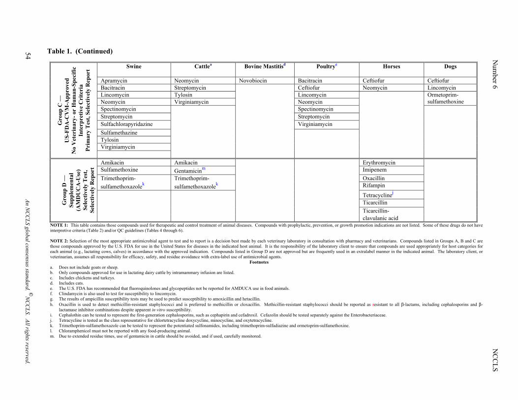
Table 1. (Continued)
Swine Cattlea Bovine Mastitisd Poultryc Horses Dogs
Apramycin Neomycin Novobiocin Bacitracin Ceftiofur Ceftiofur
Bacitracin Streptomycin Ceftiofur Neomycin Lincomycin Lincomycin Tylosin Lincomycin Neomycin Virginiamycin Neomycin
Ormetoprim- sulfamethoxine
Spectinomycin Spectinomycin Streptomycin Streptomycin Sulfachlorapyridazine Virginiamycin Sulfamethazine Tylosin Virginiamycin
Gro
up C
—
US-
FDA
-CV
M-A
ppro
ved
N
o V
eter
inar
y- o
r H
uman
-Spe
cific
In
terp
retiv
e C
rite
ria
Prim
ary
Tes
t, Se
lect
ivel
y R
epor
t
Amikacin Amikacin Erythromycin Sulfamethoxine Gentamicinm Imipenem Trimethoprim- Trimethoprim- Oxacillin sulfamethoxazolek sulfamethoxazolek Rifampin
Tetracyclinej
Ticarcillin Ticarcillin-
G
roup
D —
Su
pple
men
tal
(AM
DU
CA
-Use
) Se
lect
ivel
y T
est,
Sele
ctiv
ely
Rep
ort
clavulanic acid
NOTE 1: This table contains those compounds used for therapeutic and control treatment of animal diseases. Compounds with prophylactic, prevention, or growth promotion indications are not listed. Some of these drugs do not have interpretive criteria (Table 2) and/or QC guidelines (Tables 4 through 6).
NOTE 2: Selection of the most appropriate antimicrobial agent to test and to report is a decision best made by each veterinary laboratory in consultation with pharmacy and veterinarians. Compounds listed in Groups A, B and C are those compounds approved by the U.S. FDA for use in the United States for diseases in the indicated host animal. It is the responsibility of the laboratory client to ensure that compounds are used appropriately for host categories for each animal (e.g., lactating cows, calves) in accordance with the approved indication. Compounds listed in Group D are not approved but are frequently used in an extralabel manner in the indicated animal. The laboratory client, or veterinarian, assumes all responsibility for efficacy, safety, and residue avoidance with extra-label use of antimicrobial agents.
Footnotes a. Does not include goats or sheep. b. Only compounds approved for use in lactating dairy cattle by intramammary infusion are listed. c. Includes chickens and turkeys. d. Includes cats. e. The U.S. FDA has recommended that fluoroquinolones and glycopeptides not be reported for AMDUCA use in food animals. f. Clindamycin is also used to test for susceptibility to lincomycin. g. The results of ampicillin susceptibility tests may be used to predict susceptibility to amoxicillin and hetacillin. h. Oxacillin is used to detect methicillin-resistant staphylococci and is preferred to methicillin or cloxacillin. Methicillin-resistant staphylococci should be reported as resistant to all β-lactams, including cephalosporins and β-
lactamase inhibitor combinations despite apparent in vitro susceptibility. i. Cephalothin can be tested to represent the first-generation cephalosporins, such as cephapirin and cefadroxil. Cefazolin should be tested separately against the Enterobacteriaceae. j. Tetracycline is tested as the class representative for chlortetracycline doxycycline, minocycline, and oxytetracycline. k. Trimethoprim-sulfamethoxazole can be tested to represent the potentiated sulfonamides, including trimethoprim-sulfadiazine and ormetoprim-sulfamethoxine. l. Chloramphenicol must not be reported with any food-producing animal. m. Due to extended residue times, use of gentamicin in cattle should be avoided, and if used, carefully monitored.
N
umber 6
N
CC
LS
54 An NC
CLS global consensus standard. ©
NC
CLS. All rights reserved.

Table 2. Zone Diameter Interpretive Standards and Minimal Inhibitory Concentration (MIC) Breakpoints for Veterinary Pathogens Zone Diameter (mm) MIC Breakpoint (µg/mL)
Antimicrobial Agent Disk Content
S I F R S I F R Comments
Aminoglycosides/Aminocyclitols Amikacin 30 µg ≥ 17 15-16 ≤ 14 ≤ 16 32 ≥ 64
Gentamicin 10 µg ≥ 15 13-14 ≤ 12 ≤ 4 8 ≥ 16
Kanamycin 30 µg ≥ 18 14-17 ≤ 13 ≤ 16 32 ≥ 64
Spectinomycin Bovine (Respiratory Disease) 100 µg ≥ 14 11-13 ≤ 10 ≤ 32 64 ≥ 128 Mannheimia haemolytica Pasteurella multocida Haemophilus somnus
β-Lactam/β-Lactamase Inhibitor Combinations Amoxicillin-clavulanic acid Staphylococci 20/10 µg ≥ 20 — ≤ 19 ≤ 4/2 — ≥ 8/4 Other organisms 20/10 µg ≥ 18 14-17 ≤ 13 ≤ 8/4 16/8 ≥ 32/16
Ticarcillin-clavulanic acid Pseudomonas aeruginosa 75/10 µg ≥ 15 — ≤ 14 ≤ 64/2 — ≥ 128/2 Gram-negative enteric organisms 75/10 µg ≥ 20 15-19 ≤ 14 ≤ 16/2 32/2-64/2 ≥ 128/2
β-Lactams Penicillins Ampicillin Enterobacteriaceae 10 µg ≥ 17 14-16 ≤ 13 ≤ 8 16 ≥ 32 Staphylococci 10 µg ≥ 29 — ≤ 28 ≤ 0.25 — ≥ 0.5
Enterococci 10 µg ≥ 17 — ≤ 16 ≤ 8 — ≥ 16
Streptococci (not S. pneumoniae) 10 µg ≥ 26 19-25 ≤ 18 ≤ 0.25 0.5-4 ≥ 8 Listeria monocytogenes — — — — ≤ 2 — ≥ 4
Ampicillin is used to test for susceptibility to amoxicillin and hetacillin.
Oxacillin Staphylococci 1 µg ≥ 13 11-12 ≤ 10 ≤ 2 — ≥ 4
Oxacillin is used to test for susceptibility to methicillin, nafcillin, and cloxacillin.
Penicillin
Staphylococci 10 units ≥ 29 — ≤ 28 ≤ 0.12 — ≥ 0.25 Enterococci 10 units ≥ 15 — ≤ 14 ≤ 8 — ≥ 16 S. pneumoniae 1 µg oxacillin ≥ 20 — — ≤ 0.06 — — Streptococci (not S. pneumoniae) 10 units ≥ 28 20-27 ≤ 19 ≤ 0.12 0.25-2 ≥ 4 Listeria monocytogenes — — — — ≤ 2 — ≥ 4
The interpretive standards for Streptococcus spp. including S. pneumoniae only apply to disk susceptibility testing performed using Mueller-Hinton agar supplemented with 5% CO2 and broth with 2 to 5% lysed horse blood.
Ticarcillin Pseudomonas aeruginosa 75 µg ≥ 15 — ≤ 14 ≤ 64 — ≥ 128 Gram-negative enteric organisms 75 µg ≥ 20 15-19 ≤ 14 ≤ 16 32-64 ≥ 128
Volum
e 22 M31-A
2
An NC
CLS global consensus standard. ©N
CC
LS. All rights reserved. 55

Table 2. (Continued)
Zone Diameter (mm) MIC Breakpoint (µg/mL) Comments Antimicrobial Agent Disk Content
S I F R S I F R
β-Lactam/Other Class Combinations
Penicillin-novobiocin
Bovine mastitis 10 units/30 µg ≥ 18 15-17 ≤ 14 ≤ 1/2 2/4 ≥ 4/8
Staphylococcus aureus Streptococcus agalactiae Streptococcus dysgalactiae Streptococcus uberis
Available as an infusion product for treatment of bovine mastitis during lactation.
Carbapenems
Imipenem 10 µg ≥ 16 14-15 ≤ 13 ≤ 4 8 ≥ 16
Cephalosporins
Cephalothin 30 µg ≥ 18 15-17 ≤ 14 ≤ 8 16 ≥ 32
Cephalothin is used to test all first-generation cephalosporins, such as cephapirin and cefadroxil. Cefazolin should be tested separately with the gram-negative enteric organisms.
Cefazolin 30 µg ≥ 18 15-17 ≤ 14 ≤ 8 16 ≥ 32
Ceftiofur Bovine (Respiratory Disease) 30 µg ≥ 21 18-20 ≤ 17 ≤ 2 4 ≥ 8 Mannheimia haemolytica Pasteurella multocida Haemophilus somnus Swine (Respiratory Disease) 30 µg ≥ 21 18-20 ≤ 17 ≤ 2 4 ≥ 8 Actinobacillus pleuropneumoniae Pasteurella multocida Salmonella choleraesuis
Fluoroquinolones Enrofloxacin Canine and feline (dermal, URI, UTI) 5 µg ≥ 23 17-22 ≤ 16 ≤ 0.5 1-2 ≥ 4
Gram-negative enteric bacilli Staphylococcus spp. Other susceptible organisms
Enrofloxacin
Chickens and turkeys 5 µg ≥ 23 17-22 ≤ 16 ≤ 0.25 0.5-1 ≥ 2
Pasteurella multocida Escherichia coli
N
umber 6
N
CC
LS
56 An NC
CLS global consensus standard. ©
NC
CLS. All rights reserved.

Table 2. (Continued) Antimicrobial Agent Disk
Content Zone Diameter (mm) MIC Breakpoint (µg/mL) Comments
S I F R S I F R
Fluoroquinolones (Continued) Enrofloxacin Bovine (Respiratory Disease) 5 µg ≥ 21 17-20 ≤ 16 ≤ 0.25 0.5-1 ≥ 2 Mannheimia haemolytica Pasteurella multocida Haemophilus somnus
Difloxacin 10 µg ≥ 21 18-20 ≤ 17 ≤ 0.5 1-2 ≥ 4
Orbifloxacin Canine & Feline (Dermal, UTI)
Gram-negative enteric bacilli Staphylococcus spp. Other susceptible organisms
10 µg ≥ 23 18-22 ≤ 17 ≤ 1 2-4 ≥ 8
Lincosamides
Clindamycin
Canine (Skin & Soft Tissue Infections) Staphylococcus spp.
2 µg ≥ 21 15-20 ≤ 14 ≤ 0.5 1-2 ≥ 4
Clindamycin is also used to test for susceptibility to lincomycin. Clindamycin tends to be more active than lincomycin against some staphylococcal strains.
Pirlimycin
Bovine mastitis 2 µg ≥ 13 — ≤ 12 ≤ 2 — ≥ 4
Staphylococcus aureus
Streptococcus agalactiae Streptococcus dysgalactiae Streptococcus uberis
Available as an infusion product for treatment of bovine mastitis during lactation.
Macrolides Erythromycin Organisms other than streptococci 15 µg ≥ 23 14-22 ≤ 13 ≤ 0.5 1-4 ≥ 8 Streptococci 15 µg ≥ 21 16-20 ≤ 15 ≤ 0.25 0.5 ≥ 1
Tilmicosin Bovine (Respiratory Disease) 15 µg ≥ 14 11-13 ≤ 10 ≤ 8 16 ≥ 32 Mannheimia haemolytica
Also applicable to P. multocida
Swine (Respiratory Disease) Pasturella multocida Actinobacillus pleuropneumoniae
15 µg ≥ 11 ≤ 10 ≤ 16 ≥ 32
Volum
e 22 M31-A
2
An NC
CLS global consensus standard. ©N
CC
LS. All rights reserved. 57

Table 2. (Continued) Antimicrobial Agent Disk Content Zone Diameter (mm) MIC Breakpoint (:g/mL) Comments
S I F R S I F R
Phenicols Chloramphenicol Organisms other than streptococci 30 µg ≥ 18 13-17 ≤ 12 ≤ 8 16 ≥ 32 S. pneumoniae 30 µg ≥ 21 — ≤ 20 ≤ 4 — ≥ 8 Streptococci (not S. pneumoniae) 30 µg ≥ 21 18-20 ≤ 17 ≤ 4 8 ≥ 16
Florfenicol Bovine (Respiratory Disease) 30 µg ≥ 19 15-18 ≤ 14 ≤ 2 4 ≥ 8 Mannheimia haemolytica Pasteurella multocida Haemophilus somnus
Pleuromutilins Tiamulin Swine (Respiratory Disease) 30 µg ≥ 9 — ≤ 8 ≤ 16 — ≥ 32
Actinobacillus pleuropneumoniae
Potentiated Sulfonamides
Trimethoprim-sulfamethoxazole Organisms other than S. pneumoniae 1.25/23.75 µg ≥ 16 11-15 ≤ 10 ≤ 2/38 — ≥ 4/76
Streptococcus pneumoniae 1.25/23.75 µg ≥ 19 16-18 ≤ 15 ≤ 0.5/9.5 1/19-2/38 ≥ 4/76
Trimethoprim-sulfamethoxazole is used to test for susceptibility to trimethoprim- sulfadiazine and ormetoprim-sulfadimethoxine. A breakpoint of ≤ 2/38 should be used for isolates from urinary tract infections. For systemic disease, isolates with MICs of ≤ 0.5/9.5 should be considered susceptible.
Ansamycins
Rifampin Organisms other than streptococci 5 µg ≥ 20 17-19 ≤ 16 ≤ 1 2 ≥ 4 Streptococcus pneumoniae 5 µg ≥ 19 17-18 ≤ 16 ≤ 1 2 ≥ 4
Sulfonamides
Sulfisoxazole 250 or 300 µg ≥ 17 13-16 ≤ 12 ≤ 256 — ≥ 512
Tetracyclines Tetracycline
Organisms other than streptococci 30 µg ≥ 19 15-18 ≤ 14 ≤ 4 8 ≥ 16
Streptococci 30 µg ≥ 23 19-22 ≤ 18 ≤ 2 4 ≥ 8
Tetracycline is used to test for susceptibility to chlortetracycline, oxytetracycline, minocycline, and doxycycline The interpretive standards for Streptococcus spp. including S. pneumoniae only apply to disk susceptibility testing performed using Mueller-Hinton agar supplemented with 5% CO2 and broth with 2 to 5% lysed horse blood.
N
umber 6
N
CC
LS
58 An NC
CLS global consensus standard. ©
NC
CLS. All rights reserved.

Table 2. (Continued) Antimicrobial Agent Disk Content Zone Diameter (mm) MIC Breakpoint (:g/mL) Comments
S I F R S I F R
Glycopeptides Vancomycin Enterococci 30 µg ≥ 17 15-16 ≤ 14 ≤ 4 8-16 ≥ 32 Streptococci 30 µg ≥ 17 — — ≤ 1 — — Other gram-positive organisms 30 µg ≥ 12 10-11 ≤ 9 ≤ 4 8-16 ≥ 32
NOTE 1: Zone interpretive criteria and MIC breakpoints for antimicrobial agents with gray shading are human data taken from M100- S12 supplements to M2 and M7.
NOTE 2: Veterinary-specific interpretive criteria are valid only for indicated organisms. NOTE 3: Flexible label (F) indicates the availability of FDA-approved labeling; this organism should be considered susceptible if appropriate dosing modifications
found in the packaging insert are applied. NOTE 4: Certain veterinary specific disks may or may not be commercially available. Disks not commercially available may be obtained from the
pharmaceutical sponsors as indicated:
Apramcyin Tilmicosin
Elanco Animal Health Greenfield, IN 800-428-4441
Ceftiofur Penicillin-Novobiocin Pirlimycin Spectinomycin
Pharmacia Animal Health Kalamazoo, MI 800-793-0596
Difloxacin Fort Dodge Animal Health Overland Park, KS 800-685-5656 Enrofloxacin Bayer Animal Health Shawnee Mission, KS 800-422-9874 Florfenicol Orbifloxacin
Schering-Plough Animal Health Union, NJ 800-521-5767
Tiamulin Boehringer Ingelheim Vetmedica, Inc.
St. Joseph, MO 800-325-9167
Volum
e 22 M31-A
2
An NC
CLS global consensus standard. ©N
CC
LS. All rights reserved. 59

Number 6 NCCLS
60 An NCCLS global consensus standard. ©NCCLS. All rights reserved.
Table 3. Culture Collection Numbers for Organisms Used for Quality Control of Antimicrobial Susceptibility Tests Strain Number for the Indicated Collections: Organism ATCC1 DSM2 UKNCC3 JCM4 CIP5 CCUG6
Escherichia coli 25922 1103 12241 5491 7624 17620 Escherichia coli 35218 5923 11954 102181 30600 Pseudomonas aeruginosa
27853 1117 10896 76110 17619
Staphylococcus aureus
29213 2569 2874 103429 15915
Staphylococcus aureus
25923 1104 12702 2413 7625 17621
Staphylococcus aureus
43300
Enterococcus faecalis
29212 2570 12697 2875 103214 9997
Enterococcus faecalis
33186 12756 100750
Enterococcus faecalis
51299
Actinobacillus pleuropneumoniae
27090 100916 T
Haemophilus somnus
700025
Campylobacter jejuni
33560 11351 70.2 T
Streptococcus pneumoniae
49619
Klebsiella pneumoniae
700603
1American Type Culture Collection (www.atcc.org) 2Deutsche Sammlung von Mikroorganismen und Zellkulturen (www.dsmz.de) 3United Kingdom National Culture Collections (includes National Collection of Type Cultures [NCTC] and National Collection if Industrial and Marine Bacteria [NCIMB]) (www.ukncc.co.uk) 4Japan Collection of Microorganisms (www.jcm.riken.go.jp) 5Collection of Bacterial Strain of Institut Pasteur 6Swedish Reference Group for Antibiotics (www.srga.org)

Volume 22 M31-A2
An NCCLS global consensus standard. ©NCCLS. All rights reserved. 61
Table 4. Acceptable Quality Control Ranges of Antimicrobial Disk Susceptibility Test Zone Diameters (mm) for Reference Strains on Mueller-Hinton Medium Without Blood or Other Supplements
Antimicrobial Agent
Disk
Content
Escherichia coli
ATCC® 25922
Staphylococcus aureus
ATCC® 25923
Pseudomonas aeruginosa
ATCC® 27853
Streptococcus pneumoniaea
ATCC® 49619 Amikacin 30 µg 19-26 20-26 18-26 — Amoxicillin- clavulanic acidb
20/10 µg 18-24 28-36 — —
Ampicillin 10 µg 16-22 27-35 — 30-36
Apramycin 15 µg 15-20 17-24 13-18 — Cefazolin 30 µg 21-27 29-35 — — Cefoxitin 30 µg 23-29 23-29 — — Ceftiofur 30 µg 26-31 27-31 14-18 — Cephalothin 30 µg 15-21 29-37 — 26-32
Chloramphenicol 30 µg 21-27 19-26 — 23-27
Clindamycin 2 µg — 24-30 — 19-25
Difloxacin 10 µg 28-35 27-33 16-22 — Enrofloxacin 5 µg 32-40 27-31 15-19 — Erythromycin 15 µg — 22-30 — 25-30
Florfenicol 30 µg 22-28 22-29 — 24-31 Gentamicin 10 µg 19-26 19-27 16-21 — Imipenem 10 µg 26-32 — 20-28 — Kanamycin 30 µg 17-25 19-26 — — Orbifloxacin 10 µg 29-37 24-30 16-22 — Oxacillin 1 µg — 18-24 — ≤ 12c
Penicillin 10 units — 26-37 — 24-30 Penicillin-novobiocin (1/3)
10 units/30 µg
— 30-36 — 24-30
Pirlimycin 2 µg — 20-25 — — Rifampin 5 µg 8-10 26-34 — 25-30
Tetracycline 30 µg 18-25 24-30 — 27-31
Tiamulin 30 µg — 25-32 — — Ticarcillin 75 µg 24-30 — 21-27 — Ticarcillin- clavulanic acid
75/10 µg 24-30 29-37 20-28 —
Tilmicosind 15 µg — 17-21a — — Spectinomycin 100 µg 21-25 13-17 10-14 — Sulfisoxazole 250 or 300 µg 15-23 24-34 — — Trimethoprim- Sulfamethoxazolee
1.25/23.75 µg
23-29
24-32
—
20-28
Vancomycin 30 µg — 17-21 — 20-27

Number 6 NCCLS
62 An NCCLS global consensus standard. ©NCCLS. All rights reserved.
Table 4. (Continued) a. These quality control ranges for Streptococcus pneumoniae ATCC® 49619 are applicable only to tests performed by disk diffusion using
Mueller-Hinton agar supplemented with 5% defibrinated sheep blood, incubated in 5% CO2. b. The QC range for E. coli ATCC 35218 is 17-22 mm. c. Deterioration in oxacillin disk content is best assessed with QC organism Staphylococcus aureus ATCC® 25923, with an acceptable zone
diameter of 18 to 24 mm. d. Quality control ranges for tilmicosin were developed using Mueller-Hinton agar supplemented with 5% defibrinated sheep blood and
incubated aerobically. e. Very medium-dependent, especially with enterococci. NOTE 1: The dash (—) indicates that no range has been established. NOTE 2: To determine whether the Mueller-Hinton medium contains excessive levels of thymine, an E. faecalis (ATCC® 29212 or
33186) should be tested with trimethoprim, sulfa compounds, or trimethoprim/sulfamethoxazole especially when testing Staphylococcus hyicus or Mannheimia haemolytica. An inhibition zone of ≥ 20 mm that is free of fine colonies is acceptable.
NOTE 3: Certain veterinary specific disks may or may not be commercially available. Disks not commercially available may be obtained
from the pharmaceutical sponsors as indicated:
Apramcyin Tilmicosin
Elanco Animal Health Greenfield, IN 800-428-4441
Ceftiofur Penicillin-Novobiocin Pirlimycin Spectinomycin
Pharmacia Animal Health Kalamazoo, MI 800-793-0596
Difloxacin Fort Dodge Animal Health Overland Park, KS 800-685-5656 Enrofloxacin Bayer Animal Health Shawnee Mission, KS 800-422-9874 Florfenicol Orbifloxacin
Schering-Plough Animal Health Union, NJ 800-521-5767
Tiamulin Boehringer Ingelheim Vetmedica, Inc. St. Joseph, MO 800-325-9167

Volume 22 M31-A2
An NCCLS global consensus standard. ©NCCLS. All rights reserved. 63
Table 5. Acceptable Quality Control Ranges of MICs (µg/mL) for Reference Strains Antimicrobial Agent
Staphylococcus aureus
ATCC® 29213
Enterococcus faecalis
ATCC® 29212
Escherichia coli
ATCC® 25922
Pseudomonas aeruginosa
ATCC® 27853
Streptococcus pneumoniaea
ATCC® 49619 Amikacin 1-4 64-256 0.5-4 1-4 — Amoxicillin- clavulanic acidb
0.12/0.06- 0.5/0.25
0.25/0.12- 1.0/0.5 2/1-8/4 — 0.03/0.016–
0.12/0.06
Ampicillin 0.5-2 0.5-2 2-8 — 0.06-0.25
Apramycin 2-8 — 2-16 2-16 —
Cefazolin 0.25-1 — 1-4 — —
Cefoxitin 1-4 — 2-8 — —
Ceftiofur 0.25-1.0 0.25-1 16-64 —
Cephalothin 0.12-0.5 — 4-16 — 0.5-2
Chloramphenicol 2-8 4-16 2-8 — 2-8
Clindamycin 0.06-0.25 4-16 — — 0.03-0.12
Difloxacin 0.06-0.5 1-4 0.015-0.12 1-8 —
Enrofloxacin 0.03-0.12 0.12-1 0.008-0.03 1-4 —
Erythromycin 0.25-1 1-4 — — 0.03-0.12
Florfenicol 2-8 2-8 2-8 >16 1-4
Gentamicin 0.12-1 4-16 0.25-1 0.5-2 —
Imipenem 0.016-0.06 0.5-2 0.06-0.25 1-4 0.03-0.12
Kanamycin 1-4 16-64 1-4 — —
Orbifloxacin 0.25-2 1-8 0.015-0.12 2-16 —
Oxacillin 0.12-0.5 8-32 — — —
Penicillin 0.25-2 1-4 — — 0.25-1
Penicillin-novobiocin 0.015/0.03- 0.06/0.12 0.25/0.5-2/4 8/16->8/16 >8/16 —
Pirlimycin 0.25-1.0 2-8 >16 >16 —
Rifampin 0.004-0.016 0.5-4 4-16 16-64 0.015-0.06
Spectinomycin 64-256 64-256 8-64 256->512 —
Tetracycline 0.12-1 8-32 0.5-2 8-32 0.12-0.5
Tiamulin 0.5-2 — — — 0.5-4
Ticarcillin 2-8 16-64 4-16 8-32 — Ticarcillin- clavulanic acidb 0.5/2-2/2 16/2-64/2 4/2-16/2 8/2-32/2 —
Tilmicosin 1-4c ≥ 32 ≥ 64 >64 —
Tylosin 0.5-4 0.5-4 >32 >32 —
Sulfisoxazole 32-128 32-128 8-32 — — Trimethoprim- Sulfamethoxazole (1/19) ≤ 0.5/9.5 ≤ 0.5/9.5 ≤ 0.5/9.5 8/152-32/608 0.12/2.4-1/19
Vancomycin 0.5-2 1-4 — — 0.12-0.5

Number 6 NCCLS
64 An NCCLS global consensus standard. ©NCCLS. All rights reserved.
Table 5. (Continued) NOTE 1: To determine whether the Mueller-Hinton medium contains excessive levels of thymidine or thymine,
an E. faecalis (ATCC® 29212 or 33186) should be tested with trimethoprim, sulfa compounds, or trimethoprim-sulfamethoxazole especially when testing Staphylococcus hyicus or Mannheimia haemolytica. If excessive thymidine is present, an expected MIC within the susceptible category (trimethoprim-sulfamethoxazole MIC ≤ 0.5/9.5 µg/mL) will shift to the resistant category (trimethoprim-sulfamethoxazole MIC >4/76 µg/mL).
Footnotes a. These quality control ranges for Streptococcus pneumoniae ATCC® 49619 are applicable only to tests
performed by the broth microdilution method using cation-adjusted Mueller-Hinton broth with 2 to 5% lysed horse blood.
b. The QC ranges for E. coli 35218 for amoxicillin-clavulanic acid and ticarcilllin-clavulanic acid are 4/2-16/8 and 4/2-16/2 µg/mL, respectively.
c. An MIC for tilmicosin of 0.5 or 8 µg/mL may indicate a medium or pH problem. The methodology should be reexamined.

Volume 22 M31-A2
An NCCLS global consensus standard. ©NCCLS. All rights reserved. 65
Table 5A. Tentative Quality Control Ranges for the Agar Dilution Method of Testing Campylobacter jejuni (ATCC® 33560)
Antimicrobial Agent MIC (µg/mL) Ciprofloxacin 0.12−1 Doxycycline 0.5−2 Erythromycin 1−8 Gentamicin 0.5−4 Meropenem 0.004−0.015 Nalidixic Acid 8−32 Tetracycline 1−4

Number 6 NCCLS
66 An NCCLS global consensus standard. ©NCCLS. All rights reserved.
Table 6. Acceptable Quality Control Ranges for Haemophilus somnus and Actinobacillus pleuropneumoniae
Zone Diameter
MICs
Antimicrobial
Agent
Disk
Content
Haemophilus
somnus ATCC®700025
Actinobacillus pleuropneumoniae
ATCC®27090
Haemophilus
somnus ATCC® 700025
Actinobacillus
pleuropneumoniae ATCC® 27090
Ceftiofur 30 µg 36-46 34-42 0.0005-0.004 0.004-0.015
Enrofloxacin 5 µg 32-38 31-38 0.015-0.06 0.015-0.06
Florfenicol 30 µg 34-44 31-40 0.12-0.5 0.25-1
Gentamicin 10 µg 14-22 15-19 8-32 8-32
Penicillin 10 units 35-44 29-36 0.015-0.06 0.12-1
Tetracycline 30 µg 27-33 23-30 0.12-1 0.25-2
Tiamulin 30 µg - 12-19 - 8-32
Tilmicosin 15 µg 8-13 8-13 2-16 4-32 Trimethoprim- sulfamethoxazole (1/19)
1.25/23.75 µg 26-32 28-32 0.03/0.57-0.125/2.38
0.015/0.28-0.06/1.14
NOTE 1: Zone diameter quality control ranges are applicable only to tests performed by disk diffusion using
chocolate Mueller-Hinton agar, incubated in 5 to 7% CO2 for 20 to 24 hours. NOTE 2: MIC quality control ranges are applicable only to tests performed by broth microdilution
procedures using veterinary fastidious medium (VFM).

Volume 22 M31-A2
An NCCLS global consensus standard. ©NCCLS. All rights reserved. 67
Table 7. Suggested Modifications of Standard Methods for Susceptibility Testing of Some Fastidious and Special Problem Veterinary Pathogens
Organism Method Medium Incubation Comments
Staphylococcus hyicus
Broth Microdilution CAMHB + thymidine phosphorylase (0.2 IU/mL); for sulfonamides and trimethoprim only
35 º C/18-24 h
Wegener, et al.b
Haemophilus somnus and Actinobacillus pleuropneumoniae
Agar Dilution with Disk Diffusion
Chocolate Mueller-Hinton agar
35 ºC/5-7% CO2/20-24 h
Broth Microdilution Veterinary Fastidious Medium (VFM)
35 º C/5-7% CO2/20-24 h
Enterococci Broth Microdilution CAMHB 35 º C/16-24 h
Accurate detection of vancomycin resistance requires that the plates be incubated for a full 24 h. Strains producing β-lactamase are best detected by a nitrocefin β-lactamase test.
Streptococcus pneumoniae Broth Microdilution CAMHB + LHB
(2-5% v/v/)a 35 ºC/20-24 h
The final volume of broth in the well of the microdilution plate should be 100 µL.
Listeria spp. Broth Microdilution CAMHB + LHB (2-5% v/v)a 35 ºC/18 h
NOTE 1: These are acceptable alternatives with which some members of the subcommittee have had substantial experience. CAMHB, Cation-adjusted Mueller-Hinton broth. LHB, Lysed horse blood.
Footnotes a. To prepare LHB, freeze-thaw 3-4 times until blood is thoroughly lysed. Aseptically mix equal volumes of
LHB and sterile distilled water (now 50% LHB). To be used in the broth test, the combination of broth and LHB must be clear; this can be done by centrifuging the blood at 16,000 x g for 20 minutes. Decant the supernatant; recentrifuge if necessary. Add appropriate amounts of the 50% LHB to the broth medium to yield a final concentration of 2 to 5% LHB.
b. Wegener HC, Watts JL, Salmon SA, Yancey RJ, Jr. Antimicrobial susceptibility testing of Staphylococcus
hyicus isolated from exudative dermatitis in pigs. J Clin Microbiol. 1994;32:793-795.

Number 6 NCCLS
68 An NCCLS global consensus standard. ©NCCLS. All rights reserved.
Table 8. Solvents and Diluents for Preparation of Stock Solutions of Antimicrobial Agents Requiring Solvents Other Than Water
Antimicrobial Agent Solventa Diluenta Amoxicillin, clavulanic acid, and ticarcillin
Phosphate buffer, pH 6.0, 0.1 mol/L Phosphate buffer, pH 6.0, 0.1 mol/L
Ampicillin Phosphate buffer, pH 8.0, 0.1 mol/L Phosphate buffer, pH 6.0, 0.1 mol/L Cephalothinb Phosphate buffer, pH 6.0, 0.1 mol/L Water Chloramphenicol, erythromycin, florfenicol, tylosinc, tilmicosinc
95% ethanol Water
Difloxacin 1/2 volume of water, then add 1 mol/L NaOH dropwise to dissolve
Water
Enrofloxacin 1/2 volume of water, then add 1 mol/L NaOH dropwise to dissolve
Water
Imipenem Phosphate buffer, pH 7.2, 0.01 mol/L Phosphate buffer, pH 7.2, 0.01 mol/L
Nitrofurantoinc Phosphate buffer, pH 8.0, 0.1 mol/L Phosphate buffer, pH 8.0, 0.01 mol/L
Orbifloxacin 1/2 volume of water, then add 1 mol/L NaOH dropwise to dissolve
Water
Rifampin Methanold Water (with stirring) Sulfonamides 1/2 volume hot water and minimal
amount of 2.5 mol/L NaOH to dissolve Water
Trimethoprim 0.05N (0.05 mol/L) lacticd or hydrochloric acid,d 10% of final volume
Water (can require heat)
Footnotes
a. These solvents and diluents can be further diluted as necessary in water or broth. The products known
to be suitable for water solvents and diluents are amikacin, carbenicillin, ciprofloxacin, clindamycin, gentamicin, methicillin, novobiocin, oxacillin, penicillin, pirlmycin, tetracylines, tiamulin (if hydrogen fumarate), trimethoprim (if lactate), and vancomycin.
b. All other cephalosporins and cephems except ceftiofur not listed above are solubilized (unless otherwise
indicated by the manufacturer) in phosphate buffer, pH 6.0, 0.1 mol/L, and further diluted in sterile distilled water. Ceftiofur can be solubilized in water or broth.
c. Alternatively, nitrofurantoin, tylosin, and tilmicosin can be dissolved in dimethyl sulfoxide (DMSO). d. These compounds are potentially toxic. Consult the Material Safety Data Sheets (MSDS) available
from the product manufacturer before using any of these materials.

Volume 22 M31-A2
An NCCLS global consensus standard. ©NCCLS. All rights reserved. 69
Table 9A. Screening and Confirmatory Tests for Expanded-spectrum-β-lactamases (ESBLs) in Klebsiella pneumoniae, K. oxytoca, and Escherichia coli (Zone Diameter)
Method
Initial Screen Test
Phenotypic Confirmatory Test
Medium
Mueller-Hinton Agar
Mueller-Hinton Agar
Cefpodoxime ceftazidime aztreonam cefotaxime ceftriaxone
10 µg or 30 µg or 30 µg or 30 µg or 30 µg
ceftazidime ceftazidime-clavulanic acida and cefotaxime cefotaxime-clavulanic acida
30 µg 30/10 µg 30 µg 30/10 µg
Antimicrobial Disk Concentration
(The use of more than one antimicrobial agent for screening improves the sensitivity of detection.)
(Confirmatory testing requires use of both cefotaxime and ceftazidime, alone and in combination with clavulanic acid.)
Inoculum Incubation conditions Incubation length
Standard disk diffusion recommendations
Standard disk diffusion recommendations
Cefpodoxime zone Ceftazidime zone Aztreonam zone Cefotaxime zone Ceftriaxone zone
≤ 17 mm ≤ 22 mm ≤ 27 mm ≤ 27 mm ≤ 25 mm
Results
= may indicate ESBL production
A ≥ 5 mm increase in a zone diameter for either antimicrobial agent tested in combination with clavulanic acid versus its zone when tested alone = ESBL (e.g., ceftazidime zone = 16; ceftazidime-clavulanic acid zone = 21)
E. coli ATCC® 25922 (see control limits in Table 4) Klebsiella pneumoniae ATCC® 700603:
E. coli ATCC® 25922: ≤ 2 mm increase in zone diameter for antimicrobial agent tested alone versus its zone when tested in combination with clavulanic acid
QC Recommendations
cefpodoxime zone ceftazidime zone aztreonam zone cefotaxime zone ceftriaxone zone
9-16 mm 10-18 mm 9-17 mm 17-25 mm 16-24 mm
Klebsiella pneumoniae ATCC® 700603: ≥ 5 mm increase in ceftazidime-clavulanic acid zone diameter; ≥ 3-mm increase in cefotaxime-clavulanic acid zone diameter.
Footnote
a. Preparation of ceftazidime-clavulanic acid (30 µg/10 µg) and cefotaxime-clavulanic acid (30 µg/10 µg) disks. Using a stock
solution of clavulanic acid at 1,000 µg/mL (either freshly prepared or taken from small aliquots that have been frozen at -70 ºC), add 10 µL of clavulanic acid to ceftazidime (30 µg) and cefotaxime (30 µg) disks. Use a micropipette to apply the 10 µL of stock solution to the ceftazidime and cefotaxime disks within one hour before they are applied to the plates, allowing about 30 minutes for the clavulanic acid to absorb and the disks to be dry enough for application. Use disks immediately after preparation or discard; do not store.

Number 6 NCCLS
70 An NCCLS global consensus standard. ©NCCLS. All rights reserved.
Table 9B. Screening and Confirmatory Tests for ESBLs in Klebsiella pneumoniae, K. oxytoca, and Escherichia coli (MIC)
Method
Initial Screen Test
Phenotypic Confirmatory Test
Medium
CAMHB
CAMHB
cefpodoxime ceftazidime aztreonam cefotaxime ceftriaxone
4 µg/mL or 1 µg/mL or 1 µg/mL or 1 µg/mL or 1 µg/mL
ceftazidime ceftazidime-clavulanic acid and cefotaxime cefotaxime-clavulanic acid
0.25-128 µg/mL 0.25/4-128/4 µg/mL 0.25-64 µg/mL 0.25/4-64/4 µg/mL
Antimicrobial Concentration
(The use of more than one antimicrobial agent for screening will improve the sensitivity of detection.)
(Confirmatory testing requires use of both cefotaxime and ceftazidime, alone and in combination with clavulanic acid.)
Inoculum Incubation Conditions Incubation Length
Standard broth dilution recommendations
Standard broth dilution recommendations
Results
Growth = may indicate ESBL production (i.e., MIC ≥ 2 µg/mL for ceftazidime, aztreonam, cefotaxime or ceftriaxone or MIC ≥ 8 µg/mL for cefpodoxime)
A ≥3 twofold concentration decrease in an MIC for either antimicrobial agent tested in combination with clavulanic acid versus its MIC when tested alone = ESBL (e.g., ceftazidime MIC= 8 µg/mL; ceftazidime-clavulanic acid MIC = 1 µg/mL)
E. coli ATCC® 25922= No growth (also refer to control limits listed in M7 Table 3) Klebsiella pneumoniae ATCC® 700603 = Growth:
QC Recommendations
cefpodoxime ceftazidime aztreonam cefotaxime ceftriaxone
MIC ≥ 8 µg/mL MIC ≥ 2 µg/mL MIC ≥ 2 µg/mL MIC ≥ 2 µg/mL MIC ≥ 2 µg/mL
Klebsiella pneumoniae ATCC® 700603: ≥3 twofold concentration decrease in an MIC for an antimicrobial agent tested in combination with clavulanic acid versus its MIC when tested alone

Volume 22 M31-A2
An NCCLS global consensus standard. ©NCCLS. All rights reserved. 71
Table 9C. Oxacillin-Salt Agar Screening Test for Staphylococcus aureus Screen Test Oxacillin Resistance Medium MHA with NaCl (4% w/v; 0.68 mol/L) Antimicrobial concentration 6 µg/mL oxacillin Inoculum Direct colony suspension to obtain 0.5 McFarland turbidity
Using a 1-µL loop that was dipped in the suspension, spot an area 10 to 15 mm in diameter. Alternatively, using a swab dipped in the suspension and expressed, spot a similar area or streak an entire quadrant. 35 ºC; ambient air Incubation conditions
Incubation length 24 hours Results >1 colony = resistant
Examine carefully with transmitted light for >1 colony or light film of growth.
QC Recommendations Staphylococcus aureus ATCC® 29213 - Susceptible Staphylococcus aureus ATCC® 43300 - Resistant
MHA = Mueller-Hinton Agar

Number 6 NCCLS
72 An NCCLS global consensus standard. ©NCCLS. All rights reserved.
Table 9D. Screening Tests for High-Level Aminoglycoside Resistance (HLAR) and Vancomycin Resistance in Enterococcus spp. Screen Test
Gentamicin HLAR
Streptomycin HLAR
Vancomycin Resistance
Medium BHIa broth or agar BHIa broth or agar BHI agar Antimicrobial concentration
500 µg/mL
Broth: 1000 µg/mL Agar: 2000 µg/mL
6 µg/mL
Inoculum
Growth method or direct colony suspension to obtain 0.5 McFarland turbidity Agar — 10 µL of a 0.5 McFarland suspension spotted onto agar surface Broth — standard broth dilution recommendations
Growth method or direct colony suspension to obtain 0.5 McFarland turbidity Agar — 10 µL of a 0.5 McFarland suspension spotted onto agar surface Broth — standard broth dilution recommendations
Growth method or direct colony suspension to obtain 0.5 McFarland turbidity 1-10 µL of a 0.5 McFarland suspension spotted onto agar surface
Incubation conditions
35 ºC; ambient air
35 ºC; ambient air
35 ºC; ambient air
Incubation length
24 hours
24 - 48 hours (if susceptible at 24 hours, reincubate)
24 hours
Results
Agar: >1 colony = resistant Broth: any growth = resistant Resistant — will not be synergistic with cell-wall-active agent (e.g., ampicillin, penicillin, vancomycin) Susceptible — will be synergistic with cell-wall-active agent that is also susceptible (e.g., ampicillin, penicillin, vancomycin)
Agar: >1 colony = resistant Broth: any growth = resistant Resistant — will not be synergistic with cell-wall-active agent (e.g., ampicillin, penicillin, vancomycin) Susceptible — will be synergistic with cell-wall-active agent to which the isolate is susceptible (e.g., ampicillin, penicillin, vancomycin)
>1 colony = presumptive resistance Perform vancomycin MIC and tests for motility and pigment production to distinguish species with acquired resistance (VanA and VanB) from those with intrinsic, intermediate-level resistance to vancomycin (VanC) such as E. gallinarum, E. casseliflavus, or E. flavescens, which often grow on the vancomycin screen plate. In contrast to other enterococci, E. casseliflavus, E. flavescens, and E. gallinarum with vancomycin MICs of 8-16 µg/mL (intermediate) differ from vancomycin-resistant enterococci for infection control purposes.
QC Recommendations
E. faecalis ATCC® 29212 — Susceptible E. faecalis ATCC® 51299 — Resistant
E. faecalis ATCC® 29212 — Susceptible E. faecalis ATCC® 51299 — Resistant
E. faecalis ATCC® 29212 — Susceptible E. faecalis ATCC® 51299 — Resistant
BHI = Brain heart infusion
Footnote a Even though not as widely available, dextrose phosphate agar and broth have been shown in limited testing to perform comparably.

Volume 22 M31-A2
An NCCLS global consensus standard. ©NCCLS. All rights reserved. 73
Glossary 1. Antimicrobial Class, Antimicrobial Agents, and Antimicrobial Resistance Mechanisms Antimicrobial Class Antimicrobial Agents Antimicrobial Resistance
Mechanism Aminocyclitols spectinomycin, apramycin Modifiying enzymes
Efflux
Aminoglycosides streptomycin, kanamycin, gentamicin, amikacin, tobramycin, neomycin
Modifying enzymes Decreased permeability Target resistance (ribosome) Efflux
Ansamycin Rifampin Target resistance (RNA polymerase)ß-lactam antibiotics: Penicillins
penicillin, ampicillin, oxacillin, ticarcillin, piperacillin, amoxicillin
Reduced permeability Altered penicillin-binding proteins β-lactamases, cephalosporinases Efflux
ß-lactam/B-lactamase inhibitors
amoxicillin-clavulanic acid, ticarcillin-clavulanic acid, piperacillin-tazobactam,
Cephalosporins and cephamycins: 1st generation (narrow spectrum) 2nd generation (expanded spectrum) 3rd generation (expanded spectrum) 4th generation (expanded spectrum) Carbapenems Monobactams
cephalothin, cefazolin, cephapirin, cephalaxin, cefadroxil cefotetan, cefoxitin (cephamycin), cefaclor ceftiofur, cefotaxime, ceftazidime, ceftriaxone cefepime, cefquinome imipenem, meropenem aztreonam
Folate pathway inhibitors
sulfonamides, trimethoprim, ormetoprim, trimethoprim-sulfamethoxazole
Decreased permeability Production of drug-insensitive enzymes
Glycopeptides vancomycin, teicoplanin Target resistance (cell wall) Lincosamides lincomycin, clindamycin, pirlimycin Decreased ribosomal binding Macrolides: 14-membered rings 15-membered rings 16-membered rings
erythromycin azithromycin spiramycin, tylosin, clarithromycin, tilmicosin
Decreased ribosomal binding Decreased permeability Modifying enzymes Efflux
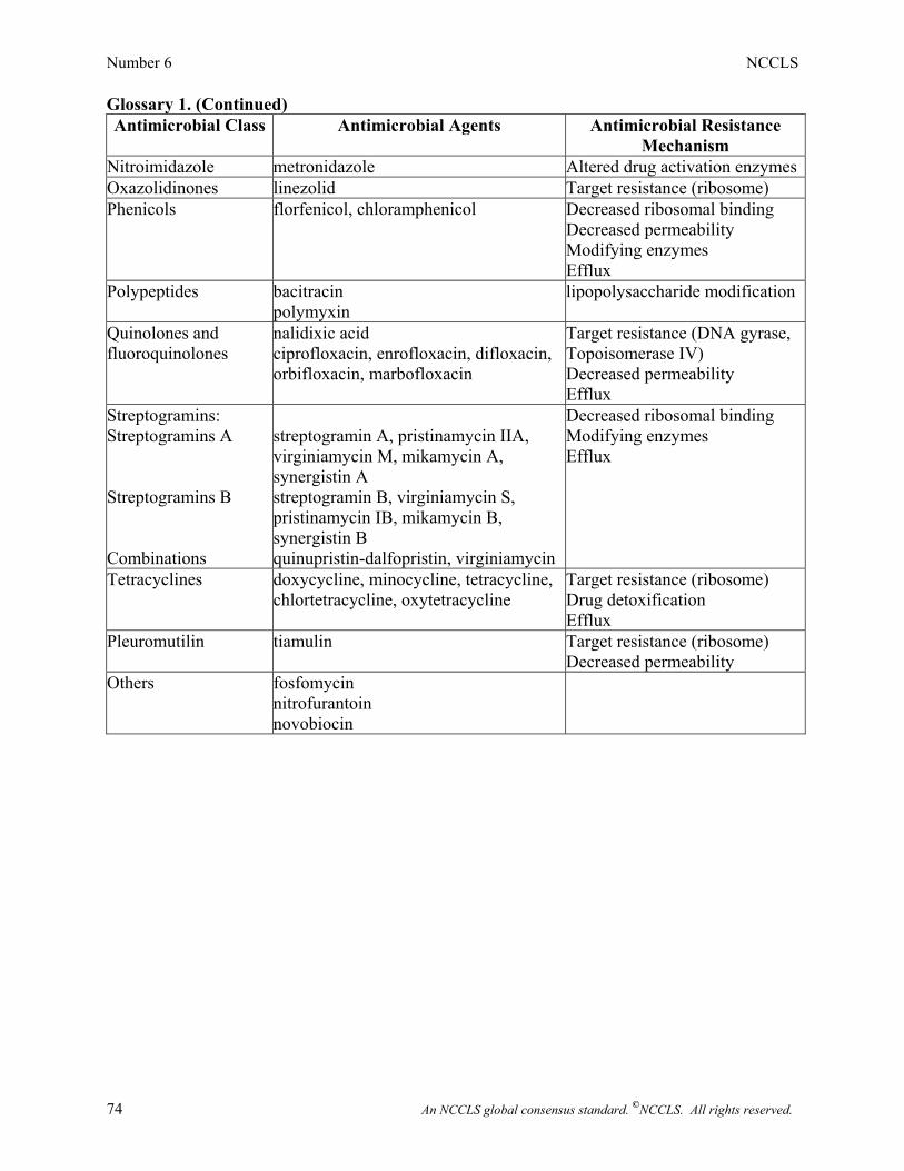
Number 6 NCCLS
74 An NCCLS global consensus standard. ©NCCLS. All rights reserved.
Glossary 1. (Continued) Antimicrobial Class Antimicrobial Agents Antimicrobial Resistance
Mechanism Nitroimidazole metronidazole Altered drug activation enzymesOxazolidinones linezolid Target resistance (ribosome) Phenicols florfenicol, chloramphenicol Decreased ribosomal binding
Decreased permeability Modifying enzymes Efflux
Polypeptides bacitracin polymyxin
lipopolysaccharide modification
Quinolones and fluoroquinolones
nalidixic acid ciprofloxacin, enrofloxacin, difloxacin, orbifloxacin, marbofloxacin
Target resistance (DNA gyrase, Topoisomerase IV) Decreased permeability Efflux
Streptogramins: Streptogramins A Streptogramins B Combinations
streptogramin A, pristinamycin IIA, virginiamycin M, mikamycin A, synergistin A streptogramin B, virginiamycin S, pristinamycin IB, mikamycin B, synergistin B quinupristin-dalfopristin, virginiamycin
Decreased ribosomal binding Modifying enzymes Efflux
Tetracyclines doxycycline, minocycline, tetracycline, chlortetracycline, oxytetracycline
Target resistance (ribosome) Drug detoxification Efflux
Pleuromutilin tiamulin Target resistance (ribosome) Decreased permeability
Others fosfomycin nitrofurantoin novobiocin

Volume 22 M31-A2
An NCCLS global consensus standard. ©NCCLS. All rights reserved. 75
Glossary 2. Abbreviations for Antimicrobial Agents Incorporated into Disks or Susceptibility Panels
Antimicrobial Agents Agent Abbreviation
Amikacin AN, AK, Ak, AMI, AMK
Amoxicillin-clavulanic acid AMC, A/C, AXC, Aug, AUG, XL, AML, AMOX/CA
Ampicillin AM, Am, AMP, AP, ABPC
Apramycin AP, APR
Cefazolin CZ, CFZ, Cfz, FAZ, KZ, CEZ, CFAZ
Cefoxitin FOX, CX, Cfx, FX, CFX, CFOX, COX
Ceftiofur XNL, FUR, CEF, ACC
Cephalothin CF, Cf, CEP, CE, KF, CET, CEF, CTIN, CTN
Chloramphenicol C, CHL, CL, CP, CMP
Clindamycin CC, CD, CM, Cd, CLI, DA, CLDM
Difloxacin DIC, DIF, DF
Enrofloxacin ENO, ENR, ENF, EFX, ENRO, EF
Erythromycin E, ERY, EM
Florfenicol FFC
Gentamicin GM, GMS, HLG, Gm, GmS, GEN, CN
Imipenem IPM, IMI, Imp, IP, IMIP
Kanamycin K, KAN, HLK, KM
Orbifloxacin OBX, ORB
Oxacillin OX, OXS, Ox, OXA, MPIPC, OXAC
Penicillin P, PEN, PG, PCG, PENG
Penicillin-novabiocin P/NB, P/NOV, P/N
Pirlimycin PRL, PIRL
Rifampin RA, RI, RIF, Rif, RD, RP, RFP, RAM
Spectinomycin SPT, SPE, SC, SH, SPC

Number 6 NCCLS
76 An NCCLS global consensus standard. ©NCCLS. All rights reserved.
Glossary 2. (Continued)
Sulfisoxazole G, FIS, SF
Tetracycline Te, TE, TET, TC, T
Tiamulin TIA
Ticarcillin TIC, TC, Ti, TI, TIPC, TICC
Ticarcillin-clavulanic acid TIM, T/C, TCC, Tim, TLc, TICC/CA, TIL
Tilmicosin TIL, TMS
Trimethoprim-sulfamethoxazole SXT, T/S, STG, TS, ST
Tylosin TYL, TYLB, TYLT, TY, TLS
Vancomycin Va, VA, VAN, VCM
Note: Designations for antimicrobial agents that are also used in human medicine can be found in the most current edition of M100―Performance Standards for Antimicrobial Susceptibility Testing.

Volume 22 M31-A2
An NCCLS global consensus standard. ©NCCLS. All rights reserved. 77
NCCLS consensus procedures include an appeals process that is described in detail in Section 9 of the Administrative Procedures. For further information contact the Executive Offices or visit our website at www.nccls.org. Summary of Comments and Subcommittee Responses M31-A: Performance Standards for Antimicrobial Disk and Dilution Susceptibility Tests for Bacteria
Isolated from Animals; Approved Standard General 1. Page 25, Section 5.2.3.6. Macrodilution-tube broth method. If the inoculum is prepared in broth
(as listed in this section) and 1 ml is added to the 1 ml of antimicrobials, which have been dissolved and diluted in water..., the broth is only at half-strength and could result in poor growth of some organisms.
• Section 4.2.3.4 addresses the oversight by requiring double-strength medium to be used as
the aqueous diluent. 2. Page 36, Table 2, line- Amox/Clav...Staph - There is no value listed for the MIC breakpoint in the
“resistant” column. Page 36, Table 2, line- Amox/Clav...other organisms The value listed in the MIC breakpoint "susceptible" column is the same as the “resistant” column; both are at 8/4.
• These errors have been corrected and conform to M100-S12 values. 3. Page 36, Table 2, line-Ticar/Clav - There is a value in the MIC breakpoint “resistant” column that
is just floating there, not for any organism — where does it belong??? with the amox/clav??? • These errors have been corrected and conform to M100-S12 values. 4. Page 36, Table 2. , line-Ticar/Clav - The clavulanic values are all at 2 —- shouldn't they increase
like the clavulanic values increase with the amoxicillin? Page 36, Table 2, line- Ticar/Clav ... gram-negative enteric organisms the MIC breakpoint “intermediate” value is 32-64..., the clav isn't added? Shouldn't it be 32/4 - 64/8??
• A fixed concentration of 2 µg of clavulanate is used with all dilutions of ticarcillin. The
intermediate values are now designated as 32/2 – 64/2 to conform to M100-S12. 5. I found a minor error in the M31-A document that should be corrected during its next version.
Page 20, Section 5.2.2.6, the second bullet: the final inoculum on the agar is incorrectly stated as CFU/mL. It should be CFU/spot.
• The wording has been changed to CFU/spot. 6. It should be interesting to state the natural resistance of the major bacterial species from animal
origin. The natural resistance is useful for bacterial species identification and interpretive reading of results (recommendation from the CA-SFM).
• It is expected that veterinary microbiologists are aware of intrinsic resistance in various
bacteria to specific agents in order to select the appropriate agents to test. For example, one would not test a lincosamide or macrolide compound against an enteric bacillus as these organisms are intrinsically resistant to these compounds.

Number 6 NCCLS
An NCCLS global consensus standard. ©NCCLS. All rights reserved. 78
7. It is recommended to store the antimicrobial stock solution at –70 °C. Several antimicrobial agents did not support several thawing/dethawing cycles and a reference strain is not a good indicator to control antimicrobial stability. For example, florfenicol stock solution at 1280 µg/ml in 10% methanol/water, stored at –20 °C lost 30% of activity after 6 months of storage while MIC value of control strain did not differ during the same period. We recommend indicating in a table the conservation procedure and stability of antimicrobial on the basis of the literature.
• The comments above describe several deviations from the procedures described in the
referenced section. Section 4.2.1.3 indicates that that stock solutions are to be stored at –70 °C which would minimize degradation of the agent. Also, it is indicated that stock solutions are to removed as needed, used the same day, and any unused portion discarded. This avoids problems associated with freeze/thaw cycles. Finally, a properly performed susceptibility test with the appropriate quality control organism should readily detect a 30% loss in activity in antimicrobial agent.
Glossary 8. 1) Thiamphenicol is approved for veterinary use internationally (Europe). Should this be added
to the list for phenicols? 2) Should the new human drug, linezolid, be listed under "Others"? • Thiamphenicol has been added to the phenicol group. Linezolid has been added as a
separate group under "Oxazolidinones. " 9. This table is interesting but is it very useful, especially since certain mechanisms do not apply to
some of the specific drugs? • This table is intended to provide an overview of the major resistance mechanisms to each
class of drug and is not intended to address the specific agents within each class.

Volume 22 M31-A2
An NCCLS global consensus standard. ©NCCLS. All rights reserved. 79
Summary of Delegate Comments and Subcommittee Responses M31-A2: Performance Standards for Antimicrobial Disk and Dilution Susceptibility Tests for Bacteria Isolated from Animals; Approved Standard—Second Edition General 1. The M31-A2 document cannot be accepted, because some information was not correct: especially
on marbofloxacin. The manufacturer has provided as an attachment quality control and interpretive criteria information, as well as supporting information. The manufacturer would like to rectify the information on marbofloxacin and could send the necessary documents to evaluate the work performed.
• The sole basis for the reject vote appears to be the lack of inclusion of a particular sponsor's
antimicrobial agent, not the inclusion of incorrect data. There does not appear to be any substantive reason for rejection that relates to the methodology or other statements in NCCLS document M31-A2. To date, the sponsor has not submitted marbofloxacin data to be reviewed by the subcommittee; thus, it has not been included. The most current edition of NCCLS document M37—Development of In Vitro Susceptibility Testing Criteria and Quality Control Parameters for Veterinary Antimicrobial Agents outlines the process for a sponsor to present information to the subcommittee to request approval for quality control ranges, interpretive criteria, and other listings in M31. The sponsor has been encouraged to follow M37 to compile marbofloxacin data and request that it be reviewed at the next subcommittee meeting. The NCCLS consensus process does not allow for any data to be placed in a document without appropriate subcommittee review; thus, the sponsor's request to amend M31-A2 cannot be granted.

Number 6 NCCLS
An NCCLS global consensus standard. ©NCCLS. All rights reserved. 80
Related NCCLS Publications* M2-A7 Performance Standards for Antimicrobial Disk Susceptibility Tests; Approved
Standard—Seventh Edition (2000). This document provides current recommended techniques for disk susceptibility testing, and new frequency criteria for quality control testing.
M6-A Protocols for Evaluating Dehydrated Mueller-Hinton Agar; Approved Standard
(1996). This standard contains procedures for evaluating production lots of Mueller-Hinton agar, and for the development and application of reference media.
M7-A5 Methods for Dilution Antimicrobial Susceptibility Tests for Bacteria That Grow
Aerobically; Approved Standard —Fifth Edition (2000). This document provides updated reference methods for the determination of minimal inhibitory concentrations (MICs) for aerobic bacteria by broth macrodilution, broth microdilution, and agar dilution.
M11-A5 Methods for Antimicrobial Susceptibility Testing of Anaerobic Bacteria; Approved
Standard—Fifth Edition (2001). This document provides reference methods for the determination of minimal inhibitory concentrations (MICs) of anaerobic bacteria by broth macrodilution, broth microdilution and agar dilution. Interpretive and quality control tables are included.
M23-A2 Development of In Vitro Susceptibility Testing Criteria and Quality Control
Parameters; Approved Guideline—Second Edition (2001). This document addresses the required and recommended data needed for the selection of appropriate interpretative standards and quality control guidelines for new antimicrobial agents.
M37-A2 Development of In Vitro Susceptibility Testing Criteria and Quality Control
Parameters for Veterinary Antimicrobial Agents; Approved Guideline—Second Edition (2002). This document addresses the recommended data needed for selection of appropriate interpretative standards and quality control guidelines for new veterinary antimicrobial agents.
M39-A Analysis and Presentation of Cumulative Antimicrobial Susceptibility Test Data;
Approved Guideline (2002). This document describes methods for the recording and analysis of antimicrobial susceptibility test data, consisting of cumulative and ongoing summaries of susceptibility patterns of epidemiologically significant microorganisms.
M100-S12 Performance Standards for Antimicrobial Susceptibility Testing; Twelfth
Informational Supplement (2002). This document provides updated tables for the NCCLS antimicrobial susceptibility testing standards M2-A7 and M7-A5.
NRSCL8-A Terminology and Definitions for Use in NCCLS Documents; Approved Standard
(1998). This document provides standard definitions for use in NCCLS standards and guidelines, and for submitting candidate reference methods and materials to the National Reference System for the Clinical Laboratory (NRSCL).
* Proposed- and tentative-level documents are being advanced through the NCCLS consensus process; therefore, readers should refer to the most recent editions.

Volume 22 M31-A2
An NCCLS global consensus standard. ©NCCLS. All rights reserved. 81
NOTES

Number 6 NCCLS
An NCCLS global consensus standard. ©NCCLS. All rights reserved. 82
NOTES

Volume 22 M31-A2
An NCCLS global consensus standard. ©NCCLS. All rights reserved. 83
NOTES

Number 6 NCCLS
An NCCLS global consensus standard. ©NCCLS. All rights reserved. 84
NOTES

Volume 22 M31-A2
An NCCLS global consensus standard. ©NCCLS. All rights reserved. 85
NOTES

Number 6 NCCLS
An NCCLS global consensus standard. ©NCCLS. All rights reserved. 86
NOTES

NCCLS 940 West Valley Road Suite 1400 Wayne, PA 19087 USA PHONE 610.688.0100 FAX 610.688.0700 E-MAIL: [email protected] WEBSITE: www.nccls.org ISBN 1-56238-461-9