WO 2015/103438 A2
212
(12) INTERNATIONAL APPLICATION PUBLISHED UNDER THE PATENT COOPERATION TREATY (PCT) (19) World Intellectual Property Organization International Bureau (10) International Publication Number (43) International Publication Date WO 201 5/1 0343 8 A2 9 July 2015 (09.07.2015) W PO PCT (51) International Patent Classification: BZ, CA, CH, CL, CN, CO, CR, CU, CZ, DE, DK, DM, C07K 16/18 (2006.01) DO, DZ, EC, EE, EG, ES, FI, GB, GD, GE, GH, GM, GT, HN, HR, HU, ID, IL, IN, IR, IS, JP, KE, KG, KN, KP, KR, (21) International Application Number: KZ, LA, LC, LK, LR, LS, LU, LY, MA, MD, ME, MG, PCT/US20 14/073088 MK, MN, MW, MX, MY, MZ, NA, NG, NI, NO, NZ, OM, (22) International Filing Date: PA, PE, PG, PH, PL, PT, QA, RO, RS, RU, RW, SA, SC, 31 December 2014 (3 1.12.2014) SD, SE, SG, SK, SL, SM, ST, SV, SY, TH, TJ, TM, TN, TR, TT, TZ, UA, UG, US, UZ, VC, VN, ZA, ZM, ZW. (25) Filing Language: English (84) Designated States (unless otherwise indicated, for every (26) Publication Language: English kind of regional protection available): ARIPO (BW, GH, (30) Priority Data: GM, KE, LR, LS, MW, MZ, NA, RW, SD, SL, ST, SZ, 61/964,383 2 January 2014 (02.01 .2014) US TZ, UG, ZM, ZW), Eurasian (AM, AZ, BY, KG, KZ, RU, TJ, TM), European (AL, AT, BE, BG, CH, CY, CZ, DE, (71) Applicant: GENELUX CORPORATION [US/US]; 3030 DK, EE, ES, FI, FR, GB, GR, HR, HU, IE, IS, IT, LT, LU, Bunker Hill Street, Suite 3 10, San Diego, CA 92109 (US). LV, MC, MK, MT, NL, NO, PL, PT, RO, RS, SE, SI, SK, SM, TR), OAPI (BF, BJ, CF, CG, CI, CM, GA, GN, GQ, (72) Inventors: SZALAY, Aladar, A.; 7704 North Fork Road, GW, KM, ML, MR, NE, SN, TD, TG). Highland, CA 92364 (US). CAPPELLO, Joseph; 3030 Bunker Hill Street, Suite 3 10, San Diego, CA 92109 (US). Declarations under Rule 4.17 : CHEN, Nanhai, G.; 9167 Buckwheat Street, San Diego, — as to the applicant's entitlement to claim the priority of the CA 92129 (US). MINEV, Boris; 3030 Bunker Hill Street, earlier application (Rule 4.1 7(in)) Suite 310, San Diego, CA 92109 (US). Published: (74) Agents: SEIDMAN, Stephanie, L. et al; McKenna Long & Aldridge LLP, 4435 Eastgate Mall, Suite 400, San — without international search report and to be republished Diego, CA 92121 (US). upon receipt of that report (Rule 48.2(g)) (81) Designated States (unless otherwise indicated, for every — with sequence listingpart of description (Rule 5.2(a)) kind of national protection available): AE, AG, AL, AM, AO, AT, AU, AZ, BA, BB, BG, BH, BN, BR, BW, BY, (54) Title: ONCOLYTIC VIRUS ADJUNCT THERAPY WITH AGENTS THAT INCREASE VIRUS INFECTIVITY (57) Abstract: Provided are adjunct therapies for use in combinations and compositions with an oncolytic virus, such as a vaccinia virus. The adjunct therapies include co -administration and co -formulation of a complement inhibitor and/or a lipid emulsion com position with the oncolytic virus. Also provided herein are therapeutic methods using the adjunct therapies for treatment of disease and conditions employing an oncolytic therapeutic virus, such as for the treatment of hyperproliferative diseases or conditions in cluding tumors or cancers.
-
Upload
khangminh22 -
Category
Documents
-
view
0 -
download
0
Transcript of WO 2015/103438 A2

(12) INTERNATIONAL APPLICATION PUBLISHED UNDER THE PATENT COOPERATION TREATY (PCT)
(19) World Intellectual PropertyOrganization
International Bureau(10) International Publication Number
(43) International Publication Date WO 2015/103438 A29 July 2015 (09.07.2015) W P O P C T
(51) International Patent Classification: BZ, CA, CH, CL, CN, CO, CR, CU, CZ, DE, DK, DM,C07K 16/18 (2006.01) DO, DZ, EC, EE, EG, ES, FI, GB, GD, GE, GH, GM, GT,
HN, HR, HU, ID, IL, IN, IR, IS, JP, KE, KG, KN, KP, KR,(21) International Application Number: KZ, LA, LC, LK, LR, LS, LU, LY, MA, MD, ME, MG,
PCT/US20 14/073088 MK, MN, MW, MX, MY, MZ, NA, NG, NI, NO, NZ, OM,
(22) International Filing Date: PA, PE, PG, PH, PL, PT, QA, RO, RS, RU, RW, SA, SC,
3 1 December 2014 (3 1.12.2014) SD, SE, SG, SK, SL, SM, ST, SV, SY, TH, TJ, TM, TN,TR, TT, TZ, UA, UG, US, UZ, VC, VN, ZA, ZM, ZW.
(25) Filing Language: English(84) Designated States (unless otherwise indicated, for every
(26) Publication Language: English kind of regional protection available): ARIPO (BW, GH,
(30) Priority Data: GM, KE, LR, LS, MW, MZ, NA, RW, SD, SL, ST, SZ,
61/964,383 2 January 2014 (02.01 .2014) US TZ, UG, ZM, ZW), Eurasian (AM, AZ, BY, KG, KZ, RU,TJ, TM), European (AL, AT, BE, BG, CH, CY, CZ, DE,
(71) Applicant: GENELUX CORPORATION [US/US]; 3030 DK, EE, ES, FI, FR, GB, GR, HR, HU, IE, IS, IT, LT, LU,Bunker Hill Street, Suite 310, San Diego, CA 92109 (US). LV, MC, MK, MT, NL, NO, PL, PT, RO, RS, SE, SI, SK,
SM, TR), OAPI (BF, BJ, CF, CG, CI, CM, GA, GN, GQ,(72) Inventors: SZALAY, Aladar, A.; 7704 North Fork Road,GW, KM, ML, MR, NE, SN, TD, TG).
Highland, CA 92364 (US). CAPPELLO, Joseph; 3030Bunker Hill Street, Suite 310, San Diego, CA 92109 (US). Declarations under Rule 4.17:CHEN, Nanhai, G.; 9167 Buckwheat Street, San Diego, — as to the applicant's entitlement to claim the priority of theCA 92129 (US). MINEV, Boris; 3030 Bunker Hill Street, earlier application (Rule 4.1 7(in))Suite 310, San Diego, CA 92109 (US).
Published:(74) Agents: SEIDMAN, Stephanie, L. et al; McKenna Long
& Aldridge LLP, 4435 Eastgate Mall, Suite 400, San — without international search report and to be republishedDiego, CA 92121 (US). upon receipt of that report (Rule 48.2(g))
(81) Designated States (unless otherwise indicated, for every — with sequence listing part of description (Rule 5.2(a))
kind of national protection available): AE, AG, AL, AM,AO, AT, AU, AZ, BA, BB, BG, BH, BN, BR, BW, BY,
(54) Title: ONCOLYTIC VIRUS ADJUNCT THERAPY WITH AGENTS THAT INCREASE VIRUS INFECTIVITY
(57) Abstract: Provided are adjunct therapies for use in combinations and compositions with an oncolytic virus, such as a vacciniavirus. The adjunct therapies include co-administration and co-formulation of a complement inhibitor and/or a lipid emulsion composition with the oncolytic virus. Also provided herein are therapeutic methods using the adjunct therapies for treatment of diseaseand conditions employing an oncolytic therapeutic virus, such as for the treatment of hyperproliferative diseases or conditions including tumors or cancers.

ONCOLYTIC VIRUS ADJUNCT THERAPY WITH AGENTS THAT INCREASEVIRUS INFECTIVITY
RELATED APPLICATIONS
Benefit of priority is claimed to U.S. provisional application Serial No. 61/964,383,
filed January 02, 2014, to Aladar A. Szalay, Joseph Cappello, Nanhai G. Chen and Boris
Minev, entitled "ONCOLYTIC VIRUS ADJUNCT THERAPY WITH AGENTS THAT
INCREASE VIRUS INFECTIVITY." Where permitted, the subject matter of U.S.
provisional application Serial No. 61/964,383 is incorporated by reference in its entirety.
INCORPORATION BY REFERENCE OF SEQUENCE LISTING PROVIDEDELECTRONICALLY
An electronic version of the Sequence Listing is filed herewith, the contents of
which are incorporated by reference in their entirety. The electronic file was created on
December 30, 2014, is 4,434 kilobytes in size, and titled 4847SEQPCl.txt.
FIELD OF THE INVENTION
Provided are adjunct therapies for use in combinations or compositions with an
oncolytic virus, such as a vaccinia virus, to increase infectivity of the virus. The adjunct
therapies include co-administration or co-formulation of a complement inhibitor and/or a
lipid emulsion composition. Also provided herein are therapeutic methods using the adjunct
therapies for treatment of disease and conditions with employing the combinations,
compositions and methods, such as for the treatment of hyperproliferative diseases and
conditions including tumors and cancers.
BACKGROUND
Oncolytic viral therapy is effected by administering a virus that accumulates in
tumor cells and replicates in the tumor cells. For example, vaccinia is an oncolytic virus
that accumulates in wounds and tumors. By virtue of replication in the cells, and optional
delivery of therapeutic agents, tumor cells are lysed, and the tumor shrinks and can be
eliminated. Vaccinia viruses are typically administered systemically or locally. There still
exists a need for improved or alternative methods of administering vaccinia viruses for
various therapeutic and diagnostic applications. Accordingly, it is among the objects herein,
to provide virus compositions that can be employed for diagnostic and/or therapeutic
methods.
SUMMARY
Provided are methods, uses, compositions, combinations for increasing the
infectivity of oncolytic viruses, such as vaccinia viruses. Infectivity is increased by treating
a subject with an anti-complement molecule, particularly an antibody, such as an anti-C5
antibody and/or other complement inhibitors known to those of skill in the art. Treatment

can be effected simultaneously with administration of the virus, before, after and
intermittently with administration of the viruses. Uses of the antibodies for such treatment,
including treatment of tumors are provided. Infectivity can be increased by pretreatment,
such as incubation, of the oncolytic viruses with a biocompatible lipid as described herein.
The antibodies, biocompatible lipid compositions, and viruses include any and all described
herein. Both modes of increasing infectivity can be employed together. These methods also
can be used and combined with other methods for increasing infectivity as well as for use
with viruses that are modified to have increased infectivity, such as, but not limited to,
vaccinia viruses, such as vaccinia viruses with modified to increase the production of
extracellular enveloped virus (EEV) forms. These include vaccinia virus in which the
A34R polypeptide is modified, such as with the mutation K151E (see, U.S. Patent No.
8,329,164, which describes vaccinia viruses with increased infectivity by virtue of
modification of the virus).
Provided are uses of anti-complement molecules, such as antibodies, for increasing
infectivity of oncolytic viruses. Anti-C5 antibodies, are used to increase the infectivity of
oncolytic viruses, such as vaccinia viruses. Composition and combinations and kits
containing the antibodies and viruses also are provided.
In particular, provided are methods and uses of an anti-complement component 5
(C5) antibody for increasing infectivity of an oncolytic virus. Anti-C5 antibodies, include,
for example, eculizumab, pexelizumab, TSA12/22 or MB12/122, and variants thereof that
bind to C5. The antibodies for all methods and uses provided herein can be full-length
antibodies or binding fragments thereof, such as Fabs and single chain antibodies. The
antibodies and oncolytic virus can be provided in separate compositions or in a single
composition. The antibodies increase the effectiveness of the oncolytic virus, such as by
increasing the infectivity of the virus. Infectivity can be assessed by any suitable method,
including measuring titer of the virus in a body fluid, such as blood or serum. The antibody
and virus can be administered separately, in the same composition, sequentially or
intermittently or in any suitable regimen. The antibody can be
administered before the virus is administered or intermittently therewith or after the virus is
ad ministered. The oncolytic virus can be pretreated with a lipid emulsion containing a
biocompatible lipid that is comprised of fatty acids and/or fatty acid derivatives. The lipid
emulsion is as described herein and comprises a biocompatible lipid that is comprised of
fatty acids and/or fatty acid derivatives, wherein the composition is an emulsion. Also
provided are the compositions containing an oncolytic virus; and an anti-complement
component 5 (C5) antibody, such as, but not limited to, eculizumab, pexelizumab,

TSA12/22 or MB12/122, and variants thereof that bind to C5 and antigen binding fragments
thereof. The composition containing the antibody and the virus can also include the
biocompatible lipid component that is comprised of fatty acids and/or fatty acid derivatives
In general any therapeutic virus can be used in methods, uses, compositions and
combinations provided herein. These include oncolytic viruses, and their use for treatment
of tumors, as well as diagnosis and monitoring treatment. The viruses include any described
herein, including, but not limited to, the LIVP strains of vaccinia virus described herein and
known to those of skill in the art. such as other strains of vaccinia virus, herpes simplex
viruses, oncolytic adenoviruses, measles virus, reoviruses and others.
Provided are compositions that contain therapeutic viruses, particularly therapeutic
oncolytic viruses, and methods of treatment by administering or using the compositions.
The compositions, which contain a biocompatible lipid and/or lipid-treated virus, typically
are emulsions. The compositions and/or methods provide the viruses whereby the
infectivity of the virus is increased. It is increased such by virtue of increased half-life
and/or changes to the virus that protect it from the immune system or enhance interaction
and/or uptake by target cells, such as cells that are the target of therapy with therapeutic
oncolytic viruses. Target cells include, but are not limited to, tumor cells, circulating tumor
cells, metastasizing tumor cells, cells in wounded or inflamed tissue. Target cells include
cells in subjects to whom the composition is administered and also include, in vitro cell
lines, and ex vivo cells, including cells for cell therapy, stem cells and other such cells.
Therapeutic oncolytic viruses include, but are not limited to, vaccinia viruses,
measles viruses, oncolytic adenoviruses, vesicular stomatitis virus, herpes simplex viruses
and other oncolytic viruses. Exemplary of such viruses are LIVP, Wyeth and Copenhagen
strain vaccinia virus, such as JX594 and derivatives thereof and the GLV-ONC1 (GLV-
lh68) and derivatives thereof, and clonal strains of LTVP and Copenhagen. Other
exemplary viruses include the Onyx strains of adenovirus. By increasing infectivity, the
amount of virus that infects target cells is effectively increased, and it is increased without
increasing dosage.
Provided are compositions that contain a therapeutic oncolytic virus, or mixture
thereof, and a biocompatible lipid component. Such compositions generally are emulsions.
Also provided are lipid-treated therapeutic oncolytic viruses. The lipid-treated viruses
include viruses that have been contacted with a composition containing a biocompatible
lipid, and, are formulated for systemic administration or local administration, such as by
parenteral administration, including intravenous administration, and by peritoneal
administration.

Lipids include any known to those of skill in the art that can be administered
systemically. These include vegetable oils, such as soybean oil, which contain a mixture of
various lipids. The lipid component includes, but is not limited to, fatty acids and fatty acid
derivatives, such as triglycerides, diglycerides, monoglycerides, phospholipids, and
mixtures thereof. The triglycerides and the triglycerides are long-chain triglycerides
(LCTs), medium-chain triglycerides (MCTs) and mixtures thereof. Exemplary long-chain
triglycerides are linoleate, oleate, palmitate, linolenate, stearate and mixtures thereof.
Medium-chain triglycerides include caprylic acid and capric acid.
The lipids can be provided as oils, including plant oil, vegetable oil, animal oil, fish
oil, mineral oil, chemically synthesized oil and mixtures thereof. Plant and vegetable oils
include soybean oil, cottonseed oil, safflower oil, corn oil, coconut oil, sesame oil, peanut
oil, olive oil, castor oil and mixtures thereof.
Exemplary compositions are emulsions that contain: a biocompatible lipid
component in an amount between 0.001% and 50%>, inclusive, such at least 0.0002%>, 0.2
%>, 2 % and 40%>-50%>, inclusive, by weight, of the lipid emulsion; and optionally an
emulsifier in an amount between 0.2%> and 5%>, inclusive, by weight, of the lipid emulsion;
and the remainder an aqueous phase in an amount between 50%> and 98%>, inclusive, by
weight, of the lipid emulsion. Emulsifiers are biocompatible and can be naturally-occurring
emulsifier or synthetic emulsifiers, such as phospholipid derived from an egg or soy source,
including, but not limited to, egg yolk phospholipids, hydrogenated egg yolk phospholipids,
soybean phospholipids, hydrogenated soybean phospholipids and mixtures thereof.
Exemplary of emulsifiers is lecithin. The concentration of the emulsifier as a wt%> of the
composition is between 0.0002% and 5%, 0.0002% and 2%, 0.0002% and 1%, 0.0002% and
0.2%, 0.0002% and 0.02%, 0.0002% and 0.002%, 0.002% and 2%, 0.002% and 1%,
0.002% and 0.2%, 0.002% and 0.02%, 0.02% and 2%, 0.02% and 1%, 0.02% and 0.2%,
0 .2% and 5%, 0.2% and 2%>, or 2%> and 5%>, each inclusive. Generally the concentration of
the emulsifier is less than 2%> wt%> of the composition.
The aqueous phase typically is water or isotonic aqueous medium and contains the
virus, particularly in a multiple dosage or single dosage concentration or amounts.
Other components in the compositions, including tonicity modifiers, pH adjusters
and other such components. For example, the compositions can contain a tonicity modifier,
such as glycerin, in an amount between 0.00002%, 00001%, 0001%, 0.2% and 5%,
inclusive, by weight of the emulsion, generally less than 2%>. Exemplary tonicity modifiers
include glycerin, sodium chloride, potassium chloride, mannitol, sucrose, lactose, fructose,

maltose, dextrose, dextrose anhydrous, xylitol, sorbitol, propylene glycol, polyoxyethylated
hydrocarbons, and C -C2osaturated and unsaturated aliphatic acids.
Tonicity modifiers include, but are not limited to, glycerin, sodium chloride,
potassium chloride, mannitol, sucrose, lactose, fructose, maltose, dextrose, dextrose
anhydrous, xylitol, sorbitol, propylene glycol, polyoxyethylated hydrocarbons, and C -C2o
saturated or unsaturated aliphatic acids. Concentrations of the tonicity modifier as a wt% of
the composition is between 0.0002% and 5%, 0.0002% and 2%, 0.0002% and 1%, 0.0002%
and 0.2%, 0.0002% and 0.02%, 0.0002% and 0.002%, 0.002% and 2%, 0.002% and 1%,
0.002% and 0.2%, 0.002% and 0.02%, 0.02% and 2%, 0.02% and 1%, 0.02% and 0.2%,
0.2% and 5%, 0.2% and 2%, or 2% and 5%, each inclusive.
The compositions contain an aqueous medium such as isotonic solutions selected
from among Ringer's solution, Ringer's lactate solution, phosphate- buffered saline (PBS),
TRIS-buffered saline (TBS), Hank's balanced salt solution (HBSS), Earle's balanced salt
solution (EBSS), standard saline citrate (SSC), HEPES- buffered saline (HBS), Grey's
balanced salt solution (GBSS), and normal saline (NaCl). The concentration of the aqueous
phase can be between 40% and 99.9%, 50 and 99%, 60% and 99%, 70% and 99%, 80% and
99%, 90% and 99%, 70% and 95% or 80% and 90%, each inclusive.
Other exemplary compositions include lipid emulsions that contain: a biocompatible
lipid component in a concentration between 10%> and 30%, inclusive, by weight, of the lipid
emulsion, wherein the biocompatible lipid component is selected from among soybean oil,
safflower oil, olive oil, and mixtures thereof; an egg yolk phospholipid(s) in a concentration
that is at or about 1.2% by weight, of the lipid emulsion; glycerin in a concentration between
2.25% and 2.5%, inclusive, by weight, of the lipid emulsion; and water in a concentration
that is between 60% and 90%, inclusive, by weight, of the lipid emulsion. For example, the
liquid emulsion comprises: 10%, 20% or 30% by weight soybean oil; 1.2% by weight egg
yolk phospholipid; and 2.5% by weight glycerin.
To prepare the lipid-treated virus, the virus is contacted with the lipid, such as by
incubation. Incubation is effected for a time sufficient to increase infectivity of the virus,
such as for example, at least 5 minutes, 10 minutes, 15 minutes, 30 minutes, such as 30
minutes to 12 hours, 30 minutes to 6 hours, 30 minutes to 4 hours, 30 minutes to 2 hours, 30
minutes to 1 hours, 1 hour to 12 hours, 1 hour to 6 hours, 1 hour to 4 hours, 1 hour to 2
hours, 2 hours to 12 hours, 2 hours to 6 hours, 2 hours to 4 hours, 4 hours to 12 hours, 4
hours to 6 hours, or 6 hours to 12 hours. Incubation can be effected at any temperature at
which the virus remains infective, such as, but are limited to, 0° to 42° C, inclusive,

including for example, 32 ° C to 40-45 ° C, at or about 35° C to 42° C, typically at least 37°
C.
The concentration of the biocompatible lipid component can be at a weight
percentage (wt%) of the composition between 0.001% and 40%, inclusive, such as for
example, 0.001% and 20%, 0.001% and 10%, 0.001% and 5%, 0.001% and 2%, 0.001% and
1%, 0.001% and 0.01%, 0.01% and 20%, 0.01% and 10%, 0.01% and 5%, 0.01% and 2%,
0.01% and 1%, 0.01% and 0.1%, 0.1% and 20%, 0.1% and 10%, 0.1% and 5%, 0.1% and
2%, 0.1% and 1%, 0.5% and 20%, 0.5% and 10%, 0.5% and 5%, 0.5% and 2%, 0.5% and
1%, 2% and 20%, 2% and 10%, 5% and 20%, 5% and 10%, or 10% and 20%, each
inclusive.
Oncolytic viruses, as discussed above, include any therapeutic oncolytic virus, such,
but not limited to, a Newcastle Disease virus, parvovirus, vaccinia virus, measles virus,
reovirus, vesicular stomatitis virus (VSV), oncolytic adenoviruses, adeno-associated virus,
poliovirus, herpes viruses, sindbis virus and seneca valley virus, and derivatives that that are
modified to contain heterologous nucleic acid, including heterologous nucleic acid
encoding a heterologous gene product.
Vaccinia viruses and related viruses include strains selected from among Lister,
Western Reserve (WR), Copenhagen (Cop), Bern, Paris, Tashkent, Tian Tan, Wyeth
(DRYVAX), IHD-J, IHD-W, Brighton, Ankara, CVA382, Modified Vaccinia Ankara
(MVA), Dairen I, LC16m8, LC16M0, LIVP, ACAM2000, WR 65-16, Connaught, New
York City Board of Health (NYCBH), EM-63 and NYVAC strain. Included are clonal
strains of each as well as those containing heterologous nucleic acid.
Exemplary viruses are Copenhagen and Lister strains, such as LIVP, including
clonal strains thereof. Heterologous nucleic acid includes that encoding a therapeutic gene
product and/or a reporter gene and/or promoter or regulatory region. Heterologous gene
product is selected from among an anticancer agent, an antimetastatic agent, an
antiangiogenic agent, an immunomodulatory molecule, an antigen, a cell matrix degradative
gene, genes for tissue regeneration and reprogramming human somatic cells to pluripotency,
enzymes that modify a substrate to produce a detectable product or signal or are detectable
by antibodies, proteins that can bind a contrasting agent, genes for optical imaging or
detection, genes for PET imaging and genes for MRI imaging. For example, the
heterologous gene product is a therapeutic agent selected from among a hormone, a growth
factor, a cytokine, a chemokine, a costimulatory molecule, a ribozyme, a transporter protein,
a single chain antibody, such an anti-VEGF or anti-VEGFR, or anti-EGFR antibody, an
antisense or ds RNA or other RNA product, a prodrug converting enzyme, an siRNA, a

microRNA, a toxin, an antitumor oligopeptide, a mitosis inhibitor protein, an antimitotic
oligopeptide, an anti-cancer polypeptide antibiotic, an angiogenesis inhibitor, a tumor
suppressor, a cytotoxic protein, a cytostatic protein and a tissue factor.
The oncolytic viruses are present in the composition in multidose and single dosage
amounts, including, but not limited to between or between about 1 x 10⁵ and 1 x 10 12 pfu, 1
x 106 to 1 x 10 1 pfu, or 1 x 10⁷ to 1 x 10 1 pfu, each inclusive, such as at least or about at
least or 1 x 10⁶, 1 x 10⁷, 1 x 10 , 1 x 10⁹, 2 x 10⁹, 3 x 10⁹, 4 x 10⁹, 5 x 10⁹ pfu, 1 x 10 10 pfu.
Another exemplary compositions contains an oncolytic virus in an amount that is
between or between about 1 x 10⁶ to 1 x 10 1 pfu, inclusive; a biocompatible lipid
component as a wt% of the composition of less than 10%; an emulsifier as a wt% of
the composition of less than 2%; a tonicity modifier as a wt% of the composition of less
than 2%; and an aqueous phase as a wt% of the composition that is greater than 85%. The
virus typically is provided as part of the aqueous phase.
The volume of the composition can be any volume, and can be for single or multiple
dosage administration, including, but not limited to, from or from about 0.01 mL to 100
mL, 0.1 mL to 100 mL, 1 mL to 100 mL, 10 mL to 100 mL, 0.01 mL to 10 mL, 0.1 mL to
10 mL, 1 mL to 10 mL, 0.02 mL to 20 mL, 0.05 mL to 5 mL, 0.5 mL to 50 mL, or 0.5 mL
to 5 mL, each inclusive.
The infectivity of the virus can be manifested, such as by increased titer or half-life
of the oncolytic virus when exposed to a bodily fluid, such as blood or serum. Infectivity
can be increased by any amount, including, but not limited to, at least 1.1 -fold, 1.2-fold, 1.3-
fold, 1.4-fold, 1.5-fold, 1.6-fold, 1.7-fold, 1.8-fold, 1.9-fold, 2.0-fold, 2.5-fold, 3-fold, 4-
fold, 5-fold, 6-fold, 7-fold, 8-fold, 9-fold, or 10-fold.
The compositions can be formulated for direct administration. They can be
formulated for local or systemic injection, such as intravenous administration.
Also provided are combinations that include two compositions: a first composition
comprising an oncolytic virus; and a second composition comprising a lipid. Typically the
second composition is an emulsion. It can be provided as a lipid alone or a lipid with an
emulsifier. In such cases the lipid component is up to 100% of the composition, and with the
optional emulsifier in an amount sufficient to aid in emulsifying the lipid with the virus
composition, which typically is an aqueous compositions.
The compositions can be formulated separately, or they can be combined. They can
be administered separately or combined or administered sequentially. The oncolytic virus is
any oncolytic virus, as described above. The lipid, which generally is provided as an
emulsion composition contains a biocompatible lipid component and optionally an

emulsifier. The lipid component is present any suitable concentration, typically from at least
about or at 1% to 50%, such as, but not limited to, between 2% and 40%, 5% and 40%, or
10% and 30%, each inclusive wherein the lipid component is comprised of fatty acids
and/or fatty acid derivatives.
The lipid component can be any lipid or oil, such as soybean oil, which contains a
mixture of various lipid components. As described above, the lipid component can be any
lipid or mixture or source thereof known to those of skill in the art that can be administered
systemically. These include vegetable oils, such as soybean oil, which contain a mixture of
various lipids. The lipid component includes, but is not limited to, fatty acids and fatty acid
derivatives, such as triglycerides, diglycerides, monoglycerides, phospholipids, and
mixtures thereof. The triglycerides and the triglycerides are long-chain triglycerides
(LCTs), medium-chain triglycerides (MCTs) and mixtures thereof. Exemplary long-chain
triglycerides are linoleate, oleate, palmitate, linolenate, stearate and mixtures thereof.
Medium-chain triglycerides include caprylic acid and capric acid.
The lipids can be provided as oils, including plant oil, vegetable oil, animal oil, fish
oil, mineral oil, chemically synthesized oil and mixtures thereof. Plant and vegetable oils
include soybean oil, cottonseed oil, safflower oil, corn oil, coconut oil, sesame oil, peanut
oil, olive oil, castor oil and mixtures thereof.
Other exemplary compositions include lipid emulsions that contain: a biocompatible
lipid component in a concentration between 1-50%, such as 10%> and 30%>, inclusive, by
weight, of the lipid emulsion, wherein the biocompatible lipid component is selected from
among soybean oil, safflower oil, olive oil, and mixtures thereof; an egg yolk
phospholipid(s) in a concentration that is at or about 1.2% by weight, of the lipid emulsion;
glycerin in a concentration between 2.25%> and 2.5%>, inclusive, by weight, of the lipid
emulsion; and water in a concentration that is between 60%> and 90%>, inclusive, by weight,
of the lipid emulsion. For example, the liquid emulsion comprises: 10%>, 20%> or 30%> by
weight soybean oil; 1.2% by weight egg yolk phospholipid; and 2.5% by weight glycerin.
Exemplary compositions are emulsions that contain: a biocompatible lipid
component in an amount between 0.00 1% and 50%, inclusive, such at least 0.0002%>, 0.2
%>, 2% and 40%-50%, inclusive, by weight, of the lipid emulsion; and optionally an
emulsifier in an amount between 0.2% and 5%, inclusive, by weight, of the lipid emulsion;
and the remainder an aqueous phase in an amount between 50% and 98%, inclusive, by
weight, of the lipid emulsion. Emulsifiers are biocompatible and can be naturally-occurring
emulsifier or synthetic emulsifiers, such as phospholipid derived from an egg or soy source,
including, but not limited to, egg yolk phospholipids, hydrogenated egg yolk phospholipids,

soybean phospholipids, hydrogenated soybean phospholipids and mixtures thereof.
Exemplary of emulsifiers is lecithin. The concentration of the emulsifier as a wt% of the
composition is between 0.0002% and 5%, 0.0002% and 2%, 0.0002% and 1%, 0.0002% and
0.2%, 0.0002% and 0.02%, 0.0002% and 0.002%, 0.002% and 2%, 0.002% and 1%,
0.002% and 0.2%, 0.002% and 0.02%, 0.02% and 2%, 0.02% and 1%, 0.02% and 0.2%,
0.2% and 5%, 0.2% and 2%>, or 2%> and 5%>, each inclusive. Generally the concentration of
the emulsifier is less than 2%> wt%> of the composition.
Other components in the compositions, including tonicity modifiers, pH adjusters
and other such components. For example, the compositions can contain a tonicity modifier,
such as glycerin, in an amount between 0.00002%, 00001%, 0001%, 0.2% and 5%,
inclusive, by weight of the emulsion, generally less than 2%>. Exemplary tonicity modifiers
include glycerin, sodium chloride, potassium chloride, mannitol, sucrose, lactose, fructose,
maltose, dextrose, dextrose anhydrous, xylitol, sorbitol, propylene glycol, polyoxyethylated
hydrocarbons, and C6-C20 saturated and unsaturated aliphatic acids.
Tonicity modifiers include, but are not limited to, glycerin, sodium chloride,
potassium chloride, mannitol, sucrose, lactose, fructose, maltose, dextrose, dextrose
anhydrous, xylitol, sorbitol, propylene glycol, polyoxyethylated hydrocarbons, and C -C2o
saturated or unsaturated aliphatic acids. Concentrations of the tonicity modifier as a wt%> of
the composition is between 0.0002% and 5%, 0.0002% and 2%, 0.0002% and 1%, 0.0002%
and 0.2%, 0.0002% and 0.02%, 0.0002% and 0.002%, 0.002% and 2%, 0.002% and 1%,
0.002% and 0.2%, 0.002% and 0.02%, 0.02% and 2%, 0.02% and 1%, 0.02% and 0.2%,
0.2% and 5%, 0.2% and 2%, or 2% and 5%, each inclusive.
The compositions contain an aqueous medium such as isotonic solutions selected
from among Ringer's solution, Ringer's lactate solution, phosphate- buffered saline (PBS),
TRIS-buffered saline (TBS), Hank's balanced salt solution (HBSS), Earle's balanced salt
solution (EBSS), standard saline citrate (SSC), HEPES- buffered saline (HBS), Grey's
balanced salt solution (GBSS), and normal saline (NaCl). The concentration of the aqueous
phase can be between 40% and 99.9%, 50 and 99%, 60% and 99%, 70% and 99%, 80% and
99%, 90% and 99%, 70% and 95% or 80% and 90%, each inclusive.
The lipid compositions optionally include an emulsifier, such as a lecithin or other
phospholipid. Emulsifiers are biocompatible and can be naturally-occurring emulsifier or
synthetic emulsifiers, such as phospholipid derived from an egg or soy source, including,
but not limited to, egg yolk phospholipids, hydrogenated egg yolk phospholipids, soybean
phospholipids, hydrogenated soybean phospholipids and mixtures thereof. Exemplary of
emulsifiers is lecithin. The concentration of the emulsifier as a wt%> of the composition is
between 0.0002% and 5%, 0.0002% and 2%, 0.0002% and 1%, 0.0002% and 0.2%,
0.0002% and 0.02%, 0.0002% and 0.002%, 0.002% and 2%, 0.002% and 1%, 0.002% and
0.2%, 0.002% and 0.02%, 0.02% and 2%, 0.02% and 1%, 0.02% and 0.2%, 0.2% and 5%,
0 .2% and 2%, or 2% and 5%, each inclusive. Generally the concentration of the emulsifier
is less than 2%> wt%> of the composition.
The lipid composition in the combination typically is provided as an emulsion with
an aqueous phase that is water or an isotonic composition. The concentration of the aqueous
phase can be any amount, depending upon how much lipid the composition provides.
Generally it the aqueous phase, when present constitutes at least 1%, 2%, 5%, 10%, 20%,
30%, 40%, 50% or more of the emulsions, such as between 50% and 98%, 60% and 90%, or
65% and 80%, each inclusive. The emulsion compositions can include tonicity modifiers as
discussed above in any suitable amount for adjusting osmolality of the resulting
composition, such as, as a weight percentage of the lipid emulsion, between 0.1 to 0.2%
and 5%, 0.5% and 4%, or 1% and 3%, each inclusive.
Tonicity modifiers for all embodiments herein can be selected from among glycerin,
sodium chloride, potassium chloride, mannitol, sucrose, lactose, fructose, maltose, dextrose,
dextrose anhydrous, xylitol, sorbitol, propylene glycol, polyoxyethylated hydrocarbons, and
C -C2osaturated or unsaturated aliphatic acids. The lipid emulsions herein for the
combination and other embodiments can be formulated for any suitable route of
administration, including local, such as peritoneal administration, and systemic
administration, such as intravenous, such as an intravenous liquid emulsion (ILE).
Exemplary of the lipid compositions are those that are a lipid component alone or a
lipid component with the emulsifier, or an emulsion. Exemplary of lipid emulsions are
those that contain: a biocompatible lipid component in a concentration between 10% and
30% , inclusive, by weight, of the lipid emulsion, wherein the biocompatible lipid
component is selected from among soybean oil, safflower oil, olive oil, and mixtures
thereof; an egg yolk phospholipid(s) in a concentration that is at or about 1.2% by weight, of
the lipid emulsion; glycerin in a concentration between 2.25% and 2.5%, inclusive, by
weight, of the lipid emulsion; and water (or an aqueous isotonic solution) in a concentration
that is between 60% and 90%, inclusive, by weight, of the lipid emulsion. For example, the
liquid emulsion contains: up to 10%, 20% or 30% by weight soybean oil; 1-2%, such as
1.2% by weight egg yolk phospholipid; and l%>-5%>, such as 2.5% by weight glycerin.
The combinations include compositions of any virus, particularly oncolytic viruses.
As discussed above, oncolytic include any oncolytic virus, such, but not limited to, a
Newcastle Disease virus, parvovirus, vaccinia virus, measles virus, reovirus, vesicular

stomatitis virus (VSV), oncolytic adenoviruses, adeno-associated virus, poliovirus, herpes
viruses, sindbis virus and seneca valley virus, and derivatives that that are modified to
contain heterologous nucleic acid, including heterologous nucleic acid encoding a
heterologous gene product.
Vaccinia viruses and related viruses include strains selected from among Lister,
Western Reserve (WR), Copenhagen (Cop), Bern, Paris, Tashkent, Tian Tan, Wyeth
(DRYVAX), IHD-J, IHD-W, Brighton, Ankara, CVA382, Modified Vaccinia Ankara
(MVA), Dairen I, LC16m8, LC16M0, LIVP, ACAM2000, WR 65-16, Connaught, New
York City Board of Health (NYCBH), EM-63 and NYVAC strain. Included are clonal
strains of each as well as those containing heterologous nucleic acid.
Exemplary viruses are Copenhagen and Lister strains, such as LIVP, including
clonal strains thereof. Heterologous nucleic acid includes that encoding a therapeutic gene
product and/or a reporter gene and/or promoter or regulatory region. Heterologous gene
product is selected from among an anticancer agent, an antimetastatic agent, an
antiangiogenic agent, an immunomodulatory molecule, an antigen, a cell matrix degradative
gene, genes for tissue regeneration and reprogramming human somatic cells to pluripotency,
enzymes that modify a substrate to produce a detectable product or signal or are detectable
by antibodies, proteins that can bind a contrasting agent, genes for optical imaging or
detection, genes for PET imaging and genes for MRI imaging. For example, the
heterologous gene product is a therapeutic agent selected from among a hormone, a growth
factor, a cytokine, a chemokine, a costimulatory molecule, a ribozyme, a transporter protein,
a single chain antibody, such an anti-VEGF or anti-VEGFR, or anti-EGFR antibody, an
antisense or ds RNA or other RNA product, a prodrug converting enzyme, an siRNA, a
microRNA, a toxin, an antitumor oligopeptide, a mitosis inhibitor protein, an antimitotic
oligopeptide, an anti-cancer polypeptide antibiotic, an angiogenesis inhibitor, a tumor
suppressor, a cytotoxic protein, a cytostatic protein and a tissue factor.
The composition containing virus contains any concentration of virus, since dosage
can be selected by volume administered or diluted. Concentrations are as discussed above
with respect to the compositions above.
The oncolytic viruses are present in the composition in multidose and single dosage
amounts, including, but not limited to between or between about 1 x 10⁵ and 1 x 10 12 pfu, 1
x 106 to 1 x 10 1 pfu, or 1 x 10⁷ to 1 x 10 1 pfu, each inclusive, such as at least or about at
least or 1 x 10⁶, 1 x 10⁷, 1 x 10 , 1 x 10⁹, 2 x 10⁹, 3 x 10⁹, 4 x 10⁹, 5 x 10⁹ pfu, 1 x 10 10 pfu.
Another exemplary compositions contains an oncolytic virus in an amount that is
between or between about 1 x 10⁶ to 1 x 10 1 pfu, inclusive; a biocompatible lipid

component as a wt% of the composition of less than 10%; an emulsifier as a wt% of
the composition of less than 2%; a tonicity modifier as a wt% of the composition of less
than 2%; and an aqueous phase as a wt% of the composition that is greater than 85%. The
virus typically is provided as part of the aqueous phase.
The volume of the composition can be any volume, and can be for single or multiple
dosage administration, including, but not limited to, from or from about 0.01 mL to 100
mL, 0.1 mL to 100 mL, 1 mL to 100 mL, 10 mL to 100 mL, 0.01 mL to 10 mL, 0.1 mL to
10 mL, 1 mL to 10 mL, 0.02 mL to 20 mL, 0.05 mL to 5 mL, 0.5 mL to 50 mL, or 0.5 mL
to 5 mL, each inclusive.
The combinations can also include additional compositions, and are additional
agents in one or both of the compositions, such as another active agent, such as an anti
cancer compound or therapeutic agent or another different oncolytic virus, and/or a
diagnostic agent. Exemplary of such agents are additional active agent is selected from
among a therapeutic compound, an agent that increases virus infectivity, a therapeutic or
diagnostic virus, an antiviral or chemotherapeutic agent, or an agent or compound for
modulation of gene expression of endogenous or heterologous genes encoded by the virus.
Therapeutic compounds include those selected from among a cytokine, growth factor,
photosensitizing agent, radionuclide, toxin, siRNA molecule, enzyme/pro E drug pair, anti
metabolite, signaling modulator, anti-cancer antibiotic, anti-cancer antibody, angiogenesis
inhibitor, chemotherapeutic compound, antimetastatic compound or a combination of any
thereof.
Also provided are combinations of the compositions that contain the virus and lipid
as one compositions, and a second compositions containing an additional active agent such
as, but not limited to a therapeutic compound, an agent that increases virus infectivity, a
therapeutic or diagnostic virus, an antiviral or chemotherapeutic agent, or an agent or
compound for modulation of gene expression of endogenous or heterologous genes encoded
by the virus. Additional active agents include anti-cancer agents, and also agents that
modulate or alter or improve properties of the virus. Therapeutic compound include, for
example, any selected from among a cytokine, growth factor, photosensitizing agent,
radionuclide, toxin, siRNA molecule, enzyme/pro E drug pair, anti-metabolite, signaling
modulator, anti-cancer antibiotic, anti-cancer antibody, angiogenesis inhibitor,
chemotherapeutic compound, antimetastatic compound and a combination of any thereof.
In particular the additional agent for inclusion in the combination and also in any
composition provided herein or as part of any combination provided herein, an agent that
modulates or alters or improves a property of the virus, such as an agent that increases

infectivity of the virus. These include agents that alter the immune response to the virus so
that less is cleared upon administration. Additional agents include agents complement
inhibitors, such as any agent that inhibits complement activation or the activity of any
protein in a complement pathway, such as, inhibition of the activity of any of complement
proteins CI, C2, C3, C4, C5, C5a, C5aR, C3aR, Factor B, Factor P, Clq and MBP. For
example C5, refers to component 5 (C5) of complement Such agents are known to those of
skill in the art, and include, for example, include antibodies specific for one or more of these
proteins. Exemplary inhibitors include, for example, cobra venom factor (CVF), heparin,
TA 106, TNX-234, anti-properdin, Cl-INH, a compstatin or derivative or analog thereof,
soluble CR1, K76COOH, eculizumab, pexelizumab, TSA12/22, MSA12/22, ARC 1005,
TNX-558, NOX-D19, PMX-53, PMX-201, PMX-205, neutrazumab, and variants, analogs
or derivatives thereof that inhibit a complement activity.
For example, inhibitors include complement C5 inhibitions, such as anti-C5
antibodies. These include any known to those of skill in the art, such as eculizumab,
pexelizumab, TSA12/22 or MB12/122, or a variant thereof.
Provided are combinations containing a virus, particularly an oncolytic virus; and an
complement inhibitor, such as an anti-C5 antibody. The virus can be provided as a
composition, including as a lipid emulsion as described above. The virus is typically an
oncolytic virus, such as any described herein, including, but not limited to, a Newcastle
Disease virus, parvovirus, vaccinia virus, measles virus, reovirus, vesicular stomatitis virus
(VSV), oncolytic adenoviruses, adeno-associated virus, poliovirus, herpes viruses, sindbis
virus and seneca valley virus, derivatives of any of these virus modified to contain nucleic
acid encoding a heterologous nucleic acid, such as encoding a gene product.
Oncolytic viruses, as discussed above, include any oncolytic virus, such, but not
limited to, a Newcastle Disease virus, parvovirus, vaccinia virus, measles virus, reovirus,
vesicular stomatitis virus (VSV), oncolytic adenoviruses, adeno-associated virus, poliovirus,
herpes viruses, sindbis virus and seneca valley virus, and derivatives that that are modified
to contain heterologous nucleic acid, including heterologous nucleic acid encoding a
heterologous gene product.
Vaccinia viruses and related viruses include strains selected from among Lister,
Western Reserve (WR), Copenhagen (Cop), Bern, Paris, Tashkent, Tian Tan, Wyeth
(DRYVAX), IHD-J, IHD-W, Brighton, Ankara, CVA382, Modified Vaccinia Ankara
(MVA), Dairen I, LC16m8, LC16M0, LIVP, ACAM2000, WR 65-16, Connaught, New
York City Board of Health (NYCBH), EM-63 and NYVAC strain. Included are clonal
strains of each as well as those containing heterologous nucleic acid.

Exemplary viruses are Copenhagen and Lister strains, such as LIVP, including
clonal strains thereof. Heterologous nucleic acid includes that encoding a therapeutic gene
product and/or a reporter gene and/or promoter or regulatory region. Heterologous gene
product is selected from among an anticancer agent, an antimetastatic agent, an
antiangiogenic agent, an immunomodulatory molecule, an antigen, a cell matrix degradative
gene, genes for tissue regeneration and reprogramming human somatic cells to pluripotency,
enzymes that modify a substrate to produce a detectable product or signal or are detectable
by antibodies, proteins that can bind a contrasting agent, genes for optical imaging or
detection, genes for PET imaging and genes for MRI imaging. For example, the
heterologous gene product is a therapeutic agent selected from among a hormone, a growth
factor, a cytokine, a chemokine, a costimulatory molecule, a ribozyme, a transporter protein,
a single chain antibody, such an anti-VEGF or anti-VEGFR, or anti-EGFR antibody, an
antisense or ds RNA or other RNA product, a prodrug converting enzyme, an siRNA, a
microRNA, a toxin, an antitumor oligopeptide, a mitosis inhibitor protein, an antimitotic
oligopeptide, an anti-cancer polypeptide antibiotic, an angiogenesis inhibitor, a tumor
suppressor, a cytotoxic protein, a cytostatic protein and a tissue factor.
The oncolytic viruses are present in the composition in multidose and single dosage
amounts, including, but not limited to between or between about 1 x 10⁵ and 1 x 10 12 pfu, 1
x 106 to 1 x 10 1 pfu, or 1 x 10⁷ to 1 x 10 1 pfu, each inclusive, such as at least or about at
least or 1 x 10⁶, 1 x 10⁷, 1 x 10 , 1 x 10⁹, 2 x 10⁹, 3 x 10⁹, 4 x 10⁹, 5 x 10⁹ pfu, 1 x 10 10 pfu.
Another exemplary compositions contains an oncolytic virus in an amount that is
between or between about 1 x 10⁶ to 1 x 10 1 pfu, inclusive; a biocompatible lipid
component as a wt% of the composition of less than 10%; an emulsifier as a wt% of
the composition of less than 2%; a tonicity modifier as a wt% of the composition of less
than 2%; and an aqueous phase as a wt% of the composition that is greater than 85%. The
virus typically is provided as part of the aqueous phase.
The volume of the composition can be any volume, and can be for single or multiple
dosage administration, including, but not limited to, from or from about 0.01 mL to 100
mL, 0.1 mL to 100 mL, 1 mL to 100 mL, 10 mL to 100 mL, 0.01 mL to 10 mL, 0.1 mL to
10 mL, 1 mL to 10 mL, 0.02 mL to 20 mL, 0.05 mL to 5 mL, 0.5 mL to 50 mL, or 0.5 mL
to 5 mL, each inclusive.
The infectivity of the virus can be manifested, such as by increased titer or half-life
of the oncolytic virus when exposed to a bodily fluid, such as blood or serum. Infectivity
can be increased by any amount, including, but not limited to, at least 1.1 -fold, 1.2-fold, 1.3-

fold, 1.4-fold, 1.5-fold, 1.6-fold, 1.7-fold, 1.8-fold, 1.9-fold, 2.0-fold, 2.5-fold, 3-fold, 4-
fold, 5-fold, 6-fold, 7-fold, 8-fold, 9-fold, or 10-fold.
The compositions can be formulated for direct administration. They can be
formulated for local or systemic injection, such as intravenous administration.
The concentration of anti-C5 antibody is one that selectively or specifically binds to
C5, such as eculizumab. It can have an affinity sufficient to inhibit activity of a C5 protein,
such affinities generally range from at least 10 M, such as having a dissociation constant of
less than 10 6, 10 7, 10 , 10", 10 10, 10 11, or 10 12 M. The antibody can be one that
specifically or selectively binds the alpha chain of C5. Also included are anti-C5 antibodies
that specifically or selectively bind to the beta chain of C5. Anti-C5 antibodies, include any
known to those of skill in the art and include, but are not limited to, eculizumab,
pexelizumab, TSA12/22 or MB12/122 and variants of any of these antibodies that retain the
ability to specifically or selectively bind to C5.
The amount of the anti-C5 antibody in the compositions and combinations herein is
one that increases the infectivity of the virus for pre-treatment or is suitable to administer to
a subject to increase infectivity of the virus by any mechanism. Concentrations include, but
are not limited to, 1 mg to 5000 mg, 10 mg to 5000 mg, 100 mg to 5000 mg, 100 mg to
2500 mg, 100 mg to 1000 mg, 100 mg to 500 mg, 500 mg to 5000 mg, 500 mg to 2500 mg,
500 mg to 1000 mg, 1000 mg to 2500 mg, 2000 mg to 5000 mg or 1500 mg to 2500 mg,
each inclusive, such as at least or about at least or about 100 mg, 200 mg, 300 mg, 400 mg,
500 mg, 600 mg, 700 mg, 800 mg, 900 mg or 1000 mg. The volume of the composition is
any suitable or convenient volume.
The compositions containing the virus can be any suitable volume, and include, but
are not limited to, from about 0.01 mL to 100 mL, 0.1 mL to 100 mL, 1 mL to 100 mL, 10
mL to 100 mL, 0.01 mL to 10 mL, 0.1 mL to 10 mL, 1 mL to 10 mL, 0.02 mL to 20 mL,
0.05 mL to 5 mL, 0.5 mL to 50 mL, or 0.5 mL to 5 mL, each inclusive.
As discussed above, the compositions and combinations, and methods below,
increase infectivity of the viruses, and in particular increase infectivity for a target cell
compared to in the absence of the anti-C5 antibody. Target cells and tissues include those in
vivo in a subject and also in vitro cell lines or is a cell, such as cell therapy compositions,
infected ex vivo. Target cells include cells in solid tumors, cells in blood and lymph
disorders, circulating tumor cells and metastasizing tumor cells. Infectivity can be
manifested by observing increased titer, particularly as a function of time, or half-life of the
oncolytic virus when exposed to a body fluid, such as blood and serum. As noted above an
increase in infectivity refers to any increase include an increase of at least 1.1 -fold, 1.2-fold,

1.3-fold, 1.4-fold, 1.5-fold, 1.6-fold, 1.7-fold, 1.8-fold, 1.9-fold, 2.0-fold, 2.5-fold, 3-fold, 4-
fold, 5-fold, 6-fold, 7-fold, 8-fold, 9-fold, or 10-fold.
It can be manifested by virtue of decreased immune response to the virus and/or a
change in properties of the virus, such as, for example, increased binding to a target cell or
to blood cells compared to in the absence of the anti-C5 antibody. Increased binding refers
to any increase, including at least 1.1 -fold, 1.2-fold, 1.3-fold, 1.4-fold, 1.5-fold, 1.6-fold,
1.7-fold, 1.8-fold, 1.9-fold, 2.0-fold, 2.5-fold, 3-fold, 4-fold, 5-fold, 6-fold, 7-fold, 8-fold, 9-
fold, 10-fold
Kits, which comprise the combinations and/or compositions provided herein are
provided. The kits optionally include packaging, devices and components for
administration of the viruses and lipids compositions and combinations and instructions for
use.
Provided are methods for increasing the infectivity any virus, particularly the
oncolytic viruses described herein, are provided. The methods include methods in which a
virus, such as an oncolytic virus, is contacted in vitro with a biocompatible lipid component
for a sufficient time to produce a lipid-treated virus that exhibits increased infectivity for a
target cell compared to the oncolytic virus in the absence of contacting with the
biocompatible lipid component.
Methods of treating diseases in which immunoprivileged cells and tissues, are
involved are provided. The immunoprivileged cells and tissues are the cells and tissues,
such as tumor cells, circulating tumor cells, and metastasizing cancerous cells, that are the
targets of oncolytic therapy. The target cells can be treated in vivo, ex vivo and/or in vitro as
described herein.
Hence, provided are methods for treating a disease or condition in a subject
treatable by an oncolytic virus. The compositions, and combinations herein provide for
methods in which the infectivity of viruses, particularly oncolytic viruses is enhanced by
adjunct therapy. Hence the combination and compositions can be administered to a subject
and/or the viruses can be contacted with the lipid containing compositions to produce lipid-
treated viruses with enhanced infectivity; such viruses can then be administered to a subject.
In particular in some embodiments, provided are methods for increasing the
infectivity of the viruses that include, for example: a) contacting an oncolytic virus in vitro
with a biocompatible lipid component for a sufficient time to produce a lipid-treated virus
composition, wherein the lipid-treated virus exhibits increased infectivity for a target cell
compared to the oncolytic virus in the absence of contacting with the biocompatible lipid
component; and b) administering the lipid-treated virus composition to the subject.

Treatment is manifested, for example, by increased titer or half-life of the oncolytic virus
when exposed to a bodily fluid, such as blood or serum. Any increase is contemplated, such
as, but not limited to at least 1.1-fold, 1.2-fold, 1.3-fold, 1.4-fold, 1.5-fold, 1.6-fold, 1.7-
fold, 1.8-fold, 1.9-fold, 2.0-fold, 2.5-fold, 3-fold, 4-fold, 5-fold, 6-fold, 7-fold, 8-fold, 9-
fold, or 10-fold. The virus, such as the oncolytic virus, is contacted with a biocompatible
lipid component for 5 minutes, 10 minutes, 15 minutes, 20 minutes, 30 minutes, 1 hour,
such as at least 30 minutes to 12 hours, 15 hours, 20 hours, 24 hours, 30 minutes to 6 hours,
30 minutes to 4 hours, 30 minutes to 2 hours, 1 hour to 12 hours, 1 hour to 6 hours, 1 hour
to 4 hours, 1 hour to 2 hours, 2 hours to 12 hours, 2 hours to 6 hours, 2 hours to 4 hours, 4
hours to 12 hours, 4 hours to 6 hours, or 6 hours to 12 hours. As described for the
composition above, contacting can be effected at any temperature at which the virus remains
viable, including but not limited to at or between 0° to 42° C, inclusive, such as at or about
20 ° C to 40 ° C, 10 ° C to 30 ° C, 35° C to 42° C, inclusive. Typically contacting is effected
at or about or at least 37° C.
Lipids for contacting the virus include any known to those of skill in the art that
can be administered systemically. These include vegetable oils, such as soybean oil, which
contain a mixture of various lipids. The lipid component includes, but is not limited to, fatty
acids and fatty acid derivatives, such as triglycerides, diglycerides, monoglycerides,
phospholipids, and mixtures thereof. The triglycerides and the triglycerides are long-chain
triglycerides (LCTs), medium-chain triglycerides (MCTs) and mixtures thereof. Exemplary
long-chain triglycerides are linoleate, oleate, palmitate, linolenate, stearate and mixtures
thereof. Medium-chain triglycerides include caprylic acid and capric acid.
The lipids can be provided as oils, including plant oil, vegetable oil, animal oil, fish
oil, mineral oil, chemically synthesized oil and mixtures thereof. Plant and vegetable oils
include soybean oil, cottonseed oil, safflower oil, corn oil, coconut oil, sesame oil, peanut
oil, olive oil, castor oil and mixtures thereof.
Generally the lipid is provided as an emulsion that contains the lipid in an aqueous
phase, such as water or an isotonic aqueous solution. Exemplary compositions are emulsions
that contain: a biocompatible lipid component in an amount between 0.001% and 50%,
inclusive, such at least 0.0002%, 0.2 %, 2% and 40%-50%, inclusive, by weight, of the
lipid emulsion; and optionally an emulsifier in an amount between 0.2%> and 5%>, inclusive,
by weight, of the lipid emulsion; and the remainder an aqueous phase in an amount between
50% and 98%>, inclusive, by weight, of the lipid emulsion.
The lipid emulsions herein for the combination and other embodiments can be
formulated for any suitable route of administration, including local, such as peritoneal

administration, and systemic administration, such as intravenous, such as an intravenous
liquid emulsion (ILE).
Emulsifiers are biocompatible and can be naturally-occurring emulsifier or synthetic
emulsifiers, such as phospholipid derived from an egg or soy source, including, but not
limited to, egg yolk phospholipids, hydrogenated egg yolk phospholipids, soybean
phospholipids, hydrogenated soybean phospholipids and mixtures thereof. Exemplary of
emulsifiers is lecithin. The concentration of the emulsifier as a wt% of the composition is
between 0.0002% and 5%, 0.0002% and 2%, 0.0002% and 1%, 0.0002% and 0.2%,
0.0002% and 0.02%, 0.0002% and 0.002%, 0.002% and 2%, 0.002% and 1%, 0.002% and
0.2%, 0.002% and 0.02%, 0.02% and 2%, 0.02% and 1%, 0.02% and 0.2%, 0.2% and 5%,
0.2% and 2%>, or 2%> and 5%, each inclusive. Generally the concentration of the emulsifier
is less than 2%> wt%> of the composition.
The aqueous phase typically is water or isotonic aqueous medium and contains the
virus, particularly in a multiple dosage or single dosage concentration or amounts.
Other components in the compositions, including tonicity modifiers, pH adjusters
and other such components. For example, the compositions can contain a tonicity modifier,
such as glycerin, in an amount between 0.00002%, 00001%, 0001%, 0.2% and 5%,
inclusive, by weight of the emulsion, generally less than 2%>. Exemplary tonicity modifiers
include glycerin, sodium chloride, potassium chloride, mannitol, sucrose, lactose, fructose,
maltose, dextrose, dextrose anhydrous, xylitol, sorbitol, propylene glycol, polyoxyethylated
hydrocarbons, and C -C2osaturated and unsaturated aliphatic acids.
Tonicity modifiers include, but are not limited to, glycerin, sodium chloride,
potassium chloride, mannitol, sucrose, lactose, fructose, maltose, dextrose, dextrose
anhydrous, xylitol, sorbitol, propylene glycol, polyoxyethylated hydrocarbons, and C6-C20
saturated or unsaturated aliphatic acids. Concentrations of the tonicity modifier as a wt%> of
the composition is between 0.0002% and 5%, 0.0002% and 2%, 0.0002% and 1%, 0.0002%
and 0.2%, 0.0002% and 0.02%, 0.0002% and 0.002%, 0.002% and 2%, 0.002% and 1%,
0.002% and 0.2%, 0.002% and 0.02%, 0.02% and 2%, 0.02% and 1%, 0.02% and 0.2%,
0.2% and 5%, 0.2% and 2%, or 2% and 5%, each inclusive.
The compositions contain an aqueous medium such as isotonic solutions selected
from among Ringer's solution, Ringer's lactate solution, phosphate- buffered saline (PBS),
TRIS-buffered saline (TBS), Hank's balanced salt solution (HBSS), Earle's balanced salt
solution (EBSS), standard saline citrate (SSC), HEPES- buffered saline (HBS), Grey's
balanced salt solution (GBSS), and normal saline (NaCl). The concentration of the aqueous

phase can be between 40% and 99.9%, 50 and 99%, 60% and 99%, 70% and 99%, 80% and
99%, 90% and 99%, 70% and 95% or 80% and 90%, each inclusive.
Exemplary of a lipid emulsion is on that contains: a biocompatible lipid component
in an amount between 2%> and 50 %>, inclusive, by weight, of the lipid emulsion; an
emulsifier in an amount between 0.1 % > or 0.2%> and 5%>, inclusive, by weight, of the lipid
emulsion; and an aqueous phase in an amount between 50%> and 98%>, inclusive, by weight,
of the lipid emulsion. In one embodiment the lipid emulsion for treatment of the virus
contains: the biocompatible lipid component in a concentration between 10%> and 30%>,
inclusive, by weight, of the lipid emulsion, where the biocompatible lipid component is
selected from among soybean oil, safflower oil, olive oil, and mixtures thereof; an egg yolk
phospholipid(s), such as lecithin, in a concentration that is at or about 1.2% by weight, of
the lipid emulsion; glycerin in a concentration between 2.25%> and 2.5%>, inclusive, by
weight, of the lipid emulsion; and water, or an isotonic aqueous medium, in a concentration
that is between 60%> and 90%>, inclusive, by weight, of the lipid emulsion. In particular
embodiments, the lipid emulsion contains: at least or at 10%>, 20%> or 30%> by weight
soybean oil; 1-2 %>, such as 1.2% by weight egg yolk phospholipid; and at 2-5% such as
2 .5% by weight glycerin.
In the methods, the viruses include any oncolytic virus, such, but not limited to, a
Newcastle Disease virus, parvovirus, vaccinia virus, measles virus, reovirus, vesicular
stomatitis virus (VSV), oncolytic adenoviruses, adeno-associated virus, poliovirus, herpes
viruses, sindbis virus and seneca valley virus, and derivatives that that are modified to
contain heterologous nucleic acid, including heterologous nucleic acid encoding a
heterologous gene product.
Vaccinia viruses and related viruses include strains selected from among Lister,
Western Reserve (WR), Copenhagen (Cop), Bern, Paris, Tashkent, Tian Tan, Wyeth
(DRYVAX), IHD-J, IHD-W, Brighton, Ankara, CVA382, Modified Vaccinia Ankara
(MVA), Dairen I, LC16m8, LC16M0, LIVP, ACAM2000, WR 65-16, Connaught, New
York City Board of Health (NYCBH), EM-63 and NYVAC strain. Included are clonal
strains of each as well as those containing heterologous nucleic acid, such as nucleic acid
encoding a gene product.
In particular, the viruses include vaccinia viruses, such as LIVP viruses,
Copenhagen, Wyeth and others, such as, for example, viruses designated GLV-ONC1 and
derivatives thereof and JX-594 and derivatives thereof, and clonal isolates of any vaccinia
strain,

Any of the viruses used in the methods and compositions and combinations herein
can be modified, including by insertion of heterologous nucleic acid, as well as deletion of
nucleic acid. Inserted nucleic acid includes, for example, nucleic acid encoding a
heterologous gene product, such as a therapeutic product or reporter gene product.
Exemplary are any selected from among an anticancer agent, an antimetastatic agent, an
antiangiogenic agent, an immunomodulatory molecule, an antigen, a cell matrix degradative
gene, genes for tissue regeneration and reprogramming human somatic cells to pluripotency,
enzymes that modify a substrate to produce a detectable product or signal or are detectable
by antibodies, proteins that can bind a contrasting agent, genes for optical imaging or
detection, genes for PET imaging and genes for MRI imaging. Other examples include, but
are not limited to, a heterologous gene product that is a therapeutic agent selected from
among a hormone, a growth factor, a cytokine, a chemokine, a costimulatory molecule, a
ribozyme, a transporter protein, a single chain antibody, an antisense RNA, a prodrug
converting enzyme, an siRNA, a microRNA, a toxin, an antitumor oligopeptide, a mitosis
inhibitor protein, an antimitotic oligopeptide, an anti-cancer polypeptide antibiotic, an
angiogenesis inhibitor, a tumor suppressor, a cytotoxic protein, a cytostatic protein and a
tissue factor.
Provided herein are methods of treatment of a disease or condition in a subject
treatable by an therapeutic oncolytic virus. Disease and conditions include cancers and
proliferative disorders. The disease or condition is a cancer, tumor or metastasis, such as
solid tumor. Cancers include, carcinomas, sarcomas, lymphomas and leukemias and other
blood disorders, such as cancer of the tongue, mouth, throat, stomach, cecum, colon, rectum,
breast, ovary, uterus, thyroid, adrenal cortex, lung, kidney, prostate or pancreas. The
subjects include humans and non-human animals, particularly domesticated and farm
animals and experimental animals, such as, chimpanzees, gorillas, horse, cat, dog, cow, pig,
sheep, goat, mouse, rabbit, chicken, rat, and guinea pig.
In some embodiments, the methods include administering a composition that
contains a lipid-treated therapeutic oncolytic virus. In such compositions, the concentration
of virus is any that is suitable for treatment, such, but not limited to, concentrations in
which the oncolytic viruses are present in the composition in multidose and single dosage
amounts, including, but not limited to between or between about 1 x 10⁵ and 1 x 10 12 pfu, 1
x 106 to 1 x 10 1 pfu, or 1 x 10⁷ to 1 x 10 1 pfu, each inclusive, such as at least or about at
least or 1 x 10⁶, 1 x 10⁷, 1 x 10 , 1 x 10⁹, 2 x 10⁹, 3 x 10⁹, 4 x 10⁹, 5 x 10⁹ pfu, 1 x 10 10 pfu.
Another exemplary compositions contains an oncolytic virus in an amount that is
between or between about 1 x 10⁶ to 1 x 10 1 pfu, inclusive; a biocompatible lipid

component as a wt% of the composition of less than 10%; an emulsifier as a wt% of
the composition of less than 2%; a tonicity modifier as a wt% of the composition of less
than 2%; and an aqueous phase as a wt% of the composition that is greater than 85%. The
virus typically is provided as part of the aqueous phase.
The volume of the composition can be any volume, and can be for single or multiple
dosage administration, including, but not limited to, from or from about 0.01 mL to 100
mL, 0.1 mL to 100 mL, 1 mL to 100 mL, 10 mL to 100 mL, 0.01 mL to 10 mL, 0.1 mL to
10 mL, 1 mL to 10 mL, 0.02 mL to 20 mL, 0.05 mL to 5 mL, 0.5 mL to 50 mL, or 0.5 mL
to 5 mL, each inclusive.
Also provided are methods of treating a disease or condition in a subject treatable
by an oncolytic virus, comprising administering any of combinations of virus and lipid
compositions provided herein, particularly the lipid emulsion compositions. The
composition containing the lipid emulsion is administered prior to, simultaneously,
intermittently or subsequently from administration of the composition comprising the
oncolytic virus. In one embodiment the composition comprising the lipid emulsion is
administered prior to administration of the composition comprising the oncolytic virus.
Exemplary of such methods are methods in which the lipid emulsion is administered at least
5 minutes, 10 minutes, 20 minutes, 30 minutes, 45 minutes before, such as 5 minutes to 6
hours, 5 minutes to 4 hours, 5 minutes to 2 hours, 5 minutes to 1 hour, 5 minute to 30
minutes, 30 minutes to 6 hours, 30 minutes to 4 hours, 30 minutes to 2 hours, 30 minutes to
1 hour, 1 hour to 6 hours, 1 hour to 4 hours, 1 hour to 2 hours, 2 hours to 6 hours, 2 hours to
4 hours, or 4 hours to 6 hours prior to administration of the composition comprising the
oncolytic virus.
The amount of lipid emulsion is an amount that increases infectivity of the
therapeutic oncolytic virus, such as but not limited to, 203 an amount that delivers at least
1 gram (g), 5 g, 10 g, 20 g, 25 g, such as 1 g to 50 g, 1 g to 40 g, 1 g to 30 g, 1 g to 20 g, 1 g
to 10 g, 1 g to 5 g, 5 g to 50 g, 5 g to 40 g, 5 g to 30 g, 5 g to 20 g, 5 g to 10 g, 10 g to 50 g,
10 g to 40 g, 10 g to 30 g, 10 g to 20 g, 20 g to 50 g, 20 g to 40 g, or 20 g to 30 g of the
biocompatible lipid component. The amount of lipid emulsion composition is sufficient to
deliver the desired amount. Such volumes include, at least 1 mL, 2 mL, 3 mL, 5 mL, 10
mL, 50 mL, 100 mL, 150 mL, 200 mL, 300 mL, 400 mL, such as where 1 mL to 500 mL, 1
mL to 200 mL, 1 mL to 100 mL, 1 mL to 50 mL, 50 mL to 500 mL, 50 mL to 200 mL, 50
mL to 100 mL, 100 mL to 500 mL, 100 mL to 200 mL, or 200 mL to 500 mL of lipid
emulsion composition is administered. Typically at least 50 mL or 100 mL of lipid
emulsion composition is administered.

In these methods in which viruses and lipid emulsions are administered and in the
methods in which the lipid-treated virus is administered, they can be administered with a
complement inhibitor. The complement inhibitor can be administered separately or in the
same composition as the virus or lipid. Any inhibitor of a complement pathway component
is contemplated These include any agent that inhibits complement activation or the activity
of any protein in a complement pathway, such as, inhibition of the activity of any of CI,
C2, C3, C4, C5, C5a, C5aR, C3aR, Factor B, Factor P, Clq and MBP. Such agents are
known to those of skill in the art, and include, for example, include antibodies specific for
one or more of these proteins. Exemplary inhibitors include, for example, cobra venom
factor (CVF), heparin, TA 106, TNX-234, anti-properdin, C1-INH, a compstatin or
derivative or analog thereof, soluble CR1, K76COOH, eculizumab, pexelizumab,
TSA12/22, MSA12/22, ARC 1005, TNX-558, NOX-D19, PMX-53, PMX-201, PMX-205,
neutrazumab, and variants, analogs or derivatives thereof that inhibit a complement activity.
For example, inhibitors include C5 inhibitors, such as anti-C5 antibodies. These include any
known to those of skill in the art, such as eculizumab, pexelizumab, TSA12/22 or
MB12/1 22, or a variant thereof. In some embodiments, the complement inhibitor is an anti-
C5 antibody, such as, but a not limited to, eculizumab, pexelizumab, TSA12/22 or
MB12/122, or a C5-inhibiting variant thereof. In some embodiments, the complement
inhibitor is an anti-C5 antibody.
Also provided are methods in which the combination containing any therapeutic
oncolytic virus, including a lipid-treated virus, and a composition containing a complement
inhibitor are administered. Treatment is effected by administering the virus and the
complement inhibitor, which includes any noted above and herein and . Exemplary of
complement inhibitors are anti-C5 antibodies, such as, but not limited to eculizumab, In the
methods the composition comprising the complement inhibitor, such as anti-C5 antibody, is
administered prior to, simultaneously, intermittently or subsequently from administration of
the composition comprising the oncolytic virus.
The composition comprising the complement inhibitor, such as the anti-C5
antibody, is administered prior to administration of the composition containing the
oncolytic virus. It can be administered least 5 minutes, 10 minutes, 20 minutes, 30
minutes, 45 minutes before, such as 5 minutes to 6 hours, 5 minutes to 4 hours, 5 minutes to
2 hours, 5 minutes to 1 hour, 5 minute to 30 minutes, 30 minutes to 6 hours, 30 minutes to 4
hours, 30 minutes to 2 hours, 30 minutes to 1 hour, 1 hour to 6 hours, 1 hour to 4 hours, 1
hour to 2 hours, 2 hours to 6 hours, 2 hours to 4 hours, or 4 hours to 6 hours prior, such as at
least 30 minutes to administration of the composition comprising the oncolytic virus.

The complement inhibitor, such an antibody, depends upon the particular inhibitor
and the disease treated and the treatment regimen. Typical amounts include, but are not
limited to, administering an amount to deliver 100 mg to 5000 mg, 200 mg to 2000 mg, 500
mg to 1000 mg, 200 mg to 5000 mg, 200 mg to 1000 mg, 500 mg to 5000 mg, 1000 mg to
2000 mg, 1000 mg to 5000 mg or 2000 mg to 5000 mg, such as at least 800 mg, 900 mg,
1000 mg, 1200 mg to 1500 mg.
The composition containing the virus is administered in an amount that delivers
sufficient virus to treat the disease or condition. Such amount depends upon the virus,
disease or condition and treatment regimen. Exemplary are administering the composition
containing the therapeutic oncolytic virus is administered to deliver at least 1 x 10⁵ pfu
virus, 1 x 10⁶ pfu virus, 1 x 10⁷ pfu virus, 1 x 10 pfu virus, 1 x 10⁹ pfu virus, 1 x 10 1 pfu
virus, 1 x 10 pfu virus, or 1 x 10 12 pfu virus, such as between 1 x 10⁵ and 1 x 10 12 pfu, 1 x
106 to 1 x 10 1 pfu, or 1 x 10⁷ to 1 x 10 1 pfu, each inclusive, such as at least or about at least
or 1 x 10⁶, 1 x 10⁷, 1 x 10 , 1 x 10⁹, 2 x 10⁹, 3 x 10⁹, 4 x 10⁹, 5 x 10⁹ pfu, or 1 x 10 10 pfu. In
one embodiment the composition comprising the oncolytic virus is administered between 1
x 10 pfu virus and 1 x 10 1 pfu virus, inclusive.
In the methods, the compositions can be administered locally or systemically; for
the combinations the compositions can be administered by different routes. In particular
embodiments, the composition(s) is(are) administered intravenously or intraperitoneally.
The compositions and combinations provided here are for use in treating a disease
or condition that is one that is treatable by an oncolytic virus. Also provided are uses of the
compositions and combinations for treating a disease or condition that is treated by an
oncolytic virus. The combinations and uses can be those, wherein, when administered to a
subject, the anti-C5 antibody is administered prior to the oncolytic virus.
Diseases and conditions proliferative disorders, including cancers and
inflammatory diseases. The disease or condition can be a tumor or a metastasis. The
disease or condition is a cancer, tumor or metastasis, such as solid tumor. Cancers include,
carcinomas, sarcomas, lymphomas and leukemias and other blood disorders, such as cancer
of the tongue, mouth, throat, stomach, cecum, colon, rectum, breast, ovary, uterus, thyroid,
adrenal cortex, lung, kidney, prostate or pancreas. The subjects include humans and non-
human animals, particularly domesticated and farm animals and experimental animals, such
as, chimpanzees, gorillas, horse, cat, dog, cow, pig, sheep, goat, mouse, rabbit, chicken, rat,
and guinea pig.
Diseases and conditions include proliferative disorders, including cancers as noted
above and herein. Cancers include, but are not limited to, a carcinoma, sarcoma, lymphoma

and leukemia, such as cancer of the tongue, mouth, throat, stomach, cecum, colon, rectum,
breast, ovary, uterus, thyroid, adrenal cortex, lung, kidney, prostate or pancreas.
DETAILED DESCRIPTION
OutlineA. DefinitionB. Oncolytic Virus Therapy
1. Oncolytic Viruses2. Virus Therapies to Increase Infectivity
C. Oncolytic Viruses1. Vaccinia Viruses
Lister and LIVP Strains2. Heterologous Nucleic Acid and Modified Viruses
a. Exemplary Modificationsb. Control of Heterologous Gene Expressionc. Exemplary Modified or Recombinant Virusesd. Methods of Generating Modified Viruses
3. Methods of Producing Virusesa. Host cells for Propagationb. Concentration Determinationc. Storage Methods
D. Adjunct Therapy with Complement Inhibitors1. The Complement System and Virus Neutralization
a. Complement Pathwaysi. Classical Pathwayii. Alternative Pathwayiii. Lectin Pathway
b. Complement Effector Mechanismsi. Opsonizationii. Virolysis by the Membrane Attack Complex (MAC)iii. Proinflammatory Mediator Anaphylatoxin
2. Exemplary Complement InhibitorsAnti-C5 Antibody
E. Adjunct Therapy with Lipids and Lipid Emulsions1. Components of Lipid Emulsions
a. Lipid Componentb. Emulsifiersc. Aqueous Phased. Additional Ingredients
2. Exemplary Injectable Lipid Emulsions (ILE)a. Long-chain triglyceride (LCT) emulsionsb. Medium-chain triglyceride (MCT) emulsionsc. Fish Oil emulsionsd. Mixtures of LCTs, MCTs and fish oilse. Synthetic lipid emulsions
F. Pharmaceutical Compositions, Formulations and Articles of Manufacture1. Formulation and Dosage Forms
a. Oncolytic Virusb. Lipid Emulsion (e.g. Lipid-treated Virus)c. Complement Inhibitor Compositions
2. Combinations3. Packaging and Articles of Manufacture
G. Methods of Assessing Infectivity and Virus Activity

1. Viral Infectivity and Anti-Tumorigenecity2. Toxicity/Safety
H. Therapeutic Methods of Adjunct Therapy1. Hyperproliferative Diseae or Disorder2. Dosage and Administration3. Combination Therapy
a. Oncolytic or Therapeutic Virusb. Therapeutic Compounds
I. Examples
A. DEFINITIONS
Unless defined otherwise, all technical and scientific terms used herein have the
same meaning as is commonly understood by one of skill in the art to which the invention(s)
belong. All patents, patent applications, published applications and publications, Genbank
sequences, databases, websites and other published materials referred to throughout the
entire disclosure herein, unless noted otherwise, are incorporated by reference in their
entirety. In the event that there are a plurality of definitions for terms herein, those in this
section prevail. Where reference is made to a URL or other such identifier or address, it
understood that such identifiers can change and particular information on the internet can
come and go, but equivalent information can be found by searching the internet. Reference
thereto evidences the availability and public dissemination of such information.
As used herein, "adjunct therapy" or "adjunctive therapy" refers to a treatment in
which another treatment is used with a primary treatment to assist or enhance the primary
treatment. Thus, it is a treatment that is given in addition to the primary, main or initial
treatment. The adjunct therapy increases the effectiveness of the primary treatment in
treating a condition. For purposes herein, treatment with an oncolytic virus is the primary or
main treatment, and one or more different treatments are employed to increase the
effectiveness of the oncolytic virus therapy, such as by increasing infectivity. For purposes
herein, adjunct therapy includes therapy with lipid components (e.g. lipid emulsions) and/or
a complement inhibitor (e.g. an anti-C5 antibody), which therapies increases the virus
infectivity, and hence therapeutic efficacy, of treatments with an oncolytic virus.
As used herein, "combination therapy" refers to a treatment in which a subject is
given two or more therapeutic agents, such as at least two or at least three therapeutic
agents, for treating a single disease. For purposes herein, a combination therapy can include
a treatment regime that includes administration of an oncolytic virus and another anti-cancer
agent, each for treating the same hyperproliferative disease or conditions, such as the same
tumor or cancer.
As used herein, infectivity with reference to an oncolytic virus refers to the ability
of the virus to infect a cell. Virus infectivity is affected by virus titer, half-life, tropism and

interaction with a target cell. In particular, the infectivity of a virus is affected by processes
that affect virus receptor binding to a target cell, target cell uptake, aggregation,
phagocytosis or clearance, or lysis. For example, the infectivity of a virus can be affected
by processes that alter a virus from binding to its receptors or being taken up by target cells,
by antibody-mediated aggregation of virus particles, by phagocytic clearance mechanisms,
or by complement-mediated lysis that disrupts virus cell membranes.
As used herein, increased virus infectivity refers to processes that result in an
increase in the ability of a virus to infect a cell. The increased infectivity can occur due to
processes that result in increased binding to target cell receptors or uptake by target cells,
decreased virus aggregation, decreased virus clearance, or decreased virus lysis in the
presence of the adjunct therapy compared to the absence of the adjunct therapy. Infectivity
of a virus can be increased in the presence of an adjunct therapy that effects one or more of
the above properties. For purposes herein, infectivity of a virus is increased in the presence
of an adjunct therapy, such as a complement inhibitor (e.g. anti-C5 antibody) or lipid
emulsion (e.g. 20% soybean oil intravenous fat emulsion, which is marketed under the
®trademark Intralipid lipid emulsion), that reduces virus inactivation, such as by reducing
virus clearance of lysis. Increased infectivity can be assessed directly or indirectly. For
example, infectivity can be assessed by monitoring viral titer (e.g. as assessed by the
number of plaques produced in a plaque assay), changes in viral gene expression or host
gene expression or cytotoxicity for tumor cells or other target cells. The pharmacokinetic
properties of a virus, such as half-life, also can be assessed as a parameter indicative of
increased infectivity. Generally, the infectivity of oncolytic virus in the presence of an
adjunct therapy provided herein (i.e. lipid emulsion and/or complement inhibitor) is
increased at least 1.2-fold, 1.3-fold, 1.4-fold, 1.5-fold, 1.6-fold, 1.7-fold, 1.8-fold, 1.9-fold,
2.0-fold, 2.5-fold, 3-fold, 4-fold, 5-fold, 6-fold, 7-fold, 8-fold, 9-fold, 10-fold or more
compared to the absence of the adjunct therapy.
®As used herein, Intralipid lipid emulsion, refers to the lipid emulsion (10%, 20%
or 30% fat emulsion) containing 10%-30% soybean oil, respectively, and also contains
1.2% egg yolk phospholipids, 2.25% glycerin and water for injection, and adjusted to a pH
of 6 to 8.9 using sodium hydroxide.
As used herein, "virus titer" or "viral titer" refers to the concentration of a virus and
is a given number of infectious virus units per volume, such as plaque forming units
(pfu)/mL. Virus titer can be determined by serial dilution of a sample for infection of target

cells in order to quantify the number of infectious or active virus in a sample. For example,
virus titer can be determined using a plaque assay.
As used herein, "virus load" refers to the virus titer in a bodily fluid. For example,
the particular viral titer can be dependent on the amount of virus administered, the presence
of active virus, and the rate of clearance and/or the half-life of virus in the blood. Hence,
the relative degree of virus titer can correlate to the infectivity of the virus . The higher the
virus load or titer, generally the greater the infectivity of the virus.
As used herein, "half-life" with reference to a virus refers to the time taken for half
of infectious circulating virus to be cleared from the body.
As used herein "binding to blood cells" with reference to a virus refers to the
association of virus with blood cells. Association can be determined by contacting blood
cells with virus for a sufficient time and under conditions to achieve binding, followed by
centrifugation and washing of cells to remove loosely bound or unbound virus. For
example, binding can be for at least 20 minutes to 6 hours or more, such as generally at least
30 minutes, 1 hour or 2 hours, and at physiologic temperatures (e.g. at or about 37° C).
As used herein, "blood cells" refer to cells isolated from whole blood, such as
normal human blood, by centrifugation. Such cells include red cells, white cells (i.e.
leukocytes and cells of the immune system) and platelets.
As used herein, a "virus" or virus vector refers to any of a large group of infectious
entities that cannot grow or replicate without a host cell. Viruses typically contain a protein
coat surrounding an RNA or DNA core of genetic material, but no semipermeable
membrane, and are capable of growth and multiplication only in living cells. Viruses
include, but are not limited to, poxviruses, herpesviruses, adenoviruses, adeno-associated
viruses, lentiviruses, retroviruses, rhabdoviruses, papillomaviruses, vesicular stomatitis
virus, measles virus, Newcastle disease virus, picornavirus, Sindbis virus, papillomavirus,
parvovirus, reovirus, coxsackievirus, influenza virus, mumps virus, poliovirus, and semliki
forest virus.
As used herein, oncolytic viruses refer to viruses that replicate selectively in tumor
cells in tumorous subjects. Some oncolytic viruses can kill a tumor cell following infection
of the tumor cell. For example, an oncolytic virus can cause death of the tumor cell by
lysing the tumor cell or inducing cell death of the tumor cell.
As used herein, a therapeutic virus, such as a therapeutic oncolytic virus, is a virus
that is used to treat disease or condition. Typically they are not pathogenic or have been
rendered non-pathogenic.

As used herein the term "vaccinia virus" or "VACV" or "W" denotes a large,
complex, enveloped virus belonging to the poxvirus family. It has a linear, double-stranded
DNA genome approximately 190 kbp in length, and which encodes approximately 200
proteins. Vaccinia virus strains include, but are not limited to, strains of, derived from, or
modified forms of Western Reserve (WR), Copenhagen, Tashkent, Tian Tan, Lister, Wyeth,
IHD-J, and IHD-W, Brighton, Ankara, MVA, Dairen I, LIPV, LC16M8, LC16MO, LIVP,
WR 65-16, Connaught, New York City Board of Health vaccinia virus strains.
As used herein, Lister Strain of the Institute of Viral Preparations (LIVP) or LIVP
virus strain refers to a virus strain that is the attenuated Lister strain (ATCC Catalog No.
VR-1549) that was produced by adaption to calf skin at the Institute of Viral Preparations,
Moscow, Russia (Al'tshtein et al. (1985) Dokl Akad. Nauk USSR 255:696-699). The LIVP
strain can be obtained, for example, from the Institute of Viral Preparations, Moscow,
Russia (see. e.g., Kutinova et al. (1995) Vaccine 13:487-493); the Microorganism
Collection of FSRI SRC VB Vector (Kozlova et al. (2010) Environ. Sci. Technol. 44:5121-
5126); or can be obtained from the Moscow Ivanovsky Institute of Virology (C0355 K0602;
Agranovski et al. (2006) Atmospheric Environment 40:3924-3929). It also is well known to
those of skill in the art; it was the vaccine strain used for vaccination in the USSR and
throughout Asia and India. The strain now is used by researchers and is well known (see
e.g., Altshteyn et al. (1985) Dokl. Akad. Nauk USSR 255:696-699; Kutinova et al. (1994)
Arch. Virol. 134:1-9; Kutinova et al. (1995) Vaccine 73:487-493; Shchelkunov et al. (1993)
Virus Research 25:273-283; Sroller /. (1998) Archives Virology 743:1311-1320;
Zinoviev et al, (1994) Gene 747:209-214; and Chkheidze et al. (1993) FEBS 336:340-342).
Among the LIVP strains is one that contains a genome having a sequence of nucleotides set
forth in SEQ ID NO: 1 or 2, or a sequence that is at least or at least about 97%, 98% or 99%
identical to the sequence of nucleotides set forth in SEQ ID NO: 1 or 2. An LIVP virus
strain encompasses any virus strain or virus preparation that is obtained by propagation of
LIVP through repeat passage in cell lines.
As used herein, an LIVP clonal strain or LIVP clonal isolate refers to a virus that is
derived from the LIVP virus strain by plaque isolation, or other method in which a single
clone is propagated, and that has a genome that is homogenous in sequence. Hence, an
LIVP clonal strain includes a virus whose genome is present in a virus preparation
propagated from LIVP. An LrVP clonal strain does not include a recombinant LIVP virus
that is genetically engineered by recombinant means using recombinant DNA methods to
introduce heterologous nucleic acid. In particular, an LIVP clonal strain has a genome that
does not contain heterologous nucleic acid that contains an open reading frame encoding a

heterologous protein. For example, an LIVP clonal strain has a genome that does not
contain non-viral heterologous nucleic acid that contains an open reading frame encoding a
non-viral heterologous protein. As described herein, however, it is understood that any of
the LIVP clonal strains provided herein can be modified in its genome by recombinant
means to generate a recombinant virus. For example, an LIVP clonal strain can be modified
to generate a recombinant LIVP virus that contains insertion of nucleotides that contain an
open reading frame encoding a heterologous protein.
As used herein, LIVP 1.1.1 is an LIVP clonal strain that has a genome having a
sequence of nucleotides set forth in SEQ ID NO: 5, or a genome having a sequence of
nucleotides that has at least 97%, 98%, or 99% sequence identity to the sequence of
nucleotides set forth in SEQ ID NO: 5.
As used herein, LIVP 2.1.1 is an LIVP clonal strain that has a genome having a
sequence of nucleotides set forth in SEQ ID NO: 6, or a genome having a sequence of
nucleotides that has at least 97%, 98%, or 99% sequence identity to the sequence of
nucleotides set forth in SEQ ID NO: 6.
As used herein, LIVP 4.1.1 is an LIVP clonal strain that has a genome having a
sequence of nucleotides set forth in SEQ ID NO: 7, or a genome having a sequence of
nucleotides that has at least 97%, 98% or 99% sequence identity to the sequence of
nucleotides set forth in SEQ ID NO: 7.
As used herein, LIVP 5.1.1 is an LIVP clonal strain that has a genome having a
sequence of nucleotides set forth in SEQ ID NO: 8, or a genome having a sequence of
nucleotides that has at least 97%, 98% or 99% sequence identity to the sequence of
nucleotides set forth in SEQ ID NO: 8.
As used herein, LIVP 6.1.1 is an LIVP clonal strain that has a genome having a
sequence of nucleotides set forth in SEQ ID NO: 9, or a genome having a sequence of
nucleotides that has at least 97%, 98% or 99% sequence identity to the sequence of
nucleotides set forth in SEQ ID NO: 9.
As used herein, LIVP 7.1.1 is an LIVP clonal strain that has a genome having a
sequence of nucleotides set forth in SEQ ID NO: 10, or a genome having a sequence of
nucleotides that has at least 97%, 98% or 99% sequence identity to the sequence of
nucleotides set forth in SEQ ID NO: 10.
As used herein, LIVP 8.1.1 is an LIVP clonal strain that has a genome having a
sequence of nucleotides set forth in SEQ ID NO: 11, or a genome having a sequence of
nucleotides that has at least 97%, 98% or 99% sequence identity to the sequence of
nucleotides set forth in SEQ ID NO: 11.

As used herein, LIVP GLV-lh68 (also designated GLV-ONC1) is an LIVP virus
that contains ruc-gfp (a luciferase and green fluorescent protein fusion gene (see e.g. US
Patent No. 5,976,796), beta-galactosidase (LacZ) and beta-glucuronidase (gusA) reporter
genes inserted into the F14.5L, J2R (thymidine kinase) and A56R (hemagglutinin) loci,
respectively. The genome of GLV-lh68 has a sequence of nucleotides set forth in SEQ ID
NO: 3 or a sequence of nucleotides that has at least 97%, 98% or 99% sequence identity to
the sequence of nucleotides set forth in SEQ ID NO: 3.
As used herein, the term "modified virus" or "recombinant virus" refers to a virus
that is altered compared to a parental strain of the virus. Typically modified viruses have
one or more truncations, mutations, insertions or deletions in the genome of virus. A
modified virus can have one or more endogenous viral genes modified and/or one or more
intergenic regions modified. Exemplary modified viruses can have one or more
heterologous nucleic acid sequences inserted into the genome of the virus. Modified viruses
can contain one or more heterologous nucleic acid sequences in the form of a gene
expression cassette for the expression of a heterologous gene.
As used herein, a modified LIVP virus strain refers to an LIVP virus that has a
genome that is not contained in LIVP, but is a virus that is produced by modification of a
genome of a strain derived from LIVP. Typically, the genome of the virus is modified by
substitution (replacement), insertion (addition) or deletion (truncation) of nucleotides.
Modifications can be made using any method known to one of skill in the art such as genetic
engineering and recombinant DNA methods. Hence, a modified virus is a virus that is
altered in its genome compared to the genome of a parental virus. Exemplary modified
viruses have one or more heterologous nucleic acid sequences inserted into the genome of
the virus. Typically, the heterologous nucleic acid contains an open reading frame encoding
a heterologous protein. For example, modified viruses herein can contain one or more
heterologous nucleic acid sequences in the form of a gene expression cassette for the
expression of a heterologous gene.
As used herein, "production by recombinant methods" or "methods using
recombinant DNA methods" or variations thereof refers to the use of the well-known
methods of molecular biology for expressing proteins encoded by cloned DNA.
As used herein a "gene expression cassette" or "expression cassette" is a nucleic acid
construct, containing nucleic acid elements that are capable of effecting expression of a gene
in hosts that are compatible with such sequences. Expression cassettes include at least
promoters and optionally, transcription termination signals. Typically, the expression
cassette includes a nucleic acid to be transcribed operably linked to a promoter. Expression

cassettes can contain genes that encode, for example, a therapeutic gene product, or a
detectable protein or a selectable marker gene.
As used herein, a heterologous nucleic acid (also referred to as exogenous nucleic
acid or foreign nucleic acid) refers to a nucleic acid that is not normally produced in vivo by
an organism or virus from which it is expressed or that is produced by an organism or a
virus but is at a different locus, or that mediates or encodes mediators that alter expression
of endogenous nucleic acid, such as DNA, by affecting transcription, translation, or other
regulatable biochemical processes. Hence, heterologous nucleic acid is often not normally
endogenous to a virus into which it is introduced. Heterologous nucleic acid can refer to a
nucleic acid molecule from another virus in the same organism or another organism,
including the same species or another species. Heterologous nucleic acid, however, can be
endogenous, but is nucleic acid that is expressed from a different locus or altered in its
expression or sequence {e.g., a plasmid). Thus, heterologous nucleic acid includes a nucleic
acid molecule not present in the exact orientation or position as the counterpart nucleic acid
molecule, such as DNA, is found in a genome. Generally, although not necessarily, such
nucleic acid encodes RNA and proteins that are not normally produced by the virus or in the
same way in the virus in which it is expressed. Any nucleic acid, such as DNA, that one of
skill in the art recognizes or considers as heterologous, exogenous or foreign to the virus in
which the nucleic acid is expressed is herein encompassed by heterologous nucleic acid.
Examples of heterologous nucleic acid include, but are not limited to, nucleic acid that
encodes exogenous peptides/proteins, including diagnostic and/or therapeutic agents.
Proteins that are encoded by heterologous nucleic acid can be expressed within the virus,
secreted, or expressed on the surface of the virus in which the heterologous nucleic acid has
been introduced.
As used herein, a heterologous protein or heterologous polypeptide (also referred to
as exogenous protein, exogenous polypeptide, foreign protein or foreign polypeptide) refers
to a protein that is not normally produced by a virus.
As used herein, the term, "therapeutic gene product" or "therapeutic polypeptide" or
"therapeutic agent" refers to any heterologous protein expressed by the therapeutic virus that
ameliorates the symptoms of a disease or disorder or ameliorates the disease or disorder.
Therapeutic agents include, but are not limited to, moieties that inhibit cell growth or
promote cell death, that can be activated to inhibit cell growth or promote cell death, or that
activate another agent to inhibit cell growth or promote cell death. Optionally, the
therapeutic agent can exhibit or manifest additional properties, such as, properties that
permit its use as an imaging agent, as described elsewhere herein. Exemplary therapeutic

agents include, for example, cytokines, growth factors, photosensitizing agents,
radionuclides, toxins, anti-metabolites, signaling modulators, anti-cancer antibiotics, anti
cancer antibodies, angiogenesis inhibitors, chemotherapeutic compounds or a combination
thereof.
As used herein, a "reporter gene" is a gene that encodes a reporter molecule that can
be detected when expressed by a virus provided herein or encodes a molecule that
modulates expression of a detectable molecule, such as nucleic acid molecule or a protein,
or modulates an activity or event that is detectable. Hence reporter molecules include,
nucleic acid molecules, such as expressed RNA molecules, and proteins.
As used herein, a detectable label or detectable moiety or diagnostic moiety (also
imaging label, imaging agent, or imaging moiety) refers to an atom, molecule or
composition, wherein the presence of the atom, molecule or composition can be directly or
indirectly measured. Detectable labels can be used to image one or more of any of the
viruses provided herein. Detectable labels include, for example, chemiluminescent
moieties, bioluminescent moieties, fluorescent moieties, radionuclides, and metals.
Methods for detecting labels are well known in the art. Such a label can be detected, for
example, by visual inspection, by fluorescence spectroscopy, by reflectance measurement,
by flow cytometry, by X-rays, by a variety of magnetic resonance methods such as magnetic
resonance imaging (MRI) and magnetic resonance spectroscopy (MRS). Methods of
detection also include any of a variety of tomographic methods including computed
tomography (CT), computed axial tomography (CAT), electron beam computed tomography
(EBCT), high resolution computed tomography (HRCT), hypocycloidal tomography,
positron emission tomography (PET), single-photon emission computed tomography
(SPECT), spiral computed tomography, and ultrasonic tomography. Direct detection of a
detectable label refers to, for example, measurement of a physical phenomenon of the
detectable label itself, such as energy or particle emission or absorption of the label itself,
such as by X-ray or MRI. Indirect detection refers to measurement of a physical
phenomenon of an atom, molecule or composition that binds directly or indirectly to the
detectable label, such as energy or particle emission or absorption, of an atom, molecule or
composition that binds directly or indirectly to the detectable label. In a non- limiting
example of indirect detection, a detectable label can be biotin, which can be detected by
binding to avidin. Non-labeled avidin can be administered systemically to block non
specific binding, followed by systemic administration of labeled avidin. Thus, included
within the scope of a detectable label or detectable moiety is a bindable label or bindable
moiety, which refers to an atom, molecule or composition, wherein the presence of the

atom, molecule or composition can be detected as a result of the label or moiety binding to
another atom, molecule or composition. Exemplary detectable labels include, for example,
metals such as colloidal gold, iron, gadolinium, and gallium-67, fluorescent moieties, and
radionuclides. Exemplary fluorescent moieties and radionuclides are provided elsewhere
herein.
As used herein, operative linkage of heterologous nucleic acids to regulatory and
effector sequences of nucleotides, such as promoters, enhancers, transcriptional and
translational stop sites, and other signal sequences refers to the relationship between such
nucleic acid, such as DNA, and such sequences of nucleotides. For example, operative
linkage of heterologous DNA to a promoter refers to the physical relationship between the
DNA and the promoter such that the transcription of such DNA is initiated from the
promoter by an RNA polymerase that specifically recognizes, binds to and transcribes the
DNA. Thus, operatively linked or operationally associated refers to the functional
relationship of a nucleic acid, such as DNA, with regulatory and effector sequences of
nucleotides, such as promoters, enhancers, transcriptional and translational stop sites, and
other signal sequences. For example, operative linkage of DNA to a promoter refers to the
physical and functional relationship between the DNA and the promoter such that the
transcription of such DNA is initiated from the promoter by an RNA polymerase that
specifically recognizes, binds to and transcribes the DNA. In order to optimize expression
and/or transcription, it can be necessary to remove, add or alter 5' untranslated portions of
the clones to eliminate extra, potentially inappropriate, alternative translation initiation (i.e.,
start) codons or other sequences that can interfere with or reduce expression, either at the
level of transcription or translation. In addition, consensus ribosome binding sites can be
inserted immediately 5' of the start codon and can enhance expression (see, e.g., Kozak J.
Biol. Chem. 266: 19867-19870 (1991) and Shine and Delgarno, Nature 254(5495):34-38
(1975)). The desirability of (or need for) such modification can be empirically determined.
As used herein, the phrases "operatively positioned," "operatively linked," "under
control," and "under transcriptional control" mean that a promoter is in a correct functional
location and orientation in relation to a nucleic acid sequence to control transcriptional
initiation and expression of that sequence.
As used herein, an "internal ribosome entry site" (IRES) refers to a nucleotide
sequence that allows for translation initiation in the middle of a messenger RNA (rriRNA)
sequence as part of protein synthesis.
As used herein, a heterologous promoter refers to a promoter that is not normally
found in the wild-type organism or virus or that is at a different locus as compared to a wild-

type organism or virus. A heterologous promoter is often not endogenous to a virus into
which it is introduced, but has been obtained from another virus or prepared synthetically.
A heterologous promoter can refer to a promoter from another virus in the same organism or
another organism, including the same species or another species. A heterologous promoter,
however, can be endogenous, but is a promoter that is altered in its sequence or occurs at a
different locus (e.g., at a different location in the genome or on a plasmid). Thus, a
heterologous promoter includes a promoter not present in the exact orientation or position as
the counterpart promoter is found in a genome.
A synthetic promoter is a heterologous promoter that has a nucleotide sequence that
is not found in nature. A synthetic promoter can be a nucleic acid molecule that has a
synthetic sequence or a sequence derived from a native promoter or portion thereof. A
synthetic promoter also can be a hybrid promoter composed of different elements derived
from different native promoters.
As used herein, a virus preparation or virus composition, for example an LIVP virus
preparation, refers to a virus composition obtained by propagation of a virus strain, for
example an LIVP virus strain, an LIVP clonal strain or a modified or recombinant virus
strain, in vivo or in vitro in a culture system. For example, an LIVP virus preparation refers
to a viral composition obtained by propagation of a virus strain in host cells, typically upon
purification from the culture system using standard methods known in the art. A virus
preparation generally is made up of a number of virus particles or virions. If desired, the
number of virus particles in the sample or preparation can be determined using a plaque
assay to calculate the number of plaque forming units per sample unit volume (pfu/mL),
assuming that each plaque formed is representative of one infective virus particle. Each
virus particle or virion in a preparation can have the same genomic sequence compared to
other virus particles (i.e. the preparation is homogeneous in sequence) or can have different
genomic sequences (i.e. the preparation is heterogeneous in sequence). It is understood to
those of skill in the art that, in the absence of clonal isolation, heterogeneity or diversity in
the genome of a virus can occur as the virus reproduces, such as by homologous
recombination events that occur in the natural selection processes of virus strains (Plotkin &
Orenstein (eds) "Recombinant Vaccinia Virus Vaccines" in Vaccines, 3 d edition (1999)).
As used herein, plaque forming unit (pfu) or infectious unit (IU) refers to the
number of infectious or live viruses. It thus reflects the amount of active virus in the
preparation. The pfu can be determined using a plaque formation assay or an end-point
dilution assay, which are standard assays known to one of skill in the art.
As used herein, a lipid refers to any of a class of organic compounds that are fatty
acids or their derivatives and are insoluble in water but soluble in organic solvents. Lipids
includes fats, fatty acids, and derivatives of fatty acids, including triglycerides, diglycerides,
monoglycerides, and phospholipids. They include many natural oils, waxes, and steroids.
As used herein, "fatty acid" refers to straight-chain hydrocarbon molecules with a
carboxyl (-COOH) group at one end of the chain.
As used herein, a lipid component refers to a composition or material containing a
lipid or lipids. Typically, the lipid component is an oil, such as a plant, vegetable, animal,
fish, mineral, or chemically synthesized oil. For example, the lipid component is a
triglyceride -containing oil, for example, a soybean oil or safflower oil or mixtures thereof.
As used herein, a biocompatible lipid or a biocompatible lipid component refers to a
lipid or lipid composition that is not harmful to living tissues. Generally, biocompatibility is
achieved when the body of a subject accepts the lipid or composition as a normal constituent
and the material undergoes normal metabolic degradation. A biocompatible lipid can be a
synthetic lipid or a natural or naturally occurring lipid. For example, a biocompatible lipid
can include natural oils, such as soybean oil, cotton-seed oil, safflower oil and others
described herein or known in the art. Biocompatibility of compositions generally also is
achieved by formulation as an isotonic solution having a physiologic pH.
As used herein, soybean oil refers to a vegetable oil extracted from the seeds of the
soyben (Glycine max). Per lOOg, soybean oil has 16g of saturated fat, 23g of mono-
unsaturated fat, and 58g of poly-unsaturated fat. The major unsaturated fatty acids in
soybean oil triglycerides are the poly-unsaturates, alpha-linolenic acid (C-18:3), 7-10%, and
linoleic acid (C-18:2), 51%>; and the mono-unsaturate, oleic acid (C-18:l), 23%>. It also
contains the saturated fatty acids, stearic acid, (C-18:0), 4%>, and palmitic acid, (c-16:0),
10%.
As used herein, an emulsion refers to a colloidal dispersion of two immiscible
liquids, for example, an oil and an aqueous liquid, for example, water, one of which is part
of a continuous phase and the other of which is part of a dispersed phase. In an oil-in-water
emulsion, the dispersed phase is an oil phase and the continuous phase is an aqueous (e.g. ,
water) phase. Emulsions typically are stabilized by one or more emulsifiers (or surfactants).
Emulsifiers form an interfacial film between the oil and water phase of the emulsion,
providing stability.
As used herein, "lipid emulsion" or "fat emulsion" refer to an emulsion of a lipid
component with an aqueous liquid. The lipid emulsions can contain one or more additional
ingredients. Typically, the lipid emulsions provided herein are oil-in-water emulsions that

contain a lipid component, an emulsifier, an aqueous phase, and optionally a tonicity
modifier.
As used herein, an "intravenous lipid emulsion" or "injection lipid emulsion," i.e.
ILE, (also termed intravenous fat emulsions or IVFE) refer to emulsions of fats or lipids that
contain one or more triglyceride-containing oils, a phospholipid emulsifier, and typically a
tonicity modifier (e.g. glycerin) to maintain isotonicity in blood. ILEs typically are used in
parenteral feeding, but also are used as a delivery vehicle for drugs that are poorly soluble in
water. Commercially available ILEs include any described herein or known in the art. For
example, commercially available ILEs include, but are not limited to any made from
®soybean oil (e.g. Intralipid lipid emulsion 10%, 20% or 30%; or Liposyn® III lipid
emulsion 20%) or combined safflower/soybean oil (e.g. Liposyn® II 10%, containing 5%
safflower oil and 5% soybean oil).
As used herein, "emulsifier" refers to a surface-active agent that stabilizes an
emulsion. An emulsifier can include synthetic and naturally occurring amphiphilic
molecules that have hydrophobic portion(s) and hydrophilic portion(s). Due to their
amphiphilic (amphipathic) nature, emulsifiers typically can reduce the surface tension
between two immiscible liquids, for example, the oil and water phases in an emulsion,
stabilizing the emulsion. An emulsifier contains phospholipids, and can include surfactants
and other natural or synthetic emulsifiers. For example, natural emulsifiers include those
made from ingredients derived from nature, such as egg yolks that contain lecithin.
As used herein, the term "tonicity modifier" is used to mean a compound or
compounds that can be used to adjust the tonicity of the lipid emulsion. Suitable tonicity
modifiers include glycerin (i.e., glycerol or glycerin), sodium chloride, potassium chloride,
mannitol, sucrose, lactose, fructose, maltose, dextrose, dextrose anhydrous, xylitol, sorbitol,
propylene glycol, polyoxyethylated hydrocarbons, C -C2osaturated or unsaturated aliphatic
acids, and other tonicity modifiers known to those of skill in the art.
As used herein, "natural" or "naturally occurring" is used to refer to a composition,
emulsion and/or ingredients in the composition or emulsion that can be found in nature and
are not solely man-made. For example, egg yolk phospholipids are naturally occurring
emulsifiers. Typically, the lipid emulsions provided herein contain one or more natural
ingredients.
As used herein, "w/w," "by weight," "% by weight," "wt%" and "weight percent"
are used synonymously to express the ratio of the mass of one component of a composition
compared to the mass of the entire composition. For example, when the amount of a

particular ingredient represents 1%, by weight (w/w) of a concentrate, the mass of that
ingredient is 1% of the mass of the entire concentrate. Similarly, when the amount of an
ingredient is 50% (w/w) of the concentrate, the mass of that ingredient is 50% of the entire
mass of the concentrate. Similarly, when a composition and/or a compound contains 10%>,
by weight of an ingredient, the mass of the ingredient is 10%> of the total mass of the
composition or compound. When a composition contains 10 wt% of an ingredient, the mass
of that ingredient is 10%> of the mass of the entire composition. When only a concentration,
amount, or percentage (without units) is listed, it is to b e understood that the concentration
or percentage is a concentration or percentage by weight.
A s used herein, a lipid-treated virus refers to a virus that has been exposed or
contacted with a lipid or lipid component sufficient to increase infectivity of the virus. For
example, a lipid-treated virus includes a virus composition that is mixed with an emulsion
containing a biocompatible lipid component (e.g. soybean oil) or formed as an emulsion
with a biocompatible lipid component (e.g. soybean oil). The contacting or exposure can
include a pretreatment of a virus composition with a lipid component, such as a
biocompatible lipid component, under conditions sufficient to allow lipid portions to interact
with the virus. For example, contacting can b e at a temperature of 0° to 42° C, such as
generally greater than 21°C, for 5 minutes to 12 hours, such as generally 30 minutes to 6
hours, for example, at least or about at least 1 hour, 2 hours, 3 hours, 4 hours or more.
Contacting typically is at a physiologic temperature (e.g. at or about 37° C).
A s used herein, a complement inhibitor refers to any molecule that reduces or
eliminates complement activity in a complement pathway by preventing or reducing
activation and/or propagation of the complement cascade that results in the formation of the
membrane attack complex (MAC) and/or effector molecules. A complement inhibitor can
operate on one or more of the complement pathways, i.e., classical, alternative or lectin
pathway. The inhibitor can b e a molecule that prevents or reduces the activity of one or
more of the complement components CI, C2, C3, C4, C5, C6, C7, C8, C9, Factor B, Factor
D , properdin, Clr, Cls, C2a, C3a, C3b, C4a, C4b, C5a, C5b, Bb, C3 convertase, C5
convertase, MAC, complement receptor 1 (CR1), CR2, CR3 or CR4. The reduction in
complement activity can b e incremental (e.g., a 10%, 20%, 30%, 40%, 50%, 60%, 70%,
80% , or 90% reduction in activity) or complete. For example, in some embodiments, a
complement inhibitor can inhibit complement activity by at least 10 (e.g., at least 15, 20, 25,
30, 35, 40, 45, 50, 55, 60, 65, 70, 75, 80, 85, 90, or 95 or greater) % in a standard in vitro
red blood cell hemolysis assay or an in vitro CH50eq assay. See, e.g., Kabat and Mayer
(eds), "Experimental Immunochemistry, 2nd Edition," 135-240, Springfield, 111., CC Thomas

(1961), pages 135-139, or a conventional variation of that assay such as the chicken
erythrocyte hemolysis method as described in, e.g., Hillmen et al. (2004) NEnglJMed
350(6):552.
The complement inhibitor can be a soluble protein, a binding molecule such as an
antibody or active fragment hereof, or a small organic molecule. For example, a
complement inhibitor can be a soluble or membrane -bound protein such as, for example,
membrane cofactor protein (MCP), decay accelerating factor (DAF/CD55), CD59, mouse
complement receptor 1-related gene/protein y (Crry), human complement receptor 1 (CRl)
or factor H; an antibody specific for a component of a complement pathway such as, for
example, eculizumab (an anti-C5 antibody marketed under the name Soliris®), pexelizumab
(the antigen-binding fragment of eculizumab), an anti-factor B antibody (such as the
monoclonal antibody 1379 produced by ATCC Deposit No. PTA-6230), an anti-properdin
antibody, an anti-factor D antibody, and others described herein or known in the art; or a
small molecule or a linear or cyclic peptide such as, for example, compstatin or N-
acetylaspartylglutamic acid (NAAGA). Other complement inhibitors are known to a skilled
artisan, including any of the exemplary described complement inhibitors herein.
As used herein, a "C3 inhibitor" is a molecule or substance that prevents or reduces
the cleavage of C3 into C3a and C3b.
As used herein, a "C5 inhibitor" is a molecule or substance that prevents or reduces
the cleavage of C5 into C5a or C5b, thereby preventing the formation of the membrane
attack complex (MAC, i.e. C5b-9) or generation of the C5a anaphylatoxin.
As used herein, a "C5a inhibitor" is a molecule or substance that prevents or
reduces the activity of C5a.
As used herein, a "C5aR inhibitor" is a molecule or substance that prevents or
reduces the binding of C5a to the C5a receptor.
As used herein, a "C3aR inhibitor" is a molecule or substance that prevents or
reduces binding of C3a to the C3a receptor.
As used herein, a "factor D inhibitor" is a molecule or substance that prevents or
reduces the activity of Factor D.
As used herein, a "factor B inhibitor" is a molecule or substance that prevents or
reduces the activity of factor B.
As used herein, a "C4 inhibitor" is a molecule or substance that prevents or reduces
the cleavage of C4 into C4b and C4a.

As used herein, a "Clq inhibitor" is a molecule or substance that prevents or
reduces Clq binding to antibody-antigen complexes, virions, infected cells, or other
molecules to which Clq binds to initiate complement activation.
As used herein, "binding molecule" refers to any molecule that specifically exhibits
binding activity to specifically bind a binding partner, such as a protein or portion thereof.
With reference to a binding molecule that is a complement inhibitor, the binding partner is a
complement component, and binding inhibits interaction of the complement component
with a sequentially interacting molecule in a complement pathway cascade. A binding
molecule can be a peptide or polypeptide, such as an antibody.
As used herein, binding activity refer to characteristics of a molecule, e.g. a
polypeptide, relating to whether or not, and how, it binds one or more binding partners.
Binding activities include the ability to bind the binding partner(s), the affinity with which it
binds to the binding partner (e.g. high affinity), the avidity with which it binds to the
binding partner, the strength of the bond with the binding partner and specificity for binding
with the binding partner.
As used herein, "bind," "bound" and grammatical variations thereof refer to the
participation of a molecule in any attractive interaction with another molecule, resulting in a
stable association in which the two molecules are in close proximity to one another.
Binding includes, but is not limited to, non-covalent bonds, covalent bonds (such as
reversible and irreversible covalent bonds), and includes interactions between molecules
such as, but not limited to, proteins, nucleic acids, carbohydrates, lipids, and small
molecules, such as chemical compounds including drugs. Exemplary of bonds are antibody-
antigen interactions and receptor-ligand interactions. When an antibody "binds" a particular
antigen, bind refers to the specific recognition of the antigen by the antibody, through
cognate antibody-antigen interaction, at antibody combining sites. Binding also can include
association of multiple chains of a polypeptide, such as antibody chains which interact
through disulfide bonds.
As used herein, "specifically bind" or "immunospecifically bind" with respect to an
antibody or antigen-binding fragment thereof are used interchangeably herein and refer to
the ability of the antibody or antigen-binding fragment to form one or more noncovalent
bonds with a cognate antigen, by noncovalent interactions between the antibody combining
site(s) of the antibody and the antigen. Typically, an antibody that immunospecifically
binds (or that specifically binds) to an antigen is one that binds to the antigen with an
affinity constant Ka of about or lxlO 7 M 1 or lxlO M 1 or greater (or a dissociation constant
(Kd) of 1 10~7 M or 1x10~ M or less). Affinity constants can be determined by standard

kinetic methodology for antibody reactions, for example, immunoassays, surface plasmon
resonance (SPR) (Rich andMyszka (2000) Curr. Opin. Biotechnol 11:54; Englebienne
(1998) Analyst. 123:1599), isothermal titration calorimetry (ITC) or other kinetic interaction
assays known in the art (see, e.g., Paul, ed., Fundamental Immunology, 2nd ed., Raven
Press, New York, pages 332-336 (1989); see also U.S. Pat. No. 7,229,619 for a description
of exemplary SPR and ITC methods). Instrumentation and methods for real time detection
and monitoring of binding rates are known and are commercially available {e.g., BiaCore
2000, Biacore AB, Uppsala, Sweden and GE Healthcare Life Sciences; Malmqvist (2000)
Biochem. Soc. Trans. 27:335).
As used herein, "antibody" refers to immunoglobulins and immunoglobulin
fragments, whether natural or partially or wholly synthetically, such as recombinantly,
produced, including any fragment thereof containing at least a portion of the variable heavy
chain and light region of the immunoglobulin molecule that is sufficient to form an antigen
binding site and, when assembled, to specifically bind antigen. Hence, an antibody includes
any protein having a binding domain that is homologous or substantially homologous to an
immunoglobulin antigen-binding domain (antibody combining site). For example, an
antibody refers to an antibody that contains two heavy chains (which can be denoted H and
H') and two light chains (which can be denoted L and L'), where each heavy chain can be a
full-length immunoglobulin heavy chain or a portion thereof sufficient to form an antigen
binding site (e.g. heavy chains include, but are not limited to, VH chains, VH-CH1 chains
and VH-CH1-CH2-CH3 chains), and each light chain can be a full-length light chain or a
portion thereof sufficient to form an antigen binding site (e.g. light chains include, but are
not limited to, VL chains and VL-CL chains). Each heavy chain (H and H') pairs with one
light chain (L and L', respectively). Typically, antibodies minimally include all or at least a
portion of the variable heavy (VH) chain and/or the variable light (VL) chain. The antibody
also can include all or a portion of the constant region.
For purposes herein, the term antibody includes full-length antibodies and portions
thereof including antibody fragments. Antibody fragments, include, but not limited to, Fab
fragments, Fab' fragments, F(ab')2 fragments, Fv fragments, disulfide-linked Fvs (dsFv), Fd
fragments, Fd' fragments, single-chain Fvs (scFv), single-chain Fabs (scFab), diabodies,
anti-idiotypic (anti-Id) antibodies, or antigen-binding fragments of any of the above.
Antibody also includes synthetic antibodies, recombinantly produced antibodies,
multispecific antibodies (e.g., bispecific antibodies), human antibodies, non-human
antibodies, humanized antibodies, chimeric antibodies, and intrabodies. Antibodies
provided herein include members of any immunoglobulin type (e.g., IgG, IgM, IgD, IgE,

IgA and IgY), any class (e.g. IgGl, IgG2, IgG3, IgG4, IgAl and IgA2) or subclass (e.g.,
IgG2a and IgG2b).
As used herein, a full-length antibody is an antibody having two full-length heavy
chains (e.g. VH-CH 1-CH2-CH3 or VH-CH1 -CH2-CH3-CH4) and two full-length light
chains (VL-CL) and hinge regions, such as human antibodies produced by antibody
secreting B cells and antibodies with the same domains that are produced synthetically.
As used herein, antibody fragment or antibody portion refers to any portion of a
full-length antibody that is less than full length but contains at least a portion of the variable
region of the antibody sufficient to form an antigen binding site (e.g. one or more CDRs)
and thus retains the binding specificity and/or an activity of the full-length antibody;
antibody fragments include antibody derivatives produced by enzymatic treatment of full-
length antibodies, as well as synthetically, e.g. recombinantly produced derivatives.
Examples of antibody fragments include, but are not limited to, Fab, Fab', F(ab)2, single-
chain Fvs (scFv), Fv, dsFv, diabody, Fd and Fd fragments (see, for example, Methods in
Molecular Biology, Vol 207: Recombinant Antibodiesfor Cancer Therapy Methods and
Protocols (2003); Chapter 1; p 3-25, Kipriyanov). The fragment can include multiple
chains linked together, such as by disulfide bridges and/or by peptide linkers. An antibody
fragment generally contains at least about 50 amino acids and typically at least 200 amino
acids.
As used herein, an Fv antibody fragment is composed of one variable heavy domain
(VH) and one variable light (VL) domain linked by noncovalent interactions.
As used herein, a dsFv refers to an Fv with an engineered intermolecular disulfide
bond, which stabilizes the VH-VL pair.
As used herein, an Fd fragment is a fragment of an antibody containing a variable
domain (VH) and one constant region domain (CH1) of an antibody heavy chain.
As used herein, a Fab fragment is an antibody fragment that results from digestion
of a full-length immunoglobulin with papain, or a fragment having the same structure that is
produced synthetically, e.g. by recombinant methods. A Fab fragment contains a light chain
(containing a VL and CL) and another chain containing a variable domain of a heavy chain
(VH) and one constant region domain of the heavy chain (CH1).
As used herein, a F(ab')2 fragment is an antibody fragment that results from
digestion of an immunoglobulin with pepsin at pH 4.0-4.5, or a fragment having the same
structure that is produced synthetically, e.g. by recombinant methods. The F(ab') 2 fragment
essentially contains two Fab fragments where each heavy chain portion contains an

additional few amino acids, including cysteine residues that form disulfide linkages joining
the two fragments.
As used herein, a Fab' fragment is a fragment containing one half (one heavy chain
and one light chain) of the F(ab') 2 fragment.
As used herein, an Fd' fragment is a fragment of an antibody containing one heavy
chain portion of a F(ab')2 fragment.
As used herein, an Fv' fragment is a fragment containing only the VH and VL
domains of an antibody molecule.
As used herein, hsFv refers to antibody fragments in which the constant domains
normally present in a Fab fragment have been substituted with a heterodimeric coiled-coil
domain (see, e.g., Arndt et al. (2001) JMolBiol. 7:312:221-228).
As used herein, an scFv fragment refers to an antibody fragment that contains a
variable light chain (VL) and variable heavy chain (VH), covalently connected by a
polypeptide linker in any order. The linker is of a length such that the two variable domains
are bridged without substantial interference. Exemplary linkers are (Gly-Ser) residues with
some Glu or Lys residues dispersed throughout to increase solubility.
As used herein, diabodies are dimeric scFv; diabodies typically have shorter peptide
linkers than scFvs, and preferentially dimerize.
As used herein, a minibody is an engineered antibody construct containing the
variable heavy (VH) and variable light (VL) chain domains of a native antibody fused to the
hinge region and to the CH3 domain of the imunoglobulin molecule. Minibodies are thus
small versions of whole antibodies encoded in a single protein chain which retain the
antigen binding region, the CH3 domain to permit assembly into a bivalent molecule and the
antibody hinge to accommodate dimerization by disulfide linkages
As used herein, anti-C5 antibody refers to an antibody or antigen-binding fragment
thereof that specifically binds to the complement component C5. Anti-C5 antibodies or
antigen-binding fragments thereof include those that bind to the C5 alpha chain or C5 beta
chain. Exemplary anti-C5 antibodies include antibodies that bind to a C5 epitope set forth
in SEQ ID NO:48 or SEQ ID NO:49.
As used herein, eculizumab refers to an anti-C5 antibody that is humanized IgG2/4
kappa antibody, made up of two 448 amino acid heavy chains and two 214 amino acid light
chains. The heavy chains are composed of human IgG2 sequences in constant region 1, the
hinge and the adjacent portion of constant region 2, and human IgG4 sequences in the
remaining part of constant region 2 and 3. The light chain is composed of human kappa
sequences. The sequence of amino acids of the heavy chain of eculizumab is set forth in

SEQ ID NO:44 and the sequence of amino acids of the light chain is set forth in SEQ ID
NO:45. Eculizumab specifically binds to an epitope in C5 set forth in SEQ ID NO:48.
As used herein, an antigen-binding fragment of eculizumab refers to and antibody
derived from eculizumab but that is less than the full length of eculizumab but contains at
least a portion of the variable region of the antibody sufficient to form an antigen binding
site (e.g. one or more CDRs) and thus retains the binding specificity and/or activity of
eculizumab. For example, an antigen-binding fragment of eculizumab contains one or more
of the CDRs set forth in any of SEQ ID NOS:36-41. Typically, an antigen-binding
fragment of eculizumab recongizes the same epitope on C5 as eculizumab, such as the
epitope set forth in SEQ ID NO:48. Exemplary of antigen-binding fragments of eculizumab
include antibodies that contain the sequence of amino acids set forth in SEQ ID NO:42
(variable heavy chain) and the sequence of amino acids set forth in SEQ ID NO:43 (variable
light chain), or a portion of SEQ ID NO:42 and SEQ ID NO:43 sufficient to bind to C5. For
example, exemplary of an antigen-binding fragment of eculizumab is an scFv antigen-
binding fragment designated pexelizumab or a variant thereof, such as set forth in SEQ ID
NO: 46 or SEQ ID NO:47.
As used herein, a variant or mutant with reference to a complement inhibitor refers
to a modified form of the inhibitor that contains one or more amino acid modifications
compared to a reference complement inhibitor. Typically, a variant or mutant retains an
activity of the reference inhibitor, but the activity can be increased or decreased. Typically,
a variant or mutant exhibits 50% to 500% of the activity of the reference inhibitor, such as
generally at least 80%, 90%, 100%, 110%, 120%, 130%, 140%, 150%, 200% or more of the
activity of the reference inhibitor.
As used herein, modification is in reference to modification of a sequence of amino
acids of a polypeptide or a sequence of nucleotides in a nucleic acid molecule and includes
deletions, insertions, and replacements (e.g. substitutions) of amino acids and nucleotides,
respectively. Exemplary of modifications are amino acid substitutions. An amino-acid
substituted polypeptide can exhibit 65%, 70%, 80%, 85%, 90%, 91%, 92%, 93%, 94%,
95% , 96%o, 97% , 98%o or more sequence identity to a polypeptide not containing the amino
acid substitutions. Amino acid substitutions can be conservative or non-conservative.
Generally, any modification to a polypeptide retains an activity of the polypeptide. Methods
of modifying a polypeptide are routine to those of skill in the art, such as by using
recombinant DNA methodologies.
As used herein, a variant of eculizumab refers to an antibody derived from
eculizumab or an antigen-binding fragment thereof that exhibits one or more modifications

in eculizumab and that specifically binds C5, such as specifically bind to the epitope set
forth in SEQ ID NO:48. Exemplary variants of eculizumab include those that have a
sequence of amino acids for a variable heavy chain that exhibit at least 75%, 80%>, 85%,
90%, 91%, 92%, 93%, 94%, 95%, 96%, 97%, 98%, 99% or more sequence identity to the
sequence of amino acids set forth in SEQ ID NO:44 and/or a sequence of amino acids for a
variable light chain that exhibits at least 75%, 80%, 85%, 90%, 91%, 92%, 93%, 94%, 95%,
96% , 97% , 9 8% , 99% or more sequence identity to the sequence of amino acids set forth in
SEQ ID NO:45, and that specifically bind to C5. Variants also include variants that have a
sequence of amino acids for a variable heavy chain that exhibit at least 75%, 80%, 85%,
90%, 91%, 92%, 93%, 94%, 95%, 96%, 97%, 98%, 99% or more sequence identity to the
sequence of amino acids set forth in SEQ ID NO:46 or 47.
As used herein, a derivative with refernce to a complement inhibitor refers to a form
of the inhibitor that has undergone change or modification from a reference drug or agent,
but still retains activity (e.g. exhibits increased or decreased activity) compared to the
reference drug or agent. Typically a derivative form of a compound means that a side chain
of the compound has been modified or changed.
As used herein, an analogue or analog with reference to a complement inhibitor is a
form of the inhibitor that is related to a reference drug, but whose chemical and biological
activities can be different. Typically, analogues exhibit similar activities to a reference drug
or agent, but the activity can be increased or decreased or otherwise improved. Typically,
an analogue form of a compound or drug means that the backbone core of the structure is
modified or changed compared to a reference drug. An analog can be a variant or mutant of
a complement inhibitor.
As used herein, nucleic acids include DNA, RNA and analogs thereof, including
peptide nucleic acids (PNA) and mixtures thereof. Nucleic acids can be single or double-
stranded. Nucleic acids can encode gene products, such as, for example, polypeptides,
regulatory RNAs, microRNAs, siRNAs and functional RNAs.
As used herein, a peptide refers to a polypeptide that is greater than or equal to 2
amino acids in length, and less than or equal to 40 amino acids in length.
As used herein, the amino acids which occur in the various sequences of amino
acids provided herein are identified according to their known, three-letter or one-letter
abbreviations (Table 1). The nucleotides which occur in the various nucleic acid fragments
are designated with the standard single-letter designations used routinely in the art.
As used herein, an "amino acid" is an organic compound containing an amino group
and a carboxylic acid group. A polypeptide contains two or more amino acids. For

purposes herein, amino acids include the twenty naturally-occurring amino acids, non-
natural amino acids and amino acid analogs (i.e., amino acids wherein the a-carbon has a
side chain).
As used herein, "amino acid residue" refers to an amino acid formed upon chemical
digestion (hydrolysis) of a polypeptide at its peptide linkages. The amino acid residues
described herein are presumed to be in the "L" isomeric form. Residues in the "D" isomeric
form, which are so designated, can be substituted for any L-amino acid residue as long as
the desired functional property is retained by the polypeptide. NH2 refers to the free amino
group present at the amino terminus of a polypeptide. COOH refers to the free carboxy
group present at the carboxyl terminus of a polypeptide. In keeping with standard
polypeptide nomenclature described in J . Biol. Chem. 243:3557-3559 (1968), and adopted
37 C.F.R. §§ 1.821-1.822, abbreviations for amino acid residues are shown in Table 1:
Table 1 - Table of Correspondence
All amino acid residue sequences represented herein by formulae have a left to right
orientation in the conventional direction of amino-terminus to carboxyl-terminus. In
addition, the phrase "amino acid residue" is defined to include the amino acids listed in the
Table of Correspondence (Table 1) and modified and unusual amino acids, such as those

referred to in 37 C.F.R. §§ 1.821-1.822, and incorporated herein by reference. Furthermore,
it should be noted that a dash at the beginning or end of an amino acid residue sequence
indicates a peptide bond to a further sequence of one or more amino acid residues, to an
amino-terminal group such as NH2 or to a carboxyl-terminal group such as COOH.
As used herein, the "naturally occurring a-amino acids" are the residues of those 20
a-amino acids found in nature which are incorporated into protein by the specific
recognition of the charged tRNA molecule with its cognate mRNA codon in humans. Non-
naturally occurring amino acids thus include, for example, amino acids or analogs of amino
acids other than the 20 naturally-occurring amino acids and include, but are not limited to,
the D-stereoisomer of amino acids. Exemplary non-natural amino acids are described
herein and are known to those of skill in the art.
As used herein, suitable conservative substitutions of amino acids are known to
those of skill in this art and can be made generally without altering the biological activity of
the resulting molecule. Those of skill in this art recognize that, in general, single amino acid
substitutions in non-essential regions of a polypeptide do not substantially alter biological
activity (see, e.g., Watson et al. Molecular Biology of the Gene, 4th Edition, 1987, The
Benjamin/Cummings Pub. co., p.224). Such substitutions can be made in accordance with
those set forth in TABLE 2 as follows:
TABLE 2
Other substitutions also are contemplated, and they can be determined empirically or in
accord with known conservative substitutions.

As used herein, a DNA construct is a single- or double-stranded, linear or circular
DNA molecule that contains segments of DNA combined and juxtaposed in a manner not
found in nature. DNA constructs exist as a result of human manipulation, and include
clones and other copies of manipulated molecules.
As used herein, a DNA segment is a portion of a larger DNA molecule having
specified attributes. For example, a DNA segment encoding a specified polypeptide is a
portion of a longer DNA molecule, such as a plasmid or plasmid fragment, which, when
read from the 5' to 3' direction, encodes the sequence of amino acids of the specified
polypeptide.
As used herein, the term polynucleotide means a single- or double-stranded polymer
of deoxyribonucleotides or ribonucleotide bases read from the 5' to the 3' end.
Polynucleotides include RNA and DNA, and can be isolated from natural sources,
synthesized in vitro, or prepared from a combination of natural and synthetic molecules.
The length of a polynucleotide molecule is given herein in terms of nucleotides (abbreviated
"nt") or base pairs (abbreviated "bp"). The term nucleotides is used for single- and double-
stranded molecules where the context permits. When the term is applied to double-stranded
molecules it is used to denote overall length and will be understood to be equivalent to the
term base pairs. It will be recognized by those skilled in the art that the two strands of a
double-stranded polynucleotide can differ slightly in length and that the ends thereof can be
staggered; thus all nucleotides within a double-stranded polynucleotide molecule may not be
paired. Such unpaired ends will, in general, not exceed 20 nucleotides in length.
As used herein, recitation that nucleotides or amino acids "correspond to"
nucleotides or amino acids in a disclosed sequence, such as set forth in the Sequence listing,
refers to nucleotides or amino acids identified upon alignment with the disclosed sequence
to maximize identity using a standard alignment algorithm, such as the GAP algorithm. By
aligning the sequences, one skilled in the art can identify corresponding residues, for
example, using conserved and identical amino acid residues as guides. In general, to
identify corresponding positions, the sequences of amino acids are aligned so that the
highest order match is obtained (see, e.g. : Computational Molecular Biology, Lesk, A.M.,
ed., Oxford University Press, New York, 1988; Biocomputing: Informatics and Genome
Projects, Smith, D.W., ed., Academic Press, New York, 1993; Computer Analysis of
Sequence Data, Parti, Griffin, A.M., and Griffin, H.G., eds., Humana Press, New Jersey,
1994; Sequence Analysis in Molecular Biology, von Heinje, G., Academic Press, 1987; and
Sequence Analysis Primer, Gribskov, M. and Devereux, J., eds., M Stockton Press, New
York, 1991; Carrillo et al. (1988) SIAM J Applied Math 48:1073).

As used herein, "sequence identity" refers to the number of identical or similar
amino acids or nucleotide bases in a comparison between a test and a reference polypeptide
or polynucleotide. Sequence identity can be determined by sequence alignment of nucleic
acid or protein sequences to identify regions of similarity or identity. For purposes herein,
sequence identity is generally determined by alignment to identify identical residues. The
alignment can be local or global. Matches, mismatches and gaps can be identified between
compared sequences. Gaps are null amino acids or nucleotides inserted between the
residues of aligned sequences so that identical or similar characters are aligned. Generally,
there can be internal and terminal gaps. Sequence identity can be determined by taking into
account gaps as the number of identical residues/ length of the shortest sequence x 100.
When using gap penalties, sequence identity can be determined with no penalty for end gaps
(e.g. terminal gaps are not penalized). Alternatively, sequence identity can be determined
without taking into account gaps as the number of identical positions/length of the total
aligned sequence x 100. Typically, with reference to sequence identity of virus strains,
sequence identity of nucleotide sequences containing nucleotides corresponding to the
inverted terminal repetitions (ITRs) is determined using global alignment, whereby terminal
gaps are not penalized.
As used herein, a "global alignment" is an alignment that aligns two sequences from
beginning to end, aligning each letter in each sequence only once. An alignment is
produced, regardless of whether or not there is similarity or identity between the sequences.
For example, 50% sequence identity based on "global alignment" means that in an
alignment of the full sequence of two compared sequences each of 100 nucleotides in
length, 50% of the residues are the same. It is understood that global alignment also can be
used in determining sequence identity even when the length of the aligned sequences is not
the same. The differences in the terminal ends of the sequences will be taken into account
in determining sequence identity, unless the "no penalty for end gaps" is selected.
Generally, a global alignment is used on sequences that share significant similarity over
most of their length. Exemplary algorithms for performing global alignment include the
Needleman-Wunsch algorithm (Needleman et al. J. Mol. Biol. 48: 443 (1970). Exemplary
programs for performing global alignment are publicly available and include the Global
Sequence Alignment Tool available at the National Center for Biotechnology Information
(NCBI) website (ncbi.nlm.nih.gov/), and the program available at
deepc2.psi.iastate.edu/ aat/align/align.html.
As used herein, a "local alignment" is an alignment that aligns two sequences, but
only aligns those portions of the sequences that share similarity or identity. Hence, a local

alignment determines if sub-segments of one sequence are present in another sequence. If
there is no similarity, no alignment will be returned. Local alignment algorithms include
BLAST or Smith-Waterman algorithm (Adv. Appl. Math. 2 : 482 (1981)). For example, 50%
sequence identity based on "local alignment" means that in an alignment of the full
sequence of two compared sequences of any length, a region of similarity or identity of 100
nucleotides in length has 50%> of the residues that are the same in the region of similarity or
identity.
For purposes herein, sequence identity can be determined by standard alignment
algorithm programs used with default gap penalties established by each supplier. Default
parameters for the GAP program can include: (1) a unary comparison matrix (containing a
value of 1 for identities and 0 for non identities) and the weighted comparison matrix of
Gribskov et al. Nucl. Acids Res. 14: 6745 (1986), as described by Schwartz and Dayhoff,
eds., Atlas of Protein Sequence and Structure, National Biomedical Research Foundation,
pp. 353-358 (1979); (2) a penalty of 3.0 for each gap and an additional 0.10 penalty for each
symbol in each gap; and (3) no penalty for end gaps. Whether any two nucleic acid
molecules have nucleotide sequences (or any two polypeptides have amino acid sequences)
that are at least 80%, 85%, 90%, 95%, 96%, 97%, 98% or 99% "identical," or other similar
variations reciting a percent identity, can be determined using known computer algorithms
based on local or global alignment (see e.g. wikipedia.org/wiki/Sequence_alignment_
software, providing links to dozens of known and publicly available alignment databases
and programs). Generally, for purposes herein sequence identity is determined using
computer algorithms based on global alignment, such as the Needleman-Wunsch Global
Sequence Alignment tool available from NCBI/BLAST
(blast.ncbi.nlm.nih.gov/Blast.cgi?CMD=Web&Page_TYPE=BlastHome); LAlign (William
Pearson implementing the Huang and Miller algorithm (Adv. Appl. Math. (1991) 12:337-
357)); and program from Xiaoqui Huang available at
deepc2.psi.iastate.edu/aat/align/align.html. Generally, when comparing nucleotide
sequences herein, an alignment with no penalty for end gaps (e.g. terminal gaps are not
penalized) is used.
Therefore, as used herein, the term "identity" represents a comparison or alignment
between a test and a reference polypeptide or polynucleotide. In one non-limiting example,
"at least 90%> identical to" refers to percent identities from 90 to 100%> relative to the
reference polypeptide or polynucleotide. Identity at a level of 90%> or more is indicative of
the fact that, assuming for exemplification purposes a test and reference polypeptide or
polynucleotide length of 100 amino acids or nucleotides are compared, no more than 10%>

(i.e., 10 out of 100) of amino acids or nucleotides in the test polypeptide or polynucleotide
differs from that of the reference polypeptides. Similar comparisons can be made between a
test and reference polynucleotides. Such differences can be represented as point mutations
randomly distributed over the entire length of an amino acid sequence or they can be
clustered in one or more locations of varying length up to the maximum allowable, e.g. ,
10/100 amino acid difference (approximately 90% identity). Differences are defined as
nucleic acid or amino acid substitutions, insertions or deletions. Depending on the length of
the compared sequences, at the level of homologies or identities above about 85-90%, the
result can be independent of the program and gap parameters set; such high levels of
identity can be assessed readily, often without relying on software.
As used herein, substantially pure means sufficiently homogeneous to appear free of
readily detectable impurities as determined by standard methods of analysis, such as thin
layer chromatography (TLC), gel electrophoresis and high performance liquid
chromatography (HPLC), used by those of skill in the art to assess such purity, or
sufficiently pure such that further purification would not detectably alter the physical and
chemical properties, such as enzymatic and biological activities, of the substance. Methods
for purification of the compounds to produce substantially chemically pure compounds are
known to those of skill in the art. A substantially chemically pure compound can, however,
be a mixture of stereoisomers or isomers. In such instances, further purification might
increase the specific activity of the compound.
As used herein, the term assessing, determining or measuring, used interchangeably
herein, is intended to include quantitative and qualitative determination in the sense of
obtaining an absolute value for the activity, expression or presence of a product, and also of
obtaining an index, ratio, percentage, visual or other value indicative of the level of the
activity, expression or presence. Assessment can be direct or indirect.
As used herein, activity refers to the in vitro or in vivo activities of a compound or
virus provided herein. For example, in vivo activities refer to physiological responses that
result following in vivo administration thereof (or of a composition or other mixture).
Activity, thus, encompasses resulting therapeutic effects and pharmaceutical activity of such
compounds, compositions and mixtures. Activities can be observed in in vitro and/or in
vivo systems designed to test or use such activities.
As used herein, "anti-tumor activity" or "anti-tumorigenic" refers to virus strains
that prevent or inhibit the formation or growth of tumors in vitro or in vivo in a subject.
Anti-tumor activity can be determined by assessing a parameter or parameters indicative of
anti-tumor activity. These include, but are not limited to, infectivity of tumor cells,

accumulation of virus in tumor tissues, viral nucleic acid replication in tumor cells, virus
production in tumor cells, viral gene expression in tumor cells, cytotoxicity of tumor cells,
tumor cell selectivity, tumor cell type selectivity, decreased tumor size, increased tumor
volume, decreased tumor weight, and initiation of specific and nonspecific anti-tumor
immune responses. Assays that assess any of the above parameters or other anti-
tumorigenic properties are known to one of skill in the art. Exemplary assays are described
herein. Hence, a virus that exhibits any one or more of the above activities or properties
exhibits anti-tumor activity.
As used herein, "toxicity" (also referred to as virulence or pathogenicity herein)
with reference to a virus refers to the deleterious or toxic effects to a host upon
administration of the virus. For an oncolytic virus, such as LIVP, the toxicity of a virus is
associated with its accumulation in non-tumorous organs or tissues, which can impact the
survival of the host or result in deleterious or toxic effects. Toxicity can be measured by
assessing one or more parameters indicative of toxicity, which include, but are not limited
to, decreased survival of the subject, decreased body weight, fever, rash, allergy, fatigue,
abdominal pain, induction of an immune response in the subject and pock formation.
Assays or measures that assess any of the above parameters or other toxic properties known
to one of skill in the art are described herein or are known to one of skill in the art. Hence, a
virus that mediates any one or more of the above activities or properties in a host exhibits
some degree of toxicity.
As used herein, a tumor cell or cancer cell refers to cells that divides and reproduces
abnormally because growth and division is not regulated or controlled, i.e. cells that are
susceptible to uncontrolled growth. A tumor cell can be a benign or malignant cell.
Typically, the tumor cell is a malignant cell that can spread to other parts of the body, a
process known as metastasis.
As used herein, the terms immunoprivileged cells and immunoprivileged tissues
refer to cells and tissues, such as solid tumors, which are sequestered from the immune
system. Generally, administration of a virus to a subject elicits an immune response that
clears the virus from the subject. Immunoprivileged sites, however, are shielded or
sequestered from the immune response, permitting the virus to survive and generally to
replicate. Immunoprivileged tissues include proliferating tissues, such as tumor tissues and
other tissues and cells involved in other proliferative disorders, wounds and other tissues
involved in inflammatory responses.
As used herein, a disease or disorder refers to a pathological condition in an
organism resulting from, for example, infection or genetic defect, and characterized by

identifiable symptoms. An exemplary disease as described herein is a neoplastic disease,
such as cancer.
As used herein, proliferative disorder or condition or hyperproliferative disorder or
condition includes any disorders involving abnormal proliferation of cells. Such disorders
include, but are not limited to, neoplastic diseases, inflammatory responses and disorders,
e.g. including wounds or lesions involving wound healing processes, psoriasis, restenosis,
macular degeneration, diabetic retinopathies, endometriosis, benign prostatic hypertrophy,
hypertrophic scarring, cirrhosis, proliferative vitreoretinopathy, retinopathy of prematurity,
and immunoproliferative diseases or disorders, e.g. inflammatory bowel disease, rheumatoid
arthritis, systemic lupus erythematosus (SLE) and vascular hyperproliferation secondary to
retinal hypoxia or vasculitis.
As used herein, a wound or lesion refers to any damage to any tissue in a living
organism. The tissue can be an internal tissue, such as the stomach lining or a bone, or an
external tissue, such as the skin. As such, a wound or lesion can include, but is not limited
to, a gastrointestinal tract ulcer, a broken bone, a neoplasia, and cut or abraded skin. A
wound or lesion can be in a soft tissue, such as the spleen, or in a hard tissue, such as bone.
The wound or lesion can have been caused by any agent, including traumatic injury,
infection or surgical intervention.
As used herein, a tumor, also known as a neoplasm, is an abnormal mass of tissue
that results when cells proliferate at an abnormally high rate. Tumors can show partial or
total lack of structural organization and functional coordination with normal tissue. Tumors
can be benign (not cancerous), or malignant (cancerous). As used herein, a tumor is
intended to encompass hematopoietic tumors as well as solid tumors.
Malignant tumors can be broadly classified into three major types. Carcinomas are
malignant tumors arising from epithelial structures (e.g. breast, prostate, lung, colon,
pancreas). Sarcomas are malignant tumors that originate from connective tissues, or
mesenchymal cells, such as muscle, cartilage, fat or bone. Leukemias and lymphomas are
malignant tumors affecting hematopoietic structures (structures pertaining to the formation
of blood cells) including components of the immune system. Other malignant tumors
include, but are not limited to, tumors of the nervous system (e.g. neurofibromatomas), germ
cell tumors, and blastic tumors.
As used herein, neoplastic disease refers to any disorder involving cancer, including
tumor development, growth, metastasis and progression.
As used herein, cancer is a term for diseases caused by or characterized by any type
of malignant tumor, including metastatic cancers, lymphatic tumors, and blood cancers.

Exemplary cancers include, but are not limited to, acute lymphoblastic leukemia, acute
lymphoblastic leukemia, acute myeloid leukemia, acute promyelocytic leukemia,
adenocarcinoma, adenoma, adrenal cancer, adrenocortical carcinoma, AIDS-related cancer,
AIDS-related lymphoma, anal cancer, appendix cancer, astrocytoma, basal cell carcinoma,
bile duct cancer, bladder cancer, bone cancer, osteosarcoma/malignant fibrous histiocytoma,
brainstem glioma, brain cancer, carcinoma, cerebellar astrocytoma, cerebral
astrocytoma/malignant glioma, ependymoma, medulloblastoma, supratentorial primitive
neuroectodermal tumor, visual pathway or hypothalamic glioma, breast cancer, bronchial
adenoma/carcinoid, Burkitt lymphoma, carcinoid tumor, carcinoma, central nervous system
lymphoma, cervical cancer, chronic lymphocytic leukemia, chronic myelogenous leukemia,
chronic myeloproliferative disorder, colon cancer, cutaneous T-cell lymphoma,
desmoplastic small round cell tumor, endometrial cancer, ependymoma epidermoid
carcinoma, esophageal cancer, Ewing's sarcoma, extracranial germ cell tumor, extragonadal
germ cell tumor, extrahepatic bile duct cancer, eye cancer/intraocular melanoma, eye
cancer/retinoblastoma, gallbladder cancer, gallstone tumor, gastric/stomach cancer,
gastrointestinal carcinoid tumor, gastrointestinal stromal tumor, giant cell tumor,
glioblastoma multiforme, glioma, hairy-cell tumor, head and neck cancer, heart cancer,
hepatocellular/liver cancer, Hodgkin lymphoma, hyperplasia, hyperplastic corneal nerve
tumor, in situ carcinoma, hypopharyngeal cancer, intestinal ganglioneuroma, islet cell
tumor, Kaposi's sarcoma, kidney/renal cell cancer, laryngeal cancer, leiomyoma tumor, lip
and oral cavity cancer, liposarcoma, liver cancer, non-small cell lung cancer, small cell lung
cancer, lymphomas, macroglobulinemia, malignant carcinoid, malignant fibrous
histiocytoma of bone, malignant hypercalcemia, malignant melanomas, marfanoid habitus
tumor, medullary carcinoma, melanoma, merkel cell carcinoma, mesothelioma, metastatic
skin carcinoma, metastatic squamous neck cancer, mouth cancer, mucosal neuromas,
multiple myeloma, mycosis fungoides, myelodysplasia syndrome, myeloma,
myeloproliferative disorder, nasal cavity and paranasal sinus cancer, nasopharyngeal
carcinoma, neck cancer, neural tissue cancer, neuroblastoma, oral cancer, oropharyngeal
cancer, osteosarcoma, ovarian cancer, ovarian epithelial tumor, ovarian germ cell tumor,
pancreatic cancer, parathyroid cancer, penile cancer, pharyngeal cancer,
pheochromocytoma, pineal astrocytoma, pineal germinoma, pineoblastoma, pituitary
adenoma, pleuropulmonary blastoma, polycythemia vera, primary brain tumor, prostate
cancer, rectal cancer, renal cell tumor, reticulum cell sarcoma, retinoblastoma,
rhabdomyosarcoma, salivary gland cancer, seminoma, Sezary syndrome, skin cancer, small
intestine cancer, soft tissue sarcoma, squamous cell carcinoma, squamous neck carcinoma,

stomach cancer, supratentorial primitive neuroectodermal tumor, testicular cancer, throat
cancer, thymoma, thyroid cancer, topical skin lesion, trophoblastic tumor, urethral cancer,
uterine/endometrial cancer, uterine sarcoma, vaginal cancer, vulvar cancer, Waldenstrom's
macroglobulinemia or Wilm's tumor. Exemplary cancers commonly diagnosed in humans
include, but are not limited to, cancers of the bladder, brain, breast, bone marrow, cervix,
colon/rectum, kidney, liver, lung/bronchus, ovary, pancreas, prostate, skin, stomach,
thyroid, or uterus. Exemplary cancers commonly diagnosed in dogs, cats, and other pets
include, but are not limited to, lymphosarcoma, osteosarcoma, mammary tumors,
mastocytoma, brain tumor, melanoma, adenosquamous carcinoma, carcinoid lung tumor,
bronchial gland tumor, bronchiolar adenocarcinoma, fibroma, myxochondroma, pulmonary
sarcoma, neurosarcoma, osteoma, papilloma, retinoblastoma, Ewing's sarcoma, Wilm's
tumor, Burkitt's lymphoma, microglioma, neuroblastoma, osteoclastoma, oral neoplasia,
fibrosarcoma, osteosarcoma and rhabdomyosarcoma, genital squamous cell carcinoma,
transmissible venereal tumor, testicular tumor, seminoma, Sertoli cell tumor,
hemangiopericytoma, histiocytoma, chloroma (e.g. , granulocytic sarcoma), corneal
papilloma, corneal squamous cell carcinoma, hemangiosarcoma, pleural mesothelioma,
basal cell tumor, thymoma, stomach tumor, adrenal gland carcinoma, oral papillomatosis,
hemangioendothelioma and cystadenoma, follicular lymphoma, intestinal lymphosarcoma,
fibrosarcoma and pulmonary squamous cell carcinoma. Exemplary cancers diagnosed in
rodents, such as a ferret, include, but are not limited to, insulinoma, lymphoma, sarcoma,
neuroma, pancreatic islet cell tumor, gastric MALT lymphoma and gastric adenocarcinoma.
Exemplary neoplasias affecting agricultural livestock include, but are not limited to,
leukemia, hemangiopericytoma and bovine ocular neoplasia (in cattle); preputial
fibrosarcoma, ulcerative squamous cell carcinoma, preputial carcinoma, connective tissue
neoplasia and mastocytoma (in horses); hepatocellular carcinoma (in swine); lymphoma and
pulmonary adenomatosis (in sheep); pulmonary sarcoma, lymphoma, Rous sarcoma,
reticulo-endotheliosis, fibrosarcoma, nephroblastoma, B-cell lymphoma and lymphoid
leukosis (in avian species); retinoblastoma, hepatic neoplasia, lymphosarcoma
(lymphoblastic lymphoma), plasmacytoid leukemia and swimbladder sarcoma (in fish),
caseous lymphadenitis (CLA): chronic, infectious, contagious disease of sheep and goats
caused by the bacterium Corynebacterium pseudotuberculosis, and contagious lung tumor
of sheep caused by jaagsiekte.
As used herein, a "metastasis" refers to the spread of cancer from one part of the
body to another. For example, in the metastatic process, malignant cells can spread from the
site of the primary tumor in which the malignant cells arose and move into lymphatic and

blood vessels, which transport the cells to normal tissues elsewhere in an organism where
the cells continue to proliferate. A tumor formed by cells that have spread by metastasis is
called a "metastatic tumor," a "secondary tumor" or a "metastasis."
As used herein, an anticancer agent or compound (used interchangeably with
"antitumor or antineoplastic agent") refers to any agents, or compounds, used in anticancer
treatment. These include any agents, when used alone or in combination with other
compounds or treatments, that can alleviate, reduce, ameliorate, prevent, or place or
maintain in a state of remission of clinical symptoms or diagnostic markers associated with
neoplastic disease, tumors and cancer, and can be used in methods, combinations and
compositions provided herein. Anticancer agents include antimetastatic agents. Exemplary
anticancer agents include, but are not limited to, chemotherapeutic compounds (e.g., toxins,
alkylating agents, nitrosoureas, anticancer antibiotics, antimetabolites, antimitotics,
topoisomerase inhibitors), cytokines, growth factors, hormones, photosensitizing agents,
radionuclides, signaling modulators, anticancer antibodies, anticancer oligopeptides,
anticancer oligonucleotides (e.g. , antisense RNA and siRNA), angiogenesis inhibitors,
radiation therapy, or a combination thereof. Exemplary chemotherapeutic compounds
include, but are not limited to, Ara-C, cisplatin, carboplatin, paclitaxel, doxorubicin,
gemcitabine, camptothecin, irinotecan, cyclophosphamide, 6-mercaptopurine, vincristine, 5-
fluorouracil, and methotrexate. As used herein, reference to an anticancer or
chemotherapeutic agent includes combinations or a plurality of anticancer or
chemotherapeutic agents unless otherwise indicated.
As used herein, a "chemosensitizing agent" is an agent which modulates, attenuates,
reverses, or affects a cell's or organism's resistance to a given chemotherapeutic drug or
compound. The terms "modulator", "modulating agent", "attenuator", "attenuating agent",
or "chemosensitizer" can be used interchangeably to mean "chemosensitizing agent." In
some examples, a chemosensitizing agent can also be a chemotherapeutic agent. Examples
of chemosensitizing agents include, but are not limited to, radiation, calcium channel
blockers (e.g., verapamil), calmodulin inhibitors (e.g., trifluoperazine), indole alkaloids
(e.g., reserpine), quinolines (e.g., quinine), lysosomotropic agents (e.g., chloroquine),
steroids (e.g., progesterone), triparanol analogs (e.g., tamoxifen), detergents (e.g.,
Cremophor EL), texaphyrins, and cyclic antibiotics (e.g., cyclosporine).
As used herein, a subject includes any organism, including an animal for whom
diagnosis, screening, monitoring or treatment is contemplated. Animals include mammals
such as primates and domesticated animals. An exemplary primate is human.
As used herein, an "individual" can be a subject.

As used herein, a patient refers to a human subject exhibiting symptoms of a disease
or disorder. Hence, a patient refers to a subject, such as a mammal, primate, human, or
livestock subject, afflicted with a disease condition or for which a disease condition is to be
determined or risk of a disease condition is to be determined.
As used herein, treatment of a subject that has a condition, disorder or disease
means any manner of treatment in which the symptoms of the condition, disorder or disease
are ameliorated or otherwise beneficially altered. Treatment encompasses any
pharmaceutical use of the viruses described and provided herein.
As used herein, amelioration of the symptoms of a particular disease or disorder by
a treatment, such as by administration of a pharmaceutical composition or other therapeutic,
refers to any lessening, whether permanent or temporary, lasting or transient, of the
symptoms that can be attributed to or associated with administration of the composition or
therapeutic.
As used herein, treatment of a subject that has a hyperproliferative disease or
disorder, such as a neoplastic disease, including a tumor or metastasis, means any manner of
treatment in which the symptoms of having the disease are ameliorated or otherwise
beneficially altered. For example, typically, treatment of a tumor or metastasis in a subject
encompasses any manner of treatment that results in slowing of tumor growth, lysis of
tumor cells, reduction in the size of the tumor, prevention of new tumor growth, or
prevention of metastasis of a primary tumor, including inhibition vascularization of the
tumor, tumor cell division, tumor cell migration or degradation of the basement membrane
or extracellular matrix.
As used herein, therapeutic effect means an effect resulting from treatment of a
subject that alters, typically improves or ameliorates the symptoms of a disease or condition
or that cures a disease or condition. A therapeutically effective amount refers to the amount
of a composition, molecule or compound which results in a therapeutic effect following
administration to a subject.
As used herein, effective treatment or efficacy of treatment with a virus is one that
can increase survival compared to the absence of treatment therewith. For example, a virus
is an effective treatment if it stabilizes disease, causes tumor regression, decreases severity
of disease or slows down or reduces metastasizing of the tumor.
As used herein, dosing regime refers to the amount of agent, for example, oncolytic
virus administered, and the frequency of administration. The dosing regime is a function of
the disease or condition to be treated, and thus can vary.

As used herein, frequency of administration refers to the time between successive
administrations of treatment. For example, frequency can be days, weeks or months. For
example, frequency can be more than once weekly, for example, twice a week, three times a
week, four times a week, five times a week, six times a week or daily. Frequency also can
be one, two, three or four weeks. The particular frequency is function of the particular
disease or condition treated.
As used herein, a "cycle of administration" refers to the repeated schedule of the
dosing regime of administration of an oncolytic virus therapy that is repeated over
successive administrations. For example, an exemplary cycle of administration is a 28 day
cycle that can be repeated.
As used herein, when referencing dosage based on mg/kg of the subject, an average
human subject is considered to have a mass of about 70 kg-75 kg, such as 70 kg.
As used herein, a composition refers to any mixture. It can be a solution,
suspension, liquid, gel, powder, paste, aqueous, non-aqueous or any combination thereof.
As used herein, a formulation refers to a composition containing at least one active
pharmaceutical or therapeutic agent and one or more excipients.
As used herein, a co-formulation refers to a composition containing two or more
active or pharmaceutical or therapeutic agents and one or more excipients.
As used herein, a combination refers to any association between or among two or
more items. The combination can be two or more separate items, such as two compositions
or two collections, can be a mixture thereof, such as a single mixture of the two or more
items, or any variation thereof. The elements of a combination are generally functionally
associated or related.
As used herein, direct administration refers to administration of a composition
without dilution.
As used herein, a single dosage formulation refers to a formulation for use only
once. Typically, a single dosage formulation is for direct administration.
As used herein, a multiple dosage formulation refers to a formulation for use in
repeat administrations.
As used herein, "intravenous administration" refers to delivery of a therapeutic
directly into a vein.
As used herein, a kit is a packaged combination, optionally, including instructions
for use of the combination and/or other reactions and components for such use.
As used herein, an "article of manufacture" is a product that is made and sold. As
used throughout this application, the term is intended to encompass articles containing a

vaccinia virus alone or in combination with a second therapy or a therapeutic energy source
contained in the same or separate articles of packaging.
As used herein, a device refers to a thing made or adapted for a particular task.
Exemplary of devices herein are devices that cover or coat or are capable of contacting the
epidermis or surface of the skin. Examples of such devices include, but are not limited to, a
wrap, bandage, bind, dress, suture, patch, gauze or dressing.
As used herein, the singular forms "a," "an" and "the" include plural referents
unless the context clearly dictates otherwise.
As used herein, ranges and amounts can be expressed as "about" or
"approximately" a particular value or range. "About" or "approximately" also includes the
exact amount. Hence, "about 5 milliliters" means "about 5 milliliters" and also "5
milliliters." Generally "about" includes an amount that would be expected to be within
experimental error.
As used herein, "about" or "about the same" means within an amount that one of
skill in the art would consider to be the same or to be within an acceptable range of error.
For example, typically, for pharmaceutical compositions, within at least 1%, 2%, 3%, 4%,
5% or 10% is considered about the same. Such amount can vary depending upon the
tolerance for variation in the particular composition by subjects.
As used herein, "optional" or "optionally" means that the subsequently described
event or circumstance does or does not occur, and that the description includes instances
where said event or circumstance occurs and instances where it does not.
As used herein, the abbreviations for any protective groups, amino acids and other
compounds, are, unless indicated otherwise, in accord with their common usage, recognized
abbreviations, or the IUPAC-IUB Commission on Biochemical Nomenclature (see, (1972)
Biochem. 11:1726).
For clarity of disclosure, and not by way of limitation, the detailed description is
divided into the subsections that follow.
B. ONCOLYTIC VIRUS THERAPY
Provided herein are combinations therapies of an oncolytic virus, such as a vaccinia
virus, and a modulator(s) of the innate immune system for use in maintaining the infectivity
and/or bioactivity of the oncolytic virus in bodily fluids. In particular, the adjunct therapies
provided herein maintain the infectivity of the oncolytic virus (e.g. vaccinia virus) for
treating proliferative diseases and conditions, such as tumors. For example, the innate
immune system is the first line of host defense and functions in advance of the adaptive
immune system to recognize and inactivate or clear foreign pathogens. Consistent with

innate immune involvement, oncolytic viruses, such as vaccinia virus, exhibit a loss of
infectivity and rapid elimination of the virus from the blood, which can undermine the
utility of the virus to infect target cells, such as cancer cells, throughout the body. Thus,
therapies that reduce virus clearance and/or inactivation by the innate immune system, and
thereby increase virus infectivity, are desired.
In particular, the complement system is a primary pathway involved in the innate
immune -mediated inactivation of foreign substances, such as bacteria and viruses. Thus,
provided herein are combinations and compositions of an oncolytic virus and an inhibitor of
the complement system (e.g. anti-C5 antibody) for use in methods of co-administration for
maintaining the infectivity and/or bioactivity of the oncolytic virus for treating a
proliferative disorder, such as a tumor.
In addition, it is also found herein that lipid emulsions (e.g. 20% soybean oil
intravenous fat emulsion) either co-formulated or co-administered with a virus reduce virus
inactivation and/or clearance, and hence increase virus infectivity. Thus, also provided
herein are lipid-emulsion oncolytic virus compositions or combinations containing a virus
and a lipid emulsion for use in methods for maintaining the infectivity of the oncolytic virus
for treating a proliferative disorder, such as a tumor. A lipid-emulsion oncolytic virus also
can be used in methods and uses of adjunct therapy with a complement inhibitor (e.g. anti-
C5 antibody) or other immune modulator agent or therapeutic agent.
1. Oncolytic Viruses
Oncolytic viruses are viruses that accumulate in tumor cells and replicate in tumor
cells. By virtue of replication in the cells, and optional delivery of therapeutic agents
encoded by the virus, tumor cells are lysed, and the tumor shrinks and can be eliminated.
Also, many oncolytic viruses such as vaccinia viruses have a broad host and cell type range.
For example, vaccinia viruses can accumulate in immunoprivileged cells or
immunoprivileged tissues, including tumors and/or metastases, and also including wounded
tissues and cells. Among oncolytic viruses being studied include, for example, adenovirus,
Herpes Simplex Virus, Vesticular Stomatic virus, Reovirus, Coxsackie virus and Vaccinia
virus. Vaccinia virus strains have been shown to specifically colonize solid tumors, while
not infecting other organs (see, e.g., Zhang et al. (2007) Cancer Res 67:10038-10046; Yu et
al, (2004) Nat Biotech 22:313-320; Heo et al, (201 1) Mol Ther 19:1 170-1 179; Liu et al.
(2008) Mol Ther 16:1637-1642; Park et al, (2008) Lancet Oncol, 9:533-542).
Various oncolytic viruses, including vaccinia viruses, have been demonstrated to
exhibit antitumor activities. In one study, for example, nude mice bearing nonmetastatic
colon adenocarcinoma cells were systemically injected with a WR strain of vaccinia virus

modified by having a vaccinia growth factor deletion and an enhanced green fluorescence
protein inserted into the thymidine kinase locus. The virus was observed to have antitumor
effect, including one complete response, despite a lack of exogenous therapeutic genes in
the modified virus (McCart et al. (2001) Cancer Res 7:8751-8757). In another study,
vaccinia melanoma oncolysate (VMO) was injected into sites near melanoma positive
lymph nodes in a Phase III clinical trial of melanoma patients. As a control, New York City
Board of Health strain vaccinia virus (W) was administered to melanoma patients. The
melanoma patients treated with VMO had a survival rate better than that for untreated
patients, but similar to patients treated with the W control (Kim et al. (2001) Surgical
Oncol 70:53-59).
LIVP strains of vaccinia virus also have been used for the diagnosis and therapy of
tumors, and for the treatment of wounded and inflamed tissues and cells (see e.g. Zhang et
al. (2007) Surgery, 142:976-983; Lin et al. (2008) J . Clin. Endocrinol., Metab., 93:4403-7;
Kelly et al. (2008) Hum gene There., 19:774-782; Yu et al. (2009) Mol Cancer Ther.,
8:141-151; Y et al. (2009) Mol Cancer, 8:45; U.S. Patent No. 7,588,767; U.S. Patent No.
8,052,968; and U.S. Published application No. US20040234455). For example, when
intravenously administered, LIVP strains have been demonstrated to accumulate in internal
tumors at various loci in vivo, and have been demonstrated to effectively treat human tumors
of various tissue origin, including, but not limited to, breast tumors, thyroid tumors,
pancreatic tumors, metastatic tumors of pleural mesothelioma, squamous cell carcinoma,
lung carcinoma and ovarian tumors. LIVP strains of vaccinia, including attenuated forms
thereof, exhibit less toxicity than WR strains of vaccinia virus, and results in increased and
longer survival of treated tumor-bearing animal models (see e.g. U.S. Published Patent
Appl. No. US201 10293527).
In addition, oncolytic viruses possess a large carrying capacity for foreign genes
that can be inserted into the vaccinia genome. Hence, oncolytic viruses can be optimized to
encode therapeutic genes, including anti-cancer genes, that can increase the therapeutic or
anti-tumor activity of the virus. In particular, vaccinia virus can carry up to 25 kb of
exogenous DNA fragments (approximately 12% of the vaccinia genome size). The ability
to modify viruses, such as vaccinia viruses, is facilitated due to the high sequence homology
among different strains for designing and generating modified viruses in other strains.
Techniques for production of modified viruses, including vaccinia strains, by genetic
engineering are well established (Moss (1993) Curr. Opin. Genet. Dev. 3 : 86-90; Broder and
Earl (1999) Mol. Biotechnol. 13: 223-245; Timiryasova et al. (2001) Biotechniques 31: 534-
540).

Vaccinia viruses also exhibit low toxicity. While most oncolytic viruses are natural
pathogens, vaccinia virus has a unique history in its widespread application as a smallpox
vaccine that has resulted in an established track record of safety in humans. Toxicities
related to vaccinia administration occur in less than 0.1% of cases, and can be effectively
addressed with immunoglobulin administration.
Although oncolytic viruses, such as vaccinia viruses, can accumulate in
immunoprivileged cells and tissues, the systemic administration of viruses exposes them to
the immune system. An active immune response against the virus can develop quickly,
resulting in clearance and inactivation of the virus in the serum. For example,
pharmacokinetic (PK) analysis of virus administered intravenously typically reveals that
there is little detectable live virus or only a low titer live virus soon after administration.
The result is that effectiveness of the virus to accumulate in desired tissues is reduced and/or
a larger concentration of virus must be administered to obtained a desired efficacy.
Therefore, therapies and methods that prolong the titer, PK or half-life of the virus in the
circulation are desired.
2. Virus Therapies To Increase InfectMty
As indicated above, therapeutic uses of oncolytic viruses (e.g. vaccinia viruses) are
hampered by the rapid and efficient inactivation and loss of infectivity of the virus after
administration into the body. The oncolytic virus (e.g. vaccinia virus) can rapidly lose its
infectivity after exposure to blood. Components of the innate and acquired immune systems
are likely responsible for the inactivity of the viruses.
Among the immune system mechanisms involved in virus inactivation is the
complement system. Complement is activated upon exposure to foreign substances,
including bacteria and viruses. As described in more detail in Section D below, the
activation is the result of a cascade of enzymatic and biochemical changes in the proteins of
the complement system, ultimately leading to the binding of complement to the surface of
the foreign substance. The infectivity of the virus is reduced by this binding either directly
as a function of the binding itself or through subsequent interactions that the bound
complement components can direct. The immediate effect is rapid and efficient loss of
infectivity of the exposed virus. Secondary effects can include more rapid phagocytosis and
destruction of the complement-bound virus by macrophages and other blood cells and cells
of the reticuloendothelial system, and elimination of the virus from the blood stream. Loss
of infectivity and rapid elimination of the virus from the blood undermines the utility of the
virus if its intended use is to infect target cells, such as cancer cells, throughout the body.
Provided herein are combinations, composition and methods that reduce or

eliminate complement-mediated inactivation of the viras, and that thereby enhance the
intended oncolytic use of the viras. In particular, it is found herein that co-administration of
an oncolytic virus to a subject in the presence of a complement inhibitor, separately or
formulated together, results in decreased serum inactivation of the virus and an increase in
virus titer and recovery of the viras from the serum. The complement inhibitor can be any
inhibitor that targets a complement pathway, but generally is an inhibitor that targets a
component in the common pathway of complement activation, and thereby inhibits the
terminal portion of the complement pathway. Exemplary of such inhibitors are inhibitors
that target the C3 or C5 complement components. Adjunct therapies with a complement
inhibitor can be used in methods of administration of the viras to any body fluid or
compartment in the body in which complement is present, and in particular by systemic
administration methods to the blood.
Among complement inhibitors, it is found herein that anti-complement antibody
inhibitors (e.g. anti-C5 antibody, for example eculizumab) offer protective benefits to the
virus that do not exist with other complement inhibitors, including by generalized
complement inhibition achieved by heat inactivation. For example, as shown in the
Examples herein, exposure of virus to blood cells in the presence of serum containing an
anti-C5 antibody (e.g. eculizumab) results in a substantial increase in virus that is bound to
blood cells compared to conditions in which the anti-C5 antibody (e.g. eculizumab) is not
present. Based on the total increase in recovery of viras, along with an increase propensity
to bind to blood cells, the results demonstrate that cell binding of the viras preserves
infectivity and provides a protective advantage to the viras. In some cases, the anti-
complement antibody (e.g. anti-C5 antibody, such as eculizumab) can increase the
infectivity of the viras that is bound to blood cells in serum in excess of that obtained in the
absence of serum. These results are not achieved when the complement in the serum is
instead inhibited by heat inactivation. Thus, the mechanism for this protective effect could
be that the anti-complement antibody itself binds to a complement component (e.g. C5)
already coated on the viras, and the bound antibody then interacts with its Fc receptors on
the surface of blood cells.
The adjunct therapies provided herein employing separate administration of a
complement inhibitor (e.g. anti-complement antibody), such as by co-administration or co-
formulation with an oncolytic viras, offers advantages compared to prior art methods.
Strategies employed in the art rely on engineering the oncolytic viras to encode
heterologous immunomodulatory polypeptides that can attenuate the immune response (see
e.g. U.S. Patent Publication No. US2010/0178684). Such strategies, however, are not ideal

because in order to express the immunomodulatory protein, the virus must first infect and
enter the host cell and/or replicate in the host cells. By this time, virus can already be
exposed to immune modulators in the serum that inactivate or clear the virus so that it never
reaches the host cell. Further, after infection, a virus produces its own complement-control
proteins that can circumvent complement-mediated clearance. For example, vaccinia virus
encodes a complement control protein designated VCP that inhibits complement activation
(Sahu et al. (1998) J. Immunol, 160:5596-5604). Thus, delivering a virus that only encodes
a complement protein after infection of host cells fails to address the problem of virus
neutralization and inactivation that occurs by immune modulators prior to infection of cells.
In contrast, in the methods provided herein, the complement inhibitor (e.g. anti-
complement antibody) can be administered prior to, simultaneously, intermittently, or
subsequently to administration of the oncolytic virus. Typically, the complement inhibitor
(e.g. anti-complement antibody, such as anti-C5 antibody) is administered prior to delivery
of the oncolytic virus in order to pretreat the serum or other bodily fluid so that complement
is inhibited upon delivery of the virus to the bodily fluid. Thus, when the virus is delivered
to the bodily fluid, the virus is not susceptible to virus neutralization, clearance or
inactivation that would otherwise occur in the absence of the complement inhibitor (e.g.
anti-complement antibody, such as anti-C5 antibody).
It is also found herein that adjunct therapies employing lipid emulsions also offer
protective effects to the virus, and thereby increases virus infectivity. Lipid emulsions are
fat emulsions that contain various phospholipids and fatty acids. Lipid emulsions typically
are administered for parenteral nutrition, but also have been shown to exhibit
immunomodulatory properties. For example, lipid emulsions have been reported to
suppress NK cell cytotoxicity (Roussev et al. (2007) Am. J. Reprod. Immunol., 57:262-9);
inhibit the synthesis of complement proteins, such as C2 and C4, by macrophages (Strunk et
al. (1979) Pediatric Research, 13:188-193; Strunk et al. (1983) Lipids, 18:493-500)); and
inhibit antibody-dependent cellular cytotoxicity (ADCC) (Loo et al. (1982) Journal of
Infectious Diseases, 146:64-70). In adjunct therapies provided herein, a lipid emulsion can
be co-administered to a bodily fluid prior to, simultaneously, intermittently or subsequently
to administration or delivery of an oncolytic virus, and typically prior to administration of
an oncolytic virus. In such examples, the virus infectivity of the oncolytic virus is increased
compared to therapies that do not include co-administration (e.g. preadministration) with a
lipid emulsion.
It is also found herein, however, that lipid emulsions can also increase virus
infectivity when the virus is pretreated with lipid in an emulsion (i.e. lipid-emulsion

oncolytic virus), even at concentrations that are substantially lower than would be required
in preadministration methods. For example, it is found herein that pretreatment of an equal
volume of an oncolytic virus stock with a standard 20% soybean intravenous fat emulsion
®(e.g. Intralipid lipid emulsion) for at least 1 hour at 37° C, and typically up to or between 1
to 4 hours, followed by further dilution of the pretreated virus stock to an appropriate
concentration for use or administration, results in an increased titer and recovery of the virus
in serum, and hence increased virus infectivity. It is likely that pretreatment with lipids and
other components in the lipid emulsion alters the virus coat, and thereby changes (e.g.
reduces) the interaction of the virus with components of the innate immune system, such as
complement. Thus, while lipid emulsions have been shown to suppress natural killer (NK)
cells and other immune processes, it is believed that the effect of the lipid emulsions on
virus infectivity of a lipid-pretreated virus is because the lipid component alters the
properties of the virus as opposed to direct effects of the lipid component or emulsion on
immune cells. For example, administration of a lipid emulsion to serum prior to addition of
virus, and at concentrations similar to the concentrations of lipid present when the same
dosage of virus is administered as a lipid-treated virus, do not reduce the amount of serum
inactivation of the virus in the same manner as the lipid-treated virus.
The following sections describe exemplary oncolytic viruses, including vaccinia
viruses, exemplary complement inhibitors and lipid emulsions that can be used in
combination with an oncolytic virus as descried herein. Exemplary dosage regimes and
methods for treatment of proliferative disorders using the oncolytic virus therapies also are
described.
C. ONCOLYTIC VIRUS
Provided herein are methods to increase the infectivity of an oncolytic virus by
administration of the virus in combination or formulation with an agent(s), such as a
complement inhibitors or lipid component, that increases virus titer or half-life compared to
the absence of the agent(s). Oncolytic viruses are characterized by their largely tumor cell
specific replication, resulting in tumor cell lysis and efficient tumor regression. Oncolytic
viruses effect treatment by colonizing or accumulating in tumor cells, including metastatic
tumor cells such as circulating tumor cells, and replicating. They provide an effective
weapon in the tumor treatment arsenal. Oncolytic viruses include Newcastle Disease virus,
parvovirus, vaccinia virus, reovirus, measles virus, vesticular stomatitis virus (VSV),
oncolytic adenoviruses and herpes viruses. In many cases, tumor selectivity is an inherent

property of the virus, such as vaccinia viruses and other oncolytic viruses. Generally
oncolytic viruses effect treatment by replicating in tumors or tumor cells resulting in lysis.
Oncolytic viruses for use in the combinations, compositions, use or methods
provided here are well known to one of skill in the art and include, for example, vesicular
stomatitis virus, see, e.g., U.S. Patent Nos. 7,731,974, 7,153,510, 6,653,103 and U.S. Pat.
Pub. Nos. 2010/0178684, 2010/0172877, 2010/0113567, 2007/0098743, 20050260601,
20050220818 and EP Pat. Nos. 1385466, 1606411 and 1520175; herpes simplex virus, see,
e.g., U.S. Patent Nos. 7,897,146, 7731,952, 7,550,296, 7,537,924, 6,723,316, 6,428,968 and
U.S. Pat. Pub. Nos. 2011/0177032, 2011/0158948, 2010/0092515, 2009/0274728,
2009/0285860, 2009/0215147, 2009/0010889, 2007/0110720, 2006/0039894 and
20040009604; retroviruses, see, e.g., U.S. Patent Nos. 6,689,871, 6,635,472, 5,851,529,
5,716,826, 5,716,613 and U.S. Pat. Pub. No. 20110212530; and adeno-associated viruses,
see, e.g., U.S. Patent Nos. 8,007,780, 7,968,340, 7,943,374, 7,906,111, 7,927,585,
7,811,814, 7,662,627, 7,241,447, 7,238,526, 7,172,893, 7,033,826, 7,001,765, 6,897,045,
and 6,632,670.
Oncolytic viruses also include viruses that have been genetically altered to attenuate
their virulence, to improve their safety profile, enhance their tumor specificity, and they
have also been equipped with additional genes, for example cytotoxins, cytokines, prodrug
converting enzymes to improve the overall efficacy of the viruses (see, e.g. , Kirn et al. ,
(2009) Nat Rev Cancer 9:64-71; Garcia-Aragoncillo et al, (2010) Curr Opin Mol Ther
12:403-411; see U.S. Patent Nos. 7,588,767, 7,588,771, 7,662,398 and 7,754,221 and U.S.
Pat. Publ. Nos. 2007/0202572, 2007/0212727, 2010/0062016, 2009/0098529,
2009/0053244, 2009/0155287, 2009/0117034, 2010/0233078, 2009/0162288,
2010/0196325, 2009/0136917 and 2011/0064650.
For example, other activities can be introduced and/or anti-tumor activity can be
enhanced by including nucleic acid encoding a heterologous gene product that is a
therapeutic and/or diagnostic agent or agents. In some examples, the oncolytic viruses
provide oncolytic therapy of a tumor cell without the expression of a therapeutic gene. In
other examples, the oncolytic viruses can express one or more genes whose products are
useful for tumor therapy. For example, a virus can express proteins that cause cell death or
whose products cause an anti-tumor immune response. Such genes can be considered
therapeutic genes. A variety of therapeutic gene products, such as toxic or apoptotic
proteins, or siRNA, are known in the art, and can be used with the oncolytic viruses in the
combinations, compositions and methods provided herein. The therapeutic genes can act by
directly killing the host cell, for example, as a channel-forming or other lytic protein, or by

triggering apoptosis, or by inhibiting essential cellular processes, or by triggering an
immune response against the cell, or by interacting with a compound that has a similar
effect, for example, by converting a less active compound to a cytotoxic compound.
Exemplary thereof are gene products selected from among an anticancer agent, an anti-
metastatic agent, an antiangiogenic agent, an immunomodulatory molecule, an antigen, a
cell matrix degradative gene, genes for tissue regeneration and reprogramming human
somatic cells to pluripotency, and other genes described herein or known to one of skill in
the art. In these examples, the tumor-specific replication process is capable of directly
killing the infected tumor cells (oncolytic viruses) and/or strongly amplifying the copy
number of the therapeutic gene carried by the viral vector.
Exemplary therapeutic genes that can be inserted into any oncolytic virus are
described herein in below and exemplified with respect to vaccinia virus. It is understood
that an oncolytic virus can be modified to include nucleic acid sequences encoding any of
the therapeutic genes described below or any known to one of skill in the art. The sequence
of nucleotides encoding a gene is typically inserted into or in place of a non-essential gene
or region in the genome of the virus. Thus, oncolytic viruses herein also include viruses that
contain nucleic acid encoding a heterologous gene product that is a therapeutic and/or
diagnostic agent or agents. Exemplary of such oncolytic viruses are viruses derived from
the Lister strain, such as LIVP, including any containing nucleic acid encoding a
heterologous gene product (e.g. GLV-lh68 and derivatives thereof). Such viruses are
further described in detail below. Among other therapeutic vaccinia viruses are the virus
designated JX-594, which is a vaccinia virus that expresses GM-CSF described, for
example, in U.S. Patent No. 6,093,700, and the Wyeth strain vaccinia virus designated JX-
594, which is a TK-deleted vaccinia virus that expresses GM-CSF (see, International PCT
applications WO 2004/014314, U.S. Patent No. 5,364,773; Mastrangelo et al. (1998)
Cancer Gene Therapy 6:409-422; Kim et al. (2006) Molecular Therapeutics 74:361-370).
Other oncolytic viruses include, but are not limited to, JX-954 (Parato et al. (2012) Mol.
Ther., 20:749-58); ColoAdl (Kuhn et al. (2008) PLoS One, 3:e2409; MV-CEA and MV-
NIS (Msaouel et al. (2009) Curr. Opin. Mol. Ther., 11:43-53); Synco-B18R (Fu et al.
(2012) Mol. Ther., 20:1871-81); OncoVEX GM-CSF (Kaufman et al. (2010) Future Oncol.
6:941-9), Reo-001 (Reolysin®, Galanis et al. (2012) Mol. Ther., 20:1998-2003); NTX-010
(Morton et al. (2010) Pediatr Blood Cancer, 55:295-303); and Coxsackieviruses A13, A15,
A18, A20 and A21 (e.g. CAVATAK™, which is coxsackievirus A21.)
In addition, adenoviruses, such as the ONYX viruses and others, have been
modified, such as be deletion of EA1 genes, so that they selectively replicate in cancerous

cells, and, thus, are oncolytic. Adenoviruses also have been engineered to have modified
tropism for tumor therapy and also as gene therapy vectors. Exemplary of such is ONYX-
015, H101 and Ad5ACR (Hallden and Portella (2012) Expert Opin Ther Targets, 16:945-58)
and TNFerade (McLoughlin et al. (2005) Ann. Surg. Oncol., 12:825-30). A conditionally
replicative adenovirus is Oncorine®, which is approved in China.
Any oncolytic virus, such as any described above or herein or others known in the
art, can benefit from the adjunct therapies provided herein. Exemplary oncolytic viruses for
use in the adjunct therapies provided herein are further described below. In particular
examples, the oncolytic virus is a vaccinia virus, such as an LIVP, Western Reserve or
Copenhagen strain of virus, or a recombinant or modified virus thereof that encodes a
heterologous gene product.
1. Vaccinia Viruses
Vaccinia viruses are oncolytic viruses that possess a variety of features that make
them particularly suitable for use in treatment of hyperproliferative diseases and disorders,
such as wound and cancer gene therapy. For example, vaccinia is a cytoplasmic virus, thus,
it does not insert its genome into the host genome during its life cycle. Unlike many other
viruses that require the host's transcription machinery, vaccinia virus can support its own
gene expression in the host cell cytoplasm using enzymes encoded in the viral genome.
Vaccinia viruses also have a broad host and cell type range. In particular vaccinia viruses
can accumulate in immunoprivileged cells or immunoprivileged tissues, including tumors
and/or metastases, and also including wounded tissues and cells. Yet, unlike other oncolytic
viruses, vaccinia virus can typically be cleared from the subject to whom the viruses are
administered by activity of the subject's immune system, and hence are less toxic than other
viruses such as adenoviruses. Thus, while the viruses can typically be cleared from the
subject to whom the viruses are administered by activity of the subject's immune system,
viruses can nevertheless accumulate, survive and proliferate in immunoprivileged cells and
tissues such as tumors because such immunoprivileged areas are isolated from the host's
immune system.
Vaccinia viruses also can be easily modified by insertion of heterologous genes.
This can result in the attenuation of the virus and/or permit delivery of therapeutic proteins.
For example, vaccinia virus genome has a large carrying capacity for foreign genes, where
up to 25 kb of exogenous DNA fragments (approximately 12% of the vaccinia genome size)
can be inserted. The genomes of several of the vaccinia strains have been completely
sequenced, and many essential and nonessential genes identified. Due to high sequence
homology among different strains, genomic information from one vaccinia strain can be

used for designing and generating modified viruses in other strains. Finally, the techniques
for production of modified vaccinia strains by genetic engineering are well established
(Moss, Curr. Opin. Genet. Dev. 3 (1993), 86-90; Broder and Earl, Mol. Biotechnol. 13
(1999), 223-245; Timiryasova et al, Biotechniques 3 1 (2001), 534-540).
Various vaccinia viruses have been demonstrated to exhibit antitumor activities. In
one study, for example, nude mice bearing nonmetastatic colon adenocarcinoma cells were
systemically injected with a WR strain of vaccinia virus modified by having a vaccinia
growth factor deletion and an enhanced green fluorescence protein inserted into the
thymidine kinase locus. The virus was observed to have antitumor effect, including one
complete response, despite a lack of exogenous therapeutic genes in the modified virus
(McCart et al. (2001) Cancer Res 7:8751-8757). In another study, vaccinia melanoma
oncolysate (VMO) was injected into sites near melanoma positive lymph nodes in a Phase
III clinical trial of melanoma patients. As a control, New York City Board of Health strain
vaccinia virus (W) was administered to melanoma patients. The melanoma patients treated
with VMO had a survival rate better than that for untreated patients, but similar to patients
treated with the VV control (Kim et al. (2001) Surgical Oncol 70:53-59).
LIVP strains of vaccinia virus also have been used for the diagnosis and therapy of
tumors, and for the treatment of wounded and inflamed tissues and cells {see e.g. Zhang et
al. (2007) Surgery, 142:976-983; Lin et al. (2008) J . Clin. Endocrinol., Metab., 93:4403-7;
Kelly et al. (2008) Hum gene There., 19:774-782; Yu et al. (2009) Mol Cancer Ther.,
8:141-151; Yu et al. (2009) Mol Cancer, 8:45; U.S. Patent No. 7,588,767; U.S. Patent No.
8,052,968; and U.S. Published application No. US20040234455). For example, when
administered {e.g. intravenously administered or directly administered to a tissue), LIVP
strains have been demonstrated to accumulate in internal tumors at various loci in vivo, and
have been demonstrated to effectively treat human tumors of various tissue origin,
including, but not limited to, breast tumors, thyroid tumors, pancreatic tumors, metastatic
tumors of pleural mesothelioma, squamous cell carcinoma, lung carcinoma and ovarian
tumors. LIVP strains of vaccinia, including attenuated forms thereof, exhibit less toxicity
than WR strains of vaccinia virus, and results in increased and longer survival of treated
tumor-bearing animal models (see e.g. U.S. Published Patent Appl. No. US201 10293527).
Vaccinia is a cytoplasmic virus, thus, it does not insert its genome into the host
genome during its life cycle. Vaccinia virus has a linear, double-stranded DNA genome of
approximately 180,000 base pairs in length that is made up of a single continuous
polynucleotide chain (Baroudy et al. (1982) Cell, 28:315-324). The structure is due to the
presence of 10,000 base pair inverted terminal repeats (ITRs). The ITRs are involved in

genome replication. Genome replication is believed to involve self-priming, leading to the
formation of high molecular weight concatemers (isolated from infected cells) which are
subsequently cleaved and repaired to make virus genomes. See, e.g., Traktman, P., Chapter
27, Poxvirus DNA Replication, pp. 775-798, in DNA Replication in Eukaryotic Cells, Cold
Spring Harbor Laboratory Press (1996). The genome encodes for approximately 250 genes.
In general, the nonsegmented, noninfectious genome is arranged such that centrally located
genes are essential for virus replication (and are thus conserved), while genes near the two
termini effect more peripheral functions such as host range and virulence. Vaccinia viruses
practice differential gene expression by utilizing open reading frames (ORFs) arranged in
sets that, as a general principle, do not overlap.
Vaccinia virus possesses a variety of features for use in cancer gene therapy and
vaccination including broad host and cell type range, and low toxicity. For example, while
most oncolytic viruses are natural pathogens, vaccinia virus has a unique history in its
widespread application as a smallpox vaccine that has resulted in an established track record
of safety in humans. Toxicities related to vaccinia administration occur in less than 0.1% of
cases, and can be effectively addressed with immunoglobulin administration. In addition,
vaccinia virus is an attractive therapeutic virus because it possesses a large carrying capacity
for foreign genes (up to 25 kb of exogenous DNA fragments, which is approximately 1 %
of the vaccinia genome size can be inserted into the vaccinia genome), has high sequence
homology among different strains for designing and generating modified viruses in other
strains, and the techniques for production of modified vaccinia strains by genetic
engineering are well established (Moss (1993) Curr. Opin. Genet. Dev. 3 : 86-90; Broder and
Earl (1999) Mol. Biotechnol. 13: 223-245; Timiryasova et al. (2001) Biotechniques 31: 534-
540). Vaccinia virus strains have been shown to specifically colonize solid tumors, while
not infecting other organs (see, e.g., Zhang et al. (2007) Cancer Res 67:10038-10046; Yu et
al, (2004) Nat Biotech 22:313-320; Heo et al, (201 1) Mol Ther 19:1 170-1 179; Liu et al.
(2008) Mol Ther 16:1637-1642; Park et al, (2008) Lancet Oncol, 9:533-542).
A variety of vaccinia virus strains can be used in the combinations, compositions
and methods herein, including, but not limited to, Western Reserve (WR) (SEQ ID NO: 12),
Copenhagen (SEQ ID NO: 13), Tashkent, Tian Tan, Lister, Wyeth, IHD-J, and IHD-W,
Brighton, Ankara, MVA, Dairen I, LIPV, LC16M8, LC16MO, LIVP, WR 65-16,
Connaught, New York City Board of Health, or modified forms thereof encoding a
heterologous gene product. Exemplary of known viruses are set forth in Table 3. For
example, among vaccinia viruses for use in the combinations, compositions or methods
provided herein include, but are not limited to, Lister strain or LIVP strain of vaccinia

viruses or modified forms thereof. For example, a recombinant derivative of LIVP,
designated GLV-lh68 (set forth in SEQ ID NO: 3; GenBank Acc. No. EU410304) and
GLV-lh64 (set forth in SEQ ID NO: 14) exhibit tumor targeting properties and an improved
safety profile compared to its parental LIVP strain (set forth in SEQ ID NO: 2) and the WR
strain (Zhang et al. (2009) Mol. Genet. Genomics, 282:417-435).
Lister and LIVP Strains
Exemplary vaccinia viruses are Lister or LIVP vaccinia viruses. Lister (also
referred to as Elstree) vaccinia virus is available from any of a variety of sources. For
example, the Elstree vaccinia virus is available at the ATCC under Accession Number VR-
1549. The Lister vaccinia strain has high transduction efficiency in tumor cells with high
levels of gene expression.
The vaccinia virus provided in the combinations, compositions and methods herein
can be based on modifications to the Lister strain of vaccinia virus. LIVP is a vaccinia
strain derived from Lister (ATCC Catalog No. VR-1549). As described elsewhere herein,
the LIVP strain can be obtained from the Lister Institute of Viral Preparations, Moscow,
Russia; the Microorganism Collection of FSRI SRC VB Vector; or can be obtained from the
Moscow Ivanovsky Institute of Virology (C0355 K0602). The LIVP strain was used for
vaccination throughout the world, particularly in India and Russia, and is widely available.
LIVP and its production are described, for example, in U.S. Patent Nos. 7,588,767,
7,588,771, 7,662,398 and 7,754,221 and U.S. Patent Publication Nos. 2007/0202572,
2007/0212727, 2010/0062016, 2009/0098529, 2009/0053244, 2009/0155287,
2009/0117034, 2010/0233078, 2009/0162288, 2010/0196325, 2009/0136917,

2011/0064650; Zhang et al. (2009) Mol. Genet. Genomics, 282:417-435). A sequence of an
LIVP strain is set forth in SEQ ID NO: 1 or 2.
LIVP strains in the compositions provided herein also include clonal strains that are
derived from LIVP and that can be present in a virus preparation propagated from LIVP.
The LIVP clonal strains have a genome that differs from the parental sequence set forth in
SEQ ID NO: 1 or 2. The clonal strains provided herein exhibit greater anti-tumorigenicity
and/or reduced toxicity compared to the recombinant or modified virus strain designated
GLV-lh68 (having a genome set forth in SEQ ID NO: 3; see e.g. U.S. published patent
Appl. No.US2012/0308484).
The LIVP and clonal strains have a sequence of nucleotides that have at least 70%,
such as at least 75%, 80%, 85% or 90% sequence identity to SEQ ID NO: 1 or 2. For
example, the clonal strains have a sequence of nucleotides that has at least 9 1 >, 92%, 93%,
94%, 95%, 95%, 97%, 98%, 99%, 99.1%, 99.2%, 99.3%, 99.4%, 99.5%, 99.6%, 99.7%,
99.8%, 99.9% or 100% sequence identity to SEQ ID NO: 1 or 2. Such LIVP clonal viruses
include viruses that differ in one or more open reading frames (ORF) compared to the
parental LIVP strain that has a sequence of amino acids set forth in SEQ ID NO: 1 or 2.
The LIVP clonal virus strains provided herein can contain a nucleotide deletion or mutation
in any one or more nucleotides in any ORF compared to SEQ ID NO: 1 or 2, or can contain
an addition or insertion of viral DNA compared to SEQ ID NO: 1 or 2.
LIVP strains in the combinations, compositions or methods provided herein include
those that have a nucleotide sequence corresponding to nucleotides 2,256 - 181,1 14 of SEQ
ID NO: 5, nucleotides 11,243 -182,721 of SEQ ID NO: 6, nucleotides 6,264 - 181,390 of
SEQ ID NO: 7, nucleotides 7,044 - 181,820 of SEQ ID NO: 8, nucleotides 6,674 - 181,409
of SEQ ID NO: 9, nucleotides 6,716 - 181,367 of SEQ ID NO: 10 or nucleotides 6,899 -
181,870 of SEQ ID NO: 11, or to a complement thereof. In some examples, the LIVP strain
for use in the combinations, compositions and methods is a clonal strain of LIVP or a
modified form thereof containing a sequence of nucleotides that has at least 85%, 86%,
87%, 88%, 89%, 90%, 91%, 92%, 93%, 94%, 95%, 96%, 97%, 98%, 99% or more
sequence identity to a sequence of nucleotides 2,256 - 181,1 14 of SEQ ID NO: 5,
nucleotides 11,243 -182,721 of SEQ ID NO: 6, nucleotides 6,264 - 181,390 of SEQ ID
NO: 7, nucleotides 7,044 - 181,820 of SEQ ID NO: 8, nucleotides 6,674 - 181,409 of SEQ
ID NO: 9, nucleotides 6,716 - 181,367 of SEQ ID NO: 10 or nucleotides 6,899 - 181,870 of
SEQ ID NO: 11. LIVP clonal strains provided herein generally also include terminal
nucleotides corresponding to a left and/or right inverted terminal repeat (ITR).

Exemplary LIVP strains include, but are not limited to, virus strains designated
LIVP 1.1.1 having a genome containing a sequence of nucleotides set forth in SEQ ID
NO: 5 or a sequence of nucleotides that exhibits at least 85% sequence identity to SEQ ID
NO: 5; a virus strain designated LIVP 2.1.1 having a genome containing a sequence of
nucleotides set forth in SEQ ID NO: 6 or a sequence of nucleotides that exhibits at least
85%o sequence identity to SEQ ID NO: 6; a virus strain designated LIVP 4.1.1 having a
genome containing a sequence of nucleotides set forth in SEQ ID NO: 7 or a sequence of
nucleotides that exhibits at least 85% sequence identity to SEQ ID NO: 7; a virus strain
designated LIVP 5.1.1 having a genome containing a sequence of nucleotides set forth in
SEQ ID NO: 8 or a sequence of nucleotides that exhibits at least 85% sequence identity to
SEQ ID NO: 8; a virus strain designated LIVP 6.1.1 having a sequence of nucleotides set
forth in SEQ ID NO: 9 or a sequence of nucleotide that exhibits at least 85% sequence
identity to SEQ ID NO: 9; a virus strain designated LIVP 7.1.1 having a genome containing
a sequence of nucleotides set forth in SEQ ID NO: 10 or a sequence of nucleotides that
exhibits at least 85% sequence identity to SEQ ID NO: 10; or a virus strain designated LIVP
8.1.1 having a genome containing a sequence of nucleotides set forth in SEQ ID NO: 11 or a
sequence of nucleotides that exhibits at least 85 sequence identity to SEQ ID NO: 11. For
example, LIVP clonal strains include any having a genome containing a sequence of
nucleotides that exhibits at least 86%, 87%, 88%, 89%, 90%, 91%, 92%, 93%, 94%, 95%,
96%, 97%, 98%, 99% or more sequence identity to any of SEQ ID NOS:5-l 1.
2. Heterologous Nucleic Acid and Modified Viruses
Oncolytic viruses, such as a vaccinia virus (e.g. an LIVP virus) can be modified to
express a heterologous gene product. The large genome size of poxviruses, such as the
vaccinia viruses in the combinations, compositions and methods provided herein, allows
large inserts of heterologous DNA and/or multiple inserts of heterologous DNA to be
incorporated into the genome (Smith and Moss (1983) Gene 25(l):21-28). Oncolytic
viruses, such as vaccinia viruses, e.g. an LIVP virus strain or clonal strain as set forth in any
of SEQ ID NOS: 1, 2 or 5-1 1, can be modified by insertion of 1, 2, 3, 4, 5, 6, 7, 8, 9, 10 or
more heterologous DNA molecules. Generally, the one or more heterologous DNA
molecules are inserted into a non-essential region of the virus genome. For example, the
one or more heterologous DNA molecules are inserted into a locus of the virus genome that
is non-essential for replication in proliferating cells, such as tumor cells. Exemplary
insertion sites are provided herein below and are known in the art.
In some examples, the virus can be modified to express an exogenous or
heterologous gene. Exemplary exogenous gene products include proteins and RNA

molecules. The modified viruses can express a therapeutic gene product, a detectable gene
product (e.g. a diagnostic or reporter gene product), a gene product for manufacturing or
harvesting, an antigenic gene product for antibody harvesting, or a viral gene product.
Viruses also can be modified to alter attenuation of the virus, which includes any method of
increasing or decreasing the transcriptional or translational load.
Exemplary therapeutic proteins that can be expressed by the viruses provided herein
and used in the methods provided herein include, but are not limited to, erythropoietin (e.g. ,
SEQ ID NO: 20), an anti-VEGF single chain antibody (e.g., SEQ ID NO: 21), a
plasminogen K5 domain (e.g., SEQ ID NO: 22), a human tissue factor-avP3-integrin RGD
fusion protein (e.g., SEQ ID NO: 23), interleukin-24 (e.g., SEQ ID NO: 24), or immune
stimulators, such as SIL-6-SIL-6 receptor fusion protein (e.g., SEQ ID NO: 25).
In some examples, the viruses can be modified to express two or more gene
products, such as 2, 3, 4, 5, 6, 7, 8, 9, 10 or more gene products, where any combination of
the two or more gene products can be one or more detectable gene products, therapeutic
gene products, gene products for manufacturing or harvesting or antigenic gene products for
antibody harvesting or a viral gene product. In one example, a virus can be modified to
express an anticancer gene product. In another example, a virus can be modified to express
two or more gene products for detection or two or more therapeutic gene products. In some
examples, one or more proteins involved in biosynthesis of a luciferase substrate can be
expressed along with luciferase. When two or more exogenous genes are introduced, the
genes can be regulated under the same or different regulatory sequences, and the genes can
be inserted in the same or different regions of the viral genome, in a single or a plurality of
genetic manipulation steps. In some examples, one gene, such as a gene encoding a
detectable gene product, can be under the control of a constitutive promoter, while a second
gene, such as a gene encoding a therapeutic gene product, can be under the control of an
inducible promoter. Methods for inserting two or more genes in to a virus are known in the
art and can be readily performed for a wide variety of viruses using a wide variety of
exogenous genes, regulatory sequences, and/or other nucleic acid sequences.
The heterologous DNA can be any gene of interest, including any from the list of
human genes and genetic disorders authored and edited by Dr. Victor A. McKusick and his
colleagues at Johns Hopkins University and elsewhere, and developed for the World Wide
Web by NCBI, the National Center for Biotechnology Information; online, Mendelian
Inheritance in Man, OMIM™ Center for Medical Genetics, Johns Hopkins University
(Baltimore, Md.), and National Center for Biotechnology Information, National Library of

Medicine (Bethesda, Md.), 1999; and those available in public databases, such as PubMed
and GenBank (see, e.g., (ncbi.nlm.nih.gov/entrez/query.fcgi?db=OMIM).
In particular, viruses provided herein can be modified to express an anti-tumor
antibody, an anti-metastatic gene or metastasis suppressor genes; cell matrix degradative
genes; hormones; growth factors; immune modulatory molecules, including a cytokine,
such as interleukins or interferons, a chemokine, including CXC chemokines, costimulatory
molecules; ribozymes; transporter protein; antibody or fragment thereof; antisense RNA;
siRNA; microRNAs; protein ligands; a mitosis inhibitor protein; an antimiotic oligopeptide;
an anti-cancer polypeptide; anti-cancer antibiotics; angiogenesis inhibitors; anti-angiogenic
factors; tissue factors; a prodrug converting enzyme; genes for tissue regeneration and
reprogramming human somatic cells to pluripotency; enzymes that modify a substrate to
produce a detectable product or signal or are detectable by antibodies; a viral attenuation
factors; a superantigen; proteins that can bind a contrasting agent, chromophore, or a
compound of ligand that can be detected; tumor suppressors; cytotoxic protein; cytostatic
protein; genes for optical imaging or detection including luciferase, a fluorescent protein
such as a green fluorescent protein (GFP) or GFP-like protein, a red fluorescent protein
(RFP), a far-red fluorescent protein, a near-infrared fluorescent protein, a yellow fluorescent
protein (YFP), an orange fluorescent protein (OFP), a cerulean fluorescent proein (CFP), or
a blue fluorescent protein (BFP), and phycobiliproteins from certain cyanobacteria and
eukaryotic algae, including phycoerythrins (red) and the phycocyanins (blue); genes for PET
imaging; genes for MRI imaging; or genes to alter attenuation of the viruses
a. Exemplary Modifications
Exemplary modified viruses, including heterologous genes for modification of
viruses herein, are known in the art (see e.g. U.S. Pub. Nos. US2003-0059400, US2003-
0228261, US 2005-0031643 now U.S. Patent Nos. 7,588,767, 7,588,771 and 7,662,398,
US 2008-0193373, US2009-01 17034, US2009-0098529, US2009-0053244, US 2009-
0155287, US2009-0081639, US 2009-01 17034 and US2009-0136917; U.S. Patent Nos.
7,588,767 and 7,763,420; and International Pub. No. WO 2005/047458, WO 2008/100292
and WO 2008/150496, WO 2009/139921). A non-limiting description of exemplary genes
encoding heterologous proteins for modification of virus strains is set forth in Table 4. The
sequence of the gene and encoded proteins are known to one of skill in the art from the
literature.
Table 4: Exemplary Genes and Gene ProductsDetectable gene products
Optical ImagingLuciferase

Table 4: Exemplary Genes and Gene Productsbacterial luciferaseluciferase (from Vibrio harveyi or Vibriofischerii)
luxAluxBluxCluxD
luxEluxABluxCDluxABCDE
Firefly luciferaseRenilla luciferase from Renilla renformisGaussia luciferaseluciferases found among marine arthropodsluciferases that catalyze the oxidation of Cypridina (Vargula) luciferinluciferases that catalyze the oxidation of Coleoptera luciferin
luciferase photoproteinsaequorin photoprotein to which luciferin is non-covalently bound
click beetle luciferaseCBG99CBG99-mRFPl
Fusion ProteinsRuc-GFP
Fluorescent ProteinsGFP
aequorin from Aequorea victoriaGFP from Aequorea victoriaGFP from Aequorea coerulescensGFP from the anthozoan coelenterates Renilla reniformis and Renilla koUikeri (seapansies)Emerald (Invitrogen, Carlsbad, CA)EGFP (Clontech, Palo Alto, CA)Azami-Green (MBL International, Woburn, MA)Kaede (MBL International, Woburn, MA)ZsGreenl (Clontech, Palo Alto, CA)CopGFP (Eyrogen/Axxora, LLC, San Diego, CA)Anthozoa reef coralAnemonia sea anemoneRenilla sea pansyGalaxea coralAcropora brown coralTrachyphyllia stony coralPectiniidae stony coralGFP-like proteins
RFPRFP from the corallimorph Discosoma (DsRed) (Matz et al. (1999) NatureBiotechnology 17:969-973)Heteractis reef coral, Actinia or Entacmaea sea anemoneRFPs from Discosoma variants
mRFPl (Wang et al. (2004) Proc. Natl. Acad. Sci. U.S.A. 101 :16745-9)mCherry (Wang et al. (2004) PNAS 01(48):16745-9)tdTomato (Wang et al. (2004) PNAS .101(48): 16745-9)mStrawberry (Wang et al. (2004) PNAS USA. 101(48): 16745-9)mTangerine (Wang et al. (2004) PNAS .101(48): 16745-9)
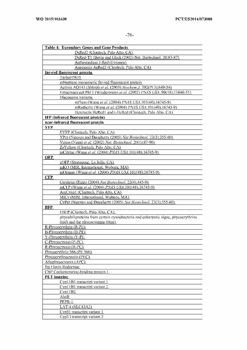
Cyp5 1 transcript variant 2

SLC6A14 Sodium- and chloride-dependent neutral and basic amino


Table 4: Exemplary Genes and Gene ProductsSLC19 solute carrier 19 transporter protein familySLC19A1, SLC19A2, SLC19A3SLC20 solute carrier 20 transporter protein familySLC20A1, SLC20A2SLC21 solute carrier 21 transporter protein family
subfamily 1; SLC01A2, SLCOIBI, SLC01B3, SLC01B4,SLCOICIsubfamily 2; SLCQ2A1, SLCQ2B1subfamily 3; SLCQ3A1subfamily 4; SLCQ4A1, SLCQ4C1subfamily 5; SLCQ5A1
SLC22 solute carrier 22 transporter protein familySLC22A1, SLC22A2, SLC22A3, SLC22A4, SLC22A5, SLC22A6, SLC22A7,SLC22A8, SLC22A9, SLC22A10, SLC22A1 1, SLC22A12, SLC22A13,SLC22A14, SLC22A15, SLC22A16, SLC22A17, SLC22A1 8, SLC22A19,SLC22A20SLC23 solute carrier 23 transporter protein familySLC23A1, SLC23A2, SLC23A3, SLC23A4SLC24 solute carrier 24 transporter protein familySLC24A1, SLC24A2, SLC24A3, SLC24A4, SLC24A5, SLC24A6SLC25 solute carrier 25 transporter protein familySLC25A1, SLC25A2, SLC25A3, SLC25A4, SLC25A5, SLC25A6, SLC25A7,SLC25A8, SLC25A9, SLC25A10, SLC25A1 1, SLC25A12, SLC25A13,SLC25A14, SLC25A15, SLC25A16, SLC25A17, SLC25A1 8, SLC25A19,SLC25A20, SLC25A21, SLC25A22, SLC25A23, SLC25A24, SLC25A25,SLC25A26, SLC25A27, SLC25A28, SLC25A29, SLC25A30, SLC25A3 1,
SLC25A32, SLC25A33, SLC25A34, SLC25A35, SLC25A36, SLC25A37,SLC25A38, SLC25A39, SLC25A40, SLC25A41, SLC25A42, SLC25A43,SLC25A44, SLC25A45, SLC25A46SLC26 solute carrier 26 transporter protein familySLC26A1, SLC26A2, SLC26A3, SLC26A4, SLC26A5, SLC26A6, SLC26A7,SLC26A8, SLC26A9, SLC26A10, SLC26A1 1SLC27 solute carrier 27 transporter protein familySLC27A1, SLC27A2, SLC27A3, SLC27A4, SLC27A5, SLC27A6SLC28 solute carrier 28 transporter protein familySLC28A1, SLC28A2, SLC28A3SLC29 solute carrier 29 transporter protein familySLC29A1, SLC29A2, SLC29A3, SLC29A4SLC30 solute carrier 30 transporter protein familySLC30A1, SLC30A2, SLC30A3, SLC30A4, SLC30A5, SLC30A6, SLC30A7,SLC30A8, SLC30A9, SLC30A10SLC31 solute carrier 31 transporter protein familySLC3 1A1
SLC32 solute carrier 32 transporter protein familySLC32A1SLC33 solute carrier 33 transporter protein familySLC33A1SLC34 solute carrier 34 transporter protein familySLC34A1, SLC34A2, SLC34A3SLC35 solute carrier 35 transporter protein family
subfamily A; SLC35A1, SLC35A2, SLC35A3, SLC35A4, SLC35A5subfamily B; SLC35B1, SLC35B2, SLC35B3, SLC35B4subfamily C; SLC35C1, SLC35C2subfamily D; SLC35D1, SLC35D2, SLC35D3

pyrophosphatase


TIMP-4 (Tissue Inhibitor of Metalloprotemase Type-4)
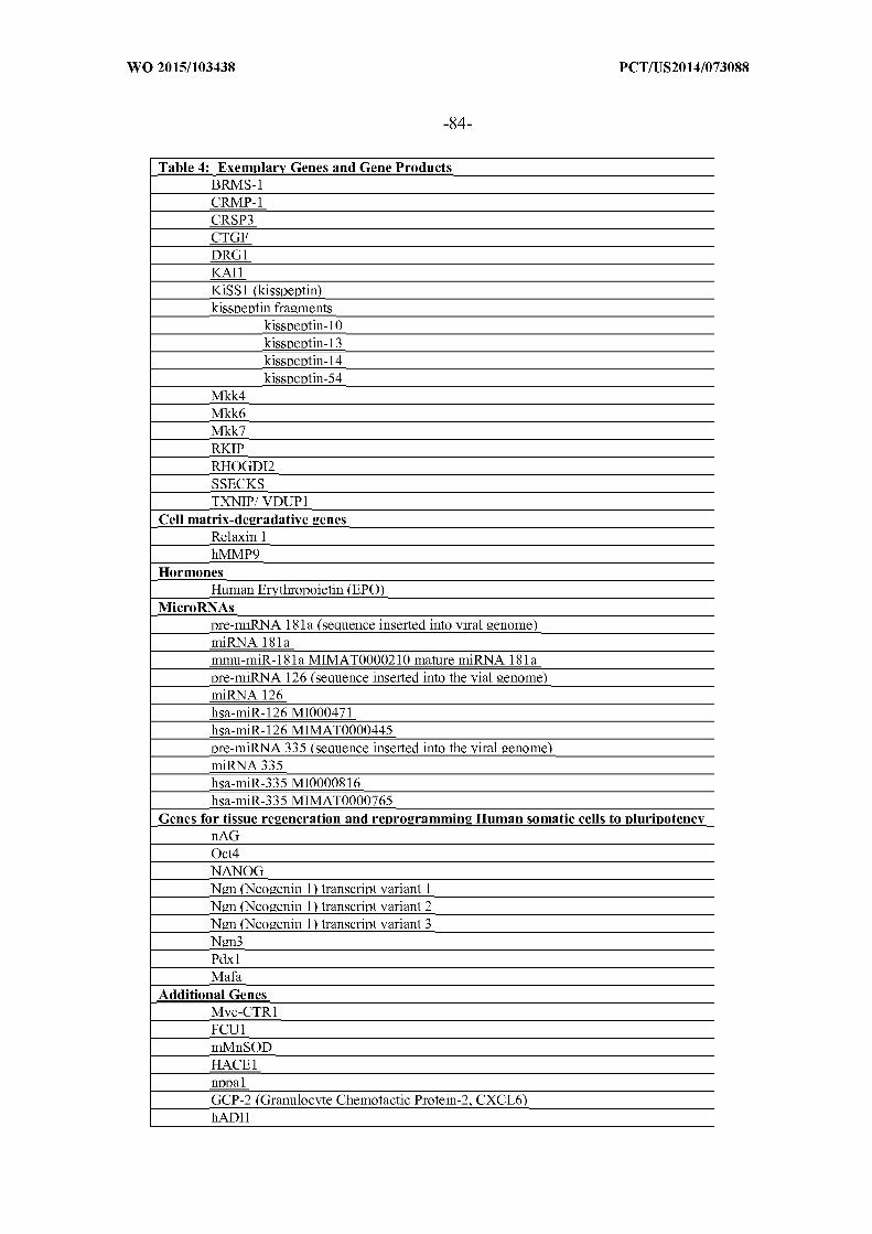


Table 4: Exemplary Genes and Gene ProductsA34R with a mutation at codon 15 1 (Lys 15 1 to Asp)A34R with a mutation at codon 15 1 (Lys 15 1 to Glu)
Non-coding SequenceNon-proteinsNon-coding nucleic acid
RibozymesGroup I intronsGroup II intronsRNasePhairpin ribozymeshammerhead ribozymes
Prodrug converting enzymesvaricella zoster thymidine kinasecytosine deaminasepurine nucleoside phosphorylase (e.g. , from E. coli)beta lactamasecarboxypeptidase G2carboxypeptidase Acytochrome P450
cytochrome P450-2B1cytochrome P450-4B1
horseradish peroxidasenitroreductaserabbit carboxylesterasemushroom tyrosinasebeta galactosidase (lacZ) {i.e. , from . coli)beta glucuronidase (gusA)thymidine phosphorylasedeoxycytidine kinaselinamerase
Proteins detectable by antibodieschloramphenicol acetyl transferasehGH
Viral attenuation factorsvirus-specific antibodies
mucinsthrombospondin
tumor necrosis factors (TNFs)TNFa
SuperantigensToxins
diphtheria toxinPseudomonas exotoxinEscherichia coli Shiga toxinShigella toxinEscherichia coli Verotoxin 1Toxic Shock Syndrome Toxin 1Exfoliating Toxins (EXft)Streptococcal Pyrogenic Exotoxin (SPE) A, B and CClostridial Perfringens Enterotoxin (CPET)staphylococcal enterotoxins
SEA, SEB, SEC1, SEC2, SEP, SEE and SEHMouse Mammary Tumor Virus proteins (MMTV)Streptococcal M proteins

Table 4: Exemplary Genes and Gene ProductsListeria monocytogenes antigen p60mycoplasma arthritis superantigens
Proteins that can bind a contrasting agent, chromophore, or a compound or ligand thatcan be detected
siderophoresenterobactinsalmochelinyersiniabactinaerobactin
Growth Factorsplatelet-derived growth factor (PDG-F)keratinocyte growth factor (KGF)insulin-like growth factor-1 (IGF-1)insulin-like growth factor-binding proteins (IGFBPs)transforming growth factor (TGF-alpha)Growth factors for blood cells
Granulocyte Colony Stimulating Factor (G-CSF)growth factors that can boost platelets
Other GroupsBAC (Bacterial Artificial Chromosome) encoding several or all proteins of a
specific pathway, e . g. woundhealing -pathwayMAC (Mammalian Artificial Chromosome) encoding several or all proteins of a
specific pathway, e . g. woundhealing -pathwaytumor antigenRNAiligand binding proteinsproteins that can induce a signal detectable by MRJangiogeninsphotosensitizing agentsanti-metabolitessignaling modulatorschemotherapeutic compoundslipasesproteasespro-apoptotic factorsanti-cancer vaccine
antigen vaccineswhole cell vaccines (i.e. , dendritic cell vaccines)DNA vaccinesanti-idiotype vaccines
tumor suppressorscytotoxic proteincytostatic proteinscostimulatory molecules
cytokines and chemokinescancer growth inhibitorsgene therapyBCG vaccine for bladder cancer
Proteins that interact with host cell proteins
b. Control of Heterologous Gene Expression
In some examples, the heterologous nucleic acid also can contain one or more
regulatory sequences to regulate expression of an open reading frame encoding the

heterologous RNA and/or protein. Suitable regulatory sequences which, for example, are
functional in a mammalian host cell are well known in the art. Expression can also be
influenced by one or more proteins or RNA molecules expressed by the virus. Gene
regulatory elements, such as promoters and enhancers, possess cell type specific activities
and can be activated by certain induction factors (e.g. , hormones, growth factors, cytokines,
cytostatic agents, irradiation, heat shock) via responsive elements. A controlled and
restricted expression of these genes can be achieved using such regulatory elements as
internal promoters to drive the expression of therapeutic genes in viral vector constructs.
For example, the one or more heterologous nucleic acid molecules can be operably
linked to a promoter for expression of the heterologous RNA and/or protein. For example, a
heterologous nucleic acid that is operably linked to a promoter is also called an expression
cassette. Hence, viruses provided herein can have the ability to express one or more
heterologous genes. Gene expression can include expression of a protein encoded by a gene
and/or expression of an RNA molecule encoded by a gene. In some embodiments, the
viruses provided herein can express exogenous genes at levels high enough that permit
harvesting products of the exogenous genes from the tumor. Expression of heterologous
genes can be controlled by a constitutive promoter, or by an inducible promoter. In other
examples, organ or tissue-specific expression can be controlled by regulatory sequences. In
order to achieve expression only in the target organ, for example, a tumor to be treated, the
foreign nucleotide sequence can be linked to a tissue specific promoter and used for gene
therapy. Such promoters are well known to those skilled in the art (see, e.g. , Zimmermann
et al, Neuron 12: 11-24 (1994); Vidal et al, EMBOJ. 9 : 833-840 (1990); Mayford et al,
Cell 81: 891-904 (1995); and Pinkert et al, Genes & Dev. 1: 268-76 (1987)).
Exemplary promoters for the expression of heterologous genes are known in the art.
The heterologous nucleic acid can be operatively linked to a native promoter or a
heterologous promoter that is not native to the virus. Any suitable promoters, including
synthetic and naturally-occurring and modified promoters, can be used. Exemplary
promoters include synthetic promoters, including synthetic viral and animal promoters.
Native promoter or heterologous promoters include, but are not limited to, viral promoters,
such as vaccinia virus and adenovirus promoters.
In one example, the promoter is a poxvirus promoter, such as, for example, a
vaccinia virus promoter. Vaccinia viral promoters for the expression of one or more
heterologous genes can be synthetic or natural promoters, and include vaccinia early,
intermediate, early/late and late promoters. Exemplary vaccinia viral promoters for
controlling heterologous gene expression include, but are not limited to, P7 5k, P k, PSE, PSEL,

PSL, H5R, TK, P28, CI 1R, G8R, F17R, I3L, I8R, AIL, A2L, A3L, H1L, H3L, H5L, H6R,
H8R, DIR, D4R, D5R, D9R, Dl 1L, D12L, D13L, MIL, N2L, P4b or Kl promoters. Other
viral promoters include, but are not limited to, adenovirus late promoter, Cowpox ATI
promoter, or T7 promoter. Strong late promoters can be used to achieve high levels of
expression of the heterologous genes. Early and intermediate-stage promoters also can be
used. In one example, the promoters contain early and late promoter elements, for example,
the vaccinia virus early/late promoter P k, vaccinia late promoter P k, a synthetic early/late
vaccinia PSEL promoter (Patel et al, (1988) Proc. Natl. Acad. Sci. USA 85: 9431-9435;
Davison and Moss, (1989) JMolBiol 210: 749-769; Davison et al. (1990) Nucleic Acids
Res. 18: 4285-4286; Chakrabarti et al. (1997), BioTechniques 23: 1094-1097). The viruses
provided herein can exhibit differences in characteristics, such as attenuation, as a result of
using a stronger promoter versus a weaker promoter. For example, in vaccinia, synthetic
early/late and late promoters are relatively strong promoters, whereas vaccinia synthetic
early, P7 5k early/late, P 5k early, and P28 late promoters are relatively weaker promoters (see
e.g., Chakrabarti et al. (1997) BioTechniques 23(6) 1094-1097). Combinations of different
promoters can be used to express different gene products in the same virus or two different
viruses.
As is known in the art, regulatory sequences can permit constitutive expression of
the exogenous gene or can permit inducible expression of the exogenous gene. Further, the
regulatory sequence can permit control of the level of expression of the exogenous gene. In
some examples, such as gene product manufacture and harvesting, the regulatory sequence
can result in constitutive, high levels of gene expression. In some examples, such as anti-
(gene product) antibody harvesting, the regulatory sequence can result in constitutive, lower
levels of gene expression. In tumor therapy examples, a therapeutic protein can be under
the control of an internally inducible promoter or an externally inducible promoter.
Hence, expression of heterologous genes can be controlled by a constitutive
promoter or by an inducible promoter. Inducible promoters can be used to provide tissue
specific expression of the heterologous gene or can be inducible by the addition of a
regulatory molecule to provide temporal specific induction of the promoter. In some
examples, inducible expression can be under the control of cellular or other factors present
in a tumor cell or present in a virus-infected tumor cell. In further examples, inducible
expression can be under the control of an administrable substance, including IPTG, RU486
or other known induction compounds. Additional regulatory sequences can be used to
control the expression of the one or more heterologous genes inserted the virus. Any of a

variety of regulatory sequences are available to one skilled in the art according to known
factors and design preferences.
c. Exemplary Modified or Recombinant Viruses
Exemplary modified or recombinant vaccinia viruses for use in the combinations,
compositions or methods herein are those derived from the Lister strain, and in particular
the attenuated Lister strain LIVP. Recombinant LIVP viruses containing heterologous
nucleic acid have been generated and are known in the art. The modified LIVP viruses can
be modified by insertion, deletion or amino acid replacement of heterologous nucleic acid
compared to an LIVP strain having a genome set forth in any one of SEQ ID NOS: 1, 2 or
5-1 1, or having a genome that exhibits at least 85% or more sequence identity to any of SEQ
ID NOS: 1, 2 or 5-1 1. Table 5 sets forth exemplary viruses, the reference or parental LIVP
(e.g. LIVP set forth in SEQ ID NO: 2 or GLV-lh68 set forth in SEQ ID NO: 3) and the
resulting genotype. The exemplary modifications of the Lister strain can be adapted to other
vaccinia viruses (e.g., Western Reserve (WR), Copenhagen, Tashkent, Tian Tan, Lister,
Wyeth, IHD-J, and IHD-W, Brighton, Ankara, MVA, Dairen I, LIPV, LC16M8, LC16MO,
LIVP, WR 65-1 6, Connaught, New York City Board of Health).


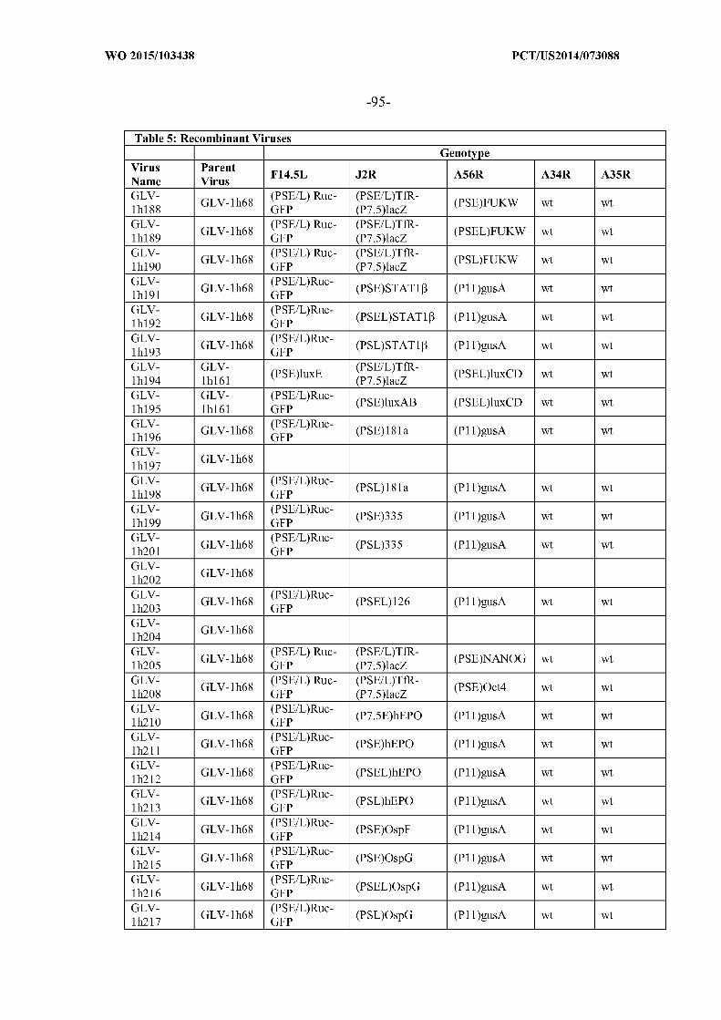


Table 5: Recombinant VirusesGenotype
Virus ParentF14.5L J2R A56R A34R A35R
Name VirusGLV- GLV- (PSE/L) Ruc-
(PSEL)PEPRl (PSEL)FUKW wt wtlh270 1 89 GFPGLV- GLV- (PSE/L) Ruc-
(PSL)PEPRl (PSEL)FUKW wt wtlh271 lhl 89 GFPGLV- GLV- (PSE/L) Ruc-
(PSE)LAT4 (PSEL)FUKW wt wtlh272 lhl 89 GFPGLV- GLV- (PSE/L) Ruc-
(PSEL)LAT4 (PSEL)FUKW wt wtlh273 lhl 89 GFPGLV- GLV- (PSE/L) Ruc-
(PSL)LAT4 (PSEL)FUKW wt wtlh274 lhl 89 GFPGLV- GLV- (PSE/L) Ruc-
(PSE)Cyp5 1 (PSEL)FUKW wt wtlh275 1 89 GFPGLV- GLV- (PSE/L) Ruc-
(PSEL)Cyp5 1 (PSEL)FUKW wt wtlh276 lhl 89 GFPGLV- GLV- (PSE/L) Ruc-
(PSL)Cyp5 1 (PSEL)FUKW wt wtlh277 lhl 89 GFPGLV- GLV- (PSE/L)Ruc- (PSE)BMP4 (PSEL)FUKW wt wtlh284 lhl 89 GFPGLV- GLV- (PSE/L)Ruc-
(PSEL)BMP4 (PSEL)FUKW wt wtlh285 lhl 89 GFPGLV- GLV- (PSE/L)Ruc-
(PSL)BMP4 (PSEL)FUKW wt wtlh286 1 89 GFP
For example, GLV-lh68 (also named RVGL21 , SEQ ID NO: 3; described in U.S.
Pat. Pub. No. 2005-003 1643, now U.S. Patent Nos. 7,588,767, 7,588,771 , 7,662,398) is an
attenuated virus of the LIVP parental strain containing a genome set forth in SEQ ID NO: 2.
GLV-lh68 contains DNA insertions in gene loci F14.5L (also designated in LIVP as F3)
gene locus, thymidine kinase (TK) gene locus, and hemagglutinin (HA) gene locus with
expression cassettes encoding detectable marker proteins. Specifically, GLV-lh68 contains
an expression cassette containing a Ruc-GFP cDNA molecule (a fusion of DNA encoding
Renilla luciferase and DNA encoding GFP) under the control of a vaccinia synthetic
early/late promoter PSEL (( EL)RUC-GFP) inserted into the F14.5L gene locus; an expression
cassette containing a DNA molecule encoding beta-galactosidase under the control of the
vaccinia early/late promoter P 5k ((5i
)LacZ) and DNA encoding a rat transferrin receptor
positioned in the reverse orientation for transcription relative to the vaccinia synthetic
early/late promoter PSEL ((P SEL)r 7 7 ) inserted into the TK gene locus (the resulting virus
does not express transferrin receptor protein since the DNA molecule encoding the protein
is positioned in the reverse orientation for transcription relative to the promoter in the
cassette); and an expression cassette containing a DNA molecule encoding β-glucuronidase
under the control of the vaccinia late promoter P l lk (( )gusA) inserted into the HA gene
locus.

Other recombinant LIVP viruses are derived from GLV-lh68 and contain
heterologous DNA that encodes a gene product or products (see e.g. see e.g. U.S. Pub. Nos.
US2003-0059400, US2003-0228261, US2007-0202572, US2007-0212727, US2009-
0117034, US2009-0098529, US2009-0053244, US2009-0155287, US2009-0081639,
US2009-0136917, US2009-0162288, US2010-0062016, US2010-0233078 and US2010-
0196325; U.S. Patent No. 7,588,767, 7,588,771, 7,662,398 and 7,754,221 and 7,763,420;
and International Pub. No. WO 2009/139921).
Exemplary of such recombinant viruses include those set forth in Table 5 above,
including but not limited to, GLV-lh64 (set forth in SEQ ID NO: 14); viruses that encode
the far-red fluorescent protein TurboFP635 (scientific name "Katushka") from the sea
anemone Entacmaea quadricolor include GLV-lhl88 (SEQ ID NO: 15), GLV-lhl89 (SEQ
ID NO: 16), GLV-lhl90 (SEQ ID NO: 17) and GLV-lh253 (SEQ ID NO: 18).
Modified vaccinia viruses also include viruses that are modified by introduction of
heterologous nucleic acid into an LIVP strain containing a genome set forth in any of SEQ
ID NO: 5-11, or a genome that exhibits at least 85%, 90%, 92%, 95%, 96%, 97%, 98%,
99% or more sequence identity to any of SEQ ID NOS:5-l 1. For example, exemplary of a
modified vaccinia virus is a virus that is modified by insertion, deletion or replacement of
heterologous nucleic acid compared to an LIVP strain having a genome set forth in SEQ ID
NO: 5. Exemplary of such as strain is GLV-2b372, which contains TurboFP635 (Far-red
fluorescent protein "katushka") under the control of the vaccinia synthetic early/late
promoter at the TK locus. The genome of GLV-lb372 has the sequence of nucleotides set
forth in SEQ ID NO: 19.
d. Methods of Generating Modified Viruses
Oncolytic viruses, such as vaccinia viruses {e.g. LIVP), for use in the combinations,
compositions or methods herein can be modified by insertion, deletion, replacement or
mutation as described herein, for example insertion or replacement of heterologous nucleic
acid, using standard methodologies well known in the art for modifying viruses. Methods
for modification include, for example, in vitro recombination techniques, synthetic methods,
direct cloning, and in vivo recombination methods as described, for example, in Sambrook
et a , Molecular Cloning: A Laboratory Manual, 2nd edition, Cold Spring Harbor
Laboratory Press, cold Spring Harbor NY (1989).
For example, generation of recombinant viruses, including recombinant vaccinia
virus, is well known in the art, and typically involves the generation of gene cassettes or
transfer vectors using standard techniques in molecular biology (see, e.g., U.S. Pat. No.
7,588,767 and US2009-0053244-A1, which describe exemplary methods of generating

recombinant LIVP vaccinia virases). Such techniques include various nucleic acid
manipulation techniques, nucleic acid transfer protocols, nucleic acid amplification
protocols, and other molecular biology techniques known in the art. For example, point
mutations or small insertions or deletions can be introduced into a gene of interest through
the use of oligonucleotide mediated site-directed mutagenesis. In another example,
homologous recombination can be used to introduce a mutation in the nucleic acid sequence
or insertion or deletion of a nucleic acid molecule into a target sequence of interest. In some
examples, mutations, insertions or deletions of nucleic acid in a particular gene can be
selected for using a positive or negative selection pressure. See, e.g., Current Techniques in
Molecular Biology, (Ed. Ausubel, et al).
Nucleic acid amplification protocols include, but are not limited to, the polymerase
chain reaction (PCR), or amplification via viruses or organisms, such as, but not limited to,
bacteria, yeast, insect or mammalian cells. Use of nucleic acid tools such as plasmids,
vectors, promoters and other regulating sequences, are well known in the art for a large
variety of viruses and cellular organisms.
Nucleic acid transfer protocols include calcium chloride transformation/transfection,
electroporation, liposome mediated nucleic acid transfer, N-[l-(2,3-dioloyloxy)propyl]-
Ν,Ν,Ν-trimethylammonium methylsulfate meditated transformation, and others. Further a
large variety of nucleic acid tools are available from many different sources, including
various commercial sources. One skilled in the art will be readily able to select the
appropriate tools and methods for genetic modifications of any particular virus according to
the knowledge in the art and design choice.
Hence, any of a variety of modifications can be readily accomplished using standard
molecular biological methods known in the art. The modifications will typically be one or
more truncations, deletions, mutations or insertions of the viral genome. In one example,
the modification can be specifically directed to a particular sequence in the viral genome.
The modifications can be directed to any of a variety of regions of the viral genome,
including, but not limited to, a regulatory sequence, a gene-encoding sequence, an intergenic
sequence, a sequence without a known role, or a non-essential region of the viral genome.
Any of a variety of regions of viral genomes that are available for modification are readily
known in the art for many viruses, including LIVP.
Heterologous nucleic acid molecules are typically inserted into the viral genome in
an intergenic region or in a locus that encodes a nonessential viral gene product. Insertion
of heterologous nucleic acid at such sites generally does not significantly affect viral
infection or replication in the target tissue. Exemplary insertion sites are known in the art

and include, but are not limited to, J2R (thymidine kinase (TK)), A56R (hemagglutinin
(HA)), F14.5L, vaccinia growth factor (VGF), A35R, NIL, E2L/E3L, K1L/K2L, superoxide
dismutase locus, 7.5K, C7-K1L (host range gene region), B13R+B14R (hemorrhagic
region), A26L (A type inclusion body region (ATI)) or I4L (large subunit, ribonucleotide
reductase) gene loci. Insertion sites for the viruses also include sites that correspond to
intragenic regions described in other poxviruses such as Modified Vaccinia Ankara (MVA)
virus (exemplary sites set forth in U.S. Patent No. 7,550,147), NYVAC (exemplary sites set
forth in U.S. Patent No. 5,762,938).
Methods for the generation of recombinant viruses using recombinant DNA
techniques are well known in the art (e.g., see U.S. Pat. No. 4,769,330; 4,603,1 12;
4,722,848; 4,215,051; 5,110,587; 5,174,993; 5,922,576; 6,319,703; 5,719,054; 6,429,001;
6,589,531; 6,573,090; 6,800,288; 7,045,313; He etal. (1998) PNAS 95(5): 2509-2514;
Racaniello et al, (1981) Science 214: 916-919; and Hruby et al, (1990) Clin Micro Rev.
3:153-1 70). Methods for the generation of recombinant vaccinia viruses are well known in
the art (e.g., see Hruby et al, (1990) Clin Micro Rev. 3:153-170, U.S. Pat. Pub. No. 2005-
0031643, now U.S. Patent Nos. 7,588,767, 7,588,771, 7,662,398 and U.S. Patent No.
7,045,313).
For example, generating a recombinant vaccinia virus that expresses a heterologous
gene product typically includes the use of a recombination plasmid which contains the
heterologous nucleic acid, optionally operably linked to a promoter, with vaccinia virus
DNA sequences flanking the heterologous nucleic acid to facilitate homologous
recombination and insertion of the gene into the viral genome. Generally, the viral DNA
flanking the heterologous gene is complementary to a non-essential segment of vaccinia
virus DNA, such that the gene is inserted into a nonessential location. The recombination
plasmid can be grown in and purified from Escherichia coli and introduced into suitable
host cells, such as, for example, but not limited to, CV-1, BSC-40, BSC-1 and TK-143 cells.
The transfected cells are then superinfected with vaccinia virus which initiates a replication
cycle. The heterologous DNA can be incorporated into the vaccinia viral genome through
homologous recombination, and packaged into infection progeny. The recombinant viruses
can be identified by methods known in the art, such as by detection of the expression of the
heterologous gene product, or by using positive or negative selection methods (U.S. Pat. No.
7,045,313).
In another example, the recombinant vaccinia virus that expresses a heterologous
gene product can be generated by direct cloning (see, e.g. U.S. Pat. No. 6,265,183 and
Scheiflinger et al. (1992) Proc. Natl. Acad. Sci. USA 89: 9977-9981). In such methods, the

heterologous nucleic acid, optionally operably linked to a promoter, is flanked by restriction
endonuclease cleavage sites for insertion into a unique restriction endonuclease site in the
target virus. The virus DNA is purified using standard techniques and is cleaved with the
sequence-specific restriction endonuclease, where the sequence is a unique site in the virus
genome. Any unique site in the virus genome can be employed provided that modification
at the site does not interfere with viral replication. For example, in vaccinia virus strain
LIVP, the Notl restriction site is located in the ORF encoding the F14.5L gene with
unknown function (Mikryukov et al, Biotekhnologiya 4 : 442-449 (1988)). Table 6 provides
a summary of unique restriction sites contained in exemplary LIVP strains and designates
the nucleotide position of each. Such LIVP strains can be modified herein by direct cloning
and insertion of heterologous DNA into the site or sites. Generally, insertion is in a site that
is located in a non-essential region of the virus genome. For example, exemplary
modifications herein include insertion of a foreign DNA sequence into the Notl digested
virus DNA.
In some examples, the virus genomic DNA is first modified by homologous
recombination to introduce one or more unique restriction sites in the virus (see, e.g.
Mackett et al. (1984) J. Virol. 857-864). Following cleavage with the restriction
endonuclease, the cleaved DNA is optionally treated with a phosphatase to remove a
phosphate moiety from an end of the DNA segment that is produced by cleavage with the

endonuclease. Typically, a plasmid vector is generated that contains the heterologous DNA
for insertion flanked by the restriction sites. Prior to insertion into the virus, the
heterologous DNA is excised from the plasmid by cleavage with the sequence specific
restriction endonuclease. The heterologous DNA is then ligated to the cleaved viral DNA
and is packaged in a permissive cell line by infection of the cells with a helper virus, such
as, but not limited to a fowlpox virus or a puv-inactivated helper vaccinia virus, and
transfection of the ligated DNA into the infected cells.
In some examples, the methods involve homologous recombination and/or use of
unique restriction sites in the virus. For example, a recombinant LIVP vaccinia virus with
an insertion, for example, in the F14.5L gene {e.g., in the Not I restriction site of an LIVP
isolate) can be prepared by the following steps: (a) generating (i) a vaccinia shuttle/transfer
plasmid containing the modification {e.g. a gene expression cassette or a modified F14.5L
gene) inserted at a restriction site, X {e.g. Not I), where the restriction site in the vector is
flanked by parental virus sequences of the target insertion site and (ii) an LIVP virus DNA
digested at restriction site X {e.g. Not I) and optionally dephosphorylated; (b) infecting cells
with PUV-inactivated helper vaccinia virus and transfecting the infected host cells with a
mixture of the constructs of (i) and (ii) of step a; and (c) isolating the recombinant vaccinia
viruses from the transfectants. One skilled in the art knows how to perform such methods
(see, e.g., Timiryasova et al. (Biotechniques 31: 534-540 (2001)). Typically, the restriction
site X is a unique restriction site in the virus as described above.
3. Methods of Producing Viruses
Viruses in the combinations, compositions and methods provided herein can be
produced by methods known to one of skill in the art. Typically, the virus is propagated in
host cells, quantified and prepared for storage before finally being prepared in the
compositions described herein. The virus can be propagated in suitable host cells to enlarge
the stock, the concentration of which is then determined. In some examples, the infectious
titer is determined, such as by plaque assay. The total number of viral particles also can be
determined. The viruses are stored in conditions that promote stability and integrity of the
virus, such that loss of infectivity over time is minimized. In some examples, a large
amount of virus is produced and stored in small aliquots of known concentration that can be
used for multiple procedures over an extended period of time. Conditions that are most
suitable for various viruses will differ, and are known in the art, but typically include
freezing or drying, such as by lyophilization. The viruses can be stored at a concentration of
105-10 1 pfu/mL, for example, 10 -109 pfu/mL, such as at least or about or 106 pfu/mL, 107
pfu/mL, 10 pfu/mL or 109 pfu/mL. Immediately prior to preparing compositions provided

herein, the stored viruses can be reconstituted (if dried for storage) and diluted in an
appropriate medium or solution.
The following sections provide exemplary methods that can be used for the
production and preparation of viruses for use in preparing viruses.
a. Host cells for propagation
Virus strains can be propagated in an appropriate host cell. Such cells can be a
group of a single type of cells or a mixture of different types of cells. Host cells can include
cultured cell lines, primary cells, and proliferative cells. These host cells can include any of
a variety of animal cells, such as mammalian, avian and insect cells and tissues that are
susceptible to the virus, such as vaccinia virus, infection, including chicken embryo, rabbit,
hamster, and monkey kidney cells. Suitable host cells include, but are not limited to,
hematopoietic cells (totipotent, stem cells, leukocytes, lymphocytes, monocytes,
macrophages, APC, dendritic cells, non-human cells and the like), pulmonary cells, tracheal
cells, hepatic cells, epithelial cells, endothelial cells, muscle cells (e.g. , skeletal muscle,
cardiac muscle or smooth muscle), fibroblasts, and cell lines including, for example, chicken
embryo fibroblast (CEF), CV-1, BSC40, Vero, and BSC-1 , and human HeLa cells.
Typically, viruses are propagated in cell lines that that can be grown at monolayers or in
suspension. For example, exemplary cell lines for the propagation of vaccinia viruses
include, but are not limited to, chicken embryo fibroblast (CEF), CV-1, BSC40, Vero,
BGM, BSC-1 and RK-1 3 cells. Purification of the cultured strain from the system can be
effected using standard methods.
b. Concentration determination
The concentration of virus in a solution, or virus titer, can be determined by a
variety of methods known in the art. In some methods, a determination of the number of
infectious virus particles is made (typically termed plaque forming units (PFU)), while in
other methods, a determination of the total number of viral particles, either infectious or not,
is made. Methods that calculate the number of infectious virions include, but are not limited
to, the plaque assay, in which titrations of the virus are grown on cell monolayers and the
number of plaques is counted after several days to several weeks, and the endpoint dilution
method, which determines the titer within a certain range, such as one log. Methods that
determine the total number of viral particles, including infectious and non-infectious,
include, but are not limited to, immunohistochemical staining methods that utilize
antibodies that recognize a viral antigen and which can be visualized by microscopy or
FACS analysis; optical absorbance, such as at 260 nm; and measurement of viral nucleic
acid, such as by PCR, RT-PCR, or quantitation by labeling with a fluorescent dye.

c. Storage methods
Once the virus has been purified (or to a desired purity) and the titer has been
determined, the virus can be stored in conditions which optimally maintain its infectious
integrity. Typically, viruses are stored in the dark, because light serves to inactivate the
viruses over time. Viral stability in storage is usually dependent upon temperatures.
Although some viruses are thermostable, most viruses are not stable for more than a day at
room temperature, exhibiting reduced viability (Newman et al, (2003) J. Inf. Dis.
187:1319-1322). Vaccinia virus is generally stable at refrigerated temperatures, and can be
stored in solution at 4°C, frozen at, for example -20°C, -70°C or -80°C, or lyophilized with
little loss of viability (Newman et a , (2003) J. Inf. Dis. 187:1319-1322, Hruby et al,
(1990) Clin. Microb. Rev. 3:153-170). Methods and conditions suitable for the storage of
particular viruses are known in the art, and can be used to store the viruses used in the
methods presented herein.
For short-term storage of viruses, for example, up to 1 day, 2 days, 4 days or 7 days,
temperatures of approximately 4°C are generally recommended. For long-term storage,
most viruses can be kept at -20°C, -70°C or -80°C. When frozen in a simple solution such
as PBS or Tris solution (20mM Tris pH 8.0, 200 NaCl, 2-3% glycerol or sucrose) at these
temperatures, the virus can be stable for 6 months to a year, or even longer. Repeated
freeze-thaw cycles are generally avoided, however, since it can cause a decrease in viral
titer. The virus also can be frozen in media containing other supplements in the storage
solution which can further preserve the integrity of the virus. For example, the addition of
serum or bovine serum albumin (BSA) to a viral solution stored at -80°C can help retain
virus viability for longer periods of time and through several freeze-thaw cycles.
In other examples, the virus sample is dried for long-term storage at ambient
temperatures. Viruses can be dried using various techniques including, but not limited to,
freeze-drying, foam-drying, spray-drying and desiccation. Water is a reactant in nearly all
of the destructive pathways that degrade viruses in storage. Further, water acts as a
plasticizer, which allows unfolding and aggregation of proteins. Since water is a participant
in almost all degradation pathways, reduction of the aqueous solution of viruses to a dry
powder provides an alternative composition methodology to enhance the stability of such
samples. Lyophilization, or freeze-drying, is a drying technique used for storing viruses
(see, e.g., Cryole et al, (1998) Pharm. Dev. Technol., 3(3), 973-383). There are three
stages to freeze-drying; freezing, primary drying and secondary drying. During these
stages, the material is rapidly frozen and dehydrated under high vacuum. Once lyophilized,
the dried virus can be stored for long periods of time at ambient temperatures, and
reconstituted with an aqueous solution when needed. Various stabilizers can be included in
the solution prior to freeze-drying to enhance the preservation of the virus. For example, it
is known that high molecular weight structural additives, such as serum, serum albumin or
gelatin, aid in preventing viral aggregation during freezing, and provide structural and
nutritional support in the lyophilized or dried state. Amino acids such as arginine and
glutamate, sugars, such as trehalose, and alcohols such as mannitol, sorbitol and inositol,
can enhance the preservation of viral infectivity during lyophilization and in the lyophilized
state. When added to the viral solution prior to lyophilization, urea and ascorbic acid can
stabilize the hydration state and maintain osmotic balance during the dehydration period.
Typically, a relatively constant pH of about 7.0 is maintained throughout lyophilization.
Other methods for the storage of viruses at ambient, refrigerated or freezing
temperatures are known in the art, and include, but are not limited to, those described in
U.S. Pat. Nos. 5,149,653; 6,165,779; 6,255,289; 6,664,099; 6,872,357; and 7,091 ,030; and
in U.S. Pat. Pub. Nos. 2003-0153065, 2004-003841 and 2005-0032044.
D. ADJUNCT THERAPY WITH COMPLEMENT INHIBITORS
Provided herein are combinations and compositions containing an oncolytic virus
(e.g. vaccinia virus), such as a lipid-treated oncolytic virus, and a complement inhibitor.
Complement is a system of over 30 serum and membrane-bound proteins that form a
cascade of reactions that contribute to the elimination, and hence neutralization, of viruses
and other microorganisms. In the combinations and compositions provided herein, a
complement inhibitor, which is a biomolecule that targets one or more components of the
complement system to thereby reduce activation or propagation of complement cascade,
reduces the elimination of virus and thereby increases the infectivity of the oncolytic virus
compared to the absence of the inhibitor. Hence, the combinations and compositions of an
oncolytic virus (e.g. vaccinia virus), such as a lipid-treated oncolytic virus, and a
complement inhibitor can be used in adjunct therapies to improve the therapeutic effect of
an oncolytic virus, for example, for treatment of proliferative diseases and disorders, such as
tumors or cancers.
The complement inhibitor can be administered prior to, simultaneously, or
intermittently with the oncolytic virus. When administered simultaneously, the complement
inhibitor can be administered in the same composition with the oncolytic virus or as a
combination of two separate composition. Typically, the complement inhibitor is
administered prior to the oncolytic virus. For example, the complement inhibitor is
administered at least or up to 2 minutes, 3 minutes, 4 minutes, 5 minutes, 10 minutes, 15
minutes, 20 minutes, 30 minutes, 40 minutes, 50 minutes, 1 hour, 1.5 hours, 2 hours, 3

hours, 4 hours, 5 hours, 6 hours, 7 hours, 8 hours, 9 hours, 10 hours, 11 hours or 12 hours
prior to administration of the oncolytic virus. Typically, the mode of administration of the
oncolytic virus and complement inhibitor is the same so that both agents are exposed to the
same bodily fluid. Typically, administration is systemic, such as by subcutaneous or
intravenous administration.
In the adjunct therapies provided herein, the presence of a complement inhibitor in a
combination or composition with an oncolytic virus increases the infectivity of the virus in a
complement-containing bodily fluid (e.g. serum) by at least 1.2-fold, 1.3-fold, 1.4-fold, 1.5-
fold, 1.6-fold, 1.7-fold, 1.8-fold, 1.9-fold, 2.0-fold, 2.5-fold, 3-fold, 4-fold, 5-fold, 10-fold
or more compared to the infectivity of the virus in the bodily fluid (e.g. serum) in the
absence of the complement inhibitor. In some examples, an oncolytic virus in combination
or composition with a complement inhibitor exhibits increased virus titer after exposure to
bodily fluid for a predetermined time (e.g. virus titer AUC or total virus recovery) by at least
1.2-fold, 1.3-fold, 1.4-fold, 1.5-fold, 1.6-fold, 1.7-fold, 1.8-fold, 1.9-fold, 2.0-fold, 2.5-fold,
3-fold, 4-fold, 5-fold or more compared to virus titer of the same oncolytic virus in the
absence of the complement inhibitor. The predetermined time can be 1 minute to 12 hours,
and generally is 1 minute to 2 hours, 1 minute to 1 hour, 1 minute to 30 minutes, for
example, at least 2 minutes, 5 minutes, 10 minutes, 15 minutes, 20 minutes, 30 minutes, 40
minutes, 50 minutes, 60 minutes, 70 minutes, 80 minutes, 90 minutes, 100 minutes, 110
minutes, 120 minutes or more. In other examples, an oncolytic virus in combination or
composition with a complement inhibitor exhibits increased half-life in a bodily fluid (e.g.
serum) by at least 1.2-fold, 1.3-fold, 1.4-fold, 1.5-fold, 1.6-fold, 1.7-fold, 1.8-fold, 1.9-fold,
2.0-fold, 2.5-fold, 3-fold, 4-fold, 5-fold or more compared to half-life of the same oncolytic
virus in the same bodily fluid (e.g. serum) in absence of the complement inhibitor.
In examples of the adjunct therapies provided herein, the presence of a complement
inhibitor in a combination or composition with an oncolytic virus increases binding of the
virus to blood cells by at least 1.2-fold, 1.3-fold, 1.4-fold, 1.5-fold, 1.6-fold, 1.7-fold, 1.8-
fold, 1.9-fold, 2.0-fold, 2.5-fold, 3-fold, 4-fold, 5-fold, 10-fold or more compared to the
binding of the virus to blood cells in the absence of the complement inhibitor.
1. The Complement System and Virus Neutralization
The complement system is part of the immune system and plays a role in the
elimination of invading foreign organisms and initiates inflammatory responses. There are
over 30 soluble and cell-membrane proteins that are part of the complement system. The
complement system constitutes an irreversible cascade of proteolytic events, resulting in the
formation of multiple effector molecules that stimulate inflammation, facilitate antigen

phagocytosis, and lyse some cells directly. The numbering of the complement components
is based on the order of their discovery rather than the order of the sequence of reactions
within the complement cascade. As a result, the sequence of reactions of the complement
cascades is CI, C4, C2, C3, C5, C6, C7, C8, and C9. Several complement proteins are pro-
enzyme proteases (termed zymogens) that are themselves activated by proteolytic cleavage
to become effector proteases that cut peptide bonds in other complement proteins to activate
them in turn. Following activation, the products of the cleavage reactions are designated by
adding lower case letters, the larger fragment generally being designated "b" and the
smaller fragment as "a" (e.g. C4 is cleaved to generate C4b and C4a).
There are three distinct pathways through which complement can be activated on
the pathogen surface: the classical pathway, the alternative pathway, and the lectin pathway.
These pathways are distinct in that the components required for their initiation are different,
but the pathways are similar in that they converge to generate the same set of effector
molecules. The convergence point of the pathways is the cleavage of C3 by C3 convertase
(a C3 activating enzyme). Different enzyme complexes have C3 convertase activity. For
example, in the classical pathway C4b2b acts as a C3 convertase, whereas in the alternative
pathway, C3bBb is a C3 convertase. Cleavage of C3 generates C3b, which acts as an
opsonin and as the main effector molecule of the complement system for subsequent
complement reactions, and C3a, which is a peptide mediator of inflammation. The addition
of C3b to each C3 convertase forms a C5 convertase that cleaves C5 to generate C5a and
C5b. C5a, like C3a, is a peptide mediator of inflammation. C5b mediates the "late" events
of complement activation initiating the sequence of reactions culminating in the generation
of the membrane attack complex (MAC). Although the three pathways produce different
C3 and C5 convertases, all of the pathways produce the split products of C3 and C5 and
form MAC.
a. Complement Pathways
Virus infection can activate all three complement pathways. The classical pathway
is activated by binding of Clq to antibody-antigen complexes formed on the surface of the
virus or by Clq directly binding to cell surface components on viruses or other pathogens in
the absence of specific antibodies. The lectin pathway is activated upon the interaction of
mannan-binding lectin (MBL) with viral surface carbohydrates. The alternative pathway is
activated by binding of C3b to the pathogen surface
i. Classical Pathway
Clq is the first component of the classical pathway of complement. Clq is a
calcium-dependent binding protein associated with the collectin family of proteins due to an

overall shared structural homology (Malhotra R et al., Clin Exp Immunol. 1994, 97(2):4-9;
Holmskov et al. Immunol Today. 1994, 15(2):67-74). Clq initiates the classical pathway of
complement in two different ways. First, the classical pathway is activated by the
interaction of Clq with immune complexes {i.e. antigen-antibody complexes of aggregated
IgG or IgM antibody). IgA, IgE and IgD do not bind Clq and cannot activate complement.
Second, Clq also is able to activate complement in the absence of antibody by the
interaction of Clq with non-immune molecules such as polyanions (bacterial
lipopolysaccharides, DNA, and RNA), certain small polysaccharides, viral membranes, C
reactive protein (CRP), serum amyloid P component (SAP), and bacterial, fungal and virus
membrane components.
Clq is part of the CI complex which contains a single Clq molecule bound to two
molecules each of the zymogens Clr and Cls. Binding of more than one of the Clq
globular domains to a target surface (such as aggregated antibody or a pathogen), causes a
conformational change in the (Clr:Cls)2 complex, which results in the activation of the Clr
protease to cleave Cls to generate an active serine protease. Active Cls cleaves subsequent
complement components C4 and C2 to generate C4b and C2b, which together form the C3
convertase of the classical pathway. The C3 convertase cleaves C3 into C3b, which
covalently attaches to the pathogen surface and acts as an opsonin, and C3a, which
stimulates inflammation. Some C3b molecules associate with C4b2b complexes yielding
C4b2b3b which is the classical cascade C5 convertase.
ii. Alternative Pathway
The alternative pathway is initiated by foreign pathogens in the absence of antibody.
Instead, the initiation of complement by the alternative pathway occurs through the
spontaneous hydrolysis of C3 into C3b. A small amount of C3b is always present in body
fluids, due to serum and tissue protease activity. C3b is an opsonin that recognizes and
binds to pathogen surfaces. C3b on pathogen surfaces is recognized by the protease
zymogen Factor B. Factor B, which is the only activating protease of the complement
system that circulates as an active enzyme rather than as a zymogen, is cleaved by Factor D.
Cleavage of Factor B by Factor D yields the active product Bb which can associate with
C3b to form C3bBb, the C3 convertase of the alternative pathway. Similar to the classical
pathway, the C3 convertase produces more C3b and C3a from C3. C3b covalently attaches
to the pathogen surface and acts as an opsonin, while C3a stimulates inflammation. Some
C3b joins the complex to form C3bBb3b, the alternative pathway C5 convertase. C3bBb3b
is stabilized by the plasma protein properdin or Factor P which binds to microbial surfaces
and stabilizes the convertase.

iii. Lectin Pathway
The lectin pathway (also referred to as the MBL pathway) is initiated following
recognition and binding of carbohydrate moieties (e.g. N-acetyl glucosamine and mannose
structures) on the surface of pathogens by lectin proteins, such as mannose binding lectin
(MBL) and ficolins (i.e. L-ficolin, M-ficolin, and H-ficolin). The lectin molecule interacts
with two protease zymogens, MASP-1 and MASP-2. When the lectin protein (e.g. MBL)
binds to the pathogen surface, MASP-1 and MASP-2 become activated to form an MBL
complex with the lectin protein, resulting in cleavage of C4 and C2 and the generation of the
MBL cascade C3 convertase (C4bC2b). When C3 is cleaved by the convertase, C3b then
joins the complex to form the MBL cascade C5 convertase.
b. Complement Effector Mechanisms
Regardless of which initiation pathway is used, the end result is the formation of
activated fragments of complement proteins (e.g. C3a, C4a, and C5a anaphylatoxins and
C5b-9 membrane attack complexes) that ultimately result in the neutralization and
elimination of virus. In particular, these fragments mediate several functions including
leukocyte chemotaxis, activation of macrophages, vascular permeability and cellular lysis
(Frank, M. and Fries, L. Complement. In Paul, W. (ed.) Fundamental Immunology, Raven
Press, 1989).
i. Opsonization
An important action of complement is to facilitate the uptake and destruction of
pathogens by phagocytic cells. This occurs by a process termed opsonization, whereby
complement components bound to target pathogen (e.g. virus), interact with complement
receptors on the surface of phagocytic cells such as neutrophils or macrophages. In this
instance, the complement effector molecules are termed opsonins. Opsonization of
pathogens is a major function of C3b and C4b. iC3b also functions as an opsonin. C3a
and C5a increase the expression of C3b receptors on phagocytes and increase their
metabolic activity.
ii. Virolysis By the Membrane Attack Complex (MAC)
The final step of the complement cascade by all three pathways is the formation of
the membrane attack complex (MAC) bound to the surface of the pathogen. At the point of
convergence of the pathways, each pathway generates a C3 convertase on the surface of
pathogens. For example, in the classical and lectin pathways, the C3 convertase is formed
when C4b and C2b combine on the surface of the pathogens. The C3 convertase cleaves C3
to generate C5 convertase fixed on a pathogen surface.

Once fixed on the pathogen surface, the C5 convertase cleaves serum C5 into C5a
and C5b. The C5a acts as a proinflammatory anaphylatoxin, while C5b remains bound to the
C5 convertase on the surface. The complex is stabilized by binding of C6. The MAC is
complete after further binding of C7, C8 and C9. C7 exposes hydrophobic regions that
facilitate penetration into the pathogen membrane, C8 stabilizes the complex in the
membrane to initiate pore formation, and the addition of at least four molecules of C9
effects the formation of a tunnel that permits water and solutes to pass, resulting in osmotic
lysis and cell death. The formation of the MAC can be disrupted by proteins that bind to the
complex before membrane insertion such as Streptococcal inhibitor of complement (SIC),
clusterin and anti-C5 antibody (e.g. ecluzimumab).
iii. Proinflammatory Mediator Anaphylatoxin
Complement activation also can result in other effector functions. For example,
complement activation results in the formation of several proinflammatory mediators such
as C3a, C4a, and C5a, and stable desArg derivatives thereof. C3a, C4a and C5a, and to a
lesser extent their desArg derivatives, are potent bioactive polypeptides or anaphylatoxins
that bind to receptors on various cell types to stimulate smooth muscle contraction, increase
vascular permeability, and activate mast cells to release inflammatory mediators. C5a also
acts as a chemotactic factor for leukocytes and neutrophils, thereby inducing the movement
of the cells to the site of infection. For example, phagocytes can move towards increasing
concentrations of C5a and subsequently attach, via their CR1 receptors, to C3b molecules
attached to the pathogen surface, resulting phagocytosis and elimination of the pathogen by
the cell. Finally, complement also can induce humoral immunity against viruses by
increasing viral antigen presentation, which is enhanced when antigen-presenting cells, such
as dendritic cells, recognize opsonized virus (e.g. C3b-bound virus) via specific complement
receptors (e.g. CR1 or CR3).
2. Exemplary Complement Inhibitors
The complement inhibitor used in the combinations, compositions, methods and
uses herein can be any biomolecule that inhibits one or more components of at least one of
the complement pathways to prevent or reduce activity of the component, and thereby
reduce complement activation and/or propagation of the complement cascade. In particular,
reducing or inhibiting a complement component can result in a reduction or decrease in one
or more of a complement-mediated effector function from among opsonization,
anaphylatoxin activity or cell membrane attack complex lysing.
The inhibitor can inhibit activation of at least one native complement component, at
least one activated complement component, or at least one complement receptor. For

example, the complement inhibitor inhibits activity of one or more of a native or non-
activated complement component from among CI, C2, C3, C4, C5, C6, C7, C8, C9, Factor
B, Factor D or properdin. In other examples, the complement inhibitor inhibits activity of
one or more of an activated complement component from among Clr, Cls, C2a, C3a, C3b,
C4a, C4b, C5a, C5b, Bb, C3 convertase, C5 convertase or MAC. In further examples, the
complement inhibitor inhibits activity of a complement receptor from among complement
receptor 1 (CR1), CR2, CR3 or CR4. In particular, the inhibitor can be one or more of a CI
inhibitor, a C3 inhibitor, a C5 inhibitor, a C5a inhibitor, a C5aR inhibitior, a C3aR inhibitor,
a factor P inhibitor, a factor B inhibitor .
Complement inhibitors include peptides, polypeptides, antibodies, small molecules,
and nucleic acids (e.g., aptamers, RNAi agents such as short interfering RNAs). The
peptide or protein can be natural or purified, or can be synthetic. In particular examples
herein, the inhibitor is an antibody or fragment thereof that binds to or inhibits activity of a
complement component. For example, the inhibitor is an anti-C2, anti-C3, anti-C4, anti-C5,
anti-Factor B or anti-Clq antibody or fragment thereof that specifically binds to C2, C3, C4,
C5, Factor B or Clq, respectively. Complement inhibitors are well known to a skilled
artisan (see e.g. International Publication No. WO2012/174055; U.S. Patent No. 5,624,837;
5,627,264; 5,847,082; 6,355,245;6,956,107; 6,998,468; Morgan and Harris (2003) Mol.
Immunol., 40:159-70; Ricklin and Lambris (2007) Nat. Biotechnol, 25:1265-1275; Qu et al.
(2009) Molecular Immunology, 47:185-195). Exemplary inhibitors are set forth in Table 7.
For example, the complement inhibitor is one that inhibits Factor B. For example,
cobra venom factor (CVF) binds to Factor B and thereby consumes available C3 without
activation of C3. A Factor B inhibitor also includes, for example, an anti-factor B antibody
designated TA 106 (Taligen Therapeutics).
The complement inhibitor can inhibit CI . For example, the complement inhibitor is
a polyanionic glycosaminoglycan, such as heparin, which binds and inactivates CI, thereby
blocking C3 convertase formation and MAC formation (e.g. Baker, P.J. et al. (1975) J .
Immunol., 114:554-558).
The complement inhibitor can inhibit activation of C2 or C4. Such inhibitors
include inhibitors that indirectly inhibit activation of C2 or C4. For example, CI-inhibitor
(Cl-INH) is a protease inhibitor that irreversibly binds to and inactivates Clr and Cls in the
CI complex in the classical pathway or MASP-1 and MASP-2 proteases in the MBL
complex of the lectin pathway. Hence, Cl-INH prevents the proteolytic cleavage of later
complement components C4 and C2 by CI and MBL. Indirect inhibitors of C2 and C4 also
include, for example, antibodies against Cls (Carroll S and Georgiou G (2013)

Immunobiology, 218:1041-8). Inhibitors also can include anti-C2/C2a antibodies (see e.g.
U.S. Patent No. 6,998,468).
The complement inhibitor can inhibit C3. For example, the inhibitor is a
compstatin, which is a peptide inhibitor that binds to C3 and prevents cleavage thereof
(Sahu et al. (1996) J . Immunol., 157:884-891). Compstatin is a small molecular weight
disulfide bonded cyclic peptide having the sequence Ile-Cys-Val-Val-Gln-Asp-Trp-Gly-His-
His-Arg-Cys-Thr (SEQ ID NO:34). Other C3 inhibitors include a compstatin analog, a
compstatin peptidomimetic or a compstatin derivative. Examples of compstatin analogs,
derivatives and peptidomimetics are described in the art (see e.g. U.S. Pat. No. 6,319,897,
WO/ 1999/013899 and WO/2004/026328). The structure and function of compstatin and
variants thereof are summarized in Ricklin and Lambris (2008) Adv. Exp. Med. Biol.,
632:273-292). Since C3 is a central component of all three pathways of complement
activation, compstatin and analogs, peptidomimetics or derivatives thereof are able to inhibit
activation of the converging protein of all three pathways.
C3 and C5 convertases also can be inhibited by inhibiting C3b or C4b. For
example, the complement inhibitor can be a soluble CR1, which is a soluble form of the
complement receptor that acts as the C3b/C4b receptor. Thus, soluble CR1 can act as an
antagonist of C3b or C4b, and thereby prevent the formation of the convertases (see e.g.
published International PCT Appl. No. WO1994/026786).
The complement inhibitor can inhibit C5. For example, the complement inhibitor is
a small molecule, such as K76COOH, which is from Stachybotrys complementi {e.g. Hong
et al. (1981) J . Immunol., 127:104-108). The complement inhibitor also can be an anti-C5
antibody (see subsection below). For example, eculizumab (Alexion Pharmaceuticals,
Cheshire, CT) is an anti-C5 antibody that binds to C5 and prevents its cleavage into C5a and
C5b, and Pexelizumab is an scFv fragment of eculizumab that exhibits similar activity
(U.S. Patent No. 6,355,245). The anti-C5 antibody designated TSA12/22 also is an scFv
against C5, and is the framework for the anti-C5 antibody minibody (Mubodina®, Adienee;
U.S. Patent No., 7,999,081). ARC 1905 (Archemix), an anti-C5 aptamer, binds to and
inhibits cleavage of C5, inhibiting the generation of C5b and C5a. The C5 inhibitory
protein Ornithodoros moubata Complement Inhibitor (OmCI, Nunn, M. A. et al. J Immunol
2005, 174:2084-2091) from soft tic Ornithodoros moubata has been hypothesized to bind to
the distal end of the CUB-C5d-MG8 superdomain, which is close to the convertase cleavage
site (Fredslund et al. Nat Immunol 2008, 9 (7):753-760).
The complement inhibitor can inhibit C5a or C5aR. C5a can be inhibited directly
using an inhibitor that binds to C5a. For example, TNX-558 (Tanox) is an antibody that

neutralizes C5a by binding to C5a. Anti-C5a spiegelmers, which are nucleotide L-RNA
oligonucleotides that acts like an aptamer to specifically bind and inhibit C5a and C5a-
desArg also are known (e.g. NOX-D1 9; U.S. Patent No. 8,507,456 and International PCT
Appl. No. WO201 3104540) C5a also can be inhibited indirectly by preventing or
significantly reducing the binding of C5a to its receptor, C5aR. A number of C5aR
inhibitors are known in the art. For example, PMX-53 (Peptech) is a small cyclic
hexapeptide that is a C5aR antagonist. Analogs of PMX-53 (e.g. PMX-201 and PMX-205)
that also function as C5aR antagonists are also available (see for instance Proctor et al.,
2006, Adv Exp Med Biol. 586:329-45 and U.S. Pat. Pub. No. 2006021 7530). Neutrazumab
(G2 Therapies) binds to extracellular loops of C5aR, thereby inhibiting binding of C5a to
C5aR. C5a also can be inhibited by reducing or preventing the formation of C5a, using
inhibitors that target C5 to prevent cleavage of C5 into C5a and C5b (see above for C5
inhibitors).
Exemplary complement inhibitors, including those that have been approved for
clinical use or are currently in clinical trials, are set forth in Table 7. Any of the above
complement inhibitors, including any set forth in Table 7, can be used in a combination or
composition with an oncolytic vaccinia virus for use to increase the infectivity of the
oncolytic virus compared to the absence of the inhibitor. For example, provided herein are
combinations or compositions containing an oncolytic virus or lipid-emulsion oncolytic
virus and complement inhibitor, such as any complement inhibitor that inhibits CI , C2, C3,
C4, C5, C5a, C5aR, C3aR, Factor B, Factor P, Clq or MBP. For example, the inhibitor can
be cobra venom factor (CVF), heparin, TA 106, TNX-234, anti-properdin, Cl -INH, a
compstatin or derivative or analog thereof, soluble CR1 , K76COOH, eculizumab,
pexelizumab, TSA12/22, MSA12/22, ARC 1005, TNX-558, NOX-D1 9, PMX-53, PMX-
201 , PMX-205, neutrazumab, or variants, analogs or derivatives thereof. In particular
examples, the complement inhibitor is an anti-C5 antibody eculizumab, pexelizumab,
TSA12/22 or MB 12/122, or a variant thereof, such as eculizumab.
TABLE 7: Exemplary Complement Inhibitors
Target Inhibitor Reference Example of CommercialTherapeutic
Clr/Cls Cl-INH 5,030,578 Berinert® (CSL Behring);
Cinryze® (ViroPharma);Cetor-n® (Sanquin);Rhucin® (Pharming)
C3b/C4b soluble complement WO1994/026786; TP 10 (Celldex Therapeutics,receptor type 1 WO1998/002454; Inc.; also called CDX-1 135);(sCRl) 5,856,300; TP20 (Celldex Therapeutics,

TABLE 7: Exemplary Complement Inhibitors
Target Inhibitor Reference Example of CommercialTherapeutic
5,856,297 also called sCRlsLEx);
Mirococept (InflazymePharmaceuticals, also calledAPT070)
Factor B anti-Factor B antibody 7,964,705; TA106 (Taligen7,999,082 Therapeutics)
Factor D anti-Factor D 6,956,107; TNX-234 (Tanox)antibody 7,112,327
properdin anti-properdin 8,435,512 anti-properdin (Novelmed(Factor P) antibody Therapeutics)
C3 Compstatin and 6,319,897; WO/ APL-1 (Apellisanalogs 1999/013899 and Pharmaceuticals; also called
WO/2004/026328 POT-4)
C5 anti-C5 antibody 6,355,245 eculizumab (Soliris®,Alexion Pharmaceuticals, alsodesignated 5G1.1);
pexelizumab (AlexionPharmaceuticals);
7,999,081TS-A12/22 (Adienne)
anti-C5 minibody 7,999,081; Marzari Mubodina® (Adienne; alsoetal. (2002) Eur. J . called MB 12/22)Immunol., 32:2773-82
Aptamer-based C5 7,538,211 ARC 1905 (Archemix)inhibitor
C5 binding International PCT.polypeptide Appl. No.
WO2013 126006
C5a anti-C5a antibody US2003/0129187 TNX-558 (Tanox)
Anti-C5a spiegelmers 8,507,466 NOX-D19 (Noxxon PharmaAg)
C5aR C5aR peptidomimetic Finch et al. (1999) PMX-53 (Peptech)J . Med. Chem.,42:1965-1974; Kohl(2006) Curr. Opin.Mol. Ther., 8:529-38
anti-C5aR antibody 8,071,096 Neutrazumab (G2 Therapies)
Anti-C5 Antibody
In particular, provided herein are combinations and compositions containing an
oncolytic virus or a lipid-emulsion oncolytic virus and an anti-C5 antibody or antigen-
binding fragment thereof. An anti-C5 antibody or antigen-binding fragment thereof binds to
complement component C5 or active fragments thereof, and thereby blocks the generation

and/or activity of complement components C5a and/or C5b. Through this blocking effect,
the antibodies inhibit the proinflammatory (anaphylatoxic) effects of C5a and/or the
generation of the C5b-9 membrane attack complex (MAC).
Anti-C5 antibodies include antibodies that specifically bind to C5 with an
association constant (Ka) that is higher than 106M 1, such as greater than 107M 1, 10 M 1,
10 M_1, 10 1 M_1 or greater. In some embodiments, an anti-C5 antibody has a dissociation
constant (Kd) of less than 1(T6, 10 7, 1(T , 10 9, 10 10, 10 , or 1(T 12 M.
Anti-C5 antibodies include antibodies that specifically bind to the beta chain or
alpha chain of C5. C5 is synthesized as a single chain precursor protein of 1659 amino
acids along with an 18 amino acid leader sequence (SEQ ID NO:35). After removal of the
18 amino acid signal peptide, the pro-C5 precursor is cleaved to remove the four amino acid
residue propeptide corresponding to amino acid residues 674 to 677 of SEQ ID NO:35 to
yield a mature two-chain protein containing a beta chain (corresponding to amino residues
19-673 of SEQ ID NO:35) and an alpha chain (amino acid residues 678-1676 of SEQ ID
NO: 35) that are disulfide linked. The C5 convertase activates C5 by cleaving the alpha
chain, thereby releasing C5a and generating C5b, which is the remaining portion of the
alpha chain linked to the beta chain by the disulfide bond (beta chain + alpha' chain).
For example, antibodies that are immunoreactive against the C5 beta chain include
N19-8 and N20-9 antibody and related antibodies (Moongkarndi et al. Immunobiol. 1982,
162:397; Moongkarndi et al. (1983) Immunobiol. 165:323; Mollnes et al. (1988) Scand. J.
Immunol. 28:307-312).
Antibodies that are immunoreactive against the C5 alpha chain are described in U.S.
Patent No. 6,355,245 or U.S. Patent No. 7,999,081 . In particular examples, an antibody that
inhibits C5 through binding to the alpha chain are able to both block complement hemolytic
activity and the generation of C5a.
Antibodies that are immunoreactive against the C5 alpha chain also are described in
U.S. Patent No. 6,355,245. In particular, among anti-C5 antibodies or antigen-binding
fragments in the combinations and compositions provided herein for use in adjunct therapy
with an oncolytic virus include antibodies that bind at a C5 epitope designated KSSKC
epitope (Val e Asp His Gin Gly Thr Lys Ser Ser Lys Cys Val Arg Gin Lys Val Glu Gly
Ser Ser; SEQ ID NO:48). For example, the anti-C5 antibody or antigen-binding fragment
thereof contains a heavy chain variable region and a light chain variable region, wherein the
heavy chain variable region contains CDRs set forth in SEQ ID NO:36-38 and the light
chain variable region contains CDRs set forth in SEQ ID NO: 39-41 . Such antibodies

include, but are not limited to, eculizumab or pexelizumab, or variants or derivatives
thereof.
The antibody can be a human or humanized antibody. In particular examples, the
anti-C5 antibody or antigen-binding fragment thereof is a humanized antibody containing
the mouse CDRs set forth in SEQ ID NOS: 36-41 and having a sequence of amino acids
containing a heavy chain variable region set forth in SEQ ID NO:42 and a light chain
variable region set forth in SEQ ID NO:43.
The anti-C5 antibody herein can be a full-length IgGl, IgG2, IgG3 and/or IgG4
antibody. For example, the anti-C5 antibody can be the anti-C5 antibody designated
eculizumab, which is marketed under the name Soliris®. Eculizumab is a humanized
IgG2/4 kappa antibody, made up of two 448 amino acid heavy chains and two 214 amino
acid light chains. The heavy chains are composed of human IgG2 sequences in constant
region 1, the hinge and the adjacent portion of constant region 2, and human IgG4
sequences in the remaining part of constant region 2 and 3. The light chain is composed of
human kappa sequences. The sequence of amino acids of the heavy chain of eculizumab is
set forth in SEQ ID NO:44 and the sequence of amino acids of the light chain is set forth in
SEQ ID NO:45.
The anti-C5 antibody also can be an antigen-binding fragments of a full-length anti-
C5 antibody, such as a full-length eculizumab. Antibody fragments, which are derivatives
of full-length antibodies that contain less than the full sequence of the full-length antibodies
but retain at least a portion of the specific binding abilities of the full-length antibody, for
example the variable portions of the heavy and light chain. The antibody fragments also can
include antigen-binding portions of an antibody that can be inserted into an antibody
framework (e.g., chimeric antibodies) in order to retain the binding affinity of the parent
antibody. Examples of antibody fragments include, but are not limited to, Fab, Fab', F(ab')2,
single-chain Fv (scFv), Fv, dsFv, diabody, Fd and Fd' fragments, and other fragments,
including modified fragments (see, for example, Methods in Molecular Biology, Vol. 207:
Recombinant Antibodies for Cancer Therapy Methods and Protocols (2003); Chapter 1; p 3-
25, Kipriyanov). Antibody fragments can include multiple chains linked together, such as
by disulfide bridges and can be produced recombinantly. Antibody fragments also can
contain synthetic linkers, such as peptide linkers, to link two or more domains. Methods for
generating antigen-binding fragments are well-known known in the art and can be used to
modify any antibody provided herein. Fragments of antibody molecules can be generated,
such as for example, by enzymatic cleavage. For example, upon protease cleavage by

papain, a dimer of the heavy chain constant regions, the Fc domain, is cleaved from the two
Fab regions (i.e. the portions containing the variable regions).
Single chain antibodies can be recombinantly engineered by joining a heavy chain
variable region (VH) and light chain variable region (VL) of a specific antibody. The
particular nucleic acid sequences for the variable regions can be cloned by standard
molecular biology methods, such as, for example, by polymerase chain reaction (PCR) and
other recombination nucleic acid technologies. Methods for producing scFvs are described,
for example, by Whitlow and Filpula (1991) Methods, 2 : 97-105; Bird et al. (1988) Science
242:423-426; Pack al. (1993) Bio/Technology 11:1271-77; and U.S. Patent Nos.
4,946,778, 5,840,300, 5,667,988, 5,658,727, 5,258,498).
For example, the anti-C5 antibody is pexelizumab or a variant thereof. Pexelizumab
is a 25 kDa recombinant, humanized single-chain (scFv) antibody that binds to human C5
(Fitch et al. (1999) Circulation, 100:2499-506; U.S. Patent No. 6,355,245). Pexelizumab
has the sequence of amino acids set forth in SEQ ID NO:46, and contains: an antibody light
chain variable region (amino acids 1-107 of SEQ ID NO:46); two amino acids of an
immunoglobulin light chain constant region (amino acids 108 and 109); a flexible peptide
linker (amino acids 110-124 of SEQ ID NO:46); and an antibody heavy chain variable
region (amino acids 125-247 of SEQ ID NO:46). A variant of pexelizumab is also known,
set forth in SEQ ID NO:47, that differs from the amino acid sequence of pexelizumab by
two amino acids, and that exhibits a change in its isoelectric point (pi) and an increase in
solubility (see US201 3/0273052). For example, the variant single chain antibody does not
contain an amino terminal alanine that is present in pexelizumab. The variant antibody also
contains a substitution of the arginine (R) at position 38 of pexelizumab for glutamine (Q).
Other C5 binding molecules are known in the art, and include single-chain variable
fragments (scFV), minibodies and aptamers targeting C5. These C5 inhibitors can bind to
different sites (epitopes) on the C5 molecule and may have different modes of action. For
example, whereas Eculizumab interacts with C5 at some distance of the convertase cleavage
site, the minibody MB12/22 (Mubodina®) interacts with the cleavage site of C5. In contrast
to the proteins that inhibit cleavage of C5, the monoclonal antibody TNX-558 binds to a
C5a epitope present both on intact C5 and released C5a without inhibiting the cleavage of
C5. (Fung et al. (2003) Clin Exp Immunol 133 (2): 160-169).
For example, among other antibodies that are immunoreactive against C5 alpha
chain include antibodies described in U.S. Patent No. 7,999,081. In particular, among anti-
C5 antibodies or antigen-binding fragments in the combinations and compositions provided
herein for use in adjunct therapy with an oncolytic virus include antibodies that bind at a C5

epitope KDMQLGRLHM KTLLPVSK (SEQ ID NO:49), which corresponds to the epitope
on the alpha chain of the C5 component that contains the cleavage region of C5 convertase.
Such antibodies include, but are not limited to, TS-A12/22 or MB12/22. The antibodies can
include a variable light chain set forth in SEQ ID NO:50 and a variable heavy chain set forth
in SEQ ID NO:51. The antibody can be a full-length antibody or a fragment thereof, such
as any described above. For example, the antibody is a scFv containing the heavy chain and
light chain covalently linked by a linker (e.g. TSA12-22 set forth in SEQ ID NO:52).
MB12/22 is the TSA12-22 antibody that is dimerized by means of rat CH2 CH3 domains.
E. ADJUNCT THERAPY WITH LIPIDS AND LIPID EMULSIONS
Provided herein are compositions and combinations that contain an oncolytic virus,
such as a vaccinia virus, and a lipid component. Generally, the lipid is a biocompatible
lipid. The lipid is typically a hydrophobic lipid, such as a fatty acid or fatty acid derivative.
The lipid can be a natural or synthetic lipid. In particular, the lipid component contains a
triglyceride, diglyceride, monoglyceride, phospholipid or mixtures thereof. Typically, the
lipid component contains triglycerides (or triacylglycerols or triacylglycerides), which are
glycerides in which the glycerol backbone is esterified with three fatty acids. Fatty acids are
long-chain monocarboxylic acids that can either be saturated (i.e., without double bonds) or
mono- or poly-unsaturated. Saturated fatty acids have the general structure of
CH3(CH2) COOH, where n (i.e., the length of the carbon chain) generally ranges from 12 to
24 carbons, with an even number of carbons. The presence of double bonds in the carbon
chain, i.e., an unsaturated fatty acid, functions to reduce the melting point of the fatty acid,
and thus the triglyceride.
Triglycerides can be characterized by their fatty acid content, based upon the chain
length of the fatty acids. Triglycerides are generally classified as short-chain triglycerides
(less than 6 carbon atoms), medium-chain triglycerides (6-10 carbon atoms), or long-chain
triglycerides (greater than 14 carbon atoms). For example, the lipid component contains a
fatty acid that is a long-chain triglyceride, including but not limited to, linoleate, oleate,
palmitate, linolenate, or stearate. In other examples, the lipid component contains a fatty
acid that is a medium-chain triglyceride (MCT), such as caprylic acid or capric acid. Long-
chain triglycerides can also include omega-3 and omega-6 fatty acids, whereas medium-
chain triglycerides have saturated fatty acids and thus do not contain omega-3 or omega-6
fatty acids.
Generally, a lipid component that is biocompatible is employed. For example, the
lipid component is from a plant oil, vegetable oil, animal oil, fish oil, mineral oil, other
chemically synthesized oil that is biocompatible, or mixtures thereof. Exemplary lipid
components are described further below or are known in the art. For example, the lipid
component is from soybean oil, cottonseed oil, safflower oil, corn oil, coconut oil, sesame
oil, peanut oil, olive oil, castor oil or mixtures thereof.
Typically, the lipid component is provided as a lipid emulsion. Lipid emulsions are
a heterogeneous system where the lipid phase (or oil phase) is dispersed as droplets in an
aqueous phase (e.g., water) and is stabilized by an emulsifying agent. Hence, lipid
emulsions generally contain a lipid, an emulsifier, and an aqueous phase. Emulsifiers, such
as a phospholipid, stabilize emulsions by reducing the interfacial tension of the system and
by providing enough surface charge for droplet-droplet repulsion. Lipid emulsions also can
optionally contain other components.
Lipid (i.e., fat) emulsions include those that are injectables commonly formulated
for intravenous use as a component of parenteral nutrition therapy, also called injectable or
intravenous lipid emulsions (ILE). ILEs generally include a tonicity modifier (e.g.,
glycerol, also called glycerine or glycerin), for example, to adjust the emulsion so that it is
isotonic with blood. The lipid emulsions provide the main source of fuel calories and
essential fatty acids to patients who require parenteral nutrition. Soybean oil, a LCT, is
frequently used in lipid emulsions for intravenous therapeutic uses as an important source of
the essential free fatty acids linolenate and linoleate (Driscoll (2006) Nutr. Clin. Pract.
2 1:381-386). More recently, ILEs have been used as a vehicle for delivery of highly lipid-
soluble drugs such as propofol, paclitaxel, etomidate, and diazepam. ILEs have also been
used to treat local anesthetic system toxicity and show promise as an effective antidote for
other lipophilic drug poisonings (Rothschild et al. (2010) Scan. J. Trauma Resus. Emerg.
Med. 18:51).
Exemplary of ILEs are emulsions of soybean, sesame or safflower oil (e.g. , 10-
30%) emulsified with egg lecithin containing 60-70% phosphatidylcholine and containing
®glycerol (i.e., glycerin). For example, Intralipid lipid emulsion (10%>, 20%>, 30%> fat
emulsion) contains 10%>-30%> soybean oil, respectively, and also contains 1.2% egg yolk
phospholipids, 2.25% glycerin and water for injection, and adjusted to a pH of 6 to 8.9 using
sodium hydroxide. For example, among the fatty acids in the soybean oil fat emulsion are
linoleic (44-62%), oleic (19-30%), palmitic (7-14%), linolenic (4-1 1%) and stearic (1.4-
5.5% ) . Other lipid emulsions are known in the art, and also are described below.
The lipid emulsion generally is formulated with the oncolytic virus resulting in a
lipid-treated oncolytic virus, including lipid-treated oncolytic virus compositions that are
emulsions. In other cases, the oncolytic virus can be provided in combination with a lipid

emulsion for co-administration of the lipid emulsion prior to, simultaneously, intermittently
or subsequent from the oncolytic virus. Generally, when the lipid emulsion is provided in
combination with an oncolytic virus, and separately formulated from an oncolytic virus, the
concentration of lipid emulsion required to increase virus infectivity is about or at least 3-
fold, 4-fold, 5-fold, 6-fold, 7-fold, 8-fold, 9-fold, 10-fold, 50-fold, 100-fold or higher than
when the lipid is present with the pretreated virus.
The lipid emulsions provided herein can be used in combinations and compositions
with an oncolytic virus, such as a vaccinia virus. The lipid emulsion can be used, for
example, in adjunct therapy with an oncolytic virus, e.g., a vaccinia virus, alone, or can be
used in conjunction with a complement inhibitor, such as an anti-C5 antibody, e.g. ,
eculizumab.
The combinations and compositions provided herein, i.e., combinations and
compositions containing a therapeutic virus, e.g. , a vaccinia virus, a lipid emulsion, and,
optionally, an anti-C5 antibody, e.g. , eculizumab, maintain the infectivity and bioactivity of
the therapeutic virus after exposure to blood, for example, after administration into the body.
For example, administration of the compositions and combinations provided herein, i.e.,
compositions and combinations containing a therapeutic virus, such as a vaccinia virus, a
lipid emulsion, such as a lipid emulsion that contains long-chain triglycerides, medium-
chain triglycerides, oils of marine origin, i.e., fish oils, synthetic lipids, or mixtures thereof,
and, optionally, an anti-C5 antibody, can result in a reduction or elimination of complement-
mediated inactivation of the therapeutic virus.
Combinations and compositions of an oncolytic virus and a lipid emulsion exhibit
increased virus infectivity compared to an oncolytic virus in the absence of a lip component
or lipid emulsion, for example, upon administration or exposure to a bodily fluid {e.g.,
serum). For example, combinations and compositions containing an oncolytic virus and
lipid component, such as a lipid emulsion, exhibits at least 1.2-fold, 1.3-fold, 1.4-fold, 1.5-
fold, 1.6-fold, 1.7-fold, 1.8-fold, 1.9-fold, 2.0-fold, 2.5-fold, 3-fold, 4-fold, 5-fold, 10-fold
or more increased infectivity compared to the infectivity of the virus that is not formulated
with lipids as an emulsion. In some examples, a lipid-emulsion oncolytic virus exhibits
increased virus titer after exposure of the virus to bodily fluid for a predetermined time {e.g. ,
virus titer AUC or total virus recovery) by at least 1.2-fold, 1.3-fold, 1.4-fold, 1.5-fold, 1.6-
fold, 1.7-fold, 1.8-fold, 1.9-fold, 2.0-fold, 2.5-fold, 3-fold, 4-fold, 5-fold or more compared
to the virus titer of the same oncolytic virus that is not formulated with lipids as an
emulsion. The predetermined time can be 1 minute to 12 hours, and generally is 1 minute to
2 hours, 1 minute to 1 hour, 1 minute to 30 minutes, for example, at least 2 minutes, 5

minutes, 10 minutes, 15 minutes, 20 minutes, 30 minutes, 40 minutes, 50 minutes, 60
minutes, 70 minutes, 80 minutes, 90 minutes, 100 minutes, 110 minutes, 1 0 minutes or
more. In other examples, a lipid-emulsion oncolytic virus exhibits increased half-life in a
bodily fluid (e.g., serum) by at least 1.2-fold, 1.3-fold, 1.4-fold, 1.5-fold, 1.6-fold, 1.7-fold,
1.8-fold, 1.9-fold, 2.0-fold, 2.5-fold, 3-fold, 4-fold, 5-fold or more compared to the half-life
of the same oncolytic virus in the same bodily fluid (e.g. , serum) that is not formulated with
lipids as an emulsion.
The combinations and compositions (e.g. lipid-treated virus) provided herein
containing an oncolytic virus and a lipid component, such as a lipid emulsion, also can be
employed in an adjunct therapy or combination therapy with another agent. The other agent
can be a therapeutic agent for treating a hyperproliferative disease or disorder or can be an
agent that increases the infectivity of the virus. Exemplary of such other agents for use in
adjunct therapy or combination therapy are described herein. In particular examples, a
lipid-treated virus is co-administered in an adjunct therapy with a complement inhibitor
(e.g. , anti-C5 antibody, such as eculizumab) that itself also increases virus infectivity.
1. Components of Lipid Emulsions
Lipid emulsions are heterogeneous systems in which the lipid phase (e.g., fat) is
dispersed as droplets in an aqueous phase and stabilized by an emulsifying agent. Hence,
prepared lipid emulsions typically contain a lipid component, an emulsifier and an aqueous
phase. Injectable lipid emulsions (ILEs) are known that generally also contain a tonicity
modifier. Lipid emulsions can be prepared by any method known to those of skill in the art,
for example, gentle shaking, agitation, or sonication. The components of the emulsion can
be mixed or pre-mixed in any order prior to the preparation process, e.g. , gentle shaking,
agitation, or sonication. The size of the particles of the prepared emulsion can be in the
range of from at or about 0.25 microns to at or about 0.75 microns in diameter, for example,
the size of the particles can be at or about 0.25, 0.3, 0.35, 0.4, 0.45, 0.5, 0.55, 0.6, 0.65, 0.7,
or 0.75 microns in diameter. In one example, the size of the particles of the prepared
emulsion is at or about 0.5 microns in diameter.
The components of lipid emulsions (e.g. , ILE) are described below. Any virus
described in Section C above can be provided in combination or as a composition (e.g. lipid-
treated virus or virus emulsion). Typically, the virus is a vaccinia virus, such as a
Copenhagen, WR or Lister (e.g., LIVP) virus strain or recombinant or modified form
thereof.
a. Lipid Component

Lipid emulsions (e.g. , ILE) contain a lipid component that is any lipid that is soluble
in hydrocarbons and insoluble in water, i.e., any hydrophobic lipid. Suitable lipids include
fatty acids and their derivatives, including, but not limited to, triglycerides, diglycerides,
monoglycerides, and phospholipids. Generally, the lipid component in the lipid emulsions
provided herein is a biocompatible lipid, such as a naturally occurring plant or vegetable oil,
including, but not limited to, soybean oil, cottonseed oil, saffiower oil, corn oil, coconut oil,
sesame oil, peanut oil, olive oil, castor oil, and mixtures thereof. The lipid component can
also be an animal oil; fish oil; for example, cod liver oil; mineral oil; chemically synthesized
oil, for example, 2-linoleoyl-l ,3 -dioctanoyl glycerol; or a semi-synthetic mono-, di- or
triglyceride, for example, rac-glyceryl-l-monopalmitic, acyl glyceryl-l-monoolein, 1,2-
dipalmitic,l ,3-dipalmitic, trimyristin, tripalmitin, tristearin, triolein, trilaiden and the like.
Exemplary lipid components used in the emulsions provided herein are soybean oils. For
example, an exemplary soybean oil-containing lipid emulsion is Intralipid®, a soybean oil-
in-water emulsion containing egg yolk phospholipids and glycerin that contains 10%, 20%>,
or 3 0% soybean oil.
In lipid emulsions, such as ILE, the lipid component is typically present in an
amount as a percentage (%>) by weight, of the lipid emulsion (wt%>), for example, from at or
about 2% to at or about 40%, such as 2% to 5%, 2% to 10%, 2% to 15%, 2% to 20%, 2% to
25%, 2% to 30%, 2% to 35%, 2% to 40%, 5% to 10%, 5% to 15%, 5% to 20%, 5% to 25%,
5% to 30%, 5% to 35%, 5% to 40%, 10% to 15%, 10% to 20%, 10% to 25%, 10% to 30%,
10% to 35%, 10% to 40%, 15% to 20%, 15% to 25%, 15% to 30%, 15% to 35%, 15% to
40%, 20% to 25%, 20% to 30%, 20% to 35%, 20% to 40%, 25% to 30%, 25% to 35%, 25%
to 40%, 30% to 35%, 30% to 40%, and 35% to 40%, by weight, of the lipid emulsion.
Exemplary concentrations of the lipid component in the lipid emulsion are at or about 2%>,
5%, 10%, 15%, 20%, 25%, 30%, 35%, and 40% (wt%) of the lipid emulsion. In one
example, the lipid component is soybean oil and is present in an amount of at or about 20%>
by weight, of the lipid emulsion. In another example, the lipid component is soybean oil
and is present in an amount of at or about 10%> by weight, of the lipid emulsion. In yet
another example, the lipid component is soybean oil and is present in an amount of at or
about 3 0 % by weight, of the lipid emulsion.
b. Emulsifiers
Lipid emulsions (e.g. , ILE) include one or more emulsifiers that stabilize the
emulsion by reducing the interfacial tension of the system and by providing enough surface
charge for droplet-droplet repulsion. Typically, the emulsifier is a biocompatible emulsifier,
such as a naturally-occurring emulsifier, for example, a naturally-occurring phospholipid,

such as those derived from egg or soy sources. Suitable emulsifiers include, but are not
limited to, egg yolk phospholipids, hydrogenated egg yolk phospholipids, soybean
phospholipids, hydrogenated soybean phospholipids, and mixtures thereof. The choice of
emulsifier can depend upon the desired application.
In particular, the emulsifier is natural lecithin obtained from egg yolk. Lecithin is
biocompatible, nontoxic and is metabolized like natural fat. The emulsifier can also be a
synthetic lecithin, such as dihexanoyl-L-a-lecithin. Other emulsifiers can include
polyethylene glycol (PEG) lipids, such as polyethylene glycol-modified
phosphatidylethanolamine (PEG-PE), non-ionic surfactants, such as Pluronic® F68, and
other glycerophospholipids, such as phosphatidylcholine lipids, e.g. , cholesterol,
stearylamine, phosphatidylserine, and phosphatidylglycerol, and any hydrogenated
derivatives. Exemplary emulsifiers used in the lipid emulsions provided herein are egg yolk
phospholipids, which contains egg lecithin.
In lipid emulsions, such as ILE, the emulsifier is typically present in an amount as a
percentage (%) by weight, of the lipid emulsion (wt%), for example, from at or about 0.2%
to at or about 5%, such as 0.2% to 0.5%, 0.2% to 1%, 0.2% to 1.5%, 0.2% to 2%, 0.2% to
2.5%, 0.2% to 3%, 0.2% to 3.5%, 0.2% to 4%, 0.2% to 4.5%, 0.2% to 5%, 0.5% to 1%,
0.5% to 1.5%, 0.5% to 2%, 0.5% to 2.5%, 0.5% to 3%, 0.5% to 3.5%, 0.5% to 4%, 0.5% to
4.5%, 0.5% to 5%, 1% to 1.5%, 1% to 2%, 1% to 2.5%, 1% to 3%, 1% to 3.5%, 1% to 4%,
1% to 4.5%, 1% to 5%, 1.5% to 2%, 1.5% to 2.5%, 1.5% to 3%, 1.5% to 3.5%, 1.5% to 4%,
1.5% to 4.5%, 1.5% to 5%, 2% to 2.5%, 2% to 3%, 2% to 3.5%, 2% to 4%, 2% to 4.5%, 2%
to 5%, 2.5% to 3%, 2.5% to 3.5%, 2.5% to 4%, 2.5% to 4.5%, 2.5% to 5%, 3% to 3.5%, 3%
to 4%, 3% to 4.5%, 3% to 5%, 3.5% to 4%, 3.5% to 4.5%, 3.5% to 5%, 4% to 4.5%, 4% to
5% , and 4.5% to 5%, by weight, of the lipid emulsion. Exemplary concentrations of the
emulsifier in the lipid emulsion are at or about 0.2%, 0.5%, 1%, 1.2%, 1.5%, 2%, 2.5%, 3%,
3 .5% , 4% , 4 .5% and 5% (wt%) of the lipid emulsion. In one example, the emulsifier is an
egg yolk phospholipid and is present in an amount of at or about 1.2% by weight, of the
lipid emulsion.
c. Aqueous phase
Lipid emulsions (e.g., ILE) include an aqueous phase that typically is water.
Typically, the amount of aqueous phase, e.g., water, is determined by the amount of water
needed to achieve the final desired lipid concentration .
In lipid emulsions, such as ILE, the amount of aqueous phase, e.g. , water, in the
emulsions provided herein is typically present in an amount as a percentage (%) by weight,
of the lipid emulsion (wt%), for example, from at or about 50% to at or about 98%, such as

50% to 55%, 50% to 60%, 50% to 65%, 50% to 70%, 50% to 75%, 50% to 80%, 50% to
85%, 50% to 90%, 50% to 95%, 50% to 98%, 55% to 60%, 55% to 65%, 55% to 70%, 55%
to 75%, 55% to 80%, 55% to 85%, 55% to 90%, 55% to 95%, 55% to 98%, 60% to 65%,
60% to 70%, 60% to 75%, 60% to 80%, 60% to 85%, 60% to 90%, 60% to 95%, 60% to
99%, 65% to 70%, 65% to 75%, 65% to 80%, 65% to 85%, 65% to 90%, 65% to 95%, 65%
to 98%, 70% to 75%, 70% to 80%, 70% to 85%, 70% to 90%, 70% to 95%, 70% to 99%,
75% to 80%, 75% to 85%, 75% to 90%, 75% to 95%, 75% to 98%, 80% to 85%, 80% to
90%, 80% to 95%, 80% to 98%, 85% to 90%, 85% to 95%, 85% to 98%, 90% to 95%, 90%
to 9 8% , and 95% to 98%, by weight, of the lipid emulsion. Exemplary concentrations of the
aqueous phase, e.g., water, in the lipid emulsion are at or about 50%, 55%, 60%, 65%, 70%,
75%, 80%, 85%, 90%, 95% and 98% (wt%) of the lipid emulsion. In one example, the
aqueous phase is water and is present in an amount of at or about 80% by weight, of the
lipid emulsion.
d. Additional ingredients
Lipid emulsions (e.g. , ILE) can also include one or more additional ingredients, for
example, additives that can aid in the formulation of the emulsion or in the preservation of
the emulsion during storage.
For example, the emulsions can contain a tonicity modifier. In particular, ILEs are
lipid emulsions that are formulated for intravenous administration and contain a tonicity
modifier so that the emulsion is isotonic with the blood and can be administered
systemically. The tonicity modifier can include, but is not limited to, glycerin (i.e., glycerol
or glycerine), sodium chloride, potassium chloride, mannitol, sucrose, lactose, fructose,
maltose, dextrose, dextrose anhydrous, xylitol, sorbitol, propylene glycol, polyoxyethylated
hydrocarbons, and C6-C20 saturated or unsaturated aliphatic acids. An exemplary tonicity
modifier used in the lipid emulsions is glycerin.
In lipid emulsions, such as ILE, the tonicity modifier is typically present in an
amount as a percentage (%) by weight, of the lipid emulsion (wt%), for example, from at or
about 0.2% to at or about 5%, such as 0.2% to 0.5%, 0.2% to 1%, 0.2% to 1.5%, 0.2% to
2%, 0.2% to 2.5%, 0.2% to 3%, 0.2% to 3.5%, 0.2% to 4%, 0.2% to 4.5%, 0.2% to 5%,
0.5% to 1%, 0.5% to 1.5%, 0.5% to 2%, 0.5% to 2.5%, 0.5% to 3%, 0.5% to 3.5%, 0.5% to
4%, 0.5% to 4.5%, 0.5% to 5%, 1% to 1.5%, 1% to 2%, 1% to 2.5%, 1% to 3%, 1% to
3.5%, 1% to 4%, 1% to 4.5%, 1% to 5%, 1.5% to 2%, 1.5% to 2.5%, 1.5% to 3%, 1.5% to
3.5%, 1.5% to 4%, 1.5% to 4.5%, 1.5% to 5%, 2% to 2.5%, 2% to 3%, 2% to 3.5%, 2% to
4%, 2% to 4.5%, 2% to 5%, 2.5% to 3%, 2.5% to 3.5%, 2.5% to 4%, 2.5% to 4.5%, 2.5% to
5%, 3% to 3.5%, 3% to 4%, 3% to 4.5%, 3% to 5%, 3.5% to 4%, 3.5% to 4.5%, 3.5% to

5%, 4% to 4.5%, 4% to 5%, and 4.5% to 5%, by weight, of the lipid emulsion. Exemplary
concentrations of the tonicity modifier in the lipid emulsion are at or about 0.2%, 0.5%, 1%,
1.5%, 2%, 2.25%, 2.5%, 3%, 3.5%, 4%, 4.5% and 5% (wt%) of the lipid emulsion. In one
example, the tonicity modifier is glycerin and is present in an amount of at or about 2.25%
by weight, of the lipid emulsion.
pH adjusters, for example, sodium hydroxide, can be used to adjust the pH of the
system to around 8.0 before sterilization. For lipid emulsions, such as ILE, a slightly
alkaline pH is preferred because the pH will decrease during sterilization and storage due to
the production of free fatty acids. Other additional ingredients can include, for example,
stabilizing agents, e.g. , carbohydrates, amino acids, and polysorbates, such as 5% dextrose;
solubilizing agents, e.g., cetrimide, sodium docusate, glyceryl monooleate,
polyvinylpyrolidone (PVP), and polyethylene glycol (PEG); buffers, e.g., acetates, citrates,
phosphate, tartrates, lactates, succinates, and amino acids; preservatives, e.g., butylated
hydroxyanisole (BHA), butylated hydroxytoluene (BHT), gentisic acids, vitamin E, ascorbic
acid, sodium ascorbate, and sulfur-containing agents such as sulfites, bisulfites,
metabisulfites, thioglycerols, and thioglycolates; antioxidants, e.g., a-tocopherol, ascorbic
acid, and deferoxamine mesylate; suspending or viscosity agents; chelating agents;
surfactants; co-solvents; bacteriostats or antimicrobial agents, e.g., EDTA, sodium benzoate
and benzyl alcohol; active ingredients; adsorbents; and mixtures thereof.
2. Exemplary Injectable Lipid Emulsions (ILE)
Injectable (also called intravenous) lipid emulsions (ILE) are well known in the art
(see e.g. , Hippalgaonkar et al. (20 10) AAPS PharmSciTech, 11:1526), and any can be used
to prepare combinations or compositions with an oncolytic virus as provided herein. ILEs
contain a lipid component, an emulsifier, a tonicity modifier and an aqueous phase. The
tonicity modifier typically is glycerin, but can also include sorbitol, xylitol, mannitol,
dextrose, glucose, polyethylene glycol, propylene glycol, sucrose, or lactose or other
tonicity modifier. The ILE can be classified by the type of lipid in the emulsion. ILEs
include those that contain a long-chain triglyceride (LCT) emulsion that contains only LCTs
as the lipid component, a medium-chain triglyceride emulsion that contains only MCTs as
the lipid component, a fish oil emulsion that contains only fish oil as the lipid component, or
a lipid emulsion that contains a mixture of one or more of LCTs, MCTs and fish oils. In
some examples, ILEs can be classified as a synthetic lipid emulsion. Exemplary lipid
emulsions that fall into these categories are described below.

a. Long-chain triglyceride (LCT) emulsions
ILEs can contain long-chain triglycerides (LCTs), i.e., triglycerides with fatty acid
chains of more than 14 carbons. Typical formulations contain LCT concentrations ranging
from 10-30%. The LCTs contain free fatty acids such as linoleate, oleate, palmitate,
linolenate, and stearate. LCT lipid emulsions are most commonly made from soybean oil,
but other oils containing LCTs include, but are not limited to, almond oil, canola oil, corn
oil, cottonseed oil, flaxseed oil, grape seed oil, olive oil, palm oil, peanut oil, safflower oil,
sesame oil, sunflower oil, and walnut oil.
Typically, LCT lipid emulsions contain soybean oil, which is a good source of the
essential free fatty acids linolenate and linoleate. For example, approximately 60%> of the
total fatty acids in soybean oil are polyunsaturated fatty acids (PUFAs), with a ratio of
linoleic acid (n-6) to a-linolenic acid (n-3) of approximately 8:1. An exemplary soybean oil
emulsion is Intralipid®, a soybean oil-in-water emulsion containing egg yolk phospholipids
(1.2% ) and glycerin (2.25%) that is approved for human use. Intralipid® is available in
10% , 2 0% and 30%> (soybean oil) concentrations. The final pH of Intralipid® is adjusted to
8.0 with sodium hydroxide. The role that soybean oil emulsions, e.g., Intralipid®, play in
inflammation is unknown, though Intralipid® has been shown to inhibit the in vitro
synthesis and secretion of the second (C2) and fourth (C4 ) components of complement by
guinea pig peritoneal macrophages (Strunk et al. (1979) Pediatr. Res. 13:188-193).
Other ILEs containing LCTs from soybean oil include commercially available lipid
emulsions, including, but not limited to, Elolipid® 20%> (Fresenius Kabi, Bad Homburg,
Germany), Mixid® (Otsuka Pharmaceutical Factory, Inc., Tokushima, Japan), Liposyn III®
(Hospira, Inc., Lake Forest, IL), Ivelip® 10%> (Baxter Healthcare Ltd., Auckland, Australia),
Ivelip® 20%o (Baxter Healthcare Ltd., Auckland, Australia), Endolipid® 10%> (B. Braun
AG, Melsungen, Germany), and Endolipid® 20%> (B. Braun AG, Melsungen, Germany),
and any other commercially available soybean oil-containing lipid emulsion.
Emulsions that contain safflower oil are also known. These emulsions have more
linoleic acids and less saturated fatty acids as compared to soybean oil. An exemplary
safflower oil emulsion approved for human use is Liposyn®, a safflower oil-in-water
emulsion that contains egg phosphatides and glycerin. Liposyn® and Intralipid® differ in
their fatty acid composition, the major difference being in the a-linolenic acid content, a
precursor to n-3 fatty acids, with Liposyn® containing 8%> whereas Intralipid® contains
0.5%.
ILEs also include lipid emulsions containing a mixture of LCTs. For example, the
lipid emulsion can contain a mixture of soybean oil and safflower oil. An exemplary lipid

emulsion that contains a mixture of soybean oil and safflower oil is Liposyn II® (Hospira,
Inc., Lake Forest, IL), available in both a 10% and 20%> concentration, where the amounts of
soybean oil and safflower oil are equal in each composition. In another example, the lipid
emulsion can contain a mixture of olive oil and soybean oil. These emulsions have a high
content of monounsaturated oleic acid and vitamin E (a-tocopherol), with a ratio of
approximately 9:1 linoleic acid (n-6) to a-linoleic acid (n-3). An exemplary lipid emulsion
that contains a mixture of olive oil and soybean oil is ClinOleic® 20%> (Baxter, Maurepas,
France). ClinOleic® 20%> contains approximate 80%> olive oil and approximately 20%>
soybean oil, along with egg lecithin, glycerol and sodium oleate.
In one example, the lipid emulsion contains either 10%>, 20%> or 30%> of a long-
chain triglyceride, for example, soybean oil, 1.2% egg phospholipids, and 2.25%-2.5%
glycerol. In another example, the lipid emulsion contains 10% or 20% of a long-chain
triglyceride, for example, safflower oil, 1.2% egg phospholipids, and 2.25%-2.5% glycerol.
In yet another example, the lipid emulsion contains a mixture of LCTs, for example, 5%
soybean oil and 5% safflower oil, 10% soybean oil and 10% safflower oil, or 16% olive oil
and 4% soybean oil, and 1.2% egg phospholipids, and 2.25%-2.5% glycerol.
Table 8 below provides a list of commercially available LCT lipid emulsions and
the corresponding lipid component(s). The lipid emulsions listed in Table 8 and any other
known lipid emulsions that contain LCTs can be used in the compositions and combinations
provided herein.
Table 8. Long-chain triglyceride (LCT) lipid emulsions

b. Medium-chain triglyceride (MCT) emulsions
ILEs also include medium-chain triglyceride (MCT) emulsions that contain
triglycerides with fatty acid chains of between 6 and 10 carbons. The MCTs found in the
lipid emulsions are generally caprylic acid (C8) and capric acid (CIO). These fatty acids are
saturated and typically are found in coconut oil, for example, fractionated coconut oil, and
also palm kernel oils. MCTs are typically used in combination with LCTs because MCTs
are not a source of essential fatty acids. MCT lipid emulsions include those that are
composed of a mixture of caprylic and capric triglycerides. For example, commercially
available MCT lipid emulsions that contain a mixture of caprylic and capric triglycerides
include, but are not limited to Miglyol® 810 (Sasol Olefins & Surfactants GmbH, Hamburg,
Germany), Miglyol® 812 (Sasol Olefins & Surfactants GmbH, Hamburg, Germany),
Miglyol® 818 (Sasol Olefins & Surfactants GmbH, Hamburg, Germany), Neobee® M5
(Stepan Specialty Products, LLC, Maywood, NJ), Captex® 300 (Abitec Corp., Columbus,
OH), and any other commercially available MCT-containing lipid emulsion.
Table 9 below provides a list of commercially available MCT lipid emulsions and
the corresponding lipid component. The lipid emulsions listed in Table 9 and any other
known lipid emulsions that contain MCTs can be used in the compositions and
combinations provided herein.
Table 9. Medium-chain triglyceride (MCT) lipid emulsions
Lipid emulsion Lipid component Emulsifier Tonicity modifierCaprylic acid (65-80%),
capric acid (20-35%),Egg phospholipid
Miglyol® 810 caproic acid (<2%), Glycerol (2.25%)(1.2%)
lauric acid (<2%),myristic acid (<1%)
Caprylic acid (50-65%),capric acid (30-45%),
Egg phospholipidMiglyol® 812 caproic acid (<2%), Glycerol (2.25%)
(1.2%)lauric acid (<2%),
myristic acid (<1%)

Lipid emulsion Lipid component Emulsifier Tonicity modifierCaprylic acid (45-65%),
capric acid (30-45%),caproic acid (<2%>), Egg phospholipid
Miglyol® 818 Glycerol (2.25%)lauric acid (<3%>), (1.2%)
myristic acid (< l %) ,linoleic acid (2-5%>)
c. Fish oil emulsions
ILEs based on fish oil are also available. Triglycerides typical of fish oils are those
triglycerides that have a high concentration of omega-3 fatty acids, for example, higher than
30%. Omega-3 fatty acids are hydrolyzed much more slowly than LCTs, which are in turn
hydrolyzed much more slowly than MCTs. The high unsaturated fatty acid content of fish
oil emulsions can reduce the generation of pro-inflammatory lipid mediators, such as TNF-
, IL1, IL6 and IL8 from monocytes (Mayer et al. (2003) J . Immunol. 171(9):4837-4843).
These emulsions are designed to provide essential omega-3 PUFAs and also function to
prevent elevated omega-6 to omega-3 PUFA ratios in cell membranes, which can occur after
administration of LCT lipid emulsions, e.g. , soybean oil emulsions, that are high in
potentially inflammatory omega-6 PUFAs (Waitzberg et al. (2006) J . Parenter. Enteral.
Nutr.30:35 1-367). The potential anti-inflammatory effects of fish oil emulsions are shown
to be influenced by the ratio of omega-3 to omega-6 PUFAs (Hagi et al. (2010) J . Parenter.
Enteral. Nutr. 34:263-270).
Fish oil emulsions can include fish oils from, for example, any cold-water fish,
including, but not limited to, salmon, sardine, mackerel, herring, anchovy, smelt and
swordfish. Fish oil lipid emulsions can be composed of fish oil alone, but are typically
formulated as a mixture of fish oil and other oils, such as LCTs, e.g. , soybean oil, olive oil,
of safflower oil, or emulsions rich in MCTs, e.g., coconut oil. An exemplary fish oil
emulsion with fish oil as the only lipid component is Omegaven® (Fresenius Kabi GmbH,
Bad Homburg, Germany), which contains highly refined fish oil, along with glycerol and
egg lecithin.
Table 10 below provides a list of commercially available fish oil lipid emulsions
and the corresponding lipid component. The lipid emulsion listed in Table 10 and any other
known fish oil lipid emulsions can be used in the compositions and combinations provided
herein.
Table 10. Fish oil lipid emulsions
Lipid emulsion Lipid component Emulsifier Tonicity modifierEgg phospholipidOmegaven® Fish oil (10%) Glycerol (2.5%)
(1.2%)

d. Mixtures of LCTs, MCTs and fish oils
Lipid emulsions have been developed that contain mixtures of LCTs, MCTs and/or
fish oils. These emulsions can provide both rapid and slowly metabolized fuels as well as
essential fatty acids. For example, a lipid emulsion can contain a mixture of LCTs and
MCTs, such as a 50:50 mixture of LCTs and MCTs. Suitable lipid emulsions can also
contain mixtures of one or more LCTs, MCTs, and fish oil, for example, an emulsion that
contains soybean oil, olive oil, MCTs and fish oil, or an emulsion that contains soybean oil,
MCTs and fish oil. ILEs include lipid emulsions that include mixtures of MCTs and fish
oil.
Suitable lipid emulsions that contain a mixture of lipids include emulsions with a
mixture of LCTs and MCTs. For example, mixtures of a LCT, e.g., soybean oil, and a
MCT, e.g. , coconut oil, are known. These emulsions can be composed of an equal mixture
of soybean oil and a MCT such as coconut oil and supply half of the PUFA as compared to
the emulsions containing 100% soybean oil, with a similar ratio of approximately 8:1 of
linoleic acid (n-6) to a-linoleic acid (n-3). Exemplary emulsions that contain equal parts
LCT and MCT are Lipofundin® MCT/LCT 10% and Lipofundin® MCT/LCT 20% (B.
Braun, Inc., Melsungen, Germany).
ILEs also include lipid emulsions that contain a mixture of LCTs, MCTs and fish
oil. For example, a suitable lipid emulsion can contain a mixture of LCTs such as soybean
oil and/or olive oil, MCTs, and fish oil. An exemplary lipid emulsion is SMOFlipid® 20%>
(Fresenius-Kabi, Bad Homburg, Germany), an emulsion that contains the LCTs soybean oil
(30% ) and olive oil (25%>), medium-chain triglycerides (30%>), and fish oil (15%>). Another
exemplary lipid emulsion that contains a mixture of LCTs, MCTs and fish oil is Lipoplus®
2 0% (B. Braun, Inc., Melsungen, Germany). Lipoplus® 20%> is an emulsion that contains
soybean oil (LCT), MCTs and fish oil.
Table 11 below provides a list of commercially available lipid emulsions that
contain a mixture of LCTs, MCTs and/or fish oils and the corresponding lipid
component(s). The lipid emulsions listed in Table 11 and any other known lipid emulsions
that contain a mixture of LCTs, MCTs and/or fish oils can be used in the compositions and
combinations provided herein.
Table 1. LCT, MCT and/or fish oil lipid emulsions
Lipid emulsion Lipid component Emulsifier Tonicity modifierLipofundin® Soybean oil (5%), Egg phospholipid
Glycerol (2.5%)MCT/LCT 10% coconut oil (5%>) (1.2%)Lipofundin® Soybean oil (10%), Egg phospholipid
Glycerol (2.5%)MCT/LCT 20% coconut oil (10%>) (1.2%)

e. Synthetic lipid emulsions
ILEs have been developed that possess physicochemical properties that closely
resemble those that contain naturally-occurring lipids. These synthetic lipid emulsions were
originally developed to overcome metabolic and immunologic concerns that arose from the
use of lipid emulsions containing LCTs, such as soybean oil, and MCTs. Exemplary
synthetic lipids include, for example, Structolipid® 20% (Fresenius-Kabi, Bad Homburg,
Germany), made by the hydrolysis of soybean oil and MCTs with subsequent random re-
esterification of long- and medium-chain fatty acids in the 1, 2, or 3 positions of glycerol,
and the synthetic lipid emulsions described in Hultin et al. (1994) J . Lipid Res. 35:1859-
1860.
Table 12. Synthetic lipid emulsions
F. PHARMACEUTICAL COMPOSITIONS, FORMULATIONS AND
ARTICLES OF MANUFACTURE
Provided herein are compositions containing an oncolytic virus, such as any
described herein or known to those of skill in the art including vaccinia viruses, such as
LIVP viruses and recombinant form thereof, are formulated for co-administration with or
are co-formulated with a complement inhibitor, including, but not limited to, an anti-
complement antibody, such as an anti-C5 antibody, and/or with lipid or a lipid emulsion
(e.g. formulated with 20% soybean oil intravenous fat emulsion). Such compositions and
combinations, when co-formulated for administration together or co-administered
separately, provide an adjunct therapy to increase the infectivity of the oncolytic virus
compared to administration or delivery of the oncolytic virus without such adjunct.
For example, provided herein are combinations or compositions containing an
oncolytic virus (e.g. a vaccinia virus, such as an LIVP virus or modified form thereof), and a
complement inhibitor (e.g. anti-complement antibody, such as an anti-C5 antibody). The
oncolytic virus and complement inhibitor can be formulated as separate compositions or all
agents can be formulated together. Typically, the compositions are provided separately.

For example, provided herein are combinations of separate compositions that are
administered separately that include a first composition containing an oncolytic virus (e.g. a
vaccinia virus, such as an LIVP virus or modified form thereof) and a second composition
containing a complement inhibitor (e.g. anti-complement antibody, such as an anti-C5
antibody). In such examples, the complement inhibitor can be co-administered (prior to,
subsequently or intermittently) in combination with a composition containing an oncolytic
virus. In other examples, the complement inhibitor and oncolytic virus can be provided in
the same composition and can be administered together.
Also, provided herein are compositions of a lipid-emulsion oncolytic virus that
contains an oncolytic virus (e.g. a vaccinia virus, such as an LIVP virus or modified form
thereof) that is formulated with a lipid emulsion (e.g. 20% soybean oil intravenous fat
emulsion). The lipid-emulsion oncolytic virus also can be provided in combination with
another agent, such as a therapeutic agent or another agent that increases virus infectivity
(e.g. complement inhibitor). The agents can be formulated as separate compositions or all
agents can be formulated together. In particular, the lipid-emulsion oncolytic virus can be
co-formulated as a composition with another agent (e.g. complement inhibitor) or provided
in combination with another agent (e.g. complement inhibitor). When another agent (e.g.
complement inhibitor) is provided separately from a lipid-emulsion oncolytic virus, the
agent (e.g. complement inhibitor) can be co-administered (prior to, subsequently or
intermittently) in combination with a lipid-emulsion oncolytic virus composition. For
example, the combination can be provided as a first composition containing an oncolytic
virus (e.g. a vaccinia virus, such as an LIVP virus or modified form thereof) that is
formulated with a lipid emulsion (e.g. 20% soybean oil intravenous fat emulsion) and a
second composition containing a complement inhibitor (e.g. anti-complement antibody,
such as an anti-C5 antibody). In other cases, all agents can be formulated together as a
single composition.
The particular formulation of the agents is within the level of one of skill in the art
to determine, and is dependent on the subject being treated, the route of administration, the
disease or condition being treated and other factors that can be considered by the skilled
artisan.
The compositions can be formulated for single dosage administration or for multiple
dosage administration. The agents can be formulated for direct administration. The
compositions can be provided as a liquid or lyophilized formulation. The agents can be
formulated into suitable pharmaceutical preparations such as solutions, suspensions, tablets,
dispersible tablets, pills, capsules, powders, sustained release formulations or elixirs, for oral

administrate, as well as transdermal patch preparation and dry powder inhalers. Typically,
the compounds are formulated into pharmaceutical compositions using techniques and
procedures well known in the art (see e.g., Ansel Introduction to Pharmaceutical Dosage
Forms, Fourth Edition, 1985, 126). Generally, the mode of formulation is a function of the
route of administration. The compositions can be co-formulated or provided as separate
compositions.
Typically, compositions, including liquid preparations, can be prepared by
conventional means with pharmaceutically acceptable additives or excipients. Where the
compositions are provided in lyophilized form they can be reconstituted just prior to use by
an appropriate buffer, for example, a sterile saline solution. Pharmaceutically acceptable
compositions are prepared in view of approvals for a regulatory agency or other agency
prepared in accordance with generally recognized pharmacopeia for use in animals and in
humans. The compositions can be formulated for single dosage administration or for
multiple dosage administration. The compositions can be formulated for direct
administration.
The pharmaceutical compositions can contain a suitable pharmaceutical carrier. A
pharmaceutically acceptable carrier includes a solid, semi-solid or liquid material that acts
as a vehicle carrier or medium for the virus or other agent. Pharmaceutical compositions
provided herein can be formulated in various forms, for example in solid, semi-solid,
aqueous, liquid, powder or lyophilized form. Exemplary pharmaceutical compositions
include, but are not limited to, sterile injectable solutions, sterile packaged powders, eye
drops, tablets, pills, powders, lozenges, sachets, cachets, elixirs, suspensions, emulsions,
solutions, syrups, aerosols (as a solid or in a liquid medium), ointments, soft and hard
gelatin capsules, and suppositories.
Examples of suitable pharmaceutical carriers are known in the art and include, but
are not limited to, water, buffers, saline solutions, phosphate buffered saline solutions,
various types of wetting agents, sterile solutions, alcohols, gum arabic, vegetable oils,
benzyl alcohols, gelatin, glycerin, carbohydrates, such as lactose, sucrose, dextrose, amylose
or starch, sorbitol, mannitol, magnesium stearate, talc, silicic acid, viscous paraffin, perfume
oil, fatty acid monoglycerides and diglycerides, pentaerythritol fatty acid esters, hydroxy
methylcellulose, powders, among others. Pharmaceutical compositions provided herein can
contain other additives including, for example, antioxidants, preserving agents, analgesic
agents, binders, disintegrants, coloring, diluents, excipients, extenders, glidants, solubilizers,
stabilizers, tonicity agents, vehicles, viscosity agents, flavoring agents, sweetening agents,
emulsions, such as oil/water emulsions, emulsifying and suspending agents, such as acacia,

agar, alginic acid, sodium alginate, bentonite, carbomer, carrageenan,
carboxymethylcellulose, cellulose, cholesterol, gelatin, hydroxyethyl cellulose,
hydroxypropyl cellulose, hydroxypropyl methylcellulose, methylcellulose, octoxynol 9,
oleyl alcohol, povidone, propylene glycol monostearate, sodium lauryl sulfate, sorbitan
esters, stearyl alcohol, tragacanth, xanthan gum, and derivatives thereof, solvents, and
miscellaneous ingredients, such as, but not limited to, crystalline cellulose, microcrystalline
cellulose, citric acid, dextrin, liquid glucose, lactic acid, lactose, magnesium chloride,
potassium metaphosphate, starch, among others. Such carriers and/or additives can be
formulated by conventional methods and can be administered to the subject at a suitable
dose. Stabilizing agents such as lipids, nuclease inhibitors, polymers, and chelating agents
can preserve the compositions from degradation within the body. Other suitable
formulations for use in a pharmaceutical composition can be found, for example, in
Remington: The Science and Practice of Pharmacy (2005, Twenty-first edition, Gennaro &
Gennaro, eds., Lippencott Williams and Wilkins).
The combinations and compositions provided herein can be used in various methods
known to one of skill in the art, and in particular for the therapy of tumors or treatment of
wounded and inflamed tissues and cells. The compositions for use alone or together in
combination with another composition described herein can be administered by any method
known to one of skill in the art, such as systemic {e.g. intravenous), oral, intraperitoneal and
intratumoral applications. Typically, the compositions are administered systemically, such
as intravenously. When provided as separate compositions, the compositions typically are
administered by the same route of administration.
Compositions can be formulated for administration by any route known to those of
skill in the art including intramuscular, intravenous, intradermal, intralesional,
intraperitoneal injection, subcutaneous, intratumoral, epidural, nasal, oral, vaginal, rectal,
topical, local, otic, inhalational, buccal {e.g., sublingual), and transdermal administration or
any route. Other modes of administration also are contemplated. Administration can be
local, topical or systemic depending upon the locus of treatment. Local administration to an
area in need of treatment can be achieved by, for example, but not limited to, local infusion
during surgery, topical application, e.g., in conjunction with a wound dressing after surgery,
by injection, by means of a catheter, by means of a suppository, or by means of an implant.
The most suitable route in any given case depends on a variety of factors, such as the nature
of the disease, the progress of the disease, the severity of the disease and the particular
composition which is used. Pharmaceutical compositions can be formulated in dosage
forms appropriate for each route of administration.

Systemic or parenteral administration, generally characterized by injection or
infusion, either subcutaneously, intramuscularly, intravenous, intraperitoneal or
intradermally is contemplated herein. The compositions are formulated in a manner that is
suitable for the mode of administration. In particular examples, the compositions are
formulated for intravenous administration. Preparations for parenteral administration
include sterile solutions ready for injection, sterile dry soluble products, such as lyophilized
powders, ready to be combined with a solvent just prior to use, sterile suspensions ready for
injection, sterile dry insoluble products ready to be combined with a vehicle just prior to use
and sterile emulsions.
Injectables can be prepared in conventional forms, either as liquid solutions or
suspensions, solid forms suitable for solution or suspension in liquid prior to injection, or as
emulsions. The compositions provided herein can be formulated in an aqueous solutions,
such as in a physiologically compatible buffer. Exemplary parenteral vehicles or buffers
include, but are not limited to, Hanks' solution, Ringer's solution, or physiological saline
buffer, phosphate buffered saline (PBS), a sodium chloride solution, Ringer's dextrose,
dextrose and sodium chloride, lactated Ringer's or fixed oils. For example, suitable carriers
include solutions containing thickening and solubilizing agents, such as glucose,
polyethylene glycol, and polypropylene glycol and mixtures thereof. The concentration of
the pharmaceutically active compound is adjusted so that an injection or infusion provides
an effective amount to produce the desired pharmacological effect.
Compositions also can be administered with other biologically active agents, either
sequentially, intermittently or in the same composition. Administration also can include
controlled release systems including controlled release formulations and device controlled
release, such as by means of a pump.
1. Formulation and Dosage Forms
The compositions can be prepared so that the concentration of the pharmaceutically
active compound or agent is adjusted so that administration or delivery (e.g. by injection)
provides an effective amount to produce the desired pharmacological effect. The exact dose
depends on the age, weight and condition of the patient or animal as is known in the art. The
unit-dose parenteral preparations are packaged in an ampoule, a vial or a syringe with a
needle. The volume of liquid solution or reconstituted powder preparation, containing the
pharmaceutically active compound, is a function of the disease to be treated and the
particular article of manufacture chosen for package. All preparations for parenteral
administration must be sterile, as is known and practiced in the art.
a. Oncolytic Virus

In any examples of the compositions provided herein containing an oncolytic virus,
including in combinations with a complement inhibitior (e.g. anti-C5) and/or lipid emulsion,
the virus can be prepared at an appropriate concentration in suitable media or
pharmaceutical solution depending on the particular application of the composition. Once
prepared, the virus can be maintained or stored at a cool temperature (e.g. refrigerated or
stored at temperatures greater than -20°C). For example, immediately prior to use the virus
can be maintained on ice until use. If the virus was lyophilized or otherwise dried for
storage, then it can be reconstituted in an appropriate aqueous solution. The aqueous
solution in which the virus is prepared is typically the medium used in the assay (e.g.,
DMEM or RPMI) or one that is compatible, such as a buffered saline solution or other
isotonic solution. For example, but aqueous solution can be Ringer's solution, Ringer's
lactate solution, phosphate- buffered saline (PBS), TRIS-buffered saline (TBS), Hank's
balanced salt solution (HBSS), Earle's balanced salt solution (EBSS), standard saline citrate
(SSC), HEPES- buffered saline (HBS), Grey's balanced salt solution (GBSS), and normal
saline (NaCl).
For pharmaceutical applications, the virus can be immediately prepared or
reconstituted in a pharmaceutical solution. Numerous pharmaceutically acceptable solutions
for use are well known in the art (see e.g. Remington's Pharmaceutical Sciences (18 th
edition) ed. A. Gennaro, 1990, Mack Publishing Co., Easton, Pa). In one example, the
viruses can be diluted in a physiologically acceptable solution, such as sterile saline or
sterile buffered saline, with or without an adjuvant or carrier. In other examples, the
pharmaceutical solution can contain a component that provides viscosity (e.g. glycerol)
and/or component that has bactericidal properties (e.g. phenol). The virus can be
reconstituted or diluted to provide the desired concentration or amount. The particular
concentration can be empirically determined by one of skill in the art depending on the
particular application.
Pharmaceutical compositions containing an oncolytic virus, such as a vaccinia virus
(e.g. LIVP), can be generated to contain a therapeutically effective amount of virus. For
example, the compositions can have a virus concentration of from or from about 105-10 1
pfu/mL, for example, 5 x 10⁶ to 5 x 10⁹ or 10 -109 pfu/mL, such as at least or about or 106
pfu/mL, 107 pfu/mL, 10 pfu/mL or 109 pfu/mL. The pharmaceutical compositions can be
provided as liquid compositions having a volume of from or from about 0.01 mL to 100 mL,
such as from or from about 0.1 mL to 100 mL, 1 mL to 100 mL, 10 mL to 100 mL, 0.01 mL
to 10 mL, 0.1 mL to 10 mL, 1 mL to 10 mL, 0.02 mL to 20 mL, 0.05 mL to 5 mL, 0.5 mL to
50 mL, 0.5 mL to 5 mL, for example, at least or about at least or 0.05 mL, 0.5 mL or 1 mL.

For example, the compositions can contain an amount of viras that is or is about 1 x 10⁵ to 1
x 10 12 pfu, such as 1 x 10⁶ to 1 x 10 1 pfu or 1 x 10⁷ to 1 x 10 1 pfu, for example at least or
about at least or 1 x 10⁶, 1 x 10⁷, 1 x 10 , 1 x 10⁹, 2 x 10⁹, 3 x 10⁹, 4 x 10⁹, or 5 x 10⁹ pfu.
b. Lipid Emulsion {e.g. lipid-treated Virus)
Any of the lipid emulsion compositions described above in Section E can be
provided as a composition for use in adjunct therapy with an oncolytic viras. The lipid
emulsion can be formulated together or separately from the adjunct therapy. For example,
the lipid emulsion can be formulated separately and administered independently from the
oncolytic viras, such as prior to, simultaneously, intermittently or subsequently. Suitable
emulsions include any described in Section E above, and typically contain up to 40% oil, for
example, between 5 and 40%, such as generally up to or about 10%>, 20%> or 30%> of oil.
Exemplary of such emulsions are any of the commercially available fat emulsions, such as
Intralipid™, Liposyn ™, Infonutrol™, Lipofundin ™ and Lipiphysan™. Typically, the
emulsion will has fat droplets between 0.1 and 1.0 µ η, particularly 0.1 and 0.5 µ η, and a
pH in the range of 5.5 to 8.0.
Lipid-treated oncolytic virus compositions include emulsions that include an
oncolytic viras, a lipid component, an emulsifier, and an aqueous phase. The emulsions can
also include one or more additional ingredients, for example, a tonicity modifier,
surfactants, co-solvents, bacteriostats, preservatives, active ingredients, and/or adsorbents.
The oncolytic virus can be either dissolved in a pre-mixed emulsion composition or
alternatively it can be dissolved in an oil {e.g., soybean oil, safflower oil, cottonseed oil,
sesame oil, com oil or almond oil) and an emulsion formed upon mixing with a
phospholipid {e.g. , eggs phospholipids, soybean phospholipids or soybean lecithin) and
water. Thus, the lipid emulsion containing an oncolytic viras can be prepared de novo or
extemperaneously. It will be appreciated that other ingredients can be added, for example
glycerol or glucose, to adjust the tonicity of the emulsion.
For example, a de novo emulsion can be generated by preparing the virus
composition in an aqueous phase with emulsifier by mixing or combining virus with an
emulsifier in a suitable aqueous phase. The oily phase containing the lipid component can
then be added to the aqueous phase under conditions in which an emulsion is formed.
Alternatively, a de novo emulsion can be generated by preparing an oil phase containing the
virus, lipid component and emulsifier, which is then added to the aqueous phase under
conditions in which an emulsion is formed. The lipid component and viras composition can
be emulsified by means of an emulsifier. Emulsification can be achieved by procedures
known in the art that achieve mixing of the components. It is understood that the

procedures employed to prepare the virus emulsion do not result in inactivation of the virus,
for example, due to heat-killing or lysis.
Typically, the lipid-treated oncolytic virus is prepared extemporaneously as an
emulsion by addition of the oncolytic virus to a prepared lipid emulsion composition or vice
versa. In such examples, the virus composition and lipid emulsion are combined and mixed.
Section E describes exemplary lipid emulsions. For example, the lipid emulsion
composition can be an ILE, such as any of the commercially available ILEs that are well
known to a skilled artisan. Exemplary ILEs are 10%, 20% or 30%> soybean oil intravenous
fat emulsions, including those marketed under the name Intralipid®. In some examples, the
lipid-treated oncolytic virus composition can be prepared by diluting an oncolytic virus
composition by addition of a lipid emulsion. For example, an equal volume of virus and a
prepared lipid emulsion preparation (e.g. an ILE, 10%>, 20% or 30% soybean oil intravenous
fat emulsions, for example, Intralipid®) can be combined and mixed. In other examples, a
lyophilized oncolytic virus composition can be reconstituted with a lipid emulsion.
Generally, the lipid-treated oncolytic virus is prepared by contacting the virus with a
biocompatible lipid component a predetermined time before use or administration. For
example, the lipid-treated oncolytic virus composition can be prepared extemperaneously
prior to use by addition of the lipid emulsion with virus, followed by incubation for a
predetermined period of time before use or administration. This can achieve pre-treatment
of the virus with components of the lipid emulsion, such as the lipid components, which can
alter the properties of the virus or virus composition. Typically, the incubation time and
period is such that the virus is not inactivated or its titer is not otherwise reduced. Such time
period and incubation conditions can be determined empirically by a person skilled in the
art, and are will within the capabilities of a skilled artisan. For example, methods of
assessing virus activity or titer are well known to a skilled artisan, and include any assay as
described herein in Section G.
For example, the oncolytic virus, e.g., the vaccinia virus, can be contacted (i.e. pre-
treated) with the lipid emulsion (e.g. 10%, 20% or 30% soybean oil intravenous lipid
emulsion) for 1 minute to 24 hours, such as 5 minutes to 12 hours, and generally 30 minutes
to 6 hours, such as 30 minutes to 3 hours or 30 minutes to 2 hours, for example at least 1
minute, 5 minutes, 10 minutes, 15 minutes, 20 minutes, 25 minutes, 30 minutes, 45 minutes,
1 hour, 1.25 hours, 1.5 hours, 1.75 hours, 2 hours, 2.25 hours, 2.5 hours, 2.75 hours, 3
hours, 3.25 hours, 3.5 hours, 3.75 hours, 4 hours, 4.25 hours, 4.5 hours, 4.75 hours, 5 hours,
or more prior to use or administration. The virus and lipid emulsion can be incubated at
temperatures at or about 2° C to 42° C, such as 2° C to 8° C, 18° C to 27° C or 30° C to 42°

C, for example, at least 4°C, 6° C, 8° C, 10 ° C, 15° C, 20° C, 21° C, 22° C, 23° C. 24° C. 25°
C, 26° C, 27° C, 28° C, 29° C. 30° C, 31° C, 32° C, 33° C, 34° C, 35° C, 36° C, 37° C, 38° C,
39° C, 40° C or more. For example, the virus and lipid emulsions can be incubated at
temperatures at or about or up to 37° C for at least 30 minutes, 1 hour, 2 hours, 3 hours, 4
hours, 5 hours, 6 hours or more.
In any of the examples of lipid-emulsion oncolytic virus compositions, the lipid-
emulsion oncolytic virus is prepared to contain an oncolytic virus at concentration in the
resulting composition of 105- 1012 pfu/mL (e.g. at least or about or 106 pfu/mL, 107 pfu/mL,
10 pfu/mL, 109 pfu/mL, 101 pfu/mL, 10 pfu/mL or 10 1 pfu/mL); a lipid component,
such as any described in Section E, present as a percentage (%) by weight of the
composition (wt%), for example, from at or about 2% to at or about 40% (e.g. at least 2%,
5%, 10%, 15%, 20%, 25%, 30%, 35%, and 40% (wt%) of the composition); an emulsifier,
such as any described in Section E, present in an amount as a percentage (%) by weight of
the composition (wt%), for example, from at or about 0.2% to at or about 5% (e.g. at or
about or at least 0.2%, 0.5%, 1%, 1.2%, 1.5%, 2%, 2.5%, 3%, 3.5%, 4%, 4.5% and 5%
(wt%) of the composition); and an aqueous solution provided in an amount for injection to
achieve the appropriate concentration of components. The emulsion also can contain other
agents. For example, the lipid-treated oncolytic virus also contains a tonicity modifier as
described in Section E in an amount that is at or about 0.2% to at or about 5% (e.g. at or
about or at least 0.2%, 0.5%, 1%, 1.5%, 2%, 2.25%, 2.5%, 3%, 3.5%, 4%, 4.5% and 5%
(wt%) of the composition).
For example, exemplary of a lipid-treated oncolytic virus provided herein is an
emulsion containing 105- 1012 pfu/mL (e.g. at least or about or 106 pfu/mL, 107 pfu/mL, 10
pfu/mL, 10 pfu/mL, 1010 pfu/mL, 10 pfu/mL or 101 pfu/mL) of a vaccinia virus, such as
an LIVP, WR or Copenhagen virus strain, a clonal strain thereof or a modified or
recombinant form encoding a heterologous gene product; a biocompatible lipid component
that is a soybean oil, almond oil, canola oil, corn oil, cottonseed oil, flaxseed oil, grape seed
oil, olive oil, palm oil, peanut oil, safflower oil, sesame oil, sunflower oil, and walnut oil
present, or mixture thereof, as a percentage (%) by weight of the composition (wt%), for
example, from at or about 2% to at or about 40% (e.g. at least 2%, 5%, 10%, 15%, 20%,
25%, 30% , 35% , and 40% (wt%) of the composition); a biocompatible emulsifier that is an
egg yolk phospholipid, hydrogenated egg yolk phospholipid, soybean phospholipid,
hydrogenated soybean phospholipid, or mixtures thereof present in an amount as a
percentage (%) by weight of the composition (wt%), for example, from at or about 0.2% to
at or about 5% (e.g. at or about or at least 0.2%, 0.5%, 1%, 1.2%, 1.5%, 2%, 2.5%, 3%,

3.5%, 4%, 4.5% and 5% (wt%) of the composition); and an aqueous solution provided in an
amount for injection to achieve the appropriate concentration of components. In some
examples, the lipid-treated oncolytic virus can contain a tonicity modifier, such as glycerol,
present in an that is at or about 0.2% to at or about 5% (e.g. at or about or at least 0.2%,
0.5%, 1%, 1.5%, 2%, 2.25%, 2.5%, 3%, 3.5%, 4%, 4.5% and 5% (wt%) of the
composition).
In particular examples, the lipid-treated oncolytic virus provided herein is an
emulsion containing a vaccinia virus (e.g. an LIVP, WR or Copenhagen virus strain, a
clonal strain thereof or a modified or recombinant form encoding a heterologous gene
product) in a 10%, 20% or 30% (wt%) soybean oil intravenous fat emulsion (such as the
marketed ILE formulation Intralipid®). For example, such a lipid-emulsion oncolytic virus
contains 105- 1012 pfu/mL (e.g. at least or about or 106 pfu/mL, 107 pfu/mL, 10 pfu/mL, 109
pfu/mL, 1010 pfu/mL, 1011 pfu/mL or 1010 pfu/mL); 10%, 20% or 30% (wt%) soybean oil; at
or about 1.2% (wt%) egg yolk phospholipids; at or about 2.25% (wt%) glycerin; and an
aqueous solution provided in an amount for injection to achieve the appropriate
concentration of components.
If necessary to adjust the dosage of virus for administration, any of the generated
lipid-treated oncolytic virus compositions can be further diluted prior to use. Generally, the
composition is diluted to a virus concentration of 1 x 10⁷ pfu/mL to 1 x 10⁹ pfu/mL, such as
generally at least 5 x 10⁷, 6 x 10⁷, 8 x 10⁷, 9 x 10⁷, 1 x 10 , 2 x 10⁸, 3 x 10⁸, 4 x 10⁸, 5 x 10⁸,
6 x 10⁸, 7 x 10⁸, 8 x 10⁸, 9 x 10⁸ pfu/mL. In particular, the dilution of the lipid-treated
oncolytic virus is generally in an aqueous solution that is a buffered saline solution or other
isotonic solution. For example, the aqueous solution can be Ringer's solution, Ringer's
lactate solution, phosphate- buffered saline (PBS), TRIS-buffered saline (TBS), Hank's
balanced salt solution (HBSS), Earle's balanced salt solution (EBSS), standard saline citrate
(SSC), HEPES- buffered saline (HBS), Grey's balanced salt solution (GBSS), and normal
saline (NaCl).
It is understood that dilution of a lipid-treated oncolytic composition will reduce the
effective concentration of all components therein. Since the lipid is affecting the virus, and
is not itself acting as the active agent per se (in contrast to embodiments herein involving
co-administration with a lipid emulsion), a lower concentration of lipid in the resulting
administered lipid-treated composition is sufficient to effect an increased virus infectivity of
the lipid-treated virus. For example, the effective concentration of the lipid component in a
lipid-treated virus can be less than 3-fold or more less than, such as up to 5-fold, 10-fold or

100-fold more less, than the effective concentration of the lipid component when
administered as a lipid emulsion composition separately.
Hence, the resulting lipid-treated composition for administration can contain an
oncolytic virus at concentration in the resulting composition of 105- 10 1 pfu/mL, such as l x
107 pfu/mL to 1 x 10⁹ pfu/mL ; a lipid component, such as any described in Section E,
present as a percentage (%) by weight of the composition (wt%), for example, from at or
about 0.001% to at or about 20%, such as generally less than 10% (e.g. at least 0.001%,
0.01%, 0.1%, 1.0%, 2.0%, 3.0%, 4.0%, 5.0%, 6.0%, 7.0%, 8.0, 9.0% (wt%) of the
composition); an emulsifier, such as any described in Section E, present in an amount as a
percentage (%) by weight of the composition (wt%>), for example, from at or about 0.0002%>
to at or about 5%, such as generally less than 2% (e.g. at or about or at least 0.0002%>,
0.002% , 0.02% , 0 .2% or 1% (wt%) of the composition); and an aqueous solution provided
in an amount for injection to achieve the appropriate concentration of components (e.g. 50%
and 99% (wt%)); and optionally a tonicity modifier as described in Section E in an amount
that is at or about 0.0002% to at or about 5%, such as generally less than 2% (e.g. at or
about or at least 0.0002%, 0.002%, 0.02%, 0.2% or 1% (wt%) of the composition).
In the lipid-treated oncolytic virus compositions provided herein, generally, the
volume of the compositions is 0.01 mL to 100 mL, such as from or from about 0.1 mL to
100 mL, 1 mL to 100 mL, 10 mL to 100 mL, 0.01 mL to 10 mL, 0.1 mL to 10 mL, 1 mL to
10 mL, 0.02 mL to 20 mL, 0.05 mL to 5 mL, 0.5 mL to 50 mL, 0.5 mL to 5 mL, for
example, at least or about at least or 0.05 mL, 0.5 mL or 1 mL. Hence, the resulting
composition can contain an oncolytic virus (e.g. a vaccinia virus) in an amount that is from
or from about 1 x 10⁵ to 1 x 10¹² pfu, such as 1 x 10⁶ to 1 x 10 10 pfu or 1 x 10⁷ to 1 x 10 10
pfu, for example at least or about at least or 1 x 10⁶, 1 x 10⁷, 1 x 10 , 1 x 10⁹, 2 x 10⁹, 3 x
10 , 4 x l 09, o 5 x l 0 pfu.
c. Complement Inhibitor Compositions
Compositions containing a complement inhibitior, either separate or formulated
together with an oncolytic virus or lipid-treated oncolytic virus, contain a therapeutically
amount of inhibitor for single dosage or multiple dosage administration. The particular
formulation and concentration of inhibitor is dependent on the particular inhibitor employed.
Since such inhibitors are known to a skilled artisan, including those approved for clinical
use, such concentrations are known or can be empirically determined.
In the compositions or combinations of compositions provided herein, the
complement inhibitor, such as an anti-C5 antibody (e.g. eculizumab), is formulated in an
amount for direct administration in a range between or between about 1 mg to 5000 mg,

such as 10 mg to 5000 mg, 100 mg to 5000 mg, 100 mg to 2500 mg, 100 mg to 1000 mg,
100 mg to 500 mg, 500 mg to 5000 mg, 500 mg to 2500 mg, 500 mg to 1000 mg, 1000 mg
to 2500 mg, 2000 mg to 5000 mg or 1500 mg to 2500 mg, generally at least or about at least
or about 100 mg, 200 mg, 300 mg, 400 mg, 500 mg, 600 mg, 700 mg, 800 mg, 900 mg or
1000 mg. The composition can be provided as a lyophilized form for later reconstitution or
as a liquid formulation.
For example, in the compositions or combinations of compositions provided herein,
the complement inhibitor, such as an anti-C5 antibody (e.g. ecluzumab), is formulated in an
amount at a concentration of between about 0.1 mg/mL to 100 mg/mL; between about 0.5
mg/mL to 50 mg/mL; between or about 1 mg/mL to 100 mg/mL; between or about 1
mg/mL to 50 mg/mL; between or about 5 mg/mL to 50 mg/mL; between about 9 mg/mL
and 90 mg/mL; between about 9 mg/mL and 50 mg/mL; between about 10 mg/mL and 50
mg/mL; between about 15 mg/mL and 50 mg/mL; between about 15 mg/mL and 100
mg/mL; between about 15 mg/mL and 100 mg/mL; between about 20 mg/mL and 100
mg/mL; between about 20 mg/mL and 80 mg/mL; between about 25 mg/mL and 100
mg/mL; between about 25 mg/mL and 85 mg/mL; between about 20 mg/mL and 50 mg/mL;
between about 25 mg/mL and 50 mg/mL; between about 30 mg/mL and 100 mg/mL;
between about 30 mg/mL and 50 mg/mL; between about 40 mg/mL and 100 mg/mL;
between about 50 mg/mL and 100 mg/mL; or between about 20 mg/mL and 50 mg/mL.
The volume of the composition can be formulated in a volume that is 0.01 mL to
100 mL, such as from or from about 0.1 mL to 100 mL, 1 mL to 100 mL, 1 mL to 50 mL, 5
mL to 100 mL, 5 mL to 50 mL, 10 mL to 100 mL, 10 mL to 50 mL, 20 mL to 100 mL, 20
mL to 50 mL, 30 mL to 100 mL or 30 mL to 50 mL, for example, at least or about at least or
0.05 mL, 0.5 mL, 1 mL, 2 mL, 3 mL, 4 mL, 5 mL, 6 mL, 7 mL, 8 mL, 9 mL, 10 mL, 20
mL, 30 mL, 40 mL, 50 mL or more. The entire vial contents can be withdrawn for
administration, or can be divided up into a plurality of dosages for mutliple administration.
It is understood that formulations of a complement inhibitor can contain other components,
including carriers, polymers, lipids and other excipients.
For example, eculizumab, is formulated as a preservative-free formulation as a 10
mg/mL solution supplied in a 30 mL volume. Each 30 mL vial contains 300 mg of
eculizumab, 13.8 mg sodium phosphate monobasic, 53.4 mg sodium phosphate dibasic,
263. 1 mg sodium chloride, 6.6 mg polysorbate 80 and water for injection.
2. Combinations
Any of the combinations and compositions provided herein can be provided in
further combination with an additional agent. In one example, lipid-emulsion oncolytic
viruses provided herein can be provided in combination with other further agents. In some
cases, the further agent can be a complement inhibitor as described elsewhere herein. In
other cases, the further agent can be a different agent that is not a complement inhibitor. In
still further cases, an oncolytic virus or lipid-emulsion oncolytic virus provided in
combination with a complement inhibitor can also be provided in further combination with
another additional agent. The choice of additional agent can depend on the particular virus
being employed, the particular treatment or diagnosis to be achieved, the disease being
treated, the severity or extent of the disease or condition, the health of the subject being
treated and other factors within the level of a skilled artisan.
In examples of combinations herein, an additional agent can be a second virus or
lipid-emulsion oncolytic virus, other agent to increase infectivity of the virus or other
therapeutic or diagnostic agent. For example, the additional agent can be a therapeutic
compound, a therapeutic or diagnostic virus, an antiviral or chemotherapeutic agent or an
agent or compound for modulation of gene expression of endogenous or heterologous genes
encoded by the virus.
For example, combinations provided herein can contain an additional agent that is a
therapeutic compound. Therapeutic compounds for the combinations provided herein can
be, for example, an anti-cancer or chemotherapeutic compound. Exemplary therapeutic
compounds include, for example, cytokines, growth factors, photosensitizing agents,
radionuclides, toxins, siRNA molecules, enzyme/pro E drug pairs, anti-metabolites,
signaling modulators, anti-cancer antibiotics, anti-cancer antibodies, angiogenesis inhibitors,
chemotherapeutic compounds, antimetastatic compounds or a combination of any thereof.
Viruses provided herein can be combined with an anti-cancer compound, such as a platinum
coordination complex. Exemplary platinum coordination complexes include, for example,
cisplatin, carboplatin, oxaliplatin, DWA21 14R, NK121, IS 3 295, and 254-S. Exemplary
chemotherapeutic agents also include, but are not limited to, methotrexate, vincristine,
adriamycin, non-sugar containing chloroethylnitrosoureas, 5-fluorouracil, mitomycin C,
bleomycin, doxorubicin, dacarbazine, taxol, fragyline, Meglamine GLA, valrubicin,
carmustine, polifeprosan, MM1270, BAY 12-9566, RAS farnesyl transferase inhibitor,
farnesyl transferase inhibitor, MMP, MTA/LY231514, lometrexol/LY264618, Glamolec,
CI-994, TNP-470, Hycamtin/topotecan, PKC412, Valspodar/PSC833,
Novantrone/mitoxantrone, Metaret/suramin, BB-94/batimastat, E7070, BCH-4556, CS-682,
9-AC, AG3340, AG3433, Incel/VX-710, VX-853, ZD0101, IS1641, ODN 698, TA
2516/marimastat, BB25 16/marimastat, CDP 845, D2163, PD183805, DX8951f, Lemonal
DP 2202, FK 317, picibanil/OK-432, valrubicin/AD 32, strontium-89/Metastron,

Temodal/temozolomide, Yewtaxan/paclitaxel, Taxol/paclitaxel, Paxex/paclitaxel,
Cyclopax/oral paclitaxel, Xeloda/capecitabine, Furtulon/doxifluridine, oral taxoids, SPU-
077/cisplatin, HMR 1275/flavopiridol, CP-358 (774)/EGFR, CP-609 (754)/RAS oncogene
inhibitor, BMS-182751/oral platinum, UFT(Tegafur/Uracil), Ergamisol/ levamisole,
Campto/levamisole, Eniluracil/776C85/5FU enhancer, Camptosar/irinotecan,
Tomudex/raltitrexed, Leustatin/cladribine, Caelyx/liposomal doxorubicin,
Myocet/liposomal doxorubicin, Doxil/liposomal doxorubicin, Evacet/liposomal
doxorubicin, Fludara/fludarabine, Pharmorubicin/epirubicin, DepoCyt, ZD1839, LU
79553/Bis-Naphthalimide, LU 103793/Dolastain, Gemzar/gemcitabine, ZD 0473/Anormed,
YM 116, Iodine seeds, CDK4 and CDK2 inhibitors, PARP inhibitors,
D4809/dexifosfamide, Ifex/Mesnex/ifosfamide, Vumon/teniposide, Paraplatin/carboplatin,
Platinol/cisplatin, VePesid/Eposin/Etopophos/etoposide, ZD 9331, Taxotere/docetaxel,
prodrugs of guanine arabinoside, taxane analogs, nitrosoureas, alkylating agents such as
melphalan and cyclophosphamide, aminoglutethimide, asparaginase, busulfan, carboplatin,
chlorambucil, cytarabine HC1, dactinomycin, daunorubicin HC1, estramustine phosphate
sodium, etoposide (VP16-213), floxuridine, fluorouracil (5-FU), flutamide, hydroxyurea
(hydroxycarbamide), ifosfamide, interferon alfa-2a, interferon alfa-2b, leuprolide acetate
(LHRH-releasing factor analogue), lomustine (CCNU), mechlorethamine HC1 (nitrogen
mustard), mercaptopurine, mesna, mitotane (ο,ρ'-DDD), mitoxantrone HC1, octreotide,
plicamycin, procarbazine HC1, streptozocin, tamoxifen citrate, thioguanine, thiotepa,
vinblastine sulfate, amsacrine (m-AMSA), azacitidine, erythropoietin, hexamethylmelamine
(HMM), interleukin 2, mitoguazone (methyl-GAG; methyl glyoxal bis-guanylhydrazone;
MGBG), pentostatin (2'deoxycoformycin), semustine (methyl-CCNU), teniposide (VM-26)
and vindesine sulfate. Additional exemplary therapeutic compounds for the use in
pharmaceutical compositions and combinations provided herein include cytokines, growth
factors, photosensitizing agents, radionuclides, toxins, siRNA molecules, enzyme/pro-drug
pairs, anti-metabolites, signaling modulators, anti-cancer antibiotics, anti-cancer antibodies,
angiogenesis inhibitors, and chemotherapeutic compounds.
Other exemplary therapeutic compounds include, for example, compounds that are
substrates for enzymes encoded and expressed by the virus, or other therapeutic compounds
provided herein or known in the art to act in concert with a virus. For example, the virus
can express an enzyme that converts a prodrug into an active chemotherapy drug for killing
the cancer cell. Hence, combinations provided herein can contain a therapeutic compound,
such as a prodrug. An exemplary virus/therapeutic compound combination can include a
virus encoding Herpes simplex virus thymidine kinase with the prodrug ganciclovir.

Additional exemplary enzyme/pro-drug pairs, for the use in combinations provided include,
but are not limited to, varicella zoster thymidine kinase/ganciclovir, cytosine deaminase/5-
fluorouracil, purine nucleoside phosphorylase/6-methylpurine deoxyriboside, beta
lactamase/cephalosporin-doxorubicin, carboxypeptidase G2/4-[(2-chloroethyl)(2-
mesyloxyethyl)amino]benzoyl-L-glutamic acid, cytochrome P450/acetominophen,
horseradish peroxidase/indole-3 -acetic acid, nitroreductase/CB1954, rabbit
carboxylesterase/7-ethyl-10-[4-(l-piperidino)-l-piperidino]carbonyloxycamptothecin (CPT-
11), mushroom tyrosinase/bis-(2-chloroethyl)amino-4-hydroxyphenylaminomethanone 28,
beta galactosidase/1 -chloromethyl-5hydroxy- 1,2-dihydro-3H-benz[e]indole, beta
glucuronidase/epirubicin-glucuronide, thymidine phosphorylase/5' -deoxy-5 -fluorouridine,
deoxycytidine kinase/cytosine arabinoside, beta-lactamase and linamerase/linamarin. Any
of a variety of known combinations provided herein or otherwise known in the art can be
included in the combinations provided herein.
In some examples, the combination can include compounds that can kill or inhibit
viral growth or toxicity. Such compounds can be used to alleviate one or more adverse side
effects that can result from viral infection (see, e.g. U.S. Patent Pub. No. US 2009-016228-
Al). Combinations provided herein can contain antibiotic, antifungal, anti-parasitic or
antiviral compounds for treatment of infections. In some examples, the antiviral compound
is a chemotherapeutic agent that inhibits viral growth or toxicity. Exemplary antibiotics
which can be included in a combination with a virus provided herein include, but are not
limited to, ceftazidime, cefepime, imipenem, aminoglycoside, vancomycin and
antipseudomonal β-lactam. Exemplary antifungal agents which can be included in a
combination with a virus provided herein include, but are not limited to, amphotericin B,
dapsone, fluconazole, flucytosine, griseofulvin, itraconazole, ketoconazole, miconazole,
clotrimazole, nystatin, and combinations thereof. Exemplary antiviral agents can be
included in a combination with a virus provided herein include, but are not limited to,
cidofovir, alkoxyalkyl esters of cidofovir (CDV), cyclic CDV, and (S)-9-(3-hydroxy-2
phosphonylmethoxypropyl)adenine, 5-(dimethoxymethyl)-2'-deoxyuridine, isatin-beta-
thiosemicarbazone, N-methanocarbathymidine, brivudine, 7-deazaneplanocin A, ST-246,
Gleevec, 2'-beta-fluoro-2',3'-dideoxyadenosine, indinavir, nelfinavir, ritonavir, nevirapine,
AZT, ddl, ddC, and combinations thereof. Typically, combinations with an antiviral agent
contain an antiviral agent known to be effective against the virus of the combination.
Exemplary antiviral compounds include, for example, cidofovir, alkoxyalkyl esters of
cidofovir, ganciclovir, acyclovir, ST-246, Gleevec, and derivatives thereof.

In some examples, the combination can include a detectable compound. A
detectable compound can include, for example, a ligand, substrate or other compound that
can interact with and/or bind specifically to a protein or RNA encoded and expressed by the
virus, and can provide a detectable signal, such as a signal detectable by tomographic,
spectroscopic, magnetic resonance, or other known techniques. In some examples, the
protein or RNA is an exogenous protein or RNA. In some examples, the protein or RNA
expressed by the virus modifies the detectable compound where the modified compound
emits a detectable signal. Exemplary detectable compounds can be, or can contain, an
imaging agent such as a magnetic resonance, ultrasound or tomographic imaging agent,
including a radionuclide. The detectable compound can include any of a variety of
compounds as provided elsewhere herein or are otherwise known in the art. Exemplary
proteins that can be expressed by the virus and a detectable compound combinations
employed for detection include, but are not limited to luciferase and luciferin, β-
galactosidase and (4,7,10-tri(acetic acid)-l-(2 - -galactopyranosylethoxy)-l,4,7,10-
tetraazacyclododecane) gadolinium (Egad), and other combinations known in the art.
In some examples, the combination can include a gene expression modulating
compound that regulates expression of one or more genes encoded by the virus.
Compounds that modulate gene expression are known in the art, and include, but are not
limited to, transcriptional activators, inducers, transcriptional suppressors, RNA polymerase
inhibitors and RNA binding compounds such as siRNA or ribozymes. Any of a variety of
gene expression modulating compounds known in the art can be included in the
combinations provided herein. Typically, the gene expression modulating compound
included with a virus in the combinations provided herein will be a compound that can bind,
inhibit or react with one or more compounds, active in gene expression such as a
transcription factor or RNA of the virus of the combination. An exemplary virus/expression
modulator combinations can be a virus encoding a chimeric transcription factor complex
having a mutant human progesterone receptor fused to a yeast GAL4 DNA inding domain
an activation domain of the herpes simplex virus protein VP16 and also containing a
synthetic promoter containing a series of GAL4 recognition sequences upstream of the
adenovirus major late E1B TATA box, where the compound can be RU486 (see, e.g., Yu et
ah, (2002) Mol Genet Genomics 268:169-178). A variety of other virus/expression
modulator combinations known in the art also can be included in the combinations provided
herein.
In some examples, the combination can contain one or more additional therapeutic
and/or diagnostic viruses or other therapeutic and/or diagnostic microorganism {e.g.

therapeutic and/or diagnostic bacteria) for diagnosis or treatment. Exemplary therapeutic
and/or diagnostic viruses are known in the art and include, but are not limited to, therapeutic
and/or diagnostic poxviruses, herpesviruses, adenoviruses, adeno-associated viruses, and
reoviruses.
3. Packaging and Articles of Manufacture
For purposes herein, the compositions typically are provided separately (e.g. lipid-
emulsion oncolytic virus or oncolytic virus; complement inhibitor; and, optionally an
additional agent). The lipid-emulsion oncolytic virus or oncolytic virus; complement
inhibitor; and, optionally an additional agent can be packaged as separate compositions for
administration together, sequentially or intermittently. In some examples, an oncolytic virus
and a prepared lipid emulsion (e.g. ILE composition) are packaged in a kit separately for
extemperaneous preparation of a lipid-emulsion oncolytic virus. The combinations can be
packaged as a kit.
Hence, selected combinations and compositions also can be provided as kits. Kits
can include a pharmaceutical composition or combination described herein and optionally,
include instructions for application including dosages, dosing regimens and instructions for
modes of administration. Kits also can include a pharmaceutical composition described
herein and an item for diagnosis. The kits also can include pharmaceutical compositions or
combinations described herein, and an item for administration provided as an article of
manufacture.
Also provided are articles of manufacture containing packaging materials, any
pharmaceutical composition or combination provided herein, and a label that indicates that
the compositions and combinations are to be used for treatment of proliferative disorders,
for example cancers. Exemplary of articles of manufacture are containers including single
chamber and dual chamber containers. The containers include, but are not limited to, tubes,
bottles and syringes. The containers can further include a needle for intravenous or
subcutaneous administration. The agents can be provided together or separately, for
packaging as articles of manufacture.
The articles of manufacture provided herein contain packaging materials.
Packaging materials for use in packaging pharmaceutical products are well known to those
of skill in the art. See, for example, U.S. Pat. Nos. 5,323,907, 5,052,558 and 5,033,252,
each of which is incorporated herein in its entirety. Examples of pharmaceutical packaging
materials include, but are not limited to, blister packs, bottles, tubes, inhalers, pumps, bags,
vials, containers, syringes, bottles, and any packaging material suitable for a selected
formulation and intended mode of administration and treatment.

The choice of package depends on the agents, and whether such compositions will
be packaged together or separately. In general, the packaging is non-reactive with the
compositions contained therein. In other examples, some of the components can be
packaged as a mixture. In other examples, all components are packaged separately. Thus,
for example, the components can be packaged as separate compositions that, upon mixing
just prior to administration, can be directly administered together. For example, an
oncolytic virus and a lipid emulsion preparation can be packaged as separate compositions
that can be mixed together to pre-treat the virus prior to use. Alternatively, the components
can be packaged as separate compositions for administration separately.
The components can be packaged in a container. The components can be separately
packaged in the same container. Generally, examples of such containers include those that
have an enclosed, defined space that contains the polymer-conjugated hyaluronan-degrading
enzyme, and a separate enclosed, defined space containing the other components or
component such that the subsequent areas are separated by a readily removable membrane
which, upon removal, permits the components to mix, or which permits the components to
be separately administered. Any container or other article of manufacture is contemplated,
so long as the agents are separated from the other components prior to administration. For
suitable embodiments see e.g., containers described in U.S. Pat. Nos. 3,539,794 and
5,171,081.
G. METHODS OF ASSESSING INFECTIVITY AND VIRUS ACTIVITY
The combinations and compositions provided herein can be tested to determine the
infectivity of the oncolytic virus (e.g. vaccinia virus) contained therein, including its anti-
tumorigenicity for infection of tumor cells. Assays also can be performed to assess the
toxicity or safety. Such assays are well known in the art, and exemplary of such assays are
described below. For example, compositions and combinations provided herein, including
compositions containing a lipid-treated virus (e.g. lipid-treated virus emulsion) is tested in
one or more in vitro and/or in vivo assays that assess infectivity, such as viral titer, viral
nucleic acid replication, virus production, viral gene expression from tumor cells, effects on
the host cell, cytotoxicity of tumor cells, tumor cell selectivity, tumor cell type selectivity,
specific and nonspecific immune response, and therapeutic efficacy. The activity of the
virus when administered as an adjunct therapy with another agents, e.g. as a combination or
composition with a complement inhibitor (e.g. anti-C5 antibody) or lipid emulsion (e.g.
lipid-treated virus) can be compared to the activity of the virus in the absence of the adjunct
therapy.

1. Viral Infectivity and Anti-Tumorigenicity
Assays or methods to assess viral infectivity or anti-tumorigenicity are well-known
in the art (see e.g. U.S. Patent Pub. No. US-2009-0136917 and US-2012-0308484).
Typically, any assay assesses the property or ability of the oncolytic virus to infect target
cells, such as tumor cells, when provided as a combination or composition as described
herein. Such properties or activities include, but are not limited to, virus titer, virus
clearance, receptor binding to a target cell {e.g. tumor cell), target cell {e.g. tumor cell)
uptake, or virus replication and gene expression. The assays generally are performed in the
presence of or following exposure or incubation of virus or other adjunct therapy with
components that typically inactivate the virus. For example, assays are performed in the
presence of a bodily fluid, such as blood or serum or other bodily fluid. The bodily fluid is
typically a fluid that contains immunomodulatory proteins. In particular, the bodily fluid
contains components of the complement system. The bodily fluid also can contain cells,
such as immune cells. The assays can be performed in vitro or in vivo.
The assay can be performed in vitro. For example, an oncolytic virus composition
can be exposed or incubated with a bodily fluid {e.g. serum, such as human serum) for a
predetermined time in the presence or absence of an adjunct therapy. The predetermined
time generally is a time period that results in inactivation, clearance or reduced infectivity of
the virus in the absence of the adjunct therapy. Such time period can be determined
empirically by a skilled artisan, and is dependent on the particular type of bodily fluid. For
example, as shown in the Examples herein, exposure of virus to serum for up to 30 minutes
results in inactivation of virus as evidenced by an over 2-fold decreased recovery of virus.
Generally, the predetermined time is 5 minutes to 6 hours, and generally at least 10 minutes,
20 minutes, 30 minutes, 45 minutes, 1 hour, 2 hours or more.
For example, in examples where the adjunct therapy is provided as a composition
{e.g. lipid-treated virus), the bodily fluid is treated with the oncolytic virus composition for a
predetermined time, such as a time between or between about 20 minutes to 2 hours, such as
at least or about 30 minutes. In other examples where the virus is provided separately from
the additional agent in the adjunct therapy {e.g. complement inhibitor or lipid emulsion), the
bodily fluid can be treated with the agent and virus simultaneously, where each are provided
separately . Alternatively, the bodily fluid is first pretreated with the agent prior to
treatment with the virus. The time period for pretreatment of the bodily fluid can be
empirically determined by a skilled artisan. For example, a time course experiment can be
performed. Generally, the time period is 5 minutes to 6 hours, and generally at least 10

minutes, 20 minutes, 30 minutes, 45 minutes, 1 hour, 2 hours or more. The pretreated
bodily fluid is then treated with an oncolytic virus composition for the predetermined time.
In any of such examples, the infectivity of the virus in the bodily fluid can be
assessed by monitoring the virus recovery or titer in the bodily fluid. For example, a
standard plaque assay can be used. Titration of virus by plaque assay is known to one of
skill in the art. Typically, serial dilutions of the bodily fluid treated with virus is made, and
diluted virus is added to a monolayer of cells, for example, monolayers of permissive cell
line, such as, for example, CV-1, Vero, BHK, RK13 or HEK-293 cell line, and incubated
with virus. In some examples, the plaque assay can be performed directly on a cell
monolayer of a tumor cells provided that the tumor cells can form a monolayer. Following
incubation, an agarose overlay is added to the monolayer of cells without dislodging the
cells, and the plate is further incubated until plaques become visible. A dye or color stain
solution that is taken up by healthy cells but not dead cells, such as neutral red, is added to
each of the wells or plate. After incubation, the dye or stain is removed such that the
plaques are observed to be clear, while non-lysed cells remain stained. Titer (pfu/mL) is
calculated by counting the number of plaques in the well and dividing by the dilution factor
(d) and the volume (V) of diluted virus added to the well (# plaques/d x V). The virus yield
can be converted to pfu/cell by dividing the total amount of virus present in the sample by
the number of cells originally infected in the sample.
Alternatively, the bodily fluid containing virus can be assessed for the ability of the
virus to infect target cells (e.g. tumor cells). For example, assays to assess infectivity of
tumor cells include, but are not limited to viral titer (as assessed by the number of plaques
produced in a plaque assay) or the changes in viral gene expression or host gene expression
(see, e.g. U.S. Patent Pub. No. 2009-0136917). Assays to assess infectivity can be assessed
on cell lysates of cells infected in vitro with any of the compositions provided herein
containing an oncolytic virus (e.g. vaccinia virus), for example, various tumor cell lines,
primary tissues or cells as well as tumor cells such as from a biopsy. For example, a tissue
or cell sample can be obtained (e.g., biopsy) from a subject (e.g., human or non-human
animal subject), and the sample can be infected with one or more types of viruses. In other
examples, tumor cell lines can be used. Tumor cell lines are known and available to one of
skill in the art, for example, from the American Type Culture Collection (ATTC; Manassas,
VA) or from the European Collection of Cell Cultures (ECACC). Tumor cell lines also are
available from the Division of Cancer Treatment and Diagnosis (DCTD) Tumor Repository
(National Cancer Institute/National Institute of Health; dtp.nih.gov/index.html.) Exemplary
of tumor cell lines include human and other animal cells lines and include, but are not

limited to, DU145 human prostate carcinoma cells, LNCaP human prostate cancer cells,
MCF-7 human breast cancer cells, MRC-5 human lung fibroblast cells, MDA-MB-438
human breast cancer cells, MDA-MB-231 human breast carcinoma cells, PC3 human
prostate cancer cells, T47D human breast cancer cells, THP-1 human acute myeloid
leukemia cells, U87 human glioblastoma cells, SH-SY5Y human neuroblastoma cells, Saos-
2 human cells, A549 human lung carcinoma cells, A2780 human ovarian carcinoma cells,
HCT 116 human colon cells, HT-29 human colon cells, SW260 human colon cells, HT-180
human fibrosarcoma, MIA PaCa-2 human pancreatic carcinoma cells, PANC-1 human
pancreatic cells, CMT 64 C57BL/6 mouse cell, JC mouse mammary cells, TIB-75 mouse
hepatic cells, CT26 WT mouse colon carcinoma cells, MC-38 mouse adenocarcinoma cells,
B16-F10 mouse melanoma cells, 4T1 murine mammary carcinoma cells and hamster
pancreatic tumor HP-1 cells.
In particular examples, infectivity is assessed in vivo. For example, combinations or
compositions provided herein containing an oncolytic virus can be administered to a subject
{e.g. human or non-human animal subject) and properties or activities indicative of
infectivity can be monitored and compared to administration of the virus alone in absence
of the adjunct therapy. For example, following administration of an adjunct therapy herein
{e.g. as a composition or combination with an oncolytic virus), a bodily fluid {e.g. blood or
serum) can be collected and virus titer directly determined. Alternatively, target cells {e.g.
tumor cells) can be harvested, and plaque assays performed on supernatants or cells lysates
of tumors or cells infected with the virus using a standard plaque assay as described above.
Pharmacokinetics (PK) and pharmacodynamics (PD) assays of oncolytic virus {e.g.
vaccinia virus) in the adjunct herapies provided herein can be performed using methods
described herein or known in the art {see, e.g., Klutchko, et ah, (1998) J. Med. Chem.
4 1:3276-3292). Examples of parameters of measurement generally include the maximum
(peak) plasma concentration (C x), the peak time {i.e. when maximum plasma
concentration occurs; TJ, the minimum plasma concentration {i.e. the minimum plasma
concentration between doses; Cmm), the elimination half-life (T /2) and area under the curve
{i.e. the area under the curve generated by plotting time versus plasma concentration; AUC),
following administration. The concentration of virus in the plasma following administration
can be measured using any method known in the art suitable for assessing concentrations of
virus in samples of blood, and typically includes any assay that measures viral titer or is
indicative of viral titer, such as a standard plaque assay. PK and PD assays can be
performed in any animal model described herein or known in the art, including healthy
animal models, diseased animal models {e.g. tumor models) and humans.

In vivo parameters associated with infection of target cells (e.g. tumor cells) can be
assessed. Such properties or activities include, but are not limited to, a desirable therapeutic
index in an animal model of cancer, release of tumor antigens and preferential accumulation
of the virus in tumor tissues following administration. Other indicators of infectivity of
target cells also can be assessed. For example, expression of viral genes, tumor proteins
and/or housekeeping genes that are correlated with viral replication and/or infectivity in
tumor cells can be assessed (see e.g. U.S. Patent Pub. No. 2009-0136917).
Other indirect effects resulting from virus infection of cells can be assessed. A virus
can destroy tumor cells by replicating such that continual amplification of the virus results
in infection of adjacent cells and their subsequent destruction. Oncolytic viruses also
exhibit anti-tumorigenicity by expression of proteins that are cytotoxic to cancer cells. In
further examples, viruses can exhibit anti-tumorigenicity by initiating specific and
nonspecific anti-tumor immune responses, for example, the initiation of cytokine expression
from infected cells {e.g. TNF) or through a specific response {e.g. CTL response). Hence,
any of the above parameters can be assessed as indicative of anti-tumorigenicity, and hence
infectivity, of a virus.
Virus compositions, provided as a combination or composition in the adjunct
therapies provided herein, can be tested to determine if they are cytotoxic or kill tumor cells.
For example, viruses can eliminate tumor cells via induction of cell death and/or lysis of the
tumor cell (i.e. oncolysis). The cell killing activity of the virus can be assessed by a variety
of techniques known in the art including, but not limited to, cytotoxicity/cell viability assays
that can be employed to measure cell necrosis and/or apoptosis following virus infection,
such as MTT (3-(4,5-dimethylthiazol-2-yl)-2,5-diphenyltetrazolium bromide) assays and
other related tetrazolium salt based assays (e.g. XTT, MTS or WST), ATP assays, apoptosis
assays, such as TUNEL staining of infected cells, DNA fragmentation assays, DNA
laddering assays, and cytochrome C release assays. Such assays are well known to one of
skill in the art.
Virus compositions, provided as a combination or composition in the adjunct
therapies provided herein, can be tested to determine if the virus causes shrinkage of tumor
size and/or delays tumor progression. Tumor size can be assessed in vivo in tumor-bearing
human or animal models treated with virus. Tumor shrinkage or tumor size can be assessed
by various assays known in art, such as, by weight, volume or physical measurement. For
example, tumor-bearing animal models can be generated using methods well-known in the
art, including xenograft models or syngeneic models are used. Tumor size and volume can
be monitored based on techniques known to one of skill in the art. For example, tumor size

and volume can be monitored by radiography, ultrasound imaging, necropsy, by use of
calipers, by microCT or by 1 F-FDG-PET. Tumor size also can be assessed visually. In
particular examples, tumor size (diameter) is measured directly using calipers. In other
examples, tumor volume can be measured using an average of measurements of tumor
diameter (D) obtained by caliper or ultrasound assessments. The volume can be determined
from the formula V = D x π / 6 (for diameter measured using calipers) or V = D2 x d x π/ 6
(for diameter measured using ultrasound where d is the depth or thickness). In additional
examples, tumors can be harvested from the animals and weighed. In further examples, the
harvested tumors can be lysed. For example, lysis of tumors can be by freeze thaw of the
harvested tumor several times (e.g. at least 2 times, 3 times or 4 times) shortly after removal
of the tumor from the animal. For example, the tumor is lysed by 3 freeze thaw cycles
within 2 hours of removal. The virus in the tumor lysates can be tittered as described above
and the amount of virus in each tumor sample determined. In some examples, the virus titer
can be expressed as tissue culture infectious dose normalized to the tissue weight
(TCID50/mg tissue). In particular examples, the effect of the virus on other organs or tissues
in the animal can be assessed. For example, other organs can be harvested from the
animals, weighed and/or lysed for viral titer determination.
2. Toxicity/Safety
The adjunct therapies provided herein with an oncolytic virus can be tested for
parameters indicative of its toxicity/safety property. Viruses can be toxic to their hosts by
manufacturing one or more compounds that worsen the health condition of the host.
Toxicity to the host can be manifested in any of a variety of manners, including septic
shock, neurological effects, or muscular effects. Typically, vaccinia virus exhibits minimal
to no toxicity to a host, such that the host does not die or become severely ill from the toxic
effects of the virus. For example, the viruses are not toxic or exhibit minimal toxicity if a
host typically has no significant long-term effect from the presence of the viruses in the
host, beyond any effect on tumorous, metastatic or necrotic organs or tissues. For example,
minimal toxicity can be a minor fever or minor infection, which lasts for less than about a
month, and following the fever or infection, the host experiences no adverse effects resultant
from the fever or infection. In another example, the minimal toxicity can be measured as an
unintentional decline in body weight of about 5% or less for the host after administration of
the virus. In other examples, the virus has no toxicity to the host.
Parameters indicative of toxicity or safety of a virus can be tested in vitro or in vivo.
Typically, assessment is in vivo. Exemplary methods include administration of the virus to a
subject {e.g. animal model) and assessment of one or more properties associated with

toxicity including, but not limited to, survival of the subject, decrease in body weight,
existence of side effects such as fever, rash or other allergy, fatigue or abdominal pain,
induction of an immune response in the subject, tissue distribution of the virus, amount of
tumor antigens that are released and decreased rate of pock formation. Hence, any of the
above parameters can be assessed as indicative of toxicity/safety of a virus.
H. ADJUNCT THERAPY FOR INCREASING VIRAL INFECTIVITY
Oncolytic virus adjunct therapies provided herein, such as combinations or
compositions of an oncolytic virus and a complement inhibitor (e.g. anti-C5 antibody) or a
lipid emulsion (e.g. lipid-treated virus), can be used in therapeutic methods and uses in
which an oncolytic virus is employed. In particular, the combinations and compositions
provided herein are particularly suitable for treatment of hyperproliferative diseases or
conditions, such as in the treatment of tumors or cancers. If the virus encodes a detectable
protein, the adjunct therapy also can be used in methods of diagnosis of a hyperproliferative
disease or disorder or monitoring of treatments with a hyperproliferative disease or disorder.
For example, compositions and combinations provided herein that include an
oncolytic virus (e.g. vaccinia virus) and biocompatible lipid (e.g. an LCT, such as in
soybean oil), for example co-formulation of an oncolytic virus with a lipid or lipid emulsion
(e.g. lipid-treated virus) or co-administration of an oncolytic virus and a lipid emulsion, can
be used in therapeutic or diagnostic methods in which an oncolytic virus (e.g. vaccinia
virus) is employed or can be employed. In other examples, the compositions and
combinations provided herein that include an oncolytic virus (e.g. , a vaccinia virus) and a
complement inhibitor (e.g. anti-C5 antibody, such as eculizumab), can be used in
therapeutic or diagnostic methods in which an oncolytic virus (e.g. vaccinia virus) is
employed or can be employed. In such examples, the compositions and combinations for
use in the methods and uses provided herein can contain an oncolytic virus (e.g. , a vaccinia
virus), a complement inhibitor (e.g. anti-C5 antibody) and also optionally a biocompatible
lipid components (e.g. lipid emulsion containing a biocompatible lipid) that is formulated
together or separately from the oncolytic virus. For example, the compositions and
combinations for use in the methods and uses provided herein can include an oncolytic virus
that is a lipid-treated virus and a complement inhibitor (e.g. anti-C5 antibody).
Because the active agent in the adjunct therapy (i.e. the complement inhibitor or
lipid emulsion) increases virus infectivity, there is more effective virus available after initial
immune clearance. This means that the same anti-tumor effect can be achieved with a lower
dose of virus in the adjunct therapy as achieved by a higher dose of virus when the oncolytic
virus is not administered in combination with an additional agent as provided herein.

Likewise, the adjunct therapy herein also permits the use of a significantly higher dose of
input virus (i.e. a dose of virus without dose limiting toxicity), which could translate into
higher titer of virus surviving immune clearance and greater therapeutic efficacy because
more virus can reach the tumor.
The combinations and compositions provided herein can be used or modified for
use in any known methods (or uses) in which oncolytic viruses have been employed or can
be employed (see e.g. see e.g. U.S. Pub. Nos. US2003-0059400, US2003-0228261,
US2009-01 17034, US2009-0098529, US2009-0053244, US2009-0081639 and US2009-
0136917; U.S. Patent Nos. 7,588,767 and 7,763,420; and International Pub. No. WO
2009/139921). Any oncolytic virus, and in particular any vaccinia virus, such as any LIVP
virus (e.g. the GLV-lh68 virus and derivatives thereof) can be used the combination or
compositions herein wit for use in therapeutic and diagnostic methods described below and
discussed throughout the disclosure herein.
The diagnostic and therapeutic methods provided herein include, but are not limited
to, delivering a combinations or compositions provided herein to a subject containing a
tumor and/or metastases or wound. In one example, in examples of treatments or methods
provided herein, delivery of the composition can be effected by systemic administration, for
example intravenous administration of the composition to the subject. The subject can be
any subject, such as an animal subject, including mammal or avian species. For example,
the animal subject can be a human or non-human animal including, but not limited to, a
goat, sheep, horse, cat, or dog. In particular examples, the animal subject is a human
subject.
The composition and combinations provided herein also can be used in further
combination with other treatments. For example, treatment also can be accomplished by
perfusion, direct injection or local application of the area with an additional anti-cancer
therapy. Such treatment can be repeated, for example, every 1, 2, 3, 4, 5, 6, or 7 days, or
every 1, 2, 3, 4, and 5 weeks or every 1, 2, 3, 4, 5, 6, 7, 8, 9, 10, 11, or 12 months. These
treatments may be of varying dosages as well.
1. Hyperproliferative Disease or Disorder
The combination and compositions provided herein can be used for the treatment of
disease or conditions associated with immunoprivileged cells or tissues, including
proliferative disorders or conditions, including the treatment (such as inhibition) of
cancerous cells, neoplasms, tumors, metastases, cancer stem cells, and other
immunoprivileged cells or tissues, such as wounds and wounded or inflamed tissues.

In particular, provided herein are methods of treating cancerous cells, neoplasms,
tumors, metastases and cancer stem cells. The viruses in the combinations and
compositions provided herein preferentially accumulate in tumors or metastases. In some
examples, the administration of a virus provided herein results in a slowing of tumor
growth, and in some cases an inhibition in tumor growth. In other examples, the
administration of a virus provided herein results in a decrease in tumor volume, including
elimination or eradication of the tumor.
Methods of reducing or inhibiting tumor growth, inhibiting metastasis growth
and/or formation, decreasing the size of a tumor or metastasis, eliminating a tumor or
metastasis and/or cancer stem cell or other tumor therapeutic methods provided herein
include causing or enhancing an anti-tumor immune response in the host. The immune
response of the host, being anti-tumor in nature, can be mounted against tumors and/or
metastases in which viruses have accumulated, and can also be mounted against tumors
and/or metastases in which viruses have not accumulated, including tumors and/or
metastases that form after administration of the virus to the subject. Hence, the virus
compositions provided for administration in the combinations and compositions herein can
be used in methods to inhibit or prevent recurrence of a neoplastic disease or new tumor
growth, where the methods include administering to a subject an adjunct therapy provided
herein containing a composition containing an oncolytic virus, whereby the virus can
accumulate in a tumor and/or metastasis, and can cause or enhance an anti-tumor immune
response, and the anti-tumor immune response can inhibit or prevent recurrence of a
neoplastic disease or inhibit or prevent new tumor growth.
For example, viruses in the combinations or compositions provided herein, when
administered or delivered to a subject, can be used to stimulate humoral and/or cellular
immune response, induce strong cytotoxic T lymphocytes responses in subjects who can
benefit from such responses. For example, the virus can provide prophylactic and
therapeutic effects against a tumor infected by the virus or other infectious diseases, by
rejection of cells from tumors or lesions using viruses that express immunoreactive antigens
(Earl et al, Science 234: 728-831 (1986); Lathe et al, Nature (London) 32: 878-880
(1987)), cellular tumor-associated antigens (Bernards et al, Proc. Natl. Acad. Sci. USA 84:
6854-6858 (1987); Estin etal, Proc. Natl Acad. Sci. USA 85: 1052-1056 (1988); Kantor et
al, J. Natl Cancer Inst. 84: 1084-1091 (1992); Roth et al, Proc. Natl Acad. Sci. USA 93:
4781-4786 (1996)) and/or cytokines (e.g., IL-2, IL-12), costimulatory molecules (B7-1, B7-
2) (Rao etal, J. Immunol. 156: 3357-3365 (1996); Chamberlain etal, Cancer Res. 56:
2832-2836 (1996); Oertli etal, J. Gen. Virol. 77: 3121-3125 (1996); Qin and Chatterjee,

Human Gene Ther. 7 : 1853-1860 (1996); McAneny et al. , Ann. Surg. Oncol. 3 : 495-500
(1996)), or other therapeutic proteins.
Methods of adjunct therapy provided herein that includes administering a
composition containing a virus also can cause tumor cell lysis or tumor cell death. For
example viruses, such as the viruses in combinations and compositions provided herein, can
cause cell lysis or tumor cell death as a result of expression of an endogenous gene or as a
result of an exogenous gene. Endogenous or exogenous genes can cause tumor cell lysis or
inhibit cell growth as a result of direct or indirect actions, as is known in the art, including
lytic channel formation or activation of an apoptotic pathway. Gene products, such as
exogenous gene products can function to activate a prodrug to an active, cytotoxic form,
resulting in cell death where such genes are expressed.
As shown previously, solid tumors can be treated with viruses, such as vaccinia
viruses, resulting in an enormous tumor-specific virus replication, which can lead to tumor
protein antigen and viral protein production in the tumors (U.S. Patent Publication No.
2005-0031643, now U.S. Patent Nos. 7,588,767, 7,588,771, 7,662,398), which provide and
exemplify the GLV-lh68 virus and derivatives thereof. Vaccinia virus administration to
mice resulted in lysis of the infected tumor cells and a resultant release of tumor-cell-
specific antigens. Continuous leakage of these antigens into the body led to a very high
level of antibody titer (in approximately 7-14 days) against tumor proteins, viral proteins,
and the virus encoded engineered proteins in the mice. The newly synthesized anti-tumor
antibodies and the enhanced macrophage, neutrophils count were continuously delivered via
the vasculature to the tumor and thereby provided for the recruitment of an activated
immune system against the tumor. The activated immune system then eliminated the
foreign compounds of the tumor including the viral particles. This interconnected release of
foreign antigens boosted antibody production and continuous response of the antibodies
against the tumor proteins to function like an autoimmunizing vaccination system initiated
by vaccinia viral infection and replication, followed by cell lysis, protein leakage and
enhanced antibody production.
In one example, the tumor treated is a cancer or neoplastic disease, such as
carcinoma, sarcoma, lymphoma or leukemia. For example, the cancer is a pancreatic
cancer, non-small cell lung cancer, multiple myeloma or leukemia, although the cancer is
not limited in this respect, and other metastatic diseases can be treated by the combinations
provided herein. For example, the tumor can be a tumor of the lung and bronchus, breast,
colon and rectum, kidney, stomach, esophagus, liver and intrahepatic bile duct, urinary
bladder, brain and other nervous system, head and neck, oral cavity and pharynx, cervix,

uterine corpus, thyroid, ovary, testes, prostate, malignant melanoma, cholangiocarcinoma,
thymoma, non-melanoma skin cancers, as well as hematologic tumors and/or malignancies,
such as childhood leukemia and lymphomas, multiple myeloma, Hodgkin's disease,
lymphomas of lymphocytic and cutaneous origin, acute and chronic leukemia such as acute
lymphoblastic, acute myelocytic or chronic myelocytic leukemia, plasma cell neoplasm,
lymphoid neoplasm and cancers associated with AIDS. Exemplary tumors include, for
example, pancreatic tumors, ovarian tumors, lung tumors, colon tumors, prostate tumors,
cervical tumors and breast tumors. In one example, the tumor is a carcinoma such as, for
example, an ovarian tumor or a pancreatic tumor.
In particular, the methods can be used for treating solid tumors that include, but are
not limited to, sarcomas and carcinomas such as, but not limited to: fibrosarcoma,
myxosarcoma, liposarcoma, chondrosarcoma, osteogenic sarcoma, chordoma,
angiosarcoma, endotheliosarcoma, lymphangiosarcoma, lymphangioendotheliosarcoma,
synovioma, mesothelioma, Ewing's tumor, leiomyosarcoma, rhabdomyosarcoma,
colon carcinoma, pancreatic cancer, breast cancer, ovarian cancer, prostate cancer,
squamous cellcarcinoma, basal cell carcinoma, adenocarcinoma, sweat gland carcinoma,
sebaceous gland carcinoma, papillary carcinoma, papillary adenocarcinomas,
cystadenocarcinoma, medullary carcinoma, bronchogenic carcinoma, renal cell carcinoma,
hepatoma, bile duct carcinoma, choriocarcinoma, seminoma, embryonal carcinoma,
Wilms tumor, cervical cancer, testicular tumor, lung carcinoma, small cell lung carcinoma,
bladder carcinoma, epithelial carcinoma, glioma, astrocytoma, medulloblastoma,
craniopharyngioma, ependymoma, pinealoma, hemangioblastoma, acoustic neuroma,
oligodendroglioma, meningioma, melanoma, neuroblastoma, and retinoblastoma.
The methods also can be used for treatment of cancers that metastasize. It is
understood by those in the art that metastasis is the spread of cells from a primary tumor to a
noncontiguous site, usually via the bloodstream or lymphatics, which results in the
establishment of a secondary tumor growth. Examples of cancers contemplated for
treatment include, but are not limited to melanoma, bladder, non-small cell lung, small cell
lung, lung, hepatocarcinoma, retinoblastoma, astrocytoma, glioblastoma, neuroblastoma,
head, neck, breast, pancreatic, gum, tongue, prostate, renal, bone, testicular, ovarian,
mesothelioma, cervical, gastrointestinal lymphoma, brain, or colon cancer and any other
tumors or neoplasms that are metastasized or at risk of metastasis.
2. Dosage and Administration
Any mode of administration of a virus and other agents in the adjunct therapy, e.g.
co-administered or co-formulated with the virus, to a subject can be used, provided the

mode of administration permits the virus to enter a tumor or metastasis. Modes of
administration can include, but are not limited to, systemic, parenteral, intravenous,
intraperitoneal, subcutaneous, intramuscular, transdermal, intradermal, intra-arterial (e.g.,
hepatic artery infusion), intravesicular perfusion, intrapleural, intraarticular, topical,
intratumoral, intralesional, endoscopic, multipuncture (e.g., as used with smallpox
vaccines), inhalation, percutaneous, subcutaneous, intranasal, intratracheal, oral, intracavity
(e.g. , administering to the bladder via a catheter, administering to the gut by suppository or
enema), vaginal, rectal, intracranial, intraprostatic, intravitreal, aural, ocular or topical
administration. In some examples, a diagnostic or other therapeutic agent as described
elsewhere herein also can be similarly administered.
One skilled in the art can select any mode of administration compatible with the
subject and the virus, and that also is likely to result in the virus reaching tumors and/or
metastases. The route of administration can be selected by one skilled in the art according
to any of a variety of factors, including the nature of the disease, the kind of tumor, and the
particular virus contained in the pharmaceutical composition.
The compositions in the adjunct therapies provided herein can be administered by a
single injection, by multiple injections, or continuously. For example, the composition(s)
can be administered by slow infusion including using an intravenous pump, syringe pump,
intravenous drip or slow injection. For example, continuous administration of the
compositions can occur over the course of minutes to hours, such as between or between
about 1 minutes to 1 hours, such as between 20 and 60 minutes. The components of the
composition(s) can be administered separately or together.
For example, in methods provided herein employing separate administration of a
composition containing an oncolytic virus (e.g. vaccinia virus) and a composition containing
a complement inhibitor (e.g. anti-C5 antibody), the separate compositions can be
administered simultaneously or at different times. For example, the composition containing
a complement inhibitor can be administered prior to, subsequently or intermittently from the
composition containing the oncolytic virus. Typically, the composition containing the
complement inhibitor is administered prior to the composition containing the oncolytic
virus, for example, 5 minutes to 12 hours, such as at least 10 minutes, 20 minutes, 30
minutes, 40 minutes, 50 minutes, 1 hour, 2 hours or more prior to administration of the
oncolytic virus.
In examples of methods provided herein employing separate administration of a
composition containing an oncolytic virus (e.g. vaccinia virus) and a composition containing
®a lipid emulsion, such as, for example, Intralipid lipid emulsion, the separate compositions

can be administered simultaneously or at different times. For example, the composition
containing a lipid emulsion can be administered prior to, subsequently or intermittently from
the composition containing the oncolytic virus. Typically, the composition containing the
lipid emulsion is administered prior to the composition containing the oncolytic virus, for
example, 5 minutes to 12 hours, such as at least 10 minutes, 20 minutes, 30 minutes, 40
minutes, 50 minutes, 1 hour, 2 hours or more prior to administration of the oncolytic virus.
The dosage regimen can be any of a variety of methods and amounts, and can be
determined by one skilled in the art according to known clinical factors. As is known in the
medical arts, dosages for any one patient can depend on many factors, including the
subject's species, size, body surface area, age, sex, immunocompetence, and general health,
the particular virus to be administered, duration and route of administration, the kind and
stage of the disease, for example, tumor size, and other treatments or compounds, such as
chemotherapeutic drugs, being administered concurrently. In addition to the above factors,
such levels can be affected by the infectivity of the virus, and the nature of the virus, as can
be determined by one skilled in the art.
For example, vaccinia virus, in particular Lister strain, such as LIVP viruses,
generally exhibits little to no host toxicity, and hence higher dosages can be tolerated than
for other oncolytic viruses. Although administration of a bolus of virus directly into the
blood stream can result in rapid dissemination of the virus throughout the organism, the
vaccinia virus compositions herein are efficiently delivered and infect immunoprivileged
cells and tissues, for example, tumors. Systemic administration, such as intravenous
administration, of the virus compositions is possible because the virus is able to accumulate
in immunoprivileged cells and tissues (e.g. tumors), yet is efficiently cleared from the
subject and does not significantly accumulate in non-tumor tissues. This can result in
decreased toxicity. For example, a dose limiting toxicity (DLT) of vaccinia virus (e.g. GL-
ONC1) has not been achieved with doses up to 5 x 10⁹ pfu, such that the maximum tolerated
dose (MTD) is greater than 5 x 10 pfu. In contrast, other oncolytic viruses (e.g. JX-594) can
be more toxic so that the maximum tolerated dose (MTD) is only up to 1 x 10⁹ pfu. While
the adjunct therapy herein increases virus infectivity so that there is more effective virus
available after initial immune clearance, the low toxicity of vaccinia virus (e.g. LIVP virus)
means that a sufficiently high dosage still can be achieved without causing a DLT. Thus,
dosages far higher than 5 x 10⁹ pfu can be realized.
In the present adjunct therapy methods, appropriate minimum dosage levels and
dosage regimes of viruses in the compositions herein can be levels sufficient for the virus to
survive, grow and replicate in a tumor, metastasis or other wound or lesion. Generally, the

virus is administered in an amount that is at least or about or 1 x 10⁵ pfu. For example, the
dose level can range from 1 x 10⁶ to 1 x 10¹² pfu, such as 1 x 10⁶ to 1 x 101 , 1 x 10⁶ to 1 x
10 or 1 x 10 to 1 x 101 , each inclusive. Exemplary doses for administering a virus to a 65
kg human can include at least about 1 x 10⁵ plaque forming units (pfu), at least about 5 x 10⁵
pfu, at least about 1 x 10⁶ pfu, at least about 5 x 10⁶ pfu, at least about 1 x 10⁷ pfu, at least
about 1 x 10⁸ pfu, at least about 1 x 10⁹ pfu, at least about 2 x 10⁹ pfu, at least about 3 x 10⁹
pfu, at least about 4 x 10⁹ pfu, at least about 5 x 10⁹ pfu, at least about 6 x 10⁹ pfu, at least
about 7 x 10⁹ pfu, at least about 8 x 10⁹ pfu, at least about 9 x 10⁹ pfu, at least about 1 x 10 1
pfu, at least about 1 x 10 pfu, at least about 1 x 10¹² pfu, at least about 1 x 101 pfu, or at
least about 1 x 10¹⁴ pfu at least one time over a cycle of administration.
Generally, appropriate maximum dosage levels or dosage regimes of viruses are
levels that are not toxic to the host, levels that do not cause splenomegaly of 3 times or
more, levels that do not result in colonies or plaques in normal tissues or organs after about
1 day or after about 3 days or after about 7 days.
It is understood that the particular dosage that is administered using the adjunct
therapy provided herein typically is a dosage that achieves a higher efficacy of treatment
than a monotherapy with the oncolytic virus administered at the same dosage.
Alternatively, the particular dosage administered can be a dosage that is higher or greater
than employed in a monotherapy with the oncolytic virus, while maintaining the same or
similar therapeutic efficacy.
The particular dosage employed is a function of the route of administration. For
example, generally higher dosage amounts are administered for intravenous, intrapleural or
intraperitoneal administration than for other more localized routes of administration. For
example, lower dosage amounts can be employed for intracranial or intrathecal injections.
The particular dosage can be empirically determined by a skilled artisan depending on the
particular virus and adjunct therapy employed. For example, for a vaccinia virus (e.g.
LIVP), a dose level ranging from 1 x 10⁹ to 1 x 101 pfu can be employed for intravenous,
intrapleural or intraperitoneal routes of administration. In contrast, for a vaccinia virus (e.g.
LIVP), a dose that is one or two logs lower can be employed for intracranial or intrathecal
injection, such as a dose level ranging from 1 x 10⁷ to 1 x 10⁹.
In the dosage regime, the amount of virus can be delivered as a single
administration or multiple times over a cycle of administration. Hence, in examples herein
employing an adjunct therapy as a co-formulation of the virus and another agent that
increases virus infectivity (e.g. complement inhibitor and/or biocompatible lipid, such as
lipid-treated virus), the other agent also can be administered as a single administration or

multiple times over a cycle of administration in accord with the viral dosage regime. In
contrast, in examples herein employing an adjunct therapy that includes separate
administration of another agent that increases virus infectivity (e.g. complement inhibitor
and/or biocompatible lipid, such as lipid emulsion), the other agent can be administered in a
cycle of administration that is similar to that employed for the virus or a different cycle of
administration can be employed. For example, the other agent can be administered only one
time in the cycle of administration, for example, prior to the first administration with virus
in a cycle of administration. The particular regime employed can be determined empirically
by a skilled artisan, and is dependent on factors that include the particular virus employed,
the particular adjunct therapy, the particular subject being treated, the particular disease or
condition, the severity of the disease or condition and other factors routinely considered by a
skilled medical practitioner.
In some examples, a single administration of virus is sufficient to establish a virus
in a tumor, where the virus can proliferate and can cause or enhance an anti-tumor response
in the subject; such methods do not require additional administrations of a virus in order to
cause or enhance an anti-tumor response in a subject, which can result, for example in
inhibition of tumor growth, inhibition of metastasis growth or formation, reduction in tumor
or size, elimination of a tumor or metastasis, inhibition or prevention of recurrence of a
neoplastic disease or new tumor formation, or other cancer therapeutic effects.
In other examples, the virus can be administered on different occasions, separated in
time typically by at least one day. For example, the compositions can be administered two
times, three time, four times, five times, or six times or more, with one day or more, two
days or more, one week or more, or one month or more time between administrations.
Separate administrations can increase the likelihood of delivering a virus to a tumor or
metastasis, where a previous administration has been ineffective in delivering a virus to a
tumor or metastasis. Separate administrations can increase the locations on a tumor or
metastasis where virus proliferation can occur or can otherwise increase the titer of virus
accumulated in the tumor, which can increase the scale of release of antigens or other
compounds from the tumor in eliciting or enhancing a host's anti-tumor immune response,
and also can, optionally, increase the level of virus-based tumor lysis or tumor cell death.
Separate administrations of a virus can further extend a subject's immune response against
viral antigens, which can extend the host's immune response to tumors or metastases in
which viruses have accumulated, and can increase the likelihood of a host mounting an anti
tumor immune response.

When separate administrations are performed, each administration can be a dosage
amount that is the same or different relative to other administration dosage amounts. In one
example, all administration dosage amounts are the same. In other examples, a first dosage
amount can be a larger dosage amount than one or more subsequent dosage amounts, for
example, at least 1Ox larger, at least 1OOx larger, or at least 1OOOx larger than subsequent
dosage amounts. In one example of a method of separate administrations in which the first
dosage amount is greater than one or more subsequent dosage amounts, all subsequent
dosage amounts can be the same, smaller amount relative to the first administration.
Separate administrations can include any number of two or more administrations,
including two, three, four, five or six administrations. One skilled in the art can readily
determine the number of administrations to perform or the desirability of performing one or
more additional administrations according to methods known in the art for monitoring
therapeutic methods and other monitoring methods provided herein. Accordingly, the
methods provided herein include methods of providing to the subject one or more
administrations of a virus, where the number of administrations can be determined by
monitoring the subject, and, based on the results of the monitoring, determining whether or
not to provide one or more additional administrations. Deciding on whether or not to
provide one or more additional administrations can be based on a variety of monitoring
results, including, but not limited to, indication of tumor growth or inhibition of tumor
growth, appearance of new metastases or inhibition of metastasis, the subject's anti-virus
antibody titer, the subject's anti-tumor antibody titer, the overall health of the subject, the
weight of the subject, the presence of virus solely in tumor and/or metastases, the presence
of virus in normal tissues or organs.
The time period between administrations can be any of a variety of time periods.
The time period between administrations can be a function of any of a variety of factors,
including monitoring steps, as described in relation to the number of administrations, the
time period for a subject to mount an immune response, the time period for a subject to clear
the virus from normal tissue, or the time period for virus proliferation in the tumor or
metastasis. In one example, the time period can be a function of the time period for a
subject to mount an immune response; for example, the time period can be more than the
time period for a subject to mount an immune response, such as more than about one week,
more than about ten days, more than about two weeks, or more than about a month; in
another example, the time period can be less than the time period for a subject to mount an
immune response, such as less than about one week, less than about ten days, less than
about two weeks, or less than about a month. In another example, the time period can be a

function of the time period for a subject to clear the virus from normal tissue; for example,
the time period can be more than the time period for a subject to clear the virus from normal
tissue, such as more than about a day, more than about two days, more than about three
days, more than about five days, or more than about a week. In another example, the time
period can be a function of the time period for virus proliferation in the tumor or metastasis;
for example, the time period can be more than the amount of time for a detectable signal to
arise in a tumor or metastasis after administration of a virus expressing a detectable marker,
such as about 3 days, about 5 days, about a week, about ten days, about two weeks, or about
a month.
For example, an amount of virus is administered two times, three times, four times,
five times, six times or seven times over a cycle of administration. The amount of virus can
be administered on the first day of the cycle, the first and second day of the cycle, each of
the first three consecutive days of the cycle, each of the first four consecutive days of the
cycle, each of the first five consecutive days of the cycle, each of the first six consecutive
days of the cycle, or each of the first seven consecutive days of the cycle. Generally, the
cycle of administration is 7 days, 14 days, 1 days or 28 days. Depending on the
responsiveness or prognosis of the patient the cycle of administration is repeated over the
course of several months or years.
In the examples of adjunct therapies herein employing a composition containing a
virus and a biocompatible lipid component (e.g. lipid emulsion), such as any of the lipid-
treated compositions described in Section F, the effective amount of biocompatible lipid
component (e.g. lipid emulsion) that is administered is not pertinent. This is because the
lipid is being employed to pretreat the virus, which results in altered properties of the virus;
the lipid component is not itself necessarily acting as an active agent in the administration
methods.
In contrast, in examples where the biocompatible lipid (e.g. lipid emulsion) is
administered as a combination separately from the virus composition, the biocompatible
lipid (e.g. lipid emulsion) is being employed as an active agent. In such examples, the lipid
emulsion is typically administered at a sufficiently high concentration or amount that the
lipid emulsion itself exhibits direct immunomodulatory effects on immune cells, for
example NK cells. For example, the lipid emulsion is administered as a composition
containing at least 0.1%, 0.5%, 0.6%, 0.7%, 0.8%, 0.9%, 1.0%, 2.0%, 3.0%, 4.0%, 5.0%,
6 .0% , 7 .0% , 8.0% , 9 .0% , 10 .0% or more of a biocompatible lipid component (e.g. soybean
®oil, such as Intralipid lipid emulsion). For example, the lipid emulsion is administered in

an amount to deliver 1 gram (g) to 50 g, 1 g to 40 g, 1 g to 30 g, 1 g to 20 g, 1 g to 10 g, 1 g
to 5 g, 5 g to 50 g, 5 g to 40 g, 5 g to 30 g, 5 g to 20 g, 5 g to 10 g, 10 g to 50 g, 10 g to 40 g,
10 g to 30 g, 10 g to 20 g, 20 g to 50 g, 20 g to 40 g, or 20 g to 30 g of the biocompatible
lipid component. The composition containing the lipid component (e.g. lipid emulsion) can
be administered in a total volume that is lmL to 500 mL, 1 mL to 200 mL, 1 mL to 100
mL, 1 mL to 50 mL, 50 mL to 500 mL, 50 mL to 200 mL, 50 mL to 100 mL, 100 mL to 500
mL, 100 mL to 200 mL, or 200 mL to 500 mL. The composition, such as any described in
Sections E or F, can be administered directly, or can be diluted prior to administration. For
example, a composition containing a lipid component (e.g. a lipid emulsion, such as an ILE,
®i.e. Intralipid lipid emulsion) can be diluted with saline or other aqueous solution or
isotonic buffer prior to administration.
In any of the examples herein of an adjunct therapy containing a complement
inhibitor (e.g. an anti-C5 antibody, such as eculizumab), such as combinations and
compositions herein with an oncolytic virus, the complement inhibitor is administered in a
dosage amount sufficient to effect inhibition of the target complement protein, such as a
sufficient amount to inhibit or reduce an effector function associated with the particular
target protein. Such dosages or amounts are known to a skilled artisan for known
complement inhibitors, or can be empirically determined by a skilled artisan.
For example, the complement inhibitor (e.g. an anti-C5 antibody, such as
eculizumab) is administered to deliver a dose of between 1 ng/kg and 100 mg/kg body
weight as a single bolus, or in a repeated regimen, or a combination thereof as readily
determined by the skilled artisan. In certain embodiments, the dosage is at least 0.1 mg/kg,
or at least 0.2 mg/kg, or at least 0.3 mg kg, or at least 0.4 mg/kg, or at least 0.5 mg/kg, or at
least 0.6 mg/kg, or at least 0.7 mg/kg, or at least 0.8 mg/kg, or at least 0.9 mg/kg, or at least
1 mg/kg, or at least 2 mg/kg, or at least 3 mg/kg, or at least 4 mg/kg, or at least 5 mg/kg, or
at least 6 mg/kg, or at least 7 mg/kg, or at least 8 mg/kg, or at least 9 mg/kg, or at least 10
mg/kg, or at least 15 mg/kg, or at least 20 mg/kg, or at least 25 mg/kg, or at least 30 mg/kg,
or at least 35 mg/kg, or at least 40 mg/kg, or at least 45 mg/kg, or at least 50 mg/kg, or at
least 55 mg/kg, or at least 60 mg/kg, or at least 65 mg kg, or at least 70 mg/kg, or at least 75
mg/kg, or at least 80 mg /kg, or at least 85 mg/kg, or at least 90 mg/kg, or at least 95 mg/kg,
or at least 100 mg/kg, as a single administration in a cycle of administration or on another
suitable periodic regimen. In a particular embodiment, the dosage is between about 0.5
mg/kg and about 20 mg/kg, or between about 1 mg/kg and about 10 mg/kg, or between
about 2 mg/kg and about 6 mg/kg.

For example, wherein the complement inhibitor (e.g. an anti-C5 antibody, such as
eculizumab) is administered in an amount to deliver 100 mg to 5000 mg, 200 mg to 2000
mg, 500 mg to 1000 mg, 200 mg to 5000 mg, 200 mg to 1000 mg, 500 mg to 5000 mg,
1000 mg to 2000 mg, 1000 mg to 5000 mg or 2000 mg to 5000 mg, for example at least 800
mg, 900 mg, 1000 mg, 1200 mg to 1500 mg. The composition containing can be
administered in a total volume that is lmL to 500 mL, 1 mL to 200 mL, 1 mL to 100 mL, 1
mL to 50 mL, 50 mL to 500 mL, 50 mL to 200 mL, 50 mL to 100 mL, 100 mL to 500 mL,
100 mL to 200 mL, or 200 mL to 500 mL.
3. Combination Therapy
The subject also can be undergoing secondary treatment for a tumor, cancer, wound
or hyperproliferative surface lesion. For example, the methods herein include further
combination therapy with a secondary anti-cancer therapy. Examples of such therapy
include, but are not limited to, surgical therapy, chemotherapy, radiation therapy,
immunotherapy, treatment with another therapeutic substance or agent and/or administration
with another therapeutic virus. These can be administered simultaneously, sequentially or
intermittently with the adjunct therapies provided herein.
a. Oncolytic or Therapeutic Virus
Methods are provided for administering to a subject in combination with an adjunct
therapy provided herein another oncolytic or therapeutic virus. The virus can be any virus
that is capable of effecting treatment of diseases or conditions associated with
immunoprivileged cells or tissues, including proliferative disorders or conditions, including
the treatment (such as inhibition) of cancerous cells, neoplasms, tumors, metastases, cancer
stem cells, and other immunoprivileged cells or tissues, such as wounds and wounded or
inflamed tissues. For example, the virus is an oncolytic virus. The virus can contain a
heterologous gene product that encodes a therapeutic protein or that is detectable or capable
of being detected. For example, the virus can be a vaccinia virus (e.g. Lister strain or
LIVP), an adenovirus, an adeno-associated virus, a retrovirus, a herpes simplex virus, a
reovirus, a mumps virus, a foamy virus, an influenza virus, a myxoma virus, a vesicular
stomatitis virus, or any other virus described herein or known in the art, or derivatives or
modified forms thereof.
The virus can be provided as combinations of compositions and/or as kits that
include the virus and compositions provided herein packaged for administration and
optionally including instructions therefore. The additional virus compositions can contain
the viruses formulated for single dosage administration (i.e., for direct administration) and
can require dilution or other additions.

Administration can be effected simultaneously, sequentially or intermittently. The
time period between administrations can be any time period that achieves the desired
effects, as can be determined by one skilled in the art. Selection of a time period between
administrations of different viruses can be determined according to parameters similar to
those for selecting the time period between administrations of the same virus, including
results from monitoring steps, the time period for a subject to mount an immune response,
the time period for a subject to clear virus from normal tissue, or the time period for virus
proliferation in the tumor or metastasis. In one example, the time period can be a function
of the time period for a subject to mount an immune response; for example, the time period
can be more than the time period for a subject to mount an immune response, such as more
than about one week, more than about ten days, more than about two weeks, or more than
about a month; in another example, the time period can be less than the time period for a
subject to mount an immune response, such as less than about one week, less than about ten
days, less than about two weeks, or less than about a month. In another example, the time
period can be a function of the time period for a subject to clear the virus from normal
tissue; for example, the time period can be more than the time period for a subject to clear
the virus from normal tissue, such as more than about a day, more than about two days,
more than about three days, more than about five days, or more than about a week. In
another example, the time period can be a function of the time period for virus proliferation
in the tumor or metastasis; for example, the time period can be more than the amount of
time for a detectable signal to arise in a tumor or metastasis after administration of a virus
expressing a detectable marker, such as about 3 days, about 5 days, about a week, about ten
days, about two weeks, or about a month.
b. Therapeutic Compounds
Any therapeutic or anti-cancer agent can be used as the second, therapeutic or anti
cancer agent in the combined cancer treatment methods provided herein. The methods can
include administering one or more therapeutic compounds to the subject in addition to the
adjunct therapy provided herein to a subject. Therapeutic compounds can act
independently, or in conjunction with the virus, for tumor therapeutic effects. Therapeutic
compounds or agents also include those that are immunotherapeutic compounds.
Therapeutic compounds to be administered can be any of those provided herein or in the art.
Therapeutic compounds that can act independently include any of a variety of
known chemotherapeutic compounds that can inhibit tumor growth, inhibit metastasis
growth and/or formation, decrease the size of a tumor or metastasis, eliminate a tumor or

metastasis, without reducing the ability of a virus to accumulate in a tumor, replicate in the
tumor, and cause or enhance an anti-tumor immune response in the subject.
Therapeutic compounds that act in conjunction with the viruses include, for
example, compounds that alter the expression of the viruses or compounds that can interact
with a virally-expressed gene, or compounds that can inhibit virus proliferation, including
compounds toxic to the virus. Therapeutic compounds that can act in conjunction with the
virus include, for example, therapeutic compounds that increase the proliferation, toxicity,
tumor cell killing or immune response eliciting properties of a virus, and also can include,
for example, therapeutic compounds that decrease the proliferation, toxicity or cell killing
properties of a virus. Optionally, the therapeutic agent can exhibit or manifest additional
properties, such as, properties that permit its use as an imaging agent, as described
elsewhere herein.
For example, tumors, cancers and metastasis can be a monotherapy-resistant tumor
such as, for example, one that does not respond to therapy with virus alone or other
therapeutic agent (e.g. anti-cancer agent alone), but that does respond to therapy with a
combination of virus and other therapeutic agent (e.g. anti-cancer agent). Typically, a
therapeutically effective amount of a virus composition provided herein is administered to
the subject and the virus localizes and accumulates in the tumor. Subsequent to
administering the virus, the subject is administered a therapeutically effective amount of
another therapeutic agent, for example an anti-cancer agent, such as a chemotherapeutic
agent (e.g. cisplatin). In one example, the other therapeutic agent is administered once-daily
for five consecutive days. One of skill in the art could determine when to administer the
therapeutic agent subsequent to the virus using, for example, in vivo animal models. Using
the methods provided herein, administration of a virus composition provided herein and
other therapeutic agent can cause a reduction in tumor volume, can cause tumor growth to
stop or be delayed or can cause the tumor to be eliminated from the subject. The status of
tumors, cancers and metastasis following treatment can be monitored using any of the
methods provided herein and known in the art.
Therapeutic compounds or agents include, but are not limited to, chemotherapeutic
agents, nanoparticles, radiation therapy, siRNA molecules, enzyme/pro-drug pairs,
photosensitizing agents, toxins, microwaves, a radionuclide, an angiogenesis inhibitor, a
mitosis inhibitor protein (e.g., cdc6), an antitumor oligopeptide (e.g., antimitotic
oligopeptides, high affinity tumor-selective binding peptides), a signaling modulator, anti
cancer antibiotics, or a combination thereof.

Exemplary photosensitizing agents include, but are not limited to, for example,
indocyanine green, toluidine blue, aminolevulinic acid, texaphyrins, benzoporphyrins,
phenothiazines, phthalocyanines, porphyrins such as sodium porfimer, chlorins such as
tetra(m-hydroxyphenyl)chlorin or tin(IV) chlorin e6, purpurins such as tin ethyl
etiopurpurin, purpurinimides, bacteriochlorins, pheophorbides, pyropheophorbides or
cationic dyes. In one example, a vaccinia virus, such as a vaccinia virus provided herein, is
administered to a subject having a tumor, cancer or metastasis in combination with a
photosensitizing agent.
Radionuclides, which depending up the radionuclide, amount and application can be
used for diagnosis and/or for treatment. They include, but are not limited to, for example, a
compound or molecule containing 2Phosphorus, Cobalt, Vttrium, "Technitium,
1 Palladium, 1 Ruthenium, 1 1indium, Lutetium, 125Iodine, 1 iodine, 1 Cesium,
15 Samarium, 1 Rhenium, 1 Rhenium, 1 2Iridium, 1 Gold, 211Astatine, 212Bismuth or
2 1 Bismuth. In one example, a vaccinia virus, such as a vaccinia virus provided herein, is
administered to a subject having a tumor, cancer or metastasis in combination with a
radionuclide.
Toxins include, but are not limited to, chemotherapeutic compounds such as, but not
limited to, 5-fluorouridine, calicheamicin and maytansine. Signaling modulators include,
but are not limited to, for example, inhibitors of macrophage inhibitory factor, toll-like
receptor agonists and stat3 inhibitors. In one example, a vaccinia virus, such as a vaccinia
virus provided herein, is administered to a subject having a tumor, cancer or metastasis in
combination with a toxin or a signaling modulator.
Chemotherapeutic compounds include, but are not limited to, alkylating agents such
as thiotepa and cyclophosphamide; alkyl sulfonates such as busulfan, improsulfan and
piposulfan; aziridines such as benzodepa, carboquone, meturedepa and uredepa;
ethylenimine and methylmelamines, including altretamine, triethylenemelamine,
triethylenephosphoramide, triethylenethiophosphoramide and trimethylmelamine nitrogen
mustards such as chlorambucil, chlornaphazine, chlorophosphamide, estramustine,
ifosfamide, mechlorethamine, mechlorethamine oxide hydrochloride, melphalan,
novobiocin, phenesterine, prednimustine, trofosfamide, uracil mustard; nitrosoureas such as
carmustine, chlorozotocin, fotemustine, lomustine, nimustine, ranimustine; antibiotics such
as aclacinomycins, actinomycin, anthramycin, azaserine, bleomycins, cactinomycin,
calicheamicin, carubicin, carminomycin, carzinophilin, chromomycins, dactinomycin,
daunorubicin, detorubicin, 6-diazo-5-oxo-L-norleucine, doxorubicin, epirubicin, esorubicin,
idarubicin, marcellomycin, mitomycins, mycophenolic acid, nogalamycin, olivomycins,

peplomycin, porfiromycin, puromycin, quelamycin, rodorubicin, streptonigrin, streptozocin,
tubercidin, ubenimex, zinostatin, zorubicin; anti-metabolites such as methotrexate and 5-
fluorouracil (5-FU); folic acid analogues such as denopterin, methotrexate, pteropterin,
trimetrexate; purine analogs such as fludarabine, 6-mercaptopurine, thiamiprine,
thioguanine; pyrimidine analogs such as ancitabine, azacitidine, 6-azauridine, carmofur,
cytarabine, dideoxyuridine, doxifluridine, enocitabine, floxuridine; androgens such as
calusterone, dromostanolone propionate, epitiostanol, mepitiostane, testolactone; anti-
adrenals such as aminoglutethimide, mitotane, trilostane; folic acid replenisher such as
folinic acid; aceglatone; aldophosphamide glycoside; aminolevulinic acid; amsacrine;
bestrabucil; bisantrene; edatrexate; defosfamide; demecolcine; diaziquone; eflornithine;
elliptinium acetate; etoglucid; gallium nitrate; hydroxyurea; lentinan; lonidamine;
mitoguazone; mitoxantrone; mopidamol; nitracrine; pentostatin; phenamet; pirarubicin;
podophyllinic acid; 2-ethylhydrazide; procarbazine; polysaccharide-K; razoxane; sizofiran;
spirogermanium; tenuazonic acid; triaziquone; 2, 2',2"-trichlorotriethylamine; urethan;
vindesine; dacarbazine; mannomustine; mitobronitol; mitolactol; pipobroman; gacytosine;
cytosine arabinoside; cyclophosphamide; thiotepa; taxoids, e.g., paclitaxel and docetaxel;
chlorambucil; gemcitabine; 6-thioguanine; mercaptopurine; methotrexate; platinum analogs
such as cisplatin and carboplatin; vinblastine; platinum; etoposide (VP-16); ifosfamide;
mitomycin C; mitoxantrone; vincristine; vinorelbine; Navelbine; Novantrone; teniposide;
daunomycin; aminopterin; Xeloda; ibandronate; CPTl 1; topoisomerase inhibitor RFS 2000;
difluoromethylornithine (DMFO); retinoic acid; esperamycins; capecitabine; and
pharmaceutically acceptable salts, acids or derivatives of any of the above. Also included
are anti-hormonal agents that act to regulate or inhibit hormone action on tumors such as
anti-estrogens including for example tamoxifen, raloxifene, aromatase inhibiting 4(5)-
imidazoles, 4-hydroxytamoxifen, trioxifene, keoxifene, LY1 17018, onapristone and
toremifene (Fareston); and antiandrogens such as flutamide, nilutamide, bicalutamide,
leuprolide and goserelin; and pharmaceutically acceptable salts, acids or derivatives of any
of the above. Such chemotherapeutic compounds that can be used herein include
compounds whose toxicities preclude use of the compound in general systemic
chemotherapeutic methods. Chemotherapeutic agents also include new classes of targeted
chemotherapeutic agents such as, for example, imatinib (sold by Novartis under the trade
name Gleevec in the United States), gefitinib (developed by AstraZeneca under the trade
name Iressa) and erlotinib. Particular chemotherapeutic agents include, but are not limited
to, cisplatin, carboplatin, oxaliplatin, DWA21 14R, NK121, IS 3 295, and 254-S vincristine,
prednisone, doxorubicin and L-asparaginase; mechlorethamine, vincristine, procarbazine

and prednisone (MOPP), cyclophosphamide, vincristine, procarbazine and prednisone (C-
MOPP), bleomycin, vinblastine, gemcitabine and 5-flurouracil. Exemplary
chemotherapeutic agents are, for example, cisplatin, carboplatin, oxaliplatin, DWA2114R,
NK121, IS 3 295, and 254-S.
Exemplary anti-cancer antibiotics include, but are not limited to, anthracyclines
such as doxorubicin hydrochloride (adriamycin), idarubicin hydrochloride, daunorubicin
hydrochloride, aclarubicin hydrochloride, epirubicin hydrochloride and pirarubicin
hydrochloride, phleomycins such as phleomycin and peplomycin sulfate, mitomycins such
as mitomycin C, actinomycins such as actinomycin D, zinostatinstimalamer and
polypeptides such as neocarzinostatin.
Anti-cancer antibodies include, but are not limited to, Rituximab, ADEPT,
Trastuzumab (Herceptin), Tositumomab (Bexxar), Cetuximab (Erbitux), Ibritumomab
(Zevalin), Alemtuzumab (Campath-1H), Epratuzumab (Lymphocide), Gemtuzumab
ozogamicin (Mylotarg), Bevacimab (Avastin), Tarceva (Erlotinib), SUTENT (sunitinib
malate), Panorex (Edrecolomab), RITUXAN (Rituximab), Zevalin (90Y-ibritumomab
tiuexetan), Mylotarg (Gemtuzumab Ozogamicin) and Campath (Alemtuzumab).
Cancer growth inhibitors use cell-signaling molecules which control the growth and
multiplication of cells, such as cancer cells. Drugs that block these signaling molecules can
stop cancers from growing and dividing. Cancer growth inhibitors include drugs that block
tyrosine kinases (i.e. tyrosine kinase inhibitors; TKIs) or that inhibit the proteasome
inhibitors. Examples of TKIs include, but are not limited to, Erlotinib (Tarceva, OSI - 774),
Iressa (Gefitinib, ZD 1839), Imatinib (Glivec, STI 571) and Bortezomib (Velcade).
In one example, nanoparticles can be designed such that they carry one or more
therapeutic agents provided herein. Additionally, nanoparticles can be designed to carry a
molecule that targets the nanoparticle to the tumor cells. In one non-limiting example,
nanoparticles can be coated with a radionuclide and, optionally, an antibody
immunoreactive with a tumor-associated antigen. In one example, a vaccinia virus in
protein polymer composition, such as any provided herein, is administered to a subject
having a tumor, cancer or metastasis in combination with a nanoparticle carrying any of the
therapeutic agents provided herein.
Radiation therapy has become a foremost choice of treatment for a majority of
cancer patients. The wide use of radiation treatment stems from the ability of gamma-
irradiation to induce irreversible damage in targeted cells with the preservation of normal
tissue function. Ionizing radiation triggers apoptosis, the intrinsic cellular death machinery
in cancer cells, and the activation of apoptosis seems to be the principal mode by which

cancer cells die following exposure to ionizing radiation. In one example, a vaccinia virus
in protein polymer composition, such as any provided herein, is administered to a subject
having a tumor, cancer or metastasis in combination with radiation therapy.
Therapeutic compounds also include those that can act in conjunction with the virus
to increase the proliferation, toxicity, tumor cell killing or immune response eliciting
properties of a virus are compounds that can alter gene expression, where the altered gene
expression can result in an increased killing of tumor cells or an increased anti-tumor
immune response in the subject. A gene expression-altering compound can, for example,
cause an increase or decrease in expression of one or more viral genes, including
endogenous viral genes and/or exogenous viral genes. For example, a gene expression-
altering compound can induce or increase transcription of a gene in a virus such as an
exogenous gene that can cause cell lysis or cell death, that can provoke an immune
response, that can catalyze conversion of a prodrug-like compound, or that can inhibit
expression of a tumor cell gene. Any of a wide variety of compounds that can alter gene
expression are known in the art, including IPTG and RU486. Exemplary genes whose
expression can be up-regulated include proteins and RNA molecules, including toxins,
enzymes that can convert a prodrug to an anti-tumor drug, cytokines, transcription
regulating proteins, siRNA and ribozymes. In another example, a gene expression-altering
compound can inhibit or decrease transcription of a gene in a virus such as a heterologous
gene that can reduce viral toxicity or reduces viral proliferation. Any of a variety of
compounds that can reduce or inhibit gene expression can be used in the methods provided
herein, including siRNA compounds, transcriptional inhibitors or inhibitors of
transcriptional activators. Exemplary genes whose expression can be down-regulated
include proteins and RNA molecules, including viral proteins or RNA that suppress lysis,
nucleotide synthesis or proliferation, and cellular proteins or RNA molecules that suppress
cell death, immunoreactivity, lysis, or viral replication.
In another example, therapeutic compounds that can act in conjunction with the
virus to increase the proliferation, toxicity, tumor cell killing, or immune response eliciting
properties of a virus are compounds that can interact with a virally expressed gene product,
and such interaction can result in an increased killing of tumor cells or an increased anti
tumor immune response in the subject. A therapeutic compound that can interact with a
virally-expressed gene product can include, for example a prodrug or other compound that
has little or no toxicity or other biological activity in its subject-administered form, but after
interaction with a virally expressed gene product, the compound can develop a property that
results in tumor cell death, including but not limited to, cytotoxicity, ability to induce

apoptosis, or ability to trigger an immune response. In one non-limiting example, the virus
carries an enzyme into the cancer cells. Once the enzyme is introduced into the cancer cells,
an inactive form of a chemotherapy drug (i.e., a prodrug) is administered. When the
inactive prodrug reaches the cancer cells, the enzyme converts the prodrug into the active
chemotherapy drug, so that it can kill the cancer cell. Thus, the treatment is targeted only to
cancer cells and does not affect normal cells. The prodrug can be administered concurrently
with, or sequentially to, the virus. A variety of prodrug-like substances are known in the art
and an exemplary set of such compounds are disclosed elsewhere herein, where such
compounds can include gancyclovir, 5-fluorouracil, 6-methylpurine deoxyriboside,
cephalosporin-doxorubicin, 4-[(2-chloroethyl)(2-mesyloxyethyl)amino]benzoyl-L-glutamic
acid, acetaminophen, indole-3 -acetic acid, CB1954, 7-ethyl-10-[4-(l-piperidino)-l-
piperidino]carbonyloxycamptothecin, bis-(2-chloroethyl)amino-4-hydroxyphenyl-
aminomethanone 28, l-chloromethyl-5-hydroxy-l,2-dihydro-3H-benz[e]indole, epirubicin-
glucuronide, 5'-deoxy-5-fluorouridine, cytosine arabinoside, linamarin, and a nucleoside
analogue (e.g. , fluorouridine, fluorodeoxyuridine, fluorouridine arabinoside, cytosine
arabinoside, adenine arabinoside, guanine arabinoside, hypoxanthine arabinoside, 6-
mercaptopurineriboside, theoguanosine riboside, nebularine, 5-iodouridine, 5-
iododeoxyuridine, 5-bromodeoxyuridine, 5-vinyldeoxyuridine, 9-[(2-
hydroxy)ethoxy]methylguanine (acyclovir), 9-[(2-hydroxy-l -hydroxymethyl)-
ethoxy]methylguanine (DHPG), azauridien, azacytidine, azidothymidine, dideoxyadenosine,
dideoxycytidine, dideoxyinosine, dideoxyguanosine, dideoxythymidine, 3'-deoxyadenosine,
3'-deoxycytidine, 3'-deoxyinosine, 3'-deoxyguanosine, 3'-deoxythymidine).
In another example, therapeutic compounds that can act in conjunction with the
virus to decrease the proliferation, toxicity or cell killing properties of a virus are
compounds that can inhibit viral replication, inhibit viral toxins or cause viral death. A
therapeutic compound that can inhibit viral replication, inhibit viral toxins, or cause viral
death can generally include a compound that can block one or more steps in the viral life
cycle, including, but not limited to, compounds that can inhibit viral DNA replication, viral
RNA transcription, viral coat protein assembly, outer membrane or polysaccharide
assembly. Any of a variety of compounds that can block one or more steps in a viral life
cycle are known in the art, including any known antiviral compound (e.g., cidofovir), viral
DNA polymerase inhibitors, viral RNA polymerase inhibitors, inhibitors of proteins that
regulate viral DNA replication or RNA transcription. In another example, a virus can
contain a gene encoding a viral life cycle protein, such as DNA polymerase or RNA

polymerase that can be inhibited by a compound that is, optionally, non-toxic to the host
organism.
Therapeutic compounds also include, but are not limited to, compounds that exert
an immunotherapeutic effect, stimulate or suppress the immune system, carry a therapeutic
compound, or a combination thereof. Such therapeutic compounds include, but are not
limited to, anti-cancer antibodies, radiation therapy, siRNA molecules and compounds that
suppress the immune system (i.e. immunosuppressors, immunosuppressive agents). In
some cases, it is desirable to administer an immunosuppressive agent to a subject to
suppress the immune system prior to the administration of the virus in order to minimize
any adverse reactions to the virus. Exemplary immunosuppressive agents include, but are
not limited to, glucocorticoids, alkylating agents, antimetabolites, cytokines and growth
factors (e.g. interferons) and immunosuppressive antibodies (e.g. , anti-CD3 and anti-IL2
receptor antibodies). For example, immunosuppressive agents include biological response
modifiers, such as monoclonal antibodies (mAbs), cancer vaccines, growth factors for blood
cells, cancer growth inhibitors, anti-angiogenic factors, interferon alpha, interleukin-2 (IL-
2), gene therapy and BCG vaccine for bladder cancer
Cytokines and growth factors include, but are not limited to, interleukins, such as,
for example, interleukins (e.g. interleukin-1, interleukin-2, interleukin-6 and interleukin-
12),, tumor necrosis factors, such as tumor necrosis factor alpha (TNF-a), interferons such
as interferon gamma (IFN-γ) or interferon alpha (IFN-a), Granulocyte Colony Stimulating
Factor (G-CSF; also called filgrastim (Neupogen) or lenograstim (Granocyte)), Granulocyte
and Macrophage Colony Stimulating Factor (GM-CSF; also called molgramostim),
angiogenins, erythropoietin (EPO) and tissue factors.
Cancer vaccines include, for example, antigen vaccines, whole cell vaccines,
dendritic cell vaccines, DNA vaccines and anti-idiotype vaccines. Antigen vaccines are
vaccines made from tumor-associated antigens in, or produced by, cancer cells. Antigen
vaccines stimulate a subject's immune system to attack the cancer. Whole cell vaccines are
vaccines that use the whole cancer cell, not just a specific antigen from it, to make the
vaccine. The vaccine is made from a subject's own cancer cells, another subject's cancer
cells or cancer cells grown in a laboratory. The cells are treated in the laboratory, usually
with radiation, so that they can't grow, and are administered to the subject via injection or
through an intravenous drip into the bloodstream so they can stimulate the immune system
to attack the cancer. One type of whole cell vaccine is a dendritic cell vaccine, which help
the immune system to recognize and attack abnormal cells, such as cancer cells. Dendritic
cell vaccines are made by growing dendritic cells alongside the cancer cells in the lab. The

vaccine is administered to stimulate the immune system to attack the cancer. Anti-idiotype
vaccines are vaccines that stimulate the body to make antibodies against cancer cells.
Cancer cells make some tumor-associated antigens that the immune system recognizes as
foreign. But because cancer cells are similar to non-cancer cells, the immune system can
respond weakly. DNA vaccines boost the immune response. DNA vaccines are made from
DNA from cancer cells that carry the genes for the tumor-associated antigens. When a
DNA vaccine is injected, it enables the cells of the immune system to recognize the tumor-
associated antigens, and activates the cells in the immune system (i.e., breaking tolerance).
The dose scheme of the combination therapy administered is such that the
combination of the two or more therapeutic modalities is therapeutically effective. Dosages
will vary in accordance with such factors as the age, health, sex, size and weight of the
patient, the route of administration, the toxicity of the drugs, frequency of treatment and the
relative susceptibilities of the cancer to each of the therapeutic modalities. For combination
therapies with additional therapeutic agents provided herein (e.g. chemotherapeutic
compounds), dosages for the administration of such compounds are known in the art or can
be determined by one skilled in the art according to known clinical factors (e.g., subject's
species, size, body surface area, age, sex, immunocompetence, and general health, duration
and route of administration, the kind and stage of the disease, for example, tumor size, and
other viruses, treatments, or compounds, such as other chemotherapeutic drugs, being
administered concurrently). As will be understood by one of skill in the art, the optimal
treatment regimen will vary and it is within the scope of the treatment methods to evaluate
the status of the disease under treatment and the general health of the patient prior to, and
following one or more cycles of combination therapy in order to determine the optimal
therapeutic combination.
I. EXAMPLES
The following examples are included for illustrative purposes only and are not
intended to limit the scope of the invention.
Example 1
Generation of Modified LIVP Viruses
A recombinant, replication-competent vaccinia virus derived from the vaccinia virus
LIVP strain (Lister strain from the Institute of Viral Preparations, Moscow, Russia) was
generated containing an expression cassette containing a Ruc-GFP cDNA molecule (a
fusion of DNA encoding Renilla luciferase and DNA encoding GFP) under the control of a
vaccinia synthetic early/late promoter PSEL ((E
)Ruc-GFP) inserted into the F14.5L gene
locus; an expression cassette containing a DNA molecule encoding beta-galactosidase under

the control of the vaccinia early/late promoter P 5 ((P7 )LacZ), DNA encoding a rat
transferrin receptor positioned in the reverse orientation for transcription relative to the
vaccinia synthetic early/late promoter PSEL ((E
)rTrfR) inserted into the TK gene locus (the
resulting virus does not express transferrin receptor protein since the DNA molecule
encoding the protein is positioned in the reverse orientation for transcription relative to the
promoter in the cassette); and an expression cassette containing a DNA molecule encoding
β-glucuronidase under the control of the vaccinia late promoter P k (( gusA inserted
into the HA gene locus. The genome of the resulting recombinant virus has the sequence of
nucleotides set forth in SEQ ID NO:3.
The virus was produced in CV-1 cells and purified to generate the virus strain
designated GLV-lh68. The virus was produced in chicken embryo fibroblast (CEF) cells
and purified to generate the virus strain designated GL-ONC1 . The virus was produced in
CV-1 cells and purified to generate the virus strain designated GL-ONC1A. The virus
strains have the same genomic sequence, but differ in the manufacturing procedures used for
propagation and purification of virus.
Example 2
Effect of eculizumab on human serum inactivation of GLV-lh68
The effect of the anti-C5 mAb eculizumab (SEQ ID NO:44, heavy chain and SEQ
ID NO:45, light chain; Soliris®, Lot 10007A; Alexion Pharmaceuticals, Chesire, CT) on the
infectivity of vaccinia virus strain GLV-lh68 in human serum was evaluated. GLV-lh68
virus described in Example 1 (2.5 x 10⁵ pfu/mL) was exposed to either human serum,
human serum that had been heated to 58°C for 30 minutes to inactivate complement in the
serum, or human serum that had been pretreated with either 0.001 mg/mL, 0.01 mg/mL, 0.1
mg/mL or 1 mg/mL eculizumab.
After exposure of the virus to the serum for 30 minutes at room temperature, the
infectivity of the virus compositions was assessed by viral plaque assay on CV-1 cells. A
viral titer assay was performed in a standard plaque assay using African green monkey
kidney fibroblast CV-1 cells (ATCC No. CCL-70; American Type Culture Collection,
Manassas, VA). CV-1 cells were plated in a 24-well plate at 2.5 x 10⁵ cells per well and
grown until near confluency. Wells containing a cell monolayer were infected with serial
dilutions of the virus compositions. The cells in each well were overlaid with virus overlay
medium (DMEM + 5% FBS + 1% Antibiotic-Antimycotic Solution + 1.5%
carboxymethylcellulose), and the cells were further incubated until plaques were visible.
After addition of color dye to visualize the plaques, viral titer (pfu/mL) was calculated by

counting the number of plaques in the well and dividing by the dilution factor (d) and
volume (V) of diluted virus added to the well.
Results are provided in Table 13 below. The results show that exposure of the
GLV-lh68 virus to human serum alone resulted in a 58% loss in virus titer (42% recovered
titer), while exposure of GLV-lh68 to the heat-inactivated control serum had no effect on
the infectivity of the virus (100% recovered titer). The amount of recovered titer varied
with the concentration of eculizumab in the sample and ranged from 38% to 81%> recovered
titer. At concentrations of eculizumab of 0.1 mg/mL and 1 mg/mL there was an increase in
recovered titer of virus compared to absence of treatment with the antibody, and thus a
decrease in loss of virus titer upon exposure to serum. Infectivity was preserved
approximately 2-fold when the virus was exposed to serum pretreated with eculizumab as
compared to untreated serum. The results indicate that inactivation of complement by
treating human serum with 0.1 mg/mL or 1 mg/mL of the anti-C5 mAb eculizumab reduced
the loss of infectivity of GLV-lh68 as compared to human serum that had not been treated
with eculizumab.
Table 13. % Recovered titer of GLV-lh68 in human serum with varyingconcentrations of eculizumab
Example 3
Adsorption of GLV-lh68 to human blood cells in the presence of serum
The adsorption of vaccinia virus strain GLV-lh68 described in Example 1 to human
whole blood cells was evaluated. Whole human blood cells were prepared from a normal
human donor by collecting blood in EDTA and isolating cells by centrifugation. The cells
were washed with phosphate buffered saline (PBS) to remove any plasma/EDTA and were
resuspended in PBS to the original blood volume. GLV-lh68 virus (2.0 x 10⁷ pfu/mL) was
exposed to whole human blood cells (33% of blood concentration) in either phosphate
buffered saline (PBS), human serum, heat-inactivated human serum that had been heated to
58°C for 30 minutes to inactivate complement in the serum, or human serum that had been

pretreated with 1 mg/mL eculizumab (Soliris®, Lot 10007A; Alexion Pharmaceuticals,
Chesire, CT).
After exposure for 60 minutes at 37°C, the virus fraction bound to blood cells was
separated from the unbound fraction by centrifugation and washed with PBS to remove
loosely bound virus. The infectivity of the virus was assessed by the viral plaque assay on
CV-1 cells described in Example 2. The recovered titer of cell-bound and unbound virus
was determined.
The Results are set forth in Table 14. The results show that 21% of the GLV-lh68
virus in PBS was cell-bound, while 79% remained unbound, for a total recovery of 100% .
This value was used as a comparative standard relative to the values obtained in the human
serum compositions, and the percent recovered titer of cell-bound virus and unbound virus
compared to PBS control was determined.
In human serum, the total amount of virus recovered (bound and unbound) relative
to PBS was reduced to only 67%, indicating that the virus was inactivated by the serum.
When virus was exposed to heat-inactivated serum, which inactivates complement, there
was an increase in total recovery of virus (bound and unbound) of 78% compared to the
human serum treated samples. This result demonstrates that inhibiting complement can
reduce virus inactivation by serum. Exposure of virus to human serum in the presence of
the anti-C5 mAb eculizumab resulted in an even greater total recovery of virus (bound and
unbound) of 150% compared to the PBS reference control. The recovery of 150% compared
to the PBS control likely indicates that a fraction of the total virus in the PBS sample was
lost during the centrifugation and washing of the cell-bound virus.
In addition to effects on total virus recovery, treatment with human serum resulted
in 4 9% of the virus being cell-bound and 18% remaining unbound. In the heat-treated serum
with inactivated complement, 5% of the virus was cell-bound and 73% unbound. Thus, the
results show that heat-treating the serum inactivated complement and reduced the binding of
the virus to blood cells, in addition to also reducing the inactivation of the unbound virus.
In contrast, the amount of cell-bound virus in the human serum sample pretreated with 1
mg/mL eculizumab was 118% and the amount of unbound virus 32%, relative to the PBS
control. This result demonstrates that inactivation of complement by treating human serum
with 1 mg/mL of the anti-C5 mAb eculizumab increased the binding of GLV-lh68 to
human blood cells. Thus, the results show that treatment with the anti-C5 mAb both
reduces inactivation of the virus by serum and increases the propensity of binding of the
virus to blood cells.

Table 14. Cell-bound and unbound GLV-lh68 in human serum and PBS
Example 4
Effect of eculizumab on the adsorption of GLV-lh68, GL-ONC1 and GL-ONC1A to
human blood cells in serum
The effect of eculizumab (Soliris®, Lot 10007A; Alexion Pharmaceuticals, Chesire,
CT) on the adsorption of vaccinia virus strains GLV-lh68, GL-ONC1 and GL-ONC1A to
human whole blood cells in human serum was evaluated. GLV-lh68, GL-ONC1 and GL-
ONC1A viruses (2.0 x 10⁷ pfu/mL), described in Example 1, were exposed to whole human
blood cells purified as described in Example 3 in either human serum or human serum that
had been pretreated with 1 mg/mL eculizumab. After exposure for 30 minutes at room
temperature, the virus fraction bound to blood cells was separated from the unbound fraction
by centrifugation and washed with PBS to remove loosely bound virus. The infectivity of
the virus was assessed by the viral plaque assay on CV-1 cells as described in Example 2.
The recovered titer of cell-bound and unbound virus was determined, and the percent
recovered titer of cell-bound virus and unbound virus compared to PBS control was
calculated.
The results are shown in Table 15 below. The results show that similar to GLV-
lh68, exposure of the other virus strains GL-ONC1 and GL-ONC1A to human serum
pretreated with anti-C5 mAb eculizumab also increased total recovery of the virus (bound
and unbound) and therefore reduced inactivation of the virus by serum. The results also
show that the fraction of cell-bound virus increased in all three vaccinia viruses that had
been exposed to human serum pretreated with 1 mg/mL eculizumab as compared to human
serum that had not been pretreated with eculizumab (relative to PBS control). The most
substantial increase occurred with GLV-lh68, but all three viruses exhibited increased
binding to blood cells after exposure to eculizumab.
The results show that virus infectivity was substantially preserved when virus was
exposed to serum treated with anti-C5 mAb eculizumab. Thus, the results show that
treatment with anti-C5 mAb increased the retention of infectivity of GLV-lh68 and two
related virus strains by the inactivation of C5 complement activity in serum. Infectivity was

increased approximately 2-fold when virus was exposed to serum treated with anti-C5 mAb
eculizumab compared to untreated serum. In addition, exposure to serum treated with anti-
C5 mAb eculizumab also increased the amount of GLV-lh68, and two related viruses
manufactured by alternative processes, that bound to human blood cells, thereby also
preserving infectivity compared to unbound virus.
Table 15. Cell-bound and unbound GLV-lh68, GL-ONC1 and GL-ONC1Ain human serum and human serum re-treated with eculizumab
Example 5®Effect of Intralipid lipid emulsion on human serum inactivation of GLV-lh68
The effect of the soybean oil emulsion Intralipid® (20% lipid suspension, Lot
5LBD3129V; Sigma-Aldrich Corp., St. Louis, MO) on the infectivity of vaccinia virus
strain in human serum was evaluated.
1. GLV-lh68
GLV-lh68 as described in Example 1 was either untreated or was pretreated with
Intralipid® for 1 hour at 37°C. Briefly, a virus stock suspension (ranging from 1-10 x 109
®pfu/mL in 1 mM Tris-HCl, pH 9.0) were mixed with an equal volume of Intralipid lipid
emulsion (20%> lipid emulsion) and incubated for 1 hour at 37° C with occasional mixing.
The virus was then diluted to the appropriate concentration in phosphate buffered saline
(PBS) for use in the experiment. Untreated GLV-lh68 virus or GLV-lh68 virus pretreated
®with Intralipid lipid emulsion were exposed to either human serum or control medium
DMEM (2% FBS) at three different concentrations (5 x 10⁷ pfu/mL, 5 x 10⁵ pfu/mL and 5 x
104 pfu/mL). After exposure for 30 minutes at room temperature, the infectivity of the
viruses was assessed by the viral plaque assay on CV-1 cells described in Example 2.
The results are shown in Table 16. Exposure of untreated or treated GLV-lh68 to
the control medium (DMEM + 2%> FBS) did not result in any substantial differences in
virus infectivity at all concentrations tested, since the recovered titer was substantially the

same for the tested groups. In contrast, the results show that exposure of GLV-lh68 to
human serum did reduce the recovered titer, consistent with serum inactivation of virus.
®The results indicate that treating GLV-lh68 at varying concentrations with Intralipid lipid
emulsion reduced the virus sensitivity to serum inactivation by approximately 2-fold.
Further experiments were performed by adding Intralipid® to serum at
®concentrations similar to that present with virus pretreated with Intralipid lipid emulsion,
and prior to addition of virus. The results show that preadministration of Intralipid® to
serum at these concentrations did not reduce the amount of serum inactivation of the virus in
®the same manner as when the virus was pretreated with Intralipid lipid emulsion.
Intralipid® addition to serum did reduce the amount of serum inactivation of virus, but only
at concentrations about 5 fold higher than would have been present with the pretreated virus.
Table 16. % Recovered titer of GLV-lh68 in human serum and DMEM aftertreatment with Intralipid lipid emulsion
2. Other Vaccinia Virus Strains
The effect of Intralipid® on human serum inactivation of another exemplary
®vaccinia virus strain was assessed. Untreated virus or virus pretreated with Intralipid lipid
emulsion for 4 hours at 37°C were exposed to either human serum or the control medium
DMEM (2% FBS) at two different concentrations (5 x 10⁷ pfu/mL and 5 x 10⁵ pfu/mL).
After exposure for 30 minutes at room temperature, the infectivity of the viruses was
assessed by the viral plaque assay on CV-1 cells described in Example 2.
The results are shown in Table 17 below. Similar to the results with GLV-lh68,
exposure of untreated or treated virus to the control medium (DMEM + 2% FBS) did not
result in any substantial differences in virus infectivity at all concentrations tested, since the
recovered titer was substantially the same for the tested groups. The results show that
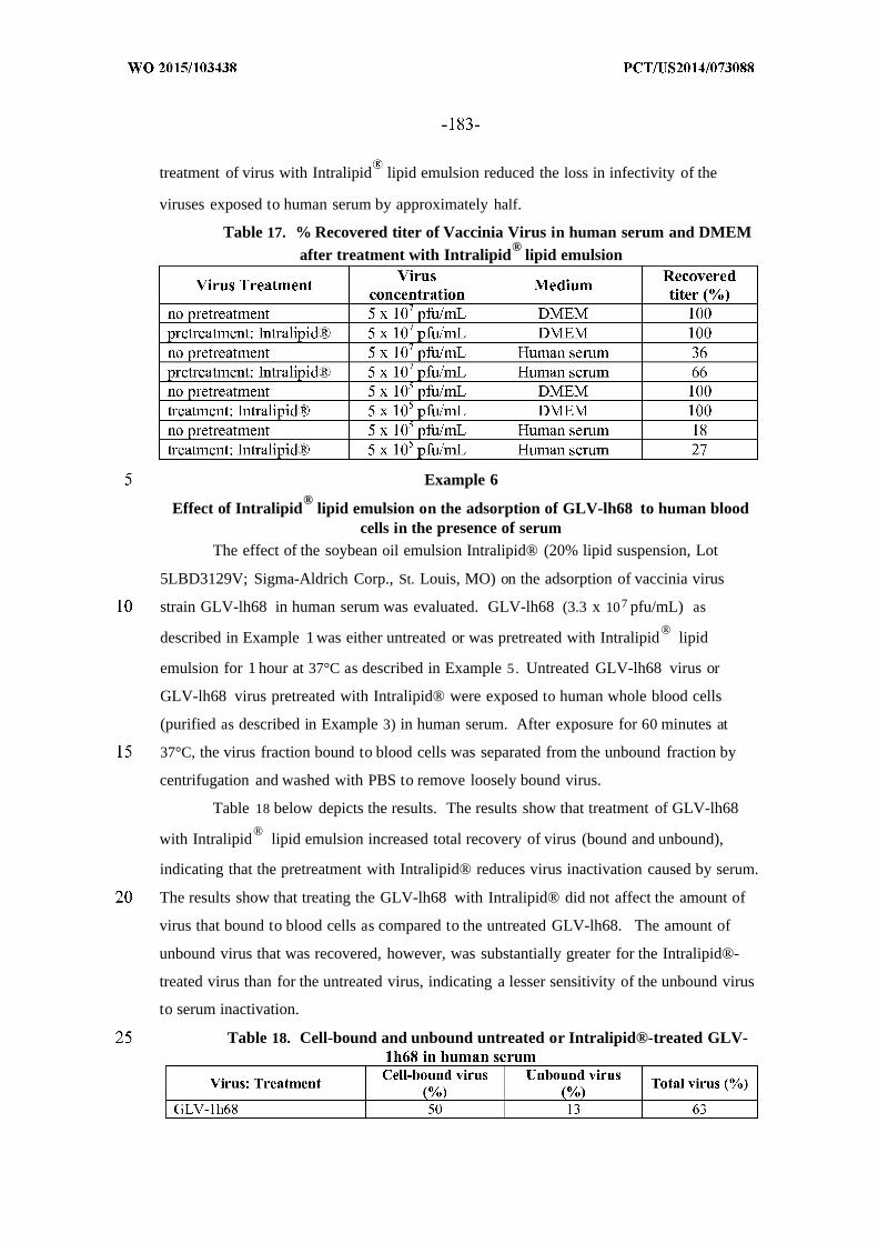
treatment of virus with Intralipid lipid emulsion reduced the loss in infectivity of the
viruses exposed to human serum by approximately half.
Table 17. % Recovered titer of Vaccinia Virus in human serum and DMEM®
after treatment with Intralipid lipid emulsion
Example 6®
Effect of Intralipid lipid emulsion on the adsorption of GLV-lh68 to human bloodcells in the presence of serum
The effect of the soybean oil emulsion Intralipid® (20% lipid suspension, Lot
5LBD3129V; Sigma-Aldrich Corp., St. Louis, MO) on the adsorption of vaccinia virus
strain GLV-lh68 in human serum was evaluated. GLV-lh68 (3.3 x 10⁷ pfu/mL) as
®described in Example 1 was either untreated or was pretreated with Intralipid lipid
emulsion for 1 hour at 37°C as described in Example 5. Untreated GLV-lh68 virus or
GLV-lh68 virus pretreated with Intralipid® were exposed to human whole blood cells
(purified as described in Example 3) in human serum. After exposure for 60 minutes at
37°C, the virus fraction bound to blood cells was separated from the unbound fraction by
centrifugation and washed with PBS to remove loosely bound virus.
Table 18 below depicts the results. The results show that treatment of GLV-lh68®with Intralipid lipid emulsion increased total recovery of virus (bound and unbound),
indicating that the pretreatment with Intralipid® reduces virus inactivation caused by serum.
The results show that treating the GLV-lh68 with Intralipid® did not affect the amount of
virus that bound to blood cells as compared to the untreated GLV-lh68. The amount of
unbound virus that was recovered, however, was substantially greater for the Intralipid®-
treated virus than for the untreated virus, indicating a lesser sensitivity of the unbound virus
to serum inactivation.
Table 18. Cell-bound and unbound untreated or Intralipid®-treated GLV-

Example 7
Pharmacokinetic evaluation of nude and wild type mice to determine the effect ofIntralipid® pretreatment of Vaccinia Virus
®The pharmacokinetic effects of Intralipid lipid emulsion -pretreated Vaccinia virus
in tumor-bearing nude and wild type BALB/C mice (Harlan, female, 20-24 grams body
weight) following intravenous injection were evaluated. Mice were injected subcutaneously
with mouse 4T1 mammary carcinoma cell (ATCC) to establish tumors. Specifically, 1 x
105 4T1 cells in 0.1 cc of PBS were injected subcutaneously on the upper right thigh of each
animal. At time of virus injection, 11 days after tumor cell implantation, mice were
observed to have tumors of approximately 250-500 mm3. Because this study was only
intended to evaluate the blood concentration of virus, tumors were not harvested or
evaluated in these animals.
A vaccinia virus strain containing a heterologous nucleic acid molecule encoding a
detectable marker TurboFP635 was treated with Intralipid lipid emulsion as described in
Example 5. Vaccinia virus strain (5 x 10⁶ pfu/mouse) that was untreated or that was
pretreated with the soybean oil emulsion Intralipid® lipid emulsion (20% lipid suspension,
Lot 5LBD3129V; Sigma-Aldrich Corp., St. Louis, MO) were administered to nude mice
(Group A, untreated, n=4; Group B, pretreated n=5) and BALB/c mice (Group C, untreated,
n=5; Group D, pretreated n=5) via tail vein. In several mice the intravenous tail vein
injection was unsuccessful and the virus was injected into the abdominal aorta. Blood
samples were collected from the abdominal aorta at regular intervals post-injection (p.i.)
beginning at approximately 1 minute p.i. to 120 minutes p.i. The samples were
anticoagulated with EDTA, immediately frozen on dry ice, and assessed by viral plaque
assay for infectious virus on CV-1 cells as described in Example 2. PK parameters were
calculated in Microsoft Excel using a PK calculation module.
With respect to virus titer (pfu/mL) in the blood, the results show that the highest
concentration of virus post-injection occurred at the earliest time point measured
(approximately 1 minute p.i.) and declined rapidly with each measurement. In Groups of
mice where the virus was pretreated with Intralipid® lipid emulsion, there was a
consistently higher detection of virus titer in the blood at the earlier time points than in the
corresponding mice administered untreated virus.

Average Time Post- Average Titer/mLGroup Injection (Min±SD) Blood SD Titer/mL Blood
Preinj ection Not Detectable Not Detectable
0.9±0.1 2.6 x 10⁶ 2.0 x 10⁶
A 2.1±0.1 4.7 x 10⁵ 3.8 x l O5(Nude mice;
no 5.5±0.6 4.5 x 10⁴ 1.8 x 10⁴
pretreatment) 10.2±0.2 2.0 x 10⁴ 1.3 x 10⁴
30.5±3.5 1.8 x 10⁴ 2.1 x 10⁴
61.0±1.4 2.1 x 10³ 0.0
Preinj ection Not Detectable Not Detectable
1.4±0.4 3.6 x 10⁶ 2.5 x 10⁶
B 3.2±0.8 4.7 x 10⁵ 3.3 x 10⁵
(Nude mice; 5.4±0.2 2.9 x 10⁵ 2.6 x 10⁵Intralipid®
lipid 10.7±1.0 7.0 x 10⁴ 7.9 x 10⁴
emulsion) 33.6±3.3 6.7 x 10³ 5.7 x 10³
44.7±0.9 6.4 x 10³ 1.6 x 10²
67.9±12.3 2.1 x 10³ 1.5 x 10³
Preinj ection Not Detectable Not Detectable
C 1.0±0.1 3.8 x l O6 3.0 x 10⁶
(Balb/C 2.0±0.4 1.1 x 10⁶ 9.2 x 10⁵
mice; no 5.4±0.3 8.7 x 10⁴ 5.7 x 10⁴
pretreatment)10.8±0.5 6.7 x 10⁴ 3.4 x 10⁴
33.5±9.9 4.4 x 10⁴ 5.5 x l O4
Preinj ection Not Detectable Not DetectableD(Balb/C 1.1±0.2 7.2 x 10⁶ 1.9 x 10⁶
mice; 2.2±0.3 4.6 x 10⁶ 2.7 x 10⁶
Intralipid® 5.3±0.8 7.3 x 10⁵ 5.5 x 10⁵
lipid10.5±0.5 1.8 x 10⁵ 1.9 x 10
emulsion)36.1±13.6 4.6 x 10⁴ 3.0 x 10⁴
Results were analyzed through 10 minutes post-injection for maximum blood titer
( m ), calculated half-life in blood (t½), the rate constant for elimination of the virus from
blood (K iim), and the tissue exposure to the injected virus (AUC) from 1 to 10 minutes
(AUCo-io) and from 1 to infinity (AUCo-mf) . The group average results and percent
differences between the group average PK values are illustrated in Tables 19 and 20,
respectively.
Table 19. Group average PK values

p u-m n m
Table 20. Percent difference in group average PK values
The group average PK values were compared between the four groups of mice.
Comparison of the group average PK values between the Group A (nude) and Group C
(BALB/c) mice that received untreated virus revealed that the total tissue exposure (AUC 0.
of the nude mice was less than the BALB/c mice (55%) due to a decrease in the C of
the nude mice compared to the BALB/c mice (68%). However, the t½ was comparable in
both groups (97%).
Comparison of the group average PK values between the mice injected with
untreated virus and the mice injected with the Intralipid® lipid emulsion -treated virus
resulted in an increase in the total tissue exposure (AUC 0 _mf) in the mice injected with the
Intralipid® lipid emulsion -treated virus as compared to the mice injected with untreated
virus. There was a 192% increase in AUCo-mf in nude mice and a 344% increase in the
BALB/c mice (Group B vs. Group A and Group D vs. Group C, respectively),
corresponding to a 14 0% increase in the and a 139% increase in the t½ in nude mice
and a 19 1% increase in the C and a 15 1% increase in the t½ in the BALB/c mice.
The differences in PK values between the nude mice and the BALB/c mice injected
with the Intralipid® lipid emulsion-treated virus were consistent with the differences

observed with the untreated virus. The half-lives of the two viruses were substantially
comparable in nude and BALB/c mice.
Overall, pretreatment of virus with Intralipid® lipid emulsion increased the
maximum exposure of virus in blood (AUC0_mf) by 2-3 fold post- injection. The increase
was due to a reduction in the rate of elimination of the virus (Kelim) and an increase in the
circulation half-life (t½).
Example 8Effect of eculizumab on human serum inactivation of GLV-lh68 with serum from five
human blood donors
The effect of the anti-C5 mAb eculizumab (SEQ ID NO:44, heavy chain and SEQ
ID NO:45, light chain; sold under the trademark Soliris®, Lot 10007A; Alexion
Pharmaceuticals, Chesire, CT) on the infectivity of vaccinia virus strain GLV-lh68 was
evaluated in serum from five additional human blood donors. GLV-lh68 virus described in
Example 1 (2.5 x 10⁵ pfu/mL) was exposed to either human serum, human serum that had
been heated to 58°C for 30 minutes to inactivate complement in the serum, or human serum
that had been pretreated with 1 mg/mL eculizumab.
After exposure of the virus to the serum for 30 minutes at room temperature, the
infectivity of the virus compositions was assessed by viral plaque assay on CV-1 cells. A
viral titer assay was performed in a standard plaque assay using African green monkey
kidney fibroblast CV-1 cells (ATCC No. CCL-70; American Type Culture Collection,
Manassas, VA). CV-1 cells were plated in a 24-well plate at 2.5 x 10⁵ cells per well and
grown until near confluency. Wells containing a cell monolayer were infected with serial
dilutions of the virus compositions. The cells in each well were overlaid with virus overlay
medium (DMEM + 5% FBS + 1% Antibiotic-Antimycotic Solution + 1.5%
carboxymethylcellulose), and the cells were further incubated until plaques were visible.
After addition of color dye to visualize the plaques, viral titer (pfu/mL) was calculated by
counting the number of plaques in the well and dividing by the dilution factor (d) and
volume (V) of diluted virus added to the well. Tests were conducted in triplicate and the
average of 6 measures (2 per replicate) are reported.
Results are provided in Table 2 1 below. The results show that exposure of the
GLV-lh68 virus to various human sera alone resulted in recovery of virus titer ranging from
3% to 38%. Addition of ecuzimulab to the serum for 30 minutes at room temperature prior
to exposure of the virus resulted in substantial increase in the recovered virus titers, ranging
from 24% to 9 1 >. The fold increase in titer recovery in serum with ecuzimulab compared
to serum alone ranged from 2.4 to 7.4. The results indicate that inhibiting complement by

treating human serum with 1 mg/mL of the anti-C5 mAb eculizumab reduced the loss of
infectivity of GLV-lh68 as compared to serum that had not been treated with eculizumab.
While the results demonstrated variable amounts of virus inactivation activity in the
various sera, increase in infectious virus recovery after treatment with eculizumab was
obtained with all sera from the five individual blood donors. Four of the donor sera, # 1 to
#4, contained very little, if any, anti-vaccinia neutralizing antibody as evident from the
recovery of nearly 100% of the virus after exposure to serum that had been heat-treated to
inactivate complement activity, but not antibody activity. For Donor #5, only 68% of the
virus was recovered after exposure to serum that had been heat-treated, indicating that this
serum had substantial anti-vaccinia neutralizing antibody. Yet in all cases, exposure of virus
to serum in the presence of eculizumab increased the recovery of infectious virus by 2.4 to
7.4 fold over serum alone.
Table 21. % Recovered titer of GLV-lh68 in human serum from five healthyhuman blood donors without or with eculizumab
Example 9Adsorption of GLV-lh68 to human blood cells from five healthy human blood donors
in the presence of matched serumThe adsorption of vaccinia virus strain GLV-lh68 described in Example 1 to human
whole blood cells was evaluated. Whole human blood cells were prepared from five healthy
human blood donors by collecting blood in EDTA and isolating cells by centrifugation. The

cells were washed with phosphate buffered saline (PBS) to remove any plasma/EDTA and
were resuspended in PBS to the original blood volume. GLV-lh68 virus (2.0 x 10⁶ pfu/mL)
was exposed to whole human blood cells (33% of blood concentration) in either human
serum, heat-inactivated human serum that had been heated to 58°C for 30 minutes to
inactivate complement in the serum, or human serum that had been pretreated with 1 mg/mL
eculizumab (Soliris®, Lot 10007A; Alexion Pharmaceuticals, Chesire, CT). GLV-lh68
(2.0 x 10 pfu/mL) in PBS serve as a control.
After exposure for 60 minutes at 37°C, the virus fraction bound to blood cells was
separated from the unbound fraction by centrifugation and washed with PBS to remove
loosely bound virus. The infectivity of the virus was assessed by the viral plaque assay on
CV-1 cells described in Example 2. The recovered titer of cell-bound virus was determined.
Tests were conducted in triplicate and the average of 6 measures (2 per replicate) are
reported.
The Results are set forth in Table 22. The results show that 21% of the GLV-lh68
virus in PBS was cell-bound, while 79%> remained unbound, for a total recovery of 100% .
This value was used as a comparative standard relative to the values obtained in the human
serum compositions, and the percent recovered titer of cell-bound virus and unbound virus
compared to PBS control was determined.
In human serum, the fraction of virus recovered bound to blood cells ranged from
27%o to 90% for blood cells and serum from Donors 1 to 4. For Donor 5, who had
substantial levels of serum anti-vaccinia neutralizing antibody, the fraction of virus
recovered cell-bound in serum was 2%>, indicating that the virus was inactivated by the
serum. When virus was exposed to heat-inactivated serum, which inactivates complement,
there was a substantial decrease in the fraction of virus recovered bound to blood cells was
< 4%o for all donors. This result demonstrates that while reducing the amount of virus
inactivation by the serum, heat-inactivating complement also reduces the amount of virus
bound to blood cells.
Exposure of virus to human serum in the presence of the anti-C5 mAb eculizumab
resulted in an increase in the fraction of virus recovered bound to blood cells. For Donors 1
to 4, 89%o to 173% of the virus was recovered bound to blood cells (1.9 to 3.4 fold increase
over serum alone). For Donor 5, whose serum contained anti-vaccinia neutralizing antibody,
53%o of the virus was recovered bound to cells in serum in the presence of anti-C5 mAb
eculizumab (27.3 fold increase over serum alone). This indicates that inhibiting
complement by treatment of serum with eculizumab increases the fraction of bound virus in
blood from individuals even with anti-vaccinia neutralizing antibody. Additionally, for

some individuals exposure of virus with serum treated with eculizumab boosts the
infectivity of the virus that is bound to blood cells in excess of that obtained with the
Table 22. GLV-lh68 bound to human blood cells in human serum
Example 10
Effect of Intralipid® lipid emulsion on NK Cell Activity
Intralipid® lipid emulsion (20% lipid suspension, Lot 5LBD3129V; Sigma-Aldrich
Corp., St. Louis, MO) was assessed to test the effect of Intralipid® lipid emulsion on the
cytotoxicity of human NK cells using the standard human NK cell target K-562 (human
erythroleukemia cell line). Intralipid® lipid emulsion at the following concentrations 0.4
microliters ( ), 0.8 µ , 1 µ , 2 µ and 4 µ was added to 1 ml of media containing
800,000 human NK cells. The results showed that even at the lowest concentration, the
Intralipid® lipid emulsion suppressed the cytotoxicity of the NK cells with a more
pronounced effect at 1 µ / Ι.
Since modifications will be apparent to those of skill in this art, it is intended that
the inventions are limited only by the scope of the appended claims.

CLAIMS:
1. Use of an anti-complement component 5 (C5) antibody for increasing
infectivity of an oncolytic virus.
2. The use of claim 1, wherein the antibody is eculizumab, pexelizumab,
TSA12/22 or MB12/122, and variants thereof that bind to C5.
3. The use of claim 1 or claim 2, wherein the anti-C5 antibody is provided in
a composition that also contains the oncolytic virus.
4. A method of increasing infectivity of an oncolytic virus, comprising
administering the oncolytic virus and an anti-C5 antibody to a subject, wherein the virus and
antibody are administered separately, in the same composition, sequentially or
intermittently.
5. The method of claim 4, wherein the virus and antibody are administered in
the same composition.
6. The method of claim 4, wherein the virus and antibody are administered in
separate compositions.
7. The method of claim 4, wherein the antibody is administered before the
virus is administered.
8. The use or method of any of claims 1-5, wherein the oncolytic virus is
treated with a lipid emulsion containing a biocompatible lipid component that is comprised
of fatty acids and/or fatty acid derivatives.
9. The use or method of any of claims 1-5, wherein the oncolytic virus is
administered with or is in an composition comprising a biocompatible lipid component that
is comprised of fatty acids and/or fatty acid derivatives, wherein the composition is an
emulsion.
10. A composition, comprising:
an oncolytic virus; and
an anti-complement component 5 (C5) antibody .
11. The composition of claim 10, wherein the antibody is eculizumab,
pexelizumab, TSA12/22 or MB12/122, and variants thereof that bind to C5.
12. The composition of claim 10, comprising a biocompatible lipid component
that is comprised of fatty acids and/or fatty acid derivatives
13. A composition, comprising an oncolytic virus and a biocompatible lipid
component, wherein:
the composition is an emulsion; and
the lipid component is comprised of fatty acids and/or fatty acid derivatives.

14. The composition or use or method of any of claims 8-13, wherein the lipid
component is comprised of fatty acid derivatives and the fatty acid derivatives are selected
from among triglycerides, diglycerides, monoglycerides, phospholipids, and mixtures
thereof.
15. The composition, use or method of claim 14, wherein the lipid component
is comprised of triglycerides and the triglycerides are long-chain triglycerides (LCTs),
medium-chain triglycerides (MCTs), or mixtures thereof.
16. The composition, use or method of any of claims 8-15, wherein the lipid
component is comprised of a long-chain triglyceride and the long-chain triglyceride is
selected from among linoleate, oleate, palmitate, linolenate, stearate and mixtures thereof.
17. The composition, use or method of any of claims 8-15, wherein the lipid
component is comprised of a medium-chain triglyceride, and the medium-chain triglyceride
is caprylic acid or capric acid.
18. The composition, use or method of any of claims 8-17, wherein the
biocompatible lipid component is selected from among plant oil, vegetable oil, animal oil,
fish oil, mineral oil, chemically synthesized oil, or mixtures thereof.
19. The composition, use or method of claim 18, wherein the biocompatible
lipid component is a plant or vegetable oil, wherein the plant or vegetable oil is selected
from among soybean oil, cottonseed oil, safflower oil, corn oil, coconut oil, sesame oil,
peanut oil, olive oil, castor oil, and mixtures thereof.
20. The composition, use or method of claim 19, wherein the plant or vegetable
oil is soybean oil.
21. The composition, use or method of any of claims 1-20, wherein the
oncolytic virus is treated by incubation with a lipid emulsion comprising the biocompatible
lipid component to produce a lipid-treated oncolytic virus.
22. The composition of claim 21, wherein the biocompatible lipid component
comprises a lipid emulsion that comprises:
a biocompatible lipid component in an amount between 2% and 40%, inclusive, by
weight, of the lipid emulsion;
an emulsifier in an amount between 0.2% and 5%>, inclusive, by weight, of the lipid
emulsion; and
an aqueous phase in an amount between 50%> and 98%>, inclusive, by weight, of the
lipid emulsion.
23. The composition, use or method of claim 22, wherein the emulsifier is a
naturally-occurring emulsifier or a synthetic emulsifier.

24. The composition, use or method of claim 23, wherein the emulsifier is a
naturally-occurring emulsifier and the naturally-occurring emulsifier is a phospholipid
derived from an egg or soy source.
25. The composition, use or method of claim 24, wherein the phospholipid is
selected from among egg yolk phospholipids, hydrogenated egg yolk phospholipids,
soybean phospholipids, hydrogenated soybean phospholipids, and mixtures thereof.
26. The composition, use or method of claim 25, wherein the emulsifier is an
egg yolk phospholipid.
27. The composition, use or method of any of claims 22-26, wherein the
aqueous phase of the emulsion is water.
28. The composition, use or method of any of claims 22-27, wherein the lipid
emulsion comprises a tonicity modifier in an amount between 0.2% and 5%, inclusive, by
weight, of the lipid emulsion.
29. The composition, use or method of claim 28, wherein the tonicity modifier
is selected from among glycerin, sodium chloride, potassium chloride, mannitol, sucrose,
lactose, fructose, maltose, dextrose, dextrose anhydrous, xylitol, sorbitol, propylene glycol,
polyoxyethylated hydrocarbons, and C -C2osaturated or unsaturated aliphatic acids.
30. The composition, use or method of claim 28 or claim 29, wherein the
tonicity modifier is glycerin.
31. The composition, use or method of any of claims 21-30, wherein the lipid
emulsion is an intravenous liquid emulsion (ILE).
32. The composition, use or method of any of claims 21-31, wherein the virus
is in a lipid emulsion or treated with a lipid emulsion that comprises:
a biocompatible lipid component in a concentration between 10%> and 30%>,
inclusive, by weight, of the lipid emulsion, wherein the biocompatible lipid component is
selected from among soybean oil, safflower oil, olive oil, and mixtures thereof;
an egg yolk phospholipid(s) in a concentration that is at or about 1.2% by weight, of
the lipid emulsion;
glycerin in a concentration between 2.25% and 2.5%, inclusive, by weight, of the
lipid emulsion; and
water in a concentration that is between 60% and 90%, inclusive, by weight, of the
lipid emulsion.
33. The composition, use or method of any of claims 21-32, wherein the virus
is in an emulsion that comprises:
10%, 20% or 30% by weight soybean oil;

1.2% by weight egg yolk phospholipid; and
2.5% by weight glycerin.
34. The composition, use or method of any of claims 21-33, wherein the
oncolytic virus is incubated with a lipid emulsion comprising the biocompatible lipid
component for 30 minutes to 12 hours, 30 minutes to 6 hours, 30 minutes to 4 hours, 30
minutes to 2 hours, 1 hour to 12 hours, 1 hour to 6 hours, 1 hour to 4 hours, 1 hour to 2
hours, 2 hours to 12 hours, 2 hours to 6 hours, 2 hours to 4 hours, 4 hours to 12 hours, 4
hours to 6 hours, or 6 hours to 12 hours.
35. The composition, use or method of claim 34 wherein the incubation is at a
temperature that is at or between 0° to 42° C, inclusive, or is at a temperature that is at or
about 35° C to 42° C, or is at or about or at least 37° C.
36. The composition, use or method of any of claims 1-35, wherein
composition that comprises the biocompatible lipid,component comprises the oncolytic
virus, the biocompatible lipid component, an emulsifier and an aqueous solution.
37. The composition, use or method of any of claims 1-31, wherein the
concentration of the biocompatible lipid component is at a weight percentage (wt%>) of the
composition between 0.001%> and 40%>, inclusive.
38. The composition, use or method of claim 37, wherein the concentration of
the biocompatible lipid component is at a wt%> of the composition between 0.00 1% and
20%, 0.001% and 10%, 0.001% and 5%, 0.001% and 2%, 0.001% and 1%, 0.001% and
0.01%, 0.01% and 20%, 0.01% and 10%, 0.01% and 5%, 0.01% and 2%, 0.01% and 1%,
0.01% and 0.1%, 0.1% and 20%, 0.1% and 10%, 0.1% and 5%, 0.1% and 2%, 0.1% and
1%, 0.5% and 20%, 0.5% and 10%, 0.5% and 5%, 0.5% and 2%, 0.5% and 1%, 2% and
20%, 2% and 10%, 5% and 20%, 5% and 10%, or 10% and 20%, each inclusive.
39. The composition, use or method of any of claims 1-38, wherein:
the virus is treated with or in a composition comprising a biocompatible lipid
component;
and the biocompatible lipid component is soybean oil.
40. The composition, use or method of any of claims 1-39, wherein the
oncolytic virus is selected from among a Newcastle Disease virus, parvovirus, vaccinia
virus, myxoma virus, measles virus, reovirus, vesicular stomatitis virus (VSV), oncolytic
adenoviruses, adeno-associated virus, polioviras, herpes viruses, Sindbis viras and Seneca
Valley virus, or a derivative thereof that is modified to contain nucleic acid encoding a
heterologous gene product.

41. The composition, use or method of any of claims 1-40, wherein the
oncolytic virus is a vaccinia virus.
42. The composition, use or method of claim 41, wherein the vaccinia virus is
selected from among Lister, Western Reserve (WR), Copenhagen (Cop), Bern, Paris,
Tashkent, Tian Tan, Wyeth (DRYVAX), IHD-J, IHD-W, Brighton, Ankara, CVA382,
Modified Vaccinia Ankara (MVA), Dairen I, LC16m8, LC16M0, LIVP, ACAM2000, WR
65-16, Connaught, New York City Board of Health (NYCBH), EM-63 and NYVAC strain.
43. The composition, use or method of claim 4 1 or claim 42, wherein the
vaccinia virus is a Lister strain virus.
44. The composition, use or method of any of claims 1-43, wherein the virus is
vaccinia virus that is an LIVP virus or a clonal strain of an LIVP virus or a modified LIVP
virus.
45. The composition, use or method of claim 44, wherein the virus is a
modified form containing nucleic acid encoding a heterologous gene product.
46. The composition, use or method of claim 45, wherein the heterologous gene
product is a therapeutic or reporter gene product.
47. The composition, use or method of claim 45 or claim 46, wherein the
heterologous gene product is selected from among an anticancer agent, an antimetastatic
agent, an antiangiogenic agent, an immunomodulatory molecule, an antigen, a cell matrix
degradative gene, genes for tissue regeneration and reprogramming human somatic cells to
pluripotency, enzymes that modify a substrate to produce a detectable product or signal or
are detectable by antibodies, proteins that can bind a contrasting agent, genes for optical
imaging or detection, genes for PET imaging and genes for MRI imaging.
48. The composition, use or method of any of claims 45-47, wherein the
heterologous gene product is a therapeutic agent selected from among a hormone, a growth
factor, a cytokine, a chemokine, a costimulatory molecule, a ribozyme, a transporter protein,
a single chain antibody, an antisense RNA, a prodrug converting enzyme, an siRNA, a
microRNA, a toxin, an antitumor oligopeptide, a mitosis inhibitor protein, an antimitotic
oligopeptide, an anti-cancer polypeptide antibiotic, an angiogenesis inhibitor, a tumor
suppressor, a cytotoxic protein, a cytostatic protein and a tissue factor.
49. The composition, use or method of any of claims 1-48, wherein the
oncolytic virus is in an amount that is between or between about 1 x 10⁵ and 1 x 10 12 pfu, 1
x 106 to 1 x 10 10 pfu, or 1 x 10⁷ to 1 x 10 10 pfu, each inclusive.

50. The composition, use or method of any of claims 1-49, wherein the
oncolytic virus is present in the composition in an amount that is at least or about at least or
is 1 x 10⁶, 1 x 10⁷, 1 x 10 , 1 x 10⁹, 2 x 10⁹, 3 x 10⁹, 4 x 10⁹, 5 x 10⁹ pfu, or 1 x 10 10 pfu.
51. The composition, use or method of any of claims 1-50, comprising an
emulsifier.
52. The composition, use or method of claim 36 or claim 51, wherein the
emulsifier is a naturally-occurring emulsifier and the naturally-occurring emulsifier is a
phospholipid derived from an egg or soy source.
53. The composition, use or method of claim 52, wherein the phospholipid is
selected from among egg yolk phospholipids, hydrogenated egg yolk phospholipids,
soybean phospholipids, hydrogenated soybean phospholipids, and mixtures thereof.
54. The composition, use or method of claim 53, wherein the emulsifier is an
egg yolk phospholipid.
55. The composition, use or method of claim 54, wherein the phospholipid is
lecithin (phosphatidylcholine) .
56. The composition, use or method of any of claims 52-55, wherein the
concentration of the emulsifier as a wt% of the composition is between 0.0002% and 5%>,
0.0002% and 2%, 0.0002% and 1%, 0.0002% and 0.2%, 0.0002% and 0.02%, 0.0002% and
0.002%, 0.002% and 2%, 0.002% and 1%, 0.002% and 0.2%, 0.002% and 0.02%, 0.02%
and 2%, 0.02% and 1%, 0.02% and 0.2%, 0.2% and 5%, 0.2% and 2%, or 2% and 5%, each
inclusive.
57. The composition, use or method of any of claims 1-56, wherein the
oncolytic virus is in an emulsion, comprising:
an oncolytic virus in an amount that is between or between about 1 x 10⁶ to 1 x 10 1
pfu, inclusive;
a biocompatible lipid component as a wt%> of the composition of less than 10%>;
an emulsifier as a wt%> of the composition of less than 2%>;
a tonicity modifier as a wt%> of the composition of less than 2%>; and
an aqueous solution as a wt%> of the composition that is greater than 85%>.
58. The composition, use or method of any of claims 1-57, wherein the volume
of the composition is from or from about 0.01 mL to 100 niL, 0.1 mL to 100 niL, 1 mL to
100 mL, 10 mL to 100 mL, 0.01 mL to 10 mL, 0.1 mL to 10 mL, 1 mL to 10 mL, 0.02 mL
to 20 mL, 0.05 mL to 5 mL, 0.5 mL to 50 mL, or 0.5 mL to 5 mL, each inclusive.
59. The composition, use or method of any of claims 1-58, wherein increased
infectivity of the oncolytic virus in the presence of the biocompatible lipid component
and/or when co-administered with the antibody is manifested by increased titer or half-life
of the oncolytic virus in a bodily fluid compared to the same virus in the absence of the
antibody and/or the biocompatible lipid component.
60. The composition, use or method of claim 59, wherein the bodily fluid is
serum.
6 1. The composition, use or method of any of claims 1-60, wherein infectivity
is increased at least 1.2-fold, 1.3-fold, 1.4-fold, 1.5-fold, 1.6-fold, 1.7-fold, 1.8-fold, 1.9-
fold, 2.0-fold, 2.5-fold, 3-fold, 4-fold, 5-fold, 6-fold, 7-fold, 8-fold, 9-fold, or 10-fold.
62. The composition, use or method of any of claims 1-61 , wherein the
composition that contains the virus is formulated for direct administration,
for local or systemic injection, and/or for intravenous administration.
63. Use of a composition of any of claims 1-3, 8, 9 and 14-62 for treatment of
condition treated by the oncolytic virus.
64. The use of claim 63, wherein the condition is a proliferative disorder.
65. The use of claim 63, wherein the condition is a tumor or cancer.
66. A combination, comprising:
a first composition comprising an oncolytic virus; and
a second composition comprising a lipid emulsion, wherein:
the lipid emulsion comprises a biocompatible lipid component, an emulsifier and an
aqueous phase.
67. The combination of claim 66, wherein the biocompatible lipid component is
at a concentration as a weight percentage of the lipid emulsion of between 2% and 40%, 5%
and 40% , or 10%> and 30%, each inclusive.
wherein the lipid component is comprised of fatty acids and/or fatty acid
derivatives.
68. The combination of claim 66 or claim 67, wherein the lipid component is
comprised of fatty acid derivatives and the fatty acid derivatives are selected from among
triglycerides, diglycerides, monoglycerides, phospholipids, and mixtures thereof.
69. The combination of claim 68, wherein the lipid component is comprised of
triglycerides and the triglycerides are long-chain triglycerides (LCTs), medium-chain
triglycerides (MCTs), or mixtures thereof.
70. The combination of claim 69, wherein the lipid component is comprised of
a long-chain triglyceride and the long-chain triglyceride is selected from among linoleate,
oleate, palmitate, linolenate, stearate and mixtures thereof.

7 1. The combination of claim 69, wherein the lipid component is comprised of
a medium-chain triglyceride, and the medium-chain triglyceride is caprylic acid or capric
acid.
72. The combination of any of claims 66-71, wherein the biocompatible lipid
component is selected from among plant oil, vegetable oil, animal oil, fish oil, mineral oil,
chemically synthesized oil, or mixtures thereof.
73. The combination of claim 72, wherein the biocompatible lipid component is
a plant or vegetable oil, wherein the plant or vegetable oil is selected from among soybean
oil, cottonseed oil, safflower oil, corn oil, coconut oil, sesame oil, peanut oil, olive oil,
castor oil, and mixtures thereof.
74. The combination of claim 73, wherein the plant or vegetable oil is soybean
oil.
75. The combination of any of claims 66-74, wherein the concentration of the
emulsifier as a weight percentage of the lipid emulsion is between 0.2% and 5%>, 0.5%> and
4 % , or 1% and 3%>, each inclusive.
76. The combination of any of claims 66-75, wherein the emulsifier is a
naturally-occurring emulsifier or a synthetic emulsifier.
77. The combination of claim 76, wherein the emulsifier is a naturally-
occurring emulsifier and the naturally-occurring emulsifier is a phospholipid derived from
an egg or soy source.
78. The combination of claim 77, wherein the phospholipid is selected from
among egg yolk phospholipids, hydrogenated egg yolk phospholipids, soybean
phospholipids, hydrogenated soybean phospholipids, and mixtures thereof.
79. The combination of claim 78, wherein the emulsifier is an egg yolk
phospholipid.
80. The combination of claim 79, wherein the egg yolk phospholipid comprises
lecithin (phosphatidylcholine).
81. The combination of any of claims 66-80, wherein the concentration of the
aqueous solution as a weight percentage of the lipid emulsion is between 50%> and 98%>,
60% and 90%, or 65% and 80%, each inclusive.
82. The combination of any of claims 66-81, wherein the aqueous phase is
water.
83. The combination of any of claims 66-82, wherein the lipid emulsion is an
intravenous liquid emulsion (ILE).
84. The combination of claim 83, wherein the lipid emulsion comprises:

a biocompatible lipid component in a concentration between 10% and 30%,
inclusive, by weight, of the lipid emulsion, wherein the biocompatible lipid component is
selected from among soybean oil, safflower oil, olive oil, and mixtures thereof;
an egg yolk phospholipid(s) in a concentration that is at or about 1.2% by weight, of
the lipid emulsion;
glycerin in a concentration between 2.25% and 2.5%, inclusive, by weight, of the
lipid emulsion; and
water in a concentration that is between 60%> and 90%>, inclusive, by weight, of the
lipid emulsion.
85. The combination of any of claims 66-84, wherein the liquid emulsion
comprises:
10%, 20% or 30% by weight soybean oil;
1.2 % by weight egg yolk phospholipid; and
2 .5% by weight glycerin.
86. The combination of any of claims 66-85, wherein the oncolytic virus is
selected from among a Newcastle Disease virus, parvovirus, vaccinia virus, myxoma virus,
measles virus, reovirus, vesicular stomatitis virus (VSV), oncolytic adenoviruses, adeno-
associated virus, poliovirus, herpes viruses, Sindbis virus and Seneca Valley virus, or a
derivative thereof that is modified to contain nucleic acid encoding a heterologous gene
product.
87. The combination of any of claims 66-86, wherein the oncolytic virus is a
vaccinia virus.
88. The combination of claim 87, wherein the vaccinia virus is selected from
among Lister, Western Reserve (WR), Copenhagen (Cop), Bern, Paris, Tashkent, Tian Tan,
Wyeth (DRYVAX), IHD-J, IHD-W, Brighton, Ankara, CVA382, Modified Vaccinia
Ankara (MVA), Dairen I, LC16m8, LC16M0, LIVP, ACAM2000, WR 65-16, Connaught,
New York City Board of Health (NYCBH), EM-63 and NYVAC strain.
89. The combination of claim 87 or claim 88, wherein the vaccinia virus is a
Lister strain virus.
90. The combination of any of claims 87-89, wherein the vaccinia virus is an
LIVP virus or a clonal strain of an LIVP virus.
9 1. The combination of claim 90, wherein the virus is a modified form
containing nucleic acid encoding a heterologous gene product.
92. The combination of claim 91, wherein the heterologous gene product is a
therapeutic or reporter gene product.

93. The combination of claim 9 1 or claim 92, wherein the heterologous gene
product is selected from among an anticancer agent, an antimetastatic agent, an
antiangiogenic agent, an immunomodulatory molecule, an antigen, a cell matrix degradative
gene, genes for tissue regeneration and reprogramming human somatic cells to pluripotency,
enzymes that modify a substrate to produce a detectable product or signal or are detectable
by antibodies, proteins that can bind a contrasting agent, genes for optical imaging or
detection, genes for PET imaging and genes for MRI imaging.
94. The combination of any of claims 91-93, wherein the heterologous gene
product is a therapeutic agent selected from among a hormone, a growth factor, a cytokine, a
chemokine, a costimulatory molecule, a ribozyme, a transporter protein, a single chain
antibody, an antisense RNA, a prodrug converting enzyme, an siRNA, a microRNA, a toxin,
an antitumor oligopeptide, a mitosis inhibitor protein, an antimitotic oligopeptide, an anti
cancer polypeptide antibiotic, an angiogenesis inhibitor, a tumor suppressor, a cytotoxic
protein, a cytostatic protein and a tissue factor.
95. The combination of any of claims 66-94, wherein the oncolytic virus is
present in the composition in an amount that is between or between about 1 x 10⁵ and 1 x
10 12 pfu, 1 x 10⁶ to 1 x 10 10 pfu, or 1 x 10⁷ to 1 x 10 10 pfu, each inclusive.
96. The combination of any of claims 66-95, wherein the oncolytic virus is
present in the composition in an amount that is at least or about at least or 1 x 10⁶, 1 x 10⁷, 1
x 10 , 1 x 10⁹, 2 x 10⁹, 3 x 10⁹, 4 x 10⁹, 5 x 10⁹ pfu, or 1 x 10 10 pfu.
97. The combination of any of claims 66-96, wherein the volume of the
composition comprising the oncolytic virus is from or from about 0.01 mL to 100 mL, 0.1
mL to 100 mL, 1 mL to 100 mL, 10 mL to 100 mL, 0.01 mL to 10 mL, 0.1 mL to 10 mL, 1
mL to 10 mL, 0.02 mL to 20 mL, 0.05 mL to 5 mL, 0.5 mL to 50 mL, or 0.5 mL to 5 mL,
each inclusive.
98. The combination of any of claims 66-97, comprising a further composition
comprising an additional active agent.
99. A combination, comprising:
a first composition that is a composition of any of claims 10-62 or a first
composition comprising an oncolytic virus; and
a second composition comprising an additional active agent, wherein the first and
second compositions are separate or are combined so that the additional active agent is in
the first composition.
100. The combination of claim 98 or claim 99, wherein the additional active
agent is selected from among a therapeutic compound, an agent that increases virus

infectivity, a therapeutic or diagnostic virus, an antiviral or chemotherapeutic agent, or an
agent or compound for modulation of gene expression of endogenous or heterologous genes
encoded by the virus.
101. The combination of claim 100, wherein the additional agent is a therapeutic
compound that is an anti-cancer agent.
102. The combination of claim 100 or claim 101, wherein the therapeutic
compound is selected from among a cytokine, growth factor, photosensitizing agent,
radionuclide, toxin, siRNA molecule, enzyme/pro E drug pair, anti-metabolite, signaling
modulator, anti-cancer antibiotic, anti-cancer antibody, angiogenesis inhibitor,
chemotherapeutic compound, antimetastatic compound or a combination of any thereof.
103. The combination of any of claims 98-101, wherein the additional agent is
an agent that increases the infectivity of the virus and is a complement inhibitor.
104. The combination of claim 103, wherein the complement inhibitor inhibits
activity of CI, C2, C3, C4, C5, C5a, C5aR, C3aR, Factor B, Factor P, Clq or MBP.
105. The combination of claim 103 or claim 104, wherein the complement
inhibitor is selected from among cobra venom factor (CVF), heparin, TA 106, TNX-234,
anti-properdin, Cl-INH, a compstatin or derivative or analog thereof, soluble CR1,
K76COOH, eculizumab, pexelizumab, TSA12/22, MSA12/22, ARC 1005, TNX-558, NOX-
D19, PMX-53, PMX-201, PMX-205, neutrazumab, and variants, analogs or derivatives
thereof that inhibit complement activity.
106. The combination of claim 103 or claim 104, wherein the complement
inhibitor is an anti-C5 antibody.
107. The combination of claim 106, wherein the anti-C5 antibody is selected
from among eculizumab, pexelizumab, TSA12/22 or MB12/122, or a variant thereof.
108. The combination of claim 107, wherein the anti-C5 antibody is eculizumab.
109. A combination, comprising:
a first composition comprising an oncolytic virus; and
a second composition comprising an anti-C5 antibody.
110. The composition of claim 10 or combination of claim 109, wherein the
oncolytic virus is selected from among a Newcastle Disease virus, parvovirus, vaccinia
virus, myxoma virus, measles virus, reovirus, vesicular stomatitis virus (VSV), oncolytic
adenoviruses, adeno-associated virus, poliovirus, herpes viruses, Sindbis virus and Seneca
Valley virus, or a derivative thereof that is modified to contain nucleic acid encoding a
heterologous gene product.

111. The composition or combination of any of claims 10, 109 and 110, wherein
the oncolytic virus is a vaccinia virus.
11 . The composition or combination of claim 111, wherein the vaccinia virus is
selected from among Lister, Western Reserve (WR), Copenhagen (Cop), Bern, Paris,
Tashkent, Tian Tan, Wyeth (DRYVAX), IHD-J, IHD-W, Brighton, Ankara, CVA382,
Modified Vaccinia Ankara (MVA), Dairen I, LC16m8, LC16M0, LIVP, ACAM2000, WR
65-16, Connaught, New York City Board of Health (NYCBH), EM-63 and NYVAC strain.
113. The composition or combination of claim 111 or claim 112, wherein the
vaccinia virus is a Lister strain virus.
114. The composition or combination of any of claims 111-113, wherein the
vaccinia virus is an LIVP virus or a clonal strain of an LIVP virus.
115. The composition or combination of claim 114, wherein the virus is a
modified form containing nucleic acid encoding a heterologous gene product.
116. The composition or combination of claim 115, wherein the heterologous
gene product is a therapeutic or reporter gene product.
117. The composition or combination of claim 116 or claim 117, wherein the
heterologous gene product is selected from among an anticancer agent, an antimetastatic
agent, an antiangiogenic agent, an immunomodulatory molecule, an antigen, a cell matrix
degradative gene, genes for tissue regeneration and reprogramming human somatic cells to
pluripotency, enzymes that modify a substrate to produce a detectable product or signal or
are detectable by antibodies, proteins that can bind a contrasting agent, genes for optical
imaging or detection, genes for PET imaging and genes for MRI imaging.
118. The composition or combination of any of claims 115-117, wherein the
heterologous gene product is a therapeutic agent selected from among a hormone, a growth
factor, a cytokine, a chemokine, a costimulatory molecule, a ribozyme, a transporter protein,
a single chain antibody, an antisense RNA, a prodrug converting enzyme, an siRNA, a
microRNA, a toxin, an antitumor oligopeptide, a mitosis inhibitor protein, an antimitotic
oligopeptide, an anti-cancer polypeptide antibiotic, an angiogenesis inhibitor, a tumor
suppressor, a cytotoxic protein, a cytostatic protein and a tissue factor.
119. The composition or combination of any of claims 10 and 109-1 18, wherein
the oncolytic virus is present in the composition or combination in an amount that is
between or between about 1 x 10⁵ and 1 x 10 pfu, 1 x 10 to 1 x 10 0 pfu, or 1 x 101 to 1 x
10 1 pfu, each inclusive.

120. The composition or combination of any of claims 10 and 109-1 19, wherein
the oncolytic virus is present in the composition in an amount that is at least or about at least
or 1 x 10⁶, 1 x 10⁷, 1 x 10 , 1 x 10⁹, 2 x 10⁹, 3 x 10⁹, 4 x 10⁹, 5 x 10⁹ pfu, or 1 x 10 10 pfu.
121. The composition or combination of any of claims 10 and 109-120, wherein
the volume of the composition comprising the oncolytic virus is from or from about 0.01
mL to 100 mL, 0.1 n L to 100 mL, 1 n L to 100 mL, l OmL to 100 mL, 0.01 mL to 10 mL,
0.1 mL to 10 mL, 1 mL to 10 mL, 0.02 mL to 20 mL, 0.05 mL to 5 mL, 0.5 mL to 50 mL,
or 0.5 mL to 5 mL, each inclusive.
122. The composition, use, method or combination of any of claims 1-12 and
106-121, wherein the anti-C5 antibody specifically binds to complement component 5 (C5)
with a binding affinity such that the dissociation constant is less than or equal to 10 , 10 ,
10 8, Ιθ 10 10, 10 , or 10 12 M.
123. The composition, use, method or combination of any of claims 1-12 and
106-122, wherein the anti-C5 antibody specifically binds the alpha chain of C5.
124. The composition, use, method or combination of any of claims 1-12 and
106-123, wherein the anti-C5 antibody is selected from among eculizumab, pexelizumab,
TSA12/22 or MB12/122, or a variant thereof that specifically binds C5.
125. The composition, use, method or combination of claim 124, wherein the
anti-C5 antibody is eculizumab.
126 The composition, use, method or combination of any of claims 1-12 and
106-122, wherein the anti-C5 antibody specifically binds the beta chain of C5.
127. The composition, use, method or combination of any of claims 1-12 and 1-
106-126, wherein the amount of the anti-C5 antibody in the composition, when formulated
for single dosage administration is between or between about 1 mg to 5000 mg, 10 mg to
5000 mg, 100 mg to 5000 mg, 100 mg to 2500 mg, 100 mg to 1000 mg, 100 mg to 500 mg,
500 mg to 5000 mg, 500 mg to 2500 mg, 500 mg to 1000 mg, 1000 mg to 2500 mg, 2000
mg to 5000 mg, or 1500 mg to 2500 mg, each inclusive.
128. The composition, use, method or combination of any of claims 1-12 and
106-127, wherein the amount of the anti-C5 antibody in the composition, when formulated
for single dosage administration, is at least or about at least or about 100 mg, 200 mg, 300
mg, 400 mg, 500 mg, 600 mg, 700 mg, 800 mg, 900 mg or 1000 mg.
129. The composition, use, method or combination of any of claims 1-12 and 99-
128, wherein the volume of the composition comprising the oncolytic virus is from or from
about 0.01 mL to 100 mL, 0.1 mL to 100 mL, 1 mL to 100 mL, 10 mL to 100 mL, 0.01 mL

to 10 mL, 0.1 mL to 10 mL, 1 mL to 10 mL, 0.02 mL to 20 mL, 0.05 mL to 5 mL, 0.5 mL to
50 mL, or 0.5 mL to 5 mL, each inclusive.
130. The composition, use, method or combination of any of claims 1-12 and
103-129, wherein the virus exhibits increased infectivity that is manifested by increased titer
or half-life of the oncolytic virus when exposed to a bodily fluid.
131. The composition, use, method or combination of claim 130, wherein the
bodily fluid is serum.
132. The composition, use, method or combination of claim 130 or claim 131,
wherein infectivity is increased at least 1.2-fold, 1.3-fold, 1.4-fold, 1.5-fold, 1.6-fold, 1.7-
fold, 1.8-fold, 1.9-fold, 2.0-fold, 2.5-fold, 3-fold, 4-fold, 5-fold, 6-fold, 7-fold, 8-fold, 9-
fold, or 10-fold.
133. A method of increasing infectivity of an oncolytic virus, comprising
contacting an oncolytic virus in vitro with a biocompatible lipid component for a sufficient
time to produce a lipid-treated virus, wherein the lipid-treated virus exhibits increased
infectivity for a target cell compared to the oncolytic virus in the absence of contacting with
the biocompatible lipid component comprised of fatty acids and/or fatty acid derivatives.
134. A method of treating a disease or condition in a subject treatable by an
oncolytic virus, comprising:
a) contacting an oncolytic virus in vitro with a biocompatible lipid component for a
sufficient time to produce a lipid-treated virus composition, wherein the lipid-treated virus
exhibits increased infectivity for a target cell compared to the oncolytic virus in the absence
of contacting with the biocompatible lipid component, wherein biocompatible lipid
component is comprised of fatty acids and/or fatty acid derivatives; and
b) administering the lipid-treated virus composition to the subject.
135. The method of claim 133 or claim 134, wherein the oncolytic virus is
contacted with a biocompatible lipid component for 30 minutes to 12 hours, 30 minutes to 6
hours, 30 minutes to 4 hours, 30 minutes to 2 hours, 1 hour to 12 hours, 1 hour to 6 hours, 1
hour to 4 hours, 1 hour to 2 hours, 2 hours to 12 hours, 2 hours to 6 hours, 2 hours to 4
hours, 4 hours to 12 hours, 4 hours to 6 hours, or 6 hours to 12 hours.
136. The method of claim 135, wherein the contacting is at a temperature that is
at or between 0° to 42° C, inclusive.
137. The method of any of claims 133-136, wherein the lipid component is
comprised of fatty acid derivatives and the fatty acid derivatives are selected from among
triglycerides, diglycerides, monoglycerides, phospholipids, and mixtures thereof.

138. The method of claim 137, wherein the lipid component is comprised of
triglycerides and the triglycerides are long-chain triglycerides (LCTs), medium-chain
triglycerides (MCTs), or mixtures thereof.
139. The method of claim 138, wherein the lipid component is comprised of a
long-chain triglyceride and the long-chain triglyceride is selected from among linoleate,
oleate, palmitate, linolenate, stearate and mixtures thereof.
140. The method of claim 138, wherein the lipid component is comprised of a
medium-chain triglyceride, and the medium-chain triglyceride is caprylic acid or capric
acid.
141. The method of any of claims 133-140, wherein the biocompatible lipid
component is selected from among plant oil, vegetable oil, animal oil, fish oil, mineral oil,
chemically synthesized oil, or mixtures thereof.
142. The method of claim 141, wherein the biocompatible lipid component is a
plant or vegetable oil, wherein the plant or vegetable oil is selected from among soybean oil,
cottonseed oil, safflower oil, corn oil, coconut oil, sesame oil, peanut oil, olive oil, castor
oil, and mixtures thereof.
143. The method of claim 142, wherein the plant or vegetable oil is soybean oil.
144. The method of any of claims 133-143, wherein the biocompatible lipid
component is a lipid emulsion comprising the biocompatible lipid component.
145. The method of claim 144, wherein the lipid emulsion comprises:
a biocompatible lipid component in an amount between 2% and 40%, inclusive, by
weight, of the lipid emulsion;
an emulsifier in an amount between 0.2% and 5%>, inclusive, by weight, of the lipid
emulsion; and
an aqueous phase in an amount between 50%> and 98%>, inclusive, by weight, of the
lipid emulsion.
146. The method of claim 145, wherein the emulsifier is a naturally-occurring
emulsifier or a synthetic emulsifier.
147. The method of claim 146, wherein the emulsifier is a naturally-occurring
emulsifier and the naturally-occurring emulsifier is a phospholipid derived from an egg or
soy source.
148. The method of claim 147, wherein the phospholipid is selected from among
egg yolk phospholipids, hydrogenated egg yolk phospholipids, soybean phospholipids,
hydrogenated soybean phospholipids, and mixtures thereof.

149. The method of claim 148, wherein the emulsifier is an egg yolk
phospholipid.
150. The method of any of claims 145-149, wherein the aqueous phase is water.
151. The method of any of claims 133-150, wherein the lipid emulsion is an
intravenous liquid emulsion (ILE).
152. The method of any of claims 133-151, wherein the lipid emulsion
comprises:
a biocompatible lipid component in a concentration between 10% and 30%,
inclusive, by weight, of the lipid emulsion, wherein the biocompatible lipid component is
selected from among soybean oil, safflower oil, olive oil, and mixtures thereof;
an egg yolk phospholipid(s) in a concentration that is at or about 1.2% by weight, of
the lipid emulsion;
glycerin in a concentration between 2.25%> and 2.5%>, inclusive, by weight, of the
lipid emulsion; and
water in a concentration that is between 60%> and 90%>, inclusive, by weight, of the
lipid emulsion.
153. The method of any of claims 133-152, wherein the lipid emulsion
comprises:
10%, 20% or 30% by weight soybean oil;
1.2% by weight egg yolk phospholipid; and
2.5% by weight glycerin.
154. The method of any of claims 133-153, wherein the oncolytic virus is
selected from among a Newcastle Disease virus, parvovirus, vaccinia virus, myxoma virus,
measles virus, reovirus, vesicular stomatitis virus (VSV), oncolytic adenoviruses, adeno-
associated virus, poliovirus, herpes viruses, Sindbis virus and Seneca Valley virus, or a
derivative thereof that is modified to contain nucleic acid encoding a heterologous gene
product.
155. The method of any of claims 133-155, wherein the oncolytic virus is a
vaccinia virus.
156. The method of claim 155, wherein the vaccinia virus is selected from
among Lister, Western Reserve (WR), Copenhagen (Cop), Bern, Paris, Tashkent, Tian Tan,
Wyeth (DRYVAX), IHD-J, IHD-W, Brighton, Ankara, CVA382, Modified Vaccinia
Ankara (MVA), Dairen I, LC16m8, LC16M0, LIVP, ACAM2000, WR 65-16, Connaught,
New York City Board of Health (NYCBH), EM-63 and NYVAC strain.

157. The method of claim 155 or claim 156, wherein the virus is vaccinia virus
that is a Lister strain virus.
158. The method of any of claims 155-157, wherein the vaccinia virus is an
LIVP virus or a clonal strain of an LIVP virus.
159. The method of claim 158, wherein the virus is a modified form containing
nucleic acid encoding a heterologous gene product.
160. The method of claim 159, wherein the heterologous gene product is a
therapeutic or reporter gene product.
161. The method of claim 159 or claim 160, wherein the heterologous gene
product is selected from among an anticancer agent, an antimetastatic agent, an
antiangiogenic agent, an immunomodulatory molecule, an antigen, a cell matrix degradative
gene, genes for tissue regeneration and reprogramming human somatic cells to pluripotency,
enzymes that modify a substrate to produce a detectable product or signal or are detectable
by antibodies, proteins that can bind a contrasting agent, genes for optical imaging or
detection, genes for PET imaging and genes for MRI imaging.
162. The method of any of claims 159-161, wherein the heterologous gene
product is a therapeutic agent selected from among a hormone, a growth factor, a cytokine, a
chemokine, a costimulatory molecule, a ribozyme, a transporter protein, a single chain
antibody, an antisense RNA, a prodrug converting enzyme, an siRNA, a microRNA, a toxin,
an antitumor oligopeptide, a mitosis inhibitor protein, an antimitotic oligopeptide, an anti
cancer polypeptide antibiotic, an angiogenesis inhibitor, a tumor suppressor, a cytotoxic
protein, a cytostatic protein and a tissue factor.
163. A method of treating a disease or condition in a subject treatable by an
oncolytic virus, comprising administering the composition or combination of any of claims
10-62 and 99-132.
164. The method of claim 163, wherein the composition comprising the
oncolytic virus is administered to deliver at least 1 x 10⁵ pfu virus, 1 x 10 pfu virus, 1 x 10
pfu virus, 1 x 10 pfu virus, 1 x 10⁹ pfu virus, 1 x 10 1 pfu virus, 1 x 10 1 1 pfu virus, or 1 x
10 12 pfu virus.
165. The method of claim 163, wherein the composition comprising the
oncolytic virus is administered to deliver between 1 x 10 pfu virus and l x 10 1 pfu virus,
inclusive.
166. A method of treating a disease or condition in a subject treatable by an
oncolytic virus, comprising administering the combination of any of claims 66-98.
167. The method of claim 166, wherein the composition comprising the lipid
emulsion is administered prior to, simultaneously, intermittently or subsequently from
administration of the composition comprising the oncolytic virus.
168. The method of claim 166 or claim 167, wherein the composition comprising
the lipid emulsion is administered prior to administration of the composition comprising the
oncolytic virus.
169. The method of claim 168, wherein the composition comprising the lipid
emulsion is administered 5 minutes to 6 hours, 5 minutes to 4 hours, 5 minutes to 2 hours, 5
minutes to 1 hour, 5 minute to 30 minutes, 30 minutes to 6 hours, 30 minutes to 4 hours, 30
minutes to 2 hours, 30 minutes to 1 hour, 1 hour to 6 hours, 1 hour to 4 hours, 1 hour to 2
hours, 2 hours to 6 hours, 2 hours to 4 hours, or 4 hours to 6 hours prior to administration of
the composition comprising the oncolytic virus.
170. The method of claim 168 or claim 169, wherein the composition comprising
the lipid emulsion is administered at least 30 minutes prior to administration of the
composition comprising the oncolytic virus.
171. The method of any of claims 166-170, wherein the lipid emulsion is
administered in an amount to deliver 1 gram (g) to 50 g, 1 g to 40 g, 1 g to 30 g, 1 g to 20 g,
1 g to 10 g, 1 g to 5 g, 5 g to 50 g, 5 g to 40 g, 5 g to 30 g, 5 g to 20 g, 5 g to 10 g, 10 g to 50
g, 10 g to 40 g, 10 g to 30 g, 10 g to 20 g, 20 g to 50 g, 20 g to 40 g, or 20 g to 30 g of the
biocompatible lipid component.
172. The method of any of claims 166-171, wherein 1 mL to 500 mL, 1 mL to
200 mL, 1 mL to 100 mL, 1 mL to 50 mL, 50 mL to 500 mL, 50 mL to 200 mL, 50 mL to
100 mL, 100 mL to 500 mL, 100 mL to 200 mL, or 200 mL to 500 mL of lipid emulsion
composition is administered.
173. The method of any of claims 166-172, wherein at least or about at least 100
mL of lipid emulsion composition is administered.
174. The method of any of claims 166-173, further comprising administering a
complement inhibitor.
175. The method of claim 174, wherein the complement inhibitor inhibits
activity of CI, C2, C3, C4, C5, C5a, C5aR, C3aR, Factor B, Factor P, Clq or MBP.
176. The method of claim 174 or claim 175, wherein the complement inhibitor is
selected from among cobra venom factor (CVF), heparin, TA 106, TNX-234, anti-
properdin, Cl-INH, a compstatin or derivative or analog thereof, soluble CR1, K76COOH,
eculizumab, pexelizumab, TSA12/22, MSA12/22, ARC 1005, TNX-558, NOX-D19, PMX-

53, PMX-201, PMX-205, neutrazumab, and variants, analogs or derivatives thereof that
inhibit a complement activity.
177. The method of any of claims 174-176, wherein the complement inhibitor is
an anti-C5 antibody.
178. A method of treating a disease or condition in a subject treatable by an
oncolytic virus, comprising administering the combination of any of claims 109-132.
179. The method of any of claims 174-178, wherein the composition comprising
the complement inhibitor or anti-C5 antibody is administered prior to, simultaneously,
intermittently or subsequently from administration of the composition comprising the
oncolytic virus.
180. The method of any of claims 174-179, wherein the composition comprising
the complement inhibitor or anti-C5 antibody is administered prior to administration of the
composition comprising the oncolytic virus.
181. The method of claim 180, wherein the composition comprising the
complement inhibitor or anti-C5 antibody is administered 5 minutes to 6 hours, 5 minutes to
4 hours, 5 minutes to 2 hours, 5 minutes to 1 hour, 5 minute to 30 minutes, 30 minutes to 6
hours, 30 minutes to 4 hours, 30 minutes to 2 hours, 30 minutes to 1 hour, 1 hour to 6 hours,
1 hour to 4 hours, 1 hour to 2 hours, 2 hours to 6 hours, 2 hours to 4 hours, or 4 hours to 6
hours prior to administration of the composition comprising the oncolytic virus.
182. The method of claim 180 or claim 181, wherein the composition comprising
the complement inhibitor or anti-C5 antibody is administered at least 30 minutes prior to
administration of the composition comprising the oncolytic virus.
183. A method of treating a disease or condition in a subject treatable by an
oncolytic virus, comprising administering the composition of any of claims 10-12 and 109-
132.
184. The method of claim 183, wherein the anti-C5 antibody is selected from
among eculizumab, pexelizumab, TSA12/22 or MB12/122, or a variant thereof.
185. The method of claim 184, wherein the anti-C5 antibody is eculizumab.
186. The method of any of claims 183-1 85, wherein the disease or condition is a
proliferative disorder.
187. The method of any of claims 183-1 85, wherein the disease or condition is a
cancer, tumor or metastasis.
188. The method of claim 187, wherein the disease or condition is a cancer and
the cancer is a carcinoma, sarcoma, lymphoma or leukemia.

189. The method of claim 187, wherein the disease or condition is a tumor and
the tumor is a solid tumor.
190. The method of any of claims 183-189, wherein the disease or condition is a
cancer and the cancer is a cancer of the tongue, mouth, throat, stomach, cecum, colon,
rectum, breast, ovary, uterus, thyroid, adrenal cortex, lung, kidney, prostate or pancreas.
191. The method of any of claims 183-190, wherein the subject is a human or
non-human animal.
192. The method of claim 191, wherein the subject is a non-human animal and
the non-human animal is selected from among a horse, cat, dog, cow, pig, sheep, goat,
mouse, rabbit, chicken, rat, and guinea pig.
193. A composition or combination of any of claims 10-132 for use in treating a
disease or condition that is one that is treatable by an oncolytic virus.
194. Use of a composition or combination of any of claims 10-132 formulated as
a medicament for treating a disease or condition that is treated by an oncolytic virus.
195. The combination or composition of claim 193 or use of claim 194,
wherein, when the combination is administered to a subject, the anti-C5 antibody is
administered prior to the oncolytic virus.
196. The composition, combination or use of any of claims 193-195, wherein the
disease of condition is a proliferative disorder.
197. The composition, combination or use of claim 196, wherein the
proliferative disease is cancer.
198. The composition, combination or use of claim 197, wherein the cancer is a
carcinoma, sarcoma, lymphoma or leukemia.
199. The composition, combination or use of claim 197 or claim 198, wherein
the cancer is a cancer of the tongue, mouth, throat, stomach, cecum, colon, rectum, breast,
ovary, uterus, thyroid, adrenal cortex, lung, kidney, prostate or pancreas.
200. The composition, combination or use of any of claims 193-199, wherein the
disease or condition is a tumor or a metastasis.
201 . The composition, combination or use of claim 200, wherein the tumor is a
solid tumor.
202. The composition, use, method or combination of any of claims 1-12 and
106-132, wherein the oncolytic virus exhibits increased binding to blood cells compared to
in the absence of the anti-C5 antibody.
203. The composition, use, method or combination of claim 202, wherein
binding to blood cells is increased at least 1.2-fold, 1.3-fold, 1.4-fold, 1.5-fold, 1.6-fold, 1.7-

fold, 1.8-fold, 1.9-fold, 2.0-fold, 2.5-fold, 3-fold, 4-fold, 5-fold, 6-fold, 7-fold, 8-fold, 9-
fold, or 10-fold.