WO 2014/138670 Al
165
(12) INTERNATIONAL APPLICATION PUBLISHED UNDER THE PATENT COOPERATION TREATY (PCT) (19) World Intellectual Property Organization International Bureau (10) International Publication Number (43) International Publication Date 12 September 2014 (12.09.2014) WO 2014/138670 Al PCT (51) International Patent Classification: 1800 Grant Street, 8th Floor, Denver, Colorado 80203 CUP 19/34 (2006.01) (US). (21) International Application Number: (72) Inventors; and PCT/US20 14/022052 (71) Applicants : WANG, Xiao-Jing [US/US]; 5835 E. Powers Avenue, Greenwood Village, Colorado 801 11 (22) International Filing Date: (US). ZHANG, Qinghong [US/US]; 6235 S. Lola Court, 7 March 2014 (07.03.2014) Englewood, Colorado 801 11 (US). REFAELI, Yosef (25) Filing Language: English [US/US]; 1115 Steele Street, Denver, Colorado 80206 (US). (26) Publication Language: English (74) Agents: VAVRA, Stephanie H. et al; Foley & Lardner (30) Priority Data: LLP, 3000 K Street N.W., Suite 600, Washington, District 61/775,252 8 March 2013 (08.03.2013) US of Columbia 20007-5109 (US). (71) Applicant: THE REGENTS OF THE UNIVERSITY (81) Designated States (unless otherwise indicated, for every OF COLORADO, A BODY CORPORATE [US/US]; kind of national protection available): AE, AG, AL, AM, [Continued on next page] (54) Title: PTD-SMAD7 THERAPEUTICS (57) Abstract: The present technology provides methods and composi tions for the treatment of inflammatory and/or tissue damage condi tions. In particular, the use of Smad7 compositions delivered locally or systemically to a site of inflammation and/or tissue damage is described. Other specific embodiments concern treatment or prevention of side ef fects caused by radiation and/or chemotherapy, including but not lim ited to oral and gastric mucositis. Also provided are codon-optimized nucleic acids encoding for Smad7 fusion proteins. Α -' ¾ ¾ f ii *? ¾ D Γ o 00
-
Upload
khangminh22 -
Category
Documents
-
view
0 -
download
0
Transcript of WO 2014/138670 Al
(12) INTERNATIONAL APPLICATION PUBLISHED UNDER THE PATENT COOPERATION TREATY (PCT)
(19) World Intellectual PropertyOrganization
International Bureau(10) International Publication Number
(43) International Publication Date12 September 2014 (12.09.2014)
WO 2014/138670 AlP CT
(51) International Patent Classification: 1800 Grant Street, 8th Floor, Denver, Colorado 80203CUP 19/34 (2006.01) (US).
(21) International Application Number: (72) Inventors; andPCT/US20 14/022052 (71) Applicants : WANG, Xiao-Jing [US/US]; 5835 E.
Powers Avenue, Greenwood Village, Colorado 801 11(22) International Filing Date: (US). ZHANG, Qinghong [US/US]; 6235 S. Lola Court,
7 March 2014 (07.03.2014) Englewood, Colorado 801 11 (US). REFAELI, Yosef(25) Filing Language: English [US/US]; 1115 Steele Street, Denver, Colorado 80206
(US).(26) Publication Language: English
(74) Agents: VAVRA, Stephanie H. et al; Foley & Lardner(30) Priority Data: LLP, 3000 K Street N.W., Suite 600, Washington, District
61/775,252 8 March 2013 (08.03.2013) US of Columbia 20007-5109 (US).
(71) Applicant: THE REGENTS OF THE UNIVERSITY (81) Designated States (unless otherwise indicated, for everyOF COLORADO, A BODY CORPORATE [US/US]; kind of national protection available): AE, AG, AL, AM,
[Continued on nextpage]
(54) Title: PTD-SMAD7 THERAPEUTICS
(57) Abstract: The present technology provides methods and compositions for the treatment of inflammatory and/or tissue damage conditions. In particular, the use of Smad7 compositions delivered locally orsystemically to a site of inflammation and/or tissue damage is described.Other specific embodiments concern treatment or prevention of side effects caused by radiation and/or chemotherapy, including but not limited to oral and gastric mucositis. Also provided are codon-optimizednucleic acids encoding for Smad7 fusion proteins.
Α
- ' ¾ ¾fi i * ? ¾
D
Γ
o
00
wo 2014/138670 Ai II II II I III IIII I I I III il II II III II I II
AO, AT, AU, AZ, BA, BB, BG, BH, BN, BR, BW, BY, TM), European (AL, AT, BE, BG, CH, CY, CZ, DE, DK,BZ, CA, CH, CL, CN, CO, CR, CU, CZ, DE, DK, DM, EE, ES, FI, FR, GB, GR, HR, HU, IE, IS, ΓΓ , LT, LU,DO, DZ, EC, EE, EG, ES, FI, GB, GD, GE, GH, GM, LV, MC, MK, MT, NL, NO, PL, PT, RO, RS, SE, SI, SK,GT, HN, HR, HU, ID, IL, IN, IR, IS, JP, KE, KG, KN, SM, TR), OAPI (BF, BJ, CF, CG, CI, CM, GA, GN, GQ,KP, KR, KZ, LA, LC, LK, LR, LS, LT, LU, LY, MA, GW, KM, ML, MR, NE, SN, TD, TG).MD, ME, MG, MK, MN, MW, MX, MY, MZ, NA, NG,NI, NO, NZ, OM, PA, PE, PG, PH, PL, PT, QA, RO, RS, Published:
RU, RW, SA, SC, SD, SE, SG, SK, SL, SM, ST, SV, SY, — with international search report (Art. 21(3))TH, TJ, TM, TN, TR, TT, TZ, UA, UG, US, UZ, VC, — before the expiration of the time limit for amending theVN, ZA, ZM, ZW. claims and to be republished in the event of receipt of
(84) Designated States (unless otherwise indicated, for every amendments (Rule 48.2(h))kind of regional protection available): ARIPO (BW, GH, — with sequence listing part of description (Rule 5.2(a))GM, KE, LR, LS, MW, MZ, NA, RW, SD, SL, SZ, TZ,UG, ZM, ZW), Eurasian (AM, AZ, BY, KG, KZ, RU, TJ,
PTD-SMAD7 THERAPEUTICS
CROSS-REFERENCE TO RELATED APPLICATIONS
[0001] The present application claims priority to U.S. provisional patent application USSN
61/775,252, filed March 8, 2013, which is incorporated by reference herein in its entirety.
STATEMENT OF GOVERNMENT INTEREST
[0002] This invention was made with government support under grant number
AR061796 awarded by the National Institutes of Health. The government has certain rights
in the invention.
BACKGROUND
[0003] Oral mucositis, a severe oral ulceration, is a common adverse effect of a large
dose of radiation for bone marrow transplant or craniofacial radiotherapy for cancer. Severe
oral mucositis could require feeding tubes, management of severe pain, and prematurely
halting radiotherapy. Excessive inflammation and epithelial ablation are key features of oral
mucositis.
[0004] Palifermin, a KGF (human keratinocyte growth factor) recombinant protein, is
approved for preventing oral mucositis in bone-marrow transplant patients. Two Palifermin
clinical trials in head and neck cancer patients showed that Palifermin reduced severe oral
mucositis incidence from 67% and 69% to 51% and 54%, respectively. Other oral mucositis
drugs in clinical trials or pre-clinical studies include growth factors, agents for
radioprotection, anti-inflammatory agents or immune modulators.
[0005] The modest effects of Palifermin and drugs being developed in the above
mentioned categories highlight the need for identification of biomarkers for novel therapies.
However, the lack of routine diagnostic biopsies or discarded tissues from oral mucositis
patients has hindered this effort.
[0006] Cutaneous wound healing progresses through three overlapping phases:
inflammation, tissue formation, and tissue remodeling. These are dynamic processes that
involve interactions among the epidermis, leukocytes, extracellular matrix (ECM), and
dermal fibroblasts. In response to skin injury, blood clots, infiltrated inflammatory cells and
other cell types in the wound release multiple cytokines and chemokines. These cytokines
initiate fibroblast proliferation and synthesis of ECM that fill the wound deficit and lead to
wound closure.
[0007] Meanwhile, keratinocytes at the wound edge begin to proliferate and migrate to
cover the wound surface. Underneath the re-epithelialized epidermis, new stroma, called
granulation tissue, begins to fill the wound space, which contains provisional ECM,
inflammatory cells, fibroblasts, and blood vessels. Once the wound area is filled with the
granulation tissue and covered by newly re-epithelialized epidermis, the process of wound
closure is completed. Later on, the wound gradually returns to normal strength and texture
through tissue remodeling.
[0008] Among the many molecules known to influence wound healing, transforming
growth factor β (TGF- β) has the broadest spectrum of action, affecting all cell types that are
involved in all stages of wound healing (Feng et al, Annu Rev Cell Dev Biol 21:659-693,
2005). The various functions of TGF- β are mediated by a number of signaling molecules,
including the Smad family members. When a ligand binds to TGF- β type I and type II
receptors (TGFPRI and TGF^RII), TGF- β phosphorylates Smad2 and Smad3.
Phosphorylated Smad2 and Smad3 bind a co-Smad, Smad4, to form heteromeric Smad
complexes and translocate into the nucleus to regulate transcription of TGF- β target genes.
[0009] TGF- β signaling has been reported to exert both positive and negative effects on
wound healing (Wang et al, J Investig Dermatol Symp Proc 11: 112-117, 2006). For
instance, Smad3 deficient mice, in which TGF- β signaling is partially abrogated, exhibit
accelerated wound healing (Ashcroft et al, Nat Cell Biol 1:260-266, 1999). In contrast, the
introduction of exogenous Smad3 to wound sites to enhance TGF- β signaling also
accelerated wound healing in a rabbit dermal ulcer model (Sumiyoshi et al, J Invest
Dermatol 123:229-236, 2004). Skin wounds in Smad4-deficient mice have a dramatic
increase in inflammation and angiogenesis causing a delay in wound closure and formed an
excessive scar (Owens et al, Am J Pathol 176:122-133, 2010). Transient adenoviral gene
transfer of Smad7, an antagonist of TGF- β signaling, in corneal epithelium and stroma
resulted in accelerated corneal wound healing with reduced inflammation (Saika et al. , Am J
Pathol 166: 1405-141 8, 2005). Further, Smad7 gene transfer to the lens epithelium and
stroma prevented injury-induced epithelial-mesenchymal transition of lens epithelial cells and
suggests a potential role of Smad7 in prevention of capsular fibrosis (Saika et al, Lab Invest
84: 1259-1270, 2004). However, adenoviral vector delivery of Smad7 to balloon injury in
rat carotid arteries resulted in reduced vascular healing (Mallawaarachchi et al. , Arterioscler
Thromb VaseBiol 25: 1383-1387, 2005). These studies suggest that the effects of TGF-β
signaling components, such as Smad7, on wound healing are complex and highly
context-specific. Additionally, the effect of Smad7 may not always be explained by its role
in TGF-β signaling. For instance, Smad7 has also been shown to interact with components
of the Wnt/p-catenin (Han et al., Dev Cell Biol 11:301-3 12, 2006) and the TNFp/NF- κΒ
(Hong etal, Nat Immunol 8:504-513, 2007) families.
SUMMARY
[0010] The present technology provides a nucleic acid molecule comprising a
codon-optimized human Smad7 cDNA nucleotide sequence. In some embodiments, the
codon-optimized human Smad7 nucleotide sequence may include one or more codons for
arginine optimized for expression in one or more of bacteria or yeast, including one or more
codons for serine optimized for expression in one or more of bacteria or yeast, and/or
including one or more codons for histidine optimized for expression in one or more of
bacteria or yeast. In some embodiments, the codon-optimized human Smad7 nucleotide
sequence may include 28 serine codons, 6 histidine codons, and 9 arginine codons optimized
for expression in one or more of bacteria or yeast. In some embodiments, the
codon-optimized human Smad7 nucleotide sequence may be selected from the group
consisting of SEQ ID Os: 9, 10, 11, 21, 23-41. In some embodiments, the
codon-optimized human Smad7 nucleotide sequence may have about 65 to 75 percent
homology to human Smad7 cDNA, may comprise a nucleotide sequence encoding an
N-terminal fragment SMAD7, may comprise a nucleotide sequence encoding a C-terminal
fragment of SMAD7, may comprise nucleotides encoding amino acids 2-258 of the human
Smad7 protein, may comprise nucleotides encoding amino acids 259-426 of the human
Smad7 protein, or may comprise nucleotides encoding amino acids 204-258 of the human
Smad7 protein. In some embodiments, any of the foregoing may further comprise a
nucleotide sequence encoding a protein transduction domain, such as Tat. In some
embodiments, any of the foregoing may also further comprise a nucleotide sequence
encoding one or more of an epitope tag or a purification tag, such as V5,
glutathione-S-transferase, or 6-Histidine.
[0011] In some embodiments, any of the foregoing may be isolated and/or purified. In
some embodiments, any one of the foregoing may also encode a polypeptide having one or
more biological activities selected from the group consisting of reducing or eliminating
phosphorylation of Smad2, reducing or eliminating nuclear translocation of the NF-κΒ p50
subunit, increasing cell proliferation, reducing apoptosis, reducing radiation-induced DNA
damage, reducing inflammation, reducing angiogenesis, promoting healing in oral mucositis,
promoting wound healing, and treating auto-immune disease. In some embodiments,
pharmaceutical compositions comprising the nucleic acid molecules above and one or more
pharmaceutically acceptable excipients are provided. In some embodiments, expression
vectors comprising the nucleic acid molecules above operably linked to a promoter are
provided, as are host cells comprising such expression vectors, and pharmaceutical
compositions comprising such vectors and host cells with one or more pharmaceutically
acceptable excipients.
[0012] In one aspect, a protein molecule comprising a human Smad7 protein having
leucine at position 16 is provided. In some embodiments, the human Smad7 protein may
be truncated at the C-terminal, or truncated at the N-terminal. In some embodiments, the
truncated human Smad7 protein may include about 50% of the full-length Smad7 sequence,
or may include about 13% of the full-length Smad7 sequence. In some embodiments, the
human Smad7 protein may comprise or consist of amino acids 2-258, amino acids 204-258,
or amino acids 259-426 of the human Smad7 protein. In some embodiments, the protein
molecule may have one or more biological activities selected from the group consisting of
reducing or eliminating phosphorylation of Smad2, reducing or eliminating nuclear
translocation of the F-κΒ p50 subunit, increasing cell proliferation, reducing apoptosis,
reducing radiation-induced DNA damage, reducing inflammation, reducing angiogenesis,
promoting healing in oral mucositis, promoting wound healing, and treating auto-immune
disease. In some embodiments, any of the foregoing may further comprise a protein
transduction domain, such as Tat. In some embodiments, any of the foregoing may also
further comprise one or more of an epitope tag or a purification tag, such as V5,
glutathione-S-transferase or 6-histidine. In some embodiments, a pharmaceutical
composition comprising any of the foregoing, a protein molecule, and one or more
pharmaceutically acceptable excipients is provided.

[0013] In another aspect, a method is provided for treating or preventing an
inflammatory condition in a subject comprising providing to the subject a therapeutically
effective amount of the pharmaceutical composition described above. In some
embodiments, the inflammatory condition may be one or more of a chronic wound, skin
inflammation, psoriasis, or an autoimmune disease. In some embodiments, the composition
may reduce inflammation through inhibition of TGF-β and NF-κΒ signaling.
[0014] In another aspect, a method is provided for preventing or treating a disease or
disorder in a subject comprising one or more of increasing one or more of cell proliferation or
cell migration, or preventing one or more of apoptosis or DNA damage in the subject
comprising providing to the subject a therapeutically effective amount of the pharmaceutical
composition as described above, wherein one or more of increasing one or more of cell
proliferation or cell migration, or preventing one or more of apoptosis or DNA damage is
useful in preventing or treating the disease or disorder. In some embodiments, the disease
or disorder may include one or more of chronic wounds, acute wounds, or mucositis. In
some embodiments, the chronic wounds may include one or more of diabetic ulcers, pressure
ulcers, venous ulcers, or oral ulcers, the acute wounds may include one or more of
trauma-induced wounds, surgical wounds, or scarring, the mucositis may include one or more
of radiation-induced mucositis or chemotherapy-induced mucositis and the mucositis may
include one or more of oral mucositis or gut mucositis.
[0015] It is contemplated that any method or composition described herein can be
implemented with respect to any other method or composition described herein.
[0016] The use of the word "a" or "an" when used in conjunction with the term
"comprising" in the claims and/or the specification may mean "one," but it is also consistent
with the meaning of "one or more," "at least one," and "one or more than one." The word
"about" means plus or minus 5% of the stated number.
[0017] Other objects, features and advantages of the present technology will become
apparent from the following detailed description. It should be understood, however, that the
detailed description and the specific examples, while indicating specific embodiments of the
present technology, are given by way of illustration only, since various changes and
modifications within the spirit and scope of the present technology will become apparent to
those skilled in the art from this detailed description.
BRIEF DESCRIPTION OF THE DRAWINGS
[0018] The following drawings form part of the present specification and are included to
further demonstrate certain embodiments of the present technology. The embodiments may
be better understood by reference to one or more of these drawings in combination with the
detailed description of specific embodiments presented herein.
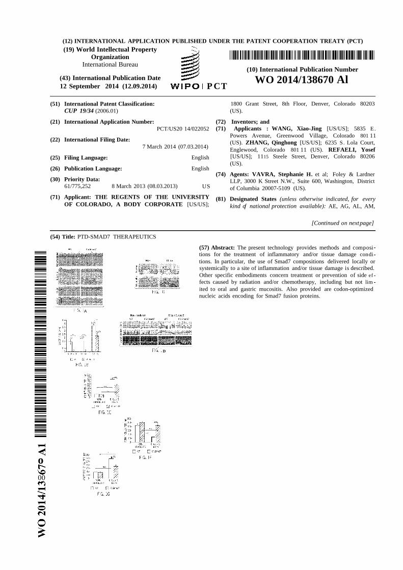
[0019] FIGS. 1A-G provide an illustrative embodiment of data showing that K5.Smad7
mice were resistant to radiation-induced oral mucositis. FIG. 1A provides an illustrative
embodiment of H&E staining in non-irradiated and irradiated (day 9 after initiation of
radiation) wild-type (WT) and K5.Smad7 tongues. The vertical lines in the images of
tongues from WT mice highlight the ulcer boundary and dotted lines in the images indicate
the epithelial-stromal boundary (scale bar, 50 µιη) . FIG. I B provides a graphical
representation of the quantification of sizes of tongue ulcers (mean ± s.e.m); n = 8 for WT
mice and n = 7 for K5.Smad7 mice in 8 Gy x 3 radiation; n = 5 for WT mice and n = 4 for
K5.Smad7 mice in 18-Gy radiation; n = 5 per group for WT and K5.Smad7 mice in 22-Gy
radiation. FIG. 1C provides an illustrative embodiment of human ventricular posterior of
the tongue (top) and radiation-induced tongue mucositis (bottom) visualized using H&E (left)
and CD45 staining (right). The solid line indicates the ulcer boundary, and dotted lines
indicate the basement membrane (scale bar, 25 µιη) . FIG. I D provides an illustrative
embodiment of immunostaining of CD45, proliferating cell nuclear antigen (PCNA), and
TUNEL assay in irradiated sections adjacent to an ulcer from WT mice and in damaged areas
from K5.Smad7 mice (PI, propidium iodide). Dotted lines indicate the basement membrane
(scale bar, 25 µιη) . FIGS. 1E-1G provide graphical representations of the quantification of
staining in FIG. I D (n = 3 or 4 per group). Data are expressed as mean ± s.e.m (FIG. IB)
or mean ± s.d (FIGS. 1E-1G), and two-tail Student ?-test is used to calculate P values. *P <
0.05, **P < 0.01, ***P < 0.001, NS, no significance determined by two-tailed Student's
t-test. Dotted lines in (FIG. 1A), (FIG. 1C) and (FIG. ID) highlight the basement
membrane. Scale bar: 50 µιη for all panels in (FIG. 1A) and (FIG. 1C), 25 µιη for all
panels in (FIG. ID).
[0020] FIGS. 2A-G provide an illustrative embodiment of data showing molecular
alterations attenuated by Smad7. FIG. 2A provides an illustrative embodiment of
immune-staining of NF-κΒ p50, TGF- Ι and pSmad2. Irradiated tongue sections of
wild-type (WT) were adjacent to ulcer and sections of K5.Smad7 were from the damaged
area. Human samples were from non-irradiated oral mucosa and radiation-induced
mucositis. Dotted lines delineate epithelial-stromal boundary. Scale bar, 25 µιη for all
panels. FIG. 2B provides a graphical representation of the quantification of
immunostaining of NF-κΒ p50 and pSmad2 shown in (FIG. 2A). FIG. 2C provides an
illustrative embodiment of qRT-PCR of TGF- Ι (normalized by Keratin 5, n = 6 per group
for day 0, n = 4 for day 7 and day 9, and n = 7 for day 10). FIG. 2D provides a graphical
representation of the quantification of human oral keratinocyte migration (see images in FIG.
8). Scrambled, scrambled siRNA; n = 3 per group. FIG. 2E provides an illustrative
embodiment of a western analysis of knockdown efficiency of siSmad7-l and siSmad7-2 for
Smad7 and for Racl, 72 hours after Smad7 knockdown. M, molecular markers. FIG. 2F
provides an illustrative embodiment of western analysis of total and activated (GTP-bound)
Racl protein. M : molecular markers. FIG. 2G provides a graphical representation of the
quantification of the effect of Racl knockdown on Smad7-mediated keratinocyte migration
(see knockdown efficiency in FIG. 9A and images in FIG. 9D). n = 3 per group. Data are
presented as mean ± s.d. and two-tail Student's est was used to calculate P values for (FIG.
2B-2D) and (FIG. 2G). *P <0.05, **P <0.01, ***P <0.001. NS, no significance.
[0021] FIGS. 3A-H provide an illustrative embodiment of data showing Smad7
increased Racl expression by repressing individual Smad and CtBPl binding to the SBE of
the Racl promoter. FIG. 3A provides a graphical representation of the quantification of
Racl mRNA in wild-type (WT) and Smad7 transgenic keratinocytes. n = 4 per group.
FIG. 3B provides an illustrative embodiment of western analysis of GTP-Rac 1 and total
Racl in WT and Smad7 keratinocytes. Smad7 protein levels in WT and Smad7
keratinocytes were determined by reprobing the tubulin western blot with an antibody to
Smad7 (see an additional western blot and quantification in FIGS. 10A-B). FIG. 3C
provides an illustrative embodiment of western analysis of Racl protein level after knocking
down individual Smad2, Smad3 or Smad4 in human keratinocytes (see FIG. lOC-lOE for
Smad knockdown efficiencies). FIG. 3D provides an illustrative embodiment of a ChIP
assay for Smad-2, -3, -4, and -7 binding to the -1.5 Kb SBE site of the Racl promoter in WT
and Smad7 transgenic keratinocytes. FIG. 3E provides a graphical representation of the
quantification oiRacl luciferase reporter assay in mouse keratinocytes. Scrambled:
scrambled siRNA. n = 6. FIG. 3F provides a graphical representation of the
quantification of the activities oiRacl luciferase reporter containing SBE or mutant (mut)
SBE in WT or Smad7 transgenic keratinocytes. n = 6. FIG. 3G provides an illustrative

embodiment of images of ChIP assays of CtBPl binding to the SBE-1.5 Kb site of the Racl
promoter in WT or K5.Smad7 keratinocytes. FIG. 3H provides a graphical representation
of ChlP-qPCR quantification of CtBPl binding to the SBE shown in FIG. 3G in WT and
Smad7 transgenic keratinocytes. n = 4. Data are presented as mean ± s.d. and two-tail
Student's /-test is used to calculate P values for FIGs. 3A, 3E, 3F and 3H. *P < 0.05, **P
< 0.01, ***P <0.001.
[0022] FIGS. 4A-G provide an illustrative embodiment of data showing
CtBPl -associated Racl repression contributed to inhibition of keratinocyte migration. FIG.
4A provides an illustrative embodiment of western analysis of Racl protein after knockdown
of CtBPl in human oral keratinocytes. FIG. 4B provides a graphical representation of the
quantification of SBE-containing Racl luc reporter activity n = 6. FIG. 4C provides a
graphical representation of the quantification of the effect of CtBPl knockdown on human
oral keratinocyte migration n = 3 per group. FIG. 4D provides an illustrative
embodiment of immunostaining of CtBP 1. Irradiated sections were adjacent to the ulcer
(WT) or the damaged area (K5.Smad7). Dotted lines denote the basement membrane.
Scale bar, 50 µιη for all panels. FIG. 4E provides an illustrative embodiment of
immunostaining of CtBPl in non-irradiated oral mucosa and radiation-induced oral mucositis
in human specimens. Dotted lines denote the basement membrane. Scale bar, 50 µιη for
both panels. FIG. 4F provides a graphical representation of the quantification of CtBP 1
nuclear positive cells in FIGS. 4D-E. n = 3 or 4 per group. FIG. 4G provides a graphical
representation of the quantification of qRT-PCR for CtBPl (normalized with Keratin K5). n
= 6 per group for day 0, n = 4 for day 7 and day 9, and n = 7 for day 10. Data are presented as
mean ± s.d. and two-tail Student's /-test is used to calculate P values for FIGS. 4B, 4C, 4F
and 4G. *P <0.05, **P <0.01, ***P <0.001.
[0023] FIGS. 5A-G provide an illustrative embodiment of data showing oral Tat-Smad7
application prevented radiation-induced oral mucositis in mice. FIG. 5A provides a
graphical representation of the quantification of oral mucositis ulcer sizes on day 9 after
initiation of 8 Gy x 3 radiation. Vehicle = saline or 50% glycerol/PBS. FIG. 5B provides
an illustrative embodiment of pathological alterations on day 9 of initiation of 8 Gy x 3
radiation. Vehicle = saline or 50% glycerol/PBS. Scale bar, 50 µιη for H&E panels and 25
µιη for remaining panels. Dotted lines delineate epithelial-stromal boundary; the solid line
highlights the ulcer boundary. FIGS. 5C, 5D, 5E, 5F, and 5G provide a graphical
representation of the quantification of immunostaining shown in FIG. 5B. n = 3 or 4 per
group. Data are presented as mean ± s.e.m (FIG. 5A) or mean ± s.d. (FIGS. 5C-5G) and
two-tail Student's -test is used to calculate P values. *P < 0.05, **P < 0.01, ***P < 0.001.
NS, no significance.
[0024] FIGS. 6A-G provide an illustrative embodiment of data showing Tat-Smad7
treatment on oral mucositis. FIG. 6A provides a graphical representation of the
quantification of ulcer sizes measured on day 10 after initiation of 8 Gy x 3 radiation.
Glycerol = 50% glycerol/PBS. FIG. 6B provides an illustrative embodiment of H&E
staining of oral mucosa. Upper panels: open ulcer in Palifermin treated but not Tat-Smad7
treated mucosa. Lower panels: comparison of epithelial thickness between Palifermin
treated and Tat-Smad7 treated mucosa. Dotted lines delineate the basement membrane.
The vertical line highlights the ulcer boundary. Scale bar, 50 µιη for all panels. FIG. 6C
provides an illustrative embodiment of immune-staining of Tat-Smad7 treatment in 20
Gy-induced oral mucositis after ulcers healed. V5 immunostaining visualizes Tat-Smad7 in
oral epithelia (sections are away from the damaged regions). K14 immunostaining was used
as counterstain. Dotted lines delineate basement membrane. Scale bar, 25 µιη for all panels.
FIG. 6D provides an illustrative embodiment of Racl western analysis of Tat-Smad7 treated
mouse tongues, day 10 after initiation of 8 Gy x 3 radiation. FIG. 6E provides an
illustrative embodiment of Racl western analysis on Tat-Smad7 treated normal human oral
keratinocytes 48 hours after treatment. FIG. 6F provides an illustrative embodiment of the
effect of Tat-Smad7 treatment on oral human keratinocyte migration (NOK-SI, see images in
FIG. 13A). n = 4 per group. FIG. 6G provides a graphical representation of the
quantification of survival curves of NOK-SI keratinocytes and SCC lines (Cal27 and
MSK92 1) with or without Tat-Smad7 treatment n = 4 per group for each radiation dose.
Data are presented as mean ± s.e.m (FIG. 6A) or mean ± s.d. (FIGS. 6F, 6G) and two-tail
Student's i-test is used to calculate P values. *P < 0.05, **P < 0.01, ***P < 0.001. NS, no
significance.
[0025] FIGS. 7A-E provide an illustrative embodiment of data showing K5.Smad7 oral
mucosal tissues were resistant to radiation-induced oral mucositis. FIG. 7A provides an
illustrative embodiment of Smad7 western blots: undetectable in non-irradiated wild-type (WT)
tongue and barely detectable after radiation. K5.Smad7 tongues have comparable Smad7
protein levels before and after radiation. M : molecular marker. FIG. 7B provides an
illustrative embodiment of Smad7 immunostaining. Note that nuclei in some irradiated
epithelial cells are hypertrophic. Dotted lines delineate epithelial-stromal boundary. FIG. 7C
provides a graphical representation of the quantification of reduced incidence of oral
mucositis-induced morbidity in K5.Smad7 mice. Fisher's exact test is used to calculate the p
value. **P = 0.007. FIG. 7D provides an illustrative embodiment of immune-staining of
K5.Smad7 tongue showing reduced infiltration of neutrophils (Ly-6G), macrophages (BM8)
and activated T cells (CD4) compared to WT oral mucositis. Dotted lines delineate
epithelial-stromal boundary. FIG. 7E provides an illustrative embodiment of
immune-staining showing no significant difference in pSmadl/5/8-nuclear positive cells
(green) between WT and K5.Smad7 oral mucosa before or after radiation. Keratin (K14)
immunostaining (red) highlights the epithelial compartment. Note that nuclei of irradiated
epithelial cells are hypertrophic. The scale bar is 50 µιη for all panels.
[0026] FIGS. 8A-D provide an illustrative embodiment of data showing migration in
spontaneously immortalized human oral epithelial cells (NOK-SI) was delayed by knocking
down Smad7 but accelerated by knocking down TGF- Ι . FIGS. 8A and 8B provide an
illustrative embodiment of representative images of cell migration. Pairs of dotted lines
delineate the scratch wound. Quantification of cell migration and efficiency of Smad7
knockdown are presented in FIG. 2D and FIG. 2E (above). Scrambled, scrambled siRNA.
FIG. 8C provides a graphical representation of the quantification of cell migration after
TGF- Ι knockdown from 3 separate experiments. FIG. 8D provides a graphical
representation of qRT-PCR showing TGF- Ι knockdown efficiency. Data are presented as
mean ± s.d. and two-tail Student's Mest was used to calculate P values. *P < 0.05, **P <
0.01. NS, no significance.
[0027] FIGS. 9A-D provide an illustrative embodiment of data showing knocking down
Racl reduced proliferation and migration of wild-type (WT) and Smad7 transgenic
keratinocytes. FIG. 9A provides an illustrative embodiment of western blot analysis for
Racl 48 hours after Racl siRNA (siRacl-1, siRacl-2) transfection. Control, scrambled
siRNA. FIG. 9B provides a graphical representation of the percentage of BrdU labeled
cells in WT and Smad7 cultured cells in BrdU incorporation assay with or without Racl
knockdown. Data from 3 separate experiments were presented as mean ± s.d. ***p <
0.001. FIG. 9C provides an illustrative embodiment of representative immunofluorescence
of BrdU positive cells presented in (FIG. 9B). An antibody against keratin 14 (K14, red)
was used for counterstain. FIG. 9D provides an illustrative embodiment of in vitro cell
migration assay for Smad7 transgenic and WT keratinocytes after Racl knockdown. Pairs
of dotted lines delineate the scratch wound. Quantification of cell migration is presented in
FIG. 2G.
[0028] FIGS. 10A-F provide an illustrative embodiment of data showing Smad7
increased Racl expression by repressing Smad and CtBPl binding to the SBE of the Racl
promoter. FIG. 10A provides an illustrative embodiment of western blot analysis for
GTP-Racl and total Racl in Smad7 transgenic keratinocytes. Additional samples are
shown in FIG. 3B. M, molecular marker. FIG. 10B provides a graphical representation of
the quantification of GTP-Racl, total Racl and Smad7 in WT and K5.Smad7 keratinocytes
shown in FIG 10A and in FIG. 3B. The protein level in WT keratinocytes of each blot was
normalized as "1". Data is presented as mean ± s.d. and two-tail Student's ?-test was used to
calculate P values. **P < 0.01, ***P < 0.001. FIGS. IOC and 10D provide an illustrative
embodiment of western blot analysis for Smad2, Smad3, and Smad4 knockdown in NOK-SI
cells. Their effects on Racl expression are shown in FIG. 3C. M, molecular marker.
GAPDH, internal protein control by reprobing same blot. FIG. 10F provides an illustrative
embodiment showing CtBP 1 knockdown promotes NOK-SI cell migration. Pairs of dotted
lines delineate the scratch wound. Quantification of cell migration and efficiency of CtBP 1
knockdown are shown in FIG. 4A and FIG. 4C.
[0029] FIGS. 11A-G provide an illustrative embodiment of data showing the
purification and characterization of Tat-Smad7 and Tat-Cre proteins. FIG. 11A shows an
illustrative embodiment of a schematic representation of Tat-Smad7 protein. FIG. 11B
provides an illustrative embodiment of a western blot of purified Tat-Smad7 protein. FIG.
llC provides an illustrative embodiment of immune-staining of Tat-Smad7 protein
transduction in keratinocytes. Left and middle panels: Tat-Smad7 staining (green) using a V5
antibody, counterstained with a K14 antibody (red). Cells showed Tat-Smad7 in the nucleus
5 min after transduction and in both nucleus and cytoplasm 1 hours after transduction.
Right panels: Tat-Smad7 abrogated Smad2 phosphorylation (pSmad2, green). V5 (red)
counterstain visualizes Tat-Smad7 transduced cells. FIG. 11D provides an illustrative
embodiment of immune-staining showing that V5 antibody staining detects Tat-Smad7
transduction in buccal mucosa 12 hours after Tat-Smad7 topical application. A K14
antibody was used for counterstain. Scale bar, 50 µιη for both panels. FIG. H E provides

an illustrative embodiment of a western blot of purified Tat-Cre protein with the same Tat
and V5 tags shown in FIG. 11A. FIG. 11F provides an illustrative embodiment of an
agarose gel showing activity of Tat-Cre: Tat-Cre cuts out a 1,460 bp floxed fragment from
the 7,650 bp vector pLL3.7. FIG. 11G provides a graphical representation showing
Tat-Smad7 protein preventive treatment reduced 20 Gy radiation-induced oral ulcers. Data
are expressed as mean ± s.e.m. Two-tail Student's ?-test is used to calculate P values. *P
< 0.05, ***P < 0.001.
[0030] FIGS. 12A-I provide an illustrative embodiment of data showing effects of
Tat-Smad7 treatment on oral mucositis. FIG. 12A provides a graphical representation of
the quantification of reduced ulcer size in Tat-Smad7 (0.8 µg daily, day 6 to day 9) treated
oral mucosa. Samples were harvested on day 10. n = 8 per group. FIG. 12B provides an
illustrative embodiment of immunostaining of molecular markers for samples from FIG.
12A. Scale bar, 50 µιη for the top two panels and 25 µιη for other panels. Propidium
iodide (PI) and K14 were used as counterstain. FIGS. 12C-G provide graphical
representation of the quantifications of immunostaining shown in FIG. 12C. 3-4 samples
were used. FIG. 12H provides a graphical representation quantification of the Luciferase
assay. Tat-Smad7 treatment increased the activity of the Racl promoter with SBE but not
the mutant SBE in mouse keratinocytes. FIG. 121 provides an illustrative embodiment of a
ChIP assay for CtBPl binding to the SBE of mouse Racl promoter in Tat-Smad7 treated
mouse keratinocytes. Data are expressed as mean ± s.e.m (a) or mean ± s.d (c-h) and
two-tail Student's -test is used to calculate P values. *P < 0.05, **P < 0.01, ***P < 0.001.
NS, no significance.
[0031] FIGS. 13A-H provide an illustrative embodiment of data showing effects of
Tat-Smad7 treatment on migration of human keratinocytes and tumor cell lines. FIG. 13A
provides an illustrative embodiment showing Tat-Smad7 accelerates NOK-SI cell migration.
Quantification from four separate experiments is shown in FIG. 6F (above). Pairs of dotted
lines delineate initial wounds. FIG. 13B provides an illustrative embodiment of
immunostaining of Tat-Smad7 treatment in NOK-SI cells showing attenuated
radiation-induced pSmad2 and NF-κΒ p50 nuclear localization. FIG. 13C provides an
illustrative embodiment showing V5 staining of MSK92 1 cells 2 hours after Tat-Smad7
treatment. K14 staining was used as counterstain. FIG. 13D provides an illustrative
embodiment of a Racl western analysis in MSK921 60 hours after Tat-Smad7 treatment.
M, molecular marker. FIG. 13E provides a graphical representation of quantification of
MSK921 cell migration from 3 separate experiments. FIG. 13F provides an illustrative
embodiment showing a representative MSK921 cell migration assay treated with Tat-Smad7
and PBS. Pairs of solid lines delineate initial wounds. Dotted lines highlight the forefront
of migrated cells. FIG. 13G provides a graphical representation of quantification of Cal27
cell migration from 3 separate experiments. FIG. 13H provides an illustrative embodiment
showing representative images for FIG. 13G. Pairs of solid lines delineate initial wounds.
Dotted lines highlight the forefront of migrated cells. Data are expressed as mean ± s.d. and
the two-tail Student's /-test is used to calculate P values. NS, no significance.
[0032] FIGS. 14A-B show an illustrative schematic of a summary of potential
mechanisms of Smad7-mediated protection and healing of oral mucositis. FIG. 14A shows
an illustrative schematic of how radiation activates N F-κΒ , increases TGF - Ι and CtBPl.
NF-KB and TGF - Ι induce inflammation. TGF - Ι induces apoptosis, growth arrest and
activates Smad-2, -3 and -4, which recruit CtBPl to the Racl promoter to repress Racl
transcription, leading to blunted re-epithelialization. FIG. 14B shows an illustrative
schematic of how Smad7 blocks N F-κΒ and TGF - Ι -induced inflammation and blocks
TGF -βΙ -induced apoptosis and growth arrest. Smad7 relieves Racl transcriptional
repression by either preventing TGF -βΙ -mediated Smad activation (phosphorylation) or
competing with signaling Smads/CtBPl transcriptional repression complex in binding to the
Racl promoter. Increased Racl induced by Smad7 contributes to keratinocyte migration
during re-epithelialization.
[0033] FIG. 15 shows an illustrative schematic of Smad 7 domains associated with
protein partners, potential target effects, and potential physiological effects.
[0034] FIGS. 16A-B are graphs demonstrating the ability of truncated Smad7 proteins to
accelerate wound healing in a mouse wound healing model. FIG. 16A is a graph showing
the effect of C-terminally truncated (259-426aa) Tat-C-Smad7 on average percent wound
healing over time relative to full-length Tat-Smad7 and control (PBS) n = 3 for each group.
FIG 16B is a graph illustrating the effect of Tat-N-Smad7 (l-258aa) on average percent
wound healing over time relative to full-length Tat-Smad7 and control (PBS) n = 6 for
each group. Data are presented as mean ± s.d., and two-tail Student's /-test was used to
calculate P values. *p < 0.05 compared to control (PBS), #p < 0.05 compared to
Tat-Smad7.
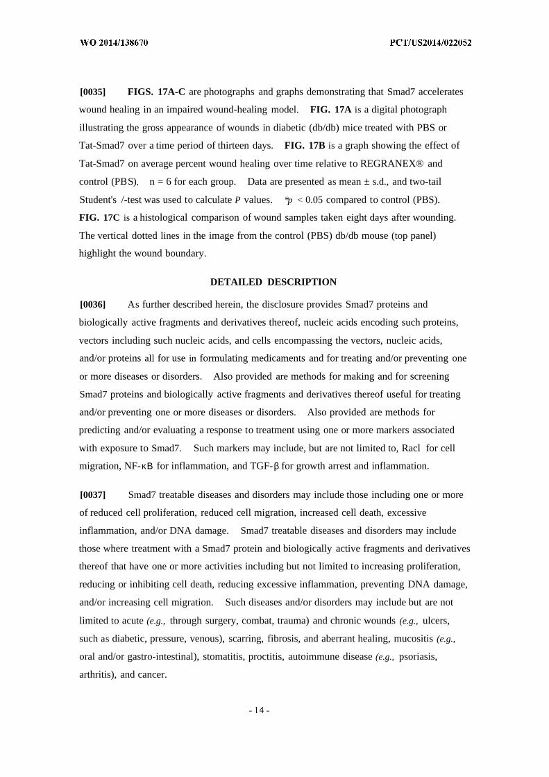
[0035] FIGS. 17A-C are photographs and graphs demonstrating that Smad7 accelerates
wound healing in an impaired wound-healing model. FIG. 17A is a digital photograph
illustrating the gross appearance of wounds in diabetic (db/db) mice treated with PBS or
Tat-Smad7 over a time period of thirteen days. FIG. 17B is a graph showing the effect of
Tat-Smad7 on average percent wound healing over time relative to REGRANEX® and
control (PBS) n = 6 for each group. Data are presented as mean ± s.d., and two-tail
Student's /-test was used to calculate P values. *p < 0.05 compared to control (PBS).
FIG. 17C is a histological comparison of wound samples taken eight days after wounding.
The vertical dotted lines in the image from the control (PBS) db/db mouse (top panel)
highlight the wound boundary.
DETAILED DESCRIPTION
[0036] As further described herein, the disclosure provides Smad7 proteins and
biologically active fragments and derivatives thereof, nucleic acids encoding such proteins,
vectors including such nucleic acids, and cells encompassing the vectors, nucleic acids,
and/or proteins all for use in formulating medicaments and for treating and/or preventing one
or more diseases or disorders. Also provided are methods for making and for screening
Smad7 proteins and biologically active fragments and derivatives thereof useful for treating
and/or preventing one or more diseases or disorders. Also provided are methods for
predicting and/or evaluating a response to treatment using one or more markers associated
with exposure to Smad7. Such markers may include, but are not limited to, Racl for cell
migration, NF-κΒ for inflammation, and TGF-β for growth arrest and inflammation.
[0037] Smad7 treatable diseases and disorders may include those including one or more
of reduced cell proliferation, reduced cell migration, increased cell death, excessive
inflammation, and/or DNA damage. Smad7 treatable diseases and disorders may include
those where treatment with a Smad7 protein and biologically active fragments and derivatives
thereof that have one or more activities including but not limited to increasing proliferation,
reducing or inhibiting cell death, reducing excessive inflammation, preventing DNA damage,
and/or increasing cell migration. Such diseases and/or disorders may include but are not
limited to acute (e.g., through surgery, combat, trauma) and chronic wounds (e.g., ulcers,
such as diabetic, pressure, venous), scarring, fibrosis, and aberrant healing, mucositis (e.g.,
oral and/or gastro-intestinal), stomatitis, proctitis, autoimmune disease (e.g., psoriasis,
arthritis), and cancer.

[0038] It is critical for oral mucositis prevention and treatment to overcome epithelial
ablation due to massive apoptosis and blunted keratinocyte proliferation. The proliferative
and anti-apoptotic effects of Smad7 are more obvious in oral mucositis than in normal oral
mucosa, when TGF- Ι , a potent growth inhibitor and apoptosis inducer for epithelial cells,
was increased.
[0039] Although not wishing to be bound by theory, it is believed that increased Racl
activation is largely responsible for Smad7-mediated keratinocyte migration in wound
closure. This finding was unexpected, given the documented role of TGF-β signaling in
Rho/Rac activation in cancer cells via a Smad-independent mechanism (Dernyck et ah,
Nature 415:577-584, 2003).
[0040] It is believed that during oral mucositis, Smad-dependent Racl repression
overcomes Smad-independent Racl activation (if any) due to increased Smad signaling
(evidenced by increased pSmad2) and Smad transcriptional co-repressor CtBP 1. When this
repression is abrogated by Smad7, it permits Racl activation-mediated keratinocyte
migration. However, in oral cancer cells, signaling Smads are lost or inactivated, or other
mechanisms independently activate Racl. As a result, Smad7-mediated abrogation oiRacl
repression would no longer occur.
[0041] Although Racl activation also contributed to keratinocyte proliferation, knocking
down Racl only partially attenuated the proliferative effect of Smad7. Therefore, Racl ' s
contribution to proliferation appears to be limited, and blocking TGF- Ι -induced growth arrest
is also needed to overcome radiation-induced growth inhibitory effects.
[0042] Dampening excessive inflammation creates a microenvironment for oral
mucositis healing. The antagonistic effect of Smad7 on both TGF-β and NF-κΒ signaling
makes Smad7 a more efficient anti-inflammatory molecule than other agents targeting only
NF-KB . Because inflammatory cells produce cytokines that further activate TGF-β and
NF-KB , reduced TGF-β and N F-κΒ signaling, found in K5.Smad7 or Tat-Smad7 treated oral
mucosa after radiation, reflects the direct antagonistic effect of Smad7 on these two pathways
and the consequence of reduced inflammatory cytokines from infiltrated leukocytes.
However, Smad7 did not reduce N F -κΒ or TGF-β signaling below their normal physiological
conditions. This incomplete blockade of N F -κΒ or TGF-β signaling may be beneficial to oral
mucositis healing, as a complete loss of either pathway could induce excessive inflammation.

[0043] The primary obstacle to using growth factors to treat oral mucositis in cancer
patients is the potential risk of promoting cancer cell growth. The majority of human oral
cancers lose TGF-β signaling in tumor epithelial cells. Thus, anti-Smad-associated cell
proliferation and migration by Smad7 would not be effective in cancer cells. In tumors with
intact TGF-β signaling, activation of other oncogenic pathways could override
TGF-p-induced tumor suppressive effects. These two scenarios could explain why there
was no observation of Smad7 increasing proliferation and migration in oral cancer cells with
mutant or intact TGF-β signaling components.
[0044] Additionally, TGF-β signaling promotes tumor invasion mainly through
Smad-independent mechanisms after loss of TGF-p-induced tumor suppression. Thus,
blocking TGF-β signaling by Smad7 in cancer cells could abrogate TGF-p-mediated tumor
promotion effects, which behaves similarly to TGF-β inhibitors currently being used in
clinical trials for advanced cancers. Further, the potent anti-inflammatory effect of Smad7
may reduce the risk of tumor progression. Therefore, long-term Smad7 application may
also be helpful in cancer treatment.
[0045] Spontaneous tumor formation in K5.Smad7 mice has not been observed.
Because Smad7 is not a secreted protein, local and short-term Smad7 protein delivery in oral
mucositis treatment should have few systemic effects. In bone marrow transplant patients,
whose oral epithelia do not contain cancer cells, Smad7 topical application may be suitable
for both prevention and treatment of oral mucositis.
[0046] Although not wishing to be bound by any theory, Smad7-mediated oral mucositis
healing appears to be a result of targeting multiple pathogenic processes mediated by one or
more molecules (see, e.g., FIGS. 14A-B). It is believed that one or more of these molecules
(e.g., TGF-β, NF-KB , CtBPl, Racl) may also be helpful as predictive and therapeutic
responsive markers of oral mucositis in patients.
A. Nucleic acids, Vectors and Host Cells
[0047] The present disclosure also provides, in another embodiment, genes encoding
Smad7. In addition to the wild-type SMAD7 gene (SEQ ID NOs: 12, 22), as well as various
codon-optimized versions (SEQ ID NOs: 9-11, 21, 23-41), it should be clear that the present
technology is not limited to the specific nucleic acids disclosed herein. As discussed below, a
"Smad7 gene" may contain a variety of different bases and yet still produce a corresponding
polypeptide that is functionally indistinguishable from, and in some cases structurally identical
to, the human gene disclosed herein.
1. Nucleic Acids Encoding Smad7
[0048] Nucleic acids according to the present technology may represent an entire Smad7
gene, a truncated portion, and/or a fragment of Smad7 that expresses a polypeptide with one
or more activity associated with Smad7 such as but not limited to increasing proliferation,
reducing or inhibiting cell death, reducing excessive inflammation, preventing DNA damage,
and/or increasing cell migration, as well as treating or preventing one or more disease or
disorders in which such treatment would be helpful as further discussed herein. Such
activities can be assessed using one or more assays including, but not limited to, the ability to
block phosphorylation of Smad2 and/or nuclear translocation of the NF-κΒ p50 subunit,
increase cell proliferation, reduce apoptosis and/or radiation-induced DNA damage, reduce
inflammation and/or angiogenesis, promote healing in oral mucositis, surgical wounds,
diabetes wounds, and/or wounds associated with chronic inflammation in mice. The nucleic
acid may be derived from genomic DNA, i.e., cloned directly from the genome of a particular
organism. In particular embodiments, however, the nucleic acid would comprise
complementary DNA (cDNA). Also provided is a cDNA plus a natural intron or an intron
derived from another gene; such engineered molecules are sometime referred to as
"mini-genes." At a minimum, these and other nucleic acids of the present technology may
be used as molecular weight standards in, for example, gel electrophoresis.
[0049] The term "cDNA" is intended to refer to DNA prepared using messenger RNA
(mRNA) as template. The advantage of using a cDNA, as opposed to genomic DNA or
DNA polymerized from a genomic, non- or partially-processed RNA template, is that the
cDNA primarily contains coding sequences of the corresponding protein. There may be
times when the full or partial genomic sequence is preferred, such as where the non-coding
regions are required for optimal expression or where non-coding regions such as introns are
to be targeted in an antisense strategy.
[0050] As used in this application, the term "a nucleic acid encoding a Smad7" may refer
to a nucleic acid molecule that has been isolated free of total cellular nucleic acid and/or may
refer to a cDNA encoding a Smad7 polypeptide. As used herein, the term "isolated free of
total cellular nucleic acid" means that the nucleic acid molecule is about or at least about 75%
pure, 80% pure, 85% pure, 90% pure, 95% pure, 96% pure, 97% pure, 98% pure, 99% pure,
or 100% pure of other cellular nucleic acid molecules as determined using standard
biochemical techniques, such as but not limited to agarose gel electrophoresis. As used
herein, the term "isolated free of total cellular protein" means that the protein molecule is
about or at least about 75% pure, 80% pure, 85% pure, 90% pure, 95% pure, 96% pure, 97%
pure, 98% pure, 99% pure, or 100% pure of other cellular nucleic acid molecules as
determined using standard biochemical techniques, such as but not limited to a western blot.
In certain embodiments, the present technology concerns a nucleic acid sequence essentially
as set forth in, and/or including any one of SEQ ID NOs: 9-11, 21, 23-41.
[0051] An isolated nucleic acid molecule may be produced using recombinant DNA
technology (e.g., polymerase chain reaction (PCR) amplification, cloning) or chemical
synthesis. Isolated nucleic acid molecules include natural nucleic acid molecules and
homologues thereof, including, but not limited to, natural allelic variants and modified
nucleic acid molecules in which nucleotides have been inserted, deleted, substituted, and/or
inverted in such a manner that such modifications provide the desired effect (e.g., production
of Smad7 protein in non-human expression systems).
[0052] The term "essentially as set forth in one or more nucleic acid sequence (e.g., SEQ
ID NOs: 9-1 1, 21, 23-41 " means that the nucleic acid sequence substantially corresponds to
at least a portion, and in some cases the entirety, of the one or more nucleic acid sequence
(e.g., SEQ ID NOs: 9-11, 21, 23-41. In some embodiments, sequences that substantially
correspond to at least a portion of a nucleic acid sequence, may correspond to about, or at
least about 50 nucleic acids, 75 nucleic acids, 150 nucleic acids, 200 nucleic acids, 250
nucleic acids, 300 nucleic acids, 350 nucleic acids, 400 nucleic acids, 450 nucleic acids, 500
nucleic acids, 550 nucleic acids, 600 nucleic acids, 650 nucleic acids, 700 nucleic acids, 750
nucleic acids, 800 nucleic acids, 900 nucleic acids, 1000 nucleic acids, 1100 nucleic acids,
1200 nucleic acids, or 1250 nucleic acids of one or more of the sequences described herein.
In some embodiments, sequences that substantially correspond to at least a portion of a
nucleic acid sequence, may correspond to about a range of about 50-1250 nucleic acids,
75-1250 nucleic acids, 150-1250 nucleic acids, 200-1250 nucleic acids, 250-1250 nucleic
acids, 300-1250 nucleic acids, 350—1250 nucleic acids, 400-1250 nucleic acids, 450-1250
nucleic acids, 500-1250 nucleic acids, 550-1250 nucleic acids, 600-1250 nucleic acids,
650-1250 nucleic acids, 700-1250 nucleic acids, 750-1250 nucleic acids, 800-1250 nucleic
acids, 900-1250 nucleic acids, 1000-1250 nucleic acids, 1100-1250 nucleic acids, 1200-1250
nucleic acids, at least about 50-75 nucleic acids, 75-150 nucleic acids, 75-200 nucleic acids,
75-250 nucleic acids, 75-300 nucleic acids, 75-350 nucleic acids, 75-400 nucleic acids,
75-450 nucleic acids, 75-500 nucleic acids, 75-550 nucleic acids, 75-600 nucleic acids,
75-650 nucleic acids, 75-700 nucleic acids, 75-750 nucleic acids, 75-800 nucleic acids,
75-900 nucleic acids, 75-1000 nucleic acids, 75-1 100 nucleic acids, 75-1200 nucleic acids, or
75-1250 nucleic acids or 1250 nucleic acids of one or more of the sequences described
herein.
[0053] In some embodiments, sequences that substantially correspond to at least a
portion of a nucleic acid sequence include identical sequences to that portion of the nucleic
acid sequence. In some embodiments, sequences that substantially correspond to at least a
portion of a nucleic acid sequence or the entirety of a nucleic acid sequence may include one
or more functionally equivalent codons. The term "functionally equivalent codon" is used
herein to refer to one or more codons that encode the same amino acid, such as the six codons
for arginine or serine, and in some embodiments refers to codons that encode biologically
equivalent amino acids, as discussed in the following pages. The term "biologically
equivalent" amino acid is used herein to refer to one or more amino acids that when changed
from the amino acid present in the amino acid sequence of human Smad7 wild-type protein,
do not change one or more (or in some embodiments any) of the biological activities of
Smad7 described herein, such as but not limited to, increasing proliferation, reducing or
inhibiting cell death, reducing excessive inflammation, preventing DNA damage, and/or
increasing cell migration, as well as treating or preventing one or more disease or disorders in
which such treatment would be helpful as further discussed herein.
[0054] In some embodiments, allowing for the degeneracy of the genetic code, sequences
that have about or at least about 60%, 70%, 80%, 90%, 91%, 92%, 93%, 94%, 95%, 96%,
97%, 98%, and/or 99% of nucleotides that are identical to the nucleotides of any one of the
codon-optimized nucleic acid sequences (e.g., SEQ ID NOs: 9-11, 21, 23-41) may be
considered substantially corresponding nucleic acid sequences. Sequences that are
essentially the same as those set forth in any one of the nucleic acid sequences (e.g., SEQ ID
NOs: 9-11, 21, 23-41) also may be functionally defined as sequences that are capable of
hybridizing to a nucleic acid segment containing the complement of SEQ ID NOs: 9-11, 21,
23-41 under various standard conditions.

[0055] For applications requiring high selectivity, one will typically desire to employ
relatively high stringency conditions to form the hybrids. For example, relatively low salt
and/or high temperature conditions, such as provided by about 0.02 M to about 0.10 M NaCl
at temperatures of about 50 °C to about 70 °C. Such high stringency conditions tolerate
little, if any, mismatch between the probe or primers and the template or target strand and
would be particularly suitable for isolating specific genes or for detecting specific mRNA
transcripts. It is generally appreciated that conditions can be rendered more stringent by the
addition of increasing amounts of formamide.
[0056] For certain applications it is appreciated that lower stringency conditions are
preferred. Under these conditions, hybridization may occur even though the sequences of
the hybridizing strands are not perfectly complementary, but are mismatched at one or more
positions. Conditions may be rendered less stringent by increasing salt concentration and/or
decreasing temperature. For example, a medium stringency condition could be provided by
about 0.1 to 0.25 M NaCl at temperatures of about 37 °C to about 55 °C, while a low
stringency condition could be provided by about 0.15 M to about 0.9 M salt, at temperatures
ranging from about 20 °C to about 55 °C. Hybridization conditions can be readily
manipulated depending on the desired results.
[0057] In other embodiments, hybridization may be achieved under conditions of, for
example, 50 mM Tris-HCl (pH 8.3), 75 mM KCl, 3 mM MgCl2, 1.0 mM dithiothreitol, at
temperatures between approximately 20 °C to about 37 °C. Other hybridization conditions
utilized could include approximately 10 mM Tris-HCl (pH 8.3), 50 mM KCl, 1.5 mM MgCl2,
at temperatures ranging from approximately 40 °C to about 72 °C.
[0058] To determine the percent homology of two amino acid sequences or of two
nucleic acids, the sequences are aligned for optimal comparison purposes (e.g., gaps are
introduced in the sequence of a first amino acid or nucleic acid sequence for optimal
alignment with a second amino acid or nucleic acid sequence). The amino acid residues or
nucleotides at corresponding amino acid positions or nucleotide positions can then be
compared. When a position in the first sequence is occupied by the same amino acid
residue or nucleotide as the corresponding position in the second sequence, then the
molecules are identical at that position. The percent homology between the two sequences
is a function of the number of identical positions shared by the sequences (% identity = # of
identical positions/total # of positions (e.g., overlapping positions)xl00). In some
embodiments the two sequences are the same length.
[0059] To determine percent homology between two sequences, the algorithm of Karlin
and Altschul (1990) Proc. Natl. Acad. Sci. USA 87:2264-2268, modified as in Karlin and
Altschul (1993) Proc. Natl. Acad. Sci. USA 90:5873-5877 can be used. Such an algorithm is
incorporated into the NBLAST and XBLAST programs of Altschul et a . (1990) J. Mo Biol.
215:403-410. BLAST nucleotide searches is performed with the NBLAST program,
score=100, wordlength=12 to obtain nucleotide sequences homologous to a nucleic acid
molecules described or disclose herein. BLAST protein searches is performed with the
XBLAST program, score=50, wordlength=3. To obtain gapped alignments for comparison
purposes, Gapped BLAST may be utilized as described in Altschul et al. (1997) Nucleic
Acids Res. 25:3389-3402. When utilizing BLAST and Gapped BLAST programs, the
default parameters of the respective programs (for example, XBLAST and NBLAST) are
used. See the website of the National Center for Biotechnology Information for further
details (on the World Wide Web at ncbi.nlm.nih.gov). Proteins suitable for use in the
methods described herein also includes proteins having between 1 to 15 amino acid changes,
for example, 1, 2, 3, 4, 5, 6, 7, 8, 9, 10, 11, 12, 13, 14, or 15 amino acid substitutions,
deletions, or additions, compared to the amino acid sequence of any protein described herein.
In other embodiments, the altered amino acid sequence is at least 75% identical, for example,
77%, 80%, 82%, 85%, 88%, 90%, 92%, 95%, 97%, 98%, 99%, or 100% identical to the
amino acid sequence of any protein inhibitor described herein. Such sequence-variant
proteins are suitable for the methods described herein as long as the altered amino acid
sequence retains sufficient biological activity to be functional in the compositions and
methods described herein. In certain instances conservative amino acid substitutions are
utilized. Illustrative conservative substitution among amino acids are within each of the
following groups: (1) glycine, alanine, valine, leucine, and isoleucine, (2) phenylalanine,
tyrosine, and tryptophan, (3) serine and threonine, (4) aspartate and glutamate, (5) glutamine
and asparagine, and (6) lysine, arginine and histidine. The BLOSUM62 table is an amino
acid substitution matrix derived from about 2,000 local multiple alignments of protein
sequence segments, representing highly conserved regions of more than 500 groups of related
proteins (Henikoff et al. (1992), Proc. Natl Acad. Sci. USA, 89: 10915-10919). The
BLOSUM62 substitution frequencies can be used to define conservative amino acid
substitutions that, in some embodiments, are introduced into the amino acid sequences
described or disclosed herein. Although it is possible to design amino acid substitutions
based solely upon chemical properties (as discussed above), the language "conservative
amino acid substitution" preferably refers to a substitution represented by a BLOSUM62
value of greater than -1. For example, an amino acid substitution is conservative if the
substitution is characterized by a BLOSUM62 value of 0, 1, 2, or 3. According to this
system, preferred conservative amino acid substitutions are characterized by a BLOSUM62
value of at least 1 (e.g., 1, 2 or 3), while more preferred conservative amino acid substitutions
are characterized by a BLOSUM62 value of at least 2 (e.g., 2 or 3).
[0060] The DNA segments of the present technology include those encoding biologically
functional equivalent Smad7 proteins and peptides, as described above. Such sequences
may arise as a consequence of codon redundancy and amino acid functional equivalency that
are known to occur naturally within nucleic acid sequences and the proteins thus encoded.
Alternatively, functionally equivalent proteins or peptides may be created via the application
of recombinant DNA technology, in which changes in the protein structure may be
engineered, based on considerations of the properties of the amino acids being exchanged.
Changes designed by man may be introduced through the application of site-directed
mutagenesis techniques or may be introduced randomly and screened later for the desired
function, as described elsewhere.
[0061] As described in greater detail below, the Smad7 nucleic acid sequence has been
optimized for expression in alternative host organisms (e.g., non-human). Although as
described above, the genetic code is degenerate, so frequently one amino acid may be coded
for by two or more nucleotide codons. Thus, multiple nucleic acid sequences may encode
one amino acid sequence. Although this creates identical proteins, the nucleic acids
themselves are distinct, and can have distinct properties. As described herein, one aspect of
the choice of codon usage can be (but is not limited to) the ability to express a protein in a
non-native cells (e.g., a human protein in bacteria or yeast), or the level of expression in such
cells. In order to obtain enough protein for purification, testing, and use in in vitro assays,
in animal models, and eventually in clinical development, efficient protein expression in
non-human systems is needed.
[0062] A series of 23 arginine amino acids in the human Smad7 protein sequence coded
for by one or more of AGG (1.7% codon utilization; 9 residues), AGA (2.8% codon
utilization; 2 residues), CGA (3.5% codon utilization; 4 residues), or CGG (5.4% codon

utilization; 8 residues) has been identified, and it has been determined that in order to have
efficient protein expression from non-human sources, such as, but not limited to, bacteria
and/or yeast that one or more, and potentially all the arginine codons should be modified to
CGT (20.6% codon utilization). Therefore, in some embodiments, the Smad7
codon-optimized nucleic acid sequence includes at least 1, at least 2, at least 3, at least 4, at
least 5, at least 6, at least 7, at least 8, at least 9, at least 10, at least 11, at least 12, at least 13,
at least 14, at least 15, at least 16, at least 17, at least 18, at least 19, at least 20, at least 21, at
least 22, or 23 codons for arginine that have been changed to CGT. In some embodiments,
the Smad7 codon-optimized nucleic acid sequence includes one or more or all of the arginine
codons at nucleic acid sequence positions 7-9, 43-45, 169-171, 403-405, 490-492, 526-528,
526-528, 823-825, 1057-1059, 16-18, 136-138, 199-201, 598-600, 31-33, 112-1 14, 316-318,
772-774, 940-942, 973-975, 1135-1 137, 1276-1278, 637-639, or 814-816 be changed to
CGT.
[0063] A series of 33 serine residues in the human Smad7 protein sequence coded for by
TCC or TCG (9%) has been identified, and it has been determined that it may be beneficial to
efficient protein expression and purification from non-human sources, such as, but not limited
to, bacteria and/or yeast, that one or more, and potentially all the serine codons be modified
to AGC (15% codon utilization). Therefore, in some embodiments, the Smad7
codon-optimized nucleic acid sequence includes at least 1, at least 2, at least 3, at least 4, at
least 5, at least 6, at least 7, at least 8, at least 9, at least 10, at least 11, at least 12, at least 13,
at least 14, at least 15, at least 16, at least 17, at least 18, at least 19, at least 20, at least 21, at
least 22, at least 23, at least 24, at least 25, at least 26, at least 27, at least 28, at least 29, at
least 30, at least 3 1, at least 32 or 33 codons for serine that have been changed to (AGC). In
some embodiments, the Smad7 codon-optimized nucleic acid sequence includes one or more
or all of the serine codons at nucleic acid sequence positions 19-21, 46-48, 133-135, 292-294,
349-351, 451-453, 454-456, 460-462, 511-513, 514-516, 544-546, 595-597, 616-618,
634-636, 691-693, 694-696, 739-741, 745-747, 775-777, 847-849, 907-909, 919-921,
943-945, 1006-1008, 1009-1 101, 1030-1032, 1054-1056, 1093-1095, 1126-1128, 1192-1 194,
1237-1239, 1240-1242, 1273-1275. Of these, 23 codons (19-21, 292-294, 349-351,
451-453, 454-456, 460-462, 5 11-513, 514-516, 544-546, 616-618, 634-636, 691-693,
694-696, 739-741, 745-747, 775-777, 847-849, 907-909, 919-921, 1009-1 101, 1030-1032,
1054-1056, 1093-1095) can be changed without introducing potential alternative open
reading frames.
[0064] A series of 12 histidine residues in the human Smad7 protein sequence coded for
by CAC (9.6% codon usage) has also been identified, and it has been determined that it may
be beneficial to efficient protein expression and purification from non-human sources, such
as but not limited to bacteria and/or yeast, that one or more, and potentially all the serine
codons be modified to CAT (optionally to 12.6% usage). Therefore, in some embodiments,
the Smad7 codon-optimized nucleic acid sequence includes at least 1, at least 2, at least 3, at
least 4, at least 5, at least 6, at least 7, at least 8, at least 9, at least 10, at least 11, or 12
codons for histidine that have been changed to (CAT). In some embodiments, the Smad7
codon-optimized nucleic acid sequence includes one or more or all of the serine codons at
nucleic acid sequence positions 142-144, 214-216, 217-219, 220-222, 226-228, 289-291,
589-591, 778-780, 1072-1074, 1147-1 149. Of these, 4 codons (nucleotides 217-219,
220-222, 589-591, 778-780) can be changed without introducing potential alternative open
reading frames.
[0065] In some embodiments, one or more codon-optimized nucleic acids may include
one or more of at least one and any integer up to 22 of its arginine codons modified to CGT,
at least one and any integer up to 28 of its serine codons (optionally that are able to be
modified with introducing open reading frames) modified to AGC, or at least one and any
integer up to 12 of its histidine codons (optionally that are able to be modified with
introducing open reading frames) modified to CAT. In some embodiments, one or more
codon-optimized nucleic acid may include at least one and any integer up to 22 of its arginine
codons modified to CGT, at least one and any integer up to 28 of its serine codons (optionally
that are able to be modified with introducing open reading frames) modified to AGC, and at
least one and any integer up to 12 (optionally that are able to be modified with introducing
open reading frames) of its histidine codons modified to CAT. In some embodiments, one
or more codon-optimized nucleic acid may include 22 of its arginine codons modified to
CGT, 28 of its serine codons (optionally that are able to be modified with introducing open
reading frames) modified to AGC, and 12 of its histidine codons (optionally that are able to
be modified with introducing open reading frames) modified to CAT. In some
embodiments, one or more codon-optimized nucleic acid may also have a nucleotide
substitution in the codon for Met216 (ATG), to form the codon for Leu216 (CTG).
[0066] In some embodiments, one or more codon-optimized nucleic acids may have
about 65% to 75%, about 65% to 68%, about 68% to 75%, or about 68% to 71% homology to
human Smad7 wild-type cDNA (SEQ ID NOs: 12, 22). In some embodiments, one or more
codon-optimized nucleic acid may have about 65%, 66%, 67%, 68%, 69%, 70%, 71%, 72%,
73%, 74%, or 75%, homology to human Smad7 wild-type cDNA (SEQ ID NOs: 12, 22). In
some embodiments, one or more codon-optimized nucleic acid may also have a nucleotide
substitution in the codon for Met216 (ATG), to form the codon for Leu216 (CTG).
[0067] A methionine codon (Met2 16; ATG) that has the potential for being perceived by
translation machinery (e.g., such as but not limited bacteria or yeast) as an alternative open
reading frame has been identified. Although not intending to be bound by theory, it is
believed that the presence of the second potential open reading frame may decrease
expression of the Smad7 protein. In some embodiments, one or more Smad7 nucleic acid
sequences are modified at nucleotide position (646-648) to encode a human Smad7 protein
where Met216 (ATG) is modified to Leu216 (CTG).
[0068] It has also been discovered that various truncated forms and fragments of Smad7
protein retain one or more of the activities of full-length human Smad7, such as, but not
limited to, increasing proliferation, reducing or inhibiting cell death, reducing excessive
inflammation, preventing DNA damage, and/or increasing cell migration, as well as treating
or preventing one or more disease or disorders in which such treatment would be helpful as
further discussed herein. Such activities can be assessed using one or more assays including,
but not limited to, the ability to block phosphorylation of Smad2 and/or nuclear translocation
of the NF-KB p50 subunit, increase cell proliferation, reduce apoptosis and/or
radiation-induced DNA damage, reduce inflammation and/or angiogenesis, promote healing
in oral mucositis, surgical wounds, diabetes wounds, and/or wounds associated with chronic
inflammation in mice.
[0069] Further, in some embodiments, various truncated forms and fragments of Smad7
protein retain only a subset of the one or more of the activities of full-length human Smad7.
For example, the C-terminal MH2 domain of Smad7 may primarily mediate the
anti-inflammatory effect of Smad7. Smad7 peptides having this anti-inflammatory function
may be sufficient and optionally an improvement for treating chronic inflammation
associated conditions, such as but not limited to, oral mucositis, stomatitis, arthritis, and
psoriasis, among others. The N-terminal MH1 domain may primarily mediate cell
migration and/or blocking TGF-p-induced growth arrest and/or fibrotic response. Smad7
peptides having this cell migration and proliferation function may be sufficient, and
optionally an improvement, for enhancing healing that is not associated with excessive
inflammation. Types of wounds that might benefit from this form of treatment include, but
are not limited to, surgical wounds, fibrotic scarring, and diabetes wounds, defective healing
and/or scarring among others.
[0070] In some embodiments, nucleic acid molecules (optionally codon-optimized
nucleic acid molecules as described above and herein) encode fragments or truncated forms
of Smad7 protein (optionally including Leu216). In some embodiments, these fragments
and/or truncated forms of Smad7 protein retain one or more or all of the activities of
full-length human Smad7 protein. In some embodiments, such truncated nucleic acid
sequences encode the N-terminal portion of the Smad7 protein. In some embodiments, such
truncated nucleic acid sequences encode the C-terminal portion of the Smad7 protein. In
some embodiments, such truncated nucleic acid sequences (nucleotide positions 4-774)
encode amino acids 2-258 of the human Smad7 protein. In some embodiments, such
truncated nucleic acid sequences (nucleotide positions 775-1278) encode amino acids
259-426 of the human Smad7 protein. In some embodiments, such fragments of the nucleic
acid sequences (nucleotide positions 610-774) encode amino acids 204-258 of the human
Smad7 protein.
[0071] The term "truncated" as used herein in reference to nucleic acid molecules refers
to a molecule that contains nucleotide sequences encoding the natural N-terminus of a
corresponding protein (with or without a cleaved leader sequence), but lacks one or more
nucleotides starting from the C-terminus-encoding portion of the molecule, or a molecule that
contains nucleotide sequences encoding the natural C-terminus of a corresponding protein
(with or without a cleaved leader sequence), but lacks one or more nucleotides starting from
the N-terminus-encoding portion of the molecule. In some embodiments, molecules lacking
nucleotides encoding at least about 25, at least about 50, at least about 75, at least about 100,
at least about 125, at least about 150, at least about 200, at least about 250, at least about 300,
or at least about 350, or at least about 400 amino acids from one or the other terminus are
specifically provided. Similarly, the term "truncated" may also be used in reference to
protein molecules encoded by truncated nucleic acid molecules. In some embodiments, a
"truncated" molecule is biologically active, having (or encoding a polypeptide having) one or
more of the Smad7 activities described herein.
[0072] The term "fragment" as used herein in reference to nucleic acid molecules refers
to a molecule containing contiguous residues of a full length sequence but lacking some 5'
and/or 3' sequences of the full length sequence. In some embodiments, a "fragment"
includes a portion of one or more of the full length sequences described herein. In some
embodiments, the "fragment" does not include sequences encoding either the N-terminal or
the C-terminal, but only internal fragments. In some embodiments, a "fragment" encodes a
polypeptide that is biologically active, having one or more of the Smad7 activities described
herein. In some embodiments, nucleic acid fragments may encode proteins having at least
about 25, 30, 35, 40, 45, 50, 55, 60, 65, 70, 75, 80, 85, 90, 95, 100,150, 200, 250, 300, 350,
400, 450, 500, 550, 600, 650, 700, 750, 800, 850, 900, 950, 1000, 1050, 1100, 1150 amino
acids. Similarly, "fragment" may also be used in reference to protein molecules encoded by
Smad7 nucleic acid fragments.
[0073] The term "N-terminal portion" as used herein in refers to a fragment of a
corresponding protein that contains the protein's N-terminus but lacks all sequences
C-terminal to an internal residue.
[0074] The term "C-terminal portion" as used herein in refers to a fragment of a
corresponding protein that contains the protein's C-terminus but lacks all sequences
N-terminal to an internal residue.
[0075] Although not intending to be bound by theory, the Smad7 protein activity is
generally believed to be the result of interactions in both the cytoplasm and nucleus of a cell.
For that reason among others, there existed a general belief that Smad7 protein was not a
candidate for a therapeutic role. However, it was decided to pursue development of Smad7
as a protein therapeutic, and modify the Smad7 nucleic acid sequence to encode a protein
transduction domain (PTD) in frame with the Smad7 nucleic acid sequence (e.g., optionally
any nucleic acid sequence described herein encoding Smad7 protein, including human
wild-type and codon-optimized sequences, both full-length and biologically active fragments
or truncated portions). In some embodiments, the PTD is located at the 3' end of the Smad7
nucleic acid sequence, and in some embodiments the PTD is located at the 5' end of the
Smad7 nucleic acid sequence. In some embodiments, there is a linker sequence encoding 1,
2, 3, 4, 5, or 6 amino acids that connects the PTD and the Smad7 nucleic acid sequence.
[0076] In some embodiments, the PTD nucleic acid sequence is a Tat nucleic acid
sequence ggccgtaaaaaacgccgtcaacgccgccgt (SEQ ID NO: 1) encoding GRKKRRQRRR
(SEQ ID NO: 2), tatggccgtaaaaaacgccgtcaacgccgccgt (SEQ ID NO: 3) encoding
YGRKKRRQRRR (SEQ ID NO: 4), or ggccgtaaaaaacgccgtcaa (SEQ ID NO: 5) encoding
GRKKRRQ (SEQ ID NO: 6).
[0077] In some embodiments, the nucleic acid sequence further includes a nucleotide
sequence encoding one or more of an epitope tag or a purification tag. In some
embodiments, the epitope tag is V5. In some embodiments, the purification tag is one or
more of glutathione-S-Transferase (GST) or 6-histidine (H6).
[0078] The term "epitope tag" as used herein in reference to nucleic acid molecules
refers to nucleotides encoding peptide sequences that are recognized and bound by the
variable region of an antibody or fragment. In some embodiments, the epitope tag is not
part of the native protein. In some embodiments, the epitope tag is removable. In some
embodiments, the epitope tag is not intrinsic to the protein's native biological activity.
Examples of epitope tags include, but are not limited to V5.
[0079] The term "purification tag" as used herein in reference to nucleic acid molecules
refers to nucleotides encoding peptide sequences that facilitate the purification of the protein,
but are generally not necessary for the protein's biological activity. In some embodiments,
purification tags may be removed following protein purification. Examples of purification
tags include, but are not limited to GST and H-6.
2. Vectors for Cloning, Gene Transfer and Expression
[0080] Within certain embodiments, expression vectors are employed to express the
Smad7 polypeptide product, which can then be purified for various uses. In other
embodiments, the expression vectors are used in gene therapy. Expression requires that
appropriate signals be provided in the vectors, and which include various regulatory elements,
such as enhancers/promoters from both viral and mammalian sources that drive expression of
the genes of interest in host cells. Elements designed to optimize messenger RNA stability
and trans latability in host cells also are defined. The conditions for the use of a number of
dominant drug selection markers for establishing permanent, stable cell clones expressing the
products are also provided, as is an element that links expression of the drug selection
markers to expression of the polypeptide.

[0081] Throughout this application, the term "expression construct" is meant to include
any type of genetic construct containing a nucleic acid coding for a gene product in which
part or all of the nucleic acid encoding sequence is capable of being transcribed. The
transcript may be translated into a protein, but it need not be. In certain embodiments,
expression includes both transcription of a gene and translation of mRNA into a gene product.
In other embodiments, expression only includes transcription of the nucleic acid encoding a
gene of interest.
[0082] The term "vector" is used to refer to a carrier nucleic acid molecule into which a
nucleic acid sequence can be inserted for introduction into a cell where it can be replicated.
A nucleic acid sequence can be "exogenous," which means that it is foreign to the cell into
which the vector is being introduced or that the sequence is homologous to a sequence in the
cell but in a position within the host cell nucleic acid in which the sequence is ordinarily not
found. Vectors include plasmids, cosmids, viruses (bacteriophage, animal viruses, and plant
viruses), and artificial chromosomes (e.g., YACs). One of skill in the art would be
well-equipped to construct a vector through standard recombinant techniques, which are
described, e.g., in Sambrook, et al, Molecular Cloning (Cold Spring Harbor Lab Press, 1989),
and Ausubel, et al, Current Protocols in Molecular Biology (Wiley, 1994), both incorporated
herein by reference.
[0083] The term "expression vector" refers to a vector containing a nucleic acid sequence
coding for at least part of a gene product capable of being transcribed. In some cases, RNA
molecules are then translated into a protein, polypeptide, or peptide. In other cases, these
sequences are not translated, for example, in the production of antisense molecules or
ribozymes. Expression vectors can contain a variety of "control sequences," which refer to
nucleic acid sequences necessary for the transcription and possibly translation of an operably
linked coding sequence in a particular host organism, including promoters and enhancers.
In addition to control sequences that govern transcription and translation, vectors and
expression vectors may contain nucleic acid sequences that serve other functions, such as
transcription termination signals and poly-adenylation sites.
[0084] The capacity of certain viral vectors to efficiently infect or enter cells, to integrate
into a host cell genome and stably express viral genes, have led to the development and
application of a number of different viral vector systems. Robbins, et al, Pharmacol. Ther.
80:35-47 (1998). Viral systems are currently used as vectors for ex vivo and in vivo gene

transfer. For example, adenovirus, herpes-simplex virus, lentiviruses, retrovirus and
adeno-associated virus vectors are being evaluated currently for treatment of diseases such as
cancer, cystic fibrosis, Gaucher disease, renal disease and arthritis. Robbins, et al,
Pharmacol. Ther. 80:35-47 (1998); Imai, et al, Nephrologie 19:379-402 (1998); U.S. Patent
5,670,488. The various viral vectors present specific advantages and disadvantages,
depending on the particular gene-therapeutic application.
[0085] Suitable non-viral methods for nucleic acid delivery for transformation of an
organelle, a cell, a tissue or an organism for use with the present technology are believed to
include virtually any method by which a nucleic acid (e.g., DNA) can be introduced into an
organelle, a cell, a tissue or an organism, as described herein or as would be known to one of
ordinary skill in the art. Such methods include, but are not limited to, direct delivery of
DNA such as by injection (U.S. Patents 5,994,624, 5,981,274, 5,945,100, 5,780,448,
5,736,524, 5,702,932, 5,656,610, 5,589,466 and 5,580,859, each incorporated herein by
reference), including microinjection (Harland and Weintraub, 1985; U.S. Patent 5,789,215,
incorporated herein by reference); by electroporation (U.S. Patent 5,384,253, incorporated
herein by reference); by calcium phosphate precipitation (Graham, et al., Virology
52:456-467 (1973); Chen, et al., Mol. Cell Biol. 7:2745-2752 (1987); Rippe, et al., Mol. Cell
Biol. 10:689-695 (1990)); by using DEAE-dextran followed by polyethylene glycol (Gopal,
Mol. Cell Biol. 5 :1188-1 190 (1985)); by direct sonic loading (Fechheimer, et al., PNAS
84:8463-8467 (1987)); by liposome mediated transfection (Nicolau, et al., Biochim. Biophys.
Acta 721:185-190 (1982); Fraley, etal, PNAS 76:3348-3352 (1979); Nicolau, et al, Methods
Enzymol. 149: 157-176 (1987); Wong, etal, Gene 10:87-94 (1980); Kaneda, et al, J. Biol.
Chem. 264:12126-12129 (1989); Kato, et al, J. Biol. Chem. 266:3361-3364 (1991)); by
microprojectile bombardment (PCT Application Nos. WO 94/09699 and 95/06128; U.S.
Patents 5,610,042; 5,322,783, 5,563,055, 5,550,318, 5,538,877 and 5,538,880, and each
incorporated herein by reference); by agitation with silicon carbide fibers (Kaeppler, et al,
Plant Cell Rep. 9:415-418 (1990); U.S. Patents 5,302,523 and 5,464,765, each incorporated
herein by reference); or by PEG-mediated transformation of protoplasts (Omirulleh, et al,
Plant Mol. Biol. 21:415-428 (1993); U.S. Patents 4,684,61 1 and 4,952,500, each incorporated
herein by reference); by desiccation/inhibition-mediated DNA uptake (Potrykus, et al, Mol.
Gen. Genet. 199:169-177 (1985)). Through the application of techniques such as these,
organelle(s), cell(s), tissue(s) or organism(s) may be stably or transiently transformed.
3. Expression Systems
[0086] Numerous expression systems exist that comprise at least a part or all of the
compositions discussed above. Prokaryote- and/or eukaryote-based systems can be
employed for use with the present technology to produce nucleic acid sequences, or their
cognate polypeptides, proteins and peptides. Many such systems are commercially and
widely available.
[0087] The insect cell/baculovirus system can produce a high level of protein expression
of a heterologous nucleic acid segment, such as described in U.S. Patents 5,871,986 and
4,879,236, both herein incorporated by reference, and which can be bought, for example,
under the name MAXBAC® 2.0 from INVITROGEN® and BACPACK™ BACULOVIRUS
EXPRESSION SYSTEM FROM CLONTECH®.
[0088] Other examples of expression systems include STRATAGENE®' s COMPLETE
CONTROL™ Inducible Mammalian Expression System, which involves a synthetic
ecdysone-inducible receptor, or its pET Expression System, an E. coli expression system.
Another example of an inducible expression system is available from INVITROGEN®, which
carries the T-REX™ (tetracycline-regulated expression) System, an inducible mammalian
expression system that uses the full-length CMV promoter. INVITROGEN® also provides a
yeast expression system called the Pichia methanolica Expression System, which is designed
for high-level production of recombinant proteins in the methylotrophic yeast Pichia
methanolica. One of skill in the art would know how to express a vector, such as an
expression construct, to produce a nucleic acid sequence or its cognate polypeptide, protein,
or peptide.
[0089] Primary mammalian cell cultures may be prepared in various ways. In order for
the cells to be kept viable while in vitro and in contact with the expression construct, it is
necessary to ensure that the cells maintain contact with the correct ratio of oxygen and carbon
dioxide and nutrients but are protected from microbial contamination. Cell culture
techniques are well documented.
[0090] One embodiment of the foregoing involves the use of gene transfer to immortalize
cells for the production of proteins. The gene for the protein of interest may be transferred
as described above into appropriate host cells followed by culture of cells under the
appropriate conditions. The gene for virtually any polypeptide may be employed in this
manner. The generation of recombinant expression vectors, and the elements included
therein, are discussed above. Alternatively, the protein to be produced may be an
endogenous protein normally synthesized by the cell in question.
[0091] Examples of useful mammalian host cell lines are Vero and HeLa cells and cell
lines of Chinese hamster ovary, W138, BHK, COS-7, 293, HepG2, NIH3T3, R N and
MDCK cells. In addition, a host cell strain may be chosen that modulates the expression of
the inserted sequences, or modifies and process the gene product in the manner desired.
Such modifications (e.g., glycosylation) and processing (e.g., cleavage) of protein products
may be important for the function of the protein. Different host cells have characteristic and
specific mechanisms for the post-translational processing and modification of proteins.
Appropriate cell lines or host systems can be chosen to insure the correct modification and
processing of the foreign protein expressed.
[0092] A number of selection systems may be used including, but not limited to, HSV
thymidine kinase, hypoxanthine-guanine phosphoribosyltransferase and adenine
phosphoribosyltransferase genes, in tk-, hgprt- or aprt- cells, respectively. Also,
anti-metabolite resistance can be used as the basis of selection for dhfr, that confers resistance
to; gpt, that confers resistance to mycophenolic acid; neo, that confers resistance to the
aminoglycoside G418; and hygro, that confers resistance to hygromycin.
[0093] As used herein, the terms "cell," "cell line," and "cell culture" may be used
interchangeably. All of these terms also include their progeny, which are any and all
subsequent generations. It is understood that all progeny may not be identical due to
deliberate or inadvertent mutations. In the context of expressing a heterologous nucleic acid
sequence, "host cell" refers to a prokaryotic or eukaryotic cell, and it includes any
transformable organism that is capable of replicating a vector and/or expressing a
heterologous gene encoded by a vector. A host cell can, and has been, used as a recipient
for vectors. A host cell may be "transfected" or "transformed," which refers to a process by
which exogenous nucleic acid is transferred or introduced into the host cell. A transformed
cell includes the primary subject cell and its progeny.
[0094] Host cells may be derived from prokaryotes or eukaryotes (e.g., bacteria or yeast),
depending upon whether the desired result is replication of the vector or expression of part or
all of the vector-encoded nucleic acid sequences. Numerous cell lines and cultures are

available for use as a host cell, and they can be obtained through the American Type Culture
Collection (ATCC), which is an organization that serves as an archive for living cultures and
genetic materials (atcc.org). An appropriate host can be determined by one of skill in the art
based on the vector backbone and the desired result. A plasmid or cosmid, for example, can
be introduced into a prokaryote host cell for replication of many vectors. Bacterial cells
used as host cells for vector replication and/or expression include DH5a, JM109, and KC8,
as well as a number of commercially available bacterial hosts such as SURE® Competent
Cells and SOLOPACK™ Gold Cells (STRATAGENE ®, La Jolla). Alternatively, bacterial
cells such as E. coli LE392 could be used as host cells for phage viruses.
[0095] Examples of eukaryotic host cells for replication and/or expression of a vector
include HeLa, NIH3T3, Jurkat, 293, Cos, CHO, Saos, and PC12. Many host cells from
various cell types and organisms are available and would be known to one of skill in the art.
Similarly, a viral vector may be used in conjunction with either a eukaryotic or prokaryotic
host cell, particularly one that is permissive for replication or expression of the vector.
[0096] Some vectors may employ control sequences that allow it to be replicated and/or
expressed in both prokaryotic and eukaryotic cells. One of skill in the art would further
understand the conditions under which to incubate all of the above described host cells to
maintain them and to permit replication of a vector. Also understood and known are
techniques and conditions that would allow large-scale production of vectors, as well as
production of the nucleic acids encoded by vectors and their cognate polypeptides, proteins,
or peptides.
B. Smad7 Proteins and Protein Fragments
[0097] Mothers against decapentaplegic homolog 7 (Smad7) was previously identified as
an antagonist of TGF-β signaling by several mechanisms including: (a) blockade of TGF-β
receptor-mediated phosphorylation and nuclear translocation of signaling Smads; (b)
increased degradation of TGF-β receptors and signaling Smads through specific
ubiquitin-proteasome pathways and (c) inhibition of signaling Smads for their binding to
Smad binding elements (SBEs). Smad7 also antagonizes other signaling pathways, like the
NF-KB pathway.
[0098] Smad7 protein is encoded by the SMAD7 gene, discussed above. Like many
other TGF- β family members, Smad7 is involved in cell signaling. It is a TGF- β type 1
receptor antagonist. It blocks TGF- Ι and activin associating with the receptor, blocking
access to Smad2. It is an inhibitory Smad (I-SMAD) and is enhanced by SMURF2.
Smad7 also enhances muscle differentiation.
[0099] The terms "polypeptide," "peptide," and "protein" are used interchangeably herein
to refer to a polymer of amino acid residues. The terms apply to naturally occurring amino
acid polymers as well as amino acid polymers in which one or more amino acid residues is a
non-naturally occurring amino acid, for example, an amino acid analog. As used herein, the
terms encompass amino acid chains of any length, including full length proteins, wherein the
amino acid residues are linked by covalent peptide bonds.
[0100] In one embodiment, the present technology relates to Smad7 protein
compositions. In addition to the entire Smad7 molecule, the present technology also relates
to truncated portions and fragments of the polypeptide that retain one or more activity
associated with Smad7, such as, but not limited to, increasing proliferation, reducing or
inhibiting cell death, reducing excessive inflammation, preventing DNA damage, and/or
increasing cell migration, as well as treating or preventing one or more disease or disorders in
which such treatment would be helpful as further discussed herein. Such activities can be
assessed using one or more assays including, but not limited to, the ability to block
phosphorylation of Smad2 and/or nuclear translocation of the NF-κΒ p50 subunit, increase
cell proliferation, reduce apoptosis and/or radiation-induced DNA damage, reduce
inflammation and/or angiogenesis, promote healing in oral mucositis, surgical wounds,
diabetes wounds, and/or wounds associated with chronic inflammation in mice.
[0101] Protein fragments may be generated by genetic engineering of translation stop
sites within the coding region (discussed below). Alternatively, treatment of the Smad7
molecule with proteolytic enzymes, known as proteases, can produces a variety of
N-terminal, C-terminal and internal fragments. These fragments may be purified according
to known methods, such as precipitation (e.g., ammonium sulfate), HPLC, ion exchange
chromatography, affinity chromatography (including immunoaffinity chromatography) or
various size separations (sedimentation, gel electrophoresis, gel filtration).
[0102] As used herein, reference to an isolated protein or polypeptide in the present
embodiments include full-length proteins, fusion proteins, chimeric proteins, or any fragment
(truncated form, portion) or homologue of such a protein. More specifically, an isolated
protein can be a protein (including a polypeptide or peptide) that has been removed from its
natural milieu (i.e., that has been subject to human manipulation), and can include, but is not
limited to, purified proteins, partially purified proteins, recombinantly produced proteins,
proteins complexed with lipids, soluble proteins, synthetically produced proteins, and isolated
proteins associated with other proteins. As such, "isolated" does not reflect the extent to
which the protein has been purified. Preferably, an isolated protein is produced
recombinantly.
[0103] Variants of Smad7 are also provided - these can be substitutional, insertional or
deletion variants. Deletion variants lack one or more residues of the native protein that are
not essential for activity, including the truncation mutants described above and herein.
Substitutional variants typically contain the exchange of one amino acid for another at one or
more sites within the protein, and may be designed to modulate one or more properties of the
polypeptide, such as stability against proteolytic cleavage and/or translation and/or
transcription (protein expression), without the loss of other functions or properties.
Substitutions of this kind preferably are conservative, that is, one amino acid is replaced with
one of similar shape and charge. Conservative substitutions are well known in the art and
include, for example, each amino acid can be changed or substituted with a different amino
acid. In making substitutional variants, the hydropathic index, hydrophilicity, charge and
size are normally considered.
[0104] Specifically contemplated deletion variants of Smad7 include truncations and
fragments, for example, including polypeptide molecules having N-terminal sequences, but
not C-terminal sequences, having C-terminal sequences but not N-terminal sequences, or
having internal sequences, but not N-terminal or c-terminal sequences. Specifically
contemplated Smad7 polypeptide truncations or fragments include, but are not limited to,
molecules including amino acid residues 2-258, 259-426, 204- 258 corresponding to the
native human Smad7 protein sequence.
[0105] The term "truncated" as used herein in reference to protein sequences refers to a
molecule that contains the natural N-terminus of a corresponding protein (with or without a
cleaved leader sequence), but lacks one or more amino acids starting from the C-terminus of
the molecule, or a molecule that contains the natural C-terminus of a corresponding protein
(with or without a cleaved leader sequence), but lacks one or more amino acids starting from
the N-terminus of the molecule. In some embodiments, molecules lacking at least about 25,

at least about 50, at least about 75, at least about 100, at least about 125, at least about 150, at
least about 200, at least about 250, at least about 300, or at least about 350, or at least about
400 amino acids from one or the other terminus are specifically provided. In some
embodiments, a "truncated" molecule is biologically active, having one or more of the Smad7
activities described herein.
[0106] The term "fragment" as used herein in reference to polypeptide sequences refers
to a molecule containing contiguous residues of a full length sequence but lacking some
N-terminal and/or C-terminal residues of the full length sequence. In some embodiments, a
"fragment" includes a portion of one or more of the full length sequences described herein.
In some embodiments, the "fragment" does not include sequences encoding either the
N-terminal or the C-terminal, but only internal fragments. In some embodiments, a
"fragment" encodes a polypeptide that is biologically active, having one or more of the
Smad7 activities described herein. In some embodiments, polypeptide fragments have at
least about 25, 30, 35, 40, 45, 50, 55, 60, 65, 70, 75, 80, 85, 90, 95, 100,150, 200, 250, 300,
350, 400, 450, 500, 550, 600, 650, 700, 750, 800, 850, 900, 950, 1000, 1050, 1100, 1150
amino acids.
[0107] A specialized kind of variant is the fusion protein. This molecule generally has
all or a substantial portion of the native molecule, linked at the N- or C-terminus, to all or a
portion of a second polypeptide. However, in some embodiments, the fusion protein may
include any one of the fragments and/or truncated (N-terminal, C-terminal) Smad7 proteins
described throughout the disclosure. For example, fusions may employ leader sequences
from other species to permit the recombinant expression of a protein in a heterologous host.
Another useful fusion includes the addition of an optional functionally active domain, such as
but not limited to an antibody epitope and/or a purification tag (e.g., V5:
GKPIPNPLLGLDST; Flag: KYKDDDDK; HA: YPYDVPDYA). Another type of fusion
includes attaching a domain that can act as the target for an activating or inactivating ligand,
thereby permitting control of the fusion protein's function once delivered to a subject. Such
domains include, for example, steroid ligand binding (e.g., ER, PR, GR), which can be
activated by small molecules, e.g., 4-hydroxyl tamoxifen or RU486 that are either uniquely
able to activate those steroid ligand binding domains and/or do not exist in nature and will
therefore enable full control of the Smad7 function by the presence of these small molecules.
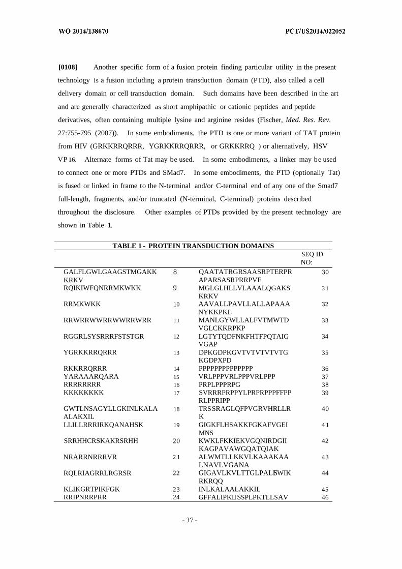
[0108] Another specific form of a fusion protein finding particular utility in the present
technology is a fusion including a protein transduction domain (PTD), also called a cell
delivery domain or cell transduction domain. Such domains have been described in the art
and are generally characterized as short amphipathic or cationic peptides and peptide
derivatives, often containing multiple lysine and arginine resides (Fischer, Med. Res. Rev.
27:755-795 (2007)). In some embodiments, the PTD is one or more variant of TAT protein
from HIV (GRKKRRQRRR, YGRKKRRQRRR, or GRKKRRQ ) or alternatively, HSV
VP 16. Alternate forms of Tat may be used. In some embodiments, a linker may be used
to connect one or more PTDs and SMad7. In some embodiments, the PTD (optionally Tat)
is fused or linked in frame to the N-terminal and/or C-terminal end of any one of the Smad7
full-length, fragments, and/or truncated (N-terminal, C-terminal) proteins described
throughout the disclosure. Other examples of PTDs provided by the present technology are
shown in Table 1.
TABLE 1 - PROTEIN TRANSDUCTION DOMAINSSEQ IDNO:
GALFLGWLGAAGSTMGAKK 8 QAATATRGRSAASRPTERPR 30KRKV APARSASRPRRPVERQIKIWFQNRRMKWKK 9 MGLGLHLLVLAAALQGAKS 3 1
KRKVRRMKWKK 10 AAVALLPAVLLALLAPAAA 32
NYKKPKLRRWRRWWRRWWRRWRR 1 1 MANLGYWLLALFVTMWTD 33
VGLCKKRPKPRGGRLSYSRRRFSTSTGR 12 LGTYTQDFNKFHTFPQTAIG 34
VGAPYGRKKRRQRRR 13 DPKGDPKGVTVTVTVTVTG 35
KGDPXPDRKKRRQRRR 14 PPPPPPPPPPPPPP 36YARAAARQARA 15 VRLPPPVRLPPPVRLPPP 37RRRRRRRR 16 PRPLPPPRPG 38KKKKKKKK 17 SVRRRPRPPYLPRPRPPPFFPP 39
RLPPRIPPGWTLNSAGYLLGKINLKALA 18 TRSSRAGLQFPVGRVHRLLR 40ALAKXIL KLLILLRRRIRKQANAHSK 19 GIGKFLHSAKKFGKAFVGEI 4 1
MNSSRRHHCRSKAKRSRHH 20 KWKLFKKIEKVGQNIRDGII 42
KAGPAVAWGQATQIAKNRARRNRRRVR 2 1 ALWMTLLKKVLKAAAKAA 43
LNAVLVGANARQLRIAGRRLRGRSR 22 GIGAVLKVLTTGLPALISWIK 44
RKRQQKLIKGRTPIKFGK 23 INLKALAALAKKIL 45RRIPNRRPRR 24 GFFALIPKIISSPLPKTLLSAV 46
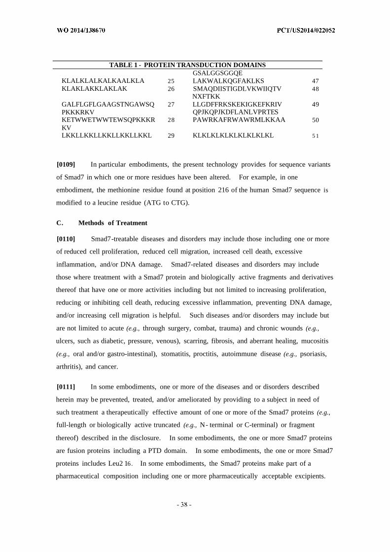
TABLE 1 - PROTEIN TRANSDUCTION DOMAINSGSALGGSGGQE
KLALKLALKALKAALKLA 25 LAKWALKQGFAKLKS 47KLAKLAKKLAKLAK 26 SMAQDIISTIGDLVKWIIQTV 48
NXFTKKGALFLGFLGAAGSTNGAWSQ 27 LLGDFFRKSKEKIGKEFKRIV 49PKKKRKV QPJKQPJKDFLANLVPRTESKETWWETWWTEWSQPKKKR 28 PAWRKAFRWAWRMLKKAA 50KVLKKLLKKLLKKLLKKLLKKL 29 KLKLKLKLKLKLKLKLKL 5 1
[0109] In particular embodiments, the present technology provides for sequence variants
of Smad7 in which one or more residues have been altered. For example, in one
embodiment, the methionine residue found at position 216 of the human Smad7 sequence is
modified to a leucine residue (ATG to CTG).
C. Methods of Treatment
[0110] Smad7-treatable diseases and disorders may include those including one or more
of reduced cell proliferation, reduced cell migration, increased cell death, excessive
inflammation, and/or DNA damage. Smad7-related diseases and disorders may include
those where treatment with a Smad7 protein and biologically active fragments and derivatives
thereof that have one or more activities including but not limited to increasing proliferation,
reducing or inhibiting cell death, reducing excessive inflammation, preventing DNA damage,
and/or increasing cell migration is helpful. Such diseases and/or disorders may include but
are not limited to acute (e.g., through surgery, combat, trauma) and chronic wounds (e.g.,
ulcers, such as diabetic, pressure, venous), scarring, fibrosis, and aberrant healing, mucositis
(e.g., oral and/or gastro-intestinal), stomatitis, proctitis, autoimmune disease (e.g., psoriasis,
arthritis), and cancer.
[0111] In some embodiments, one or more of the diseases and or disorders described
herein may be prevented, treated, and/or ameliorated by providing to a subject in need of
such treatment a therapeutically effective amount of one or more of the Smad7 proteins (e.g.,
full-length or biologically active truncated (e.g., N- terminal or C-terminal) or fragment
thereof) described in the disclosure. In some embodiments, the one or more Smad7 proteins
are fusion proteins including a PTD domain. In some embodiments, the one or more Smad7
proteins includes Leu2 16. In some embodiments, the Smad7 proteins make part of a
pharmaceutical composition including one or more pharmaceutically acceptable excipients.
[0112] In some embodiments, one or more of the diseases and or disorders described
herein may be prevented, treated, and/or ameliorated by providing to a subject in need of
such treatment a therapeutically effective amount of one or more of the nucleic acid
molecules encoding one or more Smad7 proteins (e.g., full-length or biologically active
truncated (e.g., N- terminal or C-terminal) or fragment thereof) described in the disclosure.
In some embodiments, the one or more nucleic acid molecules include codon-optimized
nucleotide sequences and/or sequences that encode Leu216. In some embodiments, the one
or more Smad7 nucleic acid molecules are provided to the subject in a construct including an
expression vector. In some embodiments, the Smad7 nucleic acid molecules (optionally
part of an expression vector) make part of a pharmaceutical composition including one or
more pharmaceutically acceptable excipients.
[0113] The term "subject" or "patient" as used herein refers to persons or non-human
animals in need of treatment and or prevention using one or more of the treatments described
herein. In some embodiments, non-human animals include laboratory animals such as
monkeys, mice, rats, and rabbits, domestic pets such as dogs and cats, and livestock such as
cattle, horses, pigs, goats and sheep.
1. Chronic Wounds
[0114] A chronic wound is a wound that does not heal in an orderly set of stages and in a
predictable amount of time the way most wounds do; wounds that do not heal within three
months are often considered chronic. Chronic wounds seem to be detained in one or more
of the phases of wound healing. For example, chronic wounds often remain in the
inflammatory stage for too long. In acute wounds, there is a precise balance between
production and degradation of molecules such as collagen; in chronic wounds this balance is
lost and degradation plays too large a role.
[0115] As described in more detail elsewhere herein, PTD-Smad7 has been shown to
enhance wound healing in a mouse skin model and a mucosal model. Application of
PTD-Smad7 was effective through a topical route, which is desirable for wound treatment.
Although not intending to be bound by theory, it is believed that PTD-Smad7 may act to treat
or ameliorate chronic wounds through multiple routes, which may include one or more of
reducing inflammation, increasing cell proliferation (e.g., keratinocytes), increasing cell
migration (e.g., keratinocytes), or reducing fibrosis (e.g., through modulation of collagen),
among others.

[0116] Chronic wounds may never heal or may take years to do so. These wounds
cause patients severe emotional and physical stress as well as creating a significant financial
burden on patients and the whole healthcare system. Acute and chronic wounds are at
opposite ends of a spectrum of wound healing types that progress toward being healed at
different rates. The vast majority of chronic wounds can be classified into three categories:
venous ulcers, diabetic, and pressure ulcers. A small number of wounds that do not fall into
these categories may be due to causes such as radiation poisoning or ischemia.
[0117] Venous and arterial ulcers. Venous ulcers, which usually occur in the legs,
account for about 70% to 90% of chronic wounds and mostly affect the elderly. They are
thought to be due to venous hypertension caused by improper function of valves that exist in
the veins to prevent blood from flowing backward. Ischemia results from the dysfunction
and, combined with reperfusion injury, causes the tissue damage that leads to the wounds.
[0118] Diabetic ulcers. Another major cause of chronic wounds, diabetes, is increasing
in prevalence. Diabetics have a 15% higher risk for amputation than the general population
due to chronic ulcers. Diabetes causes neuropathy, which inhibits nociception and the
perception of pain. Thus patients may not initially notice small wounds to legs and feet, and
may therefore fail to prevent infection or repeated injury. Further, diabetes causes immune
compromise and damage to small blood vessels, preventing adequate oxygenation of tissue,
which can cause chronic wounds. Pressure also plays a role in the formation of diabetic
ulcers.
[0119] Pressure ulcers. Another leading type of chronic wounds is pressure ulcers,
which usually occur in people with conditions such as paralysis that inhibit movement of
body parts that are commonly subjected to pressure such as the heels, shoulder blades, and
sacrum. Pressure ulcers are caused by ischemia that occurs when pressure on the tissue is
greater than the pressure in capillaries, and thus restricts blood flow into the area. Muscle
tissue, which needs more oxygen and nutrients than skin does, shows the worst effects from
prolonged pressure. As in other chronic ulcers, reperfusion injury damages tissue.
[0120] Chronic wounds may affect only the epidermis and dermis, or they may affect
tissues all the way to the fascia. They may be formed originally by the same things that
cause acute wounds, such as surgery or accidental trauma, or they may form as the result of
systemic infection, vascular, immune, or nerve insufficiency, or comorbidities such as
neoplasias or metabolic disorders. Although not intending to be bound by theory, the reason
a wound becomes chronic is that the body's ability to deal with the damage is overwhelmed
by factors such as repeated trauma, continued pressure, ischemia, or illness. Some of the
major factors that lead to chronic wounds include, but are not limited to, ischemia,
reperfusion injury, and bacterial colonization.
[0121] Ischemia. Ischemia is an important factor in the formation and persistence of
wounds, especially when it occurs repetitively (as it usually does) or when combined with a
patient's old age. Ischemia causes tissue to become inflamed and cells to release factors that
attract neutrophils such as interleukins, chemokines, leukotrienes, and complement factors.
[0122] While they fight pathogens, neutrophils also release inflammatory cytokines and
enzymes that damage cells. One of their important functions is to produce Reactive Oxygen
Species (ROS) to kill bacteria, for which they use an enzyme called myeloperoxidase. The
enzymes and ROS produced by neutrophils and other leukocytes damage cells and prevent
cell proliferation and wound closure by damaging DNA, lipids, proteins, the ECM, and
cytokines that speed healing. Neutrophils remain in chronic wounds for longer than they do
in acute wounds, and contribute to the fact that chronic wounds have higher levels of
inflammatory cytokines and ROS. Because wound fluid from chronic wounds has an excess
of proteases and ROS, the fluid itself can inhibit healing by inhibiting cell growth and
breaking down growth factors and proteins in the ECM.
[0123] Bacterial colonization. Since more oxygen in the wound environment allows
white blood cells to produce ROS to kill bacteria, patients with inadequate tissue
oxygenation, for example, those who suffered hypothermia during surgery, are at higher risk
for infection. The host's immune response to the presence of bacteria prolongs
inflammation, delays healing, and damages tissue. Infection can lead not only to chronic
wounds but also to gangrene, loss of the infected limb, and death of the patient.
[0124] Like ischemia, bacterial colonization and infection damage tissue by causing a
greater number of neutrophils to enter the wound site. In patients with chronic wounds,
bacteria with resistance to antibiotics may have time to develop. In addition, patients
carrying drug resistant bacterial strains, such as methicillin-resistant Staphylococcus aureus
(MRSA), have more chronic wounds.

[0125] Growth factors and proteolytic enzymes. Chronic wounds also differ in makeup
from acute wounds in that their levels of proteolytic enzymes such as elastase and matrix
metalloproteinases (MMPs) are higher, while their concentrations of growth factors such as
Platelet-derived growth factor and Keratinocyte Growth Factor are lower.
[0126] Since growth factors (GFs) are imperative in timely wound healing, inadequate
GF levels may be an important factor in chronic wound formation. In chronic wounds, the
formation and release of growth factors may be prevented, the factors may be sequestered
and unable to perform their metabolic roles, or degraded in excess by cellular or bacterial
proteases.
[0127] Chronic wounds such as diabetic and venous ulcers are also caused by a failure of
fibroblasts to produce adequate ECM proteins and by keratinocytes to epithelialize the
wound. Fibroblast gene expression is different in chronic wounds than in acute wounds.
[0128] Although all wounds require a certain level of elastase and proteases for proper
healing, too high a concentration is damaging. Leukocytes in the wound area release
elastase, which increases inflammation, destroys tissue, proteoglycans, and collagen, and
damages growth factors, fibronectin, and factors that inhibit proteases. The activity of
elastase is increased by human serum albumin, which is the most abundant protein found in
chronic wounds. However, chronic wounds with inadequate albumin are especially unlikely
to heal, so regulating the wound's levels of that protein may in the future prove helpful in
healing chronic wounds.
[0129] Excess matrix metalloproteinases, which are released by leukocytes, may also
cause wounds to become chronic. MMPs break down ECM molecules, growth factors, and
protease inhibitors, and thus increase degradation while reducing construction, throwing the
delicate compromise between production and degradation out of balance.
[0130] Oral ulcers. A mouth ulcer (also termed an oral ulcer, or a mucosal ulcer) is an
ulcer that occurs on the mucous membrane of the oral cavity. More plainly, a mouth ulcer is
a sore or open lesion in the mouth. Mouth ulcers are very common, occurring in association
with many diseases and by many different mechanisms, but usually there is no serious
underlying cause. The two most common causes of oral ulceration are local trauma (e.g. ,
rubbing from a sharp edge on a filling) and aphthous stomatitis ("canker sores"), a condition
characterized by recurrent formation of oral ulcers for largely unknown reasons. Some

consider ulcers on the lips or on the skin around the mouth to be included under the general
term oral ulceration (e.g., an ulcer left by rupture of a blister caused by herpes labialis, i.e., a
cold sore). Mouth ulcers often cause pain and discomfort, and may alter the person's choice
of food while healing occurs (e.g., avoiding acidic or spicy foods and beverages). They may
occur singly or multiple ulcers may occur at the same time (a "crop" of ulcers). Once
formed, the ulcer may be maintained by inflammation and/or secondary infection. Rarely, a
mouth ulcer that does not heal for many weeks may be a sign of oral cancer. Other causes
include burns, chemical injury, or infection.
[0131] A mucosal ulcer is an ulcer which specifically occurs on a mucous membrane.
An ulcer is a tissue defect which has penetrated the epithelial-connective tissue border, with
its base at a deep level in the submucosa, or even within muscle or periosteum. An ulcer is
a deeper breech of the epithelium than an erosion or an excoriation, and involves damage to
both epithelium and lamina propria. An erosion is a superficial breach of the epithelium,
with little damage to the underlying lamina propria. A mucosal erosion is an erosion which
specifically occurs on a mucous membrane. Only the superficial epithelial cells of the
epidermis or of the mucosa are lost, and the lesion can reach the depth of the basement
membrane. Erosions heal without scar formation. Excoriation is a term sometimes used to
describe a breach of the epithelium which is deeper than an erosion but shallower than an
ulcer. This type of lesion is tangential to the rete pegs and shows punctiform (small pinhead
spots) bleeding, caused by exposed capillary loops.
2. Acute Wounds/Trauma
[0132] Physical trauma is a serious and body-altering physical injury, such as the
removal of a limb. Blunt force trauma, a type of physical trauma caused by impact or other
force applied from or with a blunt object, whereas penetrating trauma is a type of physical
trauma in which the skin or tissues are pierced by an object. Trauma can also be described
as both unplanned, such as an accident, or planned, in the case of surgery. Both can be
characterized by mild to severe tissue damage, blood loss and/or shock, and both may lead to
subsequent infection, including sepsis. The present technology provides for treatment of
trauma, including both pre-treatment (in the case of a medical procedure) and treatment after
trauma injury has occurred.
[0133] As described in more detail elsewhere herein (and briefly mentioned above),
PTD-Smad7 has been shown to enhance wound healing in a mouse skin model and a mucosal

model. Application of PTD-Smad7 was effective through a topical route, which is desirable
for wound treatment. Although not intending to be bound by theory, it is believed that
PTD-Smad7 may act to treat or ameliorate wounds through multiple routes, which may
include one or more of reducing inflammation, increasing cell proliferation (e.g.,
keratinocytes), increasing cell migration (e.g., keratinocytes), or reducing fibrosis (e.g.,
through modulation of collagen), among others. As described briefly below, reduced
inflammation could significantly contribute to accelerated wound healing, optionally through
reduced angiogenesis and collagen production and/or reduced leukocyte infiltration leading to
reduction of cytokines and chemokines normally released by leukocytes, which are
angiogenic and fibrogenic. Temporal treatment with Smad7 may allow early stage
angiogenesis and collagen production required for wound repair, while preventing prolonged
angiogenesis and collagen production. These changes could potentially accelerate wound
stromal remodeling and prevent excessive scarring due to unresolved inflammation or
collagen overproduction. For surgical procedures (as well as everyday injuries), particularly
where the potential for scarring is an issue, treatment with Smad7 may be beneficial.
[0134] Surgery. Surgery uses operative manual and instrumental techniques on a
patient to investigate and/or treat a pathological condition such as disease or injury, to help
improve bodily function or appearance, or sometimes for some other reason. The present
technology can address trauma resulting from surgeries, as defined further below.
[0135] As a general rule, a procedure is considered surgical when it involves cutting of a
patient's tissues or closure of a previously sustained wound. Other procedures that do not
necessarily fall under this rubric, such as angioplasty or endoscopy, may be considered
surgery if they involve common surgical procedure or settings, such as use of a sterile
environment, anesthesia, antiseptic conditions, typical surgical instruments, and suturing or
stapling. All forms of surgery are considered invasive procedures; so-called noninvasive
surgery usually refers to an excision that does not penetrate the structure being addressed
(e.g., laser ablation of the cornea) or to a radiosurgical procedure (e.g., irradiation of a
tumor). Surgery can last from minutes to hours.
[0136] Surgical procedures are commonly categorized by urgency, type of procedure,
body system involved, degree of invasiveness, and special instrumentation. Elective surgery
is done to correct a non-life-threatening condition, and is carried out at the patient's request,
subject to the surgeon's and the surgical facility's availability. Emergency surgery is surgery

which must be done quickly to save life, limb, or functional capacity. Exploratory surgery
is performed to aid or confirm a diagnosis. Therapeutic surgery treats a previously
diagnosed condition.
[0137] Amputation involves cutting off a body part, usually a limb or digit.
Replantation involves reattaching a severed body part. Reconstructive surgery involves
reconstruction of an injured, mutilated, or deformed part of the body. Cosmetic surgery is
done to improve the appearance of an otherwise normal structure. Excision is the cutting
out of an organ, tissue, or other body part from the patient. Transplant surgery is the
replacement of an organ or body part by insertion of another from different human (or
animal) into the patient. Removing an organ or body part from a live human or animal for
use in transplant is also a type of surgery.
[0138] When surgery is performed on one organ system or structure, it may be classified
by the organ, organ system or tissue involved. Examples include cardiac surgery
(performed on the heart), gastrointestinal surgery (performed within the digestive tract and its
accessory organs), and orthopedic surgery (performed on bones and/or muscles).
[0139] Minimally invasive surgery involves smaller outer incision(s) to insert
miniaturized instruments within a body cavity or structure, as in laparoscopic surgery or
angioplasty. By contrast, an open surgical procedure requires a large incision to access the
area of interest. Laser surgery involves use of a laser for cutting tissue instead of a scalpel
or similar surgical instruments. Microsurgery involves the use of an operating microscope
for the surgeon to see small structures. Robotic surgery makes use of a surgical robot, such
as Da Vinci or Zeus surgical systems, to control the instrumentation under the direction of the
surgeon.
3. Autoimmune/Inflammatory Disease
[0140] The present technology contemplates the treatment of a variety of autoimmune
and/or inflammatory disease states such as spondyloarthropathy, ankylosing spondylitis,
psoriatic arthritis, reactive arthritis, enteropathic arthritis, ulcerative colitis, Crohn's disease,
irritable bowel disease, inflammatory bowel disease, rheumatoid arthritis, juvenile rheumatoid
arthritis, familial Mediterranean fever, amyotrophic lateral sclerosis, Sjogren's syndrome,
early arthritis, viral arthritis, multiple sclerosis, or psoriasis. The diagnosis and treatment of
these diseases are well documented in the literature.

[0141] In general, autoimmune diseases are associated with an overactive immune
response of a body against substances and tissues normally present in the body, and not
normally the focus of an immune response. There are more than 80 types of autoimmune
diseases, some of which have similar symptoms, and they may arise from a similar underlying
cause. The classic sign of an autoimmune disease is inflammation, which as disclosed herein
is amenable to treatment with Smad7 (optionally PTD-Smad7) compositions.
4. Chemotherapy, Radiotherapy and Cytokine Therapy Toxicity
[0142] Various forms of cancer therapy, including chemotherapy, radiation, and
cytokines, are associated with toxicity, sometimes severe, in the cancer patient. The present
technology seeks to reduce this toxicity using the pharmaceutical compositions of the present
technology, thereby reducing or alleviating discomfort on the part of the patient, as well as
permitting higher doses of the therapy.
[0143] As described at length throughout this disclosure, it has been found that
PTD-Smad7 acts to heal as well as to prevent oral mucositis in a mouse model.
PTD-Smad7 was shown to be more effective than palifermin, the existing approved drug for
preventing oral mucositis, in direct comparisons.
[0144] Oral cancer, the 6th most common cancer worldwide, is a subtype of head and
neck cancer, and includes any cancerous tissue growth located in the oral cavity. It may
arise as a primary lesion originating in any of the oral tissues, by metastasis from a distant
site of origin, or by extension from a neighboring anatomic structure, such as the nasal cavity
or the oral cancers may originate in any of the tissues of the mouth, and may be of varied
histologic types: teratoma, adenocarcinoma derived from a major or minor salivary gland,
lymphoma from tonsillar or other lymphoid tissue, or melanoma from the pigment-producing
cells of the oral mucosa. There are several types of oral cancers, but around 90% are
squamous cell carcinomas, originating in the tissues that line the mouth and lips. Oral or
mouth cancer most commonly involves the tongue. It may also occur on the floor of the
mouth, cheek lining, gingiva (gums), lips, or palate (roof of the mouth). Most oral cancers
look very similar under the microscope and are called squamous cell carcinoma. These are
malignant and tend to spread rapidly.
[0145] Over 80% of oral cancer patients are treated with radiation therapy and at least
75% of these individuals will develop oral mucositis. Oral mucositis is a chronic oral
ulceration. This disease frequently occurs in radiation-treated patients of all cancer types,
including but not limited to patients who are radiation-treated for organ transplants (to
eliminate rejection of the transplants), and patients undergoing routine chemotherapy.
Severe oral mucositis is extremely painful and impairs food/liquid intake, hence is often the
most severe complication of cancer therapy. Oral mucositis is a major factor in determining
the maximum dose possible of radiation and chemotherapy to the head and neck region; it
can significantly complicate cancer treatment, extend hospitalization, decrease quality of life
and increase costs.
[0146] Currently, there is no established therapy to effectively treat severe oral
mucositis. To date, palifermin (KEPIVANCE®), a recombinant protein of human
keratinocyte growth factor (KGF), is the only FDA approved drug for intravenous (i.v.)
injections for severe oral mucositis in bone-marrow transplant patients, and its use in cancer
patients remains to be determined. It is also used for prevention of oral mucositis. Hence,
this drug is available for only 4% of the at-risk population. It also suffers from the need for
medical service providers due to the i.v. administration route. Other potential therapies
include topical rinses, such as viscous 2% lidocaine rinses, or baking soda and saline
solutions, or a cocktail solution, for instance BAX (lidocaine, diphenhyramine, sorbitol and
MYLANTA®). Other investigative or mucoprotective adjuvant therapies include, but are
not limited to, beta carotene, tocopherol, laser irradiation, prophylactic brushing the oral
mucosa with silver-nitrate, misoprostol, leucovorin, systemic KGF, pentoxifylline,
allopurinol mouthwash, systemic sucralfate, chlorhexidine gluconate, and cryotherapy.
[0147] Chemotherapy- and radiation-induced gut mucositis is an inflammatory condition
that arises as a result of the acute death of rapidly dividing intestinal epithelial cells. Most
chemotherapeutic drugs used for treatment of solid tumors, alone, in a combination of drugs,
or with radiation, will result in the death of a large number of intestinal epithelial cells. The
clinical manifestations of the ensuing mucositis include digestive symptoms such as nausea
and vomiting, serious diarrhea, acute weight loss and wasting. This is fast becoming one of
the limiting factors for administering chemotherapy for many cancer patients. The ability of
Tat-Smad7 to protect intestinal epithelial cells from either chemotherapeutic agents,
radiation, or a combinations of those, will significantly decrease the undesirable side effects
of cancer therapies, and enable more aggressive ways to treat the disease with existing tools.
[0148] Bone marrow failure syndromes are a set of conditions that develop when the
hematopoietic stem cell compartment is compromised and fails to give rise to normal cell
types. Bone marrow failure occurs as a result of inherited genetic abnormalities, exposure
to a noxious substance, such as toxins, chemicals or viruses. Although the nature and
identity of environmental factors that can lead to the development of acquired bone marrow
failure is still not completely understood, a few factors have been linked to the development
of acquired bone marrow failure among military personnel including exposure to mustard
gas, ionizing radiation, and infectious agents such as visceral leishmaniasis or African
trypanosomiasis. The best approach for management of bone marrow failure syndromes is
still the transplantation of hematopoietic stem cells (HSCs), unless a sufficient number of the
remaining resident bone marrow HSCs can be spared from these stresses and encouraged to
repopulate the hematopoietic compartment. The modulation of Smad 7, as described here,
should enable for the deliberate protection of the remaining resident HSCs in patients that
exhibit clinical signs consistent with bone marrow failure.
5. Cancer
[0149] TGF-β and NF-κΒ activations are known to promote cancer invasion and
metastasis. Currently, TGF-β inhibitors are in clinical trials for treating metastatic cancer and
NF-KB inhibitors are used in cancer prevention. The demonstrated effect of Smad7 on
blocking both TGF-β and NF-κΒ signaling present the possibility that it is an even stronger
anti-cancer/anti-metastasis agent than other inhibitors that inhibit only one of these two
pathways. Smad7 has been shown to prevent angiogenesis and fibrogenesis, and may
therefore be particularly useful in situations where the tumor needs to develop a blood supply
and/or stroma.
[0150] The cancer may be selected from the group consisting of brain, lung, liver, spleen,
kidney, lymph node, small intestine, pancreas, blood cells, colon, stomach, breast,
endometrium, prostate, testicle, cervix, uterus, ovary, skin, head & neck, esophagus, bone
marrow and blood cancer. The cancers may be metastatic or primary, recurrent or
multi-drug resistant. In some embodiments, the cancer is a solid tumor (organ tumor).
Solid tumors refer to a mass of cells that grow in organ systems and can occur anywhere in
the body. Two types of solid tumors include epithelial tumors (carcinomas) that occur in the
epithelial tissue inside or outside an organ, and sarcomas (connective tissue tumors) that
occur in connective tissue such as, but not limited to, muscles, tendons, fat, nerves and other

connective tissues that support, surround, or connect structures and organs in the body. In
some embodiments the cancer is a liquid tumor or cancer of the blood, bone marrow, or
lymph nodes. These tumors include, but are not limited to, leukemia, lymphoma, and
myeloma.
6. Scarring, Fibrosis, and Aberrant Healing
[0151] In addition to accelerated re-epithelialization (e.g., through increasing cell
proliferation and/or increasing cell migration), Smad7 effects on wound stroma include one
or more of reducing inflammation, angiogenesis, or collagen production, among others.
Although not intending to be bound by theory these effects may be mediated through
reduction of NF-κΒ signaling (evidenced by reduced p50), and blocking TGF-β signaling
(evidenced by reduced pSmad2). As a result, reduced inflammation could significantly
contribute to accelerated wound healing, optionally through reduced angiogenesis and
collagen production and/or reduced leukocyte infiltration leading to reduction of cytokines
and chemokines normally released by leukocytes, which are angiogenic and fibrogenic.
Temporal treatment with Smad7 may allow early stage angiogenesis and collagen production
required for wound repair, while preventing prolonged angiogenesis and collagen production.
These changes could potentially accelerate wound stromal remodeling and prevent excessive
scarring due to unresolved inflammation or collagen overproduction.
7. Stomatitis
[0152] Stomatitis is an inflammation of the mucous lining of any of the structures in the
mouth, which may involve the cheeks, gums, tongue, lips, throat, and roof or floor of the
mouth. The inflammation can be caused by conditions in the mouth itself, such as poor oral
hygiene, dietary protein deficiency, poorly fitted dentures, or from mouth burns from hot
food or drinks, toxic plants, or by conditions that affect the entire body, such as medications,
allergic reactions, radiation therapy, or infections. Severe iron deficiency anemia can lead
to stomatitis. Iron is necessary for the upregulation of transcriptional elements for cell
replication and repair. Lack of iron can cause the genetic downregulation of these elements,
leading to ineffective repair and regeneration of epithelial cells, especially in the mouth and
lips. This condition is also prevalent in people who have a deficiency in vitamin B 2
(Riboflavin), B 3 (Niacin), B (Pyridoxine), B 9 (folic acid) or B12 (cobalamine). When it also
involves an inflammation of the gingiva (gums), it is called gingivostomatitis. It may also
be seen in ariboflavinosis (riboflavin deficiency) or neutropenia.

[0153] Irritation and fissuring in the corners of the lips is termed angular stomatitis or
angular cheilitis. In children, angular stomatitis is a frequent cause is repeated lip-licking
and in adults it may be a sign of underlying iron deficiency anemia, or vitamin B deficiencies
(e.g., B 2-riboflavin, Bg-folate or B^-cobalamin), which in turn may be evidence of poor diets
or malnutrition (e.g., celiac disease). Also, angular cheilitis can be caused by a patient's
jaws at rest being "overdosed" due to edentulousness or tooth wear, causing the jaws to come
to rest closer together than if the complete/unaffected dentition were present. This causes
skin folds around the angle of the mouth which are kept moist by saliva which in turn favours
infection; mostly by Candida albicans or similar species. Treatment usually involves the
administration of topical nystatin or similar antifungal agents. Another treatment can be to
correct the jaw relationship with dental treatment (e.g., dentures or occlusal adjustment).
[0154] Migratory stomatitis is a condition in which extensive areas in the oral cavity
mucosa are affected by annular atrophic red lesions that are surrounded by a thin white rim.
This is a relatively uncommon form of the geographic tongue condition, that, as opposed to
migratory stomatitis, is confined to the dorsal and lateral aspects of the tongue mucosa only.
8. Proctitis
[0155] Proctitis is inflammation of the lining of the rectum, the lower end of the large
intestine leading to the anus. With proctitis, inflammation of the rectal lining—called the
rectal mucosa—is uncomfortable and sometimes painful. The condition may lead to bleeding
or mucous discharge from the rectum, among other symptoms. Some causes of proctitis
include, but are not limited to: sexually transmitted diseases (STDs), such as those that can be
transmitted during anal sex (e.g., gonorrhea, chlamydia, syphilis, and herpes); non-STD
infections from, for example, food borne bacteria (e.g., Salmonella and Shigella); anorectal
trauma from, for example, anal sex or the insertion of objects or substances into the rectum
(e.g., chemicals from enemas); ulcerative colitis and Crohn's disease or other inflammatory
bowel diseases, may cause ulcers (e.g., sores) in the inner lining of the colon and rectum;
radiation therapy, particularly of the pelvic area (e.g., rectal, ovarian, or prostate cancer) which
may lead to rectal bleeding; antibiotics which lead to a loss of commensal bacteria allowing
harmful bacteria (e.g., Clostridium difficile) to cause disease.

9. Formulations and Routes of Administration
[0156] Where clinical applications are contemplated, it will be necessary to prepare
pharmaceutical compositions - proteins, expression vectors, virus stocks, proteins and drugs
- in a form appropriate for the intended application. Generally, this will entail preparing
compositions that are essentially free of pyrogens, as well as other impurities that could be
harmful to humans or animals.
[0157] PTD-Smad7 (and truncated variants) were purified extensively prior to use in
animal models. PTD-Smad7 (and truncated versions) were prepared for topical and
trans-mucosal application using a mixture of glycerol and PBS.
[0158] One will generally desire to employ appropriate salts and buffers to render
delivery vectors stable and allow for uptake by target cells. Buffers also will be employed
when recombinant cells are introduced into a patient. Aqueous compositions of the present
technology comprise an effective amount of the vector to cells, dissolved or dispersed in a
pharmaceutically acceptable carrier or aqueous medium. Such compositions also are
referred to as inocula. The phrase "pharmaceutically or pharmacologically acceptable" refer
to molecular entities and compositions that do not produce adverse, allergic, or other
untoward reactions when administered to an animal or a human. As used herein,
"pharmaceutically acceptable carrier" includes any and all solvents, dispersion media,
coatings, antibacterial and antifungal agents, isotonic and absorption delaying agents and the
like. The use of such media and agents for pharmaceutically active substances is well
known in the art. Except insofar as any conventional media or agent is incompatible with
the vectors or cells of the present technology, its use in therapeutic compositions is
contemplated. Supplementary active ingredients also can be incorporated into the
compositions.
[0159] The active compositions of the present technology may include classic
pharmaceutical preparations. Administration of these compositions according to the present
technology will be via any common route so long as the target tissue is available via that
route. Such routes of administration may include oral parenteral (including intravenous,
intramuscular, subcutaneous, intradermal, intra-articular, intra-synovial, intrathecal,
intra-arterial, intracardiac, subcutaneous, intraorbital, intracapsular, intraspinal, intrastemal,
and transdermal), nasal, buccal, urethral, rectal, vaginal, mucosal, dermal, or topical
(including dermal, buccal, and sublingual). Alternatively, administration may be by
orthotopic, intradermal, subcutaneous, intramuscular, intraperitoneal or intravenous injection.
Such compositions would normally be administered as pharmaceutically acceptable
compositions, described supra. Of particular interest is direct intratumoral administration,
perfusion of a tumor, or administration local or regional to a tumor, for example, in the local
or regional vasculature or lymphatic system, or in a resected tumor bed. Administration can
also be via nasal spray, surgical implant, internal surgical paint, infusion pump, or via catheter,
stent, balloon or other delivery device. The most useful and/or beneficial mode of
administration can vary, especially depending upon the condition of the recipient and the
disorder being treated.
[0160] Solutions of the active compounds as free base or pharmacologically acceptable
salts can be prepared in water suitably mixed with a surfactant, such as
hydroxypropylcellulose. Dispersions can also be prepared in glycerol, liquid polyethylene
glycols, and mixtures thereof and in oils. Under ordinary conditions of storage and use,
these preparations contain a preservative to prevent the growth of microorganisms.
[0161] The pharmaceutical forms suitable for injectable use include sterile aqueous
solutions or dispersions and sterile powders for the extemporaneous preparation of sterile
injectable solutions or dispersions. In all cases, the form must be sterile and must be fluid to
the extent that easy syringability exists. It must be stable under the conditions of
manufacture and storage and must be preserved against the contaminating action of
microorganisms, such as bacteria and fungi. The carrier can be a solvent or dispersion
medium containing, for example, water, ethanol, polyol (e.g., glycerol, propylene glycol, and
liquid polyethylene glycol, and the like), suitable mixtures thereof, and vegetable oils. The
proper fluidity can be maintained, for example, by the use of a coating, such as lecithin, by
the maintenance of the required particle size in the case of dispersion and by the use of
surfactants. The prevention of the action of microorganisms can be brought about by
various antibacterial an antifungal agents, for example, parabens, chlorobutanol, phenol,
sorbic acid, thimerosal, and the like. In many cases, it will be preferable to include isotonic
agents, for example, sugars or sodium chloride. Prolonged absorption of the injectable
compositions can be brought about by the use in the compositions of agents delaying
absorption, for example, aluminum monostearate and gelatin.
[0162] Sterile injectable solutions are prepared by incorporating the active compounds in
the required amount in the appropriate solvent with various of the other ingredients
enumerated above, as required, followed by filtered sterilization. Generally, dispersions are
prepared by incorporating the various sterilized active ingredients into a sterile vehicle which
contains the basic dispersion medium and the required other ingredients from those
enumerated above. In the case of sterile powders for the preparation of sterile injectable
solutions, the preferred methods of preparation are vacuum-drying and freeze-drying
techniques which yield a powder of the active ingredient plus any additional desired
ingredient from a previously sterile-filtered solution thereof.
[0163] As used herein, "pharmaceutically acceptable carrier" includes any and all
solvents, dispersion media, coatings, antibacterial and antifungal agents, isotonic and
absorption delaying agents and the like. The use of such media and agents for
pharmaceutical active substances is well known in the art. Except insofar as any
conventional media or agent is incompatible with the active ingredient, its use in the
therapeutic compositions is contemplated. Supplementary active ingredients can also be
incorporated into the compositions.
[0164] The compositions of the present technology may be formulated in a neutral or salt
form. Pharmaceutically-acceptable salts include the acid addition salts (formed with the
free amino groups of the protein) and which are formed with inorganic acids such as, for
example, hydrochloric or phosphoric acids, or such organic acids as acetic, oxalic, tartaric,
mandelic, and the like. Salts formed with the free carboxyl groups can also be derived from
inorganic bases such as, for example, sodium, potassium, ammonium, calcium, or ferric
hydroxides, and such organic bases as isopropylamine, trimethylamine, histidine, procaine
and the like.
[0165] The formulations are easily administered in a variety of dosage forms. Some
variation in dosage will necessarily occur depending on the condition of the subject being
treated. The person responsible for administration will, in any event, determine the
appropriate dose for the individual subject. Moreover, for human administration,
preparations should meet sterility, pyrogenicity, general safety and purity standards as
required by FDA Office of Biologies standards.
[0166] For oral administration the polypeptides of the present technology may be
incorporated with excipients and used in the form of non-ingestible mouthwashes and
dentifrices. It is anticipated that virtually any pill or capsule type known to one of skill in

the art including, e.g., coated, and time delay, slow release, etc., may be used with the present
technology. A mouthwash may be prepared incorporating the active ingredient in the
required amount in an appropriate solvent, such as a sodium borate solution (Dobell's
Solution). Alternatively, the active ingredient may be incorporated into an antiseptic wash
containing sodium borate, glycerin and potassium bicarbonate. The active ingredient may
also be dispersed in dentifrices, including: gels, pastes, creams, powders and slurries. The
active ingredient may be added in a therapeutically effective amount to a paste dentifrice that
may include water, binders, abrasives, flavoring agents, foaming agents, and humectants.
[0167] Pharmaceutical compositions suitable for oral dosage may take various forms, such
as tablets, capsules, caplets, and wafers (including rapidly dissolving or effervescing), each
containing a predetermined amount of the active agent. The compositions may also be in the
form of a powder or granules, a solution or suspension in an aqueous or non-aqueous liquid,
and as a liquid emulsion (oil-in-water and water-in-oil). The active agents may also be
delivered as a bolus, electuary, or paste. It is generally understood that methods of
preparations of the above dosage forms are generally known in the art, and any such method
would be suitable for the preparation of the respective dosage forms for use in delivery of the
compositions.
[0168] In one embodiment, an active agent compound may be administered orally in
combination with a pharmaceutically acceptable vehicle such as an inert diluent or an edible
carrier. Oral compositions may be enclosed in hard or soft shell gelatin capsules, may be
compressed into tablets or may be incorporated directly with the food of the patient's diet. The
percentage of the composition and preparations may be varied; however, the amount of
substance in such therapeutically useful compositions is preferably such that an effective
dosage level will be obtained.
[0169] Hard capsules containing the active agent compounds may be made using a
physiologically degradable composition, such as gelatin. Such hard capsules comprise the
compound, and may further comprise additional ingredients including, for example, an inert
solid diluent such as calcium carbonate, calcium phosphate, or kaolin. Soft gelatin capsules
containing the compound may be made using a physiologically degradable composition, such
as gelatin. Such soft capsules comprise the compound, which may be mixed with water or an
oil medium such as peanut oil, liquid paraffin, or olive oil.
[0170] Sublingual tablets are designed to dissolve very rapidly. Examples of such
compositions include ergotamine tartrate, isosorbide dinitrate, and isoproterenol HCL. The
compositions of these tablets contain, in addition to the drug, various soluble excipients, such
as lactose, powdered sucrose, dextrose, and mannitol. The solid dosage forms of the present
technology may optionally be coated, and examples of suitable coating materials include, but
are not limited to, cellulose polymers (such as cellulose acetate phthalate, hydroxypropyl
cellulose, hydroxypropyl methylcellulose, hydroxypropyl methylcellulose phthalate, and
hydroxypropyl methylcellulose acetate succinate), polyvinyl acetate phthalate, acrylic acid
polymers and copolymers, and methacrylic resins (such as those commercially available under
the trade name EUDRAGIT ®), zein, shellac, and polysaccharides.
[0171] Powdered and granular compositions of a pharmaceutical preparation may be
prepared using known methods. Such compositions may be administered directly to a patient
or used in the preparation of further dosage forms, such as to form tablets, fill capsules, or
prepare an aqueous or oily suspension or solution by addition of an aqueous or oily vehicle
thereto. Each of these compositions may further comprise one or more additives, such as
dispersing or wetting agents, suspending agents, and preservatives. Additional excipients
(e.g., fillers, sweeteners, flavoring, or coloring agents) may also be included in these
compositions.
[0172] Liquid compositions of pharmaceutical compositions which are suitable for oral
administration may be prepared, packaged, and sold either in liquid form or in the form of a dry
product intended for reconstitution with water or another suitable vehicle prior to use.
[0173] A tablet containing one or more active agent compounds described herein may be
manufactured by any standard process readily known to one of skill in the art, such as, for
example, by compression or molding, optionally with one or more adjuvant or accessory
ingredient. The tablets may optionally be coated or scored and may be formulated so as to
provide slow or controlled release of the active agents.
[0174] Solid dosage forms may be formulated so as to provide a delayed release of the
active agents, such as by application of a coating. Delayed release coatings are known in the
art, and dosage forms containing such may be prepared by any known suitable method. Such
methods generally include that, after preparation of the solid dosage form (e.g., a tablet or
caplet), a delayed release coating composition is applied. Application can be by methods,
such as airless spraying, fluidized bed coating, use of a coating pan, or the like. Materials for
use as a delayed release coating can be polymeric in nature, such as cellulosic material (e.g.,
cellulose butyrate phthalate, hydroxypropyl methylcellulose phthalate, and carboxymethyl
ethylcellulose), and polymers and copolymers of acrylic acid, methacrylic acid, and esters
thereof.
[0175] Solid dosage forms according to the present technology may also be sustained
release (i.e., releasing the active agents over a prolonged period of time), and may or may not
also be delayed release. Sustained release compositions are known in the art and are generally
prepared by dispersing a drug within a matrix of a gradually degradable or hydrolyzable
material, such as an insoluble plastic, a hydrophilic polymer, or a fatty compound.
Alternatively, a solid dosage form may be coated with such a material.
[0176] Compositions for parenteral administration include aqueous and non-aqueous
sterile injection solutions, which may further contain additional agents, such as antioxidants,
buffers, bacteriostats, and solutes, which render the compositions isotonic with the blood of the
intended recipient. The compositions may include aqueous and non-aqueous sterile
suspensions, which contain suspending agents and thickening agents. Such compositions for
parenteral administration may be presented in unit-dose or multi-dose containers, such as, for
example, sealed ampoules and vials, and may be stores in a freeze-dried (lyophilized) condition
requiring only the addition of the sterile liquid carrier, for example, water (for injection),
immediately prior to use. Extemporaneous injection solutions and suspensions may be
prepared from sterile powders, granules, and tablets of the kind previously described.
[0177] Compositions for rectal delivery include rectal suppositories, creams, ointments,
and liquids. Suppositories may be presented as the active agents in combination with a carrier
generally known in the art, such as polyethylene glycol. Such dosage forms may be designed
to disintegrate rapidly or over an extended period of time, and the time to complete
disintegration can range from a short time, such as about 10 minutes, to an extended period of
time, such as about 6 hours.
[0178] Topical compositions may be in any form suitable and readily known in the art for
delivery of active agents to the body surface, including dermally, buccally, and sublingually.
Typical examples of topical compositions include ointments, creams, gels, pastes, and
solutions. Compositions for administration in the mouth include lozenges.

[0179] In accordance with these embodiments, oral (topical, mucosal, and/or dermal)
delivery materials can also include creams, salves, ointments, patches, liposomes,
nanoparticles, microparticles, timed-release formulations and other materials known in the art
for delivery to the oral cavity, mucosa, and/or to the skin of a subject for treatment and/or
prevention of a condition disclosed herein. Certain embodiments concern the use of a
biodegradable oral (topical, mucosal, and/or dermal) patch delivery system or gelatinous
material. These compositions can be a liquid formulation or a pharmaceutically acceptable
delivery system treated with a formulation of these compositions, and may also include
activator/inducers .
[0180] The compositions for use in the methods of the present technology may also be
administered transdermally, wherein the active agents are incorporated into a laminated
structure (generally referred to as a "patch") that is adapted to remain in intimate contact with
the epidermis of the recipient for a prolonged period of time. Typically, such patches are
available as single layer "drug-in-adhesive" patches or as multi-layer patches where the active
agents are contained in a layer separate from the adhesive layer. Both types of patches also
generally contain a backing layer and a liner that is removed prior to attachment to the
recipient's skin. Transdermal drug delivery patches may also be comprised of a reservoir
underlying the backing layer that is separated from the skin of the recipient by a
semi-permeable membrane and adhesive layer. Transdermal drug delivery may occur
through passive diffusion, electrotransport, or iontophoresis.
[0181] In certain embodiments, a patch contemplated herein may be a slowly dissolving
or a time-released patch. In accordance with these embodiments, a slowly dissolving patch
can be an alginate patch. In certain examples, a patch may contain a detectible indicator dye
or agent such as a fluorescent agent. In other embodiments, a tag (e.g., detectible tag such
as a biotin or fluorescently tagged agent) can be associated with a treatment molecule in order
to detect the molecule after delivery to the subject. In certain embodiments, one or more
oral delivery patches or other treatment contemplated herein may be administered to a subject
three times daily, twice daily, once a day, every other day, weekly, and the like, depending on
the need of the subject as assessed by a health professional. Patches contemplated herein
may be oral-biodegradable patches or patches for exterior use that may or may not degrade.
Patches contemplated herein may be 1 mm, 2 mm, 3 mm, 4 mm to 5 mm in size or more
depending on need. In addition, skin patches are contemplated herein for use for example in
a subject suffering from psoriasis. In treating psoriasis and chronic wounds, Smad7 can be
delivered topically using vehicles such as glycerol, carboxymethycellulose. It can also use
transdermal system (e.g., commercially available from 3M) for delivery. Subcutaneous
injection into the lesion (in normal saline or PBS) can also be used.
[0182] In some embodiments, compositions may include short-term, rapid-onset,
rapid-offset, controlled release, sustained release, delayed release, and pulsatile release
compositions, providing the compositions achieve administration of a compound as described
herein. See Remington 's Pharmaceutical Sciences (18th ed. ; Mack Publishing Company,
Eaton, Pennsylvania, 1990), herein incorporated by reference in its entirety.
[0183] In certain embodiments, the compounds and compositions disclosed herein can be
delivered via a medical device. Such delivery can generally be via any insertable or
implantable medical device, including, but not limited to stents, catheters, balloon catheters,
shunts, or coils. In one embodiment, the present technology provides medical devices, such as
stents, the surface of which is coated with a compound or composition as described herein.
The medical device of this technology can be used, for example, in any application for treating,
preventing, or otherwise affecting the course of a disease or condition, such as those disclosed
herein.
[0184] It is contemplated that any molecular biology, cellular biology or biochemical
technique known in the art may be used to generate and/or test treatments provided herein.
In addition, protein chemistry techniques are contemplated to assess utility of treatments in
model systems developed herein (e.g., mouse model system).
10. Combination Therapies
[0185] It is common in many fields of medicine to treat a disease with multiple
therapeutic modalities, often called "combination therapies." Many of the diseases
described herein (e.g., inflammatory disease and cancer) are no exception. In some
embodiments, to treat inflammatory disorders using the methods and compositions of the
present technology, one would contact a target cell, organ or subject with a Smad7 protein,
expression construct or activator and at least one other therapy. These therapies would be
provided in a combined amount effective to achieve a reduction in one or more disease
parameter. This process may involve contacting the cells/subjects with the both
agents/therapies at the same time, e.g. , using a single composition or pharmacological
formulation that includes both agents, or by contacting the cell/subject with two distinct
compositions or formulations, at the same time, wherein one composition includes the Smad7
agent and the other includes the other agent.
[0186] Alternatively, the Smad7 agent may precede or follow the other treatment by
intervals ranging from minutes to weeks. One would generally ensure that a significant
period of time did not expire between the time of each delivery, such that the therapies would
still be able to exert an advantageously combined effect on the cell/subject. In such
instances, it is contemplated that one would contact the cell with both modalities within about
12-24 hours of each other, within about 6-12 hours of each other, or with a delay time of only
about 12 hours. In some situations, it may be desirable to extend the time period for
treatment significantly; however, where several days (2, 3, 4, 5, 6 or 7) to several weeks (1, 2,
3, 4, 5, 6, 7 or 8) lapse between the respective administrations.
[0187] It also is conceivable that more than one administration of either the Smad7 agent
or the other therapy will be desired. Various combinations may be employed, where the
Smad7 agent is "A," and the other therapy is "B," as exemplified below:
A/B/A B/A/B B/B/A A/A/B B/A/A A/B/B B/B/B/A B/B/A/B
A/A/B/B A/B/A/B A/B/B/A B/B/A/A B/A/B/A B/A/A/B B/B/B/A
A/A/A/B B/A/A/A A/B/A/A A/A/B/A A/B/B/B B/A/B/B B/B/A/B
[0188] Other combinations are provided. Other agents suitable for use in a combined
therapy against an inflammatory disorder include steroids, glucocorticoids, non-steriodal
anti-inflammatory drugs (NSAIDS; including COX-1 and COX-2 inhibitors), aspirin,
ibuprofen, and naproxen. Analgesics are commonly associated with anti-inflammatory drugs
but which have no anti-inflammatory effects. An example is paracetamol, called
acetaminophen in the U.S. and sold under the brand name of Tylenol. As opposed to
NSAIDS, which reduce pain and inflammation by inhibiting COX enzymes, paracetamol has
recently been shown to block the reuptake of endocannabinoids, which only reduces pain,
likely explaining why it has minimal effect on inflammation. A particular agent for
combination use is an anti-TGF-β antibody.

[0189] The skilled artisan is directed to Remington 's Pharmaceutical Sciences, 15th
Edition, chapter 33, in particular, pages 624-652, 1990. Some variation in dosage will
necessarily occur depending on the condition of the subject being treated. The person
responsible for administration will, in any event, determine the appropriate dose for the
individual subject. Moreover, for human administration, preparations should meet sterility,
pyrogenicity, general safety and purity standards as required by FDA Office of Biologies
standards.
[0190] It also should be pointed out that any of the foregoing therapies may prove useful
by themselves in treating inflammation.
[0191] As discussed above, the present technology has particular relevance to the
treatment of DNA damage and/or inflammation resulting from certain anti-cancer therapies,
and for the treatment of cancer. Thus, in particular, the present technology may be applied
as a combination with cancer therapies. This process may involve contacting the cells,
organ, or patient with the agents/therapies at the same time, including by contacting the cells,
organ or patient with a single composition or pharmacological formulation that includes both
agents, or with two distinct compositions or formulations at the same time, wherein one
composition includes the Smad7 agent and the other includes the other agent. Alternatively,
analogous to the chart set forth above, the compositions can be delivered at different times,
including repeated doses of one or both agents.
[0192] Agents or factors suitable for use in a combined therapy include any chemical
compound or treatment method that induces DNA damage when applied to a cell. Such
agents and factors include radiation and waves that induce DNA damage such as, irradiation,
microwaves, electronic emissions, and the like. A variety of chemical compounds, also
described as "chemotherapeutic" or "genotoxic agents," are intended to be of use in the
combined treatment methods disclosed herein. In treating cancer according to the present
technology, one would contact the tumor cells with an agent in addition to the expression
construct. This may be achieved by irradiating the localized tumor site; alternatively, the
tumor cells may be contacted with the agent by administering to the subject a therapeutically
effective amount of a pharmaceutical composition.
[0193] Various classes of chemotherapeutic agents are provided for use with in
combination with peptides of the present technology. For example, selective estrogen

receptor antagonists ("SERMs"), such as Tamoxifen, 4-hydroxy Tamoxifen (Afimoxfene),
Falsodex, Raloxifene, Bazedoxifene, Clomifene, Femarelle, Lasofoxifene, Ormeloxifene, and
Toremifene.
[0194] Chemotherapeutic agents contemplated to be of use, include, e.g., camptothecin,
actinomycin-D, mitomycin C. The present technology also encompasses the use of a
combination of one or more DNA damaging agents, whether radiation-based or actual
compounds, such as the use of X-rays with cisplatin or the use of cisplatin with etoposide.
The agent may be prepared and used as a combined therapeutic composition, or kit, by
combining it with a MUC1 peptide, as described above.
[0195] Heat shock protein 90 is a regulatory protein found in many eukaryotic cells.
HSP90 inhibitors have been shown to be useful in the treatment of cancer. Such inhibitors
include Geldanamycin, 17-(Allylamino)-17-demethoxygeldanamycin, PU-H71 and Rifabutin.
[0196] Agents that directly cross-link DNA or form adducts are also envisaged. Agents
such as cisplatin, and other DNA alkylating agents may be used. Cisplatin has been widely
used to treat cancer, with efficacious doses used in clinical applications of 20 mg/m2 for 5
days every three weeks for a total of three courses. Cisplatin is not absorbed orally and
must therefore be delivered via injection intravenously, subcutaneous ly, intratumorally or
intraperitoneally.
[0197] Agents that damage DNA also include compounds that interfere with DNA
replication, mitosis and chromosomal segregation. Such chemotherapeutic compounds
include adriamycin, also known as doxorubicin, etoposide, verapamil, podophyllotoxin, and
the like. Widely used in a clinical setting for the treatment of neoplasms, these compounds
are administered through bolus injections intravenously at doses ranging from 25-75 mg/m2
at 2 1 day intervals for doxorubicin, to 35-50 mg/m2 for etoposide intravenously or double the
intravenous dose orally. Microtubule inhibitors, such as taxanes, also are contemplated.
These molecules are diterpenes produced by the plants of the genus Taxus, and include
paclitaxel and docetaxel.
[0198] Epidermal growth factor receptor inhibitors, such as Iressa, mTOR, the
mammalian target of rapamycin, also known as FK506-binding protein 12-rapamycin
associated protein 1 (FRAPl) is a serine/threonine protein kinase that regulates cell growth,
cell proliferation, cell motility, cell survival, protein synthesis, and transcription.

Rapamycin and analogs thereof ("rapalogs") are therefore provided for use in combination
cancer therapy in accordance with the present technology.
[0199] Another possible combination therapy with the peptides claimed herein is TNF-a
(tumor necrosis factor-alpha), a cytokine involved in systemic inflammation and a member of
a group of cytokines that stimulate the acute phase reaction. The primary role of TNF is in
the regulation of immune cells. TNF is also able to induce apoptotic cell death, to induce
inflammation, and to inhibit tumorigenesis and viral replication.
[0200] Agents that disrupt the synthesis and fidelity of nucleic acid precursors and
subunits also lead to DNA damage. As such a number of nucleic acid precursors have been
developed. Particularly useful are agents that have undergone extensive testing and are
readily available. As such, agents such as 5-fluorouracil (5-FU), are preferentially used by
neoplastic tissue, making this agent particularly useful for targeting to neoplastic cells.
Although quite toxic, 5-FU, is applicable in a wide range of carriers, including topical,
however intravenous administration with doses ranging from 3 to 15 mg/kg/day being
commonly used.
[0201] Other factors that cause DNA damage and have been used extensively include
what are commonly known as γ -rays, x-rays, and/or the directed delivery of radioisotopes to
tumor cells. Other forms of DNA damaging factors are also contemplated such as
microwaves and UV-irradiation. It is most likely that all of these factors effect a broad
range of damage DNA, on the precursors of DNA, the replication and repair of DNA, and the
assembly and maintenance of chromosomes. Dosage ranges for x-rays range from daily
doses of 50 to 200 roentgens for prolonged periods of time (3 to 4 weeks), to single doses of
2000 to 6000 roentgens. Dosage ranges for radioisotopes vary widely, and depend on the
half-life of the isotope, the strength and type of radiation emitted, and the uptake by the
neoplastic cells.
[0202] The skilled artisan is directed to Remington 's Pharmaceutical Sciences, 15th
Edition, chapter 33, in particular pages 624-652. Some variation in dosage will necessarily
occur depending on the condition of the subject being treated. The person responsible for
administration will, in any event, determine the appropriate dose for the individual subject.
Moreover, for human administration, preparations should meet sterility, pyrogenicity, general
safety and purity standards as required by FDA Office of Biologies standards.

[0203] In addition to combining Smad7 therapies with chemo- and radiotherapies, it also
is contemplated that combination with immunotherapy, hormone therapy, toxin therapy and
surgery. In particular, one may employ targeted therapies such as AVASTIN®,
ERBITUX®, GLEEVEC®, HERCEPTIN®, and RITUXAN®.
[0204] In other embodiments, to assess the roles and mechanisms of Smad7 within the
context of oral mucositis, "gene-switch" transgenic mouse models were developed to allow
control of the level and duration of Smad7 transgene expression specifically in oral epithelia.
In accordance with these embodiments, these models may be used to test other genes or
downstream molecules for their effects on oral epithelia and oral mucosa. Thus, these
models can be used for, but are not limited to, further analysis of oral wound healing biology
and testing therapeutic approaches to oral wound healing. Molecular Smad7 targets
identified in these studies can provide additional therapeutic targets for subjects suffering
from oral mucositis. Models and resources developed herein can provide unique tools for
analytical studies to identify biomarkers and therapeutic targets related to Smad7
overexpression and control, for example, downstream molecules turned on or bound by
Smad7 can be identified as additional therapeutic targets for example, to treat oral mucositis,
psoriasis and other conditions aggravated by TGF-β activities and NF-κΒ activities.
D. Kits
[0205] In certain embodiments, a kit provided herein may include compositions discussed
above for treating a subject having a condition provided herein, such as but not limited to oral
mucositis, psoriasis, or wound healing. The kits can include one or more containers
containing the therapeutic Smad7 compositions of the present technology. Any of the kits
will generally include at least one vial, test tube, flask, bottle, syringe or other container, into
which compositions may be preferably and/or suitably aliquoted. Kits herein may also
include a kit for assessing biological targets that contribute to a condition provided herein.
E. Methods of Predicting or Evaluating Responses
[0206] Also provided are methods for predicting and/or evaluating a response to
treatment with Smad7 using by assessing the level of expression of one or more markers
associated with exposure to Smad7. Such markers may include, but are not limited to, Racl
for cell migration, NF-κΒ for inflammation, and TGF-β for growth arrest and inflammation.
As is discussed in the Examples, methods for detection of and/or changes in the levels of one
or more markers associated with Smad7 activity are provided and/or known in the art. In
some embodiments, the level of expression of one or more of the Smad7 markers in a subject
may be assessed, and based on the level detected, a decision may be made to treat (or to
continue or discontinue treatment) with Smad7, or to employ an alternate treatment.
[0207] The term "detection of as used herein refers to the ability to measure the
presence or absence of a marker at some repeatable and controlled level. Typically,
detection is performed over background values, which may include the noise (or detection
limits) inherent in the testing system. As such, there is typically a "lower limit" of detection
associated with an assay, and in order to be detected, a change may need to be above a certain
cut-off level, for example. Determination of such limits is well-known in the art.
[0208] In some embodiments, detection is performed as compared to controls, which
may include, but are not limited to, a comparison with data from normal subjects and/or
comparable normal tissue (in the same or different subjects) absent the disease or disorder
present in the subject (or the specific tissue of the subject tested). In some embodiments, the
comparison may be between levels detected at a variety of time intervals (and/or locations) in
a patient. In some embodiments, the detection needs to be statistically significant as
compared to background or control levels; the ability to assess significance is well-known in
the art, and exemplified in the Examples.
[0209] The term "changes in the levels" as used herein refers to a detectable change from
a control or background level, and or a previously detected level. In some embodiments, the
change is an increase as compared to another level, and in some embodiments the change is a
decrease as compared to another level. In some embodiments, the detectable change
(increase or decrease) is statistically significant. In some embodiments, such changes can
be assessed quantitatively as at least about a 5%, 10%, 25%, 50%, 100%, 200%, 500% or
greater change, and/or about a 5-10%, 10-25%, 10-50%, 25-50%, 50-75%, 50-100%,
100-150%, 100-200%, 200-300%, 300-500%, or 500-1000% change.
F. Method of Screening for Additional Biologically Active Fragments
[0210] In another aspect, methods of screening for additional biologically active
fragments (including, but not limited to truncations) of Smad7 are contemplated. In some
embodiments, biological activity may be assessed using one of the methods described herein,
including those described below in Examples 5 and 8. Some of the biological activities that

can be assessed include, but are not limited to, increasing cell proliferation, reducing or
inhibiting cell death, reducing excessive inflammation, preventing DNA damage, and/or
increasing cell migration, as well as animal models treating or preventing one or more disease
or disorders in which such treatment would be helpful as further discussed herein. Such
activities can be assessed using one or more assays including, but not limited to, the ability to
block phosphorylation of Smad2 and/or nuclear translocation of the NF-κΒ p50 subunit,
increase cell proliferation, reduce apoptosis and/or radiation-induced DNA damage, reduce
inflammation and/or angiogenesis, promote healing in oral mucositis, surgical wounds,
diabetes wounds, and/or wounds associated with chronic inflammation in mice and other
laboratory models. Some specific examples include, but are not limited to,
immunofluorescence (IF), immunohistochemistry (IHC), and TUNEL assay for apoptosis.
[0211] In some embodiments, biologically active fragments are those that are selected to
include one or more or all of the activities described herein. In some embodiments,
biologically active fragments selected to include only or primarily 1, only or primarily 2, only
or primarily 3, only or primarily 4, or only or primarily 5 of the activities described herein. In
some embodiments, biologically active fragments selected to exclude only or primarily 1, only
or primarily 2, only or primarily 3, only or primarily 4, or only or primarily 5 of the activities
described herein. In some embodiments, the biologically active fragments are selected to
include or to exclude a specific subset of the activities described herein. For instance, increased
proliferation and migration may be sufficient for treating diabetic wounds, whereas
anti-inflammation is needed in chronic inflammatory wounds. Reduced apoptosis and DNA
damage activities are needed for treating oral mucositis but not for treating surgical wounds
[0212] The term "primarily includes" as used herein refers to fragments in which although
some level of other biological activity may remain, that activity is reduced as compared with
full-length fragments, whereas the activity that is considered "primary" remains at about the
same or an increased level as that observed in the full-length native protein. Similarly, the
term "primarily excludes" as used herein refers to fragments in which although some level of a
particular biological activity may remain, the level of that particular activity is reduced
(optionally significantly and/or statistically significantly reduced) as compared with full-length
fragments, whereas one or more other biological activities remains at about the same or
increased level as that observed in the full-length native protein.

[0213] In some embodiments involving selection of biologically active fragments, the
methods include assessing changes in the level of expression of one or more biological
activities, including increases and decreases of one or more activities in a selected fragment are
assessed as changes in reference to the activities observed in the full-length protein. In some
embodiments, one or more biological activities are being selected to remain the same as that
observed in the full-length fragments while other activities may be increased or decreased or
even eliminated (e.g., such fragments would lack one or more of the activities discussed). In
some embodiments, the change is an increase as compared to another level, and in some
embodiments the change is a decrease as compared to another level. In some embodiments,
the detectable change (increase or decrease) is statistically significant. In some
embodiments, such changes can be assessed quantitatively as at least about a 5%, 10%, 25%,
50%, 100%, 200%, 500% or greater change, and/or about a 5-10%, 10-25%, 10-50%,
25-50%, 50-75%, 50-100%, 100-150%, 100-200%, 200-300%, 300-500%, or 500-1000%
change. In some embodiments, an activity that "remains the same" can still be observed to
have some change from the activity of the full-length protein, but such change might be
limited to, for example, about a 1%, 2%, 5%, 10%, or 20% change or less.
[0214] In a non-limiting example, fragments of interest may include those that primarily
mediate the anti-inflammatory effect of Smad7. Smad7 peptides having this
anti-inflammatory function may be sufficient and optionally an improvement for treating
chronic inflammation associated conditions, such as but not limited to, oral mucositis,
stomatitis and psoriasis, among others. In another non-limiting example, fragments of
interest may include those that primarily mediate cell migration and/or blocking
TGF-p-induced growth arrest and/or fibrotic response. Smad7 peptides having this cell
migration and proliferation function may be sufficient, and optionally an improvement, for
enhancing healing that is not associated with excessive inflammation. Types of wounds that
might benefit from this form of treatment include, but are not limited to, surgical wounds,
fibrotic scarring, and diabetes wounds, defective healing and/or scarring among others.
G. Methods of Producing Smad7 protein
[0215] In another aspect, methods for producing Smad7 protein, including any of the
Smad7 variants, fragments, truncations, fusion proteins (e.g., PTD-Smad7) described herein
are contemplated. The inventors have discovered methods of producing Smad7 protein at
levels and purity sufficient for research, development, or commercialization that include
nucleic acid codon optimization. As a result, methods for producing Smad7 including the
use of one or more of the codon-optimized Smad7 nucleic acid molecules described herein
(e.g., within the Examples) are expressly contemplated.
EXAMPLES
[0216] The following examples are included to illustrate various embodiments. It should
be appreciated by those of skill in the art that the techniques disclosed in the examples that
follow represent techniques discovered to function well in the practice of the claimed methods,
compositions and apparatus. However, those of skill in the art should, in light of the present
disclosure, appreciate that many changes may be made in the specific embodiments which are
disclosed and still obtain a like or similar result without departing from the spirit and scope of
the present technology.
Example 1: K5.Smad7 mice are resistant to oral mucositis
[0217] A transgenic mouse model expressing a human Smad7 protein in keratinocytes
(K5.Smad7) was generated as previously described (Han et ah, Dev. Cell, 11:301-3 12, 2006).
Transgene expression in oral epithelia was confirmed (FIGS. 7A-B). The mice were bred
into in the C57BL/6 background, and 8-10 weeks old male and female transgenic mice and
wild-type littermates were used in studies. These mice showed improved healing of
excisional skin wounds (Han et al, Am. J. Pathol., 179:1768-1779, 201 1) and
radiation-induced oral mucositis.
[0218] K5.Smad7 mice and wild-type littermates were exposed to cranial radiation to
determine the biological equivalent dose (BED) required to induce oral mucositis in mice.
It was determined that 8 Gy x 3 (BED = 43.2), a regimen relevant to hypo-fractionated
radiotherapy in clinic, was the minimal dose needed to induce oral mucositis (FIGS. 1A-B).
To evaluate the potency of Smad7 effects, they also tested single doses of cranial radiation
and found that oral mucositis severity correlated with BED values between 18 Gy (BED =
50.4) and 22 Gy (BED = 70.4) (FIGS. 1A-B, FIG. 7C). By day 9 after initiation of
radiation, wild-type mice developed oral ulcers (FIGS. 1A-B).
[0219] K5.Smad7 oral mucosa prior to irradiation had morphology similar to wild-type
mice, but exhibited resistance to radiation-induced oral mucositis (FIGS. 1A-B).
Histological analyses revealed that wild-type mice developed oral mucositis (FIG. 1A)
similar to that in humans (FIG. 1C). The First Affiliated Hospital of Kunming Medical
University, China provided de-identified archived human tissue paraffin sections and
approved the study as an exempt for human subjects. Oral mucositis lesions were from the
tongue, buccal or oropharyngeal mucosa adjacent to recurrent oral cancers that had
undergone radiotherapy. Non-irradiated oral mucosa sections were from surgically removed
sleep apnea oral tissues and a tongue biopsy adjacent to a cyst (mucocele).
[0220] K5.Smad7 oral epithelia typically showed radiation dose-dependent damage, i.e.,
thinning epithelium and flattened tongue papillae after 8 Gy x 3 radiation, and more damaged
(hypo- or hypertrophic) epithelial cells after 18 Gy and 22 Gy radiation (FIG. 1A).
Consistent with increased leukocyte infiltration in human oral mucositis lesions (FIG. 1C),
lesions in wild-type mice harbored numerous infiltrated leukocytes (FIGS. 1D-E) consisting
of neutrophils, macrophages, and lymphocytes (FIG. 7D); all were substantially reduced in
K5.Smad7 oral mucosa (FIGS. 1D-E and 7D).
[0221] Because it is difficult to capture human oral mucositis pathology at the acute
phase, a mouse model was utilized to assess proliferation and apoptosis when ulcers are just
formed. Similar to previous reports, proliferative cells were sparse in irradiated wild-type
oral epithelium, but were seen more in irradiated K5.Smad7 oral epithelium (FIGS. ID and
IF). Conversely, apoptotic cells were significantly reduced in irradiated K5.Smad7 oral
mucosa compared to wild-type mice (FIGS. ID and 1G).
[0222] As expected, cells with nuclear NF-κΒ p50 subunit were significantly increased
in oral mucositis compared to non-irradiated wild-type oral mucosa (FIGS. 2A-B).
Interestingly, TGF- Ι , an immune suppressant in internal organ, but pro-inflammatory in oral
mucosa, together with its activated signaling mediator, phosphorylated (p) Smad2, were also
increased in oral mucositis compared to non-irradiated oral mucosa in wild-type mice (FIGS.
2A-B). Similar changes were also detected in human oral mucositis lesions (FIGS. 2A-B).
[0223] Irradiated K5.Smad7 oral epithelia significantly reduced cells positive for nuclear
NF-κΒ p50 and pSmad2, even though they still had abundant TGF- Ι protein (FIGS. 2A-B).
TGF- Ι mRNA in irradiated wild-type oral mucosa was significantly increased on day 9 and
day 10 (FIG. 2C). TGF-βΙ mRNA level in K5.Smad7 mucosa was similar to wild-type
mucosa at earlier time points, but was back to normal by day 10 (FIG. 2C). Although not
wishing to be bound by any theory, these data suggest that TGF-βΙ transcription is not
inhibited by Smad7, but its more rapid decline in K5.Smad7 mucosa could be a consequence
of accelerated healing.
[0224] Phospho-Smadl/5/8, markers for activated BMP signaling, were not affected by
Smad7 before and after radiation (FIG. 7E). This result is consistent with the ability of
Smad7 to preferentially inhibit TGF-β signaling.
Example 2 : Racl contributes to Smad7-mediated keratinocyte migration
[0225] To determine if Smad7 contributes to healing in human oral keratinocytes, Smad7
was knocked down in spontaneously immortalized human oral keratinocytes (NOK-SI)
Smad7 knockdown blunted keratinocyte migration after wounding (FIG. 2D and FIG. 8A).
Conversely, knocking down TGF- Ι accelerated keratinocyte migration (FIGS. 8B-8D),
consistent with accelerated wound healing seen in mice null for TGF- Ι or Smad3.
[0226] To search for molecular mechanisms associated with Smad7-mediated
keratinocyte migration, Racl, a protein indispensable for oral wound healing was examined.
Racl was reduced after Smad7 knockdown (FIG. 2E). It was expected that TGF- Ι
overexpression in oral mucositis would activate Racl through a Smad-independent
mechanism. However, although total Racl protein increased by 2-fold after irradiation,
activated Racl protein did not change considerably in wild-type tongues (FIG. 2F).
[0227] In K5.Smad7 oral mucosa, both total and activated Racl were significantly
increased by 4-fold and 8-fold, respectively, compared to wild-type oral mucosa (FIG. 2F).
To determine the functional significance of Smad7-induced Racl activation, Racl was
knocked down in primary keratinocytes isolated from wild-type and Smad7 transgenic
neonatal skin, and assays for cell proliferation and migration were performed. Racl
knockdown showed modestly reduced proliferation in wild-type and Smad7 keratinocytes
(FIGS. 9A-9C), but almost completely abrogated Smad7-induced migration (FIG. 2G and
FIG. 9D), suggesting that increased Racl contributes to Smad7-mediated cell migration.
[0228] It was observed that increased Racl mRNA levels in Smad7 transgenic
keratinocytes correlated with total and active Racl protein levels (FIGS. 3A-B and FIGS.
10A-B), suggesting that increased Racl activation in Smad7 keratinocytes is, at least in part,
a consequence of increased Racl transcripts. Further, Racl protein increased by ~3-fold

(FIG. 3C) after knockdown of individual Smads in NOK-SI cells (FIGS. lOC-lOE). These
data suggest that normal Smad signaling represses Racl transcription.
[0229] Among the two putative Smad binding elements (SBEs) in the mouse Racl
promoter (-2.1 Kb and -1.5 Kb upstream of the coding sequence), which are in similar
regions of the human Racl promoter, chromatin immunoprecipitation (ChIP) identified
Smad-2, -3, -4, and -7 binding to the -1.5 Kb site (FIG. 3D), but not the -2.1 Kb site in
wild-type keratinocytes; binding of Smad-2, -3 and -4 was significantly reduced in Smad7
transgenic keratinocytes (FIG. 3D).
[0230] Luciferase reporter assays using a SBE-containing Racl -Luc construct show that
knockdown of Smad7 in wild-type keratinocytes significantly reduced luciferase activity
(FIG. 3E). Conversely, Smad7 transgenic cells had increased luciferase activity compared
to wild-type cells, and mutating the SBE attenuated this increase (FIG. 3F). Thus, Smad7
binding to SBE appears necessary to expel signaling Smads to abrogate Racl repression.
[0231] Among known Smad transcriptional co-repressors, it was found that CtBPl
bound to the Racl promoter SBE- 1.5 Kb site in wild-type keratinocytes (FIG. 3G), and
Smad7 transgene expression significantly reduced CtBPl binding to the SBE (FIGS. 3G-H).
When CtBPl was knocked down in NOK-SI cells, Racl protein and Racl -Luc activity were
increased compared to keratinocytes transfected with scrambled siRNA (FIGS. 4A-B),
suggesting that CtBPl binding to SBE- 1.5 Kb represses Racl expression. Further, knocking
down CtBPl in NOK-SI cells increased their migration (FIG. 4C and FIG. 10F).
[0232] Upon examination of CtBP 1 protein in radiation-induced oral mucositis, it was
found that CtBPl is barely detectable in non-irradiated mouse and human oral mucosa
(FIGS. 4D-4F); however, CtBP 1 positive cells were significantly increased in irradiated oral
mucosa of wild-type and K5.Smad7 mice as well as in human oral mucositis (FIGS. 4D-4F).
Additionally, CtBPl mRNA in irradiated wild-type oral mucosa was significantly increased
on day 9 and day 10 (FIG. 4G). CtBPl mRNA level in K5.Smad7 mucosa was similar to
wild-type mucosa at earlier time points, but declined to normal by day 10 (FIG. 4G). These
results indicate that Smad7 does not reduce CtBP 1 mRNA but instead inhibits CtBP 1 binding
to the Racl promoter by repelling the Smad/CtBPl complex from the SBE binding site;
further, more rapid CtBPl reduction in K5.Smad7 mucosa serves as a marker of healing.
Example 3 : Tat-Smad7 alleviates radiation-induced oral mucositis
[0233] Smad7 transgene's ability to block multiple pathological processes of oral
mucositis prompted us to explore if localized Smad7 delivery can be used to prevent and treat
oral mucositis. Because Smad7 is a nuclear protein, local Smad7 delivery needs to allow
Smad7 to rapidly enter into cells before saliva washes off the protein. Thus, a recombinant
human Smad7 with an N-terminal Tat-tag allowing proteins to rapidly permeate the cell
membrane and enter the nucleus was produced. A V5 epitope was added to the C-terminal
end of the Tat-Smad7 protein to track Tat-Smad7 cell penetration (FIGS. 11A-11D).
[0234] Using its ability to block Smad2 phosphorylation, Tat-Smad7 bioactivity was
tested (FIG. 11C). Tat-Cre recombinant protein with the same tags as a control (FIGS.
11E-F) was produced, and cloned into the pETlOl-Topo protein expression vector
(Invitrogen) that contains a sequence encoding C-terminal 6XHis. Tat-Cre was transformed
into BL-2 1 STAR™ E. coli (Invitrogen) to produce Tat-Cre protein and was purified with
Ni-NTA column.
[0235] The purity and size of both proteins was verified using SDS-PAGE
electrophoresis. To evaluate transduction and activity of Tat-Smad7 protein in vitro,
Tat-Smad7 was added to primary mouse keratinocytes. Slides were fixed in cold methanol
for 5 minutes and stained for V5 and pSmad2. Tat-Cre activity was verified by digesting a
1,460 bp floxed fragment from the 7,650 bp vector pLL3.7 (Addgene). For in vivo
treatments, 30 µ 50% glycerol/PBS as a vehicle control and Tat-Cre as a non-irrelevant
protein control were used. Tat-Smad7 or Tat-Cre (in 30 50% glycerol/PBS, doses and
regimens are specified in each figure) was topically applied to mouse oral cavity and mice
were restricted from oral intake for 1 hour.
[0236] For oral mucositis prevention, both Tat-Smad7 and Tat-Cre (in 50%
glycerol/PBS) were topically applied to the oral cavity of 8-10 week old C3H females
(Jackson Laboratory) or C57BL/6 mice daily, starting 24 hours prior to radiation through day
8 after initiation of radiation. Treated tissues were examined on day 9. Mouse tongues
were harvested, fixed in 10% formalin, embedded in paraffin, and cut into 5 µιη sections.
Histological changes were analyzed and ulcers were measured using H&E stained slides.
An additional group received Palifermin treatment with a clinical regimen, i.e., 6.25 mg kg 1
(i.p.) daily for 3 days prior to irradiation, and daily for 3 days 24 hours after the last dose of
radiation.

[0237] Tat-Cre showed no effect compared to vehicle controls (FIGS. 5A-B).
Tat-Smad7 treatments showed preventive effects on ulcer formation similar to Palifermin
(FIG. 5A). The dose-dependent effect of Tat-Smad7 was more obvious when used on
animals given a 20 Gy (BED = 60) single dose of radiation that induced larger oral ulcers
than fractionated radiation (FIG. 11G). Microscopically, both Palifermin and Tat-Smad7
treated oral mucosa prevented open ulceration in the majority of cases (FIG. 5B).
Palifermin-treated mucosa exhibited more keratinocyte down-growth but also more damaged
keratinocytes (condensed or charcoal-like nuclei, swelled mono- or multi-nucleated cells and
shattered nuclear fragments in conified layers) than Tat-Smad7-treated mucosa (FIG. 5B).
Immunostaining revealed that Palifermin increased proliferation more significantly than
Tat-Smad7. Tat-Smad7 reduced apoptosis, leukocyte infiltration, nuclear pSmad2 and
NF-κΒ p50, but Palifermin did not (FIGS. 5B-5G).
[0238] To test whether Tat-Smad7 can be used to treat existing oral mucositis, mice were
exposed to fractionated (8 Gy x 3) cranial radiation and Tat-Smad7 (topically) or Palifermin
(6.25 mg kg 1, i.p.) was applied daily from day 6 after initiation of radiation (when mucosal
damage was obvious) till day 9. Treated tissues were examined on day 10. Although
beginning post-radiation administration of Palifermin at earlier time points than the current
protocol reduced oral mucositis in mice, Palifermin administration with the current protocol
did not accelerate ulcer closure (FIG. 6A), regardless of its hyperproliferative effect on the
entire oral mucosa (FIG. 6B). This is not surprising, as Palifermin is approved to prevent
but not treat oral mucositis.
[0239] Tat-Smad7 treated oral mucositis reduced ulcer sizes and pathological alterations
after both fractionated and single dose radiation (FIGS. 6A-B and FIGS. 12A-12G). Away
from ulcers, Tat-Smad7 treated oral mucosa exhibited less hyperplasia and more
differentiated epithelia than Palifermin-treated oral mucosa (FIG. 6B). With a 20 Gy single
dose radiation that caused slower healing than fractionated radiation, the effect of Tat-Smad7
on recovery after wound closure was more obvious. When vehicle treated ulcer was just
re-epithelialized, Tat-Smad7 treated mucosa had almost recovered to normal morphology
(FIG. 6C).
[0240] Consistent with observations in K5.Smad7 mice, Tat-Smad7 increased Racl
promoter activity and reduced CtBPl binding to the SBE of the mouse Racl promoter

(FIGS. 12G and 121), and increased Racl protein in mouse oral mucositis and human oral
keratinocytes (FIGS. 6D-E).
[0241] Tat-Smad7-treated human oral keratinocytes after wound scratch had accelerated
wound closure (FIG. 6F and FIG. 13A). Further, irradiated human oral keratinocytes
increased nuclear pSmad2 and NF-κΒ p50, which were attenuated by Tat-Smad7 treatment
(FIG. 13B). In contrast, although Tat-Smad7 penetrated oral cancer cells efficiently (FIG.
13C), it did not further elevate Racl protein level that is already abundant in cancer cells
(FIG. 13D). This result could account for faster migration of cancer cells than normal
keratinocytes (FIG. 6F and FIGS. 13A, 13E-13H), and the lack of an effect of Tat-Smad7 on
migration in two oral cancer cell lines: MSK921 which does not contain genetic loss of
TGF-β signaling components; and Cal27 which has a mutated Smad4 (FIGS. 13E-13H).
[0242] Colony assays show that survival of human oral keratinocytes was slightly
increased by Tat-Smad7 treatment with or without radiation (FIG. 6G). Consistent with the
notion that reduced survival after irradiation is more prominent in cancer cells than in normal
cells, SCC cells showed substantial reductions in cell survival after radiation. Treatment
with Tat-Smad7 did not affect survival in SCC cells with or without radiation (FIG. 6G).
Example 4 : Design of a cell-penetrating Smad7 protein
[0243] It was hypothesized that in order to be effective as a therapeutic, SMAD7 needed
to be able to penetrate cells efficiently. In order to achieve this, the Smad7 sequence was
modified to include a protein transduction domain.
[0244] The Tat sequence from HIV was selected to test with Smad7 as a protein
transduction domain. The nucleotide and protein sequences of Tat that were used in fusion
proteins with Smad7 and Smad7 fragments are derived from Cardarelli et ah, TrafficApr
9(4):528-39 (2008). The Tat nucleotide and amino acid sequences are provided below:
ggccgtaaaaaacgccgtcaacgccgccgt (SEQ ID NO: 1)
G R K K R R Q R R R (SEQ ID NO: 2)
Fusion proteins were prepared having Tat directly linked in frame to human Smad7
complementary DNA (cDNA) either at the 5' or 3' ends of Smad7 as shown below:
5' Tat: Ggccgtaaaaaacgccgtcaacgccgccgt (SEQ ID NO: 7)-Smad7
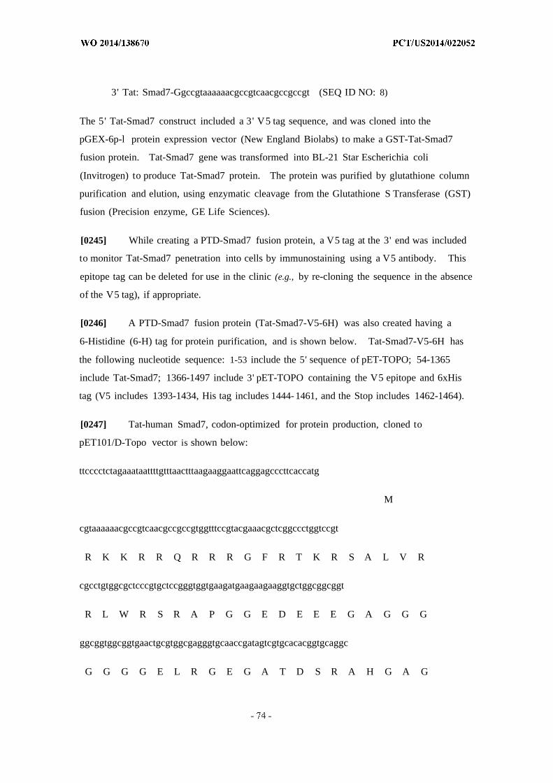
3' Tat: Smad7-Ggccgtaaaaaacgccgtcaacgccgccgt (SEQ ID NO: 8)
The 5' Tat-Smad7 construct included a 3' V5 tag sequence, and was cloned into the
pGEX-6p-l protein expression vector (New England Biolabs) to make a GST-Tat-Smad7
fusion protein. Tat-Smad7 gene was transformed into BL-21 Star Escherichia coli
(Invitrogen) to produce Tat-Smad7 protein. The protein was purified by glutathione column
purification and elution, using enzymatic cleavage from the Glutathione S Transferase (GST)
fusion (Precision enzyme, GE Life Sciences).
[0245] While creating a PTD-Smad7 fusion protein, a V5 tag at the 3' end was included
to monitor Tat-Smad7 penetration into cells by immunostaining using a V5 antibody. This
epitope tag can be deleted for use in the clinic (e.g., by re-cloning the sequence in the absence
of the V5 tag), if appropriate.
[0246] A PTD-Smad7 fusion protein (Tat-Smad7-V5-6H) was also created having a
6-Histidine (6-H) tag for protein purification, and is shown below. Tat-Smad7-V5-6H has
the following nucleotide sequence: 1-53 include the 5' sequence of pET-TOPO; 54-1365
include Tat-Smad7; 1366-1497 include 3' pET-TOPO containing the V5 epitope and 6xHis
tag (V5 includes 1393-1434, His tag includes 1444- 1461, and the Stop includes 1462-1464).
[0247] Tat-human Smad7, codon-optimized for protein production, cloned to
pET101/D-Topo vector is shown below:
ttcccctctagaaataattttgtttaactttaagaaggaattcaggagcccttcaccatg
M
cgtaaaaaacgccgtcaacgccgccgtggtttccgtacgaaacgctcggccctggtccgt
R K K R R Q R R R G F R T K R S A L V R
cgcctgtggcgctcccgtgctccgggtggtgaagatgaagaagaaggtgctggcggcggt
R L W R S R A P G G E D E E E G A G G G
ggcggtggcggtgaactgcgtggcgagggtgcaaccgatagtcgtgcacacggtgcaggc
G G G G E L R G E G A T D S R A H G A G
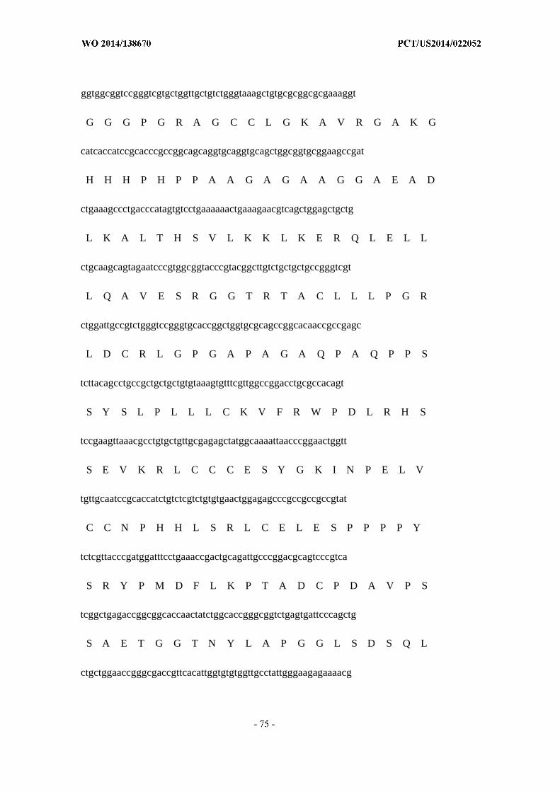
ggtggcggtccgggtcgtgctggttgctgtctgggtaaagctgtgcgcggcgcgaaaggt
G G G P G R A G C C L G K A V R G A K G
catcaccatccgcacccgccggcagcaggtgcaggtgcagctggcggtgcggaagccgat
H H H P H P P A A G A G A A G G A E A D
ctgaaagccctgacccatagtgtcctgaaaaaactgaaagaacgtcagctggagctgctg
L K A L T H S V L K K L K E R Q L E L L
ctgcaagcagtagaatcccgtggcggtacccgtacggcttgtctgctgctgccgggtcgt
L Q A V E S R G G T R T A C L L L P G R
ctggattgccgtctgggtccgggtgcaccggctggtgcgcagccggcacaaccgccgagc
L D C R L G P G A P A G A Q P A Q P P S
tcttacagcctgccgctgctgctgtgtaaagtgtttcgttggccggacctgcgccacagt
S Y S L P L L L C K V F R W P D L R H S
tccgaagttaaacgcctgtgctgttgcgagagctatggcaaaattaacccggaactggtt
S E V K R L C C C E S Y G K I N P E L V
tgttgcaatccgcaccatctgtctcgtctgtgtgaactggagagcccgccgccgccgtat
C C N P H H L S R L C E L E S P P P P Y
tctcgttacccgatggatttcctgaaaccgactgcagattgcccggacgcagtcccgtca
S R Y P M D F L K P T A D C P D A V P S
tcggctgagaccggcggcaccaactatctggcaccgggcggtctgagtgattcccagctg
S A E T G G T N Y L A P G G L S D S Q L
ctgctggaaccgggcgaccgttcacattggtgtgtggttgcctattgggaagagaaaacg
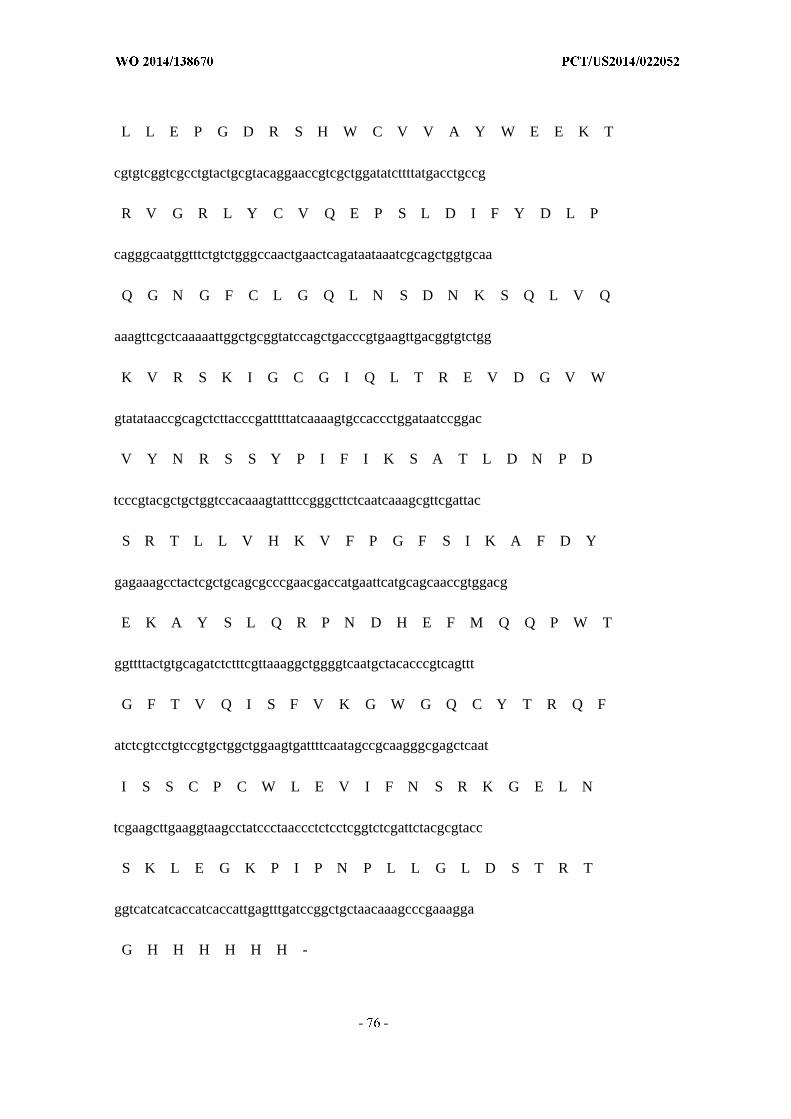
L L E P G D R S H W C V V A Y W E E K T
cgtgtcggtcgcctgtactgcgtacaggaaccgtcgctggatatcttttatgacctgccg
R V G R L Y C V Q E P S L D I F Y D L P
cagggcaatggtttctgtctgggccaactgaactcagataataaatcgcagctggtgcaa
Q G N G F C L G Q L N S D N K S Q L V Q
aaagttcgctcaaaaattggctgcggtatccagctgacccgtgaagttgacggtgtctgg
K V R S K I G C G I Q L T R E V D G V W
gtatataaccgcagctcttacccgatttttatcaaaagtgccaccctggataatccggac
V Y N R S S Y P I F I K S A T L D N P D
tcccgtacgctgctggtccacaaagtatttccgggcttctcaatcaaagcgttcgattac
S R T L L V H K V F P G F S I K A F D Y
gagaaagcctactcgctgcagcgcccgaacgaccatgaattcatgcagcaaccgtggacg
E K A Y S L Q R P N D H E F M Q Q P W T
ggttttactgtgcagatctctttcgttaaaggctggggtcaatgctacacccgtcagttt
G F T V Q I S F V K G W G Q C Y T R Q F
atctcgtcctgtccgtgctggctggaagtgattttcaatagccgcaagggcgagctcaat
I S S C P C W L E V I F N S R K G E L N
tcgaagcttgaaggtaagcctatccctaaccctctcctcggtctcgattctacgcgtacc
S K L E G K P I P N P L L G L D S T R T
ggtcatcatcaccatcaccattgagtttgatccggctgctaacaaagcccgaaagga
G H H H H H H -
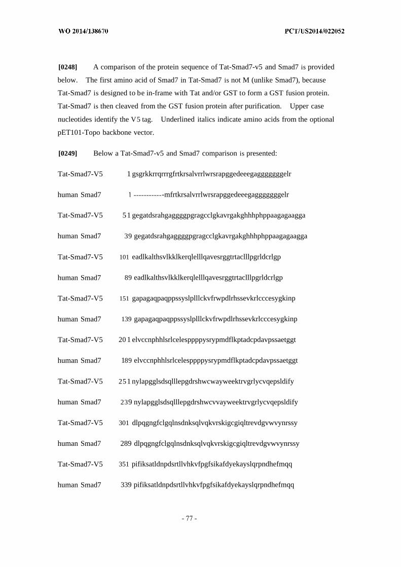
[0248] A comparison of the protein sequence of Tat-Smad7-v5 and Smad7 is provided
below. The first amino acid of Smad7 in Tat-Smad7 is not M (unlike Smad7), because
Tat-Smad7 is designed to be in-frame with Tat and/or GST to form a GST fusion protein.
Tat-Smad7 is then cleaved from the GST fusion protein after purification. Upper case
nucleotides identify the V5 tag. Underlined italics indicate amino acids from the optional
pET101-Topo backbone vector.
[0249] Below a Tat-Smad7-v5 and Smad7 comparison is presented:
Tat-Smad7-V5 1 gsgrkkrrqrrrgfrtkrsalvrrlwrsrapggedeeegagggggggelr
human Smad7 -mfrtkrsalvrrlwrsrapggedeeegagggggggelr
Tat-Smad7-V5 51 gegatdsrahgaggggpgragcclgkavrgakghhhphppaagagaagga
human Smad7 39 gegatdsrahgaggggpgragcclgkavrgakghhhphppaagagaagga
Tat-Smad7-V5 101 eadlkalthsvlkklkerqlelllqavesrggtrtaclllpgrldcrlgp
human Smad7 89 eadlkalthsvlkklkerqlelllqavesrggtrtaclllpgrldcrlgp
Tat-Smad7-V5 151 gapagaqpaqppssyslplllckvfrwpdlrhssevkrlcccesygkinp
human Smad7 139 gapagaqpaqppssyslplllckvfrwpdlrhssevkrlcccesygkinp
Tat-Smad7-V5 20 1 elvccnphhlsrlcelesppppysrypmdflkptadcpdavpssaetggt
human Smad7 189 elvccnphhlsrlcelesppppysrypmdflkptadcpdavpssaetggt
Tat-Smad7-V5 251 nylapgglsdsqlllepgdrshwcwayweektrvgrlycvqepsldify
human Smad7 239 nylapgglsdsqlllepgdrshwcvvayweektrvgrlycvqepsldify
Tat-Smad7-V5 301 dlpqgngfclgqlnsdnksqlvqkvrskigcgiqltrevdgvwvynrssy
human Smad7 289 dlpqgngfclgqlnsdnksqlvqkvrskigcgiqltrevdgvwvynrssy
Tat-Smad7-V5 351 pifiksatldnpdsrtllvhkvfpgfsikafdyekayslqrpndhefmqq
human Smad7 339 pifiksatldnpdsrtllvhkvfpgfsikafdyekayslqrpndhefmqq
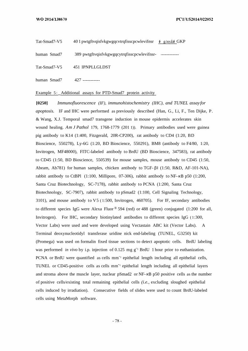
Tat-Smad7-V5 40 1 pwtgftvqisfvkgwgqcvtrqfisscpcwlevifnsr fe g/ fe GKP
human Smad7 389 pwtgftvqisfvkgwgqcytrqfisscpcwlevifnsr-
Tat-Smad7-V5 451 IPNPLLGLDST
human Smad7 427
Example 5 : Additional assays for PTD-Smad7 protein activity
[0250] Immunofluorescence (IF), immunohistochemistry (IHC), and TUNEL assay for
apoptosis. IF and IHC were performed as previously described (Han, G., Li, F., Ten Dijke, P.
& Wang, X.J. Temporal smad7 transgene induction in mouse epidermis accelerates skin
wound healing. Am J Pathol 179, 1768-1779 (201 1)). Primary antibodies used were guinea
pig antibody to K14 (1:400, Fitzgerald, 20R-CP200), rat antibody to CD4 (1:20, BD
Bioscience, 550278), Ly-6G (1:20, BD Bioscience, 550291), BM8 (antibody to F4/80, 1:20,
Invitrogen, MF48000), FITC-labeled antibody to BrdU (BD Bioscience, 347583), rat antibody
to CD45 (1:50, BD Bioscience, 550539) for mouse samples, mouse antibody to CD45 (1:50,
Abeam, Ab781) for human samples, chicken antibody to TGF- βΙ (1:50, R&D, AF-101-NA),
rabbit antibody to CtBPl (1:100, Millipore, 07-306), rabbit antibody to NF- κΒ p50 (1:200,
Santa Cruz Biotechnology, SC-7178), rabbit antibody to PCNA (1:200, Santa Cruz
Biotechnology, SC-7907), rabbit antibody to pSmad2 (1:100, Cell Signaling Technology,
3101), and mouse antibody to V5 ( 1:500, Invitrogen, 460705). For IF, secondary antibodies
to different species IgG were Alexa Fluor ® 594 (red) or 488 (green) conjugated (1:200 for all,
Invitrogen). For IHC, secondary biotinylated antibodies to different species IgG ( 1:300,
Vector Labs) were used and were developed using Vectastain ABC kit (Vector Labs). A
Terminal deoxynucleotidyl transferase uridine nick end-labeling (TUNEL, G3250) kit
(Promega) was used on formalin fixed tissue sections to detect apoptotic cells. BrdU labeling
was performed in vivo by i.p. injection of 0.125 mg g 1 BrdU 1 hour prior to euthanization.
PCNA or BrdU were quantified as cells mm 1 epithelial length including all epithelial cells,
TUNEL or CD45-positive cells as cells mm 1 epithelial length including all epithelial layers
and stroma above the muscle layer, nuclear pSmad2 or NF- κΒ p50 positive cells as the number
of positive cells/existing total remaining epithelial cells (i.e., excluding sloughed epithelial
cells induced by irradiation). Consecutive fields of slides were used to count BrdU-labeled
cells using MetaMorph software.
[0251] Cell culture. Smad7 transgenic and wild-type primary keratinocytes were
prepared from neonatal mouse skin as previously described (Han, G., Li, F., Ten Dijke, P. &
Wang, X.J. Temporal smad7 transgene induction in mouse epidermis accelerates skin wound
healing. Am J Pathol 179, 1768-1779 (201 1)), and cultured in PCT medium (CELLnTEC).
Spontaneously immortalized normal oral keratinocytes (NOK-SI) derived from gingival
tissues of healthy volunteers were cultured and maintained in defined keratinocyte medium
(Castilho, R.M., et al. Racl is required for epithelial stem cell function during dermal and
oral mucosal wound healing but not for tissue homeostasis in mice. PloS one 5, el0503
(2010)). Oral cancer cells Cal27 (ATCC) and MSK92 1 were cultured (D. Raben's lab,
fingerprinted by University of Colorado Cancer Center Tissue Culture Core) in Dulbecco
Modified Eagle Medium supplemented with 10% fetal bovine serum (GIBCO®; Invitrogen).
To assess the effect of Tat-Smad7 in irradiated cells, the above human cell lines were
cultured in chamber slides (BD Bioscience, 354108), irradiated with 3 Gy, and Tat-Smad7 ( 1
µg mL 1) was added to the culture medium immediately after irradiation. Cells were fixed
in 100% cold methanol 4 hours after Tat-Smad7 treatment for immunostaining of pSmad2,
NF- κΒ p50 and V5.
[0252] Transfection with siRNA. When cultured keratinocytes reached 70%
confluency, lOOnM of target siRNA or scrambled siRNA (Dharmacon) was transfected using
LIPOFECTAMINE® 2000 (Invitrogen). Cells were harvested 48-72 hours after
transfection and subjected to western analyses to determine knockdown efficiency. For
migration assays, siRNA was transfected when cells were plated. Target siRNAs included
in this study are: mouse siRacl-1 (Invitrogen, MSS237708) and siRacl-2 (IDT,
MMC.RNAI.N009007.12.3); human siSmad2 (Dharmacon, L-003 561-00-0005), siSmad3
(Invitrogen, HSS106252), and siSmad4 (Invitrogen, HSS1 18066); human siCtBPl-1 and
siCtBPl-2; human siSmad7-l and siSmad7-2; human TGF- Ι (Dharmacon,
J-012562-08-0005); mouse siSmad7.
[0253] In vitro keratinocyte proliferation assay. In vitro keratinocyte proliferation was
determined by BrdU incorporation in wild-type and Smad7 transgenic keratinocytes. Cells
at 70% confluency were transfected with Racl siRNAs, and changed to regular culture
medium 24 hours later. An in situ cell proliferation kit (Roche Applied Science) was used
to perform in vitro BrdU labeling and detection, and MetaMorph software was used to count
BrdU-labeled cells.
[0254] In vitro cell migration assays. When cells reached 100% confluency, the cells
were treated with mitomycin C (Sigma) at 10 µg mL 1 for 2 hours to inhibit cell proliferation
and a scratch wound was introduced with a Fisherbrand pipet tip. Cell migration was
photographed daily. Migration assays were performed when cells reached confluency after
24 to 36 hours of siR A transfection, and Image-J software was used to document cell
migration as the wound area occupied with migrating cells. For Tat-Smad7 treatment, cells
were exposed to Tat-Smad7 protein at 1 µg mL 1 or vehicle control (PBS) in medium after
wound scratch, and medium was changed every other day with freshly added Tat-Smad7 until
migrating cells fully covered the scratched wound.
[0255] Cell survival assay. Cell survival assays were performed as previously
described (Munshi, A., Hobbs, M. & Meyn, R.E. Clonogenic cell survival assay. Methods in
molecular medicine 110, 21-28 (2005)), with slight modifications. Briefly, cells were plated
in 12-well plates at 500 cells well 1 for non-irradiated wells, and increased up to 1,500 cells
well 1 along with increased radiation doses. Cells were irradiated 24 hours after they were
plated. Tat-Smad7 was added at 1 µg mL 1 or the same volume of PBS used to dissolve
Tat-Smad7 (control) to culture medium of irradiated and non-irradiated cells. The medium
was changed every other day with freshly added Tat-Smad7 or PBS for 10 tol4 days.
Colonies were fixed in methanol, stained in 0.5% crystal violet solution (containing 25%
methanol), counted and the average from 4 wells in each experiment was calculated. Two to
three separate experiments were performed for each cell line. The relative surviving fraction
was calculated as previously described, i.e., the absolute surviving fraction (colony
numbers/total plated cells) under each radiation dose divided by the absolute surviving fraction
of non-irradiated cells.
[0256] Western analysis. Protein extraction and western analyses were performed as
previously described (Li, A.G., Lu, S.L., Zhang, M.X., Deng, C. & Wang, X.J. Smad3
knockout mice exhibit a resistance to skin chemical carcinogenesis. Cancer Res 64,
7836-7845 (2004)). The antibodies used in this study included rabbit antibody to Smad7
(1:500), rabbit antibodies to Smad2 (1:300, Zymed, 51-1300) and Smad4 (1:300, Epitomics,
1676-1), rabbit antibody to Smad3 (1:300, Cell Signaling Technology, 9513), mouse
antibody to Racl (1:500, BD Biosciences, 610651), rabbit antibody to CtBPl (1:500,
Millipore, 07-306), mouse antibody to tubulin (1:3000, Sigma, T5168), mouse antibody to
GAPDH (1:5000, Abeam, Ab8245) and goat antibody to actin (1:1000, Santa Cruz
Biotechnology, SC1616). Gray-scale images were obtained using the ODYSSEY v.1.2
software (LI-COR Biosciences).
[0257] Racl activation assay. Active GTP-bound Racl was examined using a
BIOCHEM™ Kit for Rac 1 activation (Cytoskeleton Inc, BK03 5). Wild-type and Smad7
transgenic keratinocytes were cultured in 15 cm diameter tissue culture plates and prepared
protein lysates using the provided lysis buffer. To assay Racl activity, 1 mg of cell lysate
was used. To examine total Racl and Smad7 proteins, 50 µg of lysate was used. To
measure GTP-bound Racl in mouse tongues, half of the tongue was ground to a powder in
liquid nitrogen and lysed with lysis buffer to extract protein, GTP-bound Racl was assayed in
2 mg of protein lysate per sample and 50 µg of protein lysate was loaded for total Racl
protein western blot.
[0258] ChIP assays. ChIP assays were performed using the ChIP-IT express kit
(Active Motive, 53009) as previously described (Hoot, K.E., etal. HGF upregulation
contributes to angiogenesis in mice with keratinocyte-specific Smad2 deletion. J Clin Invest
120, 3606-3616 (2010); Hoot, K.E., et al. Keratinocyte-specific Smad2 ablation results in
increased epithelial-mesenchymal transition during skin cancer formation and progression.
Owens, et al. , J.Clin.Invest 118, 2722-2732 (2008). Smad4-dependent desmoglein-4
expression contributes to hair follicle integrity. Owens, et al., Dev. Biol. 322: 156-166
(2008). DNA-protein complex was isolated from primary mouse keratinocytes. For ChIP,
6.3 µg sheared chromatin was incubated with protein-G magnetic beads and 2 µg each of
rabbit antibodies to Smad2 (Cell Signaling Technology, 3122), Smad3 (Cell Signaling
Technology, 9523), Smad4 (Cell Signaling Technology, 9515), Smad7 antibody (Santa Cruz
Biotechnology, SC-1 1392), CtBPl (Millipore) or a negative control rabbit IgG (Santa Cruz
Biotechnology, SC-2027). Eluted DNA from the protein-DNA complex was used for PCR
analyses, and CtBPl binding to the Rac promoter was compared in wild-type and Smad7
transgenic keratinocytes by ChIP band intensities on gel images or by quantitative PCR using
Power SYBR Green Master Mix (Applied Biosystems). Primers used to amplify the Racl
SBE-1.5 Kb promoter regions:
5' -TGGAATTCCTGGTCTGGTTT-3 ' (sense)
5' -GCCAAGCTGCTCTTCCAGTA-3 ' (antisense)
5' -TCTCAGGGGGCCAAAGGTGTT-3 ' (sense)

5' -TCCCAGCACCTGAATCACATGG-3 ' (antisense)
[0259] Racl promoter luciferase reporter construct, site-directed mutagenesis and
luciferase assay. The 883 bp fragment of -1671 bp to -789 bp of the Racl promoter,
encompassing the SBE-1.5 Kb site, was amplified from wild-type mouse DNA using 5' Xhol
and 3' Hindlll tagged primers, and this Racl promoter fragment was cloned into pGL4.26
vector (Promega) to make the Racl promoter-pGL4.26 luciferase reporter (Racl-Luc)
construct. For site-directed mutagenesis, the SBE sequence 5'-TGTCTGTGCT-3' was
mutated to 5' -TGATAGAGCT-3 ' . Racl-Luc and pGL4.74 (1:20) were co-transfected with
Smad7 siRNA, CtBP 1 siRNA or scrambled siRNA using Lipofectamine 2000 (Invitrogen) to
primary mouse keratinocytes, or primary mouse keratinocytes with Tat-Smad7 treatment ( 1
µg mL 1) . Cell lysates were collected and luciferase assays were performed 48 hours after
transfection or Tat-Smad7 treatment, using the DUAL-LUCIFERASE® Reporter Assay kit
(Promega) following manufacturer's instructions. Rac 1-luciferase activity was measured
with the Glomax machine (Promega) and expressed by the ratio of firefly activity to Renilla
activity. Primers used for amplification oiRacl promoter sequence were:
5' -ATCCTCGAG-TATCCTCCAGGTCTGGG-3 '
5' -GCCAAGCTT-AGCGTCCAGCGTTAACCTG-3 '
[0260] Statistical analysis. Statistical differences in molecular analyses and oral
mucositis ulcer size were analyzed using the Student's est and all data was presented by
mean ± s.d. except ulcer size, which was presented by mean ± s.e.m. Oral mucositis
incidences were analyzed by Fisher's exact test.
Example 6 : Codon optimization for Smad7 protein production in E. coli or yeast
[0261] Although many mammalian proteins can be produced in bacteria without
nucleotide sequence modification, the analysis indicated that the Smad7 nucleotide sequence
would need to be modified to allow protein expression in bacteria.
[0262] Analysis of Smad7 cDNA mammalian codon use revealed nine arginine amino
acids coded for by the following nucleotides: 7-9, 43-45, 169-171, 403-405, 490-492,
526-528, 526-528, 823-825, 1057-1059 are a rare codon (AGG, codon utilization 1.7%).
Since these codons are rare codons in bacteria, it is expected that they could halt or reduce
protein translation and/or production in bacteria. The amino acids coded for by rare
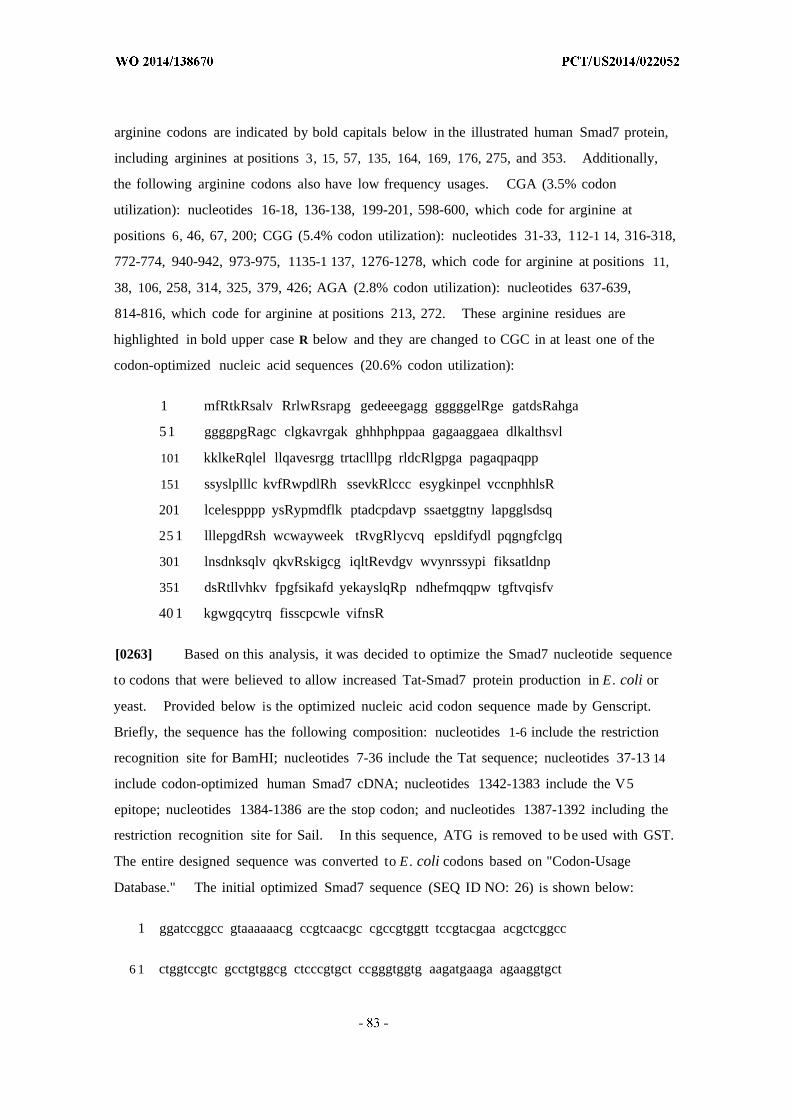
arginine codons are indicated by bold capitals below in the illustrated human Smad7 protein,
including arginines at positions 3, 15, 57, 135, 164, 169, 176, 275, and 353. Additionally,
the following arginine codons also have low frequency usages. CGA (3.5% codon
utilization): nucleotides 16-18, 136-138, 199-201, 598-600, which code for arginine at
positions 6, 46, 67, 200; CGG (5.4% codon utilization): nucleotides 31-33, 112-1 14, 316-318,
772-774, 940-942, 973-975, 1135-1 137, 1276-1278, which code for arginine at positions 11,
38, 106, 258, 314, 325, 379, 426; AGA (2.8% codon utilization): nucleotides 637-639,
814-816, which code for arginine at positions 213, 272. These arginine residues are
highlighted in bold upper case R below and they are changed to CGC in at least one of the
codon-optimized nucleic acid sequences (20.6% codon utilization):
1 mfRtkRsalv RrlwRsrapg gedeeegagg gggggelRge gatdsRahga
51 ggggpgRagc clgkavrgak ghhhphppaa gagaaggaea dlkalthsvl
101 kklkeRqlel llqavesrgg trtaclllpg rldcRlgpga pagaqpaqpp
151 ssyslplllc kvfRwpdlRh ssevkRlccc esygkinpel vccnphhlsR
201 lcelespppp ysRypmdflk ptadcpdavp ssaetggtny lapgglsdsq
25 1 lllepgdRsh wcwayweek tRvgRlycvq epsldifydl pqgngfclgq
301 lnsdnksqlv qkvRskigcg iqltRevdgv wvynrssypi fiksatldnp
351 dsRtllvhkv fpgfsikafd yekayslqRp ndhefmqqpw tgftvqisfv
40 1 kgwgqcytrq fisscpcwle vifnsR
[0263] Based on this analysis, it was decided to optimize the Smad7 nucleotide sequence
to codons that were believed to allow increased Tat-Smad7 protein production in E. coli or
yeast. Provided below is the optimized nucleic acid codon sequence made by Genscript.
Briefly, the sequence has the following composition: nucleotides 1-6 include the restriction
recognition site for BamHI; nucleotides 7-36 include the Tat sequence; nucleotides 37-13 14
include codon-optimized human Smad7 cDNA; nucleotides 1342-1383 include the V5
epitope; nucleotides 1384-1386 are the stop codon; and nucleotides 1387-1392 including the
restriction recognition site for Sail. In this sequence, ATG is removed to be used with GST.
The entire designed sequence was converted to E. coli codons based on "Codon-Usage
Database." The initial optimized Smad7 sequence (SEQ ID NO: 26) is shown below:
1 ggatccggcc gtaaaaaacg ccgtcaacgc cgccgtggtt tccgtacgaa acgctcggcc
6 1 ctggtccgtc gcctgtggcg ctcccgtgct ccgggtggtg aagatgaaga agaaggtgct
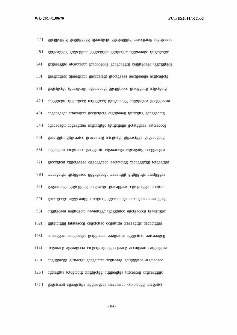
1 1 ggcggcggtg gcggtggcgg tgaactgcgt ggcgagggtg caaccgatag tcgtgcacac
181 ggtgcaggcg gtggcggtcc gggtcgtgct ggttgctgtc tgggtaaagc tgtgcgcggc
241 gcgaaaggtc atcaccatcc gcacccgccg gcagcaggtg caggtgcagc tggcggtgcg
301 gaagccgatc tgaaagccct gacccatagt gtcctgaaaa aactgaaaga acgtcagctg
361 gagctgctgc tgcaagcagt agaatcccgt ggcggtaccc gtacggcttg tctgctgctg
42 1 ccgggtcgtc tggattgccg tctgggtccg ggtgcaccgg ctggtgcgca gccggcacaa
481 ccgccgagct cttacagcct gccgctgctg ctgtgtaaag tgtttcgttg gccggacctg
54 1 cgccacagtt ccgaagttaa acgcctgtgc tgttgcgaga gctatggcaa aattaacccg
601 gaactggttt gttgcaatcc gcaccatctg tctcgtctgt gtgaactgga gagcccgccg
661 ccgccgtatt ctcgttaccc gatggatttc ctgaaaccga ctgcagattg cccggacgca
721 gtcccgtcat cggctgagac cggcggcacc aactatctgg caccgggcgg tctgagtgat
78 1 tcccagctgc tgctggaacc gggcgaccgt tcacattggt gtgtggttgc ctattgggaa
841 gagaaaacgc gtgtcggtcg cctgtactgc gtacaggaac cgtcgctgga tatcttttat
901 gacctgccgc agggcaatgg tttctgtctg ggccaactga actcagataa taaatcgcag
961 ctggtgcaaa aagttcgctc aaaaattggc tgcggtatcc agctgacccg tgaagttgac
1021 ggtgtctggg tatataaccg cagctcttac ccgattttta tcaaaagtgc caccctggat
1081 aatccggact cccgtacgct gctggtccac aaagtatttc cgggcttctc aatcaaagcg
1141 ttcgattacg agaaagccta ctcgctgcag cgcccgaacg accatgaatt catgcagcaa
1201 ccgtggacgg gttttactgt gcagatctct ttcgttaaag gctggggtca atgctacacc
126 1 cgtcagttta tctcgtcctg tccgtgctgg ctggaagtga ttttcaatag ccgcaagggc
132 1 gagctcaatt cgaagcttga aggtaagcct atccctaacc ctctcctcgg tctcgattct

1381 acgtgagtcg ac
[0264] A nucleotide sequence comparison between Tat-Smad7-V5 and human Smad7
cDNA is provided below. Human Smad7 and codon-optimized Tat-Smad7-V5 share 68%
codon homology. Human Smad7 and codon-optimized Tat-Smad7 share 71% codon
homology. Human Smad7 and codon-optimized Smad7 share 73% codon homology.

Alignment: Global DNA alignment against reference moleculeParameters: Scoring matrix: Linear (Mismatch 2 , OpenGap 4 , ExtGap 1 )
Reference molecule: human S a R A , Region 1-1281Number of sequences to align: 2Settings: Similarity significance value cutoff: >- 90%
Summary of Percent Matches :Reference: human Smad7 mRNA 1 - 1281 ( 1281 bps)Sequence 2 : Tat-Smad7-V5 1 - 1392 ( 1392 bps)
human Smad7 --a g g c g¾ gg gTat-Smad7-V5 ggS|:ccggccgtaaaaaacgccgtcaacgccgccgtggtl
human Smad7 25Tat-Smad7-V5 51 g gt gc g c c t
human Smad7 85
human Smad7 265 ggg gTat-Smad7-V5 301 g g¾ a
human Smad7 325
human Smad7 445 gg ctcg c gtcg gTat-Srriad7-V5 481 g ¾g a g
human Smad 7 505 t cTat-Smad7-V5 541 cg cag cgi gi t acg ci g g
human Smad7 565 g gg tg t g c c-Smad7-V5 601
human Smad7 805 g gg ga fc g cTat-Smad7-V5 841 a g cgt fc g t g g g a e ¾g a ¾ t
human Smad7 865 gat t a gg tg g tTat-Smad7-V5 901 g
human Smad7 925 i ife
human Smad7 985 ggg g g gg
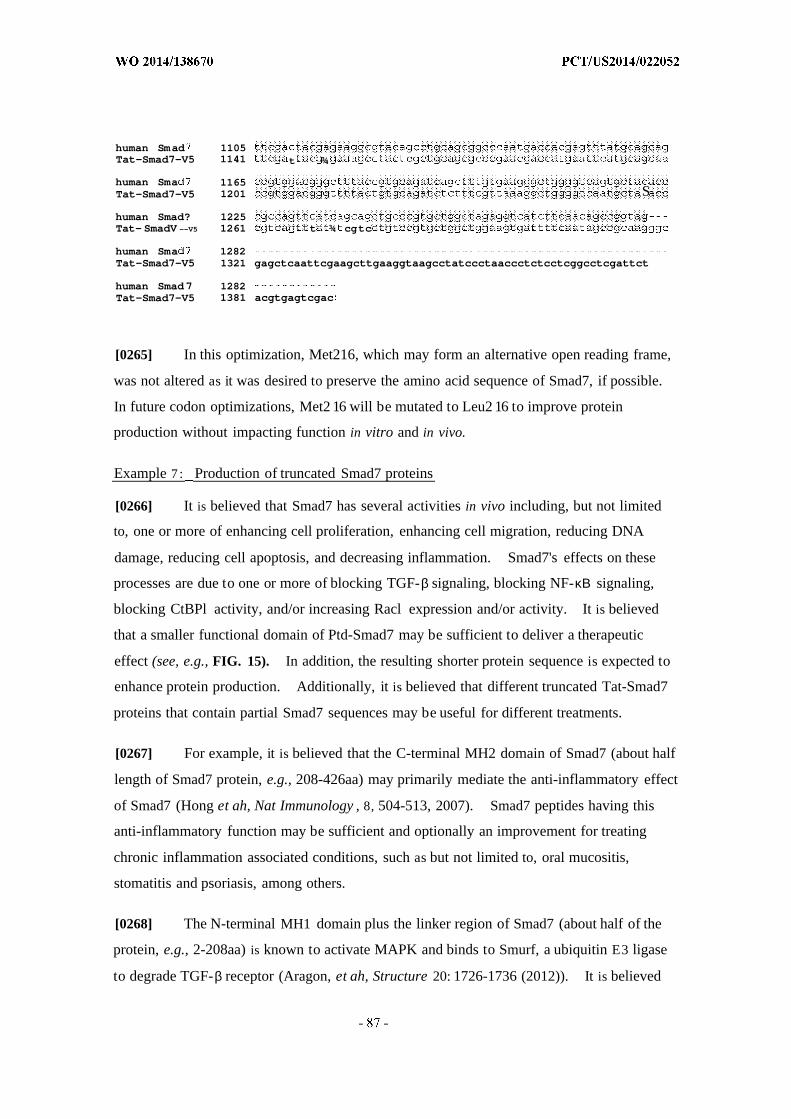
human Smad 1105Tat-Smad7-V5 1141 t ¾
human Sma 1165Tat-Smad7-V5 1201 S
human Smad? 1225Tat- SmadV --V5 1261 t ¾ cgtc
human Sma 1282Tat-Smad7-V5 1321 gagctcaattcgaagcttgaaggtaagcctatccctaaccctctcctcggcctcgattct
human Smad7 1282Tat-Smad7-V5 1381 acgtgagtcgac
[0265] In this optimization, Met216, which may form an alternative open reading frame,
was not altered as it was desired to preserve the amino acid sequence of Smad7, if possible.
In future codon optimizations, Met2 16 will be mutated to Leu2 16 to improve protein
production without impacting function in vitro and in vivo.
Example 7 : Production of truncated Smad7 proteins
[0266] It is believed that Smad7 has several activities in vivo including, but not limited
to, one or more of enhancing cell proliferation, enhancing cell migration, reducing DNA
damage, reducing cell apoptosis, and decreasing inflammation. Smad7's effects on these
processes are due to one or more of blocking TGF-β signaling, blocking NF-κΒ signaling,
blocking CtBPl activity, and/or increasing Racl expression and/or activity. It is believed
that a smaller functional domain of Ptd-Smad7 may be sufficient to deliver a therapeutic
effect (see, e.g., FIG. 15). In addition, the resulting shorter protein sequence is expected to
enhance protein production. Additionally, it is believed that different truncated Tat-Smad7
proteins that contain partial Smad7 sequences may be useful for different treatments.
[0267] For example, it is believed that the C-terminal MH2 domain of Smad7 (about half
length of Smad7 protein, e.g., 208-426aa) may primarily mediate the anti-inflammatory effect
of Smad7 (Hong et ah, Nat Immunology , 8, 504-513, 2007). Smad7 peptides having this
anti-inflammatory function may be sufficient and optionally an improvement for treating
chronic inflammation associated conditions, such as but not limited to, oral mucositis,
stomatitis and psoriasis, among others.
[0268] The N-terminal MH1 domain plus the linker region of Smad7 (about half of the
protein, e.g., 2-208aa) is known to activate MAPK and binds to Smurf, a ubiquitin E3 ligase
to degrade TGF-β receptor (Aragon, et ah, Structure 20: 1726-1736 (2012)). It is believed
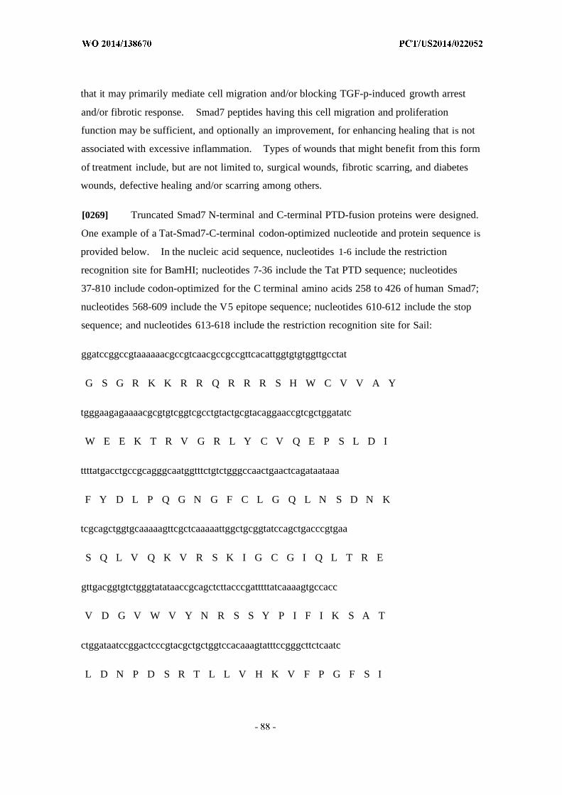
that it may primarily mediate cell migration and/or blocking TGF-p-induced growth arrest
and/or fibrotic response. Smad7 peptides having this cell migration and proliferation
function may be sufficient, and optionally an improvement, for enhancing healing that is not
associated with excessive inflammation. Types of wounds that might benefit from this form
of treatment include, but are not limited to, surgical wounds, fibrotic scarring, and diabetes
wounds, defective healing and/or scarring among others.
[0269] Truncated Smad7 N-terminal and C-terminal PTD-fusion proteins were designed.
One example of a Tat-Smad7-C-terminal codon-optimized nucleotide and protein sequence is
provided below. In the nucleic acid sequence, nucleotides 1-6 include the restriction
recognition site for BamHI; nucleotides 7-36 include the Tat PTD sequence; nucleotides
37-810 include codon-optimized for the C terminal amino acids 258 to 426 of human Smad7;
nucleotides 568-609 include the V5 epitope sequence; nucleotides 610-612 include the stop
sequence; and nucleotides 613-618 include the restriction recognition site for Sail:
ggatccggccgtaaaaaacgccgtcaacgccgccgttcacattggtgtgtggttgcctat
G S G R K K R R Q R R R S H W C V V A Y
tgggaagagaaaacgcgtgtcggtcgcctgtactgcgtacaggaaccgtcgctggatatc
W E E K T R V G R L Y C V Q E P S L D I
ttttatgacctgccgcagggcaatggtttctgtctgggccaactgaactcagataataaa
F Y D L P Q G N G F C L G Q L N S D N K
tcgcagctggtgcaaaaagttcgctcaaaaattggctgcggtatccagctgacccgtgaa
S Q L V Q K V R S K I G C G I Q L T R E
gttgacggtgtctgggtatataaccgcagctcttacccgatttttatcaaaagtgccacc
V D G V W V Y N R S S Y P I F I K S A T
ctggataatccggactcccgtacgctgctggtccacaaagtatttccgggcttctcaatc
L D N P D S R T L L V H K V F P G F S I
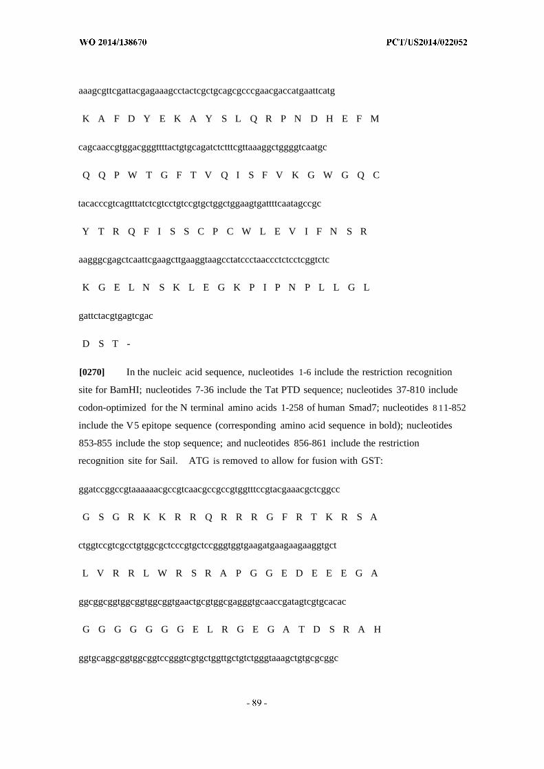
aaagcgttcgattacgagaaagcctactcgctgcagcgcccgaacgaccatgaattcatg
K A F D Y E K A Y S L Q R P N D H E F M
cagcaaccgtggacgggttttactgtgcagatctctttcgttaaaggctggggtcaatgc
Q Q P W T G F T V Q I S F V K G W G Q C
tacacccgtcagtttatctcgtcctgtccgtgctggctggaagtgattttcaatagccgc
Y T R Q F I S S C P C W L E V I F N S R
aagggcgagctcaattcgaagcttgaaggtaagcctatccctaaccctctcctcggtctc
K G E L N S K L E G K P I P N P L L G L
gattctacgtgagtcgac
D S T -
[0270] In the nucleic acid sequence, nucleotides 1-6 include the restriction recognition
site for BamHI; nucleotides 7-36 include the Tat PTD sequence; nucleotides 37-810 include
codon-optimized for the N terminal amino acids 1-258 of human Smad7; nucleotides 8 11-852
include the V5 epitope sequence (corresponding amino acid sequence in bold); nucleotides
853-855 include the stop sequence; and nucleotides 856-861 include the restriction
recognition site for Sail. ATG is removed to allow for fusion with GST:
ggatccggccgtaaaaaacgccgtcaacgccgccgtggtttccgtacgaaacgctcggcc
G S G R K K R R Q R R R G F R T K R S A
ctggtccgtcgcctgtggcgctcccgtgctccgggtggtgaagatgaagaagaaggtgct
L V R R L W R S R A P G G E D E E E G A
ggcggcggtggcggtggcggtgaactgcgtggcgagggtgcaaccgatagtcgtgcacac
G G G G G G G E L R G E G A T D S R A H
ggtgcaggcggtggcggtccgggtcgtgctggttgctgtctgggtaaagctgtgcgcggc

G A G G G G P G R A G C C L G K A V R G
gcgaaaggtcatcaccatccgcacccgccggcagcaggtgcaggtgcagctggcggtgcg
A K G H H H P H P P A A G A G A A G G A
gaagccgatctgaaagccctgacccatagtgtcctgaaaaaactgaaagaacgtcagctg
E A D L K A L T H S V L K K L K E R Q L
gagctgctgctgcaagcagtagaatcccgtggcggtacccgtacggcttgtctgctgctg
E L L L Q A V E S R G G T R T A C L L L
ccgggtcgtctggattgccgtctgggtccgggtgcaccggctggtgcgcagccggcacaa
P G R L D C R L G P G A P A G A Q P A Q
ccgccgagctcttacagcctgccgctgctgctgtgtaaagtgtttcgttggccggacctg
P P S S Y S L P L L L C K V F R W P D L
cgccacagttccgaagttaaacgcctgtgctgttgcgagagctatggcaaaattaacccg
R H S S E V K R L C C C E S Y G K I N P
gaactggtttgttgcaatccgcaccatctgtctcgtctgtgtgaactggagagcccgccg
E L V C C N P H H L S R L C E L E S P P
ccgccgtattctcgttacccgatggatttcctgaaaccgactgcagattgcccggacgca
P P Y S R Y P M D F L K P T A D C P D A
gtcccgtcatcggctgagaccggcggcaccaactatctggcaccgggcggtctgagtgat
V P S S A E T G G T N Y L A P G G L S D
tcccagctgctgctggaaccgggcgaccgtggtaagcctatccctaaccctctcctcggt
S Q L L L E P G D R G K P I P N P L L G

ctcgattctacgtgagtcgac
L D S T
Example 8 : Testing of truncated Smad7 proteins
[0271] Activity of truncated Smad7 proteins is tested using the in vitro and in vivo assays
used to test full-length Smad7 described above, among other assays. Such assays include,
but are not limited to, the ability to block phosphorylation of Smad2 and/or nuclear
translocation of the F-κΒ p50 subunit, increase cell proliferation, reduce apoptosis and/or
radiation-induced DNA damage, reduce inflammation and/or angiogenesis, promote healing
in oral mucositis, surgical wounds, diabetes wounds, and/or wounds associated with chronic
inflammation in mice.
[0272] In a wound healing assay, 6-mm punch biopsies were performed in wild-type
mice followed by daily topical application of C-terminal or N-terminal Tat-Smad7. By
measuring gross wound closure, both truncated Smad7 proteins described above (e.g.,
Tat-Smad7 C-terminal and N-terminal protein) were found to have promoted wound healing
similar to full length Tat-Smad7.
Example 9 : Truncated Smad7 proteins accelerate wound healing
[0273] The ability of truncated Smad7 proteins to accelerate wound healing was
examined in wild-type mice. FIG. 16A demonstrates the effect of C-terminally truncated
(259-426aa) Tat-C-Smad7 on a mouse wound healing model. Wild-type C57BL/6 mice
were anaesthetized, dorsally wounded by a 6-mm dermal punch biopsy, and treated every
other day with the topical application of PBS (control, 3 mice, 4 wounds/mouse), full-length
Tat-Smad7 (O^g/IC^L PBS/wound, 3 mice, 4 wounds/mouse) or Tat-C-Smad7
(O^g/lC^L/wound PBS, 3 mice, 4 wounds/mouse, total of 12 wounds were treated).
Wound area was photographed with Canon digital camera and measured by imaging analysis
using the Image J software with a 6-mm circle inside of the photo for normalization at 1, 2, 4
and 5 days after wounding, and the average percent wound area remaining was calculated for
each treatment group. The ability of Tat-C-Smad7 to accelerate wound healing was similar
to that of full-length Smad7 (FIG. 16A).
[0274] FIG. 16B illustrates the effect of N-terminally truncated (l-258aa) Tat-N-Smad7
on wound healing. Wild-type C57BL/6 mice were anaesthetized, dorsally wounded by a

6-mm dermal punch biopsy, and treated every other day with the topical application of PBS
(control, 6 mice, 4 wounds/mouse), full-length Tat-Smad7 (O^g/IC^L PBS, 6 mice, 4
wounds/mouse), or Tat-N-Smad7 (O^g/IC^L PBS, 6 mice, 4 wounds/mouse). Wound
area was measured by imaging analysis using the Image J software with a 6-mm circle inside
of the photo for normalization at 1, 2, and 4 days after wounding, and the average percent
wound area remaining was calculated for each treatment group. There was a significant
difference in the rate of wound healing between the control and Tat-Smad7-treated mice after
two days, with the latter healing faster (FIG. 16B, p<0.05). Although Tat-N-Smad7 was
tested as a negative control, it unexpectedly promoted significantly accelerated wound
healing relative to control mice after just one day, and significantly accelerated wound
healing relative to full-length Tat-Smad7-treated mice after two days (FIG. 16B, p<0.05).
Tat-Cre had no effect on wound closure relative to control treatment (data not shown).
[0275] These results demonstrate that both full-length Tat-Smad7 and truncated
Tat-Smad7 proteins (Tat-C-Smad7 and Tat-N-Smad7) promote wound healing. Moreover,
these results demonstrate that certain truncated Tat-Smad7 proteins are more effective in
accelerating wound healing than full-length Tat-Smad7.
[0276] Accordingly, compositions comprising truncated Tat-Smad7 proteins are useful in
treating wounds and in accelerating wound healing.
Example 10: Smad7 accelerates wound healing in an impaired wound-healing model
[0277] Diabetic (db/db) mice were dorsally wounded by a 6-mm dermal punch biopsy as
described above in Example 9, and treated every other day with the topical application of
PBS (control, 6 mice, 4 wounds/mouse), full-length Tat-Smad7 (O^g/IC^L PBS, 6 mice, 4
wounds/mouse), or REGRANEX® cream (smear with cotton swab, 6 mice 4 wounds/mouse)
to the wounds before day 8 when the wounds were not completely covered by scabs. After
day 10, the treatments were topically applied to the gap between the scab and wound
periphery to avoid the barrier of the hard scab and the unwounded stratum corneum.
REGRANEX®, which contains a recombinant human platelet-derived growth factor (PDGF),
is approved for topical administration to diabetic ulcers.
[0278] Wound closure was visually assessed at 1, 2, 4, 6, 8, 9, 10, 11, 12, and 13 days
after wounding (FIG. 17A). At each time point, the wounds were photographed with Canon
digital camera. After 8 days, there was a marked visual improvement in the wound closure
in Tat-Smad7-treated mice compared to controls (FIG. 17A).
[0279] Wound size was measured by using the Image J software with a 6-mm circle
inside of the photo for normalization at 1, 2, 3, 4, 7, 9, and 11 days after wounding, and the
average percent wound area remaining was calculated for each treatment group. The ability
of Tat-Smad7 to accelerate wound healing was similar to that of REGRANEX® (FIG. 17B).
On day 7, image analysis results indicated that wound closure was significantly accelerated in
Tat-Smad7-treated mice relative to controls (FIG. 17B, p<0.05). These results were similar
to those achieved with REGRANEX®.
[0280] Formalin fixed paraffin dorsal wound sections (1mm) from day 8 wound samples
were stained using hematoxylin and eosin (H&E). Histological comparison of day 8 wound
samples revealed complete re-epithelialization and an accelerated wound closure in
Smad7-treated db/db mice relative to controls (FIG. 17C).
[0281] Collectively, these results demonstrate that Tat-Smad7 is useful for accelerating
wound closure in poorly healing diabetic wounds, and provides an alternative to treatment
with REGRANEX®.
Example 11: Additional codon optimization for Smad7 protein production
[0282] Smad7 nucleic acid molecules are designed with additional nucleotide changes
selected to increase protein production. For example, the utilization of codons encoding the
amino acids Ser and His will be manipulated. In codon-optimized human Smad7 in
examples above, the Ser codon (TCC or TCG) has an amino acid frequency of approximately
9% of codon utilization. It is believed that changing the codon of Ser to AGC will increase
Smad7 protein production, at least partly because it can optionally increase codon usage to
15%. There are 33 Ser amino acids in Smad7 protein (nucleotides at positions 19-21, 46-48,
133-135, 292-294, 349-351, 451-453, 454-456, 460-462, 5 11-513, 514-516, 544-546,
595-597, 616-618, 634-636, 691-693, 694-696, 739-741, 745-747, 775-777, 847-849,
907-909, 919-921, 943-945, 1006-1008, 1009-1101, 1030-1032, 1054-1056, 1093-1095,
1126-1 128, 1192-1194, 1237-1239, 1240-1242, 1273-1275; with a corresponding serine
amino acid position of 7, 16, 45, 98, 117, 151, 152, 154, 171, 172, 182, 199, 206, 212, 231,
232, 247, 249, 259, 283, 303, 307, 315, 336, 337, 344, 352, 365, 376, 398, 413, 414, 425).
Of these, 23 (nucleotides 19-21, 292-294, 349-351, 451-453, 454-456, 460-462, 5 11-513,
514-516, 544-546, 616-618, 634-636, 691-693, 694-696, 739-741, 745-747, 775-777,
847-849, 907-909, 919-921, 1009-1 101, 1030-1032, 1054-1056, 1093-1095; corresponding
serine amino acid position of 7, 98, 117, 151, 152, 154, 171, 172, 182, 206, 212, 231, 232,
247, 249, 259, 283, 303, 307, 337, 344, 352, 365) can be changed without introducing
potential alternative open reading frames.
[0283] Similarly, in codon-optimized human Smad7 in the examples above, the His
codon (CAC) has 9.6% of codon usage. It is believed that changing the His codon to CAT
(optionally to 12.6% usage) will increase Smad7 protein production. There are 12 His
(nucleotides 142-144, 214-216, 217-219, 220-222, 226-228, 289-291, 589-591, 778-780,
1072-1074, 1147-1 149; corresponding to histidine amino acids at position 48, 72, 73, 74, 76,
97, 170, 196, 197, 260, 358, 383) in Smad7 protein. Of these, 4 (nucleotides 217-219.
220-222, 589-591, 778-780, histidine residues 73, 76, 197, 260) can be changed without
introducing potential alternative open reading frames.
[0284] In addition, wild-type human Smad7 includes a Met amino acid as amino acid
216 (Met216). This may be perceived as an alternative open reading frame by bacterial
machinery, for example, and decrease protein production. It is believed that changing
Met216 to Leu216 (ATG to CTG), the amino acid that has the biochemical property the
closest to Met and thus not expected to change 3D structure of the protein, will increase
protein production.

[0285] The comparison between original codon-optimized Tat-Smad7-V5 and further
changes is provided below. Top strand: Tat-Smad7-V5; bottom strand: after optimized Ser,
Alignment; Local A homologies.
Parameters Both strands. Method: a s t S c a Max Score
o 1 20120323113102B12-SS4366--164160-l-PGEX-5 .seq (1-1163) Kol 2 a t S m a 7 Ser-His op t r
u b e r c sequences t o align: 2
Settings: Similarity significance value cutoff: >= 60¾
Homology Block: Percent Matches 4 Score 1227 Length 1392
201203231131 3 0
T a Sn a d Ser 1
2C1203231131 9 0
TacSrnad.7 Ser 6 1
201203231131
TatSmad7 Ser 1
201203231131 210
TatSmac? Ser 1S1
201203231131 270
'. Ser 241
201203231131 3 3 0
TatS ∞ d 7 Ser 301
201203231131 390
TatSniad7 Ser 3S1
2 0 1 2 0 33 2 3311 1 3 11 44 5 00 ccgggtcgtctggattgccgtctgggtccgggtgcaccggctggtgcgcagcccgcacaa
a Sra a 7 Ser 4
201203231131 510
a t S a d Ser 481
22 0C 1 22 00 3 2 3 1 11 33 11 55 7 0 g c a¾ a g & :¾ ag t a a a c g c g
Tatsr.iad? Ser 541 ¾
201203231131 6 3
T a S ;a d 7 Ser 601
2200112200.33223.311 3 11 6 9 c c g cc g ¾ ¾ ¾ c g .t a c gi¾ tg g a t.t. c t g c c g a c g c a g a g c c cg g a c g c a
Ta.tSmad7 Ser 661 ¾
220C11220033223211113311 7 5 00 g t« g ¾ g a ¾
TatSraad? Ser 7 2 1 . . .
2 012 032 810
Tatsraad7 Ser 781
201203231131 g a g a a a a c g g r.g g g cg c c tg a c g g t a c a gg a c c g ¾ ;c t g g a
TtttSinad7 Sex 841
22 0C 11 22 00 33 22 3 1 1 .3 1 993300 g a c e g c c g c a g g g c a a g c g t g g g a aT a 3 a d 7 Ser S01
201203231131 3 0
TacSt\ad7 Ser 6 1
201203231131 1 0 5 0
T a S a d 7 e r 1021 p.
2C1203231131 1110
TatS'.-,iad7 Ser O l
2 2 3 ' t t c t t a c a g a a a c c c ¾¾ ¾ c t g c a g c c c c g a a c g a c a c a c a g c a a
T a t S a 7 Ser 1141 ¾ "
201203231131 1230 c c g t g g a c g g g t t a .g t g a g 3 -c ¾ ¾ ¾ t t c g a a a g g c t g g g g a a c a c a c
T a a d 7 Ser 1201
2012 03231131 1290 cgtcagtttatc¾¾§¾¾ctgtccgtgctqqctqqaagtgatt;ttcaatagccgcaaqggc
T a a d 7 Sex 1261 . ¾ ¾ . . '. . . '. . .
201203231131 5 0 g g c t. a t. a a g c g a a g g a a g c a c t c . t g g t t c g a t
TatSrp.ad? Ser 1 2 1
201203231131 1410 acgtgagtcgac
T m a d Ser 1381
His and M216L mutation.
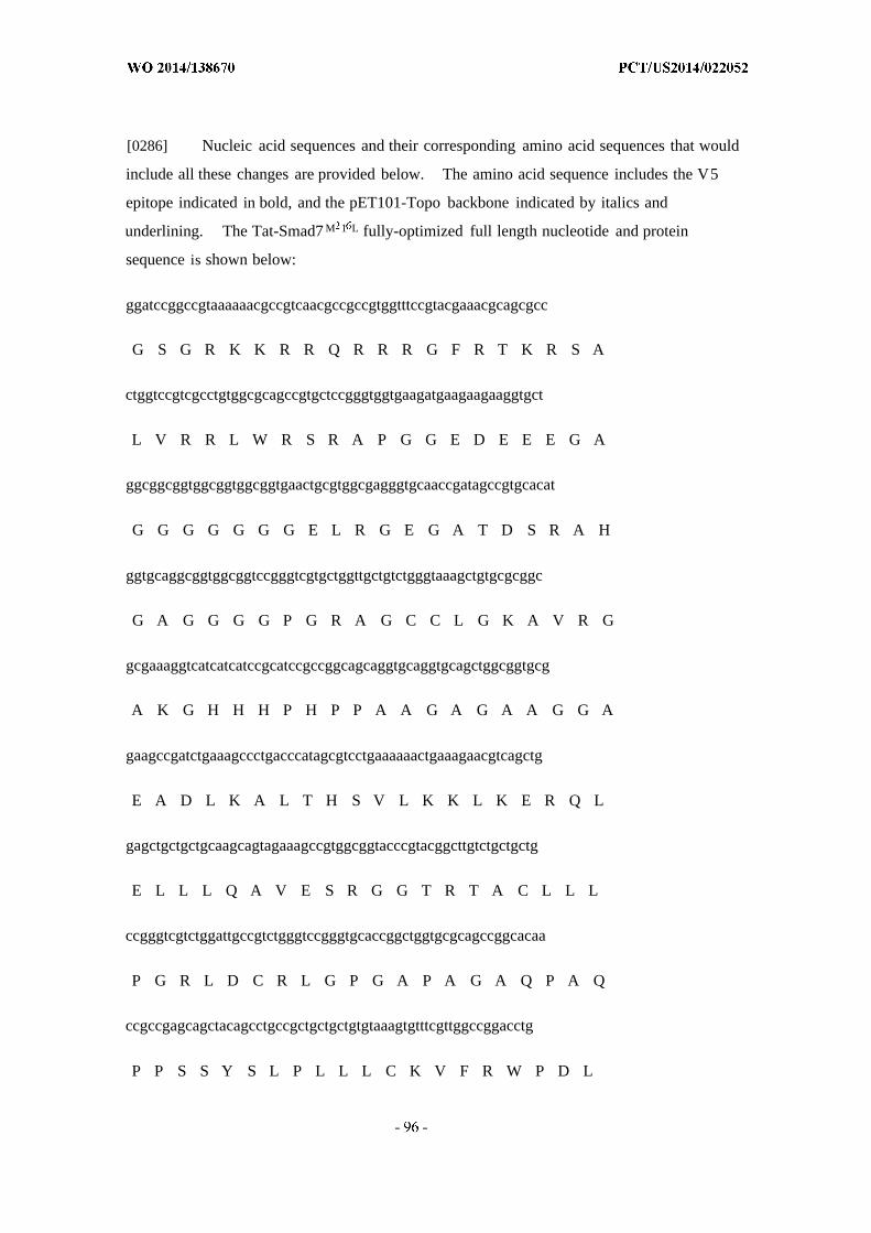
[0286] Nucleic acid sequences and their corresponding amino acid sequences that would
include all these changes are provided below. The amino acid sequence includes the V5
epitope indicated in bold, and the pET101-Topo backbone indicated by italics and
underlining. The Tat-Smad7 M 1 L fully-optimized full length nucleotide and protein
sequence is shown below:
ggatccggccgtaaaaaacgccgtcaacgccgccgtggtttccgtacgaaacgcagcgcc
G S G R K K R R Q R R R G F R T K R S A
ctggtccgtcgcctgtggcgcagccgtgctccgggtggtgaagatgaagaagaaggtgct
L V R R L W R S R A P G G E D E E E G A
ggcggcggtggcggtggcggtgaactgcgtggcgagggtgcaaccgatagccgtgcacat
G G G G G G G E L R G E G A T D S R A H
ggtgcaggcggtggcggtccgggtcgtgctggttgctgtctgggtaaagctgtgcgcggc
G A G G G G P G R A G C C L G K A V R G
gcgaaaggtcatcatcatccgcatccgccggcagcaggtgcaggtgcagctggcggtgcg
A K G H H H P H P P A A G A G A A G G A
gaagccgatctgaaagccctgacccatagcgtcctgaaaaaactgaaagaacgtcagctg
E A D L K A L T H S V L K K L K E R Q L
gagctgctgctgcaagcagtagaaagccgtggcggtacccgtacggcttgtctgctgctg
E L L L Q A V E S R G G T R T A C L L L
ccgggtcgtctggattgccgtctgggtccgggtgcaccggctggtgcgcagccggcacaa
P G R L D C R L G P G A P A G A Q P A Q
ccgccgagcagctacagcctgccgctgctgctgtgtaaagtgtttcgttggccggacctg
P P S S Y S L P L L L C K V F R W P D L
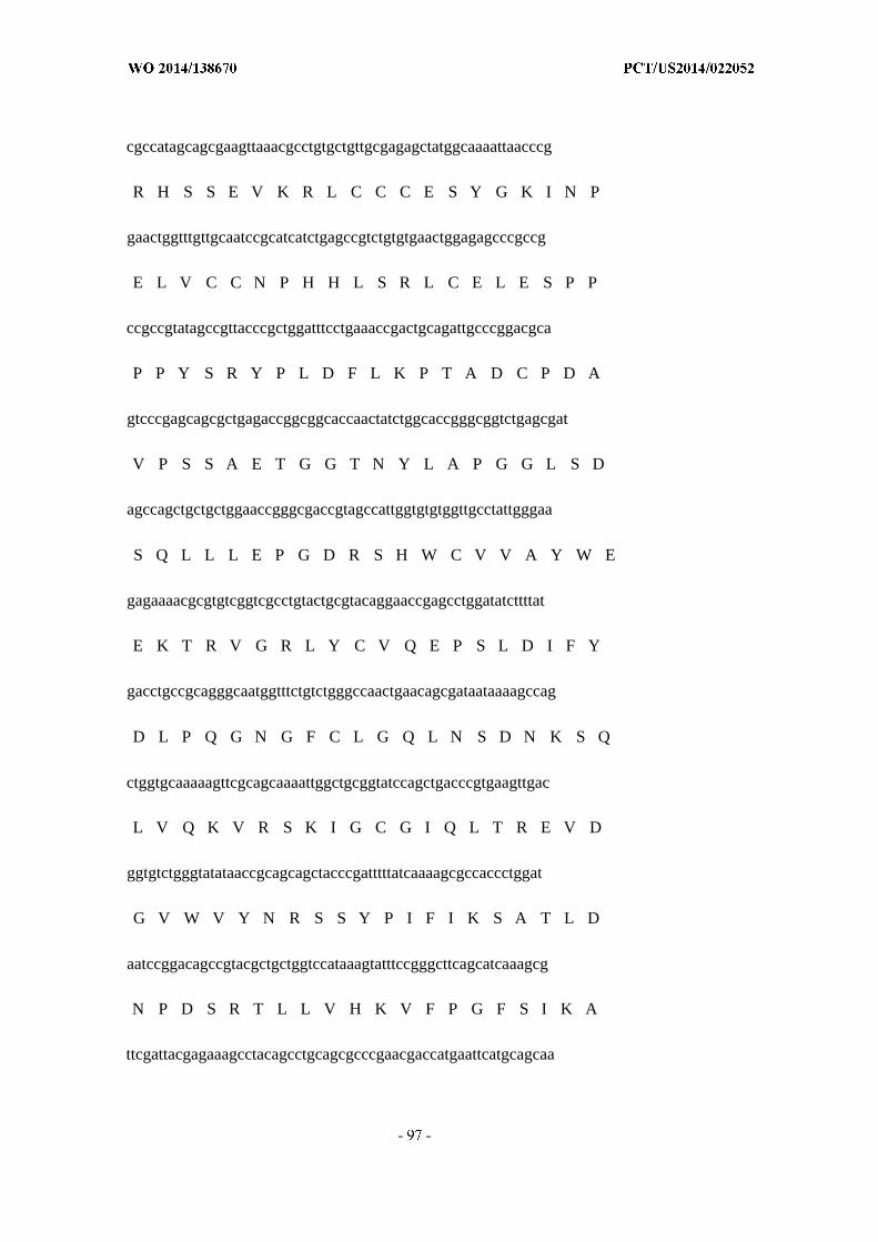
cgccatagcagcgaagttaaacgcctgtgctgttgcgagagctatggcaaaattaacccg
R H S S E V K R L C C C E S Y G K I N P
gaactggtttgttgcaatccgcatcatctgagccgtctgtgtgaactggagagcccgccg
E L V C C N P H H L S R L C E L E S P P
ccgccgtatagccgttacccgctggatttcctgaaaccgactgcagattgcccggacgca
P P Y S R Y P L D F L K P T A D C P D A
gtcccgagcagcgctgagaccggcggcaccaactatctggcaccgggcggtctgagcgat
V P S S A E T G G T N Y L A P G G L S D
agccagctgctgctggaaccgggcgaccgtagccattggtgtgtggttgcctattgggaa
S Q L L L E P G D R S H W C V V A Y W E
gagaaaacgcgtgtcggtcgcctgtactgcgtacaggaaccgagcctggatatcttttat
E K T R V G R L Y C V Q E P S L D I F Y
gacctgccgcagggcaatggtttctgtctgggccaactgaacagcgataataaaagccag
D L P Q G N G F C L G Q L N S D N K S Q
ctggtgcaaaaagttcgcagcaaaattggctgcggtatccagctgacccgtgaagttgac
L V Q K V R S K I G C G I Q L T R E V D
ggtgtctgggtatataaccgcagcagctacccgatttttatcaaaagcgccaccctggat
G V W V Y N R S S Y P I F I K S A T L D
aatccggacagccgtacgctgctggtccataaagtatttccgggcttcagcatcaaagcg
N P D S R T L L V H K V F P G F S I K A
ttcgattacgagaaagcctacagcctgcagcgcccgaacgaccatgaattcatgcagcaa
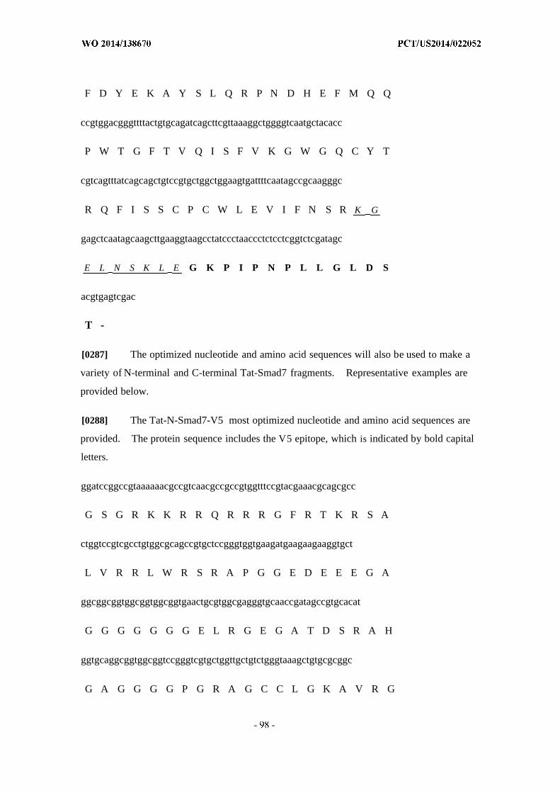
F D Y E K A Y S L Q R P N D H E F M Q Q
ccgtggacgggttttactgtgcagatcagcttcgttaaaggctggggtcaatgctacacc
P W T G F T V Q I S F V K G W G Q C Y T
cgtcagtttatcagcagctgtccgtgctggctggaagtgattttcaatagccgcaagggc
R Q F I S S C P C W L E V I F N S R K G
gagctcaatagcaagcttgaaggtaagcctatccctaaccctctcctcggtctcgatagc
E L N S K L E G K P I P N P L L G L D S
acgtgagtcgac
T -
[0287] The optimized nucleotide and amino acid sequences will also be used to make a
variety of N-terminal and C-terminal Tat-Smad7 fragments. Representative examples are
provided below.
[0288] The Tat-N-Smad7-V5 most optimized nucleotide and amino acid sequences are
provided. The protein sequence includes the V5 epitope, which is indicated by bold capital
letters.
ggatccggccgtaaaaaacgccgtcaacgccgccgtggtttccgtacgaaacgcagcgcc
G S G R K K R R Q R R R G F R T K R S A
ctggtccgtcgcctgtggcgcagccgtgctccgggtggtgaagatgaagaagaaggtgct
L V R R L W R S R A P G G E D E E E G A
ggcggcggtggcggtggcggtgaactgcgtggcgagggtgcaaccgatagccgtgcacat
G G G G G G G E L R G E G A T D S R A H
ggtgcaggcggtggcggtccgggtcgtgctggttgctgtctgggtaaagctgtgcgcggc
G A G G G G P G R A G C C L G K A V R G
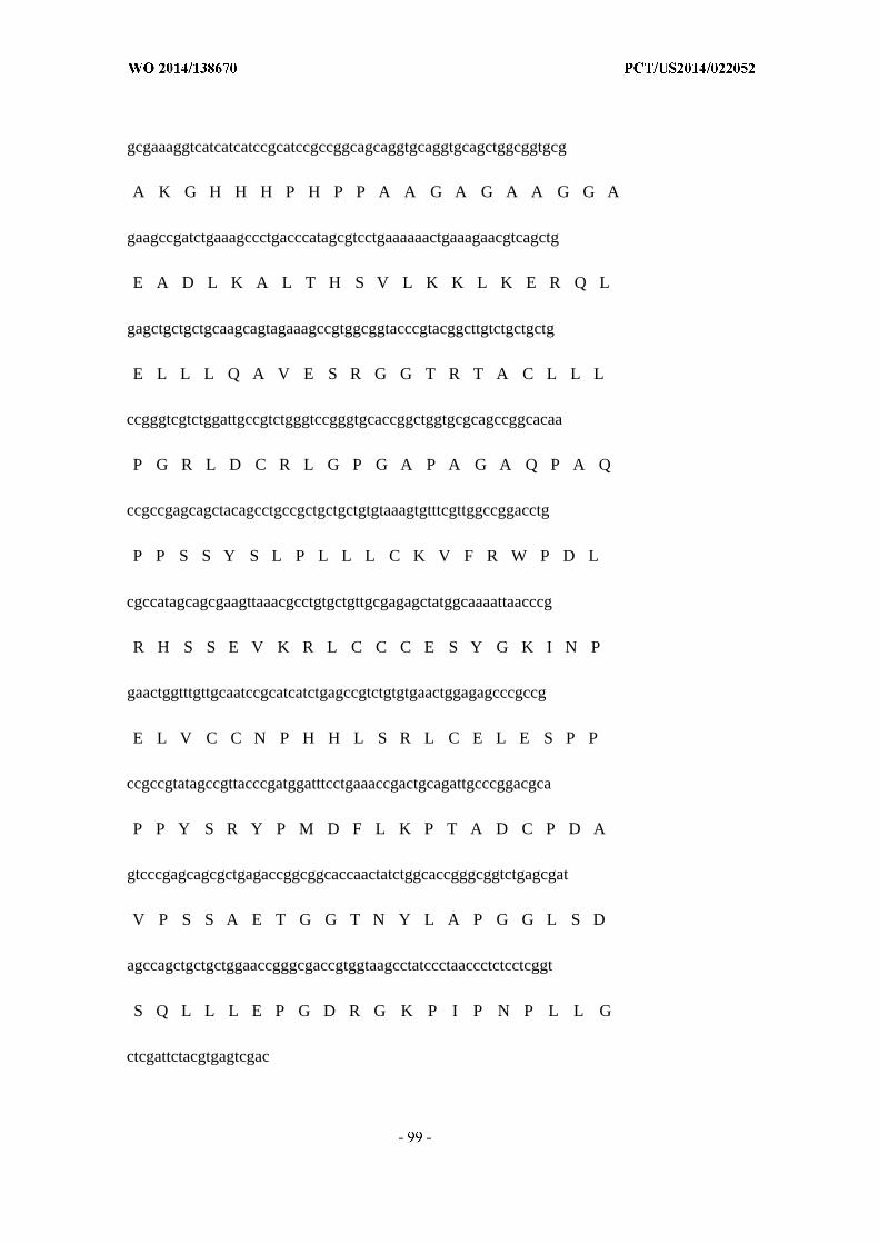
gcgaaaggtcatcatcatccgcatccgccggcagcaggtgcaggtgcagctggcggtgcg
A K G H H H P H P P A A G A G A A G G A
gaagccgatctgaaagccctgacccatagcgtcctgaaaaaactgaaagaacgtcagctg
E A D L K A L T H S V L K K L K E R Q L
gagctgctgctgcaagcagtagaaagccgtggcggtacccgtacggcttgtctgctgctg
E L L L Q A V E S R G G T R T A C L L L
ccgggtcgtctggattgccgtctgggtccgggtgcaccggctggtgcgcagccggcacaa
P G R L D C R L G P G A P A G A Q P A Q
ccgccgagcagctacagcctgccgctgctgctgtgtaaagtgtttcgttggccggacctg
P P S S Y S L P L L L C K V F R W P D L
cgccatagcagcgaagttaaacgcctgtgctgttgcgagagctatggcaaaattaacccg
R H S S E V K R L C C C E S Y G K I N P
gaactggtttgttgcaatccgcatcatctgagccgtctgtgtgaactggagagcccgccg
E L V C C N P H H L S R L C E L E S P P
ccgccgtatagccgttacccgatggatttcctgaaaccgactgcagattgcccggacgca
P P Y S R Y P M D F L K P T A D C P D A
gtcccgagcagcgctgagaccggcggcaccaactatctggcaccgggcggtctgagcgat
V P S S A E T G G T N Y L A P G G L S D
agccagctgctgctggaaccgggcgaccgtggtaagcctatccctaaccctctcctcggt
S Q L L L E P G D R G K P I P N P L L G
ctcgattctacgtgagtcgac

L D S T -
[0289] Tat-C-Smad7-V5 most optimized nucleotide and amino acid sequences are
provided. The protein sequence includes the V5 epitope (indicated by bold capital letters),
and the pET101-Topo backbone (indicated by underlined italics).
ggatccggccgtaaaaaacgccgtcaacgccgccgttcacattggtgtgtggttgcctat
G S G R K K R R Q R R R S H W C V V A Y
tgggaagagaaaacgcgtgtcggtcgcctgtactgcgtacaggaaccgagcctggatatc
W E E K T R V G R L Y C V Q E P S L D I
ttttatgacctgccgcagggcaatggtttctgtctgggccaactgaacagcgataataaa
F Y D L P Q G N G F C L G Q L N S D N K
agccagctggtgcaaaaagttcgcagcaaaattggctgcggtatccagctgacccgtgaa
S Q L V Q K V R S K I G C G I Q L T R E
gttgacggtgtctgggtatataaccgcagcagctacccgatttttatcaaaagcgccacc
V D G V W V Y N R S S Y P I F I K S A T
ctggataatccggacagccgtacgctgctggtccataaagtatttccgggcttcagcatc
L D N P D S R T L L V H K V F P G F S I
aaagcgttcgattacgagaaagcctacagcctgcagcgcccgaacgaccatgaattcatg
K A F D Y E K A Y S L Q R P N D H E F M
cagcaaccgtggacgggttttactgtgcagatcagcttcgttaaaggctggggtcaatgc
Q Q P W T G F T V Q I S F V K G W G Q C
tacacccgtcagtttatcagcagctgtccgtgctggctggaagtgattttcaatagccgc
Y T R Q F I S S C P C W L E V I F N S R

aagggcgagctcaatagcaagcttgaaggtaagcctatccctaaccctctcctcggtctc
K G E L N S K L E G K P I P N P L L
gatagcacgtgagtcgac
D S T -

[0290] The comparisons before and after the above optimizations are provided below.
C-terminal optimization (top strand):
Alignment: Local Di A homologies.
Parameters: Both strands. Method: F a tS n
1 Tat-C-Smad7-ser-his optimized (1-518) Mol 2 T a -C -te r a Smad7-V5 (1-618;Number o f u e to a : 2
Settings. Similarity significance value cutoff: >··.· G
o io o y Block: Percent Matches S3 Score 541 Length «18
Tat -C- Smad? - 1Tat -C- t a l 1
Tat -C- a d 7 - 1 ggatatc
Tat -C-- te r .a l 6 1
Tat. -c- Smad7 - 121Tat -c-tergal 121
Tat -c- Smad7- 13811 ¾¾ c a g c g t g c a ag t g ¾ i¾ a a a a gg ct g c g t --c c ag c t g ac g --q a a
T t -c- t e r a l 1 8s1i ¾ 7 .
T -c- Smad7- 241 g g g r.g g g t a a cg a g c a r c cg a t c
Tat -c- termal 241
Tat -c- Smad7 - 301 t c c g g a c o g a cg c g c tg g t c c a a a a g a t t cg g g c t c - a tTat -c- ter¾al 3 01
T -c- Sm a d - 336611 a t g a g a g a a g c c c t g cg c g a a cq a c a a a c a
Tat -c~ termal 361 - . .
Tat -c- 442211 g a cg g g a cg g g a g g c a g a cg ta a gg c tg g g g ca g c
Tat -c- te r 421
Tat -c- Smad7- 481 .gtccgtgccggctggaagtgactttcaauagccgcT t -c- t e .a l 481
T a -c- Stnad7- 541 g a ag g a a g c a . c a a c :c ot gg t
Tat. -c- te a l 541
Tat -c- 601 t g a g g a g t c
Tat -c~ termal 6 01
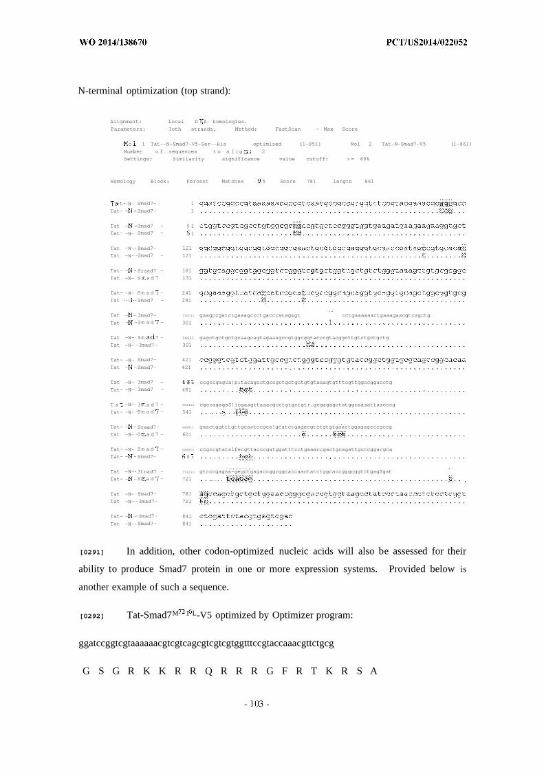
N-terminal optimization (top strand):
Alignment: Local D A homologies.
Parameters: 3oth strands. Method: FastScan - Max Score
o 1 Tat--N~Smad7-V5-Ser--His optimized (1-851} Mol 2 Tat-N-Smad7-V5 (1-861)
Number o f sequences t o a l ig 2
Settings: Similarity significance value cutoff: > = 60%
Homology Block: Percent Matches 5 Score 781 Length 861
t -N- Smad7- 1
Tat- - -Smad7- 1
Tat - --3mad7 - 5 1
Tat- -N- Smad7 - 1
Tat -N- 8mad7- 121
Tat -N- -3mad7 - 121
Tat- - -Sraad7 - 181
Tat -N- Sr a d 7 131
Tat- -N- Sm a d - 241
Tat -1 -Smad7 - 241
Tat - -3mad7- 330011 gaagccgatctgaaagccctgacccatag> cctgaaaaaactgaaagaacgtcagctg
Tat - -3m a d - 301 1
Tat -N- Sm 7 - 3SS1I gagctgctgctgcaagcagtagaasgccgtggcggtacccgtacggcttgtctgctgctg
Tat -N- -3mad7- 3S1
Tat- -N- Smad7- 421
Tat - -Smad7- 421
Tat -N- 3mad7 - 4 3 1 ccgccgagca|p:tacagcctgccgctgctgctgtgtaaagtgtttcgttggccggacctg
Tat- -N- 3mad7 - 481
T a -N- 3 a d 7 - 554411 cgccagagsS!icgaagttaaacgcctgtgctgtr.gcgagagctatggcaaaattaacccg
Tat- -N- -Sm a d - 541 ... .· ..
Tat- - -Sraad7- 660011 gaactggtttgttgcaatccgca!gcatctgagecgtctgtgtgaactggagagcccgccg
Tat -N- -3 a d 7 - 601
Tat- -N- Sm a d - SSSSII ccgccgtatslfecgttacccgatggatttcctgaaaccgactgcagattgcccggacgca
Tat- - -Smad7- S
Tat -N- 3tnad7 - 772211 gtcccgagsa-gegctgagaccggcggcaccaactatctggcaccgggcggtctgagSgat
Tat- - -S a d - 721 .... .
Tat -N- Smad7- 781
Tat -N- -3mad7- 7S1
Tat- - --Smad7- 841
Tat -N- Smad7- 841
[0291] In addition, other codon-optimized nucleic acids will also be assessed for their
ability to produce Smad7 protein in one or more expression systems. Provided below is
another example of such a sequence.
[0292] Tat-Smad7M 1 L-V5 optimized by Optimizer program:
ggatccggtcgtaaaaaacgtcgtcagcgtcgtcgtggtttccgtaccaaacgttctgcg
G S G R K K R R Q R R R G F R T K R S A
ctggttcgtcgtctgtggcgttctcgtgcgccgggtggtgaagacgaagaagaaggtgcg
L V R R L W R S R A P G G E D E E E G A
ggtggtggtggtggtggtggtgaactgcgtggtgaaggtgcgaccgactctcgtgcgcac
G G G G G G G E L R G E G A T D S R A H
ggtgcgggtggtggtggtccgggtcgtgcgggttgctgcctgggtaaagcggttcgtggt
G A G G G G P G R A G C C L G K A V R G
gcgaaaggtcaccaccacccgcacccgccggcggcgggtgcgggtgcggcgggtggtgcg
A K G H H H P H P P A A G A G A A G G A
gaagcggacctgaaagcgctgacccactctgttctgaaaaaactgaaagaacgtcagctg
E A D L K A L T H S V L K K L K E R Q L
gaactgctgctgcaggcggttgaatctcgtggtggtacccgtaccgcgtgcctgctgctg
E L L L Q A V E S R G G T R T A C L L L
ccgggtcgtctggactgccgtctgggtccgggtgcgccggcgggtgcgcagccggcgcag
P G R L D C R L G P G A P A G A Q P A Q
ccgccgtcttcttactctctgccgctgctgctgtgcaaagttttccgttggccggacctg
P P S S Y S L P L L L C K V F R W P D L
cgtcactcttctgaagttaaacgtctgtgctgctgcgaatcttacggtaaaatcaacccg
R H S S E V K R L C C C E S Y G K I N P
gaactggtttgctgcaacccgcaccacctgtctcgtctgtgcgaactggaatctccgccg
E L V C C N P H H L S R L C E L E S P P
ccgccgtactctcgttacccgctggacttcctgaaaccgaccgcggactgcccggacgcg

P P Y S R Y P L D F L K P T A D C P D A
gttccgtcttctgcggaaaccggtggtaccaactacctggcgccgggtggtctgtctgac
V P S S A E T G G T N Y L A P G G L S D
tctcagctgctgctggaaccgggtgaccgttctcactggtgcgttgttgcgtactgggaa
S Q L L L E P G D R S H W C V V A Y W E
gaaaaaacccgtgttggtcgtctgtactgcgttcaggaaccgtctctggacatcttctac
E K T R V G R L Y C V Q E P S L D I F Y
gacctgccgcagggtaacggtttctgcctgggtcagctgaactctgacaacaaatctcag
D L P Q G N G F C L G Q L N S D N K S Q
ctggttcagaaagttcgttctaaaatcggttgcggtatccagctgacccgtgaagttgac
L V Q K V R S K I G C G I Q L T R E V D
ggtgtttgggtttacaaccgttcttcttacccgatcttcatcaaatctgcgaccctggac
G V W V Y N R S S Y P I F I K S A T L D
aacccggactctcgtaccctgctggttcacaaagttttcccgggtttctctatcaaagcg
N P D S R T L L V H K V F P G F S I K A
ttcgactacgaaaaagcgtactctctgcagcgtccgaacgaccacgaattcatgcagcag
F D Y E K A Y S L Q R P N D H E F M Q Q
ccgtggaccggtttcaccgttcagatctctttcgttaaaggttggggtcagtgctacacc
P W T G F T V Q I S F V K G W G Q C Y T
cgtcagttcatctcttcttgcccgtgctggctggaagttatcttcaactctcgtggtaaa
R Q F I S S C P C W L E V I F N S R G K

ccgatcccgaacccgctgctgggtctggactctacctgagtcgac
P I P N P L L G L D S T - -
[0293] Nucleotide sequence:
1-6: BamHI; 7-36: Tat; 37-1314: codon-optimized human Smad7; 1315-1356: V5; 137-1359:
stop; 1360-1365 Sail
ATG is removed to be used with GST; 682 ATG to CTG (M216 to L)
1 ggatccggtc gtaaaaaacg tcgtcagcgt cgtcgtggtt tccgtaccaa acgttctgcg
61 ctggttcgtc gtctgtggcg ttctcgtgcg ccgggtggtg aagacgaaga agaaggtgcg
12 1 ggtggtggtg gtggtggtgg tgaactgcgt ggtgaaggtg cgaccgactc tcgtgcgcac
181 ggtgcgggtg gtggtggtcc gggtcgtgcg ggttgctgcc tgggtaaagc ggttcgtggt
241 gcgaaaggtc accaccaccc gcacccgccg gcggcgggtg cgggtgcggc gggtggtgcg
301 gaagcggacc tgaaagcgct gacccactct gttctgaaaa aactgaaaga acgtcagctg
361 gaactgctgc tgcaggcggt tgaatctcgt ggtggtaccc gtaccgcgtg cctgctgctg
42 1 ccgggtcgtc tggactgccg tctgggtccg ggtgcgccgg cgggtgcgca gccggcgcag
481 ccgccgtctt cttactctct gccgctgctg ctgtgcaaag ttttccgttg gccggacctg
541 cgtcactctt ctgaagttaa acgtctgtgc tgctgcgaat cttacggtaa aatcaacccg
601 gaactggttt gctgcaaccc gcaccacctg tctcgtctgt gcgaactgga atctccgccg
661 ccgccgtact ctcgttaccc gctggacttc ctgaaaccga ccgcggactg cccggacgcg
72 1 gttccgtctt ctgcggaaac cggtggtacc aactacctgg cgccgggtgg tctgtctgac
781 tctcagctgc tgctggaacc gggtgaccgt tctcactggt gcgttgttgc gtactgggaa
841 gaaaaaaccc gtgttggtcg tctgtactgc gttcaggaac cgtctctgga catcttctac
901 gacctgccgc agggtaacgg tttctgcctg ggtcagctga actctgacaa caaatctcag
961 ctggttcaga aagttcgttc taaaatcggt tgcggtatcc agctgacccg tgaagttgac
1021 ggtgtttggg tttacaaccg ttcttcttac ccgatcttca tcaaatctgc gaccctggac
1081 aacccggact ctcgtaccct gctggttcac aaagttttcc cgggtttctc tatcaaagcg
1141 ttcgactacg aaaaagcgta ctctctgcag cgtccgaacg accacgaatt catgcagcag
1201 ccgtggaccg gtttcaccgt tcagatctct ttcgttaaag gttggggtca gtgctacacc
1261 cgtcagttca tctcttcttg cccgtgctgg ctggaagtta tcttcaactc tcgtggtaaa
1321 ccgatcccga acccgctgct gggtctggac tctacctgag tcgac
[0294] Sequence comparison with Tat-Smad7 M 1 L-V5 described in Example 4 :
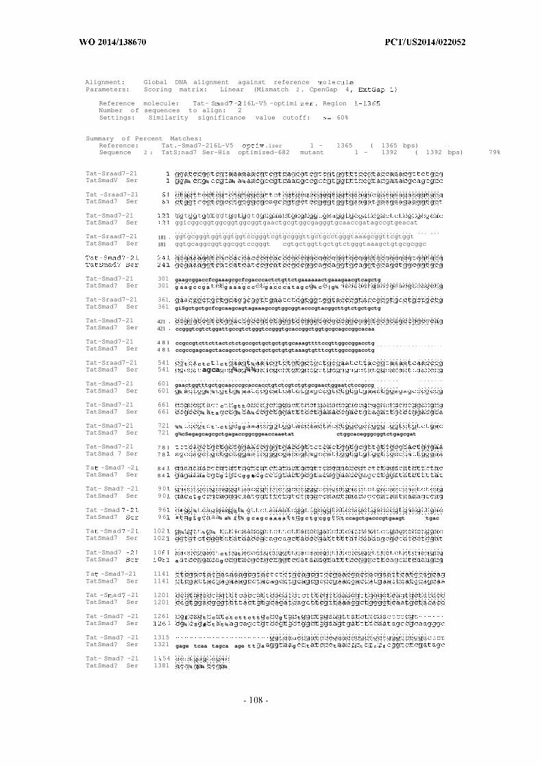
Alignment: Global DNA alignment against reference olec lParameters: Scoring matrix: Linear (Mismatch 2 , CpenGap 4
Reference molecule: Tat- S ad - 16L-V5 -optimi e , RegionNumber of sequences to align: 2Settings: Similarity significance value cutoff: 60%
Summary of Percent Matches:Reference: Tat.-Smad7-216L-V5 .izer 1 - 1365 ( 1365 bps)Sequence 2 : TatS;nad7 Ser-His optimized-682 mutant 1 - 1392 ( 1392 bps) 79%
Tat-Sraad7-21TatSmadV Ser : ¾ ¾ ¾ ¾
Tat -Sraad7-21TatSmad7 Ser
Tat-Smad7-21 g t t g ggt g gtgTatSmad7 Ser ggicggcggtggcggtggcggtgaactgcgtggcgagggtgcaaccgatagccgtgeacat
Tat-Sraad7-21 181 ggtgcgggtggtggtggtccgggtcgtgcgggttgctgcctgggtaaagcggttcgtggtTatSmad7 Ser 181 ggtgcaggcggtggcggtccgggt cgtgctggttgctgtctgggtaaagctgtgcgcggc
Tat-Smad7-21 301 gaagcggaccfcgaaagcgcfcgacccactctgttctgaaaaaactgaaagaacgtcagctgTatSmad? Ser 301 g aa g c cg a g a a a g c c g a c c c a t ag c ¾ c |g¾
Tat-Sraad7-21 361 a gTatSmad7 Ser 361 giSgctgctgcfcgcaagcagtagaaagccgtggcggtacccgtacggcttgtctgctgctg
Tat-Smad7-21 421TatSmad7 Ser 421 ccgggtcgtctggattgccgtctgggtccgggtgcaccggctggtgcgcagccggcacaa
Tat-Smad7-21 4 8 1 ccgccgtcttcttactctctgccgctgctgctgtgcaaagttttccgttggccggacctgTatSmad7 Ser 4 8 1 ccgccgagcagctacagcctgocgctgctgctgtgtaaagtgtttcgttggccggacotg
Tat-Sraad7-21 541 t c tc 1c t ¾TatSmad7 Ser 541 agca ¾ ¾¾
Tat-Smad7-21 601 gaactggtttgctgcaacccgcaccacctgtctcgtctgtgcgaactggaatctccgccgTatSmad7 Ser 601 ¾ ¾ ¾ t ¾
Tat-Smad7-21 661 c g t tTatSmad7 Ser 661 ¾ ta ¾ ¾
Tat-Smad7-21 721 ¾¾ t t c t g gTatSmad7 Ser 721 g¾cSegagcagcgctgagaccggcggeaccaaetat ctggcacegggcggtctgagcgat
Tat-Smad7-21 7 8 1TatSmad 7 Ser 7 8
Ta -Smad7 -2 8 4TatSmad7 Ser 8 4 a g g g te g
Tat- Smad? -21 90TatSmad7 Ser 90 c g
Tat -Smad 96 fe : ¾ ) ) :i ¾TatSmad7 96 s : :g: g aa¾ a¾ i ¾ g c ag c a a a a t g c tg cg g t ccagctgacccgtgaagt tgac
Ta -Smad -2 102 ¾ i ¾ ¾ :TatSmad7 Ser 102
Tat-Smad7 10 c t tTatSmad7 ο a g
Ta -Smad7-21 1141TatSmad7 Ser 1141
Tat -S ad -21 1201TatSmad7 Ser 1201
Tat -Smad? -21 1261 fi t c c t c t t c t t c g tTatSmad7 Ser 1 ¾ S :fe t t¾
Tat -Smad? -21 1315TatSmad? Ser 1321 gage tcaa tagca age t t a g t t ii t i c i fi
Tat- Smad? -21 1 54TatSmad? Ser 1381 ¾ ) ¾ ) ¾
[0295] Protein production using the sequences below is carried out by transfecting
mammalian expression vectors (e.g., pCMV-6-Entry) into HEK293 or CHO cells.
[0296] Optimized GST-Tat-Smad7-myc-flag nucleotide sequence, cloned inpCMV6-Entry plasmid:
1 gacgttgtat acgactccta tagggcggcc gggaattcgt cgactggatc cggtaccgag
61 gagatctgcc gccgcgatcg ccatgtcccc cattctgggc tactggaaga ttaagggcct
121 ggtgcagcct actagactgc tgctggaata cctggaggaa aaatatgaag agcatctgta
181 tgaaagagac gagggggata aatggaggaa caagaaattc gaactgggac tggagtttcc
241 taatctgcca tactatattg acggcgatgt gaagctgact cagtctatgg ctatcattag
301 atacatcgca gacaaacaca acatgctggg cgggtgtcct aaggaaaggg cagagattag
361 tatgctggag ggagccgtgc tggatattag atacggcgtc tcacgcatcg cctatagcaa
421 agacttcgaa accctgaagg tggattttct gagcaaactg cctgaaatgc tgaagatgtt
481 cgaggacaga ctgtgccaca aaacctacct gaatggcgac catgtcacac acccagattt
541 tatgctgtac gacgccctgg atgtggtcct gtatatggac cccatgtgtc tggatgcttt
601 ccctaagctg gtgtgcttta agaaaaggat cgaggcaatt ccccagatcg ataagtacct
661 gaaaagctcc aagtatatcg cttggcctct ccagggctgg caggcaacat tcggaggcgg
721 ggaccatccc cctaaaagcg acctggaggt gctgtttcag ggaccactgg gcagcggccg
781 gaagaagcgg cggcagaggc gccgaagtag gttccgcact aagcggtcag cactggtgcg
841 gagactgtgg cgatctcggg ctcctggagg agaggacgag gaagagggag caggcggcgg
901 gg gg gg ggagaactgc gcggggaggg agctacagat agccgagccc acggagctgg
961 gg gg gg ccagggcgag ccggatgctg tctgggcaaa gcagtgagag gcgccaaggg
1021 gcaccatcac ccccatccac ccgccgctgg cgcaggagca gccggcggag ctgaggcaga
1081 cctgaaagcc ctgactcaca gtgtgctgaa gaaactgaag gaaagacagc tggagctgct
1141 gctccaggca gtcgaatcac gcggaggcac ccgaacagct tgtctgctgc tgcccggccg
1201 gctggactgc cggctgggac ccggcgcccc tgctggggca cagccagccc agcctccatc
1261 tagttatagc ctgcccctgc tgctgtgtaa ggtgttccga tggcctgatc tgcggcattc
1321 aagcgaagtc aaaaggctgt gctgttgcga gtcctacggc aagattaacc cagaactggt
1381 gtgttgcaat ccccatcacc tgtctcgact gtgtgaactg gagtcccccc ctccacccta
1441 ctctaggtat cctatggact ttctgaagcc aaccgctgac tgcccagatg cagtgccctc
1501 ctctgccgag actgggggaa ccaactacct ggctcctggc ggactgagcg actcccagct
1561 gctgctggaa ccaggggatc gcagccactg gtgtgtggtc gcctactggg aagagaagac
1621 aagagtggga aggctgtatt gcgtccagga gccttccctg gacatcttct acgatctgcc
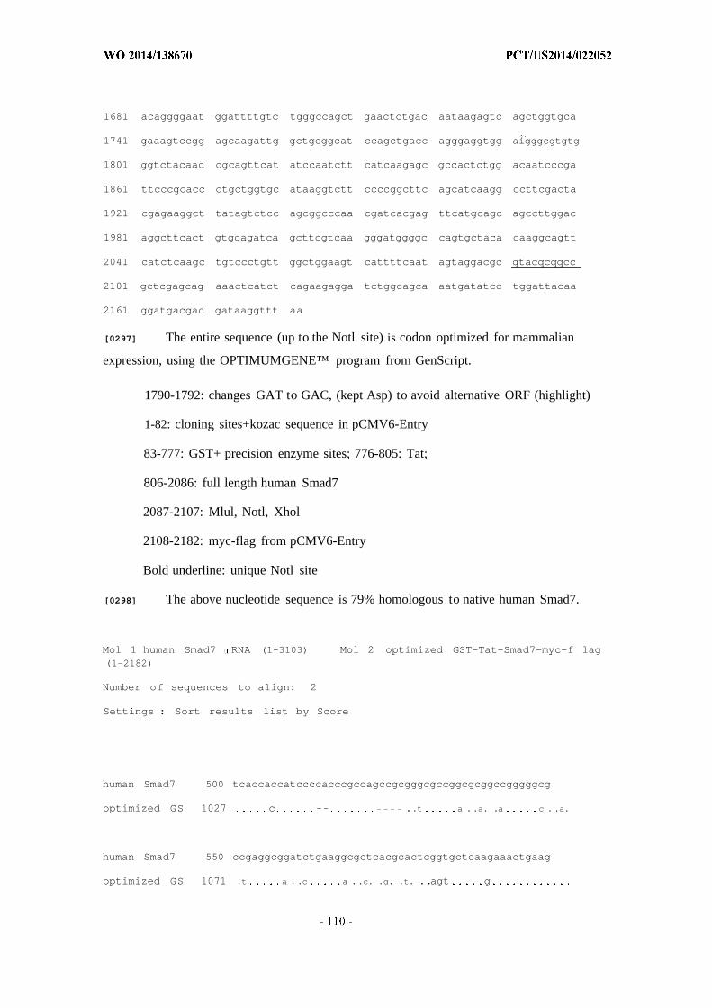
1681 acaggggaat ggattttgtc tgggccagct gaactctgac aataagagtc agctggtgca
1741 gaaagtccgg agcaagattg gctgcggcat ccagctgacc agggaggtgg aigggcgtgtg
1801 ggtctacaac cgcagttcat atccaatctt catcaagagc gccactctgg acaatcccga
1861 ttcccgcacc ctgctggtgc ataaggtctt ccccggcttc agcatcaagg ccttcgacta
1921 cgagaaggct tatagtctcc agcggcccaa cgatcacgag ttcatgcagc agccttggac
1981 aggcttcact gtgcagatca gcttcgtcaa gggatggggc cagtgctaca caaggcagtt
2041 catctcaagc tgtccctgtt ggctggaagt cattttcaat agtaggacgc qtacqcqqcc
2101 gctcgagcag aaactcatct cagaagagga tctggcagca aatgatatcc tggattacaa
2161 ggatgacgac gataaggttt aa
[0297] The entire sequence (up to the Notl site) is codon optimized for mammalian
expression, using the OPTIMUMGENE™ program from GenScript.
1790-1792: changes GAT to GAC, (kept Asp) to avoid alternative ORF (highlight)
1-82: cloning sites+kozac sequence in pCMV6-Entry
83-777: GST+ precision enzyme sites; 776-805: Tat;
806-2086: full length human Smad7
2087-2107: Mlul, Notl, Xhol
2108-2182: myc-flag from pCMV6-Entry
Bold underline: unique Notl site
[0298] The above nucleotide sequence is 79% homologous to native human Smad7.
Mol 1 human Smad7 RNA (1-3103) Mol 2 optimized GST-Tat-Smad7-myc-f lag(1-2182)
Number of sequences to align: 2
Settings : Sort results list by Score
human Smad7 500 tcaccaccatccccacccgccagccgcgggcgccggcgcggccgggggcg
optimized GS 1027 c -- ..t a ..a. .a c . .a.
human Smad7 550 ccgaggcggatctgaaggcgctcacgcactcggtgctcaagaaactgaag
optimized GS 1071 .t a . .c a ..c. .g. .t. ..agt g
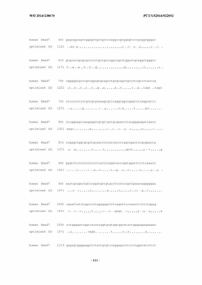
human Smad7 600 gagcggcagctggagctgctgctccaggccgtggagtcccgcggcgggac
optimized GS 1121 ..aa .a a ..c. .a. .a a . .c. .
human Smad7 650 gcgcaccgcgtgcctcctgctgcccggccgcctggactgcaggctgggcc
optimized GS 1171 c ..a ..a ..t ..t ..g g c a .
human Smad7 700 cgggggcgcccgccggcgcgcagcctgcgcagccgccctcgtcctactcg
optimized GS 1221 .c ..c ..c ..t ..t ..g ..a a..c t ..a ..tagt ..tagc
human Smad7 750 ctccccctcctgctgtgcaaagtgttcaggtggccggatctcaggcattc
optimized GS 1271 ..g g t . .g c.a t gc
human Smad7 800 ctcggaagtcaagaggctgtgttgctgtgaatcttacgggaagatcaacc
optimized GS 1321 aagc a c . .t. .c. .g. .c c t ....
human Smad7 850 ccgagctggtgtgctgcaacccccatcaccttagccgactctgcgaacta
optimized GS 1371 .a. .a t t gtct g - -t g
human Smad7 900 gagtctcccccccctccttactccagatacccgatggattttctcaaacc
optimized GS 1421 c t . .a..c t..g. .t..t c g . .g. .
human Smad7 950 aactgcagactgtccagatgctgtgccttcctccgctgaaacagggggaa
optimized GS 1471 ...c .t c a c t..c. .g..t
human Smad7 1000 cgaattatctggcccctggggggctttcagattcccaacttcttctggag
optimized GS 1521 .c. .c. .c t c . .a. .gage, .c g ..g. .g a
human Smad7 1050 cctggggatcggtcacactggtgcgtggtggcatactgggaggagaagac
optimized GS 1571 ..a cage t c..c a
human Smad7 110 0 gagagtggggaggctctactgtgtccaggagccctctctggatatcttct
- I l l -
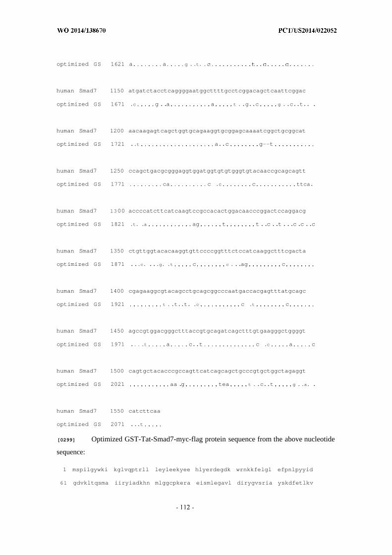
optimized GS 1621 a a g . .t.
human Smad7 1150 atgatctacctcaggggaatggcttttgcctcggacagctcaattcggac
optimized GS 1671 .c g ..a a t . .g..c g ..c..t.. .
human Smad7 1200 aacaagagtcagctggtgcagaaggtgcggagcaaaatcggctgcggcat
optimized GS 1721 ..t a..c g--t
human Smad7 1250 ccagctgacgcgggaggtggatggtgtgtgggtgtacaaccgcagcagtt
optimized GS 1771 ca c .c c ttca.
human Smad7 1300 accccatcttcatcaagtccgccacactggacaacccggactccaggacg
optimized GS 1821 .t. .a ag t t..c ..t...c.c ..c
human Smad7 1350 ctgttggtacacaaggtgttccccggtttctccatcaaggctttcgacta
optimized GS 1871 ...c. ...g. .t c c ...ag c
human Smad7 1400 cgagaaggcgtacagcctgcagcggcccaatgaccacgagtttatgcagc
optimized GS 1921 t . .t..t. .c c .t c
human Smad7 1450 agccgtggacgggctttaccgtgcagatcagctttgtgaagggctggggt
optimized GS 1971 .. . .t a c..t c .c a c
human Smad7 1500 cagtgctacacccgccagttcatcagcagctgcccgtgctggctagaggt
optimized GS 2021 aa .g tea t . .c..t g . .a. .
human Smad7 1550 catcttcaa
optimized GS 2071 ...t
[0299] Optimized GST-Tat-Smad7-myc-flag protein sequence from the above nucleotide
sequence:
1 mspilgywki kglvqptrll leyleekyee hlyerdegdk wrnkkfelgl efpnlpyyid
61 gdvkltqsma iiryiadkhn mlggcpkera eismlegavl dirygvsria yskdfetlkv

121 dflsklpeml kmfedrlchk tylngdhvth pdfmlydald vvlymdpmcl dafpklvcfk
181 krieaipqid kylksskyia wplqgwqatf gggdhppksd levlfqgplg sgrkkrrqrr
241 rsrfrtkrsa lvrrlwrsra pggedeeega gggggggeir gegatdsrah gaggggpgra
301 gcclgkavrg akghhhphpp aagagaagga eadlkalths vlkklkerql elllqavesr
361 ggtrtaclll pgrldcrlgp gapagaqpaq ppssyslpll Ickvfrwpdl rhssevkrlc
421 ccesygkinp elvccnphhl srlcelespp ppysrypmdf lkptadcpda vpssaetggt
481 nylapgglsd sqlllepgdr shwcwaywe ektrvgrlyc vqepsldify dlpqgngfcl
541 gqlnsdnksq Ivqkvrskig cgiqltrevd gvwvynrssy pifiksatld npdsrtllvh
601 kvfpgfsika fdyekayslq rpndhefmqq pwtgftvqis fvkgwgqcyt rqfisscpcw
661 levifnsrtr trpleqklis eedlaandil dykddddkv
1-229: GST+precision sites
232-668 Tat-Smad7
671-699: restriction sites+myc-Flag
[0300] Optimized GST-Tat-N-Smad7-myc-flag nucleotide sequence, cloned in
pCMV6-Entry plasmid:
1 gaattcgtcg actggatccg gtaccgagga gatctgccgc cgcgatcgcc atgtccccca
61 ttctgggcta ctggaagatt aagggcctgg tgcagcctac tagactgctg ctggaatacc
121 tggaggaaaa atatgaagag catctgtatg aaagagacga gggggataaa tggaggaaca
181 agaaattcga actgggactg gagtttccta atctgccata ctatattgac ggcgatgtga
241 agctgactca gtctatggct atcattagat acatcgcaga caaacacaac atgctgggcg
301 ggtgtcctaa ggaaagggca gagattagta tgctggaggg agccgtgctg gatattagat
361 acggcgtctc acgcatcgcc tatagcaaag acttcgaaac cctgaaggtg gattttctga
421 gcaaactgcc tgaaatgctg aagatgttcg aggacagact gtgccacaaa acctacctga
481 atggcgacca tgtcacacac ccagatttta tgctgtacga cgccctggat gtggtcctgt
541 atatggaccc catgtgtctg gatgctttcc ctaagctggt gtgctttaag aaaaggatcg
601 aggcaattcc ccagatcgat aagtacctga aaagctccaa gtatatcgct tggcctctcc
661 agggctggca ggcaacattc ggaggcgggg accatccccc taaaagcgac ctggaggtgc
721 tgtttcaggg accactgggc agcggccgga agaagcggcg gcagaggcgc cgaagtaggt
781 tccgcactaa gcggtcagca ctggtgcgga gactgtggcg atctcgggct cctggaggag
841 aggacgagga agagggagca ggcggcggcg gaggaggagg agaactgcgc ggggagggag
901 ctacagatag ccgagcccac ggagctggag gaggaggacc agggcgagcc ggatgctgtc

961 tgggcaaagc agtgagaggc gccaaggggc accatcaccc ccatccaccc gccgctggcg
1021 caggagcagc gg gg g t gaggcagacc tgaaagccct gactcacagt gtgctgaaga
1081 aactgaagga aagacagctg gagctgctgc tccaggcagt cgaatcacgc ggaggcaccc
1141 gaacagcttg tctgctgctg cccggccggc tggactgccg gctgggaccc ggcgcccctg
1201 ctggggcaca gccagcccag cctccatcta gttatagcct gcccctgctg ctgtgtaagg
1261 tgttccgatg gcctgatctg cggcattcaa gcgaagtcaa aaggctgtgc tgttgcgagt
1321 cctacggcaa gattaaccca gaactggtgt gttgcaatcc ccatcacctg tctcgactgt
1381 gtgaactgga gtccccccct ccaccctact ctaggtatcc tatggacttt ctgaagccaa
1441 ccgctgactg cccagatgca gtgccctcct ctgccgagac tggggg aactacctgg
1501 ctcctggcgg actgagcgac tcccagctgc tgctggaacc aggggatcgg ccrcrcccrctcq
1561 agcagaaact catctcagaa gaggatctgg cagcaaatga tatcctggat tacaaggatg
1621 acgacgataa ggtttaa
[0301] The entire sequence (up to the Notl site) is codon optimized for mammalian
expression, using the OPTIMUMGENE program from GenScript.
1-50: cloning sites+kozac sequence in pCMV6-Entry
51-743: GST+ precision enzyme sites
744-773-: Tat
774-1549-:human Smad7 encoding N-terminal l-258aa
1550-1563: Notl, Xhol, Bold underline: unique Notl site
1564-1637: myc-flag from pCMV6-Entry
[0302] Optimized GST-Tat-N-Smad7-myc-flag protein sequence from the above
nucleotide sequence:
1 mspilgywki kglvqptrll leyleekyee hlyerdegdk wrnkkfelgl efpnlpyyid
61 gdvkltqsma iiryiadkhn mlggcpkera eismlegavl dirygvsria yskdfetlkv
121 dflsklpeml kmfedrlchk tylngdhvth pdfmlydald vvlymdpmcl dafpklvcfk
181 krieaipqid kylksskyia wplqgwqatf gggdhppksd levlfqgplg sgrkkrrqrr
241 rsrfrtkrsa Ivrrlwrsra pggedeeega gggggggelr gegatdsrah gaggggpgra
301 gcclgkavrg akghhhphpp aagagaagga eadlkalths vlkklkerql elllqavesr
361 ggtrtaclll pgrldcrlgp gapagaqpaq ppssyslpll Ickvfrwpdl rhssevkrlc
421 ccesygkinp elvccnphhl srlcelespp ppysrypmdf lkptadcpda vpssaetggt
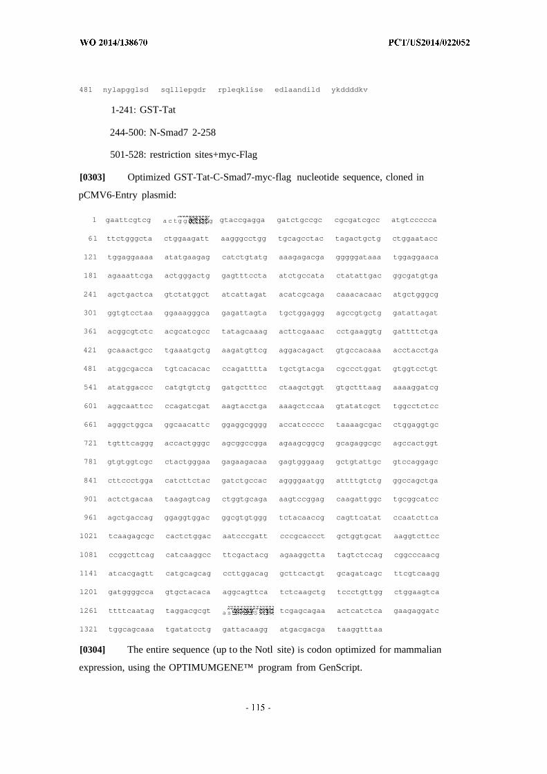
481 nylapgglsd sqlllepgdr rpleqklise edlaandild ykddddkv
1-241: GST-Tat
244-500: N-Smad7 2-258
501-528: restriction sites+myc-Flag
[0303] Optimized GST-Tat-C-Smad7-myc-flag nucleotide sequence, cloned in
pCMV6-Entry plasmid:
1 gaattcgtcg a c tg g g gtaccgagga gatctgccgc cgcgatcgcc atgtccccca
61 ttctgggcta ctggaagatt aagggcctgg tgcagcctac tagactgctg ctggaatacc
121 tggaggaaaa atatgaagag catctgtatg aaagagacga gggggataaa tggaggaaca
181 agaaattcga actgggactg gagtttccta atctgccata ctatattgac ggcgatgtga
241 agctgactca gtctatggct atcattagat acatcgcaga caaacacaac atgctgggcg
301 ggtgtcctaa ggaaagggca gagattagta tgctggaggg agccgtgctg gatattagat
361 acggcgtctc acgcatcgcc tatagcaaag acttcgaaac cctgaaggtg gattttctga
421 gcaaactgcc tgaaatgctg aagatgttcg aggacagact gtgccacaaa acctacctga
481 atggcgacca tgtcacacac ccagatttta tgctgtacga cgccctggat gtggtcctgt
541 atatggaccc catgtgtctg gatgctttcc ctaagctggt gtgctttaag aaaaggatcg
601 aggcaattcc ccagatcgat aagtacctga aaagctccaa gtatatcgct tggcctctcc
661 agggctggca ggcaacattc ggaggcgggg accatccccc taaaagcgac ctggaggtgc
721 tgtttcaggg accactgggc agcggccgga agaagcggcg gcagaggcgc agccactggt
781 gtgtggtcgc ctactgggaa gagaagacaa gagtgggaag gctgtattgc gtccaggagc
841 cttccctgga catcttctac gatctgccac aggggaatgg attttgtctg ggccagctga
901 actctgacaa taagagtcag ctggtgcaga aagtccggag caagattggc tgcggcatcc
961 agctgaccag ggaggtggac ggcgtgtggg tctacaaccg cagttcatat ccaatcttca
1021 tcaagagcgc cactctggac aatcccgatt cccgcaccct gctggtgcat aaggtcttcc
1081 ccggcttcag catcaaggcc ttcgactacg agaaggctta tagtctccag cggcccaacg
1141 atcacgagtt catgcagcag ccttggacag gcttcactgt gcagatcagc ttcgtcaagg
1201 gatggggcca gtgctacaca aggcagttca tctcaagctg tccctgttgg ctggaagtca
1261 ttttcaatag taggacgcgt a s S tcgagcagaa actcatctca gaagaggatc
1321 tggcagcaaa tgatatcctg gattacaagg atgacgacga taaggtttaa
[0304] The entire sequence (up to the Notl site) is codon optimized for mammalian
expression, using the OPTIMUMGENE™ program from GenScript.

1-50: cloning sites+kozac sequence in pCMV6-Entry
51-743: GST+ precision enzyme sites
744-773: Tat
774-1274: c-terminal Smad7
1275: Mlu
1283: Notl
1275-1370: mlul, Notl, Xhol, myc+flag
[0305] Optimized GST-Tat-C-Smad7-myc-flag protein sequence from the above
nucleotide sequence:
1 mspilgywki kglvqptrll leyleekyee hlyerdegdk wrnkkfelgl efpnlpyyid
61 gdvkltqsma iiryiadkhn mlggcpkera eismlegavl dirygvsria yskdfetlkv
121 dflsklpeml kmfedrlchk tylngdhvth pdfmlydald vly dp cl dafpklvcfk
181 krieaipqid kylksskyia wplqgwqatf gggdhppksd levlfqgplg sgrkkrrqrr
241 rshwcwayw eektrvgrly cvqepsldif ydlpqgngfc lgqlnsdnks qlvqkvrski
301 gcgiqltrev dgvwvynrss ypifiksatl dnpdsrtllv hkvfpgfsik afdyekaysl
361 qrpndhefmq qpwtgftvqi sfvkgwgqcy trqfisscpc wlevifnsrt rtrpleqkli
421 seedlaandi ldykddddkv
1-241: GST+precision+Tat
242-409: c-Smad7 (259-426aa)
410-440: Mlu+Not+myc+flag: restriction sites+myc-Flag
[0306] Based on the Smad7 structure, it is anticipated that the linker (203-258aa), which
contains the PY domain (203-2 17aa), will have therapeutic effects by blocking
TGF-p-induced inflammation or growth inhibition (FIG. 15).
[0307] The amino acid sequence for the Tat-Smad7-linker peptide (203-258aa) is:
GRKKRRQRRR-
ELESPPPPYSRYPMDFLKPTADCPDAVPSSAETGGTNYLAPGGLSDSQLLLEPGDR
1-10: Tat
[0308] The codon optimized nucleotide sequence for the above peptide in bacterial
production is:
1 ggccgtaaaa aacgccgtca acgccgccgt gaactggaga gcccgccgcc gccgtatagc
61 cgttacccga tggatttcct gaaaccgact gcagattgcc cggacgcagt cccgagcagc
121 gctgagaccg gcggcaccaa ctatctggca ccgggcggtc tgagcgatag ccagctgctg
181 ctggaaccgg gcgaccgtgg taagcctatc cctaaccctc tcctcggtct cgattctacg
241 tgagtcgac
1-30: Tat
31-198: Smad7 linker
199-249: V5
[0309] The codon optimized nucleotide sequence for the above peptide in mammalian
cell production is:
1 ggccggaaga agcggcggca gaggcgccga gaactggagt ccccccctcc accctactct
61 aggtatccta tggactttct gaagccaacc gctgactgcc cagatgcagt gccctcctct
121 gccgagactg ggggaaccaa ctacctggct cctggcggac tgagcgactc ccagctgctg
181 ctggaaccag gggatcggcg gccgctcgag cagaaactca tctcagaaga ggatctggca
241 gcaaatgata tcctggatta caaggatgac gacgataagg tttaa
1-30: mammalian codon optimized Tat
31-198: Smad7 linker
199-285: Notl, Myc tag, Flag tag
[0310] The amino acid sequence for the Tat-Smad7-PY peptide (203-2 17aa) is:
GRKKRRQRRR-ELESPPPPYSRYPMD
1-10: Tat
[0311] The codon optimized nucleotide sequence for the above peptide in bacterial
production is:
1 ggccgtaaaa aacgccgtca acgccgccgt gaactggaga gcccgccgcc gccgtatagc
61 cgttacccga tggatggtaa gcctatccct aaccctctcc tcggtctcga ttctacgtga
121 gtcgac
1-30: Tat
31-75: Smad7-PY
76-126: V5
[0312] The codon optimized nucleotide sequence for the above peptide in mammalian
cell production is:
1 ggccggaaga agcggcggca gaggcgccga gaactggagt ccccccctcc accctactct
61 aggtatccta tggaccggcc gctcgagcag aaactcatct cagaagagga tctggcagca
121 aatgatatcc tggattacaa ggatgacgac gataaggttt aa
1-30: mammalian codon optimized Tat
31-75: Smad7 PY
76-162: Notl, Myc tag, Flag tag
[0313] The foregoing discussion of the present technology has been presented for
purposes of illustration and description. The foregoing is not intended to limit the present
technology to the form or forms disclosed herein. Although the description of the present
technology has included description of one or more embodiments and certain variations and
modifications, other variations and modifications are within the scope of the present
technology, e.g., as may be within the skill and knowledge of those in the art, after
understanding the present disclosure. It is intended to obtain rights which include
alternative embodiments to the extent permitted, including alternate, interchangeable and/or
equivalent structures, functions, ranges or steps to those claimed, whether or not such
alternate, interchangeable and/or equivalent structures, functions, ranges or steps are
disclosed herein, and without intending to publicly dedicate any patentable subject matter.

CLAIMS
What is claimed is:
1. A nucleic acid molecule comprising a codon-optimized human Smad7 cDNA
nucleotide sequence.
2. The nucleic acid molecule of claim 1, wherein the codon-optimized human Smad7
nucleotide sequence includes one or more codon for arginine optimized for expression
in one or more of bacteria or yeast.
3. The nucleic acid molecule of either one of claims 1 or 2, wherein the codon-optimized
human Smad7 nucleotide sequence includes one or more codon for serine optimized
for expression in one or more of bacteria or yeast.
4. The nucleic acid molecule of any one of claims 1 to 3, wherein the codon-optimized
human Smad7 nucleotide sequence includes one or more codon for histidine
optimized for expression in one or more of bacteria or yeast.
5. The nucleic acid molecule of any one of claims 1 to 4, wherein the codon-optimized
human Smad7 nucleotide sequence includes one or more codons for serine, one or
more codons for histidine, and one or more codons for arginine optimized for
expression in one or more of bacteria or yeast.
6. The nucleic acid molecule of claim 5, wherein the codon-optimized human Smad7
nucleotide sequence includes 28 serine codons, 6 histidine codons, and 9 arginine
codons optimized for expression in one or more of bacteria or yeast.
7. The nucleic acid molecule of claim 1, wherein the codon-optimized human Smad7
nucleotide sequence is selected from the group consisting of SEQ ID NOS: 9-11, 21,
23-41).
8. The nucleic acid molecule of claim 1, wherein the codon-optimized human Smad7
nucleotide sequence has about 65 to 75 percent homology to human Smad7 cDNA.
9. The nucleic acid molecule of claim 1, wherein the nucleotide sequence comprises a
nucleotide sequence encoding an N-terminal fragment SMAD7.

10. The nucleic acid molecule of claim 1, wherein the nucleotide sequence comprises a
nucleotide sequence encoding a C-terminal fragment of SMAD7.
11. The nucleic acid molecule of claim 1, wherein the nucleotide sequence comprises
nucleotides encoding amino acids 2-258 of the human Smad7 protein.
12. The nucleic acid molecule of claim 1, wherein the nucleotide sequence comprises
nucleotides encoding amino acids 259-426 of the human Smad7 protein.
13. The nucleic acid molecule of claim 1, wherein the nucleotide sequence comprises
nucleotides encoding amino acids 204-258 of the human Smad7 protein.
14. The nucleic acid molecule of any one of claims 1 to 14, wherein the nucleotide
sequence further comprises a nucleotide sequence encoding a protein transduction
domain.
15. The nucleic acid molecule of claim 14, wherein the protein transduction domain is
Tat.
16. The nucleic acid molecule of any one of claims 1 to 15, wherein the nucleotide
sequence further comprises a nucleotide sequence encoding one or more of an epitope
tag or a purification tag.
17. The nucleic acid of claim 16, wherein the epitope tag is V5.
18. The nucleic acid of claim 16, wherein the purification tag is one or more of
glutathione-S-transferase or 6-Histidine.
19. The nucleic acid molecule of any one of claims 1- 18, wherein the nucleic acid
molecule is one or more of isolated or purified.
20. The nucleic acid of any one of claims 1- 19, wherein the nucleic acid molecule
encodes a polypeptide having one or more biological activity selected from the group
consisting of reducing or eliminating phosphorylation of Smad2, reducing or
eliminating nuclear translocation of the NFKB p50 subunit, increasing cell
proliferation, reducing apoptosis, reducing radiation-induced DNA damage, reducing
inflammation, reducing angiogenesis, promoting healing in oral mucositis, promoting
wound healing, and treating auto-immune disease.
21. A pharmaceutical composition comprising the nucleic acid molecule of any one of
claims 1-20, and one or more pharmaceutically acceptable excipients.
22. An expression vector comprising the nucleic acid molecule of any one of claims 1 to
20 operably linked to a promoter.
23 . A host cell comprising the expression vector of claim 22.
24. A pharmaceutical composition comprising the expression vector of claim 22 and one
or more pharmaceutically acceptable excipients.
25. A protein molecule comprising a human Smad7 protein having leucine at position
216.
26. The protein molecule of claim 25, wherein the human Smad7 protein is truncated at
the C-terminal.
27. The protein molecule of claim 25, wherein the human Smad7 protein is truncated at
the N-terminal.
28. The protein molecule of any one of claims 26 to 27, wherein the truncated human
Smad7 protein includes about 50% of the full-length Smad7 sequence.
29. The protein molecule of any one of claims 26 to 27, wherein the truncated human
Smad7 protein includes about 13% of the full-length Smad7 sequence.
30. A protein molecule comprising a human Smad7 protein having amino acids 2-258 of
the human Smad7 protein.
31. The protein molecule of claim 30, wherein the Smad7 protein comprises amino acids
204-258.
32. A protein molecule comprising a human Smad7 protein having amino acids 259-426
of the human Smad7 protein.
33 . The protein molecule of any one of claims 25 to 32, wherein the protein molecule has
one or more biological activity selected from the group consisting of reducing or
eliminating phosphorylation of Smad2, reducing or eliminating nuclear translocation
of the NFKB p50 subunit, increasing cell proliferation, reducing apoptosis, reducing

radiation-induced DNA damage, reducing inflammation, reducing angiogenesis,
promoting healing in oral mucositis, promoting wound healing, and treating
auto-immune disease.
34. The protein molecule of any one of claims 25 to 33, wherein the amino acid sequence
further comprises a protein transduction domain.
35. The protein molecule of claim 34, wherein the protein transduction domain is Tat.
36. The protein molecule of any one of claims 25 to 35, wherein the amino acid sequence
further comprises one or more of an epitope tag or a purification tag.
37. The protein molecule of claim 36, wherein the epitope tag is v5.
38. The protein molecule of claim 36, wherein the purification tag is one or more of
glutathione-S-transferase or 6-histidine.
39. A pharmaceutical composition comprising the protein molecule of any one of claims
25 to 38, and one or more pharmaceutically acceptable excipient.
40. A method for treating or preventing an inflammatory condition in a subject
comprising providing to the subject a therapeutically effective amount of the
pharmaceutical composition of any one of claims 21, 24 or 39.
41. The method of claim 40, wherein the inflammatory condition is one or more of a
chronic wound, skin inflammation, psoriasis, or an autoimmune disease.
42. The method of claim 40, wherein the composition reduces inflammation through
inhibition of TGF and NFKB signaling.
43. A method for preventing or treating a disease or disorder in a subject comprising one
or more of increasing one or more of cell proliferation or cell migration, or preventing
one or more of apoptosis or DNA damage in the subject comprising providing to the
subject a therapeutically effective amount of the pharmaceutical composition of any
one of claims 21, 24 or 39, wherein one or more of increasing one or more of cell
proliferation or cell migration, or preventing one or more of apoptosis or DNA
damage is useful in preventing or treating the disease or disorder.

44. The method of claim 43, wherein the disease or disorder includes one or more of
chronic wounds, acute wounds, or mucositis.
45. The method of claim 44, wherein the chronic wounds include one or more of diabetic
ulcers, pressure ulcers, venous ulcers, or oral ulcers.
46. The method of claim 44, wherein the acute wounds include one or more of
trauma-induced wounds, surgical wounds, or scarring.
47. The method of claim 44, wherein the mucositis includes one or more of
radiation-induced mucositis or chemotherapy-induced mucositis.
48. The method of claim 44, wherein the mucositis includes one or more of oral mucositis
or gut mucositis.
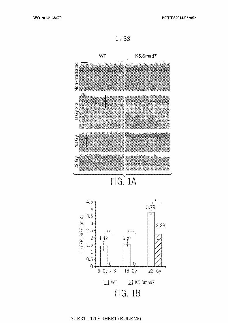
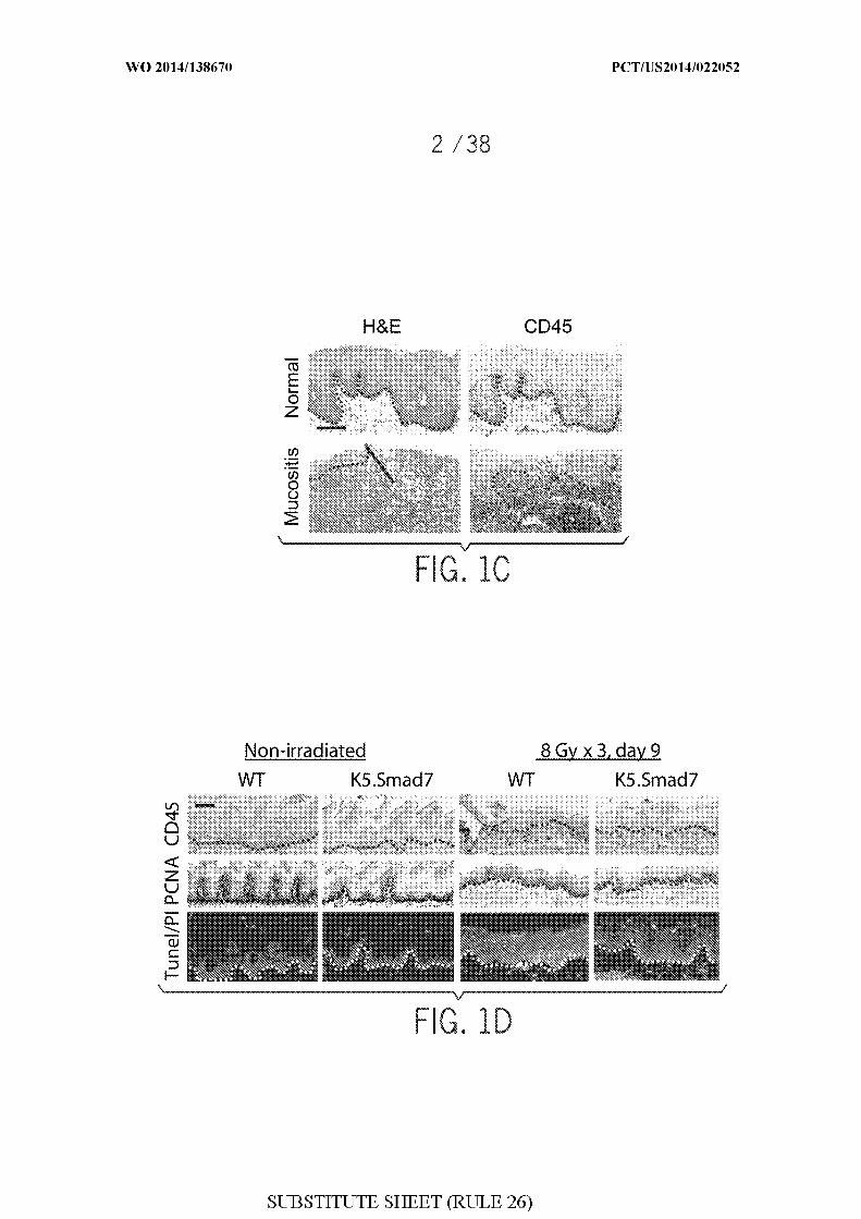


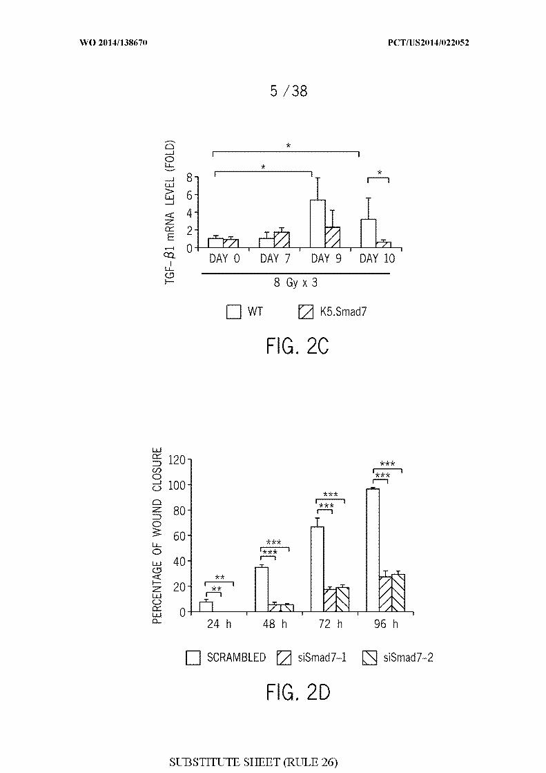
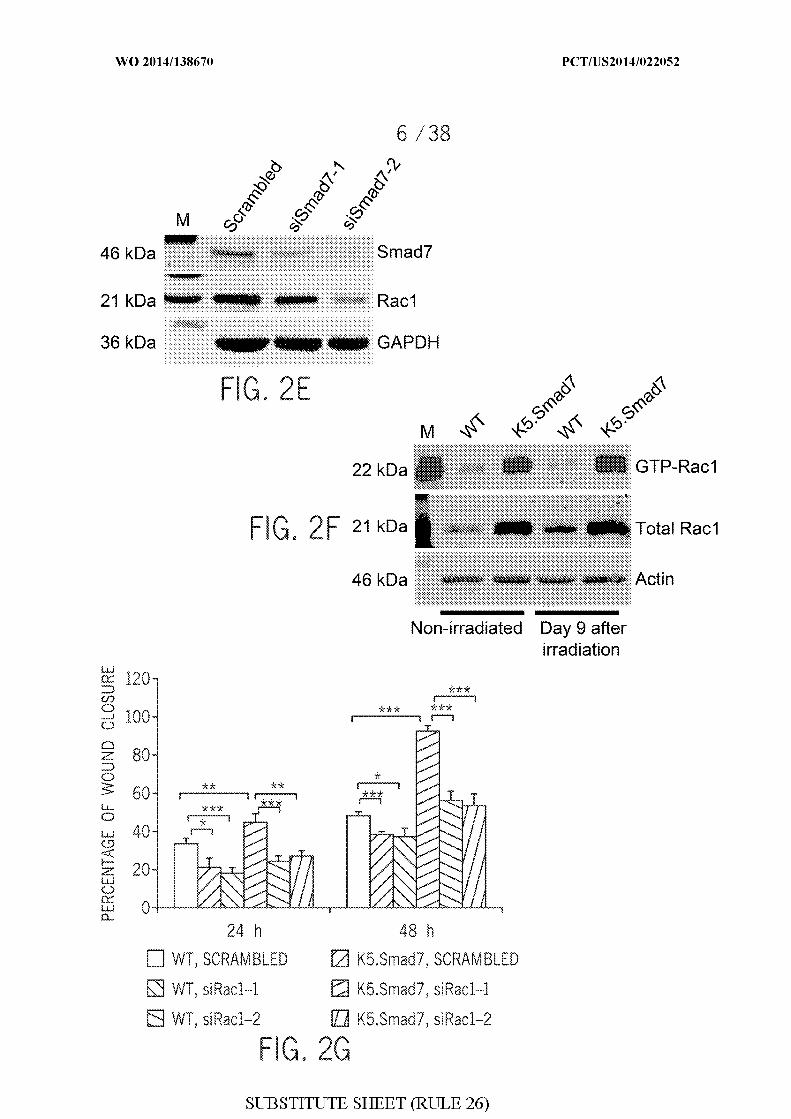


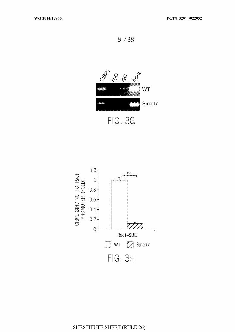
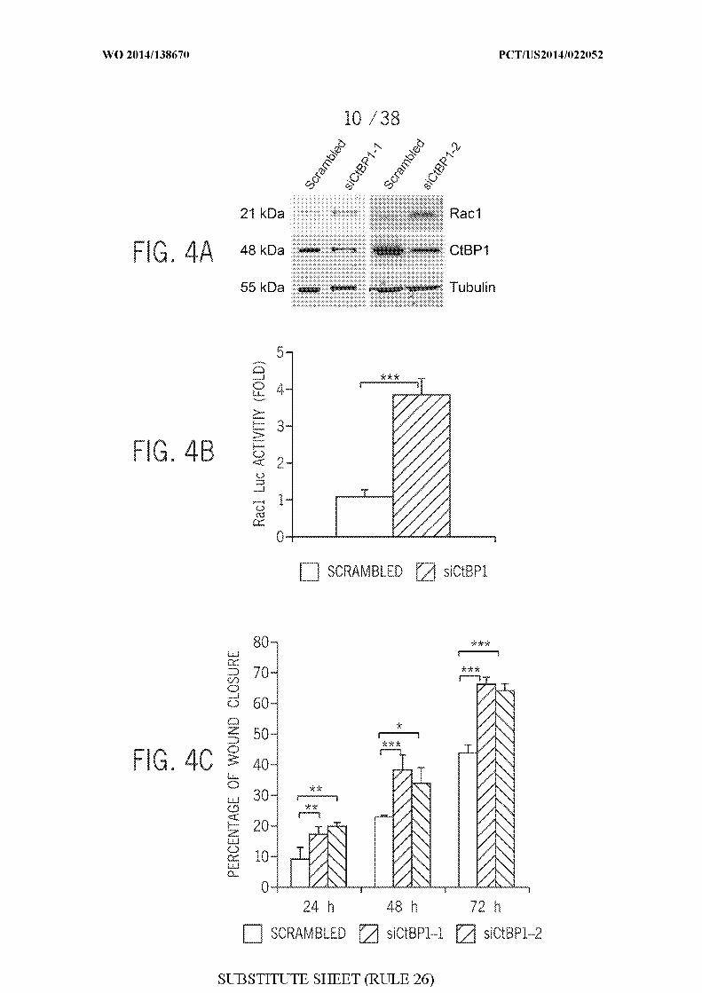

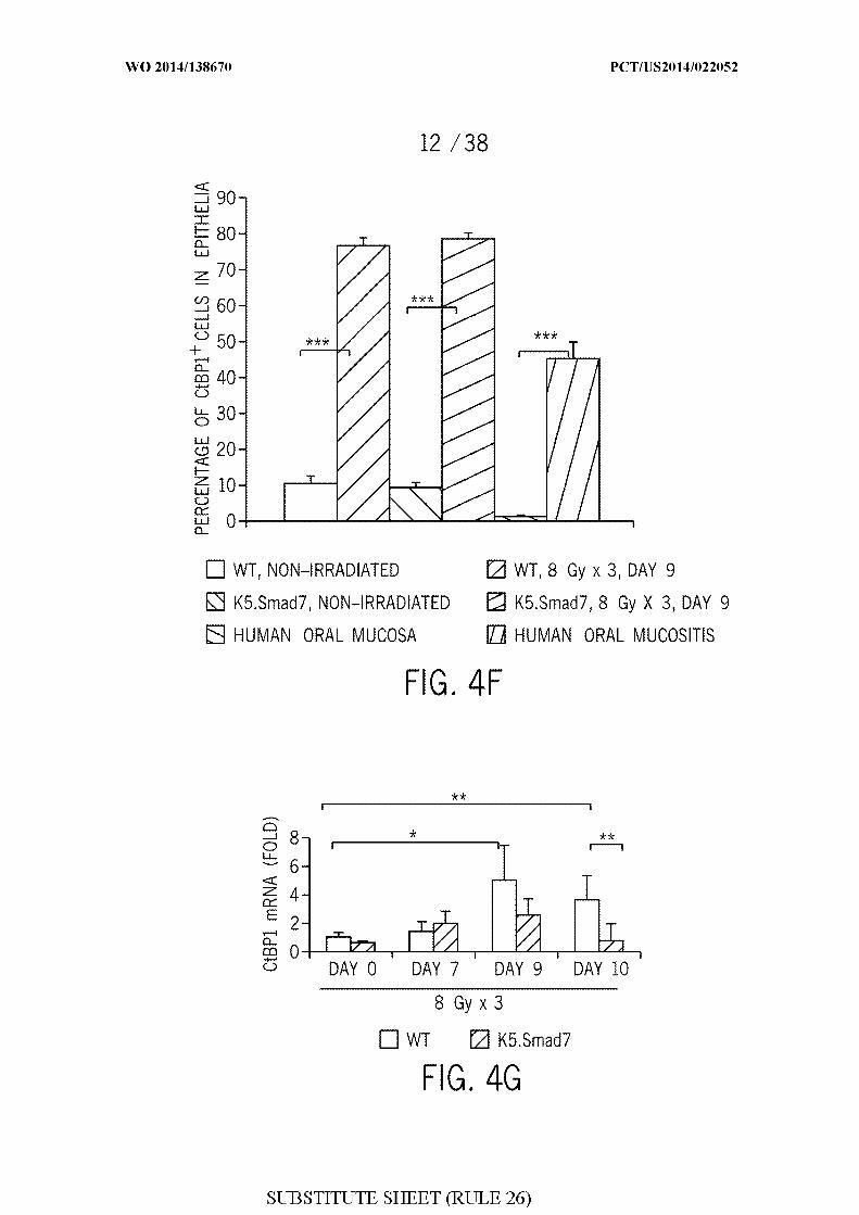
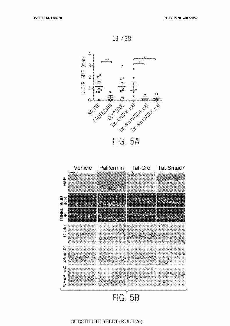
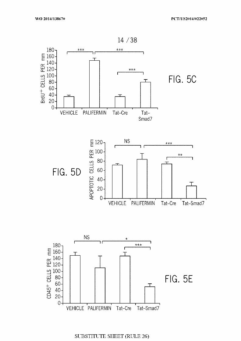


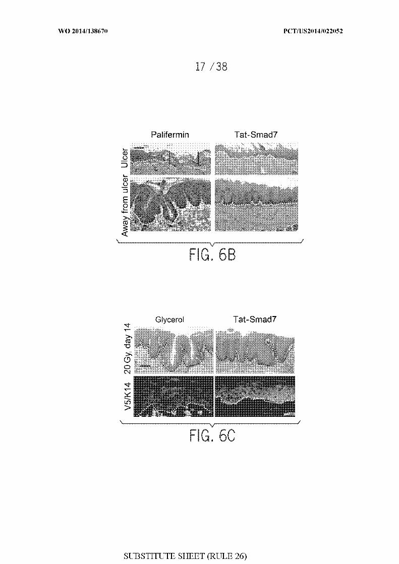

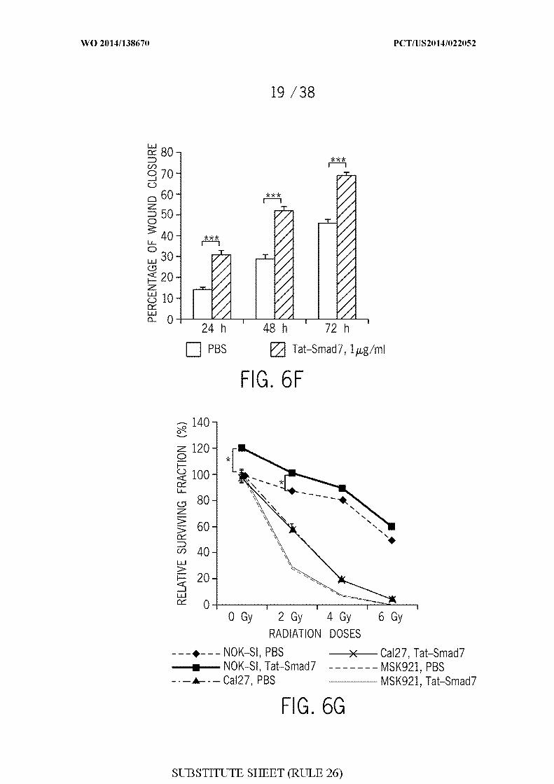

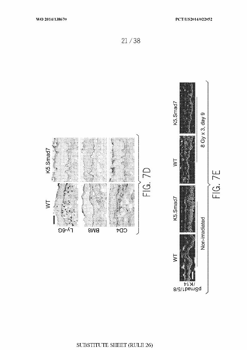
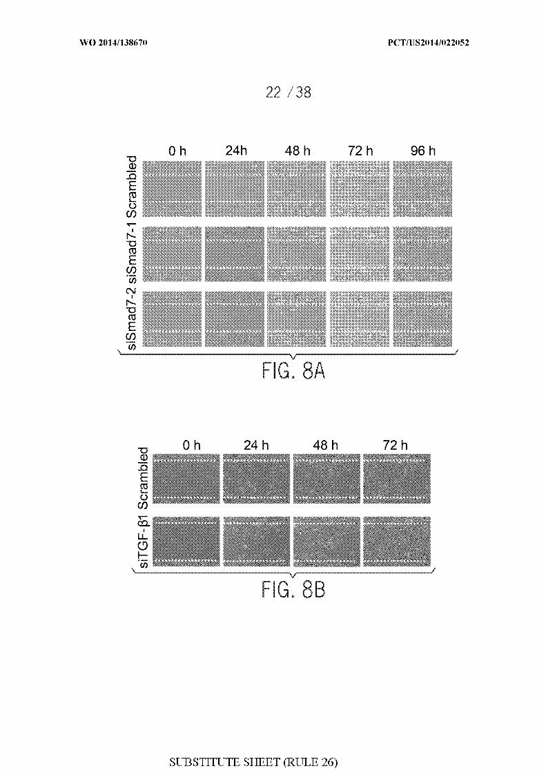
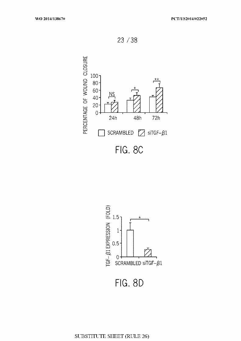

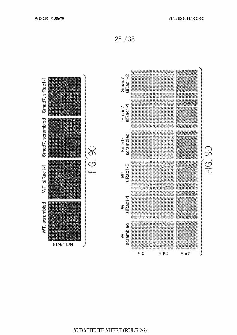

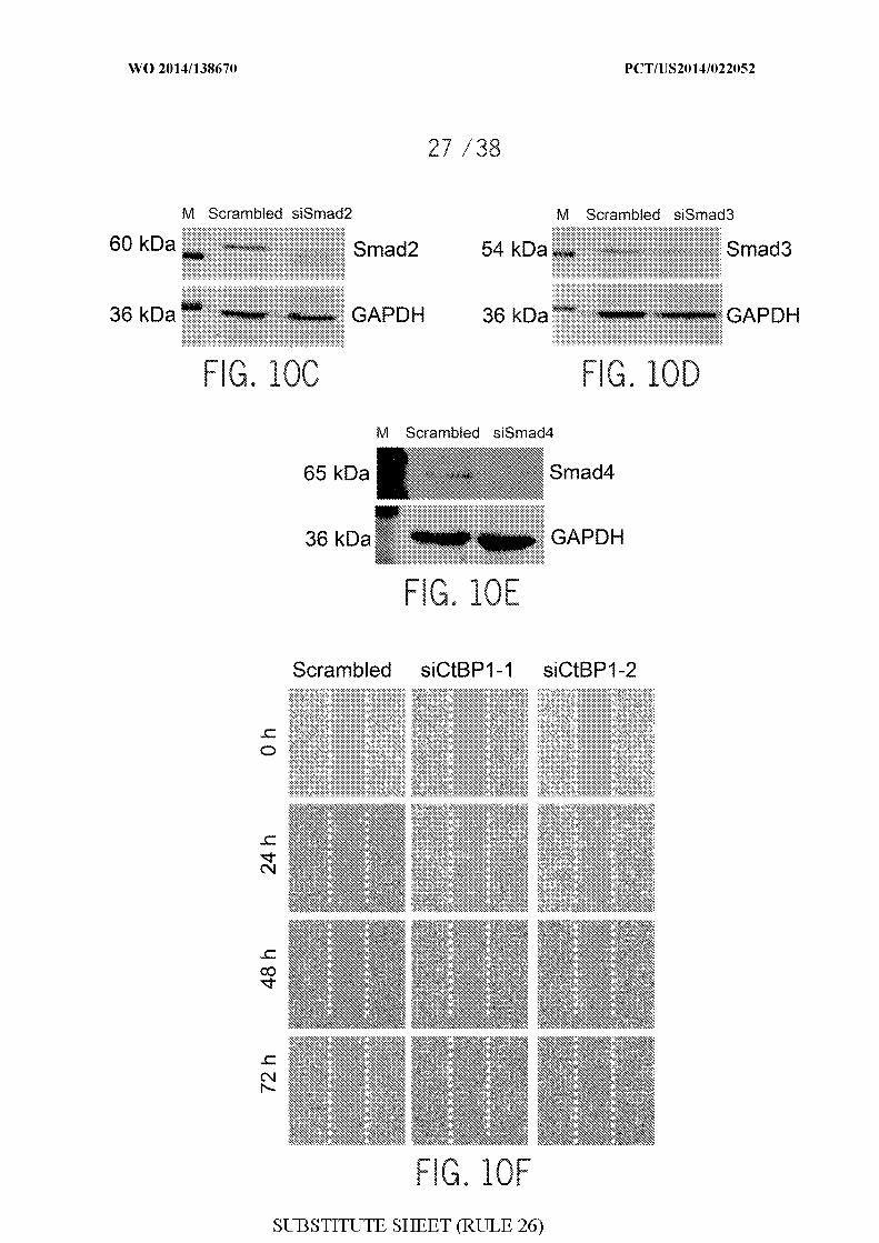
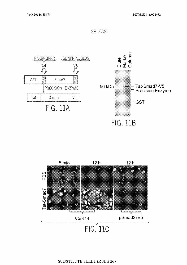
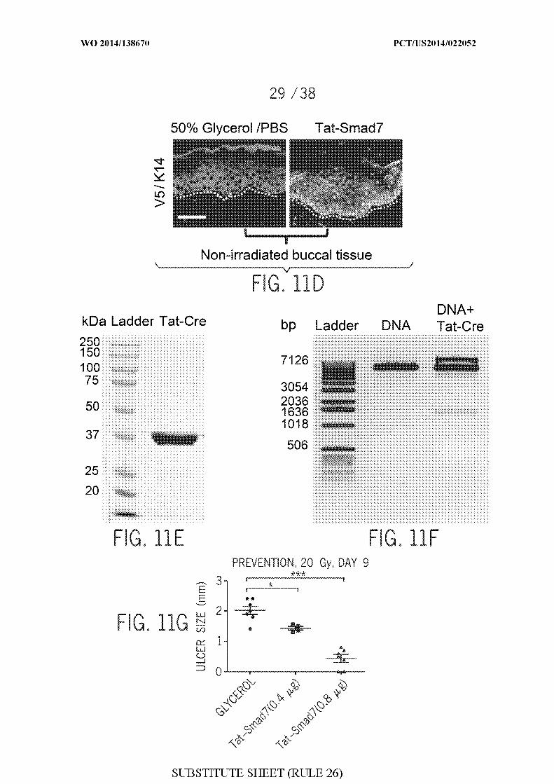

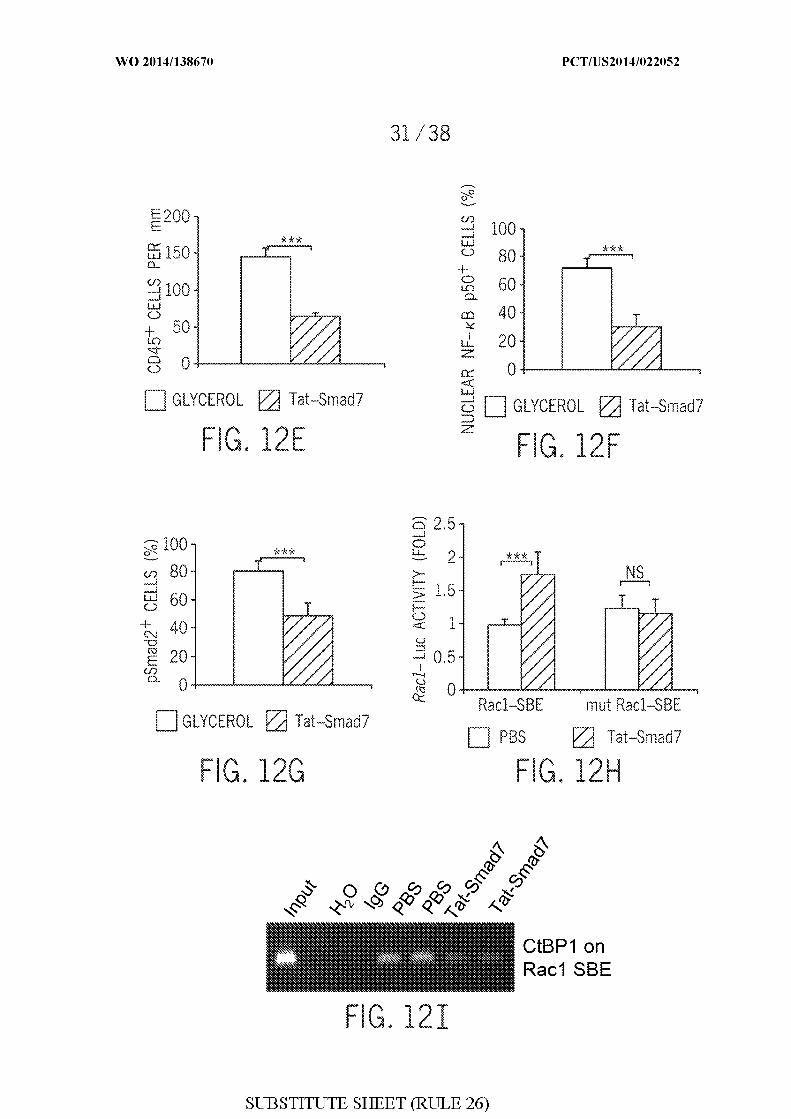
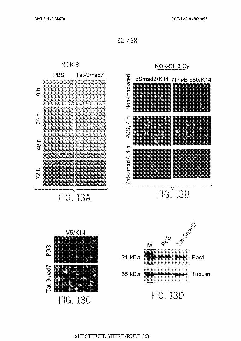
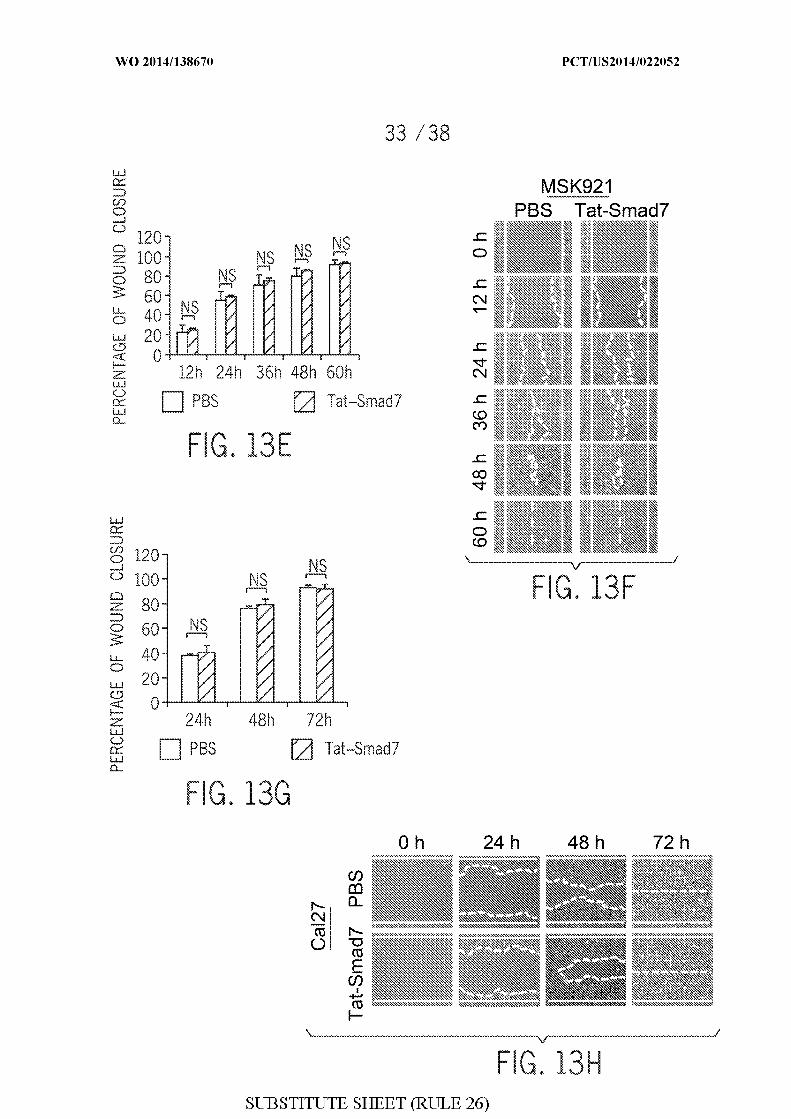
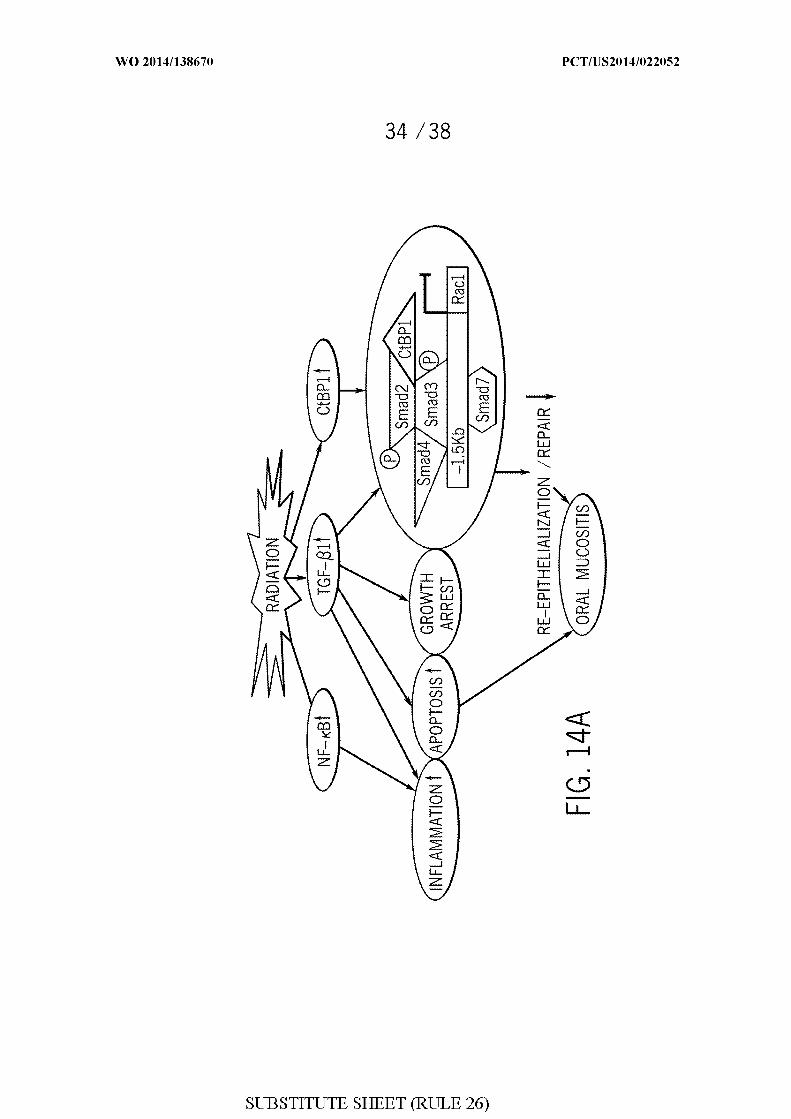

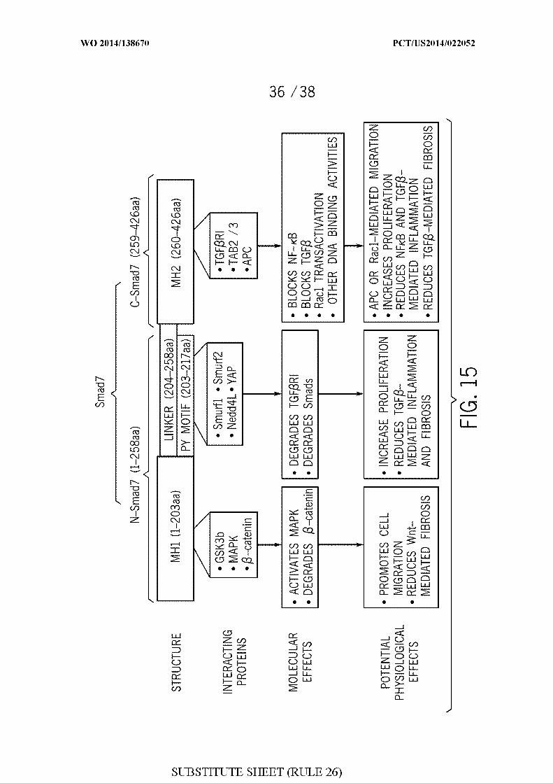


INTERNATIONAL SEARCH REPORT International application No.
PCT/US 14/22052
A . CLASSIFICATION O F SUBJECT MATTERIPC(8) - C12P 19/34 (2014.01 )USPC - 435/91 . 1 , 69.1
According to International Patent Classification (IPC) or to both national classification and IPC
B. FIELDS SEARCHED
Minimum documentation searched (classification system followed by classification symbols)
IPC(8): C12P 19/34, 21/04; C12N 5/07; A61K 48/00, 39/00 (2014.01)USPC: 435/91 .1, 89, 85, 84, 72, 4 1, 325, 326, 328, 41, 69.1, 69.7; 424/192.1, 184.1
Documentation searched other than minimum documentation to the extent that such documents are included in the fields searched
Electronic data base consulted during the international search (name of data base and, where practicable, search terms used)
MicroPatent (US-G, US-A, EP-A, EP-B, WO, JP-bib, DE-C.B, DE-A, DE-T, DE-U, GB-A, FR-A); Google; Google Scholar; GooglePatents; PubMed; ProQuest; 'codon-optimized,' 'smad7,' methionine, leucine, stabilized, mutation, nucleic, human
C . DOCUMENTS CONSIDERED TO BE RELEVANT
Category* Citation of document, with indication, where appropriate, of the relevant passages Relevant to claim No.
WO 2012/040295 A2 (WANG, XJ et al.) March 29, 2012; paragraphs [00033], [00036], [00037], 1-3, 7, 8[000122]
9-13
25-29
US 6251628 B 1 (NAKAO, A e t al.) June 26, 2001; column 3, lines 51-52; figure 1 30-32
9-13
25-29
ZHANG, S et al. Smad7 Antagonizes Transforming Growth Factor Beta Signaling In The 25-29Nucleus By Interfering With Functions Smad-DNA Complex Formation. June 2007. Molecularand Cellular Biology. Vol. 27; pages 4488-4499; figure 2A; DOI:10.1 128/MCB.01636-06.
Further documents are listed in the continuation of Box C . | |
Special categories of cited documents: "T" later document published after the international filing date or prioritydocument defining the general state of the art which is not considered date and not in conflict with the application but cited to understandto be of particular relevance the principle or theory underlying the invention
Έ earlier application or patent but published on or after the international "X" document of particular relevance; the claimed invention cannot befiling date considered novel or cannot be considered to involve an inventive
"L" document which may throw doubts on priority claim(s) or which is step when the document is taken alonecited to establish the publication date of another citation or otherspecial reason (as specified) Ύ " document of particular relevance; the claimed invention cannot be
considered to involve an inventive step when the document is"O" document referring to an oral disclosure, use, exhibition or other combined with one or more other such documents, such combination
means being obvious to a person skilled in the art"P" document published prior to the international filing date but later than & d o m t mem ber of the same patent family
the priority date claimed
Date of the actual completion of the international search Date of mailing of the international search report
24 June 2014 (24.06.2014) J UL 2014Name and mailing address o f the ISA/US Authorized officer:
Mail Stop PCT, Attn: ISA/US, Commissioner for Patents Shane ThomasP.O. Box 1450, Alexandria, Virginia 22313-1450
Facsimile No. 571-273-3201
Form PCT/1SA/2I0 (second sheet) (July 2009)

INTERNATIONAL SEARCH REPORT International application No.
PCT/US14/22052
Box No. II Observations where certain claims were found unsearchable (Continuation of item 2 of first sheet)
This international search report has not been established in respect of certain claims under Article 17(2)(a) for the following reasons:
1. Claims Nos.:because they relate to subject matter not required to be searched by this Authority, namely:
□ Claims Nos.:because they relate to parts of the international application that do not comply with the prescribed requirements to such anextent that no meaningful international search can be carried out, specifically:
3. Claims Nos.: 4-6, 14-24, 33-48because they are dependent claims and are not drafted in accordance with the second and third sentences of Rule 6.4(a).
Box No. Ill Observations where unity of invention is lacking (Continuation of item 3 of first sheet)
This International Searching Authority found multiple inventions in this international application, as follows:
As all required additional search fees were timely paid by the applicant, this international search report covers all searchableclaims.
As all searchable claims could be searched without effort justifying additional fees, this Authority did not invite payment ofadditional fees.
As only some of the required additional search fees were timely paid by the applicant, this international search report coversonly those claims for which fees were paid, specifically claims Nos.:
No required additional search fees were timely paid by the applicant. Consequently, this international search report isrestricted to the invention first mentioned in the claims; it is covered by claims Nos.:
Remark on Protest I The additional search fees were accompanied by the applicant's protest and, where applicable, thepayment of a protest fee.
I I The additional search fees were accompanied by the applicant's protest but the applicable protestfee was not paid within the time limit specified in the invitation.
No protest accompanied the payment of additional search fees.
Form PCT/ISA/210 (continuation of first sheet (2)) (July 2009)