WO 2014/165679 Al
196
(12) INTERNATIONAL APPLICATION PUBLISHED UNDER THE PATENT COOPERATION TREATY (PCT) (19) World Intellectual Property Organization International Bureau (10) International Publication Number (43) International Publication Date 9 October 2014 (09.10.2014) WO 2014/165679 Al PO PCT (51) International Patent Classification: AO, AT, AU, AZ, BA, BB, BG, BH, BN, BR, BW, BY, A61K 39/00 (2006.01) A61K 39/38 (2006.01) BZ, CA, CH, CL, CN, CO, CR, CU, CZ, DE, DK, DM, DO, DZ, EC, EE, EG, ES, FI, GB, GD, GE, GH, GM, GT, (21) International Application Number: HN, HR, HU, ID, IL, IN, IR, IS, JP, KE, KG, KN, KP, KR, PCT/US2014/032838 KZ, LA, LC, LK, LR, LS, LT, LU, LY, MA, MD, ME, (22) International Filing Date: MG, MK, MN, MW, MX, MY, MZ, NA, NG, NI, NO, NZ, 3 April 2014 (03.04.2014) OM, PA, PE, PG, PH, PL, PT, QA, RO, RS, RU, RW, SA, SC, SD, SE, SG, SK, SL, SM, ST, SV, SY, TH, TJ, TM, (25) Filing Language: English TN, TR, TT, TZ, UA, UG, US, UZ, VC, VN, ZA, ZM, (26) Publication Language: English ZW. (30) Priority Data: (84) Designated States (unless otherwise indicated, for every 61/808,1 18 3 April 2013 (03.04.2013) US kind of regional protection available): ARIPO (BW, GH, GM, KE, LR, LS, MW, MZ, NA, RW, SD, SL, SZ, TZ, (71) Applicant: ALLERTEIN THERAPEUTICS, LLC UG, ZM, ZW), Eurasian (AM, AZ, BY, KG, KZ, RU, TJ, [US/US]; 640 Sasco Hill Road, Fairfield, Connecticut TM), European (AL, AT, BE, BG, CH, CY, CZ, DE, DK, 06824 (US). EE, ES, FI, FR, GB, GR, HR, HU, IE, IS, IT, LT, LU, LV, MC, MK, MT, NL, NO, PL, PT, RO, RS, SE, SI, SK, SM, (72) Inventors: SOSIN, Howard; c/o Allertein Therapeutics, TR), OAPI (BF, BJ, CF, CG, CI, CM, GA, GN, GQ, GW, LLC, 640 Sasco Hill Road, Fairfield, Connecticut 06824 KM, ML, MR, NE, SN, TD, TG). (US). CAPLAN, Michael; c/o Allertein Therapeutics, LLC, 640 Sasco Hill Road, Fairfield, Connecticut 06824 Published: (US). FAHMY, Tarek; c/o Allertein Therapeutics, LLC, — with international search report (Art. 21(3)) 640 Sasco Hill Road, Fairfield, Connecticut 06824 (US). — before the expiration of the time limit for amending the (74) Agents: REESE, Brian E. et al; Choate, Hall & Stewart claims and to be republished in the event of receipt of LLP, Two International Place, Boston, Massachusetts amendments (Rule 48.2(h)) 021 10 (US). (81) Designated States (unless otherwise indicated, for every kind of national protection available): AE, AG, AL, AM, (54) Title: NOVEL NANOPARTICLE COMPOSITIONS (57) Abstract: The present invention provides, among other things, nanoparticle compositions including a plurality of nanoparticles, o each of which is comprised of a biodegradable or biocompatible polymer arranged in a nanoparticle structure defining an internal lu - men and an external surface and one or more of a preparation of hydrophilic cellular components and a preparation of hydrophobic o cellular components. In some embodiments, the preparation of hydrophilic cellular components is encapsulated within the internal lumen and the preparation of hydrophobic cellular components is associated with the external surface. Various methods of making and using disclosed nanoparticle compositions are also provided.
-
Upload
khangminh22 -
Category
Documents
-
view
0 -
download
0
Transcript of WO 2014/165679 Al
(12) INTERNATIONAL APPLICATION PUBLISHED UNDER THE PATENT COOPERATION TREATY (PCT)
(19) World Intellectual PropertyOrganization
International Bureau(10) International Publication Number
(43) International Publication Date9 October 2014 (09.10.2014)
WO 2014/165679 AlP O P C T
(51) International Patent Classification: AO, AT, AU, AZ, BA, BB, BG, BH, BN, BR, BW, BY,A61K 39/00 (2006.01) A61K 39/38 (2006.01) BZ, CA, CH, CL, CN, CO, CR, CU, CZ, DE, DK, DM,
DO, DZ, EC, EE, EG, ES, FI, GB, GD, GE, GH, GM, GT,(21) International Application Number: HN, HR, HU, ID, IL, IN, IR, IS, JP, KE, KG, KN, KP, KR,
PCT/US2014/032838 KZ, LA, LC, LK, LR, LS, LT, LU, LY, MA, MD, ME,
(22) International Filing Date: MG, MK, MN, MW, MX, MY, MZ, NA, NG, NI, NO, NZ,
3 April 2014 (03.04.2014) OM, PA, PE, PG, PH, PL, PT, QA, RO, RS, RU, RW, SA,SC, SD, SE, SG, SK, SL, SM, ST, SV, SY, TH, TJ, TM,
(25) Filing Language: English TN, TR, TT, TZ, UA, UG, US, UZ, VC, VN, ZA, ZM,
(26) Publication Language: English ZW.
(30) Priority Data: (84) Designated States (unless otherwise indicated, for every
61/808,1 18 3 April 2013 (03.04.2013) US kind of regional protection available): ARIPO (BW, GH,GM, KE, LR, LS, MW, MZ, NA, RW, SD, SL, SZ, TZ,
(71) Applicant: ALLERTEIN THERAPEUTICS, LLC UG, ZM, ZW), Eurasian (AM, AZ, BY, KG, KZ, RU, TJ,[US/US]; 640 Sasco Hill Road, Fairfield, Connecticut TM), European (AL, AT, BE, BG, CH, CY, CZ, DE, DK,06824 (US). EE, ES, FI, FR, GB, GR, HR, HU, IE, IS, IT, LT, LU, LV,
MC, MK, MT, NL, NO, PL, PT, RO, RS, SE, SI, SK, SM,(72) Inventors: SOSIN, Howard; c/o Allertein Therapeutics,
TR), OAPI (BF, BJ, CF, CG, CI, CM, GA, GN, GQ, GW,LLC, 640 Sasco Hill Road, Fairfield, Connecticut 06824
KM, ML, MR, NE, SN, TD, TG).(US). CAPLAN, Michael; c/o Allertein Therapeutics,LLC, 640 Sasco Hill Road, Fairfield, Connecticut 06824 Published:(US). FAHMY, Tarek; c/o Allertein Therapeutics, LLC, — with international search report (Art. 21(3))640 Sasco Hill Road, Fairfield, Connecticut 06824 (US).
— before the expiration of the time limit for amending the(74) Agents: REESE, Brian E. et al; Choate, Hall & Stewart claims and to be republished in the event of receipt of
LLP, Two International Place, Boston, Massachusetts amendments (Rule 48.2(h))021 10 (US).
(81) Designated States (unless otherwise indicated, for everykind of national protection available): AE, AG, AL, AM,
(54) Title: NOVEL NANOPARTICLE COMPOSITIONS
(57) Abstract: The present invention provides, among other things, nanoparticle compositions including a plurality of nanoparticles,
o each of which is comprised of a biodegradable or biocompatible polymer arranged in a nanoparticle structure defining an internal lu -men and an external surface and one or more of a preparation of hydrophilic cellular components and a preparation of hydrophobic
o cellular components. In some embodiments, the preparation of hydrophilic cellular components is encapsulated within the internallumen and the preparation of hydrophobic cellular components is associated with the external surface. Various methods of makingand using disclosed nanoparticle compositions are also provided.
NOVEL NANOPARTICLE COMPOSITIONS
BACKGROUND
[0001] Many medical benefits could be realized if the immune system could be trained to
respond to antigens in a desired manner, such as by developing tolerance to (e.g., for an allergic
antigen or auto-antigen), or by learning to reject (e.g., for a disease-associated antigen) the
antigen. The body can react to a wide variety of antigens, whether exogenous antigens (e.g.,
allergens, infectious agent antigens, etc) or endogenous antigens (e.g., auto-antigens, certain
disease-associate antigens, etc). Diverse approaches have been applied in order to meet this
challenge, including systemic drug treatments, injection of antigens, antibody therapies, etc.
However, there remains a need for improved approaches.
SUMMARY
[0002] The present invention provides a novel system for modulating (including
inducing, promoting or suppressing) immune responses to antigens. In particular, in some
embodiments, the invention provides technologies that combine features of certain nanoparticle
systems together with microbial components and/or antigen materials, either or both of which
may be utilized in relatively crude form (e.g., as relatively crude extracts). Alternatively or
additionally, one or more microbial component and/or antigen material may be recombinant in
nature.
[0003] Among other things, the present invention provides the insight that hydrophilic
and hydrophobic components of microbial systems play different roles in and/or have different
effects on immune responses. In some embodiments of the present invention, such components
are separated from one another and utilized together with nanoparticle entities in compositions
that modulate immune responses.
[0004] The present invention also provides the insight that relatively crude microbial
cellular preparations, optionally comprising primarily hydrophobic or primarily hydrophilic
cellular components, are useful for combination with nanoparticle entities to modulate immune
responses. The present invention specifically encompasses the recognition that combining such
relatively crude microbial cellular preparations with certain nanoparticle technologies permits the
development of surprisingly useful immunomodulatory nanoparticle compositions. In some
embodiments, such compositions benefit from attributes of microbial cellular material that have
developed through evolution. The present invention encompasses the appreciation that such
evolution may have generated combinations of individual components that together impart upon
the microbial cells certain desirable attributes that might be difficult to define or recreate by
attempting to combine individual isolated components. Furthermore, the present invention
appreciates that use of relatively crude preparations simplifies and reduces expense associated
with manufacturing technologies while potentially also providing unexpected desirable attributes
to inventive compositions.
[0005] In some embodiments, the present invention encompasses use of recombinant
microbial components (e.g. CpG) and/or recombinant antigen materials. In some embodiments,
use of recombinant nucleic acids and/or proteins may be desirable due to a lower risk of toxicity
or other adverse event. In some embodiments, use of recombinant nucleic acids and/or proteins
may be beneficial in that recombinant production may make it easier to produce and use large
quantities of a particular nucleic acid and/or protein.
[0006] Alternatively or additionally, in some embodiments, the present invention
provides nanoparticle compositions comprising polymer nanoparticles and relatively crude
antigen preparations.
[0007] Still further, in some embodiments, the present invention provides nanoparticle
compositions formulated for mucosal delivery.
[0008] In some embodiments, provided compositions show additional beneficial
attributes such as, for example, regulated and/or tunable release of encapsulated materials from
nanoparticles, optional encapsulation of antigens within nanoparticles so that they are hidden
from relevant immune system components unless and until they are released, etc. Furthermore,
the present invention provides facile combinations of different elements, thus facilitating, for
example, targeted localization of nanoparticles and/or simultaneous modulation of responses to
multiple antigens (e.g., of allergic responses to allergens, therapeutic responses to disease-
associated and/or infectious antigens, and/or inappropriate responses to autoallergens.
[0009] The present invention provides, among other things, nanoparticle compositions,
methods for administering provided nanoparticle compositions, and methods of forming
provided nanoparticle compositions. In some embodiments, provided nanoparticle compositions
include a plurality of nanoparticles, each of which is comprised of a biodegradable or
biocompatible polymer arranged in a nanoparticle structure defining an internal lumen and an
external surface, and a preparation of hydrophilic cellular components encapsulated within the
internal lumen. In some embodiments, provided nanoparticle compositions include a plurality of
nanoparticles, each of which is comprised of a biodegradable or biocompatible polymer arranged
in a nanoparticle structure defining an internal lumen and an external surface and a preparation
of hydrophobic cellular components associated with the external surface. In some embodiments,
provided nanoparticle compositions include a plurality of nanoparticles, each of which is
comprised of a biodegradable or biocompatible polymer arranged in a nanoparticle structure
defining an internal lumen and an external surface, and a preparation of hydrophilic cellular
components encapsulated within the internal lumen and a preparation of hydrophobic cellular
components associated with the external surface. In some embodiments, the biodegradable or
biocompatible polymer is poly(lactic-co-gly colic acid).
[0010] In some embodiments, the preparation of hydrophilic cellular components is or
comprises a hydrophilic extract of a cellular preparation. In some embodiments, the hydrophilic
extract comprises or consists of an aqueous extract of the cellular preparation. In some
embodiments, the preparation of hydrophobic cellular components comprises or consists of a
hydrophobic extract of a cellular preparation.
[0011] In some embodiments, provided compositions include one or more antigens. In
some embodiments, the antigen is or comprises an allergic antigen. In some embodiments,
wherein the antigen is or comprises an anaphylactic antigen. In some embodiments, wherein the
antigen is or comprises an infectious antigen. In some embodiments, the infectious antigen is
provided with one or more additional components of the infectious agent. In some embodiments,
the antigen is or comprises an autoantigen. In some embodiments, the antigen is or comprises a
disease-associated antigen. In some embodiments, the antigen is partly or wholly encapsulated
within the lumen. In some embodiments, the antigen is partly or wholly associated with the

external surface. In some embodiments, the antigen is mixed with the nanoparticles so that each
is dispersed throughout the composition.
[0012] In some embodiments, the antigen or infectious agent is selected from the group
consisting of a food antigen, a microbial antigen, a viral antigen, a tumor antigen, and an
environmental antigen. In some embodiments, provided compositions comprise first and second
antigens, the first antigen being partly or wholly encapsulated within nanoparticle lumens and the
second antigen being partly or wholly associated with the external surface of nanoparticles.
[0013] In some embodiments, at least one of the hydrophilic cellular components and the
hydrophobic cellular components is provided from a microbial cellular preparation. In some
embodiments, at least one of the hydrophilic cellular components and the hydrophobic cellular
components is provided from a tumor cell cellular preparation.
BRIEF DESCRIPTION OF THE DRAWING
[0014] The Figures described below, that together make up the Drawing, are for
illustration purposes only, not for limitation.
[0015] Figure 1: depicts an exemplary flow chart according to some embodiments
illustrating the production of nanoparticles with hydrophobic cellular components attached to the
surface of the nanoparticle. Cells are lysed and the hydrophobic and hydrophilic cellular
components separated. The hydrophobic cellular components are combined with polymer and
organic solvent. The hydrophobic cellular components + polymer + organic solvent mixture is
added to water (or an aqueous solution) and the solvent is then evaporated. Nanoparticles are
isolated by centrifugation. The resultant nanoparticles include hydrophobic cellular components
attached to the surface of the nanoparticle.
[0016] Figure 2: depicts an exemplary flow chart according to some embodiments
illustrating the production of nanoparticles with hydrophilic cellular components encapsulated
within the nanoparticle. Cells are lysed and the hydrophobic and hydrophilic cellular
components separated. The hydrophilic cellular components are added to an aqueous solution.
Polymer and organic solvent are combined together separately. The hydrophilic cellular

components in aqueous solution are added to the polymer and organic solvent solution (W/O
Emulsion). The W/O emulsion is added to water (or an aqueous solution) (W/O/W Emulsion)
and the solvent is then evaporated. The resultant nanoparticles are isolated by centrifugation and
include encapsulated hydrophilic cellular lysate.
[0017] Figure 3: depicts an exemplary flow chart according to some embodiments,
illustrating the production of nanoparticles with hydrophilic cellular components encapsulated
within the nanoparticle and hydrophobic cellular components attached to the surface of the
nanoparticle. Cells are lysed and the hydrophobic and hydrophilic cellular components
separated. The hydrophilic cellular components are added to an aqueous solution. The
hydrophobic cellular components are combined with polymer and organic solvent. The
hydrophilic cellular components in aqueous solution are added to the hydrophobic cellular
components + polymer + organic solvent (W/O emulsion). The W/O emulsion is added to water
(or an aqueous solution) (W/O/W Emulsion) and the solvent is then evaporated. The resultant
nanoparticles are isolated by centrifugation and include encapsulated hydrophilic cellular lysate
and hydrophobic cellular lysate attached to the surface of the nanoparticle.
[0018] Figure 4: depicts an exemplary flow chart according to some embodiments
illustrating the production of nanoparticles with one or more antigen extracts and hydrophilic
cellular components encapsulated within the nanoparticle and hydrophobic cellular components
attached to the surface of the nanoparticle. Cells are lysed and the hydrophobic and hydrophilic
cellular components separated. The hydrophilic cellular components are added to an aqueous
solution and combined with soluble antigen extract. The hydrophobic cellular components are
combined with an aqueous PVA solution. Polymer and organic solvent are combined together
separately. The hydrophilic cellular components in aqueous solution are added to the polymer +
organic solvent (W/O emulsion; first emulsion). The W/O emulsion is combined with the
hydrophobic cellular components in aqueous PVA solution (second emulsion) and the solvent is
then evaporated. The resultant nanoparticles are isolated by centrifugation and include
encapsulated antigen extract, encapsulated hydrophilic cellular components, and hydrophobic
cellular components attached to the surface of the nanoparticle.
[0019] Figure 5: depicts an exemplary result illustrating the mean ± standard error of the
mean (SEM) serum concentrations of peanut-specific IgE one day prior to initiation of
desensitization treatment at Week 11 (pre-therapy) and one day prior to each oral food challenge
(OFC) at Weeks 14, 18, 22, 26, and 30. "Agent" depicts mice treated with CpG-coated, PLGA-
encapsulated peanut extract nanoparticles; "vehicle" depicts mice treated with control; "naive"
depicts mice receiving no treatments of any type.
[0020] Figure 6: depicts an exemplary result illustrating the mean ± SEM serum
concentrations of peanut-specific IgG2a one day prior to the sensitizations at Week 11 (pre-
therapy) and one day prior to the OFC at Weeks 14, 18, 22, 26, and 30. "Agent" depicts mice
treated with CpG-coated, PLGA-encapsulated peanut extract nanoparticles; "vehicle" depicts
mice treated with control; "naive" depicts mice receiving no treatments of any type.
[0021] Figure 7: depicts an exemplary result illustrating individual and median
anaphylactic symptom scores following OFC at Weeks 14 and 18 (* = P<0.05; NC= not
challenged). "Agent" depicts mice treated with CpG-coated, PLGA-encapsulated peanut extract
nanoparticles (200 µg peanut protein and 1.835 µg CpG-biotin); "vehicle" depicts mice treated
with control; "naive" depicts mice receiving no treatments of any type.
[0022] Figure 8: depicts an exemplary result illustrating individual and median
anaphylactic symptom scores following OFC at Weeks 22 and 26 (* = P<0.05; NC= not
challenged). "Agent" depicts mice treated with CpG-coated, PLGA-encapsulated peanut extract
nanoparticles (200 µg peanut protein and 1.835 µg CpG-biotin); "vehicle" depicts mice treated
with control; "naive" depicts mice receiving no treatments of any type.
[0023] Figure 9: depicts an exemplary result illustrating individual and median
anaphylactic symptom scores following OFC at Weeks 30 (* = P<0.05). "Agent" depicts mice
treated with CpG-coated, PLGA-encapsulated peanut extract nanoparticles (200 µg peanut
protein and 1.835 µg CpG-biotin); "vehicle" depicts mice treated with control; "naive" depicts
mice receiving no treatments of any type.
[0024] Figure 10: depicts an exemplary result illustrating individual and mean body
temperatures following OFCs at Weeks 14 and 18 (* = P<0.05). "Agent" depicts mice treated
with CpG-coated, PLGA-encapsulated peanut extract nanoparticles (200 µg peanut protein and
1.835 µg CpG-biotin); "vehicle" depicts mice treated with control; "naive" depicts mice
receiving no treatments of any type.

[0025] Figure 11: depicts an exemplary result illustrating individual and mean body
temperatures following OFCs at Weeks 22 and 26 (* = P<0.05). "Agent" depicts mice treated
with CpG-coated, PLGA-encapsulated peanut extract nanoparticles (200 µg peanut protein and
1.835 µg CpG-biotin); "vehicle" depicts mice treated with control; "naive" depicts mice
receiving no treatments of any type.
[0026] Figure 12: depicts an exemplary result illustrating individual and mean body
temperatures following OFC at Week 30 (* = P<0.05). "Agent" depicts mice treated with CpG-
coated, PLGA-encapsulated peanut extract nanoparticles (200 µg peanut protein and 1.835 µg
CpG-biotin); "vehicle" depicts mice treated with control; "naive" depicts mice receiving no
treatments of any type.
[0027] Figure 13: depicts an exemplary result illustrating individual and mean plasma
histamine levels following OFCs at Weeks 14 and 18 (*** = P<0.001). "Agent" depicts mice
treated with CpG-coated, PLGA-encapsulated peanut extract nanoparticles (200 µg peanut
protein and 1.835 µg CpG-biotin); "vehicle" depicts mice treated with control; "naive" depicts
mice receiving no treatments of any type.
[0028] Figure 14: depicts an exemplary result illustrating individual and mean plasma
histamine levels following OFCs at Weeks 22 and 26 (* = P<0.05; **P<0.01). "Agent" depicts
mice treated with CpG-coated, PLGA-encapsulated peanut extract nanoparticles (200 µg peanut
protein and 1.835 µg CpG-biotin); "vehicle" depicts mice treated with control; "naive" depicts
mice receiving no treatments of any type.
[0029] Figure 15: depicts an exemplary result illustrating individual and mean plasma
histamine levels following OFC at Week 30 (* = P<0.05). "Agent" depicts mice treated with
CpG-coated, PLGA-encapsulated peanut extract nanoparticles (200 µg peanut protein and 1.835
µg CpG-biotin); "vehicle" depicts mice treated with control; "naive" depicts mice receiving no
treatments of any type.
[0030] Figure 16: depicts an exemplary result illustrating mean ± SEM cytokine levels in
post-OFC (Week 30) spleen cell cultures incubated with crude peanut extract (*** = P<0.001
Vehicle vs. Agent). "Agent" depicts mice treated with CpG-coated, PLGA-encapsulated peanut
extract; "vehicle" depicts mice treated with control; "naive" depicts mice receiving no treatments

of any type. Panel A depicts an exemplary result illustrating mean ± SEM interleukin-4 (IL-4)
levels. Panel B depicts an exemplary result illustrating mean ± SEM interleukin-10 (IL-10)
levels. Panel C depicts an exemplary result illustrating mean ± SEM interferon (IFN)-gamma
levels. Panel D depicts an exemplary result illustrating mean ± SEM interleukin-5 (IL-5) levels.
Panel E depicts an exemplary result illustrating mean ± SEM interleukin-13 (IL-13) levels.
Panel F depicts an exemplary result illustrating mean ± SEM transforming growth factor (TGF)-
Beta levels.
[0031] Figure 17: depicts an exemplary schematic, according to some embodiments, of
the manufacture, administration, and hydrolytic degradation of organic E. coli extract-coated
poly(lactic-co-glycolic acid)- (PLGA-) nanoparticles encapsulating D.farinae and/or D.
pteronyssinus dust mite extract and aqueous E. coli extract.
[0032] Figure 18: shows an exemplary flow diagram of a protocol to test the affects of
simulated gastric digestion in simulated gastric fluid (SGF) and/or simulated intestinal digestion
in simulated intestinal fluid (SIF) on provided nanoparticles.
[0033] Figure 19: shows an exemplary Western Blot of provided organic E coli extract
(OEE)-coated nanoparticles encapsulating E coli DNA and OVA (also referred to as
"OEE/DNA+OVA") digested in SGF for up to four hours..
[0034] Figure 20: shows an exemplary Western Blot of provided OEE/DNA+OVA
nanoparticles digested in SIF for up to 1 hours.
[0035] Figure 21: shows exemplary Western Blot of provided OEE/DNA+OVA
nanoparticles digested in SGF for four hours followed by digestion in SIF for up to 1 hours.
[0036] Figure 22: shows exemplary graphs of: A) antigen-specific CD8+ T cell
proliferation after incubation with one or more antigens or provided nanoparticle or nanoparticle
compositions; B) antigen-specific IL-2 production by CD8+ T cells after incubation with one or
more antigens or provided nanoparticle or nanoparticle compositions; and C) antigen-specific
production of IFNy production by CD8+ T cells after incubation with one or more antigens or
provided nanoparticle or nanoparticle compositions.
[0037] Figure 23: shows exemplary graphs of: A) antigen-specific CD4+ T cell
proliferation after incubation with one or more antigens or provided nanoparticle or nanoparticle
compositions; and B) antigen-specific IFNy production by CD4+ T cells after incubation with
one or more antigens or provided nanoparticle or nanoparticle compositions.
[0038] Figure 24: shows exemplary graphs of: A) IL-10, IL-12, IL-6, and TNFa cytokine
production by dendritic cells after incubation with ovalbumin (OVA), nanoparticles containing e
coli DNA and OVA, nanoparticles coated with an organic extract of an E. coli cell culture (OEE)
and containing e coli DNA and OVA, or dead e coli containing OVA; and B) IL-10, IL-13, IL-5,
and IFNy cytokine production by CD4+ T cells after incubation with ovalbumin (OVA),
nanoparticles containing e coli DNA and OVA, nanoparticles coated with OEE and containing e
coli DNA and OVA, or dead e coli containing OVA.
[0039] Figure 25: shows exemplary confocal microscopy images of murine dendritic cell
(DC) uptake of either soluble OVA (panels A, B and C) or OEE-coated nanoparticles containing
e coli DNA and OVA (panels D, E, and F) after 1, 4 or 8 hours, respectively. Antigen
administered in provided nanoparticles were observed inside DCs as early as 1 hour after
administration (none observable in soluble OVA group) and significantly higher levels of antigen
are found in DCs 8 hours after administration of provided nanoparticles as compared to
administration of soluble OVA alone.
[0040] Figure 26: shows exemplary confocal miscoscopy images of murine DC uptake
of either soluble OVA (panels A, B, and C) or OEE-coated nanoparticles containing e coli DNA
and OVA (panels D, E, and F) after 24 hours, 72 hours, or 1 week, respectively. While similar
levels of antigen appear present in both groups after 24 hours (panels A and D), soluble antigen
is cleared by 72 hours (panels B and E) and by 1 week only encapsulated antigen in provided
nanoparticles remain observable (panels C and F).
[0041] Figure 27: shows an exemplary graph of antigen presentation in the cervical,
inguinal and mesenteric lymph nodes of mice after administration of one of: soluble OVA,
nanoparticles containing OVA, or nanoparticles coated with OEE and containing e coli DNA and
OVA.

[0042] Figure 28: shows exemplary graphs of relative amounts of antigen in the spleen
of mice after exposure to one of: soluble OVA, nanoparticles containing OVA, or nanoparticles
coated with OEE and containing e coli DNA and OVA as measured through relative
fluorescence of labeled OVA (normalized by organ mass). Encapsulating OVA inside provided
nanoparticles results in significantly greater accumulation of antigen in the spleen as shown both
by: A) relative fluorescence, and B) percent of overall fluorescence. * p < 0.05, *** p < 0.01
[0043] Figure 29: shows exemplary graphs of CD4+ T cell proliferation in the A) spleen,
B) inguinal lymph node, C) mesenteric lymph node, or D) cervical lymph nodes of mice treated
with either PBS or nanoparticles coated with OEE and containing e coli DNA and OVA, In all
tested regions, mice treated with provided nanoparticles showed significantly higher levels of
antigen-specific CD4+ T cell proliferation. * p < 0.05, ** p < 0.01, *** p < 0.001
DEFINITIONS
[0044] In this application, unless otherwise clear from context, (i) the term "a" may be
understood to mean "at least one"; (ii) the term "or" may be understood to mean "and/or"; (iii)
the terms "comprising" and "including" may be understood to encompass itemized components
or steps whether presented by themselves or together with one or more additional components or
steps; and (iv) the terms "about" and "approximately" may be understood to permit standard
variation as would be understood by those of ordinary skill in the art; and (v) where ranges are
provided, endpoints are included.
[0045] Administration: As used herein, the term "administration" refers to the
administration of a composition to a subject. Administration may be by any appropriate route.
For example, in some embodiments, administration may be bronchial (including by bronchial
instillation), buccal, enteral, interdermal, intra-arterial, intradermal, intragastric, intramedullary,
intramuscular, intranasal, intraperitoneal, intrathecal, intravenous, intraventricular, mucosal,
nasal, oral, rectal, subcutaneous, sublingual, topical, tracheal (including by intratracheal
instillation), transdermal, vaginal and vitreal.
[0046] Allergen: The term "allergen", as used herein, refers to those antigens that induce
an allergic reaction. In some embodiments, an allergen is or comprises a polypeptide. In some
embodiments, an allergen is or comprises a small molecule. In some embodiments, an allergen
is selected from the group consisting of food allergens, drug allergens, environmental allergens,
insect venoms, animal allergens, and latex.
[0047] Allergic reaction: The phrase "allergic reaction," as used herein, has its art-
understood meaning and refers to an IgE-mediated immune response to an antigen. When an
antigen induces IgE antibodies, they will bind to IgE receptors on the surface of basophils and
mast cells. Subsequent exposures to the antigen trigger cross-linking of such surface-bound anti-
allergen IgEs, which trigger release of histamine from stores within the cells. This histamine
release triggers the allergic reaction. Typically, an allergic reaction involves one or more of the
cutaneous {e.g., uticana, angiodema, pruritus), respiratory (e.g., wheezing, coughing, laryngeal
edema, rhinorrhea, watery/itching eyes), gastrointestinal {e.g., vomiting, abdominal pain,
diarrhea), and/or cardiovascular {e.g., if a systemic reaction occurs) systems. For the purposes of
the present invention, an asthmatic reaction is considered to be a form of allergic reaction. In
some embodiments, allergic reactions are mild; typical symptoms of a mild reaction include, for
example, hives (especially over the neck and face) itching, nasal congestion, rashes, watery eyes,
red eyes, and combinations thereof. In some embodiments, allergic reactions are severe and/or
life threatening; in some embodiments, symptoms of severe allergic reactions (e.g., anaphylactic
reactions) are selected from the group consisting of abdominal pain, abdominal breathing sounds
(typically high-pitched), anxiety chest discomfort or tightness, cough, diarrhea, difficulty
breathing, difficulty swallowing, dizziness or light-headedness, flushing or redness of the face,
nausea or vomiting, palpitations, swelling of the face, eyes or tongue, unconsciousness,
wheezing, and combinations thereof. In some embodiments, allergic reactions are anaphylactic
reactions.
[0048] Allergy: The term "allergy", as used herein, refers to a condition characterized by
an IgE-mediated immune response to particular antigens. In some embodiments, the antigens are
ones that do not elicit an IgE-mediated immune response in many or most individuals. In some
embodiments, the term "allergy" is used to refer to those situations where an individual has a
more dramatic IgE-mediated immune response when exposed to a particular antigen than is
typically observed by members of the individual's species when comparably exposed to the same
antigen. Thus, an individual who is suffering from or susceptible to "allergy" is one who

experiences or is at risk of experiencing an allergic reaction when exposed to one or more
allergens. In some embodiments, symptoms of allergy include, for example, presence of IgE
antibodies, reactive with the allergen(s) to which the individual is allergic, optionally above a
particular threshold, in blood or serum of the individual. In some embodiments, symptoms of
allergy include development of a wheel/flare larger than a control wheel/flare when a preparation
of the antigen is injected subcutaneous ly under the individual's skin. In some embodiments, an
individual can be considered susceptible to allergy without having suffered an allergic reaction to
the particular allergen in question. For example, if the individual has suffered an allergic
reaction, and particularly if the individual has suffered an anaphylactic reaction, to a related
allergen (e.g., one from the same source or one for which shared allergies are common), that
individual may be considered susceptible to allergy to (and/or to an allergic or anaphylactic
reaction to) the relevant allergen. Similarly, if members of an individual's family react to a
particular allergen, the individual may be considered to be susceptible to allergy to (and/or to an
allergic and/or anaphylactic reaction to) that allergen.
[0049] Alloantigen: The term "alloantigen", as used herein, refers to an antigen
associated with allorecognition and/or graft rejection (e.g., an antigen against which a rejection
immune response is directed). In general, alloantigens are agents that are present in or on tissue
from one individual (e.g., a donor individual) of a particular species, but not in or on tissue from
another individual (e.g., a recipient individual, for example who is genetically different from the
donor individual) of the species, so that transfer of tissue from the donor individual to the
recipient individual risks and/or results in a rejection immune response. In general, an antigen
may be or include any chemical entity such as, for example, a small molecule, a nucleic acid, a
polypeptide, a carbohydrate, a lipid, etc. In some embodiments, an alloantigen is or comprises a
polypeptide. A variety of polypeptides are known in the art whose amino acid sequences can
vary between and among individuals of the same species such that they might act as alloantigens.
[0050] Allorecognition: The term "allorecognition", as used herein, typically refers to an
immune response mounted by the immune system of an individual (i.e., a recipient) who receives
a tissue graft from another individual (i.e., a donor, who for example is genetically distinct from
the recipient individual) of the same species, which immune response involves recognition of an
alloantigen on the grafted tissue. Typically, allorecognition involves T cell recognition of the
alloantigen. In many embodiments, T cells recogonize an alloantigen peptide, for example,
encoded by a polymorphic gene whose sequence differs between the donor and recipient
individuals.
[0051] Amino acid: As used herein, the term "amino acid," in its broadest sense, refers
to any compound and/or substance that can be incorporated into a polypeptide chain, e.g.,
through formation of one or more peptide bonds. In some embodiments, an amino acid has the
general structure H2N-C(H)(R)-COOH. In some embodiments, an amino acid is a naturally-
occurring amino acid. In some embodiments, an amino acid is a synthetic amino acid; in some
embodiments, an amino acid is a D-amino acid; in some embodiments, an amino acid is an L-
amino acid. "Standard amino acid" refers to any of the twenty standard L-amino acids
commonly found in naturally occurring peptides. "Nonstandard amino acid" refers to any amino
acid, other than the standard amino acids, regardless of whether it is prepared synthetically or
obtained from a natural source. In some embodiments, an amino acid, including a carboxy-
and/or amino-terminal amino acid in a polypeptide, can contain a structural modification as
compared with the general structure above. For example, in some embodiments, an amino acid
may be modified by methylation, amidation, acetylation, and/or substitution as compared with
the general structure. In some embodiments, such modification may, for example, alter the
circulating half life of a polypeptide containing the modified amino acid as compared with one
containing an otherwise identical unmodified amino acid. In some embodiments, such
modification does not significantly alter a relevant activity of a polypeptide containing the
modified amino acid, as compared with one containing an otherwise identical unmodified amino
acid. As will be clear from context, in some embodiments, the term "amino acid" is used to refer
to a free amino acid; in some embodiments it is used to refer to an amino acid residue of a
polypeptide.
[0052] Anaphylactic antigen: The phrase "anaphylactic antigen", as used herein, refers
to an antigen (e.g., an allergen) that is recognized to present a risk of anaphylactic reaction in
allergic individuals when encountered in its natural state, under normal conditions. For example,
for the purposes of the present invention, pollens and animal danders or excretions {e.g., saliva,
urine) are not considered to be anaphylactic antigens. On the other hand, certain food antigens,
insect antigens, drugs, and rubber {e.g., latex) antigens latex are generally considered to be
anaphylactic antigens. Exemplary anaphylactic antigens include those to which reactions are so
severe as to create a risk of death (e.g., nuts, seeds, and fish).
[0053] Anaphylactic reaction: The phrase "anaphylactic reaction," (e.g., "anaphylaxis")
as used herein, refers to a severe, whole body allergic reaction to an allergen, characterized by
pathological responses in multiple target organs, e.g., airway, skin digestive tract, and
cardiovascular system. As noted above, symptoms of severe allergic reactions such as
anaphylactic reactions typically develop quickly, often within minutes of exposure to the
allergen, and can include, for example, abdominal pain, abdominal breathing sounds (typically
high-pitched), anxiety chest discomfort or tightness, cough, diarrhea, difficulty breathing,
difficulty swallowing, dizziness or light-headedness, flushing or redness of the face, nausea or
vomiting, palpitations, swelling of the face, eyes or tongue, unconsciousness, wheezing, and
combinations thereof. Particular signs of anaphylaxis may include, for example, abnormal heart
rhythm (arrhythmia), fluid in the lungs (pulmonary edema), hives, low blood pressure, mental
confusion, rapid pulse, skin that is blue from lack of oxygen or pale (e.g., from shock), swelling
(angioedema) in the throat that may be severe enough to block the airway, swelling of the eyes
and/or face, weakness, wheezing. The most severe anaphylactic reactions can result in loss of
consciousness and/or death.
[0054] Animal: As used herein, the term "animal" refers to any member of the animal
kingdom. In some embodiments, "animal" refers to humans, at any stage of development. In
some embodiments, "animal" refers to non-human animals, at any stage of development. In
some embodiments, the non-human animal is a mammal (e.g., a rodent, a mouse, a rat, a rabbit, a
monkey, a dog, a cat, a sheep, cattle, a primate, and/or a pig). In some embodiments, animals
include, but are not limited to, mammals, birds, reptiles, amphibians, fish, and/or worms. In
some embodiments, an animal may be a transgenic animal, genetically-engineered animal, and/or
a clone.
[0055] Antigen: The term "antigen", as used herein, refers to an agent that elicits an
immune response; and/or (ii) an agent that binds to a T cell receptor (e.g., when presented by an
MHC molecule) or to an antibody (e.g., produced by a B cell). In some embodiments, an antigen
elicits a humoral response (e.g., including production of antigen-specific antibodies); in some
embodients, an elicits a cellular response (e.g., involving T-cells whose receptors specifically
interact with the antigen). In general, and antigen may be or include any chemical entity such as,
for example, a small molecule, a nucleic acid, a polypeptide, a carbohydrate, a lipid, etc. In
some embodients, an antigen is or comprises a polypeptide. Those of ordinary skill in the art
will appreciate that, in general, an antigen may be provided in isolated or pure form, or
alternatively may be provided in crude form (e.g., together with other materials, for example in
an extract such as a cellular extract or other relatively crude preparation of an antigen-containing
source). In some embodiments, antigens utilized in accordance with the present invention are
provided in a crude form. In some embodiments, an antigen is a recombinant antigen.
[0056] Antigen presenting cell: The phrase "antigen presenting cell" or "APC," as used
herein, has its art understood meaning referring to cells which process and present antigens to T-
cells. Exemplary antigen cells include dendritic cells, macrophages and certain activated
epithelial cells.
[0057] Approximately: As used herein, the term "approximately" and "about" is
intended to encompass normal statistical variation as would be understood by those of ordinary
skill in the art. In certain embodiments, the term "approximately" or "about" refers to a range of
values that fall within 25%, 20%, 19%, 18%, 17%, 16%, 15%, 14%, 13%, 12%, 11%, 10%, 9%,
8% , 7 % , 6 % , 5%, 4%, 3%, 2%, 1%, or less in either direction (greater than or less than) of the
stated reference value unless otherwise stated or otherwise evident from the context (except
where such number would exceed 100% of a possible value).
[0058] Associated with: Two events or entities are "associated" with one another, as that
term is used herein, if the presence, level and/or form of one is correlated with that of the other.
For example, a particular entity (e.g., polypeptide) is considered to be associated with a
particular disease, disorder, or condition, if its presence, level and/or form correlates with
incidence of and/or susceptibility of the disease, disorder, or condition (e.g., across a relevant
population). In some embodiments, two or more entities are "associated" with one another if
they interact, directly or indirectly, so that they are and remain in physical proximity with one
another.
[0059] Autoantigen: As used herein, the term "autoantigen" is used to refer to antigens
produced by an individual that are recognized by the immune system of that individual. In some

embodiments, an autoantigen is one whose recognition by the individual's immune system is
associated with an autoimmune disease, disorder or condition. In general, an autoatigen may be
or include any chemical entity such as, for example, a small molecule, a nucleic acid, a
polypeptide, a carbohydrate, a lipid, etc. In some embodients, an autoantigen is or comprises a
polypeptide. Those of skill in the art are familiar with a variety of agents, including
polypeptides, that can act as autoantigens, and particular that are recognized in immune reactions
associated with autoimmunity diseases, disorders and/or conditions.
[0060] Biocompatible: The term "biocompatible", as used herein, refers to materials that
do not cause significant harm to living tissue when placed in contact with such tissue, e.g., in
vivo. In certain embodiments, materials are "biocompatible" if they are not toxic to cells. In
certain embodiments, materials are "biocompatible" if their addition to cells in vitro results in
less than or equal to 20% cell death, and/or their administration in vivo does not induce
significant inflammation or other such adverse effects.
[0061] Biodegradable: As used herein, the term "biodegradable" refers to materials that,
when introduced into cells, are broken down (e.g., by cellular machinery, such as by enzymatic
degradation, by hydrolysis, and/or by combinations thereof) into components that cells can either
reuse or dispose of without significant toxic effects on the cells. In certain embodiments,
components generated by breakdown of a biodegradable material are biocompatible and
therefore do not induce significant inflammation and/or other adverse effects in vivo. In some
embodiments, biodegradable polymer materials break down into their component monomers. In
some embodiments, breakdown of biodegradable materials (including, for example,
biodegradable polymer materials) involves hydrolysis of ester bonds. Alternatively or
additionally, in some embodiments, breakdown of biodegradable materials (including, for
example, biodegradable polymer materials) involves cleavage of urethane linkages. Exemplary
biodegradable polymers include, for example, polymers of hydroxy acids such as lactic acid and
glycolic acid, including but not limited to poly(hydroxyl acids), poly(lactic acid)(PLA),
poly(glycolic acid)(PGA), poly(lactic-co-glycolic acid)(PLGA), and copolymers with PEG,
polyanhydrides, poly(ortho)esters, polyesters, polyurethanes, poly(butyric acid), poly(valeric
acid), poly(caprolactone), poly(hydroxyalkanoates, poly(lactide-co-caprolactone), blends and
copolymers thereof. Many naturally occurring polymers are also biodegradable, including, for

example, proteins such as albumin, collagen, gelatin and prolamines, for example, zein, and
polysaccharides such as alginate, cellulose derivatives and polyhydroxyalkanoates, for example,
polyhydroxybutyrate blends and copolymers thereof. Those of ordinary skill in the art will
appreciate or be able to determine when such polymers are biocompatible and/or biodegradable
derivatives thereof (e.g., related to a parent polymer by substantially identical structure that
differs only in substitution or addition of particular chemical groups as is known in the art).
[0062] Biologically active: As used herein, the phrase "biologically active" refers to a
substance that has activity in a biological system (e.g., in a cell (e.g., isolated, in culture, in a
tissue, in an organism), in a cell culture, in a tissue, in an organism, etc.). For instance, a
substance that, when administered to an organism, has a biological effect on that organism, is
considered to be biologically active. It will be appreciated by those skilled in the art that often
only a portion or fragment of a biologically active substance is required (e.g., is necessary and
sufficient) for the activity to be present; in such circumstances, that portion or fragment is
considered to be a "biologically active" portion or fragment.
[0063] Cellular lysate: As used herein, the term "cellular lysate" or "cell lysate" refers
to a fluid containing contents of one or more disrupted cells (i.e., cells whose membrane has
been disrupted). In some embodiments, a cellular lysate includes both hydrophilic and
hydrophobic cellular components. In some embodiments, a cellular lysate is a lysate of one or
more cells selected from the group consisting of plant cells, microbial (e.g., bacterial or fungal)
cells, animal cells (e.g., mammalian cells), human cells, and combinations thereof. In some
embodiments, a cellular lysate is a lysate of one or more abnormal cells, such as cancer cells. In
some embodiments, a cellular lysate is a crude lysate in that little or no purification is performed
after disruption of the cells, which generates a "primary" lysate. In some embodiments, one or
more isolation or purification steps is performed on the primary lysate. However, the term
"lysate" refers to a preparation that includes multiple cellular components and not to pure
preparations of any individual component.
[0064] Characteristic sequence element: As used herein, the phrase "characteristic
sequence element" refers to a sequence element found in a polymer (e.g., in a polypeptide or
nucleic acid) that represents a characteristic portion of that polymer. In some embodiments,
presence of a characteristic sequence element correlates with presence or level of a particular
activity or property of the polymer. In some embodiments, presence (or absence) of a
characteristic sequence element defines a particular polymer as a member (or not a member) of a
particular family or group of such polymers. A characteristic sequence element typically
comprises at least two monomers (e.g., amino acids or nucleotides). In some embodiments, a
characteristic sequence element includes at least 2, 3, 4, 5, 6, 7, 8, 9, 10, 11, 12, 13, 14, 15, 20,
25, 30, 35, 40, 45, 50, or more monomers (e.g., contiguously linked monomers). In some
embodiments, a characteristic sequence element includes at least first and second stretches of
continguous monomers spaced apart by one or more spacer regions whose length may or may
not vary across polymers that share the sequence element.
[0065] Combination therapy: As used herein, the term "combination therapy" refers to
those situations in which a subject is simultaneously exposed to two or more therapeutic agents.
In some embodiments, such agents are administered simultaneously; in some embodiments, such
agents are administered sequentially; in some embodiments, such agents are administered in
overlapping regimens.
[0066] Corresponding to: As used herein, the term "corresponding to" is often used to
designate the position/identity of a residue in a polymer, such as an amino acid residue in a
polypeptide or a nucleotide residue in a nucleic acid. Those of ordinary skill will appreciate that,
for purposes of simplicity, residues in such a polymer are often designated using a canonical
numbering system based on a reference related polymer, so that a residue in a first polymer
"corresponding to" a residue at position 190 in the reference polymer, for example, need not
actually be the 190th residue in the first polymer but rather corresponds to the residue found at
the 190th position in the reference polymer; those of ordinary skill in the art readily appreciate
how to identify "corresponding" amino acids, including through use of one or more
commercially-available algorithms specifically designed for polymer sequence comparisons.
[0067] Derivative: As used herein, the term "derivative" refers to a structural analogue
substance that is produced or formed from another substance of similar structure in one or more
steps. In some embodiments, a derivative refers to a second chemical substance related
structurally to a first chemical substance and theoretically derivable from the first chemical
substance. Examples of cellulose derivatives include, but are not limited to, cellulose esters
(such as organic and inorganic esters), cellulose ethers (such as alkyl, hydroxyalkyl and
carboxyalkyl ethers), sodium carboxymethyl cellulose and cellulose acetate. Examples of
cellulose organic esters include, but are not limited to cellulose acetate, cellulose triacetate,
cellulose propionate, cellose acetate propionate and cellulose acetate butyrate. Examples of
cellulose inorganic esters include, but are not limited to, cellulose nitrate and cellulose sulfate.
Examples of cellulose alkyl ethers include, but are not limited to, methylcellulose, ethylcellulose
and ethyl methyl cellulose. Examples of cellulose hydroxyalkyl ethers include, but are not
limited to, hydroxyethyl cellulose, hydroxypropyl cellulose, hydroxyethyl methyl cellulose,
hydroxypropyl methyl cellulose and ethyl hydroxyethyl cellulose. Examples of a cellulose
carboxyalkyl ethers include, but are not limited to carboxymethyl cellulose.
[0068] Dosageform: As used herein, the term "dosage form" refers to a physically
discrete unit of a therapeutic agentfor administration to a subject. Each unit contains a
predetermined quantity of active agent. In some embodiments, such quantity is a unit dosage
amount (or a whole fraction thereof) appropriate for administration in accordance with a dosing
regimen that has been determined to correlate with a desired or beneficial outcome when
administered to a relevant population (i.e., with a therapeutic dosing regimen).
[0069] Dosing regimen: As used herein, the term "dosing regimen" refers to a set of unit
doses (typically more than one) that are administered individually to a subject, typically
separated by periods of time. In some embodiments, a given therapeutic agent has a
recommended dosing regimen, which may involve one or more doses. In some embodiments, a
dosing regimen comprises a plurality of doses each of which are separated from one another by a
time period of the same length; in some embodiments, a dosing regimen comprises a plurality of
doses and at least two different time periods separating individual doses. In some embodiments,
a dosing regimen is correlated with a desired or beneficial outcome when administered across a
relevant population (i.e., is a therapeutic dosing regimen).
[0070] Encapsulated: The term "encapsulated" is used herein to refer to substances that
are completely surrounded by another material.
[0071] Expression: As used herein, "expression" of a nucleic acid sequence refers to one
or more of the following events: (1) production of an R A template from a DNA sequence {e.g.,
by transcription); (2) processing of an RNA transcript (e.g., by splicing, editing, 5' cap
formation, and/or 3' end formation); (3) translation of an RNA into a polypeptide or protein;
and/or (4) post-translational modification of a polypeptide or protein.
[0072] Functional: As used herein, the term "functional" is used to refer to a form or
fragment of an entity that exhibits a particular property and/or activity.
[0073] Graft rejection: The term "graft rejection" as used herein, refers to rejection of
tissue transplanted from a donor individual to a recipient individual. In some embodiments, graft
rejection refers to an allograft rejection, wherein the donor individual and receipient individual
are of the same species. Typically, allograft rejection occurs when the donor tissue carries an
alloantigen against which the recipient immune system mounts a rejection response. In some
embodiments, graft rejection refers to a xenograft rejection, wherein the donor and receipient are
of different species. Typically, xenograft rejection occurs when the donor species tissue carries a
xenoantigen against which the recipient species immune system mounts a rejection response.
[0074] Homology: As used herein, the term "homology" refers to the overall relatedness
between polymeric molecules, e.g., between nucleic acid molecules {e.g., DNA molecules and/or
RNA molecules) and/or between polypeptide molecules. In some embodiments, polymeric
molecules are considered to be "homologous" to one another if their sequences are at least 25%,
30%, 35%, 40%, 45%, 50%, 55%, 60%, 65%, 70%, 75%, 80%, 85%, 90%, 95%, or 99%
identical. In some embodiments, polymeric molecules are considered to be "homologous" to one
another if their sequences are at least 25%, 30%, 35%, 40%, 45%, 50%, 55%, 60%, 65%, 70%,
75% , 80% , 85%o, 90% , 95%, or 99% similar (e.g., containing residues with related chemical
properties at corresponding positions). For example, as is well known by those of ordinary skill
in the art, certain amino acids are typically classified as similar to one another as "hydrophobic"
or "hydrophilic"amino acids, and/or as having "polar" or "non-polar" side chains. Substitution
of one amino acid for another of the same type may often be considered a "homologous"
substitution. Typical amino acid categorizations are summarized below:
Alanine Ala A nonpolar neutral 1.8
Arginine Arg R polar positive -4.5
Asparagine Asn N polar neutral -3.5
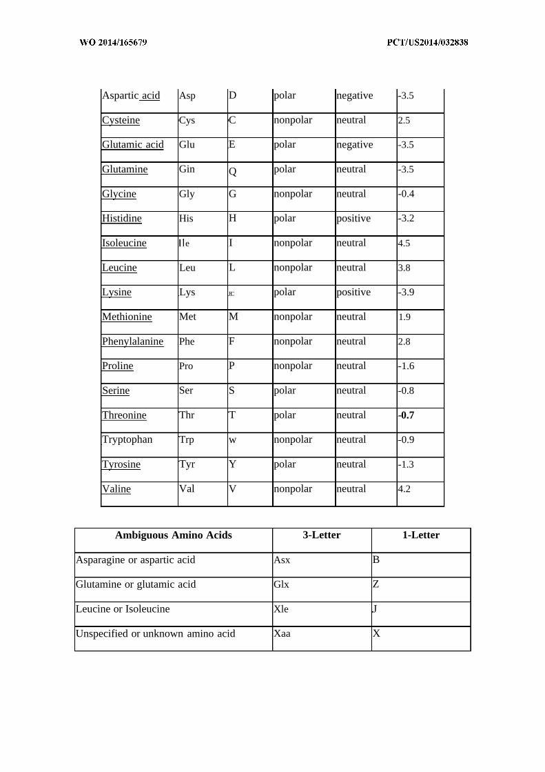
Aspartic acid Asp D polar negative -3.5
Cysteine Cys C nonpolar neutral 2.5
Glutamic acid Glu E polar negative -3.5
Glutamine Gin Q polar neutral -3.5
Glycine Gly G nonpolar neutral -0.4
Histidine His H polar positive -3.2
Isoleucine e I nonpolar neutral 4.5
Leucine Leu L nonpolar neutral 3.8
Lysine Lys EC polar positive -3.9
Methionine Met M nonpolar neutral 1.9
Phenylalanine Phe F nonpolar neutral 2.8
Proline Pro P nonpolar neutral -1.6
Serine Ser S polar neutral -0.8
Threonine Thr T polar neutral -0.7
Tryptophan Trp w nonpolar neutral -0.9
Tyrosine Tyr Y polar neutral -1.3
Valine Val V nonpolar neutral 4.2
Ambiguous Amino Acids 3-Letter 1-Letter
Asparagine or aspartic acid Asx B
Glutamine or glutamic acid Glx Z
Leucine or Isoleucine Xle J
Unspecified or unknown amino acid Xaa X

[0075] As will be understood by those skilled in the art, a variety of algorithms are
available that permit comparison of sequences in order to determine their degree of homology,
including by permitting gaps of designated length in one sequence relative to another when
considering which residues "correspond" to one another in different sequences. Calculation of
the percent homology between two nucleic acid sequences, for example, can be performed by
aligning the two sequences for optimal comparison purposes (e.g., gaps can be introduced in one
or both of a first and a second nucleic acid sequences for optimal alignment and non-
corresponding sequences can be disregarded for comparison purposes). In certain embodiments,
the length of a sequence aligned for comparison purposes is at least 30%, at least 40%, at least
50%, at least 60%, at least 70%, at least 80%, at least 90%, at least 95%, or substantially 100%
of the length of the reference sequence. The nucleotides at corresponding nucleotide positions
are then compared. When a position in the first sequence is occupied by the same nucleotide as
the corresponding position in the second sequence, then the molecules are identical at that
position; when a position in the first sequence is occupied by a similar nucleotide as the
corresponding position in the second sequence, then the molecules are similar at that position.
The percent homology between the two sequences is a function of the number of identical and
similar positions shared by the sequences, taking into account the number of gaps, and the length
of each gap, which needs to be introduced for optimal alignment of the two sequences.
Representative algorithms and computer programs useful in determining the percent homology
between two nucleotide sequences include, for example, the algorithm of Meyers and Miller
(CABIOS, 1989, 4 : 11-17), which has been incorporated into the ALIGN program (version 2.0)
using a PAM120 weight residue table, a gap length penalty of 12 and a gap penalty of 4 . The
percent homology between two nucleotide sequences can, alternatively, be determined for
example using the GAP program in the GCG software package using an NWSgapdna.CMP
matrix.
[0076] Human: In some embodiments, a human is an embryo, a fetus, an infant, a child,
a teenager, an adult, or a senior citizen.
[0077] Hydrophilic: As used herein, the term "hydrophilic" and/or "polar" refers to a
tendency to mix with, or dissolve easily in, water.

[0078] Hydrophobic: As used herein, the term "hydrophobic" and/or "non-polar", refers
to a tendency to repel, not combine with, or an inability to dissolve easily in, water.
[0079] Identity: As used herein, the term "identity" refers to the overall relatedness
between polymeric molecules, e.g., between nucleic acid molecules (e.g., DNA molecules and/or
R A molecules) and/or between polypeptide molecules. In some embodiments, polymeric
molecules are considered to be "substantially identical" to one another if their sequences are at
least 25%, 30%, 35%, 40%, 45%, 50%, 55%, 60%, 65%, 70%, 75%, 80%, 85%, 90%, 95%, or
99% identical. As will be understood by those skilled in the art, a variety of algorithms are
available that permit comparison of sequences in order to determine their degree of homology,
including by permitting gaps of designated length in one sequence relative to another when
considering which residues "correspond" to one another in different sequences. Calculation of
the percent identity between two nucleic acid sequences, for example, can be performed by
aligning the two sequences for optimal comparison purposes (e.g., gaps can be introduced in one
or both of a first and a second nucleic acid sequences for optimal alignment and non-
corresponding sequences can be disregarded for comparison purposes). In certain embodiments,
the length of a sequence aligned for comparison purposes is at least 30%>, at least 40%>, at least
50%, at least 60%, at least 70%, at least 80%, at least 90%, at least 95%, or substantially 100%
of the length of the reference sequence. The nucleotides at corresponding nucleotide positions
are then compared. When a position in the first sequence is occupied by the same nucleotide as
the corresponding position in the second sequence, then the molecules are identical at that
position. The percent identity between the two sequences is a function of the number of identical
positions shared by the sequences, taking into account the number of gaps, and the length of each
gap, which needs to be introduced for optimal alignment of the two sequences. Representative
algorithms and computer programs useful in determinng the percent identity between two
nucleotide sequences include, for example, the algorithm of Meyers and Miller (CABIOS, 1989,
4 : 11-17), which has been incorporated into the ALIGN program (version 2.0) using a PAM120
weight residue table, a gap length penalty of 12 and a gap penalty of 4 . The percent identity
between two nucleotide sequences can, alternatively, be determined for example using the GAP
program in the GCG software package using an NWSgapdna.CMP matrix.
[0080] Infection: As used herein, the term "infection" refers to the invasion of a host
organism's body by a disease-causing organism that multiplies in the host. Symptoms of an
infection may result from action of toxins produced by the disease-causing organism and/or be
reaction of host tissues to the organisms and/or to toxins they produce.
[0081] Isolated: As used herein, the term "isolated" refers to a substance and/or entity
that has been (1) separated from at least some of the components with which it was associated
when initially produced (whether in nature and/or in an experimental setting), and/or (2)
produced, prepared, and/or manufactured by the hand of man. Isolated substances and/or entities
may be separated from about 10%, about 20%>, about 30%>, about 40%>, about 50%>, about 60%>,
about 70%, about 80%, about 90%, about 91%, about 92%, about 93%, about 94%, about 95%,
about 96%o, about 97%, about 98%>, about 99%, or more than about 99% of the other components
with which they were initially associated. In some embodiments, isolated agents are about 80%,
about 85%, about 90%, about 91%, about 92%, about 93%, about 94%, about 95%, about 96%,
about 97%o, about 98%>, about 99%, or more than about 99% pure. As used herein, a substance is
"pure" if it is substantially free of other components. In some embodiments, as will be
understood by those skilled in the art, a substance may still be considered "isolated" or even
"pure", after having been combined with certain other components such as, for example, one or
more carriers or excipients {e.g., buffer, solvent, water, etc.); in such embodiments, percent
isolation or purity of the substance is calculated without including such carriers or excipients.
[0082] Nanoemulsion: An emulsion is traditionally defined in the art "as a system ...
consisting of a liquid dispersed with or without an emulsifier in an immiscible liquid usually in
droplets of larger than colloidal size" Medline Plus Online Medical Dictionary, Merriam
Webster (2005). The term "nanoemulsion," as used herein, refers to an emulsion in which at
least some of the droplets (or particles) have diameters in the nanometer size range. As will be
understood by those of ordinary skill in the art, a nanoemulsion is characterized by droplets or
particles one thousand fold smaller than microemulsion droplets or particles.
[0083] Nanoparticle: As used herein, the term "nanoparticle" refers to a particle having
a diameter of less than 1000 nanometers (nm). In some embodiments, a nanoparticle has a
diameter of less than 300 nm, as defined by the National Science Foundation. In some
embodiments, a nanoparticle has a diameter of less than 100 nm as defined by the National
Institutes of Health. In some embodiments, nanoparticles are micelles in that they comprise an
enclosed compartment, separated from the bulk solution by a micellar membrane, typically
comprised of amphiphilic entities which surround and enclose a space or compartment (e.g., to
define a lumen). In some embodiments, a micellar membrane is comprised of at least one
polymer, such as for example a biocompatible and/or biodegradable polymer.
[0084] Nanoparticle composition: As used herein, the term "nanoparticle composition"
refers to a composition that contains at least one nanoparticle and at least one additional agent or
ingredient. In some embodiments, a nanoparticle composition contains a substantially uniform
collection of nanoparticles as described herein.
[0085] Nanoparticle membrane: As used herein, the term "nanoparticle membrane"
refers to the boundary or interface between a nanoparticle outer surface and a surrounding
environment. In some embodiments, the nanoparticle membrane is a polymer membrane having
an outer surface and bounding lumen.
[0086] Nucleic acid: As used herein, the term "nucleic acid," in its broadest sense, refers
to any compound and/or substance that is or can be incorporated into an oligonucleotide chain.
In some embodiments, a nucleic acid is a compound and/or substance that is or can be
incorporated into an oligonucleotide chain via a phosphodiester linkage. As will be clear from
context, in some embodiments, "nucleic acid" refers to individual nucleic acid residues {e.g.,
nucleotides and/or nucleosides); in some embodiments, "nucleic acid" refers to an
oligonucleotide chain comprising individual nucleic acid residues. In some embodiments, a
"nucleic acid" is or comprises R A; in some embodiments, a "nucleic acid" is or comprises
DNA. In some embodiments, a nucleic acid is, comprises, or consists of one or more natural
nucleic acid residues. In some embodiments, a nucleic acid is, comprises, or consists of one or
more nucleic acid analogs. In some embodiments, a nuclic acid analog differs from a nucleic
acid in that it does not utilize a phosphodiester backbone. For example, in some embodiments, a
nucleic acid is, comprises, or consists of one or more "peptide nucleic acids", which are known
in the art and have peptide bonds instead of phosphodiester bonds in the backbone, are
considered within the scope of the present invention. Alternatively or additionally, in some
embodiments, a nucleic acid has one or more phosphorothioate and/or 5'-N-phosphoramidite
linkages rather than phosphodiester bonds. In some embodiments, a nucleic acid is, comprises,
or consists of one or more natural nucleosides (e.g., adenosine, thymidine, guanosine, cytidine,
uridine, deoxyadenosine, deoxythymidine, deoxyguanosine, and deoxycytidine). In some
embodiments, a nucleic acid is, comprises, or consists of one or more nucleoside analogs (e.g., 2-
aminoadenosine, 2-thiothymidine, inosine, pyrrolo-pyrimidine, 3-methyl adenosine, 5-
methylcytidine, C-5 propynyl-cytidine, C-5 propynyl-uridine, 2-aminoadenosine, C5-
bromouridine, C5-fluorouridine, C5-iodouridine, C5-propynyl-uridine, C5-propynyl-cytidine,
C5-methylcytidine, 2-aminoadenosine, 7-deazaadenosine, 7-deazaguanosine, 8-oxoadenosine, 8-
oxoguanosine, 0(6)-methylguanine, 2-thiocytidine, methylated bases, intercalated bases, and
combinations thereof). In some embodiments, a nucleic acid comprises one or more modified
sugars (e.g., 2'-fluororibose, ribose, 2'-deoxyribose, arabinose, and hexose) as compared with
those in natural nucleic acids. In some embodiments, a nucleic acid has a nucleotide sequence
that encodes a functional gene product such as an RNA or protein. In some embodiments, a
nucleic acid includes one or more introns. In some embodiments, nucleic acids are prepared by
one or more of isolation from a natural source, enzymatic synthesis by polymerization based on a
complementary template (in vivo or in vitro), reproduction in a recombinant cell or system, and
chemical synthesis. In some embodiments, a nucleic acid is at least 3, 4, 5, 6, 7, 8, 9, 10, 15, 20,
25, 30, 35, 40, 45, 50, 55, 60, 65, 70, 75, 80, 85, 90, 95, 100, 110, 120, 130, 140, 150, 160, 170,
180, 190, 20, 225, 250, 275, 300, 325, 350, 375, 400, 425, 450, 475, 500, 600, 700, 800, 900,
1000, 1500, 2000, 2500, 3000, 3500, 4000, 4500, 5000 or more residues long.
[0087] Patient: As used herein, the term "patient" or "subject" refers to a human or any
non-human animal (e.g., mouse, rat, rabbit, dog, cat, cattle, swine, sheep, horse or primate) to
whom therapy is administered. In many embodiments, a patient is a human being. In some
embodiments, a patient is a human presenting to a medical provider for diagnosis or treatment of
a disease, disorder or condition. In some embodiments, a patient displays one or more symptoms
or characteristics of a disease, disorder or condition. In some embodiments, a patient does not
display any symptom or characteristic of a disease, disorder, or condition. In some
embodiments, a patient is someone with one or more features characteristic of susceptibility to or
risk of a disease, disorder, or condition.
[0088] Pharmaceutically acceptable: The term "pharmaceutically acceptable" as used
herein, refers to agents that, within the scope of sound medical judgment, are suitable for use in
contact with tissues of human beings and/or animals without excessive toxicity, irritation,
allergic response, or other problem or complication, commensurate with a reasonable benefit/risk
ratio.
[0089] Polypeptide: The term "polypeptide", as used herein, generally has its art-
recognized meaning of a polymer of at least three amino acids. In some embodiments, the term
is used to refer to specific functional classes of polypeptides, such as, for example, autoantigen
polypeptides, nicotinic acetylcholine receptor polypeptides, alloantigen polypeptides, etc. For
each such class, the present specification provides several examples of amino acid sequences of
known exemplary polypeptides within the class; in some embodiments, such known polypeptides
are reference polypeptides for the class. In such embodiments, the term "polypeptide" refers to
any member of the class that shows significant sequence homology or identity with a relevant
reference polypeptide. In many embodiments, such member also shares significant activity with
the reference polypeptide. For example, in some embodiments, a member polypeptide shows an
overall degree of sequence homology or identity with a reference polypeptide that is at least
about 30-40%, and is often greater than about 50%, 60%, 70%, 80%, 90%, 91%, 92%, 93%,
94% , 95% , 96% , 97% , 98%>, 99% or more and/or includes at least one region (i.e., a conserved
region, often including a characteristic sequence element) that shows very high sequence
identity, often greater than 90%> or even 95%, 96%, 97%, 98%, or 99%. Such a conserved region
usually encompasses at least 3-4 and often up to 20 or more amino acids; in some embodiments,
a conserved region encompasses at least one stretch of at least 2, 3, 4, 5, 6, 7, 8, 9, 10, 11, 12, 13,
14, 15 or more contiguous amino acids..
[0090] Protein: As used herein, the term "protein" refers to a polypeptide {i.e., a string
of at least two amino acids linked to one another by peptide bonds). Proteins may include
moieties other than amino acids {e.g., may be glycoproteins, proteoglycans, etc.) and/or may be
otherwise processed or modified. Those of ordinary skill in the art will appreciate that a
"protein" can be a complete polypeptide chain as produced by a cell (with or without a signal
sequence), or can be a characteristic portion thereof. Those of ordinary skill will appreciate that
a protein can sometimes include more than one polypeptide chain, for example linked by one or
more disulfide bonds or associated by other means. Polypeptides may contain L-amino acids, D-
amino acids, or both and may contain any of a variety of amino acid modifications or analogs
known in the art. Useful modifications include, e.g., terminal acetylation, amidation,
methylation, etc. In some embodiments, proteins may comprise natural amino acids, non-natural
amino acids, synthetic amino acids, and combinations thereof. The term "peptide" is generally
used to refer to a polypeptide having a length of less than about 100 amino acids, less than about
50 amino acids, less than 20 amino acids, or less than 10 amino acids. In some embodiments,
proteins are antibodies, antibody fragments, biologically active portions thereof, and/or
characteristic portions thereof.
[0091] Refractory: As used herein, the term "refractory" refers to any subject that does
not respond with an expected clinical efficacy following the administration of provided
compositions as normally observed by practicing medical personnel.
[0092] Small molecule: As used herein, the term "small molecule" means a low
molecular weight organic compound that may serve as an enzyme substrate or regulator of
biological processes. In general, a "small molecule" is a molecule that is less than about 5
kilodaltons (kD) in size. In some embodiments, provided nanoparticles further include one or
more small molecules. In some embodiments, the small molecule is less than about 4 kD, 3 kD,
about 2 kD, or about 1 kD. In some embodiments, the small molecule is less than about 800
daltons (D), about 600 D, about 500 D, about 400 D, about 300 D, about 200 D, or about 100 D.
In some embodiments, a small molecule is less than about 2000 g/mol, less than about 1500
g/mol, less than about 1000 g/mol, less than about 800 g/mol, or less than about 500 g/mol. In
some embodiments, one or more small molecules are encapsulated within the nanoparticle. In
some embodiments, small molecules are non-polymeric. In some embodiments, in accordance
with the present invention, small molecules are not proteins, polypeptides, oligopeptides,
peptides, polynucleotides, oligonucleotides, polysaccharides, glycoproteins, proteoglycans, etc.
In some embodiments, a small molecule is a therapeutic. In some embodiments, a small
molecule is an adjuvant. In some embodiments, a small molecule is a drug.
[0093] Stable: The term "stable," when applied to compositions herein, means that the
compositions maintain one or more aspects of their physical structure {e.g., size range and/or
distribution of particles) over a period of time. In some embodiments, a stable nanoparticle
composition is one for which the average particle size, the maximum particle size, the range of
particle sizes, and/or the distribution of particle sizes {i.e., the percentage of particles above a
designated size and/or outside a designated range of sizes) is maintained for a period of time
under specified conditions. In some embodiments, a stable provided composition is one for
which a biologically relevant activity is maintained for a period of time. In some embodiments,
the period of time is at least about one hour; in some embodiments the period of time is about 5
hours, about 10 hours, about one (1) day, about one (1) week, about two (2) weeks, about one (1)
month, about two (2) months, about three (3) months, about four (4) months, about five (5)
months, about six (6) months, about eight (8) months, about ten (10) months, about twelve (12)
months, about twenty-four (24) months, about thirty-six (36) months, or longer. In some
embodiments, the period of time is within the range of about one (1) day to about twenty-four
(24) months, about two (2) weeks to about twelve (12) months, about two (2) months to about
five (5) months, etc. For example, if a population of nanoparticles is subjected to prolonged
storage, temperature changes, and/or pH changes, and a majority of the nanoparticles in the
composition maintain a diameter within a stated range, the nanoparticle composition is stable. In
some embodiments, a stable composition is stable at ambient conditions. In some embodiments,
a stable composition is stable under biologic conditions (i.e. 37° C in phosphate buffered saline).
[0094] Subject: As used herein, the term "subject" refers to a human or any non-human
animal (e.g., mouse, rat, rabbit, dog, cat, cattle, swine, sheep, horse or primate). A human
includes pre and post natal forms. In many embodiments, a subject is a human being. A subject
can be a patient, which refers to a human presenting to a medical provider for diagnosis or
treatment of a disease. A subject can be afflicted with or is susceptible to a disease or disorder
but may or may not display symptoms of the disease or disorder.
[0095] Substantially: As used herein, the term "substantially" refers to the qualitative
condition of exhibiting total or near-total extent or degree of a characteristic or property of
interest. One of ordinary skill in the biological arts will understand that biological and chemical
phenomena rarely, if ever, go to completion and/or proceed to completeness or achieve or avoid
an absolute result. The term "substantially" is therefore used herein to capture the potential lack
of completeness inherent in many biological and chemical phenomena.
[0096] Sufferingfrom: An individual who is "suffering from" a disease, disorder, or
condition has been diagnosed with and/or exhibits or has exhibited one or more symptoms or
characteristics of the disease, disorder, or condition.

[0097] Susceptible to: An individual who is "susceptible to" a disease, disorder, or
condition is at risk for developing the disease, disorder, or condition. In some embodiments, an
individual who is susceptible to a disease, disorder, or condition does not display any symptoms
of the disease, disorder, or condition. In some embodiments, an individual who is susceptible to
a disease, disorder, or condition has not been diagnosed with the disease, disorder, and/or
condition. In some embodiments, an individual who is susceptible to a disease, disorder, or
condition is an individual who has been exposed to conditions associated with development of
the disease, disorder, or condition. In some embodiments, a risk of developing a disease,
disorder, and/or condition is a population-based risk {e.g., family members of individuals
suffering from allergy, etc.
[0098] Symptoms are reduced: According to the present invention, "symptoms are
reduced" when one or more symptoms of a particular disease, disorder or condition is reduced in
magnitude {e.g., intensity, severity, etc.) and/or frequency. For purposes of clarity, a delay in the
onset of a particular symptom is considered one form of reducing the frequency of that symptom.
[0099] Therapeutic agent: As used herein, the phrase "therapeutic agent" refers to any
agent that has a therapeutic effect and/or elicits a desired biological and/or pharmacological
effect, when administered to a subject. In some embodiments, an agent is considered to be a
therapeutic agent if its administration to a relevant population is statistically correlated with a
desired or beneficial therapeutic outcome in the population, whether or not a particular subject to
whom the agent is administered experiences the desired or beneficial therapeutic outcome.
[0100] Therapeutically effective amount: As used herein, the term "therapeutically
effective amount" means an amount that is sufficient, when administered to a population
suffering from or susceptible to a disease, disorder, and/or condition in accordance with a
therapeutic dosing regimen, to treat the disease, disorder, and/or condition {e.g., allergy). In
some embodiments, a therapeutically effective amount is one that reduces the incidence and/or
severity of, and/or delays onset of, one or more symptoms of the disease, disorder, and/or
condition. Those of ordinary skill in the art will appreciate that the term "therapeutically
effective amount" does not in fact require successful treatment be achieved in a particular
individual. Rather, a therapeutically effective amount may be that amount that provides a
particular desired pharmacological response in a significant number of subjects when
administered to patients in need of such treatment. It is specifically understood that particular
subjects may, in fact, be "refractory" to a "therapeutically effective amount." To give but one
example, a refractory subject may have a low bioavailability such that clinical efficacy is not
obtainable. In some embodiments, reference to a therapeutically effective amount may be a
reference to an amount as measured in one or more specific tissues (e.g., a tissue affected by the
disease, disorder or condition) or fluids (e.g., blood, saliva, serum, sweart, tears, urine, etc).
Those of ordinary skill in the art will appreciate that, in some embodiments, a therapeutically
effective agent may be formulated and/or administered in a single dose. In some embodiments, a
therapeutically effective agent may be formulated and/or administered in a plurality of doses, for
example, as part of a dosing regimen.
[0101] Therapeutic regimen: A "therapeutic regimen", as that term is used herein, refers
to a dosing regimen whose administration across a relevant population is correlated with a
desired or beneficial therapeutic outcome.
[0102] Treatment: As used herein, the term "treatment" (also "treat" or "treating") refers
to any administration of a substance that partially or completely alleviates, ameliorates, relives,
inhibits, delays onset of, reduces severity of, and/or reduces frequency, incidence or severity of
one or more symptoms, features, and/or causes of a particular disease, disorder, and/or condition.
Such treatment may be of a subject who does not exhibit signs of the relevant disease, disorder
and/or condition and/or of a subject who exhibits only early signs of the disease, disorder, and/or
condition. Alternatively or additionally, such treatment may be of a subject who exhibits one or
more established signs of the relevant disease, disorder and/or condition. In some embodiments,
treatment may be of a subject who has been diagnosed as suffering from the relevant disease,
disorder, and/or condition. In some embodiments, treatment may be of a subject known to have
one or more susceptibility factors that are statistically correlated with increased risk of
development of the relevant disease, disorder, and/or condition.
[0103] Uniform: The term "uniform," when used herein in reference to a nanoparticle
composition, refers to a nanoparticle composition in which individual nanoparticles have
diameters within a a specified range. For example, in some embodiments, a uniform
nanoparticle composition is one in which the difference between the minimum diameter and
maximum diameter does not exceed about 300 nm. In some embodiments, a uniform
nanoparticle composition contains nanoparticles with diameters within the range of about lOOnm
to about 300nm. In some embodiments, a uniform nanoparticle composition contains
nanoparticles with an average particle size that is under about 500 nm. In some embodiments, a
uniform nanoparticle composition contains nanoparticles with an average particle size that is
within a range of about lOOnm to about 500nm. In some embodiments, a uniform nanoparticle
composition is one in which a majority of the particles within the composition have diameters
below a specified size or within a specified range. In some embodiments, the majority is more
than 50%, 60%, 70%, 75%, 80%, 85%, 90%, 95%, 96%, 97%, 98%, 99%, 99.5%, 99.6%, 99.7%,
99.8%, 99.9% or more of the particles in the composition.
DETAILED DESCRIPTION OF CERTAIN EMBODIMENTS
[0104] The present invention is based, in part, on the surprising insight that desirable
immunomodulatory compositions can be prepared by combining features of certain nanoparticles
together with cellular components of microbial cells. The present invention provides the
particular insight that such immunomodulatory compositions can be prepared with preparations
comprising either hydrophilic or hydrophobic microbial cellular components, or both. In some
particular embodiments, the present invention provides the insight that desirable
immunmodulatory compositions can be prepared by encapsulating hydrophilic microbial cellular
components within nanoparticles and/or associating hydrophobic microbial cellular components
with the external surface of the nanoparticles.
[0105] Nanoparticles
[0106] Nanoparticles useful in accordance with the present invention include those in
which the nanoparticles are comprised of at least one polymer assembled into a micelle that
bounds an interior lumen and has an external surface. In some embodiments, nanoparticles are
comprised of at least one polymer that is a homopolymer, a diblock polymer, a triblock polymer,
a multiblock copolymer, a linear polymer, a dendritic polymer, a branched polymer, a random
block, etc., or combinations thereof. In some embodiments, nanoparticles are comprised of a
blend and/or mixture of polymers.
[0107] In some embodiments, nanoparticules are comprised of one or more
biocompatible polymers and/or one or more biodegradable polymers. In some embodiments,
nanoparticles are comprised of one or more synthetic polymers, or derivatives thereof. In some
embodiments, nanoparticles are comprised of one or more natural polymers, or derivatives
thereof. In some embodiments, nanoparticles are comprised of combinations of synthetic and
natural polymers, or derivatives thereof.
[0108] In some embodiments, nanoparticles are comprised of one or more polymers
selected from the group consisting of poly(hydroxy acids) such as poly(lactic acid), poly(glycolic
acid), poly(lactic acid-co-glycolic acid), poly(lactic-co-glycolic acid), and derivatives of
poly(lactic-co-glycolic acid), PEGylated poly(lactic-co-glycolic acid), poly(lactide),
poly(glycolide), poly(lactide-co-glycolide), poly(anhydrides), PEGylated poly(anhydrides), poly
(ortho esters), derivatives of poly(ortho esters), PEGylated poly(ortho esters),
poly(caprolactones), derivatives of poly(caprolactone), PEGylated poly(caprolactones),
polyamines (e.g. spermine, spermidine, polylysine, and derivatives thereof), PEGylated
polylysine, polyamides, polycarbonates, poly(propylene fumarates), polyamides,
polyphosphazenes, polyamino acids, polyethers, polyacetals, polylactides,
polyhydroxyalkanoates, polyglycolides, polyketals, polyesteramides, poly(dioxanones),
polyhydroxybutyrates, polyhydroxyvalyrates, polycarbonates, polyorthocarbonates, poly(vinyl
pyrrolidone), polycyanoacrylates, polyalkylene oxalates, polyalkylene succinates, poly(malic
acid), poly(methyl vinyl ether), poly(ethylene imine), poly(acrylic acid), poly(maleic anhydride),
poly(ethylene imine), derivatives of poly(ethylene imine), PEGylated poly(ethylene imine),
poly(acrylic acid), derivatives of poly(acrylic acid), PEGylated poly(acrylic acid),
poly(urethane), PEGylated poly(urethane), derivatives of poly(urethane), poly(lactide),
poly(glycolide), poly(hydroxy acids), polyesters, poly(arylates), polyalkylenes such as
polyethylene and polypropylene, polyalkylene glycols such as poly(ethylene glycol),
polyalkylene oxides such as poly(ethylene oxide), polyalkylene terepthalates such as
poly(ethylene terephthalate), polyvinyl alcohols, polyvinyl ethers, polyvinyl esters, polyvinyl
halides such as poly(vinyl chloride), polyvinylpyrrolidone, polysiloxanes, poly(vinyl alcohols),
poly(vinyl acetate), polystyrene, polyurethanes and co-polymers thereof, derivativized celluloses
such as alkyl cellulose, hydroxyalkyl celluloses, cellulose ethers, cellulose esters, nitro
celluloses, methyl cellulose, ethyl cellulose, hydroxypropyl cellulose, hydroxy-propyl methyl
cellulose, hydroxybutyl methyl cellulose, cellulose acetate, cellulose propionate, cellulose
acetate butyrate, cellulose acetate phthalate, carboxylethyl cellulose, cellulose triacetate, and
cellulose sulfate sodium salt (jointly referred to herein as "synthetic celluloses"), polymers of
acrylic acid, methacrylic acid or copolymers or derivatives thereof including esters, poly(methyl
methacrylate), poly(ethyl methacrylate), poly(butylmethacrylate), poly(isobutyl methacrylate),
poly(hexylmethacrylate), poly(isodecyl methacrylate), poly(lauryl methacrylate), poly(phenyl
methacrylate), poly(methyl acrylate), poly(isopropyl acrylate), poly(isobutyl acrylate), and
poly(octadecyl acrylate) (jointly referred to herein as "polyacrylic acids"), poly(butyric acid),
poly(valeric acid), and poly(lactide-co-caprolactone) and/or derivatives thereof.
[0109] In some embodiments, nanoparticles are comprised of one or more acrylic
polymers. In certain embodiments, acrylic polymers include, for example, acrylic acid and
methacrylic acid copolymers, methyl methacrylate copolymers, ethoxyethyl methacrylates,
cyanoethyl methacrylate, aminoalkyl methacrylate copolymer, poly(acrylic acid),
poly(methacrylic acid), methacrylic acid alkylamide copolymer, poly(methyl methacrylate),
poly(methacrylic acid anhydride), methyl methacrylate, polymethacrylate, poly(methyl
methacrylate) copolymer, polyacrylamide, aminoalkyl methacrylate copolymer, glycidyl
methacrylate copolymers, polycyanoacrylates, and/or combinations thereof.
[0110] In some embodiments, nanoparticles are comprised of one or more natural
polymers. Exemplary natural polymers include, but are not limited to, proteins (such as albumin,
collagen, gelatin), prolamines (for example, zein), polysaccharides (such as alginate), cellulose
derivatives (such as hydroxypropyl cellulose, sodium carboxymethyl cellulose, ethyl cellulose
and cellulose acetate), polyhydroxyalkanoates (for example, polyhydroxybutyrate), and/or
combinations thereof. In some embodiments, a natural polymer may comprise or consist of
chitosan.
[0111] In some embodiments, nanoparticles are comprised of one or more polymers such
as poly(lactide-co-glycolide) copolymerized with polyethylene glycol (PEG). Withouth wishing
to be bound by any particular theory, it is proposed that arrangement of a nanoparticle so that
PEG is exposed on the external surface, may increase stability of the nanoparticle in blood,
perhaps at least in part due to the hydrophilicity of PEG.
[0112] In some particular embodiments, nanoparticles are comprised of PLGA.
[0113] In some particular embodiments, nanoparticles utilized in accordance with the
present invention are as described in one or more of U.S. Pat. 7,534,448, U.S. Pat. 7,534,449,
U.S. Pat. 7,550,154, US20090239789A1, US20090269397A1, US20100104503A1,
US20100151436A1, US20100284965A1, WO2006080951, WO20081 15641, WO2008109347,
WO2009094273, WO2012167261 and WO20 13003 157.
[0114] In general, a nanoparticle is or comprises a particle having a diameter {e.g.,
average diameter) of less than 1000 nanometers (nm). In some embodiments, provided
nanoparticle compositions comprise a population of nanoparticles. In some embodiments, a
population of nanoparticles comprises nanoparticles of a uniform size. In some embodiments, a
population of nanoparticles comprises nanoparticles of different sizes; in some embodiments
showing a particular size distribution. In many embodiments, provided nanoparticle
compositions comprise nanoparticles having sizes {e.g., average sizes) within a range defined by
a lower limit and an upper limit. In some embodiments, the lower limit is 5 nm, 10 nm, 15 nm,
20 nm, 25 nm, 30 nm, 35 nm, 40 nm, 45 nm, 50 nm, 55 nm, 60 nm, 65 nm, 70 nm, 75 nm, 80
nm, 85 nm, 90 nm, 95 nm, 100 nm, 150 nm, 200 nm, or more. In some embodiments, the upper
limit is 1000 nm, 950 nm, 900 nm, 850 nm, 800 nm, 750 nm, 700 nm, 650 nm, 600 nm, 550 nm,
500 nm, 450 nm, 400 nm, 350 nm, 300 nm, 250 nm or less. In some embodiments, provided
nanoparticle compositions comprise nanoparticles having sizes {e.g., average sizes) similar to the
size of bacterial cells. For example, in some embodiments, provided nanoparticle compositions
comprise nanoparticles having sizes {e.g., average sizes) ranging between 100 nm and 2000 nm,
between 100 nm and 1000 nm, between 100 nm and about 500 nm, between 100 nm and about
300 nm, or between 100 nm and about 200 nm.
[0115] In some embodiments, provided nanoparticle compositions are substantially free
of particles larger than about 2000 nm, about 1000 nm, about 900 nm, about 800 nm, about 700
nm, about 600 nm, about 500 nm, about 400 nm, or about 300 nm. In some embodiments,
provided nanoparticle compositions comprise no more than about 50%, about 25%, about 10%>,
about 5%, or about 1% of particles larger than about 2000 nm, about 1000 nm, about 900 nm,
about 800 nm, about 700 nm, about 600 nm, about 500 nm, about 400 nm, or about 300 nm.

Nanoparticles - Exemplary Methods of Making
[0116] In another aspect, the present invention provides methods of producing
nanoparticles. In some embodiments, for example, embodiments wherein the nanoparticles
include one or more of hydrophilic cellular component(s) and hydrophobic cellular
component(s), provided methods of making nanoparticles may include one or more of
separating, associating, forming, emulsions, hot melt microencapsulation, solvent removal,
spray-drying, and/or ionic gelation steps, and combinations thereof.
Forming
[0117] In some embodiments, provided nanoparticles may be formed using any available
method in the art. In some embodiments, provided nanoparticles and/or nanoparticle
compositions may be prepared by nanoprecipitation, flow focusing using fluidic channels, spray
drying, single and double emulsion solvent evaporation, solvent extraction, phase separation, hot
melt microencapsulation, milling, microemulsion procedures, microfabrication, nanofabrication,
sacrificial layers, simple and complex coacervation, and other methods well known to those of
ordinary skill in the art. In some embodiments, provided nanoparticle compositions are prepared
by aqueous and organic solvent syntheses (see for example, Pellegrino et al, 2005, Small, 1:48;
Murray et al, 2000, Ann. Rev. Mat. Sci., 30:545; and Trindade et al, 2001, Chem. Mat.,
13:3843). In some embodiments, provided nanoparticle compositions are prepared by
nanoprecipitation or spray drying. Conditions used in preparing particles may be altered to yield
particles of a desired size or property (e.g. , hydrophobicity, hydrophilicity, external morphology,
"stickiness," shape, etc.). In general, methods of preparing nanoparticles and/or conditions used
(e.g., solvent, temperature, concentration, air flow rate, etc.) may depend on identity of
functional elements (e.g., cellular lysate components) associated with the particles and/or the
composition of the polymer matrix.
[0118] In some embodiments, additional methods for making nanoparticles for delivery
of encapsulated agents are described in the literature (see for example, Doubrow, Ed.,
"Microcapsules and Nanoparticles in Medicine and Pharmacy," CRC Press, Boca Raton, 1992;
Mathiowitz et al, 1987, J. Control. Release, 5:13; Mathiowitz et al., 1987, Reactive Polymers,
6:275; and Mathiowitz et al, 1988, J. Appl. Polymer Sci., 35:755).

Methods of Making - with Antigen
[0119] In some embodiments, provided methods further include a step of associating an
antigen with a nanoparticle. Suitable antigens and/or antigenic extracts may include those
described herein. In some embodiments, the antigen is an infectious agent antigen. In some
embodiments, the infectious agent antigen is provided with one or more additional components
of the infectious agent. In some embodiments, the antigen is part of a raw or crude antigenic or
allergenic extract {e.g. dust mite extract or raw nut extract).
[0120] In some embodiments, provided methods further include a step wherein an
antigen is associated with either or both of the hydrophilic and/or hydrophobic cellular
components so that some or all of the antigen(s) is/are encapsulated within the internal lumen. In
some embodiments, one or more antigens are associated with the hydrophilic cellular
components so that some or all of the antigen(s) is/are encapsulated within the internal lumen. In
some embodiments, one or more antigen(s) are associated with the hydrophobic cellular
components so that some or all of the antigen(s) is/are encapsulated within the internal lumen.
[0121] As a more detailed example of some embodiments only, the use of certain
methods, such as double emulsion, hot melt encapsulation, solvent removal, spray-drying, and
ionic gelation methods for forming nanoparticles are provided. Exemplary methods for forming
nanoparticles may be found in Demento et al., "TLR9-Targeted Biodegradable Nanoparticlesas
Immunization Vectors Protect Against West Nile Encephalitis", 2010, J. Immunol. 185:2989-
2997; see also Demento et al., "Inflammasome-activating nanoparticles as modular systems for
optimizing vaccine efficacy", 2009, Vaccine 27(23): 3013-3021.
Emulsions
[0122] In some embodiments, the polymer is dissolved in a volatile organic solvent, such
as methylene chloride. The payload (either soluble or dispersed as fine particles) is added to the
solution, and the mixture is suspended in an aqueous solution that contains a surface active agent
such as poly(vinyl alcohol). The resulting emulsion is stirred until most of the organic solvent
evaporates, leaving solid nanoparticles. The resulting nanoparticles are washed with water and
dried overnight in a lyophilizer. Freeze dried nanoparticles may then be stored at -20° C for later
use.
[0123] In some embodiments, a water-in-oil-in-water (W/O/W) emulsion method may be
used for preparation of the nanoparticles. In some embodiments, the nanoparticles include one
or more hydrophilic cellular components. For example, in a first emulsion (W/O), aqueous
cellular components in phosphate-buffered saline (PBS) are added to a vortexing PLGA solution
dissolved in methylene chloride. The first emulsion of polymer and aqueous cellular lysate are
then added drop-wise to PVA in a second emulsion (W/O/W). After each emulsion, samples are
sonicated for 30 seconds on ice. The second emulsion is then rapidly added to 0.3% PVA. This
external phase is then vigorously stirred for 3 hours at constant room temperature to evaporate
the methylene chloride, leaving solid nanoparticles. Particles are collected by centrifugation.
The resulting nanoparticles are washed with deionized water, flash-frozen, lyophilized, and
stored at -20° C for later use.
[0124] In some embodiments, the nanoparticles include one or more hydrophobic cellular
components. The hydrophobic cellular component(s) are first combined with a second emulsion.
The first emulsion of polymer (with or without aqueous cellular lysate and/or antigen) is then
added drop-wise to the second emulsion (W/O/W).
[0125] In some embodiments, the nanoparticles further include one or more encapsulated
antigens (for example, allergen extracts such as dust mite or peanut). In a first emulsion (W/O),
concentrated antigen in phosphate-buffered saline (PBS) is added to a vortexing PLGA solution
dissolved in methylene chloride. In some embodiments an aqueous cellular lysate is combined
with the first emulsion. Polymer and encapsulant are then added drop-wise to a second emulsion
(W/O/W). In some embodiments, the second emulsion has been combined with one or more
hydrophobic cellular components. After each emulsion, samples are sonicated for 30 seconds on
ice. The second emulsion is then rapidly added to 0.3% PVA. This external phase is then
vigorously stirred for 3 hours at constant room temperature to evaporate the methylene chloride,
leaving solid nanoparticles. Particles are collected by centrifugation. The resulting nanoparticles
are washed with deionized water, flash-frozen, lyophilized, and stored at -20° C for later use.
Hot Melt Microencapsulation
[0126] In this method, the polymer is first melted and then mixed with the solid particles.
The mixture is suspended in a non-miscible solvent (like silicon oil), and, with continuous
stirring, heated to a temperature, for example, 5°C, above the melting point of the polymer. Once
the emulsion is stabilized, it is cooled until the polymer particles solidify. The resulting
nanoparticles are washed by decantation with petroleum ether to give a free-flowing powder.
Nanoparticles with sizes between 0.5 to 1000 microns may be obtained with this method. The
external surfaces of nanoparticles prepared with this technique are usually smooth and dense.
This procedure is used to prepare nanoparticles made of polyesters and polyanhydrides. In some
embodiments, such a method may use polymers with molecular weights between 1,000-50,000.
Solvent Removal
[0127] This technique is primarily designed for polyanhydrides according to known
methods. In some embodiments, a payload to be encapsulated (for example, allergen extracts
such as dust mite or peanut) is dispersed or dissolved in a solution of the selected polymer in a
volatile organic solvent like methylene chloride. This mixture is suspended by stirring in an
organic oil (such as silicon oil) to form an emulsion. Unlike solvent evaporation, this method
may be used to make nanoparticles from polymers with high melting points and different
molecular weights. The external morphology of nanoparticles produced with this technique is
highly dependent on the type of polymer used.
Spray-Drying
[0128] In some embodiments using this method, the polymer is dissolved in organic
solvent. A known amount of the payload (for example, allergen extracts such as dust mite or
peanut) is suspended (insoluble extract) or co-dissolved (soluble extract) in the polymer solution.
The solution or the dispersion is then spray-dried. Typical process parameters for a mini-spray
drier (Buchi) are as follows: polymer concentration^. 04 g/mL, inlet temperature=-24° C, outlet
temperature=13-15° C, aspirator setting=15, pump setting=10 mL/minute, spray flow=600 Nl/hr,
and nozzle diameter=0.5 mm.
Ionic Gelation
[0129] In some embodiments, such as those including nanoparticles made of gel-type
polymers, such as alginate, traditional ionic gelation techniques may be used. Typically, the
polymer(s) are first dissolved in an aqueous solution, mixed with barium sulfate or some
bioactive agent, and then extruded through a nanodroplet forming device, which in some

instances employs a flow of nitrogen gas to break off the droplet. A slowly stirred
(approximately 100-170 RPM) ionic hardening bath is positioned below the extruding device to
catch the forming nanodroplets. The nanoparticles are left to incubate in the bath for twenty to
thirty minutes in order to allow sufficient time for gelation to occur. Nanoparticle size is
controlled by using various size extruders or varying either the nitrogen gas or polymer solution
flow rates. Chitosan nanoparticles can be prepared by dissolving the polymer in acidic solution
and crosslinking it with tripolyphosphate. Carboxymethyl cellulose (CMC) nanoparticles can be
prepared by dissolving the polymer in acid solution and precipitating the nanoparticle with lead
ions. In the case of negatively charged polymers (e.g., alginate, CMC), positively charged
ligands (e.g., polylysine, polyethyleneimine) of different molecular weights can be ionically
attached.
Microbial Cellular Components
[0130] As described herein, the present invention encompasses the recognition that
certain advantages are achieved when hydrophilic and hydrophilic components of microbial
cellular preparations, such as lysates that are separated from one another so that a hydrophilic
cellular component preparation and/or a hydrophobic cellular component preparation is/are
generated and one or both of such preparations is/are combined with nanoparticles to create
nanoparticle compositions of the present invention.
[0131] In some embodiments, one or more of a hydrophilic cellular component
preparation and a hydrophobic cellular component preparation may be provided from a microbial
cellular lysate. In such embodiments, a hydrophilic cellular component may be referred to as a
microbial hydrophilic cellular component and a hydrophobic cellular component may be referred
to as a microbial hydrophobic cellular component. Without wishing to be bound by any
particular theory, some embodiments of the present invention including one or more of a
microbial hydrophilic cellular component and/or a microbial hydrophobic cellular component
may permit development and/or production of useful immunomodulatory nanoparticle
compositions at least in part because they utilize various evolved attributes of microbial cells
relating to their ability to modulate or evade human or animal immune reactions. The present
invention also captures the insight that combining such evolved attributes with various features
of certain nanoparticle systems such as, for example, ability to sequester antigens and/or cellular
hydrophilic components from immune system elements (e.g., by encapsulation within a lumen of
a nanoparticle), tunable degradation rates and/or locations, and/or modular association with
targeting, adjuvant, or other surface entities, permits development and/or production of
particularly useful immunomodulatory compositions.
[0132] The present invention recognizes the source of a problem with various prior art
approaches to providing immunomodulatory compositions. Specifically, the present invention
encompasses the recognition that use of pure components, and in particular pure adjuvant
components, loses certain advantages, including certain immunological effects, achieved by
collections of components, and particularly by collections that mimic or contain absolute and/or
relative amounts as are found in such microbial cells. In some embodiments, the present
invention encompasses the recognition that use of isolated individual microbial components
(e.g., particular CpG and/or LPS molecules) may fail to induce as broad or effective an immune
response as could be achieved with an extract that includes multiple components and in some
embodiments presents a plurality of components in relative amounts that approximate what is
found in nature.
[0133] In some embodiments, the present invention provides microbial extracts - e.g.,
hydrophilic or hydrophobic extracts of microbial cells for use in or which nanoparticle
compositions. In some embodiments, such microbial extracts may contain a collection of
microbial components that share a chemical feature, so that they associate with other included
components and not with excluded components during production of the extract. In some
embodiments, extracts may contain at least some cellular components at relative levels
comparable to those at which they are present in the cells. Those skilled in the art will be aware
of a variety of techniques available to determine presence and/or level of particular components,
and to compare such determined level(s) with those observed in intact cells. Moreover, those of
ordinary skill in the art will readily appreciate reasonable and expected experimental variation
and therefore will be able to determine whether components are present in absolute or relative
levels or concentrations in an extract that are reasonably comparable to those at which they are
present in cells.

[0134] In general, microbial extracts are prepared from microbial cell preparations.
Microbial cell preparations are prepared by culturing microbial cells for a period of time and
under conditions sufficient to achieve cell growth to a desirable level (e.g., optical density,
concentration, colony size, total protein, total DNA, and colony forming units). In some
embodiments, microbial cell preparations contain intact cells, and optionally are substantially
free of lysed cells. In some embodiments, microbial cell preparations contain lysed cells, and
optionally are substantially free of intact cells.
[0135] In some embodiments, the present invention provides hydrophilic microbial
extracts, for example extracts prepared by contacting a microbial cell preparation with a
hydrophilic solvent so that hydrophilic cellular components partition into solution in the
hydrophilic solvent. The hydrophilic solvent can then be separated from non-solubilized
components which may, for example, be precipitated, solubilized in a hydrophobic solvent
(optionally not miscible with the hydrophilic solvent), or otherwise separable from the
hydrophilic solvent. In some embodiments, hydrophilic cellular components that partition into a
hydrophilic solvent include, for example, components that are miscible and/or soluble in such
solvent.
Separating
[0136] Any of a variety of separation methods may be used to separate hydrophilic
cellular components from hydrophobic cellular components. Exemplary suitable methods
include solvent extraction, detergent extraction, and phase separation.
[0137] Exemplary hydrophilic components that may be found in certain embodiments of
hydrophilic cellular extracts include, but are not limited to, cytosol components; carbohydrates
including sugars; amphipathic molecules {e.g., glycolipids and/or lipoproteins); salts; soluble
proteins {i.e., polar proteins); nucleic acids (e.g., DNA and/or RNA); and/or combinations
thereof. In some embodiments, a hydrophilic cellular extract includes sheared DNA or RNA. In
some embodiments, a hydrophilic cellular extract includes lipopolysaccharides (LPS). In some
embodiments, a hydrophilic cellular extract includes one or more CpGs. In some embodiments,
a hydrophilic cellular extract is substantially free of membrane lipids or membrane proteins.
[0138] In some embodiments, the present invention provides hydrophobic microbial
extracts, for example extracts prepared by contacting a microbial cell preparation with a
hydrophobic solvent so that hydrophobic cellular components partition into solution in the
hydrophobic solvent. The hydrophobic solvent can then be separated from non-solubilized
components which may, for example, be precipitated, solubilized in a hydrophilic solvent
(optionally not miscible with the hydrophobic solvent), or otherwise separable from the
hydrophobic solvent. In some embodiments, hydrophobic cellular components that partition
into a hydrophobic solvent include that are miscible and/or soluble in the solvent; in some
embodiments, such hydrophobic cellular components include components that are substantially
immiscible and/or insoluble in water and/or other aqueous solvents.
[0139] Exemplary components that may be found in some embodiments of hydrophobic
cellular extracts include, but are not limited to, cell membrane components; certain
carbohydrates including certain glycoproteins and/or glycolipids; certain proteins including
certain glycoproteins, transmembrane proteins, lipid anchored proteins {i.e., non-polar proteins);
lipids including phospholipids, glycolipids, and cholesterols; and/or combinations thereof. In
some embodiments, a hydrophobic cellular extract includes lipopolysaccharides (LPS). In some
embodiments, a hydrophobic cellular extract is substantially free of components found
exclusively in the cytoplasm (e.g., hydrophilic proteins, DNA, and R A).
[0140] In some embodiments, a hydrophilic extract is substantially free of hydrophobic
components; in some embodiments, a hydrophobic extract is substantially free of hydrophilic
components. However, as will be appreciated by those skilled in the art, separation of cellular
components by extract preparation is often not complete. That is, in some embodiments, at least
some cellular components may partition substantially equally into hydrophilic and hydrophobic
extracts; other components may partition favorably but not exclusively into one orthe other of a
hydrophilic and a hydrophobic extract. To give but a few examples, in some embodiments,
amphiphilic entities such as for example certain membrane spanning proteins, glycolipids and/or
lipoproteins, LPS, etc., and combinations thereof.
[0141] Microbial extracts for use in accordance with the present invention can be
prepared from extracts of any microbial cells, or combinations thereof. In some embodiments,

microbial extracts are prepared from bacterial, fungal, archael, and/or protest cells, or
combinations thereof.
[0142] In some embodiments, microbial extracts can be prepared from bacterial cells
including, but not limited to Actinomyces, Aeromonas, Anabaena, Arthrobacter, Bacillus,
Bacteroides, Bdellovibrio, Bordetella, Borrelia, Campylobacter, Caulobacter, Chlamydia,
Chlorobium, Chromatium, Citrobacter, Clostridium, Corynebacterium, Cytophaga,
Deinococcus, Enterobacter, Escherichia, Francisella, Halobacterium, Heliobacter,
Haemophilus, Hemophilus influenza type B (HIB), Hyphomicrobium, Klebsiella, Lactococcus,
Legionella, Leptspirosis, Listeria, Meningococcus A, B and C Methanobacterium, Micrococcus,
Morganella, Myobacterium, Mycoplasma, Myxococcus, Neisseria, Nitrobacter, Oscillatoria,
Peptococcus, Phodospirillum, Plesiomonas, Prochloron, Proteus, Providencia, Pseudomonas,
Rickettsia, Salmonella, Serratia, Shigella, Spirillum, Spirochaeta, Sporolactobacillu,
Staphylococcus, Streptococcus, Streptomyces, Sulfolobus, Thermoplasma, Thiobacillus, and
Treponema, Vibrio, Yersinia, and combinations thereof. In some embodiments, microbial
extracts can be prepared from E coli cells.
[0143] In some embodiments, microbial extracts can be prepared from yeast cells such
as, for example, Brettanomyces anomalus, Brettanomyces bruxellensis, Brettanomyces
claussenii, Brettanomyces custersianus, Brettanomyces lambicus, Brettanomyces naardenensis,
Brettanomyces nanus, Canida albicans, Candida blankii, Candida slooffl, Dekkera intermedia,
Leucosporidium frigidum, Rhodotorula rubra, Saccharomyces cerevisiae, Saccharomyces
pastorianus, Saccharomyces telluris, Schizosaccharomyces pombe, Sporidiobolus johnsonii,
Sporidiobolus longiusculus, Sporidiobolus metaroseus, Sporidiobolus pararoseus, Sporidiobolus
ruineniae, Sporidiobolus salmonicolor, Sporidiobolus veronae, Trichosporon beigelii,
Trichosporon cutaneum, and combinations thereof. In some embodiments, microbial extracts
can be prepared from S . cerevisiae cells.
[0144] In some embodiments, microbial extracts can be prepared from one or more
microbial cell types that are pathogenic in the organism to which inventive compositions are to
be administered. In some embodiments, microbial extracts can be prepared from one or more
microbial cell types that naturally colonize subjects to which inventive compositions are to be
administered. In some embodiments, microbial extracts can be prepared from microbial cell
types that are present in foods consumed by organisms to which inventive compositions are to be
administered.
[0145] Without wishing to be held to a particular theory, some embodiments may be
beneficial and/or desirable in their ability to recreate natural environmental exposure to one or
more subtances. For example, in some embodiments, the presence of a mix of naturally
occurring microbial extract components may replicate an environmental exposure to one or more
toxins, infectious agents, antigens and/or allergens.
Antigens
[0146] In some embodiments, provided nanoparticles and/or nanoparticle compositions
further include an antigen. Antigens can be any of a variety of antigens including peptides,
proteins, polysaccharides, saccharides, lipids, glycolipids, nucleic acids, or combinations thereof.
The antigen can be derived from any source, including, but not limited to, a virus, bacterium,
parasite, plant, protozoan, fungus, tissue or transformed cell such as a canceror leukemic cell and
can be a whole cell or immunogenic component thereof, e.g., cell wall components or molecular
components thereof. In some embodiments, crude extracts including one or more antigens may
be used.
[0147] In some embodiments, an antigen is an infectious agent antigen. In some
embodiments, an infectious agent antigen is provided with one or more additional components of
an infectious agent. In some embodiments, an antigen or infectious agent is selected from the
group consisting of an allergen, an infectious antigen, a disease-associated antigen (e.g. a tumor-
associated antigen), an autoantigen, or combinations thereof. In some embodiments, some or all
of the antigen is encapsulated within the internal lumen of the nanoparticle.
[0148] In some embodiments, suitable antigens are known in the art and are available
from commercial government and scientific sources. In some embodiments, antigens are
provided from whole inactivated or attenuated organisms. In some embodiments, antigens may
be provided from infectious organisms, such as viruses, parasites and bacteria. In some
embodiments, antigens may be provided from tumor cells. In some embodiments, the antigens
may be purified or partially purified polypeptides derived from tumors or viral or bacterial

sources. Exemplary criteria for identifying and selecting effective antigenic peptides (e.g.,
minimal peptide sequences capable of eliciting an immune response) may be found in the art.
For example, Apostolopoulos, et al. (Curr. Opin. Mol.Ther., 2:29-36 (2000)), discusses the
strategy for identifying minimal antigenic peptide sequences based on an understanding of the
three dimensional structure of an antigen-presenting molecule and its interaction with both an
antigenic peptide and T-cell receptor. Shastri, (Curr. Opin.Immunol, 8:271-7 (1996)), discloses
how to distinguish rare peptides that serve to activate T cells from the thousands peptides
normally bound to MHC molecules. The antigens may be recombinant polypeptides produced
by expressing DNA encoding the polypeptide antigen in a heterologous expression system. The
antigens can be DNA encoding all or part of an antigenic protein. The DNA may be in the form
of vector DNA such as plasmid DNA.
[0149] In some embodiments, antigens may be provided as single antigens or may be
provided in combination. In some embodiments, antigens may also be provided as complex
mixtures of polypeptides or nucleic acids.
[0150] In some embodiments, antigens are provided as crude extract (e.g. whole peanut
extract). In some embodiments, provided nanoparticles and/or nanoparticle compositions may
include one or more crude antigenic extracts. In some embodiments, crude extract can be a
useful and inexpensive alternative to using individual antigens in provided nanoparticle
compositions.
[0151] In some embodiments, provided nanoparticles and/or nanoparticle compositions
may include one or more viral antigens. Generally, a virus consists of either two or three parts:
1) genetic material, which may be DNA or RNA, depending on the virus, 2) a protein coat that
surrounds and protects the genetic material, and, in some viruses, 3) a lipid envelope that
surrounds the protein coat. In some embodiments, a viral antigen may be provided from any
component of a virus. In some embodiments, a viral antigen may be isolated from any virus
including, but not limited to, a virus from any of the following viral families: Arenaviridae,
Arterivirus, Astroviridae, Baculoviridae, Badnavirus, Barnaviridae, Birnaviridae, Bromoviridae,
Bunyaviridae, Caliciviridae, Capillovirus, Carlavirus, Caulimovirus, Circoviridae,
Closterovirus, Comoviridae, Coronavtridae (e.g., Coronavirus, such as severe acute respiratory
syndrome (SARS) virus), Corticoviridae, Cystoviridae, Deltavirus, Dianthovirus, Enamovirus,
Filoviridae (e.g., Marburg virus and Ebola virus (e.g., Zaire, Reston, Ivory Coast, or Sudan
strain)), Flaviviridae, (e.g., Hepatitis C virus, Dengue virus 1, Dengue virus 2, Dengue virus 3,
and Dengue virus 4), Hepadnaviridae, Herpesviridae (e.g., Human herpesvirus 1, 3, 4, 5, and 6,
and Cytomegalovirus), Hypoviridae, Iridoviridae, Leviviridae, Lipothrixviridae, Microviridae,
Orthomyxoviridae (e.g., Influenza virus A and B and C), Papovaviridae, Paramyxoviridae (e.g.,
measles, mumps, and human respiratory syncytial virus), Parvoviridae, Picornaviridae (e.g.,
poliovirus, rhinovirus, hepatovirus, and aphthovirus), Poxviridae (e.g.,vaccinia and smallpox
virus), Reoviridae (e.g., rotavirus), Retroviridae (e.g., lentivirus, such as human
immunodeficiency virus (HIV) 1 and HIV 2), Rhabdoviridae (for example, rabies virus, measles
virus, respiratory syncytial virus, etc.), Togaviridae (for example, rubella virus, dengue virus,
etc.), and Totiviridae. Suitable viral antigens also include all or part of Dengue protein M,
Dengue protein E, Dengue D 1 NS 1, Dengue D 1NS2, and Dengue D1NS3. In some
embodiments, a viral antigen may comprise or consist of fragments of one or more viruses, such
as fragments from an influenza virus, for example. In some embodiments, viral fragments are
provided from one or more of 1) viral genetic material 2) a portion of a viral protein coat, and/or
3) a portion of a viral lipid envelope. In some embodiments, viral fragments may be provided
from two or more of 1) viral genetic material 2) a portion of a viral protein coat, and/or 3) a
portion of a viral lipid envelope.
[0152] Exemplary viral antigens include, but are not limited to, those found in the
following viral strains such as an adenovirus, borrelia , chagas, coxsackieviruses,
cytomegalovirus, dengue, Epstein-Barr (EBV), encephalitis (e.g. equine encephalitis and
Japanese encephalitis), hantavirus, hepatitis A (HAV), hepatits B (HBV), hepatitis C (HCV),
delta hepatitis D (HDV), hepatitis E (HEV), hepatitis G virus (HGV), herpes simplex virus
(HSV)(i.e. HSV1 and HSV2), human immunodeficiency virus (HIV), human T-lymphotrophic
virus (HTLV), influenza, lymphocytic choriomeningitis (LCMV), malaria, measles,
mycoplasma, papillomavirus (e.g. human papillomavirus, HPV), parainfluenza, parvovirus,
rhinovirus, Rift Valley fever, rotavirus, rubella, SARS, toxoplasma, treponema, varicella-zoster
(VZV), west nile virus (WNV), yellow fever, and combinations thereof.
[0153] In some embodiments, provided nanoparticles and/or nanoparticle compositions
may include one or more bacterial antigens. Bacterial antigens may originate from any bacteria

including, but not limited to Actinomyces, Aeromonas, Anabaena, Arthrobacter, Bacillus,
Bacteroides, Bdellovibrio, Bordetella, Borrelia, Campylobacter, Caulobacter, Chlamydia,
Chlorobium, Chromatium, Citrobacter, Clostridium, Corynebacterium, Cytophaga,
Deinococcus, Enterobacter, Escherichia, Francisella, Haemophilus, Halobacterium,
Heliobacter, Hemophilus influenza type B (HIB), Hyphomicrobium, Klebsiella, Lactococcus,
Legionella, Leptspirosis, Listeria, Meningococcus A, B and C, Methanobacterium, Micrococcus,
Morganella, Mycoplasma, Myobacterium, Myxococcus, Neisseria, Nitrobacter, Oscillatoria,
Peptococcus, Phodospirillum, Plesiomonas, Prochloron, Proteus, Providencia, Pseudomonas,
Rickettsia, Salmonella, Serratia, Shigella, Spirillum, Spirochaeta,Sporolactobacillu,
Staphylococcus, Streptococcus, Streptomyces, Sulfolobus, Thermoplasma, Thiobacillus,
Treponema, Vibrio, Yersinia, and combinations thereof.
[0154] In some embodiments, provided nanoparticles and/or nanoparticle compositions
may include one or more parasite antigens. Parasite antigens can be obtained from parasites such
as, but not limited to, an antigen derived from Candida albicans, Candida tropicalis, Chlamydia
trachomatis, Chlamydial psittaci, Cryptococcus neoformans, Entamoeba histolytica,
Histoplasma capsulatum, Mycoplasma pneumoniae, Nocardia asteroides,
Plasmodiumfalciparum, Rickettsia ricketsii, Rickettsia typhi, Schistosoma mansoni, Toxoplasma
gondii, Trichomonas vaginalis and Trypanosoma brucei. These include Sporozoan antigens,
Plasmodian antigens, such as all or part of a Circumsporozoite protein, a Sporozoite surface
protein, a liver stage antigen, an apical membrane associated protein, or a Merozoite surface
protein.
[0155] In some embodiments, provided nanoparticles and/or nanoparticle compositions
may include one or more environmental antigens. Exemplary environmental antigens include,
but are not limited to, those derived from naturally occurring allergens such as pollen allergens
(tree-, weed-, and grass pollen allergens), insect allergens (inhalant, saliva and venom allergens),
animal hair and/or dander allergens.
[0156] In some embodiments, an antigen may be an allergen found in certain foods,
venom, drugs or rubber that are capable of eliciting allergic responses, and in particular
anaphylactic allergic responses in an individual. Exemplary allergens that may induce
anaphylaxis, include several protein allergens found in food (peanut, milk, egg, wheat), insect
venom (i.e. bees, reptiles), drugs, and latex. In some embodiments, an environmental antigen
may be one or more venoms. Stings from organisms that inject venoms, such as insect stings are
known to cause anaphylaxis in individuals with allergies to the venom. In general, insect venom
includes venom from Hymenoptera such as bees, hornets, wasps, yellow jackets, velvet ants, and
fire ants. For example, venom from honey bees of the genus Apis can cause anaphylaxis in stung
victims who are allergic (Weber et al. Allergy 42:464-470). The venom from honey bees
contains numerous compounds which have been extensively studied and characterized (see for a
reference, Banks and Shipolini. Chemistry and Pharmacology of Honey-bee Venom. Chapter 7 of
Venoms of the Hymenoptera . Ed. T. Piek. Academic Press. London. 1986). The two main
components of bee venom are phospholipase A2 and melittin and may be used in some
embodiments for treating and preventing allergies to bee venom. Non-limiting examples of
protein allergens found in food include proteins found in nuts {e.g., peanut, walnut, almond,
pecan, cashew, hazelnut, pistachio, pine nut, brazil nut), seafood {e.g. shrimp, crab, lobster,
clams), fruit {e.g. plums, peaches, nectarines; Ann Allergy Asthma Immunol 7(6):504-8 (1996);
cherries, Allergy 51(10):756-7 (1996)), seeds (sesame, poppy, mustard), and soy and dairy
products (e.g., egg, milk).
[0157] In some embodiments, protein antigens found in pollen-related food allergies may
be used (e.g. birch pollen related to apple allergies). Important pollen allergens from trees,
grasses and herbs originate from the taxonomic orders of Fagales, Oleales, Finales and
platanaceae including i.a.. birch (Betula), alder (Alnus), hazel (Corylus), hornbeam (Carpinus)
and olive (Olea), cedar (Cryptomeriaand Juniperus) , Plane tree (Platanus), the order of Poales
including i.e. grasses of the genera Lolium, Phleum, Poa, Cynodon, Dactylis, Holcus, Phalaris,
Secale, and Sorghum, the orders ofAsterales and Urticales including i.e. herbs of the genera
Ambrosia, Artemisia, and Parietaria.
[0158] In some embodiments, an antigen may be one or more allergens from house dust
mites of the genus Dermatophagoides and Euroglyphus, storage mite e.g Lepidoglyphys,
Glycyphagus and Tyrophagus, cockroaches, midges and fleas e.g. Blatella, Periplaneta,
Chironomus and Ctenocepphalides, mammals such as cat, dog and horse, birds, venom allergens
including such originating from stinging or biting insects such as those from the taxonomic order
of Hymenoptera including bees (superfamily Apidae), wasps (superfamily Vespidea), and ants
(superfamily Formicoidae). Still other allergen antigens that may be used include inhalation
allergens from fungi such as from the genera Alternaria and Cladosporium.
[0159] In some embodiments, it may be desirable to work in systems in which a single
compound (e.g., a single protein) is responsible for an observed allergy. In some embodiments,
an antigen may comprise more complex allergens and/or crude allergenic extracts. Therefore,
collections of more than one antigen may be used so that immune responses to multiple antigens
may be modulated with a single embodiment.
[0160] In an effort to better exemplify some embodiments, an exemplary list of antigens
and/or antigenic extracts (such as one or more allergens and/or allergenic extracts) that may be
used in some embodiments include, but are not limited to, Acarus siro (mite) fatty acid-binding
protein (Aca s 13); Actinidia chinensis (kiwi) cysteine protease (Act c 1); Aedes aegyptii
(mosquito) antigen (Aed a 2); Aedes aegyptii (mosquito) antigen (Aed a 2); Aedes aegyptii
(mosquito) apyrase (Aed a 1); Aedes aegyptii (mosquito) apyrase (Aed a 1); Alnus glutinosa
(alder) antigen (Aln g 1); Alternaria alternata (fungus) acid ribosomal protein PI (Alt a 12);
Alternaria alternata (fungus) aldehyde dehydrogenase (Alt a 10); Alternaria alternata (fungus)
antigen (Alt a 1); Alternaria alternata (fungus) antigen (Alt a 2); Alternaria alternata (fungus)
enloase (Alt a l l); Alternaria alternata (fungus) heat shock protein (Alt a 3); Alternaria
alternata (fungus) ribosomal protein (Alt a 6); Alternaria alternata (fungus) YCP4 protein (Alt a
7); Ambrosia artemisiifolia (short ragweed) antigen E (Amb a 1); Ambrosia artemisiifolia (short
ragweed) antigen K (Amb a 2); Ambrosia artemisiifolia (short ragweed) Ra3 antigen (Amb a 3);
Ambrosia artemisiifolia (short ragweed) Ra5 antigen (Amb a 5); Ambrosia artemisiifolia (short
ragweed) Ra6 antigen (Amb a 6); Ambrosia artemisiifolia (short ragweed) Ra7 antigen (Amb a
7); Ambrosia trifida (giant ragweed) Ra5G antigen (Amb t 5); Anisakis simplex (nematode)
antigen (Ani s i); Anisakis simplex (nematode) paramyosin (Ani s 2); Apis mellifera (honey bee)
antigen (Api m 6); Apis mellifera (honey bee) hyaluronidase (Api m 2); Apis mellifera (honey
bee) melittin (Api m 4); Apis mellifera (honey bee) phospho lipase A2 (Api m l); Apium
graveolens (celery) antigen (Api g 5); Apium graveolens (celery) Bet v 1 homologue (Api g 1);
Apium graveolens (celery) profilin (Api g 4); Arachis hypogaea (peanut) (conglutin Ar a h 2);
Arachis hypogaea (peanut) (profilin Ar a h 5); Arachis hypogaea (peanut) conglutin homologue
(Ar a h 6); Arachis hypogaea (peanut) conglutin homologue (Ar a h 7); Arachis hypogaea
(peanut) glycinin (Ar a h 3); Arachis hypogaea (peanut) glycinin (Ar a h 4); Arachis hypogaea
(peanut) vicilin (Ar a h 1); Artemisia vulgaris (mugwort) antigen (Art v 1); Artemisia vulgaris
(mugwort) antigen (Art v 2); Ascaris suum (worm) antigen (Asc s i); Aspergillusflavus (fungus)
alkaline serine proteinase (Asp fl 13); Aspergillus Fumigatus (fungus) alkaline serine proteinase
(Asp f 13); Aspergillus Fumigatus (fungus) antigen (Asp f 1); Aspergillus Fumigatus (fungus)
antigen (Asp f 15); Aspergillus Fumigatus (fungus) antigen (Asp f 16); Aspergillus Fumigatus
(fungus) antigen (Asp f 17); Aspergillus Fumigatus (fungus) antigen (Asp f 2); Aspergillus
Fumigatus (fungus) antigen (Asp f 4); Aspergillus Fumigatus (fungus) antigen (Asp f 7);
Aspergillus Fumigatus (fungus) antigen (Asp f 9); Aspergillus Fumigatus (fungus) aspartis
protease (Asp f 10); Aspergillus Fumigatus (fungus) heat shock protein P70 (Asp f 12);
Aspergillus Fumigatus (fungus) metalloprotease (Asp f 5); Aspergillus Fumigatus (fungus) Mn
superoxide dismutase (Asp f 6); Aspergillus Fumigatus (fungus) peptidyl-prolyl isomerase (Asp
f 11); Aspergillus Fumigatus (fungus) peroxisomal protein (Asp f 3); Aspergillus Fumigatus
(fungus) ribosomal protein P2 (Asp f 8); Aspergillus Fumigatus (fungus) vacuolar serine (Asp f
18); Aspergillus niger (fungus) antigen (Asp n 18); Aspergillus niger (fungus) beta-xylosidase
(Asp n 14); Aspergillus niger (fungus) vacuolar serine proteinase; Aspergillus oryzae (fungus)
alkaline serine proteinase (Asp o 13); Aspergillus oryzae (fungus) TAKA-amylase A (Asp o 2);
Bertholletia excelsa (Brazil nut) 2S albumin (Ber e l); Betula verrucosa (birch) antigen (Bet v
1); Betula verrucosa (birch) antigen (Bet v 3); Betula verrucosa (birch) antigen (Bet v 4); Betula
verrucosa (birch) cyclophilin (Bet v 7); Betula verrucosa (birch) isoflavone reductase
homologue (Bet v 5); Betula verrucosa (birch) profilin (Bet v 2); Blattella germanica (German
cockroach) aspartic protease (Bla g 2); Blattella germanica (German cockroach) Bd90k (Bla g
1); Blattella germanica (German cockroach) calycin (Bla g 4); Blattella germanica (German
cockroach) glutathione transferase (Bla g 5); Blattella germanica (German cockroach) troponin
C (Bla g 6); Blomia tropicalis (mite) antigen (Bio t 5); Blomia tropicalis (mite) Btl l a antigen
(Bio 12); Blomia tropicalis (mite) Bt6 fatty acid-binding protein (Bio t); Bombus
pennsylvanicus (bumble bee) phospho lipase (Bom p i); Bombus pennsylvanicus (bumble bee)
protease (Bom p 4); Bos domesticus (cow) Ag3, lipocalin (Bos d 2); Bos domesticus (cow) alpha-
lactalbumin (Bos d 4); Bos domesticus (cow) beta-lactalbumin (Bos d 5); Bos domesticus (cow)
casein (Bos d 8); Bos domesticus (cow) immunoglobulin (Bos d 7); Bos domesticus (cow) serum
albumin (Bos d 6); Brassica juncea (oriental mustard) 2S albumin (Bra j 1); Brassica rapa
(turnip) prohevein-like protein (Bar r 2); Candida albicans (fungus) antigen (Cand a 1); Candida
boidinii (fungus) antigen (Cand b 2); Canisfamiliaris (dog) albumin (Can f ?); Canisfamiliaris
(dog) antigen (Can f 1); Canisfamiliaris (dog) antigen (Can f 2); Carpinus betulus (hornbeam)
antigen (Car b 1); Castanea sativa (chestnut) Bet v 1 homologue (Cas s i); Castanea sativa
(chestnut) chitinase (Cas s 5); Chironomus thummi thummi (midge) component I (Chi 2.0101);
Chironomus thummi thummi (midge) component IA (Chi 2.0102); Chironomus thummi thummi
(midge) component II-beta (Chi t 3); Chironomus thummi thummi (midge) component III (Chi t
1.01); Chironomus thummi thummi (midge) component IIIA (Chi 4); Chironomus thummi
thummi (midge) component IV (Chi 1.02); Chironomus thummi thummi (midge) component IX
(Chi t 6.02); Chironomus thummi thummi (midge) component VI (Chi t 5); Chironomus thummi
thummi (midge) component VIIA (Chi t 6.01); Chironomus thummi thummi (midge) component
VIIB (Chi t 7); Chironomus thummi thummi (midge) component VIII (Chi t 8); Chironomus
thummi thummi (midge) component X (Chi t 9); Chironomus thummi thummi (midge)
hemoglobin (Chi 1-9); Cladosporium herbarum (fungus) acid ribosomal protein PI (Cla h 12);
Cladosporium herbarum (fungus) aldehyde dehydrogenase (Cla h 3); Cladosporium herbarum
(fungus) antigen (Cla h i); Cladosporium herbarum (fungus) antigen (Cla h 2); Cladosporium
herbarum (fungus) enolase (Cla h 6); Cladosporium herbarum (fungus) ribosomal protein);
Cladosporium herbarum (fungus) YCP4 protein (Cla h 5); Coprinus comatus (shaggy cap)
antigen (Cop c 1); Coprinus comatus (shaggy cap) antigen (Cop c 2); Coprinus comatus (shaggy
cap) antigen (Cop c 3); Coprinus comatus (shaggy cap) antigen (Cop c 5); Coprinus comatus
(shaggy cap) antigen (Cop c 7); Corylus avellana (hazel) antigen (Cor a 1); Corylus avellana
(hazelnut) Bet v 1 homologue (Cor a 1.0401); Cryptomeriajaponica (sugi) antigen (Cryj 1);
Cryptomeriajaponica (sugi) antigen (Cryj 2); Ctenocephalidesfelis felis (cat flea) antigen (Cte f
1); Cynodon dactylon (Bermuda grass) antigen (Cyn d 1); Cynodon dactylon (Bermuda grass)
antigen (Cyn d 7); Cynodon dactylon (Bermuda grass) profilin (Cyn d 12); Dactylis glomerata
(orchard grass) AgDgl antigen (Dac g 1); Dactylis glomerata (orchard grass) antigen (Dac g 2);
Dactylis glomerata (orchard grass) antigen (Dac g 3); Dactylis glomerata (orchard grass) antigen
(Dac g 5); Dermatophagoides farinae (mite) antigen (Der f 1); Dermatophagoides farinae (mite)
antigen (Der f 2); Dermatophagoides farinae (mite) antigen (Der f 3); Dermatophagoides farinae

(mite) Mag 3, apolipophorin (Der f 14); Dermatophagoides farinae (mite) paramyosin (Der f
11); Dermatophagoides farinae (mite) tropomyosin (Der f 10); Dermatophagoides microceras
(mite) antigen (Der m l); Dermatophagoides pteronyssinus (mite) amylase (Der p 4);
Dermatophagoides pteronyssinus (mite) antigen (Der p 2); Dermatophagoides pteronyssinus
(mite) antigen (Der p 5); Dermatophagoides pteronyssinus (mite) antigen (Der p 7);
Dermatophagoides pteronyssinus (mite) antigen PI (Der p i); Dermatophagoides pteronyssinus
(mite) apolipophorin like p (Der p 14); Dermatophagoides pteronyssinus (mite) chymotrypsin
(Der p 6); Dermatophagoides pteronyssinus (mite) collagenolytic serine prot. (Der p 9);
Dermatophagoides pteronyssinus (mite) glutathione transferase (Der p 8); Dermatophagoides
pteronyssinus (mite) tropomyosin (Der p 10); Dermatophagoides pteronyssinus (mite) trypsin
(Der p 3); Dolichovespula arenaria (yellow hornet) antigen 5 (Dol a 5); Dolichovespula
maculata (white face hornet) antigen 5 (Dol m 5); Dolichovespula maculata (white face hornet)
phospho lipase (Dol m l); Dolichovespula maculate (white face hornet) hyaluronidase (Dol m 2);
Equus caballus (horse) lipocalin (Equ c 1); Equus caballus (horse) lipocalin (Equ c 2);
Euroglyphus maynei (mite) apolipophorin (Eur m 14); Felis domesticus (cat) cat-1 antigen (Fel d
1); Fraxinus excelsior (ash) antigen (Fra e l); Gadus callarias (cod) allergen M (Gad c 1);
Gallus domesticus (chicken) conalbumin; A22 (Gal d 3); Gallus domesticus (chicken) lysozyme
(Gal d 4); Gallus domesticus (chicken) ovalbumin (Gal d 2); Gallus domesticus (chicken)
ovomucoid (Gal d 1); Gallus domesticus (chicken) serum albumin (Gal d 5); Glycine max
(soybean) antigen (Gly m 2); Glycine max (soybean) HPS (Gly m 1.0101); Glycine max
(soybean) HPS (Gly m 1.0102); Glycine max (soybean) profilin (Gly m 3); Haliotis Midae
(abalone) antigen (Hal m l); Helianthus annuus (sunflower) antigen (Hel a 1); Helianthus
annuus (sunflower) profilin (Hel a 2); Hevea brasiliensis (rubber) 1,3-glucanase (Hev b 2);
Hevea brasiliensis (rubber) antigen (Hev b 3); Hevea brasiliensis (rubber) antigen (Hev b 5);
Hevea brasiliensis (rubber) component of microhelix protein complex (Hev b 4); Hevea
brasiliensis (rubber) C-terminal fragment antigen (Hev b 6.03); Hevea brasiliensis (rubber)
elongation factor (Hev b 1); Hevea brasiliensis (rubber) enolase (Hev b 9); Hevea brasiliensis
(rubber) hevein (Hev b 6.02); Hevea brasiliensis (rubber) hevein precursor (Hev b 6.01); Hevea
brasiliensis (rubber) Mn-superoxide dismut (Hev b 10); Hevea brasiliensis (rubber) patatin
homologue (Hev b 7); Hevea brasiliensis (rubber) profilin (Hev b 8); Holcus lanatus (velvet

grass) antigen (Hoi 11); Homo sapiens (human autoallergen) antigen (Horn s i); Homo sapiens
(human autoallergen) antigen (Horn s 2); Homo sapiens (human autoallergen) antigen (Horn s 3);
Homo sapiens (human autoallergen) antigen (Horn s 4); Homo sapiens (human autoallergen)
antigen (Horn s 5); Hordeum vulgare (barley) BMAI-1 (Hor v 1); Juglans regia (English walnut)
2S albumin (Jug r 1); Juglans regia (English walnut) vicilin (Jug r 2); Juniperus ashei (mountain
cedar) antigen (Jun a 1); Juniperus ashei (mountain cedar) antigen (Jun a 3); Juniperus
oxycedrus (prickly juniper) calmodulin-like antigen (Jun o 2); Juniperus sabinoides (mountain
cedar) antigen (Jun s i); Juniperus virginiana (eastern red cedar) antigen (Jun v 1);
Lepidoglyphus destructor (storage mite) antigen (Lep d 2.0101); Lepidoglyphus destructor
(storage mite) antigen (Lep d 2.0102); Ligustrum vulgare (privet) antigen (Lig v 1); Lolium
perenne (rye grass) antigen (Lol p lb); Lolium perenne (rye grass) group I antigen (Lol p i);
Lolium perenne (rye grass) group II antigen (Lol p 2); Lolium perenne (rye grass) group III
antigen (Lol p 3); Lolium perenne (rye grass) group IX antigen (Lol p 5); Lolium perenne (rye
grass) trypsin (Lol p 11); Malassezia furfur (fungus) antigen (Mai f 1); Malassezia furfur
(fungus) antigen (Mai f 4); Malassezia furfur (fungus) antigen (Mai f 5); Malassezia furfur
(fungus) cyclophilin homologue (Mai f 6); Malassezia furfur (fungus) MF1 peroxisomal
membrane protein (Mai f 2); Malassezia furfur (fungus) MF2 peroxisomal membrane protein
(Mai f 3); Malus domestica (apple) Bet v 1 homologue (Mai d 1); Malus domestica (apple) lipid
transfer protein (Mai d 3); Mercurialis annua (annual mercury) profilin (Mer a 1); Metapenaeus
ensis (shrimp) tropomyosin (Met e l); Mus musculus (mouse) MUP antigen (Mus m l);
Myrmecia pilosula (Australian jumper ant) antigen (Myr p i); Myrmecia pilosula (Australian
jumper ant) antigen (Myr p 2); Olea europea (olive) antigen (Ole e l); Olea europea (olive)
antigen (Ole e 3); Olea europea (olive) antigen (Ole e 4); Olea europea (olive) antigen (Ole e 6);
Olea europea (olive) profilin (Ole e 2); Olea europea (olive) superoxide dismutase (Ole e 5);
Oryza sativa (rice) antigen (Ory s i); Penaeus aztecus (shrimp) tropomyosin (Pen a 1); Penaeus
indicus (shrimp) tropomyosin (Pen i 1); Penicillium brevicompactum (fungus) alkaline serine
proteinase (Pen b 13); Penicillium citrinum (fungus) alkaline serine proteinase (Pen c 13);
Penicillium citrinum (fungus) heat shock protein P70 (Pen c 1); Penicillium citrinum (fungus)
peroxisomal membrane protein (Pen c 3); Penicillium notatum (fungus) alkaline serine
proteinase (Pen n 13); Penicillium notatum (fungus) N-acetyl glucosaminidase (Pen n 1);

Penicillium notatum (fungus) vacuolar serine proteinase (Pen n 18); Penicillium oxalicum
(fungus) vacuolar serine proteinase (Pen o 18); Periplaneta americana (American cockroach)
Cr-PI (Per a 3); Periplaneta americana (American cockroach) Cr-PII (Per a 1); Periplaneta
americana (American cockroach) tropomyosin (Per a 7); Persea americana (avocado)
endochitinase (Pers a 1); Phalaris aquatica (canary grass) antigen (Pha a 1); Phleum pratense
(timothy grass) antigen (Phi p i); Phleum pratense (timothy grass) antigen (Phi p 2); Phleum
pratense (timothy grass) antigen (Phi p 4); Phleum pratense (timothy grass) antigen (Phi p 6);
Phleum pratense (timothy grass) antigen Ag 25 (Phi p 5); Phleum pratense (timothy grass)
polygalacturonase (Phi p 13); Phleum pratense (timothy grass) profilin (Phi p 12); Poa pratensis
(Kentucky blue grass) antigen (Poa p 5); Poa pratensis (Kentucky blue grass) group I antigen
(Poa p i); Polistes annularies (wasp) antigen 5 (Pol a 5); Polistes annularies (wasp)
hyaluronidase (Pol a 2); Polistes annularies (wasp) phospholipase A l (Pol a 1); Polistes
dominulus (Mediterranean paper wasp) antigen (Pol d 1); Polistes dominulus (Mediterranean
paper wasp) antigen (Pol d 5); Polistes dominulus (Mediterranean paper wasp) serine protease
(Pol d 4); Polistes exclamans (wasp) antigen 5 (Pol e 5); Polistes exclamans (wasp)
phospholipase A l (Pol e 1); Polistesfuscatus (wasp) antigen 5 (Pol f 5); Polistes metricus (wasp)
antigen 5 (Pol m 5); Prunus armeniaca (apricot) Bet v 1 homologue (Pru ar 1); Prunus
armeniaca (apricot) lipid transfer protein (Pru ar 3); Prunus avium (sweet cherry) Bet v 1
homologue (Pru av 1); Prunus avium (sweet cherry) profilin (Pru av 4); Prunus avium (sweet
cherry) thaumatin homologue (Pru av 2); Prunus persica (peach) lipid transfer protein (Pru p 3);
Psilocybe cubensis (fungus) antigen (Psi c 1); Psilocybe cubensis (fungus) cyclophilin (Psi c 2);
Pyrus communis (pear) Bet v 1 homologue (Pyr c 1); Pyrus communis (pear) isoflavone
reductase homologue (Pyr c 5); Pyrus communis (pear) profilin (Pyr c 4); Quercus alba (white
oak) antigen (Que a 1); Rattus norvegius (rat) antigen (Rat n 1); Ricinus communis (castor bean)
2S albumin (Ric c 1); Salmo salar (Atlantic salmon) parvalbumin (Sal s i); Sinapis alba (yellow
mustard) 2S albumin (Sin a 1); Solanum tuberosum (potato) patatin (Sol 1); Solenopsis
geminata (tropical fire ant) antigen (Sol g 2); Solenopsis geminata (tropical fire ant) antigen (Sol
g 4); Solenopsis invicta (fire ant) antigen (Sol i 2); Solenopsis invicta (fire ant) antigen (Sol i 3);
Solenopsis invicta (fire ant) antigen (Sol i 4); Solenopsis saevissima (Brazilian fire ant) antigen
(Sol s 2); Sorghum halepense (Johnson grass) antigen (Sor h i); Syringa vulgaris (lilac) antigen
(Syr v 1); Todarodespacificus (squid) tropomyosin (Tod p i); Trichophyton rubrum (fungus)
antigen (Tri r 2); Trichophyton rubrum (fungus) serine protease (Tri r 4); Trichophyton
tonsurans (fungus) antigen (Tri 1); Trichophyton tonsurans (fungus) serine protease (Tri 4);
Vespa crabo (European hornet) antigen 5 (Vesp c 5.0101); Vespa crabo (European hornet)
antigen 5 (Vesp c 5.0102); Vespa crabo (European hornet) phospho lipase (Vesp c 1); Vespa
mandarina (giant Asian hornet) antigen (Vesp m 1.01); Vespa mandarina (giant Asian hornet)
antigen (Vesp m 1.02); Vespa mandarina (giant Asian hornet) antigen (Vesp m 5); Vespula
flavopilosa (yellowjacket) antigen 5 (Ves f 5); Vespula germanica (yellowjacket) antigen 5 (Ves
g 5); Vespula maculifrons (yellowjacket) antigen 5 (Ves m 5); Vespula maculifrons
(yellowjacket) hyaluronidase (Ves m 2); Vespula maculifrons (yellowjacket) phospholipase A l
(Ves m 1); Vespulapennsylvanica (yellowjacket) (antigen 5Ves p 5); Vespula squamosa
(yellowjacket) antigen 5 (Ves s 5); Vespula vidua (wasp) antigen (Ves vi 5); Vespula vulgaris
(yellowjacket) antigen 5 (Ves v 5); Vespula vulgaris (yellowjacket) hyaluronidase (Ves v 2);
Vespula vulgaris (yellowjacket) phospholipase A l (Ves v 1); Zea mays (maize, corn) lipid
transfer protein (Zea m 14); and/or combinations thereof.
[0161] In some embodiments, provided nanoparticles and/or nanoparticle compositions
may include cancer/tumor antigens. In some embodiments, antigens can be a tumor antigen,
including a tumor-associated or tumor-specific antigen, such as, but not limited to, alpha-actinin-
4, Bcr-Abl fusion protein, Casp-8, beta-catenin, cdc27, cdk4, cdkn2a, coa-1, dek-can fusion
protein, EF2, ETV6-AML1 fusion protein, LDLR-fucosyltransferaseAS fusion protein, HLA-
A2, HLA-A11, hsp70-2, KIAAO205, Mart2, Mum-1, 2, and 3, neo-PAP, myosin class I, OS-9,
pmlRARa fusion protein, PTPRK, K-ras, N-ras, Triosephosphate isomeras, Bage-1, Gage
3,4,5,6,7, GnTV, Herv-K-mel, Lage-1, MageAl, 2,3,4,6,10,12, Mage-C2, NA-88, NY-Eso-
l/Lage-2, SP17, SSX-2, and TRP2-Int2, MelanA (MART -I), gplOO (Pmell7), tyrosinase, TRP-
1, TRP-2, MAGE-1, MAGE-3, BAGE, GAGE-1, GAGE-2, pl5(58), CEA, RAGE, NY-ESO
(LAGE), SCP-1, Hom/Mel-40, PRAME, p53, H-Ras, HER-2/neu, BCR-ABL, E2A-PRL, H4-
RET, IGH-IGK, MYL-RAR, Epstein Barr virus antigens, EBNA, human papillomavirus (HPV)
antigens E6 and E7, TSP-180, MAGE-4, MAGE-5, MAGE-6, pl85erbB2, pl80erbB-3, c-met,
nm-25 23H1, PSA, TAG-72-4, CA 19-9, CA 72-4, CAM 17.1, NuMa, K-ras, pCatenin, CDK4,
Mum-1, pl6, TAGE, PSMA, PSCA, CT7, telomerase, 43-9F, 5T4, 791Tgp72, a-fetoprotein,
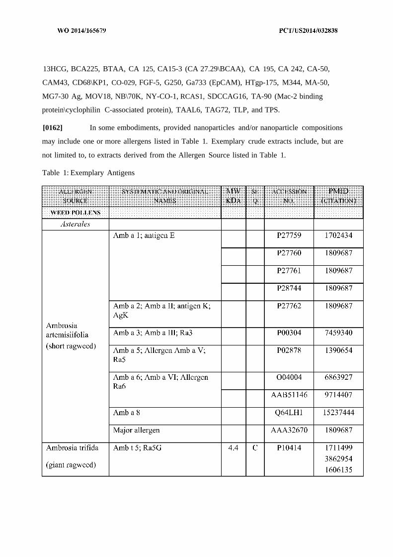
13HCG, BCA225, BTAA, CA 125, CA15-3 (CA 27.29\BCAA), CA 195, CA 242, CA-50,
CAM43, CD68\KP1, CO-029, FGF-5, G250, Ga733 (EpCAM), HTgp-175, M344, MA-50,
MG7-30 Ag, MOV18, NB\70K, NY-CO-1, RCAS1, SDCCAG16, TA-90 (Mac-2 binding
protein\cyclophilin C-associated protein), TAAL6, TAG72, TLP, and TPS.
[0162] In some embodiments, provided nanoparticles and/or nanoparticle compositions
may include one or more allergens listed in Table 1. Exemplary crude extracts include, but are
not limited to, to extracts derived from the Allergen Source listed in Table 1.
Table 1: Exemplary Antigens
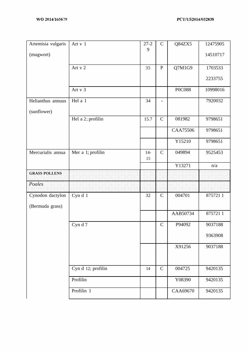
Artemisia vulgaris Art v 1 27-2 C Q84ZX5 124759059
(mugwort) 14510717
Art v 2 35 P Q7M1G9 1703533
2233755
Art v 3 P0C088 10998016
Helianthus annuus Hel a 1 34 - 7920032
(sunflower)
Hel a 2; profilin 15.7 C 081982 9798651
CAA75506 9798651
Y15210 9798651
Mercurialis annua Mer a 1; profilin 14- C 049894 952545315
Y13271 n/a
GRASS POLLENS
Poales
Cynodon dactylon Cyn d 1 32 C 004701 875721 1
(Bermuda grass)
AAB50734 875721 1
Cyn d C P94092 9037188
9363908
X91256 9037188
Cyn d 12; profilin 14 C 004725 9420135
Profilin Y08390 9420135
Profilin 1 CAA69670 9420135
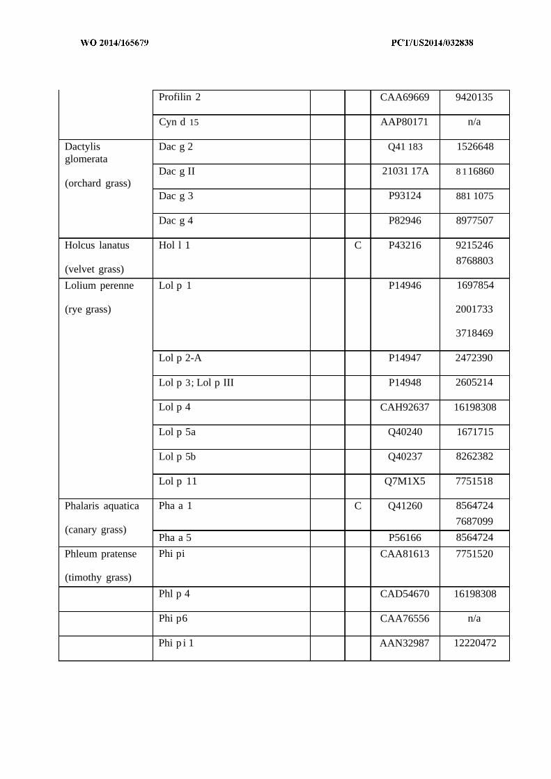
Profilin 2 CAA69669 9420135
Cyn d 15 AAP80171 n/a
Dactylis Dac g 2 Q41 183 1526648glomerata
Dac g II 21031 17A 8 116860(orchard grass)
Dac g 3 P93124 881 1075
Dac g 4 P82946 8977507
Holcus lanatus Hol l 1 C P43216 9215246
8768803(velvet grass)
Lolium perenne Lol p 1 P14946 1697854
(rye grass) 2001733
3718469
Lol p 2-A P14947 2472390
Lol p 3; Lol p III P14948 2605214
Lol p 4 CAH92637 16198308
Lol p 5a Q40240 1671715
Lol p 5b Q40237 8262382
Lol p 11 Q7M1X5 7751518
Phalaris aquatica Pha a 1 C Q41260 8564724
7687099(canary grass)
Pha a 5 P56166 8564724
Phleum pratense Phi pi CAA81613 7751520
(timothy grass)
Phl p 4 CAD54670 16198308
Phi p6 CAA76556 n/a
Phi p i 1 AAN32987 12220472
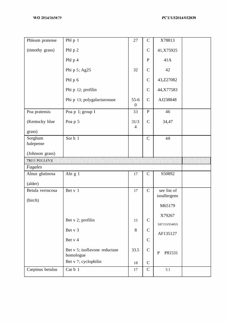
Phleum pratense Phl p 1 27 C X78813
(timothy grass) Phl p 2 C 41.X75925
Phl p 4 P 41A
Phi p 5; Ag25 32 C 42
Phl p 6 C 43,Z27082
Phi p 12; profilin C 44,X77583
Phi p 13; polygalacturonase 55-6 C AJ2388480
Poa pratensis Poa p 1; group I 33 P 46
(Kentucky blue Poa p 5 31/3 C 34,474
grass)
Sorghum Sor h 1 C 48halepense
(Johnson grass)
Fagales
Alnus glutinosa Aln g 1 17 C S50892
(alder)
Betula verrucosa Bet v 1 17 C see list ofisoallergens
(birch)M65179
X79267Bet v 2; profilin 15 C
X87153/S54819
Bet v 3 8 CAF135127
Bet v 4 C
Bet v 5; isoflavone reductase 33.5 CP P81531homologue
Bet v 7; cyclophilin 18 C
Carpinus betulus Car b 1 17 C 5 1
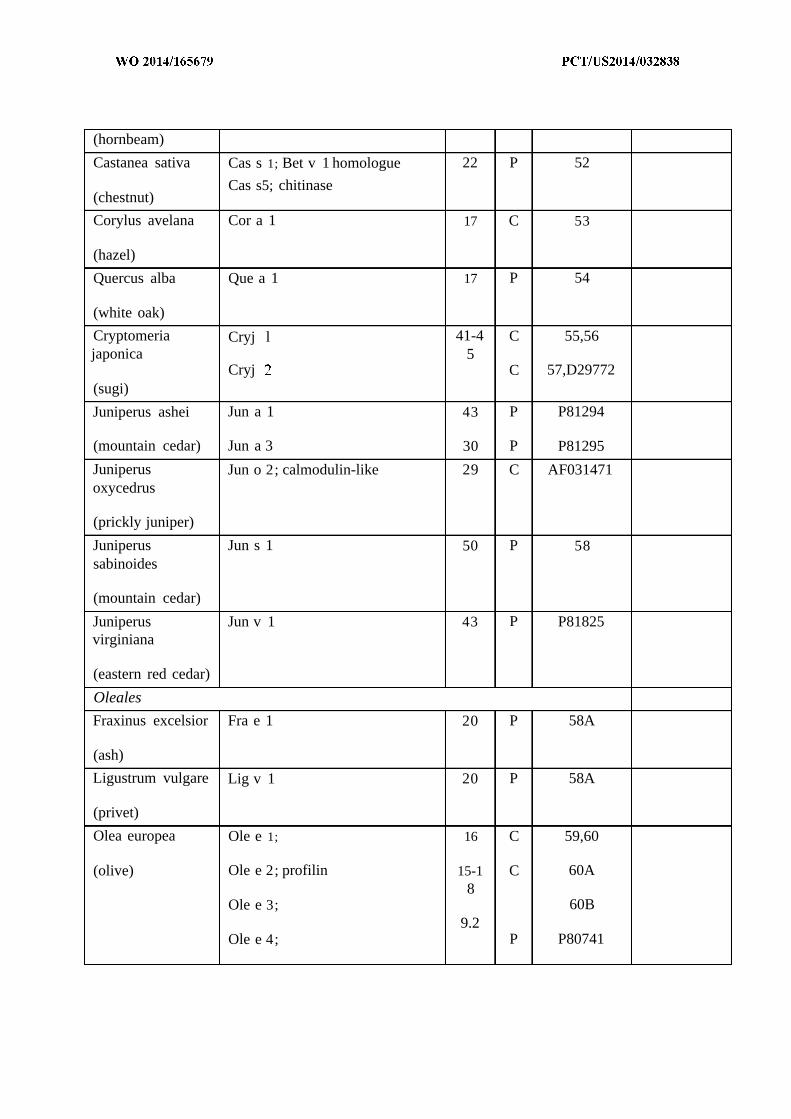
(hornbeam)
Castanea sativa Cas s 1; Bet v 1 homologue 22 P 52
Cas s5; chitinase(chestnut)
Corylus avelana Cor a 1 17 C 53
(hazel)
Quercus alba Que a 1 17 P 54
(white oak)
Cryptomeria Cryj l 41-4 C 55,56japonica 5
Cryj C 57,D29772(sugi)
Juniperus ashei Jun a 1 43 P P81294
(mountain cedar) Jun a 3 30 P P81295
Juniperus Jun o 2; calmodulin-like 29 C AF031471oxycedrus
(prickly juniper)
Juniperus Jun s 1 50 P 58sabinoides
(mountain cedar)
Juniperus Jun v 1 43 P P81825virginiana
(eastern red cedar)
Oleales
Fraxinus excelsior Fra e 1 20 P 58A
(ash)
Ligustrum vulgare Lig v 1 20 P 58A
(privet)
Olea europea Ole e 1; 16 C 59,60
(olive) Ole e 2; profilin 15-1 C 60A8
Ole e 3; 60B9.2
Ole e 4; P P80741
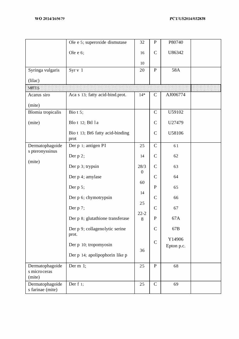
Ole e 5; superoxide dismutase 32 P P80740
Ole e 6; 16 C U86342
10
Syringa vulgaris Syr v 1 20 P 58A
(lilac)
Acarus siro Aca s 13; fatty acid-bind.prot. 14* C AJ006774
(mite)
Blomia tropicalis Bio t 5; C U59102
(mite) Blo t 12; Btl l a C U27479
Bio t 13; Bt6 fatty acid-binding C U58106prot
Dermatophagoide Der p 1; antigen PI 25 C 6 1s pteronyssinus
Der p 2; 14 C 62(mite)
Der p 3; trypsin 28/3 C 630
Der p 4; amylase C 6460
Der p 5; P 6514
Der p 6; chymotrypsin C 6625
Der p 7; C 6722-2
Der p 8; glutathione transferase 8 P 67A
Der p 9; collagenolytic serine C 67Bprot.
Y14906CDer p 10; tropomyosin Epton p.c.
36Der p 14; apolipophorin like p
Dermatophagoide Der m 1; 25 P 68s microceras(mite)
Dermatophagoide Der f 1; 25 C 69s farinae (mite)


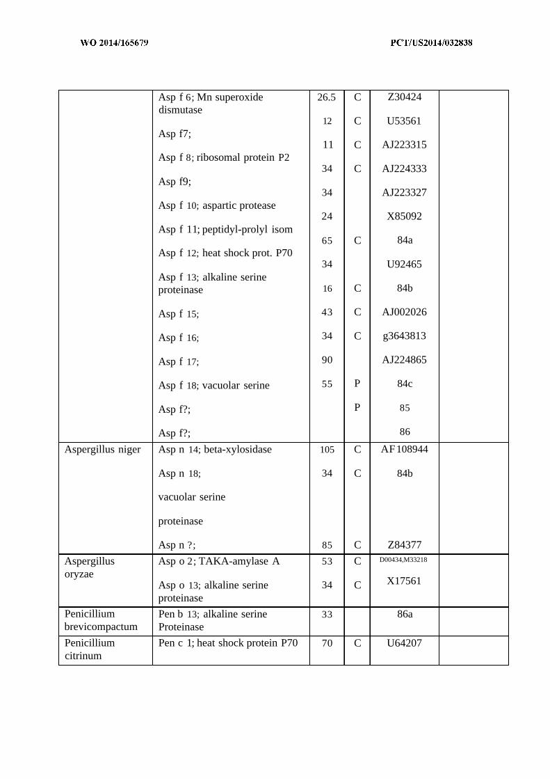
Asp f 6; Mn superoxide 26.5 C Z30424dismutase
12 C U53561Asp f7;
11 C AJ223315Asp f 8; ribosomal protein P2
34 C AJ224333Asp f9;
34 AJ223327Asp f 10; aspartic protease
24 X85092Asp f 11; peptidyl-prolyl isom
65 C 84aAsp f 12; heat shock prot. P70
34 U92465Asp f 13; alkaline serineproteinase 16 C 84b
Asp f 15; 43 C AJ002026
Asp f 16; 34 C g3643813
Asp f 17; 90 AJ224865
Asp f 18; vacuolar serine 55 P 84c
Asp f?; P 85
Asp f?; 86
Aspergillus niger Asp n 14; beta-xylosidase 105 C AF108944
Asp n 18; 34 C 84b
vacuolar serine
proteinase
Asp n ?; 85 C Z84377
Aspergillus Asp o 2; TAKA-amylase A 53 C D00434,M33218
oryzaeAsp o 13; alkaline serine 34 C X17561
proteinase
Penicillium Pen b 13; alkaline serine 33 86abrevicompactum Proteinase
Penicillium Pen c 1; heat shock protein P70 70 C U64207citrinum
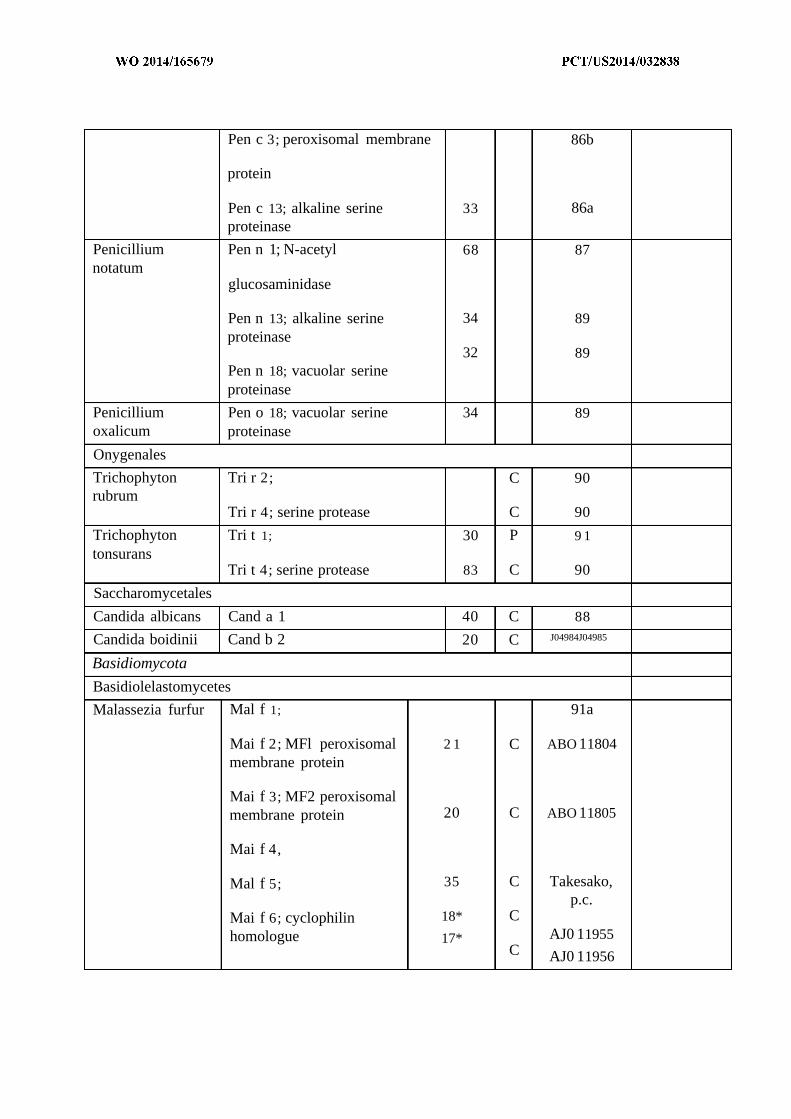
Pen c 3; peroxisomal membrane 86b
protein
Pen c 13; alkaline serine 33 86aproteinase
Penicillium Pen n 1; N-acetyl 68 87notatum
glucosaminidase
Pen n 13; alkaline serine 34 89proteinase
32 89Pen n 18; vacuolar serineproteinase
Penicillium Pen o 18; vacuolar serine 34 89oxalicum proteinase
Onygenales
Trichophyton Tri r 2; C 90rubrum
Tri r 4; serine protease C 90
Trichophyton Tri t 1; 30 P 9 1tonsurans
Tri t 4; serine protease 83 C 90
Saccharomycetales
Candida albicans Cand a 1 40 C 88
Candida boidinii Cand b 2 20 C J04984J04985
Basidiomycota
Basidiolelastomycetes
Malassezia furfur Mal f 1; 91a
Mai f 2; MFl peroxisomal 2 1 C ABO 11804membrane protein
Mai f 3; MF2 peroxisomalmembrane protein 20 C ABO 11805
Mai f 4,
Mal f 5; 35 C Takesako,p.c.
Mai f 6; cyclophilin 18* Chomologue 17* AJ0 11955
C AJ0 11956
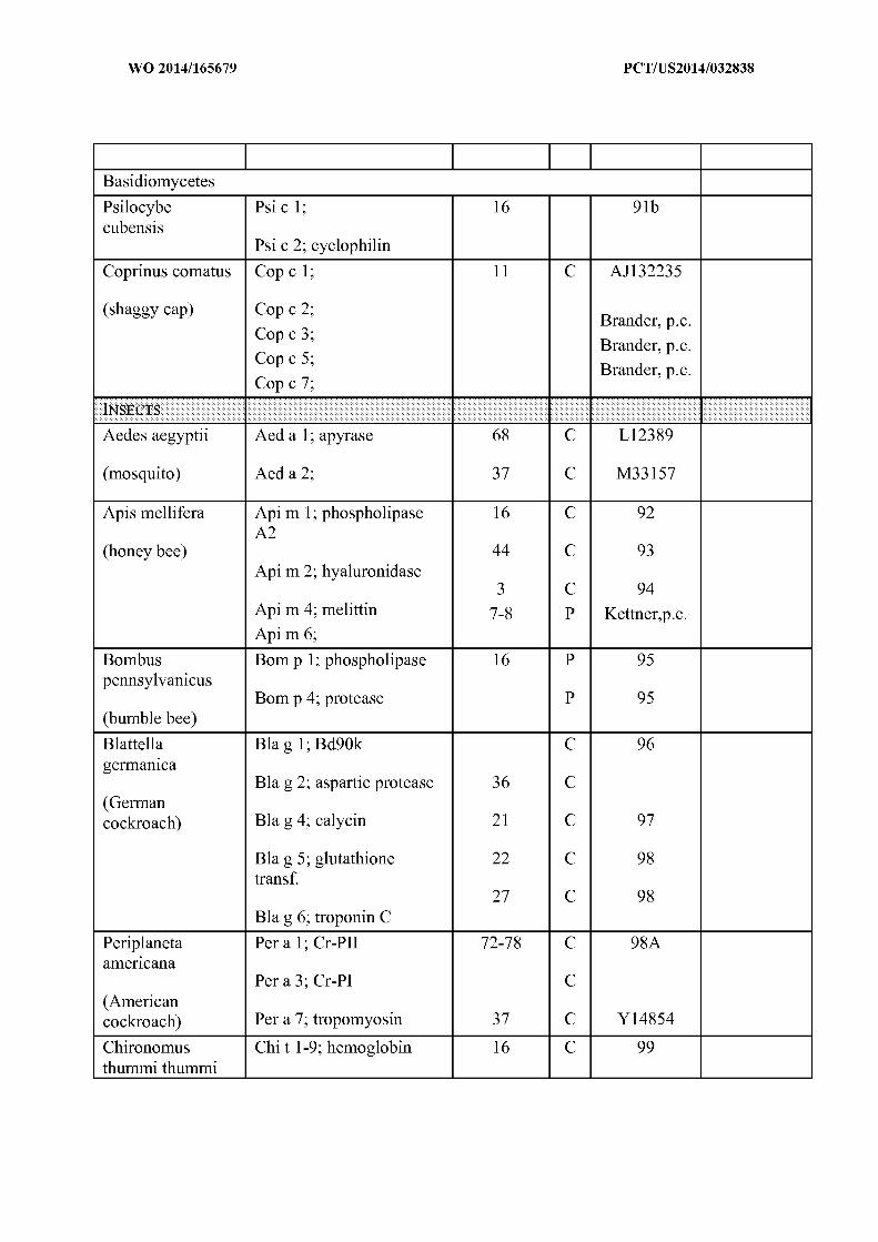

(midges) Chi t 1.01; component III 16 C P02229
Chi t 1.02; component IV 16 C P02230
Chi t 2.0101; component I 16 C P02221
Chi t 2.0102; component 16 C P02221IA
16 C P02222Chi t 3; component II-beta
16 C P02231Chi t 4; component IIIA
16 C P02224Chi t 5; component VI
16 C P02226Chi t 6.01; componentVIIA 16 C P02223
Chi t 6.02; component IX 16 C P02225
Chi t 7; component VIIB 16 C P02227
Chi t 8; component VIII 16 C P02228
Chi t 9; component X
Dolichovespula Dol m 1; phospholipase 35 C 100maculata A l
44 C 101(white face Dol m 2; hyaluronidase Chornet) 23 102,103
Dol m 5; antigen 5
Dolichovespula Dol a 5; antigen 5 23 C 104arenaria
(yellow hornet)
Polistes annularies Pol a 1; phospholipase A l 35 P 105
(wasp) Pol a 2; hyaluronidase 44 P 105
Pol a 5; antigen 5 23 C 104
Polistes Pol d 1; 32-34 C DRdominulus Hoffman
Pol d 4; serine protease(Mediterranean DRpaper wasp) Pol d 5; Hoffman
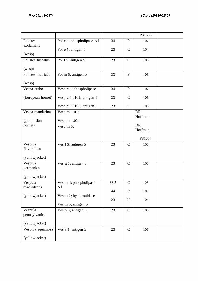
P81656
Polistes Pol e 1; phospholipase A l 34 P 107exclamans
Pol e 5; antigen 5 23 C 104(wasp)
Polistes fuscatus Pol f 5; antigen 5 23 C 106
(wasp)
Polistes metricus Pol m 5; antigen 5 23 P 106
(wasp)
Vespa crabo Vesp c 1; phospholipase 34 P 107
(European hornet) Vesp c 5.0101; antigen 5 23 C 106
Vesp c 5.0102; antigen 5 23 C 106
Vespa mandarina Vesp m 1.01; DRHoffman
(giant asian Vesp m 1.02;hornet) Vesp m 5; DR
Hoffman
P81657
Vespula Ves f 5; antigen 5 23 C 106flavopilosa
(yellowjacket)
Vespula Ves g 5; antigen 5 23 C 106germanica
(yellowjacket)
Vespula Ves m 1; phospholipase 33.5 C 108maculifrons A l
44 P 109(yellowjacket) Ves m 2; hyaluronidase
23 23 104Ves m 5; antigen 5
Vespula Ves p 5; antigen 5 23 C 106pennsylvanica
(yellowjacket)
Vespula squamosa Ves s 5; antigen 5 23 C 106
(yellowjacket)
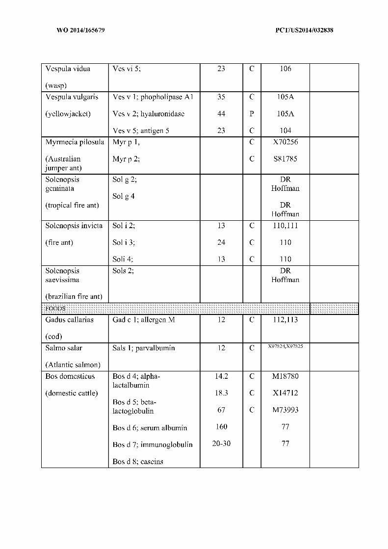

Gallus domesticus Gal d 1; ovomucoid 28 C 114,1 15
(chicken) Gald 2; ovalbumin 44 C 114,1 15
Gald 3; conalbumin 78 C 114,1 15(Ag22)
14 C 114,1 15
Gald 4; lysozyme 69 C X60688Gal d 5; serum albumin
Metapenaeus ensis Met e 1; tropomyosin C U08008
(shrimp)
Penaeus aztecus Pen a 1; tropomyosin 36 P 116
(shrimp)
Penaeus indicus Pen i 1; tropomyosin 34 C 117
(shrimp)
Todarodes Tod p 1; tropomyosin 38 P 117Apacificus (squid)
Haliotis Midae Hal m 1 49 - 117B
(abalone)
Apium graveolens Api g 1; Bet v 1 16* C Z48967homologue AF 129423
(celery) Api g 4; profilin 55/58 P P81943Api g 5;
Brassica juncea Braj 1; 2S albumin 14 C 118
(oriental mustard)
Brassica rapa Bra r 2; prohevein-like 25 P81729protein
(turnip)
Hordeum vulgare Hor v 1; BMAI-1 15 C 119
(barley)
Zea mays Zea m 14; lipid transfer 9 P P19656prot.
(maize, corn)
Corylus avellana Cor a 1.0401; Bet v 1 17 C AF 136945homologue
(hazelnut)

Malus domestica Mai d 1; Bet v 1 C X83672homologue
(apple) 9 C PastorelloMai d 3; lipid transferprotein
Pyrus communis Pyr e 1; Bet v 1 18 C AF05730homologue
(pear) 14 C AF129424Pyr c 4; profilin
Pyr c 5; isoflavonereductase homologue 33.5 C AF071477
Oryza sativa Ory s 1; C U31771
(rice)
Persea americana Pers a 1; endochitinase 32 C Z78202
(avocado)
Primus armeniaca Pru ar 1; Bet v 1 C U93165homologue
(apricot) 9 PPru ar 3; lipid transferprotein
Prunus avium Pru av 1; Bet v 1 C U66076homologue
(sweet cherry) C U3244015Pru av 2; thaumatin C
homologue AF129425
Pru av 4; profilin
Prunus persica Pru p 3;lipid transfer 10 P P81402protein
(peach)
Sinapis alba Sin a 1; 2S albumin 14 C 120
(yellow mustard)
Glycine max Gly m 1.0101; HPS 7.5 P 121
(soybean) Gly m 1.0102; HPS 7 P 121
Gly m 2 8 P A57106
Gly m 3; profilin 14 C AJ223982
Arachis hypogaea Ara h 1; vicilin 63.5 C L34402
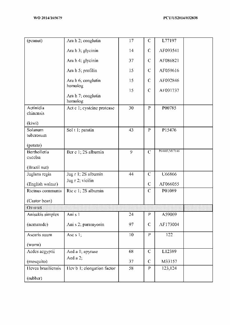
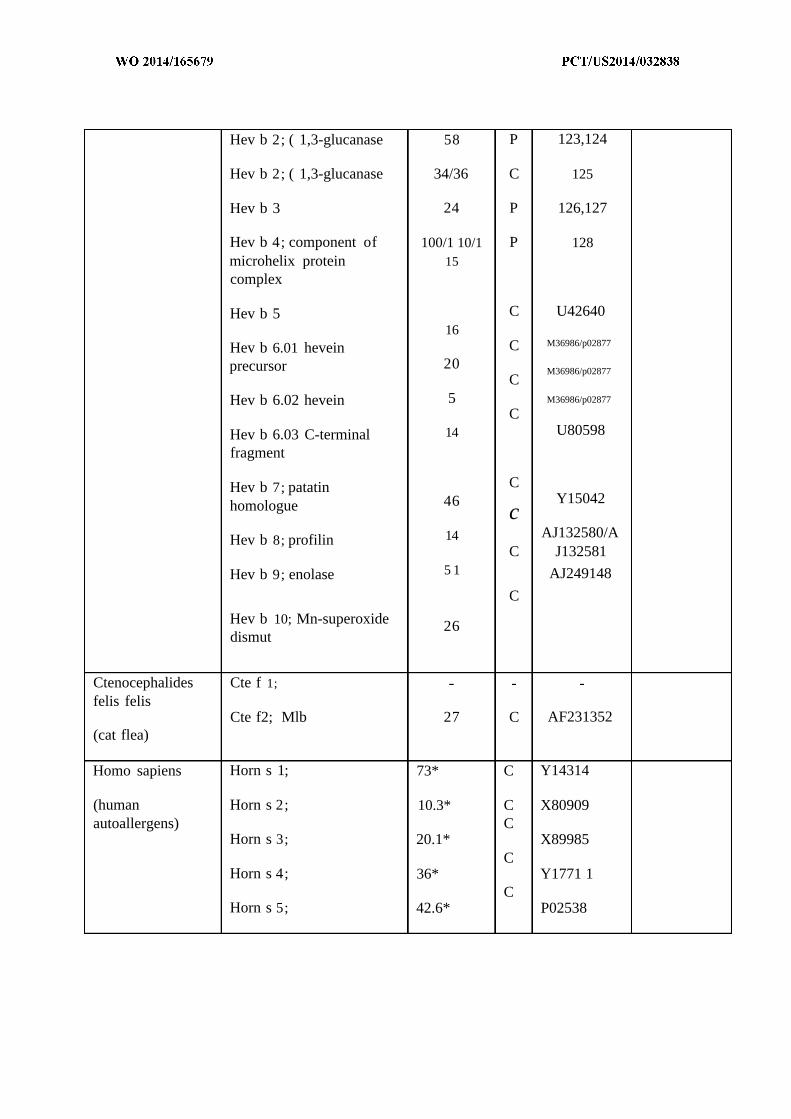
Hev b 2; ( 1,3-glucanase 58 P 123,124
Hev b 2; ( 1,3-glucanase 34/36 C 125
Hev b 3 24 P 126,127
Hev b 4; component of 100/1 10/1 P 128microhelix protein 15complex
Hev b 5 C U4264016
M36986/p02877Hev b 6.01 hevein Cprecursor 20
M36986/p02877C
Hev b 6.02 hevein 5 M36986/p02877
CHev b 6.03 C-terminal 14 U80598
fragment
Hev b 7; patatin C
homologue 46 Y15042c
Hev b 8; profilin 14 AJ132580/AC J132581
Hev b 9; enolase 5 1 AJ249148
C
Hev b 10; Mn-superoxide 26dismut
Ctenocephalides Cte f 1;
felis felisCte f2; Mlb 27 C AF231352
(cat flea)
Homo sapiens Horn s 1; 73* C Y14314
(human Horn s 2; 10.3* C X80909autoallergens) C
Horn s 3; 20.1* X89985C
Horn s 4; 36* Y1771 1C
Horn s 5; 42.6* P02538
[0163] In some embodiments, cancer antigens are provided in crude form such as a
cellular lysate or cellular fraction. Exemplary cellular lysates and/or cellular lysate fractions
include, but are not limited to, cancer cells from acute lymphoblastic leukemia (ALL);
adrenocortical carcinoma; AIDS-related cancers including AIDS-related lymphoma; anal cancer;
appendix cancer; astrocytomas; basal cell carcinoma; bile duct cancer; bladder cancer; bone
cancer (e.g., osteosarcoma and malignant fibrous histiocytoma); brainstem glioma; brain cancer;
brain tumors; breast cancer; bronchial adenomas/carcinoids; Burkitt lymphoma; carcinoid tumors
(e.g., childhood and gastrointestinal tumors); carcinoma (including carcinoma of unknown
primary (CUP) whose origin or developmental lineage is unknown but that possess specific
molecular, cellular, and histological characteristics of epithelial cells); central nervous system
lymphoma; cerebellar astrocytoma; malignant glioma; cervical cancer; childhood cancers;
chronic lymphocytic leukemia; chronic myelogenous leukemia; chronic myeloproliferative
disorders; colon Cancer; cutaneous T-cell lymphoma; desmoplastic small round cell tumor;
endometrial cancer; ependymoma; esophageal cancer; Ewing's sarcoma in the Ewing family of
tumors; extracranial germ cell tumor; extragonadal germ cell tumor; ovarian germ cell tumor;
extrahepatic bile duct cancer; eye cancer; intraocular melanoma; retinoblastoma; gallbladder
cancer; gastric cancer; gastrointestinal carcinoid tumor; gastrointestinal stromal tumor (GIST);
gestational trophoblastic tumor; gastric carcinoid; hairy cell leukemia; head and neck cancer;
heart cancer; hepatocellular (liver) cancer; Hodgkin lymphoma; hypopharyngeal cancer;
hypothalamic and visual pathway glioma; intraocular Melanoma; Islet Cell Carcinoma
(Endocrine Pancreas); kaposi sarcoma; soft tissue sarcoma; uterine sarcoma; kidney cancer
(renal cell carcinoma); laryngeal cancer; leukemias (including acute lymphoblastic or acute
lymphocytic leukemia, acute myeloid or acute myelogenous leukemia, chronic lymphocytic or
chronic lymphocytic leukemia, chronic myelogenous or chronic myeloid leukemia); Lip and Oral
Cavity Cancer; liposarcoma; liver cancer; lung cancer (including non-small cell and small cell);
lymphomas (e.g., AIDS-related, Burkitt, cutaneous T-Cell, Hodgkin, non-Hodgkin, Primary
Central Nervous System); macroglobulinemia; medulloblastoma; melanoma; Merkel Cell
Carcinoma; mesothelioma (e.g., adult malignant mesothelioma, childhood mesothelioma);
metastatic squamous neck cancer; mouth cancer; Multiple Endocrine Neoplasia Syndrome;
Multiple Myeloma; Mycosis Fungoides; Myelodysplasia Syndromes;
Myelodysplastic/Myeloproliferative Diseases; Myelogenous Leukemia; Myeloid Leukemia; (e.g.
Adult Acute; nasal cavity and paranasal sinus cancer; nasopharyngeal carcinoma; neuroblastoma;
oral cancer; oropharyngeal cancer; ovarian cancer; ovarian epithelial cancer (Surface epithelial-
stromal tumor); ovarian germ cell tumor; ovarian low malignant potential tumor; pancreatic
cancer; paranasal sinus and nasal cavity cancer; parathyroid cancer; penile cancer; pharyngeal
cancer; pheochromocytoma; pineal astrocytoma; pineal germinoma; pineoblastoma and
supratentorial primitive neuroectodermal tumors; pituitary adenoma; pleuropulmonary blastoma;
prostate cancer; rectal cancer; renal pelvis and ureter and transitional cell cancer;
rhabdomyosarcoma; Sezary syndrome; skin cancer (including melanoma and nonmelanoma);
skin carcinoma; small intestine cancer; squamous cell carcinoma; stomach cancer; testicular
cancer; throat cancer; thymoma and thymic carcinoma; thyroid cancer; urethral cancer;
endometrial uterine cancer; vaginal cancer; vulvar cancer; and/or combinations thereof.
[0164] In some embodiments, provided nanoparticles include one or more alloantigens.
As described herein, an alloantigen refers to an antigen associated with allorecognition and/or
graft rejection (e.g., an antigen against which a rejection immune response is directed).
Alloantigens are generally polypeptides expressed by an individual that are genetically different
from another individual of the same species. The term "alloantigen polypeptide" refers to a
polypeptide whose amino acid sequence includes at least one characteristic sequence of an
alloantigen. A wide variety of alloantigen sequences are known in the art.
[0165] In some embodiments, an alloantigen for use in accordance with the present
invention is a major histocompatibility complex (MHC) polypeptide. In some embodiments, an
alloantigen for use in accordance with the present invention is a Class I MHC polypeptide. In
some embodiments, an alloantigen for use in accordance with the present invention is a Class II
MHC polypeptide. In some embodiments, an alloantigen for use in accordance with the present
invention contains part of or all of an extracellular domain of an MHC polypeptide. In some
embodiments, an alloantigen for use in accordance with the present invention is a minor
histocompatibility complex polypeptide. In some embodiments, an alloantigen for use in
accordance with the present invention is a costimulatory entity (e.g., CD28, CD80, and CD86,
among others). In some embodiments, an alloantigen for use in accordance with the present
invention is a non-MHC protein produced by or present in graft tissue and not produced by or

present in a host. One of ordinary skill in the art will recognize that alloantigens described
herein are exemplary. Any polypeptide that is associated with an allorecognition and/or graft
rejection can be classified as an alloantigen.
[0166] It will be appreciated that alloantigen polypeptides may have a complete
sequence, or alternatively may be polypeptides that represent functional fragments (i.e.,
fragments retaining at least one activity and/or one characteristic sequence or portion) of such
complete polypeptides. Moreover, those of ordinary skill in the art understand that protein
sequences generally tolerate some substitution without destroying activity. Thus, any
polypeptide that retains activity and shares at least about 30-40% overall sequence identity, often
greater than about 50%>, 60%>, 70%>, or 80%>, and further usually including at least one region of
much higher identity, often greater than 90%> or even 95%, 96%, 97%, 98%, or 99% in one or
more highly conserved regions, usually encompassing at least 3-4 and often up to 20 or more
amino acids, with another alloantigen polypeptide of the same class, is encompassed within the
relevant term "alloantigen polypeptide" as used herein.
[0167] In some embodiments, it may be desirable to include nanoparticles encapsulating
more than one polypeptide, such as an autoantigen and/or alloantigen polypeptide. In some
embodiments, a nanoparticle may encapsulate more than one polypeptide. In some
embodiments, a mixture of nanoparticles that each encapsulate one or more polypeptides may be
used in accordance with the present invention. To give but one example, at least three different
autoantigen polypeptides, Pancreatic β-cell antigens, insulin and glutamic acid decarboxylase
(GAD) are thought to contribute to Insulin-Dependent Diabetes Mellitus (e.g., Type I diabetes
abbreviated IDDM). To give another non-limiting example, several different alloantigen
polypeptides are thought to contribute to graft rejection, including major histocompatibility
complex polypeptides, minor histocompatibility polypeptides, and costimulatory entities.
Inventive compositions may include a mixture of nanoparticles that encapsulate more than one or
all of the autoantigen or alloantigen polypeptides. Also, it may be desirable to include
autoantigen polypeptides that are associated with a variety of different kinds autoimmune
disorders so that multiple autoimmune disorders are treated simultaneously.
Other Agents

[0168] In some embodiments, the provided nanoparticles and/or nanoparticle
compositions may include one or more other agents (e.g. adjuvants). Without wishing to be held
to a particular theory, it is possible that some embodiments may mimic one or more
characteristics or features of microbial (e.g., bacterial) cells. In some embodiments, adjuvants
may be provided from one or more bacterial sources, including bacterial cellular lysates and/or
cellular lysate fractions. In some embodiments, bacterial cellular lysate fractions are or comprise
entities known as pathogen-associated molecular patterns ("PAMPs"). In some embodiments,
one or more of a hydrophobic bacterial cellular lysate fraction and/or hydrophilic bacterial
cellular lysate fraction include one or more PAMPs as a hydrophilic cellular component and/or
hydrophobic cellular component.
[0169] In some embodiments, PAMPs are entities associated with bacterial cells that are
recognized by cells of the innate immune system. In some embodiments, PAMPs are recognized
by Toll-like receptors (TLRs) and other pattern recognition receptors (PRRs) in both plants and
animals. In some embodiments, PAMPs are recognizd by C-type lectin receptors (CLRs). In
some embodiments, a CLR is a type I or type II CLR. In some embodiments, PAMPs are or
comprise entities associated with the outer surface of a bacterial cell, including, but not limited
to, membrane-associated proteins and/or peptides, receptors embedded in bacterial membranes,
etc. Exemplary PAMPs include, but are not limited to, bacterial lipopolysaccharide (LPS),
bacterial flagellin, lipoteichoic acid from gram positive bacteria, peptidoglycan, double-stranded
RNAs (dsRNAs), unmethylated CpG motifs, any of the TLR ligands presented in Table 2,
characteristic portions thereof, and/or combinations thereof.
Table 2 . Exemplary TLRs and TLR Ligands
TLR TLR Ligand(s)
Multiple triacyl lipopeptides (e.g., from bacteria and mycobacteria),TLR1
such as lipopeptide Pam3Cys-SK4 ("Pam")
Multiple glycolipids, lipopeptides and lipoproteins, such aslipopeptide Pam3Cys-SK4 ("Pam")
TLR2Lipoteichoic acid
Peptidoglycan
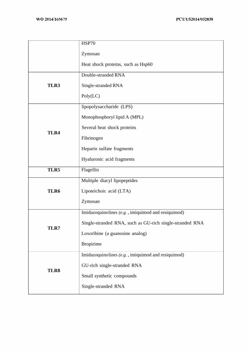
HSP70
Zymosan
Heat shock proteins, such as Hsp60
Double-stranded RNA
TLR3 Single-stranded RNA
Poly(LC)
lipopolysaccharide (LPS)
Monophosphoryl lipid A (MPL)
Several heat shock proteinsTLR4
Fibrinogen
Heparin sulfate fragments
Hyaluronic acid fragments
TLR5 Flagellin
Multiple diacyl lipopeptides
TLR6 Lipoteichoic acid (LTA)
Zymosan
Imidazoquinolines (e.g. , imiquimod and resiquimod)
Single-stranded RNA, such as GU-rich single-stranded RNATLR7
Loxoribine (a guanosine analog)
Bropirime
Imidazoquinolines (e.g. , imiquimod and resiquimod)
GU-rich single-stranded RNATLR8
Small synthetic compounds
Single-stranded RNA
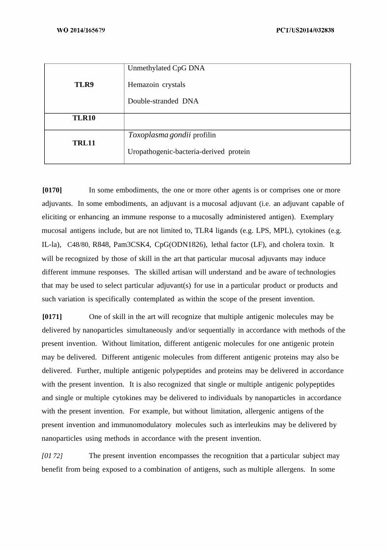
Unmethylated CpG DNA
TLR9 Hemazoin crystals
Double-stranded DNA
TLR10
Toxoplasma gondii profilinTRL11
Uropathogenic-bacteria-derived protein
[0170] In some embodiments, the one or more other agents is or comprises one or more
adjuvants. In some embodiments, an adjuvant is a mucosal adjuvant (i.e. an adjuvant capable of
eliciting or enhancing an immune response to a mucosally administered antigen). Exemplary
mucosal antigens include, but are not limited to, TLR4 ligands (e.g. LPS, MPL), cytokines (e.g.
IL-la), C48/80, R848, Pam3CSK4, CpG(ODN1826), lethal factor (LF), and cholera toxin. It
will be recognized by those of skill in the art that particular mucosal adjuvants may induce
different immune responses. The skilled artisan will understand and be aware of technologies
that may be used to select particular adjuvant(s) for use in a particular product or products and
such variation is specifically contemplated as within the scope of the present invention.
[0171] One of skill in the art will recognize that multiple antigenic molecules may be
delivered by nanoparticles simultaneously and/or sequentially in accordance with methods of the
present invention. Without limitation, different antigenic molecules for one antigenic protein
may be delivered. Different antigenic molecules from different antigenic proteins may also be
delivered. Further, multiple antigenic polypeptides and proteins may be delivered in accordance
with the present invention. It is also recognized that single or multiple antigenic polypeptides
and single or multiple cytokines may be delivered to individuals by nanoparticles in accordance
with the present invention. For example, but without limitation, allergenic antigens of the
present invention and immunomodulatory molecules such as interleukins may be delivered by
nanoparticles using methods in accordance with the present invention.
[01 72] The present invention encompasses the recognition that a particular subject may
benefit from being exposed to a combination of antigens, such as multiple allergens. In some

embodiments, it may be desirable to provide a nanoparticle composition comprising multiple
antigens relevant to a specific subject, and/or to a population of subjects. For example, in some
embodiments, a particular provided composition will contain a combination of allergens to
address some or all of a particular subject's allergies and/or a combination of allergens to address
some or all allergies commonly present within a population. For example, if a particular subject
is allergic to peanuts and to dust mites, a nanoparticle composition may be designed and
manufactured to address both allergies. Alternatively or additionally, in some embodiments it
may be desirable to prepare nanoparticle compositions including antigens from a plurality of
allergens (i) to which members of a particular community are commonly exposed (e.g., by virtue
of geographic location); (ii) to which subjects are exposed by a common route (e.g., inhalation,
injestion, contact, etc); (iii) to which incidence of allergy within a relevant population (e.g., a
geographic population, an age population, an ethnic population, etc) is above a designated
threshold; (iv) to which subjects allergic to one allergen also tend ot have allergy to, for example,
subjects allergic to tree nuts tend to also be allergic to pecans, walnuts, and pistachios, subjects
with allergy to crustaceans (e.g., loster, crab, shrimp, or crayfish) or mollusks (e.g., clams,
mussels, oysters, or scallops) tend to have allergy to various types, not just a single crustacean or
mollusk.
[0173] In some embodiments, a particular provided composition may contain a
combination of antigens other than allergens. For example, in some embodiments, a particular
provided composition may contain a combination of antigens associated with a particular
disease, disorder or condition (e.g., with a particular cancer, a particular infectious disease, a
particular graft v host or host v graft syndrome, etc).
[0174] Those of skill in the art will recognize a wide variety of potential applications
utilizing combinations of antigens; each of these is contemplated as within the scope of the
present invention.
[0175] According to various embodiments, provided compositions comprising an antigen
or other protein agent may comprise the antigen or other protein agent in any of a variety of
forms. Exemplary forms include, without limitation, R A, DNA, protein, and combinations
thereof. In some embodiments, the antigen or protein agent may be provided as a portion of a
cell, tissue or extract thereof.

Nanoparticle Compositions
[0176] The present invention provides a variety of new and/or improved nanoparticle
compositions. The present invention encompasses the recognition that the many advantages of
certain known nanoparticle compositions (such as, for example, those described in one or more
of U.S. Pat. 7,534,448, U.S. Pat. 7,534,449, U.S. Pat. 7,550,154, US20090239789A1,
US20090269397A1, US20100104503A1, US20100151436A1, US20100284965A1,
WO2006080951, WO20081 15641, WO2008109347, WO2009094273, WO2012167261, and
WO20 13003 157, each of which is incorporated herein by reference) can beneficially be
exploited and improved through use with one or more microbial cellular extracts and/or one or
more crude antigen preparations. According to the present invention, in some embodiments,
combination of nanoparticles with microbial cellular extracts comprising multiple microbial
cellular components (optionally together with one or more antigens and/or one or more other
agents) provides improved nanoparticles that embody certain immunologically relevant features
of microbial cells.
[0177] The present invention further encompasses the recognition that known
nanoparticle compositions (such as, for example, those described in one or more of U.S. Pat.
7,534,448, U.S. Pat. 7,534,449, U.S. Pat. 7,550,154, US20090239789A1, US20090269397A1,
US20100104503A1, US20100151436A1, US20100284965A1, WO2006080951,
WO20081 15641, WO2008109347, WO2009094273, WO2012167261, and WO2013003157,
each of which is incorporated herein by reference) can beneficially be exploited for the treatment
and/or prevention of allergies. In some embodiments, the present invention provides
nanoparticle compositions comprising nanoparticles together with one or more allergens. In
certain such embodiments, the present invention provides nanoparticle compositions comprising
nanoparticles together with one or more relatively crude allergen preparations.
[0178] The present invention further encompasses the recognition that known
nanoparticle compositions (such as, for example, those described in one or more of U.S. Pat.
7,534,448, U.S. Pat. 7,534,449, U.S. Pat. 7,550,154, US20090239789A1, US20090269397A1,
US20100104503A1, US20100151436A1, US20100284965A1, WO2006080951,
WO20081 15641, WO2008109347, WO2009094273, WO2012167261, and WO2013003157,
each of which is incorporated herein by reference), can desirably be exploited and/or improved
in a variety of contexts through combination with one or more antigen preparations and/or one or
more microbial cellular extracts and/or one or more other agents as described herein.
[0179] In certain embodiments, provided nanoparticle compositions comprise
nanoparticles combined with one or more cellular extracts, one or more antigen preparations,
and/or one or more other agents so that certain combined elements are entrapped within lumens
of the nanoparticles. In some embodiments, provided nanoparticle compositions comprise
nanoparticles combined with one or more cellular extracts, one or more antigen preparations,
and/or one or more other agents so that certain combined elements are associated with the
external surface of nanoparticles. In some embodiments, provided nanoparticle compositions
comprise nanoparticles combined with one or more cellular extracts, one or more antigen
preparations, and/or one or more other agents so that certain combined elements are present both
in and on nanoparticles. In some embodiments, provided nanoparticle compositions comprise
nanoparticles combined with one or more cellular extracts, one or more antigen preparations,
and/or one or more other agents so that certain combined elements are mixed with, but not
specifically associated with any site on or in, nanoparticles.
[0180] In certain particular embodiments, the present invention provides nanoparticle
compositions in which components of a hydrophilic cellular extract are localized within
nanoparticle lumens; in some such embodiments, all components of a hydrophilic cellular extract
are preferentially localized within nanoparticle lumens; in some such embodiments, all
components of a hydrophilic cellular extract are substantially exclusively localized within
nanoparticle lumens.
[0181] In certain particular embodiments, the present invention provides nanoparticle
compositions in which components of a hydrophobic cellular extract are localized on the external
surface of nanoparticle; in some such embodiments, all components of a hydrophobic cellular
extract are preferentially localized on the nanoparticle external surface; in some such
embodiments, all components of a hydrophobic cellular extract are substantially exclusively
localized on the external surface.

[0182] In certain particular embodiments, the present invention provides nanoparticle
compositions in which components of a hydrophilic cellular extract are localized (e.g.,
preferentially or substantially exclusively) within nanoparticle lumens; and components of a
hydrophobic cellular extract are localized (e.g., preferentially or substantially exclusively) on the
external surface of nanoparticles.
[0183] In some embodiments, it may be desirable that all or substantially all of one or
more microbial components, one or more antigen preparations, and/or one or more other agents
are entrapped within lumens of nanoparticles in provided compositions, for example, when
administering an anaphalactic allergen to a sensitive individual. In some embodiments, it may be
desirable for one or more microbial components, antigen preparations, and/or other agents to be
located both within the lumens of the nanoparticles as well as on the external surface of the
nanoparticles.
Associating
[0184] Any of a variety of methods of associating one or more cellular preparations,
cellular extracts, one or more antigen preparations, and/or one or more other agents with a
biodegradable and/or biocompatible polymer may be used according to various embodiments.
Exemplary methods of associating include, but are not limited to: mixing, blending or combining
under pressure substantially equal to atmospheric pressure, mixing, blending or combining under
pressures elevated above atmospheric pressure, mixing, blending or combining under pressure
less than atmospheric pressure {e.g. vacuum).
[0185] In some embodiments, one or more extracts, preparations and/or agents is
associated covalently with a nanoparticle surface. In some embodiments, one or more extracts,
preparations and/or agents is associated non-covalently with a nanoparticle surface. In some
embodiments, non-covalent association involves incorporation of one or more components into
the nanoparticle membrane. In some embodiments, non-covalent association involves specific
binding with the nanoparticle membrane or an element incorporated therein. In some specific
embodiments, one or more particular components of an extract, preparation, or agent may be
coupled with a ligand that specifically binds with a target in the nanoparticle membrane. In
some embodiments, a ligand-target combination utilized in such an embodiment may be, for

example, biotin-avidin, antibody-antigen, GST-glutathione, mannose binding protein-mannose,
Protein A-IgG, and/or S-tag.
[0186] In some embodiments, provided nanoparticle compositions may include a
plurality of sets of nanoparticles that share one or more structural and/or functional
characteristics. For example, in some embodiments, provided nanoparticle compositions may
comprise a plurality of sets of nanoparticles, each of which includes a targeting agent that
localizes members of the set to a particular target site (see U.S. Pat. Nos. 7,534,448, and
7,534,449, hereby incorporated in their entirety, for exemplary targeting agents and methods of
incorporating targeting agents in nanoparticles). Alternatively or additionally, in some
embodiments, provided nanoparticle compositions may comprise a plurality of sets each of
which is designed to have and/or is characterized by a different half-life (e.g., in a relevant tissue
or organ of interest), different components (e.g. in the lumen or associated with external surface,
different populations of antigens, etc).
Pharmaceutical Compositions
[0187] In some embodiments, the present invention provides pharmaceutical
compositions comprising a provided nanoparticle composition together with one or more
pharmaceutically acceptable excipients.
[0188] In some embodiments, provided pharmaceutical compositions may be prepared by
any appropriate method, for example as known or hereafter developed in the art of
pharmacology. In general, such preparatory methods include the step of bringing a provided
nanoparticle composition into association with one or more pharmaceutically acceptable
excipients, and then, if necessary and/or desirable, shaping and/or packaging the product into an
appropriate form for administration, for example as or in a single- or multi-dose unit.
[0189] In some embodiments, compositions may be prepared, packaged, and/or sold in
bulk, as a single unit dose, and/or as a plurality of single unit doses. As used herein, a "unit
dose" is a discrete amount of the pharmaceutical composition comprising a predetermined
amount of the provided nanoparticle composition. The amount of the provided nanoparticle
composition is generally equal to the dosage of the provided nanoparticle which would be

administered to a subject and/or a convenient fraction of such a dosage such as, for example,
one-half or one-third of such a dosage.
[0190] In many embodiments, provided pharmaceutical compositions are specifically
formulated for mucosal delivery (e.g., oral, nasal, rectal or subligual delivery).
[0191] In some embodiments, appropriate excipients for use in provided pharmaceutical
compositions may, for example, include one or more pharmaceutically acceptable solvents,
dispersion media, granulating media, diluents, or other liquid vehicles, dispersion or suspension
aids, surface active agents and/or emulsifiers, isotonic agents, thickening or emulsifying agents,
preservatives, solid binders, lubricants, disintegrating agents, binding agents, preservatives,
buffering agents and the like, as suited to the particular dosage form desired. Alternatively or
additionally, pharmaceutically acceptable excipients such as cocoa butter and/or suppository
waxes, coloring agents, coating agents, sweetening, flavoring, and/or perfuming agents can be
utilized. Remington's The Science and Practice of Pharmacy, 2 1st Edition, A. R. Gennaro
(Lippincott, Williams & Wilkins, Baltimore, MD, 2005; incorporated herein by reference)
discloses various excipients used in formulating pharmaceutical compositions and known
techniques for the preparation thereof.
[0192] In some embodiments, an appropriate excipient is at least 95%, at least 96%, at
least 97% , at least 98%>, at least 99%, or 100% pure. In some embodiments, an excipient is
approved by United States Food and Drug Administration. In some embodiments, an excipient
is pharmaceutical grade. In some embodiments, an excipient meets the standards of the United
States Pharmacopoeia (USP), the European Pharmacopoeia (EP), the British Pharmacopoeia,
and/or other International Pharmacopoeia.
[0193] In some embodiments, liquid dosage forms (e.g., for oral and/or parenteral
administration) include, but are not limited to, emulsions, microemulsions, solutions,
suspensions, syrups, and/or elixirs. In addition to provided nanoparticle compositions, liquid
dosage forms may comprise inert diluents commonly used in the art such as, for example, water
or other solvents, solubilizing agents and emulsifiers such as ethyl alcohol, isopropyl alcohol,
ethyl carbonate, ethyl acetate, benzyl alcohol, benzyl benzoate, propylene glycol, 1,3-butylene
glycol, dimethylformamide, oils (in particular, cottonseed, groundnut, corn, germ, olive, castor,
and sesame oils), glycerol, tetrahydrofurfuryl alcohol, polyethylene glycols and fatty acid esters
of sorbitan, and mixtures thereof. Besides inert diluents, oral compositions can include adjuvants
such as wetting agents, emulsifying and suspending agents, sweetening, flavoring, and/or
perfuming agents. In certain embodiments for parenteral administration, compositions are mixed
with solubilizing agents such a CREMOPHOR®, alcohols, oils, modified oils, glycols,
polysorbates, cyclodextrins, polymers, and/or combinations thereof.
[0194] In some embodiments, injectable preparations, for example, sterile aqueous or
oleaginous suspensions, may be formulated according to known methods using suitable
dispersing agents, wetting agents, and/or suspending agents. Sterile liquid preparations may be,
for example, solutions, suspensions, and/or emulsions in nontoxic parenterally acceptable
diluents and/or solvents, for example, as a solution in 1,3-butanediol. Among the acceptable
vehicles and solvents that may be employed, for example, are water, Ringer's solution, U.S.P.,
and isotonic sodium chloride solution. Sterile, fixed oils are conventionally employed as a
solvent or suspending medium. For this purpose any bland fixed oil can be employed including
synthetic mono- or diglycerides. Fatty acids such as oleic acid can be used in the preparation of
liquid formulations.
[0195] Liquid formulations can be sterilized, for example, by filtration through a
bacterial-retaining filter, and/or by incorporating sterilizing agents in the form of sterile solid
compositions which can be dissolved or dispersed in sterile water or other sterile injectable
medium prior to use.
[0196] In some embodiments, one or more strategies may be utilized prolong and/or
delay the effect of a provided nanoparticle composition after delivery.
[0197] In some embodiments, provided pharmaceutical compositions may be formulated
as suppositories, for example for rectal or vaginal delivery. In some embodiments, suppository
formulations can be prepared by mixing utilizing suitable non-irritating excipients such as cocoa
butter, polyethylene glycol or a suppository wax which are solid at ambient temperature but
liquid at body temperature and therefore melt in the body (e.g., in the rectum or vaginal cavity)
and release the provided nanoparticle composition.

[0198] In some embodiments, solid dosage forms (e.g., for oral administration) include
capsules, tablets, pills, powders, and/or granules. In such solid dosage forms, the provided
nanoparticle composition may be mixed with at least one inert, pharmaceutically acceptable
excipient such as sodium citrate or dicalcium phosphate and/or fillers or extenders (e.g. , starches,
lactose, sucrose, glucose, mannitol, and silicic acid), binders (e.g., carboxymethylcellulose,
alginates, gelatin, polyvinylpyrrolidinone, sucrose, and acacia), humectants (e.g., glycerol),
disintegrating agents (e.g. , agar, calcium carbonate, potato starch, tapioca starch, alginic acid,
certain silicates, and sodium carbonate), solution retarding agents (e.g., paraffin), absorption
accelerators (e.g., quaternary ammonium compounds), wetting agents (e.g., cetyl alcohol and
glycerol monostearate), absorbents (e.g., kaolin and bentonite clay), and lubricants (e.g., talc,
calcium stearate, magnesium stearate, solid polyethylene glycols, sodium lauryl sulfate), and
mixtures thereof. In the case of capsules, tablets and pills, the dosage form may comprise
buffering agents.
[0199] In some embodiments, solid compositions of a similar type may be employed as
fillers in soft and/or hard-filled gelatin capsules using such excipients as lactose or milk sugar as
well as high molecular weight polyethylene glycols and the like. The solid dosage forms of
tablets, dragees, capsules, pills, and granules can be prepared with coatings and shells such as
enteric coatings and other coatings well known in the pharmaceutical formulating art.
[0200] Exemplary enteric coatings include, but are not limited to, one or more of the
following: cellulose acetate phthalate; methyl acrylate-methacrylic acid copolymers; cellulose
acetate succinate; hydroxy propyl methyl cellulose phthalate; hydroxy propyl methyl cellulose
acetate succinate (hypromellose acetate succinate); HP55; polyvinyl acetate phthalate (PVAP);
methyl methacrylate-methacrylic acid copolymers; methacrylic acid copolymers, cellulose
acetate (and its succinate and phthalate version); styrol maleic acid co-polymers;
polymethacrylic acid/acrylic acid copolymer; hydroxyethyl ethyl cellulose phthalate;
hydroxypropyl methyl cellulose acetate succinate; cellulose acetate tetrahydrophtalate; acrylic
resin; shellac, and combinations thereof.
[0201] In some embodiments, solid dosage forms may optionally comprise opacifying
agents and can be of a composition that they release the provided nanoparticle composition(s)
only, or preferentially, in a certain part of the intestinal tract, optionally, in a delayed manner.
Examples of embedding compositions which can be used include polymeric substances and
waxes. Solid compositions of a similar type may be employed as fillers in soft and hard-filled
gelatin capsules using such excipients as lactose or milk sugar as well as high molecular weight
polyethylene glycols and the like.
[0202] In some embodiments, the present invention provides compositions for topical
and/or transdermal delivery, e.g., as a cream, liniment, ointment, oil, foam, spray, lotion, liquid,
powder, thickening lotion, or gel. Particular exemplary such formulations may be prepared, for
example, as products such as skin softeners, nutritional lotion type emulsions, cleansing lotions,
cleansing creams, skin milks, emollient lotions, massage creams, emollient creams, make-up
bases, lipsticks, facial packs or facial gels, cleaner formulations such as shampoos, rinses, body
cleansers, hair-tonics, or soaps, or dermatological compositions such as lotions, ointments, gels,
creams, liniments, patches, deodorants, or sprays.
[0203] In some embodiments, an adjuvant is provided in the same formulation with
provided nanoparticle composition(s) so that adjuvant and provided nanoparticle composition are
delivered substantially simultaneously to the individual. In some embodiments, an adjuvant is
provided in a separate formulation. Separate adjuvant may be administered prior to,
simultaneously with, or subsequent to provided nanoparticle composition administration.
[0204] In some embodiments, provided compositions are stable for extended periods of
time, such as 1 week, 2 weeks, 1 month, 2 months, 6 months, 1 year, 2 years, 3 years, or more.
In some embodiments, provided compositions are easily transportable and may even be sent via
traditional courier or other package delivery service. Accordingly, some embodiments may be
useful in situations of disease outbreak, such as epidemics, or attacks with biological agents {e.g.
anthrax, smallpox, viral hemorrhagic fevers, plague, and others) at least in part due to their
ability to be stored for long periods of time and transported quickly, easily, and safely. Such
attributes may allow for rapid distribution of provided compositions to those in need.
[0205] In some embodiments, it may be advantageous to release encapsulated agent, for
example, an antigen, at varous locations along a subject's gastrointestinal (GI) tract. In some
embodiments, it may be advantageous to release encapsulated agent, for example, an antigen, in
a subject's mouth as well as one or more locations along the subject's GI tract. Accordingly, in
some embodiments, a plurality of provided compositions (e.g. two or more) may be administered
to a single subject to facilitate release of encapsulated agent at multiple locations. In some
embodiments, each of the plurality of compositions has a different release profile, such as
provided by various enteric coatings, for example. In some embodiments, each of the plurality
of compositions has a similar release profile. In some embodiments, the plurality of
compositions comprises one or more antigens. In some embodiments, each of the plurality of
administered compositions comprises a different antigen. In some embodiments, each of the
plurality of compositions comprises the same antigen.
[0206] In some embodiments, one or more agents may be included that can affect rate
and/or extent of release of agent (e.g., an antigen) from nanoparticles. In some embodiments,
such an agent would affect rate and/or extent of release by leakage or otherwise undesired
release (e.g., at a site other than a target site and/or at a time other than a desired time). Without
wishing to be bound by any particular theory, in some embodiments, such agents may coat or
block release sites on nanoparticle surfaces. In some embodiments, such agents may be or
comprise tannic acid.
Routes of Administration
[0207] In some embodiments, provided nanoparticle compositions may be formulated for
any appropriate route of delivery. In some embodiments, provided nanoparticles and/or
nanoparticle compositions may be formulated for any route of delivery, including, but not
limited to, bronchial instillation, and/or inhalation; buccal, enteral, interdermal, intra-arterial
(IA), intradermal, intragastric (IG), intramedullary, intramuscular (IM), intranasal,
intraperitoneal (IP), intrathecal, intratracheal instillation (by), intravenous (IV), intraventricular,
mucosal, nasal spray, and/or aerosol, oral (PO), as an oral spray, rectal (PR), subcutaneous (SQ),
sublingual; topical and/or transdermal {e.g., by lotions, creams, liniments, ointments, powders,
gels, drops, etc.), transdermal, vaginal, vitreal, and/or through a portal vein catheter; and/or
combinations thereof. In some embodiments, the present invention provides methods of
administration of provided nanoparticle compositions via mucosal administration. In some
embodiments, the present invention provides methods of administration of provided nanoparticle

compositions via oral administration. In some embodiments, the present invention provides
methods of administration of provided nanoparticle compositions via sublingual administration.
Methods of Treatment
[0208] The present invention provides, among other things, methods of administering to
a subject in need thereof a nanoparticle composition including a plurality of nanoparticles, each
of which is comprised of a biodegradable or biocompatible polymer arranged in a nanoparticle
structure defining an internal lumen and external surface, and at least one of a preparation of
hydrophilic cellular components encapsulated within the internal lumen and/or at least one
preparation of hydrophobic cellular components associated with the external surface of the
nanoparticle.
[0209] In some embodiments, the present invention provides methods of treating various
diseases, disorders and/or conditions. In some embodiments, provided compositions may be
administered to a subject for treatment and/or prevention of allergy, infection, cancer, and
combinations thereof. Exemplary suitable compositions include those described herein.
Allergy
[0210] The present invention provides, among other things, methods and compositions
for the treatment and/or prevention of allergy. In some embodiments, provided nanoparticle
compositions are useful as vaccines to prevent and/or delay the onset of an allergic reaction. In
some embodiments, provided nanoparticle compositions are useful as vaccines to lessen the
severity and/or duration of a future allergic reaction. In some embodiments, provided
nanoparticle compositions are useful as therapeutics to alleviate and/or arrest an allergic reaction
in progress. In some embodiments, the subject in need thereof is suffering from an allergic
condition as herein described, including, but not limited to allergic rhinitis, asthma, atopic
eczema, anaphylaxis, insect venom, drug allergies, food allergies, and/or combinations thereof.
[0211] In some embodiments, provided nanoparticle compositions may be used for
treatment and/or prevention of allergies associated with anaphylactic allergens, e.g., food
allergens, insect allergens, and rubber allergens {e.g., from latex).

[0212] In some embodiments, provided nanoparticle compositions may be used for
treatment and/or prevention of allergies associated with food. Food allergies are mediated
through the interaction of IgE to specific proteins contained within the food. Examples of
common food allergens include proteins from nuts (e.g., from peanut, walnut, almond, pecan,
cashew, hazelnut, pistachio, pine nut, brazil nut), dairy products (e.g., from egg, milk), seeds
(e.g., from sesame, poppy, mustard), soybean, wheat, and fish (e.g., shrimp, crab, lobster, clams,
mussels, oysters, scallops, crayfish).
[0213] In some embodiments, provided nanoparticle compositions may be used for
treatment and/or prevention of allergies associated with insect allergens. Examples of common
insect allergens include, but are not limited to, proteins from insects such as fleas, ticks, ants,
cockroaches, and bees.
[0214] In some embodiments, allergens elicit a reaction when ingested, inhaled, and/or
injected. Allergens can also elicit a reaction based solely on contact with the skin. Latex is a
well-known example. Latex products are manufactured from a milky fluid derived from the
rubber tree (Hevea brasiliensis) and other processing chemicals. A number of the proteins in
latex can cause a range of allergic reactions. Many products contain latex, such as medical
supplies and personal protective equipment. Two types of reactions can occur in persons
sensitive to latex: local allergic dermatitis and immediate systemic hypersensitivity (or
anaphylaxis).
[0215] In some embodiments, provided nanoparticle compositions may be used for
treatment and/or prevention of allergies associated with local allergic dermatitis. Local allergic
dermatitis may develop within a short time after exposure to latex and generally includes
symptoms of urticaria or hives. The reaction is thought to be allergic and triggered by direct
contact, not inhalation (Sussman et al., 1991, JAMA, 265:2844; incorporated herein by
reference). Symptoms of immediate systemic hypersensitivity vary from skin and respiratory
problems (e.g., urticaria, hives, rhinoconjunctivitis, swelling of lips, eyelids, and throat,
wheezing, and coughing) to anaphylaxis which may progress to hypotension and shock. Such a
reaction may be triggered by inhalation or skin exposure to the allergen.
[0216] In some embodiments, provided nanoparticle compositions may function to
suppress and/or decrease a subject's TH2-type responses and/or enhance and/or increase a
subject's THl-type responses. In some embodiments, provided nanoparticle compositions may
function to enhance and/or increase a subject's TH2-type responses and/or suppress and/or
decrease a subject's THl-type responses. In some embodiments, a subject's TH2-type responses
are enhanced through targeting of a cell surface receptor for CpG oligonucleotides (e.g.
DEC205).
[0217] In some embodiments, provided nanoparticle compositions effectively treat
and/or prevent all of a subject's allergies falling into a particular class of allergy. In some
embodiments, exemplary "classes" of allergies include, but are not limited to, anaphylactic
allergies and non-anaphylactic allergies. In some embodiments, exemplary "classes" of allergies
include, but are not limited to food allergies, insect allergies, pet dander allergies, pollen
allergies, grass allergies, rubber allergies, and so forth. Thus, in some embodiments, provided
nanoparticle compositions may be useful for treating all of a subject's food allergies. In some
embodiments, exemplary "classes" of allergies include, but are not limited to, particular
individual foods which contain multiple allergens. For example, there are at least eleven known
peanut allergen proteins. Thus, in some embodiments, a "class" of allergies is "peanut" allergy,
and provided nanoparticle compositions may be useful for treating all of a subject's allergies
associated with all seven different peanut allergen proteins.
[0218] In some embodiments, provided nanoparticle compositions may be useful for
treating and/or preventing a single allergy, even though no allergy-specific antigen is included.
In some embodiments, provided nanoparticle compositions may be useful for treating and/or
preventing multiple different allergies. In some embodiments, provided nanoparticle
compositions may be useful for treating and/or preventing substantially all of a subject's
allergies. For example, subjects suffering from and/or susceptible to allergy are frequently
allergic to more than one allergen, e.g., at least 1, at least 2, at least 3, at least 4, at least 5, at least
6, at least 7, at least 8, at least 9, at least 10, at least 15, at least 20, at least 25, or more different
allergens. Thus, in some embodiments, an provided nanoparticle composition may be used for
treating and/or preventing at least 1, at least 2, at least 3, at least 4, at least 5, at least 6, at least 7,
at least 8, at least 9, at least 10, at least 15, at least 20, at least 25, or more different allergies in a
single patient. In some embodiments, an provided nanoparticle composition is administered to a
subject suffering from and/or susceptible to multiple different allergies, e.g., at least 1, at least 2,
at least 3, at least 4, at least 5, at least 6, at least 7, at least 8, at least 9, at least 10, at least 15, at
least 20, at least 25, or more different allergies, such that the subject's symptoms are reduced
and/or improved. In some embodiments, an provided nanoparticle composition is administered
to a subject suffering from and/or susceptible to multiple different allergies, e.g., at least 1, at
least 2, at least 3, at least 4, at least 5, at least 6, at least 7, at least 8, at least 9, at least 10, at least
15, at least 20, at least 25, or more different allergies, such that onset of the subject's symptoms
is delayed.
[0219] In some embodiments, provided compositions maybe used as oral vaccines to
treat allergy. One of the major benefits of oral vaccines is the ability to generate both mucosal
and systemic immunity. While oral vaccines have been developed previously, but they have
been almost entirely directed to prevention of infectious disease, and have met with widely
varying levels of success. For example, oral vaccines have been developed for anthrax, cholera,
gastoenteritis, infant diarrhea, malaria, measles, and tuberculosis, among others (see Aziz et al.,
Oral Vaccines: New Needs, New Possibilities, 2007, BioEssays 29.6: 591-604; see also Silin et
al, Oral Vaccination: Where are we?, Exp. Opin. Drug Deliv., 2007, 4(4):323-340, both of
which are hereby incorporated by reference in their entirety). Part of the reason for such
unpredictable results is the complex nature of the gut mucosa. Briefly, the base of the mucosa in
the gut is lined by gut- or mucosa-associated lymphoid tissue, with underlying lamina propria
that is rich in intraepithelial lymphocytes (sometimes referred to as diffuse lymphoid tissue).
The majority of T-cells in the gut mucosa are either αβ or γδ types. Both CD4 and CD8 cells are
found in the gut mucosa, which also carries B cells, monocytes/macrophages, dendrocytes and
other immune cells. In fact, the gut is known to house -90% of the total number of
immunocompetent cells in the human body, with circulating lymphocytes only comprising -2%
of the total lymphocytes (see Silin et al.). Furthermore, the gut is known to accommodate -80%
of all immunoglobin or Ig-producing cells and releases 2 to 3 times more secretory IgA that the
total output of circulating IgG (see Silin et al). Accordingly, any therapy that is exposed to the
gut environment has the potential to engender a wide variety of responses and be affected by any
of several immune or other cells.
[0220] In order to have an effective oral vaccine to treat allergy, effective presentation of
one or more antigens to an antigen presenting cell (APC) is required. While M-cells and Peyer's
patches are popular targets of oral therapies, additional targets include, but are not limited to,
enterocytes, mesenteric lymph nodes, and intestinal epithelial cells. Each APC may be targeted
by various embodiments. Oral immunization is known to generate significant quantities of
secretory IgA (slgA), which is known to play a major role in mucosal defense against pathogens.
However, the value of slgA is questionable when one considers non-mucosal pathogens or
conditions. Various embodiments recognize this and do not trigger large amounts of slgA
release, instead substantially generating a Th2 response.
[0221] Major known barriers to providing effective oral vaccines include proteolytic
degradation of antigens in the gut, tuning of proper release profile in the intestine, and problems
delivering enough antigen in a reasonable sized dose. Additionally, the development of oral
tolerance to an antigen is thought to be a major point of concern in developing oral vaccines in
general. Oral tolerance is a phenomenon where oral antigen exposure can lead to immune
tolerance and a suppression of the systemic immune response to subsequent challenges. The
development of oral tolerance is not an automatic feature of oral antigen exposure, but rather
depends on several factors including, but not limited to, age of subject, MHC restriction, delivery
site, nature, size and dose of antigen, degree of antigenic uptake, and processing and frequency
of administration of antigen. Oral tolerance is thought to be mediated by several immunological
mechanisms including: induction of regulatory T-cells (suppresors) that downregulate specific
cytokines including IL-4, IL-10, and TGF-β, functional of clonal deletion of effector cells, and
antibody-mediated suppression (see Silin et al).
[0222] In some embodiments, provided compositions are able to present antigen to APCs
without inducing oral tolerance. Without wishing to be held to a particular theory, it is possible
certain embodiments are able to present larger quantities of antigen to the immune system than
traditionally known methods of oral immunization. It is suspected that oral tolerance may
manifest, at least in part, due to very small amounts of antigen being presented to APCs (see
Silin et al., Overcoming immune tolerance during oral vaccination against actinobacillus
pleuropneumoniae, 2002, J Vet. Med. 49:169-175). In some embodiments, provided
compositions present antigens to APCs in such a manner as to promote immune tolerance.

Without wishing to be held to a particular theory, it may be advantageous to promote immune
tolerance in some clinical circumstances, such as in cases of anaphalaxis, autoimmune disease, or
certain infectious dieases including, but not limited to, dengue fever and RSV.
Infection
[0223] In some embodiments, the subject in need thereof is suffering from an infection
caused by, but not limited to viruses, prions, bacteria, viroids, macroparasites, fungi, and/or
combinations thereof. In some embodiments, the subject is suffering from a primary infection.
In some embodiments, the subject is suffering from a secondary infection. In some
embodiments, the subject is suffering from an active symptomatic infection. In some
embodiments, the subject is suffering from an active asymptomatic infection {i.e., infection is
active, but does not produce noticeable symptoms; e.g. silent or subclinical infection). In some
embodiments, the subject is suffering from a latent infection {i.e., inactive or dormant infection).
[0224] Exemplary infections that may be treated by some embodiments include, but are
not limited to actinomycosis, African sleeping sickness, AIDS, anthrax, hemorrhagic fevers,
bacterial pneumonia, candidiasis, cellulitis, Chagas disease, chickpox, cholera, C. difficile
infection, Creutzfeldt-Jakob disease, dengue fever, diphtheria, ebola, enterococcus infection,
food poisoning, gangrene, gonorrhea, streptococcal infections, hepatitis A-E, herpes, hookworm,
mononucleosis, leishmaniasis, leprosy, lyme disease, malaria, measles, meningitis, mumps,
conjunctivitis, pertussis, rabies, respiratory syncytial virus, rhinovirus, rubella, SARS, scabies,
sepsis, shingles, syphilis, tetanus, trichinellosis, tuberculosis, tularemia, viral pneumonia, west
nile fever, and yellow fever.
[0225] Without wishing to be held to a particular theory, it is contemplated that some
embodiments may maintain anti-bacterial immune surveillance in an otherwise immune
compromised subject. For example, a subject suffering from a viral or other immune
compromising condition may normally exhibit reduced bacterial resistance, however, with
administration of provided compositions may reduce or eliminate the degree of reduced bacterial
resistance exhibited by the subject. In some embodiments, provided compositions are
administered at regular intervals in order to maintain anti-bacterial immune surveillance. In
some embodiments, In some embodiments, provided compositions are administered to a subject
suffering from or susceptible to a non-bacterial immune challenge. In some embodiments,
provided compositions are administered to a subject that has recentrly suffered from a non
bacterial immune challenge.
Cancer
[0226] In some embodiments, the subject in need thereof is suffering from a cancer
including, but not limited to acute lymphoblastic leukemia (ALL); adrenocortical carcinoma;
AIDS-related cancers including AIDS-related lymphoma; anal cancer; appendix cancer;
astrocytomas; basal cell carcinoma; bile duct cancer; bladder cancer; bone cancer (e.g.,
osteosarcoma and malignant fibrous histiocytoma); brainstem glioma; brain cancer; brain
tumors; breast cancer; bronchial adenomas/carcinoids; Burkitt lymphoma; carcinoid tumors (e.g.,
childhood and gastrointestinal tumors); carcinoma (including carcinoma of unknown primary
(CUP) whose origin or developmental lineage is unknown but that possess specific molecular,
cellular, and histological characteristics of epithelial cells); central nervous system lymphoma;
cerebellar astrocytoma; malignant glioma; cervical cancer; childhood cancers; chronic
lymphocytic leukemia; chronic myelogenous leukemia; chronic myeloproliferative disorders;
colon Cancer; cutaneous T-cell lymphoma; desmoplastic small round cell tumor; endometrial
cancer; ependymoma; esophageal cancer; Ewing's sarcoma in the Ewing family of tumors;
extracranial germ cell tumor; extragonadal germ cell tumor; ovarian germ cell tumor;
extrahepatic bile duct cancer; eye cancer; intraocular melanoma; retinoblastoma; gallbladder
cancer; gastric cancer; gastrointestinal carcinoid tumor; gastrointestinal stromal tumor (GIST);
gestational trophoblastic tumor; gastric carcinoid; hairy cell leukemia; head and neck cancer;
heart cancer; hepatocellular (liver) cancer; Hodgkin lymphoma; hypopharyngeal cancer;
hypothalamic and visual pathway glioma; intraocular Melanoma; Islet Cell Carcinoma
(Endocrine Pancreas); kaposi sarcoma; soft tissue sarcoma; uterine sarcoma; kidney cancer
(renal cell carcinoma); laryngeal cancer; leukemias (including acute lymphoblastic or acute
lymphocytic leukemia, acute myeloid or acute myelogenous leukemia, chronic lymphocytic or
chronic lymphocytic leukemia, chronic myelogenous or chronic myeloid leukemia); Lip and Oral
Cavity Cancer; liposarcoma; liver cancer; lung cancer (including non-small cell and small cell);
lymphomas (e.g., AIDS-related, Burkitt, cutaneous T-Cell, Hodgkin, non-Hodgkin, Primary
Central Nervous System); macroglobulinemia; medulloblastoma; melanoma; Merkel Cell
Carcinoma; mesothelioma (e.g., adult malignant mesothelioma, childhood mesothelioma);
metastatic squamous neck cancer; mouth cancer; Multiple Endocrine Neoplasia Syndrome;
Multiple Myeloma; Mycosis Fungoides; Myelodysplasia Syndromes;
Myelodysplastic/Myeloproliferative Diseases; Myelogenous Leukemia; Myeloid Leukemia; (e.g.
Adult Acute; nasal cavity and paranasal sinus cancer; nasopharyngeal carcinoma; neuroblastoma;
oral cancer; oropharyngeal cancer; ovarian cancer; ovarian epithelial cancer (Surface epithelial-
stromal tumor); ovarian germ cell tumor; ovarian low malignant potential tumor; pancreatic
cancer; paranasal sinus and nasal cavity cancer; parathyroid cancer; penile cancer; pharyngeal
cancer; pheochromocytoma; pineal astrocytoma; pineal germinoma; pineoblastoma and
supratentorial primitive neuroectodermal tumors; pituitary adenoma; pleuropulmonary blastoma;
prostate cancer; rectal cancer; renal pelvis and ureter and transitional cell cancer;
rhabdomyosarcoma; Sezary syndrome; skin cancer (including melanoma and nonmelanoma);
skin carcinoma; small intestine cancer; squamous cell carcinoma; stomach cancer; testicular
cancer; throat cancer; thymoma and thymic carcinoma; thyroid cancer; urethral cancer;
endometrial uterine cancer; vaginal cancer; vulvar cancer; and/or combinations thereof.
Dosing
[0227] In some embodiments, provided pharmaceutical compositions are administered
according to a dosing regimen sufficient to achieve a desired immunological reaction. For
example, in some embodiments, a dosing regimen is sufficient to achieve a desired
immunological reaction if its administration to a relevant patient population shows a statistically
significant correlation with achievement of the desired immunological reaction.
[0228] In some embodiments, the desired immunological reaction is a reduction in the
degree and/or prevalence of symptoms of allergy of at least about 20%, about 25%; about 30%>;
about 3 1% , about 32%, about 33%, about 34%, about 35%, about 36%, about 37%, about 38%,
about 39%, about 40%, about 41%, about 42%, about 43%, about 44%, about 45%, about 46%,
about 47%, about 48%, about 49%, about 50%, about 51%, about 52%, about 53%, about 54%,
about 55% , about 56%, about 57%, about 58%, about 59%, about 60%, about 61%, about 62%,
about 63%, about 64%, about 65%, about 66%, about 67%, about 68%, about 69%, about 70%,
about 71%, about 72%, about 73%, about 74%, about 75%, about 76%, about 77%, about 78%,

about 79%, about 80%, about 81%, about 82%, about 83%, about 84%, about 85%, about 86%,
about 87%, about 88%, about 89%, about 90%, about 91%, about 92%, about 93%, about 94%,
about 95%, about 96%, about 97%, about 98%, about 99%, or more.
[0229] In some embodiments, a provided pharmaceutical composition is administered
according to a dosing regimen sufficient to achieve a reduction in the degree and/or prevalence
of symptoms of allergy of a specified percentage of a population of patients to which the
composition is administered. In some embodiments, the specified percentage of population of
patients to which the composition was administered is at least about 5%, about 10%>, about 15%,
about 20%, about 25%, about 30%, about 31%, about 32%, about 33%, about 34%, about 35%,
about 36%, about 37%, about 38%, about 39%, about 40%, about 41%, about 42%, about 43%,
about 44%, about 45%, about 46%, about 47%, about 48%, about 49%, about 50%, about 51%,
about 52%o, about 53%, about 54%, about 55%, about 56%, about 57%, about 58%, about 59%,
about 60% , about 61%, about 62%, about 63%, about 64%, about 65%, about 66%, about 67%,
about 68%, about 69%, about 70%, about 71%, about 72%, about 73%, about 74%, about 75%,
about 76%, about 77%, about 78%, about 79%, about 80%, about 81%, about 82%, about 83%,
about 84%, about 85%, about 86%, about 87%, about 88%, about 89%, about 90%, about 91%,
about 92%, about 93%, about 94%, about 95%, about 96%, about 97%, about 98%, about 99%,
or more.
[0230] To give but a few illustrative examples, in some embodiments, administration of
at least one provided pharmaceutical composition according to a dosing regimen is sufficient to
achieve a reduction in the degree and/or prevalence of allergy of at least about 20% in at least
about 50% of the population of patients to which the composition was administered. In some
embodiments, administration of at least one provided pharmaceutical composition according to a
dosing regimen is sufficient to achieve a reduction in the degree and/or prevalence of allergy of
at least about 30% in at least about 50% of the population of patients to which the composition
was administered.
[0231] In some embodiments, at least one provided pharmaceutical composition is
administered according to a dosing regimen sufficient to achieve a delay in the onset of
symptoms of allergy. In some embodiments, at least one provided pharmaceutical composition

is administered according to a dosing regimen sufficient to prevent the onset of one or more
symptoms of allergy.
[0232] In some embodiments, a provided dosing regimen comprises or consists of a
single dose. In some embodiments, a provided dosing regimen comprises or consists of multiple
doses, separated from one another by intervals of time that may or may not vary. In some
embodiments, a provided dosing regimen comprises or consists of dosing once every 20 years,
once every 10 years, once every 5 years, once every 4 years, once every 3 years, once every 2
years, once per year, twice per year, 3 times per year, 4 times per year, 5 times per year, 6 times
per year, 7 times per year, 8 times per year, 9 times per year, 10 times per year, 11 times per
year, once per month, twice per month, three times per month, once per week, twice per week,
three times per week, 4 times per week, 5 times per week, 6 times per week, daily, twice daily, 3
times daily, 4 times daily, 5 times daily, 6 times daily, 7 times daily, 8 times daily, 9 times daily,
10 times daily, 11 times daily, 12 times daily, or hourly.
[0233] In some embodiments, a provided dosing regimen comprises or consists of an
initial dose with one or more booster doses. In some embodiments, one or more booster doses
are administered 1 day, 2 days, 3 days, 4 days, 5 days, 6 days, 1 week, 2 weeks, 1 month, 2
months, 6 months, 1 year, 2 years, 5 years, 10 years, or longer than 10 years after the initial dose.
In some embodiments, an initial dose comprises a series of doses administered over a period of
time. For example, in some embodiments, an initial dose comprises a series of 1, 2, 3, 4, 5, 6, 7,
8, 9, 10, 11, 12, 13, 14, 15, 16, 17, 18, 19, 20, 21, 22, 23, 24, or more doses administered at
regular intervals, e.g., intervals that are close in time to one another, such as 5 minute intervals,
10 minute intervals, 15 minute intervals, 20 minute intervals, 25 minute intervals, 30 minute
intervals, 45 minute intervals, hourly intervals, every 2 hours, etc.
[0234] In some embodiments, an initial dose and booster doses contain the same amount
of provided nanoparticle and/or nanoparticle composition. In some embodiments, an initial dose
and booster doses contain different amounts of provided nanoparticle composition. In certain
embodiments, provided nanoparticle compositions at dosage levels sufficient to deliver from
about 0.001 mg/kg to about 100 mg/kg, from about 0.01 mg/kg to about 50 mg/kg, preferably
from about 0.1 mg/kg to about 40 mg/kg, from about 0.5 mg/kg to about 30 mg/kg, from about
0.01 mg/kg to about 10 mg/kg, from about 0.1 mg/kg to about 10 mg/kg, or from about 1 mg/kg
to about 25 mg/kg, of subject body weight per day, one or more times a day. In some
embodiments, provided nanoparticle compositions are formulated into a unit dose. In some
embodiments, a unit dosage is about 10 mg, about 25 mg, about 50 mg, about 100 mg, about 250
mg, about 500 mg, about 1 g, about 5 g, about 10 g, about 25 g, about 50 g, about 100 g, or more
than about 100 g . In some embodiments, the amount of provided nanoparticle composition
present in a particular unit dose depends on the subject to which the composition is to be
administered. To give but a few examples, in some embodiments, a unit dose appropriate for a
mouse is smaller than a unit dose that is appropriate for a rat, which is smaller than a unit dose
that is appropriate for a dog, is smaller than a unit dose that is appropriate for a human.
[0235] In some embodiments, a provided dosing regimen comprises or consists of
administration of multiple doses over the course of the subject's entire lifespan. In some
embodiments, a provided dosing regimen comprises administration of multiple doses over the
course of several years (e.g., 2, 3, 4, 5, 6, 7, 8, 9, 10, 15, 20, 25, 30, 35, 40, 45, 50, 60, 70, 80, 90,
or 100 years). In some embodiments, a provided dosing regimen comprises or consists of
multiple doses over the course of 1, 2, 3, 4, 5, 6, 7, 8, 9, 10, 11, or 12 months. .
[0236] In some embodiments, when provided compositions are used in the treatment of
allergy, prior to the first dose, a subject's baseline allergic response is determined by one or more
of a variety of methods, including, but not limited to, (1) performing a prick skin test (PST) of
one or more of the subject's 1, 2, 3, 4, 5, 6, 7, 8, 9, 10, 11, 12, 13, 14, 15, 16, 17, 18, 19, 20, or
more than 20 allergens, and measuring the wheal and flare response to the PST; (2) measuring
blood serum IgE levels; (3) noting the subject's own description of her typical symptoms (e.g.,
nature, severity, and/or duration of symptoms) upon exposure to one or more of her 1, 2, 3, 4, 5,
6, 7, 8, 9, 10, 11, 12, 13, 14, 15, 16, 17, 18, 19, 20, or more than 20 allergens; (4) exposing the
subject to a certain dose of one or more of her 1, 2, 3, 4, 5, 6, 7, 8, 9, 10, 11, 12, 13, 14, 15, 16,
17, 18, 19, 20, or more than 20 allergens (e.g., if only a small or nonexistent risk of anaphylaxis);
(5) measuring expression (e.g., levels, spatial distribution, temporal distribution, etc.), of one or
more molecular markers, including, but not limited to, T-cell markers CD4+ and/or CD8+; (6)
performing a basophil histamine release assay; and/or combinations thereof. In some
embodiments, a subject's allergic response is monitored using any combination of methods, e.g.
methods (1) - (6) described above, throughout the course of the treatment regimen and/or after

the treatment regimen is completed, e.g., at regular intervals. In some embodiments, allergic
response is monitored daily, weekly, bi-weekly, monthly, 6 times per year, 4 times per year, 3
times per year, 2 times per year, once per year, every 2 years, every 5 years, and/or every 10
years, etc.
[0237] In some embodiments, a subject is challenged with a single allergen and/or
multiple allergens, e.g., a subset of the subject's allergens {e.g., allergens to which the subject is
known to be allergic) and/or all of the subject's allergens {e.g., allergens to which the subject is
known to be allergic). In some embodiments, allergy challenge is performed after 1 week, 2
weeks, 1 month, 2 months, 6 months, and 1 year after initiation of treatment.
[0238] In some embodiments, provided compositions may be administered via any
medically acceptable route. For example, in some embodiments, a provided composition may be
administered via intravenous administration; intradermal administration; transdermal
administration; oral administration; subcutaneous administration; transmucosal administration;
and/or combinations thereof. In some embodiments, exemplary routes of transmucosal
administration include, but are not limited to buccal administration; nasal administration;
bronchial administration; vaginal administration; rectal administration; sublingual
administration; and/or combinations thereof.
Combination Therapy
[0239] In some embodiments, provided pharmaceutical compositions are administered to
a subject in combination with one or more other therapeutic agents or modalities, for example,
useful in the treatment of one or more diseases, disorders, or conditions treated by the relevant
provided pharmaceutical composition, so the subject is simultaneously exposed to both. In some
embodiments, a provided nanoparticle composition is utilized in a pharmaceutical formulation
that is separate from and distinct from the pharmaceutical formulation containing the other
therapeutic agent. In some embodiments, a provided nanoparticle composition is admixed with
the composition comprising the other therapeutic agent. In other words, in some embodiments, a
provided nanoparticle composition is produced individually, and the provided nanoparticle
composition is simply mixed with another composition comprising another therapeutic agent.
[0240] The particular combination of therapies (substances and/or procedures) to employ
in a combination regimen will take into account compatibility of the desired substances and/or
procedures and the desired therapeutic effect to be achieved. In some embodiments, provided
nanoparticle compositions can be administered concurrently with, prior to, or subsequent to, one
or more other therapeutic agents (e.g., desired known allergy therapeutics).
[0241] It will be appreciated that the therapies employed may achieve a desired effect for
the same disorder (for example, a provided nanoparticle composition useful for treating allergy
may be administered concurrently with a known allergy therapeutic that is also useful for treating
allergy), or they may achieve different effects (for example, a provided nanoparticle composition
that is useful for treating allergy may be administered concurrently with a therapeutic agent that
is useful for alleviating adverse side effects, for instance, inflammation, nausea, etc.). In some
embodiments, provided nanoparticle compositions in accordance with the invention are
administered with a second therapeutic agent that is approved by the U.S. Food and Drug
Administration (FDA).
[0242] As used herein, the terms "in combination with" and "in conjunction with" mean
that the provided nanoparticle compositions can be administered concurrently with, prior to, or
subsequent to, one or more other desired therapeutics. In general, each substance will be
administered at a dose and/or on a time schedule determined for that agent.
[0243] For example, in some embodiments, provided pharmaceutical compositions for
the treatment of allergy may, in some embodiments, be administered in combination with, for
example, one or more antihistamines (i.e., histamine antagonist), corticosteroids including
glucocorticoids; epinephrine (adrenaline); theophylline (dimethylxanthine); cromolyn sodium;
anti-leukotrienes; anti-cholinergics; decongestants; mast cell stabilizers; immunotherapy
(progressively larger doses of a specific allergen); monoclonal anti-IgE antibodies (e.g.,
omalizumab); and/or combinations thereof.
[0244] Exemplary antihistamines include, but are not limited to Azelastine;
Brompheniramine; Buclizine; Bromodiphenhydramine; Carbinoxamine; Cetirizine; Cyclizine;
Chlorpheniramine; Chlorodiphenhydramine; Clemastine; Cyproheptadine; Desloratadine;
Dexbrompheniramine; Deschlorpheniramine; Dexchlorpheniramine; Dimetindene;
Diphenhydramine (Benadryl); Doxylamine; Ebastine; Embramine; Fexofenadine; Levocetirizine;
Loratadine; Olopatadine (Patanol); Phenindamine (Nolahist and Thephorin); Pheniramine (Avil);
Phenyltoloxamine; Promethazine; Pyrilamine; Rupatadine; Tripelennamine; Triprolidine; and/or
combinations thereof.
[0245] Exemplary corticosteroids and glucocorticoids include, but are not limited to
Beclometasone dipropionate and Beclomethasone (Clenil, Qvar, Beconase AQ, Alanase,
Vancenase); Budesonide (Rhinocort, Rhinosol, Pulmicort, Budicort, Symbicort, Noex);
Ciclesonide (Alvesco, Omnaris, Omniair); Flunisolide (Aerobid); Fluticasone (Veramyst);
Fluticasone (Flonase); Mometasone and Mometasone furoate (Nasonex); Triamcinolone
(Nasacort AQ); Prednisone; Methylprednisolone (Depo-Medrol); Triamcinolone (Kenalog);
and/or combinations thereof.
[0246] Exemplary forms of cromolyn sodium include, but are not limited to, Rynacrom;
Nasalcrom; Prevalin; Intal; Optocrom; Optrex; Gastrocrom; Intercron; and/or combinations
thereof.
[0247] Exemplary anti-leukotrienes and leukotriene inhibitors (or modifiers) include, but
are not limited to Montelukast (Singulair, Montelo-10, and Monteflo); Zafirlukast (Accolate,
Accoleit, Vanticon); Pranlukast; Zileuton (Zyflo, Zyflo CR); and/or combinations thereof.
[0248] Exemplary anti-cholinergics include, but are not limited to, Ipratropium bromide
(Atrovent®, Apovent, Ipraxa , Aervoent); Combivent (Ipratropium bromide and Albuterol);
Benztropine (Cogentin); Oxitropium (Oxivent); Tiotropium (Spiriva); Glycopyrrolate (Robinul);
Oxybutinin (Ditropan, Driptane, Lyrinel XL); Tolterodine (Detrol, Detrusitol); Chlorphenamine
(Chlor-Trimeton); Diphenhydramine (Benadryl, Sominex, Advil PM, etc.); Dimenhydrinate
(Dramamine); Bupropion (Zyban, Wellbuterin); Hexamethonium; Tubocurarine;
Dextromethorphan; Mecamylamine; Doxacurium; and/or combinations thereof.
[0249] Exemplary decongestants include, but are not limited to, Ephedrine; Levo-
methamphetamine; Naphazoline; Oxymetazoline; Phenylephrine; Phenylpropanolamine;
Propylhexedrine; Synephrine; Tetrahydrozoline; and/or combinations thereof.
[0250] Exemplary mast cell stabilizers include, but are not limited to, Cromoglicic acid;
Ketotifen and Ketotifen fumarate (Zaditor, Zaditen, Alaway, Zyrtec Itchy-Eye Drops, Claritin
Eye); Methyl xanthines; and/or combinations thereof.
[0251] In some embodiments, exemplary known allergy therapeutics that can be
administered in combination with provided nanoparticle compositions in accordance with the
invention include, but are not limited to, any of the therapeutics described in US Patent Numbers
5,558,869, 5,973,121, 6,835,824, 6,486,31 1, and/or 7,485,708, and/or in US Patent Publication
Numbers 2003/0035810, 2003/0202980, 2004/0208894, 2004/0234548, 2007/0213507,
2010/0166802, and/or 201 1/0027298, all of which are incorporated herein by reference.
[0252] As an additional example, in some embodiments, provided pharmaceutical
compositions for the treatment of infectious disease may be administered in combination with,
for example, an antibiotic such as an antibacterial agent, an antiviral agent, and/or an antifungal
agent. In some embodiments, provided pharmaceutical compositions may be administered in
combination with a vaccine.
[0253] Exemplary antibacterial agents include, but are not limited to sulfaniliamide; folic
acid analogs; beta-lactams such as penicillins, cephalosporins, and carbapenems;
aminoglycosides such as streptomycin, kanamycin, neomycin, and gentamycin; tetracyclines
such as chlortetracycline, oxytetracycline, and doxycycline; macrolides; lincosamides;
streptogramins; fluoroquinolones, rifampin, mupirocin, cycloserine, aminocyclitols,
glycopeptides, oxazolidinones, and derivatives/analogs and/or combinations thereof.
[0254] Exemplary antiviral agents include, but are not limited to Abacavir, Aciclovir,
Acyclovir, Adefovir, Amantadine, Amprenavir, Ampligen, Arbidol, Atazanavir, Atripla,
Boceprevirertet, Cidofovir, Combivir, Darunavir, Delavirdine, Didanosine, Docosanol,
Edoxudine, Efavirenz, Emtricitabine, Enfuvirtide, Entecavir, Entry inhibitors, Famciclovir,
Fomivirsen, Fosamprenavir, Foscarnet, Fosfonet, Imunovir, Idoxuridine, Imiquimod, Indinavir,
Inosine, Interferon type III, Interferon type II, Interferon type I, Interferon, Lamivudine,
Lopinavir, Loviride, Maraviroc, Moroxydine, Methisazone, Nelfmavir, Nevirapine, Nexavir,
Nucleoside analogues, Oseltamivir, Peginterferon alfa-2a, Penciclovir, Peramivir, Pleconaril,
Podophyllotoxin, Raltegravir, Reverse transcriptase inhibitors, Ribavirin, Rimantadine,
Ritonavir, Pyramidine, Saquinavir, Stavudine, Tea tree oil, Telaprevir, Tenofovir, Tenofovir
disoproxil, Tipranavir, Trifiuridine, Trizivir, Tromantadine, Truvada, Valaciclovir,
Valganciclovir, Vicriviroc, Vidarabine, Viramidine, Zalcitabine, Zanamivir, Zidovudine, and
derivatives/analogs and/or combinations thereof.
[0255] Exemplary antifungal agents include, but are not limited to polyene agents such as
amphotericin, candicidin, filipin, hamycin, natamycin, nystatin, and rimocidin; imidazole,
triazole and thiazole agents such as bifonazole, butoconazole, clotrimazole, econazole,
fenticonazole, isoconazole, ketoconazole, miconazole, omoconazole, oxiconazole, sertaconazole,
sulconazole, tioconazole, alboconazole, fluconazole, isavuconazole, posaconazole, ravuconazole,
terconazole, voriconazole, and abafungin;allylamines such as amorolfm, butenafine, naftafine,
and terbinafine; and echinocandins such as anidulafungin, caspofungin, and micafungin and
derivatives/analogs and/or combinations thereof.
[0256] As an additional example, in some embodiments, provided pharmaceutical
compositions for the treatment of cancer may be administered in combination with, for example,
alkylating agents, antimetabolite agents, and/or other anticancer medications.
[0257] Exemplary alkylating agents include, but are not limited to polyfunctional
alkylating agents such as cyclophosphamide (Cytoxan), mechlorethamine, melphan (Alkeran),
chlorambucil (Leukeran), thiopeta (Thioplex), and busulfan (Myleran); procarbazine,
dacarbazine, altretamine, cisplatin, and derivatives/analogs and/or combinations thereof.
[0258] Exemplary antimetabolite agents include, but are not limited to methotrexate;
purine antagonists such as mercaptopurine (6-MP), thioguanine (6-TG), fludarabine phosphate,
cladribine, and pentostatin; pyrimidine antagonists such as fiuorouracil, cytarabine, and
azacitidine; plant alkaloids such as vinblastine (Velban), vincristine (Oncovin), etoposide (VP-
16), teniposide (Vimon), topotecan (Hycamtin), irinotecan (Camptosar), paclitaxel (Taxol), and
docetaxel (Taxotere) and derivatives/analogs and/or combinations thereof.
[0259] Exemplary other anticancer agents include, but are not limited to amsacrine;
hydroxyurea (Hydrea); asparaginase (El-spar); mitoxantrone (Novantrone); mitotane; retinoic
acid, bone marrow growth factors, amifostine, and derivatives/analogs and/or combinations
thereof.
Kits
[0260] The present invention provides kits comprising vaccine and/or therapeutic
compositions including provided nanoparticles. In some embodiments, a kit may comprise (i) at
least one provided nanoparticle composition; and (ii) at least one pharmaceutically acceptable
excipient; and, optionally, (iii) instructions for use.
[0261] In some embodiments, kits include multiple {e.g., 1, 2, 3, 4, 5, 6, 7, 8, 9, 10, 11,
12, 13, 14, 15, 16, 17, 18, 19, 20, or more) doses of provided nanoparticle compositions. In
some embodiments, kits include multiple {e.g., 1, 2, 3, 4, 5, 6, 7, 8, 9, 10, 11, 12, 13, 14, 15, 16,
17, 18, 19, 20, or more) populations of provided nanoparticles having different functional
elements {e.g., microbial mimic entities). In some embodiments, multiple populations of
provided nanoparticles are packaged separately from one another in provided kits. In some
embodiments, provided kits may include provided compositions and one or more other
therapeutic agents intended for administration with the provided compositions.
[0262] In some embodiments, the present invention provides pharmaceutical packs or
kits including provided nanoparticle compositions to be used in treatment methods according to
the present invention. In some embodiments, pharmaceutical packs or kits include preparations
or pharmaceutical compositions containing provided nanoparticle compositions in one or more
containers filled with optionally one or more additional ingredients of pharmaceutical
compositions in accordance with the invention. In some embodiments, the pharmaceutical pack
or kit includes an additional approved therapeutic agentfor use in combination therapies, as
described herein. Optionally associated with such container(s) can be a notice in the form
prescribed by a governmental agency regulating the manufacture, use or sale of pharmaceutical
products, which notice reflects approval by the agency of manufacture, use, or sale for human
administration.
[0263] In some embodiments, kits are provided that include provided nanoparticle
compositions and instructions for use. Pharmaceutical doses or instructions therefor may be
provided in a kit for administration to an individual suffering from and/or susceptible to allergy.
Exemplification
Example 1: Preparation of Aqueous E. coli Extract
[0264] The present Example describes preparation of an aqueous extract of microbial
cells, here E. coli, containing hydrophilic components of the cells, for use in accordance with the
present invention.
[0265] The present Example describes preparation of an aqueous E. coli extract (i.e., an
aqueous extract of an E. coli cell culture; "AEE") using standard available procedures. The
production strain may be a common and non-pathogenic strain of E. coli. A master and working
cell bank of the production strain may be established prior to clinical manufacture.
[0266] Cells are harvested from high cell density fermentation and then the media is
exchanged for phosphate buffered saline (PBS) containing 5 mM ethylene diamine tetraacetic
acid (EDTA) as a protease inhibitor. Cells suspended in the PBS-EDTA are homogenized (using
a French press, for example) and then clarified to remove cellular debris. The clarified extract is
treated to inactivate residual enzymes, for example using mild heat treatment. The resultant
extract is clarified again, if necessary, and stored frozen prior to use in the nanoparticle
manufacturing process. In addition to bacterial proteins and DNA, it is expected the AEE will
also contain LPS. In some embodiments, one or more components of the AEE will be processed
insome manner to facilitate nanoencapsulation, for example, shearing DNA to break up large
stretches into smaller pieces more suitable for nanoencapsulation.
[0267] Exemplary specifications for the AEE are presented in Table 3 . Given that both
protein and LPS give banding patterns in sodium dodecyl sulfate-polyacrylamide gel
electrophoresis (SDS-PAGE) analysis, it is anticipated that an SDS-PAGE profile will be too
complex to provide meaningful comparisons between batches. Keto-deoxyoctulosonic acid
(KDO) is a carbohydrate exclusively found in bacterial LPS and will be used as a surrogate assay
for LPS content of the extract.
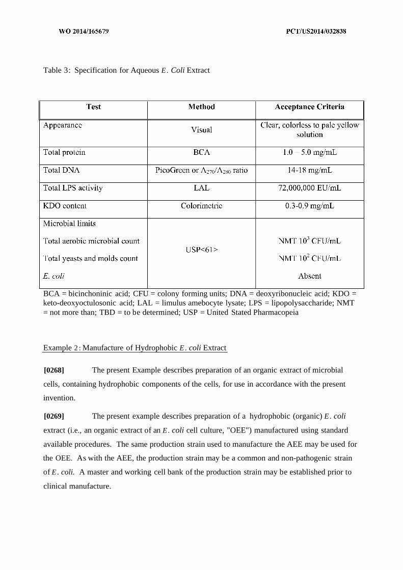
Table 3: Specification for Aqueous E. Coli Extract
BCA = bicinchoninic acid; CFU = colony forming units; DNA = deoxyribonucleic acid; KDO =keto-deoxyoctulosonic acid; LAL = limulus amebocyte lysate; LPS = lipopolysaccharide; NMT= not more than; TBD = to be determined; USP = United Stated Pharmacopeia
Example 2 : Manufacture of Hydrophobic E. coli Extract
[0268] The present Example describes preparation of an organic extract of microbial
cells, containing hydrophobic components of the cells, for use in accordance with the present
invention.
[0269] The present example describes preparation of a hydrophobic (organic) E. coli
extract (i.e., an organic extract of an E. coli cell culture, "OEE") manufactured using standard
available procedures. The same production strain used to manufacture the AEE may be used for
the OEE. As with the AEE, the production strain may be a common and non-pathogenic strain
of E. coli. A master and working cell bank of the production strain may be established prior to
clinical manufacture.

[0270] Briefly, an extract is prepared using the well-known phenol-chloroform-
petroleum ether process, with the exception that hexane is used in lieu of petroleum ether, as
petroleum ether is a pharmaceutically unacceptable solvent. Dried bacterial cells are suspended
in the phenol-chloroform-hexane (PCH) mixture for about 30 minutes. The slurry is then
centrifuged to remove the remaining cells. The remaining cells are then treated with PCH twice
more. The combined organic extracts is evaporated to remove the volatile organic solvents.
Water is added drop-wise to the phenol concentrate to precipitate the LPS and lipids. The
precipitated OEE is then washed with 95% phenol followed by acetone, suspended in water,
lyophilized, and stored frozen prior to use in the nanoparticle manufacturing process. It is
expected that the OEE will be comprised mainly of bacterial LPS and lipids.
[0271] Exemplary specifications for the OEE are presented in Table 4 . It is anticipated
that substantially no proteins or nucleic acids will be extracted into the OEE.
Table 4 : Specification for Organic E. Coli Extract
CFU = colony forming unit; KDO = keto-deoxyoctulosonic acid; LAL = limulus amebocytelysate; LC/MS = liquid chromatography/mass spectrometry; LPS = lipopolysaccharide; NMT =not more than; TBD = to be determined; USP = United Stated Pharmacopeia

Example 3 : Production of Nanoparticle Compositions Containing Encapsulated E. coli
Hydrophilic Components and Surface-Associated E. coli Hydrophobic Components
[0272] This Example describes an exemplary preparation of E-nanoparticles (i.e.,
nanoparticles containing hydrophilic and/or hydrophobic E. coli extract preparations) using a
double emulsion (water-oil-water) process. Briefly, AEE at a protein concentration of c.a. 60
mg/mL is used to reconstitute allergen extracts (dust mite or peanut, for example) also at a
concentration of 60 mg/mL. Higher or lower concentrations of both the AEE and/or the allergen
extract may be used. The AEE-allergen extract solution is homogenized with a dichloromethane
solution of PLGA (c.a. 35.7 mg/mL) with an aqueous to organic ratio of 0.20 mL:28 mL. This
mixture is designated as the first emulsion and comprises or consists essentially of nanodroplets
of the AEE (including, in this Example, antigen, DNA, and LPS) in the OEE.
[0273] In some embodiments, for example where it is desirable to increase the level of
surface-associated hydrophobic components (such as LPS), OEE, such as that described above
in Example 2, is dissolved in 5% aqueous PVA at a concentration of c.a. 2.2 mg/mL total LPS.
Higher or lower concentrations of OEE in PVA solution may be used. Half of the first emulsion
(14 mL) is added to the OEE-PVP solution (14 mL) and homogenized. This mixture is
designated as the second emulsion, and includes several structural similarities to the first
emulsion, described above (i.e. nanodroplets of the AEE in the OEE).
[0274] The first or, if generated, the second emulsion (28 mL) is added to 935 mL of
0.33% aqueous PVA and stirred for c.a. 4 hours to allow the dichloromethane to evaporate. The
nanoparticle suspension is concentrated and the nanoparticles are isolated by centrifugation.
After removal of the supernatant, the nanoparticles are washed 2 to 3 times with water, re-
suspended in water, and isolated by freeze drying. In this example, DNA, LPS and antigen are
distributed throughout the nanparticles, and LPS and lipids are coated on the surface. The
nanoparticles in this Example are approximately 450 nm +/- 150 nm in diameter.
[0275] Example 4 : In vivo Desensitization Study of Orally Administered CpG-Coated,
PLGA-Encapsulated Peanut Extract Nanoparticles

[0276] The present Example describes an exemplary study assessing the in vivo
desensitization efficacy of an orally administered nanoparticle composition comprising relatively
crude antigen (specifically, a peanut extract) in PLGA nanoparticles coated with CpG and
furthermore evaluating the safety of said nanoparticles by assessing anaphylactic potential in
peanut-sensitized animals.
[0277] Among other things, the present Example describes administration of an
exemplary nanoparticle composition that, when orally administered to peanut-sensitized mice,
resulted in desensitization to subsequent oral peanut challenges relative to vehicle control mice.
Materials and Methods
CpG-coated, PLGA-encapsulated peanut extract nanoparticles
[0278] Among other things, the present Example describes nanoparticles which contain
encapsulated allergenic extract of peanuts, Arachis hypogaea. Briefly, commercially available
roasted peanuts in shells (White Rose Brand, NJ) were de-shelled, homogenized in phosphate
buffered saline (PBS, pH 7.4), and subjected to acetone extraction. The final concentration of
peanut protein in PBS was determined to be 13.2 mg/mL by bicinchoninic acid (BCA) assay.
[0279] Prior to nanoparticle manufacture, the 13.2 mg protein/mL peanut extract solution
was concentrated using an Amicon Ultra-4 Centrifugal Filter Unit with Ultracel-3 Membrane (3
kDa cutoff; Millipore, catalog no. UFC8-003). The final protein concentration was determined
to be 27.3 mg/mL via BCA assay.
[0280] CpG-coated, PLGA-encapsulated peanut extract nanoparticles were manufactured
using a double-emulsion method. To minimize endotoxin in the final product, all glassware was
baked at 250°C for 2 hours and plastic instruments were decontaminated in Pyro-Clean
(AlerChek, Inc., catalog no. 130103) for 30 minutes and subsequently washed in UltraPure water
(Millipore, catalog no. H20CC1006).
[0281] A total of 0.7 mL of concentrated peanut extract in PBS (19 mg peanut protein)
was added drop-wise to 14 mL of 25 mg/mL PLGA in methylene chloride (Thermo Fisher
Scientific, catalog no. D65 100) divided over four tubes. The PLGA used for NP-401
manufacture was a 50:50 poly(DL-lactide-co-glycolide) with an ester end group and an inherent
viscosity of 0.95-1.2 dL/g (Durect, product no. B6010-4). The resulting suspensions were briefly
vortexed and then sonicated for 30 seconds in 10 second intervals, icing for 30 seconds between
sonications, using a Tekmar Sonic Distributor fitted with a CV26 sonicator (38% amplitude).
[0282] For the second emulsion, the first emulsions were pooled and added drop-wise to
14 mL of 1.25% poly vinyl alcohol (w/v) (PVA, Sigma Aldrich, catalog no. P8136), 1% sodium
deoxycholate (w/v), 1.25 mg/mL avidin-palmitate divided over four tubes. The resulting
suspensions were briefly vortexed and then sonicated for 30 seconds on ice using a Tekmar
Sonic Distributor fitted with a CV26 sonicator (38% amplitude).
[0283] The avidin-palmitate used in the nanoparticle manufacture was prepared by
incubating 5 mg/ml avidin (EMD Millipore, catalog no. 189725) in 2% sodium deoxycholate
(Sigma-Aldrich, catalog no. D6750) with a 15-fold molar excess of palmitic acid N-hydroxy-
succinimide ester (Sigma-Aldrich, catalog no. PI 162) for 3 hours at 37°C. Excess palmitic acid
was removed by dialysis in a 2% sodium deoxycholate bath over 24 hours at 37°C.
[0284] The second emulsion was pooled and immediately added to 350 mL of room
temperature (RT) 0.3% PVA (w/v) in one beaker. The methylene chloride was evaporated by
stirring with a stir bar for 3 hours at RT. The nanoparticles were collected by centrifugation at
18,500 g for 15 minutes at 4°C. The pellet was resuspended in 10 mL UltraPure water,
centrifuged at 18,500 g for 15 minutes at 4°C, and the supernatant decanted. This washing step
was repeated a total of 3 times. The nanoparticles were then flash-frozen, lyophilized, and stored
at -20°C.
[0285] The nanoparticles were labeled with CpG-biotin immediately prior to use. CpG
oligodeoxynucleotide 1826 (5'-TCC ATG ACG TTC CTG ACG TT-3' SEQ ID NO:l) with a
phosphorotioate backbone was synthesized and conjugated to biotin (5' end). For labeling,
PLGA-encapsulated peanut extract nanoparticles and CpG-biotin were suspended in sterile PBS,
pH (7.4) at 7.34 mg nanoparticles/mL and 3.67 µg/mL (0^g/mg nanoparticle), respectively,
vortexed, and then incubated for 15 minutes at RT. The labeled material was used immediately
for study without further processing.
Characterization of CpG-coated, PLGA-encapsulated peanut extract nanoparticles
[0286] Particle size was measured by a liquid phase Stokes diffusion particle-tracking
system (NanoSight; Wiltshire, U.K.). CpG-coated, PLGA-encapsulated peanut extract
nanoparticles (hereinafter interchangeable referred to as "CpG-coated, PLGA-encapsulated
peanut extract nanoparticles" and/or "Agent") had an average particle size diameter of 214 ± 39
nm.
[0287] Total protein was measured by dissolving the nanoparticles in 0.2 N NaOH at RT
overnight and assaying with a micro BCA protein assay (Pierce, catalog no.23235). The
following two protein standards were used: peanut extract in PBS, 0.2N NaOH and bovine
serum albumin (BSA; provided in BCA kit) in PBS, 0.2N NaOH. Control nanoparticles,
including PLGA nanoparticles encapsulating peanut extract without avidin, empty avidin-
palmitate nanoparticles, and empty nanoparticles without avidin, were dissolved in the same
buffer and used to correct for assay background signals from avidin and PLGA. CpG-coated,
PLGA-encapsulated peanut extract nanoparticles were loaded with 54.53 µg protein/mg
nanoparticle.
[0288] Total avidin was measured by the micro BCA assay on intact nanoparticles to
quantify surface protein. CpG-coated, PLGA-encapsulated peanut extract nanoparticles had
25.28 ± 0.31 µg avidin/mg nanoparticle. Under the CpG-biotin labeling conditions used for this
study (described above), all of the CpG-biotin added is anticipated to be attached to the
nanoparticles with no free CpG in solution. Thus, the CpG-coated, PLGA-encapsulated peanut
extract nanoparticles are anticipated to be coated with 0^g/mg CpG-biotin/mg nanoparticle.
Dosage Preparation and Analyses
[0289] The final CpG-coated, PLGA-encapsulated peanut extract nanoparticles were
formulated fresh the day of each dose administration. Briefly, the PLGA-encapsulated peanut
extract nanoparticles and CpG-biotin were suspended in sterile PBS, pH 7.4, and then incubated
for 15 minutes at RT to allow for labeling (described above). The CpG-biotin labeled PLGA-
encapsulated peanut extract nanoparticles were held at RT until dosing was complete and was
used within 30 minutes of preparation. No dose formulation analyses were performed for this
study.
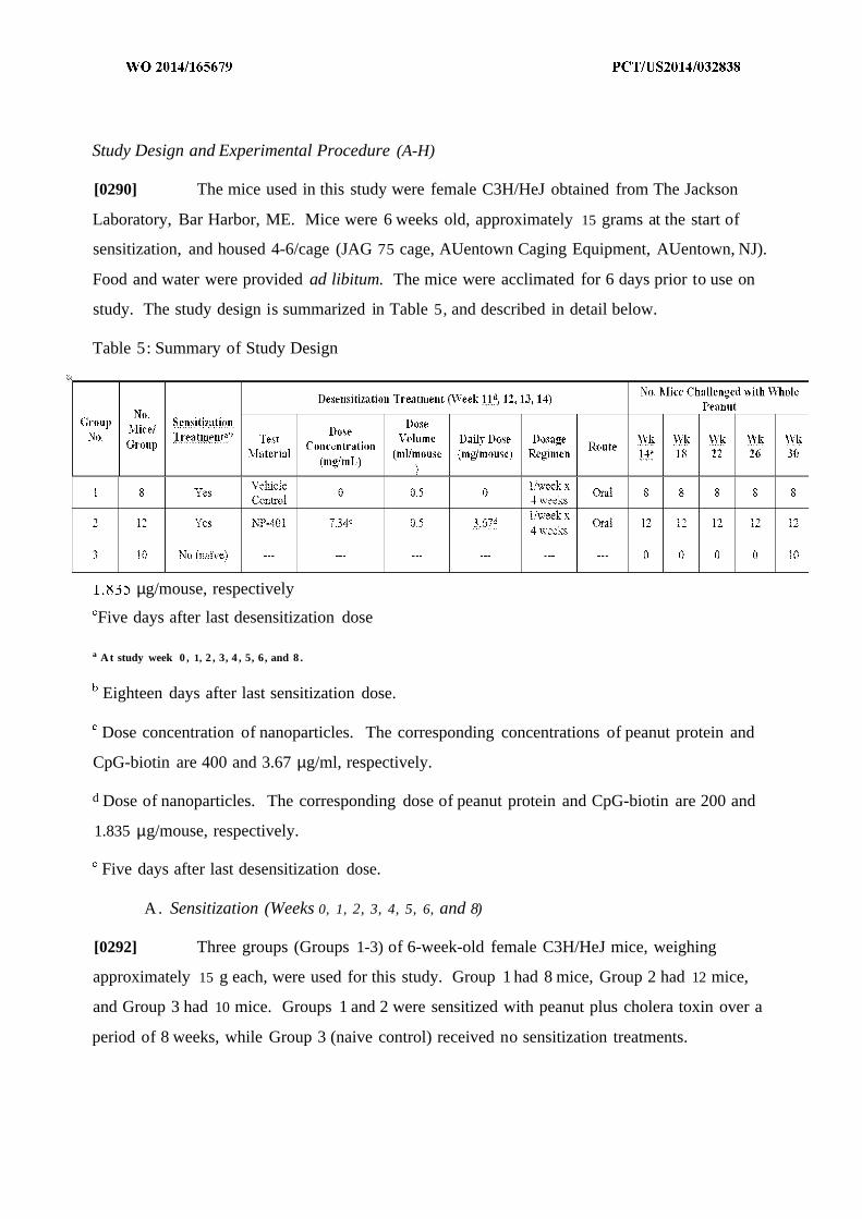
Study Design and Experimental Procedure (A-H)
[0290] The mice used in this study were female C3H/HeJ obtained from The Jackson
Laboratory, Bar Harbor, ME. Mice were 6 weeks old, approximately 15 grams at the start of
sensitization, and housed 4-6/cage (JAG 75 cage, AUentown Caging Equipment, AUentown, NJ).
Food and water were provided ad libitum. The mice were acclimated for 6 days prior to use on
study. The study design is summarized in Table 5, and described in detail below.
Table 5: Summary of Study Design
µg/mouse, respectively
Five days after last desensitization dose
a A t study week 0 , 1, 2 , 3, 4 , 5, 6, and 8 .
Eighteen days after last sensitization dose.
Dose concentration of nanoparticles. The corresponding concentrations of peanut protein and
CpG-biotin are 400 and 3.67 µg/ml, respectively.
d Dose of nanoparticles. The corresponding dose of peanut protein and CpG-biotin are 200 and
1.835 µg/mouse, respectively.
Five days after last desensitization dose.
A. Sensitization (Weeks 0, 1, 2, 3, 4, 5, 6, and 8)
[0292] Three groups (Groups 1-3) of 6-week-old female C3H/HeJ mice, weighing
approximately 15 g each, were used for this study. Group 1 had 8 mice, Group 2 had 12 mice,
and Group 3 had 10 mice. Groups 1 and 2 were sensitized with peanut plus cholera toxin over a
period of 8 weeks, while Group 3 (naive control) received no sensitization treatments.

[0293] Briefly, all mice (including those in Group 3) were fasted for 2 hours. After 2
hours of fasting, each of the mice to be sensitized (Groups 1-2) were orally gavaged with 0.5 ml
of a PBS solution containing 10 mg freshly ground whole peanut (i.e. peanut and skin, with no
shell), 20 µg cholera toxin, 16.5 µΐ of alcohol, and 1.5% sodium bicarbonate (w/v). This was
repeated for 3 consecutive days. Following the initial sensitization (Study Week 0), the same
process was repeated weekly for an additional 5 weeks (Study Weeks 1-5); however, the
sensitizations were administered only once per week (rather than 3 consecutive days per week as
in Study Week 0). One week and three weeks later (i.e., Study Weeks 6 and 8, respectively), the
mice were sensitized as above (i.e., once per week), except that the amount of freshly ground
whole peanut was increased to 50 mg per mouse.
B. Desensitization (Weeks 11, 12, 13, and 14)
[0294] Eighteen days after the last sensitization boost (Study Week 11), all but the naive
mice (Group 3) began a desensitization treatment regimen. The mice were administered either 0
(vehicle; Group 1) or 3.67 mg CpG-coated, PLGA-encapsulated peanut extract nanoparticles
(Group 2)/mouse by oral gavage once a week for four consecutive weeks. The 3.67 mg/mouse
nanoparticle dose corresponds to 200 µg peanut protein and 1.835 µg CpG-biotin. The dose
volume for each desensitization treatment was 0.5 mL/mouse. All mice (including those in
Group 3) were fasted for 2 hours prior to desensitization treatments.
C. Oral Food Challenge (Weeks 14, 18, 22, 26, and 30)
[0295] At Study Weeks 14 (5 days after last desensitization treatment), 18, 22, 26, and
30, the mice subjected to desensitization treatments (Groups 1 and 2) were challenged orally
with freshly ground whole dry-roasted peanut. The naive mice (Group 3) were challenged at
Week 30 only. Briefly, all mice (including those in Group 3) were fasted overnight. The next
morning, each of the mice in Groups 1-2 were orally gavaged with 0.5 ml of a PBS solution
containing 100 mg freshly ground whole peanut, 10 µg cholera toxin, 8.25 µΐ of alcohol, and
1.5% sodium bicarbonate (w/v). Thirty minutes later, the mice were orally gavaged with an
additional 0.5 ml of the above solution (i.e., for a total of 200 mg of ground whole peanut per
mouse).
[0296] Twenty-four hours after each oral food challenge (OFC), the mice in Groups 1-2
were orally gavaged with 0.5 ml of a PBS solution containing 10 mg freshly ground whole
peanut, 20 µg cholera toxin, 16.5 µΐ of alcohol, and 1.5% sodium bicarbonate (w/v). This was
done to maintain reactivity of the mice for the long duration of the study.
D . Anaphylactic Symptom Scores
[0297] Mice were observed for symptoms of anaphylaxis for a period of 1 hour after each
desensitization treatment (Weeks 11, 12, 13, and 14) and for a period of 30 minutes after each
OFC (Weeks 14, 18, 22, 26, and 30). The following scoring system was used for evaluating
anaphylactic symptoms: 0 for no symptoms; 1 for scratching and rubbing around the nose and
head; 2 for puffiness around the eyes and snout, diarrhea, pilar erecti, reduced activity, and/or
decreased activity with increased respiratory rate; 3 for wheezing, labored respiration, and
cyanosis around the mouth and the tail; 4 for no activity after gentle prodding or tremor and
convulsion; 5 for death.
E. Body Temperature
[0298] Thirty minutes after administration of each OFC (Weeks 14, 18, 22, 26, and 30),
the mice were evaluated for body temperature using a rectal thermometer. The Group 3 mice
that were not challenged at Weeks 14, 18, 22, and 26 had their temperatures recorded at the same
time point as the Group 1 and 2 animals.
F. Plasma Histamine Levels
[0299] Plasma histamine levels were measured after administration of each OFC (Weeks
14, 18, 22, 26, and 30). Briefly, 30 minutes after administration of the OFC, blood was
collected, plasma was isolated, and the plasma stored at -80°C until assayed. The level of
plasma histamine was measured using a commercially available histamine enzyme linked
immunosorbent assay (ELISA) kit (Immunotech, Marseille, France), with the concentration of
histamine calculated by comparison with the standard curve provided.
G. Serum Peanut-Specific IgE and IgG2a Levels
[0300] Serum levels of peanut-specific IgE and IgG2a were measured one day prior to
the first desensitization at Week 11, and one day prior to each OFC (Weeks 14, 18, 22, 26, and
30). At the above specified time points, blood was collected, serum isolated, and the serum
stored at -80°C until assayed.
[0301] Peanut-specific IgE was measured as follows: Ninety-six-well Immulon 4HB
(Thermo Scientific, Milford, MA) plates were coated with crude peanut extract (500 µ /ι 1
defatted peanut preparation) or rat anti-mouse IgE (2 µg/ml, BD Biosciences, San Diego, CA).
After an overnight incubation at 4°C, the plates were washed three times, and then blocked for 3
hours at 37°C with 2% BSA (Sigma, St. Louis, MO) in PBS. The plates were washed three
times, and then a 1:50 dilution of the test serum sample was added to the crude peanut extract-
coated wells, and ten serial dilutions ( 1:2, starting at 1,000 ng/mL) of purified mouse IgE (BD
Biosciences, San Diego, CA) were added to the rat anti-mouse IgE-coated wells to generate a
reference curve. All dilutions were made with 2% BSA in PBS. Following an overnight
incubation, the plates were washed three times, and then biotinylated rat anti-mouse IgE ( 1
µg/ml, BD Biosciences, San Diego, CA) was added for 1 hour at RT. The plates were washed 6
times, and then incubated for 15 minutes with a 1:4,000 dilution of streptavidin-horseradish
peroxidase (lmg/ml, Sigma, St. Louis, MO). The plates were washed six times, and then
developed with ABTS® peroxidase substrate (KPL, Gaithersburg, MD) for 30 minutes.
Absorbance was measured at 405 nm by a microplate reader.
[0302] Peanut-specific IgG2a were measured as follows: Ninety-six-well Immulon 4HB
plates were coated with crude peanut extract (2 µg/ml) or dinitrophenyl (DNP, 2 µg/ml, Sigma,
St. Louis, MO). After an overnight incubation at 4°C, the plates were washed three times, and
then blocked for 3 hours at 37°C with 1% human serum albumin (Sigma, St. Louis, MO) in PBS
with 0.5% Tween-20 (Sigma, St. Louis, MO). The plates were washed three times, and then
dilutions of the test serum sample (1:1 ,000 for IgG2a method) were added to the crude peanut
extract-coated wells, and ten serial dilutions ( 1:3, starting at 2,000 ng/mL) of mouse anti-DNP
IgG2a (Accurate, NY) were added to the DNP-coated wells to generate a reference curve. All
dilutions were made in 1% human serum albumin in PBS with 0.5% Tween-20. Following an
overnight incubation, the plates were washed three times, and then biotinylated rat IgG2a (0.25
µg/ml, BD Biosciences, San Diego, CA) was added for 1 hour at RT. The plates were washed 6
times, and then incubated for 15 minutes with a 1:4,000 dilution of streptavidin-horseradish
peroxidase ( 1 mg/ml, Sigma, St. Louis, MO). The plates were washed six times, and then
developed with ABTS peroxidase substrate for 30 minutes. Absorbance was measured at 405
nm by a microplate reader.
H. Spleen Cell Cytokine Levels
[0303] Thirty to forty minutes after the fifth OFC, all mice were sacrificed, their spleens
removed, spleen cells isolated, and the resulting spleen cell cultures assayed for cytokines (IL-4,
IL-5, IL-10, IL-13, IFN-γ , and TGF- β) in the presence or absence of crude peanut extract.
Individual cultures were set up for each animal. The spleen cells were suspended in RPMI 1640
(Mediatech, Herndon, VA) supplemented with 10% fetal bovine serum (Invitrogen, Carlsbad,
CA) and 1% penicillin/streptomycin (MP Biomedicals, Solon, OH) and plated in 24-well plates
in the presence or absence of crude peanut extract. Seventy-two hours later, the culture
supernatants were collected and the level of cytokines assayed by commercially available ELISA
kits. OptEIA ELISA Kits (BD Biosciences, San Diego, CA) were used for all cytokines, except
TGF- β and IL-13, which were measured using duo-set kits from R&D systems (Minneapolis,
MN).
Results
1. Serum Peanut-Specific IgE Levels
[0304] Exemplary mean ± standard error of the mean (SEM) serum concentrations of
peanut-specific IgE one day prior to the first desensitization at Week 11 (pre-therapy) and one
day prior to the OFC at Weeks 14, 18, 22, 26, and 30 are shown in Figure 5 . Mice receiving no
treatments of any type (naive control) had undetectable peanut-specific IgE levels throughout the
study. Both groups of mice subjected to sensitization treatments showed presence of
peanut-specific IgE levels at Weeks 11 through 30. No statistical difference in mean peanut-
specific IgE was observed between vehicle and Agent treated groups at Week 11 (one day prior
to desensitization; vehicle control: 6,844 ± 1,41 1 ng/niL, Agent: 6,525 ± 729 ng/mL).
[0305] Four days after the completion of the desensitization treatments (Week 14, one
day prior to the first OFC), Agent treated mice showed a trend of decreased levels of peanut-
specific IgE (4,980 ± 732 ng/mL) when compared to mice administered vehicle (6,662 ± 861
ng/mL, P<0.07). During the remainder of the study, mean peanut-specific IgE levels in Agent

treated mice were significantly lower than those observed for vehicle treated mice (P<0.05-
0.001).
2. Peanut-Specific IgG2a Levels
[0306] Exemplary mean ± SEM serum concentrations of peanut-specific IgG2a one day
prior to the sensitizations at Week 11 (pre-therapy) and one day prior to the OFC at Weeks 14,
18, 22, 26, and 30 are shown in Figure 6 . Mice receiving no treatments of any type (naive
control) had undetectable peanut-specific IgG2a levels throughout the study. Both groups of
mice subjected to sensitization treatments showed presence of peanut-specific IgG2a levels at
Weeks 11 through 30. No statistical difference in mean peanut- specific IgG2a was observed
between vehicle and Agent treated groups at Week 11 (one day prior to desensitization; vehicle
control: 318,286 ± 32,586 ng/mL, Agent: 339,592 ± 51,494 ng/niL).
[0307] Four days after the completion of the desensitization treatments (Week 14, one
day prior to the first OFC), the Agent treated mice showed an increase in the level of mean
peanut-specific IgG2a (444,426 ± 60,288 ng/mL) while the mice administered vehicle showed a
decrease in the level of mean peanut-specific IgG2a (207,741 ± 53,494 ng/mL, P<0.05). After
Week 14, the mean peanut-specific IgG2a levels decreased for both the vehicle and AGENT
treated mice; however, at all time points subsequent to Week 11, the mean levels of peanut-
specific IgG2a in the Agent treated mice remained significantly higher than those observed in the
vehicle treatment group (P<0.05-0.01).
3. Anaphylactic Symptom Scores During Desensitization Period
[0308] Mice treated with vehicle control or 3.67 mg/mouse Agent (200 µg peanut protein
and 1.835 µg CpG-biotin) showed no signs of anaphylaxis during the 1-hour observation period
following each desensitization treatment. All mice had anaphylactic symptom scores of 0 (0 =
no symptoms).
4. Anaphylactic Symptom Scores Following OFCs
[0309] Exemplary individual and median anaphylactic symptom scores following the
OFC at Weeks 14 and 18, 22 and 26, and 30 are shown in Figures 7, 8, and 9, respectively. Mice
receiving no sensitization/treatments of any type (i.e., na ve control) showed no anaphylactic
symptoms (i.e., all mice had an anaphylactic symptom score of 0) following the OFC at Week
30. The na ve control mice were not challenged at Weeks 14, 18, 22, and 26.
[0310] Sensitized mice administered vehicle control showed signs of anaphylaxis
following the OFC at Week 14. Specifically, 1 mouse scored a 3 (wheezing, labored respiration,
and cyanosis around the mouth and tail), 4 mice scored a 2 (puffmess around eyes and snout,
diarrhea, pilar erecti, reduced activity, and/or decreased activity with increased respiratory rate),
and 1 mouse scored a 1 (scratching and rubbing around the nose and head). Two mice showed
no symptoms (score of 0). The anaphylactic symptoms in these mice worsened with subsequent
challenges up through Week 26, with 1 mouse scoring a 4 (no activity after gentle prodding or
tremor and convulsion) and the remaining 7 mice scoring a 3 . At Week 30, 1 mouse that had
scored a 3 at Week 26 had reduced symptoms (score of 2).
[0311] Oral administration of 3.67 mg/mouse Agent (200 µg peanut protein and 1.835 µg
CpG-biotin) once a week for four consecutive weeks in peanut-sensitized mice resulted in a
statistically significant decrease in the anaphylactic symptom scores at all five OFCs (P<0.05).
Nine of the 12 mice (75%) were completely protected from anaphylactic symptoms (score of 0)
after the first challenge, with the remaining three having symptom scores of 1, 2, and 3,
respectively. By the second challenge, the mouse that had scored a 1 at the first challenge scored
a 0, bringing the total number of completely protected to 10 (83%). This remained the same
through the fourth challenge (i.e., 3 months after desensitization treatment was stopped). Four
months after desensitization treatment was stopped, 2 of the 10 mice that were completely
protected at Week 26 had increased symptoms (score of 2).
5. Body Temperatures Following OFCs
[0312] Exemplary individual and mean body temperatures following OFCs at Weeks 14
and 18, 22 and 26, and 30 are shown in Figures 10, 11, and 12, respectively. The Group 3 mice
that were not challenged at Weeks 14, 18, 22, and 26 had their temperatures recorded at the same
time point as the Group 1 and 2 animals. Following each OFC, the vehicle control group had
mean body temperatures that were consistently lower than that of the na ve control mice. Oral
administration of 3 .67 mg/mouse Agent (200 µg peanut protein and 1.835 µg CpG-biotin) once a
week for four consecutive weeks in peanut-sensitized mice resulted in an increase in the mean
body temperatures relative to vehicle control animals at all five OFCs. The increase was
statistically significant at all but the first challenge (P<0.05 at 2nd, 3rd, and 4th challenge and
P<0.001 at the 5th challenge).
6. Plasma Histamine Levels Following OFCs
[0313] Exemplary individual and mean plasma histamine levels following OFCs at
Weeks 14 and 18, 22 and 26, and 30 are shown in Figures 13, 14, and 15, respectively. The
Group 3 mice that were not challenged at Weeks 14, 18, 22, and 26 had their plasma histamine
measured at the same time point as the Group 1 and 2 animals. The mean plasma histamine
levels 30 minutes after each OFC were higher in the vehicle control group relative to the na ve
control group. Oral administration of 3.67 mg/mouse Agent (200 µg peanut protein and 1.835
µg CpG-biotin) once a week for four consecutive weeks in peanut-sensitized mice resulted in a
statistically significant decrease in the mean plasma histamine levels relative to vehicle control
animals at all five OFCs (PO.05-0.001).
7. Cytokine Levels in Post-OFC (Week 30) Spleen Cell Cultures Incubated With Crude Peanut
Extract
[0314] Exemplary mean ± SEM cytokine levels in post-OFC (Week 30) spleen cell
cultures incubated with crude peanut extract are shown in Figure 16. Figure 16A depicts an
exemplary result illustrating mean ± SEM IL-4 cytokine levels. Figure 16B depicts an
exemplary result illustrating mean ± SEM IL-10 cytokine levels. Figure 16C depicts an
exemplary result illustrating mean ± SEM IFN-gamma cytokine levels. Figure 16D depicts an
exemplary result illustrating mean ± SEM IL-5 cytokine levels. Figure 16E depicts an
exemplary result illustrating mean ± SEM IL-13 cytokine levels. Figure 16F depicts an
exemplary result illustrating mean ± SEM TGF- β cytokine levels.
[0315] The mean spleen cell production of IL-4, IL-5, IL-10, IL-13, IFN-γ , and TGF- β in
the presence of crude peanut extract was higher in the vehicle control group relative to the na ve
control group. Oral administration of 3.67 mg/mouse Agent (200 µg peanut protein and 1.835
µg CpG-biotin) once a week for four consecutive weeks in peanut-sensitized mice resulted in
statistically significant decreases in mean spleen cell production of IL-4 (p<0.001), IL-5
(p<0.001), and IL-13 (p<0.001) relative to vehicle control, and in statistically significant
increases in mean spleen cell production of IFN-γ (p<0.001) relative to vehicle control. No
significant differences were observed between vehicle and Agent treated groups for IL-10 and
TGF-β.
Conclusion
[0316] Peanut-sensitized mice orally administered 3.67 mg/mouse Agent (200 µg peanut
protein and 1.835 µg CpG-biotin; i.e. CpG-coated, PLGA-encapsulated peanut extract
nanoparticles) once a week for four consecutive weeks showed no symptoms of anaphylaxis
during the desensitization period, indicating that CpG-coated, PLGA-encapsulated peanut extract
nanoparticles lack anaphylactic potential in peanut-sensitized mice.
[0317] Further, oral administration of 3.67 mg/mouse Agent once a week for four
consecutive weeks in peanut-sensitized mice resulted in desensitization to subsequent oral peanut
challenges relative to vehicle control mice. This was evidenced by lower median anaphylactic
symptom scores, higher mean body temperature (essentially similar to na ve mice), lower mean
plasma histamine levels, a decrease in the mean serum peanut-specific IgE levels, an increase in
the mean serum peanut-specific IgG2a levels, a decrease in the mean spleen cell production of
IL-4, IL-5 and IL-13, and an increase in the mean spleen cell production of IFN-γ compared to
vehicle-treated mice. The desensitization was still evident at four months after treatment with
Agent was stopped, suggesting that tolerance may have been achieved in some animals.
Example : Preparation of Arachis hvpogaea (Peanut) Allergen Extracts
[0318] The present Example describes preparation of a Arachis hypogaea (Peanut)
Allergen Extract (i.e. crude peanut extract) for use in accordance with the present invention.
[0319] The present Example describes preparation of a Peanut Allergen Extract,
containing Ara h 1, Ara h 2 and Ara h3 antigens, among others, for use in accordance with the
present invention.
[0320] The present Example also describes preparation of recombinant modified peanut
proteins (mAra h 1, mAra h 2, and mAra h 3, where m stands for modified), for use in
accordance with the present invention.
[0321] Arachis hypogaea (Peanut) Allergen Extract (i.e. crude peanut extract) may be
prepared as follows. Commercially available roasted peanuts in shells (White Rose Brand, NJ)
are de-shelled, homogenized in phosphate buffered saline (PBS, pH) 7.4), and subjected to
acetone extraction. The final concentration of peanut protein in PBS is determined by
bicinchoninic acid (BCA) assay. Peanut allergen extract may then becombined with aqueous E.
coli extract (AEE) and prepared substantially as described in Example 3, for example.
[0322] Recombinant peanut proteins were prepared as follows. The three recombinant
modified peanut proteins (mAra h 1, mAra h 2, and mAra h 3) were separately expressed in E.
coli strain BLR(DE3), and the E.coli were subsequently killed using heat and phenol. The
expressed proteins remained encapsulated within the dead E. coli., which resulted in three whole-
cell suspensions referred to as EMP-1, EMP-2, and EMP-3 (i.e., encapsulated mAra h 1, mAra h
2, and mAra h 3, respectively). Each whole-cell suspension was then to used prepare aqueous E.
coli extract (AEE) ), as described in Example 1, which included the expressed recombinant
peanut protein in the aqueous phase, for fabrication of a nanoparticle composition substantially
as described in Example 3 .
Example 6 : In vitro Cytokine Studies of Nanoparticle Compositions in Peripheral Blood
Mononuclear Cells (PBMCs) of Allegic Patients
[0323] The present Example describes exemplary in vitro stimulation of patient
peripheral blood mononuclear cells (PBMCs) for evaluating the immunogenicity of provided
nanoparticle compositions (i.e., nanoparticles containing hydrophilic and/or hydrophobic E. coli
extract preparations) for use in accordance with the present invention.
[0324] The present Example describes exemplary in vitro stimulation of patient
peripheral blood mononuclear cells (PBMCs) for evaluating the immunogenicity of provided
nanoparticle compositions that comprise one or more antigens, including crude allergenic extract
(such as Arachis hypogaea, Dermatophagoidesfarina or Dermatophagoides pteronyssinus), for
use in accordance with the present invention.

[0325] The present example describes exemplary in vitro cytokine studies to evaluate the
immunogenicity of organic Escherichi coli extract ("OEE") coated polylactide co-glycolide-
("PLGA-") encapsulated Dermatophagoides (D.) farinae and D. pteronyssinus dust mite extract
and aqueous E. coli extract (AEE) nanoparticles (hereinafter referred to as the "Agent").
[0326] In some embodiments, the studies described herein, including stimulation of
allergic patient PBMCs, may be used for assessing immunological characteristics or effects of
certain embodiments of the present invention.
[0327] In some embodiments, provided compositions may modulate immununological
function in a subject. In some embodiments, such modulation may comprise desensitization. In
some embodiments, such modulation does not comprise densitization. In some embodiments,
immunological characteristics or effects include an altered allergen-specific Th2-dominated
phenotype of dust mite allergic patient toward a Thl/ThO-like phenotype.
[0328] The objective of this in vitro study is to assess the ability of the Agent to alter the
allergen-specific Th2-dominated phenotype of dust mite allergic patient peripheral blood
mononuclear cells (PBMCs) toward a Thl/ThO-like phenotype. This model system includes
polyclonal T cells and B cell antigen presenting cells (APCs) in a single culture.
[0329] The Agent in this Example comprises standardized allergenic extracts of two
common house dust mites, D.farinae and D. pteronyssinus, mixed 1:1 by allergy unit. The dust
mite extracts are the same as those found in the commercially available standardized dust mite
extracts approved by FDA for use in subcutaneous immunotherapy (SCIT). The dust mite
extracts are encapsulated within PLGA nanoparticles wherein the dust mite extract and bacterial
protein, deoxyribonucleic acid (DNA), and lipopolysaccharide (LPS) of the AEE is encapsulated
on the inside of the nanoparticles and the bacterial LPS and lipids of the OEE is coated on the
outside of the nanoparticles.
[0330] Without wishing to be held to a particular theory, a pharmacological rationale for
the encapsulation of the dust mite extract in provided nanoparticle compositions is to facilitate
uptake by APCs in the sublingual mucosa and activation of the innate immune system, and thus
to potentially improve the efficacy of sublingual immunotherapy (SLIT) with the extracts alone.
Specifically, the Agent is designed to maximize the incorporation of the known toll-like receptor
(TLR) agonists, bacterial LPS and CpG DNA motifs, as well as any potential unidentified
agonists that may be present in the bacterial cells, thereby enhancing the potential efficacy of the
Agent drug product.
[0331] This study will assess in vitro IFN-γ , IL-10, IL-5, and IL-4 levels in dust mite
allergic patient PBMCs following treatment with the Agent compared against dust mite extract
encapsulated within nanoparticles that lack E. coli components (Agent minus adjuvant),
semi-synthetic bacterial cells without dust mite extract (Agent minus dust mite extract), and
empty nanoparticles (Agent minus adjuvant and dust mite extract).
[0332] The general method of this Example (summarized below) is substantially based
on the method published by Bohle et al. (Bohle et al, J. Immunology, 2004; 172:6642-6648) for
a birch pollen allergy study. The method can be modified as necessary to improve the study
design. In particular, one of skill in the art will recognize that certain parameters, including, but
not limited to, the number of cells per well, the duration of culture, the culture conditions, and
the dose of agent and stimulant may need to be optimized for a particular antigen and patient
population.
[0333] Peripheral blood is collected from 10 dust mite allergic patients via venipuncture
into heparanized syringes. All patients must have a history of perennial allergic rhinitis for a
minimum of one year prior to blood collection and a demonstrated sensitivity to D.farinae or D.
pteronyssinus by a positive prick skin test (PST) at the time of blood collection. A PST is
defined as a mean wheal diameter 3 mm greater than that elicited by the negative control (saline)
at 15-20 minutes. Patients must not have received allergen immunotherapy or
immunomodulatory therapy within 6 and 3 months prior to blood collection, respectively.
Patients must not have clinical history of significant symptomatic perennial allergic rhinitis
and/or asthma due to another allergen (i.e., other than dust mites) to which the subject is
regularly exposed.
[0334] PBMCs are isolated from the peripheral blood of dust mite allergic patients by
Ficoll-Hypaque density gradient centrifugation (Pharmacia). The whole blood should be
processed to PBMCs as soon after collection as possible, ideally within 8-12 hours. The PBMCs
are typiclaly not be frozen prior to assay.
[0335] The PBMCs are cultured in 96-well plates (Nunclone; Nunc) in serum-free Ultra
Culture Medium (BioWhittaker, Walkersville, MD) supplemented with 2 mM L-glutamine, 2 x
10 5 M 2-mercaptoethanol, and study drug/control. Triplicate wells at 1 x 10 /200 µΐ and
triplicate wells at 2 x 10⁵/200 µ will be set up for each of the following study drug/controls for
each dust mite allergic patient's PBMCs:
1. Low dose Agent [dose will be expressed as the final concentration of nanoparticles(mg/ml) in culture media];
2 . Mid dose Agent;
3 . High dose Agent;
4 . Agent minus adjuvant (same dose as #3);
5 . Agent minus dust mite extract (same dose as #3);
6 . Agent minus adjuvant and dust mite extract (same dose as #3);
7 . Media only control.
[0336] As described above, triplicate wells are set up at 1 x 10 /200 µΐ and are used to
follow the Thl -related cytokines. After 48 hours of culture with study drug/controls, the
supematants is collected and analyzed for IFN-γ and IL-10 content using an enzyme-linked
immunosorbent assay (ELISA) with endogen matched Ab pairs (Endogen, Woburn, MA)
according to the manufacturer's instructions.
[0337] Addtionally, triplicate wells are set up at 2 x 10⁵/200 µΐ and are used to follow
each of the Th2-related cytokines. After 6 days of culture with study drug/controls, the cells are
washed, counted, and restimulated at 1 x 10 /200 µΐ with phorbol myristate acetate (PMA, 10
ng/ml) plus ionomycin ( 1 µΜ ) for 24 hours. After 24 hours of restimulation, the supematants are
collected and analyzed for IL-5 and IL-4 content using an ELISA with endogen matched Ab
pairs (Endogen, Wobum, MA).

Example 7 : A Phase 1, Randomized, Double-Blind, Placebo-Controlled Safety,
Pharmacodynamic, and Preliminary Efficacy Study of Dust Mite Allergy Vaccine Administered
Sublingually in Adult Subjects with Perennial Allergic Rhinitis and Sensitive to
Dermatophagoides farinae or Dermatophagoides pteronyssinus
[0338] This present Example describes an exemplary phase 1, single-center, randomized,
double-blind, placebo-controlled, safety, pharmacodynamic, and preliminary efficacy study of
provided nanoparticle compositions (in this Example, nanoparticles containing hydrophilic
and/or hydrophobic E. coli extract preparations), in adult subjects with perennial allergic rhinitis
(with or without asthma) and sensitive to D.farinae or D. pteronyssinus, for use in accoradance
with the present invention.
[0339] In some embodiments, provided nanoparticle compositions are organic
Escherichia coli extract ("OEE") coated poly(lactic-co-glycolic acid)- ("PLGA-") encapsulated
Dermatophagoides (D.) farinae and D. pteronyssinus dust mite extract and aqueous E. coli
extract (AEE) nanoparticles (hereinafter referred to as the "Agent" and/or "Dust Mite Allergy
Vaccine").
[0340] The present Example describes an exemplary clinical trial design for evaluating
the pharmacodynamics and preliminary efficacy of provided nanoparticle compositions (in this
Example, nanoparticles containing hydrophilic and/or hydrophobic E. coli extract preparations)
for use in accordance with the present invention.
[0341] The present Example describes an exemplary protocol synposis for evaluating the
pharmacodynamics and preliminary efficacy of sublingual administration of certain provided
nanoparticle compositions containing Dermatophagoidesfarinae or Dermatophagoides
pteronyssinus extracts (i.e. antigens) and bacterial components for use in accordance with the
present invention.
Chemical Name and Structure
[0342] The drug substances in the Dust Mite Allergy Vaccine of this Example are
standardized allergenic extracts of two common house dust mites, D.farinae and D.
pteronyssinus, mixed 1:1 by allergy unit (AU). As such, there is no conventional chemical name
or structure for the Dust Mite Allergy Vaccine drug substances.
[0343] The dust mite extracts in the Dust Mite Allergy Vaccine will be the same as those
found in the commercially available standardized dust mite extracts approved by the Food and
Drug Administration (FDA) for use in subcutaneous immunotherapy (SCIT) [Standardized Mite
Extract (Dermatophagoidesfarinae) and Standardized Mite Extract (Dermatophagoides
pteronyssinus), Greer, Lenoir, NC].
[0344] The dust mite extracts will be encapsulated within PLGA nanoparticles
manufactured using a double-emulsion process. In the first emulsion, the dust mite extracts and
an AEE that contains bacterial deoxyribonucleic acid (DNA) will be mixed with PLGA in
organic solvent. The resulting emulsion will be microfluidized or homogenized to generate
nanoparticles with the dust mite extracts and AEE encapsulated within the particles. OEE,
containing mainly bacterial lipids and LPS, in a 5% polyvinyl alcohol solution will then be added
and the resulting second emulsion microfluidized/homogenized to coat the nanoparticles
generated during the first emulsion.
[0345] The final PLGA nanoparticles are thus designed to mimic bacteria so as to
facilitate uptake by antigen presenting cells (APCs) in the GI tract and enhance activation of the
innate immune system, and thus potentially improve the efficacy of conventional SLIT with
naked dust mite extract alone. The Dust Mite Allergy Vaccine bulk nanoparticles will include
the addition of the AEE and the second emulsion will use the OEE rather than LPS alone. Stated
another way, these PLGA nanoparticles are designed to be "semi-synthetic bacterial cells" in
which the dust mite extracts and bacterial protein, DNA, and LPS of the AEE will be
encapsulated on the inside of the nanoparticles, and the bacterial LPS and lipids of the OEE will
be coated on the outside of the nanoparticles. Placebo will be generated the same way, only with
no dust mite extract present.
Proposed Indication
[0346] The Dust Mite Allergy Vaccine is being developed for SLIT for the treatment of
perennial allergic rhinitis in patients with a demonstrated sensitivity to D.farinae or D.
pteronyssinus as determined by positive prick skin test (PST).
Dosage Form, Route Of Administration, and Dosing Regimen

[0347] The final dosage form of the Dust Mite Allergy Vaccine will be a solid oral
dosage form suitable for sublingual administration. Formulation development approaches may
include a dry blend of the nanoparticles with a water-soluble excipient such as lactose
monohydrate packaged in plastic straws or a direct-compression, rapidly-dissolving tablet.
Dosing will take the form of a dose escalation up to a maximum dose of 1,400 AU. All subjects
will be administered Dust Mite Allergy Vaccine once per week for six weeks. During this
period, the dose will be increased weekly, starting in three fold increments up to a maximum
dose of 1,400 AU and then decreasing to a 1.5-fold increment. Protocol-specified dose
modifications will be required if allergic symptoms are observed or doses missed. After the
initial six weeks of dose escalation, patients who tolerate the Dust Mite Allergy Vaccine will
then receive additional doses qd for five months (maintenance dosing period). The dose during
the maintenance dosing period will be the maximum tolerated dose (MTD) established during
the subject's dose escalation phase.
[0348] The route of administration of the Dust Mite Allergy Vaccine is sublingual for use
as sublingual immunotherapy (SLIT).
[0349] The proposed dosing regimen to be used for the Dust Mite Allergy Vaccine for
the treatment of perennial allergic rhinitis in patients sensitive to D.farinae or D. pteronyssinus
is based on previous clinical trials of oral immunotherapy (OIT) and SLIT. The dosage regimens
for OIT and SLIT for perennial allergic rhinitis are commonly once daily (qd) for chronic use
according to an extended dosing period.
Additional Information and Possible Mechanisms
[0350] The Dust Mite Allergy Vaccine of this Example is designed to act as an "allergy
vaccine" to induce tolerance to dust mites when used as SLIT for the treatment of perennial
allergic rhinitis in patients sensitive to D.farinae or D. pteronyssinus. The dust mite allergens to
be used in the Agent are the same allergens found in the commercially available standardized
dust mite extracts approved by FDA for use in SCIT [Standardized Mite Extract
(Dermatophagoidesfarinae) and Standardized Mite Extract (Dermatophagoides pteronyssinus),
Greer, Lenoir, NC]. One difference between the extracts in the Greer products and the Agent is
that the Greer products are formulated as a liquid in glycerin and the Agent will use lyophilized
extracts for incorporation into PLGA nanoparticles that include bacterial components intended to
facilitate APC recognition and uptake of the particles and enhance a Thl/ThO-like immune
response associated with desensitization and eventual tolerance to dust mite allergens.
[0351] Traditional allergen-injection vaccines have been used for control of allergic
disease for many years. Standardized extracts of D.farinae and D. pteronyssinus, both
individually and mixed, are commercially available for SCIT of dust mite allergy. SCIT is
reported to be capable of modifying the natural history of disease (i.e., the onset of asthma in
rhinitis patients) and result in long-lasting effects after discontinuation. However, due to
concerns about adverse reactions elicited by allergen injections and the inconvenience of regular
subcutaneous injections at a clinician's office for over a period of 3 to 5 years, the widespread
adoption of SCIT has been limited. Sublingual administration as used for SLIT appears to be a
more viable alternative route of administration of allergen extracts.
[0352] The Dust Mite Allergy Vaccine product is designed to encapsulate the dust mite
extracts and bacterial DNAwithin PLGA nanoparticles coated with bacterial LPS and lipids on
the surface (refer to schematic in Figure 17). The pharmacological rationale is as follows.
[0353] A dust mite allergy vaccine should protect against exposure to the main dust mite
proteins responsible for allergic reactions in order for efficacy to be achieved. D.farinae and D.
pteronyssinus are the most common house dust mite species, and numerous allergens have been
identified for each. Hence, the Agent includes standardized extracts from both species as
antigens.
[0354] Delivery of the dust mite antigens encapsulated in PLGA nanoparticles
mimicking bacterial cells may enhance the activation of the innate immune system, and thus to
potentially improve the efficacy of SLIT with dust mite extracts alone. Specifically, bacterial
LPS and CpG DNA motifs, as well as any potential unidentified bacterial moieties on the interior
of the nanoparticle may enhance the potential efficacy of the Agent to produce the desired
Thl/ThO-like immune response associated with desensitization and eventual tolerance to dust
mite allergens.
[0355] In addition, encapsulation of the dust mite antigens within PLGA nanoparticles
mimicking bacterial cells may facilitate uptake by APCs in the sublingual mucosa, and thus to
potentially improve the efficacy of SLIT using the dust mite extracts alone. Specifically, the
Agent is designed to maximize the recognition and internalization by APCs of the encapsulated
dust mite proteins by both surface display on the nanoparticle exterior and incorporation within
the nanoparticle of the known toll-like receptor (TLR) agonists.
[0356] Further, encapsulation of the dust mite antigens within an intact delivery system
(i.e., PLGA nanoparticles) may reduce the potential for allergic reactions upon administration of
the proteins by hiding the proteins from mast cells. Hence, the dust mite proteins in the Agent
are encapsulated within PLGA nanoparticles mimicking bacterial cells.
[0357] As exemplified in Figure 17, there are no covalent bonds between the components
of the Agent in this Example. In the presence of water, the nanoparticles will continuously erode
by hydrolysis and the encapsulated dust mite extract proteins and bacterial protein, DNA, and
LPS, as well as the coating of bacterial LPS and lipids will be continuously released. Hydrolysis
of the PLGA polymer results in lactic acid (LA) and glycoic acid (GA) monomers, which are
harmless. The rate of PLGA hydrolysis is dependent upon the ratio of LA to GA, and PLGA
polymer used for the Agent will be selected so that it releases the encapsulated dust mite extract
proteins within 48 to 72 hours based on an in vitro drug release assay.
[0358] After sublingual administration, the nanoparticles that are not taken up and
processed by APCs will be swallowed. In either case, the presence of H20 hydrolyzes PLGA to
LA and GA (faster at low pH), with release of the dust mite extracts (i.e., antigens) and bacterial
components. After release, the extract proteins and bacterial components will be digested in the
same way as dust mite proteins or bacterial components would be when consumed in daily life or
in food.
Clinical TrialDesign and Methodology
[0359] The Clinical Trial Design is a phase 1, single-center, randomized, double-blind,
placebo-controlled safety, pharmacodynamic, and preliminary efficacy study of the Agent in
adult subjects with perennial allergic rhinitis (with or without asthma) and sensitive to D.farinae
or D. pteronyssinus.
[0360] Twelve subjects will be randomized 1:2 to receive placebo or the Agent. The
placebo and active cohorts will include both subjects sensitive to D.farinae and D. pteronyssinus
and subjects sensitive to only one of the two species, so as to allow for monitoring of neo-
sensitization.
[0361] All subjects will be administered a sublingual dose of study drug (placebo or
active) once per week for six weeks. During this period, the dose will be increased weekly,
starting with 3-fold increments culminating in a dose of 1,400 AU, and then progressively
decreasing to a 1.5-fold increment. If no dose limiting toxicities [DLTs; defined as any adverse
event (AE) of a severity greater than moderate and assigned at least a possible relationship to
study drug] occur, the subject will then take the maximum tolerated dose once daily (qd) for six
months.
A. Screening Visit:
[0362] Within 14 days prior to the first dose of study drug (Day 1), each subject will be
provided with written information (informed consent form) describing the study and will have
any questions answered. Subjects that consent in writing to participate in the study will undergo
eligibility assessments, including complete medical history, comprehensive physical
examination, height, weight, vital signs (blood pressure, pulse rate, respiration rate, and oral
body temperature), 12-lead electrocardiogram (ECG), spirometry [forced expiratory volume in 1
second (FEV1), forced vital capacity (FVC), and peak expiratory flow (PEF)], complete blood
count (CBC) with differential, serum chemistry, urinalysis, urine human chorionic gonadotropin
(HCG) pregnancy test for women of childbearing potential (WCBP), blood collection for D.
farinae- or D. pteronyssinus-specific IgE and IgG4 levels, blood collection for D.farinae- or D.
pteronyssinus-stimulated PBMC T-lymphocyte phenotype, prick skin tests (PSTs) and endpoint
skin test titrations to standardized D.farinae and D. pteronyssinus extracts, and record of
medications taken within 30 days prior to the screening visit.
B. Study Day 1:
[0363] Subjects that meet all eligibility requirements at screening will be randomized 1:2
to receive placebo or the Agent and will return to the clinic on the morning of Day 1 for the
following baseline procedures: medical history update, targeted physical examination, weight,
vital signs, PEF, CBC with differential, serum chemistry, urinalysis, urine HCG pregnancy test
for WCBP, and record of medications taken since screening visit.
[0364] Subjects that continue to meet eligibility requirements after the baseline
assessments will be given a single sublingual dose of study drug (placebo or active).
Administration of the study drug will take place in a clinic with experience in treating severe
allergic reactions. Specifically, a crash cart will be available in the facility and there will be
medical personnel and a physician present to treat anaphylaxis.
[0365] AE monitoring will begin immediately following administration of study drug and
will continue throughout the study. Subjects will remain in the clinic under observation for
4 hours post-dose. Vital signs will be monitored at 5, 10, 15, and 30 minutes post-dose.
[0366] Subjects will be given a diary to record any AEs or medications used between
visits, and will be given forms to assess the severity of rhinoconjunctivitis symptoms (visual
analog scale) and the use of rescue medications on a daily basis throughout the trial. Subjects
will be given a 24-hour emergency telephone number to call in the event of an adverse reaction.
C. Study Day 2 :
[0367] Subjects will return to the clinic on the mornings of Days 2 and 7 for the
following procedures: targeted physical examination, vital signs, PEF, urine HCG pregnancy
test for WCBP, review of the AE and concomitant medication diary, and review of the daily
forms assessing the severity of rhinoconjunctivitis symptoms and rescue medication use.
D. Weekly Visits During Dose Escalation:
[0368] Subjects will return to the clinic once a week during the dose escalation period for
evaluations, as well as administration of study drug. The following procedures will be
performed on the morning of Days 8, 15, 22, 29, and 36: targeted physical examination, vital
signs, PEF, urine HCG pregnancy test for WCBP, review of the AE and concomitant medication
diary, and review of the daily forms assessing the severity of rhinoconjunctivitis symptoms and
rescue medication use. Pending no safety concenrs, the subjects will be given a single sublingual
dose of study drug (placebo or active). Subjects will remain in the clinic under observation for 4
hours post-dose. Vital signs will be monitored at 5, 10, 15, and 30 minutes post-dose.
E. Monthly Visits During Maintenance Therapy:
[0369] Subjects will return to the clinic one week after completion of the dose escalation
phase (morning of Day 43) and then monthly thereafter for 5 months during the maintenance
dosing period for the following procedures: targeted physical examination, vital signs, PEF, CBC
with differential, serum chemistry, urinalysis, urine HCG pregnancy test for WCBP, review of
the AE and concomitant medications diary, and review of the daily forms assessing the severity
of rhinoconjunctivitis symptoms and rescue medication use.
[0370] At the first, third, and fifth monthly visit (i.e., after 2.5, 4.5, and 6.5 months of
study drug treatment), the following additional procedures will be performed: blood collection
for D.farinae- or D. pteronyssinus-specific IgE and IgG4 levels, blood collection for D.farinae-
or D. pteronyssinus-stimulated PBMC T-lymphocyte phenotype, endpoint titration PSTs to
standardized D.farinae and D. pteronyssinus extracts, and endpoint titration PST to non-dust
mite extract (selected at Screening). Further, at the fifth monthly visit (after 6.5 months of study
drug treatment), allergen bronchoprovocation to standardized D.farinae or D. pteronyssinus
extracts will be performed.
[0371] Pending no safety concenrs following the monthly assessments, the subjects will
be provided with sufifcient study drug for four weeks of dosing and a dairy to record the details
of study drug administration at home. The subjects will be instructed to take qd at home and to
return to the clinic for monthly evaluations. At each visit, the study drug diary will be reviewed
and the study drug containers and any unused study drug will be collected to monitor
compliance. No study drug will be given after the fifth monthly visit (i.e., after 6.5 months of
treatment).
F. Final Study Visit
[0372] All subjects will return four weeks after their last dose of study drug for their final
study visit. The following procedures will be performed at the final visit: complete physical
examination, weight, vital signs, PEF, CBC with differential, serum chemistry, urinalysis, urine
HCG pregnancy test for WCBP, blood collection for D.farinae- or D. pteronyssinus-specific IgE
and IgG4 levels, blood collection for D.farinae- or D. pteronyssinus-stimulated PBMC T-
lymphocyte phenotype, endpoint titration PST to standardized D.farinae and D. pteronyssinus
extracts, endpoint titration PST to non-dust mite extract (selected at Screening), review of the AE
and concomitant medication diary, and review of the daily forms assessing the severity of
rhinoconjunctivitis symptoms and rescue medication use.
Diagnosis and Main Criteriafor Inclusion:
[0373] Subjects 18 to 50 years of age with a history of perennial allergic rhinitis for a
minimum of one year prior to Screening, and a demonstrated sensitivity to D.farinae or D.
pteronyssinus by a positive PST at Screening [mean wheal diameter 3 mm greater than that
elicited by the negative control (saline) at 15-20 minutes].
[0374] Subjects may or may not have asthma. For subjects with asthma, the subject must
be diagnosed with either intermittent or mild persistent asthma, as defined by National Heart
Lung and Blood Institute (NHLBI) guidelines, for a minimum of one year prior to Screening.
For all subjects, FEV1 must be greater than 80% predicted and FEV1/FVC must be normal at
Screening.
[0375] Both males and WCBP agree to use adequate contraceptive methods while on
study (from Screening through final study visit);
[0376] Adequate contraceptive methods include those with a low failure rate, i.e., less
than 1% per year, when used consistently and correctly, such as complete abstinence from sexual
intercourse, and some double barrier methods (condom with spermicide) in conjunction with use
by the partner of an IUD, diaphragm with spermicide, oral contraceptives, birth control patch or
vaginal ring, oral, or injectable or implanted contraceptives.
[0377] Subjects meeting any of the following criteria will be excluded from the trial:
history of severe anaphylactic event requiring mechanical ventilation or use of intravenous
vasopressor drugs (i.e., subject underwent cardio-respiratory arrest); life-threatening asthma
exacerbation requiring intubation or mechanical ventilation or resulting in a hypoxic seizure
within two years of Screening; asthma requiring oral steroid use within 6 months prior to
Screening; clinical history of significant symptomatic perennial allergic rhinitis and/or asthma
due to another allergen (i.e., other than dust mites) to which the subject is regularly exposed;
clinical history of significant symptomatic seasonal allergic rhinitis and/or asthma to which the
subject will be exposed during the study; unstable angina, significant arrhythmia, uncontrolled
hypertension, chronic sinusitis, or other chronic or immunologic diseases that, in the opinion of
the Investigator, may interfere with the study or pose additional risk to the subject; evidence of
clinically significant neurologic, cardiac, pulmonary, hepatic, or renal disease by history,
physical examination, and/or laboratory studies; viral upper respiratory infection within 30 days
of Screening; acute infection requiring antibiotics within 30 days of Screening; use of allergen
immunotherapy within 180 days prior to Screening; use of omalizumab or immunomodulatory
therapy (not including corticosteroids) within 90 days prior to Screening; use of intravenous
antihistamines or corticosteroids within 30 days of Screening; use of another investigational drug
within 30 days of Screening; use of tricyclic antidepressants or beta-adrenergic blocker drugs
within 14 days of Screening; use of monoamine oxidate (MAO) inhibitors or any medications
known to interfere with the treatment of anaphylaxis within 14 days of Screening; use of any
prescription medication (other than the rescue medications referenced above under
Methodology) within 14 days of Screening; use of any over-the-counter, non-prescription
preparations (including vitamins, minerals, and phytotherapeutic/herbal/plant-derived
preparations; excluding rescue medications referenced above under Methodology) within
14 days of Screening; inability to temporarily discontinue antihistamines (five half-lives of the
antihistamine) prior to skin testing; pregnancy or breast-feeding (if female); use of any tobacco-
containing or nicotine-containing products (including cigarette, pipe, cigar, chewing tobacco,
nicotine patch, or nicotine gum) within 6 months prior to Screening; behavioral, cognitive, or
psychiatric disease that in the opinion of the Investigator affects the ability of the subject to
understand and cooperate with the study protocol; history of drug or alcohol abuse, that in the
opinion of the Investigator, would interfere with the study; known allergy to inactive ingredients
of study drug; and cannot perform spirometry.
Product, Dose and Mode of Administration:
[0378] The final dosage form of the Agent will be a solid oral dosage form suitable for
sublingual administration. Formulation development approaches will include a dry blend of the
nanoparticles with a water-soluble excipient such as lactose monohydrate packaged in plastic
straws or a direct-compression, rapidly-dissolving tablet. As described above, the study in this
Example includes dose escalation, with a maximum dose of 1,400 AU.
[0379] The placebo will be AEE-encapsulated, OEE-coated PLGA nanoparticles that do
not contain encapsulated dust mite extract (antigen). The placebo will be visually
indistinguishable from the active solid oral dosage form.
[0380] All subjects will be administered study drug (placebo or Agent) sublingually once
a week for 6 weeks (dose escalation). During this period, the dose will be increased weekly,
starting with 3-fold increments and progressively decreasing to a 1.5 -fold increment. Protocol-
specified dose modifications will be required if allergic symptoms are observed or doses are
missed.
[0381] After the initial 6 weeks (dose escalation), subjects who tolerate study drug
(placebo or ENDM-500) will then take study drug qd for ifve months (maintenance dosing
period). The dose during the maintenance therapy phase will be the MTD established during the
subject's dose escalation phase. Protocol-specified dose modifications will be required if allergic
symptoms are observed or doses are missed.
[0382] The study drug will be administered under the tongue. Subjects will be instructed
to hold the contents under the tongue for 2 minutes and not to eat or drink for 15 minutes after
the dose. Each dose of study drug will be taken in the morning prior to eating breakfast (i.e., on
an empty stomach). The Day 1 dose will be administered in the clinic, and all remaining doses
will be self-administered at home.
[0383] The study drug will be administered in the clinic during the dose escalation phase
(first 6 weeks of dosing). All remaining doses (maintenance dosing period) will be self
administered at home.
Example 8 : A Phase 1, Randomized, Double-Blind, Placebo-Controlled Safety,
Pharmacodynamic, and Preliminary Efficacy Study of Peanut Allergy Vaccine Administered
Sublingually in Adult Subjects
[0384] As a further example of how provided compositions and methods may be used to
treat allergy, this Example outlines a study providing an embodiment containing a peanut
allergen to assess safety and preliminary efficacy in adult subjects.

[0385] This present Example, similarly to Example 7 above, describes an exemplary
phase 1, single-center, randomized, double-blind, placebo-controlled, safety, pharmacodynamic,
and preliminary efficacy study of provided nanoparticle compositions (in this Example,
nanoparticles containing hydrophilic and/or hydrophobic E. coli extract preparations), in adult
subjects. In this Example, rather than suffering from perennial allergic rhinitis (with or without
asthma) and being sensitive to D.farinae or D. pteronyssinus, the subjects in this Example suffer
from peanut allergy.
[0386] In some embodiments, provided nanoparticle compositions are organic
Escherichia coli extract ("OEE") coated poly(lactic-co-glycolic acid)- ("PLGA-") encapsulated
Arachis hypogaea peanut extract and aqueous E. coli extract (AEE) nanoparticles (hereinafter
referred to as the "Agent" and/or "Peanut Allergy Vaccine").
[0387] The present Example describes an exemplary clinical trial design for evaluating
the pharmacodynamics and preliminary efficacy of provided nanoparticle compositions (in this
Example, nanoparticles containing hydrophilic and/or hydrophobic E. coli extract preparations)
for use in accordance with the present invention.
Chemical Name and Structure
[0388] The drug substance in the Peanut Allergy Vaccine of this Example is an allergenic
extract of the common peanut {Arachis hypogaea). As such, there is no conventional chemical
name or structure for the Peanut Allergy Vaccine drug substance.
[0389] The peanut extract in the Peanut Allergy Vaccine will be the same as those found
in the commercially available peanut extract approved by the Food and Drug Administration
(FDA) for diagnostic use [Peanut {Arachis hypogaea) Extract, Greer, Lenoir, NC].
[0390] The peanut extract will be encapsulated within PLGA nanoparticles manufactured
using a double-emulsion process. In the first emulsion, the peanut extract and an AEE that
contains bacterial deoxyribonucleic acid (DNA) will be mixed with PLGA in organic solvent.
The resulting emulsion will be microfluidized or homogenized to generate nanoparticles with the
peanut extract and AEE encapsulated within the particles. OEE, containing mainly bacterial
lipids and LPS, in a 5% polyvinyl alcohol solution will then be added and the resulting second
emulsion microfluidized/homogenized to coat the nanoparticles generated during the first
emulsion.
[0391] The final PLGA nanoparticles are thus designed to mimic bacteria so as to
facilitate uptake by antigen presenting cells (APCs) in the GI tract and enhance activation of the
innate immune system, and thus potentially improve the efficacy of conventional SLIT with
naked peanut extract alone. The Peanut Allergy Vaccine bulk nanoparticles will include the
addition of the AEE and the second emulsion will use the OEE rather than LPS alone. Stated
another way, these PLGA nanoparticles are designed to be "semi-synthetic bacterial cells" in
which the peanut extracts and bacterial DNA of the AEE will be encapsulated on the inside of
the nanoparticles, and the bacterial LPS and lipids of the OEE will be coated on the outside of
the nanoparticles. Placebo will be generated the same way, only with no peanut extract present.
Dosage Form, Route Of Administration, and Dosing Regimen
[0392] The final dosage form of the Peanut Allergy Vaccine will be a solid oral dosage
form suitable for sublingual administration. Formulation development approaches may include a
dry blend of the nanoparticles with a water-soluble excipient such as lactose monohydrate
packaged in plastic straws or a direct-compression, rapidly-dissolving tablet. Dosing will take
the form of a dose escalation up to a maximum dose of 2,000 micrograms of peanut protein. All
subjects will be administered Peanut Allergy Vaccine once per week for 18 weeks. During this
period, the dose will be increased biweekly, up to a maximum dose of 2,000 micrograms of
peanut protein. Protocol-specified dose modifications will be required if allergic symptoms are
observed or doses missed. After the initial 18 weeks of dose escalation, patients who tolerate the
Peanut Allergy Vaccine will then receive additional doses qd for 12 weeks (maintenance dosing
period). The dose during the maintenance dosing period will be the maximum tolerated dose
(MTD) established during the subject's dose escalation phase.
[0393] The route of administration of the Peanut Allergy Vaccine is sublingual for use as
sublingual immunotherapy (SLIT).
[0394] The proposed dosing regimen to be used for the Peanut Allergy Vaccine for the
treatment of peanut allergy is based on previous clinical trials of oral immunotherapy (OIT) and

SLIT. The dosage regimens for OIT and SLIT for peanut allergy are commonly once daily (qd)
for chronic use according to an extended dosing period.
Clinical Trial Design and Methodology
[0395] This study of this Example is a phase 1, single-center, open-label, single-arm,
dose escalation study to evaluate the safety, tolerability, and preliminary efficacy of a Peanut
Allergy Vaccine in 12 adult subjects with peanut allergy. All subjects will be administered
Peanut Allergy Vaccine sublingually qd for 18 weeks (dose escalation).
[0396] The dose of Peanut Allergy Vaccine will be increased approximately 2-fold every
two weeks duirng the 18-week dose escalation. Protocol-speciifed dose modiifcations will be
required if allergic symptoms are observed or doses are missed.
[0397] After the initial 18 weeks (dose escalation), subjects who tolerate the Peanut
Allergy Vaccine will be given the option to receive an additional 12-week course (maintenance
therapy; qd at fixed dose). The dose during the maintenance therapy phase will be the maximum
tolerated dose established during the subject's dose escalation phase.
A. Screening Visit
[0398] Within 14 days prior to the first dose of study drug (Day 1), each subject will be
provided with written information (informed consent form) describing the study and will have
any questions answered. Subjects that consent in writing to participate in the study will undergo
eligibility assessments, including complete medical history, comprehensive physical
examination, height, weight, vital signs (blood pressure, pulse rate, respiration rate, and oral
body temperature), 12-lead electrocardiogram (ECG), spirometry [forced expiratory volume in 1
second (FEV1), forced vital capacity (FVC), and peak expiratory flow (PEF)], complete blood
count (CBC) with differential, serum chemistry, urinalysis, urine human chorionic gonadotropin
(HCG) pregnancy test for women of childbearing potential (WCBP), blood collection for peanut-
specific IgE and IgG4 levels, PST to a panel of antigens, including peanut, endpoint titration PST
to peanut extract, DBPCFC to peanut.
B. Study Day 1
[0399] Subjects that meet all eligibility requirements at Screening will return to the clinic
on the morning of Day 1 for the following baseline procedures: medical history update, targeted
physical examination, weight, vital signs, PEF, CBC with differential, serum chemistry,
urinalysis, urine pregnancy test for WCBP, and record of medications taken since screening visit.
[0400] Subjects that continue to meet eligibility requirements after the baseline
assessments will be given a single sublingual dose of study drug. Administration of the study
drug will take place in a General Clinical Research Center (GCRC) or comparable monitored
clinical site with experience in treating severe allergic reactions. Specifically, a crash cart will
be available in the facility and there will be medical personnel and a physician present to treat
anaphylaxis.
[0401] Monitoring for treatment emergent adverse events (AEs) will begin immediately
following administration of study drug and will continue throughout the study. Subjects will
remain in the clinic under observation for 4 hours post-dose. Vital signs will be monitored at
0.25, 0.5, 1, and 2 hours post-dose. Subjects will be given a diary to record any AEs or
concomitant medications used between visits.
C. Study Day 2
[0402] Subjects will return to the clinic on the morning of Day 2 for the following
procedures: targeted physical examination, vital signs, PEF, and review of the AE and
concomitant medication diary. Subjects that tolerate the first dose of study drug will be given
their second dose of study drug and will remain in the clinic under observation for 4 hours post-
dose. Vital signs will be monitored at 0.25, 0.5, 1, and 2 hours post-dose.
[0403] Pending no safety issues, the subjects will be provided with sufficient study drug
to complete the first week of dosing at home, and a dairy to record the details of study drug
administration. The subjects will be instructed on how to sublingually administer the study drug,
and will be instructed to take the study drug qd at approximately the same time each day. All
subjects will be provided with EpiPens and instructed on their use in the event of a severe
allergic reaction between study visits. Subjects will be given a 24-hour emergency telephone
number and instructed to call the investigational site immediately should an AE occur between
visits.
D. Weekly Visits During dose Escalation
[0404] Subjects will retunr to the clinic once a week duirng the dose escalation period for
evaluations, as well as administration of study drug (first dose of each week) and supply of
additional study drug to take at home. The following procedures will be performed at each
weekly visit prior to administering the study drug: targeted physical examination, vital signs,
PEF, review of the AE and concomitant medication diary, and review of the study drug
administration diary and returned study drug containers and any unused study drug (for
monitoring of compliance).
[0405] Following administration of the study drug, subjects will remain in the clinic
under observation for 4 hours post-dose. Vital signs will be monitored at 0.25, 0.5, 1, and 2
hours post-dose. Pending no safety issues, the subjects will be provided with sufficient study
drug to complete another week of dosing at home. A phone interview will be conducted the next
day to assess for any AEs.
E. Visits at Completion of 18-Week Dose Escalation
[0406] Subjects will return to the clinic the day following their last dose of the dose
escalation period and the following procedures will be performed: targeted physical examination,
vital signs, PEF, CBC with differential, serum chemistry, urinalysis, urine pregnancy test for
WCBP, review of the AE and concomitant medication diary, review of the study drug
administration diary and returned study drug containers and any unused study drug (for
monitoring of compliance), blood collection for peanut-specific IgE and IgG4 levels, and
endpoint titration PST to peanut and non-peanut extract (selected at Screening). The subjects will
return 6 days later for a DBPCFC to peanut.
F. Visits During Optional 12-Week Maintenance Therapy
[0407] Subjects who tolerate the Peanut Allergy Vaccine will be given the option to
receive a 12-week course of Peanut Allergy Vaccine maintenance therapy. Following
completion of the 18-week dose escalation evaluations (including the DBPCFC to peanut), these
subjects will be provided with sufficient study drug for 2 weeks of dosing and a dairy to record
the details of study drug administration at home. The subjects will be instructed to take the study
drug qd at approximately the same time each day, and to return to the clinic in two weeks.
[0408] At each biweekly visit during the optional maintenance therapy, the subjects will
undergo the following procedures: targeted physical examination, vital signs, PEF, review of the
AE and concomitant medications diary, review of study drug diary, and review of the study drug
administration diary and returned study drug containers and any unused Peanut Allergy Vaccine
(for monitoring of compliance). Pending no safety issues, the subjects will be provided with
sufifcient study drug to complete another two weeks of dosing at home.
[0409] Subjects will return to the clinic the day following their last dose of the
maintenance therapy period and the following procedures will be performed: targeted physical
examination, vital signs, PEF, CBC with differential, serum chemistry, urinalysis, urine
pregnancy test for WCBP, review of the AE and concomitant medication diary, review of the
study drug administration diary and returned study drug containers and any unused study drug
(for monitoring of compliance), blood collection for peanut-specific IgE and IgG4 levels, and
endpoint titration PST to peanut and non-peanut extract (selected at Screening). The subjects will
return 6 days later for a DBPCFC to peanut.
[0410] Subjects who do not tolerate the Peanut Allergy Vaccine during the 18-week dose
escalation or who do not opt to participate in the additional 12-week course of Peanut Allergy
Vaccine maintenance therapy will return to the clinic four weeks after their last dose of study
drug for their final clinic visit.
G. Final Study Visit
[0411] All subjects will return to the clinic 4 weeks after their last dose of study drug for
the final study visit. The following procedures will be performed: review of the AE and
concomitant medication diary, complete physical examination, weight, PEF, vital signs, CBC
with differential, serum chemistry, urinalysis, urine pregnancy test for WCBP, blood collection
for peanut-specific IgE and IgG4 levels, and endpoint titration PST to peanut and non-peanut
extract (selected at Screening).
[0412] If a subject is withdrawn from the study early, all evaluations described for the
final study visit will be performed if feasible. Any subject with a possible study drug-related AE
at the fmal study visit will be followed until resolution or stabilization of the event.
[0413] All peanut allergic subjects may continue their usual medications, including those
taken for asthma, allergic rhinitis, and atopic dermatitis, during the study (except for those listed
as exclusion criteria). Subjects must be able to temporarily discontinue antihistamines (5 half-
lives of the antihistamine) prior to skin testing and prior to DBPCFC. Regular topical steroids
use is permitted at the time of skin testing. If a burst of oral steroids is administered during the
course of the trial to treat an allergic reaction, the DBPCFC will not be administered until at least
two weeks after completion of the oral steroid treatment.
[0414] The study will be discontinued if there is any death related to the Peanut Allergy
Vaccine dosing. If more than one severe anaphylactic reaction (cyanosis or Sp02 < 92% at any
stage, hypotension, confusion, collapse, loss of consciousness, or incontinence) related to the
Peanut Allergy Vaccine dosing occurs, study enrollment and further dosing of subjects already
enrolled in the trial will be stopped until a Data Safety Monitoring Board (DSMB) convenes and
determines that it is safe to proceed.
Diagnosis and Main Criteriafor Inclusion:
[0415] Subjects 18 to 50 years of age with a convincing history of peanut allergy and
meeting the three main inclusion criteria of: 1) a positive PST to peanut at screening [mean
wheal diameter 3 mm greater than that elicited by the negative control (saline) at 15-20 minutes],
2) serum peanut-specific IgE level greater than or equal to 0.35 kUA L (UniCAP) at screening,
and 3)positive DBPCFC to peanut at a cumulative dose of less than l g of peanut protein at
screening will be admitted to the study, absent the subject meeting one or more of the exclusion
criteria.
[0416] A subject meeting any of the exclusion criteria will be excluded form the study.
The exclusion criteria include history of severe anaphylactic event requiring mechanical
ventilation or use of intravenous vasopressor drugs (i.e., subject underwent cardio-respiratory
arrest); more than mild persistent asthma per NHLBI classification, forced expiratory volume in
one second (FEV1) < 80% predicted at screening, poor control or persistent activation of atopic
dermatitis, any hospitaliazation in the past year for asthma or any emergency room visit in the
past 6 months for asthma, eosinophilic gastroenteritis, use of oral or IV corticosteroids within 30
days of screening, inability to discontinue antihistamines for skin testing and DBPCFC, use of
omalizumab or other non-traditional forms of allergen immunotherapy or immunomodulatory
therapy (not including corticosteroids) or biologic therapy within one year of screening, use of
any other allergen immunotherapy within one year of screening, use of immunosuppressive
drugs within 30 days of screening, use of b-blockers (oral), angiotensin-covnerting enzyme
(ACE) inhibitors, angiotensin-receptor blockers (ARBs), or calcium channel blockers, evidence
of clinically significant immunosuppressive, neurologic, cardiac, pulmonary, hepatic,
rheumatologic, autoimmune, or renal disease by history, physical examination, and/or laboratory
studies including urinanalysis, pregnancy or breast-feeding (if female); behavioral, cognitive, or
psychiatric disease that in the opinion of the Investigator affects the ability of the subject to
understand and cooperate with the study protocol, known allergy to inactive ingredients of study
drug, known allergy to oat flour (placebo for DBPCFC), and/or participation in another
investigational vaccine or drug trial within 30 days of screening.
Example 9 : Production of Nanoparticle Compositions Containing Encapsulated Fragments of
One or More Influenza Viral Particles
[041 7] This Example describes an exemplary preparation of nanoparticles containing
fragments of one or more influenza virus particles using a double emulsion (water-oil-water)
process. In some embodiments, packaging of "flu fragments" may be desirable to treat or

prevent development of influenza infections, such as via oral immunization. Briefly, AEE at a
protein concentration of c.a. 60 mg/mL is used to reconstitute a crude antigen extract containing
fragments of one or more influenza virus particles, also at a concentration of 60 mg/mL. Higher
or lower concentrations of both the AEE and/or the crude extract may be used. The AEE-crude
extract solution is homogenized with a dichloromethane solution of PLGA (c.a. 35.7 mg/mL)
with an aqueous to organic ratio of 0.20 mL:28 mL. This mixture is designated as the first
emulsion.
[0418] In some embodiments, for example where it is desirable to increase the level of
surface-associated hydrophobic components (such as LPS), OEE, such as that described above
in Example 2, is dissolved in 5% aqueous PVA at a concentration of c.a. 2.2 mg/mL total LPS.
Higher or lower concentrations of OEE in PVA solution may be used. Half of the first emulsion
(14 mL) is added to the OEE-PVP solution (14 mL) and homogenized. This mixture is
designated as the second emulsion.
[0419] The first or, if generated, the second emulsion (28 mL) is added to 935 mL of
0.33% aqueous PVA and stirred for c.a. 4 hours to allow the dichloromethane to evaporate. The
nanoparticle suspension is concentrated and the nanoparticles are isolated by centrifugation.
After removal of the supernatant, the nanoparticles are washed 2 to 3 times with water, re-
suspended in water, and isolated by freeze drying.
Example 10: Simulated Digestion of Nanoparticle Compositions
[0420] In some embodiments, as discussed elsewhere herein, provided nanoparticle
compositions may be administered via any medically appropriate route, for example, via oral
administration. This Example confirms that provided nanoparticle compositions are stable after
exposure to up to four hours to simulated gastric fluid and/or exposure of up to 12 hours to
simulated intestinal fluid and that no significant leakage of antigen (here, OVA) is detected after
simulated digestion.
[0421] In this Example, organic E coli extract (OEE)-coated nanoparticles encapsulating
E coli DNA and OVA (also referred to as "OEE/DNA+OVA") were exposed to simulated gastric
fluid (SGF) for up to four hours and/or simulated intestinal fluid (SIF) for up to 12 hours. After
exposure, provided nanoparticles were centrifuged to separate disrupted nanoparticle membrane
and any intact nanoparticles from the supernatant, which contained material that may have
"leaked" from partially digested nanoparticles. A western blot was then run on each sample
using anti-OVA sera. A flow diagram of the procedure is shown in Figure 18.
[0422] Figure 19 shows a western blot of provided nanoparticles digested in SGF for up
to four hours, with the supernatant ("sup") and pellet being analyzed separately. In addition,
intact OEE/ DNA+OVA nanoparticles (undigested) were used as a positive control. As shown in
Figure 19, no OVA was detected in the supernatant of test samples, while significant amounts of
OVA remained in the pelleted nanoparticles. This result indicates that provided nanoparticles
are stable in SGF for up to four hours.
[0423] Figure 20 shows a western blot of provided nanoparticles digested in SIF for up to
twelve hours, with the supernatant ("sup") and pellet being analyzed separately. In addition,
intact OEE/ DNA+OVA nanoparticles (undigested) were used as a positive control. As shown in
Figure 20, no OVA was detected in the supernatant of test samples, while significant amounts of
OVA remained in the pelleted nanoparticles. This result indicates that provided nanoparticles
are stable in SIF for up to twelve hours.
[0424] Figure 2 1 shows a western blot of provided nanoparticles digested in SGF for four
hours followed by digestion in SIF for up to twelve hours, with the supernatant ("sup") and pellet
being analyzed separately. In addition, intact OEE/ DNA+OVA nanoparticles (undigested) were
used as a positive control and a partially digested (SGF only) OEE/DNA+OVA sample was also
used as a control. As shown in Figure 21, no OVA was detected in the supernatant of test
samples, while significant amounts of OVA remained in the pelleted nanoparticles. This result
indicates that provided nanoparticles are stable in SGF for four hours followed by treatment with
SIF for twelve hours. Unlike Figures 19 and 20, Figure 2 1 also contains an OVA control lane.
The lack of detectable levels of OVA in the supernatant of tested conditions indicates that
provided nanoparticles are able to survive simulated digestive processes in a condition sufficient
to prevent the escape of OVA.
[0425] This Example confirms that, in some embodiments, provided nanoparticles are
able to survive in simulated gastric and/or intestinal conditons for biologically relevant periods

of time. This data also confirms that oral administration of provided nanoparticles is appropriate
according to various embodiments.
Example 11 : In Vitro Stimulation of CD8+ or CD4+ T Cell Activity
[0426] In this Example, the ability of provided nanoparticles to stimulate CD8+ oand/or
CD4+ activity was confirmed. Provided nanoparticles were created as described in Table 6
below:
Table 6 : Experimental Group Designations
heat-inactivated E o!i expressing OVA Ecoii/OVA positive controi: antigen-expressing
[0427] In this Example, bone marrow-derived dendritic cells (BMDCs) from C57/B16
mice were incubated with with one of the provided nanoparticles or controls listed in Table 6 for
24 hours. Subsequently, exposed Dendritic cells (DCs) were co-incubated with OVA-specific
CD8+ T cells for 3 days and the proliferation of CD8+ T Cells as well as the production of IL-2
and IFNy were measured.
[0428] Figure 22A shows the fold proliferation in OVA-specific CD8+ T cells after
exposure to DCs treated with a provided nanoparticle or control for 24 hours. As shown in
Figure 22A, the OEE/DNA+OVA treatment resulted in the highest amount of proliferation in
antigen-specific CD8+ T cells, which was significantly greater than any other treatment or
control group. Treatment with uncoated DNA+OVA nanoparticles, OEE-coated empty
nanoparticles, uncoated nanoparticles encapsulating OVA, or CpG-coated nanoparticles
contaiing OVA also showed increased proliferation, though to a lesser degree than the
OEE/DNA+OVA group.
[0429] Figure 22B shows the amount of IL-2 produced by antigen-specific CD8+ T cells
treated as described above. Interestingly, DCs exposed to uncoated nanoparticles containing E
coli DNA and OVA, and OEE-coated nanoparticles containing only OVA resulted in the hightest
amount of IL-2 production while exposure to OEE-coated nanoparticles containing both E coli
DNA and OVA showed relatively little IL-2 response.
[0430] Figure 23C shows the amount of IFNy produced by antigen-specific CD8+ T cells
treated as described above. As shown in Figure 23C, DCs treated with OEE-coated
nanoparticles contiang OVA had the highest levels of IFNy production, while DCs treated with
one of: uncoated nanoparticles containing OVA and CpGs, OEE-coated nanoparticles containing
E coli DNA and OVA, or uncoated nanoparticles containing E coli DNA and OVA also showed
increased levels of IFNy production.
[0431] Though the production of IL-2 resulting from administration of provided
nanoparticles was less than that resulting from E coli administration, administration of provided
nanoparticles resulted in higher proliferation of CD8+ T cells, indicating that provided
nanoparticles, in some embodiments, are be capable of MHC class I antigen presentation.
[0432] An analysis of T cell proliferation and IFNy production was also conducted using
the same design as the above studies, only examining CD4+ T cells rather than CD8+ T cells.
Figure 23A shows the CD4+ T-cell proliferation results, where OEE-coated nanoparticles
containing either E coli DNA and OVA or just OVA alone resulted in significantly enhanced
CD4+ T cell proliferation. In addition, treatment with uncoated nanoparticles containing either
OVA alone or OVA + E coli DNA also triggered significantly increased CD4+ T cell
proliferation. In each case, the level of increased proliferation observed was similar to those
observed in the groups exposed only to dead E coli or to dead E coli containing OVA.
[0433] Figure 23B shows the IFNy production of CD4+ T cells after treatments as
described above. In this case, OEE-coated nanoparticles containing E coli DNA and OVA
elicited the strongest increase in production by more than two-fold.
Example 12: Organic E Coli Extract (OEE) in Eliciting Cytokine Production from Dendritic
Cells and CD4+ T Cells
[0434] As is shown in the various Examples herein, the immune response in a subject
may differ based upon the specific embodiment or embodiments administered. In order to
partially characterize the importance of coating nanoparticles with OEE, the cytokine production
of Dendritic Cells and CD4+ T Cells were assayed after administration of OEE/DNA + OVA
nanoparticles or uncoated nanoparticles containing E coli DNA and OVA, as compared to one of
two control groups: uncoated nanoparticles containing OVA (negative control), or dead E Coli
containing OVA (positive control).
[0435] Figure 24A shows the production of Interleukin-10 (IL-10), Interleukin-12 (IL-
12), Interleukin-6 (IL-6), or tumor necrosis factor-a (TNF ) after exposure to one of the above
described agents. As shown in Figure 24A administration of each type of nanoparticle resulted
in production of TNFa and IL-6 (with OEE/DNA + OVA also resulting in some production of
IL-10), while administration of dead E coli containing OVA resulted in expression of IL-10,
TNFa and the largest amount of IL-6 observed in the DC populations.
[0436] Figure 24B shows the production of IL-10, Interleukin-13 (IL-13), Interleukin-5
(IL-5), or IFNy after exposure to one of the above described treatments. As shown in Figure
24B, each tested condition saw production of IFNy, with the uncoated nanoparticles containing
both E coli DNA and OVA showing the highest levels of IFNy production. Interestingly, both
the OEE/DNA + OVA nanoparticles and dead E coli containing OVA showed significantly more
IL-10 production than the other groups, and the OEE/DNA+OVA nanoparticles also resulted in
production of both IL-13 and IL-5.
[0437] As shown in Figure 24A and B, OEE/DNA + OVA induces significantly less IL-
13 than uncoated nanoparticles containing DNA + OVA and, without wishing to be held to a
particular theory, stimulating less IL-13 production may be considered an advantageous in some
embodiments. In addition, OEE/DNA + OVA induces higher IL-10 production than uncoated
DNA + OVA nanoparticles. Again, without wishing to be held to a particular theory, stimulating
more IL-10 production may be considered advantageous in some embodiments.
Example 13: Evaluation of Antigen Delivery to Dendritic Cells
[0438] In this Example, the uptake of OVA antigen from OEE/DNA+OVA nanoparticles
was compared to that of soluble OVA alone. Briefly, nanoparticles were incubated with C57/B16
mouse bone marrow-derived dendritic cells (BMDCs) for 1 hour, 4 hours, 8 hours, 24 hours, 72
hours, or 1 week. In this Example, and as shown in Figures 25A-F and 26A-F, provided
nanoparticles were stained with Texas Red and BMDCs were stained with the F-actin stain
phalloidin-AF488, In Figures 25 and 26, the nucleus was stained blue.
[0439] Figure 25A-F show exemplary confocal microscopy images of dendritic cells
exposed to either soluble OVA (panels A, B, and C) or OEE/DNA + OVA nanoparticles (panels
D, E, and F) for 1, 4, or 8 hours, respectively. The bright outlines show the cellular
cytoskeletons, with the nuclei being shown in dark blue and internalized antigen shown as lighter
red spots (examples of antigen are noted with arrows). As shown in Figure 25D, antigen is
detected inside of dendritic cells exposed to OEE/DNA+OVA nanoaparticles as early as 1 hour
after administration, with increasing amounts of antigen present in the dendritic cells at 4 and 8
hours. In contrast, dendritic cells exposed to soluble OVA only show barely detectable levels of
antigen loading even up to 8 hours after administration.
[0440] In addition to the effects of provided nanoparticles on dendritic cells after 1, 4 or
8 hours of incubation, longer time points of 24 hours, 72 hours, and 1 week were also examined.
Figure 26A-F show exemplary confocal microscopy images generated via the same methods as
described above, unless otherwise specified. Panels A, B and C show dendritic cells exposed to
soluble OVA for 24 hours, 72 hours, or 1 week, respectively. Panels D, E, and F show dendritic
cells exposed to OEE/DNA + OVA for 24 hours, 72 hours, or 1 week, respectively. As shown in
panels A and D, commensurate levels of OVA are observed in each treatment group after 24
hours, and by 72 hours, panels B and E show that soluble antigen has been cleared. Importantly,
after 1 week, panel F shows that OVA encapsulated in provided nanoparticles remains present in
dendritic cells while soluble OVA is no longer detectable.
[0441] This Example shows that encapsulation of antigen in provided nanoparticles
results in more rapid uptake and a longer residence time in dendritic cells as compared to
administration of soluble antigen alone.
Example 14: Lymph Node and Spleen Accumulation of Provided Nanoparticles
[0442] In this Example, the ability of provided nanoparticles to accumulate in specific
tissues, here the lymph nodes and spleen, was confirmed. Specifically, the ability of OEE/DNA
+ OVA nanoparticles to accumulate in the lymph nodes and/or spleen was compared to both
uncoated nanoparticles containing OVA and also to soluble OVA alone.
[0443] Briefly, C57/B16 mice were fed, via oral gavage, one of the following treatments:
phosphate buffered saline (PBS), soluble OVA stained with Texas Red, uncoated nanoparticles
containing Texas Red-labeled OVA, or organic E coli extract (OEE) coated nanoparticles
containing E coli DNA and Texas Red labeled OVA (also referred to as "OEE/DNA+OVA" in
this Example). Approximately 24 hours after administration, mice were sacrificed and lymph
nodes and spleens were harvested from each animal and lymph nodes were pooled by type.
Fluorescence was then read and divided by cell number, subtracting the fluorescence of
phosphate buffered saline (PBS) mouse lymph node fluorescence as background.
[0444] Figure 27 shows a graph of the fluorescence per cell number in each of the
cervical, inguinal, and mesenteric lymph nodes in C57/B16 mice 24 hours after administration of
provided nanoparticles or relevant control. As shown in Figure 27, each treatment group showed
accumulation in the cervical lymph node, with uncoated nanoparticles showing the greatest
accumulation. Only uncoated nanoparticles showed appreciable accumulation in the inguinal
lymph node. Interestingly only OEE/DNA+OVA nanoparticles showed accumulation in the
mesenteric lymph node.
[0445] In addition to the lymph nodes, accumulation in the spleen was analyzed. Briefly,
the spleens were harvested, normalized by organ mass, and the fluorescence was measured as
described above. Figure 28 shows the fluorescence/spleen mass for each of the tested groups.
Figure 28A shows the average OVA accumulation in the spleen, with OEE/DNA+OVA showing
significantly more OVA accumulation in the spleen after 24 hours than the other two tested
groups. Figure 28B shows the percent OVA in the spleen per treatment group, which was
normalized by total dosed fluorescence. As shown in Figure 28B, approximately 2.5% of the
dosed OVA in the OEE/DNA+OVA group goes to the spleen.
Example 15: Stimulation of Antigen-Specific CD4+ T Cell Response In Vivo
[0446] This Example confirms that provided nanoparticle compositions may be
administered orally and exert a significant antigen-specific effect on the immune system in vivo.
In this Example, the effect of provided compositions on CD4+ T Cells in vivo was confirmed
after oral administration of a provided composition.
[0447] Specifically, in this Example, Thyl.l-OT-II CD4+ T cells were isolated and
stained with Cell Trace Violet. Next, C57/B16 mice were injected intravenously with dyed
CD4+ cells. Approximately 24 hours later, mice were dosed with either PBS (as a control) or
nanoparticles coated with an organic extract of an E. coli cell culture (OEE) and encapsulating E
coli DNA and ovalbumin (OVA) via oral gavage. Each dose or provided nanoparticles
contained approximately the following: 4 mg nanoparticles, 69 µg OVA, 14 µg E coli DNA, and
3.18 11 EU endotoxin. Provided nanoparticles in this Example are referred to as
"OEE/DNA+OVA" for convenience. Three days later, the mice were sacrificed, tissue from the
spleen, inguinal lymph node, mesenteric lymph node, and cervical lymph node were isolated and
stained for fluorescence-activated cell sorting (FACS). The Experimental design is shown
below:
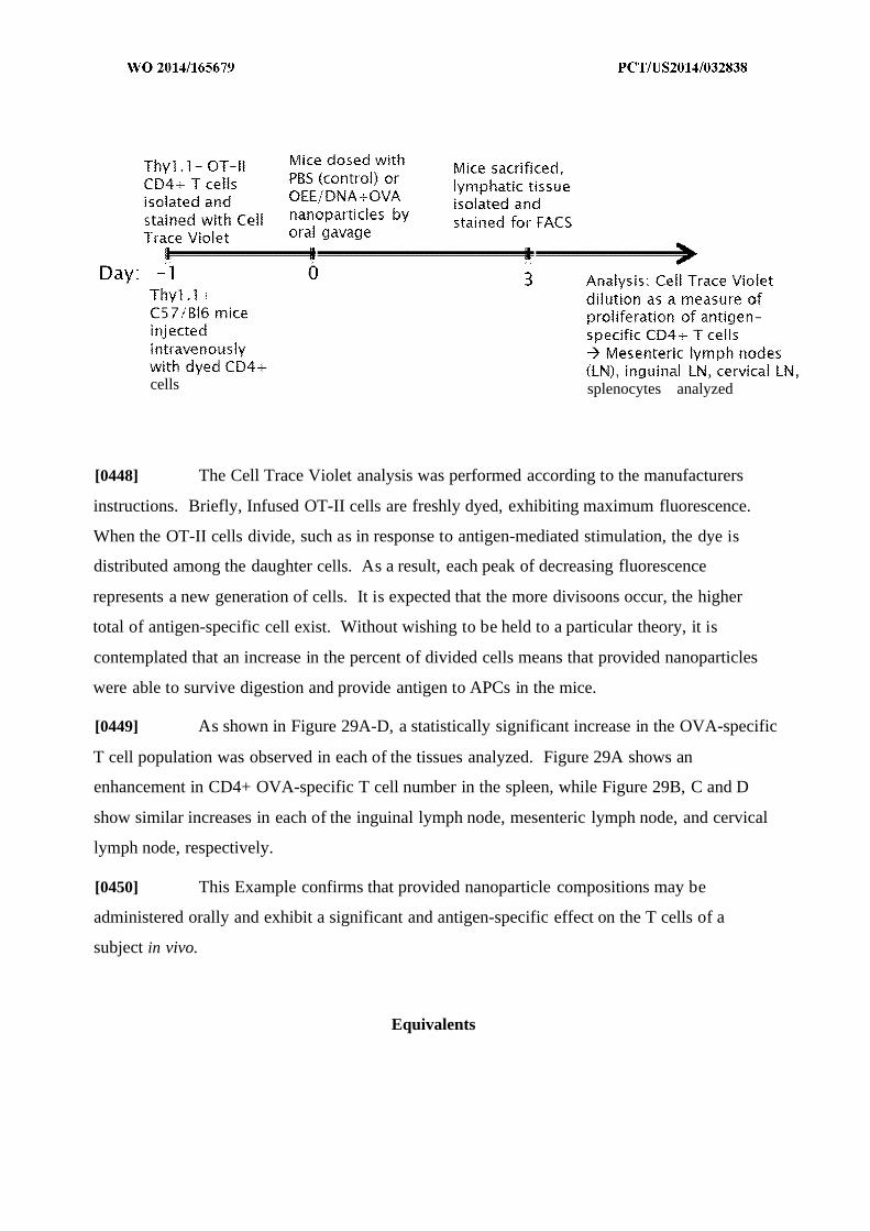
cells splenocytes analyzed
[0448] The Cell Trace Violet analysis was performed according to the manufacturers
instructions. Briefly, Infused OT-II cells are freshly dyed, exhibiting maximum fluorescence.
When the OT-II cells divide, such as in response to antigen-mediated stimulation, the dye is
distributed among the daughter cells. As a result, each peak of decreasing fluorescence
represents a new generation of cells. It is expected that the more divisoons occur, the higher
total of antigen-specific cell exist. Without wishing to be held to a particular theory, it is
contemplated that an increase in the percent of divided cells means that provided nanoparticles
were able to survive digestion and provide antigen to APCs in the mice.
[0449] As shown in Figure 29A-D, a statistically significant increase in the OVA-specific
T cell population was observed in each of the tissues analyzed. Figure 29A shows an
enhancement in CD4+ OVA-specific T cell number in the spleen, while Figure 29B, C and D
show similar increases in each of the inguinal lymph node, mesenteric lymph node, and cervical
lymph node, respectively.
[0450] This Example confirms that provided nanoparticle compositions may be
administered orally and exhibit a significant and antigen-specific effect on the T cells of a
subject in vivo.
Equivalents

[0451] Those skilled in the art will recognize, or be able to ascertain using no more than
routine experimentation, many equivalents to the specific embodiments of the invention
described herein. The scope of the present invention is not intended to be limited to the above
Description, but rather is as set forth in the following claims:
Claims
We claim:
1. A nanoparticle composition comprising:
a plurality of nanoparticles, each of which is comprised of a biodegradable or
biocompatible polymer arranged in a nanoparticle structure defining an internal
lumen and an external surface; and
a preparation of hydrophilic cellular components encapsulated within the internal lumen.
2 . A nanoparticle composition comprising:
a plurality of nanoparticles, each of which is comprised of a biodegradable or
biocompatible polymer arranged in a nanoparticle structure defining an internal
luman and an external surface; and
a preparation of hydrophobic cellular components associated with the external surface.
3 . A nanoparticle composition comprising:
a plurality of nanoparticles, each of which is comprised of a biodegradable or
biocompatible polymer arranged in a nanoparticle structure defining an internal
lumen and an external surface;
a preparation of hydrophilic cellular components encapsulated within the internal lumen;
and
a preparation of hydrophobic cellular components associated with the external surface.
4 . The nanoparticle composition of any one of claims 1-3, wherein the preparation of hydrophilic
cellular components comprises a hydrophilic extract of a cellular preparation.
5 . The nanoparticle composition of any one of claims 1-3, wherein the preparation of
hydrophobic cellular components comprises or consists of a hydrophobic extract of a cellular
preparation.
6 . The nanoparticle composition of claim 4, wherein the hydrophilic extract comprises or
consists of an aqueous extract of the cellular preparation.
7 . The nanoparticle composition of any one of the preceding claims, which composition
comprises an antigen.
8. The nanoparticle composition of claim 7, wherein the antigen is or comprises an allergic
antigen.
9 . The nanoparticle composition of claim 7, wherein the antigen is or comprises an anaphylactic
antigen.
10. The nanoparticle composition of claim 7, wherein the antigen is or comprises an infectious
antigen.
11. The nanoparticle composition of claim 7, wherein the antigen is or comprises an autoantigen.
12. The nanoparticle compostion of claim 7, wherein the antigen is or comprises a disease-
associated antigen.
13. The nanoparticle composition of claim 7, wherein the antigen is partly or wholly
encapsulated within the lumen.
14. The nanoparticle composition of claim 7, wherein the antigen is partly or wholly associated
with the external surface.
15. The nanoparticle composition of claim 7, wherein the antigen is mixed with the nanoparticles
so that each is dispersed throughout the composition.
16. The nanoparticle composition of claim 7, which composition comprises first and second
antigens, the first antigen being partly or wholly encapsulated within nanoparticle lumens and the
second antigen being partly or wholly associated with the external surface of nanoparticles.
17. The nanoparticle composition of claim 10, wherein the infectious antigen is provided with
one or more additional components of the infectious agent.
18. The nanoparticle composition of any one of claims 7-12, wherein the antigen or infectious
agent is selected from the group consisting of a food antigen, a microbial antigen, a viral antigen,
a tumor antigen, and an environmental antigen.
19. The nanoparticle composition of any one of claims 1-6, wherein at least one of the
hydrophilic cellular components and the hydrophobic cellular components is provided from a
microbial cellular preparation.
20. The nanoparticle composition of any one of claims 1-6, wherein at least one of the
hydrophilic cellular components and the hydrophobic cellular components is provided from a
tumor cell cellular preparation.
21. The nanoparticle composition of any one of claims 1-3, wherein the biodegradable or
biocompatible polymer is poly(lactic-co-gly colic acid).
22. A pharmaceutical composition comprising the nanoparticle composition of any one of the
preceeding claims.
23. A method comprising administering to a subject in need thereof a nanoparticle composition
comprising:
a plurality of nanoparticles, each of which is comprised of a biodegradable or
biocompatible polymer arranged in a nanoparticle structure defining an internal
lumen and an external surface; and
a preparation of hydrophilic cellular components encapsulated within the internal lumen.
24. A method comprising administering to a subject in need thereof a nanoparticle composition
comprising:
a plurality of nanoparticles, each of which is comprised of a biodegradable or
biocompatible polymer arranged in a nanoparticle structure defining an internal
luman and an external surface; and
a preparation of hydrophobic cellular components associated with the external surface.
25. A method comprising administering to a subject in need thereof a nanoparticle composition
comprising:
a plurality of nanoparticles, each of which is comprised of a biodegradable or
biocompatible polymer arranged in a nanoparticle structure defining an internal
luman and an external surface; and
a preparation of hydrophilic cellular components encapsulated within the internal lumen;
and
a preparation of hydrophobic cellular components associated with the external surface.
26. The method of any one of claims 23-25, wherein the preparation of hydrophilic cellular
components comprises a hydrophilic extract of a cellular preparation.
27. The method of any one of claims 23-25, wherein the preparation of hydrophobic cellular
components comprises or consists of a hydrophobic extract of a cellular preparation.
28. The method of claims 26, wherein the hydrophilic extract comprises or consists of an
aqueous extract of the cellular preparation.
29. The method of any one of claims 23-28, wherein the nanoparticle composition comprises an
antigen.

30. The method of claim 29, wherein the antigen is or comprises an allergic antigen
31. The method of claim 29, wherein the antigen is or comprises and anaphylactic antigen.
32. The method of claim 29, wherein the antigen is or comprises an infectious antigen.
33. The method of claim 29, wherein the antigen is or comprises an autoantigen.
34. The method of claim 29, wherein the antigen is or comprises a disease-associated antigen.
35. The method of claim 29, wherein the antigen is partly or wholly encapsulated within the
lumen.
36. The method of claim 29, wherein the antigen is partly or wholly associated with the external
surface.
37. The method of claim 29, wherein the antigen is mixed with the nanoparticles so that each is
dispersed throughout the composition.
38. The method of claim 29, wherein the nanoparticle composition comprises first and second
antigens, the first antigen being partly or wholly encapsulated within nanoparticle lumens and the
second antigen being partly or wholly associated with the external surface of nanoparticles.
39. The method of any one of claims 23-25, wherein the subject is suffering from at least one of
allergy, infection, and cancer.
40. The method of any one of claims 23-25, wherein the nanoparticle is administered
intravenously, intradermally, transdermally, orally, subcutaneously, and/or transmucosally.
41. The method of claim 40, wherein the transmucosal administration is buccal, nasal, bronchial,
vaginal, rectal, and/or sublingual administration.
42. The method of claim 29, wherein the antigen is selected from the group consisting of a food
antigen, a microbial antigen, a viral antigen, a tumor antigen, and an environmental antigen.
42. The method of any one of claims 23-27, wherein at least one of the hydrophilic cellular
components and the hydrophobic cellular components is provided from a microbial cellular
preparation.
43. The method of any one of claims 23-27, wherein at least one of the hydrophilic cellular
preparation and the hydrophobic cellular preparation is provided from a tumor cell cellular
preparation.
44. The method of any one of claims 23-43, wherein the biodegradable or biocompatible
polymer is poly(lactic-co-glycolic acid).
45. A method of forming a nanoparticle composition comprising:
providing a preparation of one or more hydrophilic cellular components;
providing a biodegradable or biocompatible polymer;
associating the preparation of hydrophilic cellular components and the
biodegradable or biocompatible polymer; and
forming a nanoparticle structure wherein the biodegradable or
biocompatible polymer defines an internal lumen and an external surface;
wherein the hydrophilic cellular components are encapsulated within the
internal lumen.
46. A method of forming a nanoparticle composition comprising:
providing a preparation of one or more hydrophobic cellular components;
providing a biodegradable or biocompatible polymer;
associating the preparation of hydrophobic cellular components and the
biodegradable or biocompatible polymer; and
forming a nanoparticle structure wherein the biodegradable or
biocompatible polymer defines an internal lumen and an external surface;
wherein the the hydrophobic cellular components are associated with the
external surface.
47. A method of forming a nanoparticle composition comprising:
providing a cellular preparation;
separating the cellular preparation into a preparation of hydrophilic
cellular components and a preparation of hydrophobic cellular
components;
providing a biodegradable or biocompatible polymer;
associating the preparation of hydrophilic cellular components and the
biodegradable or biocompatible polymer;
associating the preparation of hydrophobic cellular components and the
biodegradable or biocompatible polymer; and
forming a nanoparticle structure wherein the biodegradable or
biocompatible polymer defines an internal lumen and an external surface;
wherein the hydrophilic cellular components are encapsulated within the
internal lumen and the hydrophobic cellular components are associated
with the external surface.
48. The method of claim 45 or 47, wherein the preparation of hydrophilic cellular components
comprise a hydrophilic extract of a cellular preparation.
49. The method of claim 46 or 47, wherein the preparation of hydrophobic cellular components
comprise a hydrophobic extract of a cellular preparation.

50. The method of any one of claims 48, wherein the hydrophilic extract comprises an aqueous
extract of the cellular preparation.
51. The method of any one of claims 45-47, wherein the nanoparticle composition further
comprises an antigen.
52. The method of claims 51, wherein the antigen is or comprises an allergic antigen.
53. The method of claim 51, wherein the antigen is or comprises an anaphylactic antigen.
54. The method of claim 51, wherein the antigen is or comprises an infectious antigen.
55. The method of claim 51, wherein the antigen is or comprises an autoantigen.
56. The method of claim 51, wherein the antigen is or comprises a disease-associated antigen.
57. The method of claim 51, wherein the antigen is partly or wholly encapsulated within the
lumen.
58. The method of claim 51, wherein the antigen is partly or wholly associated with the external
surface.
59. The method of claim 51, wherein the nanoparticle composition comprises first and second
antigens, the first antigen being partly or wholly encapsulated within the lumen and the second
antigen being partly or wholly associated with the external surface.

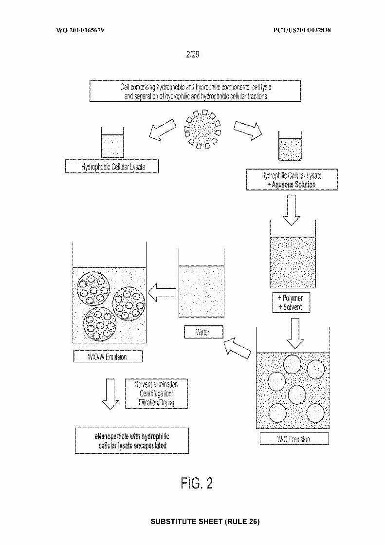
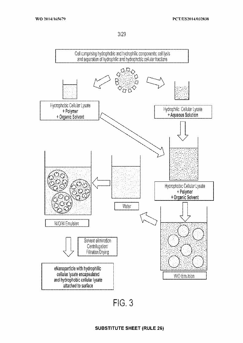
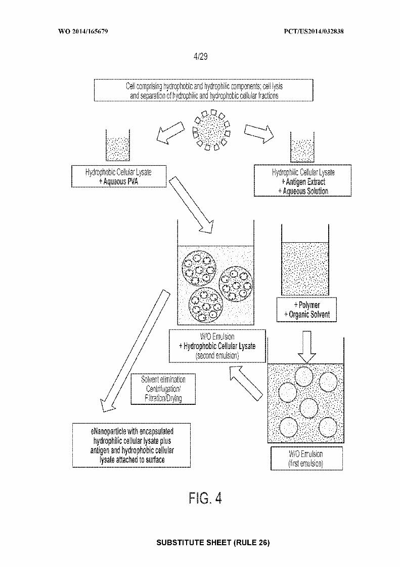
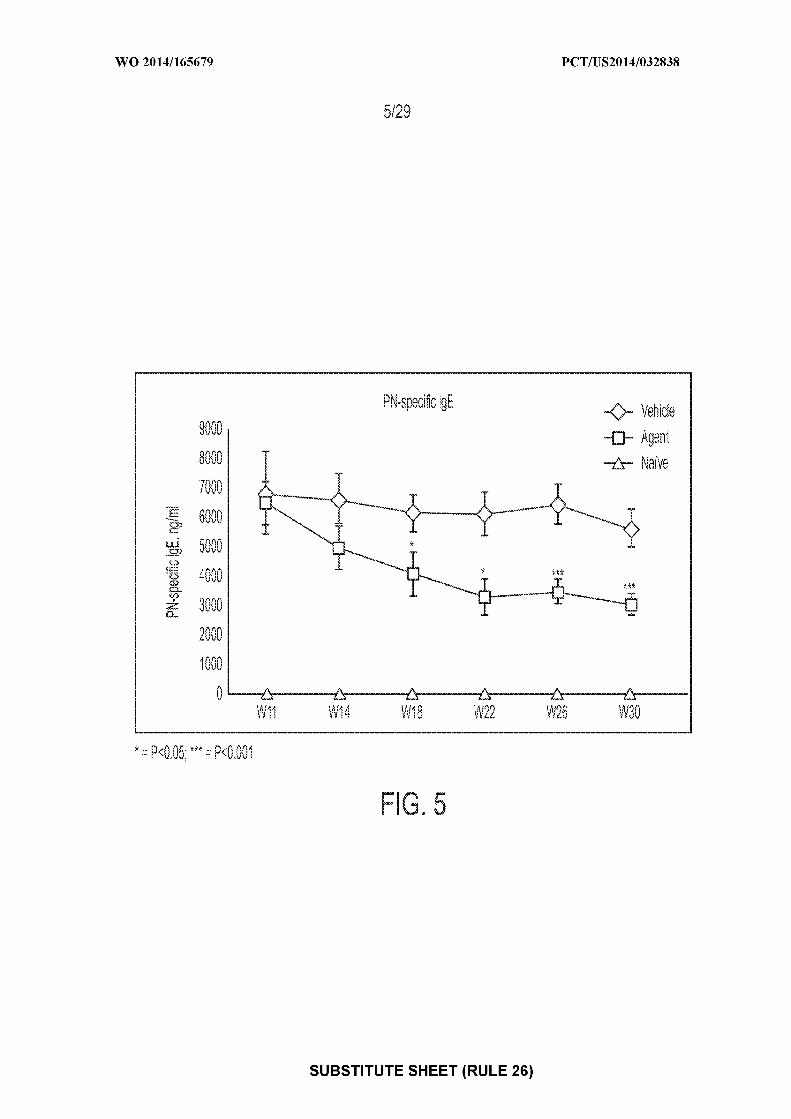
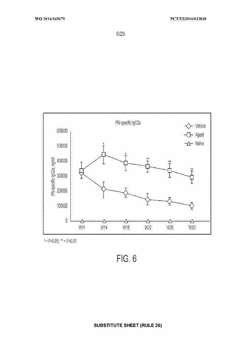

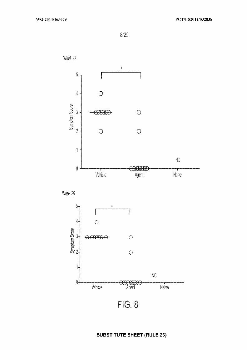
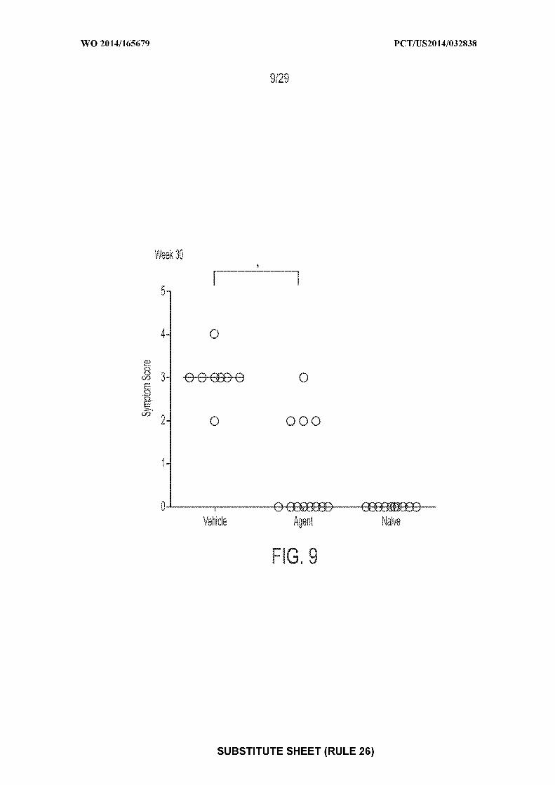
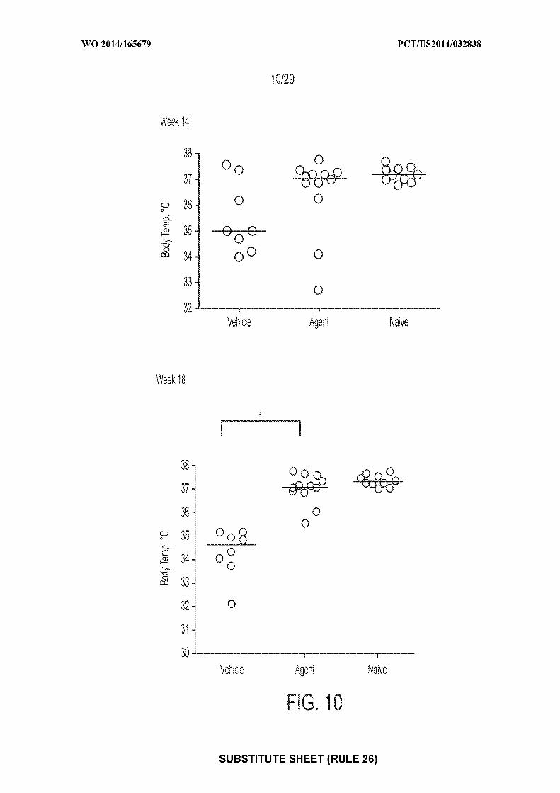
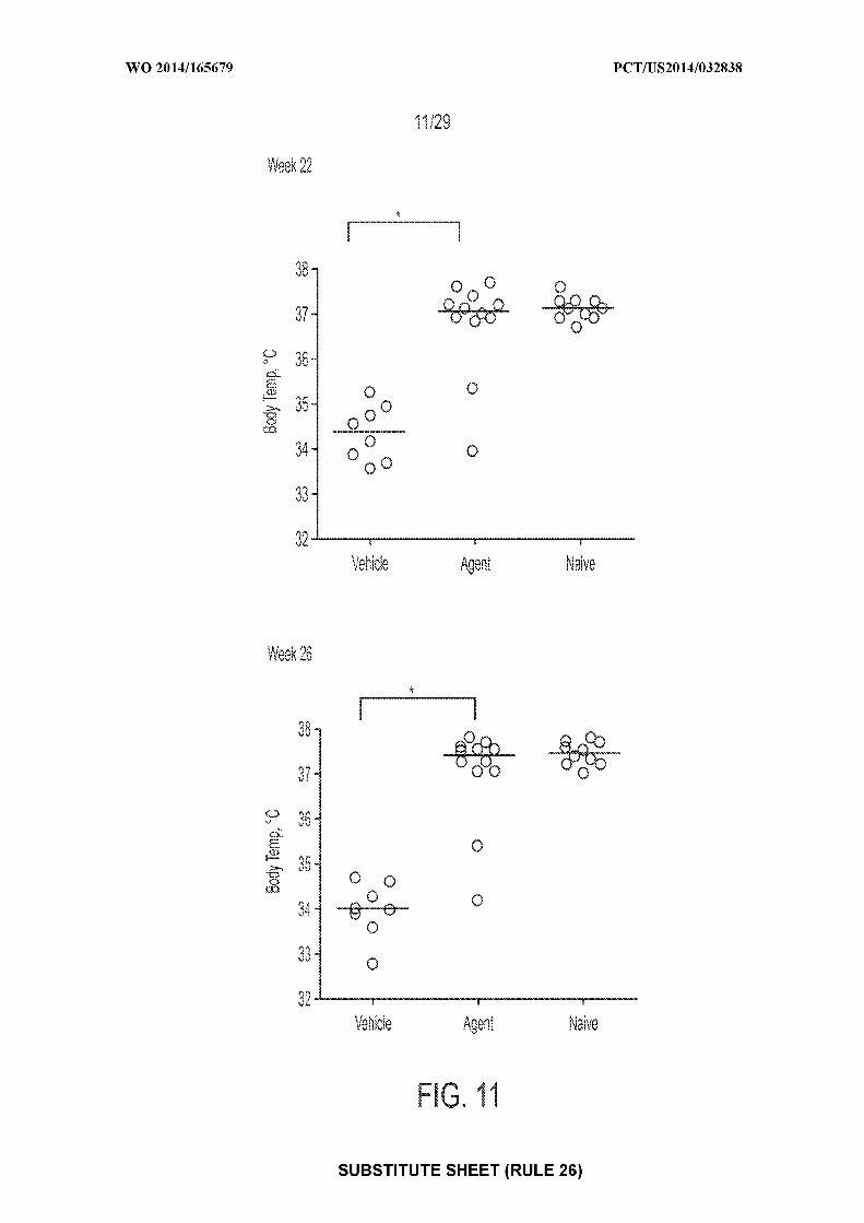
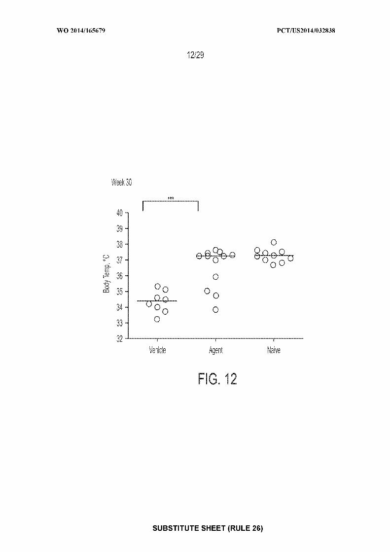
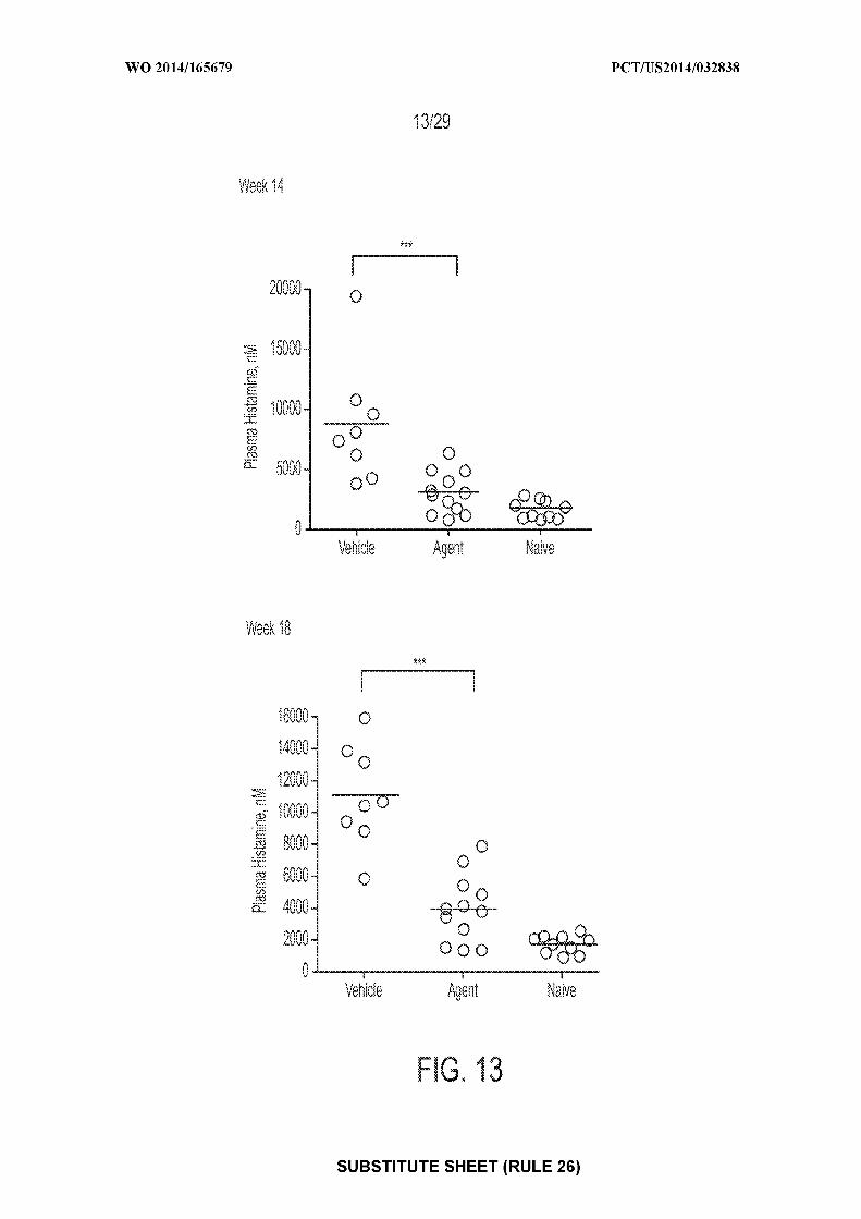


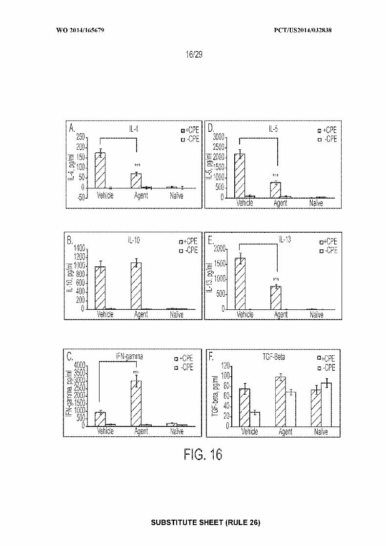
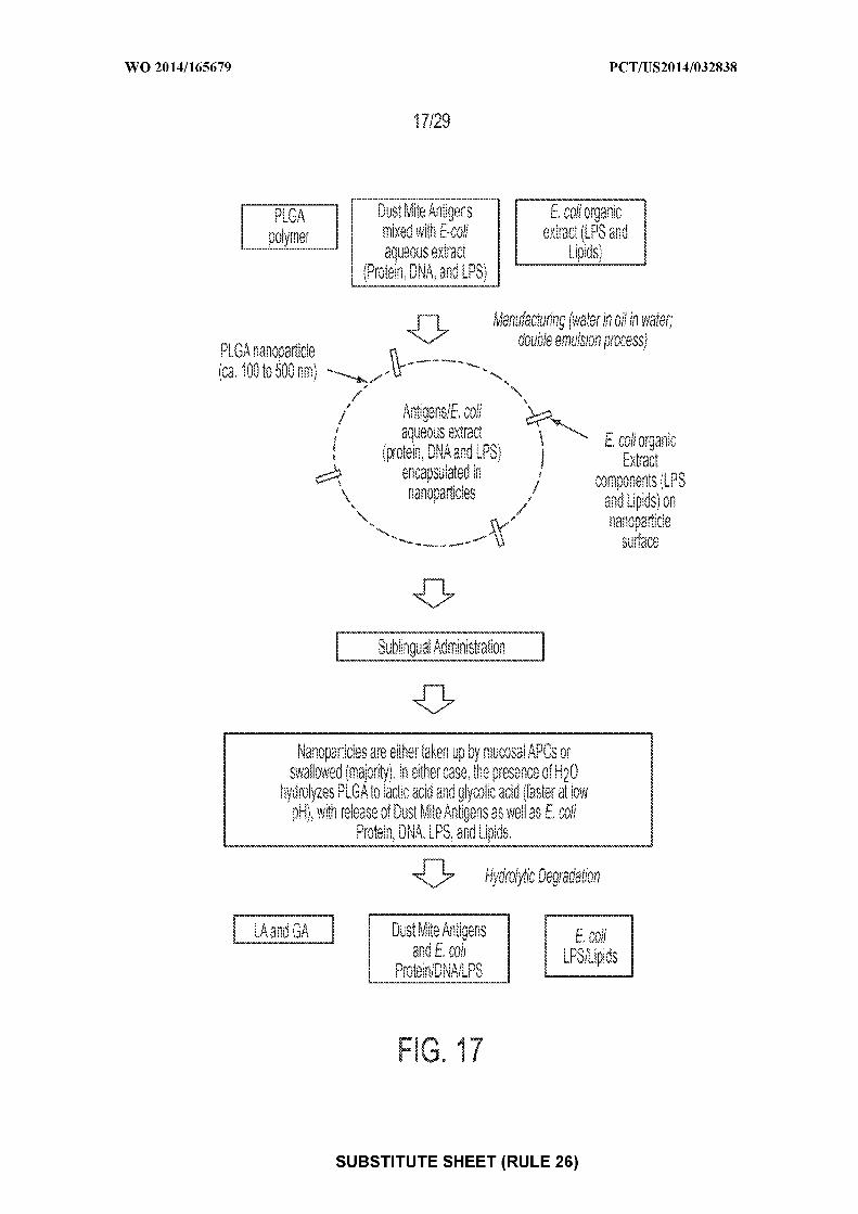
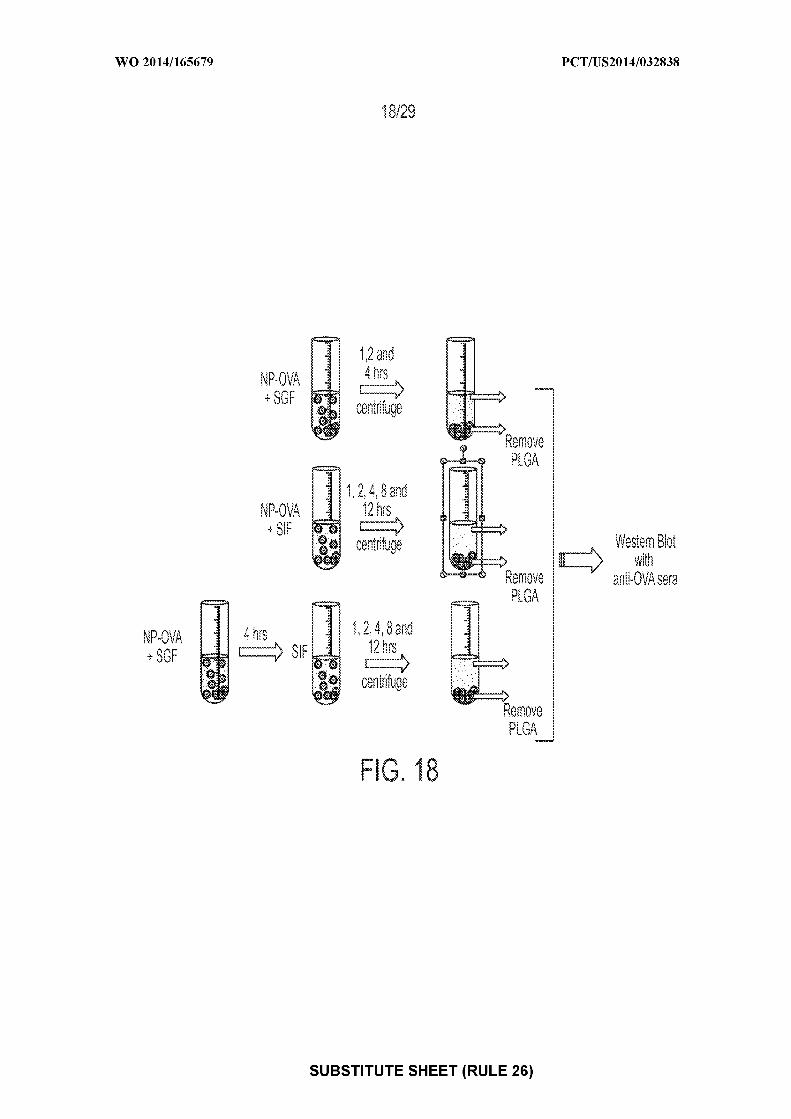


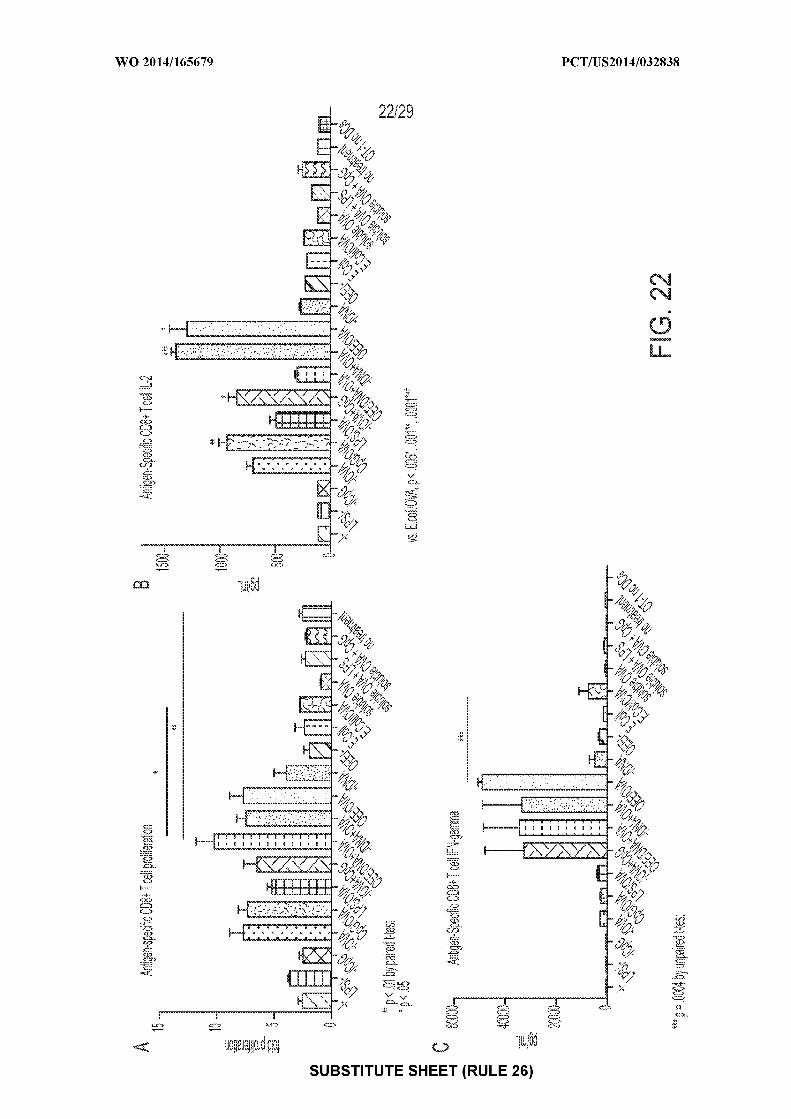

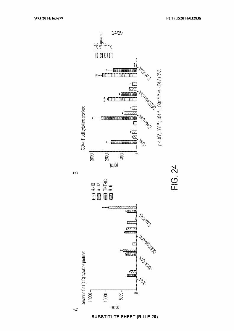

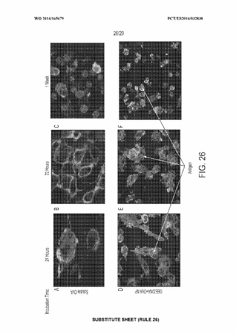
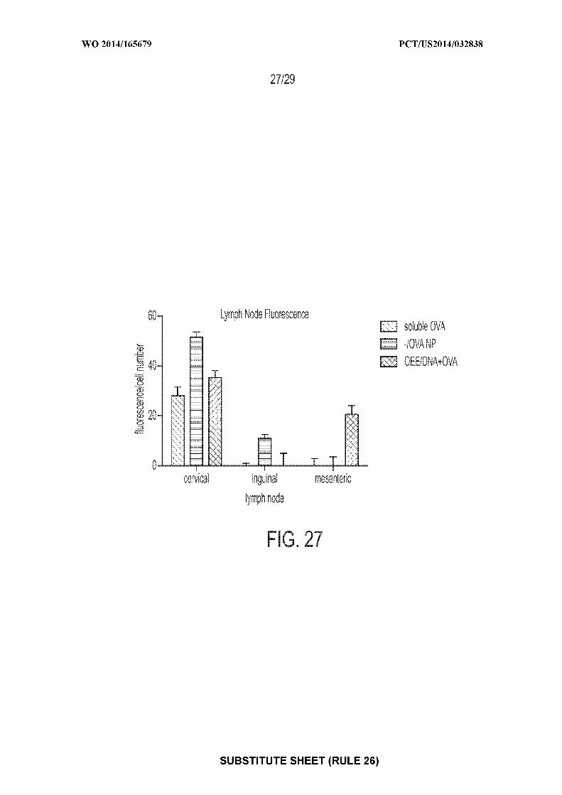

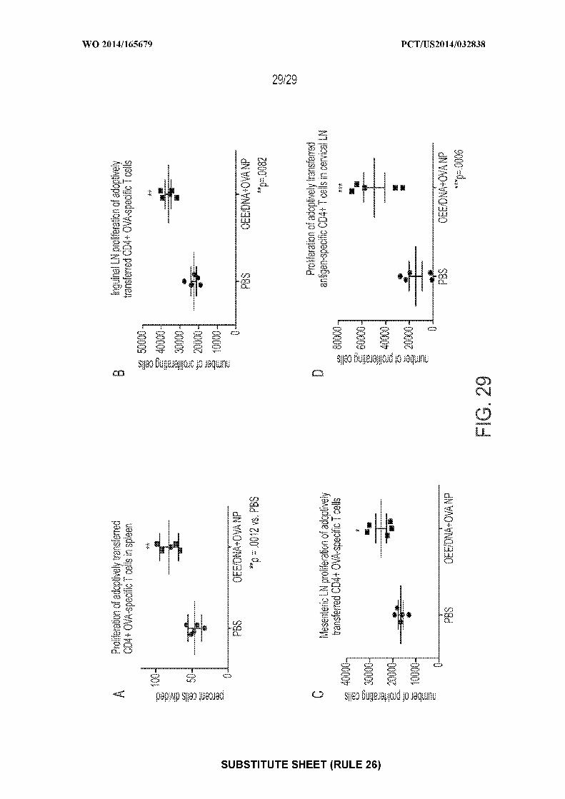
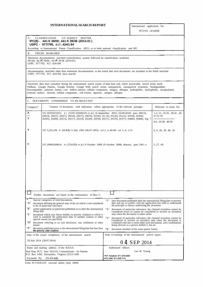
INTERNATIONALSEARCH REPORT International application No.
PCT/US 14/32838
A . CLASSIFICATION O F SUBJECT MATTERIPC(8) - A61 K 39/00; A61 K 39/38 (2014.01 )USPC - 977/795, 9 17 ; 424/1 84
According to International Patent Classification (IPC) or to both national classification and IPC
B . FIELDS SEARCHED
Minimum documentation searched (classification system followed by classification symbols)IPC(8): A6 1 39/00; A61 39/38 (2014.01)USPC: 977/795, 917; 424/184
Documentation searched other than minimum documentation to the extent that such documents are included in the fields searchedUSPC: 977/795, 917; 424/184 (text search)
Electronic data base consulted during the international search (name of data base and, where practicable, search terms used)PatBase, Google Patents, Google Scholar, Google Web, search terms: nanoparticle, nanoparticle treatment, biodegradable,biocompatible, polymer, lumen, core hollow interior, cellular component, antigen, allergen, hydrophillic, hydrophobic, encapsulated,external surface, internal, cellular component, cell extract, aqueous, antigen, allergan
C . DOCUMENTS CONSIDERED TO BE RELEVANT
Category* Citation of document, with indication, where appropriate, of the relevant passages Relevant to claim No.
US 2010/0233251 A 1 (VON ANDRIAN et al.) 16 September 2010 (16.09.2010) para [0013], 1-3, 21, 23-25, 39-41 , 45-[0014], [0021], [0027], [0034], [0072], [0076], [0105], [01 19], [0120], [0121], [0194], [0204], 47,51-59[0205], [0209], [0213], [0217], [0219], [0220], [0359], [0371], [0376], [0377], [0460], [0468], Fig. 7
4-6, 26-28, 48-50
US 5,225,194 A (SUER) 6 July 1993 (06.07.1993) col 2, In 66-69- col 3, In 1-13 4 , 6 , 26, 28, 48, 50
US 2008/0249014 A 1 (TAUER et al.) 9 October 2008 (9 October 2008) abstract, para [001 1] 5, 27, 49
□ Further documents are listed in the continuation of Box C .
Special categories of cited documents: " '
□later document published after the international filing date or priority
document defining the general state of the art which is not considered date and not in conflict with the application but cited to understandto be of particular relevance the principle or theory underlying the invention
earlier application or patent but published on or after the international "X" document of particular relevance; the claimed invention cannot befiling date considered novel or cannot be considered to involve an inventivedocument which may throw doubts on priority claim(s) or which is step when the document is taken alonecited to establish the publication date of another citation or otherspecial reason (as specified) "Y" document of particular relevance; the claimed invention cannot be
considered to involve an inventive step when the document isdocument referring to an oral disclosure, use, exhibition or other combined with one or more other such documents, such combinationmeans being obvious to a person skilled in the artdocument published prior to the international filing date but later than &" document member of the same patent family
Date of the actual completion of the international search Date of mailing of the international search report
28 July 2014 (28.07.2014) 0 S EP 2014Name and mailing address of the ISA/US Authorized officer:
Mail Stop PCT, Attn: ISA/US, Commissioner for Patents Lee W. YoungP.O. Box 1450, Alexandria, Virginia 22313-1450
Facsimile No. 571-273-3201
Form PCT/ISA/210 (second sheet) (July 2009)
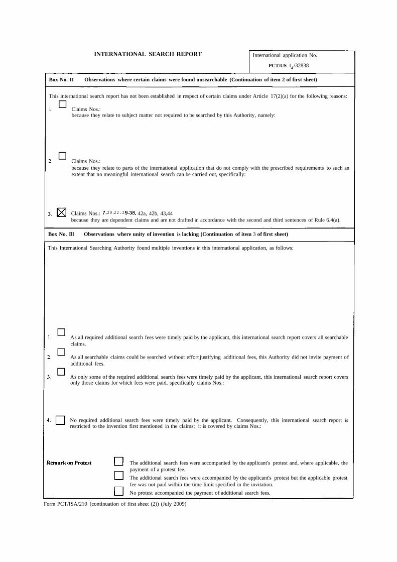
INTERNATIONAL SEARCH REPORT International application No.
PCT/US 14 /32838
Box No. II Observations where certain claims were found unsearchable (Continuation of item 2 of first sheet)
This international search report has not been established in respect of certain claims under Article 17(2)(a) for the following reasons:
□ Claims Nos.:because they relate to subject matter not required to be searched by this Authority, namely:
□ Claims Nos.:because they relate to parts of the international application that do not comply with the prescribed requirements to such anextent that no meaningful international search can be carried out, specifically:
Claims Nos.: -2 0 ·2 2 · 2 42a, 42b, 43,44because they are dependent claims and are not drafted in accordance with the second and third sentences of Rule 6.4(a).
Box No. Ill Observations where unity of invention is lacking (Continuation of item 3 of first sheet)
This International Searching Authority found multiple inventions in this international application, as follows:
□ As all required additional search fees were timely paid by the applicant, this international search report covers all searchableclaims.
□ As all searchable claims could be searched without effort justifying additional fees, this Authority did not invite payment ofadditional fees.
□ As only some of the required additional search fees were timely paid by the applicant, this international search report coversonly those claims for which fees were paid, specifically claims Nos.:
No required additional search fees were timely paid by the applicant. Consequently, this international search report isrestricted to the invention first mentioned in the claims; it is covered by claims Nos.:
The additional search fees were accompanied by the applicant's protest and, where applicable, thepayment of a protest fee.
The additional search fees were accompanied by the applicant's protest but the applicable protestfee was not paid within the time limit specified in the invitation.
No protest accompanied the payment of additional search fees.
Form PCT/ISA/210 (continuation of first sheet (2)) (July 2009)