
WO 2010/037397 Al
569
(12) INTERNATIONALAPPLICATION PUBLISHED UNDER THE PATENT COOPERATION TREATY (PCT) (19) World Intellectual Property Organization International Bureau (10) International Publication Number (43) International Publication Date 8 April 2010 (08.04.2010) WO 2010/037397 Al (51) International Patent Classification: (81) Designated States (unless otherwise indicated, for every A61K 38/17 (2006.01) C07K 14/705 (2006.01) kind of national protection available): AE, AG, AL, AM, A61K 47/48 (2006.01) GOlN 33/50 (2006.01) AO, AT, AU, AZ, BA, BB, BG, BH, BR, BW, BY, BZ, CA, CH, CL, CN, CO, CR, CU, CZ, DE, DK, DM, DO, (21) International Application Number: DZ, EC, EE, EG, ES, FI, GB, GD, GE, GH, GM, GT, PCT/DK2009/050257 HN, HR, HU, ID, IL, IN, IS, JP, KE, KG, KM, KN, KP, (22) International Filing Date: KR, KZ, LA, LC, LK, LR, LS, LT, LU, LY, MA, MD, 1 October 2009 (01 .10.2009) ME, MG, MK, MN, MW, MX, MY, MZ, NA, NG, NI, NO, NZ, OM, PE, PG, PH, PL, PT, RO, RS, RU, SC, SD, (25) Filing Language: English SE, SG, SK, SL, SM, ST, SV, SY, TJ, TM, TN, TR, TT, (26) Publication Language: English TZ, UA, UG, US, UZ, VC, VN, ZA, ZM, ZW. (30) Priority Data: (84) Designated States (unless otherwise indicated, for every PA 2008 01381 1 October 2008 (01 .10.2008) DK kind of regional protection available): ARIPO (BW, GH, 61/101,898 1 October 2008 (01 .10.2008) US GM, KE, LS, MW, MZ, NA, SD, SL, SZ, TZ, UG, ZM, ZW), Eurasian (AM, AZ, BY, KG, KZ, MD, RU, TJ, (71) Applicant (for all designated States except US): DAKO TM), European (AT, BE, BG, CH, CY, CZ, DE, DK, EE, DENMARK A/S [DK/DK]; Produktionsvej 42, DK-2600 ES, FI, FR, GB, GR, HR, HU, IE, IS, IT, LT, LU, LV, Glostrup (DK). MC, MK, MT, NL, NO, PL, PT, RO, SE, SI, SK, SM, TR), OAPI (BF, BJ, CF, CG, CI, CM, GA, GN, GQ, GW, (72) Inventors; and ML, MR, NE, SN, TD, TG). (75) Inventors/Applicants (for US only): BRIX, Liselotte [DK/DK]; Haspegardsvej 38, DK-2800 Bagsvsrd (DK). Published: SCHOLLER, Jβ rgen [DK/DK]; Sverigesvej 13A, — with international search report (Art. 21(3)) DK-2800 Lyngby (DK). PEDERSEN, Henrik [DK/DK]; Lynge Bygade 12D, DK-3450 Lynge (DK). — before the expiration of the time limit for amending the claims and to be republished in the event of receipt of (74) Agent: HOIBERG A/S; St. Kongensgade 59A, DK- 1264 amendments (Rule 48.2(h)) Copenhagen K (DK). [Continued on next page] (54) Title: MHC MULTIMERS IN CMV IMMUNE MONITORING molecule Figure 1: Schematic representation of MHC multimer (57) Abstract: The present invention relates to MHC-peptide complexes and uses thereof in the diagnosis of, treatment of or vac cination against a disease in an individual. More specifically the invention discloses MHC complexes comprising CMV antigenic peptides and uses there of.
-
Upload
khangminh22 -
Category
Documents
-
view
3 -
download
0
Transcript of WO 2010/037397 Al

(12) INTERNATIONALAPPLICATION PUBLISHED UNDER THE PATENT COOPERATION TREATY (PCT)
(19) World Intellectual Property OrganizationInternational Bureau
(10) International Publication Number(43) International Publication Date
8 April 2010 (08.04.2010) WO 2010/037397 Al
(51) International Patent Classification: (81) Designated States (unless otherwise indicated, for everyA61K 38/17 (2006.01) C07K 14/705 (2006.01) kind of national protection available): AE, AG, AL, AM,A61K 47/48 (2006.01) GOlN 33/50 (2006.01) AO, AT, AU, AZ, BA, BB, BG, BH, BR, BW, BY, BZ,
CA, CH, CL, CN, CO, CR, CU, CZ, DE, DK, DM, DO,(21) International Application Number: DZ, EC, EE, EG, ES, FI, GB, GD, GE, GH, GM, GT,
PCT/DK2009/050257 HN, HR, HU, ID, IL, IN, IS, JP, KE, KG, KM, KN, KP,(22) International Filing Date: KR, KZ, LA, LC, LK, LR, LS, LT, LU, LY, MA, MD,
1 October 2009 (01 .10.2009) ME, MG, MK, MN, MW, MX, MY, MZ, NA, NG, NI,NO, NZ, OM, PE, PG, PH, PL, PT, RO, RS, RU, SC, SD,
(25) Filing Language: English SE, SG, SK, SL, SM, ST, SV, SY, TJ, TM, TN, TR, TT,
(26) Publication Language: English TZ, UA, UG, US, UZ, VC, VN, ZA, ZM, ZW.
(30) Priority Data: (84) Designated States (unless otherwise indicated, for every
PA 2008 0 1381 1 October 2008 (01 .10.2008) DK kind of regional protection available): ARIPO (BW, GH,
61/101,898 1 October 2008 (01 .10.2008) US GM, KE, LS, MW, MZ, NA, SD, SL, SZ, TZ, UG, ZM,ZW), Eurasian (AM, AZ, BY, KG, KZ, MD, RU, TJ,
(71) Applicant (for all designated States except US): DAKO TM), European (AT, BE, BG, CH, CY, CZ, DE, DK, EE,DENMARK A/S [DK/DK]; Produktionsvej 42, DK-2600 ES, FI, FR, GB, GR, HR, HU, IE, IS, IT, LT, LU, LV,Glostrup (DK). MC, MK, MT, NL, NO, PL, PT, RO, SE, SI, SK, SM,
TR), OAPI (BF, BJ, CF, CG, CI, CM, GA, GN, GQ, GW,(72) Inventors; andML, MR, NE, SN, TD, TG).
(75) Inventors/Applicants (for US only): BRIX, Liselotte[DK/DK]; Haspegardsvej 38, DK-2800 Bagsvsrd (DK). Published:SCHOLLER, Jβrgen [DK/DK]; Sverigesvej 13A,
— with international search report (Art. 21(3))DK-2800 Lyngby (DK). PEDERSEN, Henrik [DK/DK];Lynge Bygade 12D, DK-3450 Lynge (DK). — before the expiration of the time limit for amending the
claims and to be republished in the event of receipt of(74) Agent: HOIBERG A/S; St. Kongensgade 59A, DK- 1264 amendments (Rule 48.2(h))
Copenhagen K (DK).
[Continued on next page]
(54) Title: MHC MULTIMERS IN CMV IMMUNE MONITORING
molecule
Figure 1: Schematic representation of MHC multimer
(57) Abstract: The present invention relates to MHC-peptide complexes and uses thereof in the diagnosis of, treatment of or vaccination against a disease in an individual. More specifically the invention discloses MHC complexes comprising CMV antigenicpeptides and uses there of.

— with sequence listing part of description (Rule 5.2(a))

MHC MULTIMERS IN CMV IMMUNE MONITORING
All patent and non-patent references cited in US 61/101 ,898 as well as in this
application are hereby incorporated by reference in their entirety. US 61/101 ,898 is
hereby also incorporated herein by reference in its entirety.
PCT/DK2009/050185, PCT/DK2008/0001 18, and PCT/DK2008/050167 are hereby
also incorporated herein by reference in their entirety. All patent and non-patent
references cited in PCT/DK2009/050185, PCT/DK2008/0001 18, and
PCT/DK2008/0501 67 are hereby also incorporated by reference in their entirety.
Field of invention
The present invention relates to MHC-peptide complexes and uses thereof in the
treatment of a disease in an individual.
Background of invention
Biochemical interactions between peptide epitope specific membrane molecules
encoded by the Major Histocompatibility Complex (MHC, in humans HLA) and T-cell
receptors (TCR) are required to elicit specific immune responses. This requires
activation of T-cells by presentation to the T-cells of peptides against which a T-cell
response should be raised. The peptides are presented to the T-cells by the MHC
complexes.
The immune response
The immune response is divided into two parts termed the innate immune response
and the adaptive immune response. Both responses work together to eliminate
pathogens (antigens). Innate immunity is present at all times and is the first line of
defense against invading pathogens. The immediate response by means of pre-
existing elements, i.e. various proteins and phagocytic cells that recognize conserved
features on the pathogens, is important in clearing and control of spreading of
pathogens. If a pathogen is persistent in the body and thus only partially cleared by the
actions of the innate immune system, the adaptive immune system initiate a response
against the pathogen. The adaptive immune system is capable of eliciting a response

against virtually any type of pathogen and is unlike the innate immune system capable
of establishing immunological memory.
The adaptive response is highly specific to the particular pathogen that activated it but
it is not so quickly launched as the innate when first encountering a pathogen.
However, due to the generation of memory cells, a fast and more efficient response is
generated upon repeated exposure to the same pathogen. The adaptive response is
carried out by two distinct sets of lymphocytes, the B cells producing antibodies leading
to the humoral or antibody mediated immune response, and the T cells leading to the
cell mediated immune response.
T cells express a clonotypic T cell receptor (TCR) on the surface. This receptor enable
the T cell to recognize peptide antigens bound to major histocompatibility complex
(MHC) molecules, called human leukocyte antigens (HLA) in man. Depending on the
type of pathogen, being intracellular or extracellular, the antigenic peptides are bound
to MHC class I or MHC class II, respectively. The two classes of MHC complexes are
recognized by different subsets of T cells; Cytotoxic CD8+ T cells recognizing MHC
class I and CD4+ helper cells recognizing MHC class II. In general, TCR recognition of
MHC-peptide complexes result in T cell activation, clonal expansion and differentiation
of the T cells into effector, memory and regulatory T cells.
B cells express a membrane bound form of immunoglobulin (Ig) called the B cell
receptor (BCR). The BCR recognizes an epitope that is part of an intact three
dimensional antigenic molecule. Upon BCR recognition of an antigen the BCR:antigen
complex is internalized and fragments from the internalized antigen is presented in the
context of MHC class I l on the surface of the B cell to CD4+ helper T-cells (Th). The
specific Th cell will then activate the B cell leading to differentiation into an antibody
producing plasma cell.
A very important feature of the adaptive immune system is its ability to distinguish
between self and non-self antigens, and preferably respond against non-self. If the
immune system fails to discriminate between the two, specific immune responses
against self-antigens are generated. These autoimmune reactions can lead to damage
of self-tissue.

The adaptive immune response is initiated when antigens are taken up by professional
antigen presenting cells such as dendritic cells, Macrophages, Langerhans cells and B-
cells. These cells present peptide fragments, resulting from the degradation of proteins,
in the context of MHC class I l proteins (Major Histocompatibility Complex) to helper T
cells. The T helper cells then mediate help to B-cells and antigen-specific cytotoxic T
cells, both of which have received primary activation signals via their BCR respective
TCR. The help from the Th-cell is mediated by means of soluble mediators e.g.
cytokines.
In general the interactions between the various cells of the cellular immune response is
governed by receptor-ligand interactions directly between the cells and by production of
various soluble reporter substances e.g. cytokines by activated cells.
MHC-peptide complexes.
MHC complexes function as antigenic peptide receptors, collecting peptides inside the
cell and transporting them to the cell surface, where the MHC-peptide complex can be
recognized by T-lymphocytes. Two classes of classical MHC complexes exist, MHC
class I and II. The most important difference between these two molecules lies in the
protein source from which they obtain their associated peptides. MHC class I
molecules present peptides derived from endogenous antigens degraded in the cytosol
and are thus able to display fragments of viral proteins and unique proteins derived
from cancerous cells. Almost all nucleated cells express MHC class I on their surface
even though the expression level varies among different cell types. MHC class Il
molecules bind peptides derived from exogenous antigens. Exogenous proteins enter
the cells by endocytosis or phagocytosis, and these proteins are degraded by
proteases in acidified intracellular vesicles before presentation by MHC class I l
molecules. MHC class Il molecules are only expressed on professional antigen
presenting cells like B cells and macrophages.
The three-dimensional structure of MHC class I and I l molecules are very similar but
important differences exist. MHC class I molecules consist of two polypeptide chains, a
heavy chain, α, spanning the membrane and a light chain, β2-microglobulin (β2m). The
heavy chain is encoded in the gene complex termed the major histocompatibility
complex (MHC), and its extracellular portion comprises three domains, α1, α2 and α3 .
The β2m chain is not encoded in the MHC gene and consists of a single domain, which

together with the α3 domain of the heavy chain make up a folded structure that closely
resembles that of the immunoglobulin. The α1 and α2 domains pair to form the peptide
binding cleft, consisting of two segmented α helices lying on a sheet of eight β-strands.
In humans as well as in mice three different types of MHC class I molecule exist. HLA-
A, B, C are found in humans while MHC class I molecules in mice are designated H-
2K, H-2D and H-2L
The MHC class I l molecule is composed of two membrane spanning polypeptide
chains, α and β, of similar size (about 30000 Da). Genes located in the major
histocompatibility complex encode both chains. Each chain consists of two domains,
where α1 and β1 forms a 9-pocket peptide-binding cleft, where pocket 1, 4 , 6 and 9 are
considered as major peptide binding pockets. The α2 and β2 , like the α2 and β2m in
the MHC class I molecules, have amino acid sequence and structural similarities to
immunoglobulin constant domains. In contrast to MHC class I complexes, where the
ends of the antigenic peptide is buried, peptide-ends in MHC class I l complexes are
not. HLA-DR, DQ and DP are the human class I l molecules, H-2A, M and E are those
of the mice.
A remarkable feature of MHC genes is their polymorphism accomplished by multiple
alleles at each gene. The polygenic and polymorphic nature of MHC genes is reflected
in the peptide-binding cleft so that different MHC complexes bind different sets of
peptides. The variable amino acids in the peptide binding cleft form pockets where the
amino acid side chains of the bound peptide can be buried. This permits a specific
variant of MHC to bind some peptides better than others.
MHC multimer s
Due to the short half-life of the peptide-MHC-T cell receptor ternary complex (typically
between 10 and 25 seconds) it is difficult to label specific T cells with labelled MHC-
peptide complexes, and like-wise, it is difficult to employ such monomers of MHC-
peptide for therapeutic and vaccine purposes because of their weak binding. In order to
circumvent this problem, MHC multimers have been developed. These are complexes
that include multiple copies of MHC-peptide complexes, providing these complexes
with an increased affinity and half-life of interaction, compared to that of the monomer
MHC-peptide complex. The multiple copies of MHC-peptide complexes are attached,

covalently or non-covalently, to a multimerization domain. Known examples of such
MHC multimers include the following:
• MHC-dimers: Each MHC dimer contains two copies of MHC-peptide. IgG is
used as multimerization domain, and one of the domains of the MHC protein is
covalently linked to IgG.
• MHC-tetramers: Each MHC-tetramer contains four copies of MHC-peptide,
each of which is biotinylated. The MHC complexes are held together in a
complex by the streptavidin tetramer protein, providing a non-covalent linkage
between a streptavidin monomer and the MHC protein. Tetramers are
described in US patent 5,635,363.
• MHC pentamers: Five copies of MHC-peptide complexes are multimerised by a
self-assembling coiled-coil domain, to form a MHC pentamer. MHC pentamers
are described in the US patent 2004209295
• MHC dextramers: A large number of MHC-peptide complexes, typically more
than ten, are attached to a dextran polymer. MHC-dextramers are described in
the patent application WO 02/072631 A2.
• MHC streptamers: 8-1 2 MHC-peptide complexes attached to Streptactin. MHC
streptamers are described in Knabel M et al. Reversibel MHC multimer staining
for functional isolation of T-cell populations and effective adoptive transfer.
Nature medicine 6 . 631 -637 (2002).
Use of MHC multimers in flow cytometry and related techniques
The concentration of antigen-specific T-cells in samples from e.g. peripheral blood can
be very low. Flow cytometry and related methods offer the ability to analyze a large
number of cells and simultaneously identify the few of interest. MHC multimers have
turned out to be very valuable reagents for detection and characterization of antigen-
specific T-cells in flow cytometer experiments. The relative amount of antigen-specific
T cells in a sample can be determined and also the affinity of the binding of MHC
multimer to the T-cell receptor can be determined.
The basic function of a flow cytometer is its ability to analyse and identify fluorochrome
labelled entities in a liquid sample, by means of its excitation, using a light source such
as a laser beam and the light emission from the bound fluorochrome.
MHC multimers is used as detections molecule for identification of antigen-specific T-
cells in flow cytometry, by labelling the MHC multimer with a specific fluorochrome,
which is detectable, by the flow cytometer used.

In order to facilitate the identification of a small amount of cells, the cells can be sub-
categorized using antibodies or other fluorochrome labelled detections molecules
directed against surface markers other than the TCR on the specific T-cells population.
Antibodies or other fluorochrome labelled detections molecules can also be used to
identify cells known not to be antigen-specific T-cells. Both kinds of detections
molecules are in the following referred to as gating reagents. Gating reagents, helps
identify the "true" antigen-specific T cells bound by MHC multimers by identifying
specific subpopulations in a sample, e.g. T cells and by excluding cells that for some
reason bind MHC mulimers without being antigen-specific T-cells.
Other cytometry methods, e.g. fluorescence microscopy and IHC can like flow
cytometry be employed in identification of antigen-specific T cells in a cell sample using
MHC multimers.
Application of MHC multimers in immune monitoring, diagnostics, prognostics,
therapy and vaccines
T cells are pivotal for mounting an adaptive immune response. It is therefore of
importance to be able to measure the number of specific T cells when performing a
monitoring of a given immune response, for example in connection with vaccine
development, infectious diseases e.g. tuberculosis, toxicity studies etc.
Accordingly, the present invention further provides powerful tools in the fields of
vaccines, therapy and diagnosis. One objective is to isolate antigen-specific T-cells and
culture these in the presence of co-stimulatory molecules. Ex vivo priming and
expansion of T-cell populations allows the T-cells to be used in immunotherapy of
various types of infectious diseases. A second objective of the present invention is to
identify and label specific subsets of cells with relevance for the development or
treatment of diseases.
Of special interest of the present invention is transplantation related disease. MHC
multimers of the present invention can be used in immune monitoring following
transplantation but may also be used for immune monitoring in other immuno
suppressive conditions e.g. in HIV infected patients or patients receiving chemotherapy

SUMMARY OF INVENTION.
Measurement of antigen-specific T cells during an immune response are important
parameters in vaccine development, autologous cancer therapy, transplantation,
infectious diseases, inflammation, autoimmunity, toxicity studies etc. MHC multimers
are crucial reagents in monitoring of antigen-specific T cells. The present invention
describes novel methods to generate MHC multimers and methods to improve existing
and new MHC multimers. The invention also describes improved methods for the use
of MHC multimers in analysis of T cells in samples including diagnostic and prognostic
methods. Furthermore the use of MHC multimers in therapy are described, e.g. anti-
tumour and anti-virus therapy, including isolation of antigen-specific T cells capable of
inactivation or elimination of undesirable target cells or isolation of specific T cells
capable of regulation of other immune cells. The present invention also relates to MHC
multimers comprising one or more CMV derived peptides. In one preferred
embodyment such MHC multimers are used in monitoring immune status following e.g.
transplantation, during immunosuppresive treatment, during anticancer treatment, in
HIV infected individuals or other conditions where the immune system is supressed.
In one preferred embodiment the present invention relates to a MHC multimer such as
a MHC dextramer such as a chemically biotinylated MHC dextramer. These MHC
multimers prefereably comprise one or more peptides derived from one or more CMV
antigens such as one or more peptides derived from pp28, pp50, pp65, pp150, pp71 ,
gH, gB, IE-1 , IE-2, US2, US3, US6, US1 1, and UL18.
In one preferred embodiment the measurement of antigen-specific T cells according to
the present invention is based on measurement of the number of T cells and not on
measurement of activation of T cells.
In one preferred embodiment the measurement of antigen-specific T cells according to
the present invention is based on a binding assay and not on a functional assay.
In another preferred embodiment the method for measurement of T cells is not based
on and/or does not involve priming of Antigen presenting cells. The method for
measurement of T cells can in one preferred embodiment be performed at
experimental condition that will only allow limited or completely prevent priming of
Antigen presenting cells. These experimental conditions can involve one or more

factors that do not allow priming of Antigen presenting cells such as an incubation time
that is too short to allow priming or a pH or salt concentration that will prevent or inhibit
priming of Antigen presenting cells.
In another preferred embodiment the present invention relates to a CMV vaccine. In a
CMV vaccine the peptides bound in the peptide binding cleft of MHC are derived from
CMV proteins.
DEFINITIONS
As used everywhere herein, the term "a", "an" or "the" is meant to be one or more, i . e.
at least one.
Adjuvant: adjuvants are drugs that have few or no pharmacological effects by
themselves, but can increase the efficacy or potency of other drugs when given at the
same time. In another embodiment, an adjuvant is an agent which, while not having
any specific antigenic effect in itself, can stimulate the immune system, increasing the
response to a vaccine.
Agonist: agonist as used herein is a substance that binds to a specific receptor and
triggers a response in the cell. It mimics the action of an endogenous ligand that binds
to the same receptor.
Antagonist: antagonist as used herein is a substance that binds to a specific receptor
and blocks the response in the cell. It blocks the action of an endogenous ligand that
binds to the same receptor.
Antibodies: As used herein, the term "antibody" means an isolated or recombinant
binding agent that comprises the necessary variable region sequences to specifically
bind an antigenic epitope. Therefore, an antibody is any form of antibody or fragment
thereof that exhibits the desired biological activity, e.g., binding the specific target
antigen. Antibodies can derive from multiple species. For example, antibodies include
rodent (such as mouse and rat), rabbit, sheep, camel, and human antibodies.
Antibodies can also include chimeric antibodies, which join variable regions from one
species to constant regions from another species. Likewise, antibodies can be
humanized, that is constructed by recombinant DNA technology to produce

immunoglobulins which have human framework regions from one species combined
with complementarity determining regions (CDR's) from a another species'
immunoglobulin. The antibody can be monoclonal or polyclonal.
Antibodies can be divided into isotypes (IgA, IgG, IgM, IgD, IgE, IgGI , lgG2, lgG3,
lgG4, IgAI , lgA2, IgMI , lgM2)
Antibodies: In another embodiment the term "antibody" refers to an intact antibody, or a
fragment of an antibody that competes with the intact antibody for antigen binding. In
certain embodiments, antibody fragments are produced by recombinant DNA
techniques. In certain embodiments, antibody fragments are produced by enzymatic or
chemical cleavage of intact antibodies. Exemplary antibody fragments include, but are
not limited to, Fab, Fab', F(ab')2, Fv, and scFv. Exemplary antibody fragments also
include, but are not limited to, domain antibodies, nanobodies, minibodies ((scFv-
C.sub.H3).sub.2), maxibodies ((scFv-C.sub.H2-C. sub. H3).sub.2), diabodies
(noncovalent dimer of scFv).
Antigen presenting cell: An antigen-presenting cell (APC) as used herein is a cell that
displays foreign antigen complexed with MHC on its surface.
Antigenic peptide: Used interchangeably with binding peptide. Any peptide molecule
that is bound or able to bind into the binding groove of either MHC class 1 or MHC
class 2 .
Aptamer: the term aptamer as used herein is defined as oligonucleic acid or peptide
molecules that bind a specific target molecule. Aptamers are usually created by
selecting them from a large random sequence pool, but natural aptamers also exist.
Aptamers can be divided into DNA aptamers, RNA aptamers and peptide aptamers.
Avidin: Avidin as used herein is a glycoprotein found in the egg white and tissues of
birds, reptiles and amphibians. It contains four identical subunits having a combined
mass of 67,000-68,000 daltons. Each subunit consists of 128 amino acids and binds
one molecule of biotin.
Biologically active molecule: A biologically active molecule is a molecule having itself a
biological activity/effect or is able to induce a biological activity/effect when

administered to a biological system. Biologically active molecules include adjuvants,
immune targets (e.g. antigens), enzymes, regulators of receptor activity, receptor
ligands, immune potentiators, drugs, toxins, cytotoxic molecules, co-receptors, proteins
and peptides in general, sugar moieties, lipid groups, nucleic acids including siRNA,
nanoparticles, and small molecules.
Bioluminescent: Bioluminescence, as used herein, is the production and emission of
light by a living organism as the result of a chemical reaction during which chemical
energy is converted to light energy.
Biotin: Biotin, as used herein, is also known as vitamin H or B7. Niotin has the chemical
formula Ci0H16
N2O3S.
Bispecific antibodies: The term bispecific antibodies as used herein is defined as
monoclonal, preferably but not limited to human or humanized, antibodies that have
binding specificities for at least two different antigens. The antibody can also be
trispecific or multispecific.
Carrier: A carrier as used herin can be any type of molecule that is directly or indirectly
associated with the MHC peptide complex. In this invention, a carrier will typically refer
to a functionalized polymer (e.g. dextran) that is capable of reacting with MHC-peptide
complexes, thus covalently attaching the MHC-peptide complex to the carrier, or that is
capable of reacting with scaffold molecules (e.g. streptavidin), thus covalently attaching
streptavidin to the carrier; the streptavidin then may bind MHC-peptide complexes.
Carrier and scaffold are used interchangeably herein where scaffold typically refers to
smaller molecules of a multimerization domain and carrier typically refers to larger
molecule and/or cell like structures.
Chelating chemical compound: Chelating chemical compound, as used herein, is the
process of reversible bindingof a ligand to a metal ion, forming a metal complex.
Chemiluminescent: Chemiluminescence, as used herein, is the emission of light
(luminescence) without emission of heat as the result of a chemical reaction.

Chromophore: A chromophore, as used herein, is the part of a visibly coloured
molecule responsible for light absorption over a range of wavelengths thus giving rise
to the colour. By extension the term can be applied to uv or ir absorbing parts of
molecules.
CMV: Cytomegalovirus
Coiled-coil polypeptide: Used interchangeably with coiled-coil peptide and coiled-coil
structure. The term coiled-coil polypeptide as used herein is a structural motif in
proteins, in which 2-7 alpha-helices are coiled together like the strands of a rope
Covalent binding: The term covalent binding is used herein to describe a form of
chemical bonding that is characterized by the sharing of pairs of electrons between
atoms. Attraction-to-repulsion stability that forms between atoms when they share
electrons is known as covalent bonding.
Crosslinking is the process of chemically joining two or more molecules by a covalent
bond. Crosslinking reagents contain reactive ends to specific functional groups
(primary amines, sulfhydryls, etc.) on proteins or other molecules.
Diagnosis: The act or process of identifying or determining the nature and cause of a
disease or injury through evaluation
Diabodies: The term "diabodies" refers to small antibody fragments with two antigen-
binding sites, which fragments comprise a heavy-chain variable domain (VH)
connected to a light-chain variable domain (VL) in the same polypeptide chain (VH-VL).
By using a linker that is too short to allow pairing between the two domains on the
same chain, the domains are forced to pair with the complementary domains of another
chain and create two antigen-binding sites.
Dendritic cell: The term dendritic cell as used herein is a type of immune cells. Their
main function is to process antigen material and present it on the surface to other cells
of the immune system, thus functioning as antigen-presenting cells.

Detection: In this invention detection means any method capable of measuringen one
molecule bound to anoher molecule. The molecules are typically proteins but can be
any type of molecule
Dextran: the term dextran as used herein is is a complex, branched polysaccharide
made of many glucose molecules joined into chains of varying lengths. The straight
chain consists of α1->6 glycosidic linkages between glucose molecules, while branches
begin from α1->3 linkages (and in some cases, α1->2 and α1->4 linkages as well).
Direct detection of T cells: Direct detection of T cells is used herein interchangeably
with direct detection of TCR and direct detection of T cell receptor. As used herein
direct detection of T cells is detection directly of the binding interaction between a
specific T cell receptor and a MHC multimer.
DNA: The term DNA (Deoxyribonucleic acid) duplex as used herein is a polymer of
simple units called nucleotides, with a backbone made of sugars and phosphate atoms
joined by ester bonds. Attached to each sugar is one of four types of molecules called
bases.
DNA duplex: In living organisms, DNA does not usually exist as a single molecule, but
instead as a tightly-associated pair of molecules. These two long strands entwine like
vines, in the shape of a double helix.
Electrophilic: electrophile, as used herein, is a reagent attracted to electrons that
participates in a chemical reaction by accepting an electron pair in order to bond to a
nucleophile.
Enzyme label: enzyme labelling, as used herein, involves a detection method
comprising a reaction catalysed by an enzyme.
Epitope-focused antibody: Antibodies also include epitope-focused antibodies, which
have at least one minimal essential binding specificity determinant from a heavy chain
or light chain CDR3 from a reference antibody, methods for making such epitope-
focused antibodies are described in U.S. patent application Ser. No. 11/040,1 59, which
is incorporated herein by reference in its entirety.

Flow cytomerty: The analysis of single cells using a flow cytometer.
Flow cytometer: Instrument that measures cell size, granularity and flourescence due
to bound fluorescent marker molecules as single cells pass in a stream past
photodectors. A flow cytomter carry out the measurements and/or sorting of individual
cells.
Fluorescent: the term fluorescent as used herein is to have the ability to emit light of a
certain wavelength when activated by light of another wavelength.
Fluorochromes: fluorochrome, as used herein, is any fluorescent compound used as a
dye to mark e.g. protein with a fluorescent label.
Fluorophore: A fluorophore, as used herein, is a component of a molecule which
causes a molecule to be fluorescent.
Folding: In this invention folding means in vitro or in vivo folding of proteins in a tertiery
structure.
Fusion antibody: As used herein, the term "fusion antibody" refers to a molecule in
which an antibody is fused to a non-antibody polypeptide at the N- or C-terminus of the
antibody polypeptide.
Glycosylated: Glycosylation, as used herein, is the process or result of addition of
saccharides to proteins and lipids.
Hapten: A residue on a molecule for which there is a specific molecule that can bind,
e.g. an antibody.
Heteroconjugate antibodies are composed of two covalently joined antibodies. Such
antibodies have, for example, been proposed to target immune system cells to
unwanted cells.

IgG: IgG as used herein is a monomeric immunoglobulin, built of two heavy chains and
two light chains. Each molecule has two antigen binding sites.
Isolated antibody: The term "isolated" antibody as used herein is an antibody which has
been identified and separated and/or recovered from a component of its natural
environment.
Immunoconjugates: The invention also pertains to immunoconjugates comprising an
antibody or a MHC-peptide complex conjugated to a cytotoxic agent such as a
chemotherapeutic agent, toxin (e.g., an enzymatically active toxin of bacterial, fungal,
plant, or animal origin, or fragments thereof), or a radioactive isotope (i.e., a
radioconjugate). Enzymatically active toxins and fragments thereof that can be used
include diphtheria A chain, nonbinding active fragments of diphtheria toxin, exotoxin A
chain (from Pseudomonas aeruginosa), ricin A chain, abrin A chain, modeccin A chain,
alpha-sarcin, Aleurites fordii proteins, dianthin proteins, Phytolaca americana proteins
(PAPI, PAPII, and PAP-S), momordica charantia inhibitor, curcin, crotin, sapaonaria
officinalis inhibitor, gelonin, mitogellin, restrictocin, phenomycin, enomycin, and the
tricothecenes. A variety of radionuclides are available for the production of
radioconjugated antibodies or MHC-peptide complexes. Conjugates of the antibody or
MHC-peptide complex and cytotoxic agent are made using a variety of bifunctional
protein-coupling agents such as N-succinimidyl-3-(2-pyridyldithiol) propionate (SPDP),
iminothiolane (IT), bifunctional derivatives of imidoesters (such as dimethyl adipimidate
HCL), active esters (such as disuccinimidyl suberate), aldehydes (such as
glutaraldehyde), bis-azido compounds (such as bis(p-azidobenzoyl)hexanediamine),
bis-diazonium derivatives (such as bis-(p-diazoniumbenzoyl)-ethylenediamine),
diisocyanates (such as tolyene 2,6-diisocyanate), and bis-active fluorine compounds
(such as 1,5-difluoro-2,4-dinitrobenzene).
Immune monitoring: Immune monitoring of the present invention refers to testing of
immune status in the diagnosis and therapy of diseases like but not limited to cancer,
immunoproliferative and immunodeficiency disorders, autoimmune abnormalities, and
infectious diseases. It also refers to testing of immune status before, during and after
vaccination and transplantation procedures.
Immune monitoring process: a series of one or more immune monitoring analysis

lmmuno profiling: lmmuno profiling as used herein defines the profiling of an
individual's antigen-specific T-cell repertoire
Indirect detection of T cells: Indirect detection of T cells is used interchangeably herein
with Indirect detection of TCR and indirect detection of T cell receptor. As used herein
indirect detection of T cells is detection of the binding interaction between a specific T
cell receptor and a MHC multimer by measurement of the effect of the binding
interaction.
lonophore: ionophore, as used herein, is a lipid-soluble molecule usually synthesized
by microorganisms capable of transporting ions.
Label: Label herein is used interchangeable with labeling molecule. Label as described
herein is an identifiable substance that is detectable in an assay and that can be
attached to a molecule creating a labeled molecule. The behavior of the labeled
molecule can then be studied.
Labelling: Labelling herein means attachment of a label to a molecule.
Lanthanide: lanthanide, as used herein, series comprises the 15 elements with atomic
numbers 57 through 7 1 , from lanthanum to lutetium.
Linker molecule: Linker molecule and linker is used interchangeable herein. A linker
molecule is a molecule that covalently or non-covalently connects two or more
molecules, thereby creating a larger complex consisting of all molecules including the
linker molecule.
Liposomes: The term liposomes as used herein is defined as a spherical vesicle with a
membrane composed of a phospholipid and cholesterol bilayer. Liposomes, usually but
not by definition, contain a core of aqueous solution; lipid spheres that contain no
aqueous material are called micelles.
Immunoliposomes: The antibodies or MHC-peptide complexes disclosed herein can
also be formulated as immunoliposomes. Liposomes comprising the antibody or MHC-
peptide complexes are prepared by methods known in the art, such as described in

Epstein et al., Proc. Natl. Acad. Sci. USA 82: 3688 ( 1985); Hwang et al., Proc. Natl.
Acad. Sci. USA 77: 4030 ( 1980); and U.S. Pat. Nos. 4,485,045 and 4,544,545.
Particularly useful liposomes can be generated by the reverse-phase evaporation
method with a lipid composition comprising phosphatidylcholine, cholesterol, and PEG-
derivatized phosphatidylethanolamine (PEG-PE).
Marker: Marker is used interchangeably with marker molecule herein. A marker is
molecule that specifically associates covalently or non-covalently with a molecule
belonging to or associated with an entity.
MHC: Denotes the major histocompatibility complex.
MHC: Denotes the major histocompatibility complex.
MHC I is used interchangeably herein with MHC class I and denotes the major
histocompatibility complex class I .
MHC I l is used interchangeably herein with MHC class I l and denotes the major
histocompatibility complex class I .
MHC molecule: a MHC molecule as used everywhere herein is defined as any MHC
class I molecule or MHC class I l molecule as defined herein.
A "MHC Class I molecule" as used everywhere herein is used interchangeably with
MHC I molecule and is defined as a molecule which comprises 1-3 subunits, including
a MHC I heavy chain, a MHC I heavy chain combined with a MHC I beta2microglobulin
chain, a MHC I heavy chain combined with MHC I beta2microglobulin chain through a
flexible linker, a MHC I heavy chain combined with an antigenic peptide, a MHC I
heavy chain combined with an antigenic peptide through a linker, a MHC I heavy chain/
MHC I beta2microglobulin dimer combined with an antigenic peptide, and a MHC I
heavy chain/ MHC I beta2microglobulin dimer combined with an antigenic peptide
through a flexible linker to the heavy chain or beta2microglobulin. The MHC I molecule
chains can be changed by substitution of single or by cohorts of native amino acids, or
by inserts, or deletions to enhance or impair the functions attributed to said molecule.

MHC complex: MHC complex is herein used interchangeably with MHC-peptide
complex, and defines any MHC I and/or MHC I l molecule combined with antigenic
peptide unless it is specified that the MHC complex is empty, i.e. is not complexed with
antigenic peptide
MHC Class I like molecules (including non-classical MHC Class I molecules) include
CD1d, HLA E, HLA G, HLA F, HLA H, MIC A, MIC B, ULBP-1 , ULBP-2, and ULBP-3.
A "MHC Class I l molecule" as used everywhere herein is used interchangeably with
MHC I l molecule and is defined as a molecule which comprises 2-3 subunits including
a MHC I l alpha-chain and a MHC I l beta-chain (i.e. a MHC I l alpha/beta-dimer), an
MHC I l alpha/beta dimer with an antigenic peptide, and an MHC I l alpha/beta dimer
combined with an antigenic peptide through a flexible linker to the MHC I l alpha or
MHC I l beta chain, a MHC I l alpha/beta dimer combined through an interaction by
affinity tags e.g. jun-fos, a MHC I l alpha/beta dimer combined through an interaction by
affinity tags e.g. jun-fos and further combined with an antigenic peptide through a
flexible linker to the MHC Il alpha or MHC I l beta chain. The MHC I l molecule chains
can be changed by substitution of single or by cohorts of native amino acids, or by
inserts, or deletions to enhance or impair the functions attributed to said molecule.
Under circumstances where the MHC I l alpha-chain and MHC I l beta-chain have been
fused, to form one subunit, the "MHC Class I l molecule" can comprise only 1 subunit or
2 subunits if antigenic peptide also. Included.
MHC Class I l like molecules (including non-classical MHC Class I l molecules) include
HLA DM, HLA DO, I-A beta2, and I-E beta2.
A "peptide free MHC Class I molecule" is used interchangeably herein with "peptide
free MHC I molecule" and as used everywhere herein is meant to be a MHC Class I
molecule as defined above with no peptide.
A "peptide free MHC Class Il molecule" is used interchangeably herein with "peptide
free MHC I l molecule" and as used everywhere herein is meant to be a MHC Class I l
molecule as defined above with no peptide.

o
Such peptide free MHC Class I and Il molecules are also called "empty" MHC Class I
and Il molecules.
The MHC molecule may suitably be a vertebrate MHC molecule such as a human, a
mouse, a rat, a porcine, a bovine or an avian MHC molecule. Such MHC complexes
from different species have different names. E.g. in humans, MHC complexes are
denoted HLA. The person skilled in the art will readily know the name of the MHC
complexes from various species.
In general, the term "MHC molecule" is intended to include all alleles. By way of
example, in humans e.g. HLA A, HLA B, HLA C, HLA D, HLA E, HLA F, HLA G, HLA
H, HLA DR, HLA DQ and HLA DP alleles are of interestshall be included, and in the
mouse system, H-2 alleles are of interestshall be included. Likewise, in the rat system
RT1 -alleles, in the porcine system SLA-alleles, in the bovine system BoLA, in the avian
system e.g. chicken-B alleles, are of interestshall be included.
"MHC complexes" and "MHC constructs" are used interchangeably herein.
By the terms "MHC complexes" and "MHC multimers" as used herein are meant such
complexes and multimers thereof, which are capable of performing at least one of the
functions attributed to said complex or multimer. The terms include both classical and
non-classical MHC complexes. The meaning of "classical" and "non-classical" in
connection with MHC complexes is well known to the person skilled in the art. Non-
classical MHC complexes are subgroups of MHC-like complexes. The term "MHC
complex" includes MHC Class I molecules, MHC Class I l molecules, as well as MHC-
like molecules (both Class I and Class II), including the subgroup non-classical MHC
Class I and Class I l molecules.
MHC multimer: The terms MHC multimer, MHC-multimer, MHCmer and MHC'mer
herein are used interchangeably, to denote a complex comprising more than one MHC-
peptide complexes, held together by covalent or non-covalent bonds.
Monoclonal antibodies: Monoclonal antibodies, as used herein, are antibodies that are
identical because they were produced by one type of immune cell and are all clones of
a single parent cell.

Monovalent antibodies: The antibodies in the present invention can be monovalent
antibodies. Methods for preparing monovalent antibodies are well known in the art. For
example, one method involves recombinant expression of immunoglobulin light chain
and modified heavy chain. The heavy chain is truncated generally at any point in the Fc
region so as to prevent heavy chain crosslinking. Alternatively, the relevant cysteine
residues are substituted with another amino acid residue or are deleted so as to
prevent crosslinking. In vitro methods are also suitable for preparing monovalent
antibodies. Digestion of antibodies to produce fragments thereof, particularly, Fab
fragments, can be accomplished using routine techniques known in the art.
Multimerization domain: A multimerization domain is a molecule, a complex of
molecules, or a solid support, to which one or more MHC or MHC-peptide complexes
can be attached. A multimerization domain consist of one or more carriers and/or one
or more scaffolds and may also contain one or more linkers connecting carrier to
scaffold, carrier to carrier, scaffold to scaffold. The multimerization domain may also
contain one or more linkers that can be used for attachment of MHC complexes and/or
other molecules to the multimerization domain.
Multimerization domains thus include IgG, streptavidin, streptactin, micelles, cells,
polymers, beads and other types of solid support, and small organic molecules carrying
reactive groups or carrying chemical motifs that can bind MHC complexes and other
molecules.
Nanobodies: Nanobodies as used herein is a type of antibodies derived from camels,
and are much smaller than traditional antibodies.
Neutralizing antibodies: neutralizing antibodies as used herein is an antibody which, on
mixture with the homologous infectious agent, reduces the infectious titer.
NMR: NMR (Nuclear magnetic resonance), as used herein, is a physical phenomenon
based upon the quantum mechanical magnetic properties of an atom's nucleus. NMR
refers to a family of scientific methods that exploit nuclear magnetic resonance to study
molecules.

Non-covalent: The term noncovalent bond as used herein is a type of chemical bond,
that does not involve the sharing of pairs of electrons, but rather involves more
dispersed variations of electromagnetic interactions.
Nucleic acid duplex: A nucleic acid is a complex, high-molecular-weight biochemical
macromolecule composed of nucleotide chains that convey genetic information. The
most common nucleic acids are deoxyribonucleic acid (DNA) and ribonucleic acid
(RNA).
Nucleophilic: a nucleophile, as used herein, is a reagent that forms a chemical bond to
its reaction partner (the electrophile) by donating both bonding electrons.
"One or more" as used everywhere herein is intended to include one and a plurality.
A "peptide free MHC Class I molecule" as used everywhere herein is meant to be a
MHC Class I molecule as defined above with no peptide.
A "peptide free MHC Class Il molecule" as used everywhere herein is meant to be a
MHC Class I l molecule as defined above with no peptide.
Such peptide free MHC Class I and Il molecules are also called "empty" MHC Class I
and I l molecules.
Pegylated: pegylated, as used herein, is conjugation of Polyethylene glycol (PEG) to
proteins.
Pentamer, MHC pentamer and pentamer MHC multimer is used interchangeable
herein and refers to a MHC multimer comprising 5 MHC molecules and optionally one
or more labelling compunds.
Peptide or protein: Any molecule composed of at least two amino acids. Peptide
normally refers to smaller molecules of up to around 30 amino acids and protein to
larger molecules containing more amino acids.

Phosphorylated; phosphorylated, as used herein, is is the addition of a phosphate
(PO4) group to a protein molecule or a small molecule.
"A plurality" as used everywhere herein should be interpreted as two or more.
PNA: PNA (Peptide nucleic acid) as used herein is a chemical similar to DNA or RNA.
PNA is not known to occur naturally in existing life on Earth but is artificially
synthesized and used in some biological research and medical treatments. DNA and
RNA have a deoxyribose and ribose sugar backbone, respectively, whereas PNA's
backbone is composed of repeating N-(2-aminoethyl)-glycine units linked by peptide
bonds. The various purine and pyrimidine bases are linked to the backbone by
methylene carbonyl bonds. PNAs are depicted like peptides, with the N-terminus at the
first (left) position and the C-terminus at the right.
"A plurality" as used everywhere herein should be interpreted as two or more. This
applies i.a. to the MHC peptide complex and the binding entity. When a plurality of
MHC peptide complexes is attached to the multimerization domain, such as a scaffold
or a carrier molecule, the number of MHC peptide complexes need only be limited by
the capacity of the multimerization domain.
Polyclonal antibodies: a polyclonal antibody as used herein is an antibody that is
derived from different B-cell lines. They are a mixture of immunoglobulin molecules
secreted against a specific antigen, each recognising a different epitope.
Polymer: the tern polymer as used herein is defined as a compound composed of
repeating structural units, or monomers, connected by covalent chemical bonds.
Polypeptide: Peptides are the family of short molecules formed from the linking, in a
defined order, of various α-amino acids. The link between one amino acid residue and
the next is an amide bond and is sometimes referred to as a peptide bond. Longer
peptides are reffered to as proteins or polypeptide.
Polysaccharide: The term polysaccharide as used herein is defined as polymers made
up of many monosaccharides joined together by glycosidic linkages.

Radicals: radicals, as used herein, are atomic or molecular species with unpaired
electrons on an otherwise open shell configuration. These unpaired electrons are
usually highly reactive, so radicals are likely to take part in chemical reactions.
Radioactivity: Radioactive decay is the process in which an unstable atomic nucleus
loses energy by emitting radiation in the form of particles or electromagnetic waves.
RNA: RNA (Ribonucleic acid) as used herein is a nucleic acid polymer consisting of
nucleotide monomers that plays several important roles in the processes that translate
genetic information from deoxyribonucleic acid (DNA) into protein products
Scaffold: A scaffold is typically an organic molecule carrying reactive groups, capable
of reacting with reactive groups on a MHC-peptide complex. Particularly small organic
molecules of cyclic structure (e.g. functionalized cycloalkanes or functionalized
aromatic ring structures) are termed scaffolds. Scaffold and carrier are used
interchangeably herein where scaffold typically refers to smaller molecules of a
multimerization domain and carrier typically refers to larger molecule and/or cell like
structures.
Staining: In this invention staining means specific or unspecific labelling of cells by
binding labeled molecules to defined proteins or other structures on the surface of cells
or inside cells. The cells are either in suspension or part of a tissue. The labeled
molecules can be MHC multimers, antibodies or similar molecules capable of binding
specific structures on the surface of cells.
Streptavidin: Streptavidin as used herein is a tetrameric protein purified from the
bacterium Streptomyces avidinii. Streptavidin is widely use in molecular biology
through its extraordinarily strong affinity for biotin.
Sugar: Sugars as used herein include monosaccharides, disaccharides, trisaccharides
and the oligosaccharides - comprising 1, 2 , 3 , and 4 or more monosaccharide units
respectively.
Therapy: Treatment of illness or disability

Vaccine: A vaccine is an antigenic preparation used to establish immunity to a disease
or illness and thereby protects or cure the body from a specific disease or illness.
Vaccines are either prophylactic and prevent disease or therapeutic and treat disease.
Vaccines may contain more than one type of antigen and is then called a combined
vaccine.
Vaccination: The introduction of vaccine into the body of human or animals for the
purpose of inducing immunity.
B.L. is an abereviation for Bind level
Aff. Is an abbreviation for affinity
DETAILED DESCRIPTION OF INVENTION.
The present invention in one aspect refers to a MHC monomer comprising a-b-P, or a
MHC multimer comprising (a-b-P)n, wherein n > 1,
wherein a and b together form a functional MHC protein capable of binding the peptide
P
wherein (a-b-P) is the MHC-peptide complex formed when the peptide P binds to the
functional MHC protein, and
wherein each MHC peptide complex of a MHC multimer is associated with one or more
multimerization domains.
The peptide is in one embodiment a CMV peptide such as e.g. a peptide derived from
the CMV internal matrix protein pp65.
MHC monomers and MHC multimers comprising one or more MHC peptide complexes
of class 1 or class 2 MHC are covered by the present invention. Accordingly, the
peptide P can have a length of e.g. 8 , 9 ,10, 11, 12, 13 , 14, 15 , 16, 16-20, or 20-30
amino acid residues.
Examples of the peptide P is provided herein below. In one embodiment, the peptide P
can be selected from the group consisting of sequences disclosed in the electronically

enclosed "Sequence Listing" and annotated consecutively (using integers) starting with
SEQ ID NO 1 and ending with SEQ ID NO 9697.
In another aspect the present invention is directed to a composition comprising a
plurality of MHC monomers and/or MHC multimers according to the present invention,
wherein the MHC multimers are identical or different, and a carrier.
In yet another aspect there is provided a kit comprising a MHC monomer or a MHC
multimer according to the present invention, or a composition according to the present
invention, and at least one additional component, such as a positive control and/or
instructions for use.
In a still further aspect there is provided a method for immune monitoring one or more
diseases comprising monitoring of antigen-specific T cells, said method comprising the
steps of
i) providing the MHC monomer or MHC multimer or individual components thereof
according to the present invention, or the individual components thereof,
ii) providing a population of antigen-specific T cells or individual antigen-specific T
cells, and
iii) measuring the number, activity or state and/or presence of antigen-specific of T
cells specific for the peptide P of thesaid MHC monomer or MHC multimer, thereby
immune monitoring said one or more diseases.
In yet another aspect there is provided a method for diagnosing one or more diseases
comprising immune monitoring of antigen-specific T cells, said method comprising the
following steps: of
i) providing the MHC monomer or MHC multimer or individual components thereof
according to the present invention, or individual components thereof,
ii) providing a population of antigen-specific T cells or individual antigen-specific T
cells, and
iii) measuring the number, activity or state and/or presence of T cells specific for said
MHC monomer or the peptide P of the MHC multimer, thereby diagnosing said one or
more diseases.

There is also provided a method for isolation of one or more antigen-specific T cells,
said method comprising the steps of
i) providing the MHC monomer or MHC multimer or individual components thereof
according to the present invention, or individual components thereof, and
ii) providing a population of antigen-specific T cells or individual antigen-specific T
cells, and
iii) thereby isolating said T cells specific for the peptide P of the said MHC monomer
or MHC multimer.
The present invention makes it possible to pursue different immune monitoring
methods using the MHC monomers and MHC multimers according to the present
invention. The immune monitoring methods include e.g. flow cytometry, ELISPOT,
LDA, Quantaferon and Quantaferon-like methods. Using the above-cited methods, the
MHC monomers and/or the MHC multimers can be provided as a MHC peptide
complex, or the peptide and the MHC monomer and/or multimer can be provided
separately.
Accordingly, recognition of TCR's can be achieved by direct or indirect detection, e.g.
by using one or more of the following methods:
ELISPOT technique using indirect detection, e.g. by adding the antigenic peptide
optionally associated with a MHC monomer or MHC multimer, followed by
measurement of INF-gamma secretion from a population of cells or from individual
cells.
Another technique involves a Quantaferon-like detection assays, e.g. by using indirect
detection, e.g. by adding the antigenic peptide optionally associated with a MHC
monomer or MHC multimer, followed by measurement of INF-gamma secretion from a
population of cells or from individual cells.
Flow cytometry offers another alternative for performing detection assays, e.g. by using
direct detection (e.g. of MHC tetramers), e.g. by adding the antigenic peptide optionally

associated with a MHC monomer or MHC multimer, followed by detection of a
fluorescein label, thereby measuring the number of TCRs on specific T-cells.
Flow cytometry can also be used for indirect detection, e.g. by adding the antigenic
peptide optionally associated with a MHC monomer or MHC multimer, followed by
addition of a "cell-permeabilizing factor", and subsequent measurement of an
intracellular component (e.g. INF-gamma mRNA), from individual cells or populations
of cells.
By using the above-mentioned and other techniques, one can diagnose and/or monitor
e.g. infectious diseases caused e.g. by mycobacetrium, Gram positive bacteria, Gram
negative bacteria, Spirochetes, intracellular bacterium, extracelular bacterium, Borrelia,
TB, CMV, HPV, Hepatitis, BK, fungal organisms and microorganisms. The diagnosis
and/or monitoring of a particular disease can greatly aid in directing an optimal
treatment of said disease in an individual.
Cancer monitoring methods also fall within the scope of the present invention.
In still further aspects of the present invention there is provided a method for
performing a vaccination of an individual in need thereof, said method comprising the
steps of
providing a MHC monomer or a MHC multimer according to the present
invention, or the individual components thereof, and
administering said MHC monomer or MHC multimer to said individual and
obtaining a protective immune response, thereby performing a
vaccination of the said individual.
In yet another embodiment there is provided a method for performing therapeutic
treatment of an individual comprising the steps of
Providing the MHC multimer according to the present invention, or
individual components thereof, and

Isolating or obtaining T-cells from a source, such as an individual or an
ex-vivo library or cell bank, wherein said isolated or obtained T-cells are
specific for said provided MHC multimer,
Optionally manipulating said T-cells, and
Introducing said isolated or obtained T-cells into an individual to be
subjected to a therapeutic treatment, wherein the individual can be the
same individual or a different individual from the source individual.
There is also provided a method comprising one or more steps for minimizing
undesired binding of the MHC multimer according to the present invention. This
method is disclosed herein below in more detail.
In further aspects the present invention provides:
A method for performing a control experiment comprising the step of counting of
particles comprising the MHC multimer according to the present invention.
A method for performing a control experiment comprising the step of sorting of particles
comprising the MHC multimer according to the present invention.
A method for performing a control experiment comprising the step of performing flow
cytometry analysis of particles comprising the MHC multimer according to the present
invention.
A method for performing a control experiment comprising the step of performing a
immunohistochemistry analysis comprising the MHC multimer according to the present
invention.
A method for performing a control experiment comprising the step of performing a
immunocytochemistry analysis comprising the MHC multimer according to the present
invention.

A method for performing a control experiment comprising the step of performing an
ELISA analysis comprising the MHC multimer according to the present invention.
In a still further aspect of the present invention there is provided a method for
generating MHC multimers according to the present invention, said method comprising
the steps of
i) providing one or more peptides P; and/or
ii) providing one or more functional MHC proteins,
iii) optionally providing one or more multimerization domains, and
iv) contacting the one or more peptides P and the one or more functional MHC
proteins and the one or more multimerization domains simultaneously or sequentialy
in any order, thereby obtaining MHC multimers according to the present invention.
The method can also be performed by initially providing one or more antigenic
peptide(s) P and one or more functional MHC proteins to generate a MHC-peptide
complex (a-b-P); subsequently providing one or more multimerisation domain(s); and
reacting the one or more MHC-peptide complexes and the one or more
multimerization domain(s) to generate a MHC multimer according to the present
invention.
In one aspect, the present invention is directed to novel MHC complexes optionally
comprising a multimerization domain preferably comprising a carrier molecule and/or a
scaffold.
There is also provided a MHC multimer comprising 2 or more MHC-peptide complexes
and a multimerization domain to which the 2 or more MHC-peptide complexes are
associated. The MHC multimer can generally be formed by association of the 2 or
more MHC-peptide complexes with the multimerization domain to which the 2 or more
MHC-peptide complexes are capable of associating.
The multimerization domain can be a scaffold associated with one or more MHC-
peptide complexes, or a carrier associated with one or more, preferably more than one,
MHC-peptide complex(es), or a carrier associated with a plurality of scaffolds each
associated with one or more MHC-peptide complexes, such as 2 MHC-peptide

complexes, 3 MHC-peptide complexes, 4 MHC-peptide complexes, 5 MHC-peptide
complexes or more than 5 MHC-peptide complexes. Accordingly, multimerization
domain collectively refers to each and every of the above. It will be clear from the
detailed description of the invention provided herein below when the multimerization
domain refers to a scaffold or a carrier or a carrier comprising one or more scaffolds.
Generally, when a multimerization domain comprising a carrier and/or a scaffold is
present, the MHC complexes can be associated with this domain either directly or via
one or more binding entities. The association can be covalent or non-covalent.
Accordingly, there is provided in one embodiment a MHC complex comprising one or
more entities (a-b-P)n, wherein a and b together form a functional MHC protein capable
of binding a peptide P, and wherein (a-b-P) is the MHC-peptide complex formed when
the peptide P binds to the functional MHC protein, said MHC complex optionally further
comprising a multimerization domain comprising a carrier molecule and/or a scaffold.
"MHC complex" refers to any MHC complex, including MHC monomers in the form of a
single MHC-peptide complex and MHC multimers comprising a multimerization domain
to which more than one MHC peptide complex is associated.
When the invention is directed to complexes comprising a MHC multimer, i.e. a plurality
of MHC peptide complexes of the general composition (a-b-P)n associated with a
multimerization domain, n is by definition more than 1, i.e. at least 2 or more.
Accordingly, the term "MHC multimer" is used herein specifically to indicate that more
than one MHC-peptide complex is associated with a multimerization domain, such as a
scaffold or carrier or carrier comprising one or more scaffolds. Accordingly, a single
MHC-peptide complex can be associated with a scaffold or a carrier or a carrier
comprising a scaffold and a MHC-multimer comprising 2 or more MHC-peptide
complexes can be formed by association of the individual MHC-peptide complexes with
a scaffold or a carrier or a carrier comprising one or more scaffolds each associated
with one or more MHC-peptide complexes.
When the MHC complex comprises a multimerization domain to which the n MHC-
peptide complexes are associated, the association can be a covalent linkage so that
each or at least some of the n MHC-peptide complexes is covalently linked to the
multimerization domain, or the association can be a non-covalent association so that

each or at least some of the n MHC-peptide complexes are non-covalently associated
with the multimerization domain.
The MHC complexes of the invention may be provided in non-soluble or soluble form,
depending on the intended application.
Effective methods to produce a variety of MHC complexes comprising highly
polymorphic human HLA encoded proteins makes it possible to perform advanced
analyses of complex immune responses, which may comprise a variety of peptide
epitope specific T-cell clones.
One of the benefits of the MHC complexes of the present invention is that the MHC
complexes overcome low intrinsic affinities of monomer ligands and counter receptors.
The MHC complexes have a large variety of applications that include targeting of high
affinity receptors (e.g. hormone peptide receptors for insulin) on target cells. Taken
together poly-ligand binding to target cells has numerous practical, clinical and
scientifically uses.
Thus, the present invention provides MHC complexes which present mono-valent or
multi-valent binding sites for MHC recognising cells, such as MHC complexes
optionally comprising a multimerization domain, such as a scaffold or a carrier
molecule, which multimerization domain have attached thereto, directly or indirectly via
one or more linkers, covalently or non-covalently, one or more MHC peptide
complexes. "One or more" as used herein is intended to include one as well as a
plurality, such as at least 2 . This applies i.a. to the MHC peptide complexes and to the
binding entities of the multimerization domain. The scaffold or carrier molecule may
thus have attached thereto a MHC peptide complex or a plurality of such MHC peptide
complexes, and/or a linker or a plurality of linkers.
PRODUCT
The product of the present invention is a MHC monomer or a MHC multimer as
described above. As used in the description of this invention, the term "MHC multimers"
will be used interchangeably with the terms MHC'mers and MHCmers, and will include
any number, (larger than one) of MHC-peptide complexes, held together in a large

complex by covalent or non-covalent interactions between a multimerization domain
and one or more MHC-peptide complexes, and will also include the monomeric form of
the MHC-peptide complex, i.e. a MHC-peptide complex that is not attached to a
multimerization domain. The multimerization domain consists of one or more carriers
and/or one or more scaffolds while the MHC-peptide complex consists of MHC
molecule and antigenic peptide. MHC-peptide complexes may be attached to the
multimerization domain through one or more linkers. A schematic representation of a
MHC multimer is presented in figure 1.
Design and generation of antigenic peptides
Approaches and methods for the identification and design of appropriate
peptides
MHC class 1 protein typically binds octa-, nona-, deca- or ondecamer (8-, 9-, 10,- 11-
mer) peptides in their peptide binding groove. The individual MHC class 1 alleles have
individual preferences for the peptide length within the given range. MHC class 2
proteins typically bind peptides with a total length of 13-1 8 amino acids, comprising a
9'-mer core motif containing the important amino acid anchor residues. However the
total length is not strictly defined, as opposed to most MHC class 1 molecules.
For some of the MHC alleles the optimal peptide length and the preferences for specific
amino acid residues in the so called anchor positions are known.
To identify high-afinity binding peptides derived from a specific protein for a given MHC
allele it is necessary to systematically work through the amino acid sequence of the
protein to identify the putative high-affinity binding peptides. Although a given peptide is
a binder it is not necessarily a functional T-cell epitope. Functionality needs to be
confirmed by a functional analysis e.g. ELISPOT, CTL killing assay or flow cytometry
assay.
The binding affinity of the peptide for the MHC molecules can for some MHC molecules
be predicted in databases such as www.syfpeithi.de; http://www-
bimas.cit.nih.gov/molbio/hla_bind/; www.cbs.dtu.dk/services/NetMHC/;
www.cbs.dtu.dk/services/NetMHCII/
Design of binding peptides

The first step in the design of binding peptides is obtaining the protein's amino acid
seguence. When only the genomic DNA sequences are known, i.e. the reading frame
and direction of transcription of the genes is unknown, the DNA sequence needs to be
translated in all three reading frames in both directions leading to a total of six amino
acid sequences for a given genome. From these amino acid sequences binding
peptides can then be identified as described below. In organisms having intron/exon
gene structure the present approach must be modified accordingly, to identify peptide
sequence motifs that are derived by combination of amino acid sequences derived
partly from two separate introns. cDNA sequences can be translated into the actual
amino acid sequences to allow peptide identification. In cases where the protein
sequence is known, these can directly be used to predict peptide epitopes.
Binding peptide sequences can be predicted from any protein sequence by either a
total approach, generating binding peptide sequences for potentially any MHC allele, or
by a directed approach, identifying a subset of binding peptides with certain preferred
characteristics such as affinity for MHC protein, specificity for MHC protein, likelihood
of being formed by proteolysis in the cell, and other important characteristics.
Design of MHC class 1 binding peptide sequence
Many parameters influence the design of the individual binding peptide, as well as the
choice of the set of binding peptides to be used in a particular application. Important
characteristics of the MHC-peptide complex are physical and chemical (e.g. proteolytic)
stability. The relevance of these parameters must be considered for the production of
the MHC-peptide complexes and the MHC multimers, as well as for their use in a given
application. As an example, the stability of the MHC-peptide complex in assay buffer
(e.g. PBS), in blood, or in the body can be very important for a particular application.
In the interaction of the MHC-peptide complex with the TCR, a number of additional
characteristics must be considered, including binding affinity and specificity for the
TCR, degree of cross-talk, undesired binding or interaction with other TCRs. Finally, a
number of parameters must be considered for the interaction of MHC-peptide
complexes or MHC multimers with the sample or individual it is being applied to. These
include immunogenicity, allergenicity, as well as side effects resulting from un-desired
interaction with "wrong" T cells, including cross-talk with e.g. autoimmune diseases and
un-desired interaction with other cells than antigen-specific T cells.

For some applications, e.g. immuno profiling of an individual's immune response
focused on one antigen, it is preferred that all possible binding peptides of that antigen
are included in the application (i.e. the "total approach" for the design of binding
peptides described below). For other applications, e.g vaccines it may be adequate to
include a few or just one binding peptide for each of the HLA-alleles included in the
application (i.e. the "directed approach" whereby only the most potent binding peptides
can be included). Personalized diagnostics, therapeutics and vaccines will often fall in-
between these two extremes, as it will only be necessary to include a few or just one
binding peptide in e.g. a vaccine targeting a given individual, but the specific binding
peptide may have to be picked from binding peptides designed by the total approach,
and identified through the use of immuno profiling studies involving all possible binding
peptides. The principles of immuno profiling is described elsewhere herein.
a) Total approach
The MHC class 1 binding peptide prediction is done as follows using the total
approach. The actual protein sequence is split up into 8-, 9-, 10-, and 11-mer peptide
sequences. This is performed by starting at amino acid position 1 identifying the first 8-
mer; then move the start position by one amino acid identifying the second 8-mer; then
move the start position by one amino acid, identifying the third 8-mer. This procedure
continues by moving start position by one amino acid for each round of peptide
identification. Generated peptides will be amino acid position 1-8, 2-9, 3-10 etc. This
procedure can be carried out manually or by means of a software program (Figure 2).
This procedure is then repeated in an identical fashion for 9-, 10 and 11-mers,
respectively.
b) Directed approach
The directed approach identifies a preferred subset of binding peptides from the
binding peptides generated in the total approach. This preferred subset is of particularly
value in a given context. Software programs are available that use neural networks or
established binding preferences to predict the interaction of specific binding peptides
with specific MHC class I alleles, and/or probability of the binding peptide in question to
be generated by the proteolytic machinery of the average individual. However, the
proteolytic activitiy varies a lot among individuals, and for personalized diagnostics,
treatment or vaccination it may be desirable to disregard these general proteolytic data.

Examples of such programs are www.syfpeithi.de;
www.imtech.res.in/raghava/propred1/index.html; www.cbs.dtu.dk/services/NetMHC/.
Identified peptides can then be tested for biological relevance in functional assays such
as Cytokine release assays, ELISPOT and CTL killing assays or their binding to
selected MHC molecules may be determined in binding assays.
Prediction of good HLA class 1 peptide binders can be done at the HLA superfamily
level even taking the combined action of endosolic, cytosolic and membrane bound
protease activities as well as the TAP1 and TAP2 transporter specificities into
consideration using the program www.cbs.dtu.dk/services/NetCTL/.
Alternatively, simple consensus sequences for the individual MHC allele can be used to
choose a set of relevant binding peptides that will suit the "average" individual. Such
consensus sequences often solely consider the affinity of the binding peptide for the
MHC protein; in other words, a subset of binding peptides is identified where the
designed binding peptides have a high probability of forming stable MHC-peptide
complexes, but where it is uncertain whether this MHC-peptide complex is of high
relevance in a population, and more uncertain whether this MHC-peptide complex is of
high relevance in a given individual.
For class I MHC-alleles, the consensus sequence for a binding peptide is generally
given by the formula
X1-X2-X3-X4-....-Xn,
where n equals 8 , 9 , 10 , or 11, and where X represents one of the twenty naturally
occurring amino acids, optionally modified as described elsewhere in this application.
XI -Xn can be further defined. Thus, certain positions in the consensus sequence are
the socalled anchor positions and the selection of useful amino acids for these
positions is limited to those able to fit into the corresponding binding pockets in the HLA
molecule. For HLA-A*02, for example, X2 and X9 are primary anchor positions and
useful amino acids at these two positions in the binding peptide are preferable limited
to leucine or methionine for X2 and to valine or leucine at postion X9. In contrast the
primary anchor positions of peptides binding HLA-B* 08 are X3, X5 and X9 and the
corresponding preferred amino acids at these positions are lysine at position X3, lysine
or arginine at position X5 and leucine at position X9.
Design of MHC class 2 binding peptide sequence.
a) Total approach and b) directed approach

The approach to predict putative peptide binders for MHC class 2 can be done in a
similar way as described for MHC class 1 binding peptide prediction above. The
change is the different size of the peptides, which is preferably 13-16 amino acids long
for MHC class 2 . The putative binding peptide sequences only describe the central part
of the peptide including the 9-mer core peptide; in other words, the peptide sequences
shown represent the core of the binding peptide with a few important flanking amino
acids, which in some cases may be of considerably length generating binding peptides
longer than the 13-16 amino acids.
Alternatively, simple consensus sequences for the individual MHC allele can be used to
choose a set of relevant binding peptides that will suit the "average" individual. Such
consensus sequences often solely consider the affinity of the binding peptide for the
MHC protein; in other words, a subset of binding peptides is identified where the
designed binding peptides have a high probability of forming stable MHC-peptide
complexes, but where it is uncertain whether this MHC-peptide complex is of high
relevance in a population, and more uncertain whether this MHC-peptide complex is of
high relevance in a given individual.
For class I l MHC-alleles, the consensus sequence for the interacting core of a binding
peptide is generally given by the formula
X1-X2-X3-X4-....-Xn,
where n equals 9 , and where X represents one of the twenty naturally occurring amino
acids, optionally modified as described elsewhere in this application.
XI-Xn can be further defined. Thus, certain positions in the consensus sequence are
the socalled anchor positions and the selection of useful amino acids for these
positions is limited to those able to fit into the corresponding binding pockets in the HLA
molecule. For example HLA-DRB1 * 1501 have X 1, X4 and X7 as primary anchor
positions where preferred amino acids at the three positions are as follows, X 1: leucine,
valine and isoleucine, X4: phenylalanine, tyrosine or isoleucine, X7: isoleucine, leucine,
valine, methionine or phenylalanine. In general, MHC I l binding peptides have much
more varied anchor positions than MHC I binding peptides and the number of usefull
amino acids at each anchor position is much higher.
Choice of MHC allele
More than 600 MHC alleles (class 1 and 2) are known in humans; for many of these,
the peptide binding characteristics are known. Figure 3 presents an updated list of the

HLA class 1 alleles. The frequency of the different HLA alleles varies considerably, also
between different ethnic groups (Figure 4). Thus it is of outmost importance to carefully
select the MHC alleles that corresponds to the population that one wish to study.
The combined choice of peptide, MHC and carrier.
Above it has been described how to generate binding peptides, and which MHC alleles
are available. Below it is further described how one may modify the binding peptides in
order to increase the stability, affinity, specificity and other features of the MHC-peptide
complex or the MHC multimer. In the following it is described what characteristics of
binding peptides and MHC alleles are important when using the MHC-peptide complex
or MHC-multimer for different purposes.
A first preferred embodiment employs binding peptides of particularly high affinity for
the MHC proteins. This may be done in order to increase the stability of the MHC-
peptide complex. A higher affinity of the binding peptide for the MHC proteins may in
some instances also result in increased rigidity of the MHC-peptide complex, which in
turn often will result in higher affinity and/or specificity of the MHC-peptide complex for
the T-cell receptor. A higher affinity and specificity will in turn have consequences for
the immunogenicity and allergenicity, as well as possible side-effects of the MHC-
peptide complex in e.g. the body.
Binding peptides of particularly high affinity for the MHC proteins may be identified by
several means, including the following.
• Incubation of candidate binding peptides and MHC proteins, followed by
analysis of the resulting complexes to identify those binding peptides that have
most frequently been associated with MHC proteins. The binding peptides that
have most frequently been associated with MHC proteins typically will represent
high-affinity binding peptides. The identification of binding peptides with
particularly high-affinity may involve enrichment of binding peptides, e.g.
incubation of candidate peptides with immobilized MHC molecules, removal of
non-binding peptides by e.g. washing, elution of binding peptides. This pool of
peptides enriched for binding to the chosen MHC molecules may then be
identified e.g. by mass spectrometry or HPLC and amino acid sequencing or
the pool can be further enriched by another round of incubation with
immobilized MHC.

• Candidate binding peptides may be compared to consensus sequences for the
binding to a specific MHC allele. Thus, for a given class 1 allele, the consensus
8'mer sequence may be given by the sequence "X1-X2-X3-X4-X5-X6-X7-X8",
where each of the X 1-X8 amino acids can be chosen from a specific subset of
amino acids, as described above.
Those binding peptides that correlate the best with the consensus sequence
are expected to have particularly high affinity for the MHC allele in question.
• Based on a large data set of affinities of binding peptides for specific MHC
alleles, software programs (often involving neural networks) have been
developed that allow a relatively accurate prediction of the affinity of a given
candidate binding peptide for a given MHC allele. By examining candidate
binding peptides using such software programs, one can identify binding
peptides of expected high-affinity for the MHC molecule.
A second prefered embodiment employs binding peptides with medium affinity for the
MHC molecule. A medium affinity of the peptide for the MHC protein will often lead to
lower physical and chemical stability of the MHC-peptide complex, which can be an
advantage for certain applications. As an example, it is often desirable to administer a
drug on a daily basis due to convenience. An MHC-peptide complex-based drug with
high stability in the body would not allow this. In contrast a binding peptide with medium
or low affinity for the MHC protein can be an advantage for such applications, since
these functional MHC-peptide molecules will be cleared more rapidly from the body
due to their lower stability.
For some applications where some level of cross-talk is desired, e.g. in applications
where the target is a number of T cell clones that interact with a number of structurally
related MHC-peptide complexes, e.g. MHC-peptide complexes containing binding
peptides from different strains of a given species, a medium or low affinity of the
binding peptide for the MHC protein can be an advantage. Thus, these MHC-peptide
complexes are often more structurally flexible, allowing the MHC-peptide complexes to
interact with several structurally related TCRs.
The affinity of a given peptide for a MHC protein, predicted by a software program or by
its similarity to a consensus sequence, should only be considered a guideline to its real
affinity. Moreover, the affinity can vary a lot depending on the conditions in the

o
environment, e.g. the affinity in blood may be very different from the affinity in a
biochemical assay. Further, in the context of a MHC multimer, the flexibility of the
MHC-peptide complex can sometimes be an important parameter for overall avidity.
In summary, a lot of factors must be considered for the choice of binding peptides in a
certain application. Some applications benefit from the use of all possible binding
peptides for an antigen ("total approach"), other applications benefit from the selective
choice of just a few binding peptides. Depending on the application, the affinity of the
binding peptide for MHC protein is preferably high, medium, or low; the physical and/or
chemical stability of the MHC-peptide complex is preferably high, medium or low; the
binding peptide is preferably a very common or very rare epitope in a given population;
etc.
It is obvious from the above preferred embodiments that most or all of the binding
peptides generated by the total approach have important applications. In other words,
in order to make relevant MHC multimers that suit the different applications with regard
to e.g. personalized or general targeting, or with regard to affinity, avidity, specificity,
immunogenicity, stimulatory efficiency, or stability, one must be able to choose from the
whole set of binding peptides generated by the total approach
Peptide modifications
In addition to the binding peptides designed by the total approach, homologous
peptides and peptides that have been modified in the amino acid side chains or in the
backbone can be used as binding peptides.
Homologous peptides
Homologues MHC peptide sequences may arise from the existence of multiple strongly
homologous alleles, from small insertions, deletions, inversions or substitutions. If they
are sufficiently homologous to peptides derived by the total approach, i.e. have an
amino acid sequence identity greater than e.g. more than 90%, more than 80%, or
more than 70%, or more than 60%, to one or two binding peptides derived by the total
approach, they may be good candidates. Identity is often most important for the anchor
residues.

A MHC binding peptide may be of split- or combinatorial epitope origin i.e. formed by
linkage of peptide fragments derived from two different peptide fragments and/or
proteins. Such peptides can be the result of either genetic recombination on the DNA
level or due to peptide fragment association during the complex break down of proteins
during protein turnover. Possibly it could also be the result of faulty reactions during
protein synthesis i.e. caused by some kind of mixed RNA handling. A kind of
combinatorial peptide epitope can also be seen if a portion of a longer peptide make a
loop out leaving only the terminal parts of the peptide bound in the groove.
Uncommon, artificial and chemically modified amino acids.
Peptides having un-common amino acids, such as selenocysteine and pyrrolysine,
may be bound in the MHC groove as well. Artificial amino acids e.g. having the
isomeric D-form may also make up isomeric D-peptides that can bind in the binding
groove of the MHC molecules. Bound peptides may also contain amino acids that are
chemically modified or being linked to reactive groups that can be activated to induce
changes in or disrupt the peptide. Example post-translational modifications are shown
below. However, chemical modifications of amino acid side chains or the peptide
backbone can also be performed.
Any of the modifications can be found individually or in combination at any position of
the peptide, e.g. position 1, 2 , 3 , 4 , 5 , 6 , etc. up to n.

Phosphorylation, Sulfation, Porphyrin ring linkage, Flavin linkage
Tyrosine GFP prosthetic group (Thr-Tyr-Gly sequence) formation, Lysine
tyrosine quinone (LTQ) formation, Topaquinone (TPQ) formation
Asparagine Deamidation, Glycosylation
Aspartate Succinimide formation
Glutamine Transglutamination
Glutamate Carboxylation, Methylation, Polyglutamylation, Polyglycylation
Arginine Citrullination, Methylation
Proline Hydroxylation
Table 1 Post translational modification of peptides

Post translationallv modified peptides
The amino acids of the antigenic peptides can also be modified in various ways
dependent on the amino acid in question, or the modification can affect the amino- or
carboxy-terminal end of the peptide. See table 1. Such peptide modifications are
occuring naturally as the result of post tranlational processing of the parental protein. A
non-exhaustive description of the major post translational modifications is given below,
divided into three main types.
a) Modification that adds a chemical moiety to the binding peptide.
acetylation, the addition of an acetyl group, usually at the N-terminus of the protein
alkylation, the addition of an alkyl group (e.g. methyl, ethyl). Methylation, the
addition of a methyl group, usually at lysine or arginine residues is a type of
alkylation. Demethylation involves the removal of a methyl-group.
amidation at C-terminus
biotinylation, acylation of conserved lysine residues with a biotin appendage
formylation
gamma-carboxylation dependent on Vitamin K
glutamylation, covalent linkage of glutamic acid residues to tubulin and some other
proteins by means of tubulin polyglutamylase
glycosylation, the addition of a glycosyl group to either asparagine, hydroxylysine,
serine, or threonine, resulting in a glycoprotein. Distinct from glycation, which is
regarded as a nonenzymatic attachment of sugars.
glycylation, covalent linkage of one to more than 40 glycine residues to the tubulin
C-terminal tail
heme moiety may be covalently attached

hydroxylation, is any chemical process that introduces one or more hydroxyl groups
(-OH) into a compound (or radical) thereby oxidizing it. The principal residue to be
hydroxylated is Proline. The hydroxilation occurs at the Cγ atom, forming
hydroxyproline (Hyp). In some cases, proline may be hydroxylated instead on its Cβ
atom. Lysine may also be hydroxylated on its Cδ atom, forming hydroxylysine (HyI).
iodination
isoprenylation, the addition of an isoprenoid group (e.g. farnesol and
geranylgeraniol)
lipoylation, attachment of a lipoate functionality, as in prenylation, GPI anchor
formation, myristoylation, farnesylation, geranylation
nucleotides or derivatives thereof may be covalently attached, as in ADP-
ribosylation and flavin attachment
oxidation, lysine can be oxidized to aldehyde
pegylation, addition of poly-ethylen-glycol groups to a protein. Typical reactive
amino acids include lysine, cysteine, histidine, arginine, aspartic acid, glutamic
acid, serine, threonine, tyrosine. The N-terminal amino group and the C-terminal
carboxylic acid can also be used
phosphatidylinositol may be covalently attached
phosphopantetheinylation, the addition of a 4'-phosphopantetheinyl moiety from
coenzyme A, as in fatty acid, polyketide, non-ribosomal peptide and leucine
biosynthesis
phosphorylation, the addition of a phosphate group, usually to serine, tyrosine,
threonine or histidine
pyroglutamate formation as a result of N-terminal glutamine self-attack, resulting in
formation of a cyclic pyroglutamate group.
racemization of proline by prolyl isomerase

tRNA-mediated addition of amino acids such as arginylation
sulfation, the addition of a sulfate group to a tyrosine.
Selenoylation (co-translational incorporation of selenium in selenoproteins)
b) Modification that adds protein or peptide.
ISGylation, the covalent linkage to the ISG15 protein (Interferon-Stimulated Gene
15)
SUMOylation, the covalent linkage to the SUMO protein (Small Ubiquitin-related
Modifier)
ubiquitination, the covalent linkage to the protein ubiquitin.
c) Modification that converts one or more amino acids to different amino acids.
citrullination, or deimination the conversion of arginine to citrulline
deamidation, the conversion of glutamine to glutamic acid or asparagine to aspartic
acid
The peptide modifications can occur as modification of a single amino acid or more
than one i.e. in combinations. Modifications can be present on any position within the
peptide i.e. on position 1, 2 , 3 , 4 , 5 ,etc. for the entire length of the peptide.
Sources of binding peptides
a) From natural sources
The binding peptides can be obtained from natural sources by enzymatic digestion or
proteolysis of natural proteins or proteins derived by in vitro translation of mRNA.
Binding peptides may also be eluted from the MHC binding groove.
b) From recombinant sources
1) as monomeric or multimeric peptide
Alternatively peptides can be produced recombinantly by transfected cells either as
monomeric antigenic peptides or as multimeric (concatemeric) antigenic peptides.

Optionally, the Multimeric antigenic peptides are cleaved to form monomeric antigenic
peptides before binding to MHC protein.
2) as part of a bigger recombinant protein
Binding peptides may also constitute a part of a bigger recombinant protein
e.g.consisting of,
2a) For MHC class 1 binding peptides,
Peptide-linker- β2m, β2m being full length or truncated;
Peptide-linker-MHC class 1 heavy chain, the heavy chain being full length or truncated.
Most importantly the truncated class I heavy chain will consist of the extracellular part
i.e the αi , α2 , and α domains. The heavy chain fragment may also only contain the α1
and α2 domains, or α1 domain alone, or any fragment or full length β2m or heavy chain
attached to a designer domain(s) or protein fragment(s).
2b) For MHC class 2 binding peptides the recombinant construction
can consist of,
Peptide-linker-MHC class 2 α-chain, full length or truncated;
Peptide-linker-MHC class 2 β-chain, full length or truncated;
Peptide-linker-MHC class 2 α-chain-linker-MHC class 2 β-chain, both chains can be full
length or truncated, truncation may involve, omission of α- and/or β-chain
intermembrane domain, or omission of α- and/or β-chain intermembrane plus
cytoplasmic domains. MHC class 2 part of the construction may consist of fused
domains from NH2-terminal, MHC class 2 β1domain-MHC class 2 α1domain-constant
oc3 of MHC class 1, or alternatively of fused domains from NH2-terminal, MHC class 2
α1domain-MHC class 2 β1domain-constant α3 of MHC class 1. In both cases β2m will
be associated non-covalently in the folded MHC complex. β2m can also be covalently
associated in the folded MHC class 2 complex if the following constructs are used from
NH2 terminal, MHC class 2 β1domain-MHC class 2 α1domain-constant α3 of MHC
class 1-linker-β2m, or alternatively of fused domains from NH2-terminal, MHC class 2
α1domain-MHC class 2 β1domain-constant α3 of MHC class 1-linker-β2m; the
construct may also consist of any of the above MHC class 2 constructs with added
designer domain(s) or sequence(s).
c) From chemical synthesis

MHC binding peptide may also be chemically synthesized by solid phase or fluid phase
synthesis, according to standard protocols.
Comprehensive collections of antigenic peptides, derived from one antigen, may be
prepared by a modification of the solid phase synthesis protocol, as described in the
following and exemplified in Example 2 1 .
The protocol for the synthesis of the full-length antigen on solid support is modified by
adding a partial cleavage step after each coupling of an amino acid. Thus, the starting
point for the synthesis is a solid support to which has been attached a cleavable linker.
Then the first amino acid X 1 (corresponding to the C-terminal end of the antigen) is
added and a coupling reaction performed. The solid support now carries the molecule
"Iinker-X1 " . After washing, a fraction (e.g. 10%) of the cleavable linkers are now
cleaved, to release into solution X 1. The supernatant is transferred to a collection
container. Additional solid support carrying a cleavable linker is added, e.g.
corresponding to 10% of the initial amount of solid support.
Then the second amino acid X2 is added and coupled to X 1 or the cleavable linker, to
form on solid support the molecules "Iinker-X2" and "Iinker-X1-X2". After washing, a
fraction (e.g. 10%) of the cleavable linker is cleaved, to release into solution X2 and
X1-X2. The supernatant is collected into the collection container, which therefore now
contains X 1, X2, and X 1-X2. Additional solid support carrying a cleavable linker is
added, e.g. corresponding to 10% of the initial amount of solid support.
Then the third amino acid X3 is added and coupled to X2 or the cleavable linker, to
form on solid support the molecules "Iinker-X3", "Iinker-X2-X3" and "Iinker-X1 -X2-X3".
After washing, a fraction (e.g. 10%) of the cleavable linker is cleaved, to release into
solution X3, X2-X3 and X1-X2-X3. The supernatant is collected into the collection
container, which therefore now contains X 1, X2, X3, X 1-X2, X2-X3 and X 1-X2-X3.
Additional solid support carrying a cleavable linker is added, e.g. corresponding to 10%
of the initial amount of solid support.
This step-wise coupling and partial cleavage of the linker is continued until the N-
terminal end of the antigen is reached. The collection container will now contain a large
number of peptides of different length and sequence. In the present example where a
10% partial cleavage was employed, a large fraction of the peptides will be 8'-mers, 9'-
mers, 10'-mers and 11'-mers, corresponding to class I antigenic peptides. As an
example, for a 100 amino acid antigen the 8'-mers will consist of the sequences X 1-X2-

X3-X4-X5-X6-X7-X8, X2-X3-X4-X5-X6-X7-X8-X9, , X93-X94-X95-X96-X97-X98-
X99-X1 00.
Optionally, after a number of coupling and cleavage steps or after each coupling and
cleavage step, the used (inactivated) linkers on solid support can be regenerated, in
order to maintain a high fraction of linkers available for synthesis. The collection of
antigenic peptides can be used as a pool for e.g. the display by APCs to stimulate
CTLs in ELISPOT assays, or the antigenic peptides may be mixed with one or more
MHC alleles, to form a large number of different MHC-peptide complexes which can
e.g. be used to form a large number of different MHC multimers which can e.g. be used
in flow cytometry experiments.
Loading of the peptide into the MHCmer
Loading of the peptides into the MHCmer being either MHC class 1 or class 2 can be
performed in a number of ways depending on the source of the peptide and the MHC,
and depending on the application. MHC class 2 molecules can in principle be loaded
with peptides in similar ways as MHC class 1. However, due to complex instability the
most successful approach have been to make the complexes recombinant in toto in
eukaryotic cells from a gene construct encoding the following form β chain-flexible
linker-α chain-flexible linker-antigenic peptide.
The antigenic peptide may be added to the other peptide chain(s) at different times and
in different forms, as follows.
a) Loading of antigenic peptide during MHC complex folding
a1) Antigenic peptide is added as a free peptide
MHC class I molecules are most often loaded with peptide during assembly in vitro by
the individual components in a folding reaction i.e. consisting of purified recombinant
heavy chain α with the purified recombinant β2 microglobulin and a peptide or a
peptide mix.
a2) Antigenic peptide is part of a recombinant protein construct
Alternatively the peptide to be folded into the binding groove can be encoded together
with e.g. the α heavy chain or fragment hereof by a gene construct having the
structure, heavy chain-flexible linker- peptide. This recombinant molecule is then folded
in vitro with β2-microglobulin.
b) Antigenic peptide replaces another antigenic peptide by an exchange reaction.

b1) Exchange reaction "in solution"
Loading of desired peptide can also be made by an in vitro exchange reaction where a
peptide already in place in the binding groove are being exchanged by another peptide
species.
b2) Exchange reaction "in situ"
Peptide exchange reactions can also take place when the parent molecule is attached
to other molecules, structures, surfaces, artificial or natural membranes and nano-
particles.
b3) Aided exchange reaction.
This method can be refined by making the parent construct with a peptide containing a
meta-stable amino acid analog that is split by either light or chemically induction
thereby leaving the parent structure free for access of the desired peptide in the
binding groove.
b4) Display by in vivo loading
Loading of MHC class I and I l molecules expressed on the cell surface with the desired
peptides can be performed by an exchange reaction. Alternatively cells can be
transfected by the peptides themselves or by the mother proteins that are then being
processed leading to an in vivo analogous situation where the peptides are bound in
the groove during the natural cause of MHC expression by the transfected cells. In the
case of professional antigen presenting cells e.g. dendritic cells, macrophages,
Langerhans cells, the proteins and peptides can be taken up by the cells themselves
by phagocytosis and then bound to the MHC complexes the natural way and
expressed on the cell surface in the correct MHC context.
Other features of product
In one preferred embodiment the MHC multimer is between 50,000 Da and 1,000,000
Da, such as from 50,000 Da to 980,000; for example from 50,000 Da to 960,000; such
as from 50,000 Da to 940,000; for example from 50,000 Da to 920,000; such as from
50,000 Da to 900,000; for example from 50,000 Da to 880,000; such as from 50,000
Da to 860,000; for example from 50,000 Da to 840,000; such as from 50,000 Da to
820,000; for example from 50,000 Da to 800,000; such as from 50,000 Da to 780,000;
for example from 50,000 Da to 760,000; such as from 50,000 Da to 740,000; for
example from 50,000 Da to 720,000; such as from 50,000 Da to 700,000; for example
from 50,000 Da to 680,000; such as from 50,000 Da to 660,000; for example from
50,000 Da to 640,000; such as from 50,000 Da to 620,000; for example from 50,000

Da to 600,000; such as from 50,000 Da to 580,000; for example from 50,000 Da to
560,000; such as from 50,000 Da to 540,000; for example from 50,000 Da to 520,000;
such as from 50,000 Da to 500,000; for example from 50,000 Da to 480,000; such as
from 50,000 Da to 460,000; for example from 50,000 Da to 440,000; such as from
50,000 Da to 420,000; for example from 50,000 Da to 400,000; such as from 50,000
Da to 380,000; for example from 50,000 Da to 360,000; such as from 50,000 Da to
340,000; for example from 50,000 Da to 320,000; such as from 50,000 Da to 300,000;
for example from 50,000 Da to 280,000; such as from 50,000 Da to 260,000; for
example from 50,000 Da to 240,000; such as from 50,000 Da to 220,000; for example
from 50,000 Da to 200,000; such as from 50,000 Da to 180,000; for example from
50,000 Da to 160,000; such as from 50,000 Da to 140,000; for example from 50,000
Da to 120,000; such as from 50,000 Da to 100,000; for example from 50,000 Da to
80,000; such as from 50,000 Da to 60,000; such as from 100,000 Da to 980,000; for
example from 100,000 Da to 960,000; such as from 100,000 Da to 940,000; for
example from 100,000 Da to 920,000; such as from 100,000 Da to 900,000; for
example from 100,000 Da to 880,000; such as from 100,000 Da to 860,000; for
example from 100,000 Da to 840,000; such as from 100,000 Da to 820,000; for
example from 100,000 Da to 800,000; such as from 100,000 Da to 780,000; for
example from 100,000 Da to 760,000; such as from 100,000 Da to 740,000; for
example from 100,000 Da to 720,000; such as from 100,000 Da to 700,000; for
example from 100,000 Da to 680,000; such as from 100,000 Da to 660,000; for
example from 100,000 Da to 640,000; such as from 100,000 Da to 620,000; for
example from 100,000 Da to 600,000; such as from 100,000 Da to 580,000; for
example from 100,000 Da to 560,000; such as from 100,000 Da to 540,000; for
example from 100,000 Da to 520,000; such as from 100,000 Da to 500,000; for
example from 100,000 Da to 480,000; such as from 100,000 Da to 460,000; for
example from 100,000 Da to 440,000; such as from 100,000 Da to 420,000; for
example from 100,000 Da to 400,000; such as from 100,000 Da to 380,000; for
example from 100,000 Da to 360,000; such as from 100,000 Da to 340,000; for
example from 100,000 Da to 320,000; such as from 100,000 Da to 300,000; for
example from 100,000 Da to 280,000; such as from 100,000 Da to 260,000; for
example from 100,000 Da to 240,000; such as from 100,000 Da to 220,000; for
example from 100,000 Da to 200,000; such as from 100,000 Da to 180,000; for
example from 100,000 Da to 160,000; such as from 100,000 Da to 140,000; for
example from 100,000 Da to 120,000; such as from 150,000 Da to 980,000; for

example from 150,000 Da to 960,000; such as from 150,000 Da to 940,000; for
example from 150,000 Da to 920,000; such as from 150,000 Da to 900,000; for
example from 150,000 Da to 880,000; such as from 150,000 Da to 860,000; for
example from 150,000 Da to 840,000; such as from 150,000 Da to 820,000; for
example from 150,000 Da to 800,000; such as from 150,000 Da to 780,000; for
example from 150,000 Da to 760,000; such as from 150,000 Da to 740,000; for
example from 150,000 Da to 720,000; such as from 150,000 Da to 700,000; for
example from 150,000 Da to 680,000; such as from 150,000 Da to 660,000; for
example from 150,000 Da to 640,000; such as from 150,000 Da to 620,000; for
example from 150,000 Da to 600,000; such as from 150,000 Da to 580,000; for
example from 150,000 Da to 560,000; such as from 150,000 Da to 540,000; for
example from 150,000 Da to 520,000; such as from 150,000 Da to 500,000; for
example from 150,000 Da to 480,000; such as from 150,000 Da to 460,000; for
example from 150,000 Da to 440,000; such as from 150,000 Da to 420,000; for
example from 150,000 Da to 400,000; such as from 150,000 Da to 380,000; for
example from 150,000 Da to 360,000; such as from 150,000 Da to 340,000; for
example from 150,000 Da to 320,000; such as from 150,000 Da to 300,000; for
example from 150,000 Da to 280,000; such as from 150,000 Da to 260,000; for
example from 150,000 Da to 240,000; such as from 150,000 Da to 220,000; for
example from 150,000 Da to 200,000; such as from 150,000 Da to 180,000; for
example from 150,000 Da to 160,000.
In another preferred embodiment the MHC multimer is between 1,000,000 Da and
3,000,000 Da, such as from 1,000,000 Da to 2,800,000; for example from 1,000,000
Da to 2,600,000; such as from 1,000,000 Da to 2,400,000; for example from 1,000,000
Da to 2,200,000; such as from 1,000,000 Da to 2,000,000; for example from 1,000,000
Da to 1,800,000; such as from 1,000,000 Da to 1,600,000; for example from 1,000,000
Da to 1,400,000.
Above it was described how to design and produce the key components of the MHC
multimers, i.e. the MHC-peptide complex. In the following it is described how to
generate the MHC monomer or MHC multimer products of the present invention.

Number of MHC complexes pr multimer
A non-exhaustive list of possible MHC mono- and multimers illustrates the possibilities
n indicates the number of MHC complexes comprised in the multimer:
a) n=1 , Monomers
b) n=2, Dimers, multimerization can be based on IgG scaffold, streptavidin with
two MHCs, coiled-coil dimerization e.g. Fos.Jun dimerization
c) n=3, Trimers, multimerization can be based on streptavidin as scaffold with
three MHCs, TNFalpha-MHC hybrids, triplex DNA-MHC konjugates or other trimer
structures
d) n=4, Tetramers, multimerization can be based on streptavidin with all four
binding sites occupied by MHC molecules or based on dimeric IgA
e) n=5, Pentamers, multimerization can take place around a pentameric coil-coil
structure
f) n=6, Hexamers
g) n=7, Heptamers
h) n=8-1 2 , Octa- dodecamers, multimerization can take place using Streptactin
i) n=1 0 , Decamers, multimerization can take place using IgM
j) 1<n<1 00, Dextramers, as multimerization domain polymers such as polypeptide,
polysaccharides and Dextrans can be used.
k) 1<n<1 000, Multimerization can make use of dendritic cells (DC), antigen-presenting
cells (APC), micelles, liposomes, beads, surfaces e.g. microtiterplate, tubes, microarray
devices, micro-fluidic systems

I) 1<n, n in billions or trillions or higher, multimerization take place on beads, and
surfaces e.g. microtiterplate, tubes, microarray devices, micro-fluidic systems
MHC origin
Any of the three components of a MHC complex can be of any of the below mentioned
origins. The list is non- exhaustive. A complete list would encompass all Chordate
species. By origin is meant that the sequence is identical or highly homologous to a
naturally occurring sequence of the specific species.
List of origins:
• Human
• Mouse
• Primate
o Chimpansee
o Gorilla
o Orang Utan
• Monkey
o Macaques
• Porcine (Swine/Pig)
• Bovine (Cattle/Antilopes)
• Equine (Horse)
• Camelides (Camels)
• Ruminants (Deears)
• Canine (Dog)
• Feline (Cat)
• Bird
o Chicken
o Turkey
• Fish
• Reptiles
• Amphibians
Generation of MHC multimers

Different approaches to the generation of various types of MHC multimers are
described in US patent 5,635,363 (Altmann et al.), patent application WO 02/072631
A2 (Winther et al.), patent application WO 99/42597, US patent 2004209295, US
patent 5,635,363, and is described elsewhere in the present patent application as well.
In brief, MHC multimers can be generated by first expressing and purifying the
individual protein components of the MHC protein, and then combining the MHC
protein components and the peptide, to form the MHC-peptide complex. Then an
appropriate number of MHC-peptide complexes are linked together by covalent or non-
covalent bonds to a multimerization domain. This can be done by chemical reactions
between reactive groups of the multimerization domain (e.g. vinyl sulfone functionalities
on a dextran polymer) and reactive groups on the MHC protein (e.g. amino groups on
the protein surface), or by non-covalent interaction between a part of the MHC protein
(e.g. a biotinylated peptide component) and the multimerization domain (e.g. four
binding sites for biotin on the strepavidin tetrameric protein). As an alternative, the
MHC multimer can be formed by the non-covalent association of amino acid helices
fused to one component of the MHC protein, to form a pentameric MHC multimer, held
together by five helices in a coiled-coil structure making up the multimerization domain.
Appropriate chemical reactions for the covalent coupling of MHC and the
multimerization domain include nucleophilic substitution by activation of electrophiles
(e.g. acylation such as amide formation, pyrazolone formation, isoxazolone formation;
alkylation; vinylation; disulfide formation), addition to carbon-hetero multiple bonds (e.g.
alkene formation by reaction of phosphonates with aldehydes or ketones; arylation;
alkylation of arenes/hetarenes by reaction with alkyl boronates or enolethers),
nucleophilic substitution using activation of nucleophiles (e.g. condensations; alkylation
of aliphatic halides or tosylates with enolethers or enamines), and cycloadditions.
Appropriate molecules, capable of providing non-covalent interactions between the
multimerization domain and the MHC-peptide complex, involve the following molecule
pairs and molecules: streptavidin/biotin, avidin/biotin, antibody/antigen, DNA/DNA,
DNA/PNA, DNA/RNA, PNA/PNA, LNA/DNA, leucine zipper e.g. Fos/Jun, IgG dimeric
protein, IgM multivalent protein, acid/base coiled-coil helices, chelate/metal ion-bound
chelate, streptavidin (SA) and avidin and derivatives thereof, biotin, immunoglobulins,
antibodies (monoclonal, polyclonal, and recombinant), antibody fragments and
derivatives thereof, leucine zipper domain of AP-1 (jun and fos), hexa-his (metal

chelate moiety), hexa-hat GST (glutathione S-transferase) glutathione affinity,
Calmodulin-binding peptide (CBP), Strep-tag, Cellulose Binding Domain, Maltose
Binding Protein, S-Peptide Tag, Chitin Binding Tag, Immuno-reactive Epitopes, Epitope
Tags, E2Tag, HA Epitope Tag, Myc Epitope, FLAG Epitope, AU1 and AU5 Epitopes,
GIu-GIu Epitope, KT3 Epitope, IRS Epitope, Btag Epitope, Protein Kinase-C Epitope,
VSV Epitope, lectins that mediate binding to a diversity of compounds, including
carbohydrates, lipids and proteins, e.g. Con A (Canavalia ensiformis) or WGA (wheat
germ agglutinin) and tetranectin or Protein A or G (antibody affinity). Combinations of
such binding entities are also comprised. In particular, when the MHC complex is
tagged, the binding entity can be an "anti-tag". By "anti-tag" is meant an antibody
binding to the tag and any other molecule capable of binding to such tag.
Generation of components of MHC
When employing MHC multimers for diagnostic purposes, it is preferable to use a MHC
allele that corresponds to the tissue type of the person or animal to be diagnosed.
Once the MHC allele has been chosen, a peptide derived from the antigenic protein
may be chosen. The choice will depend on factors such as known or expected binding
affinity of the MHC protein and the various possible peptide fragments that may be
derived from the full sequence of the antigenic peptide, and will depend on the
expected or known binding affinity and specificity of the MHC-peptide complex for the
TCR. Preferably, the affinity of the peptide for the MHC molecule, and the affinity and
specificity of the MHC-peptide complex for the TCR, should be high.
Similar considerations apply to the choice of MHC allele and peptide for therapeutic
and vaccine purposes. In addition, for some of these applications the effect of binding
the MHC multimer to the TCR is also important. Thus, in these cases the effect on the
T-cell's general state must be considered, e.g. it must be decided whether the desired
end result is apoptosis or proliferation of the T-cell.
Likewise, it must be decided whether stability is important. For some applications low
stability may be an advantage, e.g. when a short-term effect is desired; in other
instances, a long-term effect is desired and MHC multimers of high stability is desired.
Stabilities of the MHC protein and of the MHC-peptide complex may be modified as
described elsewhere herein.

Finally, modifications to the protein structure may be advantageous for some
diagnostics purposes, because of e.g. increased stability, while for vaccine purposes
modifications to the MHC protein structure may induce undesired allergenic responses.
Generation of protein chains of MHC
Generation of MHC class I heavy chain and β2-Microqlobulin
MHC class I heavy chain (HC) and β2-mircroglobulin (β2m) can be obtained from a
variety of sources.
a) Natural sources by means of purification from eukaryotic cells naturally
expressing the MHC class 1 or β2m molecules in question.
b) The molecules can be obtained by recombinant means e.g. using.
a . in vitro translation of mRNA obtained from cells naturally expressing the
MHC or β2m molecules in question
b. by expression and purification of HC and/or β2m gene transfected cells
of mammalian, yeast, bacterial or other origin. This last method will
normally be the method of choice. The genetic material used for
transfection/transformation can be:
i . of natural origin isolated from cells, tissue or organisms
ii. of synthetical origin i.e. synthetic genes identical to the natural
DNA sequence or it could be modified to introduce molecular
changes or to ease recombinant expression.
The genetic material can encode all or only a fragment of β2m, all
or only a fragment of MHC class 1 heavy chain. Of special interest
are MHC class 1 heavy chain fragments consisting of, the
complete chain minus the intramembrane domain, a chain
consisting of only the extracellular α1 and α2 class 1 heavy chain
domains, or any of the mentioned β2m and heavy chain fragments
containing modified or added designer domain(s) or sequence(s).
Generation of MHC class 2 α- and β-chains
MHC class 2 α- and β-chains can be obtained from a variety of sources:
a) Natural sources by means of purification from eukaryotic cells naturally
expressing the MHC class 2 molecules in question.
b) By recombinant means e.g. using:

a . in vitro translation of mRNA obtained from cells naturally expressing the
MHC class 2 molecules in question
b. By purification from MHC class 2 gene transfected cells of mammalian,
yeast, bacterial or other origin. This last method will normally be the
method of choice.The genetic material used for
transfection/transformation can be
i . of natural origin isolated from cells, tissue or organisms
ii. of synthetical origin i.e. synthetic genes identical to the natural
DNA sequence or it could be modified to introduce molecular
changes or to ease recombinant expression.
The genetic material can encode all or only a fragment of MHC
class 2 α- and β-chains. Of special interest are MHC class 2 α-
and β-chain_fragments consisting of, the complete α- and 3-chains
minus the intramembrane domains of either or both chains; and o>
and 3-chains consisting of only the extracellular domains of either
or both, i.e α1 plus α2 and β1 plus β2 domains, respectively.
The genetic material can be modified to encode the interesting
MHC class 2 molecule fragments consisting of domains starting
from the amino terminal in consecutive order, MHC class 2 β1 plus
MHC class 2 α1 plus MHC class 1 α3 domains or in alternative
order, MHC class 2 α1 plus MHC class 2 β1 plus MHC class 1 α3
domains.
Lastly, the gentic material can encode any of the above mentioned
MHC class 2 α- and β-chain molecules or fragments containing
modified or added designer domain(s) or sequence(s).
c) The MHC material may also be of exclusively synthetic origin manufactured by
solid phase protein synthesis. Any of the above mentioned molecules can be
made this way.
Modified MHC I or MHC I l complexes
MHC I and MHC I l complexes modified in any way as described above, can bind TCR.
Modifications include mutations (substitutions, deletions or insertions of natural or non-
natural amino acids, or any other organic molecule. The mutations are not limited to
those that increase the stability of the MHC complex, and could be introduced
anywhere in the MHC complex. One example of special interest is mutations

introduced in the α3 subunit of MHC I heavy chain. The α3-subunit interacts with CD8
molecules on the surface of T cells. To minimize binding of MHC multimer to CD8
molecules on the surface of non-specific T cells, amino acids in α3 domain involved in
the interaction with CD8 can be mutated. Such a mutation can result in altered or
abrogated binding of MHC to CD8 molecules. Another example of special interest is
mutations in areas of the β2-domain of MHC I l molecules responsible for binding CD4
molecules.
Another embodiment is chemically modified MHC complexes where the chemical
modification could be introduced anywhere in the complex, e.g. a MHC complex where
the peptide in the peptide-binding cleft has a dinitrophenyl group attached.
Modified MHC complexes could also be MHC I or MHC I l fusion proteins where the
fusion protein is not necessarily more stable than the native protein. Of special interest
is MHC complexes fused with genes encoding an amino acid sequence capable of
being biotinylated with a Bir A enzyme (Schatz, P. J.,(1993), Biotechnology
11( 10):1 138-1 143). This biotinylation sequence could be fused with the COOH-terminal
of β2m or the heavy chain of MHC I molecules or the COOH-terminal of either the α-
chain or β-chain of MHC II. Similarly, other sequences capable of being enzymatically
or chemically modified, can be fused to the NH2 or COOH-terminal ends of the MHC
complex.
Stabilization of empty MHC complexes and MHC-peptide complexes.
Classical MHC complexes are in nature embedded in the membrane. A preferred
embodiment includes multimers comprising a soluble form of MHC I l or I where the
transmembrane and cytosolic domains of the membrane-anchored MHC complexes
are removed. The removal of the membrane-anchoring parts of the molecules can
influence the stability of the MHC complexes. The stability of MHC complexes is an
important parameter when generating and using MHC multimers.
MHC I complexes consist of a single membrane-anchored heavy chain that contains
the complete peptide binding groove and is stable in the soluble form when complexed
with β2m. The long-term stability is dependent on the binding of peptide in the peptide-
binding groove. Without a peptide in the peptide binding groove the heavy chain and
β2m tend to dissociate. Similarly, peptides with high affinity for binding in the peptide-
binding groove will typically stabilize the soluble form of the MHC complex while

peptides with low affinity for the peptide-binding groove will typically have a smaller
stabilizing effect.
In contrast, MHC I l complexes consist of two membrane-anchored chains of almost
equal size. When not attached to the cell membrane the two chains tend to dissociate
and are therefore not stable in the soluble form unless a high affinity peptide is bound
in the peptide-binding groove or the two chains are held together in another way.
In nature MHC I molecules consist of a heavy chain combined with β2m, and a peptide
of typically 8-1 1 amino acids. Herein, MHC I molecules also include molecules
consisting of a heavy chain and β2m (empty MHC), or a heavy chain combined with a
peptide or a truncated heavy chain comprising α1 and α2 subunits combined with a
peptide, or a full-length or truncated heavy chain combined with a full-length or
truncated β2m chain. These MHC I molecules can be produced in E.coli as
recombinant proteins, purified and refolded in vitro (Garboczi et al., (1992), Proc. Natl.
Acad. Sci. 89, 3429-33). Alternatively, insect cell systems or mammalian cell systems
can be used. To produce stable MHC I complexes and thereby generate reliable MHC I
multimers several strategies can be followed. Stabilization strategies for MHC I
complexes are described in the following.

Stabilization strategies for MHC I complexes
o Generation of covalent protein -fusions.
MHC I molecules can be stabilized by introduction of one or more linkers between
the individual components of the MHC I complex. This could be a complex
consisting of a heavy chain fused with β2m through a linker and a soluble peptide,
a heavy chain fused to β2m through a linker, a heavy chain /β2m dimer covalently
linked to a peptide through a linker to either heavy chain or β2m, and where there
can or can not be a linker between the heavy chain and β2m, a heavy chain fused
to a peptide through a linker, or the α1 and α2 subunits of the heavy chain fused to
a peptide through a linker. In all of these example protein-fusions, each of the
heavy chain, β2m and the peptide can be truncated.
The linker could be a flexible linker, e.g. made of glycine and serine and e.g.
between 5-20 residues long. The linker could also be rigid with a defined structure,
e.g. made of amino acids like glutamate, alanine, lysine, and leucine creating e.g. a
more rigid structure.
In heavy chain-β2m fusion proteins the COOH terminus of β2m can be covalently
linked to the NH2 terminus of the heavy chain, or the NH2 terminus of β2m can be
linked to the COOH terminus of the heavy chain. The fusion-protein can also
comprise a β2m domain, or a truncated β2m domain, inserted into the heavy chain,
to form a fusion- protein of the form "heavy chain (first part)-β2m-heavy chain (last
part)".
Likewise, the fusion-protein can comprise a heavy chain domain, or a truncated
heavy chain, inserted into the β2m chain, to form a fusion-protein of the form
"β2m(first part)-heavy chain-β2m(last part)".
In peptide-β2m fusion proteins the COOH terminus of the peptide is preferable
linked to the NH2 terminus of β2m but the peptide can also be linked to the COOH
terminal of β2m via its NH2 terminus. In heavy chain-peptide fusion proteins it is
preferred to fuse the NH2 terminus of the heavy chain to the COOH terminus of the
peptide, but the fusion can also be between the COOH terminus of the heavy chain
and the NH2 terminus of the peptide. In heavy chain-β2m-peptide fusion proteins

the NH2 terminus of the heavy chain can be fused to the COOH terminus of β2m
and the NH2 terminus of β2m can be fused to the COOH terminus of the peptide.
o Non-covalent stabilization by binding to an unnatural component
Non-covalent binding of unnatural components to the MHC I complexes can lead to
increased stability. The unnatural component can bind to both the heavy chain and
the β2m, and in this way promote the assemble of the complex, and/or stabilize the
formed complex. Alternatively, the unnatural component can bind to either β2m or
heavy chain, and in this way stabilize the polypeptide in its correct conformation,
and in this way increase the affinity of the heavy chain for β2m and/or peptide, or
increase the affinity of β2m for peptide.
Here, unnatural components mean antibodies, peptides, aptamers or any other
molecule with the ability to bind peptides stretches of the MHC complex. Antibody is
here to be understood as truncated or full-length antibodies (of isotype IgG, IgM,
IgA, IgE), Fab, scFv or bi-Fab fragments or diabodies.
An example of special interest is an antibody binding the MHC I molecule by
interaction with the heavy chain as well as β2m. The antibody can be a bispecific
antibody that binds with one arm to the heavy chain and the other arm to the β2m
of the MHC complex. Alternatively the antibody can be monospecific, and bind at
the interface between heavy chain and β2m.
Another example of special interest is an antibody binding the heavy chain but only
when the heavy chain is correct folded. Correct folded is here a conformation
where the MHC complex is able to bind and present peptide in such a way that a
restricted T cell can recognize the MHC-peptide complex and be activated. This
type of antibody can be an antibody like the one produced by the clone W6/32
(M0736 from Dako, Denmark) that recognizes a conformational epitope on intact
human and some monkey MHC complexes containing β2m, heavy chain and
peptide.
o Generation of modified proteins or protein components
One way to improve stability of a MHC I complex is to increase the affinity of the
binding peptide for the MHC complex. This can be done by mutation/substitution of
amino acids at relevant positions in the peptide, by chemical modifications of amino

acids at relevant positions in the peptide or introduction by synthesis of non-natural
amino acids at relevant positions in the peptide. Alternatively, mutations, chemical
modifications, insertion of natural or non-natural amino acids or deletions could be
introduced in the peptide binding cleft, i.e. in the binding pockets that accommodate
peptide side chains responsible for anchoring the peptide to the peptide binding
cleft. Moreover, reactive groups can be introduced into the antigenic peptide;
before, during or upon binding of the peptide, the reactive groups can react with
amino acid residues of the peptide binding cleft, thus covalently linking the peptide
to the binding pocket.
Mutations/substitutions, chemical modifications, insertion of natural or non-natural
amino acids or deletions could also be introduced in the heavy chain and/or β2m at
positions outside the peptide-binding cleft. By example, it has been shown that
substitution of XX with YY in position nn of human β2m enhance the biochemical
stability of MHC Class I molecule complexes and thus may lead to more efficient
antigen presentation of subdominant peptide epitopes.
A preferred embodiment is removal of "unwanted cysteine residues" in the heavy
chain by mutation, chemical modification, amino acid exchange or deletion.
"Unwanted cysteine residues" is here to be understood as cysteines not involved in
the correct folding of the final MHC I molecule. The presence of cysteine not
directly involved in the formation of correctly folded MHC I molecules can lead to
formation of intra molecular disulfide bridges resulting in a non correct folded MHC
complex during in vitro refolding.
Another method for covalent stabilization of MHC I complex am to covalently attach
a linker between two of the subunits of the MHC complex. This can be a linker
between peptide and heavy chain or between heavy chain and beta2microglobulin.
Stabilization with soluble additives.
The stability of proteins in aqueous solution depends on the composition of the
solution. Addition of salts, detergents organic solvent, polymers ect. can influence
the stability. Of special interest are additives that increase surface tension of the
MHC molecule without binding the molecule. Examples are sucrose, mannose,
glycine, betaine, alanine, glutamine, glutamic acid and ammoniumsulfate. Glycerol,
mannitol and sorbitol are also included in this group even though they are able to
bind polar regions.

Another group of additives of special interest are able to increase surface tension of
the MHC molecule and simultaneously interact with charged groups in the protein.
Examples are MgSO4, NaCI, polyethylenglycol, 2-methyl-2,4-pentandiol and
guanidiniumsulfate.
Correct folding of MHC I complexes is very dependent on binding of peptide in the
peptide-binding cleft and the peptide binding stabilises correct conformation.
Addition of molar excess of peptide will force the equilibrium against correct folded
MHC-peptide complexes. Likewise is excess β2m also expected to drive the folding
process in direction of correct folded MHC I complexes. Therefore peptide identical
to the peptide bound in the peptide-binding cleft and β2m are included as stabilizing
soluble additives.
Other additives of special interest for stabilization of MHC I molecules are BSA,
fetal and bovine calf serum or individual protein components in serum with a protein
stabilizing effect.
All of the above mentioned soluble additives could be added to any solution
containing MHC I molecules in order to increase the stability of the molecule. That
could be during the refolding process, to the soluble monomer or to a solutions
containing MHC I bound to a carrier.
MHC I l molecules as used herein are defined as classical MHC I l molecule consisting
of a α-chain and a β-chain combined with a peptide. It could also be a molecule only
consisting of α-chain and β-chain (α/β dimer or empty MHC II), a truncated α-chain
(e.g. α1 domain alone) combined with full-length β-chain either empty or loaded with a
peptide, a truncated β-chain (e.g. β1 domain alone) combined with a full-length α-chain
either empty or loaded with a peptide or a truncated α-chain combined with a truncated
β-chain (e.g. α1 and β1 domain) either empty or loaded with a peptide.
In contrast to MHC I molecules MHC I l molecules are not easily refolded in vitro. Only
some MHC I l alleles may be produced in E.coli followed by refolding in vitro.

O--
Therefore preferred expression systems for production of MHC I l molecules are
eukaryotic systems where refolding after expression of protein is not necessary. Such
expression systems could be stable Drosophila cell transfectants, baculovirus infected
insect cells, CHO cells or other mammalian cell lines suitable for expression of
proteins.
Stabilization of soluble MHC I l molecules is even more important than for MHC I
molecules since both α- and β-chain are participants in formation of the peptide binding
groove and tend to dissociate when not embedded in the cell membrane.
Stabilization strategies for MHC I l complexes
o Generation of covalent protein -fusions.
MHC I l complexes can be stabilized by introduction of one or more linkers between
the individual components of the MHC I l complex. This can be a α/β dimer with a
linker between α-chain and β-chain; a α/β dimer covalently linked to the peptide via
a linker to either the α-chain or β-chain; a α/β dimer, covalently linked by a linker
between the α-chain and β-chain, and where the dimer is covalently linked to the
peptide; a α/β dimer with a linker between α-chain and β-chain, where the dimer is
combined with a peptide covalently linked to either α-chain or β-chain.
The linker can be a flexible linker, e.g. made of glycine and serine, and is typically
between 5-20 residues long, but can be shorter or longer. The linker can also be
more rigid with a more defined structure, e.g. made of amino acids like glutamate,
alanine, lysine, and leucine.
The peptides can be linked to the NH2- or COOH-terminus of either α-chain or β-
chain. Of special interest are peptides linked to the NH2-terminus of the β-chain via
their COOH-terminus, since the linker required is shorter than if the peptide is
linked to the COOH-terminus of the β-chain.
Linkage of α-chain to β-chain can be via the COOH-terminus of the β-chain to the
NH2-terminus of the α-chain or from the COOH-terminus of the α-chain to the NH2-
terminus of the β-chain.

In a three-molecule fusion protein consisting of α-chain, β-chain and peptide a
preferred construct is where one linker connect the COOH-terminus of the β-chain
with the NH2-terminus of the α-chain and another linker connects the COOH-
terminal of the peptide with the NH2-terminal of the β-chain. Alternatively one linker
joins the COOH-terminus of the α-chain with the NH2-terminus of the β-chain and
the second linker joins the NH2-terminus of the peptide with the COOH-terminus of
the β-chain. The three peptides of the MHC complex can further be linked as
described above for the three peptides of the MHC complex, including internal
fusion points for the proteins.
o Non-covalent stabilization by binding ligand.
Non-covalent binding of ligands to the MHC I l complex can promote assembly of α-
and β-chain by bridging the two chains, or by binding to either of the α- or β-chains,
and in this way stabilize the conformation of α or β, that binds β or α, respectively,
and/or that binds the peptide. Ligands here mean antibodies, peptides, aptamers or
any other molecules with the ability to bind proteins.
A particular interesting example is an antibody binding the MHC complex distal to
the interaction site with TCR, i.e. distal to the peptide-binding cleft. An antibody in
this example can be any truncated or full length antibody of any isotype (e.g. IgG,
IgM, IgA or IgE), a bi-Fab fragment or a diabody. The antibody could be bispecific
with one arm binding to the α-chain and the other arm binding to the β-chain.
Alternatively the antibody could be monospecific and directed to a sequence fused
to the α-chain as well as to the β-chain.
Another example of interest is an antibody binding more central in the MHC I l
molecule, but still interacting with both α- and β-chain. Preferable the antibody
binds a conformational epitope, thereby forcing the MHC molecule into a correct
folded configuration. The antibody can be bispecific binding with one arm to the α-
chain and the other arm to the β-chain. Alternatively the antibody is monospecific
and binds to a surface of the complex that involves both the α- and β-chain, e.g.
both the α2- and β2- domain or both the α1- and β1- domain.
The antibodies described above can be substituted with any other ligand that binds
at the α-/β-chain interface, e.g. peptides and aptamers. The ligand can also bind

the peptide, although, in this case it is important that the ligand does not interfere
with the interaction of the peptide or binding cleft with the TCR.
o Non-covalent stabilization by induced multimerization.
In nature the anchoring of the α- and β-chains in the cell membrane stabilizes the
MHC I l complexes considerably. As mentioned above, a similar concept for
stabilization of the α/β-dimer was employed by attachment of the MHC I l chains to
the Fc regions of an antibody, leading to a stable α/β-dimer, where α and β are held
together by the tight interactions between two Fc domains of an antibody. Other
dimerization domains can be used as well.
In one other example of special interest MHC I l molecules are incorporated into
artificial membrane spheres like liposomes or lipospheres. MHC I l molecules can
be incorporated as monomers in the membrane or as dimers like the MHC II-
antibody constructs describes above. In addition to stabilization of the MHC I l
complex an increased avidity is obtained. The stabilization of the dimer will in most
cases also stabilize the trimeric MHC-peptide complex.
Induced multimerization can also be achieved by biotinylation of α- as well as β-
chain and the two chains brought together by binding to streptavidin. Long flexible
linkers such as extended glycine-serine tracts can be used to extend both chains,
and the chains can be biotinylated at the end of such extended linkers. Then
streptavidin can be used as a scaffold to bring the chains together in the presence
of the peptide, while the flexible linkers still allow the chains to orientate properly.
o Generation of modified proteins or protein components
Stability of MHC I l complexes can be increased by covalent modifications of the
protein. One method is to increase the affinity of the peptide for the MHC complex.
This can be done by exchange of the natural amino acids with other natural or non-
natural amino acids at relevant positions in the peptide or by chemical modifications
of amino acids at relevant positions in the peptide. Alternatively, mutations,
chemical modifications, insertion of natural or non-natural amino acids or deletions
can be introduced in the peptide-binding cleft.
Mutations, chemical modifications, insertion of natural or non-natural amino acids or
deletions can alternatively be introduced in α- and/or β- chain at positions outside
the peptide-binding cleft.

In this respect a preferred embodiment is to replace the hydrophobic
transmembrane regions of α-chain and β-chain by leucine zipper dimerisation
domains (e.g. Fos-Jun leucine zipper; acid-base coiled-coil structure) to promote
assembly of α-chain and β-chain.
Another preferred embodiment is to introduce one or more cysteine residues by
amino acid exchange at the COOH-terminal of both α-chain and β-chain, to create
disulfide bridges between the two chains upon assembly of the MHC complex.
Another embodiment is removal of "unwanted cysteine residues" in either of the
chains by mutation, chemical modification, amino acid exchange or deletion.
"Unwanted cysteine residues" is here to be understood as cysteines not involved in
correct folding of the MHC ll-peptide complex. The presence of cysteines not
directly involved in the formation of correctly folded MHC I l complexes can lead to
formation of intra molecular disulfide bridges and incorrectly folded MHC
complexes.
MHC I l complexes can also be stabilized by chemically linking together the subunits
and the peptide. That can be a linker between peptide and α-chain, between
peptide and β-chain, between α-chain and β-chain, and combination thereof.
Such linkages can be introduced prior to folding by linking two of the complex
constituents together, then folding this covalent hetero-dimer in the presence of the
third constituent. An advantage of this method is that it only requires complex
formation between two, rather than three species.
Another possibility is to allow all three constituents to fold, and then to introduce
covalent cross-links on the folded MHC-complex, stabilizing the structure. An
advantage of this method is that the two chains and the peptide will be correctly
positioned relatively to each other when the cross linkages are introduced.
o Stabilization with soluble additives.
Salts, detergents, organic solvent, polymers and any other soluble additives can be
added to increase the stability of MHC complexes. Of special interest are additives
that increase surface tension of the MHC complex. Examples are sucrose,

mannose, glycine, betaine, alanine, glutamine, glutamic acid and ammonium
sulfate. Glycerol, mannitol and sorbitol are also included in this group even though
they are able to bind polar regions.
Another group of additives of special interest increases surface tension of the MHC
complex and simultaneously can interact with charged groups in the protein.
Examples are MgSO4, NaCI, polyethylenglycol, 2-methyl-2,4-pentanediol and
guanidiniumsulphate.
Correct formation of MHC complexes is dependent on binding of peptide in the
peptide-binding cleft; the bound peptide appears to stabilize the complex in its
correct conformation. Addition of molar excess of peptide will force the equilibrium
towards correctly folded MHC-peptide complexes. Likewise, excess β2m is also
expected to drive the folding process in direction of correctly folded MHC
complexes. Therefore peptide identical to the peptide bound in the peptide-binding
cleft and β2m can be included as stabilizing soluble additives.
Other additives of special interest for stabilization of MHC complexes are BSA, fetal
and bovine calf serum, and other protein components in serum with a protein
stabilizing effect.
All of the above mentioned soluble additives could be added to any solution
containing MHC complexes in order to increase the stability of the molecule. This
can be during the refolding process, to the formed MHC complex or to a solution of
MHC multimers comprising several MHC complexesThat could be to the soluble
monomer, to a solution containing MHC I l bound to a carrier or to solutions used
during analysis of MHC I l specific T cells with MHC I l multimers.
Other additives of special interest for stabilization of MHC I l molecules are BSA,
fetal and bovine calf serum or individual protein components in serum with a protein
stabilizing effect.
All of the above mentioned soluble additives could be added to any solution
containing MHC I l molecules in order to increase the stability of the molecule. That
could be to the soluble monomer, to a solution containing MHC I l bound to a carrier

or to solutions used during analysis of MHC I l specific T cells with MHC I l
multimers.
Chemically modified MHC I and Il complexes
There are a number of amino acids that are particularly reactive towards chemical
cross linkers. In the following, chemical reactions are described that are particularly
preferable for the cross-linking or modification of MHC I or MHC I l complexes.
The amino group at the N-terminal of both chains and of the peptide, as well as
amino groups of lysine side chains, are nucleophilic and can be used in a number
of chemical reactions, including nucleophilic substitution by activation of
electrophiles (e.g. acylation such as amide formation, pyrazolone formation,
isoxazolone formation; alkylation; vinylation; disulfide formation), addition to
carbon-hetero multiple bonds (e.g. alkene formation by reaction of phosphonates
with aldehydes or ketones; arylation; alkylation of arenes/hetarenes by reaction
with alkyl boronates or enolethers), nucleophilic substitution using activation of
nucleophiles (e.g. condensations; alkylation of aliphatic halides or tosylates with
enolethers or enamines), and cycloadditions. Example reagents that can be used in
a reaction with the amino groups are activated carboxylic acids such as NHS-ester,
tetra and pentafluoro phenolic esters, anhydrides, acid chlorides and fluorides, to
form stable amide bonds. Likewise, sulphonyl chlorides can react with these amino
groups to form stable sulphone-amides. Iso-Cyanates can also react with amino
groups to form stable ureas, and isothiocyanates can be used to introduce thio
urea linkages.
Aldehydes, such as formaldehyde and glutardialdehyde will react with amino
groups to form shiff's bases, than can be further reduced to secondary amines.
The guanidino group on the side chain of arginine will undergo similar reactions
with the same type of reagents.
Another very useful amino acid is cysteine. The thiol on the side chain is readily
alkylated by maleimides, vinyl sulphones and halides to form stable thioethers, and
reaction with other thiols will give rise to disulphides.

o
Carboxylic acids at the C-terminal of both chains and peptide, as well as on the
side chains of glutamic and aspartic acid, can also be used to introduce cross-links.
They will require activation with reagents such as carbodiimides, and can then
react with amino groups to give stable amides.
Thus, a large number of chemistries can be employed to form covalent cross-links.
The crucial point is that the chemical reagents are bi-functional, being capable of
reacting with two amino acid residues.
They can be either homo bi-functional, possessing two identical reactive moieties,
such as glutardialdehyde or can be hetero bi-functional with two different reactive
moieties, such as GMBS (MaleimidoButyryloxy-Succinimide ester).
Alternatively, two or more reagents can be used; i.e. GMBS can be used to
introduce maleimides on the α-chain, and iminothiolane can be used to introduce
thiols on the β-chain; the malemide and thiol can then form a thioether link between
the two chains.
For the present invention some types of cross-links are particularly useful. The
folded MHC-complex can be reacted with dextrans possessing a large number (up
to many hundreds) of vinyl sulphones. These can react with lysine residues on both
the α and β chains as well as with lysine residues on the peptide protruding from
the binding site, effectively cross linking the entire MHC-complex. Such cross
linking is indeed a favored reaction because as the first lysine residue reacts with
the dextran, the MHC-complex becomes anchored to the dextran favoring further
reactions between the MHC complex and the dextran multimerization domain.
Another great advantage of this dextran chemistry is that it can be combined with
fluorochrome labelling; i.e. the dextran is reacted both with one or several MHC-
complexes and one or more fluorescent protein such as APC.
Another valuable approach is to combine the molecular biological tools described
above with chemical cross linkers. As an example, one or more lysine residues can
be inserted into the α-chain, juxtaposed with glutamic acids in the β-chain, where
after the introduced amino groups and carboxylic acids are reacted by addition of
carbodiimide. Such reactions are usually not very effective in water, unless as in
this case, the groups are well positioned towards reaction. This implies that one

avoids excessive reactions that could otherwise end up denaturing or changing the
conformation of the MHC-complex.
Likewise a dextran multimerization domain can be cross-linked with appropriately
modified MHC-complexes; i.e. one or both chains of the MHC complex can be
enriched with lysine residues, increasing reactivity towards the vinylsulphone
dextran. The lysine's can be inserted at positions opposite the peptide binding cleft,
orienting the MHC-complexes favorably for T-cell recognition.
Another valuable chemical tool is to use extended and flexible cross-linkers. An
extended linker will allow the two chains to interact with little or no strain resulting
from the linker that connects them, while keeping the chains in the vicinity of each
other should the complex dissociate. An excess of peptide should further favor
reformation of dissociated MHC-complex.
Other TCR binding molecules
MHC I and MHC I l complexes bind to TCRs. However, other molecules also bind TCR.
Some TCR-binding molecules are described in the following. MHC I and MHC I l
complexes binding to TCRs may be substituted with other molecules capable of
binding TCR or molecules that have homology to the classical MHC molecules and
therefore potentially could be TCR binding molecules. These other TCR binding or
MHC like molecules include:
Non-classical MHC complexes and other MHC-like molecules:
Non-classical MHC complexes include protein products of MHC Ib and MHC Nb genes.
MHC Ib genes encode β2m-associated cell-surface molecules but show little
polymorphism in contrast to classical MHC class I genes. Protein products of MHC
class Ib genes include HLA-E, HLA-G, HLA-F, HLA-H, MIC A, MIC B, ULBP-1 , ULBP-
2 , ULBP-3 in humans and H2-M, H2-Q, H2-T and Rae1 in mice.
Non-classical MHC I l molecules (protein products of MHC Nb genes) include HLA-DM,
HLA-DO in humans and H2-DM and H2-DO in mice that are involved in regulation of
peptide loading into MHC I l molecules.

Another MHC-like molecule of special interest is the MHC l-like molecule CD1 . CD1 is
similar to MHC I molecules in its organization of subunits and association with β2m but
presents glycolipids and lipids instead of peptides.

Artificial molecules capable of binding specific TCRs
Of special interest are antibodies that bind TCRs. Antibodies herein include full length
antibodies of isotype IgG, IgM, IgE, IgA and truncated versions of these, antibody
fragments like Fab fragments and scFv. Antibodies also include antibodies of antibody
fragments displayed on various supramolecular structures or solid supports, including
filamentous phages, yeast, mammalian cells, fungi, artificial cells or micelles, and
beads with various surface chemistries.
Peptide binding TCR
Another embodiment of special interest is peptides that bind TCRs. Peptides herein
include peptides composed of natural, non-natural and/or chemically modified amino
acids with a length of 8-20 amino acid. The peptides could also be longer than 20
amino acids or shorter than 8 amino acids. The peptides can or can not have a defined
tertiary structure.
Aptamers
Aptamers are another preferred group of TCR ligands. Aptamers are herein understood
as natural nucleic acids (e.g. RNA and DNA) or unnatural nucleic acids (e.g. PNA,
LNA, morpholinos) capable of binding TCR. The aptamer molecules consist of natural
or modified nucleotides in various lengths.
Other TCR-binding molecules can be ankyrin repeat proteins or other repeat proteins,
Avimers, or small chemical molecules, as long as they are capable of binding TCR with
a dissociation constant smaller than 10 3 M..
Verification of correctly folded MHC-peptide complexes
Quantitative ELISA and other techniques to quantify correctly folded MHC
complexes
When producing MHC multimers, it is desirable to determine the degree of correctly
folded MHC.
The fraction or amount of functional and/or correctly folded MHC can be tested in a
number of different ways, including:

• Measurement of correctly folded MHC in a quantitative ELISA, e.g. where the MHC
bind to immobilized molecules recognizing the correctly folded complex.
• Measurement of functional MHC in an assay where the total protein concentration
is measured before functional MHC is captured, by binding to e.g. immobilized
TCR, and the excess, non-bound protein are measured. If the dissociation constant
for the interaction is known, the amount of total and the amount of non-bound
protein can be determined. From these numbers, the fraction of functional MHC
complex can be determined.
• Measurement of functional MHC complex by a non-denaturing gel-shift assay,
where functional MHC complexes bind to TCR (or another molecule that recognize
correctly folded MHC complex), and thereby shifts the TCR to another position in
the gel.
Multimerization domain
A number of MHC complexes associate with a multimerization domain to form a MHC
multimer. The size of the multimerization domain spans a wide range, from
multimerisation domains based on small organic molecule scaffolds to large multimers
based on a cellular structure or solid support. The multimerization domain may thus be
based on different types of carriers or scaffolds, and likewise, the attachment of MHC
complexes to the multimerization domain may involve covalent or non-covalent linkers.
Characteristics of different kinds of multimerization domains are described below.
Molecular weight of multimerization domain.
• In one embodiment the multimerization domain(s) in the present invention is
preferably less than 1,000 Da (small molecule scaffold). Examples include short
peptides (e.g. comprising 10 amino acids), and various small molecule scaffolds
(e.g. aromatic ring structures).
• In another embodiment the multimerization domain(s) is preferably between
1,000 Da and 10,000 Da (small molecule scaffold, small peptides, small
polymers). Examples include polycyclic structures of both aliphatic and aromatic
compounds, peptides comprising e.g. 10-1 00 amino acids, and other polymers
such as dextran, polyethylenglycol, and polyureas.
• In another embodiment the multimerization domain(s) is between 10,000 Da
and 100,000 Da (Small molecule scaffold, polymers e.g. dextran, streptavidin,
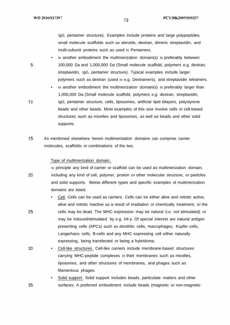
IgG, pentamer structure). Examples include proteins and large polypeptides,
small molecule scaffolds such as steroids, dextran, dimeric streptavidin, and
multi-subunit proteins such as used in Pentamers.
• In another embodiment the multimerization domain(s) is preferably between
100,000 Da and 1,000,000 Da (Small molecule scaffold, polymers e.g. dextran,
streptavidin, IgG, pentamer structure). Typical examples include larger
polymers such as dextran (used in e.g. Dextramers), and streptavidin tetramers.
• In another embodiment the multimerization domain(s) is preferably larger than
1,000,000 Da (Small molecule scaffold, polymers e.g. dextran, streptavidin,
IgG, pentamer structure, cells, liposomes, artificial lipid bilayers, polystyrene
beads and other beads. Most examples of this size involve cells or cell-based
structures such as micelles and liposomes, as well as beads and other solid
supports.
As mentioned elsewhere herein multimerisation domains can comprise carrier
molecules, scaffolds or combinations of the two.
Type of multimerization domain.
In principle any kind of carrier or scaffold can be used as multimerization domain,
including any kind of cell, polymer, protein or other molecular structure, or particles
and solid supports. Below different types and specific examples of multimerization
domains are listed.
• Cell. Cells can be used as carriers. Cells can be either alive and mitotic active,
alive and mitotic inactive as a result of irradiation or chemically treatment, or the
cells may be dead. The MHC expression may be natural (i.e. not stimulated) or
may be induced/stimulated by e.g. lnf-γ . Of special interest are natural antigen
presenting cells (APCs) such as dendritic cells, macrophages, Kupfer cells,
Langerhans cells, B-cells and any MHC expressing cell either naturally
expressing, being transfected or being a hybridoma.
• Cell-like structures . Cell-like carriers include membrane-based structures
carrying MHC-peptide complexes in their membranes such as micelles,
liposomes, and other structures of membranes, and phages such as
filamentous phages.
• Solid support . Solid support includes beads, particulate matters and other
surfaces. A preferred embodiment include beads (magnetic or non-magnetic

beads) that carry electrophilic groups e.g. divinyl sulfone activated
polysaccharide, polystyrene beads that have been functionalized with tosyl-
activated esters, magnetic polystyrene beads functionalized with tosyl-activated
esters), and where MHC complexes may be covalently immobilized to these by
reaction of nucleophiles comprised within the MHC complex with the
electrophiles of the beads. Beads may be made of sepharose, sephacryl,
polystyrene, agarose, polysaccharide, polycarbamate or any other kind of
beads that can be suspended in aqueous buffer.
Another embodiment includes surfaces, i.e. solid supports and particles
carrying immobilized MHC complexes on the surface. Of special interest are
wells of a microtiter plate or other plate formats, reagent tubes, glass slides or
other supports for use in microarray analysis, tubings or channels of micro
fluidic chambers or devices, Biacore chips and beads
Molecule . Multimerization domains may also be molecules or complexes of
molecules held together by non-covalent bonds. The molecules constituting the
multimerization domain can be small organic molecules or large polymers, and
may be flexible linear molecules or rigid, globular structures such as e.g.
proteins. Different kinds of molecules used in multimerization domains are
described below.
o Small organic molecules. Small organic molecules here includes
steroids, peptides, linear or cyclic structures, and aromatic or aliphatic
structures, and many others. The prototypical small organic scaffold is a
functionalized benzene ring, i.e. a benzene ring functionalized with a
number of reactive groups such as amines, to which a number of MHC
molecules may be covalently linked. However, the types of reactive
groups constituting the linker connecting the MHC complex and the
multimerization domain, as well as the type of scaffold structure, can be
chosen from a long list of chemical structures. A non-comprehensive list
of scaffold structures are listed below.
Typical scaffolds include aromatic structures, benzodiazepines,
hydantoins, piperazines, indoles, furans, thiazoles, steroids,
diketopiperazines, morpholines, tropanes, coumarines, qinolines,
pyrroles, oxazoles, amino acid precursors, cyclic or aromatic ring
structures, and many others.

Typical carriers include linear and branched polymers such as peptides,
polysaccharides, nucleic acids, and many others. Multimerization
domains based on small organic or polymer molecules thus include a
wealth of different structures, including small compact molecules, linear
structures, polymers, polypeptides, polyureas, polycarbamates, cyclic
structures, natural compound derivatives, alpha-, beta-, gamma-, and
omega-peptides, mono-, di- and tri-substituted peptides, L- and D-form
peptides, cyclohexane- and cyclopentane-backbone modified beta-
peptides, vinylogous polypeptides, glycopolypeptides, polyamides,
vinylogous sulfonamide peptide, Polysulfonamide-conjugated peptide (i.e.,
having prosthetic groups), Polyesters, Polysaccharides such as dextran
and aminodextran, polycarbamates, polycarbonates, polyureas, poly-
peptidylphosphonates, Azatides, peptoids (oligo N-substituted glycines),
Polyethers, ethoxyformacetal oligomers, poly-thioethers, polyethylene,
glycols (PEG), polyethylenes, polydisulfides, polyarylene sulfides,
Polynucleotides, PNAs, LNAs, Morpholinos, oligo pyrrolinone, polyoximes,
Polyimines, Polyethyleneimine, Polyacetates, Polystyrenes,
Polyacetylene, Polyvinyl, Lipids, Phospholipids, Glycolipids, polycycles,
(aliphatic), polycycles (aromatic), polyheterocycles, Proteoglycan,
Polysiloxanes, Polyisocyanides, Polyisocyanates, polymethacrylates,
Monofunctional, Difunctional, Trifunctional and Oligofunctional open-chain
hydrocarbons, Monofunctional, Difunctional, Trifunctional and
Oligofunctional Nonaromat Carbocycles, Monocyclic, Bicyclic, Tricyclic
and Polycyclic Hydrocarbons, Bridged Polycyclic Hydrocarbones,
Monofunctional, Difunctional, Trifunctional and Oligofunctional
Nonaromatic, Heterocycles, Monocyclic, Bicyclic, Tricyclic and Polycyclic
Heterocycles, bridged Polycyclic Heterocycles, Monofunctional,
Difunctional, Trifunctional and Oligofunctional Aromatic Carbocycles,
Monocyclic, Bicyclic, Tricyclic and Polycyclic Aromatic Carbocycles,
Monofunctional, Difunctional, Trifunctional and Oligofunctional Aromatic
Hetero-cycles. Monocyclic, Bicyclic, Tricyclic and Polycyclic Heterocycles.
Chelates, fullerenes, and any combination of the above and many others.
Biological polymers. Biological molecules here include peptides,
proteins (including antibodies, coiled-coil helices, streptavidin and many
others), nucleic acids such as DNA and RNA, and polysaccharides such

as dextran. The biological polymers may be reacted with MHC
complexes (e.g. a number of MHC complexes chemically coupled to e.g.
the amino groups of a protein), or may be linked through e.g. DNA
duplex formation between a carrier DNA molecule and a number of DNA
oligonucleotides each coupled to a MHC complex. Another type of
multimerization domain based on a biological polymer is the
streptavidin-based tetramer, where a streptavidin binds up to four
biotinylated MHC complexes, as described above (see Background of
the invention).
o Self-assembling multimeric structures. Several examples of commercial
MHC multimers exist where the multimer is formed through self-
assembling. Thus, the Pentamers are formed through formation of a
coiled-coil structure that holds together 5 MHC complexes in an
apparently planar structure. In a similar way, the Streptamers are based
on the Streptactin protein which oligomerizes to form a MHC multimer
comprising several MHC complexes (see Background of the invention).
In the following, alternative ways to make MHC multimers based on a molecule
multimerization domain are described. They involve one or more of the
abovementoned types of multimerization domains.
MHC dextramers can be made by coupling MHC complexes to dextran via a
streptavidin-biotin interaction. In principle, biotin-streptavdin can be replaced by any
dimerization domain, where one half of the dimerization domain is coupled to the MHC-
peptide complex and the other half is coupled to dextran. For example, an acidic helix
(one half of a coiled-coil dimer) is coupled or fused to MHC, and a basic helix (other
half of a coiled-coil dimmer) is coupled to dextran. Mixing the two results in MHC
binding to dextran by forming the acid/base coiled-coil structure.
Antibodies can be used as scaffolds by using their capacity to bind to a carefully
selected antigen found naturally or added as a tag to a part of the MHC molecule not
involved in peptide binding. For example, IgG and IgE will be able to bind two MHC
molecules, IgM having a pentameric structure will be able to bind 10 MHC molecules.
The antibodies can be full-length or truncated; a standard antibody-fragment includes
the Fab2 fragment.

Peptides involved in coiled-coil structures can act as scaffold by making stable dimeric,
trimeric, tetrameric and pentameric interactions. Examples hereof are the Fos-Jun
heterodimeric coiled coil, the E. coli homo-trimeric coiled-coil domain Lpp-56, the
engineered Trp-zipper protein forming a discrete, stable, α-helical pentamer in water at
physiological pH.
Further examples of suitable scaffolds, carriers and linkers are streptavidin (SA) and
avidin and derivatives thereof, biotin, immunoglobulins, antibodies (monoclonal,
polyclonal, and recombinant), antibody fragments and derivatives thereof, leucine
zipper domain of AP- 1 (jun and fos), hexa-his (metal chelate moiety), hexa-hat GST
(glutathione S-tranferase), glutathione, Calmodulin-binding peptide (CBP), Strep-tag,
Cellulose Binding Domain, Maltose Binding Protein, S-Peptide Tag, Chitin Binding Tag,
Immuno-reactive Epitopes, Epitope Tags, E2Tag, HA Epitope Tag, Myc Epitope, FLAG
Epitope, AU1 and AU5 Epitopes, GIu-GIu Epitope, KT3 Epitope, IRS Epitope, Btag
Epitope, Protein Kinase-C Epitope, VSV Epitope, lectins that mediate binding to a
diversity of compounds, including carbohydrates, lipids and proteins, e.g. Con A
{Canavalia ensiformis) or WGA (wheat germ agglutinin) and tetranectin or Protein A or
G (antibody affinity). Combinations of such binding entities are also comprised. Non-
limiting examples are streptavidin-biotin and jun-fos. In particular, when the MHC
molecule is tagged, the binding entity may be an "anti-tag". By "anti-tag" is meant an
antibody binding to the tag, or any other molecule capable of binding to such tag.
MHC complexes can be multimerized by other means than coupling or binding to a
multimerization domain. Thus, the multimerization domain may be formed during the
multimerization of MHCs. One such method is to extend the bound antigenic peptide
with dimerization domains. One end of the antigenic peptide is extended with
dimerization domain A (e.g. acidic helix, half of a coiled-coil dimer) and the other end is
extended with dimerization domain B (e.g. basic helix, other half of a coiled-coil dimer).
When MHC complexes are loaded/mixed with these extended peptides the following
multimer structure will be formed: A-MHC-BA-MHC-BA-MHC-B etc. The antigenic
peptides in the mixture can either be identical or a mixture of peptides with comparable
extended dimerization domains. Alternatively both ends of a peptide are extended with
the same dimerization domain A and another peptide (same amino acid sequence or a
different amino acid sequence) is extended with dimerization domain B. When MHC

o
and peptides are mixed the following structures are formed: A-MHC-AB-MHC-BA-
MHC-AB-MHC-B etc. Multimerization of MHC complexes by extension of peptides are
restricted to MHC I l molecules since the peptide binding groove of MHC I molecules is
typically closed in both ends thereby limiting the size of peptide that can be embedded
in the groove, and therefore preventing the peptide from extending out of the groove.
Another multimerization approach applicable to both MHC I and MHC I l complexes is
based on extension of N- and C-terminal of the MHC complex. For example the N-
terminal of the MHC complex is extended with dimerization domain A and the C-
terminal is extended with dimerization domain B. When MHC complexes are incubated
together they pair with each other and form multimers like: A-MHC-BA-MHC-BA-MHC-
BA-MHC-B etc. Alternatively the N-terminal and the C-terminal of a MHC complex are
both extended with dimerization domain A and the N-terminal and C-terminal of
another preparation of MHC complex (either the same or a different MHC) are
extended with dimerization domain B. When these two types of MHC complexes are
incubated together multimers will be formed: A-MHC-AB-MHC-BA-MHC-AB-MHC-B
etc.
In all the above-described examples the extension can be either chemically coupled to
the peptide/MHC complex or introduced as extension by gene fusion.
Dimerization domain AB can be any molecule pair able to bind to each other, such as
acid/base coiled-coil helices, antibody-antigen, DNA-DNA, PNA-PNA, DNA-PNA, DNA-
RNA, LNA-DNA, leucine zipper e.g. Fos/Jun, streptavidin-biotin and other molecule
pairs as described elsewhere herein.
Linker molecules.
A number of MHC complexes associate with a multimerization domain to form a MHC
multimer. The attachment of MHC complexes to the multimerization domain may
involve covalent or non-covalent linkers, and may involve small reactive groups as well
as large protein-protein interactions.
The coupling of multimerization domains and MHC complexes involve the association
of an entity X (attached to or part of the multimerization domain) and an entity Y
(attached to or part of the MHC complex). Thus, the linker that connects the
multimerization domain and the MHC complex comprises an XY portion.

Covalent linker. The XY linkage can be covalent, in which case X and Y are
reactive groups. In this case, X can be a nucleophilic group (such as -NH 2, -
OH, -SH, -NH-NH2) , and Y an electrophilic group (such as CHO, COOH, CO)
that react to form a covalent bond XY; or Y can be a nucleophilic group and X
an electrophilic group that react to form a covalent bond XY. Other possibilities
exist, e.g either of the reactive groups can be a radical, capable of reacting with
the other reactive group. A number of reactive groups X and Y, and the bonds
that are formed upon reaction of X and Y, are shown in figure 5 .
X and Y can be reactive groups naturally comprised within the multimerization
domain and/or the MHC complex, or they can be artificially added reactive
groups. Thus, linkers containing reactive groups can be linked to either of the
multimerization domain and MHC complex; subsequently the introduced
reactive group(s) can be used to covalently link the multimerization domain and
MHC complex.
Example natural reactive groups of MHC complexes include amino acid side
chains comprising -NH2, -OH, -SH, and -NH-. Example natural reactive groups
of multimerization domains include hydroxyls of polysaccharides such as
dextrans, but also include amino acid side chains comprising -NH2, -OH, -SH,
and -NH- of polypeptides, when the polypeptide is used as a multimerization
domain. In some MHC multimers, one of the polypeptides of the MHC complex
(i.e. the β2M, heavy chain or the antigenic peptide) is linked by a protein fusion
to the multimerization domain. Thus, during the translation of the fusion protein,
an acyl group (reactive group X or Y) and an amino group (reactive group Y or
X) react to form an amide bond. Example MHC multimers where the bond
between the multimerization domain and the MHC complex is covalent and
results from reaction between natural reactive groups, include MHC-pentamers
(described in US patent 2004209295) and MHC-dimers, where the linkage
between multimerization domain and MHC complex is in both cases generated
during the translation of the fusion protein.
Example artificial reactive groups include reactive groups that are attached to
the multimerization domain or MHC complex, through association of a linker
molecule comprising the reactive group. The activation of dextran by reaction of
the dextran hydroxyls with divinyl sulfone, introduces a reactive vinyl group that
can react with e.g. amines of the MHC complex, to form an amine that now links
the multimerization domain (the dextran polymer) and the MHC complex. An

o
alternative activation of the dextran multimerization domain involves a multistep
reaction that results in the decoration of the dextran with maleimide groups, as
described in the patent Siiman et al. US 6,387,622. In this approach, the amino
groups of MHC complexes are converted to -SH groups, capable of reacting
with the maleimide groups of the activated dextran. Thus, in the latter example,
both the reactive group of the multimerization domain (the maleimide) and the
reactive group of the MHC complex (the thiol) are artificially introduced.
Sometimes activating reagents are used in order to make the reactive groups
more reactive. For example, acids such as glutamate or aspartate can be
converted to activated esters by addition of e.g. carbodiimid and NHS or
nitrophenol, or by converting the acid moiety to a tosyl-activated ester. The
activated ester reacts efficiently with a nucleophile such as -NH 2, -SH, -OH, etc.
For the purpose of this invention, the multimerization domains (including small
organic scaffold molecules, proteins, protein complexes, polymers, beads,
liposomes, micelles, cells) that form a covalent bond with the MHC complexes
can be divided into separate groups, depending on the nature of the reactive
group that the multimerization domain contains. One group comprise
multimerization domains that carry nucleophilic groups (e.g. -NH 2, -OH, -SH, -
CN, -NH-NH2) , exemplified by polysaccharides, polypeptides containing e.g.
lysine, serine, and cysteine; another group of multimerization domains carry
electrophilic groups (e.g. -COOH, -CHO, -CO, NHS-ester, tosyl-activated ester,
and other activated esters, acid-anhydrides), exemplified by polypeptides
containing e.g. glutamate and aspartate, or vinyl sulfone activated dextran; yet
another group of multimerization domains carry radicals or conjugated double
bonds.
The multimerization domains appropriate for this invention thus include those
that contain any of the reactive groups shown in Figure 5 or that can react with
other reactive groups to form the bonds shown in Figure 5 .
Likewise, MHC complexes can be divided into separate groups, depending on
the nature of the reactive group comprised within the MHC complex. One group
comprise MHCs that carry nucleophilic groups (e.g. -NH 2, -OH, -SH, -CN, -NH-

NH2) , e.g. lysine, serine, and cysteine; another group of MHCs carry
electrophilic groups (e.g. -COOH, -CHO, -CO, NHS-ester, tosyl-activated ester,
and other activated esters, acid-anhydrides), exemplified by e.g. glutamate and
aspartate; yet another group of MHCs carry radicals or conjugated double
bonds.
The reactive groups of the MHC complex are either carried by the amino acids
of the MHC-peptide complex (and may be comprised by any of the peptides of
the MHC-peptide complex, including the antigenic peptide), or alternatively, the
reactive group of the MHC complex has been introduced by covalent or non-
covalent attachment of a molecule containing the appropriate reactive group.
Preferred reactive groups in this regard include -CSO2OH, phenylchloride, -SH,
-SS, aldehydes, hydroxyls, isocyanate, thiols, amines, esters, thioesters,
carboxylic acids, triple bonds, double bonds, ethers, acid chlorides, phosphates,
imidazoles, halogenated aromatic rings, any precursors thereof, or any
protected reactive groups, and many others. Example pairs of reactive groups,
and the resulting bonds formed, are shown in figure 5 .
Reactions that may be employed include acylation (formation of amide,
pyrazolone, isoxazolone, pyrimidine, comarine, quinolinon, phthalhydrazide,
diketopiperazine, benzodiazepinone, and hydantoin), alkylation, vinylation,
disulfide formation, Wittig reaction, Horner-Wittig-Emmans reaction, arylation
(formation of biaryl or vinylarene), condensation reactions, cycloadditions
((2+4), (3+2)), addition to carbon-carbon multiplebonds, cycloaddition to
multiple bonds, addition to carbon-hetero multiple bonds, nucleophilic aromatic
substitution, transition metal catalyzed reactions, and may involve formation of
ethers, thioethers, secondary amines, tertiary amines, beta-hydroxy ethers,
beta-hydroxy thioethers, beta-hydroxy amines, beta-amino ethers, amides,
thioamides, oximes, sulfonamides, di- and tri-functional compounds, substituted
aromatic compounds, vinyl substituted aromatic compounds, alkyn substituted
aromatic compounds, biaryl compounds, hydrazines, hydroxylamine ethers,
substituted cycloalkenes, substituted cyclodienes, substituted 1, 2 , 3 triazoles,
substituted cycloalkenes, beta-hydroxy ketones, beta-hydroxy aldehydes, vinyl
ketones, vinyl aldehydes, substituted alkenes, substituted alkenes, substituted
amines, and many others.

O-.
MHC dextramers can be made by covalent coupling of MHC complexes to the
dextran backbone, e.g. by chemical coupling of MHC complexes to dextran
backbones. The MHC complexes can be coupled through either heavy chain or
β2-microglobulin if the MHC complexes are MHC I or through α-chain or β-
chain if the MHC complexes are MHC II. MHC complexes can be coupled as
folded complexes comprising heavy chain/beta2microglobulin or α-chain/β-
chain or either combination together with peptide in the peptide-binding cleft.
Alternatively either of the protein chains can be coupled to dextran and then
folded in vitro together with the other chain of the MHC complex not coupled to
dextran and together with peptide. Direct coupling of MHC complexes to
dextran multimerization domain can be via an amino group or via a sulphide
group. Either group can be a natural component of the MHC complex or
attached to the MHC complex chemically. Alternatively, a cysteine may be
introduced into the genes of either chain of the MHC complex.
Another way to covalently link MHC complexes to dextran multimerization
domains is to use the antigenic peptide as a linker between MHC and dextran.
Linker containing antigenic peptide at one end is coupled to dextran. Antigenic
peptide here means a peptide able to bind MHC complexes in the peptide-
binding cleft. As an example, 10 or more antigenic peptides may be coupled to
one dextran molecule. When MHC complexes are added to such peptide-
dextran construct the MHC complexes will bind the antigenic peptides and
thereby MHC-peptide complexes are displayed around the dextran
multimerization domain. The antigenic peptides can be identical or different
from each other. Similarly MHC complexes can be either identical or different
from each other as long as they are capable of binding one or more of the
peptides on the dextran multimerization domain.
Non-covalent linker. The linker that connects the multimerization domain and
the MHC complex comprises an XY portion. Above different kinds of covalent
linkages XY were described. However, the XY linkage can also be non-
covalent.
Non-covalent XY linkages can comprise natural dimerization pairs such as
antigen-antibody pairs, DNA-DNA interactions, or can include natural

O
interactions between small molecules and proteins, e.g. between biotin and
streptavidin. Artificial XY examples include XY pairs such as His6 tag (X)
interacting with Ni-NTA (Y) and PNA-PNA interations.
Protein-protein interactions. The non-covalent linker may comprise a complex
of two or more polypeptides or proteins, held together by non-covalent
interactions. Example polypeptides and proteins belonging to this group include
Fos/Jun, Acid/Base coiled coil structure, antibody/antigen (where the antigen is
a peptide), and many others.
A preferred embodiment involving non-covalent interactions between
polypeptides and/or proteins are represented by the Pentamer structure
described in US patent 2004209295.
Another preferred embodiment involves the use of antibodies, with affinity for
the surface of MHC opposite to the peptide-binding groove. Thus, an anti-MHC
antibody, with its two binding site, will bind two MHC complexes and in this way
generate a bivalent MHC multimer. In addition, the antibody can stabilize the
MHC complex through the binding interactions. This is particularly relevant for
MHC class I l complexes, as these are less stable than class I MHC complexes.
Polynucleotide-polynucleotide interactions. The non-covalent linker may
comprise nucleotides that interact non-covalently. Example interactions include
PNA/PNA, DNA/DNA, RNA/RNA, LNA/DNA, and any other nucleic acid duplex
structure, and any combination of such natural and unnatural polynucleotides
such as DNA/PNA, RNA/DNA, and PNA/LNA.
Protein-small molecule interactions . The non-covalent linker may comprise a
macromolecule (e.g. protein, polynucleotide) and a small molecule ligand of the
macromolecule. The interaction may be natural (i.e., found in Nature, such as
the Streptavidin/biotin interaction) or non-natural (e.g. His-tag peptide/Ni-NTA
interaction). Example interactions include Streptavidin/biotin and anti-biotin
antibody/biotin.

Combinations - non-covalent linker molecules. Other combinations of proteins,
polynucleotides, small organic molecules, and other molecules, may be used to
link the MHC to the multimerization domain. These other combinations include
protein-DNA interactions (e.g. DNA binding protein such as the gene regulatory
protein CRP interacting with its DNA recognition sequence), RNA aptamer-
protein interactions (e.g. RNA aptamer specific for growth hormone interacting
with growth hormone)
Synthetic molecule-synthetic molecule interaction. The non-covalent linker may
comprise a complex of two or more organic molecules, held together by non-
covalent interactions. Example interactions are two chelate molecules binding
to the same metal ion (e.g. EDTA-Ni++-NTA), or a short polyhistidine peptide
(e.g. HiS6) bound to NTA-Ni++ .
In another preferred embodiment the multimerization domain is a bead. The bead is
covalently or non-covalently coated with MHC multimers or single MHC complexes,
through non-cleavable or cleavable linkers. As an example, the bead can be coated
with streptavidin monomers, which in turn are associated with biotinylated MHC
complexes; or the bead can be coated with streptavidin tetramers, each of which are
associated with 0 , 1, 2 , 3 , or 4 biotinylated MHC complexes; or the bead can be coated
with MHC-dextramers where e.g. the reactive groups of the MHC-dextramer (e.g. the
divinyl sulfone-activated dextran backbone) has reacted with nucleophilic groups on the
bead, to form a covalent linkage between the dextran of the dextramer and the beads.
In another preferred embodiment, the MHC multimers described above (e.g. where the
multimerization domain is a bead) further contains a flexible or rigid, and water soluble,
linker that allows for the immobilized MHC complexes to interact efficiently with cells,
such as T-cells with affinity for the MHC complexes. In yet another embodiment, the
linker is cleavable, allowing for release of the MHC complexes from the bead. If T-cells
have been immobilized, by binding to the MHC complexes, the T-cells can very gently
be released by cleavage of this cleavable linker. Appropriate cleavable linkers are
shown in Figure 6. Most preferably, the linker is cleaved at physiological conditions,
allowing for the integrity of the isolated cells.

Further examples of linker molecules that may be employed in the present invention
include Calmodulin-binding peptide (CBP), 6xHIS, Protein A, Protein G, biotin, Avidine,
Streptavidine, Strep-tag, Cellulose Binding Domain, Maltose Binding Protein, S-Peptide
Tag, Chitin Binding Tag, Immuno-reactive Epitopes, Epitope Tags, GST tagged
proteins, E2Tag, HA Epitope Tag, Myc Epitope, FLAG Epitope, AU1 and AU5
Epitopes, GIu-GIu Epitope, KT3 Epitope, IRS Epitope, Btag Epitope, Protein Kinase-C
Epitope, VSV Epitope.
The list of dimerization- and multimerization domains, described elsewhere in this
document, define alternative non-covalent linkers between the multimerization domain
and the MHC complex.
The abovementioned dimerization- and multimerization domains represent specific
binding interactions. Another type of non-covalent interactions involves the non-specific
adsorption of e.g. proteins onto surfaces. As an example, the non-covalent adsorption
of proteins onto glass beads represents this class of XY interactions. Likewise, the
interaction of MHC complexes (comprising full-length polypeptide chains, including the
transmembrane portion) with the cell membrane of for example dendritic cells is an
example of a non-covalent, primarily non-specific XY interaction.
In some of the abovementioned embodiments, several multimerization domains (e.g.
streptavidin tetramers bound to biotinylated MHC complexes) are linked to another
multimerization domain (e.g. the bead). For the purpose of this invention we shall call
both the smaller and the bigger multimerization domain, as well as the combined
multimerization domain, for multimerization domain
Additional features of product
Additional components may be coupled to carrier or added as individual components
not coupled to carrier
Attachment of biologically active molecules to MHC multimers
Engagement of MHC complex to the specific T cell receptor leads to a signaling
cascade in the T cell. However, T-cells normally respond to a single signal stimulus by
going into apoptosis. T cells needs a second signal in order to become activated and

o
start development into a specific activation state e.g. become an active cytotoxic T cell,
helper T cell or regulatory T cell.
It is to be understood that the MHC multimer of the invention may further comprise one
or more additional substituents. The definition of the terms "one or more", "a plurality",
"a", "an", and "the" also apply here. Such biologically active molecules may be attached
to the construct in order to affect the characteristics of the constructs, e.g. with respect
to binding properties, effects, MHC molecule specificities, solubility, stability, or
detectability. For instance, spacing could be provided between the MHC complexes,
one or both chromophores of a Fluorescence Resonance Energy Transfer (FRET)
donor/acceptor pair could be inserted, functional groups could be attached, or groups
having a biological activity could be attached.
MHC multimers can be covalently or non-covalently associated with various molecules:
having adjuvant effects; being immune targets e.g. antigens; having biological activity
e.g. enzymes, regulators of receptor activity, receptor ligands, immune potentiators,
drugs, toxins, co-receptors, proteins and peptides in general; sugar moieties; lipid
groups; nucleic acids including siRNA; nano particles; small molecules. In the following
these molecules are collectively called biologically active molecules. Such molecules
can be attached to the MHC multimer using the same principles as those described for
attachment of MHC complexes to multimerisation domains as described elsewhere
herein. In brief, attachment can be done by chemical reactions between reactive
groups on the biologically active molecule and reactive groups of the multimerisation
domain and/or between reactive groups on the biologically active molecule and
reactive groups of the MHC-peptide complex. Alternatively, attachment is done by non-
covalent interaction between part of the multimerisation domain and part of the
biological active molecule or between part of the MHC-peptide complex and part of the
biological active molecule. In both covalent and non-covalent attachment of the
biologically molecule to the multimerisation domain a linker molecule can connect the
two. The linker molecule can be covalent or non-covalent attached to both molecules.
Examples of linker molecules are described elsewhere herein. Some of the MHCmer
structures better allows these kind of modifications than others.
Biological active molecules can be attached repetitively aiding to recognition by and
stimulation of the innate immune system via Toll or other receptors.

o
MHC multimers carrying one or more additional groups can be used as therapeutic or
vaccine reagents.
In particular, the biologically active molecule may be selected from
proteins such as MHC Class l-like proteins like MIC A , MIC B, CD1d, HLA E, HLA F,
HLA G, HLA H, ULBP-1 , ULBP-2, and ULBP-3,
co-stimulatory molecules such as CD2, CD3, CD4, CD5, CD8, CD9, CD27, CD28,
CD30, CD69, CD1 34 (OX40), CD137 (4-1 BB), CD147, CDw1 50 (SLAM), CD1 52
(CTLA-4), CD153 (CD30L), CD40L (CD154), NKG2D, ICOS, HVEM, HLA Class II, PD-
1, Fas (CD95), FasL expressed on T and/or NK cells, CD40, CD48, CD58, CD70,
CD72, B7.1 (CD80), B7.2 (CD86), B7RP-1 , B7-H3, PD-L1 , PD-L2, CD134L, CD137L,
ICOSL, LIGHT expressed on APC and/or tumour cells,
cell modulating molecules such as CD1 6 , NKp30, NKp44, NKp46, NKpδO, 2B4, KIR,
LIR, CD94/NKG2A, CD94/NKG2C expressed on NK cells, IFN-alpha, IFN-beta, IFN-
gamma, IL-1 , IL-2, IL-3, IL-4, IL-6, IL-7, IL-8, IL-10, IL-1 1, IL-12, IL-15, CSFs (colony-
stimulating factors), vitamin D3, IL-2 toxins, cyclosporin, FK-506, rapamycin, TGF-beta,
clotrimazole, nitrendipine, and charybdotoxin,
accessory molecules such as LFA-1 , CD1 1a/1 8 , CD54 (ICAM-1 ) , CD1 06 (VCAM), and
CD49a,b,c,d,e,f/CD29 (VLA-4),
adhesion molecules such as ICAM-1 , ICAM-2, GlyCAM-1 , CD34, anti-LFA-1 , anti-
CD44, anti-beta7, chemokines, CXCR4, CCR5, anti-selectin L, anti-selectin E, and
anti-selectin P,
toxic molecules selected from toxins, enzymes, antibodies, radioisotopes, chemi-
luminescent substances, bioluminescent substances, polymers, metal particles, and
haptens, such as cyclophosphamide, methrotrexate, Azathioprine, mizoribine, 15-
deoxuspergualin, neomycin, staurosporine, genestein, herbimycin A, Pseudomonas
exotoxin A , saporin, Rituxan, Ricin, gemtuzumab ozogamicin, Shiga toxin, heavy
metals like inorganic and organic mercurials, and FN1 8-CRM9, radioisotopes such as

OO
incorporated isotopes of iodide, cobalt, selenium, tritium, and phosphor, and haptens
such as DNP, and digoxiginin,
and combinations of any of the foregoing, as well as antibodies (monoclonal,
polyclonal, and recombinant) to the foregoing, where relevant. Antibody derivatives or
fragments thereof may also be used.
Design and generation of product to be used for immune monitoring, diagnosis,
therapy or vaccination
The product of the present invention may be used forjmmune monitoring, diagnosis,
therapy and/or vaccination. The generation of product may follow some or all of the
following general steps.
1. Design of antigenic peptides
2 . Choice of MHC allele
3 . Generation of product
4 . Validation and optimization of product
Production of a MHC multimer diagnostic or immune monitoring reagent may
follow some or all of the following steps.
1. Identify disease of interest. Most relevant diseases in this regard are infectious-,
cancer-, auto immune-, transplantation-, or immuno-suppression- related
diseases.
2 . Identify relevant protein antigen(s). This may be individual proteins, a group of
proteins from a given tissue or subgroups of proteins from an organism.
3 . Identify the protein sequence. Amino acid sequences can be directly found in
databases or deduced from gene- or mRNA sequence e.g. using the following
link http://www.ncbi.nlm.nih.gov/Genbank/index.html. If not in databases
relevant proteins or genes encoding relevant proteins may be isolated and
sequenced. In some cases only DNA sequences will be available without
knowing which part of the sequence is protein coding. Then the DNA sequence
is translated into amino acid sequence in all reading frames.
4 . Choose MHC allele(s). Decide on needed MHC allele population coverage. If a
broad coverage of a given population is needed (i.e. when generally applicable
reagents are sought) the most frequently expressed MHC alleles by the

population of interest may be chosen e.g. using the database
http://www.allelefrequencies.net/test/default1 .asp or
http://epitope.liai.org:8080/tools/population/iedb_input.
In case of personalized medicine the patient is tissue typed (HLA type) and then
MHC alleles may be selected according to that.
5 . Run the general peptide epitope generator program described elsewhere herein
on all selected amino acid sequences from step 3 , thereby generating all
possible epitopes of defined length (8'-, 9'-, 10'-, 11'-, 13-, 14'-, 15'-, and/or 16'-
mers).
6 . If searching for broadly applicable epitope sequences, a good alternative to
step 5 is to run the "intelligent" peptide epitope prediction programs on the
selected amino acid sequences of step 3 using the selected MHC alleles from
step 4 e.g. using epitope prediction programs like http://www.syfpeithi.de/,
http://www.cbs.dtu.dk/services/NetMHC/, and
http://www.cbs.dtu.dk/services/NetMHCI I/.
This step can also be used supplementary to step 5 by running selected or all
epitopes from the general peptide epitope generator program through one or
more of the intelligent peptide epitope prediction programs.
7 . If searching for broadly applicable epitope sequences, one may choose to
select the epitopes with highest binding score, or the most likely proteolytic
products of the species in question, for the chosen MHC alleles and run them
through the BLAST program (http://www.ncbi.nlm.nih.gov/blast/Blast.cgi) to
validate the uniqueness of the peptides. If the peptide sequences are present in
other species, evaluate the potential risk of disease states caused by the non-
relevant species in relation to causing false positive results. If considered being
a potential problem for evaluating the future analysis outcome, leave out the
peptide. Preferably, choose unique peptide sequences only present in the
selected protein.
8 . Produce selected peptides as described elsewhere herein, e.g. by standard
organic synthesis, and optionally test for binding to the desired MHC alleles by
e.g in vitro folding, peptide exchange of already preloaded MHC complexes or
another method able to test for peptide binding to MHC I or I l molecules.
9 . Generate desired MHC multimer by covalently or non-covalently attaching
MHC-peptide complex(es) to multimerization domain, and optionally attach a
fluorophore to the MHC multimer, as described elsewhere herein. Optionally,

test efficacy in detecting specific T-cells using e.g. the methods described in the
section "Detection".
The MHC multimer reagents may be used in a diagnostic procedure or kit for
testing patient and control samples e.g. by flow cytometry, immune
histochemistry, Elispot or other methods as described herein.
Production of a MHC multimer therapeutic reagent may follow some or all of the
following steps.
1. As step 1 - 8 above for diagnostic reagent.
9 . Select additional molecules (e.g. biologically active molecules, toxins) to attach
to the MHC multimer as described elsewhere herein. The additional molecules
can have different functionalities as e.g. adjuvants, specific activators, toxins
etc.
10. Test the therapeutic reagent following general guidelines
11. Use for therapy
Processes involving MHC multimers
The present invention relates to methods for detecting the presence of MHC
recognising cells in a sample comprising the steps of
(a) providing a sample suspected of comprising MHC recognising cells,
(b) contacting the sample with a MHC multimer as defined above, and
(c) determining any binding of the MHC multimer.
Binding indicates the presence of MHC recognising cells.
Such methods are a powerful tool in diagnosing various diseases. Establishing a
diagnosis is important in several ways. A diagnosis provides information about the
disease, thus the patient can be offered a suitable treatment regime. Also, establishing
a more specific diagnosis may give important information about a subtype of a disease
for which a particular treatment will be beneficial (i.e. various subtypes of diseases may
involve display of different peptides which are recognised by MHC recognising cells,
and thus treatment can be targeted effectively against a particular subtype). In this
way, it may also be possible to gain information about aberrant cells, which emerge
through the progress of the disease or condition, or to investigate whether and how T-
cell specificity is affected. The binding of the MHC multimer makes possible these

options, since the binding is indicative for the presence of the MHC recognising cells in
the sample, and accordingly the presence of MHC multimers displaying the peptide.
The present invention also relates to methods for monitoring MHC recognising cells
comprising the steps of
(a) providing a sample suspected of comprising MHC recognising cells,
(b) contacting the sample with a MHC complex as defined above, and
(c) determining any binding of the MHC multimer, thereby monitoring MHC recognising
cells.
Such methods are a powerful tool in monitoring the progress of a disease, e.g. to
closely follow the effect of a treatment. The method can i.a. be used to manage or
control the disease in a better way, to ensure the patient receives the optimum
treatment regime, to adjust the treatment, to confirm remission or recurrence, and to
ensure the patient is not treated with a medicament which does not cure or alleviate the
disease. In this way, it may also be possible to monitor aberrant cells, which emerge
through the progress of the disease or condition, or to investigate whether and how T-
cell specificity is affected during treatment. The binding of the MHC multimer makes
possible these options, since the binding is indicative for the presence of the MHC
recognising cells in the sample, and accordingly the presence of MHC multimers
displaying the peptide.
The present invention also relates to methods for establishing a prognosis of a disease
involving MHC recognising cells comprising the steps of
(a) providing a sample suspected of comprising MHC recognising cells,
(b) contacting the sample with a MHC multimer as defined above, and
(c) determining any binding of the MHC multimer, thereby establishing a prognosis of a
disease involving MHC recognising cells.
Such methods are a valuable tool in order to manage diseases, i.a. to ensure the
patient is not treated without effect, to ensure the disease is treated in the optimum
way, and to predict the chances of survival or cure. In this way, it may also be possible
to gain information about aberrant cells, which emerge through the progress of the
disease or condition, or to investigate whether and how T-cell specificity is affected,
thereby being able to establish a prognosis. The binding of the MHC multimer makes

possible these options, since the binding is indicative for the presence of the MHC
recognising cells in the sample, and accordingly the presence of MHC complexs
displaying the peptide.
The present invention also relates to methods for determining the status of a disease
involving MHC recognising cells comprising the steps of
(a) providing a sample suspected of comprising MHC recognising cells,
(b) contacting the sample with a MHC complex as defined above, and
(c) determining any binding of the MHC complex, thereby determining the status of a
disease involving MHC recognising cells.
Such methods are a valuable tool in managing and controlling various diseases. A
disease could, e.g. change from one stage to another, and thus it is important to be
able to determine the disease status. In this way, it may also be possible to gain
information about aberrant cells which emerge through the progress of the disease or
condition, or to investigate whether and how T-cell specificity is affected, thereby
determining the status of a disease or condition. The binding of the MHC complex
makes possible these options, since the binding is indicative for the presence of the
MHC recognising cells in the sample, and accordingly the presence of MHC complexs
displaying the peptide.
The present invention also relates to methods for the diagnosis of a disease involving
MHC recognising cells comprising the steps of
(a) providing a sample suspected of comprising MHC recognising cells,
(b) contacting the sample with a MHC multimer as defined above, and
(c) determining any binding of the MHC multimer, thereby diagnosing a disease
involving MHC recognising cells.
Such diagnostic methods are a powerful tool in the diagnosis of various diseases.
Establishing a diagnosis is important in several ways. A diagnosis gives information
about the disease, thus the patient can be offered a suitable treatment regime. Also,
establishing a more specific diagnosis may give important information about a subtype
of a disease for which a particular treatment will be beneficial (i.e. various subtypes of
diseases may involve display of different peptides which are recognised by MHC
recognising cells, and thus treatment can be targeted effectively against a particular

subtype). Valuable information may also be obtained about aberrant cells emerging
through the progress of the disease or condition as well as whether and how T-cell
specificity is affected. The binding of the MHC multimer makes possible these options,
since the binding is indicative for the presence of the MHC recognising cells in the
sample, and accordingly the presence of MHC multimers displaying the peptide.
The present invention also relates to methods of correlating cellular morphology with
the presence of MHC recognising cells in a sample comprising the steps of
(a) providing a sample suspected of comprising MHC recognising cells,
(b) contacting the sample with a MHC multimer as defined above, and
(c) determining any binding of the MHC multimer, thereby correlating the binding of the
MHC multimer with the cellular morphology.
Such methods are especially valuable as applied in the field of histochemical methods,
as the binding pattern and distribution of the MHC multimers can be observed directly.
In such methods, the sample is treated so as to preserve the morphology of the
individual cells of the sample. The information gained is important i.a. in diagnostic
procedures as sites affected can be observed directly.
The present invention also relates to methods for determining the effectiveness of a
medicament against a disease involving MHC recognising cells comprising the steps of
(a) providing a sample from a subject receiving treatment with a medicament,
(b) contacting the sample with a as defined herein, and
(c) determining any binding of the MHC multimer, thereby determining the
effectiveness of the medicament.
Such methods are a valuable tool in several ways. The methods may be used to
determine whether a treatment is effectively combating the disease. The method may
also provide information about aberrant cells which emerge through the progress of the
disease or condition as well as whether and how T-cell specificity is affected, thereby
providing information of the effectiveness of a medicament in question. The binding of
the MHC multimer makes possible these options, since the binding is indicative for the
presence of the MHC recognising cells in the sample, and accordingly the presence of
MHC multimers displaying the peptide.

The present invention also relates to methods for manipulating MHC recognising cells
populations comprising the steps of
(a) providing a sample comprising MHC recognising cells,
(b) contacting the sample with a MHC multimer immobilised onto a solid support as
defined above,
(c) isolating the relevant MHC recognising cells, and
(d) expanding such cells to a clinically relevant number, with or without further
manipulation.
Such ex vivo methods are a powerful tool to generate antigen-specific, long-lived
human effector T-cell populations that, when re-introduced to the subject, enable killing
of target cells and has a great potential for use in immunotherapy applications against
various types of cancer and infectious diseases.
As used everywhere herein, the term "MHC recognising cells" are intended to mean
such which are able to recognise and bind to MHC multimers. The intended meaning of
"MHC multimers" is given above. Such MHC recognising cells may also be called MHC
recognising cell clones, target cells, target MHC recognising cells, target MHC
molecule recognising cells, MHC molecule receptors, MHC receptors, MHC peptide
specific receptors, or peptide-specific cells. The term "MHC recognising cells" is
intended to include all subsets of normal, abnormal and defect cells, which recognise
and bind to the MHC molecule. Actually, it is the receptor on the MHC recognising cell
that binds to the MHC molecule.
As described above, in diseases and various conditions, peptides are displayed by
means of MHC multimers, which are recognised by the immune system, and cells
targeting such MHC multimers are produced (MHC recognising cells). Thus, the
presence of such MHC protein recognising cells is a direct indication of the presence of
MHC multimers displaying the peptides recognised by the MHC protein recognising
cells. The peptides displayed are indicative and may involved in various diseases and
conditions.

For instance, such MHC recognising cells may be involved in diseases of inflammatory,
auto-immune, allergic, viral, cancerous, infectious, allo- or xenogene (graft versus host
and host versus graft) origin.
The MHC multimers of the present invention have numerous uses and are a valuable
and powerful tool e.g. in the fields of therapy, diagnosis, prognosis, monitoring,
stratification, and determining the status of diseases or conditions. Thus, the MHC
multimers may be applied in the various methods involving the detection of MHC
recognising cells.
Furthermore, the present invention relates to compositions comprising the MHC
multimers in a solubilising medium. The present invention also relates to compositions
comprising the MHC multimers immobilised onto a solid or semi-solid support.
The MHC multimers can be used in a number of applications, including analyses such
as flow cytometry, immunohistochemistry (IHC), and ELISA-like analyses, and can be
used for diagnostic, prognostic or therapeutic purposes including autologous cancer
therapy or vaccines such as CMV or HIV vaccine or cancer vaccine.
The MHC multimers are very suitable as detection systems. Thus, the present
invention relates to the use of the MHC multimers as defined herein as detection
systems.
In another aspect, the present invention relates to the general use of MHC peptide
complexes and multimers of such MHC peptide complexes in various methods. These
methods include therapeutic methods, diagnostic methods, prognostic methods,
methods for determining the progress and status of a disease or condition, and
methods for the stratification of a patient.
The MHC multimers of the present invention are also of value in testing the expected
efficacy of medicaments against or for the treatment of various diseases. Thus, the
present invention relates to methods of testing the effect of medicaments or treatments,
the methods comprising detecting the binding of the MHC multimers to MHC
recognising cells and establishing the effectiveness of the medicament or the treatment
in question based on the specificity of the MHC recognising cells.

As mentioned above, the present invention also relates generally to the field of therapy.
Thus, the present invention relates per se to the MHC multimer as defined herein for
use as medicaments, and to the MHC multimers for use in in vivo and ex vivo therapy.
The present invention relates to therapeutic compositions comprising as active
ingredients the MHC multimers as defined herein.
An important aspect of the present invention is therapeutic compositions comprising as
active ingredients effective amounts of MHC recognising cells obtained using the MHC
multimers as defined herein to isolate relevant MHC recognising cells, and expanding
such cells to a clinically relevant number.
The present invention further relates to methods for treating, preventing or alleviating
diseases, methods for inducing anergy of cells, as well as to methods for up-regulating,
down-regulating, modulating, stimulating, inhibiting, restoring, enhancing and/or
otherwise manipulating immune responses.
The invention also relates to methods for obtaining MHC recognising cells by using the
MHC multimers as described herein.
Also encompassed by the present invention are methods for preparing the therapeutic
compositions of the invention.
The present invention is also directed to generating MHC multimers for detecting and
analysing receptors on MHC recognising cells, such as epitope specific T-cell clones or
other immune competent effector cells.
It is a further object of the present invention to provide new and powerful strategies for
the development of curative vaccines. This in turn will improve the possibilities for
directed and efficient immune manipulations against diseases caused by tumour
genesis or infection by pathogenic agent like viruses and bacteria. HIV is an important
example. The ability to generate and optionally attach recombinant MHC multimers to
multimerization domains, such as scaffolds and/or carrier molecules, will enable the
development of a novel analytical and therapeutical tool for monitoring immune

responses and contribute to a rational platform for novel therapy and "vaccine"
applications.
Therapeutic compositions (e.g. "therapeutical vaccines") that stimulate specific T-cell
proliferation by peptide-specific stimulation is indeed a possibility within the present
invention. Thus, quantitative analysis and ligand-based detection of specific T-cells that
proliferate by the peptide specific stimulation should be performed simultaneously to
monitoring the generated response.
Application of MHC multimers in immune monitoring, diagnostics, therapy,
vaccine
MHC multimers as described herein can be used to identify and isolate specific T cells
in a wide array of applications. In principle all kind of samples possessing T cells can
be analyzed with MHC multimers.
MHC multimers detect antigen-specific T cells of the various T cell subsets. T cells are
pivotal for mounting an adaptive immune response. It is therefore of importance to be
able to measure the number of specific T cells when performing a monitoring of a given
immune response. Typically, the adaptive immune response is monitored by measuring
the specific antibody response, which is only one of the effector arms of the immune
system. This can lead to miss-interpretation of the actual clinical immune status.
In many cases intruders of the organism can hide away inside the cells, which often
does not provoke a humoral response. In other cases, e.g. in the case of certain
viruses the intruder mutates fast, particularly in the genes encoding the proteins that
are targets for the humoral response. Examples include the influenza and HIV viruses.
The high rate of mutagenesis renders the humoral response unable to cope with the
infection. In these cases the immune system relies on the cellular immune response.
When developing vaccines against such targets one needs to provoke the cellular
response in order to get an efficient vaccine.
MHC multimers can be used for monitoring immune responses elicited by vaccines
One preferred embodiment of the present invention is monitoring the effect of vaccines
against infectious disease, e.g. CMV infection. CMV is a member of the herpes virus
familily and is particularly abundant, with 50-80 % of all individuals world-wide being

infected. In healthy immuno-competent individuals, CMV infection is asymptomatic. An
equilibrium is achieved where CMV-specific T-cells control the persisting virus.
However, in AIDS patients, in patients rcieving immune suppressive treatment or in
others with compromisedimmune system the latent infections may be reactivated if the
immune system is suppressed too much, leading to serious complications and death.
Also in young children with not fully developed immune system infection with CMV
pose a danger. An affective and save vaccine against CMV are therefore useful to
prevent or inhibit CMV infection in e.g. young children. MHC multimers of the present
invention can be useful during development of CMV vaccine's and later to monitor the
outcome of a vaccination.
In another preferred embodiment of the present invention MHC multimers are used as
components of a CMV vaccine. An example of usefull MHC multimers are cells
expressing MHC-peptide complexes where the antigenic peptides are derived from
proteins of CMV. Such cells if used as a vaccine may be able to induce a cellular
immune response generating T cells specific for the protein from which the antigenic
peptides are derived and thereby generate an immune respons against the CMV. To
further enhance the MHC-peptide specific stimulation of the T cells, T cell stimulatory
molecules can be coupled to the multimerisation domain together with MHC or may be
added as soluble adjuvant together with the MHC multimer. Example T cell stimulatory
molecules include but are not limited to IL-2, CD80 (B7.1), CD86 (B7.2), anti-CD28
antibody, CD40, CD37ligand (4-1 BBL), IL-6, IL-1 5JL-21 , IFN-γ , IFN-α, IFN-β, CD27
ligand, CD30 ligand, IL-23, IL-1 α and IL-1 β.
One or more T cell stimulatory molecules may be added together with or coupled to the
MHC multimer. Likewise, adjuvants or molecules enhancing or otherwise affecting the
cellular, humoral or innate immune response may be coupled to or added together with
the MHC multimer vaccine.
Other MHC multimers as described elsewhere herein may also be usefull as vaccines
against CMV or other infectious diseases by eliciting pathogen-specific immune
responses.
In principles any MHC multimer or derivatives of MHC multimers can be useful as
vaccines, as vaccine components or as engineered intelligent adjuvant. The possibility
of combining MHC multimers that specifically bind certain T cells with molecules that

trigger, e.g. the humoral response or the innate immune response, can accelerate
vaccine development and improve the efficiency of vaccines.
The number of antigen-specific cytotoxic T cells can be used as surrogate markers for
the overall wellness of the immune system. The immune system can be compromised
severely by natural causes such as HIV infections, big traumas, by immuno
suppressive therapy in relation to transplantation or due to treatment with
chemotherapy. The efficacy of an anti HIV treatment can be evaluated by studying the
number of common antigen-specific cytotoxic T cells, specific for e.g. Cytomegalovirus
(CMV) and Epstein- Barr virus. In this case the measured T cells can be conceived as
surrogate markers. The treatment can then be corrected accordingly and a prognosis
can be made.
A similar situation is found for patients undergoing transplantation as they are severely
immune compromised due to pharmaceutical immune suppression to avoid organ
rejection. The suppression can lead to outbreak of opportunistic infections caused by
reactivation of otherwise dormant viruses residing in the transplanted patients or the
grafts. This can be the case for CMV and EBV viruses. Therefore measurement of the
number of virus-specific T cells can be used to give a prognosis for the outcome of the
transplantation and adjustment of the immune suppressive treatment. Similarly, the BK
virus has been implied as a causative reagent for kidney rejection. Therefore
measurement of BK-virus specific T cells can have prognostic value.
MHC multimers can be of importance in diagnosis of infections caused by bacteria,
virus and parasites that hide away inside cells. Serum titers can be very low and direct
measurement of the disease-causing organisms by PCR or other methods directly
detecting the presence of pathogen can be very difficult because the host cells are not
identified or are inaccessible. Other clinical symptoms of a chronical infection can be
unrecognizable in an otherwise healthy individuals, even though such persons still are
disease-carriers and at risk of becoming spontaneously ill if being compromised by
other diseases or stress.
Antigen-specific T helper cells and regulatory T cells have been implicated in the
development of autoimmune disorders. In most cases the timing of events leading to
autoimmune disease is unknown and the exact role of the immune cells not clear. Use

of MHC multimers to study these diseases will lead to greater understanding of the
disease-causing scenario and make provisions for development of therapies and
vaccines for these diseases.
Therapeutic use of MHC multimers is possible, either directly or as part of therapeutic
vaccines. In therapies involving T cells, e.g. treatment with in vitro amplified antigen-
specific effector T cells, the T cells often do not home effectively to the correct target
sites but ends up in undesired parts of the body. If the molecules responsible for
interaction with the correct homing receptor can be identified these can be added to the
MHC multimer making a dual, triple or multiple molecular structure that are able to aid
the antigen-specific T cells home to the correct target, as the MHC multimer will bind to
the specific T cell and the additional molecules will mediate binding to the target cells.
In a preferable embodiment, MHC multimers bound to other functional molecules are
employed to directly block, regulate or kill the targeted cells.
In another aspect of the present invention modulation of regulatory T cells could be part
of a treatment. In diseases where the function of regulatory T cells is understood it may
be possible to directly block, regulate or kill these regulatory cells by means of MHC
multimers that besides MHC-peptide complexes also features other functional
molecules. The MHC multimers specifically recognize the target regulatory T cells and
direct the action of the other functional molecules to this target T cell.
Diseases
MHCmers can be used in immune monitoring, diagnostics, prognostics, therapy and
vaccines for many different diseases, including but not limited to the diseases listed in
the following.
a) Infectious diseases caused by virus such as,
Adenovirus (subgropus A-F), BK-virus, CMV (Cytomegalo virus, HHV-5), EBV (Epstein
Barr Virus, HHV-4), HBV (Hepatitis B Virus), HCV (Hepatitis C virus), HHV-6a and b
(Human Herpes Virus-6a and b), HHV-7, HHV-8, HSV-1 (Herpes simplex virus-1 , HHV-
1) , HSV-2 (HHV-2), JC-virus, SV-40 (Simian virus 40), VZV (Varizella-Zoster- Virus,
HHV-3), Parvovirus B 19 , Haemophilus influenza, HIV-1 (Human immunodeficiency
Virus-1 ) , HTLV-1 (Human T-lymphotrophic virus-1), HPV (Human Papillomavirus giving

rise to clinical manifestions such as Hepatitis, AIDS, Measles, Pox, Chicken pox,
Rubella, Herpes and others
b) Infectious diseases caused by bacteria such as,
Gram positive bacteria, gram negative bacteria, intracellular bacterium, extracellular
bacterium, Mycobacterium tuberculosis, Mycobacterium bovis, Mycobacterium avium
subsp. Paratuberculosis, Mycobacterium africanum, Mycobacterium canetti,
Mycobacterium microti, Mycobacterium kansasii, Mycobacterium malmoense,
Mycobacterium abscessus, Mycobacterium xenopi, other mycobacteria, Borrelia
burgdorferi, other spirochetes, Helicobacter pylori, Streptococcus pneumoniae, Listeria
monocytogenes, Histoplasma capsulatum, Bartonella henselae, Bartonella quintana
giving rise to clinical manifestations such as Tuberculosis, Pneumonia, Stomach ulcers,
Paratuberculosis and others
c) Infectious diseases caused by fungus such as,
Aspergillus fumigatus, Candida albicans, Cryptococcus neoformans, Pneumocystis
carinii giving rise to clinical manifestations such as skin-, nail-, and mucosal infections,
Meningitis, Sepsis and others
d) Parasitic diseases caused by parasites such as,
Plasmodium falciparum, Plasmodium vivax, Plasmodium malariae, Schistosoma
mansoni, Schistosoma japonicum, Schistosoma haematobium, Trypanosoma cruzi,
Trypanosoma rhodesiense, Trypanosoma gambiense, Leishmania donovani, and
Leishmania tropica.
e) Allergic diseases caused by allergens such as,
Birch, Hazel, Elm, Ragweed, Wormwood, Grass, Mould, Dust Mite giving rise to clinical
manifestations such as Asthma.
f) Transplantation-related diseases caused by
reactions to minor histocompatibility antigens such as HA-1 , HA-8, USP9Y, SMCY,
TPR-protein, HB-1 Y and other antigens in relation to, Graft-versus-host-related
disease, allo- or xenogene reactions i.e. graft-versus-host and host-versus-graft
disease.

g) Cancerous diseases associated with antigens such as
Survivin, Survivin-2B, Livin/ML-IAP, Bcl-2, Mcl-1 , BcI-X(L), Mucin-1 , NY-ESO-1 ,
Telomerase, CEA, MART-1 , HER-2/neu, bcr-abl, PSA, PSCA, Tyrosinase, p53, hTRT,
Leukocyte Proteinase-3, hTRT, gplOO, MAGE antigens, GASC, JMJD2C, JARD2
(JMJ), JHDM3a, WT- 1,CA 9 , Protein kinases, where the cancerous diseases include
malignant melanoma, renal carcinoma, breast cancer, lung cancer, cancer of the
uterus, cervical cancer, prostatic cancer, pancreatic cancer, brain cancer, head and
neck cancer, leukemia, cutaneous lymphoma, hepatic carcinoma, colorectal cancer,
bladder cancer.
h) Autoimmune and inflammatory diseases, associated with antigens such as
GAD64, Collagen, human cartilage glycoprotein 39, β-amyloid, Aβ42, APP, Presenilin
1, where the autoimmune and inflammatory diseases include Diabetes type 1,
Rheumatoid arthritis, Alzheimer, chronic inflammatory bowel disease, Crohn's disease,
ulcerative colitis uterosa, Multiple Sclerosis, Psoriasis
Approaches to the analysis or treatment of diseases.
For each application of a MHC multimer, a number of choices must be made. These
include:
A. Disease (to be e.g. treated, prevented, diagnosed, monitored).
B. Application (e.g. analyze by flow cytometry, isolate specific cells, induce an
immune response)
C. Label (e.g. should the MHC multimer be labelled with a fluorophore or a
chromophore)
D. Biologically active molecule (e.g. should a biologically active molecule such as
an interleukin be added or chemically linked to the complex)
E. Peptide (e.g. decide on a peptide to be complexed with MHC)
F. MHC (e.g. use a MHC allele that does not interfere with the patient's immune
system in an undesired way).
A number of diseases A1-An, relevant in connection with MHC multimers, have been
described herein; a number of applications B1-Bn, relevant in connection with MHC
multimers, have been described herein; a number of Labels C1-Cn, relevant in
connection with MHC multimers, have been described herein; a number of biologically
active molecules D1-Dn, relevant in connection with MHC multimers, have been

described herein; a number of peptides ErE n, relevant in connection with MHC
multimers, have been described herein; and a number of MHC molecules FrF n,
relevant in connection with MHC multimers, have been described herein.
Thus, each approach involves a choice to be made regarding all or some of the
parameters A-F. A given application and the choices it involves can thus be described
as follows:
Ai x Bi x Ci x Di x Ei x Fi
Where i specifies a number between 1 and n. n is different for different choices A, B, C,
D, E, or F. Consequently, the present invention describes a large number of
approaches to the diagnosis, monitoring, prognosis, therapeutic or vaccine treatment of
diseases. The total number of approaches, as defined by these parameters, are
n(A) x n(B) x n(C) x n(D) x n(E) x n(F),
where n(A) describes the number of different diseases A described herein, n(B)
describes the number of different applications B described herein, etc.
Detection
Diagnostic procedures, immune monitoring and some therapeutic processes all involve
identification and/or enumeration and/or isolation of antigen-specific T cells.
Identification and enumeration of antigen-specific T cells may be done in a number of
ways, and several assays are currently employed to provide this information.
In the following it is described how MHC multimers as described in the present
invention can be used to detect specific T cell receptors (TCRs) and thereby antigen-
specific T cells in a variety of methods and assays. In the present invention detection
includes detection of the presence of antigen-specific T cell receptors/ T cells in a
sample, detection of and isolation of cells or entities with antigen-specific T cell
receptor from a sample and detection and enrichment of cells or entities with antigen-
specific T cell receptor in a sample.
The sample may be a biological sample including solid tissue, solid tissue section or a
fluid such as, but not limited to, whole blood, serum, plasma, nasal secretions, sputum,
urine, sweat, saliva, transdermal exudates, pharyngeal exudates, bronchoalveolar

lavage, tracheal aspirations, cerebrospinal fluid, synovial fluid, fluid from joints, vitreous
fluid, vaginal or urethral secretions, or the like. Herein, disaggregated cellular tissues
such as, for example, hair, skin, synovial tissue, tissue biopsies and nail scrapings are
also considered as biological samples.
Many of the assays are particularly useful for assaying T-cells in blood samples. Blood
samples are whole blood samples or blood processed to remove erythrocytes and
platelets (e.g., by Ficoll density centrifugation or other such methods known to one of
skill in the art) and the remaining PBMC sample, which includes the T-cells of interest,
as well as B-cells, macrophages and dendritic cells, is used directly.
In order to be able to detect specific T cells by MHC multimers, labels and marker
molecules can be used.
Marker molecules
Marker molecules are molecules or complexes of molecules that bind to other
molecules. Marker molecules thus may bind to molecules on entities, including the
desired entities as well as undesired entities. Labeling molecules are molecules that
may be detected in a certain analysis, i.e. the labeling molecules provide a signal
detectable by the used method. Marker molecules, linked to labeling molecules,
constitute detection molecules. Likewise labeling molecules linked to MHC multimers
also constitute detection molecules but in contrast to detection molecules made up of
marker and lebelling molecule labeled MHC multimers are specific for TCR.
Sometimes a marker molecule in itself provides a detectable signal, wherefore
attachment to a labeling molecule is not necessary.
Marker molecules are typically antibodies or antibodyfragments but can also be
aptamers, proteins, peptides, small organic molecules, natural compounds (e.g.
steroids), non-peptide polymers, or any other molecules that specifically and efficiently
bind to other molecules are also marker molecules.
Labelling molecules
Labelling molecules are molecules that can be detected in a certain analysis, i.e. the
labelling molecules provide a signal detectable by the used method. The amount of
labelling molecules can be quantified.
The labelling molecule is preferably such which is directly or indirectly detectable.

The labelling molecule may be any labelling molecule suitable for direct or indirect
detection. By the term "direct" is meant that the labelling molecule can be detected per
se without the need for a secondary molecule, i.e. is a "primary" labelling molecule. By
the term "indirect" is meant that the labelling molecule can be detected by using one or
more "secondary" molecules, i.e. the detection is performed by the detection of the
binding of the secondary molecule(s) to the primary molecule.
The labelling molecule may further be attached via a suitable linker. Linkers suitable for
attachment to labelling molecules would be readily known by the person skilled in the
art and as described elsewhere herein for attachment of MHC molecules to
multimerisation domains.
Examples of such suitable labelling compounds are fluorescent labels, enzyme labels,
radioisotopes, chemiluminescent labels, bioluminescent labels, polymers, metal
particles, haptens, antibodies, and dyes.
The labelling compound may suitably be selected:
from fluorescent labels such as 5-(and 6)-carboxyfluorescein, 5- or 6-carboxy-
fluorescein, 6-(fluorescein)-5-(and 6)-carboxamido hexanoic acid, fluorescein isothio-
cyanate (FITC), rhodamine, tetramethylrhodamine, and dyes such as Cy2, Cy3, and
Cy5, optionally substituted coumarin including AMCA, PerCP, phycobiliproteins
including R-phycoerythrin (RPE) and allophycoerythrin (APC), Texas Red, Princeston
Red, Green fluorescent protein (GFP) and analogues thereof, and conjugates of R-
phycoerythrin or allophycoerythrin and e.g. Cy5 or Texas Red, and inorganic
fluorescent labels based on semiconductor nanocrystals (like quantum dot and Qdot™
nanocrystals), and time-resolved fluorescent labels based on lanthanides like Eu3+
and Sm3+,
from haptens such as DNP, biotin, and digoxiginin,
from enzymic labels such as horse radish peroxidase (HRP), alkaline phosphatase
(AP), beta-galactosidase (GAL), glucose-6-phosphate dehydrogenase, beta-N-acetyl-

glucosaminidase, β-glucuronidase, invertase, Xanthine Oxidase, firefly luciferase and
glucose oxidase (GO),
from luminiscence labels such as luminol, isoluminol, acridinium esters, 1,2-dioxetanes
and pyridopyridazines, and
from radioactivity labels such as incorporated isotopes of iodide, cobalt, selenium,
tritium, and phosphor.
Radioactive labels may in particular be interesting in connection with labelling of the
peptides harboured by the MHC multimers.
Different principles of labelling and detection exist, based on the specific property of the
labelling molecule. Examples of different types of labelling are emission of radioactive
radiation (radionuclide, isotopes), absorption of light (e.g. dyes, chromophores),
emission of light after excitation (fluorescence from fluorochromes), NMR (nuclear
magnetic resonance form paramagnetic molecules) and reflection of light (scatter from
e.g. such as gold-, plastic- or glass -beads/particles of various sizes and shapes).
Alternatively, the labelling molecules can have an enzymatic activity, by which they
catalyze a reaction between chemicals in the near environment of the labelling
molecules, producing a signal, which include production of light (chemi-luminescence),
precipitation of chromophor dyes, or precipitates that can be detected by an additional
layer of detection molecules. The enzymatic product can deposit at the location of the
enzyme or, in a cell based analysis system, react with the membrane of the cell or
diffuse into the cell to which it is attached. Examples of labelling molecules and
associated detection principles are shown in table 2 below.
Table 2 : Examples of labelling molecules and associated detection principles.

"Photometry; is to be understood as any method that can be applied to detect the
intensity, analyze the wavelength spectra, and or measure the accumulation of light
derived form a source emitting light of one or multiple wavelength or spectra.
Labelling molecules can be used to label MHC multimers as well as other reagents
used together with MHC multimers, e.g. antibodies, aptamers or other proteins or
molecules able to bind specific structures in another protein, in sugars, in DNA or in
other molecules. In the following molecules able to bind a specific structure in another
molecule are named a marker.
Labelling molecules can be attached to a given MHC multimer or any other protein
marker by covalent linkage as described for attachment of MHC multimers to
multimerization domains elsewhere herein. The attachment can be directly between

o
reactive groups in the labelling molecule and reactive groups in the marker molecule or
the attachment can be through a linker covalently attached to labelling molecule and
marker, both as described elsewhere herein. When labelling MHC multimers the label
can be attached either to the MHC complex (heavy chain, β2m or peptide) or to the
multimerization domain.
In particular,
one or more labelling molecules may be attached to the carrier molecule, or
one or more labelling molecules may be attached to one or more of the scaffolds, or
one or more labelling compounds may be attached to one or more of the MHC
complexes, or one or more labelling compounds may be attached to the carrier
molecule and/or one or more of the scaffolds and/or one or more of the MHC
complexes, or
one or more labelling compounds may be attached to the peptide harboured by the
MHC molecule.
A single labelling molecule on a marker does not always generate sufficient signal
intensity. The signal intensity can be improved by assembling single label molecules
into large multi-labelling compounds, containing two or more label molecule residues.
Generation of multi-label compounds can be achived by covalent or non-covalent,
association of labelling molecules with a major structural molecule. Examples of such
structures are synthetic or natural polymers (e.g. dextramers), proteins (e.g.
streptavidin), or polymers. The labelling molecules in a multi-labelling compound can all
be of the same type or can be a mixture of different labelling molecules.
In some applications, it may be advantageous to apply different MHC complexs, either
as a combination or in individual steps. Such different MHC multimers can be
differently labelled (i.e. by labelling with different labelling compounds) enabling
visualisation of different target MHC recognising cells. Thus, if several different MHC
multimers with different labelling compounds are present, it is possible simultaneously
to identify more than one specific receptor, if each of the MHC multimers present a
different peptide.
Detection principles, such as listed in Table 2 , can be applied to flow cytometry,
stationary cytometry, and batch-based analysis. Most batch-based approaches can

use any of the labelling substances depending on the purpose of the assay. Flow
cytometry primarily employs fluorescence, whereas stationary cytometry primarily
employs light absorption, e.g. dyes or chromophore deposit from enzymatic activity. In
the following section, principles involving fluorescence detection will be exemplified for
flow cytometry, and principles involving chromophore detection will be exemplified in
the context of stationary cytometry. However, the labelling molecules can be applied to
any of the analyses described in this invention.
Labelling molecules of particular utility in Flow Cytometry:
In flowcytometry the typical label is detected by its fluorescence. Most often a positive
detection is based on the presents of light from a single fluorochrome, but in other
techniques the signal is detected by a shift in wavelength of emitted light; as in FRET
based techniques, where the exited fluorochrome transfer its energy to an adjacent
bound fluorochrome that emits light, or when using Ca2+ chelating fluorescent props,
which change the emission (and absorption) spectra upon binding to calcium.
Preferably labelling molecules employed in flowcytometry are illustrated in Table 3 and
4 and described in the following.
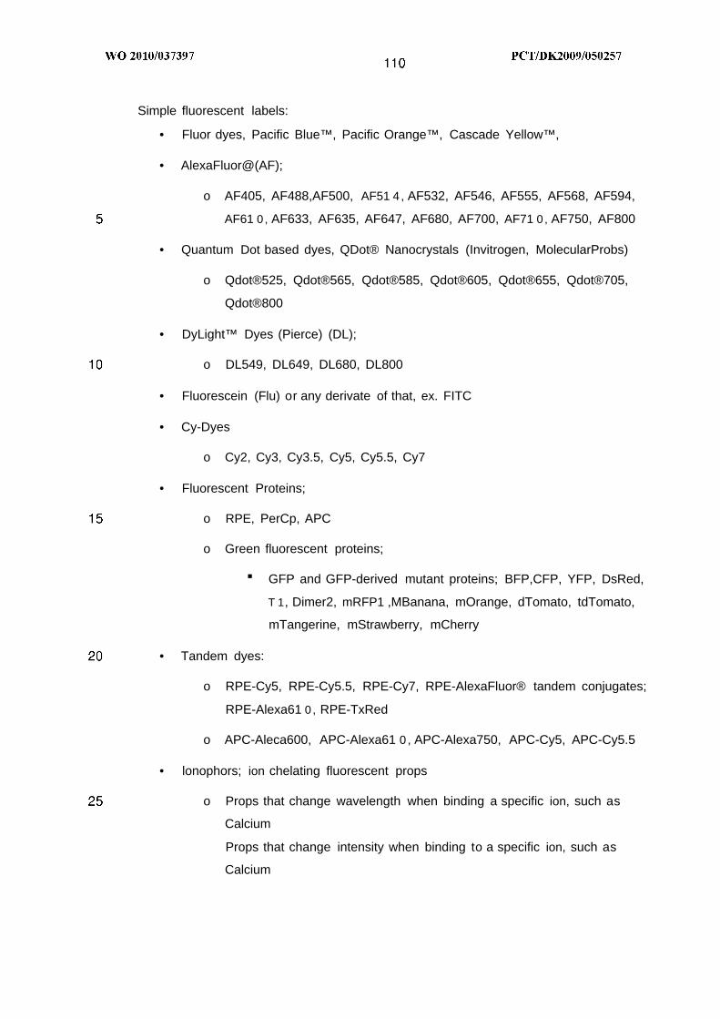
Simple fluorescent labels:
• Fluor dyes, Pacific Blue™, Pacific Orange™, Cascade Yellow™,
• AlexaFluor@(AF);
o AF405, AF488,AF500, AF51 4 , AF532, AF546, AF555, AF568, AF594,
AF61 0 , AF633, AF635, AF647, AF680, AF700, AF71 0 , AF750, AF800
• Quantum Dot based dyes, QDot® Nanocrystals (Invitrogen, MolecularProbs)
o Qdot®525, Qdot®565, Qdot®585, Qdot®605, Qdot®655, Qdot®705,
Qdot®800
• DyLight™ Dyes (Pierce) (DL);
o DL549, DL649, DL680, DL800
• Fluorescein (Flu) or any derivate of that, ex. FITC
• Cy-Dyes
o Cy2, Cy3, Cy3.5, Cy5, Cy5.5, Cy7
• Fluorescent Proteins;
o RPE, PerCp, APC
o Green fluorescent proteins;
■ GFP and GFP-derived mutant proteins; BFP,CFP, YFP, DsRed,
T 1, Dimer2, mRFP1 ,MBanana, mOrange, dTomato, tdTomato,
mTangerine, mStrawberry, mCherry
• Tandem dyes:
o RPE-Cy5, RPE-Cy5.5, RPE-Cy7, RPE-AlexaFluor® tandem conjugates;
RPE-Alexa61 0 , RPE-TxRed
o APC-Aleca600, APC-Alexa61 0 , APC-Alexa750, APC-Cy5, APC-Cy5.5
• lonophors; ion chelating fluorescent props
o Props that change wavelength when binding a specific ion, such as
Calcium
Props that change intensity when binding to a specific ion, such as
Calcium

• Combinations of fluorochromes on the same marker. Thus, the marker is not
identified by a single fluorochrome but by a code of identification being a
specific combination of fluorochromes, as well as inter related ratio of
intensities.
Example: Antibody Ab1 and Ab2, are conjugated to both. FITC and BP but Ab1
have 1 FITC to 1 BP whereas Ab2 have 2 FITC to 1 BP. Each antibodymay
then be identified individually by the relative intensity of each fluorochrome. Any
such combinations of n fluorochromes with m different ratios can be generated.
• Table 3 : Examples of preferable fluorochromes

Table 4 : Examples of preferable fluorochrome families
Fluorochrome family Example fluorochrome
AlexaFluor®(AF) AF 350, AF405, AF430, AF488.AF500, AF514, AF532,
AF546, AF555, AF568, AF594, AF610, AF633, AF635,
AF647, AF680, AF700, AF710, AF750, AF800
Quantum Dot (Qdot®) based Qdot®525, Qdot®565, Qdot®585, Qdot®605, Qdot®655,
dyes Qdot®705, Qdot®800
DyLight™ Dyes (DL) DL549, DL649, DL680, DL800
Small fluorescing dyes FITC, Pacific Blue™, Pacific Orange™, Cascade Yellow™,
Marina blue™, DSred, DSred-2, 7-AAD, TO-Pro-3,
Cy-Dyes Cy2, Cy3, Cy3.5, Cy5, Cy5.5, Cy7
Phycobili Proteins: R-Phycoerythrin (RPE), PerCP, Allophycocyanin (APC), B-
Phycoerythrin, C-Phycocyanin
Fluorescent Proteins (E)GFP and GFP ((enhanced) green fluorescent protein)
derived mutant proteins; BFP, CFP, YFP, DsRed, T 1 , Dimer2,
mRFP1 ,MBanana, mOrange, dTomato, tdTomato,
mTangerine, mStrawberry, mCherry
Tandem dyes with RPE RPE-Cy5, RPE-Cy5.5, RPE-Cy7, RPE-AlexaFluor® tandem
conjugates; RPE-Alexa610, RPE-TxRed
Tandem dyes with APC APC-Aleca600, APC-Alexa610, APC-Alexa750, APC-Cy5,
APC-Cy5.5
Calcium dyes Indo-1-Ca2+ lndo-2-Ca2+

Preferably labelling molecules employed in stationary cytometry and IHC
• Enzymatic labelling, as exemplified in Table 5 :
o Horse radish peroxidase; reduces peroxides (H2O2) ,and the signal is
generated by the Oxygen acceptor when being oxidized.
■ Precipitating dyes; Dyes that when they arereduced they are
soluble, and precipitate when oxidized, generating a coloured
deposit at the site of the reaction.
■ Precipitating agent, carrying a chemical residue, a hapten, for
second layer binding of marker molecules, for amplification of the
primary signal.
■ Luminol reaction, generating a light signal at the site of reaction.
o Other enzymes, such as Alkaline Phosphatase, capable of converting a
chemical compound from a non-detectable molecule to a precipitated
detectable molecule, which can be coloured, or carries a hapten as
described above.
• Fluorescent labels, as exemplified in Table 3 and 4 ; as those described for Flow
cytometry are likewise important for used in stationary cytometry, such as in
fluorescent microscopy.
Table 5 : Examples of preferable labels for stationary cytometry


Detection methods and principles
Detection of TCRs with multimers may be direct or indirect.
Direct detection
Direct detection of TCRs is detection directly of the binding interaction between the
specific T cell receptor and the MHC multimer. Direct detection includes detection of
TCR when TCR is attached to lipid bilayer, when TCR is attached to or in a solid
medium or when TCR is in solution.
Direct detection of TCR attached to lipid bilayer
One type of TCRs to detect and measure are TCRs attached to lipid bilayer including
but is not limited to naturally occurring T cells (from blood, spleen, lymphnode, brain or
any other tissue containing T cells), TCR transfected cells, T cell hybridomas, TCRs
embedded in liposomes or any other membrane structure. In the following methods for
direct detection of entities of TCRs attached to lipid bilayer will be described and any
entity consisting of TCR attached to lipid bilayer will be referred to as T cells.
T cells can be directly detected either when in a fluid solution or when immobilized to a
support.
Direct detection of T cells in fluid sample.
T cells can be detected in fluid samples as described elsewhere herein and in
suspension of disrupted tissue, in culture media, in buffers or in other liquids.T cells in
fluid samples can be detected individually or deteced as populations of T cells. In the
following different methods for direct detction of T cells in fluid samples are shown.
Direct detection of individual T cells
o Direct detection of individual T cells using flow cytometry.
A suspension of T cells are added MHC multimers, the sample washed and
then the amount of MHC multimer bound to each cell are measured. Bound
MHC multimers may be labelled directly or measured through addition of
labelled marker molecules. The sample is analyzed using a flow cytometer, able
to detect and count individual cells passing in a stream through a laser beam.
For identification of specific T cells using MHC multimers, cells are stained with
fluorescently labeled MHC multimer by incubating cells with MHC multimer and

then forcing the cells with a large volume of liquid through a nozzle creating a
stream of spaced cells. Each cell passes through a laser beam and any
fluorochrome bound to the cell is excited and thereby fluoresces. Sensitive
photomultipliers detect emitted fluorescence, providing information about the
amount of MHC multimer bound to the cell. By this method MHC multimers can
be used to identify specific T cell populations in liquid samples such as synovial
fluid or blood.
When analyzing blood samples whole blood can be used with or without lysis of
red blood cells. Alternatively lymphocytes can be purified before flow cytometry
analysis using standard procedures like a Ficoll-Hypaque gradient. Another
possibility is to isolate T cells from the blood sample for example by binding to
antibody coated plastic surfaces, followed by elution of bound cells. This
purified T cell population can then be used for flow cytometry analysis together
with MHC multimers. Instead of actively isolating T cells unwanted cells like B
cells and NK cells can be removed prior to the analysis. One way to do this is
by affinity chromatography using columns coated with antibodies specific for the
unwanted cells. Alternatively, specific antibodies can be added to the blood
sample together with complement, thereby killing cells recognized by the
antibodies.
Various gating reagents can be included in the analysis. Gating reagents here
means labeled antibodies or other labeled markers identifying subsets of cells
by binding to unique surface proteins. Preferred gating reagents when using
MHC multimers are antibodies directed against CD3, CD4, and CD8 identifying
major subsets of T cells. Other preferred gating reagents are antibodies against
CD1 4 , CD1 5 , CD1 9 , CD25, CD56, CD27, CD28, CD45, CD45RA, CD45RO,
CCR7, CCR5, CD62L, Foxp3 recognizing specific proteins unique for different
lymphocytes of the immune system.
Following labelling with MHC multimers and before analysis on a flow cytometer
stained cells can be treated with a fixation reagent like formaldehyde to cross-
link bound MHC multimer to the cell surface. Stained cells can also be analyzed
directly without fixation.
The number of cells in a sample can vary. When the target cells are rare, it is
preferable to analyze large amounts of cells. In contrast, fewer cells are

required when looking at T cell lines or samples containing many cells of the
target cell type.
The flow cytometer can be equipped to separate and collect particular types of
cells. This is called cell sorting. MHC multimers in combination with sorting on a
flowcytometer can be used to isolate specific T cell populations. Isolated
specific T cell populations can then be expanded in vitro. This can be useful in
autologous cancer therapy.
Direct determination of the concentration of MHC-peptide specific T cells in a
sample can be obtained by staining blood cells or other cell samples with MHC
multimers and relevant gating reagents followed by addition of an exact amount
of counting beads of known concentration. Counting beads is here to be
understood as any fluorescent bead with a size that can be visualized by flow
cytometry in a sample containing T cells. The beads could be made of
polystyrene with a size of about 1-10 µm. They could also be made of agarose,
polyacrylamide, silica, or any other material, and have any size between 0,1 µm
and 100 m. The counting beads are used as reference population to measure
the exact volume of analyzed sample. The sample are analyzed on a flow
cytometer and the amount of MHC-specific T cell determined using a
predefined gating strategy and then correlating this number to the number of
counted counting beads in the same sample using the following equation:
Amounts of MHC-peptide specific T cells in a blood sample can be determined
by flow cytometry by calculating the amount of MHC'mer labeled cells in a given
volumen of sample with a given cell density and then back calculate. Exact
enumeration of specific T cells is better achieved by staining with MHC'mers
together with an exact amount of counting beads followed by flow cytometry
analysis. The amount of T cells detected can then be correlated with the
amount of counting beads in the same volume of the sample and an exact
number of MHC-peptide specific T cells determined:
Concentration of MHC-specific T-cell in sample = (number of MHC-peptide
specific T cells counted/number of counting beads counted) x concentration of
counting beads in sample
Direct detection of individual T cells in fluid sample by microscopy

A suspension of T cells are added MHC multimers, the sample washed and
then the amount of MHC multimer bound to each cell are measured. Bound
MHC multimers may be labelled directly or measured through addition of
labelled marker molecules. The sample is then spread out on a slide or similar
in a thin layer able to distinguish individual cells and labelled cells identified
using a microscope. Depending on the type of label different types of
microscopes may be used, e.g. if fluorescent labels are used a fluorescent
microscope is used for the analysis. For example MHC multimers can be
labeled with a flourochrome or bound MHC multimer detected with a fluorescent
antibody. Cells with bound fluorescent MHC multimers can then be visualized
using an immunofluorescence microscope or a confocal fluorescence
microscope.
o Direct detection of individual T cells in fluid sample by capture on solid support
followed by elution.
MHC multimers are immobilized to a support e.g. beads, immunotubes, wells
of a microtiterplate, CD, mircrochip or similar and as decribed elsewhere herein,
then a suspension of T cells are added allowing specific T cells to bind MHC
multimer molecules. Following washing bound T cells are recovered/eluted (e.g.
using acid or competition with a competitor molecules) and counted.
Direct detection of populations of T cells
o Cell suspensions are added labeled MHC multimer, samples are washed and
then total signal from label are measured. The MHC multimers may be labeled
themselves or detected through a labeled marker molecule.
o Cell suspensions are added labeled MHC multimer, samples are washed and
then signal from label are amplified and then total signal from label and/or
amplifier are measured.
Direct detection of immobilized T cells.
T cells may be immobilized and then detected directly. Immobilization can be on solid
support, in solid tissue or in fixator (e.g. paraffin, a sugar matrix or another medium
fixing the T cells).

Direct detection of T cells immobilized on solid support.
In a number of applications, it may be advantageous immobilise the T cell onto a solid
or semi-solid support. Such support may be any which is suited for immobilisation,
separation etc. Non-limiting examples include particles, beads, biodegradable particles,
sheets, gels, filters, membranes (e. g . nylon membranes), fibres, capillaries, needles,
microtitre strips, tubes, plates or wells, combs, pipette tips, micro arrays, chips, slides,
or indeed any solid surface material. The solid or semi-solid support may be labelled, if
this is desired. The support may also have scattering properties or sizes, which enable
discrimination among supports of the same nature, e.g. particles of different sizes or
scattering properties, colour or intensities.
Conveniently the support may be made of glass, silica, latex, plastic or any polymeric
material. The support may also be made from a biodegradable material.
Generally speaking, the nature of the support is not critical and a variety of materials
may be used. The surface of support may be hydrophobic or hydrophilic.
Preferred are materials presenting a high surface area for binding of the T cells. Such
supports may be for example be porous or particulate e.g. particles, beads, fibres,
webs, sinters or sieves. Particulate materials like particles and beads are generally
preferred due to their greater binding capacity. Particularly polymeric beads and
particles may be of interest.
Conveniently, a particulate support (e.g. beads or particles) may be substantially
spherical. The size of the particulate support is not critical, but it may for example have
a diameter of at least 1 µm and preferably at least 2 µm, and have a maximum
diameter of preferably not more than 10 µm and more preferably not more than 6 µm.
For example, particulate supports having diameters of 2.8 µm and 4.5 µm will work
well.
An example of a particulate support is monodisperse particles, i.e. such which are
substantially uniform in size (e. g . size having a diameter standard deviation of less
than 5%). Such have the advantage that they provide very uniform reproducibility of
reaction. Monodisperse particles, e.g. made of a polymeric material, produced by the
technique described in US 4,336,173 (ref. 25) are especially suitable.

Non-magnetic polymer beads may also be applicable. Such are available from a wide
range of manufactures, e.g. Dynal Particles AS, Qiagen, Amersham Biosciences,
Serotec, Seradyne, Merck, Nippon Paint, Chemagen, Promega, Prolabo, Polysciences,
Agowa, and Bangs Laboratories.
Another example of a suitable support is magnetic beads or particles. The term
"magnetic" as used everywhere herein is intended to mean that the support is capable
of having a magnetic moment imparted to it when placed in a magnetic field, and thus
is displaceable under the action of that magnetic field. In other words, a support
comprising magnetic beads or particles may readily be removed by magnetic
aggregation, which provides a quick, simple and efficient way of separating out the
beads or particles from a solution. Magnetic beads and particles may suitably be
paramagnetic or superparamagnetic. Superparamagnetic beads and particles are e.g.
described in EP 0 106 873 (Sintef, ref. 26). Magnetic beads and particles are available
from several manufacturers, e.g. Dynal Biotech ASA (Oslo, Norway, previously Dynal
AS, e.g. Dynabeads®) .
The support may suitably have a functionalised surface. Different types of
functionalisation include making the surface of the support positively or negatively
charged, or hydrophilic or hydrophobic. This applies in particular to beads and
particles. Various methods therefore are e.g. described in US 4,336,173 (ref. 25), US
4,459,378 (ref. 27) and US 4,654,267 (ref. 28).
Immobilized T cells may be detected in several ways including:
o Direct detection of T cells directly immobilized on solid support.
T cells may be directly immobilized on solid support e.g. by non-specific
adhesion. Then MHC multimers are added to the immobilized T cells thereby
allowing specific T cells to bind the MHC multimers. Bound MHC multimer may
be measured through label directly attached to the multimer or through labeled
marker molecules. Individual T cells may be detected if the method for analysis
is able to distinguish individual labelled cells, e.g. cells are immobilized in a
monolayer on a cell culture well or a glass slide. Following staining with labelled
multimer a digital picture is taken and labelled cells identified and counted.

Alternatively a population of T cells is detected by measurement of total signal
from all labelled T cells, e.g. cells are plated to wells of a microtiter plate,
stained with labelled MHC multimer and total signal from each well are
measured.
o Direct detection of T cells immobilized on solid support through linker molecule
T cells can also be immobilized to solid support through a linker molecule. The
linker molecule can be an antibody specific for the T cell, a MHC multimer, or
any molecule capable of binding T cells. In any case the linker may be attached
directly to the solid support, to the solid support through another linker, or the
linker may be embedded in a matrix, e.g. a sugar matrix.
Then MHC multimers are added to the immobilized T cells thereby allowing
specific T cells to bind the MHC multimers. Bound MHC multimer may be
measured through label directly attached to the multimer or through labeled
marker molecules. Individual T cells may be detected if the method for analysis
is able to distinguish individual labelled cells, e.g. a digital picture is taken and
labelled cells identified and counted.
By using a specific MHC multimer both for the immobilization of the T-cells and
for the labelling of immobilized cells (e.g. by labelling immobilized cells with
chromophore- or fluorophore-labelled MHC multimer), a very high analytical
specificity may be achieved because of the low background noise that results.
Alternatively a population of T cells is detected by measurement of total signal
from all labeled T cells.
o lmmuno profiling: Phenotyping T cell sample using MHC multimer beads or
arrays.
Different MHC multimers are immobilized to different beads with different
characteristics (e.g. different size, different fluorophores or different
fluorescence intensities) where each kind of bead has a specific type of MHC
multimer molecule immobilized. The immobilization may be direct or through a
linker molecule as described above. The amount of bound T cells to a specific
population of beads can be analyzed, thereby phenotyping the sample. The

TCR on the T cell is defined by the MHC multimer and hence the bead to which
it binds.
Likewise, MHC multimers can be immobilized in an array, e.g. on a glass plate
or pin array so that the position in the array specifies the identity of the MHC
multimer. Again, the immobilization may be direct or through a linker molecule
as described above. After addition of T cells, the amount of bound T cells at a
specified position in the array can be determined by addition of a label or
labelled marker that binds to cells in general, or that binds specifically to the
cells of interest. For example, the cells may be generally labelled by the
addition of a labelled molecule that binds to all kinds of cells, or specific cell
types, e.g. CD4+ T-cells, may be labelled with anti-CD4 antibodies that are
labelled with e.g. a chromophore or fluorophore. Either of these approaches
allow a phenotyping of the sample.
• Profiling of an individual's disease-specific T-cell repertoire.
Mass profiling of the T-cells of an individual may be done by first
immobilizing specific MHC multimers (e.g. 10-1 06 different MHC multimers,
each comprising a specific MHC-peptide combination) in an array (e.g. a
glass plate), adding e.g. a blood sample from the individual, and then after
washing the unbound cells off, label the immobilized cells. Positions in the
array of particularly high staining indicate MHC-peptide combinations that
recognize specific T-cells of particularly high abundance or affinity. Thus, an
immuno profiling of the individual with regard to the tested MHC-peptide
combinations is achieved. A similar profiling of an individuals disease may
be made using MHC multimers immobilized to different beads as described
above.
Whether the profiling is performed using beads or arrays, the profiling may
entail a number of diseases, a specific disease, a set of specific antigens
implicated in one or more diseases, or a specific antigen (e.g. implicated in a
specific disease or set of diseases).
In a preferred embodiment, an individual's immuno profile for a particular
antigen is obtained. Thus, peptides corresponding to all possible 8'-, 9'- 10'-
and 11'-mer peptide sequences derived from the peptide antigen sequence are

generated, for example by standard organic synthesis or combinatorial
chemistry, and the corresponding MHC multimers are produced, using one or
more of the class I MHC-alleles of the individual in question. Further, peptides
of e.g. 13 , 14, 15 , 16 and upto 25 amino acids length may be generated, for
example by organic synthesis or combinatorial chemistry, corresponding to all
13', 14', 15', 16' and upto 25'-mers of the antigen, and the corresponding class
I l MHC multimers are produced, using one or more of the class I l MHC-alleles
of the individual in question. For a complete profiling for this particular antigen,
all of the HLA-alleles of the individual in question should be used for the
generation of the array; i.e., if the HLA class I haplotype of the individual is
HLA-A*02, HLA-A*03, HLA-B*08 and HLA-B*07, all these HLA class I alleles
should be combined with every tested peptide and similarly for all HLA class Il
alleles of the given individual.
Based on the profile, a personalized drug, -vaccine or -diagnostic test may be
produced.
The principle described above may also be employed to distinguish between
the immune response raised against a disease (e.g. an infection with a
bacterium or the formation of a tumour), and the immune response raised
against a vaccine for the same disease (in the example, a vaccine against the
bacterium or the tumour). Most vaccines consists of subcomponents of the
pathogen /tumour they are directed against and/or are designed to elicit an
immune response different from the natural occuring immune response i.e. the
T cell epitopes of the two immune reponses differs. Thus, by establishing the
immuno profile, using a comprehensive array (i.e. an array that comprises all
possible epitopes from one or more antigen(s)) or a subset of these epitopes, it
is possible to deduce whether the immune response has been generated
against the disease or the vaccine, or against both the disease and the
vaccine. If the vaccine raises a response against a particular epitope or a
particular set of epitopes, the corresponding positions in the array will give rise
to high signals (compared to the remaining positions). Similarly a natural
generated immune response will be directed against other and/or more
particular epitopes and therefore give rise to high signals in other positions
and/or more positions in the array. When an individual is vaccinated the
immuno profile will reflect the effect of the vaccination on the immune

response, and even if the individual has encountered the disease before and
has generated a general immune response towards this disease, it will still be
possible to deduce from the profiling whether this individual also has generated
a specific response against the vaccine.
In another preferred embodiment, an individual's immuno profile for a set of
antigens implicated in a specific disease is obtained. A subset of epitopes from
a number of antigens is used. Thus, this is not a comprehensive profiling of this
individual with regard to these antigens, but careful selection of the epitopes
used may ensure that the profiling data can be used afterwards to choose
between e.g. a limited set of vaccines available, or the data can be used to
evaluate the immune response of the individual following an infection, where
the epitopes used have been selected in order to avoid interference from
related infectious diseases.
As above, a personalized drug, -vaccine or -diagnostic test may be produced
based on the information obtained from the immuno profiling.
In yet another preferred embodiment, the array comprising all possible 8'-, 9'-
10'- and 11'-mer peptide sequences derived from a given peptide antigen, and
all 13 , 14, 15 and 16'-mers of the same antigen, are synthesized and
assembled in MHC multimers, and immobilized in an array. Then, the ability of
the individual peptide to form a complex with MHC is tested. As an example,
one may add labelled W6/32 antibody, an antibody that binds correctly folded
MHC I heavy chain, when this heavy chain is assembled together with
antigenic peptide and beta2microglobulin, and which therefore can be used to
detect formation of MHC-peptide complex, as binding of W6/32 antibody is
usually considered a strong indication that the MHC-peptide complex has been
formed. The ability of different peptides to enter into a MHC-peptide complex
may also be promoted by the addition to the array of T-cells. Specific T-cells
will drive the formation of the corresponding specific MHC-peptide complexes.
Thus, after addition of T-cells to the array, the MHC-peptide complex integrity
can be examined by addition of the labelled W6/32 antibody or other antibodies
specific for correct conformation. Positions on the array that have strong
signals indicate that the peptide that was added to MHC and immobilized at
this position, was capable of forming the MHC-peptide complex in the presence

of specific T-cells. Alternatively, the binding of the specific T-cells to the
corresponding MHC-peptide complexes may be detected directly through a
labbelled antibody specific for the T cell.
Direct detection of immobilized T cells followed by sorting
T cells immobilized to solid support in either of the ways described above can following
washing be eluted from the solid support and treated further. This is a method to sort
out specific T cells from a population of different T cells. Specific T-cells can e.g.be
isolated through the use of bead-based MHC multimers. Bead-based MHC multimers
are beads whereto monomer MHC-peptide complexes or MHC multimers are
immobilized. After the cells have been isolated they can be manipulated in many
different ways. The isolated cells can be activated (to differentiate or proliferate), they
can undergo induced apoptosis, or undesired cells of the isolated cell population can
be removed. Then, the manipulated cell population can be re-introduced into the
patient, or can be introduced into another patient.
A typical cell sorting experiment, based on bead-based MHC multimers, would follow
some of the steps of the general procedure outlined in general terms in the following:
Acquire the sample, e.g. a cell sample from the bone marrow of a cancer patient.
Block the sample with a protein solution, e.g. BSA or skim milk.
Block the beads coated with MHC complexes, with BSA or skim milk.
Mix MHC-coated beads and the cell sample, and incubate.
Wash the beads with washing buffer, to remove unbound cells and non-specifically
bound cells.
Isolate the immobilized cells, by either cleavage of the linker that connects MHC
complex and bead; or alternatively, release the cells by a change in pH, salt-
concentration addition of competitive binder or the like. Preferably, the cells are
released under conditions that do not disrupt the integrity of the cells.
Manipulate the isolated cells (induce apoptosis, proliferation or differentiation)
Direct detection of T cells in solid tissue.
o Direct detection of T cells in solid tissue in vitro.
For in vitro methods of the present invention solid tissue includes tissue, tissue
biopsies, frozen tissue or tissue biopsies, paraffin embedded tissue or tissue
biopsies and sections of either of the above mentioned. In a preferred method
of this invention sections of fixed or frozen tissues are incubated with MHC

multimer, allowing MHC multimer to bind to specific T cells in the tissue section.
The MHC multimer may be labeled directly or through a labeled marker
molecule. As an example, the MHC multimer can be labeled with a tag that can
be recognized by e.g. a secondary antibody, optionally labeled with HRP or
another label. The bound MHC multimer is then detected by its fluorescence or
absorbance (for fluorophore or chromophore), or by addition of an enzyme-
labeled antibody directed against this tag, or another component of the MHC
multimer (e.g. one of the protein chains, a label on the multimerization domain).
The enzyme can be Horse Raddish Peroxidase (HRP) or Alkaline Phosphatase
(AP), both of which convert a colorless substrate into a colored reaction product
in situ. This colored deposit identifies the binding site of the MHC multimer, and
can be visualized under a light microscope. The MHC multimer can also be
directly labeled with e.g. HRP or AP, and used in IHC without an additional
antibody.
The tissue sections may derive from blocks of tissue or tissue biopsies
embedded in paraffin, and tissue sections from this paraffin-tissue block fixed in
formalin before staining. This procedure may influence the structure of the TCR
in the fixed T cells and thereby influence the ability to recognize specific MHC
complexes. In this case, the native structure of TCR needs to be at least partly
preserved in the fixed tissue. Fixation of tissue therefore should be gentle.
Alternatively, the staining is performed on frozen tissue sections, and the
fixation is done after MHC multimer staining.
o Direct detection of T cells in solid tissue in vivo
For in vivo detection of T cells labeled MHC multimers are injected in to the
body of the individual to be investigated. The MHC multimers may be labeled
with e.g. a paramagnetic isotope. Using a magnetic resonance imaging (MRI)
scanner or electron spin resonance (ESR) scanner MHC multimer binding T
cells can then be measured and localized. In general, any conventional method
for diagnostic imaging visualization can be utilized. Usually gamma and positron
emitting radioisotopes are used for camera and paramagnetic isotopes for MRI.
The methods described above for direct detection of TCR embedded in lipid bilayers
collectively called T cells using MHC multimers also applies to detection of TCR in

solution and detection of TCR attached to or in a solid medium. Though detection of
individual TCRs may not be possible when TCR is in solution.
Indirect detection of TCR
Indirect detection of TCR is primarily usefull for detection of TCRs embedded in lipid
bilayer, preferably natural occurring T cells, T cell hybridomas or transfected T cells. In
indirect detection, the number or activity of T cells are measured, by detection of
events that are the result of TCR-MHC-peptide complex interaction. Interaction
between MHC multimer and T cell may stimulate the T cell resulting in activation of T
cells, in cell division and proliferation of T cell populations or alternatively result in
inactivation of T cells. All these mechanism can be measured using various detection
methods.
Indirect detection of T cells by measurement of activation.
MHC multimers, e.g. antigen presenting cells, can stimulate T cells resulting in
activation of the stimulated T cells. Activation of T cell can be detected by
measurement of secretion of specific soluble factor from the stimulated T cell, e.g.
secretion of cytokines like INFγ and IL2. Stimulation of T cells can also be detected by
measurement of changes in expression of specific surface receptors, or by
measurement of T cell effector functions.
Measurement of activation of T cells involves the following steps:
a) To a sample of T cells, preferably a suspension of cells, is added MHC multimer to
induce either secretion of soluble factor, up- or down-regulation of surface receptor
or other changes in the T cell.
Alternatively, a sample of T cells containing antigen presenting cells is added
antigenic peptide or protein/protein fragments that can be processed into antigenic
peptides by the antigen presenting cell and that are able to bind MHC I or MHC I l
molecules expressed by the antigen presenting cells thereby generating a cell
based MHC multimer in the sample. Several different peptides and proteins be
added to the sample. The peptide-loaded antigen presenting cells can then
stimulate specific T cells, and thereby induce the secretion of soluble factor, up- or
down-regulation of surface receptors, or mediate other changes in the T cell, e.g.
enhancing effector functions.

--O
Optionally a second soluble factor, e.g. cytokine and/or growth factor(s) may be
added to facilitate continued activation and expansion of T cell population
b) Detect the presence of soluble factor, the presence/ absence of surface receptor or
detect effector function
c) Correlate the measured result with presence of T cells. The measured
signal/response indicate the presence of specific T cells that have been stimulated
with particular MHC multimer.
The signal/response of a T lymphocyte population is a measure of the overall
response. The frequency of specific T cells able to respond to a given MHC
multimer can be determined by including a limiting-dilution culture in the assay also
called a Limiting dilution assay.
The limiting-dilution culture method involves the following steps:
a) Sample of T cells in suspension are plated into culture wells at increasing
dilutions
b) MHC multimers are added to stimulate specific T cells . Alternatively
antigen presenting cells are provided in the sample and then antigenic
peptide I added to the sample as descrinbed above.
Optionally growth factors, cytokines or other factors helping T cells to
proliferate are added.
c) Cells are allowed to grow and proliferate (V2- several days). Each well that
initially contained a specific T cell will make a response to the MHC
multimer and divide.
d) Wells are tested for a specific response e.g. secretion of soluble factors,
cell proliferation, cytotoxicity or other effector function.
The assay is replicated with different numbers of T cells in the sample, and
each well that originally contained a specific T cell will make a response to the
MHC multimer. The frequency of specific T cells in the sample equals the
reciprocal of the number of cells added to each well when 37% of the wells are
negative, because due to Poisson distrubtion each well then on average
contained one specific T cell at the beginning of the culture.
In the following various methods to measure secretion of specific soluble factor,
expression of surface receptors, effector functions or proliferation is described.
Indirect detection of T cells by measurement of secretion of soluble factors.

Indirect detection of T cells by measurement of extracellular secreted soluble factors.
Secreted soluble factors can be measured directly in fluid suspension, captured by
immobilization on solid support and then detected or an effect of the secreted soluble
factor can be detected.
o Indirect detection of T cells by measurement of extracellular secreted soluble
factor directly in fluid sample.
A sample of T cells are added MHC multimer or antigenic peptide as described
above to induce secretion of soluble factors from antigen-specific T cells. The
secreted soluble factors can be measured directly in the supernatant using e.g.
mass spectrometry.
o Indirect detection of T cells by capture of extracellular secreted soluble factor
on solid support.
A sample of T cells are added MHC multimer or antigenic peptide as described
above to induce secretion of soluble factors from antigen-specific T cells.
Secreted soluble factors in the supernatant are then immobilized on a solid
support either directly or through a linker as described for immobilization of T
cells elsewhere herein. Then immobilized soluble factors can be detected using
labeled marker molecules.
Soluble factors secreted from individual T cells can be detected by capturing of
the secreted soluble factors locally by marker molecules, e.g antibodies
specific for the soluble factor. Soluble factor recognising marker molecules are
then immobilised on a solid support together with T cells and soluble factors
secreted by individual T cells are thereby captured in the proximity of each T
cell. Bound soluble factor can be measured using labelled marker molecule
specific for the captured soluble factor. The number of T cells that has given
rise to labelled spots on solid support can then be enumerated and these spots
indicate the presence of specific T cells that may be stimulated with particular
MHC multimer.
Soluble factors secreted from a population of T cells are detected by capture
and detection of soluble factor secreted from the entire population of specific T
cells. In this case soluble factor do not have to be captured locally close to each

T cell but the secreted soluble factors my be captured and detected in the same
well as where the T cells are or transferred to another solid support with marker
molecules for capture and detection e.g. beads or wells of ELISA plate.
o Indirect detection of T cells immobilized to solid support in a defined pattern.
Different MHC multimers og MHC-peptide complexes are immobilized to a
support to form a spatial array in a defined pattern, where the position specifies
the identity of the MHC multimer/MHC-peptide complex immobilized at this
position. Marker molecules able to bind T cell secreted soluble factors are co-
spotted together with MHC multimer/MHC-peptide complex. Such marker
molecules can e.g. be antibodies specific for cytokines like INFγ or IL-2. The
immobilization may be direct or through a linker molecule as described above.
Then a suspension of labeled T cells are added or passed over the array of
MHC multimers/MHC-peptide complexes and specific T cells will bind to the
immobilized MHC multimers/MHC-peptide complexes and upon binding be
stimulated to secrete soluble factors e.g. cytokines like INFγ ord IL-2. Soluble
factors secreted by individual T cells are then captured in the proximity of each
T cell and bound soluble factor can be measured using labelled marker
molecule specific for the soluble factor. The number and position of different
specific T cells that has given rise to labelled spots on solid support can then be
identified and enumerated. In this way T cells bound to defined areas of the
support are analyzed, thereby, phenotyping the sample. Each individual T cell is
defined by the TCR it expose and depending on these TCRs each entity will
bind to different types of MHC multimers/MHC-peptide complexes immobilized
at defined positions on the solid support.
o Indirect detection of T cells by measurement of effect of extracellular secreted
soluble factor.
Secreted soluble factors can be measured and quantified indirectly by
measurement of the effect of the soluble factor on other cell systems. Briefly,
a sample of T cells are added MHC multimer or antigenic peptide as described
above to induce secretion of soluble factors from antigen-specific T cells. The
supernatant containing secreted soluble factor are transferred to another cell
system and the effect measured. The soluble factor may induce proliferation,
secretion of other soluble factors, expression/downregulation of receptors, or

the soluble factor may have cytotoxic effects on these other cells,. All effects
can be measured as described elsewhere herein.
Indirect detection of T cells by measurement of intracellular secreted soluble factors
Soluble factor production by stimulated T cells can be also be measured intracellular by
e.g. flow cytometry. This can be done using block of secretion of soluble factor (e.g. by
monensin), permeabilization of cell (by e.g. saponine) followed by immunofluorescent
staining. The method involves the following steps: 1) Stimulation of T cells by binding
specific MHC multimers, e.g. antigen presenting cells loaded with antigenic peptide. An
reagent able to block extracellular secretion of cytokine is added, e.g. monensin that
interrupt intracellular transport processes leading to accumulation of produced soluble
factor, e.g. cytokine in the Golgi complex. During stimulation other soluble factors may
be added to the T cell sample during stimulation to enhance activation and/or
expansionn This other soluble factor can be cytokine and or growth factors. 2) addition
of one or more labelled marker able to detect special surface receptors (e.g. CD8,
CD4, CD3, CD27, CD28, CD2). 3) Fixation of cell membrane using mild fixator followed
by permeabilization of cell membrane by. e.g. saponine. 4) Addition of labelled marker
specific for the produced soluble factor to be determined, e.g. INFγ , IL-2, IL-4, IL-1 0 . 5)
Measurement of labelled cells using a flow cytometer.
An alternative to this procedure is to trap secreted soluble factors on the surface of the
secreting T cell as described by Manz, R. et al., Proc. Natl. Acad. Sci. USA 92:1 921
( 1995).
Indirect detection of T cells by measurement of expression of receptors
Activation of T cells can be detected by measurement of expression and/or down
regulation of specific surface receptors. The method includes the following steps. A
sample of T cells are added MHC multimer or antigenic peptide as described above to
induce expression or downregulation of specific surface receptors on antigen-specific T
cells.These receptors include but are not limited to CD28, CD27, CCR7, CD45RO,
CD45RA, IL2-receptor, CD62L, CCR5. Their expression level can be detected by
addition of labelled marker specific for the desired receptor and then measure the
amount of label using flow cytometry, microscopy, immobilization of activated T cell on
solid support or any other method like those decribed for direct detection of TCR in lipid
bilayer.

Indirect detection of T cells by measurement of effector function
Activation of T cells can be detected indirectly by measurement of effector functions. A
sample of T cells are added MHC multimer or antigenic peptide as described above to
induce the T cell to be able to do effector function. The effector function is then
measured. E.g. activation of antigen-specific CD8 positive T cells can be measured in a
cytotoxicity assay.
Indirect detection of T cells by measurement of proliferation
T cells can be stimulated to proliferate upon binding specific MHC multimers.
Proliferation of T cells can be measured several ways including but not limited to:
o Detection of mRNA
Proliferation of T cells can be detected by measurement of mRNA inside cell.Cell
division and proliferation requires production of new protein in each cell which as
an initial step requires production of mRNA encoding the proteins to be
synthesized.
A sample of T cells are added MHC multimer or antigenic peptide as described
above to induce proliferation of antigen-specific T cells. Detection of levels of
mRNA inside the proliferating T cells can be done by quantitative PCR and
indirectly measure activation of a T cell population as a result of interaction with
MHC multimer. An example is measurement of cytokine mRNA by in sity
hybridization.
o Detection of incorporation of thymidine
The proliferative capacity of T cells in response to stimulation by MHC multimer
can be determined by a radioactive assay based on incorporation of [3H]thymidine
([3H]TdR) into newly generated DNA followed by measurement of radioactive
signal.
o Detection of incorporation of BrdU
T cell proliferation can also be detected by of incorporation of bromo-2'-
deoxyuridine (BrdU) followed by measurement of incorporated BrdU using a
labeled anti-BrdU antibody in an ELISA based analysis.
Viability of cells may be measured by measurement ATP in a cell culture.

Indirect detection of T cells by measurement of inactivation
Not all MHC multimers will lead to activation of the T cells they bind. Under certain
circumstances some MHC multimers may rather inactivate the T cells they bind to.
Indirect detection of T cells by measurement of effect of blockade of TCR
Inactivation of T cells by MHC multimers may be measured be measuring the effect of
blocking TCR on antigen-specific T cells. MHC multimers, e.g. MHC-peptide
complexes coupled to IgG scaffold can block the TCR of an antigen-specific T cell by
binding the TCR, thereby prevent the blocked T cell receptor interacting with e.g.
antigen presenting cells. Blockade of TCRs of a T cell can be detected in any of the
above described medthods for detection of TCR by addition of an unlabeled blocking
MHC multimer together with the labelled MHC multimer and then measuring the effect
of the blockade on the readout.
Indirect detection of T cells by measurement of induction of apoptosis
Inactivation of T cells by MHC multimers may be measured be measuring apoptosis of
the antigen-specific T cell. Binding of some MHC multimers to specific T cells may lead
to induction of apoptosis. Inactivation of T cells by binding MHC multimer may therefore
be detected by measuring apoptosis in the T cell population. Methods to measure
apoptosis in T cells include but are not limited to measurement of the following:
• DNA fragmentation
• Alterations in membrane asymmetry (phosphatidylserine translocation)
• Activation of apoptotic caspases
• Release of cytochrome C and AIF from mitochondria into the cytoplasm
Positive control experiments for the use of MHC multimers in flow cytometry and
related techniques
When performing flow cytometry experiments, or when using similar technologies, it is
important to include appropriate positive and negative controls. In addition to
establishing proper conditions for the experiments, positive and negative control
reagents can also be used to evaluate the quality (e.g. specificity and affinity) and
stability (e.g. shelf life) of produced MHC multimers.
The quality and stability of a given MHC multimer can be tested in a number of different
ways, including:

• Measurement of specific MHC multimer binding to beads, other types of solid
support, or micelles and liposomes, to which TCR's have been immobilized.
Other kinds of molecules that recognize specifically the MHC-peptide complex
can be immobilized and used as well. Depending on the nature of the solid
support or membrane structure to which the TCR is immobilized, the TCR can
be full-length (i.e. comprise the intracellular- and intra-membrane domains), or
can be truncated (e.g. only comprise the extracellular domains). Likewise, the
TCR can be recombinant, and can be chemically or enzymatically modified.
• Measurement of MHC multimer binding to beads, other types of solid support,
or micelles and liposomes, to which aptamers, antibodies or other kinds of
molecules that recognize correctly folded MHC-peptide complexes have been
immobilized.
• Measurement of specific MHC multimer binding to specific cell lines (e.g. T-cell
lines) displaying MHC multimer-binding molecules, e.g. displaying TCRs with
appropriate specificity and affinity for the MHC multimer in question.
• Measurement of specific MHC multimer binding to cells in blood samples,
preparations of purified lymphocytes (HPBMCs), or other bodily fluids that
contain cells carrying receptor molecules specific for the MHC multimer in
question.
• Measurement of specific MHC multimer binding to soluble TCRs, aptamers,
antibodies, or other soluble MHC-peptide complex-binding molecules, by
density-gradient centrifugation (e.g. in CsCI) or by size exclusion
chromatography, PAGE or other type of chromatographic method.
Measurement of specific MHC binding to TCRs, aptamers, antibodies, streptavidin, or
other MHC-peptide complex-binding molecules immobilized on a solid surface (e.g. a
microtiter plate). The degree of MHC multimer binding can be visualized with a
secondary component that binds the MHC multimer, e.g. a biotinylated fluorophore in
cases where the MHC multimer contains streptavidin proteins, not fully loaded with
biotin. Alternatively, the secondary component is unlabelled, and a labelled second
component-specific compound is employed (e.g. EnVision System, Dako) for
visualization. This solid surface can be beads, immunotubes, microtiterplates act. The
principle for purification are basically the same I.e. T cells are added to the solid with

immobilized MHC'mer, non-binding T cells are washed away and MHC-peptide specific
T cells can be retrieved by elution with mild acid or a competitive binding reagent.
• Measurement of specific MHC multimer binding to TCRs, aptamers, antibodies,
streptavidin, or other MHC-peptide complex-binding molecules immobilized on
a solid surface (e.g. a microtiter plate) visualized with a secondary component
specific to MHC multimer (e.g. TCRs, aptamers, antibodies, streptavidin, or
other MHC-peptide binding complex-binding molecules). Alternatively the
secondary receptor is unlabelled, and a labelled second receptor-specific
compound is employed (e.g. EnVision System, Dako) before visualization.
In the above mentioned approaches, positive control reagents include MHC multimers
comprising correctly folded MHC, complexed with an appropriate peptide that allows
the MHC multimer to interact specifically and efficiently with its cognate TCR. Negative
control reagents include empty MHC multimers, or correctly folded MHC multimers
complexed with so-called nonsense peptides that support a correct conformation of the
MHC-peptide complex, but that do not efficiently bind TCRs through the peptide-
binding site of the MHC complex.
Negative control reagents and negative control experiments for the use of MHC
multimers in flow cytometry and related techniques
Experiments with MHC multimers require a negative control in order to determine
background staining with MHC multimer. Background staining can be due to unwanted
binding of any of the individual components of the MHC multimer, e.g., MHC complex
or individual components of the MHC complex, multimerization domain or label
molecules. The unwanted binding can be to any surface or intracellular protein or other
cellular structure of any cell in the test sample, e.g. undesired binding to B cells, NK
cells or T cells. Unwanted binding to certain cells or certain components on cells can
normally be corrected for during the analysis, by staining with antibodies that bind to
unique surface markers of these specific cells, and thus identifies these as false
positives, or alternatively, that bind to other components of the target cells, and thus
identifies these cells as true positives. A negative control reagent can be used in any
experiment involving MHC multimers, e.g. flow cytometry analysis, other cytometric
methods, immunohistochemistry (IHC) and ELISA. Negative control reagents include
the following:

• MHC complexes or MHC multimers comprising MHC complexes carrying nonsense
peptides. A nonsense peptide is here to be understood as a peptide that binds the
MHC protein efficiently, but that does not support binding of the resultant MHC-
peptide complex to the desired TCR. An example nonsense peptide is a peptide
with an amino acid sequence different from the linear sequence of any peptide
derived from any known protein. When choosing an appropriate nonsense peptide
the following points are taken into consideration. The peptide should ideally have
appropriate amino acids at relevant positions that can anchor the peptide to the
peptide-binding groove of the MHC. The remaining amino acids should ideally be
chosen in such a way that possible binding to TCR (through interactions with the
peptide or peptide-binding site of MHC) are minimized. The peptide should ideally
be soluble in water to make proper folding with MHC alpha chain and β2m possible
in aqueous buffer. The length of the peptide should ideally match the type and
allele of MHC complex. The final peptide sequence should ideally be taken through
a blast search or similar analysis, to ensure that it is not identical with any peptide
sequence found in any known naturally occurring proteins.
• MHC complexes or MHC multimers comprising MHC complexes carrying a
chemically modified peptide in the peptide-binding groove. The modification should
ideally allow proper conformation of the MHC-peptide structure, yet should not
allow efficient interaction of the peptide or peptide-binding site of MHC with the
TCR.
• MHC complexes or MHC multimers comprising MHC complexes carrying a
naturally occurring peptide different from the peptide used for analysis of specific T
cells in the sample. When choosing the appropriate natural peptide the following
should be taken into consideration. The peptide in complex with the MHC protein
should ideally not be likely to bind a TCR of any T cell in the sample with such an
affinity that it can be detected with the applied analysis method. The peptide should
ideally be soluble in water to make proper folding with MHC alpha chain and β2m
possible in aqueous buffer. The length of the peptide should match the type and
allele of MHC complex.
• Empty MHC complexes or MHC multimers comprising empty MHC complexes,
meaning any correctly folded MHC complex without a peptide in the peptide-
binding groove.

MHC heavy chain or MHC multimers comprising MHC heavy chain, where MHC
heavy chain should be understood as full-length MHC I or MHC I l heavy chain or
any truncated version of MHC I or MHC I l heavy chain. The MHC heavy chains can
be either folded or unfolded. Of special interest is MHC I alpha chains containing
the oc3 domain that binds CD8 molecules on cytotoxic T cells. Another embodiment
of special interest is MHC I l β chains containing the β2 domain that binds CD4 on
the surface of helper T cells.
• Beta2microglobulin or subunits of beta2microglobulin, or MHC multimers
comprising Beta2microglobulin or subunits of beta2microglobulin, folded or
unfolded.
• MHC-like complexes or MHC multimers comprising MHC-like complexes, folded or
unfolded. An example could be CD1 molecules that are able to bind peptides in a
peptide-binding groove that can be recognized by T cells (Russano et al. (2007).
CD1 -restricted recognition of exogenous and self-lipid antigens by duodenal
gammadelta÷ T lymphocytes. J Immunol. 178(6):3620-6 )
• Multimerization domains without MHC or MHC-like molecules, e.g. dextran,
streptavidin, IgG, coiled-coil-domain liposomes.
• Labels, e.g. FITC, PE, APC, pacific blue, cascade yellow, or any other label listed
elsewhere herein.
Negative controls 1-4 can provide information about potentially undesired binding of the
MHC multimer, through interaction of a surface of the MHC-peptide complex different
from the peptide-binding groove and its surroundings. Negative control 5 and 6 can
provide information about binding through interactions through the MHC I or MHC I l
proteins (in the absence of peptide). Negative control 7 can provide information about
binding through surfaces of the MHC complex that is not unique to the MHC complex.
Negative controls 8 and 9 provide information about pontential undesired interactions
between non-MHC-peptide complex components of the MHC multimer and cell
constituents.

o
Minimization of undesired binding of the MHC multimer
Identification of MHC-peptide specific T cells can give rise to background signals due to
unwanted binding to cells that do not carry TCRs. This undesired binding can result
from binding to cells or other material, by various components of the MHC multimer,
e.g. the dextran in a MHC dextramer construct, the labelling molecule (e.g. FITC), or
surface regions of the MHC-peptide complex that do not include the peptide and the
peptide-binding cleft.
MHC-peptide complexes bind to specific T cells through interaction with at least two
receptors in the cell membrane of the T-cell. These two receptors are the T-cell
receptor (TCR) and CD8 for MHC l-peptide complexes and TCR and CD4 receptor
protein for MHC Il-peptide complexes. Therefore, a particularly interesting example of
undesired binding of a MHC multimer is its binding to the CD8 or CD4 molecules of T
cells that do not carry a TCR specific for the actual MHC-peptide complex. The
interaction of CD8 or CD4 molecules with the MHC is not very strong; however,
because of the avidity gained from the binding of several MHC complexes of a MHC
multimer, the interaction between the MHC multimer and several CD8 or CD4
receptors potentially can result in undesired but efficient binding of the MHC multimer
to these T cells. In an analytical experiment this would give rise to an unwanted
background signal; in a cell sorting experiment undesired cells might become isolated.
Other particular interesting examples of undesired binding is binding to lymphoid cells
different from T cells, e.g. NK-cells, B-cells, monocytes, dendritic cells, and
granulocytes like eosinophils, neutrophils and basophiles.
Apart from the MHC complex, other components in the MHC multimer can give rise to
unspecific binding. Of special interest are the multimerization domain, multimerization
domain molecules, and labelling molecules.
One way to overcome the problem with unwanted binding is to include negative
controls in the experiment and subtract this signal from signals derived from the
analyzed sample, as described elsewhere in the invention.
Alternatively, unwanted binding could be minimized or eliminated during the
experiment. Methods to minimize or eliminate background signals include:
• Mutations in areas of the MHC complex responsible for binding to unwanted cells
can be introduced. Mutations here mean substitution, insertion, or deletion of

natural or non-natural amino acids. Sub-domains in the MHC complex can be
responsible for unwanted binding of the MHC multimer to cells without a TCR
specific for the MHC-peptide complex contained in the MHC multimer. One
example of special interest is a small region in the oc3-domain of the α-chain of
MHC I molecules that is responsible for binding to CD8 on all cytotoxic T cells.
Mutations in this area can alter or completely abolish the interaction between CD8
on cytotoxic T cells and MHC multimer (Neveu et al. (2006) lnt Immunol. 18, 1139-
45). Similarly a sub domain in the β2 domain of the β-chain of MHC I l molecules is
responsible for binding CD4 molecules on all CD4 positive T cells. Mutations in this
sub domain can alter or completely abolish the interaction between MHC I l and
CD4.
Another embodiment is to mutate other areas of MHC I /MHC I l complexes that are
involved in interactions with T cell surface receptors different from TCR, CD8 and
CD4, or that bind surface receptors on B cells, NK cells, Eosiniophils, Neutrophils,
Basophiles, Dendritic cells or monocytes.
• Chemical alterations in areas of the MHC complex responsible for binding to
unwanted cells can be employed in order to minimize unwanted binding of MHC
multimer to irrelevant cells. Chemical alteration here means any chemical
modification of one or more amino acids. Regions in MHC complexes that are of
special interest are as mentioned above the α3 domain of the α-chain in MHC I
molecules and β2 domains in the β-chain of MHC Il molecules. Other regions in
MHC I /MHC I l molecules that can be chemically modified to decrease the extent of
undesired binding are regions involved in interaction with T cell surface receptors
different from TCR, CD8 and CD4, or that bind surface receptors on B cells, NK
cells, Eosiniophils, Neutrophils, Basophiles, Dendritic cells or monocytes.
• Another method to minimize undesired binding involves the addition of one or more
components of a MHC multimer, predicted to be responsible for the unwanted
binding. The added component is not labeled, or carries a label different from the
label of the MHC multimer used for analysis. Of special interest is addition of MHC
multimers that contain nonsense peptides, i.e. peptides that interact efficiently with
the MHC protein, but that expectably do not support specific binding of the MHC
multimer to the TCR in question. Another example of interest is addition of soluble
MHC complexes not coupled to a multimerization domain, and with or without
peptide bound in the peptide binding cleft. In another embodiment, individual

components of the MHC complex can be added to the sample, e.g. I α-chain or
subunits of MHC I α-chain either folded or unfolded, beta2microglobulin or subunits
thereof either folded or unfolded, α/β-chain of MHC I l or subunits thereof either
folded or unfolded. Any of the above mentioned individual components can also be
attached to a multimerization domain identical or different from the one used in the
MHC multimer employed in the analysis.
Of special interest is also addition of multimerization domain similar or identical to
the multimerization domain used in the MHC multimer or individual components of
the multimerization domain.
• Reagents able to identify specific cell types either by selection or exclusion can be
included in the analysis to help identify the population of T cells of interest, and in
this way deselect the signal arising from binding of the MHC multimer to undesired
cells.
Of special interest is the use of appropriate gating reagents in flow cytometry
experiments. Thus, fluorescent antibodies directed against specific surface markers
can be used for identification of specific subpopulations of cells, and in this way
help to deselect signals resulting from MHC multimers binding to undesired cells.
Gating reagents of special interest that helps identify the subset of T cells of
interest when using MHC I multimers are reagents binding to CD3 and CD8
identifying all cytotoxic T cells. These reagents are preferably antibodies but can be
any labeled molecule capable of binding CD3 or CD8. Gating reagents directed
against CD3 and CD8 are preferably used together. As they stain overlapping cell
populations they are preferably labeled with distinct fluorochromes. However, they
can also be used individually in separate samples. In experiments with MHC I l
multimers reagents binding to CD3 and CD4 identifying T helper cells can be used.
These reagents are preferably antibodies but can be any labeled molecule capable
of binding CD3 or CD4. Gating reagents directed against CD3 and CD4 are
preferable used together. As they stain overlapping cell populations they are
preferably labeled with distinct fluorochromes. However, they can also be used
individually in separate samples.
Other gating reagents of special interest in experiments with any MHC multimer, are
reagents binding to the cell surface markers CD2, CD27, CD28, CD45RA, CD45RO,
CD62L and CCR7. These surface markers are unique to T cells in various

differentiation states. Co staining with either of these reagents or combinations thereof
together with MHC multimers helps to select MHC multimer binding T cells expressing
a correct TCR. These reagents can also be combined with reagents directed against
CD3, CD4 and/or CD8.
Another flow cytometric method of special interest to remove signals from MHC
multimer stained cells not expressing the specific TCR, is to introduce an exclusion
gate. Antibodies or other reagents specific for surface markers unique to the unwanted
cells are labeled with a fluorochrome and added to the test sample together with the
MHC multimer. The number of antibodies or surface marker specific reagents are not
limited to one but can be two, three, four, five, six, seven, eight, nine, ten or more
individual reagents recognizing different surface markers, all of which are unique to the
unwanted cells. During or after collection of data all events representing cells labeled
with these antibodies are dumped in the same gate and removed from the dataset.
This is possible because all the antibodies/reagents that bind to the wrong cells are
labeled with the same fluorochrome.
Reagents of special interest that exclude irrelevant cells include reagents against CD45
expressed on red blood cells, CD19 expressed on B cells, CD56 expressed on NK
cells, CD4 expressed on T helper cells and CD8 expressed on cytotoxic T cells, CD14
expressed on monocytes and CD1 5 expressed on granulocytes and monocytes.
Vaccine treatment
For the purpose of making vaccines it can be desirable to employ MHC multimers that
comprise a polymer such as dextran, or that are cell-based (e.g. specialized dendritic
cells such as described by Banchereau and Palucka, Nature Reviews, Immunology,
2005, vol. 5 , p. 296-306).
D Preventive vaccination leading to prophylaxis/sterile immunity by
inducing memory in the immune system may be obtained by
immunizing/vaccinating an individual or animal with MHC alone, or with
MHC in combination with other molecules as mentioned elsewhere in the
patent.
o Vaccine antigens can be administered alone
o Vaccine can be administered in combination with adjuvant(s).
D Adjuvant can be mixed with vaccine component or
administered alone, simultaneously or in any order.

D Adjuvant can be administered by the same route as the
other vaccine components
o Vaccine administered more than once may change composition
from 1st administration to the 2nd , 3rd , etc.
o Vaccine administered more than once can be administered by
alternating routes
o Vaccine components can be administered alone or in
combinations by the same route or by alternating/mixed routes
o Vaccine can be administered by the following routes
D Cutaneously
D Subcutaneously (SC)
D Intramuscular (IM)
D Intravenous (IV)
D Per-oral (PO)
D Inter peritoneally
D Pulmonally
D Vaginally
D Rectal Iy
D
Therapeutic vaccination i.e. vaccination "teaching" the immune system
to fight an existing infection or disease, may be obtained by
immunizing/vaccinating an individual or animal with MHC alone, or with
MHC in combination with other molecules as mentioned elsewhere in the
patent.
o Vaccine antigens can be administered alone
o Vaccine can be administered in combination with adjuvant(s).
D Adjuvant can be mixed with vaccine component or
administered alone, simultaneously or in any order.
D Adjuvant can be administered by the same route as the
other vaccine components
o Vaccine administered more than once may change composition
from 1st administration to the 2nd , 3rd , etc.
o Vaccine administered more than once can be administered by
alternating routes

o Vaccine components can be administered alone or in
combinations by the same route or by alternating/mixed routes
o Vaccine can be administered by the following routes
D Cutaneously
D Subcutaneously (SC)
D Intramuscular (IM)
D Intravenous (IV)
D Per-oral (PO)
D Inter peritoneally
D Pulmonally
D Vaginally
D Rectal Iy
Therapeutic treatment
D Theraoeutic treatment includes the i
any molecular combination mentioned elsewhere in the patent application
for the purpose of treating a disease in any state. Treatment may be in the
form of
o Per-orally intake
D Pills
D Capsules
o Injections
D Systemic
D Local
D
o Jet-infusion (micro-drops, micro-spheres, micro-beads) through
skin
o Drinking solution, suspension or gel
o Inhalation
o Nose-drops
o Eye-drops
o Ear-drops
o Skin application as ointment, gel or creme
o Vaginal application as ointment, gel, creme or washing

o Gastro-lntestinal flushing
o Rectal washings or by use of suppositories
D Treatment can be performed as
o Single intake, injection, application, washing
o Multiple intake, injection, application, washing
D On single day basis
D Over prolonged time as days, month, years
D
D Treatment dose and regimen can be modified during the course
Personalized medicine takes advantage of the large diversity of peptide epitopes
that may be generated from a given antigen.
The immune system is very complex. Each individual has a very large repertoire of
specific T cells (on the order of 106- 109 different T cell specificities), which again is only
a small subset of the total T cell repertoire of a population of individuals. It is estimated
that the Caucasian population represents a T cell diversity of 10 10- 1012 . MHC allele
diversity combined with large variation among individuals' proteolytic metabolism
further enhances the variation among different individuals' immune responses. As a
result, each individual has its own characteristic immune response profile.
This is important when designing a MHC multimer-based immune monitoring reagent
or immunotherapeutic agent. If an agent is sought that should be as generally
applicable as possible, one should try to identify peptide epitopes and MHC alleles that
are common for the majority of individuals of a population. As described elsewhere in
this application, such peptide epitopes can be identified through computerized search
algorithms developed for that same purpose, and may be further strengthened by
experimental testing of a large set of individuals.
This approach will be advantageous in many cases, but because of the variability
among immune responses of different individuals, is likely to be inefficient or inactive in
certain individuals, because of these individuals' non-average profile. In these latter
cases one may have to turn to personalized medicine. In the case of immune

monitoring and immunotherapy, this may involve testing a large number of different
epitopes from a given antigen, in order to find peptide epitopes that may provide MHC
multimers with efficiency for a given individual.
Thus, personalized medicine takes advantage of the wealth of peptide epitopes that
may be generated from a given antigen. A large number of the e.g. 8-, 9-, 10-, and 11-
mer epitopes that may be generated from a given antigen to be included in a class 1
MHC multimer reagent, for use in immune monitoring or immunotherapy, are therefore
of relevance in personalized medicine. Only in the case where one wants to generate a
therapeutic agent or diagnostic reagent that is applicable to the majority of individuals
of a population can the large majority of epitope sequences be said to be irrelevant,
and only those identified by computerized search algorithms and experimental testing
be said to be of value. For the odd individual with the odd immune response these
disregarded peptide epitopes may be the epitopes that provide an efficient diagnostic
reagent or cures that individual from a deadly disease.
Antigenic peptides
The present invention relates in one embodiment to antigenic peptides derived from
CMV antigens. The one or more antigenic peptides can in one embodiment comprise
one or more fragments from one or more CMV antigens capable of interacting with one
or more MHC class 1 molecules. The one or more antigenic peptides can in another
embodiment comprise one or more fragments from one or more CMV antigens capable
of interacting with one or more MHC class 2 molecules.
The antigenic peptides can be generated from any CMV antigen such as the CMV
antigens listed in Table 6 .
Table 6 : CMV derived antigens
MHC Class I and MHC Class I l molecules have different structures, as described
above, and therefore have different restrictions on the size of the peptide which may be
accommodated. In general, MHC Class I molecules will accommodate peptides of from
about 8 amino acids in length to about 11 amino acids. MHC Class I l molecules will in

general accommodate peptides of from about 13 amino acids in length to about 16
amino acids. Peptides derived from the sequences shown in Table 6 , for use preferably
with MHC Class I or ll-based multimers are shown in Table 9 .
The antigenic peptides can in one embodiment be generated by computational
prediction using NetMHC (www.cbs.dtu.dk/services/NetMHC/) or by selected of specific
8 , 9 , 10 , 11, 13 , 14 , 15 or 16 amino acid sequences.
The present invention relates to one or more MHC multimers and/or one or more MHC
complexes comprising one or more antigenic peptides such as the antigenic peptides
listed in Table 7 , Table 8 and/or Table 9 (SEQ ID NO 1 to SEQ ID NO 9697) and/or the
antigenic peptides characterized by item 1 to 735 herein below.
The one or more antigenic peptides can in one embodiment comprise or consist of a
fragment of one or more antigenic peptides listed in Table 7 , Table 8 and/or Table 9
(SEQ ID NO 1 to SEQ ID NO 9697) and/or the antigenic peptides characterized by item
1 to 735 herein below, such as a fragment consisting of 1, 2 , 3 , 4 , 5 , 6 , 7 , 8 , 9 , 10 , 11,
12 , 13 , 14 , or 15 amino acids.
In another embodiment the antigenic peptide listed in Table 7 , Table 8 and/or Table 9
(SEQ ID NO 1 to SEQ ID NO 9697) and/or the antigenic peptides characterized by item
1 to 735 herein below can be part of a larger peptide/protein, wherein the larger
peptide/protein may be of a total length of 17, such as 18 , for example 19, such as 20,
for example 2 1 , such as 22, for example 23, such as 24, for example 25, such as 26,
for example 27, such as 28, for example 29, such as 30, for example 3 1 , such as 32,
for example 33, such as 34, for example 35, such as 36, for example 37, such as 38,
for example 39, such as 40 amino acids, wherein 8 to 16 of said amino acids are
defined in the items below. In another embodiment, the larger protein may be of a total
length of between 20 to 30, such as 30-40, for example 40-50, such as 50-60, for
example 60-70, such as 70-80, for example 80-90, such as 90-100, for example 100-
150, such as 150-200, for example 200-250, such as 250-300, for example 300-500,
such as 500-1 000, for example 1000-2000, such as 2000-3000, for example 3000-
4000, such as 4000-5000, for example 5000-1 0,000, such as 10,000-20,000, for
example 20,000-30,000, such as 30,000-40,000, for example 40,000-50,000, such as
50,000-75,000, for example 75,000-100,000, such as 100,000-250,000, for example
250,000-, 500,000, such as 500,000-1 ,000,000 amino acids.

In one embodiment the antigenic peptides listed in Table 7 , Table 8 and/or Table 9
(SEQ ID NO 1 to SEQ ID NO 9697) are modified by one or more type(s) of post-
translational modifications such as one or more of the post-translational modifications
listed in the items (item 1 to 735) herein below. The same or different types of post-
translational modification can occur on one or more amino acids in the antigenic
peptide such as on 2 , 3 , 4 , 5 , 6 , 7 , 8 , 9 , 10 , 11, 12 , 13 , 14 , 15 or 16 amino acids.
Table 7 : Prediction of cancer antigen BcIX(L) specific MHC class 1, 8- , 9- ,10-,1 1-mer peptide binders.Prediction of cancer antigen BcIX(L) specific MHC classi , 8- , 9- , 1 0- , 1 1-mer peptidebinders for 24 MHC class 1 alleles using the http://www.cbs.dtu.dk/services/NetMHC/database. The MHC class 1 molecules for which no binders were found are not listed.
pos peptide logscore affinity (nM) Bind Level Protein Name Allele8—mers
57 HLADSPAV 0.691 28 SB Sequence A0201213 FLTGMTVA 0.687 29 SB Sequence A0201166 AAWMATYL 0.477 285 WB Sequence A0201160 VLVSRIAA 0.463 333 WB Sequence A0201119 YQSFEQW 0.436 448 WB Sequence A0201147 GALCVESV 0.431 472 WB Sequence A0201223 VLLGSLFS 0.427 494 WB Sequence A0201
213 FLTGMTVA 0.777 11 SB Sequence A020257 HLADSPAV 0.771 11 SB Sequence A0202
119 YQSFEQW 0.590 84 WB Sequence A0202218 TVAGWLL 0.565 110 WB Sequence A020211 FLSYKLSQ 0 .545 137 WB Sequence A020282 MAAVKQAL 0 .512 195 WB Sequence A020273 SLDAREVI 0.475 294 WB Sequence A0202
192 ELYGNNAA 0.444 410 WB Sequence A0202217 MTVAGWL 0.440 425 WB Sequence A0202160 VLVSRIAA 0.434 454 WB Sequence A0202
1 SQSNRELV 0.434 457 WB Sequence A0202
213 FLTGMTVA 0.852 SB Sequence A020357 HLADSPAV 0.831 SB Sequence A0203
160 VLVSRIAA 0 .642 48 SB Sequence A0203158 MQVLVSRI 0 .602 74 WB Sequence A020311 FLSYKLSQ 0.582 92 WB Sequence A0203
133 GVNWGRIV 0.581 92 WB Sequence A0203216 GMTVAGW 0 .579 94 WB Sequence A0203119 YQSFEQW 0 .578 96 WB Sequence A0203164 RIAAWMAT 0 .573 101 WB Sequence A020378 EVIPMAAV 0.486 261 WB Sequence A02031 SQSNRELV 0.481 274 WB Sequence A0203
217 MTVAGWL 0.467 318 WB Sequence A0203147 GALCVESV 0.464 328 WB Sequence A0203221 GWLLGSL 0.443 412 WB Sequence A0203218 TVAGWLL 0.440 429 WB Sequence A0203
57 HLADSPAV 0 .555 122 WB Sequence A0204153 SVDKEMQV 0.431 469 WB Sequence A0204
57 HLADSPAV 0.780 10 SB Sequence A0206158 MQVLVSRI 0.733 18 SB Sequence A0206213 FLTGMTVA 0 .682 31 SB Sequence A0206
1 SQSNRELV 0.677 32 SB Sequence A0206119 YQSFEQW 0.677 33 SB Sequence A0206138 RIVAFFSF 0.653 42 SB Sequence A0206164 RIAAWMAT 0 .575 99 WB Sequence A0206147 GALCVESV 0 .568 106 WB Sequence A0206

166 AAWMATYL 0 .567 108 WB Sequence A0206217 MTVAGWL 0 .563 112 WB Sequence A0206160 VLVSRIAA 0 .517 185 WB Sequence A020642 SEMETPSA 0 .514 191 WB Sequence A020678 EVIPMAAV 0 .496 233 WB Sequence A0206
153 SVDKEMQV o.493 240 WB Sequence A0206
57 HLADSPAV o 955 1 SB Sequence A0211153 SVDKEMQV o.898 3 SB Sequence A0211213 FLTGMTVA 0 .893 SB Sequence A021173 SLDAREVI 0 .877 SB Sequence A0211
192 ELYGNNAA 0 .834 6 SB Sequence A0211218 TVAGWLL 0.797 SB Sequence A0211172 YLNDHLEP 0.751 14 SB Sequence A021178 EVIPMAAV o.739 16 SB Sequence A0211
216 GMTVAGW o.718 21 SB Sequence A0211160 VLVSRIAA o.684 30 SB Sequence A0211223 VLLGSLFS o.683 30 SB Sequence A0211133 GVNWGRIV 0 .668 36 SB Sequence A0211212 WFLTGMTV 0 .668 36 SB Sequence A0211144 SFGGALCV 0 .591 83 WB Sequence A021172 SSLDAREV 0 .590 84 WB Sequence A0211
106 DLTSQLHI o.564 111 WB Sequence A0211119 YQSFEQW o.545 136 WB Sequence A021181 PMAAVKQA o.532 158 WB Sequence A021111 FLSYKLSQ o.511 198 WB Sequence A0211
166 AAWMATYL 0 .456 360 WB Sequence A02111 SQSNRELV 0 .439 431 WB Sequence A0211
147 GALCVESV 0 .439 434 WB Sequence A0211
57 HLADSPAV 0 .915 2 SB Sequence A0212192 ELYGNNAA 0 .813 7 SB Sequence A0212213 FLTGMTVA 0.801 8 SB Sequence A0212153 SVDKEMQV 0.732 18 SB Sequence A021273 SLDAREVI 0.714 22 SB Sequence A0212
160 VLVSRIAA 0 .662 38 SB Sequence A0212172 YLNDHLEP 0 .662 38 SB Sequence A0212119 YQSFEQW 0 .586 88 WB Sequence A021278 EVIPMAAV 0 .585 88 WB Sequence A0212
223 VLLGSLFS 0 .582 92 WB Sequence A021211 FLSYKLSQ 0 .573 101 WB Sequence A0212
212 WFLTGMTV 0 .541 142 WB Sequence A0212216 GMTVAGW 0 .466 321 WB Sequence A0212
57 HLADSPAV 0 .892 3 SB Sequence A0216153 SVDKEMQV 0 .817 7 SB Sequence A0216213 FLTGMTVA 0.761 13 SB Sequence A0216192 ELYGNNAA 0.715 21 SB Sequence A021678 EVIPMAAV 0 .666 37 SB Sequence A0216
218 TVAGWLL 0 .657 41 SB Sequence A021673 SLDAREVI 0 .640 49 SB Sequence A0216
144 SFGGALCV 0 .630 54 WB Sequence A0216216 GMTVAGW 0 .613 65 WB Sequence A0216166 AAWMATYL 0 .603 73 WB Sequence A0216160 VLVSRIAA 0 .583 91 WB Sequence A0216212 WFLTGMTV 0 .565 110 WB Sequence A021611 FLSYKLSQ 0 .488 255 WB Sequence A0216
106 DLTSQLHI o.487 258 WB Sequence A0216133 GVNWGRIV o.470 308 WB Sequence A021681 PMAAVKQA 0 .469 311 WB Sequence A0216
118 AYQSFEQV 0 .461 342 WB Sequence A0216223 VLLGSLFS 0 .442 417 WB Sequence A0216147 GALCVESV 0 .438 436 WB Sequence A0216
57 HLADSPAV o.924 2 SB Sequence A0219213 FLTGMTVA o.668 36 SB Sequence A0219153 SVDKEMQV o.597 78 WB Sequence A021973 SLDAREVI o.576 98 WB Sequence A0219
218 TVAGWLL 0 .517 185 WB Sequence A0219192 ELYGNNAA 0 .486 259 WB Sequence A0219212 WFLTGMTV 0 .458 352 WB Sequence A0219166 AAWMATYL 0 .455 362 WB Sequence A0219106 DLTSQLHI o.448 390 WB Sequence A0219223 VLLGSLFS o.431 471 WB Sequence A0219

LSYKLSQK 0 .761 13 SB Sequence A0301WDFLSYK 0.551 128 WB Sequence A0301LLGSLFSR 0.487 257 WB Sequence A0301
WDFLSYK 0 .751 14 SB Sequence AIlOlLSYKLSQK 0.721 20 SB Sequence AIlOlVIPMAAVK 0.509 203 WB Sequence AIlOlQWNELFR 0.472 302 WB Sequence AIlOlLWDFLSY 0.457 355 WB Sequence AIlOlNAAAESRK 0.455 363 WB Sequence AIlOl
NWGRIVAF 0.600 75 WB Sequence A23 01
RIVAFFSF 0.466 321 WB Sequence A23 01
WLLGSLF 0.461 339 WB Sequence A23 01
NWGRIVAF 0.617 62 WB Sequence A2402
AYQSFEQV 0.569 105 WB Sequence A2403
EVIPMAAV 0.598 77 WB Sequence A26 01
LWDFLSY 0.541 144 WB Sequence A26 01
EVIPMAAV 0.862 4 SB Sequence A26 02
LWDFLSY 0.797 9 SB Sequence A26 02
HIIPGIAY 0.755 14 SB Sequence A26 02
ELRYRRAF 0.589 85 WB Sequence A2602RIVAFFSF 0.529 164 WB Sequence A2602
HIIPGIAY 0.597 78 WB Sequence A2902LWDFLSY 0 .480 276 WB Sequence A2902
KGQERFNR 0.743 16 SB Sequence A3101LLGSLFSR 0.697 26 SB Sequence A3101EMQVLVSR 0.583 90 WB Sequence A3101HSSSLDAR 0.577 97 WB Sequence A3101AAVKQALR 0.539 146 WB Sequence A3101EFELRYRR 0.509 201 WB Sequence A3101QWNELFR 0.453 369 WB Sequence A3101LSYKLSQK 0.447 397 WB Sequence A3101
EFELRYRR 0.823 6 SB Sequence A3301EMQVLVSR 0 .738 16 SB Sequence A3301DEFELRYR 0.650 43 SB Sequence A3301ESRKGQER 0.606 71 WB Sequence A3301LLGSLFSR 0.538 148 WB Sequence A3301HSSSLDAR 0.463 332 WB Sequence A3301
QWNELFR 0.803 8 SB Sequence A6801HSSSLDAR 0.775 11 SB Sequence A6801NAAAESRK 0 .681 31 SB Sequence A6801LSYKLSQK 0.647 45 SB Sequence A6801EMQVLVSR 0.599 76 WB Sequence A6801NNAAAESR 0.585 88 WB Sequence A6801AAVKQALR 0.535 153 WB Sequence A6801ESRKGQER 0.532 158 WB Sequence A6801IAAWMAIY 0.532 158 WB Sequence A6801EFELRYRR 0.511 198 WB Sequence A6801IAYQSFEQ 0.507 208 WB Sequence A6801WDFLSYK 0.481 273 WB Sequence A6801DEFELRYR 0.457 357 WB Sequence A6801FSDVEENR 0.442 418 WB Sequence A6801LLGSLFSR 0.430 476 WB Sequence A6801
EVIPMAAV 0.888 3 SB Sequence A6802IVAGWLL 0.790 9 SB Sequence A6802IGMIVAGV 0.742 16 SB Sequence A6802MIVAGWL 0.697 26 SB Sequence A6802MAAVKQAL 0.633 52 WB Sequence A6802HLADSPAV 0.549 131 WB Sequence A6802ERFNRWFL 0.481 273 WB Sequence A6802DSPAVNGA 0.473 300 WB Sequence A6802ELYGNNAA 0.447 395 WB Sequence A6802EAGDEFEL 0.436 444 WB Sequence A6802

78 EVIPMAAV 0.812 7 SB Sequence A690157 HLADSPAV 0.740 16 SB Sequence A690192 ELYGNNAA 0 .570 104 WB Sequence A690117 MTVAGWL 0.544 138 WB Sequence A690118 TVAGWLL 0.507 206 WB Sequence A690191 EAGDEFEL 0.489 252 WB Sequence A690153 SVDKEMQV 0.437 441 WB Sequence A690112 WFLTGMTV 0.436 445 WB Sequence A6901
61 SPAVNGAT 0.657 41 SB Sequence B070282 MAAVKQAL 0.468 316 WB Sequence B070266 AAWMATYL 0.430 477 WB Sequence B0702
97 ELRYRRAF 0.589 85 WB Sequence B0801
7 LWDFLSY 0.511 198 WB Sequence B150138 RIVAFFSF 0.493 240 WB Sequence B150112 HITPGTAY 0.492 243 WB Sequence B150165 IAAWMATY 0.473 300 WB Sequence B150197 ELRYRRAF 0.439 430 WB Sequence B150122 WLLGSLF 0.433 461 WB Sequence B1501
06 QERFNRWF 0.528 165 WB Sequence B18015 RELWDFL 0.517 185 WB Sequence B1801
22 FEQWNEL 0 .508 205 WB Sequence B1801
10 NRWFLTGM 0 .510 WB Sequence B2705
65 IAAWMATY 0.806 SB Sequence B35017 LWDFLSY 0.629 55 WB Sequence B3501
82 MAAVKQAL 0 .591 83 WB Sequence B350112 HITPGTAY 0.543 140 WB Sequence B350175 DAREVIPM 0 .516 187 WB Sequence B350142 FFSFGGAL 0.499 226 WB Sequence B350161 SPAVNGAT 0.478 283 WB Sequence B350166 AAWMATYL 0.476 289 WB Sequence B350117 MTVAGWL 0.470 307 WB Sequence B3501
5 RELWDFL 0.624 58 WB Sequence B400122 FEQWNEL 0 .618 62 WB Sequence B4001
RELWDFL 0.442 420 WB Sequence B4002
56 KEMQVLVS 0.430 478 WB Sequence B4403
77 REVIPMAA 0.434 456 WB Sequence B4501
61 LVSRIAAW 0 .626 57 WB Sequence B580165 IAAWMATY 0.593 81 WB Sequence B580116 LSQKGYSW 0.586 88 WB Sequence B580119 KGYSWSQF 0 .543 141 WB Sequence B580138 RIVAFFSF 0.467 320 WB Sequence B580149 AINGNPSW 0.447 394 WB Sequence B5801
9.mers
04 FSDLTSQLH 0.482 270 WB Sequence AOlOl
43 FSFGGALCV 0 .518 183 WB Sequence A020117 MTVAGWLL 0.478 282 WB Sequence A0201
72 YLNDHLEPW 0 .739 16 SB Sequence A020217 MTVAGWLL 0 .604 72 WB Sequence A020265 IAAWMATYL 0.568 107 WB Sequence A020213 FLTGMTVAG 0 .564 111 WB Sequence A020211 FLSYKLSQK 0 .520 179 WB Sequence A020261 LVSRIAAWM 0.450 382 WB Sequence A0202g WDFLSYKL 0.449 387 WB Sequence A0202
92 ELYGNNAAA 0.447 394 WB Sequence A020281 PMAAVKQAL 0.437 441 WB Sequence A020216 GMTVAGWL 0.436 448 WB Sequence A0202
14 LTGMTVAGV 0 .691 28 SB Sequence A0203
217 MTVAGWLL 0.609 69 WB Sequence A0203165 IAAWMATYL 0 .530 161 WB Sequence A020384 AVKQALREA 0 .518 183 WB Sequence A0203
110 QLHITPGTA 0.507 206 WB Sequence A0203172 YLNDHLEPW 0.493 240 WB Sequence A0203117 TAYQSFEQV 0.473 300 WB Sequence A020311 FLSYKLSQK 0.447 396 WB Sequence A0203
214 LTGMTVAGV 0.504 213 WB Sequence A0204217 MTVAGWLL 0.475 291 WB Sequence A0204
109 SQLHITPGT 0.712 22 SB Sequence A0206217 MTVAGWLL 0 .675 33 SB Sequence A0206117 TAYQSFEQV 0 .650 43 SB Sequence A0206
1 SQSNRELW 0 .648 45 SB Sequence A0206143 FSFGGALCV 0.584 90 WB Sequence A020677 REVIPMAAV 0.572 103 WB Sequence A0206
165 IAAWMATYL 0.551 128 WB Sequence A0206158 MQVLVSRIA 0 .544 138 WB Sequence A0206214 LTGMTVAGV 0.492 244 WB Sequence A0206172 YLNDHLEPW 0.464 331 WB Sequence A020642 SEMETPSAI 0.440 426 WB Sequence A0206
192 ELYGNNAAA 0.863 4 SB Sequence A0211143 FSFGGALCV 0.797 g SB Sequence A021181 PMAAVKQAL 0.794 9 SB Sequence A0211
172 YLNDHLEPW 0 .715 21 SB Sequence A0211153 SVDKEMQVL 0 .703 24 SB Sequence A0211
g WDFLSYKL 0.696 26 SB Sequence A0211217 MTVAGWLL 0 .634 52 WB Sequence A0211112 HITPGTAYQ 0.618 62 WB Sequence A0211117 TAYQSFEQV 0.617 63 WB Sequence A0211223 VLLGSLFSR 0.581 93 WB Sequence A0211213 FLTGMTVAG 0.581 93 WB Sequence A0211133 GVNWGRIVA 0.575 99 WB Sequence A0211216 GMTVAGWL 0.553 126 WB Sequence A0211185 GGWDTFVEL 0.550 130 WB Sequence A0211103 AFSDLTSQL 0.472 302 WB Sequence A0211176 HLEPWIQEN 0.427 493 WB Sequence A0211
192 ELYGNNAAA 0.845 5 SB Sequence A021281 PMAAVKQAL 0.789 9 SB Sequence A0212
143 FSFGGALCV 0.702 25 SB Sequence A0212172 YLNDHLEPW 0.673 34 SB Sequence A0212223 VLLGSLFSR 0 .573 101 WB Sequence A0212
g WDFLSYKL 0 .561 115 WB Sequence A0212153 SVDKEMQVL 0.535 153 WB Sequence A0212213 FLTGMTVAG 0 .521 178 WB Sequence A0212118 AYQSFEQW 0.476 290 WB Sequence A0212
192 ELYGNNAAA 0.741 16 SB Sequence A021681 PMAAVKQAL 0.710 22 SB Sequence A0216
143 FSFGGALCV 0 .652 42 SB Sequence A0216117 TAYQSFEQV 0.593 81 WB Sequence A0216112 HITPGTAYQ 0.512 196 WB Sequence A0216216 GMTVAGWL 0.430 479 WB Sequence A0216
81 PMAAVKQAL 0 .675 33 SB Sequence A0219143 FSFGGALCV 0 .652 43 SB Sequence A0219192 ELYGNNAAA 0 .541 142 WB Sequence A0219117 TAYQSFEQV 0.497 232 WB Sequence A0219172 YLNDHLEPW 0.459 348 WB Sequence A0219223 VLLGSLFSR 0.456 361 WB Sequence A0219214 LTGMTVAGV 0.450 384 WB Sequence A0219
224 LLGSLFSRK 0.762 13 SB Sequence A030111 FLSYKLSQK 0.710 23 SB Sequence A0301
164 RIAAWMATY 0.698 26 SB Sequence A0301223 VLLGSLFSR 0 .615 64 WB Sequence A0301
7 LWDFLSYK 0.497 231 WB Sequence A0301
7 LWDFLSYK 0.767 12 SB Sequence AIlOl224 LLGSLFSRK 0 .612 66 WB Sequence AIlOl223 VLLGSLFSR 0.595 79 WB Sequence AIlOl

164 RIAAWMATY 0.575 99 WB Sequence AIlOl148 ALCVESVDK 0 .529 163 WB Sequence AIlOl78 EVIPMAAVK 0 .509 201 WB Sequence AIlOl11 FLSYKLSQK 0.430 477 WB Sequence AIlOl
99 RYRRAFSDL 0.690 28 SB Sequence A2301135 NWGRIVAFF 0.644 47 SB Sequence A2301137 GRIVAFFSF 0.459 346 WB Sequence A2301
135 NWGRIVAFF 0 .739 16 SB Sequence A240299 RYRRAFSDL 0 .550 129 WB Sequence A2402
99 RYRRAFSDL 0.748 15 SB Sequence A2403121 SFEQWNEL 0.557 120 WB Sequence A2403118 AYQSFEQW 0.487 256 WB Sequence A2403
6 ELWDFLSY 0 .532 158 WB Sequence A2601164 RIAAWMAlY 0.495 235 WB Sequence A2601
164 RIAAWMAlY 0.923 2 SB Sequence A26026 ELWDFLSY 0.873 3 SB Sequence A2602
161 LVSRIAAWM 0.677 32 SB Sequence A2602153 SVDKEMQVL 0 .639 49 SB Sequence A26 02
78 EVIPMAAVK 0.496 234 WB Sequence A26 02
217 MlVAGWLL 0.481 273 WB Sequence A26 02
111 LHIlPGlAY 0.553 125 WB Sequence A29026 ELWDFLSY 0.539 146 WB Sequence A2902
164 RIAAWMAIY 0.463 334 WB Sequence A3002
82 MAAVKQALR 0.766 12 SB Sequence A3101223 VLLGSLFSR 0.686 30 SB Sequence A3101
7 LWDFLSYK 0.573 101 WB Sequence A3101156 KEMQVLVSR 0.474 296 WB Sequence A3101
94 DEFELRYRR 0.721 20 SB Sequence A33 01
82 MAAVKQALR 0.665 37 SB Sequence A33 01
97 ELRYRRAFS 0 .614 65 WB Sequence A33 01
223 VLLGSLFSR 0.581 92 WB Sequence A33 01
91 EAGDEFELR 0.532 157 WB Sequence A33 01
25 QFSDVEENR 0.531 159 WB Sequence A33 01
78 EVIPMAAVK 0.848 5 SB Sequence A680182 MAAVKQALR 0.813 7 SB Sequence A68017 LWDFLSYK 0.786 10 SB Sequence A6801
91 EAGDEFELR 0.710 23 SB Sequence A6801123 EQWNELFR 0.635 51 WB Sequence A680111 FLSYKLSQK 0 558 119 WB Sequence A680194 DEFELRYRR 0 .544 139 WB Sequence A680125 QFSDVEENR 0 .540 145 WB Sequence A6801
196 NNAAAESRK 0.474 295 WB Sequence A6801223 VLLGSLFSR 0.430 477 WB Sequence A6801
217 MTVAGWLL 0.796 9 SB Sequence A6802117 TAYQSFEQV 0.729 18 SB Sequence A6802215 TGMTVAGW 0 .654 42 SB Sequence A6802o MSQSNRELV 0.587 86 WB Sequence A6802
21 YSWSQFSDV 0 .549 131 WB Sequence A6802143 FSFGGALCV 0 .526 169 WB Sequence A6802152 ESVDKEMQV 0.525 171 WB Sequence A6802169 MATYLNDHL 0 .520 180 WB Sequence A6802192 ELYGNNAAA 0.509 202 WB Sequence A6802140 VAFFSFGGA 0.500 222 WB Sequence A6802214 LTGMTVAGV 0.464 330 WB Sequence A6802165 IAAWMATYL 0.451 378 WB Sequence A6802
217 MTVAGWLL 0.705 24 SB Sequence A6901117 TAYQSFEQV 0.623 58 WB Sequence A6901192 ELYGNNAAA 0 .604 72 WB Sequence A6901143 FSFGGALCV 0 .589 85 WB Sequence A6901214 LTGMTVAGV 0.557 120 WB Sequence A690121 YSWSQFSDV 0.489 252 WB Sequence A6901

APEGTESEM 0.519 181 WB Sequence B0702SPAVNGATG 0 .454 369 WB Sequence B0702TPGTAYQSF 0 .450 382 WB Sequence B0702
FELRYRRAF 0 .497 229 WB Sequence B0801
RIAAWMATY 0.586 87 WB Sequence B1501ALREAGDEF 0.520 180 WB Sequence B1501
FELRYRRAF 0 .752 14 SB Sequence B1801QERFNRWFL 0 .592 82 WB Sequence B1801FEQWNELF 0.523 174 WB Sequence B1801QENGGWDTF 0 .476 290 WB Sequence B1801
GRIVAFFSF 0.554 124 WB Sequence B2705RRAFSDLTS 0 .434 459 WB Sequence B2705
TPGTAYQSF 0 .705 24 SB Sequence B3501IAAWMATYL 0 .649 44 SB Sequence B3501APEGTESEM 0 .540 144 WB Sequence B3501ELWDFLSY 0 .531 159 WB Sequence B3501LHITPGTAY 0 .437 441 WB Sequence B3501NPSWHLADS 0.429 480 WB Sequence B3501RIAAWMATY 0.428 485 WB Sequence B3501
REAGDEFEL 0.788 9 SB Sequence B4001QERFNRWFL 0.597 78 WB Sequence B4001QENGGWDTF 0.525 170 WB Sequence B4001FEQWNELF 0 .453 370 WB Sequence B4001FELRYRRAF 0 .446 399 WB Sequence B4001
SEMETPSAI 0 .504 215 WB Sequence B4002FELRYRRAF 0 .473 299 WB Sequence B4002
QENGGWDTF 0 .434 455 WB Sequence B4402
SEMETPSAI 0 .467 319 WB Sequence B4403RELWDFLS 0 .444 407 WB Sequence B4403
REVIPMAAV 0.438 438 WB Sequence B4501
IAAWMATYL 0.442 416 WB Sequence B5301
IPMAAVKQA 0 .716 21 SB Sequence B5401
SAINGNPSW 0 .641 48 SB Sequence B5801KLSQKGYSW 0.596 79 WB Sequence B5801IAAWMATYL 0.559 118 WB Sequence B5801YLNDHLEPW 0 .506 208 WB Sequence B5801
10-mers
FSDLTSQLHI 0.427 492 WB Sequence AOIOI
YLNDHLEPWI 0.866 4 SB Sequence A0201FLTGMTVAGV 0.841 5 SB Sequence A0201RIAAWMATYL 0 .651 43 SB Sequence A0201WMATYLNDHL 0.573 101 WB Sequence A0201LWDFLSYKL 0 .524 173 WB Sequence A0201SLDAREVIPM 0 .491 246 WB Sequence A0201VLVSRIAAWM 0 .486 259 WB Sequence A0201SVDKEMQVLV 0 .473 298 WB Sequence A0201
GMTVAGWLL 0 .444 411 WB Sequence A0201
FLTGMTVAGV 0.811 7 SB Sequence A0202
WMATYLNDHL 0 .772 11 SB Sequence A0202
RIAAWMATYL 0 .763 13 SB Sequence A0202
LWDFLSYKL 0 .651 43 SB Sequence A0202
RAFSDLTSQL 0.617 63 WB Sequence A0202
SLDAREVIPM 0 .616 63 WB Sequence A0202
YLNDHLEPWI 0.587 87 WB Sequence A0202
GMTVAGWLL 0 .496 233 WB Sequence A0202
FGGALCVESV 0.480 276 WB Sequence A0202
VLVSRIAAWM 0 .430 476 WB Sequence A0202

213 FLTGMTVAGV 0.936 2 SB Sequence A0203172 YLNDHLEPWI 0.891 3 SB Sequence A0203164 RIAAWMATYL 0.837 5 SB Sequence A0203168 WMATYLNDHL 0.647 45 SB Sequence A0203160 VLVSRIAAWM 0 .613 66 WB Sequence A0203139 IVAFFSFGGA 0.596 79 WB Sequence A0203
7 LWDFLSYKL 0.581 92 WB Sequence A0203125 WNELFRDGV 0.570 105 WB Sequence A0203216 GMTVAGWLL 0.476 289 WB Sequence A0203102 RAFSDLTSQL 0.470 308 WB Sequence A0203214 LTGMTVAGW 0.468 315 WB Sequence A0203116 GTAYQSFEQV 0.463 335 WB Sequence A0203
213 FLTGMTVAGV 0.697 26 SB Sequence A0204172 YLNDHLEPWI 0.664 37 SB Sequence A020473 SLDAREVIPM 0.477 287 WB Sequence A0204
164 RIAAWMATYL 0.476 290 WB Sequence A02047 LWDFLSYKL 0.468 317 WB Sequence A0204
49 AINGNPSWHL 0.452 374 WB Sequence A0204
213 FLTGMTVAGV 0.869 4 SB Sequence A0206164 RIAAWMATYL 0.809 7 SB Sequence A0206172 YLNDHLEPWI 0.722 20 SB Sequence A0206158 MQVLVSRIAA 0.689 28 SB Sequence A0206
7 LWDFLSYKL 0.684 30 SB Sequence A0206168 WMATYLNDHL 0 .680 31 SB Sequence A0206109 SQLHITPGTA 0 .652 43 SB Sequence A0206116 GTAYQSFEQV 0.572 102 WB Sequence A0206153 SVDKEMQVLV 0.558 119 WB Sequence A0206102 RAFSDLTSQL 0.543 140 WB Sequence A0206156 KEMQVLVSRI 0 .508 204 WB Sequence A0206181 IQENGGWDTF 0 .508 205 WB Sequence A0206139 IVAFFSFGGA 0.495 235 WB Sequence A0206125 WNELFRDGV 0.483 269 WB Sequence A0206117 TAYQSFEQW 0.450 383 WB Sequence A0206
213 FLTGMTVAGV 0.966 1 SB Sequence A0211172 YLNDHLEPWI 0.951 1 SB Sequence A0211153 SVDKEMQVLV 0.905 2 SB Sequence A021173 SLDAREVIPM 0.826 6 SB Sequence A0211
216 GMTVAGWLL 0.737 17 SB Sequence A02117 LWDFLSYKL 0.730 18 SB Sequence A0211
164 RIAAWMATYL 0.711 22 SB Sequence A0211125 WNELFRDGV 0.687 29 SB Sequence A021149 AINGNPSWHL 0 .686 29 SB Sequence A0211
168 WMATYLNDHL 0 .685 30 SB Sequence A0211117 TAYQSFEQW 0.633 52 WB Sequence A0211160 VLVSRIAAWM 0 .632 53 WB Sequence A0211142 FFSFGGALCV 0 .567 107 WB Sequence A0211223 VLLGSLFSRK 0.498 228 WB Sequence A0211102 RAFSDLTSQL 0.453 372 WB Sequence A0211116 GTAYQSFEQV 0.429 481 WB Sequence A0211
213 FLTGMTVAGV 0.932 2 SB Sequence A0212172 YLNDHLEPWI 0.916 2 SB Sequence A0212153 SVDKEMQVLV 0.742 16 SB Sequence A0212168 WMATYLNDHL 0.697 26 SB Sequence A0212125 WNELFRDGV 0 .695 27 SB Sequence A0212
7 LWDFLSYKL 0 .648 45 SB Sequence A0212160 VLVSRIAAWM 0 .604 72 WB Sequence A021273 SLDAREVIPM 0 .594 80 WB Sequence A021249 AINGNPSWHL 0 .570 104 WB Sequence A0212
164 RIAAWMATYL 0.550 129 WB Sequence A0212142 FFSFGGALCV 0.494 238 WB Sequence A0212223 VLLGSLFSRK 0.487 258 WB Sequence A0212117 TAYQSFEQW 0.482 270 WB Sequence A0212192 ELYGNNAAAE 0.475 293 WB Sequence A0212216 GMTVAGWLL 0.440 426 WB Sequence A0212
213 FLTGMTVAGV 0.911 2 SB Sequence A0216172 YLNDHLEPWI 0.869 4 SB Sequence A0216153 SVDKEMQVLV 0.772 11 SB Sequence A0216168 WMATYLNDHL 0.696 26 SB Sequence A0216

164 RIAAWMATYL 0 .695 27 SB Sequence A021649 AINGNPSWHL 0 .680 31 SB Sequence A0216
160 VLVSRIAAWM 0 .657 41 SB Sequence A02167 LWDFLSYKL 0 .643 47 SB Sequence A0216
73 SLDAREVIPM 0.617 62 WB Sequence A0216216 GMlVAGWLL 0.588 85 WB Sequence A0216117 IAYQSFEQW 0.530 161 WB Sequence A0216142 FFSFGGALCV 0.487 256 WB Sequence A0216116 G1AYQSFEQV 0.444 408 WB Sequence A0216
213 FLIGMlVAGV 0.927 2 SB Sequence A0219172 YLNDHLEPWI 0.884 3 SB Sequence A0219168 WMAIYLNDHL 0 .611 67 WB Sequence A021949 AINGNPSWHL 0 .543 140 WB Sequence A02197 LWDFLSYKL 0.539 146 WB Sequence A0219
153 SVDKEMQVLV 0 .533 156 WB Sequence A0219164 RIAAWMATYL 0.449 387 WB Sequence A021973 SLDAREVIPM 0.445 404 WB Sequence A0219
160 VLVSRIAAWM 0.441 421 WB Sequence A0219
223 VLLGSLFSRK 0.767 12 SB Sequence A03016 ELWDFLSYK 0 .504 213 WB Sequence A0301
147 GALCVESVDK 0.488 253 WB Sequence A0301222 WLLGSLFSR 0.457 356 WB Sequence A030177 REVIPMAAVK 0.453 372 WB Sequence A0301
223 VLLGSLFSRK 0.742 16 SB Sequence AIlOl222 WLLGSLFSR 0 .681 31 SB Sequence AIlOl147 GALCVESVDK 0.515 190 WB Sequence AIlOl24 SQFSDVEENR 0.470 310 WB Sequence AIlOl
121 SFEQWNELF 0.581 92 WB Sequence A23 01
171 TYLNDHLEPW 0 .547 134 WB Sequence A23 01
121 SFEQWNELF 0.528 165 WB Sequence A2402171 TYLNDHLEPW 0 .520 180 WB Sequence A2402113 ITPGTAYQSF 0.460 343 WB Sequence A2402
171 TYLNDHLEPW 0 .739 16 SB Sequence A2403121 SFEQWNELF 0 .546 136 WB Sequence A2403113 ITPGTAYQSF 0 .508 204 WB Sequence A2403
35 EAPEGTESEM 0.448 390 WB Sequence A2601
164 RIAAWMATYL 0 .626 57 WB Sequence A2602113 ITPGTAYQSF 0 .581 92 WB Sequence A2602160 VLVSRIAAWM 0.553 126 WB Sequence A260235 EAPEGTESEM 0.507 207 WB Sequence A26 02
152 ESVDKEMQVL 0.490 249 WB Sequence A26 02
95 EFELRYRRAF 0.483 268 WB Sequence A26 02
110 QLHITPGTAY 0.506 209 WB Sequence A2902
222 WLLGSLFSR 0 .683 30 SB Sequence A3101129 LFRDGVNWGR 0 .667 36 SB Sequence A3101202 SRKGQERFNR 0 .608 69 WB Sequence A310181 PMAAVKQALR 0 .521 177 WB Sequence A3101
222 WLLGSLFSR 0 .572 103 WB Sequence A33 01
129 LFRDGVNWGR 0.553 126 WB Sequence A33 01
10 DFLSYKLSQK 0.470 308 WB Sequence A33 01
6 ELWDFLSYK 0.702 25 SB Sequence A680124 SQFSDVEENR 0.532 158 WB Sequence A6801
222 WLLGSLFSR 0 .516 188 WB Sequence A6801194 YGNNAAAESR 0.493 240 WB Sequence A680178 EVIPMAAVKQ 0.454 368 WB Sequence A6801
169 MATYLNDHLE 0.448 394 WB Sequence A6801
139 IVAFFSFGGA 0.742 16 SB Sequence A6802116 GTAYQSFEQV 0.673 34 SB Sequence A6802
7 LWDFLSYKL 0 .659 39 SB Sequence A6802120 QSFEQWNEL 0 .618 62 WB Sequence A6802213 FLTGMTVAGV 0.577 96 WB Sequence A6802

117 TAYQSFEQW 0.561 115 WB Sequence A6802164 RIAAWMATYL 0 .519 182 WB Sequence A680265 NGATGHSSSL 0.496 234 WB Sequence A6802
218 TVAGWLLGS 0.491 246 WB Sequence A6802145 FGGALCVESV 0.474 294 WB Sequence A6802125 WNELFRDGV 0.465 328 WB Sequence A6802161 LVSRIAAWMA 0.451 380 WB Sequence A6802215 TGMTVAGWL 0.450 382 WB Sequence A6802
153 SVDKEMQVLV 0 .534 155 WB Sequence A6901213 FLTGMTVAGV 0 .527 166 WB Sequence A6901117 TAYQSFEQW 0.466 324 WB Sequence A6901
7 LWDFLSYKL 0.451 378 WB Sequence A6901164 RIAAWMATYL 0.443 412 WB Sequence A6901116 GTAYQSFEQV 0.427 493 WB Sequence A6901
80 IPMAAVKQAL 0 .704 24 SB Sequence B0702
17 SQKGYSWSQF 0.572 102 WB Sequence B1501110 QLHITPGTAY 0.557 121 WB Sequence B1501133 GVNWGRIVAF 0 .548 132 WB Sequence B1501181 IQENGGWDTF 0.522 175 WB Sequence B150112 LSYKLSQKGY 0.455 364 WB Sequence B1501
5 RELWDFLSY 0.759 13 SB Sequence B1801
163 SRIAAWMATY 0.588 86 WB Sequence B2705101 RRAFSDLTSQ 0.461 340 WB Sequence B2705
80 IPMAAVKQAL 0 .609 69 WB Sequence B3501178 EPWIQENGGW 0 .604 72 WB Sequence B350191 EAGDEFELRY 0 .566 109 WB Sequence B350135 EAPEGTESEM 0.563 112 WB Sequence B350187 QALREAGDEF 0 .508 206 WB Sequence B350161 SPAVNGATGH 0.490 249 WB Sequence B350146 TPSAINGNPS 0.483 269 WB Sequence B3501
133 GVNWGRIVAF 0.455 362 WB Sequence B3501140 VAFFSFGGAL 0.454 367 WB Sequence B3501190 FVELYGNNAA 0.439 430 WB Sequence B3501
200 AESRKGQERF 0.453 370 WB Sequence B4501
178 EPWIQENGGW 0 .603 73 WB Sequence B5301
2 QSNRELWDF 0.474 295 WB Sequence B5801159 QVLVSRIAAW 0.467 320 WB Sequence B580147 PSAINGNPSW 0.437 444 WB Sequence B5801
11-mers
213 FLTGMTVAGW 0 .627 56 WB Sequence A020173 SLDAREVIPMA 0 .561 115 WB Sequence A0201
160 VLVSRIAAWMA 0 .547 135 WB Sequence A020157 HLADSPAVNGA 0.539 147 WB Sequence A0201
172 YLNDHLEPWIQ 0.480 278 WB Sequence A020188 ALREAGDEFEL 0.470 309 WB Sequence A0201
119 YQSFEQWNEL 0.426 497 WB Sequence A0201
57 HLADSPAVNGA 0 .763 12 SB Sequence A0202213 FLTGMTVAGW 0.754 14 SB Sequence A0202119 YQSFEQWNEL 0.734 17 SB Sequence A020288 ALREAGDEFEL 0 .679 32 SB Sequence A0202
172 YLNDHLEPWIQ 0 .611 67 WB Sequence A0202139 IVAFFSFGGAL 0 558 119 WB Sequence A0202218 TVAGWLLGSL 0.537 149 WB Sequence A0202160 VLVSRIAAWMA 0.525 170 WB Sequence A020215 KLSQKGYSWSQ 0.450 382 WB Sequence A02026 ELWDFLSYKL 0.449 387 WB Sequence A0202
73 SLDAREVIPMA 0.446 401 WB Sequence A0202
213 FLTGMTVAGW 0.864 4 SB Sequence A020357 HLADSPAVNGA 0.844 5 SB Sequence A0203
138 RIVAFFSFGGA 0 .752 14 SB Sequence A0203

160 VLVSRIAAWMA 0.649 44 SB Sequence A0203218 TVAGWLLGSL 0 .619 61 WB Sequence A020388 ALREAGDEFEL 0 .604 72 WB Sequence A0203
119 YQSFEQWNEL 0 .567 108 WB Sequence A0203172 YLNDHLEPWIQ 0 .565 110 WB Sequence A020349 AINGNPSWHLA 0.562 114 WB Sequence A0203
209 FNRWFLTGMTV 0 .494 239 WB Sequence A0203139 IVAFFSFGGAL 0.487 257 WB Sequence A020373 SLDAREVIPMA 0.480 276 WB Sequence A020382 MAAVKQALREA 0 .430 477 WB Sequence A0203
124 QWNELFRDGV 0 .426 496 WB Sequence A0203
213 FLTGMTVAGW 0 .584 90 WB Sequence A020488 ALREAGDEFEL 0.526 168 WB Sequence A0204
160 VLVSRIAAWMA 0.517 185 WB Sequence A0204172 YLNDHLEPWIQ 0.517 186 WB Sequence A020473 SLDAREVIPMA 0.485 263 WB Sequence A0204
213 FLTGMTVAGW 0 .746 15 SB Sequence A0206138 RIVAFFSFGGA 0 .725 19 SB Sequence A0206181 IQENGGWDTFV 0 .704 24 SB Sequence A0206119 YQSFEQWNEL 0 .671 35 SB Sequence A020648 SAINGNPSWHL 0 .664 38 SB Sequence A0206
124 QWNELFRDGV 0.619 62 WB Sequence A0206160 VLVSRIAAWMA 0.584 90 WB Sequence A020686 KQALREAGDEF 0 .547 134 WB Sequence A020657 HLADSPAVNGA 0 .509 201 WB Sequence A0206
218 TVAGWLLGSL 0 .456 358 WB Sequence A020688 ALREAGDEFEL 0 .442 420 WB Sequence A0206
109 SQLHITPGTAY 0 .441 422 WB Sequence A0206
213 FLTGMTVAGW 0 .943 1 SB Sequence A021173 SLDAREVIPMA 0 .876 SB Sequence A0211
172 YLNDHLEPWIQ 0 .852 4 SB Sequence A021188 ALREAGDEFEL 0 .799 8 SB Sequence A021157 HLADSPAVNGA 0 .787 10 SB Sequence A0211
160 VLVSRIAAWMA 0 .759 13 SB Sequence A021115 KLSQKGYSWSQ 0 .743 16 SB Sequence A02116 ELWDFLSYKL 0 .682 31 SB Sequence A0211
218 TVAGWLLGSL 0.628 55 WB Sequence A021149 AINGNPSWHLA 0.612 66 WB Sequence A0211
212 WFLTGMTVAGV 0.577 97 WB Sequence A0211141 AFFSFGGALCV 0 .571 103 WB Sequence A0211144 SFGGALCVESV 0.569 105 WB Sequence A021179 VIPMAAVKQAL 0 .568 107 WB Sequence A0211
124 QWNELFRDGV 0.535 152 WB Sequence A0211130 FRDGVNWGRIV 0 .516 187 WB Sequence A021148 SAINGNPSWHL 0.515 189 WB Sequence A0211
192 ELYGNNAAAES 0 .504 214 WB Sequence A0211150 CVESVDKEMQV 0.489 252 WB Sequence A0211116 GTAYQSFEQW 0 .452 376 WB Sequence A0211139 IVAFFSFGGAL 0 .444 411 WB Sequence A0211
213 FLTGMTVAGW 0 .824 6 SB Sequence A0212172 YLNDHLEPWIQ 0 .812 7 SB Sequence A021288 ALREAGDEFEL 0 .779 10 SB Sequence A021273 SLDAREVIPMA 0 .745 15 SB Sequence A021257 HLADSPAVNGA 0 .714 22 SB Sequence A0212
160 VLVSRIAAWMA 0.681 31 SB Sequence A021279 VIPMAAVKQAL 0 .604 72 WB Sequence A021215 KLSQKGYSWSQ 0 .568 107 WB Sequence A0212
212 WFLTGMTVAGV 0 .504 213 WB Sequence A02126 ELWDFLSYKL 0 .451 380 WB Sequence A0212
130 FRDGVNWGRIV 0 .431 473 WB Sequence A0212
213 FLTGMTVAGW 0.862 4 SB Sequence A021688 ALREAGDEFEL 0.821 6 SB Sequence A021673 SLDAREVIPMA 0 .744 16 SB Sequence A0216
160 VLVSRIAAWMA 0 .649 44 SB Sequence A0216150 CVESVDKEMQV 0 .641 48 SB Sequence A021657 HLADSPAVNGA 0 .595 79 WB Sequence A0216
144 SFGGALCVESV 0 .591 83 WB Sequence A0216172 YLNDHLEPWIQ 0.581 92 WB Sequence A021615 KLSQKGYSWSQ 0.561 115 WB Sequence A0216

ELWDFLSYKL 0.544 138 WB Sequence A0216TVAGWLLGSL 0.534 154 WB Sequence A0216AFFSFGGALCV 0.526 168 WB Sequence A0216IQENGGWDTFV 0.497 231 WB Sequence A0216QWNELFRDGV 0.490 249 WB Sequence A0216VIPMAAVKQAL 0.487 257 WB Sequence A0216ELYGNNAAAES 0.478 283 WB Sequence A0216SAINGNPSWHL 0.468 315 WB Sequence A0216WFLTGMTVAGV 0.437 441 WB Sequence A0216AINGNPSWHLA 0.436 447 WB Sequence A0216
FLTGMTVAGW 0.781 10 SB Sequence A0219HLADSPAVNGA 0.730 18 SB Sequence A0219YLNDHLEPWIQ 0.695 27 SB Sequence A0219SLDAREVIPMA 0.609 68 WB Sequence A0219ALREAGDEFEL 0.549 131 WB Sequence A0219WFLTGMTVAGV 0.524 172 WB Sequence A0219VLVSRIAAWMA 0.499 225 WB Sequence A0219
WLLGSLFSRK 0.688 29 SB Sequence A0301
WLLGSLFSRK 0.786 10 SB Sequence AIlOlGWLLGSLFSR 0.596 78 WB Sequence AIlOlATGHSSSLDAR 0.505 212 WB Sequence AIlOlRELWDFLSYK 0.428 489 WB Sequence AIlOl
NWGRIVAFFSF 0.695 26 SB Sequence A23 01TYLNDHLEPWI 0.576 98 WB Sequence A23 01AWMATYLNDHL 0.534 154 WB Sequence A23 01SYKLSQKGYSW 0.531 159 WB Sequence A23 01
NWGRIVAFFSF 0.746 15 SB Sequence A2402TYLNDHLEPWI 0.746 15 SB Sequence A2402AWMATYLNDHL 0.520 180 WB Sequence A2402
TYLNDHLEPWI 0.660 39 SB Sequence A 2403AWMATYLNDHL 0.523 174 WB Sequence A 2403SYKLSQKGYSW 0.523 174 WB Sequence A 2403HITPGTAYQSF 0.431 473 WB Sequence A 2403
QVLVSRIAAWM 0.640 49 SB Sequence A2602HITPGTAYQSF 0.573 101 WB Sequence A2602WIQENGGWDTF 0.518 183 WB Sequence A2602FLSYKLSQKGY 0.516 188 WB Sequence A26 02DGVNWGRIVAF 0.462 336 WB Sequence A26 02TVAGWLLGSL 0.443 415 WB Sequence A26 02
SQLHITPGTAY 0.516 188 WB Sequence A2902
WLLGSLFSRK 0.435 453 WB Sequence A3001RYRRAFSDLTS 0.428 486 WB Sequence A3001
GWLLGSLFSR 0.598 77 WB Sequence A3101SFEQWNELFR 0.552 127 WB Sequence A3101ESRKGQERFNR 0.511 198 WB Sequence A3101ATGHSSSLDAR 0.475 292 WB Sequence A3101RELWDFLSYK 0.442 417 WB Sequence A3101LYGNNAAAESR 0.435 449 WB Sequence A3101
ESRKGQERFNR 0.690 28 SB Sequence A3301ELFRDGVNWGR 0.634 52 WB Sequence A3301EAGDEFELRYR 0.538 148 WB Sequence A3301SFEQWNELFR 0.472 303 WB Sequence A3301GWLLGSLFSR 0.447 396 WB Sequence A3301EFELRYRRAFS 0.435 449 WB Sequence A3301
ELFRDGVNWGR 0.739 16 SB Sequence A6801WSQFSDVEENR 0.667 36 SB Sequence A6801ESRKGQERFNR 0.666 36 SB Sequence A6801EAGDEFELRYR 0.643 47 SB Sequence A6801GWLLGSLFSR 0.638 49 SB Sequence A6801IPMAAVKQALR 0.566 109 WB Sequence A6801SFEQWNELFR 0.536 151 WB Sequence A6801

218 TVAGWLLGSL 0.796 9 SB Sequence A6802124 QWNELFRDGV 0.712 22 SB Sequence A6802139 IVAFFSFGGAL 0 .684 30 SB Sequence A6802152 ESVDKEMQVLV 0.613 65 WB Sequence A6802215 TGMTVAGWLL 0 .561 116 WB Sequence A6802
6 ELWDFLSYKL 0.551 129 WB Sequence A6802138 RIVAFFSFGGA 0.548 132 WB Sequence A6802188 DTFVELYGNNA 0.530 161 WB Sequence A680278 EVIPMAAVKQA 0.516 188 WB Sequence A6802
116 GTAYQSFEQW 0 .514 191 WB Sequence A680275 DAREVIPMAAV 0 .509 203 WB Sequence A680257 HLADSPAVNGA 0 .508 206 WB Sequence A6802
217 MTVAGWLLGS 0.480 277 WB Sequence A680245 ETPSAINGNPS 0.479 279 WB Sequence A6802
213 FLTGMTVAGW 0.470 308 WB Sequence A680270 HSSSLDAREVI 0.447 397 WB Sequence A6802
48 SAINGNPSWHL 0 .541 143 WB Sequence A690175 DAREVIPMAAV 0 .527 166 WB Sequence A6901
152 ESVDKEMQVLV 0 .511 199 WB Sequence A690157 HLADSPAVNGA 0.485 263 WB Sequence A690154 PSWHLADSPAV 0.469 312 WB Sequence A6901
212 WFLTGMTVAGV 0.453 369 WB Sequence A690178 EVIPMAAVKQA 0.442 417 WB Sequence A69016 ELWDFLSYKL 0.442 417 WB Sequence A6901
218 TVAGWLLGSL 0.430 475 WB Sequence A6901188 DTFVELYGNNA 0.427 494 WB Sequence A6901
139 IVAFFSFGGAL 0 .518 183 WB Sequence B070253 NPSWHLADSPA 0.499 224 WB Sequence B070261 SPAVNGATGHS 0.457 355 WB Sequence B0702
109 SQLHITPGTAY 0 .605 72 WB Sequence B150186 KQALREAGDEF 0 .567 108 WB Sequence B15011 SQSNRELWDF 0 .540 144 WB Sequence B1501
119 YQSFEQWNEL 0 .515 189 WB Sequence B1501158 MQVLVSRIAAW 0 .512 196 WB Sequence B1501162 VSRIAAWMATY 0.477 285 WB Sequence B150111 FLSYKLSQKGY 0.474 294 WB Sequence B1501
180 WIQENGGWDTF 0.473 299 WB Sequence B1501120 QSFEQWNELF 0.450 386 WB Sequence B1501112 HITPGTAYQSF 0.447 394 WB Sequence B1501133 GVNWGRIVAFF 0.433 463 WB Sequence B1501
94 DEFELRYRRAF 0.829 6 SB Sequence B180190 REAGDEFELRY 0.560 116 WB Sequence B1801
109 SQLHITPGTAY 0.539 146 WB Sequence B1801151 VESVDKEMQVL 0.476 288 WB Sequence B1801177 LEPWIQENGGW 0.466 321 WB Sequence B1801209 FNRWFLTGMTV 0.444 409 WB Sequence B1801
101 RRAFSDLTSQL 0.563 113 WB Sequence B2705163 SRIAAWMATYL 0.470 310 WB Sequence B2705
53 NPSWHLADSPA 0.623 59 WB Sequence B350146 TPSAINGNPSW 0.485 263 WB Sequence B3501
180 WIQENGGWDTF 0.474 297 WB Sequence B3501132 DGVNWGRIVAF 0.442 419 WB Sequence B3501
119 YQSFEQWNEL 0 .610 68 WB Sequence B3901
151 VESVDKEMQVL 0.492 243 WB Sequence B4001
40 TESEMETPSAI 0.446 402 WB Sequence B4002
30 EENRTEAPEGT 0.498 228 WB Sequence B4501
46 TPSAINGNPSW 0.772 11 SB Sequence B5301
53 NPSWHLADSPA 0.430 474 WB Sequence B5401
170 ATYLNDHLEPW 0 .540 145 WB Sequence B5701
170 ATYLNDHLEPW 0.537 150 WB Sequence B5801

120 QSFEQWNELF 0.495 235 WB Sequence B5801
Table 8 : Prediction of cancer antigen BcIX(L) specific MHC class 2, 15-merpeptide binders.Prediction of cancer antigen BcIX(L) specific MHC class 2 , 15-mer peptide binders for14 MHC class 2 alleles using the http://www.cbs.dtu.dk/services/NetMHCII / database.The MHC class 2 molecules for which no binders were found are not listed.
Allele pos peptide l-log50k(aff) affinity (nM) Bind Level Identity
DRBlJlOl 212 RWFLTGMTVAGWL L LTGMTVAGV 0.8028 8 SB BcIX(L)DRBlJlOl 209 RFNRWFLTGMTVAGV FLTGMTVAG 0.7932 9 SB BdX(L)DRBlJlOl 210 FNRWFLTGMTVAGW LTGMTVAGV 0.79 40 9 SB BdX(L)DRBlJlOl 211 NRWFLTGMTVAGWL LTGMTVAGV 0.7970 9 SB BdX(L)DRBlJlOl 213 WFLTGMTVAGWL LG LTGMTVAGV 0.7753 11 SB BdX(L)DRBlJlOl 76 DAREVIPMAAVKQAL VIPMAAVKQ 0.7755 11 SB BdX(L)DRBlJlOl 77 AREVIPMAAVKQALR VIPMAAVKQ 0.7788 11 SB BdX(L)DRBlJlOl 78 REVIPMAAVKQALRE VIPMAAVKQ 0.7772 11 SB BdX(L)DRBlJlOl 75 LDAREVIPMAAVKQA VIPMAAVKQ 0.7730 12 SB BdX(L)DRBlJlOl 157 KEMQVLVSRIAAWMA MQVLVSRIA 0.7458 16 SB BdX(L)DRBlJlOl 108 LTSQLHITPGTAYQS LHITPGTAY 0.7338 18 SB BdX(L)DRBlJlOl 109 TSQLHITPGTAYQSF ITPGTAYQS 0.7313 18 SB BdX(L)DRBlJlOl 74 SLDAREVIPMAAVKQ AREVIPMAA 0.7348 18 SB BdX(L)DRBlJlOl 110 SQLHITPGTAYQSFE ITPGTAYQS 0.7287 19 SB BdX(L)DRBlJlOl 214 FLTGMTVAGWLLGS LTGMTVAGV 0.7282 19 SB BdX(L)DRBlJlOl 156 DKEMQVLVSRIAAWM MQVLVSRIA 0.7226 20 SB BdX(L)DRBlJlOl 111 QLHITPGTAYQSFEQ ITPGTAYQS 0.7202 21 SB BdX(L)DRBlJlOl 112 LHITPGTAYQSFEQV ITPGTAYQS 0.7189 21 SB BdX(L)DRBlJlOl 154 SVDKEMQVLVSRIAA MQVLVSRIA 0.7165 21 SB BdX(L)DRBlJlOl 79 EVIPMAAVKQALREA IPMAAVKQA 0.7208 21 SB BdX(L)DRBlJlOl 153 ESVDKEMQVLVSRIA VDKEMQVLV 0.7145 22 SB BdX(L)DRBlJlOl 155 VDKEMQVLVSRIAAW MQVLVSRIA 0.7141 22 SB BdX(L)DRBlJlOl 215 LTGMTVAGWLLGSL LTGMTVAGV 0.7090 23 SB BdX(L)DRBlJlOl 208 ERFNRWFLTGMTVAG FNRWFLTGM 0.7077 24 SB BdX(L)DRBlJlOl 46 ETPSAINGNPSWHLA INGNPSWHL 0.7051 24 SB BdX(L)DRBlJlOl 47 TPSAINGNPSWHLAD INGNPSWHL 0.7051 24 SB BdX(L)DRBlJlOl 48 PSAINGNPSWHLADS INGNPSWHL 0.7076 24 SB BdX(L)DRBlJlOl 49 SAINGNPSWHLADSP INGNPSWHL 0.7072 24 SB BdX(L)DRBlJlOl 45 METPSAINGNPSWHL PSAINGNPS 0.7034 25 SB BdX(L)DRBlJlOl 158 EMQVLVSRIAAWMAT MQVLVSRIA 0.6845 30 SB BdX(L)DRBlJlOl 80 VIPMAAVKQALREAG VIPMAAVKQ 0.6856 30 SB BdX(L)DRBlJlOl 159 MQVLVSRIAAWMATY MQVLVSRIA 0.6834 31 SB BdX(L)DRBlJlOl 161 VLVSRIAAWMATYLN IAAWMATYL 0.6838 31 SB BdX(L)DRBlJlOl 217 GMTVAGWLLGSLFS MTVAGWLL 0.6800 32 SB BdX(L)DRBlJlOl 218 MTVAGWLLGSLFSR WLLGSLFS 0.6800 32 SB BdX(L)DRBlJlOl 51 INGNPSWHLADSPAV INGNPSWHL 0.6778 33 SB BdX(L)DRBlJlOl 192 VELYGNNAAAESRKG YGNNAAAES 0.6693 36 SB BdX(L)DRBlJlOl 219 TVAGWLLGSLFSRK WLLGSLFS 0.6677 36 SB BdX(L)DRBlJlOl 160 QVLVSRIAAWMATYL VSRIAAWMA 0.6652 37 SB BdX(L)DRBlJlOl 191 FVELYGNNAAAESRK YGNNAAAES 0.6658 37 SB BdX(L)DRBlJlOl 193 ELYGNNAAAESRKGQ YGNNAAAES 0.6661 37 SB BdX(L)DRBlJlOl 99 LRYRRAFSDLTSQLH YRRAFSDLT 0.6657 37 SB BdX(L)DRBlJlOl 162 LVSRIAAWMATYLND IAAWMATYL 0.6627 38 SB BdX(L)DRBlJlOl 190 TFVELYGNNAAAESR YGNNAAAES 0.6628 38 SB BdX(L)DRBlJlOl 189 DTFVELYGNNAAAES FVELYGNNA 0.6613 39 SB BdX(L)DRBlJlOl 54 NPSWHLADSPAVNGA WHLADSPAV 0.6617 39 SB BdX(L)DRBlJlOl 55 PSWHLADSPAVNGAT WHLADSPAV 0.6611 39 SB BdX(L)DRBlJlOl 216 TGMTVAGWLLGSLF MTVAGWLL 0.6543 42 SB BdX(L)DRBlJlOl 163 VSRIAAWMATYLNDH IAAWMATYL 0.6531 43 SB BdX(L)DRBlJlOl 53 GNPSWHLADSPAVNG WHLADSPAV 0.6532 43 SB BdX(L)DRBlJlOl 59 LADSPAVNGATGHSS LADSPAVNG 0.6361 51 WB BdX(L)DRBlJlOl 98 ELRYRRAFSDLTSQL YRRAFSDLT 0.6357 51 WB BdX(L)DRBlJlOl 164 SRIAAWMATYLNDHL IAAWMATYL 0.6357 52 WB BdX(L)DRBlJlOl 97 FELRYRRAFSDLTSQ YRRAFSDLT 0.6337 53 WB BdX(L)DRBlJlOl 96 EFELRYRRAFSDLTS YRRAFSDLT 0.6321 54 WB BdX(L)DRBlJlOl 95 DEFELRYRRAFSDLT LRYRRAFSD 0.6294 55 WB BdX(L)DRBlJlOl 140 IVAFFSFGGALCVES FSFGGALCV 0.6203 61 WB BdX(L)DRBl 0101 56 SWHLADSPAVNGATG LADSPAVNG 0.6196 61 WB BdX(L)

DRBlJlOl 61 DSPAVNGATGHSSSL VNGATGHSS 0.6197 61 WB BdX(L)DRBlJlOl 62 SPAVNGATGHSSSLD VNGATGHSS O .6205 61 WB BdX(L)DRBlJlOl 141 VAFFSFGGALCVESV FSFGGALCV O .6191 62 WB BdX(L)DRBlJlOl 60 ADSPAVNGATGHSSS VNGATGHSS O .6174 63 WB BdX(L)DRBlJlOl 142 AFFSFGGALCVESVD FSFGGALCV 0.6153 64 WB BdX(L)DRBlJlOl 57 WHLADSPAVNGATGH LADSPAVNG 0.6162 64 WB BdX(L)DRBlJlOl 113 HITPGTAYQSFEQW ITPGTAYQS 0.6121 66 WB BdX(L)DRBlJlOl 114 ITPGTAYQSFEQWN ITPGTAYQS 0.6131 66 WB BdX(L)DRBlJlOl 50 AINGNPSWHLADSPA INGNPSWHL O .6133 66 WB BdX(L)DRBlJlOl 63 PAVNGATGHSSSLDA VNGATGHSS 0.6078 70 WB BdX(L)DRBlJlOl 100 RYRRAFSDLTSQLHI YRRAFSDLT O .6026 74 WB BdX(L)DRBlJlOl 101 YRRAFSDLTSQLHIT YRRAFSDLT O .6021 74 WB BdX(L)DRBlJlOl 52 NGNPSWHLADSPAVN WHLADSPAV 0.6004 75 WB BdX(L)DRBlJlOl 138 GRIVAFFSFGGALCV IVAFFSFGG O .5864 88 WB BdX(L)DRBlJlOl 194 LYGNNAAAESRKGQE YGNNAAAES O .5808 93 WB BdX(L)DRBlJlOl 165 RIAAWMATYLNDHLE AAWMATYLN O .5762 98 WB BdX(L)DRBlJlOl 195 YGNNAAAESRKGQER YGNNAAAES O .5731 101 WB BdX(L)DRBlJlOl 139 RIVAFFSFGGALCVE FSFGGALCV O .5729 102 WB BdX(L)DRBlJlOl 131 FRD GVNWGR IVAF FS VNWGRIVAF 5627 114 WB BdX(L)DRBlJlOl 143 FFSFGGALCVESVDK FSFGGALCV 5613 115 WB BdX(L)DRBlJlOl 144 FSFGGALCVESVDKE FSFGGALCV 5583 119 WB BdX(L)DRBlJlOl 132 RDGVNWGR IVAF FSF VNWGRIVAF 5561 122 WB BdX(L)DRBlJlOl 133 DGVNWGR IVAF FSFG VNWGRIVAF 5558 122 WB BdX(L)DRBlJlOl 81 IPMAAVKQALREAGD IPMAAVKQA O .5527 126 WB BdX(L)DRBlJlOl 102 RRAFSDLTSQLHITP FSDLTSQLH O .5334 156 WB BdX(L)DRBlJlOl 103 RAFSDLTSQLHITPG FSDLTSQLH 0.5314 159 WB BdX(L)DRBlJlOl 58 HLADSPAVNGATGHS LADSPAVNG O .5293 163 WB BdX(L)DRBlJlOl 134 GVNWGRIVAFFSFGG VNWGRIVAF U .5 H 165 WB BdX(L)DRBlJlOl 166 IAAWMATYLNDHLEP IAAWMATYL O .5271 167 WB BdX(L)DRBlJlOl 64 AVNGATGHSSSLDAR VNGATGHSS 0.5266 168 WB BdX(L)DRBlJlOl 135 VNWGRIVAFFSFGGA VNWGRIVAF O .5248 171 WB BdX(L)DRBlJlOl 7 ELWDFLSYKLSQKG WDFLSYKL O .5243 172 WB BdX(L)DRBlJlOl 129 ELFRD GVNWGR IVAF FRDGVNWGR 0.5154 189 WB BdX(L)DRBlJlOl 65 VNGATGHSSSL DARE VNGATGHSS O .5156 189 WB BdX(L)DRBlJlOl 130 LFRD GVNWGR IVAF F VNWGRIVAF O .5023 218 WB BdX(L)DRBlJlOl 8 LWDFLSYKLSQKGY LSYKLSQKG O .4921 244 WB BdX(L)DRBlJlOl 9 WDFLSYKLSQKGYS LSYKLSQKG O .4892 251 WB BdX(L)DRBlJlOl 207 QERFNRWFLTGMTVA FNRWFLTGM O .4845 264 WB BdX(L)DRBlJlOl 42 ESEMETPSAINGNPS METPSAING O .4626 335 WB BdX(L)DRBlJlOl 40 GTESEMETPSAINGN METPSAING 0.4622 337 WB BdX(L)DRBlJlOl 107 DLTSQLHITPGTAYQ LHITPGTAY O .4593 347 WB BdX(L)DRBlJlOl 39 EGTESEMETPSAING ESEMETPSA 0.4591 348 WB BdX(L)DRBlJlOl 43 SEMETPSAINGNPSW METPSAING O .4590 348 WB BdX(L)DRBlJlOl 106 SDLTSQLHITPGTAY SQLHITPGT O .4569 356 WB BdX(L)DRBlJlOl 10 VDFLSYKLSQKGYSW LSYKLSQKG O .4565 358 WB BdX(L)DRBlJlOl 41 TESEMETPSAINGNP METPSAING O .4564 358 WB BdX(L)DRBlJlOl 167 AAWMATYLNDHLEPW AAWMATYLN O .4556 361 WB BdX(L)DRBlJlOl 11 DFLSYKLSQKGYSWS LSYKLSQKG 0.4515 378 WB BdX(L)DRBlJlOl 104 AFSDLTSQLHITPGT FSDLTSQLH O .4453 404 WB BdX(L)DRBlJlOl 105 FSDLTSQLHITPGTA FSDLTSQLH 0.4451 405 WB BdX(L)DRBlJlOl 137 WGRIVAFFSFGGALC IVAFFSFGG 0.4311 471 WB BdX(L)DRBlJlOl 206 GQERFNRWFLTGMTV FNRWFLTGM O .4277 489 WB BdX(L)
DRBlJ 401 99 LRYRRAFSDLTSQLH YRRAFSDLT O .5618 115 WB BdX(L)DRBlJ 401 97 FELRYRRAFSDLTSQ YRRAFSDLT 0.5300 162 WB BdX(L)DRBlJ 401 96 EFELRYRRAFSDLTS YRRAFSDLT O .5284 164 WB BdX(L)DRBlJ 401 95 DEFELRYRRAFSDLT DEFELRYRR O .5275 166 WB BdX(L)DRBlJ 401 98 ELRYRRAFSDLTSQL YRRAFSDLT O .5259 169 WB BdX(L)DRBlJ 401 185 NGGWDTFVELYGNNA WDTFVELYG O .5189 182 WB BdX(L)DRBlJ 401 186 GGWDTFVELYGNNAA FVELYGNNA O .5180 184 WB BdX(L)DRBlJ 401 188 WDTFVELYGNNAAAE FVELYGNNA 0.5159 188 WB BdX(L)DRBlJ 401 187 GWDTFVELYGNNAAA FVELYGNNA 0.5157 189 WB BdX(L)DRBlJ 401 189 DTFVELYGNNAAAES FVELYGNNA O .5154 189 WB BdX(L)DRBlJ 401 100 RYRRAFSDLTSQLHI YRRAFSDLT O .4844 WB BdX(L)DRBlJ 401 101 YRRAFSDLTSQLHIT YRRAFSDLT O .4813 274 WB BdX(L)DRBlJ 401 153 ESVDKEMQVLVSRIA KEMQVLVSR 0.4561 360 WB BdX(L)DRBlJ 401 155 VDKEMQVLVSRIAAW MQVLVSRIA 0.4519 376 WB BdX(L)DRBlJ 401 208 ERFNRWFLTGMTVAG WFLTGMTVA 0.4512 379 WB BdX(L)DRBlJ 401 154 SVDKEMQVLVSRIAA MQVLVSRIA O .4495 386 WB BdX(L)DRBlJ 401 209 RFNRWFLTGMTVAGV FLTGMTVAG O .4467 398 WB BdX(L)DRBlJ 401 210 FNRWFLTGMTVAGW FLTGMTVAG O .4427 416 WB BdX(L)DRBlJ 401 157 KEMQVLVSRIAAWMA MQVLVSRIA O .4419 419 WB BdX(L)DRBlJ 401 156 DKEMQVLVSRIAAWM MQVLVSRIA 0.4413 422 WB BdX(L)DRBl 0401 211 NRWF LTGMTVAGWL FLTGMTVAG O .4327 463 WB BdX(L)

DRBl_0 404 167 AAWMATYLNDHLEPW WMATYLNDH 0.5484 132 WB BcIX(L)DRBl_0 404 164 SRIAAWMATYLNDHL WMATYLNDH 0.5424 141 WB BdX(L)DRBl_0 404 165 RIAAWMATYLNDHLE WMATYLNDH 0.5417 142 WB BdX(L)DRBl_0 404 166 IAAWMATYLNDHLEP WMATYLNDH 0.5330 156 WB BdX(L)DRBl_0 404 163 VSRIAAWMATYLNDH AAWMATYLN 0.5217 177 WB BdX(L)DRBl_0 404 219 TVAGWLLGSLFSRK WLLGSLFS 0.5094 202 WB BdX(L)DRBl_0 404 209 RFNRWFLTGMTVAGV FLTGMTVAG 0.4902 249 WB BdX(L)DRBl_0 404 210 FNRWFLTGMTVAGW FLTGMTVAG 0.4853 262 WB BdX(L)DRBl_0 404 211 NRWF LTGMTVAGWL FLTGMTVAG 0.4826 270 WB BdX(L)DRBl_0 404 208 ERFNRWFLTGMTVAG WFLTGMTVA 0.4761 290 WB BdX(L)DRBl_0 404 168 AWMATYLNDHLEPWI WMATYLNDH 0.4694 311 WB BdX(L)DRBl_0 404 212 RWFLTGMTVAGWL L FLTGMTVAG 0.4547 365 WB BdX(L)DRBl_0 404 189 DTFVELYGNNAAAES FVELYGNNA 0.4505 382 WB BdX(L)DRBl_0 404 187 GWDTFVELYGNNAAA FVELYGNNA 0.4498 385 WB BdX(L)DRBl_0 404 169 WMATYLNDHLEPWIQ WMATYLNDH 0.4478 393 WB BdX(L)DRBl_0 404 188 WDTFVELYGNNAAAE FVELYGNNA 0.4462 400 WB BdX(L)DRBl_0 404 186 GGWDTFVELYGNNAA FVELYGNNA 0.4437 411 WB BdX(L)DRBl_0 404 185 NGGWDTFVELYGNNA GWDTFVELY 0.4388 434 WB BdX(L)
DRBl_0 405 118 TAYQSFEQWNELFR YQSFEQWN 0.5794 WB BdX(L)DRBl_0 405 117 GTAYQSFEQWNELF YQSFEQWN 0.5772 97 WB BdX(L)DRBl_0 405 115 TPGTAYQSFEQWNE YQSFEQWN 0.5541 124 WB BdX(L)DRBl_0 405 116 PGTAYQSFEQWNEL YQSFEQWN 0.5538 125 WB BdX(L)DRBl_0 405 114 ITPGTAYQSFEQWN AYQSFEQW 0.5505 129 WB BdX(L)DRBl_0 405 163 VSRIAAWMATYLNDH AAWMATYLN 0.5510 129 WB BdX(L)DRBl_0 405 162 LVSRIAAWMATYLND AAWMATYLN 0.5473 134 WB BdX(L)DRBl_0 405 161 VLVSRIAAWMATYLN IAAWMATYL 0.5442 139 WB BdX(L)DRBl_0 405 164 SRIAAWMATYLNDHL AAWMATYLN 0.5402 145 WB BdX(L)DRBl_0 405 99 LRYRRAFSDLTSQLH YRRAFSDLT 0.5100 201 WB BdX(L)DRBl_0 405 97 FELRYRRAFSDLTSQ YRRAFSDLT 0.5085 204 WB BdX(L)DRBl_0 405 96 EFELRYRRAFSDLTS YRRAFSDLT 0.5069 208 WB BdX(L)DRBl_0 405 165 RIAAWMATYLNDHLE AAWMATYLN 0.5055 211 WB BdX(L)DRBl_0 405 119 AYQSFEQWNELFRD YQSFEQWN 0.4974 230 WB BdX(L)DRBl_0 405 120 YQSFEQWNELFRDG YQSFEQWN 0.4968 231 WB BdX(L)DRBl_0 405 98 ELRYRRAFSDLTSQL YRRAFSDLT 0.4923 243 WB BdX(L)DRBl_0 405 18 SQKGYSWSQFSDVEE GYSWSQFSD 0.4575 354 WB BdX(L)DRBl_0 405 95 DEFELRYRRAFSDLT LRYRRAFSD 0.4574 355 WB BdX(L)DRBl_0 405 19 QKGYSWSQFSDVEEN WSQFSDVEE 0.4562 359 WB BdX(L)DRBl_0 405 100 RYRRAFSDLTSQLHI YRRAFSDLT 0.4475 395 WB BdX(L)DRBl_0 405 20 KGYSWSQFSDVEENR WSQFSDVEE 0.4430 414 WB BdX(L)DRBl_0 405 166 IAAWMATYLNDHLEP AAWMATYLN 0.4423 418 WB BdX(L)DRBl_0 405 21 GYSWSQFSDVEENRT WSQFSDVEE 0.4353 450 WB BdX(L)
DRBl_0 701 157 KEMQVLVSRIAAWMA VLVSRIAAW n 175 WB BdX(L)DRBl_0 701 159 MQVLVSRIAAWMATY VLVSRIAAW 0.5194 181 WB BdX(L)DRBl_0 701 158 EMQVLVSRIAAWMAT VLVSRIAAW 0.5191 182 WB BdX(L)DRBl_0 701 156 DKEMQVLVSRIAAWM VLVSRIAAW 0.4971 231 WB BdX(L)DRBl_0 701 160 QVLVSRIAAWMATYL VLVSRIAAW 0.4833 268 WB BdX(L)DRBl_0 701 46 ETPSAINGNPSWHLA INGNPSWHL 0.4783 283 WB BdX(L)DRBl_0 701 45 METPSAINGNPSWHL AINGNPSWH 0.4779 284 WB BdX(L)DRBl_0 701 155 VDKEMQVLVSRIAAW MQVLVSRIA 0.4772 286 WB BdX(L)DRBl_0 701 47 TPSAINGNPSWHLAD INGNPSWHL 0.4774 286 WB BdX(L)DRBl_0 701 48 PSAINGNPSWHLADS INGNPSWHL 0.4764 289 WB BdX(L)DRBl_0 701 161 VLVSRIAAWMATYLN VLVSRIAAW 0.4745 295 WB BdX(L)DRBl_0 701 49 SAINGNPSWHLADSP INGNPSWHL 0.4718 303 WB BdX(L)DRBl_0 701 99 LRYRRAFSDLTSQLH YRRAFSDLT 0.4627 335 WB BdX(L)DRBl_0 701 100 RYRRAFSDLTSQLHI FSDLTSQLH 0.4416 420 WB BdX(L)DRBl_0 701 101 YRRAFSDLTSQLHIT FSDLTSQLH 0.4344 455 WB BdX(L)DRBl_0 701 51 INGNPSWHLADSPAV INGNPSWHL 0.4291 481 WB BdX(L)
DRBl_0901 55 PSWHLADSPAVNGAT WHLADSPAV 0.5482 133 WB BdX(L)DRBl_0901 54 NPSWHLADSPAVNGA WHLADSPAV 0.5414 143 WB BdX(L)DRBl_0901 53 GNPSWHLADSPAVNG WHLADSPAV 0.5408 144 WB BdX(L)DRBl_0901 51 INGNPSWHLADSPAV SWHLADSPA 0.5289 164 WB BdX(L)DRBl_0901 52 NGNPSWHLADSPAVN WHLADSPAV 0.5284 164 WB BdX(L)DRBl_0901 56 SWHLADSPAVNGATG WHLADSPAV 0.4678 317 WB BdX(L)DRBl_0901 57 WHLADSPAVNGATGH WHLADSPAV 0.4636 331 WB BdX(L)DRBl_0901 212 RWFLTGMTVAGWL L FLTGMTVAG 0.4483 391 WB BdX(L)DRBl_0901 213 WFLTGMTVAGWL LG MTVAGWLL 0.4445 408 WB BdX(L)DRBl_0901 214 FLTGMTVAGWLLGS MTVAGWLL 0.4327 463 WB BdX(L)
DRB1_11O1 157 KEMQVLVSRIAAWMA MQVLVSRIA 0.4319 467 WB BdX(L)DRBl 1101 131 FRDGVNWGR IVAFFS GVNWGRIVA 0.4310 472 WB BdX(L)

DRB1_11O1 156 DKEMQVLVSRIAAWM MQVLVSRIA 0.4308 473 WB BdX(L)DRB1_11O1 155 VDKEMQVLVSRIAAW MQVLVSRIA 0.4292 481 WB BdX(L)DRB1_11O1 132 RDGVNWGRIVAFFSF GVNWGRIVA 0.4283 486 WB BdX(L)DRBl 1101 154 SVDKEMQVLVSRIAA MQVLVSRIA 0.4267 494 WB BdX(L)
DRB 1_ 1302 218 MTVAGWLLGSLFSR WLLGSLFS 0.4945 237 WB BdX(L)DRB 1_ 1302 216 TGMTVAGWLLGSLF MTVAGWLL 0.4855 262 WB BdX(L)DRB 1_ 1302 214 FLTGMTVAGWLLGS MTVAGWLL 0.4842 265 WB BdX(L)DRB 1_ 1302 217 GMTVAGWLLGSLFS MTVAGWLL 0.4840 266 WB BdX(L)DRB 1_ 1302 212 RWFLTGMTVAGWL L FLTGMTVAG 0.4832 268 WB BdX(L)DRB 1_ 1302 215 LTGMTVAGWLLGSL MTVAGWLL 0.4775 285 WB BdX(L)DRB 1_ 1302 213 WFLTGMTVAGWL LG MTVAGWLL 0.4716 304 WB BdX(L)DRB 1_ 1302 189 DTFVELYGNNAAAES VELYGNNAA 0.4688 313 WB BdX(L)DRB 1_ 1302 190 TFVELYGNNAAAESR YGNNAAAES 0.4615 339 WB BdX(L)DRB 1_ 1302 191 FVELYGNNAAAESRK YGNNAAAES 0.4526 373 WB BdX(L)DRB 1_ 1302 192 VELYGNNAAAESRKG YGNNAAAES 0.4415 421 WB BdX(L)DRB 1_ 1302 219 TVAGWLLGSLFSRK WLLGSLFS 0.4384 436 WB BdX(L)DRB 1_ 1302 193 ELYGNNAAAESRKGQ YGNNAAAES 0.4275 490 WB BdX(L)
DRB 1_.1501 219 TVAGWLLGSLFSRK VLLGSLFSR 0.6681 36 SB BdX(L)
DRB 1_.1501 218 MTVAGWLLGSLFSR WLLGSLFS 0.6441 47 SB BdX(L)
DRB 1_.1501 6 RELWDFLSYKLSQK LWDFLSYK 0.5208 179 WB BdX(L)
DRB 1_ 1501 7 ELWDFLSYKLSQKG FLSYKLSQK 0.5009 221 WB BdX(L)DRB 1_ 1501 157 KEMQVLVSRIAAWMA MQVLVSRIA 0.4968 231 WB BdX(L)DRB 1_ 1501 156 DKEMQVLVSRIAAWM MQVLVSRIA 0.4965 232 WB BdX(L)DRB 1_ 1501 209 RFNRWFLTGMTVAGV FNRWFLTGM 0.4870 257 WB BdX(L)DRB 1_
.1501 164 SRIAAWMATYLNDHL WMATYLNDH 0.4849 263 WB BdX(L)
DRB 1_.1501 210 FNRWFLTGMTVAGW LTGMTVAGV 0.4836 267 WB BdX(L)
DRB 1_.1501 8 LWDFLSYKLSQKGY FLSYKLSQK 0.4778 284 WB BdX(L)
DRB 1_.1501 5 NRELWDFLSYKLSQ LWDFLSYK 0.4777 285 WB BdX(L)
DRB 1_ 1501 135 VNWGRIVAFFSFGGA IVAFFSFGG 0.4756 291 WB BdX(L)DRB 1_ 1501 4 SNRELWDFLSYKLS LWDFLSYK 0.4755 291 WB BdX(L)DRB 1_ 1501 159 MQVLVSRIAAWMATY LVSRIAAWM 0.4693 312 WB BdX(L)DRB 1_ 1501 163 VSRIAAWMATYLNDH IAAWMATYL 0.4691 312 WB BdX(L)DRB 1_
.1501 158 EMQVLVSRIAAWMAT VLVSRIAAW 0.4683 315 WB BdX(L)
DRB 1_.1501 165 RIAAWMATYLNDHLE WMATYLNDH 0.4685 315 WB BdX(L)
DRB 1_.1501 128 NELFRD GVNWGR IVA LFRDGVNWG 0.4675 318 WB BdX(L)
DRB 1_.1501 125 QWNE LFRDGVNWGR LFRDGVNWG 0.4668 320 WB BdX(L)
DRB 1_.1501 126 WNE LFRDGVNWGR I LFRDGVNWG 0.4668 320 WB BdX(L)
DRB 1_ 1501 166 IAAWMATYLNDHLEP WMATYLNDH 0.4649 327 WB BdX(L)DRB 1_ 1501 137 WGRIVAFFSFGGALC IVAFFSFGG 0.4643 329 WB BdX(L)DRB 1_ 1501 136 NWGRIVAFFSFGGAL IVAFFSFGG 0.4638 331 WB BdX(L)DRB 1_ 1501 207 QERFNRWFLTGMTVA FNRWFLTGM 0.4608 342 WB BdX(L)DRB 1_
.1501 208 ERFNRWFLTGMTVAG FNRWFLTGM 0.4602 344 WB BdX(L)
DRB 1_.1501 138 GRIVAFFSFGGALCV IVAFFSFGG 0.4587 350 WB BdX(L)
DRB 1_.1501 9 WDFLSYKLSQKGYS FLSYKLSQK 0.4584 351 WB BdX(L)
DRB 1_.1501 155 VDKEMQVLVSRIAAW MQVLVSRIA 0.4570 356 WB BdX(L)
DRB 1_ 1501 167 AAWMATYLNDHLEPW WMATYLNDH 0.4551 364 WB BdX(L)DRB 1_ 1501 3 QSNRELWDFLSYKL LWDFLSYK 0.4543 367 WB BdX(L)DRB 1_ 1501 127 VNE LFRDGVNWGR IV LFRDGVNWG 0.4431 414 WB BdX(L)DRB 1_ 1501 134 GVNWGRIVAFFSFGG VNWGRIVAF 0.4391 432 WB BdX(L)DRB 1_
.1501 129 ELFRD GVNWGR IVAF LFRDGVNWG 0.4390 433 WB BdX(L)
DRB 1_.1501 217 GMTVAGWLLGSLFS VAGWLLGS 0.4377 439 WB BdX(L)
DRB 1_.1501 124 EQWNE LFRDGVNWG WNELFRDG 0.4373 441 WB BdX(L)
DRB 1_.1501 139 RIVAFFSFGGALCVE IVAFFSFGG 0.4300 477 WB BdX(L)
DRB 1_ 1501 211 NRWF LTGMTVAGWL LTGMTVAGV 0.4264 496 WB BdX(L)
DRB 4_ 0101 99 LRYRRAFSDLTSQLH YRRAFSDLT 0.4654 325 WB BdX(L)DRB 4_ 0101 100 RYRRAFSDLTSQLHI FSDLTSQLH 0.4328 463 WB BdX(L)
Table 9 : CMV antigenic peptides. MHC class I or MHC class I l antigenic peptidesderived from CMV antigens. The sequences of the CMV antigens are also listed in thefigure.Protein and peptide sequences SEQ ID
NO<YP_081 531.1 tegument protein pp65;Human herpesvirus 5> 1-4401MESRGRRCPEMISVLGPISGHVLKAVFSRGDTPVLPHETRLLQTGIHVRVSQPSLILVSQYTPDSTPCHRGDNQLQVQHTYFTGSEVENVSVNVHNPTGRSICPSQEPMSIYVY

ALPLKMLNIPSINVHHYPSAAERKHRHLPVADAVIHASGKQMWQARLTVSGLAWTRQQNQWKEPDVYYTSAFVFPTKDVALRHVVCAHELVCSMENTRATKMQVIGDQYVKVYLESFCEDVPSGKLFMHVTLGSDVEEDLTMTRNPQPFMRPHERNGFTVLCPKNMIIKPGKISHIMLDVAFTSHEHFGLLCPKSIPGLSISGNLLMNGQQIFLEVQAIRETVELRQYDPVAALFFFDIDLLLQRGPQYSEHPTFTSQYRIQGKLEYRHTWDRHDEGAAQGDDDVWTSGSDSDEELVTTERKTPRVTGGGAMASASTSAGRKRKSASSATACTAGVMTRGRLKAESTVAPEEDTDEDSDNEIHNPAVFTWPPWQAGILARNLVPMVATVQGQNLKYQEFFWDANDIYRIFAELEGVWQPAAQPKRRRHRQDALPGPCIASTPKKHRG
8 mers:MESRGRRCiESRGRRCPiSRGRRCPEiRGRRCPEMiGRRCPEMIiRRCPEMISiRCPEMISViCPEMISVLiPEMISVLGiEMISVLGPiMISVLGPlilSVLGPISiSVLGPISGiVLGPISGHiLGPISGHViGPISGHVLiPISGHVLKiISGHVLKAiSGHVLKAViGHVLKAVFiHVLKAVFSiVLKAVFSRiLKAVFSRGiKAVFSRGDiAVFSRGDTiVFSRGDTPiFSRGDTPViSRGDTPVLiRGDTPVLPiGDTPVLPHiDTPVLPHEiTPVLPHETiPVLPHETRiVLPHETRL;LPHETRLL;PHETRLLQ;HETRLLQT;ETRLLQTG;TRLLQTGI;RLLQTGIH;LLQTGIHV;LQTGIHVRiQTGIHVRViTGIHVRVSiGIHVRVSQiIHVRVSQPiHVRVSQPSiVRVSQPSLiRVSQPSLIiVSQPSLILiSQPSLILViQPSLILVSiPSLILVSQiSLILVSQYiLILVSQYTiILVSQYTPiLVSQYTPDiVSQYTPDSiSQYTPDSTiQYTPDSTPiYTPDSTPCiTPDSTPCH;PDSTPCHR;DSTPCHRG;STPCHRGD;TPCHRGDN;PCHRGDNQ;CHRGDNQL;HRGDNQLQ;RGDNQLQV;GDNQLQVQ;DNQLQVQH;NQLQVQHT;QLQVQHTY;LQVQHTYF;QVQHTYFT;VQHTYFTG;QHTYFTGS;HTYFTGSE;TYFTGSEV;YFTGSEVE;FTGSEVENiTGSEVENViGSEVENVSiSEVENVSViEVENVSVNiVENVSVNViENVSVNVH;NVSVNVHNiVSVNVHNPiSVNVHNPTiVNVHNPTGiNVHNPTGRiVHNPTGRSiHNPTGRSIiNPTGRSICiPTGRSICPiTGRSICPSiGRSICPSQiRSICPSQEiSICPSQEPiICPSQEPMiCPSQEPMSiPSQEPMSIiSQEPMSIYiQEPMSIYViEPMSIYVYiPMSIYVYAiMSIYVYALiSIYVYALPiIYVYALPLiYVYALPLKiVYALPLKMiYALPLKMLiALPLKMLNiLPLKMLNIiPLKMLNIPiLKMLNIPSiKMLNIPSIiMLNIPSINiLNIPSINViNIPSINVHiIPSINVHHiPSINVHHYiSINVHHYPiINVHHYPSiNVHHYPSAiVHHYPSAAiHHYPSAAEiHYPSAAER;YPSAAERK;PSAAERKH;SAAERKHR;AAERKHRH;AERKHRHL;ERKHRHLP;RKHRHLPViKHRHLPVAiHRHLPVADiRHLPVADAiHLPVADAViLPVADAVIiPVADAVIH;VADAVIHA;ADAVIHAS;DAVIHASG;AVIHASGK;VIHASGKQ;IHASGKQM;HASGKQMW;ASGKQMWQ;SGKQMWQA;GKQMWQAR;KQMWQARL;QMWQARLT;MWQARLTViWQARLTVSiQARLTVSGiARLTVSGLiRLTVSGLAiLTVSGLAWiTVSGLAWTiVSGLAWTRiSGLAWTRQiGLAWTRQQiLAWTRQQNiAWTRQQNQiWTRQQNQWiTRQQNQWK;RQQNQWKE;QQNQWKEP;QNQWKEPD;NQWKEPDV;QW KEPDVYiWKEPDVYYiKEPDVYYTiEPDVYYTSiPDVYYTSAiDVYYTSAFiVYYTSAFViYYTSAFVFiYTSAFVFPiTSAFVFPTiSAFVFPTKiAFVFPTKDiFVFPTKDViVFPTKDVAiFPTKDVALiPTKDVALRiTKDVALRHiKDVALRHViDVALRHVViVALRHVVCiALRHVVCAiLRHVVCAH;RHVVCAHEiHVVCAHELiVVCAHELViVCAHELVCiCAHELVCSiAHELVCSMiHELVCSMEiELVCSMENiLVCSMENTiVCSMENTRiCSMENTRAiSMENTRATiMENTRATKiENTRATKMiNTRATKMQiTRATKMQViRATKMQVIiATKMQVIGiTKMQVIGDiKMQVIGDQiMQVIGDQYiQVIGDQYViVIGDQYVKiIGDQYVKViGDQYVKVYiDQYVKVYLiQYVKVYLEiYVKVYLESiVKVYLESFiKVYLESFCiVYLESFCEiYLESFCEDiLESFCEDViESFCEDVPiSFCEDVPSiFCEDVPSGiCEDVPSGKiEDVPSGKLiDVPSGKLFiVPSGKLFMiPSGKLFMHiSGKLFMHViGKLFMHVTiKLFMHVTLiLFMHVTLGiFMHVTLGSiMHVTLGSD;HVTLGSDV;VTLGSDVE;TLGSDVEE;LGSDVEED;GSDVEEDL;SDVEEDLT;DVEEDLTM;VEEDLTMT;EEDLTMTR;EDLTMTRN;DLTMTRNP;LTMTRNPQ;TMTRNPQP;MTRNPQPF;TRNPQPFM;RNPQPFMR;NPQPFMRP;PQPFMRPH;QPFMRPHE;PFMRPHERiFMRPHERNiMRPHERNGiRPHERNGFiPHERNGFTiHERNGFTViERNGFTVLiRNGFTVLCiNGFTVLCPiGFTVLCPKiFTVLCPKNiTVLCPKNMiVLCPKNMIiLCPKNMIIiCPKNMIIKiPKNMIIKPiKNMIIKPGiNMIIKPGKiMIIKPGKIiIIKPGKISiIKPGKISH;KPGKISHhPGKISHIMiGKISHIMLiKISHIMLDJSHIMLDViSHIMLDVAiHIMLDVAFiIMLDVAFTiMLDVAFTSiLDVAFTSHiDVAFTSHEiVAFTSHEHiAFTSHEHFiFTSHEHFGiTSHEHFGLiSHEHFGLLiHEHFGLLCiEHFGLLCPiHFGLLCPKiFGLLCPKSiGLLCPKSI;LLCPKSIPiLCPKSIPGiCPKSIPGLiPKSIPGLSiKSIPGLSIiSIPGLSISilPGLSISGiPGLSISGNiGLSISGNLiLSISGNLLiSISGNLLMiISGNLLMNiSGNLLMNGiGNLLMNGQiNLLMNGQQiLLMNGQQIiLMNGQQIFiMNGQQIFLiNGQQIFLEiGQQIFLEViQQIFLEVQiQ

IFLEVQAiIFLEVQAIiFLEVQAIRiLEVQAIREiEVQAIRETiVQAIRETViQAIRETVEiAIRETVELiIRETVELRiRETVELRQiETVELRQYiTVELRQYDiVELRQYDPiELRQYDPViLRQYDPVAiRQYDPVAAiQYDPVAALiYDPVAALFiDPVAALFFiPVAALFFFiVAALFFFD;AALFFFDIiALFFFDIDiLFFFDIDLiFFFDIDLLiFFDIDLLLiFDIDLLLQiDIDLLLQRiIDLLLQRG;DLLLQRGP;LLLQRGPQ;LLQRGPQY;LQRGPQYS;QRGPQYSE;RGPQYSEH;GPQYSEHPiPQYSEHPTiQYSEHPTFiYSEHPTFTiSEHPTFTSiEHPTFTSQiHPTFTSQYiPTFTSQYRiTFTSQYRIiFTSQYRIQiTSQYRIQGiSQYRIQGKiQYRIQGKLiYRIQGKLE;RIQGKLEY;IQGKLEYR;QGKLEYRH;GKLEYRHT;KLEYRHTW;LEYRHTWD;EYRHTWDR;YRHTWDRH;RHTWDRHD;HTWDRHDE;TWDRHDEG;WDRHDEGA;DRHDEGAA;RHDEGAAQ;HDEGAAQG;DEGAAQGD;EGAAQGDD;GAAQGDDD;AAQGDDDV;AQGDDDVW;QGDDDVWT;GDDDVWTS;DDDVWTSG;DDVWTSGS;DVWTSGSDiVWTSGSDSiWTSGSDSDiTSGSDSDEiSGSDSDEEiGSDSDEELiSDSDEELViDSDEELVTiSDEELVTTiDEELVTTEiEELVTTERiELVTTERKiLVTTERKTiVTTERKTPiTTERKTPRiTERKTPRViERKTPRVTiRKTPRVTGiKTPRVTGGiTPRVTGGGiPRVTGGGAiRVTGGGAMiVTGGGAMAiTGGGAMASiGGGAMASAiGGAMASASiGAMASASTiAMASASTSiMASASTSAiASASTSAGiSASTSAGRiASTSAGRKiSTSAGRKRiTSAGRKRK;SAGRKRKS;AGRKRKSA;GRKRKSAS;RKRKSASS;KRKSASSA;RKSASSAT;KSASSATAiSASSATACiASSATACTiSSATACTAiSATACTAGiATACTAGViTACTAGVM;ACTAGVMTiCTAGVMTRiTAGVMTRGiAGVMTRGRiGVMTRGRLiVMTRGRLKiMTRGRLKAiTRGRLKAEiRGRLKAESiGRLKAESTiRLKAESTViLKAESTVAiKAESTVAPiAESTVAPEiESTVAPEEiSTVAPEEDiTVAPEEDTiVAPEEDTDiAPEEDTDEiPEEDTDEDiEEDTDEDSiEDTDEDSDiDTDEDSDNiTDEDSDNEiDEDSDNEIiEDSDNEIHiDSDNEIHNiSDNEIHNPiDNEIHNPAiNEIHNPAViEIHNPAVFiIHNPAVFTiHNPAVFTWiNPAVFTWP;PAVFTWPP;AVFTWPPW;VFTWPPWQ;FTWPPWQA;TWPPWQAG;WPPWQAGIiPPWQAGILiPWQAGILAiWQAGILARiQAGILARNiAGILARNLiGILARNLViILARNLVPiLARNLVPMiARNLVPMViRNLVPMVAiNLVPMVATiLVPMVATViVPMVATVQiPMVATVQG;MVATVQGQ;VATVQGQN;ATVQGQNL;TVQGQNLK;VQGQNLKY;QGQNLKYQ;GQNLKYQE;QNLKYQEF;NLKYQEFF;LKYQEFFW;KYQEFFWD;YQEFFWDA;QEFFWDANiEFFWDANDiFFWDANDIiFWDANDIYiWDANDIYRiDANDIYRIiANDIYRIF;NDIYRIFAiDIYRIFAEJYRIFAELiYRIFAELEiRIFAELEGJFAELEGViFAELEGVWiAELEGVWQiELEGVWQPiLEGVWQPAiEGVWQPAAiGVWQPAAQiVWQPAAQPiWQPAAQPK;QPAAQPKR;PAAQPKRR;AAQPKRRR;AQPKRRRH;QPKRRRHR;PKRRRHRQ;KRRRHRQD;RRRHRQDA;RRHRQDAL;RHRQDALP;HRQDALPG;RQDALPGP;QDALPGPCiDALPGPCIiALPGPCIAiLPGPCIASiPGPCIASTiGPCIASTPiPCIASTPKiCIASTPKK;IASTPKKH;ASTPKKHR;STPKKHRG;
9 ΓΠΘΓS"
MESRGRRCPiESRGRRCPEiSRGRRCPEMiRGRRCPEMIiGRRCPEMlSiRRCPEMlSViRCPEMISVLiCPEMISVLGiPEMISVLGPiEMISVLGPIiMISVLGPISiISVLGPISGiSVLGPISGHiVLGPISGHViLGPISGHVLiGPISGHVLKiPISGHVLKAiISGHVLKAViSGHVLKAVFiGHVLKAVFSiHVLKAVFSRiVLKAVFSRGiLKAVFSRGDiKAVFSRGDTiAVFSRGDTPiVFSRGDTPViFSRGDTPVLiSRGDTPVLPiRGDTPVLPHiGDTPVLPHEiDTPVLPHET;TPVLPHETR;PVLPHETRL;VLPHETRLL;LPHETRLLQ;PHETRLLQT;HETRLLQTGiETRLLQTGIiTRLLQTGIHiRLLQTGIHViLLQTGIHVRiLQTGIHVRViQTGIHVRVSiTGIHVRVSQiGIHVRVSQPiIHVRVSQPSiHVRVSQPSLiVRVSQPSLIiRVSQPSLILiVSQPSLILViSQPSLILVSiQPSLILVSQiPSLILVSQYiSLILVSQYTiLILVSQYTPiILVSQYTPDiLVSQYTPDSiVSQYTPDSTiSQYTPDSTPiQYTPDSTPCiYTPDSTPCHiTPDSTPCHR;PDSTPCHRG;DSTPCHRGD;STPCHRGDN;TPCHRGDNQ;PCHRGDNQL;CHRGDNQLQ;HRGDNQLQV;RGDNQLQVQ;GDNQLQVQH;DNQLQVQHT;NQLQVQHTY;QLQVQHTYFiLQVQHTYFTiQVQHTYFTGiVQHTYFTGSiQHTYFTGSEiHTYFTGSEViTYFTGSEVEiYFTGSEVENiFTGSEVENViTGSEVENVSiGSEVENVSViSEVENVSVN;EVENVSVNViVENVSVNVHiENVSVNVHNiNVSVNVHNPiVSVNVHNPTiSVNVHNPTGiVNVHNPTGRiNVHNPTGRSiVHNPTGRSIiHNPTGRSICiNPTGRSICPiPTGRSICPSiTGRSICPSQiGRSICPSQEiRSICPSQEPiSICPSQEPMiICPSQEPMSiCPSQEPMSI;PSQEPMSIYiSQEPMSIYViQEPMSIYVYiEPMSIYVYAiPMSIYVYALiMSIYVYALPiSIYVYALPLiIYVYALPLKiYVYALPLKMiVYALPLKMLiYALPLKMLNiALPLKMLNIiLPLKMLNIPiPLKMLNIPSiLKMLNIPSIiKMLNIPSINiMLNIPSINViLNIPSINVHiNIPSINVHHiIPSINVHHYiPSINVHHYPiSINVHHYPSiINVHHYPSAiNVHHYPSAAiVHHYPSAAEiHHYP

SAAER;HYPSAAERK;YPSAAERKH;PSAAERKHR;SAAERKHRH;AAERKHRHL;AERKHRHLPiERKHRHLPVjRKHRHLPVAiKHRHLPVADiHRHLPVADAjRHLPVADAViHLPVADAVIjLPVADAVIHiPVADAVIHAiVADAVIHASiADAVIHASGiDAVIHASGKjAVIHASGKQ;VIHASGKQM;IHASGKQMW;HASGKQMWQ;ASGKQMWQA;SGKQMWQAR;GKQMWQARL;KQMWQARLT;QMWQARLTV;MWQARLTVS;WQARLTVSG;QARLTVSGLiARLTVSGLAjRLTVSGLAWiLTVSGLAWTjTVSGLAWTRiVSGLAWTRQiSGLAWTRQQ;GLAWTRQQN;LAWTRQQNQ;AWTRQQNQW;WTRQQNQWK;TRQQNQWKE;RQQNQWKEP;QQNQWKEPD;QNQWKEPDV;NQWKEPDVY;QWKEPDVYY;WKEPDVYYTjKEPDVYYTSiEPDVYYTSAiPDVYYTSAFiDVYYTSAFViVYYTSAFVFjYYTSAFVFPiYTSAFVFPTjTSAFVFPTKiSAFVFPTKDiAFVFPTKDVjFVFPTKDVAiVFPTKDVALjFPTKDVALRiPTKDVALRHiTKDVALRHViKDVALRHVVjDVALRHVVCiVALRHVVCAiALRHVVCAHjLRHVVCAHEiRHVVCAHELjHVVCAHELViVVCAHELVCiVCAHELVCSjCAHELVCSMjAHELVCSMEiHELVCSMENjELVCSMENTiLVCSMENTRiVCSMENTRAjCSMENTRATiSMENTRATKiMENTRATKMiENTRATKMQiNTRATKMQVjTRATKMQVIiRATKMQVIGiATKMQVIGDiTKMQVIGDQiKMQVIGDQYiMQVIGDQYViQVIGDQYVKiVIGDQYVKViIGDQYVKVYiGDQYVKVYLiDQYVKVYLEiQYVKVYLESiYVKVYLESFiVKVYLESFCiKVYLESFCEiVYLESFCEDiYLESFCEDViLESFCEDVPiESFCEDVPSiSFCEDVPSGiFCEDVPSGKiCEDVPSGKLiEDVPSGKLFiDVPSGKLFMiVPSGKLFMHiPSGKLFMHViSGKLFMHVTiGKLFMHVTLiKLFMHVTLGiLFMHVTLGSiFMHVTLGSDiMHVTLGSDViHVTLGSDVEiVTLGSDVEEiTLGSDVEEDiLGSDVEEDL;GSDVEEDLT;SDVEEDLTM;DVEEDLTMT;VEEDLTMTR;EEDLTMTRN;EDLTMTRNP;DLTMTRNPQ;LTMTRNPQP;TMTRNPQPF;MTRNPQPFM;TRNPQPFMR;RNPQPFMRPiNPQPFMRPHiPQPFMRPHEiQPFMRPHERiPFMRPHERNiFMRPHERNGiMRPHERNGFiRPHERNGFTiPHERNGFTViHERNGFTVLiERNGFTVLCiRNGFTVLCPiNGFTVLCPKiGFTVLCPKNiFTVLCPKNMiTVLCPKNMIiVLCPKNMIIiLCPKNMIIKiCPKNMIIKPiPKNMIIKPGiKNMIIKPGKiNMIIKPGKIiMIIKPGKISiIIKPGKISHiIKPGKISHIiKPGKISHIMiPGKISHIMLiGKISHIMLDiKISHIMLDViISHIMLDVAiSHIMLDVAFiHIMLDVAFTiIMLDVAFTSiMLDVAFTSHiLDVAFTSHEiDVAFTSHEHiVAFTSHEHFiAFTSHEHFGiFTSHEHFGLiTSHEHFGLLiSHEHFGLLCiHEHFGLLCPiEHFGLLCPKiHFGLLCPKSiFGLLCPKSliGLLCPKSIPiLLCPKSIPGiLCPKSIPGLiCPKSIPGLSiPKSIPGLSIiKSIPGLSISiSIPGLSISGiIPGLSISGNiPGLSISGNLiGLSISGNLLiLSISGNLLMiSISGNLLMNiISGNLLMNG;SGNLLMNGQ;GNLLMNGQQ;NLLMNGQQI;LLMNGQQIF;LMNGQQIFL;MNGQQIFLEiNGQQIFLEViGQQIFLEVQiQQIFLEVQAiQIFLEVQAIiIFLEVQAIRiFLEVQAIREiLEVQAIRETiEVQAIRETViVQAIRETVEiQAIRETVELiAIRETVELRiIRETVELRQ;RETVELRQY;ETVELRQYD;TVELRQYDP;VELRQYDPV;ELRQYDPVA;LRQYDPVAAiRQYDPVAALiQYDPVAALFiYDPVAALFFiDPVAALFFFiPVAALFFFDiVAALFFFDI;AALFFFDIDiALFFFDIDLiLFFFDIDLLiFFFDIDLLLiFFDIDLLLQiFDIDLLLQRiDIDLLLQRGiIDLLLQRGPiDLLLQRGPQiLLLQRGPQYiLLQRGPQYSiLQRGPQYSEiQRGPQYSEHiRGPQYSEHPiGPQYSEHPTiPQYSEHPTFiQYSEHPTFTiYSEHPTFTSiSEHPTFTSQiEHPTFTSQYiHPTFTSQYRiPTFTSQYRIiTFTSQYRIQiFTSQYRIQGiTSQYRIQGK;SQYRIQGKL;QYRIQGKLE;YRIQGKLEY;RIQGKLEYR;IQGKLEYRH;QGKLEYRHT;GKLEYRHTW;KLEYRHTWD;LEYRHTWDR;EYRHTWDRH;YRHTWDRHD;RHTWDRHDE;HTWDRHDEG;TWDRHDEGA;WDRHDEGAA;DRHDEGAAQ;RHDEGAAQG;HDEGAAQGD;DEGAAQGDD;EGAAQGDDD;GAAQGDDDV;AAQGDDDVW;AQGDDDVWTiQGDDDVWTSiGDDDVWTSGiDDDVWTSGSiDDVWTSGSDiDVWTSGSDSiVWTSGSDSDiWTSGSDSDEiTSGSDSDEEiSGSDSDEELiGSDSDEELViSDSDEELVT;DSDEELVTT;SDEELVTTE;DEELVTTER;EELVTTERK;ELVTTERKT;LVTTERKTP;VTTERKTPRiTTERKTPRViTERKTPRVTiERKTPRVTGiRKTPRVTGGiKTPRVTGGGiTPRVTGGGA;PRVTGGGAM;RVTGGGAMA;VTGGGAMAS;TGGGAMASA;GGGAMASAS;GGAMASAST;GAMASASTS ;AMASASTSA;MASASTSAG ;ASASTSAG R;SASTSAGRKiASTSAGRKRiSTSAGRKRKiTSAGRKRKSiSAGRKRKSAiAGRKRKSASiGRKRKSASSiRKRKSASSAiKRKSASSATiRKSASSATAiKSASSATACiSASSATACTiASSATACTAiSSATACTAG ;SATACTAGV;ATACTAGVM;TACTAGVMT;ACTAGVMTR;CTAGVMTRG;TAGVMTRGR;AGVMTRGRL;GVMTRGRLK;VMTRGRLKA;MTRGRLKAE;TRGRLKAESiRGRLKAESTiGRLKAESTViRLKAESTVAiLKAESTVAPiKAESTVAPEiAESTVAPEEiESTVAPEEDiSTVAPEEDTiTVAPEEDTDiVAPEEDTDEiAPEEDTDEDiPEEDTDEDSiEEDTDEDSDiEDTDEDSDNiDTDEDSDNEiTDEDSDNEIiDEDSDNEIHiEDSDNEIHNiDSDNEIHNPiSDNEIHNPAiDNEIHNPAViNEIHNPAVFiEIHNPAVFTiIHN
O
PAVFTW;HNPAVFTWP;NPAVFTWPP;PAVFTWPPW;AVFTWPPWQ;VFTWPPWQA;FTWPPWQAGiTWPPWQAGIiWPPWQAGILjPPWQAGILAiPWQAGILARjWQAGILARNiQAGILARNLjAGILARNLViGILARNLVPilLARNLVPMiLARNLVPMVjARNLVPMVA;RNLVPMVAT;NLVPMVATV;LVPMVATVQ;VPMVATVQG;PMVATVQGQ;MVATVQGQN;VATVQGQNL;ATVQGQNLK;TVQGQNLKY;VQGQNLKYQ;QGQNLKYQE;GQNLKYQEF;QNLKYQEFF;NLKYQEFFW;LKYQEFFWD;KYQEFFWDA;YQEFFWDAN;QEFFWDANDjEFFWDANDIiFFWDANDIYiFWDANDIYRiWDANDIYRIiDANDIYRIFjANDIYRIFAiNDIYRIFAEjDIYRIFAELilYRIFAELEjYRIFAELEGiRIFAELEGVilFAELEGVW;FAELEGVWQ;AELEGVWQP;ELEGVWQPA;LEGVWQPAA;EGVWQPAAQ;GVWQPAAQP;VWQPAAQPK;WQPAAQPKR;QPAAQPKRR;PAAQPKRRR;AAQPKRRRH;AQPKRRRHR;QPKRRRHRQ;PKRRRHRQD;KRRRHRQDA;RRRHRQDAL;RRHRQDALP;RHRQDALPGiHRQDALPGPjRQDALPGPCiQDALPGPCIiDALPGPCIAjALPGPCIAS;LPGPCIASTjPGPCIASTPiGPCIASTPKiPCIASTPKKiCIASTPKKHilASTPKKHRjASTPKKHRG;
10 mers:MESRGRRCPEiESRGRRCPEMiSRGRRCPEMIiRGRRCPEMISiGRRCPEMISViRRCPEMISVLiRCPEMISVLGiCPEMISVLGPiPEMISVLGPliEMISVLGPISiMISVLGPISG;ISVLGPISGHiSVLGPISGHViVLGPISGHVLiLGPISGHVLKiGPISGHVLKAiPISGHVLKAViISGHVLKAVFiSGHVLKAVFSiGHVLKAVFSRiHVLKAVFSRGiVLKAVFSRGDiLKAVFSRGDTiKAVFSRGDTPiAVFSRGDTPViVFSRGDTPVLiFSRGDTPVLPiSRGDTPVLPHiRGDTPVLPHEiGDTPVLPHETiDTPVLPHETRiTPVLPHETRLiPVLPHETRLLiVLPHETRLLQiLPHETRLLQTiPHETRLLQTGiHETRLLQTGIiETRLLQTGIHiTRLLQTGIHViRLLQTGIHVRiLLQTGIHVRViLQTGIHVRVSiQTGIHVRVSQiTGIHVRVSQPiGIHVRVSQPSiIHVRVSQPSLiHVRVSQPSLIiVRVSQPSLILiRVSQPSLILViVSQPSLILVSiSQPSLILVSQiQPSLILVSQYiPSLILVSQYTiSLILVSQYTPiLILVSQYTPDiILVSQYTPDSiLVSQYTPDSTiVSQYTPDSTPiSQYTPDSTPCiQYTPDSTPCHiYTPDSTPCHRiTPDSTPCHRGiPDSTPCHRGDiDSTPCHRGDNiSTPCHRGDNQiTPCHRGDNQLiPCHRGDNQLQiCHRGDNQLQViHRGDNQLQVQiRGDNQLQVQHiGDNQLQVQHTiDNQLQVQHTY;NQLQVQHTYF;QLQVQHTYFT;LQVQHTYFTG;QVQHTYFTGS;VQHTYFTGSE;QHTYFTGSEViHTYFTGSEVEiTYFTGSEVENiYFTGSEVENViFTGSEVENVSiTGSEVENVSViGSEVENVSVNiSEVENVSVNViEVENVSVNVHiVENVSVNVHNiENVSVNVHNPiNVSVNVHNPTiVSVNVHNPTGiSVNVHNPTGRiVNVHNPTGRSiNVHNPTGRSI;VHNPTGRSICiHNPTGRSICPiNPTGRSICPSiPTGRSICPSQiTGRSICPSQEiGRSICPSQEPiRSICPSQEPMiSICPSQEPMSilCPSQEPMSIiCPSQEPMSIYiPSQEPMSIYViSQEPMSIYVYiQEPMSIYVYAiEPMSIYVYALiPMSIYVYALPiMSIYVYALPLiSIYVYALPLKiIYVYALPLKMiYVYALPLKMLiVYALPLKMLNiYALPLKMLNIiALPLKMLNIPiLPLKMLNIPSiPLKMLNIPSIiLKMLNIPSINiKMLNIPSINViMLNIPSINVHiLNIPSINVHHiNIPSINVHHYiIPSINVHHYPiPSINVHHYPSiSINVHHYPSAiINVHHYPSAAiNVHHYPSAAEiVHHYPSAAERiHHYPSAAERKiHYPSAAERKHiYPSAAERKHRiPSAAERKHRHiSAAERKHRHLiAAERKHRHLPiAERKHRHLPViERKHRHLPVAiRKHRHLPVADiKHRHLPVADA;HRHLPVADAV;RHLPVADAVI;HLPVADAVIH;LPVADAVIHA;PVADAVIHAS;VADAVIHASGiADAVIHASGKiDAVIHASGKQiAVIHASGKQMiVIHASGKQMWiIHASGKQMWQ;HASGKQMWQA;ASGKQMWQAR;SGKQMWQARL;GKQMWQARLT;KQMWQARLTV;QMWQARLTVS;MWQARLTVSG;WQARLTVSGL;QARLTVSGLA;ARLTVSGLAW;RLTVSGLAWTiLTVSGLAWTRiTVSGLAWTRQ;VSGLAWTRQQ;SGLAWTRQQN;GLAWTRQQNQiLAWTRQQNQW ;AWTRQQNQWK;WTRQQNQWKE;TRQQNQWKEP;RQQNQWKEPD;QQNQWKEPDV;QNQW KEPDVY;NQWKEPDVYY;QWKEPDVYYT;WKEPDVYYTSiKEPDVYYTSAiEPDVYYTSAFiPDVYYTSAFViDVYYTSAFVFiVYYTSAFVFPiYYTSAFVFPTiYTSAFVFPTKiTSAFVFPTKDiSAFVFPTKDViAFVFPTKDVAiFVFPTKDVALiVFPTKDVALRiFPTKDVALRHiPTKDVALRHViTKDVALRHVViKDVALRHVVCiDVALRHVVCAiVALRHVVCAHiALRHVVCAHEiLRHVVCAHELiRHVVCAHELV;HVVCAHELVC;VVCAHELVCS;VCAHELVCSM;CAHELVCSME;AHELVCSMEN;HELVCSMENTiELVCSMENTRiLVCSMENTRAiVCSMENTRATiCSMENTRATKiSMENTRATKM;MENTRATKMQ;ENTRATKMQV;NTRATKMQVI;TRATKMQVIG;RATKMQVIGD;ATKMQVIGDQ;TKMQVIGDQY;KMQVIGDQYV;MQVIGDQYVK;QVIGDQYV KViVIGDQYVKVYiIGDQYVKVYLiGDQYVKVYLEiDQYVKVYLESiQYVKVYLESFiYVKVYLESFCiVKVYLESFCEiKVYLESFCEDiVYLESFCEDViYLESFCEDVPiLESFCEDVPSiESFC

o
EDVPSGiSFCEDVPSGKjFCEDVPSGKLiCEDVPSGKLFiEDVPSGKLFMjDVPSGKLFMHjVPSGKLFMHViPSGKLFMHVTiSGKLFMHVTLjGKLFMHVTLGiKLFMHVTLGSiLFMHVTLGSDiFMHVTLGSDViMHVTLGSDVEjHVTLGSDVEEiVTLGSDVEEDjTLGSDVEEDLjLGSDVEEDLTiGSDVEEDLTMiSDVEEDLTMTiDVEEDLTMTRjVEEDLTMTRN;EEDLTMTRNP;EDLTMTRNPQ;DLTMTRNPQP;LTMTRNPQPF;TMTRNPQPFM;MTRNPQPFMR;TRNPQPFMRP;RNPQPFMRPH;NPQPFMRPHE;PQPFMRPHER;QPFMRPHERNiPFMRPHERNGiFMRPHERNGFjMRPHERNGFTjRPHERNGFTViPHERNGFTVLjHERNGFTVLCiERNGFTVLCPiRNGFTVLCPKjNGFTVLCPKNiG FTVLCPKNM;FTVLCPKNMIjTVLCPKNMIliVLCPKNMIIKiLCPKNMIIKPjCPKNMIIKPGiPKNMIIKPGKjKNMIIKPGKliNMIIKPGKISiMIIKPGKISHillKPGKISHlilKPGKISHIMiKPGKISHIMLjPGKISHIMLDiGKISHIMLDViKISHIMLDVAjISHIMLDVAFiSHIMLDVAFTjHIMLDVAFTS;IMLDVAFTSHiMLDVAFTSHEjLDVAFTSHEHjDVAFTSHEHFiVAFTSHEHFGiAFTSHEHFGLiFTSHEHFGLLjTSHEHFGLLCiSHEHFGLLCPiHEHFGLLCPKjEHFGLLCPKS;HFGLLCPKSIjFGLLCPKSIPiGLLCPKSIPGiLLCPKSIPGLjLCPKSIPGLSiCPKSIPGLSIjPKSIPGLSISiKSIPGLSISGiSIPGLSISGNilPGLSISGNLjPGLSISGNLLiGLSISGNLLMiLSISGNLLMNiSISGNLLMNGiISGNLLMNGQiSGNLLMNGQQiGNLLMNGQQIjNLLMNGQQIFiLLMNGQQIFLjLMNGQQIFLEjMNGQQIFLEViNGQQIFLEVQiGQQIFLEVQAjQQIFLEVQAliQIFLEVQAIRilFLEVQAIREiFLEVQAIRETjLEVQAIRETViEVQAIRETVEjVQAIRETVELiQAIRETVELRjAIRETVELRQilRETVELRQYiRETVELRQYDiETVELRQYDPiTVELRQYDPViVELRQYDPVAjELRQYDPVAAiLRQYDPVAALjRQYDPVAALFjQYDPVAALFFiYDPVAALFFFiDPVAALFFFDiPVAALFFFDI /AALFFFDIDjAALFFFDIDLiALFFFDIDLLiLFFFDIDLLLiFFFDIDLLLQiFFDIDLLLQRiFDIDLLLQRGiDIDLLLQRGPiIDLLLQRGPQiDLLLQRGPQYiLLLQRGPQYSiLLQRGPQYSEiLQRGPQYSEHiQRGPQYSEHPiRGPQYSEHPTiGPQYSEHPTFiPQYSEHPTFTiQYSEHPTFTSiYSEHPTFTSQiSEHPTFTSQYiEHPTFTSQYRiHPTFTSQYRIiPTFTSQYRIQiTFTSQYRIQGiFTSQYRIQGKiTSQYRIQGKLiSQYRIQGKLEiQYRIQGKLEYiYRIQGKLEYRiRIQGKLEYRH;IQGKLEYRHT;QGKLEYRHTW;GKLEYRHTWD;KLEYRHTWDR;LEYRHTWDRH;EYRHTWDRHD;YRHTWDRHDE;RHTWDRHDEG;HTWDRHDEGA;TWDRHDEGAA;WDRHDEGAAQ;DRHDEGAAQG;RHDEGAAQGD;HDEGAAQGDD;DEGAAQGDDD;EGAAQGDDDV;GAAQGDDDVW;AAQGDDDVWT;AQGDDDVWTS;QGDDDVWTSG;GDDDVWTSGS;DDDVWTSGSD;DDVWTSGSDS;DVWTSGSDSD;VWTSGSDSDE;WTSGSDSDEE;TSGSDSDEEL;SGSDSDEELV;GSDSDEELVT;SDSDEELVTT;DSDEELVTTE;SDEELVTTER;DEELVTTERK;EELVTTERKT;ELVTTERKTP;LVTTERKTPR;VTTERKTPRV;TTERKTPRVT;TERKTPRVTG;ERKTPRVTGG;RKTPRVTGGG;KTPRVTGGGAiTPRVTGGGAMiPRVTGGGAMAiRVTGGGAMASiVTGGGAMASAiTGGGAMASASiGGGAMASASTiGGAMASASTSiGAMASASTSAiAMASASTSAGiMASASTSAGRiASASTSAGRKiSASTSAGRKRiASTSAGRKRKiSTSAGRKRKSiTSAGRKRKSAiSAGRKRKSASiAGRKRKSASSiGRKRKSASSAiRKRKSASSATiKRKSASSATAiRKSASSATACiKSASSATACTiSASSATACTAiASSAT ACTAG;SSATACTAGV;SATACTAGVMiATACTAGVMTiTACTAGVMTRiACTAGVMTRGiCTAGVMTRGRiTAGVMTRGRLiAGVMTRGRLKiGVMTRGRLKAiVMTRGRLKAEiMTRGRLKAESiTRGRLKAESTiRGRLKAESTViGRLKAESTVAiRLKAESTVAPiLKAESTVAPEiKAESTVAPEEiAESTVAPEEDiESTVAPEEDTiSTVAPEEDTDiTVAPEEDTDEiVAPEEDTDEDiAPEEDTDEDSiPEEDTDEDSDiEEDTDEDSDNiEDTDEDSDNEiDTDEDSDNEIiTDEDSDNEIHiDEDSDNEIHNiEDSDNEIHNPiDSDNEIHNPAiSDNEIHNPAViDNEIHNPAVFiNEIHNPAVFTiEIHNPAVFTWiIHNPAVFTWPiHNPAVFTWPPiNPAVFTWPPWiPAVFTWPPWQ;AVFTWPPWQAiVFTWPPWQAGiFTWPPWQAGIiTWPPWQAGILiWPPWQAGILAiPPWQAGILARiPWQAGILARNiWQAGILARNLiQAGILARNLViAGILARNLVPiGILARNLVPMiILARNLVPMViLARNLVPMVAiARNLVPMVATiRNLVPMVATViNLVPMVATVQiLVPMVATVQG;VPMVATVQGQ;PMVATVQGQN;MVATVQGQNL;VATVQGQNLK;ATVQGQNLKY ;TVQGQNLKYQ;VQGQNLKYQE;QGQNLKYQEF;GQNLKYQEFF;QNLKYQEFFW;NLKYQEFFWD;LKYQEFFWDA;KYQEFFWDAN;YQEFFWDAND;QEFFWDANDIiEFFWDANDIYiFFWDANDIYRiFWDANDIYRIiWDANDIYRIFiDANDIYRIFAiANDIYRIFAEiNDIYRIFAELiDIYRIFAELEiIYRIFAELEGiYRIFAELEGViRIFAELEGVWiIFAELEGVWQ;FAELEGVWQP;AELEGVWQPA;ELEGVWQPAA;LEGVWQPAAQ;EGVWQPAAQP;GVWQPAAQPK;VWQPAAQPKR;WQPAAQPKRR;QPAAQPKRRR;PAAQPKRRRH;AAQPKRRRHR;AQPKRRRHRQ;QPKRRRHRQD;PKRRRHRQDA;KRRRHRQDALjRRRHRQDALPiRRHRQDALPGiRHRQDALPGPiHRQDALPGPCiRQDALPGPCI;

QDALPGPCIAiDALPGPCIASiALPGPCIASTiLPGPCIASTPiPGPCIASTPKiGPCIASTPKKiPCIASTPKKHiCIASTPKKHRiIASTPKKHRG;
11 mers:MESRGRRCPEMiESRGRRCPEMIiSRGRRCPEMISiRGRRCPEMISViGRRCPEMISVLiRRCPEMISVLGiRCPEMISVLGPiCPEMISVLGPliPEMISVLGPISiEMISVLGPISG;MISVLGPISGHiISVLGPISGHViSVLGPISGHVLiVLGPISGHVLKiLGPISGHVLKAiGPISGHVLKAViPISGHVLKAVFiISGHVLKAVFSiSGHVLKAVFSRiGHVLKAVFSRGiHVLKAVFSRGDiVLKAVFSRGDTiLKAVFSRGDTPiKAVFSRGDTPViAVFSRGDTPVLiVFSRGDTPVLPiFSRGDTPVLPHiSRGDTPVLPHEiRGDTPVLPHETiGDTPVLPHETRiDTPVLPHETRLiTPVLPHETRLLiPVLPHETRLLQiVLPHETRLLQTiLPHETRLLQTGiPHETRLLQTGIiHETRLLQTGIHiETRLLQTGIHViTRLLQTGIHVRiRLLQTGIHVRViLLQTGIHVRVSiLQTGIHVRVSQiQTGIHVRVSQPiTGIHVRVSQPSiGIHVRVSQPSLiIHVRVSQPSLIiHVRVSQPSLILiVRVSQPSLILViRVSQPSLILVSiVSQPSLILVSQiSQPSLILVSQYiQPSLILVSQYTiPSLILVSQYTPiSLILVSQYTPDiLILVSQYTPDSiILVSQYTPDST;LVSQYTPDSTPiVSQYTPDSTPCiSQYTPDSTPCHiQYTPDSTPCHRiYTPDSTPCHRGiTPDSTPCHRGDiPDSTPCHRGDNiDSTPCHRGDNQiSTPCHRGDNQLiTPCHRGDNQLQiPCHRGDNQLQViCHRGDNQLQVQiHRGDNQLQVQHiRGDNQLQVQHTiGDNQLQVQHTYiDNQLQVQHTYFiNQLQVQHTYFTiQLQVQHTYFTGiLQVQHTYFTGSiQVQHTYFTGSE;VQHTYFTGSEV;QHTYFTGSEVE;HTYFTGSEVEN;TYFTGSEVENV;YFTGSEVENVSiFTGSEVENVSViTGSEVENVSVNiGSEVENVSVNViSEVENVSVNVHiEVENVSVNVHNiVENVSVNVHNPiENVSVNVHNPTiNVSVNVHNPTGiVSVNVHNPTGRiSVNVHNPTGRSiVNVHNPTGRSIiNVHNPTGRSICiVHNPTGRSICPiHNPTGRSICPSiNPTGRSICPSQiPTGRSICPSQEiTGRSICPSQEPiGRSICPSQEPMiRSICPSQEPMSiSICPSQEPMSIiICPSQEPMSIYiCPSQEPMSIYViPSQEPMSIYVYiSQEPMSIYVYAiQEPMSIYVYALiEPMSIYVYALPiPMSIYVYALPLiMSIYVYALPLKiSIYVYALPLKMiIYVYALPLKMLiYVYALPLKMLNiVYALPLKMLNIiYALPLKMLNIPiALPLKMLNIPSiLPLKMLNIPSIiPLKMLNIPSINiLKMLNIPSINViKMLNIPSINVHiMLNIPSINVHHiLNIPSINVHHYiNIPSINVHHYPiIPSINVHHYPSiPSINVHHYPSAiSINVHHYPSAAiINVHHYPSAAE;NVHHYPSAAERiVHHYPSAAERKiHHYPSAAERKHiHYPSAAERKHRiYPSAAERKHRH;PSAAERKHRHL;SAAERKHRHLP;AAERKHRHLPV;AERKHRHLPVA;ERKHRHLPVADiRKHRHLPVADAiKHRHLPVADAViHRHLPVADAVIiRHLPVADAVIHiHLPVADAVIHAiLPVADAVIHASiPVADAVIHASGiVADAVIHASGKiADAVIHASGKQiDAVIHASGKQM;AVIHASGKQMW;VIHASGKQMWQ;IHASGKQMWQA;HASGKQMWQAR;ASGKQMWQARL;SGKQMWQARLT;GKQMWQARLTV;KQMWQARLTVS;QMWQARLTVSG;MWQARLTVSGL;WQARLTVSGLA;QARLTVSGLAW;ARLTVSGLAWT;RLTVSGLAWTR;LTVSGLAWTRQ;TVSG LAWTRQQ;VSGLAWTRQQN;SGLAWTRQQNQ;GLAWTRQQNQW;LAWTRQQNQWK;AWTRQQNQWKE;WTRQQNQWKEP;TRQQNQWKEPD;RQQNQWKEPDV;QQNQWKEPDVY;QNQW KEPDVYY;NQWKEPDVYYT;QW KEPDVYYTS;WKEPDVYYTSA;KEPDVYYTSAF;EPDVYYTSAFV;PDVYYTSAFVF;DVYYTSAFVFPiVYYTSAFVFPTiYYTSAFVFPTKiYTSAFVFPTKDiTSAFVFPTKDViSAFVFPTKDVAiAFVFPTKDVALiFVFPTKDVALRiVFPTKDVALRHiFPTKDVALRHViPTKDVALRHVViTKDVALRHVVCiKDVALRHVVCAiDVALRHVVCAHiVALRHVVCAHEiALRHVVCAHELiLRHVVCAHELViRHVVCAHELVCiHVVCAHELVCSiVVCAHELVCSMiVCAHELVCSME;CAHELVCSMEN;AHELVCSMENT;HELVCSMENTR;ELVCSMENTRA;LVCSMENTRATiVCSMENTRATKiCSMENTRATKMiSMENTRATKMQiMENTRATKMQV;ENTRATKMQVIiNTRATKMQVIGiTRATKMQVIGDiRATKMQVIGDQiATKMQVIGDQY;TKMQVIGDQYV;KMQVIGDQYVK;MQVIGDQYVKV;QVIGDQYVKVY;VIGDQYVKVYLiIGDQYVKVYLEiGDQYVKVYLESiDQYVKVYLESFiQYVKVYLESFCiYVKVYLESFCEiVKVYLESFCEDiKVYLESFCEDViVYLESFCEDVPiYLESFCEDVPSiLESFCEDVPSGiESFCEDVPSGKiSFCEDVPSGKLiFCEDVPSGKLFiCEDVPSGKLFMiEDVPSGKLFMH;DVPSGKLFMHV;VPSGKLFMHVT;PSGKLFMHVTL;SGKLFMHVTLG;GKLFMHVTLGS;KLFMHVTLGSD;LFMHVTLGSDV;FMHVTLGSDVE;MHVTLGSDVEE;HVTLGSDVEED;VTLGSDVEEDL;TLGSDVEEDLT;LGSDVEEDLTM;GSDVEEDLTMT;SDVEEDLTMTRiDVEEDLTMTRNiVEEDLTMTRNPiEEDLTMTRNPQiEDLTMTRNPQPiDLTMTRNPQPF;LTMTRNPQPFM;TMTRNPQPFMR;MTRNPQPFMRP;TRNPQPFMRPH;RNPQPFMRPHE;NPQPFMRPHER;PQPFMRPHERN;QPFMRPHERNG;PFMRPHERNGFiFMRPHERNGFTjMRPHERNGFTViRPHERNGFTVLiPHERNGFTVLCiHERNGF

TVLCPiERNGFTVLCPKiRNGFTVLCPKNiNGFTVLCPKNMiGFTVLCPKNMIiFTVLCPKNMIIiTVLCPKNMIIKiVLCPKNMIIKPiLCPKNMIIKPGiCPKNMIIKPGKiPKNMIIKPGKI;KNMIIKPGKISiNMIIKPGKISHiMIIKPGKISHIiIIKPGKISHIMiIKPGKISHIMLiKPGKISHIMLDiPGKISHIMLDViGKISHIMLDVAiKISHIMLDVAFiISHIMLDVAFTiSHIMLDVAFTS;HIMLDVAFTSHiIMLDVAFTSHEiMLDVAFTSHEHiLDVAFTSHEHFiDVAFTSHEHFG;VAFTSHEHFGLiAFTSHEHFGLLiFTSHEHFGLLCiTSHEHFGLLCPiSHEHFGLLCPK;HEHFGLLCPKSiEHFGLLCPKSIiHFGLLCPKSIPiFGLLCPKSIPGiGLLCPKSIPGLiLLCPKSIPGLSiLCPKSIPGLSIiCPKSIPGLSISiPKSIPGLSISGiKSIPG LSISGNiSIPG LSISGNLiIPGLSISGNLLiPGLSISGNLLMiGLSISGNLLMNiLSISGNLLMNGiSISGNLLMNGQ;ISGNLLMNGQQ;SGNLLMNGQQI;GNLLMNGQQIF;NLLMNGQQIFL;LLMNGQQIFLEiLMNGQQIFLEViMNGQQIFLEVQiNGQQIFLEVQAiGQQIFLEVQAIiQQIFLEVQAIR;QIFLEVQAIREiIFLEVQAIRETiFLEVQAIRETViLEVQAIRETVEiEVQAIRETVELiVQAIRETVELRiQAIRETVELRQiAIRETVELRQYiIRETVELRQYDiRETVELRQYDPiETVELRQYDPV;TVELRQYDPVA;VELRQYDPVAA;ELRQYDPVAAL;LRQYDPVAALF;RQYDPVAALFFiQYDPVAALFFFiYDPVAALFFFDiDPVAALFFFDIiPVAALFFFDIDiVAALFFFDIDLiAALFFFDIDLLiALFFFDIDLLLiLFFFDIDLLLQiFFFDIDLLLQRiFFDIDLLLQRG;FDIDLLLQRGPiDIDLLLQRGPQiIDLLLQRGPQYiDLLLQRGPQYSiLLLQRGPQYSEiLLQRGPQYSEH;LQRGPQYSEHP;QRGPQYSEHPT;RGPQYSEHPTF;GPQYSEHPTFT;PQYSEHPTFTS;QYSEHPTFTSQ;YSEHPTFTSQY;SEHPTFTSQYR;EHPTFTSQYRIiHPTFTSQYRIQiPTFTSQYRIQGiTFTSQYRIQGKiFTSQYRIQGKLiTSQYRIQGKLE;SQYRIQGKLEY;QYRIQGKLEYR;YRIQGKLEYRH;RIQG KLEYRHTiIQGKLEYRHTW;QGKLEYRHTWD;GKLEYRHTWDR;KLEYRHTWDRH;LEYRHTWDRHD;EYRHTWDRHDE;YRHTWDRHDEG;RHTWDRHDEGA;HTWDRHDEGAA;TWDRHDEGAAQ;WDRHDEGAAQG;DRHDEGAAQGD;RHDEGAAQGDD;HDEGAAQGDDD;DEGAAQGDDDV;EGAAQGDDDVW;GAAQGDDDVWT;AAQGDDDVWTS;AQGDDDVWTSG;QGDDDVWTSGS;GDDDVWTSGSD;DDDVWTSGSDS;DDVWTSGSDSD;DVWTSGSDSDE;VWTSGSDSDEE;WTSGSDSDEEL;TSGSDSDEELV;SGSDSDEELVT;GSDSDEELVTT;SDSDEELVTTE;DSDEELVTTER;SDEELVTTERK;DEELVTTERKT;EELVTTERKTP;ELVTTERKTPR;LVTTERKTPRV;VTTERKTPRVT;TTERKTPRVTG;TERKTPRVTGG;ERKTPRVTGGG;RKTPRVTGGGA;KTPRVTGGGAM;TPRVTGGGAMA;PRVTGGGAMASiRVTGGGAMASAiVTGGGAMASASiTGGGAMASASTiGGGAMASASTSiGGAMASASTSAiGAMASASTSAGiAMASASTSAGRiMASASTSAGRKiASASTSAGRKRiSASTSAGRKRKiASTSAGRKRKSiSTSAGRKRKSAiTSAGRKRKSASiSAGRKRKSASS;AGRKRKSASSAiGRKRKSASSATiRKRKSASSATAiKRKSASSATACiRKSASSATACTiKSASSATACTAiSASSATACTAGiASSATACTAGViSSATACTAGVMiSATACTAGVMTiATACTAGVMTRiTACTAGVMTRGiACTAGVMTRGRiCTAGVMTRGRLiTAGVMTRGRLK;AGVMTRGRLKA;GVMTRGRLKAE;VMTRGRLKAES;MTRGRLKAEST;TRGRLKAESTViRGRLKAESTVAiGRLKAESTVAPiRLKAESTVAPEiLKAESTVAPEEiKAESTVAPEEDiAESTVAPEEDTiESTVAPEEDTDiSTVAPEEDTDEiTVAPEEDTDEDiVAPEEDTDEDSiAPEEDTDEDSDiPEEDTDEDSDNiEEDTDEDSDNEiEDTDEDSDNEIiDTDEDSDNEIHiTDEDSDNEIHNiDEDSDNEIHNPiEDSDNEIHNPAiDSDNEIHNPAViSDNEIHNPAVFiDNEIHNPAVFTiNEIHNPAVFTWiEIHNPAVFTWPiIHNPAVFTWPPiHNPAVFTWPPW;NPAVFTWPPWQ;PAVFTWPPWQA;AVFTWPPWQAG;VFTWPPWQAGI;FTWPPWQAGIL;TWPPWQAGILA;WPPWQAGILAR;PPWQAGILARN;PWQAGILARNLiWQAGILARNLViQAGILARNLVPiAGILARNLVPMiGILARNLVPMViILARNLVPMVA;LARNLVPMVAT;ARNLVPMVATV;RNLVPMVATVQ;NLVPMVATVQG;LVPMVATVQGQ;VPMVATVQGQN;PMVATVQGQN L;MVATVQGQNLK;VATVQGQNLKY;ATVQGQNLKYQ;TVQGQNLKYQE;VQGQNLKYQEF;QGQNLKYQEFF;GQNLKYQEFFW;QNLKYQEFFWDiNLKYQEFFWDAiLKYQEFFWDANiKYQEFFWDANDiYQEFFWDANDIiQEFFWDANDIYiEFFWDANDIYRiFFWDANDIYRIiFWDANDIYRIFiWDANDIYRIFAiDANDIYRIFAEiANDIYRIFAELiNDIYRIFAELEiDIYRIFAELEGiIYRIFAELEGViYRIFAELEGVW;RIFAELEGVWQ;IFAELEGVWQP;FAELEGVWQPA;AELEGVWQPAA;ELEGVWQPAAQ;LEGVWQPAAQP;EGVWQPAAQPK;GVWQPAAQPKR;VWQPAAQPKRR;WQPAAQPKRRR;QPAAQPKRRRH;PAAQPKRRRHR;AAQPKRRRHRQ;AQPKRRRHRQD;QPKRRRHRQDA;PKRRRHRQDAL;KRRRHRQDALP;RRRHRQDALPG;RRHRQDALPGPiRHRQDALPGPCiHRQDALPGPCIiRQDALPGPCIAiQDALPGPCIASiDALPGPCIASTiALPGPCIASTPiLPGPCIASTPKiPGPCIASTPKKiGPCIASTPKKHiPCIASTPKKHRjCIASTPKKHRG;

13 mers:MESRGRRCPEMISiESRGRRCPEMISViSRGRRCPEMISVLjRGRRCPEMISVLGiGRRCPEMISVLGPiRRCPEMISVLGPliRCPEMISVLGPISiCPEMISVLGPISGiPEMISVLGPISGHiEMISVLGPISGHViMISVLGPISGHVLiISVLGPISGHVLKiSVLGPISGHVLKAiVLGPISGHVLKAViLGPISGHVLKAVFiGPISGHVLKAVFSiPISGHVLKAVFSRiISGHVLKAVFSRGiSGHVLKAVFSRGDiGHVLKAVFSRGDTiHVLKAVFSRGDTPiVLKAVFSRGDTPViLKAVFSRGDTPVLiKAVFSRGDTPVLPiAVFSRGDTPVLPHiVFSRGDTPVLPHEiFSRGDTPVLPHETiSRGDTPVLPHETRiRGDTPVLPHETRLiGDTPVLPHETRLLiDTPVLPHETRLLQiTPVLPHETRLLQTiPVLPHETRLLQTGiVLPHETRLLQTGIiLPHETRLLQTGIHiPHETRLLQTGIHViHETRLLQTGIHVRiETRLLQTGIHVRViTRLLQTGIHVRVSiRLLQTGIHVRVSQiLLQTGIHVRVSQPiLQTGIHVRVSQPSiQTGIHVRVSQPSLiTGIHVRVSQPSLIiGIHVRVSQPSLILiIHVRVSQPSLILViHVRVSQPSLILVSiVRVSQPSLILVSQiRVSQPSLILVSQYiVSQPSLILVSQYTiSQPSLILVSQYTPiQPSLILVSQYTPDiPSLILVSQYTPDSiSLILVSQYTPDSTiLILVSQYTPDSTPiILVSQYTPDSTPCiLVSQYTPDSTPCHiVSQYTPDSTPCHRiSQYTPDSTPCHRGiQYTPDSTPCHRGDiYTPDSTPCHRGDNiTPDSTPCHRGDNQiPDSTPCHRGDNQLiDSTPCHRGDNQLQiSTPCHRGDNQLQViTPCHRGDNQLQVQiPCHRGDNQLQVQHiCHRGDNQLQVQHTiHRGDNQLQVQHTY;RGDNQLQVQHTYF;GDNQLQVQHTYFT;DNQLQVQHTYFTG;NQLQVQHTYFTGS;QLQVQHTYFTGSE;LQVQHTYFTGSEV;QVQHTYFTGSEVE;VQHTYFTGSEVENiQHTYFTGSEVENViHTYFTGSEVENVSiTYFTGSEVENVSViYFTGSEVENVSVN;FTGSEVENVSVNViTGSEVENVSVNVHiGSEVENVSVNVHNiSEVENVSVNVHNPiEVENVSVNVHNPTiVENVSVNVHNPTGiENVSVNVHNPTGRiNVSVNVHNPTGRSiVSVNVHNPTGRSIiSVNVHNPTGRSICiVNVHNPTGRSICPiNVHNPTGRSICPSiVHNPTGRSICPSQiHNPTGRSICPSQEiNPTGRSICPSQEPiPTGRSICPSQEPMiTGRSICPSQEPMSiGRSICPSQEPMSIiRSICPSQEPMSIYiSICPSQEPMSIYViICPSQEPMSIYVYiCPSQEPMSIYVYAiPSQEPMSIYVYALiSQEPMSIYVYALPiQEPMSIYVYALPLiEPMSIYVYALPLKiPMSIYVYALPLKMiMSIYVYALPLKMLiSIYVYALPLKMLNiIYVYALPLKMLNIiYVYALPLKMLNIPiVYALPLKMLNIPSiYALPLKMLNIPSIiALPLKMLNIPSINiLPLKMLNIPSINViPLKMLNIPSINVHiLKMLNIPSINVHHiKMLNIPSINVHHYiMLNIPSINVHHYP;LNIPSINVHHYPSiNIPSINVHHYPSAiIPSINVHHYPSAAiPSINVHHYPSAAEiSINVHHYPSAAERiINVHHYPSAAERKiNVHHYPSAAERKHiVHHYPSAAERKHRiHHYPSAAERKHRHiHYPSAAERKHRHLiYPSAAERKHRHLPiPSAAERKHRHLPViSAAERKHRHLPVAiAAERKHRHLPVADiAERKHRHLPVADAiERKHRHLPVADAViRKHRHLPVADAVI;KHRHLPVADAVIH;HRHLPVADAVIHA;RHLPVADAVIHAS;HLPVADAVIHASG;LPVADAVIHASGKiPVADAVIHASGKQiVADAVIHASGKQMiADAVIHASGKQMWiDAVIHASGKQMWQ;AVIHASGKQMWQA;VIHASGKQMWQAR;IHASGKQMWQARL;HASGKQMWQARLT;ASGKQMWQARLTV;SGKQMWQARLTVS;GKQMWQARLTVSG;KQMWQARLTVSGLiQMWQARLTVSGLAiMWQARLTVSGLAWiWQARLTVSGLAWTiQARLTVSG LAWTRiARLTVSGLAWTRQ;RLTVSGLAWTRQQ;LTVSGLAWTRQQN;TVSGLAWTRQQNQ;VSG LAWTRQQNQW;SGLAWTRQQNQWK;GLAWTRQQNQWKE;LAWTRQQNQWKEP;AWTRQQNQWKEPD;WTRQQNQWKEPDV;TRQQNQWKEPDVY;RQQNQWKEPDVYY;QQNQWKEPDVYYT;QNQWKEPDVYYTS;NQW KEPDVYYTSA;QWKEPDVYYTSAFiWKEPDVYYTSAFViKEPDVYYTSAFVFiEPDVYYTSAFVFPiPDVYYTSAFVFPTiDVYYTSAFVFPTKiVYYTSAFVFPTKDiYYTSAFVFPTKDViYTSAFVFPTKDVAiTSAFVFPTKDVALiSAFVFPTKDVALRiAFVFPTKDVALRHiFVFPTKDVALRHViVFPTKDVALRHVViFPTKDVALRHVVCiPTKDVALRHVVCAiTKDVALRHVVCAH;KDVALRHVVCAHE;DVALRHVVCAHEL;VALRHVVCAHELV;ALRHVVCAHELVC;LRHVVCAHELVCSiRHVVCAHELVCSMiHVVCAHELVCSMEiVVCAHELVCSMENiVCAHELVCSMENT;CAHELVCSMENTR;AHELVCSMENTRA;HELVCSMENTRAT;ELVCSMENTRATKiLVCSMENTRATKMiVCSMENTRATKMQiCSMENTRATKMQViSMENTRATKMQVIiMENTRATKMQVIGiENTRATKMQVIGDiNTRATKMQVIGDQiTRATKMQVIGDQYiRATKMQVIGDQYViATKMQVIGDQYVKiTKMQVIGDQYVKViKMQVIGDQYVKVYiMQVIGDQYVKVYLiQVIGDQYVKVYLEiVIGDQYVKVYLESiIGDQYVKVYLESFiGDQYVKVYLESFCiDQYVKVYLESFCEiQYVKVYLESFCEDiYVKVYLESFCEDViVKVYLESFCEDVPiKVYLESFCEDVPSiVYLESFCEDVPSGiYLESFCEDVPSGKiLESFCEDVPSGKLiESFCEDVPSGKLFiSFCEDVPSGKLFMiFCEDVPSGKLFMHiCEDVPSGKLFMHV;EDVPSGKLFMHVT;DVPSGKLFMHVTL;VPSGKLFMHVTLG;PSGKLFMHVTL

GSiSGKLFMHVTLGSDjGKLFMHVTLGSDVjKLFMHVTLGSDVEjLFMHVTLGSDVEE;FMH\π XGSDVEED;MHVTLGSDVEEDL;HVTLGSDVEEDLT;VTLGSDVEEDLTM;TLGSDVEEDLTMTjLGSDVEEDLTMTRiGSDVEEDLTMTRNjSDVEEDLTMTRNPjDVEEDLTMTRNPQiVEEDLTMTRNPQPjEEDLTMTRNPQPFjEDLTMTRNPQPFMjDLTMTRNPQPFMRiLTMTRNPQPFMRPjTMTRNPQPFMRPHjMTRNPQPFMRPHEjTRNPQPFMRPHERjRNPQPFMRPHERNjNPQPFMRPHERNGiPQPFMRPHERNGFjQPFMRPHERNGFTiPFMRPHERNGFTVjFMRPHERNGFTVLjMRPHERNGFTVLCjRPHERNGFTVLCPiPHERNGFTVLCPKiHERNGFTVLCPKNjERNGFTVLCPKNMjRNGFTVLCPKNMIjNGFTVLCPKNMIliGFTVLCPKNMIIKiFTVLCPKNMIIKPjTVLCPKNMIIKPGiVLCPKNMIIKPGKiLCPKNMIIKPGKliCPKNMIIKPGKISiPKNMIIKPGKISHjKNMIIKPGKISHI;NMIIKPGKISHIMjMIIKPGKISHIMLillKPGKISHIMLDilKPGKISHIMLDVjKPGKISHIMLDVAjPGKISHIMLDVAFiGKISHIMLDVAFTjKISHIMLDVAFTSilSHIMLDVAFTSHiSHIMLDVAFTSHEjHIMLDVAFTSHEHjIMLDVAFTSHEHFjMLDVAFTSHEHFGiLDVAFTSHEHFGLjDVAFTSHEHFGLLjVAFTSHEHFGLLCiAFTSHEHFGLLCPiFTSHEHFGLLCPKjTSHEHFGLLCPKSiSHEHFGLLCPKSIjHEHFGLLCPKSIPiEHFGLLCPKSIPGiHFGLLCPKSIPGLjFGLLCPKSIPGLSiGLLCPKSIPGLSIjLLCPKSIPGLSISiLCPKSIPGLSISGiCPKSIPGLSISGNiPKSIPGLSISGNLjKSIPGLSISGNLLiSIPGLSISGNLLMjlPGLSISGNLLMNjPGLSISGNLLMNGjGLSISGNLLMNGQjLSISGNLLMNGQQiSISGNLLMNGQQlilSGNLLMNGQQIFiSGNLLMNGQQIFLjGNLLMNGQQIFLEiNLLMNGQQIFLEVjLLMNGQQIFLEVQiLMNGQQIFLEVQAjMNGQQIFLEVQAIjNGQQIFLEVQAIRjGQQIFLEVQAIREiQQIFLEVQAIRETjQIFLEVQAIRETVjIFLEVQAIRETVEjFLEVQAIRETVELiLEVQAIRETVELRjEVQAIRETVELRQiVQAIRETVELRQYiQAIRETVELRQYDjAIRETVELRQYDPilRETVELRQYDPVjRETVELRQYDPVAjETVELRQYDPVAAjTVELRQYDPVAALiVELRQYDPVAALFjELRQYDPVAALFFjLRQYDPVAALFFFjRQYDPVAALFFFDiQYDPVAALFFFDIiYDPVAALFFFDIDjDPVAALFFFDIDLiPVAALFFFDIDLLjVAALFFFDIDLLLjAALFFFDIDLLLQjALFFFDIDLLLQRjLFFFDIDLLLQRGiFFFDIDLLLQRGP;FFDIDLLLQRGPQiFDIDLLLQRGPQYjDIDLLLQRGPQYSjIDLLLQRGPQYSEjDLLLQRGPQYSEHjLLLQRGPQYSEHPjLLQRGPQYSEHPTjLQRGPQYSEHPTFiQRGPQYSEHPTFT;RGPQYSEHPTFTS;GPQYSEHPTFTSQ;PQYSEHPTFTSQY;Q YSEHPTFTSQYRjYSEHPTFTSQYRIjSEHPTFTSQYRIQiEHPTFTSQYRIQGjHPTFTSQYRIQGKiPTFTSQYRIQGKLiTFTSQYRIQGKLEjFTSQYRIQGKLEYjTSQYRIQGKLEYRiSQYRIQGKLEYRHiQYRIQGKLEYRHTjYRIQGKLEYRHTWjRIQGKLEYRHTWDjlQGKLEYRHTWDR;QGKLEYRHTWDRH;GKLEYRHTWDRHD;KLEYRHTWDRHDE;LEYRHTWDRHDEG;EYRHTWDRHDEGA;YRHTWDRHDEGAA;RHTWDRHDEGAAQ;HTWDRHDEGAAQG;TWDRHDEGAAQGD;WDRHDEGAAQGDD;DRHDEGAAQGDDD;RHDEGAAQGDDDV;HDEGAAQGDDDVW;DEGAAQGDDDVWT;EGAAQGDDDVWTS;GAAQGDDDVWTSG ;AAQGDDDVWTSGS;AQGDDDVWTSGSD;QGDDDVWTSGSDSiGDDDVWTSGSDSDiDDDVWTSGSDSDEiDDVWTSGSDSDEEjDVWTSGSDSDEEL;VWTSGSDSDEELV;WTSGSDSDEELVT;TSGSDSDEELVTT;SGSDSDEELVTTE;GSDSDEELVTTERiSDSDEELVTTERKjDSDEELVTTERKTjSDEELVTTERKTPjDEELVTTERKTPR;EELVTTERKTPRV;ELVTTERKTPRVT;LVTTERKTPRVTG;VTTERKTPRVTGG;TTERKTPRVTGGG;TERKTPRVTGGGA;ERKTPRVTGGGAM;RKTPRVTGGGAMAjKTPRVTGGGAMASiTPRVTGGGAMASAjPRVTGGGAMASASjRVTGGGAMASAST;VTGGGAMASASTS ;TGGGAMASASTSA;GGGAMASASTSAG ;GGAMASASTSAGRiGAMASASTSAGRKjAMASASTSAGRKRjMASASTSAGRKRKjASASTSAGRKRKSiSASTSAGRKRKSAjASTSAGRKRKSASjSTSAGRKRKSASSjTSAGRKRKSASSA;SAGRKRKSASSATjAGRKRKSASSATAiGRKRKSASSATACiRKRKSASSATACTjKRKSASSATACTAjRKSASSATACTAGiKSASSATACTAGViSASSATACTAGVMjASSATACTAGVMTiSSATACTAGVMTRjSATACTAGVMTRGiATACTAGVMTRGRjTACTAGVMTRGRLiACTAGVMTRGRLKiCTAGVMTRGRLKAjTAGVMTRGRLKAEjAGVMTRGRLKAESiGVMTRGRLKAESTjVMTRGRLKAESTVjMTRGRLKAESTVAjTRGRLKAESTVAPjRGRLKAESTVAPEiGRLKAESTVAPEEjRLKAESTVAPEEDjLKAESTVAPEEDTiKAESTVAPEEDTDjAESTVAPEEDTDEjESTVAPEEDTDEDiSTVAPEEDTDEDSiTVAPEEDTDEDSDiVAPEEDTDEDSDNjAPEEDTDEDSDNEjPEEDTDEDSDNEIjEEDTDEDSDNEIHiEDTDEDSDNEIHNjDTDEDSDNEIHNPjTDEDSDNEIHNPAjDEDSDNEIHNPAVjEDSDNEIHNPAVFjDSDNEIHNPAVFTiSDNEIHNPAVFTWjDNEIHNPAVFTWPjNEIHNPAVFTWPPjEIHNPAVFTWPPWjlHNPAVFTWPPWQiHNPAVFTWPPWQAjNPAVFTWPPWQAGiPAVFTWPPWQAGliAVFTWPPWQAGILiVFTWPPWQAGILA;

FTWPPWQAGILARjTWPPWQAGILARNiWPPWQAGILARNLjPPWQAGILARNLViPWQAG ILARN LVP;WQAG ILARN LVPM ;QAG ILARN LVPMV;AG ILARN LVPMVA;G ILARNLVPMVAT; ILARN LVPMVATV; LARN LVPMVATVQ ;ARN LVPMVATVQG ;RNLVPMVATVQGQ;NLVPMVATVQGQN;LVPMVATVQGQNL;VPMVATVQGQNLK;PMVATVQGQNLKY;MVATVQGQNLKYQ;VATVQGQNLKYQE;ATVQGQNLKYQEF;TVQGQNLKYQEFF;VQGQNLKYQEFFW;QGQNLKYQEFFWD;GQNLKYQEFFWDA;QNLKYQEFFWDAN;NLKYQEFFWDAND;LKYQEFFWDANDI;KYQEFFWDANDIY;YQEFFWDANDIYRiQEFFWDANDIYRhEFFWDANDIYRIFiFFWDANDIYRIFAiFWDANDIYRIFAEiWDANDIYRIFAELiDANDIYRIFAELEiANDIYRIFAELEGiNDIYRIFAELEGViDIYRIFAELEGVWiIYRIFAELEGVWQiYRIFAELEGVWQPiRIFAELEGVWQPAiIFAELEGVWQPAA;FAELEGVWQPAAQ;AELEGVWQPAAQP;ELEGVWQPAAQPK;LEGVWQPAAQPKR;EGVWQPAAQPKRR;GVWQPAAQPKRRR;VWQPAAQPKRRRH;WQPAAQPKRRRHR;QPAAQPKRRRHRQ;PAAQPKRRRHRQD;AAQPKRRRHRQDA;AQPKRRRHRQDAL;QPKRRRHRQDALP;PKRRRHRQDALPG;KRRRHRQDALPGP;RRRHRQDALPGPCiRRHRQDALPGPCIiRHRQDALPGPCIAjHRQDALPGPCIASiRQDALPGPCIASTiQDALPGPCIASTPiDALPGPCIASTPKiALPGPCIASTPKKiLPGPCIASTPKKHiPGPCIASTPKKHRiGPCIASTPKKHRG;
14 mers:MESRGRRCPEMISViESRGRRCPEMISVLiSRGRRCPEMISVLGiRGRRCPEMISVLGPiGRRCPEMISVLGPIiRRCPEMISVLGPISiRCPEMISVLGPISGiCPEMISVLGPISGH;PEMISVLGPISGHViEMISVLGPISGHVLiMISVLGPISGHVLKJSVLGPISGHVLKAiSVLGPISGHVLKAViVLGPISGHVLKAVFiLGPISGHVLKAVFSiGPISGHVLKAVFSRiPISGHVLKAVFSRGiISGHVLKAVFSRGDiSGHVLKAVFSRGDTiGHVLKAVFSRGDTPiHVLKAVFSRGDTPViVLKAVFSRGDTPVLiLKAVFSRGDTPVLPiKAVFSRGDTPVLPHiAVFSRGDTPVLPHE;VFSRGDTPVLPHET;FSRGDTPVLPHETR;SRGDTPVLPHETRL;RGDTPVLPHETRLL;GDTPVLPHETRLLQ;DTPVLPHETRLLQT;TPVLPHETRLLQTG;PVLPHETRLLQTGIiVLPHETRLLQTGIHiLPHETRLLQTGIHViPHETRLLQTGIHVRiHETRLLQTGIHVRViETRLLQTGIHVRVSiTRLLQTGIHVRVSQiRLLQTGIHVRVSQPiLLQTGIHVRVSQPSiLQTGIHVRVSQPSLiQTGIHVRVSQPSLIiTGIHVRVSQPSLILiGIHVRVSQPSLILViIHVRVSQPSLILVSiHVRVSQPSLILVSQiVRVSQPSLILVSQYiRVSQPSLILVSQYTiVSQPSLILVSQYTPiSQPSLILVSQYTPDiQPSLILVSQYTPDSiPSLILVSQYTPDSTiSLILVSQYTPDSTPiLILVSQYTPDSTPCiILVSQYTPDSTPCHiLVSQYTPDSTPCHRiVSQYTPDSTPCHRGiSQYTPDSTPCHRGDiQYTPDSTPCHRGDNiYTPDSTPCHRGDNQiTPDSTPCHRGDNQLiPDSTPCHRGDNQLQiDSTPCHRGDNQLQViSTPCHRGDNQLQVQiTPCHRGDNQLQVQHiPCHRGDNQLQVQHTiCHRGDNQLQVQHTY;HRGDNQLQVQHTYF;RGDNQLQVQHTYFT;GDNQLQVQHTYFTG;DNQLQVQHTYFTGS;NQLQVQHTYFTGSE;QLQVQHTYFTGSEV;LQVQHTYFTGSEVE;QVQHTYFTGSEVENiVQHTYFTGSEVENViQHTYFTGSEVENVSiHTYFTGSEVENVSViTYFTGSEVENVSVNiYFTGSEVENVSVNViFTGSEVENVSVNVHiTGSEVENVSVNVHNiGSEVENVSVNVHNPiSEVENVSVNVHNPTiEVENVSVNVHNPTGiVENVSVNVHNPTGRiENVSVNVHNPTGRSiNVSVNVHNPTGRSIiVSVNVHNPTGRSICiSVNVHNPTGRSICPiVNVHNPTGRSICPSiNVHNPTGRSICPSQiVHNPTGRSICPSQEiHNPTGRSICPSQEP;NPTGRSICPSQEPMiPTGRSICPSQEPMSiTGRSICPSQEPMShGRSICPSQEPMSIY;RSICPSQEPMSIYViSICPSQEPMSIYVYiICPSQEPMSIYVYAiCPSQEPMSIYVYALiPSQEPMSIYVYALPiSQEPMSIYVYALPLiQEPMSIYVYALPLKiEPMSIYVYALPLKMiPMSIYVYALPLKMLiMSIYVYALPLKMLNiSIYVYALPLKMLNIiIYVYALPLKMLNIPiYVYALPLKMLNIPSiVYALPLKMLNIPSIiYALPLKMLNIPSINiALPLKMLNIPSINViLPLKMLNIPSINVHiPLKMLNIPSINVHHiLKMLNIPSINVHHYiKMLNIPSINVHHYPiMLNIPSINVHHYPSiLNIPSINVHHYPSAiNIPSINVHHYPSAAiIPSINVHHYPSAAEiPSINVHHYPSAAER;SINVHHYPSAAERKJNVHHYPSAAERKHiNVHHYPSAAERKHRiVHHYPSAAERKHRHiHHYPSAAERKHRHLiHYPSAAERKHRHLPiYPSAAERKHRHLPViPSAAERKHRHLPVAiSAAERKHRHLPVADiAAERKHRHLPVADAiAERKHRHLPVADAViERKHRHLPVADAVIiRKHRHLPVADAVIHiKHRHLPVADAVIHAiHRHLPVADAVIHASiRHLPVADAVIHASGiHLPVADAVIHASGKiLPVADAVIHASGKQiPVADAVIHASGKQMiVADAVIHASGKQMW ;ADAVIHASGKQMWQ;DAVIHASGKQMWQA;AVIHASGKQMWQAR;VIHASGKQMWQARL;IHASGKQMWQARLT;HASGKQMWQARLTV;ASGKQMWQARLTVS;SGKQMWQARLTVSG ;GKQMWQARLTVSGL;KQMWQARLTVSGLA;QMWQARLTVS

GLAW;MWQARLTVSGLAWT;WQARLTVSGLAWTR;QARLTVSGLAWTRQ ;ARLTVSGLAWTRQQiRLTVSGLAWTRQQNjLTVSGLAWTRQQNQiTVSGLAWTRQQNQWjVSGLAWTRQQNQWK;SGLAWTRQQNQWKE;GLAWTRQQNQWKEP;LAWTRQQNQWKEPD;AWTRQQNQWKEPDV;WTRQQNQWKEPDVY;TRQQNQWKEPDVYY;RQQNQWKEPDVYYT;QQNQWKEPDVYYTS;QNQWKEPDVYYTSA;NQW KEPDVYYTSAF;QWKEPDVYYTSAFV;WKEPDVYYTSAFVF;KEPDVYYTSAFVFP;EPDVYYTSAFVFPTiPDVYYTSAFVFPTKjDVYYTSAFVFPTKDjVYYTSAFVFPTKDVjYYTSAFVFPTKDVAjYTSAFVFPTKDVALjTSAFVFPTKDVALRiSAFVFPTKDVALRHjAFVFPTKDVALRHVjFVFPTKDVALRHVViVFPTKDVALRHVVCiFPTKDVALRHVVCAjPTKDVALRHVVCAHiTKDVALRHVVCAHEjKDVALRHVVCAHELjDVALRHVVCAHELViVALRHVVCAHELVCiALRHVVCAHELVCSiLRHVVCAHELVCSMjRHVVCAHELVCSMEjHVVCAHELVCSMENiVVCAHELVCSMENTjVCAHELVCSMENTRiCAHELVCSMENTRAjAHELVCSMENTRATiHELVCSMENTRATKjELVCSMENTRATKMjLVCSMENTRATKMQiVCSMENTRATKMQVjCSMENTRATKMQVIjSMENTRATKMQVIGjMENTRATKMQVIGD;ENTRATKMQVIGDQjNTRATKMQVIGDQYjTRATKMQVIGDQYVjRATKMQVIGDQYVKiATKMQVIGDQYVKVjTKMQVIGDQYVKVYjKMQVIGDQYVKVYLjMQVIGDQYVKVYLEiQVIGDQYVKVYLESjVIGDQYVKVYLESFjlGDQYVKVYLESFCjGDQYVKVYLESFCEjDQYVKVYLESFCEDiQYVKVYLESFCEDViYVKVYLESFCEDVPjVKVYLESFCEDVPSiKVYLESFCEDVPSGiVYLESFCEDVPSGKjYLESFCEDVPSGKLjLESFCEDVPSGKLFjESFCEDVPSGKLFMiSFCEDVPSGKLFMHjFCEDVPSGKLFMHViCEDVPSGKLFMHVTjEDVPSGKLFMHVTLjDVPSGKLFMHVTLGiVPSGKLFMHVTLGSjPSGKLFMHVTLGSDiSGKLFMHVTLGSDVjGKLFMHVTLGSDVEjKLFMHVTLGSDVEEjLFMHVTLGSDVEEDiFMHVTLGSDVEEDLjMHVTLGSDVEEDLTjHVTLGSDVEEDLTMjVTLGSDVEEDLTMTjTLGSDVEEDLTMTRjLGSDVEEDLTMTRNiGSDVEEDLTMTRNPjSDVEEDLTMTRNPQjDVEEDLTMTRNPQPjVEEDLTMTRNPQPFjEEDLTMTRNPQPFMiEDLTMTRNPQPFMRjDLTMTRNPQPFMRPjLTMTRNPQPFMRPHjTMTRNPQPFMRPHEjMTRNPQPFMRPHERjTRNPQPFMRPHERNjRNPQPFMRPHERNGiNPQPFMRPHERNGFjPQPFMRPHERNGFTiQPFMRPHERNGFTVjPFMRPHERNGFTVLjFMRPHERNGFTVLCiMRPHERNGFTVLCPjRPHERNGFTVLCPKjPHERNGFTVLCPKN;HERNGFTVLCPKNMiERNGFTVLCPKNMIiRNGFTVLCPKNMIIiNGFTVLCPKNMIIK;GFTVLCPKNMIIKPiFTVLCPKNMIIKPGiTVLCPKNMIIKPGKiVLCPKNMIIKPGKhLCPKNMIIKPGKISiCPKNMIIKPGKISHiPKNMIIKPGKISHIiKNMIIKPGKISHIMiNMIIKPGKISHIMLiMIIKPGKISHIMLDiIIKPGKISHIMLDViIKPGKISHIMLDVAiKPGKISHIMLDVAF;PGKISHIMLDVAFTiGKISHIMLDVAFTSiKISHIMLDVAFTSHiISHIMLDVAFTSHEiSHIMLDVAFTSHEHiHIMLDVAFTSHEHFiIMLDVAFTSHEHFGiMLDVAFTSHEHFGLiLDVAFTSHEHFGLLiDVAFTSHEHFGLLCiVAFTSHEHFGLLCPiAFTSHEHFGLLCPKiFTSHEHFGLLCPKSiTSHEHFGLLCPKSIiSHEHFGLLCPKSIPiHEHFGLLCPKSIPGiEHFGLLCPKSIPGLiHFGLLCPKSIPGLSiFGLLCPKSIPGLSIiGLLCPKSIPGLSISiLLCPKSIPGLSISGiLCPKSIPGLSISGNiCPKSIPGLSISGNLiPKSIPGLSISGNLLiKSIPGLSISGNLLMiSIPGLSISGNLLMNiIPGLSISGNLLMNGiPGLSISGNLLMNGQiGLSISGNLLMNGQQiLSISGNLLMNGQQIiSISGNLLMNGQQIFiISGNLLMNGQQIFLiSGNLLMNGQQIFLEiGNLLMNGQQIFLEViNLLMNGQQIFLEVQiLLMNGQQIFLEVQAiLMNGQQIFLEVQAI;MNGQQIFLEVQAIRiNGQQIFLEVQAIREiGQQIFLEVQAIRETiQQIFLEVQAIRETViQIFLEVQAIRETVEiIFLEVQAIRETVELiFLEVQAIRETVELRiLEVQAIRETVELRQiEVQAIRETVELRQYiVQAIRETVELRQYDiQAIRETVELRQYDPiAIRETVELRQYDPViIRETVELRQYDPVA;RETVELRQYDPVAA;ETVELRQYDPVAAL;TVELRQYDPVAALF;VELRQYDPVAALFFiELRQYDPVAALFFFiLRQYDPVAALFFFDiRQYDPVAALFFFDIiQYDPVAALFFFDIDiYDPVAALFFFDIDLiDPVAALFFFDIDLLiPVAALFFFDIDLLLiVAALFFFDIDLLLQiAALFFFDIDLLLQRiALFFFDIDLLLQRGiLFFFDIDLLLQRGPiFFFDIDLLLQRGPQiFFDIDLLLQRGPQYiFDIDLLLQRGPQYSiDIDLLLQRGPQYSEiIDLLLQRGPQYSEH;DLLLQRGPQYSEHP;LLLQRGPQYSEHPT;LLQRGPQYSEHPTF;LQRGPQYSEHPTFTiQRGPQYSEHPTFTSiRGPQYSEHPTFTSQiGPQYSEHPTFTSQYiPQYSEHPTFTSQYRiQYSEHPTFTSQYRIiYSEHPTFTSQYRIQiSEHPTFTSQYRIQGiEHPTFTSQYRIQGKiHPTFTSQYRIQGKLiPTFTSQYRIQGKLEiTFTSQYRIQGKLEYiFTSQYRIQGKLEYRiTSQYRIQGKLEYRHiSQYRIQGKLEYRHTiQYRIQGKLEYRHTWiYRIQGKLEYRHTWDiRIQG KLEYRHTWDR;IQG KLEYRHTWDRH;QGKLEYRHTWDRHD;GKLEYRHTWDRHDE;KLEYRHTWDRHDEG;LEYRHTWDRHDEGA;EYRHTWDRHDEGAA;YRHTWDRHDEGAAQ;RHTWDRHDEGAAQG;HTWDRHDEGAAQGD;TWDRHDEGAAQ

GDD;WDRHDEGAAQGDDD;DRHDEGAAQGDDDV;RHDEGAAQGDDDVW;HDEGAAQG DDDVWT;DEGAAQG DDDVWTS;EGAAQG DDDVWTSG ;G AAQG DDDVWTSGS;AAQGDDDVWTSGSD;AQGDDDVWTSGSDS;QGDDDVWTSGSDSD;GDDDVWTSGSDSDE;DDDVWTSGSDSDEE;DDVWTSGSDSDEEL;DVWTSGSDSDEELV;VWTSGSDSDEELVT;WTSGSDSDEELVTT;TSGSDSDEELVTTE;SGSDSDEELVTTER;GSDSDEELVTTERKiSDSDEELVTTERKTjDSDEELVTTERKTPjSDEELVTTERKTPRjDEELVTTERKTPRV;EELVTTERKTPRVT;ELVTTERKTPRVTG;LVTTERKTPRVTGG;VTTERKTPRVTGGG;TTERKTPRVTGGGA;TERKTPRVTGGGAM;ERKTPRVTGGGAMA;RKTPRVTGGGAMASjKTPRVTGGGAMASAjTPRVTGGGAMASASjPRVTGGGAMASASTjRVTGGGAMASASTSjVTGGGAMASASTSAjTGGGAMASASTSAGiGGGAMASASTSAGRiGGAMASASTSAGRKjGAMASASTSAGRKRjAMASASTSAGRKRKjMASASTSAGRKRKSjASASTSAGRKRKSAjSASTSAGRKRKSASjASTSAGRKRKSASSiSTSAGRKRKSASSAiTSAGRKRKSASSATjSAGRKRKSASSATAjAGRKRKSASSATACjGRKRKSASSATACTjRKRKSASSATACTAjKRKSASSATACTAGiRKSASSAT ACTAGV;KSASSATACTAGVMiSASSATACTAGVMTjASSATACTAGVMTRjSSATACTAGVMTRGiSATACTAGVMTRGRiATACTAGVMTRGRLjTACTAGVMTRGRLKjACTAGVMTRGRLKAiCTAGVMTRGRLKAEjTAGVMTRGRLKAESiAGVMTRGRLKAESTjGVMTRGRLKAESTViVMTRGRLKAESTVAiMTRGRLKAESTVAPiTRGRLKAESTVAPEjRGRLKAESTVAPEEiGRLKAESTVAPEEDiRLKAESTVAPEEDTiLKAESTVAPEEDTDiKAESTVAPEEDTDEiAESTVAPEEDTDEDiESTVAPEEDTDEDSiSTVAPEEDTDEDSDiTVAPEEDTDEDSDNiVAPEEDTDEDSDNEiAPEEDTDEDSDNEIiPEEDTDEDSDNEIHiEEDTDEDSDNEIHNiEDTDEDSDNEIHNPiDTDEDSDNEIHNPAiTDEDSDNEIHNPAViDEDSDNEIHNPAVFiEDSDNEIHNPAVFTiDSDNEIHNPAVFTWiSDNEIHNPAVFTWP;DNEIHNPAVFTWPPiNEIHNPAVFTWPPWiEIHNPAVFTWPPWQiIHNPAVFTWPPWQAiHNPAVFTWPPWQAGiNPAVFTWPPWQAGIiPAVFTWPPWQAGILiAVFTWPPWQAGILAiVFTWPPWQAGILARiFTWPPWQAGILARNiTWPPWQAGILARNLiWPPWQAGILARNLViPPWQAGILARNLVPiPWQAGILARNLVPMiWQAGILARNLVPMViQAGILARNLVPMVAiAGILARNLVPMVATiGILARNLVPMVATViILARNLVPMVATVQiLARNLVPMVATVQG;ARNLVPMVATVQGQ;RNLVPMVATVQGQN;NLVPMVATVQGQNL;LVPMVATVQGQNLK;VPMVATVQGQNLKY;PMVATVQGQNLKYQ;MVATVQGQNLKYQE;VATVQGQNLKYQEF;ATVQGQNLKYQEFF;TVQGQNLKYQEFFW;VQGQNLKYQEFFWD;QGQNLKYQEFFWDA;GQNLKYQEFFWDAN;QNLKYQEFFWDAND;NLKYQEFFWDANDIiLKYQEFFWDANDIYiKYQEFFWDANDIYRiYQEFFWDANDIYRIiQEFFWDANDIYRIFiEFFWDANDIYRIFAiFFWDANDIYRIFAEiFWDANDIYRIFAELiWDANDIYRIFAELEiDANDIYRIFAELEGiANDIYRIFAELEGViNDIYRIFAELEGVWiDIYRIFAELEGVWQiIYRIFAELEGVWQPiYRIFAELEGVWQPAiRIFAELEGVWQPAAiIFAELEGVWQPAAQ;FAELEGVWQPAAQP;AELEGVWQPAAQPK;ELEGVWQPAAQPKR;LEGVWQPAAQPKRR;EGVWQPAAQPKRRR;GVWQPAAQPKRRRH;VWQPAAQPKRRRHR;WQPAAQPKRRRHRQ;QPAAQPKRRRHRQD;PAAQPKRRRHRQDA;AAQPKRRRHRQDALiAQPKRRRHRQDALPiQPKRRRHRQDALPGiPKRRRHRQDALPGPiKRRRHRQDALPGPCiRRRHRQDALPGPCIiRRHRQDALPGPCIAiRHRQDALPGPCIASiHRQDALPGPCIASTiRQDALPG PCIASTPiQDALPGPCIASTPKiDALPGPCIASTPKKiALPGPCIASTPKKHiLPGPCIASTPKKHRiPGPCIASTPKKHRG;
15 mers:MESRGRRCPEMISVLiESRGRRCPEMISVLGiSRGRRCPEMISVLGPiRGRRCPEMISVLGPliGRRCPEMISVLGPISiRRCPEMISVLGPISGiRCPEMISVLGPISGHiCPEMISVLGPISGHViPEMISVLGPISGHVLiEMISVLGPISGHVLKiMISVLGPISGHVLKAiISVLGPISGHVLKAViSVLGPISGHVLKAVFiVLGPISGHVLKAVFSiLGPISGHVLKAVFSRiGPISGHVLKAVFSRGiPISGHVLKAVFSRGDiISGHVLKAVFSRGDTiSGHVLKAVFSRGDTP;GHVLKAVFSRGDTPV;HVLKAVFSRGDTPVL;VLKAVFSRGDTPVLP;LKAVFSRGDTPVLPHiKAVFSRGDTPVLPHEiAVFSRGDTPVLPHETiVFSRGDTPVLPHETRiFSRGDTPVLPHETRLiSRGDTPVLPHETRLLiRGDTPVLPHETRLLQiGDTPVLPHETRLLQTiDTPVLPHETRLLQTGiTPVLPHETRLLQTGIiPVLPHETRLLQTGIHiVLPHETRLLQTGIHViLPHETRLLQTGIHVRiPHETRLLQTGIHVRViHETRLLQTGIHVRVSiETRLLQTGIHVRVSQiTRLLQTGIHVRVSQPiRLLQTGIHVRVSQPSiLLQTGIHVRVSQPSLiLQTGIHVRVSQPSLIiQTGIHVRVSQPSLILiTGIHVRVSQPSLILViGIHVRVSQPSLILVSiIHVRVSQPSLILVSQiHVRVSQPSLILVSQYiVRVSQPSLILVSQYTiRVSQPSLILVSQYTPiVSQ

O
PSLILVSQYTPDjSQPSLILVSQYTPDSiQPSLILVSQYTPDSTjPSLILVSQYTPDSTPjSLILVSQYTPDSTPCiLILVSQYTPDSTPCHjILVSQYTPDSTPCHRjLVSQYTPDSTPCHRGiVSQYTPDSTPCHRGDjSQYTPDSTPCHRGDNiQYTPDSTPCHRGDNQjYTPDSTPCHRGDNQLjTPDSTPCHRGDNQLQiPDSTPCHRGDNQLQVjDSTPCHRGDNQLQVQiSTPCHRGDNQLQVQHjTPCHRGDNQLQVQHTjPCHRGDNQLQVQHTYiCHRGDNQLQVQHTYF;HRGDNQLQVQHTYFT;RGDNQLQVQHTYFTG;GDNQLQVQHTYFTGS;DNQLQVQHTYFTGSE;NQLQVQHTYFTGSEV;QLQVQHTYFTGSEVE;LQVQHTYFTGSEVENjQVQHTYFTGSEVENVjVQHTYFTGSEVENVSiQHTYFTGSEVENVSVjHTYFTGSEVENVSVN ;TYFTGSEVENVSVNV;YFTGSEVENVSVNVH ;FTGSEVENVSVNVHNjTGSEVENVSVNVHNPiGSEVENVSVNVHNPTiSEVENVSVNVHNPTGiEVENVSVNVHNPTGRiVENVSVNVHNPTGRSiENVSVNVHNPTGRSIiNVSVNVHNPTGRSIC;VSVNVHNPTGRSICPiSVNVHNPTGRSICPSiVNVHNPTGRSICPSQiNVHNPTGRSICPSQEiVHNPTGRSICPSQEPiHNPTGRSICPSQEPMiNPTGRSICPSQEPMSiPTGRSICPSQEPMSIiTGRSICPSQEPMSIYiGRSICPSQEPMSIYViRSICPSQEPMSIYVYiSICPSQEPMSIYVYAilCPSQEPMSIYVYALiCPSQEPMSIYVYALPiPSQEPMSIYVYALPL;SQEPMSIYVYALPLKiQEPMSIYVYALPLKMiEPMSIYVYALPLKMLiPMSIYVYALPLKMLNiMSIYVYALPLKMLNIiSIYVYALPLKMLNIPiIYVYALPLKMLNIPSiYVYALPLKMLNIPSIiVYALPLKMLNIPSINiYALPLKMLNIPSINViALPLKMLNIPSINVHiLPLKMLNIPSINVHHiPLKMLNIPSINVHHYiLKMLNIPSINVHHYPiKMLNIPSINVHHYPSiMLNIPSINVHHYPSAiLNIPSINVHHYPSAAiNIPSINVHHYPSAAEiIPSINVHHYPSAAERiPSINVHHYPSAAERKiSINVHHYPSAAERKHiINVHHYPSAAERKHRiNVHHYPSAAERKHRHiVHHYPSAAERKHRHLiHHYPSAAERKHRHLPiHYPSAAERKHRHLPViYPSAAERKHRHLPVAiPSAAERKHRHLPVADiSAAERKHRHLPVADAiAAERKHRHLPVADAViAERKHRHLPVADAVIiERKHRHLPVADAVIHiRKHRHLPVADAVIHAiKHRHLPVADAVIHASiHRHLPVADAVIHASGiRHLPVADAVIHASGKiHLPVADAVIHASGKQiLPVADAVIHASGKQMiPVADAVIHASGKQMWiVADAVIHASGKQMWQiADAVIHASGKQMWQAiDAVIHASGKQMWQAR;AVIHASGKQMWQARL;VIHASGKQMWQARLT;IHASGKQMWQARLTV;HASGKQMWQARLTVS;ASGKQMWQARLTVSG;SGKQMWQARLTVSGL;GKQMWQARLTVSGLA;KQMWQARLTVSGLAW;QMWQARLTVSGLAWT;MWQARLTVSGLAWTR;WQARLTVSGLAWTRQ;QARLTVSGLAWTRQQ;ARLTVSGLAWTRQQN;RLTVSGLAWTRQQNQ;LTVSGLAWTRQQNQW;TVSGLAWTRQQNQWK;VSGLAWTRQQNQWKE;SGLAWTRQQNQWKEP;GLAWTRQQNQWKEPD;LAWTRQQNQWKEPDV;AWTRQQNQWKEPDVY;WTRQQNQWKEPDVYY;TRQQNQWKEPDVYYT;RQQNQWKEPDVYYTSiQQNQW KEPDVYYTSA;QNQWKEPDVYYTSAF;NQWKEPDVYYTSAFViQWKEPDVYYTSAFVFiWKEPDVYYTSAFVFPiKEPDVYYTSAFVFPTiEPDVYYTSAFVFPTKiPDVYYTSAFVFPTKDiDVYYTSAFVFPTKDViVYYTSAFVFPTKDVAiYYTSAFVFPTKDVALiYTSAFVFPTKDVALRiTSAFVFPTKDVALRHiSAFVFPTKDVALRHV;AFVFPTKDVALRHVViFVFPTKDVALRHVVCiVFPTKDVALRHVVCAiFPTKDVALRHVVCAHiPTKDVALRHVVCAHEiTKDVALRHVVCAHELiKDVALRHVVCAHELViDVALRHVVCAHELVCiVALRHVVCAHELVCSiALRHVVCAHELVCSMiLRHVVCAHELVCSME;RHVVCAHELVCSMENiHVVCAHELVCSMENTiVVCAHELVCSMENTRiVCAHELVCSMENTRAiCAHELVCSMENTRATiAHELVCSMENTRATKiHELVCSMENTRATKMiELVCSMENTRATKMQiLVCSMENTRATKMQViVCSMENTRATKMQVIiCSMENTRATKMQVIGiSMENTRATKMQVIGDiMENTRATKMQVIGDQiENTRATKMQVIGDQYiNTRATKMQVIGDQYViTRATKMQVIGDQYVKiRATKMQVIGDQYVKViATKMQVIGDQYVKVY;TKMQVIGDQYVKVYL;KMQVIGDQYVKVYLE;MQVIGDQYVKVYLESiQVIGDQYVKVYLESFiVIGDQYVKVYLESFCilGDQYVKVYLESFCEiGDQYVKVYLESFCEDiDQYVKVYLESFCEDViQYVKVYLESFCEDVPiYVKVYLESFCEDVPSiVKVYLESFCEDVPSG;KVYLESFCEDVPSGKiVYLESFCEDVPSGKLiYLESFCEDVPSGKLFiLESFCEDVPSGKLFMiESFCEDVPSGKLFMHiSFCEDVPSGKLFMHViFCEDVPSGKLFMHVTiCEDVPSGKLFMHVTLiEDVPSGKLFMHVTLGiDVPSGKLFMHVTLGSiVPSGKLFMHVTLGSD;PSGKLFMHVTLGSDV;SGKLFMHVTLGSDVE;GKLFMHVTLGSDVEE;KLFMHVTLGSDVEEDiLFMHVTLGSDVEEDLiFMHVTLGSDVEEDLTiMHVTLGSDVEEDLTMiHVTLGSDVEEDLTMTiVTLGSDVEEDLTMTRiTLGSDVEEDLTMTRNiLGSDVEEDLTMTRNPiGSDVEEDLTMTRNPQiSDVEEDLTMTRNPQPiDVEEDLTMTRNPQPFiVEEDLTMTRNPQPFMiEEDLTMTRNPQPFMRiEDLTMTRNPQPFMRPiDLTMTRNPQPFMRPHiLTMTRNPQPFMRPHEiTMTRNPQPFMRPHERiMTRNPQPFMRPHERNiTRNPQPFMRPHERNG;RNPQPFMRPHERNGF;NPQPFMRPHERNGFT;PQPFMRPHERNGFTV;

QPFMRPHERNGFTVLjPFMRPHERNGFTVLCjFMRPHERNGFTVLCPjMRPHERNGFTVLCPKiRPHERNGFTVLCPKNiPHERNGFTVLCPKNMiHERNGFTVLCPKNMIiERNGFTVLCPKNMIIiRNGFTVLCPKNMIIKiNGFTVLCPKNMIIKPiGFTVLCPKNMIIKPG;FTVLCPKNMIIKPGKiTVLCPKNMIIKPGKIiVLCPKNMIIKPGKISiLCPKNMIIKPGKISH;CPKNMIIKPGKISHIiPKNMIIKPGKISHIMiKNMIIKPGKISHIMLiNMIIKPGKISHIMLDiMIIKPGKISHIMLDViIIKPGKISHIMLDVAiIKPGKISHIMLDVAFiKPGKISHIMLDVAFTiPGKISHIMLDVAFTSiGKISHIMLDVAFTSHiKISHIMLDVAFTSHEiISHIMLDVAFTSHEHiSHIMLDVAFTSHEHFiHIMLDVAFTSHEHFGiIMLDVAFTSHEHFGLiMLDVAFTSHEHFGLLiLDVAFTSHEHFGLLCiDVAFTSHEHFGLLCPiVAFTSHEHFGLLCPKiAFTSHEHFGLLCPKSiFTSHEHFGLLCPKSIiTSHEHFGLLCPKSIPiSHEHFGLLCPKSIPGiHEHFGLLCPKSIPGLiEHFGLLCPKSIPGLSiHFGLLCPKSIPGLSIiFGLLCPKSIPGLSISiGLLCPKSIPGLSISGiLLCPKSIPGLSISGNiLCPKSIPGLSISGNLiCPKSIPGLSISGNLLiPKSIPGLSISGNLLMiKSIPGLSISGNLLMNiSIPGLSISGNLLMNGiIPGLSISGNLLMNGQiPGLSISGNLLMNGQQiGLSISGNLLMNGQQIiLSISGNLLMNGQQIFiSISGNLLMNGQQIFLiISGNLLMNGQQIFLEiSGNLLMNGQQIFLEViGNLLMNGQQIFLEVQiNLLMNGQQIFLEVQAiLLMNGQQIFLEVQAIiLMNGQQIFLEVQAIRiMNGQQIFLEVQAIREiNGQQIFLEVQAIRETiGQQIFLEVQAIRETViQQIFLEVQAIRETVEiQIFLEVQAIRETVELiIFLEVQAIRETVELRiFLEVQAIRETVELRQiLEVQAIRETVELRQYiEVQAIRETVELRQYDiVQAIRETVELRQYDPiQAIRETVELRQYDPViAIRETVELRQYDPVAiIRETVELRQYDPVAA;RETVELRQYDPVAALiETVELRQYDPVAALFiTVELRQYDPVAALFFiVELRQYDPVAALFFFiELRQYDPVAALFFFDiLRQYDPVAALFFFDIiRQYDPVAALFFFDIDiQYDPVAALFFFDIDLiYDPVAALFFFDIDLLiDPVAALFFFDIDLLLiPVAALFFFDIDLLLQiVAALFFFDIDLLLQRiAALFFFDIDLLLQRGiALFFFDIDLLLQRGPiLFFFDIDLLLQRGPQiFFFDIDLLLQRGPQYiFFDIDLLLQRGPQYSiFDIDLLLQRGPQYSEiDIDLLLQRGPQYSEHiIDLLLQRGPQYSEHP;DLLLQRGPQYSEHPT;LLLQRGPQYSEHPTF;LLQRGPQYSEHPTFT;LQRGPQYSEHPTFTS;QRGPQYSEHPTFTSQ;RGPQYSEHPTFTSQY;G PQYSEHPTFTSQYRiPQYSEHPTFTSQYRIiQYSEHPTFTSQYRIQiYSEHPTFTSQYRIQGiSEHPTFTSQYRIQGKiEHPTFTSQYRIQGKLiHPTFTSQYRIQGKLEiPTFTSQYRIQGKLEY;TFTSQYRIQGKLEYR;FTSQYRIQGKLEYRH;TSQYRIQG KLEYRHTiSQYRIQGKLEYRHTW ;QYRIQGKLEYRHTWD;YRIQGKLEYRHTWDR;RIQG KLEYRHTWDRHiIQG KLEYRHTWDRHD;QG KLEYRHTWDRHDE;GKLEYRHTWDRHDEG;KLEYRHTWDRHDEGA;LEYRHTWDRHDEGAA;EYRHTWDRHDEGAAQ;YRHTWDRHDEGAAQG;RHTWDRHDEGAAQGD;HTWDRHDEGAAQGDD;TWDRHDEGAAQGDDD;WDRHDEGAAQGDDDV;DRHDEGAAQGDDDVW;RHDEGAAQGDDDVWT;HDEGAAQGDDDVWTS;DEGAAQGDDDVWTSG;EGAAQGDDDVWTSGS;GAAQGDDDVWTSGSD;AAQGDDDVWTSGSDS;AQGDDDVWTSGSDSD;QGDDDVWTSGSDSDE;GDDDVWTSGSDSDEE;DDDVWTSGSDSDEEL;DDVWTSGSDSDEELV;DVWTSGSDSDEELVT;VWTSGSDSDEELVTT ;WTSGSDSDEELVTTE;TSGSDSDEELVTTER;SGSDSDEELVTTERK;GSDSDEELVTTERKT;SDSDEELVTTERKTP;DSDEELVTTERKTPR;SDEELVTTERKTPRV;DEELVTTERKTPRVT;EELVTTERKTPRVTG;ELVTTERKTPRVTGG;LVTTERKTPRVTGGG;VTTERKTPRVTGGGA;TTERKTPRVTGGGAM;TERKTPRVTGGGAMAiERKTPRVTGGGAMASiRKTPRVTGGGAMASAiKTPRVTGGGAMASASiTPRVTGGGAMASAST;PRVTGGGAMASASTS;RVTGGGAMASASTSA;VTGGGAMASASTSAGiTGGGAMASASTSAGRiGGGAMASASTSAGRKiGGAMASASTSAGRKRiGAMASASTSAGRKRK;AMASASTSAGRKRKS;MASASTSAGRKRKSA;ASASTSAGRKRKSAS;SASTSAGRKRKSASSiASTSAGRKRKSASSAiSTSAGRKRKSASSATiTSAGRKRKSASSATAiSAGRKRKSASSATACiAGRKRKSASSATACTiGRKRKSASSATACTAiRKRKSASSATACTAGiKRKSASSATACTAGViRKSASSATACTAGVMiKSASSATACTAGVMTiSASSATACTAGVMTRiASSATACTAGVMTRGiSSATACTAGVMTRGRiSATACTAGVMTRGRLiATACTAGVMTRGRLKiTACTAGVMTRGRLKAiACTAGVMTRGRLKAE;CTAGVMTRGRLKAESiTAGVMTRGRLKAESTiAGVMTRGRLKAESTViGVMTRGRLKAESTVAiVMTRGRLKAESTVAPiMTRGRLKAESTVAPEiTRGRLKAESTVAPEEiRGRLKAESTVAPEEDiGRLKAESTVAPEEDTiRLKAESTVAPEEDTDiLKAESTVAPEEDTDEiKAESTVAPEEDTDEDiAESTVAPEEDTDEDSiESTVAPEEDTDEDSDiSTVAPEEDTDEDSDNiTVAPEEDTDEDSDNEiVAPEEDTDEDSDNEIiAPEEDTDEDSDNEIHiPEEDTDEDSDNEIHNiEEDTDEDSDNEIHNPiEDTDEDSDNEIHNPAiDTDEDSDNEIHNPAV;TDEDSDNEIHNPAVF;DEDSDNEIHNPAVFT;EDSDNEIHNPAVFTW;DSDNEIHNPAVFTWPiSDNEIHNPAVFTWPPiDNEIHNPAVFTWPPWiNEIHNPAVFTWPPWQjEIHNP

o
AVFTWPPWQAiIHNPAVFTWPPWQAGiHNPAVFTWPPWQAGIiNPAVFTWPPWQAGILiPAVFTWPPWQAGILAjAVFTWPPWQAGILARjVFTWPPWQAGILARNjFTWPPWQAGILARNLjTWPPWQAGILARNLVjWPPWQAGILARNLVPjPPWQAGILARNLVPM;PWQAG ILARN LVPMV;WQAG ILARN LVPMVA;QAG ILARN LVPMVAT ;AG ILARN LVPMVATVjGILARNLVPMVATVQjlLARNLVPMVATVQGjLARNLVPMVATVQGQiARNLVPMVATVQGQN;RNLVPMVATVQGQNL;NLVPMVATVQGQNLK;LVPMVATVQGQNLKY;VPMVATVQGQNLKYQ;PMVATVQGQNLKYQE;MVATVQGQNLKYQEF;VATVQGQNLKYQEFF;ATVQGQNLKYQEFFW;TVQGQNLKYQEFFWD;VQGQNLKYQEFFWDA;QGQNLKYQEFFWDAN;GQNLKYQEFFWDAND;QNLKYQEFFWDANDI;NLKYQEFFWDANDIYjLKYQEFFWDANDIYRjKYQEFFWDANDIYRIjYQEFFWDANDIYRIFiQEFFWDANDIYRIFAjEFFWDANDIYRIFAEjFFWDANDIYRIFAELjFWDANDIYRIFAELE;WDANDIYRIFAELEGjDANDIYRIFAELEGVjANDIYRIFAELEGVWjNDIYRIFAELEGVWQiDIYRIFAELEGVWQPjlYRIFAELEGVWQPAjYRIFAELEGVWQPAAjRIFAELEGVWQPAAQ;IFAELEGVWQPAAQP;FAELEGVWQPAAQPK;AELEGVWQPAAQPKR;ELEGVWQPAAQPKRR;LEGVWQPAAQPKRRR;EGVWQPAAQPKRRRH;GVWQPAAQPKRRRHR;VWQPAAQPKRRRHRQ;WQPAAQPKRRRHRQD;QPAAQPKRRRHRQDA;PAAQPKRRRHRQDAL;AAQPKRRRHRQDALP;AQPKRRRHRQDALPG;QPKRRRHRQDALPGPiPKRRRHRQDALPGPCiKRRRHRQDALPGPCIiRRRHRQDALPGPCIA;RRHRQDALPG PCIASiRHRQDALPGPCIASTjHRQDALPGPCIASTPiRQDALPG PCIASTPK;QDALPG PCIASTPKKiDALPG PCIASTPKKHiALPGPCIASTPKKHRiLPGPCIASTPKKHRG;
16 mers:MESRGRRCPEMISVLGiESRGRRCPEMISVLGPiSRGRRCPEMISVLGPIiRGRRCPEMISVLGPISiGRRCPEMISVLGPISGiRRCPEMISVLGPISGHiRCPEMISVLGPISGHV;CPEMISVLGPISGHVLiPEMISVLGPISGHVLKiEMISVLGPISGHVLKAiMISVLGPISGHVLKAViISVLGPISGHVLKAVFiSVLGPISGHVLKAVFSiVLGPISGHVLKAVFSRiLGPISGHVLKAVFSRGiGPISGHVLKAVFSRGDiPISGHVLKAVFSRGDTiISGHVLKAVFSRGDTPiSGHVLKAVFSRGDTPViGHVLKAVFSRGDTPVLiHVLKAVFSRGDTPVLPiVLKAVFSRGDTPVLPHiLKAVFSRGDTPVLPHEiKAVFSRGDTPVLPHETiAVFSRGDTPVLPHETR;VFSRGDTPVLPHETRL;FSRGDTPVLPHETRLL;SRGDTPVLPHETRLLQ;RGDTPVLPHETRLLQTiGDTPVLPHETRLLQTGiDTPVLPHETRLLQTGIiTPVLPHETRLLQTGIHiPVLPHETRLLQTGIHViVLPHETRLLQTGIHVRiLPHETRLLQTGIHVRViPHETRLLQTG IHVRVS ;H ETRLLQTG IHVRVSQ ;ETRLLQTG IHVRVSQP;TRLLQTG IHVRVSQPSiRLLQTGIHVRVSQPSLiLLQTGIHVRVSQPSLIiLQTGIHVRVSQPSLILiQTGIHVRVSQPSLILViTGIHVRVSQPSLILVSiGIHVRVSQPSLILVSQiIHVRVSQPSLILVSQYiHVRVSQPSLILVSQYTiVRVSQPSLILVSQYTPiRVSQPSLILVSQYTPDiVSQPSLILVSQYTPDSiSQPSLILVSQYTPDSTiQPSLILVSQYTPDSTPiPSLILVSQYTPDSTPCiSLILVSQYTPDSTPCHiLILVSQYTPDSTPCHRiILVSQYTPDSTPCHRGiLVSQYTPDSTPCHRGDiVSQYTPDSTPCHRGDNiSQYTPDSTPCHRGDNQiQYTPDSTPCHRGDNQLiYTPDSTPCHRGDNQLQiTPDSTPCHRGDNQLQViPDSTPCHRGDNQLQVQiDSTPCHRGDNQLQVQHiSTPCHRGDNQLQVQHTiTPCHRGDNQLQVQHTYiPCHRGDNQLQVQHTYFiCHRGDNQLQVQHTYFTiHRGDNQLQVQHTYFTGiRGDNQLQVQHTYFTGSiGDNQLQVQHTYFTGSE;DNQLQVQHTYFTGSEV;NQLQVQHTYFTGSEVE;QLQVQHTYFTGSEVENiLQVQHTYFTGSEVENViQVQHTYFTGSEVENVSiVQHTYFTGSEVENVSViQHTYFTGSEVENVSVNiHTYFTGSEVENVSVNViTYFTGSEVENVSVNVHiYFTGSEVENVSVNVHNiFTGSEVENVSVNVHNPiTGSEVENVSVNVHNPTiGSEVENVSVNVHNPTGiSEVENVSVNVHNPTGRiEVENVSVNVHNPTGRSiVENVSVNVHNPTGRSliENVSVNVHNPTGRSICiNVSVNVHNPTGRSICPiVSVNVHNPTGRSICPSiSVNVHNPTGRSICPSQiVNVHNPTGRSICPSQEiNVHNPTGRSICPSQEPiVHNPTGRSICPSQEPMiHNPTGRSICPSQEPMSiNPTGRSICPSQEPMSIiPTGRSICPSQEPMSIYiTGRSICPSQEPMSIYViGRSICPSQEPMSIYVYiRSICPSQEPMSIYVYAiSICPSQEPMSIYVYALilCPSQEPMSIYVYALPiCPSQEPMSIYVYALPLiPSQEPMSIYVYALPLKiSQEPMSIYVYALPLKMiQEPMSIYVYALPLKMLiEPMSIYVYALPLKMLNiPMSIYVYALPLKMLNI;MSIYVYALPLKMLNIPiSIYVYALPLKMLNIPSiIYVYALPLKMLNIPShYVYALPLKMLNIPSINiVYALPLKMLNIPSINViYALPLKMLNIPSINVHiALPLKMLNIPSINVHHiLPLKMLNIPSINVHHYiPLKMLNIPSINVHHYPiLKMLNIPSINVHHYPSiKMLNIPSINVHHYPSAiMLNIPSINVHHYPSAAiLNIPSINVHHYPSAAEjNIPSINVHHYPSAAERilPSINVHHYPSAAE

RKjPSINVHHYPSAAERKHiSINVHHYPSAAERKHRjINVHHYPSAAERKHRHjNVHHYPSAAERKHRHLjVHHYPSAAERKHRHLPjHHYPSAAERKHRHLPVjHYPSAAERKHRHLPVAjYPSAAERKHRHLPVADjPSAAERKHRHLPVADAiSAAERKHRHLPVADAVjAAERKHRHLPVADAVIjAERKHRHLPVADAVIHjERKHRHLPVADAVIHAjRKHRHLPVADAVIHASiKHRHLPVADAVIHASGjHRHLPVADAVIHASGKjRHLPVADAVIHASGKQjHLPVADAVIHASGKQMjLPVADAVIHASGKQMWjPVADAVIHASGKQMWQjVADAVIHASGKQMWQA;ADAVIHASGKQMWQAR;DAVIHASGKQMWQARL;AVIHASGKQMWQARLTiVIHASGKQMWQARLTVjIHASGKQMWQARLTVSjHASGKQMWQARLTVSGjASGKQMWQARLTVSGLjSGKQMWQARLTVSGLAjGKQMWQARLTVSGLAWjKQMWQARLTVSGLAWT;QMWQARLTVSGLAWTR;MWQARLTVSGLAWTRQ;WQARLTVSGLAWTRQQ ;QARLTVSGLAWTRQQN;ARLTVSGLAWTRQQNQ;RLTVSGLAWTRQQNQW;LTVSGLAWTRQQNQWK;TVSGLAWTRQQNQWKE;VSGLAWTRQQNQWKEP;SGLAWTRQQNQWKEPD;GLAWTRQQNQWKEPDV;LAWTRQQNQWKEPDVY;AWTRQQNQWKEPDVYYjWTRQQNQWKEPDVYYTjTRQQNQWKEPDVYYTSjRQQNQWKEPDVYYTSAjQQNQWKEPDVYYTSAFjQNQWKEPDVYYTSAFVjNQW KEPDVYYTSAFVFjQWKEPDVYYTSAFVFPjWKEPDVYYTSAFVFPTjKEPDVYYTSAFVFPTKjEPDVYYTSAFVFPTKDiPDVYYTSAFVFPTKDVjDVYYTSAFVFPTKDVAjVYYTSAFVFPTKDVALjYYTSAFVFPTKDVALRjYTSAFVFPTKDVALRHjTSAFVFPTKDVALRHViSAFVFPTKDVALRHVVjAFVFPTKDVALRHVVCiFVFPTKDVALRHVVCAjVFPTKDVALRHVVCAHiFPTKDVALRHVVCAHEjPTKDVALRHVVCAHELjTKDVALRHVVCAHELVjKDVALRHVVCAHELVCiDVALRHVVCAHELVCSiVALRHVVCAHELVCSMjALRHVVCAHELVCSMEiLRHVVCAHELVCSMENjRHVVCAHELVCSMENTjHVVCAHELVCSMENTRiVVCAHELVCSMENTRAjVCAHELVCSMENTRATiCAHELVCSMENTRATKjAHELVCSMENTRATKMjHELVCSMENTRATKMQiELVCSMENTRATKMQVjLVCSMENTRATKMQVIjVCSMENTRATKMQVIGjCSMENTRATKMQVIGDjSMENTRATKMQVIGDQiMENTRATKMQVIGDQYjENTRATKMQVIGDQYVjNTRATKMQVIGDQYVKjTRATKMQVIGDQYVKVjRATKMQVIGDQYVKVYjATKMQVIGDQYVKVYLjTKMQVIGDQYVKVYLEjKMQVIGDQYVKVYLESjMQVIGDQYVKVYLESFiQVIGDQYVKVYLESFCjVIGDQYVKVYLESFCEjlGDQYVKVYLESFCEDiGDQYVKVYLESFCEDVjDQYVKVYLESFCEDVPjQYVKVYLESFCEDVPSjYVKVYLESFCEDVPSGiVKVYLESFCEDVPSGK;KVYLESFCEDVPSGKLiVYLESFCEDVPSGKLFjYLESFCEDVPSGKLFMjLESFCEDVPSGKLFMHjESFCEDVPSGKLFMHVjSFCEDVPSGKLFMHVTjFCEDVPSGKLFMHVTLiCEDVPSGKLFMHVTLGiEDVPSGKLFMHVTLGSjDVPSGKLFMHVTLGSDjVPSGKLFMHVTLGSDVjPSGKLFMHVTLGSDVEjSGKLFMHVTLGSDVEEjGKLFMHVTLGSDVEEDiKLFMHVTLGSDVEEDLjLFMHVTLGSDVEEDLTjFMHVTLGSDVEEDLTMjMHVTLGSDVEEDLTMTjHVTLGSDVEEDLTMTRjVTLGSDVEEDLTMTRNjTLGSDVEEDLTMTRNPjLGSDVEEDLTMTRNPQjGSDVEEDLTMTRNPQPjSDVEEDLTMTRNPQPFjDVEEDLTMTRNPQPFMiVEEDLTMTRNPQPFMRjEEDLTMTRNPQPFMRPjEDLTMTRNPQPFMRPHjDLTMTRNPQPFMRPHEjLTMTRNPQPFMRPHERjTMTRNPQPFMRPHERNjMTRNPQPFMRPHERNGjTRNPQPFMRPHERNGFjRNPQPFMRPHERNGFTjNPQPFMRPHERNGFTVjPQPFMRPHERNGFTVLiQPFMRPHERNGFTVLCjPFMRPHERNGFTVLCPiFMRPHERNGFTVLCPKjMRPHERNGFTVLCPKNjRPHERNGFTVLCPKNMiPHERNGFTVLCPKNMIjHERNGFTVLCPKNMIIjERNGFTVLCPKNMIIK;RNGFTVLCPKNMIIKPiNGFTVLCPKNMIIKPGiGFTVLCPKNMIIKPGKjFTVLCPKNMIIKPGKIjTVLCPKNMIIKPGKISiVLCPKNMIIKPGKISHjLCPKNMIIKPGKISHliCPKNMIIKPGKISHIMjPKNMIIKPGKISHIMLiKNMIIKPGKISHIMLDjNMIIKPGKISHIMLDViMIIKPGKISHIMLDVAillKPGKISHIMLDVAFjlKPGKISHIMLDVAFTjKPGKISHIMLDVAFTSiPGKISHIMLDVAFTSHjGKISHIMLDVAFTSHEjKISHIMLDVAFTSHEHjISHIMLDVAFTSHEHFjSHIMLDVAFTSHEHFGiHIMLDVAFTSHEHFGLjIMLDVAFTSHEHFGLLjMLDVAFTSHEHFGLLCiLDVAFTSHEHFGLLCPjDVAFTSHEHFGLLCPKjVAFTSHEHFGLLCPKSiAFTSHEHFGLLCPKSIjFTSHEHFGLLCPKSIPjTSHEHFGLLCPKSIPGiSHEHFGLLCPKSIPGLjHEHFGLLCPKSIPGLSiEHFGLLCPKSIPGLSIjHFGLLCPKSIPGLSIS;FGLLCPKSIPGLSISGiGLLCPKSIPGLSISGNjLLCPKSIPGLSISGNLjLCPKSIPGLSISGNLLiCPKSIPGLSISGNLLMjPKSIPGLSISGNLLMNjKSIPGLSISGNLLMNGiSIPGLSISGNLLMNGQjlPGLSISGNLLMNGQQjPGLSISGNLLMNGQQIjGLSISGNLLMNGQQIFjLSISGNLLMNGQQIFLiSISGNLLMNGQQIFLEjlSGNLLMNGQQIFLEVjSGNLLMNGQQIFLEVQiGNLLMNGQQIFLEVQAjNLLMNGQQIFLEVQAIjLLMNGQQIFLEVQAIRjLMNGQQIFLEVQAIREiMNGQQIFLEVQAIRETiNGQQIFLEVQAIRETViGQQIFLEVQAI

o
RETVE iQQIFLEVQAIRETVELjQIFLEVQAIRETVELRjIFLEVQAIRETVELRQjFLEVQAIRETVELRQYjLEVQAIRETVELRQYDjEVQAIRETVELRQYDPjVQAIRETVELRQYDPViQAIRETVELRQYDPVAjAIRETVELRQYDPVAAjIRETVELRQYDPVAALjRETVELRQYDPVAALFjETVELRQYDPVAALFFjTVELRQYDPVAALFFFjVELRQYDPVAALFFFDjELRQYDPVAALFFFDIjLRQYDPVAALFFFDIDjRQYDPVAALFFFDIDLiQYDPVAALFFFDIDLLjYDPVAALFFFDIDLLLjDPVAALFFFDIDLLLQiPVAALFFFDIDLLLQRjVAALFFFDIDLLLQRGjAALFFFDIDLLLQRGPjALFFFDIDLLLQRGPQjLFFFDIDLLLQRGPQYiFFFDIDLLLQRGPQYSjFFDIDLLLQRGPQYSEjFDIDLLLQRGPQYSEHjDIDLLLQRGPQYSEHPjIDLLLQRGPQYSEHPTjDLLLQRGPQYSEHPTFjLLLQRGPQ YSEHPTFTjLLQRGPQYSEHPTFTSjLQRGPQYSEHPTFTSQiQRGPQYSEHPTFTSQYjRGPQYSEHPTFTSQYRjGPQYSEHPTFTSQYRIjPQYSEHPTFTSQYRIQjQYSEHPTFTSQYRIQGiYSEHPTFTSQYRIQGKjSEHPTFTSQYRIQGKLjEHPTFTSQYRIQGKLEjHPTFTSQYRIQGKLEYjPTFTSQYRIQGKLEYRjTFTSQYRIQGKLEYRHjFTSQYRIQGKLEYRHTiTSQYRIQGKLEYRHTWjSQYRIQGKLEYRHTWDjQYRIQGKLEYRHTWDRjYRIQGKLEYRHTWDRH;RIQGKLEYRHTWDRHD;IQGKLEYRHTWDRHDE;QGKLEYRHTWDRHDEG;GKLEYRHTWDRHDEGA;KLEYRHTWDRHDEGAA;LEYRHTWDRHDEGAAQ;EYRHTWDRHDEGAAQG;YRHTWDRHDEGAAQGD;RHTWDRHDEGAAQGDD;HTWDRHDEGAAQGDDD;TWDRHDEGAAQGDDDV;WDRHDEGAAQGDDDVW;DRHDEGAAQGDDDVWT;RHDEGAAQGDDDVWTS;HDEGAAQGDDDVWTSG;DEGAAQGDDDVWTSGSjEGAAQGDDDVWTSGSDjGAAQGDDDVWTSGSDSjAAQGDDDVWTSGSDSDjAQGDDDVWTSGSDSDEjQGDDDVWTSGSDSDEEjGDDDVWTSGSDSDEELiDDDVWTSGSDSDEELVjDDVWTSGSDSDEELVTjDVWTSGSDSDEELVTT;VWTSGSDSDEELVTTE;WTSGSDSDEELVTTER;TSGSDSDEELVTTERK;SGSDSDEELVTTERKT;GSDSDEELVTTERKTP;SDSDEELVTTERKTPR;DSDEELVTTERKTPRVjSDEELVTTERKTPRVTjDEELVTTERKTPRVTGiEELVTTERKTPRVTGGjELVTTERKTPRVTGGGjLVTTERKTPRVTGGGAjVTTERKTPRVTGGGAMjTTERKTPRVTGGGAMAjTERKTPRVTGGGAMASjERKTPRVTGGGAMASAjRKTPRVTGGGAMASAS;KTPRVTGGGAMASASTjTPRVTGGGAMASASTSjPRVTGGGAMASASTSAjRVTGGGAMASASTSAGjVTGGGAMASASTSAGRjTGGGAMASASTSAGRKjGGGAMASASTSAGRKRjGGAMASASTSAGRKRKjGAMASASTSAGRKRKSjAMASASTSAGRKRKSAjMASASTSAGRKRKSASjASASTSAGRKRKSASSiSASTSAGRKRKSASSAjASTSAGRKRKSASSATjSTSAGRKRKSASSATAjTSAGRKRKSASSATACjSAGRKRKSASSATACTjAGRKRKSASSATACTAiGRKRKSASSATACTAGiRKRKSASSATACTAGVjKRKSASSATACTAGVMjRKSASSATACTAGVMTjKSASSATACTAGVMTRiSASSATACTAGVMTRGjASSATACTAGVMTRGRiSSATACTAGVMTRGRLjSATACTAGVMTRGRLKiATACTAGVMTRGRLKAjTACTAGVMTRGRLKAEjACTAGVMTRGRLKAESiCTAGVMTRGRLKAESTjTAGVMTRGRLKAESTVjAGVMTRGRLKAESTVAjG VMTRGRLKAESTVAPjVMTRGRLKAESTVAPEjMTRGRLKAESTVAPEEjTRGRLKAESTVAPEEDjRGRLKAESTVAPEEDTiGRLKAESTVAPEEDTDjRLKAESTVAPEEDTDEjLKAESTVAPEEDTDEDjKAESTVAPEEDTDEDSjAESTVAPEEDTDEDSDjESTVAPEEDTDEDSDNiSTVAPEEDTDEDSDNEjTVAPEEDTDEDSDNEIjVAPEEDTDEDSDNEIHjAPEEDTDEDSDNEIHNjPEEDTDEDSDNEIHNPjEEDTDEDSDNEIHNPAjEDTDEDSDNEIHNPAViDTDEDSDNEIHNPAVFjTDEDSDNEIHNPAVFTjDEDSDNEIHNPAVFTWjEDSDNEIHNPAVFTWPjDSDNEIHNPAVFTWPPjSDNEIHNPAVFTWPPWjDNEIHNPAVFTWPPWQjNEIHNPAVFTWPPWQAjEIHNPAVFTWPPWQAGilHNPAVFTWPPWQAGIjHNPAVFTWPPWQAGILiNPAVFTWPPWQAGILAiPAVFTWPPWQAGILARjAVFTWPPWQAGILARNjVFTWPPWQAGILARNLjFTWPPWQAGILARNLVjTWPPWQAGILARNLVPjWPPWQAGILARNLVPMjPPWQAGILARNLVPMVjPWQAGILARNLVPMVA;WQAG ILARN LVPMVAT;QAG ILARN LVPMVATV;AG ILARN LVPMVATVQ ;G ILARN LVPMVATVQGjlLARNLVPMVATVQGQjLARNLVPMVATVQGQNjARNLVPMVATVQGQNL;RNLVPMVATVQGQNLK;NLVPMVATVQGQNLKY;LVPMVATVQGQNLKYQ;VPMVATVQGQNLKYQE;PMVATVQGQNLKYQEF;MVATVQGQNLKYQEFF;VATVQGQNLKYQEFFW ;ATVQGQNLKYQEFFWD;TVQGQNLKYQEFFWDA;VQGQNLKYQEFFWDAN;QGQNLKYQEFFWDAND;GQNLKYQEFFWDANDI;QNLKYQEFFWDANDIY;NLKYQEFFWDANDIYRjLKYQEFFWDANDIYRIjKYQEFFWDANDIYRIFjYQEFFWDANDlYRIFAiQEFFWDANDIYRIFAEjEFFWDANDIYRIFAELjFFWDANDIYRIFAELEjFWDANDIYRIFAELEGjWDANDIYRIFAELEGVjDANDIYRIFAELEGVWjANDIYRIFAELEGVWQiNDIYRIFAELEGVWQPiDIYRIFAELEGVWQPAilYRIFAELEGVWQPAAjYRIFAEL

o
EGVWQPAAQ;RIFAELEGVWQPAAQP;IFAELEGVWQPAAQPK;FAELEGVWQPAAQPKR;AELEGVWQPAAQPKRR;ELEGVWQPAAQPKRRR;LEGVWQPAAQPKRRRH;EGVWQPAAQPKRRRHR;GVWQPAAQPKRRRHRQ;VWQPAAQPKRRRHRQD;WQPAAQPKRRRHRQDA;QPAAQPKRRRHRQDAL;PAAQPKRRRHRQDALP;AAQPKRRRHRQDALPG ;AQPKRRRHRQDALPGP;QPKRRRHRQDALPGPC;PKRRRHRQDALPGPCliKRRRHRQDALPGPCIAiRRRHRQDALPGPCIASiRRHRQDALPGPCIASTjRHRQDALPGPCIASTPiHRQDALPGPCIASTPKiRQDALPGPCIASTPKKiQDALPGPCIASTPKKHjDALPG PCIASTPKKH RJALPG PCIASTPKKHRG<YP_081 562.1 regulatory protein IE1 ;Human herpesvirus 5> 4402-MESSAKRKMDPDNPDEGPSSKVPRPETPVTKATTFLQTMLRKEVNSQLSLGDPLFP 8362ELAEESLKTFEQVTEDCNENPEKDVLTELVKQIKVRVDMVRHRIKEHMLKKYTQTEEKFTGAFNMMGGCLQNALDILDKVHEPFEDMKCIGLTMQSMYENYIVPEDKREMWMACIKELHDVSKGAANKLGGALQAKARAKKDELRRKMMYMCYRNIEFFTKNSAFPKTTNGCSQAMAALQNLPQCSPDEIMSYAQKIFKILDEERDKVLTHIDHIFMDILTTCVETMCNEYKVTSDACMMTMYGGISLLSEFCRVLCCYVLEETSVM LAKRPLITKPEVISVM KRRIEEICMKVFAQYILGADPLRVCSPSVDDLRAIAEESDEEEAIVA YTLATAGASSSDSLVSPPESPVPATIPLSSVIVAENSDQEESEQSDEEQEEGAQEEREDTVSVKSEPVSEIEEVASEEEEDGAEEPTASGGKSTHPMVTRSKADQ
8 mers:MESSAKRKjESSAKRKMiSSAKRKMDiSAKRKMDPiAKRKMDPDiKRKMDPDNjRKMDPDNPjKMDPDNPDiMDPDNPDEjDPDNPDEGiPDNPDEGPiDNPDEGPSiNPDEGPSSiPDEGPSSKjDEGPSSKViEGPSSKVPiGPSSKVPRiPSSKVPRPiSSKVPRPEjSKVPRPETjKVPRPETPiVPRPETPViPRPETPVTiRPETPVTKiPETPVTKAiETPVTKATjTPVTKATT;PVTKATTF;VTKATTFL;TKATTFLQ;KATTFLQT;ATTFLQTM;TTFLQTML;TFLQTMLR;FLQTMLRK;LQTMLRKE;QTMLRKEV;TMLRKEVN;MLRKEVNS;LRKEVNSQiRKEVNSQLjKEVNSQLSiEVNSQLSLiVNSQLSLGiNSQLSLGDiSQLSLGDPjQLSLGDPLjLSLGDPLFiSLGDPLFPiLGDPLFPEiGDPLFPELiDPLFPELAiPLFPELAEjLFPELAEEjFPELAEESiPELAEESLiELAEESLKiLAEESLKTiAEESLKTFiEESLKTFEjESLKTFEQiSLKTFEQVjLKTFEQVTiKTFEQVTEiTFEQVTEDiFEQVTEDCiEQVTEDCNjQVTEDCNEiVTEDCNENjTEDCNENPiEDCNENPEiDCNENPEKiCNENPEKDjNENPEKDVjENPEKDVLiNPEKDVLTiPEKDVLTEiEKDVLTELjKDVLTELViDVLTELVKiVLTELVKQiLTELVKQIjTELVKQIKiELVKQIKViLVKQIKVRiVKQIKVRViKQIKVRVDjQIKVRVDMilKVRVDMVjKVRVDMVRiVRVDMVRHiRVDMVRHRiVDMVRHRIiDMVRHRIKjMVRHRIKEiVRHRIKEHiRHRIKEHMiHRIKEHMLiRIKEHMLKiIKEHMLKKjKEHMLKKY;EHMLKKYT;HMLKKYTQ;MLKKYTQT;LKKYTQTE;KKYTQTEE;KYTQTEEK;YTQTEEKF;TQTEEKFT;QTEEKFTG;TEEKFTGA;EEKFTGAF;EKFTGAFN;KFTGAFNM;FTGAFNMMjTGAFNMMGiGAFNMMGGiAFNMMGGCiFNMMGGCLjNMMGGCLQiMMGGCLQNiMGGCLQNAiGGCLQNALiGCLQNALDiCLQNALDIiLQNALDILiQNALDILDjNALDILDKiALDILDKViLDILDKVHiDILDKVHEiILDKVHEPiLDKVHEPFiDKVHEPFEiKVHEPFEDiVHEPFEDMiHEPFEDMKiEPFEDMKCiPFEDMKCIiFEDMKCIGiEDMKCIGL;DMKCIGLTiMKCIGLTMiKCIGLTMQiCIGLTMQSilGLTMQSMiGLTMQSMYiLTMQSMYEiTMQSMYENiMQSMYENYiQSMYENYIiSMYENYIViMYENYIVPiYENYIVPEiENYIVPEDiNYIVPEDKiYIVPEDKRiIVPEDKREiVPEDKREMiPEDKREMWiEDKREMWMiDKREMWMAiKREMWMACiREMWMACIiEMWMACIKiMWMACIKEiWMACIKELiMACIKELHiACIKELHDiCIKELHDViIKELHDVSiKELHDVSKiELHDVSKGiLHDVSKGAiHDVSKGAA;DVSKGAAN;VSKGAANK;SKGAANKL;KGAANKLG;GAANKLGG;AANKLGGA;ANKLGGAL;NKLGGALQ;KLGGALQA;LGGALQAK;GGALQAKA;GALQAKAR;ALQAKARA;LQAKARAK;QAKARAKK;AKARAKKD;KARAKKDE;ARAKKDEL;RAKKDELR;AKKDELRR;KKDELRRK;KDELRRKM;DELRRKMM;ELRRKMMY;LRRKMMYM;RRKMMYMCiRKMMYMCYiKMMYMCYRiMMYMCYRNiMYMCYRNIiYMCYRNIEiMCYRNIEFiCYRNIEFFiYRNIEFFTiRNIEFFTKiNIEFFTKNiIEFFTKNSiEFFTKNSAiFFTKNSAF;FTKNSAFPiTKNSAFPKiKNSAFPKTiNSAFPKTTiSAFPKTTNiAFPKTTNGiFPKTTNGC;PKTTNGCS;KTTNGCSQ;TTNGCSQA;TNGCSQAM;NGCSQAMA;GCSQAMAA;CSQAMAAL;SQAMAALQ;QAMAALQN;AMAALQNL;MAALQNLP;AALQNLPQ;ALQNLPQCiLQNLPQCSiQNLPQCSPiNLPQCSPDiLPQCSPDEiPQCSPDEIiQCSPDEIMiCSPDElMSiSPDEIMSYiPDEIMSYAjDEIMSYAQjEIMSYAQKilMSYAQKIiMSYAQKIFiSYAQ

O-.
KIFKiYAQKIFKIiAQKIFKILiQKIFKILDiKIFKILDEiIFKILDEEiFKILDEERiKILDEERDiILDEERDKiLDEERDKViDEERDKVLiEERDKVLTiERDKVLTHiRDKVLTHIiDKVLTHIDiKVLTHIDHiVLTHIDHIiLTHIDHIFiTHIDHIFMiHIDHIFMDiIDHIFMDIiDHIFMDILiHIFMDILTiIFMDILTTiFMDILTTCiMDILTTCViDILTTCVEiILTTCVETiLTTCVETMiTTCVETMC;TCVETMCNiCVETMCNEiVETMCNEYiETMCNEYKiTMCNEYKViMCNEYKVTiCNEYKVTSiNEYKVTSDiEYKVTSDAiYKVTSDACiKVTSDACMiVTSDACMMiTSDACMMT;SDACMMTMiDACMMTMYiACMMTMYGiCMMTMYGGiMMTMYGGhMTMYGGISiTMYGGISLiMYGGISLLiYGGISLLSiGGISLLSEiGISLLSEFilSLLSEFCiSLLSEFCRiLLSEFCRViLSEFCRVLiSEFCRVLCiEFCRVLCCiFCRVLCCYiCRVLCCYViRVLCCYVLiVLCCYVLE;LCCYVLEE;CCYVLEET;CYVLEETS;YVLEETSV;VLEETSVM;LEETSVML;EETSVMLAiETSVMLAKiTSVMLAKRiSVMLAKRPiVMLAKRPLiMLAKRPLIiLAKRPLIT;AKRPLITKiKRPLITKPiRPLITKPEiPLITKPEViLITKPEVIiITKPEVISiTKPEVISViKPEVISVMiPEVISVMKiEVISVMKRiVISVMKRRiISVMKRRIiSVMKRRIEiVMKRRIEEiMKRRIEEIiKRRIEEICiRRIEEICMiRIEEICMKiIEEICMKViEEICMKVFiEICMKVFAiICMKVFAQ ;CM KVFAQY;MKVFAQYI ;KVFAQYIL;VFAQYILG ;FAQYILGA;AQYI LGAD ;QYI LGADPiYILGADPLiILGADPLRiLGADPLRViGADPLRVCiADPLRVCSiDPLRVCSPiPLRVCSPSiLRVCSPSViRVCSPSVDiVCSPSVDDiCSPSVDDLiSPSVDDLRiPSVDDLRAiSVDDLRAIiVDDLRAIAiDDLRAIAEiDLRAIAEEiLRAIAEESiRAIAEESDiAIAEESDEiIAEESDEEiAEESDEEEiEESDEEEAiESDEEEAIiSDEEEAIViDEEEAIVAiEEEAIVAYiEEAIVAYTiEAIVAYTLiAIVAYTLAiIVAYTLATiVAYTLATAiAYTLATAGiYTLATAGAiTLATAGASiLATAGASSiATAGASSSiTAGASSSDiAGASSSDSiGASSSDSLiASSSDSLViSSSDSLVSiSSDSLVSPiSDSLVSPPiDSLVSPPEiSLVSPPESiLVSPPESPiVSPPESPViSPPESPVPiPPESPVPAiPESPVPATiESPVPATIiSPVPATIPiPVPATIPLiVPATIPLSiPATIPLSSiATIPLSSViTIPLSSVIiIPLSSVIViPLSSVIVAiLSSVIVAEiSSVIVAENiSVIVAENSiVIVAENSDiIVAENSDQiVAENSDQEiAENSDQEEiENSDQEESiNSDQEESEiSDQEESEQ;DQEESEQS;QEESEQSD;EESEQSDE;ESEQSDEE;SEQSDEEQ;EQSDEEQE;QSDEEQEE;SDEEQEEG;DEEQEEGA;EEQEEGAQ;EQEEGAQE;QEEGAQEE;EEGAQEER;EGAQEERE;GAQEERED;AQEEREDT;QEEREDTV;EEREDTVS;EREDTVSViREDTVSVKiEDTVSVKSiDTVSVKSEiTVSVKSEPiVSVKSEPViSVKSEPVSiVKSEPVSEiKSEPVSEIiSEPVSEIEiEPVSEIEEiPVSEIEEViVSEIEEVAiSEIEEVASiEIEEVASEiIEEVASEEiEEVASEEEiEVASEEEEiVASEEEEDiASEEEEDGiSEEEEDGAiEEEEDGAE;EEEDGAEE;EEDGAEEP;EDGAEEPT;DGAEEPTA;GAEEPTAS;AEEPTASG;EEPTASGGiEPTASGGKiPTASGGKSiTASGGKSTiASGGKSTHiSGGKSTHPiGGKSTHPMiGKSTHPMViKSTHPMVTiSTHPMVTRiTHPMVTRSiHPMVTRSKiPMVTRSKAiMVTRSKADiVTRSKADQiTRSKADQLiRSKADQLPiSKADQLPGiKADQLPGPiADQLPGPCiDQLPGPCIiQLPGPCIAiLPGPCIASiPGPCIASTiGPCIASTPiPCIASTPKiCIASTPKK;IASTPKKH;ASTPKKHR;STPKKHRG;
9 ΓΠΘΓS"
MESSAKRKMiESSAKRKMDiSSAKRKMDPiSAKRKMDPDiAKRKMDPDNiKRKMDPDNPiRKMDPDNPDiKMDPDNPDEiMDPDNPDEGiDPDNPDEGPiPDNPDEGPSiDNPDEGPSSiNPDEGPSSKiPDEGPSSKViDEGPSSKVPiEGPSSKVPRiGPSSKVPRPiPSSKVPRPEiSSKVPRPETiSKVPRPETPiKVPRPETPViVPRPETPVTiPRPETPVTKiRPETPVTKA;PETPVTKAT;ETPVTKATT;TPVTKATTF;PVTKATTFL;VTKATTFLQ;TKATTFLQT;KATTFLQTM ;ATTFLQTML;TTFLQTMLR;TFLQTMLRK;FLQTMLRKE;LQTMLRKEViQTMLRKEVNiTMLRKEVNSiMLRKEVNSQiLRKEVNSQLiRKEVNSQLSiKEVNSQLSL;EVNSQLSLG;VNSQLSLGD;NSQLSLGDP;SQLSLGDPL;QLSLGDPLF;LSLGDPLFPiSLGDPLFPEiLGDPLFPELiGDPLFPELAiDPLFPELAEiPLFPELAEEiLFPELAEES;FPELAEESLiPELAEESLKiELAEESLKTiLAEESLKTFiAEESLKTFEiEESLKTFEQiESLKTFEQViSLKTFEQVTiLKTFEQVTEiKTFEQVTEDiTFEQVTEDCiFEQVTEDCNiEQVTEDCNE;QVTEDCNEN;VTEDCNENP;TEDCNENPE;EDCNENPEK;DCNENPEKD;CNENPEKDViNENPEKDVLiENPEKDVLTiNPEKDVLTEiPEKDVLTELiEKDVLTELViKDVLTELVKiDVLTELVKQiVLTELVKQIiLTELVKQIKiTELVKQIKViELVKQIKVRiLVKQIKVRViVKQIKVRVDiKQIKVRVDMiQIKVRVDMViIKVRVDMVRiKVRVDMVRHiVRVDMVRHRiRVDMVRHRIiVDMVRHRIKiDMVRHRIKEiMVRHRIKEHiVRHRIKEHMiRHRIKEHML;HRIKEHMLK;RIKEHMLKK;IKEHMLKKY;KEHMLKKYT;EHMLKKYTQ;HMLKKYTQT;MLKKYTQTE;LKKYTQTEE;KKYTQTEEK;KYTQTEEKF;YTQTEEKFT;TQTEEKFTG;QTEEKFTGA;TEEKFTGAF;EEKFTGAFN;EKFTGAFNM;KFTGAFNMM;FTGAFN

O
MMG;TGAFNMMGG;GAFNMMGGC;AFNMMGGCL;FNMMGGCLQ;NMMGGCLQN;MMGGCLQNAiMGGCLQNALiGGCLQNALDiGCLQNALDIiCLQNALDILiLQNALDILDiQNALDILDKjNALDILDKViALDILDKVHiLDILDKVHEiDILDKVHEPilLDKVHEPFjLDKVHEPFEiDKVHEPFEDjKVHEPFEDMiVHEPFEDMKjHEPFEDMKCiEPFEDMKCliPFEDMKCIGiFEDMKCIGLjEDMKCIGLTiDMKCIGLTMjMKCIGLTMQiKCIGLTMQSiCIGLTMQSMiIGLTMQSMYiGLTMQSMYEiLTMQSMYENTMQSMYENYiMQSMYENYIiQSMYENYIViSMYENYIVPjMYENYIVPEiYENYIVPEDiENYIVPEDKiNYIVPEDKRjYIVPEDKREilVPEDKREMjVPEDKREMWiPEDKREMWMiEDKREMWMAjDKREMWMACiKREMWMACIiREMWMACIKiEMWMACIKEiMWMACIKELiWMACIKELHiMACIKELHD;ACIKELHDViCIKELHDVSiIKELHDVSKiKELHDVSKGiELHDVSKGAiLHDVSKGAAiHDVSKGAANiDVSKGAANKiVSKGAAN KL;SKGAAN KLG;KGAANKLGG;GAANKLGGA;AANKLGGAL;ANKLGGALQ;NKLGGALQA;KLGGALQAK;LGGALQAKA;GGALQAKAR;GALQAKARA;ALQAKARAK;LQAKARAKK;QAKARAKKD;AKARAKKDE;KARAKKDEL;ARAKKDELR;RAKKDELRR;AKKDELRRK;KKDELRRKM;KDELRRKMM;DELRRKMMYiELRRKMMYMiLRRKMMYMCiRRKMMYMCYiRKMMYMCYRiKMMYMCYRNiMMYMCYRNIiMYMCYRNIEiYMCYRNIEFiMCYRNIEFFiCYRNIEFFTiYRNIEFFTKiRNIEFFTKNiNIEFFTKNSiIEFFTKNSAiEFFTKNSAFiFFTKNSAFPiFTKNSAFPKiTKNSAFPKTiKNSAFPKTTiNSAFPKTTNiSAFPKTTNGiAFPKTTNGCiFPKTTNGCSiPKTTNGCSQiKTTNGCSQAiTTNGCSQAMiTNGCSQAMAiNGCSQAMAAiGCSQAMAALiCSQAMAALQiSQAMAALQNiQAMAALQNLiAMAALQNLPiMAALQNLPQiAALQNLPQCiALQNLPQCSiLQNLPQCSPiQNLPQCSPDiNLPQCSPDEiLPQCSPDEIiPQCSPDEIMiQCSPDEIMSiCSPDEIMSYiSPDEIMSYAiPDEIMSYAQiDEIMSYAQKiEIMSYAQKIilMSYAQKIFiMSYAQKIFKiSYAQKIFKIiYAQKIFKILiAQKIFKILDiQKIFKILDEiKIFKILDEEiIFKILDEERiFKILDEERDiKILDEERDKiILDEERDKViLDEERDKVLiDEERDKVLTiEERDKVLTHiERDKVLTHIiRDKVLTHIDiDKVLTHIDHiKVLTHIDHIiVLTHIDHIFiLTHIDHIFMiTHIDHIFMDiHIDHIFMDIiIDHIFMDILiDHIFMDILTiHIFMDILTTiIFMDILTTCiFMDILTTCViMDILTTCVEiDILTTCVETiILTTCVETMiLTTCVETMCiTTCVETMCNiTCVETMCNEiCVETMCNEYiVETMCNEYKiETMCNEYKViTMCNEYKVTiMCNEYKVTSiCNEYKVTSDiNEYKVTSDAiEYKVTSDACiYKVTSDACMiKVTSDACMMiVTSDACMMTiTSDACMMTMiSDACMMTMYiDACMMTMYGiACMMTMYGGiCMMTMYGGIiMMTMYGGIS;MTMYGGISLTMYGGISLLiMYGGISLLSiYGGISLLSEiGGISLLSEFiGISLLSEFCiISLLSEFCRiSLLSEFCRViLLSEFCRVLiLSEFCRVLCiSEFCRVLCCiEFCRVLCCYiFCRVLCCYViCRVLCCYVLiRVLCCYVLEiVLCCYVLEEiLCCYVLEETiCCYVLEETSiCYVLEETSViYVLEETSVMiVLEETSVMLiLEETSVMLAiEETSVMLAKiETSVMLAKRiTSVMLAKRPiSVMLAKRPLiVMLAKRPLIiMLAKRPLITiLAKRPLITKiAKRPLITKPiKRPLITKPEiRPLITKPEViPLITKPEVIiLITKPEVISiITKPEVISViTKPEVISVMiKPEVISVMKiPEVISVMKRiEVISVMKRRiVISVMKRRIiISVMKRRIEiSVMKRRIEEiVMKRRIEEIiMKRRIEEICiKRRIEEICMiRRIEEICMKiRIEEICMKViIEEICMKVFiEEICMKVFAiEICMKVFAQiICMKVFAQYiCMKVFAQYIiMKVFAQYILiKVFAQYILGiVFAQYILGAiFAQYILGADiAQYILGADPiQYILGADPLiYILGADPLRiILGADPLRViLGADPLRVCiGADPLRVCSiADPLRVCSPiDPLRVCSPSiPLRVCSPSViLRVCSPSVDiRVCSPSVDDiVCSPSVDDLiCSPSVDDLRiSPSVDDLRAiPSVDDLRAIiSVDDLRAIAiVDDLRAIAEiDDLRAIAEEiDLRAIAEESiLRAIAEESDiRAIAEESDEiAIAEESDEEiIAEESDEEEiAEESDEEEAiEESDEEEAIiESDEEEAIViSDEEEAIVAiDEEEAIVAYiEEEAIVAYTiEEAIVAYTLiEAIVAYTLAiAIVAYTLATiIVAYTLATAiVAYTLATAGiAYTLATAGAiYTLATAGASiTLATAGASSiLATAGASSSiATAGASSSDiTAGASSSDSiAGASSSDSLiGASSSDSLViASSSDSLVSiSSSDSLVSPiSSDSLVSPPiSDSLVSPPEiDSLVSPPESiSLVSPPESPiLVSPPESPViVSPPESPVPiSPPESPVPAiPPESPVPATiPESPVPATIiESPVPATIPiSPVPATIPLiPVPATIPLSiVPATIPLSSiPATIPLSSViATIPLSSVIiTIPLSSVIViIPLSSVIVAiPLSSVIVAEiLSSVIVAENiSSVIVAENSiSVIVAENSDiVIVAENSDQiIVAENSDQEiVAENSDQEEiAENSDQEESiENSDQEESEiNSDQEESEQiSDQEESEQSiDQEESEQSDiQEESEQSDEiEESEQSDEEiESEQSDEEQ;SEQSDEEQE;EQSDEEQEE;QSDEEQEEG;SDEEQEEGA;DEEQEEGAQ;EEQEEGAQE;EQEEGAQEE;QEEGAQEER;EEGAQEERE;EGAQEERED;GAQEEREDTiAQEEREDTViQEEREDTVSiEEREDTVSViEREDTVSVKiREDTVSVKSiEDTVSVKSEiDTVSVKSEPiTVSVKSEPViVSVKSEPVSiSVKSEPVSEiVKSEPVSEIiKSEPVSEIEiSEPVSEIEEiEPVSEIEEViPVSEIEEVAiVSEIEEVASiSEIEEVASEiEIEEVASEEiIEEVASEEEiEEVASEEEEiEVASEEEEDiVASEEEEDGiASEEEEDGAiSEEEEDGAE;EEEEDGAEE;EEEDGAEEP;EEDGAEEPT;EDGAEEPTA;DGAEEPTAS;GAEEPTAS

GiAEEPTASGGjEEPTASGGKiEPTASGGKSiPTASGGKSTiTASGGKSTHjASGGKSTHPiSGGKSTHPMjGGKSTHPMViGKSTHPMVTjKSTHPMVTRiSTHPMVTRSiTHPMVTRSKjHPMVTRSKAiPMVTRSKADiMVTRSKADQiVTRSKADQLjTRSKADQLPiRSKADQLPGiSKADQLPGPiKADQLPGPCiADQLPGPCIiDQLPGPCIAiQLPG PCIAS;LPGPCIASTjPGPCIASTPiGPCIASTPKiPCIASTPKKiCIASTPKKHilASTPKKHRjASTPKKHRG;
10 mers:MESSAKRKMDjESSAKRKMDPiSSAKRKMDPDiSAKRKMDPDNiAKRKMDPDNPjKRKMDPDNPDjRKMDPDNPDEjKMDPDNPDEGiMDPDNPDEGPiDPDNPDEGPSiPDNPDEGPSSiDNPDEGPSSKjNPDEGPSSKViPDEGPSSKVPiDEGPSSKVPRjEGPSSKVPRPiGPSSKVPRPEjPSSKVPRPETiSSKVPRPETPiSKVPRPETPVjKVPRPETPVT;VPRPETPVTK;PRPETPVTKA;RPETPVTKAT;PETPVTKATT;ETPVTKATTF;TPVTKATTFL;PVTKATTFLQ;VTKATTFLQT;TKATTFLQTM;KATTFLQTML;ATTFLQTMLR;TTFLQTMLRK;TFLQTMLRKE;FLQTMLRKEV;LQTMLRKEVN;QTMLRKEVNS;TMLRKEVNSQiMLRKEVNSQLjLRKEVNSQLSiRKEVNSQLSLjKEVNSQLSLGiEVNSQLSLGD;VNSQLSLGDP;NSQLSLGDPL;SQLSLGDPLF;QLSLGDPLFP;LSLGDPLFPE;SLGDPLFPELjLGDPLFPELAiGDPLFPELAEiDPLFPELAEEiPLFPELAEESiLFPELAEESLjFPELAEESLKiPELAEESLKTiELAEESLKTFjLAEESLKTFEjAEESLKTFEQiEESLKTFEQVjESLKTFEQVTiSLKTFEQVTEiLKTFEQVTEDiKTFEQVTEDCiTFEQVTEDCNjFEQVTEDCNEjEQVTEDCNENiQVTEDCNENPiVTEDCNENPEiTEDCNENPEKjEDCNENPEKDjDCNENPEKDViCNENPEKDVLiNENPEKDVLTiENPEKDVLTEjNPEKDVLTELiPEKDVLTELViEKDVLTELVKjKDVLTELVKQiDVLTELVKQliVLTELVKQIKjLTELVKQIKViTELVKQIKVRjELVKQIKVRViLVKQIKVRVDiVKQIKVRVDMjKQIKVRVDMViQIKVRVDMVRilKVRVDMVRHjKVRVDMVRHRiVRVDMVRHRIjRVDMVRHRIKiVDMVRHRIKEiDMVRHRIKEHjMVRHRIKEHMiVRHRIKEHMLiRHRIKEHMLKiHRIKEHMLKKjRIKEHMLKKY;IKEHMLKKYT;KEHMLKKYTQ;EHMLKKYTQT;HMLKKYTQTE;MLKKYTQTEE;LKKYTQTEEK;KKYTQTEEKF;KYTQTEEKFT;YTQTEEKFTG;TQTEEKFTGA;QTEEKFTGAF;TEEKFTGAFN;EEKFTGAFNM;EKFTGAFNMM;KFTGAFNMMG;FTGAFNMMGG;TGAFNMMGGC;GAFNMMGGCL;AFNMMGGCLQ;FNMMGGCLQN;NMMGGCLQNAjMMGGCLQNALjMGGCLQNALDiGGCLQNALDIiGCLQNALDILiCLQNALDILDjLQNALDILDKiQNALDILDKViNALDILDKVHiALDILDKVHEjLDILDKVHEPiDILDKVHEPFilLDKVHEPFEiLDKVHEPFEDjDKVHEPFEDMjKVHEPFEDMKiVHEPFEDMKC;HEPFEDMKCliEPFEDMKCIGiPFEDMKCIGLjFEDMKCIGLTiEDMKCIGLTMjDMKCIGLTMQiMKCIGLTMQSiKCIGLTMQSMjCIGLTMQSMYilGLTMQSMYEjGLTMQSMYENiLTMQSMYENYjTMQSMYENYIiMQSMYENYIViQSMYENYIVPiSMYENYIVPEjMYENYlVPEDiYENYlVPEDKiENYlVPEDKRjNYlVPEDKREjYlVPEDKREMilVPEDKREMWiVPEDKREMWMjPEDKREMWMAjEDKREMWMACiDKREMWMACliKREMWMACIKiREMWMACIKEjEMWMACIKELiMWMACIKELHiWMACIKELHDjMACIKELHDViACIKELHDVSiCIKELHDVSKilKELHDVSKGjKELHDVSKGAiELHDVSKGAAjLHDVSKGAANjHDVSKGAANKjDVSKGAANKLiVSKGAANKLGiSKGAANKLGGiKGAANKLGGA;GAANKLGGAL;AANKLGGALQ;ANKLGGALQA;NKLGGALQAK;KLGGALQAKA;LGGALQAKAR;GGALQAKARA;GALQAKARAK;ALQAKARAKK;LQAKARAKKD;QAKARAKKDE;AKARAKKDEL;KARAKKDELR;ARAKKDELRR;RAKKDELRRK;AKKDELRRKMjKKDELRRKMMiKDELRRKMMYiDELRRKMMYMjELRRKMMYMCiLRRKMMYMCY;RRKMMYMCYRiRKMMYMCYRNjKMMYMCYRNIiMMYMCYRNIEiMYMCYRNIEFjYMCYRNIEFFiMCYRNIEFFTiCYRNIEFFTKjYRNIEFFTKNjRNIEFFTKNSiNIEFFTKNSAilEFFTKNSAFjEFFTKNSAFPiFFTKNSAFPKiFTKNSAFPKTiTKNSAFPKTTjKNSAFPKTTNjNSAFPKTTNGiSAFPKTTNGCiAFPKTTNGCSjFPKTTNGCSQiPKTTNGCSQAiKTTNGCSQAMiTTNGCSQAMAjTNGCSQAMAAjNGCSQAMAALiGCSQAMAALQ;CSQAMAALQNiSQAMAALQNLiQAMAALQNLPjAMAALQNLPQjMAALQNLPQCiAALQNLPQCS ;ALQNLPQCSP;LQNLPQCSPD;QNLPQCSPDE;NLPQCSPDEI;LPQCSPDElMjPQCSPDEIMSiQCSPDEIMSYiCSPDEIMSYAiSPDEIMSYAQiPDEIMSYAQKjDEIMSYAQKIjEIMSYAQKIFjIMSYAQKIFKiMSYAQKIFKIiSYAQKIFKILiYAQKIFKILDjAQKIFKILDEiQKIFKILDEEjKIFKILDEERilFKILDEERDiFKILDEERDKjKILDEERDKVilLDEERDKVLiLDEERDKVLTiDEERDKVLTHjEERDKVLTHIiERDKVLTHIDjRDKVLTHIDHiDKVLTHIDHIjKVLTHIDHIFiVLTHIDHIFMiLTHIDHIFMDjTHIDHIFMDIiHIDHIFMDIL;IDHIFMDILTiDHIFMDILTTiHIFMDILTTCilFMDILTTCViFMDILTTCVEjMDILTTCVET;

DILTTCVETMilLTTCVETMCiLTTCVETMCNjTTCVETMCNEjTCVETMCNEYiCVETMCNEYK;VETMCNEYKV;ETMCNEYKVT;TMCNEYKVTS;MCNEYKVTSD;CNEYKVTSDAiNEYKVTSDACiEYKVTSDACMjYKVTSDACMMjKVTSDACMMTiVTSDACMMTM;TSDACMMTMYiSDACMMTMYGiDACMMTMYGGiACMMTMYGGIiCMMTMYGGIS;MMTMYGGISLiMTMYGGISLLjTMYGGISLLSiMYGGISLLSEjYGGISLLSEFiGGISLLSEFCiGISLLSEFCRjISLLSEFCRViSLLSEFCRVLjLLSEFCRVLCiLSEFCRVLCCiSEFCRVLCCYiEFCRVLCCYVjFCRVLCCYVLiCRVLCCYVLEjRVLCCYVLEEiVLCCYVLEETjLCCYVLEETSiCCYVLEETSViCYVLEETSVMiYVLEETSVMLiVLEETSVMLAjLEETSVMLAKiEETSVMLAKRjETSVMLAKRPjTSVMLAKRPLiSVMLAKRPLIiVM LAKRPLITjMLAKRPLITKiLAKRPLITKPiAKRPLITKPEjKRPLITKPEViRPLITKPEVIiPLITKPEVIS;LITKPEVISVilTKPEVISVMiTKPEVISVMKjKPEVISVMKRiPEVISVMKRRjEVISVMKRRliVISVMKRRIEjlSVMKRRIEEiSVMKRRIEEliVMKRRIEEICiMKRRIEEICMjKRRIEEICMKjRRIEEICMKViRIEEICMKVFilEEICMKVFAjEEICMKVFAQiEICMKVFAQYilCMKVFAQYIiCMKVFAQYILjMKVFAQYILGiKVFAQYILGAiVFAQYILGADiFAQYILGADPjAQYILGADPLiQYILGADPLRjYILGADPLRVjILGADPLRVCiLGADPLRVCSiGADPLRVCSPjADPLRVCSPSiDPLRVCSPSViPLRVCSPSVDiLRVCSPSVDDjRVCSPSVDDLiVCSPSVDDLRiCSPSVDDLRAiSPSVDDLRAIjPSVDDLRAIAiSVDDLRAIAEjVDDLRAIAEEjDDLRAIAEESiDLRAIAEESDiLRAIAEESDEiRAIAEESDEEjAIAEESDEEEilAEESDEEEAjAEESDEEEAIjEESDEEEAIVjESDEEEAIVAiSDEEEAIVAYiDEEEAIVAYTjEEEAIVAYTL;EEAIVAYTLA;EAIVAYTLAT ;AIVAYTLATA; IVAYTLATAG;VAYTLATAGA;AYTLATAGASiYTLATAGASSiTLATAGASSSiLATAGASSSDiATAGASSSDSiTAGASSSDSLiAGASSSDSLViGASSSDSLVSiASSSDSLVSPiSSSDSLVSPPiSSDSLVSPPE;SDSLVSPPES;DSLVSPPESP;SLVSPPESPV;LVSPPESPVP;VSPPESPVPA;SPPESPVPATiPPESPVPATIiPESPVPATIPiESPVPATIPLiSPVPATIPLSiPVPATIPLSSiVPATIPLSSViPATIPLSSVIiATIPLSSVIViTIPLSSVIVAiIPLSSVIVAEiPLSSVIVAENiLSSVIVAENSiSSVIVAENSDiSVIVAENSDQiVIVAENSDQEiIVAENSDQEEiVAENSDQEES;AENSDQEESEiENSDQEESEQiNSDQEESEQSiSDQEESEQSDiDQEESEQSDEiQEESEQSDEE;EESEQSDEEQ;ESEQSDEEQE;SEQSDEEQEE;EQSDEEQEEG;QSDEEQEEGA;SDEEQEEGAQ;DEEQEEGAQE;EEQEEGAQEE;EQEEGAQEER;QEEGAQEERE;EEGAQEERED;EGAQEEREDT;GAQEEREDTV;AQEEREDTVS;QEEREDTVSViEEREDTVSVKiEREDTVSVKSiREDTVSVKSEiEDTVSVKSEPiDTVSVKSEPViTVSVKSEPVSiVSVKSEPVSEiSVKSEPVSEIiVKSEPVSEIEiKSEPVSEIEEiSEPVSEIEEV;EPVSEIEEVAiPVSEIEEVASiVSEIEEVASEiSEIEEVASEEiEIEEVASEEEiIEEVASEEEEiEEVASEEEEDiEVASEEEEDGiVASEEEEDGAiASEEEEDGAEiSEEEEDGAEEiEEEEDGAEEPiEEEDGAEEPTiEEDGAEEPTAiEDGAEEPTASiDGAEEPTASGiGAEEPTASGGiAEEPTASGGKiEEPTASGGKSiEPTASGGKSTiPTASGGKSTHiTASGGKSTHP;ASGGKSTHPM;SGGKSTHPMV;GGKSTHPMVT;GKSTHPMVTR;KSTHPMVTRSiSTHPMVTRSKiTHPMVTRSKAiHPMVTRSKADiPMVTRSKADQiMVTRSKADQLiVTRSKADQLPiTRSKADQLPGiRSKADQLPGPiSKADQLPGPCiKADQLPGPCIiADQLPGPCIAiDQLPGPCIASiQLPGPCIASTiLPGPCIASTPiPGPCIASTPKiGPCIASTPKKiPCIASTPKKH;CIASTPKKHR;IASTPKKHRG;
11 mers:MESSAKRKMDPiESSAKRKMDPDiSSAKRKMDPDNiSAKRKMDPDNPiAKRKMDPDNPDiKRKMDPDNPDEiRKMDPDNPDEGiKMDPDNPDEGPiMDPDNPDEGPSiDPDNPDEGPSSiPDNPDEGPSSKiDNPDEGPSSKViNPDEGPSSKVPiPDEGPSSKVPRiDEGPSSKVPRPiEGPSSKVPRPEiGPSSKVPRPETiPSSKVPRPETPiSSKVPRPETPViSKVPRPETPVTiKVPRPETPVTKiVPRPETPVTKAiPRPETPVTKATiRPETPVTKATTiPETPVTKATTF;ETPVTKATTFL;TPVTKATTFLQ;PVTKATTFLQT;VTKATTFLQTM;TKATTFLQTML;KATTFLQTMLR;ATTFLQTMLRK;TTFLQTMLRKE;TFLQTMLRKEV;FLQTMLRKEVNiLQTMLRKEVNSiQTMLRKEVNSQiTMLRKEVNSQLiMLRKEVNSQLSiLRKEVNSQLSLiRKEVNSQLSLGiKEVNSQLSLGDiEVNSQLSLGDPiVNSQLSLGDPLiNSQLSLGDPLFiSQLSLGDPLFPiQLSLGDPLFPEiLSLGDPLFPELiSLGDPLFPELAiLGDPLFPELAEiGDPLFPELAEEiDPLFPELAEESiPLFPELAEESLiLFPELAEESLKiFPELAEESLKTiPELAEESLKTFiELAEESLKTFEiLAEESLKTFEQiAEESLKTFEQViEESLKTFEQVTiESLKTFEQVTEiSLKTFEQVTEDiLKTFEQVTEDCiKTFEQVTEDCNiTFEQVTEDCNE;FEQVTEDCNEN;EQVTEDCNENP;QVTEDCNENPE;VTEDCNENPEK;TEDCNENPEKDiEDCNENPEKDVjDCNENPEKDVLiCNENPEKD VLT;NENPEKDVLTE;ENP

o
EKDVLTELiNPEKDVLTELViPEKDVLTELVKiEKDVLTELVKQiKDVLTELVKQIiDVLTELVKQIKiVLTELVKQIKViLTELVKQIKVRjTELVKQIKVRViELVKQIKVRVDjLVKQIKVRVDMiVKQIKVRVDMVjKQIKVRVDMVRiQIKVRVDMVRHilKVRVDMVRHRjKVRVDMVRHRliVRVDMVRHRIKjRVDMVRHRIKEiVDMVRHRIKEHiDMVRHRIKEHMjMVRHRIKEHML;VRHRIKEHMLK;RHRIKEHMLKK;HRIKEHMLKKY;RIKEHMLKKYT;IKEHMLKKYTQ;KEHMLKKYTQT;EHMLKKYTQTE;HMLKKYTQTEE;MLKKYTQTEEK;LKKYTQTEEKF;KKYTQTEEKFT;KYTQTEEKFTG;YTQTEEKFTGA;TQTEEKFTGAF;QTEEKFTGAFN;TEEKFTGAFNM;EEKFTGAFNMM;EKFTGAFNMMG;KFTGAFNMMGG;FTGAFNMMGGC;TGAFNMMGGCL;GAFNMMGGCLQ;AFNMMGGCLQN;FNMMGGCLQNAiNMMGGCLQNALjMMGGCLQNALDjMGGCLQNALDIiGGCLQNALDILiGCLQNALDILDiCLQNALDILDKjLQNALDILDKViQNALDILDKVHiNALDILDKVHEjALDILDKVHEPiLDILDKVHEPFjDILDKVHEPFEilLDKVHEPFEDiLDKVHEPFEDMjDKVHEPFEDMKiKVHEPFEDMKCiVHEPFEDMKCIjHEPFEDMKCIGjEPFEDMKCIGLiPFEDMKCIGLT;FEDMKCIGLTMjEDMKCIGLTMQiDMKCIGLTMQSiMKCIGLTMQSMjKCIGLTMQSMYiCIGLTMQSMYEiIGLTMQSMYENiGLTMQSMYENYiLTMQSMYENYIiTMQSMYENYIVjMQSMYENYIVPiQSMYENYIVPEiSMYENYIVPEDiMYENYIVPEDKjYENYIVPEDKRiENYIVPEDKREjNYIVPEDKREMjYIVPEDKREMWilVPEDKREMWMiVPEDKREMWMAiPEDKREMWMACiEDKREMWMACIiDKREMWMACIKiKREMWMACIKEiREMWMACIKELiEMWMACIKELHiMWMACIKELHDiWMACIKELHDViMACIKELHDVSiACIKELHDVSKiCIKELHDVSKGiIKELHDVSKGAiKELHDVSKGAAiELHDVSKGAANiLHDVSKGAANKiHDVSKGAAN KLiDVSKGAANKLG;VSKGAANKLGG;SKGAAN KLGGAiKGAANKLGGALiGAAN KLGGALQ;AANKLGGALQA;AN KLGGALQAK;NKLGGALQAKA;KLGGALQAKAR;LGGALQAKARA;GGALQAKARAK;GALQAKARAKK;ALQAKARAKKD;LQAKARAKKDE;QAKARAKKDEL;AKARAKKDELR;KARAKKDELRR;ARAKKDELRRKiRAKKDELRRKM ;AKKDELRRKMM;KKDELRRKMMY;KDELRRKMMYM;DELRRKMMYMCiELRRKMMYMCYiLRRKMMYMCYRiRRKMMYMCYRNiRKMMYMCYRNI;KMMYMCYRNIEiMMYMCYRNIEFiMYMCYRNIEFFiYMCYRNIEFFTiMCYRNIEFFTK;CYRNIEFFTKNiYRNIEFFTKNSiRNIEFFTKNSAiNIEFFTKNSAFiIEFFTKNSAFPiEFFTKNSAFPKiFFTKNSAFPKTiFTKNSAFPKTTiTKNSAFPKTTNiKNSAFPKTTNGiNSAFPKTTNGCiSAFPKTTNGCSiAFPKTTNGCSQiFPKTTNGCSQAiPKTTNGCSQAMiKTTNGCSQAMAiTTNGCSQAMAAiTNGCSQAMAALiNGCSQAMAALQiGCSQAMAALQNiCSQAMAALQNLiSQAMAALQNLPiQAMAALQNLPQiAMAALQNLPQCiMAALQNLPQCSiAALQNLPQCSPiALQNLPQCSPDiLQNLPQCSPDEiQNLPQCSPDEIiNLPQCSPDEIMiLPQCSPDEIMSiPQCSPDEIMSYiQCSPDEIMSYAiCSPDEIMSYAQiSPDEIMSYAQKiPDEIMSYAQKIiDEIMSYAQKIFiEIMSYAQKIFKiIMSYAQKIFKIiMSYAQKIFKILiSYAQKIFKILDiYAQKIFKILDEiAQKIFKILDEEiQKIFKILDEERiKIFKILDEERDiIFKILDEERDKiFKILDEERDKViKILDEERDKVLiILDEERDKVLTiLDEERDKVLTHiDEERDKVLTHIiEERDKVLTHIDiERDKVLTHIDHiRDKVLTHIDHIiDKVLTHIDHIFiKVLTHIDHIFMiVLTHIDHIFMDiLTHIDHIFMDIiTHIDHIFMDILiHIDHIFMDILTiIDHIFMDILTTiDHIFMDILTTCiHIFMDILTTCViIFMDILTTCVEiFMDILTTCVETiMDILTTCVETMiDILTTCVETMCiILTTCVETMCNiLTTCVETMCNEiTTCVETMCNEYiTCVETMCNEYKiCVETMCNEYKViVETMCNEYKVTiETMCNEYKVTSiTMCNEYKVTSDiMCNEYKVTSDAiCNEYKVTSDACiNEYKVTSDACMiEYKVTSDACMMiYKVTSDACMMTiKVTSDACMMTMiVTSDACMMTMYiTSDACMMTMYGiSDACMMTMYGGiDACMMTMYGGIiACMMTMYGGISiCMMTMYGGISLiMMTMYGGISLLiMTMYGGISLLSiTMYGGISLLSEiMYGGISLLSEFiYGGISLLSEFCiGGISLLSEFCRiGISLLSEFCRViISLLSEFCRVLiSLLSEFCRVLCiLLSEFCRVLCCiLSEFCRVLCCYiSEFCRVLCCYViEFCRVLCCYVLiFCRVLCCYVLEiCRVLCCYVLEEiRVLCCYVLEETiVLCCYVLEETSiLCCYVLEETSViCCYVLEETSVMiCYVLEETSVMLiYVLEETSVMLAiVLEETSVMLAKiLEETSVMLAKRiEETSVMLAKRPiETSVMLAKRPLiTSVMLAKRPLIiSVMLAKRPLITiVMLAKRPLITKiMLAKRPLITKPiLAKRPLITKPEiAKRPLITKPEViKRPLITKPEVIiRPLITKPEVISiPLITKPEVISViLITKPEVISVMiITKPEVISVMKiTKPEVISVMKRiKPEVISVMKRRiPEVISVMKRRIiEVISVMKRRIEiVISVMKRRIEEilSVMKRRIEEliSVMKRRIEEICiVMKRRIEEICMiMKRRIEEICMKiKRRIEEICMKViRRIEEICMKVFiRIEEICMKVFAiIEEICMKVFAQiEEICMKVFAQYiEICMKVFAQYIiICMKVFAQYILiCMKVFAQYILGiMKVFAQYILGAiKVFAQYILGADiVFAQYILGADPiFAQYILGADPLiAQYILGADPLRiQYILGADPLRViYILGADPLRVCiILGADPLRVCSiLGADPLRVCSPiGADPLRVCSPSiADPLRVCSPSViDPLRVCSPSVDiPLRVCSPSVDD;LRVCSPSVDDLiRVCSPSVDDLRiVCSPSVDDLRAiCSPSVDDLRAIiSPSVDDLRAIA;

o
PSVDDLRAIAEiSVDDLRAIAEEjVDDLRAIAEESiDDLRAIAEESDiDLRAIAEESDEjLRAlAEESDEEiRAIAEESDEEEjAIAEESDEEEAjlAEESDEEEAIjAEESDEEEAIVjEESDEEEAIVAjESDEEEAIVAYiSDEEEAIVAYTiDEEEAIVAYTLjEEEAIVAYTLAjEEAIVAYTLAT;EAIVAYTLATA;AIVAYTLATAG;IVAYTLATAGA;VAYTLATAGAS;AYTLATAGASS;YTLATAGASSSiTLATAGASSSDjLATAGASSSDSjATAGASSSDSLjTAGASSSDSLV;AGASSSDSLVSiGASSSDSLVSPjASSSDSLVSPPjSSSDSLVSPPEjSSDSLVSPPES;SDSLVSPPESPiDSLVSPPESPVjSLVSPPESPVPjLVSPPESPVPAjVSPPESPVPAT;SPPESPVPATIjPPESPVPATIPiPESPVPATIPLjESPVPATIPLSiSPVPATIPLSSiPVPATlPLSSViVPATIPLSSVIiPATIPLSSVIVjATIPLSSVIVAjTIPLSSVIVAEjIPLSSVIVAENjPLSSVIVAENSiLSSVIVAENSDiSSVIVAENSDQiSVIVAENSDQEjVIVAENSDQEE;!VAENSDQEESiVAENSDQEESEjAENSDQEESEQjENSDQEESEQSjNSDQEESEQSDiSDQEESEQSDEjDQEESEQSDEEiQEESEQSDEEQiEESEQSDEEQEjESEQSDEEQEE;SEQSDEEQEEG;EQSDEEQEEGA;QSDEEQEEGAQ;SDEEQEEGAQE;DEEQEEGAQEE;EEQEEGAQEER;EQEEGAQEERE;QEEGAQEERED;EEGAQEEREDT;EGAQEEREDTVjGAQEEREDTVSiAQEEREDTVSVjQEEREDTVSVKjEEREDTVSVKSjEREDTVSVKSEiREDTVSVKSEPiEDTVSVKSEPVjDTVSVKSEPVSiTVSVKSEPVSEiVSVKSEPVSEIjSVKSEPVSEIEiVKSEPVSEIEEiKSEPVSEIEEViSEPVSEIEEVAjEPVSEIEEVASiPVSEIEEVASEjVSEIEEVASEEiSEIEEVASEEEjEIEEVASEEEEilEEVASEEEEDjEEVASEEEEDGjEVASEEEEDGAjVASEEEEDGAEjASEEEEDGAEEiSEEEEDGAEEPjEEEEDGAEEPTjEEEDGAEEPTAjEEDGAEEPTASjEDGAEEPTASGiDGAEEPTASGGiGAEEPTASGGKjAEEPTASGGKSjEEPTASGGKSTjEPTASGGKSTH;PTASGGKSTHPjTASGGKSTHPMjASGGKSTHPMViSGGKSTHPMVTjGGKSTHPMVTRjGKSTHPMVTRSiKSTHPMVTRSKiSTHPMVTRSKAiTHPMVTRSKADjHPMVTRSKADQiPMVTRSKADQLjMVTRSKADQLP /TRSKADQLPGiTRSKADQLPGPiRSKADQLPGPCiSKADQLPGPCIiKADQLPGPCIAiADQLPGPCIASiDQLPGPCIASTiQLPGPCIASTPiLPGPCIASTPKiPGPCIASTPKKiGPCIASTPKKHiPCIASTPKKHRiCIASTPKKHRG;
13 mers:MESSAKRKMDPDNiESSAKRKMDPDNPiSSAKRKMDPDNPDiSAKRKMDPDNPDE;AKRKMDPDNPDEGiKRKMDPDNPDEGPiRKMDPDNPDEGPSiKMDPDNPDEGPSS;MDPDNPDEGPSSKiDPDNPDEGPSSKViPDNPDEGPSSKVPiDNPDEGPSSKVPRiNPDEGPSSKVPRPiPDEGPSSKVPRPEiDEGPSSKVPRPETiEGPSSKVPRPETPiGPSSKVPRPETPViPSSKVPRPETPVTiSSKVPRPETPVTKiSKVPRPETPVTKAiKVPRPETPVTKATiVPRPETPVTKATTiPRPETPVTKATTFiRPETPVTKATTFLiPETPVTKATTFLQ;ETPVTKATTFLQT;TPVTKATTFLQTM;PVTKATTFLQTML;VTKATTFLQTMLR;TKATTFLQTMLRK;KATTFLQTMLRKE ;ATTFLQTMLRKEV;TTFLQTMLRKEVN;TFLQTMLRKEVNSiFLQTMLRKEVNSQiLQTMLRKEVNSQLiQTMLRKEVNSQLSiTMLRKEVNSQLSLiMLRKEVNSQLSLGiLRKEVNSQLSLGDiRKEVNSQLSLGDPiKEVNSQLSLGDPLiEVNSQLSLGDPLFiVNSQLSLGDPLFPiNSQLSLGDPLFPEiSQLSLGDPLFPEL;QLSLGDPLFPELAiLSLGDPLFPELAEiSLGDPLFPELAEEiLGDPLFPELAEESiGDPLFPELAEESLiDPLFPELAEESLKiPLFPELAEESLKTiLFPELAEESLKTFiFPELAEESLKTFEiPELAEESLKTFEQiELAEESLKTFEQViLAEESLKTFEQVTiAEESLKTFEQVTEiEESLKTFEQVTEDiESLKTFEQVTEDCiSLKTFEQVTEDCNiLKTFEQVTEDCNEiKTFEQVTEDCNENiTFEQVTEDCNENPiFEQVTEDCNENPEiEQVTEDCNENPEKiQVTEDCNENPEKDiVTEDCNENPEKDViTEDCNENPEKDVLiEDCNENPEKDVLTiDCNENPEKDVLTEiCNENPEKDVLTELiNENPEKDVLTELViENPEKDVLTELVKiNPEKDVLTELVKQiPEKDVLTELVKQIiEKDVLTELVKQIKiKDVLTELVKQIKViDVLTELVKQIKVRiVLTELVKQIKVRViLTELVKQIKVRVDiTELVKQIKVRVDMiELVKQIKVRVDMViLVKQIKVRVDMVRiVKQIKVRVDMVRHiKQIKVRVDMVRHRiQIKVRVDMVRHRIiIKVRVDMVRHRIK;KVRVDMVRHRIKEiVRVDMVRHRIKEHiRVDMVRHRIKEHMiVDMVRHRIKEHMLiDMVRHRIKEHMLKiMVRHRIKEHMLKKiVRHRIKEHMLKKYiRHRIKEHMLKKYTiHRIKEHMLKKYTQ;RIKEHMLKKYTQT;IKEHMLKKYTQTE;KEHMLKKYTQTEE;EHMLKKYTQTEEK;HMLKKYTQTEEKF;MLKKYTQTEEKFT;LKKYTQTEEKFTG;KKYTQTEEKFTGA;KYTQTEEKFTGAF;YTQTEEKFTGAFN;TQTEEKFTGAFNM;QTEEKFTGAFNMM;TEEKFTGAFNMMGiEEKFTGAFNMMGGiEKFTGAFNMMGGCiKFTGAFNMMGGCLiFTGAFNMMGGCLQiTGAFNMMGGCLQNiGAFNMMGGCLQNAiAFNMMGGCLQNAL;FNMMGGCLQNALDiNMMGGCLQNALDIiMMGGCLQNALDILjMGGCLQNALDILDiG

o
GCLQNALDILDKiGCLQNALDILDKVjCLQNALDILDKVHjLQNALDILDKVHEiQNALDILDKVHEPiNALDILDKVHEPFjALDILDKVHEPFEjLDILDKVHEPFEDjDILDKVHEPFEDMjILDKVHEPFEDMKjLDKVHEPFEDMKCiDKVHEPFEDMKCliKVHEPFEDMKCIGiVHEPFEDMKCIGLiHEPFEDMKCIGLTjEPFEDMKCIGLTMjPFEDMKCIGLTMQiFEDMKCIGLTMQSiEDMKCIGLTMQSMiDMKCIGLTMQSMYjMKCIGLTMQSMYEjKCIGLTMQSMYENiCIGLTMQSMYENYjlGLTMQSMYENYIiGLTMQSMYENYIVjLTMQSMYENYIVPjTMQSMYENYIVPEjMQSMYENYIVPEDiQSMYENYIVPEDKjSMYENYIVPEDKRiMYENYIVPEDKREjYENYIVPEDKREMjENYIVPEDKREMWjNYIVPEDKREMWM;YIVPEDKREMWMAjlVPEDKREMWMACiVPEDKREMWMACliPEDKREMWMACIKjEDKREMWMACIKEiDKREMWMACIKELiKREMWMACIKELHjREMWMACIKELHDjEMWMACIKELHDVjMWMACIKELHDVSiWMACIKELHDVSKjMACIKELHDVSKGiACIKELHDVSKGAiCIKELHDVSKGAAilKELHDVSKGAANjKELHDVSKGAANKjELHDVSKGAANKLjLHDVSKGAANKLGiHDVSKGAANKLGGjDVSKGAANKLGGAjVSKGAANKLGGAL;SKGAANKLGGALQ;KGAANKLGGALQA;GAANKLGGALQAK;AAN KLGGALQAKA;ANKLGGALQAKAR;NKLGGALQAKARA;KLGGALQAKARAK;LGGALQAKARAKK;GGALQAKARAKKD;GALQAKARAKKDE;ALQAKARAKKDEL;LQAKARAKKDELR;QAKARAKKDELRR;AKARAKKDELRRK;KARAKKDELRRKM;ARAKKDELRRKMM;RAKKDELRRKMMYjAKKDELRRKMMYMiKKDELRRKMMYMCiKDELRRKMMYMCYjDELRRKMMYMCYRiELRRKMMYMCYRNjLRRKMMYMCYRNIjRRKMMYMCYRNIEjRKMMYMCYRNIEFiKMMYMCYRNIEFFjMMYMCYRNIEFFTjMYMCYRNIEFFTKjYMCYRNIEFFTKNiMCYRNIEFFTKNSiCYRNIEFFTKNSAiYRNIEFFTKNSAFiRNIEFFTKNSAFPiNIEFFTKNSAFPKiIEFFTKNSAFPKTiEFFTKNSAFPKTTiFFTKNSAFPKTTNiFTKNSAFPKTTNGiTKNSAFPKTTNGCiKNSAFPKTTNGCSiNSAFPKTTNGCSQiSAFPKTTNGCSQAiAFPKTTNGCSQAMiFPKTTNGCSQAMAiPKTTNGCSQAMAAiKTTNGCSQAMAALiTTNGCSQAMAALQiTNGCSQAMAALQNiNGCSQAMAALQNLiGCSQAMAALQNLP;CSQAMAALQNLPQ;SQAMAALQNLPQC ;QAMAALQNLPQCS;AMAALQNLPQCSPiMAALQNLPQCSPDiAALQNLPQCSPDEiALQNLPQCSPDEIiLQNLPQCSPDElMiQNLPQCSPDEIMSiNLPQCSPDEIMSYiLPQCSPDEIMSYAiPQCSPDEIMSYAQ;QCSPDEIMSYAQKiCSPDEIMSYAQKIiSPDEIMSYAQKIFiPDEIMSYAQKIFKiDEIMSYAQKIFKIiEIMSYAQKIFKILiIMSYAQKIFKILDiMSYAQKIFKILDEiSYAQKIFKILDEEiYAQKIFKILDEERiAQKIFKILDEERDiQKIFKILDEERDKiKIFKILDEERDKViIFKILDEERDKVLiFKILDEERDKVLTiKILDEERDKVLTHiILDEERDKVLTHIiLDEERDKVLTHIDiDEERDKVLTHIDHiEERDKVLTHIDHIiERDKVLTHIDHIFiRDKVLTHIDHIFMiDKVLTHIDHIFMDiKVLTHIDHIFMDIiVLTHIDHIFMDILiLTHIDHIFMDILTiTHIDHIFMDILTTiHIDHIFMDILTTCiIDHIFMDILTTCViDHIFMDILTTCVEiHIFMDILTTCVETiIFMDILTTCVETMiFMDILTTCVETMCiMDILTTCVETMCNiDILTTCVETMCNEiILTTCVETMCNEYiLTTCVETMCNEYKiTTCVETMCNEYKViTCVETMCNEYKVTiCVETMCNEYKVTSiVETMCNEYKVTSD;ETMCNEYKVTSDA;TMCNEYKVTSDAC;MCNEYKVTSDACM;CNEYKVTSDACMMiNEYKVTSDACMMTiEYKVTSDACMMTMiYKVTSDACMMTMYiKVTSDACMMTMYGiVTSDACMMTMYGGiTSDACMMTMYGGIiSDACMMTMYGGISiDACMMTMYGGISLiACMMTMYGGISLLiCMMTMYGGISLLSiMMTMYGGISLLSEiMTMYGGISLLSEFiTMYGGISLLSEFCiMYGGISLLSEFCRiYGGISLLSEFCRViGGISLLSEFCRVLiGISLLSEFCRVLCiISLLSEFCRVLCCiSLLSEFCRVLCCYiLLSEFCRVLCCYViLSEFCRVLCCYVLiSEFCRVLCCYVLEiEFCRVLCCYVLEEiFCRVLCCYVLEETiCRVLCCYVLEETS;RVLCCYVLEETSV;VLCCYVLEETSVM;LCCYVLEETSVML;CCYVLEETSVMLA;CYVLEETSVMLAKiYVLEETSVMLAKRiVLEETSVMLAKRPiLEETSVMLAKRPLiEETSVMLAKRPLIiETSVMLAKRPLITiTSVMLAKRPLITKiSVMLAKRPLITKPiVMLAKRPLITKPE;MLAKRPLITKPEViLAKRPLITKPEVIiAKRPLITKPEVISiKRPLITKPEVISViRPLITKPEVISVMiPLITKPEVISVMKiLITKPEVISVMKRiITKPEVISVMKRRiTKPEVISVMKRRIiKPEVISVMKRRIEiPEVISVMKRRIEEiEVISVMKRRIEEIiVISVMKRRIEEICiISVMKRRIEEICMiSVMKRRIEEICMKiVMKRRIEEICMKViMKRRIEEICMKVFiKRRIEEICMKVFAiRRIEEICMKVFAQiRIEEICMKVFAQYiIEEICMKVFAQYIiEEICMKVFAQYILiEICMKVFAQYILGiICMKVFAQYILGAiCMKVFAQYILGADiMKVFAQYILGADPiKVFAQYILGADPLiVFAQYILGADPLRiFAQYILGADPLRViAQYILGADPLRVCiQYILGADPLRVCSiYILGADPLRVCSPiILGADPLRVCSPSiLGADPLRVCSPSViGADPLRVCSPSVDiADPLRVCSPSVDDiDPLRVCSPSVDDLiPLRVCSPSVDDLRiLRVCSPSVDDLRAiRVCSPSVDDLRAIiVCSPSVDDLRAIAiCSPSVDDLRAIAEiSPSVDDLRAIAEEiPSVDDLRAIAEESiSVDDLRAIAEESDiVDDLRAIAEESDEiDDLRAIAEESDEEiDLRAIAEESDEEEiLRAIAEES

DEEEAjRAIAEESDEEEAIiAIAEESDEEEAIVjIAEESDEEEAIVAjAEESDEEEAIVAYjEESDEEEAIVAYTjESDEEEAIVAYTLiSDEEEAIVAYTLAjDEEEAIVAYTLATjEEEAIVAYTLATA;EEAIVAYTLATAG;EAIVAYTLATAGA;AIVAYTLATAGAS;IVAYTLATAGASS;VAYTLATAGASSSiAYTLATAGASSSDjYTLATAGASSSDSjTLATAGASSSDSLjLAT AGASSSDSLVjATAGASSSDSLVSiTAGASSSDSLVSPjAGASSSDSLVSPPjGASSSDSLVSPPEjASSSDSLVSPPESiSSSDSLVSPPESPjSSDSLVSPPESPVjSDSLVSPPESPVPiDSLVSPPESPVPAiSLVSPPESPVPATjLVSPPESPVPATIjVSPPESPVPATIPjSPPESPVPATIPLjPPESPVPATIPLSiPESPVPATIPLSSjESPVPATIPLSSVjSPVPATIPLSSVIiPVPATIPLSSVIViVPATIPLSSVIVAjPATIPLSSVIVAEiATIPLSSVIVAENjTIPLSSVIVAENSilPLSSVIVAENSDjPLSSVIVAENSDQjLSSVIVAENSDQEjSSVIVAENSDQEEiSVIVAENSDQEESiVIVAENSDQEESEjlVAENSDQEESEQiVAENSDQEESEQSjAENSDQEESEQSDjENSDQEESEQSDEjNSDQEESEQSDEEjSDQEESEQSDEEQiDQEESEQSDEEQEiQEESEQSDEEQEEjEESEQSDEEQEEGjESEQSDEEQEEGAjSEQSDEEQEEGAQ;EQSDEEQEEGAQE;QSDEEQEEGAQEE;SDEEQEEGAQEER;DEEQEEGAQEERE;EEQEEGAQEERED;EQEEGAQEEREDT;QEEGAQEEREDTV;EEGAQEEREDTVSjEGAQEEREDTVSVjGAQEEREDTVSVKjAQEEREDTVSVKSiQEEREDTVSVKSEjEEREDTVSVKSEPjEREDTVSVKSEPVjREDTVSVKSEPVSiEDTVSVKSEPVSEjDTVSVKSEPVSEIjTVSVKSEPVSEIEjVSVKSEPVSEIEEiSVKSEPVSEIEEViVKSEPVSEIEEVAjKSEPVSEIEEVASiSEPVSEIEEVASEiEPVSEIEEVASEEjPVSEIEEVASEEEjVSEIEEVASEEEEjSEIEEVASEEEEDjEIEEVASEEEEDGilEEVASEEEEDGAjEEVASEEEEDGAEjEVASEEEEDGAEEiVASEEEEDGAEEPjASEEEEDGAEEPTiSEEEEDGAEEPTAjEEEEDGAEEPTASiEEEDGAEEPTASGiEEDGAEEPTASGGjEDGAEEPTASGGKjDGAEEPTASGGKSiGAEEPTASGGKSTjAEEPTASGGKSTHjEEPTASGGKSTHPiEPTASGGKSTHPMjPTASGGKSTHPMVjTASGGKSTHPMVTjASGGKSTHPMVTRjSGGKSTHPMVTRSiGGKSTHPMVTRSKjGKSTHPMVTRSKAjKSTHPMVTRSKADjSTHPMVTRSKADQiTHPMVTRSKADQLjHPMVTRSKADQLPjPMVTRSKADQLPGiMVTRSKADQLPGPjVTRSKADQLPGPCiTRSKADQLPGPCIjRSKADQLPGPCIAiSKADQLPGPCIASiKADQLPGPCIASTjADQLPGPCIASTPjDQLPG PCIASTPKiQLPGPCIASTPKKjLPGPCIASTPKKHjPGPCIASTPKKHRiG PCIASTPKKHRG;
14 mers:MESSAKRKMDPDNPjESSAKRKMDPDNPDjSSAKRKMDPDNPDEjSAKRKMDPDNPDEGjAKRKMDPDNPDEGPjKRKMDPDNPDEGPSiRKMDPDNPDEGPSSjKMDPDNPDEGPSSKjMDPDNPDEGPSSKViDPDNPDEGPSSKVPjPDNPDEGPSSKVPRjDNPDEGPSSKVPRPiNPDEGPSSKVPRPEjPDEGPSSKVPRPETjDEGPSSKVPRPETPjEGPSSKVPRPETPVjGPSSKVPRPETPVTjPSSKVPRPETPVTKiSSKVPRPETPVTKAjSKVPRPETPVTKATjKVPRPETPVTKATTiVPRPETPVTKATTFjPRPETPVTKATTFLjRPETPVTKATTFLQiPETPVTKATTFLQTjETPVTKATTFLQTMjTPVTKATTFLQTMLjPVTKATTFLQTMLR;VTKATTFLQTMLRK;TKATTFLQTMLRKE;KATTFLQTMLRKEV;ATTFLQTMLRKEVNjTTFLQTMLRKEVNSjTFLQTMLRKEVNSQiFLQTMLRKEVNSQLjLQTMLRKEVNSQLSjQTMLRKEVNSQLSLjTMLRKEVNSQLSLGjMLRKEVNSQLSLGDiLRKEVNSQLSLGDPjRKEVNSQLSLGDPLjKEVNSQLSLGDPLFjEVNSQLSLGDPLFPjVNSQLSLGDPLFPEjNSQLSLGDPLFPELjSQLSLGDPLFPELAiQLSLGDPLFPELAEjLSLGDPLFPELAEEjSLGDPLFPELAEESiLGDPLFPELAEESLjGDPLFPELAEESLKiDPLFPELAEESLKTjPLFPELAEESLKTFjLFPELAEESLKTFEjFPELAEESLKTFEQ;PELAEESLKTFEQVjELAEESLKTFEQVTjLAEESLKTFEQVTEjAEESLKTFEQVTED;EESLKTFEQVTEDCjESLKTFEQVTEDCNjSLKTFEQVTEDCNEjLKTFEQVTEDCNENiKTFEQVTEDCNENPjTFEQVTEDCNENPEjFEQVTEDCNENPEKjEQVTEDCNENPEKDiQVTEDCNENPEKDVjVTEDCNENPEKDVLjTEDCNENPEKDVLTjEDCNENPEKDVLTEjDCNENPEKDVLTELiCNENPEKDVLTELVjNENPEKDVLTELVKjENPEKDVLTELVKQiNPEKDVLTELVKQIjPEKDVLTELVKQIKjEKDVLTELVKQIKVjKDVLTELVKQIKVRiDVLTELVKQIKVRVjVLTELVKQIKVRVDjLTELVKQIKVRVDMjTELVKQIKVRVDMViELVKQIKVRVDMVRjLVKQIKVRVDMVRHjVKQIKVRVDMVRHRjKQIKVRVDMVRHRliQIKVRVDMVRHRIKjlKVRVDMVRHRIKEjKVRVDMVRHRIKEHjVRVDMVRHRIKEHMiRVDMVRHRIKEHMLjVDMVRHRIKEHMLKjDMVRHRIKEHMLKKjMVRHRIKEHMLKKYjVRHRIKEHMLKKYTjRHRIKEHMLKKYTQiHRIKEHMLKKYTQTjRIKEHMLKKYTQTE;IKEHMLKKYTQTEE;KEHMLKKYTQTEEK;EHMLKKYTQTEEKF;HMLKKYTQTEEKFT;MLKKYTQTEEKFTG;LKKYTQTEEKFTGA;KKYTQTEEKFTGAF;KYTQTEE

KFTGAFN;YTQTEEKFTGAFNM;TQTEEKFTGAFNMM;QTEEKFTGAFNMMG;TEEKFTGAFNMMGGiEEKFTGAFNMMGGCiEKFTGAFNMMGGCLiKFTGAFNMMGGCLQiFTGAFNMMGGCLQNjTGAFNMMGGCLQNAiGAFNMMGGCLQNALjAFNMMGGCLQNALDiFNMMGGCLQNALDIiNMMGGCLQNALDILiMMGGCLQNALDILDiMGGCLQNALDILDKiGGCLQNALDILDKViGCLQNALDILDKVHiCLQNALDILDKVHEiLQNALDILDKVHEPiQNALDILDKVHEPFiNALDILDKVHEPFEiALDILDKVHEPFEDiLDILDKVHEPFEDMiDILDKVHEPFEDMKiILDKVHEPFEDMKCiLDKVHEPFEDMKCIiDKVHEPFEDMKCIGiKVHEPFEDMKCIGLiVHEPFEDMKCIGLTiHEPFEDMKCIGLTMiEPFEDMKCIGLTMQiPFEDMKCIGLTMQSiFEDMKCIGLTMQSMiEDMKCIGLTMQSMYiDMKCIGLTMQSMYEiMKCIGLTMQSMYENiKCIGLTMQSMYENYiCIGLTMQSMYENYIiIGLTMQSMYENYIViGLTMQSMYENYIVPiLTMQSMYENYIVPEiTMQSMYENYIVPEDiMQSMYENYIVPEDKiQSMYENYIVPEDKRiSMYENYIVPEDKREiMYENYIVPEDKREMiYENYIVPEDKREMWiENYIVPEDKREMWMiNYIVPEDKREMWMAiYIVPEDKREMWMACiIVPEDKREMWMACIiVPEDKREMWMACIKiPEDKREMWMACIKEiEDKREMWMACIKELiDKREMWMACIKELHiKREMWMACIKELHDiREMWMACIKELHDViEMWMACIKELHDVSiMWMACIKELHDVSKiWMACIKELHDVSKGiMACIKELHDVSKGAiACIKELHDVSKGAAiCIKELHDVSKGAANiIKELHDVSKGAANKiKELHDVSKGAANKLiELHDVSKGAANKLG;LHDVSKGAANKLGG;HDVSKGAANKLGGA;DVSKGAANKLGGAL;VSKGAAN KLGGALQ;SKGAANKLGGALQA;KGAAN KLGGALQAK;GAANKLGGALQAKA;AANKLGGALQAKAR ;ANKLGGALQAKARA;NKLGGALQAKARAK;KLGGALQAKARAKK;LGGALQAKARAKKD;GGALQAKARAKKDE;GALQAKARAKKDEL;ALQAKARAKKDELR;LQAKARAKKDELRR;QAKARAKKDELRRK;AKARAKKDELRRKM;KARAKKDELRRKMM;ARAKKDELRRKMMY;RAKKDELRRKMMYM;AKKDELRRKMMYMC;KKDELRRKMMYMCYiKDELRRKMMYMCYRiDELRRKMMYMCYRNiELRRKMMYMCYRNIiLRRKMMYMCYRNIEiRRKMMYMCYRNIEFiRKMMYMCYRNIEFFiKMMYMCYRNIEFFT;MMYMCYRNIEFFTKiMYMCYRNIEFFTKNiYMCYRNIEFFTKNSiMCYRNIEFFTKNSA;CYRNIEFFTKNSAF;YRNIEFFTKNSAFP;RNIEFFTKNSAFPK;NIEFFTKNSAFPKT;IEFFTKNSAFPKTTiEFFTKNSAFPKTTNiFFTKNSAFPKTTNGiFTKNSAFPKTTNGCiTKNSAFPKTTNGCSiKNSAFPKTTNGCSQiNSAFPKTTNGCSQAiSAFPKTTNGCSQAM;AFPKTTNGCSQAMAiFPKTTNGCSQAMAAiPKTTNGCSQAMAALiKTTNGCSQAMAALQiTTNGCSQAMAALQNiTNGCSQAMAALQNLiNGCSQAMAALQNLPiGCSQAMAALQNLPQiCSQAMAALQNLPQCiSQAMAALQNLPQCSiQAMAALQNLPQCSPiAMAALQNLPQCSPDiMAALQNLPQCSPDEiAALQNLPQCSPDEIiALQNLPQCSPDEIMiLQNLPQCSPDEIMSiQNLPQCSPDEIMSYiNLPQCSPDEIMSYAiLPQCSPDEIMSYAQiPQCSPDEIMSYAQKiQCSPDEIMSYAQKI ;CSPDEIMSYAQKIF;SPDEIMSYAQKIFK;PDEIMSYAQKIFKIiDEIMSYAQKIFKILiEIMSYAQKIFKILDiIMSYAQKIFKILDEiMSYAQKIFKILDEEiSYAQKIFKILDEERiYAQKIFKILDEERDiAQKIFKILDEERDKiQKIFKILDEERDKViKIFKILDEERDKVLiIFKILDEERDKVLTiFKILDEERDKVLTHiKILDEERDKVLTHIiILDEERDKVLTHIDiLDEERDKVLTHIDHiDEERDKVLTHIDHIiEERDKVLTHIDHIFiERDKVLTHIDHIFMiRDKVLTHIDHIFMDiDKVLTHIDHIFMDIiKVLTHIDHIFMDILiVLTHIDHIFMDILTiLTHIDHIFMDILTT iTHIDHIFMDILTTCiHIDHIFMDILTTCVilDHIFMDILTTCVEiDHIFMDILTTCVETiHIFMDILTTCVETMiIFMDILTTCVETMCiFMDILTTCVETMCNiMDILTTCVETMCNEiDILTTCVETMCNEYiILTTCVETMCNEYKiLTTCVETMCNEYKViTTCVETMCNEYKVT;TCVETMCNEYKVTS;CVETMCNEYKVTSD;VETMCNEYKVTSDA;ETMCNEYKVTSDAC;TMCNEYKVTSDACM;MCNEYKVTSDACMM;CNEYKVTSDACMMTiNEYKVTSDACMMTMiEYKVTSDACMMTMYiYKVTSDACMMTMYGiKVTSDACMMTMYGGiVTSDACMMTMYGGIiTSDACMMTMYGGISiSDACMMTMYGGISLiDACMMTMYGGISLLiACMMTMYGGISLLSiCMMTMYGGISLLSEiMMTMYGGISLLSEFiMTMYGGISLLSEFCiTMYGGISLLSEFCRiMYGGISLLSEFCRViYGGISLLSEFCRVLiGGISLLSEFCRVLCiGISLLSEFCRVLCCiISLLSEFCRVLCCYiSLLSEFCRVLCCYViLLSEFCRVLCCYVLiLSEFCRVLCCYVLEiSEFCRVLCCYVLEEiEFCRVLCCYVLEETiFCRVLCCYVLEETSiCRVLCCYVLEETSViRVLCCYVLEETSVMiVLCCYVLEETSVMLiLCCYVLEETSVMLAiCCYVLEETSVMLAKiCYVLEETSVMLAKRiYVLEETSVMLAKRPiVLEETSVMLAKRPLiLEETSVMLAKRPLIiEETSVMLAKRPLITiETSVMLAKRPLITKiTSVMLAKRPLITKPiSVMLAKRPLITKPEiVMLAKRPLITKPEViMLAKRPLITKPEVIiLAKRPLITKPEVISiAKRPLITKPEVISViKRPLITKPEVISVMiRPLITKPEVISVMKiPLITKPEVISVMKRiLITKPEVISVMKRRiITKPEVISVMKRRIiTKPEVISVMKRRIEiKPEVISVMKRRIEEiPEVISVM KRRIEEliEVISVMKRRIEEICiVISVMKRRIEEICMilSVMKRRIEEICMKiSVM KR

RIEEICMKViVMKRRIEEICMKVFjMKRRIEEICMKVFAjKRRIEEICMKVFAQiRRIEEICMKVFAQYjRIEEICMKVFAQYIilEEICMKVFAQYILjEEICMKVFAQYILGiEICMKVFAQYILGAilCMKVFAQYILGADiCMKVFAQYILGADPjMKVFAQYILGADPLjKVFAQYILGADPLRiVFAQYILGADPLRViFAQYILGADPLRVCiAQYILGADPLRVCSiQYILGADPLRVCSPiYILGADPLRVCSPSilLGADPLRVCSPSVjLGADPLRVCSPSVDjGADPLRVCSPSVDDiADPLRVCSPSVDDLjDPLRVCSPSVDDLRiPLRVCSPSVDDLRAjLRVCSPSVDDLRAIjRVCSPSVDDLRAIAjVCSPSVDDLRAIAEiCSPSVDDLRAIAEEiSPSVDDLRAIAEESiPSVDDLRAIAEESDjSVDDLRAIAEESDEjVDDLRAIAEESDEEjDDLRAIAEESDEEEjDLRAIAEESDEEEAjLRAIAEESDEEEAIiRAIAEESDEEEAIVjAIAEESDEEEAIVAjIAEESDEEEAIVAYjAEESDEEEAIVAYTjEESDEEEAIVAYTLjESDEEEAIVAYTLAiSDEEEAIVAYTLATjDEEEAIVAYTLATAjEEEAIVAYTLATAGiEEAIVAYTLATAGAjEAIVAYTLATAGAS;AIVAYTLATAGASS;IVAYTLATAGASSS;VAYTLATAGASSS D ;AYTLATAGASSSDSiYTLATAGASSSDSLjTLATAGASSSDSLVjLATAGASSSDSLVSjATAGASSSDSLVSPjTAGASSSDSLVSPPjAGASSSDSLVSPPEjGASSSDSLVSPPESiASSSDSLVSPPESPiSSSDSLVSPPESPVjSSDSLVSPPESPVPjSDSLVSPPESPVPAjDSLVSPPESPVPATiSLVSPPESPVPATIiLVSPPESPVPATIPiVSPPESPVPATIPLiSPPESPVPATIPLSiPPESPVPATIPLSSiPESPVPATIPLSSViESPVPATIPLSSVIiSPVPATIPLSSVIViPVPATIPLSSVIVAjVPATIPLSSVIVAEjPATIPLSSVIVAENjATIPLSSVIVAENS;TIPLSSVIVAENSDjIPLSSVIVAENSDQiPLSSVIVAENSDQEjLSSVIVAENSDQEEjSSVIVAENSDQEESiSVIVAENSDQEESEjVIVAENSDQEESEQjlVAENSDQEESEQSjVAENSDQEESEQSDjAENSDQEESEQSDEjENSDQEESEQSDEEjNSDQEESEQSDEEQiSDQEESEQSDEEQEjDQEESEQSDEEQEEjQEESEQSDEEQEEGjEESEQSDEEQEEGA;ESEQSDEEQEEGAQ;SEQSDEEQEEGAQE;EQSDEEQEEGAQEE;QSDEEQEEGAQEER;SDEEQEEGAQEERE;DEEQEEGAQEERED;EEQEEGAQEEREDT;EQEEGAQEEREDTV;QEEGAQEEREDTVS;EEGAQEEREDTVSV;EGAQEEREDTVSVKjGAQEEREDTVSVKSiAQEEREDTVSVKSEjQEEREDTVSVKSEPjEEREDTVSVKSEPViEREDTVSVKSEPVSiREDTVSVKSEPVSEiEDTVSVKSEPVSEIiDTVSVKSEPVSEIEiTVSVKSEPVSEIEEiVSVKSEPVSEIEEViSVKSEPVSEIEEVAiVKSEPVSEIEEVASiKSEPVSEIEEVASEiSEPVSEIEEVASEEiEPVSEIEEVASEEEiPVSEIEEVASEEEEiVSEIEEVASEEEEDiSEIEEVASEEEEDGiEIEEVASEEEEDGAiIEEVASEEEEDGAEiEEVASEEEEDGAEEiEVASEEEEDGAEEPiVASEEEEDGAEEPTiASEEEEDGAEEPTAiSEEEEDGAEEPTASiEEEEDGAEEPTASGiEEEDGAEEPTASGGiEEDGAEEPTASGGK;EDGAEEPTASGGKS;DGAEEPTASGGKST;GAEEPTASGGKSTH;AEEPTASGGKSTHPiEEPTASGGKSTHPMiEPTASGGKSTHPMViPTASGGKSTHPMVTiTASGGKSTHPMVTRiASGGKSTHPMVTRSiSGGKSTHPMVTRSKiGGKSTHPMVTRSKA;GKSTHPMVTRSKADiKSTHPMVTRSKADQiSTHPMVTRSKADQLiTHPMVTRSKADQLPiHPMVTRSKADQLPGiPMVTRSKADQLPGPiMVTRSKADQLPGPCiVTRSKADQLPGPCIiTRSKADQLPGPCIAiRSKADQLPGPCIASiSKADQLPGPCIASTiKADQLPGPCIASTPiADQLPG PCIASTPK;DQLPGPCIASTPKK;QLPG PCIASTPKKHiLPG PCIASTPKKHRiPGPCIASTPKKHRG;
15 mers:MESSAKRKMDPDNPDiESSAKRKMDPDNPDEiSSAKRKMDPDNPDEGiSAKRKMDPDNPDEGPiAKRKMDPDNPDEGPSiKRKMDPDNPDEGPSSiRKMDPDNPDEGPSSK;KMDPDNPDEGPSSKViMDPDNPDEGPSSKVPiDPDNPDEGPSSKVPRiPDNPDEGPSSKVPRPiDNPDEGPSSKVPRPEiNPDEGPSSKVPRPETiPDEGPSSKVPRPETPiDEGPSSKVPRPETPViEGPSSKVPRPETPVTiGPSSKVPRPETPVTKiPSSKVPRPETPVTKAiSSKVPRPETPVTKATiSKVPRPETPVTKATTiKVPRPETPVTKATTFiVPRPETPVTKATTFLiPRPETPVTKATTFLQiRPETPVTKATTFLQTiPETPVTKATTFLQTMiETPVTKATTFLQTML;TPVTKATTFLQTMLR;PVTKATTFLQTMLRK;VTKATTFLQTMLRKE;TKATTFLQTMLRKEV;KATTFLQTMLRKEVN;ATTFLQTMLRKEVNS;TTFLQTMLRKEVNSQiTFLQTMLRKEVNSQLiFLQTMLRKEVNSQLSiLQTMLRKEVNSQLSLiQTMLRKEVNSQLSLGiTMLRKEVNSQLSLGDiMLRKEVNSQLSLGDPiLRKEVNSQLSLGDPL;RKEVNSQLSLGDPLFiKEVNSQLSLGDPLFPiEVNSQLSLGDPLFPEiVNSQLSLGDPLFPELiNSQLSLGDPLFPELAiSQLSLGDPLFPELAEiQLSLGDPLFPELAEEiLSLGDPLFPELAEESiSLGDPLFPELAEESLiLGDPLFPELAEESLKiGDPLFPELAEESLKTiDPLFPELAEESLKTFiPLFPELAEESLKTFEiLFPELAEESLKTFEQiFPELAEESLKTFEQViPELAEESLKTFEQVT;ELAEESLKTFEQVTE;LAEESLKTFEQVTED;AEESLKTFEQVTED

CiEESLKTFEQVTEDCNjESLKTFEQVTEDCNEiSLKTFEQVTEDCNENjLKTFEQVTEDCNENPiKTFEQVTEDCNENPEiTFEQVTEDCNENPEKiFEQVTEDCNENPEKDjEQVTEDCNENPEKDViQVTEDCNENPEKDVLiVTEDCNENPEKDVLTiTEDCNENPEKDVLTEiEDCNENPEKDVLTELiDCNENPEKDVLTELViCNENPEKDVLTELVKiNENPEKDVLTELVKQiENPEKDVLTELVKQIiNPEKDVLTELVKQIKiPEKDVLTELVKQIKViEKDVLTELVKQIKVRiKDVLTELVKQIKVRViDVLTELVKQIKVRVDiVLTELVKQIKVRVDMiLTELVKQIKVRVDMViTELVKQIKVRVDMVRiELVKQIKVRVDMVRHiLVKQIKVRVDMVRHRiVKQIKVRVDMVRHRliKQIKVRVDMVRHRIKiQIKVRVDMVRHRIKEilKVRVDMVRHRIKEHiKVRVDMVRHRIKEHMiVRVDMVRHRIKEHMLiRVDMVRHRIKEHMLKiVDMVRHRIKEHMLKKiDMVRHRIKEHMLKKYiMVRHRIKEHMLKKYTiVRHRIKEHMLKKYTQiRHRIKEHMLKKYTQT;HRIKEHMLKKYTQTE;RIKEHMLKKYTQTEE;IKEHMLKKYTQTEEK;KEHMLKKYTQTEEKF;EHMLKKYTQTEEKFT;HMLKKYTQTEEKFTG;MLKKYTQTEEKFTGA;LKKYTQTEEKFTGAF;KKYTQTEEKFTGAFN;KYTQTEEKFTGAFNM;YTQTEEKFTGAFNMM;TQTEEKFTGAFNMMG;QTEEKFTGAFNMMGG;TEEKFTGAFNMMGGCiEEKFTGAFNMMGGCLiEKFTGAFNMMGGCLQiKFTGAFNMMGGCLQNiFTGAFNMMGGCLQNAiTGAFNMMGGCLQNALiGAFNMMGGCLQNALDiAFNMMGGCLQNALDIiFNMMGGCLQNALDILiNMMGGCLQNALDILDiMMGGCLQNALDILDKiMGGCLQNALDILDKViGGCLQNALDILDKVHiGCLQNALDILDKVHEiCLQNALDILDKVHEPiLQNALDILDKVHEPFiQNALDILDKVHEPFEiNALDILDKVHEPFEDiALDILDKVHEPFEDMiLDILDKVHEPFEDMKiDILDKVHEPFEDMKCiILDKVHEPFEDMKCIiLDKVHEPFEDMKCIGiDKVHEPFEDMKCIGLiKVHEPFEDMKCIGLTiVHEPFEDMKCIGLTMiHEPFEDMKCIGLTMQiEPFEDMKCIGLTMQSiPFEDMKCIGLTMQSMiFEDMKCIGLTMQSMYiEDMKCIGLTMQSMYEiDMKCIGLTMQSMYENiMKCIGLTMQSMYENYiKCIGLTMQSMYENYIiCIGLTMQSMYENYIViIGLTMQSMYENYIVPiGLTMQSMYENYIVPEiLTMQSMYENYIVPEDiTMQSMYENYIVPEDKiMQSMYENYIVPEDKRiQSMYENYIVPEDKRE;SMYENYIVPEDKREMiMYENYIVPEDKREMWiYENYIVPEDKREMWMiENYIVPEDKREMWMAiNYIVPEDKREMWMACiYIVPEDKREMWMACIiIVPEDKREMWMACIKiVPEDKREMWMACIKEiPEDKREMWMACIKELiEDKREMWMACIKELHiDKREMWMACIKELHDiKREMWMACIKELHDViREMWMACIKELHDVSiEMWMACIKELHDVSKiMWMACIKELHDVSKGiWMACIKELHDVSKGAiMACIKELHDVSKGAAiACIKELHDVSKGAANiCIKELHDVSKGAANKiIKELHDVSKGAANKLiKELHDVSKGAANKLGiELHDVSKGAANKLGG;LHDVSKGAANKLGGA;HDVSKGAANKLGGAL;DVSKGAANKLGGALQ;VSKGAANKLGGALQA;SKGAANKLGGALQAK;KGAANKLGGALQAKA;GAANKLGGALQAKARiAAN KLGGALQAKARA;ANKLGGALQAKARAK;NKLGGALQAKARAKK;KLGGALQAKARAKKD;LGGALQAKARAKKDE;GGALQAKARAKKDEL;GALQAKARAKKDELR;ALQAKARAKKDELRR;LQAKARAKKDELRRK;QAKARAKKDELRRKM;AKARAKKDELRRKMM;KARAKKDELRRKMMY;ARAKKDELRRKMMYM;RAKKDELRRKMMYMC;AKKDELRRKMMYMCYiKKDELRRKMMYMCYRiKDELRRKMMYMCYRNiDELRRKMMYMCYRNIiELRRKMMYMCYRNIEiLRRKMMYMCYRNIEFiRRKMMYMCYRNIEFFiRKMMYMCYRNIEFFTiKMMYMCYRNIEFFTKiMMYMCYRNIEFFTKNiMYMCYRNIEFFTKNSiYMCYRNIEFFTKNSAiMCYRNIEFFTKNSAFiCYRNIEFFTKNSAFPiYRNIEFFTKNSAFPKiRNIEFFTKNSAFPKTiNIEFFTKNSAFPKTTiIEFFTKNSAFPKTTNiEFFTKNSAFPKTTNGiFFTKNSAFPKTTNGCiFTKNSAFPKTTNGCSiTKNSAFPKTTNGCSQiKNSAFPKTTNGCSQAiNSAFPKTTNGCSQAMiSAFPKTTNGCSQAMAiAFPKTTNGCSQAMAAiFPKTTNGCSQAMAALiPKTTNGCSQAMAALQiKTTNGCSQAMAALQNiTTNGCSQAMAALQNLiTNGCSQAMAALQNLPiNGCSQAMAALQNLPQiGCSQAMAALQNLPQCiCSQAMAALQNLPQCSiSQAMAALQNLPQCSPiQAMAALQNLPQCSPDiAMAALQNLPQCSPDEiMAALQNLPQCSPDEIiAALQNLPQCSPDEIMiALQNLPQCSPDEIMSiLQNLPQCSPDEIMSYiQNLPQCSPDEIMSYAiNLPQCSPDEIMSYAQiLPQCSPDEIMSYAQKiPQCSPDEIMSYAQKIiQCSPDEIMSYAQKIFiCSPDEIMSYAQKIFKiSPDEIMSYAQKIFKIiPDEIMSYAQKIFKILiDEIMSYAQKIFKILDiEIMSYAQKIFKILDEiIMSYAQKIFKILDEEiMSYAQKIFKILDEERiSYAQKIFKILDEERDiYAQKIFKILDEERDKiAQKIFKILDEERDKViQKIFKILDEERDKVLiKIFKILDEERDKVLTiIFKILDEERDKVLTHiFKILDEERDKVLTHIiKILDEERDKVLTHIDiILDEERDKVLTHIDHiLDEERDKVLTHIDHIiDEERDKVLTHIDHIFiEERDKVLTHIDHIFMiERDKVLTHIDHIFMDiRDKVLTHIDHIFMDIiDKVLTHIDHIFMDILiKVLTHIDHIFMDILTiVLTHIDHIFMDILTTiLTHIDHIFMDILTTCiTHIDHIFMDILTTCViHIDHIFMDILTTCVEiIDHIFMDILTTCVETiDHIFMDILTTCVETMiHIFMDILTTCVETMCilFMDILTTCVETMCNiFMDILTTCVETMCNEiMDILTTCVETMCNEYjDILT

TCVETMCNEYKilLTTCVETMCNEYKVjLTTCVETMCNEYKVTjTTCVETMCNEYKVTSiTCVETMCNEYKVTSDiCVETMCNEYKVTSDAjVETMCNEYKVTSDACjETMCNEYKVTSDACM;TMCNEYKVTSDACMM;MCNEYKVTSDACMMT;CNEYKVTSDACMMTM;NEYKVTSDACMMTMYiEYKVTSDACMMTMYGiYKVTSDACMMTMYGGiKVTSDACMMTMYGGljVTSDACMMTMYGGlSjTSDACMMTMYGGlSLjSDACMMTMYGGlSLL;DACMMTMYGGISLLSiACMMTMYGGISLLSEjCMMTMYGGISLLSEFjMMTMYGGISLLSEFCiMTMYGGISLLSEFCRiTMYGGISLLSEFCRVjMYGGISLLSEFCRVLjYGGISLLSEFCRVLCiGGISLLSEFCRVLCCiGISLLSEFCRVLCCYiISLLSEFCRVLCCYViSLLSEFCRVLCCYVLjLLSEFCRVLCCYVLEjLSEFCRVLCCYVLEEiSEFCRVLCCYVLEET;EFCRVLCCYVLEETS;FCRVLCCYVLEETSV;CRVLCCYVLEETSVM;RVLCCYVLEETSVMLjVLCCYVLEETSVMLAjLCCYVLEETSVMLAKiCCYVLEETSVMLAKRiCYVLEETSVMLAKRPiYVLEETSVMLAKRPLjVLEETSVMLAKRPLIjLEETSVMLAKRPLITjEETSVMLAKRPLITKjETSVMLAKRPLITKPjTSVMLAKRPLITKPEiSVMLAKRPLITKPEVjVMLAKRPLITKPEVIjMLAKRPLITKPEVISiLAKRPLITKPEVISVjAKRPLITKPEVISVMjKRPLITKPEVISVMKiRPLITKPEVISVMKRiPLITKPEVISVMKRRiUTKPEVISVMKRRI;!TKPEVISVM KRRIEjTKPEVISVMKRRIEEjKPEVISVMKRRIEEIjPEVISVM KRRIEEIC;EVISVM KRRIEEICMiVISVMKRRIEEICMKJSVMKRRIEEICMKViSVMKRRIEEICMKVF;VM KRRIEEICMKVFAiMKRRIEEICMKVFAQiKRRIEEICMKVFAQYjRRIEEICMKVFAQYIiRIEEICMKVFAQYILiIEEICMKVFAQYILGiEEICMKVFAQYILGAiEICMKVFAQYILGADiICMKVFAQYILGADPiCMKVFAQYILGADPLiMKVFAQYILGADPLRiKVFAQYILGADPLRViVFAQYILGADPLRVCiFAQYILGADPLRVCS ;AQYILGADPLRVCSP;QYILGADPLRVCSPSiYILGADPLRVCSPSViILGADPLRVCSPSVDiLGADPLRVCSPSVDD;GADPLRVCSPSVDDL;ADPLRVCSPSVDDLR;DPLRVCSPSVDDLRA;PLRVCSPSVDDLRAIiLRVCSPSVDDLRAIAiRVCSPSVDDLRAIAEiVCSPSVDDLRAIAEEiCSPSVDDLRAIAEESiSPSVDDLRAIAEESDiPSVDDLRAIAEESDEiSVDDLRAIAEESDEEiVDDLRAIAEESDEEEiDDLRAIAEESDEEEAiDLRAIAEESDEEEAIiLRAIAEESDEEEAIViRAIAEESDEEEAIVAiAIAEESDEEEAIVAYiIAEESDEEEAIVAYTiAEESDEEEAIVAYTL;EESDEEEAIVAYTLAiESDEEEAIVAYTLATiSDEEEAIVAYTLATAiDEEEAIVAYTLATAGiEEEAIVAYTLATAGAiEEAIVAYTLATAGASiEAIVAYTLATAGASSiAIVAYTLATAGASSSiIVAYTLATAGASSSDiVAYTLATAGASSSDSiAYTLATAGASSSDSLiYTLATAGASSSDSLViTLATAGASSSDSLVSiLATAGASSSDSLVSPiATAGASSSDSLVSPPiTAGASSSDSLVSPPEiAGASSSDSLVSPPESiGASSSDSLVSPPESPiASSSDSLVSPPESPViSSSDSLVSPPESPVPiSSDSLVSPPESPVPAiSDSLVSPPESPVPATiDSLVSPPESPVPATIiSLVSPPESPVPATIPiLVSPPESPVPATIPLiVSPPESPVPATIPLSiSPPESPVPATIPLSSiPPESPVPATIPLSSViPESPVPATIPLSSVIiESPVPATIPLSSVIViSPVPATIPLSSVIVAiPVPATIPLSSVIVAEiVPATIPLSSVIVAENiPATIPLSSVIVAENSiATIPLSSVIVAENSDiTIPLSSVIVAENSDQiIPLSSVIVAENSDQEiPLSSVIVAENSDQEEiLSSVIVAENSDQEESiSSVIVAENSDQEESEiSVIVAENSDQEESEQiVIVAENSDQEESEQSiIVAENSDQEESEQSDiVAENSDQEESEQSDEiAENSDQEESEQSDEEiENSDQEESEQSDEEQ;NSDQEESEQSDEEQE;SDQEESEQSDEEQEE;DQEESEQSDEEQEEG;QEESEQSDEEQEEGA;EESEQSDEEQEEGAQ;ESEQSDEEQEEGAQE;SEQSDEEQEEGAQEE;EQSDEEQEEGAQEER;QSDEEQEEGAQEERE;SDEEQEEGAQEERED;DEEQEEGAQEEREDT;EEQEEGAQEEREDTV;EQEEGAQEEREDTVS;QEEGAQEEREDTVSViEEGAQEEREDTVSVKiEGAQEEREDTVSVKSiGAQEEREDTVSVKSEiAQEEREDTVSVKSEPiQEEREDTVSVKSEPViEEREDTVSVKSEPVSiEREDTVSVKSEPVSEiREDTVSVKSEPVSEIiEDTVSVKSEPVSEIEiDTVSVKSEPVSEIEEiTVSVKSEPVSEIEEViVSVKSEPVSEIEEVAiSVKSEPVSEIEEVASiVKSEPVSEIEEVASEiKSEPVSEIEEVASEEiSEPVSEIEEVASEEEiEPVSEIEEVASEEEEiPVSEIEEVASEEEEDiVSEIEEVASEEEEDGiSEIEEVASEEEEDGAiEIEEVASEEEEDGAEiIEEVASEEEEDGAEEiEEVASEEEEDGAEEPiEVASEEEEDGAEEPTiVASEEEEDGAEEPTAiASEEEEDGAEEPTAS;SEEEEDGAEEPTASG;EEEEDGAEEPTASGG;EEEDGAEEPTASGGK;EEDGAEEPTASGGKSiEDGAEEPTASGGKSTiDGAEEPTASGGKSTHiGAEEPTASGGKSTHP;AEEPTASGGKSTHPM;EEPTASGGKSTHPMV;EPTASGGKSTHPMVT;PTASGGKSTHPMVTRiTASGGKSTHPMVTRSiASGGKSTHPMVTRSKiSGGKSTHPMVTRSKAiGGKSTHPMVTRSKADiGKSTHPMVTRSKADQiKSTHPMVTRSKADQLiSTHPMVTRSKADQLPiTHPMVTRSKADQLPGiHPMVTRSKADQLPGPiPMVTRSKADQLPGPCiMVTRSKADQLPGPCIiVTRSKADQLPG PCIAiTRSKADQLPG PCIASiRSKADQLPGPCIASTjSKADQLPGPCIASTPiKADQLPGPCIASTPKiADQLPGPCIASTPKKjDQLPGPCIAS

TPKKH;QLPGPCIASTPKKHR;LPG PCIASTPKKHRG;
16 mers:MESSAKRKMDPDNPDEjESSAKRKMDPDNPDEGjSSAKRKMDPDNPDEGPjSAKRKMDPDNPDEGPSjAKRKMDPDNPDEGPSSjKRKMDPDNPDEGPSSKjRKMDPDNPDEGPSSKVjKMDPDNPDEGPSSKVPjMDPDNPDEGPSSKVPRjDPDNPDEGPSSKVPRPiPDNPDEGPSSKVPRPEjDNPDEGPSSKVPRPETjNPDEGPSSKVPRPETPjPDEGPSSKVPRPETPVjDEGPSSKVPRPETPVTjEGPSSKVPRPETPVTKjGPSSKVPRPETPVTKAjPSSKVPRPETPVTKATiSSKVPRPETPVTKATTjSKVPRPETPVTKATTFjKVPRPETPVTKATTFL;VPRPETPVTKATTFLQ;PRPETPVTKATTFLQT;RPETPVTKATTFLQTM;PETPVTKATTFLQTML;ETPVTKATTFLQTMLR;TPVTKATTFLQTMLRK;PVTKATTFLQTMLRKE;VTKATTFLQTMLRKEV;TKATTFLQTMLRKEVN;KATTFLQTMLRKEVNSjATTFLQTMLRKEVNSQiTTFLQTMLRKEVNSQLjTFLQTMLRKEVNSQLSjFLQTMLRKEVNSQLSL;LQTMLRKEVNSQLSLG;QTMLRKEVNSQLSLGD;TMLRKEVNSQLSLGDPjMLRKEVNSQLSLGDPLjLRKEVNSQLSLGDPLFjRKEVNSQLSLGDPLFP;KEVNSQLSLGDPLFPEjEVNSQLSLGDPLFPELjVNSQLSLGDPLFPELAjNSQLSLGDPLFPELAEiSQLSLGDPLFPELAEEjQLSLGDPLFPELAEESjLSLGDPLFPELAEESLjSLGDPLFPELAEESLKjLGDPLFPELAEESLKTjGDPLFPELAEESLKTFjDPLFPELAEESLKTFEjPLFPELAEESLKTFEQiLFPELAEESLKTFEQVjFPELAEESLKTFEQVTjPELAEESLKTFEQVTEjELAEESLKTFEQVTEDjLAEESLKTFEQVTEDCjAEESLKTFEQVTEDCNiEESLKTFEQVTEDCNEjESLKTFEQVTEDCNENiSLKTFEQVTEDCNENPjLKTFEQVTEDCNENPEiKTFEQVTEDCNENPEKiTFEQVTEDCNENPEKDjFEQVTEDCNENPEKDVjEQVTEDCNENPEKDVLiQVTEDCNENPEKDVLTjVTEDCNENPEKDVLTEjTEDCNENPEKDVLTELiEDCNENPEKDVLTELVjDCNENPEKDVLTELVKiCNENPEKDVLTELVKQiNENPEKDVLTELVKQIiENPEKDVLTELVKQIKiNPEKD VLTELVKQIKV;PEKDVLTELVKQIKVRiEKDVLTELVKQIKVRViKDVLTELVKQIKVRVDiDVLTELVKQIKVRVDMiVLTELVKQIKVRVDMViLTELVKQIKVRVDMVRiTELVKQIKVRVDMVRHiELVKQIKVRVDMVRHRiLVKQIKVRVDMVRHRIiVKQIKVRVDMVRHRIKiKQIKVRVDMVRHRIKEiQIKVRVDMVRHRIKEHiIKVRVDMVRHRIKEHMiKVRVDMVRHRIKEHMLiVRVDMVRHRIKEHMLKiRVDMVRHRIKEHMLKKiVDMVRHRIKEHMLKKYiDMVRHRIKEHMLKKYT;MVRHRIKEHMLKKYTQ;VRHRIKEHMLKKYTQT;RHRIKEHMLKKYTQTE;HRIKEHMLKKYTQTEE;RIKEHMLKKYTQTEEK;IKEHMLKKYTQTEEKF;KEHMLKKYTQTEEKFT;EHMLKKYTQTEEKFTG;HMLKKYTQTEEKFTGA;MLKKYTQTEEKFTGAF;LKKYTQTEEKFTGAFN;KKYTQTEEKFTGAFNM;KYTQTEEKFTGAFNMM;YTQTEEKFTGAFNMMG;TQTEEKFTGAFNMMGG;QTEEKFTGAFNMMGGC;TEEKFTGAFNMMGGCLiEEKFTGAFNMMGGCLQiEKFTGAFNMMGGCLQNiKFTGAFNMMGGCLQNAiFTGAFNMMGGCLQNALiTGAFNMMGGCLQNALDiGAFNMMGGCLQNALDIiAFNMMGGCLQNALDILiFNMMGGCLQNALDILDiNMMGGCLQNALDILDKiMMGGCLQNALDILDKViMGGCLQNALDILDKVHiGGCLQNALDILDKVHEiGCLQNALDILDKVHEPiCLQNALDILDKVHEPFiLQNALDILDKVHEPFEiQNALDILDKVHEPFEDiNALDILDKVHEPFEDMiALDILDKVHEPFEDMKiLDILDKVHEPFEDMKCiDILDKVHEPFEDMKCIiILDKVHEPFEDMKCIGiLDKVHEPFEDMKCIGLiDKVHEPFEDMKCIGLTiKVHEPFEDMKCIGLTMiVHEPFEDMKCIGLTMQiHEPFEDMKCIGLTMQSiEPFEDMKCIGLTMQSMiPFEDMKCIGLTMQSMYiFEDMKCIGLTMQSMYEiEDMKCIGLTMQSMYENiDMKCIGLTMQSMYENYiMKCIGLTMQSMYENYIiKCIGLTMQSMYENYIViCIGLTMQSMYENYIVP;IGLTMQSMYENYIVPE;GLTMQSMYENYIVPED;LTMQSMYENYIVPEDK;TMQSMYENYIVPEDKRiMQSMYENYIVPEDKREiQSMYENYIVPEDKREMiSMYENYIVPEDKREMWiMYENYIVPEDKREMWMiYENYIVPEDKREMWMAiENYIVPEDKREMWMACiNYIVPEDKREMWMACIiYIVPEDKREMWMACIKiIVPEDKREMWMACIKEiVPEDKREMWMACIKELiPEDKREMWMACIKELHiEDKREMWMACIKELHDiDKREMWMACIKELHDViKREMWMACIKELHDVSiREMWMACIKELHDVSKiEMWMACIKELHDVSKGiMWMACIKELHDVSKGAiWMACIKELHDVSKGAAiMACIKELHDVSKGAAN iACIKELHDVSKGAANKiCIKELHDVSKGAANKLiIKELHDVSKGAANKLGiKELHDVSKGAANKLGGiELHDVSKGAANKLGGAiLHDVSKGAAN KLGGALiHDVSKGAAN KLGGALQ;DVSKGAANKLGGALQA;VSKGAANKLGGALQAK;SKGAANKLGGALQAKA;KGAANKLGGALQAKARiGAAN KLGGALQAKARA;AANKLGGALQAKARAK;ANKLGGALQAKARAKK;NKLGGALQAKARAKKD;KLGGALQAKARAKKDE;LGGALQAKARAKKDEL;GGALQAKARAKKDELR;GALQAKARAKKDELRR;ALQAKARAKKDELRRK;LQAKARAKKDELRRKM ;Q

AKARAKKDELRRKMM;AKARAKKDELRRKMMY;KARAKKDELRRKMMYM;ARAKKDELRRKMMYMCjRAKKDELRRKMMYMCYiAKKDELRRKMMYMCYRjKKDELRRKMMYMCYRNjKDELRRKMMYMCYRNIjDELRRKMMYMCYRNIEjELRRKMMYMCYRNIEF;LRRKMMYMCYRN IEFFiRRKMMYMCYRNIEFFTiRKMMYMCYRNIEFFTKiKMMYMCYRNIEFFTKNjMMYMCYRNIEFFTKNSjMYMCYRNIEFFTKNSAjYMCYRNIEFFTKNSAFjMCYRNIEFFTKNSAFPiCYRNIEFFTKNSAFPKjYRNIEFFTKNSAFPKTjRNIEFFTKNSAFPKTTjNIEFFTKNSAFPKTTNjlEFFTKNSAFPKTTNGjEFFTKNSAFPKTTNGCiFFTKNSAFPKTTNGCSiFTKNSAFPKTTNGCSQiTKNSAFPKTTNGCSQAjKNSAFPKTTNGCSQAMiNSAFPKTTNGCSQAMAiSAFPKTTNGCSQAMAAiAFPKTTNGCSQAMAALiFPKTTNGCSQAMAALQiPKTTNGCSQAMAALQNiKTTNGCSQAMAALQNLiTTNGCSQAMAALQNLPiTNGCSQAMAALQNLPQiNGCSQAMAALQNLPQCiGCSQAMAALQNLPQCSiCSQAMAALQNLPQCSPiSQAMAALQNLPQCSPDiQAMAALQNLPQCSPDEiAMAALQNLPQCSPDEIiMAALQNLPQCSPDEIMiAALQNLPQCSPDEIMSiALQNLPQCSPDEIMSYiLQNLPQCSPDEIMSYAiQNLPQCSPDEIMSYAQiNLPQCSPDEIMSYAQKiLPQCSPDEIMSYAQKIiPQCSPDEIMSYAQKIFiQCSPDEIMSYAQKIFKiCSPDEIMSYAQKIFKIiSPDEIMSYAQKIFKILiPDEIMSYAQKIFKILDiDEIMSYAQKIFKILDEiEIMSYAQKIFKILDEEiIMSYAQKIFKILDEERiMSYAQKIFKILDEERDiSYAQKIFKILDEERDKiYAQKIFKILDEERDKViAQKIFKILDEERDKVLiQKIFKILDEERDKVLTiKIFKILDEERDKVLTHiIFKILDEERDKVLTHIiFKILDEERDKVLTHIDiKILDEERDKVLTHIDHiILDEERDKVLTHIDHIiLDEERDKVLTHIDHIFiDEERDKVLTHIDHIFMiEERDKVLTHIDHIFMDiERDKVLTHIDHIFMDIiRDKVLTHIDHIFMDILiDKVLTHIDHIFMDILTiKVLTHIDHIFMDILTTiVLTHIDHIFMDILTTCiLTHIDHIFMDILTTCViTHIDHIFMDILTTCVEiHIDHIFMDILTTCVETiIDHIFMDILTTCVETMiDHIFMDILTTCVETMCiHIFMDILTTCVETMCNiIFMDILTTCVETMCNEiFMDILTTCVETMCNEYiMDILTTCVETMCNEYKiDILTTCVETMCNEYKViILTTCVETMCNEYKVTiLTTCVETMCNEYKVTSiTTCVETMCNEYKVTSDiTCVETMCNEYKVTSDA;CVETMCNEYKVTSDAC;VETMCNEYKVTSDACM;ETMCNEYKVTSDACMM;TMCNEYKVTSDACMMT;MCNEYKVTSDACMMTM;CNEYKVTSDACMMTMYiNEYKVTSDACMMTMYGiEYKVTSDACMMTMYGGiYKVTSDACMMTMYGGI;KVTSDACMMTMYGGISiVTSDACMMTMYGGISLiTSDACMMTMYGGISLLiSDACMMTMYGGISLLSiDACMMTMYGGISLLSEiACMMTMYGGISLLSEFiCMMTMYGGISLLSEFCiMMTMYGGISLLSEFCRiMTMYGGISLLSEFCRViTMYGGISLLSEFCRVLiMYGGISLLSEFCRVLCiYGGISLLSEFCRVLCCiGGISLLSEFCRVLCCYiGISLLSEFCRVLCCYViISLLSEFCRVLCCYVLiSLLSEFCRVLCCYVLEiLLSEFCRVLCCYVLEEiLSEFCRVLCCYVLEETiSEFCRVLCCYVLEETSiEFCRVLCCYVLEETSViFCRVLCCYVLEETSVMiCRVLCCYVLEETSVMLiRVLCCYVLEETSVMLAiVLCCYVLEETSVMLAKiLCCYVLEETSVMLAKRiCCYVLEETSVMLAKRPiCYVLEETSVMLAKRPLiYVLEETSVMLAKRPLIiVLEETSVMLAKRPLITiLEETSVMLAKRPLITKiEETSVMLAKRPLITKPiETSVMLAKRPLITKPEiTSVMLAKRPLITKPEViSVMLAKRPLITKPEVIiVMLAKRPLITKPEVISiMLAKRPLITKPEVISViLAKRPLITKPEVISVMiAKRPLITKPEVISVMKiKRPLITKPEVISVMKRiRPLITKPEVISVMKRRiPLITKPEVISVMKRRIiLITKPEVISVMKRRIEiITKPEVISVMKRRIEEiTKPEVISVMKRRIEEIiKPEVISVMKRRIEEICiPEVISVMKRRIEEICMiEVISVMKRRIEEICMKiVISVMKRRIEEICMKViISVMKRRIEEICMKVFiSVMKRRIEEICMKVFA;VMKRRIEEICMKVFAQiMKRRIEEICMKVFAQYiKRRIEEICMKVFAQYIiRRIEEICMKVFAQYILiRIEEICMKVFAQYILGiIEEICMKVFAQYILGAiEEICMKVFAQYILGADiEICMKVFAQYILGADPiICMKVFAQYILGADPLiCMKVFAQYILGADPLRiMKVFAQYILGADPLRViKVFAQYILGADPLRVCiVFAQYILGADPLRVCSiFAQYILGADPLRVCSPiAQYILGADPLRVCSPSiQYILGADPLRVCSPSViYILGADPLRVCSPSVDiILGADPLRVCSPSVDDiLGADPLRVCSPSVDDLiGADPLRVCSPSVDDLRiADPLRVCSPSVDDLRAiDPLRVCSPSVDDLRAIiPLRVCSPSVDDLRAIAiLRVCSPSVDDLRAIAEiRVCSPSVDDLRAIAEEiVCSPSVDDLRAIAEESiCSPSVDDLRAIAEESDiSPSVDDLRAIAEESDEiPSVDDLRAIAEESDEEiSVDDLRAIAEESDEEEiVDDLRAIAEESDEEEAiDDLRAIAEESDEEEAI;DLRAIAEESDEEEAIV;LRAIAEESDEEEAIVA;RAIAEESDEEEAIVAY;AIAEESDEEEAIVAYTiIAEESDEEEAIVAYTLiAEESDEEEAIVAYTLAiEESDEEEAIVAYTLATiESDEEEAIVAYTLATAiSDEEEAIVAYTLATAGiDEEEAIVAYTLATAGAiEEEAIVAYTLATAGAS;EEAIVAYTLATAGASSiEAIVAYTLATAGASSSiAIVAYTLATAGASSSDiIVAYTLATAGASSSDSiVAYTLATAGASSSDSLiAYTLATAGASSSDSLViYTLATAGASSSDSLVSiTLATAGASSSDSLVSPiLATAGASSSDSLVSPPiATAGASSSDSLVSPPEiTAGASSSDSLVSPPESiAGASSSDSLVSPPESPiGASSSDSLVSPPESPViASSSDSLVSPPESPVPiS

Preferred peptide sequences

The peptide according to the present invention may be defined as outlined in the items
herein below. It is to be understood that said items are not meant to be limiting to the
peptide according to the present invention in that said peptide may consist of more
than said 8 to 16 amino acids, but at least comprising said 8 to 16 amino acids.
Thus, in one embodiment of the present invention, the peptide may be a fragment or
part of a larger protein, wherein the larger protein may be of a total length of 17, such
as 18, for example 19, such as 20, for example 2 1 , such as 22, for example 23, such
as 24, for example 25, such as 26, for example 27, such as 28, for example 29, such
as 30, for example 3 1 , such as 32, for example 33, such as 34, for example 35, such
as 36, for example 37, such as 38, for example 39, such as 40 amino acids, wherein 8
to 16 of said amino acids are defined in the items below. In another embodiment, the
larger protein may be of a total length of between 20 to 30, such as 30-40, for example
40-50, such as 50-60, for example 60-70, such as 70-80, for example 80-90, such as
90-1 00, for example 100-1 50, such as 150-200, for example 200-250, such as 250-
300, for example 300-500, such as 500-1 000, for example 1000-2000, such as 2000-
3000, for example 3000-4000, such as 4000-5000, for example 5000-1 0,000, such as
10,000-20,000, for example 20,000-30,000, such as 30,000-40,000, for example
40,000-50,000, such as 50,000-75,000, for example 75,000-100,000, such as 100,000-
250,000, for example 250,000-, 500,000, such as 500,000-1 ,000,000 amino acids.
It is also to be understood, that the co-translational and post-translational modifications
may occur either individually or in combination, on the same or different amino acid
residues. Thus, in one embodiment, any one amino acid may be modified once, twice
or three times with the same or different types of modifications. Furthermore, said
identical and/or different modification may be present on 1, 2 , 3 , 4 , 5 , 6 , 7 , 8 , 9 , 10 , 11,
12 , 13 , 14, 15 , 16, 17 or 18 of the amino acid residues of the peptide according to the
present invention as defined in the items below. In addition, modifications may also be
present on amino acid residues outside said 8 to 16 amino acids, in case the peptide is
part of a larger protein.
ITEMS
1. An antigenic peptide of between 8 to 16 consecutive amino acids, comprising at
least 8 of amino acid number X1-X2-X3-X4-X5-X6-X7-X8-Xg-X1O-XiI-X^-XiS-XM-
X15-X16

2 . The peptide according to item 1, wherein Xi is alanine
3 . The peptide according to item 1, wherein Xi is arginine
4 . The peptide according to item 1, wherein Xi is asparagine
5 . The peptide according to item 1, wherein Xi is aspartic acid
6 . The peptide according to item 1, wherein Xi is cysteine
7 . The peptide according to item 1, wherein Xi is glutamic acid
8 . The peptide according to item 1, wherein Xi is glutamine
9 . The peptide according to item 1, wherein X1 is glycine
10. The peptide according to item 1, wherein X1 is histidine
11. The peptide according to item 1, wherein X1 is isoleucine
12. The peptide according to item 1, wherein X1 is leucine
13. The peptide according to item 1, wherein X1 is lysine
14. The peptide according to item 1, wherein X1 is methionine
15. The peptide according to item 1, wherein X1 is phenylalanine
16. The peptide according to item 1, wherein X1 is proline
17. The peptide according to item 1, wherein X1 is serine
18. The peptide according to item 1, wherein X1 is threonine
19. The peptide according to item 1, wherein X1 is tryptophan
20. The peptide according to item 1, wherein X1 is tyrosine
2 1 . The peptide according to item 1, wherein X1 is valine
22. The peptide according to item 1, wherein X2 is alanine
23. The peptide according to item 1, wherein X2 is arginine
24. The peptide according to item 1, wherein X2 is asparagine
25. The peptide according to item 1, wherein X2 is aspartic acid
26. The peptide according to item 1, wherein X2 is cysteine
27. The peptide according to item 1, wherein X2 is glutamic acid
28. The peptide according to item 1, wherein X2 is glutamine
29. The peptide according to item 1, wherein X2 is glycine
30. The peptide according to item 1, wherein X2 is histidine
3 1 . The peptide according to item 1, wherein X2 is isoleucine
32. The peptide according to item 1, wherein X2 is leucine
33. The peptide according to item 1, wherein X2 is lysine
34. The peptide according to item 1, wherein X2 is methionine
35. The peptide according to item 1, wherein X2 is phenylalanine
36. The peptide according to item 1, wherein X2 is proline

37. The peptide according to item 1, wherein X2 is serine
38. The peptide according to item 1, wherein X2 is threonine
39. The peptide according to item 1, wherein X2 is tryptophan
40. The peptide according to item 1, wherein X2 is tyrosine
4 1 . The peptide according to item 1, wherein X2 is valine
42. The peptide according to item 1, wherein X3 is alanine
43. The peptide according to item 1, wherein X3 is arginine
44. The peptide according to item 1, wherein X3 is asparagine
45. The peptide according to item 1, wherein X3 is aspartic acid
46. The peptide according to item 1, wherein X3 is cysteine
47. The peptide according to item 1, wherein X3 is glutamic acid
48. The peptide according to item 1, wherein X3 is glutamine
49. The peptide according to item 1, wherein X3 is glycine
50. The peptide according to item 1, wherein X3 is histidine
5 1 . The peptide according to item 1, wherein X3 is isoleucine
52. The peptide according to item 1, wherein X3 is leucine
53. The peptide according to item 1, wherein X3 is lysine
54. The peptide according to item 1, wherein X3 is methionine
55. The peptide according to item 1, wherein X3 is phenylalanine
56. The peptide according to item 1, wherein X3 is proline
57. The peptide according to item 1, wherein X3 is serine
58. The peptide according to item 1, wherein X3 is threonine
59. The peptide according to item 1, wherein X3 is tryptophan
60. The peptide according to item 1, wherein X3 is tyrosine
6 1 . The peptide according to item 1, wherein X3 is valine
62. The peptide according to item 1, wherein X4 is alanine
63. The peptide according to item 1, wherein X4 is arginine
64. The peptide according to item 1, wherein X4 is asparagine
65. The peptide according to item 1, wherein X4 is aspartic acid
66. The peptide according to item 1, wherein X4 is cysteine
67. The peptide according to item 1, wherein X4 is glutamic acid
68. The peptide according to item 1, wherein X4 is glutamine
69. The peptide according to item 1, wherein X4 is glycine
70. The peptide according to item 1, wherein X4 is histidine
7 1 . The peptide according to item 1, wherein X4 is isoleucine

72. The peptide according to item 1, wherein X4 is leucine
73. The peptide according to item 1, wherein X4 is lysine
74. The peptide according to item 1, wherein X4 is methionine
75. The peptide according to item 1, wherein X4 is phenylalanine
76. The peptide according to item 1, wherein X4 is proline
77. The peptide according to item 1, wherein X4 is serine
78. The peptide according to item 1, wherein X4 is threonine
79. The peptide according to item 1, wherein X4 is tryptophan
80. The peptide according to item 1, wherein X4 is tyrosine
8 1 . The peptide according to item 1, wherein X4 is valine
82. The peptide according to item 1, wherein X5 is alanine
83. The peptide according to item 1, wherein X5 is arginine
84. The peptide according to item 1, wherein X5 is asparagine
85. The peptide according to item 1, wherein X5 is aspartic acid
86. The peptide according to item 1, wherein X5 is cysteine
87. The peptide according to item 1, wherein X5 is glutamic acid
88. The peptide according to item 1, wherein X5 is glutamine
89. The peptide according to item 1, wherein X5 is glycine
90. The peptide according to item 1, wherein X5 is histidine
9 1 . The peptide according to item 1, wherein X5 is isoleucine
92. The peptide according to item 1, wherein X5 is leucine
93. The peptide according to item 1, wherein X5 is lysine
94. The peptide according to item 1, wherein X5 is methionine
95. The peptide according to item 1, wherein X5 is phenylalanine
96. The peptide according to item 1, wherein X5 is proline
97. The peptide according to item 1, wherein X5 is serine
98. The peptide according to item 1, wherein X5 is threonine
99. The peptide according to item 1, wherein X5 is tryptophan
100. The peptide according to item 1, wherein X5 is tyrosine
10 1 . The peptide according to item 1, wherein X5 is valine
102. The peptide according to item 1, wherein X6 is alanine
103. The peptide according to item 1, wherein X6 is arginine
104. The peptide according to item 1, wherein X6 is asparagine
105. The peptide according to item 1, wherein X6 is aspartic acid
106. The peptide according to item 1, wherein X6 is cysteine

107. The peptide according to item 1, wherein X6 is glutamic acid
108. The peptide according to item 1, wherein X6 is glutamine
109. The peptide according to item 1, wherein X6 is glycine
110 . The peptide according to item 1, wherein X6 is histidine
111. The peptide according to item 1, wherein X6 is isoleucine
112 . The peptide according to item 1, wherein X6 is leucine
113 . The peptide according to item 1, wherein X6 is lysine
114. The peptide according to item 1, wherein X6 is methionine
115 . The peptide according to item 1, wherein X6 is phenylalanine
116. The peptide according to item 1, wherein X6 is proline
117 . The peptide according to item 1, wherein X6 is serine
118 . The peptide according to item 1, wherein X6 is threonine
119 . The peptide according to item 1, wherein X6 is tryptophan
120. The peptide according to item 1, wherein X6 is tyrosine
12 1 . The peptide according to item 1, wherein X6 is valine
122. The peptide according to item 1, wherein X7 is alanine
123. The peptide according to item 1, wherein X7 is arginine
124. The peptide according to item 1, wherein X7 is asparagine
125. The peptide according to item 1, wherein X7 is aspartic acid
126. The peptide according to item 1, wherein X7 is cysteine
127. The peptide according to item 1, wherein X7 is glutamic acid
128. The peptide according to item 1, wherein X7 is glutamine
129. The peptide according to item 1, wherein X7 is glycine
130. The peptide according to item 1, wherein X7 is histidine
13 1 . The peptide according to item 1, wherein X7 is isoleucine
132. The peptide according to item 1, wherein X7 is leucine
133. The peptide according to item 1, wherein X7 is lysine
134. The peptide according to item 1, wherein X7 is methionine
135. The peptide according to item 1, wherein X7 is phenylalanine
136. The peptide according to item 1, wherein X7 is proline
137. The peptide according to item 1, wherein X7 is serine
138. The peptide according to item 1, wherein X7 is threonine
139. The peptide according to item 1, wherein X7 is tryptophan
140. The peptide according to item 1, wherein X7 is tyrosine
141 . The peptide according to item 1, wherein X7 is valine

142. The peptide according to item 1, wherein X8 is alanine
143. The peptide according to item 1, wherein X8 is arginine
144. The peptide according to item 1, wherein X8 is asparagine
145. The peptide according to item 1, wherein X8 is aspartic acid
146. The peptide according to item 1, wherein X8 is cysteine
147. The peptide according to item 1, wherein X8 is glutamic acid
148. The peptide according to item 1, wherein X8 is glutamine
149. The peptide according to item 1, wherein X8 is glycine
150. The peptide according to item 1, wherein X8 is an histidine
15 1 . The peptide according to item 1, wherein X8 is isoleucine
152. The peptide according to item 1, wherein X8 is leucine
153. The peptide according to item 1, wherein X8 is lysine
154. The peptide according to item 1, wherein X8 is methionine
155. The peptide according to item 1, wherein X8 is phenylalanine
156. The peptide according to item 1, wherein X8 is proline
157. The peptide according to item 1, wherein X8 is serine
158. The peptide according to item 1, wherein X8 is threonine
159. The peptide according to item 1, wherein X8 is tryptophan
160. The peptide according to item 1, wherein X8 is tyrosine
16 1 . The peptide according to item 1, wherein X8 is valine
162. The peptide according to item 1, wherein X9 is alanine
163. The peptide according to item 1, wherein X9 is arginine
164. The peptide according to item 1, wherein X9 is asparagine
165. The peptide according to item 1, wherein X9 is aspartic acid
166. The peptide according to item 1, wherein X9 is cysteine
167. The peptide according to item 1, wherein X9 is glutamic acid
168. The peptide according to item 1, wherein X9 is glutamine
169. The peptide according to item 1, wherein X9 is glycine
170. The peptide according to item 1, wherein X9 is an histidine
17 1 . The peptide according to item 1, wherein X9 is isoleucine
172. The peptide according to item 1, wherein X9 is leucine
173. The peptide according to item 1, wherein X9 is lysine
174. The peptide according to item 1, wherein X9 is methionine
175. The peptide according to item 1, wherein X9 is phenylalanine
176. The peptide according to item 1, wherein X9 is proline

177. The peptide according to item 1, wherein X9 is serine
178. The peptide according to item 1, wherein X9 is threonine
179. The peptide according to item 1, wherein X9 is tryptophan
180. The peptide according to item 1, wherein X9 is tyrosine
18 1 . The peptide according to item 1, wherein X9 is valine
182. The peptide according to item 1, wherein X9 is alanine
183. The peptide according to item 1, wherein X9 is arginine
184. The peptide according to item 1, wherein X9 is asparagine
185. The peptide according to item 1, wherein X9 is aspartic acid
186. The peptide according to item 1, wherein X9 is cysteine
187. The peptide according to item 1, wherein X9 is glutamic acid
188. The peptide according to item 1, wherein X9 is glutamine
189. The peptide according to item 1, wherein X9 is glycine
190. The peptide according to item 1, wherein X9 is an histidine
19 1 . The peptide according to item 1, wherein X9 is isoleucine
192. The peptide according to item 1, wherein X9 is leucine
193. The peptide according to item 1, wherein X9 is lysine
194. The peptide according to item 1, wherein X9 is methionine
195. The peptide according to item 1, wherein X9 is phenylalanine
196. The peptide according to item 1, wherein X9 is proline
197. The peptide according to item 1, wherein X9 is serine
198. The peptide according to item 1, wherein X9 is threonine
199. The peptide according to item 1, wherein X9 is tryptophan
200. The peptide according to item 1, wherein X9 is tyrosine
201 . The peptide according to item 1, wherein X9 is valine
202. The peptide according to item 1, wherein X10
is alanine
203. The peptide according to item 1, wherein X10
is arginine
204. The peptide according to item 1, wherein X10
is asparagine
205. The peptide according to item 1, wherein X10
is aspartic acid
206. The peptide according to item 1, wherein X10
is cysteine
207. The peptide according to item 1, wherein X10
is glutamic acid
208. The peptide according to item 1, wherein X10
is glutamine
209. The peptide according to item 1, wherein X10
is glycine
210. The peptide according to item 1, wherein X10
is an histidine
2 11. The peptide according to item 1, wherein Xi0 is isoleucine

212. The peptide according to item 1, wherein Xi0 is leucine
213. The peptide according to item 1, wherein Xi0 is lysine
214. The peptide according to item 1, wherein Xi0 is methionine
215. The peptide according to item 1, wherein Xi0 is phenylalanine
216. The peptide according to item 1, wherein Xi0 is proline
2 17 . The peptide according to item 1, wherein Xi0 is serine
218. The peptide according to item 1, wherein Xi0 is threonine
219. The peptide according to item 1, wherein X10
is tryptophan
220. The peptide according to item 1, wherein X10
is tyrosine
221 . The peptide according to item 1, wherein X10
is valine
222. The peptide according to item 1, wherein X1 1
is alanine
223. The peptide according to item 1, wherein X1 1
is arginine
224. The peptide according to item 1, wherein X1 1
is asparagine
225. The peptide according to item 1, wherein X1 1
is aspartic acid
226. The peptide according to item 1, wherein X1 1
is cysteine
227. The peptide according to item 1, wherein X1 1
is glutamic acid
228. The peptide according to item 1, wherein X1 1
is glutamine
229. The peptide according to item 1, wherein X1 1
is glycine
230. The peptide according to item 1, wherein X1 1
is an histidine
231 . The peptide according to item 1, wherein X1 1
is isoleucine
232. The peptide according to item 1, wherein X1 1
is leucine
233. The peptide according to item 1, wherein X1 1
is lysine
234. The peptide according to item 1, wherein X1 1
is methionine
235. The peptide according to item 1, wherein X1 1
is phenylalanine
236. The peptide according to item 1, wherein X1 1
is proline
237. The peptide according to item 1, wherein X1 1
is serine
238. The peptide according to item 1, wherein X1 1
is threonine
239. The peptide according to item 1, wherein X1 1
is tryptophan
240. The peptide according to item 1, wherein X1 1
is tyrosine
241 . The peptide according to item 1, wherein X1 1
is valine
242. The peptide according to item 1, wherein X12
is alanine
243. The peptide according to item 1, wherein X12
is arginine
244. The peptide according to item 1, wherein X12
is asparagine
245. The peptide according to item 1, wherein X12
is aspartic acid
246. The peptide according to item 1, wherein X12
is cysteine

247. The peptide according to item 1, wherein Xi2 is glutamic acid
248. The peptide according to item 1, wherein Xi2 is glutamine
249. The peptide according to item 1, wherein Xi2 is glycine
250. The peptide according to item 1, wherein Xi2 is histidine
251 . The peptide according to item 1, wherein Xi2 is isoleucine
252. The peptide according to item 1, wherein Xi2 is leucine
253. The peptide according to item 1, wherein Xi2 is lysine
254. The peptide according to item 1, wherein Xi2 is methionine
255. The peptide according to item 1, wherein Xi2 is phenylalanine
256. The peptide according to item 1, wherein Xi2 is proline
257. The peptide according to item 1, wherein Xi2 is serine
258. The peptide according to item 1, wherein Xi2 is threonine
259. The peptide according to item 1, wherein Xi2 is tryptophan
260. The peptide according to item 1, wherein Xi2 is tyrosine
261 . The peptide according to item 1, wherein Xi2 is valine
262. The peptide according to item 1, wherein Xi3 is alanine
263. The peptide according to item 1, wherein Xi3 is arginine
264. The peptide according to item 1, wherein Xi3 is asparagine
265. The peptide according to item 1, wherein Xi3 is aspartic acid
266. The peptide according to item 1, wherein Xi3 is cysteine
267. The peptide according to item 1, wherein Xi3 is glutamic acid
268. The peptide according to item 1, wherein Xi3 is glutamine
269. The peptide according to item 1, wherein Xi3 is glycine
270. The peptide according to item 1, wherein Xi3 is histidine
271 . The peptide according to item 1, wherein Xi3 is isoleucine
272. The peptide according to item 1, wherein Xi3 is leucine
273. The peptide according to item 1, wherein Xi3 is lysine
274. The peptide according to item 1, wherein Xi3 is methionine
275. The peptide according to item 1, wherein Xi3 is phenylalanine
276. The peptide according to item 1, wherein Xi3 is proline
277. The peptide according to item 1, wherein Xi3 is serine
278. The peptide according to item 1, wherein Xi3 is threonine
279. The peptide according to item 1, wherein Xi3 is tryptophan
280. The peptide according to item 1, wherein Xi3 is tyrosine
281 . The peptide according to item 1, wherein Xi3 is valine

-.
282. The peptide according to item 1, wherein X14
is alanine
283. The peptide according to item 1, wherein X14
is arginine
284. The peptide according to item 1, wherein X14
is asparagine
285. The peptide according to item 1, wherein X14
is aspartic acid
286. The peptide according to item 1, wherein X14
is cysteine
287. The peptide according to item 1, wherein X14
is glutamic acid
288. The peptide according to item 1, wherein X14
is glutamine
289. The peptide according to item 1, wherein X14
is glycine
290. The peptide according to item 1, wherein X14
is histidine
291 . The peptide according to item 1, wherein X14
is isoleucine
292. The peptide according to item 1, wherein X14
is leucine
293. The peptide according to item 1, wherein X14
is lysine
294. The peptide according to item 1, wherein X14
is methionine
295. The peptide according to item 1, wherein X14
is phenylalanine
296. The peptide according to item 1, wherein X14
is proline
297. The peptide according to item 1, wherein X14
is serine
298. The peptide according to item 1, wherein X14
is threonine
299. The peptide according to item 1, wherein X14
is tryptophan
300. The peptide according to item 1, wherein X14
is tyrosine
301 . The peptide according to item 1, wherein X14
is valine
302. The peptide according to item 1, wherein X15
is alanine
303. The peptide according to item 1, wherein X15
is arginine
304. The peptide according to item 1, wherein X15
is asparagine
305. The peptide according to item 1, wherein X15
is aspartic acid
306. The peptide according to item 1, wherein X15
is cysteine
307. The peptide according to item 1, wherein X15
is glutamic acid
308. The peptide according to item 1, wherein X15
is glutamine
309. The peptide according to item 1, wherein X15
is glycine
310. The peptide according to item 1, wherein X15
is histidine
3 11. The peptide according to item 1, wherein X15
is isoleucine
312. The peptide according to item 1, wherein X15
is leucine
313. The peptide according to item 1, wherein X15
is lysine
314. The peptide according to item 1, wherein X15
is methionine
315. The peptide according to item 1, wherein X15
is phenylalanine
316. The peptide according to item 1, wherein X15
is proline

317. The peptide according to item 1, wherein Xi5 is serine
318. The peptide according to item 1, wherein Xi5 is threonine
319. The peptide according to item 1, wherein Xi5 is tryptophan
320. The peptide according to item 1, wherein Xi5 is tyrosine
321 . The peptide according to item 1, wherein Xi5 is valine
322. The peptide according to item 1, wherein Xi 6 is alanine
323. The peptide according to item 1, wherein Xi 6 is arginine
324. The peptide according to item 1, wherein Xi 6 is asparagine
325. The peptide according to item 1, wherein Xi 6 is aspartic acid
326. The peptide according to item 1, wherein Xi 6 is cysteine
327. The peptide according to item 1, wherein Xi 6 is glutamic acid
328. The peptide according to item 1, wherein Xi 6 is glutamine
329. The peptide according to item 1, wherein Xi 6 is glycine
330. The peptide according to item 1, wherein Xi 6 is histidine
331 . The peptide according to item 1, wherein Xi 6 is isoleucine
332. The peptide according to item 1, wherein Xi 6 is leucine
333. The peptide according to item 1, wherein Xi 6 is lysine
334. The peptide according to item 1, wherein Xi 6 is methionine
335. The peptide according to item 1, wherein Xi 6 is phenylalanine
336. The peptide according to item 1, wherein Xi 6 is proline
337. The peptide according to item 1, wherein Xi 6 is serine
338. The peptide according to item 1, wherein Xi 6 is threonine
339. The peptide according to item 1, wherein Xi 6 is tryptophan
340. The peptide according to item 1, wherein Xi 6 is tyrosine
341 . The peptide according to item 1, wherein Xi 6 is valine
342. The peptide according to any of items 2 , 22, 42, 62, 82, 102, 122, 142,
162, 182, 202, 222, 242, 262, 282, 302 or 322, wherein the alanine is D-alanine
343. The peptide according to any of items 2 , 22, 42, 62, 82, 102, 122, 142,
162, 182, 202, 222, 242, 262, 282, 302 or 322, wherein the alanine is L-alanine
344. The peptide according to any of items 3 , 23, 43, 63, 83, 103, 123, 143,
163, 183, 203, 223, 243, 263, 283, 303 or 323, wherein the arginine is D-
arginine
345. The peptide according to any of items 3 , 23, 43, 63, 83, 103, 123, 143,
163, 183, 203, 223, 243, 263, 283, 303 or 323, wherein the arginine is L-
arginine

-. o
346. The peptide according to any of items 4 , 24, 44, 64, 84, 104, 124, 144,
164, 184, 204, 224, 244, 264, 284, 304 or 324, wherein the asparagine is D-
asparagine
347. The peptide according to any of items 4 , 24, 44, 64, 84, 104, 124, 144,
164, 184, 204, 224, 244, 264, 284, 304 or 324, wherein the asparagine is L-
asparagine
348. The peptide according to any of items 5 , 25, 45, 65, 85, 105, 125, 145,
165, 185, 205, 225, 245, 265, 285, 305 or 325, wherein the aspartic acid is D-
aspartic acid
349. The peptide according to any of items 5 , 25, 45, 65, 85, 105, 125, 145,
165, 185, 205, 225, 245, 265, 285, 305 or 325, wherein the aspartic acid is L-
aspartic acid
350. The peptide according to any of items 6 , 26, 46, 66, 86, 106, 126, 146,
166, 186, 206, 226, 246, 266, 286, 306 or 326, wherein the cysteine is D-
cysteine
351 . The peptide according to any of items 6 , 26, 46, 66, 86, 106, 126, 146,
166, 186, 206, 226, 246, 266, 286, 306 or 326, wherein the cysteine is L-
cysteine
352. The peptide according to any of items 7 , 27, 47, 67, 87, 107, 127, 147,
167, 187, 207, 227, 247, 267, 287, 307 or 327, wherein the glutamic acid is D-
glutamic acid
353. The peptide according to any of items 7 , 27, 47, 67, 87, 107, 127, 147,
167, 187, 207, 227, 247, 267, 287, 307 or 327, wherein the glutamic acid is L-
glutamic acid
354. The peptide according to any of items 8 , 28, 48, 68, 88, 108, 128, 148,
168, 188, 208, 228, 248, 268, 288, 308 or 328, wherein the glutamine is D-
glutamine
355. The peptide according to any of items 8 , 28, 48, 68, 88, 108, 128, 148,
168, 188, 208, 228, 248, 268, 288, 308 or 328, wherein the glutamine is L-
glutamine
356. The peptide according to any of items 9 , 29, 49, 69, 89, 109, 129, 149,
169, 189, 209, 229, 249, 269, 289, 309 or 329, wherein the glycine is D-glycine
357. The peptide according to any of items 9 , 29, 49, 69, 89, 109, 129, 149,
169, 189, 209, 229, 249, 269, 289, 309 or 329, wherein the glycine is L-glycine

358. The peptide according to any of items 10 , 30, 50, 70, 90, 110 , 130, 150,
170, 190, 2 10 , 230, 250, 270, 290, 3 10 or 330, wherein the histidine is D-
histidine
359. The peptide according to any of items 10 , 30, 50, 70, 90, 110 , 130, 150,
170, 190, 2 10 , 230, 250, 270, 290, 3 10 or 330, wherein the histidine is L-
histidine
360. The peptide according to any of items 11, 3 1 , 5 1 , 7 1, 9 1, 111, 131 , 151 ,
17 1, 19 1, 2 1 1, 231 , 251 , 271 , 291 , 3 11 or 331 , wherein the isoleucine is D-
isoleucine
361 . The peptide according to any of items 11, 3 1 , 5 1 , 7 1, 9 1, 111, 131 , 151 ,
17 1, 19 1, 2 1 1, 231 , 251 , 271 , 291 , 3 11 or 331 , wherein the isoleucine is L-
isoleucine
362. The peptide according to any of items 12 , 32, 52, 72, 92, 112 , 132, 152,
172, 192, 2 12 , 232, 252, 272, 292, 312 or 332, wherein the leucine is D-leucine
363. The peptide according to any of items 12 , 32, 52, 72, 92, 112 , 132, 152,
172, 192, 2 12 , 232, 252, 272, 292, 312 or 332, wherein the leucine is L-leucine
364. The peptide according to any of items 13 , 33, 53, 73, 93, 113 , 133, 153,
173, 193, 2 13 , 233, 253, 273, 293, 3 13 or 333, wherein the lysine is D-lysine
365. The peptide according to any of items 13 , 33, 53, 73, 93, 113 , 133, 153,
173, 193, 2 13 , 233, 253, 273, 293, 3 13 or 333, wherein the lysine is L-lysine
366. The peptide according to any of items 14 , 34, 54, 74, 94, 114 , 134, 154,
174, 194, 214, 234, 254, 274, 294, 314 or 334, wherein the methionine is D-
methionine
367. The peptide according to any of items 14 , 34, 54, 74, 94, 114 , 134, 154,
174, 194, 214, 234, 254, 274, 294, 314 or 334, wherein the methionine is L-
methionine
368. The peptide according to any of items 15 , 35, 55, 75, 95, 115 , 135, 155,
175, 195, 2 15 , 235, 255, 275, 295, 3 15 or 335, wherein the phenylalanine is D-
phenylalanine
369. The peptide according to any of items 15 , 35, 55, 75, 95, 115 , 135, 155,
175, 195, 2 15 , 235, 255, 275, 295, 3 15 or 335, wherein the phenylalanine is L-
phenylalanine
370. The peptide according to any of items 16 , 36, 56, 76, 96, 116 , 136, 156,
176, 196, 2 16 , 236, 256, 276, 296, 3 16 or 336, wherein the proline is D-proline

371 . The peptide according to any of items 16 , 36, 56, 76, 96, 116 , 136, 156,
176, 196, 2 16 , 236, 256, 276, 296, 3 16 or 336, wherein the proline is L-proline
372. The peptide according to any of items 17 , 37, 57, 77, 97, 117 , 137, 157,
177, 197, 2 17 , 237, 257, 277, 297, 3 17 or 337, wherein the serine is D-serine
373. The peptide according to any of items 17 , 37, 57, 77, 97, 117 , 137, 157,
177, 197, 2 17 , 237, 257, 277, 297, 3 17 or 337, wherein the serine is L-serine
374. The peptide according to any of items 18 , 38, 58, 78, 98, 118 , 138, 158,
178, 198, 2 18 , 238, 258, 278, 298, 3 18 or 338, wherein the threonine is D-
threonine
375. The peptide according to any of items 18 , 38, 58, 78, 98, 118 , 138, 158,
178, 198, 2 18 , 238, 258, 278, 298, 3 18 or 338, wherein the threonine is L-
threonine
376. The peptide according to any of items 19 , 39, 59, 79, 99, 119 , 139, 159,
179, 199, 2 19 , 239, 259, 279, 299, 319 or 339, wherein the tryptophan is D-
tryptophan
377. The peptide according to any of items 19 , 39, 59, 79, 99, 119 , 139, 159,
179, 199, 2 19 , 239, 259, 279, 299, 319 or 339, wherein the tryptophan is L-
tryptophan
378. The peptide according to any of items 20, 40, 60, 80, 100, 120, 140, 160,
180, 200, 220, 240, 260, 280, 300, 320 or 340, wherein the tyrosine is D-
tyrosine
379. The peptide according to any of items 20, 40, 60, 80, 100, 120, 140, 160,
180, 200, 220, 240, 260, 280, 300, 320 or 340, wherein the tyrosine is L-
tyrosine
380. The peptide according to any of items 2 1, 4 1 , 6 1 , 8 1, 10 1 , 12 1 , 141 , 16 1 ,
18 1, 201 , 221 , 241 , 261 , 281 , 301 , 321 or 341 , wherein the valine is D-valine
381 . The peptide according to any of items 2 1, 4 1 , 6 1 , 8 1, 10 1 , 12 1 , 141 , 16 1 ,
18 1, 201 , 221 , 241 , 261 , 281 , 301 , 321 or 341 , wherein the valine is L-valine
382. The peptide according to item 1 to 381 , wherein one or more of said
amino acid residues are modified, such as post-translationally modified or co-
translationally modified
383. The peptide according to item 382, wherein said modification is
acetylation of one or more amino acid residues
384. The peptide according to item 382, wherein said modification is
phosphorylation of one or more amino acid residues

385. The peptide according to item 382, wherein said modification is
glycosylation of one or more amino acid residues
386. The peptide according to item 382, wherein said modification is non-
enzymatic glycosylation (or glycation) of one or more amino acid residues
387. The peptide according to item 382, wherein said modification is
methylation of one or more amino acid residues
388. The peptide according to item 382, wherein said modification is amidation
of one or more amino acid residues
389. The peptide according to item 382, wherein said modification is
deamidation of one or more amino acid residues
390. The peptide according to item 382, wherein said modification is
succinimide formation of one or more amino acid residues
391 . The peptide according to item 382, wherein said modification is
biotinylation of one or more amino acid residues
392. The peptide according to item 382, wherein said modification is
formylation of one or more amino acid residues
393. The peptide according to item 382, wherein said modification is
carboxylation of one or more amino acid residues
394. The peptide according to item 382, wherein said modification is
carbamylation of one or more amino acid residues
395. The peptide according to item 382, wherein said modification is
hydroxylation of one or more amino acid residues
396. The peptide according to item 382, wherein said modification is iodination
of one or more amino acid residues
397. The peptide according to item 382, wherein said modification is
isoprenylation (or prenylation or lipidation or lipoylation) of one or more amino
acid residues
398. The peptide according to item 382, wherein said modification is GPI
(glycosyl phosphatidylinositol) anchor formation of one or more amino acid
residues
399. The peptide according to item 382, wherein said modification is
myristoylation of one or more amino acid residues
400. The peptide according to item 382, wherein said modification is
farnesylation of one or more amino acid residues

401 . The peptide according to item 382, wherein said modification is
geranylgeranylation of one or more amino acid residues
402. The peptide according to item 382, wherein said modification is covalent
attachment of nucleotides or derivates thereof to one or more amino acid
residues
403. The peptide according to item 382, wherein said modification is ADP-
ribosylation of one or more amino acid residues
404. The peptide according to item 382, wherein said modification is flavin
attachment to one or more amino acid residues
405. The peptide according to item 382, wherein said modification is oxidation
of one or more amino acid residues
406. The peptide according to item 382, wherein said modification is oxidative
deamination of one or more amino acid residues
407. The peptide according to item 382, wherein said modification is
deamination of one or more amino acid residues
408. The peptide according to item 382, wherein said modification is
palmitoylation of one or more amino acid residues
409. The peptide according to item 382, wherein said modification is
pegylation of one or more amino acid residues
4 10 . The peptide according to item 382, wherein said modification is
attachment of phosphatidyl-inositol of one or more amino acid residues
4 11. The peptide according to item 382, wherein said modification is
phosphopantetheinylation of one or more amino acid residues
4 12 . The peptide according to item 382, wherein said modification is
polysialylation of one or more amino acid residues
4 13 . The peptide according to item 382, wherein said modification is sulfation
of one or more amino acid residues
414. The peptide according to item 382, wherein said modification is
selenoylation of one or more amino acid residues
4 15 . The peptide according to item 382, wherein said modification is
arginylation of one or more amino acid residues
4 16 . The peptide according to item 382, wherein said modification is
glutamylation or polyglutamylation of one or more amino acid residues
4 17 . The peptide according to item 382, wherein said modification is
glycylation or polyglycylation of one or more amino acid residues

418. The peptide according to item 382, wherein said modification is acylation
(or alkanoylation) of one or more amino acid residues
4 19 . The peptide according to item 382, wherein said modification is
Methylidene-imidazolone (MIO) formation of one or more amino acid residues
420. The peptide according to item 382, wherein said modification is p-
Hydroxybenzylidene-imidazolone formation of one or more amino acid residues
421 . The peptide according to item 382, wherein said modification is Lysine
tyrosyl quinone (LTQ) formation of one or more amino acid residues
422. The peptide according to item 382, wherein said modification is
Topaquinone (TPQ) formation of one or more amino acid residues
423. The peptide according to item 382, wherein said modification is Porphyrin
ring linkage of one or more amino acid residues
424. The peptide according to item 382, wherein said modification is glypiation
(addition of glycosyl phosphatidyl inositol) of one or more amino acid residues
425. The peptide according to item 382, wherein said modification is addition
of heme to one or more amino acid residues
426. The peptide according to item 382, wherein said modification is
ubiquitination of one or more amino acid residues
427. The peptide according to item 382, wherein said modification is
SUMOylation (Small Ubiquitin-like Modifier) of one or more amino acid residues
428. The peptide according to item 382, wherein said modification is
ISGylation of one or more amino acid residues
429. The peptide according to item 382, wherein said modification is
citrullination (or deimination) of one or more amino acid residues
430. The peptide according to item 382, wherein said modification is the
formation of pyroglutamic acid (or pidolic acid) of one or more amino acid
residues
431 . The peptide according to item 382, wherein said modification is formation
of disulfide bridges (or disulfide bond or SS-bond or persulfide connection)
between two amino acid residues
432. The peptide according to item 382, wherein said modification is formation
of a desmosine cross-link between two or more amino acid residues
433. The peptide according to item 382, wherein said modification is
transglutamination between two or more amino acid residues

434. The peptide according to item 1, wherein any of X1, X2, X3, X , X5, Xe, X7,
X8, X9, X10
, X1 1
, X1
, Xi3, Xi4, Xi5 and/or X16
is an uncommon or modified amino
acid
435. The peptide according to item 434, wherein said uncommon amino acid is
acetylalanine
436. The peptide according to item 434, wherein said uncommon amino acid is
acetylaspartic acid
437. The peptide according to item 434, wherein said uncommon amino acid is
acetylcysteine
438. The peptide according to item 434, wherein said uncommon amino acid is
acetylglutamic acid
439. The peptide according to item 434, wherein said uncommon amino acid is
acetylglutamine
440. The peptide according to item 434, wherein said uncommon amino acid is
acetylglycine
441 . The peptide according to item 434, wherein said uncommon amino acid is
acetylisoleucine
442. The peptide according to item 434, wherein said uncommon amino acid is
acetyllysine
443. The peptide according to item 434, wherein said uncommon amino acid is
acetylmethionine
444. The peptide according to item 434, wherein said uncommon amino acid is
acetylproline
445. The peptide according to item 434, wherein said uncommon amino acid is
acetylserine
446. The peptide according to item 434, wherein said uncommon amino acid is
acetylthreonine
447. The peptide according to item 434, wherein said uncommon amino acid is
acetyltyrosine
448. The peptide according to item 434, wherein said uncommon amino acid is
acetylvaline
449. The peptide according to item 434, wherein said uncommon amino acid is
acetyllysine
450. The peptide according to item 434, wherein said uncommon amino acid is
acetylcysteine

451 . The peptide according to item 434, wherein said uncommon amino acid is
alanine amide
452. The peptide according to item 434, wherein said uncommon amino acid is
arginine amide
453. The peptide according to item 434, wherein said uncommon amino acid is
asparagine amide
454. The peptide according to item 434, wherein said uncommon amino acid is
aspartic acid amide
455. The peptide according to item 434, wherein said uncommon amino acid is
cysteine amide
456. The peptide according to item 434, wherein said uncommon amino acid is
glutamine amide
457. The peptide according to item 434, wherein said uncommon amino acid is
glutamic acid amide
458. The peptide according to item 434, wherein said uncommon amino acid is
glycine amide
459. The peptide according to item 434, wherein said uncommon amino acid is
histidine amide
460. The peptide according to item 434, wherein said uncommon amino acid is
isoleucine amide
461 . The peptide according to item 434, wherein said uncommon amino acid is
leucine amide
462. The peptide according to item 434, wherein said uncommon amino acid is
lysine amide
463. The peptide according to item 434, wherein said uncommon amino acid is
methionine amide
464. The peptide according to item 434, wherein said uncommon amino acid is
phenylalanine amide
465. The peptide according to item 434, wherein said uncommon amino acid is
proline amide
466. The peptide according to item 434, wherein said uncommon amino acid is
serine amide
467. The peptide according to item 434, wherein said uncommon amino acid is
threonine amide

..
468. The peptide according to item 434, wherein said uncommon amino acid is
tryptophan amide
469. The peptide according to item 434, wherein said uncommon amino acid is
tyrosine amide
470. The peptide according to item 434, wherein said uncommon amino acid is
valine amide
471 . The peptide according to item 434, wherein said uncommon amino acid is
an amino acid alcohol
472. The peptide according to item 434, wherein said uncommon amino acid is
Aminobenzoic Acid
473. The peptide according to item 434, wherein said uncommon amino acid is
Aminobutyric Acid
474. The peptide according to item 434, wherein said uncommon amino acid is
Aminocyanobutyric acid
475. The peptide according to item 434, wherein said uncommon amino acid is
Aminocyanopropionic acid
476. The peptide according to item 434, wherein said uncommon amino acid is
Aminocyclohexanoic acid
477. The peptide according to item 434, wherein said uncommon amino acid is
Aminocyclopropanoic acid
478. The peptide according to item 434, wherein said uncommon amino acid is
Aminocylopentanoic acid
479. The peptide according to item 434, wherein said uncommon amino acid
is Aminodecanoic acid
480. The peptide according to item 434, wherein said uncommon amino acid is
Aminododecanoic acid
481 . The peptide according to item 434, wherein said uncommon amino acid is
Aminohexanoic acid
482. The peptide according to item 434, wherein said uncommon amino acid is
Aminoisobutyric acid
483. The peptide according to item 434, wherein said uncommon amino acid is
Aminomethylbenzoic acid
484. The peptide according to item 434, wherein said uncommon amino acid is
Aminomethylcyclohexanoic acid

485. The peptide according to item 434, wherein said uncommon amino acid is
Aminononanoic acid
486. The peptide according to item 434, wherein said uncommon amino acid is
Aminooctanoic acid
487. The peptide according to item 434, wherein said uncommon amino acid is
Aminophenylalanine
488. The peptide according to item 434, wherein said uncommon amino acid is
Amino Salicylic acid
489. The peptide according to item 434, wherein said uncommon amino acid
is 2-Amino-2-Thiazoline-4-carboxylic acid
490. The peptide according to item 434, wherein said uncommon amino acid is
Aminoundecanoic acid
491 . The peptide according to item 434, wherein said uncommon amino acid is
Aminovaleric acid
492. The peptide according to item 434, wherein said uncommon amino acid is
4-Benzoylphenylalanine
493. The peptide according to item 434, wherein said uncommon amino acid is
Biphenylalanine
494. The peptide according to item 434, wherein said uncommon amino acid is
Bromophenylalanine
495. The peptide according to item 434, wherein said uncommon amino acid is
gamma-Carboxyglutamic acid
496. The peptide according to item 434, wherein said uncommon amino acid is
canavanine
497. The peptide according to item 434, wherein said uncommon amino acid is
Carnitine
498. The peptide according to item 434, wherein said uncommon amino acid is
Chlorophenylalanine
499. The peptide according to item 434, wherein said uncommon amino acid is
Chlorotyrosine
500. The peptide according to item 434, wherein said uncommon amino acid is
Cine
501 . The peptide according to item 434, wherein said uncommon amino acid is
Citrulline
-. o
502. The peptide according to item 434, wherein said uncommon amino acid is
4-Cyano-2-Aminobutyric acid
503. The peptide according to item 434, wherein said uncommon amino acid is
Cyclohexylalanine
504. The peptide according to item 434, wherein said uncommon amino acid is
Cyclohexylglycine
505. The peptide according to item 434, wherein said uncommon amino acid is
Diaminobenzoic acid
506. The peptide according to item 434, wherein said uncommon amino acid is
2,4-Diaminobutyric acid
507. The peptide according to item 434, wherein said uncommon amino acid is
2,3-Diaminopropionic acid
508. The peptide according to item 434, wherein said uncommon amino acid is
Dibutylglycine
509. The peptide according to item 434, wherein said uncommon amino acid is
Diethylglycine
5 10 . The peptide according to item 434, wherein said uncommon amino acid is
Dihydrotryptophan
5 1 1. The peptide according to item 434, wherein said uncommon amino acid is
Dipropylglycine
5 12 . The peptide according to item 434, wherein said uncommon amino acid is
Fluorophenylalanine
5 13 . The peptide according to item 434, wherein said uncommon amino acid is
formylmethionine
514. The peptide according to item 434, wherein said uncommon amino acid is
formylglycine
5 15 . The peptide according to item 434, wherein said uncommon amino acid is
formyllysine
5 16 . The peptide according to item 434, wherein said uncommon amino acid is
farnesylcysteine
5 17 . The peptide according to item 434, wherein said uncommon amino acid is
hydroxyfarnesylcysteine
5 18 . The peptide according to item 434, wherein said uncommon amino acid is
Homoalanine

519. The peptide according to item 434, wherein said uncommon amino acid is
Homoarginine
520. The peptide according to item 434, wherein said uncommon amino acid is
Homoasparagine
521 . The peptide according to item 434, wherein said uncommon amino acid is
Homoaspartic acid
522. The peptide according to item 434, wherein said uncommon amino acid is
Homoglutamic acid
523. The peptide according to item 434, wherein said uncommon amino acid is
Homoglutamine
524. The peptide according to item 434, wherein said uncommon amino acid is
Homoisoleucine
525. The peptide according to item 434, wherein said uncommon amino acid is
Homophenylalanine
526. The peptide according to item 434, wherein said uncommon amino acid is
Homoserine
527. The peptide according to item 434, wherein said uncommon amino acid is
Homotyrosine
528. The peptide according to item 434, wherein said uncommon amino acid is
Hydroxyproline
529. The peptide according to item 434, wherein said uncommon amino acid is
Hydroxylysine
530. The peptide according to item 434, wherein said uncommon amino acid is
2-lndanylglycine
531 . The peptide according to item 434, wherein said uncommon amino acid is
2-lndolecarboxylic acid
532. The peptide according to item 434, wherein said uncommon amino acid is
lndoleglycine
533. The peptide according to item 434, wherein said uncommon amino acid is
lodophenylalanine
534. The peptide according to item 434, wherein said uncommon amino acid is
lsonipecotic Acid
535. The peptide according to item 434, wherein said uncommon amino acid is
Kynurenine

536. The peptide according to item 434, wherein said uncommon amino acid is
β-(S-Benzyl)Mercapto- β,β-cyclopentamethylene propionic acid
537. The peptide according to item 434, wherein said uncommon amino acid is
Methyltyrosine
538. The peptide according to item 434, wherein said uncommon amino acid is
Methylphenylalanine
539. The peptide according to item 434, wherein said uncommon amino acid is
methylalanine
540. The peptide according to item 434, wherein said uncommon amino acid is
trimethylalanine
541 . The peptide according to item 434, wherein said uncommon amino acid is
methylglycine
542. The peptide according to item 434, wherein said uncommon amino acid is
methylmethionine
543. The peptide according to item 434, wherein said uncommon amino acid is
methylphenylalanine
544. The peptide according to item 434, wherein said uncommon amino acid is
dimethylproline
545. The peptide according to item 434, wherein said uncommon amino acid is
dimethylarginine
546. The peptide according to item 434, wherein said uncommon amino acid is
methylarginine
547. The peptide according to item 434, wherein said uncommon amino acid is
methylasparagine
548. The peptide according to item 434, wherein said uncommon amino acid is
methylglutamine
549. The peptide according to item 434, wherein said uncommon amino acid is
methylhistidine
550. The peptide according to item 434, wherein said uncommon amino acid is
trimethyllysine
551 . The peptide according to item 434, wherein said uncommon amino acid is
dimethyllysine
552. The peptide according to item 434, wherein said uncommon amino acid is
methyllysine

553. The peptide according to item 434, wherein said uncommon amino acid is
methylcysteine
554. The peptide according to item 434, wherein said uncommon amino acid is
glutamic acid 5-methyl ester
555. The peptide according to item 434, wherein said uncommon amino acid is
Naphthylalanine
556. The peptide according to item 434, wherein said uncommon amino acid is
Nipecotic acid
557. The peptide according to item 434, wherein said uncommon amino acid is
Nitrophenylalanine
558. The peptide according to item 434, wherein said uncommon amino acid is
Norleucine
559. The peptide according to item 434, wherein said uncommon amino acid is
Norvaline
560. The peptide according to item 434, wherein said uncommon amino acid is
Octahydroindolecarboxylic acid
561 . The peptide according to item 434, wherein said uncommon amino acid is
ornithine
562. The peptide according to item 434, wherein said uncommon amino acid is
Penicillamine
563. The peptide according to item 434, wherein said uncommon amino acid is
Phenylglycine
564. The peptide according to item 434, wherein said uncommon amino acid is
phosphocysteine
565. The peptide according to item 434, wherein said uncommon amino acid is
phosphohistidine
566. The peptide according to item 434, wherein said uncommon amino acid is
phosphoserine
567. The peptide according to item 434, wherein said uncommon amino acid is
phosphothreonine
568. The peptide according to item 434, wherein said uncommon amino acid is
phosphotyrosine
569. The peptide according to item 434, wherein said uncommon amino acid is
phosphoarginine

570. The peptide according to item 434, wherein said uncommon amino acid is
(phospho-5'-adenosine)-tyrosine
571 . The peptide according to item 434, wherein said uncommon amino acid is
phosphopantetheine-serine
572. The peptide according to item 434, wherein said uncommon amino acid is
(phospho-5'-RNA)-serine
573. The peptide according to item 434, wherein said uncommon amino acid is
(phospho-5'-adenosine)-lysine
574. The peptide according to item 434, wherein said uncommon amino acid is
(phospho-5'-guanosine)-lysine
575. The peptide according to item 434, wherein said uncommon amino acid is
(phospho-5'-DNA)-serine
576. The peptide according to item 434, wherein said uncommon amino acid is
(phospho-5'-RNA)-tyrosine
577. The peptide according to item 434, wherein said uncommon amino acid is
(phospho-5'-adenosine)-threonine
578. The peptide according to item 434, wherein said uncommon amino acid is
(phospho-5'-DNA)-tyrosine
579. The peptide according to item 434, wherein said uncommon amino acid is
(phospho-5'-DNA)-threonine
580. The peptide according to item 434, wherein said uncommon amino acid is
(phospho-5'-uridine)-tyrosine
581 . The peptide according to item 434, wherein said uncommon amino acid is
4-Phosphonomethylphenylalanine
582. The peptide according to item 434, wherein said uncommon amino acid is
palmitoylcysteine
583. The peptide according to item 434, wherein said uncommon amino acid is
palmitoyllysine
584. The peptide according to item 434, wherein said uncommon amino acid is
palmitoylthreonine
585. The peptide according to item 434, wherein said uncommon amino acid is
palmitoylserine
586. The peptide according to item 434, wherein said uncommon amino acid is
palmitoylcysteine

587. The peptide according to item 434, wherein said uncommon amino acid is
phycoerythrobilin-bis-cysteine
588. The peptide according to item 434, wherein said uncommon amino acid is
phycourobilin-bis-cysteine
589. The peptide according to item 434, wherein said uncommon amino acid is
pyrrolidone-5-carboxylic acid
590. The peptide according to item 434, wherein said uncommon amino acid is
Pipericolic Acid
591 . The peptide according to item 434, wherein said uncommon amino acid is
Propargylglycine
592. The peptide according to item 434, wherein said uncommon amino acid is
Pyridinylalanine
593. The peptide according to item 434, wherein said uncommon amino acid is
pyroglutamic acid
594. The peptide according to item 434, wherein said uncommon amino acid is
Sarcosine
595. The peptide according to item 434, wherein said uncommon amino acid is
Tert-Leucine
596. The peptide according to item 434, wherein said uncommon amino acid is
Tetrahydoisoquinoline-3-carboxylic acid
597. The peptide according to item 434, wherein said uncommon amino acid is
Thiazolidinecarboxylic acid
598. The peptide according to item 434, wherein said uncommon amino acid is
Thyronine
599. The peptide according to item 434, wherein said uncommon amino acid is
selenocysteine
600. The peptide according to item 434, wherein said uncommon amino acid is
selenomethionine
601 . The peptide according to item 434, wherein said uncommon amino acid is
erythro-beta-hydroxyasparagine
602. The peptide according to item 434, wherein said uncommon amino acid is
erythro-beta-hydroxyaspartic acid
603. The peptide according to item 434, wherein said uncommon amino acid is
gamma-carboxyglutamic acid

604. The peptide according to item 434, wherein said uncommon amino acid is
aspartic 4-phosphoric anhydride
605. The peptide according to item 434, wherein said uncommon amino acid
is2'-[3-carboxamido-3-(trimethylammonio)propyl]-histidine
606. The peptide according to item 434, wherein said uncommon amino acid is
glucuronoylglycine
607. The peptide according to item 434, wherein said uncommon amino acid is
geranylgeranylcysteine
608. The peptide according to item 434, wherein said uncommon amino acid is
myristoylglycine
609. The peptide according to item 434, wherein said uncommon amino acid is
myristoyllysine
6 10 . The peptide according to item 434, wherein said uncommon amino acid is
cysteine methyl disulfide
6 1 1. The peptide according to item 434, wherein said uncommon amino acid is
diacylglycerolcysteine
6 12 . The peptide according to item 434, wherein said uncommon amino acid is
isoglutamylcysteine
6 13 . The peptide according to item 434, wherein said uncommon amino acid is
cysteinylhistidine
614. The peptide according to item 434, wherein said uncommon amino acid is
lanthionine
6 15 . The peptide according to item 434, wherein said uncommon amino acid is
mesolanthionine
6 16 . The peptide according to item 434, wherein said uncommon amino acid is
methyllanthionine
6 17 . The peptide according to item 434, wherein said uncommon amino acid is
cysteinyltyrosine
6 18 . The peptide according to item 434, wherein said uncommon amino acid is
carboxylysine
6 19 . The peptide according to item 434, wherein said uncommon amino acid is
carboxyethyllysine
620. The peptide according to item 434, wherein said uncommon amino acid is
(4-amino-2-hydroxybutyl)-lysine

621 . The peptide according to item 434, wherein said uncommon amino acid is
biotinyllysine
622. The peptide according to item 434, wherein said uncommon amino acid is
lipoyllysine
623. The peptide according to item 434, wherein said uncommon amino acid is
pyridoxal phosphate-lysine
624. The peptide according to item 434, wherein said uncommon amino acid is
retinal-lysine
625. The peptide according to item 434, wherein said uncommon amino acid is
allysine
626. The peptide according to item 434, wherein said uncommon amino acid is
lysinoalanine
627. The peptide according to item 434, wherein said uncommon amino acid is
isoglutamyllysine
628. The peptide according to item 434, wherein said uncommon amino acid is
glycyllysine
629. The peptide according to item 434, wherein said uncommon amino acid is
isoaspartylglycine
630. The peptide according to item 434, wherein said uncommon amino acid is
pyruvic acid
631 . The peptide according to item 434, wherein said uncommon amino acid is
phenyllactic acid
632. The peptide according to item 434, wherein said uncommon amino acid is
oxobutanoic acid
633. The peptide according to item 434, wherein said uncommon amino acid is
succinyltryptophan
634. The peptide according to item 434, wherein said uncommon amino acid is
phycocyanobilincysteine
635. The peptide according to item 434, wherein said uncommon amino acid is
phycoerythrobilincysteine
636. The peptide according to item 434, wherein said uncommon amino acid is
phytochromobilincysteine
637. The peptide according to item 434, wherein said uncommon amino acid is
heme-bis-cysteine

638. The peptide according to item 434, wherein said uncommon amino acid is
heme-cysteine
639. The peptide according to item 434, wherein said uncommon amino acid is
tetrakis-cysteinyl iron
640. The peptide according to item 434, wherein said uncommon amino acid is
tetrakis-cysteinyl diiron disulfide
641 . The peptide according to item 434, wherein said uncommon amino acid is
tris-cysteinyl triiron trisulfide
642. The peptide according to item 434, wherein said uncommon amino acid is
tris-cysteinyl triiron tetrasulfide
643. The peptide according to item 434, wherein said uncommon amino acid is
tetrakis-cysteinyl tetrairon tetrasulfide
644. The peptide according to item 434, wherein said uncommon amino acid is
cysteinyl homocitryl molybdenum-heptairon-nonasulfide
645. The peptide according to item 434, wherein said uncommon amino acid is
cysteinyl molybdopterin
646. The peptide according to item 434, wherein said uncommon amino acid is
(8alpha-FAD)-cysteine
647. The peptide according to item 434, wherein said uncommon amino acid is
(8alpha-FAD)-histidine
648. The peptide according to item 434, wherein said uncommon amino acid is
(δalpha-FAD)-tyrosine
649. The peptide according to item 434, wherein said uncommon amino acid is
dihydroxyphenylalanine
650. The peptide according to item 434, wherein said uncommon amino acid is
topaquinone
651 . The peptide according to item 434, wherein said uncommon amino acid is
tryptophyl quinine
652. The peptide according to item 434, wherein said uncommon amino acid is
(tryptophan)-tryptophyl quinone
653. The peptide according to item 434, wherein said uncommon amino acid is
glycosylasparagine
654. The peptide according to item 434, wherein said uncommon amino acid is
glycosylcysteine
655. The peptide according to item 434, wherein said uncommon amino acid is
glycosylhydroxylysine
656. The peptide according to item 434, wherein said uncommon amino acid is
glycosylserine
657. The peptide according to item 434, wherein said uncommon amino acid is
glycosylthreonine
658. The peptide according to item 434, wherein said uncommon amino acid is
glycosyltryptophan
659. The peptide according to item 434, wherein said uncommon amino acid is
glycosyltyrosine
660. The peptide according to item 434, wherein said uncommon amino acid is
asparaginyl-glycosylphosphatidylinositolethanolamine
661 . The peptide according to item 434, wherein said uncommon amino acid is
aspartyl-glycosylphosphatidylinositolethanolamine
662. The peptide according to item 434, wherein said uncommon amino acid is
cysteinyl-glycosylphosphatidylinositolethanolamine
663. The peptide according to item 434, wherein said uncommon amino acid is
glycyl-glycosylphosphatidylinositolethanolamine
664. The peptide according to item 434, wherein said uncommon amino acid is
seryl-glycosylphosphatidylinositolethanolamine
665. The peptide according to item 434, wherein said uncommon amino acid is
seryl-glycosylsphingolipidinositolethanolamine
666. The peptide according to item 434, wherein said uncommon amino acid is
(phosphoribosyl dephospho-coenzyme A)-serine
667. The peptide according to item 434, wherein said uncommon amino acid is
(ADP-ribosyl)-arginine
668. The peptide according to item 434, wherein said uncommon amino acid is
(ADP-ribosyl)-cysteine
669. The peptide according to item 434, wherein said uncommon amino acid is
glutamyl-glycerylphosphorylethanolamine
670. The peptide according to item 434, wherein said uncommon amino acid is
sulfocysteine
671 . The peptide according to item 434, wherein said uncommon amino acid is
sulfotyrosine

672. The peptide according to item 434, wherein said uncommon amino acid is
bromohistidine
673. The peptide according to item 434, wherein said uncommon amino acid is
bromophenylalanine
674. The peptide according to item 434, wherein said uncommon amino acid is
triiodothyronine
675. The peptide according to item 434, wherein said uncommon amino acid is
thyroxine
676. The peptide according to item 434, wherein said uncommon amino acid is
bromotryptophan
677. The peptide according to item 434, wherein said uncommon amino acid is
dehydroalanine
678. The peptide according to item 434, wherein said uncommon amino acid is
dehydrobutyrine
679. The peptide according to item 434, wherein said uncommon amino acid is
dehydrotyrosine
680. The peptide according to item 434, wherein said uncommon amino acid is
seryl-imidazolinone glycine
681 . The peptide according to item 434, wherein said uncommon amino acid is
oxoalanine
682. The peptide according to item 434, wherein said uncommon amino acid is
alanyl-imidazolinone glycine
683. The peptide according to item 434, wherein said uncommon amino acid is
allo-isoleucine
684. The peptide according to item 434, wherein said uncommon amino acid is
isoglutamyl-polyglycine
685. The peptide according to item 434, wherein said uncommon amino acid is
isoglutamyl-polyglutamic acid
686. The peptide according to item 434, wherein said uncommon amino acid is
aminovinyl-cysteine
687. The peptide according to item 434, wherein said uncommon amino acid is
(aminovinyl)-methyl-cysteine
688. The peptide according to item 434, wherein said uncommon amino acid is
cysteine sulfenic acid

689. The peptide according to item 434, wherein said uncommon amino acid is
glycyl-cysteine
690. The peptide according to item 434, wherein said uncommon amino acid is
hydroxycinnamyl-cysteine
691 . The peptide according to item 434, wherein said uncommon amino acid is
chondroitin sulfate glucuronyl-galactosyl-galactosyl-xylosyl-serine
692. The peptide according to item 434, wherein said uncommon amino acid is
dermatan sulfate glucuronyl-galactosyl-galactosyl-xylosyl-serine
693. The peptide according to item 434, wherein said uncommon amino acid is
heparan sulfate glucuronyl-galactosyl-galactosyl-xylosyl-serine
694. The peptide according to item 434, wherein said uncommon amino acid is
glycosyl-hydroxyproline
695. The peptide according to item 434, wherein said uncommon amino acid is
hydroxy-arginine
696. The peptide according to item 434, wherein said uncommon amino acid is
isoaspartyl-cysteine
697. The peptide according to item 434, wherein said uncommon amino acid is
alpha-mannosyl-tryptophan
698. The peptide according to item 434, wherein said uncommon amino acid is
mureinyl-lysine
699. The peptide according to item 434, wherein said uncommon amino acid is
chondroitin sulfate-aspartic acid ester
700. The peptide according to item 434, wherein said uncommon amino acid is
(6-FMN)-cysteine
701 . The peptide according to item 434, wherein said uncommon amino acid is
diphytanylglycerol diether-cysteine
702. The peptide according to item 434, wherein said uncommon amino acid is
bis-cysteinyl bis-histidino diiron disulfide
703. The peptide according to item 434, wherein said uncommon amino acid is
hexakis-cysteinyl hexairon hexasulfide
704. The peptide according to item 434, wherein said uncommon amino acid is
cysteine glutathione disulfide
705. The peptide according to item 434, wherein said uncommon amino acid is
nitrosyl-cysteine

706. The peptide according to item 434, wherein said uncommon amino acid is
(ADP-ribosyl)-asparagine
707. The peptide according to item 434, wherein said uncommon amino acid is
beta-methylthioaspartic acid
708. The peptide according to item 434, wherein said uncommon amino acid is
(lysine)-topaquinone
709. The peptide according to item 434, wherein said uncommon amino acid is
hydroxymethyl-asparagine
7 10 . The peptide according to item 434, wherein said uncommon amino acid is
(ADP-ribosyl)-serine
7 1 1. The peptide according to item 434, wherein said uncommon amino acid is
cysteine oxazolecarboxylic acid
7 12 . The peptide according to item 434, wherein said uncommon amino acid is
cysteine oxazolinecarboxylic acid
7 13 . The peptide according to item 434, wherein said uncommon amino acid is
glycine oxazolecarboxylic acid
714. The peptide according to item 434, wherein said uncommon amino acid is
glycine thiazolecarboxylic acid
7 15 . The peptide according to item 434, wherein said uncommon amino acid is
serine thiazolecarboxylic acid
7 16 . The peptide according to item 434, wherein said uncommon amino acid is
phenyalanine thiazolecarboxylic acid
7 17 . The peptide according to item 434, wherein said uncommon amino acid is
cysteine thiazolecarboxylic acid
7 18 . The peptide according to item 434, wherein said uncommon amino acid is
lysine thiazolecarboxylic acid
7 19 . The peptide according to item 434, wherein said uncommon amino acid is
keratan sulfate glucuronyl-galactosyl-galactosyl-xylosyl-threonine
720. The peptide according to item 434, wherein said uncommon amino acid is
selenocysteinyl molybdopterin guanine dinucleotide
721 . The peptide according to item 434, wherein said uncommon amino acid is
histidyl-tyrosine
722. The peptide according to item 434, wherein said uncommon amino acid is
methionine sulfone

723. The peptide according to item 434, wherein said uncommon amino acid is
dipyrrolylmethanemethyl-cysteine
724. The peptide according to item 434, wherein said uncommon amino acid is
glutamyl-tyrosine
725. The peptide according to item 434, wherein said uncommon amino acid is
glutamyl-poly-glutamic acid
726. The peptide according to item 434, wherein said uncommon amino acid is
cysteine sulfinic acid
727. The peptide according to item 434, wherein said uncommon amino acid is
trihydroxyphenylalanine
728. The peptide according to item 434, wherein said uncommon amino acid is
(sn-1 -glycerophosphoryl)-serine
729. The peptide according to item 434, wherein said uncommon amino acid is
thioglycine
730. The peptide according to item 434, wherein said uncommon amino acid is
heme P460-bis-cysteine-tyrosine
731 . The peptide according to item 434, wherein said uncommon amino acid is
tris-cysteinyl-cysteine persulfido-bis-glutamato-histidino tetrairon disulfide
trioxide
732. The peptide according to item 434, wherein said uncommon amino acid is
cysteine persulfide
733. The peptide according to item 434, wherein said uncommon amino acid is
Lactic acid (2-hydroxypropanoic acid)
734. The peptide according to any of items 434 to 733, wherein said
uncommon amino acid is the L-enantiomer
735. The peptide according to any of items 434 to 733, wherein said
uncommon amino acid is the D-enantiomer
In one embodiment the present invention relates to a MHC multimer such as a MHC
dextramer such as a biotinylated MHC dextramer compsising one or more one the
peptides, lipopeptide or polyepitope peptide disclosed in WO 03/000720. The
disclosures of WO 03/000720 are hereby incorpated into this patent application by
reference.
Polvepitope peptides

In one embodiment the present invention relates to a MHC multimer such as a MHC
dextramer such as a biotinylated MHC dextramer compsising one or more polyepitope
peptides. These poly epitope peptides can comprise one or more of the peptides
disclosed in this application.
Peptide derivatives such as lipopeptides
In one embodiment the present invention relates to a MHC multimer such as a MHC
dextramer such as a biotinylated MHC dextramer compsising one or more lipopeptides.
These lipopeptides can comprise one or more of the peptides disclosed in this
application.
Preferred derivatives of the subject peptides include lipopeptides, wherein a lipid
moiety is conjugated to the amino acid sequence, said lipid moiety known to act as an
adjuvant (Jung et al., Angew Chem, lnt Ed Engl 10, 872,1 985; Martinon et al., J
Immunol 149, 341 6,1992; Toyokuni ef al., JAm Chem Soc 116 , 395,1 994; Deprez, et
a/., J Med Chem 38,459,1 995; Sauzet et a/., Vaccine 13, 1339,1 995; BenMohamed et
al., Eur. J. Immunol. 27,1242,1 997; Wiesmuller et al., Vaccine 7,29, 1989; Nardin et al.,
Vaccine 16,590,1 998; Benmohamed, et al. Vaccine 18 , 2843, 2000; and Obert, et al.,
Vaccine 16 , 161 , 1 998). Suitable lipopeptides show none of the harmful side effects
associated with adjuvant formulations, and both antibody and cellular responses have
been observed against lipopeptides. Several different lipid moieties are known for use
in lipopeptide constructs. Exemplary lipid moieties include, but are not limited to,
palmitoyl, myristoyl, stearyl and decanol groups or, more generally, any C2 to C30
saturated, monounsaturated, or polyunsaturated fatty acyl group is thought to be
useful.
The lipoamino acid S-[2, 3-bis (palmitoyloxy) propyl] cysteine, also known as
Pam3Cys- OH (Wiesmuller et al_, Z . Physiol. Chem. 364 593,1983), is a synthetic
version of the N-terminal moiety of Braun's lipoprotein that spans the inner and outer
membranes of Gram negative bacteria. United States Patent No. 5,700,910 to Metzger
et a/ (December 23,1997) describes several N-acyl-S- (2-hydroxyalkyl) cysteines for
use as intermediates in the preparation of lipopeptides that are used as synthetic
adjuvants, B lymphocyte stimulants, macrophage stiumulants, or synthetic vaccines.
Metzger et al. also teach the use of such compounds as intermediates in the synthesis
of Pam3Cys- OH (Wiesmuller et al., Z. Physiol. Chem. 364,593,1 983), and of
lipopeptides that comprise this lipoamino acid or an analog thereof at the N-terminus.

According to Metzger et a/., the peptide moiety of the lipopeptides are conjugated to
the lipoamino acid moiety by removal of the C-terminal protective groups in the
lipoamino acid, and then using the resultant compound as a substrate for lipopeptide
synthesis, such as in solid phase peptide synthesis. Pam3Cys has been shown to be
capable of stimulating virus-specific cytotoxic T lymphocyte (CTL) responses against
influenza virus-infected cells (Deres et al., Nature 342,561 ,1989) and to elicit protective
antibodies against foot-and-mouth disease (Wiesmuller et al., Vaccine 7,29,1 989;
United States Patent No. 6,024,964 to Jung et a/., February 15,2000) when coupled to
the appropriate synthetic CTL epitopes. The advantage of using Pam3Cys in such
vaccines is that the compound is a membrane anchor compound (i. e. it can penetrate
into the plasma membrane of a cell to enhance the induction of cytotoxic T-
lymphocytes in response to specific CTL epitopes attached via their N-termini to the
lipoamino acid.
PamzCys, a synthetic lipoamino acid comprising the lipid moiety of macrophage-
activating lipopeptide (i. e. MALP-2), has been recently synthesized (Metzger et a/., J
Pept. Sci 1, 184, 1995). Pam2Cys is reported to be a more potent stimulator of
splenocytes and macrophages than Pam3Cys (Metzger et al., J Pept Sci 1, 184, 1995 ;
Muhlradt et al., J Exp Med 185, 1951 ,1997; and Muhlradt et a/., Infect lmmun 66,
4804,1 998).
Alternatively, or in addition to the conjugation of one or more lipid moieties to the
epitope of the invention, the epitopes are modified by the addition of one or more other
epitopes, such as, for example, one or more HCMV B cell epitopes, HCMV T-helper
epitopes or promiscuous/permissive T-helper epitopes. In this respect, the generation
of an antibody response against a given antigen requires the generation of a strong T
helper cell response (Vitiello et al., J. Clin. Invest. 95,341-349,1995; Livingston et aL, J.
Immunol. 159, 1383-1 392,1997). Accordingly, it is particularly preferred to derivatize
the subject CTL epitopes in this manner. Alternatively, promiscuous or permissive T-
helper epitope-containing peptides are administered in conjunction with the antigen.
Examples of promiscuous or permissive T-helper epitopes are tetanus toxoid peptide,
Plasmodium falciparum pfg27, lactate dehydrogenase, and HIVgpi 20 (Contreas et al.,
Infect lmmun, 66,3579-3590,1 998; Gaudebout et al., J. A. I. D. S. Human Retroviral
14, 91-1 0 1 ,1997; Kaumaya et al., J. MoI. Recog. 6,81 -94,1993; and Fern and Good J.
Immunol. 148, 907-91 3,1992). Ghosh et al., Immunol 104, 58- 66,2001 and
International Patent Application No. PCT/AUOO/00070 (WO 00/46390) also describe T-

helper epitopes from the fusion protein of Canine Distemper Virus (CDV-F). Certain
promiscuous T-helper epitopes induce strong B cell responses to a given antigen, and
can bypass certain haplotype restricted immune responses (Kaumaya eta/., J. MoI.
Recog. 6,81 -94,1993). Alternatively, or in addition, the peptide is derivatized by
covalent linkage to an adjuvant which is known for immunization purposes, such as, for
example, muralydipeptide, lipid or lipopolysaccharide.
Figure legends
Figure 1: Schematic representation of MHC multimer.
A MHC multimer consist of a multimerization domain whereto one or more MHC-
peptide complexes are attached through one or more linkers. The multimerization
domain comprice one or more carriers and/or one or more scaffolds. The MHC-peptide
complexes comprice a peptide and a MHC molecule.
Figure 2 : Program for peptide sequence motifs prediction
Figure 3 : Full List of HLA Class I alleles assigned as of January 2007 from
http://www.anthonynolan.org.uk/HIG/lists/class1list.html
Figure 4 : Top 30 HLA class 1 alleles in human ethnic groups
Figure 5 : Reactive groups and the bonds formed upon their reaction.
Figure 6 : Cleavable linkers, conditions for cleaving them and the resulting
products of the cleavage.
Figure 7 : Size exclusion chromatography of folded HLA-A*0201- β2m -
QLFEELQEL (SEQ ID NO 9668) peptide-complex.
Purification of HLA-A * 0201 -β2m -QLFEELQEL (SEQ ID NO 9668) peptide-complex by
size exclusion chromatography on a HiLoad 16/60 Superdex 75 column. Eluted protein
was followed by measurement of the absorbance at 280 nm. The elution profile
consisted of 4 peaks, corresponding to aggregated Heavy Chain, correctly folded
MHC-complex, β2m and excess biotin and peptide.

Figure 8 : MHC-SHIFT Assay.
The SHIFT Assay shows that heavy chain is efficiently biotinylated, since the band
corresponding to biotinylated heavy chain (lane 2) is shifted up-wards upon incubation
with streptavidin.
Lane 1: Benchmark protein-ladder
Lane 2 : Folded HLA-A * 0201 -β2m -QLFEELQEL (SEQ ID NO 9668) peptide-complex.
Lane 3 : Folded HLA-A * 0201 -β2m -QLFEELQEL (SEQ ID NO 9668) peptide-complex
incubated with molar excess Streptavidin.
Figure 9 : Composition of Fluorescein-linker molecule.
(A) Schematic representation of an example of a Fluorescein-linker molecule. (B)
Composition of a L15 linker.
Figure 10: HLA alleles of the NetMHC databases
List of the 24 MHC class 1 alleles used for peptide prediction by the database
http://www.cbs.dtu.dk/services/NetMHC/ and the 14 MHC class 2 alleles used for
peptide prediction by the database http://www.cbs.dtu.dk/services/NetMHCII/
Figure 1 1 : Ex vivo ELISPOT analysis of BclX(L)-specific CD8 positive T cells in
PBL from a breast cancer patient.
Ex vivo ELISPOT analysis of BclX(L)-specific, CD8 positive T cells in PBL from a
breast cancer patient either with or without the BcIX(L) YLNDHLEPWI (SEQ ID NO
9669) peptide. Analysis were performed in doublets and number of IFN-gamma
producing T-cells are presented. (Reference: Sorensen RB, Hadrup SR, Kollgaard T,
Svane IM, Thor Straten P, Andersen MH (2006) Efficient tumor cell lysis mediated by a
BcI-X(L) specific T cell clone isolated from a breast cancer patient.Cancer Immunol
lmmunother Apr;56(4)527-33)
Figure 12: PBL from a breast cancer patient analyzed by flow cytometry.
PBL from a breast cancer patient was analyzed by flow cytometry to identify BcI-
X(L)1 73-1 82 (peptide YLNDHLEPWI (SEQ ID NO 9669)) specific CD8 T cells using
the dextramer complex HLA-A2/Bcl-X(L)173-1 82-APC, 7-AAD-PerCP, CD3-FITC, and
CD8-APC-Cy7. The dextramer complex HLA-A2/HIV-1 pol476-484-APC was used as
negative control.

-.
(Reference: Sorensen RB, Hadrup SR, Kollgaard T, Svane IM, Thor Straten P,
Andersen MH (2006) Efficient tumor cell lysis mediated by a BcI-X(L) specific T cell
clone isolated from a breast cancer patient.Cancer Immunol lmmunother Apr;56(4)527-
33)
Figure 13: 51-Cr release assay of isolated T cell clones.
Ten expanded T cell clones isolated by Flow sorting and then expanded were tested
for their specificity by analysis in a standard 51-Cr release assay. For this purpose, T2
cells loaded with either BcI-X(L)1 73-1 82, YLNDHLEPWI (SEQ ID NO 9669) peptide or
an irrelevant peptide (BA4697-1 05, GLQHWVPEL (SEQ ID NO 9670)) were used as
target cells.
(Reference: Sorensen RB, Hadrup SR, Kollgaard T, Svane IM, Thor Straten P,
Andersen MH (2006) Efficient tumor cell lysis mediated by a BcI-X(L) specific T cell
clone isolated from a breast cancer patient.Cancer Immunol lmmunother Apr;56(4)527-
33)
Figure 14: BcI-X(L)1 73-1 82 specific clone tested for its cytotoxic potential in
5 1Cr-release assays.
A BcI-X(L)1 73-1 82 specific clone was tested for its cytotoxic potential in 5 1Cr-release
assays. Two assays were performed a Cell lysis of T2 cells pulsed with BcI-X(L)1 73-
182 peptide or an irrelevant peptide (BA4697-105, GLQHWVPEL (SEQ ID NO 9670))
in three E:T ratios b Cell lysis of T2 cells pulsed with different concentrations of BcI-
X(L)1 73-1 82 peptide at the E:T ratio 1: 1
(Reference: Sorensen RB, Hadrup SR, Kollgaard T, Svane IM, Thor Straten P,
Andersen MH (2006) Efficient tumor cell lysis mediated by a BcI-X(L) specific T cell
clone isolated from a breast cancer patient.Cancer Immunol lmmunother Apr;56(4)527-
33)
Figure 15: Detection of CMV specific T cells using MHC dextramers.
Dot plots showing live gated CD37CD4 lymphocytes from CMV infected patient
stained with (A) Negative Control MHC Dextramers (HLA-A * 0201 (GLAGDVSAV; (SEQ
ID NO 9671))) or (B) MHC Dextramers containing peptides from CMV pp65 antigen
(HLA-A * 0201 (NLVPMVATV (SEQ ID NO 9672))).
Figure 16: Conformational ELISA.

The ELISA is carried out as a sandwich-ELISA. The ELISA-plate was coated with
W6/32 mouse-anti-hHLA-ABC (DAKO M0736) antibody, which recognizes a
conformational epitope on correctly folded MHC-complex. Then MHC complex in
various concentration was added. β2m in various concentrations was used as negative
control. HRP-conjugated rabbit anti-β2m (DAKO P01 74) was used for detection of
bound MHC complex. TMB One-step substrate system (Dako) was used as a substrate
for HRP, and color formation was followed by measurement of absorbance at 450 nm.
Figure 17. Carboxylate-modified beads coupled to TCR and stained with HLA-
A*0201 (NLVPMVATV (SEQ ID NO 9672))/RPE or HLA-A*0201 (ILKEPVHGV (SEQ ID
NO 9673))/RPE dextramers.
TCR in various concentrations were coupled to carboxylate- modified beads and then
stained with HLA-A * 0201 (NLVPMVATV (SEQ ID NO 9672))/RPE or HLA-
A* 0201 (ILKEPVHGV (SEQ ID NO 9673))/RPE dextramers in a flow cytometry
experiment.
A) Histogram showing x-axis: Fluorescence intensity measured in the RPE channel
(FL2), y-axis: events counted. Events measured in the Region R9 are regarded as
negative, and events measured in Region R10 are regarded as positive.
B) Percentage of positively stained beads is shown for each preparation of beads.
Negative control samples:
1) Beads coupled with 10 µg TCR stained with HLA-A * 0201 (ILKEPVHGV (SEQ ID NO
9673))/RPE
2) Beads coupled with 0 µg TCR stained with HLA-A * 0201 (NLVPMVATV (SEQ ID NO
9672))/RPE
Positive control samples:
3) Beads coupled with 2 µg TCR stained with HLA-A * 0201 (NLVPMVATV (SEQ ID NO
9672))/RPE
4) Beads coupled with 5 µg TCR stained with HLA-A * 0201 (NLVPMVATV (SEQ ID NO
9672))/RPE
5) Beads coupled with 10 µg TCR stained with HLA-A * 0201 (NLVPMVATV (SEQ ID NO
9672))/RPE
6) Beads coupled with 20 µg TCR stained with HLA-A * 0201 (NLVPMVATV (SEQ ID NO
9672))/RPE

--OO
Figure 18: Flow cytometry analysis of human cell samples added
TCR-COated beads. TCR-beads were added into human peripheral whole blood
(left panel) or HPBMC (right panel) and then the samples were analysed by flow
cytometry. Region R 1 represents TCR-beads; region R2 represents lymphocyte cell
population of interest.
Figure 19: Flow cytometry analysis of MHC multimer constructs carrying
nonsense peptides.
Human Peripheral Blood Lymphocytes were ficoll purified from blood from a human
donor and stained with mouse anti-human CD3/PE antibody and mouse anti-human
CD8/PB antibody together with either of the MHC Dextramer molecule constructs A)
HI_A-A* 0201 (NLVPMVATV (SEQ ID NO 9672))/APC, B) HLA-A * 0201 (ILKEPVHGV
(SEQ ID NO 9673))/APC, C) HLA-A * 0201 (nonsense peptide 1)/APC or D) HLA-
A* 0201 (nonsense peptide 2)/APC. The staining was analysed on a CyAn ADP flow
cytometer. Live-gated and CD3 positive lymphocytes are shown.
Figure 20: Summary of flow cytometry analysis of the binding of
different MHC multimer constructs to specific T cells in purified
Human Peripheral Blood. Mononuclear Cell samples. Purified HPBMC were
stained with different MHC(peptide) molecules attached to APC labeled dextran270
multimerization domain and analyzed by flow cytometry . See example 3 1 for details on
experimental procedures. 5 different MHC(peptide) molecules were investigated.
Construct 1: HLA-A* 0201 (GLAGDVSAV (SEQ ID NO 9671 )), construct 2 : HLA-
A* 0201 (ALIAPVHAV (SEQ ID NO 9674)), construct 3 : HLA-A * 0201 (NLVPMVATV;
(SEQ ID NO 9672)), construct 4 : HLA-A * 0201 (GLCTLVAML (SEQ ID NO 9675)) and
construct 5 : HLA-A * 0201 (ILKEPVHGV (SEQ ID NO 9673)). A positive staining is
symbolized with a (+) and is here defined as the identification of a distinct CD8 positive
and MHC (peptide) positive population when visualized in a dot plot (as exemplified in
Figure 15). Negative staining is symbolized wit a (-) and is defined as absence of a
distinct CD8 positive and MHC (peptide) positive population when visualized in a dot
plot. Nt means not determined. All samples have previously been analyzed for the
presence of T-cells restricted by HLA-A * 0201 (NLVPMVATV) (SEQ ID NO 9672), HLA-
A* 0201 (GLCTLVAML (SEQ ID NO 9675)) and HLA-A * 0201 (ILKEPVHGV (SEQ ID NO
9673)) and these results are shown in italics in the figure (column 2 and 3).

Figure 2 1 : Gating strategy for no-lyse no-wash procedure.
Whole blood was stained with MHC multimer, anti-CD8/APC, anti-CD3/PB and
CD45/CY antibody in a no-lyse no-wash procedure. For further details see text in
example 28. During analysis of data the following gating strategy was used: CD45/PB
antibody was used to set a trigger discriminator to allow the flow cytometer to
distinguish between red blood cells and stained white blood cells. This was done during
data collection by gating on CD45/PB positive cells in a CD45/PB vs. side scatter dot
plot as shown in A. After data collection and during data analysis CD3 positive cells
were selected by gating CD3/FITC positive cells in a CD3/FITC vs side scatter plot as
shown in B. The final data was illustrated in a MHC multimer/PE vs CD8/APC plot (see
Figure 22).
Figure 22: Identification of CMV-specific T cells in a blood sample using no-lyse
no-wash procedure.
Whole blood from three different donors were analysed for the presence of CMV-
specific T cells by flow cytometry using a no-lyse no-wash procedure. Donor 1 was
stained with a MHC multimer consisting of PE-conjugated 270 kDa dextran coupled
with HLA-A * 0201 in complex with beta2microglobulin and the peptide NLVPMVATV
(SEQ ID NO 9672) derived from Human Cytomegalo Virus (HCMV) (left panel) and
with a negative control MHC multimer consisting of PE conjugated 270 kDa dextran
coupled with HLA-A * 0201 in complex with beta2microglobulin and the peptide
ILKEPVHGV (SEQ ID NO 9673) derived from Human Immunodeficiency Virus (HIV)
(right panel). Donor 2 was stained with a MHC multimer consisting of PE-conjugated
270 kDa dextran coupled with HLA-A * 0 10 1 in complex with beta2microglobulin and the
peptide VTEHDTLLY (SEQ ID NO 9676) derived from Human Cytomegalo Virus
(HCMV) (left panel) and a negative control MHC multimer consisting of PE-conjugated
270 kDa dextran coupled with HLA-A * 0 10 1 in complex with beta2microglobulin and the
peptide IVDCLTEMY (SEQ ID NO 9677) derived from ubiquitin specific peptidase 9
(USP9) (right panel). Donor 3 was stained with twoMHC multimers consisting of PE
conjugated 270 kDa dextran coupled with HLA-B *0207 in complex with
beta2microglobulin and either of the peptides TPRVTGGGAM (SEQ ID NO 9678) (left
panel) or RPHERNGFTVL (SEQ ID NO 9679) (center panel) both derived from Human
Cytomegalo Virus (HCMV) and with a negative control MHC multimer consisting of PE-

conjugated 270 kDa dextran coupled with HLA- B*0207 in complex with
beta2microglobulin and the peptide TPGPGVRYPL (SEQ ID NO 9680) derived from
Human Immunodeficiency Virus (HIV) (right panel).
All samples were also added Anti-CD45/PB, anti-CD3/FITC and anti-CD8/APC
antibodies. The samples were gated as shown in Figure 2 1.
Figure 23: Enumeration of specific T cells using CytoCount™ beads.
Whole blood from a human donor were analysed for the presence of CMV-specific T
cells with MHC multimers by flow cytometry using a no-lyse no-wash procedure. 2 x
100 µl donor blood was analysed with two different MHC multimers: A) PE-conjugated
270 kDa dextran coupled with HLA-A* 0 10 1 in complex with beta2microglobulin and the
peptide VTEHDTLLY (SEQ ID NO 9676) derived from Human Cytomegalo Virus
(HCMV) and a negative control construct B) consisting of PE-conjugated 270 kDa
dextran coupled with HLA-A* 0 10 1 in complex with beta2microglobulin and the peptide
IVDCLTEMY (SEQ ID NO 9677) derived from ubiquitin specific peptidase 9 (USP9). To
each sample Anti-CD45/CY, anti-CD3/APC and anti-CD8/PB antibody was added
together with 50 µl CytoCount beads ( 1028 beads/µl). Following staining for 15 minutes
PBS was added to 1 ml and the samples analysed on a CyAn flow cytometer. During
analysis CD45/CY antibody was used to set a trigger discriminator to allow the flow
cytometer to distinguish between red blood cells and stained white blood cells and
CD3/APC antibody was used to gate for CD3 positive T lymphocytes.
Amount of counted beads in sample A are shown in the histogram C and amount of
beads counted in the negative control sample B are show in histogram D.
Concentration of HLA-A*0 10 1(VTEHDTLLY) (SEQ ID NO 9676) specific T cells in the
blood sample was determined as follows:
((count of MHC multimer+ CD8+ cells in A x concentration of beads x dilution factor of
beads) /counted beads C))- ((counted MHC multimer+ CD8+ cells in B x concentration
of beads x dilution factor of beads) /counted beads D) = ((1300 cells x 1028 beads/µl x
0,05) / 67225 beads) - ((2 cells x 1028 beads/µl x 0,05) / 72623 beads) = 0,9926 cells/
µl = 992,6 celler/ml
Figure 24: MHC dextramers can be embedded in a sugar matrix together with
antibodies and used for detection of specific T cells in a blood sample.
MHC dextramer constructs was embedded in a sugar matrix together with relevant
gating reagents (anti-CD3/Pacific Blue, anti-CD8/Alexa700 and anti-CD45/Cascade

Yellow antibodies) and the matrix dried. Then EDTA stabilized blood from a human
donor were added and the samples analyzed by flow cytometry. Two different MHC
construct were used HLA-A* 0 10 1(VTEHDTLLY (SEQ ID NO 9676))/PE dextramer (A)
and the negative control construct HLA-A* 0 10 1(IVDCLTEMY (SEQ ID NO 9677) )/PE
(B). As a control antibodies and MHC dextramer constructs were used to stain blood
from the same donor following a general staining procedure without embedding the
antibodies and MHC dextramers in a sugar matrix as described elsewhere herein. (C)
Staining with HLA-A* 0 10 1(VTEHDTLLY (SEQ ID NO 9676) )/PE dextramer following a
normal staining procedure and (D) Staining with HLA-A* 0 10 1(IVDCLTEMY (SEQ ID
NO 9677))/PE dextramer following a normal staining procedure.
Figure 25: Summary flow chart, ELISPOT
summary flow chart showing measurement of antigen reactive T-CeIIs by IFN-γ
capture in blood samples by ELISPOT. See example 37 for more detailed information.
Figure 26: Detection of activated lymphocytes using MHC pentamers and IFN-γ .
The figures illustrate IFN-γ versus MHC Pentamer staining of live lymphocytes. PBMCs
were incubated with either a negative control (non-specific) Pentamer (A*0201/EBV
(GLCTLVAML (SEQ ID NO 9675))) or a Pentamer specific for the cells of interest
(B*0801/EBV (RAKFKQLL)), then stimulated with LAC (non-specific activation) or
B*0801/EBV peptide (specific peptide activation) for 15 hours in the presence of
Brefeldin A. Fixation, permeabilization and staining for IFN-γ were carried out exactly
as detailed in the protocol. From www.proimmune.com: Pro5 Recombinant MHC
Pentamer staining protocol for human Intracellular Proteins. Version 4. 1 02/2007.
Figure 27. SDS PAGE shift assay with SA and B*0802-Cys Dextramers.
1 ug Maleimide-biotin treated B*0802-cys-peptide complex was incubated with 1.8 ug
SA in 12 ul PBS, pH 7.0 for 1 hour at room temperature. Then the sample was
analyzed by SDS PAGE together with samples of various concentrations of Maleimide-
biotin treated B*0802-cys-peptide complex not incubated with SA and a sample with
SA alone .
Lane 1 and 11: Marker, Benchmark. Lane 2 : 1 ug B*0802-cys (ELRRKMMYM) + SA,
Lane 3 : 1 ug B*0802-cys (ELRRKMMYM), Lane 4 : 0.5 ug B*0802-cys (ELRRKMMYM),
Lane 5 : 0.25 ug B*0802-cys (ELRRKMMYM), Lane 6 : 1 ug B*0802-cys (AAKGRGAAL)

+ SA, Lane 7 : 1 ug B*0802-cys (AAKGRGAAL), Lane 8 : 0.5 ug B*0802-cys
(AAKGRGAAL), Lane 9 : 0.25 ug B*0802-cys (AAKGRGAAL), Lane 10 : SA.
Figure 28. Detection of CMV-specific T cells with Chemically biotinylated MHC
Dextramers.
Human Peripheral Blood Lymphocytes were ficoll purified from blood from a human
donor and stained with mouse anti-human CD3/APC antibody, mouse-anti-human
CD4/FITC antibody and mouse anti-human CD8/PB antibody together with either A)
the CMV specific MHC Dextramer B*0802-Cys(ELRRKMMYM)/PE or B) the negative
control MHC Dextramer B*0802-Cys(AAKGRGAAL )/PE. The staining was analysed
on a CyAn ADP flow cytometer. Live-gated, CD3 positive and CD4 negative
lymphocytes are shown.
Figure 29. Detection of CMV-specific T cells with Chemically biotinylated MHC
Dextramers.
Human Peripheral Blood Lymphocytes were ficoll purified from blood from a human
donor and stained with mouse anti-human CD3/APC antibody, mouse-anti-human
CD4/FITC antibody and mouse anti-human CD8/PB antibody together with either A)
the CMV specific MHC Dextramer A* 0 101-Cys(VTEHDTLLY)/PE or B) the negative
control MHC Dextramer B*0802-Cys(AAKGRGAAL )/PE. The staining was analysed
on a CyAn ADP flow cytometer. Live-gated, CD3 positive and CD4 negative
lymphocytes are shown.
Figure 30. Detection of CMV-specific T cells with Chemically biotinylated MHC
Dextramers.
Human Peripheral Blood Lymphocytes were ficoll purified from blood from a human
donor and stained with mouse anti-human CD3/APC antibody, mouse-anti-human
CD4/FITC antibody and mouse anti-human CD8/PB antibody together with either A)
the CMV specific MHC Dextramer B*0702-Cys(RPHERNGFTVL)/PE or B) the negative
control MHC Dextramer B*0802-Cys(AAKGRGAAL )/PE. The staining was analysed
on a CyAn ADP flow cytometer. Live-gated, CD3 positive and CD4 negative
lymphocytes are shown.
Figure 31. Detection of CMV-specific T cells with Chemically biotinylated MHC
Dextramers.

Human Peripheral Blood Lymphocytes were ficoll purified from blood from a human
donor and stained with mouse anti-human CD3/APC antibody, mouse-anti-human
CD4/FITC antibody and mouse anti-human CD8/PB antibody together with either A)
the CMV specific MHC Dextramer B* 0702-Cys(TPRVTGGGAM)/PE or B) the negative
control MHC Dextramer B* 0802-Cys(AAKG RGAAL )/PE. The staining was analysed
on a CyAn ADP flow cytometer. Live-gated, CD3 positive and CD4 negative
lymphocytes are shown.
Examples
Example 1
This example describes how to make a MHC class I complex with a peptide in the
peptide binding-groove using in vitro refolding. The MHC-complex in this example
consisted of light chain β2m, the MHC class I Heavy Chain allele HLA-A * 0201 (a
truncated version in which the intracellular and transmembrane domains have been
deleted) and the peptide QLFEELQEL (SEQ ID NO 9668).
MHC l-complexes consists of 3 components; Light Chain (β2m), Heavy Chain and a
peptide of typically 8-10 amino acids. In this example MHC-complexes was generated
by in vitro refolding of heavy chain, β2m and peptide in a buffer containing reduced and
oxidized glutathione. By incubation in this buffer a non-covalent complex between
Heavy Chain, β2m and peptide was formed. Heavy chain and β2m was expressed as
inclusion bodies in E.coli prior to in vitro refolding following standard procedures as
described in Garboczi et al., ( 1996), Nature 384, 134-1 4 1 . Following refolding the MHC
complexes was biotinylated using BirA enzyme able to biotinylate a specific amino acid
residue in a recognition sequence fused to the C-terminal of the Heavy Chain by
genetic fusion. Monomer MHC complexes was then purified by size exclusion
chromatography.
1. 200 ml of refolding buffer (100 mM Tris, 400 mM L-arginin-HCL, 2 mM NaEDTA,
0.5 mM oxidized Gluthathione, 5 mM reduced Glutathione, pH 8.0) was supplied
with protease inhibitors PMSF (phenylmethylsulphonyl fluoride), Pepstatin A and
Leupeptin (to a final concentration of 1 mM, 1 mg/l and 1 mg/l, respectively).
The refolding buffer was placed at 10 0C on a stirrer.

2 . 12 mg of peptide QLFEELQEL (SEQ ID NO 9668) was dissolved in DMSO or
another suitable solvent (300-500 µl), and added drop-wise to the refolding buffer at
vigorous stirring.
3 . 4.4 mg of human Light Chain β2m was added drop-wise to the refolding buffer at
vigorous stirring.
4 . 6.2 mg of Heavy Chain HLA-A * 0201 (supplied with DTT to a concentration of 0.1
mM) was added drop-wise to the refolding buffer at vigorous stirring.
5 . The folding reaction was placed at 100C at slow stirring for 4-8 hours.
6 . After 4-8 hours, step 3 and 4 was repeated and the folding reaction is placed at
1O0C at slow stirring O/N.
7 . Step 3 and 4 was repeated, and the folding reaction is placed at 100C at slow
stirring for 6-8 hours.
Optionally, steps 5-7 may be done in less time, e.g. a total of 0.5-5 hours.
8 . After 6-8 hours the folding reaction was filtrated through a 0.2 µm filter to remove
aggregates.
9 . The folding reaction was concentrated O/N at 100C shaking gently in a suitable
concentrator with a 5000 mw cut-off filter. The folding reaction was concentrated to
approximately 5-1 0 ml. (Optionally the filtrate can be stored at 40C and reused for
another folding with the same peptide and heavy chain.)
10. The concentrated folding reaction was buffer-exchanged at least 8 times, into a
MHC-buffer (20 mM Tris-HCI, 50 mM NaCI, pH 8.0) and concentrated (at 1O0C in a
suitable concentrator with a 5000 mw cut-off filter) down to approximately 1 ml.
11. The heavy chain part of the MHC-complex was biotinylated by mixing the following
components: approximately 1000 µl folded MHC-complex, 100 µl each of Biomix-A,
Biomix-B and d-Biotin (all 3 from Biotin Protein Ligase Kit from Avidity, 10 µl birA
enzyme (3 mg/ml, from Biotin Protein Ligase Kit from Avidity, 0.5 µl Pepstatin A (2
mg/ml) and 0.5 µl Leupeptin (2 mg/ml). The above was gently mixed and incubated
O/N at room temperature.
12 . The biotinylated and folded MHC-complex solution was centrifuged for 5 min. at
170Ox g , room temperature.
13 . Correctly folded MHC-complex was separated and purified from excess biotin,
excess β2m, excesss heavy chain and aggregates thereoff, by size exclusion
chromatography on a column that separates proteins in the 10-1 00 kDa range.
Correctly folded monomer MHC-complex was eluted with a MHC-buffer (20 mM
Tris-HCI, 50 mM NaCI, pH 8.0). The elution profile consisted of 4 peaks,

corresponding to aggregated Heavy Chain, correctly folded monomer MHC-
complex, β2m and excess biotin and peptide (See figure 7).
14. Fractions containing the folded MHC-complex were pooled and concentrated to
approximately 1 ml in a suitable concentrator with a 5000 mw cut-off filter. The
protein-concentration was estimated from its abosorption at 280 nm.
15 . Folded MHC-complex can optionally be stored stored at - 17 O0C before further use.
16. The grade of biotinylation was analyzed by a SDS PAGE SHIFT-assay with
Streptavidin (figure 8) and correct folding was confirmed by ELISA, using the
antibody W6/32 that recognizes correctly folded MHC-peptide complex.
The above procedure may be used for folding any MHC I compexes consisting of any
β2m, any heavy chain and any peptide approx. 8-1 1 amino acids long. Either of the
components can be truncated or otherwise modified. The above procedure can also be
used for generation of "empty" MHC I complexes consisting of β2m and heavy chain
without peptide.
Example 2
This example describes how to generate soluble biotinylated MHC I l complexes using
a baculovirus expression system, where the MHC Il complex was DR4 consisting of the
α-chain DRA1 * 0 10 1 and the β-chain DRB1 * 0401 as described by Svendsen et al.,
(2004), J. Immunol. 173(1 1):7037-45. Briefly, The hydrophobic transmembrane regions
of the DRa and DRβ chains of DR4 were replaced by leucine zipper dimerization
domains from the transcription factors Fos and Jun to promote DR α/β assembly. This
was done by ligating cytoplasmic cDNA sequences of DRAV0101 and DRBV0401 to
fos- and yun-encoding sequences. A birA site GLNDIFEAQKIEWH (SEQ ID NO 9681)
was added to the 3' end of the DRAV0 10 1-fos template. Covalently bound peptide
AGFKGEQGPKGEP (SEQ ID NO 9682) derived from collagen I l amino acid 261-273
were genetically attached by a flexible linker peptide to the N terminus of the DRβ-
chain. Finally, the modified DRA1 * 0 10 1 and DRB1 * 0401 inserts were cloned into the
expression vector pAcAb3. The pAcAB3-DRA1 * 0 101/ DRB1 * 0401 plasmids were
cotransfected with linearized baculovirus DNA (BD Pharmingen; BaculoGold kit) into
Sf9 insect cells, according to the manufacturer's instructions. Following two rounds of
plaque purification, clonal virus isolates were further amplified three times before
preparation of high-titer virus (108- 1010AnI). These stocks were used to infect High Five
or serum-free Sf21 insect cells (Invitrogen Life Technologies, Carlsbad, CA) for protein

production. Spinner cultures (2-3 x 10⁶ cells/ml) were infected at a multiplicity of
infection of 1-3 in a volume of 150 ml per 2 L spinner flask. Supernatantswere
harvested and proteinase inhibitor tablets (Roche, Basel, Switzerland) were added
before affinity purification on MiniLeak-Low columns (Kem-En-Tec) coupled with the
anti-HLA-DR monoclonal antibody L243. HLA-DR4 complexes were eluted with
diethylamine (pH 11) into neutralization buffer (2 M Tris, pH 6.5) and immediately buffer
exchanged and concentrated in PBS, 0.01% NaN3, using Millipore (Bedford, MA)
concentrators. The purity of protein was confirmed by SDS-PAGE. The purified DR4
complexes were biotinylated in vitro as described for MHC I complexes elsewhere
herein. These complexes may now be used for coupling to any dimerization domain,
e.g. divynylsulfone activated dextran 270coupled with SA and a fluorochrome.
Example 3
This example describes how to generate empty biotinylated MHC I l complexes using a
baculovirus expression system , where the MHC I l complex consist of any α-chain and
any β-chain, including truncated and otherwise modified versions of the two. Briefly,
The hydrophobic transmembrane regions of the DRa and DRβ chains of MHC I l are
replaced by leucine zipper dimerization domains from the transcription factors Fos and
Jun to promote DR α/β assembly. This is done by ligating cytoplasmic cDNA
sequences of DRa and DRβ to fos- and yun-encoding sequences. A birA site
GLNDIFEAQKIEWH (SEQ ID NO 9681) is added to the 3' end of either the DRα-fos/
DRα-yun or the DRβ-yun/ DRβ-fos template. The modified DRa and DRβ inserts is
cloned into the expression vector pAcAb3 and cotransfected with linearized baculovirus
DNA into Sf9 insect cells, according to the manufacturer's instructions. Following
rounds of plaque purification, clonal virus isolates is further amplified before preparation
of high-titer virus. These stocks are used to infect High Five or serum-free Sf21 insect
cells (Invitrogen Life Technologies, Carlsbad, CA) for protein production, e.g. as
Spinner cultures. Supernatants are harvested and proteinase inhibitors added before
affinity purification, e.g. using a MiniLeak-Low columns (Kem-En-Tec) coupled with
anti-MHC I l antibody. The purified MHC I l complexes is biotinylated in vitro as
described for MHC I complexes elsewhere herein. These biotinylated MHC I l
complexes may now be used for coupling to any dimerization domain, e.g.
divynylsulfone activated dextran 270coupled with SA and a fluorochrome.
Example 4

This example describes how to generate biotinylated MHC I l complexes using a cell
based protein expression system , where the MHC I l complex consist of any α-chain
and any β-chain, including truncated and otherwise modified versions of the two. The
MHC I l complex may also have a peptide bound in the peptide binding cleft.
The hydrophobic transmembrane regions of the MHC I l α-chain and MHC I l β-chain are
replaced by leucine zipper dimerization domains from the transcription factors Fos and
Jun to promote α/β chain assembly. This is done by ligating cytoplasmic cDNA
sequences of α-chain and β-chain to fos- and yun-encoding sequences. A birA site
GLNDIFEAQKIEWH (SEQ ID NO 9681) is added to the 3' end of the DRα-fos template.
Optionally covalently bound peptide is genetically attached by a flexible linker peptide
to the N terminus of the DRβ-chain. The modified DRa and DRβ inserts is cloned into a
suitable expression vector and transfected into a cell line capable of protein
expression, e.g. insect cells, CHO cells or similar. Transfected cells are grown in
culture, supernatants are harvested and proteinase inhibitors added before affinity
purification, e.g. using a MiniLeak-Low columns (Kem-En-Tec) coupled with anti-MHC
I l antibody. Alternatively the expressed MHC I l complexes may be purified by anion- or
cation-exchange chromatography. The purified MHC I l complexes is biotinylated in
vitro as described for MHC I complexes elsewhere herein. These biotinylated MHC I l
complexes may now be used for coupling to any dimerization domain, e.g.
divynylsulfone activated dextran 270coupled with SA and a fluorochrome.
Example 5
This is an example of how to make a MHC multimer that is a tetramer and where the
MHC are attached to the multimerization domain through a non-covalent interaction
The multimerization domain consist of Streptavidin. The MHC molecule was
biotinylated DR4 consisting of the α-chain DRA1 * 0 10 1 and the β-chain DRB1 * 0401
and the peptide AGFKGEQGPKGEP (SEQ ID NO 9682) derived from collagen I l amino
acid 261-273. The biotinylated MHC-peptide complexes was generated as described
in a previous example herein.
Fluorescent DR4-peptide tetramer complexes were assembled by addition of ultra-
avidin-R-PE (Leinco Technologies, St. Louis, MO) at a final molar ratio of biotinylated
to DR4-peptide ultra-avidin-R-PE of 6:1 . The resulting DR4-peptide multimer
complexes were subjected to size exclusion on a Superdex-200 column to separate the
tetramer complexes from protein aggregates and lower molecular weight complexes
and excess fre DR4-peptide. The tetramer complexes were concentrated using

Centicon-30 concentrators and stored at 0.1-0.3 mg/ml in a mixture of protease
inhibitors.
These complexes could be used to detect specific T cells in a flow cytometry assay as
described by Svendsen et al.(2004) Tracking of Proinflammatory Collagen-Specific T
cells in Early and Late Collagen-Induced Arthritis in Humanized mice. J. Immunol.
173:7037-7045.
Example 6
This example describes how an activated divinylsylfone-dextran(270kDa)(VS-dex270)
was coupled with streptavidin (SA) and Allophycocyanin (APC). Such molecules can be
used as multimerization domains for attachment of biotinylated MHC molecules.
1. Streptavidin (approx. 100 mg SAAnI in 10 mM HEPES, 0,1 M NaCI, pH 7.85)
was dialysed with gentle stirring for 2 days against 10 mM HEPES, 0.1 M NaCI,
pH 7.85 (20 fold excess volume) at 2-80C with 1 buffer change/day.
2 . 5 ml of APC from a homogen suspension (approx. 10 mg/ml) was centrifuged
40 min. at 3000 rpm. The supernatant was discharged and the precipitate
dissolved in 5 ml of 10 mM HEPES, 0,1 M NaCI, pH 7.85. This APC solution
was dialysed with gentle stirring in the dark for 2 days against 10 mM HEPES,
0.1 M NaCI, pH 7.85 (20 fold excess volume) at 2-80C with 1 buffer change/day.
3 . The APC-solution was concentrated to 1 ml and the concentration measured to
47 g/L at UV 650nm. The A650/A278-ratio was measured to 4.2.
4 . The SA-solution was filtrated through a 0.45 µm filter and the protein
concentration was measured to 6 1 .8 g SA/L at UV 278nm .
5 . Conjugation: The reagents was mixed to a total volume of 500 µl in the
following order with 8.1 mol SA/mol Dex and 27 mol APC/mol Dex.:
a) 90 µl water
b) 160 µl activated VS-dex270
c) 23 µl SA (61 ,8 g/L) ~ 8.1 equivalents,
d) 177 µl APC (47 g/L) ~ 27 equivalents,
e) 50 µl of 100 mM HEPES, 1M NaCI, pH 8
The reaction was placed in a water bath with stirring at 3 O0C in the dark for 18
hours.
6 . The coupling was stopped by adding 50 µl 0,1 M ethanolamine, pH 8.0.

7 . The conjugate was purified on a Sephacryl S-200 column with 10 mM HEPES,
0,1 M NaCI buffer, pH 7.2.
8 . 3 peaks were collected (peak 1: APC-S A-dex270; peak 2 : Free APC; peak 3 :
Free SA). Volume, UV A650 and UV A278 were measured.
9 . The concentration of dextran270, APC/Dex and SA/Dex were calculated to
22.4x1 0 8 M; 3.48 and 9.54 respectively.
10. The conjugate were added NaN3 and BSA to a final concentration of 15 mM
and 1% respectively. The volume was adjusted with 10 mM HEPES, 0.1 M
NaCI, pH 7.2 to a final concentration of 16x1 0 M Dex270.
11. The conjugate were kept at 2-80C in dark until further use.
The conjugate can be coupled with biotinylated MHC molecules to generate a MHC
multimer as described in example 8 .
Example 7
This example describes how an activated divinylsylfone-dextran(270kDa)(VS-dex270)
was coupled with streptavidin (SA) and R-phycoerythrin (RPE).
The coupling procedure described for coupling of SA and APC to VS-dex270 (as
described in example 6) were followed with the exception that APC were replaced with
RPE.
The conjugate can be coupled with biotinylated MHC molecules to generate a MHC
multimer as described in example 8 .
Example 8
This example describes how to couple an empty MHC or a MHC-complex to a dextran
multimerization domain through a non-covalent coupling, to generate a MHC-
dextramer. The MHC-dextramer in this example consisted of APC-streptavidin (APC-
SA) -conjugated 27OkDA dextran and a biotinylated, folded MHC-complex composed of
β2m, HLA-A * 0201 heavy chain and the peptide NLVPMVATV (SEQ ID NO 9672).
The APC-SA conjugated 27OkDA dextran was generated as described in example 6
and contained 3,7 molecules of SA per dextran (each SA can bind 3 MHC-complexes)
and the concentration was 16x10 8 M. The concentration of the HLA-A *0201/
NLVPMVATV(SEQ ID NO 9672)-complex was 4 mg/ml ( 1 µg = 20,663 pmol). The
molecular concentration of the MHC-complex was 8,27x1 0 M .

The MHC-complex was attached to the dextran by a non-covalent Biotin-Streptavidin
interaction between the biotinylated Heavy Chain part of the MHC-complex and the SA,
conjugated to dextran.
Here follows a protocol for how to produce 1000 µl of a MHC-dextramer solution with a
final concentration of approximately 32x1 0 9M :
1. 200 µl_ 270 kDA vinylsulfone-activated dextran, corresponding to 3,2x1 0 1 mol, and
4 µl MHC-complex, corresponding to 3,55x1 0 10 mol was mixed and incubated at
room temperature in the dark for 30 min.
2 . A buffer of 0,05M Tris-HCI, 15 mM NaN3, 1% BSA, pH 7,2 was added to a total
volume of 1000 µl .
3 . The resulting MHC-dextramer preparation may now be used in flow cytometry
eksperiments.
Example 9
This is an example of how to make and use MHC multimers that are trimers consisting
of a streptavidin multimerization domain with 3 biotinylated MHC complexs and 1
flourophore molecule attached to the biotin binding pockets of streptavidin.
MHC complexs consisting of HLA-A * 0201 heavy chain, beta2microglobulin and
NLVPMVATV (SEQ ID NO 9672) peptide or the negative control peptide GLAGDVSAV
(SEQ ID NO 9671) were generated as described elsewhere herein. The fluorophore in
this example was Fluorescein-linker molecules as shown in figure 9 . Each of these
molecules consist of a linker-biotin molecule mounted with 4 trippel fluorescein-linker
molecules. The linker-biotin molecule was here H-L30-Lys(NH 2)-L30-Lys(NH 2)-L30-
Lys(NH2)L300Lys(caproylamidobiotin)-NH 2 where L30 was a 30 atom large linker and
L300 was a 300 atom large linker. Both L30 and L300 was composed of multiple L15
linkers with the structure shown in figure 9B. Linker-biotin molecules were generated as
follows: Downloaded Boc-L300-Lys(Fmoc) resin ( 100 mg) was deprotected and
subjected to coupling with Boc-Lys(2CIZ)-OH, Boc-L30-OH, Boc-Lys(2CIZ)-OH, Boc-
L30-OH, Boc-Lys(2CIZ)-OH then Boc-L30-OH. The resin was Fmoc deprotected and
reacted twice (2x 2 h) with caproylamido biotin NHS ester (25 mg in 0.5 mL NMP + 25
microL DIPEA). The resin was washed with TFA and the product cleaved off with
TFA:TFMSA:mCresol:thioanisol (6:2:1 :1), 1 mL, precipitated with diethyl ether and

purified by RP-HPLC. MS calculated for C300
H54
N64
Oi37
S is 7272.009 Da, found
7271 . 1 9 Da.
Alternatively linker-biotin molecule was H-L60- Lys(NH2)-L60- Lys(NH2)-L60-
Lys(NH2)L300Lys(caproylamidobiotin)-NH 2 and made from downloaded Boc-L300-
Lys(Fmoc) resin ( 100 mg), and then prepared analogously to H-L30-Lys(NH 2)-L30-
Lys(NH2)-L30-Lys(NH 2)L300Lys(caproylamidobiotin)-NH 2. MS calculated for
C360
H652
N76
Oi67
S is 8749.5848 Da and was found to be 7271 . 1 9 Da. Yield 3 mg.
The trippel fluorescein-linker molecules was here betaalanin-L90-Lys(Flu)-L90-
Lys(Flu)-L90-Lys(Flu)-NH 2 where Lys = Lysine, Flu = Fluorescein and L90 is a 90 atom
linker (se figure 9 for further details). The trippel-fluorescein-linker molecule was
generated as follows: Downloaded Boc-Lys(Fmoc) resin, 2 g,was Boc deprotected and
subjected to 3 x coupling with Boc-L30-OH, Boc-Lys(Fmoc)-OH, 3 x Boc-L30-OH, Boc-
Lys(Fmoc)-OH, 3 x Boc-L30-OH. The three Fmoc groups were removed and
carboxyfluorescein, 301 mg, activated with HATU, 274 mg, and DIPEA, 139 µL, in 8
mL NMP, was added to the resin twice for 30 min. The resin was Boc deprotected and
subjected to 2 x 30 min coupling with beta-alanine-N,N-diacetic acid benzyl ester,
followed by 5 min treatment with 20 % piperidine in NMP. The resin was washed with
DCM, then TFA and the product was cleaved off the resin, precipitated with diethyl
ether and purified by RP-HPLC. Yield was 621 mg. MS calculated for
C268H402N44O1 16 is 6096.384 Da, while MS found was 6096 Da.
Biotin-linker molecule were coupled together with 4 trippel fluorescein-linker molecules
as follows: (500 nmol) was dissolved in 88 microliter NMP + 2 µl pyridine and activated
for 10 min at room temperature (conversion to cyclic anhydride) by addition of 10 µl
N,N' diisopropylcarbodiimide. Following activation the trippel fluorescein-linker was
precipitated with diethyl ether and redissolved in 100 microliter NMP containing 10
nmol biotin-linker. Once dissolved the coupling was initiated by addition of 5 µl
diisopropyl ethyl amine, and was complete after 30 min.
Streptavidin and Fluorescein-linker molecules are then mixed in a molar ration of 1: 1
and incubated for V hour. Then MHC complexes are added in 3-fold molar excess in
respect to streptavidin and incubated for another Vz hour. Alternatively, MHC
complexes are added first, then Fluorescein-linker molecules or MHC complexes are
mixed with Fluorescein-linker molecules before addition to Streptavidin.
These MHC multimers are then used to stain CMV specific T cells in a flow Cytometry
experiment. 1x1 06 purified HPBMC from a donor with T cells specific for HLA-
A* 0201 (NLVPMVATV(SEQ ID NO 9672)) are incubated with 10 µl of each of the two

HLA-A * 0201 (peptide)/Fluorescein constructs described above for 10 minutes in the
dark at room temperature with a cell concentration of 2x1 07 cells/ml. 10 µl of mouse-
anti-human CD8/PB (clone DK25 from Dako) are added and the incubation continued
for another 20 minutes at 40C in the dark. The samples are then washed by adding 2
ml PBS; pH =7.2 followed by centrifugation for 5 minutes at 200xg and the supernatant
removed. The cells are resuspended in 400-500 µl PBS; pH=7.2 and analyzed on a
flowcytometer.
In the above described example the Fluorescein-linker is as shown in figure 9 but the
linker molecule can be any linker molecule as described in patent application WO
2007/01 5 168 A2 (Lohse (2007)) or alternatively chemical biotinylated fluorochrom can
be used instead of Fluorescein-linker molecules. The MHC complexes described in this
example is a MHC I molecule composed of HLA-A* 0201 heavy chain,
beta2microglobulin and NLVPMVATV (SEQ ID NO 9672) peptide but can in principle
be any MHC complex or MHC like molecule as described elsewhere herein.
Example 10
This is an example of how to make MHC multimers consisting of a streptavidin
multimerization domain with 3 biotinylated MHC complexs attached to the biotin binding
pockets of streptavidin and how to use such trimer MHC complexs to detect specific T
cells by direct detection of individual cells in a flow cytometry experiment by addition of
a biotinylated flourophore molecule. In this example the fluorophore is Fluorescein
linker molecules constructed as described elsewhere herein.
MHC complexs consisting of HLA-A * 0201 heavy chain, beta2microglobulin and peptide
are generated as described elsewhere. MHC complexs are incubated with streptavidin
in a molar ratio of 3:1 for V hour.
These trimer MHC multimers are then used to stain CMV specific T cells in a flow
Cytometry experiment. 1x1 06 purified HPBMC from a donor with T cells specific for
HLA-A * 0201 (NLVPMVATV (SEQ ID NO 9672)) are incubated with 10 µl HLA-
A* 0201 (peptide) multimer construct for 10 minutes in the dark at room temperature with
a cell concentration of 2x1 07 cells/ml. Then Fluorescein linker molecules (as described
in Example 9) are added and incubation continued for 5 minutes. 10 µl mouse-anti-
human CD8/PB antibody (clone DK25 from Dako) is added and the incubation
continued for another 20 minutes at 40C in the dark. The samples are then washed by
addition of 2 ml PBS; pH =7.2 followed by centrifugation for 5 minutes at 200xg and the

supernatant removed. Cells are resuspended in 400-500 µl PBS; pH=7.2 and analyzed
on a flowcytometer.
In this example the Fluorescein-linker is as shown in figure 9 but the linker molecule
can be any linker molecule as described in Lohse, Jesper, (2007), WO 2007/01 5 168
A2 or alternative chemically biotinylated fluorochrome may be used. The MHC
complexse described in this example is a MHC I molecule composed of HLA-A* 0201
heavy chain, beta2microglobulin and NLVPMVATV (SEQ ID NO 9672) peptide but can
in principle be any MHC complex or MHC like molecule as described elsewhere herein.
Example 11
This is an example of how to make MHC multimers where the multimerization domain
is dextran and the MHC complexs are chemically conjugated to the dextran
multimerization domain.
MHC complexs consisting of HLA-A * 0201 heavy chain, beta2microglobulin and
NLVPMVATV (SEQ ID NO 9672) peptide or the negative control peptide GLAGDVSAV
(SEQ ID NO 9671) are generated as described elsewhere herein. Dextran with a
molecular weight of 270 kDa is activated with divinylsulfone. Activated Dextran is then
incubated with MHC and RPE in a 0.05 M NaCHO 3 buffer; pH = 9.5 with a molar ratio
between MHC and Dextran of 30-60 and a molar ratio between RPE and dextran of 3-7
: 1 The mixture is placed in a water bath at 3 O0C for 16 hours. Excess flourochrome,
MHC and dextran are removed by FPLC using a sephacryl S-300 column.
These MHC/RPE dextramers are then used to stain CMV specific T cells in a flow
Cytometry experiment. Briefly, 1x 10⁶ purified HPBMC from a donor with T cells specific
for HLA-A * 0201 (NLVPMVATV (SEQ ID NO 9672)) are incubated with 10 µl of each of
the two HLA-A * 0201 (peptide)/RPE constructs described above for 10 minutes in the
dark at room temperature with a cell concentration of 2x1 07 cells/ml. 10 µl mouse-anti-
human CD8/PB antibody (clone DK25 from Dako) are added and the incubation
continued for another 20 minutes at 40C in the dark. The samples are then washed by
adding 2 ml PBS; pH =7.2 followed by centrifugation for 5 minutes at 200xg and the
supernatant removed. The cells are then resuspended in 400-500 µl PBS; pH=7.2 and
analyzed on a flow cytometer.

Example 12
This is an example of how to make MHC multimers where the multimerization domain
is dextran and MHC complexs are MHC I molecules chemically conjugated to dextran
multimerization domain and the dextran multimerization domain also have
fluorochrome chemically coupled.
Human beta2microglobulin is coupled to dextran as follows. Dextran with a molecular
weight of 270 kDa is activated with divinylsulfone. Activated dextran is incubated with
human beta2microglobulin and RPE in a 0.05 M NaCHO 3 buffer; pH = 9.5 with a molar
ratio between beta2microglobulin and Dextran of 30-60 and a molar ratio between RPE
and dextran of 3-7:1 . The molar ratio of the final product is preferable 4-6 RPE and 15-
24 beta2microglobulin per dextran. The mixture is placed in a water bath at 3 O0C for 16
hours. Excess flourochrome, beta2microglobulin and dextran are removed by FPLC
using a sephacryl S-300 column. The beta2microglobulin-RPE-dextran construct is
then refolded in vitro together with heavy chain and peptide using the following
procedure. 200 ml refolding buffer (100 mM Tris, 400 mM L-arginin-HCL, 2 mM
NaEDTA, 0,5 mM oxidized Gluthathione, 5 mM reduced Glutathione, pH 8.0) supplied
with protease inhibitors PMSF, Pepstatin A and Leupeptin (to a final concentration of 1
mM, 1 mg/l and 1 mg/l, respectively) is made and cooled to 1O0C. 12 mg NLVPMVATV
(SEQ ID NO 9672) peptide is dissolved in DMSO and added to the refolding buffer
together with 20-30 mg beta2microglobulin-RPE-dex and 6 mg HLA-A * 0201 heavy
chain. Incubation at 1O0C for 4-8 hours, then 20-30 mg beta2microglobulin-RPE-dex
and 6 mg HLA-A* 0201 heavy chain is added and incubation continued for 4-8 hours.
Another 20-30 mg beta2microglobulin-RPE-dex and 6 mg HLA-A* 0201 heavy chain is
added and incubation continued for 6-8 hours. The folding reaction is filtrated through a
0,2 µm filter to remove larger aggregates and then buffer exchanged into a buffer
containing 20 mM Tris-HCI, 50 nM NaCI; pH = 8.0 followed by concentration to 1-2 ml
sample. Dextran-RPE-MHC complexs are then separated from excess heavy chain
and peptide by size exclusion chromatography using a sephacryl S-300, S-400 or
sephacryl S-500 column.
These MHC/RPE dextramers may be used to stain CMV specific T cells in a flow
Cytometry experiment. Briefly, 1x 10⁶ purified HPBMC from a donor with T cells specific
for HLA-A * 0201 (NLVPMVATV (SEQ ID NO 9672)) are incubated with 10 µl of each of
the two HLA-A * 0201 (peptide)/RPE constructs described above for 10 minutes in the
dark at room temperature with a cell concentration of 2x1 07 cells/ml. 10 µl of mouse-
anti-human CD8/PB antibody (clone DK25 from Dako) are added and the incubation

continued for another 20 minutes at 40C in the dark. The samples are then washed by
adding 2 ml PBS; pH =7.2 followed by centrifugation for 5 minutes at 200xg and the
supernatant removed. The cells are then resuspended in 400-500 µl PBS; pH=7.2 and
analyzed on a flowcytometer.
Example 13
The preparation of a Pentamer MHC multimer is described in e.g. (United States
Patent application 20040209295). Briefly, the following steps lead to a fluorescent
Pentamer MHC multimer reagent:
The following is a detailed example for cloning, expressing, and purifying a pentameric
class I MHC multimer, which comprises a chimeric fusion of .beta.2m with COMP. The
chimeric .beta.2m-COMP protein is expressed in insoluble inclusion bodies in E. coli
and subsequently assembled as pentameric .beta.2m-COMP in vitro. The pentameric
class I MHC peptide multimer is then formed in a second refolding reaction by
combining .beta.2m-COMP pentamers and the human MHC class I .alpha molecule
known as HLA-A * 0201 , in the presence of an appropriate synthetic binding peptide
representing the T cell antigen. In this example, a well characterized antigen derived
from Epstein-Barr virus BMLF1 protein, GLCTLVAML (SEQ ID NO 9675), is used. The
resultant complex is labelled with a fluorescent entity and used as a staining reagent
for detecting antigen-specific T cells from a mixed lymphocyte population, in a flow
cytometry application.
The strategy involves the sequential cloning into pET-24c vector of .beta.2m, yielding a
construct referred to as pETBMCOI , followed by the insertion of the oligomerisation
domain of cartilage oligomeric matrix protein (COMP) with a biotin acceptor sequence
(BP) for site-specific biotinylation with the biotin-protein ligase BirA, yielding a construct
referred to as pETBMC02. Thirdly a polyglycine linker is cloned in between .beta.2m
and COMP, yielding a construct referred to as pETBMC03, and finally, a serine-residue
is removed by site-directed mutagenesis, which serine residue precedes the po ly
glycine linker, to give the final .beta.2m-COMP/pET-24c construct, referred to as
pETBMC04 (see also FIG. 3). Removal of the serine residue is carried out to avoid
steric hindrance when the .beta.2m molecule is associated with the MHC class I chain
protein.

-.
The extracellular portion of .beta.2m comprises of 99 amino acids (equivalent to Ile1 -
Met99 of the mature protein) encoded by 74 bp-370 bp of the DNA sequence. This
region of the .beta.2m sequence is amplified from a normal human lymphocyte cDNA
library, by polymerase chain reaction (PCR)
beta.2m PCR product is purified from the above reaction mix using a QIAquick.RTM.
PCR purification kit according to the manufacturer's instructions (Qiagen). 200 ng of
purified PCR product and 1 .mu.g pET-24c vector (Novagen) are each digested with
BamH I (10 U) and Nde I ( 10 U) restriction enzymes (New England Biolabs, NEB) for 4
h at 37.degree. C , in accordance with the manufacturer's instructions, and purified.
The gel-purified insert and vector DNA are ligated at a 1:3 molar ratio (vectorinsert, 50
ng: 7.5 ng) using T4 DNA ligase (5 U; Bioline), in T4 DNA ligase buffer (as supplied) for
16 hrs at 16.degree. C.
The ligation mixtures and appropriate controls are subsequently transformed into XL1 -
Blue strain competent E. coli cells, according to the manufacturer's instructions
(Stratagene). Successful transformants are selected by plating the cells on Luria-
Bertani (LB) agar plates containing 30 .mu.g/ml kanamycin, and incubating overnight at
37.degree. C.
A selection of single colonies from the bacterial transformation plates are screened by
PCR with T7 promoter ( 1 .mu.M) and T7 terminator ( 1 .mu.M) primers (Sigma
Genosys, see Appendix I for primer sequences), which are complementary to regions
of the pET vector flanking the cloning site. Amplification is carried out using Taq DNA
polymerase ( 1 U, Bioline) in Taq reaction buffer (as supplied), supplemented with 2 mM
MgSO.sub.4 and 0.2 mM dNTPs, using 25 thermal-cycling reactions as detailed above.
Successful transformants generated a DNA fragment of approximately 500 bp,
ascertained by 1.5% agarose gel electrophoresis.
Bacterial transformants that generated the correct size of PCR products are inoculated
into 6 ml of sterile LB-kanamycin medium and incubated overnight at 37.degree. C.
with 200 rpm shaking. pETBMCOI plasmid DNA is recovered from the bacterial
cultures using a QIAprep.RTM. Spin Mini-prep kit according to the manufacturer's
instructions (Qiagen). The presence of the .beta.2m fragment in these plasmids is

further verified by automated DNA sequencing.
The sequence of the oligomerisation domain of COMP is obtained from the Genbank
database (accession # 1705995) and a region encoding the coiled-coil domain (amino
acids 21-85) is selected based on self-association experiments of COMP (Efinov et al.,
FEBS Letters 341 :54-58 ( 1994)). A biotin acceptor sequence BP
SLNDIFEAQKIEWHE is incorporated at the C terminus and an additional 14 amino
acid linker, PQPQPKPQPKPEPET is included to provide a physical separation
between the COMP oligomerising domain and BP.
The whole region is synthesized using the overlapping complementary
oligonucleotides, and purified COMP-BP and 1 .mu.g pETBMCOI vector are digested
for 4 hrs at 37.degree. C. using Hind III (10 U) and Xho I (10 U) restriction enzymes
(NEB), as described in Section 1. 1 . The digestion products are purified, ligated,
transformed and PCR screened as in Section 1. 1 . Plasmids positive from the screen
are purified and sequenced as described in Section 1. 1 .
The poly-glycine linker is synthesized by annealing overlapping oligonucleotides. Since
the nucleotide sequence of the polyGlycine linker only incorporates the 5' overhang of
the cut BamH I restriction site, and the 3' overhang of the cut Hind III nucleotide
recognition motifs, there is no need to digest the annealed product to produce the
complementary single-stranded overhangs suitable for subsequent ligation. The
oligonucleotides are phosphorylated and annealed as described in Section 1.2.
pETBMC02 is digested with BamH I ( 10 U) and Hind III ( 10 U) . Ligation of the
annealed poly-glycine linker into pETBMC02 was as described previously (Section
1.1), assuming 96 fmoles of annealed oligonucleotide/.mu.l. The transformation and
PCR-screening reactions are as described in Section 1. 1 , but in addition, the presence
of an inserted linker is verified by a restriction enzyme digestion of the PCR screen
product to ascertain the presence or absence of a Sal I restriction site. Successful
transformants are not susceptible to Sal I digestion, given the removal of the site from
the plasmid vector backbone. Purification of pETBMC03 and automated sequencing is
as described in Section 1. 1 .
Analysis of X-ray crystallography models of MHC class I molecules reveal that the C

-.
terminus of .beta.2m closely abuts the .alpha.3 domain of the .alpha chain. It is
therefore desirable to achieve maximum flexibility at the start of the poly-glycine linker.
The extracellular portion of HLA-A * 0201 .alpha chain (EMBL M84379) comprises of
276 amino acids (equivalent to GIyI -Pro276 of the mature protein) encoded by bases
73-900 of the messenger RNA sequence. In the following HLA-A * 0201 is used
interchangeably with A* 0201 . This region of the A* 0201 sequence is amplified from a
normal human lymphocyte cDNA library by PCR, using suitable primers,which
incorporated Ncol and BamHI restriction sites respectively. The procedure for cloning
the A* 0201 insert into Nco l/BamH l-digested pET-1 Id vector (Novagen) is essentially
as described for .beta.2m in Section 1. 1 .
An identical procedure is carried out to produce either .beta.2m-COMP or A* 0201
.alpha chain proteins. Plasmid DNA is transformed into an E. coli expression host
strain in preparation for a large scale bacterial prep. Protein is produced as insoluble
inclusion bodies within the bacterial cells, and is recovered by sonication. Purified
inclusion bodies are solubilised in denaturing buffer and stored at -8O.degree. C. until
required.
Purified plasmid DNA is transformed into the BL21 (DE3)pLysS E. coli strain, which
carries a chromosomal copy of the T7 RNA polymerase required to drive protein
expression from pET-based constructs. Transformations into BL21 (DE3)pLysS
competent cells (Stratagene) are carried out with appropriate controls.
A single bacterial transformant colony is innoculated into 60 ml sterile LB medium,
containing appropriate antibiotics for selection, and left to stand overnight in a warm
room (.about.24.degree. C.) The resulting overnight culture is added to 6 litres of LB
and grown at 37.degree. C. with shaking (.about. 240 rpm), up to mid-log phase
(OD.sub.600=0. 3-0.4). Protein expression is induced at this stage by addition of 1.0 ml
of 1M IPTG to each flask. The cultures are left for a further 4 h at 37.degree. C. with
shaking, after which the cells are harvested by centrifugation and the supernatant
discarded.
The bacterial cell pellet is resuspended in ice-cold balanced salt solution and sonicated
(XL series sonicator; Misonix Inc., USA) in a small glass beaker on ice in order to lyse

the cells and release the protein inclusion bodies. Once the cells are completely lysed
the inclusion bodies are spun down in 50 ml polycarbonate Oak Ridge centrifuge tubes
in a Beckman high-speed centrifuge (J2 series) at 15,000 rpm for 10 min. The inclusion
bodies are then washed three times in chilled Triton. RTM. wash This is followed by a
final wash in detergent-free wash buffer.
The resultant purified protein preparation is solubilised in 20-50 ml of 8 M urea buffer,
containing 50 mM MES, pH 6.5, 0.1 mM EDTA and 1 mM DTT, and left on an end-
over-end rotator overnight at 4 .degree. C. Insoluble particles are removed by
centrifugation and the protein yield is determined using Bradford's protein assay
reagent (Bio-Rad Laboratories) and by comparison with known standards. Urea-
solubilised protein is dispensed in 10 mg aliquots and stored at -8O.degree. C. for
future use.
Assembly of .beta.2m-COMP from the urea-solubilised inclusion bodies is performed
by diluting the protein into 20 mM CAPS buffer, pH 11.0, containing 0.2 M sodium
chloride and 1 mM EDTA, to give a final protein concentration of 1.5 mg/ml. The protein
is oxidised at room temperature by addition of oxidised and reduced glutathione to final
concentrations of 20 mM and 2 mM, respectively. Following an overnight incubation,
disulphide bond formation is analysed by non-reducing SDS-PAGE on 10% bis-tricine
gels (Invitrogen).
The protein mixture is subsequently buffer exchanged into 20 mM Tris, pH 8.0, 50 mM
sodium chloride fS200 buffer ) , and concentrated to a final volume of 4.5 ml, in
preparation for enzymatic biotinylation with BirA (Affinity, Denver, Colo.). 0.5 ml of
lO.times. BirA reaction buffer (as supplied) is added, and recombinant BirA enzyme at
10 .mu.M final concentration, supplemented with 10 mM ATP, pH 7.0. A selection of
protease inhibitors is also used to preserve the proteins: 0.2 mM PMSF, 2 .mu.g/ml
pepstatin and 2 .mu.g/ml leupeptin. The reaction is left for 4 hours at room
temperature.
Biotinylated .beta.2m-COMP is purified by size exclusion chromatography (SEC) on a
Superdex.RTM.200 HR 26/60 column (Amersham Biosciences), running S200 buffer.

500 ml of refolding buffer is prepared as follows: 100 mM Tris, pH 8.0, 400 mM
Larginine hydrochloride, 2 mM EDTA, 5 mM reduced glutathione and 0.5 mM oxidised
glutathione, dissolved in deionised water and left stirring at 4 .degree. C. 15 mg of
lyophilised synthetic peptide GLCTLVAML (SEQ ID NO 9675) is dissolved in 0.5 ml
dimethylsulfoxide and added to the refolding buffer whilst stirring. 50 mg of biotinylated
pentameric .beta.2m-COMP and 30 mg of A* 0201 .alpha chain is added sequentially,
injected through a 23gauge hypodermic needle directly into the vigorously-stirred
buffer, to ensure adequate dispersion. The refolding mixture is then left stirring gently
for 16 hours at 4 .degree. C.
The protein refolding mixture is subsequently concentrated from 500 ml to 20 ml using
a MiniKros hollow fibre ultrafiltration cartridge (Spectrum Labs, Rancho Dominguez,
Calif.) with a 30 kD molecular weight cutoff. Further concentration of the complex from
20 ml to 5 ml is carried out in Centricon Plus-20 centrifugal concentrators (30 kD
molecular weight cut-off) according to the manufacturers instructions, followed by
buffer exchange into S200 buffer using disposable PD10 desalting columns
(Amersham Biosciences), according to the manufacturer's instructions. Final volume is
7.5 ml. The concentrated protein refold mixture is first purified by SEC on a
Superdex.RTM. 200 HR 26/60 gel filtration chromatography column, as in Section 4.2.
Fractions containing protein complexes in the region of 310 kD is collected.
Fractions collected from SEC are pooled and subjected to further purification by anion
exchange chromatography on a MonoQ.RTM. HR 5/5 column (Amersham
Biosciences), running a salt gradient from 0-0.5 M sodium chloride in 20 mM Tris over
15 column volumes. The dominant peak is collected. Protein recovery is determined
using the Bradford assay.
Since each streptavidin molecule is able to bind up to 4 biotin entities, final labelling
with phycoerythrin (PE)-conjugated streptavidin is carried out in a molar ratio of 1:0.8,
streptavidin to biotinylated pentamer complex respectively, taking into account the
initial biotinylation efficiency measurement made for .beta.2m-COMP in Section 4.2.
The total required amount of pentamer complex is subdivided (e.g. into 5 equal
amounts) and titrated successively into streptavidin-PE. The concentration of A* 0201
pentamer-streptavidin complex is adjusted to 1 mg/ml with phosphate buffered saline
(PBS), supplemented with 0.01% azide and 1% BSA.

--
This resultant fluorescent Pentamer MHC multimer reagent is stored at 4.degree until
use. This reagent may be used for detection of antigen specific T cells by flow
cytometry , IHC or other procedures described herein usefull for detection of specific T
cells using MHC multimers.
Pentamer MHC multimers are used in the following interchangeably with Pentamers or
pentamer complexes.
Example 14
This is an example of how the directed approach described elsewhere herein for
selection of antigenic peptides (as described elsewhere herein) is applied to an
antigenic protein with known protein sequence, the cancer protein BcIX(L) encoded by
the human genome. The purpose is to predict BcIX(L) peptide sequences that binds to
MHC class 1 molecules for use in construction of MHC'mers designed to be used for
analytical, diagnostic, prognostic, therapeutic and vaccine purposes, through the
interaction of the MHC'mers with human BcIX(L) specific T-cells. Prediction is carried
out using the known preferences of the 24 HLA class 1 alleles included in the
http://www.cbs.dtu.dk/services/NetMHC/ database (figure 10).
The result of the prediction software is used to find all strong and weak 8-, 9-, 10- and
11-mer peptide binders of the 24 HLA class 1 alleles. The result can be seen in Table
7 . The MHC class 1 alleles for whom no binders are predicted are omitted from the list.
The
listed peptides are ranked according to decreased binding affinity for the individual
MHC alleles. Strong binders are defined as binders with an affinity value of less than
50 nM and weak binders with a value of less than 50OnM. Only peptides defined as
weak or strong binders are shown.
Example 15 Prediction of MHC class 2 peptide binders for human cancer protein
BcIX(L) using directed approach
This is an example of how the directed approach described elsewhere herein for
selection of antigenic peptides (as described elsewhere herein) is applied to an
antigenic protein with known protein sequence, the cancer protein BcIX(L) encoded by
the human genome. The purpose is to predict BcIX(L) peptide sequences that binds to
MHC class 2 molecules for use in construction of MHC'mers designed to be used for

CJ c.
analytical, diagnostic, prognostic, therapeutic and vaccine purposes, through the
interaction of the MHC'mers with human BcIX(L) specific T-cells. Prediction is carried
out using the known preferences of the 14 HLA class 2 alleles included in the
http://www.cbs.dtu.dk/services/NetMHCII/ database (figure 10).
The result of the prediction software is used to find all strong and weak 15-mer peptide
binders of the 14 HLA class 2 alleles. It also finds the important central nonamer core
peptide sequence of each binding peptide. The result can be seen in Table 8 . The
MHC class 2 alleles for whom no binders are predicted are omitted from the list. The
listed peptides are ranked according to decreased binding affinity for the individual
MHC alleles. Strong binders are defined as binders with an affinity value of less than
50 nM and weak binders with a value of less than 50OnM. Only peptides defined as
weak or strong binders are shown.
Example 16. Test of predicted BcIX(L) 10-mer binding peptide functionality in
ELISPOT
In example 14 the best binding BcIX(L) 10-mer peptide for HLA-A * 0201 was identified
to be YLNDHLEPWI (SEQ ID NO 9669). This peptide has then been tested in
ELISPOT to see if it were able to detect the presence Bcl-X(L)-specific, CD8 positive T
cells in PBL (Peripheral Blood Lymphocytes) from a breast cancer patient. PBL from a
breast cancer patient
was analyzed by ELISPOT ex vivo either with or without the BcI-X(L)1 73-1 82 peptide
(YLNDHLEPWI (SEQ ID NO 9669)), 106 PBL/well in doublets. The number of spots
was counted using the lmmunospot Series 2.0 Analyzer (CTL Analysers). The result is
given as number of spots above the pictures of the result as shown in Figure 11 and it
clearly shows the presence of BcIX(L) specific T-cells and thereby the functionality of
the peptide as compared to the absence of added peptide.
This example is from Cancer Immunol lmmunother Apr;56(4)527-33.
Example 17. Test of predicted BcIX(L) 10-mer binding peptide functionality in
Flow cytometry
In example 14 the best binding BcIX(L) 10-mer peptide for HLA-A * 0201 was identified
to be YLNDHLEPWI (SEQ ID NO 9669). In the present example the functionality of the
peptide is shown in a flow cytometric analysis of PBL from the patient was analyzed ex

.. o
vivo by Flow cytometry to identify BcI-X(L)1 73-1 82 specific CD8 T cells using the
dextramer complex HI_A-A2/Bcl-X(L)1 73-1 82-APC, 7-AAD-PerCP, CD3-FITC, and
CD8-APC-Cy7. The dextramer complex HLA-A2/HIV-1 pol476-484-APC was used as
negative control. The result (figure 12) clearly demonstrate that a MHC Dextramer
HI_A-A* 0201/YI_NDHI_EPWI (SEQ ID NO 9669) complex detects BcIX(L) antigen
specific CD-8 cells in the patient sample at a level of 0.03% as compared with the
negative control using HIV specific MHC Dextramer.
This example is from Cancer Immunol lmmunother Apr;56(4)527-33.
Example 18. Use of BcIX(L) specific MHC Dextramer for sorting of antigen
specific CD8 T cells from patient sample
The antigen specific CD8 positive T-cells of example 17 were sorted out during the flow
cytometric analysis using the MHC Dextramer HLA-A *0201/YLNDHLEPWI (SEQ ID
NO 9669). The detectable population of dextramer positive CD8 T cells was sorted as
single cells into 96 well plates using the following protocol:
Small lymphocytes were gated by forward and side scatter profile, before cloning
according to CD8/MHC-multimer double staining. CD8/MHC-multimer double-positive
cells were sorted as single cells into 96 well plates (Nunc) already containing 105
cloning mix cells/well. The cloning mix was prepared containing 106 irradiated (20 Gy)
lymphocytes from three healthy donors per ml in X-vivo with 5% heat-inactivated
human serum, 25 mM HEPES buffer (GibcoBRL), 1 µg/ml phytohemagglutinin (PHA)
(Peprotech) and 120 U/ml IL-2. The cloning mix was incubated for two hours at 37 /5
%CO2,prior to cloning. After cloning, the plates were incubated at 37°C/5 %CO 2. Every
3 - 4 days 50 µl fresh media were added containing IL-2 to a final concentration of
120U/ml. Following 10 - 14 days of incubation, growing clones were further expanded
using cloning mix cells. Consequently, each of the growing clones were transferred
(split) into two or three wells (depending on the number of growing cells) of a new 96
well plate containing 5 x 10⁴ cloning mix cells/well. Clones that were not growing at this
time were incubated for another week with IL-2, and then expanded. Subsequently, the
specificity of the growing clones was tested in a 5 1Cr-release assay or by FACS.
Out of twenty-isolated dextramer positive CD8 T cells, ten were able to be expanded
into T-cell clones.
This example is from Cancer Immunol lmmunother Apr;56(4)527-33.

Example 19. Demonstration of specific cytolytic activity of isolated BcIX(L)
specific CD8 T-cells
The ten expanded T cell clones isolated by Flow sorting as shown in example 18 were
tested for their specificity by analysis in a standard 5 1-Cr release assay. For this
purpose, T2 cells loaded with either BcI-X(L)1 73-1 82 peptide or an irrelevant peptide
(BA4697-1 05, GLQHWVPEL; (SEQ ID NO 9670)) were used as target cells. Five CD8
T-cell clones (Clone 8 , 9 , 10 , 11, and 12) effectively lysed T2 cells pulsed with BcI-
X(L)1 73-1 82 without killing of T2 cells pulsed with an irrelevant peptide (Figure 13).
One of these BcIX(L)1 73-1 82 specific CD8 T-cell clones [Clone 9] were expanded for
further analyses. The remaining five expanded clones (Clone 7 , 13 , 15, 17, and 18) did
not show specific lysis against T2 cells pulsed with BcI-X(L)1 73-1 82 peptide.
This example is from Cancer Immunol lmmunother Apr;56(4)527-33.
Example 20. Demonstration of the cytotoxic capacity of a BcIX(L)1 73-1 82
specific CD8 T cell clone isolated by flow aided sorting of antigen (HLA-
A*0201 /YLNDHLEPWI (SEQ ID NO 9669)) specific T cells.
The BcI-X(L)1 73-1 82 specific clone 9 from example 19 was expanded for additional 2
weeks before the cytotoxic potential was examined further in 51Cr-release assays. Two
assays were performed a Cell lysis of T2 cells pulsed with BcI-X(L) 173-1 82 peptide or
an irrelevant peptide (BA4697-1 05, GLQHWVPEL (SEQ ID NO 9670)) in three E:T
ratios b Cell lysis of T2 cells pulsed with different concentrations of BcI-X(L)1 73-1 82
peptide at the E:T ratio 1: 1 The result is given in figure 14. As can be seen the
presence of the specific peptide is necessary to get killing of the target cell and the
effect of the peptide is significant even at low concentrations.
This example is from Cancer Immunol lmmunother Apr;56(4)527-33.
Example 21. Synthesis of a comprehensive library of antigenic peptides of
variable size derived from a full-length antigen sequence.
In this example it is described how virtually all of the possible 8'- to 2O'-mer peptide
epitopes of an antigen may be synthetically prepared by modification of the standard
Fmoc peptide synthesis protocol.
N-α-amino acids are incorporated into a peptide of the desired sequence with one end
of the sequence remaining attached to a solid support matrix. All soluble reagents can
be removed from the peptide-solid support matrix by filtration and washed away at the
end of each coupling step. After each of the coupling steps, and after the removal of

-.
reagents, a fraction of the generated peptides are removed and recovered from the
polymeric support by cleavage of the cleavable linker that links the growing peptide to
solid support.
The solid support can be a synthetic polymer that bears reactive groups such as -OH.
These groups are made so that they can react easily with the carboxyl group of an N-α-
protected amino acid, thereby covalently binding it to the polymer. The amino
protecting group can then be removed and a second N-α-protected amino acid can be
coupled to the attached amino acid. These steps are repeated until the desired
sequence is obtained. At the end of the synthesis, a different reagent is applied to
cleave the bond between the C-terminal amino acid and the polymer support; the
peptide then goes into solution and can be obtained from the solution.
Initially, the first Fmoc amino acid (starting at the C-terminal end of the antigen
sequence) is coupled to a precursor molecule on an insoluble support resin via an acid
labile linker. Deprotection of Fmoc is accomplished by treatment of the amino acid with
a base, usually piperidine. Before coupling the next amino acid, a fraction of the
synthesized peptide (for example 0.1%) is detached from the solid support, and
recovered. Then additional beads carrying only the precursor molecule including the
linker (for example corresponding to 0.1% of the total amount of solid support in the
reaction) is added. Then the next Fmoc amino acid is coupled utilizing a pre-activated
species or in situ activation.
This cycle of amino acid coupling, removal of reagents, detachment of a small fraction
of synthesized peptide and recovery of these, and activation of the immobilized peptide
to prepare for the next round of coupling, goes on until the entire antigen sequence has
been processed.
The recovered peptides thus represent different fragments of the antigen, with varying
lengths. The peptide pool thus contains most or all of the possible peptide epitopes of
the antigen, and may be used in the preparation of MHC multimers as a pool.
The entire process, including the detachment of a fraction of the peptides after each
round of coupling, follows standard Fmoc peptide synthesis protocols, and involves
weak acids such as TFA or TMSBr, typical scavengers such as thiol compounds,
phenol and water, and involves standard protecting groups.

-.
Example 22
This is an example of how MHC multimers may be used for detection of
Cytomegalovirus (CMV) specific T cells in blood samples from humans infected with
CMV.
In this example the MHC multimer used are MHC complexes coupled to fluorophor-
labelled dextran (Dextramers). The dextramers are used for direct detection of TCR in
flow cytometry. The antigen origin is CMV, thus, immune monitoring of CMV.
MHC multimers carrying CMV specific peptides is in this example used to detect the
presence of CMV specific T cells in the blood of patients infected with
Cytomegalovirus.
Purified MHC-peptide complexes consisting of HLA-A * 0201 heavy chain, human
beta2microglobulin and peptide derived from a region in CMV internal matrix protein
pp65 or a negative control peptide are generated by in vitro refolding, purified and
biotinylated as described elsewhere herein. Biotinylated MHC-peptide complexes are
then coupled to a 270 kDa dextran multimerization domain labelled with APC by
interaction with streptavidin (SA) on the dextran multimerization domain. The dextran-
APC-SA multimerization domain is generated as described elsewhere herein. MHC-
peptide complexes are added in an amount corresponding to a ratio of three MHC-
peptide molecules per SA molecule and each molecule dextran contains 3.7 SA
molecule and 8.95 molecules APC. The final concentration of dextran is 3.8x1 Oe-8 M.
The following MHC(peptide)/APC dextran constructs are made:
1. APC-SA conjugated 270 kDa dextran coupled with HLA-A * 0201 in complex with
beta2microglobulin and the peptide NLVPMVATV (SEQ ID NO 9672) derived
from CMV pp65.
2 . APC-SA conjugated 270 kDa dextran coupled with HLA-A * 0201 in complex with
beta2microglobulin and the non-sense peptide GLAGDVSAV (SEQ ID NO
9671)
The binding of the above described MHC(peptide)/APC dextran is used to determine
the presence of CMV pp65 specific T cells in the blood from CMV infected individuals
by flow cytometry following a standard flow cytometry protocol.
Blood from a patient with CMV infection is isolated and 100 ul of this blood is incubated
with 10 µl of of the MHC(peptide)/APC dextran constructs described above for 10
minutes in the dark at room temperature. 5 µl of each of each of the antibodies mouse-

anti-human CD3/PB (clone UCHT1 from Dako), and mouse-anti-human CD8/PE (clone
DK25 from Dako) are added and the incubation continues for another 20 minutes at
40C in the dark. The samples are then washed by adding 2 ml PBS; pH =7.2 followed
by centrifugation for 5 minutes at 300xg and the supernatant removed. The washing
step is repeated twice. The washed cells are resuspended in 400-500 µl PBS + 1%
BSA; pH=7.2 and analyzed on flowcytometer.
The presence of cells labeled with anti-CD3/PB, anti-CD8/PE and the
MHC(peptide)/APC dextran construct 1 described above and thereby the presence of
CMV specific T cells indicate that the patient are infected with Cytomegalovirus. Blood
analysed with MHC(peptide)/APC dextran construct 2 show no staining of CD3 and
CD8 positive cells with this MHC(peptide)/APC dextran construct. The result is shown
in figure 15
The sensitivity of the above described test may be enhanced by addition of labeled
antibodies specific for activation markers expressed in or on the surface of the CMV
specific T cells.
We conclude that the MHC(peptide)/APC dextran constructs can be used to detect the
presence of CMV specific T cells in the blood of patients infected with
Cytomegalovirus.
Example 23
This is an example of how MHC multimers may be used for detection of
Cytomegalovirus (CMV) specific T cells in blood samples from humans infected with
CMV.
In this example the MHC multimer used are MHC complexes coupled to fluorophor-
labelled multimerisation domain Streptavidin (SA), used for direct detection of TCR in
flow cytometry. The antigen origin is CMV, thus, immune monitoring of CMV.
MHC multimers carrying CMV specific peptides is in this example used to detect the
presence of CMV specific T cells in the blood of patients infected with
Cytomegalovirus.
Purified MHC-peptide complexes consisting of HLA-A * 0201 heavy chain, human
beta2microglobulin and peptide derived from a region in CMV internal matrix protein
pp65 or a negative control peptide were generated by in vitro refolding, purified and

.. o
biotinylated as described elsewhere herein. Biotinylated MHC-peptide complexes are
then coupled SA labelled with APC. MHC-peptide complexes were added in an amount
corresponding to a ratio of 5 MHC-peptide molecules per SA molecule. Then SA/APC
carrying four MHC complexes were purified from free SA, free monomeric MHC
complex, SA carrying three, two and one MHC complexes.
The following SA- MHC(peptide)/APC tetramers are made:
3 . APC-SA coupled with HLA-A * 0201 in complex with beta2microglobulin and the
peptide NLVPMVATV (SEQ ID NO 9672) derived from CMV pp65.
4 . APC-SA coupled with HLA-A * 0201 in complex with beta2microglobulin and the
non-sense peptide GLAGDVSAV (SEQ ID NO 9671)
The binding of the above described MHC(peptide)/APC dextran can be used to
determine the presence of CMV pp65 specific T cells in the blood from
Cytomegalovirus infected individuals by flow cytometry following a standard flow
cytometry protocol.
Blood from a patient with CMV is isolated and 100 ul of this blood is incubated with
either of the SA- MHC(peptide)/APC tetramers described above for 10 minutes in the
dark at room temperature. 5 µl of each of each of the antibodies mouse-anti-human
CD3/PB (clone UCHT1 from Dako) and mouse-anti-human CD8/PE (clone DK25 from
Dako) are added and the incubation continued for another 20 minutes at 40C in the
dark. The samples are then washed by adding 2 ml PBS; pH =7.2 followed by
centrifugation for 5 minutes at 200xg and the supernatant removed. The washing step
is repeated. The washed cells are resuspended in 400-500 µl PBS; pH=7.2 and
analyzed on flowcytometer.
The presence of cells labeled with anti-CD3/PB, anti-CDSVPE and the SA-
MHC(peptide)/APC tetramers 3 described above and thereby the presence of CMV
specific T cells will indicate that the patient are infected with Cytomegalovirus. Blood
analysed with SA- MHC(peptide)/APC tetramers 4 should show no staining of CD3 and
CD8 positive cells with this SA- MHC(peptide)/APC tetramer.
The sensitivity of the above described test may be enhanced by addition of labeled
antibodies specific for activation markers expressed in or on the surface of the CMV
specific T cells.

We conclude that the APC-SA coupled MHC(peptide) constructs may be used to detect
the presence of CMV specific T cells in the blood of patients infected with
Cytomegalovirus.
Example 24
This is an example of how MHC multimers may be used for detection of
Cytomegalovirus (CMV) specific T cells in blood samples from humans infected with
CMV.
In this example the MHC multimer used are MHC complexes coupled to any
fluorophor-labelled multimerisation as described elsewhere herein. The MHC multimers
are used for direct detection of TCR in flow cytometry. The antigen origin is CMV, thus,
immune monitoring of CMV.
MHC multimers carrying CMV specific peptides is in this example used to detect the
presence of CMV specific T cells in the blood of patients infected with
Cytomegalovirus.
Purified MHC-peptide complexes consisting of HLA-A * 0201 heavy chain, human
beta2microglobulin and peptide derived a region in CMV internal matrix protein pp65 or
a negative control peptide were generated by in vitro refolding and purified or purified
from antigen presenting cells. MHC-peptide complexes are then coupled to a
multimerisation domain together with APC.
The following MHC(peptide)/APC multimers are made:
5 . APC-multimerisation domain coupled with HLA-A* 0201 in complex with
beta2microglobulin and the peptide NLVPMVATV (SEQ ID NO 9672) derived
from CMV pp65.
6 . APC-multimerisation domain coupled with HLA-A* 0201 in complex with
beta2microglobulin and the non-sense peptide GLAGDVSAV (SEQ ID NO
9671 ) .
The binding of the above described MHC(peptide)/APC multimers can be used to
determine the presence of CMV pp65 specific T cells in the blood from CMV infected
individuals by flow cytometry following a standard flow cytometry protocol.
Blood from a patient with CMV infection is isolated and 100 ul of this blood is incubated
with either of the MHC(peptide)/APC multimers described above for 10 minutes in the
dark at room temperature. 5 µl of each of each of the antibodies mouse-anti-human

CD3/PB (clone UCHT1 from Dako) and mouse-anti- human CD8/PE (clone DK25 from
Dako) are added and the incubation continued for another 20 minutes at 40C in the
dark. The samples are then washed by adding 2 ml PBS; pH =7.2 followed by
centrifugation for 5 minutes at 200xg and the supernatant removed. The washing step
is repeated. The washed cells are resuspended in 400-500 µl PBS; pH=7.2 and
analyzed on flowcytometer.
The presence of cells labeled with anti-CD3/PB, anti-CD8/PE and the
MHC(peptide)/APC multimers 5 described above and thereby the presence of CMV
specific T cells will indicate that the patient are infected with Cytomegalovirus. Blood
analysed with MHC(peptide)/APC multimer 6 should show no staining of CD3 and CD8
positive cells with this SA- MHC(peptide)/APC multimer.
The sensitivity of the above described test may be enhanced by addition of labeled
antibodies specific for activation markers expressed in or on the surface of the CMV
specific T cells.
We conclude that the APC-multimerisation domain coupled MHC(peptide) constructs
may be used to detect the presence of CMV specific T cells in the blood of patients
infected with Cytomegalovirus.
Example 25
This is an example of how MHC multimers may be used for detection of
Cytomegalovirus (CMV) specific T cells in blood samples from humans infected with
CMV.
In this example the MHC multimer used are MHC complexes coupled to fluorophor-
labelled dextran (Dextramers). The dextramers are used for direct detection of TCR in
flow cytometry. The antigen origin is CMV, thus, immune monitoring of CMV.
MHC multimers carrying CMV specific peptides is in this example used to detect the
presence of CMV specific T cells in the blood of patients infected with
Cytomegalovirus.
Purified MHC-peptide complexes consisting of HLA-A*2402 heavy chain, human
beta2microglobulin and peptide derived from a region in CMV internal matrix protein
pp65 or a negative control peptide are generated by in vitro refolding, purified and

biotinylated as described elsewhere herein. Biotinylated MHC-peptide complexes are
then coupled to a 270 kDa dextran multimerization domain labelled with APC by
interaction with streptavidin (SA) on the dextran multimerization domain. The dextran-
APC-SA multimerization domain is generated as described elsewhere herein. MHC-
peptide complexes are added in an amount corresponding to a ratio of three MHC-
peptide molecules per SA molecule and each molecule dextran contains 3.7 SA
molecule and 8.95 molecules APC. The final concentration of dextran is 3.8x1 Oe-8 M.
The following MHC(peptide)/APC dextran constructs are made:
7 . APC-SA conjugated 270 kDa dextran coupled with HLA-A * 2402 in complex with
beta2microglobulin and the peptide QYDPVAALF (SEQ ID NO 9683) derived
from CMV pp65.
8 . APC-SA conjugated 270 kDa dextran coupled with HLA-A * 2402 in complex with
beta2microglobulin and the peptide VYALPLKML (SEQ ID NO 9684) derived
from CMV pp65.
9 . APC-SA conjugated 270 kDa dextran coupled with HLA-A * 2402 in complex with
beta2microglobulin and the non-sense peptide.
The binding of the above described MHC(peptide)/APC dextran is used to determine
the presence of CMV pp65 specific T cells in the blood from CMV infected individuals
by flow cytometry following a standard flow cytometry protocol.
Blood from a patient with CMV infection is isolated and 100 ul of this blood is incubated
with 10 µl of of the MHC(peptide)/APC dextran constructs described above for 10
minutes in the dark at room temperature. 5 µl of each of each of the antibodies mouse-
anti-human CD3/PB (clone UCHT1 from Dako), and mouse-anti-human CD8/PE (clone
DK25 from Dako) are added and the incubation continues for another 20 minutes at
40C in the dark. The samples are then washed by adding 2 ml PBS; pH =7.2 followed
by centrifugation for 5 minutes at 300xg and the supernatant removed. The washing
step is repeated. The washed cells are resuspended in 400-500 µl PBS + 1% BSA;
pH=7.2 and analyzed on flowcytometer.
The presence of cells labeled with anti-CD3/PB, anti-CD8/PE and the
MHC(peptide)/APC dextran constructs 7 or 8 described above and thereby the
presence of CMV specific T cells indicate that the patient are infected with
Cytomegalovirus. Blood analysed with MHC(peptide)/APC dextran construct 9 show no
staining of CD3 and CD8 positive cells with this MHC(peptide)/APC dextran construct.

The sensitivity of the above described test may be enhanced by addition of labeled
antibodies specific for activation markers expressed in or on the surface of the CMV
specific T cells.
We conclude that the MHC(peptide)/APC dextran constructs can be used to detect the
presence of CMV specific T cells in the blood of patients infected with
Cytomegalovirus.
Example 26
This is an example of how MHC multimers may be used for detection of
Cytomegalovirus (CMV) specific T cells in blood samples from humans infected with
CMV.
In this example the MHC multimer used are MHC complexes coupled to fluorophor-
labelled multimerisation domain Streptavidin (SA), used for direct detection of TCR in
flow cytometry. The antigen origin is CMV, thus, immune monitoring of CMV.
MHC multimers carrying CMV specific peptides is in this example used to detect the
presence of CMV specific T cells in the blood of patients infected with
Cytomegalovirus.
Purified MHC-peptide complexes consisting of HLA-A*2402 heavy chain, human
beta2microglobulin and peptide derived from a region in CMV internal matrix protein
pp65 or a negative control peptide were generated by in vitro refolding, purified and
biotinylated as described elsewhere herein. Biotinylated MHC-peptide complexes are
then coupled SA labelled with APC. MHC-peptide complexes were added in an amount
corresponding to a ratio of 5 MHC-peptide molecules per SA molecule. Then SA/APC
carrying four MHC complexes were purified from free SA, free monomeric MHC
complex, SA carrying three, two and one MHC complexes.
The following SA- MHC(peptide)/APC tetramers are made:
10 . APC-SA coupled with HLA-A*2402 in complex with beta2microglobulin and the
peptide QYDPVAALF (SEQ ID NO 9683) derived from CMV pp65.
11. APC-SA coupled with HLA-A*2402 in complex with beta2microglobulin and the
peptide VYALPLKML (SEQ ID NO 9684) derived from CMV pp65.

12 . APC-SA coupled with HLA-A*2402 in complex with beta2microglobulin and the
non-sense peptide.
The binding of the above described MHC(peptide)/APC dextran can be used to
determine the presence of CMV pp65 specific T cells in the blood from
Cytomegalovirus infected individuals by flow cytometry following a Standard flow
cytometry protocol.
Blood from a patient with CMV is isolated and 100 ul of this blood is incubated with
either of the SA- MHC(peptide)/APC tetramers described above for 10 minutes in the
dark at room temperature. 5 µl of each of each of the antibodies mouse-anti-human
CD3/PB (clone UCHT1 from Dako) and mouse-anti-human CD8/PE (clone DK25 from
Dako) are added and the incubation continued for another 20 minutes at 40C in the
dark. The samples are then washed by adding 2 ml PBS; pH =7.2 followed by
centrifugation for 5 minutes at 200xg and the supernatant removed. The washing step
is repeated. The washed cells are resuspended in 400-500 µl PBS; pH=7.2 and
analyzed on flowcytometer.
The presence of cells labeled with anti-CD3/PB, anti-CD8/PE and either of the SA-
MHC(peptide)/APC tetramers 10 or 11 described above and thereby the presence of
CMV specific T cells will indicate that the patient are infected with Cytomegalovirus.
Blood analysed with SA- MHC(peptide)/APC tetramers 12 should show no staining of
CD3 and CD8 positive cells with this SA- MHC(peptide)/APC tetramer.
The sensitivity of the above described test may be enhanced by addition of labeled
antibodies specific for activation markers expressed in or on the surface of the CMV
specific T cells.
We conclude that the APC-SA coupled MHC(peptide) constructs may be used to detect
the presence of CMV specific T cells in the blood of patients infected with
Cytomegalovirus.
Example 27
This is an example of how MHC multimers may be used for detection of
Cytomegalovirus (CMV) specific T cells in blood samples from humans infected with
CMV.

In this example the MHC multimer used are MHC complexes coupled to any
fluorophor-labelled multimerisation as described elsewhere herein. The MHC multimers
are used for direct detection of TCR in flow cytometry. The antigen origin is CMV, thus,
immune monitoring of CMV.
MHC multimers carrying CMV specific peptides is in this example used to detect the
presence of CMV specific T cells in the blood of patients infected with
Cytomegalovirus.
Purified MHC-peptide complexes consisting of HLA-A*2402 heavy chain, human
beta2microglobulin and peptide derived a region in CMV internal matrix protein pp65 or
a negative control peptide were generated by in vitro refolding and purified or purified
from antigen presenting cells. MHC-peptide complexes are then coupled to a
multimerisation domain together with APC.
The following MHC(peptide)/APC multimers are made:
13 . APC-multimerisation domain coupled with HLA-A*2402 in complex with
beta2microglobulin and the peptide QYDPVAALF (SEQ ID NO 9683) derived
from CMV pp65.
14. APC-multimerisation domain coupled with HLA-A*2402 in complex with
beta2microglobulin and the peptide VYALPLKML (SEQ ID NO 9684) derived
from CMV pp65.
15 . APC-multimerisation domain coupled with HLA-A*2402 in complex with
beta2microglobulin and the non-sense peptide.
The binding of the above described MHC(peptide)/APC multimers can be used to
determine the presence of CMV pp65 specific T cells in the blood from CMV infected
individuals by flow cytometry following a standard flow cytometry protocol.
Blood from a patient with CMV infection is isolated and 100 ul of this blood is incubated
with either of the MHC(peptide)/APC multimers described above for 10 minutes in the
dark at room temperature. 5 µl of each of each of the antibodies mouse-anti-human
CD3/PB (clone UCHT1 from Dako) and mouse-anti- human CD8/PE (clone DK25 from
Dako) are added and the incubation continued for another 20 minutes at 40C in the
dark. The samples are then washed by adding 2 ml PBS; pH =7.2 followed by
centrifugation for 5 minutes at 200xg and the supernatant removed. The washing step

is repeated. The washed cells are resuspended in 400-500 µl PBS; pH=7.2 and
analyzed on flowcytometer.
The presence of cells labeled with anti-CD3/PB, anti-CD8/PE and either of the
MHC(peptide)/APC multimers 13 or 14 described above and thereby the presence of
CMV specific T cells will indicate that the patient are infected with Cytomegalovirus.
Blood analysed with MHC(peptide)/APC multimer 15 should show no staining of CD3
and CD8 positive cells with this SA- MHC(peptide)/APC multimer.
The sensitivity of the above described test may be enhanced by addition of labeled
antibodies specific for activation markers expressed in or on the surface of the CMV
specific T cells.
We conclude that the APC-multimerisation domain coupled MHC(peptide) constructs
may be used to detect the presence of CMV specific T cells in the blood of patients
infected with Cytomegalovirus.
Example 28
This example describes how to identify specific T cells in a blood sample with MHC
multimers using flow cytometry analysis without lysis of red blood cells and without
washing the cells after staining. MHC complexes in this example consisted of HLA-
A* 0201 heavy chain, human beta2microglobulin and different peptides, and the MHC
complexes were coupled to a 270 kDa dextran multimerization domain.
Purified MHC-peptide complexes consisting of human heavy chain, human
beta2microglobulin and peptide were generated by in vitro refolding, purified and
biotinylated as described elsewhere herein. Biotinylated MHC-peptide complexes were
then coupled to a 270 kDa dextran multimerization domain labelled with PE by
interaction with streptavidin (SA) on the dextran multimerization domain. The SA-PE-
dextran was made as described elsewhere herein. MHC-peptide complexes was added
in an amount corresponding to a ratio of three MHC-peptide moleculess per SA
molecule and each molecule dextran contained 6.1 SA molecule and 3.9 molecules
PE. The final concentration of dextran was 3.8x1 Oe-8 M. The following constructs were
made:
1. PE conjugated 270 kDa dextran coupled with HLA-A * 0101 in complex with
beta2microglobulin and the peptide VTEHDTLLY (SEQ ID NO 9676) derived from

Human Cytomegalo Virus (HCMV).
2 . PE conjugated 270 kDa dextran coupled with HLA-A * 0101 in complex with
beta2microglobulin and the peptide IVDCLTEMY (SEQ ID NO 9677) derived from
ubiquitin specific peptidase 9 (USP9).
3 . PE conjugated 270 kDa dextran coupled with HLA-A * 0201 in complex with
beta2microglobulin and the peptide NLVPMVATV (SEQ ID NO 9672) derived from
Human Cytomegalo Virus (HCMV).
4 . PE conjugated 270 kDa dextran coupled with HLA-A * 0201 in complex with
beta2microglobulin and the peptide ILKEPVHGV (SEQ ID NO 9673) derived from
Human Immunodeficiency Virus (HIV).
5 . PE/SA conjugated 270 kDa dextran coupled with HLA-B *0207 in complex with
beta2microglobulin and the peptide TPRVTGGGAM (SEQ ID NO 9678) derived
from Human Cytomegalo Virus (HCMV).
6 . PE conjugated 270 kDa dextran coupled with HLA- B*0207 in complex with
beta2microglobulin and the peptide RPHERNGFTVL (SEQ ID NO 9679) derived
from Human Cytomegalo Virus (HCMV).
7 . PE conjugated 270 kDa dextran coupled with HLA- B*0207 in complex with
beta2microglobulin and the peptide TPGPGVRYPL (SEQ ID NO 9680) derived
from Human Immunodeficiency Virus (HIV).
These seven MHC multimer constructs were used for detection of specific T cells in
flow cytometry analysis using a no-lyse no-wash procedure. Blood samples from three
individual donors were analyzed. The donors had previously been screened for the
presence of specific T cells using a general staining procedure including lysis and wash
of the cell sample, and donor one turned out to be positive for HLA* 0201 in complex
with the peptide NLVPMVATV (SEQ ID NO 9672), donor two were positive for
HLA* 0101 in complex with the peptide VTEHDTLLY (SEQ ID NO 9676) and donor
three were positive for HLA-B *0207 in complex with the peptides TPRVTGGGAM (SEQ
ID NO 9678) and RPHERNGFTVL (SEQ ID NO 9679). In this experiment blood from
each donor were analyzed with the MHC multimer construct they were supposed to
have specific T cells restricted for and with MHC multimers of same haplotype but
carrying a negative control peptide. The negative control peptides were either derived
from HIV or the self-protein USP 9 . Self-protein here means a naturally occurring
protein in normal cells of a human individual. Normal healthy donors not infected with
HIV are not expected to have specific T cells recognizing HIV derived peptides or

peptides derived from self-proteins in complex with any HLA molecule in an amount
detectable with this analysis method.
The blood were stained as follows:
100 µl EDTA stabilized blood were incubated with 5 µl MHC(peptide)/PE dextran for 5
minutes at room temperature. Anti-CD45/PB, anti-CD3/FITC and anti-CD8/APC
antibody in an amount of 0.4-1 .2 µg/sample was added to each tube and the incubation
continued for another 15 minutes. 850 µl PBS; pH = 7.2 was added and the sample
analyzed on a CyAn ADP flowcytometry instrument with a speed of 150 µl/minute. A
total of 20.000 CD8 positive cells were acquired. During analysis CD45/PB antibody
was used to set a trigger discriminator to allow the flow cytometer to distinguish
between red blood cells and stained white blood cells (see figure 2 1A). Furthermore
CD3/FITC antibody was used to select CD3 positive cells in a second gating strategy
(see figure 2 1B).
Blood from donor one showed specific staining with HLA-A * 0201 (NLVPMVATV (SEQ
ID NO 9672)) multimer (construct 3) while no staining of specific T cells was observed
with the negative control HLA-A * 0201 (ILKEPVHGV (SEQ ID NO 9673)) multimer
(construct 4). Donor two showed specific staining with HLA-A * 0 10 1(VTEHDTLLY (SEQ
ID NO 9676)) multimer (construct 1) and no staining was observed with the negative
control HLA-A * 0 10 1(IVDCLTEMY (SEQ ID NO 9677)) multimer (construct 2). In blood
from donor three a population of T cells were stained with HLA-
B*0207(TPRVTGGGAM (SEQ ID NO 9678)) multimer (construct 5) and another
population with HLA-B *0207(RPHERNGFTVL (SEQ ID NO 9679)) multimer (construct
6) while no specific staining was observed with the negative control HLA-
B*0207(TPGPGVRYPL (SEQ ID NO 9680)) multimer (construct 7). The results are
shown in figure 22.
We have shown that MHC multimers of three different haplotypes can be used to
identify specific T cells in blood samples from three different donors using an approach
without lysing red blood cells and without wash following staining with MHC multimer.
This method is simple, fast and interfere as little as possible with cells in the blood
sample.
Example 29
This example illustrates how MHC multimers together with counting beads was used
for exact numeration of MHC-peptide specific T cells in a flow cytometry analysis whit

no lyses of red blood cells and no washing steps during or after staining. Counting
beads in this example was CytoCount™, Count Control Beads from Dako that are
polystyrene Fluorospheres with a diameter of 5.2 µm. The MHC multimer consisted of
HLA-A * 0101 heavy chain complexed with human beta2microgloblin and a peptide and
the MHC-peptide complexes were coupled to a 270 kDa dextran multimerization
domain labelled with PE. MHC multimers were generated as described elsewhere
herein and the following two constructs were made:
1) PE conjugated 270 kDa dextran coupled with HLA-A * 0101 in complex with
beta2microglobulin and the peptide VTEHDTLLY (SEQ ID NO 9676) derived from
Human Cytomegalo Virus (HCMV).
2) PE conjugated 270 kDa dextran coupled with HLA-A * 0101 in complex with
beta2microglobulin and the peptide IVDCLTEMY (SEQ ID NO 9677) derived from
ubiquitin specific peptidase 9 (USP9).
Construct 2 is a negative control for construct 1 in this example and both were used for
detection of specific T cells by flow cytometry using a no-lyse no-wash procedure:
100 µl of EDTA stabilized blood from a donor positive for HLA* 0 10 1 in complex with the
peptide VTEHDLLY were incubated with 5 µl MHC multimer for 5 minutes at room
temperature. Anti-CD45/CY, anti-CD3/PB and anti-CD8/APC antibody in an amount of
0.4-1 .2 µg/sample was added and the incubation continued for another 15 minutes.
850 µl PBS; pH = 7.2 was added together with precise 50 µl CytoCount beads 1028
bead/µl and the sample analyzed on a CyAn ADP flowcytometry instrument with a
speed of 150 µl/minute. A total of 20.000 CD8 positive cells were acquired. During
analysis CD45/CY antibody was used to set a trigger discriminator to allow the flow
cytometer to distinguish between red blood cells and stained white blood cells.
A dot plot was made for each sample showing MHC multimer vs CD8 positive events
(se figure 23 A and B). Based on the negative control a gate comprising events
representing CD8 positive T cells specific for MHC multimer was defined. Similarly
histogram plots for each sample was made showing FITC signal vs counts (figure 23 C
and D). In these histograms the amount of beads in the analyzed sample were
identified since the beads in contrast to the cells emit light in the FITC channel. In
principle the beads could be visualized in any fluorochrome channel because they emit
light in all channels but it was important to visualize the beads in a channel where there
was no interfering signal from labelled cells.

The concentration of T cells specific for HLA-A* 0 10 1(VTEHDTLLY (SEQ ID NO 9676))
multimer (construct 1) in the blood sample were determined using the counting beads
as an internal standard. Events obtained from staining with the negative control MHC
multimer, construct 2 , were defined as background signals and subtracted from the
result obtained from staining with construct 1.
Concentration of HLA-A *0 10 1(VTEHDTLLY (SEQ ID NO 9676)) specific T cells in the
blood sample =
((Count of MHC multimer÷ CD8+ positive cells, construct 1 x concentration of beads x
dilution factor of beads) /counted beads))- ((Counted MHC multimer+ CD8+ cells,
construct 2 x concentration of beads x dilution factor of beads) /counted beads) = 992,6
cells/ml
For details se figure 23.
This experiment demonstrated how CytoCount™ counting beads together with MHC
multimers could be used to determine the exact concentration of MHC-peptide specific
T cells in a blood sample using a no-lyse no-wash method.
Example 30
This example describes an analysis of specific T cells in blood using MHC multimers
where MHC multimers together with antibodies are pre-mixed and embedded in a
matrix material to retain and immobilize the reagents prior to use. In this example the
matrix was composed of Trehalose and Fructose and the MHC complex consisted of
HLA-A* 0101 heavy chain complexed with human beta2microglobulin and peptide. The
MHC-peptide complexes were coupled to a 270 kDa dextran multimerization domain.
Purified MHC-peptide complexes consisting of heavy chain, human beta2microglobulin
and peptide were generated by in vitro refolding, purified and biotinylated as described
elsewhere herein. Biotinylated MHC(peptide) complexes were coupled to a 270 kDa
dextran multimerization domain labelled with PE, thereby generating PE labelled MHC
multimers. The following MHC multimer constructs were made:
1) PE conjugated 270 kDa dextran coupled with HLA-A *0101 in complex with
beta2microglobulin and the peptide VTEHDTLLY (SEQ ID NO 9676) derived
from Human Cytomegalo Virus (HCMV).
2) PE conjugated 270 kDa dextran coupled with HLA-A * 0101 in complex with

-.
beta2microglobulin and the negative control peptide IVDCLTEMY (SEQ ID NO
9677) derived from ubiquitin specific peptidase 9 (USP9).
Tubes with a matrix material to retain and immobilize the above described MHC
multimer constructs together with antibodies relevant for later flow cytometer analysis
was made. The matrix material was made to retain MHC multimer and antibody in the
container when dry but release them into the sample medium when a sample
comprising cells of interest was added to the tube.
Experimentally, solutions of 20% Fructose in water and 20% Trehalose in water were
made and mixed in a 1: 1 ratio. 15 µl of this mixture were transferred to two 5 ml Falcon
tubes. A premix of antibodies were made consisting of 40 µl anti-CD8 Alexa700
labelled antibody in a concentration of 25 µg/ml + 40 µl anti-CD3 Pacific Blue labelled
antibody in a concentration of 100 µg/ml + 160 µl anti-CD45 Cascade Yellow labelled
antibody in a concentration of 200 µg/ml. 12 µl of this mixture were added to each
Falcon tube together with 3 µl of either of the two MHC multimer constructs. 100 µl
butylated hydroxytoluen (BHT) with a concentration of 99 mg/L were added. The
mixtures were dried under vacuum a 2-80C over night. 100 µl EDTA stabilized blood
from a donor with T cells specific for HLA-A* 0 10 1 complexed with the peptide
VTEHDTLLY (SEQ ID NO 9676) were added to each of the two tubes. As a control
experiment 6 µl of the antibody premix described above were transferred to two empty
5 ml Falcon tubes together with 3 µl of either of the MHC multimer constructs and 100
µl blood from the same donor. All four tubes were incubated for 15 minutes at room
temperature. Then 900 µl PBS; pH = 7.2 was added and the sample analyzed on a
CyAn ADP flowcytometer instrument.
A total of 20.000 CD8 positive cells were acquired for each sample. During analysis
CD45/CY antibody was used to set a trigger discriminator to allow the flow cytometer to
distinguish between red blood cells and stained white blood cells.
As expected and shown in figure 24 a population of CD8 positive and HLA-
A* 0 10 1(VTEHDTLLY (SEQ ID NO 9676)) multimer positive cells were observed in the
two samples stained with construct 1. The amount of specific T cells detected in the
matrix sample was comparable to the amount of specific T cells detected in the control
sample without matrix material. No HLA-A* 0 10 1(IVDCLTEMY (SEQ ID NO 9677))
multimer specific CD8 positive cells were observed in the two samples stained with the
negative control MHC multimer construct 2 .

-.
This experiment demonstrates that the MHC multimer constructs used in this
experiment can be embedded in a sugar matrix and later used for analysis of specific T
cells in a blood sample and that this method gives results comparable to results
obtained from a no-lyse no-wash staining procedure.
Example 3 1
This example describes the generation and application of negative controls, where the
MHC complex is HLA-A *0201 loaded with either of the nonsense peptides
GLAGDVSAV (SEQ ID NO 9671) or ALIAPVHAV (SEQ ID NO 9674) and these MHC
complexes are coupled to a 270 kDa dextran multimerization domain. The nonsense
peptides have an amino acid sequence different from the linear sequence of any
peptide derived from any known naturally occurring protein. This was analyzed by a
blast search. The amino acids at position 2 and 9 can serve as anchor residues when
binding to HLA-A * 0201 molecules.
Purified MHC(peptide) molecules consisting of the allele HLA-A * 0201 , human
beta2microglobulin and peptide was generated by in vitro refolding, purified and
biotinylated as described elsewhere herin. Biotinylated HLA-A * 0201 (peptide) was
mixed with APC-SA-conjugated 270 kDa dextran in an amount corresponding to a ratio
of three biotinylated HLA-A* 0201 (peptide) molecules per SA molecule and incubated
for 30 minutes in the dark at room temperature. The APC-SA-conjugated 270 kDa
dextran contained 9 molecules APC and 3,7 molecules SA per dextran molecule.
Following incubation the mixture was diluted into a buffer comprising 0,05M Tris/HCI,
15 nM NaN3 and 1% BSA to a final concentration of 3,8x1 0 8 M dextran.
By this procedure the following MHC multimer constructs were made:
1) A negative control construct comprising APC-SA-conjugated 270 kDa dextran
and biotinylated HLA-A * 0201 in complex with beta2microglobulin and the
nonsense peptide GLAGDVSAV (nonsense peptide 1; (SEQ ID NO 9671 )).
2) A negative control construct comprising APC-SA-conjugated 270 kDa dextran
and biotinylated HLA-A * 0201 in complex with beta2microglobulin and the
nonsense peptide ALIAPVHAV (nonsense peptide 2 ; (SEQ ID NO 9674)).
3) A construct comprising APC-SA-conjugated 270 kDa dextran and biotinylated
HLA-A * 0201 in complex with beta2microglobulin and the peptide NLVPMVATV

(SEQ ID NO 9672) derived from pp65 protein from human cytomegalovirus
(HCMV).
4) A construct comprising APC-SA-conjugated 270 kDa dextran and biotinylated
HLA-A * 0201 in complex with beta2microglobulin and the peptide GLCTLVAML
(SEQ ID NO 9675) derived from BMLF-1 protein from Epstein Barr virus (EBV).
5) A construct comprising APC-SA-conjugated 270 kDa dextran and biotinylated
HLA-A * 0201 in complex with beta2microglobulin and the peptide ILKEPVHGV
(SEQ ID NO 9673) Reverse Transcriptase from Human Immunodeficiency Virus
(HIV).
The binding of the HLA-A * 0201 (peptide)/APC dextran constructs to Human Peripheral
Blood Mononuclear Cells (HPBMC) from various donors was analyzed by flow
cytometry following a standard flow cytometry protocol. Briefly, HPBMC from the blood
of 9 individual donors were isolated, by a standard protocol using Ficoll-Hypaque.
1x 10⁶ purified HPBMC at a concentration of 2x1 07 cells/ml were incubated with 10 µl of
one of the HLA-A * 0201 (peptide)/APC dextran constructs described above for 10
minutes in the dark at room temperature. 10 µl of each of the antibodies mouse-anti-
human CD3/PE (clone UCHT1 from Dako) and mouse-anti-human CD8/PB (clone
DK25 from Dako) were added and the incubation continued for another 20 minutes at
40C in the dark. The samples were then washed by adding 2 ml PBS; pH =7.2 followed
by centrifugation for 5 minutes at 200xg and the supernatant removed. The cells were
then resuspended in 400-500 µl PBS; pH=7.2 and analyzed on a CYAN ADP
flowcytometer.
Donor 1-5 were known to have detectable T cells specific for HLA-
A* 0201 (NLVPMVATV (SEQ ID NO 9672)) and no detectable T cells specific for HLA-
A* 0201 (ILKEPVHGV (SEQ ID NO 9673)) while donor 6 were known not to have
detectable specific T cells for either HLA-A* 0201 (NLVPMVATV (SEQ ID NO 9672)) nor
HLA-A * 0201 (ILKEPVHGV (SEQ ID NO 9673)). Lymphocytes from these 6 donors were
stained with MHC multimer construct 1, 2 , 3 , and 5 . Donor 1-5 showed positive staining
with MHC multimer construct 3 as expected while no staining was observed with the
either of the negative control MHC complex constructs 1 and 2 or with MHC complex
construct 5 . An example showing the staining patterns for donor 2 is shown in figure
19. No specific staining was observed of lymphocytes from donor 6 with either of the
MHC multimer constructs.

--OO
Donor 7-8 known to have detectable T cells specific for HI_A-A* 0201 (GLCTLVAML
(SEQ ID NO 9675)) and no detectable T cells recognizing HLA-A * 0201 (ILKEPVHGV
(SEQ ID NO 9673)) and donor 9 having no detectable T cells specific for either HLA-
A* 0201 (GLCTLVAML (SEQ ID NO 9675)) nor HLA-A * 0201 (ILKEPVHGV (SEQ ID NO
9673)) were all stained with MHC multimer construct 1, 2 , 4 , and 5 . Donor 7 and 8
demonstrated efficient staining with MHC multimer construct 4 as expected while no
staining was observed with the other MHC multimer constructs tested. No staining was
observed of lymphocytes from donor 9 with either of the MHC multimer constructs
tested. A summary of the results is shown in figure 20.
In conclusion this experiment demonstrates that the negative MHC multimer constructs
1 and 2 did not stain any specific T cells in lymphocyte preparations from 10 different
donors. Donors known to have specific T cells for either HLA-A * 0201 (GLCTLVAML
(SEQ ID NO 9675)) or HLA-A * 0201 (NLVPMVATV (SEQ ID NO 9672)) also
demonstrated positive staining with the corresponding MHC multimer constructs 3 and
4 . None of the 10 donors were infected with HIV and as expected did not appear to
have T cells specific for HLA-A * 0201 in complex with the HIV derived peptide
ILKEPVHGV (SEQ ID NO 9673), and as expected none of these donors showed
staining with MHC multimere construct 5 . MHC multimer construct 1 and 2 are
therefore suitable negative controls when using HLA-A* 0201 (peptide) multimers for
detection of specific T cells in Flow Cytometry.
Example 32
This example describes the generation of a negative control, where the MHC complex
is HLA-A * 0201 coupled to a 270 kDa dextran , and where the MHC is loaded with the
peptide ILAKFLHWL that have pivaloyl coupled to Lysine at position 4 . ILAKFLHWL is
a peptide derived from telomerase and is known to bind HLA-A * 0201 . Pivaloyl is a
small molecule that confers high sterical hindrance. Because pivaloyl is placed at a
central position in the peptide it is likely to inhibit or completely abrogate the interaction
with a specific TCR, because TCR-recognition is normally directed to the middle of the
peptide when bound in the peptide-binding cleft. In the following the pivaloyl-modified
peptide will be designated ILAKPFLHWL.
Purified HLA-A * 0201 (ILAK pFLHWL) molecules consisting of the HLA-A * 0201 heavy
chain, human beta2microglobulin and ILAKPFLHWL peptide is generated by in vitro
refolding, purified and biotinylated as described elsewhere herein. Biotinylated HLA-

A* 0201 (ILAKpFLHWL) molecules are mixed with flourochrome-SA-conjugated 270 kDa
dextran molecules. The resulting HLA-A * 0201 (ILAKpFLHWL)/flourochrome-carrying
dextran molecules can be used as negative controls in e.g. flow cytometric analysis.
Example 33
This example describes the generation of a negative control, where the MHC complex
is any MHC I or MHC I l molecule of human, mouse, rabbit, rat, swine, monkey or any
other origin loaded with the peptide ILAKTLHWL and coupled to any multimerization
domain labeled with fluorochrome, HRP or any other label. Purified
MHC(ILAK PFLHWL) complexes consisting of the heavy chain, human
beta2microglobulin and ILAKPFLHWL peptide is generated by in vitro refolding, purified
and biotinylated as described elsewhere herein. Biotinylated MHC(ILAK PFLHWL)
complexes are mixed with labeled multimerization domain, thereby generating
MHC(ILAK PFLHWL) multimers. The MHC(ILAK PFLHWL) multimers mayn be used as
negative controls in e.g. flow cytometric analysis, IHC, ELISA or similar.
Example 34
This example describes how to verify that a MHC-complex is correctly folded by a
sandwich-ELISA assay. W6/32 mouse-anti-HLA-ABC antibody (Dako M0736), that
recognizes a conformational epitope on correctly folded MHC-complex , was used as
coating-antibody. HRP-conjugated rabbit anti-β2m (Dako PO174) was used for
visualization.
1. Wells of a microtiter plate was pre-coated with W6/32 antibody (Dako M0736, 5
µg/ml in 0.1 M NaHCO 3, 1 mM MgCI2, pH 9.8, 50 µl/well) following a standard
ELISA procedure regarding washes and blocking ect.
2 . After addition of 50 µl of 0.5M Tris-HCI, 0.1 M NaCI, 0.1% Tween 20, 0.01%
Bronidox, pH 7.2 to each well, 50 µl of a sample of purified folded MHC-complex (in
a concentration of approx. 0.4 mg/ml) was added to two wells in to columns in the
microtiter plate, diluted 2-fold down the column and incubated 2 hours at 40C. Light
chain β2m (0.15 mg/ml in 0.5M Tris-HCI, 0.1 M NaCI, 0.1% Tween 20, 0.01%
Bronidox, pH 7.2) was used as a negative control and the cell-line KG-Ia,

-.
expressing HLA-A * 30, HLA-A * 3 1 and HLA-B *35 heavy chains, was used as
positive control ( 106 cells/well).
3 . After a Standard ELISA wash, 50 µl of the detecting antibody; HRP-conjugated
rabbit anti-β2m (Dako PO174), diluted 1:2500 in 1% Skimmed Milk in 0.5M Tris-
HCI, 0.1 M NaCI, 0.1% Tween 20, 0.01% Bronidox, pH 7.2 was added to each well.
The plate wass incubated 1 hour at 40C.
4 . After a standard ELISA wash, 50 µl of an amplifying antibody; HRP-Dextran500-
conjugated goat anti-rabbit (Dako DM01 06), diluted 1:2000 in 1% Skimmed Milk in
0.5M Tris-HCI, 0.1 M NaCI, 0.1% Tween 20, 0.01% Bronidox, 1% mouse serum
(Dako X01 90) pH 7.2 was added. The plate was incubated 30 min. at 2 O0C.
5 . After a standard ELISA wash, 50 µl of Dako S1599 (TMB+Substrat Chromogen)
was added to each well for visualization.
6 . After 10 min. the visualization reaction was stopped with 50 µl 0,5M H2SO4/well.
7 . The chromogenic intensity was measured at OD450
and the result from the ELISA
assay evaluated.
As shown in figure 16 the OD450
values from wells with MHC complex was more than 6
times higher than OD450
values from wells with the negative control β2m. This ELISA
procedure can be used to verify the presence of correctly folded MHC-peptide
complexes in a preparation of MHC complexes.
Example 35
This example describes how the quality of a MHC multimer can be tested. The MHC
multimer is in this example a MHC-dextramer, and the test involves specific binding of
the MHC-dextramer to TCRs immobilized on beads.
Recombinant TCRs (CMV3 TCRs; Soluble CMVpp65(NLVPMVATV (SEQ ID NO
9672))-specific TCR protein) specific for the MHC-peptide complex HLA-
A* 0201 (NLVPMVATV (SEQ ID NO 9672)), where the letters in parenthesis denote the
peptide complexed to the MHC-allel HLA-A * 0201 , were obtained from Altor
Biosciences. The TCRs were dimers linked together via an IgG framework.
The purity of the TCRs were verified by SDS PAGE and was between 95-100% pure.
The quality of the TCRs were verified by their ability to recognize the relevant MHC-
dextramer and not irrelevant MHC dextramers in ELISA experiments (data not shown).

-. o
Carboxylate-modified beads were coupled with dimeric TCR (CMV3 TCRs; Soluble
CMVpp65(NLVPMVATV (SEQ ID NO 9672))-specific TCR protein), incubated with
fluorescently labeled MHC-dextramers and the extend of cell staining analysed by flow
cytometry, as follows:
Immobilization of TCR on carboxylate beads:
1. 3x1 09 Carboxylate-modified beads, Duke Scientific Corporation, XPR-1 536, 4µm,
lot:4394 were washed in 2x 500 µl Wash buffer 1 (0,05% Tetronic 1307, 0,1 M MES-
buffer (2-[N-morpholino]ethanesulfonic acid), pH 6,0), centrifuged 4 min at 15000 g ,
and the supernatant was discarded.
2 . 125 µl EDAC/Sulfo-NHS (5OmM EDAC ( 1 -ethyl-3-(3-
dimethylaminopropyl)carbodiimide), 5OmM Sulfo-NHS, in Wash buffer 1) was
added to the beads, and the suspension incubated at room temperature for 20 min.
3 . Beads were washed in 2x 250 µl Wash buffer 1 and centrifuged 2 min at 15000 g ,
and the supernatant was discarded.
4 . TCR was added in various concentrations from 0 µg to 20 µg , and incubated with
slow shaking overnight at 4 C.
5 . Beads were centrifuged 4 min at 15000 g , and the supernatant discarded.
6 . Beads were washed in 2x 500 µl Wash buffer 1 and centrifuged 4 min at 1500 g ,
and the supernatant was discarded.
7 . 125µl 2OmM Glycin in Wash buffer 1 was added, and resuspended beads
incubated for 1 hour at room temperature.
8 . Beads were washed in 2x 500 µl phosphate-buffered saline (PBS) pH 7.2, 0.5 %
Tetronic 1307, and centrifuged 2 min at 15000 g , and the supernatant was
discarded.
9 . Beads were resuspended in 250µl PBS pH 7.2, 0,05% Tetronic 1307.
Bead concentration after resuspension was 1,2x1 07 beads/ µl . Beads coated with
TCR were stored at 2-8 C until further use.
Flow cytometry analysis:
1. 20µl beads ( 1 ,2x1 07 beads/ µl) coated with 0-20 µg TCRs, as described above were
washed in 200 µl Wash buffer 2 (5% FCS, PBS, pH 7.4).
2 . Beads were centrifuged 3 min at 12000 g , and the supernatant was discarded, and
beads resuspended in 50µl Wash buffer 2 .

3 . 10µl MHC-dextramers were added, and samples were incubated 15 min. at room
temperature in the dark.
4 . Samples were washed in 1 ml Wash buffer 2 , centrifuged at 300 g for 5 min. The
supernatant was discarded, and pellet resuspended in 0.4 ml PBS pH 7.4, and kept
at 4°C in the dark until analysis on flow cytometer.
5 . Samples were analysed by flow cytometry on a CyAn instrument.
The results are shown in figure 17. Beads coated with 2-20 µg TCR all showed positive
staining with the specific HLA-A * 0201 (NLVPMVATV (SEQ ID NO 9672))/RPE and not
with an irrelevant HLA-A * 0201 (ILKEPVHGV (SEQ ID NO 9673))/RPE dextramer. It can
be concluded that carboxylate beads coated with dimeric TCRs can be used to test the
quality of the MHC-dextramers.
Example 36
This example describes how TCR-coated beads can be used as internal, positive
controls when analysing suspensions of Human Peripheral Blood Mononuclear
Cells (HPBMCs), whole blood samples or any other cell sample of interest. The
MHC multimer employed in this example is a MHC-dextramer.
In this example TCR-coated carboxylated beads generated as described in
example 35 were added to a sample comprising either HPBMCs or whole
peripheral blood.
HPBMCs and TCR-beads were incubated with fluorescently labelled MHC-
dextramers and the extent of cell staining analysed by flow cytometry according to
this general staining procedure:
1. Transfer 1-3 x 10⁶ lymphoid cells (PBMC or splenocytes) to a 12 x 75 mm
polystyrene test tube. Other cells of interest can be used. Allocate only 2-5
x 105 cells per tube when staining T-cell clones or cell lines due to the
high frequency of antigen-specific T cells
2 . Add 2 ml 0,01 mol/L PBS comprising 5% fetal calf serum and centrifuge at
30Ox g for 5 minutes. Remove supernatant and resuspend cells in
remaining liquid.
3 . Add 10 µl of MHC Dextramer and mix gently with a vortex mixer. Incubate
in the dark at room temperature for 10 minutes.

-- O
4 . Add an optimally titrated amount of anti-CD8 antibody conjugated with a
relevant flourochrome (e.g. Dako clone DK25 for human lymphocytes or
clone YTS1 69.4/KT15 for mouse lymphocytes). Incubate in the dark at 2-
8°C for 20 min.
5 . Add 2 ml of 0,01 mol/L PBS comprising 5% fetal calf serum and centrifuge
at 30Ox g for 5 minutes.
6 . Resuspend pellet in an appropriate fluid for flow cytometry, e.g. 0.4 ml
PBS. Analyse on a flow cytometer or store at 2-80C in the dark until
analysis. Do not store longer than 2 hours before analysis.
Human peripheral whole blood and TCR-beads were incubated with fluorescently
labelled MHC-dextramers and the extent of cell staining analysed by flow cytometry
as follows:
1. Transfer 100 µl_ whole blood to a 12 x 75 mm polystyrene test tube.
2 . Add 10 µl of MHC Dextramer and mix with a vortex mixer. Incubate in the
dark at room temperature for 10 minutes.
3 . Add an optimally titrated amount of anti-CD8 antibody (e.g. Dako clone
DK25) conjugated with a relevant fluorochromes and mix well. Continue
incubation at 2-8 QC in the dark for 20 minutes.
4 . Add 2 ml_ EasyLyse™ working solution (Code No. S2364) and incubate
for 10 minutes.
5 . Centrifuge for 5 minutes at 300 x g and aspirate supernatant.
6 . Add 2 ml_ 0.01 mol/L PBS and centrifuge for 5 minutes at 300 x g and
aspirate supernatant.
7 . Resuspend pellet in an appropriate fluid for flow cytometry, e.g. 0.4 ml_
PBS, and analyze on a flow cytometer or store at 2-8 QC in the dark until
analysis. Do not store longer than 2 hours before analysis.
Figure 18 shows examples of TCR-beads added into whole blood or HPBMC
samples.
In both experiments it is possible, by forward- vs. side-scatter measurements, to
distinguish TCR-beads from cell populations in the sample. Region R 1 is TCR-

beads, and region R2 is lymphocyte cell population of interest in the analysis of
MHC positive T cells.
The size and conditions of coating of beads might be optimized. The size of beads
or labeling of beads (e.g. flourescent labeling) can be optimized to allow separation
of cells of interest in the sample. In this example the forward- vs. side- scatter dot
plot has been used for gating of cell populations of interest. Other parameters (e.g.
fluorescence intensity) for cell populations of interest can be used.
Human peripheral whole blood and other cells (e.g. HPBMCs) can be stained with
MHC Dextramers simultaneously with immuno-phenotyping of relevant antigens.
The staining procedure describes the use of labelled CD8 antibody together with
MHC dextramers; additional antibodies for detection of other extracellular antigens
can be added. Likewise, detection of intracellular antigens can be performed
simultaneously with MHC-detection (for protocol, see IntraStain procedure, cat no.
K231 1, Dako. Additional washing step prior to IntraStain Reagent A is essential for
good results using MHC Dextramers together with this IntraStain procedure).
Example 37
This is an example of measurement of antigen reactive T-CeIIs by IFN-γ capture in
blood samples by ELISPOT.
This is an example of indirect detection of TCR, where individual cells are immobilized
and measured by a chromogen assay.
The example provides a sensitive assay for the detection of T-cells reactive to an
antigen by detecting a soluble factor whose secretion is induced by stimulation of the
T-cell by the antigen.
A summary flow chart of the method is shown in figure 25. In brief, peripheral blood is
diluted threefold in Dulbecco's phosphate buffered saline (DPBS), underlain with 15 ml
of Ficoll (Pharmacia Ficoll-Paque #17-0840-02, Piscataway, NJ.) per 40 ml diluted
blood in a 50 ml polypropylene centrifuge tube, and spun at 2000 RPM for 20 minutes
in a Beckman CS-6R centrifuge (Beckman Inc., Palo Alto, Calif.). The buffy layer at the
DPBS/Ficoll interface is removed, washed twice with DPBS and once with human
tissue culture medium (hTCM: αMEM+5% heat inactivated human AB serum
(Ultraserum, BioWhittaker, Walkersville, Md.), penicillin/streptomycin, 1-glutamine) at

low RCF to remove platelets. Sixty percent of the PBMCs are resuspended in freezing
medium (10% dimethyl sulfoxide(Sigma Chenical Co., St. Louis, Mo.), 90% fetal bovine
serum to a concentration of 5x1 06 cells/ml, frozen in a programmable Cryo-Med (New
Baltimore, Mich.) cell freezer, and stored under liquid nitrogen until needed.
The purified PBMCs are plated at 2x1 05 cells/well at a volume of 0.1 ml in 96 well
Costar cell culture plates. An equal volume of antigen at 10 µg/ml is added to triplicate
or sextuplet sets of wells and the plate is incubated in a 37 °C., 5% CO2 incubator. On
day five, 10 µl/well of 100 U/ml stock recombinant IL-2 (Advanced Biotechnologies Inc.,
Columbia, Md.) is added to each well. On day 8 , frozen PBMCs are thawed, washed in
DPBS+0.5% bovine serum albumin (BSA) to remove DMSO, resuspended to a
concentration of 4x1 06 cells/ml in hTCM, and γ -irradiated (3,000 RADS). Fifty
micro liters/well are dispensed along with 50 µl of the appropriate antigen at a stock
concentration of 40 µl/ml to give a final antigen concentration of 10 µg/ml.
To prepare a capture plate, IFN-γ capture antibody (monoclonal mouse anti-human
IFN-g, Endogen #M700A, Cambridge, Mass.) is diluted to 10 µg/ml in sterile 0.1 M
Na(CO3)2 pH 8.2 buffer, aliquotted at 50 µl/well in flat bottomed 96 well sterile microtiter
plates (Corning Costar Corp.), and incubated at 4 for a minimum of 24 hours. Prior to
use, excess antibody is removed and wells are washed twice with dPBS+1% Tween 20
(PBST). To block further nonspecific protein binding, plates are incubated with 250
µl/well of PBS+5% BSA at room temperature for 1 hour. After discarding the blocking
solution, wells are washed once with PBST (0.1% Tween), followed by hTCM in
preparation for the antigen stimulated cells.
On day 9 of the assay, twenty four hours after the second antigen stimulation, the
stimulation plate is spun for 5 minutes at 1500 RPM in a Beckman CS-6R centrifuge
and 90 µl of supernatant is carefully removed from each well with a micropipette. The
pelleted cells are resuspended in 100 µl of hTCM, pooled in sterile tubes (Corning
Costar corp sterile ClusterTAb #441 1, Cambridge, Mass.), mixed and transferred into
an equal number of wells of an anti IFN- capture plate. Capture plates are incubated
undisturbed at 37 C for 16-20 hours. At the end of the IFN-γ secretion phase, the cells
are discarded and the plates are washed three times with 0.1% PBST. A final aliquot of
PBST is added to the wells for ten minutes, removed, and 100 µl of a 1:500 dilution of
rabbit anti-human IFN-γ polyclonal antibody (Endogen #P700, Cambridge, Mass.) in

PBST+1% BSA is added to each well for 3.5 hours at room temperature with gentle
rocking. Unbound anti-IFN- γ polyclonal antibody is removed by three washes with
PBST, followed by a wash with 250 µl of 1X Tris-buffered saline+0.05% Tween 20
(TBST). Next, a 100 µl aliquot of 1:5000 alkaline phosphatase-conjugated mouse anti-
rabbit polyclonal antibody (Jackson Immunological #21 1-055-1 09, West Grove, Pa.)
diluted in TBST is added to each well and incubated at room temperature for 1.5-2
hours with gentle rocking. Excess enzyme-conjugated antibody is removed by three
washes with PBST and two washes with alkaline phosphatase buffer (APB=O. 1 M
NaCI, 0.05 M MgCl.sub.2, 0.1 M Tris HCI, pH 9.5) followed by addition of the substrate
mix of p-Toluidine salt and nitroblue tetrazolium chloride (BCIP/NBT, GIBCO BRL
#18280-01 6 , Gaithersburg, Md.). To stop the calorimetric reaction, plates were washed
three times in dH2O, inverted to minimize deposition of dust in the wells, and dried
overnight at 28 C in a dust free drying oven.
Images of the spots corresponding to the lymphokine secreted by individual antigen-
stimulated T cells are captured with a CCD video camera and the image is analyzed by
NIH image software. Captured images are enhanced using the Look Up Table which
contrasts the images. Thresholding is then applied to every image and a wand tool is
used to highlight the border to effectively subtract the edge of the well so that
background counts won't be high and artificial. Density slicing over a narrow range is
then used to highlight the spots produced from secreting cells. Pixel limits are set to
subtract out small debris and large particles, and the number of spots falling within the
prescribed pixel range are counted by the software program. Totals from each well are
then manually recorded for future analysis. Alternatively, spots can be counted by other
commercially available or customized software applications, or may be quantitated
manually by a technician using standard light microscopy. Spots can also be counted
manually under a light microscope.
We conclude that the protocol detailed above can be used for the enumeration of
single IFN-γ secreting T cells.
Example 38
This is an example of measurement of antigen reactive T-CeIIs by IFN-γ capture in
blood samples by ELISPOT.

This is an example of indirect detection of TCR, where individual cells are immobilized
and measured by a chromogen assay. The antigenic peptide origin is a library of
antigens.
The example provides a sensitive assay for the detection of T-cells reactive to the
antigen of a library generated as described in example 2 1 , by detecting a soluble factor
whose secretion is induced by stimulation of the T-cell by the antigen.
This example is similar to the experiment above. PMBC are isolated, prepared and
stored as described in the example above.
The purified PBMCs are plated at 2x1 05 cells/well at a volume of 0.1 ml in 96 well
Costar cell culture plates. An equal volume of antigens from the library, at 10 µg/ml is
added to triplicate or sextuplet sets of wells and the plate is incubated in a 37 C, 5%
CO2 incubator. On day five, 10 µl/well of 100 U/ml stock recombinant IL-2 is added to
each well. On day 8 , frozen PBMCs are thawed, washed in DPBS+0.5% BSA to
remove DMSO, resuspended to a concentration of 4x1 06 cells/ml in hTCM, and -
irradiated (3,000 RADS). 50 µl/well are dispensed along with 50 µl of the appropriate
antigen at a stock concentration of 40 µl/ml to give a final antigen concentration of 10
µg/ml.
A capture plate with IFN-γ antibody is prepared, washed and blocked as described in
the example 37 above.
On day 9 of the assay, twenty four hours after the second antigen stimulation, the
stimulation plate is spun for 5 minutes at 1500 RPM and 90 µl of supernatant is
carefully removed from each well with a micropipette. The pelleted cells are
resuspended in 100 µl of hTCM, pooled in sterile tubes, mixed and transferred into an
equal number of wells of an anti IFN- capture plate. Capture plates are incubated
undisturbed at 37 for 16-20 hours. At the end of the IFN-γ secretion phase, the cells
are discarded and the plates are washed three times with 0.1% PBST. A final aliquot of
PBST is added to the wells for ten minutes, removed, and 100 µl of a 1:500 dilution of
rabbit anti-human IFN-γ polyclonal antibody in PBST+1% BSA is added to each well
for 3.5 hours at room temperature with gentle rocking. Unbound anti-IFN-γ polyclonal
antibody is removed by three washes with PBST, followed by a wash with 250 µl of 1X
Tris-buffered saline+0.05% Tween 20 (TBST). Next, a 100 µl aliquot of 1:5000 alkaline

phosphatase-conjugated mouse anti-rabbit polyclonal antibody diluted in TBST is
added to each well and incubated at room temperature for 1.5-2 hours with gentle
rocking. Excess enzyme-conjugated antibody is removed by three washes with PBST
and two washes with alkaline phosphatase followed by addition of the substrate mix of
p-Toluidine salt and nitroblue tetrazolium chloride. To stop the calorimetric reaction,
plates were washed three times in dH2O, inverted to minimize deposition of dust in the
wells, and dried overnight at 28 C in a dust free drying oven.
Images of the spots corresponding to the lymphokine secreted by individual antigen-
stimulated T cells are captured with a CCD video camera and the image is analyzed as
described in the example above
We conclude that the experiment detailed above can be used for the enumeration of
single IFN-γ secreting T cells in blood..
Example 39
This is an example of how antigen specific T-cells can be detected using a direct
detection method detecting T cell immobilized in solid tissue. In this example MHC
dextramers are used to detect antigen specific T cells on frozen tissue sections using
enzymatic chromogenic precipitation detection.
Equilibrate the cryosection tissue (e.g. section of spleen from transgenic mice) to -
20 C in the cryostate. Cut 5 µm sections and then dry sections on slides at room
temperature. Store slides frozen until use at -20 C.
Equilibrate frozen sections to room temperature. Fix with acetone for 5 min.
Immediately after fixation transfer slides to TBS buffer (50 mM Tris-HCL pH 7,6, 150
mM NaCI) for 10 min.
Incubate slides with FITC-conjugated MHC-dextramers at appropriate dilution ( 1 :40-
1:80) and incubate for 30 min at room temperature. Other dilution ranges, as well as
incubation time and temperature, may be desirable.
Decant solution and gently tap slides against filter paper, submerge in TBS buffer.
Decant and wash for 10 min in TBS buffer.
Incubate with rabbit polyclonal anti-FITC antibody (Dako P51 00) at 1:100 dilution in
TBS at room temperature for 30 min.
Repeat step 5 and 6 .

Incubate with Envision anti-Rabbit HRP (Dako K4003) at room temperature for 30 min.
Other visualization systems may be used.
Repeat step 5 and 6 .
Develop with DAB+ (Dako K3468) in fume hood for 10 min. Other substrates may be
used. Rinse slides in tap-water for 5 min. Counterstain with hematoxylin (Dako S3309)
for 2 min. Repeat step 12, mount slides. The slides stained with MHC-Dextramers can
now be evaluated by microscopy.
Example 40
This is an example of how antigen specific T-cells can be detected using a direct
detection method detecting T cell immobilized in solid tissue. In this example MHC
dextramers are used to detect antigen specific T cells on paraffin embedded tissue
sections using enzymatic chromogenic precipitation detection.
Formaldehyde fixed paraffin-embedded tissue are cut in section and mounted on the
glass slice, for subsequent IHC staining with MHC-dextramers. Tissue fixed and
prepared according to other protocols may be used as well. E.g. fresh tissue, lightly
fixed tissue section (e.g. tissue fixed in 2% formaldehyde) or formalin-fixed, paraffin-
embedded tissue section.
Optimal staining may require target retrieval treatment with enzymes as well as heating
in a suitable buffer before incubation with antibodies and MHC-dextramer.
The sample is stained for DNA using DAPI stain, followed by incubated with an antigen
specific MHCdex/FITC reagent, followed by addition of anti-FITC antibody labeled with
HRP.
Then the substrate for HRP, "DAP" is added and the reaction allows to progress.
The sample is analyzed by light microscopy for the present of a colored precipitate on
the cells (DAPI stained nucleus) positive for the specific MHC/dex reagent.
A digital image of the stained sample is obtained, and this can be analyzed manually in
the same way as by microscopy. However, a digital image may be used for automatic
determination of where and how many cells that are positive, related to the total
amount of cells, determined by the DAPI staining, or other criteria or stainings.

Example 4 1
This example describes how the quality of a MHC multimer can be tested. The MHC
multimer in this example is a MHC-dextramer, and the test involves specific binding of
the MHC-dextramer to a cell line that express specific TCRs and display these on the
cell surface.
A transfected Jurkat T celle line (JT3A) from Altor Biosciences specific for the MHC
complex HLA-A * 0201 (NLVPMVATV (SEQ ID NO 9672)) was evaluated as positive
control for the MHC-dextramer HLA-A * 0201 (NLVPMVATV (SEQ ID NO 9672)). The
cells were cultured and treated to express TCR just before evaluation. Under the
conditions used, 20-50% of the cells were expected to express and display TCR. After
stimulation the cells were incubated with fluorescently labeled MHC-dextramers and
the extent of cell staining analyzed by flow cytometry, as follows:
1. JT3A cells growing in log phase were incubated at room temperature for 2-3
hours to express TCRs (The TCRs are not stable expressed at 37°C).
2 . After 3 hours cells were centrifuged for 5 min at 400 g , and the supernatant was
discarded.
3 . Cells were washed in PBS pH 7.4 + 5% FCS, and centrifuged for 5 min at 400
g . The supernatant was discarded, and cells resuspended in proper volume
PBS pH 7.4 + 5% FCS for counting in a Bϋrker chamber.
4 . 1x 10⁶ cells per sample in 10 Oµl PBS pH 7.4 + 5% FCS were added to each
sample tube.
5 . 10µl MHC-dextramers were added. Incubation for 30 min at 4 in the dark.
6 . 5µl anti-CD3 was added to each sample. Further incubation for 30 min at 4 in
the dark.
7 . Samples were washed in 2 ml PBS, centrifuged for 5 min at 300 g . Supernatant
discarded and sample resuspended in 0.4 ml PBS pH 7.4.
8 . Samples were kept at 2-8 C in the dark until analysis on flow cytometer.
9 . Samples were analyzed by flow cytometry on a CyAn instrument.
Data were analyzed by the Summit software. Stimulated JT3A cells were stained with
the specific MHC-dextramer HLA-A * 0201 (NLVPMVATV (SEQ ID NO 9672)) and anti-

CD3. Another sample of cells were stained with the irrelevant MHC-dextramer HLA-
A* 0201 (GILGFVFTL) and anti-CD3. The cells stained with HLA-A * 0201 (GILGFVFTL)
had weak signals (low fluorescent intensity), and therefore regarded as the negative
population. A boundary was introduced in the dot plot, to mark the negative population.
Cells with fluorescence higher than the negative boundary were hereafter regarded
positive. 19% and 0.25% of the cells were regarded positive when stained with the
relevant and irrelevant MHC-dextramer, respectively. See table below.
The results thus correlate well with the expected 20-50% HLA-A * 0201 (NLVPMVATV
(SEQ ID NO 9672)) positive JT3A cells after stimulation. We conclude that the
transfected Jurkat cell line (JT3A) can be used as positive control for the MHC-
dextramer.
Example 42
This example describes how the quality of a MHC multimer can be tested. The MHC
multimer in this example is a MHC-dextramer, and the test involves specific binding of
the MHC-dextramer to cell preparations expressing TCRs.
Three different peptide specific T-cell preparations of Human cytotoxic T lymphocyte
lines specific for a viral peptides were incubated with fluorescently labeled MHC-
dextramers and the extent of cell staining analyzed by flow cytometry. The following T-
cell preparations were examined: (NLV) specific for MHC-dextramer HLA-
A* 0201 (NLVPMVATV (SEQ ID NO 9672)), (IPSI) specific for MHC-dextramer
B* 3501 (IPSINVHHY) and (GLC) specific for MHC-dextramer A* 0201 (GLCLVALM).
1. Cells were added 1ml RPMI and then transfer to a tube with 9ml RPMI. Cells
were centrifuged for 5 min at 300 g , and the supernatant was discarded.
2 . Cells were washed in 10ml PBS pH 7.4 + 5% FCS, and centrifuged for 5 min at
300 g , and the supernatant was discarded.

3 . 1x 10⁶ cells per sample in 10 Oµl PBS pH 7.4 + 5% FCS were added to sample
tubes.
4 . 10µl MHC Dextramers were added, and incubated at room temperature in the
dark for 10 min.
5 . 5µl anti-CD3 and anti-CD8 were added to each sample. Further incubation for
20 min at 4°C in the dark.
6 . Samples were washed in 2ml PBS pH 7.4 + 5% FCS and centrifuged for 5 min
at 300 g , and the supernatant was discarded.
7 . Pellets were resuspended in 0.4 ml PBS pH 7.4.
8 . Samples were kept in the dark at 2-8 C until analysis on a flow cytometer.
9 . Samples were analyzed by flow cytometry on a CyAn instrument.
Data were analyzed by the Summit software. The cell preparations were stained with
anti-CD3, anti-CD8, the respective specific MHC-dextramer, or an irrelevant MHC-
dextramer. Anti-CD3 positive cells were positively gated and anti-CD8 vs. MHC-
dextramer were depicted in a dot plot. The main population of anti-CD8 positive cells
stained with the irrelevant MHC-dextramer was regarded as negative, and a boundary
was introduced in the dot plot to mark the negative population. Anti-CD8 positive cells
with fluorescence higher than the negative boundary were regarded positive. In the
NLV and IPSI cell preparations, approximately 95% of the CD8+ cells were positive for
the relevant MHC dextramer. 45% of the CD8+ GLC cells were positive for relevant
MHC Dextramers, see table below. Cell preparations were not stained by the irrelevant
MHC-dextramer.
We conclude that the different peptide specific T-cell preparations can be used as
positive controls for the relevant MHC-dextramer.

Example 43
This example describes the prediction of MHC class 1 and 2 CMV specific antigenic
peptide sequences for use in construction of MHC multimers designed to be used for
analytical, diagnostic, prognostic, therapeutic and vaccine purposes, through the
interaction of the MHC multimers with CMV specific T-cells. Prediction of the 8-, 9-, 10-,
11- , 13-, 14-, 15- and 16-mer peptide sequences are carried out using the protein
sequence for the human CMV derived antigen pp65 (see table 6) and the peptide
generation software program described in figure 2 . The outcome is shown in Table 9
under the pp65 antigen.
Example 44
This is an example of how MHC multimers may be used for the detection of antigen
specific T-cells simultaneously with activation of T cells.
This example is a combination of i) direct detection of TCR, using MHC complexes
coupled to any multimerisation as described elsewhere herein to stain antigen specific
T cells, and ii) indirect detection of TCR, by detection of induced intracellular cytokine
production by addition of fluorophor-labelled anti-cytokine antibodies by flow cytometry.
Multicolor immunofluorescent staining with antibodies against intracellular cytokines
and cell surface markers provides a high resolution method to identify the nature and
frequency of cells which express a particular cytokine(s). In addition to enabling highly
specific and sensitive measurements of several parameters for individual cells
simultaneously, this method has the capacity for rapid analysis of large numbers of
cells which are required for making statistically significant measurements.
Production of cytokines plays an important role in the immune response. Examples
include the induction of many antiviral proteins by IFN-γ , the induction of T cell
proliferation by IL-2 and the inhibition of viral gene expression and replication by TNF-

α. Cytokines are not preformed factors; instead they are rapidly produced upon
relevant stimulation. Intracellular cytokine staining relies upon the stimulation of T cells
in the presence of an inhibitor of protein transport thus retaining the cytokines inside
the cell.
Cellular activation to trigger cytokine production generally results in down-regulation of
the T cell receptor. For this reason, MHC multimer staining is carried out prior to
activation to ensure a good level of staining. The MHC multimers may be internalized
with the T cell receptor during this period, but can still be detected in permeabilized
cells. To analyze the effector function of antigen-specific T cells, the cells are first
stained with MHC multimers, and then stimulated with antigen. This is followed by
staining with antibodies specific for extracellular epitopes (such as CD8), then by
membrane permeabilization and intracellular cytokine staining. The following protocol is
an example of MHC multimer co-staining with anti-IFN-γ , TNF-α, MIP-I b, or IL-2.
Protocol applicable for intracellular staining of IFN-gamma, TNFa, MIP-Ib, or IL-2
1. Prepare peripheral blood cells in phosphate buffered saline (PBS) at a cell
concentration of 2x1 07 cells/ml.
2 . Transfer the cell suspension to individual tubes in 50 µl aliquots.
3 . Add relevant titrated fluorescently-labeled MHC multimers to the desired tubes, and
incubate for 10 min at 22QC (nonstimulated single-color controls should not be stained
at this stage). Add 10 µl PBS to remaining tubes.
4 . Add 500 µl PBS to each tube. Centrifuge at 450 x g for 5 minutes at 10 <€ .
5 . Aspirate supernatant. Agitate to disrupt cell pellets and resuspend in 200 µl
complete RPMI.
6 . Dilute peptide/antigen stock 1:50 in complete RPMI. Add 2 µl of this (10 µg/ml
(investigate the effect on cytokine response of titrating your peptide)) to each desired
tube. If using Leukocyte Activation cocktail (LAC) as a control, rapidly thaw this at 37
in a water bath and add 0.33 µl of this to each desired tube.
7 . Place the tubes at 37 in a humidified CO2 incubator for 15 minutes to 1 hour.
8 . Add Brefeldin A ( 10 µg/ml final) to the desired tubes (n.b. LAC contains Brefeldin A)
and return to the incubator. Incubate for 15 hours (the optimal incubation time
isvariable and must be determined).
9 . Remove tubes from the incubator. Centrifuge at 450 x g for 5 minutes at 10 C.
10. Aspirate supernatant. Resuspend desired cell pellets in 50 µl PBS containing an
optimally titrated amount of anti-CD8 antibody. Add 50 µl PBS to remaining tubes.

Note: Single-color controls should be stained at this stage. If additional phenotyping of
samples is desired, antibodies to other cell surface receptors may also be added at this
time.
11. Incubate for 20 minutes on ice.
12 . Add 500 µl PBS to each tube. Centrifuge at 450 x g for 5 minutes at 10°C.
13. Aspirate supernatant. Agitate to disrupt cell pellets.
14. Add 200 µl 4% paraformaldehyde to each sample tube. Vortex tubes. Incubate for
20 minutes on ice. This step will fix the cell morphology of the activated cells.
Note: The procedure can be stopped at this point. Repeat steps 12 and 13. Resuspend
the cells in 100 µl/tube PBS. Cover and store the cells at 4 for up to 3 days. To
proceed, repeat steps 12 and 13. Resuspend the cells in 100 µl/tube permeabilization
buffer and proceed to step 16 .
15 . Add 200 µl permeabilization buffer to each tube.
16 . Centrifuge at 450 x g for 5 minutes at 10 C. Aspirate supernatant.
17 . Add 100 µl permeabilization buffer to the sample tubes that are to be stained with
anti-cytokine antibody. Add 100 µl PBS to the remaining tubes (i.e. Single-color
controls).
18 . Incubate for 5 minutes at room temperature.
19 . Add an optimally titrated amount of conjugated anti-cytokine antibody to the desired
sample tubes and mix.
20. Incubate for 20 minutes at room temperature.
2 1. Add 200 µl permeabilization buffer to each tube and centrifuge at 450 x g for 5
minutes at 10 C. Aspirate supernatant and agitate tubes to disrupt the cell pellets.
22. Resuspend the cells in 200 µl fix solution. Vortex tubes. It is important to vortex well
when adding this fixative so that cells do not clump.
23. The samples are now ready for data acquisition and analysis on a flow cytometer
but may be stored overnight at 4 in the dark prior to analysis.
We conclude that the MHC multimer constructs can be used to detect the presence of
specific T cells in the blood simultaneously with activation and intracellular staining of
cytokines.
Example 45
This is an example of how MHC multimers may be used for the detection of antigen
specific T-cells simultaneously with activation of T cells.

This example is a combination of i) direct detection of TCR, using MHC complexes
coupled as pentamer structures to stain antigen specific T cells, and ii) indirect
detection of TCR, by detection of induced intracellular cytokine production by addition
of fluorophor-labelled anti-cytokine antibodies by flow cytometry. The antigenic origin is
Epstein-Barr Virus (EBV), thus, immune monitoring of EBV infection
PBMCs were incubated with either a negative control (non-specific) Pentamer MHC
multimer (A*0201/EBV (GLCTLVAML (SEQ ID NO 9675))) or a Pentamer MHC
multimer specific for the cells of interest (B*0801/EBV (RAKFKQLL)), then stimulated
with LAC (non-specific activation) or B*0801/EBV peptide (specific peptide activation)
for 15 hours in the presence of Brefeldin A. Pentamer MHC multimers were produced
as described elsewhere herein. Fixation, permeabilization and staining for IFN-γ were
carried out exactly as detailed in the protocol outlined in example 44 above.
Figure 26 illustrates Pentamer (specific or non-specific) versus intracellular IFN-γ
staining after activation with specific or non-specific antigen.
We conclude that the MHC multimer constructs can be used to detect the presence of
EBV specific T cells in the blood simultaneously with activation and intracellular
staining of cytokines.
Modified from www.proimmune.com: Pro5 Recombinant MHC Pentamer staining
protocol for human Intracellular Proteins. Version 4. 1 02/2007.
Example 46
This is an example of how MHC multimers may be used for the detection of antigen
specific T-cells and activation of T cells
This example is a combination of i) direct detection of TCR, using MHC complexes
coupled as any multimerisation as described elsewhere herein to stain antigen specific
T cells, and ii) indirect detection of TCR, by detection of induced intracellular cytokine
production by addition of fluorophor-labelled anti-cytokine antibodies by flow cytometry.
PBMCs are stimulated with either a negative control (non-specific) MHC multimer or a
MHC multimer carrying a CMV derived antigenic peptide for an optimal period of time
in the presence of Brefeldin A, thereby stimulating CMV specific T cells. Fixation,

permeabilization and staining for IFN-γ are carried out as detailed in the protocol
outlined in the example 44.
We conclude that the MHC multimer constructs can activate T cells. The cytokine
production is detected by intracellular staining in flow cytometric analysis.
Example 47
This is an example of how MHC tetramers may be used to monitor the immune status
of a patient following transplantation, thereby guiding the immune suppressive
treatment. The detection method used is a direct detection of individual specific T cells
using flow cytometry.
T lymphocytes (T cells) play a critical role in host immune defense to microbial
infections. Specialized phagocytic cells, known as antigen-presenting cells (APCs),
process foreign proteins resulting in peptide fragment expression on their cell surfaces.
These peptides are complexed to MHC (major histocompatibility complex) molecules.
When naϊve T cells encounter APCs expressing foreign peptide-MHC molecules, the T
cells are induced to differentiate and proliferate. The result is a cellular immune
response consisting of T cells that are capable of recognizing and destroying infected
cells expressing the specific peptide-MHC molecule. Long-term memory cells capable
of rapidly responding to a repeat infection are also generated.
These antigen-specific T cells can be detected using MHC Tetramers.
Within their lifetime, more than half of the world's population will become infected with
human cytomegalovirus (CMV), a member of the ubiquitous herpes virus family.
Once an individual is infected, viral particles can escape total immunoclearance and
remain dormant within the host's cells. CMV antigen-specific CD8+ cytotoxic T cells
can control the latent CMV infection in healthy individuals.
The detection of CD8+ antigen-specific T cells requires cognate recognition of the T
cell receptor (TCR) by a unique combination of a Class I major histocompatibility
complex (MHC) molecule coupled with a specific antigen peptide. CMV-specific TCR
on the surface of CD8+ T cells is recognized by MHC Class I Tetramers carrying CMV
antigenic peptides in the peptide binding cleft. MHC Class I Tetramers are a complex of
four peptide-MHC Class I molecules stably bound with streptavidin to which a
fluorochrome (most often PE) is attached. MHC Class I Tetramers are used in

combination with fluorescently conjugated CD3 and CD8 antibodies to determine the
frequency of CD3+CD8+ T cells.
The following flow cytometric protocol describes a two-panel technique for the
determination of the absolute count (cells/µL) of CMV antigen-specific CD8+ T cells in
whole blood. A similar approach has been used by the European Working Group on
Clinical Cell Analysis. Panel 1
consists of a single assay tube containing whole blood, anti-CD3, anti-CD4, anti-CD8
monoclonal antibody reagents, and Flow-Count Fluorospheres and is prepared using a
lyse-no-wash method. Results from panel 1 provide a direct determination of the
CD3+CD4+ and CD3+CD8+ T cell
subset absolute counts. Panel 2 consists of a variable number of tubes dependent on
the MHC or HLA (Human Leukocyte Antigen) phenotype of the individual being tested.
Each tube in panel 2 contains whole blood, anti-CD3, anti-CD8, and specific CMV MHC
Class I Tetramer and is prepared using a lyse-with-wash method. Results from panel 2
are used to determine the relative
percentage of antigen-specific CD3+CD8+ T cells present within a sample.
Identification of the CD3+CD8+ population is common to both panels. Therefore, by
applying the percentage of antigen-specific CD3+CD8+ T cells determined in panel 2 to
the absolute CD3+CD8+
cell count determined in panel 1, the absolute number of antigen-specific CD3+CD8+ T
cells is determined per µL of whole blood. Both panels may be prepared concomitantly.
Control Cells are prepared as described above for use in panel 1 only.
3
Detection and Enumeration of CMV Antigen-Specific CD8-Positive T Lymphocytes in
Whole Blood by Flow Cytometry.
Reagent Preparation
1. Lyse/Fixative solution: Calculate the total volume of Lyse/Fixative solution required
(panel 1— 1 mL/tube. Add 25 µL of MHC Tetramer Fixative Reagent to 1 mL of iTAg
MHC Tetramer Lyse Reagent.
2 . 0.1% formaldehyde in PBS: Calculate the total volume of 0.1% formaldehyde/PBS
fixative solution required (panel 2 only—0.5 ml_/tube). Add 12.5 µL iTAg MHC Tetramer
Fixative Reagent to 1 mL of PBS.

3 . Bring all monoclonal antibody reagents, IMMUNO-TROL™ Control Cells and iTAg
MHC Tetramers to room temperature (RT) before pipetting. Vortex before use.
4 . On the same day of data acquisition by flow cytometry, remove Flow-Count™
Fluorospheres from 4°C storage. Bring to RT prior to use. Vortex for 10-12 seconds
and avoid excessive mixing to minimize air bubble formation.
Panel 1. Determination of Absolute T Cell Counts—Lyse-no-Wash
1. Appropriately label tubes for each patient being tested.
2 . Pipette 10 µL of anti-CD3, 10 µL of anti-CD4, and 10 µL of anti-CD8 into the bottom
of each tube.
3 . Pipette 100 µL of whole blood into the bottom of each tube.
4 . Vortex gently to ensure complete mixing of whole blood sample with antibody
reagents.
5 . Incubate tubes at room temperature ( 18-25 C) for 20-30 minutes, protected from
light.
6 . Add 1 ml_ of Lyse/Fixative solution to each tube and vortex immediately for one
second after each addition.
7 . Incubate at room temperature for at least 10 minutes, protected from light.
8 . Store prepared samples at 4 , protected from light, until the addition of Flow-Count
Fluorospheres within 24 hours.
9 . Pipette 100 µL of adequately mixed room-temperature Flow-Count Fluorospheres
into each tube
immediately prior to analysis by flow cytometry.
10. Vortex each tube for 5 seconds to ensure proper mixing and resuspension of cells
and fluorospheres.
11. Samples must be analyzed within one hour. Repeat vortexing immediately prior to
flow-cytometric acquisition.
Panel 2. Determination of Relative Percent of CMV Antigen-Specific CD8+ T Cells—
Lyse-with-Wash
1. Appropriately label tubes for each patient being tested.
2 . Add 10 µL of specific iTAg MHC Class I Tetramer or Negative Tetramer and 10 µL
each of anti-CD3 and anti-CD8 monoclonal antibody reagents into each tube.
3 . Add 200 µL of whole blood into each tube. Specimens with low leukocyte counts
(<3.0 x 10³/µL) or low lymphocyte counts (<0.5 x 10³/µL) may require whole blood

volumes up to 400 µl_. Under these circumstances, up to 4 ml_ of Lyse/Fixative solution
is required— all other reagent volumes remain as described.
4 . Vortex gently.
5 . Incubate at room temperature ( 18-25 C) for 20-30 minutes, protected from light.
6 . Add 2 ml_ of Lyse/Fixative solution to each tube and vortex immediately for one
second after each addition.
7 . Incubate at room temperature for at least 10 minutes, protected from light.
8 . Centrifuge tubes at 150 x g for 5 minutes.
9 . Aspirate or decant the supernatant.
10 . Add 3 ml_ of PBS.
11. Centrifuge tubes at 150 x g for 5 minutes.
12 . Aspirate or decant the supernatant.
13 . Resuspend the cell pellet in 500 µl_ of PBS with 0.1% formaldehyde.
14. Vortex each tube for 5 seconds.
15 . Store prepared samples at 4°C protected from light for a minimum of 1 hour
(maximum 24 hours) until analysis by flow cytometry.
Example 48
This is an example of how MHC dextramers may be used to monitor the immune status
of a patient following transplantation, thereby guiding the immune suppressive
treatment. The detection method used is a direct detection of individual specific T cells
using flow cytometry.
T lymphocytes (T cells) play a critical role in host immune defense to microbial
infections. Specialized phagocytic cells, known as antigen-presenting cells (APCs),
process foreign proteins resulting in peptide fragment expression on their cell surfaces.
These peptides are complexed to MHC (major histocompatibility complex) molecules.
When naϊve T cells encounter APCs expressing foreign peptide-MHC molecules, the T
cells are induced to differentiate and proliferate. The result is a cellular immune
response consisting of T cells that are capable of recognizing and destroying infected
cells expressing the specific peptide-MHC molecule. Long-term memory cells capable
of rapidly responding to a repeat infection are also generated.
These antigen-specific T cells can be detected using MHC Dextramers.
Within their lifetime, more than half of the world's population will become infected with
human cytomegalovirus (CMV), a member of the ubiquitous herpes virus family.

Once an individual is infected, viral particles can escape total immunoclearance and
remain dormant within the host's cells. CMV antigen-specific CD8+ cytotoxic T cells
can control the latent CMV infection in healthy individuals.
The detection of CD8+ antigen-specific T cells requires cognate recognition of the T
cell receptor (TCR) by a unique combination of a Class I major histocompatibility
complex (MHC) molecule coupled with a specific antigenic peptide. CMV-specific TCR
on the surface of CD8+ T cells is recognized by fluorescent labeled MHC Class I
Dextramers carrying CMV antigenic peptides in the peptide binding celft. MHC Class I
Dextramers are used in combination with fluorescently conjugated CD3 and CD8
antibodies to determine the frequency of CD3+CD8+ T cells.
The following flow cytometric protocol describes a two-panel technique for the
determination of the absolute count (cells/µL) of CMV antigen-specific CD8+ T cells in
whole blood. A similar approach has been used by the European Working Group on
Clinical Cell Analysis. Panel 1
consists of a single assay tube containing whole blood, anti-CD3, anti-CD4, anti-CD8
monoclonal antibody reagents, and Flow-Count Fluorospheres and is prepared using a
lyse-no-wash method. Results from panel 1 provide a direct determination of the
CD3+CD4+ and CD3+CD8+ T cell
subset absolute counts. Panel 2 consists of a variable number of tubes dependent on
the MHC or HLA (Human Leukocyte Antigen) phenotype of the individual being tested.
Each tube in panel 2 contains whole blood, anti-CD3, anti-CD8, and specific CMV MHC
Class I Dextramer and is prepared using a lyse-with-wash method. Results from panel
2 are used to determine the relative
percentage of antigen-specific CD3+CD8+ T cells present within a sample.
Identification of the CD3+CD8+ population is common to both panels. Therefore, by
applying the percentage of antigen-specific CD3+CD8+ T cells determined in panel 2 to
the absolute CD3+CD8+
cell count determined in panel 1, the absolute number of antigen-specific CD3+CD8+ T
cells is determined per µL of whole blood. Both panels may be prepared concomitantly.
IMMUNO-TROL Control Cells are prepared as described above for use in panel 1 only.
Detection and Enumeration of CMV Antigen-Specific CD8-Positive T Lymphocytes in
Whole Blood by Flow Cytometry.

Reagent Preparation
1. Lyse/Fixative solution: Calculate the total volume of Lyse/Fixative solution required
(panel 1— 1 mL/tube, panel 2—2 mL/tube). Add 25 µl_ of MHC Dextramer Fixative
Reagent to 1 ml_ of MHC Dextramer Lyse Reagent.
2 . 0.1% formaldehyde in PBS: Calculate the total volume of 0.1% formaldehyde/PBS
fixative solution required (panel 2 only—0.5 ml_/tube). Add 12.5 µl_ MHC Dextramer
Fixative Reagent to 1 ml_ of PBS.
3 . Bring all monoclonal antibody reagents, IMMUNO-TROL™ Control Cells and MHC
Dextramers to room temperature (RT) before pipetting. Vortex before use.
4 . On the same day of data acquisition by flow cytometry, remove Flow-Count™
Fluorospheres from 4°C storage. Bring to RT prior to use. Vortex for 10-12 seconds
and avoid excessive mixing to minimize air bubble formation.
Panel 1. Determination of Absolute T Cell Counts—Lyse-no-Wash
1. Appropriately label tubes for each patient being tested.
2 . Pipette 10 µL of anti-CD3, 10 µL of anti-CD4, and 10 µL of anti-CD8 into the bottom
of each tube.
3 . Pipette 100 µL of whole blood into the bottom of each tube.
4 . Vortex gently to ensure complete mixing of whole blood sample with antibody
reagents.
5 . Incubate tubes at room temperature ( 18-25 C) for 20-30 minutes, protected from
light.
6 . Add 1 ml_ of Lyse/Fixative solution to each tube and vortex immediately for one
second after each addition.
7 . Incubate at room temperature for at least 10 minutes, protected from light.
8 . Store prepared samples at 4 C, protected from light, until the addition of Flow-Count
Fluorospheres within 24 hours.
9 . Pipette 100 µL of adequately mixed room-temperature Flow-Count Fluorospheres
into each tube
immediately prior to analysis by flow cytometry.
10 . Vortex each tube for 5 seconds to ensure proper mixing and resuspension of cells
and fluorospheres.
11. Samples must be analyzed within one hour. Repeat vortexing immediately prior to
flow-cytometric acquisition.

o
Panel 2. Determination of Relative Percent of CMV Antigen-Specific CD8+ T Cells—
Lyse-with-Wash
1. Appropriately label tubes for each patient being tested.
2 . Add 10 µL of specific MHC Class I Dextramer or Negative Dextramer and 10 µL
each of anti-CD3 and anti-CD8 monoclonal antibody reagents into each tube.
3 . Add 200 µL of whole blood into each tube. Specimens with low leukocyte counts
(<3.0 x 10³/µL) or low lymphocyte counts (<0.5 x 10³/µL) may require whole blood
volumes up to 400 µL. Under these circumstances, up to 4 mL of Lyse/Fixative solution
is required—all other reagent volumes remain as described.
4 . Vortex gently.
5 . Incubate at room temperature ( 18-25 C) for 20-30 minutes, protected from light.
6 . Add 2 mL of Lyse/Fixative solution to each tube and vortex immediately for one
second after each addition.
7 . Incubate at room temperature for at least 10 minutes, protected from light.
8 . Centrifuge tubes at 150 x g for 5 minutes.
9 . Aspirate or decant the supernatant.
10 . Add 3 mL of PBS.
11. Centrifuge tubes at 150 x g for 5 minutes.
12 . Aspirate or decant the supernatant.
13 . Resuspend the cell pellet in 500 µL of PBS with 0.1% formaldehyde.
14. Vortex each tube for 5 seconds.
15 . Store prepared samples at 4°C protected from light for a minimum of 1 hour
(maximum 24 hours) until analysis by flow cytometry.
Example 49
Any MHC multimer may be used to monitor the immune status of a patient following
transplantation, thereby guiding the immune suppressive treatment. The procedure for
the use of MHC tetramers to monitor immune status of a patient following
transplantation described elsewhere herein may be followed replacing MHC tetramers
with MHC dextramers or any other MHC multimer usefull in flow cytometry assays.
Example 50
This is an example of generation of MHC multimers where the multimerization domain
is Dextran with Streptavidin (SA) covalently attached. Chemically biotinylated MHC-
peptide molecules were coupled to the Dextran-SA multimerization domain through

non-covalent interactions between biotin and SA. The dextran multimerization domain
also had fluorochrome molecules covalently attached.
The MHC-peptide molecules were B* 0801 molecules binding the CMV derived
antigenic peptide ELRRKMMYM or the nonsense peptide AAKGRGAAL in the peptide
binding groove. The flurochrome used was PE.
Chemically biotinylated MHC-peptide molecules were generated as follows:
Recombinant B* 0801 Heavy chain molecules with a C-terminal Cysteine residue were
made by generation of a DNA expression vector encoding the B* 0801 molecule
(without the intracellular and transmembrane domains) fused to the sequence
GSLHHILDAQKMVWNHRCG. This expression vector was transformed into BL21 DE3
bacteria. Likewise a DNA expression vector encoding human beta2microglobulin (β2m)
were generated and transformed into BL21 DE3 bacteria. β2m and the B* 0801 -Cys
fusion protein were expressed and purified from inclusion bodies using standard
procedures as described in Garboczi et al. ( 1996), Nature 384, 134-141 .
MHC-peptide complexes was generated by in vitro refolding of B* 0801 -cys fusion
protein, β2m and either of the peptides ELRRKMMYM or AAKGRGAAL in a buffer
containing reduced and oxidized gluthatione using the following steps:
17. 100 ml of refolding buffer (100 mM Tris, 400 mM L-arginin-HCL, 2 mM NaEDTA,
0.5 mM oxidized Gluthathione, 5 mM reduced Glutathione, pH 8.0) was supplied
with protease inhibitors PMSF (phenylmethylsulphonyl fluoride), Pepstatin A and
Leupeptin (to a final concentration of 1 mM, 1 mg/l and 1 mg/l, respectively).
The refolding buffer was placed at 10 0C.
18. 6 mg of peptide was dissolved in DMSO (200-300 µl), and added drop-wise to the
refolding buffer at vigorous stirring.
19. 6,6 mg of β2m was added drop-wise to the refolding buffer at vigorous stirring.
20. 9,4 mg of Heavy Chain HLA-B * 0801 (supplied with DTT to a concentration of 0.1
mM) was added drop-wise to the refolding buffer at vigorous stirring.
2 1 . The folding reaction was placed at 100C at slow stirring for 18 hours.
22. The folding reaction was filtrated through a 0.45 µm filter to remove aggregates and
then concentrated to approximately 5 ml using a 20 ml Vivaspin concentrator with a
5000 mw cut-off filter.
23. Correctly folded MHC-complex was separated and purified from excess β2m,
excesss heavy chain and aggregates thereoff, by size exclusion chromatography
on a column that separates proteins in the 10-1 00 kDa range. Correctly folded

monomer MHC-complex was eluted with a MHC-buffer (20 mM Tris-HCI, 50 mM
NaCI, pH 8.0).
24. Fractions containing the folded monomer MHC-peptide complex were pooled, the
buffer exchanged to PBS, pH = 7.0 and the sample concentrated to approximately
1 ml in a suitable concentrator with a 5000 mw cut-off filter. The protein-
concentration was estimated from its absorption at 280 nm.
Correct foldning of purified B*0801-cys-peptide molecules were verified as described in
example 34. Then the molecules were chemically biotinylated by treating purified
B* 0801 -cys-peptide with Maleimide-biotin (EZ-link Maleimide-PEO2-Biotin). The
reaction was carried out in PBS, PH 7.0 for 21/ 2 hour at 4°C, with a Maleimide-biotin:
B* 0801 -cys-peptide molar ratio equal to 1.5 ( 1 ,6 ug Maleimide-biotin per 100 ug
B* 0801 -cys-peptide complex). Excess Maleimide-biotin was removed and the buffer
exchanged to 20 mM Tris-HCI, 50 mM NaCI, pH 8.0 by gel filtration in a G-25 column.
Efficiency of biotinylation was determined using a SDS PAGE shift assay with SA. 1 ug
Maleimide-biotin treated B* 0801 -cys-peptide complex was incubated with 1.8 ug SA in
12 ul PBS, pH 7.0 for 1 hour at room temperature. Then the sample was analyzed by
SDS PAGE together with samples of various concentrations of Maleimide-biotin treated
B* 0801 -cys-peptide complex not incubated with SA and a sample with SA alone. The
degree of biotinylation was estimated to approximately 70% (see figure 27).
Biotinylated B* 0801 -cys-peptide was then coupled to RPE-SA-conjugated 27OkDA
dextran in a molar ratio of MHC-peptide complexes: SA equal to 3 : 1. The RPE-SA-
conjugated 27OkDA dextran was generated as described in example 6 and 7 herein
and contained 5.4 mol SA and 3,3 mol RPE per mol dextran. The coupling of
biotinylated B* 0801 -cys-peptide to RPE-SA-conjugated 27OkDA dextran was done by
mixing 6,79 ul biotinylated B* 0801 -cys-peptide (0.377 mg/ml) with 20 ul RPE-SA-
conjugated 27OkDA dextran ( 16x10 M) followed by incubation for 30 minutes at room
temperature in the dark. Then buffer ( 0.05M Tris-HCI, 15mM NaN3, 1% BSA, pH 7.2)
was added to a total volume of 100 ul.
The binding of the above described MHC(peptide)/RPE dextran complexes: B*0801-
cys- ELRRKMMYM/RPE and B*0801-cys- AAKGRGAAL/RPE was used to determine
the presence of CMV pp65 specific T cells in the blood from a HLA-B * 0801 positive
CMV infected individual by flow cytometry following a standard flow cytometry protocol.
Blood from a CMV infected individual was withdrawn and HPBMC isolated by ficoll-
Hypaque purification using a standard procedure. Approximately 1x1 06 purified

HPBMC at a concentration of 2x1 07 cells/ml were incubated with 10 ul of either of the
above described dextramer constructs: B*0801-cys- ELRRKMMYM/RPE or B* 0801 -
cys- AAKGRGAAL/RPE for 10 minutes at 4°C in the dark at room temperature. 5 µl of
each of each of the antibodies mouse-anti-human CD3/APC (clone UCHT1 from
Dako), mouse-anti-human CD4/FITC (clone MT31 0 from Dako) and mouse-anti-human
CD8/PB (clone DK25 from Dako) were added and the incubation continued for another
20 minutes at 40C in the dark. The samples were then washed by adding 2 ml PBS; pH
=7.2 followed by centrifugation for 5 minutes at 300xg and the supernatant removed.
The washing step was repeated twice. The washed cells were resuspended in 400-500
µl PBS; pH=7.2 and analyzed on a flowcytometer.
Staining with B* 0801 -cys-ELRRKMMYM/RPE on a sample from a B* 0801 positive and
CMV-infected individual resulted in identification of CD8 positive T cells specific for this
Dextramer and thereby the presence of CMV specific T cells, while no MHC Dextramer
positive staining was observed with the negative control Dextramer carrying the
nonsense-peptide AAKGRGAAL (B* 0801 -cys- AAKGRGAAL/RPE). Results are
shown in figure 28.
Staining with B* 0801 -cys-ELRRKMMYM/RPE Dextramer on samples from HLA-
mismatched donors (I.e. donors not carrying the B* 0801 allele resulted in no MHC
Dextramer positive staining of CD8 positive cells (data not shown).
This example demonstrates that B*0801-cys Dextramers can be used for identification
of CMV specific CD8 positive T cells if the Dextramer carries a CMV derived antigenic
peptide in the peptide binding cleft.
Example 5 1
This is an example of generation of MHC multimers where the multimerization domain
is Dextran with Streptavidin (SA) covalently attached. Chemically biotinylated MHC-
peptide molecules were coupled to the Dextran-SA multimerization domain through
non-covalent interactions between biotin and SA. The dextran multimerization domain
also had fluorochrome molecules covalently attached.
The MHC-peptide molecules were:
A* 0 10 1 molecules binding the CMV derived antigenic peptide VTEHDTLLY,
B*0702 molecules binding the CMV derived antigenic peptide RPHERNGFTVL,
B*0702 molecules binding the CMV derived antigenic peptide TPRVTGGGAM
or

B* 0801 molecules binding the nonsense peptide AAKGRGAAL in the peptide
binding groove.
The flurochrome used was PE.
Chemically biotinylated MHC-peptide molecules were generated as follows:
Recombinant Heavy chain molecules with a C-terminal Cysteine residue were made by
generation of a DNA expression vector encoding the Heavy chain molecule (without
the intracellular and transmembrane domains) fused to the sequence
GSLHHILDAQKMVWNHRCG. This expression vector was transformed into BL21 DE3
bacteria. Likewise a DNA expression vector encoding human beta2microglobulin (β2m)
were generated and transformed into BL21 DE3 bacteria. β2m and the Heavy chin-
Cys fusion protein were expressed and purified from inclusion bodies using standard
procedures as described in Garboczi et al. ( 1996), Nature 384, 134-141 .
MHC-peptide complexes was generated by in vitro refolding of Heavy chain-cys fusion
protein, β2m and either of the peptides in a buffer containing reduced and oxidized
gluthatione using the following steps:
1. 100 ml of refolding buffer (10O mM Tris, 400 mM L-arginin-HCL, 2 mM
NaEDTA, 0.5 mM oxidized Gluthathione, 5 mM reduced Glutathione, pH 8.0)
was supplied with protease inhibitors PMSF (phenylmethylsulphonyl fluoride),
Pepstatin A and Leupeptin (to a final concentration of 1 mM, 1 mg/l and 1 mg/l,
respectively).
The refolding buffer was placed at 10 0C.
2 . 6 mg of peptide was dissolved in DMSO (200-300 µl), and added drop-wise to
the refolding buffer at vigorous stirring.
3 . 6,6 mg of β2m was added drop-wise to the refolding buffer at vigorous stirring.
4 . 9,4 mg of Heavy Chain (supplied with DTT to a concentration of 0.1 mM) was
added drop-wise to the refolding buffer at vigorous stirring.
5 . The folding reaction was placed at 100C at slow stirring for 18 hours.
6 . The folding reaction was filtrated through a 0.45 µm filter to remove aggregates
and then concentrated to approximately 5 ml using a 20 ml Vivaspin
concentrator with a 5000 mw cut-off filter.
7 . Correctly folded MHC-complex was separated and purified from excess β2m,
excesss heavy chain and aggregates thereoff, by size exclusion
chromatography on a column that separates proteins in the 10-1 00 kDa range.

Correctly folded monomer MHC-complex was eluted with a MHC-buffer (20 mM
Tris-HCI, 50 mM NaCI, pH 8.0).
8 . Fractions containing the folded monomer MHC-peptide complex were pooled,
the buffer exchanged to PBS, pH = 7.0 and the sample concentrated to
approximately 1 ml in a suitable concentrator with a 5000 mw cut-off filter. The
protein-concentration was estimated from its absorption at 280 nm.
Correct foldning of purified MHC-cys-peptide molecules were verified as described in
example 34. Then the molecules were chemically biotinylated by treating purified MHC-
cys-peptide molecules with Maleimide-biotin (EZ-link Maleimide-PEO2-Biotin). The
reaction was carried out in PBS, PH 7.0 for 21/ 2 hour at 4°C, with a Maleimide-biotin:
MHC-cys-peptide molecule molar ratio equal to 1.5 ( 1 ,6 ug Maleimide-biotin per 100
ug MHC-cys-peptide molecules). Excess Maleimide-biotin was removed and the buffer
exchanged to 20 mM Tris-HCI, 50 mM NaCI, pH 8.0 by gel filtration in a G-25 column.
Efficiency of biotinylation was determined using a SDS PAGE shift assay with SA. 1 ug
Maleimide-biotin treated MHC-cys-peptide molecules was incubated with 1.8 ug SA in
12 ul PBS, pH 7.0 for 1 hour at room temperature. Then the sample was analyzed by
SDS PAGE together with samples of various concentrations of Maleimide-biotin treated
MHC-cys-peptide molecules not incubated with SA and a sample with SA alone. The
degree of biotinylation was estimated to approximately 70-90%.
Biotinylated MHC-cys-peptide molecules were then coupled to PE-SA-conjugated
27OkDA dextran in a molar ratio of MHC-peptide complexes: SA equal to 3 : 1. The PE-
SA-conjugated 27OkDA dextran was generated as described in example 6 and 7 herein
and contained 5.4 mol SA and 3,3 mol PE per mol dextran. The coupling of biotinylated
MHC-cys-peptide molecules to PE-SA-conjugated 27OkDA dextran were done by
mixing biotinylated MHC-cys-peptide molecules with PE-SA-conjugated 27OkDA
dextran (16x1 0 M) followed by incubation for 30 minutes at room temperature in the
dark. Then buffer (0.05M Tris-HCI, 15mM NaN3, 1% BSA, pH 7.2) was added.
A* 0 10 1(VTEHDTLLY) 70,9 ul (0,41 mg/ml) + 231 ul -SA-conjugated 27OkDA
dextran. Buffer to a final volume of 1155 ul
B* 0702(RPHERNGFTVL) 70,1 ul (0,49 mg/ml) + 273 ul -SA-conjugated
27OkDA dextran. Buffer to a final volume of 1365 ul.
B* 0702(TPRVTGGGAM) 69,5 ul (0,57 mg/ml) + 3 15 ul -SA-conjugated 27OkDA
dextran. Buffer to a final volume of 1575 ul.

The binding of the above described MHC-cys-peptide molecules /PE dextran
complexes were used to determine the presence of CMV pp50 or pp65 specific T cells
in the blood from HLA-matched CMV infected individual by flow cytometry following a
Standard flow cytometry protocol. Blood from CMV infected individual was withdrawn
and HPBMC isolated by ficoll-Hypaque purification using a Standard procedure.
Approximately 1x1 06 purified HPBMC at a concentration of 2x1 07 cells/ml were
incubated with 10 ul of either of the above described dextramer constructs for 10
minutes at 4°C in the dark at room temperature. 5 µl of each of each of the antibodies
mouse-anti-human CD3/APC (clone UCHT1 from Dako), mouse-anti-human CD4/FITC
(clone MT310 from Dako) and mouse-anti-human CD8/PB (clone DK25 from Dako)
were added and the incubation continued for another 20 minutes at 40C in the dark.
The samples were then washed by adding 2 ml PBS; pH =7.2 followed by
centrifugation for 5 minutes at 300xg and the supernatant removed. The washing step
was repeated twice. The washed cells were resuspended in 400-500 µl PBS; pH=7.2
and analyzed on a flowcytometer.
Staining with dextramers on samples from HLA-matched and CMV-infected individuals
resulted in identification of CD8 positive T cells specific for the respectively Dextramer
and thereby the presence of CMV specific T cells, while no MHC Dextramer positive
staining was observed with the negative control Dextramer carrying the nonsense-
peptide AAKGRGAAL (B*0801-cys- AAKGRGAAL/RPE). Results are shown in figures
29-31 .
Staining with Dextramers on samples from HLA-mismatched donors (I.e. donors not
carrying the A* 0101 and/or B*0702 allele resulted in no MHC Dextramer positive
staining of CD8 positive cells (data not shown).
This example demonstrates that Dextramers can be used for identification of CMV
specific CD8 positive T cells if the Dextramer carries a CMV derived antigenic peptide
in the peptide binding cleft.
Example 52
This is an example of generation of MHC multimers where the multimerization domain
is Dextran with Streptavidin (SA) covalently attached. Chemically biotinylated MHC-
peptide molecules were coupled to the Dextran-SA multimerization domain through
non-covalent interactions between biotin and SA. The dextran multimerization domain
also had fluorochrome molecules covalently attached.
The MHC-peptide molecules were:

A* 0301 molecules binding the CMV derived antigenic peptide KLGGALQAK,
A* 2402 molecules binding the CMV derived antigenic peptide VYALPLKML,
A* 2402 molecules binding the CMV derived antigenic peptide QYDPVAALF,
B* 3501 molecules binding the CMV derived antigenic peptide IPSINVHHY or
B* 0801 molecules binding the nonsense peptide AAKGRGAAL in the peptide
binding groove.
The flurochrome used was PE.
Chemically biotinylated MHC-peptide molecules were generated as follows:
Recombinant Heavy chain molecules with a C-terminal Cysteine residue were made by
generation of a DNA expression vector encoding the Heavy chain molecule (without
the intracellular and transmembrane domains) fused to the sequence
GSLHHILDAQKMVWNHRCG. This expression vector was transformed into BL21 DE3
bacteria. Likewise a DNA expression vector encoding human beta2microglobulin (β2m)
were generated and transformed into BL21 DE3 bacteria. β2m and the Heavy chain-
Cys fusion protein were expressed and purified from inclusion bodies using standard
procedures as described in Garboczi et al. ( 1996), Nature 384, 134-141 .
MHC-peptide complexes was generated by in vitro refolding of Heavy chain-cys fusion
protein, β2m and either of the peptides in a buffer containing reduced and oxidized
gluthatione using the following steps:
1. 100 ml of refolding buffer (10O mM Tris, 400 mM L-arginin-HCL, 2 mM
NaEDTA, 0.5 mM oxidized Gluthathione, 5 mM reduced Glutathione, pH 8.0)
was supplied with protease inhibitors PMSF (phenylmethylsulphonyl fluoride),
Pepstatin A and Leupeptin (to a final concentration of 1 mM, 1 mg/l and 1 mg/l,
respectively).
The refolding buffer was placed at 10 0C.
2 . 6 mg of peptide was dissolved in DMSO (200-300 µl), and added drop-wise to
the refolding buffer at vigorous stirring.
3 . 6,6 mg of β2m was added drop-wise to the refolding buffer at vigorous stirring.
4 . 9,4 mg of Heavy Chain (supplied with DTT to a concentration of 0.1 mM) was
added drop-wise to the refolding buffer at vigorous stirring.
5 . The folding reaction was placed at 100C at slow stirring for 18 hours.

6 . The folding reaction was filtrated through a 0.45 µm filter to remove aggregates
and then concentrated to approximately 5 ml using a 20 ml Vivaspin
concentrator with a 5000 mw cut-off filter.
7 . Correctly folded MHC-complex was separated and purified from excess β2m,
excesss heavy chain and aggregates thereoff, by size exclusion
chromatography on a column that separates proteins in the 10-1 00 kDa range.
Correctly folded monomer MHC-complex was eluted with a MHC-buffer (20 mM
Tris-HCI, 50 mM NaCI, pH 8.0).
8 . Fractions containing the folded monomer MHC-peptide complex were pooled,
the buffer exchanged to PBS, pH = 7.0 and the sample concentrated to
approximately 1 ml in a suitable concentrator with a 5000 mw cut-off filter. The
protein-concentration was estimated from its absorption at 280 nm.
Correct foldning of purified MHC-cys-peptide molecules were verified as described in
example 34. Then the molecules were chemically biotinylated by treating purified MHC-
cys-peptide molecules with Maleimide-biotin (EZ-link Maleimide-PEO2-Biotin). The
reaction was carried out in PBS, PH 7.0 for 21/ 2 hour at 4°C, with a Maleimide-biotin:
MHC-cys-peptide molecule molar ratio equal to 1.5 ( 1 ,6 ug Maleimide-biotin per 100
ug MHC-cys-peptide molecules). Excess Maleimide-biotin was removed and the buffer
exchanged to 20 mM Tris-HCI, 50 mM NaCI, pH 8.0 by gel filtration in a G-25 column.
Efficiency of biotinylation was determined using a SDS PAGE shift assay with SA. 1 ug
Maleimide-biotin treated MHC-cys-peptide molecules was incubated with 1.8 ug SA in
12 ul PBS, pH 7.0 for 1 hour at room temperature. Then the sample was analyzed by
SDS PAGE together with samples of various concentrations of Maleimide-biotin treated
MHC-cys-peptide molecules not incubated with SA and a sample with SA alone. The
degree of biotinylation was estimated to approximately 70-90%.
Biotinylated MHC-cys-peptide molecules were then coupled to PE-SA-conjugated
27OkDA dextran in a molar ratio of MHC-peptide complexes: SA equal to 3 : 1. The PE-
SA-conjugated 27OkDA dextran was generated as described in example 6 and 7 herein
and contained 5.4 mol SA and 3,3 mol PE per mol dextran. The coupling of biotinylated
MHC-cys-peptide molecules to PE-SA-conjugated 27OkDA dextran were done by
mixing biotinylated MHC-cys-peptide molecules with PE-SA-conjugated 27OkDA
dextran (16x1 0 M) followed by incubation for 30 minutes at room temperature in the
dark. Then buffer (0.05M Tris-HCI, 15mM NaN3, 1% BSA, pH 7.2) was added.

A* 0301 (KLGGALQAK) 62,2 ul (0,63 mg/ml) + 315 ul -SA-conjugated 27OkDA
dextran. Buffer to a final volume of 1575 ul.
A* 2402 (VYALPLKML) 68,2 ul (0,43 mg/ml) + 231 ul -SA-conjugated 27OkDA
dextran. Buffer to a final volume of 1155 ul.
A* 2402 (QYDPVAALF) 94,3 ul (0,31 mg/ml) + 231 ul -SA-conjugated 27OkDA
dextran. Buffer to a final volume of 1155 ul.
B* 3501 (IPSINVHHY) 99,1 ul (0,27 mg/ml) + 210 ul -SA-conjugated 27OkDA
dextran. Buffer to a final volume of 1050 ul.
The binding of the above described MHC-cys-peptide molecules /PE dextran
complexes were used to determine the presence of CMV IE or pp65 specific T cells in
the blood from HLA-matched CMV infected individual by flow cytometry following a
Standard flow cytometry protocol.
Briefly, blood from CMV infected individuals was withdrawn and 200-400 ul of blood
incubated with 10 ul of either of the above described dextramer constructs or a
negative control Dextramer (B* 0801 -cys- AAKGRGAAL/RPE , generated as described
in example 49) for 10-30 minutes at room temperature (18-25 °C) in the dark . 5-10 µl
of each of each of the antibodies mouse-anti-human CD3/PCP and mouse-anti-human
CD8/FITC were added and the incubation continued for another 20-30 minutes at 40C
in the dark. 100 ul UTI-Lyse reagent A was added to each tube, incubation for 10
minutes at roomtemperature in the dark. 1 ml UTI-Lyse reagent B was added to each
and incubation continued for 10 minutes at room temperature in the dark. Samples
were centrifuged for 5 minutes at 300xg, supernatant removed, cells resuspended in 2
ml PBS; pH =7.0 followed by centrifugation for 5 minutes at 300xg and the supernatant
removed. The washed cells were resuspended in 400-500 µl PBS + 2% Formaldehyde
and analyzed on a flowcytometer.
Staining with dextramers on samples from HLA-matched and CMV infected
individuals resulted in identification of 0,1- 20% CD8 positive T cells specific for the
respective Dextramers and thereby the presence of CMV specific T cells, while no
MHC Dextramer positive staining was observed with the negative control Dextramer
carrying the nonsense-peptide AAKGRGAAL (B* 0801 -cys- AAKGRGAAL/RPE) (data
not shown). Staining with Dextramers on samples from HLA-mismatched donors
resulted in no MHC Dextramer positive staining of CD8 positive cells.
This example demonstrates that Dextramers can be used for identification of CMV
specific CD8 positive T cells if the Dextramer carries a CMV derived antigenic peptide
in the peptide binding cleft.

o o
Example 53
This is an example of generation of MHC multimers where the multimerization domain
is Dextran with Streptavidin (SA) covalently attached. Chemically biotinylated MHC-
peptide molecules are coupled to the Dextran-SA multimerization domain through non-
covalent interactions between biotin and SA. The dextran multimerization domain also
has fluorochrome molecules covalently attached.
Chemically biotinylated MHC-peptide molecules are generated as described within
example 49.
MHC-peptide complexes are generated by in vitro refolding of Heavy chain-cys fusion
protein, β2m and either of the peptides from CMV IE, pp50 or pp65.
Correct foldning of purified MHC-cys-peptide molecules are verified as described in
example 34. Then the molecules are chemically biotinylated by treating purified MHC-
cys-peptide molecules with Maleimide-biotin (EZ-link Maleimide-PEO2-Biotin). The
reaction is carried out in PBS, PH 7.0 for 2Vz hour at 4°C, with a Maleimide-biotin:
MHC-cys-peptide molecule molar ratio equal to 1.5 ( 1 ,6 ug Maleimide-biotin per 100
ug MHC-cys-peptide molecules). Excess Maleimide-biotin was removed and the buffer
exchanged to 20 mM Tris-HCI, 50 mM NaCI, pH 8.0 by gel filtration in a G-25 column.
Efficiency of biotinylation is determined using a SDS PAGE shift assay with SA. 1 ug
Maleimide-biotin treated MHC-cys-peptide molecules is incubated with 1.8 ug SA in 12
ul PBS, pH 7.0 for 1 hour at room temperature. Then the sample is analyzed by SDS
PAGE together with samples of various concentrations of Maleimide-biotin treated
MHC-cys-peptide molecules not incubated with SA and a sample with SA alone. The
degree of biotinylation is estimated.
Biotinylated MHC-cys-peptide molecules are then coupled to PE-SA-conjugated
27OkDA dextran in a molar ratio of MHC-peptide complexes: SA equal to 3 : 1. The
coupling of biotinylated MHC-cys-peptide molecules to PE-SA-conjugated 27OkDA
dextran are done by mixing biotinylated MHC-cys-peptide molecules with PE-SA-
conjugated 27OkDA dextran ( 16x10 M) followed by incubation for 30 minutes at room
temperature in the dark. Then buffer (0.05M Tris-HCI, 15mM NaN3, 1% BSA, pH 7.2)
was added.
The binding of the above described MHC-cys-peptide molecules /PE dextran
complexes are used to determine the presence of CMV IE, pp50 or pp65 specific T
cells in the blood from HLA-matched CMV infected individuals by flow cytometry
following a standard flow cytometry protocol for staining on whole blood (e.g. as

described in example 5 1 (a lyse procedure) or in example 28 (a no-lyse procedure) ) or
a standard procedure for analysis of purified HPBMCs as described in the following.
Briefly, blood from a donor/individual is withdrawn and HPBMC isolated by ficoll-
Hypaque purification using a Standard procedure. Approximately 1x1 06 purified
HPBMC at a concentration of 2x10⁷cells/ml are incubated with 10 ul dextramer
constructs for 10 minutes at 4 C in the dark at room temperature. 5 µl of each of each
of the antibodies mouse-anti-human CD3/APC (clone UCHT1 from Dako), mouse-anti-
human CD4/FITC (clone MT31 0 from Dako) and mouse-anti-human CD8/PB (clone
DK25 from Dako) are added and the incubation continues for another 20 minutes at
40C in the dark. The samples are washed by adding 2 ml PBS; pH =7.2 followed by
centrifugation for 5 minutes at 300xg and the supernatant removed. The washing step
is repeated twice. The washed cells are resuspended in 400-500 µl PBS; pH=7.2 and
analyzed on a flowcytometer.
Staining with dextramers will resulte in identification of CD8 positive T cells specific for
the respectively Dextramer and thereby the presence of CMV specific T cells, while no
MHC Dextramer positive staining is expected with a negative control Dextramer e.g.
carrying a nonsense-peptide or another irrelevant peptide.
Staining with Dextramers on samples from HLA-mismatched donors results in no MHC
Dextramer positive staining of CD8 positive cells.
This example will demonstrate that Dextramers may be used for identification of CMV
specific CD8 positive T cells if the Dextramer carries a CMV derived antigenic peptide
in the peptide binding cleft.
Example 54
This is an example of generation of MHC multimers where the multimerization domain
is Dextran with Streptavidin (SA) covalently attached. Chemically biotinylated MHC-
peptide molecules are coupled to the Dextran-SA multimerization domain through non-
covalent interactions between biotin and SA. The dextran multimerization domain also
has fluorochrome molecules covalently attached.
Chemically biotinylated MHC-peptide molecules are generated as described within
example 49.
MHC-peptide complexes are generated by in vitro refolding of Heavy chain-cys fusion
protein, β2m and either of the peptides from CMV IE, pp50 or pp65.
Correct foldning of purified MHC-cys-peptide molecules are verified as described in
example 34. Then the molecules are chemically biotinylated by treating purified MHC-

cys-peptide molecules with Maleimide-biotin (EZ-link Maleimide-PEO2-Biotin) or
another derivatised biotin molecule capable of reacting with an -SH group on a cystein
residue. The reaction is carried out in a buffer with, PH 6.0-8.5 for 1/2-24 hours at 4°C,
with a biotin: MHC-cys-peptide molecule molar ratio between 1- 10 . Excess biotin are
removed and the buffer exchanged to 20 mM Tris-HCI, 50 mM NaCI, pH 8.0 or another
buffer e.g. PBS with pH between 6-8,5 by gel filtration in a G-25 column or another
suitable column able to separate biotin from MHC-cys-peptide molecules (e.g. a G-75
or G-200 column).
Efficiency of biotinylation is determined using a SDS PAGE shift assay with SA. 0.1 -20
ug Maleimide-biotin treated MHC-cys-peptide molecules is incubated with 0,1 -50 ug SA
in a suitable volume of PBS, pH 6.0-9 for 0.1-5 hours at room temperature. Then the
sample is analyzed by SDS PAGE together with samples of various concentrations of
Maleimide-biotin treated MHC-cys-peptide molecules not incubated with SA and a
sample with SA alone. The degree of biotinylation is estimated.
Biotinylated MHC-cys-peptide molecules are then coupled to PE-SA-conjugated
27OkDA dextran in a molar ratio of MHC-peptide complexes: SA equal to 3 : 1 or equal
to 2:1 or 1: 1 or 1:2 or 1:2 or 1:3. The coupling of biotinylated MHC-cys-peptide
molecules to PE-SA-conjugated 27OkDA dextran are done by mixing biotinylated MHC-
cys-peptide molecules with PE-SA-conjugated 27OkDA dextran (16x1 0 M) followed by
incubation for 10-300 minutes at room temperature in the dark. Then buffer (e.g. 0.05M
Tris-HCI, 15mM NaN3, 1% BSA, pH 7.2 or another Tris-, PBS or other suitable buffer
with pH &.0-9.0) is added.
The binding of the above described MHC-cys-peptide molecules /PE dextran
complexes are used to determine the presence of CMV IE, pp50 or pp65 specific T
cells in the blood from HLA-matched CMV infected individual by flow cytometry
following a standard flow cytometry protocol for staining on whole blood (either with or
without lysis of red blood cells and woth or without washing) or a standard procedure
for analysis of purified HPBMCs as described in the following.. Blood from CMV
infected individual is withdrawn and HPBMC isolated by ficoll-Hypaque purification
using a standard procedure. 1x1 05- 1x1 07 purified HPBMC at a concentration of 1x1 06-
1x1 08 cells/ml are incubated with 2-20 ul dextramer constructs for 5-60 minutes at 4-
30° C in the dark at room temperature. Each of the antibodies mouse-anti-human
CD3/APC, mouse-anti-human CD4/FITC and mouse-anti-human CD8/PB are added in
a concentration and volume as recommended by the amnufacturer and the incubation
continues for another 5-1 00 minutes at 4-3O0C in the dark. The samples are washed by

adding 2 ml PBS; pH =7.2 or another suitable buffer with pH between 6.0-9.0 followed
by centrifugation for 2-60 minutes at 100-1 000xg and the supernatant removed. The
washing step is optionally repeated. The washed cells are resuspended in 200-2000 µl
PBS; pH=7.2 or another suitable buffer with pH 6.0-9.0 and analyzed on a
flowcytometer.
Staining with dextramers will result in identification of CD8 positive T cells specific for
the respectively Dextramer and thereby the presence of CMV specific T cells.
Staining with Dextramers on samples from HLA-mismatched donors will result in no
MHC Dextramer positive staining of CD8 positive cells.
This example demonstrates that Dextramers may be used for identification of CMV
specific CD8 positive T cells if the Dextramer carries a CMV derived antigenic peptide
in the peptide binding cleft.

Claims
1. A MHC monomer comprising a-b-P, or a MHC multimer comprising (a-b-P)n,
wherein n > 1,
wherein a and b together form a functional MHC protein capable of binding the
peptide P,
wherein (a-b-P) is the MHC-peptide complex formed when the peptide P binds to the
functional MHC protein,
wherein each MHC peptide complex of a MHC multimer is associated with one or
more multimerization domains.
2 . The MHC multimer according to claim 1, wherein the association is a covalent
linkage so that one or more of the n MHC-peptide complexes is covalently linked to
the one or more multimerization domains.
3 . The MHC multimer according to claim 1, wherein the association is a non-
covalent association so that one or more of the n MHC-peptide complexes is non-
covalently associated with the one or more multimerization domains.
4 . The MHC multimer according to claim 1, wherein the one or more multimerization
domains comprises one or more scaffolds.
5 . The MHC multimer according to claim 1, wherein the one or more multimerization
domains comprises one or more carriers.
6 . The MHC multimer according to claim 1, wherein the one or more multimerization
domains comprises at least one scaffold and at least one carrier.
7 . The MHC multimer according to claim 1, wherein the one or more multimerization
domains comprise one or more optionally substituted organic molecules.

-
8 . The MHC multimer according to claim 7 , wherein the optionally substituted
organic molecule comprises one or more functionalized cyclic structures.
9 . The MHC multimer according to claim 8 , wherein the one or more functionalized
cyclic structures comprises one or more benzene rings.
10. The MHC multimer according to claim 7 , wherein the optionally substituted
organic molecule comprises a scaffold molecule comprising at least three reactive
groups, or at least three sites suitable for non-covalent attachment.
11. The MHC multimer according to claim 1, wherein the one or more multimerization
domains comprises one or more biological cells and/or cell-like structures, such as
antigen presenting cells or dendritic cells.
12. The MHC multimer according to claim 1, wherein the one or more multimerization
domains comprises one or more membranes.
13. The MHC multimer according to claim 12, wherein the one or more membranes
comprises liposomes or micelles.
14. The MHC multimer according to claim 1, wherein the one or more multimerization
domains comprises one or more polymers such as one or more synthetic polymers.
15. The MHC multimer according to claim 14, wherein the one or more polymers are
selected from the group consisting of polysaccharides.
16. The MHC multimer according to claim 15, wherein the polysaccharide comprises
one or more dextran moieties.
17. The MHC multimer according to claim 16, wherein the one or more dextran
moieties are covalently attached to one or more MHC peptide complexes.
18 . The MHC multimer according to claim 16, wherein the one or more dextran
moieties are non-covalently attached to one or more MHC peptide complexes.

19. The MHC multimer according to claim 16, wherein the one or more dextran
moieties are modified.
20. The MHC multimer according to claim 16, wherein the one or more dextran
moieties comprises one or more Amino-dextrans.
2 1 . The MHC multimer according to claim 16, wherein the one or more dextran
moieties comprises one or more Amino-dextrans modified with divinyl sulfone.
22. The MHC multimer according to claim 16, wherein the one or more dextran
moieties comprises one or more dextrans with a molecular weight of from 1,000 to
50,000, such as from 1,000 to 5,000, for example 5,000 to 10,000, such as from
10,000 to 15,000, for example 15,000 to 20,000, such as from 20,000 to 25,000, for
example 25,000 to 30,000, such as from 30,000 to 35,000, for example 35,000 to
40,000, such as from 40,000 to 45,000, for example 45,000 to 50,000, including any
consecutive combination of the afore-mentioned ranges.
23. The MHC multimer according to claim 16, wherein the one or more dextran
moieties comprises one or more dextrans with a molecular weight of from 50,000 to
150,000, such as from 50,000 to 60,000, for example 60,000 to 70,000, such as from
70,000 to 80,000, for example 80,000 to 90,000, such as from 90,000 to 100,000, for
example 100,000 to 110,000, such as from 110,000 to 120,000, for example 120,000
to 130,000, such as from 130,000 to 140,000, for example 140,000 to 150,000,
including any consecutive combination of the afore-mentioned ranges.
24. The MHC multimer according to claim 16, wherein the one or more dextran
moieties comprises one or more dextrans with a molecular weight of from 150,000-
270,000 such as from 150,000 to 160,000, for example 160,000 to 170,000, such as
from 170,000 to 180,000, for example 180,000 to 190,000, such as from 190,000 to
200,000, for example 200,000 to 2 10,000, such as from 2 10,000 to 220,000, for
example 220,000 to 230,000, such as from 230,000 to 240,000, for example 240,000
to 250,000, such as from 250,000 to 260,000, for example 260,000 to 270,000, such
as from 270,000 to 280,000, for example 280,000 to 290,000, such as from 290,000
to 300,000, for example 300,000 to 310,000 such as from 3 10,000 to 320,000, for
example 320,000 to 330,000 such as from 330,000 to 340,000, for example 340,000

to 350,000 such as from 350,000 to 360,000, for example 360,000 to 370,000 such
as from 370,000 to 380,000, for example 380,000 to 390,000, such as from 390,000
to 400,000, including any consecutive combination of the afore-mentioned ranges.
25. The MHC multimer according to claim 16, wherein the one or more dextran
moieties are linear.
26. The MHC multimer according to claim 16, wherein the one or more dextran
moieties are branched.
27. The MHC multimer according to claim 14, wherein the one or more synthetic
polymers are selected from the group consisting of PNA, polyamide and PEG.
28. The MHC multimer according to claim 1, wherein the one or more multimerization
domains comprises one or more entities selected from the group consisting of an IgG
domain, a coiled-coil polypeptide structure, a DNA duplex, a nucleic acid duplex,
PNA-PNA, PNA-DNA, DNA-RNA.
29. The MHC multimer according to claim 1, wherein the one or more multimerization
domains comprises one or more avidins, such as one or more streptavidins.
30. The MHC multimer according to claim 29, wherein the one or more streptavidins
comprises one or more tetrameric streptavidin variants.
3 1 . The MHC multimer according to claim 29, wherein the one or more streptavidins
comprises one or more monomeric streptavidin variants.
32. The MHC multimer according to claim 1, wherein the one or more multimerization
domains comprises an antibody.
33. The MHC multimer according to claim 32, wherein the antibody is selected from
the group consisting of polyclonal antibody, monoclonal antibody, IgA, IgG, IgM, IgD,
IgE, IgGI , lgG2, lgG3, lgG4, IgAI , lgA2, IgMI , lgM2, humanized antibody,
humanized monoclonal antibody, chimeric antibody, mouse antibody, rat antibody,
rabbit antibody, human antibody, camel antibody, sheep antibody, engineered human

antibody, epitope-focused antibody, agonist antibody, antagonist antibody,
neutralizing antibody, naturally-occurring antibody, isolated antibody, monovalent
antibody, bispecific antibody, trispecific antibody, multispecific antibody,
heteroconjugate antibody, immunoconjugates, immunoliposomes, labeled antibody,
antibody fragment, domain antibody, nanobody, minibody, maxibody, diabody, fusion
antibody.
34. The MHC multimer according to claim 1, wherein the MHC multimer comprises
one or more small organic scaffold molecules.
35. The MHC multimer according to claim 1, wherein the MHC multimer comprises
one or more small organic molecules.
36. The MHC multimer according to claim 35, wherein the one or more small organic
molecules comprises one or more steroids.
37. The MHC multimer according to claim 35, wherein the one or more small organic
molecules comprises one or more peptides.
38. The MHC multimer according to claim 35, wherein the one or more small organic
molecules comprises one or more aromatic organic molecules.
39. The MHC multimer according to claim 38, wherein the one or more aromatic
organic molecules comprises one or more monocyclic structures.
40. The MHC multimer according to claim 39, wherein the one or more monocyclic
structures comprises one or more optionally functionalized or substituted benzene
rings.
4 1. The MHC multimer according to claim 38, wherein the one or more aromatic
organic molecules comprises one or more dicyclic structures.
42. The MHC multimer according to claim 38, wherein the one or more aromatic
organic molecules comprises one or more polycyclic structures.

43. The MHC multimer according to claim 35, wherein the one or more small organic
molecules comprises one or more aliphatic molecules.
44. The MHC multimer according to claim 43, wherein the one or more aliphatic
molecules comprises one or more monocyclic molecules.
45. The MHC multimer according to claim 43, wherein the one or more aliphatic
molecules comprises one or more dicyclic molecules.
46. The MHC multimer according to claim 43, wherein the one or more aliphatic
molecules comprises one or more polycyclic molecules.
47. The MHC multimer according to claim 1, wherein the MHC multimer comprises
one or more monomeric molecules able to polymerize.
48. The MHC multimer according to claim 1, wherein the MHC multimer comprises
one or more biological polymers such as one or more proteins.
49. The MHC multimer according to claim 1, wherein the MHC multimer comprises
one or more small molecule scaffolds.
50. The MHC multimer according to claim 1, wherein the MHC multimer comprises
one or more supramolecular structure(s) such as one or more nanoclusters.
5 1. The MHC multimer according to claim 1, wherein the MHC multimer comprises
one or more further polypeptides in addition to a and b.
52. The MHC multimer according to claim 1, wherein the MHC multimer comprises
one or more protein complexes.
53. The MHC multimer according to claim 1, wherein the MHC multimer comprises
one or more beads.
54. The MHC multimer according to claim 53, wherein the one or more beads can be
selected from the groups consisting of beads that carry electrophilic groups e.g.

O - - O
divinyl sulfone activated polysaccharide, polystyrene beads that have been
functionalized with tosyl-activated esters, magnetic polystyrene beads functionalized
with tosyl-activated esters, and beads where MHC complexes have been covalently
immobilized to these by reaction of nucleophiles comprised within the MHC complex
with the electrophiles of the beads.
55. The MHC multimer according to claim 53, wherein the one or more beads can be
selected from the groups consisting of sepharose beads, sephacryl beads,
polystyrene beads, agarose beads, polysaccharide beads, polycarbamate beads and
any other kind of beads that can be suspended in an aqueous buffer.
56. The MHC multimer according to claim 1, wherein the multimerization domain
comprises one or more compounds selected from the group consisting of agarose,
sepharose, resin beads, glass beads, pore-glass beads, glass particles coated with a
hydrophobic polymer, chitosan-coated beads, SH beads, latex beads ,spherical latex
beads, allele-type beads, SPA bead, PEG-based resins, PEG-coated bead, PEG-
encapsulated bead, polystyrene beads, magnetic polystyrene beads, glutathione
agarose beads, magnetic bead, paramagnetic beads, protein A and/or protein G
sepharose beads, activated carboxylic acid bead, macroscopic beads, microscopic
beads, insoluble resin beads, silica-based resins, cellulosic resins, cross-linked
agarose beads, polystyrene beads, cross-linked polyacrylamide resins, beads with
iron cores, metal beads, dynabeads, Polymethylmethacrylate beads activated with
NHS, streptavidin-agarose beads, polypropylene, polyethylene, dextran, nylon,
amylases, natural and modified celluloses, nitrocellulose, polyacrylamides, gabbros,
magnetite, polymers, oligomers, non-repeating moieties, polyethylene glycol (PEG),
monomethoxy-PEG, mono-(Ci-Ci 0)alkoxy-PEG, aryloxy-PEG, poly-(N-vinyl
pyrrolidone)PEG, tresyl monomethoxy PEG, PEG propionaldehyde, bis-succinimidyl
carbonate PEG, polystyrene bead crosslinked with divinylbenzene, propylene glycol
homopolymers, a polypropylene oxide/ethylene oxide co-polymer, polyoxyethylated
polyols (e.g., glycerol), polyvinyl alcohol, dextran, aminodextran, carbohydrate-based
polymers, cross-linked dextran beads, polysaccharide beads, polycarbamate beads,
divinyl sulfone activated polysaccharide, polystyrene beads that have been
functionalized with tosyl-activated esters, magnetic polystyrene beads functionalized
with tosyl-activated esters, streptavidin beads, streptaivdin-monomer coated beads,
streptaivdin-tetramer coated beads, Streptavidin Coated Compel Magnetic beads,

avidin coated beads, dextramer coated beads, divinyl sulfone-activated dextran,
Carboxylate-modified bead, amine-modified beads, antibody coated beads, cellulose
beads, grafted co-poly beads, poly-acrylamide beads, dimethylacrylamide beads
optionally crosslinked with N-N'-bis-acryloylethylenediamine, hollow fiber
membranes, fluorescent beads, collagen-agarose beads, gelatin beads, collagen-
gelatin beads, collagen-fibronectin-gelatin beads, collagen beads, chitosan beads,
collagen-chitosan beads, protein-based beads, hydrogel beads, hemicellulose, alkyl
cellulose, hydroxyalkyl cellulose, carboxymethylcellulose, sulfoethylcellulose, starch,
xylan, amylopectine, chondroitin, hyarulonate, heparin, guar, xanthan, mannan,
galactomannan, chitin and chitosan.
57. The MHC multimer according to claim 1, wherein the one or more multimerization
domains comprises a dimerization domain.
58. The MHC multimer according to claim 1, wherein the one or more multimerization
domains comprises a trimerization domain.
59. The MHC multimer according to claim 1, wherein the one or more multimerization
domains comprises a tetramerization domain.
60. The MHC multimer according to claim 1, wherein the one or more multimerization
domains comprises a pentamerization domain.
6 1 . The MHC multimer according to claim 60, wherein the pentamerization domain
comprises a coiled-coil polypeptide structure.
62. The MHC multimer according to claim 1, wherein the one or more multimerization
domains comprises a hexamerization domain.
63. The MHC multimer according to claim 62, wherein the hexamerization domain
comprises three IgG domains.
64. The MHC multimer according to claim 1, wherein the one or more multimerization
domains comprises a polymer structure to which is attached one or more scaffolds.

65. The MHC multimer according to claim 64, wherein the polymer structure
comprises a polysaccharide.
66. The MHC multimer according to claim 65, wherein the polysaccharide comprises
one or more dextran moieties.
67. The MHC multimer according to claim 1, wherein the one or more multimerization
domains comprises a polyamide and/or a polyethylene glycol and/or a
polysaccharide and/or a sepharose.
68. The MHC multimer according to claim 1, wherein the one or more multimerization
domains comprises a carboxy methyl dextran and/or a dextran polyaldehyde and/or a
carboxymethyl dextran lactone and/or a cyclodextrin.
69. The MHC multimer according to claim 1, wherein the one or more multimerization
domains have a molecular weight of less than 1,000 Da.
70. The MHC multimer according to claim 1, wherein the one or more multimerization
domains have a molecular weight of from 1,000 Da to preferably less than 10,000
Da.
7 1 . The MHC multimer according to claim 1, wherein the one or more multimerization
domains have a molecular weight of from 10,000 Da to preferably less than 100,000
Da.
72. The MHC multimer according to claim 1, wherein the one or more multimerization
domains have a molecular weight of from 100,000 Da to preferably less than
1,000,000 Da.
73. The MHC multimer according to claim 1, wherein the one or more multimerization
domains have a molecular weight of more than 1,000,000 Da.
74. The MHC multimer according to claim 1, wherein the one or more multimerization
domains have a molecular weight of from 50,000 Da to preferably less than
1,000,000 Da, such as from 50,000 Da to 980,000; for example from 50,000 Da to

U U
960,000; such as from 50,000 Da to 940,000; for example from 50,000 Da to
920,000; such as from 50,000 Da to 900,000; for example from 50,000 Da to
880,000; such as from 50,000 Da to 860,000; for example from 50,000 Da to
840,000; such as from 50,000 Da to 820,000; for example from 50,000 Da to
800,000; such as from 50,000 Da to 780,000; for example from 50,000 Da to
760,000; such as from 50,000 Da to 740,000; for example from 50,000 Da to
720,000; such as from 50,000 Da to 700,000; for example from 50,000 Da to
680,000; such as from 50,000 Da to 660,000; for example from 50,000 Da to
640,000; such as from 50,000 Da to 620,000; for example from 50,000 Da to
600,000; such as from 50,000 Da to 580,000; for example from 50,000 Da to
560,000; such as from 50,000 Da to 540,000; for example from 50,000 Da to
520,000; such as from 50,000 Da to 500,000; for example from 50,000 Da to
480,000; such as from 50,000 Da to 460,000; for example from 50,000 Da to
440,000; such as from 50,000 Da to 420,000; for example from 50,000 Da to
400,000; such as from 50,000 Da to 380,000; for example from 50,000 Da to
360,000; such as from 50,000 Da to 340,000; for example from 50,000 Da to
320,000; such as from 50,000 Da to 300,000; for example from 50,000 Da to
280,000; such as from 50,000 Da to 260,000; for example from 50,000 Da to
240,000; such as from 50,000 Da to 220,000; for example from 50,000 Da to
200,000; such as from 50,000 Da to 180,000; for example from 50,000 Da to
160,000; such as from 50,000 Da to 140,000; for example from 50,000 Da to
120,000; such as from 50,000 Da to 100,000; for example from 50,000 Da to 80,000;
such as from 50,000 Da to 60,000; for example from 100,000 Da to 1,000,000; such
as from 100,000 Da to 980,000; for example from 100,000 Da to 960,000; such as
from 100,000 Da to 940,000; for example from 100,000 Da to 920,000; such as from
100,000 Da to 900,000; for example from 100,000 Da to 880,000; such as from
100,000 Da to 860,000; for example from 100,000 Da to 840,000; such as from
100,000 Da to 820,000; for example from 100,000 Da to 800,000; such as from
100,000 Da to 780,000; for example from 100,000 Da to 760,000; such as from
100,000 Da to 740,000; for example from 100,000 Da to 720,000; such as from
100,000 Da to 700,000; for example from 100,000 Da to 680,000; such as from
100,000 Da to 660,000; for example from 100,000 Da to 640,000; such as from
100,000 Da to 620,000; for example from 100,000 Da to 600,000; such as from
100,000 Da to 580,000; for example from 100,000 Da to 560,000; such as from
100,000 Da to 540,000; for example from 100,000 Da to 520,000; such as from

—
100,000 Da to 500,000; for example from 100,000 Da to 480,000; such as from
100,000 Da to 460,000; for example from 100,000 Da to 440,000; such as from
100,000 Da to 420,000; for example from 100,000 Da to 400,000; such as from
100,000 Da to 380,000; for example from 100,000 Da to 360,000; such as from
100,000 Da to 340,000; for example from 100,000 Da to 320,000; such as from
100,000 Da to 300,000; for example from 100,000 Da to 280,000; such as from
100,000 Da to 260,000; for example from 100,000 Da to 240,000; such as from
100,000 Da to 220,000; for example from 100,000 Da to 200,000; such as from
100,000 Da to 180,000; for example from 100,000 Da to 160,000; such as from
100,000 Da to 140,000; for example from 100,000 Da to 120,000; such as from
150,000 Da to 1,000,000; for example from 150,000 Da to 960,000; such as from
150,000 Da to 940,000; for example from 150,000 Da to 920,000; such as from
150,000 Da to 900,000; for example from 150,000 Da to 880,000; such as from
150,000 Da to 860,000; for example from 150,000 Da to 840,000; such as from
150,000 Da to 820,000; for example from 150,000 Da to 800,000; such as from
150,000 Da to 780,000; for example from 150,000 Da to 760,000; such as from
150,000 Da to 740,000; for example from 150,000 Da to 720,000; such as from
150,000 Da to 700,000; for example from 150,000 Da to 680,000; such as from
150,000 Da to 660,000; for example from 150,000 Da to 640,000; such as from
150,000 Da to 620,000; for example from 150,000 Da to 600,000; such as from
150,000 Da to 580,000; for example from 150,000 Da to 560,000; such as from
150,000 Da to 540,000; for example from 150,000 Da to 520,000; such as from
150,000 Da to 500,000; for example from 150,000 Da to 480,000; such as from
150,000 Da to 460,000; for example from 150,000 Da to 440,000; such as from
150,000 Da to 420,000; for example from 150,000 Da to 400,000; such as from
150,000 Da to 380,000; for example from 150,000 Da to 360,000; such as from
150,000 Da to 340,000; for example from 150,000 Da to 320,000; such as from
150,000 Da to 300,000; for example from 150,000 Da to 280,000; such as from
150,000 Da to 260,000; for example from 150,000 Da to 240,000; such as from
150,000 Da to 220,000; for example from 150,000 Da to 200,000; such as from
150,000 Da to 180,000; for example from 150,000 Da to 160,000.
75. The MHC multimer according to claim 1 further comprising one or more scaffolds,
carriers and/or linkers selected from the group consisting of streptavidin (SA) and
avidin and derivatives thereof, biotin, immunoglobulins, antibodies (monoclonal,

polyclonal, and recombinant), antibody fragments and derivatives thereof, leucine
zipper domain of AP-1 (jun and fos), hexa-his (metal chelate moiety), hexa-hat GST
(glutathione S-tranferase) glutathione affinity, Calmodulin-binding peptide (CBP),
Strep-tag, Cellulose Binding Domain, Maltose Binding Protein, S-Peptide Tag, Chitin
Binding Tag, Immuno-reactive Epitopes, Epitope Tags, E2Tag, HA Epitope Tag, Myc
Epitope, FLAG Epitope, AU1 and AU5 Epitopes, GIu-GIu Epitope, KT3 Epitope, IRS
Epitope, Btag Epitope, Protein Kinase-C Epitope, VSV Epitope, lectins that mediate
binding to a diversity of compounds, including carbohydrates, lipids and proteins, e.g.
Con A (Canavalia ensiformis) or WGA (wheat germ agglutinin) and tetranectin or
Protein A or G (antibody affinity).
76. The MHC multimer according to claim 1, wherein n is 2 .
77. The MHC multimer according to claim 1, wherein n is 3 .
78. The MHC multimer according to claim 1, wherein n is 4 .
79. The MHC multimer according to claim 1, wherein n is 5 .
80. The MHC multimer according to claim 1, wherein n is 6 .
8 1 . The MHC multimer according to claim 1, wherein n is 7 .
82. The MHC multimer according to claim 1, wherein n is 8 .
83. The MHC multimer according to claim 1, wherein n is 9 .
84. The MHC multimer according to claim 1, wherein n is 10 .
85. The MHC multimer according to claim 1, wherein n is 11.
86. The MHC multimer according to claim 1, wherein n is 12 .
87. The MHC multimer according to claim 1, wherein n ≥ 13 .

88. The MHC multimer according to claim 1, wherein 1 < n ≥ 100.
89. The MHC multimer according to claim 1, wherein 1 < n ≥ 1000.
90. The MHC multimer according to claim 1, wherein n ≥ 1,000,000,000.
9 1 . The MHC multimer according to claim 1, wherein n ≥ 1,000,000,000,000,000,000
(one trillion).
92. The MHC multimer according to any of claims 1 to 9 1, wherein n is selected from
the group of integers consisting of 1, 2 , 3 , 4 , 5 , 6 , 7 , 8 , 9 , 10 , 11, 12 , 13 , 14 , 15 , 16 ,
17, 18, 19 , 20, 2 1, 22, 23, 24, 25, 26, 27, 28, 29, 30, 3 1, 32, 33, 34, 35, 36, 37, 38,
39, 40, 4 1, 42, 43, 44, 45, 46, 47, 48, 50, 55, 60, 65, 70, 75, 80, 85, 90, 95, 100, 110 ,
120, 130, 140, 150, 160, 170, 180, 190, 200, 250, 300, 350, 400, 450, 500, 550, 600,
650, 700, 750, 800, 850, 900 and 1000.
93. The MHC multimer according to any of claims 1 to 9 1, wherein n has a value of
from 1 to 1000, for example from 1 to 10 , such as from 10 to 20, for example from 20
to 30, such as from 30 to 40, for example from 40 to 50, such as from 50 to 60, for
example from 60 to 70, such as from 70 to 80, for example from 80 to 90, such as
from 90 to 100, for example from 100 to 120, such as from 120 to 140, for example
from 140 to 160, such as from 160 to 180, for example from 180 to 200, such as from
200 to 250, for example from 250 to 300, such as from 300 to 350, for example from
350 to 400, such as from 400 to 450, for example from 450 to 500, such as from 500
to 550, for example from 550 to 600, such as from 600 to 650, for example from 650
to 700, such as from 700 to 750, for example from 750 to 800, such as from 800 to
900, for example from 900 to 950, such as from 950 to 1000, including any
consecutive combinations of the aforementioned ranges.
94. The MHC multimer according to claim 1, wherein a and/or b and/or P is of human
origin.
95. The MHC multimer according to claim 1, wherein a and/or b and/or P is of mouse
origin.

96. The MHC multimer according to claim 1, wherein a and/or b and/or P is of primate
origin.
97. The MHC multimer according to claim 1, wherein a and/or b and/or P is of
chimpansee origin.
98. The MHC multimer according to claim 1, wherein a and/or b and/or P is of gorilla
origin.
99. The MHC multimer according to claim 1, wherein a and/or b and/or P is of
orangutan origin.
100. The MHC multimer according to claim 1, wherein a and/or b and/or P is of
monkey origin.
101 . The MHC multimer according to claim 1, wherein a and/or b and/or P is
of Macaque origin.
102. The MHC multimer according to claim 1, wherein a and/or b and/or P is of
porcine (swine/pig) origin.
103. The MHC multimer according to claim 1, wherein a and/or b and/or P is
of bovine (cattle/antilopes) origin.
104. The MHC multimer according to claim 1, wherein a and/or b and/or P is of
equine (horse) origin.
105. The MHC multimer according to claim 1, wherein a and/or b and/or P is of
Camelides (camels) origin.
106. The MHC multimer according to claim 1, wherein a and/or b and/or P is of
ruminant origin.
107. The MHC multimer according to claim 1, wherein a and/or b and/or P is of
Canine (Dog) origin.

108. The MHC multimer according to claim 1, wherein a and/or b and/or P is of
Feline (Cat) origin.
109. The MHC multimer according to claim 1, wherein a and/or b and/or P is of
Bird origin.
110 . The MHC multimer according to claim 1, wherein a and/or b and/or P is of
Chicken origin.
111. The MHC multimer according to claim 1, wherein a and/or b and/or P is of
Turkey origin.
112 . The MHC multimer according to claim 1, wherein a and/or b and/or P is of
Fish origin.
113 . The MHC multimer according to claim 1, wherein a and/or b and/or P is of
Reptile origin.
114. The MHC multimer according to claim 1, wherein a and/or b and/or P is of
Amphibian origin.
115 . The MHC multimer according to any of claims 1 to 114 , wherein (a-b-P) is
a class 1 MHC-peptide complex.
116 . The MHC multimer according to any of claims 1 to 114 , wherein (a-b-P) is
a class 2 MHC-peptide complex.
117 . The MHC multimer according to any of claims 1 to 116 , wherein the MHC
protein is the same or different MHC proteins selected from the group of HLA classes
consisting of
A*0 10 10 10 1B*070201 Cw*0 10201 E*0 10 10 10 1F*0 10 10 10 1G*0 10 10 10 1 ,
A*0 10 10 102NB*070202Cw *0 10202E *0 10 10 102F*0 10 10 102G*0 10 10 102,
A*0 10 102B*070203Cw *0 10203E *0 10 10 103F*0 10 10 103G*0 10 10 103,

A* 0 10 103B * 070204Cw * 0 10204E * 0 10301 0 1F* 0 10 10 104G * 0 10 10 104,
A* 0 10 104B * 0703Cw * 0 103E * 0 10301 02F * 0 10 10 105G * 0 10 10 105,
A* 0 102B * 0704Cw * 0 104E * 0 10302F * 0 10 10 106G *0 10 10201 ,
A* 0 103B * 070501 Cw* 0 105E * 0 10303F * 0 10 10 107G * 0 10 10202,
A* 0 104NB * 070502Cw * 0 106E * 0 10304F * 0 10 10 108G * 0 10 103,
A* 0 106B * 070503Cw * 0 107E * 0 104F * 0 10 10201 G*0 10 104,
A* 0 107B * 0706Cw * 0 108F * 0 10 10202G * 0 10 105,
A* 0 108B * 0707Cw * 0 109F * 0 10 10203G * 0 10 106,
A* 0 109B * 0708Cw * 0 110F* 0 10 10204G * 0 10 107,
A* 0 110B* 0709Cw * 0 111F* 0 10 10205G * 0 10 108,
A* 0 111NB* 071 0Cw * 0 112F* 0 10 10301 G* 0 10 109,
A* 0 112B* 071 1Cw* 0 113F* 0 10 10302G * 0 10 110 ,
A* 0 113B* 071 2Cw * 020201 F* 0 10 10303G *0 102,
A* 0 114B* 071 3Cw * 020202F * 0 10 10304G *0 103,
A* 0 115NB * 0714Cw * 020203F * 0 102G * 0 10401 ,
A* 0 116NB * 071 5Cw * 020205F * 0 10301 0 1G* 0 10402,
A* 0 117B* 071 6Cw * 0203F * 0 10301 02G * 0 10403,
A* 0 118NB * 071 7Cw * 0204F * 0 104G * 0 105N, A* 0 119B* 071 8Cw * 0205G * 0 106,
A* 0 120B * 071 9Cw * 0206G * 0 107, A* 0201 0 10 1B* 0720Cw * 0207,
A* 0201 0 102LB * 0721 Cw * 0208, A* 0201 02B * 0722Cw * 0209,
A* 0201 03B * 0723Cw * 021 0 , A* 0201 04B * 0724Cw * 021 1, A* 0201 05B * 0725Cw * 021 2 ,
A* 0201 06B * 0726Cw * 021 3 , A* 0201 07B * 0727Cw * 021 4 , A* 0201 08B * 0728Cw * 021 5 ,
A* 0201 09B * 0729Cw * 021 6 , A* 0201 10B* 0730Cw * 021 7 , A* 0201 11B* 0731 Cw * 030201 ,
A* 0201 12B* 0732Cw * 030202, A* 0202B * 0733Cw * 030301 ,
A* 020301 B* 0734Cw * 030302, A* 020302B * 0735Cw * 030303,
A* 0204B * 0736Cw * 030304, A* 0205B * 0737Cw * 030305, A* 020601 B* 0738Cw * 030401 ,
A* 020602B * 0739Cw * 030402, A* 020603B * 0740Cw * 030403,
A* 0207B * 0741 Cw* 030404, A* 0208B * 0742Cw * 030405, A* 0209B * 0743Cw * 0305,
A* 021 0B* 0744Cw * 0306, A* 021 1B* 0745Cw * 0307, A* 021 2B* 0746Cw * 0308,
A* 021 3B* 0747Cw * 0309, A* 0214B * 0748Cw * 0310, A* 021 5NB * 0749NCw * 031 101 ,
A* 021 6B* 0750Cw * 031 102, A* 021 701 B* 0751 Cw * 031 2 , A* 021 702B * 0801 0 1Cw * 031 3 ,
A* 021 8B* 0801 02Cw * 031 4 , A* 021 9B* 0801 03Cw * 031 5 , A* 022001 B* 0802Cw * 031 6 ,
A* 022002B * 0803Cw * 031 7 , A* 0221 B* 0804Cw * 031 8 , A* 0222B * 0805Cw * 031 9 ,
A* 0224B * 0806Cw * 0320N, A* 0225B * 0807Cw * 0321 , A* 0226B * 0808NCw * 0322Q,
A* 0227B * 0809Cw * 0323, A* 0228B * 081 0Cw * 0324, A* 0229B * 081 1Cw * 0325,

A* 0230B * 0812Cw * 0326, A* 0231 B* 081 3Cw * 0327, A* 0232NB * 0814Cw * 0328,
A* 0233B * 081 5Cw * 0329, A* 0234B * 081 6Cw * 0330, A* 023501 B* 081 7Cw * 0331 ,
A* 023502B * 081 8Cw * 0332, A* 0236B * 081 9NCw * 0333, A* 0237B * 0820Cw * 0334,
A* 0238B * 0821 Cw* 0335, A* 0239B * 0822Cw * 0401 0 10 1 , A* 0240B * 0823Cw * 0401 0 102,
A* 0241 B* 0824Cw * 0401 02, A* 0242B * 0825Cw * 0401 03, A* 0243NB * 0826Cw * 0401 04,
A* 0244B * 0827Cw * 0403, A* 0245B * 0828Cw * 040401 , A* 0246B * 0829Cw * 040402,
A* 0247B * 0830NCw * 0405, A* 0248B *0831Cw * 0406, A* 0249B * 1301Cw * 0407,
A* 0250B * 130201 Cw* 0408, A* 0251 B* 130202Cw * 0409N, A* 0252B * 130203Cw * 041 0 ,
A* 0253NB * 1303Cw * 041 1, A* 0254B * 1304Cw * 0412, A* 0255B * 1306Cw * 0413,
A* 0256B * 1307NCw * 041 4 , A* 0257B * 1308Cw * 041 5 , A* 0258B * 1309Cw * 041 6 ,
A* 0259B * 13 10Cw * 041 7 , A* 0260B * 13 1 1Cw* 041 8 , A* 0261 B* 13 12Cw * 041 9 ,
A* 0262B * 1313Cw * 0420, A* 0263B * 1314Cw * 0421 , A* 0264B * 131 5Cw * 0423,
A* 0265B * 13 16Cw * 0424, A* 0266B * 13 17Cw * 0501 0 1 , A* 0267B * 1401 Cw* 0501 02,
A* 0268B * 140201 Cw* 0501 03, A* 0269B * 140202Cw * 0502, A* 0270B * 1403Cw * 0503,
A* 0271 B* 1404Cw * 0504, A* 0272B * 1405Cw * 0505, A* 0273B * 140601 Cw* 0506,
A* 027401 B* 140602Cw * 0507N, A* 027402B * 1407NCw * 0508,
A* 0275B * 1501 0 10 1Cw* 0509, A* 0276B * 1501 0 102NCw * 051 0 ,
A* 0277B * 1501 02Cw * 051 1, A* 0278B * 1501 03Cw * 051 2 , A* 0279B * 1501 04Cw * 051 3 ,
A* 0280B * 1502Cw * 051 4 , A* 0281 B* 1503Cw * 051 5 , A* 0282NB * 1504Cw * 060201 0 1,
A* 0283NB * 1505Cw * 060201 02, A* 0284B * 1506Cw * 060202, A* 0285B * 1507Cw * 0603,
A* 0286B * 1508Cw * 0604, A* 0287B * 1509Cw * 0605, A* 0288NB * 1510Cw * 0606,
A* 0289B * 15 110 1Cw* 0607, A* 0290B * 15 1 102Cw * 0608, A* 0291 B* 15 1103Cw * 0609,
A* 0292B * 15 12Cw * 061 0 , A* 0293B * 15 13Cw * 061 1, A* 0294NB * 15 14Cw * 061 2 ,
A* 0295B * 15 15Cw * 061 3 , A* 0296B * 15 16Cw * 061 4 , A* 0297B * 15 1701 0 1Cw* 0701 0 1,
A* 0299B * 15 1701 02Cw * 0701 02, A* 0301 0 10 1B* 15 1702Cw * 0701 03,
A* 0301 0 102NB * 15 18Cw * 0701 04, A* 0301 0 103B * 15 19Cw * 0701 05,
A* 0301 02B * 1520Cw * 0701 06, A* 0301 03B * 1521 Cw* 0701 07,
A* 0301 04B * 1523Cw * 070201 0 1 , A* 0301 05B * 1524Cw * 070201 02,
A* 0302B * 1525Cw * 070201 03, A* 0303NB * 1526NCw * 0703,
A* 0304B * 1527Cw * 070401 , A* 0305B * 1528Cw * 070402, A* 0306B * 1529Cw * 0705,
A*0307B * 1530Cw * 0706, A* 0308B * 1531Cw * 0707, A* 0309B * 1532Cw * 0708,
A*031 0B* 1533Cw * 0709, A* 031 1NB* 1534Cw * 071 0 , A* 031 2B* 1535Cw * 071 1,
A*031 3B* 1536Cw * 071 2 , A* 0314B * 1537Cw * 0713, A* 031 5B* 1538Cw * 0714,
A*031 6B* 1539Cw * 071 5 , A* 031 7B* 1540Cw * 071 6 , A* 031 8B* 1542Cw * 071 7 ,
A*031 9B* 1543Cw * 071 8 , A* 0320B * 1544Cw * 071 9 , A* 0321 NB* 1545Cw * 0720,

A*0322B *1546Cw*0721 , A*0323B*1547Cw*0722, A*0324B *1548Cw*0723,
A*0325B *1549Cw*0724, A*0326B*1550Cw*0725, A*110 10 1B*1551 Cw*0726,
A*110 102B*1552Cw*0727, A*110 103B*1553Cw*0728, A*110 104B*1554Cw*0729,
A*110 105B*1555Cw*0730, A*110 106B*1556Cw*0731 , A*110201 B*1557Cw*0732N,
A*110202B *1558Cw*0733N, A*1103B*1560Cw*0734, A*1104B*1561 Cw*0735,
A*1105B*1562Cw*0736, A*1106B*1563Cw*0737, A*1107B*1564Cw*0738,
A*1108B*1565Cw*0739, A*1109B*1566Cw*0740, A*1110B*1567Cw*0741 ,
A*1111B*1568Cw*0742, A*1112B*1569Cw*0743, A*1113B*1570Cw*0744,
A*1114B*1571 Cw*0745, A*1115B*1572Cw*0801 0 1 , A*1116B*1573Cw*0801 02,
A*1117B*1574Cw*0802, A*1118B*1575Cw*0803, A*1301 B*1576Cw*0804,
A*1120B*1577Cw*0805, A*112 1NB*1578Cw*0806, A*1122B*1579NCw*0807,
A*1123B*1580Cw*0808, A*1124B*1581 Cw*0809, A*1125B*1582Cw*081 0 ,
A*1126B*1583Cw*081 1, A*1127B*1584Cw*081 2 , A*1128B*1585Cw*081 3 ,
A*1129B*1586Cw*081 4 , A*2301 B*1587Cw*120201 , A*2302B*1588Cw*120202,
A*2303B *1589Cw*120203, A*2304B*1590Cw*120301 0 1 ,
A*2305B *1591 Cw*120301 02, A*2306B*1592Cw*120302,
A*2307NB*1593Cw*120303, A*2308NB*1594NCw*120304,
A*2309B *1595Cw*120401 , A*231 0B*1596Cw*120402, A*231 1NB*1597Cw*1205,
A*231 2B*1598Cw*1206, A*2313B*1599Cw*1207, A*2314B *9501Cw *1208,
A*240201 0 1B*9502Cw *1209, A*240201 02LB*9503Cw *12 10 ,
A*240202B *9504Cw *12 11, A*240203B *9505Cw *12 12 , A*240204B *9506Cw *12 13 ,
A*240205B *9507Cw *12 14 , A*240206B *9508Cw *12 15 , A*240207B *9509Cw *12 16 ,
A*240208B *951 0Cw*12 17 , A*240209B *951 1NCw*12 18 , A*24021 0B*951 2Cw*12 19 ,
A*24021 1B*951 3Cw*140201 , A*24021 2B*951 4Cw*140202,
A*24021 3B*951 5Cw*140203, A*240301 B*951 6Cw*140204,
A*240302B *951 7Cw*1403, A*2404B*951 8Cw*1404, A*2405B*951 9Cw*1405,
A*2406B *9520Cw *1406, A*2407B*9521Cw *1407N, A*2408B *9522Cw *1408,
A*2409NB*1801 0 1Cw*150201 , A*241 0B*1801 02Cw*150202,
A*241 1NB*1801 03Cw*150203, A*241 3B*1802Cw*1503, A*241 4B*1803Cw*1504,
A*241 5B*1804Cw*150501 , A*241 7B*1805Cw*150502, A*241 8B*1806Cw*150503,
A*241 9B*1807Cw*150504, A*2420B*1808Cw*1506, A*2421 B*1809Cw*1507,
A*2422B *18 10Cw*1508, A*2423B*18 1 1Cw*1509, A*2424B *18 12Cw*15 10 ,
A*2425B *18 13Cw*15 1 1, A*2426B*18 14Cw*15 12 , A*2427B *18 15Cw*15 13 ,
A*2428B *18 17NCw*15 14 , A*2429B *18 18Cw*15 15 , A*2430B*18 19Cw*15 16 ,
A*2431 B*1820Cw*15 17 , A*2432B*1821 Cw*1601 0 1 , A*2433B*1822Cw*1601 02,

A* 2434B * 1823NCw * 1602, A* 2435B * 1824Cw * 160401 , A* 2436NB * 2701 Cw * 1606,
A* 2437B * 2702Cw * 1607, A* 2438B * 2703Cw * 1608, A* 2439B * 270401 Cw * 1609,
A* 2440NB * 270402Cw * 1701 , A* 2441 B* 270502Cw * 1702, A* 2442B * 270503Cw * 1703,
A* 2443B * 270504Cw * 1704, A* 2444B * 270505Cw * 1801 , A* 2445NB * 270506Cw * 1802,
A* 2446B * 270507, A* 2447B * 270508, A* 2448NB * 270509, A* 2449B * 2706,
A* 2450B * 2707, A* 2451 B* 2708, A* 2452B * 2709, A* 2453B * 271 0 , A* 2454B *271 1,
A* 2455B * 2712, A* 2456B * 2713, A* 2457B * 2714, A* 2458B * 271 5 , A* 2459B *271 6 ,
A* 2460NB * 2717, A* 2461 B* 2718, A* 2462B * 271 9 , A* 2463B * 2720, A* 2464B * 2721 ,
A* 2465B * 2723, A* 2466B * 2724, A* 2467B * 2725, A* 2468B * 2726, A* 250101 B* 2727,
A* 2501 02B * 2728, A* 2502B * 2729, A* 2503B * 2730, A* 2504B * 2731 , A* 2505B * 2732,
A*2506B * 2733, A* 2601 0 1B* 2734, A* 2601 02B * 2735, A* 260103B * 2736,
A*260104B * 3501 0 1 , A* 2602B * 3501 02, A* 2603B * 3501 03, A* 2604B * 350104,
A*2605B * 3501 05, A* 2606B * 3501 06, A* 260701 B* 350201 , A* 260702B * 350202,
A*2608B * 3503, A* 2609B * 350401 , A* 2610B * 350402, A* 261 1NB* 3505,
A*261 2B* 3506, A* 2613B * 3507, A* 2614B * 350801 , A* 261 5B* 350802,
A* 261 6B* 350901 , A* 261 7B* 350902, A* 261 8B* 351 0 , A* 261 9B* 351 1, A* 2620B * 351 2 ,
A* 2621 B* 3513, A* 2622B * 351401 , A* 2623B * 351402, A* 2624B * 351 5 ,
A* 2625NB * 3516, A* 2626B * 3517, A* 2627B * 351 8 , A* 2628B * 351 9 , A* 2629B * 3520,
A* 2630B * 3521 , A* 2631 B* 3522, A* 2632B * 3523, A* 2633B * 3524, A* 2634B *3525,
A* 2901 0 10 1B* 3526, A* 2901 0 102NB * 3527, A* 290201 B* 3528, A* 290202B * 3529,
A* 290203B * 3530, A* 2903B * 3531 , A* 2904B * 3532, A* 2905B * 3533, A* 2906B * 3534,
A* 2907B * 3535, A* 2908NB * 3536, A* 2909B * 3537, A* 2910B * 3538, A* 291 1B* 3539,
A* 291 2B* 3540N, A* 2913B * 3541 , A* 2914B * 3542, A* 2915B * 3543, A* 291 6B* 3544,
A* 300101 B* 3545, A* 300102B * 3546, A* 300201 B* 3547, A* 300202B * 3548,
A 300203B 3549, A 3003B 3550, A 3004B 3551 , A 3006B 3552, A 3007B 3553N,
A* 3008B * 3554, A* 3009B * 3555, A* 3010B * 3556, A* 301 1B* 3557, A* 301 2B*3558,
A* 301 3B* 3559, A* 3014LB * 3560, A* 301 5B* 3561 , A* 301 6B* 3562, A* 3017B * 3563,
A* 301 8B* 3564, A* 3019B * 3565Q, A* 3 10 102B * 3566, A* 3 102B *3567, A* 3 103B * 3568,
A* 3104B * 3569, A* 3 105B * 3570, A* 3 106B * 3571 , A* 3 107B * 3572, A* 3108B *370101 ,
A* 3 109B * 3701 02, A* 3 1 10B* 3701 03, A* 3 1 11B* 3701 04, A* 3 112B* 3702,
A* 3 113B* 3703N, A* 3 1 14NB* 3704, A* 3 115B* 3705, A* 3201 B* 3706, A* 3202B * 3707,
A* 3203B * 3708, A* 3204B * 3709, A* 3205B * 371 0 , A* 3206B * 371 1, A* 3207B *371 2 ,
A* 3208B * 3801 0 1 , A* 3209B * 3801 02, A* 321 0B* 380201 , A* 321 1QB* 380202,
A* 321 2B* 3803, A* 3213B * 3804, A* 3214B * 3805, A* 3301 B* 3806, A* 330301 B* 3807,
A* 330302B * 3808, A* 3304B * 3809, A* 3305B * 381 0 , A* 3306B * 381 1, A* 3307B * 381 2 ,

J "
A* 3308B * 3813, A* 3309B * 3814, A* 3401 B* 381 5 , A* 3402B * 39010101 ,
A* 3403B * 3901 0 102L, A* 3404B * 390103, A* 3405B * 3901 04, A* 3406B * 390201 ,
A* 3407B * 390202, A* 3408B * 3903, A* 3601 B* 3904, A* 3602B * 3905, A* 3603B * 390601 ,
A* 3604B * 390602, A*4301 B* 3907, A* 6601 B* 3908, A* 6602B * 3909, A* 6603B * 391 0 ,
A* 6604B * 391 1, A* 6605B * 391 2 , A* 6606B * 391 301 , A* 6801 0 1B* 391 302,
A* 680102B * 3914, A* 680103B * 3915, A* 6801 04B * 3916, A* 680105B * 391 7 ,
A* 68020101 B* 3918, A* 68020102B * 3919, A* 680301 B* 3920, A* 680302B * 3922,
A* 6804B * 3923, A* 6805B * 3924, A* 6806B * 3925N, A* 6807B * 3926, A* 6808B * 3927,
A* 6809B * 3928, A* 6810B * 3929, A* 681 1NB* 3930, A* 6812B * 3931 , A* 681 3B* 3932,
A* 681 4B* 3933, A* 681 5B* 3934, A* 681 6B* 3935, A* 681 7B* 3936, A* 681 8NB * 3937,
A*681 9B* 3938Q, A* 6820B * 3939, A* 6821 B* 3940N, A* 6822B * 3941 ,
A*6823B *4001 0 1 , A* 6824B *4001 02, A* 6825B *4001 03, A* 6826B *400104,
A*6827B *4001 05, A* 6828B *400201 , A* 6829B *400202, A* 6830B *400203,
A*6831 B*4003, A* 6832B *4004, A* 6833B *4005, A* 6834B *400601 0 1 ,
A*6835B *400601 02, A* 6836B *400602, A* 6901 B*4007, A* 7401 B*4008,
A* 7402B *4009, A* 7403B *4010, A* 7404B *401 1, A* 7405B *401 2 , A* 7406B *401 3 ,
A* 7407B *401401 , A* 7408B *401402, A* 7409B *401403, A* 741 0B*401 5 ,
A* 741 1B*401 6 , A* 741 2NB *401 8 , A* 8001 B*401 9 , A* 9201 B*4020, A* 9202B *4021 ,
A* 9203B *4022N, A* 9204B *4023, A* 9205B *4024, A* 9206B *4025, A* 9207B *4026,
A* 9208B *4027, A* 9209B *4028, B*4029, B*4030, B*4031 , B*4032, B*4033, B*4034,
B*4035, B*4036, B*4037, B*4038, B*4039, B*4040, B*4042, B*4043, B*4044,
B*4045, B*4046, B*4047, B*4048, B*4049, B*4050, B*4051 , B*4052, B*4053,
B*4054, B*4055, B*4056, B*4057, B*4058, B*4059, B*4060, B*4061 , B*4062,
B*4063, B*4064, B*4065, B*4066, B*4067, B*4068, B*4069, B*4070, B*4101 ,
B *4 102, B *4 103, B *4 104, B *4 105, B *4 106, B *4 107, B *4 108, B *4201 ,B *4202,
B *4204, B *420501, B *420502, B *4206, B *4207, B *4208, B *4209, B *44020101,
B *44020102S, B *440202, B *440203, B *440204, B *440301, B *440302, B *4404,
B *4405, B *4406, B *4407, B *4408, B *4409, B *4410, B *4411, B *4412, B *4413,
B *4414, B *4415, B *4416, B *4417, B *4418, B *4419N, B *4420, B *4421, B *4422,
B *4423N, B *4424, B *4425, B *4426, B *4427, B *4428, B *4429, B *4430, B *4431 ,
B *4432, B *4433, B *4434, B *4435, B *4436, B *4437, B *4438, B *4439, B *4440,
B *4441, B *4442, B *4443, B *4444, B *4445, B *4446, B *4447, B *4448, B *4449,
B *4450, B *4451, B *4501, B *4502, B *4503, B *4504, B *4505, B *4506, B *4507,
B *460101, B *460102, B *4602, B *4603, B *4604, B *4605, B *4606, B *4607N, B *4608,
B *4609, B *4701 0 10 1,B *4701 0 102, B *4702, B *4703, B *4704, B *4705, B *4801 ,

B*4802, B4480301 , B4480302, B44804, B44805, B*4806, B44807, B44808, B44809,
B4481 0 , B*481 1, B4481 2 , B4481 3 , B44814, B44815, B*4816, B44901 , B44902,
B44903, B44904, B*4905, B45001 , B45002, B45004, B45 10 10 1 , B* 510102, B45 10 103,
B4510104, B4510105, B45 10 106, B* 510107, B4510201 , B45 10202, B45103, B45104,
B* 5 105, B45 106, B* 5 107, B45 108, B45 109, B* 5 1 10 , B45 1 11N, B* 5 1 12 , B* 5 1 1301 ,
B* 5 11302, B45 114, B45 115, B45 116 , B* 5 1 17, B* 5 118 , B* 5 1 19, B* 5120, B* 5 12 1 ,
B* 5122, B* 5123, B* 5124, B* 5126, B* 5 127N, B* 5128, B* 5 129, B* 5130, B* 5 13 1 ,
B 5132, B 5133, B* 5134, B 5135, B 5 136, B 5 137, B 5 138, B* 5139, B 5140,
B* 5141 N, B* 5142, B* 5143, B* 5144N, B* 5145, B* 5146, B* 5201 0 1 , B* 520102,
B* 520103, B* 520104, B* 5202, B* 5203, B* 5204, B* 5205, B* 5206, B* 5207, B* 5208,
B* 5209, B* 521 0 , B* 530101 , B* 530102, B* 5301 03, B* 5301 04, B* 5302, B* 5303,
B 5304, B 5305, B* 5306, B 5307, B 5308, B 5309, B 5310, B 531 1, B 5312,
B* 5401 , B* 5402, B* 5403, B* 5404, B* 5405N, B* 5406, B* 5407, B* 5408N, B* 5409,
B* 541 0 , B* 541 1, B* 541 2 , B* 550101 , B* 5501 02, B* 550103, B* 550104, B* 550201 ,
B *550202, B *5503, B *5504, B *5505, B *5507, B *5508, B *5509, B *551 0 , B *551 1,
B 5512, B 5513, B *5514, B 5515, B 5516, B 5517, B 5518, B *5519, B 5520,
B 5521, B 5522, B 5523, B *5524, B 5601 ,B 5602, B 5603, B 5604, B 560501,
B *560502, B *5606, B *5607, B *5608, B *5609, B *5610, B *5611, B *5612, B *5613,
B 5614, B *5615, B 5616, B 5617, B 5618, B 5619N, B 570101, B 570102,
B *570103, B *5702, B *570301 ,B *570302, B *5704, B *5705, B *5706, B *5707, B *5708,
B 5709, B 5710, B 5711, B 5801, B 5802, B 5804, B 5805, B *5806, B 5807,
B *5808, B *5809, B *5810N, B *5811, B *5812, B *5813, B *5814, B *5901, B *5902,
B *670101, B *670102, B *6702, B *7301, B *7801, B *780201, B *780202, B *7803,
B *7804, B *7805, B *8101, B *8102, B *8201, B *8202, B *8301,
H OI OI OI OI J OI OI OI OI K OI OI OI OI L OI OI OI OI P OI OI OI OI ,
H40 10 10 102J40 10 10 102K40 10 10 102L401010102P 401010102,
H* 0 10 10 103J * 0 10 10 103K * 0 10 10 103L * 0 10 10 103P * 0201 0 10 1,
H4OI 02J40 10 10 104K40 10 10 104L40 10 102P 40201 0 102,
H40201 0 10 1J4OI 0 10 105K40 102L40 102, H40201 0 102J40 10 10 106K 40 103,
H40202J 40 10 10 107, H40203J 40 10 10 108, H40204J 40201 , H40205, H40206 and,
H40301 .
118 . The MHC multimer according to any of claims 1 to 116 , wherein the MHC
protein is the same or different MHC proteins selected from the group of HLA classes
consisting of A40201 , C40701 , A40 10 1 , A40301 , C40702, C40401 , B44402, B40702,

B* 0801 , C* 0501 , C* 0304, C* 0602, A * 1101 , B*4001 , A* 2402, B* 3501 , C* 0303,
B* 5101 , C* 1203, B* 1501 , A * 2902, A* 2601 , A* 3201 , C* 0802, A *2501 , B* 5701 ,
B* 1402, C* 0202, B* 1801 , B*4403, C* 0401 , C* 0701 , C* 0602, A *0201 , A* 2301 ,
C* 0202, A* 0301 , C* 0702, B* 5301 , B* 0702, C* 1601 , B* 1503, B* 5801 , A * 6802,
C* 1701 , B*4501 , B*4201 , A * 3001 , B* 3501 , A* 0 10 1 , C* 0304, A *3002, B* 0801 ,
A* 3402, A * 7401 , A* 3303, C* 1801 , A * 2902, B*4403, B*4901 , A* 0201 , C* 0401 ,
A* 2402, C* 0702, C* 0701 , C* 0304, A * 0301 , B* 0702, B* 3501 , C*0602, C* 0501 ,
A* 0 10 1, A * 110 1, B* 5 10 1, C* 1601 , B*4403, C* 0 102, A * 2902, C* 0802, B* 1801 ,
A* 3 10 1, B* 5201 , B* 1402, C* 0202, C* 1203, A * 2601 , A * 6801 , B* 0801 , A* 3002,
B*4402, A * 110 1, A* 2402, C* 0702, C* 0 102, A * 3303, C* 0801 , C* 0304, A* 0201 ,
B*4001 , C* 0401 , B* 5801 , B*4601 , B* 5101 , C* 0302, B* 3802, A* 0207, B* 1501 ,
A 0206, C 0303, B 1502, A 0203, B 4403, C 1402, B* 3501 , C* 0602, B 5401 ,
B* 1301 , B*4002, B* 5502, A* 2601 .
119 . The MHC multimer according to any claims 1 to 116 comprising one or
more covalently attached labels.
120. The MHC multimer according to any of claims 1 to 116 comprising one or
more non-covalently attached labels.
121 . The MHC multimer according to claim 119 , wherein the one or more
labels is covalently attached to the polypeptide a .
122. The MHC multimer according to claim 119 , wherein the one or more
labels is covalently attached to the polypeptide b.
123. The MHC multimer according to claim 119 , wherein the one or more
labels is covalently attached to the peptide P.
124. The MHC multimer according to claim 119 , wherein the one or more
labels is covalently attached to the one or more multimerization domains.
125. The MHC multimer according to claim 119 , wherein the one or more
labels is covalently attached to (a-b-P) n.

*
126. The MHC multimer according to claim 120, wherein the one or more
labels is non-covalently attached to the polypeptide a .
127. The MHC multimer according to claim 120, wherein the one or more
labels is non-covalently attached to the polypeptide b.
128. The MHC multimer according to claim 120, wherein the one or more
labels is non-covalently attached to P.
129. The MHC multimer according to claim 120, wherein the one or more
labels is non-covalently attached to the one or more multimerization domains.
130. The MHC multimer according to claim 120, wherein the one or more
labels is non-covalently attached to (a-b-P) n.
13 1 . The MHC multimer according to claim 120, wherein the one or more
labels is non-covalently attached to the antibody in the mutimerization domain.
132. The MHC multimer according to claim 119 , wherein the one or more
labels is covalently attached to an antibody in the mutimerization domain.
133. The MHC multimer according to claim 120, wherein the one or more
labels is non-covalently attached to an aptamer in the mutimerization domain.
134. The MHC multimer according to claim 119 , wherein the one or more
labels is covalently attached to an aptamer in the mutimerization domain.
135. The MHC multimer according to claim 120, wherein the one or more
labels is non-covalently attached to a molecule in the mutimerization domain.
136. The MHC multimer according to claim 119 , wherein the one or more
labels is covalently attached to a molecule in the mutimerization domain.
137. The MHC multimer according to claim 120, wherein the one or more
labels is non-covalently attached to a protein in the mutimerization domain.

138. The MHC multimer according to claim 119 , wherein the one or more
labels is covalently attached to a protein in the mutimerization domain.
139. The MHC multimer according to claim 120, wherein the one or more
labels is non-covalently attached to a sugar residue in the mutimerization domain.
140. The MHC multimer according to claim 119 , wherein the one or more
labels is covalently attached to a sugar residue in the mutimerization domain.
141 . The MHC multimer according to claim 120, wherein the one or more
labels is non-covalently attached to a DNA in the mutimerization domain.
142. The MHC multimer according to claim 119 , wherein the one or more
labels is covalently attached to a DNA in the mutimerization domain.
143. The MHC multimer according to any of the claims 119 to 142, wherein the
attachment is directly between reactive groups in the labelling molecule and reactive
groups in the marker molecule.
144. The MHC multimer according to any of the claims 119 to 142, wherein the
attachment is through a linker connecting labelling molecule and marker.
145. The MHC multimer according to any of the claims 119 to 144, wherein
one label is used.
146. The MHC multimer according to any of the claims 119 to 144, wherein
more than one label is used.
147. The MHC multimer according to claim 146, wherein the more than one
label are all identical.
148. The MHC multimer according to claim 146, wherein at least two labels are
different.

149. The MHC multimer according to any of claims 119 to 148, wherein the
one or more labels is attached to (a-b-P)nvia a streptavidin-biotin linkage.
150. The MHC multimer according to any of claims 119 to 148, wherein the
one or more labels is a fluorophore label.
15 1 . The MHC multimer according to claim 150, wherein the one or more
fluorophore label are selected from the group of fluorescein isothiocyanate,
rhodamine, phycoerythrin, phycocyanin, allophycocyanin, o-phthaldehyde and
fluorescamine.
152. The MHC multimer according to claim 150, wherein the one or more
fluorophore label are selected from the group consisting of 2-(4'-
maleimidylanilino)naphthalene-6- sulfonic acid, sodium salt
153. The MHC multimer according to claim 150, wherein the one or more
fluorophore label are selected from the group consisting of 5-((((2-
iodoacetyl)amino)ethyl)amino) naphthalene-1 -sulfonic acid
154. The MHC multimer according to claim 150, wherein the one or more
fluorophore label are selected from the group consisting of Pyrene-1-butanoic acid
155. The MHC multimer according to claim 150, wherein the one or more
fluorophore label are selected from the group consisting of AlexaFluor 350 (7-amino-
6-sulfonic acid-4-methyl coumarin-3-acetic acid
156. The MHC multimer according to claim 150 wherein the one or more
fluorophore label are selected from the group consisting of AMCA (7-amino-4-methyl
coumarin-3-acetic acid
157. The MHC multimer according to claim 150, wherein the one or more
fluorophore label are selected from the group consisting of 7-hydroxy-4-methyl
coumarin-3-acetic acid

158. The MHC multimer according to claim 150, wherein the one or more
fluorophore label are selected from the group consisting of Marina Blue (6,8-difluoro-
7-hydroxy-4-methyl coumarin-3-acetic acid
159. The MHC multimer according to claim 150, wherein the one or more
fluorophore label are selected from the group consisting of 7-dimethylamino-
coumarin-4-acetic acid
160. The MHC multimer according to claim 150, wherein the one or more
fluorophore label are selected from the group consisting of Fluorescamin-N-butyl
amine adduct
16 1 . The MHC multimer according to claim 150, wherein the one or more
fluorophore label are selected from the group consisting of 7-hydroxy-coumarine-3-
carboxylic acid
162. The MHC multimer according to claim 150, wherein the one or more
fluorophore label are selected from the group consisting of CascadeBIue (pyrene-
trisulphonic acid acetyl azide
163. The MHC multimer according to claim 150, wherein the one or more
fluorophore label are selected from the group consisting of Cascade Yellow
164. The MHC multimer according to claim 150, wherein the one or more
fluorophore label are selected from the group consisting of Pacific Blue (6,8 difluoro-
7-hydroxy coumarin-3-carboxylic acid
165. The MHC multimer according to claim 150, wherein the one or more
fluorophore label are selected from the group consisting of 7-diethylamino-coumarin-
3-carboxylic acid
166. The MHC multimer according to claim 150, wherein the one or more
fluorophore label are selected from the group consisting of N-(((4-
azidobenzoyl)amino)ethyl)- 4-amino-3,6-disulfo-1 ,8-naphthalimide, dipotassium salt

167. The MHC multimer according to claim 150, wherein the one or more
fluorophore label are selected from the group consisting of Alexa Fluor 430
168. The MHC multimer according to claim 150, wherein the one or more
fluorophore label are selected from the group consisting of 3-perylenedodecanoic
acid
169. The MHC multimer according to claim 150, wherein the one or more
fluorophore label are selected from the group consisting of 8-hydroxypyrene-1 ,3,6-
trisulfonic acid, trisodium salt
170. The MHC multimer according to claim 150, wherein the one or more
fluorophore label are selected from the group consisting of 12-(N-(7-nitrobenz-2-oxa-
1,3- diazol-4-yl)amino)dodecanoic acid
17 1 . The MHC multimer according to claim 150, wherein the one or more
fluorophore label are selected from the group consisting of N,N'-dimethyl-N-
(iodoacetyl)-N'-(7-nitrobenz-2- oxa-1 ,3-diazol-4-yl)ethylenediamine
172. The MHC multimer according to claim 150, wherein the one or more
fluorophore label are selected from the group consisting of Oregon Green 488
(difluoro carboxy fluorescein)
173. The MHC multimer according to claim 150, wherein the one or more
fluorophore label are selected from the group consisting of 5-
iodoacetamidofluorescein
174. The MHC multimer according to claim 150, wherein the one or more
fluorophore label are selected from the group consisting of propidium iodide-DNA
adduct
175. The MHC multimer according to claim 150, wherein the one or more
fluorophore label are selected from the group consisting of Carboxy fluorescein

176. The MHC multimer according to any of claims 119-148, wherein the one
or more labels is a fluorescent label.
177. The MHC multimer according to claim 176, wherein the one or more
fluorescent label is a simple fluorescent label
178. The MHC multimer according to claim 177, wherein the one or more
simple fluorescent label is selected from the group Fluor dyes, Pacific Blue™, Pacific
Orange™, Cascade Yellow™
179. The MHC multimer according to claim 177, wherein the one or more
simple fluorescent label is selected from the group AlexaFluor@(AF), AF405,
AF488,AF500, AF514, AF532, AF546, AF555, AF568, AF594, AF610, AF633,
AF635, AF647, AF680, AF700, AF71 0 , AF750, AF800.
180. The MHC multimer according to claim 177, wherein the one or more
simple fluorescent label is selected from the group Quantum Dot based dyes, QDot®
Nanocrystals (Invitrogen, MolecularProbs), Qdot®525, Qdot®565, Qdot®585,
Qdot®605, Qdot®655, Qdot®705, Qdot®800.
18 1. The MHC multimer according to claim 177, wherein the one or more
simple fluorescent label is selected from the group DyLight™ Dyes (Pierce) (DL);
DL549, DL649, DL680, DL800.
182. The MHC multimer according to claim 177, wherein the one or more
simple fluorescent label is selected from the group Fluorescein (Flu) or any derivate
of that, such as FITC
183. The MHC multimer according to claim 177, wherein the one or more
simple fluorescent label is selected from the group Cy-Dyes, Cy2, Cy3, Cy3.5, Cy5,
Cy5.5, Cy7.
184. The MHC multimer according to claim 177, wherein the one or more
simple fluorescent label is selected from the group Fluorescent Proteins, RPE,
PerCp, APC, Green fluorescent proteins; GFP and GFP derived mutant proteins;

BFP,CFP, YFP, DsRed, T 1, Dimer2, mRFP1 ,MBanana, mOrange, dTomato,
tdTomato, mTangerine, mStrawberry, mCherry.
185. The MHC multimer according to claim 177, wherein the one or more
simple fluorescent label is selected from the group Tandem dyes, RPE-Cy5, RPE-
Cy5.5, RPE-Cy7, RPE-AlexaFluor® tandem conjugates; RPE-Alexa610, RPE-TxRed,
APC-Aleca600, APC-Alexa61 0 , APC-Alexa750, APC-Cy5, APC-Cy5.5.
186. The MHC multimer according to claim 177, wherein the one or more
simple fluorescent label is selected from the group multi fluorochrome assemblies,
Multiple fluorochromes attached to a polymer molecule, such as a peptide/protein,
Dextrane, polysaccharide, any combination of the fluorescent dyes involving in
generation of FRET (Fluorescence resonance energy transfer) based techniques.
187. The MHC multimer according to claim 177 wherein the one or more
simple fluorescent label is selected from the group ionophors; ion chelating
fluorescent props, props that change wavelength when binding a specific ion, such as
Calcium, props that change intensity when binding to a specific ion, such as Calcium.
188. The MHC multimer according to any of claims 119-148, wherein the one
or more labels is capable of absorption of light
189. The MHC multimer according to claim 188, wherein the one or more
labels capable of absorption of light is a chromophore.
190. The MHC multimer according to claim 188, wherein the one or more
labels capable of absorption of light is a dye.
191 . The MHC multimer according to any of claims 119-148, wherein the one
or more labels is capable of emission of light after excitation
192. The MHC multimer according to claim 191 , wherein the one or more
labels capable of emission of light is one or more fluorochromes.

193. The MHC multimer according to claim 192, wherein the one or more
fluorochrome is selected from the AlexaFluor@(AF) family, which include AF®350,
AF405, AF430, AF488,AF500, AF514, AF532, AF546, AF555, AF568, AF594,
AF610, AF633, AF635, AF647, AF680, AF700, AF710, AF750 and AF800
194. The MHC multimer according to claim 192, wherein the one or more
fluorochrome is selected from the Quantum Dot (Qdol®) based dye family, which
include Qdot®525, Qdot®565, Qdot®585, Qdot®605, Qdot®655, Qdot®705,
Qdot®800
195. The MHC multimer according to claim 192, wherein the one or more
fluorochrome is selected from the DyLight™ Dyes (DL) family, which include DL549,
DL649, DL680, DL800
196. The MHC multimer according to claim 192, wherein the one or more
fluorochrome is selected from the family of Small fluorescing dyes, which include
FITC, Pacific Blue™, Pacific Orange™, Cascade Yellow™, Marina blue™, DSred,
DSred-2, 7-AAD, TO-Pro-3.
197. The MHC multimer according to claim 192, wherein the one or more
fluorochrome is selected from the family of Cy-Dyes, which include Cy2, Cy3, Cy3.5,
Cy5, Cy5.5, Cy7
198. The MHC multimer according to claim 192, wherein the one or more
fluorochrome is selected from the family of Phycobili Proteins, which include R-
Phycoerythrin (RPE), PerCP, Allophycocyanin (APC), B-Phycoerythrin, C-
Phycocyanin.
199. The MHC multimer according to claim 192, wherein the one or more
fluorochrome is selected from the family of Fluorescent Proteins, which include
(E)GFP and GFP ((enhanced) green fluorescent protein) derived mutant proteins;
BFP,CFP, YFP, DsRed, T 1, Dimer2, mRFP1 ,MBanana, mOrange, dTomato,
tdTomato, mTangerine.

200. The MHC multimer according to claim 192, wherein the one or more
fluorochrome is selected from the family of Tandem dyes with RPE, which include
RPE-Cy5, RPE-Cy5.5, RPE-Cy7, RPE-AlexaFIuor® tandem conjugates; RPE-
Alexa61 0 , RPE-TxRed.
201 . The MHC multimer according to claim 192, wherein the one or more
fluorochrome is selected from the family of Tandem dyes with APC, which include
APC-Aleca600, APC-Alexa61 0 , APC-Alexa750, APC-Cy5, APC-Cy5.5.
202. The MHC multimer according to claim 192, wherein the one or more
fluorochrome is selected from the family of Calcium dyes, which include Indo-1-Ca 2+
lndo-2-Ca 2+ .
203. The MHC multimer according to any of claims 119-148, wherein the one
or more labels is capable of reflection of light.
204. The MHC multimer according to claim 203, wherein the one or more
labels capable of reflection of light comprises gold.
205. The MHC multimer according to claim 203, wherein the one or more
labels capable of reflection of light comprises plastic.
206. The MHC multimer according to claim 203, wherein the one or more
labels capable of reflection of light comprises glass.
207. The MHC multimer according to claim 203, wherein the one or more
labels capable of reflection of light comprises polystyrene.
208. The MHC multimer according to claim 203, wherein the one or more
labels capable of reflection of light comprises pollen.
209. The MHC multimer according to any of claims 119-148, wherein the one
or more labels is a chemiluminescent label.
2 10 . The MHC multimer according to claim 209, wherein the chemiluminescent
labels is selected from the group luminol, isoluminol, theromatic acridinium ester,
imidazole, acridinium salt and oxalate ester.
2 1 1. The MHC multimer according to any of claims 119-148, wherein the one
or more labels is a bioluminescent label.
2 12 . The MHC multimer according to claim 2 1 1, wherein the bioluminescent
labels is selected from the group luciferin, luciferase and aequorin.
2 13 . The MHC multimer according to any of claims 119-148, wherein the one
or more labels is a radioactive label.
2 14 . The MHC multimer according to claim 2 13 , wherein the one or more
radioactive labels is a radionuclide.
2 15 . The MHC multimer according to claim 2 13 wherein the one or more
radioactive labels is an isotope.
2 16 . The MHC multimer according to claim 2 13 , wherein the one or more
radioactive labels comprises α rays.
2 17 . The MHC multimer according to claim 2 13 , wherein the one or more
radioactive labels comprises β rays.
218. The MHC multimer according to claim 2 13 , wherein the one or more
radioactive labels comprises rays.
219. The MHC multimer according to any of claims 119-148, wherein the one
or more labels is detectable by NMR (nuclear magnetic resonance form
paramagnetic molecules).
220. The MHC multimer according to any of claims 119-1 48, wherein the one
or more labels is an enzyme label.

221 . The MHC multimer according to claim 220, wherein the enzyme catalyze
a reaction between chemicals in the near environment of the labeling molecules,
producing a light signal (chemi-luminescence).
222. The MHC multimer according to claim 220, wherein the enzyme catalyze
a reaction between chemicals in the near environment of the labeling molecules,
resulting in precipitation of chromophor dyes.
223. The MHC multimer according to claim 220, wherein the enzyme catalyze
a reaction between chemicals in the near environment of the labeling molecules,
resulting in precipitates that can be detected by an additional layer of detection
molecules.
224. The MHC multimer according to claim 220, wherein the enzyme label is
selected from the group peroxidases, malate dehydrogenase, staphylococcal
nuclease, delta-5-steroid isomerase, yeast alcohol dehydrogenase, alpha-
glycerophosphate, dehydrogenase, triose phosphate isomerase, horseradish
peroxidase, alkaline phosphatase, asparaginase, glucose oxidase, beta-
galactosidase, ribonuclease, urease, catalase, glucose-6-phosphate dehydrogenase,
glucoamylase and acetylcholinesterase.
225. The MHC multimer according to claim 220, wherein the enzyme label is
horseradish peroxidase.
226. The MHC multimer according to claim 220, wherein the enzyme label is
horseradish peroxidase and the substrate is diaminobenzidine (DAB).
227. The MHC multimer according to claim 220, wherein the enzyme label is
horseradish peroxidase and the substrate is 3-amino-9-ethyl-carbazole (AEC+).
228. The MHC multimer according to claim 220, wherein the enzyme label is
horseradish peroxidase and the substrate is biotinyl tyramide.
229. The MHC multimer according to claim 220, wherein the enzyme label is
horseradish peroxidase and the substrate is fluorescein tyramide.

230. The MHC multimer according to claim 220, wherein the enzyme label is
alkaline phosphatase.
231 . The MHC multimer according to claim 220, wherein the enzyme label is
alkaline phosphatase and the substrate is Fast red dye.
232. The MHC multimer according to any of claims 119-148, wherein the one
or more labels is a ionophore or chelating chemical compound binding to specific
ions such as Ca2+ .
233. The MHC multimer according to any of claims 119-1 48, wherein the one
or more labels is a lanthanide.
234. The MHC multimer according to claim 233, wherein the lanthanide
comprises fluorescence.
235. The MHC multimer according to claim 233, wherein the lanthanide
comprises Phosphorescence.
236. The MHC multimer according to claim 233, wherein the lanthanide is
paramagnetic.
237. The MHC multimer according to any of claims 119-148, wherein the one
or more labels is a DNA fluorescing stain.
238. The MHC multimer according to claim 237, wherein the DNA fluorescing
stain is Propidium iodide.
239. The MHC multimer according to claim 237, wherein the DNA fluorescing
stain is Hoechst stain.
240. The MHC multimer according to claim 237, wherein the DNA fluorescing
stain is DAPI.

241 . The MHC multimer according to claim 237, wherein the DNA fluorescing
stain is AMC.
242. The MHC multimer according to claim 237, wherein the DNA fluorescing
stain is DraQ5™
243. The MHC multimer according to claim 237 wherein the DNA fluorescing
stain is Acridine orange.
244. The MHC multimer according to claim 1, wherein the MHC-peptide
complex (a-b-P) is attached to a multimerization domain comprising an avidin or
streptavidin via a linkage comprising a biotin moiety.
245. The MHC multimer according to any of claims 1 to 244, wherein P is
chemically modified.
246. The MHC multimer according to any of claims 1 to 244, wherein P is
pegylated.
247. The MHC multimer according to any of claims 1 to 244, wherein P is
phosphorylated.
248. The MHC multimer according to any of claims 1 to 244, wherein P is
glycosylated.
249. The MHC multimer according to any of claims 1 to 244, wherein one of
the amino acid residues of the peptide P is substituted with another amino acid.
250. The MHC multimer according to any of claims 1 to 244, wherein a and b
are both full-length peptides.
251 . The MHC multimer according to any of claims 1 to 244, wherein a is a
full-length peptide.

252. The MHC multimer according to any of claims 1 to 244, wherein b is a
full-length peptide.
253. The MHC multimer according to any of claims 1 to 244, wherein a is
truncated.
254. The MHC multimer according to any of claims 1 to 244, wherein b is
truncated.
255. The MHC multimer according to any of claims 1 to 244, wherein a and b
are both truncated.
256. The MHC multimer according to any of claims 1 to 244, wherein a is
covalently linked to b.
257. The MHC multimer according to any of claims 1 to 244, wherein a is
covalently linked to P.
258. The MHC multimer according to any of claims 1 to 244, wherein b is
covalently linked to P.
259. The MHC multimer according to any of claims 1 to 244, wherein a , b and
P are all covalently linked.
260. The MHC multimer according to any of claims 1 to 244, wherein a is non-
covalently linked to b.
261 . The MHC multimer according to any of claims 1 to 244, wherein a is non-
covalently linked to P.
262. The MHC multimer according to any of claims 1 to 244, wherein b is non-
covalently linked to P.
263. The MHC multimer according to any of claims 1 to 244, wherein a , b and
P are all non-covalently linked.

O
264. The MHC multimer according to any of claims 1 to 244, wherein a is not
included in the (a-b-P) complex.
265. The MHC multimer according to any of claims 1 to 244, wherein b is not
included in the (a-b-P) complex.
266. The MHC multimer according to any of claims 1 to 244, wherein P is not
included in the (a-b-P) complex.
267. The MHC multimer according to any of claims 1 to 244, wherein P
consists of 8 , 9 , 10 , 11, 12 , 13 , 14 , 15 , 16 , 17 , 18 , 19 , 20, 2 1, 22, 23, 24, 25, 26, 27,
28, 29, or 30 amino acids.
268. The MHC multimer according to any of claims 1 to 244, wherein P consist
of more than 30 amino acids such as/or more than 35, 40, 45, 50, 55, 60, 65, 70, 75,
80, 85, 90, 95 or 100 amino acids.
269. The MHC multimer according to any of claims 1 to 244, wherein the
MHC-peptide complex is linked to at least one of the one or more multimerization
domains by a linker moiety.
270. The MHC multimer according to claim 269, wherein the MHC-peptide
complex is linked to at least one of the one or more multimerization domains by a
covalent linker moiety.
271 . The MHC multimer according to claims 269 and 270, wherein the linkage
of at least one of the one or more multimerization domains and at least one MHC-
peptide complexes is formed by a binding entity X attached to, or being part of, at
least one of the one or more multimerization domains, and a binding entity Y
attached to, or being part of at least one of the MHC-peptide complexes.
272. The MHC multimer according to claim 269 and 270, wherein the linker
moiety linking at least one of the one or more multimerization domains and the MHC-
peptide complex comprises the linker moiety XY, wherein the linker moiety XY results

from a reaction of the moiety X comprising one or more reactive groups and the
moiety Y comprising one or more reactive groups, wherein at least some of said
reactive groups are capable of reacting with each other.
273. The MHC multimer according to any of claims 271 and 272, wherein the
moiety X comprises a nucleophilic group.
274. The MHC multimer according to claim 273, wherein the nucleophilic
group is selected from the group consisting of -NH2, -OH, -SH, -NH-NH2.
275. The MHC multimer according to claim 272, wherein the moiety Y
comprises an electrophilic group.
276. The MHC multimer according to claim 275, wherein the electrophilic
group is selected from the group consisting of CHO, COOH and CO.
277. The MHC multimer according to claims 269 and 270, wherein at least one
of the reactive groups on one of the moieties X and Y comprises a radical capable of
reacting with a reactive group forming part of the other moiety.
278. The MHC multimer according to claims 269 and 270, wherein X and Y
comprises reactive groups natively associated with the one or more multimerization
domains and/or the MHC-peptide complexes.
279. The MHC multimer according to claims 269 and 270, wherein X and Y
comprises reactive groups not natively associated with the one or more
multimerization domains and/or the MHC-peptide complex.
280. The MHC multimer according to claims 269 and 270, wherein the linker
moiety forms a covalent link between at least one of the one or more multimerization
domains and at least one of the MHC-peptide complexes.
281 . The MHC multimer according to claims 269 and 270, wherein the reactive
groups of MHC-peptide complexes include amino acid side chains selected from the
group -NH2, -OH, -SH, and -NH-

282. The MHC multimer according to claims 277 and 279, wherein the reactive
groups of multimerization domains include hydroxyls of polysaccharides such as
dextrans.
283. The MHC multimer according to claims 277 and 279, wherein the reactive
groups of multimerization domains are selected from the group amino acid side
chains comprising -NH2, -OH, -SH, and -NH- of polypeptides.
284. The MHC multimer according to claims 269 and 270, wherein one of the
polypeptides of the MHC-peptide complex is linked by a protein fusion to the
multimerization domain.
285. The MHC multimer according to claims 269 and 270, wherein one of the
polypeptides of the MHC-peptide complex is linked by a protein fusion to the
multimerization domain, wherein an acyl group and an amino group react to form an
amide bond.
286. The MHC multimer according to claim 1, wherein one of the polypeptides
of the MHC-peptide complex is a β2M polypeptide.
287. The MHC multimer according to claim 1, wherein one of the polypeptides
of the MHC-peptide complex is a heavy chain polypeptide.
288. The MHC multimer according to claim 1, wherein one of the polypeptides
of the MHC-peptide complex is an antigenic peptide.
289. The MHC multimer according to claim 1, wherein one of the polypeptides
of the MHC-peptide complex is linked by non-native reactive groups to the
multimerization domain.
290. The MHC multimer according to claim 289, wherein the non-native
reactive groups include reactive groups that are attached to the multimerization
domain through association of a linker molecule comprising the reactive group.

291 . The MHC multimer according to claim 289, wherein the non-native
reactive groups include reactive groups that are attached to the MHC-peptide
complex through association of a linker molecule comprising the reactive group.
292. The MHC multimer according to claim 291 , wherein a dextran is activated
by reaction of the dextran hydroxyls with divinyl sulfon.
293. The MHC multimer according to claim 291 , wherein dextran is activated
by a multistep reaction that results in the decoration of the dextran with maleimide
groups.
294. The MHC multimer according to claim 1, wherein the multimerization
domain comprises one or more nucleophilic groups.
295. The MHC multimer according to claim 294, wherein the nucleophilic
group is selected from the group consisting of -NH 2, -OH, -SH, -CN, -NH-NH2.
296. The MHC multimer according to claim 1, wherein the multimerization
domain is selected from the group polysaccharides, polypeptides comprising e.g.
lysine, serine, and cysteine.
297. The MHC multimer according to claim 1, wherein the multimerization
domain comprises one or more electrophilic groups.
298. The MHC multimer according to claim 297, wherein the electrophilic
group is selected from the group -COOH, -CHO, -CO, NHS-ester, tosyl-activated
ester, and other activated esters, acid-anhydrides.
299. The MHC multimer according to claim 1, wherein reactive groups involved
in forming an association between the multimerization domain and the MHC peptide
complex are located on glutamate or aspartate residues, or on a vinyl sulfone
activated dextran.
300. The MHC multimer according to claim 1, wherein the multimerization
domain is associated with the MHC peptide complex by a radical reaction.

301 . The MHC multimer according to claim 1, wherein the multimerization
domain comprises one or more conjugated double bonds.
302. The MHC multimer according to claim 1, wherein the MHC-peptide
complex comprises one or more nucleophilic groups.
303. The MHC multimer according to claim 302, wherein the nucleophilic
group is selected from the group consisting of -NH 2, -OH, -SH, -CN, -NH-NH2.
304. The MHC multimer according to claim 1, wherein the MHC-peptide
complex comprises one or more electrophilic groups.
305. The MHC multimer according to claim 304, wherein the electrophilic
group is selected from the group consisting of -COOH, -CHO, -CO, NHS-ester, tosyl-
activated ester, and other activated esters, acid-anhydrides.
306. The MHC multimer according to claim 1, wherein the MHC-peptide
complex comprises one or more radicals.
307. The MHC multimer according to claim 1, wherein the MHC-peptide
complex comprises one or more conjugated double bonds.
308. The MHC multimer according to claim 53, wherein the multimerization
domain comprising one or more beads further comprises a linker moiety
309. The MHC multimer according to claim 308, wherein the linker is a flexible
linker.
3 10 . The MHC multimer according to claim 308, wherein the linker is a rigid
linker.
3 1 1. The MHC multimer according to claim 308, wherein the linker is a water-
soluble linker.

3 12 . The MHC multimer according to claim 308, wherein the linker is a
cleavable linker.
3 13 . The MHC multimer according to claim 3 12 , wherein the cleavable linker is
selected from linkers depicted in Figure 6 herein.
3 14 . The MHC multimer according to claim 3 12 , wherein the cleavable linker is
cleavable at physiological conditions
3 15 . The MHC multimer according to claim 1, wherein the MHC-peptide
complex is linked to at least one of the one or more multimerization domains by a
non-covalent linker moiety.
3 16 . The MHC multimer according to claim 3 15 , wherein the non-covalent
linkage comprises natural dimerization.
3 17 . The MHC multimer according to claim 3 15 , wherein the non-covalent
linkage comprises protein-protein interactions.
3 18 . The MHC multimer according to claim 3 17 , wherein the protein-protein
interactions comprises one or more Fos/Jun interactions.
3 19 . The MHC multimer according to claim 3 17 , wherein the protein-protein
interactions comprises one or more Acid/Base coiled coil structure based
interactions.
320. The MHC multimer according to claim 3 17 , wherein the protein-protein
interactions comprises one or more antibody/antigen interactions.
321 . The MHC multimer according to claim 3 15 , wherein the non-covalent
linkage comprises polynucleotide-polynucleotide interactions.
322. The MHC multimer according to claim 3 15 wherein the non-covalent
linkage comprises protein-small molecule interactions.

323. The MHC multimer according to claim 3 15 , wherein the non-covalent
linkage comprises combinations of non-covalent linker molecules.
324. The MHC multimer according to claim 3 15 , wherein the non-covalent
linkage comprises synthetic molecule-synthetic molecule interactions.
325. The MHC multimer according to claim 3 16 , wherein the natural
dimerization comprises antigen-antibody pairs
326. The MHC multimer according to claim 3 16 , wherein the natural
dimerization comprises DNA-DNA interactions
327. The MHC multimer according to claim 3 15 , wherein the non-covalent
linkage comprises natural interactions.
328. The MHC multimer according to claim 327, wherein the natural interaction
comprises biotin and streptavidin.
329. The MHC multimer according to claim 53, wherein the bead is coated with
streptavidin monomers, which in turn are associated with biotinylated MHC peptide
complexes.
330. The MHC multimer according to claim 53, wherein the bead is coated with
streptavidin tetramers each of which being independently associated with 0 , 1, 2 , 3 ,
or 4 biotinylated MHC complexes.
331 . The MHC multimer according to claim 53, wherein the bead is coated with
polysaccharide, such as a polysaccharide comprising dextran moieties.
332. The MHC multimer according to claim 327, wherein the natural interaction
comprises the interaction of MHC complexes (comprising full-length polypeptide
chains, including the transmembrane portion) with the cell membrane of for example
dendritic cells.

O
333. The MHC multimer according to claim 3 15 , wherein the non-covalent
linkage comprises artificial interactions.
334. The MHC multimer according to claim 333, wherein the artificial
interaction comprises His6 tag interacting with Ni-NTA.
335. The MHC multimer according to claim 333, wherein the artificial
interaction comprises PNA-PNA.
336. The MHC multimer according to claim 3 15 , wherein the non-covalent
linkage comprises non-specific adsorption.
337. The MHC multimer according to claim 334, wherein the non-specific
adsorption comprises adsorption of proteins onto surfaces.
338. The MHC multimer according to claim 3 15 , wherein the non-covalent
linkage comprises the pentamer structure.
339. The MHC multimer according to claim 3 15 , wherein the non-covalent
linkage comprises interactions selected from the group streptavidin/biotin,
avidin/biotin, antibody/antigen, DNA/DNA, DNA/PNA, DNA/RNA, PNA/PNA,
LNA/DNA, leucine zipper e.g. Fos/Jun, IgG dimeric protein, IgM multivalent protein,
acid/base coiled-coil helices, chelate/metal ion-bound chelate, streptavidin (SA) and
avidin and derivatives thereof, biotin, immunoglobulins, antibodies (monoclonal,
polyclonal, and recombinant), antibody fragments and derivatives thereof, leucine
zipper domain of AP-1 (jun and fos), hexa-his (metal chelate moiety), hexa-hat GST
(glutathione S-transferase) glutathione affinity, Calmodulin-binding peptide (CBP),
Strep-tag, Cellulose Binding Domain, Maltose Binding Protein, S-Peptide Tag, Chitin
Binding Tag, Immuno-reactive Epitopes, Epitope Tags, E2Tag, HA Epitope Tag, Myc
Epitope, FLAG Epitope, AU1 and AU5 Epitopes, GIu-GIu Epitope, KT3 Epitope, IRS
Epitope, Btag Epitope, Protein Kinase-C Epitope, VSV Epitope, lectins that mediate
binding to a diversity of compounds, including carbohydrates, lipids and proteins, e.g.
Con A (Canavalia ensiformis) or WGA (wheat germ agglutinin) and tetranectin or
Protein A or G (antibody affinity).

o
340. The MHC multimer according to any of claims 1 to 339 further comprising
one or more molecules with adjuvant effect.
341 . The MHC multimer according to any of claims 1 to 339 further comprising
one or more immune targets.
342. The MHC multimer according to any of claim 341 , wherein the one or
more immune targets is an antigen.
343. The MHC multimer according to any of claims 1 to 342 further comprising
one or more molecules with biological activity.
344. The MHC multimer according to claim 343, wherein the one or more
molecules with biological activity comprises proteins such as MHC Class l-like
proteins like MIC A , MIC B, CD1d, HLA E, HLA F, HLA G, HLA H, ULBP-1 , ULBP-2,
and ULBP-3.
345. The MHC multimer according to claim 343, wherein the one or more
molecules with biological activity comprises co-stimulatory molecules such as CD2,
CD3, CD4, CD5, CD8, CD9, CD27, CD28, CD30, CD69, CD1 34 (OX40), CD1 37 (4-
1BB), CD147, CDw1 50 (SLAM), CD1 52 (CTLA-4), CD153 (CD30L), CD40L (CD154),
NKG2D, ICOS, HVEM, HLA Class II, PD-1 , Fas (CD95), FasL expressed on T and/or
NK cells, CD40, CD48, CD58, CD70, CD72, B7.1 (CD80), B7.2 (CD86), B7RP-1 , B7-
H3, PD-L1 , PD-L2, CD134L, CD137L, ICOSL, LIGHT expressed on APC and/or
tumour cells.
346. The MHC multimer according to claim 343, wherein the one or more
molecules with biological activity comprises cell modulating molecules such as CD1 6 ,
NKp30, NKp44, NKp46, NKp80, 2B4, KIR, LIR, CD94/NKG2A, CD94/NKG2C
expressed on NK cells, IFN-alpha, IFN-beta, IFN-gamma, IL-1 , IL-2, IL-3, IL-4, IL-6,
IL-7, IL-8, IL-10, IL-1 1, IL-1 2 , IL-1 5 , CSFs (colony-stimulating factors), vitamin D3, IL-
2 toxins, cyclosporin, FK-506, rapamycin, TGF-beta, clotrimazole, nitrendipine, and
charybdotoxin.

347. The MHC multimer according to claim 343, wherein the one or more
molecules with biological activity comprises accessory molecules such as LFA-1 ,
CDH a/1 8 , CD54 (ICAM-1), CD1 06 (VCAM), and CD49a,b,c,d,e,f/CD29 (VLA-4).
348. The MHC multimer according to claim 343, wherein the one or more
molecules with biological activity comprises one or more non-classical MHC
complexes and other MHC-like molecules such as protein products of MHC Ib and
MHC Nb genes, β2m-associated cell-surface molecules, HLA-E, HLA-G, HLA-F,
HLA-H, MIC A, MIC B, ULBP-1 , ULBP-2, ULBP-3, H2-M, H2-Q, H2-T, Rae, Non-
classical MHC I l molecules, protein products of MHC Nb genes, HLA-DM, HLA-DO,
H2-DM, H2-DO, and/or CD1 .
349. The MHC multimer according to claim 343, wherein the one or more
molecules with biological activity comprises one or more artificial molecules capable
of binding specific TCRs such as antibodies that bind TCRs including full length
antibodies of isotype IgG, IgM, IgE, IgA and truncated versions of these, antibody
fragments like Fab fragments and scFv as well as antibodies of antibody fragments
displayed on various supramolecular structures or solid supports, including
filamentous phages, yeast, mammalian cells, fungi, artificial cells or micelles, and
beads with various surface chemistries.
350. The MHC multimer according to claim 343, wherein the one or more
molecules with biological activity comprises one or more peptides that bind TCRs
including one or more peptides composed of natural, non-natural and/or chemically
modified amino acids with a length of 8-20 amino acid.
351 . The MHC multimer according to claim 343, wherein the one or more
molecules with biological activity comprises one or more aptamers such as natural
nucleic acids (e.g. RNA and DNA) or unnatural nucleic acids (e.g. PNA, LNA,
morpholinos) capable of binding TCR, said aptamer molecules consist of natural or
modified nucleotides in various lengths.
352. The MHC multimer according to claim 343, wherein the one or more
molecules with biological activity comprises one or more ankyrin repeat proteins or
other repeat proteins.

o
353. The MHC multimer according to claim 343, wherein the one or more
molecules with biological activity comprises one or more Avimers.
354. The MHC multimer according to claim 343, wherein the one or more
molecules with biological activity comprises one or more small chemical molecules
capable of binding TCR with a dissociation constant smaller than 10 3 M.
355. The MHC multimer according to claim 343, wherein the one or more
molecules with biological activity comprises adhesion molecules such as ICAM-1 ,
ICAM-2, GlyCAM-1 , CD34, anti-LFA-1 , anti-CD44, anti-beta7, chemokines, CXCR4,
CCR5, anti-selectin L, anti-selectin E, and anti-selectin P.
356. The MHC multimer according to claim 343, wherein the one or more
molecules with biological activity comprises toxic molecules selected from toxins,
enzymes, antibodies, radioisotopes, chemiluminescent substances, bioluminescent
substances, polymers, metal particles, and haptens, such as cyclophosphamide,
methrotrexate, Azathioprine, mizoribine, 15-deoxuspergualin, neomycin,
staurosporine, genestein, herbimycin A, Pseudomonas exotoxin A, saporin, Rituxan,
Ricin, gemtuzumab ozogamicin, Shiga toxin, heavy metals like inorganic and organic
mercurials, and FN1 8-CRM9, radioisotopes such as incorporated isotopes of iodide,
cobalt, selenium, tritium, and phosphor, and haptens such as DNP, and digoxiginin.
357. The MHC multimer according to claim 343, wherein the one or more
molecules with biological activity comprises antibodies such as monoclonal
antibodies, polyclonal antibodies, recombinant antibodies, antibody derivatives or
fragments thereof.
358. The MHC multimer according to any of claims 1 to 357 further comprising
one or more molecules with a cytotoxic effect.
359. The MHC multimer according to any of claims 1 to 357 further comprising
one or more molecules selected from the group consisting of enzymes, regulators of
receptor activity, receptor ligands, immune potentiators, drugs, toxins, co-receptors,

proteins, peptides, sugar moieties, lipid groups, nucleic acids including siRNA, nano
particles, and small molecules.
360. The MHC multimer according to any of claims 1 to 357 further comprising
one or more molecules selected from the group consisting of HIV gp1 20, HIV-GAG
gp 27, HSP70, and MHC class I l proteins or peptides or combinations thereof.
361 . The MHC multimer according to any of claims 1 to 360 comprising a
plurality of identical or different multimerization domains linked by a multimerization
domain linking moiety,
wherein at least one of said multimerization domains is associated with (a-b-P)n,
wherein n > 1,
wherein a and b together form a functional MHC protein capable of binding the
peptide P,
wherein (a-b-P) is the MHC-peptide complex formed when the peptide P binds to the
functional MHC protein.
362. The MHC multimer according to claim 361 , wherein the plurality of
identical or different multimerization domains is in the range of from 2 to 100, suach
as 2 to 5 , 5 to 10 , 10 to 20, 20 to 30, 30 to 40, 40 to 50, 50 to 60, 60 to 70, 70 to 80,
80 to 90, 90 to 100.
363. The MHC multimer according to claim 361 , wherein the MHC multimer
comprises a first multimerization domain linked to a second multimerization domain.
364. The MHC multimer according to claim 363, wherein the first
multimerization domain and the second multimerization domain is independently
selected from the group consisting of multimerization domains cited in any of claims 4
to 75.

365. The MHC multimer according to claim 363, wherein the association is a
covalent linkage so that one or more of the n MHC-peptide complexes is covalently
linked to the first multimerization domains.
366. The MHC multimer according to claim 363, wherein the association is a
non-covalent association so that one or more of the n MHC-peptide complexes is
non-covalently associated with the first multimerization domain.
367. The MHC multimer according to claim 363, wherein the first
multimerization domain comprises one or more scaffolds.
368. The MHC multimer according to claim 363, wherein the first
multimerization domain comprises one or more carriers.
369. The MHC multimer according to claim 363, wherein the first
multimerization domain comprises at least one scaffold and at least one carrier.
370. The MHC multimer according to claim 363, wherein the first
multimerization domain comprises one or more optionally substituted organic
molecules.
371 . The MHC multimer according to claim 370, wherein the optionally
substituted organic molecule comprises one or more functionalized cyclic structures.
372. The MHC multimer according to claim 371 , wherein the one or more
functionalized cyclic structures comprises one or more optionally substituted benzene
rings.
373. The MHC multimer according to claim 370, wherein the optionally
substituted organic molecule comprises a scaffold molecule comprising at least three
reactive groups, or at least three sites suitable for non-covalent attachment.
374. The MHC multimer according to claim 363, wherein the first
multimerization domain comprises one or more biological cells, such as antigen
presenting cells or dendritic cells.

375. The MHC multimer according to claim 374, wherein the one or more
biological cells are alive and mitotic active.
376. The MHC multimer according to claim 374, wherein the one or more
biological cells are alive and mitotic inactive e.g. as a result of irradiation and/or
chemically treatment.
377. The MHC multimer according to claim 374, wherein the one or more
biological cells are dead.
378. The MHC multimer according to claim 374, wherein the one or more
biological cells have a natural expression of MHC (i.e. not stimulated).
379. The MHC multimer according to claim 374, wherein the one or more
biological cells have to be induced/stimulated by e.g. lnf-γ to express MHC.
380. The MHC multimer according to claim 374, wherein the one or more
biological cells are macrophages.
381 . The MHC multimer according to claim 374, wherein the one or more
biological cells are Kupfer cells.
382. The MHC multimer according to claim 374, wherein the one or more
biological cells are Langerhans cells.
383. The MHC multimer according to claim 374, wherein the one or more
biological cells are B-cells.
384. The MHC multimer according to claim 374, wherein the one or more
biological cells are MHC expressing cells.
385. The MHC multimer according to claim 374, wherein the one or more
biological cells are one or more transfected cells expressing MHC.

386. The MHC multimer according to claim 374, wherein the one or more
biological cells are one or more hybridoma cells expressing MHC.
387. The MHC multimer according to claim 363, wherein the first
multimerization domain comprises one or more cell-like structures.
388. The MHC multimer according to claim 387, wherein the one or more cell-
like structures comprises one or more membrane-based structures carrying MHC-
peptide complexes in their membranes such as micelles, liposomes, and other
structures of membranes, and phages such as filamentous phages.
389. The MHC multimer according to claim 363 wherein the first
multimerization domain comprises one or more membranes.
390. The MHC multimer according to claim 389, wherein the one or more
membranes comprises liposomes or micelles.
391 . The MHC multimer according to claim 363, wherein the first
multimerization domain comprises one or more polymers.
392. The MHC multimer according to claim 391 , wherein the one or more
polymers are selected from the group consisting of the group consisting of
polysaccharides.
393. The MHC multimer according to claim 392, wherein the polysaccharide
comprises one or more dextran moieties.
394. The MHC multimer according to claim 363, wherein the first
multimerization domain comprises one or more entities selected from the group
consisting of an IgG domain, a coiled-coil polypeptide structure, a DNA duplex, a
nucleic acid duplex, PNA-PNA, PNA-DNA, DNA-RNA.
395. The MHC multimer according to claim 363, wherein the first
multimerization domain comprises an avidin, such as streptavidin.

396. The MHC multimer according to claim 363, wherein the first
multimerization domain comprises an antibody.
397. The MHC multimer according to claim 396, wherein the antibody is
selected from the group consisting of polyclonal antibody, monoclonal antibody, IgA,
IgG, IgM, IgD, IgE, IgGI , lgG2, lgG3, lgG4, IgAI , lgA2, IgMI , lgM2, humanized
antibody, humanized monoclonal antibody, chimeric antibody, mouse antibody, rat
antibody, rabbit antibody, human antibody, camel antibody, sheep antibody,
engineered human antibody, epitope-focused antibody, agonist antibody, antagonist
antibody, neutralizing antibody, naturally-occurring antibody, isolated antibody,
monovalent antibody, bispecific antibody, trispecific antibody, multispecific antibody,
heteroconjugate antibody, immunoconjugates, immunoliposomes, labeled antibody,
antibody fragment, domain antibody, nanobody, minibody, maxibody, diabody, fusion
antibody.
398. The MHC multimer according to claim 363, wherein the first
multimerization domain comprises one or more small organic scaffold molecules.
399. The MHC multimer according to claim 363, wherein the first
multimerization comprises one or more further polypeptides in addition to a and b.
400. The MHC multimer according to claim 363, wherein the first
multimerization comprises one or more protein complexes.
401 . The MHC multimer according to claim 363, wherein the first
multimerization comprises one or more beads.
402. The MHC multimer according to claim 363, wherein the first
multimerization domain comprises one or more compounds selected from the group
consisting of agarose, sepharose, resin beads, glass beads, pore-glass beads, glass
particles coated with a hydrophobic polymer, chitosan-coated beads, SH beads,
latex beads, spherical latex beads, allele-type beads, SPA bead, PEG-based resins,
PEG-coated bead, PEG-encapsulated bead, polystyrene beads, magnetic
polystyrene beads, glutathione agarose beads, magnetic bead, paramagnetic beads,
protein A and/or protein G sepharose beads, activated carboxylic acid bead,

macroscopic beads, microscopic beads, insoluble resin beads, silica-based resins,
cellulosic resins, cross-linked agarose beads, polystyrene beads, cross-linked
polyacrylamide resins, beads with iron cores, metal beads, dynabeads,
Polymethylmethacrylate beads activated with NHS, streptavidin-agarose beads,
polypropylene, polyethylene, dextran, nylon, amylases, natural and modified
celluloses, nitrocellulose, polyacrylamides, gabbros, magnetite, polymers, oligomers,
non-repeating moieties, polyethylene glycol (PEG), monomethoxy-PEG, mono-(d-
Cio)alkoxy-PEG, aryloxy-PEG, poly-(N-vinyl pyrrolidone)PEG, tresyl monomethoxy
PEG, PEG propionaldehyde, bis-succinimidyl carbonate PEG, polystyrene bead
crosslinked with divinylbenzene, propylene glycol homopolymers, a polypropylene
oxide/ethylene oxide co-polymer, polyoxyethylated polyols (e.g., glycerol), polyvinyl
alcohol, dextran, aminodextran, carbohydrate-based polymers, cross-linked dextran
beads, polysaccharide beads, polycarbamate beads, divinyl sulfone activated
polysaccharide, polystyrene beads that have been functionalized with tosyl-activated
esters, magnetic polystyrene beads functionalized with tosyl-activated esters,
streptavidin beads, streptaivdin-monomer coated beads, streptaivdin-tetramer coated
beads, Streptavidin Coated Compel Magnetic beads, avidin coated beads, dextramer
coated beads, divinyl sulfone-activated dextran, Carboxylate-modified bead, amine-
modified beads, antibody coated beads, cellulose beads, grafted co-poly beads, poly-
acrylamide beads, dimethylacrylamide beads optionally crosslinked with N-N'-bis-
acryloylethylenediamine, hollow fiber membranes, fluorescent beads, collagen-
agarose beads, gelatin beads, collagen-gelatin beads, collagen-fibronectin-gelatin
beads, collagen beads, chitosan beads, collagen-chitosan beads, protein-based
beads, hydrogel beads, hemicellulose, alkyl cellulose, hydroxyalkyl cellulose,
carboxymethylcellulose, sulfoethylcellulose, starch, xylan, amylopectine, chondroitin,
hyarulonate, heparin, guar, xanthan, mannan, galactomannan, chitin and chitosan.
403. The MHC multimer according to claim 363, wherein the first
multimerization domain comprises a dimerization domain.
404. The MHC multimer according to claim 363, wherein the first
multimerization domain comprises a trimerization domain.
405. The MHC multimer according to claim 363, wherein the first
multimerization domain comprises a tetramerization domain.

406. The MHC multimer according to claim 363, wherein the first
multimerization domain comprises a pentamerization domain.
407. The MHC multimer according to claim 406, wherein the pentamerization
domain comprises a coiled-coil polypeptide structure.
408. The MHC multimer according to claim 363, wherein the first
multimerization domain comprises a hexamerization domain.
409. The MHC multimer according to claim 408, wherein the hexamerization
domain comprises three IgG domains.
4 10 . The MHC multimer according to claim 363, wherein the first
multimerization domain comprises a polymer structure to which is attached one or
more scaffolds.
4 11. The MHC multimer according to claim 4 10 , wherein the polymer structure
comprises a polysaccharide.
4 12 . The MHC multimer according to claim 4 11, wherein the polysaccharide
comprises one or more dextran moieties.
4 13 . The MHC multimer according to claim 363, wherein the first
multimerization domain comprises a polyamide and/or a polyethylene glycol and/or a
polysaccharide and/or a sepharose.
414. The MHC multimer according to claim 363, wherein the first
multimerization domain comprises a carboxy methyl dextran and/or a dextran
polyaldehyde and/or a carboxymethyl dextran lactone and/or or a cyclodextrin.
4 15 . The MHC multimer according to claim 363, wherein one or more labels is
covalently attached to the first multimerization domain.

Ό
4 16 . The MHC multimer according to claim 363, wherein one or more labels is
non-covalently attached to the first multimerization domain.
4 17 . The MHC multimer according to claim 4 16 , wherein the one or more
labels is non-covalently attached to an antibody in the first mutimerization domain.
4 18 . The MHC multimer according claim 4 15 , wherein the one or more labels
is covalently attached to an antibody in the first multimerization domain.
4 19 . The MHC multimer according claim 4 16 , wherein the one or more labels
is non-covalently attached to an aptamer in the first mutimerization domain.
420. The MHC multimer according claim 4 15 , wherein the one or more labels
is covalently attached to an aptamer in the first mutimerization domain.
421 . The MHC multimer according claim 4 16 , wherein the one or more labels
is non-covalently attached to a molecule in the first mutimerization domain.
422. The MHC multimer according claim 4 15 , wherein the one or more labels
is covalently attached to a molecule in the first mutimerization domain.
423. The MHC multimer according claim 4 16 , wherein the one or more labels
is non-covalently attached to a protein in the first mutimerization domain.
424. The MHC multimer according claim 4 15 , wherein the one or more labels
is covalently attached to a protein in the first mutimerization domain.
425. The MHC multimer according claim 4 16 , wherein the one or more labels
is non-covalently attached to a sugar residue in the first mutimerization domain.
426. The MHC multimer according claim 4 15 , wherein the one or more labels
is covalently attached to a sugar residue in the first mutimerization domain.
427. The MHC multimer according claim 4 16 , wherein the one or more labels
is non-covalently attached to a DNA in the first mutimerization domain.

i l
428. The MHC multimer according claim 4 15 , wherein the one or more labels
is covalently attached to a DNA in the first mutimerization domain.
429. The MHC multimer according to any of the claims 4 15 to 428, wherein the
attachment is directly between reactive groups in the labelling molecule and reactive
groups in the marker molecule.
430. The MHC multimer according to any of the claims 415 to 428, wherein the
attachment is through a linker connecting labelling molecule and marker.
431 . The MHC multimer according to any of the claims 4 15 to 430, wherein
one label is used.
432. The MHC multimer according to any of the claims 415 to 430, wherein
more than one label is used.
433. The MHC multimer according to claim 432, wherein the more than one
labels are all identical.
434. The MHC multimer according to claim 432, wherein at least two labels are
different.
435. The MHC multimer according to any of claims 415 to 434, wherein the
one or more labels is a fluorophore.
436. The MHC multimer according to claim 435, wherein the one or more
fluorophore label are selected from the group consisting of selected from the group
fluorescein isothiocyanate, rhodamine, phycoerythrin, phycocyanin, allophycocyanin,
o-phthaldehyde and fluorescamine.
437. The MHC multimer according to claim 435, wherein the one or more
fluorophore label are selected from the group consisting of 2-(4'-
maleimidylanilino)naphthalene-6- sulfonic acid, sodium salt

O o
438. The MHC multimer according to claim 435, wherein the one or more
fluorophore label are selected from the group consisting of 5-((((2-
iodoacetyl)amino)ethyl)amino) naphthalene-1 -sulfonic acid
439. The MHC multimer according to claim 435, wherein the one or more
fluorophore label are selected from the group consisting of Pyrene-1-butanoic acid
440. The MHC multimer according to claim 435, wherein the one or more
fluorophore label are selected from the group consisting of AlexaFIuor 350 (7-amino-
6-sulfonic acid-4-methyl coumarin-3-acetic acid
441 . The MHC multimer according to claim 435, wherein the one or more
fluorophore label are selected from the group consisting of AMCA (7-amino-4-methyl
coumarin-3-acetic acid
442. The MHC multimer according to claim 435, wherein the one or more
fluorophore label are selected from the group consisting of 7-hydroxy-4-methyl
coumarin-3-acetic acid.
443. The MHC multimer according to claim 435, wherein the one or more
fluorophore label are selected from the group consisting of Marina Blue (6,8-difluoro-
7-hydroxy-4-methyl coumarin-3-acetic acid.
444. The MHC multimer according to claim 435, wherein the one or more
fluorophore label are selected from the group consisting of 7-dimethylamino-
coumarin-4-acetic acid.
445. The MHC multimer according to claim 435, wherein the one or more
fluorophore label are selected from the group consisting of Fluorescamin-N-butyl
amine adduct.
446. The MHC multimer according to claim 435, wherein the one or more
fluorophore label are selected from the group consisting of 7-hydroxy-coumarine-3-
carboxylic acid.

447. The MHC multimer according to claim 435, wherein the one or more
fluorophore label are selected from the group consisting of CascadeBIue (pyrene-
trisulphonic acid acetyl azide.
448. The MHC multimer according to claim 435, wherein the one or more
fluorophore label are selected from the group consisting of Cascade Yellow.
449. The MHC multimer according to claim 435, wherein the one or more
fluorophore label are selected from the group consisting of Pacific Blue (6,8 difluoro-
7-hydroxy coumarin-3-carboxylic acid.
450. The MHC multimer according to claim 435, wherein the one or more
fluorophore label are selected from the group consisting of 7-diethylamino-coumarin-
3-carboxylic acid.
451 . The MHC multimer according to claim 435, wherein the one or more
fluorophore label are selected from the group consisting of N-(((4-
azidobenzoyl)amino)ethyl)- 4-amino-3,6-disulfo-1 ,8-naphthalimide, dipotassium salt.
452. The MHC multimer according to claim 435, wherein the one or more
fluorophore label are selected from the group consisting of Alexa Fluor 430.
453. The MHC multimer according to claim 435, wherein the one or more
fluorophore label are selected from the group consisting of 3-perylenedodecanoic
acid.
454. The MHC multimer according to claim 435, wherein the one or more
fluorophore label are selected from the group consisting of 8-hydroxypyrene-1 ,3,6-
trisulfonic acid, trisodium salt.
455. The MHC multimer according to claim 435, wherein the one or more
fluorophore label are selected from the group consisting of 12-(N-(7-nitrobenz-2-oxa-
1,3- diazol-4-yl)amino)dodecanoic acid.

o
456. The MHC multimer according to claim 435, wherein the one or more
fluorophore label are selected from the group consisting of N,N'-dimethyl-N-
(iodoacetyl)-N'-(7-nitrobenz-2- oxa-1 ,3-diazol-4-yl)ethylenediamine.
457. The MHC multimer according to claim 435, wherein the one or more
fluorophore label are selected from the group consisting of Oregon Green 488
(difluoro carboxy fluorescein).
458. The MHC multimer according to claim 435, wherein the one or more
fluorophore label are selected from the group consisting of 5-
iodoacetamidofluorescein.
459. The MHC multimer according to claim 435, wherein the one or more
fluorophore label are selected from the group consisting of propidium iodide-DNA
adduct.
460. The MHC multimer according to claim 435, wherein the one or more
fluorophore label are selected from the group consisting of Carboxy fluorescein.
461 . The MHC multimer according to any of claims 4 15 to 434, wherein the
one or more labels is a fluorescent label.
462. The MHC multimer according to claim 461 , wherein the one or more
fluorescent label is a simple fluorescent label.
463. The MHC multimer according to claim 462, wherein the one or more
simple fluorescent label is selected from the group Fluor dyes, Pacific Blue™, Pacific
Orange™, Cascade Yellow™.
464. The MHC multimer according to claim 462, wherein the one or more
simple fluorescent label is selected from the group AlexaFluor@(AF), AF405,
AF488,AF500, AF514, AF532, AF546, AF555, AF568, AF594, AF610, AF633,
AF635, AF647, AF680, AF700, AF71 0 , AF750, AF800.

o
465. The MHC multimer according to claim 462, wherein the one or more
simple fluorescent label is selected from the group Quantum Dot based dyes, QDot®
Nanocrystals (Invitrogen, MolecularProbs), Qdot®525, Qdot®565, Qdot®585,
Qdot®605, Qdot®655, Qdot®705, Qdot®800.
466. The MHC multimer according to claim 462, wherein the one or more
simple fluorescent label is selected from the group DyLight™ Dyes (Pierce) (DL);
DL549, DL649, DL680, DL800.
467. The MHC multimer according to claim 462, wherein the one or more
simple fluorescent label is selected from the group Fluorescein (Flu) or any derivate
of that, such as FITC.
468. The MHC multimer according to claim 462, wherein the one or more
simple fluorescent label is selected from the group Cy-Dyes, Cy2, Cy3, Cy3.5, Cy5,
Cy5.5, Cy7.
469. The MHC multimer according to claim 462, wherein the one or more
simple fluorescent label is selected from the group Fluorescent Proteins, RPE,
PerCp, APC, Green fluorescent proteins; GFP and GFP derived mutant proteins;
BFP,CFP, YFP, DsRed, T 1, Dimer2, mRFP1 ,MBanana, mOrange, dTomato,
tdTomato, mTangerine, mStrawberry, mCherry.
470. The MHC multimer according to claim 462, wherein the one or more
simple fluorescent label is selected from the group Tandem dyes, RPE-Cy5, RPE-
Cy5.5, RPE-Cy7, RPE-AlexaFluor® tandem conjugates; RPE-Alexa610, RPE-TxRed,
APC-Aleca600, APC-Alexa61 0 , APC-Alexa750, APC-Cy5, APC-Cy5.5.
471 . The MHC multimer according to claim 462, wherein the one or more
simple fluorescent label is selected from the group multi fluorochrome assemblies,
Multiple fluorochromes attached to a polymer molecule, such as a peptide/protein,
Dextrane, polysaccharide, any combination of the fluorescent dyes involving in
generation of FRET (Fluorescence resonance energy transfer) based techniques.

472. The MHC multimer according to claim 462, wherein the one or more
simple fluorescent label is selected from the group ionophors; ion chelating
fluorescent props, props that change wavelength when binding a specific ion, such as
Calcium, props that change intensity when binding to a specific ion, such as Calcium.
473. The MHC multimer according to any of claims 415 to 434, wherein the
one or more labels is capable of absorption of light
474. The MHC multimer according to claim 473, wherein the one or more
labels capable of absorption of light is a chromophore.
475. The MHC multimer according to claim 473, wherein the one or more
labels capable of absorption of light is a dye.
476. The MHC multimer according to any of claims 4 15 to 434, wherein the
one or more labels is capable of emission of light after excitation
477. The MHC multimer according to claim 476, wherein the one or more
labels capable of emission of light is one or more fluorochromes.
478. The MHC multimer according to claim 477, wherein the one or more
fluorochrome is selected from the AlexaFluor@(AF) family, which include AF®350,
AF405, AF430, AF488,AF500, AF514, AF532, AF546, AF555, AF568, AF594,
AF610, AF633, AF635, AF647, AF680, AF700, AF710, AF750 and AF800
479. The MHC multimer according to claim 477, wherein the one or more
fluorochrome is selected from the Quantum Dot (Qdot®) based dye family, which
include Qdot®525, Qdot®565, Qdot®585, Qdot®605, Qdot®655, Qdot®705,
Qdot®800
480. The MHC multimer according to claim 477, wherein the one or more
fluorochrome is selected from the DyLight™ Dyes (DL) family, which include DL549,
DL649, DL680, DL800

481 . The MHC multimer according to claim 477, wherein the one or more
fluorochrome is selected from the family of Small fluorescing dyes, which include
FITC, Pacific Blue™, Pacific Orange™, Cascade Yellow™, Marina blue™, DSred,
DSred-2, 7-AAD, TO-Pro-3.
482. The MHC multimer according to claim 477, wherein the one or more
fluorochrome is selected from the family of Cy-Dyes, which include Cy2, Cy3, Cy3.5,
Cy5, Cy5.5, Cy7
483. The MHC multimer according to claim 477, wherein the one or more
fluorochrome is selected from the family of Phycobili Proteins, which include R-
Phycoerythrin (RPE), PerCP, Allophycocyanin (APC), B-Phycoerythrin, C-
Phycocyanin.
484. The MHC multimer according to claim 477, wherein the one or more
fluorochrome is selected from the family of Fluorescent Proteins, which include
(E)GFP and GFP ((enhanced) green fluorescent protein) derived mutant proteins;
BFP,CFP, YFP, DsRed, T 1, Dimer2, mRFP1 ,MBanana, mOrange, dTomato,
tdTomato, mTangerine.
485. The MHC multimer according to claim 477, wherein the one or more
fluorochrome is selected from the family of Tandem dyes with RPE, which include
RPE-Cy5, RPE-Cy5.5, RPE-Cy7, RPE-AlexaFluor® tandem conjugates; RPE-
Alexa61 0 , RPE-TxRed.
486. The MHC multimer according to claim 477, wherein the one or more
fluorochrome is selected from the family of Tandem dyes with APC, which include
APC-Aleca600, APC-Alexa61 0 , APC-Alexa750, APC-Cy5, APC-Cy5.5.
487. The MHC multimer according to claim 477, wherein the one or more
fluorochrome is selected from the family of Calcium dyes, which include Indo-1-Ca2+
lndo-2-Ca 2+ .
488. The MHC multimer according to any of claims 4 15 to 434, wherein the
one or more labels is capable of reflection of light

489. The MHC multimer according to claim 488, wherein the one or more
labels capable of reflection of light comprises gold
490. The MHC multimer according to claim 488, wherein the one or more
labels capable of reflection of light comprises plastic
491 . The MHC multimer according to claim 488, wherein the one or more
labels capable of reflection of light comprises glass
492. The MHC multimer according to claim 488, wherein the one or more
labels capable of reflection of light comprises polystyrene
493. The MHC multimer according to claim 488, wherein the one or more
labels capable of reflection of light comprises pollen
494. The MHC multimer according to any of claims 4 15 to 434, wherein the
one or more labels is a chemiluminescent label.
495. The MHC multimer according to claim 494, wherein the chemiluminescent
labels is selected from the group luminol, isoluminol, theromatic acridinium ester,
imidazole, acridinium salt and oxalate ester.
496. The MHC multimer according to any of claims 415 to 434, wherein the
one or more labels is a bioluminescent label.
497. The MHC multimer according to claim 496, wherein the bioluminescent
labels is selected from the group luciferin, luciferase and aequorin.
498. The MHC multimer according to any of claims 4 15 to 434, wherein the
one or more labels is a radioactive label.
499. The MHC multimer according to claim 498, wherein the one or more
radioactive labels is a radionuclide.

O
500. The MHC multimer according to claim 498, wherein the one or more
radioactive labels is an isotope.
501 . The MHC multimer according to claim 498, wherein the one or more
radioactive labels comprises α rays.
502. The MHC multimer according to claim 498, wherein the one or more
radioactive labels comprises β rays.
503. The MHC multimer according to claim 498, wherein the one or more
radioactive labels comprises rays.
504. The MHC multimer according to any of claims 4 15 to 434, wherein the
one or more labels is detectable by NMR (nuclear magnetic resonance form
paramagnetic molecules)
505. The MHC multimer according to any of claims 4 15 to 434, wherein the
one or more labels is an enzyme label.
506. The MHC multimer according to claim 505, wherein the enzyme catalyze
a reaction between chemicals in the near environment of the labeling molecules,
producing a light signal (chemi-luminescence).
507. The MHC multimer according to claim 505, wherein the enzyme catalyze
a reaction between chemicals in the near environment of the labeling molecules,
resulting in precipitation of chromophor dyes.
508. The MHC multimer according to claim 505, wherein the enzyme catalyze
a reaction between chemicals in the near environment of the labeling molecules,
resulting in precipitates that can be detected by an additional layer of detection
molecules.
509. The MHC multimer according to claim 505, wherein the enzyme label is
selected from the group peroxidases, malate dehydrogenase, staphylococcal
nuclease, delta-5-steroid isomerase, yeast alcohol dehydrogenase, alpha-

o
glycerophosphate, dehydrogenase, triose phosphate isomerase, horseradish
peroxidase, alkaline phosphatase, asparaginase, glucose oxidase, beta-
galactosidase, ribonuclease, urease, catalase, glucose-6-phosphate dehydrogenase,
glucoamylase and acetylcholinesterase.
5 10 . The MHC multimer according to claim 505, wherein the enzyme label is
horseradish peroxidase.
5 1 1. The MHC multimer according to claim 505, wherein the enzyme label is
horseradish peroxidase and the substrate is diaminobenzidine (DAB).
512. The MHC multimer according to claim 505, wherein the enzyme label is
horseradish peroxidase and the substrate is 3-amino-9-ethyl-carbazole (AEC+).
513. The MHC multimer according to claim 505, wherein the enzyme label is
horseradish peroxidase and the substrate is biotinyl tyramide.
514. The MHC multimer according to claim 505, wherein the enzyme label is
horseradish peroxidase and the substrate is fluorescein tyramide.
5 15 . The MHC multimer according to claim 505, wherein the enzyme label is
alkaline phosphatase.
5 16 . The MHC multimer according to claim 505, wherein the enzyme label is
alkaline phosphatase and the substrate is Fast red dye.
5 17 . The MHC multimer according to any of claims 4 15 to 434, wherein the
one or more labels is a ionophore or chelating chemical compound binding to specific
ions such as Ca2+
5 18 . The MHC multimer according to any of claims 4 15 to 434, wherein the
one or more labels is a lanthanide.
5 19 . The MHC multimer according to claim 5 18 , wherein the lanthanide
comprises fluorescence.
520. The MHC multimer according to claim 5 18 , wherein the lanthanide
comprises Phosphorescence.
521 . The MHC multimer according to claim 5 18 , wherein the lanthanide is
paramagnetic.
522. The MHC multimer according to any of claims 4 15 to 434, wherein the
one or more labels is a DNA fluorescing stain.
523. The MHC multimer according to claim 522, wherein the DNA fluorescing
stain is Propidium iodide.
524. The MHC multimer according to claim 522, wherein the DNA fluorescing
stain is Hoechst stain.
525. The MHC multimer according to claim 522, wherein the DNA fluorescing
stain is DAPI.
526. The MHC multimer according to claim 522, wherein the DNA fluorescing
stain is AMC.
527. The MHC multimer according to claim 522, wherein the DNA fluorescing
stain is DraQ5™
528. The MHC multimer according to claim 522, wherein the DNA fluorescing
stain is Acridine orange.
529. The MHC multimer according to claim 361 , wherein the MHC-peptide
complex (a-b-P) is attached to the first multimerization domain comprising an avidin
or streptavidin via a linkage comprising a biotin moiety.
530. The MHC multimer according to any of claims 361 to 529, wherein the
MHC-peptide complex is linked to the first multimerization domain by a first linker
moiety.

o
531 . The MHC multimer according to claim 530, wherein the MHC-peptide
complex is linked to the first multimerization domain by a covalent linker moiety.
532. The MHC multimer according to claims 530 and 531 , wherein the
association of the first multimerization domain and at least one MHC-peptide
complexes is formed by a binding entity X attached to, or being part of, the first
multimerization domain, and a binding entity Y attached to, or being part of at least
one of the MHC-peptide complexes.
533. The MHC multimer according to claims 530 and 531 , wherein the linker
moiety linking the first multimerization domain and the MHC-peptide complex
comprises the linker moiety XY, wherein the linker moiety XY results from a reaction
of the moiety X comprising one or more reactive groups and the moiety Y comprising
one or more reactive groups, wherein at least some of said reactive groups are
capable of reacting with each other.
534. The MHC multimer according to any of claims 532 and 533, wherein the
moiety X comprises a nucleophilic group.
535. The MHC multimer according to claim 534, wherein the nucleophilic
group is selected from the group consisting of -NH2, -OH, -SH, -NH-NH2.
536. The MHC multimer according to any of claims 532 and 533, wherein the
moiety Y comprises an electrophilic group.
537. The MHC multimer according to claim 536, wherein the electrophilic
group is selected from the group consisting of CHO, COOH and CO.
538. The MHC multimer according to claims 532 and 533, wherein at least one
reactive group on one of the moieties X and Y comprises a radical capable of
reacting with a reactive group forming part of the other moiety.

539. The MHC multimer according to claims 532 and 533, wherein X and Y
comprises reactive groups natively associated with the first multimerization domain
and/or the MHC-peptide complexes.
540. The MHC multimer according to claims 532 and 533, wherein X and Y
comprises reactive groups not natively associated with the first multimerization
domain and/or the MHC-peptide complex.
541 . The MHC multimer according to claims 530 and 531 , wherein the linker
moiety forms a covalent link between the first multimerization domain and at least
one of the MHC-peptide complexes.
542. The MHC multimer according to claims 532 and 533, wherein the reactive
groups of MHC-peptide complexes include amino acid side chains selected from the
group consisting of -NH2, -OH, -SH, and -NH-
543. The MHC multimer according to claims 532 and 533, wherein the reactive
groups of the first multimerization domain include hydroxyls of polysaccharides such
as dextrans
544. The MHC multimer according to claims 532 and 533, wherein the reactive
groups of the first multimerization domain selected from the group consisting of
amino acid side chains comprising -NH2, -OH, -SH, and -NH- of polypeptides
545. The MHC multimer according to claims 530 and 531 , wherein one of the
polypeptides of the MHC-peptide complex is linked by a protein fusion to the first
multimerization domain
546. The MHC multimer according to claims 530 and 531 , wherein one of the
polypeptides of the MHC-peptide complex is linked by a protein fusion to the first
multimerization domain, wherein an acyl group and an amino group react to form an
amide bond

547. The MHC multimer according to claim 361 , wherein one of the
polypeptides of the MHC-peptide complex is linked by non-native reactive groups to
the first multimerization domain.
548. The MHC multimer according to claim 361 , wherein the reactive groups
include reactive groups that are attached to the first multimerization domain through
association of a linker molecule comprising the reactive group.
549. The MHC multimer according to claim 361 , wherein the reactive groups
include reactive groups that are attached to the MHC-peptide complex through
association of a linker molecule comprising the reactive group.
550. The MHC multimer according to claim 363, wherein the first
multimerization domain comprises one or more nucleophilic groups
551 . The MHC multimer according to claim 550, wherein the nucleophilic
group is selected from the group consisting of -NH 2, -OH, -SH, -CN, -NH-NH2
552. The MHC multimer according to claim 363, wherein the first
multimerization domain is selected from the group consisting of polysaccharides,
polypeptides comprising e.g. lysine, serine, and cysteine.
553. The MHC multimer according to claim 363, wherein the first
multimerization domain comprises one or more electrophilic groups.
554. The MHC multimer according to claim 553, wherein the electrophilic
group is selected from the group consisting of -COOH, -CHO, -CO, NHS-ester, tosyl-
activated ester, and other activated esters, acid-anhydrides.
555. The MHC multimer according to claim 363, wherein the first
multimerization domain is selected from the group consisting of polypeptides
comprising e.g. glutamate and aspartate, or vinyl sulfone activated dextran.
556. The MHC multimer according to claim 363, wherein the first
multimerization domain comprises one or more radicals.
557. The MHC multimer according to claim 363, wherein the first
multimerization domain comprises one or more conjugated double bonds.
558. The MHC multimer according to claim 363, wherein the first
multimerization domain comprises one or more beads, further comprising a linker
moiety
559. The MHC multimer according to any of the claims 530 to 558, wherein the
linker is a flexible linker.
560. The MHC multimer according to any of the claims 530 to 558, wherein the
linker is a rigid linker.
561 . The MHC multimer according to any of the claims 530 to 558, wherein the
linker is a water-soluble linker.
562. The MHC multimer according to any of the claims 530 to 558, wherein the
linker is a cleavable linker.
563. The MHC multimer according to claim 562, wherein the cleavable linker is
selected from linkers depicted in Figure 6 herein.
564. The MHC multimer according to claim 562, wherein the cleavable linker is
cleavable at physiological conditions.
565. The MHC multimer according to claims 530 or 531 , wherein the MHC-
peptide complex is linked to the first multimerization domain by a non-covalent linker
moiety.
566. The MHC multimer according to claim 565, wherein the non-covalent
linkage comprises natural dimerization
567. The MHC multimer according to claim 566, wherein the natural
dimerization comprises antigen-antibody pairs

568. The MHC multimer according to claim 566, wherein the natural
dimerization comprises DNA-DNA interactions
569. The MHC multimer according to claim 565, wherein the non-covalent
linkage comprises natural interactions
570. The MHC multimer according to claim 569, wherein the natural interaction
comprises biotin and streptavidin
571 . The MHC multimer according to claim 401 , wherein the bead is coated
with streptavidin monomers, which in turn are associated with biotinylated MHC
complexes
572. The MHC multimer according to claim 401 , wherein the bead is coated
with streptavidin tetramers, each of which being independently associated with 0 , 1,
2 , 3 , or 4 biotinylated MHC complexes
573. The MHC multimer according to claim 401 , wherein the bead is coated
with a polysaccharide, such as a polysaccharide comprising dextran moieties.
574. The MHC multimer according to claim 569, wherein the natural interaction
comprises the interaction of MHC complexes (comprising full-length polypeptide
chains, including the transmembrane portion) with the cell membrane of for example
dendritic cells
575. The MHC multimer according to claim 565, wherein the non-covalent
linkage comprises artificial interactions
576. The MHC multimer according to claim 575, wherein the artificial
interaction comprises His6 tag interacting with Ni-NTA
577. The MHC multimer according to claim 575, wherein the artificial
interaction comprises PNA-PNA

578. The MHC multimer according to claim 565, wherein the non-covalent
linkage comprises non-specific adsorption
579. The MHC multimer according to claim 578, wherein the non-specific
adsorption comprises adsorption of proteins onto surfaces
580. The MHC multimer according to claim 565, wherein the non-covalent
linkage comprises the pentamer structure
581 . The MHC multimer according to claim 565, wherein the non-covalent
linkage comprises interactions selected from the group streptavidin/biotin,
avidin/biotin, antibody/antigen, DNA/DNA, DNA/PNA, DNA/RNA, PNA/PNA,
LNA/DNA, leucine zipper e.g. Fos/Jun, IgG dimeric protein, IgM multivalent protein,
acid/base coiled-coil helices, chelate/metal ion-bound chelate, streptavidin (SA) and
avidin and derivatives thereof, biotin, immunoglobulins, antibodies (monoclonal,
polyclonal, and recombinant), antibody fragments and derivatives thereof, leucine
zipper domain of AP-1 (jun and fos), hexa-his (metal chelate moiety), hexa-hat GST
(glutathione S-transferase) glutathione affinity, Calmodulin-binding peptide (CBP),
Strep-tag, Cellulose Binding Domain, Maltose Binding Protein, S-Peptide Tag, Chitin
Binding Tag, Immuno-reactive Epitopes, Epitope Tags, E2Tag, HA Epitope Tag, Myc
Epitope, FLAG Epitope, AU1 and AU5 Epitopes, GIu-GIu Epitope, KT3 Epitope, IRS
Epitope, Btag Epitope, Protein Kinase-C Epitope, VSV Epitope, lectins that mediate
binding to a diversity of compounds, including carbohydrates, lipids and proteins, e.g.
Con A (Canavalia ensiformis) or WGA (wheat germ agglutinin) and tetranectin or
Protein A or G (antibody affinity).
582. The MHC multimer according to claim 365, wherein the association is a
covalent linkage so that one or more of the n MHC-peptide complexes is covalently
linked to the second multimerization domains.
583. The MHC multimer according to claim 365 wherein the association is a
non-covalent association so that one or more of the n MHC-peptide complexes is
non-covalently associated with the second multimerization domain.

584. The MHC multimer according to claim 363, wherein the second
multimerization domain comprises one or more scaffolds.
585. The MHC multimer according to claim 363, wherein the second
multimerization domain comprises one or more carriers.
586. The MHC multimer according to claim 363, wherein the second
multimerization domain comprises at least one scaffold and at least one carrier.
587. The MHC multimer according to claim 363, wherein the second
multimerization domain comprises one or more optionally substituted organic
molecules.
588. The MHC multimer according to claim 587, wherein the optionally
substituted organic molecule comprises one or more functionalized cyclic structures.
589. The MHC multimer according to claim 588 wherein the one or more
functionalized cyclic structures comprises one or more benzene rings.
590. The MHC multimer according to claim 587, wherein the optionally
substituted organic molecule comprises a scaffold molecule comprising at least three
reactive groups, or at least three sites suitable for non-covalent attachment.
591 . The MHC multimer according to claim 363, wherein the second
multimerization domain comprises one or more biological cells, such as antigen
presenting cells or dendritic cells.
592. The MHC multimer according to claim 363, wherein the second
multimerization domain comprises one or more membranes.
593. The MHC multimer according to claim 592, wherein the one or more
membranes comprises liposomes or micelles.
594. The MHC multimer according to claim 363, wherein the second
multimerization domain comprises one or more polymers.

595. The MHC multimer according to claim 594 wherein the one or more
polymers are selected from the group consisting of the group consisting of
polysaccharides.
596. The MHC multimer according to claim 595, wherein the polysaccharide
comprises one or more dextran moieties.
597. The MHC multimer according to claim 363, wherein the second
multimerization domain comprises one or more entities selected from the group
consisting of an IgG domain, a coiled-coil polypeptide structure, a DNA duplex, a
nucleic acid duplex, PNA-PNA, PNA-DNA, DNA-RNA.
598. The MHC multimer according to claim 363, wherein the second
multimerization domain comprises an avidin, such as streptavidin.
599. The MHC multimer according to claim 363, wherein the second
multimerization domain comprises an antibody.
600. The MHC multimer according to claim 599, wherein the antibody is
selected from the group consisting of polyclonal antibody, monoclonal antibody, IgA,
IgG, IgM, IgD, IgE, IgGI , lgG2, lgG3, lgG4, IgAI , lgA2, IgMI , lgM2, humanized
antibody, humanized monoclonal antibody, chimeric antibody, mouse antibody, rat
antibody, rabbit antibody, human antibody, camel antibody, sheep antibody,
engineered human antibody, epitope-focused antibody, agonist antibody, antagonist
antibody, neutralizing antibody, naturally-occurring antibody, isolated antibody,
monovalent antibody, bispecific antibody, trispecific antibody, multispecific antibody,
heteroconjugate antibody, immunoconjugates, immunoliposomes, labeled antibody,
antibody fragment, domain antibody, nanobody, minibody, maxibody, diabody, fusion
antibody.
601 . The MHC multimer according to claim 363, wherein the second
multimerization domain comprises one or more small organic scaffold molecules.

602. The MHC multimer according to claim 363, wherein the second
multimerization comprises one or more further polypeptides in addition to a and b.
603. The MHC multimer according to claim 363, wherein the second
multimerization comprises one or more protein complexes.
604. The MHC multimer according to claim 363, wherein the second
multimerization comprises one or more beads
605. The MHC multimer according to claim 363, wherein the second
multimerization domain comprises one or more compounds selected from the group
consisting of agarose, sepharose, resin beads, glass beads, pore-glass beads, glass
particles coated with a hydrophobic polymer, chitosan-coated beads, SH beads,
latex beads ,spherical latex beads, allele-type beads, SPA bead, PEG-based resins,
PEG-coated bead, PEG-encapsulated bead, polystyrene beads, magnetic
polystyrene beads, glutathione agarose beads, magnetic bead, paramagnetic beads,
protein A and/or protein G sepharose beads, activated carboxylic acid bead,
macroscopic beads, microscopic beads, insoluble resin beads, silica-based resins,
cellulosic resins, cross-linked agarose beads, polystyrene beads, cross-linked
polyacrylamide resins, beads with iron cores, metal beads, dynabeads,
Polymethylmethacrylate beads activated with NHS, streptavidin-agarose beads,
polypropylene, polyethylene, dextran, nylon, amylases, natural and modified
celluloses, nitrocellulose, polyacrylamides, gabbros, magnetite, polymers, oligomers,
non-repeating moieties, polyethylene glycol (PEG), monomethoxy-PEG, mono-(d-
Cio)alkoxy-PEG, aryloxy-PEG, poly-(N-vinyl pyrrolidone)PEG, tresyl monomethoxy
PEG, PEG propionaldehyde, bis-succinimidyl carbonate PEG, polystyrene bead
crosslinked with divinylbenzene, propylene glycol homopolymers, a polypropylene
oxide/ethylene oxide co-polymer, polyoxyethylated polyols (e.g., glycerol), polyvinyl
alcohol, dextran, aminodextran, carbohydrate-based polymers, cross-linked dextran
beads, polysaccharide beads, polycarbamate beads, divinyl sulfone activated
polysaccharide, polystyrene beads that have been functionalized with tosyl-activated
esters, magnetic polystyrene beads functionalized with tosyl-activated esters,
streptavidin beads, streptaivdin-monomer coated beads, streptaivdin-tetramer coated
beads, Streptavidin Coated Compel Magnetic beads, avidin coated beads, dextramer
coated beads, divinyl sulfone-activated dextran, Carboxylate-modified bead, amine-

modified beads, antibody coated beads, cellulose beads, grafted co-poly beads, poly-
acrylamide beads, dimethylacrylamide beads optionally crosslinked with N-N'-bis-
acryloylethylenediamine, hollow fiber membranes, fluorescent beads, collagen-
agarose beads, gelatin beads, collagen-gelatin beads, collagen-fibronectin-gelatin
beads, collagen beads, chitosan beads, collagen-chitosan beads, protein-based
beads, hydrogel beads, hemicellulose, alkyl cellulose, hydroxyalkyl cellulose,
carboxymethylcellulose, sulfoethylcellulose, starch, xylan, amylopectine, chondroitin,
hyarulonate, heparin, guar, xanthan, mannan, galactomannan, chitin and chitosan.
606. The MHC multimer according to claim 363, wherein the second
multimerization domain comprises a dimerization domain.
607. The MHC multimer according to claim 363, wherein the second
multimerization domain comprises a trimerization domain.
608. The MHC multimer according to claim 363, wherein the second
multimerization domain comprises a tetramerization domain.
609. The MHC multimer according to claim 363, wherein the second
multimerization domain comprises a pentamerization domain.
6 10 . The MHC multimer according to claim 609, wherein the pentamerization
domain comprises a coiled-coil polypeptide structure.
6 1 1. The MHC multimer according to claim 363, wherein the second
multimerization domain comprises a hexamerization domain.
6 12 . The MHC multimer according to claim 6 1 1, wherein the hexamerization
domain comprises three IgG domains.
6 13 . The MHC multimer according to claim 363, wherein the second
multimerization domain comprises a polymer structure to which is attached one or
more scaffolds.

6 14 . The MHC multimer according to claim 6 13 , wherein the polymer structure
comprises a polysaccharide.
6 15 . The MHC multimer according to claim 6 14 , wherein the polysaccharide
comprises one or more dextran moieties.
6 16 . The MHC multimer according to claim 363, wherein the second
multimerization domain comprises a polyamide and/or a polyethylene glycol and/or a
polysaccharide and/or a sepharose.
6 17 . The MHC multimer according to claim 363, wherein the second
multimerization domain comprises a carboxy methyl dextran and/or a dextran
polyaldehyde and/or a carboxymethyl dextran lactone and/or a cyclodextrin.
6 18 . The MHC multimer according to claim 363, wherein one or more labels is
covalently attached to the second multimerization domain.
6 19 . The MHC multimer according to claim 363, wherein one or more labels is
non-covalently attached to the second multimerization domain.
620. The MHC multimer according to claim 6 19 , wherein the one or more
labels is non-covalently attached to an antibody in the second mutimerization
domain.
621 . The MHC multimer according to claim 6 18 , wherein the one or more
labels is covalently attached to an antibody in the second mutimerization domain.
622. The MHC multimer according to claim 6 19 , wherein the one or more
labels is non-covalently attached to an aptamer in the second mutimerization domain.
623. The MHC multimer according to claim 6 18 , wherein the one or more
labels is covalently attached to an aptamer in the second mutimerization domain.
624. The MHC multimer according to claim 6 19 , wherein the one or more
labels is non-covalently attached to a molecule in the second mutimerization domain.

625. The MHC multimer according to claim 6 18 , wherein the one or more
labels is covalently attached to a molecule in the second mutimerization domain.
626. The MHC multimer according to claim 6 19 , wherein the one or more
labels is non-covalently attached to a protein in the second mutimerization domain.
627. The MHC multimer according to claim 6 18 , wherein the one or more
labels is covalently attached to a protein in the second mutimerization domain.
628. The MHC multimer according to claim 6 19 , wherein the one or more
labels is non-covalently attached to a sugar residue in the second mutimerization
domain.
629. The MHC multimer according to claim 6 18 , wherein the one or more
labels is covalently attached to a sugar residue in the second mutimerization domain.
630. The MHC multimer according to claim 6 19 , wherein the one or more
labels is non-covalently attached to a DNA in the second mutimerization domain.
631 . The MHC multimer according to claim 6 18 , wherein the one or more
labels is covalently attached to a DNA in the second mutimerization domain.
632. The MHC multimer according to any of the claims 6 18 to 631 , wherein the
attachment is directly between reactive groups in the labelling molecule and reactive
groups in the marker molecule.
633. The MHC multimer according to any of the claims 6 18 to 631 , wherein the
attachment is through a linker connecting labelling molecule and marker.
634. The MHC multimer according to any of the claims 6 18 to 631 , wherein
one label is used.
635. The MHC multimer according to any of the claims 6 18 to 631 , wherein
more than one label is used.

636. The MHC multimer according to claim 635, wherein the more than one
label are all identical.
637. The MHC multimer according to claim 635, wherein at least two labels are
different.
638. The MHC multimer according to any of claims 6 18 to 637, wherein the
one or more labels is a fluorophore.
639. The MHC multimer according to claim 638, wherein the one or more
fluorophore label are selected from the group consisting of selected from the group
fluorescein isothiocyanate, rhodamine, phycoerythrin, phycocyanin, allophycocyanin,
o-phthaldehyde and fluorescamine.
640. The MHC multimer according to claim 638, wherein the one or more
fluorophore label are selected from the group consisting of 2-(4'-
maleimidylanilino)naphthalene-6- sulfonic acid, sodium salt
641 . The MHC multimer according to claim 638, wherein the one or more
fluorophore label are selected from the group consisting of 5-((((2-
iodoacetyl)amino)ethyl)amino) naphthalene-1 -sulfonic acid
642. The MHC multimer according to claim 638, wherein the one or more
fluorophore label are selected from the group consisting of Pyrene-1 -butanoic acid
643. The MHC multimer according to claim 638, wherein the one or more
fluorophore label are selected from the group consisting of AlexaFIuor 350 (7-amino-
6-sulfonic acid-4-methyl coumarin-3-acetic acid
644. The MHC multimer according to claim 638, wherein the one or more
fluorophore label are selected from the group consisting of AMCA (7-amino-4-methyl
coumarin-3-acetic acid

645. The MHC multimer according to claim 638, wherein the one or more
fluorophore label are selected from the group consisting of 7-hydroxy-4-methyl
coumarin-3-acetic acid
646. The MHC multimer according to claim 638, wherein the one or more
fluorophore label are selected from the group consisting of Marina Blue (6,8-difluoro-
7-hydroxy-4-methyl coumarin-3-acetic acid
647. The MHC multimer according to claim 638, wherein the one or more
fluorophore label are selected from the group consisting of 7-dimethylamino-
coumarin-4-acetic acid
648. The MHC multimer according to claim 638, wherein the one or more
fluorophore label are selected from the group consisting of Fluorescamin-N-butyl
amine adduct
649. The MHC multimer according to claim 638, wherein the one or more
fluorophore label are selected from the group consisting of 7-hydroxy-coumarine-3-
carboxylic acid
650. The MHC multimer according to claim 638, wherein the one or more
fluorophore label are selected from the group consisting of CascadeBIue (pyrene-
trisulphonic acid acetyl azide
651 . The MHC multimer according to claim 638, wherein the one or more
fluorophore label are selected from the group consisting of Cascade Yellow
652. The MHC multimer according to claim 638, wherein the one or more
fluorophore label are selected from the group consisting of Pacific Blue (6,8 difluoro-
7-hydroxy coumarin-3-carboxylic acid
653. The MHC multimer according to claim 638, wherein the one or more
fluorophore label are selected from the group consisting of 7-diethylamino-coumarin-
3-carboxylic acid

654. The MHC multimer according to claim 638, wherein the one or more
fluorophore label are selected from the group consisting of N-(((4-
azidobenzoyl)amino)ethyl)- 4-amino-3,6-disulfo-1 ,8-naphthalimide, dipotassium salt
655. The MHC multimer according to claim 638, wherein the one or more
fluorophore label are selected from the group consisting of Alexa Fluor 430
656. The MHC multimer according to claim 638, wherein the one or more
fluorophore label are selected from the group consisting of 3-perylenedodecanoic
acid
657. The MHC multimer according to claim 638, wherein the one or more
fluorophore label are selected from the group consisting of 8-hydroxypyrene-1 ,3,6-
trisulfonic acid, trisodium salt
658. The MHC multimer according to claim 638, wherein the one or more
fluorophore label are selected from the group consisting of 12-(N-(7-nitrobenz-2-oxa-
1,3- diazol-4-yl)amino)dodecanoic acid
659. The MHC multimer according to claim 638, wherein the one or more
fluorophore label are selected from the group consisting of N,N'-dimethyl-N-
(iodoacetyl)-N'-(7-nitrobenz-2- oxa-1 ,3-diazol-4-yl)ethylenediamine
660. The MHC multimer according to claim 638, wherein the one or more
fluorophore label are selected from the group consisting of Oregon Green 488
(difluoro carboxy fluorescein)
661 . The MHC multimer according to claim 638, wherein the one or more
fluorophore label are selected from the group consisting of 5-
iodoacetamidofluorescein
662. The MHC multimer according to claim 638, wherein the one or more
fluorophore label are selected from the group consisting of propidium iodide-DNA
adduct

663. The MHC multimer according to claim 638, wherein the one or more
fluorophore label are selected from the group consisting of Carboxy fluorescein
664. The MHC multimer according to any of claims 618 to 637, wherein the
one or more labels is a fluorescent label.
665. The MHC multimer according to claim 664, wherein the one or more
fluorescent label is a simple fluorescent label
666. The MHC multimer according to claim 664, wherein the one or more
simple fluorescent label is selected from the group Fluor dyes, Pacific Blue™, Pacific
Orange™, Cascade Yellow™
667. The MHC multimer according to claim 664, wherein the one or more
simple fluorescent label is selected from the group AlexaFluor@(AF), AF405,
AF488,AF500, AF514, AF532, AF546, AF555, AF568, AF594, AF610, AF633,
AF635, AF647, AF680, AF700, AF71 0 , AF750, AF800.
668. The MHC multimer according to claim 664, wherein the one or more
simple fluorescent label is selected from the group Quantum Dot based dyes, QDot®
Nanocrystals (Invitrogen, MolecularProbs), Qdot®525, Qdot®565, Qdot®585,
Qdot®605, Qdot®655, Qdot®705, Qdot®800.
669. The MHC multimer according to claim 664, wherein the one or more
simple fluorescent label is selected from the group DyLight™ Dyes (Pierce) (DL);
DL549, DL649, DL680, DL800.
670. The MHC multimer according to claim 664, wherein the one or more
simple fluorescent label is selected from the group Fluorescein (Flu) or any derivate
of that, such as FITC
671 . The MHC multimer according to claim 664, wherein the one or more
simple fluorescent label is selected from the group Cy-Dyes, Cy2, Cy3, Cy3.5, Cy5,
Cy5.5, Cy7.

672. The MHC multimer according to claim 664, wherein the one or more
simple fluorescent label is selected from the group Fluorescent Proteins, RPE,
PerCp, APC, Green fluorescent proteins; GFP and GFP derived mutant proteins;
BFP,CFP, YFP, DsRed, T 1, Dimer2, mRFP1 ,MBanana, mOrange, dTomato,
tdTomato, mTangerine, mStrawberry, mCherry.
673. The MHC multimer according to claim 664, wherein the one or more
simple fluorescent label is selected from the group Tandem dyes, RPE-Cy5, RPE-
Cy5.5, RPE-Cy7, RPE-AlexaFluor® tandem conjugates; RPE-Alexa610, RPE-TxRed,
APC-Aleca600, APC-Alexa61 0 , APC-Alexa750, APC-Cy5, APC-Cy5.5.
674. The MHC multimer according to claim 664, wherein the one or more
simple fluorescent label is selected from the group multi fluorochrome assemblies,
Multiple fluorochromes attached to a polymer molecule, such as a peptide/protein,
Dextrane, polysaccharide, any combination of the fluorescent dyes involving in
generation of FRET (Fluorescence resonance energy transfer) based techniques.
675. The MHC multimer according to claim 664, wherein the one or more
simple fluorescent label is selected from the group ionophors; ion chelating
fluorescent props, props that change wavelength when binding a specific ion, such as
Calcium, props that change intensity when binding to a specific ion, such as Calcium.
676. The MHC multimer according to any of claims 6 18 to 637, wherein the
one or more labels is capable of absorption of light
677. The MHC multimer according to claim 676, wherein the one or more
labels capable of absorption of light is a chromophore.
678. The MHC multimer according to claim 676, wherein the one or more
labels capable of absorption of light is a dye.
679. The MHC multimer according to any of claims 6 18 to 637, wherein the
one or more labels is capable of emission of light after excitation

680. The MHC multimer according to claim 679, wherein the one or more
labels capable of emission of light is one or more fluorochromes.
681 . The MHC multimer according to claim 680, wherein the one or more
fluorochrome is selected from the AlexaFluor@(AF) family, which include AF®350,
AF405, AF430, AF488,AF500, AF514, AF532, AF546, AF555, AF568, AF594,
AF610, AF633, AF635, AF647, AF680, AF700, AF710, AF750 and AF800
682. The MHC multimer according to claim 680, wherein the one or more
fluorochrome is selected from the Quantum Dot (Qdot®) based dye family, which
include Qdot®525, Qdot®565, Qdot®585, Qdot®605, Qdot®655, Qdot®705,
Qdot®800
683. The MHC multimer according to claim 680, wherein the one or more
fluorochrome is selected from the DyLight™ Dyes (DL) family, which include DL549,
DL649, DL680, DL800
684. The MHC multimer according to claim 680, wherein the one or more
fluorochrome is selected from the family of Small fluorescing dyes, which include
FITC, Pacific Blue™, Pacific Orange™, Cascade Yellow™, Marina blue™, DSred,
DSred-2, 7-AAD, TO-Pro-3.
685. The MHC multimer according to claim 680, wherein the one or more
fluorochrome is selected from the family of Cy-Dyes, which include Cy2, Cy3, Cy3.5,
Cy5, Cy5.5, Cy7
686. The MHC multimer according to claim 680, wherein the one or more
fluorochrome is selected from the family of Phycobili Proteins, which include R-
Phycoerythrin (RPE), PerCP, Allophycocyanin (APC), B-Phycoerythrin, C-
Phycocyanin.
687. The MHC multimer according to claim 680, wherein the one or more
fluorochrome is selected from the family of Fluorescent Proteins, which include
(E)GFP and GFP ((enhanced) green fluorescent protein) derived mutant proteins;

BFP,CFP, YFP, DsRed, T 1, Dimer2, mRFP1 ,MBanana, mOrange, dTomato,
tdTomato, mTangerine.
688. The MHC multimer according to claim 680, wherein the one or more
fluorochrome is selected from the family of Tandem dyes with RPE, which include
RPE-Cy5, RPE-Cy5.5, RPE-Cy7, RPE-AlexaFIuor® tandem conjugates; RPE-
Alexa61 0 , RPE-TxRed.
689. The MHC multimer according to claim 680, wherein the one or more
fluorochrome is selected from the family of Tandem dyes with APC, which include
APC-Aleca600, APC-Alexa61 0 , APC-Alexa750, APC-Cy5, APC-Cy5.5.
690. The MHC multimer according to claim 680, wherein the one or more
fluorochrome is selected from the family of Calcium dyes, which include Indo-1-Ca2+
lndo-2-Ca 2+ .
691 . The MHC multimer according to any of claims 6 18 to 637, wherein the
one or more labels is capable of reflection of light
692. The MHC multimer according to claim 691 , wherein the one or more
labels capable of reflection of light comprises gold
693. The MHC multimer according to claim 691 , wherein the one or more
labels capable of reflection of light comprises plastic
694. The MHC multimer according to claim 691 , wherein the one or more
labels capable of reflection of light comprises glass
695. The MHC multimer according to claim 691 , wherein the one or more
labels capable of reflection of light comprises polystyrene
696. The MHC multimer according to claim 691 , wherein the one or more
labels capable of reflection of light comprises pollen

697. The MHC multimer according to any of claims 618 to 637, wherein the
one or more labels is a chemiluminescent label.
698. The MHC multimer according to claim 697, wherein the chemiluminescent
labels is selected from the group luminol, isoluminol, theromatic acridinium ester,
imidazole, acridinium salt and oxalate ester.
699. The MHC multimer according to any of claims 6 18 to 637, wherein the
one or more labels is a bioluminescent label.
700. The MHC multimer according to claim 699, wherein the bioluminescent
labels is selected from the group consisting of luciferin, luciferase and aequorin.
701 . The MHC multimer according to any of claims 6 18 to 637, wherein the
one or more labels is a radioactive label.
702. The MHC multimer according to claim 701 , wherein the one or more
radioactive labels is a radionuclide.
703. The MHC multimer according to claim 701 , wherein the one or more
radioactive labels is an isotope.
704. The MHC multimer according to claim 701 , wherein the one or more
radioactive labels comprises α rays.
705. The MHC multimer according to claim 701 , wherein the one or more
radioactive labels comprises β rays.
706. The MHC multimer according to claim 701 , wherein the one or more
radioactive labels comprises rays.
707. The MHC multimer according to any of claims 6 18 to 637, wherein the
one or more labels is detectable by NMR (nuclear magnetic resonance form
paramagnetic molecules)

708. The MHC multimer according to any of claims 618 to 637, wherein the
one or more labels is an enzyme label.
709. The MHC multimer according to claim 708, wherein the enzyme catalyze
a reaction between chemicals in the near environment of the labeling molecules,
producing a light signal (chemi-luminescence)
7 10 . The MHC multimer according to claim 708, wherein the enzyme catalyze
a reaction between chemicals in the near environment of the labeling molecules,
resulting in precipitation of chromophor dyes
7 1 1. The MHC multimer according to claim 708, wherein the enzyme catalyze
a reaction between chemicals in the near environment of the labeling molecules,
resulting in precipitates that can be detected by an additional layer of detection
molecules
7 12 . The MHC multimer according to claim 708, wherein the enzyme label is
selected from the group peroxidases, malate dehydrogenase, staphylococcal
nuclease, delta-5-steroid isomerase, yeast alcohol dehydrogenase, alpha-
glycerophosphate, dehydrogenase, triose phosphate isomerase, horseradish
peroxidase, alkaline phosphatase, asparaginase, glucose oxidase, beta-
galactosidase, ribonuclease, urease, catalase, glucose-6-phosphate dehydrogenase,
glucoamylase and acetylcholinesterase.
713. The MHC multimer according to claim 708, wherein the enzyme label is
horseradish peroxidase.
714. The MHC multimer according to claim 708, wherein the enzyme label is
horseradish peroxidase and the substrate is diaminobenzidine (DAB).
7 15 . The MHC multimer according to claim 708, wherein the enzyme label is
horseradish peroxidase and the substrate is 3-amino-9-ethyl-carbazole (AEC+).
7 16 . The MHC multimer according to claim 708, wherein the enzyme label is
horseradish peroxidase and the substrate is biotinyl tyramide.

7 17 . The MHC multimer according to claim 708, wherein the enzyme label is
horseradish peroxidase and the substrate is fluorescein tyramide.
7 18 . The MHC multimer according to claim 708, wherein the enzyme label is
alkaline phosphatase.
7 19 . The MHC multimer according to claim 708, wherein the enzyme label is
alkaline phosphatase and the substrate is Fast red dye
720. The MHC multimer according to any of claims 618 to 637, wherein the
one or more labels is a ionophore or chelating chemical compound binding to specific
ions such as Ca2+
721 . The MHC multimer according to any of claims 618 to 637, wherein the
one or more labels is a lanthanide.
722. The MHC multimer according to claim 721 , wherein the lanthanide
comprises fluorescence.
723. The MHC multimer according to claim 721 , wherein the lanthanide
comprises Phosphorescence.
724. The MHC multimer according to claim 721 , wherein the lanthanide is
paramagnetic
725. The MHC multimer according to any of claims 6 18 to 637, wherein the
one or more labels is a DNA fluorescing stain
726. The MHC multimer according to claim 725, wherein the DNA fluorescing
stain is Propidium iodide
727. The MHC multimer according to claim 725, wherein the DNA fluorescing
stain is Hoechst stain

728. The MHC multimer according to claim 725, wherein the DNA fluorescing
stain is DAPI
729. The MHC multimer according to claim 725, wherein the DNA fluorescing
stain is AMC
730. The MHC multimer according to claim 725, wherein the DNA fluorescing
stain is DraQ5™
731 . The MHC multimer according to claim 725, wherein the DNA fluorescing
stain is Acridine orange
732. The MHC multimer according to claim 361 , wherein the MHC-peptide
complex (a-b-P) is attached to the second multimerization domain comprising an
avidin or streptavidin via a linkage comprising a biotin moiety.
733. The MHC multimer according to any of claims 582 to 732, wherein the
MHC-peptide complex is linked to the second multimerization domain by a second
linker moiety.
734. The MHC multimer according to claim 733, wherein the MHC-peptide
complex is linked to the second multimerization domain by a covalent linker moiety.
735. The MHC multimer according to claims 733 and 734, wherein the linkage
of the second multimerization domain and at least one MHC-peptide complexes is
formed by a binding entity X attached to, or being part of, the second multimerization
domain, and a binding entity Y attached to, or being part of at least one of the MHC-
peptide complexes.
736. The MHC multimer according to claims 733 and 734, wherein the linker
moiety linking the second multimerization domain and the MHC-peptide complex
comprises the linker moiety XY, wherein the linker moiety XY results from a reaction
of the moiety X comprising one or more reactive groups and the moiety Y comprising
one or more reactive groups, wherein at least some of said reactive groups are
capable of reacting with each other.

737. The MHC multimer according to claim 736, wherein the moiety X
comprises a nucleophilic group.
738. The MHC multimer according to claim 737, wherein the nucleophilic
group is selected from the group consisting of -NH2, -OH, -SH, -NH-NH2.
739. The MHC multimer according to claim 736, wherein the moiety Y
comprises an electrophilic group.
740. The MHC multimer according to claim 739, wherein the electrophilic
group is selected from the group consisting of CHO, COOH and CO.
741 . The MHC multimer according to claims 733 and 734, wherein at least one
of the reactive groups on one of the moieties X and Y comprises a radical capable of
reacting with a reactive group forming part of the other moiety.
742. The MHC multimer according to claims 733 and 734, wherein X and Y
comprises reactive groups natively associated with the second multimerization
domain and/or the MHC-peptide complexes.
743. The MHC multimer according to claims 733 and 734, wherein X and Y
comprises reactive groups not natively associated with the second multimerization
domain and/or the MHC-peptide complex.
744. The MHC multimer according to claims 733 and 734, wherein the linker
moiety forms a covalent link between the second multimerization domain and at least
one of the MHC-peptide complexes.
745. The MHC multimer according to claims 733 and 734, wherein the reactive
groups of MHC-peptide complexes include amino acid side chains selected from the
group consisting of -NH2, -OH, -SH, and -NH-
746. The MHC multimer according to claims 733 and 734, wherein the reactive
groups of the second multimerization domain include hydroxyls of polysaccharides
such as dextrans

747. The MHC multimer according to claims 733 and 734, wherein the reactive
groups of the second multimerization domain selected from the group consisting of
amino acid side chains comprising -NH2, -OH, -SH, and -NH- of polypeptides
748. The MHC multimer according to claims 733 and 734, wherein one of the
polypeptides of the MHC-peptide complex is linked by a protein fusion to the second
multimerization domain
749. The MHC multimer according to claims 733 and 734, wherein one of the
polypeptides of the MHC-peptide complex is linked by a protein fusion to the second
multimerization domain, wherein an acyl group and an amino group react to form an
amide bond
750. The MHC multimer according to claim 363, wherein one of the
polypeptides of the MHC-peptide complex is linked by non-native reactive groups to
the second multimerization domain.
751 . The MHC multimer according to claim 363, wherein the reactive groups
include reactive groups that are attached to the second multimerization domain
through association of a linker molecule comprising the reactive group.
752. The MHC multimer according to claim 363, wherein the reactive groups
include reactive groups that are attached to the MHC-peptide complex through
association of a linker molecule comprising the reactive group.
753. The MHC multimer according to claim 363, wherein the second
multimerization domain comprises one or more nucleophilic groups
754. The MHC multimer according to claim 753, wherein the nucleophilic
group is selected from the group consisting of -NH 2, -OH, -SH, -CN, -NH-NH2
755. The MHC multimer according to claim 363, wherein the second
multimerization domain is selected from the group consisting of polysaccharides,
polypeptides comprising e.g. lysine, serine, and cysteine.

756. The MHC multimer according to claim 363, wherein the second
multimerization domain comprises one or more electrophilic groups.
757. The MHC multimer according to claim 756, wherein the electrophilic
group is selected from the group consisting of -COOH, -CHO, -CO, NHS-ester, tosyl-
activated ester, and other activated esters, acid-anhydrides.
758. The MHC multimer according to claim 363, wherein the second
multimerization domain is selected from the group consisting of polypeptides
comprising e.g. glutamate and aspartate, or vinyl sulfone activated dextran.
759. The MHC multimer according to claim 363, wherein the second
multimerization domain comprises one or more radicals.
760. The MHC multimer according to claim 363, wherein the second
multimerization domain comprises one or more conjugated double bonds.
761 . The MHC multimer according to claim 363, wherein the second
multimerization domain comprises one or more beads, further comprising a linker
moiety
762. The MHC multimer according to any of the claims 733 to 761 , wherein the
linker is a flexible linker.
763. The MHC multimer according to any of the claims 733 to 761 , wherein the
linker is a rigid linker.
764. The MHC multimer according to any of the claims 733 to 761 , wherein the
linker is a water-soluble linker.
765. The MHC multimer according to any of the claims 733 to 761 , wherein the
linker is a cleavable linker.

766. The MHC multimer according to claim 765, wherein the cleavable linker is
selected from linkers depicted in (fig 2).
767. The MHC multimer according to claim 765, wherein the cleavable linker is
cleavable at physiological conditions
768. The MHC multimer according to claims 733 and 733, wherein the MHC-
peptide complex is linked to the second multimerization domain by a non-covalent
linker moiety.
769. The MHC multimer according to claim 768, wherein the non-covalent
linkage comprises natural dimerization
770. The MHC multimer according to claim 769, wherein the natural
dimerization comprises antigen-antibody pairs.
771 . The MHC multimer according to claim 769, wherein the natural
dimerization comprises DNA-DNA interactions
772. The MHC multimer according to claim 768, wherein the non-covalent
linkage comprises natural interactions
773. The MHC multimer according to claim 772, wherein the natural interaction
comprises biotin and streptavidin.
774. The MHC multimer according to claim 604, wherein the bead is coated
with streptavidin monomers, which in turn are associated with biotinylated MHC
complexes.
775. The MHC multimer according to claim 604, wherein the bead is coated
with streptavidin tetramers, which in turn are associated with with 0 , 1, 2 , 3 , or 4
biotinylated MHC complexes.
776. The MHC multimer according to claim 604, wherein the bead is coated
with a polysaccharide, such as a polysaccharide comprising a dextran moiety.

777. The MHC multimer according to claim 772, wherein the natural interaction
comprises the interaction of MHC complexes (comprising full-length polypeptide
chains, including the transmembrane portion) with the cell membrane of for example
dendritic cells.
778. The MHC multimer according to claim 768, wherein the non-covalent
linkage comprises artificial interactions.
779. The MHC multimer according to claim 778, wherein the artificial
interaction comprises His6 tag interacting with Ni-NTA.
780. The MHC multimer according to claim 778, wherein the artificial
interaction comprises PNA-PNA
781 . The MHC multimer according to claim 768, wherein the non-covalent
linkage comprises non-specific adsorption.
782. The MHC multimer according to claim 781 , wherein the non-specific
adsorption comprises adsorption of proteins onto surfaces.
783. The MHC multimer according to claim 768 wherein the non-covalent
linkage comprises the pentamer structure.
784. The MHC multimer according to claim 768, wherein the non-covalent
linkage comprises interactions selected from the group streptavidin/biotin,
avidin/biotin, antibody/antigen, DNA/DNA, DNA/PNA, DNA/RNA, PNA/PNA,
LNA/DNA, leucine zipper e.g. Fos/Jun, IgG dimeric protein, IgM multivalent protein,
acid/base coiled-coil helices, chelate/metal ion-bound chelate, streptavidin (SA) and
avidin and derivatives thereof, biotin, immunoglobulins, antibodies (monoclonal,
polyclonal, and recombinant), antibody fragments and derivatives thereof, leucine
zipper domain of AP-1 (jun and fos), hexa-his (metal chelate moiety), hexa-hat GST
(glutathione S-transferase) glutathione affinity, Calmodulin-binding peptide (CBP),
Strep-tag, Cellulose Binding Domain, Maltose Binding Protein, S-Peptide Tag, Chitin
Binding Tag, Immuno-reactive Epitopes, Epitope Tags, E2Tag, HA Epitope Tag, Myc

Epitope, FLAG Epitope, AU1 and AU5 Epitopes, GIu-GIu Epitope, KT3 Epitope, IRS
Epitope, Btag Epitope, Protein Kinase-C Epitope, VSV Epitope, lectins that mediate
binding to a diversity of compounds, including carbohydrates, lipids and proteins, e.g.
Con A (Canavalia ensiformis) or WGA (wheat germ agglutinin) and tetranectin or
Protein A or G (antibody affinity).
785. The monomer or MHC multimer according to any of claims 1 to 784,
wherein the peptide P originates from and/or is isolated from a virus polypeptide.
786. The monomer or MHC multimer according to claim 785, wherein the virus
is selected from the group consisting of Adenovirus (subgropus A-F), BK-virus, CMV
(Cytomegalo virus, HHV-5), EBV (Epstein Barr Virus, HHV-4), HBV (Hepatitis B
Virus), HCV (Hepatitis C virus), HHV-6a and b (Human Herpes Virus-6a and b),
HHV-7, HHV-8, HSV-1 (Herpes simplex virus-1 , HHV-1 ) , HSV-2 (HHV-2), JC-virus,
SV-40 (Simian virus 40), VZV (Varizella-Zoster-Virus, HHV-3), Parvovirus B 19 ,
Haemophilus influenza, HIV-1 (Human immunodeficiency Virus-1), HTLV-1 (Human
T-lymphotrophic virus-1), HPV (Human Papillomavirus giving rise to clinical
manifestions such as Hepatitis, AIDS, Measles, Pox, Chicken pox, Rubella, and
Herpes.
787. The monomer or MHC multimer according to any of claims 1 to 784,
wherein the peptide P originates from and/or is isolated from a bacterial polypeptide.
788. The monomer or MHC multimer according to claim 787, wherein the
bacterium is selected from the group consisting of Gram positive bacteria, Gram
negative bacteria, intracellular bacterium, extracellular bacterium, Mycobacterium
tuberculosis, Mycobacterium bovis, Mycobacterium avium subsp. paratuberculosis
Borrelia burgdorferi, other spirochetes, Helicobacter pylori, Streptococcus
pneumoniae, Listeria monocytogenes, Histoplasma capsulatum, Bartonella henselae,
Bartonella quintana giving rise to clinical manifestations, such as Tuberculosis,
Pneumonia, Stomach ulcers, and Paratuberculosis.
789. The monomer or MHC multimer according to any of claims 1 to 784,
wherein the peptide P originates from and/or is isolated from a fungal polypeptide.

790. The monomer or MHC multimer according to claim 789, wherein the virus
is selected from the group consisting of Aspergillus fumigatus, Candida albicans,
Cryptococcus neoformans, and Pneumocystis carinii giving rise to clinical
manifestations, such as skin-, nail-, and mucosal infections, Meningitis, and Sepsis.
791 . The monomer or MHC multimer according to any of claims 1 to 784,
wherein the peptide P originates from and/or is isolated from a polypeptide produced
by a parasitic organism.
792. The monomer or MHC multimer according to claim 791 , wherein the
parasitic organism is selected from the group consisting of Plasmodium falciparum,
Plasmodium vivax, Plasmodium malariae, Schistosoma mansoni, Schistosoma
japonicum, Schistosoma haematobium, Trypanosoma cruzi, Trypanosoma
rhodesiense, Trypanosoma gambiense, Leishmania donovani, and Leishmania
tropica.
793. The monomer or MHC multimer according to any of claims 1 to 784,
wherein the peptide P originates from and/or is isolated from a polypeptide produced
by Birch, Hazel, Elm, Ragweed, Wormwood, Grass, Mould, and Dust Mite giving rise
to clinical manifestations such as Asthma.
794. The monomer or MHC multimer according to any of claims 1 to 784,
wherein the peptide P originates from and/or is isolated from minor histocompatibility
antigens, such as HA-1 , HA-8, USP9Y, SMCY, TPR-protein, HB-1 Y.
795. The monomer or MHC multimer according to any of claims 1 to 784,
wherein the peptide P originates from and/or is isolated from Survivin, Survivin-2B,
Livin/ML-IAP, Bcl-2, Mcl-1 , BcI-X(L), Mucin-1 , NY-ESO-1 , Telomerase, CEA, MART-
1, HER-2/neu, bcr-abl, PSA, PSCA, Tyrosinase, p53, hTRT, Leukocyte Proteinase-3,
hTRT, gpl OO, MAGE antigens, GASC, JMJD2C, JARD2 (JMJ), JHDM3a, WT-1 ,CA
9 , and protein kinases.
796. The monomer or MHC multimer according to any of claims 1 to 784,
wherein the peptide P originates from and/or is isolated from antigens, such as

GAD64, Collagen, human cartilage glycoprotein 39, β-amyloid, Aβ42, APP, and
Presenilin 1.
797. The monomer or MHC multimer according to any of claims 1 to 796,
wherein the peptide P originates from and/or is isolated from
798. A method for generating the MHC multimer according to any of claims 1
to 797, said method comprising the steps of
i) providing one or more peptides P;
ii) providing one or more functional MHC proteins,
iii) optionally providing one or more multimerization domains, and
iv) contacting or reacting the one or more peptides P and the one or more functional
MHC proteins and the one or more multimerization domains simultaneously or
sequentialy in any order, thereby obtaining MHC multimers according to the present
invention.
799. The method of claim 798, wherein said reaction(s) involves a covalent
coupling of the MHC-peptide complex and the multimerization domain(s) comprising
nucleophilic substitution by activation of electrophiles, preferably acylation, such as
amide formation, pyrazolone formation, isoxazolone formation; alkylation; vinylation;
and disulfide formation.
800. The method of claim 798, wherein said reaction(s) involves a covalent
coupling of the MHC-peptide complex and the multimerization domain comprising
addition to carbon-hetero multiple bonds, preferably by alkene formation by reaction
of phosphonates with aldehydes or ketones; or arylation; alkylation of
arenes/hetarenes by reaction with alkyl boronates or enolethers.
801 . The method of claim 798, wherein said reaction(s) involves a covalent
coupling of the MHC-peptide complex and the multimerization domain comprising
nucleophilic substitution using activation of nucleophiles, preferably by
condensations; alkylation of aliphatic halides or tosylates with enolethers or
enamines.

802. The method of claim 798, wherein said reaction involves a covalent
coupling of the MHC-peptide complex and the multimerization domain comprises
cycloadditions.
803. The method of claim 798, wherein the one or more MHC peptide
complexes comprises MHC class I heavy chain and/or beta2-Microglobulin.
804. The method of claim 803, wherein the MHC class I heavy chain and/or
beta2-Microglobulin is derived from natural sources.
805. The method of claim 803, wherein the MHC class I heavy chain and/or
beta2-Microglobulin is generated by recombinant means.
806. The method of claim 805, wherein the recombinant means comprises in
vitro translation of mRNA obtained from expressing cells.
807. The method of claim 805, wherein the recombinant means comprises use
of beta2-microglubulin and/or MHC class I Heavy chain transfected cells.
808. The method of claim 807, wherein the use of beta-2-microglubulin and/or
MHC class I Heavy chain transfected cells comprises use of genetic material isolated
from natural origin such as cells, one or more tissues and/or one or more organisms.
809. The method of claim 807, wherein the use of beta-2-microglubulin and/or
MHC class I Heavy chain transfected cells comprises use of one or more synthetic
genes identical to natural DNA sequence and/or modified.
810. The method of claim 807, wherein the beta-2-microglubulin is full length.
8 11. The method of claim 807, wherein the beta-2-microglubulin is a fragment.
812. The method of claim 803, wherein the MHC class I Heavy chain is full
length.

8 13 . The method of claim 803, wherein the MHC class I Heavy chain is a
fragment.
814. The method of claim 813, wherein the MHC class I Heavy chain fragment
comprises and/or consists of the complete chain minus the intramembrane domain.
8 15 . The method of claim 8 13 , wherein the MHC class I Heavy chain fragment
comprises and/or consists of the alphal and alpha 2 class 1 heavy chain domains.
8 16 . The method of claims 803 and 8 15 , wherein the beta2-microglubulin
and/or MHC class I Heavy chain fragment comprises one or more added designer
domain(s) and/or sequences(s).
817. The method of claim 803, wherein the MHC class I heavy chain and/or
beta2-Microglobulin is generated by chemical synthesis.
8 18 . The method of claim 8 17 , wherein the chemical synthesis comprises solid
phase synthesis.
8 19 . The method of claim 798, wherein the one or more MHC peptide
complexes comprises MHC class Il alpha and/or beta chain.
820. The method of claim 819, wherein the MHC class I l alpha and/or beta
chain is derived from natural sources.
821 . The method of claim 819, wherein the MHC class I l alpha and/or beta
chain is generated by recombinant means.
822. The method of claim 821 , wherein the recombinant means comprises in
vitro translation of mRNA obtained from expressing cells.
823. The method of claim 821 , wherein the recombinant means comprises use
of MHC class I l alpha and/or beta chain transfected cells.

824. The method of claim 823, wherein the use of MHC class I l alpha and/or
beta chain transfected cells comprises use of genetic material isolated from natural
origin such as cells, one or more tissues and/or one or more organisms.
825. The method of claim 823, wherein the use of MHC class I l alpha and/or
beta chain transfected cells comprises use of one or more synthetic genes identical
to natural DNA sequence and/or modified.
826. The method of claim 823, wherein the MHC class I l alpha and/or beta
chain is full length.
827. The method of claim 823, wherein the MHC class I l alpha and/or beta
chain is a fragment.
828. The method of claim 827, wherein the MHC class I l alpha and/or beta
chain fragment comprises and/or consists of the MHC class 2 α- and β-chain
fragments consisting of, the complete α- and β-chains minus the intramembrane
domains of either or both chains.
829. The method of claim 827, wherein the MHC class I l alpha and/or beta
chain fragment comprises and/or consists of α- and β-chains consisting of only the
extracellular domains of either or both, i.e α1 plus α2 and β1 plus β2 domains,
respectively.
830. The method of claim 827, wherein the MHC class I l alpha and/or beta
chain fragment comprises and/or consists of the MHC class I l molecule fragments
consisting of domains starting from the amino terminal in consecutive order, MHC
class 2 β1 plus MHC class 2 α1 plus MHC class 1 α3 domains or in alternative order,
MHC class 2 α1 plus MHC class 2 β1 plus MHC class 1 α3 domains.
831 . The method of claim 819 to 830, wherein the MHC class I l alpha and/or
beta chain fragment comprises one or more modified or added designer domain(s)
and/or sequence(s).

832. The method of claim 798, wherein the one or more MHC peptide
complexes are modified.
833. The method of claim 832, wherein the one or more MHC peptide
complexes that are modified are one or more MHC class I complexes and/or one or
more MHC class I l complexes.
834. The method of claim 833, wherein the one or more MHC class I
complexes and/or one or more MHC class Il complexes are modified by
mutagenesis.
835. The method of claim 834, wherein the mutagenesis comprises one or
more substitutions of one or more natural and/or non-natural amino acids.
836. The method of claim 834, wherein the mutagenesis comprises one or
more deletions of one or more natural and/or non-natural amino acids.
837. The method of claim 834, wherein the mutagenesis comprises one or
more insertions of one or more natural and/or non-natural amino acids.
838. The method of claim 834, wherein the mutagenesis comprises one or
more mutations in the α3 subunit of MHC I heavy chain.
839. The method of claim 834, wherein the mutagenesis comprises one or
more mutations in areas of the β2-domain of MHC Il molecules responsible for
binding CD4 molecules.
840. The method of claim 833, wherein the one or more MHC class I
complexes and/or one or more MHC class Il complexes are modified by one or more
chemical modification(s).
841 . The method of claim 840, wherein the one or more chemical
modification(s) comprises one or more chemical modification(s) of the peptide in the
peptide-binding cleft such as attachment of a dinitrophenyl group.

842. The method of claim 840, wherein the one or more chemical
modification(s) comprises one or more chemical modification(s) of MHC I or MHC I l
fusion proteins.
843. The method of claim 840, wherein the one or more chemical
modification(s) comprises one or more chemical modification(s) of MHC complexes
fused with genes encoding an amino acid sequence (biotinylation sequence) capable
of being biotinylated with a Bir A enzyme.
844. The method of claim 843, wherein the biotinylation sequence is fused with
the COOH-terminal of β2m or the heavy chain of MHC I molecules or the COOH-
terminal of either the α-chain or β-chain of MHC II.
845. The method of claim 843, wherein the biotinylation sequence is fused with
the NH2-terminal of β2m or the heavy chain of MHC I molecules or the NH2-terminal
of either the α-chain or β-chain of MHC II.
846. The method of claim 840, wherein the one or more chemical
modification(s) comprises one or more chemical modification(s) of MHC complexes
fused with genes encoding an amino acid sequence capable of being chemically
modified.
847. The method of claim 846, wherein the one or more genes encoding an
amino acid sequence capable of being chemically modified are fused with the
COOH-terminal of β2m or the heavy chain of MHC I molecules or the COOH-terminal
of either the α-chain or β-chain of MHC II.
848. The method of claim 846, wherein the one or more genes encoding an
amino acid sequence capable of being chemically modified are fused with the NH2-
terminal of β2m or the heavy chain of MHC I molecules or the NH2-terminal of either
the α-chain or β-chain of MHC II.
849. The method of claim 798, wherein the one or more MHC peptide
complexes and/or empty MHC complexes are stabilized.

850. The method of claim 849, wherein the one or more MHC peptide
complexes and/or empty MHC complexes that are stabilized are one or more MHC
class I complexes.
851 . The method of claim 850, wherein the stabilization of one or more MHC
class I complexes comprises generation of covalent protein-fusions.
852. The method of claim 851 , wherein the generation of covalent protein-
fusions comprises introduction of one or more linkers between the individual
components of the MHC I complex.
853. The method of claim 851 , wherein the generation of covalent protein-
fusions comprises introduction of one or more linkers resulting in generation of a
complex comprising a heavy chain fused with β2m through a linker and a soluble
peptide.
854. The method of claim 851 , wherein the generation of covalent protein-
fusions comprises introduction of one or more linkers resulting in generation of a
complex comprising a heavy chain fused to β2m through a linker.
855. The method of claim 851 , wherein the generation of covalent protein-
fusions comprises introduction of one or more linkers resulting in generation of a
complex comprising a heavy chain /β2m dimer covalently linked to a peptide through
a linker to either heavy chain or β2m, and where there can or can not be a linker
between the heavy chain and β2m.
856. The method of claim 851 , wherein the generation of covalent protein-
fusions comprises introduction of one or more linkers resulting in generation of a
complex comprising a heavy chain fused to a peptide through a linker.
857. The method of claim 851 , wherein the generation of covalent protein-
fusions comprises introduction of one or more linkers resulting in generation of a
complex comprising the α1 and α2 subunits of the heavy chain fused to a peptide
through a linker.

858. The method of claim 852-857, wherein the heavy chain, beta2-
microglubulin and/or the peptide is truncated.
859. The method of claim 852-857, wherein the linker is a flexible linker.
860. The method of claim 859 wherein the flexible linker is made of glycine
and/or serine.
861 . The method of claim 859, wherein the flexible linker is between 5-20
residues long such as 6 , 7 , 8 , 9 , 10 , 11, 12 , 13 , 14 , 15 , 16 , 17 , 18 or 19 residues
long.
862. The method of claim 852-857, wherein the linker is rigid with a defined
structure.
863. The method of claim 962, wherein the rigid linker is made of amino acids
like glutamate, alanine, lysine, and leucine creating e.g. a more rigid structure.
864. The method of claim 863, wherein the rigid linker is between 5-20
residues long such as 6 , 7 , 8 , 9 , 10 , 11, 12 , 13 , 14 , 15 , 16 , 17 , 18 or 19 residues
long.
865. The method of claim 863, wherein the one or more stabilized MHC
peptide complexes and/or empty MHC complexes comprises heavy chain-β2m fusion
proteins.
866. The method of claim 865, wherein the heavy chain- β2m fusion proteins is
constructed so that the COOH terminus of β2m is covalently linked to the NH2
terminus of the heavy chain.
867. The method of claim 865, wherein the heavy chain- β2m fusion proteins is
constructed so that the NH2 terminus of β2m is linked to the COOH terminus of the
heavy chain.

868. The method of claim 849, wherein the one or more stabilized MHC
peptide complexes and/or empty MHC complexes comprises one or more peptide-
β2m fusion proteins.
869. The method of claim 868, wherein one or more peptide-β2m fusion
proteins are constructed so that the COOH terminus of the peptide is linked to the
NH2 terminus of β2m.
870. The method of claim 868, wherein one or more peptide-β2m fusion
proteins are constructed so that the peptide is linked to the COOH terminal of β2m
via its NH2 terminus.
871 . The method of claim 849, wherein the one or more stabilized MHC
peptide complexes and/or empty MHC complexes comprises one or more heavy
chain-peptide fusion proteins.
872. The method of claim 871 , wherein the one or more heavy chain-peptide
fusion proteins are constructed so that the NH2 terminus of the heavy chain is fused
to the COOH terminus of the peptide.
873. The method of claim 871 , wherein the one or more heavy chain-peptide
fusion proteins are constructed so that the COOH terminus of the heavy chain is
fused to the NH2 terminus of the peptide.
874. The method of claim 849, wherein the one or more stabilized MHC
peptide complexes and/or empty MHC complexes comprises one or more heavy
chain-β2m-peptide fusion proteins.
875. The method of claim 874, wherein the one or more heavy chain-β2m-
peptide fusion proteins is constructed so that the NH2 terminus of the heavy chain is
fused to the COOH terminus of β2m and the NH2 terminus of β2m is fused to the
COOH terminus of the peptide.

876. The method of claim 850, wherein the stabilization of one or more MHC
class I complexes comprises non-covalent stabilization by binding to one or more
unnatural component.
877. The method of claim 876, wherein the one or more unnatural component
bind to both the heavy chain and the β2m and thereby promote the assembly of the
complex, and/or stabilize the formed complex.
878. The method of claim 876, wherein the one or more unnatural component
bind to either β2m or heavy chain, and in this way stabilize the polypeptide in its
correct conformation, and in this way increase the affinity of the heavy chain for β2m
and/or peptide, or increase the affinity of β2m for peptide.
879. The method of claim 876, wherein the one or more unnatural component
comprises one or more antibodies.
880. The method of claim 879, wherein the one or more antibodies can be
selected from the group consisting of truncated or full-length antibodies of isotype
IgG, IgM, IgA, IgE, Fab, scFv or bi-Fab fragments or diabodies.
881 . The method of claim 879, wherein the one or more antibodies comprises
one or more antibodies binding the MHC I molecule by interaction with the heavy
chain as well as β2m.
882. The method of claim 879, wherein the one or more antibodies comprises
one or more bispecific antibodies that binds with one arm to the heavy chain and the
other arm to the β2m of the MHC complex.
883. The method of claim 879, wherein the one or more antibodies comprises
one or more monospecific antibodies which bind at the interface between heavy
chain and β2m.
884. The method of claim 879, wherein the one or more antibodies comprises
one or more antibodies that bind the correctly folded heavy chain with higher affinity
than non-correctly folded heavy chain.

885. The method of claim 879, wherein the one or more antibodies comprises
an antibody like the one produced by the clone W6/32 (M0736 from Dako, Denmark)
that recognizes a conformational epitope on intact human and some monkey MHC
complexes containing β2m, heavy chain and peptide.
886. The method of claim 876, wherein the one or more unnatural component
comprises one or more peptides.
887. The method of claim 876, wherein the one or more unnatural component
comprises one or more aptamers.
888. The method of claim 876, wherein the one or more unnatural component
comprises one or more molecule with the ability to bind to the surface of the MHC
complex.
889. The method of claim 850, wherein the stabilization of one or more MHC
class I complexes comprises generation of modified proteins or protein components.
890. The method of claim 889, wherein the generation of modified proteins or
protein components results in an increase of the affinity of the peptide P for the MHC
complex.
891 . The method of claim 890, wherein the increase of the affinity of the
peptide P for the MHC complex is caused by mutation/substitution of amino acids at
relevant positions in the peptide.
892. The method of claim 890, wherein the increase of the affinity of the
peptide P for the MHC complex is caused by chemical modifications of amino acids
at relevant positions in the peptide.
893. The method of claim 890, wherein the increase of the affinity of the
peptide P for the MHC complex is caused by introduction of non-natural amino acids
at relevant positions in the peptide.

894. The method of claim 890, wherein the increase of the affinity of the
peptide P for the MHC complex is caused by mutations, chemical modifications,
insertion of natural or non-natural amino acids or deletions in the peptide binding
cleft, i.e. in the binding pockets that accommodate peptide side chains responsible
for anchoring the peptide to the peptide binding cleft.
895. The method of claim 890, wherein the increase of the affinity of the
peptide P for the MHC complex is caused by introduction of reactive groups into the
antigenic peptide; before, during or upon binding of the peptide.
896. The method of claim 890, wherein the increase of the affinity of the
peptide P for the MHC complex is caused by mutations/substitutions, chemical
modifications, insertion of natural or non-natural amino acids or deletions in the
heavy chain and/or β2m at positions outside the peptide-binding cleft.
897. The method of claim 890, wherein the increase of the affinity of the
peptide P for the MHC complex is caused by removal of cysteine residues in the
heavy chain by mutation, chemical modification, amino acid exchange or deletion.
898. The method of claim 850, wherein the stabilization of one or more MHC
class I complexes comprises stabilization with soluble additives.
899. The method of claim 898, wherein the stabilization with soluble additives
comprises addition of one or more additives selected from the group consisting of
salts, detergents organic solvent and polymers.
900. The method of claim 898, wherein the stabilization with soluble additives
comprises addition one or more additives that increase surface tension of the MHC
molecule without binding the molecule.
901 . The method of claim 900, wherein the one or more additives that increase
surface tension of the MHC molecule without binding the molecule can be selected
from the group consisting of sucrose, mannose, glycine, betaine, alanine, glutamine,
glutamic acid and ammoniumsulfate.

902. The method of claim 898, wherein the stabilization with soluble additives
comprises addition one or more additives that increase surface tension of the MHC
molecule.
903. The method of claim 902, wherein the one or more additives that increase
surface tension of the MHC molecule can be selected from the group consisting of
glycerol, mannitol and sorbitol.
904. The method of claim 898, wherein the stabilization with soluble additives
comprises addition one or more additives that increase surface tension of the MHC
molecule and simultaneously interact with charged groups in the protein.
905. The method of claim 904, wherein the one or more additives that
increase surface tension of the MHC molecule and simultaneously interact with
charged groups in the protein can be selected from the group consisting of MgSO4,
NaCI, polyethylenglycol, 2-methyl-2,4-pentandiol and guanidiniumsulfate.
906. The method of claim 898, wherein the stabilization with soluble additives
comprises addition of molar excess of peptide.
907. The method of claim 898, wherein the stabilization with soluble additives
comprises addition of excess of β2m.
908. The method of claim 898, wherein the stabilization with soluble additives
comprises addition BSA, fetal and bovine calf serum and/or individual protein
components in serum with a protein stabilizing effect.
909. The method of claim 850, wherein the stabilization of one or more MHC
class I complexes comprises stabilization with soluble additives, said additives are
added during the refolding process.
910. The method of claim 850, wherein the stabilization of one or more MHC
class I complexes comprises stabilization with soluble additives, said additives are
added to the soluble monomer.

9 1 1. The method of claim 850, wherein the stabilization of one or more MHC
class I complexes comprises stabilization with soluble additives, said additives are
added to a solutions containing MHC I bound to a carrier.
9 12 . The method of claim 849, wherein the one or more MHC peptide
complexes and/or empty MHC complexes that are stabilized are one or more MHC
class I l complexes.
9 13 . The method of claim 912, wherein the stabilization of one or more MHC
class I complexes and/or one or more MHC class I l complexes comprises generation
of covalent protein-fusions.
9 14 . The method of claim 913, wherein the generation of covalent protein-
fusions comprises stabilization of MHC I l complexes by introduction of one or more
linkers between the individual components of the MHC I l complex.
9 15 . The method of claim 913, wherein the generation of covalent protein-
fusions comprises stabilization of MHC I l complexes by construction of a α/β dimer
with a linker between α-chain and β-chain.
9 16 . The method of claim 913, wherein the generation of covalent protein-
fusions comprises stabilization of MHC I l complexes by construction of a α/β dimer
covalently linked to the peptide via a linker to either the α-chain or β-chain.
9 17 . The method of claim 913, wherein the generation of covalent protein-
fusions comprises stabilization of MHC I l complexes by construction of a α/β dimer,
covalently linked by a linker between the α-chain and β-chain, and where the dimer is
covalently linked to the peptide.
918. The method of claim 913, wherein the generation of covalent protein-
fusions comprises stabilization of MHC I l complexes by construction of a α/β dimer
with a linker between α-chain and β-chain, where the dimer is combined with a
peptide covalently linked to either α-chain or β-chain.

9 19 . The method of claim 913, wherein the generation of covalent protein-
fusions comprises one or more linkers.
920. The method of claim 919, wherein the one or more linkers comprises one
or more flexible linker(s).
921 . The method of claim 920, wherein the one or more flexible linker(s)
comprises glycine and/or serine.
922. The method of claim 920, wherein the one or more flexible linker(s) are
between 5-20 residues long such as 6 , 7 , 8 , 9 , 10 , 11, 12 , 13 , 14 , 15 , 16 , 17 , 18 or 19
residues long.
923. The method of claim 919, wherein the one or more linker(s) comprises
one or more rigid linker with a more defined structure.
924. The method of claim 923, wherein the one or more rigid linker(s)
comprises glutamate, alanine, lysine, and/or leucine.
925. The method of claim 919, wherein the one or more linker(s) result in that
the one or more peptide is linked to the NH2- or COOH-terminus of either α-chain or
β-chain.
926. The method of claim 919, wherein the one or more linker(s) result in that
the one or more peptides are linked to the NH2-terminus of the β-chain via their
COOH-terminus.
927. The method of claim 919, wherein the one or more linker(s) result in that
the α-chain is linked to the β-chain via the COOH-terminus of the β-chain to the NH2-
terminus of the α-chain or from the COOH-terminus of the α-chain to the NH2-
terminus of the β-chain.
928. The method of claim 919, wherein the one or more linker result in that
formation of a three-molecule fusion protein consisting of α-chain, β-chain and
peptide, wherein some linker(s) connect the COOH-terminus of the β-chain with the

NH2-terminus of the α-chain and other linker(s) connect the COOH-terminal of the
peptide with the NH2-terminal of the β-chain.
929. The method of claim 919, wherein the one or more linker result in that
formation of a three-molecule fusion protein consisting of α-chain, β-chain and
peptide, wherein one linker joins the COOH-terminus of the α-chain with the NH2-
terminus of the β-chain and a second linker joins the NH2-terminus of the peptide with
the COOH-terminus of the β-chain.
930. The method of claim 927, wherein the stabilization of one or more MHC
class I complexes and/or one or more MHC class I l complexes comprises non-
covalent stabilization by binding ligand.
931 . The method of claim 930, wherein the non-covalent stabilization by
binding ligand results in bridging of α- and β-chain.
932. The method of claim 930, wherein the non-covalent stabilization by
binding ligand results in binding to either of the α- or β-chains, and in this way
stabilize the conformation of α or β, that binds β or α, respectively, and/or that binds
the peptide.
933. The method of claim 930, wherein the non-covalent stabilization by
binding ligand comprises one or more compounds selected from the group consisting
of antibodies, peptides, aptamers or any other molecules with the ability to bind
proteins.
934. The method of claim 933, wherein the one or more antibodies comprises
one or more antibodies which bind the MHC complex distal to the interaction site with
TCR, i.e. distal to the peptide-binding cleft.
935. The method of claim 933, wherein the one or more antibodies comprises
one or more antibodies selected from the group consisting of truncated or full length
antibody of any isotype IgG, IgM, IgA or IgE, a bi-Fab fragment or a diabody.

936. The method of claim 933, wherein the one or more antibodies comprises
one or more bispecific antibodies e.g. with one arm binding to the α-chain and the
other arm binding to the β-chain.
937. The method of claim 933, wherein the one or more antibodies comprises
one or more monospecific antibodies e.g. directed to a sequence fused to the α-chain
as well as to the β-chain.
938. The method of claim 933, wherein the one or more antibodies comprises
one or more monospecific antibodies which are capable of binding to a surface of the
complex that involves both the α- and β-chain, e.g. both the α2- and β2- domain or
both the α1- and β1- domain.
939. The method of claim 933, wherein the one or more antibodies comprises
one or more antibodies capable of binding more central in the MHC I l molecule, but
still interacting with both α- and β-chain.
940. The method of claim 933, wherein the one or more antibodies comprises
one or more antibodies which are capable of binding a conformational epitope.
941 . The method of claim 912, wherein the stabilization of one or more MHC
class I complexes and/or one or more MHC class I l complexes comprises non-
covalent stabilization by induced multimerization.
942. The method of claim 941 , wherein the non-covalent stabilization by
induced multimerization comprises anchoring of the α- and/or β-chains in the cell
membrane.
943. The method of claim 941 , wherein the non-covalent stabilization by
induced multimerization comprises anchoring the α/β-dimer by attachment of the
MHC I l chains to the Fc regions of an antibody.
944. The method of claim 941 , wherein the non-covalent stabilization by
induced multimerization comprises anchoring of one or more MHC I l monomers

and/or dimers into one or more artificial membrane spheres like liposomes or
lipospheres.
945. The method of claim 941 , wherein the non-covalent stabilization by
induced multimerization comprises the steps of biotinylation of α- as well as β-chain,
followed by the binding of biotinylated α-chain and β-chain to streptavidin, to form an
association between α-chain and β-chain.
946. The method of claim 912, wherein the stabilization of one or more MHC
class I complexes and/or one or more MHC class I l complexes comprises generation
of modified proteins or protein components.
947. The method of claim 946, wherein the generation of modified proteins or
protein components comprises one or more covalent modifications of the protein.
948. The method of claim 946, wherein the generation of modified proteins or
protein components comprises exchange of the natural amino acids with other
natural or non-natural amino acids at relevant positions in the peptide or by chemical
modifications of amino acids at relevant positions in the peptide.
949. The method of claim 9346, wherein the generation of modified proteins or
protein components comprises mutations, chemical modifications, insertion of natural
or non-natural amino acids or deletions in the peptide-binding cleft.
950. The method of claim 946, wherein the generation of modified proteins or
protein components comprises mutations, chemical modifications, insertion of natural
or non-natural amino acids or deletions in α- and/or β- chain at positions outside the
peptide-binding cleft.
951 . The method of claim 946, wherein the generation of modified proteins or
protein components comprises replacement of the hydrophobic transmembrane
regions of α-chain and β-chain with leucine zipper dimerisation domains (e.g. Fos-
Jun leucine zipper; acid-base coiled-coil structure) to promote assembly of α-chain
and β-chain.

952. The method of claim 946, wherein the generation of modified proteins or
protein components comprises introduction of one or more cysteine residues by
amino acid exchange at the COOH-terminal of both α-chain and β-chain, to create
disulfide bridges between the two chains upon assembly of the MHC complex.
953. The method of claim 946, wherein the generation of modified proteins or
protein components comprises removal of cysteine residues in either of the chains by
mutation, chemical modification, amino acid exchange or deletion.
954. The method of claim 946, wherein the generation of modified proteins or
protein components comprises stabilization of MHC I l complexes by stabilized by
chemically linking together the subunits and the peptide.
955. The method of claim 912, wherein the one or more MHC class I
complexes and/or one or more MHC class Il complexes comprises stabilization with
one or more soluble additives.
956. The method of claim 955, wherein the one or more soluble additives can
be selected from the group consisting of salts, detergents, organic solvent, polymers
and any other soluble additives can be added to increase the stability of MHC
complexes.
957. The method of claim 955, wherein the one or more soluble additives that
can increase the surface tension of the MHC complex.
958. The method of claim 957, wherein the one or more soluble additives that
can increase the surface tension of the MHC complex can be selected from the group
consisting of sucrose, mannose, glycine, betaine, alanine, glutamine, glutamic acid
and ammonium sulphate, glycerol, mannitol, sorbitol, MgSO4, NaCI,
polyethylenglycol, 2-methyl-2,4-pentanediol and guanidiniumsulphate.
959. The method of claim 957, wherein the one or more soluble additives
comprises excess of peptide.

960. The method of claim 957, wherein the one or more soluble additives
comprises excess of β2m.
961 . The method of claim 957, wherein the one or more soluble additives
comprises BSA, fetal and bovine calf serum, and other protein components in serum
with a protein stabilizing effect.
962. The method of claim 849, wherein the one or more MHC peptide
complexes and/or empty MHC complexes are stabilized by chemical modification.
963. The method of claim 962, wherein the chemical modification results in
cross-linking of MHC I and/or MHC I l complexes.
964. The method of claim 962, wherein the chemical modification comprises
nucleophilic substitution by activation of electrophiles.
965. The method of claim 962, wherein the chemical modification comprises
acylation.
966. The method of claim 962, wherein the chemical modification comprises
amide formation.
967. The method of claim 962, wherein the chemical modification comprises
pyrazolone formation.
968. The method of claim 962, wherein the chemical modification comprises
isoxazolone formation.
969. The method of claim 962, wherein the chemical modification comprises
alkylation.
970. The method of claim 962, wherein the chemical modification comprises
vinylation.

J O
971 . The method of claim 962, wherein the chemical modification comprises
disulfide formation.
972. The method of claim 962, wherein the chemical modification comprises
addition to carbon-hetero multiple bonds.
973. The method of claim 962, wherein the chemical modification comprises
alkene formation by reaction of phosphonates with aldehydes or ketones.
974. The method of claim 962, wherein the chemical modification comprises
arylation.
975. The method of claim 962, wherein the chemical modification comprises
alkylation of arenes/hetarenes by reaction with alkyl boronates or enolethers.
976. The method of claim 962, wherein the chemical modification comprises
nucleophilic substitution using activation of nucleophiles.
977. The method of claim 962, wherein the chemical modification comprises
condensations.
978. The method of claim 962, wherein the chemical modification comprises
alkylation of aliphatic halides or tosylates with enolethers or enamines.
979. The method of claim 962, wherein the chemical modification comprises
cycloadditions.
980. The method of claim 962, wherein the chemical modification comprises
use of one or more reagents selected from the group consisting of activated
carboxylic acids, NHS-ester, tetra and pentafluoro phenolic esters, anhydrides, acid
chlorides and fluorides, sulphonyl chlorides, iso-Cyanates, isothiocyanates,
aldehydes, formaldehyde, and glutardialdehyde.
981 . The method of claim 962, wherein the chemical modification comprises
modification of one or more cysteine(s).

982. The method of claim 962, wherein the chemical modification comprises
modification of one or more cysteine(s) by maleimides, vinyl sulphones and/or
halides.
983. The method of claim 962, wherein the chemical modification comprises
modification of one or more carboxylic acids at the C-terminal of both chains and/or
peptide, as well as on the side chains of glutamic and aspartic acid, by introduction of
one or more cross-links e.g. by carbodiimides treatment.
984. The method of claim 962, wherein the chemical modification comprises
use of two or more reagents i.e. GMBS can be used to introduce maleimides on the
α-chain, and iminothiolane can be used to introduce thiols on the β-chain; the
malemide and thiol can then form a thioether link between the two chains.
985. The method of claim 962, wherein the chemical modification comprises
that the folded MHC-complex is reacted with dextrans possessing a large number (up
to many hundreds) of vinyl sulphones; said vinyl sulphones react with lysine residues
on both the α and β chains as well as with lysine residues on the peptide protruding
from the binding site, effectively cross linking the entire MHC-complex.
986. The method of claim 962, wherein the chemical modification comprises
that one or more lysine residues are inserted into the α-chain, juxtaposed with
glutamic acids in the β-chain, where after the introduced amino groups and carboxylic
acids are reacted by addition of carbodiimide.
987. The method of claim 962, wherein the chemical modification comprises
that a dextran multimerization domain is cross-linked with appropriately modified
MHC-complexes; i.e. one or both chains of the MHC complex can be enriched with
lysine residues, increasing reactivity towards the vinylsulphone dextran.
988. The method of claim 962, wherein the chemical modification comprises
use of extended and flexible cross-linkers.

989. The method of claim 783, wherein the one or more peptides P are
selected either from the group consisting of 8-, 9-, 10,- and 11-mer peptides, or from
the group consisting of 13-, 14-, 15-, 16-, 17- and 18-mer peptides; corresponding to
peptides of MHC classes I and II, respectively.
990. The method of claim 989 comprising the further step of confirming the
antigenicity of a peptide P and/or determining the sequence of a peptide P, said
method comprising the steps of
selecting one or more MHC alleles; and
obtaining and optionally identifying one or more peptides P of a predetermined length
derived from a specific protein, and
assaying the association formed between a) said one or more peptides P, when
forming part of a functional MHC protein in a MHC monomer or MHC multimer, and
b) a T-cell receptor representative of the selected MHC allelle, and
optionally modifying said one or more peptides P,
thereby confirming or generating one or more optionally modified peptides P of
general utility in diagnosing or treating a disease in an individual.
991 . The method of claim 990, wherein identification of the one or more
peptides P derived from a specific protein for a given MHC allele comprises
computational analysis.
992. The method of claim 991 , wherein the computational analysis comprises
a prediction of theoretical binding affinity of the peptide to the MHC molecules using
www.syfpeithi.de.
993. The method of claim 991 , wherein the computational analysis comprises
prediction of theoretical binding affinity of the peptide to the MHC molecules using
http://www-bimas.cit.nih.gov/molbio/hla_bind/.

994. The method of claim 991 , wherein the computational analysis comprises
prediction of theoretical binding affinity of the peptide to the MHC molecules using
www.cbs. dtu.dk/services/NetMHC/.
995. The method of claim 991 , wherein the computational analysis comprises
prediction of theoretical binding affinity of the peptide to the MHC molecules using
www.cbs.dtu.dk/services/NetMHCII/.
996. The method of claim 991 , wherein the computational analysis comprises
translation of the DNA sequence (genomic DNA) in all three reading frames in both
directions leading to a total of six amino acid sequences for a given genome; said
amino acid sequences are used to predict the MHC molecule peptides P.
997. The method of claim 991 , wherein the computational analysis comprises
translation of one or more cDNA sequences into the amino acid sequences; said
amino acid sequences are used to predict the MHC molecule peptides P.
998. The method of claim 991 , wherein the computational analysis comprises
prediction of MHC molecule peptides P from one or more known amino acid
sequences.
999. The method of claim 990, wherein the modification of the one or more
antigenic peptides comprises the use of one or more peptides homologous to the
predicted peptide sequences; said peptides homologous having an amino acid
sequence identity greater than e.g. more than 90%, more than 80% or more than
70%.
1000. The method of claim 999, wherein the one or more homologues MHC
peptide sequences arise from the existence of multiple strongly homologous alleles.
1001 . The method of claim 999, wherein the one or more homologues MHC
peptide sequences arise from one or more insertions.
1002. The method of claim 999, wherein the one or more homologues MHC
peptide sequences arise from one or more deletions.

1003. The method of claim 999, wherein the one or more homologues MHC
peptide sequences arise from one or more inversions.
1004. The method of claim 999, wherein the one or more homologues MHC
peptide sequences arise from one or more substitutions.
1005. The method of claim 990, wherein the modification of the one or more
peptides P comprises the use of one or more peptides with one or more un-common
amino acids such as selenocysteine and/or pyrrolysine.
1006. The method of claim 990, wherein the modification of the one or more
peptides P comprises the use of one or more peptides with one or more artificial
amino acids such as the isomeric D-form.
1007. The method of claim 990, wherein the modification of the one or more
peptides P comprises the use of one or more peptides with one or more chemically
modified amino acids.
1008. The method of claim 990, wherein the modification of the one or more
peptides P comprises the use of one or more split- or combinatorial epitope origin i.e.
formed by linkage of peptide fragments derived from two different peptide fragments
and/or proteins.
1009. The method of claim 990, wherein the modification of the one or more
peptides P comprises one or more modifications at any position of the one or more
peptides e.g. at position 1, 2 , 3 , 4 , 5 , 6 , 7 , 8 , 9 , 10 , 11, 12 , 13 , 14 , 15 , 16 , 17 and/or
18.
1010. The method of claim 990, wherein the modification of the one or more
peptides P comprises one or more post translational modifications.
101 1. The method of claim 10 10 , wherein the one or more post translational
modifications comprises acylation.

*
1012. The method of claim 10 10 , wherein the one or more post translational
modifications comprises alkylation.
1013. The method of claim 10 10 , wherein the one or more post translational
modifications comprises methylation.
1014. The method of claim 1010, wherein the one or more post translational
modifications comprises demethylation.
1015. The method of claim 10 10 , wherein the one or more post translational
modifications comprises amidation at C-terminus.
10 16 . The method of claim 10 10 , wherein the one or more post translational
modifications comprises biotinylation.
10 17 . The method of claim 10 10 , wherein the one or more post translational
modifications comprises formylation.
10 18 . The method of claim 10 10 , wherein the one or more post translational
modifications comprises gamma-carboxylation.
10 19 . The method of claim 10 10 , wherein the one or more post translational
modifications comprises glutamylation.
1020. The method of claim 10 10 , wherein the one or more post translational
modifications comprises glycosylation.
1021 . The method of claim 10 10 , wherein the one or more post translational
modifications comprises glycation.
1022. The method of claim 10 10 , wherein the one or more post translational
modifications comprises glycylation.
1023. The method of claim 10 10 , wherein the one or more post translational
modifications comprises covalently attachment of one or more heme moieties.

1024. The method of claim 10 10 , wherein the one or more post translational
modifications comprises hydroxylation.
1025. The method of claim 10 10 , wherein the one or more post translational
modifications comprises iodination.
1026. The method of claim 10 10 , wherein the one or more post translational
modifications comprises isoprenylation.
1027. The method of claim 10 10 , wherein the one or more post translational
modifications comprises lipoylation.
1028. The method of claim 10 10 , wherein the one or more post translational
modifications comprises prenylation.
1029. The method of claim 10 10 , wherein the one or more post translational
modifications comprises GPI anchor formation.
1030. The method of claim 10 10 , wherein the one or more post translational
modifications comprises myristoylation.
1031 . The method of claim 10 10 , wherein the one or more post translational
modifications comprises farnesylation.
1032. The method of claim 10 10 , wherein the one or more post translational
modifications comprises geranylation.
1033. The method of claim 10 10 , wherein the one or more post translational
modifications comprises covalently attachment of nucleotides or derivatives thereof.
1034. The method of claim 10 10 , wherein the one or more post translational
modifications comprises ADP-ribosylation.

1035. The method of claim 10 10 , wherein the one or more post translational
modifications comprises flavin attachment.
1036. The method of claim 10 10 , wherein the one or more post translational
modifications comprises oxidation.
1037. The method of claim 10 10 , wherein the one or more post translational
modifications comprises pegylation.
1038. The method of claim 10 10 , wherein the one or more post translational
modifications comprises addition of poly-ethylen-glycol groups to a protein.
1039. The method of claim 10 10 , wherein the one or more post translational
modifications comprises modification of one or more reactive amino acids include
lysine, cysteine, histidine, arginine, aspartic acid, glutamic acid, serine, threonine,
tyrosine.
1040. The method of claim 10 10 , wherein the one or more post translational
modifications comprises covalently attachment of phosphatidylinositol.
1041 . The method of claim 10 10 , wherein the one or more post translational
modifications comprises phosphopantetheinylation.
1042. The method of claim 10 10 , wherein the one or more post translational
modifications comprises phosphorylation.
1043. The method of claim 10 10 , wherein the one or more post translational
modifications comprises pyroglutamate formation.
1044. The method of claim 10 10 , wherein the one or more post translational
modifications comprises racemization of proline by prolyl isomerase.
1045. The method of claim 10 10 , wherein the one or more post translational
modifications comprises tRNA-mediated addition of amino acids such as arginylation.

1046. The method of claim 10 10 , wherein the one or more post translational
modifications comprises sulfation.
1047. The method of claim 10 10 , wherein the one or more post translational
modifications comprises Selenoylation (co-translational incorporation of selenium in
selenoproteins).
1048. The method of claim 10 10 , wherein the one or more post translational
modifications comprises ISGylation.
1049. The method of claim 10 10 , wherein the one or more post translational
modifications comprises SUMOylation.
1050. The method of claim 10 10 , wherein the one or more post translational
modifications comprises ubiquitination.
1051 . The method of claim 10 10 , wherein the one or more post translational
modifications comprises citrullination, or deimination the conversion of arginine to
citrulline.
1052. The method of claim 10 10 , wherein the one or more post translational
modifications comprises deamidation, the conversion of glutamine to glutamic acid or
asparagine to aspartic acid.
1053. The method of claim 10 10 , wherein the one or more post translational
modifications comprises modifications of a single amino acid or more than one i.e. in
combinations.
1054. The method of claim 990, wherein the one or more antigenic peptides are
obtained by digestion and/or proteolysis of natural proteins or proteins derived by in
vitro translation of mRNA.
1055. The method of claim 990, wherein the sequence of the one or more
antigenic peptides is determined by elution of peptides from the MHC binding groove
of a naturally existing MHC complex.

1056. The method of claim 990, wherein the one or more antigenic peptides are
derived from one or more recombinant sources.
1057. The method of claim 1056, wherein the one or more recombinant sources
comprises one or more monomeric or multimeric peptides.
1058. The method of claim 1056, wherein the one or more recombinant sources
comprises one or more parts of a bigger recombinant protein.
1059. The method of claim 1056, wherein the one or more recombinant sources
comprises one or more MHC class 1 peptides P.
1060. The method of claim 1059, wherein the one or more MHC class 1
peptides P comprises Peptide-linker- β2m, β2m being full length or truncated.
1061 . The method of claim 1059, wherein the one or more MHC class 1
peptides P comprises Peptide-linker-MHC class 1 heavy chain, the heavy chain
being full length or truncated.
1062. The method of claim 1059, wherein the one or more MHC class 1
peptides P comprises truncated class I heavy chain consisting of the extracellular
part i.e the αi , α2 , and α domains.
1063. The method of claim 1059, wherein the one or more MHC class 1 binding
peptides comprises heavy chain fragment containing the α1 and α2 domains, or α1
domain alone, or any fragment or full length β2m or heavy chain attached to a
designer domain(s) or protein fragment(s).
1064. The method of claim 1054, wherein the one or more recombinant sources
comprises one or more MHC class 2 binding peptides.
1065. The method of claim 1054, wherein the one or more MHC class 2 binding
peptides comprises Peptide-linker-MHC class 2 alpha-chain, full length or truncated.

1066. The method of claim 1054, wherein the one or more MHC class 2 binding
peptides comprises Peptide-linker-MHC class 2 beta-chain, full length or truncated.
1067. The method of claim 1054, wherein the one or more MHC class 2 binding
peptides comprises Peptide-linker-MHC class 2 α-chain-linker-MHC class 2 β-chain,
both chains are full length or truncated, truncation involve, omission of α- and/or β-
chain intermembrane domain, or omission of α- and/or β-chain intermembrane plus
cytoplasmic domains.
1068. The method of claim 1054, wherein the one or more MHC class 2 binding
peptides comprises MHC class 2 part of the construction consisting of fused domains
from NH2-terminal, MHC class 2 β1domain-MHC class 2 α1domain-constant α3 of
MHC class 1, or alternatively of fused domains from NH2-terminal, MHC class 2
α1domain-MHC class 2 β1domain-constant α3 of MHC class 1.
1069. The method of claim 1054, wherein the one or more MHC class 2 binding
peptides comprises β2m associated non-covalently in the folded MHC complex.
1070. The method of claim 1054, wherein the one or more MHC class 2 binding
peptides comprises β2m covalently associated in the folded MHC class 2 complex.
1071 . The method of claim 989, wherein the one or more peptides P are
obtained from chemical synthesis.
1072. The method of claim 1071 , wherein the chemical synthesis comprises
solid phase synthesis.
1073. The method of claim 1071 , wherein the chemical synthesis comprises
fluid phase synthesis.
1074. The method of claim 1071 , wherein the chemical synthesis method
comprises the further step of loading of the peptide into the functional MHC protein.

1075. The method of claim 1074, wherein the loading of the peptide into the
functional MHC protein comprises the step of loading of free peptide P during the
folding of the functional MHC protein and the formation of the MHC peptide complex.
1076. The method of claim 1074, wherein the loading of the peptide into the
MHCmer comprises that the peptide to be folded into the binding groove is encoded
together with e.g. the α heavy chain or fragment hereof by a gene construct having
the structure, heavy chain-flexible linker- peptide followed by folding in vitro.
1077. The method of claim 1074, wherein the loading of the peptide into the
MHCmer comprises that the peptide is part of a recombinant protein construct.
1078. The method of claim 1074, wherein the loading of the peptide into the
MHCmer comprises one or more exchange reactions.
1079. The method of claim 1074, wherein the loading of the peptide into the
MHCmer comprises one or more exchange reactions in solution.
1080. The method of claim 10674, wherein the loading of the peptide into the
MHCmer comprises one or more exchange reactions "in situ".
1081 . The method of claim 1074, wherein the loading of the peptide into the
MHCmer comprises one or more exchange reactions by aided exchange.
1082. The method of claim 1074, wherein the loading of the peptide into the
MHCmer comprises one or more exchange reactions by in vivo loading.
1083. The method of claim 1074, wherein the loading of the peptide into the
MHCmer comprises one or more in vitro exchange reactions where a peptide already
in place in the binding groove are being exchanged by another peptide species.
1084. The method of claim 1074, wherein the loading of the peptide into the
MHCmer comprises one or more exchange reactions where the parent molecule is
attached to other molecules, structures, surfaces, artificial or natural membranes and
nano-particles.

1085. The method of claim 1074, wherein the loading of the peptide into the
MHCmer comprises one or more exchange reactions where the parent construct is
made with a peptide containing a meta-stable amino acid analog that is split by either
light or chemically induction thereby leaving the parent structure free for access of
the desired peptide in the binding groove.
1086. The method of claim 1074 wherein the loading of the peptide into the
MHCmer comprises one or more exchange reactions comprising in vivo loading of
MHC class I and I l molecules expressed on the cell surface with the desired
peptides.
1087. The method of claim 1074, wherein the loading of the peptide into the
MHCmer comprises one or more exchange reactions comprising that cells are
transfected by the peptides themselves or by the mother proteins that are then being
processed leading to an in vivo analogous situation where the peptides are bound in
the groove during the natural cause of MHC expression by the transfected cells.
1088. The method of claim 1074, wherein the loading of the peptide into the
MHCmer comprises one or more exchange reactions comprising one or more
professional antigen presenting cells e.g. dendritic cells, macrophages, or
Langerhans cells.
1089. The method of claim 1074 further comprising a step to verify correct
folding of the one or more MHC-peptide complexes.
1090. A composition comprising a plurality of MHC monomers and/or MHC
multimers according to any of claims 1 to 797, wherein the MHC multimers are
identical, and a carrier.
1091 . A composition comprising a plurality of MHC monomers and/or MHC
multimers according to any of claims 1 to 779, wherein the MHC multimers are
different, and a carrier.

1092. A kit comprising a MHC monomer or a MHC multimer according to any of
claims 1 to 797, or the composition according to any of claims 1090 and 1091 , and at
least one additional component, such as a positive control and/or instructions for use.
1093. A method for immune monitoring one or more diseases comprising the
following steps
i) providing the MHC monomer or MHC multimer according to any of the claims 1 to
797, or the individual components thereof,
ii) providing a population of T cells, and
iii) measuring the number, activity or state of T cells specific for said MHC monomer
or MHC multimer, thereby immune monitoring said one or more diseases.
1094. A method for diagnosing one or more diseases comprising the following
steps:
i) providing the MHC monomer or MHC multimer according to any of the claims 1 to
797, or individual components thereof,
ii) providing a population T cells, and
iii) measuring the number, activity or state T cells specific for said MHC monomer or
MHC multimer, thereby diagnosing said one or more diseases.
1095. A method for isolation of one or more antigen-specific T cells, said
method comprising the steps of
i) providing the MHC monomer or MHC multimer according to any of the claims 1 to
797, or individual components thereof,
ii) providing a population of T cells, and
iii) isolating T cells specific for said MHC monomer or MHC multimer.
1096. The method of any of claims 1093 to 1095, wherein the measurement of
T cells comprises the step of counting of T cells specific for the said MHC monomer
or MHC multimer.
1097. The method of any of claims 1093 to 1095, wherein the measurement of
T cells comprises the step of sorting of T cells specific for said MHC monomer or
MHC multimer.

1098. The method of any of claims 1093 to 1095, wherein the measurement of
T cells comprises the step of isolation of T cells specific for the MHC monomer or
MHC multimer.
1099. The method of any of claims 1093 to 1095, wherein the number of T cells
specific for said MHC monomer or MHC multimer is determined from the number of T
cells that are bound by more than a given threshold number of MHC multimers.
1100. The method of claim 1099, wherein the amount of MHC multimers bound
to an individual T cell is measured by flow cytometry.
110 1 . The method of claim 1099, where the amount of MHC multimers bound to
an individual T cell is measured by microscopy.
1102. The method of claim 1099 where the amount of MHC multimers bound to
an individual T cell is measured by capture on solid support, optionally followed by
elution of T cells.
1103. The method of claim 1099 where the amount of MHC multimers bound to
a population of T cells is determined by the total number of MHC multimers bound to
the population of T cells.
1104. The method of any of claims 1093 to 1095, where the T cells of step (ii)
are immobilized.
1105. The method of claim 1104 where the MHC multimers are labelled, and
the number of individual T cells specific for said MHC multimer is determined using
microscopy.
1106. The method of claim 1104 where the MHC multimers are labelled, and
the number of T cells specific for said MHC multimer is determined from the total
signal of the population of T cells
1107. The method of claims 1104-1 106 where the immobilized T cells are part
of a solid tissue

1108. The method of claim 1107 where the solid tissue is part of a living animal
and the detection of T cells specific for the MHC multimer is performed in vivo.
1109. The method of claim 1108 where the detection of T cells involves
magnetic resonance imaging or electron spin resonance scanning.
1110 . The method any of claims 1093 to 1095, where the number, activity or
state of the T cells specific for said MHC multimer are determined from the result of
the interaction of said MHC multimer with said T cells.
1111. The method of claim 1110 where the result of the interaction of said MHC
multimer with said T cells is an up- or down-regulation, such as an increased or
decreased production, of the amount of a specific factor.
1112. The method of claim 1111 where the specific factor is a secreted soluble
factor.
1113. The method of claim 1111 where the specific factor is an intracellular
factor.
1114. The method of claim 1111 where the specific factor is a mRNA
1115. The method of claim 1111 where the specific factor is a cytokine
1116 . The method of claim 1111 where the specific factor is a surface receptor
of T cells
1117 . The method of claim 1111 where the specific factor is a secreted soluble
factor that is captured on solid support.
1118 . The method of claim 1111 where the specific factor is interferon-gamma
(INF-gamma).
1119 . The method of claims 1110-1 118 where individual T cells are measured.

1120. The method of claims 1110-1 118 where populations of T cells are
measured.
112 1 . The method of claims 1110-1 118 where the specific factor is immobilized
on solid support before measurement of its amount.
1122. The method of claims 1110-1 118 where the specific factor is in solution
when its amount is measured.
1123. The method of claim 1110 where the result of the interaction of said MHC
multimer with the T cell is T cell apoptosis.
1124. The method of claim 1110 where the result of the interaction of said MHC
multimer with the T cell is T cell apoptosis.
1125. The method of claim 1110 where the result of the interaction of said MHC
multimer with the T cell is T cell differentiation.
1126. The method of claim 1110 where the result of the interaction of said MHC
multimer with the T cell is T cell inactivation.
1127. The method of claim 1110 where the measurement of T cells specific for
said MHC multimer with the T cell involves flow cytometry.
1128. The method of any of claims 1093 to 1095, wherein the measurement of
antigen-specific T cells comprises flow cytometry.
1129. The method of any of claims 1093 to 1095, wherein the measurement of
antigen-specific T cells comprises indirect measurement of individual T cells.
1130. The method of any of claims 1093 to 1095, wherein the measurement of
antigen-specific T cells comprises indirect measurement of populations of T cells.

113 1 . The method of claims 1110-1 130 where in step (i) an antigenic peptide is
added instead of a MHC monomer or MHC multimer.
1132. The method of any of claims 1093 to 1095, wherein the measurement of
antigen-specific T cells involves immunohistochemistry (IHC).
1133. The method of any of claims 1093 to 1095, wherein the measurement of
antigen-specific T cells involves limited dilution assay (LDA).
1134. The method of any of claims 1093 to 1095, wherein the measurement of
antigen-specific T cells involves microscopy.
1135. The method of any of claims 1093 to 1095, wherein the method
comprises the step of detecting one or more marker molecules associated with the
MHC monomer or MHC multimer.
1136. The method of claim 1135, wherein the one or more marker molecules
comprises one or more antibodies and/or antibody fragments.
1137. The method of claim 1135, wherein the one or more marker molecules
comprises one or more aptamers.
1138. The method of claim 1135, wherein the one or more marker molecules
comprises one or more proteins.
1139. The method of claim 1135, wherein the one or more marker molecules
comprises one or more peptides.
1140. The method of claim 1135, wherein the one or more marker molecules
comprises one or more small organic molecules.
1141 . The method of claim 1135, wherein the one or more marker molecules
comprises one or more natural compounds such as one or more steroids.

1142. The method of claim 1135, wherein the one or more marker molecules
comprises one or more non-peptide polymers.
1143. The method of any of the claims 1093 to 1095, wherein the method
comprises the further step of providing one or more labelling molecules.
1144. The method of claim 1143, wherein the one or more labelling molecules
comprises one or more labelling molecules that results in directly detectable T cells.
1145. The method of claim 1143, wherein the one or more labelling molecules
comprises one or more labelling molecules that results in indirectly detectable T cells.
1146. The method of claim 1143, wherein the one or more labelling molecules
are attached via one or more linkers.
1147. The method of claim 1143, wherein the one or more labelling molecules
comprises one or more fluorescent labels.
1148. The method of claim 1147, wherein the one or more fluorescent labels
can be selected from the group consisting of 5-(and 6)-carboxyfluorescein, 5- or 6-
carboxyfluorescein, 6-(fluorescein)-5-(and 6)-carboxamido hexanoic acid, fluorescein
isothiocyanate (FITC), rhodamine, tetramethylrhodamine, and dyes such as Cy2,
Cy3, and Cy5, optionally substituted coumarin including AMCA, PerCP,
phycobiliproteins including R-phycoerythrin (RPE) and allophycoerythrin (APC),
Texas Red, Princeston Red, Green fluorescent protein (GFP) and analogues thereof,
and conjugates of R-phycoerythrin or allophycoerythrin and e.g. Cy5 or Texas Red,
and inorganic fluorescent labels based on semiconductor nanocrystals (like quantum
dot and Qdot™ nanocrystals), and time-resolved fluorescent labels based on
lanthanides like Eu3+ and Sm3+.
1149. The method of claim 1143, wherein the one or more labelling molecules
comprises one or more enzyme labels.
1150. The method of claim 1149, wherein the one or more enzyme labels can
be selected from the group consisting of horse radish peroxidase (HRP), alkaline

phosphatase (AP), beta-galactosidase (GAL), glucose-6-phosphate dehydrogenase,
beta-N-acetylglucosaminidase, β-glucuronidase, invertase, Xanthine Oxidase, firefly
luciferase and glucose oxidase (GO),
115 1 . The method of claim 1143, wherein the one or more labelling molecules
comprises one or more radioisotopes.
1152. The method of claim 1151 , wherein the one or more radioisotopes can be
selected from the group consisting of isotopes of iodide, cobalt, selenium, tritium,
and/or phosphor.
1153. The method of claim 1143, wherein the one or more labelling molecules
comprises one or more chemiluminescent labels.
1154. The method of claim 1143, wherein the one or more labelling molecules
comprises one or more luminescent labels.
1155. The method of claim 1154, wherein the one or more luminescent labels
can be selected from the group consisting of luminol, isoluminol, acridinium esters,
1,2-dioxetanes and pyridopyridazines.
1156. The method of claim 1143, wherein the one or more labelling molecules
comprises one or more polymers.
1157. The method of claim 1143, wherein the one or more labelling molecules
comprises one or more metal particles.
1158. The method of claim 1143, wherein the one or more labelling molecules
comprises one or more haptens.
1159. The method of claim 1158, wherein the one or more haptens can be
selected from the group consisting of DNP, biotin, and digoxiginin.
1160. The method of claim 1143, wherein the one or more labelling molecules
comprises one or more antibodies.

116 1 . The method of claim 1143, wherein the one or more labelling molecules
comprises one or more dyes.
1162. The method of any of the claims 1093 to 1095, wherein the method
comprises a detection step based on flow cytometry or flow cytometry-like analysis.
1163. The method of any of the claims 1093 to 1095, wherein the method
comprises direct detection of TCRs attached to a lipid bilayer.
1164. The method of any of the claims 1093 to 1095, wherein the method
comprises direct detection of one or more individual T cells in a fluid sample.
1165. The method of any of the claims 1093 to 1095, wherein the method
comprises direct detection of one or more populations of T cells in a fluid sample.
1166. The method of claim 1164, where in the direct detection of one or more
individual T cells in a fluid sample comprises flow cytometry.
1167. The method of claim 1164, where in the direct detection of one or more
populations of T cells in a fluid sample comprises flow cytometry.
1168. The method of claim 1164, where in the direct detection of one or more
individual T cells in a fluid sample comprises microscopy.
1169. The method of claim 1164, where in the direct detection of one or more
populations of T cells in a fluid sample comprises microscopy.
1170. The method of claim 1164, where in the direct detection of one or more
individual T cells in a fluid sample comprises capture of T cells on a solid support
followed by elution of said T cells.
1171 . The method of claim 1164, where in the direct detection of one or more
populations of T cells in a fluid sample comprises capture of T cells on a solid support
followed by elution of said T cells.

1172. The method of any of the claims 1093 to 1095, wherein the method
comprises direct detection of one or more individual immobilized T cells.
1173. The method of any of the claims 1093 to 1095, wherein the method
comprises direct detection of one or more populations of immobilized T cells.
1174. The method of claim 1172, wherein one or more individual T cells are
immobilized on a solid support such as particles, beads, biodegradable particles,
sheets, gels, filters, membranes, nylon membranes, fibres, capillaries, needles,
microtitre strips, tubes, plates, wells, combs, pipette tips, micro arrays, chips and
slides.
1175. The method of claim 1173, wherein one or more populations of T cells are
immobilized on a solid support such as particles, beads, biodegradable particles,
sheets, gels, filters, membranes, nylon membranes, fibres, capillaries, needles,
microtitre strips, tubes, plates, wells, combs, pipette tips, micro arrays, chips and
slides.
1176. The method of claim 1172, wherein the immobilization of one or more
individual T cells are directly immobilized on the solid support.
1177. The method of claim 1173, wherein the immobilization of one or more
populations of T cells are directly immobilized on the solid support.
1178. The method of claim 1172, wherein the immobilization of one or more
individual T cells are directly immobilized through a linker on the solid support.
1179. The method of claim 1173, wherein the immobilization of one or more
individual T cells are directly immobilized through a linker on the solid support.
1180. The method of any of the claims 1093 to 1095, wherein the direct
detection of one or more individual immobilized T cells comprises phenotyping a T
cell sample using MHC multimer beads.

118 1 . The method of any of the claims 1093 to 1095, wherein the direct
detection of one or more populations of immobilized T cells comprises phenotyping a
T cell sample using MHC multimer beads.
1182. The method of any of the claims 1093 to 1095, wherein the direct
detection of one or more individual immobilized T cells comprises detection of T cells
immobilized to solid support in a defined pattern.
1183. The method of any of the claims 1093 to 1095, wherein the direct
detection of one or more populations of immobilized T cells comprises detection of T
cells immobilized to solid support in a defined pattern.
1184. The method of any of claims 1180 to 1183, wherein the direct detection of
one or more individual immobilized T cells is followed by sorting of said T cells.
1185. The method of any of claims 1180 to 1183, wherein the direct detection
one or more populations of immobilized T cells is followed by sorting of said T cells.
1186. The method of any of the claims 1093 to 1095, wherein the method
comprises direct detection of one or more individual T cells in a solid tissue either in
vitro or in vivo.
1187. The method of any of the claims 1093 to 1095, wherein the method
comprises direct detection of one or more populations of T cells in a solid tissue
either in vitro or in vivo.
1188. The method of any of the claims 1093 to 1095, wherein the method
comprises indirect detection of one or more populations of T cells in a sample.
1189. The method of any of the claims 1093 to 1095, wherein the method
comprises indirect detection of one or more individual T cells in a sample.
1190. The method of any of the claims 1093 to 1095, wherein the method
comprises indirect detection of one or more individual T cells in a sample by
measurement of activation.

119 1 . The method of any of the claims 1093 to 1095, wherein the method
comprises indirect detection of one or more populations of T cells in a sample by
measurement of activation.
1192. The method of 119 1 , wherein the indirect detection of one or more
individual T cells in a sample comprises measurement of secretion of soluble factors.
1193. The method of 119 1 , wherein the indirect detection of one or more
populations of T cells in a sample comprises measurement of secretion of soluble
factors.
1194. The method of claim 1192, wherein the measurement of secretion of
soluble factors comprises measurement of extracellular secreted soluble factors.
1195. The method of claim 1194, wherein the measurement of extracellular
secreted soluble factors comprises analysis of a fluid sample.
1196. The method of claim 1194, wherein the measurement of extracellular
secreted soluble factors comprises detection of T cells by capture of extracellular
secreted soluble factor on a solid support.
1197. The method of claim 1194, wherein the measurement of extracellular
secreted soluble factors comprises detection of T cells immobilized to solid support in
a defined pattern.
1198. The method of claim 1194, wherein the measurement of extracellular
secreted soluble factors comprises detection of T cells by measurement of effect of
extracellular secreted soluble factor.
1199. The method of claim 1193, wherein the measurement of secretion of
soluble factors comprises measurement of intracellular secreted soluble factors.

1200. The method of 1192, wherein the indirect detection of individual or
populations of T cells in a sample comprises measurement of expression of one or
more receptors.
1201 . The method of 1192, wherein the indirect detection of individual or
populations of T cells in a sample comprises measurement of T cell effector function.
1202. The method of any of the claims 1093 to 1095, wherein the method
comprises indirect detection of individual or populations of T cells in a sample by
measurement of T cell proliferation.
1203. The method of any of the claims 1093 to 1095, wherein the method
comprises indirect detection of individual or populations of T cells in a sample by
measurement of T cell inactivation such as measuremet of blockage of TCR and/or
measurement of induction of apoptosis.
1204. The method of any of the claims 1093 to 1095, wherein the method
comprises immunohistochemistry or immunohistochemistry-like analysis.
1205. The method of any of claims 1093 to 1095, wherein the method
comprises a measuring step involving an ELISA or ELISA-like analysis.
1206. The method of any of the claims 1093 to 1095, wherein the antigen-
specific T cells are isolated using MHC multimers immobilized on a solid phase.
1207. The method of claim 1206, wherein the solid phase is a bead.
1208. The method of claim 1206, wherein the solid phase is immunotubes.
1209. The method of claim 1206, wherein the solid phase is microtiter plates.
1210. The method of claim 1206, wherein the solid phase is microchips.
121 1. The method of claim 1206, wherein the solid phase is microarrays.

1212. The method of claim 1206, wherein the solid phase is test strips.
1213. The method of any of the claims 1093 to 1095, wherein the method
comprises blocking of the sample with a protein solution such as BSA or skim milk.
1214. The method of any of the claims 1093 to 1095, wherein the method
comprises blocking of the MHC multimer with a protein solution such as BSA or skim
milk.
1215. The method of any of the claims 1093 to 1095, wherein the method
comprises mixing of MHC multimer coated beads with the cell sample.
1216. The method of any of the claims 1093 to 1095, wherein the method
comprises incubation of MHC multimer coated beads with the cell sample.
1217. The method of any of the claims 1093 to 1095, wherein the method
comprises a washing step after incubation of MHC multimer coated beads with the
cell sample.
1218. The method of any of the claims 1093 to 1095, wherein the method
comprises release of the immobilized T cells from the beads.
1219. The method of claim 12 18 , wherein the T cells are released by cleavage
of the linker.
1220. The method of claim 12 18 , wherein the T cells are released by changing
the pH.
1221 . The method of claim 12 18 , wherein the T cells are released by changing
the salt concentration.
1222. The method of claim 12 18 , wherein the T cells are released by addition of
one or more competitive binders.

1223. The method of claim 12 18 , wherein the T cells are released by a method
that does not disrupts the integrity of the cells.
1224. The method of any of the claims 1093 to 1095, wherein the method
comprises manipulation of the T cells after release from the beads.
1225. The method of claim 1224, wherein the manipulation is induction of
apoptosis.
1226. The method of claim 1224, wherein the manipulation is induction of
proliferation.
1227. The method of claim 1224, wherein the manipulation is induction of
differentiation.
1228. The method of any of the claims 1093 to 1095, wherein the sample to be
analysed is acquired from the bone marrow, the blood, the lymph, a solid tissue
sample or a suspension of a tissue sample.
1229. The method of any of the claims 1093 to 1095, wherein the sample to be
analysed is acquired from solid tissue, solid tissue section or a fluid such as whole
blood, serum, plasma, nasal secretions, sputum, urine, sweat, saliva, transdermal
exudates, pharyngeal exudates, bronchoalveolar lavage, tracheal aspirations,
cerebrospinal fluid, synovial fluid, fluid from joints, vitreous fluid, vaginal or urethral
secretions, or disaggregated cellular tissues such as hair, skin, synovial tissue, tissue
biopsies and nail scrapings.
1230. The method of any of the claims 1093 to 1095, wherein the sample to be
analysed is acquired from cell suspensions derived from tissue samples or cell lines.
1231 . The method of claims 1093 to 1095 for use in a method of diagnosing an
infection in an individual, such as an animal, for example a human being.
1232. The method of claims 1231 , wherein the infection is caused by a bacteria.

1233. The method of claims 1231 , wherein the infection is caused by a virus.
1234. The method of claims 1231 , wherein the infection is caused by a parasite.
1235. The method of claims 1231 , wherein the infection is caused by a worm.
1236. The method of claims 1231 , wherein the infection is caused by an insect.
1237. The method of claims 1231 , wherein the infection is caused by a
protozoan.
1238. The method of claims 1231 , wherein the infection is caused by a
Flagellate.
1239. The method of claims 1231 , wherein the infection is caused by
Entamoeba histolytica.
1240. The method of claims 1231 , wherein the infection is caused by Giardia.
1241 . The method of claims 1231 , wherein the infection is caused by
Trichomonas.
1242. The method of claims 1231 , wherein the infection is caused by
Leishmania.
1243. The method of claims 1231 , wherein the infection is caused by
Trypanosomes.
1244. The method of claims 1231 , wherein the infection is caused by
Trichomonas vaginalis.
1245. The method of claims 1231 , wherein the infection is caused by
Plasmodium.

1246. The method of claims 1231 , wherein the infection is caused by an
archaeo.
1247. The method of claims 1231 , wherein the infection is by a fungus.
1248. The method of claims 1093 to 1095 for use in diagnosis of allergic
disease in an individual, such as an animal, for example a human being.
1249. The method of claims 1093 to 1095 for use in diagnosis of transplantation
related disease.
1250. The method of claims 1093 to 1095 for use in diagnosis of autoimmune
disease in an individual, such as an animal, for example a human being.
1251 . The method of claims 1093 to 1095 for use in diagnosis of inflammation in
an individual, such as an animal, for example a human being.
1252. The method of any of the claims 1093 to 1095 for use in monitoring of an
adaptive immune response in an individual, such as an animal, for example a human
being.
1253. The method of any of the claims 1093 to 1095 for use in monitoring of
autoimmune diseases in an individual, such as an animal, for example a human
being.
1254. The method of any of the claims 1093 to 1095 for use in diagnosis of
autoimmune diseases in an individual, such as an animal, for example a human
being.
1255. The method of any of the claims 1093 to 1095 for use in determining the
prognosis of autoimmune diseases in an individual, such as an animal, for example a
human being.

1256. The method of any of the claims 1093 to 1095 for use in development of
vaccines for autoimmune diseases in an individual, such as an animal, for example a
human being.
1257. The method of any of the claims 1093 to 1095 for use in development of
therapy for autoimmune diseases in an individual, such as an animal, for example a
human being.
1258. The method of any of the claims 1093 to 1095 for use in determination of
the prognosis for diseases associated with the immune system in an individual, such
as an animal, for example a human being.
1259. The method of any of the claims 1093 to 1095 for use in diagnostics for
diseases associated with the immune system in an individual, such as an animal, for
example a human being.
1260. The method of any of the claims 1093 to 1095 for use in monitoring a
vaccine response in an individual, such as an animal, for example a human being.
1261 . The method of any of the claims 1093 to 1095 for use in development of a
vaccine in an individual, such as an animal, for example a human being.
1262. The method of any of the claims 1093 to 1095 for use of determination of
the overall wellness of the immune system in an individual, such as an animal, for
example a human being.
1263. The method of any of the claims 1093 to 1095 for use of determination of
the efficacy of an anti HIV treatment in an individual, such as an animal, for example
a human being.
1264. The method of any of the claims 1093 to 1095 for use of development of
an anti HIV treatment in an individual, such as an animal, for example a human
being.

o
1265. The method of any of the claims 1093 to 1095, wherein T cells specific for
Cytomegalovirus (CMV) are detected.
1266. The method of any of the claims 1093 to 1095, wherein T cells specific for
allergens are detected.
1267. The method of any of the claims 1093 to 1095, wherein T cells specific for
auto antigens are detected.
1268. The method of any of the claims 1093 to 1095, wherein T cells specific for
disease associated proteins are detected.
1269. The method of any of the claims 1093 to 1095, wherein the measuring of
said T cells is predictive of the outcome of an organ transplantation in an individual,
such as an animal, for example a human being.
1270. The method of any of the claims 1093 to 1095, wherein the outcome of an
organ transplantation is monitored in an individual, such as an animal, for example a
human being.
1271 . The method of any of the claims 1093 to 1095, wherein the method is
used for adjustment of an immune suppressive treatment in an individual, such as an
animal, for example a human being.
1272. The method of any of the claims 1093 to 1095, wherein the method is
used for monitoring an immune suppressive treatment in an individual, such as an
animal, for example a human being.
1273. The method of any of the claims 1093 to 1095, wherein the method is
used for monitoring an immune suppressive treatment in an individual, such as an
animal, for example a human being.
1274. The method of any of the claims 1093 to 1095, wherein the method
comprises the provision of one or more derivatives of MHC multimers.

1275. The method of any of the claims 1093 to 1095, wherein the said method
comprises detection of and/or analysis of and/or manipulation of one or more specific
T cells with MHC multimers comprising antigenic peptides from antigens selected
from the group consisting of Adenovirus (subgropus A-F), BK-virus, CMV
(Cytomegalo virus, HHV-5), EBV (Epstein Barr Virus, HHV-4), HBV (Hepatitis B
Virus), HCV (Hepatitis C virus), HHV-6a and b (Human Herpes Virus-6a and b),
HHV-7, HHV-8, HSV-1 (Herpes simplex virus-1 , HHV-1 ) , HSV-2 (HHV-2), JC-virus,
SV-40 (Simian virus 40), VZV (Varizella-Zoster-Virus, HHV-3), Parvovirus B19,
Haemophilus influenza, HIV-1 (Human immunodeficiency Virus-1), HTLV-1 (Human
T-lymphotrophic virus-1), HPV (Human Papillomavirus), Mycobacterium tuberculosis,
Mycobacterium bovis, Borrelia burgdorferi, Helicobacter pylori, Streptococcus
pneumoniae, Listeria monocytogenes, Histoplasma capsulatum, Aspergillus
fumigatus, Candida albicans, Cryptococcus neoformans, Pneumocystis carinii,
Plasmodium falciparum, Plasmodium vivax, Plasmodium malariae, Schistosoma
mansoni, Schistosoma japonicum, Schistosoma haematobium, Trypanosoma cruzi,
Trypanosoma rhodesiense, Trypanosoma gambiense, Leishmania donovani,
Leishmania tropica, Birch, Hazel, Elm, Ragweed, Wormwood, Grass, Mould and Dust
Mite.
1276. The method of any of the claims 1093 to 1095, wherein the said method
comprises detection of and/or analysis of and/or manipulation of one or more specific
T cells with MHC multimers comprising antigenic peptides from antigens selected
from the group consisting of HA-1 , HA-8, USP9Y, SMCY, TPR-protein, HB-1 Y,
GAD64, Collagen, Survivin, Survivin-2B, Livin/ML-IAP, Bcl-2, Mcl-1 , BcI-X(L), Mucin-
1, NY-ESO-1 , Telomerase, CEA, MART-1 , HER-2/neu, bcr-abl, PSA, PSCA,
Tyrosinase, p53, hTRT, Leukocyte Proteinase-3, hTRT, gplOO, MAGE antigens,
GASC, JMJD2C, JARD2 (JMJ), JHDM3a, WT-1 ,CA 9 , Protein kinases, Collagen,
human cartilage glycoprotein 39, beta-amyloid, Abeta42, APP, Presenilin 1.
1277. A method for performing a vaccination of an individual in need thereof,
said method comprising the steps of
providing a MHC monomer or a MHC multimer according to any of the claims 1 to
797, or the individual components thereof, and

administering said MHC monomer or MHC multimer to said individual and obtaining a
protective immune response, thereby performing a vaccination of the said individual.
1278. The method according to claim 1277, wherein the MHC multimer is
administered alone to said individual.
1279. The method according to claim 1277, wherein the MHC multimer is
administered together with one or more adjuvant(s) to said individual.
1280. The method according to claim 1277, wherein the MHC multimer is
administered to said individual more than once such as twice or three times.
1281 . The method according to claim 1277, wherein the MHC multimer is
administered to said individual cutaneously.
1282. The method according to claim 1277, wherein the MHC multimer is
administered to said individual subcutaneously (SC).
1283. The method according to claim 1277, wherein the MHC multimer is
administered to said individual by intramuscular (IM) administration.
1284. The method according to claim 1277, wherein the MHC multimer is
administered to said individual by intravenous (IV) administration.
1285. The method according to claim 1277, wherein the MHC multimer is
administered to said individual by Per-oral (PO) administration.
1286. The method according to claim 1277, wherein the MHC multimer is
administered to said individual inter peritoneally.
1287. The method according to claim 1277, wherein the MHC multimer is
administered to said individual pulmonally.
1288. The method according to claim 1277, wherein the MHC multimer is
administered to said individual vaginally.

1289. The method according to claim 1277, wherein the MHC multimer is
administered to said individual rectally.
1290. The method according to claim 1277, wherein the vaccination is a
therapeutic vaccination i.e. vaccination "teaching" the immune system to fight an
existing infection or disease.
1291 . A method for performing therapeutic treatment of an individual comprising
the steps of
Providing the MHC multimer according to claim 1 to 797, or individual components
thereof, and
Isolating or obtaining T-cells from a source, such as an individual or an ex-vivo library
or cell bank, wherein said isolated or obtained T-cells are specific for said provided
MHC multimer,
Optionally manipulating said T-cells, and
Introducing said isolated or obtianed T-cells into an individual to be subjected to a
therapeutic treatment, wherein the individual can be the same individual or a different
individual from the source individual.
1292. A method for immune monitoring one or more cancer diseases
comprising the step of monitoring one or more cancer antigen-specific T-cells, said
method comprising the steps of
providing a MHC monomer or MHC multimer, or individual components thereof,
according to any of the claims 1 to 797,
providing a population of cancer antigen-specific T cells, or individual cancer antigen-
specific T cells, and

measuring the number and/or presence of cancer antigen-specific T cells specific for
the peptide P of the MHC monomer or MHC multimer, thereby immune monitoring
said one or more cancer diseases.
1293. A method for diagnosing one or more cancer diseases comprising
immune monitoring of cancer antigen-specific T cells, said method comprising the
steps of
providing the MHC multimer or individual components thereof according to any of the
claims 1 to 797,
providing a population of cancer antigen-specific T cells, or individual cancer antigen-
specific T cells, and
measuring the number and/or presence of T cells specific for the peptide P of the
MHC monomer or MHC multimer, thereby diagnosing said one or more cancer
diseases.
1294. The method of any of claims 1292 and 1293, wherein the monitoring of
antigen-specific T cells comprises detection of antigen-specific T cells specific for the
peptide P of the MHC multimer.
1295. The method of any of claims 1292 and 1293, wherein the measurement
of antigen-specific T cells comprises counting of antigen-specific T cells specific for
the peptide P of the MHC multimer.
1296. The method of any of claims 1292 and 1293, wherein the measurement
of antigen-specific T cells comprises sorting of antigen-specific T cells specific for the
peptide P of the MHC multimer.
1297. The method of any of claims 1292 and 1293, wherein the measurement
of antigen-specific T cells comprises isolation of antigen-specific T cells specific for
the peptide P of the MHC multimer.

1298. The method of any of claims 1292 and 1293, wherein the measurement
of antigen-specific T cells comprises flow cytometry.
1299. The method of any of claims 1292 and 1293, wherein the measurement
of antigen-specific T cells comprises ELISPOT.
1300. The method of any of claims 1292 and 1293, wherein the measurement
of antigen-specific T cells comprises LDA.
1301 . The method of any of claims 1292 and 1293, wherein the measurement
of antigen-specific T cells comprises QUantaferon-like analysis.
1302. The method of any of claims 1292 and 1293, wherein the measurement
of antigen-specific T cells comprises IHC.
1303. The method of any of claims 1292 and 1293, wherein the measurement
of antigen-specific T cells comprises LDA.
1304. The method of any of claims 1292 and 1293, wherein the measurement
of antigen-specific T cells comprises microscopy.
1305. The method of any of the claims 1292 and 1293, wherein the method
comprises one or more marker molecules in addition to the MHC multimer.
1306. The method of claim 1305, wherein the one or more marker molecules
comprises one or more antibodies and/or antibody fragments.
1307. The method of claim 1305, wherein the one or more marker molecules
comprises one or more aptamers.
1308. The method of claim 1305, wherein the one or more marker molecules
comprises one or more proteins.
1309. The method of claim 1305, wherein the one or more marker molecules
comprises one or more peptides.

1310. The method of claim 1305, wherein the one or more marker molecules
comprises one or more small organic molecules.
131 1. The method of claim 1305, wherein the one or more marker molecules
comprises one or more natural compounds such as one or more steroids.
1312. The method of claim 1305, wherein the one or more marker molecules
comprises one or more non-peptide polymers.
1313. The method of any of the claims 1292 and 1293, wherein the method
comprises one or more labelling molecules.
1314. The method of claim 1313, wherein the one or more labelling molecules
comprises one or more labelling molecules that results in directly detectable T cells.
1315. The method of claim 13 13 , wherein the one or more labelling molecules
comprises one or more labelling molecules that results in indirectly detectable T cells.
1316. The method of claim 13 13 , wherein the one or more labelling molecules
are attached via one or more linkers.
1317. The method of claim 13 13 , wherein the one or more labelling molecules
comprises one or more fluorescent labels.
1318. The method of claim 13 17 , wherein the one or more fluorescent labels
can be selected from the group consisting of 5-(and 6)-carboxyfluorescein, 5- or 6-
carboxyfluorescein, 6-(fluorescein)-5-(and 6)-carboxamido hexanoic acid, fluorescein
isothiocyanate (FITC), rhodamine, tetramethylrhodamine, and dyes such as Cy2,
Cy3, and Cy5, optionally substituted coumarin including AMCA, PerCP,
phycobiliproteins including R-phycoerythrin (RPE) and allophycoerythrin (APC),
Texas Red, Princeston Red, Green fluorescent protein (GFP) and analogues thereof,
and conjugates of R-phycoerythrin or allophycoerythrin and e.g. Cy5 or Texas Red,
and inorganic fluorescent labels based on semiconductor nanocrystals (like quantum

dot and Qdot™ nanocrystals), and time-resolved fluorescent labels based on
lanthanides like Eu3+ and Sm3+.
1319. The method of claim 13 13 , wherein the one or more labelling molecules
comprises one or more enzyme labels.
1320. The method of claim 13 19 , wherein the one or more enzyme labels can
be selected from the group consisting of horse radish peroxidase (HRP), alkaline
phosphatase (AP), beta-galactosidase (GAL), glucose-6-phosphate dehydrogenase,
beta-N-acetylglucosaminidase, β-glucuronidase, invertase, Xanthine Oxidase, firefly
luciferase and glucose oxidase (GO),
1321 . The method of claim 13 13 , wherein the one or more labelling molecules
comprises one or more radioisotopes.
1322. The method of claim 1321 , wherein the one or more radioisotopes can be
selected from the group consisting of isotopes of iodide, cobalt, selenium, tritium,
and/or phosphor.
1323. The method of claim 13 13 , wherein the one or more labelling molecules
comprises one or more chemiluminescent labels.
1324. The method of claim 13 13 , wherein the one or more labelling molecules
comprises one or more luminescent labels.
1325. The method of claim 1324, wherein the one or more luminescent labels
can be selected from the group consisting of luminol, isoluminol, acridinium esters,
1,2-dioxetanes and pyridopyridazines.
1326. The method of claim 13 13 , wherein the one or more labelling molecules
comprises one or more polymers.
1327. The method of claim 13 13 , wherein the one or more labelling molecules
comprises one or more metal particles.

1328. The method of claim 13 13 , wherein the one or more labelling molecules
comprises one or more haptens.
1329. The method of claim 1328, wherein the one or more haptens can be
selected from the group consisting of DNP, biotin, and digoxiginin.
1330. The method of claim 13 13 , wherein the one or more labelling molecules
comprises one or more antibodies.
1331 . The method of claim 13 13 , wherein the one or more labelling molecules
comprises one or more dyes.
1332. The method of any of the claims 1292 and 1293, wherein the method
comprises flow cytometry or flow cytometry- 1ike analysis.
1333. The method of any of the claims 1292 and 1293, wherein the method
comprises direct detection of TCRs attached to a lipid bilayer.
1334. The method of any of the claims 1292 and 1293, wherein the method
comprises direct detection of one or more individual T cells in a fluid sample.
1335. The method of any of the claims 1292 and 1293, wherein the method
comprises direct detection of one or more populations of T cells in a fluid sample.
1336. The method of claim 1335, where in the direct detection of one or more
individual T cells in a fluid sample comprises flow cytometry.
1337. The method of claim 1335, where in the direct detection of one or more
populations of T cells in a fluid sample comprises flow cytometry.
1338. The method of claim 1335, where in the direct detection of one or more
individual T cells in a fluid sample comprises microscopy.
1339. The method of claim 1335, where in the direct detection of one or more
populations of T cells in a fluid sample comprises microscopy.

1340. The method of claim 1335, where in the direct detection of one or more
individual T cells in a fluid sample comprises capture of T cells on a solid support
followed by elution of said T cells.
1341 . The method of claim 1335, where in the direct detection of one or more
populations of T cells in a fluid sample comprises capture of T cells on a solid support
followed by elution of said T cells.
1342. The method of any of the claims 1292 and 1293, wherein the method
comprises direct detection of one or more individual immobilized T cells.
1343. The method of any of the claims 1292 and 1293, wherein the method
comprises direct detection of one or more populations of immobilized T cells.
1344. The method of claim 1342, wherein one or more individual T cells are
immobilized on a solid support such as particles, beads, biodegradable particles,
sheets, gels, filters, membranes, nylon membranes, fibres, capillaries, needles,
microtitre strips, tubes, plates, wells, combs, pipette tips, micro arrays, chips and
slides.
1345. The method of claim 1343, wherein one or more populations of T cells are
immobilized on a solid support such as particles, beads, biodegradable particles,
sheets, gels, filters, membranes, nylon membranes, fibres, capillaries, needles,
microtitre strips, tubes, plates, wells, combs, pipette tips, micro arrays, chips and
slides.
1346. The method of claim 1342, wherein the immobilization of one or more
individual T cells are directly immobilized on the solid support.
1347. The method of claim 1343, wherein the immobilization of one or more
populations of T cells are directly immobilized on the solid support.
1348. The method of claim 1342, wherein the immobilization of one or more
individual T cells are directly immobilized through a linker on the solid support.

1349. The method of claim 1342, wherein the immobilization of one or more
individual T cells are directly immobilized through a linker on the solid support.
1350. The method of any of the claims 1292 and 1293, wherein the direct
detection of one or more individual immobilized T cells comprises phenotyping a T
cell sample using MHC multimer beads.
1351 . The method of any of the claims 1292 and 1293, wherein the direct
detection of one or more populations of immobilized T cells comprises phenotyping a
T cell sample using MHC multimer beads.
1352. The method of any of the claims 1292 and 1293, wherein the direct
detection of one or more individual immobilized T cells comprises detection of T cells
immobilized to solid support in a defined pattern.
1353. The method of any of the claims 1292 and 1293, wherein the direct
detection of one or more populations of immobilized T cells comprises detection of T
cells immobilized to solid support in a defined pattern.
1354. The method of any of claims 1350 to 1353, wherein the direct detection of
one or more individual immobilized T cells is followed by sorting of said T cells.
1355. The method of any of claims 1350 to 1353, wherein the direct detection
one or more populations of immobilized T cells is followed by sorting of said T cells.
1356. The method of any of the claims 1292 and 1293, wherein the method
comprises direct detection of one or more individual T cells in a solid tissue either in
vitro or in vivo.
1357. The method of any of the claims 1292 and 1293, wherein the method
comprises direct detection of one or more populations of T cells in a solid tissue
either in vitro or in vivo.

1358. The method of any of the claims 1292 to 1293, wherein the method
comprises indirect detection of one or more populations of T cells in a sample.
1359. The method of any of the claims 1292 to 1293, wherein the method
comprises indirect detection of one or more individual T cells in a sample.
1360. The method of any of the claims 1292 to 1293, wherein the method
comprises indirect detection of one or more individual T cells in a sample by
measurement of activation.
1361 . The method of any of the claims 1292 to 1293, wherein the method
comprises indirect detection of one or more populations of T cells in a sample by
measurement of activation.
1362. The method of claims 1359 and 1360, wherein the indirect detection of
one or more individual T cells in a sample comprises measurement of secretion of
soluble factors.
1363. The method of claims 1358 and 1361 , wherein the indirect detection of
one or more populations of T cells in a sample comprises measurement of secretion
of soluble factors.
1364. The method of claims 1362 and 1363, wherein the measurement of
secretion of soluble factors comprises measurement of extracellular secreted soluble
factors.
1365. The method of claim 1364, wherein the measurement of extracellular
secreted soluble factors comprises analysis of a fluid sample.
1366. The method of claim 1364, wherein the measurement of extracellular
secreted soluble factors comprises detection of T cells by capture of extracellular
secreted soluble factor on a solid support.

1367. The method of claim 1364, wherein the measurement of extracellular
secreted soluble factors comprises detection of T cells immobilized to solid support in
a defined pattern.
1368. The method of claim 1364, wherein the measurement of extracellular
secreted soluble factors comprises detection of T cells by measurement of effect of
extracellular secreted soluble factor.
1369. The method of claim 1363, wherein the measurement of secretion of
soluble factors comprises measurement of intracellular secreted soluble factors.
1370. The method of 1362, wherein the indirect detection of individual or
populations of T cells in a sample comprises measurement of expression of one or
more receptors.
1371 . The method of 1362, wherein the indirect detection of individual or
populations of T cells in a sample comprises measurement of T cell effector function.
1372. The method of any of the claims 1292 to 1293, wherein the method
comprises indirect detection of individual or populations of T cells in a sample by
measurement of T cell proliferation.
1373. The method of any of the claims 1292 to 1293, wherein the method
comprises indirect detection of individual or populations of T cells in a sample by
measurement of T cell inactivation such as measuremet of blockage of TCR and/or
measurement of induction of apoptosis.
1374. The method of any of the claims 1292 to 1293, wherein the method
comprises immunohistochemistry or immunohistochemistry-like analysis.
1375. The method of any of claims 1292 and 1293, wherein the method
comprises ELISA or ELISA-like analysis.
1376. The method of any of the claims 1292 to 1293, wherein the antigen-
specific T cells are isolated using MHC multimers immobilized on a solid surface.

1377. The method of claim 1376, wherein the solid phase is beads.
1378. The method of claim 1376, wherein the solid phase is immunotubes.
1379. The method of claim 1376, wherein the solid phase is microtiter plates.
1380. The method of claim 1376, wherein the solid phase is microchips.
1381 . The method of claim 1376, wherein the solid phase is microarrays.
1382. The method of claim 1376, wherein the solid phase is test strips.
1383. The method of any of the claims 1292 to 1293, wherein the method
comprises blocking of the sample with a protein solution such as BSA or skim milk.
1384. The method of any of the claims 1292 to 1293, wherein the method
comprises blocking of the MHC multimer with a protein solution such as BSA or skim
milk.
1385. The method of any of the claims 1292 to 1293, wherein the method
comprises mixing of MHC multimer coated beads with the cell sample.
1386. The method of any of the claims 1292 to 1293, wherein the method
comprises incubation of MHC multimer coated beads with the cell sample.
1387. The method of any of the claims 1292 to 1293, wherein the method
comprises a washing step after incubation of MHC multimer coated beads with the
cell sample.
1388. The method of any of the claims 1292 to 1293, wherein the method
comprises release of the immobilized T cells from the beads.
1389. The method of claim 1388, wherein the T cells are released by cleavage
of the linker.
1390. The method of claim 1388, wherein the T cells are released by changing
the pH.
1391 . The method of claim 1388, wherein the T cells are released by changing
the salt concentration.
1392. The method of claim 1388, wherein the T cells are released by addition of
one or more competitive binders.
1393. The method of claim 1388, wherein the T cells are released by a method
that does not disrupts the integrity of the cells.
1394. The method of any of the claims 1292 to 1293, wherein the method
comprises manipulation of the T cells after release from the beads.
1395. The method of claim 1394, wherein the manipulation is induction of
apoptosis.
1396. The method of claim 1394, wherein the manipulation is induction of
proliferation.
1397. The method of claim 1394, wherein the manipulation is induction of
differentiation.
1398. The method of any of the claims 1292 to 1293, wherein the sample to be
analysed is acquired from the bone marrow, the blood, the lymph, a solid tissue
sample or a suspension of a tissue sample.
1399. The method of any of the claims 1292 to 1293, wherein the sample to be
analysed is acquired from solid tissue, solid tissue section or a fluid such as whole
blood, serum, plasma, nasal secretions, sputum, urine, sweat, saliva, transdermal
exudates, pharyngeal exudates, bronchoalveolar lavage, tracheal aspirations,
cerebrospinal fluid, synovial fluid, fluid from joints, vitreous fluid, vaginal or urethral

secretions, or disaggregated cellular tissues such as hair, skin, synovial tissue, tissue
biopsies and nail scrapings.
1400. The method of any of the claims 1292 to 1293, wherein the sample to be
analysed is acquired from cell suspensions derived from tissue samples or cell lines.
1401 . The method of claims 1292 to 1293 for use in diagnosis of cancer.
1402. The method of any of the claims 1292 to 1293 for use in monitoring a
cancer vaccine response.
1403. The method of any of the claims 1292 to 1293 for use in development of a
cancer vaccine.
1404. The method of any of the claims 1292 to 1293, wherein T cells are
specific for cancer antigens are detected.
1405. The method of any of the claims 1292 to 1293, wherein the method
comprises derivatives of MHC multimers.
1406. The method of any of the claims 1292 to 1293, wherein T cells are
specific for cancer associated proteins are detected.
1407. The method of claim 1406, wherein the cancer associated proteins
comprises Survivin, Survivin-2B, Livin/ML-IAP, Bcl-2, McI- 1, BcI-X(L), Mucin-1 , NY-
ESO-1 , Telomerase, CEA, MART-1 , HER-2/neu, bcr-abl, PSA, PSCA, Tyrosinase,
p53, hTRT, Leukocyte Proteinase-3, hTRT, gp1 00, MAGE antigens, GASC, JMJD2C,
JARD2 (JMJ), JHDM3a, WT-1 ,CA 9 and Protein kinases, in relation to clinical
manifestations such as malignant melanoma, renal carcinoma, breast cancer, lung
cancer, cancer of the uterus, cervical cancer, prostatic cancer, pancreatic cancer,
brain cancer, head and neck cancer, leukemia, cutaneous lymphoma, hepatic
carcinoma, colorectal cancer and bladder cancer.
1408. A method for performing a cancer vaccination of an individual in need
thereof, said method comprising the steps of
providing a MHC monomer or MHC multimer according to any of the claims 1 to 797,
and
administering said MHC multimer to said individual, thereby performing a cancer
vaccination of the said individual.
1409. The method according to claim 1408, wherein the MHC multimer is
administered alone to said individual.
141 0 . The method according to claim 1408, wherein the MHC multimer is
administered together with one or more adjuvant(s) to said individual.
141 1. The method according to claim 1408, wherein the MHC multimer is
administered to said individual more than once such as twice or three times.
141 2 . The method according to claim 1408, wherein the MHC multimer is
administered to said individual cutaneously.
141 3 . The method according to claim 1408, wherein the MHC multimer is
administered to said individual subcutaneously (SC).
1414. The method according to claim 1408, wherein the MHC multimer is
administered to said individual by intramuscular (IM) administration.
141 5 . The method according to claim 1408, wherein the MHC multimer is
administered to said individual by intravenous (IV) administration.
141 6 . The method according to claim 1408, wherein the MHC multimer is
administered to said individual by Per-oral (PO) administration.
141 7 . The method according to claim 1408, wherein the MHC multimer is
administered to said individual inter peritoneally.

141 8 . The method according to claim 1408, wherein the MHC multimer is
administered to said individual pulmonally.
141 9 . The method according to claim 1408, wherein the MHC multimer is
administered to said individual vaginally.
1420. The method according to claim 1408, wherein the MHC multimer is
administered to said individual rectally.
1421 . The method according to claim 1408, wherein the cancer vaccination is a
therapeutic vaccination i.e. vaccination "teaching" the immune system to fight an
existing infection or disease.
1422. A method for performing a cancer therapeutic treatment of an individual
comprising the steps of
Providing the MHC multimer according to claim 1-797, and
Isolation of T cells specific for said MHC multimer, and
Optionally manipulation of said T cell and
Introduction of said T cells into the same or a different individual to obtain a cancer
therapeutic treatment.
1423. A method for minimization of undesired binding of the MHC multimer
according to any of the claims 1 to 782.
1424. The method of claim 1423, wherein the method comprises mutagenesis
of the MHC multimer.
1425. The method of claim 1424, wherein the method comprises mutagenesis
in areas of the MHC multimer responsible for binding to unwanted cells.

1426. The method of claim 1424, wherein the method comprises mutagenesis
in the α3-domain of the α-chain of MHC I molecules.
1427. The method of claim 1424, wherein the method comprises mutagenesis
in a sub domain in the β2 domain of the β-chain of MHC I l molecules.
1428. The method of claim 1424, wherein the method comprises mutagenesis
in areas of MHC multimers that are involved in interactions with T cell surface
receptors different from TCR, CD8 and CD4, or that bind surface receptors on B
cells, NK cells, Eosiniophils, Neutrophils, Basophiles, Dendritic cells or monocytes.
1429. The method of any of the claims 1424 to 1428, wherein the mutation is a
substitution.
1430. The method of any of the claims 1424 to 1428, wherein the mutation is an
insertion.
1431 . The method of any of the claims 1424 to 1428, wherein the mutation is a
deletion.
1432. The method of any of the claims 1424 to 1431 , wherein the one or more
target residues are natural amino acids.
1433. The method of any of the claims 1424 to 1431 , wherein the one or more
target residues are non-natural amino acids.
1434. The method of any of the claims 1424 to 1431 , wherein the target
residues are a combination of natural and non-natural amino acids.
1435. The method of any of the claims 1424 to 1434, wherein a single amino
acid is mutated.
1436. The method of any of the claims 1424 to 1434, wherein more than one
amino acid is mutated.

1437. The method for minimization of undesired binding of the MHC multimer
according to claim 1423, wherein the method comprises chemical alterations in the
MHC multimer.
1438. The method of claim 1437, wherein the method comprises one or more
chemical alterations of surface areas of the MHC multimer responsible for binding to
unwanted cells.
1439. The method of claim 1437, wherein the method comprises one or more
chemical alterations in the oc3-domain of the α-chain of MHC I molecules.
1440. The method of claim 1437, wherein the method comprises one or more
chemical alterations in a sub domain in the β2 domain of the β-chain of MHC I l
molecules.
1441 . The method of claim 1437, wherein the method comprises one or more
chemical alterations of surface areas of MHC multimers that are involved in
interactions with T cell surface receptors different from TCR, CD8 and CD4, or that
bind surface receptors on B cells, NK cells, Eosiniophils, Neutrophils, Basophiles,
Dendritic cells or monocytes.
1442. The method of any of the claims 1437 to 1441 , wherein a single amino
acid is altered.
1443. The method of any of the claims 1437 to 1441 , wherein more than one
amino acid is altered.
1444. The method of claim 1423, wherein the method comprises the addition of
one or more components of a MHC multimer, predicted to be responsible for the
undesired binding.
1445. The method of claim 1444, wherein the added component is one or more
MHC multimers that contain nonsense peptides.

1446. The method of claim 1444, wherein the added component is one or more
soluble MHC multimers not coupled to a multimerization domain, and with or without
peptide bound in the peptide binding cleft.
1447. The method of claim 1444, wherein the added component is individual
components of the MHC multimer.
1448. The method of claim 1447, wherein one or more individual components of
the MHC multimer is selected from the group consisting of I α-chain, subunits of MHC
I α-chain, beta2microglobulin, subunits of beta2microglobulin , α/β-chain of MHC II,
subunits of α/β-chain of MHC II.
1449. The method of claim 1447, wherein the one or more individual subunits
are folded.
1450. The method of claim 1447, wherein the one or more individual subunits
are un-folded.
1451 . The method of claim 1447, wherein the individual subunits are folded and
un-folded.
1452. The method of claim 1447, wherein the one or more individual subunits
are attached to one or more multimerization domains identical or different from the
one used in the MHC multimer employed in the analysis.
1453. The method of claim 1447, wherein the one or more individual subunits
are one or more multimerization domain similar or identical to the multimerization
domain used in the MHC multimer or individual components of the multimerization
domain.
1454. The method of any of the claims 1447 to 1453, wherein the added
component is not labelled.

1455. The method of any of the claims 1447 to 1453, wherein the added
component is labelled with a label different from the label of the MHC multimer used
for analysis.
1456. The method of claim 1423, wherein one or more reagents able to identify
specific cell types are included.
1457. The method of claim 1456, wherein the one or more reagents comprises
gating reagents for use in flow cytometry experiments.
1458. The method of claim 1457, wherein the flow cytometry experiment
comprises removal of signals from MHC multimer stained cells not expressing the
specific TCR by introducing an exclusion gate.
1459. The method of claim 1457, wherein the one or more gating reagents
comprises reagents binding to CD3.
1460. The method of claim 1457, wherein the one or more gating reagents
comprises reagents binding to CD8.
1461 . The method of claim 1457, wherein the one or more gating reagents
comprises reagents binding to CD3 and CD8.
1462. The method of claim 1461 , wherein the CD3 and CD8 are labelled with
distinct labels such as fluorochromes.
1463. The method of claim 1461 , wherein the CD3 and CD8 are labelled with
the same labels such as fluorochromes.
1464. The method of claim 1457, wherein the one or more gating reagents
comprises reagents binding to CD4.
1465. The method of claim 1457, wherein the one or more gating reagents
comprises reagents binding to CD3 and CD4.

1466. The method of claim 1465, wherein the CD3 and CD4 are labelled with
distinct labels such as fluorochromes.
1467. The method of claim 1465, wherein the CD3 and CD4 are labelled with
the same labels such as fluorochromes.
1468. The method of claim 1457, wherein the one or more reagents comprises
fluorescent antibodies directed against specific surface markers.
1469. The method of claim 1457, wherein the one or more gating reagents
comprises reagents binding to CD2.
1470. The method of claim 1457, wherein the one or more gating reagents
comprises reagents binding to CD27 and/or CD28.
1471 . The method of claim 1457, wherein the one or more gating reagents
comprises anti-Foxp3.
1472. The method of claim 1457, wherein the one or more gating reagents
comprises reagents binding to CD45RA.
1473. The method of claim 1457, wherein the one or more gating reagents
comprises reagents binding to CD45RO.
1474. The method of claim 1457, wherein the one or more gating reagents
comprises reagents binding to CD62L.
1475. The method of claim 1457, wherein the one or more gating reagents
comprises reagents binding to CCR7.
1476. The method of any of the claims 1456 to 1475, wherein the reagent
identifies specific cell types by selection.
1477. The method of any of the claims 1456 to 1475, wherein the reagent
identifies specific cell types by exclusion.

1478. The method of claim 1477, wherein the reagent identifies CD45
expressed on red blood cells.
1479. The method of claim 1477, wherein the reagent identifies CD1 9
expressed on B cells.
1480. The method of claim 1477, wherein the reagent identifies CD56
expressed on NK cells.
1481 . The method of claim 1477, wherein the reagent identifies CD4 expressed
on T helper cells.
1482. The method of claim 1477, wherein the reagent identifies CD8 expressed
on cytotoxic T cells.
1483. The method of claim 1477, wherein the reagent identifies CD1 4
expressed on monocytes.
1484. The method of claim 1477, wherein the reagent identifies CD1 5
expressed on granulocytes.
1485. The method of claim 1477, wherein the reagent identifies CD1 5
expressed on monocytes.
1486. The method of claim 1423 further comprising co-staining with reagents
according to any of the claims 1 to 782.
1487. A method for performing a control experiment comprising the step of
counting of particles comprising the MHC multimer according to any of the claims 1 to
797.
1488. A method for performing a control experiment comprising the step of
sorting of particles comprising the MHC multimer according to any of the claims 1 to
797.

1489. A method for performing a control experiment comprising the step of
performing flow cytometry analysis of particles comprising the MHC multimer
according to any of the claims 1 to 797.
1490. A method for performing a control experiment comprising the step of
performing a immunohistochemistry analysis comprising the MHC multimer according
to any of the claims 1 to 797.
1491 . A method for performing a control experiment comprising the step of
performing a immunocytochemistry analysis comprising the MHC multimer according
to any of the claims 1 to 797.
1492. A method for performing a control experiment comprising the step of
performing an ELISA analysis comprising the MHC multimer according to any of the
claims 1 to 797.
1493. The method of any of the claims 1487 to 1492, further comprising a
positive control (recognizing the MHC multimers comprising correctly folded MHC).
1494. The method of claim 1493, wherein the positive control comprises one or
more beads with immobilized TCR's.
1495. The method of claim 1493, wherein the positive control comprises one or
more "solid support" scaffolds with immobilized TCR's.
1496. The method of claim 1493, wherein the positive control comprises a solid
surface with immobilized TCR's.
1497. The method of claim 1496, wherein the solid surface is a microtiter plate.
1498. The method of claim 1493, wherein the positive control comprises one or
more micelles with immobilized TCR's.

1499. The method of claim 1493, wherein the positive control comprises one or
more liposomes with immobilized TCR's.
1500. The method of claim 1493, wherein the positive control comprises soluble
TCR's.
1501 . The method of any of the claims 1494 to 1500, wherein the TCR's are
full-length.
1502. The method of any of the claims 1494 to 1500, wherein the TCR's are
truncated.
1503. The method of any of the claims 1494 to 1500, wherein the TCR's are
full-length or truncated.
1504. The method of any of the claims 1494 to 1500, wherein the TCR's only
comprises the extracellular domains.
1505. The method of any of the claims 1494 to 1500, wherein the TCR's are
recombinant.
1506. The method of any of the claims 1494 to 1500, wherein the TCR's are
chemically modified.
1507. The method of any of the claims 1494 to 1500, wherein the TCR's are
enzymatically modified.
1508. The method of claim 1493, wherein the positive control comprises one or
more beads with immobilized aptamers.
1509. The method of claim 1493, wherein the positive control comprises one or
more "solid support" scaffolds with immobilized aptamers.
15 10 . The method of claim 1493, wherein the positive control comprises a solid
surface with immobilized aptamers.

151 1. The method of claim 15 10 , wherein the solid surface is a microtiter plate.
1512. The method of claim 1493, wherein the positive control comprises one or
more micelles with immobilized aptamers.
1513. The method of claim 1493, wherein the positive control comprises one or
more liposomes with immobilized aptamers.
1514. The method of claim 1493, wherein the positive control comprises one or
more soluble aptamers.
15 15 . The method of claim 1493, wherein the positive control comprises one or
more beads with immobilized antibody specific for the MCH mutimer.
15 16 . The method of claim 1493, wherein the positive control comprises one or
more "solid support" scaffolds with immobilized antibody specific for the MCH
mutimer.
15 17 . The method of claim 1493, wherein the positive control comprises a solid
surface with immobilized antibody specific for the MCH mutimer.
15 18 . The method of claim 15 17 , wherein the solid surface is a microtiter plate.
15 19 . The method of claim 1493, wherein the positive control comprises one or
more micelles with immobilized antibody specific for the MCH mutimer.
1520. The method of claim 1493, wherein the positive control comprises one or
more liposomes with immobilized antibody specific for the MCH mutimer.
1521 . The method of claim 1493, wherein the positive control comprises one or
more soluble antibody specific for the MCH mutimer.

1522. The method of claim 1493, wherein the positive control comprises one or
more beads with immobilized molecule that recognize correctly folded MHC-peptide
complexes.
1523. The method of claim 1493, wherein the positive control comprises one or
more "solid support" scaffolds with immobilized molecule that recognize correctly
folded MHC-peptide complexes.
1524. The method of claim 1493, wherein the positive control comprises a solid
surface with immobilized molecule that recognize correctly folded MHC-peptide
complexes.
1525. The method of claim 1524, wherein the solid surface is a microtiter plate.
1526. The method of claim 1493, wherein the positive control comprises one or
more micelles with immobilized molecule that recognize correctly folded MHC-
peptide complexes.
1527. The method of claim 1493, wherein the positive control comprises one or
more liposomes with immobilized molecule that recognize correctly folded MHC-
peptide complexes.
1528. The method of claim 1493, wherein the positive control comprises one or
more soluble molecule that recognize correctly folded MHC-peptide complexes.
1529. The method of claim 1493, wherein the positive control comprises one or
more cell lines that display MHC multimer-binding molecules that recognize the
MHC-peptide complexes.
1530. The method of claim 1529, wherein the cell line is a T-cell line.
1531 . The method of claim 1529, wherein the MHC multimer-binding molecules
are TCR's.

1532. The method of claim 1493, wherein the positive control comprises cells
from blood samples that carry receptor molecules specific for the MHC multimer.
1533. The method of claim 1493, wherein the positive control comprises cells
from preparations of purified lymphocytes (HPBMCs) that carry receptor molecules
specific for the MHC multimer.
1534. The method of claim 1493, wherein the positive control comprises cells
from bodily fluids that carry receptor molecules specific for the MHC multimer.
1535. The method of claim 1493, wherein the positive control comprises cells
from bodily fluids that carry receptor molecules specific for the MHC multimer.
1536. The method of claim 1493, wherein the said method comprises density-
gradient centrifugation (e.g. in CsCI).
1537. The method of claim 1493, wherein the said method comprises size
exclusion chromatography.
1538. The method of claim 1493, wherein the said method comprises PAGE.
1539. The method of claim 1493, wherein the said method comprises
chromatography.
1540. The method of any of the claims 1487 to 1492, wherein the MHC
multimer binding is visualized with a labelled secondary component that binds the
MHC multimer
1541 . The method of claim 1540, wherein the label of the secondary component
is selected from the group consisting of fluorophores, chromophores,
chemiluminescent labels, bioluminescent labels, radioactive labels, enzyme labels.
1542. The method of any of the claims 1540 and 1541 , wherein the MHC
multimer binding is visualized by binding of a labelled compound specific for the
secondary component that binds the MHC multimer selected from the group of

TCR's, aptamers, antibodies, streptavidin or other MHC-peptide complex binding
molecules.
1543. The method of claim 1541 , wherein the label of the compound specific for
the secondary component is selected from the group consisting of fluorophores,
chromophores, chemiluminescent labels, bioluminescent labels, radioactive labels,
enzyme labels.
1544. The method of any of the claims 1487 to 1543, wherein a negative control
is included
1545. The method of claim 1544, wherein the method comprises MHC
multimers carrying nonsense peptides
1546. The method of claim 1544, wherein the method comprises MHC
multimers carrying nonsense chemically modified peptides
1547. The method of claim 1544, wherein the method comprises MHC
multimers carrying naturally occurring peptides different from the peptide used for
analysis of specific T cells in the sample
1548. The method of claim 1544, wherein the method comprises empty MHC
multimers
1549. The method of claim 1544, wherein the method comprises MHC heavy
chain or MHC multimers comprising MHC heavy chain and not the MHC light chain
1550. The method of claim 1549, wherein the MHC heavy chain is MHC I
1551 . The method of claim 1549, wherein the MHC heavy chain is MHC I l
1552. The method of claims 1550 and 1551 , wherein the MHC heavy chain is
truncated

1553. The method of claims 1550 and 1551 , wherein the MHC heavy chain is
full-length.
1554. The method of claims 1550 and 1551 , wherein the MHC heavy chain is
folded.
1555. The method of claims 1550 and 1551 , wherein the MHC heavy chain is
un-folded.
1556. The method of claim 1550, wherein the MHC I heavy chain is the MHC I
alpha chain comprising the α3 domain that binds CD8 molecules on cytotoxic T cells.
1557. The method of claim 1551 , wherein the MHC I l heavy chain is the MHC I l
β chain comprising the β2 domain that binds CD4 on the surface of helper T cells.
1558. The method of claim 1557, wherein the method comprises
Beta2microglobulin.
1559. The method of claim 1544, wherein the method comprises subunits of
beta2microglobulin.
1560. The method of claim 1544, wherein the method comprises MHC
multimers comprising Beta2microglobulin.
1561 . The method of claim 1544, wherein the method comprises MHC
multimers comprising subunits of beta2microglobulin.
1562. The method of claims 1558 to 1562, wherein the Beta2microglobulin is
un-folded.
1563. The method of claims 1558 to 1562, wherein the Beta2microglobulin is
folded.
1564. The method of claims 1558 to 1562, wherein the subunit of
Beta2microglobulin is un-folded.

1565. The method of claims 1558 to 1562, wherein the subunit of
Beta2microglobulin is folded.
1566. The method of claim 1544, wherein the method comprises MHC-like
complexes.
1567. The method of claim 1566, wherein the MHC-like complexes are folded.
1568. The method of claim 1566, wherein the MHC-like complexes are u n
folded.
1569. The method of claim 1566, wherein the MHC-like complexes are CD1
molecules.
1570. The method of claim 1567, wherein the method comprises
multimerization domains without MHC or MHC-like molecules.
1571 . The method of claim 1544, wherein the method comprises one or more
multimerization domains selected from the group consisting of dextran, streptavidin,
IgG, coiled-coil-domain, liposomes.
1572. The method of claim 1544, wherein the method comprises one or more
labels selected from the group FITC, PE, APC, pacific blue, cascade yellow.
1573. The method of any of the claims 1487 to 1492, wherein a negative and a
positive control is included.
1574. The method according to any of the claims 1093 to 1573, wherein the
method comprises use of one or more MHC monomer and/or one or more MHC
multimer according to any of the claims 1 to 797, wherein said one or more MHC
monomers and/or MHC multimers are part of an array or other kind of library of MHC
monomers or MHC multimers.

1575. The method according to claim 1574, wherein said array or library of
MHC monomers or MHC multimers comprises at least 10 , such as at least 100, such
as at least 1000, such as at least 10.000, such as at least 100.000, such as at least
1.000.0000, such as at least 10.000.000 different antigenic peptides.
1576. The method according to claim 1575, wherein the antigenic peptides are
made by organic synthesis.
1577. The method according to claim 1575, wherein the antigenic peptides
comprise all possible 8'-, 9'-, 10'-, and 11'-mers of one antigen.
1578. The method according to claim 1575, wherein the antigenic peptides
comprise all possible 13'-, 14'-, 15'-, and 16'-mers of one antigen.
1579. The method of claims 1575-1 579, where the antigen is a CMV antigen
such as an antigen selected from the group consisting of YP 081531 . 1 tegument
protein pp65 (SEQ ID NO 1) and YP_081 562.1 regulatory protein IE1 (SEQ ID NO
4402).
1580. The MHC monomer or MHC multimer according to any of the claims 1 to
797, wherein said MHC monomer or MHC multimer comprises one of the possible 8'-,
9'-, 10'-, 11'-,1 3'-, 14'-, 15'-, or 16'-mers of an antigen.
1581 . The MHC monomer or MHC multimer according to claim 1580, wherein
the antigen is a CMV antigen such as an antigen selected from the group consisting of
YP_081 531 . 1 tegument protein pp65 (SEQ ID NO 1) and YP_081 562.1 regulatory
protein IE1 (SEQ ID NO 4402).
1582. The composition according to claim 1090 and 1091 , wherein the
composition comprises at least 10 different antigenic peptides, such as at least 100,
such as at least 1000, such as at least 10.000, such as at least 100.000, such as at
least 1.000.0000, such as at least 10.000.000 different antigenic peptides.
1583. The composition according to claim 1090 and 1091 , wherein the
composition comprises least 10 different antigenic peptides derived from one antigen,

such as at least 100, such as at least 1000, such as at least 10.000, such as at least
100.000, such as at least 1.000.0000, such as at least 10.000.000 different antigenic
peptides derived from one antigen.
1584. The composition according to claim 1583, wherein the antigenic peptides
are made by organic synthesis on solid support, and wherein a partial cleavage of the
linker that connects the growing antigenic peptide to the solid support is performed
before, during or after each additional amino acid that is coupled to the growing
antigenic peptide.
1585. A MHC monomer or MHC multimer, wherein the MHC monomer or MHC
multimer is comprised within the composition of claim 1584.
1586. The MHC monomer or MHC multimer of any of the preceding claims
comprising a MHC I heavy chain.
1587. The MHC monomer or MHC multimer of any of the preceding claims
comprising a MHC I heavy chain combined with a MHC I beta2microglobulin chain.
1588. The MHC monomer or MHC multimer of any of the preceding claims
comprising a MHC I heavy chain in complex with a MHC I beta2microglobulin chain.
1589. The MHC monomer or MHC multimer of any of the preceding claims
comprising a MHC I heavy chain combined with MHC I beta2microglobulin chain
through a linker.
1590. The MHC monomer or MHC multimer of any of the preceding claims
comprising a MHC I heavy chain covalently linked to a MHC I beta2microglobulin chain
through a linker.
1591 . The MHC monomer or MHC multimer of any of the preceding claims
comprising a MHC I heavy chain combined with an antigenic peptide.
1592. The MHC monomer or MHC multimer of any of the preceding claims
comprising a MHC I heavy chain in complex with an antigenic peptide.

1593. The MHC monomer or MHC multimer of any of the preceding claims
comprising a MHC I heavy chain combined with an antigenic peptide through a linker.
1594. The MHC monomer or MHC multimer of any of the preceding claims
comprising a MHC I heavy chain covalently linked with an antigenic peptide through a
linker.
1595. The MHC monomer or MHC multimer of any of the preceding claims
comprising a MHC I heavy chain/MHC I beta2microglobulin dimer combined with an
antigenic peptide.
1596. The MHC monomer or MHC multimer of any of the preceding claims
comprising a MHC I heavy chain/MHC I beta2microglobulin dimer in complex with an
antigenic peptide.
1597. The MHC monomer or MHC multimer of any of the preceding claims
comprising a MHC I heavy chain/MHC I beta2microglobulin dimer combined with an
antigenic peptide through a linker to the heavy chain or beta2microglobulin.
1598. The MHC monomer or MHC multimer of any of the preceding claims
comprising a MHC I heavy chain/MHC I beta2microglobulin dimer covalently linked to
an antigenic peptide through a linker to the heavy chain or beta2microglobulin.
1599. The MHC monomer or MHC multimer of any of the preceding claims
comprising a MHC I heavy chain and/or beta2microglobulin-chain where the heavy
chain or beta2microglobulin-chain is at least 99%, such as at least 98%, such as at
least 95%, such as at least 90%, such as at least 80%, such as at least 70%, such as
at least 60%, such as at least 50%, such as at least 40%, such as at least 30%
identical to wild type MHC I heavy chain or beta2microglobulin.
1600. The MHC monomer or MHC multimer of any of the preceding claims
where the MHC I molecule chains differ from wild type MHC I molecule chains by
substitution of single or cohorts of native amino acids.

o
1601 . The MHC monomer or MHC multimer of any of the preceding claims
where the MHC I molecule chains differ from wild type MHC I molecule chains by
inserts or deletions.
1602. The MHC monomer or MHC multimer of any of the preceding claims
comprising a MHC I l alpha-chain and a MHC I l beta-chain, i.e. is a MHC I l alpha/beta-
dimer.
1603. The MHC monomer or MHC multimer of any of the preceding claims
comprising an MHC I l alpha/beta dimer with an antigenic peptide.
1604. The MHC monomer or MHC multimer of any of the preceding claims
comprising an MHC I l alpha/beta dimer in complex with an antigenic peptide.
1605. The MHC monomer or MHC multimer of any of the preceding claims
comprising an MHC I l alpha/beta dimer combined with an antigenic peptide through a
linker to the MHC I l alpha or MHC I l beta chain.
1606. The MHC monomer or MHC multimer of any of the preceding claims
comprising an MHC I l alpha/beta dimer covalently linked to an antigenic peptide
through a flexible linker to the MHC I l alpha or MHC I l beta chain.
1607. The MHC monomer or MHC multimer of any of the preceding claims
comprising a MHC I l alpha/beta dimer combined through an interaction by affinity tags.
1608. The MHC monomer or MHC multimer of any of the preceding claims
comprising a MHC I l alpha/beta dimer held together through an interaction by affinity
tags.
1609. The MHC monomer or MHC multimer of any of the preceding claims
comprising a MHC I l alpha/beta dimer combined through an interaction by affinity tags
and further combined with an antigenic peptide through a linker to the MHC I l alpha or
MHC I l beta chain.

"
16 10 . The MHC monomer or MHC multimer of any of the preceding claims
comprising a MHC I l alpha/beta dimer held together through an interaction by affinity
tags and further covalently linked to an antigenic peptide through a flexible linker to the
MHC I l alpha or MHC I l beta chain.
16 1 1. The MHC monomer or MHC multimer of any of the preceding claims
comprising a MHC I l alpha- or beta-chain where the alpha- and/or beta-chain are at
least 99%, such as at least 98%, such as at least 95%, such as at least 90%, such as
at least 80%, such as at least 70%, such as at least 60%, such as at least 50%
identical to wild type MHC I l alpha- or beta-chains.
16 12 . The MHC monomer or MHC multimer of any of the preceding claims
where the MHC I l molecule chains differ from wild type MHC I l by substitution of single
or cohorts of native amino acids.
16 13 . The MHC monomer or MHC multimer of any of the preceding claims
where the MHC I l molecule chains differ from wild type MHC I l molecule chains by
inserts or deletions.
16 14 . The method of any of the preceding claims wherein the MHC monomer or
MHC multimer comprises a MHC I heavy chain.
16 15 . The method of any of the preceding claims wherein the MHC monomer or
MHC multimer comprises a MHC I heavy chain combined with a MHC I
beta2microglobulin chain.
16 16 . The method of any of the preceding claims wherein the MHC monomer or
MHC multimer comprises a MHC I heavy chain in complex with a MHC I
beta2microglobulin chain.
16 17 . The method of any of the preceding claims wherein the MHC monomer or
MHC multimer comprises a MHC I heavy chain combined with MHC I
beta2microglobulin chain through a linker.

16 18 . The method of any of the preceding claims wherein the MHC monomer or
MHC multimer comprises a MHC I heavy chain covalently linked to a MHC I
beta2microglobulin chain through a linker.
16 19 . The method of any of the preceding claims wherein the MHC monomer or
MHC multimer comprises a MHC I heavy chain combined with an antigenic peptide.
1620. The method of any of the preceding claims wherein the MHC monomer or
MHC multimer comprises a MHC I heavy chain in complex with an antigenic peptide.
1621 . The method of any of the preceding claims wherein the MHC monomer or
MHC multimer comprises a MHC I heavy chain combined with an antigenic peptide
through a linker.
1622. The method of any of the preceding claims wherein the MHC monomer or
MHC multimer comprises a MHC I heavy chain covalently linked with an antigenic
peptide through a linker.
1623. The method of any of the preceding claims wherein the MHC monomer or
MHC multimer comprises a MHC I heavy chain/MHC I beta2microglobulin dimer
combined with an antigenic peptide.
1624. The method of any of the preceding claims wherein the MHC monomer or
MHC multimer comprises a MHC I heavy chain/MHC I beta2microglobulin dimer in
complex with an antigenic peptide.
1625. The method of any of the preceding claims wherein the MHC monomer or
MHC multimer comprises a MHC I heavy chain/MHC I beta2microglobulin dimer
combined with an antigenic peptide through a linker to the heavy chain or
beta2microglobulin.
1626. The method of any of the preceding claims wherein the MHC monomer or
MHC multimer comprises a MHC I heavy chain/MHC I beta2microglobulin dimer
covalently linked to an antigenic peptide through a linker to the heavy chain or
beta2microglobulin.

1627. The method of any of the preceding claims wherein the MHC monomer or
MHC multimer comprises MHC I l molecule chains that differ from wild type MHC I
molecule chains by substitution of single or cohorts of native amino acids.
1628. The method of any of the preceding claims wherein the MHC monomer or
MHC multimer comprises MHC I l molecule chains that differ from wild type MHC I
molecule chains by inserts or deletions.
1629. The method of any of the preceding claims wherein the MHC monomer or
MHC multimer comprises MHC I l alpha-chain and a MHC I l beta-chain, i.e. is a MHC I l
alpha/beta-dimer.
1630. The method of any of the preceding claims wherein the MHC monomer or
MHC multimer comprises an MHC I l alpha/beta dimer with an antigenic peptide.
1631 . The method of any of the preceding claims wherein the MHC monomer or
MHC multimer comprises an MHC I l alpha/beta dimer in complex with an antigenic
peptide.
1632. The method of any of the preceding claims wherein the MHC monomer or
MHC multimer comprises an MHC I l alpha/beta dimer combined with an antigenic
peptide through a linker to the MHC I l alpha or MHC I l beta chain.
1633. The method of any of the preceding claims wherein the MHC monomer or
MHC multimer comprises an MHC I l alpha/beta dimer covalently linked to an antigenic
peptide through a linker to the MHC I l alpha or MHC I l beta chain.
1634. The method of any of the preceding claims wherein the MHC monomer or
MHC multimer comprises a MHC I l alpha/beta dimer combined through an interaction
by affinity tags.
1635. The method of any of the preceding claims wherein the MHC monomer or
MHC multimer comprises a MHC I l alpha/beta dimer held together through an
interaction by affinity tags.

1636. The method of any of the preceding claims wherein the MHC monomer or
MHC multimer comprises a MHC I l alpha/beta dimer combined through an interaction
by affinity tags and further combined with an antigenic peptide through a linker to the
MHC I l alpha or MHC I l beta chain.
1637. The method of any of the preceding claims wherein the MHC monomer or
MHC multimer comprises a MHC I l alpha/beta dimer held together through an
interaction by affinity tags and further covalently linked to an antigenic peptide through
a linker to the MHC I l alpha or MHC I l beta chain.
1638. The method of any of the preceding claims wherein the MHC monomer or
MHC multimer comprises MHC I l molecule chains that differ from wild type MHC I l by
substitution of single or cohorts of native amino acids.
1639. The method of any of the preceding claims wherein the MHC monomer or
MHC multimer comprises MHC I l molecule chains that differ from wild type MHC I l
molecule chains by inserts or deletions.
1640. The method according to any of claims 1093-1 276 or 1292-1407, wherein
the method comprises the step of measurement of the number of T cells.
1641 . The method according to any of claims 1093-1 276 or 1292-1407, wherein
the method does not comprise the step of measurement of the activation of T cells.
1642. The method according to any of claims 1093-1 276 or 1292-1407, wherein
the method comprises use of a binding assay.
1643. The method according to any of claims 1093-1 276 or 1292-1407, wherein
the method does not comprise use of a functional assay.
1644. The method according to any of claims 1093-1 276 or 1292-1407, wherein
the method does not comprise a step of priming of antigen presenting cells.

1645. The method according to any of claims 1093-1 276 or 1292-1407, wherein
the method comprises a step to limit and/or prevent priming of antigen presenting cells.
1646. The MHC multimer according to any of claims 1-797 and 1585-1 6 12 ,
wherein the MHC multimer comprises one or more chemically biotinylated MHC
dextramers.
1647. The MHC multimer according to any of claims 1-797 and 1585-1 6 12 ,
wherein the MHC multimer comprises one or more chemically biotinylated MHC
dextramers comprising one or more peptides derived from one or more CMV antigens.
1648. The MHC multimer according to any of claims 1-797 and 1647, wherein
the one or more peptides are derived from the CMV antigen pp50.
1649. The MHC multimer according to any of claims 1-797 and 1647, wherein
the one or more peptides are derived from the CMV antigen pp28.
1650. The MHC multimer according to any of claims 1-797 and 1647, wherein
the one or more peptides are derived from the CMV antigen pp65.
1651 . The MHC multimer according to any of claims 1-797 and 1647, wherein
the one or more peptides are derived from the CMV antigen pp150.
1652. The MHC multimer according to any of claims 1-797 and 1647, wherein
the one or more peptides are derived from the CMV antigen pp71 .
1653. The MHC multimer according to any of claims 1-797 and 1647, wherein
the one or more peptides are derived from the CMV antigen gH.
1654. The MHC multimer according to any of claims 1-797 and 1647, wherein
the one or more peptides are derived from the CMV antigen gB.
1655. The MHC multimer according to any of claims 1-797 and 1647, wherein
the one or more peptides are derived from the CMV antigen IE-1 .

1656. The MHC multimer according to any of claims 1-797 and 1647, wherein
the one or more peptides are derived from the CMV antigen IE-2.
1657. The MHC multimer according to any of claims 1-797 and 1647, wherein
the one or more peptides are derived from the CMV antigen US3.
1658. The MHC multimer according to any of claims 1-797 and 1647, wherein
the one or more peptides are derived from the CMV antigen US2.
1659. The MHC multimer according to any of claims 1-797 and 1647, wherein
the one or more peptides are derived from the CMV antigen US6.
1660. The MHC multimer according to any of claims 1-797 and 1647, wherein
the one or more peptides are derived from the CMV antigen US1 1.
1661 . The MHC multimer according to any of claims 1-797 and 1647, wherein
the one or more peptides are derived from the CMV antigen UL1 8 .
1662. The MHC multimer according to any of claims 1-797 and 1647, wherein
the one or more peptides comprises at least one peptide with the sequence
VTEHDTLLY (SEQ ID NO 9689).
1663. The MHC multimer according to any of claims 1-797 and 1647, wherein
the one or more peptides comprises at least one peptide with the sequence
IPSINVHHY (SEQ ID NO 9697).
1664. The MHC multimer according to any of claims 1-797 and 1647, wherein
the one or more peptides comprises at least one peptide with the sequence
NLVPMVATV (SEQ ID NO 9672).
1665. The MHC multimer according to any of claims 1-797 and 1647, wherein
the one or more peptides comprises at least one peptide with the sequence
QYDPVAALF (SEQ ID NO 9683).

1666. The MHC multimer according to any of claims 1-797 and 1647, wherein
the one or more peptides comprises at least one peptide with the sequence
VYALPLKML (SEQ ID NO 9684).
1667. The MHC multimer according to any of claims 1-797 and 1647, wherein
the one or more peptides comprises at least one peptide with the sequence
RPHERNGFTVL (SEQ ID NO 9679).
1668. The MHC multimer according to any of claims 1-797 and 1647, wherein
the one or more peptides comprises at least one peptide with the sequence
TPRVTGGGAM (SEQ ID NO 9678).
1669. The MHC multimer according to any of claims 1-797 and 1647, wherein
the one or more peptides comprises at least one peptide with the sequence
ELRRKMMYM (SEQ ID NO 5 100).
1670. The MHC multimer according to any of claims 1-797 and 1647, wherein
the one or more peptides comprises at least one peptide with the sequence
KLGGALQAK (SEQ ID NO 5085).
1671 . The MHC multimer according to any of claims 1-797 and 1647, wherein
the one or more peptides comprises at least one peptide with the sequence
VLEETSVML.
1672. The MHC multimer according to any of claims 1-797 and 1647, wherein
the one or more peptides comprises at least one peptide with the sequence
QIKVRVDMV (SEQ ID NO 4989).


INT A IONAL SE RInternational application No
PCT/DK2009/050257
A. CLASSIFICATION OF SUBJECT MATTERINV. A61K38/17 A61K47/48 C07K14/705 G01N33/50
According to International Patent Classification (IPC) or to both national classification and IPC
B FIELDS SEARCHED
Minimum documentation searched (classification system followed by classification symbols)
A61K C07K GOlN
Documentation searched other than minimum documentation to the extent that such documents are included in the fields searched
Electronic data base consulted during the international search (name of data base and, where practical, search terms used)
EPO-Internal , BIOSIS, EMBASE, WPI Data
C DOCUMENTS CONSIDERED TO BE RELEVANT
Category * Citation of document with indication, where appropriate, of the relevant passages Relevant to claim No
BATARD P ET AL: "Dextramers: Newgeneration of fluorescent MHC classI/peptide multimers for visualization ofantigen-specific CD8<+> T cells"JOURNAL OF IMMUNOLOGICAL METHODS,vol. 310, no. 1-2,
20 March 2006 (2006-03-20), pages 136-148,XP025158203ELSEVIER SCIENCE PUBLISHERSB .V., AMSTERDAM, NLISSN: 0022-1759 [retrieved on 2006-03-20]page 137, right-hand column, paragraph 4-6figures 1-6
WO 2006/082387 Al (PROIMMUNE LTD [GB];
SCHWABE NIKOLAI FRANZ GREGOR [GB])10 August 2006 (2006-08-10)example 2
Further documents are listed in the continuation of Box C See patent family annex
Special categories of cited documentsT' later document published after the international filing date
or priority date and not in conflict with the application butdocument defining the general state of the art which is not cited to understand the principle or theory underlying theconsidered to be of particular relevance invention
E' earlier document but published on or after the international X' document of particular relevance the claimed inventionfiling date cannot be considered novel or cannot be considered to
. document which may throw doubts on priority cla ιm(s) or involve an inventive step when the document is taken alonewhich is cited to establish the publication date of another Y ' document of particular relevance the claimed inventioncitation or other special reason (as specified) cannot be considered to involve an inventive step when the
document referring to an oral disclosure use exhibition or document is combined with one or more other such docu¬other means ments, such combination being obvious to a person skilled
document published prior to the international filing date but in the art
later than the priority date claimed & document member of the same patent family
Date of the actual completion of the international search Date of mailing of the international search report
27 January 2010 17/02/2010
Name and mailing address of Ihe ISA/ Authorized officer
European Patent Office P B 581 8 Patentlaan 2NL - 2280 HV Rijswijk
Tel (+31-70) 340-2040Fax (+31-70) 340-3016 Cilensek, Zoran
Form PCT/ SA/210 (second sheet) (April 8005)

INTERNATIONAL SEARCH REPORTInternational application No
PCT/DK2009/050257
C(Continuatioπ). DOCUMENTS CONSIDERED TO BE RELEVANT
Category* Citation of document, with indication, where appropriate, of the relevant passages Relevant to claim No
X WO 2005/049073 A2 (KRAEFTENS BEKAEMPELSE 1[DK] ; STRATEN EIVIND PER THOR [DK] ;ANDERSEN MAD) 2 June 2005 (2005-06-02)example 3figure 8
X WO 02/072631 A2 (DAKOCYTOMATION DENMARK AS 1[DK] ; DYNAL BIOTECH ASA [NO] ; WINTHER LARS[ ) 19 September 2002 (2002-09-19)pages 1-9page 99, l ine 34 - page 115, l ine 13claims 1-218
X DAKO: "MHC Dextramers " 1INTERNET ARTICLE, [Onl ine]6 July 2006 (2006-07-06) , XP002565535Retrieved from the Internet:URL : http : //pr i . dako . com/00207_mhcdex_0406 .pdf> [retrieved on 2010-01-27]the whole document
X WO 96/26962 Al (UNIV LELAND STANFORD 1JUNIOR [US]) 6 September 1996 (1996-09-06)page 21 - page 27c l aims 1-19
P,x WO 2008/116468 A2 (DAKO DENMARK AS [DK] ; 1SCHOELLER JOERGEN [DK] ; PEDERSEN HENRIK[DK] ; BR) 2 October 2008 (2008-10-02)examples 9 , 10 ,47-81c l aims 1-1573
P, X WO 2009/106073 A2 (DAKO DENMARK AS [DK] ; 1BRIX LISELOTTE [DK] ; PEDERSEN HENRIK [DK] ;SCHOL) 3 September 2009 (2009-09-03)exampl es 9,36-43, 58-70figures 16-24c l aim 1 . 1581
P x WO 2009/039854 A2 (DAKO DENMARK AS [DK] ; 1SCHOLLER JORGEN [DK] ; BRIX LISELOTTE [DK] ;PEDER) 2 Apri l 2009 (2009-04-02)exampl es 8-12 , 20-35,45-50figures 15-24c l aims 1-1639
P, X WO 2009/003492 Al (DAKO DENMARK AS [DK] ; 1BRIX LISELOTTE [DK] ; PEDERSEN HENRIK [DK] ;JAKOB) 8 January 2009 (2009-01-08)examples 7-9 , 15, 16,30-47 ,60,64figures 6-17c l aims 1-1481
-/--
Form PCT/lSA/210 (continuation of second he t (Ao πl 2005\

INTERNATIONAL SEARCH REPORTInternational application No
PCT/DK2009/050257
C(Continuation). DOCUMENTS CONSIDERED TO BE RELEVANT
Category* Citation of document, with indication, where appropriate, of the relevant passages Relevant to claim No.
WO 01/72782 A2 (HOPE CITY [US])4 October 2001 (2001-10-04)table 6sequences 1,7,10
WO 03/073097 A2 (INTERCELL AG [AT]; KLADECHRISTOPH [AT]; SCHALICH JULIANE [NL];VYTVYT) 4 September 2003 (2003-09-04)table 2examples 1-7
Form PCT/ISA/210 (continuation of second sheet) (April 2005)

International Application No. PCT/DK2009 /050257
FURTHER INFORMATION CONTINUED FROWI PCT/ISA/ 210
Continuation of Box II. 2
Claims Nos.: 2-1672
The present application contains 1672 claims, of which 5 are independent(claims 1 , 798 and 1093-1095), while all the claims refer to claim 1 .
There are so many dependent claims directed to countless embodiments,and they are drafted in such a way that the claims as a whole are not in
compliance with the provisions of clarity and conciseness of Article 6PCT, as they erect a smoke screen in front of the skilled reader whenassessing what should be the subject-matter to search. Moreover, thepresent claims relate to an extremely large number of possiblecompounds, products and methods. Support and disclosure in the sense ofArticle 6 and 5 PCT is to be found however for only a very small
proportion of the compounds, products and methods claimed. The currentclaims and description of the underlying application are directed to MHCmolecules of the general formula a-b-P, wherein a and b together form afunctional MHC protein binding the peptide P . Many of the claimedproducts and methods are well-known-in the prior art (see the citeddocuments in the search report). It is therefore at present not possibleto determine from the description and the claims what the real problemunderlying the current application is and which solution(s) are proposedto the problem. The non-compliance with the substantive provisions is tosuch an extent, that the search was performed taking into considerationthe non-compliance in determining the extent of the search of claims,and hence was limited to the subject-matter of claim 1 (PCT Guidelines9.19 and 9.23).
The applicant's attention is drawn to the fact that claims relating toinventions in respect of which no international search report has beenestablished need not be the subject of an international preliminaryexamination (Rule 66.1(e) PCT). The applicant is advised that the EPOpolicy when acting as an International Preliminary Examining Authority is
normally not to carry out a preliminary examination on matter which hasnot been searched. This is the case irrespective of whether or not theclaims are amended following receipt of the search report or during anyChapter II procedure. If the application proceeds into the regional phasebefore the EPO, the applicant is reminded that a search may be carriedout during examination before the EPO (see EPO Guideline C-VI, 8.2),should the problems which Ted to the Article 17(2)PCT declaration beovercome.

International application Wo
INTERNATIONAL SEARCH REPORT PCT/DK2009/050257
Box No. Il Observations where certain claims were found unsearchable (Continuation of item 2 of first sheet)
This international search report has not been established in respect of certain claims under Article 17(2)(a) for the following reasons:
1. J Claims Nos..because they relate to subject matter not required to be searched by this Authority, namely
Claims Nos 2-1672because they relate to parts of the international application that do not comply with the prescribed requirements to suchan extent that no meaningful international search can be carried out, specifically:
see FURTHER INFORMATION sheet PCT/ISA/210
3 JClaims Nos.:because they are dependent claims and are not drafted in accordance with the second and third sentences of Rule 6.4(a)
Box No. Ill Observations where unity of invention is lacking (Continuation of item 3 of first sheet)
This International Searching Authority found multiple inventions in this int θrnational application as follows
□ As all required additional search fees were timely paid by the applicant, this international search report covers all searchablerlaims
2. I I As all searchable claims could be searched without effort justifying an additional fees, this Authority did not invite payment ofadditional fees.
3 I I As only some of the required additional search fees were tmely paid by the applicant this international search report covers'— ' only those claims for which fees were paid, specifically claims Nos •
4. I I o required additional search fees were timely paid by the applicant. Consequently, this international search report isrestricted to the invention first mentioned in the claims, it is covered by claims Nos
Remark on Protest The additional search fθβs were accompanied by the applicant's protest and, where applicable, the'— ' payment of a protest fee
I IThe additional search fees were accompanied by the applicant's protest but the applicable protest' ' fθB was not paid within the time limit specified in the invitation
I I No protest accompanied the payment of additional search fees.
Form PCT/ISA/21 0 (continuation of first sheet (2)) (April 2005)

INTERNATIONAL SEARCH REPORTInternational application No
Information on patent family membersPCT/DK2009/050257
Patent document Publication Patent family Publicationcited in search report date member(s) date
WO 2006082387 Al 10-08-2006 AU 2006210705 Al 10-08-2006CA 2596953 Al 10-08-2006CN 101111520 A 23-01-2008EP 1844072 Al 17-10-2007GB 2422834 A 09-08-2006J P 2008529994 T 07-08-2008US 2008207485 Al 28-08-2008
WO 2005049073 A2 02-06-2005 AT 424842 T 15-03-2009AU 2004290866 Al 02-06-2005CA 2546794 Al 02-06-2005CN 1921878 A 28-02-2007DK 1691824 T3 06-07-2009EP 1691824 A2 23-08-2006EP 2087904 Al 12-08-2009ES 2323588 T3 21-07-2009J P 2007511547 T 10-05-2007KR 20060132622 A 21-12-2006RU 2367468 C2 20-09-2009US 2008050396 Al 28-02-2008ZA 200604866 A 28-05-2008
WO 02072631 A2 19-09-2002 AU 2002240818 B2 19-06-2008AU 2008202862 Al 24-07-2008CA 2440773 Al 19-09-2002EP 1377609 A2 07-01-2004OP 2005500257 T 06-01-2005NO 20034020 A 06-11-2003
WO 9626962 Al 06-09-1996 CA 2212747 Al 06-09-1996DE 69632577 Dl 01-07-2004DE 69632577 T2 09-06-2005EP 0812331 Al 17-12-1997J P 3506384 B2 15-03-2004OP 11501124 T 26-01-1999US 5635363 A 03-06-1997
WO 2008116468 A2 02-10-2008 NONE
WO 2009106073 A2 03-09-2009 NONE
WO 2009039854 A2 02-04-2009 NONE
WO 2009003492 Al 08-01-2009 WO 2009003493 A2 08-01-2009
WO 0172782 A2 04-10-2001 AU 4924301 A 08-10-2001AU 2001249243 B2 02-02-2006CA 2401638 Al 04-10-2001EP 1268529 A2 02-01-2003J P 2003528887 T 30-09-2003
WO 03073097 A2 04-09-2003 AU 2003214076 Al 09-09-2003CA 2476571 Al 04-09-2003CN 1659436 A 24-08-2005EP 1483575 A2 08-12-2004J P 2005527506 T 15-09-2005US 2005221381 Al 06-10-2005
Form PGT/ISA/210 (patent family annex) (April 2005)