WO 2013/166487 Al
39
(12) INTERNATIONAL APPLICATION PUBLISHED UNDER THE PATENT COOPERATION TREATY (PCT) (19) World Intellectual Property Organization International Bureau (10) International Publication Number (43) International Publication Date WO 2013/166487 Al 7 November 2013 (07.11.2013) PO PCT (51) International Patent Classification: BZ, CA, CH, CL, CN, CO, CR, CU, CZ, DE, DK, DM, A61K 9/00 (2006.01) A61K 31/337 (2006.01) DO, DZ, EC, EE, EG, ES, FI, GB, GD, GE, GH, GM, GT, A61K 9/51 (2006.01) HN, HR, HU, ID, IL, IN, IS, JP, KE, KG, KM, KN, KP, KR, KZ, LA, LC, LK, LR, LS, LT, LU, LY, MA, MD, (21) International Application Number: ME, MG, MK, MN, MW, MX, MY, MZ, NA, NG, NI, PCT/US20 13/039683 NO, NZ, OM, PA, PE, PG, PH, PL, PT, QA, RO, RS, RU, (22) International Filing Date: RW, SC, SD, SE, SG, SK, SL, SM, ST, SV, SY, TH, TJ, 6 May 2013 (06.05.2013) TM, TN, TR, TT, TZ, UA, UG, US, UZ, VC, VN, ZA, ZM, ZW. (25) Filing Language: English (84) Designated States (unless otherwise indicated, for every (26) Publication Language: English kind of regional protection available): ARIPO (BW, GH, (30) Priority Data: GM, KE, LR, LS, MW, MZ, NA, RW, SD, SL, SZ, TZ, 61/642,842 4 May 2012 (04.05.2012) US UG, ZM, ZW), Eurasian (AM, AZ, BY, KG, KZ, RU, TJ, TM), European (AL, AT, BE, BG, CH, CY, CZ, DE, DK, (71) Applicant: YALE UNIVERSITY [US/US]; Whitney EE, ES, FI, FR, GB, GR, HR, HU, IE, IS, IT, LT, LU, LV, Avenue, New Haven, CT 065 10 (US). MC, MK, MT, NL, NO, PL, PT, RO, RS, SE, SI, SK, SM, TR), OAPI (BF, BJ, CF, CG, CI, CM, GA, GN, GQ, GW, (72) Inventors: ZHOU, Ziangbing; 76 Newbridge Circle, ML, MR, NE, SN, TD, TG). Cheshire, CT 06410 (US). PATEL, Toral, R.; 1 Beaudry Lane, Bloomfield, CT 06002 (US). PIEPMEIER, Joseph, Declarations under Rule 4.17 : M.; 26 Grouse Lane, Woodbridge, CT 06525 (US). — as to applicant's entitlement to apply for and be granted a SALTZMAN, William, Mark; 2033 Chapel Street, New patent (Rule 4.1 7(H)) Haven, CT 065 15 (US). — as to the applicant's entitlement to claim the priority of the (74) Agents: PABST, Patrea L. et al; Pabst Patent Group earlier application (Rule 4.1 7(in)) LLP, 1545 Peachtree Street, N.E., Suite 320, Atlanta, GA 30309 (US). Published: (81) Designated States (unless otherwise indicated, for every — with international search report (Art. 21(3)) kind of national protection available): AE, AG, AL, AM, AO, AT, AU, AZ, BA, BB, BG, BH, BN, BR, BW, BY, 00 (54) Title: HIGHLY PENETRATIVE NANOCARRIERS FOR TREATMENT OF CNS DISEASE (57) Abstract: Brain-penetrating polymeric nanoparticles that can be loaded with drugs and are optimized for intracranial convec - o tion-enhanced delivery (CED) have been developed. In the preferred embodiment, these are loaded with FDA-approved compounds, identified through library screening to target brain cancer stem cells (BSCSs). The particles are formed by emulsifying a poly mer-drug solution, then removing solvent and centrifuging at a first force to remove the larger particles, then collecting the smaller o particles using a second higher force to sediment the smaller particles having a diameter of less than 100 nm, more preferably less than 90 nanometers average diameter, able to penetrate brain interstitial spaces.
-
Upload
khangminh22 -
Category
Documents
-
view
0 -
download
0
Transcript of WO 2013/166487 Al
(12) INTERNATIONAL APPLICATION PUBLISHED UNDER THE PATENT COOPERATION TREATY (PCT)
(19) World Intellectual PropertyOrganization
International Bureau(10) International Publication Number
(43) International Publication Date WO 2013/166487 Al7 November 2013 (07.11.2013) P O P C T
(51) International Patent Classification: BZ, CA, CH, CL, CN, CO, CR, CU, CZ, DE, DK, DM,A61K 9/00 (2006.01) A61K 31/337 (2006.01) DO, DZ, EC, EE, EG, ES, FI, GB, GD, GE, GH, GM, GT,A61K 9/51 (2006.01) HN, HR, HU, ID, IL, IN, IS, JP, KE, KG, KM, KN, KP,
KR, KZ, LA, LC, LK, LR, LS, LT, LU, LY, MA, MD,(21) International Application Number: ME, MG, MK, MN, MW, MX, MY, MZ, NA, NG, NI,
PCT/US20 13/039683 NO, NZ, OM, PA, PE, PG, PH, PL, PT, QA, RO, RS, RU,
(22) International Filing Date: RW, SC, SD, SE, SG, SK, SL, SM, ST, SV, SY, TH, TJ,
6 May 2013 (06.05.2013) TM, TN, TR, TT, TZ, UA, UG, US, UZ, VC, VN, ZA,ZM, ZW.
(25) Filing Language: English(84) Designated States (unless otherwise indicated, for every
(26) Publication Language: English kind of regional protection available): ARIPO (BW, GH,
(30) Priority Data: GM, KE, LR, LS, MW, MZ, NA, RW, SD, SL, SZ, TZ,
61/642,842 4 May 2012 (04.05.2012) US UG, ZM, ZW), Eurasian (AM, AZ, BY, KG, KZ, RU, TJ,TM), European (AL, AT, BE, BG, CH, CY, CZ, DE, DK,
(71) Applicant: YALE UNIVERSITY [US/US]; Whitney EE, ES, FI, FR, GB, GR, HR, HU, IE, IS, IT, LT, LU, LV,Avenue, New Haven, CT 065 10 (US). MC, MK, MT, NL, NO, PL, PT, RO, RS, SE, SI, SK, SM,
TR), OAPI (BF, BJ, CF, CG, CI, CM, GA, GN, GQ, GW,(72) Inventors: ZHOU, Ziangbing; 76 Newbridge Circle,
ML, MR, NE, SN, TD, TG).Cheshire, CT 06410 (US). PATEL, Toral, R.; 1 BeaudryLane, Bloomfield, CT 06002 (US). PIEPMEIER, Joseph, Declarations under Rule 4.17 :M.; 26 Grouse Lane, Woodbridge, CT 06525 (US). — as to applicant's entitlement to apply for and be granted aSALTZMAN, William, Mark; 2033 Chapel Street, New patent (Rule 4.1 7(H))Haven, CT 065 15 (US).
— as to the applicant's entitlement to claim the priority of the(74) Agents: PABST, Patrea L. et al; Pabst Patent Group earlier application (Rule 4.1 7(in))
LLP, 1545 Peachtree Street, N.E., Suite 320, Atlanta, GA30309 (US). Published:
(81) Designated States (unless otherwise indicated, for every — with international search report (Art. 21(3))
kind of national protection available): AE, AG, AL, AM,AO, AT, AU, AZ, BA, BB, BG, BH, BN, BR, BW, BY,
00
(54) Title: HIGHLY PENETRATIVE NANOCARRIERS FOR TREATMENT OF CNS DISEASE
(57) Abstract: Brain-penetrating polymeric nanoparticles that can be loaded with drugs and are optimized for intracranial convec -
o tion-enhanced delivery (CED) have been developed. In the preferred embodiment, these are loaded with FDA-approved compounds,identified through library screening to target brain cancer stem cells (BSCSs). The particles are formed by emulsifying a polymer-drug solution, then removing solvent and centrifuging at a first force to remove the larger particles, then collecting the smaller
o particles using a second higher force to sediment the smaller particles having a diameter of less than 100 nm, more preferably lessthan 90 nanometers average diameter, able to penetrate brain interstitial spaces.

HIGHLY PENETRATIVE NANOCARRIERS FOR
TREATMENT OF CNS DISEASE
STATEMENT REGARDING FEDERALLY
SPONSORED RESEARCH OR DEVELOPMENT
This invention was made with Government Support under grant
CA 149 128 awarded to Mark Saltzman by the National Institutes of Health.
The Government has certain rights in the invention.
FIELD OF THE INVENTION
This application is generally in the field of drug delivery, and more
specifically delivery of chemotherapeutics to the brain, especially for the
treatment of glioblastoma.
BACKGROUND OF THE INVENTION
Of the approximately 40,000 people diagnosed with primary brain
tumors in the United States each year, an estimated 15,000 have glioblastoma
multiforme (GBM), a WHO grade IV malignant glioma (Mrugala, et al Nat
Clin Pract Oncol 5, 476-486 (2008)). Despite considerable research efforts,
the prognosis for GBM remains poor: median survival with standard-of-care
therapy (surgery, systemic chemotherapy with temozolomide, and radiation)
is 14.6 months (Stupp et al, N Engl J Med 352, 987-996 (2005)) and five-
year survival is 9.8% (Stupp etal., The lancet oncology 10, 459-466 (2009)),
with the vast majority of GBMs recurring within 2 cm of the original tumor
focus (Hochberg, et al. Neurology 30, 907-911 (1980)). Histopathologically,
GBM is characterized by its infiltrative nature and cellular heterogeneity,
leading to a number of challenges that must be overcome by any presumptive
therapy.
The blood-brain barrier (BBB) is a major obstacle to treating GBM
(J. Kreuter, Adv Drug Deliv Rev 47, 65-8 1 (200 1)). Clinical trials have
demonstrated that the BBB can be safely bypassed with direct, locoregional
delivery of therapeutic agents. For example, local implantation of a drug-
loaded biodegradable polymer wafer (presently marketed as Gliadel®),
which slowly releases carmustine (BCNU) over a prolonged period, is a safe
and effective method for treating GBM. However, use of the Gliadel® wafer

results in only modest improvements in patient survival, typically two
months. (H. Brem et al, JNeurosurg 74, 441-446 (1991); H. Brem et al,
Lancet 345, 1008-1012 (1995)). These wafers produce high interstitial drug
concentrations in the tissue near the implant, but because drugs move from
the implant into the tissue by diffusion—penetration into tissue is limited to
approximately 1 mm, which could limit their efficacy (Fung, et al. Pharm
Res 13, 671-682 (1996); Fung et al, Cancer Res 58, 672-684 (1998)).
Drug developers have long been frustrated by the BBB, which
severely limits the types of agents that can be tested for activity in the brain.
Current therapy for glioblastoma multiforma (GBM) is insufficient, with
nearly universal recurrence. Available drug therapies are unsuccessful
because they fail to penetrate through the region of the brain containing
tumor cells and they fail to kill the cells most responsible for tumor
development and therapy resistance, brain cancer stem cells (BCSCs).
Convection-enhanced delivery (CED), in which agents are infused
into the brain under a positive pressure gradient, creating bulk fluid
movement in the brain interstitium (Bobo et al, Proc Natl Acad Sci USA
91, 2076-2080 (1994)) is safe and feasible (S. Kunwar et al, Neuro Oncol
12, 871-881 (2010); J . H. Sampson et al, Neuro Oncol 10, 320-329 (2008);
A. Jacobs et al, Lancet 358, 727-729 (2001)), but CED alone is not
sufficient to improve GBM treatment. For example, CED of a targeted toxin
in aqueous suspension failed to show survival advantages over Gliadel®
wafers (Kunwar et al, Neuro Oncol 12, 871-881 (2010); Sampson et al,
J.neurosurg. 113, 301-309 (2010)). While CED of drugs in solution results
in increased penetration, most drugs have short half-lives in the brain and, as
a result, they disappear soon after the infusion stops Sampson et al (2010);
Allard, et al. Biomaterials 30, 2302-2318 (2009). Loading of agents into
nanocarriers, such as liposomes, micelles, dendrimers, or nanoparticles, can
protect them from clearance. Compared to other carriers, nanoparticles made
from the FDA-approved poly(lactide-co-glycolide) (PLGA) are stable, safe,
and tunable to control drug release. But CED of PLGA nanoparticles, which
are typically 100-200 nm in diameter, has been limited by the failure of
particles to move by convection through the brain interstitial spaces (Sawyer,
et al. Yale J Biol Med 79, 141-152 (2006); Sawyer et al, DrugDeliv Transl
Res 1, 34-42 (201 1); Neeves, et al. Brain Res 1180, 121-132 (2007); Chen et
al, J Neurosurg 103, 311-319 (2005), which appear to be 38-64 nm in
normal brain (Thorne, et al. Proc Natl Acad Sci USA 103, 5567-5572
(2006)) and 7-100 nm in regions with tumor (Hobbs et al, Proc Natl Acad
Sci USA 95, 4607-4612 (1998)).
It is therefore an object of the present invention to provide drug
carriers which can penetrate into both normal and cancerous brain interstitial
spaces and provide prolonged release of therapeutic agents.
SUMMARY OF THE INVENTION
Brain-penetrating polymeric nanoparticles that can be loaded with
drugs and are optimized for intracranial convection-enhanced delivery
(CED) have been developed. In the preferred embodiment, these are loaded
with FDA-approved compounds, identified through library screening to
target brain cancer stem cells (BSCSs). The particles are formed by
emulsifying a polymer-drug solution, then removing solvent and centrifuging
at a first force to remove the larger particles, then collecting the smaller
particles using a second higher force to sediment the smaller particles having
a diameter of less than 100 nm, more preferably in the range of 25-75
nanometers average diameter, able to penetrate brain interstitial spaces.
Using fluorescence imaging and positron emission tomography
(PET), it was demonstrated that brain-penetrating nanoparticles can be
delivered intracranially to large volumes in both rat and pig. Several FDA-
approved agents that potently inhibit proliferation and self-renewal of
BCSCs were tested. When loaded into brain-penetrating nanoparticles and
administered by convection-enhanced delivery (CED), one of these agents,
dithiazanine iodide (DI), significantly increased survival in rats bearing
BCSC-derived xenografts. Other preferred active agents include
Carmustine (BCNU), temozolomide , paclitaxel, and camptothecin.
Treatment of brain tumors is improved by 1) enhancing the depth of
penetration of locally-delivered therapeutic agents using convection-
enhanced delivery (CED), in which agents are infused into the brain under a
positive pressure gradient, creating bulk fluid movement in the brain

interstitium (Bobo et al, Proc Natl Acad Sci USA 91, 2076-2080 (1994), 2)
providing for long-term release of active agents using polymer nanocarriers
that are much smaller than conventional particles and still capable of
efficient drug loading and controlled release, and 3) delivering agents that
are known to be effective against the cells that are most important in tumor
recurrence. Reliable methods for making PLGA nanoparticles with these
characteristics have been developed that enable direct testing of novel agents
that address the complexity of GBM biology.
BRIEF DESCRIPTION OF THE DRAWINGS
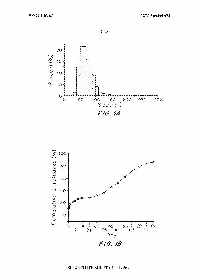
Figures 1A, IB and 1C are graphs of the synthesis and antitumor
effects of nanoparticle-encapsulated DI on BCSC xenograft tumors in the rat.
Size distribution (Figure 1A), controlled release profile of small Dl-loaded
nanoparticles (NPs) (Figure IB), Kaplan-Meier survival curves for tumor-
bearing rats with indicated treatments: blue line, brain-penetrating DI NPs
(median survival >280 d); red line, standard DI NPs (median survival 180 d);
green line, free DI (median survival 177 d); yellow line, blank NPs (median
survival 156 d); grey line, no treatment (median survival 147 d) (Figure 1C).
Rats treated with brain-penetrating, Dl-loaded NPs had significant
improvements in median survival compared to all other groups (p <0.005 for
each comparison). The experiment shown in Figure 1C has been repeated, on
separate occasions, with similar results.
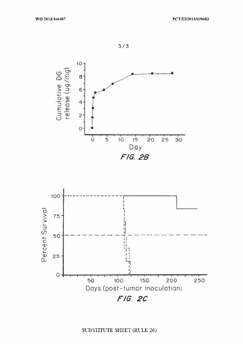
Figures 2A-2C are graphs evaluating the effects of digoxin on BCSCs
in vitro and in vivo. Figure 2A is a graph of the in vitro evaluation of
digoxin on BCSCs, showing that digoxin treatment inhibited BCSC
proliferation. Treatment with digoxin at 1 or 5 µΜ inhibited BCSC sphere
formation. Treatment with digoxin at 1 µΜ decreased the CD 133+
population in the BCSC line PS11, as determined by flow cytometry.
Characteristics, including morphology, size distribution and controlled
release profile (Figure 2B) of brain-penetrating nanoparticles loaded with
digoxin were measured. Figure 2C is a graph of Kaplan-Meier survival
curves for tumor-bearing rats with indicated treatments: black line, no
treatment (n = 6); red line, control NPs (n = 6); green line, brain-penetrating
digoxin NPs (n = 6).
DETAILED DESCRIPTION OF THE INVENTION
The creation of safe, versatile, brain-penetrating nanocarriers enables
direct testing of agents that address the complexity of GBM biology. Cells
isolated from distinct regions of a given GBM bear grossly different
expression signatures, but appear to arise from a common progenitor. A
small subpopulation of these progenitors drives tumor progression, promotes
angiogenesis, and influences tumor cell migration (Fan, et al Semin Cancer
Biol 17, 214-218 (2007); M. F. Clarke, Nature 432, 281-282 (2004); S. Bao
et al, Nature 444, 756-760 (2006); C. Calabrese et al, Cancer Cell 11, 69-
82 (2007)). These cells have features of primitive neural stem cells and, as a
result, are called brain cancer stem cells (BCSCs) (S. Bao et al, Nature 444,
756-760 (2006); S. K. Singh et al, Cancer Res 63, 5821-5828 (2003); R.
Galli et al, Cancer Res 64, 701 1-7021 (2004); S. K. Singh et al, Nature
432, 396-401 (2004); X . Yuan et al, Oncogene 23, 9392-9400 (2004); S.
Bao et al, Cancer Res 66, 7843-7848 (2006); D. Beier et al, Cancer Res 67,
4010-4015 (2007); H. S. Gunther et al, Oncogene, (2007). BCSCs, many of
which are marked by CD 133 (PROM1), are resistant to conventional drugs
(M. F. Clarke, Nature 432, 281-282 (2004); R. J . Jones, J Natl Cancer Inst
96, 583-585 (2004) ), including carboplatin, cisplatin, paclitaxel,
doxorubicin, vincristine, methotrexate, and temozolomide (Tang, et al. Ann
Acad Med Singapore 36, 352-357 (2007); G. Liu et al, Mol Cancer 5, 67
(2006); A. Eramo et al, Cell Death Differ 13, 1238-1241 (2006); C.
c ann- ax et al, Proc Natl Acad Sci USA 101, 14228-14233 (2004)),
as well as radiotherapy (S. Bao et al, Nature 444, 756-760 (2006)). These
observations suggest that agents that target BCSCs are more likely to lead to
cure of GBM (M. F. Clarke, Nature 432, 281-282 (2004); Jones, J Natl
Cancer Inst 96, 583-585 (2004); A. Abbott, Cancer: the root of the problem.
Nature 442, 742-743 (2006); Reya, et al. Nature 414, 105-1 11 (2001).
I. Definitions
The term "biocompatible" as used herein refers to one or more
materials that are neither themselves toxic to the host (e.g., an animal or
human), nor degrade (if the material degrades) at a rate that produces

monomeric or oligomeric subunits or other byproducts at toxic
concentrations in the host.
The term "biodegradable" as used herein means that the materials
degrades or breaks down into its component subunits, or digestion, e.g., by a
biochemical process, of the material into smaller (e.g., non-polymeric)
subunits.
"Sustained release" as used herein refers to release of a substance
over an extended period of time in contrast to a bolus type administration in
which the entire amount of the substance is made biologically available at
one time.
The term "microspheres" is art-recognized, and includes substantially
spherical colloidal structures formed from biocompatible polymers having a
size ranging from about one or greater up to about 1000 microns. In general,
"microcapsules," also an art-recognized term, may be distinguished from
microspheres, as formed of a core and shell. The term "microparticles" is
also art-recognized, and includes microspheres and microcapsules, as well as
structures that may not be readily placed into either of the above two
categories, all with dimensions on average of less than about 1000 microns.
If the structures are less than about one micron in diameter, then the
corresponding art-recognized terms "nanosphere," "nanocapsule," and
"nanoparticle" may be utilized. In certain embodiments, the nanospheres,
nanocapsules and nanoparticles have an average diameter of about 500 nm,
200 nm, 100 nm, 50 nm, 10 nm, or 1 nm.
A composition containing microparticles or nanoparticles may
include particles of a range of particle sizes. In certain embodiments, the
particle size distribution may be uniform, e.g., within less than about a 20%
standard deviation of the mean volume diameter, and in other embodiments,
still more uniform, e.g., within about 10% of the median volume diameter.
The term "particle" as used herein refers to any particle formed of,
having attached thereon or thereto, or incorporating a therapeutic, diagnostic
or prophylactic agent.
The term "targeting moiety" as used herein refers to a moiety that
localizes to or away from a specific locale. The moiety may be, for example,
a protein, nucleic acid, nucleic acid analog, carbohydrate, or small molecule.
Said entity may be, for example, a therapeutic compound such as a small
molecule, or a diagnostic entity such as a detectable label. Said locale may be
a tissue, a particular cell type, or a subcellular compartment. In one
embodiment, the targeting moiety directs the localization of an active entity.
The active entity may be a small molecule, protein, polymer, or metal. The
active entity may be useful for therapeutic, prophylactic, or diagnostic
purposes.
The phrase "pharmaceutically acceptable" refers to compositions,
polymers and other materials and/or dosage forms which are, within the
scope of sound medical judgment, suitable for use in contact with the tissues
of human beings and animals without excessive toxicity, irritation, allergic
response, or other problem or complication, commensurate with a reasonable
benefit/risk ratio.
The phrase "pharmaceutically acceptable carrier" refers to
pharmaceutically acceptable materials, compositions or vehicles, such as a
liquid or solid filler, diluent, solvent or encapsulating material involved in
carrying or transporting any subject composition, from one organ, or portion
of the body, to another organ, or portion of the body. Each carrier must be
"acceptable" in the sense of being compatible with the other ingredients of a
subject composition and not injurious to the patient.
The term "pharmaceutically acceptable salts" is art-recognized, and
includes relatively non-toxic, inorganic and organic acid addition salts of
compounds. Examples of pharmaceutically acceptable salts include those
derived from mineral acids, such as hydrochloric acid and sulfuric acid, and
those derived from organic acids, such as ethanesulfonic acid,
benzenesulfonic acid, and p-toluenesulfonic acid. Examples of suitable
inorganic bases for the formation of salts include the hydroxides, carbonates,
and bicarbonates of ammonia, sodium, lithium, potassium, calcium,
magnesium, aluminum, and zinc. Salts may also be formed with suitable
organic bases, including those that are non-toxic and strong enough to form
such salts.
The term "treating" preventing a disease, disorder or condition from
occurring in an animal which may be predisposed to the disease, disorder
and/or condition but has not yet been diagnosed as having it; inhibiting the
disease, disorder or condition, e.g., impeding its progress; and relieving the
disease, disorder, or condition, e.g., causing regression of the disease,
disorder and/or condition. Treating the disease or condition includes
ameliorating at least one symptom of the particular disease or condition,
even if the underlying pathophysiology is not affected, such as treating the
pain of a subject by administration of an analgesic agent even though such
agent does not treat the cause of the pain.
The term "therapeutically effective amount" refers to an amount of
the therapeutic agent that, when incorporated into and/or onto particles
described herein, produces some desired effect at a reasonable benefit/risk
ratio applicable to any medical treatment. The effective amount may vary
depending on such factors as the disease or condition being treated, the
particular targeted constructs being administered, the size of the subject, or
the severity of the disease or condition. One of ordinary skill in the art may
empirically determine the effective amount of a particular compound without
necessitating undue experimentation. In some embodiments, the term
"effective amount" refers to an amount of a therapeutic agent or prophylactic
agent to reduce or diminish the symptoms of one or more diseases or
disorders of the brain, such as reducing tumor size (e.g., tumor volume) or
reducing or diminishing one or more symptoms of a neurological disorder,
such as memory or learning deficit, tremors or shakes, etc. In still other
embodiments, an "effective amount" refers to the amount of a therapeutic
agent necessary to repair damaged neurons and/or induce regeneration of
neurons.
The terms "incorporated" and "encapsulated" refers to incorporating,
formulating, or otherwise including an active agent into and/or onto a
composition that allows for release, such as sustained release, of such agent
in the desired application. The terms contemplate any manner by which a
therapeutic agent or other material is incorporated into a polymer matrix,
including for example: attached to a monomer of such polymer (by covalent,
ionic, or other binding interaction), physical admixture, enveloping the agent
in a coating layer of polymer, and having such monomer be part of the
polymerization to give a polymeric formulation, distributed throughout the
polymeric matrix, appended to the surface of the polymeric matrix (by
covalent or other binding interactions), encapsulated inside the polymeric
matrix, etc. The term "co-incorporation" or "co-encapsulation" refers to-the
incorporation of a therapeutic agent or other material and at least one other
therapeutic agent or other material in a subject composition.
More specifically, the physical form in which any therapeutic agent
or other material is encapsulated in polymers may vary with the particular
embodiment. For example, a therapeutic agent or other material may be first
encapsulated in a microsphere and then combined with the polymer in such a
way that at least a portion of the microsphere structure is maintained.
Alternatively, a therapeutic agent or other material may be sufficiently
immiscible in the polymer that it is dispersed as small droplets, rather than
being dissolved, in the polymer.
II. Polymeric Nanoparticle Compositions.
A. Polymeric Nanoparticles.
Suitable polymeric carriers include, but are not limited to poly(lactic
acid) (PLA), poly(lactic-co-glycolic acid) (PLGA), poly(lactic acid)-
polyethyleneglycol (PLA-PEG) block copolymers, polyanhydrides,
poly(ester anhydrides), ppolyglycolide (PGA), poly-3-hydroxybutyrate
(PHB) and copolymers thereof, poly-4-hydroxybutyrate (P4HB),
polycaprolactone, cellulose, hydroxypropyl methylcellulose, ethylcellulose,
as well as blends, derivatives, copolymers, and combinations thereof.
In the preferred embodiment, nanoparticles are formed of polylactide-
co-glycolide, wherein the ratio of lactide to glycolide provides the desired
degradation profile.
As demonstrated by the examples, PLGA nanoparticles can be
synthesized using a single-emulsion, solvent evaporation technique. The
solvent should dissolve both the polymer and drug, but without damaging the
drug. For example, dichloromethane (DCM) was chosen as the solvent due

to its ability to dissolve a wide range of hydrophobic drugs. Typically a ratio
equivalent to about 2 ml solvent to 100 mg polymer is used.
The particles are made using GRAS solvents such as ethyl acetate, as
described in the examples. Additives include trehalose or other sugars or
aggregation-reducing materials. Trehalose is the best. Other sugars include
glucose, sucrose and lactose. Typically, the weight ratio of sugar to
nanoparticles is between 10-50%.
B. Therapeutic, Diagnostic and Prophylactic Agents
In the preferred embodiment, drugs that have already been approved
for clinical use are screened for delivery and efficacy in treatment of the ens,
especially brain tumors such as glioblastomas, as described in the examples.
Representative therapeutic agents include vascular endothelial growth
factor ("VEGF") or VEGF receptor inhibitors such as bevacizumab,
alkylating agents such as temozolomide or BCNU (carmustine) , and other
antineoplastics such as procarbazine. Preferred compounds include
Carmustine (BCNU), temozolomide, taxols such as paclitaxel, camptothecin,
and dithiazanine iodide (DI). The particles can also be used to deliver short
acting radioactive compounds.
Loading can range from 0.1 to 20%, with more typical values
between 1-10%
Prophylactics can include compounds alleviating swelling, reducing
radiation damage, and anti-inflammatories.
Diagnostic agents can be radioactive, magnetic, or x-ray or
ultrasound-detectable.
III. Method of Manufacture
In a preferred embodiment, a partial centrifugation technique is used
to produce particles of the desired diameter. The polymer and drug are
dissolved in a common solvent then added to an emulsifying solvent, i.e.,
one that is more water soluble or hydrophilic such as polyvinyl alcohol Other
emulsifying solvents, including didodecyl dimethyl ammonium bromide
(DMAB) and Pluronic F68, can be used. Using a solvent such as DMAB
solution as the emulsifying solvent may result in even smaller nanoparticles.
However, PVA has the least toxicity among these surfactants.. After solvent
evaporation and prior to particle washing, the particle solution is subjected to
low-speed centrifugation, for example, 8,000 g for 10 min, which causes
larger particles to pellet while keeping the smaller particles in the
supernatant. The initial pellet contains comparatively large nanoparticles
and is discarded. Nanoparticles in the supernatant are collected and washed
using high-speed centrifugation, for example, 100,000 g for 30 min.
As demonstrated in the examples, scanning electron microscopy
(SEM) showed that nanoparticles isolated using this protocol with
dicloromethane (DCM) and PLGA were 74 ± 18 nm in diameter and
morphologically spherical. The typical yield for this fabrication was 12% ±
2%. In comparison, nanoparticles made using the same materials but with
conventional centrifugation techniques were 150 ± 30 nm in diameter, with
an average yield of 55% ± 5%.
In a preferred embodiment, demonstrated by the examples, a partially
water soluble solvent was used that provided enhanced results. Organic
solvents used for preparing polymer solution are known to affect the size of
PLGA nanoparticles synthesized through emulsion procedures. In particular,
partially water-miscible organic solvents, such as benzyl alcohol, butyl
lactate, and ethyl acetate (EA), allow nanoparticle formulation through an
emulsion-diffusion mechanism and are able to produce smaller nanoparticles
than water-immiscible solvents such as DCM. Using partially water-
miscible organic solvents improves the yield of brain-penetrating
nanoparticles. EA was chosen because of its low toxicity. Representative
solvents that can be used include DCM, EA, benzyl alcohol, butyl lactate,
and ethyl acetate (EA), acetone. Centrifugation parameters are the same for
all solvents.
Lyophilization is used to stabilize nanoparticles for long-term
storage. To reduce aggregation, a sugar such as the FDA-approved
disaccharide trehalose is added to the particles at a ratio of 0.5: 1
(trehalose:nanoparticles) by mass immediately prior to lyophilization.
IV. Method of Selection of Therapeutic Agents
Drug screening is initially performed in vitro then results confirmed
in vivo.
Drug screening is performed in 96 well plates for primary screening.
In a first embodiment, a slightly modified MTT assay is used to quantify the
effects of drugs on cell proliferation, as described in the examples.
Proliferation is also assessed and IC50 calculated using a technique such as
AlamarBlue (Invitrogen) fluorescence. Fluorescence measures were
corrected for background media and drug fluorescence and normalized to the
mean of vehicle measures. IC50 values are determined using four-parameter
logistic modeling using normalized point estimates.
A sphere formation assay plates brain stem cells as single-cell
suspensions of 5 cells per µ in 48-well plates (Falcon). Cells are treated
with 1 µΜ drug or equivalent concentration of DMSO. Growth factor is
supplemented on day 5. Wells are counted on day 7. Colonies containing
more than 5 cells are considered to be spheres. Percent inhibition is
calculated as: (Control # spheres - Sample # spheres)/Control # spheres. At
3 days after plating, suspensions are collected and flow cytometry
performed.
To establish tumors for evaluation of drug-loaded nanoparticles,
tumor cells are injected into the brains of nude rats. Treatments are
performed 7 days following tumor inoculation. 20 µ of either nanoparticles
(100 mg/mL) or equivalent free drug are infused continuously. The animals'
weight, grooming, and general health are monitored on a daily basis.
V. Method of Treatment
The particles are preferably administered into or adjacent to the area
of the CNS to be treated. This may be at the time of or immediately after
surgical resection of a tumor. Preferably, the particles are administered by
injection into the tissue or the blood vessels leading into the brain. Particles
can be introduced directly in the brain tissue by direct infusion or
convection-enhanced delivery (CED). Alternately, they can be administered
intravenously, or intra-arterially via catheter into an artery that serves the
region of the brain to be treated.
To overcome the challenges associated with drug delivery to the
brain or other regions of the central nervous system, a controlled-release
delivery system comprised of brain-penetrating polymeric nanoparticles that
can penetrate to substantially (~ 7-fold) higher volumes than conventional
polymer nanoparticles when delivered intracranially using CED. The
penetration of these particles is as good as any previously reported
nanoparticle systems: for example, the V V achieved in the examples are
comparable to those achieved with nanoliposomal delivery systems in rats.
Polymeric particles have many advantages over liposomal formulations
including lower toxicity and control of drug release. PLGA nanoparticles
delivered in pig brains using CED penetrated to volumes of approximately
1180 mm3. Since the vast majority of GBMs recur within 2 cm of the
original tumor focus, the penetrative capacity of these brain-penetrating
nanoparticles when delivered by CED can address the infiltrative nature of
GBM. Surface-modified nanoparticles with [1 F]NPB4 using streptavidin-
biotin conjugation, allows tracking the nanoparticles during the CED
procedure using non-invasive PET imaging. This allows clinicians to
visualize nanoparticles delivered by CED and ensure distribution of the
therapeutic agent throughout the brain regions most likely in need of
treatment.
In comparison to currently available nanocarrier drug delivery
systems, this platform has at least three clear advantages. First, the polymer
has an excellent safety profile: PLGA was approved by the Food and Drug
Administration (FDA) in 1969 and has safely been used in clinics since that
time. Second, the release kinetics of PLGA nanoparticles can be more easily
modulated than those of competing nanocarrier systems utilized in
intracranial applications, namely liposomes and micelles. Third, the versatile
surface modification approach described in this study enables rapid, modular
attachment of biotinylated agents, thereby allowing for efficient labeling of
nanoparticles with a host of cell-targeting and -penetrating agents. Finally,
the exceptionally small diameters allow these nanoparticles to penetrate
relatively large, clinically relevant volumes when delivered by CED. In
short, this is a versatile delivery platform for the CNS.
This delivery platform allows for the direct, rapid testing of new
agents for treating GBM. BCSC resistance to conventional
chemotherapeutics is a major challenge in GBM. A library screening
approach to identify agents that have improved activity against BCSCs
allowed screening of over 2,000 compounds. Based on these results, DI was
selected for initial testing due to its abilities to inhibit growth, inhibit self-
renewal, and encourage differentiation of cells it fails to kill. Brain-
penetrating, Dl-loaded PLGA nanoparticles inhibit tumor growth in an
animal model that closely reflects many aspects of human GBM.
Although the brain-penetrating PLGA nanoparticle delivery vehicle
was evaluated here against intracranial tumors with small molecule drugs,
the system can be tailored for application to a host of CNS diseases. For
example, surface modification or size fractionation could produce particles
well suited for the treatment of neurodegenerative disorders. Additionally,
these particles have the potential to encapsulate not only hydrophobic drugs
but also a variety of nucleic acids for gene therapy applications. Due to their
ability to penetrate brain tissue, their construction from safe components, the
ability to control agent release, and the capacity to modulate particle surface
chemistry, we anticipate that this brain-penetrating PLGA nanoparticle
delivery platform will have significant clinical impact.
The present invention will be further understood by reference to the
following non-limiting examples. The following materials and methods
were used in the examples.
Chemicals
All chemicals were purchased from Sigma-Aldrich unless otherwise
noted.
Cell culture
Human glioma cell line U87MG was purchased from ATCC
(American Type Culture Collection). Cells were grown at 37°C incubator
containing 5% C02 and cultured in DMEM medium (Invitrogen)
supplemented with 10% fetal bovine serum (Invitrogen), 100 units/mL
penicillin and 100 µg/mL streptomycin (Invitrogen).
Primary tumor cultures from human GBM tissue
All studies were approved by the appropriate Institutional Review
Boards. Tumor samples classified as GBM based on World Health
Organization (WHO) criteria were obtained from neurosurgical patients at

Yale-New Haven Hospital who had provided informed consent. Within 1 to
3 h of surgical removal, tumors were washed, cut into less than 1 mm3
fragments, and enzymatically dissociated into single cells. Digested
fragments were filtered using a 70 µιη cell strainer (BD Falcon) and
collected in culture medium. The GS5 cell line was provided by Lamszus lab
and described by Gunther et al, Oncogene 27, 2897-2909 (2008). All
primary tumor cells were collected and cultured in Neurobasal A medium
(Invitrogen) supplemented with B27 (Invitrogen), fibroblast growth factor-2
(20 ng/mL, Peprotech), and epidermal growth factor (20 ng/mL, Peprotech).
Growth factors were added at least weekly.
Brain-penetrating nanoparticle synthesis
Nanoparticles loaded with C6 or paclitaxel were synthesized by a
single-emulsion solvent evaporation technique. 100 mg PLGA (50:50,
Polysciences and Birmingham) and agents to be encapsulated were dissolved
in 2 mL dichloromethane (DCM) or ethyl acetate (EA). The polymer/drug
solution was then added dropwise to 4 mL of 2.5% polyvinyl alcohol (PVA)
as the outer aqueous phase and sonicated to form an emulsion. The emulsion
was poured into a beaker containing aqueous 0.3% (v/v) PVA and stirred at
room temperature for 3 h (DCM as solvent) or 5 h (EA as solvent) to allow
the solvent to evaporate and particles to harden.
To synthesize standard nanoparticles, following the solvent
evaporation phase, the nanoparticle solution was subjected to typical
centrifugation speeds ( 11,500 x g for 15 min, x 3) and the pellet was
collected. To synthesize brain-penetrating nanoparticles, following the
solvent evaporation phase, the nanoparticle solution was first centrifuged at
low speed (8,000 x g for 10 min) to pellet the large particles. The
supernatant was decanted and brain-penetrating nanoparticles were collected
through high-speed ultracentrifugation (100,000 x g for 30 min, x 2).
As used herein, large nanoparticles synthesized using standard
protocol are between about 120-200 nm. Brain penetrating nanoparticles for
tumor treatment are between about 60 and 90 nm and for normal brain tissue
are less than 90 nm

To prevent nanoparticle aggregation during lyophilization, trehalose
was added to the final aqueous solution at a ratio of 0.5:1
(trehalose:nanoparticles) by mass immediately prior to lyophilization.
Scanning electron microscopy (SEM)
Particle size was characterized by scanning electron microscopy
(SEM). Samples were mounted on carbon tape and sputter-coated under
vacuum with gold in an argon atmosphere using a Dynavac Mini Coater set
at 40 mA current (Dynavac, USA). SEM was carried out using a Philips
XL30 SEM and LaB electron gun with an accelerating voltage of 3kV. Mean
particle diameters and size distributions were determined by image analysis
of approximately 200 particles using ImageJ (National Institutes of Health).
The same images were used to qualitatively assess particle morphology.
Characterization of nanoparticle loading
To determine the loading and encapsulation efficiency of C6
nanoparticles, 3-5 mg nanoparticles were dissolved in 1 mL DMSO at room
temperature. Loading of C6 in the nanoparticles was quantified based on the
solution's fluorescence intensity (ex: 444 nm, em: 538 nm) using a
spectrophotometer (Spectromax M5, Molecular Devices). Blank
nanoparticles were used for background control. Paclitaxel loading was
quantified using HPLC. The same approach was used to characterize loading
of DI in nanoparticles, except that the concentration of DI was determined
based on its absorbance at 655 nm.
In vitro controlled release
Nanoparticles (3-5 mg) were suspended in 1 mL PBS (pH 7.4), and
incubated at 37°C with gentle shaking (70 rpm). Release of C6, paclitaxel,
or DI was monitored at several time points over a 4-week period. At each
sampling time, the nanoparticle suspension was centrifuged for 15 min at
15,000 rpm. The supernatant was removed for quantification of C6,
paclitaxel, or DI and replaced with an equivalent volume of PBS for
continued monitoring of release. Detection of C6, paclitaxel, or DI was
conducted using the methods described above.
Fluorescence-based imaging of nanoparticle distribution in rat brain
All procedures involving animals were approved by the Yale
University Institutional Animal Care and Utilization Committee (IACUC).
Female athymic (NCr-nu/nu) nude rats were maintained in a sterile
environment. Rats were anesthetized with ketamine/xylazine solution via
intraperitoneal injection and given analgesic. The scalp was prepared for
surgery with betadine and alcohol. The rat was then placed in a stereotactic
head frame. A midline incision was made and a 1.5 mm diameter hole was
drilled in the skull 3 mm lateral and 0.5 mm anterior to the bregma. A 26G
Hamilton syringe, with 28G stepdown inner cannula, was inserted to a depth
of 5 mm. The tissue was allowed to equilibrate mechanically for 5 min.
Subsequently, 20 µ of nanoparticles or free drug was infused V
continuously at a rate of 0.667µ / η η. Following infusion, the syringe was
left in place for 5 min to allow for equilibration. For delivery studies,
animals were sacrificed 30 min post-infusion; the brains were harvested and
frozen.
Nanoparticle distribution was quantified as described by Neeves, et al
Brain Res 1180, 12 1- 132 (2007). Each brain was serially sectioned into 150
µιη slices on a cryostat. The distribution of nanoparticles in the slices was
captured on a fluorescent stereoscope (Zeiss Lumar V.12, Carl Zeiss,
Thornwood, NY) using a CY3 filter. The exposure time was optimized to
achieve maximum dynamic range at the infusion site while simultaneously
avoiding saturation. Exposure time for each nanoparticle group was
individually optimized, in order to adjust for differences in loading between
nanoparticle groups. Within each group of nanoparticles, the exposure time
was held constant. The distribution volume Vd of the nanoparticles was
calculated using a custom Matlab 7.2 (MathWorks, Natick, MA) script,
which generated a binary image from the greyscale images and calculated the
area of particle penetration. The threshold for the binary operation was 10%
of the maximum fluorescent intensity. The total V was calculated by
multiplying the distribution area in each slice by the slice thickness ( 150 µιη)
and summing the volumes of all slices.
Synthesis of [ F]NPB4 nanoparticles
[1 F]NPB4 was prepared as described by Zheng et a , J. Nuclear
Medicine 52, 417 (201 1), with 28% ± 14% radiochemical yield, >98%
radiochemical purity, and 1-2 mCi/nmol specific activity. In preliminary
experiments, [1 F]NPB4 was conjugated to avidin surface-modified PLGA
nanoparticles by incubating 7 mg of nanoparticles with approximately 0.6
mCi of [1 F]NPB4 for 1 h at room temperature. When this solution was
centrifuged to pellet the nanoparticles, no radioactivity was detected in the
wash. Less than 1% of the total added radioactivity was detected in the wash.
It was estimated that <1% of available avidin sites on the nanoparticles were
occupied by the [1 F]NPB4. Each rat received a total dose of 100-300uCi.
PET-based imaging of nanoparticle distribution in rat brain
For noninvasive imaging studies, Sprague Dawley rats were
anesthetized with ketamine/xylazine and received a 26G guide cannula
(Plastics One, Roanoke VA) to enable nanoparticle infusions while data
collection was ongoing. The guide cannula was secured to the surface of the
skull with dental cement (Henry Schein) and surgical screws. Once in the
scanner, rats were maintained on isoflurane anesthesia (2%), and an infusion
needle was threaded through the cannula to the target brain region. Emission
data were collected during the infusion and for 30 min after completion with
a Focus 220 small animal PET scanner (Siemans, Medical Solutions,
Knoxville, TN). A transmission scan ( Co source, 9 min) was collected prior
to the emission scan. Rats were sacrificed immediately after the scan and
frozen in liquid nitrogen for later tissue sectioning and fluorescence
microscopy. PET data were binned into 0.5-10 min frames and reconstructed
with the ordered subset expectation maximization algorithm (OSEM), with
corrections for attenuation, decay, randoms, and scatter. The resulting pixel
size was 0.949 x 0.949 x 0.796 mm, with an effective image resolution of
~1.5mm. Radial concentration profiles were extracted from each data frame
and thresholded to 10% of the maximum value to determine the spatial
volume of distribution.
Fluorescence-based imaging of nanoparticle distribution in pig brain
Nanoparticle infusions were performed in the striatum of Yorkshire
pigs to evaluate V in a large animal model. Pigs were anesthetized with
ketamine/xylazine, intubated, and maintained with isofluorane/oxygen/NC^.
The head was positioned such that the horizontal zero plane passed through
bregma and was parallel to a line between the upper margin of the
infraorbital ridge and the upper margin of the external auditory meatus. The
scalp was prepped with betadine and alcohol. A linear midline incision was
made and a 1.5 mm diameter hole was drilled in the skull 11 mm lateral to
bregma. A 26G Hamilton syringe, with 28G stepdown inner cannula, was
inserted to a depth of 28 mm. The tissue was allowed to equilibrate
mechanically for 5 min. Subsequently, 337.5 L of nanoparticle solution
were continuously infused at a rate of 0.5 µΙ η η for 30 min, 0.75 µΙ η η
for 30 min, and 1 µΙ η η for 300 min. Following infusion, the syringe was
left in place for 120 min, after which it was removed. Animals were
subsequently sacrificed; the brains were harvested, frozen, and sectioned as
described above. Nanoparticle distribution was quantified using the methods
described above. Exposure time was held constant between all animals.
Drug screening
Drug screening was performed in clear 96 well plates using a
compound library which contains 1,937 compounds that are or were FDA-
approved as reported by Chong, et al. Nature chemical biology 2, 415-416
(2006).
Cell proliferation assays
For primary screening, a slightly modified MTT assay was used to
quantify the effects of drugs on cell proliferation. Briefly, cells were cultured
in 96-well plates (Falcon). Three (for BCSC studies) or six (for U87MG
studies) days after treatment, medium was removed and replaced with fresh
medium containing 10% MTT (3-(4,5-dimethylthiazol-2-yl)-2,5-diphenyl
tetrazolium bromide) (Sigma) solution (4. 14 mg/mL). Four hours after
incubation at 37°C, all media was removed. Formazan was dissolved in
DMSO and the optical density (O.D.) was measured at 590 nm. The relative

inhibition on growth was determined using the following formula: Growth
inhibition = (control O.D. - sample O.D.)/control O.D.
Proliferation was also assessed and IC50 calculated using AlamarBlue
(Invitrogen) fluorescence. Briefly, cells were plated at subconfluent
concentration in black clear-bottomed 96-well plates (Falcon) with drug
concentrations spanning eight orders of magnitude. Three or six days post-
plating (as above), AlamarBlue was added at manufacturer's recommended
concentration. Cells were incubated at 37°C for 200 min and quantified (ex:
544 nm, em: 590 nm). Fluorescence measures were corrected for background
media and drug fluorescence and normalized to the mean of vehicle
measures. IC50 values were determined using four-parameter logistic
modeling using normalized point estimates.
Sphere formation assay
BCSCs were plated as single-cell suspensions of 5 cells per L in 48-
well plates (Falcon). Cells were treated with 1 µΜ drug or equivalent
concentration of DMSO. Growth factor was supplemented on day 5. Wells
were counted on day 7. Colonies containing more than 5 cells were
considered to be spheres. Percent inhibition was calculated as: (Control #
spheres - Sample # spheres)/Control # spheres.
Flow cytometry
BCSCs were plated as single-cell suspensions in 6-well plates with
100 nM drug or DMSO. At 3 days after plating, suspensions were collected
and flow cytometry performed. Briefly, following reconstitution in 0.5%
BSA in PBS (w/v), dissociated cells were washed in cold PBS and
subsequently incubated with biotin-conjugated anti-CD 133 (PROM 1)
antibody (Miltenyi Biosciences). Suspensions were incubated with avidin-
conjugated AlexaFluor 488 (Invitrogen) and read on a BD FACSCAN flow
cytometer (BD Biosciences). Geometric means were calculated in FlowJo
(TreeStar, Inc.), corrected for background (secondary only), and normalized
to DMSO-only treated cells.
Antitumor activity in xenograft model
To establish tumors for evaluation of paclitaxel-loaded PLGA
nanoparticles, nude rats were first anesthetized with a ketamine/xylazine
mixture. Animals were then prepped with betadine and alcohol and placed in
a stereotactic frame. A linear midline incision was made and a 1.5 mm
diameter hole was drilled in the skull 3mm lateral and 0.5 mm anterior to
bregma. A 26G Hamilton syringe was inserted to a depth of 5mm. The
tissue was allowed to equilibrate mechanically for 5 min. Subsequently,
5xl0 5 U87MG cells in 2 µΐ PBS was injected into the brain at a rate of 0.5
µΐ/min. The burr hole was filled with bone wax (Lukens, Reading PA), the
scalp closed with surgical staples, and the rat removed to a clean cage with
free access to food and water mixed with ibuprofen. Treatments were
performed 7 days following tumor inoculation. Rats were again anesthetized,
prepped, and placed in a stereotactic frame. The wound was reopened and
the Hamilton syringe was oriented as described previously. 20 µ of either
nanoparticles (100 mg/mL) or equivalent free drug were infused
continuously at a rate of 0.667µ /η η. Following infusion, the syringe was
left in place for 5 min, after which it was removed. The burr hole was filled
with bone wax (Lukens, Reading PA), the scalp closed with surgical staples,
and the rat removed to a clean cage with free access to food and water mixed
with ibuprofen. The animals' weight, grooming, and general health were
monitored on a daily basis. Animals were euthanized after either a 15% loss
in body weight or when it was humanely necessary due to clinical symptoms.
The same procedures were used to evaluate DI nanoparticles, except that
GS5 cells were injected intracranially and treatment was performed 10 days
following tumor cell inoculation.
Statistical Analysis
All data were collected in triplicate, unless otherwise noted, and
reported as mean and standard deviation. Comparison of two conditions was
evaluated by a paired Student's t-test. Kaplan-Meier analysis was employed
to evaluate the effect of various treatments on survival. A p < 0.05 was
considered to indicate a statistically significant difference.
Example 1: Synthesis of brain-penetrating PLGA nanoparticles
Materials and Methods
PLGA nanoparticles were synthesized using a single-emulsion,
solvent evaporation technique. Dichloromethane (DCM) was chosen
initially as the solvent due to its ability to dissolve a wide range of
hydrophobic drugs. A partial centrifugation technique was used to produce
particles of the desired diameter. Specifically, after solvent evaporation and
prior to particle washing, the particle solution was subjected to low-speed
centrifugation (8,000 g for 10 min), which caused larger particles to pellet
while keeping the smaller particles in the supernatant. The initial pellet
contained comparatively large nanoparticles and was removed. Nanoparticles
in the supernatant were collected and washed using high-speed centrifugation
(100,000 g for 30 min).
Results
Scanning electron microscopy (SEM) showed that nanoparticles
isolated using this protocol were 74 ± 18 nm in diameter and
morphologically spherical. The typical yield for this fabrication was 12% ±
2%. In comparison, nanoparticles made using the same materials but with
conventional centrifugation techniques were 150 ± 30 nm in diameter, with
an average yield of 55% ± 5%.
Example 2: Synthesis of brain penetrating NPs with water-miscible
solvent.
Materials and Methods
Partially water-miscible organic solvents, such as benzyl alcohol,
butyl lactate, and ethyl acetate (EA), allow nanoparticle formulation through
an emulsion-diffusion mechanism and are able to produce smaller
nanoparticles than water-immiscible solvents such as DCM. TEA was
chosen because of its low toxicity. The same method otherwise was used as
in Example 1.
Results
Nanoparticles synthesized using EA as solvent instead of DCM were
65 ± 16 nm in diameter and morphologically spherical. The yield was
improved with EA: 44% ± 3%.
Example 3: Cryoprotection of brain-penetrating PLGA nanoparticles
Materials and Methods
Lyophilization is a technique commonly used to stabilize
nanoparticles for long-term storage. However, lyophilization can also cause

nanoparticles to aggregate, making them difficult to resuspend in an aqueous
solution. Furthermore, particle aggregation, if it did occur, could complicate
CED infusion and restrict penetration in the brain. To reduce aggregation,
the FDA-approved disaccharide trehalose was added as an excipient, at a
ratio of 0.5 :1 (trehalose:nanoparticles) by mass immediately prior to
lyophilization.
Results
The addition of trehalose did not alter nanoparticle size, morphology,
or yield. SEM images demonstrated that trehalose enhanced the separation
of nanoparticles from one another when compared to nanoparticles
lyophilized without trehalose. Reconstitution of cryoprotected brain-
penetrating nanoparticles resulted in a homogenous solution, while
reconstitution of nanoparticles lyophilized without trehalose cryoprotection
resulted in sedimentation over time, which caused clogging of the CED
device and prevented infusion at a consistent pressure.
Example 4: CED of brain-penetrating PLGA nanoparticles in the rat
brain
Materials and Methods
The effects of particle size and cryoprotection on intracranial CED
and volume of distribution (Vd) was assessed for both brain-penetrating
PLGA nanoparticles and standard PLGA nanoparticles. Prior to
lyophilization, nanoparticles from each group were further divided into two
groups: with or without trehalose cryoprotection. Nanoparticles were loaded
with coumarin-6 (C6), a fluorescent dye commonly used for visualization.
Brain-penetrating and standard nanoparticles had mean diameters of 71 nm ±
13 nm and 147 nm ± 27 nm, respectively. Consistent with previous work
(44), release of C6 from nanoparticles was negligible (<0.5%) at 72 h.
Sixteen nude rats received 20 i infusions (V\) of C6-loaded
nanoparticles into the right striatum via CED (n=4 per group). Animals were
sacrificed 30 min after infusion and their brains were sectioned and analyzed
using fluorescence microscopy to determine Vd.
Results
Both small size and trehalose cryoprotection independently
contributed to increased penetrance of nanoparticles in brain parenchyma.
Brain-penetrating nanoparticles with cryoprotectant resulted in the best
distribution in the brain. Mean V for brain-penetrating particles with
trehalose was 74 mm3 ± 7 mm3 (Vd/Vi = 3.7 ± 0.3) while mean Vd for
standard particles without trehalose was 11 mm3 ± 3 mm3 (Vd/Vi = 0.6 ±
0.1), p < 0.05. For brain-penetrating nanoparticles with trehalose, the V V
approaches the theoretical limit of 5, which is usually only achievable by
ideal free drugs in solution.
Example 5: Live, non-invasive imaging of brain-penetrating
nanoparticles in the rat brain using PET
Materials and Methods
The clinical translation of delivery systems for the treatment of
intracranial diseases has been hindered by an inability to non-invasively
characterize in vivo distribution. A modular radiolabeling strategy was
employed to permit noninvasive, quantitative PET imaging of the brain-
penetrating nanoparticles. PLGA nanoparticles were modified to display
surface-bound palmitylated avidin, which enabled facile radiolabeling of
nanoparticles with N -(4-[1 F]fluorobenzyl)propanamido-PEG4-Biotin
([1 F]NPB4), a biotinylated, gamma-emitting compound that can be detected
with PET. [1 F]NPB4-labeled and C6-loaded PLGA nanoparticles were
synthesized and delivered via CED to the right striatum of five Sprague-
Dawley rats. Three rats received infusions of brain-penetrating PLGA
nanoparticles with trehalose, while the other two rats received infusions of
standard nanoparticles without trehalose V = 20 for both groups).
Results
When measured noninvasively and quantitatively with PET imaging,
the mean Vd for the brain-penetrating nanoparticles was 111 ± 3 mm3 (Vd/Vi
= 5.5 ± 0.2), while the mean V for the standard nanoparticles was 53 ± 23
mm3 (Vd/Vi = 2.6 ± 1.2). Post-mortem analysis using fluorescence
microscopy revealed that the mean V for the brain-penetrating nanoparticles
was 82 mm3 ± 5 mm3 (Vd/Vi = 4.1 ± 0.2), while the mean Vd for the standard
nanoparticles was 11 mm3 ± 4 mm3 V V = 0.5 ± 0.2). Thus, consistent with
imaging results from destructive fluorescence microscopy, quantitative
analysis of non-invasive PET imaging demonstrated that brain-penetrating
nanoparticles reached a larger volume of spatial distribution than standard
nanoparticles.
Example 6: CED of brain-penetrating nanoparticles in the pig brain
Materials and Methods
Rodent brains are much smaller than human brains, so it is difficult to
assess whether the Vd obtained after CED in the rat is relevant to treatment of
human disease. To extend the analysis to larger brains, brain-penetrating, C6-
loaded PLGA nanoparticles were infused into the striatum of pig brains (n =
4) using the CED technique V = 338 uL). Animals were sacrificed 120 min
post-infusion and their brains were analyzed with fluorescence microscopy to
determine Vd.
Results
Brain-penetrating PLGA nanoparticles delivered by CED penetrated
pig brain tissue with a mean Vd of 1180 mm3 ± 37 mm3, which resulted in
Vd/Vi = 3.5 ± 0.1, similar to the value obtained in the rat and again
approaching the theoretical limit of 5. The extent of nanoparticle penetration
in the pig brain was > 1 cm. The brain-penetrating nanoparticles can be
administered over volumes that are clinically relevant, since the vast majority
of GBMs recur within 2 cm of their original location. Even greater
penetration is possible in humans, since infusion volumes of up to 72 mL
have been used safely in previous clinical trials.
Example 7: Delivery of chemotherapy for solid brain tumor
Materials and Methods
Whether these brain-penetrating PLGA nanoparticles could be used
to treat intracranial tumors was assessed. For initial studies, intracranial
tumors in immunocompromised rats were established by injection of
U87MG, a widely-used, non-BCSC human GBM cell line, and animals
treated with CED of paclitaxel, a drug previously shown to inhibit
proliferation of U87MG. PLGA nanoparticles loaded with paclitaxel were
synthesized by two techniques: brain-penetrating and standard paclitaxel-

loaded nanoparticles were spherical and of expected diameters (75 ± 20 nm
and 159 ± 38 nm, respectively). All nanoparticle fabrications (brain-
penetrating and standard) were loaded with paclitaxel, having encapsulation
efficiencies of approximately 60%, and yields of greater than 35%.
Controlled release experiments showed that brain-penetrating and
standard PLGA nanoparticles released paclitaxel similarly, with
approximately 75% of the encapsulated drug released from each formulation
over the first 28 days of incubation. See Figure 1A. Both brain-penetrating
and standard paclitaxel nanoparticles inhibited growth of U87MG in vitro,
exhibiting lower IC 0 (39 nM and 37 nM, respectively) than free drug (169
nM). None of the blank nanoparticle formulations exhibited cytotoxicity.
See Figure IB.
To determine in vivo efficacy, U87MG-derived xenografts were
generated in the right striatum of nude rats. Tumor-bearing rats were divided
into five groups that received either no treatment; CED of brain-penetrating,
paclitaxel-loaded nanoparticles; CED of standard, paclitaxel-loaded
nanoparticles; CED of blank, brain-penetrating nanoparticles; or CED of
paclitaxel in solution. Consistent with previous experience, rats tolerated all
procedures well; no periprocedural toxicity was observed in any of the
treatment groups. Rats were further monitored for survival: blank
nanoparticles and free paclitaxel failed to show a survival benefit when
compared to no treatment. Kaplan-Meier analysis revealed that rats treated
with brain-penetrating, paclitaxel-loaded nanoparticles had significant
improvements in median survival (46 days) when compared to all groups
(standard nanoparticles: 38 days, free drug: 30 days, blank/unloaded
nanoparticles: 3 1 days, no treatment: 27 days; p<0.05). See Figure 1C.
Example 9: Identification of novel small molecules that inhibit BCSC
proliferation and self-renewal
Materials and Methods
A histopathologic hallmark of GBM is its infiltrative nature. The
U87MG cell line has been propagated in cell culture for many years and has
lost its infiltrative nature in vivo. After intracranial injection, U87MG cells
form solid tumors that are histopathologically distinct from human GBM. In
contrast, several recent studies have demonstrated that a murine xenograft
model utilizing human BCSCs has the ability to precisely recapitulate human
GBM histopathology. To test whether BCSCs were able to form such tumors
in nude rats, GS5, a well-characterized BCSC line was inoculated in rat
brains. Consistent with the findings in mouse brains, GS5 tumors in the brain
of nude rats are highly infiltrative and histopathologically similar to human
GBM.
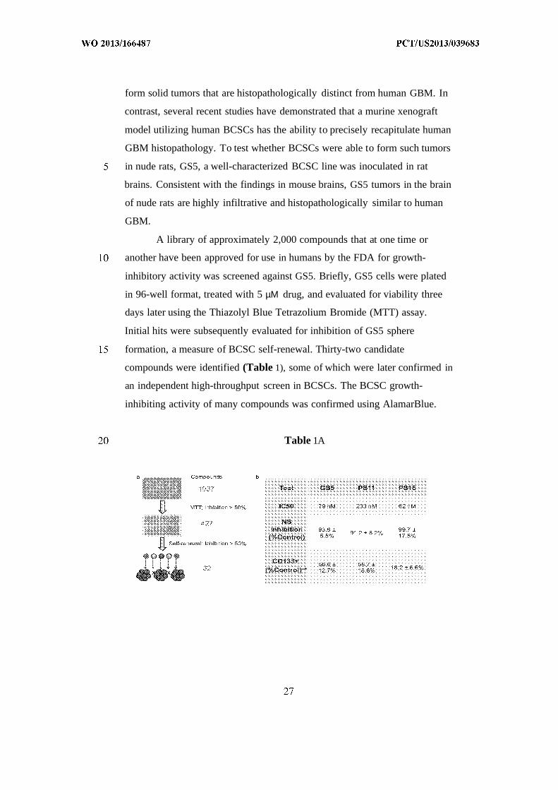
A library of approximately 2,000 compounds that at one time or
another have been approved for use in humans by the FDA for growth-
inhibitory activity was screened against GS5. Briefly, GS5 cells were plated
in 96-well format, treated with 5 µΜ drug, and evaluated for viability three
days later using the Thiazolyl Blue Tetrazolium Bromide (MTT) assay.
Initial hits were subsequently evaluated for inhibition of GS5 sphere
formation, a measure of BCSC self-renewal. Thirty-two candidate
compounds were identified (Table 1), some of which were later confirmed in
an independent high-throughput screen in BCSCs. The BCSC growth-
inhibiting activity of many compounds was confirmed using AlamarBlue.
Table 1A

Table IB
Supplemental Table 1. IC50 (µΜ) of candidate drugs on GS5 cells
One compound in particular, the anti-helminthic cyanine dye
dithiazanine iodide (DI), potently inhibited GS5 proliferation, with an IC50 of
79 nM. DI inhibited GS5 sphere formation, a measure of BCSC self-renewal,
by 94%. Additionally, DI decreased the CD 133+ cell population by 57% in
DI treated culture. DI was evaluated in two additional BCSC lines, PSl 1 and
PS16, and showed similar anti-BCSC effects.
Example 10: CED of brain-penetrating, Dl-loaded PLGA nanoparticles
for GBM therapy
Materials and Methods
CED of brain-penetrating, Dl-loaded nanoparticles was tested to see
if it could prevent the growth of a more histopathologically relevant model of
GBM than U87MG. DI was loaded into brain-penetrating nanoparticles with
encapsulation efficiency of 19% and yield of 18%. Brain-penetrating, Dl-
loaded nanoparticles were spherical and had an average diameter of 70 ± 19
nm. DI was released from brain-penetrating nanoparticles in a controlled
manner over several weeks. To evaluate their efficacy in vivo, brain-
penetrating, Dl-loaded nanoparticles were administrated via a single infusion
into rat brains bearing GS5-derived tumors.
Results
DI nanoparticles significantly increased survival of tumor-bearing
rats.. The median survival for control rats receiving either no treatment or
blank nanoparticles was 115 and 113 days, respectively. In contrast, only one
rat in the DI nanoparticle treatment group exhibited neurological symptoms
and was euthanized 189 days after treatment, while the other five rats in the
DI group remained healthy for over 250 days.
Example 11: Anisomycin and digoxin particles were not effective using
CED delivery.
Two other compounds that exhibited activity in the in vitro screening
experiments were tested. Both anisomycin and digoxin performed well on in
vitro assays against BCSCs and were loaded efficiently into brain-
penetrating nanoparticles that provided controlled release. In pilot
experiments, however, CED delivery of anisomycin-loaded or digoxin-
loaded particles provided no survival benefit to rats with intracranial BCSC-
derived tumors. See Figures 1C and 2C.

We claim:
1. A formulation for delivering therapeutic, prophylactic or diagnostic
agent to the central nervous system comprising nanoparticles formed of
polymer and a therapeutic, diagnostic, or prophylactic agent, the
nanoparticles consisting essentially of nanoparticles having an average
diameter of less than 100 nm.
2. The formulation of claim 1 wherein the nanoparticles are formed by
emulsification and separated by size centrifugation.
3. The formulation of claim 1 wherein the particles have a size range of
less than 100 nm and can penetrate brain tissue.
4. The formulation of claim 1 wherein the polymer is a hydrophobic
biodegradable polymer.
5. The formulation of claim 1 wherein the agent is a chemotherapeutic
for treatment of cancer.
6. The formulation of claim 5 wherein the agent is selected from the
group consisting of Carmustine (BCNU), temozolomide, taxols such as
paclitaxel, camptothecin, and dithiazanine iodide (DI).
7. The formulation of claim 1 comprising an excipient to decrease
aggregation following lyophilization.
8. A method of delivering a therapeutic, prophylactic or diagnostic agent
to the central nervous system comprising injecting into the blood stream or
tissue adjacent to the region of the central nervous system to be treated a
formulation comprising nanoparticles formed of polymer and a therapeutic,
diagnostic, or prophylactic agent, the nanoparticles consisting essentially of
nanoparticles having an average diameter of less than 100 nm.
9. The method of claim 8 wherein the region is the brain.
10. The method of claim 8 wherein the particles can penetrate brain
tissue and are less than 90 nm average diameter.
11. The method of claim 10 wherein the particles are for treatment of
brain tumors and have an average diameter of between 60 and 90 nm.
12. The method of claim 10 wherein the particles comprise a therapeutic,
diagnostic or prophylactic agent for delivery to brain tissue.

13. The method of claim 12 wherein the agent is a therapeutic in an
amount effective to reduce the size or alleviate the symptoms of a brain
tumor.
14. The method of claim 13 wherein the agent is selected from the group
consisting of carmustine (BCNU), temozolomide, taxols such as paclitaxel,
camptothecin, and dithiazanine iodide (DI).


International application No
PCT/US2013/039683
A. CLASSIFICATION O F SUBJECT MATTERINV. A61K9/00 A61K9/51 A61K31/337ADD.
According to International Patent Classification (IPC) or to both national classification and IPC
B. FIELDS SEARCHED
Minimum documentation searched (classification system followed by classification symbols
A61K
Documentation searched other than minimum documentation to the extent that such documents are included in the fields searched
Electronic data base consulted during the international search (name of data base and, where practicable, search terms used)
EPO-Internal , COMPENDEX, EMBASE, WPI Data
C. DOCUMENTS CONSIDERED TO BE RELEVANT
Category * Citation of document, with indication, where appropriate, of the relevant passages Relevant to claim No.
US 2008/274202 Al (KRAIG RICHARD P [US] ET 1-6,8-14AL) 6 November 2008 (2008-11-06)paragraph [0016]paragraph [0021]paragraph [0025]paragraph [0028]paragraph [0043]c l aim 12; examples 2 , 15
US 5 213 788 A (RANNEY DAVID F [US] ) 1,3 ,7-1225 May 1993 (1993-05-25)col umn 1, l ines 9-15col umn 1, l ines 39-45col umn 5 , l ines 20-35col umn 5 , ine 46 - col umn 6 , ine 2col umn 6 , l ines 44-61col umn 8 , l ines 34-37exampl es 11, 18
/--
Further documents are listed in the continuation of Box C. See patent family annex.
* Special categories of cited documents :" later document published after the international filing date or priority
date and not in conflict with the application but cited to understand"A" document defining the general state of the art which is not considered the principle or theory underlying the invention
to be of particular relevance
"E" earlier application or patent but published on or after the international "X" document of particular relevance; the claimed invention cannot befiling date considered novel or cannot be considered to involve an inventive
"L" document which may throw doubts on priority olaimfs) orwhich is step when the document is taken alonecited to establish the publication date of another citation or other Ύ " document of particular relevance; the claimed invention cannot bespecial reason (as specified) considered to involve an inventive step when the document is
Ό " document referring to an oral disclosure, use, exhibition or other combined with one or more other such documents, such combinationmeans being obvious to a person skilled in the art
"P" document published prior to the international filing date but later thanthe priority date claimed "&" document member of the same patent family
Date of the actual completion of the international search Date of mailing of the international search report
12 July 2013 19/07/2013
Name and mailing address of the ISA/ Authorized officer
European Patent Office, P.B. 5818 Patentlaan 2NL - 2280 HV ijsw ij
Tel. (+31-70) 340-2040,Fax: (+31-70) 340-3016 Zal fen, Al ina

International application No
PCT/US2013/039683
C(Continuation). DOCUMENTS CONSIDERED TO BE RELEVANT
Category* Citation of document, with indication, where appropriate, of the relevant passages Relevant to claim No.
US 2007/231360 Al (PEYMAN GHOLAM A [US] ) 1,3-6,4 October 2007 (2007-10-04) 8-12paragraph [0002]paragraph [0017]paragraph [0027]paragraph [0084] - paragraph [0085]paragraph [0087]
US 2003/017131 Al (PARK TAE GWAN [KR] ET 1,3-6,AL) 23 January 2003 (2003-01-23) 8-14paragraph [0027]paragraph [0020]table 4
X , P W0 2012/101639 A2 (YISSUM RES DEV CO [ L] ; 1-4,8-12BENITA SIMON [ I L] ; NASSER TAHER [ I L] ;KARRA NO) 2 August 2012 (2012-08-02)page 7 , l i nes 12-17page 8 , l i nes 24-27page 9 , l i ne 26 - page 10, l ine 10page 17, l i nes 15-19page 19, l i nes 8-11table 8
GAUMET M ET AL: "Nanoparti cles for drug 1-14del ivery: The need for preci s i on i nreporti ng parti cle s i ze parameters" ,EUROPEAN JOURNAL OF PHARMACEUTICS AND
BIOPHARMACEUTICS, ELSEVI ER SCI ENCE
PUBLISHERS B.V. , AMSTERDAM, NL,vol . 69, no. 1 , 1 May 2008 (2008-05-01) ,pages 1-9 , XP022589593 ,ISSN: 0939-6411, D0I :10. 1016/J . EJPB.2007.08.001[retri eved on 2008-04-05]the whole document
MAM0T C ET AL: "EXTENSIVE DISTRIBUTION OF 1-14LI POSOMES I N RODENT BRAINS AND BRAINTUMORS FOLLOWING CONVECTION-ENHANCEDDELIVERY" ,JOURNAL OF NEURO-ONCOLOGY, KLUWER, BOSTON ,US,vol . 68, no. 1 , 1 May 2004 (2004-05-01) ,pages 1-09 , XP009039195,ISSN: 0167-594X, D0I :10 . 1023/B : NEON . 0000024743 . 56415 . 4 Babstract
-/--

International application No
PCT/US2013/039683
C(Continuation). DOCUMENTS CONSIDERED TO BE RELEVANT
Category* Citation of document, with indication, where appropriate, of the relevant passages Relevant to claim No.
MUSUMECI T ET AL: " Lyoprotected 1-14nanosphere formulations for pacl i taxelcontrol led del i er y " ,JOURNAL OF NANOSCI ENCE AND NANOTECHNOLOGY,AMERICAN SCI ENTI FIC PUBLISHERS, US,vol . 6 , no. 9-10,1 September 2006 (2006-09-01) , pages3118-3125 , XP008136529 ,ISSN: 1533-4880, D0I : 10. 1166/JNN .2006.452abstract
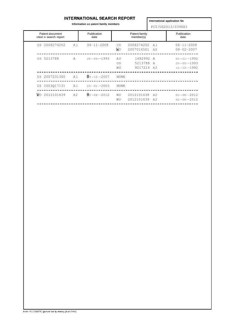
International application NoInformation on patent family members
PCT/US2013/039683
Patent document Publication Patent family Publicationcited in search report date member(s) date
US 2008274202 Al 06-11-2008 US 2008274202 Al 06-11-2008O 2007016501 A2 08-02-2007
US 5213788 A 25·-05--1993 AU 1682992 A 02·-11·-1992
US 5213788 A 25·-05·-1993WO 9217214 A2 15·-10·-1992
US 2007231360 Al 4 ·-10--2007 NONE
US 2003Q17131 Al 23·-01--2003 NONE
O 2012101639 A2 2 ·-08--2012 WO 2012101638 A2 02·-08·-2012
WO 2012101639 A2 02·-08·-2012