WO 2011/048215 Al
77
(12) INTERNATIONAL APPLICATION PUBLISHED UNDER THE PATENT COOPERATION TREATY (PCT) (19) World Intellectual Property Organization International Bureau (10) International Publication Number (43) International Publication Date ¾ ι t 28 April 2011 (28.04.2011) WO 2011/048215 Al (51) International Patent Classification: (74) Agent: WEISS, Wolfgang; Weickmann & Weickmann, A61K 39/12 (2006.01) C12N 7/00 (2006.01) Postfach 860 820, 81635 Munchen (DE). C07K 14/01 (2006.01) (81) Designated States (unless otherwise indicated, for every (21) International Application Number: kind of national protection available): AE, AG, AL, AM, PCT/EP2010/065983 AO, AT, AU, AZ, BA, BB, BG, BH, BR, BW, BY, BZ, CA, CH, CL, CN, CO, CR, CU, CZ, DE, DK, DM, DO, (22) International Filing Date: DZ, EC, EE, EG, ES, FI, GB, GD, GE, GH, GM, GT, 22 October 2010 (22.10.2010) HN, HR, HU, ID, IL, IN, IS, JP, KE, KG, KM, KN, KP, (25) Filing Language: English KR, KZ, LA, LC, LK, LR, LS, LT, LU, LY, MA, MD, ME, MG, MK, MN, MW, MX, MY, MZ, NA, NG, NI, (26) Publication Language: English NO, NZ, OM, PE, PG, PH, PL, PT, RO, RS, RU, SC, SD, (30) Priority Data: SE, SG, SK, SL, SM, ST, SV, SY, TH, TJ, TM, TN, TR, 09173810.4 22 October 2009 (22.10.2009) EP TT, TZ, UA, UG, US, UZ, VC, VN, ZA, ZM, ZW. (71) Applicants (for all designated States except US): UNI- (84) Designated States (unless otherwise indicated, for every VERSITAT LEIPZIG [DE/DE]; RitterstraBe 26, 04109 kind of regional protection available): ARIPO (BW, GH, Leipzig (DE). TIERGESUNDHEITSDIENST BAY- GM, KE, LR, LS, MW, MZ, NA, SD, SL, SZ, TZ, UG, ERN E.V. [DE/DE]; Senator-Gerauer-Str. 23, 85586 Po- ZM, ZW), Eurasian (AM, AZ, BY, KG, KZ, MD, RU, TJ, ing-Grub (DE). TM), European (AL, AT, BE, BG, CH, CY, CZ, DE, DK, EE, ES, FI, FR, GB, GR, HR, HU, IE, IS, ΓΓ , LT, LU, (72) Inventors; and LV, MC, MK, MT, NL, NO, PL, PT, RO, RS, SE, SI, SK, (75) Inventors/ Applicants (for US only): MULLER, Her¬ SM, TR), OAPI (BF, BJ, CF, CG, CI, CM, GA, GN, GQ, mann [DE/DE]; Politzstr. 29, 04 155 Leipzig (DE). HA- GW, ML, MR, NE, SN, TD, TG). LAMI, Mohammad Yahya [DE/DE]; Tarostr. 16/563, 04103 Leipzig (DE). BOTTCHER, Jens [DE/DE]; Published: Franz-Anneser-Str. 16, 84485 Dorfen (DE). KAPPE, Eva — with international search report (Art. 21(3)) [DE/DE]; Mitterfeldring 126, 85586 Poing (DE). — with sequence listing part of description (Rule 5.2(a)) SCHADE, Benjamin [DE/DE]; Inselstr. 2, 86983 Lech- bruck am See (DE). (54) Title: DETECTION OF A CIRCOVIRUS IN CALVES SUFFERING FROM BOVINE NEONATAL PANCYTOPENIA (57) Abstract: The present invention refers to a novel circovirus as causative agent of bone marrow aplasia with haemorrhagic disease in cattle. The present invention provides novel nucleic acid and protein sequences for diagnostic and therapeutic uses.
-
Upload
khangminh22 -
Category
Documents
-
view
1 -
download
0
Transcript of WO 2011/048215 Al
(12) INTERNATIONAL APPLICATION PUBLISHED UNDER THE PATENT COOPERATION TREATY (PCT)
(19) World Intellectual Property OrganizationInternational Bureau
(10) International Publication Number(43) International Publication Date ¾ ι t
28 April 2011 (28.04.2011) WO 2011/048215 Al
(51) International Patent Classification: (74) Agent: WEISS, Wolfgang; Weickmann & Weickmann,A61K 39/12 (2006.01) C12N 7/00 (2006.01) Postfach 860 820, 81635 Munchen (DE).C07K 14/01 (2006.01)
(81) Designated States (unless otherwise indicated, for every(21) International Application Number: kind of national protection available): AE, AG, AL, AM,
PCT/EP2010/065983 AO, AT, AU, AZ, BA, BB, BG, BH, BR, BW, BY, BZ,CA, CH, CL, CN, CO, CR, CU, CZ, DE, DK, DM, DO,
(22) International Filing Date: DZ, EC, EE, EG, ES, FI, GB, GD, GE, GH, GM, GT,22 October 2010 (22.10.2010) HN, HR, HU, ID, IL, IN, IS, JP, KE, KG, KM, KN, KP,
(25) Filing Language: English KR, KZ, LA, LC, LK, LR, LS, LT, LU, LY, MA, MD,ME, MG, MK, MN, MW, MX, MY, MZ, NA, NG, NI,
(26) Publication Language: English NO, NZ, OM, PE, PG, PH, PL, PT, RO, RS, RU, SC, SD,
(30) Priority Data: SE, SG, SK, SL, SM, ST, SV, SY, TH, TJ, TM, TN, TR,
09173810.4 22 October 2009 (22.10.2009) EP TT, TZ, UA, UG, US, UZ, VC, VN, ZA, ZM, ZW.
(71) Applicants (for all designated States except US): UNI- (84) Designated States (unless otherwise indicated, for every
VERSITAT LEIPZIG [DE/DE]; RitterstraBe 26, 04109 kind of regional protection available): ARIPO (BW, GH,
Leipzig (DE). TIERGESUNDHEITSDIENST BAY- GM, KE, LR, LS, MW, MZ, NA, SD, SL, SZ, TZ, UG,
ERN E.V. [DE/DE]; Senator-Gerauer-Str. 23, 85586 Po- ZM, ZW), Eurasian (AM, AZ, BY, KG, KZ, MD, RU, TJ,
ing-Grub (DE). TM), European (AL, AT, BE, BG, CH, CY, CZ, DE, DK,EE, ES, FI, FR, GB, GR, HR, HU, IE, IS, ΓΓ , LT, LU,
(72) Inventors; and LV, MC, MK, MT, NL, NO, PL, PT, RO, RS, SE, SI, SK,(75) Inventors/ Applicants (for US only): MULLER, Her¬ SM, TR), OAPI (BF, BJ, CF, CG, CI, CM, GA, GN, GQ,
mann [DE/DE]; Politzstr. 29, 04 155 Leipzig (DE). HA- GW, ML, MR, NE, SN, TD, TG).LAMI, Mohammad Yahya [DE/DE]; Tarostr. 16/563,04103 Leipzig (DE). BOTTCHER, Jens [DE/DE]; Published:
Franz-Anneser-Str. 16, 84485 Dorfen (DE). KAPPE, Eva — with international search report (Art. 21(3))[DE/DE]; Mitterfeldring 126, 85586 Poing (DE). — with sequence listing part of description (Rule 5.2(a))SCHADE, Benjamin [DE/DE]; Inselstr. 2, 86983 Lech-bruck am See (DE).
(54) Title: DETECTION OF A CIRCOVIRUS IN CALVES SUFFERING FROM BOVINE NEONATAL PANCYTOPENIA
(57) Abstract: The present invention refers to a novel circovirus as causative agent of bone marrow aplasia with haemorrhagicdisease in cattle. The present invention provides novel nucleic acid and protein sequences for diagnostic and therapeutic uses.
DETECTION OF A CIRCOVIRUS IN CALVES SUFFERING FROM BOVINE NEONATALPANCYTOPENIA
Description
The present invention refers to a novel circovirus (CV) as causative agent of
bone marrow aplasia with haemorrhagic disease in cattle. The present
invention provides novel nucleic acid and protein sequences for diagnostic
and therapeutic uses.
Introduction
Haemorrhagic diseases in cattle have been associated with a variety of
causes including viral infections, hereditary diseases, immune-mediated
diseases, bacterial septicaemia, and intoxications. Bleeding tendency and
thrombocytopenia are associated with non-cytopathic type 2 bovine viral
diarrhoea virus (BVDV) infection (Ellis et al., 1998, Rebhun et al., 1989). A
hereditable haemorrhagic diathesis is described for Simmental cattle. This
Simmental hereditary thrombopathy is caused by dysfunction of platelets
(Steficek, et al., 1993). Immune-mediated thrombocytopenia is known as a
rare condition in cows. It may be classified as idiopathic thrombocytopenic
purpura or secondary entity (Yeruham, et al., 2003). Examples of bacterial
infections include Pasteurella multocida, a well known cause of
haemorrhagic septicaemia in calves with petechial and ecchymotic
haemorrhages, generalized hyperaemia, and pneumonia as clinical signs
(Rhoades, et al., 1967, Rimler, 1978).
Several toxins may be responsible for fatal haemorrhagic diathesis in cattle.
Intoxications due to dichlorovinylcysteine (DCVC) in trichloroethylene-
extracted soybean oil meal fed to calves (Lock, et al., 1996) and also the
antibiotic furazolidone (Hoffmann-Fezer, et al., 1974, Hofmann, et al., 1974)
produce fatal aplastic anaemia, marked acellularity of bone marrow and
extensive haemorrhages. Ingestion of bracken fern (Pteridium aquilinum)
causes acute poisoning in cattle with irreversible bone marrow hypoplasia as

we (Maxie and Newman, 2007, Valli, 2007). In addition, intoxications with
mycotoxins of Stachybotrys chartarum (atra) have been described in
ruminants resulting in pancytopenic disease characterized by profuse
haemorrhage and necrosis in many tissues (Harrach, et al., 1983, Valli,
2007).
Chicken infectious anaemia is a disease strongly resembling the
haemorrhagic disease in calves reported here. The causative agent is
chicken infectious anaemia virus (CIAV). Severe anaemia, severe bone
marrow aplasia, atrophy of the thymus and Bursa of Fabricius, and
haemorrhages are consistent findings in chicks infected with CIAV (Kuscu
and Gurel, 2008, Yuasa, et al., 1979). One-day-old SPF chicks,
experimentally inoculated with CIAV, showed a decrease of haematocrit
values, became emaciated and depressed with anaemia, particularly
between days 12 and 20 post inoculation (Goryo, et al., 1989). CIAV is
classified into the family Circoviridae (Todd, et al., 2005). It only infects
chicken and is the sole member of the genus Gyrovirus. However, several
members of a second genus, Circovirus, have been detected in mammalian
and avian species including the porcine circoviruses PCV1 and PCV2.
Members of the family Circoviridae are non-enveloped icosahedral particles
with a circular single-stranded DNA (ssDNA) genome, 1759 to 2319
nucleotides (nt) in size (Todd, et al., 2005). Viruses in the genus Circovirus
possess an ambisense genome organization encoding the replication-
associated (Rep) protein from the sense strand (open reading frame [ORF]-
V 1) and the capsid protein from the complementary sense strand (ORF-C1 ) .
Additional small ORFs have been recognized in some of the circoviruses,
e.g., ORF3 encoding an apoptosis-inducing protein in PCV2-infected cells
(Liu, et al., 2005, Timmusk, et al., 2008). In the noncoding regions, a stem-
loop structure is present containing a conserved nonamer sequence and
involved in the initiation of the viral genome replication (Steinfeldt, et al.,
2001 ) . The molecular biology of circoviruses has been reviewed recently
(Mankertz, 2008).
With the exception of PCV1 , all known circoviruses are pathogens, which
cause immunosuppression and damage of the lymphoreticular tissues
(Mankertz, 2008, Segales, et al., 2005, Segales and Mateu, 2006, Todd,
2000). PCV2 is a virulent pathogen associated with a number of different
syndromes and diseases in pigs such as the post-weaning multisystemic
wasting syndrome (PMWS), the porcine respiratory disease complex
(PRDC), reproductive failure associated with PCV2 and the porcine
dermatitis and nephropathy syndrome (PDNS). However, only lesions typical
of PMWS were demonstrated in both colostrum-deprived piglets and
conventional pigs by PCV2 inoculation (Ellis, et al., 1999, Kennedy, et al.,
2000) , whereas the involvement of PCV2 in swine diseases other than
PMWS has not been fully investigated (Allan, et al., 2003, Chae, 2005).
Only limited data exist on circovirus infections in cattle. The presence of
circoviruses was demonstrated by PCR in lung tissue samples from 6 of 100
cases of bovine respiratory disease and from 4 of 30 aborted fetuses (Nayar,
et al., 1999). The genome of this agent, tentatively named bovine circovirus
(BCV), was nearly identical to that of PCV2, with 99% overall nucleotide
sequence identity. The presence of antibodies reacting with porcine
circovirus in sera of humans, mice and cattle has been reported (Tischer, et
al., 1995). In another study, however, no antibodies to PCV2 were detected
in sera from cattle, sheep, horse and humans (Allan, et al., 2000, Ellis, et al.,
2001 ). Also, a seronegative neonatal calf and six seronegative 6-months-old
beef calves that were experimentally infected with PCV2 failed to develop
antibodies to the virus (Ellis, et al., 2001 ) .
Since 2007, there have been reports by farmers and veterinarians of an
unexplainable haemorrhagic disease in calves all over Germany. 56 calves
with spontaneous haemorrhages were presented to the Bavarian Animal
Health Service to characterize the lesions and to investigate the aetiology by
further laboratory investigations. The disease was observed in young calves
of different breeds within their first month of life. Male and female calves
were affected likewise. Haemorrhages, particularly in skin, subcutis and
gastrointestinal tract, were the major findings. Inflammatory lesions were
additional sporadic findings. Histological investigation indicated a severe
bone marrow hypo- to aplasia in all animals and lymphocytic depletion in
43% of the affected calves. Blood analysis of 5 animals revealed aplastic
pancytopenia. The resulting thrombocytopenia is believed to represent the
major pathomechanism of this Haemorrhagic Disease Syndrome (HDS), also
referred to as Haemorrhagic Diathesis (HD). Meanwhile, the more common
and scientifically accepted name for HDS/HD is Bovine Neonatal
Pancytopenia (BNP). The different names and abbreviations may be used
interchangeably in the present application. Bacterial infections and infections
with bovine viral diarrhoea virus or bluetongue virus were ruled out as cause
of the disease. Specific toxins which are known to cause bone marrow
aplasia were not detected. Pedigree analysis gave no indication for heredity
of the disease.
Using a broad spectrum PCR, the present inventors were able to
demonstrate the presence of a circovirus in affected calves. Sequencing of
the whole viral genome revealed high similarities with porcine circovirus type
2b (PCV2b). Single bone marrow cells of one calf displayed slight PCV2-
antigen immunoreaction.
SUMMARY OF THE INVENTION
The present invention refers to nucleic acid molecules of a novel circovirus
(CV) identified as causative agent of Haemorrhagic Disease Syndrome
(HDS), or Haemorrhagic Diathesis (HD), now mainly referred to as Bovine
Neonatal Pancytopenia (BNP) in cattle. Further, the invention refers to novel
polypeptides encoded by the viral nucleic acid and antibodies directed
against these polypeptides. The nucleic acids, polypeptides and antibodies
are suitable for diagnostic and therapeutic uses, particularly for the
development of vaccines against HD.
In a first aspect, the present invention refers to a circovirus (CV) nucleic acid

molecule comprising
(a) the sequence as shown in SEQ ID NO: 1 or a fragment thereof, and/or
(b) the complement of the nucleotide sequence according to (a).
In another aspect, the present invention refers to a circovirus (CV) nucleic
acid molecule comprising
(a) the sequence as shown in SEQ ID NO: 7 or a fragment thereof, and/or
(b) the complement of the nucleotide sequence according to (a).
In yet another aspect, the present invention refers to a circovirus (CV)
nucleic acid molecule comprising
(a) the sequence as shown in SEQ ID NO: 11 or a fragment thereof, and/or
(b) the complement of the nucleotide sequence according to (a).
The nucleic acid molecule may be a DNA or RNA molecule, which is single-
or double-stranded, circular or linear. In some embodiments, the nucleic acid
molecule may be present as such or linked to further nucleic acid molecules,
e.g. operatively linked with heterologous expression control sequences. The
nucleic acid molecule may also be encapsulated in a viral capsid.
The nucleic acid molecule of the present invention may comprise the
complete sequence of SEQ ID NO: 1 and/or its complement or a fragment
thereof. Likewise, the nucleic acid molecule of the present invention may
comprise the complete sequence of SEQ ID NO: 7 or SEQ ID NO: 11 and/or
its complement or a fragment thereof. The fragment preferably comprises at
least 15, at least 20, at least 25, at least 30 or at least 50 contiguous
nucleotides as shown in SEQ ID NO: 1, 7 , or 11 or the complement thereof.
Preferably, the CV nucleic acid molecule of the present invention differs in at
least one nucleotide from related circovirus strains, e.g. circovirus virus
strains, whose GeneBank Accession Numbers are indicated in Figure 6 .
The present invention also refers to a nucleic acid molecule
(a) having an identity of at least 90%, at least 94%, at least 96%, at least

98% or at least 99% of the nucleotide sequence as shown in SEQ ID
NO: 1,
(b) hybridizing under stringent conditions to the nucleotide sequence as
shown in SEQ ID NO:1 , or
(c) the complement of (a) or (b).
The present invention also refers to a nucleic acid molecule
(a) having an identity of at least 90%, at least 94%, at least 96%, at least
98% or at least 99% of the nucleotide sequence as shown in SEQ ID
NO: 7 ,
(b) hybridizing under stringent conditions to the nucleotide sequence as
shown in SEQ ID NO:7, or
(c) the complement of (a) or (b).
The present invention also refers to a nucleic acid molecule
(a) having an identity of at least 90%, at least 94%, at least 96%, at least
98% or at least 99% of the nucleotide sequence as shown in SEQ ID
NO: 11,
(b) hybridizing under stringent conditions to the nucleotide sequence as
shown in SEQ ID NO:1 1, or
(c) the complement of (a) or (b).
The identity of a given nucleic acid molecule to the reference nucleic acid
molecule (i.e., for example, SEQ ID NO: 1 or a fragment thereof) may be
determined as follows:
l=n/L x 100,
wherein I is the identity in percent,
n is the number of identical nucleotides of the given nucleic acid
molecule, and the reference, and
L is the length of the sequence overlap of the given nucleic acid
molecule and the reference.

Preferably, hybridization under stringent conditions means that after washing
for h with 1 X SSC-buffer and 0.1 % SDS at 50 °C, preferably at 55 °C, more
preferably at 62 °C, and most preferably at 68 °C, particularly for 1h in 0.2 X
SSC and 0.1% SDS at 50 °C, preferably at 55 °C, more preferably at 62 °C,
and most preferably at 68 °C, a positive hybridization signal is observed.
Hybridization protocols are e.g. disclosed in Wahl and Berger (Methods
Enzymol. 152 (1987), 399-407) and Kimmel (Methods Enzymol. 152 (1987),
507-51 1) , the content of which is herein incorporated by reference.
A further aspect of the present invention refers to a CV nucleic acid molecule
encoding a circovirus polypeptide or a fragment thereof, wherein the nucleic
acid molecule comprises
(a) a region of the nucleotide sequence as shown in SEQ ID NO: 1 from
(i) nucleotide 5 1 - 995, (Rep)
(ii) nucleotide 1034 - 1735 (Cap)
(iii) nucleotide 357 - 671 (ORF3), or
(b) a nucleotide sequence corresponding to the sequence of (a) within the
scope of degeneracy of the genetic code, or
(c) a fragment of the nucleotide sequence according to (a) or (b).
Still a further aspect of the present invention refers to a CV nucleic acid
molecule encoding a circovirus polypeptide or a fragment thereof, wherein
the nucleic acid molecule comprises
(a) a region of the nucleotide sequence as shown in SEQ ID NO: 7 from
(i) nucleotide 5 1 - 995, (Rep)
(ii) nucleotide 1034 - 1735 (Cap)
(iii) nucleotide 357 - 671 (ORF3), or
(b) a nucleotide sequence corresponding to the sequence of (a) within the
scope of degeneracy of the genetic code, or
(c) a fragment of the nucleotide sequence according to (a) or (b).
Still a further aspect of the present invention refers to a CV nucleic acid
molecule encoding a circovirus polypeptide or a fragment thereof, wherein
the nucleic acid molecule comprises
(a) a region of the nucleotide sequence as shown in SEQ ID NO: 11 from
(i) nucleotide 5 1 - 995, (Rep)
(ii) nucleotide 1033 - 1734 (Cap)
(iii) nucleotide 357 - 671 (ORF3), or
(b) a nucleotide sequence corresponding to the sequence of (a) within the
scope of degeneracy of the genetic code, or
(c) a fragment of the nucleotide sequence according to (a) or (b).
Preferably, the nucleic acid molecule encodes a CV polypeptide selected
from Rep (SEQ ID NO: 2), Cap (SEQ ID NO: 3) and ORF3 (SEQ ID NO: 4)
or fragments thereof. The nucleic acid molecule may also encode a CV
polypeptide selected from Rep (SEQ ID NOs: 8 and 12), Cap (SEQ ID NOs:
9 and 13) and ORF3 (SEQ ID NOs: 10 and 14) or fragments thereof.
Fragments of the above indicated CV polypeptides may e.g. comprise at
least 6, at least 8, at least 10, at least 20 or at least 30 contiguous amino
acids of the amino acid sequences as shown in SEQ ID NO: 2, 3 or 4, or
alternatively as shown in SEQ ID NO: 8-10 or 12-14.
Further, the present invention refers to a nucleic acid molecule, which
encodes a polypeptide having an identity of at least 90%, at least 92%, at
least 94%, at least 96%, at least 98% or at least 99% of any of the amino
acid sequences as shown in SEQ ID NO: 2, 3 or 4 .
Further, the present invention refers to a nucleic acid molecule, which
encodes a polypeptide having an identity of at least 90%, at least 92%, at
least 94%, at least 96%, at least 98% or at least 99% of any of the amino
acid sequences as shown in SEQ ID NO: 8, 9, 10, 12, 13, or 14.
The degree of identity between a given polypeptide and the reference
polypeptides, e.g. of SEQ ID NO: 2, 3 or 4, may be determined as indicated
for nucleic acid molecules above.
The nucleic acid molecule of the present invention may be in operative
linkage with a heterologous expression control sequence, e.g. an expression
control sequence allowing expression in a suitable host cell. Examples of
heterologous expression control sequences for expressing the nucleic acid
sequence of the present invention, e.g. prokaryotic or eukaryotic including
mammalian expression control sequences unknown to the skilled person and
e.g. disclosed in Sambrook et al., Molecular Cloning, A Laboratory Manual,
Cold Spring Harbour Press, and Ausubel et al. (1989), Current Protocols in
Molecular Biology, John Wiley and Sons, the content of which is herein
incorporated by reference.
The present invention also encompasses a non-human host cell, e.g. a
prokaryotic or eukaryotic host cell, e.g. a yeast, insect or mammalian host
cell, which is transformed or transfected with a nucleic acid molecule as
indicated above. Transformation or transfection of host cells with nucleic acid
molecules e.g. located on a vector, e.g. a viral vector or a plasmid are well
known to the skilled person and e.g. described in Sambrook et al. (supra) or
Ausubel et al. (supra).
Still a further aspect of the present invention is a circovirus (CV) polypeptide
encoded by a nucleic acid molecule as described above. A CV polypeptide
may comprise
(a) an amino acid sequence selected from
(i) amino acid sequence SEQ ID NO: 2 (Rep)
(ii) amino acid sequence SEQ ID NO: 3 (Cap)
(iii) amino acid sequence SEQ ID NO: 4 (ORF3), or
(b) a fragment thereof.
Still a further aspect of the present invention is a circovirus (CV) polypeptide
encoded by a nucleic acid molecule as described above. A CV polypeptide
may comprise
(a) an amino acid sequence selected from
(i) amino acid sequence SEQ ID NO: 8 and 12 (Rep)
(ii) amino acid sequence SEQ D NO: 9 and 13 (Cap)
(iii) amino acid sequence SEQ ID NO: 10 and 14 (ORF3), or
(b) a fragment thereof.
The present invention comprises CV polypeptides or fragments thereof,
which comprise at least 6 , at least 8, at least 10, at least 20 or at least 30
contiguous amino acids of the amino acid sequences as shown in SEQ ID
NO: 2, 3 or 4 or, alternatively, as shown in SEQ ID NO: 8-10 or 12-14.
Preferably, a CV polypeptide of the present invention differs in at least 1
amino acid from related circovirus strains, e.g. circovirus strains, whose
GeneBank Accession Numbers are indicated in Figure 6 .
The invention also refers to polypeptides having an identity of at least 90%,
at least 92%, at least 94%, at least 96%, at least 98% or at least 99% of any
of the amino acid sequences as shown in SEQ ID NO: 2 , 3 or 4 .
The invention also refers to polypeptides having an identity of at least 90%,
at least 92%, at least 94%, at least 96%, at least 98% or at least 99% of any
of the amino acid sequences as shown in SEQ ID NO: 8, 9, 10, 12, 13, or 14.
Still a further aspect of the present invention is an antibody directed against
a polypeptide as described above or an antigen-binding fragment of such
antibody.
Methods of generating antibodies e.g. polyclonal or monoclonal antibodies
are well known in the art. For example, various mammalian hosts, e.g. mice
or rabbits may be immunized by injection of a polypeptide of the invention,
which has immunogenic properties. If desired, the polypeptide of the present
invention may be coupled to a carrier such as keyhole limpet hemocyanin
(KLH). From the immunized host polyclonal antibodies or antibody-producing
cells may be obtained by well known methods.
Monoclonal antibodies directed against the polypeptides of the invention
may be prepared by known techniques, e.g. the B-cell hybridoma technique
(Kohler et al., Nature 256; (1975) 495-497), the content of which is herein
incorporated by reference or related techniques.
The present invention also encompasses chimeric, humanized or human
antibodies or antigen-binding fragments of such antibodies, which may be
obtained by known techniques.
Still a further aspect of the present invention is a circovirus comprising a
nucleic acid molecule as described above. The virus may be an active virus.
Alternatively, the virus my be an inactivated virus or an attenuated virus.
Inactivation and attenuation may be effected as described in detail below.
The nucleic acid molecules, polypeptides, viruses and antibodies of the
present invention may be used as a diagnostic or pharmaceutic agent, e.g.
for the diagnosis or prevention and/or treatment of HD in mammals,
particularly in cattle and more particularly in calves.
In diagnostic embodiments, the nucleic acid molecule or the polypeptide may
carry a reporter group, e.g. any reporter group suitable for use in diagnostic
methods, e.g. fluorescent groups, luminescent groups, dyes, enzymes,
haptens or biotin.
Particularly, for diagnostic embodiments, the term "nucleic acid molecule" as
used in the present application also encompasses nucleic acid analogues
such as peptide nucleic acid (PNA) locked nucleic acids (LNA) or other types
of nucleic acid analogues known in the art.
Thus, a further aspect of the present invention is a diagnostic composition
comprising a nucleic acid molecule, a polypeptide, a virus or an antibody as
described above together with an acceptable carrier.
A diagnostic composition may be used in a method for diagnosing HD,
particularly in cattle, wherein a sample from the subject to be diagnosed is
contacted with a diagnostic composition as described above, such that the
presence and/or amount of CV, particularly strain PCV2-Ha08, PCV2-Ha09
or PCV2-Ha10 or of antibodies against CV, particularly strain PCV2-Ha08,
PCV2-Ha09 or PCV2-Ha10 in that sample is determined. The sample may
be a body fluid sample, e.g. blood, serum, plasma, saliva, sputum, or lymph
fluid, or a tissue sample, e.g. from liver, lung, bone marrow or lymphatic
tissue.
In one embodiment, the diagnostic method of the invention may encompass
determination of CV nucleic acid molecules, e.g. in nucleic acid based
assays, which may involve hybridization and nucleic acid amplification
techniques such as PCR. Further, the diagnostic method of the invention
encompasses determination of CV polypeptides, e.g. in immunoassays using
antibodies of the invention as diagnostic reagents and determining the
presence of immune complexes of CV polypeptides and detections
antibodies. On the other hand, the diagnostic method of the invention may
encompass the determination of anti-CV antibodies in the sample, e.g. using
CV polypeptides as described above as detection antigens.
Still a further embodiment of the present invention is the use of the above
nucleic acid molecules, polypeptides, viruses and antibodies for therapeutic
applications, particularly for the treatment and/or prevention of HD in
mammalian organisms, particularly in cattle.
Thus, the present invention also encompasses a composition for therapeutic
use comprising the nucleic acid molecule, the polypeptide, the antibody or
the virus as described above together with a pharmaceutically acceptable
carrier, diluent and/or adjuvant. In a preferred embodiment, the composition
is a vaccine or an immunogenic composition, e.g. a nucleic acid based
vaccine or immunogenic composition, or a polypeptide based vaccine or
immunogenic composition, a virus based vaccine or immunogenic
composition or an antibody based vaccine. In an especially preferred

embodiment, the composition is a polypeptide based vaccine or
immunogenic composition and comprises a CV polypeptide capable of
eliciting an immune response in a subject together with a pharmaceutically
acceptable carrier, diluent and/or adjuvant. In another especially preferred
embodiment, the composition is a virus based vaccine or immunogenic
composition and comprises a circovirus capable of eliciting an immune
response in a subject together with a pharmaceutically acceptable carrier,
diluent and/or adjuvant.
The invention also encompasses a method of preventing or treating HD,
particularly in mammalian subjects, such as cattle, wherein a therapeutic
composition as described above is administered to a subject in need thereof
in an effective amount.
For therapeutic applications, nucleic acid molecules may either be used in
form of nucleic acid based vaccines or immunogenic compositions or as
nucleic acid effector molecules, such as antisense molecules, or molecules
capable of RNA interference. Polypeptides or viruses of the present
invention may be used in therapeutic applications for the manufacture of
polypeptide- or virus-based vaccines or immunogenic compositions as
described above. Antibodies may be used in therapeutic applications for the
treatment of already existing CV infections.
DETAILED DESCRIPTION OF THE INVENTION
The following definitions may be applied to terms employed in the
description of embodiments of the invention. The following definitions
supercede any contradictory definitions contained in each individual
reference incorporated herein by reference.
Unless otherwise defined herein, scientific and technical terms used in
connection with the present invention shall have the meanings that are
commonly understood by those of ordinary skill in the art. Further, unless
otherwise required by context, singular terms shall include pluralities and
plural terms shall include the singular.
The term "adjuvant", as used herein, refers to any substance which serves
as a non-specific stimulator of the immune response. Suitable adjuvants
include, but are not limited to, the RIBI adjuvant system (Ribi Inc.), alum,
aluminum hydroxide gel, oil-in water emulsions, water-in-oil emulsions such
as, e.g., Freund's complete and incomplete adjuvants, Block co-polymer
(CytRx, Atlanta Ga.), SAF-M (Chiron, Emeryville Calif.), AMPHIGEN ®
adjuvant, ionic polysaccharides, saponin, Quil A , QS-21 (Cambridge Biotech
Inc., Cambridge Mass.), GPI-0100 (Galenica Pharmaceuticals, Inc., Birming¬
ham, AL) or other saponin fractions, Procision-A™ (an adjuvant that comprises
an admixture of Quil A , AMPHIGEN ® and cholesterol), monophosphoryl lipid A,
Avridine lipid-amine adjuvant, heat-labile enterotoxin from E. coli (recombi-
nant or otherwise), cholera toxin, or muramyl dipeptide, among many others
known to those skilled in the art.
Reference to an "ionic polysaccharide" should be understood as a reference
to any positively or negatively charged polysaccharide or derivative or
chemical equivalent thereof. Said ionic polysaccharide may be in soluble or
insoluble form. Preferably said ionic polysaccharide is an ionic dextran.
Even more preferably said ionic dextran is DEAE-dextran, dextran sulphate
or QAE-dextran. Most preferably, said ionic dextran is DEAE dextran.
Preferably, the dextran component of said ionic dextran exhibits a molecular
weight in the range 250,000 to 4,000,000 Da and even more preferably
500,000 to 1,500,00 Da.
The adjuvant properties of saponin have been long known, as has its ability
to increase antibody titres to immunogens. As used herein, the term
"saponin" refers to a group of surface-active glycosides of plant origin
composed of a hydrophilic region (usually several sugar chains) in
association with a hydrophobic region of either steroid or triterpenoid
structure. Although saponin is available from a number of diverse sources,

saponins with useful adjuvant activity have been derived from the South
American tree Quillaja saponaria (Molina). Saponin from this source was
used to isolate a "homogeneous" fraction denoted "Quil A" (Dalsgaard,
1974).
Dose-site reactivity is a major concern for both the veterinary and human
use of Quil A in vaccine preparations. One way to avoid this toxicity of Quil A
is the use of immunostimulating complexes (known as Iscoms™, an
abbreviation for /mmuno Stimulating COMplexes). This is primarily because
Quil A is less reactive when incorporated into immunostimulating complexes,
because its association with cholesterol in the complex reduces its ability to
bind to cholesterol in cell membranes and hence its cell lytic effects. In
addition, a lesser amount of Quil A is required to generate a similar level of
adjuvant effect.
The immunomodulatory properties of the Quil A saponins and the additional
benefits to be derived from these saponins when they are incorporated into
an immunostimulating complex have been described in various publications,
e.g. Cox and Coulter, 1992 (Cox, J.C. and Coulter, A.R., "Advances in
Adjuvant Technology and Application", in Animal Parasite Control Utilizing
Biotechnology, Chapter 4, Ed. Yong, W.K., CRC Press (1992)); Dalsgaard,
1974; Morein et ai, Australian Patent Specifications Nos. 558258, 589915,
590904 and 632067.
The amounts and concentrations of adjuvants and additives useful in the
context of the present invention can readily be determined by the skilled
artisan. In one embodiment, the present invention contemplates
immunogenic compositions and vaccines comprising from about 50 pg to
about 2000 pg of adjuvant. In another embodiment adjuvant is included in an
amount from about 100 g to about 1500 g, or from about 250 pg to about
1000 pg, or from about 350 pg to about 750 pg. In another embodiment,
adjuvant is included in an amount of about 500 pg/2 ml dose of the
immunogenic composition or vaccine.
The term "amino acid," as used herein, refers to naturally occurring and
synthetic amino acids, as well as amino acid analogs and amino acid
mimetics that function in a manner similar to the naturally occurring amino
acids. Naturally occurring amino acids are those encoded by the genetic
code, as well as those amino acids that are later modified, for example,
hydroxyproline, carboxyglutamate, and O-phosphoserine. Stereoisomers
(e.g., D-amino acids) of the twenty conventional amino acids, unnatural
amino acids such as a and a-disubstituted amino acids, N-alkyl amino acids,
lactic acid, and other unconventional amino acids may also be suitable
components for polypeptides of the present invention. Examples of
unconventional amino acids include: 4-hydroxyproline, γ -carboxyglutamate,
ε-Ν,Ν,Ν-trimethyllysine, ε-Ν-acetyllysine, O-phosphoserine, N-acetylserine,
N-formylmethionine, 3-methylhistidine, 5-hydroxylysine, σ-Ν-methylarginine,
and other similar amino acids and imino acids.
Amino acid analogs refer to compounds that have the same basic chemical
structure as a naturally occurring amino acid, i.e., a carbon that is bound to a
hydrogen, a carboxyl group, an amino group, and an R group. Exemplary
amino acid analogs include, for example, homoserine, norleucine,
methionine sulfoxide, and methionine methyl sulfonium. Such analogs have
modified R groups (e.g., norleucine) or modified peptide backbones, but
retain the same essential chemical structure as a naturally occurring amino
acid. Amino acid mimetics refer to chemical compounds that have a
structure that is different from the general chemical structure of an amino
acid, but that function in a manner similar to a naturally occurring amino acid.
Amino acids may be referred to herein by either their commonly known three
letter symbols or by the one-letter symbols recommended by the lUPAC-IUB
Biochemical Nomenclature Commission.
The term "antibody" or "antibodies", as used herein, refers to an
immunoglobulin molecule able to bind to an antigen by means of recognition
of an epitope. Antibodies can be a polyclonal mixture or monoclonal.

Antibodies can be intact immunoglobulins derived from natural sources or
from recombinant sources, or can be immunoreactive portions of intact
immunoglobulins. Antibodies can exist in a variety of forms including, for
example, as, Fv, Fab', F(ab')2, as well as in single chains.
The term "antigen" as used herein refers to a molecule that contains one or
more epitopes (linear, conformational or both) that upon exposure to a
subject will induce an immune response that is specific for that antigen. The
term "antigen" as used herein can refer to attenuated, inactivated or modified
live bacteria, viruses, fungi, parasites or other microbes. The term "antigen"
as used herein can also refer to a subunit antigen, which is separate and
discrete from a whole organism with which the antigen is associated in
nature. The term "antigen" also as used herein can also refer to antibodies,
such as anti-idiotype antibodies or fragments thereof, and to synthetic
peptide mimotopes that can mimic an antigen or antigenic determinant
(epitope). The term "antigen" as used herein can also refer to an
oligonucleotide or polynucleotide that expresses an antigen or antigenic
determinant in vivo, such as in DNA immunization applications.
The circovirus of the present invention can be "attenuated" or "inactivated"
prior to use in a vaccine. Methods of attenuation and inactivation are well
known to those skilled in the art. Methods for attenuation include, but are
not limited to, serial passage in cell culture on a suitable cell line, ultraviolet
irradiation, and chemical mutagenesis. Methods for inactivation include, but
are not limited to, treatment with formalin, betapropriolactone (BPL) or binary
ethyleneimine (BEI), or other methods known to those skilled in the art.
Inactivation by formalin can be performed by mixing the virus suspension
with 37% formaldehyde to a final formaldehyde concentration of 0.05%. The
virus-formaldehyde mixture is mixed by constant stirring for approximately 24
hours at room temperature. The inactivated virus mixture is then tested for
residual live virus by assaying for growth in a suitable cell line.
Inactivation by BEI can be performed by mixing the virus suspension of the
present invention with 0.1 M BEI (2-bromo-ethylamine in 0. 75 N NaOH) to a
final BEI concentration of 1 mM. The virus-BEI mixture is mixed by constant
stirring for approximately 48 hours at room temperature, followed by the
addition of 1.0 M sodium thiosulfate to a final concentration of 0.1 mM.
Mixing is continued for an additional two hours. The inactivated virus
mixture is tested for residual live virus by assaying for growth on a suitable
cell line.
The term "cell line" or "host cell", as used herein means a prokaryotic or
eukaryotic cell in which a virus can replicate and/or be maintained.
The term "immunogenic composition" as used herein means a composition
capable of inducing an immune or antigenic response in a subject.
The term "pharmaceutically-acceptable carrier" as used herein refers to
substances, which are within the scope of sound medical judgment, suitable
for use in contact with the tissues of humans or animals without undue
toxicity, irritation, allergic response, and the like, commensurate with a
reasonable benefit-to-risk ratio, and effective for their intended use.
Vaccines of the present invention can include one or more pharmaceutically-
acceptable carriers, such as all solvents, dispersion media, coatings,
adjuvants, stabilizing agents, diluents, preservatives, antibacterial and
antifungal agents, isotonic agents, adsorption delaying agents, and the like.
Diluents can include water, saline, dextrose, ethanol, glycerol, and the like.
Isotonic agents can include sodium chloride, dextrose, mannitol, sorbitol,
and lactose, among others known to those skilled in the art. Stabilizers
include albumin, among others known to the skilled artisan. Preservatives
include merthiolate, among others known to the skilled artisan.
The term "polynucleotide or nucleic acid molecule" as used herein means an
organic polymer molecule composed of nucleotide monomers covalently
bonded in a chain. DNA (deoxyribonucleic acid) and RNA (ribonucleic acid)

are examples of polynucleotides with distinct biological function.
The terms "prevent", "preventing" or "prevention", and the like, as used
herein, mean to inhibit the replication of a microorganism, to inhibit
transmission of a microorganism, or to inhibit a microorganism from
establishing itself in its host. The terms and the like as used herein can also
mean to inhibit or block one or more signs or symptoms of infection.
The term "therapeutic agent" as used herein means a microorganism (or
parts thereof), or a subunit antigen, or polypeptides, or polynucleotide
molecules, and combinations thereof, which elicits an immune response in
the subject to which it is administered. The immune response can comprise,
without limitation, induction of cellular and/or humoral immunity.
The terms "treat", "treating" or "treatment", and the like, as used herein mean
to reduce or eliminate an infection by a microorganism. The terms and the
like as used herein can also mean to reduce the replication of a
microorganism, to reduce the transmission of a microorganism, or to reduce
the ability of a microorganism to establish itself in its host. The terms and
the like as used herein can also mean to reduce, ameliorate, or eliminate
one or more signs or symptoms of infection by a microorganism, or
accelerate the recovery from infection by a microorganism.
The terms "vaccine" and "vaccine composition," as used herein, mean a
composition which prevents or reduces an infection, or which prevents or
reduces one or more signs or symptoms of infection. The protective effects
of a vaccine composition against a pathogen are normally achieved by
inducing in the subject an immune response, either a cell-mediated or a
humoral immune response or a combination of both. Generally speaking,
abolished or reduced incidences of infection, amelioration of the signs or
symptoms, or accelerated elimination of the microorganism from the infected
subjects are indicative of the protective effects of a vaccine composition. The
vaccine compositions of the present invention provide protective effects

against infections caused by circovirus (CV).
The following Figures and Examples described are provided to aid those
skilled in the art in practicing the present invention. Even so, this description
should not be construed to unduly limit the present invention as
modifications and variations in the embodiments discussed herein can be
made by those of ordinary skill in the art without departing from the spirit or
scope of the present inventive discovery.
FIGURES
FIGURE 1. Localizations of haemorrhages in diseased calves. A: Focal
acute bleedings in the skin of the head. Small tufts of hair stuck together by
dried blood. B: Petechial and ecchymotic haemorrhages in the mucosa of the
lower lip and gingiva. C: With the exception of bleedings associated with
injection sites and ear tagging there was no evidence of traumatic skin
injuries. D: Moderate focal haemorrhages in the mesenterium of small and
large intestine. The segmental dark red discoloration of the small intestine is
due to severe intraluminal bleedings. E : Subcutis of the carpus.
Subcutaneous haemorrhages are seen most often over bone protrusions
and mechanical strained parts of the body.
FIGURE 2. Frequency of additional findings in calves with pancytopenia and
haemorrhagic disease (BNP). Some animals showed several additional
lesions. Inflammations of different organs were found frequently, but 30% of
the investigated animals had no further lesions. GIT: gastrointestinal tract.
FIGURE 3. Histological investigation of bone marrow (sternal bone) after
decalcification, HE stain, 100x magnification. A : Normal bone marrow of a
three-weeks-old calf with haematopoietic tissue including several
megakaryocytes (arrows). B: Bone marrow of an affected calf with severe
loss of haematopoietic tissue. Only stromal fibroblasts and fat cells
remained.
FIGURE 4. Detection of circovirus DNA in samples from calves with
haemorrhagic disease. A nested broad-spectrum PCR was performed using
DNA extracted from bone marrow (lanes 3, 5, 9-12), blood (lanes 4 and 8),
liver (lane 6) or kidney (lane 7) of calves with numbers indicated above the
lanes. Neg: negative isolation control; pos: positive PCR control; M :
molecular mass markers, with sizes indicated left in bp. The secondary PCR
products with a size of approximately 350 bp had been separated on an
ethidium-bromide stained agarose gel.
FIGURE 5. Immunohistochemistry for detection of PCV2-specific antigen,
sternum, calf No. 1. Single bone marrow cells showed mild to moderate
finely granular cytoplasmic staining. Bar 100 µηη .
FIGURE 6. Phylogenetic relationship of the circovirus PCV2-Ha08 detected
in a German calf with porcine circovirus type 2 strains. The phylogenetic tree
was established on the basis of the complete nucleotide sequences of the
reference strains PCV2a, PCV2b and PCV2c (bold face), the Canadian
bovine circovirus (BCV) and ten circoviruses which turned out to be most
closely related to PCV2-Ha08 by a BLAST search. PCV2-Ha08 is marked
with an arrow. The GenBank accession numbers of the sequences are
shown in brackets. The tree is scaled in nucleotide substitution units.
FIGURE 7 : Nucleotide and amino acid sequence of PCV2 strain Ha08
according to unpublished Gen Bank entry Accession No. FJ804417.
FIGURE 8 : Nucleotide and amino acid sequence of PCV-2 strain Ha09
according to unpublished Gen Bank entry Accession No. HQ231329.
FIGURE 9 : Nucleotide and amino acid sequence of PCV-2 strain Ha10
according to unpublished Gen Bank entry Accession No. HQ231328.
FIGURE 10: Analysis of sera in a prototype species-independent PCV2
Enzyme-Linked Immunosorbent Assay (ELISA). Solid lines represent porcine
PCV2-negative sera, dashed lines represent porcine PCV2-positive sera,

and dotted lines represent bovine BNP sera.
EXAMPLES
Materials and methods
Case history
Between October 2007 and May 2009, 56 calves with haemorrhagic
disease, originating from 45 dairy cattle farms in Bavaria, Germany, were
presented for necropsy. Medical records were reviewed for age, sex, and
breed. Owners were asked for previous diseases and previous medical
treatment of calves, feeding of calves, contamination of forage with mould or
bracken fern, and use of rodenticides.
HDS (BNP) cases and animals of the control group are numbered according
to Table 1. Animals of the control group are specified as those in the text.
Eight calves, sent for pathological examination for other reasons than
haemorrhagic disease, were included as controls for the circovirus-specific
PCR; they are listed in Table 1. Control No. 1 belonged to the same livestock
as two cases with haemorrhagic disease (Nos. 11 and 15) and died shortly
after birth for unknown reasons. No infectious agent was detectable in this
case. Seven calves, included in the control group because of their age,
suffered from severe polyarthritis or severe enteritis and died within the first
month of life. None of the control animals showed any signs of bone marrow
depletion.
Histopathology
All animals underwent necropsy examination, and a standard series of
tissues including bone marrow of femur and sternal bone, lung, liver, kidney,
spleen, and lymph nodes were collected for histopathology. Additional
samples were collected depending on further pathological findings, as
required. Specimens of organ tissue were fixed in 10% buffered formalin.
Specimens of sternal bone marrow were decalcified overnight in Ossa
Fixona® (Waldeck, Miinster, Germany). Following processing for paraffin
embedding, 4-pm-thick sections were cut and stained with haematoxylin and
eosin (HE).
Immunohistochemistry
Immunohistochemistry (IHC) was performed on 4-mm sections mounted on
Superfrost® Plus glass slides. A mouse monoclonal antibody, 36A9, directed
against the VP2 protein (ORF2) of PCV2 (Ingenasa, Madrid, Spain) was
applied to tissue sections of bone marrow, spleen, and lymph node of 2
affected calves. Reactivity of the antibody was assessed in each run on
sections of lymph node and Payers Patches collected from a pig with
confirmed PCV2 infection based upon immunohistochemistry and PCR
analysis. Prestain treatment included xylene washes to deparaffinise the
sections and serial graded ethanol washes for rehydration followed by
treatment with 3% hydrogen peroxide to quench endogenous tissue
peroxidase activity. Staining was formed using the Histostain®-Plus Bulk Kit
and the chromogen reagent AEC Single Solution (Invitrogen™, Camarillo,
CA, USA) according to the manufacturer's instructions. Finally, sections were
counterstained with Mayer's haematoxylin.
Slides classified as PCV2 positive showed an intracytoplasmatic, bright red
signal in a granular pattern.
Haematology
EDTA blood samples were available from 5 cases (Nos. 2 , 53-56), and blood
analysis was performed within 48 hours after collection. Complete blood
count was calculated including white blood cell count, platelet count,
haemoglobin level and parameters of red blood cells using the CELL-DYN®
3500 (Abbott, Wiesbaden, Germany) equipment. The microscopic method
was used to assess the number of platelets on a haemocytometer slide
Toxicology
The following samples were tested for specific toxins: Urine and blood
samples of cases Nos. 2 1 and 22 were analyzed with specific methods to
detect dichlorovinylcysteine (DCVC) and its metabolites. Gas
chromatography-mass spectrometry (GC-MS) method was used for the
detection of volatile organic compounds, coumarine derivatives and
chemotherapeutics such as sulphonamides in urine samples of case No. 25
and renal tissue of case No. 8. Samples of urine and liver of three cases
(Nos. 23, 34, and 36) were tested for pharmaceutical drugs using GC-MS
method and high-performance liquid chromatography (HPLC) method.
Forage samples (silage, hay, soybean extraction meal, and straw) were
collected from a farm with two affected cases (calves Nos. 1 and 2). A
sample of straw was suspicious due to greyish discoloration and mouldy
smell. Mycotoxicological investigations as well as a cytotoxicity assay were
performed with regard to Aflatoxin B 1 and toxins of Stachybotrys chartarum.
Attempts to demonstrate the presence of mycotoxins (Fumitremorgen C,
Verrucologen, Aflatoxin B 1, Fumagillin, Gliotoxin, Verrucarol NH4+,
Deoxynivalenol, Nivalenol, Zearalenon, Satratoxin G, Satratoxin H,
Verrucarin A , Roridin A, Roridin L, Satratoxin F, and Verrucarin J) were
made using LC-MS/MS analysis as published recently (Gottschalk, et al.,
2008). Cytotoxicity (MTT) assays were performed according to the method of
Reubel et al. (1987).
Microbiological culture
A standard set of organs (lung, liver, spleen, kidney, and small intestine) of
all animals in the study and the control group as well as additional samples
depending on pathological findings were examined for the presence of
bacteria. Each sample was investigated by inoculating Columbia blood agar
with 5% defibrinated sheep blood and Water-blue-metachrome-yellow
lactose agar. Brain-heart-infusion-agar and chocolate-agar were used for
detection of microaerophilic germs in lungs. For anaerobic examination,
Zeissler agar was used. Salmonella were isolated in Rappaport-Vassilioadis
medium after pre-enrichment in buffered peptone water and Xylose lysine
desoxycholate agar.
Virology
Renal and thyroid tissues of all affected animals were tested for the
presence of BVDV by direct immunofluorescence assay using a diagnostic
kit (Bio-X Diagnostics, Jemelle, Belgium) according to the manufacturer's
instructions. For isolation of BVDV, monolayers of bovine KOP-R cells (RIE
244, CCLV Federal Research Centre for Virus Diseases of Animals, Island of
Riems, Germany) were inoculated with organ homogenates. The cells were
screened daily for cytopathic changes. After a second cell culture passage,
the cells were examined by direct immunofluorescence assay as described
and by an indirect ELISA for the detection of BVDV-specific antigens
(SERELISA BVD p80 Ag Mono Indirect, Synbiotics, Lyon, France). For the
demonstration of BVDV-specific nt sequences, RNA was isolated from tissue
samples using the RNeasy Mini Kit (Qiagen, Hilden, Germany), and a
commercial real-time RT-PCR protocol (Virotype BVDV Kit; Labor Diagnostik
Leipzig, Leipzig, Germany) was applied according to the manufacturer's
instructions.
For detection of BTV-specific sequences, a real-time RT-PCR protocol
covering all 24 BTV serotypes (Toussaint, et al., 2007) was carried out with
RNA isolated from spleen tissue of all affected calves.
Out of a total of 56 calves in the study group, 25 were randomly selected
(Cases No. 1, 2, 4-13, 15- 22, 3 1, 34, 4 1, 42, 45) for the detection of
mammalian and avian circoviruses including PCV2; all calves in the control
group, (designated as control No. 1, control No. 2, etc.) were also
investigated. DNA was extracted from tissues including blood, bone marrow,
spleen, thymus, kidney, and liver using the High Pure PCR Template
Preparation Kit (Roche, Mannheim, Germany), and a nested broad-spectrum
PCR protocol was applied as recently described (Halami, et al., 2008). A
further PCR protocol, routinely applied for specific detection of PCV2, was
performed according to Bogner et al. (2005). Precautions were made to
exclude laboratory DNA contamination during PCR analysis. DNA isolation,
preparation of PCR mastermix and analysis of PCR products were
performed in separate rooms with different sets of pipettes and the exclusive
use of filter tips. Each set of reactions was screened for contamination using
a negative reagent control and a negative DNA isolation control. The
laboratory has never been used for routine PCR diagnostics for PCV2
infection prior to the commencement of this investigation.
Amplification of the whole circovirus genome
The complete genome of the detected circovirus was amplified by PCR using
a pair of inverse primers (5 -AGC TCC ACA CTC GAT CAG TAAG-3' (SEQ
ID NO: 5) and 5'- CCT AGA TCT CAG GGA CAA CGG AG-3' (SEQ ID NO:
6)), designed according to the sequence amplified by the nested
broadspectrum PCR. Amplification was performed using the High Fidelity
PCR Enzyme Mix (Fermentas, St. Leon-Rot, Germany) with the following
cycling conditions: initial denaturation at 95 °C for 5 min followed by 35
cycles of 95 °C for 30 sec, 58 °C for 30 sec and 70 °C for 4 min, and a final
extension at 70 °C of 10 min. DNA sequencing and phylogenetic analysis
The PCR products were cloned using the GeneJET. PCR Cloning Kit
(Fermentas, St. Leon- Rot, Germany). The insert of the plasmids was
sequenced using the primers pJetl forward and pJetl reverse (Fermentas,
St. Leon-Roth, Germany) or specific primers in an ABI Prism device (Applied
Biosystems). The complete genome sequence of the detected circovirus was
reassembled from the sequence fragments using the EditSeq module of the
Lasergene DNASTAR software package (DNASTAR, Inc., Madison, Wl,
USA) and subsequently deposited in the GenBank database with the
accession no. FJ804417. Sequence similarity searches were performed
using the BLAST 2.2.14 search facility. Sequence alignments and
construction of phylogenetic trees were carried out with the CLUSTAL W
method (Thompson, et al., 1994) using the MegAlign module of the above-
mentioned software package. The strain designations and GenBank
accession numbers are presented in Figure 6.
Pedigree analysis
All calves and their parents were identified and traced by their ear tags. The
pedigree of all cases was constructed from the pedigree that is used for the
joint breeding evaluation of Germany and Austria. The graphical
presentation of the pedigree was performed with the Pedigraph TM software
and sires occurring more than once were identified.
Results
86% of the examined calves were Simmental cattle (n = 48), 4% were
Holstein Friesian cattle (n = 2) and 11% were of mixed or unknown breed (n
= 6). Age at time of death ranged from 7 to 32 days (17 days in average).
85% of the calves fell ill in the second to third week of life. Male and female
calves were affected in equal shares. Retrospective analysis of clinical
history revealed that the calves were healthy at time of birth and during the
first days post partum. Owners and attending veterinarians reported
spontaneous transcutaneous bleedings without any obvious injury and
haemorrhages in several mucosal surfaces as well as excessive bleeding
associated with trauma or standard management procedures such as ear
tagging or injeptions. Sometimes, additional signs such as fever, diarrhoea,
or dyspnea were recorded. Haemorrhages seemed to emerge only in single
or a couple of calves of a farm at irregular intervals. Medical treatment was
unsuccessful. Most calves died within days (n = 50) or had to be euthanized
(n = 6) in consequence of the blood loss.
All calves had received colostrum in the first days of life. Thereafter, most
farmers fed whole milk from cows of their own. In general, calves remained
untreated until first signs of haemorrhages emerged. Some calves received
preventive medication, or because of acute diarrhoea, some were treated
with halofuginone against Cryptosporidia. As in Germany bracken fern is a
component of pastures only in a minor degree, any problems due to bracken
fern contamination had not been reported. Rodenticides were used on the
farms, but owners excluded a possible ingestion by cows or calves. Only one
of the farmers mentioned to have experienced health problems in cattle due
to mouldy forage.
Pedigree analysis
The pedigree of all calves was constructed. The parentage of the calves was
diverse and indicated no monogenic (recessive or dominant) genetic cause
of disease. Even though some sires were represented several times, the
number of calves was too small to obtain meaningful results from this
analysis.
Gross pathology
At necropsy, the carcasses of the 56 calves in the study group were in good
nutritional state with bodyweights between 38 and 72 kg, depending on age
(53 kg in average). In most of the animals, the abomasum contained
coagulated milk, and some straw was found in the rumen. There was no
indication of an uptake of toxic plants such as bracken fern. Predominant
pathomorphological findings in all 56 cases were severe acute
haemorrhages in various organs and tissues. 88% of the animals showed
multifocal petechial to ecchymotic haemorrhages in skin and subcutis.
Haemorrhages in the serosal and mucosal surfaces of the gastrointestinal
tract, in some cases with severe melena, occurred very frequently (98%).
Furthermore, haemorrhages in the heart, the meninges, and skeletal muscle
were common findings (up to 84%). Examples of haemorrhages are shown
in Figure 1. The bone marrow of long bones and sternum was pale red.
Depending on duration and intensity of bleedings, the carcasses appeared
anaemic.
Inflammatory lesions were additional sporadic findings. Fibrinous or
suppurative pneumonia (in total 27%) and focal ulzerative to necrotizing
inflammation in the oral cavity (in total 1%) were observed most frequently.
Additional pathological and histological findings are listed in Figure 2.
Histopathology
The major histopathological finding was a marked hypo- to acellularity of
haemopoietic tissue in the bone marrow in each of the 56 animals (Fig. 3).
All haematopoietic lineages were affected in the same way. In some cases,
small islands of haematopoietic tissue remained. Occasionally, focal
degeneration and apoptosis of precursor cells was present in these
locations. Spaces between stromal cells were hyperaemic or filled with
homogeneous eosinophilic material, or haematopoietic tissue was replaced
by fat tissue. Only five of the 56 cases (9%) showed evidence of
extramedullary haematopoiesis. Bleeding sites showed no further changes
which would explain the bleeding tendency due to previous tissue damages
such as vasculitis, inflammatory reactions, or tissue disruption. In 43% of the
cases (n = 24), lesions in lymphatic tissues became evident as an increased
number of apoptotic lymphocytes in lymphoid follicles or low cellularity of
spleen and lymph nodes with small follicles. These changes were
summarized as lymphocytic depletion. An occasional and infrequent finding
was the presence of few multinucleated giant cells in lymphatic tissue (n =
2). The cellular inflammatory reaction in some ulcerative lesions of the oral
cavity was mainly composed of mononuclear cells with strikingly few
neutrophils. Likewise, in some cases of fibrinous pneumonia, the
inflammatory exudate consisted of large quantities of fibrin with very few
neutrophils. Additional histological findings are also listed in Figure 2. There
was no evidence of jaundice or haemolysis. No inclusion bodies were
recognized in haematopoietic or lymphatic tissues.
Haematology
EDTA blood was available from 5 cases (Nos. 2 , 53-56). Blood analysis
revealed severe thrombocytopenia (12.5-82 x 103 cells/ µ Ι) , moderate to
severe leucopenia (285-1 .470 cells/µ Ι) , and moderate relative lymphocytosis
(68-96%) in all 5 cases. Additionally, 4 of theses cases showed a marked
decrease of neutrophil granulocytes (granulocytopenia, 1- 4%). 3 cases were
anaemic. The haematocrit of 2 cases still ranged in physiological limits.
Detailed haematologic results are presented in Table 2 .
Toxicology
Toxicological screening of urine and renal tissue of cases Nos. 8 and 25
indicated no evidence for uptake of substances such as trichloroethylene,
anticoagulants or sulfonamides. The antibiotic furazolidone was not
detectable in samples of urine and liver of cases Nos. 23, 34, and 36 using
HPLC method. However, metamizol was found in cases Nos. 23 and 34 and
a combination of sulfamethazin and trimethoprim was found in case No. 36.
These results were interpreted to be the result of therapeutical administration
shortly before death. In addition, analysis of urine and blood samples
collected from 2 cases using specific methods for detection of DCVC and its
metabolite N-acetyl-DCVC yielded negative results.
The condition of straw collected from one farm suggested a possible
contamination with mould, however, no mycotoxins were detected. The
cytotoxicity assay also showed negative results.
Microbiological culture
All cases of calves with haemorrhagic disease were tested for the presence
of potentially pathogenic bacteria. In some cases, more than one agent was
detected. E. coli (n = 29) was detected most often, followed by C.
perfringens (n = 14) in intestine and other organs. P. multocida (n = 3) and
P. aeruginosa (n = 3) were found in few cases. M . haemolytica,
Pseudomonas spp., Staphylococci, Nocardia spp. and Salmonella enterica.
were found only in single animals (n = 1 each). In 16 cases no bacterial
pathogens were detected.
26 cases of calves with haemorrhagic disease showed additional
inflammatory lesions (Fig. 2). Pneumonia was most frequently observed (n =
15). Here, E.coli was isolated in lung tissue of 9 cases. P. multocida and P.
aeruginosa, S. aureus and S. uberis, or Nocardia spp. were detected in lung
tissue of one case. In lung tissue of three other cases with pneumonia, no
pathogen was isolated. Enteritis due to infections with E. coli (n = 2) and C.
perfringens (n = 1) were diagnosed in 3 cases.
Virology
All animals with haemorrhagic disease were tested for BVDV and BTV.
Neither viral antigens nor the presence of the viral genomes could be
demonstrated for either of these viral agents (data not shown).
Organ tissues collected from 25 cases (Nos. 1, 2, 4-13, 15-22, 3 1, 34, 4 1,
42, 45), were investigated for the presence of circoviral DNA by nested
broad-spectrum PCR, using primers with binding sites in the ORF-V1 of the
circovirus genome. In samples tested positive, agarose gel electrophoresis
revealed bands with the expected length of approximately 350 bp. Figure 4
shows a negative bone marrow sample of case No. 2 (lane 3), whereas a
strong band with the expected size had formed when blood was analyzed
(lane 4).
In case No. 4, bone marrow, liver, kidney and blood were positive (lanes 5-
8). Weaker bands were detected when samples collected from other calves
were investigated (lanes 9, 10 and 12); others remained negative (line 1) .
ln total, 5 out of 25 cases of the study group (Nos. 2, 4 , 5, 7 17) and 1 out of
8 controls tested positive in the circovirus PCR. The PCR products of three
samples (calves Nos. 2, 4 and 17) were sequenced, and identities of 99%
were obtained when compared with nucleotide sequences of PCV2 present
in the GenBank database. Out of the 25 samples under investigation, the
five samples tested positive in circovirus-specific PCR plus four randomly
selected out of the samples tested negative were sent to another laboratory;
a routinely used PCV2-specific PCR protocol revealed negative results in all
cases (data not shown).
Whole genome sequence analysis of strain PCV2-Ha08
Based on the sequence of the PCR products, inverse primers were created
which were capable of amplifying the complete circovirus genome present in
the sample of case No. 4 tested positive with bone marrow, liver, kidney and
blood. The strain was designated as PCV2-Ha08 and completely sequenced.
The PCV2-Ha08 genome has a length of 1768 nucleotides. Sequence
analysis revealed three ORFs with similarities to the PCV2 Rep and capsid
protein and to the product of ORF3. The stem-loop structure, 11 bp in size
and containing the conserved nonamer sequence, is evident in the non-
coding region 1 (NCR1 ) .
A sequence similarity search of the PCV2-Ha08 genome sequence with
sequences of the GenBank database revealed the highest degree of identity
(99%) with PCV2 isolate DK558control (EF565365), originating from a pig in
Denmark. Comparison of the deduced amino acid sequences of the Rep,
Cap and ORF3 product with that of selected porcine and bovine circoviruses
revealed identities between 68.5% and 100% (Tab. 3). In all cases, PCV2-
Ha08 was closely related to PCV2b-strains and showed the highest
percentages of identity with isolate DK558control (EF565365).
A phylogenetic analysis was performed using the whole genome sequences
of PCV2-Ha08, the bovine circovirus (AF1 09397), ten circoviruses sharing

highest sequence similarity (determined by BLAST search), and three
reference strains defining subtypes PCV2a, PCV2b and PCV2c.(Segales, et
al., 2008) As shown in the phylogenetic tree (Fig. 6), PCV2-Ha08 clearly
clusters within the PCV2b subtype, however, it forms a separate branch
within this group. In contrast, the bovine circovirus (AF1 09397), which had
been previously described to infect cattle in Canada, clusters together with
PCV2a.
The nucleotide sequence and amino acid sequence of 3 open reading
frames of PCV2-Ha08 is shown in Fig. 7 and in the sequence listing (SEQ ID
NO; 1, 2, 3 and 4).
Isolation of PCV2-Ha09 and PCV2-Ha10
Two further isolates were obtained according to the protocol described supra
(see section "Amplification of the whole circovirus genome").
An isolate obtained from the blood of calf from Bavaria which had died under
the symptoms of bovine neonatal pancytopenia (BNP) was designated
PCV2-Ha09 and completely sequenced. Analogous to the PCV2-Ha08
strain, the PCV2-Ha09 genome has a length of 1768 nucleotides and
contains three ORFs with similarities to the PCV2-Rep and capsid protein
and to the product of ORF3.
An isolate from lung and brain of a calf from Saxonia, which had died from
BNP as well, was designated as PCV2-Ha10 and completely sequenced.
The PCV2-Ha10 genome has a length of 1767 nucleotides and as PCV2-
Ha08 and PCV2-Ha09 contains three ORFs with similarities to the PCV2-
Rep and capsid protein and to the product of ORF3.
The nucleotide sequences of PCV2-Ha09 and PCV-Ha10 as well as the
amino acid sequences of three open reading frames for each of PCV2-Ha09
and PCV-Ha10 are shown in Figures 8 and 9 and in the sequence listing

(PCV2-Ha09: SEQ D NOs. 7, 8, 9 and 10; PCV2-Ha1 0 : SEQ ID NOs. 11,
12, 13, and 14).
Immunohistochemistry
Immunohistochemistry (IHC) was performed on tissue sections of bone
marrow, spleen, and lymph node of 2 cases (Nos. 1 and 3) to detect PCV2
antigen. Only single bone marrow cells of case No. 1 showed mild
immunoreactivity (Fig. 5). All tissues of case No. 3 and lymphatic tissues of
case No. 1 were negative for PCV2 antigen.
Detection of PCV2-specific antibodies in cattle
Detection of PCV2 by polymerase chain reaction (PCR) in Bovine Neonatal
Pancytopenia (BNP) affected calves raised the question for its role in BNP
pathogenesis. If PCV2 is a major player in the pathogenesis of BNP one
should be able to detect PCV2-specific antibody responses in affected herds.
We tried to detect PCV2-specific antibodies in cattle by a competitive
approach: ELISA-plates coated with PCV2-ORF2-antigens were incubated
with dilutions (1/2, 1/20, 1/200, 1/2000, 1/20000) of serum in PBS for 90
minutes at 37 °C in a humidified chamber. Test plates were rinsed three
times with PBS. An anti-PCV2 monoclonal antibody (mab) conjugated to
horse radish peroxidase was diluted 1/500 in PBS and added to the test
plate. It incubated another 90 minutes at 37 °C in a humidified chamber.
After rinsing the test plate with PBS substrate (TMB/H 2O2) was added.
Colour development was stopped by addition of H2SO4 and optical densities
(OD) were measured at 450 nm. As references the positive controls of two
commercialized testkits were included (Synbiotics, Ingenasa). Wells without
serum (PBS) served as negative reference. Two sera from PCV2-negative
piglets, five sera from PCV2 positive piglets and three bovine sera from BNP
affected herds were included. Results are summarized in Fig. 10. From a
dilution 1/20 onwards PCV2-negative and -positive porcine sera were clearly
discriminated. Three bovine sera from a BNP affected herd inhibited the

binding of the PCV2-specific mab in a dose-dependent manner. Although no
complete inhibition as for porcine sera was observed, the inhibition exceeded
that of the positive references (e.g. Ingenasa PC OD=0.343). These data are
highly suggestive for a PCV2-specific immune response in cattle.
Discussion
Here we describe a haemorrhagic diathesis (HD, also referred to as
haemorrhagic disease syndrome (HDS) and bovine neonatal pancytopenia
(BNP)) of calves, which could be distinguished from other haemorrhages by
following clinical, pathological and histological criteria: The most prominent
clinical signs were spontaneous transcutaneous bleedings without obvious
injury, haemorrhages in mucosal surfaces and excessive bleeding
associated with standard management procedures. Consistently, the
haemorrhagic disease became evident in young calves within their first
month of life. Severe bone marrow hypoplasia to aplasia was found in all
cases. The haematological results indicating aplastic pancytopenia in five of
these animals supported this finding. The resulting thrombocytopenia
causing the haemorrhagic disease is believed to represent the major
pathomechanism of the disease. Furthermore, the haematologic results
revealed moderate to severe leucopenia and granulocytopenia. This finding
is consistent with the severe bone marrow depletion observed in all animals
and depletion of lymphatic tissues in 43% of the animals. The lack of
proliferating lymphatic cells is supposed to cause immunosuppression. This
may explain the frequent occurrence of lesions such as pneumonia and
ulcerative stomatitis as well as the lack of inflammatory cells in some of
these lesions.
Following bone marrow destruction, the onset of clinical signs will largely
depend on the half-life of blood cells in the circulation, especially of platelets.
Anaemia is less significant, unless complicated by bleeding, due to the long
life span of bovine erythrocytes of 120 days (Loesch, et al., 2000, Valli,
2007). Platelets life span is merely 9 days and with only 8-9 h half-life of
neutrophils in the circulation is even shorter (Paape, et al., 2003, Valli,
2007). Taking these facts into consideration, we hypothesize that the
destructive insult may occur in the neonatal calf.
To assess the aetiology of HD, several causes of haemorrhages in cattle
due to thrombocytopenia were investigated. Hereditary haemorrhagic
diathesis is described in Simmental cattle and is known as Simmental
hereditary thrombopathy. It is caused by a marked dysfunction of platelets
(Steficek, et al., 1993). Here, Simmental cattle were affected in most cases,
but two Holstein Friesian calves showed equivalent lesions. In southern
Germany, Simmental cattle are the most common breed and may, therefore,
be overrepresented in this study. The distinct clinical picture of the disease in
different breeds and the results of pedigree analysis indicate no autosomal
dominant or recessive hereditary disease. However, the number of animals
in the study was not sufficient to make a definitive statement at present.
Infection with non-cytopathic type 2 BVDV may result in severe bleeding
tendency due to thrombocytopenia (Ellis, et al., 1998, Rebhun, et al., 1989).
Current thought is that decrease in the maturation pool of bone marrow,
decreased numbers of circulating platelets, and altered platelet function
contribute to haemorrhages (Ellis, et al., 1998, Walz, et al., 2001 , Wood, et
al., 2004). However, the bone marrow cellularity does not decrease in BVDV
infections. In contrast, severe bone marrow depletion was a constant finding
in the cases reported in this study. Furthermore, BVDV was not detected in
any of the calves under investigation. On this account, it seems reasonable
to exclude a BVDV infection.
Several toxins and mycotoxins are known to cause haemorrhages in cattle.
The medical history of the diseased calves and information about animal
keeping gave some indication for possible intoxication in individual cases, for
example with mycotoxins or drugs. However, there was no consistency in
given information which would apply to all affected farms and give reason to
suspect a specific toxin. Nevertheless, some random tests for toxins were
carried out and remained negative. Particularly, intoxications with S-(1 ,2-
Dichlorovinyl)-L-cysteine (DCVC) or furazolidone, both causing bone marrow
aplasia and haemorrhages, fit to the observed lesions. Trichloroethylene-
extracted soybean oil meal fed to calves produces fatal aplastic anaemia
and renal injury at higher doses. DCVC, a metabolite of trichloroethylene, is
the toxic factor in this entity. Experimentally, low doses of DCVC (0.4 mg/kg
per day i.v.) administered for 10 days resulted in a marked acellularity of
bone marrow and extensive haemorrhages (Lock, et al., 1996). Currently,
hexane instead of trichloroethylene is used for extraction of soybean oil.
Testing of blood, renal tissue and urine of a total of 4 calves for
trichloroethylene, DCVC, and its metabolite N-acetyl-DCVC yielded negative
results. The antibiotic furazolidone is used for treatment or prophylaxis of
bacterial and protozoan infections in human and animals. Experimentally,
daily dosages of 4.0 to 8.0 mg furazolidone per kg bodyweight administered
to milk fed calves produce fatal haemorrhagic diathesis due to severe bone
marrow depletion (Hoffmann-Fezer, et al., 1974, Hofmann, et al., 1974).
According to a national council regulation, the administration of furazolidone
to food producing animals is prohibited. Anyhow, 3 affected calves were
investigated and proved to be negative with regard to furazolidone.
Ingestion of bracken fern (Pteridium aquilinum) causes symptoms of
poisoning in grazing animals. Acute bracken fern poisoning in cattle
produces irreversible bone marrow hypoplasia resulting in aplastic
pancytopenia. Chronic ingestion leads to enzootic haematuria and is
associated with tumours in the lower urinary tract and the alimentary tract
(Maxie and Newman, 2007, Valli, 2007). Also, intoxications with mycotoxins
of Stachybotrys chartarum are described in ruminants and horses as
pancytopenic disease (Harrach, et al., 1983, Valli, 2007). Bracken fern
poisoning and stachybotryotoxicosis seem to be unlikely in these cases
because symptoms should emerge in animals of all ages and especially in
those fed with roughage-containing diet. In this study, forage samples were
tested negative for mycotoxins and there was no indication for the uptake of
bracken fern by calves or cows.
Idiopathic thrombocytopenic purpura is described as a rare condition in cows
(Yeruham, et al., 2003). The cause of this autoimmune disease may be
immune-mediated destruction of platelets (Lunn and Butler, 1991 ) . Reported
cases of thrombocytopenic purpura were associated with a recent
multivalent botulism toxoid vaccination or inactivated vaccines against
papilloma virus and Clostridia, respectively (Lunn and Butler, 1991 , Yeruham,
et al., 2003). Calves in this study were not vaccinated. Furthermore, the
occurrence of bone marrow destruction is inconsistent with the described
immune-mediated thrombocytopenia in cows. Infections with P. multocida
types B or E are known to cause haemorrhagic septicaemia in calves
(Rimler, 1978). Endotoxins play a major role in the pathogenesis of this
infection (Horadagoda, et al., 2001 ) . In this study, P. multocida was present
in only three calves. A typing was not carried out. In our study,
microbiological investigation of organ samples revealed a wide spectrum of
potentially pathogenic bacteria in diseased calves. However, constant
evidence of a specific pathogenic bacteria associated with the haemorrhagic
disease could not be found. In 29% (n = 16) of all cases, no pathogenic
bacteria were detected. In 17 calves isolated bacteria were associated with
additional inflammatory lesions such as pneumonia or enteritis. It may be
assumed that depletion of lymphatic tissue and bone marrow in these calves
resulted in severe leucopenia and granulocytopenia associated with
immunosuppression and secondary infection.
Using PCR, a circovirus was detected in some of the clinically diseased
calves. Circovirus infection in cattle has not been convincingly described so
far. Serological investigations on circovirus-specific antibodies led to
contradictory results (Allan, et al., 2000, Ellis, et al., 2001 , Tischer, et al.,
1995). Only one publication has shown that a circovirus closely related to
PCV2 could be detected in lung tissues and fetuses of cattle (Nayar, et al.,
1999). Interpretation of PCR results is sometimes difficult, especially with
respect to DNA contamination. In our study, however, we implemented a
strict regime to exclude laboratory DNA contamination. The successful
amplification of the whole PCV2 genome from one sample argues against a
contamination with short PCR products. The negative result of the routinely
PCV2-specific PCR protocol may be explained by a lower sensitivity of this
protocol as compared to the nested protocol of the broad-spectrum PCR.
The analysis of the whole genome sequence of the circovirus PCV2-Ha08
revealed a close relationship to PCV2b. The only circovirus sequence
originating from bovine tissue (Nayar, et al., 1999) and available at the
GenBank database is also closely related to PCV2. However, the detailed
analysis shows that both strains cluster into different subtypes thus excluding
the existence of a distinct PCV2 strain which is able to infect cattle. In the
meantime, two more strains, PCV2-Ha09 and PCV2-Ha10 have been
isolated. Circoviruses are generally thought to have narrow host ranges and
detailed phylogenetic analyses revealed a strict coevolution of circoviruses
with their hosts (Johne, et al., 2006). For PCV2, however, a slightly different
evolutionary and epidemiological pattern has been described, which is
consistent with a prolonged period of limited transmission in the past
followed by a recent worldwide spread of this virus (Hughes and Piontkivska,
2008). It may be speculated that PCV2 has accessed specific properties
allowing rapid spread and - in rare cases - transmission across the species
barrier.
In addition, PCV2 was detected in one of the calves from the control group.
Control animals had been sent for pathological examination for reasons
other than haemorrhagic disease. PCV2 is associated with different
syndromes and diseases in pigs. According to this, it may be speculated that
circoviruses contribute to several diseases in calves. It is also conceivable
that immunosuppression in calves with HD enhances susceptibility to other
infections. In this case, detection of PCV2 in calves may reflect an
opportunistic infection. Finally it is well-known that circovirus infections may
remain clinically inapparent for various periods of time, or even over the total
life-span of an infected individual.
An infection with a circovirus would be consistent with many of the observed
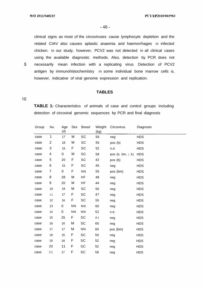
clinical signs as most of the circoviruses cause lymphocyte depletion and the
related CIAV also causes aplastic anaemia and haemorrhages in infected
chicken. In our study, however, PCV2 was not detected in all clinical cases
using the available diagnostic methods. Also, detection by PCR does not
necessarily mean infection with a replicating virus. Detection of PCV2
antigen by immunohistochemistry in some individual bone marrow cells is,
however, indicative of viral genome expression and replication.
TABLES
TABLE 1: Characteristics of animals of case and control groups including
detection of circoviral genomic sequences by PCR and final diagnosis
Group No. Age Sex Breed Weight Circovirus Diagnosis(d) (kg)
case 1 17 M SC 56 neg HDS
case 2 18 M SC 55 pos (b) HDS
case 3 15 F SC 52 n.d. HDS
case 4 0 M SC 58 pos (b, bm, I, k) HDS
case 5 20 F SC 42 pos (b) HDS
case 6 15 F SC 45 neg HDS
case 7 0 F NN 55 pos (bm) HDS
case 8 28 M HF 48 neg HDS
case 9 20 M HF 44 neg HDS
case 10 19 M SC 50 neg HDS
case 1 1 17 F SC 47 neg HDS
case 12 16 F SC 55 neg HDS
case 13 0 NN NN 60 neg HDS
case 14 0 NN NN 52 n.d. HDS
case 15 25 F SC 4 1 neg HDS
case 16 16 M SC 60 neg HDS
case 17 17 M NN 60 pos (bm) HDS
case 18 15 F SC 50 neg HDS
case 19 18 F SC 52 neg HDS
case 20 11 F SC 52 neg HDS
case 2 1 17 F SC 58 neg HDS
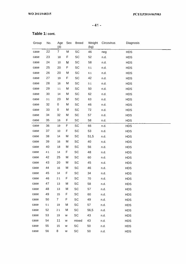
Table 1: cont.
Group No. Age Sex Breed Weight Circovirus Diagnosis(d) (kg)
case 22 7 M SC 46 neg HDS
case 23 18 F SC 52 n.d. HDS
case 24 10 SC 58 n.d. HDS
case 25 20 F SC 5 1 n.d. HDS
case 26 20 M SC 6 1 n.d. HDS
case 27 19 F SC 42 n.d. HDS
case 28 16 M SC 5 1 n.d. HDS
case 29 1 1 M SC 50 n.d. HDS
case 30 14 M SC 62 n.d. HDS
case 3 1 23 M SC 63 n.d. HDS
case 32 0 M SC 46 n.d. HDS
case 33 0 M SC 72 n.d. HDS
case 34 32 M SC 57 n.d. HDS
case 35 16 F SC 58 n.d. HDS
case 36 19 F SC 66 n.d. HDS
case 37 10 F SC 53 n.d. HDS
case 38 14 M SC 51,5 n.d. HDS
case 39 16 M SC 40 n.d. HDS
case 40 18 M SC 56 n.d. HDS
case 4 1 14 F SC 48 n.d. HDS
case 42 25 M SC 60 n.d. HDS
case 43 20 M SC 45 n.d. HDS
case 44 16 M SC 46 n.d. HDS
case 45 14 F SC 34 n.d. HDS
case 46 2 1 F SC 70 n.d. HDS
case 47 13 M SC 58 n.d. HDS
case 48 13 M SC 57 n.d. HDS
case 49 15 F SC 60 n.d. HDS
case 50 7 F SC 49 n.d. HDS
case 5 1 19 M SC 57 n.d. HDS
case 52 2 1 M SC 56,5 n.d. HDS
case 53 19 w SC 43 n.d. HDS
case 54 11 w mixed 43 n.d. HDS
case 55 15 w SC 50 n.d. HDS
case 56 8 w SC 50 n.d. HDS
Table 1: cont.
control 1 0 F SC 42 neg abortion
control 2 n.n. F SC 46 neg gastroenteritis
control 3 18 F SC 42 neg polyarthritis
control 4 n.n. F SC 38 neg enteritis
control 5 9 F SC 46 neg enteritis, dehydration, pneumonia
control 6 n.n. F SC 30 neg enteritis, dehydration
control 7 8 M SC 49 pos abomasoenteritis, dehydration
control 8 18 F SC 34 neg enteritis, dehydration, bronchitis
HDS: Haemorrhagic Disease Syndrome; M : male; F: female; SC: Simmental cattle; neg:
negative; pos: positive; n.n.: not named; b: blood, bm: bone marrow; I: liver; k: kidney
TABLE 2 :Haematologic results of five calves with haemorrhagic disease and
bone marrow aplasia
Reference Unit No. No. No. No. No.
Range 2 53 54 55 56
Haemoglobin 5.6-8.7 mmol/l 4.9 1.2 5.3 2.65 4.5
Haematocrit 0.22-0.44 l/l 0.23 0.06 0.27 0.13 0.22
Erythrocytes 5.0-9.7 T/l 6.1 1.32 6.3 2.94 5.6
White Blood Cell 4.0-12.0 G/l 0.85 1.47 0.285 0.9 1.0Count
MCV 36-50 f l 37.3 42.1 43.9 43.2 39.5
MCH 13-17 pg 12.9 14.6 13.7 14.5 13.0
MCHC 32-39 mmol/l 2 1.5 2 1 .5 19.4 20.9 20.4
Neutrophil 25-45 % 4 28 3 4granulocytes
Eosinophil 0-14 % 0 0 0 0granulocytes
Basophil granulocytes 0-1 % 1 0 0 2 0
Monocytes 1-8 % 1 2 4 11 7
Lymphocytes 45-65 % 96 94 68 84 89
Platelet count 300-800 G/l 33.7 12.5 82 60 55
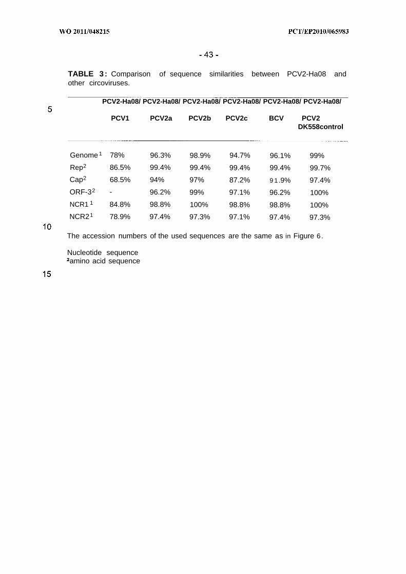
TABLE 3 : Comparison of sequence similarities between PCV2-Ha08 andother circoviruses.
PCV2-Ha08/ PCV2-Ha08/ PCV2-Ha08/ PCV2-Ha08/ PCV2-Ha08/ PCV2-Ha08/
PCV1 PCV2a PCV2b PCV2c BCV PCV2DK558control
Genome 1 78% 96.3% 98.9% 94.7% 96.1% 99%
Rep2 86.5% 99.4% 99.4% 99.4% 99.4% 99.7%
Cap2 68.5% 94% 97% 87.2% 9 1 .9% 97.4%
ORF-32 - 96.2% 99% 97.1% 96.2% 100%
NCR1 1 84.8% 98.8% 100% 98.8% 98.8% 100%
NCR21 78.9% 97.4% 97.3% 97.1% 97.4% 97.3%
The accession numbers of the used sequences are the same as in Figure 6.
Nucleotide sequenceamino acid sequence
References
Allan G, McNeilly F, Krakowa S, Ellis J (2003): PCV-2 infection in swine;
more than just postweaning multisystemic wasting syndrome. Vet J
166: 222.
Allan GM, McNeilly F, McNair I, Curran MD, Walker I, Ellis J, Konoby C,
Kennedy S, Meehan B (2000): Absence of evidence for porcine
circovirus type 2 in cattle and humans, and lack of seroconversion
or lesions in experimentally infected sheep. Arch Virol 145: 853.
Chae C (2005): A review of porcine circovirus 2-associated syndromes and
diseases. Vet J 169: 326.
Cox, J.C. and Coulter, A.R. (1992): "Advances in Adjuvant Technology and
Application", in Animal Parasite Control Utilizing Biotechnology,
Chapter 4, Ed. Yong, W.K., CRC Press.
Dalsgaard, K. (1974): Saponin adjuvants. 3. Isolation of a substance from
Quillaja saponaria Molina with adjuvant activity in food-and-mouth
disease vaccines. Arch Gesamte Virusforsch 44: 243.
Ellis J, Krakowka S, Lairmore M, Haines D, Bratanich A, Clark E, Allan
G, Konoby C, Hassard L, Meehan B, Martin K, Harding J,
Kennedy S, McNeilly F (1999): Reproduction of lesions of
postweaning multisystemic wasting syndrome in gnotobiotic piglets.
J Vet Diagn Invest 11: 3 .
Ellis JA, Konoby C, West KH, Allan GM, Krakowka S, McNeilly F,
Meehan B, Walker (2001 ) : Lack of antibodies to porcine circovirus
type 2 virus in beef and dairy cattle and horses in western Canada.
Can Vet J 42: 461 .
Ellis JA, West KH, Cortese VS, Myers SL, Carman S, Martin KM, Haines
DM (1998): Lesions and distribution of viral antigen following an
experimental infection of young seronegative calves with virulent
bovine virus diarrhea virus-type II. Can J Vet Res 62: 161 .
Goryo M, Suwa T, Umemura T, Itakura C, Yamashiro S (1989):
Histopathology of chicks inoculated with chicken anaemia agent
(MSB1-TK5803 strain). Avian Pathol 18: 73.
Gottschalk C, Bauer J, Meyer K (2008): Detection of satratoxin g and h in
indoor air from a water-damaged building. Mycopathologia 166:
103.
Halami MY, Nieper H, Muller H, Johne R (2008): Detection of a novel
circovirus in mute swans (Cygnus olor) by using nested broad-
spectrum PCR. Virus Res 132: 208.
Harrach B, Bata A, Bajmocy E, Benko M (1983): Isolation of satratoxins
from the bedding straw of a sheep flock with fatal
stachybotryotoxicosis. Appl Environ Microbiol 45: 1419.
Hoffmann-Fezer G, Hoffmann R, Hofmann W (1974): [Chronic
furazolidone poisoning in calves. II. studies of the bone marrow].
Dtsch Tierarztl Wochenschr 8 1: 59.
Hofmann W, Hoffmann R, Hoffmann-Fezer G (1974): [Chronic
furazolidone poisoning in calves. I . Clinical, hematological and
morphological studies]. Dtsch Tierarztl Wochenschr 8 1: 53.
Horadagoda NU, Hodgson JC, Moon GM, Wijewardana TG,
Eckersall PD (2001 ) : Role of endotoxin in the pathogenesis of
haemorrhagic septicaemia in the buffalo. Microb Pathog 30: 171 .
Hughes AL, Piontkivska H (2008): Nucleotide sequence polymorphism in
circoviruses. Infect Genet Evol 8: 130.
Johne R, Fernandez-de-Luco D, Hofle U, Muller H (2006): Genome of a
novel circovirus of starlings, amplified by multiply primed rolling-
circle amplification. J Gen Virol 87: 1189.
Kennedy S, Moffett D, McNeilly F, Meehan B, Ellis J, Krakowka S, Allan
GM (2000): Reproduction of lesions of postweaning multisystemic
wasting syndrome by infection of conventional pigs with porcine
circovirus type 2 alone or in combination with porcine parvovirus. J
Comp Pathol 122: 9.
Kuscu B, Gurel A (2008): Lesions in the thymus and bone marrow in chicks
with experimentally induced chicken infectious anemia disease. J
Vet Sci 9 : 15.
Liu J, Chen I, Kwang J (2005): Characterization of a previously unidentified
viral protein in porcine circovirus type 2-infected cells and its role in
virus-induced apoptosis. J Virol 79: 8262.
Lock EA, Sani Y, Moore RB, Finkelstein MB, Anders MW, Seawright AA
(1996): Bone marrow and renal injury associated with haloalkene
cysteine conjugates in calves. Arch Toxicol 70: 607.
Loesch U, Cihak J, Erhard MH, Kaspers B (2000): Blut und Abwehr. In:
von Engelhardt W, Breves G (eds) Physiologie der Haustiere. Enke
im Hippokrates Verlag GmbH, Stuttgart, Germany p 196.
Lunn DP, Butler DG (1991 ) : Idiopathic thrombocytopenic purpura in a
Holstein bull. Can Vet J 32: 559.
Mankertz A (2008): Circoviruses. In: Mahy BWJ, Van Regenmortel MHV
(eds) Encyclopedia of Virology, vol 1. Elsevier Books, Amsterdam p
513.
Maxie MG, Newman SJ (2007): Urinary system. In: Maxie MG (ed) Jubb,
Kennedy, and Palmer's Pathology of domestic animals, vol 2 .
Elsevier, Amsterdam p 425.
Nayar GP, Hamel AL, Lin L, Sachvie C, Grudeski E, Spearman G (1999):
Evidence for circovirus in cattle with respiratory disease and from
aborted bovine fetuses. Can Vet J 40: 277.
Paape MJ, Bannerman DD, Zhao X, Lee JW (2003): The bovine neutrophil:
Structure and function in blood and milk. Vet Res 34: 597.
Rebhun WC, French TW, Perdrizet JA, Dubovi EJ, Dil SG, Karcher LF
( 1989): Thrombocytopenia associated with acute bovine virus
diarrhea infection in cattle. J Vet Intern Med 3 : 42.
Reubel GH, Gareis M, Amselgruber WM (1987): Cytotoxicity evaluation of
mycotoxins by an MTT-bioassay. Mycotoxin Res 3: 85.
Rhoades KR, Heddleston KL, Rebers PA (1967): Experimental
hemorrhagic septicemia: gross and microscopic lesions resulting
from acute infections and from endotoxin administration. Can J
Comp Med Vet Sci 3 1 : 226.
Rimler RB (1978): Coagglutination test for identification of Pasteurella
multocida associated with hemorrhagic septicemia. J Clin Microbiol
8 : 214.
Segales J, Allan GM, Domingo M (2005): Porcine circovirus diseases.
Anim Health Res Rev 6 : 119.
Segales J, Mateu E (2006): Immunosuppression as a feature of

postweaning multisystemic wasting syndrome. Vet J 171 : 396.
Segales J, Olvera A, Grau-Roma L, Charreyre C, Nauwynck H, Larsen L,
Dupont K, McCullough K, Ellis J, Krakowka S, Mankertz A,
Fredholm M, Fossum C, Timmusk S, Stockhofe-Zurwieden N,
Beattie V, Armstrong D, Grassland B, Baekbo P, Allan G (2008):
PCV-2 genotype definition and nomenclature. Vet Rec 162: 867.
Steficek BA, Thomas JS, Baker JC, Bell TG (1993): Hemorrhagic
diathesis associated with a hereditary platelet disorder in
Simmental cattle. J Vet Diagn Invest 5 : 202.
Steinfeldt T, Finsterbusch T, Mankertz A (2001 ) : Rep and Rep' protein of
porcine circovirus type 1 bind to the origin of replication in vitro.
Virology 291 : 152.
Thompson JD, Higgins DG, Gibson TJ (1994): CLUSTAL W: improving
the sensitivity of progressive multiple sequence alignment through
sequence weighting, positionspecific gap penalties and weight
matrix choice. Nucleic Acids Res 22: 4673.
Timmusk S, Wallgren P, Brunborg IM, Wikstrom FH, Allan G, Meehan B,
McMenamy M, McNeilly F, Fuxler L, Belak K, Podersoo D, Saar
T, Berg M, Fossum C (2008): Phylogenetic analysis of porcine
circovirus type 2 (PCV2) pre- and postepizootic postweaning
multisystemic wasting syndrome (PMWS). Virus Genes 36: 509.
Tischer I, Bode L, Apodaca J, Timm H, Peters D, Rasch R, Pociuli S,
Gerike E (1995): Presence of antibodies reacting with porcine
circovirus in sera of humans, mice, and cattle. Arch Virol 140: 1427.
Todd D (2000): Circoviruses: immunosuppressive threats to avian species:
a review. Avian Pathol 29: 373.
Todd D, Bendinelli M, Biagini P, Hino S, Mankertz A, Mishiro S, Niel C,
Okamoto H, Raidal S, Ritchie BW, Teo GC (2005): Circoviridae.
In: Fauquet CM, Mayo MA, Maniloff J, Desselberger U, Ball LA
(eds) Virus Taxonomy, Vlllth Report of the International Committee
for the Taxonomy of Viruses. Elsevier/Academic Press, Amsterdam
p 327.
Toussaint JF, Sailleau C, Breard E, Zientara S, De Clercq K (2007):
Bluetongue virus detection by two real-time RT-qPCRs targeting
two different genomic segments. J Virol Methods 140: 115.
Valli VEO (2007): Hematopoietic system. In: Maxie MG (ed) Jubb, Kennedy,
and Palmer's Pathology of domestic animals, vol 3 . Elsevier,
Amsterdam p 107.
Wa!z PH, Bell TG, Grooms DL, Kaiser L, Maes RK, Baker JC (2001):
Platelet aggregation responses and virus isolation from platelets in
calves experimentally infected with type I or type I I bovine viral
diarrhea virus. Can J Vet Res 65: 241 .
Wood RD, Goens SD, Carman PS, Deregt D, Jefferson B, Jacobs RM
(2004): Effect on hematopoietic tissue of experimental infection of
calves with noncytopathic type 2 bovine viral diarrhea virus. Can J
Vet Res 68: 42.
Yeruham I, Avidar Y, Harms S, Fishman L, Aroch (2003): Immune-
mediated thrombocytopenia and putative haemolytic anaemia
associated with a polyvalent botulism vaccination in a cow. Vet Rec
153: 502.
Yuasa N, Taniguchi T, Yoshida I (1979): Isolation and some characteristics
of an agent inducing anemia in chicks. Avian Dis 23: 366.

CLAIMS
1. A circovirus (CV) nucleic acid molecule comprising
(a) the sequence as shown in SEQ ID NO: 1 or a fragment thereof
and/or
(b) the complement of the nucleotide sequence according to (a).
2. The nucleic acid molecule according to claim 1 comprising
(a) the sequence as shown in SEQ ID NO: 7 or a fragment thereof
and/or
(b) the complement of the nucleotide sequence according to (a).
3. The nucleic acid molecule according to claim 1 comprising
(a) the sequence as shown in SEQ ID NO: 1 or a fragment thereof
and/or
(b) the complement of the nucleotide sequence according to (a).
4 . The nucleic acid molecule according to any one of claims 1-3, wherein
the fragment comprises at least 15, at least 20, at least 25, at least 30
or at least 50 contiguous nucleotides as shown in SEQ ID NOs:1 , 7 or
1 or the complement thereof.
5 . The nucleic acid molecule according to any one of claims 1-4
(a) having an identity of at least 90%, at least 92%, at least 94%, at
least 96%, at least 98% or at least 99% of the nucleotide
sequence as shown in SEQ ID NOs: 1, 7, or 11
(b) hybridizing under stringent conditions to the nucleotide sequence
as shown in SEQ ID NOs: 1, 7 or 11 or
(c) the complement of (a) or (b).
6 . A CV nucleic acid molecule encoding a CV polypeptide or a fragment
thereof, wherein the nucleic acid molecule comprises
(a) a region of the nucleotide sequence as shown in SEQ D NO: 1
from
(i) nucleotide 5 1 - 995 (Rep)
(ii) nucleotide 1034 - 1735 (Cap)
(iii) nucleotide 357 - 671 (ORF3)
and/or the complement of (i), (ii) and/or (iii)
(b) a nucleotide sequence corresponding to the sequence of (a)
within the scope of degeneracy of the genetic code, or
(c) a fragment of the nucleotide sequence according to (a) or (b).
7 . A CV nucleic acid according to claim 6, wherein the nucleic acid
molecule comprises
(a) region of the nucleotide sequence as shown in SEQ ID NO: 7
from
(i) nucleotide 5 1 - 995 (Rep)
(ii) nucleotide 1034 - 1735 (Cap)
(iii) nucleotide 357 - 671 (ORF3)
and/or the complement of (i), (ii) and/or (iii)
(b) a region of the nucleotide sequence as shown in SEQ ID NO: 11
from
(i) nucleotide 5 1 - 995 (Rep)
(ii) nucleotide 1033 - 1734 (Cap)
(iii) nucleotide 357 - 671 (ORF3)
and/or the complement of (i), (ii) and/or (iii)
(c) a nucleotide sequence corresponding to the sequence of (a) or
(b) within the scope of degeneracy of the genetic code, or
(d) a fragment of the nucleotide sequence according to (a), (b) or
(c) .
8. The nucleic acid molecule of claim 6 or claim 7, wherein the
polypeptide or fragment thereof comprises at least 6, at least 8 , at least
10, at least 20, or at least 30 contiguous amino acids of any of the
amino acid sequences as shown in SEQ ID NO: 2-4, 8-10, or 12-14.

9. A CV nucleic acid according to any one of claims 6-8, which encodes a
polypeptide having an identity of at least 90%, at least 92%, at least
94%, at least 96%, at least 98% or at least 99% of any of the amino
acid sequences as shown in SEQ ID NO: 2-4, 8-10, or 12-14.
10. The nucleic acid molecule of any of claims 1-9 in operative linkage with
a heterologous expression control sequence.
11. A non-human host cell transformed of transfected with a nucleic acid
molecule of any one of claims 1-10.
12. The nucleic acid molecule of any one of claims 1-10 for use as a
diagnostic agent.
13. The nucleic acid molecule of claim 12, which carries a reporter group.
14. The nucleic acid molecule of claim 12 or 13 for the diagnosis of
Haemorrhagic Diathesis (HD).
15. The nucleic acid molecule of claim 14 for the diagnosis of HD in cattle.
16. The nucleic acid molecule of any one of claims 1-10 for use as a
therapeutic agent.
17. The nucleic acid molecule of claim 16 for the prevention and/or
treatment of HD.
18. The nucleic acid molecule of claim 16 or 17 for the prevention and/or
treatment of HD in cattle.
9 . The nucleic acid molecule of any one of claims 16-18 as a nucleic acid-
based vaccine or for the production of a polypeptide-based vaccine.
20. A polypeptide encoded by a nucleic acid molecule according to any one
of claims 1-10.
2 1. A circovirus (CV) polypeptide comprising
(a) an amino acid sequence selected from
(i) amino acid SEQ ID NO: 2 (Rep)
(ii) amino acid SEQ ID NO: 3 (Cap)
(iii) amino acid SEQ ID NO: 4 (ORF3), or
(b) a fragment thereof.
22. A circovirus (CV) polypeptide according to claim 2 1, comprising
(a) an amino acid sequence selected from
(i) amino acid SEQ ID NO: 8 and 12 (Rep)
(ii) amino acid SEQ ID NO: 9 and 13 (Cap)
(iii) amino acid SEQ ID NO: 10 and 14 (ORF3), or
(b) a fragment thereof.
23. The polypeptide of any one of claims 20-22 comprising at least 6, at
least 8, at least 10, at least 20, or at least 30 contiguous amino acids of
any of the amino acid sequences as shown in SEQ ID NO: 2-4, 8-10, or
12-14.
24. The polypeptide of claims 20-23 having an identity of at least 90%, at
least 92%, at least 94%, at least 96%, at least 98% or at least 99% of
any of the amino acid sequences as shown in SEQ ID NO: 2-4, 8-10, or
12-14.
25. The polypeptide of any one of claims 20-24 for use as a diagnostic
agent.
26. The polypeptide of claim 25, which carries a reporter group.
27. The polypeptide of claim 25 or 26 for the diagnosis of HD, particularly in
cattle.
28. The polypeptide of any one of claims 20-24 for use as immunogen.
29. The polypeptide of claim 28 for the generation of anti-CV-antibodies.
30. The polypeptide of any one of claims 20-24 for the prevention and or
treatment of HD, particularly in cattle.
3 . The polypeptide of claim 30 for use as a vaccine.
32. An antibody or an antigen-binding fragment thereof directed against a
polypeptide of any one of claims 20-24.
33. The antibody of claim 32, which is specific for CV strain PCV2-Ha08.
34. The antibody of claim 32 or 33 for use as a diagnostic agent.
35. The antibody of claim 32 or 33 for the prevention and/or treatment of
HD, particularly in cattle.
36. A circovirus comprising a nucleic acid molecule of any one of claims 1-
10.
37. The virus of claim 36 which is an inactivated circovirus.
38. The virus of claim 37 which is an attenuated circovirus.
39. A composition comprising a nucleic acid molecule of any one of claims
1-1 0 , or a polypeptide of any one of claims 20-24, an antibody of claim
32 or 33, or a virus of any one of claims 36-38, together with an
acceptable carrier, diluent and/or adjuvant.
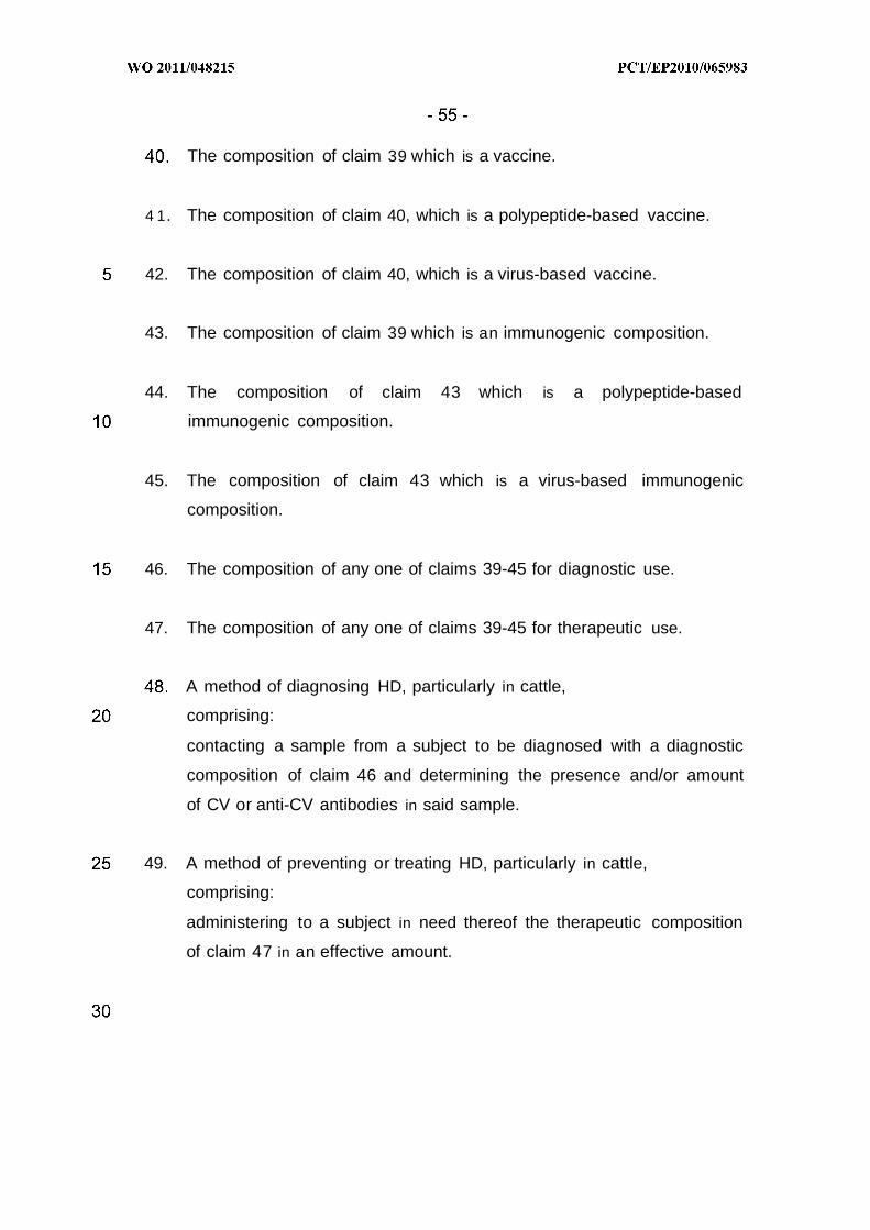
The composition of claim 39 which is a vaccine.
4 1. The composition of claim 40, which is a polypeptide-based vaccine.
42. The composition of claim 40, which is a virus-based vaccine.
43. The composition of claim 39 which is an immunogenic composition.
44. The composition of claim 43 which is a polypeptide-based
immunogenic composition.
45. The composition of claim 43 which is a virus-based immunogenic
composition.
46. The composition of any one of claims 39-45 for diagnostic use.
47. The composition of any one of claims 39-45 for therapeutic use.
A method of diagnosing HD, particularly in cattle,
comprising:
contacting a sample from a subject to be diagnosed with a diagnostic
composition of claim 46 and determining the presence and/or amount
of CV or anti-CV antibodies in said sample.
49. A method of preventing or treating HD, particularly in cattle,
comprising:
administering to a subject in need thereof the therapeutic composition
of claim 47 in an effective amount.
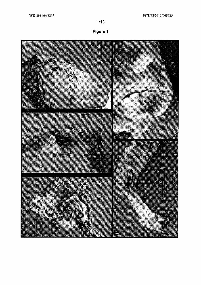
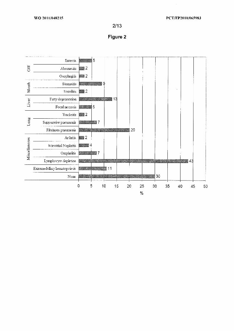
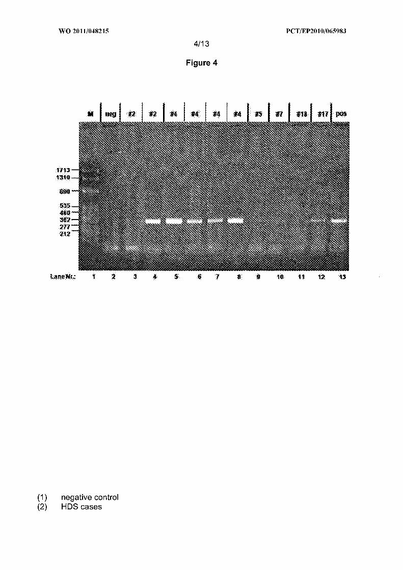
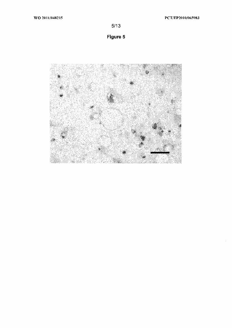
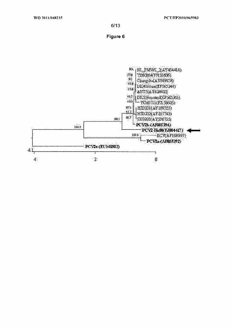

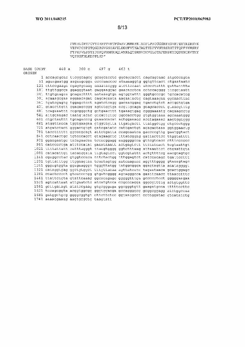
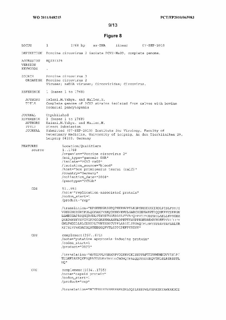
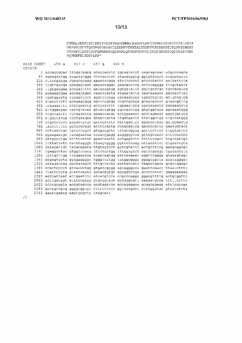

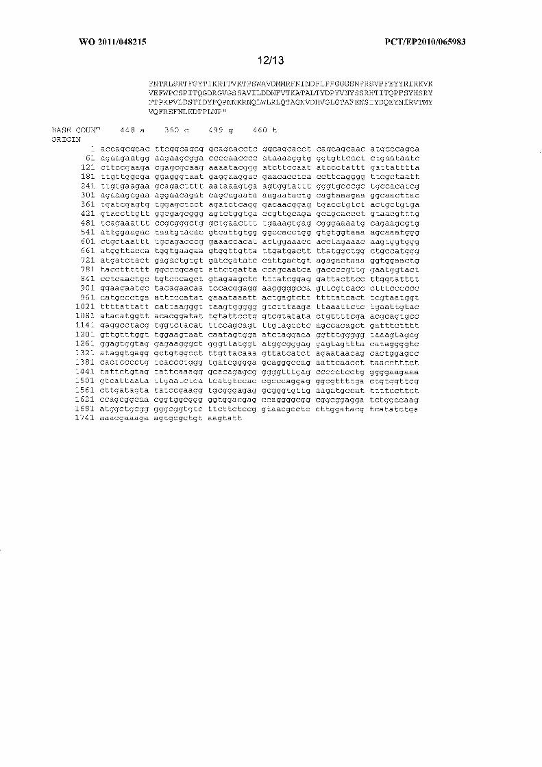
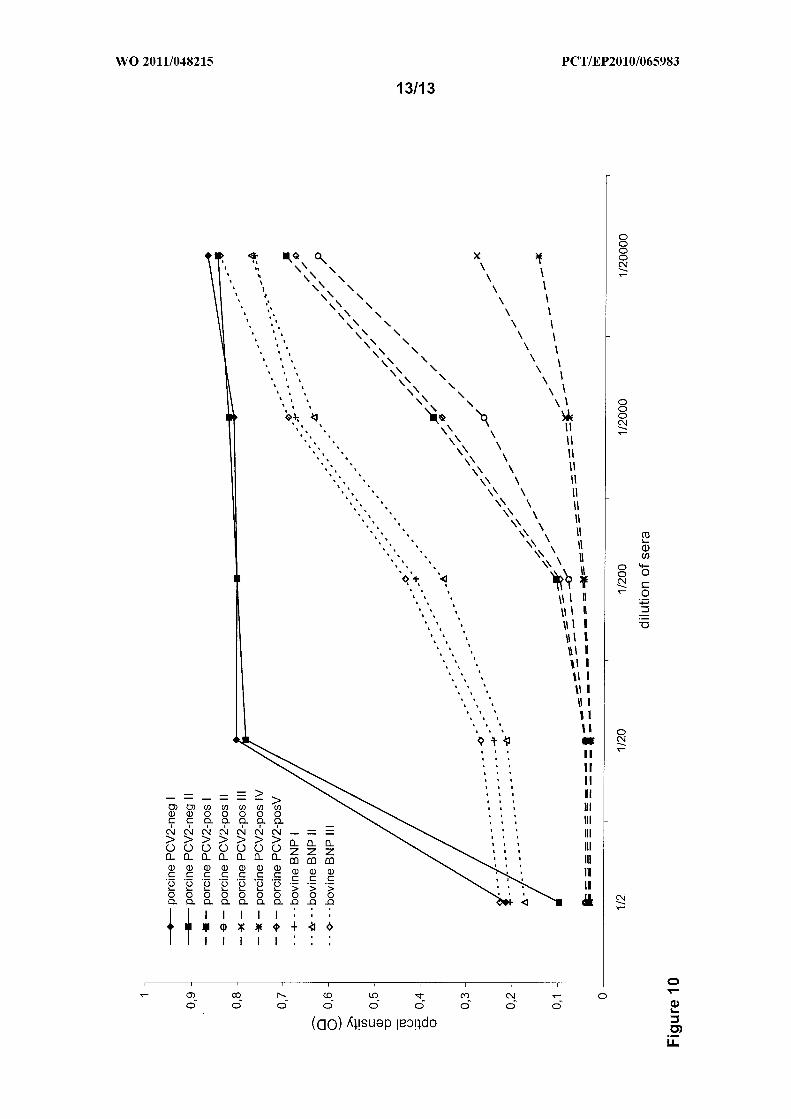
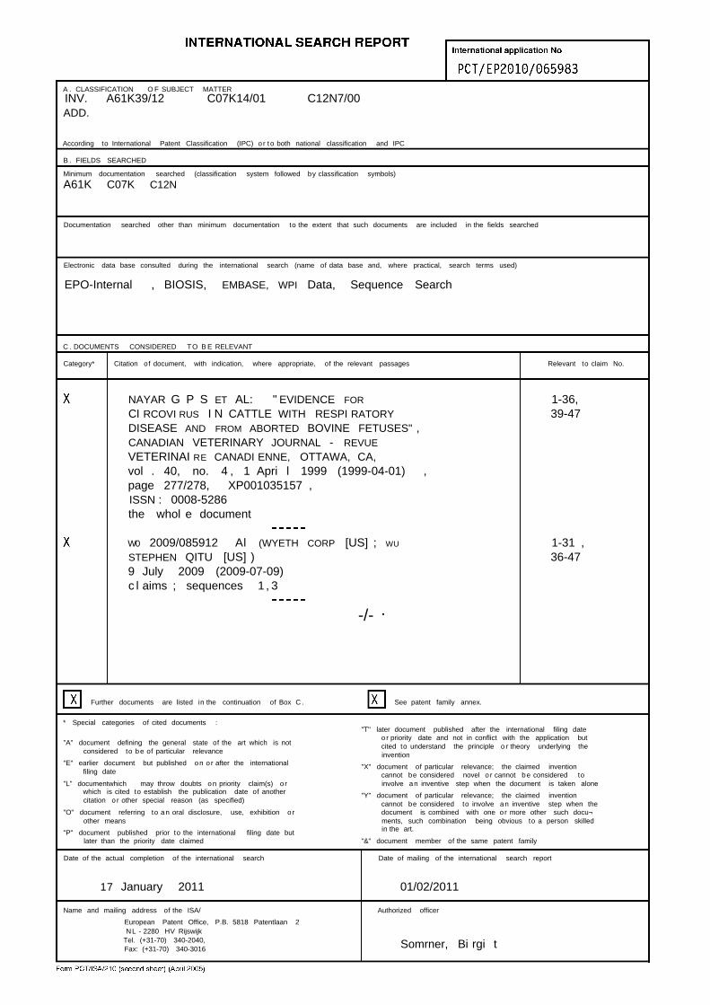
A . CLASSIFICATION O F SUBJECT MATTER
INV. A61K39/12 C07K14/01 C12N7/00ADD.
According to International Patent Classification (IPC) o r to both national classification and IPC
B . FIELDS SEARCHED
Minimum documentation searched (classification system followed by classification symbols)
A61K C07K C12N
Documentation searched other than minimum documentation to the extent that such documents are included in the fields searched
Electronic data base consulted during the international search (name of data base and, where practical, search terms used)
EPO-Internal , BIOSIS, EMBASE, WPI Data, Sequence Search
C . DOCUMENTS CONSIDERED TO B E RELEVANT
Category* Citation of document, with indication, where appropriate, of the relevant passages Relevant to claim No.
NAYAR G P S ET AL: " EVIDENCE FOR 1-36,CI RCOVI RUS I N CATTLE WITH RESPI RATORY 39-47DISEASE AND FROM ABORTED BOVINE FETUSES" ,CANADIAN VETERINARY JOURNAL - REVUE
VETERINAI RE CANADI ENNE, OTTAWA, CA,vol . 40, no. 4 , 1 Apri l 1999 (1999-04-01) ,page 277/278, XP001035157 ,ISSN : 0008-5286the whol e document
W0 2009/085912 Al (WYETH CORP [US] ; WU 1-31 ,STEPHEN QITU [US] ) 36-479 July 2009 (2009-07-09)c l aims ; sequences 1 , 3
-/-
Further documents are listed in the continuation of Box C . See patent family annex.
* Special categories of cited documents :"T" later document published after the international filing date
o r priority date and not in conflict with the application but"A" document defining the general state of the art which is not cited to understand the principle o r theory underlying the
considered to be of particular relevance invention"E" earlier document but published on o r after the international "X" document of particular relevance; the claimed invention
filing date cannot be considered novel o r cannot be considered to"L" documentwhich may throw doubts on priority claim(s) o r involve an inventive step when the document is taken alone
which is cited to establish the publication date of another "Y" document of particular relevance; the claimed inventioncitation o r other special reason (as specified) cannot be considered to involve an inventive step when the
"O" document referring to an oral disclosure, use, exhibition o r document is combined with one o r more other such docu¬other means ments, such combination being obvious to a person skilled
"P" document published prior to the international filing date but in the art.
later than the priority date claimed "&" document member of the same patent family
Date of the actual completion of the international search Date of mailing of the international search report
17 January 2011 01/02/2011
Name and mailing address of the ISA/ Authorized officer
European Patent Office, P.B. 5818 Patentlaan 2NL - 2280 HV Rijswijk
Tel. (+31-70) 340-2040,Fax: (+31-70) 340-3016 Somrner, Bi rgi t

C(Continuation). DOCUMENTS CONSIDERED TO BE RELEVANT
Category* Citation of document, with indication, where appropriate, of the relevant passages Relevant to claim No.
X WO 99/29871 A2 (VETERINAI ES ET 1-47ALIMENTAI RES C [FR] ; JESTIN ANDRE [FR] ;ALBINA EMMANUE) 17 June 1999 (1999-06-17)c l aims ; sequences 9 , 10, 14, 16
X KAPPE E C ET AL: " FATAL APLASTIC ANAEMIA 1-3 ,5-7 ,WITH HAEMORRHAGIC DISEASE I N CALVES I N 10-12 ,GERMANY" , 14-22 ,ABSTRACT BOOK, 27TH MEETING OF THE 25 ,EU R0PEAN-S0C I ETY-OF-VETERI NARY-PATHOLOGY/E 27-31 ,UROPEAN C0LLEGE-0F-VETERINARY-PATH0L0G; 36,39-48CRACOW, POLAND; SEPTEMBER 09 -13 , 2009 ,[Onl i ne]9 September 2009 (2009-09-09) , page 118,XP002614167 ,ISSN : 0021-9975Retri eved from the Internet:URL: http://www.esvp. p i /abstract book.pdf>[retri eved on 2010-12-17]* abstract
X DATABASE EMBL [Onl i ne] 1-12 ,14-19 ,
5 December 2007 (2007-12-05) , 36-47" Porci ne c i rcovi rus 2 i sol ateDK558control , compl ete genome. " ,XP002614169 ,retri eved from EBI accessi on no.EMBL: EF565365Database accessi on no. EF565365* abstract; sequences
X DATABASE EMBL [Onl i ne] 1-12 ,14-19 ,
18 February 2003 (2003-02-18) , 36-47" Porci ne c i rcovi rus type 2 strai n HZ0201 ,compl ete genome. " ,XP002614175 ,retri eved from EBI accessi on no.EMBL:AY188355Database accessi on no. AY188355* abstract; sequences
X DATABASE EMBL [Onl i ne] 1-12 ,14-19 ,
15 July 2003 (2003-07-15) , 36-47" Porci ne c i rcovi rus 2 strai n Fhl6,compl ete genome. " ,XP002614178,retri eved from EBI accessi on no.EMBL:AY321993Database accessi on no. AY321993* abstract; sequences
-/--
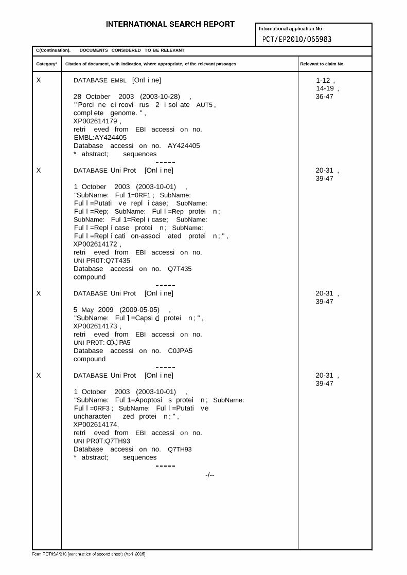
C(Continuation). DOCUMENTS CONSIDERED TO BE RELEVANT
Category* Citation of document, with indication, where appropriate, of the relevant passages Relevant to claim No.
X DATABASE EMBL [Onl i ne] 1-12 ,14-19 ,
28 October 2003 (2003-10-28) , 36-47" Porci ne c i rcovi rus 2 i sol ate AUT5 ,compl ete genome. " ,XP002614179 ,retri eved from EBI accessi on no.EMBL:AY424405Database accessi on no. AY424405* abstract; sequences
X DATABASE Uni Prot [Onl i ne] 20-31 ,39-47
1 October 2003 (2003-10-01) ,"SubName: Ful 1=0RF1 ; SubName:Ful l =Putati ve repl i case; SubName:Ful l =Rep; SubName: Ful l =Rep protei n ;SubName: Ful 1=Repl i case; SubName:Ful l =Repl i case protei n ; SubName:Ful l =Repl i cati on-associ ated protei n ; " ,XP002614172 ,retri eved from EBI accessi on no.UNI PR0T:Q7T435Database accessi on no. Q7T435compound
X DATABASE Uni Prot [Onl i ne] 20-31 ,39-47
5 May 2009 (2009-05-05) ,"SubName: Ful =Capsi protei n ; " ,XP002614173 ,retri eved from EBI accessi on no.UNI PR0T: C PA5Database accessi on no. C0JPA5compound
X DATABASE Uni Prot [Onl i ne] 20-31 ,39-47
1 October 2003 (2003-10-01) ,"SubName: Ful 1=Apoptosi s protei n ; SubName:Ful l =0RF3 ; SubName: Ful l =Putati veuncharacteri zed protei n ; " ,XP002614174,retri eved from EBI accessi on no.UNI PR0T:Q7TH93Database accessi on no. Q7TH93* abstract; sequences
-/--

C(Continuation). DOCUMENTS CONSIDERED TO BE RELEVANT
Category* Citation of document, with indication, where appropriate, of the relevant passages Relevant to claim No.
DATABASE Uni Prot [Onl i ne] 20-31 ,39-47
23 November 2004 (2004-11-23) ,"SubName: Ful =Capsi protei n ; " ,XP002614180,retri eved from EBI accessi on no.UNI PR0T:Q5XVM5Database accessi on no. Q5XVM5* abstract; sequences
KAPPE E C ET AL: "Bone marrow depl eti on 1-49wi t h haemorrhagi c d i athesi s i n cal ves i nGermany: Characteri zati on of the d i seaseand prel imi nary i nvesti gati ons on i t saeti o l ogy" ,BERLINER MUNCHENER TI ERARZTLICHEW0CHENSCHRI FT,vol . 123 , no. 1-2 , January 2010 (2010-01) ,pages 31-41 , XP002614168,the whol e document
I LL0UGHBY K ET AL: " Lack of evi dence for 1-49c i rcovi rus i nvol vement i n bovi ne neonatalpancytopeni a " ,VETERINARY RECORD, BRITISH VETERINARYASSOCIATION , LONDON , GB,vol . 166, no. 14,3 Apri l 2010 (2010-04-03) , pages 436-437 ,XP009142318,ISSN : 0042-4900the whol e document

.
INTERNATIONAL SEARCH REPORTPCT/EP2 0 1 0 / 0 6 5 9 8 3
Box No. I Nucleotide and/or amino acid sequence(s) (Continuation of item 1.b of the first sheet)
1. With regard to any nucleotide and/or amino acid sequence disclosed in the international application and necessary to the claimedinvention, the international search was carried out on the basis of:
a . (means)
on paper
in electronii
in the international application as filed
together with the international application in electronic form
subsequently to this Authority for the purpose of search
□ In addition, in the case that more than one version or copy of a sequence listing and/or table relating thereto has been filedor furnished, the required statements that the information in the subsequent or additional copies is identical to that in theapplication as filed or does not go beyond the application as filed, as appropriate, were furnished.
3 . Additional comments:
Form PCT/ISA/21 0 (continuation of first sheet (1)) (July 2009)

International application No.PCT/ EP20 1Q/065983INTERNATIONAL SEARCH REPORT
Box No. II Observations where certain claims were found unsearchable (Continuation of item 2 of first sheet)
This international search report has not been established in respect of certain claims under Article (2)(a) for the following reasons:
□ Claims Nos.:because they relate to subject matter not required to be searched by this Authority, namely:
Claims Nos.:because they relate to parts of the international application that do not comply with the prescribed requirements to suchan extent that no meaningful international search can be carried out, specifically:
□ Claims Nos.:because they are dependent claims and are not drafted in accordance with the second and third sentences of Rule 6.4(a).
Box No. Ill Observations where unity of invention is lacking (Continuation of item 3 of first sheet)
This International Searching Authority found multiple inventions in this international application, as follows:
see addi tional sheet
As all required additional search fees were timely paid by the applicant, this international search report covers all searchable□ i aims.
As all searchable claims could be searched without effort justifying an additional fees, this Authority did not invite payment ofadditional fees.
3. As only some of the required additional search fees were timely paid by the applicant, this international search report covers' ' only those claims for which fees were paid, specifically claims Nos. :
4 . I I No required additional search fees were timely paid by the applicant. Consequently, this international search report isrestricted to the invention first mentioned in the claims; it is covered by claims Nos.:
Remark on Protest The additional search fees were accompanied by the applicant's protest and, where applicable, the' ' payment of a protest fee.
The additional search fees were accompanied by the applicant's protest but the applicable protest' ' fee was not paid within the time limit specified in the invitation.
I INo protest accompanied the payment of additional search fees.
Form PCT/ISA/21 0 (continuation of first sheet (2)) (April 2005)

International Application No. PCT/ EP2Q10/ 065983
FURTHER INFORMATION CONTINUED FROM PCT/ISA/ 210
Thi s Internati onal Searchi ng Authori t y found mul t i pl e (groups of)i nventi ons i n thi s i nternati onal appl i cati on , as fol l ows :
1. cl aims : 1, 6, 21 , 33 (compl etely) ; 4 , 5 , 8-20, 23-32 ,34-49 (parti al ly)
a ci rcovi rus nucl ei c aci d mol ecul e compri si ng the sequenceas shown i n SEQ I D No. 1 as wel l as subject-matter rel atedthereto;
2 . cl aims : 2 (compl etely) ; 4 , 5 , 7-20, 22-32 , 34-49 (parti al ly)
a ci rcovi rus nucl ei c aci d mol ecul e compri si ng the sequenceas shown i n SEQ I D No. 7 as wel l as subject-matter rel atedthereto;
3 . cl aims : 3 (compl etely) ; 4 , 5 , 7-20, 22-32 , 34-49 (parti al ly)
a ci rcovi rus nucl ei c aci d mol ecul e compri si ng the sequenceas shown i n SEQ I D No. 11 as wel l as subject-matter rel atedthereto;
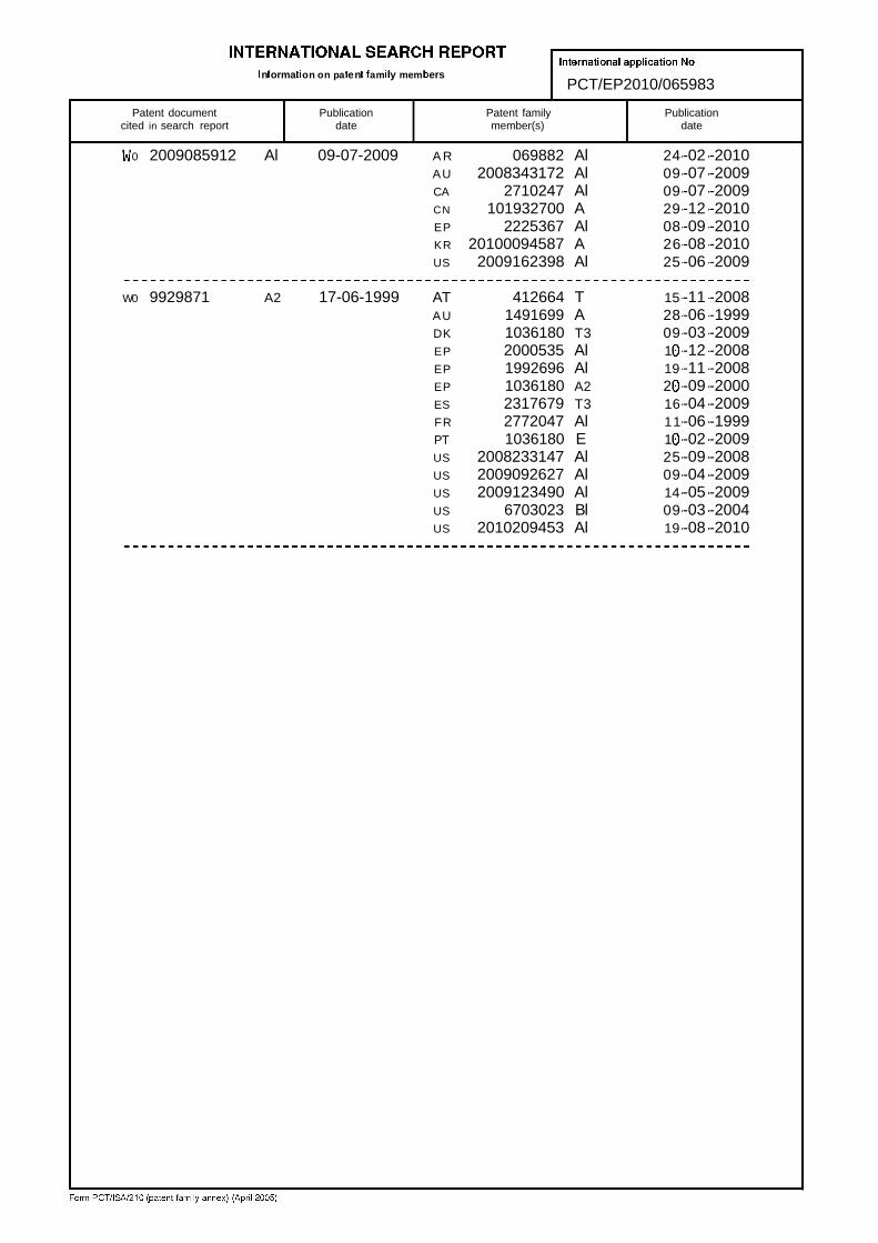
n orma on on pa en am y mem ersPCT/EP2010/065983
Patent document Publication Patent family Publicationcited in search report date member(s) date
0 2009085912 Al 09-07-2009 A R 069882 Al 24 -02 -2010AU 2008343172 Al 09 -07 -2009CA 2710247 Al 09 -07 -2009CN 101932700 A 29 -12 -2010EP 2225367 Al 08 -09 -2010KR 20100094587 A 26 -08 -2010US 2009162398 Al 25 -06 -2009
W0 9929871 A2 17-06-1999 AT 412664 T 15 -11 -2008AU 1491699 A 28 -06 -1999DK 1036180 T3 09 -03 -2009EP 2000535 Al 1 -12 -2008EP 1992696 Al 19 -11 -2008EP 1036180 A2 2 -09 -2000ES 2317679 T3 16 -04 -2009FR 2772047 Al 11 -06 -1999PT 1036180 E 1 -02 -2009US 2008233147 Al 25 -09 -2008US 2009092627 Al 09 -04 -2009US 2009123490 Al 14 -05 -2009US 6703023 Bl 09 -03 -2004US 2010209453 Al 19 -08 -2010