WO 2017/153974 Al
89
(12) INTERNATIONAL APPLICATION PUBLISHED UNDER THE PATENT COOPERATION TREATY (PCT) (19) World Intellectual Property Organization I International Bureau (10) International Publication Number (43) International Publication Date WO 2017/153974 Al 14 September 2017 (14.09.2017) PO PCT (51) International Patent Classification: (72) Inventors: SMITH, David; 357 South Maple Avenue, G01N 33/48 (2006.01) C12N 15/00 (2006.01) Ridgewood, New Jersey 07450 (US). CHAN, Wai Shun; G01N 33/487 (2006.01) A61K 35/00 (2006.01) 150 Overlook Avenue, 14C, Hackensack, New Jersey C12N 5/00 (2006.01) 07601 (US). HAMPSON, Brian; 215 Bamford Avenue, Hawthorne, New Jersey 07506 (US). PRETI, Robert; 80 (21) International Application Number: Nursery Road, Ridgefield, Connecticut 06877 (US). JI¬ PCT/IB2017/05 1736 ANG, Yajuan; 260 Franklin Avenue, Apartment 5 18, (22) International Filing Date: Mahwah, New Jersey 07430 (US). LEBLON, Courtney; 2 7 March 2017 (27.03.2017) 84 Rutgers Lane, Parsippany, New Jersey 07054 (US). (25) Filing Language: English (74) Agent: LUBIT, Beverly; McCarter & English LLP, 100 Mulberry Street, Newark, New Jersey 07052 (US). (26) Publication Language: English (81) Designated States (unless otherwise indicated, for every (30) Priority Data: kind of national protection available): AE, AG, AL, AM, 62/304,781 7 March 2016 (07.03.2016) US AO, AT, AU, AZ, BA, BB, BG, BH, BN, BR, BW, BY, 62/305,779 9 March 2016 (09.03.2016) US BZ, CA, CH, CL, CN, CO, CR, CU, CZ, DE, DJ, DK, DM, (71) Applicant: CALADRIUS BIOSCIENCES, INC. DO, DZ, EC, EE, EG, ES, FI, GB, GD, GE, GH, GM, GT, [US/US]; 420 Lexington Avenue, Suite 350, New York, HN, HR, HU, ID, IL, IN, IR, IS, JP, KE, KG, KH, KN, New York 10170 (US). KP, KR, KW, KZ, LA, LC, LK, LR, LS, LU, LY, MA, MD, ME, MG, MK, MN, MW, MX, MY, MZ, NA, NG, NI, NO, NZ, OM, PA, PE, PG, PH, PL, PT, QA, RO, RS, [Continued on next page] (54) Title: A CLOSED SYSTEM FOR LABELLING AND SELECTING LIVE CELLS (57) Abstract: The described invention provides an auto¬ FIG. 1 mated, closed system and method for separating/isolating a target cell type from a heterogeneous cell population. MICRO DEXTBAN BEADS COATED WITH ALGINATE Ι0Ρ 0 2)
-
Upload
khangminh22 -
Category
Documents
-
view
0 -
download
0
Transcript of WO 2017/153974 Al
(12) INTERNATIONAL APPLICATION PUBLISHED UNDER THE PATENT COOPERATION TREATY (PCT)
(19) World Intellectual PropertyOrganization I
International Bureau(10) International Publication Number
(43) International Publication Date WO 2017/153974 Al14 September 2017 (14.09.2017) P O P C T
(51) International Patent Classification: (72) Inventors: SMITH, David; 357 South Maple Avenue,
G01N 33/48 (2006.01) C12N 15/00 (2006.01) Ridgewood, New Jersey 07450 (US). CHAN, Wai Shun;G01N 33/487 (2006.01) A61K 35/00 (2006.01) 150 Overlook Avenue, 14C, Hackensack, New Jersey
C12N 5/00 (2006.01) 07601 (US). HAMPSON, Brian; 215 Bamford Avenue,
Hawthorne, New Jersey 07506 (US). PRETI, Robert; 80(21) International Application Number:
Nursery Road, Ridgefield, Connecticut 06877 (US). JI¬PCT/IB2017/05 1736 ANG, Yajuan; 260 Franklin Avenue, Apartment 5 18,
(22) International Filing Date: Mahwah, New Jersey 07430 (US). LEBLON, Courtney;2 7 March 2017 (27.03.2017) 84 Rutgers Lane, Parsippany, New Jersey 07054 (US).
(25) Filing Language: English (74) Agent: LUBIT, Beverly; McCarter & English LLP, 100Mulberry Street, Newark, New Jersey 07052 (US).
(26) Publication Language: English(81) Designated States (unless otherwise indicated, for every
(30) Priority Data: kind of national protection available): AE, AG, AL, AM,62/304,781 7 March 2016 (07.03.2016) U S AO, AT, AU, AZ, BA, BB, BG, BH, BN, BR, BW, BY,62/305,779 9 March 2016 (09.03.2016) U S BZ, CA, CH, CL, CN, CO, CR, CU, CZ, DE, DJ, DK, DM,
(71) Applicant: CALADRIUS BIOSCIENCES, INC. DO, DZ, EC, EE, EG, ES, FI, GB, GD, GE, GH, GM, GT,
[US/US]; 420 Lexington Avenue, Suite 350, New York, HN, HR, HU, ID, IL, IN, IR, IS, JP, KE, KG, KH, KN,
New York 10170 (US). KP, KR, KW, KZ, LA, LC, LK, LR, LS, LU, LY, MA,
MD, ME, MG, MK, MN, MW, MX, MY, MZ, NA, NG,NI, NO, NZ, OM, PA, PE, PG, PH, PL, PT, QA, RO, RS,
[Continued on nextpage]
(54) Title: A CLOSED SYSTEM FOR LABELLING AND SELECTING LIVE CELLS
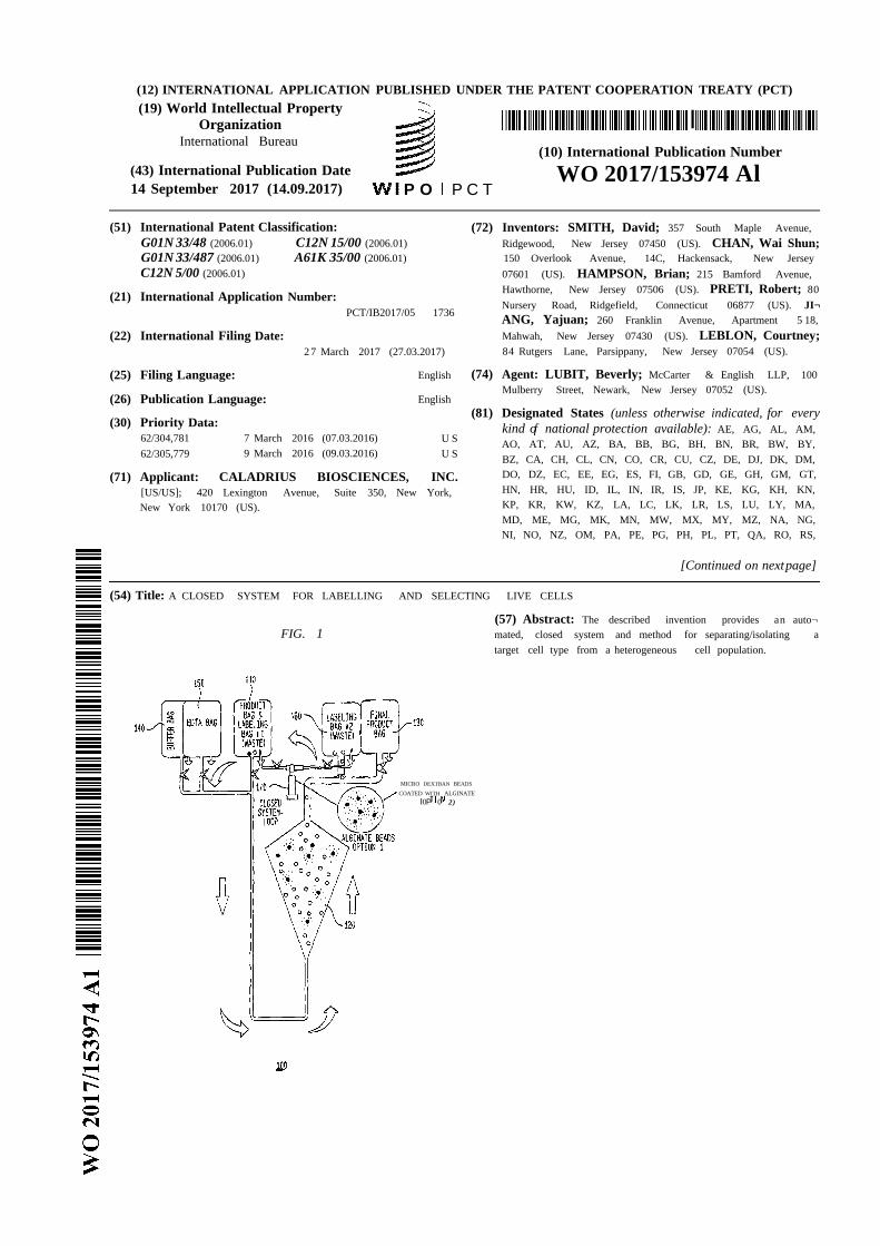
(57) Abstract: The described invention provides an auto¬FIG. 1 mated, closed system and method for separating/isolating a
target cell type from a heterogeneous cell population.
MICRO DEXTBAN BEADS
COATED WITH ALGINATE
Ι0Ρ 0 2)
w o 2017/153974 Al Hill I II I II III 11II 11111II I II
RU, RW, SA, SC, SD, SE, SG, SK, SL, SM, ST, SV, SY, Published:TH, TJ, TM, TN, TR, TT, TZ, UA, UG, US, UZ, VC,
— with international search report (Art. 21(3))VN, ZA, ZM, ZW.— before the expiration of the time limit for amending the
(84) Designated States (unless otherwise indicated, for every claims and to be republished in the event of receipt ofkind of regional protection available): ARIPO (BW, GH, amendments (Rule 48.2(h))GM, KE, LR, LS, MW, MZ, NA, RW, SD, SL, ST, SZ,TZ, UG, ZM, ZW), Eurasian (AM, AZ, BY, KG, KZ, RU, — with information concerning request for restoration of
TJ, TM), European (AL, AT, BE, BG, CH, CY, CZ, DE, the right of priority in respect of one or more priority
DK, EE, ES, FI, FR, GB, GR, HR, HU, IE, IS, IT, LT, claims (Rules 26bis.3 and 48.2(b) (vii))
LU, LV, MC, MK, MT, NL, NO, PL, PT, RO, RS, SE,SI, SK, SM, TR), OAPI (BF, BJ, CF, CG, CI, CM, GA,GN, GQ, GW, KM, ML, MR, NE, SN, TD, TG).
A CLOSED SYSTEM FOR LABELLING AND SELECTING LIVE CELLS
CROSS REFERENCE TO RELATED APPLICATIONS
[0001] This application claims the benefit of priority to U.S. Provisional
Application No. 62/304,781 (filed March 7, 2016), entitled "A Closed System for Labelling
and Selecting Live Cells," and to U.S. Provisional Application No. 62/305,779 (filed March
9, 2016), entitled "A Closed System for Labelling and Selecting Live Cells." The entire
content of each application is incorporated by reference herein.
FIELD OF THE INVENTION
[0002] The described invention generally relates to cell labeling, cell separation
and the isolation of pure populations of cells from heterogeneous cell suspensions.
BACKGROUND OF THE INVENTION
Cell Separation
[0003] Cell separation is a powerful tool that is widely used in biological and
biomedical research and in clinical therapy. The ability to sort cells into distinct populations
enables the study of individual cell types isolated from a heterogeneous starting population
(i.e., admixture) without contamination from other cell types (Tomlinson MJ et al. J Tissue
Eng January-December 2013 vol. 4 2041731412472690). This technology underpins many
discoveries in cell biology and is further enabling research in areas as diverse as regenerative
medicine, cancer therapy and HIV pathogenesis (Yang J et al. Biophys J 1999; 76: 3307-
3314; Chan JW et al. Anal Chem 2008; 80: 2180-2187).
Cell Separation in Biological/Biomedical Research
[0004] In biomedical research, cell purification has been widely used for many
different purposes, including tissue engineering and regenerative medicine, cancer therapy,
and research on the pathogenesis of infectious diseases (Guo KT et al. Stem Cells 2006; 24:
2220-2231; Takaishi S. et al. Stem Cells 2009; 27: 1006-1020; Terry VH et al. Virology
2009; 388: 294-304). Availability of purified cell populations, for example, facilitates
diagnosis of clonality in the absence of cytogenetic/molecular markers, contributes to

increase the knowledge about intratumoral cytogenetic heterogeneity and clonal evolution
pathways of different neoplastic as well as nontumoral disorders, and helps in the diagnosis
and prognostic assessment of patients with neoplastic (e.g. multiple myeloma and
mastocytosis) and nonneoplastic immunological disorders (e.g. primary immunodeficiencies),
among other diseases (Teodosio C et al. J Allergy Clin Immunol 2013; 132: 1213-1224;
Escribano L et al. J Allergy Clin Immunol 2009; 124: 514-521; Lopez-Corral L et al. Clin
Cancer Res 2011; 17: 1692-1700; Schmidt-Heiber M et al. Haematologica 2013; 98: 279-
287; Fernandez C et al. Leukemia 2013; 27: 2149-2156).
Therapeutic/Clinical Cell Separation
[0005] From a clinical perspective, usage of highly efficient cell purification
techniques has significantly contributed to the diagnosis and treatment of multiple human
diseases (Will B et al. Best Pract Res Clin Haematol 2010; 23: 391-401; Jamieson CH et al.
N Engl J Med 2004; 351: 657-667; Majeti R et al. Proc Natl Acad Sci USA 2009; 106: 3396-
3401). Therapeutic or clinical cell separation allows for the introduction of enriched cell
populations to a patient with a clinical need for those cells, including, for example, separation
of leukocytes by aphaeresis (withdrawal of blood; separation into plasma and cells;
reintroduction of cells) and enrichment of hematopoietic stem cells by immuno-magnetic
separation (Handgretinger R et al. Bone Marrow Transplant 1998; 21: 987-993; To LB et al.
Blood 1997; 89: 2233-2258). It also enables the enumeration of cells within an individual's
blood system and can aid repopulation of the immune system, for example, in multiple
sclerosis patients who have undergone immune ablation treatment (systematic destruction of
a patient's immune competence) (Mancardi G and Saccarci R Lancet Neurol 2008; 7 : 626-
636).
[0006] Most regenerative treatments based on cell separation have been restricted
to tissues such as blood and bone marrow (To LB et al. Blood 1997; 89: 2233-2258; Stamm
C et al. Lancet 2003; 361: 45-46). However, advances in stem cell therapy, tissue
engineering and regenerative medicine have revealed the potential for clinical cell-based
therapies using cells derived from a variety of tissues, such as adipose and intestine (Zuk PA
et al. Mol Biol Cell 2002; 13: 4279-4295; Lanzoni G et al. Cytotherapy 2009; 11: 1020-
1031). The use of highly selective cell separation procedures in clinical cell-based treatments
has the potential to improve quality of repair and subsequent clinical outcome. Thus, the use

of these methodologies in tissue engineering and regenerative medicine has increased, but has
not been restricted to these fields. Indeed, cell sorting is used in other scientific areas such as
biochemistry, electrical engineering, physics and materials science (Ackerman SJ et al. J Biol
Chem 2002; 277: 14859-14868; Howard D et al. Biochem Biophys Res Commun 2002; 299:
208-215; Yang J et al. Biophys J 1999; 76: 3307-3314; Chan JW et al. Anal Chem 2008; 80:
2180-2187).
Cell Purification Techniques
[0007] A large variety of cell separation methods are available which are
predominantly based on four major features: (i) cell adherence properties; (ii) density and
size; (iii) morphological characteristics; and (iv) antibody-binding immunophenotypic
properties (Almeida M et al. Pathobiology 2014; 81: 261-275; Tomlinson MJ et al. J Tissue
Eng 2013; 4 : 2041731412472690).
Cell Purification Based on Cell Adherence Properties
[0008] Purification techniques which take advantage of unique adhesion
properties of a cell population of interest are rather simple, inexpensive and have been
extensively used for the isolation of cells from enzymatically digested, mechanically
disaggregated and/or explanted primary tissues (Tomlinson MJ et al. J Tissue Eng 2013; 4 :
204173 1412472690). However, in most instances, these techniques do not provide high
purity because the adhesion capacity of the cells of interest are also frequently shared by
other adherent cells in the sample. Although significant progress has been made regarding the
variety and properties of the adhesion surfaces used (e.g. adherence of cells to polymer-
brush-grafted glass beads, cell adhesion on micro-/nanostructured surfaces and ligand-
specific (protein, peptide and aptamer) cell adhesion), usage of adhesion-based cell isolation
has been restricted to applications which do not require high purity or to applications which
require negative selection of a specific cell population (e.g. depletion of monocytes from
peripheral blood samples) (Nagase K et al. Macromol Biosci 2012; 12: 333-340; Didar TF et
al. Lab Chip 2010; 10: 3043-3053). Since an incubation period (at cell culture conditions) is
required until adherent cells can be selected or depleted, these techniques can result in
microbial contamination of the selected adherent cells and also can modify the biochemical

and molecular properties of the selected adherent cells (Tomlinson MJ et al. J Tissue Eng
2013; 4 : 2041731412472690).
Cell Purification Based on Cell Density and/or Size
[0009] Frequently, the unique density and/or size of cells of interest is used for
cell purification. Density-based techniques are now mostly based on the use of
centrifugation, although historically sedimentation-based methods have been employed
(Miller RG, Phillips RA J Cell Physiol 1969; 73: 191-201). The ability to sort large numbers
of cells based on their density, relative to a graduated separation medium (usually sugar
based), makes these techniques particularly applicable for separations involving the use of
blood, which contains 4 x 10⁹ to 6.5 x 10⁹ cells/mL. The most commonly used clinical cell
separation method is aphaeresis of whole blood to isolate mononuclear cells for treatment of
a variety of conditions, including leukemia (Buckner D et al. Blood 1969; 33: 353-369).
However, despite the large-scale use of density-based methods, there are still problems with
specificity as the differing densities of different cell populations are, in some instances, not
large enough to separate out individual cell types. These problems can be overcome, for
example, by performing repeated centrifugations using differing concentrations of
centrifugation medium and differing angular velocities. By using these techniques, it is
possible to isolate different cell types from a complex mix, including disrupted solid tissues
(Liu W et al. Proteomics 2011; 11: 2556-3564). Although technically feasible, this is still
challenging to perform with high specificity. As such, centrifugation methods are generally
used if specificity is not absolutely necessary, as in aphaeresis, or as a pre-enrichment stage
to remove cells like red blood cells and platelets (Tomlinson MJ et al. J Tissue Eng 2013; 4 :
2041731412472690).
[0010] Another widely-used, density-based method, mainly used for the isolation
of specific subpopulations of mononuclear cells (MNC) from blood-containing samples, is
based on antibody-mediated erythrocyte rosetting. This method relies on a combination of
antibody binding and density-based cell purification methods. Briefly, undesired cells are
specifically labelled with antibodies that subsequently form complexes with erythrocytes
forming immuno-rosettes of higher density than that of the cells of interest. After
centrifugation of the sample, the immuno-rosettes containing undesired cells are pelleted with
the erythrocytes coexisting in the sample, thus allowing the isolation of the target cells (e.g.,
MNC) at the interphase after density-gradient centrifugation (Strelkauskas AJ et al. Clin Exp
Immunol 1975; 22: 62-71). These techniques can also be used for positive selection of
erythrocyte-rosetting cells. In such cases, further erythrocyte-lysing procedures are required
for final purification of the pelleted cells of interest.
[0011] Filtration techniques involve the isolation of target cells based on their
unique size-associated features. Because filtration techniques are useful approaches for the
removal of debris, dead cells and cell aggregates, particularly during the preparation of single
cell suspensions from solid tissues. These techniques are relatively simple methods which are
generally employed for cell enrichment as a preparative tool for further cell purification steps
(Poynton CH et al. Lancet 1983; 1: 524). Depending on the specific cells and/or cellular
components to be isolated, filters with different pore sizes and which are built of distinct
materials are used (Autebert J et al. Methods 2012; 57: 297-307; Hosokawa M et al. Anal
Chem 2010; 82: 6629-6635; Ji HM et al. Biomed Microdevices 2008; 10: 251-257; Lin HK et
al. Clin Cancer Res 2010; 16: 5011-5018). Additionally, these methods can be applied for the
isolation of large-size cells (Orfao A and Riuz-Arguelles A Clin Biochem 1996; 29: 5-9).
However, filtration techniques are usually associated with poor recovery rates due to
significant cell loss during the process of filtration.
[0012] Other cell sorting techniques exist that combine both cell size and density
features. One such technique, centrifugal elutriation, has been successfully used for cell
purification purposes. It allows a high recovery of viable cells with relatively low cross-
contamination by unwanted cells in a single-step procedure (Bauer KD et al. Cancer Res
1982; 42: 72-78; Chavez-Crooker P et al. J Exp Biol 2001; 2014: 1433-1444; Worthington
RE and Nakeff A Blood 1981; 58: 175-178; Schwarze PE et al. Cancer Res 1986; 46: 4732-
4737). Cells are targeted through their unique rate of sedimentation; separation is dependent
on cell size, the difference between the densities of the distinct cells in the sample, and the
selected cell isolation medium (Lindahl PE Nature 1948; 161: 648).
[0013] Disadvantages of centrifugal elutriation include the relatively large volume
(>100 mL) of various fractions (especially if small numbers of cells are to be separated), the
absence of separation using specific features (e.g., surface proteins, cell shape, etc.) and the
inability to separate cells which have similar sedimentation properties cannot be separated
(See, e.g., Figdor CG et al. J Immunol Methods 1984 Mar 30; 68(1-2): 73-87).
Cell Purification Based on Antibody Binding
[0014] The term "antibody-binding methods" generally refers to the commonly
used techniques of fluorescence-activated cell sorting (FACS) and magnetic-activated cell
sorting (MACS) (Bonner WA et al. Rev Sci Instrum 1972: 43: 404-409; Miltenyi S et al.
Cytometry 1990; 11: 231-238; Rembaum A et al. J Immunol Methods 1982; 52: 341-351).
Both technologies utilize cell surface antigens against which antibodies are raised for
separation. FACS separation relies on the conjugation of fluorescent labels to these
antibodies, whereas MACS uses conjugation to iron oxide containing microbeads. Following
binding of conjugated antibodies, FACS and MACS proceed down different routes. FACS
separation is achieved by laser excitation of the bound fluorophores, with excitation above a
threshold level signaling the corresponding cell to be separated. MACS requires the antibody-
labelled cells to be placed in a magnetic field and retained; unlabelled cells which are not
bound are eluted, and labelled cells can be eluted once they are removed from the magnet,
yielding separated cell populations (Tomlinson MJ et al. J Tissue Eng 4 :
2041731412472690). MACS is restricted to individual markers (although some kits use
enzymatic removal of the microbeads, allowing the cells to be re-labelled with a subsequent
antibody) and can be seen as a bulk method, i.e., there is no individual cell analysis. FACS,
however, analyzes each individual cell, which can be tagged with multiple antibodies. This
individual cell analysis means that while FACS can be more specific, it is significantly
slower than MACS. Sorting that takes several hours by FACS can be achieved in less than 1
h by MACS (Tomlinson MJ et al. J Tissue Eng 4 : 2041731412472690).
[0015] Antibody-based methods of separation are the current gold standard for the
selection of individual cell populations, and both FACS and MACS can be used to isolate cell
populations to high purity. Despite this, there are still disadvantages to using these
techniques. The conventional method for binding an antibody to a cell is a manual, open
process. That is, antibodies and cells are added to a container and incubated on a rocking
device. Following incubation, unbound antibody must be removed. This is traditionally
accomplished by centrifugation, which pellets the cells, often resulting in physical damage
and cell death. In addition, the isolation of a viable homogeneous population of cells that
contain a unique intracellular marker can also be problematic, as the permeabilization steps
required to stain the marker can damage cell membranes leading to cell death (Tomlinson MJ
et al. J Tissue Eng 4 : 2041731412472690). Because these techniques involve an open
process (i.e., exposed to the environment), microbial contamination of cell separation
products remains an issue (Tomlinson MJ et al. J Tissue Eng 4 : 2041731412472690).
Clinical Cell Therapy
[0016] The majority of separations currently performed for clinical cell therapy
use cells isolated from tissues such as bone marrow and blood (Tomlinson MJ et al. J Tissue
Eng 4 : 2041731412472690). These separations isolate mononuclear cells, including stem
cells, and can be used to restore the hematopoietic system of a patient suffering from, for
example, chronic myeloid leukaemia, following immune ablation therapy (Mancardi G and
Saccardi R Lancet Neurol 2008; 7 : 626-636). These separations primarily utilize systems
based on centrifugation, such as apheresis (withdrawal of blood from a donor's body,
removal of one or more blood components (e.g., plasma, platelets, white blood cells), and
transfusion of the remaining blood back to the donor), as these technologies allow for the
quick isolation of the large numbers of mononuclear cells needed for cell transplantation
(Tomlinson MJ et al. J Tissue Eng 4 : 2041731412472690).
[0017] Standard FACS-based systems are not in clinical use for cell therapy,
although some flow cytometers can be used for clinical diagnostics (Brown M and Wittwer C
Clin Chem 2000; 46: 1221-1229). This is due, in part, to the difficulty in developing single-
use sterile fluidics, the possibility of cross-contamination should multiuse fluidics be
employed, and problems with batch-to-batch consistency (Tomlinson MJ et al. J Tissue Eng
4 : 2041731412472690).
[0018] Clinical cell separation is an established field with strict requirements and
challenges and difficulties to overcome. The major requirement is to ensure that a consistent,
sterile cell population is isolated. Microbial contamination of cell separation products could
lead to the infection of the recipient patient, who, in many instances, is immunocompromised
and unable to fight the infection. It is therefore imperative that clinical cell separation
products are produced in closed (i.e., sterile, self-contained/closed to the environment) strict
GMP conditions with stringent batch testing. Consistency of the isolated cell population is
also very important so as to ensure that the recipient receives the required cell type and cell
number during transplant (Tomlinson MJ et al. J Tissue Eng 4 : 2041731412472690).
[0019] Currently, the major challenge for clinical cell separation is the robust
isolation of rare cell populations with multiple surface markers from a large initial pool of
cells. For example, technologies based on centrifugation allow for the isolation of cells from
a large initial cell number, and technologies based on MACS can isolate specific populations
of cells. However, centrifugation, which pellets cells, often results in physical damage and
cell death. In addition, because both centrifugation and MACS techniques involve an open
process (i.e., exposed to the environment), microbial contamination of cell separation
products remains an issue (Tomlinson MJ et al. J Tissue Eng 4 : 2041731412472690).
[0020] Therefore, a need exists for a system and method that is capable of
performing a cell-selection process from initial source material to selected cell population
while eliminating damage to, and contamination of, the selected cell population. The
described invention provides an automated, closed, all-in-one system capable of effectively
performing initial cell enrichment, cell labelling, and cell washing, resulting in direct delivery
of cells selected based on size. The described invention reduces the risk of human error (i.e.,
automated), reduces the risk of contamination (i.e., closed system), and prevents damage of
the selected cell population (i.e., does not require sedimentation/pelleting of cells).
BRIEF SUMMARY OF THE INVENTION
[0021] According to one aspect, the described invention provides an automated,
closed system for selecting a target cell population comprising: an input bag comprising a
population of cells suspended in a physiological medium; a chamber embedded in a
centrifuge rotor, into which the population of cells is passed; a capture particle injector
comprising an agent adapted to identify a subpopulation of the population of cells; to select
the subpopulation of the population of cells; and to be released from the subpopulation of the
population of cells after the selection; an output bag comprising the released capture particle;
the selected cells, or both; and a buffer bag comprising a wash buffer.
[0022] According to one embodiment, the capture particle injector comprises a
capture particle adapted to recognize and bind to a cell surface marker on a surface of the
subpopulation of the population of cells. According to another embodiment, the capture
particle comprises a labeling agent that recognizes and binds to the cell surface marker.

[0023] According to one embodiment, the automated, closed system further
comprises a labelling bag comprising a cell not bound to the capture particle and the capture
particle not bound to a cell.
[0024] According to one embodiment, the agent is further conjugated to a bead.
[0025] According to one embodiment, the population of cells is a homogeneous
cell population. According to another embodiment, the population of cells is a heterogeneous
cell population.
[0026] According to one embodiment, the automated, closed system further
comprises a pump.
[0027] According to one embodiment, the chamber is triangular- shaped.
[0028] According to one embodiment, the labeling agent adapted to recognize and
bind to the cell-surface marker is an antibody.
[0029] According to one embodiment, the wash buffer is selected from the group
consisting of Tris-buffered saline (TBS), phosphate buffered saline (PBS), Tris-buffered
saline-tween-20 (TBST), phosphate-buffered saline-tween-20 (PBST), triethanolamine in
PBS and a physiological medium. According to another embodiment, the physiological
medium is selected from the group consisting of basal medium eagle (BME), Dulbecco's
phosphate buffered saline (DPBS), Dulbecco's modified eagle medium (DMEM), DMEM-
F12 media, F-10 nutrient mixture, Glasgow modified minimum essential medium (GMEM),
Iscove's modified Delbucco's medium (IMDM), Leibovitz's L-15 medium, McCoy's 5A
medium, MCDB 153 medium, media 199, minimal essential medium (MEM), minimal
essential media alpha (MEMA), RPMI 1640 medium, CliniMACS® buffer, Hanks balanced
salt saoltion (HBSS), TexMACs™ medium, and Waymouth's MB 752/1 medium.
[0030] According to one embodiment, the automated, closed system further
comprises a lysing agent bag comprising a lysing agent that is effective to lyse the bead.
According to another embodiment, the lysing agent bag comprises a calcium chelating agent.
According to another embodiment, the calcium chelating agent is selected from the group
consisting of ethylenediaminetetraacetic acid (EDTA); ethylene glycol tetraacetic acid
(EGTA); l,2-bis(o-aminophenoxy)ethane-N,N,N',N' -tetraacetic acid (BAPTA);
deferoxamine mesylate, iron chelator IV, 21H7; and N,N,N',N'-tetrakis(2-
pyridylmethy)ethane-l,2-diamine (TPEN). According to another embodiment, the calcium
chelating agent is ethylenediaminetetraacetic acid (EDTA).
[0031] According to one embodiment, the bead is comprised of a natural polymer.
According to another embodiment, the natural polymer is selected from the group consisting
of alginate, an alginate derivative, agarose, cross-linked agarose (Sepharose®), collagen and
chitosan. According to another embodiment, the natural polymer is alginate. According to
another embodiment, the bead comprises dextran coated with alginate. According to another
embodiment, the bead is a microbead.
[0032] According to one embodiment, the antibody is selected from the group
consisting of a monoclonal antibody, a polyclonal antibody and a synthetic antibody mimic.
According to another embodiment, the monoclonal antibody is selected from the group
consisting of a synthetic antibody and an engineered antibody. According to another
embodiment, the synthetic antibody is a recombinant antibody. According to another
embodiment, the recombinant antibody is selected from the group consisting of a single-chain
variable fragment (scFv) antibody, a nucleic acid aptamer and non-immunoglobulin protein
scaffold. According to another embodiment, the engineered antibody is selected from the
group consisting of a chimeric antibody and a humanized antibody.
[0033] According to another aspect, the described invention provides a method
for isolating a substantially pure population of cells from a heterogeneous cell suspension
using the automated, closed system according to claim 1, comprising: mixing a
heterogeneous cell population with capture particles in a chamber embedded in a centrifuge
rotor while the rotor is in motion and a counterflow in the chamber produces an opposing
force within the chamber, wherein the capture particles comprise a bead conjugated to an
agent that recognizes a specific cell surface marker; binding cells to the capture particles in
the chamber embedded in the centrifuge rotor while the rotor is in motion and the
counterflow produces an opposing force within the chamber, wherein the cells bound to
capture particles express the specific cell-surface marker recognized by the agent that
recognizes the specific cell surface marker; passing a wash buffer through the chamber
embedded in the centrifuge rotor while the rotor is in motion and the counterflow produces an
opposing force within the chamber, wherein the wash buffer removes unbound cells and
unbound capture particles from the chamber; collecting the cells bound to the agent that
recognizes the specific cell surface marker, wherein the cells bound to the agent that
recognizes the specific cell surface marker are enriched relative to the heterogeneous cell
suspension; and dissociating the cells in d . from the agent that recognizes the specific cell
surface marker, wherein the method is effective to: reduce the risk of contamination of the
collected cells; reduce damage to the collected cells; maintain viability of the collected cells;
or a combination thereof.
[0034] According to one embodiment, the bead is comprised of a natural polymer.
According to another embodiment, the natural polymer is selected from the group consisting
of alginate, an alginate derivative, agarose, cross-linked agarose (Sepharose®), collagen and
chitosan. According to another embodiment, the natural polymer is alginate. According to
another embodiment, the bead comprises dextran coated with alginate. According to another
embodiment, the bead is a microbead.
[0035] According to one embodiment, the agent that recognizes the specific cell
surface marker is an antibody. According to another embodiment, the antibody is selected
from the group consisting of a monoclonal antibody, a polyclonal antibody, an engineered
antibody, and a synthetic antibody mimic. According to another embodiment, the synthetic
antibody mimic is a recombinant antibody. According to another embodiment, the
recombinant antibody is selected from the group consisting of a single-chain variable
fragment (scFv) antibody, a nucleic acid aptamer and a non-immunoglobulin protein scaffold.
According to another embodiment, the engineered antibody is selected from the group
consisting of a chimeric antibody and a humanized antibody.
[0036] According to one embodiment, the wash buffer is selected from the group
consisting of Tris-buffered saline (TBS), phosphate buffered saline (PBS), Tris-buffered
saline-tween-20 (TBST), phosphate-buffered saline-tween-20 (PBST), triethanolamine in
PBS and a physiological medium. According to another embodiment, the physiological
medium is selected from the group consisting of basal medium eagle (BME), Dulbecco's
phosphate buffered saline (DPBS), Dulbecco's modified eagle medium (DMEM), DMEM-
F12 media, F-10 nutrient mixture, Glasgow modified minimum essential medium (GMEM),
Iscove's modified Delbucco's medium (IMDM), Leibovitz's L-15 medium, McCoy's 5A
medium, MCDB 153 medium, media 199, minimal essential medium (MEM), minimal
essential media alpha (MEMA), RPMI 1640 medium, CliniMACS® buffer, Hanks balanced
salt saoltion (HBSS), TexMACs™ medium, and Waymouth's MB 752/1 medium.
[0037] According to one embodiment, the method further comprises adding a
lysing agent to the chamber embedded in the centrifuge rotor while the rotor is in motion, the
counterflow produces an opposing force within the chamber, wherein the lysing agent lyses
the bead.
[0038] According to one embodiment, the method further comprises passing a
wash buffer through the chamber embedded in the centrifuge rotor while the rotor is in
motion, the counterflow producing an opposing force within the chamber, wherein the wash
buffer removes the lysing agent and the lysed bead.
[0039] According to one embodiment, the lysing agent is a calcium chelating
agent. According to another embodiment, the calcium chelating agent is selected from the
group consisting of ethylenediaminetetraacetic acid (EDTA); ethylene glycol tetraacetic acid
(EGTA); l,2-bis(o-aminophenoxy)ethane-N,N,N',N' -tetraacetic acid (BAPTA);
deferoxamine mesylate, iron chelator IV, 21H7; and N,N,N',N'-tetrakis(2-
pyridylmethy)ethane-l,2-diamine (TPEN). According to another embodiment, the calcium
chelating agent is ethylenediaminetetraacetic acid (EDTA).
[0040] According to one embodiment, the collecting is performed by stopping the
motion of the centrifuge rotor, increasing rate of the counterflow or a combination thereof.
[0041] According to one embodiment, the contamination is selected from the
group consisting of bacterial contamination, viral contamination, fungal contamination and
cellular debris.
[0042] According to one embodiment, the damage is selected from the group
consisting of cellular swelling, fat accumulation, metabolic failure, structural
damage/deterioration and apoptosis.
[0043] According to one embodiment, the dissociating is performed with a
dissociation solution. According to another embodiment, the dissociation solution is selected
from the group consisting of a pH solution, an ionic strength solution, a denaturing solution
and an organic solution. According to another embodiment, the pH solution is selected from
the group consisting of 100 mM glycine-HCl, pH 2.5-3.0; 100 mM citric acid, pH 3.0; 50-100
mM trimethylamine or triethanolamine, pH 11.5; and 150 mM ammonium hydroxide, pH
10.5. According to another embodiment, the ionic strength solution is selected from the
group consisting of 3.5-4.0 M magnesium chloride, pH 7.0 in 10 mM Tris; 5 M lithium
chloride in 10 mM phosphate buffer, pH 7.2; 2.5 M sodium iodide, pH 7.5; and 0.2-3.0 M
sodium thiocyanate. According to another embodiment, the denaturing solution is selected
from the group consisting of 2-6 M guanidine-HCl; 2-8 M urea; 1% deoxycholate; and 1%
sodium dodecyl sulfate (SDS). According to another embodiment, the organic solution is
selected from the group consisting of 10% dioxane and 50% ethylene glycol, pH 8-11.5.
[0044] According to one embodiment, the method further comprises isolating the
labeled targeted subpopulation of cells from the heterogeneous cell population based on size,
density, buoyancy or a combination thereof of the labeled targeted subpopulation of cells,
wherein the capture particle is effective to alter size, density, buoyancy or a combination
thereof of the target cell, and binding of the capture particle comprising the agent that
recognizes and binds specifically to the target subpopulation of cells within the
heterogeneous cell population is effective to change at least one of size, density and
buoyancy of each target cell relative to an unlabeled cell in the heterogeneous cell population.
[0045] According to another aspect, the described invention provides a method
for efficient viral-mediated gene transfer in mammalian cells comprising: Providing a first
input bag containing a mammalian cell population and a second input bag containing a
transduction buffer comprising a concentrated viral vector that is packaged with genetic
material foreign to the mammalian cell population; Adding the first input bag containing the
mammalian cell population and the transduction buffer comprising a concentrated viral
vector that is packaged with genetic material foreign to the mammalian cell population to a
chamber embedded in a centrifuge rotor while the rotor is in motion and a counterflow in the
chamber produces an opposing force within the chamber; Incubating the mammalian cell
population with the concentrated viral vector packaged with genetic material foreign to the
mammalian cell population by circulating the transduction buffer comprising the
concentrated viral vector that is packaged with the genetic material of interest around the
cells, wherein the incubating is effective to transfer genetic material from the viral vector to a
subpopulation of the mammalian cell population to form a transfected subpopulation of
mammalian cells; selectively labeling the transfected subpopulation of mammalian cells by
incubating the mammalian cell population with a capture particle comprising an agent that
recognizes and binds specifically a cell antigen expressed selectively by the transfected
subpopulation within the heterogeneous cell population; binding the capture particle
comprising the agent to the targeted population of cells, to form a labeled transfected
subpopulation of cells; passing a wash buffer through the chamber embedded in the
centrifuge rotor while the rotor is in motion and the counterflow produces an opposing force
within the chamber, wherein the wash buffer removes unbound cells and unbound capture
particles from the chamber; collecting in an output bag the transfected subpopulation of cells
bound to the capture particle comprising the agent that recognizes the specific cell surface
marker so that the cells bound to the agent that recognizes the specific cell surface marker are
enriched relative to the heterogeneous cell suspension; and dissociating the cells in (f) from
the agent that recognizes the specific cell surface marker, wherein the method is effective to:
reduce the risk of contamination of the collected cells; reduce damage to the collected cells;
maintain viability of the collected cells; or a combination thereof.
[0046] According to one embodiment, binding of the capture particle comprising
the agent that recognizes and binds specifically to the transfected subpopulation of cells
within the heterogeneous cell population is effective to change at least one of size, density
and buoyancy of each transfected cell compared to an unlabeled cell in the heterogeneous cell
population.
[0047] According to one embodiment, the method further comprises adding a
lysing agent to the chamber embedded in the centrifuge rotor while the rotor is in motion and
the counterflow produces an opposing force within the chamber, wherein the lysing agent
lyses the bead; and passing a wash buffer through the chamber embedded in the centrifuge
rotor while the rotor is in motion and the counterflow produces an opposing force within the
chamber, wherein the wash buffer removes the lysing agent and the lysed bead.
[0048] These and other advantages of the invention will be apparent to those of
ordinary skill in the art by reference to the following detailed description and the
accompanying drawings.
BRIEF DESCRIPTION OF THE DRAWINGS
[0049] Figure 1 shows a schematic representation of the described invention.
[0050] Figure 2 shows a schematic representation of the principles of counterflow
centrifugation (from Beckman Coulter's Optimizing Cell Separation with Beckman Coulter's
Centrifugal Elutriation System).
[0051] Figure 3 shows a schematic representation of the method of the described
invention.
[0052] Figure 4 shows a schematic representation of a process of transduction
performed using the described invention.
DETAILED DESCRIPTION OF THE INVENTION
Glossary
[0053] The term "ablation", as used herein, refers to removal of a body part or
destruction of its function, for example, by surgical procedure or morbid process, or the
presence or application of a noxious substance. The terms "immune ablation",
"immunoablation", "immune ablation therapy", "immunoablation therapy", "immune
ablation treatment" and "immunoablation treatment" as used herein, refer to the systematic
destruction of a patient's immune competence, often used, for example, to prepare a patient
for organ transplantation or to treat a refractory autoimmune disease, especially when
followed by immunoreconstruction by transplantation of cells including, but not limited to,
autologous stem cells.
[0054] The term "affinity" as used herein, refers to a thermodynamic expression
of the strength of interaction between a single antigen binding site and a single antigenic
determinant (e.g., antibody and antigen). Affinity is expressed as the association constant, K.
The term "high affinity" as used herein, refers to a strong intermolecular force of attraction
(i.e., high/strong binding). The term "low affinity" as used herein, refers to a weak
intermolecular force of attraction (i.e., low/weak binding).
[0055] The term "antibody", as used herein, includes, by way of example, both
naturally occurring and non-naturally occurring antibodies. Specifically, the term "antibody"
includes polyclonal antibodies and monoclonal antibodies, and fragments thereof.

Furthermore, the term "antibody" includes chimeric antibodies and wholly synthetic
antibodies, and fragments thereof.
[0056] Antibodies are serum proteins the molecules of which possess small areas
of their surface that are complementary to small chemical groupings on their targets. These
complementary regions (referred to as the antibody combining sites or antigen binding sites)
of which there are at least two per antibody molecule, and in some types of antibody
molecules ten, eight, or in some species as many as 12, may react with their corresponding
complementary region on the antigen (the antigenic determinant or epitope) to link several
molecules of multivalent antigen together to form a lattice.
[0057] The basic structural unit of a whole antibody molecule consists of four
polypeptide chains, two identical light (L) chains (each containing about 220 amino acids)
and two identical heavy (H) chains (each usually containing about 440 amino acids). The
two heavy chains and two light chains are held together by a combination of noncovalent and
covalent (disulfide) bonds. The molecule is composed of two identical halves, each with an
identical antigen-binding site composed of the N-terminal region of a light chain and the N-
terminal region of a heavy chain. Both light and heavy chains usually cooperate to form the
antigen binding surface.
[0058] Human antibodies show two kinds of light chains, κ and λ; individual
molecules of immunoglobulin generally are only one or the other. In normal serum, 60% of
the molecules have been found to have κ determinants and 30 percent λ. Many other species
have been found to show two kinds of light chains, but their proportions vary. For example,
in the mouse and rat, λ chains comprise but a few percent of the total; in the dog and cat, κ
chains are very low; the horse does not appear to have any κ chain; rabbits may have 5 to
40% λ, depending on strain and b-locus allotype; and chicken light chains are more
homologous to λ than κ .
[0059] In mammals, there are five classes of antibodies, IgA, IgD, IgE, IgG, and
IgM, each with its own class of heavy chain- a (for IgA), δ (for IgD), ε (for IgE), γ (for IgG)
and µ (for IgM). In addition, there are four subclasses of IgG immunoglobulins (IgGl, IgG2,
IgG3, IgG4) having γ ΐ , γ2, γ3, and γ4 heavy chains respectively. In its secreted form, IgM is
a pentamer composed of five four-chain units, giving it a total of 10 antigen binding sites.
Each pentamer contains one copy of a J chain, which is covalently inserted between two
adjacent tail regions.
[0060] All five immunoglobulin classes differ from other serum proteins in that
they show a broad range of electrophoretic mobility and are not homogeneous. This
heterogeneity - that individual IgG molecules, for example, differ from one another in net
charge - is an intrinsic property of the immunoglobulins.
[0061] Monoclonal antibodies (mAbs) can be generated by fusing mouse spleen
cells from an immunized donor with a mouse myeloma cell line to yield established mouse
hybridoma clones that grow in selective media. A hybridoma cell is an immortalized hybrid
cell resulting from the in vitro fusion of an antibody-secreting B cell with a myeloma cell. In
vitro immunization, which refers to primary activation of antigen-specific B cells in culture,
is another well-established means of producing mouse monoclonal antibodies.
[0062] Diverse libraries of immunoglobulin heavy (VH) and light (VK and ν λ)
chain variable genes from peripheral blood lymphocytes also can be amplified by polymerase
chain reaction (PCR) amplification. Genes encoding single polypeptide chains in which the
heavy and light chain variable domains are linked by a polypeptide spacer (single chain Fv or
scFv) can be made by randomly combining heavy and light chain V-genes using PCR. A
combinatorial library then can be cloned for display on the surface of filamentous
bacteriophage by fusion to a minor coat protein at the tip of the phage.
[0063] The technique of guided selection is based on human immunoglobulin V
gene shuffling with rodent immunoglobulin V genes. The method entails (i) shuffling a
repertoire of human λ light chains with the heavy chain variable region (VH) domain of a
mouse monoclonal antibody reactive with an antigen of interest; (ii) selecting half-human
Fabs on that antigen (iii) using the selected λ light chain genes as "docking domains" for a
library of human heavy chains in a second shuffle to isolate clone Fab fragments having
human light chain genes; (v) transfecting mouse myeloma cells by electroporation with
mammalian cell expression vectors containing the genes; and (vi) expressing the V genes of
the Fab reactive with the antigen as a complete IgGl, λ antibody molecule in the mouse
myeloma.
[0064] The term "antigen" and its various grammatical forms refers to any
substance that can stimulate the production of antibodies and/or can combine specifically
with them. The term "antigenic determinant" or "epitope" as used herein refers to an
antigenic site on a molecule. Sequential antigenic determinants/epitopes essentially are linear
chains. In ordered structures, such as helical polymers or proteins, the antigenic
determinants/epitopes essentially would be limited regions or patches in or on the surface of
the structure involving amino acid side chains from different portions of the molecule which
could come close to one another. These are conformational determinants.
[0065] The term "antigen presenting cells (APCs)", as used herein, refers to cells
of the immune system used for presenting antigen to T cells. APCs include, but are not
limited to, dendritic cells, monocytes, macrophages, marginal zone Kupffer cells, microglia,
Langerhans cells, T cells, and B cells. Antigen-presenting cells display several types of
protein molecules on their surface, including, but not limited to, major histocompatibility
complex (MHC) proteins; costimulatory proteins; and cell-cell adhesion molecules.
[0066] The term "apheresis", as used herein refers to withdrawal of blood from a
donor's body, removal of one or more blood components (e.g., plasma, platelets, white blood
cells, etc.), and transfusion of the remaining blood back into the donor.
[0067] The term "associate", and its various grammatical forms as used herein
refers to joining, connecting, or combining to, either directly, indirectly, actively, inactively,
inertly, non-inertly, completely or incompletely. Associated includes "connected."
[0068] The term "automate", as used herein, refers to running or operating a
device, a system, etc., by using machines, computers, etc., instead of using manual operation.
[0069] The term "bind" means to combine with.
[0070] The term "bind specifically", as used herein, refers to the principle of
complementarity, which often is compared to the fitting of a key in a lock, involves relatively
weak binding forces (hydrophobic and hydrogen bonds, van der Waals forces, and ionic
interactions), which are able to act effectively only when the two reacting molecules can
approach very closely to each other and indeed so closely that the projecting constituent
atoms or groups of atoms of one molecule can fit into complementary depressions or recesses
in the other. Antigen-antibody interactions show a high degree of specificity, which is
manifest at many levels. Brought down to the molecular level, binding specificity means that
the combining sites of antibodies to an antigen have a complementarity not at all similar to
the antigenic determinants of an unrelated antigen. Whenever antigenic determinants of two
different antigens have some structural similarity, some degree of fitting of one determinant
into the combining site of some antibodies to the other may occur, and that this phenomenon
gives rise to cross-reactions. Cross reactions are of major importance in understanding the
complementarity or specificity of antigen-antibody reactions. Immunological specificity or
complementarity makes possible the detection of small amounts of impurities/contaminations
among antigens. The term "multi- specificity", as used herein, refers to binding of an antibody
to more than one antigen.
[0071] The term "bond", or "chemical bonds", or "bonded", are used
interchangeably herein and refer to an attraction between atoms, alone or part of a larger
molecule, that enables the formation of larger compounds. The term bond is inclusive of all
different strengths and types, including covalent bonds, ionic bonds, halogen bonding,
hydrogen bonds, van der waals forces, and hydrophobic effects.
[0072] The term "cell-surface marker", as used herein, refers to an antigenic
determinant or epitope found on the surface of a specific type of cell. Cell surface markers
can facilitate the characterization of a cell type, its identification, and its isolation. Cell
sorting techniques are based on cellular biomarkers where a cell surface marker(s) may be
used for either positive selection or negative selection, i.e., for inclusion or exclusion, from a
cell population.
[0073] The term "chimeric antibodies" as used herein refers to antibodies in
which the rodent antibody constant region is swapped out for sequences found in human
antibody.
[0074] The term "closed system" or "isolated system", as used herein, refers to a
physical system that is isolated from its surroundings, allows no exchange of matter or energy
with its surroundings, and is not subject to any force whose source is external to the system.
[0075] The term "cluster of differentiation (CD)", as used herein, refers to a
defined subset of cell surface molecules, that identify cell type and stage of differentiation,
and which are recognized by antibodies. CD molecules can act in numerous ways, often
acting as receptors or ligands; by which a signal cascade is initiated, altering the behavior of
the cell. Some CD proteins do not play a role in cell signaling, but have other functions, such
as cell adhesion. Generally, a proposed surface molecule is assigned a CD number once two
specific monoclonal antibodies (mAb) are shown to bind to the molecule. If the molecule has
not been well-characterized, or has only one mAb, the molecule usually is given the
provisional indicator "w." More than 350 CD molecules have been identified for humans.
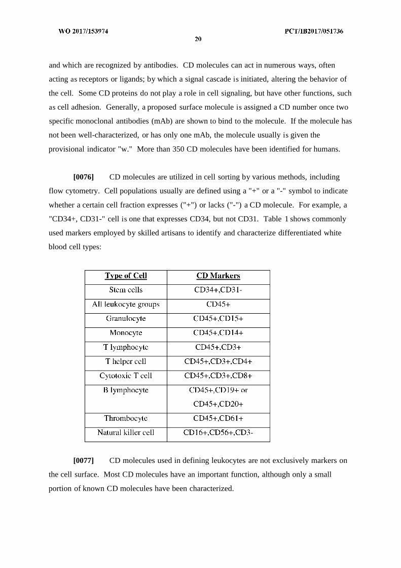
[0076] CD molecules are utilized in cell sorting by various methods, including
flow cytometry. Cell populations usually are defined using a "+" or a "-" symbol to indicate
whether a certain cell fraction expresses ("+") or lacks ("-") a CD molecule. For example, a
"CD34+, CD31-" cell is one that expresses CD34, but not CD31. Table 1 shows commonly
used markers employed by skilled artisans to identify and characterize differentiated white
blood cell types:
[0077] CD molecules used in defining leukocytes are not exclusively markers on
the cell surface. Most CD molecules have an important function, although only a small
portion of known CD molecules have been characterized.
[0078] The term "complementarity determining region" as used herein refers to
immunoglobulin (Ig) hypervariable domains that determine specific antibody (Ab) binding.
There are 6 CDRs in both variable regions of light (VL) and heavy chains (VH) with
background variability on each side of the CDRs. Antibodies (Abs) of different specificities
can assemble identical VL domains with different VH domains. The framework sequences
between CDRs can be similar or identical.
[0079] The term "conjugate" or "conjugated", as used herein, refers to reversibly
binding, coupling or connecting one substance with another substance (e.g., an antibody to a
bead).
[0080] The term "connected", as used herein, refers to is being joined, linked, or
fastened together in close association. For example, in the context of a chemical compound
the term "connected to" refers to the attraction or connection between two atoms or
molecules via direct or indirect chemical bonds.
[0081] The term "medium", "culture medium", and "physiological medium", as used
herein, refers generally to any preparation used for the cultivation of living cells. A "cell
culture" refers to cells cultivated in vitro.
[0082] The term "cytometry", as used herein, refers to a process in which physical
and/or chemical characteristics of single cells, or by extension, of other biological or
nonbiological particles in roughly the same size or stage, are measured. In flow cytometry,
the measurements are made as the cells or particles pass through the measuring apparatus (a
flow cytometer) in a fluid stream. A cell sorter, or flow sorter, is a flow cytometer that uses
electrical and/or mechanical means to divert and collect cells (or other small particles) with
measured characteristics that fall within a user-selected range of values.
[0083] The term "derivative", as used herein, refers to a compound that may be
produced from another compound of similar structure in one or more steps. A "derivative" or
"derivatives" of a peptide or a compound retains at least a degree of the desired function of
the peptide or compound. Accordingly, an alternate term for "derivative" may be "functional
derivative." Derivatives can include chemical modifications of the peptide, such as
akylation, acylation, carbamylation, iodination or any modification that derivatizes the
peptide. Such derivatized molecules include, for example, those molecules in which free
amino groups have been derivatized to form amine hydrochlorides, p-toluene sulfonyl
groups, carbobenzoxy groups, t-butyloxycarbonyl groups, chloroacetyl groups or formal
groups. Free carboxyl groups can be derivatized to form salts, esters, amides, or
hydrazides. Free hydroxyl groups can be derivatized to form O-acyl or O-alkyl
derivatives. The imidazole nitrogen of histidine can be derivatized to form N-im-
benzylhistidine. Also included as derivatives or analogues are those peptides that contain one
or more naturally occurring amino acid derivative of the twenty standard amino acids, for
example, 4-hydroxyproline, 5-hydroxylysine, 3-methylhistidine, homoserine, ornithine or
carboxyglutamiate, and can include amino acids that are not linked by peptide bonds. Such
peptide derivatives can be incorporated during synthesis of a peptide, or a peptide can be
modified by wellknown chemical modification methods (see, e.g., Glazer et al., Chemical
Modification of Proteins, Selected Methods and Analytical Procedures, Elsevier Biomedical
Press, New York (1975)).
[0084] The term "differential label" as used herein, generally refers to a stain,
dye, marker, antibody or antibody-dye combination, or intrinsically fluorescent cell-
associated molecule, used to characterize or contrast components, small molecules,
macromolecules, e.g., proteins, and other structures of a single cell or organism. The term
"dye" (also referred to as "fluorochrome" or "fluorophore") as used herein refers to a
component of a molecule which causes the molecule to be fluorescent. The component is a
functional group in the molecule that absorbs energy of a specific wavelength and re-emits
energy at a different (but equally specific) wavelength. The amount and wavelength of the
emitted energy depend on both the dye and the chemical environment of the dye. Many dyes
are known, including, but not limited to, F1TC, R-phycoerythrin (PE), PE-Texas Red
Tandem, PE-Cy5 Tandem, propidium iodem, EGFP, EYGP, ECF, DsRed, allophycocyanin
(APC), PerCp, SYTOX Green, courmarin, Alexa Fluors (350, 430, 488, 532, 546, 555, 568,
594, 633, 647, 660, 680, 700, 750), Cy2, Cy3, Cy3.5, Cy5, Cy5.5, Cy7, Hoechst 33342,
DAPI, Hoechst 33258, SYTOX Blue, chromomycin A3, mithramycin, YOYO-1, SYTOX
Orange, ethidium bromide, 7-AAD, acridine orange, TOTO-1, TO-PRO-1, thiazole orange,
TOTO-3, TO-PRO-3, thiazole orange, propidium iodide (PI), LDS 751, Indo-1, Fluo-3,
DCFH, DHR, SNARF, Y66F, Y66H, EBFP, GFPuv, ECFP, GFP, AmCyanl, Y77W, S65A,
S65C, S65L, S65T, ZsGreenl, ZsYellowl, DsRed2, DsRed monomer, AsRed2, mRFPl,
HcRedl, monochlorobimane, calcein, the DyLight Fluors, cyanine, hydroxycoumarin,

aminocoumarin, methoxycoumarin, Cascade Blue, Lucifer Yellow, NBD, PE-Cy5
conjugates, PE-Cy7 conjugates, APC-Cy7 conjugates, Red 613, fluorescein, FluorX,
BODIDY-FL, TRITC, X- rhodamine, Lissamine Rhodamine B, Texas Red, TruRed, and
derivatives thereof.
[0085] The term "enriched" or enrichment", as used herein, refers to increasing
the concentration of a given substance above the initial concentration of the substance. For
example, the term "cell enrichment", as used herein, refers to increasing the concentration of
a cell population above the initial concentration of the cell population.
[0086] The term "epitope" or antigenic determinant" or "epitope" means an
antigenic site on a molecule. From Robert C. Ladner, Biotechnol. & Genetic Engineering
Revs. 24 (1-30 (2007): Epitopes can be divided into linear epitopes (also known as
continuous epitopes) and non-linear epitopes (also known as conformational or discontinuous
epitopes). Linear epitopes persist after the protein is denatured or is in small peptide
fragments. Conformational epitopes persist only in properly folded proteins or large folded
fragments. "Epitope" can be modified or qualified in several ways. For example, there are
"functional epitopes" (Sanchez-Madrid, et al., 1983), "structural epitopes" (Abraham, et al.,
1985), "contact epitopes" (Jin, et al., 1992), "binding epitopes" (Bock, et al., 1985),
"protective epitopes" (Seyer, et al., 1986), "neutralizing epitopes" (Wimmer, et al., 1984),
"extracellular epitopes" (Khan, 2001), and "cytoplasmic epitopes" (Froehner, et al., 1983).
[0087] The term "flow cytometry", as used herein, refers to a tool for
interrogating the phenotype and characteristics of cells. It senses cells or particles as they
move in a liquid stream through a laser (light amplification by stimulated emission of
radiation)/light beam past a sensing area. The relative light-scattering and color-
discriminated fluorescence of the microscopic particles is measured. Analysis and
differentiation of the cells is based on size, granularity, and whether the cells are carrying
fluorescent molecules in the form of either antibodies or dyes. As the cell passes through the
laser beam, light is scattered in all directions, and the light scattered in the forward direction
at low angles (0.5-10°) from the axis is proportional to the square of the radius of a sphere
and so to the size of the cell or particle. Light may enter the cell; thus, the 90 0 light (right-
angled, side) scatter may be labeled with fluorochrome-linked antibodies or stained with
fluorescent membrane, cytoplasmic, or nuclear dyes. Thus, the differentiation of cell types,
the presence of membrane receptors and antigens, membrane potential, pH, enzyme activity,
and DNA content may be facilitated. Flow cytometers are multiparameter, recording several
measurements on each cell; therefore, it is possible to identify a homogeneous subpopulation
within a heterogeneous population [Marion G. Macey, Flow cytometry: principles and
applications, Humana Press, 2007].
[0088] The term "heterogeneous", as used herein, refers to a substance comprising
elements with various and dissimilar properties; not uniform in structure or composition. For
example, a heterogeneous cell population comprises cells of different types (e.g., red blood
cells, white blood cells, etc.).
[0089] The term "homogeneous" as used herein refers to a substance that is
uniform in structure or composition.
[0090] The term "human antibody" as used herein refers to antibodies having
variable and constant regions derived from human germline immunoglobulin sequences, but
excludes from the definition antibodies in which CDR sequences derived from the germline
of another mammalian species, such as a mouse, have been grafted onto human framework
sequences.
[0091] The term "humanized antibodies" as used herein refers to antibodies in
which rodent variable domain framework regions are swapped for human antibody
sequences.
[0092] The term "immunoglobulin (Ig)", as used herein, refers to a class of
structurally related proteins, each consisting of two pairs of polypeptide chains, one pair of
light (L) (low molecular weight) chains (k or 1), and one pair of heavy (H) chains (g, a, m, d,
and e), usually all four linked together by disulfide bonds. On the basis of the structural and
antigenic properties of the H chains, Ig's are classified (in order of relative amounts present
in normal human serum) as IgG, IgA, IgM, IgD, and IgE. Each class of H chain can
associate with either k or 1L chains. Subclasses of Ig's are based on differences in the H
chains, and are referred to as IgGi, etc.
[0093] When split by papain, IgG yields three pieces: the Fc piece, consisting of
the C-terminal portion of the H chains, with no antibody activity but capable of fixing

complement, and crystallizable; and two identical Fab pieces, each carrying an antigen-
binding site and each consisting of an L chain bound to the remainder of an H chain.
[0094] All L chains are divided into a region of variable sequence (VL) and one of
constant sequence (CL), each comprising about half the length of the L chain. The constant
regions of all human L chains of the same type (κ or λ) are identical except for a single amino
acid substitution, under genetic controls. H chains are similarly divided, although the VH
region, while similar in length to the VL region, is only one-third or one-fourth the length of
the C H region. Binding sites are a combination of V and V H protein regions. The large
number of possible combinations of L and H chains make up the "libraries" of antibodies of
each individual.
[0095] The term Ig includes, without limitation, naturally occurring and non-
naturally occurring IgGs, polyclonal IgGs, monoclonal IgGs, chimeric IgGs, wholly synthetic
IgGs, and fragments thereof.
[0096] The terms "isolate" and "separate" are used interchangeably herein to refer
to placing, setting apart, or obtaining a cell, protein, molecule, substance, nucleic acid,
peptide, or particle, in a form essentially free from contaminants or other materials with
which it is commonly associated.
[0097] The term "labelling" as used herein, refers to a process of distinguishing a
compound, structure, protein, peptide, antibody, cell or cell component by introducing an
antibody, a traceable constituent. Common traceable constituents include, but are not limited
to, a fluorescent antibody, a fluorophore, a dye or a fluorescent dye, a stain or a fluorescent
stain, a marker, a fluorescent marker, a chemical stain, a differential stain, a differential label,
and a radioisotope.
[0098] The term "lectin" as used herein refers to a class of proteins that bind
specifically to certain sugars.
[0099] The term "leukocyte", as used herein, refers to a colorless cell (i.e., a white
blood cell) that circulates in the blood and body fluids and is involved in counteracting
foreign substances and disease. Leukocytes include, but are not limited to, lymphocytes,
granulocytes, monocytes and macrophages.
[00100] The term "lymphocyte", as used herein, refers to a small white blood cell
formed in lymphatic tissue throughout the body and in normal adults making up about 22-
28% of the total number of leukocytes in the circulating blood.
[00101] The term "mimetic" or "mimic", as used herein, refers to chemicals
containing chemical moieties that mimic the function of an antibody. For example, if an
antibody binding site contains two charged chemical moieties having functional activity, a
mimetic places two charged chemical moieties in a spatial orientation and constrained
structure so that the charged chemical function is maintained in three-dimensional space.
[00102] The term "mononuclear cell" or "MNC", as herein, refers to any cell that
has a single round nucleus. Non-limiting examples include blood cells, such as lymphocytes,
monocytes and dendritic cells.
[00103] The term "negative selection", as used herein, refers to depletion or
removal all cell types except for a cell type of interest, which remains.
[00104] The phrase "operatively linked" or "operably linked", as used herein,
refers to a linkage in which two or more protein domains are ligated or combined via
recombinant DNA technology or chemical reaction such that each protein domain of the
resulting fusion protein retains its original function.
[00105] The term "phenotype", as used herein, refers to observable characteristics
or physical traits (e.g., morphology, development, biochemical, physiological properties) of a
cell or organism.
[00106] The term "positive selection", as used herein, refers to the isolation of a
target cell population.
[00107] The term "pure", as used herein, refers to a cell, protein, molecule,
substance, nucleic acid, peptide, or particle not mixed, adulterated or contaminated with any
other substance or material.
[00108] The term "single chain variable fragment", "single chain Fv" or "scFv" as
used herein refers to antibody fragments comprising the VH and V domains of an antibody.
These domains are present in a single polypeptide chain. Generally, the Fv polypeptide
further comprises a polypeptide linker between the VH and VL domains which enables the
scFv to form the desired structure for antigen binding.
[00109] The term "substantially pure", as used herein, refers to a purity of at least
75%, at least 80%, at least 85%, at least 90%, at least 95% or at least 99% as determined by
an analytical protocol. Such protocols may include, for example, but are not limited to,
FACS, HPLC, gel electrophoresis, chromatography, and the like.
[00110] The term "T lymphocyte" or "T-cell", as used herein, generally refers to a
small white blood cell formed in lymphatic tissue throughout the body and in normal adults
making up about 22-28% of the total number of leukocytes in the circulating blood that plays
a large role in defending the body against disease. Individual lymphocytes are specialized in
that they are committed to respond to a limited set of structurally related antigens. This
commitment, which exists before the first contact of the immune system with a given antigen,
is expressed by the presence on the lymphocyte's surface membrane of receptors specific for
determinants (epitopes) on the antigen. Each lymphocyte possesses a population of receptors,
all of which have identical combining sites. One set, or clone, of lymphocytes differs from
another clone in the structure of the combining region of its receptors and thus differs in the
epitopes that it can recognize. Lymphocytes differ from each other not only in the specificity
of their receptors, but also in their functions. Two broad classes of lymphocytes are
recognized: the B-lymphocytes (B-cells), which are precursors of antibody-secreting cells,
and T-lymphocytes (T-cells).
[00111] T-lymphocytes derive from precursors in hematopoietic tissue, undergo
differentiation in the thymus, and are then seeded to peripheral lymphoid tissue and to the
recirculating pool of lymphocytes. T-lymphocytes or T cells mediate a wide range of
immunologic functions. These include the capacity to help B cells develop into antibody-
producing cells, the capacity to increase the microbicidal action of monocytes/macrophages,
the inhibition of certain types of immune responses, direct killing of target cells, and
mobilization of the inflammatory response. These effects depend on their expression of
specific cell surface molecules and the secretion of cytokines. (Paul, W. E., "Chapter 1: The
immune system: an introduction," Fundamental Immunology, 4th Edition, Ed. Paul, W. E.,
Lippicott-Raven Publishers, Philadelphia (1999)).
[00112] T cells differ from B cells in their mechanism of antigen recognition.
Immunoglobulin, the B cell's receptor, binds to individual epitopes on soluble molecules or
on particulate surfaces. B-cell receptors see epitopes expressed on the surface of native
molecules. Antibody and B-cell receptors evolved to bind to and to protect against
microorganisms in extracellular fluids. In contrast, T cells recognize antigens on the surface
of other cells and mediate their functions by interacting with, and altering, the behavior of
these antigen-presenting cells (APCs). There are three main types of antigen-presenting cells
in peripheral lymphoid organs that can activate T cells: dendritic cells, macrophages and B
cells. The most potent of these are the dendritic cells, whose only function is to present
foreign antigens to T cells. Immature dendritic cells are located in tissues throughout the
body, including the skin, gut, and respiratory tract. When they encounter invading microbes
at these sites, they endocytose the pathogens and their products, and carry them via the lymph
to local lymph nodes or gut associated lymphoid organs. The encounter with a pathogen
induces the dendritic cell to mature from an antigen-capturing cell to an antigen-presenting
cell (APC) that can activate T cells. APCs display three types of protein molecules on their
surface that have a role in activating a T cell to become an effector cell: (1) MHC proteins,
which present foreign antigen to the T cell receptor; (2) costimulatory proteins which bind to
complementary receptors on the T cell surface; and (3) cell-cell adhesion molecules, which
enable a T cell to bind to the antigen-presenting cell (APC) for long enough to become
activated. ("Chapter 24: The adaptive immune system," Molecular Biology of the Cell,
Alberts, B. et al., Garland Science, NY, 2002).
[00113] T-cells are subdivided into two distinct classes based on the cell surface
receptors they express. The majority of T cells express T cell receptors (TCR) consisting of
a and β chains. A small group of T cells express receptors made of γ and δ chains. Among
the /β T cells are two important sublineages: those that express the coreceptor molecule
CD4 (CD4+ T cells); and those that express CD8 (CD8+ T cells). These cells differ in how
they recognize antigen and in their effector and regulatory functions.
[00114] CD4+ T cells are the major regulatory cells of the immune system. Their
regulatory function depends both on the expression of their cell-surface molecules, such as
CD40 ligand whose expression is induced when the T cells are activated, and the wide array
of cytokines they secrete when activated.
[00115] T cells also mediate important effector functions, some of which are
determined by the patterns of cytokines they secrete. The cytokines can be directly toxic to
target cells and can mobilize potent inflammatory mechanisms.
[00116] In addition, T cells particularly CD8+ T cells, can develop into cytotoxic
T-lymphocytes (CTLs) capable of efficiently lysing target cells that express antigens
recognized by the CTLs. (Paul, W. E., "Chapter 1: The immune system: an introduction,"
Fundamental Immunology, 4th Edition, Ed. Paul, W. E., Lippicott-Raven Publishers,
Philadelphia (1999)).
[00117] T cell receptors (TCRs) recognize a complex consisting of a peptide
derived by proteolysis of the antigen bound to a specialized groove of a class II or class I
MHC protein. The CD4+ T cells recognize only peptide/class II complexes while the CD8+
T cells recognize peptide/class I complexes. (Paul, W. E., "Chapter 1: The immune system:
an introduction," Fundamental Immunology, 4th Edition, Ed. Paul, W. E., Lippicott-Raven
Publishers, Philadelphia (1999)).
[00118] The TCR's ligand (i.e., the peptide/MHC protein complex) is created
within antigen-presenting cells (APCs). In general, class II MHC molecules bind peptides
derived from proteins that have been taken up by the APC through an endocytic process.
These peptide-loaded class II molecules are then expressed on the surface of the cell, where
they are available to be bound by CD4+ T cells with TCRs capable of recognizing the
expressed cell surface complex. Thus, CD4+ T cells are specialized to react with antigens
derived from extracellular sources. (Paul, W. E., "Chapter 1: The immune system: an
introduction," Fundamental Immunology, 4th Edition, Ed. Paul, W. E., Lippicott-Raven
Publishers, Philadelphia (1999)).
[00119] In contrast, class I MHC molecules are mainly loaded with peptides
derived from internally synthesized proteins, such as viral proteins. These peptides are
produced from cytosolic proteins by proteolysis by the proteosome and are translocated into
the rough endoplasmic reticulum. Such peptides, generally nine amino acids in length, are
bound into the class I MHC molecules and are brought to the cell surface, where they can be
recognized by CD8+ T cells expressing appropriate receptors. This gives the T cell system,
particularly CD8+ T cells, the ability to detect cells expressing proteins that are different
from, or produced in much larger amounts than, those of cells of the remainder of the
organism (e.g., vial antigens) or mutant antigens (such as active oncogene products), even if
these proteins in their intact form are neither expressed on the cell surface nor secreted.
(Paul, W. E., "Chapter 1: The immune system: an introduction," Fundamental Immunology,
4th Edition, Ed. Paul, W. E., Lippicott-Raven Publishers, Philadelphia (1999)).
[00120] T cells can also be classified based on their function as helper T cells; T
cells involved in inducing cellular immunity; suppressor T cells; and cytotoxic T cells.
[00121] Helper T cells are T cells that stimulate B cells to make antibody responses
to proteins and other T cell-dependent antigens. T cell-dependent antigens are immunogens
in which individual epitopes appear only once or a limited number of times such that they are
unable to cross-link the membrane immunoglobulin (Ig) of B cells or do so inefficiently. B
cells bind the antigen through their membrane Ig, and the complex undergoes endocytosis.
Within the endosomal and lysosomal compartments, the antigen is fragmented into peptides
by proteolytic enzymes and one or more of the generated peptides are loaded into class II
MHC molecules, which traffic through this vesicular compartment. The resulting
peptide/class II MHC complex is then exported to the B-cell surface membrane. T cells with
receptors specific for the peptide/class II molecular complex recognize this complex on the
B-cell surface. (Paul, W. E., "Chapter 1: The immune system: an introduction," Fundamental
Immunology, 4th Edition, Ed. Paul, W. E., Lippicott-Raven Publishers, Philadelphia (1999)).
[00122] B-cell activation depends both on the binding of the T cell through its TCR
and on the interaction of the T-cell CD40 ligand (CD40L) with CD40 on the B cell. T cells
do not constitutively express CD40L. Rather, CD40L expression is induced as a result of an
interaction with an APC that expresses both a cognate antigen recognized by the TCR of the
T cell and CD80 or CD86. CD80/CD86 is generally expressed by activated, but not resting,
B cells so that the helper interaction involving an activated B cell and a T cell can lead to
efficient antibody production. In many cases, however, the initial induction of CD40L on T
cells is dependent on their recognition of antigen on the surface of APCs that constitutively
express CD80/86, such as dendritic cells. Such activated helper T cells can then efficiently
interact with and help B cells. Cross-linkage of membrane Ig on the B cell, even if
inefficient, may synergize with the CD40L/CD40 interaction to yield vigorous B-cell
activation. The subsequent events in the B-cell response, including proliferation, Ig
secretion, and class switching (of the Ig class being expressed) either depend or are enhanced
by the actions of T cell-derived cytokines. (Paul, W. E., "Chapter 1: The immune system:
an introduction," Fundamental Immunology, 4th Edition, Ed. Paul, W. E., Lippicott-Raven
Publishers, Philadelphia (1999)).
[00123] CD4+ T cells tend to differentiate into cells that principally secrete the
cytokines IL-4, IL-5, IL-6, and IL-10 (TH2 cells) or into cells that mainly produce IL-2, IFN-
γ , and lymphotoxin (THi cells). The TH2 cells are very effective in helping B-cells develop
into antibody-producing cells, whereas the THI cells are effective inducers of cellular immune
responses, involving enhancement of microbicidal activity of monocytes and macrophages,
and consequent increased efficiency in lysing microorganisms in intracellular vesicular
compartments. Although the CD4+ T cells with the phenotype of TH2 cells (i.e., IL-4, IL-5,
IL-6 and IL-10) are efficient helper cells, THI cells also have the capacity to be helpers.
(Paul, W. E., "Chapter 1: The immune system: an introduction," Fundamental Immunology,
4th Edition, Ed. Paul, W. E., Lippicott-Raven Publishers, Philadelphia (1999)). T-helper 1
(Thi) cells express at least one type of cell surface marker, including, but not limited to,
chemokine (C-C motif) receptor 1 (CCR1), chemokine (C-C motif) receptor 5 (CCR5),
cluster of differentiation 3 (CD3), cluster of differentiation 4 (CD4), chemokine (C-X-C
motif) receptor 3 (CXCR3), interferon gamma receptor 1/cluster of differentiation 119 (IFN-
yRl/CDl 19), interferon gamma receptor 2 (IFN-yR2), interleukin-12 receptor subunit beta-2
(IL-12RP2), interleukin-18 receptor alpha (IL-18Ra), and/or interleukin 27 receptor alpha/t
cell cytokine receptor (IL-27Ra/TCCR). T-helper 2 (Th2) cells express at least one type of
cell surface marker, including, but not limited to, chemokine (C-C motif) receptor 3 (CCR3),
chemokine (C-C motif) receptor 4 (CCR4), chemokine (C-C motif) receptor 8 (CCR8),
cluster of differentiation 3 (CD3), cluster of differentiation 4 (CD4), chemokine (C-X-C
motif) receptor 4 (CXCR4), interferon gamma receptor 1/cluster of differentiation 119 (IFN-
yRl/CD119), interferon gamma receptor 2 (IFN-yR2), interleukin-4 receptor alpha (IL-4Ra),
interleukin- 17 receptor beta (IL-17RP), interleukin- 1 receptor 4 (IL-1R4), and/or thymic
stromal lymphopoietin receptor (TSLPR).
[00124] A controlled balance between initiation and downregulation of the immune
response is important to maintain immune homeostasis. Both apoptosis and T cell anergy (a
tolerance mechanism in which the T cells are intrinsically functionally inactivated following
an antigen encounter (Scwartz, R. H., "T cell anergy," Annu. Rev. Immunol., 21: 305-334
(2003)) are important mechanisms that contribute to the downregulation of the immune
response. A third mechanism is provided by active suppression of activated T cells by
suppressor or regulatory CD4+ T (Treg) cells. (Reviewed in Kronenberg, M. et al.,
"Regulation of immunity by self-reactive T cells," Nature 435: 598-604 (2005)). CD4+
Tregs that constitutively express the IL-2 receptor alpha (IL-2Ra) chain (CD4+ CD25+) are a
naturally occurring T cell subset that are anergic and suppressive. (Taams, L. S. et 1.,
"Human anergic/suppressive CD4+CD25+ T cells: a highly differentiated and apoptosis-
prone population," Eur. J . Immunol., 31: 1122-1131 (2001)). Depletion of CD4+CD25+
Tregs results in systemic autoimmune disease in mice. Furthermore, transfer of these Tregs
prevents development of autoimmune disease. Human CD4+CD25+ Tregs, similar to their
murine counterpart, are generated in the thymus and are characterized by the ability to
suppress proliferation of responder T cells through a cell-cell contact-dependent mechanism,
the inability to produce IL-2, and the anergic phenotype in vitro. Human CD4+CD25+ T
cells can be split into suppressive (CD25high) and nonsuppressive (CD251ow) cells,
according to the level of CD25 expression. A member of the forkhead family of transcription
factors, FOXP3, has been shown to be expressed in murine and human CD4+CD25+ Tregs
and appears to be a master gene controlling CD4+CD25+ Treg development. (Battaglia, M.
et al., "Rapamycin promotes expansion of functional CD4+CD25+Foxp3+ regulator T cells
of both healthy subjects and type 1 diabetic patients," J . Immunol., 177: 8338-8347 (200)).
Regulatory T-cells express at least one type of cell surface marker, including, but not limited
to, cluster of differentiation 3 (CD3), cluster of differentiation 4 (CD4), cluster of
differentiation 5 (CD5), cluster of differentiation 25 (CD25), cluster of differentiation 39
(CD39), cluster of differentiation 127 (CD127), cluster of differentiation 152 (CD152),
cluster of differentiation 45RA (CD45RA), cluster of differentiation 45RO (CD45RO),
cluster of differentiation 39 (CD39), cluster of differentiation 73 (CD73), cluster of
differentiation 357 (CD357), cluster of differentiation 103 (CD103), cluster of differentiation
223 (CD223), cluster of differentiation 134 (CD134), cluster of differentiation 62L (CD62L),
and/or cluster of differentiation 101 (CD101). Th9 cells express at least one type of cell
surface marker, including, but not limited to, cluster of differentiation 3 (CD3), cluster of
differentiation 4 (CD4), interleukin-4 receptor alpha (IL-4Ra), interleukin-17 receptor beta
(IL-17RP), and/or transforming growth factor beta receptor II (TGF-PRII). Thl7 cells
express at least one type of cell surface marker, including, but not limited to, chemokine (C-C
motif) receptor 4 (CCR4), chemokine (C-C motif) receptor 6 (CCR6), cluster of
differentiation 3 (CD3), cluster of differentiation 4 (CD4), interleukin-1 receptor 1 (IL-1R1),
interleukin-6 receptor alpha (IL-6Ra), interleukin-2 receptor (IL-21R), interleukin-23
receptor (IL-23R), and/or transforming growth factor beta receptor II (TGF-PRII).
[00125] The term "B lymphocyte" or "B-cell", as used herein, refers to a short
lived immunologically important lymphocyte that is not thymus dependent and is involved in
humoral immunity. B-lymphocytes are derived from hematopoietic cells of the bone marrow.
A mature B-cell can be activated with an antigen that expresses epitopes that are recognized
by its cell surface. The activation process may be direct, dependent on cross-linkage of
membrane Ig molecules by the antigen (cross-linkage-dependent B-cell activation), or
indirect, via interaction with a helper T-cell, in a process referred to as cognate help. In many
physiological situations, receptor cross-linkage stimuli and cognate help synergize to yield
more vigorous B-cell responses. (Paul, W. E., "Chapter 1: The immune system: an
introduction," Fundamental Immunology, 4th Edition, Ed. Paul, W. E., Lippicott-Raven
Publishers, Philadelphia (1999)).
[00126] Cross-linkage dependent B-cell activation requires that the antigen express
multiple copies of the epitope complementary to the binding site of the cell surface receptors
because each B-cell expresses Ig molecules with identical variable regions. Such a
requirement is fulfilled by other antigens with repetitive epitopes, such as capsular
polysaccharides of microorganisms or viral envelope proteins. Cross-linkage-dependent B-
cell activation is a major protective immune response mounted against these microbes. (Paul,
W. E., "Chapter 1: The immune system: an introduction," Fundamental Immunology, 4th
Edition, Ed. Paul, W. E., Lippicott-Raven Publishers, Philadelphia (1999)).
[00127] Cognate help allows B-cells to mount responses against antigens that
cannot cross-link receptors and, at the same time, provides costimulatory signals that rescue
B cells from inactivation when they are stimulated by weak cross-linkage events. Cognate
help is dependent on the binding of antigen by the B-cell' s membrane immunoglobulin (Ig),
the endocytosis of the antigen, and its fragmentation into peptides within the
endosomal/lysosomal compartment of the cell. Some of the resultant peptides are loaded into
a groove in a specialized set of cell surface proteins known as class II major
histocompatibility complex (MHC) molecules. The resultant class II/peptide complexes are
expressed on the cell surface and act as ligands for the antigen-specific receptors of a set of
T-cells designated as CD4+ T-cells. The CD4+ T-cells bear receptors on their surface
specific for the B-cell' s class II/peptide complex. B-cell activation depends not only on the
binding of the T cell through its T cell receptor (TCR), but this interaction also allows an
activation ligand on the T-cell (CD40 ligand) to bind to its receptor on the B-cell (CD40)
signaling B-cell activation. In addition, T helper cells secrete several cytokines that regulate
the growth and differentiation of the stimulated B-cell by binding to cytokine receptors on the
B cell. (Paul, W. E., "Chapter 1: The immune system: an introduction," Fundamental
Immunology, 4th Edition, Ed. Paul, W. E., Lippicott-Raven Publishers, Philadelphia (1999)).
[00128] During cognate help for antibody production, the CD40 ligand is
transiently expressed on activated CD4+ T helper cells, and it binds to CD40 on the antigen-
specific B cells, thereby tranducing a second costimulatory signal. The latter signal is
essential for B cell growth and differentiation and for the generation of memory B cells by
preventing apoptosis of germinal center B cells that have encountered antigen.
Hyperexpression of the CD40 ligand in both B and T cells is implicated in the pathogenic
autoantibody production in human SLE patients. (Desai-Mehta, A. et al., "Hyperexpression
of CD40 ligand by B and T cells in human lupus and its role in pathogenic autoantibody
production," J . Clin. Invest., 97(9): 2063-2073 (1996)).
[00129] The term "activation" or "lymphocyte activation" refers to stimulation of
lymphocytes by specific antigens, nonspecific mitogens, or allogeneic cells resulting in
synthesis of RNA, protein and DNA and production of lymphokines; it is followed by
proliferation and differentiation of various effector and memory cells. For example, a mature
B cell can be activated by an encounter with an antigen that expresses epitopes that are
recognized by its cell surface immunoglobulin Ig). The activation process may be a direct
one, dependent on cross-linkage of membrane Ig molecules by the antigen (cross-linkage-
dependent B cell activation) or an indirect one, occurring most efficiently in the context of an
intimate interaction with a helper T cell ("cognate help process"). T-cell activation is
dependent on the interaction of the TCR/CD3 complex with its cognate ligand, a peptide
bound in the groove of a class I or class II MHC molecule. The molecular events set in
motion by receptor engagement are complex. Among the earliest steps appears to be the
activation of tyrosine kinases leading to the tyrosine phosphorylation of a set of substrates
that control several signaling pathways. These include a set of adapter proteins that link the

TCR to the ras pathway, phospholipase Cyl, the tyrosine phosphorylation of which increases
its catalytic activity and engages the inositol phospholipid metabolic pathway, leading to
elevation of intracellular free calcium concentration and activation of protein kinase C, and a
series of other enzymes that control cellular growth and differentiation. Full responsiveness
of a T cell requires, in addition to receptor engagement, an accessory cell-delivered
costimulatory activity, e.g., engagement of CD28 on the T cell by CD80 and/or CD86 on the
antigen presenting cell (APC). The soluble product of an activated B lymphocyte is
immmunoglobulins (antibodies). The soluble product of an activated T lymphocyte is
lymphokines.
[00130] The term "pro B-cell", as used herein, refers to an early identifiable
intermediate cell type in a series of developmental stages leading to the generation of mature
B-cells. Human pro B-cells express at least one type of cell surface marker, including, but
not limited to, cluster of differentiation 19 (CD 19), cluster of differentiation 20 (CD20),
cluster of differentiation 34 (CD34), cluster of differentiation 38 (CD38), and/or cluster of
differentiation 45R (CD45R).
[00131] The term "pre-B-cell", as used herein, refers to the immediate precursor
cell of a mature B-cell. Human pre-B-cells express at least one type of cell surface marker,
including, but not limited to, cluster of differentiation 19 (CD 19), cluster of differentiation 20
(CD20), cluster of differentiation 38 (CD38), cluster of differentiation 40 (CD40), and/or
cluster of differentiation 45R (CD45R).
[00132] The term "immature B-cell", as used herein, refers to a cell produced in
the bone marrow that migrates to secondary lymphoid tissues where it may develop into a
mature B-cell. Human immature B-cells express at least one type of cell surface marker,
including, but not limited to, cluster of differentiation 19 (CD 19), cluster of differentiation 20
(CD20), cluster of differentiation 40 (CD40), cluster of differentiation 45R (CD45R), and/or
immunoglobulin M (IgM).
[00133] The term "transitional B-cell", as used herein, refers to an immature B-cell
that has migrated to a secondary lymphoid tissue (e.g., spleen or lymph node). Human
transitional 1 B-cells express at least one type of cell surface marker, including, but not
limited to, cluster of differentiation 10 (CD10), cluster of differentiation 19 (CD19), cluster

of differentiation 20 (CD20), cluster of differentiation 24 (CD24), cluster of differentiation 28
(CD28), and/or B-cell lymphoma 2 (BCL-2).
[00134] The term "naive B-cell", as used herein, refers to a B-cell that has not been
exposed to an antigen. Human naive B-cells express at least one type of cell surface marker,
including, but not limited to, cluster of differentiation 19 (CD 19), cluster of differentiation 20
(CD20), cluster of differentiation 23 (CD23), cluster of differentiation 38 (CD38), cluster of
differentiation 40 (CD40), cluster of differentiation 150 (CD150), immunoglobulin M (IgM),
and/or immunoglobulin D (IgD).
[00135] The term "memory B-cell", as used herein, refers to a B-cell subtype that
is formed within germinal centers following primary infection and are important in
generating an antibody-mediated immune response in the case of re-infection. Human
memory B-cells express at least one type of cell surface marker, including, but not limited to,
cluster of differentiation 19 (CD19), cluster of differentiation 20 (CD20), cluster of
differentiation 23 (CD23), cluster of differentiation 27 (CD27), cluster of differentiation 40
(CD40), immunoglobulin A (IgA), and/or immunoglobulin G (IgG).
[00136] The term "plasma cell", as used herein, refers to a fully differentiated B-
cell that produces a single type of antibody. Human plasma cells express at least one type of
cell surface marker, including, but not limited to, cluster of differentiation 9 (CD9), cluster of
differentiation 19 (CD19), cluster of differentiation 27 (CD27), cluster of differentiation 3 1
(CD31), cluster of differentiation 38 (CD38), cluster of differentiation 40 (CD40), cluster of
differentiation 95 (CD95), and/or C-X-C chemokine receptor type 4 (CXCR-4).
[00137] The term "B-l cell", as used herein, refers to a sub-class of B-cell involved
in the humoral immune response. They are not part of the adaptive immune system (i.e., they
have no memory), but can generate antibodies against antigens and can act as antigen
presenting cells. Human B-l cells express at least one type of cell surface marker, including,
but not limited to, cluster of differentiation 19 (CD19), cluster of differentiation 20 (CD20),
cluster of differentiation 27 (CD27), immunoglobulin M (IgM), and/or immunoglobulin D
(IgD).
[00138] The term "monocyte", as used herein, refers to a large phagocytic white
blood cell with a simple oval nucleus and clear, grayish cytoplasm. Monocytes are produced

in the bone marrow and then enter the blood where they migrate to tissues (e.g., spleen, liver,
lungs, and bone marrow tissue) where they mature into macrophages. Macrophages are the
main scavenger cells of the immune system; engulfing apoptotic cells and pathogens to
produce immune effector molecules which elicit an immune response.
Monocytes/macrophages derived from humans express at least one type of cell surface
marker, including, but not limited to, cluster of differentiation 14 (CD 14), and/or cluster of
differentiation 33 (CD33).
[00139] The term "dendritic cell", as used herein, refers to antigen-presenting cells
(APCs) that function to process antigen material and present it on the cell surface to T-cells.
Dendritic cells are capable of presenting both major histocompatibility class I (MHC-I) and
major histocompatibility class II (MHC-II) antigens. They act as messengers between the
innate and the adaptive immune systems. Types of dendritic cells include, but are not limited
to, myeloid (conventional) dendritic cells and plasmacytoid dendritic cells.
[00140] Myeloid dendritic cells derived from humans express at least one type of
cell surface marker, including, but not limited to, cluster of differentiation 1l c (CD1 lc),
cluster of differentiation 123 (CD 123), cluster of differentiation lc/blood dendritic cell
antigen-1 (CDlc/BDCA-1) and/or cluster of differentiation 141/blood dendritic cell antigen-3
(CD141/BDCA-3).
[00141] The term "plasmacytoid dendritic cell", as used herein, refers to an innate
immune cell that circulates in the blood and is found in peripheral organs. Plasmacytoid
dendritic cells derived from humans express at least one type of cell surface marker,
including, but not limited to, cluster of differentiation 304/blood dendritic cell antigen-4
(CD304/BDCA-4).
[00142] The term "open system", as used herein, refers to a physical system in
which material, energy, etc. can be gained from, or lost to, the surrounding environment.
[00143] The terms "saturate", "saturation conditions" and "saturated conditions"
are used interchangeably herein to refer to conditions in which one substance is united with
another to the greatest possible extent. For example, filling of all available binding sites on
an antibody molecule by its antigen. The terms "non- saturate", "non-saturation conditions"
and "non- saturated conditions" are used interchangeably herein to refer to conditions in
which one substance is united with another to an extent less than the greatest possible extent,
for example, not all available binding sites on an antibody are filled by its antigen.
[00144] The term "stem cell", as used herein, refers to an undifferentiated cell
having a high proliferative potential with the ability to self-renew that can generate daughter
cells that can undergo terminal differentiation into more than one distinct cell phenotype.
Types of stem cells include, but are not limited to, embryonic stem cells, non-embryonic
somatic or adult stem cells and induced pluripotent stem cells (iPSCs).
[00145] Embryonic stem cells are derived from embryos that develop from eggs
that have been fertilized in vitro. Embryonic stem cells derived from human subjects express
at least one cell surface marker, including, but not limited to, stage-specific embryonic
antigen-1 (SSEA-1), stage-specific embryonic antigen -3 (SSEA-3), stage-specific embryonic
antigen -4 (SSEA-4), cluster of differentiation 324 (CD324/E-Cadherin), cluster of
differentiation 90/thymus cell antigen-1 (CD90/Thy-1), cluster of differentiation
117/tyrosine-protein kinase kit/mast/stem cell growth factor receptor (CD1 17/c-KIT/SCFR),
cluster of differentiation 326 (CD326), cluster of differentiation 9/multidrug resistance
protein 1/transmembrane 4 superfamily/diphtheria toxin receptor-associated protein- 27/24
kD protein (CD9/MRPl/TM4SF/DRAP-27/p24), cluster of differentiation 29 (CD29)/pl
integrin, cluster of differentiation 24/heat-stable antigen (CD24/HSA), cluster of
differentiation 59 (CD59)/Protectin, cluster of differentiation 133 (CD133), cluster of
differentiation 31/platelet endothelial cell adhesion molecule-1 (CD31/PECAM-1), cluster of
differentiation 49f (CD49f)/Integrin a6/cluster of differentiation 29 (CD29), tumor rejection
antigen 1-60 (TRA-1-60), tumor rejection antigen 1-81(TRA-1-81), Frizzled-5 (FZD5), Stem
cell factor (SCF/c-Kit ligand), and/or Cripto/teratocarcinoma-derived growth factor- 1
(TDGF-1).
[00146] Somatic or adult stem cells are undifferentiated cells found among
differentiated cells in a tissue or organ. These cells can renew themselves and can
differentiate to yield some or all of the major specialized cell types of the tissue or organ of
origin. The primary role of somatic/adult stem cells is to maintain and repair the tissue in
which they are found. Somatic/adult stem cells include, but are not limited to, hematopoietic
stem cells and mesenchymal stem cells.

[00147] The term "hematopoietic stem cell (HSC)" as used herein, refers to a cell
isolated from blood or from bone marrow that can renew itself, differentiate to a variety of
specialized cells, mobilize out of the bone marrow into the circulating blood, and undergo
programmed cell death (apoptosis). Hematopoietic stem cells derived from human subjects
express at least one type of cell surface marker, including, but not limited to, cluster of
differentiation 34 (CD34), cluster of differentiation 38 (CD38), cluster of differentiation
45RA (CD45RA), human leukocyte antigen-antigen D related (HLA-DR), cluster of
differentiation 117/tyrosine-protein kinase kit/mast/stem cell growth factor receptor
(CD1 17/c-KIT/SCFR), cluster of differentiation 59 (CD59), stem cell antigen-1 (Sca-1),
cluster of differentiation 90/thymus cell antigen-1 (CD90/Thy-1), and/or C-X-C chemokine
receptor type 4 (CXCR-4).
[00148] The term "mesenchymal stem cell (MSC)", as used herein, refers to a
multipotent stromal cell that can differentiate into a variety of cells, including, but not limited
to, osteoblasts (bone cells), chondrocytes (cartilage cells), myocytes (muscle cells) and
adipocytes (fat cells). In vivo, MSCs associate with HSCs, exerting a key regulatory effect on
early stages of hematopoiesis. MSCs also enter differentiation pathways to replenish mature
osteoblasts, adipocytes and hemo-supportive stroma in bone marrow. MSCs are innervated
by sympathetic nervous system fibers and mediate neural control of hematopoiesis.
Mesenchymal stem cells derived from human subjects do not express a single specific
identifying marker but have been shown to express at least one type of cell surface marker,
including, but not limited to, stromal precursor antigen-1 (Stro-1), stage-specific embryonic
antigen -4 (SSEA-4), cluster of differentiation 70 (CD70), cluster of differentiation 271
(CD271), cluster of differentiation 200 (CD200), cluster of differentiation 146/melanoma cell
adhesion molecule (CD146/MCAM), cluster of differentiation 73 (CD73)/5'-nucleotidase,
cluster of differentiation 90/thymus cell antigen-1 (CD90/Thy-1), cluster of differentiation
105 (CD105)/endoglin, cluster of differentiation 106/vascular cell adhesion molecule-
1(CD106/VCAM-1), Ganglioside GD2, Frizzled-9 (FZD9), Tissue non-specific alkaline
phosphatase (TNAP), and/or Sushi domain containing 2 (SUSD2).
[00149] The term "induced pluripotent stem cell (iPSC)", as used herein, refers to a
type of pluripotent stem cell that can be generated directly from adult cells. These cells are
genetically reprogrammed to an embryonic stem cell-like state by being forced to express
genes and factors important for maintaining the defining properties of embryonic stem cells.
Induced pluripotent stem cells derived from human subjects express at least one type of cell
surface marker, including, but not limited to, stage-specific embryonic antigen - 1 (SSEA-1),
stage-specific embryonic antigen -4 (SSEA-4), alkaline phosphatase, octamer-binding
transcription factor 3/4 (Oct-3/4), homeobox protein Nanog (NANOG), sex determining
region Y-box 2 (Sox2), Krueppel-like factor 4 (KLF4), tumor rejection antigen 1-60 (TRA-1-
60), tumor rejection antigen 1-8 1(TRA- 1-81), and/or tumor rejection antigen 2-54 (TRA-2-
54).
[00150] The term "subject" or "individual" or "patient" are used interchangeably
herein to refer to a member of an animal species of mammalian origin, including but not
limited to, mouse, rat, cat, goat, sheep, horse, hamster, ferret, pig, dog, platypus, guinea pig,
rabbit and a primate, such as, for example, a monkey, ape, or human.
[00151] The term "target cell" or "targeted cell", as used herein, refers to a cell that
has a specific cell-surface marker that reacts with or binds to a specific antibody.
[00152] According to one aspect, the described invention provides an automated,
closed system for effectively labelling a target population of cells, washing the target
population of cells and enriching for the labeled targeted population of cells, resulting in
direct delivery of the targeted population of selected cells.
[00153] According to some embodiments, a population of cells suspended in a
physiological medium can added alone to the system; according to some such embodiments,
a labeling agent that recognizes a cell surface marker specifically and a capture particle
adapted to bind to cells labeled with the labeling agent and to therefore selectively separate
the subpopulation of labeled cells from the population of cells subsequently are added.
According to some embodiments, the population of cells is labeled inside the automated,
closed system of the described invention. According to some embodiments, the cells are
labeled outside the automated, closed system of the described invention. According to some
such embodiments, the population of cells plus the labeling agent that recognizes a cell
surface marker specifically are combined outside the system; the population of cells plus the
labeling agent that recognizes a cell surface marker specifically combined outside the system
are then added to the system; and a capture particle adapted to bind to a subpopulation of
cells labeled with the labeling agent that is effective to facilitate separation/selection of the

subpopulation of labeled cells from the population of cells subsequently is then added to the
system. According to some embodiments, the cells are labeled in the automated closed
system of the described invention. According to some such embodiments, the population of
cells plus the labeling agent that recognizes a cell surface marker specifically are combined
inside the system; a capture particle adapted to bind to a subpopulation of cells labeled with
the labeling agent that is effective to facilitate separation/selection of the subpopulation of
labeled cells from the population of cells subsequently is added to the system. According to
some embodiments, the population of cells, the labeling agent that recognizes a cell surface
marker specifically, and the capture particle adapted to bind to cells labeled with the labeling
agent and to therefore separate/select the subpopulation of labeled cells from the population
of cells are added and combined within the system. In each case, the capture particle can be
released from the captured cells after selection.
[00154] According to another aspect, the described invention can be used for
efficient gene transfer in a population of mammalian cells. According to some embodiments,
the gene transfer is mediated by a viral vector. According to some embodiments, the gene
transfer is by transfection. According to some embodiments, the population of mammalian
cells comprises labeled cells. According to some embodiments, the population of
mammalian cells has been enriched for labeled cells by positively selecting for the desired
cells using a labeling agent that recognizes a cell surface marker on a subpopulation of the
population of mammalian cells. According to some embodiments, the population of
mammalian cells comprises cells remaining after the labeled cells have been removed.
[00155] According to some embodiments, the automated, closed system comprises
an initial product bag (110), a chamber embedded in a centrifuge rotor/chamber (120), a final
product bag (130), a buffer bag (140), a labelling bag (160) and a capture particle injector
(170). According to some embodiments, the automated, closed system (100) comprises an
initial product bag ( 110), a chamber embedded in a centrifuge rotor/chamber (120), a final
product bag (130), a buffer bag (140), a lysing agent bag (150), a labelling bag (160) and a
capture particle injector (170) (See, e.g., Figure 1). According to some embodiments, the
automated, closed system comprises a pump.
[00156] According to one aspect, the described invention provides a method for
labelling cells using the automated, closed system (100).

[00157] According to another aspect, the described invention provides a method
for selecting/isolating cells using the automated, closed system (100).
[00158] According to another aspect, the described invention provides a method
for labelling cells, washing, enriching/selecting the labeled cells, and directly delivering the
selected cells using the automated, closed system (100).
[00159] According to some embodiments, the described invention provides a
method for isolating a substantially pure population of cells from a heterogeneous cell
suspension using the automated, closed system (100).
[00160] According to some embodiments, the method comprises passing a
heterogeneous cell population and capture particles comprising an agent that recognizes and
binds specifically to a cell into a chamber embedded in a centrifuge rotor/chamber (120)
while the rotor is in motion (i.e., spinning). According to some embodiments, the agent that
recognizes and binds specifically to a cell is an antibody that recognizes a specific cell
surface marker and bind those cells within the heterogeneous cell population that contain the
specific cell surface marker. According to some embodiments, the agent that recognizes and
binds specifically to a cell is a lectin. According to some embodiments, the method
comprises passing a wash buffer into the chamber embedded in the centrifuge rotor/chamber
(120) while the rotor is in motion in order to remove unbound cells (i.e., cells that do not
contain the specific cell surface marker). Next, according to some embodiments, the method
comprises passing a lysing agent (e.g., EDTA) into the chamber embedded in the centrifuge
rotor/chamber (120) while the rotor is in motion in order to lyse the capture particles and
passing a wash buffer into the chamber embedded in the centrifuge rotor/chamber (120)
while the rotor is in motion in order to remove the lysing agent and the lysed capture
particles. According to some embodiments, the rotor is turned off (i.e., not spinning) and the
cells bound to the agent that recognizes and binds specifically to a cell (i.e, enriched) are
collected in a final product bag (130). According to some embodiments, flow rate of
counterflow is increased and the cells bound to the agent that recognizes and binds
specifically to a cell (i.e., enriched) are collected in a final product bag (130). According to
some embodiments, the rotor is turned off (i.e., not spinning), flow rate of counterflow is
increased and the cells bound to the agent that recognizes and binds specifically to a cell (i.e.,
enriched) are collected in a final product bag (130) (See, e.g., Figure 3).

[00161] According to some embodiments, the described invention comprises an
injector, which is adapted to add a capture particle to the chamber for selecting a
subpopulation of cells. According to some embodiments, the injector is adapted to add the
labeling agent and the capture particle to the chamber for selecting a subpopulation of cells.
According to some embodiments, the injector is adapted to add the population of cells, the
labeling agent, and the capture particle to the chamber for selecting a subpopulation of cells.
[00162] According to some embodiments, the chamber is of a shape useful to form
a velocity gradient. According to some embodiments, the chamber is triangular- shaped.
[00163] According to some embodiments, the washing steps are performed by use
of a wash buffer. Non-limiting examples of commercially-available wash buffers include
Tris-buffered saline (TBS), phosphate buffered saline (PBS), Tris-buffered saline-tween-20
(TBST), phosphate-buffered saline-tween-20 (PBST), triethanolamine in PBS and
physiological media. Physiological media includes, but is not limited to, basal medium eagle
(BME), Dulbecco's phosphate buffered saline (DPBS), Dulbecco's modified eagle medium
(DMEM), DMEM-F12 media, F-10 nutrient mixture, Glasgow modified minimum essential
medium (GMEM), Iscove's modified Delbucco's medium (EVIDM), Leibovitz's L-15
medium, McCoy's 5A medium, MCDB 153 medium, media 199, minimal essential medium
(MEM), minimal essential media alpha (MEMA), RPMI 1640 medium, CliniMACS® buffer,
Hanks balanced salt saoltion (HBSS), TexMACs™ medium, and Waymouth's MB 752/1
medium.
[00164] According to some embodiments, the described invention, which is
automated, is effective to reduce the risk of human error.
[00165] According to some embodiments, the described invention is effective to
reduce the risk of contamination, by, for example, a bacteria, a virus, a fungus, cellular debris
and other unwanted materials with which the selected/isolated cells are commonly associated.
[00166] According to some embodiments, the described invention is effective to
reduce damage to the selected/isolated cell population because the described invention does
not require sedimentation/pelleting of cells. Types of damage include, but are not limited to,
cellular swelling, fat accumulation, metabolic failure, structural damage/deterioration and
apoptosis (i.e., cell death).
[00167] According to some embodiments, the described invention maintains
viability (meaning the ability of a cell to live, grow, expand, etc.) of the selected/isolated
cells.
[00168] According to some embodiments, the described invention maintains
morphology of the selected/isolated cells.
[00169] According to some embodiments, the selected/isolated cell population is at
least 75% pure. According to some embodiments, the selected/isolated cell population is at
least 76% pure. According to some embodiments, the selected/isolated cell population is at
least 77% pure. According to some embodiments, the selected/isolated cell population is at
least 78% pure. According to some embodiments the selected/isolated cell population is at
least 79% pure. According to some embodiments the selected/isolated cell population is at
least 80% pure. According to some embodiments, the selected/isolated cell population is at
least 81% pure. According to some embodiments the selected/isolated cell population is at
least 82% pure. According to some embodiments the selected/isolated cell population is at
least 83% pure. According to some embodiments the selected/isolated cell population is at
least 84% pure. According to some embodiments the selected/isolated cell population is at
least 85% pure. According to some embodiments the selected/isolated cell population is at
least 86% pure. According to some embodiments the selected/isolated cell population is at
least 87% pure. According to some embodiments the selected/isolated cell population is at
least 88% pure. According to some embodiments the selected/isolated cell population is at
least 89% pure. According to some embodiments the selected/isolated cell population is at
least 90% pure. According to some embodiments the selected/isolated cell population is at
least 91% pure. According to some embodiments the selected/isolated cell population is at
least 92% pure. According to some embodiments the selected/isolated cell population is at
least 93% pure. According to some embodiments the selected/isolated cell population is at
least 94% pure. According to some embodiments the selected/isolated cell population is at
least 95% pure. According to some embodiments the selected/isolated cell population is at
least 96% pure. According to some embodiments the selected/isolated cell population is at
least 97% pure. According to some embodiments the selected/isolated cell population is at
least 98% pure. According to some embodiments the selected/isolated cell population is at
least 99% pure.
[00170] According to some embodiments, the source of the selected/isolated cells
includes, but is not limited to, skin, blood, bone marrow, brain, heart, liver, pancreas, lung,
stomach, intestine, kidney, bladder, ovary, uterus, testis, thymus, adipose tissue and lymph
node.
[00171] According to some embodiments, the selected/isolated cells are stem cells.
Stem cells include, but are not limited to embryonic stem cells, somatic stem cells and
induced pluripotent stem cells. Somatic stem cells include, but are not limited to,
hematopoietic stem cells and mesenchymal stem cells.
[00172] According to some embodiments, the selected/isolated cells are
mononuclear cells. Non-limiting examples of mononuclear cells include lymphocytes,
monocytes and dendritic cells. Lymphocytes include, but are not limited to, T lymphocytes
and B lymphocytes. T lymphocytes include, but are not limited to, T helper cells and
regulatory T-cells. B lymphocytes include, but are not limited to, pro B-cells, pre B-cells,
immature B-cells, transitional B-cells, naive B-cells, memory B-cells, plasma cells and B-l
cells. Dendritic cells include, but are not limited to, myeloid (conventional) dendritic cells
and plasmacytoid dendritic cells.
[00173] According to some embodiments, the population of cells is
separated/isolated from components that normally accompany or interact with the population
of cells as found in its natural environment (e.g., blood) based on size. Without being bound
by theory, capture particles comprising the agent that recognizes and binds specifically to a
cell can specifically bind to cell phenotypes to facilitate separation/isolation. Cells and
capture particles are mixed and incubated within the automated, closed system of the
described invention. Following incubation, cells bound to capture particles exhibit a larger
size than unbound cells, allowing for separation within the system of the described invention.
A wash is automatically performed within the closed system to remove unbound cells.
Following the wash, a dissociation solution is automatically added within the closed system
to remove the selected/isolated cells from the capture particles. The selected/isolated cells
are automatically washed and volume is reduced within the closed system.
[00174] According to some embodiments, the dissociation solution is a pH
solution. Non limiting examples of pH solutions include 100 mM glycine-HCl, pH 2.5-3.0;
100 mM citric acid, pH 3.0; 50-100 mM trimethylamine or triethanolamine, pH 11.5; and 150
mM ammonium hydroxide, pH 10.5. According to some embodiments, the dissociation
solution is an ionic strength solution. Ionic strength solutions include, but are not limited to,
3.5-4.0 M magnesium chloride, pH 7.0 in 10 mM Tris; 5 M lithium chloride in 10 mM
phosphate buffer, pH 7.2; 2.5 M sodium iodide, pH 7.5; and 0.2-3.0 M sodium thiocyanate.
According to some embodiments, the dissociation solution is a denaturing solution. Non-
limiting examples of denaturing solutions include 2-6 M guanidine-HCl; 2-8 M urea; 1%
deoxycholate; and 1% sodium dodecyl sulfate (SDS). According to some embodiments, the
dissociation solution is an organic solution. Organic solutions include, but are not limited to,
10% dioxane and 50% ethylene glycol, pH 8-11.5.
[00175] According to some embodiments, the described invention utilizes a capture
particle, for example, a pellet, an agglomerate, a crystal, or a bead. According to some
embodiments, the capture particle comprises an agent that recognizes and binds specifically
to a cell. According to some embodiments, the capture particle is a bead. According to
some embodiments, the agent that recognizes and binds specifically to a cell is an antibody.
According to some embodiments, the agent that recognizes and binds specifically to a cell is
a lectin.
[00176] According to some embodiments, the bead is of a size useful for
conjugation with the agent. Non-limiting examples include microbeads and nanobeads.
According to some embodiments, the bead is biocompatible. According to some
embodiments, the bead is of a shape useful for conjugation to the agent. According to some
embodiments, the shape of the bead is irregular. According to some embodiments, the shape
of the bead is uniform. Shapes of the bead include, but are not limited to, a sphere, an oval, a
cylinder, a cube, a pyramid, a teardrop, a blob, a globule and the like. According to some
embodiments, the bead is of a material useful for conjugation to the agent that recognizes and
binds specifically to a cell surface marker. Non-limiting examples of materials include a
polymer or a mixture of different polymers, including, but not limited to, poly(lactic-co-
glycolic acid (PLGA), polyethylene glycol (PEG), polyorthoester, polyanhydride,
polygutamic acid, polyaspartic acid and poly(lactide-co-caprolactone). According to some
embodiments, the polymer is a synthetic polymer. Synthetic polymers include, but are not
limited to, low density polyethylene (LDPE), high density polyethylene (HDPE),
polypropylene (PP), polyvinyl chloride (PVC), polystyrene (PS), nylon, thermoplastic
polyurethane (TPU), poly(vinyl alcohol), poly(ethylene glycol) and Teflon™. According to
some embodiments, the polymer is a natural polymer. Natural polymers include, but are not
limited to, alginate, alginate derivatives, agarose, Sepharose®, collagen and chitosan.
Alginate derivatives include, but are not limited to, sodium alginate, amphiphilic alginate and
cell-interactive alginate.
[00177] Alginate is a naturally occurring anionic polymer typically obtained from
brown seaweed, and has been extensively investigated and used for many biomedical
applications, due to its biocompatibility, low toxicity, relatively low cost, and mild gelation
by addition of divalent cations such as Ca2+ . Commercially available alginate is typically
extracted from brown algae (Phaeophyceae), including Laminaria hyperborea, Laminaria
digitata, Laminaria japonica, Ascophyllum nodosum, and Macrocystis pyrifera by treatment
with aqueous alkali solutions, typically with sodium hydroxide (NaOH). The extract is
filtered, and either sodium or calcium chloride is added to the filtrate in order to precipitate
alginate. This alginate salt can be transformed into alginic acid by treatment with dilute HC1.
After further purification and conversion, water-soluble sodium alginate power is produced.
[00178] Alginate can also be synthesized by bacteria. Bacterial biosynthesis by
either Azotobacter oe Pseudomonas provides alginate with more defined chemical structures
and physical properties than can be obtained from seaweed-derived alginate. The pathway of
alginate biosynthesis is generally divided into (i) synthesis of precursor substrate; (ii)
polymerization and cytoplasmic membrane transfer; (iii) periplasmic transfer and
modification; and (iv) export through the outer membrane. Bacterial modification can enable
production of alginate with tailor-made features and a broad range of biomedical
applications.
[00179] Alginate is a family of linear copolymers containing blocks of (l,4)-linked
β-D-mannuronate (M) and a-L-guluronate (G) residues. The blocks are composed of
consecutive G residues (GGGGGG), consecutive M residues (MMMMMM), and alternating
M and G residues (GMGMGM). Alginates extracted from different sources differ in M and G
contents as well as the length of each block. Only the G-blocks of alginate are believed to
participate in intermolecular cross-linking with divalent cations (e.g., Ca2+) to form
hydrogels. The composition (i.e., M/G ratio), sequence, G-block length, and molecular
weight are thus critical factors affecting the physical properties of alginate and its resultant
hydrogels. The G-block content of Laminaria hyperborean stems is 60%, while other
commercially available alginates have a G-block content in the range of 14.0-31.0%. The
molecular weight of commercially available sodium alginates range between 32,000 and
400,000 g/mol. The viscosity of alginate solutions increases as pH decreases, and reaches a
maximum around pH = 3-3.5, as carboxylate groups in the alginate backbone become
protonated and form hydrogen bonds.
[00180] Various alginate derivatives are available and are used in a range of
biomedical applications. For example, amphiphilic alginate derivatives have been
synthesized by introducing hydrophobic moieties (e.g., alkyl chains, hydrophobic polymers)
to the alginate backbone. These derivatives can form self-assembled structures such as
particles and gels in aqueous media. Amphiphilic derivatives of sodium alginate have been
prepared by conjugation of long alkyl chains (i.e., dodecyl, octadecyl) to the alginate
backbone via ester bond formation. Microparticles can be prepared from these derivatives by
dispersion in a sodium chloride solution, this technique can allow encapsulation of proteins
and their subsequent release by the addition of either surfactants that disrupt intermolecular
hydrophobic junctions or esterases that hydrolyze the ester bond between alkyl chains and the
alginate backbone. Dodecylamine can also be conjugated to the alginate backbone via amide
linkage formation using 2-chloro-l-methylpyridinium iodide as a coupling reagent.
Hydrogels prepared from this alginate derivative exhibit long-term stability in aqueous
media, compared to those prepared from alginate derivatives with dodecyl ester, which are
labile to hydrolysis. Water soluble, amphiphilic alginate derivatives grafted with cholesteryl
groups can also be synthesized using Ν ,Ν '-dicyclohexylcarbodiimide as a coupling agent and
4-(N,N'-dimethylamino)pyridine as a catalyst at room temperature. These derivatives form
self-aggregates with a mean diameter of 136 nm in an aqueous sodium chloride solution.
Sodium alginate can also be hydrophobically modified with poly(butyl methacrylate), leading
to prolonged release of model drugs as compared with unmodified alginate gels
[00181] Cell-interactive alginates (i.e., alginate derivatives containing cell-
adhesive peptides) can be prepared by chemically introducing peptides as side-chains, using
carbodiimide chemistry to couple via the carboxylic groups of the sugar residues. Since
alginate inherently lacks mammalian cell-adhesivity, appropriate ligands are necessary to
promote and regulate cellular interactions, especially for cell culture and tissue engineering
applications. Peptides including the sequence arginine-glycine-aspartic acid (RGD) have been
extensively used as model adhesion ligands, due to the wide-spread presence of integrin
receptors (e.g., ανβ3, for this ligand on various cell types. RGD containing peptides can
be chemically coupled to the alginate backbone using water-soluble carbodiimide chemistry.
A minimum concentration of RGD peptides in alginate gels is needed for the adhesion and
growth of cells, and this minimum is likely cell type specific. For example, minimal
concentrations for substantial adhesion of MC3T3-E1 and C2C12 cells to alginate gels in
vitro have been reported as 12.5 and 10.0 g/mg alginate, respectively. The affinity of the
RGD peptide also plays an important role, and cyclic RGD peptides have been demonstrated
to be more potent and are needed at lower concentrations than linear RGD peptides. Various
peptides containing the DGEA (Asp-Gly-Glu-Ala) and YIGSR (Tyr-Ile-Gly-Ser-Arg)
sequences derived from other extracellular matrix proteins have also been exploited to
modify alginate gels and enhance the adhesive interactions with various cell types. For
example, alginate has been modified with YIGSR peptides using water-soluble carbodiimide
chemistry to promote neural cell adhesion (See, e.g., Lee KY and Mooney DJ Prog Polym
Sci 2012 Jan; 37(1): 106-126).
[00182] Agarose is a purified linear galactan hydrocolloid isolated from agar or
agar-bearing marine algae. It is a linear polymer consisting of alternating D-galactose and
3,6-anhydro-L-galactose units. As a gelling agent, agarose is used, for example, to separate
nucleic acids electrophoretically; to demonstrate cross reaction in Immunoelectrophoresis
(IEP) and double diffusion plates in which antibody- antigen precipitin lines are studied; to
make gel plates or overlays for cells in tissue culture; and to form a gel matrix (either beaded
and/or crosslinked) which can be used, for example, in chromatographic separations.
[00183] Sepharose® is a cross-linked, beaded form of agarose primarily used for
the chromatographic separation of biomolecules. Various grades and chemistries of
Sepharose® are available which permit the selective binding of cysteine side chain for the
immobilization of peptides. It can be combined with activation chemistries, such as
cyanogen bromide (CNBr) and reductive amination of aldehydes, in order to immobilize
antibodies, enzymes, proteins and peptides by way of covalent attachment.
[00184] Collagen is the most widely found protein in mammals and is the major
provider of strength to tissue. A typical collagen molecule consists of three intertwined
protein chains that form a helix. These molecules polymerize together to form collagen
fibers of varying length, thickness, and interweaving pattern (e.g., some collagen molecules
will form ropelike structures, while others will form meshes or networks). There are at least
15 different types of collagen, differing in their structure, function, location, and other
characteristics. The predominant form used in biomaterial applications is type I collagen,
which is a "rope-forming" collagen and is ubiquitous in the body, including skin and bone.
Collagen can be resorbed into the body, is non-toxic, produces only a minimal immune
response (even between different species), and is useful for attachment and biological
interaction with cells. Collagen may also be processed into a variety of formats, including
porous sponges, gels, and sheets, and can be crosslinked with chemicals to improve its
strength or to alter its degradation rate.
[00185] Chitosan is derived from chitin, a type of polysaccharide (i.e., sugar) that
is present in the hard exoskeletons of shellfish such as shrimp and crab. Chitin, in fact, is one
of the most abundant polysaccharides found in nature, making chitosan a plentiful and
relatively inexpensive product. Chitosan contains several desirable properties, including, but
limited to, minimal foreign body reaction; mild processing conditions (synthetic polymers
often need to be dissolved in harsh chemicals; chitosan will dissolve in water based on pH);
controllable mechanical/biodegradation properties (e.g., scaffold porosity or polymer length);
and availability of chemical side groups for attachment to other molecules. It can be
combined with other materials in order to increase its strength and cell-attachment
potential. Mixtures with synthetic polymers such as poly(vinyl alcohol) and poly(ethylene
glycol), or natural polymers such as collagen, are available and have displayed improved
performance over the behavior of either component alone.
[00186] According to some embodiments, the capture particle is a dextran bead
coated with alginate. Dextrans are polysaccharides with molecular weights > 1,000 Dalton.
They have a linear backbone of a-linked glucopyranosyl repeating units. Dextrans are
grouped into three (3) classes based on their structural features. Class 1 dextrans, which
contain the a(l 6)-linked D-glucopyranosyl backbone modified with small side chains of D-
glucose branches with a(l 2), a(l 3), and a(l 4)-linkage, vary in their molecular weight,
spatial arrangement, type and degree of branching, and length of branch chains, depending on
the microbial producing strains and cultivation conditions. Isomaltose and isomaltotriose are
oligosaccharides with the class 1 dextran backbone structure. Class 2 dextrans (alternans)
contain a backbone structure of alternating a(l 3) and a(l 6)-linked D-glucopyranosyl
units with a(l 3)-linked branches. Class 3 dextrans (mutans) have a backbone structure of
consecutive a(l 3)-linked D-glucopyranosyl units with a(l 6)-linked branches. One and
two-dimensional NMR spectroscopy techniques have been utilized for the structural analysis
of dextrans.The physical and chemical properties of purified dextrans vary depending on the
microbial strains from which they are produced and by the production method. Dextrans have
high water solubility and the solutions behave as Newtonian fluids. Solution viscosity
depends on concentration, temperature, and molecular weight, which have a characteristic
distribution. The hydroxyl groups present in dextran offer many sites for derivatization, and
these functionalized glycoconjugates represent a largely unexplored class of biocompatible
and environmentally safe compounds.
[00187] According to some embodiments, the capture particle is lysed with a lysing
agent. According to some embodiments, the lysing agent is a chelating agent. According to
some embodiments, the chelating agent is a calcium (Ca2+) chelating agent. Calcium
chelating agents include, but are not limited to, ethylenediaminetetraacetic acid (EDTA);
ethylene glycol tetraacetic acid (EGTA); l,2-bis(o-aminophenoxy)ethane-N,N,N',N'-
tetraacetic acid (BAPTA); deferoxamine mesylate, iron chelator IV, 21H7; and Ν ,Ν ,Ν ' ,Ν ' -
tetrakis(2-pyridylmethy)ethane- 1,2-diamine (TPEN).
[00188] According to some embodiments, the capture particle is coated with a
conjugate. Non-limiting examples of conjugates include heavy chain conjugates, light chain
conjugates, avidin and streptavidin. Examples of heavy chain conjugates include, but are not
limited to, Protein A, recombinant Protein A, Protein G and recombinant Protein G.
Examples of light chain conjugates include, but are not limited to, Protein L and recombinant
Protein L.
[00189] Protein A is derived from Staphylococcus aureus. Protein G is derived
from a Streptococcus species. Both have binding sites for the Fc portion of mammalian IgG.
The affinity of these proteins for IgG varies with the animal species. Protein G has a higher
affinity for rat, goat, sheep, and bovine IgG, as well as for mouse IgGl and human IgG3.
Protein A has a higher affinity for cat and guinea pig IgG. Native Protein G contains binding
sites for albumin, the Fab region of Igs, and membrane binding regions, which can lead to
nonspecific interactions. Recombinant Protein G has been engineered to eliminate the
albumin binding region, and recombinant Protein G' is a truncated protein which lacks the
albumin, Fab, and membrane binding sites while retaining the Fc binding site, making it more
specific for IgG than the native form. Neither Protein A nor Protein G is recommended for
detection of IgA or IgM, for detection of Fab fragments, or for detection of avian IgG. When
bound to a resin such as alginate, agarose or Sepharose®, Protein A and Protein G can be
used as affinity adsorbents to purify immunoglobulins and immunoglobulin subtypes from
serum, hybridoma ascites fluids, tissue culture supernatants, and other biological fluids.
These reagents are also commonly used to capture immune complexes generated in
immunoprecipitation experiments.
[00190] Protein L is derived from Peptostreptococcus magnus. It has an affinity
for kappa light chains from various species and will detect monoclonal or polyclonal IgG,
IgA, and IgM as well as Fab, F(ab')2, and recombinant single-chain Fv (scFv) fragments that
contain kappa light chains. It will also bind chicken IgG. Species such as bovine, goat, sheep,
and horse, whose Igs contain almost exclusively lambda chains, will not bind well, if at all, to
Protein L. Protein L is used as a general reagent for binding primary mammalian or avian
antibodies or surface Igs of all classes. It is useful for detection of Fab, F(ab')2 fragments,
recombinant scFv fragments, Igs bound to Fc receptors, or for detection of monoclonal
antibodies in the presence of bovine Igs bearing kappa light chains.
[00191] Avidin, an egg-white protein, is a highly cationic 66,000-dalton
glycoprotein with an isoelectric point of about 10.5. Its bacterial counterpart, streptavidin, is
a non-glycosylated 52,800-dalton protein with a near-neutral isoelectric point. Streptavidin
contains the tripeptide sequence Arg-Tyr-Asp (RYD) that apparently mimics the Arg-Gly-
Asp (RGD) binding sequence of fibronectin, a component of the extracellular matrix that
specifically promotes cellular adhesion. This universal recognition sequence binds integrins
and related cell-surface molecules. Each avidin and streptavidin protein binds four (4) biotin
molecules with high affinity (¾ of 10 14 to 10 15 mol/L) and selectivity. Biotin, also known
as vitamin B7, vitamin H or coenzyme R, is a water-soluble B-vitamin composed of a
tetrahydroimidizalone ring fused with a tetrahydrothiophene ring. Because both avidin and
streptavidin bind biotin with a high affinity and selectivity, proteins linked to biotin, or
"biotinylated", can be, for example, isolated from a sample or conjugated to an
avidin/streptavidin coated surface.

[00192] According to some embodiments, the described invention provides
antibodies that bind cell-surface markers. According to some embodiments, the antibodies
are full-length. According to some embodiments, the antibodies are fragments. According to
some embodiments, the fragment comprises only an antigen-binding portion. According to
another embodiment, the antigen binding portion comprises a light chain variable region (VL)
and a heavy chain variable region (VH) . According to some embodiments, the antibodies are
high-affinity antibodies. According to some embodiments, the antibodies are low-affinity
antibodies.
[00193] Antibodies of the described invention include, but are not limited to,
monoclonal antibodies, polyclonal antibodies and synthetic antibody mimics (SyAMs).
Monoclonal antibodies include, but are not limited to, synthetic antibodies and engineered
antibodies. Synthetic antibodies include, but are not limited to, recombinant antibodies.
Recombinant antibodies include, but are not limited to, single-chain variable fragment (scFv)
antibodies, nucleic acid aptamers and non-immunoglobulin protein scaffolds. Engineered
antibodies include, but are not limited to, chimeric antibodies and humanized antibodies.
[00194] Monoclonal antibodies are a homogenous population of antibodies that
recognize a single, specific eptitope of an antigen of interest. Monoclonal antibodies are
produced in cell culture by hybridoma cells, which are the result of a fusion between
myeloma cells and spleen cells from a mouse that has been immunized with a desired antigen
or from myeloma cells and B-cells from a rabbit that has been immunized with the desired
antigen.
[00195] Recombinant antibodies are antibodies that are produced by an in vitro
expression system (i.e., not produced by immunizing an animal with a desired antigen). For
example, the nucleic acid encoding a full-length antibody or VH and V antigen binding
domains may be inserted into a replicable vector for cloning (amplification of the DNA) or
for expression. Various vectors are publicly available. The vector may, for example, be in the
form of a plasmid, cosmid, viral particle, or phage. For example, plasmid vectors include,
but are not limited to, pET-26+ and pCMV6-AC. Vector components generally include, but
are not limited to, one or more of a signal sequence, an origin of replication, one or more
marker genes, an enhancer element, a promoter, and a transcription termination sequence.
Construction of suitable vectors containing one or more of these components employs
standard ligation techniques.
[00196] By way of non-limiting example, expression and cloning vectors may
contain a promoter operably linked to an antibody-encoding nucleic acid sequence to direct
mRNA synthesis. Promoters recognized by a variety of potential host cells are well known.
Promoters suitable for use with prokaryotic hosts include the beta-lactamase and lactose
promoter systems (Chang et al., Nature, 275:615 (1978); Goeddel et al., Nature, 281:544
(1979)), alkaline phosphatase, a tryptophan (trp) promoter system (Goeddel, Nucleic Acids
Res., 8:4057 (1980); EP 36,776), and hybrid promoters such as the tac promoter (deBoer et
al., Proc. Natl. Acad. Sci. USA, 80:21-25 (1983)). Promoters for use in bacterial systems also
can contain a Shine-Dalgarno (S.D.) sequence operably linked to DNA encoding antibodies.
[00197] Both expression and cloning vectors can contain a nucleic acid sequence
that enables the vector to replicate in one or more selected host cells. Such sequences are
known for a variety of bacteria, yeast, and viruses.
[00198] Expression and cloning vectors will typically contain a selection gene, also
termed a selectable marker. Typical selection genes encode proteins that (a) confer resistance
to antibiotics or other toxins (e.g., ampicillin, neomycin, methotrexate, or tetracycline), (b)
complement auxotrophic deficiencies, or (c) supply critical nutrients not available from
complex media, (e.g., the gene encoding D-alanine racemase for Bacilli). Examples of
selectable markers for mammalian cells include, but are not limited to, those that enable the
identification of cells competent to take up an antibody-encoding nucleic acid, such as DHFR
or thymidine kinase. An exemplary host cell when wild-type DHFR is employed is the CHO
cell line deficient in DHFR activity, prepared and propagated as described by Urlaub et al.,
Proc. Natl. Acad. Sci. USA, 77:4216 (1980). An exemplary selection gene for use in yeast is
the trpl gene present in the yeast plasmid YRp7 (Stinchcomb et al., Nature, 282:39 (1979);
Kingsman et al., Gene, 7:141 (1979); Tschemper et al., Gene, 10:157 (1980)). The trpl gene
provides a selection marker for a mutant strain of yeast lacking the ability to grow in
tryptophan, for example, ATCC No. 44076 or PEP4-1 (Jones, Genetics, 85:12 (1977)).
[00199] Host cells are transfected or transformed with expression or cloning
vectors described herein for antibody production and cultured in conventional nutrient media

modified as appropriate for inducing promoters, selecting transformants, or amplifying the
genes encoding the desired sequences. In general, principles, protocols, and practical
techniques for maximizing the productivity of cell cultures can be found in Mammalian Cell
Biotechnology: a Practical Approach, M. Butler, ed. (IRL Press, 1991).
[00200] Methods of eukaryotic cell transfection and prokaryotic cell transformation
include, for example, CaCl2, Ca2P0 4, liposome-mediated and electroporation. Depending on
the host cell used, transformation is performed using standard techniques appropriate to such
cells. The calcium treatment employing calcium chloride, as described in Sambrook et al., or
electroporation is generally used for prokaryotes. For mammalian cells, the calcium
phosphate precipitation method of Graham and van der Eb, Virology, 52:456-457 (1978) can
be employed. Transformations into yeast are typically carried out according to the method of
Van Solingen et al., J . Bact, 130:946 (1977) and Hsiao et al., Proc. Natl. Acad. Sci. (USA),
76:3829 (1979). However, other methods for introducing DNA into cells, such as by nuclear
microinjection, electroporation, bacterial protoplast fusion with intact cells, or polycations,
e.g., polybrene, polyornithine, may also be used. For various techniques for transforming
mammalian cells, See Keown et al., Methods in Enzymology, 185:527-537 (1990) and
Mansour et al., Nature, 336:348-352 (1988).
[00201] Suitable host cells for cloning and expressing DNA, and for producing
recombinant antibodies include, but are not limited to, Gram-negative bacteria, Gram-
positive bacteria, yeasts, fungi, protozoa, insect cells, mammalian cells and transgenic plants.
[00202] Escherichia coli has been the most important Gram-negative production
system for recombinant proteins reaching volumetric yields in the gram per liter scale for
extracellular production. However, cell wall-less L-forms of the Gram-negative bacterium
Proteus mirabilis and Pseudomonas putidas have been used for the production of mini
antibodies and scFv.
[00203] Gram-positive bacteria directly secrete proteins into the medium due to the
lack of an outer membrane which facilitate production of antibody fragments. The Gram-
positive bacteria Bacillus brevis, Bacillus subtilis, and Bacillus megaterium have been
successfully used for the production of different antibody fragments. In addition, B.
megaterium does not produce alkaline proteases and provides high stability of plasmid
vectors during growth allowing stable transgene expression during long term cultivation in
bioreactors. Lactobacilli have also been tested for antibody production and are "generally
regarded as safe" (GRAS) microorganisms. To date, two lactobacillus strains have been used
for the production of scFvs, Lactobacillus zeae/casei, and Lactobacillus paracasei.
[00204] Yeast combine short generation time and ease of genetic manipulation
with the robustness and simple medium requirements of unicellular microbial hosts. Pichia
pastoris is an exemplary yeast strain used for recombinant antibody production. Other yeast
like Saccharomyces cerevisiae, Hansenula polymorpha, Schizosaccharomyces pombe,
Schwanniomyces occidentalis, Kluyveromyces lactis, and Yarrowia lipolytica have also been
described for protein production.
[00205] Mammalian cells offer advanced mammalian folding, and a secretion and
post-translational apparatus that is capable of producing antibodies indistinguishable from
those in the human body with least concerns for immunogenic modifications. They are also
highly efficient for secretion of large and complex IgGs and, in combination with the folding
and post-translational control, high product quality can be achieved which reduces efforts and
costs in the subsequent and more expensive downstream processing steps. The risks of
contamination by pathogens or bovine spongiform encephalopathy (TSE/BSE) agents have
been eliminated by well-documented Good Manufacturing Practice (GMP) compliant
designer cell substrates and chemical defined media without the need of supplementing
animal serum components. Mammalian cell culture technology can reach production levels of
approximately 5 g/L IgGs in Chinese hamster ovary (CHO) cells. Industrial IgG production
levels often exceed 12 g/L as the result of a steadily ongoing progress in mammalian cell
culture technology, mainly due to improved high producer cell lines, optimized production
media, and prolonged production processes at high-cell densities. Producer cell lines have
also been genetically engineered regarding product homogeneity, improved metabolism,
reduced apoptosis, and inducible cell cycle arrest which allow prolonged production times for
almost 3 weeks at high-cell viability and cell densities.
[00206] Chinese hamster ovary (CHO) cells are the most common cells applied in
the commercial production of biopharmaceuticals. This cell line isolated in the 1950s gave
rise to a range of genetically different progeny, such as K1-, DukX B11-, DG44-cell lines and
others which differ in protein product quality and achievable yield. In addition, Per.C6 cells,

mouse myeloma NSO cells, baby hamster kidney (BHK) cells and the human embryonic
kidney cell line HEK293 have also been used for recombinant protein production. Although
glycosylation patterns of mammalian glycoproteins are very similar to that in humans, even
small differences can influence pharmacokinetics and effector functions of antibodies.
Alternative designer cell lines with improved glycosylation patterns have been generated, for
example human neuronal precursor cell line AGE1.HN (Probiogen, Berlin, Germany)
supporting specific and complex glycostructures for the production of antibodies which
require specific post-translational modifications or suffer from instability or susceptibility for
proteolysis. CHO cell variant Le 3 (Glycotope) also produces human IgGl with N-Linked
glycans lacking fucose which improves on Fc-gammaRIII binding and antibody-dependent
cell-mediated cytotoxicity.
[00207] A single-chain fragment variable fragment (scFv) antibody consists of
variable regions of heavy (VH) and light (VL) chains, which are joined together by a flexible
peptide linker. To create a scFv gene, mRNA is isolated from hybridoma (or also from the
spleen, lymph cells, and bone morrow) cells from an immunized animal (e.g., mouse),
followed by reverse transcription into cDNA to serve as a template for antibody gene
amplification (PCR). Once the DNA fragments encoding VH and VL segments are obtained
(by amplification and mutagenesis of germline VH and VL genes, as described above), these
DNA fragments can be further manipulated by standard recombinant DNA techniques, for
example to convert the variable region genes to full-length antibody chain genes, to Fab
fragment genes or to a scFv gene. The isolated DNA encoding the VH region can be
converted to a full-length heavy chain gene by operatively linking the VH-encoding DNA to
another DNA molecule encoding heavy chain constant regions (CHI, CH2 and CH3). The
sequences of human heavy chain constant region genes are known, and DNA fragments
encompassing these regions can be obtained by standard PCR amplification. The heavy chain
constant region can be an IgGl, IgG2, IgG3, IgG4, IgA, IgE, IgM or IgD constant region.
For an Fab fragment heavy chain gene, the Vn-encoding DNA can be operatively linked to
another DNA molecule encoding only the heavy chain CHI constant region. The isolated
DNA encoding the VL region can be converted to a full-length light chain gene (as well as a
Fab light chain gene) by operatively linking the VL-encoding DNA to another DNA molecule
encoding the light chain constant region, CL. The sequences of human light chain constant
region genes are known, and DNA fragments encompassing these regions can be obtained by

standard PCR amplification. The light chain constant region can be a kappa or lambda
constant region. The VH- and VL-encoding DNA fragments are operatively linked to another
fragment encoding a flexible linker, e.g., encoding the amino acid sequence (Gly4 Ser)3, or
(Gly4Ser)4 such that the V H and V sequences can be expressed as a contiguous single-chain
protein, with the V L and V H regions joined by the flexible linker. The term "operatively
linked", as used in this context, is defined to mean that the two DNA fragments are joined
such that the amino acid sequences encoded by the two DNA fragments remain in-frame.
[00208] Nucleic acid aptamers are small RNA/DNA molecules that can form
secondary and tertiary structures capable of specifically binding proteins or other cellular
targets. They are essentially a chemical equivalent of antibodies and they have the advantage
of being highly specific, relatively small in size, and non-immunogenic. Aptamers can be
generated, for example, by a process called systematic evolution of ligands by exponential
enrichment (SELEX). SELEX involves the progressive selection, from a large combinatorial
oligonucleotide library, of DNA and/or RNA ligands with variable DNA-binding and/or
RNA-binding affinities and specificities by repeated rounds of partition and amplification.
[00209] Non-immunoglobulin (non-Ig) protein scaffolds are small, single-domain
proteins that require no post-translational modification, often lack disulfide bonds, and can
undergo multimerization. These scaffolds can be equipped with novel binding sites by
employing methods of combinatorial engineering, such as site-directed random mutagenesis
in combination with phage display or other molecular selection techniques. They are derived
from robust and small soluble monomeric proteins (e.g., Kunitz inhibitors or the lipocalins)
or from stably folded extra-membrane domains of cell surface receptors (e.g., protein A,
fibronectin or the ankyrin repeat). Compared with antibodies or their recombinant fragments,
these protein scaffolds often provide advantages including, but not limited to, elevated
stability and high production yield in microbial expression systems.
[00210] Chimeric monoclonal antibodies are therapeutic biological agents
containing murine or other non-human variable regions, which target the antigen of interest,
and human Fc Ig components, which reduce the immunogenicity of the antibody. These
antibodies are produced in mammalian expression systems using specially designed vectors
and selectable markers. For example, an antibody (mouse or other non-human) variable
region can be subcloned into a vector for construction of a chimeric antibody with a human
IgG backbone (IgGl, 2, 3, or 4). Once the sequence is confirmed, the expression vector can
be transfected into a mammalian cell, such as Chinese hamster ovary (CHO-S) using an
Amaxa Nucleofector II. The supernatant of transfectant pools can be purified by Protein A
chromatography.
[00211] Humanized monoclonal antibodies typically retain only the hypervariable
regions or complementary determining regions (CDRs) of a murine (or other non-human)
antibody while the remainder of the antibody is human. Humanized antibodies typically
contain about 5% to about 10% murine (or other non-human) composition. Humanized
monoclonal antibodies can be synthesized by grafting murine CDRs to a human antibody.
Using recombinant DNA technology, human immunoglobulin light and heavy chain genes
can be amplified by polymerase chain reaction (PCR). The resulting human lymphoid cDNA
library can be used as a template for in vitro synthesis of the entire antibody, except for the
CDRs. Murine (or other non-human) CDRs are cloned and grown in parallel. The respective
genes can then be spliced into vector DNA and incorporated into a host cell (e.g., bacteria)
for growth. To streamline the process, often both human cDNA and murine (or other non-
human) cDNA containing vectors can be incorporated into the same host cell (co-
transfection) and an intact humanized monoclonal antibody can be produced.
[00212] Addition of isopropyl-beta-D-thiogalactopyranoside (IPTG) to bacterial
cultures can be used to induce expression of plasmid-based genes for the production of
recombinant peptides under the control of the lac promoter. IPTG binds to the lac repressor
in Escherichia coli, thereby preventing binding of the repressor protein to DNA and blocking
gene transcription.
[00213] To express the antibodies, or antibody portions, DNAs encoding partial or
full-length light and heavy chains are inserted into expression vectors such that the genes are
operatively linked to transcriptional and translational control sequences. In this context, the
term "operatively linked" is defined to mean that an antibody gene is ligated into a vector
such that transcriptional and translational control sequences within the vector serve their
intended function of regulating the transcription and translation of the antibody gene. The
expression vector and expression control sequences are chosen to be compatible with the
expression host cell used. The antibody light chain gene and the antibody heavy chain gene
can be inserted into a separate vector or, more typically, both genes are inserted into the same
expression vector. The antibody genes are inserted into the expression vector by standard
methods (e.g., ligation of complementary restriction sites on the antibody gene fragment and
vector, or blunt end ligation if no restriction sites are present). Prior to insertion, the
expression vector may already carry antibody constant region sequences. The recombinant
expression vector can encode a signal peptide that facilitates secretion of the antibody chain
from a host cell. The antibody chain gene can be cloned into the vector such that the signal
peptide is linked in-frame to the amino terminus of the antibody chain gene. The signal
peptide can be an immunoglobulin signal peptide or a heterologous signal peptide (i.e., a
signal peptide from a non-immunoglobulin protein).
[00214] After expression, recombinant antibody may be recovered from culture
medium or from host cell lysates. If membrane-bound, it can be released from the membrane
using a suitable detergent solution (e.g. Triton-X 100) or by enzymatic cleavage. Cells
employed in expression of antibody can be disrupted by various physical or chemical means,
such as freeze-thaw cycling, sonication, mechanical disruption, or cell lysing agents.
[00215] It may be desirable to isolate or purify antibody from recombinant cell
proteins or polypeptides. Exemplary isolation and purification procedures include: size-
exclusion chromatography (SEC), ammonium sulfate precipitation, ion exchange
chromatography, immobilized metal chelate chromatography, thiophilic adsorption, melon
gel chromatography, protein A, protein G, protein L and antigen- specific affinity purification.
Various methods of protein purification may be employed (See, e.g., Deutscher, Methods in
Enzymology, 182 (1990); Scopes, Protein Purification: Principles and Practice, Springer-
Verlag, New York (1982)). The purification step(s) selected will depend, for example, on the
nature of the production process used and the particular peptide produced.
[00216] Polyclonal antibodies are secreted by different B-cell lineages and are thus
a collection of immunoglobulin molecules that react against different (multiple) epitopes of a
specific antigen. These antibodies are generated by injecting an animal with an antigen.
Animals suitable for polyclonal antibody generation include, but are not limited to, rabbit,
mouse, rat, hamster, guinea pig, goat, sheep and chicken. Injections can be performed every
4-6 weeks. Animals can be bled 7-10 days after each injection. The quality and quantity of
antibodies in serum (i.e., of the bleeds) can be monitored by an immunological assay such as
enzyme-linked immunosorbent assay (ELISA). Antibody titer can be defined as the dilution
yielding half maximal absorbance in the assay. Antibodies may be purified (i.e., separated
from other serum proteins), for example, by Protein A affinity chromatography.
[00217] Synthetic antibody mimics (SyAMs), synthetic molecules that possess
both the targeting and effector-cell-activating functions of antibodies, while being less than
l/20th (5%) of their molecular weight, are synthesized with an antigen binding domain and
an Fc Gamma Receptor binding domain separated by structural peptides. SyAMs can be
produced by molecular imprinting. Molecular imprinting is a technique used to create
template-shaped cavities in polymer matrices with memory of the template molecules to be
used in molecular recognition. The technique is based on enzyme-substrate recognition, also
known as the "lock and key" model.
[00218] According to some embodiments, the antibodies are human antibodies.
[00219] According to some embodiments, the described invention employs
antibodies conjugated to capture particles, e.g., beads. Methods for conjugating antibodies
are known and include, without limitation, affinity immobilization, amine-reactive
immobilization, sulfhydryl-reactive immobilization, carbonyl-reactive immobilization,
carboxyl-reactive immobilization and active hydrogen immobilization.
[00220] Affinity immobilization includes, but is not limited to, Protein A coated
beads, Protein G coated beads, Protein L coated beads and avidin/streptavidin coated beads -
biotin labelled antibody.
[00221] Amine-reactive immobilization includes, but is not limited to, cyanogen
bromide (CNBr) activation, N-hydroxysuccinimide (NHS) ester activation, aldehyde
activation, azlactone activation and carbonyl diimidazole (CDI) activation. Amine-reactive
immobilization methods target the amine group (-NH2) of a protein molecule. This group
exists at the N-terminus of each polypeptide chain (called the alpha-amine) and in the side
chain of lysine (Lys, K) residues (called the epsilon-amine). Because of their positive charge
at physiologic conditions, primary amines are usually outward-facing (i.e, on the outer
surface) of proteins. Thus, they are usually accessible for conjugation without denaturing the
protein structure.

[00222] Sulfhydryl-reactive immobilization includes, but is not limited to,
maleimide activation, iodoacetyl activation and pyridyl disulfide activation. Sulfhydryl-
reactive immobilization uses the thiol group of a protein molecule to direct coupling reactions
away from active centers or binding sites on certain protein molecules. Sulfhydryls (-SH)
exists in the side chain of cysteine (Cys, C). As part of a protein's secondary or tertiary
structure, cysteines can be joined together between their side chains via disulfide bonds (-S-
S-). These must be reduced to sulfhydryls to make them available for
immobilization. Sulfhydryl groups typically are present in fewer numbers than primary
amines and, therefore, enable more selective immobilization of proteins and peptides.
Sulfhydryls for conjugation can be added to peptide ligands at the time of peptide synthesis
by adding a cysteine residue at one end of the molecule. This ensures that every peptide
molecule will be oriented on a support (e.g., bead) in the same way after immobilization.
Thiol groups (sulfhydryls) can be indigenous within a protein molecule or they may be added
through the reduction of disulfides or through the use of various thiolation reagents.
[00223] Carbonyl-reactive immobilization includes, but is not limited to,
hydrazide activation. Carbonyl-reactive immobilization involves coupling through carbonyl
groups. Most biological molecules do not contain carbonyl ketones or aldehydes in their
native state. However, such groups can be created on proteins in order to form a site for
immobilization that directs covalent coupling away from active centers or binding sites.
Glycoconjugates, such as glycoproteins or glycolipids, contain sugar residues that have
hydroxyls on adjacent carbon atoms. These cis-diols can be oxidized with sodium periodate
to create aldehydes as sites for covalent immobilization.
[00224] Carboxyl-reactive immobilization includes, but is not limited to,
carbodiimide-mediated immobilization. A non-limiting example of a carbodiimide is 1-
ethyl-3-(3-dimethylaminopropyl) carbodiimide hydrochloride (EDC). Carboxyl-reactive
immobilization methods target the carboxyl group (-COOH) of a protein molecule. Peptides
and proteins contain carboxyls (-COOH) at the C-terminus of each polypeptide chain and in
the side chains of aspartic acid (Asp, D) and glutamic acid (Glu, E). Like primary amines,
carboxyls are usually on the surface of protein structure. Carboxylic acids may be used to
immobilize biological molecules through the use of a carbodiimide-mediated reaction.
Although no activated support contains a reactive group that is spontaneously reactive with
carboxylates, chromatography supports containing amines (or hydrazides) can be used to

form amide bonds with carboxylates that have been activated with the water-soluble
carbodiimide crosslinker EDC.
[00225] Active hydrogen immobilization methods involve coupling through
reactive hydrogens by condensing these hydrogens with formaldehyde and an amine using a
reaction called the Mannich reaction. The Mannich reaction consists of the condensation of
formaldehyde (or another aldehyde) with ammonia and another compound containing an
active hydrogen. Instead of using ammonia, this reaction can be performed with primary or
secondary amines or even with amides. Immobilization occurs when a diaminodipropylamine
(DADPA) resin is used as the primary amine for this reaction.
[00226] According to some embodiments, the closed, automated system of the
described invention comprises counterflow centrifugation (centrifugal elutriation).
[00227] Counterflow centrifugation separates particles (e.g., cells) based on density
and size. During counterflow centrifugation, a sample of heterogeneous cells is passed into a
chamber (e.g., triangular in shape) embedded in a centrifuge rotor/chamber while the rotor is
in motion (i.e., spinning). Centrifugal force pushes cells away from the wider end of the
chamber, while a counterflow produces an opposing force toward the smaller end.
Sedimentation can occur toward an inlet located at the wider end of the chamber.
[00228] In counterflow centrifugation, particle sedimentation in a radial direction is
balanced by the velocity of fluid flowing in the opposite direction. The flow velocity (V) at
any point is equal to the flow rate (F), divided by the cross-sectional area at that point (A), V
= F/A. Since the flow rate is the same at every point in the chamber, only changes in the
cross-sectional area produce changes in the flow velocity. Thus, at chamber positions with
small cross-sectional area (for example, at maximum radial position or rmax), flow velocity is
highest, and vice versa. Through chamber design, a velocity gradient is formed in the
elutriation chamber using constant flow. A gradient in centrifugal force is introduced along
the radial direction of the chamber, as centrifugal force is related to the rotor radius or
distance from the center of the rotor. At rmax, the force of centrifugation is greatest. However,
the flow velocity is also greatest at this point as the cross-sectional area of the chamber is
smallest. Closer to the center of the rotor, both the centrifugal force and flow velocity
decrease as r (radial position) is shortened and A (cross-sectional area) increases across the

chamber, respectively. When the opposing forces are equal, the system is said to be in
equilibrium; i.e., in a state where smaller cells stay at rest near the elutriation boundary (i.e.,
closest to the center of the rotor) and larger cells remain stationary near the flow inlet (rmax) .
Thus, separations are the result of cells of different sedimentation velocities being in
equilibrium at different radial positions in the chamber. When the flow rate is increased (or
the speed is decreased), cells that were in equilibrium near the elutriation boundary (i.e.,
smaller cells) are washed out of the chamber first and the distribution of cells at equilibrium
shifts toward the center of rotation.
[00229] According to some embodiments, the described invention provides a
centrifuge rotor with a vertical axis of rotation. According to some embodiments, the
described invention provides a centrifuge rotor with a horizontal axis of rotation.
[00230] According to some embodiments, the selected/isolated cells are collected
in a final product bag by decreasing rotor speed; by increasing flow rate of counterflow; or a
combination thereof.
[00231] According to some embodiments, the capture particle is of a separable
size, density, buoyancy or combination thereof. According to some embodiments, the
capture particle recognizes and binds a target cell within a heterogeneous cell population.
According to some embodiments, the capture particle bound to a target cell within a
heterogeneous cell population is effective to change at least one of size, density and
buoyancy of the target cell. According to some embodiments, the target cell bound to the
capture particle is selected/isolated based on size, density, buoyancy or a combination
thereof. According to some embodiments, the target cell bound to the capture particle is
selected/isolated by counterflow centrifugation.
[00232] According to some embodiments, the described invention provides a
method for labelling cells with a capture particle comprising a magnetic component.
According to some embodiments, the magnetic component is a magnetic particle. According
to some embodiments, the magnetic particle is a microparticle. According to some
embodiments, the magnetic particle is a nanoparticle. According to some embodiments, the
magnetic particle comprises iron. According to some embodiments, the magnetic particle

comprises iron dextran. According to some embodiments, the capture particle comprises an
iron dextran particle to which an antibody has been coupled.
[00233] According to some embodiments, the method comprises incubating a
heterogeneous cell population with a capture particle to label a targeted population of cells,
According to some embodiments, the capture particle comprises a magnetic component.
According to some embodiments, binding of the capture particle comprising the magnetic
component to the target cell within the heterogeneous cell population is effective to change at
least one of size, density and buoyancy of the target cell relative to the unlabeled cells in the
heterogeneous cell population. According to some embodiments, the target cell bound to
the capture particle comprising the magnetic component is selected/isolated based on its size,
density, buoyancy or a combination thereof. According to some embodiments, the target cell
bound to the capture particle comprising the magnetic component is selected/isolated by
counterflow centrifugation.
[00234] According to some embodiments, the described invention provides a
method for labelling cells with a capture particle comprising a magnetic component and
separating/isolating the labelled cells using an automated, closed system (100).
[00235] According to some embodiments, the capture particle comprising a
magnetic component recognizes and binds a target cell within the heterogeneous cell
population. According to some embodiments, the target cell bound to the magnetic capture
particle is separated/isolated magnetically by application of a magnetic field. According to
some embodiments, the target cell bound to the magnetic capture particle is
separated/isolated by counterflow centrifugation.
[00236] According to some embodiments, the method comprises mixing a
heterogeneous cell population and capture particles comprising a magnetic component.
According to some embodiments, the capture particles are antibodies coupled to iron dextran
nanoparticles. According to some embodiments, the antibodies recognize a specific cell
surface marker and bind (i.e. label) those cells within the heterogeneous cell population that
contain the specific cell surface marker (i.e. target cells). According to some embodiments,
the target cells are labelled before undergoing counterflow centrifugation. According to
some embodiments, the capture particle bound to a target cell within a heterogeneous cell
population is effective to change at least one of size, density and buoyancy of the target cell.
According to some embodiments, the target cell bound to the capture particle is
selected/isolated based on size, density, buoyancy or a combination thereof. According to
some embodiments, the target cell bound to the capture particle is selected/isolated by
counterflow centrifugation. According to some embodiments, the labeled target cell is
separated from unbound nontargeted cells using a magnetic field. According to some
embodiments, excess capture particles can be collected using a magnet. According to some
embodiments, after separation/isolation, the target cells can be washed. According to some
embodiments, separated/isolated target cells can be eluted from capture particles by use of an
elution buffer, e.g., 100 mM citric acid, pH 3.0.
[00237] Where a range of values is provided, it is understood that each intervening
value, to the tenth of the unit of the lower limit unless the context clearly dictates otherwise,
between the upper and lower limit of that range and any other stated or intervening value in
that stated range is encompassed within the invention. The upper and lower limits of these
smaller ranges which may independently be included in the smaller ranges is also
encompassed within the invention, subject to any specifically excluded limit in the stated
range. Where the stated range includes one or both of the limits, ranges excluding either both
of those included limits are also included in the invention.
[00238] Unless defined otherwise, all technical and scientific terms used herein
have the same meaning as commonly understood by one of ordinary skill in the art to which
this invention belongs. Although any methods and materials similar or equivalent to those
described herein can also be used in the practice or testing of the present invention,
exemplary methods and materials have been described. All publications mentioned herein
are incorporated herein by reference to disclose and described the methods and/or materials
in connection with which the publications are cited.
[00239] It must be noted that as used herein and in the appended claims, the
singular forms "a", "and", and "the" include plural references unless the context clearly
dictates otherwise.
[00240] The publications discussed herein are provided solely for their disclosure
prior to the filing date of the present application and each is incorporated by reference in its
entirety. Nothing herein is to be construed as an admission that the present invention is not
entitled to antedate such publication by virtue of prior invention. Further, the dates of
publication provided may be different from the actual publication dates which may need to be
independently confirmed.
EXAMPLES
[00241] The following examples are put forth so as to provide those of ordinary
skill in the art with a complete disclosure and description of how to make and use the present
invention, and are not intended to limit the scope of what the inventors regard as their
invention nor are they intended to represent that the experiments below are all or the only
experiments performed. Efforts have been made to ensure accuracy with respect to numbers
used (e.g., amounts, temperature, etc.) but some experimental errors and deviations should be
accounted for. Unless indicated otherwise, parts are parts by weight, molecular weight is
weight average molecular weight, temperature is in degrees Centigrade, and pressure is at or
near atmospheric.
Example 1. Selection/Isolation of Regulatory T-cells (Tr cells) from a Heterogeneous
Population of Cells
[00242] Regulatory T-cells (Treg cells) are selected/isolated from a heterogeneous
population of leukocytes using the system and method of the described invention.
[00243] A heterogeneous population of leukocytes is prepared from whole blood
using apheresis. Briefly, whole blood is introduced into a spinning centrifuge chamber and
separates into plasma, platelet rich plasma, leukocytes and red blood cells by gravity along
the wall of the chamber. Leukocytes are removed by moving an aspiration device to the level
of separated leukocytes and suspended in a physiological medium.
[00244] Capture particles are prepared by immobilizing an anti-human CD4
antibody (sc-5 14571, Santa Cruz Biotechnology, Dallas, TX), anti-human CD25 antibody
(MAB623, R&D Systems, Minneapolis, MN), anti-human CD127 antibody (306-IR, R&D
Systems), anti-human FOXP3 antibody (ab54501, abeam, Cambridge, MA) or a combination
of these antibodies on an alginate microsphere. For example, an antibody or antibody
combination is immobilized on the porous network of the alginate microsphere during
external cross-linking of the alginate with divalent or polyvalent cations (e.g., Ca from
CaCl2) . Antibody or antibody combinations are added to a vial of sodium (Na)-alginate-tris
buffered saline (TBS) solution containing CaCl2 and the vial is placed on a reciprocating
shaker (Thermo Scientific) with gentle motion at 10 rpm. The alginate gel microspheres are
then centrifuged at 800 for 5 min to collect the alginate microspheres with the immobilized
antibodies.
[00245] Using the system of the described invention, the heterogeneous population
of leukocytes suspended in the physiological medium is mixed with the capture particles in a
chamber embedded in a centrifuge rotor while the rotor is in motion and a counterflow in the
chamber produces an opposing force within the chamber. Capture particles are bound to T eg
cells in the chamber embedded in the centrifuge rotor while the rotor is in motion and the
counterflow produces an opposing force within the chamber. Wash buffer (e.g.,
physiological medium) is then passed through the chamber embedded in the centrifuge rotor
while the rotor is in motion and the counterflow produces an opposing force within the
chamber in order to remove unbound cells and unbound capture particles from the chamber.
Next, a lysing agent (e.g., EDTA) is added to the chamber embedded in the centrifuge rotor
while the rotor is in motion and the counterflow produces an opposing force within the
chamber in order to lyse the alginate beads. Wash buffer is passed through the chamber
embedded in the centrifuge rotor while the rotor is in motion and the counterflow produces an
opposing force within the chamber in order to remove the lysing agent and the lysed bead
from the chamber. Next, T eg cells bound to antibody are collected and subsequently are
dissociated from antibody using a dissociation solution (e.g., 100 mM citric acid, pH 3.0).
Example 2. Selection/Isolation of Hematopoietic Stem Cells from a Heterogeneous
Population of Cells
[00246] Hematopoietic stem cells are selected/isolated from a heterogeneous
population of leukocytes using the system and method of the described invention.
[00247] A heterogeneous population of leukocytes is prepared from whole blood
using apheresis. Briefly, whole blood is introduced into a spinning centrifuge chamber and
separates into plasma, platelet rich plasma, leukocytes and red blood cells by gravity along
the wall of the chamber. Leukocytes are removed by moving an aspiration device to the level
of separated leukocytes and suspended in a physiological medium.
[00248] Capture particles are prepared by immobilizing an anti-human CD34
antibody (EPR2999, abeam, Cambridge, MA) on an alginate microsphere. For example, the
antibody is immobilized on the porous network of the alginate microsphere during external
cross-linking of the alginate with divalent or polyvalent cations (e.g., Ca2+ from CaCl2) .
Antibody is added to a vial of sodium (Na)-alginate-tris buffered saline (TBS) solution
containing CaCl2 and the vial is placed on a reciprocating shaker (Thermo Scientific) with
gentle motion at 10 rpm. The alginate gel microspheres are then centrifuged at 800 for 5 min
to collect the alginate microspheres with the immobilized anti-CD34 antibodies.
[00249] Using the system of the described invention, the heterogeneous population
of leukocytes suspended in the physiological medium is mixed with the capture particles in a
chamber embedded in a centrifuge rotor while the rotor is in motion and a counterflow in the
chamber produces an opposing force within the chamber. Capture particles are bound to
hematopoietic stem cells in the chamber embedded in the centrifuge rotor while the rotor is in
motion and the counterflow produces an opposing force within the chamber. Wash buffer
(e.g., physiological medium) is then passed through the chamber embedded in the centrifuge
rotor while the rotor is in motion and the counterflow produces an opposing force within the
chamber in order to remove unbound cells and unbound capture particles from the chamber.
Next, a lysing agent (e.g., EDTA) is added to the chamber embedded in the centrifuge rotor
while the rotor is in motion and the counterflow produces an opposing force within the
chamber in order to lyse the alginate beads. Wash buffer is passed through the chamber
embedded in the centrifuge rotor while the rotor is in motion and the counterflow produces an
opposing force within the chamber in order to remove the lysing agent and the lysed bead
from the chamber. Next, hematopoietic stem cells bound to antibody are collected and
subsequently are dissociated from antibody using a dissociation solution (e.g., 100 mM citric
acid, pH 3.0).
Example 3. Transduction of Cells
[00250] The term "transfection" as used herein refers to experimental introduction
of foreign DNA into cells in culture, usually followed by expression of genes in the

introduced DNA. Virus-mediated transfection or transduction is a process whereby transfer of
genetic material (and its phenotypic expression) from one cell to another occurs by viral
infection. Virus-mediated transfection is highly efficient and it is easy to achieve sustainable
transgene expression in vivo owing to the viral nature of DNA integration into the host
genome, and integrated DNA expression in the host.
[00251] A standard protocol for transfecting mammalian cells is as follows. 2xl0 6
human embryonic kidney cells (293T cells; ATCC, Manassas, VA) are seeded on a 100-cm
tissue culture dish (Corning, Inc., Corning, NY) and incubated until the cells are
approximately 70% confluent (roughly 1-2 days). A viral vector (meaning an agent that can
carry DNA into a cell or organism) is prepared by adding an 8:1 ratio of a packaging plasmid
(meaning a small circular extrachromosomal DNA molecule capable of autonomous
replication in a cell (e.g., pUMVC3 (Aldevron, Fargo, North Dakota) or pLenti-C-Myc-
DDK-IRES-Puro (Origene, Rockville, MD)) containing genetic material of interest to a
packaging or envelop plasmid (e.g., pCMV-VSV-G (Cell Biolabs, Inc., San Diego, CA)) for
a total of 1 µg to a polypropylene tube containing 94 µ of serum-free DMEM (Sigma-
Aldrich, St. Louis, MO). Next, 6 µ ΐ of FuGENE® transfection reagent (Promega, Madison,
WI) is added to the tube; the tube contents are mixed by pipetting and incubated at room
temperature for 20-30 minutes. After incubation, transfection of 293T cells is performed by
adding the viral vector mixture dropwise to the 293T cells and incubating the cells overnight.
Virus-containing media is first collected 48 hours after transfection and subsequently every
12 hours for a total of 3 collections. The collected viral media is passed through a 0.45 µΜ
low protein binding filter (EMD Millipore, Billerica, MA). Next, the viral vector is
concentrated by transferring the filtered viral media to an Amicon filter (EMD Millipore,
Billerica, MA) and centrifuging at 3,000 rpm for 10-20 minutes at 4°C.
[00252] Mammalian cells can be transduced using the system and method of the
described invention which is effective for transducing cells and does not require cell
pelleting, which may be damaging to cells. Mammalian cells are captured within a fluidized
bed within the chamber embedded in the centrifuge rotor/chamber (Figure 4). Next, a
transduction buffer comprising the concentrated viral vector that is packaged with the genetic
material of interest is circulated (i.e., continually passed) around the cells. The circulation
increases the probability of virus-cell interaction resulting in viral-mediated gene transfer

(Figure 4). Without being bound by theory, this process can result in improved mixing, thus
providing higher transduction efficiency; and in shorter incubation times.
[00253] While the present invention has been described with reference to the
specific embodiments thereof it should be understood by those skilled in the art that various
changes may be made and equivalents may be substituted without departing from the true
spirit and scope of the invention. In addition, many modifications may be made to adopt a
particular situation, material, composition of matter, process, process step or steps, to the
objective spirit and scope of the present invention. All such modifications are intended to be
within the scope of the claims appended hereto.
What is claimed is:
1. An automated, closed system for selecting a target cell population comprising:
a . an input bag comprising a population of cells suspended in a physiological
medium;
b. a chamber embedded in a centrifuge rotor, into which the population of
cells is passed;
c . a capture particle injector comprising an agent adapted
a . to identify a subpopulation of the population of cells;
b. to select the subpopulation of the population of cells; and
c . to be released from the subpopulation of the population of cells
after the selection;
d . an output bag comprising the released capture particle; the selected cells,
or both; and
e . a buffer bag comprising a wash buffer.
2 . The automated, closed system according to claim 1, wherein the capture particle
injector comprises a capture particle adapted to recognize and bind to a cell surface
marker on a surface of the subpopulation of the population of cells.
3 . The automated, closed system according to claim 2, wherein the capture particle
comprises a labeling agent that recognizes and binds to the cell surface marker.
4 . The automated, closed system according to claim 2, further comprising a labelling bag
comprising a cell not bound to the capture particle and the capture particle not bound
to a cell.
5 . The automated, closed system according to claim 2, further comprising a labelling bag
comprising a cell bound to a capture particle and a capture particle bound to a cell.
6 . The automated, closed system according to claim 1, wherein the agent is further
conjugated to a bead.
7 . The automated, closed system according to claim 1, wherein the population of cells is
a homogeneous cell population.
8. The automated, closed system according to claim 1, wherein the population of cells is
a heterogeneous cell population.
9 . The automated, closed system according to claim 1, further comprising a pump.
10. The automated, closed system according to claim 1, wherein the chamber is
triangular- shaped .
11. The automated, closed system according to claim 3, wherein the labeling agent
adapted to recognize and bind to the cell-surface marker is an antibody.
12. The automated, closed system according to claim 1, wherein the wash buffer is
selected from the group consisting of Tris-buffered saline (TBS), phosphate buffered
saline (PBS), Tris-buffered saline-tween-20 (TBST), phosphate-buffered saline-
tween-20 (PBST), triethanolamine in PBS and a physiological medium.
13. The automated, closed system according to claim 12, wherein the physiological
medium is selected from the group consisting of basal medium eagle (BME),
Dulbecco's phosphate buffered saline (DPBS), Dulbecco's modified eagle medium
(DMEM), DMEM-F12 media, F-10 nutrient mixture, Glasgow modified minimum
essential medium (GMEM), Iscove's modified Delbucco's medium (IMDM),
Leibovitz's L-15 medium, McCoy's 5A medium, MCDB 153 medium, media 199,
minimal essential medium (MEM), minimal essential media alpha (MEMA), RPMI
1640 medium, CliniMACS® buffer, Hanks balanced salt saoltion (HBSS),
TexMACs™ medium, and Waymouth's MB 752/1 medium.
14. The automated, closed system according to claim 6, further comprising a lysing agent
bag comprising a lysing agent that is effective to lyse the bead.
15. The automated, closed system according to claim 14, wherein the lysing agent bag
comprises a calcium chelating agent.
16. The automated, closed system according to claim 15, wherein the calcium chelating
agent is selected from the group consisting of ethylenediaminetetraacetic acid
(EDTA); ethylene glycol tetraacetic acid (EGTA); l,2-bis(o-aminophenoxy)ethane-
Ν ,Ν ,Ν ' ,Ν '-tetraacetic acid (BAPTA); deferoxamine mesylate, iron chelator IV, 21H7;
and N,N,N',N'-tetrakis(2-pyridylmethy)ethane-l,2-diamine (TPEN).
17. The automated, closed system according to claim 15, wherein the calcium chelating
agent is ethylenediaminetetraacetic acid (EDTA).
18. The automated, closed system according to claim 6, wherein the bead is comprised of
a natural polymer.
19. The automated, closed system according to claim 18, wherein the natural polymer is
selected from the group consisting of alginate, an alginate derivative, agarose, cross-
linked agarose (Sepharose®), collagen and chitosan.
20. The automated, closed system according to claim 18, wherein the natural polymer is
alginate.
21. The automated, closed system according to claim 6, wherein the bead comprises
dextran coated with alginate.
22. The automated, closed system according to claim 6, wherein the bead is a microbead.
23. The automated, closed system according to claim 11, wherein the antibody is selected
from the group consisting of a monoclonal antibody, a polyclonal antibody and a
synthetic antibody mimic.
24. The automated, closed system according to claim 23, wherein the monoclonal
antibody is selected from the group consisting of a synthetic antibody and an
engineered antibody.
25. The automated, closed system according to claim 24, wherein the synthetic antibody
is a recombinant antibody.
26. The automated, closed system according to claim 25, wherein the recombinant
antibody is selected from the group consisting of a single-chain variable fragment
(scFv) antibody, a nucleic acid aptamer and non-immunoglobulin protein scaffold.
27. The automated, closed system according to claim 24, wherein the engineered antibody
is selected from the group consisting of a chimeric antibody and a humanized
antibody.
28. A method for isolating a substantially pure population of cells from a heterogeneous
cell suspension using the automated, closed system according to claim 1, comprising:
a . mixing a heterogeneous cell population with capture particles in a chamber
embedded in a centrifuge rotor while the rotor is in motion and a counterflow
in the chamber produces an opposing force within the chamber, wherein the
capture particles comprise a bead conjugated to an agent that recognizes a
specific cell surface marker;
b. binding cells to the capture particles in the chamber embedded in the
centrifuge rotor while the rotor is in motion and the counterflow produces an
opposing force within the chamber, wherein the cells bound to capture
particles express the specific cell-surface marker recognized by the agent that
recognizes the specific cell surface marker;
c . passing a wash buffer through the chamber embedded in the centrifuge rotor
while the rotor is in motion and the counterflow produces an opposing force
within the chamber, wherein the wash buffer removes unbound cells and
unbound capture particles from the chamber;
d . collecting the cells bound to the agent that recognizes the specific cell surface
marker, wherein the cells bound to the agent that recognizes the specific cell
surface marker are enriched relative to the heterogeneous cell suspension; and
e . dissociating the cells in d . from the agent that recognizes the specific cell
surface marker,
wherein the method is effective to:
(i) reduce the risk of contamination of the collected cells;
( ) reduce damage to the collected cells;
(iii) maintain viability of the collected cells; or
(iv) a combination thereof.
29. The method according to claim 28, wherein the bead is comprised of a natural
polymer.
30. The method according to claim 29, wherein the natural polymer is selected from the
group consisting of alginate, an alginate derivative, agarose, cross-linked agarose
(Sepharose®), collagen and chitosan.
31. The method according to claim 29, wherein the natural polymer is alginate.
32. The method according to claim 28, wherein the bead comprises dextran coated with
alginate.
33. The method according to claim 28, wherein the bead is a microbead.
34. The method according to claim 28, wherein the agent that recognizes the specific cell
surface marker is an antibody.
35. The method according to claim 34, wherein the antibody is selected from the group
consisting of a monoclonal antibody, a polyclonal antibody, an engineered antibody,
and a synthetic antibody mimic.
36. The method according to claim 35, wherein the synthetic antibody mimic is a
recombinant antibody.
37. The method according to claim 36, wherein the recombinant antibody is selected from
the group consisting of a single-chain variable fragment (scFv) antibody, a nucleic
acid aptamer and a non-immunoglobulin protein scaffold.
38. The method according to claim 35, wherein the engineered antibody is selected from
the group consisting of a chimeric antibody and a humanized antibody.
39. The method according to claim 28, wherein the wash buffer is selected from the group
consisting of Tris-buffered saline (TBS), phosphate buffered saline (PBS), Tris-
buffered saline-tween-20 (TBST), phosphate-buffered saline-tween-20 (PBST),
triethanolamine in PBS and a physiological medium.
40. The method according to claim 39, wherein the physiological medium is selected
from the group consisting of basal medium eagle (BME), Dulbecco's phosphate
buffered saline (DPBS), Dulbecco's modified eagle medium (DMEM), DMEM-F12
media, F-10 nutrient mixture, Glasgow modified minimum essential medium
(GMEM), Iscove's modified Delbucco's medium (IMDM), Leibovitz's L-15 medium,
McCoy's 5A medium, MCDB 153 medium, media 199, minimal essential medium
(MEM), minimal essential media alpha (MEMA), RPMI 1640 medium, CliniMACS®
buffer, Hanks balanced salt saoltion (HBSS), TexMACs™ medium, and Waymouth's
MB 752/1 medium.
41. The method according to claim 28, further comprising
adding a lysing agent to the chamber embedded in the centrifuge rotor while the rotor
is in motion, the counterflow produces an opposing force within the chamber, wherein the
lysing agent lyses the bead.
42. The method according to claim 41, further comprising
passing a wash buffer through the chamber embedded in the centrifuge rotor while the
rotor is in motion, the counterflow producing an opposing force within the chamber, wherein
the wash buffer removes the lysing agent and the lysed bead.
43. The method according to claim 41, wherein the lysing agent is a calcium chelating
agent.
44. The method according to claim 43, wherein the calcium chelating agent is selected
from the group consisting of ethylenediaminetetraacetic acid (EDTA); ethylene glycol
tetraacetic acid (EGTA); l,2-bis(o-aminophenoxy)ethane-N,N,N',N'-tetraacetic acid
(BAPTA); deferoxamine mesylate, iron chelator IV, 21H7; and N,N,N',N'-tetrakis(2-
pyridylmethy)ethane- 1,2-diamine (TPEN).

45. The method according to claim 43, wherein the calcium chelating agent is
ethylenediaminetetraacetic acid (EDTA).
46. The method according to claim 28, wherein the collecting in (d) is performed by
stopping the motion of the centrifuge rotor, increasing rate of the counterflow or a
combination thereof.
47. The method according to claim 28, wherein the contamination is selected from the
group consisting of bacterial contamination, viral contamination, fungal contamination
and cellular debris.
48. The method according to claim 28, wherein the damage is selected from the group
consisting of cellular swelling, fat accumulation, metabolic failure, structural
damage/deterioration and apoptosis.
49. The method according to claim 28, wherein the dissociating in (e) is performed with a
dissociation solution.
50. The method according to claim 49, wherein the dissociation solution is selected from
the group consisting of a pH solution, an ionic strength solution, a denaturing solution and
an organic solution.
51. The method according to claim 50, wherein the pH solution is selected from the group
consisting of 100 mM glycine-HCl, pH 2.5-3.0; 100 mM citric acid, pH 3.0; 50-100 mM
trimethylamine or triethanolamine, pH 11.5; and 150 mM ammonium hydroxide, pH
10.5.
52. The method according to claim 50, wherein the ionic strength solution is selected
from the group consisting of 3.5-4.0 M magnesium chloride, pH 7.0 in 10 mM Tris; 5 M
lithium chloride in 10 mM phosphate buffer, pH 7.2; 2.5 M sodium iodide, pH 7.5; and
0.2-3.0 M sodium thiocyanate.
53. The method according to claim 50, wherein the denaturing solution is selected from
the group consisting of 2-6 M guanidine-HCl; 2-8 M urea; 1% deoxycholate; and 1%
sodium dodecyl sulfate (SDS).
54. The method according to claim 50, wherein the organic solution is selected from the
group consisting of 10% dioxane and 50% ethylene glycol, pH 8-11.5.
55. The method according to claim 28, further comprising isolating the labeled targeted
subpopulation of cells from the heterogeneous cell population based on size, density,
buoyancy or a combination thereof of the labeled targeted subpopulation of cells, wherein
(a) the capture particle is effective to alter size, density, buoyancy or a combination
thereof of the target cell, and
(b) binding of the capture particle comprising the agent that recognizes and binds
specifically to the target subpopulation of cells within the heterogeneous cell population
is effective to change at least one of size, density and buoyancy of each target cell relative
to an unlabeled cell in the heterogeneous cell population.
56. A method for efficient viral-mediated gene transfer in mammalian cells comprising:
a . Providing a first input bag containing a mammalian cell population and a
second input bag containing a transduction buffer comprising a concentrated
viral vector that is packaged with genetic material foreign to the mammalian
cell population;
b. Adding the first input bag containing the mammalian cell population and the
transduction buffer comprising a concentrated viral vector that is packaged
with genetic material foreign to the mammalian cell population to a chamber
embedded in a centrifuge rotor while the rotor is in motion and a counterflow
in the chamber produces an opposing force within the chamber;
c . Incubating the mammalian cell population with the concentrated viral vector
packaged with genetic material foreign to the mammalian cell population by
circulating the transduction buffer comprising the concentrated viral vector
that is packaged with the genetic material of interest around the cells, wherein
the incubating is effective to transfer genetic material from the viral vector to a
subpopulation of the mammalian cell population to form a transfected
subpopulation of mammalian cells;

d . selectively labeling the transfected subpopulation of mammalian cells by
(i) incubating the mammalian cell population with a capture particle
comprising an agent that recognizes and binds specifically a cell
antigen expressed selectively by the transfected subpopulation
within the heterogeneous cell population;
(ii) binding the capture particle comprising the agent to the targeted
population of cells, to form a labeled transfected subpopulation
of cells;
e . passing a wash buffer through the chamber embedded in the centrifuge rotor
while the rotor is in motion and the counterflow produces an opposing force
within the chamber, wherein the wash buffer removes unbound cells and
unbound capture particles from the chamber;
f . collecting in an output bag the transfected subpopulation of cells bound to the
capture particle comprising the agent that recognizes the specific cell surface
marker so that the cells bound to the agent that recognizes the specific cell
surface marker are enriched relative to the heterogeneous cell suspension; and
g . dissociating the cells in (f from the agent that recognizes the specific cell
surface marker,
wherein the method is effective to:
(i) reduce the risk of contamination of the collected cells;
(ii) reduce damage to the collected cells;
(iii) maintain viability of the collected cells; or
(iv) a combination thereof.
57. The method according to claim 56, wherein binding of the capture particle comprising
the agent that recognizes and binds specifically to the transfected subpopulation of cells
within the heterogeneous cell population is effective to change at least one of size, density
and buoyancy of each transfected cell compared to an unlabeled cell in the heterogeneous
cell population.
58. The method according to claim 56, further comprising
adding a lysing agent to the chamber embedded in the centrifuge rotor while the rotor
is in motion and the counterflow produces an opposing force within the chamber, wherein the
lysing agent lyses the bead; and
passing a wash buffer through the chamber embedded in the centrifuge rotor while the
rotor is in motion and the counterflow produces an opposing force within the chamber,
wherein the wash buffer removes the lysing agent and the lysed bead.


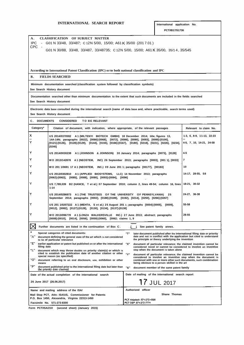
INTERNATIONAL SEARCH REPORT International application No.
PCT/IB17/51736
A . CLASSIFICATION OF SUBJECT MATTER
IPC - G01 N 33/48, 33/487; C 12N 5/00, 15/00; A61 35/00 (201 7.01 )CPC -
G01 N 30/88, 33/48, 33/487, 33/48735; C 12N 5/00, 15/00; A61 35/00, 35/1 4 , 35/545
According to International Patent Classification (IPC) or to both national classification and IPC
B. FIELDS SEARCHED
Minimum documentation searched (classification system followed by classification symbols)
See Search History document
Documentation searched other than minimum documentation to the extent that such documents are included in the fields searched
See Search History document
Electronic data base consulted during the international search (name of data base and, where practicable, search terms used)
See Search History document
C . DOCUMENTS CONSIDERED T O B E RELEVANT
Category* Citation of document, with indication, where appropriate, of the relevant passages Relevant to claim No.
US 2014/0370592 A 1 (MILTENYI BIOTECH GMBH) 18 December 2014; title; figures 13, 1-3, 6 , 8-9, 11-13, 22-2314A-14N; paragraphs [0022], [0066]-[0068], [0072], [0086], [0090], [0093], [0095]-[0100],[0121]-[0126], [0128]-[0129], [0144], [0155], [0166]-[0167], [0180], [0218], [0221], [0226], [0234], 4-5, 7 , 10, 14-21, 24-58[0240]
US 2014/0030238 A 1 (JOHNSON & JOHNSON) 30 January 2014; paragraphs [0073], [0128] 4-5
W O 2013/142878 A 1 (NEOSTEM, INC) 26 September 2013; paragraphs [0003], [001 1], [0033] 7
W O 201 1/0691 17 A 1 (NEOSTEM, INC.) 09 June 201 1; paragraphs [00177], [00183] 10
US 2013/0302810 A 1 (APPLIED BIOSYSTEMS, LLC) 14 November 2013; paragraphs 14-17, 28-55, 58[0062]-[0063], [0085], [0088], [0090], [0093]-[0094], [0096]
US 7,789,039 B2 (HANCE, T et al.) 07 September 2010; column 2 , lines 49-54; column 16, lines 18-21, 30-321-14
US 2014/0286973 A 1 (THE TRUSTEES O F THE UNIVERSITY O F PENNSYLVANIA) 25 24-27, 36-38September 2014; paragraphs [0002], [0188]-[0189], [0192], [0214], [0250], [0266]-[0267]
US 201 1/0207222 A 1 (MEHTA, S et al.) 25 August 201 1; paragraphs [0004]-[0005], [0009], 55-58[0012], [0082], [0127]-[0128], [0130], [0134], [0137]-[0138]
W O 2013/096778 A 1 (LONZA WALKERSVILLE INC.) 27 June 2013; abstract; paragraphs 28-55[0008]-[0010], [0014], [0056], [0059]-[0060], [0062; claims 1, 9
Further documents are listed in the continuation of Box C . [ | See patent family annex.
Special categories of cited documents: "T" later document published after the international filing date or prioritydocument defining the general state of the art which is not considered date and not in conflict with the application but cited to understandto be of particular relevance the principle or theory underlying the invention
earlier application or patent but published on or after the international "X" document of particular relevance; the claimed invention cannot befiling date considered novel or cannot be considered to involve an inventivedocument which may throw doubts on priority claim(s) or which is step when the document is taken alonecited to establish the publication date of another citation or other "Y" document of particular relevance; the claimed invention cannot bespecial reason (as specified) considered to involve an inventive step when the document isdocument referring to an oral disclosure, use, exhibition or other combined with one or more other such documents, such combinationmeans being obvious to a person skilled in the art
document published prior to the international filing date but later than "&" document member of the same patent family
Date of the actual completion of the international search Date of mailing of the international search report
26 June 2017 (26.06.2017) JUL 2017Name and mailing address of the ISA/ Authorized officer
Mail Stop PCT, Attn: ISA/US, Commissioner for Patents Shane ThomasP.O. Box 1450, Alexandria, Virginia 22313-1450
Facsimile No. 571-273-8300
Form PCT/ISA/210 (second sheet) (January 2015)

INTERNATIONAL SEARCH REPORT International application No.
PCT/IB17/51736
C (Continuation). DOCUMENTS CONSIDERED TO BE RELEVANT
Category* Citation of document, with indication, where appropriate, of the relevant passages Relevant to claim No.
Y US 2014/0288272 A 1 (ALDERBIO HOLDINGS LLC) 25 September 2014; paragraphs 5 1, 53-54[0176]-[0177]; table 2
Form PCT/ISA/210 (continuation of second sheet) (January 2015)