WO 2010/000721 Al
109
(12) INTERNATIONAL APPLICATION PUBLISHED UNDER THE PATENT COOPERATION TREATY (PCT) (19) World Intellectual Property Organization International Bureau (10) International Publication Number (43) International Publication Date 7 January 2010 (07.01.2010) WO 2010/000721 Al (51) International Patent Classification: AO, AT, AU, AZ, BA, BB, BG, BH, BR, BW, BY, BZ, C07K 16/24 (2006.01) CA, CH, CL, CN, CO, CR, CU, CZ, DE, DK, DM, DO, DZ, EC, EE, EG, ES, FI, GB, GD, GE, GH, GM, GT, (21) International Application Number: HN, HR, HU, ID, IL, IN, IS, JP, KE, KG, KM, KN, KP, PCT/EP2009/058155 KR, KZ, LA, LC, LK, LR, LS, LT, LU, LY, MA, MD, (22) International Filing Date: ME, MG, MK, MN, MW, MX, MY, MZ, NA, NG, NI, 30 June 2009 (30.06.2009) NO, NZ, OM, PE, PG, PH, PL, PT, RO, RS, RU, SC, SD, SE, SG, SK, SL, SM, ST, SV, SY, TJ, TM, TN, TR, TT, (25) Filing Language: English TZ, UA, UG, US, UZ, VC, VN, ZA, ZM, ZW. (26) Publication Language: English (84) Designated States (unless otherwise indicated, for every (30) Priority Data: kind of regional protection available): ARIPO (BW, GH, 08159344.4 30 June 2008 (30.06.2008) EP GM, KE, LS, MW, MZ, NA, SD, SL, SZ, TZ, UG, ZM, ZW), Eurasian (AM, AZ, BY, KG, KZ, MD, RU, TJ, (71) Applicant (for all designated States except US): NOVO TM), European (AT, BE, BG, CH, CY, CZ, DE, DK, EE, NORDISK A/S [DK/DK]; Novo AUe, DK-2880 ES, FI, FR, GB, GR, HR, HU, IE, IS, IT, LT, LU, LV, Bagsvsrd (DK). MC, MK, MT, NL, NO, PL, PT, RO, SE, SI, SK, TR), OAPI (BF, BJ, CF, CG, CI, CM, GA, GN, GQ, GW, ML, (72) Inventors; and MR, NE, SN, TD, TG). (75) Inventors/Applicants (for US only): PASS, Jesper [DK/ DK]; Akelejevej 19, DK-3450 Allerød (DK). OSTER- Published: GAARD, Sβ ren [DK/DK]; Borrebyvej 21, DK-2700 — with international search report (Art. 21(3)) Brønshøj (DK). CLAUSEN, Jes Thorn [DK/DK]; Davrekildevej 10, DK-4270 Høng (DK). — with sequence listing part of description (Rule 5. 2 (a)) (81) Designated States (unless otherwise indicated, for every kind of national protection available): AE, AG, AL, AM, (54) Title: ANTI-HUMAN INTERLEUKIN-20 ANTIBODIES Mature hlL20 (SEQ ID NO:1 ) LKTLNLGSCVIATNLQEIRNGFSEIRGSVQAKDGNIDIRILRRTESLQDTKPANRCCLL RH LLRLYLDRVFKNYQTP D 73 H 79 Yso T 8 iL 82 B 83 K 84 1 85 S 86 S 87 L 88 A 89 N90 S91 F 92 L 9 T 9 I95 K 96 K 97 D 98 L 99 R 100 L 1O iC 102 H 1O3 AHMTCHCGEEAMKKYSQILSHFEKLEPQAA WKALGELDILLQWMEETE Figure 1A (57) Abstract: Anti-human IL20 monoclonal antibodies that can reduce IL20 mediated activation of both IL20R1/IL20R2 and IL22R1/IL20R2 receptor complexes in one or more species, including humans, are described, as well as antigen-binding molecules such as, e.g., antigen-binding antibody fragments, antibody derivatives, and multi-specific molecules designed or de rived from such antibodies, and methods or producing such antibodies or other antigen-binding molecules. Such antibodies or other antigen-binding molecules can be used for treat-ing various diseases and disorders, including autoimmune or inflammatory diseases or disorders.
-
Upload
khangminh22 -
Category
Documents
-
view
0 -
download
0
Transcript of WO 2010/000721 Al
(12) INTERNATIONAL APPLICATION PUBLISHED UNDER THE PATENT COOPERATION TREATY (PCT)
(19) World Intellectual Property OrganizationInternational Bureau
(10) International Publication Number(43) International Publication Date
7 January 2010 (07.01.2010) WO 2010/000721 Al
(51) International Patent Classification: AO, AT, AU, AZ, BA, BB, BG, BH, BR, BW, BY, BZ,C07K 16/24 (2006.01) CA, CH, CL, CN, CO, CR, CU, CZ, DE, DK, DM, DO,
DZ, EC, EE, EG, ES, FI, GB, GD, GE, GH, GM, GT,(21) International Application Number: HN, HR, HU, ID, IL, IN, IS, JP, KE, KG, KM, KN, KP,
PCT/EP2009/058155 KR, KZ, LA, LC, LK, LR, LS, LT, LU, LY, MA, MD,
(22) International Filing Date: ME, MG, MK, MN, MW, MX, MY, MZ, NA, NG, NI,
30 June 2009 (30.06.2009) NO, NZ, OM, PE, PG, PH, PL, PT, RO, RS, RU, SC, SD,SE, SG, SK, SL, SM, ST, SV, SY, TJ, TM, TN, TR, TT,
(25) Filing Language: English TZ, UA, UG, US, UZ, VC, VN, ZA, ZM, ZW.
(26) Publication Language: English (84) Designated States (unless otherwise indicated, for every
(30) Priority Data: kind of regional protection available): ARIPO (BW, GH,
08159344.4 30 June 2008 (30.06.2008) EP GM, KE, LS, MW, MZ, NA, SD, SL, SZ, TZ, UG, ZM,ZW), Eurasian (AM, AZ, BY, KG, KZ, MD, RU, TJ,
(71) Applicant (for all designated States except US): NOVO TM), European (AT, BE, BG, CH, CY, CZ, DE, DK, EE,NORDISK A/S [DK/DK]; Novo AUe, DK-2880 ES, FI, FR, GB, GR, HR, HU, IE, IS, IT, LT, LU, LV,Bagsvsrd (DK). MC, MK, MT, NL, NO, PL, PT, RO, SE, SI, SK, TR),
OAPI (BF, BJ, CF, CG, CI, CM, GA, GN, GQ, GW, ML,(72) Inventors; and MR, NE, SN, TD, TG).(75) Inventors/Applicants (for US only): PASS, Jesper [DK/
DK]; Akelejevej 19, DK-3450 Allerød (DK). OSTER- Published:GAARD, Sβren [DK/DK]; Borrebyvej 21, DK-2700 — with international search report (Art. 21(3))Brønshøj (DK). CLAUSEN, Jes Thorn [DK/DK];Davrekildevej 10, DK-4270 Høng (DK). — with sequence listing part of description (Rule 5.2(a))
(81) Designated States (unless otherwise indicated, for everykind of national protection available): AE, AG, AL, AM,
(54) Title: ANTI-HUMAN INTERLEUKIN-20 ANTIBODIES
Mature hlL20 (SEQ ID NO:1 )
LKTLNLGSCVIATNLQEIRNGFSEIRGSVQAKDGNIDIRILRRTESLQDTKPANRCCLL
RHLLRLYLDRVFKNYQTP D73
H79
Yso T8i L82
B83
K84 185 S86
S87
L88
A89 N90 S91 F92
L9
T9
I95 K96
K97
D98
L99
R100
L1O
i C102 H1O3 AHMTCHCGEEAMKKYSQILSHFEKLEPQAA
WKALGELDILLQWMEETE
Figure 1A
(57) Abstract: Anti-human IL20 monoclonal antibodies that can reduce IL20 mediated activation of both IL20R1/IL20R2 andIL22R1/IL20R2 receptor complexes in one or more species, including humans, are described, as well as antigen-bindingmolecules such as, e.g., antigen-binding antibody fragments, antibody derivatives, and multi-specific molecules designed or derived from such antibodies, and methods or producing such antibodies or other antigen-binding molecules. Such antibodies orother antigen-binding molecules can be used for treat-ing various diseases and disorders, including autoimmune or inflammatorydiseases or disorders.
ANTI-HUMAN INTERLEUKlN-20 ANTIBODIES
FIELD OF THE INVENTION
The present invention relates to antibodies against human interleukin-20 (IL20), in
cluding human monoclonal anti-IL20 antibodies, as well as methods of production, composi-
tions, and use thereof.
BACKGROUND OF THE INVENTION
lnterleukin-19 (IL19), IL20, and interleukin-24 (IL24) are members of the interleukin-
10 (IL10) cytokine family. All three interleukins bind and signal through the IL20R1/IL20R2
heterodimeric receptor. IL20 and IL24 (but not IL19) are also ligands for the receptor com-
plex composed of IL20R2 and IL22R1 (Parrish-Novak et al., J Biol Chem 2002; 277: 47517-
47523; Dumoutier et al., J Immunol 2001 ;167:3545-3549). It has been proposed that IL19
and IL20, along with other IL10 family members, form a distinct subfamily of helical cytokines
where at least IL19 and IL20 have similar three-dimensional structures (Chang et al., J Biol
Chem 2003; 278: 3308-13).
IL20 and its receptors are present in elevated levels in psoriatic lesions (Wei et al.,
Clin Immunol (2005) 117: 65-72; Rømer e a/., J Invest Dermatol 2003; 121 , 1306-131 1;
Wang et al., J Invest Dermatol 2006; 126: 1590-1599; Otkjaer e al., Br J Dermatol 2005; 153:
9 11-918) and in synovial fluid of rheumatoid arthritis patients (Hsu et al., Arthritis Rheum
2006; 54: 2722-2733; Kragstrup et al., Cytokine 2008; 4 1: 16-2). Antagonizing IL20 activity
using receptor fragments or monoclonal antibodies has therefore been described as a prom
ising approach for treatment of various inflammatory conditions (e.g., WO9927103,
WO01 46261 , WO2003051 384, WO2004085475, and WO2006086396). For example, poly
clonal anti-IL20 antibodies were found to be therapeutically effective in a xenograft model of
psoriasis (Stenderup et al., Br J Dermatol 2006; 154: 11-35, Abstract P-12; Stenderup et al.
Br J Dermatol 2009; 160(2):284-96).
Antigenic epitopes of human IL20 (hlL20), as well as rat or murine monoclonal ant i
bodies binding hulL20, have also been described (e.g., WO2005052000, US200601 42550,
and WO2007081465). However, no antibodies suitable for patient treatment have so far
been provided. The present invention addresses these and other needs in the art.
SUMMARY OF THE INVENTION
The present invention provides anti-hlL20 monoclonal antibodies that can reduce
IL20-mediated activation of IL20R1/IL20R2 and IL22R1/IL20R2 receptor complexes in one or
more species, including humans. Typically, the antibodies are fully human or humanized to
minimize the risk for immune responses against the antibodies when administered to a pa
tient. The invention also provides anti-hlL20 antibodies having improved solubility properties,
making them capable of being formulated at high concentrations. As described herein, other
antigen-binding molecules such as, e.g., antigen-binding antibody fragments, antibody de
rivatives, and multi-specific molecules, can be designed or derived from such antibodies.
Antibodies binding a specific segment of the hlL20 molecule that corresponds to He
lix E in IL19 are also provided. In one embodiment, the epitope of the antibody comprise one
or more amino acid residues in the segment corresponding to D78-L93, optionally excluding
D78, in mature hlL20 (SEQ ID NO:1), e.g., H79, R83, S85, N90, F92, L93, or any combina
tion thereof.
Certain anti-hlL20 antibodies of the invention may also compete with and/or bind to
the same epitope or have the same binding interface on hlL20 as one or more of the specific
human anti-hlL20 antibodies described herein, including 15D2 and 5B7. For example, in one
embodiment, the antibodies of the invention are more capable of competing with 15D2
and/or 5B7 than with known anti-hlL20 antibodies.
In another aspect, antibodies of the invention comprise antigen-binding sequences
that derive from one or more of the same human V, D, or J segments as 15D2 or 5B7. The
antibodies may, for example, comprise one or more antigen-binding sequences that are iden-
tical or substantially identical to 15D2 and/or 5B7 antigen-binding sequences described
herein.
In other aspects, the invention provides for nucleic acids encoding antibodies of the
invention, expression vectors comprising such nucleic acids, host cells comprising such n u
cleic acids, host cells producing antibodies of the invention, and methods of producing anti-
hlL20 antibodies by culturing such host cells under appropriate conditions. Also provided for
are antibody-binding fragments of such antibodies, and molecules comprising such antibod
ies or antigen-binding fragments, including engineered antibody fragments, antibody deriva
tives, bispecific antibodies and other multispecific molecules. Pharmaceutical compositions
and kits or other articles that comprise such antibodies or molecules can also be prepared.
Further provided for are methods of reducing or inhibiting IL20-mediated activation of
IL20R1/IL20R2 and IL22R1/IL20R2 receptor complexes, and methods of treating or prevent
ing autoimmune or inflammatory diseases or disorders, including, but not limited to rheuma
toid arthritis, juvenile rheumatoid arthritis, psoriasis, psoriatic arthritis, ankylosing spondylitis,
Sjogren's syndrome, multiple sclerosis, inflammatory bowel disease, systemic lupus erythe-
matosus, lupus nephritis, or a combination thereof, using such antibodies.
DESCRIPTION OF THE DRAWINGS
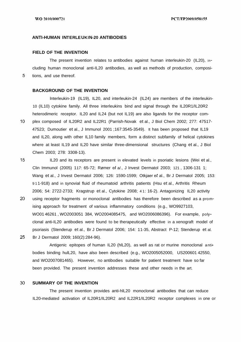
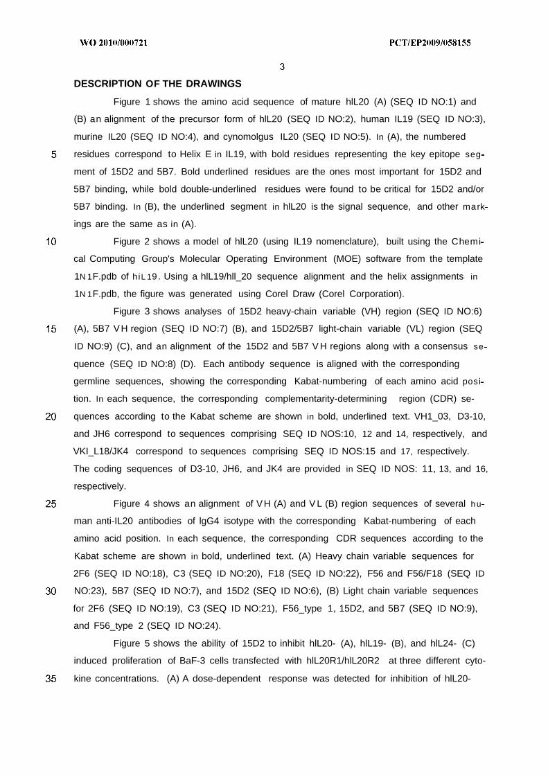
Figure 1 shows the amino acid sequence of mature hlL20 (A) (SEQ ID NO:1) and
(B) an alignment of the precursor form of hlL20 (SEQ ID NO:2), human IL19 (SEQ ID NO:3),
murine IL20 (SEQ ID NO:4), and cynomolgus IL20 (SEQ ID NO:5). In (A), the numbered
residues correspond to Helix E in IL19, with bold residues representing the key epitope seg
ment of 15D2 and 5B7. Bold underlined residues are the ones most important for 15D2 and
5B7 binding, while bold double-underlined residues were found to be critical for 15D2 and/or
5B7 binding. In (B), the underlined segment in hlL20 is the signal sequence, and other mark
ings are the same as in (A).
Figure 2 shows a model of hlL20 (using IL19 nomenclature), built using the Chemi
cal Computing Group's Molecular Operating Environment (MOE) software from the template
1N 1F.pdb of hiL 19 . Using a hlL19/hll_20 sequence alignment and the helix assignments in
1N 1F.pdb, the figure was generated using Corel Draw (Corel Corporation).
Figure 3 shows analyses of 15D2 heavy-chain variable (VH) region (SEQ ID NO:6)
(A), 5B7 VH region (SEQ ID NO:7) (B), and 15D2/5B7 light-chain variable (VL) region (SEQ
ID NO:9) (C), and an alignment of the 15D2 and 5B7 V H regions along with a consensus se
quence (SEQ ID NO:8) (D). Each antibody sequence is aligned with the corresponding
germline sequences, showing the corresponding Kabat-numbering of each amino acid posi
tion. In each sequence, the corresponding complementarity-determining region (CDR) se-
quences according to the Kabat scheme are shown in bold, underlined text. VH1_03, D3-10,
and JH6 correspond to sequences comprising SEQ ID NOS:10, 12 and 14, respectively, and
VKI_L18/JK4 correspond to sequences comprising SEQ ID NOS:15 and 17, respectively.
The coding sequences of D3-10, JH6, and JK4 are provided in SEQ ID NOS: 11, 13, and 16,
respectively.
Figure 4 shows an alignment of VH (A) and V L (B) region sequences of several h u
man anti-IL20 antibodies of lgG4 isotype with the corresponding Kabat-numbering of each
amino acid position. In each sequence, the corresponding CDR sequences according to the
Kabat scheme are shown in bold, underlined text. (A) Heavy chain variable sequences for
2F6 (SEQ ID NO:18), C3 (SEQ ID NO:20), F18 (SEQ ID NO:22), F56 and F56/F18 (SEQ ID
NO:23), 5B7 (SEQ ID NO:7), and 15D2 (SEQ ID NO:6), (B) Light chain variable sequences
for 2F6 (SEQ ID NO:19), C3 (SEQ ID NO:21), F56_type 1, 15D2, and 5B7 (SEQ ID NO:9),
and F56_type 2 (SEQ ID NO:24).
Figure 5 shows the ability of 15D2 to inhibit hlL20- (A), hlL19- (B), and hlL24- (C)
induced proliferation of BaF-3 cells transfected with hlL20R1/hlL20R2 at three different cyto-
kine concentrations. (A) A dose-dependent response was detected for inhibition of hlL20-
induced proliferation. No inhibition of hll_19- (B) or hlL24- (C) induced proliferation was ob
served in 15D2-concentration range used (up to 66nM).
Figure 6 shows the results of a primary peptide array of hlL20 against 15D2 (A) or
5B7 (B). The Y axis indicates the optical density (OD, a measure of fluorescence intensity).
Note that not all peptides are present in figure, since some OD values were below detection
limit. In (A), peptides corresponding to residues 69-86 (85), 73-90 (86), 77-94 (87), 81-98
(88), and 85-102 (89) of SEQ ID NO:1 are shown. Peptide 87 came out as the peptide with
highest binding activity. In (B), peptides corresponding to residues 49-66 (19), 53-70 (20),
57-74 (21), 69-86 (24), 73-90 (25), and 77-94 (26) are shown.
Figure 7 shows a secondary peptide array analysis of hlL20 against 15D2 (A) or
5B7 (B). The antibodies were tested against constructs with truncations from the C- and N-
terminal. The peptides were all acylated in order to avoid the positive charge arising from the
N-terminal. In (A), peptides corresponding to Y74 to K96 S86 and K84 of SEQ ID NO:1
(peptides 1-12, respectively) are shown on the left-hand column, and peptides corresponding
to Q75 I85 to K96 of SEQ ID NO:1 (peptides 13-24, respectively, where peptides 22 and
23 are identical) are shown in the right-hand column. In (B), peptides corresponding to Y74
to K96 S86 and K84 of SEQ ID NO:1 (peptides 1-12, respectively) are shown on the left-
hand column, and peptides corresponding to Q75 S84 to K96 (peptides 13-24, respec
tively, where peptides 22 and 23 are identical) are shown in the right-hand column.
Figure 8 shows an Ala-scan of the long epitope YQTPDHYTLRKISSLANSFLTIK,
corresponding to residues Y74 to K96 of SEQ ID NO:1 , against (A) 15D2 and (B) 5B7. In (A),
the peptides shown correspond to residues 78-96 of SEQ ID NO:1 with an alanine substitu
tion at positions 78-96, peptides 40-61 , respectively. In (B), the peptides shown correspond
to residues 78-96 of SEQ ID NO:1 with an alanine substitution at positions 78-96, peptides 1-
22, respectively.
Figure 9 shows 15D2 neutralization of murine IL20 activation of murine
IL22R1/IL20R2 receptor, as revealed by a luciferase assay. Murine IL20 receptor complex
mlL20R1/mll_22R1 was transfected into BHK cells and stimulated with 10 nM murine IL20.
Neutralization of stimulation was investigated using 1 microgram/ml, 10 microgram/ml or 50
microgram/ml of 15D2.
Figure 10 shows 15D2 neutralization of cynomolgus IL20 activation of human
IL20R1/IL20R2 and IL22R1/IL20R2 receptors (A) or cynomolgous IL20R1/IL20R2 and
IL22R1/IL20R2 (B), as revealed by a luciferase assay.
Figure 11 shows 15D2 neutralization of human IL20-mediated activation of human
IL20R1/IL20R2 and IL22R1/IL20R2, as revealed by a luciferase assay.
Figure 12 shows that IL19 reverted the 15D2 blocking of IL20-induced proliferation,
revealing that 15D2 bound the soluble form of IL20 and not the receptor-bound form, which
would otherwise block access of IL19 to the receptor.
Figure 13 shows the primary sequence of hlL20 used in an amide hydro-
gen/deuterium exchange (HX) - mass spectrometry (MS) study to determine the 15D2 bind
ing interface. The primary hlL20 sequence (using mature Met-1 numbering, thus differing + 1
from the corresponding residue in SEQ ID NO:1) is displayed above the HX analyzed pep
tides (shown as horizontal bars). Peptides showing similar exchange patterns both in the
presence and absence of 15D2 are indicated by grey bars whereas peptides showing re-
duced deuterium incorporation upon 15D2 binding are indicated by black bars.
Figure 14 shows sub-localization of the deuterium label from individual peptides in
the HX-MS study. (A) Close-up of region 60-93 of the IL20 primary structure. Peptides show
ing similar exchange patterns both in the presence and absence of 15D2 are coloured grey
whereas peptides showing reduced deuterium incorporation upon 15D2 binding are coloured
black. The numbers indicate the difference in deuterium level observed in the individual IL20
regions upon 15D2 binding. (B) The information from the peptides have been sub-localized
to smaller residue stretches by simple subtraction assuming complete off-exchange of the N-
terminus and first peptide bond amide. The deuterium level was then corrected for the labe l
ling reaction only containing 91% deuterium and reported as percent of total residues.
DEFINITIONS
Unless otherwise stated or contradicted by context, the terms "IL20" or "IL-20" refer
to human interleukin-20 (hlL20), also known as ZcytolO, including its unprocessed precursor
(UniProt Q9NYY1 ; SEQ ID NO:2), mature form (SEQ ID NO:1 , UniProt Q9NYY1 without the
residues 1-24 signal sequence), and/or naturally occurring variants or orthologs thereof, such
as, e.g., murine IL20 (mll_20) precursor (UniProt Q9JKV9; SEQ ID NO:4), or cynomolgous
IL20 (clL20) precursor (SEQ ID NO:5), or mature forms thereof which lack the signal se
quence corresponding to residues 1-24 in precursor hlL20 (SEQ ID NO:2).
The term "antibody" herein is used in the broadest sense and specifically includes
full-length monoclonal antibodies, polyclonal antibodies, and, unless otherwise stated or con-
tradicted by context, antigen-binding fragments, antibody variants, and multispecific mole
cules thereof, so long as they exhibit the desired specificity and/or biological activity. Gener
ally, a full-length antibody is a glycoprotein comprising at least two heavy (H) chains and two
light (L) chains inter-connected by disulfide bonds, or an antigen binding portion thereof.
Each heavy chain is comprised of a heavy chain variable region (abbreviated herein as VH)
and a heavy chain constant region. The heavy chain constant region is comprised of three
domains, CH1 , CH2 and CH3. Each light chain is comprised of a light chain variable region
(abbreviated herein as VL) and a light chain constant region. The light chain constant region
is comprised of one domain, CL. The VH and V L regions can be further subdivided into re-
gions of hypervariability, termed complementarily determining regions (abbreviated herein as
CDR), interspersed with regions that are more conserved, termed framework regions (FR).
Each V H and V L is composed of three CDRs and four FRs, arranged from amino-terminus to
carboxy-terminus in the following order: FR1 , CDR1 , FR2, CDR2, FR3, CDR3, FR4. The
variable regions of the heavy and light chains contain a binding domain that interacts with an
antigen. Various techniques relevant to the production of antibodies are provided in, e.g.,
Harlow and Lane, ANTIBODIES: A LABORATORY MANUAL, Cold Spring Harbor Laboratory
Press, Cold Spring Harbor, N.Y., (1988).
An "antigen-binding fragment" of an antibody is a molecule that comprises a portion
of a full-length antibody which is capable of detectably binding to the antigen. Antigen-
binding fragments include multivalent molecules comprising one, two, three, or more antigen-
binding portions of an antibody, and single-chain constructs wherein the VL and VH regions,
or selected portions thereof, are joined by synthetic linkers or by recombinant methods to
form a functional, antigen-binding molecule.
The terms "antibody derivative" and immunoconjugate" are used interchangeably
herein to denote molecules comprising a full-length antibody or an antigen-binding fragment
thereof, wherein one or more amino acids are chemically modified, e.g., by alkylation, PEGy-
lation, acylation, ester formation or amide formation or the like, e.g., for linking the antibody
to a second molecule. Exemplary modifications include PEGylation, cysteine-PEGylation,
biotinylation, radiolabelling, and conjugation with a second agent, such as a cytotoxic agent.
A "multispecific molecule" comprises an antibody, or an antigen-binding fragment
thereof, which is associated with or linked to at least one other functional molecule (e.g. a n
other peptide or protein such as another antibody or ligand for a receptor) to generate a
molecule that binds to at least two different binding sites or target molecules. Exemplary mul
tispecific molecules include bi-specific antibodies and antibodies linked to soluble receptor
fragments or ligands.
The term "human antibody", as used herein, is intended to include antibodies having
variable regions in which both the framework and CDR regions are derived from human
germline immunoglobulin sequences. Furthermore, if the antibody contains a constant re
gion, the constant region also is derived from human immunoglobulin sequences. Collections
of human germline sequences are available at, e.g., the NCBI website. The human antibod-
ies of the invention may include amino acid residues not encoded by human germline immu
noglobulin sequences (e.g., mutations introduced by random or site-specific mutagenesis in
vitro or by somatic mutation in vivo). However, the term "human antibody", as used herein, is
not intended to include antibodies in which CDR sequences derived from the germline of an-
other mammalian species, such as a mouse, have been grafted onto human framework se
quences.
A "humanized" antibody is a human/non-human chimeric antibody that contains a
minimal sequence derived from non-human immunoglobulin. For the most part, humanized
antibodies are human immunoglobulins (recipient antibody) in which residues from a hyper-
variable region of the recipient are replaced by residues from a hypervariable region of a
non-human species (donor antibody) such as mouse, rat, rabbit, or non-human primate hav
ing the desired specificity, affinity, and function. In some instances, FR residues of the h u
man immunoglobulin are replaced by corresponding non-human residues ("back-mutations").
Furthermore, humanized antibodies may comprise residues that are not found in the recipi-
ent antibody or in the donor antibody. These modifications are made to further refine ant i
body performance. In general, a humanized antibody will comprise substantially all of at least
one, and typically two, variable domains, in which all or substantially all of the hypervariable
loops correspond to those of a non-human immunoglobulin and all or substantially all of the
FR residues are those of a human immunoglobulin sequence. The humanized antibody can
optionally also comprise at least a portion of an immunoglobulin constant region (Fc), typi
cally that of a human immunoglobulin. For further details, see, e.g., Jones et al., Nature
321 :522-525 (1986); Riechmann et al., Nature 332:323-329 (1988); and Presta, Curr. Op.
Struct. Biol. 2:593-596 (1992), WO 92/02190, US Patent Application 20060073137, and US
Patents 6,750,325, 6,632,927, 6,639,055, 6,548,640, 6,407,213, 6,180,370, 6,054,297,
5,929,212, 5,895,205, 5,886,152, 5,877,293, 5,869,619, 5,821 ,337, 5,821 ,123, 5,770,196,
5,777,085, 5,766,886, 5,714,350, 5,693,762, 5,693,761 , 5,530,101 , 5,585,089, and
5,225,539.
The term "hypervariable region" when used herein refers to the amino acid residues
of an antibody that are responsible for antigen binding. The hypervariable region generally
comprises amino acid residues from a "complementarity-determining region" or "CDR" (resi
dues 24-34 (L1), 50-56 (L2) and 89-97 (L3) in the light-chain variable domain and 31-35
(H1 ) , 50-65 (H2) and 95-102 (H3) in the heavy-chain variable domain; (Kabat et al. (1991)
Sequences of Proteins of Immunological Interest, Fifth Edition, U.S. Department of Health
and Human Services, NIH Publication No. 91-3242) and/or those residues from a "hypervari-
able loop" (residues 26-32 (L1), 50-52 (L2) and 91-96 (L3) in the light-chain variable domain
and 26-32 (H1), 53-55 (H2) and 96-101 (H3) in the heavy-chain variable domain; Chothia
and Lesk, J. MoI. Biol 1987;196:901-917) and/or the specificity-determining residues (SDRs),
which are the residues that are most crucial in the antibody-antigen interaction (Kashmiri et
al., Methods 2005;36:25-34). The SDRs can be determined using, e.g., 3D structural analy-
sis of the antibody-antigen interaction or by mutational analysis using known techniques.
Typically, the numbering of amino acid residues in this region is performed by the method
described in Kabat et al., supra. Phrases such as "Kabat position", "variable domain residue
numbering as in Kabat" and "according to Kabat" herein refer to this numbering system for
heavy chain variable domains or light chain variable domains. Using the Kabat numbering
system, the actual linear amino acid sequence of a peptide may contain fewer or additional
amino acids corresponding to a shortening of, or insertion into, a FR or CDR of the variable
domain. For example, a heavy chain variable domain may include a single amino acid insert
(residue 52a according to Kabat) after residue 52 of CDR H2 and inserted residues (e.g.
residues 82a, 82b, and 82c, etc. according to Kabat) after heavy chain FR residue 82. The
Kabat numbering of residues may be determined for a given antibody by alignment at re
gions of homology of the sequence of the antibody with a "standard" Kabat numbered se
quence (see Figures 3 and 4).
A "variant" of a polypeptide refers to a polypeptide having an amino acid sequence
that is substantially identical to a reference polypeptide, typically a native or "parent" polypep-
tide. The polypeptide variant may possess one or more amino acid substitutions, deletions,
and/or insertions at certain positions within the native amino acid sequence, but differs from
the parent polypeptide in at least one respect.
The term "epitope" or "antigenic determinant" of an antibody is the part of a prede
termined antigen to which the antibody binds, and usually consists of chemically active sur-
face groupings of amino acids or sugar chains. The specific amino acids defining a protein
epitope can be relatively few in number, and typically comprise the amino acids that are d i
rectly involved in binding to the antibody, though other amino acids that are not directly in
volved in binding to the antibody can nevertheless be blocked when the antibody binds. The
amino acids in a protein epitope may be close to each other or widely dispersed along the
length of antigen, being brought into the correct epitope conformation via folding. A "confor
mational epitope" refers to an epitope that depends on the predetermined antigen being cor
rectly folded, while a "linear epitope" can also be recognized by the antibody when not cor
rectly folded, e.g., in denatured form or in the form of a fragment comprising the epitope.
"Specific binding" as used herein refers to the ability of an antibody to bind a prede-
termined antigen, such as, e.g., IL20. Typically, the antibody binds with a dissociation con-
stant (Kd) of 10 8 or less, and binds to the predetermined antigen with a Kd that is at least 2-
fold less than its Kd for binding to a non-specific antigen (e.g., BSA) other than the predeter
mined antigen or a closely related molecule (e.g., an ortholog).
The term "substantially identical" in the context of two amino acid sequences means
that the sequences, when optimally aligned, such as by the programs GAP or BEST-FIT us
ing default gap weights, share at least about 50, at least about 60, at least about 70, at least
about 80, at least about 90, at least about 95, at least about 98, or at least about 99 percent
sequence identity.
"Corresponding" amino acid positions in two substantially identical amino acid se-
quences are those aligned by any of the protein analysis software referred to herein, typically
using default parameters.
A nucleic acid is "operably linked" when it is placed into a functional relationship with
another nucleic acid sequence. For example, DNA for a pre-sequence or secretory leader is
operably linked to DNA for a polypeptide if it is expressed as a pre-protein that participates in
the secretion of the polypeptide; a promoter or enhancer is operably linked to a coding se
quence if it affects the transcription of the sequence; or a ribosome-binding site is operably
linked to a coding sequence if it is positioned so as to facilitate translation. Generally, "oper
ably linked" means that the DNA sequences being linked are contiguous, and, in the case of
a secretory leader, contiguous and in reading phase. However, enhancers do not have to be
contiguous. Linking is accomplished by ligation at convenient restriction sites. If such sites do
not exist, the synthetic oligonucleotide adaptors or linkers are used in accordance with con
ventional practice.
An "isolated" molecule is a molecule that is the predominant species in the composi
tion wherein it is found with respect to the class of molecules to which it belongs (i.e., it
makes up at least about 50% of the type of molecule in the composition and typically will
make up at least about 70%, at least about 80%, at least about 85%, at least about 90%, at
least about 95%, or more of the species of molecule, e.g., peptide, in the composition).
Commonly, a composition of an antibody molecule will exhibit 98%, 98%, or 99% homogene
ity for antibody molecules in the context of all present peptide species in the composition or
at least with respect to substantially active peptide species in the context of proposed use.
In the context of the present invention, "treatment" or "treating" refers to preventing,
alleviating, managing, curing or reducing one or more symptoms or clinically relevant mani
festations of a disease or disorder, unless contradicted by context. For example, "treatment"
of a patient in whom no symptoms or clinically relevant manifestations of a disease or disor-
der have been identified is preventive or prophylactic therapy, whereas "treatment" of a pa-
tient in whom symptoms or clinically relevant manifestations of a disease or disorder have
been identified generally does not constitute preventive or prophylactic therapy.
Unless otherwise expressly indicated or clearly contradicted by context, the term "or"
herein is used in the inclusive sense of "and/or."
"Activation" of a receptor or receptor complex means an increased or decreased ac
tivity of any or all intracellular signal transduction elements associated with the receptor or
receptor complex after binding of the ligand to the receptor or receptor complex under normal
physiological or pathophysiological conditions, as compared to a control. In the case of the
IL20R1/IL20R2 and IL22R1/IL20R2 receptor complexes, receptor activation can be analyzed
using, e.g., a luciferase assay similar to the one described in Example 9 . "Reducing activa
tion" of a receptor or receptor complex means that the activation of the receptor is reduced
by at least about 10%, preferably at least about 20%, more preferably at least about 30%,
most preferably at least about 50%, or more, in comparison to a control (e.g., the level of a c
tivation in the absence of antibody).
Some assays for evaluating the antibodies or other antigen-binding molecules de
scribed herein employ one or more "controls." A "control" may be a standard value retrieved
from a text book; a value obtained by running the same assay in the absence of ligand (e.g.,
IL20), receptor (e.g., IL20R1/IL20R2 and/or IL22R1/IL20R2), or antibody; or in the presence
of a non-specific molecule (e.g., a non-specific antibody); or some other reference value
used in the art. In the case of a receptor activation assay testing, for example, the ability of
an antibody to reduce activation of a receptor complex, a suitable control value can be o b
tained by running the assay in the absence of the antibody or in the presence of an antibody
not specifically binding to the ligand, receptor complex, or other components involved in re
ceptor activation.
DESCRIPTION OF THE INVENTION
The present invention provides for human anti-IL20 antibodies suitable for pharma
ceutical formulations, diagnostic uses, and therapeutic uses. As described in the Examples,
a novel epitope was identified for two human anti-IL20 antibodies designated 15D2 and 5B7.
Key epitope residues were mapped to a region of hlL20 that corresponds to Helix E in hlL19
(Chang e a/., J Biol Chem 2003; 278: 3308-13). A predicted model of hlL20 using the hlL19
nomenclature is shown in Figure 1, with Helix E corresponding to residues D78 to H103 in
the mature hlL20 sequence (SEQ ID NO:1). It was found that an antibody binding to the
novel hlL20 epitope reduced hll_20-mediated activation of both the IL20R1/IL20R2 and
IL22R1/IL20R2 receptors, and reduced IL20-mediated, but not IL19- or IL24-mediated, re-
ceptor activation in a proliferation assay. The epitope was further found to exist in both native
and denatured form of the hlL20 antigen, as well as in both murine and cynomolgous IL20.
Both 15D2 and 5B7 were also soluble at concentrations of at least about 80 mg/ml, and were
found to derive from the same set of human germline genes (Example 3 and Figure 3).
The invention thus provides antibodies which combine one or more functional prop
erties, one or more structural properties, and/or antibodies which combine one or more f unc
tional with one or more structural properties described in subsequent sections and the Ex
amples.
In one aspect, the present invention provides an antibody, such as a monoclonal
human or humanized antibody, or an antigen-binding fragment thereof, that specifically binds
to hlL20, optionally also to one or more hlL20 orthologs, specifically reduces hlL20 mediated
activation of both hlL20R1/hlL20R2 and hlL22R1/hlL20R2 receptor complexes and/or their
orthologs, and/or has a high solubility. In one embodiment, the heavy or light chain variable
region sequences of the antibody derive from one or more of the 15D2 and 5B7 germline
and/or V , D, or J segments. In one embodiment, the CDR and/or variable sequences of ant i
bodies of the invention are substantially identical to one or more antigen-binding sequence of
15D2 and/or 5B7. In one embodiment, the antibody interacts with one or more residues in
the segment H79 to H103 in the hlL20 sequence (SEQ ID NO:1), and may, for example, bind
to H79, R83, S85, N90, F91 , L92, or a combination thereof. The antibody may be in any form
suitable for therapeutic applications, e.g., a full-length antibody or a fragment thereof.
In one aspect, the invention provides for an isolated anti-hlL20 antibody, or an ant i
gen-binding fragment thereof, comprising a heavy chain variable region that is derived from a
set of human genes comprising VH1_03, D3-10, and JH6 genes. In one embodiment, the
heavy chain variable region comprises the CDR2 and CDR3 sequences, and, optionally, the
CDR1 sequence, of SEQ ID NO:8, respectively corresponding to Kabat residues 50-65, 95-
102, and 31-35. In one embodiment, the light chain variable region comprises a sequence
derived from a set of human genes comprising VKI_L18 and JK4 genes. In a specific em
bodiment, the light chain variable region comprises the sequence of SEQ ID NO:9 and the
heavy-chain variable region comprises the sequence of SEQ ID NO:6 or SEQ ID NO:7. In
another specific embodiment, the antibody is of the lgG4 isotype.
In one aspect, the invention provides for a human antibody, or an antigen-binding
fragment thererof, which binds to hlL20 and has one or more functional properties selected
from (a) reducing IL20-mediated activation of IL20R1/IL20R2 and IL22R1/IL20R2 receptor
complexes; (b) reducing IL20-mediated proliferation of BaF-3 cells recombinantly expressing
IL20R1/IL20R2; (c) not reducing IL19- or IL24-mediated proliferation of BaF-3 cells recombi-
nantly expressing IL20R1/IL20R2; (d) binding to hlL20 with a KD of about 1 nM or less; and
(e) has a solubility of at least about 80 mg/ml in an aqueous buffered solution at about pH
7.4, optionally comprising 150 mM NaCI. In one embodiment, the antibody has properties (a)
to (d). In one embodiment, the antibody has all of properties (a) to (e). In one embodiment,
the antibody or antigen-binding fragment competes in binding to hlL20 with an antibody com
prising a light-chain variable region comprising SEQ ID NO:9 and a heavy-chain variable re
gion comprising SEQ ID NO:6 or SEQ ID NO:7. In one embodiment, the antibody or antigen-
binding fragment binds to an epitope comprising at least one residue selected from H79-
H103 of mature hlL20 (SEQ ID NO:1). In one embodiment, the epitope comprises at least
one residue selected from H79-L93. In one embodiment, the antibody or antigen-binding
fragment comprises a heavy chain variable region comprising CDR1 , CDR2, and CDR3 se
quences comprising Kabat residues residues 31-35, 50-65, and 95-102, respectively, of SEQ
ID NO:6 or SEQ ID NO:7. In one embodiment, the antibody further comprises a light chain
variable region comprising CDR1 , CDR2, and CDR3 sequences comprising Kabat residues
24-34, 50-56 and 89-97, respectively, of SEQ ID NO:9. In one embodiment, the antibody
heavy and light chain variable sequences are substantially identical to the respective heavy
and light chain variable sequences of 15D2 and/or 5B7, e.g., having a sequence identity of at
least about 80%, at least about 90%, or at least about 95%. In a specific embodiment, the
antibody is of the lgG4 isotype.
In one aspect, the invention provides such human anti-hlL20 antibodies that are suf
ficiently soluble for use in pharmaceutical compositions. In one embodiment, the invention
provides a pharmaceutical composition comprising an effective amount, e.g., at a concentra
tion of at least about 80 mg/ml or at least about 100 mg/ml, of an antibody of the invention,
and a pharmaceutically acceptable excipient, diluent, or carrier. In one embodiment, the anti-
body is of an lgG4 isotype and comprises a heavy chain variable region that is derived from
a set of human genes comprising VH1_03, D3-10, and JH6 genes and/or the light chain vari
able region comprising a sequence derived from a set of human genes comprising VKI_L18
and JK4 genes. In one specific embodiment, the light chain variable region comprises the
sequence of SEQ ID NO:9 and the heavy-chain variable region comprises the sequence of
SEQ ID NO:6. In one specific embodiment, the light chain variable region comprises the se
quence of SEQ ID NO:9 and the heavy-chain variable region comprises the sequence of
SEQ ID NO:7.
In one aspect, the invention provides for an antibody, antigen-binding fragment, or
pharmaceutical composition of the invention for use as a medicament.
In one aspect, the invention provides for an antibody, antigen-binding fragment, or
pharmaceutical composition of the invention for use in treating an inflammatory or autoim
mune disorder.
In one aspect, the invention provides for the use of an antibody, antigen-binding
fragment, or pharmaceutical composition of the invention in the preparation of a medicament
for treating an inflammatory or autoimmune disorder.
In one aspect, the invention provides for a method of treating a subject suffering
from or at risk for an inflammatory or autoimmune disorder by administering an antibody, an
tigen-binding fragment, or pharmaceutical composition of the invention.
Inflammatory or autoimmune disorders suitable for such uses include rheumatoid a r
thritis, juvenile rheumatoid arthritis, psoriasis, psoriatic arthritis, ankylosing spondylitis,
Sjogren's syndrome, multiple sclerosis, inflammatory bowel disease, systemic lupus erythe
matosus, or lupus nephritis, or a combination of any thereof, as well as co-morbidities asso
ciated with these diseases, with cardiovascular disease being a non-limiting example of said
co-morbidities.
In one aspect, the invention provides for a method of recombinantly producing an
anti-IL20 antibody or antigen-binding fragment, comprising culturing a host cell producing the
antibody or antigen-binding fragment of the invention under suitable conditions, and recover
ing the antibody or antigen-binding fragment. The host cell typically comprises an expression
vector comprising nucleic adic(s) encoding heavy and/or light chain sequences of antibodies
or antigen-binding fragments of the invention.
The production, characterization, and use of antibodies, antigen-binding fragments,
or other molecules specifically binding hlL20 and having some or all of these properties are
described in more detail in the following sections.
Anti-IL20 antibodies
The antibodies of the invention are characterized by particular functional and/or
structural features or properties of the antibodies. Assays to evaluate the functional activities
of anti-IL20 antibodies are described in detail in separate sections and in the Examples, and
structural properties such as, e.g., amino acid sequences, are described below.
Functional properties
The antibodies of the invention bind specifically to hlL20. The antibody preferably
binds to hlL20 with high affinity, for example with a KD of 10 7 M or less, a KD of 10 8 M or
less, a KD of 1 nM or less, a KD of about 0.3 nM or less, or a KD of about 0.2 nM or less, or
a KD of about 0.1 nM or less. A recombinantly produced anti-IL20 antibody in sodium acetate
buffer may, for example, bind to recombinant hlL20 with an affinity of about 0.1 nM or less,
optionally with an affinity of about 0.01-0.05 nM, in a Biacore assay (see, e.g., Example 12).
Additionally, the antibodies may detectably bind to IL20 from one or more non-human mam-
mals, including mouse {e.g, mus musculus) and/or cynomolgus monkey {Macaca fascicu-
laris) (see, e.g., Example 2). Furthermore, the antibodies of the invention are capable of re
ducing IL20-mediated activation of IL20R1/IL20R2 and IL22R1/IL20R2 receptor complexes
in vitro and/or in vivo. This may be tested in one or more assays described herein (see, e.g.,
Examples 1, 2 , and 9-1 1) or known in the art. Using a suitable assay, an antibody of the in-
vention can reduce hll_20-mediated activation of human IL20R1/IL20R2 and IL22R1/IL20R2
receptor complexes by at least about 10%, more preferably by at least 20%, even more pref
erably by at least 30%, at least 40%, at least 50%, or at least 60%, as compared to a control
(e.g., in the absence of any anti-hlL20 antibody). The antibody may further be able to reduce
IL20-mediated activation of IL20R1/IL20R2 and IL22R1/IL20R2 receptor complexes in other
species, such as mice and cynomolgous monkeys, using the corresponding ligand and re
ceptor complex orthologues (see Example 9).
The antibodies of the invention reduce IL20-mediated proliferation of BaF-3 cells re
combinantly expressing IL20R1/IL20R2 and IL22R1/IL20R2, but typically have no significant
effect on IL19- and/or IL24-induced proliferation (see, e.g., Example 2). In such assays, an
antibody of the invention typically reduces proliferation with an EC50 of about 50 µM or less,
about 5 µM or less, about 1 µM or less, about 0.5 µM or less, about 0.1 µM or less, about
0.05 µM or less, or about 0.02 µM or less. For example, in a proliferation assay described in
Example 10, recombinantly produced human antibody 15D2 had an EC50 of less than 0.02
µM.
The anti-IL20 antibodies of the invention can inhibit hll_20-mediated receptor com
plex activation by any mechanism, or by a combination of different mechanisms. Typically,
an anti-hlL20 antibody can reduce or prevent hlL20 binding to cell-associated hlL20 recep
tors or fully formed receptor complexes. Additionally or alternatively, antibodies of the inven
tion may bind to cell-associated hlL20 single-chain receptor molecules, but prevent formation
of the receptor complex. Additionally or alternatively, antibodies of the invention may bind to
cell-associated hlL20 single-chain receptor molecules and fully formed receptor complexes,
but reduce or inhibit structural changes necessary for receptor complex activation. Which
one or more mechanisms are involved can be identified by, e.g., testing whether the antibody
associates to cells expressing human IL20R1 , IL20R2, IL22R1 receptor molecules, or
IL20R1/IL20R2 and/or IL22R1/IL20R2 receptor complexes in the presence of hlL20. In a
specific embodiment, the antibody reduces the binding of hlL20 to hlL20R2. In another spe
cific embodiment, the antibody does not reduce binding of hlL20 to at least one of the human
IL20R1/IL20R2 and IL22R1/IL20R2 receptor complexes. Particular antibodies of the inven
tion bind a hlL20 epitope that at least partially overlaps, or includes at least one residue in,
the segment corresponding to Helix E in IL19, optionally excluding D78. Without being limited
to theory, this segment can comprise or be part of a helical structure in IL20 that is involved
in binding to and/or activating IL20R1/IL20R2 and IL22R1/IL20R2. For a model of hlL20 built
using a hlL20-hll_19 sequence alignment and structural IL19 information, see Figure 2 . In the
hlL20 sequence, this segment comprises residues D78-H103 of mature hlL20 (SEQ ID
NO:1). In one embodiment, the antibody or antigen-binding fragment of the invention thus
binds to an epitope comprising at least one residue selected from D78-H103 of mature hlL20
(SEQ ID NO:1). In other specific and separate embodiments, the epitope includes 2 , 3 , 4 , 5 ,
6 , 7 or more residues in the D78-H103 segment.
In another aspect, the invention provides an antibody binding an epitope comprising
1, 2 , 3 , 4 , 5 , 6 , 7 or more residues in the segment corresponding to residues D78-K96 or
H79-K96 in mature hlL20. This segment contains an epitope providing a higher affinity of
anti-IL20 antibody 5B7. The antibody may alternatively bind an epitope comprising 1, 2 , 3 , 4 ,
5 , 6 , 7 or more residues in the segment corresponding to residues D78-L93 or H79-L93,
which contains the key residues of the 15D2 epitope. For example, the antibody may bind an
epitope comprising at least one residue selected from H79-L93. The antibody may alterna
tively bind an epitope comprising 1, 2 , 3 , 4 , 5 , 6 , 7 or more residues in the segment corre
sponding to residues H79-N90, which contains the key residues of the 5B7 epitope. In spe
cific and separate embodiments, all key residues of the epitope is in a segment correspond
ing to residues D78-H103, D78-K96, D78-L93, or D78-N90, optionally excluding D78.
In another aspect, the antibody binds an epitope comprising at least one of residues
H79, R83, S85, N90, F91 , and L92 of mature IL20. In separate and specific embodiments,
the antibody binds 2 , 3 , 4 , 5 , 6 , or all of D78, H79, R83, S85, N90, F91 , and L92. In another
embodiment, the epitope comprises at least residues H79 and N90. In an additional em
bodiment, the epitope further comprises residue R83. In yet another embodiment, the epi-
tope further comprises 1, 2 , 3 , or all of D78, S85, F91 , and L92.ln another aspect, the inven
tion provides antibodies that compete with and/or bind to the same epitope on hlL20 as an
antibody comprising the V H and V L sequences of either of 5B7 or 15D2, described below.
Such antibodies thus compete in binding to hlL20 with an antibody comprising a light-chain
variable region comprising SEQ ID NO:9 and a heavy-chain variable region comprising SEQ
ID NO:6 or SEQ ID NO:7. Such antibodies can be identified based on their ability to compete
with 15D2 and/or 5B7 in standard hlL20 binding assays as described herein (see, e.g., Ex
ample 4 or the section entitled "Binding Assays" below). The ability of a test antibody to re
duce or inhibit the binding of 15D2 and/or 5B7 to hlL20 demonstrates that the test antibody
can compete with 15D2 and/or 5B7 for binding to hlL20 and thus can bind to the same hlL20
segment or epitope as 5B7 and/or 15D2. In a preferred embodiment, the antibody that binds
to the same segment or epitope of hlL20 as 5B7 and/or 15D2 is a human monoclonal ant i
body. Such human monoclonal antibodies can be prepared and isolated according to known
methods in the art, as described herein.
In a particular embodiment, the antibody binds to a different hlL20 segment or epi-
tope than those bound by any of the rat antibodies described in WO2005052000
(262.4.1 .2.2.1 , 262.5.1 .6.4.4, and 262.7.1 .3.2.4), and/or by murine antibodies (7E) described
in US20060142550 and WO2007081465, and competes more with 15D2 and/or 5B7 in bind
ing to hlL20 than with either of the listed mouse or rat antibodies. In another particular em
bodiment, the antibody is a human antibody which does not bind to the segment correspond-
ding to residues 42-102 of the IL20 precursor (SEQ ID NO:2).
Any combination of the above-described functional features, other functional fea
tures described in the Examples, and/or structural features describing in the following sec
tion, may be exhibited by an antibody of the invention.
Structural properties
In one aspect, the invention provides human anti-IL20 antibodies with suitable stabil
ity and/or solubility characteristics for being formulated in aqueous formulations at concentra
tions of at least about 50 mg/ml, at least about 60 mg/ml, at least about 70 mg/ml, at least
about 80 mg/ml, at least about 90 mg/ml, or at least about 100 mg/ml, which aqueous formu
lation may further comprise a pharmaceutically acceptable excipient, diluent, or carrier, and
typically has a pH near neutral or physiological pH. In one embodiment, the anti-IL20 ant i
body has a solubility of at least 80 mg/ml in an aqueous formulation, optionally comprising a
20 mM sodium phosphate buffer and 150 mM NaCI, and having a pH of about 7.4. In one
embodiment, the anti-IL20 antibody has a solubility of at least 100 mg/ml in an aqueous for
mulation, optionally comprising a 20 mM sodium phosphate buffer and 150 mM NaCI, and
having a pH of about 7.4. It has now been found that human anti-IL20 antibodies deriving
from certain germline sequences are more soluble than others, thereby achieving higher
concentrations in an aqueous solution (see, e.g., Example 3). Such embodiments are de
scribed in further detail below.
Preferred antibodies of the invention include the human monoclonal antibodies
15D2 or 5B7 characterized as described herein. Heavy and light chain variable domains and
CDR sequences of these antibodies are provided below and in Figure 3 ,
The heavy chain variable domain of 15D2 (SEQ ID NO:6) contains the following
CDRs, corresponding to Kabat residues 31-35 (CDR1), 50-65 (CDR2) and 95-102 (CDR3) of
SEQ ID NO:6, respectively:
VH CDR1 : NDIIH
VH CDR2: WINAGYGNTQYSQNFQD
VH CDR3: EPLWFGESSPHDYYGMDV
The heavy chain variable domain of 5B7 (SEQ ID NO:7) contains the following
CDRs, corresponding to Kabat residues 31-35 (CDR1), 50-65 (CDR2) and 95-102 (CDR3) of
SEQ ID NO:7, respectively:
VH CDR1 : SHIMH
VH CDR2: WINAGYGNTKYSQNFQD
VH CDR3: EPLWFGELSPHDYYGMDV
The light chain variable domains of 15D2 and 5B7 (SEQ ID NO:9) contains the fol
lowing CDRs, corresponding to Kabat residues 24-34 (CDR1), 50-56 (CDR2) and 89-97
(CDR3) of SEQ ID NO:9, respectively:
VL CDR1 : RASQGISSALA
VL CDR2: DASSLES
VL CDR3: QQFNSYPLT
Given that 15D2 and 5B7 both bind IL20, the VH CDR sequences can be "mixed
and matched" to create other anti-hlL20 binding molecules of the invention. The hlL20-
binding of such "mixed and matched" antibodies can be tested using the binding assays de-
scribed herein (e.g. flow cytometry, Biacore, ELISAs) and/or using a receptor-activation as
say as described herein. The invention thus provides antibodies that comprise the heavy
chain and light chain CDRIs, CDR2s and/or CDR3s of 15D2 or 5B7, or combinations
thereof. The CDR regions are delineated using the Kabat system (Figure 3). Given that each
of these antibodies can bind to hlL20 with substantially overlapping epitopes, and that anti-
gen-binding specificity is provided primarily by the CDR1 , 2 and 3 regions, the VH CDR1 , 2
and 3 sequences can be "mixed and matched" (i.e., VH CDRs from different antibodies can
be mixed and matched, although each antibody can contain a VH CDR1 , 2 and 3 and a VL
CDR1 , 2 and 3) to create other anti-hlL20 binding molecules of the invention. The 15D2 and
5B7 V H CDRs share substantial structural similarity and are therefore amenable to mixing
and matching.
Accordingly, in one aspect, the invention provides an isolated monoclonal antibody
comprising: (a) a V H CDR1 from 5B7 or 15D2, (b) a VH CDR2 from 5B7 or 15D2, and (c) a
V H CDR3 from 5B7 or 15D2, optionally combined with a VL sequence comprising the VL
CDRs of SEQ ID NO:9. This can also be illustrated using consensus VH CDRs, per below.
The consensus variable heavy domain of 5B7/15D2 contains the following CDRs,
corresponding to Kabat residues 31-35 (CDR1 ) , 50-65 (CDR2) and 95-102 (CDR3) of SEQ
ID NO:8, respectively, with X representing any amino acid, preferably those listed below or
conservative substitutions thereof):
VH CDR1 : X2X3IX4H (X2: N or S; X3: D or H; X4: I or M, or conservative substitutions
of any thereof)
VH CDR2: W INAGYGNTX 5YSQN FQD (X5 is K, Q, or a conservative substitution of
any thereof)
VH CDR3: EPLWFGEX 7SPHDYYGMDV (X7 is S, L, or a conservative substitution of
any thereof),
wherein X2- X5 and X7 correspond to residues 3 1, 32, 34, 59, and 106 in SEQ ID
NO:8, respectively.
Accordingly, in another aspect, the invention provides an antibody comprising the
heavy-chain variable regions CDR2 and CDR3, optionally combined with the CDR1 , of SEQ
ID NO:8. In one embodiment, the antibody comprises the sequence of SEQ ID NO:8. In one
aspect, the antibody comprises the heavy-chain variable region CDR2 and CDR3, optionally
combined with the CDR1 , of SEQ ID NO:6. In one embodiment, the antibody comprises the
sequence of SEQ ID NO:6. In one aspect, the antibody comprises the heavy-chain variable
region CDR2 and CDR3, optionally combined with the CDR1 , of SEQ ID NO:7. In one em-
bodiment, the antibody comprises the sequence of SEQ ID NO:7. In any of these aspects or
embodiments, the antibody may optionally further comprise the light-chain variable regions
CDR1 , CDR2 and CDR3, or the full sequence, of SEQ ID NO:9.
In certain embodiments, an antibody of the invention comprises a VH region from a
particular germline H chain immunoglobulin gene, or a combination of particular germline H
chain immunoglobulin genes; and/or a VL region from a particular germline L chain immu
noglobulin gene, or a combination of particular germline L chain immunoglobulin genes.
For example, in one embodiment, the invention provides an isolated anti-hlL20 ant i
body comprising a heavy chain variable region that is derived from a set of human genes
comprising VH1_03, D3-10, and JH6 genes. The heavy chain variable region may, for exam-
pie, comprise the CDR2 and CDR3 sequences, and optionally the CDR1 sequence, of SEQ
ID NO:8, respectively corresponding to Kabat residues 50-65, 95-102, and 31-35. In another
embodiment, the antibody further comprises a light chain variable region that is derived from
a set of human genes comprising VKI_L18 and JK4 genes. The light-chain variable region
may, for example, comprise the CDR1-CDR3 sequences of SEQ ID NO:9.
In one embodiment, the invention provides an isolated anti-hlL20 monoclonal ant i
body, or an antigen-binding fragment thereof, wherein the antibody: (a) comprises a V H do
main derived from a human VH1_03 gene recombined with a human D3-10 gene and a JH6
gene, (b) comprises a VL domain derived from a human VKI_L18 gene recombined with a
human JK4 gene, and (c) the antibody specifically binds to hlL20. For example, the antibody
may comprise the light chain variable sequence of SEQ ID NO:9 and the heavy-chain vari
able sequence of SEQ ID NO:6 or SEQ ID NO:7.As used herein, a human antibody com
prises heavy or light chain variable regions of" or "derived from" or "the product of" a particu
lar germline sequence if the variable regions of the antibody are obtained from a system that
uses human germline immunoglobulin genes. Such systems include immunizing a transgenic
mouse carrying human immunoglobulin genes with the antigen of interest or screening a
human immunoglobulin gene library displayed on phage with the antigen of interest. A hu
man antibody that is "of" or "derived from" or "the product of" a human germline immu
noglobulin sequence can be identified as such by comparing the amino acid sequence of the
human antibody to the amino acid sequences of human germline immunoglobulins and se-
lecting the human germline immunoglobulin sequence that is closest in sequence (i.e.,
greatest % identity) to the sequence of the human antibody. A human antibody that is "of" or
"derived from" or "the product of" a particular human germline immunoglobulin sequence
may contain amino acid differences as compared to the germline sequence, due to, for ex
ample, naturally-occurring somatic mutations or intentional introduction of site-directed muta-
tion.
However, a selected human antibody typically is at least 90% identical in amino acid
sequence to an amino acid sequence encoded by a human germline immunoglobulin gene
and contains amino acid residues that identify the human antibody as being human when
compared to the germline immunoglobulin amino acid sequences of other species (e.g., mur-
ine germline sequences). In certain cases, a human antibody variable sequence may be at
least 95%, or even at least 96%, 97%, 98%, or 99% identical in amino acid sequence to the
amino acid sequence encoded by the recombined germline immunoglobulin gene.
Typically, a human antibody derived from a particular human germline sequence will
display no more than 10 amino acid differences from the amino acid sequence encoded by
the human germline immunoglobulin gene. In certain cases, the human antibody may display
no more than 8 , no more than 5 , or even no more than 4 , 3 , 2 , or 1 amino acid difference, or
no amino acid difference, from the amino acid sequence encoded by the recombined germ-
line immunoglobulin gene.
In yet another aspect, an antibody of the invention comprises heavy and light chain
variable regions comprising amino acid sequences that are homologous or identical to the
amino acid sequences of the preferred 15D2 and 5B7 antibodies described herein, and
wherein the antibodies retain the desired functional properties of the anti-hlL20 antibodies of
the invention. For example, the invention provides an isolated monoclonal antibody compris
ing a heavy chain variable domain and a light chain variable domain, wherein: (a) the VH
domain comprises an amino acid sequence that is at least 80% identical to an amino acid
sequence selected from the group consisting of SEQ ID NOs: 6 , 7 , and 8 ; (b) the V L region
comprises an amino acid sequence that is at least 80% identical to SEQ ID NO:9; and (c) the
antibody specifically binds to hlL20 and exhibits at least one of the functional properties de
scribed herein, preferably several of the functional properties described herein.
In other embodiments, the VH and/or V L amino acid sequences may be 85%, 90%,
95%, 96%, 97%, 98%, 99%, or 100% identical to the sequences set forth above. An antibody
having V H and V L regions having high (i.e., 80% or greater) identity to the VH and VL re
gions of the sequences set forth above, can be obtained by mutagenesis (e.g., site-directed
or PCR-mediated mutagenesis) of nucleic acid molecules encoding SEQ ID NOs:6-9, fol-
lowed by testing of the encoded altered antibody for retained function (e.g., hlL20 binding
affinity or reduction of hlL20-mediated activation of its receptor complexes) using the func
tional assays described herein.
The percent identity between the two sequences is a function of the number of iden
tical positions shared by the sequences (i.e., % identity = # of identical positions/total # of
positions x 100), taking into account the number of gaps, and the length of each gap, which
need to be introduced for optimal alignment of the two sequences. The comparison of se
quences and determination of percent identity between two sequences can be accomplished
using a mathematical algorithm in sequence analysis software. Protein analysis software
matches similar sequences using measures of similarity assigned to various substitutions,
deletions and other modifications, including conservative amino acid substitutions.
The percent identity between two amino acid sequences can be determined, e.g.,
using the Needleman and Wunsch (J. MoI. Biol. 48:444-453 (1970)) algorithm which has
been incorporated into the GAP program in the GCG software package (available at
http://www.gcg.com), using either a Blossum 62 matrix or a PAM250 matrix, and a gap
weight of 16, 14, 12, 10, 8 , 6 , or 4 and a length weight of 1, 2 , 3 , 4 , 5 , or 6 .
Polypeptide sequences can also be compared using FASTA, applying default or
recommended parameters. A program in GCG Version 6.1 . , FASTA (e.g., FASTA2 and
FASTA3) provides alignments and percent sequence identity of the regions of the best over
lap between the query and search sequences (Pearson, Methods Enzymol. 1990;1 83:63-98;
Pearson, Methods MoI. Biol. 2000;132:185-219).
The sequence identity between two amino acid sequences can also be determined
using the algorithm of E. Meyers and W . Miller (Comput. Appl. Biosci., 1988;1 1-17) which
has been incorporated into the ALIGN program (version 2.0), using a PAM120 weight res i
due table, a gap length penalty of 12 and a gap penalty of 4 .
Another algorithm for comparing a sequence to another sequence contained in a da
tabase is the computer program BLAST, especially blastp, using default parameters. See,
e.g., Altschul et al., J. MoI. Biol. 1990;21 5:403-410; Altschul et al., Nucleic Acids Res.
1997;25:3389-402 (1997); each herein incorporated by reference. The protein sequences of
the present invention can there be used as a "query sequence" to perform a search against
public databases to, for example, identify related sequences. Such searches can be per
formed using the XBLAST program (version 2.0) of Altschul, et al. 1990 (supra). BLAST pro
tein searches can be performed with the XBLAST program, score = 50, wordlength = 3 to
obtain amino acid sequences homologous to the antibody molecules of the invention. To o b
tain gapped alignments for comparison purposes, Gapped BLAST can be utilized as de-
scribed in Altschul et al., 1997 (supra). When utilizing BLAST and Gapped BLAST programs,
default parameters of the respective programs (e.g., XBLAST and NBLAST) can be used.
See http://www. ncbi.nlm.nih.gov.
In certain embodiments, an antibody of the invention comprises a VH region com
prising CDR1 , CDR2 and CDR3 sequences and a VL region comprising CDR1 , CDR2 and
CDR3 sequences, wherein one or more of these CDR sequences comprise specified amino
acid sequences based on the preferred antibodies described herein; 15D2 and 5B7, or con
servative modifications thereof, and wherein the antibodies retain the desired functional
properties of the anti-hlL20 antibodies of the invention. Accordingly, the invention provides
an isolated monoclonal antibody, or antigen-binding fragment thereof, comprising a heavy
chain variable region comprising CDR1 , CDR2, and CDR3 sequences and a light chain vari
able region comprising CDR1 , CDR2, and CDR3 sequences, wherein: (a) the VH region
CDR3 sequence comprises an amino acid sequence selected from the group consisting of
the CDR3 of SEQ ID NOs:6 and 7 , and conservative modifications thereof; (b) the VL region
CDR3 sequence comprises the amino acid sequence of the CDR3 of SEQ ID NO:9 or con-
servative modifications thereof; and (c) the antibody specifically binds to hlL20 and exhibits
at least one of the functional properties described herein, more preferably several of the
functional properties described herein.
In a further embodiment, the VH region CDR2 sequence comprises an amino acid
sequence selected from the group consisting of the CDR2 of SEQ ID NOS: 6 or 7 , and con-
servative modifications thereof; and the V L region CDR2 sequence comprises the CDR2 of
SEQ ID NO:9 or conservative modifications thereof.
In a further embodiment, the VH region CDR1 sequence comprises an amino acid
sequence selected from the group consisting of the CDR1 of SEQ ID NOS: 6 or 7 , and con
servative modifications thereof; and the V L region CDR1 sequence comprises the CDR1 of
SEQ ID NO:9 or conservative modifications thereof.
As used herein, the term "conservative sequence modifications" is intended to refer
to amino acid modifications that do not significantly affect or alter the binding characteristics
of the antibody containing the amino acid sequence. Such conservative modifications include
amino acid substitutions, additions and deletions. Modifications can be introduced into an
antibody of the invention by standard techniques known in the art, such as site-directed
mutagenesis and PCR-mediated mutagenesis.
"Conservative" amino acid substitutions are typically those in which an amino acid
residue is replaced with an amino acid residue having a side chain with similar physico-
chemical properties. Families of amino acid residues having similar side chains have been
defined in the art. These families include amino acids with basic side chains (e.g., lysine, ar-
ginine, histidine), acidic side chains (e.g., aspartic acid, glutamic acid), uncharged polar side
chains (e.g. glycine, asparagine, glutamine, serine, threonine, tyrosine, cysteine, tryptophan),
nonpolar side chains (e.g., alanine, valine, leucine, isoleucine, proline, phenylalanine, m e
thionine), beta-branched side chains (e.g. threonine, valine, isoleucine) and aromatic side
chains (e.g., tyrosine, phenylalanine, tryptophan, histidine).
Thus, one or more amino acid residues within the CDR regions of an antibody of the
invention can be replaced with other amino acid residues from the same side chain family
and the altered antibody can be tested for retained function (i.e., the functions set forth in (c),
(d) and (e) above) using the functional assays described herein.
Antigen-binding fragments
The anti-hlL20 antibodies of the invention as described herein may be prepared as
full-length antibodies or antigen-binding fragments thereof. Full-length antibodies can be of
any suitable class including, e.g., IgG and IgM. The specific class and/or isotype of an ant i
body can be chosen according to the intended therapeutic use. For example, the IgGI , lgG2,
lgG3, and lgG4 isotypes have different affinities for Fc-receptors expressed on, e.g., leuko
cytes, with lgG4 and lgG2 having lower affinities than IgGI and lgG3.
Examples of antigen-binding fragments include Fab, Fab', F(ab)2, F(ab')2, F(ab)3,
Fv (typically the V L and V H domains of a single arm of an antibody), single-chain Fv (scFv;
see e.g., Bird et al., Science 1988;242:423-426; and Huston et al. PNAS 1988;85:5879-
5883), dsFv, Fd (typically the V H and CH1 domain), and dAb (typically a VH domain) f rag
ments; VH, VL, VhH, and V-NAR domains; monovalent molecules comprising a single VH
and a single V L chain; minibodies, diabodies, triabodies, tetrabodies, and kappa bodies (see,
e.g., Ill et al., Protein Eng 1997;10:949-57); camel IgG; IgNAR; as well as one or more iso-
lated CDRs or a functional paratope, where the isolated CDRs or antigen-binding residues or
polypeptides can be associated or linked together so as to form a functional antibody f rag
ment. Various types of antibody fragments have been described or reviewed in, e.g., HoI-
liger and Hudson, Nat Biotechnol 2005;23:1 126-1 136; WO2005040219, and published U.S.
Patent Applications 20050238646 and 20020161201 .
Antibody fragments can be obtained using conventional recombinant or protein e n
gineering techniques, and the fragments can be screened for antigen-binding or other func
tion in the same manner as are intact antibodies.
Various techniques have been developed for the production of antibody fragments.
Traditionally, these fragments were derived via proteolytic digestion of full-length antibodies
(see, e.g., Morimoto et al., Journal of Biochemical and Biophysical Methods, 24:107-1 17
(1992); and Brennan et al., Science, 229:81 (1985)). However, these fragments can now be
produced directly by recombinant host cells. Alternatively, Fab'-SH fragments can be directly
recovered from E. coli and chemically coupled to form F(ab')2 fragments (Carter et al.,
Bio/Technology, 10:163-167 (1992)). According to another approach, F(ab')2 fragments can
be isolated directly from recombinant host cell culture. In other embodiments, the antibody of
choice is a single-chain Fv fragment (scFv). See WO 1993/16185; U.S. Pat. No. 5,571 ,894;
and U.S. Pat. No. 5,587,458. The antibody fragment may also be a "linear antibody", e.g., as
described in U.S. Pat. No. 5,641 ,870, for example. Such linear antibody fragments may be
monospecific or bispecific.
Multispecific Molecules
In another aspect, the present invention features multispecific molecules comprising
an anti-hlL20 antibody, or an antigen-fragment thereof, of the invention. Such multispecific
molecules include bispecific molecules comprising at least one first binding specificity for
hlL20 and a second binding specificity for a second target epitope.
One type of bispecific molecules are bispecific antibodies. Bispecific antibodies are
antibodies that have binding specificities for at least two different epitopes. Methods for mak
ing bispecific antibodies are known in the art, and traditional production of full-length bispeci
fic antibodies is usually based on the coexpression of two immunoglobulin heavy-chain-light-
chain pairs, where the two chains have different specificities (Millstein et al., Nature, 305:
537-539 (1983)). Bispecific antibodies can be prepared as full-length antibodies or antibody
fragments (e.g. F(ab')2 bispecific antibodies) or any other antigen-binding fragments de
scribed herein.
Other multispecific molecules include those produced from the fusion of a hlL20-
binding antibody moiety to one or more other non-antibody proteins. Such multispecific pro
teins and how to construct them have been described in the art. See, e.g., Dreier et al. (Bio-
conjug. Chem. 9(4): 482-489 (1998)); U.S. Patent 6,046,310; U.S. Patent Publication No.
20030103984; European Patent Application 1 413 316; US Patent Publication No.
20040038339; von Strandmann et al., Blood (2006; 107: 1955-1 962.), and WO 2004056873.
Multispecific molecules with more than two valencies are also contemplated. For
example, trispecific antibodies can be prepared. Tutt et al., J. Immunol, 147: 60 (1991).
The multispecific molecules of the present invention can be prepared by conjugating
the constituent binding specificities using methods known in the art. For example, each bind
ing specificity of the multispecific molecule can be generated separately and then conjugated
to one another. When the binding specificities are proteins or peptides, a variety of coupling
or cross-linking agents can be used for covalent conjugation. Examples of cross-linking
agents include protein A , carbodiimide, N-succinimidyl-S-acetyl-thioacetate (SATA), 5,5'-
dithiobis(2-nitrobenzoic acid) (DTNB), o-phenylenedimaleimide (oPDM), N-succinimidyl-3-(2-
pyridyldithio)propionate (SPDP), and sulfosuccinimidyl 4-(N-maleimidomethyl) cyclohaxane-
1-carboxylate (sulfo-SMCC) (see e.g., Karpovsky et al. (1984) J. Exp. Med. 160:1686; Liu,
MA et al. ( 1985) Proc. Natl. Acad. Sci. USA 82:8648). Other methods include those de
scribed in Paulus (1985) Behring Ins. Mitt. No. 78, 118-132; Brennan et al. (1985) Science
229:81-83), and Glennie et al. (1987) J. Immunol. 139: 2367-2375). Preferred conjugating
agents are SATA and sulfo-SMCC, both available from Pierce Chemical Co. (Rockford, IL).
When the binding specificities are antibodies, they can be conjugated via sulthydryl
bonding of the C-terminus hinge regions of the two heavy chains. In a particularly preferred
embodiment, the hinge region is modified to contain an odd number of sulfhydryl residues,
preferably one, prior to conjugation.
Alternatively, both binding specificities can be encoded in the same vector and ex-
pressed and assembled in the same host cell. This method is particularly useful where the
bispecific molecule is a mAb x mAb, mAb x Fab, Fab x F(ab')2 or ligand x Fab fusion protein.
A bispecific molecule of the invention can be a single chain molecule comprising one single
chain antibody and a binding determinant, or a single chain bispecific molecule comprising
two binding determinants. Bispecific molecules may comprise at least two single chain mole-
cules. Methods for preparing bispecific molecules are described or reviewed in, for example
in U.S. Patent Number 5,260,203; U.S. Patent Number 5,455,030; U.S. Patent Number
4,881 ,175; U.S. Patent Number 5,132,405; U.S. Patent Number 5,091 , 513; U.S. Patent
Number 5,476,786; U.S. Patent Number: 5,013,653; U.S. Patent Number 5,258,498; U.S.
Patent Number 5,482,858; U.S. Patent application publication 20030078385, Kontermann et
al., (2005) Acta Pharmacological Sinica 26(1 ):1-9; Kostelny et al., (1992) J. Immunol.
148(5): 1547-1 553; Hollinger et al., (1993) PNAS (USA) 90:6444-6448; and Gruber e a/.
(1994) J. Immunol. 152: 5368.
Antibody variants
An antibody of the invention further can be prepared using an antibody having one
or more of the V H and/or VL sequences disclosed herein as starting material to engineer a
modified antibody or antibody "variant", which modified antibody may have altered properties
from the parent antibody. An antibody can be engineered by modifying one or more residues
within one or both variable regions (i.e., V H and/or VL), for example within one or more CDR
regions and/or within one or more framework regions. Additionally or alternatively, an anti-
body can be engineered by modifying residues within the constant region(s), for example to
alter the effector function(s) of the antibody. Additionally, from antigen-binding portions of an
antibody, other constructs such as antigen-binding fragments, antibody derivatives, immuno-
conjugates, and multispecific molecules can be prepared.
Standard molecular biology techniques can be used to prepare and express the al-
tered antibody sequence.
Though an antibody variant or derivative typically has at least one altered property
as compared to the "parent" antibody, the antibody variant or derivative can retain one, some
or most of the functional properties of the anti-hlL20 antibodies described herein, which f unc
tional properties include, but are not limited to: (a) reduces hlL20-mediated activation of hu-
man IL20R1/IL20R2 and IL22R1/IL20R2 receptor complexes, (b) binds to murine and cyno-
molgous IL20 orthologs, preferably with substantially similar efficacy or affinity; (c) competes
with one or more of 15D2 and 5B7 in binding to hlL20, and (d) binds to an epitope in the seg
ment corresponding to Helix E (Figure 2). Any combination of the above-described functional
features, and/or the functional features as described in the Examples, may be exhibited by
an antibody of the invention.
The functional properties of the antibody variants and derivatives can be assessed
using standard assays available in the art and/or described herein. For example, the ability of
the antibody to bind hlL20 can be determined using standard binding assays, such as those
set forth in the Examples (e.g., Biacore, flow cytometry, or ELISAs).
Variable region modifications
One type of variable region engineering that can be performed is CDR grafting. A n
tibodies interact with target antigens predominantly through amino acid residues that are lo-
cated in the six heavy and light chain complementarily determining regions (CDRs). For this
reason, the amino acid sequences within CDRs are more diverse between individual antibod
ies than sequences outside of CDRs. Because CDR sequences are responsible for most a n
tibody-antigen interactions, it is possible to express recombinant antibodies that mimic the
properties of specific naturally occurring antibodies by constructing expression vectors that
include CDR sequences from the specific naturally occurring antibody grafted onto f rame
work sequences from a different antibody with different properties (see, e.g., Riechmann, L.
et al. (1998) Nature 332:323-327; Jones, P. et al. (1986) Nature 321 :522-525; Queen, C. et
al. (1989) Proc. Natl. Acad. Sci. U.S.A. 86:10029-10033;U.S.Patent No. 5,225,539 to Winter,
and U.S. Patent Nos. 5 , 530,101 ; 5,585,089; 5,693,762 and 6,180,370 to Queen et al.) Ac-
cordingly, another embodiment of the invention pertains to an isolated monoclonal antibody,
or antigen binding portion thereof, comprising the VH and/or VL CDR sequences of mono
clonal antibodies 15D2 or 5B7, but framework sequences different from these antibodies.
Framework sequences can be obtained from public DNA databases or published
references that include germline antibody gene sequences. For example, germline DNA se-
quences for human heavy and light chain variable region genes can be found in the "dBase"
human germline sequence database (available on the Internet at www.mrc-
cpe.cam.ac.uk/vbase), as well as in Kabat, E. A., et al. (1991) Sequences of Proteins of Im
munological Interest, Fifth Edition, U.S. Department of Health and Human Services, NIH
Publication No. 91-3242; Tomlinson, I . M., et al. (1992) "The Repertoire of Human Germline
VH Sequences Reveals about Fifty Groups of VH Segments with Different Hypervariable
Loops" J. MoI. Biol. 227:776-798; and Cox, J. P. L. et al. (1994) "A Directory of Human
Germ-line VH Segments Reveals a Strong Bias in their Usage" Eur. J. Immunol. 24:827-836;
the contents of each of which are expressly incorporated herein by reference.
Preferred framework sequences for use in the antibodies of the invention are those
that are structurally similar to the framework sequences used by selected antibodies of the
invention, e.g., similar to the VH1_03, D3-10, JH6, VKIJ.18, and/or JK4 sequences of 15D2
or 5B7. The VH CDR1 , 2 and 3 sequences of 15D2 or 5B7 can be grafted onto framework
regions that have the same sequence as that found in the germline immunoglobulin gene
from which the framework sequence derive, or the CDR sequences can be grafted onto
framework regions that contain one or more mutations as compared to the germline se
quences. For example, it has been found that in certain instances it is beneficial to mutate
residues within the framework regions to maintain or enhance the antigen binding ability of
the antibody (see e.g., U.S. Patent Nos. 5,530,101 ; 5,585,089; 5,693,762 and 6,180,370 to
Queen et a/.).
In another aspect of the invention, the structural features of anti-hlL20 antibodies of
the invention, e.g., 15D2 and 5B7, are used to create structurally related anti-hlL20 antibod
ies that retain at least one functional property of the antibodies of the invention, such as bind
ing to hlL20. For example, one or more CDR regions of 5B7 or 15D2, or variants thereof, can
be combined recombinantly with known framework regions and/or other CDRs to create ad-
ditional, recombinantly-engineered, anti-hlL20 antibodies of the invention. The starting mate
rial for the engineering method is one or more of the VH and/or VL sequences provided
herein, or one or more CDR regions thereof. To create the engineered antibody, it is not
necessary to actually prepare (i.e., express as a protein) an antibody having one or more of
the V H and/or V L sequences provided herein, or one or more CDR regions thereof. Rather,
the information contained in the sequence(s) is used as the starting material to create a
"second generation" sequence(s) derived from the original sequence(s) and then the "second
generation" sequence(s) is prepared and expressed as a protein.
Accordingly, in another embodiment, the invention provides a method for preparing
an anti-hlL20 antibody comprising: (a) providing: (i) a heavy chain variable region antibody
sequence comprising CDR1 , CDR2, and CDR3 sequences from SEQ ID NOS:6 or 7 , and (ii)
a light chain variable region antibody sequence comprising CDR sequences from SEQ ID
NO:9; (b) altering at least one amino acid residue within the first antibody sequence and/or
the second antibody sequence to create at least one altered antibody sequence; and (c) pre
paring the altered antibody sequence; and (d) expressing the altered antibody sequence as a
protein.
Another type of variable region modification is to mutate amino acid residues within
the VH and/or V L CDR1 , CDR2 and/or CDR3 regions to thereby improve one or more bind
ing properties (e.g., affinity) of the antibody of interest. Site-directed mutagenesis or PCR-
mediated mutagenesis can be performed to introduce the mutation(s) and the effect on anti-
body binding, or other functional property of interest, can be evaluated in in vitro or in vivo
assays as described herein and provided in the Examples. Preferably conservative modif ica
tions (as discussed above) are introduced. The mutations may be amino acid substitutions,
additions or deletions. Moreover, typically no more than 8 , more typically no more than 5
residues are altered within a single CDR region.
Accordingly, in another embodiment, the invention provides isolated anti-hlL20
monoclonal antibodies, or antigen-binding fragments thereof, comprising a heavy chain vari
able region comprising VH CDR1 , CDR2, and CDR3 sequences of SEQ ID NO:6 or 7 , or
amino acid sequences having one, two, three, four, five, six, seven, or eight amino acid sub
stitutions, deletions or additions in one or more of these CDRs; and a light chain variable re-
gion comprising V L CDR1 , CDR2, and CDR3 sequences from SEQ ID NO:9, or amino acid
sequences having one, two, three, four, five, six, seven, or eight amino acid substitutions,
deletions or additions in one or more of these CDRs.
Engineered antibodies of the invention include those in which modifications have
been made to framework residues within V H and/or VL, e.g. to improve the properties of the
antibody. Typically such framework modifications are made to decrease the immunogenicity
of the antibody. For example, one approach is to "backmutate" one or more framework res i
dues to the corresponding germline sequence. More specifically, an antibody that has under
gone somatic mutation may contain framework residues that differ from the germline se
quence from which the antibody is derived. Such residues can be identified by comparing the
antibody framework sequences to the germline sequences from which the antibody is de
rived.
For example, for 15D2 and 5B7, the VH residues that are different from the corre
sponding germline sequence are indicated by "* " in Figure 3 . To return, e.g., the framework
region sequences to their germline configuration, the somatic mutations outside the CDRs
can be "backmutated" to the germline sequence by, for example, site-directed mutagenesis
or PCR-mediated mutagenesis (e.g., residues 13, 68, and/or 82A of the 15D2 V H domain or
residues 13 , 30 and/or 82A of the 5B7 V H domain can be "backmutated" from the V H amino
acid to the germline amino acid. Such "backmutated" antibodies are also intended to be e n
compassed by the invention.
Another type of framework modification involves mutating one or more residues
within the framework region, or even within one or more CDR regions, to remove T cell e pi
topes to thereby reduce the potential immunogenicity of the antibody. This approach is also
referred to as "deimmunization" and is described in futher detail in U.S. Patent Publication
No. 20030153043 by Carr et al.
Fc modifications
In addition or as an alternative to modifications made within the framework or CDR
regions, antibodies of the invention may be engineered to include modifications within the Fc
region, typically to alter one or more functional properties of the antibody, such as serum
half-life, complement fixation, Fc receptor binding, protein stability and/or antigen-dependent
cellular cytotoxicity, or lack thereof. Furthermore, an antibody of the invention may be chemi
cally modified (e.g., one or more chemical moieties can be attached to the antibody) or be
modified to alter its glycosylation, again to alter one or more functional properties of the ant i
body. Each of these embodiments is described in further detail below. The residues in the Fc
region are numbered according to Kabat.
If desired, the class of an antibody may be "switched" by known techniques. Such
techniques include, e.g., the use of direct recombinant techniques (see e.g., US Patent
4,816,397) and cell-cell fusion techniques (see e.g., US Patent 5,916,771). For example, an
antibody that was originally produced as an IgM molecule may be class switched to an IgG
antibody. Class switching techniques also may be used to convert one IgG subclass to a n
other, e.g., from IgGI to lgG2. Thus, the effector function of the antibodies of the invention
may be changed by isotype switching to, e.g., an IgGI , lgG2, lgG3, lgG4, IgD, IgA, IgE, or
IgM antibody for various therapeutic uses. Exemplary cDNA sequences for constant regions
are available via, e.g., GenBank, each of which incorporated by reference in its entirety, are
as follows:
Human IgGI constant heavy chain region: GenBank accession No.: J00228;
Human lgG2 constant heavy chain region: GenBank accession No.: J00230;
Human lgG3 constant heavy chain region: GenBank accession No.: X04646;
Human lgG4 constant heavy chain region: GenBank accession No.: K01316; and
Human kappa light chain constant region: GenBank accession No.: J00241 .
In one embodiment, the hinge region of CH1 is modified such that the number of
cysteine residues in the hinge region is altered, e.g., increased or decreased. This approach
is described further in U.S. Patent No. 5,677,425 by Bodmer et al. The number of cysteine
residues in the hinge region of CH1 is altered to, for example, facilitate assembly of the light
and heavy chains or to increase or decrease the stability of the antibody.
In another embodiment, the Fc hinge region of an antibody is mutated to decrease
the biological half life of the antibody. More specifically, one or more amino acid mutations
are introduced into the CH2-CH3 domain interface region of the Fc-hinge fragment such that
the antibody has impaired Staphylococcyl protein A (SpA) binding relative to native Fc-hinge
domain SpA binding. This approach is described in further detail in U.S. Patent No.
6,165,745 by Ward et al. In another embodiment, the antibody is modified to increase its b io
logical half life. Various approaches are possible. For example, one or more of the following
mutations can be introduced: T252L, T254S, T256F, as described in U.S. Patent No.
6,277,375 to Ward. Alternatively, to increase the biological half life, the antibody can be al-
tered within the CH1 or CL region to contain a salvage receptor binding epitope taken from
two loops of a CH2 domain of an Fc region of an IgG, as described in U.S. Patent Nos.
5,869, 046 and 6,121 ,022 by Presta et al. In yet other embodiments, the Fc region is altered
by replacing at least one amino acid residue with a different amino acid residue to alter the
effecterfunction(s) of the antibody. For example, one or more amino acids selected from
amino acid residues 234, 235, 236, 237, 297, 318, 320 and 322 can be replaced with a d if
ferent amino acid residue such that the antibody has an altered affinity for an effector ligand
but retains the antigen-binding ability of the parent antibody. The effector ligand to which af
finity is altered can be, for example, an Fc receptor or the C 1 component of complement.
This approach is described in further detail in U.S. Patent Nos. 5,624, 821 and 5,648,260,
both to Winter et al. In another example, one or more amino acids selected from amino acid
residues 329, 331 and 322 can be replaced with a different amino acid residue such that the
antibody has altered C1q binding and/or reduced or abolished complement dependent cyto
toxicity (CDC). This approach is described in further detail in U.S. Patent Nos. 6,194,551 by
ldusogie et al. In another example, one or more amino acid residues within amino acid posi-
tions 231 and 239 are altered to thereby alter the ability of the antibody to fix complement.
This approach is described further in PCT Publication W O 94/29351 by Bodmer et al. In yet
another example, the Fc region is modified to increase the ability of the antibody to mediate
antibody dependent cellular cytotoxicity (ADCC) and/or to increase the affinity of the antibody
for an Fey receptor by modifying one or more amino acids at the following positions: 238,
239, 248, 249, 252, 254, 255, 256, 258, 265, 267, 268, 269, 270, 272, 276, 278, 280, 283,
285, 286, 289, 290, 292, 293, 294, 295, 296, 298, 301 , 303, 305, 307, 309, 312, 315, 320,
322, 324, 326, 327, 329, 330, 331 , 333, 334, 335, 337, 338, 340, 360, 373, 376, 378, 382,
388, 389, 398, 414, 416, 419, 430, 434, 435, 437, 438 or 439. This approach is described
further in PCT Publication WO 00/42072 by Presta. Moreover, the binding sites on human
IgGI for FcyRI, FcyRII, FcyRIII and FcRn have been mapped and variants with improved
binding have been described (see Shields, R.L. et al. (2001 ) J . Biol. Chem. 276:6591-6604).
Specific mutations at positions 256, 290, 298, 333, 334 and 339 were shown to improve
binding to FcRIII. Additionally, the following combination mutants were shown to improve
FcyRIII binding: T256A/S298A, S298A/E333A, S298A/K224A and S298A/E333A/K334A.
The constant region may further be modified to stabilize the antibody, e.g., to reduce
the risk of a bivalent antibody separating into two monovalent VH-VL fragments. For exam
ple, in an lgG4 constant region, the Serine (S, Ser) residue at position 241 according to the
Kabat numbering system may be mutated to a proline (P, Pro) residue to allow complete di-
sulphide bridge formation at the hinge (see, e.g., Angal et al., MoI Immunol. 1993;30:105-8).
According to the EU index numbering system, Kabat residue 241 , corresponds to residue
228.
Glycosylation modifications
In still another embodiment, the glycosylation of an antibody is modified. For exam-
pie, an aglycoslated antibody can be made (i.e., the antibody lacks glycosylation). Glycosyla
tion can be altered to, for example, increase the affinity of the antibody for antigen. Such car
bohydrate modifications can be accomplished by, for example, altering one or more sites of
glycosylation within the antibody sequence. For example, one or more amino acid substitu
tions can be made that result in elimination of one or more variable region framework glyco-
sylation sites to thereby eliminate glycosylation at that site. Such aglycosylation may in
crease the affinity of the antibody for antigen. Such an approach is described in further detail
in U.S. Patent Nos. 5,714, 350 and 6,350,861 by Co et al. Additionally or alternatively, an
antibody can be made that has an altered type of glycosylation, such as a hypofucosylated
antibody having reduced amounts of fucosyl residues or an antibody having increased bi-
secting GlcNac structures. Such altered glycosylation patterns have been demonstrated to
increase the ADCC ability of antibodies. Such carbohydrate modifications can be accom
plished by, for example, expressing the antibody in a host cell with altered glycosylation ma
chinery. Cells with altered glycosylation machinery have been described in the art and can
be used as host cells in which to express recombinant antibodies of the invention to thereby
produce an antibody with altered glycosylation. For example, EP1 176195 by Hanai et al. de
scribes a cell line with a functionally disrupted FUT8 gene, which encodes a fucosyl trans
ferase, such that antibodies expressed in such a cell line exhibit hypofucosylation. PCT Pub
lication WO 03/035835 by Presta describes a variant CHO cell line, Lecl3 cells, with reduced
ability to attach fucose to Asn(297)-linked carbohydrates, also resulting in hypofucosylation
of antibodies expressed in that host cell (see also Shields, R.L. et al. (2002) J. Biol. Chem.
277:26733-26740). PCT Publication WO 99/54342 by Umana et al. describes cell lines engi
neered to express glycoprotein-modifying glycosyl transferases (e.g., beta(l,4)-N-
acetylglucosaminyltransferase III (GnTIII)) such that antibodies expressed in the engineered
cell lines exhibit increased bisecting GlcNac structures which results in increased ADCC ac-
tivity of the antibodies (see also Umana et al. (1999) Nat. Biotech. 7:176 180).
In certain embodiments of the methods of engineering antibodies of the invention,
mutations can be introduced randomly or selectively along all or part of an anti-hlL20 ant i
body coding sequence (e.g., a 15D2 or 5B7 coding sequence) and the resulting modified a n
tibodies can be screened for binding activity and/or other functional properties as described
herein. Mutational methods have been described in the art. For example, PCT Publication
WO 02/092780 by Short describes methods for creating and screening antibody mutations
using saturation mutagenesis, synthetic ligation assembly, or a combination thereof.
Alternatively, PCT Publication WO 03/074679 by Lazar et al. describes methods of
using computational screening methods to optimize physiochemical properties of antibodies.
Antibody derivatives
Antibody derivatives (or immunoconjugates) within the scope of this invention in
clude anti-hlL20 antibodies conjugated or covalently bound to a second agent.
For example, in one aspect, the invention provides immunoconjugates comprising
an antibody conjugated or covalently bonded to a cytotoxic agent. The term "cytotoxic agent"
as used herein is a molecule that is capable of killing a cell to which it becomes associated,
e.g., via IL20-binding to cell-surface hlL20 receptors. Any type of moiety with a cytotoxic or
cytoinhibitory effect can be conjugated to the present antibodies to form a cytotoxic conj u
gate of the present invention, including therapeutic radioisotopes, toxic proteins, toxic small
molecules, such as drugs, toxins, immunomodulators, hormones, hormone antagonists, en-
zymes, oligonucleotides, enzyme inhibitors, therapeutic radionuclides, angiogenesis inhibi
tors, chemotherapeutic drugs, vinca alkaloids, anthracyclines, epidophyllotoxins, taxanes,
antimetabolites, alkylating agents, antibiotics, COX-2 inhibitors, SN-38, antimitotics, antian-
giogenic and apoptotoic agents, particularly doxorubicin, methotrexate, taxol, CPT-1 1, camp-
tothecans, nitrogen mustards, gemcitabine, alkyl sulfonates, nitrosoureas, triazenes, folic
acid analogs, pyrimidine analogs, purine analogs, platinum coordination complexes, Pseu-
domonas exotoxin, ricin, abrin, 5-fluorouridine, ribonuclease (RNase), DNase I , Staphylococ
cal enterotoxin-A, pokeweed antiviral protein, gelonin, diphtherin toxin, Pseudomonas
exotoxin, and Pseudomonas endotoxin and others (see, e.g., Remington's Pharmaceutical
Sciences, 19th Ed. (Mack Publishing Co. 1995); Goodman and Gilman's The Pharmacologi-
cal Basis of Therapeutics (McGraw Hill, 2001); Pastan et al. (1986) Cell 47:641 ; Goldenberg
( 1994) Cancer Journal for Clinicians 44:43; U.S. Pat. No. 6,077,499; the entire disclosures of
which are herein incorporated by reference). It will be appreciated that a toxin can be of an i
mal, plant, fungal, or microbial origin, or can be created de novo by chemical synthesis.
In another embodiment, the antibody is derivatized with a radioactive isotope, such
as a therapeutic radionuclide or a radionuclide suitable for detection purposes. Any of a
number of suitable radioactive isotopes can be used, including, but not limited to, 1-131 , In-
dium-1 11, Lutetium-171 , Bismuth-212, Bismuth-213, Astatine-21 1, Copper-62, Copper-64,
Copper-67, Yttrium-90, lodine-125, lodine-131 , Phosphorus-32, Phosphorus-33, Scandium-
47, Silver-1 11, Gallium-67, Praseodymium-142, Samarium-153, Terbium-161 , Dysprosium-
166, Holmium-166, Rhenium-186, Rhenium-188, Rhenium-189, Lead-212, Radium-223, Ac-
tinium-225, lron-59, Selenium-75, Arsenic-77, Strontium-89, Molybdenum-99, Rhodium-105,
Palladium-109, Praseodymium-143, Promethium-149, Erbium-169, lridium-194, Gold-198,
Gold-199, and Lead-21 1. In general, the radionuclide preferably has a decay energy in the
range of 20 to 6,000 keV, preferably in the ranges 60 to 200 keV for an Auger emitter, 100-
2,500 keV for a beta emitter, and 4,000-6,000 keV for an alpha emitter. Also preferred are
radionuclides that substantially decay with generation of alpha-particles.
The antibody conjugates of the invention can be used to modify a given biological
response, where the drug moiety is not to be construed as limited to classical chemical
therapeutic agents. For example, the drug moiety may be a protein or polypeptide possess
ing a desired biological activity. Such proteins may include, for example, an enzymatically
active toxin, or active fragment thereof, such as abrin, ricin A , pseudomonas exotoxin, or
diphtheria toxin; a protein such as tumor necrosis factor or interferon-y; or, biological re-
sponse modifiers such as, for example, lymphokines, interleukin-l ("IL1"), interleukin-2 ("IL2"),
interleukin-6 ("IL-6"), granulocyte macrophage colony stimulating factor ("GM-CSF"), granu
locyte colony stimulating factor ("G-CSF"), or other growth factors.
The second agent can be linked to the antibody directly or indirectly, using any of a
large number of available methods. For example, an agent can be attached at the hinge re-
gion of the reduced antibody component via disulfide bond formation, using cross-linkers
such as N-succinyl 3-(2-pyridyldithio)proprionate (SPDP), or via a carbohydrate moiety in the
Fc region of the antibody (see, e.g., Yu et al. (1994) Int. J. Cancer 56: 244; Wong, Chemistry
of Protein Conjugation and Cross-linking (CRC Press 1991); Upeslacis et al., "Modification of
Antibodies by Chemical Methods," in Monoclonal antibodies: principles and applications,
Birch et al. (eds.), pages 187-230 (Wiley-Liss, Inc. 1995); Price, "Production and Characteri
zation of Synthetic Peptide-Derived Antibodies," in Monoclonal antibodies: Production, eng i
neering and clinical application, Ritter e al. (eds.), pages 60-84 (Cambridge University Press
1995), Cattel e a/. (1989) Chemistry today 7:51-58, Delprino et al. (1993) J . Pharm. Sci
82:699-704; Arpicco et al. (1997) Bioconjugate Chemistry 8:3; Reisfeld et al. (1989) Anti-
hody, Immunicon. Radiopharm. 2:217, the entire disclosure of each of which is herein incor-
porated by reference). See, also, e.g. Arnon et al., "Monoclonal Antibodies For Immunotar-
geting Of Drugs In Cancer Therapy", in Monoclonal Antibodies And Cancer Therapy, Reis-
feld et al. (eds.), pp. 243-56 (Alan R. Liss, Inc. 1985); Hellstrom et al., "Antibodies For Drug
Delivery", in Controlled Drug Delivery (2nd Ed.), Robinson et al. (eds.), pp. 623-53 (Marcel
Dekker, Inc. 1987); Thorpe, "Antibody Carriers Of Cytotoxic Agents In Cancer Therapy: A
Review", in Monoclonal Antibodies '84: Biological And Clinical Applications, Pinchera et al.
(eds.), pp. 475-506 (1985); "Analysis, Results, And Future Prospective Of The Therapeutic
Use Of Radiolabeled Antibody In Cancer Therapy", in Monoclonal Antibodies For Cancer De
tection And Therapy, Baldwin et a/ (eds.), pp. 303-16 (Academic Press 1985), and Thorpe et
a/., "The Preparation And Cytotoxic Properties Of Antibody-Toxin Conjugates", Immunol.
Rev., 62:1 19-58 (1982).
For further discussion of types of cytotoxins, linkers and methods for conjugating
therapeutic agents to antibodies, see also Saito, G. et al. (2003) Adv. Drug DeNv. Rev.
55:199-215; Trail, P.A. et al. (2003) Cancer Immunol. Immunother. 52:328-337; Payne, G.
(2003) Cancer Cell 3:207-212; Allen, T.M. (2002) Nat. Rev. Cancer 2:750-763; Pastan, I . and
Kreitman, R. J. (2002) Curr. Opin. Investig. Drugs 3:1089-1091 ; Senter, P.D. and Springer,
CJ. (2001 )Adv. Drug DeNv. Rev. 53:247-264.
In other embodiments, the second agent is a detectable moiety, which can be any
molecule that can be quantitatively or qualitatively observed or measured. Examples of de-
tectable markers useful in the conjugated antibodies of this invention are radioisotopes, f luo
rescent dyes, or a member of a complementary binding pair, such as a member of any one
of: and antigen/antibody (other than an antibody to IL20), lectin/carbohydrate; avidin/biotin;
receptor/ligand; or molecularly imprinted polymer/print molecule systems.
The second agent may also or alternatively be a polymer, intended to, e.g., increase
the circulating half-life of the antibody or antigen-binding fragment thereof. Exemplary poly
mers and methods to attach such polymers to peptides are illustrated in, e.g., U.S. Pat. Nos.
4,766,106; 4,179,337; 4,495,285; and 4,609,546. Additional illustrative polymers include
polyoxyethylated polyols and polyethylene glycol (PEG) moieties. As used herein, the term
"polyethylene glycol" is intended to encompass any of the forms of PEG that have been used
to derivatize other proteins, such as mono (C1-C10) alkoxy-or aryloxy-polyethylene glycol or
polyethylene glycol-maleimide. For example, a full-length antibody or antibody fragment can
be conjugated to one or more PEG molecules with a molecular weight of between about
1,000 and about 40,000, such as between about 2000 and about 20,000, e.g., about 3,000-
12,000. To pegylate an antibody or fragment thereof, the antibody or fragment typically is re-
acted with polyethylene glycol (PEG), such as a reactive ester or aldehyde derivative of
PEG, under conditions in which one or more PEG groups become attached to the antibody
or antibody fragment. Preferably, the pegylation is carried out via an acylation reaction or an
alkylation reaction with a reactive PEG molecule (or an analogous reactive water-soluble
polymer). In certain embodiments, the antibody to be pegylated is an a glycosylated anti-
body. Methods for pegylating proteins are known in the art and can be applied to the antibod
ies of the invention. See for example, WO2004099231 , WO2003031 464, EP154316 (by Ni-
shimura et a/.) and EP401384 (by Ishikawa et a/.).
Nucleic acids
Another aspect of the invention pertains to nucleic acid molecules that encode the
antibodies of the invention. The nucleic acids may be present in whole cells, in a cell lysate,
or in a partially purified or substantially pure form. A nucleic acid is "isolated" or "rendered
substantially pure" when purified away from other cellular components or other contaminants,
e.g., other cellular nucleic acids or proteins, by standard techniques, including alkaline/SDS
treatment, CsCI banding, column chromatography, agarose gel electrophoresis and others
well known in the art. See, F. Ausubel, et al., ed. (1987) Current Protocols in Molecular Bio l
ogy, Greene Publishing and Wiley Interscience, New York. A nucleic acid of the invention
can be, for example, DNA or RNA and may or may not contain intronic sequences. In a pre
ferred embodiment, the nucleic acid is a cDNA molecule.
Nucleic acids of the invention can be obtained using standard molecular biology
techniques. For antibodies expressed by hybridomas (e.g., hybridomas prepared from trans
genic mice carrying human immunoglobulin genes as described further below), cDNAs e n
coding the light and heavy chains of the antibody made by the hybridoma can be obtained by
standard PCR amplification or cDNA cloning techniques. For antibodies obtained from an
immunoglobulin gene library (e.g., using phage display techniques), nucleic acids encoding
the antibody can be recovered from the library. Preferred nucleic acids molecules of the in
vention are those encoding heavy and light chain sequences of 15D2 or 5B7 monoclonal a n
tibodies of the lgG2 or lgG4 isotype, more preferably lgG4.
Once DNA fragments encoding V H and VL segments are obtained, these DNA
fragments can be further manipulated by standard recombinant DNA techniques, for example
to convert the variable region genes to full-length antibody chain genes, to Fab fragment
genes or to a scFv gene. In these manipulations, a VL- or VH-encoding DNA fragment is
operatively linked to another DNA fragment encoding another protein, such as an antibody
constant region or a flexible linker. The term "operatively linked", as used in this context, is
intended to mean that the two DNA fragments are joined such that the amino acid sequences
encoded by the two DNA fragments remain in-frame.
The isolated DNA encoding the VH region can be converted to a full-length heavy
chain gene by operatively linking the VH-encoding DNA to another DNA molecule encoding
heavy chain constant regions (CH1 , CH2 and CH3). The sequences of human heavy chain
constant region genes are known in the art (see e.g., Kabat, E. A., el al. ( I 991) Sequences of
Proteins of Immunological Interest, Fifth Edition, U.S. Department of Health and Human Ser
vices, NIH Publication No. 91-3242) and DNA fragments encompassing these regions can be
obtained by standard PCR amplification. The heavy chain constant region can be an IgGI ,
lgG2, lgG3, lgG4, IgA, IgE, IgM or IgD constant region, but most preferably is an lgG4 con
stant region. For a Fab fragment heavy chain gene, the VH-encoding DNA can be opera
tively linked to another DNA molecule encoding only the heavy chain CH1 constant region.
The isolated DNA encoding the VL region can be converted to a full-length light
chain gene (as well as a Fab light chain gene) by operatively linking the VL-encoding DNA to
another DNA molecule encoding the light chain constant region, CL. The sequences of h u
man light chain constant region genes are known in the art (see e.g., Kabat, E. A., et al.
(1991) Sequences of Proteins of Immunological Interest, Fifth Edition, U. S. Department of
Health and Human Services, NIH Publication No. 91-3242) and DNA fragments encompass
ing these regions can be obtained by standard PCR amplification. The light chain constant
region can be a kappa or lambda constant region, but most preferably is a kappa constant
region.
To create a scFv gene, the VH-and VL-encoding DNA fragments are operatively
linked to another fragment encoding a flexible linker, e.g., encoding the amino acid sequence
(Gly4-Ser)3, such that the V H and V L sequences can be expressed as a contiguous single-
chain protein, with the VL and VH regions joined by the flexible linker (see e.g., Bird et al.
(1988) Science 242:423-426; Huston et al. (1988) Proc. Natl. Acad. Sci. USA 85:5879-5883;
McCafferty et al., (1990) Nature 348:552-554).
Antibody production
Monoclonal antibodies (mAbs) of the present invention can be produced by a variety
of techniques, including conventional monoclonal antibody methodology e.g., the standard
somatic cell hybridization technique of Kohler and Milstein (1975) Nature 256: 495. Although
somatic cell hybridization procedures are preferred, in principle, other techniques for produc
ing monoclonal antibody can be employed e.g., viral or oncogenic transformation of B ly m
phocytes.
One preferred animal system for preparing hybridomas is the murine system. Im
munization protocols and techniques for isolation of immunized splenocytes for fusion are
known in the art, as are fusion partners (e.g., murine myeloma cells) and fusion procedures.
Chimeric or humanized antibodies of the present invention can also be prepared based on
the sequence of a murine monoclonal antibody using established techniques. For example,
DNA encoding the heavy and light chain immunoglobulins can be obtained from the murine
hybridoma of interest and engineered to contain non-murine (e.g., human) immunoglobulin
sequences using standard molecular biology techniques. For example, to create a chimeric
antibody, the murine variable regions can be linked to human constant regions using meth-
ods known in the art (see e.g., U.S. Patent No. 4,816,567 to Cabilly et a/.). To create a hu
manized antibody, the murine CDR regions can be inserted into a human framework using
methods known in the art (see e.g., U.S. Patent No. 5,225,539 to Winter, and U.S. Patent
Nos. 5,530,101 ; 5 , 585,089; 5,693,762 and 6,180,370 to Queen et ai).
In a preferred embodiment, the antibodies of the invention are human monoclonal
antibodies. Such human monoclonal antibodies directed against hlL20 can be generated us
ing transgenic or transchromosomic mice carrying parts of the human immune system rather
than the mouse system. These transgenic and transchromosomic mice include mice referred
to herein as HuMAb mice and KM mice, respectively, and are collectively referred to herein
as "human Ig mice." The HuMAb mouse (Medarex, Inc.) contains human immunoglobulin
gene miniloci that encode unrearranged human heavy (p and y) and K light chain immu
noglobulin sequences, together with targeted mutations that inactivate the endogenous, u
and K chain loci (see e.g., Lonberg, et al. (1994) Nature 368: 856-859). Accordingly, the mice
exhibit reduced expression of mouse IgM or K, and, in response to immunization, the intro
duced human heavy and light chain transgenes undergo class switching and somatic muta-
tion to generate high affinity human IgGK monoclonal (Lonberg, N. et al. (1994), supra; re
viewed in Lonberg, N. (1994) Handbook of Experimental Pharmacology 113:49-101 ; Lo n
berg, N. and Huszar, D. (1995) Intern. Rev. Immunol. 13: 65-93, and Harding, F. and Lon
berg, N. (1995) Ann. N. Y. Acad. Sci. 764:536-546). The preparation and use of HuMab
mice, and the genomic modifications carried by such mice, is further described in Taylor, L.
et al. (1992) Nucleic Acids Research 20:6287-6295; Chen, J . et al. (1993)lnternational Im
munology 5 : 647-656; Tuaillon et al. (1993) Proc. Natl. Acad. Sci. USA 90:3720-3724; Choi
et al. (1993) Nature Genetics 4 : 117-123; Chen, J . et al. (1993) EMBO J. 12: 821-830;
Tuaillon et al. (1994) J. Immunol. 152:2912 2920; Taylor, L. et al. (1994) International
immunology 6 : 579-591 ; and Fishwild, D. et al. (1996) Nature Biotechnology 14: 845-851 , the
contents of all of which are hereby specifically incorporated by reference in their entirety. See
further, U. S. Patent Nos. 5,545,806; 5,569,825; 5,625,126; 5,633,425; 5,789,650; 5 ,
877,397; 5,661 ,016; 5,814,318; 5,874,299; and 5,770,429; all to Lonberg and Kay; U.S. Pat
ent No. 5,545,807 to Surani et al.; PCT Publication Nos. WO 92/03918, WO 93/12227, WO
94/25585, WO 97/13852, WO 98/24884 and WO 99/45962, all to Lonberg and Kay; and PCT
Publication No. WO 01/14424 to Korman et al. In another embodiment, human antibodies of
the invention can be raised using a mouse that carries human immunoglobulin sequences on
transgenes and transchomosomes, such as a mouse that carries a human heavy chain
transgene and a human light chain transchromosome. Such mice, referred to herein as "KM
mice", are described in detail in PCT Publication WO 02/43478 to lshida et al. Still further,
alternative transgenic animal systems expressing human immunoglobulin genes are avail
able in the art and can be used to raise anti-hlL20 antibodies of the invention. For example,
an alternative transgenic system referred to as the Xenomouse (Abgenix, Inc.) can be used;
such mice are described in, for example, U.S. Patent Nos. 5,939,598; 6,075,181 ; 6,1 14,598;
6 , 150,584 and 6,162,963 to Kucherlapati et al. Moreover, alternative transchromosomic
animal systems expressing human immunoglobulin genes are available in the art and can be
used to raise anti-hlL20 antibodies of the invention. For example, mice carrying both a hu
man heavy chain transchromosome and a human light chain tranchromosome, referred to as
"TC mice" can be used; such mice are described in Tomizuka et al. (2000) Proc. Natl. Acad.
Sci. USA 97:722-727. Furthermore, cows carrying human heavy and light chain transchro-
mosomes have been described in the art (Kuroiwa et al. (2002) Nature Biotechnology
20:889-894) and can be used to raise anti-hlL20 antibodies of the invention.
Human monoclonal antibodies of the invention can also be prepared using phage
display methods for screening libraries of human immunoglobulin genes. Such phage display
methods for isolating human antibodies are established in the art. See for example: U.S.
Patent Nos. 5,223,409; 5 , 403,484; and 5,571 ,698 to Ladner et al.; U.S. Patent Nos. 5 ,
427,908 and 5 , 580,717 to Dower et al.; U.S. Patent Nos. 5,969,108 and 6 , 172,197 to
McCafferty et al.; and U.S. Patent Nos. 5,885,793; 6,521 ,404; 6,544,731 ; 6,555,313;
6,582,915 and 6,593,081 to Griffiths et al. Human monoclonal antibodies of the invention can
also be prepared using SCID mice into which human immune cells have been reconstituted
such that a human antibody response can be generated upon immunization. Such mice are
described in, for example, U.S. Patent Nos. 5,476,996 and 5,698,767 to Wilson et al.
When human Ig mice are used to raise human antibodies of the invention, such
mice can be immunized with a purified or enriched preparation of hlL20 antigen, as de
scribed by Lonberg, N. et al. (1994) Nature 368(6474): 856-859; Fishwild, D. et al. (1996)
NatureBiotechnolo,gy 14: 845-851 ; and PCT Publication WO 98/24884 and WO 01/14424.
Preferably, the mice will be 6-16 weeks of age upon the first infusion. For example, a purified
or enriched preparation (5-50 µg) of hlL20 antigen can be used to immunize the human Ig
mice intraperitoneal^.
The form and amount of antigen administered (e.g., hlL20 polypeptide), as well as
administration schedules and the possible use of adjuvants such as, e.g., complete Freund's
adjuvant or incomplete Freund's adjuvant, are typically optimized for each antigen-mouse
system according to established methods in the art.
The immune response can be monitored over the course of the immunization proto
col with plasma samples being obtained by retroorbital bleeds, and the plasma or serum can
be screened by ELISA (as described below), and mice with sufficient titers of anti-hlL20 hu
man immunoglobulin can be used for fusions. Mice can be boosted intravenously with ant i
gen 3 days before sacrifice and removal of the spleen. It is expected that 2-3 fusions for each
immunization may need to be performed.
To generate hybridomas producing human monoclonal antibodies of the invention,
splenocytes and/or lymph node cells from immunized mice can be isolated and fused to an
appropriate immortalized cell line, such as a mouse myeloma cell line. The resulting hybri
domas can be screened for the production of antigen-specific antibodies. For example, single
cell suspensions of splenic lymphocytes from immunized mice can be fused to one-sixth the
number of P3X63-Ag8.653 nonsecreting mouse myeloma cells (ATCC, CRL 1580) with 50%
PEG. Alternatively, the cells can be fused by electrofusion. Cells are plated at approximately
2 x 10⁵ in a flat bottom microtiter plate, followed by a two week incubation in selective m e
dium containing 20% fetal Clone Serum, 18% "653" conditioned media, 5% origen (IGEN), 4
mM L-glutamine, 1 mM sodium pyruvate, 5mM HEPES, 0.055 mM 2-mercaptoethanol, 50
units/ml penicillin, 50 mg/ml streptomycin, 50 mg/ml gentamycin and 1X HAT (Sigma; the
HAT is added 24 hours after the fusion). After approximately two weeks, cells can be cul
tured in medium in which the HAT is replaced with HT. Individual wells can then be screened
by ELISA for human monoclonal IgM and IgG antibodies. Once extensive hybridoma growth
occurs, medium can be observed usually after 10-14 days. The antibody secreting hybrido
mas can be replated, screened again, and if still positive for human IgG, the monoclonal an-
tibodies can be subcloned at least twice by limiting dilution. The stable subclones can then
be cultured in vitro to generate small amounts of antibody in tissue culture medium for char
acterization. To purify human monoclonal antibodies, selected hybridomas can be grown in
two-liter spinner-flasks for monoclonal antibody purification. Supernatants can be filtered and
concentrated before affinity chromatography with protein A-sepharose (Pharmacia, Piscata-
way, N.J.). Eluted IgG can be checked by gel electrophoresis and high performance liquid
chromatography to ensure purity. The buffer solution can be exchanged into PBS, and the
concentration can be determined by spectroscopy. The monoclonal antibodies can be ali-
quoted and stored at -80°
Antibodies of the invention can also be produced in a host cell transfectoma using,
for example, a combination of recombinant DNA techniques and gene transfection methods
as is well known in the art (e.g. , Morrison, S. (1985) Science 229:1202).
For example, to express the antibodies, or antibody fragments thereof, DNAs encod
ing partial or full-length light and heavy chains, can be obtained by standard molecular bio l
ogy techniques (e.g. PCR amplification or cDNA cloning using a hybridoma that expresses
the antibody of interest) and the DNAs can be inserted into expression vectors such that the
genes are operatively linked to transcriptional and translational control sequences. In this
context, the term "operatively linked" is intended to mean that an antibody gene is ligated into
a vector such that transcriptional and translational control sequences within the vector serve
their intended function of regulating the transcription and translation of the antibody gene.
The expression vector and expression control sequences are chosen to be compati
ble with the expression host cell used. The antibody light chain gene and the antibody heavy
chain gene can be inserted into separate vector or, more typically, both genes are inserted
into the same expression vector. The antibody genes are inserted into the expression vector
by standard methods (e.g., ligation of complementary restriction sites on the antibody gene
fragment and vector, or blunt end ligation if no restriction sites are present). The light and
heavy chain variable regions of the antibodies described herein can be used to create full-
length antibody genes of any antibody isotype by inserting them into expression vectors a l
ready encoding heavy chain constant and light chain constant regions of the desired isotype
such that the V H segment is operatively linked to the CH segment(s) within the vector and
the V L segment is operatively linked to the CL segment within the vector. Additionally or a l
ternatively, the recombinant expression vector can encode a signal peptide that facilitates
secretion of the antibody chain from a host cell. The antibody chain gene can be cloned into
the vector such that the signal peptide is linked in-frame to the amino terminus of the ant i
body chain gene. The signal peptide can be an immunoglobulin signal peptide or a heterolo-
gous signal peptide (i.e., a signal peptide from a non-immunoglobulin protein).
In addition to the antibody chain genes, the recombinant expression vectors of the
invention carry regulatory sequences that control the expression of the antibody chain genes
in a host cell. The term "regulatory sequence" is intended to include promoters, enhancers
and other expression control elements (e.g. polyadenylation signals) that control the tran-
scription or translation of the antibody chain genes. Such regulatory sequences are de-
scribed, for example, in Goeddel (Gene Expression Technology, Methods in Enzymology
185, Academic Press, San Diego, CA (1990)).
It will be appreciated by those skilled in the art that the design of the expression vec
tor, including the selection of regulatory sequences, may depend on such factors as the
choice of the host cell to be transformed, the level of expression of protein desired, etc. Pre
ferred regulatory sequences for mammalian host cell expression include viral elements that
direct high levels of protein expression in mammalian cells, such as promoters and/or e n
hancers derived from cytomegalovirus (CMV), Simian Virus 40 (SV40), adenovirus, (e.g., the
adenovirus major late promoter (AdMLP) and polyoma. Alternatively, nonviral regulatory se-
quences may be used, such as the ubiquitin promoter or p-globin promoter. Still further,
regulatory elements composed of sequences from different sources, such as the SRa pro
moter system, which contains sequences from the SV40 early promoter and the long termi
nal repeat of human T cell leukemia virus type 1 (Takebe, Y . et al. (1988) MoI. Cell. Biol.
8:466-472).
In addition to the antibody chain genes and regulatory sequences, the recombinant
expression vectors of the invention may carry additional sequences, such as sequences that
regulate replication of the vector in host cells (e.g. origins of replication) and selectable
marker genes. The selectable marker gene facilitates selection of host cells into which the
vector has been introduced (see, e.g. U.S. Pat. Nos. 4,399,216, 4,634,665 and 5,179,017, all
by Axel et al.). For example, typically the selectable marker gene confers resistance to drugs,
such as G418, hygromycin or methotrexate, on a host cell into which the vector has been
introduced. Preferred selectable marker genes include the dihydrofolate reductase (DHFR)
gene (for use in dhfr-host cells with methotrexate selection/amplification) and the neo gene
(for G418 selection).
For expression of the light and heavy chains, the expression vector(s) encoding the
heavy and light chains is transfected into a host cell by standard techniques. The various
forms of the term "transfection" are intended to encompass a wide variety of techniques
commonly used for the introduction of exogenous DNA into a prokaryotic or eukaryotic host
cell, e.g., electroporation, calcium-phosphate precipitation, DEAE-dextran transfection and
the like. Although it is theoretically possible to express the antibodies of the invention in e i
ther prokaryotic or eukaryotic host cells, expression of antibodies in eukaryotic cells, and
most preferably mammalian host cells, is the most preferred because such eukaryotic cells,
and in particular mammalian cells, are more likely than prokaryotic cells to assemble and se
crete a properly folded and immunologically active antibody. Prokaryotic expression of anti-
body genes has been reported to be ineffective for production of high yields of active ant i
body (Boss, M. A. and Wood, C. R. (1985) Immunology Today 6:12-13).
Preferred mammalian host cells for expressing the recombinant antibodies of the in
vention include Chinese Hamster Ovary (CHO cells) (including dhfr-CHO cells, described in
Urlaub and Chasin, (1980) Proc. Nail. Acad. Sci. USA 77:4216-4220, used with a DHFR se
lectable marker, e. g., as described in R. J. Kaufman and P. A . Sharp (1982) MoI. Biol.
159:601-621), NSO myeloma cells, COS cells and SP2 cells. In particular, for use with NSO
myeloma cells, another preferred expression system is the GS gene expression system d is
closed in WO 87/04462, WO 89/01036 and EP 338,841 . When recombinant expression vec-
tors encoding antibody genes are introduced into mammalian host cells, the antibodies are
produced by culturing the host cells for a period of time sufficient to allow for expression of
the antibody in the host cells or, more preferably, secretion of the antibody into the culture
medium in which the host cells are grown. Antibodies can be recovered from the culture m e
dium using standard protein purification methods.
Antibody characterization
After production or purification, or as part of a screening or selection procedure, the
functional characteristics of an anti-hlL20 antibody of the invention can be investigated.
Functional properties of interest include, e.g., antibody binding specificity for hlL20, antibody
binding to hlL20 orthologs, antibody competition with reference antibodies (such as, e.g.,
5B7 and 15D2), the epitope to which the antibody binds, the affinity of the antibody-antigen
interaction, antagonistic properties of the antibody, and solubility.
The following are brief descriptions of exemplary assays for antibody characteriza
tion. Some are further described in other sections and/or the Examples.
Binding Assays
The present invention provides for antibodies, and antigen-binding fragments, vari
ants, and immunoconjugates thereof, that bind hlL20. Any of a wide variety of assays can be
used to assess binding of an antibody to hlL20. Protocols based upon ELISAs, radioimmu
noassays, Western blotting, BIACORE, and other competition assays, inter alia, are suitable
for use and are well known in the art. Further, several binding assays, including competition
assays, are described in the Examples.
For example, simple binding assays can be used, in which a test antibody is incu
bated in the presence of a target protein or epitope (e.g., IL20 or a portion thereof), unbound
antibodies are washed off, and the presence of bound antibodies is assessed using, e.g.,
radiolabels, physical methods such as mass spectrometry, or direct or indirect fluorescent
labels detected using, e.g., cytofluorometric analysis (e.g. FACScan). Such methods are well
known to those of skill in the art. Any amount of binding above the amount seen with a con
trol, non-specific antibody indicates that the antibody binds specifically to the target.
In such assays, the ability of the test antibody to bind to the target cell or protein can
be compared with the ability of a (negative) control protein, e.g. an antibody raised against a
structurally unrelated antigen, or a non-lg peptide or protein, to bind to the same target. A nti
bodies or fragments that bind to the target cells or IL20 using any suitable assay with 25%,
50%, 100%, 200%, 1000%, or higher increased affinity relative to the control protein, are said
to "specifically bind to" or "specifically interact with" the target, and are preferred for use in
the therapeutic methods described below. The ability of a test antibody to affect the binding
of a (positive) control antibody against IL20, e.g. 15D2 or 5B7, may also be assessed.
In one aspect, the invention provides for anti-hlL20 antibodies sharing biological
characteristics and/or substantial V H and/or V L sequence identity with 15D2 or 5B7. One
exemplary biological characteristic is the binding to the 15D2 or 5B7 epitope, or the respec-
tive regions in the extracellular domain of hlL20 to which the 15D2 or 5B7 antibodies bind. To
screen for antibodies that bind to the 15D2 or 5B7 epitope, a routine cross-blocking assay,
such as that described in Antibodies, A Laboratory Manual, Cold Spring Harbor Laboratory,
Ed Harlow and David Lane (1988), can be performed.
In an exemplary cross-blocking or competition assay, 15D2 or 5B7 (control) anti-
body and a test antibody are admixed (or pre-adsorbed) and applied to a sample containing
IL20. In certain embodiments, one would pre-mix the control antibodies with varying amounts
of the test antibody (e.g., 1:10 or 1:100) for a period of time prior to applying to the IL20-
containing sample. In other embodiments, the control and varying amounts of test antibody
can simply be admixed during exposure to the antigen/target sample. As long as one can
distinguish bound from free antibodies (e.g., by using separation or washing techniques to
eliminate unbound antibodies) and the control antibody from test antibody (e.g., by using
species- or isotype-specific secondary antibodies, by specifically labeling the control antibody
with a detectable label, or by using physical methods such as mass spectrometry to dist in
guish between different compounds) one will be able to determine if the test antibody re-
duces the binding of the control antibody to the antigen, indicating that the test antibody rec
ognizes substantially the same epitope as the control. In this assay, the binding of the (la
beled) control antibody in the presence of a completely irrelevant antibody is the control high
value. The control low value is be obtained by incubating the labeled (positive) control ant i
body with unlabeled control antibody, where competition would occur and reduce binding of
the labeled antibody.
In a test assay, a significant reduction in labeled antibody reactivity in the presence
of a test antibody is indicative of a test antibody that competes with, or substantially binds to
the same epitope, as the control antibody. Any test antibody or compound that reduces the
binding of the labeled control to the antigen/target by at least 50% or more preferably 70%, at
any ratio of controktest antibody or compound between about 1:10 and about 1:100 is con
sidered to be an antibody or compound that competes with or binds to substantially the same
epitope or determinant as the control. Preferably, such test antibody or compound will reduce
the binding of the control to the antigen/target by at least 90%. Nevertheless, any compound
or antibody that reduces the binding of a control antibody or compound to any measurable
extent can be used in the present invention.
In one embodiment, competition can be assessed by a flow cytometry test. Cells
bearing hlL20 are incubated first with a control antibody that is known to specifically bind to
hlL20receptor, and then with the test antibody which may be labelled with, e.g., a fluoro-
chrome or biotin. The test antibody is said to compete with the control if the binding obtained
with preincubation with saturating amounts of control antibody is 80%, preferably, 50, 40 or
less of the binding (mean of fluorescence) obtained by the antibody without preincubation
with the control. Alternatively, a test antibody is said to compete with the control if the binding
obtained with a labeled control (by a fluorochrome or biotin) on cells preincubated with satu
rating amount of antibody to test is 80%, preferably 50%, 40%, or less of the binding ob-
tained without preincubation with the antibody. See Example 4 for an exemplary antibody
competition assay.
Functional assays
The antibodies of the invention are capable of reducing IL20-mediated activation of
IL20R1/IL20R2 and IL22R1/IL20R2 receptor complexes in vitro and/or in vivo. Various suit-
able assays are known in the art.
For example, the Examples describe a luciferase assay that detects receptor com
plex activation based on the following principles. Briefly, upon IL20 binding and receptor
complex formation, the associated JAK kinases are autophosphorylated and can phosphory-
late STAT3 protein. Phosphorylated STAT3 can enter the nucleus and bind a specific DNA
element of the promoter that has been placed next to a gene encoding luciferase. Luciferase
is then expressed, and can transform the substrate luciferin to light, which can then be de
tected and quantified.
Another type of in vitro assay for testing of IL20R1/IL20R2 and IL22R1/IL20R2 re
ceptor activation is based on proliferation of, e.g., BaF-3 cells transfected with IL20 receptor
complexes from humans or other species. Such an assay can test for a neutralizing effect of
IL20-induced proliferation of BaF-3 cells transfected with, e.g., hlL20R1/hlL20R2 or
hlL22R1/hll_20R2. The BaF-3 cells are IL3 dependent, and proliferate in vitro after the addi
tion of IL3 in their growth medium. If IL3 is not available, the cells will die within a few days,
showing signs of apoptosis already after a few hours of IL3 starvation. When the transfected
BaF-3 cells are stimulated through their IL20 receptor complex, they will start to divide and
do not need IL3. Specific assays for inhibition of proliferation are described in the Examples,
Solubility assays
Suitable assays for testing the solubility, i.e., the concentration of antibody that can
be achieved in a solution, are described in Example 3 and in Harris, E.L.V. (1989) In Harris,
E.L.V. and Angal,S. (eds), Protein Purification Methods. A Practical Approach. IRL Press,
New York, see, e.g., pp. 131-133.
Pharmaceutical Formulations
In one embodiment, the present invention provides pharmaceutical composition
comprising anti-hlL20 antibodies as described herein together with one or more carriers. The
human antibodies of the invention, including 15D2 and 5B7, are suitable for pharmaceutical
formulations, where a high concentration is often advantageous or necessary, e.g., when
used for subcutaneous administration.
The pharmaceutical formulations and administration modes described in
WO2006003179, hereby incorporated by reference in their entireties, can also be used for
the antibodies and applications of the present invention.
One object of the invention is to provide a pharmaceutical formulation comprising an
antibody of the invention which is present in a concentration from 1 mg/ml to 150 mg/ml, from
1 mg/ml to 200 mg/ml, or from 1 mg/ml to 500 mg/ml, typically in an aqueous or freeze-dried
formulation (for reconstitution), and wherein said formulation has a pH from 2.0 to 10.0, typi-
cally around neutral pH. Preferably, in an aqueous formulation, the antibody is present in
soluble form at concentrations of least about 50 mg/ml, at least about 60 mg/ml, at least
about 70 mg/ml, at least about 80 mg/ml, at least about 90 mg/ml, or at least about 100
mg/ml. The formulation may further comprise one or more pharmaceutically acceptable ex-
cipients, diluents, and/or carriers. These may include, e.g., a buffer system, as well as one or
more preservatives, tonicity agents, chelating agents, stabilizers and/or surfactants. Suitable
carriers are known in the art and described in, e.g., Remington: The Science and Practice of
Pharmacy, 19 th edition, 1995.
Suitable antibody formulations can also be determined by examining experiences
with other already developed therapeutic monoclonal antibodies. Several monoclonal anti-
bodies have been shown to be efficient in clinical situations, such as Rituxan (Rituximab),
Herceptin (Trastuzumab) Xolair (Omalizumab), Bexxar (Tositumomab), Campath (A lemtu
zumab), Zevalin, Oncolym, Humira and similar formulations may be used with the antibodies
of this invention. For example, a monoclonal antibody can be supplied at a concentration of
10 mg/mL in either 100 mg (10 ml.) or 500 mg (50 ml.) single-use vials, formulated for IV
administration in 9.0 mg/mL sodium chloride, 7.35 mg/mL sodium citrate dihydrate, 0.7
mg/mL polysorbate 80, and sterile water for injection. The pH is adjusted to 6.5. Alterna
tively, the antibody can be formulated in a solution comprising a phosphate buffer, or his-
tidine, sucrose, and Polysorbate 80.
Diagnostic applications
The hlL20-antibodies of the invention also have non-therapeutic applications. For
example, anti-hlL20 antibodies may also be useful in diagnostic assays for IL20 protein, e.g.
detecting its presence in specific tissues, tissue samples (e.g., synovial fluid) or in serum.
For diagnostic applications, the antibody typically will be labelled with a detectable
moiety. Numerous labels are available that can be generally grouped into the following cate
gories:
(a) Radioisotopes, such as 35S, 14C, 125 1, 3H, and 131 I . The antibody can be labeled
with the radioisotope using the techniques described in Current Protocols in Immunology,
Volumes 1 and 2 , Coligen et al., Ed. Wiley-lnterscience, New York, N.Y., Pubs. (1991), for
example, and radioactivity can be measured using scintillation counting.
(b) Fluorescent labels such as rare-earth chelates (europium chelates) or f luo
rescein and its derivatives, rhodamine and its derivatives, dansyl, Lissamine, phycoerythrin
and Texas Red are available. The fluorescent labels can be conjugated to the antibody using
the techniques disclosed in Current Protocols in Immunology, supra, for example. Fluores-
cence can be quantified using a fluorimeter.
(c) Various enzyme-substrate labels are available and U.S. Pat. No. 4,275,149 pro
vides a review of some of these. The enzyme generally catalyzes a chemical alteration of the
chromogenic substrate that can be measured using various techniques. For example, the
enzyme may catalyze a color change in a substrate, which can be measured spectropho-
tometrically. Alternatively, the enzyme may alter the fluorescence or chemiluminescence of
the substrate. Examples of enzymatic labels include luciferases (e.g., firefly luciferase and
bacterial luciferase; U.S. Pat. No. 4,737,456), luciferin, 2,3-dihydrophthalazinediones, malate
dehydrogenase, urease, peroxidase such as horseradish peroxidase (HRPO), alkaline phos
phatase, beta-galactosidase, glucoamylase, lysozyme, saccharide oxidases (e.g., glucose
oxidase, galactose oxidase, and glucose-6-phosphate dehydrogenase), heterocyclic ox i
dases (such as uricase and xanthine oxidase), lactoperoxidase, microperoxidase, and the
like. Techniques for conjugating enzymes to antibodies are described in O'Sullivan et al,
"Methods for the Preparation of Enzyme-Antibody Conjugates for use in Enzyme Immunoas-
say," in Methods in Enzymology (Ed., J. Langone & H. Van Vunakis), Academic Press, New
York, 73:147-166 (1981 ) .
The antibodies may also be used for in vivo diagnostic assays. Generally, the ant i
body is labeled with a radionuclide or a non-radioactive indicator detectable by, e.g., nuclear
magnetic resonance, or other means known in the art. Preferably, the label is a radiolabel,
such as, e.g., 125 1, 131 1, 67Cu, 99mTc, or 111 In. The labelled antibody is administered to a host,
preferably via the bloodstream, and the presence and location of the labelled antibody in the
host is assayed. This imaging technique is suitably used in the detection, staging, or treat
ment of the diseases or disorders in question, for example, rheumatoid arthritis, juvenile
rheumatoid arthritis, psoriasis, psoriatic arthritis, ankylosing spondylitis, Sjogren's syndrome,
multiple sclerosis, inflammatory bowel disease, systemic lupus erythematosus and/or lupus
nephritis, by visualisation of IL20 in synovial fluids and -membranes. The radioisotope is con
jugated to the protein by any means, including metal-chelating compounds iodogen tech
niques for iodination.
As a matter of convenience, the antibodies of the present invention can be provided
in a kit, i.e., a packaged combination of reagents in predetermined amounts with instructions
for performing the diagnostic assay. Where the antibody is labelled with an enzyme, the kit
will include substrates and cofactors required by the enzyme (e.g., a substrate precursor that
provides the detectable chromophore or fluorophore). In addition, other additives may be in
cluded such as stabilizers, buffers (e.g., a block buffer or lysis buffer) and the like. The rela-
tive amounts of the various reagents may be varied widely to provide for concentrations in
solution of the reagents that substantially optimize the sensitivity of the assay. Particularly,
the reagents may be provided as dry powders, usually lyophilized, including excipients that
on dissolution will provide a reagent solution having the appropriate concentration.
Therapeutic applications
Methods of treating a patient using a human or humanized anti-hlL20 antibody as
described herein are also provided for by the present invention. In one embodiment, the in
vention provides for the use of a human or humanized antibody as described herein in the
preparation of a pharmaceutical composition for administration to a human patient. Typically,
the patient suffers from, or is at risk for, an autoimmune or inflammatory disease or disorder.

In one aspect, the conditions or disorders to be treated with the antibodies and other
compounds of the invention are rheumatoid arthritis, juvenile rheumatoid arthritis, psoriasis,
psoriatic arthritis, ankylosing spondylitis, Sjogren's syndrome, multiple sclerosis, inf lamma
tory bowel diseases such as ulcerative colitis and Crohn's disease, systemic lupus erythema-
tosus, or lupus nephritis, and any combination thereof, as well as co-morbidities associated
with these diseases, with cardiovascular disease being a non-limiting example of said co
morbidities. In a further aspect, other exemplary conditions include, but are not limited to, j u
venile chronic arthritis, osteoarthritis, other spondyloarthropathies than ankylosing spondy
litis, systemic sclerosis (scleroderma), idiopathic inflammatory myopathies (dermatomyositis,
polymyositis), vasculitis, systemic vasculitis, temporal arteritis, atherosclerosis, sarcoidosis,
myasthenia gravis, autoimmune hemolytic anemia (immune pancytopenia, paroxysmal noc
turnal hemoglobinuria), pernicious anemia, autoimmune thrombocytopenia (idiopathic throm
bocytopenic purpura, immune-mediated thrombocytopenia), thyroiditis (Grave's disease,
Hashimoto's thyroiditis, juvenile lymphocytic thyroiditis, atrophic thyroiditis), diabetes mellitus,
Type 2 diabetes, immune-mediated renal disease (glomerulonephritis, tubulointerstitial ne
phritis, autoimmune oophiritis), pancreatitis, autoimmune orchitis, autoimmune uveitis, anti-
phospholipid syndrome, demyelinating diseases of the central and peripheral nervous sys
tems in addition to multiple sclerosis, idiopathic demyelinating polyneuropathy or Guillain-
Barre syndrome, and chronic inflammatory demyelinating polyneuropathy, hepatobiliary dis-
eases such as infectious hepatitis (hepatitis A, B. C, D, E and other non-hepatotropic v i
ruses), autoimmune chronic active hepatitis, viral hepatitis, primary biliary cirrhosis, granulo
matous hepatitis, Wegener's granulomatosis, Behcet's disease, and sclerosing cholangitis,
inflammatory bowel diseases such as celiac disease, gluten-sensitive enteropathy, and
Whipple's disease, autoimmune or immune-mediated skin diseases including bullous skin
diseases, erythema multiforme and contact dermatitis, atopic dermatitis, dermitis herpeti
formis, pemphigus vulgaris, vitiligo (leukoderma), allergic diseases such as asthma, allergic
rhinitis, atopic dermatitis, food hypersensitivity and urticaria, sepsis, endotoxemia, immu
nologic diseases of the lung such as eosinophilic pneumonias, idiopathic pulmonary fibrosis
and hypersensitivity pneumonitis, chronic obstructive pulmonary disease, and organ or bone
marrow transplantation associated diseases including graft rejection and graft-versus-host-
disease.
For example, in one aspect, the anti-IL20 antibody is used in combination with one
or more other anti-inflammatory agents, including, but not limited to, analgesic agents, im
munosuppressive agents (e.g., B- or T-cell antagonists such as B-cell depletion agents and T
cell inhibiting agents; complement inhibiting agents), corticosteroids, and anti-TNFalpha
agents or other anti-cytokine or anti-cytokine receptor agents, and anti-angiogenic agents.
Specific examples include metothrexate, TSG-6, Rituxan® or other B-cell therapies, anti-IL12
antibodies, CTLA4-Fc fusion proteins, IL 1-receptor antagonists, IL1 antibodies, IL15 antibod
ies, IL18 antibodies, and anti-IL6R antibodies. Further examples of combination therapies
are provided below.
When one or more other agents or approaches are used in combination with the
present therapy, there is no requirement for the combined results to be additive of the effects
observed when each treatment is conducted separately. Although at least additive effects are
generally desirable, any decrease in IL20 activity or other beneficial effect above one of the
single therapies would be of benefit. Also, there is no particular requirement for the combined
treatment to exhibit synergistic effects, although this is certainly possible and advantageous.
The IL20-based treatment may precede, or follow, the other treatment by, e.g., intervals
ranging from minutes to weeks and months. It also is envisioned that more than one admini
stration of either the anti-IL20 composition or the other agent will be utilized. The agents may
be administered interchangeably, on alternate days or weeks; or a cycle of anti-IL20 treat
ment may be given, followed by a cycle of the other agent therapy. In any event, all that is
required is to deliver both agents in a combined amount effective to exert a therapeutically
beneficial effect, irrespective of the times for administration.
Rheumatoid arthritis
Rheumatoid arthritis (RA) is a chronic systemic autoimmune inflammatory disease
that mainly involves the synovial membrane of multiple joints with resultant injury to the a r
ticular cartilage. The pathogenesis is T lymphocyte dependent and is associated with the
production of rheumatoid factors, auto antibodies directed against self IgG, with the resultant
formation of immune complexes that attain high levels in joint fluid and blood. These com-
plexes in the joint may induce the marked infiltrate of lymphocytes and monocytes into the
synovium and subsequent marked synovial changes; the joint space is infiltrated by similar
cells with the addition of numerous neutrophils. Tissues affected are primarily the joints, often
in symmetrical pattern. However, extra-articular disease also occurs in two major forms. One
form is the development of extra-articular lesions with ongoing progressive joint disease and
typical lesions of pulmonary fibrosis, vasculitis, and cutaneous ulcers. The second form of
extra-articular disease is the so called Felty's syndrome which occurs late in the RA disease
course, sometimes after joint disease has become quiescent, and involves the presence of
neutropenia, thrombocytopenia and splenomegaly. This can be accompanied by vasculitis in
multiple organs with formations of infarcts, skin ulcers and gangrene. Patients often also de-
velop rheumatoid nodules in the subcutis tissue overlying affected joints; the nodules late
stage have necrotic centers surrounded by a mixed inflammatory cell infiltrate. Other mani
festations which can occur in RA include: pericarditis, pleuritis, coronary arteritis, intestitial
pneumonitis with pulmonary fibrosis, keratoconjunctivitis sicca, and rheumatoid nodules. IL20
has been demonstrated in rheumatoid arthritis synovial fluid (Hsu et al. (2006) Arthritis
Rheum. 54, 2722-2733; Kragstrup et al., (2008) Cytokine 4 1, 16-23), and IL20 receptor ex
pression has been demonstrated in rheumatoid arthritis synovium (Hsu et al., (2006) Arthritis
Rheum. 54, 2722-2733; Sakurai et al., (2008) Rheumatology (Oxford) 47, 815-820).
Accordingly, in one aspect, the invention provides a method for treating and/or pre
venting rheumatoid arthritis (RA). The method comprises delivering an effective amount of an
anti-hlL20 antibody to a patient having RA or being identified/diagnosed as being at substan
tial risk of developing RA, such that RA is treated or prevented. In a further aspect, the ant i
body that is capable of detectably reducing IL20-mediated activation of IL20R1/IL20R2 and
IL22R1/IL20R2 receptor complexes. In one aspect, the method results in a modulation of one
or more biomarkers in a manner consistent with the treatment or prevention (as applicable)
of RA (e.g., serum IL-6, TNF- α, IL1 , VEGF, TIFF R, IL2R, shed CD4, shed CD8, and/or C
reactive protein). In vivo models of RA in which the antibodies of the invention can optionally
be tested are described in US Patent No. 6,414,218 and US Patent Publication No.
20030005469 (related principles and models are described in, e.g., Wooley, P. H., Animal
Models of Arthritis, eds. J . H. Klippel and P. A. Dieppe, Mosby Publishers (London), 1998;
Erning et al., Arthritis Res, 4 Suppl 3:S 133-40, 2002; Holmdahl et al., Ageing Res Rev, 1(1):
135-47, 2002; Anthony et al., Clin Exp Rheumatol, 17(2) :240-4,1999; Durie et al., Clin Im
munol Immunopathol, 73(1 ):1 1-8, 1994; and Muller-Ladner et al., Drugs Today (Bare), 35(4-
5):379-88, 1999).
In another aspect, the practice of the method results in a detectable reduction of
synovial inflammation in the peripheral joints of the patient/host. In one aspect, the method
results in preventing radiographic deterioration and improving physical function in the patient
or host as exhibited by, e.g., a reduction in radiographic progression in the patient or host,
reduction in swollen and tender joints (as determined by acceptable analytical criteria),
and/or significantly improved quality of life (e.g., as determined by a reduction in disability
scores on the RA Health Assessment Questionnaire). The antibody can be used alone or in
combination with one or more other anti-RA agent, such as a non-steroidal anti-inflammatory
drug (NSAID), a COX-2 inhibitor, an analgesic, a corticosteroid (e.g., predinisone, hydrocorti
sone), gold, an immunosuppressant (e.g., methotrexate), a B-cell depletion agent (e.g., Ri-
tuxan®), a B-cell agonist (e.g., Lymph oStat-B®) and an anti-TNFalpha agent (e.g., Embrel®,
Humira® and Remicade®), an anti-IL1 receptor antagonist (e.g., Kineret®), an anti-IL15 ant i
body, or a disease-modifying anti-rheumatic drug (DMARD).
Demyelinating diseases
Demyelinating diseases of the central and peripheral nervous systems, including
Multiple Sclerosis (MS); idiopathic demyelinating polyneuropathy or Guillain-Barre syndrome;
and Chronic Inflammatory Demyelinating Polyneuropathy, are believed to have an autoim
mune basis and result in nerve demyelination as a result of damage caused to oligodendro
cytes or to myelin directly. In MS there is evidence to suggest that disease induction and
progression is dependent on T lymphocytes. MS is a demyelinating disease that is T lym-
phocyte-dependent and has either a relapsing-remitting course or a chronic progressive
course. The etiology is unknown; however, viral infections, genetic predisposition, environ
ment, and autoimmunity all contribute. Lesions contain infiltrates of predominantly T lympho
cyte mediated, microglial cells and infiltrating macrophages; CD4+ T lymphocytes are the
predominant cell type at lesions.
Thus, in another aspect, the invention provides a method for treating and/or prevent
ing MS. The method comprises delivering an effective amount of an anti-hlL20 antibody to a
human patient having MS or being identified/diagnosed as being at substantial risk of devel
oping MS, such that MS is treated or prevented in the patient or host. The antibody can be
used alone or in combination with other anti-MS agents such as Tyzabri®.
Inflammatory bowel disease
In another aspect, the invention provides a method for treating and/or preventing in
flammatory bowel disease (IBD), such as Crohn's disease or ulcerative colitis.
The method of treating an inflammatory bowel disease comprises delivering an ef
fective amount of an anti-IL20 antibody to a human patient having IBD or being identi-
fied/diagnosed as being at substantial risk of developing IBD, such that IBD is treated or pre
vented in the patient. The antibody can be used alone or in combination with other anti-IBD
agents, such as drugs containing mesalamine (including sulfasalazine and other agents con
taining 5-aminosalicylic acid (5-ASA), such as olsalazine and balsalazide), non-steroidal ant i
inflammatory drugs (NSAIDs), analgesics, corticosteroids (e.g., predinisone, hydrocortisone),
TNF-inhibitors (including adilimumab (Humira®, etanercept (Enbrel® and infliximab (Remi
cade®), anti-IL12 antibodies, immunosuppressants (such as 6-mercaptopurine, azathioprine
and cyclosporine A), and antibiotics.
Psoriasis
Psoriasis is a T lymphocyte-mediated inflammatory disease. Lesions contain infil-
trates of T lymphocytes, macrophages and antigen processing cells, and some neutrophils.
IL20 and its receptors are present in elevated levels in psoriatic lesions (Wei et al., Clin Im
munol (2005) 117: 65-72; Rømer et al., J Invest Dermatol 2003; 121 , 1306-131 1; Wang et
al., J Invest Dermatol 2006; 126: 1590-1599; Otkjaer et al., Br J Dermatol 2005; 153: 9 1 1-
918).
Thus, in another aspect, the invention provides a method for treating and/or prevent
ing psoriasis. The method comprises delivering an effective amount of an anti-hlL20 antibody
to a human patient having psoriasis or being identified/diagnosed as being at substantial risk
of developing psoriasis, such that psoriasis is treated or prevented in the patient. The ant i
body can be used alone or in combination with one or more other anti-psoriasis treatments
such as phototherapy, topical therapy (e.g., tar, topical glucocorticoids), or systemic therapy
(e.g., methotrexate, a synthetic retinoid, cyclosporine), an anti-TNFalpha agent (e.g., Em-
brel®, Humira®, Remicade®), a T-cell inhibitor (e.g., Raptiva®), vitamin D analogs, p38 mi-
togen-activated protein kinase (MAPK) inhibitors, as well as a biologic agent such as Ri-
tuxan®.
Psoriatic arthritis
Psoriatic arthritis is a chronic inflammatory arthritic condition affecting the skin, the
joints, the insertion sites of tendons, ligaments, and fascia, and is commonly associated with
psoriasis. (Approximately 7% of patients with psoriasis develop psoriatic arthritis). Much ev i
dence suggests that a T-cell-mediated process drives the pathophysiology of psoriatic arthri-
tis. Monocytes also play a role in psoriatic arthritis and are responsible for the production of
matrix metalloproteinases, which may mediate the destructive changes in the joints of pa
tients with psoriatic arthritis.
Thus, in another aspect, the invention provides a method for treating and/or prevent
ing psoriatic arthritis. The method comprises delivering an effective amount of an anti-hlL20
antibody to a human patient having psoriatic arthritis or being identified/diagnosed as being
at substantial risk of developing psoriatic arthritis, such that the psoriatic arthritis is treated or
prevented in the patient. The antibody can be used alone or in combination with one or more
other anti-psoriatic arthritis treatments such as nonsteroidal anti-inflammatory drugs (aspirin,
ibuprofen), methotrexate, a synthetic retinoid, cyclosporine, a corticosteroid, an anti-
TNFalpha agent (e.g., Embrel®, Humira®, Remicade®).
Systemic lupus erythematosus
In systemic lupus erythematosus (SLE), the central mediator of disease is the pro
duction of auto-reactive antibodies to self proteins/tissues and the subsequent generation of
immune-mediated inflammation. Antibodies either directly or indirectly mediate tissue injury.
Though T lymphocytes have not been shown to be directly involved in tissue damage, T
lymphocytes are required for the development of auto-reactive antibodies. The genesis of the
disease is thus T lymphocyte dependent. Multiple organs and systems are affected clinically
including kidney, lung, musculoskeletal system, mucocutaneous, eye, central nervous sys
tem, cardiovascular system, gastrointestinal tract, bone marrow and blood.
Thus, in another aspect, the invention provides a method for treating and/or prevent
ing SLE. The method comprises delivering an effective amount of an anti-hlL20 antibody to a
human patient having SLE or being identified/diagnosed as being at substantial risk of devel
oping SLE, such that the SLE is treated or prevented in the patient.The antibody can be used
alone or in combination with other anti-SLE agents, such as non-steroidal anti-inflammatory
drugs (NSAIDs), analgesics, corticosteroids (e.g., predinisone, hydrocortisone), immunosup
pressants (such as cyclophosphamide, azathioprine, and methotrexate), antimalarials (such
as hydroxychloroquine) and biologic drugs that inhibit the production of dsDNA antibodies
(e.g. LIP 394).
Diabetes
Type I diabetes mellitus or insulin-dependent diabetes is the autoimmune destruc
tion of pancreatic islet B cells; this destruction is mediated by auto-antibodies and auto
reactive T cells. Antibodies to insulin or the insulin receptor can also produce the phenotype
of insulin-non-responsiveness.
Thus, in another aspect, an anti-IL20 antibody is delivered to a patient suffering from
or at substantial risk of developing type I diabetes mellitus in an amount and under conditions
sufficient to treat or prevent the condition in the patient. The antibody can be used alone or in
combination with other anti-diabetic agents, such as insulin, or beta cell growth or survival
factors, or immunomodulatory antibodies such as anti-CD3 antibodies.
Transplantation
Transplantation associated diseases, including graft rejection and Graft-Versus-
Host-Disease (GVHD), is T lymphocyte-dependent; inhibition of T lymphocyte function is
ameliorative.
Thus, in another aspect, the invention provides methods of reducing the likelihood of
transplant rejection (or reducing the severity or prolonging the time to onset of a transplant
rejection-related condition, i.e., to prolong allograft survival). The method comprises deliver
ing an effective amount of an anti-hlL20 antibody to a human patient that is about to be, is, or
recently was the recipient of a tissue/organ transplant, such that the likelihood of rejection is
detectably reduced (e.g., as compared to a control). Examples of tissue transplants that can
be treated include, but are not limited to, liver, lung, kidney, heart, small bowel, and pancre-
atic islet cells, as well as in bone marrow-transplantation and in the treatment of graft versus
host disease (GVHD). The antibody can be used alone or in combination with other agents
for inhibiting transplant rejection, such as immunosuppressive agents (e.g. cyclosporine,
azathioprine, methylprednisolone, prednisolone, prednisone, mycophenolate mofetil,
sirilimus, rapamycin, tacrolimus), anti-infective agents (e.g., acyclovir, clotrimazole, ganci-
clovir, nystatin, trimethoprimsulfarnethoxazole), diuretics (e.g. bumetanide, furosemide, me-
tolazone) and ulcer medications (e.g., cimetidine, famotidine, lansoprazole, omeprazole,
ranitidine, sucralfate). For hematopoietic transplantation, hematopoietic growth factor(s)
(e.g., erythropoietin, G-CSF, GM-CSF, IL3, IL1 1, thrombopoietin, etc.) or antimicrobial(s)
(e.g., antibiotic, antiviral, antifungal) may be administered as an adjunct therapy.
Other autoimmune or inflammatory diseases
In other separate aspects, the invention provides methods for treating and/or pre
venting other autoimmune or inflammatory diseases or disorders, comprising delivering an
effective amount of an anti-hlL20 antibody to a human patient having the disease or disorder
or being identified/diagnosed as being at substantial risk of developing the disease or disor-
der, such that it is treated or prevented in the patient, where the disease or disorder is one
described below. The antibody can be used alone or in combination with one or more other
therapeutic agents used for treating the disease or disorder.
Juvenile chronic arthritis is a chronic idiopathic inflammatory disease which begins
often at less than 16 years of age. Its phenotype has some similarities to RA; some patients
which are rhematoid factor positive are classified as juvenile rheumatoid arthritis. The d is
ease is sub-classified into three major categories: pauciarticular, polyarticular, and systemic.
The arthritis can be severe and is typically destructive and leads to joint ankylosis and re
tarded growth. Other manifestations can include chronic anterior uveitis and systemic amy
loidosis.
Spondyloarthropathies are a group of disorders with some common clinical features
and the common association with the expression of HLA-B27 gene product. The disorders
include: ankylosing spondylitis, Reiter's syndrome (reactive arthritis), arthritis associated with
inflammatory bowel disease, spondylitis associated with psoriasis, juvenile onset spondy
loarthropathy and undifferentiated spondyloarthropathy. Distinguishing features include sac-
roileitis with or without spondylitis; inflammatory asymmetric arthritis; association with HLA-
B27 (a serologically defined allele of the HLA-B locus of class I MHC); ocular inflammation,
and absence of autoantibodies associated with other rheumatoid disease. The cell most im
plicated as key to induction of the disease is the CD8+ T lymphocyte, a cell which targets
antigen presented by class I MHC molecules. CD8+ T cells may react against the class I
MHC allele HLA B27 as if it were a foreign peptide expressed by MHC class I molecules. It
has been hypothesized that an epitope of HLA-B27 may mimic a bacterial or other microbial
antigenic epitope and thus induces a CD8+ T cells response.
Systemic sclerosis (scleroderma) has an unknown etiology. A hallmark of the d is
ease is induration of the skin; likely this is induced by an active inflammatory process.
Scleroderma can be localized or systemic; vascular lesions are common and endothelial cell
injury in the microvasculature is an early and important event in the development of systemic
sclerosis; the vascular injury may be immune mediated. An immunologic basis is implied by
the presence of mononuclear cell infiltrates in the cutaneous lesions and the presence of
anti-nuclear antibodies in many patients. ICAM-1 is often unregulated on the cell surface of
fibroblasts in skin lesions suggesting that T cell interaction with these cells may have a role in
the pathogenesis of the disease. Other organs involved include: the gastrointestinal tract:
smooth muscle atrophy and fibrosis resulting in abnormal peristalsis/motility; kidney: concen
tric subendothelial intimal proliferation affecting small arcuate and interlobular arteries with
resultant reduced renal cortical blood flow, results in proteinuria, azotemia and hypertension;
skeletal muscle: atrophy, interstitial fibrosis; inflammation; lung: interstitial pneumonitis and
interstitial fibrosis; and heart: contraction band necrosis, scarring/fibrosis.
Idiopathic inflammatory myopathies including dermatomyositis, polymyositis and
others are disorders of chronic muscle inflammation of unknown etiology resulting in muscle
weakness. Muscle injury/inflammation is often symmetric and progressive. Autoantibodies
are associated with most forms. These myositis-specific autoantibodies are directed against
and inhibit the function of components, proteins and RNA's, involved in protein synthesis.
Sjogren's syndrome is due to immune-mediated inflammation and subsequent func
tional destruction of the tear glands and salivary glands. The disease can be associated with
or accompanied by inflammatory connective tissue diseases. The disease is associated with
autoantibody production against Ro and La antigens, both of which are small RNA-protein
complexes. Lesions result in keratoconjunctivitis sicca, xerostomia, with other manifestations
or associations including bilary cirrhosis, peripheral or sensory neuropathy, and palpable
purpura.
Systemic vasculitis are diseases in which the primary lesion is inflammation and
subsequent damage to blood vessels which results in ischemia/necrosis/degeneration to t is
sues supplied by the affected vessels and eventual end-organ dysfunction in some cases.
Vasculitides can also occur as a secondary lesion or sequelae to other immune-inflammatory
mediated diseases such as rheumatoid arthritis, systemic sclerosis, etc., particularly in d is
eases also associated with the formation of immune complexes. Diseases in the primary sys-
temic vasculitis group include: systemic necrotizing vasculitis: polyarteritis nodosa, allergic
angiitis and granulomatosis, polyangiitis; Wegener's granulomatosis; lymphomatoid granu
lomatosis; and giant cell arteritis. Miscellaneous vasculitides include: mucocutaneous lymph
node syndrome (MLNS or Kawasaki's disease), isolated CNS vasculitis, Behet's disease,
thromboangiitis obliterans (Buerger's disease) and cutaneous necrotizing venulitis. The
pathogenic mechanism of most of the types of vasculitis listed is believed to be primarily due
to the deposition of immunoglobulin complexes in the vessel wall and subsequent induction
of an inflammatory response either via ADCC, complement activation, or both.
Sarcoidosis is a condition of unknown etiology which is characterized by the pres
ence of epithelioid granulomas in nearly any tissue in the body; involvement of the lung is
most common. The pathogenesis involves the persistence of activated macrophages and
lymphoid cells at sites of the disease with subsequent chronic sequelae resultant from the
release of locally and systemically active products released by these cell types.
Autoimmune hemolytic anemia including autoimmune hemolytic anemia, immune
pancytopenia, and paroxysmal noctural hemoglobinuria is a result of production of antibodies
that react with antigens expressed on the surface of red blood cells (and in some cases other
blood cells including platelets as well) and is a reflection of the removal of those antibody
coated cells via complement mediated lysis and/or ADCC/Fc-receptor-mediated mecha
nisms.
In autoimmune thrombocytopenia including thrombocytopenic purpura, and immune-
mediated thrombocytopenia in other clinical settings, platelet destruction/removal occurs as a
result of either antibody or complement attaching to platelets and subsequent removal by
complement lysis, ADCC or Fc-receptor mediated mechanisms.
Thyroiditis including Grave's disease, Hashimoto's thyroiditis, juvenile lymphocytic
thyroiditis, and atrophic thyroiditis, are the result of an autoimmune response against thyroid
antigens with production of antibodies that react with proteins present in and often specific
for the thyroid gland. Experimental models exist including spontaneous models: rats (BUF
and BB rats) and chickens (obese chicken strain); inducible models: immunization of animals
with either thyroglobulin, thyroid microsomal antigen (thyroid peroxidase).
Immune mediated renal diseases, including glomerulonephritis and tubulointerstitial
nephritis, are the result of antibody or T lymphocyte mediated injury to renal tissue either d i
rectly as a result of the production of autoreactive antibodies or T cells against renal antigens
or indirectly as a result of the deposition of antibodies and/or immune complexes in the kid
ney that are reactive against other, non-renal antigens. Thus other immune-mediated d is
eases that result in the formation of immune-complexes can also induce immune mediated
renal disease as an indirect sequelae. Both direct and indirect immune mechanisms result in
inflammatory response that produces/induces lesion development in renal tissues with resul
tant organ function impairment and in some cases progression to renal failure. Both humoral
and cellular immune mechanisms can be involved in the pathogenesis of lesions.
Inflammatory and Fibrotic Lung Disease, including Eosinophilic Pneumonias; Idio-
pathic Pulmonary Fibrosis, and Hypersensitivity Pneumonitis may involve a disregulated im
mune-inflammatory response. Inhibition of such a response would be of therapeutic benefit.
Autoimmune or Immune-mediated Skin Disease including Bullous Skin Diseases,
Erythema Multiforme, and Contact Dermatitis are mediated by auto-antibodies, the genesis
of which is T lymphocyte dependent.
Allergic diseases, including asthma; allergic rhinitis; atopic dermatitis; food hyper
sensitivity; and urticaria are T lymphocyte dependent. These diseases are predominantly
mediated by T lymphocyte induced inflammation, IgE mediated-inflammation or a combina
tion of both.
It will be understood that the effective amount of the IL20 antibody, as well as the
overall dosage regimen, may vary according to the disease and the patient's clinical status,
which, in turn, may be reflected in one or more clinical parameters such as clinically ac
cepted disease scores. For example, for rheumatoid arthritis, the severity of disease and/or
outcome of treatment may be evaluated by monitoring number of swollen joints; pain; mobil
ity; and/or the official disease score ACR 20/50 or 70. For Type 1 diabetes, severity of dis-
ease and/or outcome of treatment may be evaluated by measuring blood glucose levels or
variations thereof, HbIC levels, the amount of insulin needed, and the like. For multiple scle
rosis, brain inflammation can be assessed through scanning the brain. For hematopoietic
transplant rejection, severity of the disease (failure to engraft) and/or outcome of treatment
may be evaluated by evidence of prolonged neutropenia, thrombocytopenia, and red-cell
transfusion dependence in patients that have undergone myeloablative conditioning, and by
failure to observe chimerism in patients that have undergone non-myeloablative conditioning.
In general, detectable effects on treatment outcome using the methods and compositions of
the present invention include a decrease in the necessity for other treatments (including, e.g.,
a decrease in the amount and/or duration of other drugs or treatments), a decrease in num-
ber and/or duration of hospital stays, a decrease in lost work days due to illness, and the like.
It will be further understood that the effective amount may be determined by those of ordinary
skill in the art by routine experimentation, by constructing a matrix of values and testing d if
ferent points in the matrix.
Dosages
For administration of the antibody, the dosage ranges from about 0.0001 to 100
mg/kg, and more usually 0.01 to 5 mg/kg, of the host body weight. For example, dosages
can be about 0.3 mg/kg body weight, about 1 mg/kg body weight, about 3 mg/kg body
weight, about 5 mg/kg body weight or about 10 mg/kg body weight or within the range of 1-
10 mg/kg. An exemplary treatment regime entails administration twice per week, once per
week, once every two weeks, once every three weeks, once every four weeks, once a
month, once every 3 months or once every three to 6 months. Exemplary dosage regimens
for an anti-hlL20 antibody of the invention include about 1, 3 or 10 mg/kg body weight body
weight via intravenous administration or subcutaneous injection, with the antibody being
given using one of the following dosing schedules: (i) loading doses every 1-3 weeks for 2-4
dosages, then every two months; (ii) every four weeks; (iii) every week, or any other optimal
dosing. In some methods, two or more monoclonal antibodies with different binding specif ici
ties are administered simultaneously, in which case the dosage of each antibody adminis-
tered falls within the ranges indicated. Antibody is usually administered on multiple occa
sions. Intervals between single dosages can be, for example, weekly, monthly, every three
months or yearly. Intervals can also be irregular as indicated by measuring blood levels of
antibody to the target antigen in the patient. In some methods, dosage is adjusted to achieve
a plasma antibody concentration of about 1-1000 µg /ml and in some methods about 25-300
µg/ml. The antibody can alternatively be administered as a sustained release formulation, in
which case less frequent administration is required. Dosage and frequency vary depending
on the half-life of the antibody in the patient. In general, human antibodies show the longest
half life, followed by humanized antibodies, chimeric antibodies, and nonhuman antibodies.
The dosage and frequency of administration can vary depending on whether the treatment is
prophylactic or therapeutic. In prophylactic applications, a relatively low dosage is adminis
tered at relatively infrequent intervals over a long period of time. Some patients continue to
receive treatment for the rest of their lives. In therapeutic applications, a relatively high dos
age at relatively short intervals is sometimes required until progression of the disease is re
duced or terminated, and preferably until the patient shows partial or complete amelioration
of symptoms of disease. Thereafter, the patient can be administered a prophylactic regime.
As will be understood by those of ordinary skill in the art, the appropriate doses of
anti-cancer agents will approximate those already employed in clinical therapies wherein the
anti-cancer agents are administered alone or in combination with other agents. Variation in
dosage will likely occur depending on the condition being treated. The physician administer-
ing treatment will be able to determine the appropriate dose for the individual subject.
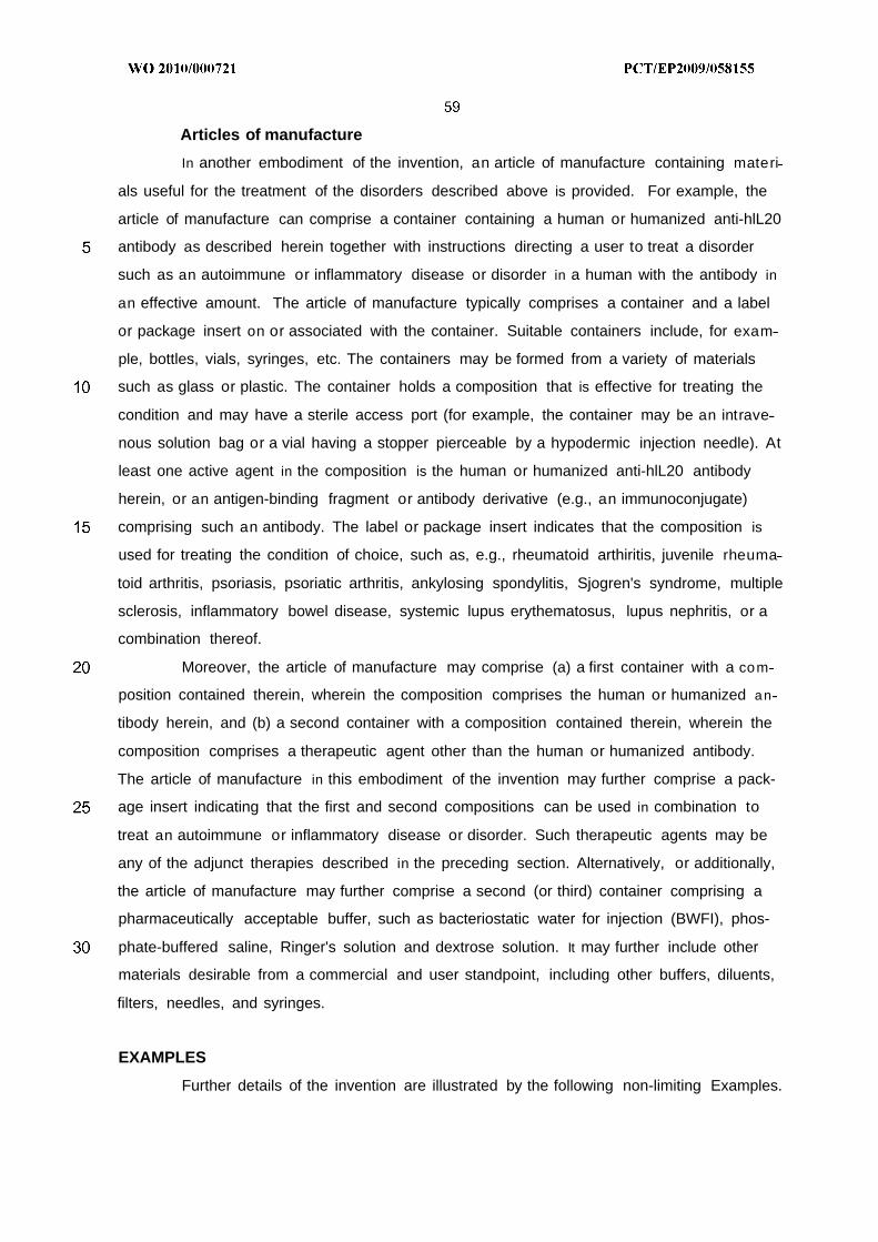
Articles of manufacture
In another embodiment of the invention, an article of manufacture containing materi
als useful for the treatment of the disorders described above is provided. For example, the
article of manufacture can comprise a container containing a human or humanized anti-hlL20
antibody as described herein together with instructions directing a user to treat a disorder
such as an autoimmune or inflammatory disease or disorder in a human with the antibody in
an effective amount. The article of manufacture typically comprises a container and a label
or package insert on or associated with the container. Suitable containers include, for exam
ple, bottles, vials, syringes, etc. The containers may be formed from a variety of materials
such as glass or plastic. The container holds a composition that is effective for treating the
condition and may have a sterile access port (for example, the container may be an intrave
nous solution bag or a vial having a stopper pierceable by a hypodermic injection needle). At
least one active agent in the composition is the human or humanized anti-hlL20 antibody
herein, or an antigen-binding fragment or antibody derivative (e.g., an immunoconjugate)
comprising such an antibody. The label or package insert indicates that the composition is
used for treating the condition of choice, such as, e.g., rheumatoid arthiritis, juvenile rheuma
toid arthritis, psoriasis, psoriatic arthritis, ankylosing spondylitis, Sjogren's syndrome, multiple
sclerosis, inflammatory bowel disease, systemic lupus erythematosus, lupus nephritis, or a
combination thereof.
Moreover, the article of manufacture may comprise (a) a first container with a com
position contained therein, wherein the composition comprises the human or humanized an
tibody herein, and (b) a second container with a composition contained therein, wherein the
composition comprises a therapeutic agent other than the human or humanized antibody.
The article of manufacture in this embodiment of the invention may further comprise a pack-
age insert indicating that the first and second compositions can be used in combination to
treat an autoimmune or inflammatory disease or disorder. Such therapeutic agents may be
any of the adjunct therapies described in the preceding section. Alternatively, or additionally,
the article of manufacture may further comprise a second (or third) container comprising a
pharmaceutically acceptable buffer, such as bacteriostatic water for injection (BWFI), phos-
phate-buffered saline, Ringer's solution and dextrose solution. It may further include other
materials desirable from a commercial and user standpoint, including other buffers, diluents,
filters, needles, and syringes.
EXAMPLES
Further details of the invention are illustrated by the following non-limiting Examples.
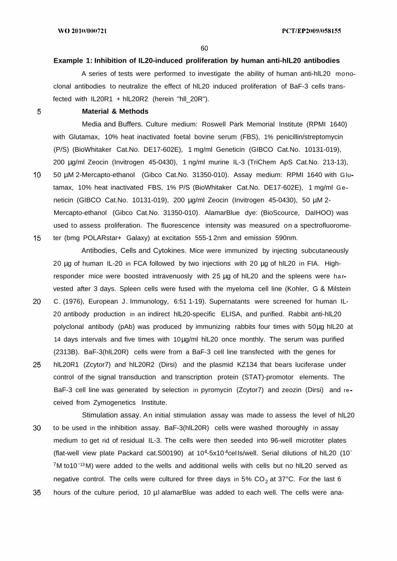
Example 1: Inhibition of IL20-induced proliferation by human anti-hlL20 antibodies
A series of tests were performed to investigate the ability of human anti-hlL20 mono
clonal antibodies to neutralize the effect of hlL20 induced proliferation of BaF-3 cells trans-
fected with IL20R1 + hlL20R2 (herein "hll_20R").
Material & Methods
Media and Buffers. Culture medium: Roswell Park Memorial Institute (RPMI 1640)
with Glutamax, 10% heat inactivated foetal bovine serum (FBS), 1% penicillin/streptomycin
(P/S) (BioWhitaker Cat.No. DE17-602E), 1 mg/ml Geneticin (GIBCO Cat.No. 10131-019),
200 µg/ml Zeocin (Invitrogen 45-0430), 1 ng/ml murine IL-3 (TriChem ApS Cat.No. 213-13),
50 µM 2-Mercapto-ethanol (Gibco Cat.No. 31350-010). Assay medium: RPMI 1640 with G lu
tamax, 10% heat inactivated FBS, 1% P/S (BioWhitaker Cat.No. DE17-602E), 1 mg/ml Ge
neticin (GIBCO Cat.No. 10131-019), 200 µg/ml Zeocin (Invitrogen 45-0430), 50 µM 2-
Mercapto-ethanol (Gibco Cat.No. 31350-010). AlamarBlue dye: (BioScource, DaIHOO) was
used to assess proliferation. The fluorescence intensity was measured on a spectrofluorome-
ter (bmg POLARstar+ Galaxy) at excitation 555-1 2nm and emission 590nm.
Antibodies, Cells and Cytokines. Mice were immunized by injecting subcutaneously
20 µg of human IL-20 in FCA followed by two injections with 20 µg of hlL20 in FIA. High-
responder mice were boosted intravenuosly with 25 µg of hlL20 and the spleens were ha r
vested after 3 days. Spleen cells were fused with the myeloma cell line (Kohler, G & Milstein
C. (1976), European J. Immunology, 6:51 1-19). Supernatants were screened for human IL-
20 antibody production in an indirect hlL20-specific ELISA, and purified. Rabbit anti-hlL20
polyclonal antibody (pAb) was produced by immunizing rabbits four times with 50µg hlL20 at
14 days intervals and five times with 10µg/ml hlL20 once monthly. The serum was purified
(2313B). BaF-3(hlL20R) cells were from a BaF-3 cell line transfected with the genes for
hlL20R1 (Zcytor7) and hlL20R2 (Dirsi) and the plasmid KZ134 that bears luciferase under
control of the signal transduction and transcription protein (STAT)-promotor elements. The
BaF-3 cell line was generated by selection in pyromycin (Zcytor7) and zeozin (Dirsi) and re
ceived from Zymogenetics Institute.
Stimulation assay. An initial stimulation assay was made to assess the level of hlL20
to be used in the inhibition assay. BaF-3(hlL20R) cells were washed thoroughly in assay
medium to get rid of residual IL-3. The cells were then seeded into 96-well microtiter plates
(flat-well view plate Packard cat.S00190) at 104-5x10 4cel Is/well. Serial dilutions of hlL20 (10
7M to10 13 M) were added to the wells and additional wells with cells but no hlL20 served as
negative control. The cells were cultured for three days in 5% CO2 at 37°C. For the last 6
hours of the culture period, 10 µl alamarBlue was added to each well. The cells were ana-
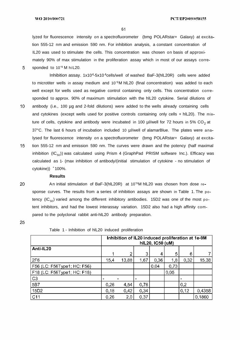
lyzed for fluorescence intensity on a spectrofluorometer (bmg POLARstar+ Galaxy) at excita
tion 555-12 nm and emission 590 nm. For inhibition analysis, a constant concentration of
IL20 was used to stimulate the cells. This concentration was chosen on basis of approxi
mately 90% of max stimulation in the proliferation assay which in most of our assays corre
sponded to 10 9 M hiL20.
Inhibition assay. 1x10⁴-5x10 4cells/well of washed BaF-3(hlL20R) cells were added
to microtiter wells in assay medium and 10 9M hlL20 (final concentration) was added to each
well except for wells used as negative control containing only cells. This concentration corre
sponded to approx. 90% of maximum stimulation with the hlL20 cytokine. Serial dilutions of
antibody (i.e., 100 µg and 2-fold dilutions) were added to the wells already containing cells
and cytokines (except wells used for positive controls containing only cells + hlL20). The m ix
ture of cells, cytokine and antibody were incubated in 100 µl/well for 72 hours in 5% CO2 at
37°C. The last 6 hours of incubation included 10 µl/well of alamarBlue. The plates were ana
lysed for fluorescence intensity on a spectrofluorometer (bmg POLARstar+ Galaxy) at excita
tion 555-12 nm and emission 590 nm. The curves were drawn and the potency (half maximal
inhibition (IC50)) was calculated using Prism 4 (GraphPad PRISM software Inc.). Efficacy was
calculated as 1- (max inhibition of antibody/(initial stimulation of cytokine - no stimulation of
cytokine)) * 100%.
Results
An initial stimulation of BaF-3(hlL20R) at 10 9M hlL20 was chosen from dose re
sponse curves. The results from a series of inhibition assays are shown in Table 1. The po
tency (IC50) varied among the different inhibitory antibodies. 15D2 was one of the most po
tent inhibitors, and had the lowest interassay variation. 15D2 also had a high affinity com
pared to the polyclonal rabbit anti-hlL20 antibody preparation.
Table 1 - Inhibition of hlL20 induced proliferation
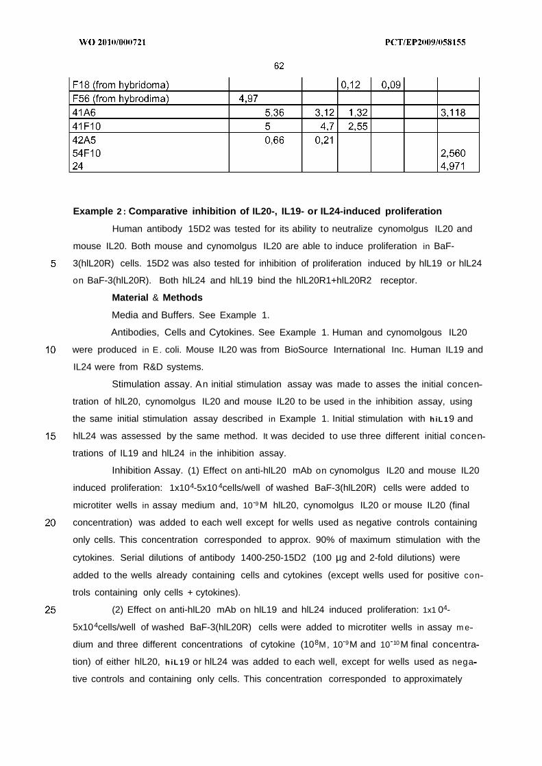
Example 2 : Comparative inhibition of IL20-, IL19- or IL24-induced proliferation
Human antibody 15D2 was tested for its ability to neutralize cynomolgus IL20 and
mouse IL20. Both mouse and cynomolgus IL20 are able to induce proliferation in BaF-
3(hlL20R) cells. 15D2 was also tested for inhibition of proliferation induced by hlL19 or hlL24
on BaF-3(hlL20R). Both hlL24 and hlL19 bind the hlL20R1+hlL20R2 receptor.
Material & Methods
Media and Buffers. See Example 1.
Antibodies, Cells and Cytokines. See Example 1. Human and cynomolgous IL20
were produced in E. coli. Mouse IL20 was from BioSource International Inc. Human IL19 and
IL24 were from R&D systems.
Stimulation assay. An initial stimulation assay was made to asses the initial concen
tration of hlL20, cynomolgus IL20 and mouse IL20 to be used in the inhibition assay, using
the same initial stimulation assay described in Example 1. Initial stimulation with h iL 19 and
hlL24 was assessed by the same method. It was decided to use three different initial concen
trations of IL19 and hlL24 in the inhibition assay.
Inhibition Assay. (1) Effect on anti-hlL20 mAb on cynomolgus IL20 and mouse IL20
induced proliferation: 1x10⁴-5x10 4cells/well of washed BaF-3(hlL20R) cells were added to
microtiter wells in assay medium and, 10 9 M hlL20, cynomolgus IL20 or mouse IL20 (final
concentration) was added to each well except for wells used as negative controls containing
only cells. This concentration corresponded to approx. 90% of maximum stimulation with the
cytokines. Serial dilutions of antibody 1400-250-15D2 (100 µg and 2-fold dilutions) were
added to the wells already containing cells and cytokines (except wells used for positive con
trols containing only cells + cytokines).
(2) Effect on anti-hlL20 mAb on hlL19 and hlL24 induced proliferation: 1x1 04-
5x10⁴cells/well of washed BaF-3(hlL20R) cells were added to microtiter wells in assay m e
dium and three different concentrations of cytokine (108M, 10 9M and 10 10M final concentra
tion) of either hlL20, h iL 19 or hlL24 was added to each well, except for wells used as nega
tive controls and containing only cells. This concentration corresponded to approximately
90% of maximum stimulation with the cytokines. Serial dilutions of antibody 15D2 (100 µg
and 2-fold dilutions) were added to the wells already containing cells and cytokines (except
wells used for positive controls containing only cells + cytokines).
For both assays, the mixture of cells, cytokine and antibody were incubated in 100
µl/well for 72 hours in 5% CO2 at 37°C. The last 6 hours of incubation included 10 µl/well of
alamarBlue. The plates were analysed for fluorescence intensity on a spectrofluorometer
(bmg POLARstar+ Galaxy) at excitation 555-12 nm and emission 590 nm. The curves were
drawn and the potency (half maximal inhibition (IC5o)) was calculated using Prism 4 (Graph-
Pad PRISM software Inc.).
The efficacy (the percentage inhibition by antibodies) was calculated as 1- (max in
hibition of antibody/(initial stimulation of cytokine - no stimulation of cytokine)) * 100%.
Results
Effect of anti-hlL20 mAb on cynomolgus IL20 and mouse IL20 induced proliferation.
When BaF-3(hlL20R) cells were stimulated with human, cynomolgus or mouse IL20, the
cells proliferated and this proliferation could be inhibited by anti-hlL20 mAb 15D2 in a dose
dependent manner. The efficacy of 15D2 was near 100% at 66 mM IL20 for all three cyto
kines. The potencies of the antibody could not be directly compared since the affinity for the
hlL20R was different for the three cytokines.
Effect ofanti-hlL20 mAb on hlL19 and hlL24 induced proliferation. BaF-3(hlL20R)
cells were stimulated with three different concentrations of hlL20, hlL19 or hlL24 (Figure 5).
A dose-dependent response was detected for inhibition of hlL20 induced proliferation (Figure
5A). This experiment served as positive control for the subsequent hlL19 and hlL24 experi
ments. When the BaF-3(hlL20R) cells initially were stimulated with hiL 19 , 15D2 was not able
to inhibit the proliferation regardless of which of the three initial concentrations of hiL 19 were
used (Figure 5B). The same results were obtained when the cells initially were stimulated
with hlL24 (Figure 5C).
Accordingly, 15D2 inhibited cynomolgus IL20-induced as well as mouse IL20-
induced proliferation of the BaF-3(hlL20R) cells, while hlL19 or hlL24-induced proliferation of
BaF-3(hlL20R) was not inhibited by 15D2.
Example 3 : Solubility
Seven different human lgG4 antibodies against IL20, produced in HEK293 cells and
purified by the same process, were compared for their ability to reach high concentration in
solution, by using centrifuge filters of the type Amicon Ultra (Millipore Corp, MA) with a cut-off
weight of 50 kD. All samples were centrifuged according to manufacturer instructions for 1
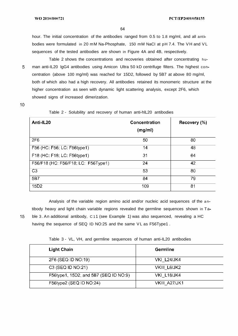
hour. The initial concentration of the antibodies ranged from 0.5 to 1.8 mg/ml, and all ant i
bodies were formulated in 20 mM Na-Phosphate, 150 mM NaCI at pH 7.4. The V H and V L
sequences of the tested antibodies are shown in Figure 4A and 4B, respectively.
Table 2 shows the concentrations and recoveries obtained after concentrating hu
man anti-IL20 lgG4 antibodies using Amicon Ultra 50 kD centrifuge filters. The highest con
centration (above 100 mg/ml) was reached for 15D2, followed by 5B7 at above 80 mg/ml,
both of which also had a high recovery. All antibodies retained its monomeric structure at the
higher concentration as seen with dynamic light scattering analysis, except 2F6, which
showed signs of increased dimerization.
Table 2 - Solubility and recovery of human anti-hlL20 antibodies
Analysis of the variable region amino acid and/or nucleic acid sequences of the a n
tibody heavy and light chain variable regions revealed the germline sequences shown in Ta
ble 3 . An additional antibody, C 11 (see Example 1) was also sequenced, revealing a HC
having the sequence of SEQ ID NO:25 and the same V L as F56Type1 .
Table 3 - VL, VH, and germline sequences of human anti-IL20 antibodies
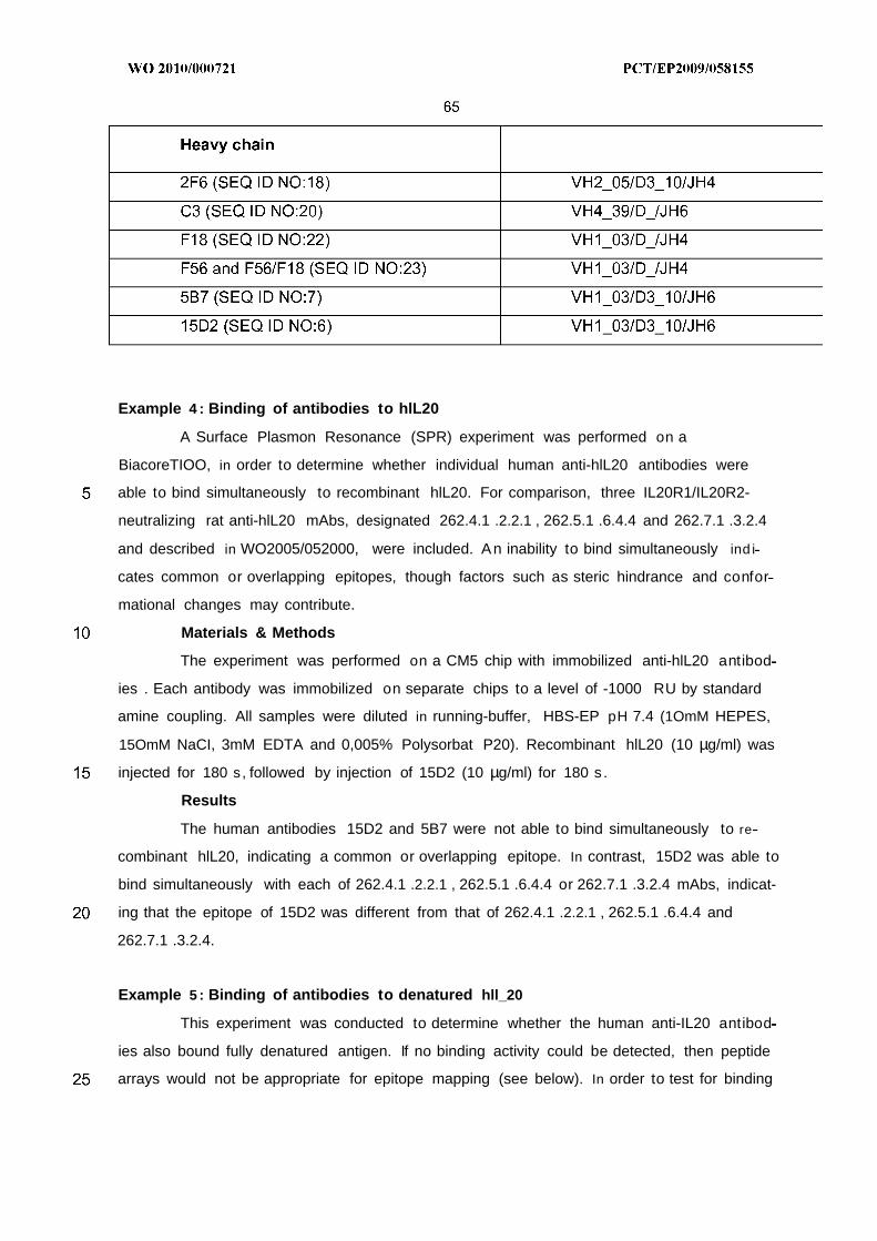
Example 4 : Binding of antibodies to hlL20
A Surface Plasmon Resonance (SPR) experiment was performed on a
BiacoreTIOO, in order to determine whether individual human anti-hlL20 antibodies were
able to bind simultaneously to recombinant hlL20. For comparison, three IL20R1/IL20R2-
neutralizing rat anti-hlL20 mAbs, designated 262.4.1 .2.2.1 , 262.5.1 .6.4.4 and 262.7.1 .3.2.4
and described in WO2005/052000, were included. An inability to bind simultaneously ind i
cates common or overlapping epitopes, though factors such as steric hindrance and confor
mational changes may contribute.
Materials & Methods
The experiment was performed on a CM5 chip with immobilized anti-hlL20 antibod
ies . Each antibody was immobilized on separate chips to a level of -1000 RU by standard
amine coupling. All samples were diluted in running-buffer, HBS-EP pH 7.4 (1OmM HEPES,
15OmM NaCI, 3mM EDTA and 0,005% Polysorbat P20). Recombinant hlL20 (10 µg/ml) was
injected for 180 s, followed by injection of 15D2 (10 µg/ml) for 180 s.
Results
The human antibodies 15D2 and 5B7 were not able to bind simultaneously to re
combinant hlL20, indicating a common or overlapping epitope. In contrast, 15D2 was able to
bind simultaneously with each of 262.4.1 .2.2.1 , 262.5.1 .6.4.4 or 262.7.1 .3.2.4 mAbs, indicat-
ing that the epitope of 15D2 was different from that of 262.4.1 .2.2.1 , 262.5.1 .6.4.4 and
262.7.1 .3.2.4.
Example 5 : Binding of antibodies to denatured hll_20
This experiment was conducted to determine whether the human anti-IL20 antibod
ies also bound fully denatured antigen. If no binding activity could be detected, then peptide
arrays would not be appropriate for epitope mapping (see below). In order to test for binding
to the denatured antigen, native and denatured hlL20 preparations were subjected to SDS-
PAGE, followed by Western-blot using 15D2 and 5B7 for detection.
Materials & Methods
Recombinant hlL20 was denatured by boiling for 10 min. in sample buffer (NuPage,
Invitrogen) containing 5OmM DTT. The samples were run in SDS-PAGE (20 ul/well including
sample-buffer) and blotted to a nitrocellulose membrane. The membrane was incubated with
the primary mAb (10 µg/ml in blotting buffer (Novex, Invitrogen)), followed by incubation with
HRP-conjugated Rabbit anti-human IgG polyclonal Ab (DAKO). Bands were visualized using
TMB-substrate (Kem-En-Tech).
Results
Both 15D2 and 5B7 recognized the native- and denatured form of the antigen, ind i
cating that the epitope is continuous (linear).
Example 6 : Surface Plasmon Resonance analysis of peptide binding
An SPR experiment was performed in order to determine whether the anti-hlL20 an-
tibodies 15D2, 262.4.1 .2.2.1 , 262.5.1 .6.4.4 or 262. 7 .1.3.2.4 bound to the region correspond
ing to residues 42-102 of the unprocessed precursor of hlL20 (SEQ ID NO:2), corresponding
to residues 18-78 of mature hlL20 (SEQ ID NO:1). The antibodies were shown to recognize
both the native and denatured form of hlL20 (for 15D2, see above).
Materials & Methods
Five 20mer peptides with a frame shift of 10 residues were synthesized:
1) IRNGFSEIRGSVQAKDGNID (residues 18-37 of SEQ ID NO:1 )
2) SVQAKDGNIDIRILRRTESL (residues 28-47 of SEQ ID NO:1)
3) IRILRRTESLQDTKPANRSS (residues 38-57 of SEQ ID NO.1)
4) QDTKPANRSSLLRHLLRLYL (residues 48-67 of SEQ ID NO.1)
5) LLRHLLRLYLDRVFKNYQTPD (residues 58-78 of SEQ ID NO:1)
The peptides, containing N-terminal biotin, were immobilized (500 RU) in individual
flow cells on streptavidin coated (SA) chips. The individual antibodies (5 µg/ml) were injected
across all flow cells for 120 s, followed by a 180 s dissociation phase. Experiments were pe r
formed on Biacore3000 and BiacoreTIOO instruments. The results were evaluated using
Scrubbed software (BioLogic Software Pty Ltd).
Results
No 15D2, 262.5.1 .6.4.4 or 262.7.1 .3.2.4 binding to the peptides was detected. The
rat anti-hlL20 mAb 262.4.1 .2.2.1 demonstrated binding to peptide 4 and 5 , indicating binding
to their shared sequence LLRHLLRLY. All mAbs demonstrated binding to immobilized intact
biotinylated IL20.
Example 7 : Primary peptide array ("peptide walk")
A peptide array consisting of 18mer hlL20 peptides with a frame shift of 4 residues
was made and screened against fluorescence-labelled anti-hlL20 antibodies 15D2 or 5B7.
Materials and Methods
Synthesis of epitope arrays. The epitope mapping arrays were synthesized on cellu
lose sheets (Aims-Scientific, Germany) on an array synthesizer (Multipep Spot, Intavis, Ger
many) essentially using the protocols provided from the manufacturer. Fmoc-amino acids
were purchased from Novabiochem (Germany) and dissolved in N-methylpyrrolidinone
(NMP) containing 0.3 M Hydrobenzotriazole (HOBt) to a final concentration of 0,3M. Cou
pling was done by activating with diisopropylcarbodiimide (DIC), and deprotection of the
Fmoc group was done by 20% piperidine in NMP. The individual sequences were designed
by the software accompanying the array synthesiser. After synthesis the protecting groups
were removed by treating the sheets with trifluoroacetic acid (TFA) 95% containing triisopro-
pylsilane (TIPS) for 60 min. Then washed with dichloromethane (DCM) and N-
methylpyrrolidinone (NMP) and finally with water.
Labelling of antibodies. The screenings were done using fluorescence labelled ant i
bodies. The labelling was done by gel-filtering the antibody stock against 1% NaHCO3, using
NAP5 column (GE Healthcare according the manual from manufacturer. This was followed
by adding 25 mole equivalents of 5(6)-carboxyfluorescein N-hydroxysuccinimide ester
(Sigma, C 1609) dissolved in DMSO. The coupling was allowed to continue for 2 hours fo l
lowed by a gelfiltration against TRIS washing buffer (5OmM TRIS, pH = 7,4, 0,1 5M NaCI, 0,1
M ArgHCI, 0,05% Tween 20) in order to removed uncoupled fluorescein.
Screening and analyzing arrays. Screening of arrays were done by adding 10 µl of
antibody to 30 ml incubation buffer (0,5% BSA, 5OmM TRIS, pH = 7,4, 0.15M NaCI, 0,1 M
ArgHCI, 0,05% Tween 20). The sheets were incubated for 1-2 hours, followed by washing
five times with washing buffer. Then the sheets were scanned using a laser scanner (Ty
phoon 9410, GE Healthcare) and the image file (.gel format) was analysed using dedicated
array software ArrayPro Analyzer (Media Cypernetics, USA). The fluorescence intensity were
measured and transformed into digits that were exported to Prism 5 (GraphPad Software,
USA) for further analysis.
Results
The results from the primary peptide array analysis, shown in Figure 6 , clearly iden
tified that 15D2 and 5B7 both bind a linear epitope located in the region corresponding to
residues 73-96 in mature hlL20 (SEQ ID NO:1 ) , corresponding to residues 97-120 of the
precursor (SEQ ID NO:2).
Example 8 : Secondary peptide array analysis - terminal deletions
In order to narrow down the length of the epitope and to evaluate which residues
were important for the binding of anti-IL20 antibodies 15D2 and 5B7, an array of various t run
cations was made. An ala-scan was also included (section 5).
For Materials and Methods, see Example 7 .
Results
The truncations from the hlL20 C-terminal revealed a gradual reduction in 15D2
binding from peptides 4 to 7 (Figure 7A). A more sudden decline in binding activity was seen
for the N-terminal truncations, where removal of D (Asp) and H (His) dramatically reduced
the affinity. Overall, the results showed that the minimum epitope had the sequence
DHYTLRKISSLANSFL, corresponding to residues 78 to 93 of SEQ ID NO:1 .
For 5B7, the truncations revealed a sudden reduction in binding when deleting from
the N-terminal (Figure 7B). A decline of about 50% in binding activity was seen for the N-
terminal truncations when removing D (Asp), and removing H (His) dramatically reduced the
affinity to no detectable binding. Removing from the C-terminal resulted in a less sudden de
cline in affinity until removing N (Asn). Overall, the results indicate that the minimum 5B7 ep i
tope had the sequence DHYTLRKISSLAN (residues 78-90 of SEQ ID NO:1 ) , although a
longer epitope, DHYTLRKISSLANSFLTIK (residues 78-96 of SEQ ID NO:1), did present a
higher affinity. This indicated the presence of some structural requirements, e.g., α-helix de-
pendence.
The result of the Ala scan clearly indicated that, for 15D2, three residues, H79 (His-
79), R83 (Arg-83) and N90 (Asn-90) of SEQ ID NO:1 were most critical for binding (Figure
8A). Also sensitive, but to a lesser extent, were residues D78 (Asp-78), S86 (Ser-86), F92
(Phe-92) and L93 (Leu-93). The residues H79 and N90 were most critical also for 5B7 bind-
ing, while R83 (Arg) appeared fairly critical (Figure 8B).
In conclusion, the human anti-hlL20 antibodies 15D2 and 5B7 both bind a linear e pi
tope (functional) of similar length and specificity, shown in Figure 1. Residues H79, R83 and
N90 (bold and single-underlined) of mature hlL20 (SEQ ID NO:1); corresponding to H103,
R107, and N 114 in hlL20 precursor (SEQ ID NO:2), were found the most critical, with resi-
dues D78, S86, F92 and L93 (bold and double-underlined) being moderately critical. The
main difference between the two antibodies was that R83 was slightly less critical for 5B7
binding as compared to 15D2 binding.
The position of the 15D2/5B7 epitope and the location of the most critical residues
were revealed using the crystal structure of the homologous protein IL19 (Chang et al. J Biol
Chem 2003; 278: 3308). It was found that the location of the epitope corresponded to helix E
in IL19, and that the most critical residues in hlL20; H79, R83, and N90 of SEQ ID NO:1 , are
exposed to the solvent. Note that the hlL20 residue H79 is a proline in h iL 19 .
Example 9 : Neutralization of IL20 activation of IL20R1/IL20R2 and IL22R1/IL20R2 re-
ceptor complexes
This Example shows that human antibody 15D2 is capable of neutralizing murine,
cynomolgous, and hlL20 activation of recombinantly expressed IL20R1/IL20R2 and
IL22R1/IL20R2 receptor complexes.
Materials and Methods
Cloning of IL20 receptors. Human IL20 receptors, IL22R1 (EMBL BC029273),
IL20R1 (EMBL AF1 84971 ) and IL20R2 (EMBL AY358305) were PCR-amplified from NHEK
(Normal Human Epidermal Keratinocyte) cDNA and cloned into pcDNA3,1+(zeocin) (IL22R1
and IL20R1 ) and pcDNA3,1 +(hygro)(IL20R2). Mouse IL20 receptors, mlL22R1 (EMBL
AY103454), mlL20R1 (EMBL AK054215) and mlL20R2 (EMBL BC107264) were cloned by
PCR using mouse liver, testis and skin cDNA, respectively, as templates. Each of these
IL20R sequences is incorporated by reference, in its entirety. The mlL22R1 and mlL20R1
PCR-product were cloned into pcDNA3,1+(zeo) and mlL20R2 into pcDNA3,1 +(hygro). Cy-
nomolgus IL20 receptors, cynolL22R1 (SEQ ID NO:28), cynolL20R1 (SEQ ID NO:26) and
cynolL20R2 (SEQ ID NO:27) were cloned by PCR using cynomolgus skin cDNA. The cy-
nolL20R1 and cynolL22R1 receptors were inserted into pcDNA3,1+(zeocin) and cynolL20R2
was inserted into pcDNA3,1+(neomycin).
The following 5' and 3' primers were used for PCR-amplification of coding cDNA.
Human IL22R1 : agaattccaccatgaggacgctgctgacca and gctcgagacagggaggaagcac-
caag (SEQ ID NOS:29 and 30)
Human IL20R1 : cgaattcccttggtttctggggaag and gctcgagcacaggaaacaaaaggcaaa
(SEQ ID NOS:31 and 32)
Human IL20R2: agaattctggaaagaaacaatgttctaggtcaa and gctcgagcttcacctgggcccttcc
(SEQ ID NOS:33 and 34)
Murine IL22R1 : ccgaattcgccaccatgaagacactactgaccatc and cttgcggccgctcag-
gattcccactgcacagtc (SEQ ID NOS:35 and 36)
Murine IL20R1 : ttgaattcgccaccatgcacactcccggga and ttgcggccgcctagctttccatttgta-
catgtaacc (SEQ ID NOS:37 and 38)
Murine IL20R2: ttggatccgccaccatgatttcccagggagtctg and ttgcggccgctcaagtctgtga-
gatccagac (SEQ ID NO:39 and 30)
Cynomolgus IL22R1 : agaattccaccatgaggacgctgctgacca and gctcgagacagggaggaag-
caccaag (SEQ ID NO:41 and 42)
Cynomolgus IL20R1 : gtgggactgagcagtctgctg and aggcaaaaggaagtgttggca (SEQ ID
NOS:43 and 44)
Cynomolgus IL20R2: agaattctggaaagaaacaatgttctaggtcaa and
gctcgagcttcacctgggcccttcc (SEQ ID NOS:45 and 46)
STAT-reporter KZ136 is a luciferase reporter containing STAT-elements and a S e
rum Response Element (SRE) (Poulsen-LK et al. J Biol Chem 1998; 273:6228-6232).
Luciferase assay in transient transfection. Day 0 : BHK cells were seeded in T80
flask to a confluency of 40 %. Day 1: 7.5 micrograms DNA was transfected using 36 microli
ters FuGene (Roche Applied Science) according to manufacturers manual, 2.5 micrograms
of receptor chain 1 (IL20R1 or IL22R1 ) and 2.5 micrograms of chain 2 (IL20R2) and 2.5 m i
crograms of luciferase reporter (KZ136). Day 2 : The cells were detached using Versene and
20,000 cells per well were seeded in a black view plate. After the cells have reattached the
well surface the media was exchanged with serum-free media. Day 3 : either 20 microliter of
10 mM IL20 or 20 microliter of serum-free medium was added to the wells. Four hours later
the luciferase activity was determined; media was removed and 100 microliter IxPBS was
added to each well followed by the addition of 100 microliter Luclite substrate (PerkinElmer).
The plate was incubated for 30 minutes. The luminescence was detected by a Topcount NXT
(PerkinElmer). IL20 used here are either recombinantly produced hlL20 produced in E. coli,
murine IL20 purchased from Biosource, #PMC0201 , or cynomolgus IL20 produced by t ran
sient expression in HEK293 6E cells and purified.
Results
Neutralization of murine IL20. The murine IL20 receptors were cloned and a t ran
sient luciferase reporter assay was set up in BHK cells. Both the two human receptor com
plexes and the two murine receptor complexes were stimulated with 1 nM murine IL20. Mur
ine IL20 could activate both human and murine IL20R1/IL20R2 and IL22R1/IL20R2 receptor
complexes. Neutralization by 15D2 was tested on one of the receptor complexes. The mur-
ine IL20R2/IL22R1 complex was transfected into BHK cells together with the STAT3 reporter
construct. The receptor complex was stimulated with 1 nM murine IL20 and exposed to 15D2
antibody in 1, 10 and 50 microgram/ml doses (Figure 9). The lowest dose, 1 microgram/ml,
neutralized the effect almost totally.
Neutralization of cynomolgus IL20. Cynomolgus IL20 receptor sequences IL20R1 ,
IL20R2 and IL22R1 were cloned from cynomolgus skin tissue cDNA, using oligonucleotide
primers based on human sequence. The respective sequence identities between cynomol
gus and human receptor sequences were 96.8% for IL20R1 , 98.9% for IL20R2, and 95.5%
for IL22R1 . BHK cells were transfected with the two receptor complexes, IL20R1/IL20R2 or
IL22R1/IL20R2 together with the KZ136 (the STAT3 luciferase reporter) plasmid. The cyno-
molgus IL20R2/IL22R1 complex was induced 3-4 fold using 1 nM cynolL20. The
IL20R1/IL20R2 complex from cynomolgus was stimulated about 2-fold. Increasing the
amount of 15D2 decreased the IL20 activity, showing that 15D2 could neutralize cynomol-
gous IL20 (Figure 10).
Neutralization ofhlL20. Human IL20 receptors IL20R1 , IL20R2 and IL22R1 , were
cloned from NHEK (Normal Human Epidermal Keratinocyte) cDNA. Expression plasmids e n
coding the IL20 receptors were transiently transfected into BHK cells together with the
luciferase reporter vector, KZ136. The stimulation was done using 1 nM hlL20 and an induc
tion of about 3 fold was seen. Increasing the amount of 15D2 antibody decreased the IL20
activity (Figure 11) .
Example 10: Inhibition of hlL20-induced proliferation
This Example evaluates the inhibitory effects of 15D2 and rat anti-hlL20 mAbs on
IL20-induced proliferation of BaF-3 cells expressing IL20R1 and IL20R2 (herein "hlL20R"),
and the form of IL20 bound by 15D2.
Materials & Methods
Three independent experiments ("148", "149" and "150") compared EC50 values of
the four tested abs. First, antibodies were added in a 3-fold serial dilution with 50ul/well in
media. At the same time 10µl hlL20 was added to every well and at last 40µl of cell-
suspension. Antibodies were diluted to a 3-fold serial dilution (100+200), with the first dilution
in the assay at 20µg/ml. hlL20 preparation was diluted to 10 7M, with dilution in assay to 10
8M. As a control, the following stimulation curve was made: hlL20 was diluted to 10 6M and
from this a 10-fold dilution row. First dilution in the assay was 10-7M.
BaF-3 cells recombinantly expressing human IL20R1/IL20R2 were centrifuged, re-
suspended in media without IL3, and counted. They were then washed twice in media w ith
out IL3, and added into 96-wells flat-bottomed view plates at a concentration of 104c/well and
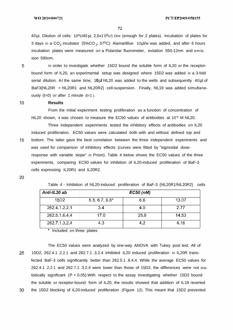
40µl . Dilution of cells: 104c/40 µl , 2,5x1 06c/1 OmI (enough for 2 plates). Incubation of plates for
3 days in a CO2 incubator (5%CO 2, 370C). AlamarBlue 10µl/w was added, and after 6 hours
incubation plates were measured on a Polarstar fluorometer, exitation 550-12nm and emis
sion 590nm.
In order to investigate whether 15D2 bound the soluble form of IL20 or the receptor-
bound form of IL20, an experimental setup was designed where 15D2 was added in a 3-fold
serial dilution. At the same time, 10µl hlL20 was added to the wells and subsequently 40µl of
BaF3(hlL20R = hlL20R1 and hlL20R2) cell-suspension. Finally, hlL19 was added simultane
ously (t=0) or after 1 minute (t=1 ) .
Results
From the initial experiment testing proliferation as a function of concentration of
hlL20 shown, it was chosen to measure the EC50 values of antibodies at 10 8 M hlL20.
Three independent experiments tested the inhibitory effects of antibodies on IL20
induced proliferation. EC50 values were calculated both with and without defined top and
bottom. The latter gave the best correlation between the three independent experiments and
was used for comparison of inhibitory effects (curves were fitted by "sigmoidal dose-
response with variable slope" in Prism). Table 4 below shows the EC50 values of the three
experiments, comparing EC50 values for inhibition of IL20-induced proliferation of BaF-3
cells expressing IL20R1 and IL20R2.
Table 4 - Inhibition of hlL20-induced proliferation of BaF-3 (hlL20R1/hlL20R2) cells
Included on three plates
The EC50 values were analyzed by one-way ANOVA with Tukey post test. All of
15D2, 262.4.1 .2.2.1 and 262.7.1 .3.2.4 inhibited IL20 induced proliferation in IL20R trans-
fected BaF-3 cells significantly better than 262.5.1 .6.4.4. While the average EC50 values for
262.4.1 .2.2.1 and 262.7.1 .3.2.4 were lower than those of 15D2, the differences were not sta
tistically significant (P < 0.05).With respect to the assay investigating whether 15D2 bound
the soluble or receptor-bound form of IL20, the results showed that addition of IL19 reverted
the 15D2 blocking of IL20-induced proliferation (Figure 12). This meant that 15D2 prevented
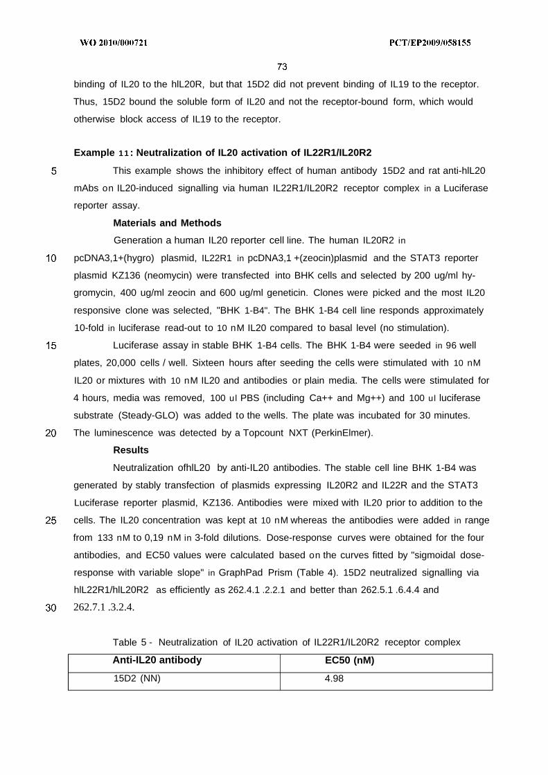
binding of IL20 to the hlL20R, but that 15D2 did not prevent binding of IL19 to the receptor.
Thus, 15D2 bound the soluble form of IL20 and not the receptor-bound form, which would
otherwise block access of IL19 to the receptor.
Example 1 1 : Neutralization of IL20 activation of IL22R1/IL20R2
This example shows the inhibitory effect of human antibody 15D2 and rat anti-hlL20
mAbs on IL20-induced signalling via human IL22R1/IL20R2 receptor complex in a Luciferase
reporter assay.
Materials and Methods
Generation a human IL20 reporter cell line. The human IL20R2 in
pcDNA3,1+(hygro) plasmid, IL22R1 in pcDNA3,1 +(zeocin)plasmid and the STAT3 reporter
plasmid KZ136 (neomycin) were transfected into BHK cells and selected by 200 ug/ml hy-
gromycin, 400 ug/ml zeocin and 600 ug/ml geneticin. Clones were picked and the most IL20
responsive clone was selected, "BHK 1-B4". The BHK 1-B4 cell line responds approximately
10-fold in luciferase read-out to 10 nM IL20 compared to basal level (no stimulation).
Luciferase assay in stable BHK 1-B4 cells. The BHK 1-B4 were seeded in 96 well
plates, 20,000 cells / well. Sixteen hours after seeding the cells were stimulated with 10 nM
IL20 or mixtures with 10 nM IL20 and antibodies or plain media. The cells were stimulated for
4 hours, media was removed, 100 ul PBS (including Ca++ and Mg++) and 100 ul luciferase
substrate (Steady-GLO) was added to the wells. The plate was incubated for 30 minutes.
The luminescence was detected by a Topcount NXT (PerkinElmer).
Results
Neutralization ofhlL20 by anti-IL20 antibodies. The stable cell line BHK 1-B4 was
generated by stably transfection of plasmids expressing IL20R2 and IL22R and the STAT3
Luciferase reporter plasmid, KZ136. Antibodies were mixed with IL20 prior to addition to the
cells. The IL20 concentration was kept at 10 nM whereas the antibodies were added in range
from 133 nM to 0,19 nM in 3-fold dilutions. Dose-response curves were obtained for the four
antibodies, and EC50 values were calculated based on the curves fitted by "sigmoidal dose-
response with variable slope" in GraphPad Prism (Table 4). 15D2 neutralized signalling via
hlL22R1/hlL20R2 as efficiently as 262.4.1 .2.2.1 and better than 262.5.1 .6.4.4 and
262.7.1 .3.2.4.
Table 5 - Neutralization of IL20 activation of IL22R1/IL20R2 receptor complex
Anti-IL20 antibody EC50 (nM)
15D2 (NN) 4.98
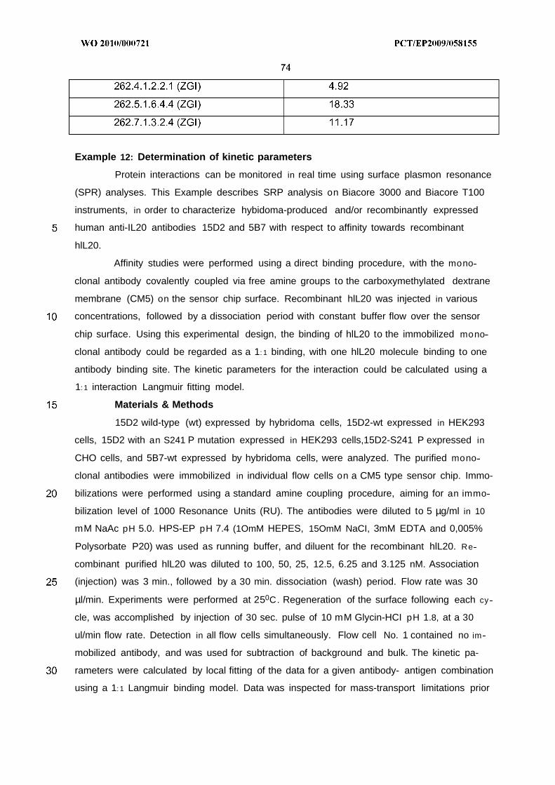
Example 12: Determination of kinetic parameters
Protein interactions can be monitored in real time using surface plasmon resonance
(SPR) analyses. This Example describes SRP analysis on Biacore 3000 and Biacore T100
instruments, in order to characterize hybidoma-produced and/or recombinantly expressed
human anti-IL20 antibodies 15D2 and 5B7 with respect to affinity towards recombinant
hlL20.
Affinity studies were performed using a direct binding procedure, with the mono
clonal antibody covalently coupled via free amine groups to the carboxymethylated dextrane
membrane (CM5) on the sensor chip surface. Recombinant hlL20 was injected in various
concentrations, followed by a dissociation period with constant buffer flow over the sensor
chip surface. Using this experimental design, the binding of hlL20 to the immobilized mono
clonal antibody could be regarded as a 1: 1 binding, with one hlL20 molecule binding to one
antibody binding site. The kinetic parameters for the interaction could be calculated using a
1: 1 interaction Langmuir fitting model.
Materials & Methods
15D2 wild-type (wt) expressed by hybridoma cells, 15D2-wt expressed in HEK293
cells, 15D2 with an S241 P mutation expressed in HEK293 cells,15D2-S241 P expressed in
CHO cells, and 5B7-wt expressed by hybridoma cells, were analyzed. The purified mono
clonal antibodies were immobilized in individual flow cells on a CM5 type sensor chip. Immo-
bilizations were performed using a standard amine coupling procedure, aiming for an immo
bilization level of 1000 Resonance Units (RU). The antibodies were diluted to 5 µg/ml in 10
mM NaAc pH 5.0. HPS-EP pH 7.4 (1OmM HEPES, 15OmM NaCI, 3mM EDTA and 0,005%
Polysorbate P20) was used as running buffer, and diluent for the recombinant hlL20. Re
combinant purified hlL20 was diluted to 100, 50, 25, 12.5, 6.25 and 3.125 nM. Association
(injection) was 3 min., followed by a 30 min. dissociation (wash) period. Flow rate was 30
µl/min. Experiments were performed at 250C. Regeneration of the surface following each cy
cle, was accomplished by injection of 30 sec. pulse of 10 mM Glycin-HCI pH 1.8, at a 30
ul/min flow rate. Detection in all flow cells simultaneously. Flow cell No. 1 contained no im
mobilized antibody, and was used for subtraction of background and bulk. The kinetic pa-
rameters were calculated by local fitting of the data for a given antibody- antigen combination
using a 1: 1 Langmuir binding model. Data was inspected for mass-transport limitations prior
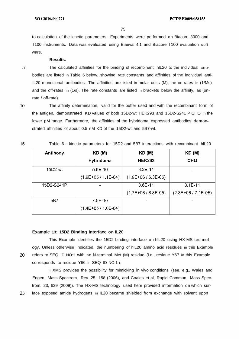
to calculation of the kinetic parameters. Experiments were performed on Biacore 3000 and
T100 instruments. Data was evaluated using Biaeval 4.1 and Biacore T100 evaluation soft
ware.
Results.
The calculated affinities for the binding of recombinant hlL20 to the individual ant i
bodies are listed in Table 6 below, showing rate constants and affinities of the individual anti-
IL20 monoclonal antibodies. The affinities are listed in molar units (M), the on-rates in (1/Ms)
and the off-rates in (1/s). The rate constants are listed in brackets below the affinity, as (on-
rate / off-rate).
The affinity determination, valid for the buffer used and with the recombinant form of
the antigen, demonstrated KD values of both 15D2-wt HEK293 and 15D2-S241 P CHO in the
lower pM range. Furthermore, the affinities of the hybridoma expressed antibodies demon
strated affinities of about 0.5 nM KD of the 15D2-wt and 5B7-wt.
Table 6 - kinetic parameters for 15D2 and 5B7 interactions with recombinant hlL20
Example 13: 15D2 Binding interface on IL20
This Example identifies the 15D2 binding interface on hlL20 using HX-MS technol
ogy. Unless otherwise indicated, the numbering of hlL20 amino acid residues in this Example
refers to SEQ ID NO:1 with an N-terminal Met (M) residue (i.e., residue Y67 in this Example
corresponds to residue Y66 in SEQ ID NO:1 ) .
HXMS provides the possibility for mimicking in vivo conditions (see, e.g., Wales and
Engen, Mass Spectrom. Rev. 25, 158 (2006), and Coales et al, Rapid Commun. Mass Spec-
trom. 23, 639 (2009)). The HX-MS technology used here provided information on which sur-
face exposed amide hydrogens in IL20 became shielded from exchange with solvent upon
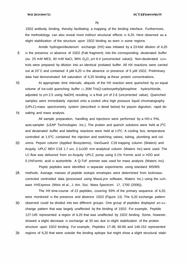
15D2 antibody binding, thereby facilitating a mapping of the binding interface. Furthermore,
the methodology can also reveal more indirect structural effects in IL20. Here observed as a
slight stabilization of the structure upon 15D2 binding as seen in some regions.
Amide hydrogen/deuterium exchange (HX) was initiated by a 23-fold dilution of IL20
in the presence or absence of 15D2 (Fab fragment) into the corresponding deuterated buffer
(Ae. 25 mM MES, 80 mM NaCI, 96% D2O, pH 6.4 (uncorrected value)). Non-deuterated con
trols were prepared by dilution into an identical protiated buffer. All HX reactions were carried
out at 20°C and contained 4 µM IL20 in the absence or presence of 5 µM 15D2. Preliminary
data had demonstrated full saturation of IL20 binding at these protein concentrations.
At appropriate time intervals, aliquots of the HX reaction were quenched by an equal
volume of ice-cold quenching buffer ( 1 .35M Tris(2-carboxyethyl)phosphine hydrochloride,
adjusted to pH 2.5 using NaOH) resulting in a final pH of 2.6 (uncorrected value). Quenched
samples were immediately injected onto a cooled ultra high pressure liquid chromatography
(UPLC)-mass spectrometry system (described in detail below) for pepsin digestion, rapid de-
salting and mass analysis.
All sample preparation, handling and injections were performed by a HD-x PAL
auto-sampler (LEAP Technologies Inc.). The protein and quench solutions were held at 20C
and deuterated buffer and labelling reactions were held at 2 O0C. A cooling box, temperature
controlled at 1.50C, contained the injection and switching valves, tubing, plumbing and col-
umns. Pepsin column (Applied Biosystems), VanGuard C18 trapping column (Waters) and
Acquity UPLC BEH C18 1.7 urn, 2.1x100 mm analytical column (Waters Inc) were used. The
LC flow was delivered from an Acquity UPLC pump using 0.1% Formic acid in H2O and
0.1%Formic acid in acetonitrile. A Q-ToF premier was used for mass analysis (Waters Inc).
Peptic peptides were identified in separate experiments using standard MS/MS
methods. Average masses of peptide isotopic envelopes were determined from lockmass-
corrected centroided data (processed using MassLynx software, Waters Inc.) using the soft
ware HXExpress (Weis et al., J. Am. Soc. Mass Spectrom. 17, 1700 (2006)).
The HX time-course of 22 peptides, covering 93% of the primary sequence of IL20,
were monitored in the presence and absence 15D2 (Figure 13). The IL20 exchange pattern
observed could be divided into two different groups. One group of peptides displayed an ex
change pattern that was largely unaffected by the binding of 15D2. For example, Peptide
127-145 represented a region of IL20 that was unaffected by 15D2 binding. Some, however,
showed a slight decrease in exchange at 30 sec due to slight stabilization of the protein
structure upon 15D2 binding. For example, Peptides 17-38, 60-66 and 146-153 represented
regions of IL20 that were outside the binding epitope but might show a slight structural stabi-
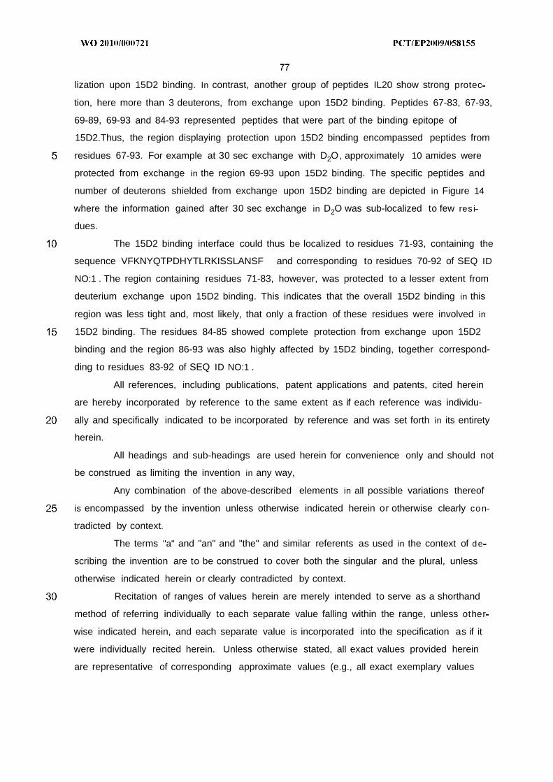
lization upon 15D2 binding. In contrast, another group of peptides IL20 show strong protec
tion, here more than 3 deuterons, from exchange upon 15D2 binding. Peptides 67-83, 67-93,
69-89, 69-93 and 84-93 represented peptides that were part of the binding epitope of
15D2.Thus, the region displaying protection upon 15D2 binding encompassed peptides from
residues 67-93. For example at 30 sec exchange with D2O, approximately 10 amides were
protected from exchange in the region 69-93 upon 15D2 binding. The specific peptides and
number of deuterons shielded from exchange upon 15D2 binding are depicted in Figure 14
where the information gained after 30 sec exchange in D2O was sub-localized to few res i
dues.
The 15D2 binding interface could thus be localized to residues 71-93, containing the
sequence VFKNYQTPDHYTLRKISSLANSF and corresponding to residues 70-92 of SEQ ID
NO:1 . The region containing residues 71-83, however, was protected to a lesser extent from
deuterium exchange upon 15D2 binding. This indicates that the overall 15D2 binding in this
region was less tight and, most likely, that only a fraction of these residues were involved in
15D2 binding. The residues 84-85 showed complete protection from exchange upon 15D2
binding and the region 86-93 was also highly affected by 15D2 binding, together correspond-
ding to residues 83-92 of SEQ ID NO:1 .
All references, including publications, patent applications and patents, cited herein
are hereby incorporated by reference to the same extent as if each reference was individu-
ally and specifically indicated to be incorporated by reference and was set forth in its entirety
herein.
All headings and sub-headings are used herein for convenience only and should not
be construed as limiting the invention in any way,
Any combination of the above-described elements in all possible variations thereof
is encompassed by the invention unless otherwise indicated herein or otherwise clearly con
tradicted by context.
The terms "a" and "an" and "the" and similar referents as used in the context of de
scribing the invention are to be construed to cover both the singular and the plural, unless
otherwise indicated herein or clearly contradicted by context.
Recitation of ranges of values herein are merely intended to serve as a shorthand
method of referring individually to each separate value falling within the range, unless other
wise indicated herein, and each separate value is incorporated into the specification as if it
were individually recited herein. Unless otherwise stated, all exact values provided herein
are representative of corresponding approximate values (e.g., all exact exemplary values
provided with respect to a particular factor or measurement can be considered to also pro
vide a corresponding approximate measurement, modified by "about," where appropriate).
All methods described herein can be performed in any suitable order unless other
wise indicated herein or otherwise clearly contradicted by context.
The use of any and all examples, or exemplary language (e.g., "such as") provided
herein, is intended merely to better illuminate the invention and does not pose a limitation on
the scope of the invention unless otherwise indicated. No language in the specification
should be construed as indicating any element is essential to the practice of the invention
unless as much is explicitly stated.
The citation and incorporation of patent documents herein is done for convenience
only and does not reflect any view of the validity, patentability and/or enforceability of such
patent documents,
The description herein of any aspect or embodiment of the invention using terms
such as "comprising", "having", "including" or "containing" with reference to an element or
elements is intended to provide support for a similar aspect or embodiment of the invention
that "consists of", "consists essentially of", or "substantially comprises" that particular element
or elements, unless otherwise stated or clearly contradicted by context (e.g., a composition
described herein as comprising a particular element should be understood as also describing
a composition consisting of that element, unless otherwise stated or clearly contradicted by
context).
This invention includes all modifications and equivalents of the subject matter re
cited in the aspects or claims presented herein to the maximum extent permitted by applica
ble law.
EXEMPLARY EMBODIMENTS
The following are exemplary and non-limiting embodiments of the invention.
1. An isolated anti-human IL20 antibody or an antigen-binding fragment thereof, which
reduces IL20 mediated activation of IL20R1/IL20R2 and IL22R1/IL20R2 receptor
complexes.
2 . The antibody or antigen-binding fragment of embodiment 1, which reduces human
IL20 mediated activation of human IL20R1/IL20R2 and IL22R1/IL20R2 receptor com
plexes.
3 . The antibody or antigen-binding fragment of any of the preceding embodiments,
which reduces cynomolgus IL20 mediated activation of cynomolgus IL20R1/IL20R2
and IL22R1/IL20R2 receptor complexes.
4 . The antibody or antigen-binding fragment of any of the preceding embodiments,
which reduces murine IL20 mediated activation of murine IL20R1/IL20R2 and
IL22R1/IL20R2 receptor complexes.
5 . The antibody or antigen-binding fragment of any of the preceding embodiments,
which reduces the binding of IL20 to the IL20R1/IL20R2 and/or IL22R1/IL20R2 recep
tor complexes.
6 . The antibody or antigen-binding fragment of any of the preceding embodiments,
which reduces the binding of IL20 to IL20R2.
7 . The antibody or antigen-binding fragment of any of embodiments 1-4, which does not
reduce binding of IL19 or IL24 to IL20R1/IL20R2 or IL22R1/IL20R2 receptor com
plexes.
8 . The antibody or antigen-binding fragment of any of the preceding embodiments,
which binds to an epitope comprising at least one residue selected from D78-H103 of
mature human IL20 (SEQ ID NO:1), optionally excluding D78.
9 . The antibody or antigen-binding fragment of embodiment 8 , wherein the epitope
comprises at least one residue selected from D78-K96.
10. The antibody or antigen-binding fragment of embodiment 9 , wherein the epitope
comprises at least one residue selected from D78-L93 or R83-F92.
11. The antibody or antigen-binding fragment of embodiment 10 , wherein the epitope
comprises at least one residue selected from H79-N90.
12. The antibody or antigen-binding fragment of embodiment 8 , wherein the epitope
comprises at least 3 residues selected from D78-H103.
13. The antibody or antigen-binding fragment of embodiment 12, wherein the epitope
comprises at least 3 residues selected from D78-K96.
14. The antibody or antigen-binding fragment of embodiment 13, wherein the epitope
comprises at least 3 residues selected from D78-L93 or R83-F92.
15. The antibody or antigen-binding fragment of embodiment 14, wherein the epitope
comprises at least 3 residues selected from H79-N90.
16. The antibody or antigen-binding fragment of embodiment 8 , wherein the epitope
comprises at least 5 residues selected from D78-H103.
17. The antibody or antigen-binding fragment of embodiment 16, wherein the epitope
comprises at least 5 residues selected from D78-K96 or R83-F92.
18. The antibody or antigen-binding fragment of embodiment 17, wherein the epitope
comprises at least 5 residues selected from D78-L93.
19. The antibody or antigen-binding fragment of embodiment 18, wherein the epitope
comprises at least 5 residues selected from H79-N90.
20. The antibody or antigen-binding fragment of embodiment 8 , wherein the epitope is in
the segment corresponding to residues D78-H103.
2 1 . The antibody or antigen-binding fragment of embodiment 20, wherein the epitope is in
the segment corresponding to residues D78-K96.
22. The antibody or antigen-binding fragment of embodiment 2 1, wherein the epitope is in
the segment corresponding to residues D78-L93.
23. The antibody or antigen-binding fragment of embodiment 22, wherein the epitope is in
the segment corresponding to residues D78-N90.
24. The antibody or antigen-binding fragment of embodiment 8 , wherein the epitope
comprises residues H79 and N90.
25. The antibody or antigen-binding fragment of embodiment 24, wherein the epitope f ur
ther comprises residue R83.
26. The antibody or antigen-binding fragment of embodiment 25, further comprising one
or more of S85, F91 , and L92.
27. The antibody of any of the preceding embodiments, which is a human or humanized
antibody.
28. The antibody or antigen-binding fragment of any of the preceding embodiments,
comprising a heavy chain variable region that is the product of or derived from a set
of human genes comprising VH1_03, D3-10, and JH6 genes.
29. The antibody or antigen-binding fragment of any of the preceding embodiments,
comprising a light-chain variable region that is the product of or derived from a set of
human genes comprising VKI_L18 and JK4 genes.
30. The antibody or antigen-binding fragment of any of the preceding embodiments,
which is a full-length antibody.
3 1 . The antibody of embodiment 30, which is a human antibody of the IgGI , lgG2, or
lgG3 isotype.
32. The antibody of embodiment 30, which is of the lgG4 isotype.
33. The antibody of embodiment 32, which comprises an S241 P mutation.
34. An antibody derivative or multispecific antibody molecule comprising the antibody or
antigen-binding fragment of any of embodiments 1-33.
35. An isolated anti-hlL20 antibody or an antigen-binding fragment thereof, comprising
the heavy-chain variable regions CDR2 and CDR3 of SEQ ID NO:8.
36. The antibody or antigen-binding fragment of embodiment 35, comprising the heavy-
chain variable region CDR1 of SEQ ID NO:8.
37. The antibody or antigen-binding fragment of embodiment 36, comprising a heavy-
chain variable region comprising the sequence of SEQ ID NO:8.
38. The antibody or antigen-binding fragment of embodiment 35, comprising the heavy-
chain variable region CDR2 and CDR3 of SEQ ID NO:6.
39. The antibody or antigen-binding fragment of embodiment 38, comprising the heavy-
chain variable region CDR1 of SEQ ID NO:6.
40. The antibody or antigen-binding fragment of embodiment 39, comprising a heavy-
chain variable region comprising the sequence of SEQ ID NO:6.
4 1 . The antibody or antigen-binding fragment of embodiment 35, comprising the heavy-
chain variable region CDR2 and CDR3 of SEQ ID NO:7.
42. The antibody or antigen-binding fragment of embodiment 4 1, comprising the heavy-
chain variable region CDR1 of SEQ ID NO:7.
43. The antibody or antigen-binding fragment of embodiment 42, comprising a heavy-
chain variable region comprising the sequence of SEQ ID NO:7.
44. The antibody or antigen-binding fragment of any of embodiments 35-43, comprising
the light-chain variable region CDR1 , CDR2 and CDR3 of SEQ ID NO:9.
45. The antibody or antigen-binding fragment of embodiment 44, comprising a light-chain
variable region comprising the sequence of SEQ ID NO:9.
46. An isolated human anti-IL20 antibody which competes with an antibody comprising a
V H region comprising SEQ ID NO:6 and/or 7 and a V L region comprising SEQ ID
NO:9 in binding to mature human IL20 (SEQ ID NO:1).
47. The antibody of embodiment 46, which further competes with an antibody comprising
a V H region comprising SEQ ID NO:6 and/or 7 and a VL region comprising SEQ ID
NO:9 in binding to mlL20 (SEQ ID NO:4), clL20 (SEQ ID NO:5), or both.
48. The antibody of embodiment 46, which binds to an epitope comprising at least one
residue in the segment corresponding to residues D78-H103 of mature human IL20
(SEQ ID NO:1), optionally excluding D78.
49. The antibody of embodiment 48, which binds to an epitope comprising residue H79,
R83, S85, N90, F91 and/or L92.
50. The antibody of embodiment 46, which binds to the same epitope as an antibody
comprising a VH region comprising SEQ ID NO:6 and/or 7 and a V L region compris
ing SEQ ID NO:9 in hlL20 (SEQ ID NO:1).
5 1 . An antigen-binding fragment of the antibody of any of embodiments 46-50.
52. A method of producing an anti-IL20 antibody or antigen-binding fragment, comprising
culturing a host cell producing the antibody or antigen-binding fragment of any of the
preceding embodiments under suitable conditions, and recovering said antibody or
antigen-binding fragment.
53. A composition comprising the antibody or antigen-binding fragment of any of the pre
ceding embodiments, and a pharmaceutically acceptable carrier.
54. The composition of embodiment 53, further comprising a second anti-inflammatory
agent.
55. The composition of embodiment 54, wherein the second anti-inflammatory agent is
selected from an immunosuppressant, an analgesic, an anti-angiogenic agent, a cor
ticosteroid, a B-cell depletion agent, a B-cell antagonist, a T-cell antagonist, a com
plement-inhibiting agent, an anti-cytokine agent, and an anti-cytokine receptor agent,
and combinations thereof.
56. A method for treating an inflammatory or autoimmune disorder, comprising adminis-
tering an effective amount of an anti-IL20 antibody or an antigen-binding fragment
thereof, which antibody or antigen-binding fragment reduces human IL20 mediated
activation of human IL20R1/hlL20R2 and IL22R1/hlL20R2 receptor complexes.
57. The method of embodiment 56, comprising administering a second anti-inflammatory
agent before, simultaneously with, or after administration of the composition compris-
ing the antibody or antigen-binding fragment.
58. The method of embodiment 57, wherein the second anti-inflammatory agent is se
lected from an immunosuppressant, an analgesic, an anti-angiogenic agent, a cort i
costeroid, a B-cell depletion agent, a B-cell antagonist, a T-cell antagonist, a com
plement-inhibiting agent, an anti-cytokine agent, and an anti-cytokine receptor agent,
and combinations thereof.
59. The method of embodiment 58, wherein the second anti-inflammatory agent is meth
otrexate.
60. The method of any of embodiments 56-59, wherein the inflammatory or autoimmune
disorder is rheumatoid arthritis, juvenile rheumatoid arthritis, psoriasis, psoriatic arthri-
tis, ankylosing spondylitis, Sjogren's syndrome, multiple sclerosis, inflammatory bowel
disease, systemic lupus erythematosus, lupus nephritis, or a combination of any
thereof.
6 1 . The method of embodiment 60, wherein the inflammatory or autoimmune disorder is
rheumatoid arthritis.
62. The method of embodiment 60, wherein the inflammatory or autoimmune disorder is
psoriasis.
63. The method of embodiment 60, wherein the inflammatory or autoimmune disorder is
psoriatic arthritis.
64. The method of embodiment 60, wherein the inflammatory or autoimmune disorder is
multiple sclerosis.
65. The method of embodiment 60, wherein the inflammatory or autoimmune disorder is
inflammatory bowel disease.
66. The method of embodiment 60, wherein the inflammatory or autoimmune disorder is
systemic lupus erythematosus.
67. The method of embodiment 60, wherein the inflammatory or autoimmune disorder is
lupus nephritis.
68. The antibody or antigen-binding fragment of any of embodiments 1-51 for use in
treating an inflammatory or autoimmune disorder.
69. A combination of the antibody or antigen-binding fragment of any of embodiments 1-
5 1 with a second anti-inflammatory agent for use in treating an inflammatory or auto
immune disorder.
70. The combination of embodiment 69, wherein the second anti-inflammatory agent is
administered before, simultaneously with, or after the antibody or antigen-binding
fragment.
7 1 . The combination of embodiment 70, wherein the second anti-inflammatory agent is
methotrexate.
72. The use of the antibody or antigen-binding fragment of any of embodiments 1-51 in
the preparation of a medicament for treating an inflammatory or autoimmune disorder.
73. The antibody or antigen-binding fragment, combination, or use of any of embodi
ments 68-72, wherein the inflammatory or autoimmune disorder is rheumatoid arthri
tis.
74. The antibody or antigen-binding fragment, combination, or use of any of embodi
ments 68-72, wherein the inflammatory or autoimmune disorder is psoriasis.
75. The antibody or antigen-binding fragment, combination, or use of any of embodi
ments 68-72, wherein the inflammatory or autoimmune disorder is psoriatic arthritis.
76. The antibody or antigen-binding fragment, combination, or use of any of embodi
ments 68-72, wherein the inflammatory or autoimmune disorder is multiple sclerosis.
77. The antibody or antigen-binding fragment, combination, or use of any of embodi-
ments 68-72, wherein the inflammatory or autoimmune disorder is inflammatory
bowel disease.
78. The antibody or antigen-binding fragment, combination, or use of any of embodi
ments 68-72, wherein the inflammatory or autoimmune disorder is systemic lupus ery
thematosus.
79. The antibody or antigen-binding fragment, combination, or use of any of embodi
ments 68-72, wherein the inflammatory or autoimmune disorder is lupus nephritis.
80. The antibody or antigen-binding fragment, combination, or use of any of embodi
ments 68-72, wherein the inflammatory or autoimmune disorder is juvenile rheuma
toid arthritis.
8 1 . The antibody or antigen-binding fragment, combination, or use of any of embodi
ments 68-72, wherein the inflammatory or autoimmune disorder is ankylosing spondy
litis.
82. The antibody or antigen-binding fragment, combination, or use of any of embodi
ments 68-72, wherein the inflammatory or autoimmune disorder is Sjogren's syn-
drome.
CLAIMS
1. An isolated anti-human IL20 antibody or an antigen-binding fragment thereof
comprising a heavy chain variable region that is derived from a set of human genes
comprising VH1_03, D3-10, and JH6 genes.
2 . The antibody or antigen-binding fragment of claim 1, wherein the heavy chain
variable region comprises the CDR2 and CDR3 sequences, and optionally the CDR1
sequence, of SEQ ID NO:8, respectively corresponding to Kabat residues 50-65, 95-
102, and 31-35.
3 . The antibody or antigen-binding fragment of any preceding claim, comprising a light
chain variable region that is derived from a set of human genes comprising VKIJ.18
and JK4 genes.
4 . The antibody or antigen-binding fragment of any preceding claim, comprising the light
chain variable sequence of SEQ ID NO:9 and the heavy-chain variable sequence of
SEQ ID NO:6 or SEQ ID NO:7.
5 . A human antibody, or an antigen-binding fragment thererof, which binds to human
IL20 and has one or more properties selected from
(a) reduces IL20-mediated activation of IL20R1/IL20R2 and IL22R1/IL20R2
receptor complexes;
(b) reduces IL20-mediated proliferation of BaF-3 cells recombinantly expressing
IL20R1/IL20R2;
(c) does not reduce IL19- or IL24-mediated proliferation of BaF-3 cells
recombinantly expressing IL20R1/IL20R2;
(d) binds to human IL20 with a KD of about 1 nM or less; and
(e) has a solubility of at least about 80 mg/ml in an aqueous buffered solution at
about pH 7.4.
6 . The antibody or antigen-binding fragment of claim 5 , which competes in binding to
human IL20 with an antibody comprising a light-chain variable region comprising SEQ
ID NO:9 and a heavy-chain variable region comprising SEQ ID NO:6 or SEQ ID
NO:7.
7 . The antibody or antigen-binding fragment of claim 6 , which binds to an epitope
comprising at least one residue selected from H79-H103 of mature human IL20 (SEQ
ID NO:1 ) .
8 . The antibody or antigen-binding fragment of claim 7 , wherein the epitope comprises
at least one residue selected from H79-L93.
9 . The antibody or antigen-binding fragment of any of claims 5-8, comprising a heavy
chain variable region comprising CDR1 , CDR2, and CDR3 sequences comprising
Kabat residues residues 31-35, 50-65, and 95-102, respectively, of SEQ ID NO:6 or
SEQ ID NO:7.
10. The antibody or antigen-binding fragment of claim 9 , comprising a light chain variable
region comprising CDR1 , CDR2, and CDR3 sequences comprising Kabat residues
24-34, 50-56 and 89-97, respectively, of SEQ ID NO:9.
11. The antibody of any preceding claim which is of the lgG4 isotype.
12. A pharmaceutical composition comprising at least about 80 mg/ml of the antibody of
any preceding claim, and a pharmaceutically acceptable excipient, diluent, or carrier.
13. The antibody, antigen-binding fragment, or pharmaceutical composition of any
preceding claim for use in treating an inflammatory or autoimmune disorder.
14. The antibody, antigen-binding fragment, or pharmaceutical composition of claim 13,
for use in treating rheumatoid arthritis, juvenile rheumatoid arthritis, psoriasis,
psoriatic arthritis, ankylosing spondylitis, Sjogren's syndrome, multiple sclerosis,
inflammatory bowel disease, systemic lupus erythematosus, lupus nephritis, or a
combination thereof.
15. A method of producing an anti-IL20 antibody or antigen-binding fragment, comprising
culturing a host cell producing the antibody or antigen-binding fragment of any of
claims 1-1 1 under suitable conditions, and recovering said antibody or antigen-
binding fragment.
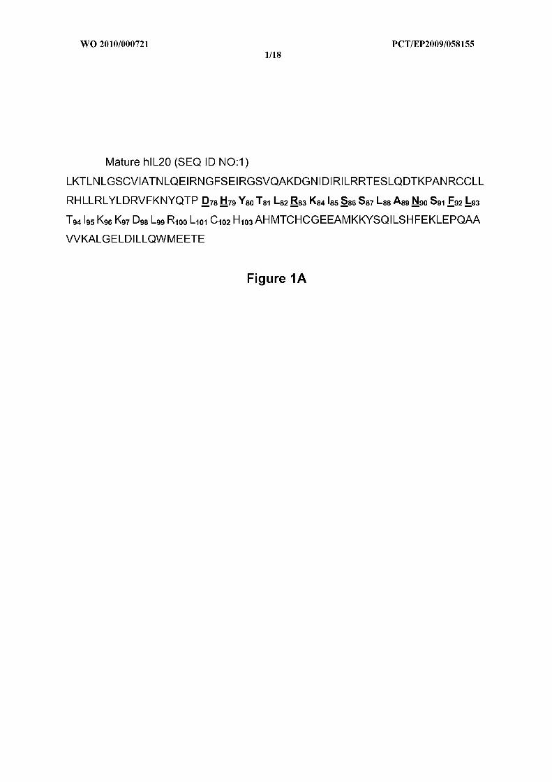
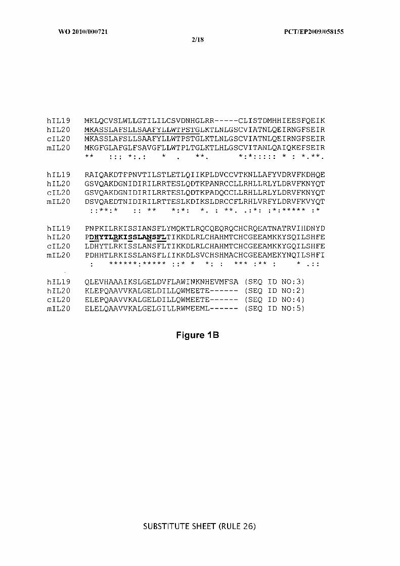
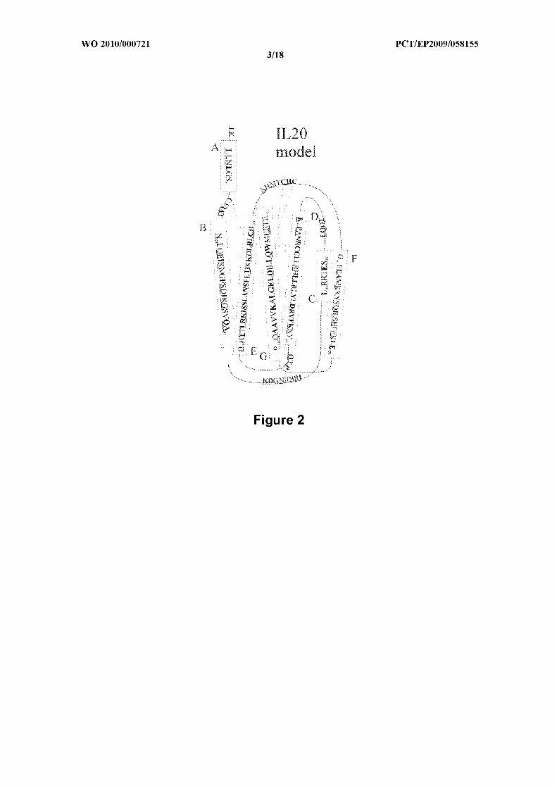
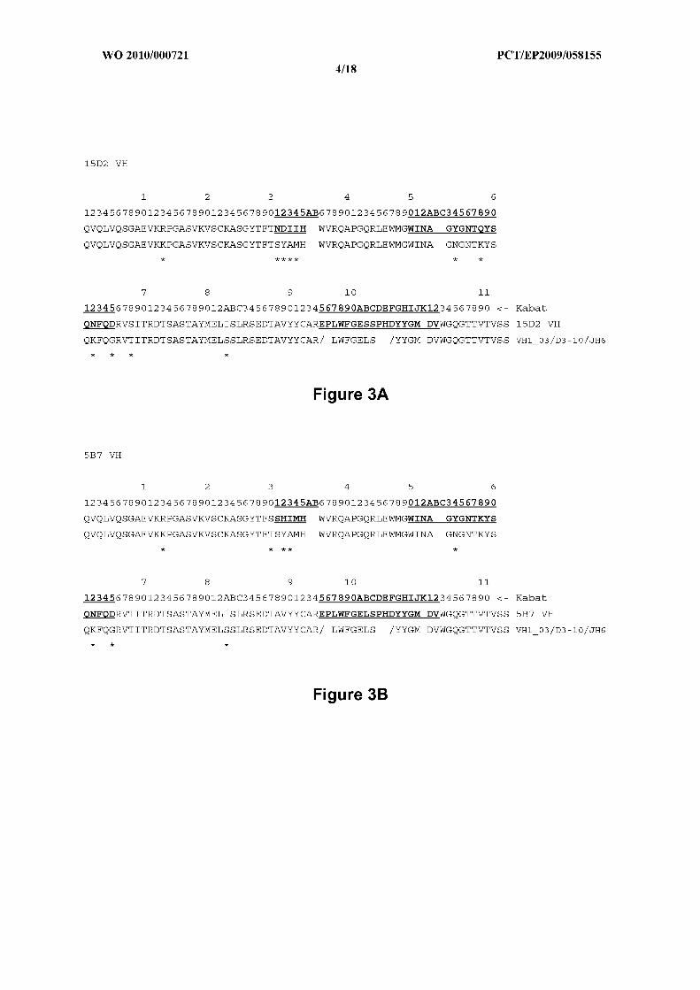
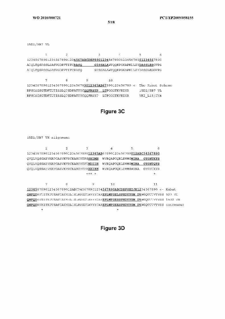
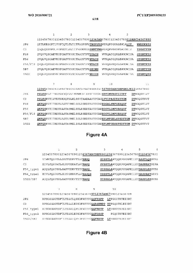

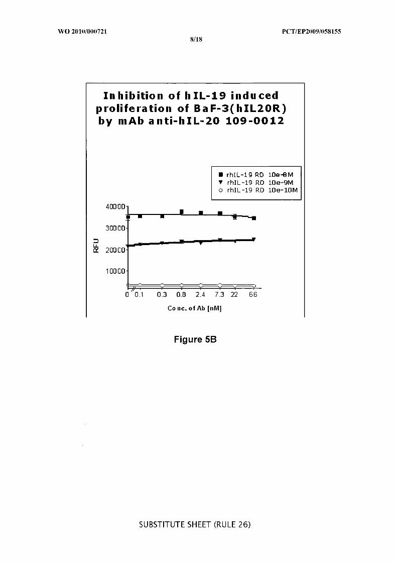
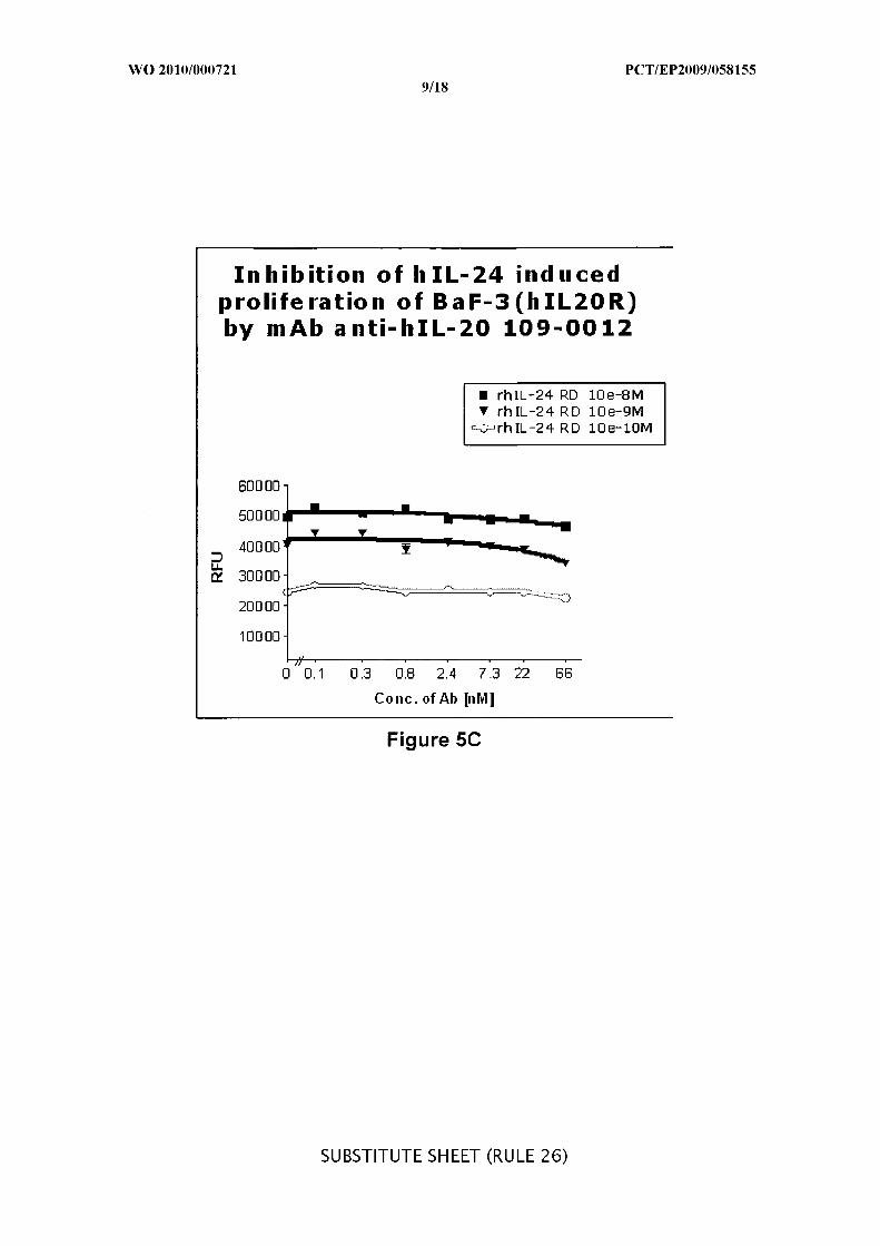
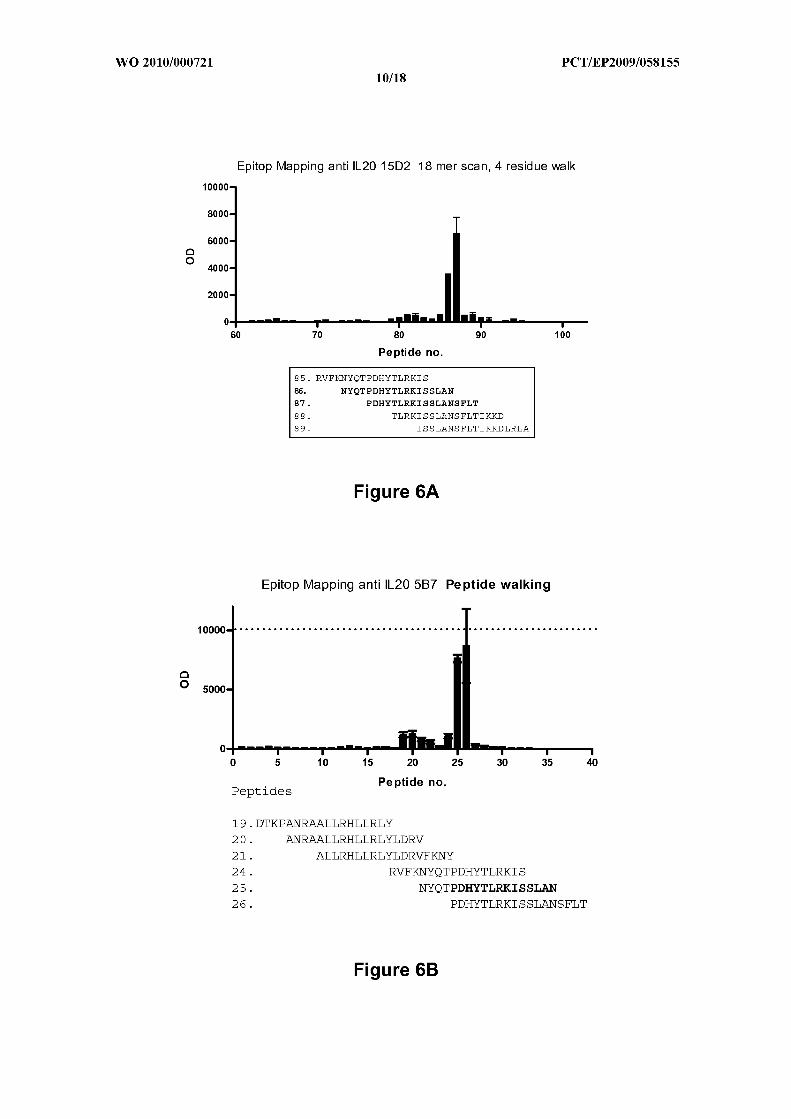
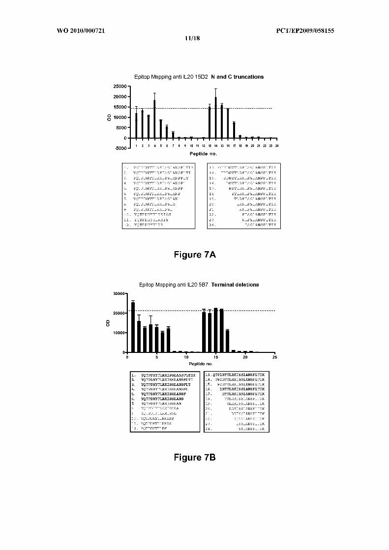
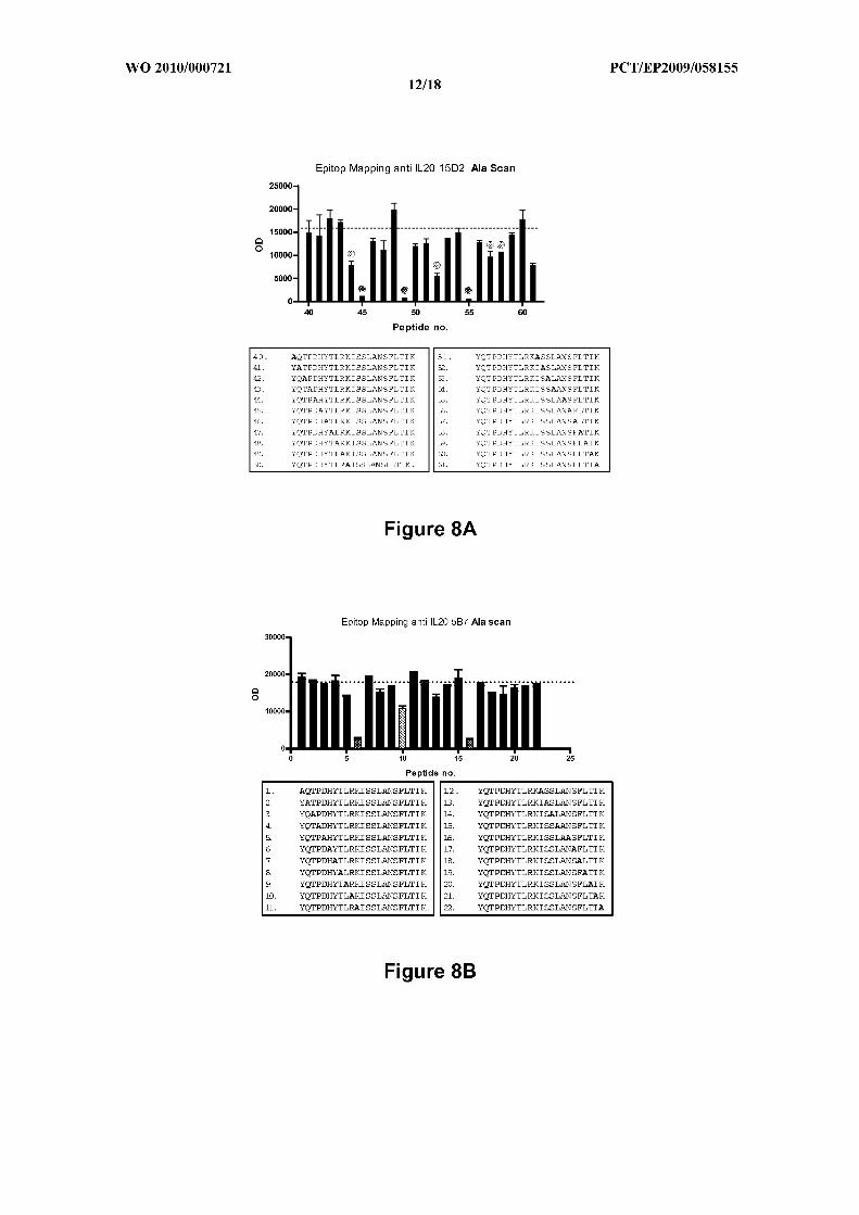
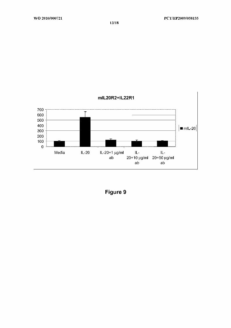
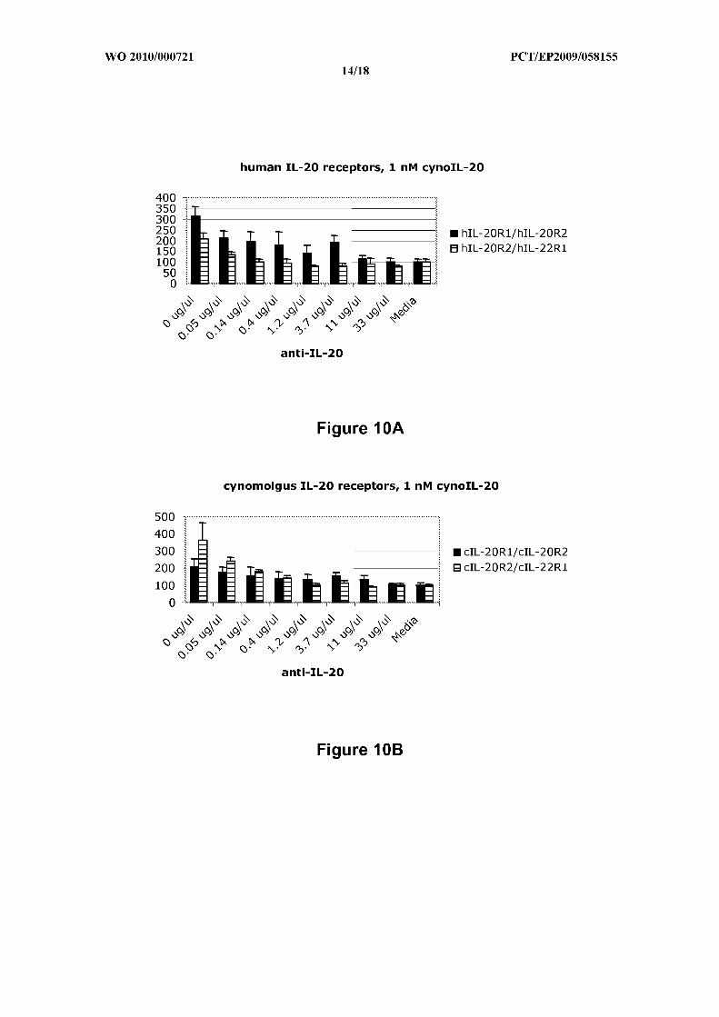
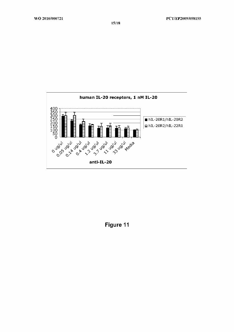
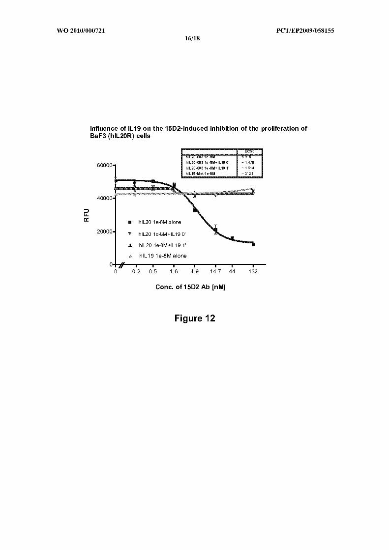
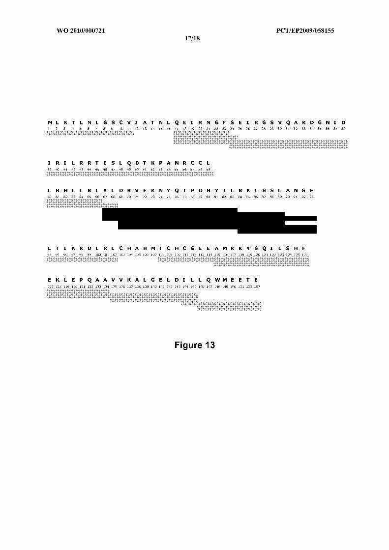
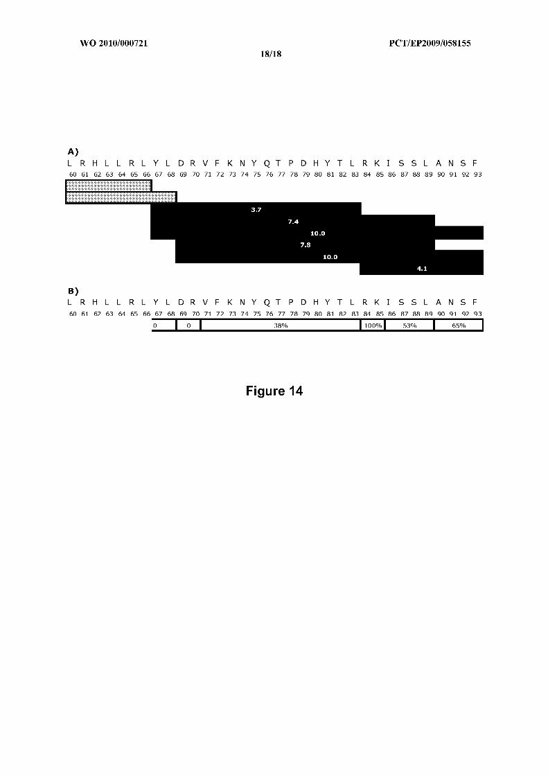
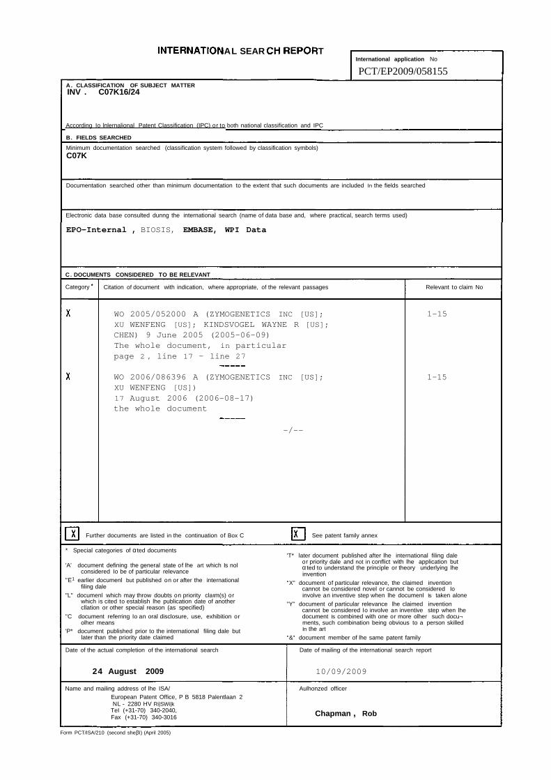
AL SEAR TInternational application No
PCT/EP2009/058155A. CLASSIFICATION OF SUBJECT MATTERINV . C07K16/24
According Io lnlernalional Patent Classification (IPC) or to both national classification and IPC
B. FIELDS SEARCHED
Minimum documentation searched (classification system followed by classification symbols)C07K
Documentation searched other than minimum documentation to the extent that such documents are included In the fields searched
Electronic data base consulted dunng the international search (name of data base and, where practical, search terms used)
EPO-Internal , BIOSIS, EMBASE, WPI Data
C. DOCUMENTS CONSIDERED TO BE RELEVANT
Category Citation of document with indication, where appropriate, of the relevant passages Relevant to claim No
WO 2005/052000 A (ZYMOGENETICS INC [US]; 1-15XU WENFENG [US]; KINDSVOGEL WAYNE R [US];CHEN) 9 June 2005 (2005-06-09)The whole document, in particularpage 2 , line 17 - line 27
WO 2006/086396 A (ZYMOGENETICS INC [US]; 1-15XU WENFENG [US])
17 August 2006 (2006-08-17)the whole document
-/--
Further documents are listed in the continuation of Box C See patent family annex
* Special categories of αted documents'T* later document published after lhe international filing dale
or priority dale and not in conflict with lhe application but'A' document defining the general state of lhe art which Is nol α ted to understand the principle or theory underlying lhe
considered Io be of particular relevance invention"E1 earlier documenl but published on or after the international •X" document of particular relevance, the claimed invention
filing dale cannot be considered novel or cannot be considered Io"L" documenl which may throw doubts on priority claιm(s) or involve an inventive step when lhe documenl Is taken alone
which is cited to establish lhe publication date of another "Y" document of particular relevance lhe claimed inventioncllation or other special reason (as specified) cannot be considered Io involve an inventive step when lhe
"C document referring Io an oral disclosure, use, exhibition or document Is combined with one or more olher such docu¬olher means ments, such combination being obvious to a person skilled
'P* document published prior to the international filing dale but In the art
later than the priority date claimed •&• document member of lhe same patent family
Date of the actual completion of the international search Date of mailing of the international search report
24 August 2009 10/09/2009
Name and mailing address of lhe ISA/ Aulhonzed officerEuropean Patent Office, P B 5818 Palentlaan 2NL - 2280 HV Ri|SWi|k
TeI (+31-70) 340-2040,Fax (+31-70) 340-3016 Chapman , Rob
Form PCT/ISA/210 (second sheβl) (April 2005)
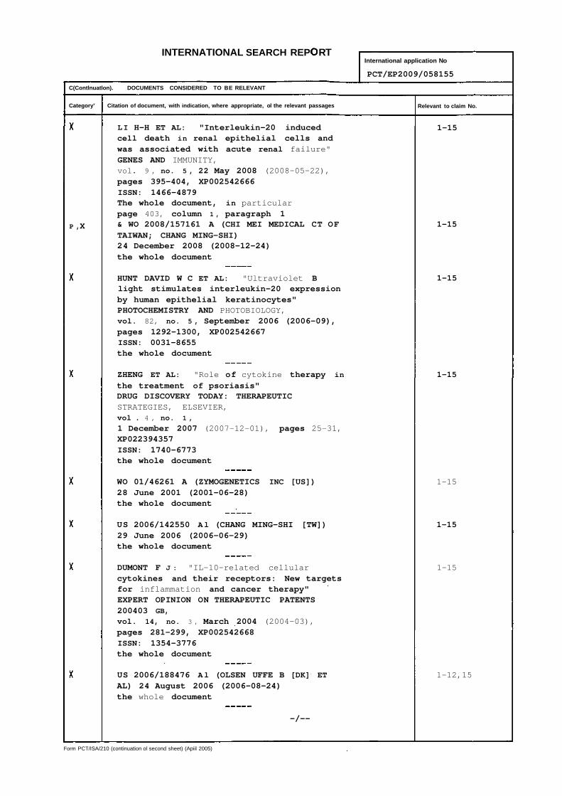
INTERNATIONAL SEARCH REP RTInternational application No
PCT/EP2009/058155
C(Contlnuatlon). DOCUMENTS CONSIDERED TO BE RELEVANT
Category' Citation of document, with indication, where appropriate, ol the relevant passages Relevant to claim No.
LI H-H ET AL: "Interleukin-20 induced 1-15cell death in renal epithelial cells andwas associated with acute renal failure"GENES AND IMMUNITY,vol. 9 , no. 5 , 22 May 2008 (2008-05-22),pages 395-404, XP002542666ISSN: 1466-4879The whole document, in particularpage 403, column 1 , paragraph 1
P ,X & WO 2008/157161 A (CHI MEI MEDICAL CT OF 1-15
TAIWAN; CHANG MING-SHI)24 December 2008 (2008-12-24)the whole document
HUNT DAVID W C ET AL: "Ultraviolet B 1-15light stimulates interleukin-20 expressionby human epithelial keratinocytes"PHOTOCHEMISTRY AND PHOTOBIOLOGY,vol. 82, no. 5 , September 2006 (2006-09),pages 1292-1300, XP002542667ISSN: 0031-8655the whole document
ZHENG ET AL: "Role of cytokine therapy in 1-15
the treatment of psoriasis"DRUG DISCOVERY TODAY: THERAPEUTICSTRATEGIES, ELSEVIER,vol . 4 , no. 1 ,
1 December 2007 (2007-12-01), pages 25-31,XP022394357ISSN: 1740-6773the whole document
WO 01/46261 A (ZYMOGENETICS INC [US]) 1-1528 June 2001 (2001-06-28)the whole document
US 2006/142550 Al (CHANG MING-SHI [TW]) 1-1529 June 2006 (2006-06-29)the whole document
DUMONT F J : "IL-10-related cellular 1-15cytokines and their receptors: New targetsfor inflammation and cancer therapy"EXPERT OPINION ON THERAPEUTIC PATENTS200403 GB,
vol. 14, no. 3 , March 2004 (2004-03),pages 281-299, XP002542668ISSN: 1354-3776the whole document
US 2006/188476 Al (OLSEN UFFE B [DK] ET 1-12,15AL) 24 August 2006 (2006-08-24)the whole document
-/--
Form PCT/ISA/210 (continuation ol second sheet) (Apiil 2005)
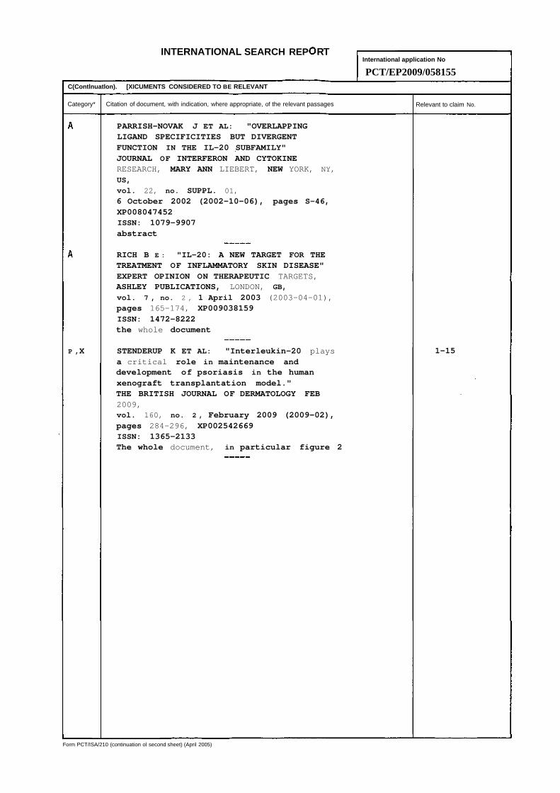
INTERNATIONAL SEARCH REP RTInternational application No
PCT/EP2009/058155C(Contlnuatlon). [XICUMENTS CONSIDERED TO BE RELEVANT
Category* Citation of document, with indication, where appropriate, of the relevant passages Relevant to claim No.
PARRISH-NOVAK J ET AL: "OVERLAPPINGLIGAND SPECIFICITIES BUT DIVERGENTFUNCTION IN THE IL-20 SUBFAMILY"JOURNAL OF INTERFERON AND CYTOKINERESEARCH, MARY ANN LIEBERT, NEW YORK, NY,
US,
vol. 22, no. SUPPL. 01,
6 October 2002 (2002-10-06), pages S-46,
XP008047452ISSN: 1079-9907abstract
RICH B E : "IL-20: A NEW TARGET FOR THETREATMENT OF INFLAMMATORY SKIN DISEASE"EXPERT OPINION ON THERAPEUTIC TARGETS,ASHLEY PUBLICATIONS, LONDON, GB,
vol. 7 , no. 2 , 1 April 2003 (2003-04-01),pages 165-174, XP009038159ISSN: 1472-8222the whole document
P ,X STENDERUP K ET AL: "Interleukin-20 plays 1-15a critical role in maintenance anddevelopment of psoriasis in the humanxenograft transplantation model."THE BRITISH JOURNAL OF DERMATOLOGY FEB2009,vol. 160, no. 2 , February 2009 (2009-02),pages 284-296, XP002542669ISSN: 1365-2133The whole document, in particular figure 2
Form PCT/ISA/210 (continuation ol second sheet) (April 2005)
INTER IONAL EAR TInternational application No
Information on patent family membersPCT/EP2009/058155
Patent document Publication Patent family Publicationcited in search report date member(s) date
WO 2005052000 09-06-2005 CA 2542546 Al 09-06-2005CA 2545867 Al 09-06-2005EP 1692180 A2 23-08-2006EP 1697418 A2 06-09-2006JP 2007535490 T 06-12-2007JP 2007537993 T 27-12-2007UO 2005052001 A2 09-06-2005
WO 2006086396 17-08-2006 AU 2006212807 Al 17-08-2006CA 2596390 Al 17-08-2006EP 1856156 A2 21-11-2007JP 2008532931 T 21-08-2008
WO 2008157161 A 24-12-2008 US 2009048432 Al 19-02-2009US 2008311115 Al 18-12-2008
WO 0146261 A 28-06-2001 AT 339450 T 15-10-2006AU 2458001 A 03-07-2001CA 2395406 Al 28-06-2001DE 60030769 T2 25-10-2007EP 1244708 Al 02-10-2002EP 1743648 Al 17-01-2007JP 2003535037 T 25-11-2003
US 2006142550 Al 29-06-2006 NONE
US 2006188476 Al 24-08-2006 NONE
Form PCT7ISA/210 (palβ famity annβx>(Apnl 2005)