WO 2014/198817 Al
216
(12) INTERNATIONAL APPLICATION PUBLISHED UNDER THE PATENT COOPERATION TREATY (PCT) (19) World Intellectual Property Organization International Bureau (10) International Publication Number (43) International Publication Date 18 December 2014 (18.12.2014) WO 2014/198817 Al PO PCT (51) International Patent Classification: (74) Agent: BIP PATENTS; c/o Bayer Intellectual Property C07K 16/28 (2006.01) A61P 35/00 (2006.01) GmbH, Alfred-Nobel-Str. 10, 40789 Monheim am Rhein A61K 47/48 (2006.01) (DE). (21) International Application Number: (81) Designated States (unless otherwise indicated, for every PCT/EP20 14/062207 kind of national protection available): AE, AG, AL, AM, AO, AT, AU, AZ, BA, BB, BG, BH, BN, BR, BW, BY, (22) International Filing Date: BZ, CA, CH, CL, CN, CO, CR, CU, CZ, DE, DK, DM, 12 June 2014 (12.06.2014) DO, DZ, EC, EE, EG, ES, FI, GB, GD, GE, GH, GM, GT, (25) Filing Language: English HN, HR, HU, ID, IL, IN, IR, IS, JP, KE, KG, KN, KP, KR, KZ, LA, LC, LK, LR, LS, LT, LU, LY, MA, MD, ME, (26) Publication Language: English MG, MK, MN, MW, MX, MY, MZ, NA, NG, NI, NO, NZ, (30) Priority Data: OM, PA, PE, PG, PH, PL, PT, QA, RO, RS, RU, RW, SA, 13 1721 11.0 14 June 2013 (14.06.2013) EP SC, SD, SE, SG, SK, SL, SM, ST, SV, SY, TH, TJ, TM, TN, TR, TT, TZ, UA, UG, US, UZ, VC, VN, ZA, ZM, (71) Applicant: BAYER PHARMA AKTIENGESELL- ZW. SCHAFT [DE/DE]; MullerstraBe 178, 13353 Berlin (DE). (84) Designated States (unless otherwise indicated, for every (72) Inventors: VOTSMEIER, Christian; PellenzstraBe 25, kind of regional protection available): ARIPO (BW, GH, 50823 Koln (DE). HAMMER, Stefanie; Neue Welt 12, GM, KE, LR, LS, MW, MZ, NA, RW, SD, SL, SZ, TZ, 10247 Berlin (DE). GRITZAN, Uwe; SchirmerstraBe 20, UG, ZM, ZW), Eurasian (AM, AZ, BY, KG, KZ, RU, TJ, 50823 Koln (DE). BORKOWSKI, Sandra; Kathestr. 9, TM), European (AL, AT, BE, BG, CH, CY, CZ, DE, DK, 16540 Hohen Neuendorf (DE). ZUBOV, Dmitry; Kipp- EE, ES, FI, FR, GB, GR, HR, HU, IE, IS, IT, LT, LU, LV, dorf 78, 42857 Remscheid (DE). LINDEN, Lars; Bruch- MC, MK, MT, NL, NO, PL, PT, RO, RS, SE, SI, SK, SM, str. 72, 40235 Dusseldorf (DE). CHRISTIAN, Sven; TR), OAPI (BF, BJ, CF, CG, CI, CM, GA, GN, GQ, GW, Scharnhorststrasse 16, 101 15 Berlin (DE). HARRENGA, KM, ML, MR, NE, SN, TD, TG). Axel; Paul-Ehrlich-Str. 10, 421 13 Wuppertal (DE). Declarations under Rule 4.17 : BIRKENFELD, Jorg; Kurfurstenstr. 3, 60486 Frankfurt am Main (DE). FREIBERG, Christoph; Lipkenskothen — as to applicant's entitlement to apply for and be granted a 10, 421 13 Wuppertal (DE). GOLFIER, Sven; Stolpchen- patent (Rule 4.1 7(H)) weg 12E, 14109 Berlin (DE). EICKER, Andrea; Ruckes Published: 101, 41238 Monchengladbach (DE). GREVEN, Simone; Am Schneckenacker 45, 41541 Dormagen (DE). STEL- — with international search report (Art. 21(3)) TE-LUDWIG, Beatrix; Gortzheide lOf, 42489 Wulfrath — before the expiration of the time limit for amending the (DE). RASCHKE, Marian; Eschengraben 11, 13189 Ber claims and to be republished in the event of receipt of lin (DE). amendments (Rule 48.2(h)) — with sequence listingpart of description (Rule 5.2(a)) (54) Title: ANTI- TWEAKR ANTIBODIES AND USES THEREOF (57) Abstract: The present invention provides recombinant antigen-binding regions and antibodies and functional fragments con taining such antigen-binding regions that are specific for the TWEAKR (TNFRSF12A, FN 14). The antibodies, accordingly, can be used to treat tumors and other disorders and conditions associated with expression of the TWEAKR. The invention also provides nucleic acid sequences encoding the foregoing antibodies, vectors containing the same, pharmaceutical compositions and kits with instructions for us.
-
Upload
khangminh22 -
Category
Documents
-
view
0 -
download
0
Transcript of WO 2014/198817 Al

(12) INTERNATIONAL APPLICATION PUBLISHED UNDER THE PATENT COOPERATION TREATY (PCT)
(19) World Intellectual PropertyOrganization
International Bureau(10) International Publication Number
(43) International Publication Date18 December 2014 (18.12.2014)
WO 2014/198817 AlP O P C T
(51) International Patent Classification: (74) Agent: BIP PATENTS; c/o Bayer Intellectual PropertyC07K 16/28 (2006.01) A61P 35/00 (2006.01) GmbH, Alfred-Nobel-Str. 10, 40789 Monheim am RheinA61K 47/48 (2006.01) (DE).
(21) International Application Number: (81) Designated States (unless otherwise indicated, for everyPCT/EP20 14/062207 kind of national protection available): AE, AG, AL, AM,
AO, AT, AU, AZ, BA, BB, BG, BH, BN, BR, BW, BY,(22) International Filing Date: BZ, CA, CH, CL, CN, CO, CR, CU, CZ, DE, DK, DM,
12 June 2014 (12.06.2014) DO, DZ, EC, EE, EG, ES, FI, GB, GD, GE, GH, GM, GT,
(25) Filing Language: English HN, HR, HU, ID, IL, IN, IR, IS, JP, KE, KG, KN, KP, KR,KZ, LA, LC, LK, LR, LS, LT, LU, LY, MA, MD, ME,
(26) Publication Language: English MG, MK, MN, MW, MX, MY, MZ, NA, NG, NI, NO, NZ,
(30) Priority Data: OM, PA, PE, PG, PH, PL, PT, QA, RO, RS, RU, RW, SA,
13 1721 11.0 14 June 2013 (14.06.2013) EP SC, SD, SE, SG, SK, SL, SM, ST, SV, SY, TH, TJ, TM,TN, TR, TT, TZ, UA, UG, US, UZ, VC, VN, ZA, ZM,
(71) Applicant: BAYER PHARMA AKTIENGESELL- ZW.SCHAFT [DE/DE]; MullerstraBe 178, 13353 Berlin (DE).
(84) Designated States (unless otherwise indicated, for every(72) Inventors: VOTSMEIER, Christian; PellenzstraBe 25, kind of regional protection available): ARIPO (BW, GH,
50823 Koln (DE). HAMMER, Stefanie; Neue Welt 12, GM, KE, LR, LS, MW, MZ, NA, RW, SD, SL, SZ, TZ,10247 Berlin (DE). GRITZAN, Uwe; SchirmerstraBe 20, UG, ZM, ZW), Eurasian (AM, AZ, BY, KG, KZ, RU, TJ,50823 Koln (DE). BORKOWSKI, Sandra; Kathestr. 9, TM), European (AL, AT, BE, BG, CH, CY, CZ, DE, DK,16540 Hohen Neuendorf (DE). ZUBOV, Dmitry; Kipp- EE, ES, FI, FR, GB, GR, HR, HU, IE, IS, IT, LT, LU, LV,dorf 78, 42857 Remscheid (DE). LINDEN, Lars; Bruch- MC, MK, MT, NL, NO, PL, PT, RO, RS, SE, SI, SK, SM,str. 72, 40235 Dusseldorf (DE). CHRISTIAN, Sven; TR), OAPI (BF, BJ, CF, CG, CI, CM, GA, GN, GQ, GW,Scharnhorststrasse 16, 101 15 Berlin (DE). HARRENGA, KM, ML, MR, NE, SN, TD, TG).Axel; Paul-Ehrlich-Str. 10, 421 13 Wuppertal (DE).
Declarations under Rule 4.17 :BIRKENFELD, Jorg; Kurfurstenstr. 3, 60486 Frankfurtam Main (DE). FREIBERG, Christoph; Lipkenskothen — as to applicant's entitlement to apply for and be granted a10, 421 13 Wuppertal (DE). GOLFIER, Sven; Stolpchen- patent (Rule 4.1 7(H))
weg 12E, 14109 Berlin (DE). EICKER, Andrea; Ruckes Published:101, 41238 Monchengladbach (DE). GREVEN, Simone;Am Schneckenacker 45, 41541 Dormagen (DE). STEL- — with international search report (Art. 21(3))
TE-LUDWIG, Beatrix; Gortzheide lOf, 42489 Wulfrath — before the expiration of the time limit for amending the(DE). RASCHKE, Marian; Eschengraben 11, 13189 Ber claims and to be republished in the event of receipt oflin (DE). amendments (Rule 48.2(h))
— with sequence listing part of description (Rule 5.2(a))
(54) Title: ANTI- TWEAKR ANTIBODIES AND USES THEREOF
(57) Abstract: The present invention provides recombinant antigen-binding regions and antibodies and functional fragments containing such antigen-binding regions that are specific for the TWEAKR (TNFRSF12A, FN 14). The antibodies, accordingly, can beused to treat tumors and other disorders and conditions associated with expression of the TWEAKR. The invention also providesnucleic acid sequences encoding the foregoing antibodies, vectors containing the same, pharmaceutical compositions and kits withinstructions for us.

Anti-TWEAKR Antibodies and Uses Thereof
The present invention provides recombinant antigen-binding regions and
antibodies and functional fragments containing such antigen-binding regions that are
specific for the TWEAKR (TNFRSF12A, FN14).
The antibodies, accordingly, can be used to treat tumors and other disorders and
conditions associated with expression of the TWEAKR. The invention also provides
nucleic acid sequences encoding the foregoing antibodies, vectors containing the same,
pharmaceutical compositions and kits with instructions for use.
BACKGROUND OF THE INVENTION
Antibody-based therapy is proving very effective in the treatment of various
cancers, including solid tumors. For example, HERCEPTIN® has been used successfully
to treat breast cancer and RITUXAN® is effective in B-cell related cancer types. Central
to the development of a novel successful antibody-based therapy is the isolation of
antibodies against cell-surface proteins found to be preferentially expressed on tumor
cells that are able to functionally modify the activity of the corresponding receptor.
Tumor necrosis factor (TNF) like weak inducer of apoptosis (TWEAK) and the
TWEAK receptor (TWEAKR, alias TNFRSF12A, FN14, CD266; Swiss Prot Acc.
Q9NP84, NP_057723) are a TNF superfamily ligand-receptor pair involved in
inflammation, proliferation, invasion, migration, differentiation, apoptosis and
angiogenesis (Winkles JA, Nat Rev Drug Discov. 2008 May;7(5):411-25; Michaelson JS
and Burkly LC, Results Probl Cell Differ. 2009;49:145-60). TWEAK binds to TWEAKR
with an affinity of 0.8 - 2.4 nM and is the only member of the TNF family that binds this

receptor (Wiley SR et al., Immunity. 2001 Nov;15(5):837-46). The TWEAKR is
expressed at relatively low levels in normal tissues, but is markedly increased locally in
injured tissues, where it has a role in tissue remodeling (Winkles JA, Nat Rev Drug
Discov. 2008 May;7(5):411-25; Zhou et al., Mol Cancer Ther. 2011 Jul;10(7):1276-88;
Burkly LC et al., Immunol Rev. 2011 Nov;244(l):99-l 14). TWEAKR signaling is
involved in processes as wound healing, chronic autoimmune disease and acute ischemic
stroke (Burkly LC et al., Immunol Rev. 2011 Nov;244(l):99-114). In addition, the
TWEAKR is highly expressed in various solid tumor types as for example pancreatic
cancer, non-small-cell-lung-cancer (NSCLC), colorectal cancer (CRC), breast cancer,
renal cancer, head and neck cancer, esophageal cancer, bladder cancer, hepatocellular
carcinoma, ovarian cancer, melanoma as well as liver and bone metastasis (Culp P et al.,
Clin Cancer Res. 2010 Jan 15;16(2):497-508; Zhou H et al., J Invest Dermatol. 2013
Apr;133(4):1052-62). Association of increased TWEAKR expression and higher tumor
grade and/or poor prognosis has been described in brain (Tran NL et al., Cancer Res.
2006 Oct l;66(19):9535-42), breast (Willis AL et al., Mol Cancer Res. 2008
May;6(5):725-34; Wang J et al., Histol Histopathol. 2013 Jan 9 [Epub ahead of print]),
esophageal (Watts GS et al., Int J Cancer. 2007 Nov 15;121(10):2132-9 2007), prostate
(Huang M et al., Carcinogenesis. 2011 Nov;32(ll): 1589-96), gastric (Kwon OH et al.,
Cancer Lett. 2012 Jan 1;314(1):73-81), neuroblastoma (Pettersen I et al., Int J Oncol.
2013 Apr ;42(4): 1239-48) and bladder cancer (Shimada K et al., Clin Cancer Res. 2012
Oct l;18(19):5247-55).
Expression of TWEAKR is induced by growth factors as FGF, PDGF and VEGF
(Winkles JA, Nat Rev Drug Discov. 2008 May;7(5):411-25). In line with this
observation, it has been shown that TWEAKR expression correlates with EGFR
overexpression or activation in NSCLC (Whitsett TG et al., Am J Pathol. 2012
Jul;181(l):l 11-20) and HER2 expression in breast cancer (Wang J et al., Histol

Histopathol. 2013 Jan 9 [Epub ahead of print]; Chao DT et al., J Cancer Res Clin Oncol.
2013 Feb;139(2):315-25).
Activation of the TWEAKR by TWEAK leads to recruitment of TNF-receptor
associated factors (TRAF) to the intracellular binding domain resulting in prolonged NF-
B activation via the canonical and non-canonical NF-κΒ pathway and induction of
cytokine secretion as IL-8 and MCP-1 (reviewed in Michaelson JS and Burkly LC,
Results Probl Cell Differ. 2009;49:145-60). This is well in accordance with the described
pro-inflammatory role of the TWEAK/TWEAKR pathway. However, the signaling
pathways responsible for cell killing via TWEAKR are less clear, as the TWEAKR lacks
a characteristic "death domain". In some tumor cell lines (Kym-1, SKOV-3, OVCAR) it
induces apoptosis through TNF and the recruitment of TRAF2, followed by lysosomal
degradation of the resulting TRAF2-cIAP complex (Nakayama M. et al, J Immunol. 2002
Jan 15;168(2):734-43; Schneider P et al, Eur J Immunol. 1999 Jun;29(6): 1785-92; Vince
JE et al, J Cell Biol. 2008 Jul 14;182(l):171-84). In other cell lines (HSC3, HT-29,
KATO-III) TWEAK induced apoptosis is reported to be TNF independent (Nakayama M
et al, J Immunol. 2003 Jan l;170(l):341-8; Wilson CA et al, Cell Death Differ. 2002
Dec;9(12): 1321-33). In a recent report induction of apoptosis by TWEAK was shown to
be dependent on the stimulation of Stat-1 phosphorylation as treatment with a JAK-
inhibitor abolished the ability of TWEAK to increase caspase3/7 activation in WiDr cells
(Chapman MS et al, Cytokine. 2013 Jan;61(l):210-7).
Several studies validated TWEAKR as an oncologic target. Michaelson et al have
shown that the administration of TWEAK reduces tumor growth in murine xenograft
models (Michaelson JS et al, MAbs. 2011 Jul-Aug;3(4):362-75). This anti-tumor effect
has been imitated by several groups with agonistic anti-TWEAKR antibodies. Potential
drug candidates, namely BIIB0036/P4A8 (Michaelson JS et al, MAbs. 2011 Jul-
Aug;3(4):362-75) and PDL-192, (Culp PA et al, Clin Cancer Res. 2010 Jan 15;16(2):497-

508) have been generated by immunization of mice and subsequent clonal selection and
humanization.
PDL-192 binds to the TWEAKR with a binding affinity of 5.5 nM (Culp PA et
al, Clin Cancer Res. 2010 Jan 15;16(2):497-508) and inhibits the growth of several
TWEAKR expressing cancer cell lines. Yet, in comparison to TWEAK ligand PDL-192
was shown to be less potent in proliferation and apoptosis assays with respect to EC/IC50
and only reached reduced efficacy (V max) of caspase 3/7 activation (Culp PA et al, Clin
Cancer Res. 2010 Jan 15;16(2):497-508). Profiling in a larger panel of breast cancer cell
lines confirmed the only modest anti-proliferative activity of monomeric PDL-192 (Culp
PA et al, Clin Cancer Res. 2010 Jan 15;16(2):497-508; Chao DT et al, J Cancer Res Clin
Oncol. 2013 Feb;139(2):315-25) with only 5 of 27 cell lines responding with >20 of
proliferation inhibition. Anti-proliferative activity of the antibody is slightly enhanced by
cross-linking or immobilization of the antibody. In addition, PDL-192 exhibits ADCC
and the anti-tumor activity described in xenograft models is thought to be a mixture of
ADCC and tumor cell growth inhibition effects (Culp PA et al, Clin Cancer Res. 2010
Jan 15;16(2):497-508). A further limitation of PDL-192 is the lack of species cross-
reactivity, especially mouse and rat, not allowing e.g. assessment of common pre-clinical
studies as toxicological studies.
The second agonistic anti-TWEAKR antibody described as drug candidate,
BIIB036/P4A8 binds to TWEAKR with an affinity of 1.7 nM which is in a similar range
as the endogenous ligand TWEAK (Michaelson JS et al, MAbs. 2011 Jul-Aug;3(4):362-
75). This antibody is shown to induce activation of NF-κΒ and cytokine release in cancer
cells, albeit significantly less efficacious compared to Fc-TWEAK, a hlgGl Fc-fusion of
soluble TWEAK (aa 106-249) with similar activity as recombinant soluble TWEAK
(Michaelson JS et al., Oncogene. 2005 Apr 14;24(16):2613-24). The same holds true in
cell proliferation assays as well as for induction of apoptosis as shown in a TUNEL
staining after treatment of cells with antibodies, where potency of BIIB036/P4A8 is also

significantly decreased compared to Fc-TWEAK. Anti-proliferative activity increases
after multimerization of the antibody, but also the multimerized form is still less
efficacious as compared to recombinant Fc-TWEAK. In contrast, BIIB036/P4A8 is a
potent inducer of ADCC and anti-tumor activity in xenograft models was shown to be
largely dependent on Fc effector function.
Besides both drug candidates several murine antibodies have been described that
would need antibody engineering for humanization to be useful for a human therapy. The
first anti-TWEAKR antibodies with anti-proliferative activity on cancer cells were
antibodies Item 1-4 described by Nakayama et al. (Nakayama M et al, Biochem Biophys
Res Commun. 2003 Jul ll;306(4):819-25). These antibodies, however, harbor only
relatively weak agonistic activity and were shown to act as partial agonists/antagonists
with regard to TWEAK mediated TWEAKR activation. Antibodies 136.1 and 18.3.3
(WO2009/020933) show higher affinity binding compared to TWEAK ligand, which
does not translate in more efficacious caspase activation. Antibodies P3G5 and P2D3
(WO2009/140177) induce cytokine release in cancer cells significantly less efficacious
compared to Fc-TWEAK. To summarize, TWEAKR agonistic activity with regard to
induction of apoptosis and inhibition of proliferation of the anti-TWEAKR antibodies
described in the art is limited and does not reach or exceed the efficacy of the endogenous
ligand TWEAK. This lack of agonistic activity is not due to a decreased affinity as these
antibodies bind to the TWEAKR with affinities in a similar range as compared to the
endogenous ligand TWEAK (Michaelson JS et al, MAbs. 2011 Jul-Aug;3(4):362-75;
Culp PA et al, Clin Cancer Res. 2010 Jan 15;16(2):497-508) and also antibodies with
higher binding affinity do not necessarily exhibit more potent signaling activity (Culp PA
et al, Clin Cancer Res. 2010 Jan 15;16(2):497-508). Anti-tumor activity of the antibodies
described previously is shown to be dependent on Fc effector function and ADCC is
shown to play a significant role for the in vivo efficacy in mouse models. The
contribution of ADCC and in vivo cross linking via Fc-Fc receptor (FcR) interactions to

anti-tumor activity in solid tumors in the clinic, however, is still not clear, given the
challenge of antibody and immune effector cell penetration into solid tumors (Culp PA et
al, Clin Cancer Res. 2010 Jan 15;16(2):497-508). Additionally, patients carrying low-
affinity alleles of FcyRIIIA would exhibit a reduced benefit from the treatment due to
lower Fc-FcR interaction capacity (Varchetta S et al, Cancer Res. 2007 Dec
15;67(24): 11991-9).
Thus, developable human antibodies with strong intrinsic capacity to induce
cancer cell apoptosis and growth inhibition by hyper-activation of the TWEAKR to the
same or even higher extend as compared to the endogenous ligand TWEAK are highly
demanded. As induction of apoptosis and inhibition of proliferation is since many years a
valid concept in inducing anti-tumor response in patients (Hanahan D and Weinberg RA,
Cell. 2000 Jan 7;100(l):57-70; Kim R et al, Cancer Chemother Pharmacol. 2002
Nov;50(5):343-52; Fesik SW, Nat Rev Cancer. 2005 Nov;5(ll):876-855) these
antibodies are expected to show increased anti-tumor activity in solid tumors in human
and are therefore promising drug candidates for the treatment of cancer.
SUMMARY OF THE INVENTION
This invention is related to antibodies, or antigen-binding antibody fragments thereof, or
variants thereof which lead to strong activation of the TWEAKR, thus leading to a strong
induction of apoptosis in various cancer cells showing overexpression of the TWEAKR.
Induction of cancer cell apoptosis by the antibodies described herein is more efficacious
compared to all antibodies described in the art (e.g. PDL-192 or BIIB0036/P4A8; e.g.
require the addition of a cross-linking agent). The unique property of the antibodies of
this invention is based on a novel binding epitope characterized by selective binding of
the antibodies to amino acid at position 47 (D47) of TWEAKR (SEQ ID NO: 169; and see
Figure 1).

The antibodies of the invention are thus suitable for the treatment of cancer as well as
metastases thereof, in particular TWEAKR expressing tumors, such as colorectal cancer,
non-small-cell lung cancer (NSCLC), head and neck cancer, esophageal cancer,
melanoma, hepatocellular carcinoma, bladder cancer, gastric cancer, breast cancer,
pancreatic cancer, renal cell carcinoma, prostate cancer, ovarian cancer and cervical
cancer.
The invention describes antibodies that are distinguished from existing anti-
TWEAKR antibodies in that they induce strong activation of cancer cell apoptosis, at
superior levels as compared to the endogenous ligand TWEAK in most cell lines. The
antibodies of the invention or antigen-binding fragments thereof a) strongly activate the
TWEAKR, b) induce apoptosis in cancer cells, c) induce cytokine secretion from cancer
cells, d) all together resulting in anti-tumor activity of the antibodies in in vivo tumor
experiments, e) additionally the antibodies lead to internalization of the TWEAKR and
inhibition of cancer cell proliferation when incubated with saporine-conjugated secondary
antibodies in experimental conditions where the antibody alone has no effect, f are
crossreactive to several species. These and other objects of the invention are more fully
described herein.
An antibody of the invention might be co-administered with known medicaments,
and in some instances the antibody might itself be modified. For example, an antibody
could be conjugated to a cytotoxic agent, immunotoxin, toxophore or radioisotope to
potentially further increase efficacy.
The invention further provides antibodies which constitute a tool for diagnosis of
malignant or dysplastic conditions in which TWEAKR expression is elevated compared
to normal tissue. Provided are anti-TWEAKR antibodies conjugated to a detectable
marker. Preferred markers are a radiolabel, an enzyme, a chromophore or a fluorescer.
The invention is also related to polynucleotides encoding the antibodies of the
invention or antigen-binding fragments thereof, cells expressing the antibodies of the
invention or antigen-binding fragments thereof, methods for producing the antibodies of
the invention or antigen-binding fragments thereof, methods for inhibiting the growth of
dysplastic cells using the antibodies of the invention or antigen-binding fragments
thereof, and methods for treating and detecting cancer using the antibodies of the
invention or antigen-binding fragments thereof.
The invention is also related to isolated nucleic acid sequences, each of which
can encode an aforementioned antibody or antigen-binding fragment thereof that is
specific for an epitope of TWEAKR. Nucleic acids of the invention are suitable for
recombinant production of antibodies or antigen-binding antibody fragments. Thus, the
invention also relates to vectors and host cells containing a nucleic acid sequence of the
invention.
Compositions of the invention may be used for therapeutic or prophylactic
applications. The invention, therefore, includes a pharmaceutical composition comprising
an inventive antibody or antigen-binding fragment thereof and a pharmaceutically
acceptable carrier or excipient therefore. In a related aspect, the invention provides a
method for treating a disorder or condition associated with the undesired presence of
TWEAKR expressing cells. In a preferred embodiment the aforementioned disorder is
cancer. Such method contains the steps of administering to a subject in need thereof an
effective amount of the pharmaceutical composition that contains an inventive antibody
as described or contemplated herein.
The invention also provides instructions for using an antibody library to isolate
one or more members of such library that binds specifically to TWEAKR.
DESCRIPTION OF THE FIGURES

Figure 1: Alignment of TWEAKR cysteine rich domain (aa 34-68) of different species.
(Numbers indicate amino acid position in full length construct inclusive signal sequence;
SEQ ID NO: 169)
Figure 2 : A - Schematic diagram of the structure of TWEAKR (SEQ ID NO: 169). The
diagram shows the extracellular domain (aa 28-80) (SEQ ID NO: 168) including the
cysteine rich domain (36-67), the transmembrane domain - TM (81-101), and the
intracellular domain (102-129). TPP-2202 - the full ectodomain (28-80) fused to the Fc
domain of hlgGl. TPP-2203 - Extracellular domain with N- and C-terminal truncation
(34-68) fused to the Fc domain of hlgGl. Disulfide bridges Cys36-Cys49, Cys52-Cys67
and Cys55-Cys64 are indicated by black bars. N-terminally, TPP-2203 contains two
amino acids and C-terminally, one amino acid more compared to the pure cysteine rich
domain to ensure proper folding. TPP-1984 - Extracellular domain with C-terminal
truncation (28-68) fused to HIS6 tag. All three constructs show comparable binding to the
antibodies of the invention and PDL-192(TPP-1104). P4A8(TPP-1324) does only bind to
the full extracellular domain (TPP-2202).
B - Amino acid sequence of extracellular domain: aa46 has been published to be essential
for TWEAK ligand binding, aa47 has been characterized to be essential for binding of the
antibodies of this invention.
Figure 3 : Interaction of TWEAKR ectodomain with antibodies of the invention and
reference antibodies. Shown is the result of an ELISA with TWEAKR-Fc fusion protein
(TPP-2202) coating µ /mϊ ) and 0.08 g/ml (open bars) and 0.3 g/ml (filled bars) of
biotinylated IgG as soluble binding partner. Detection was done with Streptavidin-HRP
and Amplex-Red substrate. Y is "ELISA signal intensity [Rfu]"; X are "antibody
constructs tested": a is "TPP-2090"; b is "TPP-2084"; c is "PDL-192(TPP-1104)"; d is
"P4A8(TPP-1324)"; e is "P3G5(TPP-2195)"; f is "136.1(TPP-2194)"; h is "ITEM1"; i is

"ITEM4"; j is a murine isotype control; k is a human isotype control. All tested
antibodies show saturated binding with a concentration of 80 ng/ml.
Figure 4 : Interaction of TWEAKR cysteine rich domain with antibodies of the invention
and reference antibodies. Shown is the result of an ELISA with TWEAKR(34-68)-Fc
fusion protein (TPP-2203) coating µ /mϊ ) and 0.08 g/ml (open bars) and 0.3 g/ml
(filled bars) of biotinylated IgG as soluble binding partner. Detection was done with
Streptavidin-HRP and Amplex-Red substrate. Y is "ELISA signal intensity [Rfu]"; X are
"antibody constructs tested": a is "TPP-2090"; b is "TPP-2084"; c is "PDL-192(TPP-
1104)"; d is "P4A8(TPP-1324)"; e is "P3G5(TPP-2195)"; f is "136.1(TPP-2194)"; h is
"ITEM1"; i is "ITEM4"; j is a murine isotype control; k is a human isotype control. The
antibodies of the invention bind to the cysteine rich domain..
Figure 5 : Interaction of TWEAKR(28-68) with antibodies of the invention and reference
antibodies. Shown is the result of an ELISA with TWEAKR(28-68)-HIS (TPP-1984)
coating µ /mϊ ) and 0.08 g/ml (open bars) and 0.3 g/ml (filled bars) of biotinylated
IgG as soluble binding partner. Detection was done with Streptavidin-HRP and Amplex-
Red substrate. Y is"ELISA signal intensity [Rfu]"; X are "antibody constructs tested": a
is "TPP-2090"; b is "TPP-2084"; c is "PDL-192(TPP-1104)"; d is "P4A8(TPP-1324)"; e
is "P3G5(TPP-2195)"; f is "136.1(TPP-2194)"; h is "ITEM1"; i is "ITEM4"; j is a murine
isotype control; k is a human isotype control. The antibodies of the invention bind to the
cysteine rich domain. Antibodies P4A8(TPP-1324), P3G5(TPP-2195), ITEM-1 and
ITEM-4 show impaired binding.
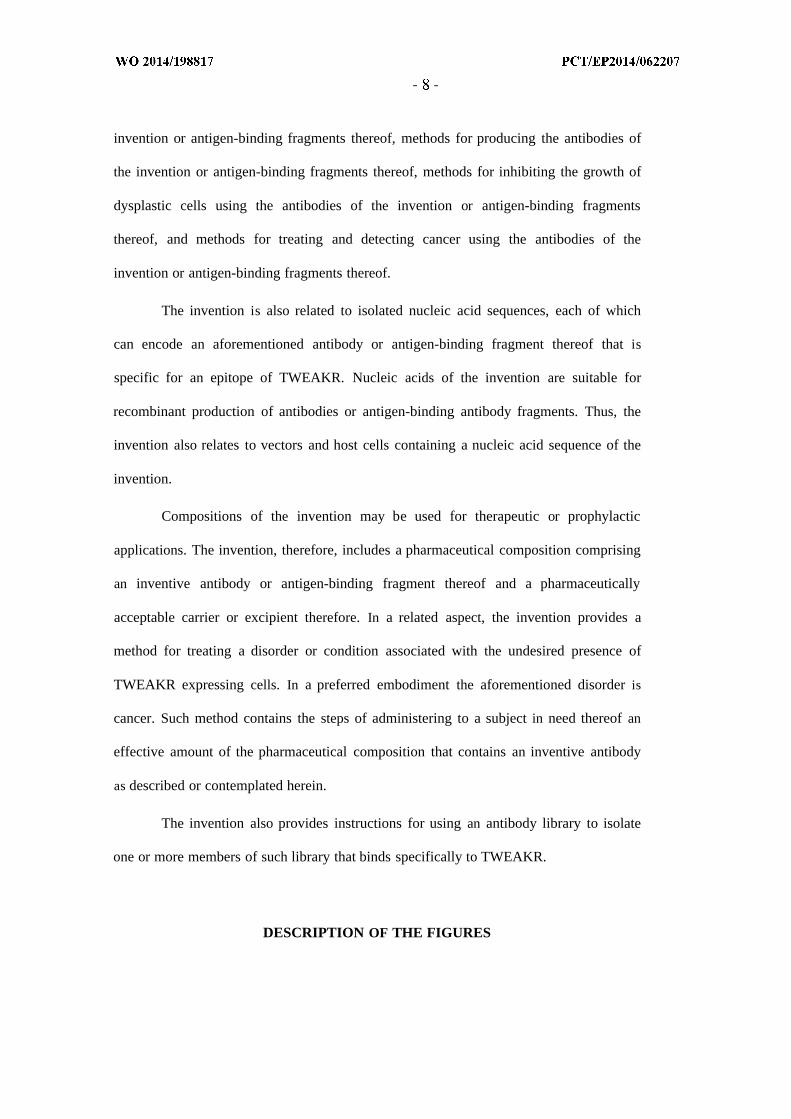
Figure 6 : A - Alanine scan of cysteine rich domain. Muteins of TWEAKR(34-68)-Fc
were analyzed for PDL-192(TPP-1104) (X) and TPP-2090 (Y) binding. S37A, R38A,
S40A, W42A, S43A, D45A, D47A, K48A, D51A, S54A, R56A, R58A, P59A, H60A,
S61A, D62A, F63A and L65A muteins were expressed in HEK293 cells (black
diamonds). PDL-192(TPP-1104) and TPP-2090 were coated ( 1 / ) and an eight-fold
diluted supernatant of the HEK293 fermentation broth was added for TWEAKR mutein
binding. X is "ELISA intensity of PDL-192(TPP-1104) interaction [Rfu]", Y is "ELISA
intensity of TPP-2090 interaction [Rfu]". TPP-2090 (Y) shows impaired binding for the
D47A TWEAKR mutein (closed box) and PDL-192(TPP-1104) (X) shows impaired
binding to R56A (dotted box).
B - Y is " binding normalized by wt binding signal [ ]", 1 is "TPP-2090"; 2 is "PDL-
192(TPP-1104)"; 3 is "P4A8(TPP-1324)". Antibodies were coated ( 1 g/ml), TWEAKR
variant was added at 250 ng/ml, detection via anti-HIS HRP. TTP-2090 shows less than
5% binding compared to the WT construct.
C - Y is „ binding normalized by wt binding signal [ ]", 1 is „TPP-2090"; 2 is "TPP-
2149", 3 is "TPP-2093"; 4 is "TPP-2148"; 5 is "TPP-2084"; 6 is "TPP-2077"; 7 is "TPP-
1538"; 8 is "TPP-883"; 9 is "TPP-1854"; 10 is "TPP-1853"; 11 is "TPP-1857"; 12 is
"TPP-1858"; 13 is "PDL-192(TPP-1104)". Antibodies were coated ( 1 g/ml), TWEAKR
variant was added 250 ng/ml, detection via anti-HIS HRP. All variants despite PDL-192
show less than 5% binding compared to the WT construct.
Figure 7 : NMR structure of TWEAKR ectodomain as published by Pellegrini et al
(FEBS 280:1818-1829). TWEAK binding depends on L46 (Pellegrini et al), TPP-2090
binding on D47 and PDL-192 binding on R56. PDL-192 binds opposite of the TWEAK
ligand binding site, TPP-2090 binds directly to the TWEAK ligand site.
Figure 8 : To differentiate binding epitopes of antibodies of the invention and of reference
antibodies competition experiments were performed. A lack of a second binding event

after injection of the 2nd antibody indicates clear competition within a respective
antibody pair. Non competing antibody pairs showed clear binding signal over
background after 2nd antibody injection. In addition the investigation of self-competition
(1st & 2nd antibody identical) was monitored as an internal system control. (-) no 2nd
binding detected; (+) 2nd binding. The antibodies of the invention compete with all tested
antibodies.
Figure 9 : To differentiate binding epitopes of antibodies of the invention and of reference
antibodies competition experiments were performed. In general all analyzed anti-
TWEAKR antibodies could be clustered into three distinct "competition groups". One
group contains exclusively TPP-2084 and TPP-2090, both showing competition to all
other tested members. These other members could be split into two separate sets of
antibodies, which do not show any competition between each other. Both antibodies of
the invention bind to a new and unique epitope.
Figure 10: Homology tree of all 29 known TNF receptor superfamily members. The
closest homologs TNFRSF13C and TNFRSF17 have only about 30% sequence identity.
Figure 11: Binding ELISA with all 29 TNF receptor superfamily members for
selectivity assessment of TPP-2090. Shown is the result of an ELISA: Y is "ELISA signal
intensity [Rfu]"; X are "TNF receptor superfamily proteins tested (Fc-fusion proteins)": 1
is "TWEAKR"; 2 is "TWEAKR"; 3 is "Apo-3"; 4 is "Trail-Rl"; 5 is "Trail-R2"; 6 is
"CD385"; 7 is "CD95"; 8 is "Rank"; 9 is "TNF-R1"; 10 is "TNF-R2"; 11 is "BAFF-R";
12 is "DcR3"; 13 is "BCMA"; 14 is "TACI"; 15 is "OX40"; 16 is "CD30"; 17 is
"CD27"; 18 is "CD40"; 19 is "Osteoprotegerin"; 20 is "EDAR"; 2 1 is "GITR"; 22 is
"HVEM"; 23 is "NGF R"; 24 is "Trail R3"; 25 is "Lymphotioxin β R"; 26 is "Trail R4";

27 is "EDA2R"; 28 is "TROY"; 29 is "RELT"; 30 is "4-1BB". In (1) 300 pM TPP-2090
were employed, in (2) 75 nM. TPP-2090 binds at a very low concentration of 300 pM (1)
and at a high concentration of 75 nM (2) in saturation to TWEAKR. For binding analysis
to all other TNF receptor superfamily members (3 - 30) 75 nM TPP-2090 were used.
TPP-2090 binds selectively to TWEAKR.
Figure 12: FACS analysis for binding of anti-TWEAKR antibodies to HT-29 cells. Y is
"background corrected Geo-Mean of FACS signal [au]". Shown is the fluorescence after
FACS analysis of HT-29 cells incubated with the antibodies as indicated at 10 g/ml
subtracted by the Geo-Mean of fluorescence of HT-29 cells incubated with the secondary
antibody alone. Antibodies of the invention (TPP-1538, TPP-2084, TPP-2090) show
lower cellular binding at this concentration as compared to known antibodies [PDL-
192(TPP-1104) and P4A8(TPP-1324)].
Figure 13: Caspase 3/7 activation by anti-TWEAKR antibodies in HT-29 cells. X is
"anti-TWEAKR antibodies tested [^g/ml]"; Y is "relative light units [RLU]". HT-29 cells
were incubated with anti-TWEAKR antibodies at different concentrations as indicated
(0.03-300 g/ml) for 24h in the presence of IFNgamma. Caspase 3/7 activity measured as
luminescence by the Caspase 3/7 Glo reagent (Promega) was plotted against the antibody
concentrations. Average values of 1-3 representative experiments performed in triplicates
are shown including standard deviations. Filled symbols show antibodies of the
invention, open symbols known antibodies [PDL-192(TPP-1104); P4A8(TPP-1324),
136.1(TPP-2194) ] . The antibodies of the invention (TPP-1538, TPP-1854, TPP-2084,
TPP-2090) display a stronger efficacy to induce Caspase 3/7 activation compared to the
known antibodies [PDL-192(TPP-1104); P4A8(TPP-1324) and 136.1(TPP-2194)].

Figure 14: Antiproliferative activity of anti-TWEAKR antibodies in WiDr (A) and 786-
O (B) cells. X is "anti-TWEAKR antibodies tested [jag/ml]"; Y is "Cell proliferation
related to proliferation of untreated control cells [ ]". Cells were incubated with anti-
TWEAKR antibodies at different concentrations as indicated (0.03-30C^g/ml) for 96h
(WiDr cells absence, 786-0 cells in the presence of IFN gamma). Average values of a
representative experiment performed in triplicates are shown and standard deviations are
indicated by error bars. Filled symbols: antibodies of the invention, open symbols known
antibodies [PDL-192(TPP-1104) and P4A8(TPP-1324]. The antibodies of the invention
(TPP-1538, TPP-1854, TPP-2084, TPP-2090) display a stronger efficacy to inhibit
cellular proliferation compared to the known antibodies [PDL-192(TPP-1104) and
P4A8(TPP-1324].
Figure 15: IL-8 secretion induced by anti-TWEAKR antibodies in A375 cells. X is "anti-
TWEAKR antibodies tested [jag/ml]"; Y is "IL-8 levels [pg/ml]". A375 cells were
incubated with anti-TWEAKR antibodies at different concentrations as indicated (0.03-
300 g/ml). Levels of IL-8 were determined in the supernatant of the cells after 24h
treatment (and plotted against the used antibody concentrations. Average values of 1-3
representative experiments performed in triplicates are shown including standard
deviations. Filled symbols show antibodies of the invention, open symbols known
antibodies [PDL-192(TPP-1104); P4A8(TPP-1324), 136.1(TPP-2194)], and treatment
with an isotype control antibody is indicated (C). The antibodies of the invention (TPP-
1538, TPP-1854, TPP-2084, TPP-2090) display a stronger efficacy to induce IL-8
secretion from A375 cells compared to the known antibodies [PDL-192(TPP-1104),
P4A8(TPP-1324), 136.1(TPP-2194)]..
Figure 16: Human IL-8 secretion induced by anti-TWEAKR antibodies in xenografts in
mice.

A : WiDr xenograft tumor bearing mice were treated with a single dose of 3 mg/kg TPP-
2090 (open symbols) or vehicle (C - filled symbols) and levels of human IL-8 (IL-8
pg/ml) determined at different time points after treatment in the plasma of tumor bearing
mice. X is "hours after treatment [h]"; Y is "11-8 level [pg/ml]". Results from 3 animals
per group are indicated, error bars represent standard deviations. Human IL-8 secretion is
specifically induced after treatment with TPP-2090 in WiDr tumor bearing mice in a time
dependent manner.
B : A375 tumor bearing (filled symbols) or non-tumor bearing (open symbols) mice were
treated with a single dose of 10 mg/kg TPP-1538, vehicle or an isotpye control antibody .
C I is "vehicle control"; C2 is "isotype control antibody"; Y is "Level of human 11-8
[pg/ml]". Levels of human IL-8 were determined in the serum of 4 mice per group 7h
after treatment are shown. IL-8 secretion is specifically induced in A375 tumor bearing
mice by TPP-1538 but not in equally treated tumor free animals.
Figure 17: Microscopic evaluation of the time course of specific internalization of
TWEAKR upon antibody binding to endogenous TWEAKR expressing cells (InCell
Analyzer). Internalization of TPP-1538 and TPP-2090 was investigated on renal cancer
cell line 786-0. Granule count/cell after treatment with antibodies of the invention (at
/µ / ϊ ) or isotype control C- at 5 g/ml) is plotted for different incubation times as
indicated (X is "time [min]"; Y is "granule count/cell [quantity]"). Antibodies of the
invention (TPP-1538, TPP-2090) show rapid and specific internalization in TWEAKR
expressing cells.
Figure 18: Inhibition of 786-0 cell proliferation by anti-TWEAKR antibodies after
incubation with saporine -conjugated secondary antibodies (Hum-Zap Assay). 786-0 cells
were incubated with TWEAKR or isotype control antibodies in the presence or absence
of saporine -conjugated secondary antibodies at ΙΟηΜ antibody concentration for 48h (in

the absence of IFN gamma). X is "antibody variant tested", a is "vehicle control", b is
"isotype control antibody", c is "TPP-2084", d is "TPP-2090"; Y is "cell proliferation
compared to untreated control cells [ ]" . Cell proliferation compared to untreated control
cells was plotted for 786-0 cells treated with different antibodies in the presence (open
bars) or absence (filled bars) of saporine-conjugated secondary antibodies. Results from
one representative experiment in triplicates are shown and standard deviations indicated
by error bars. At the experimental conditions used only antibodies of the invention (TPP-
2084, TPP-2090) in the presence of saporine-coupled secondary antibodies inhibit
proliferation of 786-0 cells almost completely. Thus, the anti-proliferative effect
observed from the anti-TWEAKR antibodies in the presence of saporine-conjugated
secondary antibodies is a result of specific internalization of the saporine after binding of
the antibody-complexes to TWEAKR expressing cells.
Figure 19: Efficacy of anti-TWEAKR antibodies in the human renal cell cancer
xenograft 786-0 after treatment with 0.3, 1.0 and 3.0 mg/kg (i.v., q4dx3) started at day 7
after tumor cell inoculation. Shown are final tumor weights at day 40. A is "Vehicle
group, treated with PBS (i.v. q4dx3)". B is "Isotype, 3 mg/kg", C is "TPP-2084, 0.3
mg/kg", D is "TPP-2084, 1 mg/kg", E is "TPP-2084, 3 mg/kg", F is "TPP-2090, 0.3
mg/kg", G is "TPP-2090, 1 mg/kg", H is "TPP-2090, 3 mg/kg". (Y is "Tumor weights
means ofn=8; SD [g]").
Figure 20: Efficacy of 3mg/kg TPP-2090 (i.v., q4dx7) in the human colon cancer
xenograft WiDr in monotherapy and combination therapy with Irinotecan (5mg/kg, i.v.,
4d on, 3d off) and Regorafenib (lOmg/kg, p.o., daily). Treatment started 7d after
inoculation with established tumors of about 40mm2. A is "Vehicle group, treated with
PBS (i.v. q4dx7)". B is "TPP-2090, 3 mg/kg", C is "TPP-2090, 10 mg/kg", D is
"Irinotecan, 5 mg/kg", E is "Combo TPP-2090 3 mg/kg + Irinotecan, 5 mg/kg", F is

"Regorafenib, 10 mg/kg", G is "Combo TPP-2090, 3mg/kg + Regorafenib 10 mg/kg". (X
is "Time after inoculation [days]", Y is "Tumor area, means of n=10; SD [mm2])
Figure 21: Efficacy of lOmg/kg TPP-2090 (i.v., q4dx8) in the human lung cancer
xenograft NCI-H322 in monotherapy and combination therapy with Paclitaxel (16mg/kg,
i.v., q7dx4). Treatment started 14d after inoculation with established tumors of about
45mm2. A is "Vehicle group, treated with PBS (i.v. q4dx8)". B is "TPP-2090, 5mg/kg", C
is "TPP-2090, lOmg/kg", D is "Paclitaxel, 16 mg/kg", E is "Combo TPP-2090 10 mg/kg
+ Paclitaxel 16 mg/kg". (X is "Time after inoculation [days]"; Y is "Tumor area , means
of n=10; SD [mm2]")
Figure 22: Reduction of proliferative cells in xenografts after treatment with antibodies
of the invention. Cryo sections from WiDr xenograft tumors after treatment with PBS
(i.v., q4dx7: A) or TPP-2090 (10 mg/kg , i.v. q4dx7:B) were stained for the proliferation
marker Ki67 by immunohistochemistry. Treatment started at day 7 after tumor cell
inoculation and cryo sections were prepared from tumors taken at the end of the study
(day 29). N=3 tumors per group were analyzed and representative images are shown.
Treatment with TPP-2090 leads to a strong reduction of Ki67 positive cells (cells with
dark staining in image) in WiDr xenograft tumors in mice.
Figure 23: Induction of Stat-1 and NF-kappaB2 signaling pathways by anti-TWEAKR
antibodies in vivo. Lysates of snap frozen WiDr xenograft tumors after treatment with
PBS (i.v., q4dx7: lanes 1&2) or TPP-2090 (3mg/kg, i.v., q4dx7: lanes 3&4) were
subjected to Western Blot analysis detected with specific antibodies for P-Statl (a), Stat-1
(b), NF-kappa2 - p52 (c) and GAPDH (d). Treatment of mice started at day 7 after tumor
cell inoculation and lysates were prepared from snap frozen tumors taken at the end of the
study (day 29). Blots from 2 representative animals per group are shown. Treatment with

TPP-2090 leads to a strong induction of P-Statl & Total Statl levels as well as NF-
kappaB2 activation (shown by the appearance of the p52 band) in WiDr xenograft
tumors.
Figure 24: Consensus sequences for anti-TWEAKR antibodies. CDR-H1 - X at position
5 : M or I ; CDR-H2 - X at position 8 : S or K; CDR-L1 - X at position 8 : G or S; CDR-L2
- X at position 1: N, A or Q; CDR-L3 - X at position 5 : T or S; X at position 6 : S or T; X
at position 8 : F or G
Figure 25: Continuous CDR sequence nomenclature.(A) Positions in boxes were
diversified for mutation gathering (maturation process). (B) Single substitutions in boxes
were recombined in one recombination library.
Figure 26: Sequences of the invention
DETAILED DESCRIPTION OF THE INVENTION
The present invention is based on the discovery of novel antibodies that have a specific
affinity for TWEAKR and can deliver a therapeutic benefit to a subject. The antibodies of
the invention, which may be human, humanized or chimeric, can be used in many
contexts, which are more fully described herein.
Definitions
Unless defined otherwise, all technical and scientific terms used herein have the
meaning commonly understood by one of ordinary skill in the art to which this invention
belongs. The following references, however, can provide one of skill in the art to which

this invention pertains with a general definition of many of the terms used in this
invention, and can be referenced and used so long as such definitions are consistent with
the meaning commonly understood in the art. Such references include, but are not limited
to, Singleton et al, Dictionary of Microbiology and Molecular Biology (2d ed. 1994); The
Cambridge Dictionary of Science and Technology (Walker ed., 1988); Hale & Marham,
The Harper Collins Dictionary of Biology (1991); and Lackie et al., The Dictionary of
Cell & Molecular Biology (3d ed. 1999); and Cellular and Molecular Immunology, Eds.
Abbas, Lichtman and Pober, 2nd Edition, W.B. Saunders Company. Any additional
technical resource available to the person of ordinary skill in the art providing definitions
of terms used herein having the meaning commonly understood in the art can be
consulted. For the purposes of the present invention, the following terms are further
defined. Additional terms are defined elsewhere in the description. As used herein and in
the appended claims, the singular forms "a," and "the" include plural reference unless the
context clearly dictates otherwise. Thus, for example, reference to "a gene" is a reference
to one or more genes and includes equivalents thereof known to those skilled in the art,
and so forth.
The terms "polypeptide" and "protein" are used interchangeably herein to refer to
a polymer of amino acid residues. The terms apply to amino acid polymers in which one
or more amino acid residue is an artificial chemical mimetic of a corresponding naturally
occurring amino acid, as well as to naturally occurring amino acid polymers and non-
naturally occurring amino acid polymer. Unless otherwise indicated, a particular
polypeptide sequence also implicitly encompasses conservatively modified variants
thereof.
A "human" antibody or antigen-binding fragment thereof is hereby defined as
one that is not chimeric (e.g., not "humanized") and not from (either in whole or in part) a
non-human species. A human antibody or antigen-binding fragment thereof can be

derived from a human or can be a synthetic human antibody. A "synthetic human
antibody" is defined herein as an antibody having a sequence derived, in whole or in part,
in silico from synthetic sequences that are based on the analysis of known human
antibody sequences. In silico design of a human antibody sequence or fragment thereof
can be achieved, for example, by analyzing a database of human antibody or antibody
fragment sequences and devising a polypeptide sequence utilizing the data obtained there
from. Another example of a human antibody or antigen-binding fragment thereof is one
that is encoded by a nucleic acid isolated from a library of antibody sequences of human
origin (e.g., such library being based on antibodies taken from a human natural source).
Examples of human antibodies include antibodies as described in Soderlind et al., Nature
Biotech. 2000, 18:853-856.
A "humanized antibody" or humanized antigen-binding fragment thereof is
defined herein as one that is (i) derived from a non-human source (e.g., a transgenic
mouse which bears a heterologous immune system), which antibody is based on a human
germline sequence; (ii) where amino acids of the framework regions of a non-human
antibody are partially exchanged to human amino acid sequences by genetic engineering
or (iii) CDR-grafted, wherein the CDRs of the variable domain are from a non-human
origin, while one or more frameworks of the variable domain are of human origin and the
constant domain (if any) is of human origin.
A "chimeric antibody" or antigen-binding fragment thereof is defined herein as
one, wherein the variable domains are derived from a non-human origin and some or all
constant domains are derived from a human origin.
The term "monoclonal antibody" as used herein refers to an antibody obtained
from a population of substantially homogeneous antibodies, i.e., the individual antibodies
comprising the population are identical except for possible mutations, e.g., naturally
occurring mutations, that may be present in minor amounts. Thus, the term "monoclonal"
indicates the character of the antibody as not being a mixture of discrete antibodies. In

contrast to polyclonal antibody preparations, which typically include different antibodies
directed against different determinants (epitopes), each monoclonal antibody of a
monoclonal antibody preparation is directed against a single determinant on an antigen. In
addition to their specificity, monoclonal antibody preparations are advantageous in that
they are typically uncontaminated by other immunoglobulins. The term "monoclonal" is
not to be construed as to require production of the antibody by any particular method. The
term monoclonal antibody specifically includes chimeric, humanized and human
antibodies. An "agonist/agonistic antibody" as used herein is an antibody which mimics at
least one of the functional activities of a polypeptide of interest (here the TWEAKR
ligand TWEAK).
As used herein, an antibody "binds specifically to", is "specific to/for" or
"specifically recognizes" an antigen of interest, e.g. a tumor-associated polypeptide
antigen target (here, TWEAKR), is one that binds the antigen with sufficient affinity such
that the antibody is useful as a therapeutic agent in targeting a cell or tissue expressing the
antigen, and does not significantly cross-react with other proteins or does not significantly
cross-react with proteins other than orthologs and variants (e.g. mutant forms, splice
variants, or proteolytically truncated forms) of the aforementioned antigen target. The
term "specifically recognizes" or "binds specifically to" or is "specific to/for" a particular
polypeptide or an epitope on a particular polypeptide target as used herein can be
exhibited, for example, by an antibody, or antigen-binding fragment thereof, having a
monovalent K D for the antigen of less than about 10 4 M, alternatively less than about 10 5
M, alternatively less than about 10 6 M, alternatively less than about 10 7 M, alternatively
less than about 10 8 M, alternatively less than about 10 9 M, alternatively less than about
10 10 M, alternatively less than about 10 11 M, alternatively less than about 10 12 M, or
less. An antibody "binds specifically to," is "specific to/for" or "specifically recognizes"
an antigen if such antibody is able to discriminate between such antigen and one or more
reference antigen(s). In its most general form, "specific binding", "binds specifically to",

is "specific to/for" or "specifically recognizes" is referring to the ability of the antibody to
discriminate between the antigen of interest and an unrelated antigen, as determined, for
example, in accordance with one of the following methods. Such methods comprise, but
are not limited to Western blots, ELISA-, RIA-, ECL-, IRMA-tests and peptide scans. For
example, a standard ELISA assay can be carried out. The scoring may be carried out by
standard color development (e.g. secondary antibody with horseradish peroxidase and
tetramethyl benzidine with hydrogen peroxide). The reaction in certain wells is scored by
the optical density, for example, at 450 nm. Typical background (=negative reaction) may
be 0.1 OD; typical positive reaction may be 1 OD. This means the difference
positive/negative is more than 5-fold, 10-fold, 50-fold, and preferably more than 100-
fold. Typically, determination of binding specificity is performed by using not a single
reference antigen, but a set of about three to five unrelated antigens, such as milk powder,
BSA, transferrin or the like.
"Binding affinity" refers to the strength of the total sum of non-covalent
interactions between a single binding site of a molecule and its binding partner. Unless
indicated otherwise, as used herein, "binding affinity" refers to intrinsic binding affinity
which reflects a 1 : 1 interaction between members of a binding pair (e.g. an antibody and
an antigen). The dissociation constant "K D" is commonly used to describe the affinity
between a molecule (such as an antibody) and its binding partner (such as an antigen) i.e.
how tightly a ligand binds to a particular protein. Ligand-protein affinities are influenced
by non-covalent intermolecular interactions between the two molecules. Affinity can be
measured by common methods known in the art, including those described herein. In one
embodiment, the "K D" or "K D value" according to this invention is measured by using
surface plasmon resonance assays using a Biacore T100 instrument (GE Healthcare
Biacore, Inc.) according to Example 2. Other suitable devices are BIACORE T200,
BIACORE(R)-2000, BIACORe 4000, a BIACORE (R)-3000 (BIAcore, Inc., Piscataway,
NJ), or ProteOn XPR36 instrument (Bio-Rad Laboratories, Inc.).
The term "antibody", as used herein, is intended to refer to immunglobulin
molecules, preferably comprised of four polypeptide chains, two heavy (H) chains and
two light (L) chains which are typically inter-connected by disulfide bonds. Each heavy
chain is comprised of a heavy chain variable region (abbreviated herein as VH) and a
heavy chain constant region. The heavy chain constant region can comprise e.g. three
domains CHI, CH2 and CH3. Each light chain is comprised of a light chain variable
region (abbreviated herein as VL) and a light chain constant region. The light chain
constant region is comprised of one domain (CL). The VH and VL regions can be further
subdivided into regions of hypervariability, termed complementarity determining regions
(CDR), interspersed with regions that are more conserved, termed framework regions
(FR). Each VH and VL is typically composed of three CDRs and up to four FRs.
arranged from amino terminus to carboxy-terminus e.g. in the following order: FR1,
CDR1, FR2, CDR2, FR3, CDR3, FR4.
As used herein, the term "Complementarity Determining Regions (CDRs; e.g.,
CDR1, CDR2, and CDR3) refers to the amino acid residues of an antibody variable
domain the presence of which are necessary for antigen binding. Each variable domain
typically has three CDR regions identified as CDR1, CDR2 and CDR3. Each
complementarity determining region may comprise amino acid residues from a
"complementarity determining region" as defined by Kabat (e.g. about residues 24-34
(LI), 50-56 (L2) and 89-97 (L3) in the light chain variable domain and 31-35 (HI), 50-65
(H2) and 95-102 (H3) in the heavy chain variable domain; (Kabat et al., Sequences of
Proteins of Immulological Interest, 5th Ed. Public Health Service, National Institutes of
Health, Bethesda, MD. (1991)) and/or those residues from a "hypervariable loop" (e.g.
about residues 26-32 (LI), 50-52 (L2) and 91-96 (L3) in the light chain variable domain
and 26- 32 (HI), 53-55 (H2) and 96-101 (H3) in the heavy chain variable domain
(Chothia and Lesk; J Mol Biol 196: 901-917 (1987)). In some instances, a

complementarity determining region can include amino acids from both a CDR region
defined according to Kabat and a hypervariable loop.
Depending on the amino acid sequence of the constant domain of their heavy
chains, intact antibodies can be assigned to different "classes". There are five major
classes of intact antibodies: IgA, IgD, IgE, IgG, and IgM, and several of these maybe
further divided into "subclasses" (isotypes), e.g., IgGl, IgG2, IgG3, IgG4, IgA, and IgA2.
The heavy-chain constant domains that correspond to the different classes of antibodies
are called [alpha], [delta], [epsilon], [gamma], and [mu], respectively. The subunit
structures and three-dimensional configurations of different classes of immunglobulins
are well known. As used herein antibodies are conventionally known antibodies and
functional fragments thereof.
A "functional fragment" or "antigen-binding antibody fragment" of an
antibody/immunoglobulin hereby is defined as a fragment of an
antibody/immunoglobulin (e.g., a variable region of an IgG) that retains the antigen-
binding region. An "antigen-binding region" of an antibody typically is found in one or
more hyper variable region(s) of an antibody, e.g., the CDR1, -2, and/or - 3 regions;
however, the variable "framework" regions can also play an important role in antigen
binding, such as by providing a scaffold for the CDRs. Preferably, the "antigen-binding
region" comprises at least amino acid residues 4 to 103 of the variable light (VL) chain
and 5 to 109 of the variable heavy (VH) chain, more preferably amino acid residues 3 to
107 of VL and 4 to 111 of VH, and particularly preferred are the complete VL and VH
chains (amino acid positions 1 to 109 of VL and 1 to 113 of VH; numbering according to
WO 97/08320). A preferred class of immunoglobulins for use in the present invention is
IgG.
"Functional fragments" or "antigen-binding antibody fragments" of the invention
include Fab, Fab', F(ab')2, and Fv fragments; diabodies; single domain antibodies (DAbs),
linear antibodies; single-chain antibody molecules (scFv); and multispecific, such as bi-

and tri-specific, antibodies formed from antibody fragments (C. A. K Borrebaeck, editor
(1995) Antibody Engineering (Breakthroughs in Molecular Biology), Oxford University
Press; R. Kontermann & S. Duebel, editors (2001) Antibody Engineering (Springer
Laboratory Manual), Springer Verlag). An antibody other than a "multi-specific" or
"multi-functional" antibody is understood to have each of its binding sites identical. The
F(ab') 2 or Fab may be engineered to minimize or completely remove the intermolecular
disulphide interactions that occur between the C HI and C L domains.
The term "Fc region" herein is used to define a C-terminal region of an
immunoglobulin heavy chain that contains at least a portion of the constant region. The
term includes native sequence Fc regions and variant Fc regions. In one embodiment, a
human IgG heavy chain Fc region extends from Cys226, or from Pro230, to the carboxyl-
terminus of the heavy chain. However, the C-terminal lysine (Lys447) of the Fc region
may or may not be present. Unless otherwise specified herein, numbering of amino acid
residues in the Fc region or constant region is according to the EU numbering system,
also called the EU index, as described in Kabat et al., Sequences of Proteins of
Immunological Interest, 5th Ed. Public Health Service, National Institutes of Health,
Bethesda, MD, 1991
Variants of the antibodies or antigen-binding antibody fragments contemplated in
the invention are molecules in which the binding activity of the antibody or antigen-
binding antibody fragment for TWEAKR is maintained.
Binding proteins contemplated in the invention are for example antibody
mimetics, such as Affibodies, Adnectins, Anticalins, DARPins, Avimers, Nanobodies
(reviewed by Gebauer M. et al., Curr. Opinion in Chem. Biol. 2009; 13:245-255; Nuttall
S.D. et al., Curr. Opinion in Pharmacology 2008; 8:608-617).
As used herein, the term "epitope" includes any protein determinant capable of
specific binding to an immunoglobulin or T-cell receptors. Epitopic determinants usually
consist of chemically active surface groupings of molecules such as amino acids or sugar

side chains, or combinations thereof and usually have specific three dimensional
structural characteristics, as well as specific charge characteristics.
An "isolated" antibody is one that has been identified and separated from a
component of the cell that expressed it. Contaminant components of the cell are materials
that would interfere with diagnostic or therapeutic uses of the antibody, and may include
enzymes, hormones, and other proteinaceous or nonproteinaceous solutes. In preferred
embodiments, the antibody is purified (1) to greater than 95% by weight of antibody as
determined e.g. by the Lowry method, UV-Vis spectroscopy or by by SDS-Capillary Gel
electrophoresis (for example on a Caliper LabChip GXII, GX 90 or Biorad Bioanalyzer
device), and in further preferred embodiments more than 99% by weight, (2) to a degree
sufficient to obtain at least 15 residues of N-terminal or internal amino acid sequence, or
(3) to homogeneity by SDS-PAGE under reducing or nonreducing conditions using
Coomassie blue or, preferably, silver stain. Isolated naturally occurring antibody includes
the antibody in situ within recombinant cells since at least one component of the
antibody's natural environment will not be present. Ordinarily, however, isolated antibody
will be prepared by at least one purification step.
"Antibody-dependent cell-mediated cytotoxicity" or "ADCC" refers to a form of
cytotoxicity in which secreted Ig bound onto Fc gamma receptors (FcyRs) present on
certain cytotoxic cells (e.g. NK cells, neutrophils, and macrophages) enable these
cytotoxic effector cells to bind specifically to an antigen-bearing target cell and
subsequently kill the target cell e.g. with cytotoxins. To assess ADCC activity of an
antibody of interest, an in vitro ADCC assay, such as that described in US Patent No.
5,500,362 or 5,821,337 or U.S. Patent No. 6,737,056 (Presta), may be performed. Useful
effector cells for such assays include PBMC and NK cells.
"Complement dependent cytotoxicity" or "CDC" refers to the lysis of a target cell
in the presence of complement. Activation of the classical complement pathway is
initiated by the binding of the first component of the complement system (Clq) to

antibodies (of the appropriate subclass), which are bound to their cognate antigen. To
assess complement activation, a CDC assay, e.g., as described in Gazzano-Santoro et al.,
J . Immunol. Methods 202: 163 (1996), may be performed. Polypeptide variants with
altered Fc region amino acid sequences (polypeptides with a variant Fc region) and
increased or decreased Clq binding are described, e.g., in US Patent No. 6,194,551 Bl
and WO 1999/51642.
The term immunoconjugate (interchangeably referred to as "antibody-drug
conjugate," or "ADC") refers to an antibody conjugated to one or more cytotoxic or
cytostatic agents, such as a chemotherapeutic agent, a drug, a growth inhibitory agent, a
toxin (e.g., a protein toxin, an enzymatically active toxin of bacterial, fungal, plant, or
animal origin, or fragments thereof), or a radioactive isotope (i.e., a radioconjugate).
Immunoconjugates have been used for the local delivery of cytotoxic agents, i.e., drugs
that kill or inhibit the growth or proliferation of cells, in the treatment of cancer (e.g. Liu
et al., Proc Natl. Acad. Sci. (1996), 93, 8618-8623)). Immunoconjugates allow for the
targeted delivery of a drug moiety to a tumor, and intracellular accumulation therein,
where systemic administration of unconjugated drugs may result in unacceptable levels of
toxicity to normal cells and/or tissues. Toxins used in antibody-toxin conjugates include
bacterial toxins such as diphtheria toxin, plant toxins such as ricin, small molecule toxins
such as geldanamycin. The toxins may exert their cytotoxic effects by mechanisms
including tubulin binding, DNA binding, or topoisomerase inhibition.
"Percent ( ) sequence identity" with respect to a reference polynucleotide or
polypeptide sequence, respectively, is defined as the percentage of nucleic acid or amino
acid residues, respectively, in a candidate sequence that are identical with the nucleic acid
or amino acid residues, respectively, in the reference polynucleotide or polypeptide
sequence, respectively, after aligning the sequences and introducing gaps, if necessary, to
achieve the maximum percent sequence identity. Conservative substitutions are not
considered as part of the sequence identity. Preferred are un-gapped alignments.

Alignment for purposes of determining percent amino acid sequence identity can be
achieved in various ways that are within the skill in the art, for instance, using publicly
available computer software such as BLAST, BLAST-2, ALIGN or Megalign
(DNASTAR) software. Those skilled in the art can determine appropriate parameters for
aligning sequences, including any algorithms needed to achieve maximal alignment over
the full length of the sequences being compared.
The term 'maturated antibodies' or 'maturated antigen-binding fragments' such
as maturated Fab variants includes derivatives of an antibody or antibody fragment
exhibiting stronger binding - i . e. binding with increased affinity - to a given antigen such
as the extracellular domain of the TWEAKR. Maturation is the process of identifying a
small number of mutations within the six CDRs of an antibody or antibody fragment
leading to this affinity increase. The maturation process is the combination of molecular
biology methods for introduction of mutations into the antibody and screening for
identifying the improved binders.
Amino acids may be referred to herein by their commonly known three letter
symbols or by the one-letter symbols recommended by the IUPAC-IUB Biochemical
Nomenclature Commission. Nucleotides, likewise, may be referred to by their commonly
accepted single-letter codes.
An "agonistic" antibody or an antibody with "agonistic activity" is one that binds to its
target and induces the activation of the respective target, that e.g. leads to activation of
the signaling pathways or biological effects that are mediated by the respective target.
Antibodies of the invention
The invention is related to antibodies, or antigen-binding antibody fragments
thereof, or variants thereof which lead to strong activation of the TWEAKR (SEQ ID
NO: 169 (protein); SEQ ID NO: 170 (DNA)), thus leading to a strong induction of
apoptosis in various cancer cells showing overexpression of the TWEAKR.

TWEAKR agonistic activity with regard to induction of apoptosis and inhibition
of proliferation of the anti-TWEAKR antibodies described previously (e.g. PDL-192) is
limited and does not reach the efficacy of the endogenous ligand TWEAK. This lack of
agonistic activity is not due to a decreased affinity as these antibodies bind to the
TWEAKR with affinities in a similar range as compared to the endogenous ligand
TWEAK (Michaelson JS et al, MAbs. 2011 Jul-Aug;3(4):362-75; Culp PA et al, Clin
Cancer Res. 2010 Jan 15;16(2):497-508) and also antibodies with higher binding affinity
do not necessarily exhibit more efficacious signaling activity (Culp PA, et al, Clin Cancer
Res. 2010 Jan 15;16(2):497-508). In addition, anti-tumor activity of the antibodies
described previously is shown to be dependent on Fc effector function and ADCC is
shown to play a significant role for the in vivo efficacy in mouse models.
The invention provides antibodies, antigen-binding fragments thereof, or variants
thereof, which have such a strong agonistic activity with regard to induction of apoptosis
and inhibition of proliferation that in vivo anti-tumor efficacy can be achieved without
ADCC playing a significant role. The skilled artesian knows methods to provide antibody
variants lacking Fc gamma receptor activation to prevent ADCC while maintaining
antigen binding and agonistic activity. Such methods include but are not limited to the use
of human IgG2 and human IgG4 antibody isotypes, to the use of aglycosylated
antibodies, or to the use of antibodies having mutations preventing Fc gamma receptor
activation.
It is an embodiment of the invention to provide antibodies, or antigen-binding
antibody fragments thereof, or variants thereof, which have a strong induction of
Caspase-3/7 in one or more TWEAKR expressing cell lines. In a preferred embodiment
the one or more TWEAKR expressing cell line is comprised in the group consisting of
WiDr, A253, NCI-H322, HT-29 and 786-0 cells. "Induction of Caspase 3/7" can be
measured by common methods known in the art, including those described herein. In one
embodiment, the "Induction of Caspase 3/7" according to this invention is measured by
using activity determination with Caspase 3/7 Solution (Promega, #G8093) and reading
of luminescence on a VICTOR V (Perkin Elmer). At the end of the incubation time
Caspase 3/7 activity was determined and the fold induction of Caspase 3/7 was calculated
as compared to untreated cells. An antibody is said to have "strong induction" of
Caspase-3/7 if the fold of induction is greater than 1.2, preferably greater than 1.5, more
preferably greater than 1.8, more preferably greater than 2.1, more preferably greater than
2.5. Provided are anti-TWEAKR antibodies which lead to a stronger induction of Caspase
3/7 in HT-29 cells as compared to the agonistic antibodies previously described [e.g.
PDL-192(TPP-1104), P4A8(TPP-1324), 136.1(TPP-2194)] and also as compared to
300ng/ml recombinant human TWEAK. This strong efficacy to induce Caspase 3/7 in
cancer cells was also seen in WiDr, A253, NCI-H322 and 786-0 cells, where the tested
antibodies of the invention induced higher fold-changes as compared to the reference
antibodies [PDL-192(TPP-1104), P4A8(TPP-1324)] and 300ng/ml TWEAK in most
experiments. Some antibodies of the invention bind to the TWEAKR with only moderate
affinity (>10nM) that is clearly lower compared to the affinity of the endogenous ligand
TWEAK and lower compared to other known agonistic antibodies. This property
provides further potential advantages as e.g. potentially improved tumor penetration.
Toward these ends, it is an embodiment of the invention to provide antibodies, or
antigen binding antibody fragments thereof, that specifically bind to a TWEAKR at a
novel epitope characterized by selective binding to aspartate (D) at position 47 (D47) of
TWEAKR (SEQ ID NO: 169; and see Figure 1). The identified dependencies on certain
TWEAKR amino acids for antibody interaction correlate with the agonistic activity that
has been determined for these antibodies. The native ligand TWEAK shows efficient
activation of TWEAKR and binds dependent on Leucin 46 in the cysteine rich domain of
TWEAKR (Pellegrini et al, FEBS 280:1818-1829). P4A8 shows very low agonistic
activity and at least partially interacts with domains outside of the cysteine rich domain of
TWEAKR. PDL-192 shows moderate agonistic activity and binds dependent of R56 to

the cysteine rich domain but opposite to the TWEAK ligand site. Antibodies of this
invention (exemplary TPP-2090) bind dependent on D47, and TWEAK binds dependent
on L46, and binds to a similar but distinguishable binding site (Figure 7). Therefore the
antibodies of this invention which show a strong agonistic activity bind to a novel epitope
(D47 dependent) for antibodies which is connected to very strong agonistic activity.
Amino acid at position 47 (D47) of TWEAKR (SEQ ID NO: 169) is regarded as
critical for binding for the antibodies of the invention, which means the antibody
specifically binds to the D at position 47 (D47) of TWEAKR (SEQ ID NO: 169), when
the antibody loses more than 20%, alternatively more than 30%, alternatively more than
40%, alternatively more than 50%, alternatively more than 60%, alternatively more than
70%, alternatively more than 80%, alternatively more than 90%, alternatively 100%, of
its ELISA signal by changing this residue into an Alanine as described in Example 2 and
Figure 6. Alternatively, an antibody specifically binds to the D at position 47 (D47) of
TWEAKR (SEQ ID NO: 169), when the antibody loses more than 20%, alternatively
more than 30%, alternatively more than 40%, alternatively more than 50%, alternatively
more than 60%, alternatively more than 70%, alternatively more than 80%, alternatively
more than 90%, alternatively 100%, of its ELISA signal on TPP-2614 compared to TPP-
2203. Preferably, an antibody specifically binds to the D at position 47 (D47) of
TWEAKR (SEQ ID NO: 169), when the antibody loses more than 80% of its ELISA
signal on TPP-2614 compared to TPP-2203.
A preferred embodiment of the invention is an anti-TWEAKR antibody or
antigen-binding fragment thereof, which specifically binds to aspartate 47 (D47) of
TWEAKR (SEQ ID NO: 169).
A further preferred embodiment of the invention is an agonistic anti-TWEAKR
antibody or antigen-binding fragment thereof, which specifically binds to aspartate 47
(D47) of TWEAKR (SEQ ID NO: 169).
A further preferred embodiment of the invention is an agonistic anti-TWEAKR
antibody or antigen-binding fragment thereof, which has reduced ADCC activity or
which lacks ADCC activity, and which specifically binds to aspartate 47 (D47) of
TWEAKR (SEQ ID NO: 169). A further preferred embodiment of the invention is an
agonistic anti-TWEAKR antibody or antigen-binding fragment thereof, which
specifically binds to aspartate 47 (D47) of TWEAKR (SEQ ID NO: 169) wherein the
agonistic activity of the anti-TWEAKR antibody is selected from the group of agonistic
activities consisting of induction of Caspase3/7, inhibition of proliferation of TWEAKR
expressing cell lines, and induction of cytokine secretion.
A further preferred embodiment of the invention is an agonistic anti-TWEAKR
antibody or antigen-binding fragment thereof, which specifically binds to aspartate 47
(D47) of TWEAKR (SEQ ID NO: 169) wherein the agonistic activity of the anti-
TWEAKR antibody is induction of Caspase3/7.
A further preferred embodiment of the invention is an agonistic anti-TWEAKR
antibody or antigen-binding fragment thereof, which specifically binds to aspartate 47
(D47) of TWEAKR (SEQ ID NO: 169) wherein the agonistic activity of the anti-
TWEAKR antibody is induction of Caspase3/7 in a TWEAKR expressing cancer cell
line.
A further preferred embodiment of the invention is an agonistic anti-TWEAKR
antibody or antigen-binding fragment thereof, which specifically binds to aspartate 47
(D47) of TWEAKR (SEQ ID NO: 169) wherein the agonistic activity of the anti-
TWEAKR antibody is induction of Caspase3/7 in a TWEAKR expressing cancer cell line
comprised in the group consisting of WiDr, A253, NCI-H322, HT-29 and 786-0 cells.
A further more preferred embodiment of the invention is an agonistic anti-
TWEAKR antibody or antigen-binding fragment thereof, which specifically binds to
aspartate 47 (D47) of TWEAKR (SEQ ID NO: 169) wherein the agonistic activity of the

anti-TWEAKR antibody is higher induction of Caspase3/7 in a HT-29 and/or 786-0 cell
line compared to the induction by recombinant human TWEAK. In a further preferred
embodiment the concentration of anti-TWEAKR antibody used is lOC^g/ml and of
recombinant human TWEAK is 300 ng/ml.
It is another embodiment of the invention to provide antibodies, or antigen-
binding antibody fragments thereof, or variants thereof, which bind specifically to the
cysteine rich domain (aa 34-68 of SEQ ID: 169) of TWEAKR of different species (Figure
1)·
It is another preferred embodiment of the invention to provide antibodies, or
antigen-binding antibody fragments thereof, or variants thereof, which bind specifically
to the cysteine rich domain (aa 34-68 of SEQ ID: 169) of TWEAKR and which bind
specifically to the D at position 47 (D47) of TWEAKR.
It is another preferred embodiment of the invention to provide antibodies, or
antigen-binding antibody fragments thereof, or variants thereof, which bind specifically
to the cysteine rich domain (aa 34-68 of SEQ ID: 169) of TWEAKR of at least two
species comprised in the group TWEAKR species consisting of human, mouse, dog, pig,
rat, and macaca fascicularis and which bind specifically to the D at position 47 (D47) of
TWEAKR. In a preferred embodiment the two species are human and mouse.
It is another embodiment of the invention to provide antibodies, or antigen-
binding antibody fragments thereof, or variants thereof, which inhibit the proliferation of
different TWEAKR expressing cell lines. In line with the strong induction of Caspase 3/7
an efficacious inhibition of proliferation of different cancer cell lines is observed. The
antibodies of the current invention are more efficacious as compared to other known
antibodies (PDL-192, P4A8) in inhibiting proliferation of various cancer cells. In most
experiments the antibodies of the current invention show a higher efficacy or the same
efficacy as compared to TWEAK ligand. Thus, the antibodies are unique in their efficacy

to induce apoptosis and proliferation inhibition in a broad panel of cancer cell lines
including but not limited to 786-0, LOVO, NCI-H1975, SW480, WiDr, HT-29, A253,
SK-OV3.
A further preferred embodiment of the invention is an agonistic anti-TWEAKR
antibody or antigen-binding fragment thereof, which specifically binds to aspartate 47
(D47) of TWEAKR (SEQ ID NO: 169) wherein the agonistic activity of the anti-
TWEAKR antibody is inhibition of proliferation of TWEAKR expressing cell lines. In a
preferred embodiment of the invention the TWEAKR expressing cell line is comprised in
the group consisting of 786-0, LOVO, NCI-H1975, SW480, WiDr, HT-29, A253, and
SK-OV3.
A further more preferred embodiment of the invention is an agonistic anti-
TWEAKR antibody or antigen-binding fragment thereof, which specifically binds to
aspartate 47 (D47) of TWEAKR (SEQ ID NO: 169) wherein the agonistic activity of the
anti-TWEAKR antibody is stronger inhibition of proliferation of a786-0 and/or WiDr
cell line compared to the inhibition by recombinant human TWEAK. In a further
preferred embodiment the concentration of anti-TWEAKR antibody used is lOC^g/ml
and of recombinant human TWEAK is 300 ng/ml.
It is another embodiment of the invention to provide antibodies, or antigen-
binding antibody fragments thereof, or variants thereof, which strongly induce cytokine
secretion from various cancer cells including but not limited to A375, WiDr cells and
xenografts. Cytokines induced include but are not limited to IL-8, IL-15, IP-10, IL-1RA
and MCP-1. A preferred cytokine which is induced is IL-8. Antibodies of this invention
show a higher efficacy to induce IL-8 in A375 cells compared to other known antibodies
(PDL-192(TPP-1104), P4A8(TPP-1324), 136.1(TPP-2194))
A preferred embodiment of the invention is an agonistic anti-TWEAKR antibody
or antigen-binding fragment thereof, which specifically binds to aspartate 47 (D47) of

TWEAKR (SEQ ID NO: 169) wherein the agonistic activity of the anti-TWEAKR
antibody is induction of cytokine secretion.
A preferred embodiment of the invention is an agonistic anti-TWEAKR antibody
or antigen-binding fragment thereof, which specifically binds to aspartate 47 (D47) of
TWEAKR (SEQ ID NO: 169) wherein the agonistic activity of the anti-TWEAKR
antibody is induction of cytokine secretion in a TWEAKR expressing cancer cell line. In
a more preferred embodiment the TWEAKR expressing cancer cell line is a A375 or a
WiDr cell line
A further preferred embodiment of the invention is an agonistic anti-TWEAKR
antibody or antigen-binding fragment thereof, which specifically binds to aspartate 47
(D47) of TWEAKR (SEQ ID NO: 169) wherein the agonistic activity of the anti-
TWEAKR antibody is induction of cytokine secretion wherein the cytokine is comprised
in a group of cytokine consisting of IL-8, IL-15, IP-10, IL-1RA and MCP-1.
A further preferred embodiment of the invention is an agonistic anti-TWEAKR
antibody or antigen-binding fragment thereof, which specifically binds to aspartate 47
(D47) of TWEAKR (SEQ ID NO: 169) wherein the agonistic activity of the anti-
TWEAKR antibody is induction of cytokine secretion in a TWEAKR expressing cancer
cell line secretion wherein the cytokine is comprised in a group of cytokine consisting of
IL-8, IL-15, IP-10, IL-1RA and MCP-1.
A further preferred embodiment of the invention is an agonistic anti-TWEAKR
antibody or antigen-binding fragment thereof, which specifically binds to aspartate 47
(D47) of TWEAKR (SEQ ID NO: 169) wherein the agonistic activity of the anti-
TWEAKR antibody is induction of cytokine secretion in a TWEAKR expressing cancer
cell line secretion wherein the cytokine is comprised in a group of cytokine consisting of
IL-8, IL-15, IP-10, IL-1RA and MCP-1 and wherein the TWEAKR expressing cancer cell
line is a A375 or a WiDr cell line.

In a further preferred embodiment the cytokine is IL-8, in an even more preferred
embodiment the IL-8 is human IL-8.
A preferred embodiment of the invention is an agonistic anti-TWEAKR antibody
or antigen-binding fragment thereof, which specifically binds to aspartate 47 (D47) of
TWEAKR (SEQ ID NO: 169) wherein the agonistic activity of the anti-TWEAKR
antibody is induction of cytokine secretion in a mouse tumor xenograft model. In a
further preferred embodiment the secreted cytokine is a human cytokine derived from the
tumor xenograft.
A further preferred embodiment of the invention is an agonistic anti-TWEAKR
antibody or antigen-binding fragment thereof, which specifically binds to aspartate 47
(D47) of TWEAKR (SEQ ID NO: 169) wherein the agonistic activity of the anti-
TWEAKR antibody is induction of human IL-8 secretion in a mouse tumor xenograft
model.
In a further preferred embodiment the mouse tumor xenograft model is a A375 or
WiDr mouse xenograft model.
In a further preferred embodiment the induction of cytokine secretion is observed
after injection of at 3mg/kg or higher or 10 mg/kg or higher anti-TWEAKR antibody of
the invention.
A further preferred embodiment of the invention is an agonistic anti-TWEAKR
antibody or antigen-binding fragment thereof, which specifically binds to aspartate 47
(D47) of TWEAKR (SEQ ID NO: 169) wherein the agonistic activity of the anti-
TWEAKR antibody is induction of human IL-8 secretion in a mouse WiDr tumor
xenograft model after injection of 3mg/kg of said antibody wherein no induction of the
mouse IL-8 analogue KC is detected.
In a further preferred embodiment the induction of cytokine secretion is observed
in the plasma of tumor bearing mice.

It is another embodiment of the invention to provide antibodies, or antigen-
binding antibody fragments thereof, or variants thereof, which bind to a broad range of
different TWEAKR expressing cell lines including, but not limited to the ones shown in
Table 21. The examples in Table 2 1 include human and murine cell lines from many
tumor origins (e.g. NSCLC, CRC, HNSCC, RCC, PancCA, OvCa, BreastCA, Melanoma,
GastricCA, Esophageal CA, Bladder CA, HCC, Prostate CA, Neuroblastoma).
It is another embodiment of the invention to provide antibodies, or antigen-
binding antibody fragments thereof, or variants thereof that are safe for human
administration.
It is another embodiment of the invention to provide antibodies, or antigen-
binding antibody fragments thereof, or variants thereof, which bind to human TWEAKR
and are cross-reactive to TWEAKR of another species including, but not limited to
murine, rat, pig, dog, macaca fascicularis with similar affinity. Preferably, said other
species is a rodent, such as for example mouse or rat. Most preferably, the antibodies, or
antigen-binding antibody fragments thereof, or variants thereof bind to human TWEAKR
and are cross-reactive to murine TWEAKR.
It is another embodiment of the invention to provide antibodies which constitute
a tool for diagnosis of malignant or dysplastic conditions in which TWEAKR expression
is elevated compared to normal tissue or where TWEAKR is shed from the cell surface
and becoming detectable in serum. Provided are anti-TWEAKR antibodies conjugated to
a detectable marker. Preferred markers are a radiolabel, an enzyme, a chromophore or a
fluorescer.
Throughout this document, reference is made to the following preferred
antibodies of the invention as depicted in Table 31: "TPP-2090", "TPP-2149", "TPP-
2093", "TPP-2148", "TPP-2084", "TPP-2077", "TPP-1538", "TPP-883", "TPP-1854",
"TPP-1853", "TPP-1857", "TPP-1858", and "TPP-2658".

TPP-2090 represents an antibody comprising a heavy chain region corresponding
D NO: 2 and a light chain region corresponding to SEQ ID NO: 1.
TPP-2149 represents an antibody comprising a heavy chain region corresponding
D NO: 12 and a light chain region corresponding to SEQ ID NO: 11.
TPP-2093 represents an antibody comprising a heavy chain region corresponding
D NO: 22 and a light chain region corresponding to SEQ ID NO: 21.
TPP-2148 represents an antibody comprising a heavy chain region corresponding
D NO: 32 and a light chain region corresponding to SEQ ID NO: 31.
TPP-2084 represents an antibody comprising a heavy chain region corresponding
D NO: 42 and a light chain region corresponding to SEQ ID NO: 41.
TPP-2077 represents an antibody comprising a heavy chain region corresponding
D NO: 52 and a light chain region corresponding to SEQ ID NO: 51.
TPP-1538 represents an antibody comprising a heavy chain region corresponding
D NO: 62 and a light chain region corresponding to SEQ ID NO: 61.
TPP-883 represents an antibody comprising a heavy chain region corresponding
D NO: 72 and a light chain region corresponding to SEQ ID NO: 71.
TPP-1854 represents an antibody comprising a heavy chain region corresponding
D NO: 82 and a light chain region corresponding to SEQ ID NO: 81.
TPP-1853 represents an antibody comprising a heavy chain region corresponding
D NO: 92 and a light chain region corresponding to SEQ ID NO: 91.
TPP-1857 represents an antibody comprising a heavy chain region corresponding
D NO: 102 and a light chain region corresponding to SEQ ID NO: 101.
TPP-1858 represents an antibody comprising a heavy chain region corresponding
D NO: 112 and a light chain region corresponding to SEQ ID NO: 111.
TPP-2658 represents an antibody comprising a heavy chain region corresponding
D NO: 213 and a light chain region corresponding to SEQ ID NO: 1.

TPP-2090 represents an antibody comprising a variable heavy chain region
corresponding to SEQ ID NO: 10 and a variable light chain region corresponding to SEQ
ID NO: 9.
TPP-2149 represents an antibody comprising a variable heavy chain region
corresponding to SEQ ID NO: 20 and a variable light chain region corresponding to SEQ
ID NO: 19.
TPP-2093 represents an antibody comprising a variable heavy chain region
corresponding to SEQ ID NO: 30 and a variable light chain region corresponding to SEQ
ID NO: 29.
TPP-2148 represents an antibody comprising a variable heavy chain region
corresponding to SEQ ID NO: 40 and a variable light chain region corresponding to SEQ
ID NO: 39.
TPP-2084 represents an antibody comprising a variable heavy chain region
corresponding to SEQ ID NO: 50 and a variable light chain region corresponding to SEQ
ID NO: 49.
TPP-2077 represents an antibody comprising a variable heavy chain region
corresponding to SEQ ID NO: 60 and a variable light chain region corresponding to SEQ
ID NO: 59.
TPP-1538 represents an antibody comprising a variable heavy chain region
corresponding to SEQ ID NO: 70 and a variable light chain region corresponding to SEQ
ID NO: 69.
TPP-883 represents an antibody comprising a variable heavy chain region
corresponding to SEQ ID NO: 80 and a variable light chain region corresponding to SEQ
ID NO: 79.

TPP-1854 represents an antibody comprising a variable heavy chain region
corresponding to SEQ ID NO: 90 and a variable light chain region corresponding to SEQ
ID NO: 89.
TPP-1853 represents an antibody comprising a variable heavy chain region
corresponding to SEQ ID NO: 100 and a variable light chain region corresponding to
SEQ ID NO: 99.
TPP-1857 represents an antibody comprising a variable heavy chain region
corresponding to SEQ ID NO: 110 and a variable light chain region corresponding to
SEQ ID NO: 109.
TPP-1858 represents an antibody comprising a variable heavy chain region
corresponding to SEQ ID NO: 120 and a variable light chain region corresponding to
SEQ ID NO: 119.
In a further preferred embodiment the antibodies or antigen-binding fragments
comprise heavy or light chain CDR sequences which are at least 50%, 55%, 60% 70%,
80%, 90, or 95% identical to at least one, preferably corresponding, CDR sequence of the
antibodies "TPP-2090", "TPP-2149", "TPP-2093", "TPP-2148", "TPP-2084", "TPP-
2077", "TPP-1538", "TPP-883", "TPP-1854", "TPP-1853", "TPP-1857" or "TPP-1858"
or at least 50%, 60%, 70%, 80%, 90%, 92% or 95% identical to the VH or VL sequence
of "TPP-2090", "TPP-2149", "TPP-2093", "TPP-2148", "TPP-2084", "TPP-2077",
"TPP-1538", "TPP-883", "TPP-1854", "TPP-1853", "TPP-1857" or "TPP-1858",
respectively.
In a further preferred embodiment the antibody or antigen-binding fragment of
the invention comprises at least one CDR sequence or at least one variable heavy chain
or variable light chain sequence as depicted in Table 31.

In a more preferred embodiment the antibody of the invention or antigen-binding
fragment thereof comprises a heavy chain antigen-binding region that comprises SEQ ID
NO:6 (H-CDRl), SEQ ID NO:7 (H-CDR2) and SEQ ID NO:8 (H-CDR3) and comprises
a light chain antigen-binding region that comprises SEQ ID NO:3 (L-CDR1), SEQ ID
NO:4 (L-CDR2) and SEQ ID NO:5 (L-CDR3).
In a more preferred embodiment the antibody of the invention or antigen-binding
fragment thereof comprises a heavy chain antigen-binding region that comprises SEQ ID
NO:16 (H-CDRl), SEQ ID NO:17 (H-CDR2) and SEQ ID NO:18 (H-CDR3) and
comprises a light chain antigen-binding region that comprises SEQ ID NO: 13 (L-CDR1),
SEQ ID NO: 14 (L-CDR2) and SEQ ID NO: 15 (L-CDR3).
In a more preferred embodiment the antibody of the invention or antigen-binding
fragment thereof comprises a heavy chain antigen-binding region that comprises SEQ ID
NO:26 (H-CDRl), SEQ ID NO:27 (H-CDR2) and SEQ ID NO:28 (H-CDR3) and
comprises a light chain antigen-binding region that comprises SEQ ID NO:23 (L-CDR1),
SEQ ID NO:24 (L-CDR2) and SEQ ID NO:25 (L-CDR3).
In a more preferred embodiment the antibody of the invention or antigen-binding
fragment thereof comprises a heavy chain antigen-binding region that comprises SEQ ID
NO:36 (H-CDRl), SEQ ID NO:37 (H-CDR2) and SEQ ID NO:38 (H-CDR3) and
comprises a light chain antigen-binding region that comprises SEQ ID NO:33 (L-CDR1),
SEQ ID NO:34 (L-CDR2) and SEQ ID NO:35 (L-CDR3).
In a more preferred embodiment the antibody of the invention or antigen-binding
fragment thereof comprises a heavy chain antigen-binding region that comprises SEQ ID
NO:46 (H-CDRl), SEQ ID NO:47 (H-CDR2) and SEQ ID NO:48 (H-CDR3) and
comprises a light chain antigen-binding region that comprises SEQ ID NO:43 (L-CDR1),
SEQ ID NO:44 (L-CDR2) and SEQ ID NO:45 (L-CDR3).
In a more preferred embodiment the antibody of the invention or antigen-binding
fragment thereof comprises a heavy chain antigen-binding region that comprises SEQ ID

NO:56 (H-CDRl), SEQ ID NO:57 (H-CDR2) and SEQ ID NO:58 (H-CDR3) and
comprises a light chain antigen-binding region that comprises SEQ ID NO:53 (L-CDR1),
SEQ ID NO:54 (L-CDR2) and SEQ ID NO:55 (L-CDR3).
In a more preferred embodiment the antibody of the invention or antigen-binding
fragment thereof comprises a heavy chain antigen-binding region that comprises SEQ ID
NO:66 (H-CDRl), SEQ ID NO:67 (H-CDR2) and SEQ ID NO:68 (H-CDR3) and
comprises a light chain antigen-binding region that comprises SEQ ID NO:63 (L-CDR1),
SEQ ID NO:64 (L-CDR2) and SEQ ID NO:65 (L-CDR3).
In a more preferred embodiment the antibody of the invention or antigen-binding
fragment thereof comprises a heavy chain antigen-binding region that comprises SEQ ID
NO:76 (H-CDRl), SEQ ID NO:77 (H-CDR2) and SEQ ID NO:78 (H-CDR3) and
comprises a light chain antigen-binding region that comprises SEQ ID NO:73 (L-CDR1),
SEQ ID NO:74 (L-CDR2) and SEQ ID NO:75 (L-CDR3).
In a more preferred embodiment the antibody of the invention or antigen-binding
fragment thereof comprises a heavy chain antigen-binding region that comprises SEQ ID
NO:86 (H-CDRl), SEQ ID NO:87 (H-CDR2) and SEQ ID NO:88 (H-CDR3) and
comprises a light chain antigen-binding region that comprises SEQ ID NO:83 (L-CDR1),
SEQ ID NO:84 (L-CDR2) and SEQ ID NO:85 (L-CDR3).
In a more preferred embodiment the antibody of the invention or antigen-binding
fragment thereof comprises a heavy chain antigen-binding region that comprises SEQ ID
NO:96 (H-CDRl), SEQ ID NO:97 (H-CDR2) and SEQ ID NO:98 (H-CDR3) and
comprises a light chain antigen-binding region that comprises SEQ ID NO:93 (L-CDR1),
SEQ ID NO:94 (L-CDR2) and SEQ ID NO:95 (L-CDR3).
In a more preferred embodiment the antibody of the invention or antigen-binding
fragment thereof comprises a heavy chain antigen-binding region that comprises SEQ ID
NO: 106 (H-CDRl), SEQ ID NO: 107 (H-CDR2) and SEQ ID NO: 108 (H-CDR3) and

comprises a light chain antigen-binding region that comprises SEQ ID NO: 103 (L-
CDR1), SEQ ID NO: 104 (L-CDR2) and SEQ ID NO: 105 (L-CDR3).
In a more preferred embodiment the antibody of the invention or antigen-binding
fragment thereof comprises a heavy chain antigen-binding region that comprises SEQ ID
NO:116 (H-CDRl), SEQ ID NO:117 (H-CDR2) and SEQ ID NO: 118 (H-CDR3) and
comprises a light chain antigen-binding region that comprises SEQ ID NO: 113 (L-
CDR1), SEQ ID NO: 114 (L-CDR2) and SEQ ID NO: 115 (L-CDR3).
Sequence alignment of the CDRs of the antibodies of this invention reveals a
consensus sequence (see Figure 24). In a more preferred embodiment the antibodies of
the invention or antigen-binding fragment thereof comprise:
a variable heavy chain comprising
• a heavy chain CDR1 encoded by an amino acid sequence comprising the
formula PYPMX (SEQ ID NO: 171), wherein X is I or M;
• a heavy chain CDR2 encoded by an amino acid sequence comprising the
formula YISPSGGXTHYADSVKG (SEQ ID NO: 172), wherein X is S or K;
and
• a heavy chain CDR3 encoded by an amino acid sequence comprising the
formula GGDTYFDYFDY (SEQ ID NO: 173);
and a variable light chain comprising
• a light chain CDR1 encoded by an amino acid sequence comprising the
formula RASQSISXYLN (SEQ ID NO: 174), wherein X is G or S;
• a light chain CDR2 encoded by an amino acid sequence comprising the
formula XASSLQS (SEQ ID NO: 175), wherein X is Q, A, or N; and

• a light chain CDR3 encoded by an amino acid sequence comprising the
formula QQSYXXPXIT (SEQ ID NO: 176), wherein X at position 5 is T or S,
and X at position 6 is T or S, and X at position 8 is G, or F.
Antibodies differ in sequence, not only within their complementarity determining
regions (CDRs), but also in the framework (FR). These sequence differences are encoded
in the different V-genes. The human antibody germline repertoire has been completely
sequenced. There are about 50 functional VH germline genes which can be grouped into
six subfamilies according to sequence homology VH1, VH2, VH3, VH4, VH5 and VH6
(Tomlinson et al., 1992, J . Mol. Biol. 227, 776-798; Matsuda & Honjo, 1996, Advan.
Immunol. 62, 1-29). About 40 functional VL kappa genes comprising seven subfamilies
are known (Cox et al., 1994, Eur. J . Immunol. 24, 827-836; Barbie & Lefranc, 1998, Exp.
Clin. Immunogenet. 15, 171-183). Vkappal, Vkappa2, Vkappa3, Vkappa4, Vkappa5,
Vkappa6 and Vkappa7. Disclosed herein are heavy chains of antibodies of this invention
that belong to the human VH3 subfamily and the light chains of antibodies of this
invention that belong to the human Vkappal subfamily, respectively. It is known that
framework sequences of antibodies belonging to the same subfamily are closely related,
e.g. antibodies comprising a human Vh3 subfamily member all share comparable stability
(Honegger et al, 2009, Protein Eng Des Sel. 22(3): 121-134). It is well known in the art
that CDRs from antibodies can be grafted on different frameworks while maintaining
special features of the corresponding origin antibody. CDRs have been successfully
grafted on frameworks belonging to a different species as well as on frameworks of the
same species belonging to a different subfamily. In a further embodiment the antibody or
antigen-binding fragment of the invention comprises at least one CDR sequence of an
antibody of the invention as depicted in Table 31 and a human variable chain framework
sequence.
In a preferred embodiment the antibody or antigen-binding fragment of the
invention comprises a variable light chain or light chain antigen-binding region
comprising the L-CDRl, L-CDR2 and L-CDR3 sequence of the variable light chain and a
variable heavy chain or heavy chain antigen-binding region comprising the H-CDR1, H-
CDR2 and H-CDR3 sequence of the variable heavy chain antibody of the invention as
depicted in Table 3 1 and a human variable light and human variable heavy chain
framework sequence.
In a more preferred embodiment the antibody or antigen-binding fragment of the
invention comprises a variable light chain or light chain antigen-binding region
comprising the L-CDRl, L-CDR2 and L-CDR3 sequence of the variable light chain and a
variable heavy chain or heavy chain antigen-binding region comprising the H-CDR1, H-
CDR2 and H-CDR3 sequence of the variable heavy chain antibody of the invention as
depicted in Table 31 and a human VH3 subfamily framework sequence for the variable
heavy chain and a human Vkappa 1 subfamily framework sequence for the variable light
chain. In a more preferred embodiment the human VH3 subfamily framework sequence
for the variable heavy chain is comprised in the group of VH3 subfamily framework
sequence consisting of VH3-07, VH3-09, VH3-11, VH3-13, VH3-15, VH3-20, VH3-21,
VH3-23, VH3-30, VH3-30.3, VH3-30.5, VH3-33, VH3-43, VH3-48, VH3-49, VH3-53,
VH3-64, VH3-66, VH3-72, VH3-73, VH3-74 and VH3-d. In an even more preferred
embodiment the human VH3 framework sequence has less than 16 or less than 15 amino
acid exchanges compared to a human VH3-23 framework sequence. In a more preferred
embodiment the human Vkappal subfamily framework sequence for the variable light
chain is comprised in the group of Vkappal subfamily framework sequence consisting of
Vkappa 1-5, Vkappa 1-6, Vkappa 1-8, Vkappa 1D-8, Vkappa 1-9, Vkappa 1-12, Vkappa
1D-12, Vkappa 1-13, Vkappa 1D-13, Vkappa 1-16, Vkappa 1D-16, Vkappa 1-17, Vkappa
1D-17, Vkappa 1-27, Vkappa 1-33, Vkappa 1D-33, Vkappa 1-37, Vkappa 1D-37, Vkappa
1-39, Vkappa 1D-39, Vkappa 1D-42, Vkappa 1D-43. In an even more preferred

embodiment the human Vkappa 1 framework sequence has less than 15 or less than 13
amino acid exchanges compared to a human Vkappa 1-39 framework sequence.
In a more preferred embodiment the antibody or antigen-binding fragment of the
invention comprises a variable light chain or light chain antigen-binding region
comprising the L-CDRl, L-CDR2 and L-CDR3 sequence of the variable light chain and a
variable heavy chain or heavy chain antigen-binding region comprising the H-CDR1, H-
CDR2 and H-CDR3 sequence of the variable heavy chain antibody of the invention as
depicted in Table 31 and a human VH3 subfamily framework sequence for the variable
heavy chain and a human Vkappa 1-39 framework sequence for the variable light chain.
In a most preferred embodiment the antibody or antigen-binding fragment of the
invention comprises a variable light chain or light chain antigen-binding region
comprising the L-CDRl, L-CDR2 and L-CDR3 sequence of the variable light chain and a
variable heavy chain or heavy chain antigen-binding region comprising the H-CDR1, H-
CDR2 and H-CDR3 sequence of the variable heavy chain antibody of the invention as
depicted in Table 3 1 and a human VH3-3 framework sequence for the variable heavy
chain and a human Vkappa 1-39 framework sequence for the variable light chain.
In a preferred embodiment the variable light chain framework sequence belongs
to the human Vkappal subfamily and the variable heavy chain framework sequence
belongs to the human VH3 subfamily. A VH3 subfamily or Vkappal subfamily variable
chain framework sequence may comprises sequence variations compared to the
respective WT framework sequence to adopt the framework for insertion of the respective
CDR sequence. In a further embodiment a VH3 subfamily or Vkappal subfamily
variable chain framework sequence comprising a sequence variation compared to the WT
framework sequence is a VH3 subfamily member or Vkappal subfamily member,
respectively. Preferably, such a variant framework sequence has up to 15 sequence
variations, more preferably up to 10 sequence variations, more preferably up to 5
sequence variations, most preferably up to 3 sequence variations.

An antibody of the invention may be an IgG (e.g. IgGl IgG2, IgG3, IgG4), while
an antibody fragment may be a Fab, Fab', F(ab')2 or scFv, for example. An inventive
antibody fragment, accordingly, may be, or may contain, an antigen-binding region that
behaves in one or more ways as described herein.
In a preferred embodiment the antibodies or antigen-binding antibody fragments
of the invention are monoclonal. In a further preferred embodiment the antibodies or
antigen-binding antibody fragments of the invention are human, humanized or chimeric.
In another aspect, the invention provides antibodies or antigen-binding fragments
thereof having an antigen-binding region that binds specifically to and/or has a high
affinity for TWEAKR. An antibody or antigen-binding fragment is said to have a "high
affinity" for an antigen if the affinity measurement is less than 250 nM (monovalent
affinity of the antibody or antigen-binding fragment). An inventive antibody or antigen-
binding region preferably can bind to human TWEAKR with an affinity of less than 250
nM, preferably less than 150 nM, more preferably less than 100 nM, more preferably less
than 50 nM, more preferably less than 30 nM, more preferably less than 20 nM,
determined as monovalent affinity to human TWEAKR (see Example 2) as shown in
Table 6.
In another aspect, the invention provides antibodies or antigen-binding fragments
thereof having an antigen-binding region that binds specifically to TWEAKR and does
not bind to other members of the TNF receptor superfamily (see Table 20) as shown in
Figure 11 exemplarily for TPP-2090.
The IgGl format was used for the cell-based affinity assessment by fluorescence-
activated cell sorting (FACS). Table 2 1 provides exemplarily for TPP-2090 and TPP-
1538 a summary of the binding of representative anti-TWEAKR antibodies on cancer cell
lines of human and murine origin. The maximal cellular binding of the antibodies as
detected by FACS analysis of the invention is moderate as compared to other described
antibodies but nevertheless these antibodies have a very strong agonistic activity
underlining the importance of the novel epitope found for the antibodies of the invention.
It is another embodiment of the invention to provide antibodies, or antigen-
binding antibody fragments thereof, or variants thereof, which are internalized efficiently
following binding to a TWEAKR expressing cell. An antibody of the invention might be
co-administered with known medicaments, and in some instances the antibody might
itself be modified. For example, an antibody could be conjugated to a cytotoxic agent,
immunotoxin, toxophore or radioisotope to potentially further increase efficacy.
An antibody or antigen-binding fragment of the invention internalizes "efficiently"
when its time of half maximal internalization (t ½) as measured by granule count/cell into
TWEAKR expressing tumor cells is shorter than 400 min or more preferably shorter than
300 min and still more preferably shorter than 200 min. Further preferred are antibodies
or antigen-binding fragments with half maximal internalization times (t ½) of 100
minutes or less as determined by the protocol described in Example 7 and Figure 17.
Internalizable antibodies of the invention or antigen-binding fragments thereof
are suitable as targeting moiety of an antibody-drug conjugate (ADC). An antibody or
antigen-binding fragment is suitable in an in vitro or in vivo method to deliver a
compound, preferably a cytotoxic agent, into a TWEAKR expressing cell. The efficient
internalization is shown with fluorescently labeled antibodies (Example 7). The efficient
use as an antibody drug conjugate is exemplified with a Saporin-conjugated antibody
(Example 7).
In some embodiments antibodies of the invention or antigen-binding fragments
thereof, or nucleic acids encoding the same are isolated. An isolated biological
component (such as a nucleic acid molecule or protein such as an antibody) is one that
has been substantially separated or purified away from other biological components in the
cell of the organism in which the component naturally occurs, e.g., other chromosomal

and extra-chromosomal DNA and RNA, proteins and organelles. Nucleic acids and
proteins that have been "isolated" include nucleic acids and proteins purified by standard
purification methods as described for example in Sambrook et al., 1989 (Sambrook, J.,
Fritsch, E. F. and Maniatis, T. (1989) Molecular Cloning: A laboratory manual, Cold
Spring Harbor Laboratory Press, Cold Spring Harbor, USA) and Robert K. Scopes et al.
1994 (Protein Purification, - Principles and Practice, Springer Science and Business
Media LLC). The term also embraces nucleic acids and proteins prepared by recombinant
expression in a host cell as well as chemically synthesized nucleic acids.
An antibody of the invention may be derived from a recombinant antibody library
that is based on amino acid sequences that have been isolated from the antibodies of a
large number of healthy volunteers e.g. using the n-CoDeR® technology the fully human
CDRs are recombined into new antibody molecules. Or alternatively antibody libraries as
the fully human antibody phage display library described in Hoet RM et al, Nat
Biotechnol 2005;23(3):344-8) can be used to isolate TWEAKR-specific antibodies.
Antibody Generation
A fully human antibody phage display library (Hoet RM et al, Nat Biotechnol
2005;23(3):344-8) was used to isolate TWEAKR-specific, human monoclonal antibodies
of the present invention by protein panning (Hoogenboom H.R., Nat Biotechnol
2005;23(3): 1105-16) with dimeric Fc-fused extracellular domains of human and murine
TWEAKR as immobilized target.
11 different Fab-phages were identified and the corresponding antibodies were re-
cloned into a mammalian IgG expression vector which provides the missing CH2-CH3
domains not present in the soluble Fab. After identification of preferred antibodies these
were expressed as full length IgGs. Theses constructs were for example transiently
expressed in mammalian cells as described in Tom et al., Chapter 12 in Methods Express:

Expression Systems edited by Micheal R. Dyson and Yves Durocher, Scion Publishing
Ltd, 2007 (see Example 1). The antibodies were purified by Protein A chromatography
and further characterized by their binding affinity to soluble monomeric TWEAKR in
ELISA and BIAcore analysis as described in Example 2. To determine the cell binding
characteristics of anti-TWEAKR antibodies, binding was tested by flow cytometry to a
panel of cell lines (HT29, HS68, HS578).
NF-kappaB reporter gene assays were performed to assess the agonistic activity of
all 11 identified antibodies (human IgGl). The antibody with the strongest in vitro
efficacy (TPP-883) was selected for further potency and affinity maturation (see Example
1 for details). 1 single substitution variant was detected with improved agonistic activity:
G102T of CDR-H3. Finally, 7 variants were selected based on enhanced affinity
compared to the best single substitution variant, G102T. The corresponding DNA of these
were re-cloned in a mammalian IgG expression vector and tested for functional activity in
the afore mentioned NFkB reporter cell assay. Finally, the obtained sequences were
compared with human germline sequences and deviations without significant impact on
affinity and potency were adjusted. The following antibodies were obtained by antibody
library screening and by affinity and/or potency maturation: "TPP-2090", "TPP-2149",
"TPP-2093", "TPP-2148", "TPP-2084", "TPP-2077", "TPP-1538", "TPP-883", "TPP-
1854", "TPP-1 853", "TPP-1 857", and "TPP-1 858".
Antibodies of the invention can be further generated by methods known in the art
like antibody phage display screening (for example see Hoet RM et al, Nat Biotechnol
2005;23(3):344-8), the well-established hybridoma technology (for example see Kohler
and Milstein Nature. 1975 Aug 7;256(5517):495-7), or immunization of mice inter alia
immunization of hMAb mice (e.g. Veloclmmune mouse ® ) .

Peptide Variants
Antibodies or antigen-binding fragments of the invention are not limited to the
specific peptide sequences provided herein. Rather, the invention also embodies variants
of these polypeptides. With reference to the instant disclosure and conventionally
available technologies and references, the skilled worker will be able to prepare, test and
utilize functional variants of the antibodies disclosed herein, while appreciating these
variants having the ability to bind to TWEAKR fall within the scope of the present
invention.
A variant can include, for example, an antibody that has at least one altered
complementary determining region (CDR) (hyper-variable) and/or framework (FR)
(variable) domain/position, vis-a-vis a peptide sequence disclosed herein. To better
illustrate this concept, a brief description of antibody structure follows.
An antibody is composed of two peptide chains, each containing one (light chain)
or three (heavy chain) constant domains and a variable region (VL, VH), the latter of
which is in each case made up of four FR regions and three interspaced CDRs. The
antigen-binding site is formed by one or more CDRs, yet the FR regions provide the
structural framework for the CDRs and, hence, play an important role in antigen binding.
By altering one or more amino acid residues in a CDR or FR region, the skilled worker
routinely can generate mutated or diversified antibody sequences, which can be screened
against the antigen, for new or improved properties, for example.
A further preferred embodiment of the invention is an antibody or antigen-binding
fragment in which the VH and VL sequences are selected as shown in Table 31. The
skilled worker can use the data in Table 31 to design peptide variants that are within the
scope of the present invention. It is preferred that variants are constructed by changing
amino acids within one or more CDR regions; a variant might also have one or more
altered framework regions. Alterations also may be made in the framework regions. For

example, a peptide FR domain might be altered where there is a deviation in a residue
compared to a germline sequence.
Alternatively, the skilled worker could make the same analysis by comparing the
amino acid sequences disclosed herein to known sequences of the same class of such
antibodies, using, for example, the procedure described by Knappik A., et al., JMB 2000,
296:57-86.
Furthermore, variants may be obtained by using one antibody as starting point for
further optimization by diversifying one or more amino acid residues in the antibody,
preferably amino acid residues in one or more CDRs, and by screening the resulting
collection of antibody variants for variants with improved properties. Particularly
preferred is diversification of one or more amino acid residues in CDR3 of VL and/or
VH. Diversification can be done e.g. by synthesizing a collection of DNA molecules
using trinucleotide mutagenesis (TRIM) technology (Virnekas B. et al., Nucl. Acids Res.
1994, 22: 5600.). Antibodies or antigen-binding fragments thereof include molecules with
modifications/variations including but not limited to e.g. modifications leading to altered
half-life (e.g. modification of the Fc part or attachment of further molecules such as
PEG), altered binding affinity or altered ADCC or CDC activity.
One embodiment of an antibody is TPP-2658, which includes a modification
resulting in altered ADCC. TPP-2658 has a mutation in the Fc part at N297 (compared to
TPP-2090) resulting in an aglycosylated antibody variant lacking ADCC.
Conservative Amino Acid Variants
Polypeptide variants may be made that conserve the overall molecular structure of
an antibody peptide sequence described herein. Given the properties of the individual
amino acids, some rational substitutions will be recognized by the skilled worker. Amino
acid substitutions, i.e., "conservative substitutions," may be made, for instance, on the
basis of similarity in polarity, charge, solubility, hydrophobicity, hydrophilicity, and/or
the amphipathic nature of the residues involved.
For example, (a) nonpolar (hydrophobic) amino acids include alanine, leucine,
isoleucine, valine, proline, phenylalanine, tryptophane, and methionine; (b) polar neutral
amino acids include glycine, serine, threonine, cysteine, tyrosine, asparagine, and
glutamine; (c) positively charged (basic) amino acids include arginine, lysine, and
histidine; and (d) negatively charged (acidic) amino acids include aspartic acid and
glutamic acid. Substitutions typically may be made within groups (a)-(d). In addition,
glycine and proline may be substituted for one another based on their ability to disrupt a-
helices. Similarly, certain amino acids, such as alanine, cysteine, leucine, methionine,
glutamic acid, glutamine, histidine and lysine are more commonly found in a-helices,
while valine, isoleucine, phenylalanine, tyrosine, tryptophan and threonine are more
commonly found in β-pleated sheets. Glycine, serine, aspartic acid, asparagine, and
proline are commonly found in turns. Some preferred substitutions may be made among
the following groups: (i) S and T; (ii) P and G; and (iii) A, V, L and I . Given the known
genetic code, and recombinant and synthetic DNA techniques, the skilled scientist readily
can construct DNAs encoding the conservative amino acid variants.
As used herein, "sequence identity" between two polypeptide sequences, indicates
the percentage of amino acids that are identical between the sequences. "Sequence
homology" indicates the percentage of amino acids that either is identical or that
represent conservative amino acid substitutions.
DNA molecules of the invention
The present invention also relates to the DNA molecules that encode an antibody of
the invention or antigen-binding fragment thereof. The DNA sequences used for the
antibodies expressed are given in Table 32. These sequences are optimized for

mammalian expression. DNA molecules of the invention are not limited to the sequences
disclosed herein, but also include variants thereof. DNA variants within the invention
may be described by reference to their physical properties in hybridization. The skilled
worker will recognize that DNA can be used to identify its complement and, since DNA
is double stranded, its equivalent or homolog, using nucleic acid hybridization
techniques. It also will be recognized that hybridization can occur with less than 100%
complementarity. However, given appropriate choice of conditions, hybridization
techniques can be used to differentiate among DNA sequences based on their structural
relatedness to a particular probe. For guidance regarding such conditions see, Sambrook
et al., 1989 supra and Ausubel et al., 1995 (Ausubel, F. M., Brent, R., Kingston, R. E.,
Moore, D. D., Sedman, J . G., Smith, J . A., & Struhl, K. eds. (1995). Current Protocols in
Molecular Biology. New York: John Wiley and Sons).
Structural similarity between two polynucleotide sequences can be expressed as a
function of "stringency" of the conditions under which the two sequences will hybridize
with one another. As used herein, the term "stringency" refers to the extent that the
conditions disfavor hybridization. Stringent conditions strongly disfavor hybridization,
and only the most structurally related molecules will hybridize to one another under such
conditions. Conversely, non-stringent conditions favor hybridization of molecules
displaying a lesser degree of structural relatedness. Hybridization stringency, therefore,
directly correlates with the structural relationships of two nucleic acid sequences. The
following relationships are useful in correlating hybridization and relatedness (where Tm
is the melting temperature of a nucleic acid duplex):
a. Tm = 69.3 + 0.41(%G+C)°C
b. The Tm of a duplex DNA decreases by 1°C with every increaseof 1% in the number of mismatched base pairs.
c. (Τ ) 2 - (Tm) ι = 18.5 ¾ ι0µ2/µ1
where µ ΐ and µ2 are the ionic strengths of two solutions.

Hybridization stringency is a function of many factors, including overall DNA
concentration, ionic strength, temperature, probe size and the presence of agents which
disrupt hydrogen bonding. Factors promoting hybridization include high DNA
concentrations, high ionic strengths, low temperatures, longer probe size and the absence
of agents that disrupt hydrogen bonding. Hybridization typically is performed in two
phases: the "binding" phase and the "washing" phase.
Functionally Equivalent Variants
Yet another class of DNA variants within the scope of the invention may be
described with reference to the product they encode. These functionally equivalent
polynucleotides are characterized by the fact that they encode the same peptide sequences
due to the degeneracy of the genetic code.
It is recognized that variants of DNA molecules provided herein can be constructed
in several different ways. For example, they may be constructed as completely synthetic
DNAs. Methods of efficiently synthesizing oligonucleotides in the range of 20 to about
150 nucleotides are widely available. See Ausubel et al, section 2.11, Supplement 2 1
(1993). Overlapping oligonucleotides may be synthesized and assembled in a fashion first
reported by Khorana et al, J . Mol. Biol. 72:209-217 (1971); see also Ausubel et al.,
supra, Section 8.2. Synthetic DNAs preferably are designed with convenient restriction
sites engineered at the 5' and 3' ends of the gene to facilitate cloning into an appropriate
vector.
As indicated, a method of generating variants is to start with one of the DNAs
disclosed herein and then to conduct site-directed mutagenesis. See Ausubel et al., supra,
chapter 8, Supplement 37 (1997). In a typical method, a target DNA is cloned into a
single-stranded DNA bacteriophage vehicle. Single-stranded DNA is isolated and
hybridized with an oligonucleotide containing the desired nucleotide alteration(s). The

complementary strand is synthesized and the double stranded phage is introduced into a
host. Some of the resulting progeny will contain the desired mutant, which can be
confirmed using DNA sequencing. In addition, various methods are available that
increase the probability that the progeny phage will be the desired mutant. These methods
are well known to those in the field and kits are commercially available for generating
such mutants.
Recombinant DNA constructs and expression
The present invention further provides recombinant DNA constructs comprising
one or more of the nucleotide sequences of the present invention (see Table 32). The
recombinant constructs of the present invention are used in connection with a vector, such
as a plasmid, phagemid, phage or viral vector, into which a DNA molecule encoding an
antibody of the invention or antigen-binding fragment thereof or variant thereof is
inserted.
The term "vector," as used herein, refers to a nucleic acid molecule capable of
propagating another nucleic acid to which it is linked. The term includes the vector as a
self- replicating nucleic acid structure as well as the vector incorporated into the genome
of a host cell into which it has been introduced. Certain vectors are capable of directing
the expression of nucleic acids to which they are operatively linked. Such vectors are
referred to herein as "expression vectors."
The terms "host cell," "host cell line," and "host cell culture" are used
interchangeably and refer to cells into which exogenous nucleic acid has been introduced,
including the progeny of such cells. Host cells include "transformants" and "transformed
cells," which include the primary transformed cell and progeny derived therefrom without
regard to the number of passages. Progeny may not be completely identical in nucleic
acid content to a parent cell, but may contain mutations. Mutant progeny that have the
same function or biological activity as screened or selected for in the originally
transformed cell are included herein.
An antibody, antigen binding portion, or variant thereof provided herein can be
prepared by recombinant expression of nucleic acid sequences encoding light and heavy
chains or portions thereof in a host cell. To express an antibody, antigen binding portion,
or variant thereof recombinantly, a host cell can be transfected with one or more
recombinant expression vectors carrying DNA fragments encoding the light and/or heavy
chains or portions thereof such that the light and heavy chains are expressed in the host
cell. Standard recombinant DNA methodologies are used to prepare and/or obtain nucleic
acids encoding the heavy and light chains, incorporate these nucleic acids into
recombinant expression vectors and introduce the vectors into host cells, such as those
described in Sambrook, Fritsch and Maniatis (eds.), Molecular Cloning; A Laboratory
Manual, Second Edition, Cold Spring Harbor, N.Y., (1989), Ausubel, F. M. et al. (eds.)
Current Protocols in Molecular Biology, Greene Publishing Associates, (1989) and in
U.S. Pat. No. 4,816,397 by Boss et al..
In addition, the nucleic acid sequences encoding variable regions of the heavy
and/or light chains can be converted, for example, to nucleic acid sequences encoding
full-length antibody chains, Fab fragments, or to scFv. The VL- or VH-encoding DNA
fragment can be operatively linked, (such that the amino acid sequences encoded by the
two DNA fragments are in-frame) to another DNA fragment encoding, for example, an
antibody constant region or a flexible linker. The sequences of human heavy chain and
light chain constant regions are known in the art (see e.g., Kabat, E. A., el al. (1991)
Sequences of Proteins of Immunological Interest, Fifth Edition, U.S. Department of
Health and Human Services, NIH Publication No. 91-3242) and DNA fragments
encompassing these regions can be obtained by standard PCR amplification.

In certain assays an expression of the antibodies of this invention as murine IgG
is preferred, e.g. immunohistochemistry with human samples can be analyzed more easily
by using murine antibodies.
To create a polynucleotide sequence that encodes a scFv, the VH- and VL-
encoding nucleic acids can be operatively linked to another fragment encoding a flexible
linker such that the VH and VL sequences can be expressed as a contiguous single-chain
protein, with the VL and VH regions joined by the flexible linker (see e.g., Bird et al.
(1988) Science 242:423-426; Huston et al. (1988) Proc. Natl. Acad. Sci. USA 85:5879-
5883; McCafferty et al., Nature (1990) 348:552-554).
To express the antibodies, antigen binding fragments thereof or variants thereof
standard recombinant DNA expression methods can be used (see, for example, Goeddel;
Gene Expression Technology. Methods in Enzymology 185, Academic Press, San Diego,
Calif. (1990)). For example, DNA encoding the desired polypeptide can be inserted into
an expression vector which is then transfected into a suitable host cell. Suitable host cells
are prokaryotic and eukaryotic cells. Examples for prokaryotic host cells are e.g. bacteria,
examples for eukaryotic host cells are yeast, insect or mammalian cells. In some
embodiments, the DNAs encoding the heavy and light chains are inserted into separate
vectors. In other embodiments, the DNA encoding the heavy and light chains is inserted
into the same vector. It is understood that the design of the expression vector, including
the selection of regulatory sequences is affected by factors such as the choice of the host
cell, the level of expression of protein desired and whether expression is constitutive or
inducible.

Bacterial Expression
Useful expression vectors for bacterial use are constructed by inserting a
structural DNA sequence encoding a desired protein together with suitable translation
initiation and termination signals in operable reading phase with a functional promoter.
The vector will comprise one or more phenotypic selectable markers and an origin of
replication to ensure maintenance of the vector and, if desirable, to provide amplification
within the host. Suitable prokaryotic hosts for transformation include but are not limited
to E. coli, Bacillus subtilis, Salmonella typhimurium and various species within the
genera Pseudomonas, Streptomyces, and Staphylococcus.
Bacterial vectors may be, for example, bacteriophage-, plasmid- or phagemid-
based. These vectors can contain a selectable marker and a bacterial origin of replication
derived from commercially available plasmids typically containing elements of the well-
known cloning vector pBR322 (ATCC 37017). Following transformation of a suitable
host strain and growth of the host strain to an appropriate cell density, the selected
promoter is de-repressed/induced by appropriate means (e.g., temperature shift or
chemical induction) and cells are cultured for an additional period. Cells are typically
harvested by centrifugation, disrupted by physical or chemical means, and the resulting
crude extract retained for further purification.
In bacterial systems, a number of expression vectors may be advantageously
selected depending upon the use intended for the protein being expressed. For example,
when a large quantity of such a protein is to be produced, for the generation of antibodies
or to screen peptide libraries, for example, vectors which direct the expression of high
levels of fusion protein products that are readily purified may be desirable.
Therefore, an embodiment of the present invention is an expression vector
comprising a nucleic acid sequence encoding for the novel antibodies of the present
invention. See Example 1 for an exemplary description.

Antibodies of the present invention or antigen-binding fragments thereof or
variants thereof include naturally purified products, products of chemical synthetic
procedures, and products produced by recombinant techniques from a prokaryotic host,
including, for example, E. coli, Bacillus subtilis, Salmonella typhimurium and various
species within the genera Pseudomonas, Streptomyces, and Staphylococcus, preferably,
from E. coli cells.
Mammalian Expression & Purification
Preferred regulatory sequences for mammalian host cell expression include viral
elements that direct high levels of protein expression in mammalian cells, such as
promoters and/or enhancers derived from cytomegalovirus (CMV) (such as the CMV
promoter/enhancer), Simian Virus 40 (SV40) (such as the SV40 promoter/enhancer),
adenovirus, (e.g., the adenovirus major late promoter (AdMLP)) and polyoma. For further
description of viral regulatory elements, and sequences thereof, see e.g., U.S. 5,168,062
by Stinski, U.S. 4,510,245 by Bell et al. and U.S. 4,968,615 by Schaffner et al.. The
recombinant expression vectors can also include origins of replication and selectable
markers (see e.g., U.S. 4,399,216, 4,634,665 and U.S. 5,179,017, by Axel et al.). Suitable
selectable markers include genes that confer resistance to drugs such as G418,
hygromycin or methotrexate, on a host cell into which the vector has been introduced. For
example, the dihydrofolate reductase (DHFR) gene confers resistance to methotrexate and
the neo gene confers resistance to G418.
Transfection of the expression vector into a host cell can be carried out using
standard techniques such as electroporation, calcium-phosphate precipitation, and DEAE-
dextran transfection.
Suitable mammalian host cells for expressing the antibodies, antigen binding
fragments thereof or variants thereof provided herein include Chinese Hamster Ovary
(CHO cells) [including dhfr- CHO cells, described in Urlaub and Chasin, (1980) Proc.

Natl. Acad. Sci. USA 77:4216-4220, used with a DHFR selectable marker, e.g., as
described in R. J . Kaufman and P. A. Sharp (1982) Mol. Biol. 159:601-621], NSO
myeloma cells, COS cells and SP2 cells. In some embodiments, the expression vector is
designed such that the expressed protein is secreted into the culture medium in which the
host cells are grown. The antibodies, antigen binding fragments thereof or variants
thereof can be recovered from the culture medium using standard protein purification
methods.
Antibodies of the invention or antigen-binding fragments thereof or variants
thereof can be recovered and purified from recombinant cell cultures by well-known
methods including, but not limited to ammonium sulfate or ethanol precipitation, acid
extraction, Protein A chromatography, Protein G chromatography, anion or cation
exchange chromatography, phospho-cellulose chromatography, hydrophobic interaction
chromatography, affinity chromatography, hydroxylapatite chromatography and lectin
chromatography. High performance liquid chromatography ("HPLC") can also be
employed for purification. See, e.g., Colligan, Current Protocols in Immunology, or
Current Protocols in Protein Science, John Wiley & Sons, NY, N.Y., (1997-2001), e.g.,
Chapters 1, 4, 6, 8, 9, 10, each entirely incorporated herein by reference.
Antibodies of the present invention or antigen-binding fragments thereof or
variants thereof include naturally purified products, products of chemical synthetic
procedures, and products produced by recombinant techniques from an eukaryotic host,
including, for example, yeast, higher plant, insect and mammalian cells. Depending upon
the host employed in a recombinant production procedure, the antibody of the present
invention can be glycosylated or can be non-glycosylated. Such methods are described in
many standard laboratory manuals, such as Sambrook, supra, Sections 17.37-17.42;
Ausubel, supra, Chapters 10, 12, 13, 16, 18 and 20.
Therefore, an embodiment of the present invention are also host cells comprising
the vector or a nucleic acid molecule, whereby the host cell can be a higher eukaryotic
host cell, such as a mammalian cell, a lower eukaryotic host cell, such as a yeast cell, and
may be a prokaryotic cell, such as a bacterial cell.
Another embodiment of the present invention is a method of using the host cell to
produce an antibody and antigen binding fragments, comprising culturing the host cell
under suitable conditions and recovering said antibody.
Therefore another embodiment of the present invention is the production of the
antibodies according to this invention with the host cells of the present invention and
purification of these antibodies to at least 95% homogeneity by weight.
Therapeutic Methods
Therapeutic methods involve administering to a subject in need of treatment a
therapeutically effective amount of an antibody or an antigen-binding fragment thereof or
a variant thereof contemplated by the invention. A "therapeutically effective" amount
hereby is defined as the amount of an antibody or antigen-binding fragment that is of
sufficient quantity to reduce proliferation of TWEAKR positive cell or to reduce size of a
TWEAKR expressing tumor in a treated area of a subject - either as a single dose or
according to a multiple dose regimen, alone or in combination with other agents, which
leads to the alleviation of an adverse condition, yet which amount is toxicologically
tolerable. The subject may be a human or non-human animal (e.g. , rabbit, rat, mouse,
dog, monkey or other lower-order primate).
It is an embodiment of the invention to provide antibodies, or antigen-binding
antibody fragments thereof, or variants thereof, which have a strong anti-tumor efficacy
in a broad panel of cell line-derived and patient-derived human tumor models. Tumor
models include but are not limited to 786-0, A375, A253, SK-OV-3, WiDr, SW480,
Co5682, NCI-H1975, NCI-H322, Lu7343 and Lu7433 (see Example 8 for further
details), Co5676 and Co 5841 (see Example 10 for further details), SCaBER (see

Example 11 for further details) and SCC4 (see Example 12 for further details). For
example a dose-dependent efficacy of TPP-2084 and TPP-2090 is shown in Figure 19 for
the human renal cell cancer model 786-0. In vivo anti-tumor efficacy is shown
exemplary for TPP-2090 in human colon cancer xenograft WiDr in Figure 20 and in
human lung cancer xenograft NCI-H322 in Figure 21. Efficacy of the anti-TWEAKR
antibody TPP-2090 was also investigated in other colorectal tumor model such as SW480
and patient-derived tumor model Co5682 in monotherapy and/or combination therapy
with similar good results (see Table 29). Further tumor models 786-0, A375, A253, SK-
OV-3, Bx-PC3 are shown in Table 28 and NCI-H322, NCI-H1975, Lu7343, and Lu7433
in Table 30.
It is an embodiment of the invention to provide an antibody of the invention or
antigen-binding fragment thereof for use as medicament.
It is an embodiment of the invention to provide an antibody of the invention or
antigen-binding fragment thereof for use as a medicament for the treatment of cancer. In a
preferred embodiment the cancer is a solid tumor. It is an embodiment of the invention to
provide an antibody of the invention or an antigen-binding fragment thereof for use in the
treatment of cancer. In a preferred embodiment the cancer is a solid tumor.
It is an embodiment of the invention to use an antibody of the invention or an
antigen-binding fragment thereof for the manufacture of a medicament for use in the
treatment of cancer. In a preferred embodiment the cancer is a solid tumor.
It is another embodiment of the invention to provide a method for the treatment
of cancer comprising administering a therapeutically effective amount of an antibody of
the invention or an antigen-binding fragment thereof to a subject in need thereof. In a
preferred embodiment the cancer is a solid tumor.
An antibody of the invention or an antigen-binding fragment thereof or a variant
thereof might be co-administered with known medicaments, and in some instances the
antibody might itself be modified. For example, an antibody or an antigen-binding

fragment thereof or a variant thereof could be conjugated to a cytotoxic agent or
radioisotope to potentially further increase efficacy.
Antibodies of the present invention or antigen-binding fragments thereof or variants
thereof may be administered as the sole pharmaceutical agent or in combination with one
or more additional therapeutic agents where the combination causes no unacceptable
adverse effects. This combination therapy includes administration of a single pharma
ceutical dosage formulation which contains an antibody of the invention or an antigen-
binding fragment thereof or a variants thereof and one or more additional therapeutic
agents, as well as administration of an antibody of the invention and each additional
therapeutic agent in its own separate pharmaceutical dosage formulation. For example, an
antibody of the invention or an antigen-binding fragment thereof or a variant thereof and
a therapeutic agent may be administered to the patient together in a single liquid
composition, or each agent may be administered in separate dosage formulation.
Where separate dosage formulations are used, an antibody of the invention or an
antigen-binding fragment thereof or a variants thereof and one or more additional
therapeutic agents may be administered at essentially the same time (e.g. , concurrently) or
at separately staggered times (e.g. , sequentially).
It is another embodiment of the invention to provide antibodies, or antigen-
binding antibody fragments thereof or variants thereof, which have synergistic or additive
efficacy in cell line-derived and patient-derived human tumor models if antibody
treatment is combined with Irinotecan, Regorafenib, Paclitaxel, PBK-inhibitor 1,
Oxaliplatin, Cisplatin, 5-Fluoruracil (5-FU), Gemcitabine, or Cetuximab.
A preferred embodiment of the invention is a combination of an antibody of the
invention, or antigen-binding antibody fragments thereof or variants thereof, with a
further active ingredient comprised in the group of ingredients consisting of Irinotecan,
Cisplatin, Oxaliplatin, 5-Fluoruracil (5-FU), Regorafenib, and Cetuximab. Even more
preferred is a combination of antibody TPP-2090 with a further active ingredient
comprised in the group of ingredients consisting of Irinotecan, Cisplatin, 5-Fluoruracil (5-
FU) and Regorafenib.
A preferred embodiment of the invention is a combination of an antibody of the
invention, or antigen-binding antibody fragments thereof or variants thereof, with a
further active ingredient comprised in the group of ingredients consisting of Irinotecan,
Oxaliplatin, 5-Fluoruracil (5-FU), Regorafenib, and Cetuximab for use in the treatment of
colorectal cancer. Even more preferred is a combination of antibody TPP-2090 with a
further active ingredient comprised in the group of ingredients consisting of Irinotecan, 5-
Fluoruracil (5-FU) and Regorafenib for use in the treatment of colorectal cancer.
In a preferred embodiment colorectal cancer is treated with a combination of an
antibody of the invention, or antigen-binding antibody fragments thereof or variants
thereof, with Irinotecan, Oxaliplatin, 5-Fluoruracil (5-FU), Regorafenib, or Cetuximab.
Even more preferred is the treatment of colorectal cancer with TPP-2090 in combination
with Irinotecan, 5-Fluoruracil (5-FU) or Regorafenib.
A further preferred embodiment is a combination of an antibody of the invention,
or an antigen-binding antibody fragment thereof or variants thereof, with Cisplatin for use
in the treatment of bladder cancer. Even more preferred a combination of antibody TPP-
2090 with Cisplatin for use in the treatment of bladder cancer..
A further preferred embodiment is the treatment of bladder cancer with a
combination of an antibody of the invention, or an antigen-binding antibody fragment
thereof or variants thereof, with Cisplatin. Even more preferred is the treatment of bladder
cancer with TPP-2090 in combination with Cisplatin.
In the human colon cancer xenograft WiDr a clear positive effect can be
demonstrated if e.g. TPP-2090 is combined with Irinotecan or Regorafenib. In the human
lung cancer xenografts NCI-H322 and NCI-H1975 a positive effect can be demonstrated
if e.g. TPP-2090 is combined with Paclitaxel.

In particular, antibodies of the present invention or antigen-binding fragments
thereof or variants thereof may be used in fixed or separate combination with other anti
tumor agents such as alkylating agents, anti-metabolites, plant-derived anti-tumor agents,
hormonal therapy agents, topoisomerase inhibitors, camptothecin derivatives, kinase
inhibitors, targeted drugs, antibodies, interferons and/or biological response modifiers,
anti-angiogenic compounds, and other anti-tumor drugs. In this regard, the following is a
non-limiting list of examples of secondary agents that may be used in combination with
the antibodies of the present invention:
Alkylating agents include, but are not limited to, nitrogen mustard N-oxide,
cyclophosphamide, ifosfamide, thiotepa, ranimustine, nimustine, temozolomide,
altretamine, apaziquone, brostallicin, bendamustine, carmustine, estramustine,
fotemustine, glufosfamide, mafosfamide, bendamustin, and mitolactol; platinum-
coordinated alkylating compounds include, but are not limited to, cisplatin, carboplatin,
eptaplatin, lobaplatin, nedaplatin, oxaliplatin, and satraplatin;
Anti-metabolites include, but are not limited to, methotrexate, 6-mercaptopurine
riboside, mercaptopurine, 5-fluorouracil alone or in combination with leucovorin, tegafur,
doxifluridine, carmofur, cytarabine, cytarabine ocfosfate, enocitabine, gemcitabine,
fludarabin, 5-azacitidine, capecitabine, cladribine, clofarabine, decitabine, eflornithine,
ethynylcytidine, cytosine arabinoside, hydroxyurea, melphalan, nelarabine, nolatrexed,
ocfosfite, disodium premetrexed, pentostatin, pelitrexol, raltitrexed, triapine, trimetrexate,
vidarabine, vincristine, and vinorelbine;
Hormonal therapy agents include, but are not limited to, exemestane, Lupron,
anastrozole, doxercalciferol, fadrozole, formestane, 11-beta hydroxysteroid
dehydrogenase 1 inhibitors, 17-alpha hydroxylase/ 17, 0 lyase inhibitors such as
abiraterone acetate, 5-alpha reductase inhibitors such as finasteride and epristeride, anti-

estrogens such as tamoxifen citrate and fulvestrant, Trelstar, toremifene, raloxifene,
lasofoxifene, letrozole, anti-androgens such as bicalutamide, flutamide, mifepristone,
nilutamide, Casodex, and anti-progesterones and combinations thereof;
Plant-derived anti-tumor substances include, e.g., those selected from mitotic
inhibitors, for example epothilones such as sagopilone, ixabepilone and epothilone B,
vinblastine, vinflunine, docetaxel, and Paclitaxel;
Cytotoxic topoisomerase inhibiting agents include, but are not limited to,
aclarubicin, doxorubicin, amonafide, belotecan, camptothecin, 10-hydroxycamptothecin,
9-aminocamptothecin, diflomotecan, Irinotecan, topotecan, edotecarin, epimbicin,
etoposide, exatecan, gimatecan, lurtotecan, mitoxantrone, pirambicin, pixantrone,
rubitecan, sobuzoxane, tafluposide, and combinations thereof;
Immunologicals include interferons such as interferon alpha, interferon alpha-2a,
interferon alpha-2b, interferon beta, interferon gamma- l a and interferon gamma-nl, and
other immune enhancing agents such as L19-IL2 and other IL2 derivatives, filgrastim,
lentinan, sizofilan, TheraCys, ubenimex, aldesleukin, alemtuzumab, BAM-002,
dacarbazine, daclizumab, denileukin, gemtuzumab, ozogamicin, ibritumomab,
imiquimod, lenograstim, lentinan, melanoma vaccine (Corixa), molgramostim,
sargramostim, tasonermin, tecleukin, thymalasin, tositumomab, Vimlizin, epratuzumab,
mitumomab, oregovomab, pemtumomab, and Provenge;
Biological response modifiers are agents that modify defense mechanisms of
living organisms or biological responses such as survival, growth or differentiation of
tissue cells to direct them to have anti-tumor activity; such agents include, e.g., krestin,
lentinan, sizofiran, picibanil, ProMune, and ubenimex;
Anti-angiogenic compounds include, but are not limited to, acitretin, aflibercept,
angiostatin, aplidine, asentar, axitinib, bevacizumab, brivanib alaninat, cilengtide,
combretastatin, endostatin, fenretinide, halofuginone, pazopanib, ranibizumab, rebima-

stat, recentin, regorafenib, removab, revlimid, sorafenib, squalamine, sunitinib, telatinib,
thalidomide, ukrain, vatalanib, and vitaxin;
Antibodies include, but are not limited to, trastuzumab, cetuximab, bevacizumab,
rituximab, ticilimumab, ipilimumab, luniiliximab, catumaxomab, atacicept, oregovomab,
panitumumab and alemtuzumab;
VEGF inhibitors such as, e.g., sorafenib, regorafenib, bevacizumab, sunitinib,
recentin, axitinib, aflibercept, telatinib, brivanib alaninate, vatalanib, pazopanib, and
ranibizumab;
EGFR (HER1) inhibitors such as, e.g., cetuximab, panitumumab, vectibix,
gefitinib, erlotinib, and Zactima;
HER2 inhibitors such as, e.g., lapatinib, tratuzumab, and pertuzumab;
mTOR inhibitors such as, e.g., temsirolimus, sirolimus/Rapamycin, and
everolimus;
c-Met inhibitors;
PI3K inhibitors such as PI3K inhibitor 1 (2-amino-N-[7-methoxy-8-(3-morpholin-
4-ylpropoxy)-2,3-dihydroimidazo[l,2-c]quinazolin-5-yl]pyrimidine-5-carboxamide
dihydrochloride (see compound of Examples 1 and 2 WO 2012/136553, (which is
incorporated herein by reference in its entirety)
and AKT inhibitors;
CDK inhibitors such as roscovitine and flavopiridol;
Spindle assembly checkpoints inhibitors and targeted anti-mitotic agents such as
PLK inhibitors, Aurora inhibitors (e.g. Hesperadin), checkpoint kinase inhibitors, and
KSP inhibitors;

HDAC inhibitors such as, e.g., panobinostat, vorinostat, MS275, belinostat, and
LBH589;
HSP90 and HSP70 inhibitors;
Proteasome inhibitors such as bortezomib and carfilzomib;
Serine/threonine kinase inhibitors including MEK inhibitors and Raf inhibitors
such as sorafenib;
Farnesyl transferase inhibitors such as, e.g., tipifarnib;
Tyrosine kinase inhibitors including, e.g., dasatinib, nilotibib, regorafenib,
bosutinib, sorafenib, bevacizumab, sunitinib, cediranib, axitinib, aflibercept, telatinib,
imatinib mesylate, brivanib alaninate, pazopanib, ranibizumab, vatalanib, cetuximab,
panitumumab, vectibix, gefitinib, erlotinib, lapatinib, tratuzumab, pertuzumab, and c-Kit
inhibitors;
Vitamin D receptor agonists;
Bcl-2 protein inhibitors such as obatoclax, oblimersen sodium, and gossypol;
Cluster of differentiation 20 receptor antagonists such as, e.g., rituximab;
Ribonucleotide reductase inhibitors such as, e.g., gemcitabine;
Tumor necrosis factor related apoptosis inducing ligand receptor 1 agonists such
as, e.g., mapatumumab;
Tumor necrosis factor related apoptosis inducing ligand receptor 2 agonists such
as e.g., lexatumumab, conatumumab, CS-1008, PRO95780;
5-Hydroxytryptamine receptor antagonists such as, e.g., rEV598, xaliprode,
palonosetron hydrochloride, granisetron, Zindol, and AB-1001;
Integrin inhibitors including alpha5-betal integrin inhibitors such as, e.g., E7820,
JSM 6425, volociximab, and endostatin;

Androgen receptor antagonists including, e.g., nandrolone decanoate,
fluoxymesterone, Android, Prost-aid, andromustine, bicalutamide, flutamide, apo-
cyproterone, apo-flutamide, chlormadinone acetate, Androcur, Tabi, cyproterone acetate,
and nilutamide;
Aromatase inhibitors such as, e.g., anastrozole, letrozole, testolactone,
exemestane, aminoglutethimide, and formestane;
Matrix metalloproteinase inhibitors;
Other anti-cancer agents including, e.g., alitretinoin, ampligen, atrasentan
bexarotene, bortezomib, bosentan, calcitriol, exisulind, fotemustine, ibandronic acid,
miltefosine, mitoxantrone, I-asparaginase, procarbazine, dacarbazine, hydroxycarbamide,
pegaspargase, pentostatin, tazaroten, velcade, gallium nitrate, canfosfamide, darinaparsin,
and tretinoin.
In a preferred embodiment, the antibodies of the present invention may be used in
combination with chemotherapy (i.e. cytotoxic agents), anti-hormones and/or targeted
therapies such as other kinase inhibitors (for example, EGFR inhibitors), mTOR
inhibitors and angiogenesis inhibitors.
The compounds of the present invention may also be employed in cancer
treatment in conjunction with radiation therapy and/or surgical intervention.
An antibody of the invention or antigen-binding fragment thereof might in some
instances itself be modified. For example, an antibody could be conjugated to any of but
not limited to the compounds mentioned above or any radioisotope to potentially further
increase efficacy. Furthermore, the antibodies of the invention may be utilized, as such or
in compositions, in research and diagnostics, or as analytical reference standards, and the
like, which are well known in the art.
The inventive antibodies or antigen-binding fragments thereof can be used as a
therapeutic or a diagnostic tool in a variety of situations with aberrant TWEAKR-

signaling, e.g. cell proliferative disorders such as cancer or fibrotic diseases. Disorders
and conditions particularly suitable for treatment with an antibody of the inventions are
solid tumors, such as cancers of the breast, respiratory tract, brain, reproductive organs,
digestive tract, urinary tract, eye, liver, skin, head and neck, thyroid, parathyroid, and
their distant metastases. Those disorders also include lymphomas, sarcomas and
leukemias.
Tumors of the digestive tract include, but are not limited to anal, colon,
colorectal, esophageal, gallbladder, gastric, pancreatic, rectal, small-intestine, and
salivary gland cancers.
Examples of esophageal cancer include, but are not limited to esophageal cell
carcinomas and Adenocarcinomas, as well as squamous cell carcinomas,
Leiomyosarcoma, malignant melanoma, rhabdomyosarcoma and lymphoma.
Examples of gastric cancer include, but are not limited to intestinal type and
diffuse type gastric adenocarcinoma.
Examples of pancreatic cancer include, but are not limited to ductal
adenocarcinoma, adenosquamous carcinomas and pancreatic endocrine tumors.
Examples of breast cancer include, but are not limited to triple negative breast
cancer, invasive ductal carcinoma, invasive lobular carcinoma, ductal carcinoma in situ,
and lobular carcinoma in situ.
Examples of cancers of the respiratory tract include, but are not limited to small-
cell and non-small-cell lung carcinoma, as well as bronchial adenoma and
pleuropulmonary blastoma.
Examples of brain cancers include, but are not limited to brain stem and
hypophtalmic glioma, cerebellar and cerebral astrocytoma, glioblastoma,
medulloblastoma, ependymoma, as well as neuroectodermal and pineal tumor.

Tumors of the male reproductive organs include, but are not limited to prostate
and testicular cancer. Tumors of the female reproductive organs include, but are not
limited to endometrial, cervical, ovarian, vaginal and vulvar cancer, as well as sarcoma of
the uterus.
Examples of ovarian cancer include, but are not limited to serous tumour,
endometrioid tumor, mucinous cystadenocarcinoma, granulosa cell tumor, Sertoli-Leydig
cell tumor and arrhenoblastoma
Examples of cervical cancer include, but are not limited to squamous cell
carcinoma, adenocarcinoma, adenosquamous carcinoma, small cell carcinoma,
neuroendocrine tumour, glassy cell carcinoma and villoglandular adenocarcinoma.
Tumors of the urinary tract include, but are not limited to bladder, penile, kidney,
renal pelvis, ureter, urethral, and hereditary and sporadic papillary renal cancers.
Examples of kidney cancer include, but are not limited to renal cell carcinoma,
urothelial cell carcinoma, juxtaglomerular cell tumor (reninoma), angiomyolipoma, renal
oncocytoma, Bellini duct carcinoma, clear-cell sarcoma of the kidney, mesoblastic
nephroma and Wilms' tumor.
Examples of bladder cancer include, but are not limited to transitional cell carcinoma,
squamous cell carcinoma, adenocarcinoma, sarcoma and small cell carcinoma.
Eye cancers include, but are not limited to intraocular melanoma and
retinoblastoma.
Examples of liver cancers include, but are not limited to hepatocellular carcinoma
(liver cell carcinomas with or without fibrolamellar variant), cholangiocarcinoma
(intrahepatic bile duct carcinoma), and mixed hepatocellular cholangiocarcinoma.
Skin cancers include, but are not limited to squamous cell carcinoma, Kaposi's
sarcoma, malignant melanoma, Merkel cell skin cancer, and non-melanoma skin cancer.

Head-and-neck cancers include, but are not limited to squamous cell cancer of the
head and neck, laryngeal, hypopharyngeal, nasopharyngeal, oropharyngeal cancer,
salivary gland cancer, lip and oral cavity cancer, and squamous cell cancer.
Lymphomas include, but are not limited to AIDS-related lymphoma, non-
Hodgkin's lymphoma, cutaneous T-cell lymphoma, Burkitt lymphoma, Hodgkin's
disease, and lymphoma of the central nervous system.
Sarcomas include, but are not limited to sarcoma of the soft tissue, osteosarcoma,
malignant fibrous histiocytoma, lymphosarcoma, and rhabdomyosarcoma.
Leukemias include, but are not limited to acute myeloid leukemia, acute
lymphoblastic leukemia, chronic lymphocytic leukemia, chronic myelogenous leukemia,
and hairy cell leukemia.
In a preferred embodiment, the antibodies or antigen-binding fragments thereof
of the invention are suitable for a therapeutic or diagnostic method for the treatment or
diagnosis of a cancer disease. In a preferred embodiment, the antibodies or antigen-
binding fragments thereof of the invention are suitable for a therapeutic or diagnostic
method for the treatment or diagnosis of a cancer disease wherein the cancer is a solid
cancer.
In a preferred embodiment, the antibodies of the invention or antigen-binding
fragments thereof are suitable for a therapeutic or diagnostic method for the treatment or
diagnosis of a cancer disease comprised in a group consisting of gastric cancer, breast
cancer, pancreatic cancer, colorectal cancer, kidney cancer, prostate cancer, ovarian
cancer, cervical cancers, lung cancer, endometrial cancer, esophageal cancer, head and
neck cancer, hepatocellular carcinoma, melanoma and bladder cancer.
In a more preferred embodiment, the antibodies of the invention or antigen-
binding fragments thereof are suitable for a therapeutic or diagnostic method for the
treatment or diagnosis of a cancer disease comprised in a group consisting of bladder

cancer, colorectal cancer, non small cell lung cancer, kidney cancer, melanoma, ovarian
cancer, head and neck cancer and pancreatic cancer.
In a more preferred embodiment, the antibodies of the invention or antigen-
binding fragments thereof are for use in a therapeutic method for the treatment of a cancer
disease comprised in a group consisting of bladder cancer, colorectal cancer, non small
cell lung cancer, kidney cancer, melanoma, ovarian cancer, head and neck cancer and
pancreatic cancer.
A more preferred embodiment is the use of the antibodies of the invention or
antigen-binding fragments thereof for the manufacture of a medicament for use in the
treatment of a cancer disease comprised in a group consisting of bladder cancer,
colorectal cancer, non small cell lung cancer, kidney cancer, melanoma, ovarian cancer,
head and neck cancer and pancreatic cancer.
A more preferred embodiment, is a method for the treatment of a cancer disease
comprised in a group consisting of bladder cancer, colorectal cancer, non small cell lung
cancer, kidney cancer, melanoma, ovarian cancer, head and neck cancer and pancreatic
cancer, comprising the administration of a therapeutic effective amount of the antibodies
of the invention or antigen-binding fragments.
In addition, the inventive antibodies or antigen-binding fragments thereof can
also be used as a therapeutic or a diagnostic tool in a variety of other disorders wherein
TWEAKR is involved such as, but not limited to fibrotic diseases such as intraalveolar
fibrosis, silica-induced pulmonary fibrosis, experimental lung fibrosis, idiopathic lung
fibrosis, renal fibrosis, as well as lymphangioleiomyomatosis, polycystic ovary
syndrome, acne, psoriasis, cholesteatoma, cholesteatomatous chronic otitis media,
periodontitis, solar lentigines, bowel disease, atherosclerosis or endometriosis.

The disorders mentioned above have been well characterized in humans, but also
exist with a similar etiology in other animals, including mammals, and can be treated by
administering pharmaceutical compositions of the present invention.
To treat any of the foregoing disorders, pharmaceutical compositions for use in
accordance with the present invention may be formulated in a conventional manner using
one or more physiologically acceptable carriers or excipients. An antibody of the
invention or antigen-binding fragment thereof can be administered by any suitable means,
which can vary, depending on the type of disorder being treated. Possible administration
routes include parenteral (e.g. , intramuscular, intravenous, intra-arterial, intraperitoneal,
or subcutaneous), intrapulmonary and intranasal, and, if desired for local
immunosuppressive treatment, intralesional administration. In addition, an antibody of
the invention or an antigen-binding fragment thereof or variants thereof might be
administered by pulse infusion, with, e.g., declining doses of the antibody. Preferably, the
dosing is given by injections, most preferably intravenous or subcutaneous injections,
depending in part on whether the administration is brief or chronic. The amount to be
administered will depend on a variety of factors such as the clinical symptoms, weight of
the individual, whether other drugs are administered. The skilled artisan will recognize
that the route of administration will vary depending on the disorder or condition to be
treated.
Determining a therapeutically effective amount of the novel antibody of this
invention or an antigen-binding fragment thereof or a variant thereof, largely will depend
on particular patient characteristics, route of administration, and the nature of the disorder
being treated. General guidance can be found, for example, in the publications of the
International Conference on Harmonization and in REMINGTON'S PHARMACEUTICAL
SCIENCES, chapters 27 and 28, pp. 484-528 (18th ed., Alfonso R . Gennaro, Ed., Easton,
Pa.: Mack Pub. Co., 1990). More specifically, determining a therapeutically effective
amount will depend on such factors as toxicity and efficacy of the medicament. Toxicity

may be determined using methods well known in the art and found in the foregoing
references. Efficacy may be determined utilizing the same guidance in conjunction with
the methods described below in the Examples.
Diagnostic Methods
Anti-TWEAKR antibodies or antigen-binding fragments thereof can be used for
detecting the presence of TWEAKR-expressing tumors. The presence of TWEAKR-
containing cells or shed TWEAKR within various biological samples, including serum,
and tissue biopsy specimens, may be detected with anti-TWEAKR antibodies. In
addition, anti-TWEAKR antibodies may be used in various imaging methodologies such
as immunoscintigraphy with a "Tc (or other isotope) conjugated antibody. For example,
an imaging protocol similar to the one described using a In conjugated anti-PSMA
antibody may be used to detect pancreatic or ovarian carcinomas (Sodee et al., Clin. Nuc.
Med. 21: 759-766, 1997). Another method of detection that can be used is positron
emitting tomography by conjugating the antibodies of the invention with a suitable
isotope (see Herzog et al., J . Nucl. Med. 34:2222-2226, 1993).

Pharmaceutical Compositions and Administration
An embodiment of the present invention are pharmaceutical compositions which
comprise anti-TWEAKR antibodies or antigen-binding fragments thereof or variants
thereof, alone or in combination with at least one other agent, such as a stabilizing
compound, which may be administered in any sterile, biocompatible pharmaceutical
carrier, including, but not limited to, saline, buffered saline, dextrose, and water. A
further embodiment are pharmaceutical compositions comprising a TWEAKR binding
antibody or antigen-binding fragment thereof and a further pharmaceutically active
compound that is suitable to treat TWEAKR related diseases such as cancer. Any of these
molecules can be administered to a patient alone, or in combination with other agents,
drugs or hormones, in pharmaceutical compositions where it is mixed with excipient(s) or
pharmaceutically acceptable carriers. In one embodiment of the present invention, the
pharmaceutically acceptable carrier is pharmaceutically inert.
The present invention also relates to the administration of pharmaceutical
compositions. Such administration is accomplished orally or parenterally. Methods of
parenteral delivery include topical, intra-arterial (directly to the tumor), intramuscular,
subcutaneous, intramedullary, intrathecal, intraventricular, intravenous, intraperitoneal, or
intranasal administration. In addition to the active ingredients, these pharmaceutical
compositions may contain suitable pharmaceutically acceptable carriers comprising
excipients and auxiliaries which facilitate processing of the active compounds into
preparations which can be used pharmaceutically. Further details on techniques for
formulation and administration may be found in the latest edition of Remington's
Pharmaceutical Sciences (Ed. Maack Publishing Co, Easton, Pa.).
The term "pharmaceutical formulation" refers to a preparation which is in such
form as to permit the biological activity of an active ingredient contained therein to be
effective, and which contains no additional components which are unacceptably toxic to a
subject to which the formulation would be administered.

Pharmaceutical compositions for oral administration can be formulated using
pharmaceutically acceptable carriers well known in the art in dosages suitable for oral
administration. Such carriers enable the pharmaceutical compositions to be formulated as
tablets, pills, dragees, capsules, liquids, gels, syrups, slurries, suspensions and the like, for
ingestion by the patient.
Pharmaceutical preparations for oral use can be obtained through combination of
active compounds with solid excipient, optionally grinding a resulting mixture, and
processing the mixture of granules, after adding suitable auxiliaries, if desired, to obtain
tablets or dragee cores. Suitable excipients are carbohydrate or protein fillers such as
sugars, including lactose, sucrose, mannitol, or sorbitol; starch from corn, wheat, rice,
potato, or other plants; cellulose such as methyl-cellulose, hydroxypropylmethylcellulose,
or sodium carboxymethyl cellulose; and gums including arabic and tragacanth; and
proteins such as gelatin and collagen. If desired, disintegrating or solubilizing agents may
be added, such as the cross-linked polyvinyl pyrrolidone, agar, alginic acid, or a salt
thereof, such as sodium alginate.
Dragee cores can be provided with suitable coatings such as concentrated sugar
solutions, which may also contain gum arabic, talc, polyvinyl pyrrolidone, carbopol gel,
polyethylene glycol and/or titanium dioxide, lacquer solutions, and suitable organic
solvents or solvent mixtures. Dyestuffs or pigments may be added to the tablets or dragee
coatings for product identification or to characterize the quantity of active compound, i.e.
dosage.
Pharmaceutical preparations that can be used orally include push-fit capsules made
of gelatin, as well as soft, sealed capsules made of gelatin and a coating such as glycerol
or sorbitol. Push-fit capsules can contain active ingredients mixed with a filler or binders
such as lactose or starches, lubricants such as talc or magnesium stearate, and optionally,
stabilizers. In soft capsules, the active compounds may be dissolved or suspended in

suitable liquids, such as fatty oils, liquid paraffin, or liquid polyethylene glycol with or
without stabilizers.
Pharmaceutical formulations for parenteral administration include aqueous
solutions of active compounds. For injection, the pharmaceutical compositions of the
invention may be formulated in aqueous solutions, preferably in physiologically
compatible buffers such as Hank's solution, Ringer's solution, or physiologically buffered
saline. Aqueous injection suspensions may contain substances that increase viscosity of
the suspension, such as sodium carboxymethyl cellulose, sorbitol, or dextran.
Additionally, suspensions of the active compounds may be prepared as appropriate oily
injection suspensions. Suitable lipophilic solvents or vehicles include fatty oils such as
sesame oil, or synthetic fatty acid esters, such as ethyl oleate or triglycerides, or
liposomes. Optionally, the suspension may also contain suitable stabilizers or agents
which increase the solubility of the compounds to allow for the preparation of highly
concentrated solutions.
For topical or nasal administration, penetrants appropriate to the particular barrier
to be permeated are used in the formulation. Such penetrants are generally known in the
art.
Kits
The invention further relates to pharmaceutical packs and kits comprising one or
more containers filled with one or more of the ingredients of the aforementioned
compositions of the invention. Associated with such container(s) can be a notice in the
form prescribed by a governmental agency regulating the manufacture, use or sale of
pharmaceuticals or biological products, reflecting approval by the agency of the
manufacture, use or sale of the product for human administration.
In another embodiment, the kits may contain DNA sequences encoding the
antibodies of the invention or antigen-binding fragments thereof or variants thereof.

Preferably the DNA sequences encoding these antibodies are provided in a plasmid
suitable for transfection into and expression by a host cell. The plasmid may contain a
promoter (often an inducible promoter) to regulate expression of the DNA in the host cell.
The plasmid may also contain appropriate restriction sites to facilitate the insertion of
other DNA sequences into the plasmid to produce various antibodies. The plasmids may
also contain numerous other elements to facilitate cloning and expression of the encoded
proteins. Such elements are well known to those of skill in the art and include, for
example, selectable markers, initiation codons, termination codons, and the like.
Manufacture and Storage.
The pharmaceutical compositions of the present invention may be manufactured in
a manner that is known in the art, e.g., by means of conventional mixing, dissolving,
granulating, dragee-making, levigating, emulsifying, encapsulating, entrapping or
lyophilizing processes.
The pharmaceutical composition may be provided as a salt and can be formed with
acids, including but not limited to hydrochloric, sulfuric, acetic, lactic, tartaric, malic,
succinic, etc. Salts tend to be more soluble in aqueous or other protonic solvents that are
the corresponding free base forms. In other cases, the preferred preparation may be a
lyophilized powder in 1 mM-50 mM histidine, 0.1% -2% sucrose, 2 -7 mannitol at a
pH range of 4.5 to 5.5 that is combined with buffer prior to use.
After pharmaceutical compositions comprising a compound of the invention
formulated in an acceptable carrier have been prepared, they can be placed in an
appropriate container and labeled for treatment of an indicated condition. For
administration of anti-TWEAKR antibodies or antigen-binding fragment thereof, such
labeling would include amount, frequency and method of administration.

Therapeutically Effective Dose.
Pharmaceutical compositions suitable for use in the present invention include
compositions wherein the active ingredients are contained in an effective amount to
achieve the intended purpose, i.e. treatment of a particular disease state characterized by
TWEAKR expression. The determination of an effective dose is well within the
capability of those skilled in the art.
For any compound, the therapeutically effective dose can be estimated initially
either in cell culture assays, e.g., neoplastic cells, or in animal models, usually mice,
rabbits, dogs, pigs or monkeys. The animal model is also used to achieve a desirable
concentration range and route of administration. Such information can then be used to
determine useful doses and routes for administration in humans.
A therapeutically effective dose refers to that amount of antibody or antigen-
binding fragment thereof, that ameliorate the symptoms or condition. Therapeutic
efficacy and toxicity of such compounds can be determined by standard pharmaceutical
procedures in cell cultures or experimental animals, e.g., ED50 (the dose therapeutically
effective in 50% of the population) and LD50 (the dose lethal to 50% of the population).
The dose ratio between therapeutic and toxic effects is the therapeutic index, and it can be
expressed as the ratio, ED50/LD50. Pharmaceutical compositions that exhibit large
therapeutic indices are preferred. The data obtained from cell culture assays and animal
studies are used in formulating a range of dosage for human use. The dosage of such
compounds lies preferably within a range of circulating concentrations that include the
ED50 with little or no toxicity. The dosage varies within this range depending upon the
dosage form employed, sensitivity of the patient, and the route of administration.
The exact dosage is chosen by the individual physician in view of the patient to be
treated. Dosage and administration are adjusted to provide sufficient levels of the active
moiety or to maintain the desired effect. Additional factors that may be taken into account
include the severity of the disease state, e.g., tumor size and location; age, weight and

gender of the patient; diet, time and frequency of administration, drug combination(s),
reaction sensitivities, and tolerance/response to therapy. Long acting pharmaceutical
compositions might be administered for example every 3 to 4 days, every week, once
every two weeks, or once every three weeks, depending on half-life and clearance rate of
the particular formulation.
Normal dosage amounts may vary from 0.1 to 100,000 micrograms, up to a total
dose of about 2 g, depending upon the route of administration. Guidance as to particular
dosages and methods of delivery is provided in the literature. See U.S. Pat. No.
4,657,760; 5,206,344; or 5,225,212. Those skilled in the art will employ different
formulations for polynucleotides than for proteins or their inhibitors. Similarly, delivery
of polynucleotides or polypeptides will be specific to particular cells, conditions,
locations, etc. Preferred specific activities for a radiolabelled antibody may range from
0.1 to 10 mCi/mg of protein (Riva et al., Clin. Cancer Res. 5:3275-3280, 1999; Ulaner et
al., 2008 Radiology 246(3):895-902)
A further preferred embodiment of the invention is:
1. An isolated anti-TWEAKR antibody or an antigen-binding fragment thereof,
which specifically binds to the D at position 47 (D47) of TWEAKR (SEQ ID
NO: 169).
2. The antibody or an antigen binding fragment thereof according to embodiment 1
wherein the antibody specifically binds to the D at position 47 (D47) of
TWEAKR (SEQ ID NO: 169), when the antibody loses more than 80% of its
ELISA signal on TPP-2614 compared to TPP-2203
3. The antibody or an antigen binding fragment thereof according to embodiment 1
or 2 wherein the antibody is an agonistic antibody.

The antibody or an antigen binding fragment thereof according to anyone of the
preceding embodiments, which comprises:
a variable heavy chain comprising:
(a) a heavy chain CDR1 encoded by an amino acid sequence
comprising the formula PYPMX (SEQ ID NO: 171), wherein X
is I or M;
(b) a heavy chain CDR2 encoded by an amino acid sequence
comprising the formula YISPSGGXTHYADSVKG (SEQ ID
NO: 172), wherein X is S or K; and
(c) a heavy chain CDR3 encoded by an amino acid sequence
comprising the formula GGDTYFDYFDY (SEQ ID NO: 173);
and a variable light chain comprising:
(a) a light chain CDR1 encoded by an amino acid sequence
comprising the formula RASQSISXYLN (SEQ ID NO: 174),
wherein X is G or S;
(b) a light chain CDR2 encoded by an amino acid sequence
comprising the formula XASSLQS (SEQ ID NO: 175), wherein
X is Q, A, or N; and
(c) a light chain CDR3 encoded by an amino acid sequence
comprising the formula QQSYXXPXIT (SEQ ID NO: 176),
wherein X at position 5 is T or S, and X at position 6 is T or S,
and X at position 8 is G, or F.
The antibody or an antigen binding fragment thereof according to anyone of the
preceding embodiments comprising:

a variable heavy chain comprising the variable heavy chain CDR1
sequence as presented by SEQ ID NO: 6, the variable heavy chain CDR2
sequence as presented by SEQ ID NO: 7, and the variable heavy chain
CDR3 sequence as presented by SEQ ID NO: 8, and
a variable light chain comprising the variable light chain CDR1 sequence
presented by SEQ ID NO: 3, the variable light chain CDR2 sequence
presented by SEQ ID NO: 4, and the variable light chain CDR3 sequence
presented by SEQ ID NO: 5, or
a variable heavy chain comprising the variable heavy chain CDR1
sequence as presented by SEQ ID NO: 16, the variable heavy chain
CDR2 sequence as presented by SEQ ID NO: 17, the variable heavy
chain CDR3 sequence as presented by SEQ ID NO: 18, and
a variable light chain comprising the variable light chain CDR1 sequence
presented by SEQ ID NO: 13, the variable light chain CDR2 sequence
presented by SEQ ID NO: 14, and the variable light chain CDR3
sequence presented by SEQ ID NO: 15, or
a variable heavy chain comprising the variable heavy chain CDR1
sequence as presented by SEQ ID NO: 26, the variable heavy chain
CDR2 sequence as presented by SEQ ID NO: 27, the variable heavy
chain CDR3 sequence as presented by SEQ ID NO:28, and
a variable light chain comprising the variable light chain CDR1 sequence
presented by SEQ ID NO: 23, the variable light chain CDR2 sequence
presented by SEQ ID NO: 24, and the variable light chain CDR3
sequence presented by SEQ ID NO:25, or

d. a variable heavy chain comprising the variable heavy chain CDRl
sequence as presented by SEQ ID NO: 36, the variable heavy chain
CDR2 sequence as presented by SEQ ID NO: 37, the variable heavy
chain CDR3 sequence as presented by SEQ ID NO:38, and
a variable light chain comprising the variable light chain CDRl sequence
presented by SEQ ID NO: 33, the variable light chain CDR2 sequence
presented by SEQ ID NO: 34, and the variable light chain CDR3
sequence presented by SEQ ID NO:35, or
e. a variable heavy chain comprising the variable heavy chain CDRl
sequence as presented by SEQ ID NO: 46, the variable heavy chain
CDR2 sequence as presented by SEQ ID NO: 47, the variable heavy
chain CDR3 sequence as presented by SEQ ID NO:48, and
a variable light chain comprising the variable light chain CDRl sequence
presented by SEQ ID NO: 43, the variable light chain CDR2 sequence
presented by SEQ ID NO: 44, and the variable light chain CDR3
sequence presented by SEQ ID NO:45, or
f . a variable heavy chain comprising the variable heavy chain CDRl
sequence as presented by SEQ ID NO: 56, the variable heavy chain
CDR2 sequence as presented by SEQ ID NO: 57, the variable heavy
chain CDR3 sequence as presented by SEQ ID NO:58, and
a variable light chain comprising the variable light chain CDRl sequence
presented by SEQ ID NO: 53, the variable light chain CDR2 sequence
presented by SEQ ID NO: 54, and the variable light chain CDR3
sequence presented by SEQ ID NO:55, or

g. a variable heavy chain comprising the variable heavy chain CDRl
sequence as presented by SEQ ID NO: 66, the variable heavy chain
CDR2 sequence as presented by SEQ ID NO: 67, the variable heavy
chain CDR3 sequence as presented by SEQ ID NO:68, and
a variable light chain comprising the variable light chain CDRl sequence
presented by SEQ ID NO: 63, the variable light chain CDR2 sequence
presented by SEQ ID NO: 64, and the variable light chain CDR3
sequence presented by SEQ ID NO:65, or
h. a variable heavy chain comprising the variable heavy chain CDRl
sequence as presented by SEQ ID NO: 76, the variable heavy chain
CDR2 sequence as presented by SEQ ID NO: 77, the variable heavy
chain CDR3 sequence as presented by SEQ ID NO:78, and
a variable light chain comprising the variable light chain CDRl sequence
presented by SEQ ID NO: 73, the variable light chain CDR2 sequence
presented by SEQ ID NO: 74, and the variable light chain CDR3
sequence presented by SEQ ID NO:75, or
i . a variable heavy chain comprising the variable heavy chain CDRl
sequence as presented by SEQ ID NO: 86, the variable heavy chain
CDR2 sequence as presented by SEQ ID NO: 87, the variable heavy
chain CDR3 sequence as presented by SEQ ID NO:88, and
a variable light chain comprising the variable light chain CDRl sequence
presented by SEQ ID NO: 83, the variable light chain CDR2 sequence
presented by SEQ ID NO: 84, and the variable light chain CDR3
sequence presented by SEQ ID NO:85, or

j . a variable heavy chain comprising the variable heavy chain CDRl
sequence as presented by SEQ ID NO: 96, the variable heavy chain
CDR2 sequence as presented by SEQ ID NO: 97, the variable heavy
chain CDR3 sequence as presented by SEQ ID NO:98, and
a variable light chain comprising the variable light chain CDRl sequence
presented by SEQ ID NO: 93, the variable light chain CDR2 sequence
presented by SEQ ID NO: 94, and the variable light chain CDR3
sequence presented by SEQ ID NO:95, or
k. a variable heavy chain comprising the variable heavy chain CDRl
sequence as presented by SEQ ID NO: 106, the variable heavy chain
CDR2 sequence as presented by SEQ ID NO: 107, the variable heavy
chain CDR3 sequence as presented by SEQ ID NO: 108, and
a variable light chain comprising the variable light chain CDRl sequence
presented by SEQ ID NO: 103, the variable light chain CDR2 sequence
presented by SEQ ID NO: 104, and the variable light chain CDR3
sequence presented by SEQ ID NO: 105 or
1. a variable heavy chain comprising the variable heavy chain CDRl
sequence as presented by SEQ ID NO: 116, the variable heavy chain
CDR2 sequence as presented by SEQ ID NO: 117, the variable heavy
chain CDR3 sequence as presented by SEQ ID NO: 118, and
a variable light chain comprising the variable light chain CDRl sequence
presented by SEQ ID NO: 113, the variable light chain CDR2 sequence
presented by SEQ ID NO: 114, and the variable light chain CDR3
sequence presented by SEQ ID NO: 115,.

The antibody or antigen-binding fragment thereof according to anyone of the
preceding embodiments comprising:
a. a variable heavy chain sequence as presented by SEQ ID NO: 10 and a
variable light chain sequences as presented by SEQ ID NO:9, or
b. a variable heavy chain sequence as presented by SEQ ID NO:20 and a
variable light chain sequences as presented by SEQ ID NO: 19, or
c. a variable heavy chain sequence as presented by SEQ ID NO:30 and a
variable light chain sequences as presented by SEQ ID NO:29, or
d. a variable heavy chain sequence as presented by SEQ ID NO:40 and a
variable light chain sequences as presented by SEQ ID NO:39, or
e. a variable heavy chain sequence as presented by SEQ ID NO:50 and a
variable light chain sequences as presented by SEQ ID NO:49, or
f . a variable heavy chain sequence as presented by SEQ ID NO:60 and a
variable light chain sequences as presented by SEQ ID NO:59, or
g. a variable heavy chain sequence as presented by SEQ ID NO:70 and a
variable light chain sequences as presented by SEQ ID NO:69, or
h. a variable heavy chain sequence as presented by SEQ ID NO: 80 and a
variable light chain sequences as presented by SEQ ID NO:79, or
i . a variable heavy chain sequence as presented by SEQ ID NO:90 and a
variable light chain sequences as presented by SEQ ID NO: 89, or
j . a variable heavy chain sequence as presented by SEQ ID NO: 100 and a
variable light chain sequences as presented by SEQ ID NO:99, or

k. a variable heavy chain sequence as presented by SEQ ID NO: 110 and a
variable light chain sequences as presented by SEQ ID NO: 109, or
1. a variable heavy chain sequence as presented by SEQ ID NO: 120 and a
variable light chain sequences as presented by SEQ ID NO: 119.
The antibody according to any one of the preceding embodiments, which is an
IgG antibody.
The antibody according to anyone of the preceding embodiments comprising:
a. a heavy chain sequence as presented by SEQ ID NO:2 and a light chain
sequences as presented by SEQ ID NO:l, or
b. a heavy chain sequence as presented by SEQ ID NO: 12 and a light chain
sequences as presented by SEQ ID NO: 11, or
c. a heavy chain sequence as presented by SEQ ID NO:22 and a light chain
sequences as presented by SEQ ID NO:21, or
d. a heavy chain sequence as presented by SEQ ID NO:32 and a light chain
sequences as presented by SEQ ID NO:31, or
e. a heavy chain sequence as presented by SEQ ID NO:42 and a light chain
sequences as presented by SEQ ID NO:41, or
f . a heavy chain sequence as presented by SEQ ID NO:52 and a light chain
sequences as presented by SEQ ID NO:51, or
g. a heavy chain sequence as presented by SEQ ID NO:62 and a light chain
sequences as presented by SEQ ID NO:61, or
h. a heavy chain sequence as presented by SEQ ID NO:72 and a light chain
sequences as presented by SEQ ID NO:71, or

i . a heavy chain sequence as presented by SEQ ID NO: 82 and a light chain
sequences as presented by SEQ ID NO:81, or
j . a heavy chain sequence as presented by SEQ ID NO:92 and a light chain
sequences as presented by SEQ ID NO:91, or
k. a heavy chain sequence as presented by SEQ ID NO: 102 and a light
chain sequences as presented by SEQ ID NO: 101, or
1. a heavy chain sequence as presented by SEQ ID NO: 112 and a light
chain sequences as presented by SEQ ID NO: 111, or
m. a heavy chain sequence as presented by SEQ ID NO:213 and a light
chain sequences as presented by SEQ ID NO:l.
The antigen-binding fragment according to any one of the preceding
embodiments, which is an scFv, Fab, Fab' fragment or a F(ab')2 fragment.
The antibody or antigen-binding fragment according to any one of the preceding
embodiments, which is a monoclonal antibody or antigen-binding fragment
thereof.
The antibody or antigen-binding fragment according to any one of the preceding
embodiments, which is a human, humanized or chimeric antibody or antigen-
binding fragment.
An antibody-drug conjugate, comprising an antibody or antigen binding fragment
thereof according to embodiments 1 to 11.
An isolated nucleic acid sequence that encodes the antibody or antigen-binding
fragment according to embodiments 1 to 11.
A vector comprising a nucleic acid sequence according to embodiment 13.

An isolated cell expressing an antibody or antigen-binding fragment according to
any one of the embodiments 1 to 11 and /or comprising a nucleic acid according
to embodiment 13 or a vector according to embodiment 14.
An isolated cell according to embodiment 15, wherein said cell is a prokaryotic
or an eukaryotic cell.
A method of producing an antibody or antigen-binding fragment according to any
one of the embodiments 1 - 11 comprising culturing of a cell according to
embodiment 16 and purification of the antibody or antigen-binding fragment.
An antibody or antigen-binding fragment according to embodiments 1 - 11 or an
antibody-drug conjugate according to embodiment 12 for use as a medicament.
An antibody or antigen antigen-binding fragment according to embodiments 1 -
11 for use as a diagnostic agent.
An antibody or antigen-binding fragment according to embodiments 1 - 11 or an
antibody-drug conjugate according to embodiment 12 for use in the treatment of
cancer.
A pharmaceutical composition comprising an antibody or antigen-binding
fragment according to embodiments 1 - 11 or an antibody-drug conjugate
according to embodiment 12.
A combination of a pharmaceutical composition according to embodiment 2 1 and
one or more therapeutically active compounds.
A method for treating a disorder or condition associated with the undesired
presence of TWEAKR, comprising administering to a subject in need thereof an
effective amount of the pharmaceutical composition according to embodiment 2 1
or a combination according to embodiment 22.

The present invention is further described by the following examples. The
examples are provided solely to illustrate the invention by reference to specific
embodiments. These exemplifications, while illustrating certain specific aspects of the
invention, do not portray the limitations or circumscribe the scope of the disclosed
invention.
All examples were carried out using standard techniques, which are well known
and routine to those of skill in the art, except where otherwise described in detail. Routine
molecular biology techniques of the following examples can be carried out as described
in standard laboratory manuals, such as Sambrook et al., Molecular Cloning: A
Laboratory Manual, 2nd Ed.; Cold Spring Harbor Laboratory Press, Cold Spring Harbor,
N.Y., 1989.
EXAMPLE 1: Antibody Generation from Dyax antibody library
A fully human antibody phage display library (Hoet RM et al, Nat Biotechnol
2005;23(3):344-8) was used to isolate TWEAKR-specific, human monoclonal antibodies
of the present invention by protein panning (Hoogenboom H.R., Nat Biotechnol
2005;23(3): 1105-16) with dimeric Fc-fused extracellular domains of human and murine
TWEAKR as immobilized target.
Table 1: List of recombinant antigens used for antibody selection
The antigens were biotinylated using an approximately 2-fold molar excess of
biotin-LC-NHS (Pierce; Cat. No. 21347) according to manufacturer's instructions and
desalted using Zeba desalting columns (Pierce; Cat. No. 89889). Washed Magnetic beads
(Dynabeads) were incubated o/n with 200 nM of biotinylated human antigen at 4°C and

blocked for lh at 4°C with blocking buffer (PBS with 3% BSA, 0.05% Tween-20). The
blocked Fab-phage library was added to the blocked TWEAKR-beads (Dynabeads
streptavidin M280 - Invitrogen 112-06D) and incubated for 30min at room temperature.
After stringent washing (3 x in blocking buffer and 9 x in PBS (150 mM NaCl; 8 mM
Na2HP04; 1.5 mM KH2P04; adjusted to pH = 7.4-7.6) with 0.05% Tween-20) Fab-
phages binding specifically to biotinylated TWEAKR-beads (Dynabeads streptavidin
M280 - Invitrogen 112-06D) were resuspended in PBS and for amplification directly used
for infection of Escherichia coli strain TGI. In selection round two murine TWEAKR
(200nM) was used to select for cross-reactive binders and in selection round three the
concentration of human TWEAKR was decreased (ΙΟΟηΜ) to augment the selection
pressure for high affinity binders.
11 different Fab-phages were identified and the corresponding antibodies were
re-cloned into a mammalian IgG expression vector which provides the missing CH2-CH3
domains not present in the soluble Fab. The resulting IgGs were transiently expressed in
mammalian cells as described in Tom et al., Chapter 12 in Methods Express: Expression
Systems edited by Micheal R. Dyson and Yves Durocher, Scion Publishing Ltd, 2007.
Briefly, a CMV-Promoter based expression plasmid was transfected into HEK293-6E
cells and incubated in Fernbach -Flasks or Wave-Bags. Expression was at 37°C for 5 to 6
days in F17 Medium (Invitrogen). 1% Ultra-Low IgG FCS (Invitrogen) and 0.5 mM
Valproic acid (Sigma) were supplemented 24 h post transfection. The antibodies were
purified by Protein A chromatography and further characterized by their binding affinity
to soluble monomeric TWEAKR in ELISA and BIAcore analysis as described in
Example 2.
Table 2 : List of recombinant antigen used for affinity measurement
Cat.No. (Fitzgerald SEQNomenclature Description Origin Inc) ID NOTPP-2305 hTNFRSF12Aaa28-80 Human 30R-AT080 168

To determine the cell binding characteristics of anti-TWEAKR antibodies,
binding was tested by flow cytometry to a panel of cell lines (HT29, HS68, HS578). Cells
were suspended in dilutions of the antibodies (5 g/ml) in FACS buffer, and incubated on
ice for lh. In the following a secondary antibody (PE goat anti-human IgG, Dianova
#109-115-098) was added. After incubation for lh on ice cells were analyzed by flow
cytometry using a FACS-Array (BD Biosciences).
NF-kappaB reporter gene assays were performed to assess the agonistic activity
of all 11 identified antibodies (human IgGl). HEK293 cells were transiently transfected
with a NF-kappaB reporter construct (BioCat, cat. No. LR-0051-PA) using 293fectin
according to manufacturer's instruction. White poly-lysine coated 384well plates (BD)
were seeded with transfected cells in F17 media (serum-free; Invitrogen) at 37C, 5%
C02. On the next day cells were stimulated with purified antibodies at different
concentrations for 6h and subsequently a luciferase assay was carried out following
standard procedures.
Internalization is followed by fluorescence labeling of anti-TWEAKR antibodies
(CypHer 5E mono NHS ester; GE Healthcare). Prior to treatment HT29 cells (2 x
104/well) were seeded in 100 µΐ media in 96-MTP plates (fat, black, clear bottom No
4308776, Applied Biosystems). After 18h incubation at 37°C / 5% C0 2 the media (Table
No 21) was changed and labeled anti-TWEAKR antibodies were added in different
concentrations (10, 5, 2.5, 1, 0.1 g/ml). The chosen incubation time was 0, 0.25, 0.5, 1,
1.5, 2, 3, 6 and 24h. Fluorescence measurement was performed with an InCell analyzer
1000 (GE Healthcare).
The antibody with the strongest in vitro efficacy (TPP-883) was selected for
further potency and affinity maturation.
TPP-883
SEQ ID NO. 1
AQDIQMTQSPATLSASVGDRVTITCRASQSISSYLNWYQQKPGKAPKLLIYAASSLQSGV

PSRFSGSGSGTDFTLTI SSLQPEDFATYYCQQSYSSPGITFGPGTKVEIKRTVAAPSVFIFPPSDEQLKSGTASVVCLLNNFYPREAKVQWKVDNALQSGNSQESVTEQDSKDSTYSLSSTLTLSKADYEKHKLYACEVTHQGLSSPVTKSFNRGEC
SEQ ID NO. 2
EVQLLESGGGLVQPGGSLRLSCAASGFTFSPYPMMWVRQAPGKGLEWVSY SPSGGKTHYADSVKGRFTISRDNSKNTLYLQMNSLRAEDTAVYYCAR GGDGYFDYFDYWGQGTLVTVSSASTKGPSVFPLAPSSKSTSGGTAALGCLVKDYFPEPVTVSWNSGALTSGVHTFPAVLQSSGLYSLSSVVTVPSSSLGTQTYICNVNHKPSNTKVDKRVEPKSCDKTHTCPPCPAPELLGGPSVFLFPPKPKDTLMISRTPEVTCVVVDVSHEDPEVKFNWYVDGVEVHNAKTKPREEQYNSTYRVVSVLTVLHQDWLNGKEYKCKVSNKALPAPIEKTI SKAKGQPREPQVYTLPPSREEMTKNQVSLTCLVKGFYPSDIAVEWESNGQPENNYKTTPPVLDSDGSFFLYSKLTVDKSRWQQGNVFSCSVMHEALHNHYTQKSLSLSPG
Amino acid sequences of the light (SEQ ID N0.71) and heavy (SEQ ID N0.72) chains ofTPP-883; CDRs of both the heavy and light chain are underlined.
Maturation was done by a first mutation gathering round followed by
recombination of the most affinity- and potency-increasing amino acid changes. For
mutation gathering NNK (N = AGCT, K = G or T) randomizations at the following
individual amino acid positions were generated by site directed mutagenesis using
synthetic oligonucleotides including NNK codon-diversification (continuous amino acid
nomenclature, compare Figure 25): S35, S36, Y37 and N39 in CDR-L1; A51, S53, S54,
Q56 and S57 in CDR-L2; S92, Y93, S94, S95, G97 and 198 in CDR-L3; P31, Y32, P33,
M34 and M35 in CDR-H1; Y50, S52, P53, S54, G56, K57 and H59 in CDR-H2; G99,
G100, D101, G102, Y103, F104, D105 and Y106 in CDR-H3. The DNA of all single
NNK saturation mutagenesis libraries were re-cloned in a mammalian IgG expression
vector for potency maturation and in a phagemid vector for affinity maturation,
respectively. Affinity maturation was done by phage panning. Washed magnetic beads
(Dynabeads) were incubated o/n with 10 nM, 1 nM, 100 pM and 10 pM of biotinylated
human antigen at 4°C and blocked for lh at 4°C with blocking buffer (PBS with 3%
BSA, 0.05% Tween-20). The blocked Fab-phage library was added with 10000-fold,
1000-fold and 100-fold excess compared to the theoretical library complexity to the
blocked TWEAKR-Dynabeads and incubated for 30min at room temperature. Thus in
total, 12 strategies were followed (4 antigen concentrations x 3 Fab-phage titers). After
stringent washing (3 x in blocking buffer and 9 x in PBS with 0.05% Tween-20) Fab-

phages binding specifically to biotinylated TWEAKR-Dynabeads (Dynabeads
streptavidin M280 - Invitrogen 112-06D) were resuspended in PBS and for amplification
directly used for infection of Escherichia coli strain TGI. In selection round two the
concentration of human TWEAKR-Fc was decreased ( 1 nM, 100 pM, 10 pM and 1 pM)
and the same Fab-phage titer was used for all 12 strategies (4.4 x 10 11) . For soluble Fab
expression the phagemid vector was digested with the restriction endonuclease Mlul to
remove the genelll membrane anchor sequence required for Fab display on phage and
religated. 96 variants of each of the 12 selection pools were expressed as soluble Fabs and
tested in an ELISA format. Therefore, 2.5 nM biotinylated TWEAKR-Fc antigen were
coated and binding of soluble Fabs was detected by Anti-c-Myc antibody (Abeam
ab62928). 7 single substitutions variants (continuous amino acid nomenclature, compare
Figure 25) were detected with improved binding to TWEAKR-Fc (Seq ID No 138): S36G
of CDR-L1, A51Q and S57K of CDR-L2, S94T and G97F of CDR-L3, M35I of CDR-H1
and G102T of CDR-H3. For potency maturation HEK293 cells were transfected with an
NF-kappaB reporter (BioCat, cat. No. LR-0051-PA). White poly-lysine coated 384well
plates (BD) were seeded with transfected cells in F17 media (serum-free; Invitrogen) and
individual variants of the NNK-diversified positional antibody (human IgGl) libraries
were transiently expressed in mammalian cells. On the next day NF-kappaB reporter cells
were stimulated with the expressed single NNK mutagenesis antibody variants for 6h and
subsequently a luciferase assay was carried out following standard procedures. 1 single
substitution variant was detected with improved agonistic activity: G102T of CDR-H3.
This variant was also obtained from affinity maturation and showed also there the greatest
affinity enhancement. After mutation gathering by affinity and potency screening all 7
beneficial single substitutions were recombined (library complexity: 128 variants) in one
recombination library. To this end, oligonucleotides were synthesized to introduce
selected mutations or the corresponding wild type amino acid at each selected position.
Library construction was performed using sequential rounds of overlap extension PCR.

The final PCR product was ligated into a bacterial soluble Fab expression vector and 528
variants were randomly selected (~ 4fold oversampling) for equilibrium ELISA screening
with soluble Fabs as described before. Finally, 7 variants were selected based on
enhanced affinity compared to the best single substitution variant, G102T. The
corresponding DNA of these were re-cloned in a mammalian IgG expression vector and
tested for functional activity in the afore mentioned NF-kappaB reporter cell assay.
Finally, the obtained sequences were compared with human germline sequences and
deviations without significant impact on affinity and potency were adjusted. Antibodies
with the following sequences were obtained by antibody library screening and by affinity
and/or potency maturation:
TPP-2090
SEQ ID NO. 1:DIQMTQSPSSLSASVGDRVTITC RASQSISGYLNWYQQKPGKAPKLLIY QASSLQSGVPSRFSGSGSGTDFTLTI SSLQPEDFATYYCQQSYTSPFITFGQGTKVEIKRTVAAPSVFIFPPSDEQLKSGTASVVCLLNNFYPREAKVQWKVDNALQSGNSQESVTEQDSKDSTYSLSSTLTLSKADYEKHKVYACEVTHQGLSSPVTKSFNRGECSEQ ID NO. 2:EVQLLESGGGLVQPGGSLRLSCAASGFTFS PYPMIWVRQAPGKGLEWVS YISPSGGSTHYADSVKGRFTISRDNSKNTLYLQMNSLRAEDTAVYYCAR GGDTYFDYFDYWGQGTLVTVSSASTKGPSVFPLAPSSKSTSGGTAALGCLVKDYFPEPVTVSWNSGALTSGVHTFPAVLQSSGLYSLSSVVTVPSSSLGTQTYICNVNHKPSNTKVDKKVEPKSCDKTHTCPPCPAPELLGGPSVFLFPPKPKDTLMISRTPEVTCVVVDVSHEDPEVKFNWYVDGVEVHNAKTKPREEQYNSTYRVVSVLTVLHQDWLNGKEYKCKVSNKALPAPIEKTI SKAKGQPREPQVYTLPPSRDELTKNQVSLTCLVKGFYPSDIAVEWESNGQPENNYKTTPPVLDSDGSFFLYSKLTVDKSRWQQGNVFSCSVMHEALHNHYTQKSLSLSPG
Amino acid sequences of the light (SEQ ID NO.l) and heavy (SEQ ID NO.2) chains ofTPP-2090; CDRs of both the heavy and light chain are underlined.
TPP-2149
SEQ ID NO. 11
DIQMTQSPATLSASVGDRVTITC RASQSISGYLNWYQQKPGKAPKLLIY QASSLQSGVPSRFSGSGSGTDFTLTI SSLQPEDFATYYCQQSYTSPFITFGPGTKVEIKRTVAAPSVFIFPPSDEQLKSGTASVVCLLNNFYPREAKVQWKVDNALQSGNSQESVTEQDSKDSTYSLSSTLTLSKADYEKHKLYACEVTHQGLSSPVTKSFNRGEC
SEQ ID NO. 12
EVQLLESGGGLVQPGGSLRLSCAASGFTFS PYPMIWVRQAPGKGLEWVS YISPSGGKTHYADSVKGRFTISRDNSKNTLYLQMNSLRAEDTAVYYCAR GGDTYFDYFDYWGQGTLVTVSSASTKGPSVFPLAPSSKSTSGGTAALGCLVKDYFPEPVTVSWNSGALTSGVHTFPAVLQSSGLYSLSSVVTVPSSSLGTQTYICNVNHKPSNTKVDKKVEPKSCDKTHTCPPCPAPELLGG

PSVFLFPPKPKDTLMISRTPEVTCVVVDVSHEDPEVKFNWYVDGVEVHNAKTKPREEQYNSTYRVVSVLTVLHQDWLNGKEYKCKVSNKALPAPIEKTI SKAKGQPREPQVYTLPPSRDELTKNQVSLTCLVKGFYPSDIAVEWESNGQPENNYKTTPPVLDSDGSFFLYSKLTVDKSRWQQGNVFSCSVMHEALHNHYTQKSLSLSPG
Amino acid sequences of the light (SEQ ID NO. 11) and heavy (SEQ ID NO. 12) chains ofTPP-2149; CDRs of both the heavy and light chain are underlined.
TPP-2093
SEQ ID NO. 21
DIQMTQSPSSLSASVGDRVTITC RASQSISSYLNWYQQKPGKAPKLLIY QASSLQSGVPSRFSGSGSGTDFTLTI SSLQPEDFATYYCQQSYTSPFITFGQGTKVEIKRTVAAPSVFIFPPSDEQLKSGTASVVCLLNNFYPREAKVQWKVDNALQSGNSQESVTEQDSKDSTYSLSSTLTLSKADYEKHKVYACEVTHQGLSSPVTKSFNRGEC
SEQ ID NO. 22
EVQLLESGGGLVQPGGSLRLSCAASGFTFS PYPMMWVRQAPGKGLEWVS YISPSGGSTHYADSVKGRFTISRDNSKNTLYLQMNSLRAEDTAVYYCAR GGDTYFDYFDYWGQGTLVTVSSASTKGPSVFPLAPSSKSTSGGTAALGCLVKDYFPEPVTVSWNSGALTSGVHTFPAVLQSSGLYSLSSVVTVPSSSLGTQTYICNVNHKPSNTKVDKKVEPKSCDKTHTCPPCPAPELLGGPSVFLFPPKPKDTLMISRTPEVTCVVVDVSHEDPEVKFNWYVDGVEVHNAKTKPREEQYNSTYRVVSVLTVLHQDWLNGKEYKCKVSNKALPAPIEKTI SKAKGQPREPQVYTLPPSRDELTKNQVSLTCLVKGFYPSDIAVEWESNGQPENNYKTTPPVLDSDGSFFLYSKLTVDKSRWQQGNVFSCSVMHEALHNHYTQKSLSLSPG
Amino acid sequences of the light (SEQ ID N0.21) and heavy (SEQ ID N0.22) chains ofTPP-2093; CDRs of both the heavy and light chain are underlined.
TPP-2148
SEQ ID NO. 31
DIQMTQSPATLSASVGDRVTITC RASQSISSYLNWYQQKPGKAPKLLIY QASSLQSGVPSRFSGSGSGTDFTLTI SSLQPEDFATYYCQQSYTSPFITFGPGTKVEIKRTVAAPSVFIFPPSDEQLKSGTASVVCLLNNFYPREAKVQWKVDNALQSGNSQESVTEQDSKDSTYSLSSTLTLSKADYEKHKLYACEVTHQGLSSPVTKSFNRGEC
SEQ ID NO. 32
EVQLLESGGGLVQPGGSLRLSCAASGFTFS PYPMMWVRQAPGKGLEWVS Y SPSGGKTHYADSVKGRFTISRDNSKNTLYLQMNSLRAEDTAVYYCAR GGDTYFDYFDYWGQGTLVTVSSASTKGPSVFPLAPSSKSTSGGTAALGCLVKDYFPEPVTVSWNSGALTSGVHTFPAVLQSSGLYSLSSVVTVPSSSLGTQTYICNVNHKPSNTKVDKKVEPKSCDKTHTCPPCPAPELLGGPSVFLFPPKPKDTLMISRTPEVTCVVVDVSHEDPEVKFNWYVDGVEVHNAKTKPREEQYNSTYRVVSVLTVLHQDWLNGKEYKCKVSNKALPAPIEKTI SKAKGQPREPQVYTLPPSRDELTKNQVSLTCLVKGFYPSDIAVEWESNGQPENNYKTTPPVLDSDGSFFLYSKLTVDKSRWQQGNVFSCSVMHEALHNHYTQKSLSLSPG
Amino acid sequences of the light (SEQ ID N0.31) and heavy (SEQ ID NO.32) chains ofTPP-2148; CDRs of both the heavy and light chain are underlined.
TPP-2084
SEQ ID NO. 1
DIQMTQSPSSLSASVGDRVTITC RASQSISSYLNWYQQKPGKAPKLLIY AASSLQSGVPSRFSGSGSGTDFTLTI SSLQPEDFATYYCQQSYSTPGITFGQGTKVEIKRTVAAPSVFIFPPSDEQLKSGTASVVCLLNNFYPREAKVQWKVDNALQSGNSQESVTEQDSKDSTYSLSSTLTLSKADYEKHKVYACEVTHQGLSSPVTKSFNRGEC
SEQ ID NO. 2
EVQLLESGGGLVQPGGSLRLSCAASGFTFS PYPMMWVRQAPGKGLEWVS YISPSGGSTHYADSVKGRFTISRDNSKNTLYLQMNSLRAEDTAVYYCAR GGDTYFDYFDYWGQGTLVTVSSASTKGPSVFPLAPSSKSTSGGTAALGCLVKDYFPEPVTVSWNSGALTSGVHTFPAVLQSSGLYSLSSVVTVPSSSLGTQTYICNVNHKPSNTKVDKKVEPKSCDKTHTCPPCPAPELLGGPSVFLFPPKPKDTLMISRTPEVTCVVVDVSHEDPEVKFNWYVDGVEVHNAKTKPREEQYNSTYRVVSVLTVLHQDWLNGKEYKCKVSNKALPAPIEKTI SKAKGQPREPQVYTLPPSRDELTKNQVSLTCLVKGFYPSDIAVEWESNGQPENNYKTTPPVLDSDGSFFLYSKLTVDKSRWQQGNVFSCSVMHEALHNHYTQKSLSLSPG
Amino acid sequences of the light (SEQ ID N0.41) and heavy (SEQ ID N0.42) chains ofTPP-2084; CDRs of both the heavy and light chain are underlined.
TPP-2077
SEQ ID NO. 51
DIQMTQSPATLSASVGDRVTITC RASQSISSYLNWYQQKPGKAPKLLIY AASSLQSGVPSRFSGSGSGTDFTLTI SSLQPEDFATYYCQQSYSSPGITFGPGTKVEIKRTVAAPSVFIFPPSDEQLKSGTASVVCLLNNFYPREAKVQWKVDNALQSGNSQESVTEQDSKDSTYSLSSTLTLSKADYEKHKLYACEVTHQGLSSPVTKSFNRGEC
SEQ ID NO. 52
EVQLLESGGGLVQPGGSLRLSCAASGFTFS PYPMMWVRQAPGKGLEWVS Y SPSGGKTHYADSVKGRFTISRDNSKNTLYLQMNSLRAEDTAVYYCAR GGDTYFDYFDYWGQGTLVTVSSASTKGPSVFPLAPSSKSTSGGTAALGCLVKDYFPEPVTVSWNSGALTSGVHTFPAVLQSSGLYSLSSVVTVPSSSLGTQTYICNVNHKPSNTKVDKKVEPKSCDKTHTCPPCPAPELLGGPSVFLFPPKPKDTLMISRTPEVTCVVVDVSHEDPEVKFNWYVDGVEVHNAKTKPREEQYNSTYRVVSVLTVLHQDWLNGKEYKCKVSNKALPAPIEKTI SKAKGQPREPQVYTLPPSRDELTKNQVSLTCLVKGFYPSDIAVEWESNGQPENNYKTTPPVLDSDGSFFLYSKLTVDKSRWQQGNVFSCSVMHEALHNHYTQKSLSLSPG
Amino acid sequences of the light (SEQ ID N0.51) and heavy (SEQ ID N0.52) chains ofTPP-2077; CDRs of both the heavy and light chain are underlined.
TPP-1538
SEQ ID NO. 61
AQDIQMTQSPATLSASVGDRVTITC RASQSISSYLNWYQQKPGKAPKLLIY AASSLQSGVPSRFSGSGSGTDFTLTI SSLQPEDFATYYCQQSYSSPGITFGPGTKVEIKRTVAAPSVFIFPPSDEQLKSGTASVVCLLNNFYPREAKVQWKVDNALQSGNSQESVTEQDSKDSTYSLSSTLTLSKADYEKHKLYACEVTHQGLSSPVTKSFNRGEC
SEQ ID NO. 62
EVQLLESGGGLVQPGGSLRLSCAASGFTFS PYPMMWVRQAPGKGLEWVS Y SPSGGKTHYADSVKGRFTISRDNSKNTLYLQMNSLRAEDTAVYYCARGGDTYFDYFDYWGQGTLVTVSS

ASTKGPSVFPLAPSSKSTSGGTAALGCLVKDYFPEPVTVSWNSGALTSGVHTFPAVLQSSGLYSLSSVVTVPSSSLGTQTYICNVNHKPSNTKVDKKVEPKSCDKTHTCPPCPAPELLGGPSVFLFPPKPKDTLMISRTPEVTCVVVDVSHEDPEVKFNWYVDGVEVHNAKTKPREEQYNSTYRVVSVLTVLHQDWLNGKEYKCKVSNKALPAPIEKTI SKAKGQPREPQVYTLPPSRDELTKNQVSLTCLVKGFYPSDIAVEWESNGQPENNYKTTPPVLDSDGSFFLYSKLTVDKSRWQQGNVFSCSVMHEALHNHYTQKSLSLSPG
Amino acid sequences of the light (SEQ ID N0.61) and heavy (SEQ ID NO.62) chains ofTPP-1538; CDRs of both the heavy and light chain are underlined.
TPP-1854
SEQ ID NO. 81
AQDIQMTQSPATLSASVGDRVTITC RASQSISGYLNWYQQKPGKAPKLLIY NASSLQSGVPSRFSGSGSGTDFTLTI SSLQPEDFATYYCQQSYTSPFITFGPGTKVEIKRTVAAPSVFIFPPSDEQLKSGTASVVCLLNNFYPREAKVQWKVDNALQSGNSQESVTEQDSKDSTYSLSSTLTLSKADYEKHKLYACEVTHQGLSSPVTKSFNRGEC
SEQ ID NO. 82
EVQLLESGGGLVQPGGSLRLSCAASGFTFS PYPMIWVRQAPGKGLEWVS YISPSGGKTHYADSVKGRFTISRDNSKNTLYLQMNSLRAEDTAVYYCAR GGDTYFDYFDYWGQGTLVTVSSASTKGPSVFPLAPSSKSTSGGTAALGCLVKDYFPEPVTVSWNSGALTSGVHTFPAVLQSSGLYSLSSVVTVPSSSLGTQTYICNVNHKPSNTKVDKKVEPKSCDKTHTCPPCPAPELLGGPSVFLFPPKPKDTLMISRTPEVTCVVVDVSHEDPEVKFNWYVDGVEVHNAKTKPREEQYNSTYRVVSVLTVLHQDWLNGKEYKCKVSNKALPAPIEKTI SKAKGQPREPQVYTLPPSRDELTKNQVSLTCLVKGFYPSDIAVEWESNGQPENNYKTTPPVLDSDGSFFLYSKLTVDKSRWQQGNVFSCSVMHEALHNHYTQKSLSLSPG
Amino acid sequences of the light (SEQ ID NO. 81) and heavy (SEQ ID NO. 82) chains ofTPP-1854; CDRs of both the heavy and light chain are underlined.
TPP-1853
SEQ ID NO. 91
AQDIQMTQSPATLSASVGDRVTITC RASQSISSYLNWYQQKPGKAPKLLIY NASSLQSGVPSRFSGSGSGTDFTLTI SSLQPEDFATYYCQQSYTSPGITFGPGTKVEIKRTVAAPSVFFPPSDEQLKSGTASVVCLLNNFYPREAKVQWKVDNALQSGNSQESVTEQDSKDSTYSLSSTLTLSKADYEKHKLYACEVTHQGLSSPVTKSFNRGEC
SEQ ID NO. 92
EVQLLESGGGLVQPGGSLRLSCAASGFTFS PYPMMWVRQAPGKGLEWVS Y SPSGGKTHYADSVKGRFTISRDNSKNTLYLQMNSLRAEDTAVYYCAR GGDTYFDYFDYWGQGTLVTVSSASTKGPSVFPLAPSSKSTSGGTAALGCLVKDYFPEPVTVSWNSGALTSGVHTFPAVLQSSGLYSLSSVVTVPSSSLGTQTYICNVNHKPSNTKVDKKVEPKSCDKTHTCPPCPAPELLGGPSVFLFPPKPKDTLMISRTPEVTCVVVDVSHEDPEVKFNWYVDGVEVHNAKTKPREEQYNSTYRVVSVLTVLHQDWLNGKEYKCKVSNKALPAPIEKTI SKAKGQPREPQVYTLPPSRDELTKNQVSLTCLVKGFYPSDIAVEWESNGQPENNYKTTPPVLDSDGSFFLYSKLTVDKSRWQQGNVFSCSVMHEALHNHYTQKSLSLSPG
Amino acid sequences of the light (SEQ ID N0.91) and heavy (SEQ ID N0.92) chains ofTPP-1853; CDRs of both the heavy and light chain are underlined.

TPP-1857
SEQ ID NO. 101AQDIQMTQSPATLSASVGDRVTITC RASQSISGYLNWYQQKPGKAPKLLIYNASSLQSGVPSRFSGSGSGTDFTLTI SSLQPEDFATYYCQQSYTSPGITFGPGTKVEIKRTVAAPSVFIFPPSDEQLKSGTASVVCLLNNFYPREAKVQWKVDNALQSGNSQESVTEQDSKDSTYSLSSTLTLSKADYEKHKLYACEVTHQGLSSPVTKSFNRGEC
SEQ ID NO. 102EVQLLESGGGLVQPGGSLRLSCAASGFTFS PYPMMWVRQAPGKGLEWVSYISPSGGKTHYADSVKGRFTISRDNSKNTLYLQMNSLRAEDTAVYYCAR GGDTYFDYFDYWGQGTLVTVSSASTKGPSVFPLAPSSKSTSGGTAALGCLVKDYFPEPVTVSWNSGALTSGVHTFPAVLQSSGLYSLSSVVTVPSSSLGTQTYICNVNHKPSNTKVDKKVEPKSCDKTHTCPPCPAPELLGGPSVFLFPPKPKDTLMISRTPEVTCVVVDVSHEDPEVKFNWYVDGVEVHNAKTKPREEQYNSTYRVVSVLTVLHQDWLNGKEYKCKVSNKALPAPIEKTI SKAKGQPREPQVYTLPPSRDELTKNQVSLTCLVKGFYPSDIAVEWESNGQPENNYKTTPPVLDSDGSFFLYSKLTVDKSRWQQGNVFSCSVMHEALHNHYTQKSLSLSPG
Amino acid sequences of the light (SEQ ID NO. 101) and heavy (SEQ ID NO. 102) chainsof TPP-1857; CDRs of both the heavy and light chain are underlined.
TPP-1858
SEQ ID NO. IllAQDIQMTQSPATLSASVGDRVTITC RASQSISSYLNWYQQKPGKAPKLLIYNASSLQSGVPSRFSGSGSGTDFTLTI SSLQPEDFATYYCQQSYTSPFITFGPGTKVEIKRTVAAPSVF IFPPSDEQLKSGTASVVCLLNNFYPREAKVQWKVDNALQSGNSQESVTEQDSKDSTYSLSSTLTLSKADYEKHKLYACEVTHQGLSSPVTKSFNRGEC
SEQ ID NO. 112EVQLLESGGGLVQPGGSLRLSCAASGFTFS PYPMMWVRQAPGKGLEWVSY SPSGGKTHYADSVKGRFTISRDNSKNTLYLQMNSLRAEDTAVYYCAR GGDTYFDYFDYWGQGTLVTVSSASTKGPSVFPLAPSSKSTSGGTAALGCLVKDYFPEPVTVSWNSGALTSGVHTFPAVLQSSGLYSLSSVVTVPSSSLGTQTYICNVNHKPSNTKVDKKVEPKSCDKTHTCPPCPAPELLGGPSVFLFPPKPKDTLMISRTPEVTCVVVDVSHEDPEVKFNWYVDGVEVHNAKTKPREEQYNSTYRVVSVLTVLHQDWLNGKEYKCKVSNKALPAPIEKTI SKAKGQPREPQVYTLPPSRDELTKNQVSLTCLVKGFYPSDIAVEWESNGQPENNYKTTPPVLDSDGSFFLYSKLTVDKSRWQQGNVFSCSVMHEALHNHYTQKSLSLSPG
Amino acid sequences of the light (SEQ ID NO. 111) and heavy (SEQ ID NO. 112) chainsof TPP-1858; CDRs of both the heavy and light chain are underlined.
EXAMPLE 2 : Biochemical characteristics of the antibody
Determination of Binding affinities by Biacore analysis:
Binding affinities of anti-TWEAKR antibodies were determined by surface
plasmon resonance analysis on a Biacore T100 instrument (GE Healthcare Biacore, Inc.).

Antibodies were immobilized onto a CM5 sensor chip through an indirect capturing
reagent, anti-human IgG(Fc). Reagents from the "Human Antibody Capture Kit" (BR-
1008-39, GE Healthcare Biacore, Inc.) were used as described by the manufacturer. Anti-
TWEAKR antibodies were injected at a concentration of 10 g/ml at ΙΟµΙ/min for 10 sec.
Table 3: List of recombinant antigen (TWEAKR) used for affinity measurement
Table 4: List of antibodies used for affinity measurements
Table 5: List of commercially available antibodies used for affinity measurements
Various concentrations (200 nM, 100 nM, 50 nM, 25 nM, 12.5 nM, 6.25 nM,
3.12 nM, 1.56nM) of purified recombinant human TWEAKR protein (TPP-2305, SEQ ID

NO: 168) were injected in HEPES-EP buffer (GE Healthcare Biacore, Inc.) over
immobilized anti-TWEAKR antibodies at a flow rate of 60 µΐ/min for 3 minutes and the
dissociation was allowed for 5 minutes. Sensorgrams were generated after in-line
reference cell correction followed by buffer sample subtraction. The dissociation
equilibrium constant (K D) was calculated based on the ratio of association (k o ) and
dissociation rated (koff) constants, obtained by fitting sensorgrams with a first order 1:1
binding model.
Table 6 : Monovalent K D values of anti-TWEAKR antibodies measured by Biacore withTWEAKR protein (TPP-2305 (SEQ ID NO: 168)).
The antibodies of the invention were determined to bind TWEAKR with
moderate affinity (K D 10 - 200 nM) whereas some antibodies used for comparison (e.g.
PDL-192(TPP-1104), 136.1(TPP-2194), 18.3.3(TPP-2193), P4A8(TPP-1324),

P3G5(TPP-2195), P2D3(TPP-2196), ITEM-1, ITEM-4) show high affinity binding (0.7 -
3.7 nM). Sequences of variable domains of antibodies PDL-192, 136.1, 18.3.3, P4A8,
P3G5 and P2D3 were obtained from patents WO2009/020933 and WO2009/140177 and
sequences encoding the constant region of human IgGl and murine IgG2 were added,
resulting in full length IgGs PDL-192(TPP-1104), 136.1(TPP-2194), 18.3.3(TPP-2193),
P4A8(TPP-1324), P3G5(TPP-2195), P2D3(TPP-2196). The range of affinities measured
in this study for other previously described antibodies is well in line with published data:
For PDL-192, 18.3.3 and 136.1, KD values of 5.5, 0.2 and 0.7 nM were published
(WO2009/020933); for P4A8 2.6 nM (WO2009/140177). For comparison, the native
ligand TWEAK binds TWEAKR with a KD value of 0.8 - 2.4 nM (Immunity. 2001
Nov;15(5):837-46; Biochem J . 2006 Jul 15;397(2):297-304; Arterioscler Thromb Vase
Biol. 2003 Apr l;23(4):594-600).
As a result the antibodies of the invention (TPP-883, TPP-1538, TPP-2077, TPP-
2084, TPP-2148, TPP-2093, TPP-2149 and TPP-2090) bind TWEAKR with moderate
affinity (KD 10 - 200 nM) .
Analysis of species cross reactivity by Biacore analysis:
For the analysis of species cross reactivity, human, rat, murine, dog, pig and
macaca fascicularis TWEAKR were expressed and purified as human Fc fragment fusion
proteins and immobilized onto a CM5 sensor chip using amine coupling via a standard
EDC/NHS-mediated chemistry (BR-1006-33, GE Healthcare Biacore, Inc.).
Table 7: List of recombinant proteins used in ELISA for profiling interspecies binders
Nomenclature Description SEQ ID NOTPP-1846 MAC-TNFRSF1 2Aaa28-80-hlgG1 -Fc 133
TPP-1779 RAT-TNFRSF12Aaa28-80-hlgG1 -Fc 134
TPP-1778 PIG-TNFRSF1 2Aaa28-80-hlgG1 -Fc 135
TPP-1777 DOG-TNFRSF1 2Aaa28-80-hlgG1-Fc 136
TPP-599 HUMAN-TNFRSF1 2Aaa28-80-hlgG1 -Fc 138
TPP-601 MURINE-TNFRSF1 2Aaa28-80-hlgG1 -Fc 137

Various concentrations (200 nM, 100 nM, 50 nM, 25 nM, 12.5 nM, 6.25 nM,
3.12 nM, 1.56 nM) of anti-TWEAKR antibodies were injected in HEPES-EP buffer (GE
Healthcare Biacore, Inc.) over the immobilized TWEAKR species at a flow rate of 60
µΐ/min for 3 minutes and the dissociation was allowed for 5 minutes. Sensorgrams were
generated after in-line reference cell correction followed by buffer sample subtraction.
The dissociation equilibrium constant (KD) was calculated based on the ratio of
association (ko i) and dissociation rated (k0ffi) constants, obtained by fitting sensorgrams
with a bivalent analyte model using Biavaluation Software (version 4.0). The species
cross reactivity of anti-TWEAKR antibodies has been determined in "avidity mode" with
immobilized bivalent antigen which does not provide "absolute" KD values, but gives
good comparative data.
Table 8 : KD values (nM) of anti-TWEAKR antibodies to different species measured byBiacore
As a result the antibodies of the invention (TPP-1538, TPP-2077, TPP-2084 and
TPP-2090) show affinity to all tested species (human, rat, murine, dog, pig and macaca
fascicularis TWEAKR).
Characterization of the binding epitope of TPP-2090 by N- and C-terminal truncation
variants of the TWEAKR ectodomain:
The alignment of the TWEAKR cysteine rich domain (aa 34-68) of different
species (Figure 1) shows that it is well conserved throughout all 6 analyzed species. PDL-
192 binds dependent of R56 (WO2009/020933: Figure 2B) and therefore does not bind to

rat, pig and mouse TWEAKR. TPP-2090 binds dependent of the conserved amino acid
D47 and therefore binds to all depicted species.
In a first approach to characterize the binding epitope of the aforementioned
antibodies, a N- and C-terminal truncation mutant of the TWEAKR ectodomain was
generated and tested for its ability to bind to the different anti-TWEAKR antibodies. N-
terminally, amino acids 28 to 33 and C-terminally amino acids 69 to 80 were deleted, thus
the cysteine rich domain with disulfide bridges between Cys36-Cys49, Cys52-Cys67 and
Cys55-Cys64 remains intact (compare Figure 2). Both constructs, the full ectodomain 28-
80 including the N- and C-terminus and the truncated ectodomain 34-68 were expressed
and purified as Fc fusion proteins TPP-2202 and TPP-2203, respectively.

To analyze for binding, 1 g/ml of the respective dimeric TWEAKR-Fc construct
were coated and, 0.3 g/ml and 0.08 g/ml of biotinylated IgG were used as soluble
binding partner. Detection was done with Streptavidin-HRP and Amplex-Red substrate.
IgGs were biotinylated using an approximately 2-fold molar excess of biotin-LC-NHS
(Pierce; Cat. No. 21347) according to manufacturer's instructions and desalted using
Zeba desalting columns (Pierce; Cat. No. 89889). At all applied concentrations of the
soluble ligand, the antibodies of the present invention show saturated binding to both
constructs, whereas antibodies P4A8(TPP-1324), P3G5(TPP-2195) and ITEM-4 show
saturated binding only to the full length ectodomain and impaired binding to the N- and
C-terminally truncated construct (Figure 3 & Figure 4). This indicates that the binding
epitope of the antibodies of the present invention is located within the cysteine rich
domain between amino acid 34-68. To analyze whether the N-terminus or the C-terminus
of the TWEAKR ectodomain is needed for P4A8(TPP-1324) and P3G5(TPP-2195)
binding a monomeric ectodomain with the C-terminal deletion of amino acids 69 to 80
was generated. The binding of P4A8(TPP-1324) and P3G5(TPP-2195) to the C-
terminally truncated TWEAKR ectodomain is also impaired whereas the antibodies of the
present invention show saturated binding (Figure 5).
Table 9: List of recombinant antigens used in ELISA analysis for epitope profiling
Table 10: List of antibodies used in ELISA analysis for epitope profiling
SEQ ID NONomenclature Description Light chain Heavy chainP3G5(TPP- 1222 195) Murine lgG2a 121
P4A8(TPP- 1261324) Human lgG1 125136.1 (TPP- 1242 194) Mmurine lgG2a 123PDL-1 92(TPP- 1281104) Human lgG1 127
TPP 2090 Human lgG1 1 2

TPP-2084 Human lgG1 4 1 42
Thus, the binding epitope of TPP-2090, TPP-2084, PDL-192(TPP-1104) and
136.1(TPP-2194) is located within the cysteine rich domain and the binding epitope of
P4A8(TPP-1324) and P3G5(TPP-2195) is located at least partially outside of the cysteine
rich domain.
Effect ofTWEAKR-Fc Muteins on antibody affinity:
To define the binding characteristics of the antibodies of the invention in more
detail certain muteins of TWEAKR that have been proposed to be relevant for binding of
known agonistic antibodies were tested (WO2009/140177). Therefore, the full
ectodomain (amino acids 28-80) with the following single amino acid substitutions were
expressed and purified as Fc fusion proteins: T33Q; S40R; W42A; M50A; R56P; H60K;
L65Q.
Table 11: List of recombinant proteins used in ELISA analysis for mutein binding
To collect dose-response data, the different TWEAKR-Fc muteins were coated
with a low concentration (62 ng/ml) in a 384-well Maxisorb ELISA plate and a serial
2fold dilution of biotinylated IgG starting with a concentration of 100 nM was used as
soluble binding partner. Detection was done with Streptavidin-HRP and Amplex Red.
The tested IgGs were TPP-2090 and TPP-2084 of the current invention, PDL-192, 136.1
and 18.3.3 from WO2009/020933, P4A8 and P3G5 from WO2009/140177, and ITEM-1
and ITEM-4 from Nakayama et al [Biochem Biophys Res Com 306: 819-825].

Table 12: List of antibodies used in ELISA analysis for mutein binding
Table 13: List of commercially available antibodies used in ELISA for mutein binding
IgGs were biotinylated using an approximately 2-fold molar excess of biotin-LC-
NHS (Pierce; Cat. No. 21347) according to manufacturer's instructions and desalted
using Zeba desalting columns (Pierce; Cat. No. 89889). The dose-response data were
fitted and IC50s determined. To visualize the results a table was generated, "-" indicates
IC50s above 50 nM, "+" indicates IC50s in the range of 1 to 150 pM.
Table 14: Effect of muteins on antibody binding
As published before, ITEM-4 shows impaired binding to the H60K mutein
[WO2009/140177: Figure 23F] and PDL-192 to the R56P mutein [WO2009/020933:

Figure 22B]. In contrast to published data, ITEM-1 shows impaired binding to R56P and
all antibodies to W42A [WO2009/140177: Figure 23E, Figure 23F]. This difference can
be explained by the method chosen; the extreme low coating concentration favors the
discrimination of off-rate impairments since it minimizes avidity effects. As none of the
analyzed antibodies shows unimpaired binding to the W42A mutein, this substitution
seems to cause rather structural changes and not a direct alteration of the binding epitope.
In contrast to ITEM-1, ITEM-4, PDL-192, 136.1 and 18.3.3, the antibodies of the
present invention bind independent of all but W42A substitutions.
Alanine scan of Cysteine Rich Domain:
To delineate the binding site of the antibodies of the invention an alanine scan of
the cysteine rich domain (amino acids 34-68) was performed. In Figure 6 it could be
shown that N- and C-terminal truncation variants of the full length ectodomain of
TWEAKR do not impair binding of the antibodies of the invention. Therefore the binding
epitope is localized within the cysteine rich domain. The following substitutions were
introduced in the TWEAKR(34-68)-Fc construct: S37A, R38A, S40A, S41A, W42A,
S43A, D45A, D47A, K48A, D51A, S54A, R56A, R58A, P59A, H60A, S61A, D62A,
F63A and L65A.
Table 15: List of TWEAKR mutein constructs for alanine scan of cysteine rich domain
Nomenclature description SEQ ID NO
TPP-2203 TweakR-ECD-34-68-hlqGFc-His 140
TPP-2625 TweakR-ECD-34-68-hlgGFc-His-L65A 149
TPP-2624 TweakR-ECD-34-68-hlgGFc-His-F63A 150
TPP-2623 TweakR-ECD-34-68-hlgGFc-His-D62A 151
TPP-2622 TweakR-ECD-34-68-hlgGFc-His-S61A 152
TPP-2621 TweakR-ECD-34-68-hlgGFc-His-H60A 153
TPP-2620 TweakR-ECD-34-68-hlgGFc-His-P59A 154
TPP-2619 TweakR-ECD-34-68-hlgGFc-His-R58A 155
TPP-2618 TweakR-ECD-34-68-hlgGFc-His-R56A 156
TPP-2617 TweakR-ECD-34-68-hlgGFc-His-S54A 157
TPP-2616 TweakR-ECD-34-68-hlgGFc-His-D51A 158
TPP-2615 TweakR-ECD-34-68-hlgGFc-His-K48A 159
TPP-2614 TweakR-ECD-34-68-hlgGFc-His-D47A 160

- Ill -
These TWEAKR(34-68)-Fc muteins were expressed in HEK293 cells. To collect
dose-response data, IgGs were coated at a concentration of 1 g/ml in a 384-well
Maxisorp ELISA plate and a serial 2fold dilution of the TWEAKR mutein containing
supernatant was used as soluble binding partner. Detection was done with anti-HIS-HRP
and Amplex Red. The tested IgGs were TPP-2090 of the present invention, PDL-192
from WO2009/020933 and P4A8 from WO2009/140177.
Table 16: List of antibodies used for alanine scan of cysteine rich domain
To assess the relevance of each TWEAKR mutein for binding to different IgGs a
correlation blot at a certain mutein concentration was prepared. Exemplarily, in Figure 6
the correlation blot for the 8fold diluted supernatant of the TWEAKR expression broth is
shown with PDL-192(TPP-1104) on the X axis and TPP-2090 on the Y axis. The blot
shows that binding of TPP-2090 was impaired by the substitution D47A and binding of
PDL-192(TPP-1104) by substitution R56A. For all constructs no binding to P4A8(TPP-
1324) could be detected which is in line with the results obtained before (Figure 6). Thus,
the P4A8 epitope is at least partially localized outside of the cysteine rich domain. The
identified dependencies on certain TWEAKR amino acids for antibody interaction
correlate with the agonistic activity that has been determined for these antibodies. The
native ligand TWEAK shows efficient activation of TWEAKR and binds dependent of
Leucin 46 in the cysteine rich domain of TWEAKR (Pellegrini et al, FEBS 280:1818-
1829). P4A8 shows very low agonistic activity and at least partially interacts with
domains outside of the cysteine rich domain of TWEAKR. PDL-192 shows moderate
agonistic activity and binds dependent of R56 to the cysteine rich domain but opposite to
the TWEAK ligand site. TPP-2090 and TWEAK bind dependent on D47 and L46,
respectively, and therefore bind to a similar binding site (Figure 7).
To support the evidence of a common epitope for all antibodies of this invention
further antibodies (namely TPP-2090, TPP-2149, TPP-2093, TPP-2148, TPP-2084, TPP-
2077, TPP-1538, TPP-883, TPP-1854, TPP-1853, TPP-1857, TPP-1858) were tested. All
antibodies, which have been tested, specifically bind to the D at position 47 (D47) of
TWEAKR (see figure 6C). Again PDL-192(TPP-1104) is still capable of binding to
D47A mutein of TWEAKR.
In conclusion, the antibodies of the invention (e.g. TPP-2090) bind to TWEAKR
dependent on D47.
The identified dependencies on certain TWEAKR amino acids for antibody
interaction correlate with the agonistic activity that has been determined for these
antibodies. The native ligand TWEAK shows efficient activation of TWEAKR and binds
dependent on Leucin 46 in the cysteine rich domain of TWEAKR (Pellegrini et al, FEBS
280:1818-1829). P4A8 shows very low agonistic activity and at least partially interacts
with domains outside of the cysteine rich domain of TWEAKR. PDL-192 shows
moderate agonistic activity and binds dependent of R56 to the cysteine rich domain but
opposite to the TWEAK ligand site. Antibodies of this invention (see Figure 6C) bind
dependent on D47, and TWEAK binds dependent on L46, and binds to a similar but
distinguishable binding site (Figure 7). Therefore the antibodies of this invention which
show a strong agonistic activity bind to a novel epitope (D47 dependent) for antibodies

which is connected to very strong agonistic activity. Interestingly, Michaelson et al (see
page 369, left column in Michaelson JS et al, MAbs. 2011 Jul-Aug;3(4):362-75) gave an
explanation why all agonistic antibodies examined by them have weaker agonistic
activity compared to the natural ligand TWEAK. In their conclusion, the decreased
efficacy might be a function of the dimeric binding interaction of an antibody with
TWEAKR wherein TWEAK presumably engages in a trimeric interaction. Therefore, it is
a surprising finding that an antibody of the invention, though in a dimeric interaction with
TWEAKR has even higher agonistic activity. This surprising effect is coupled to the
specific binding property of the antibodies of the invention, hence specific binding to D47
of TWEAKR.
Characterization of antibodies of the invention by epitope competition experiments:
To understand the difference of antibodies of this invention and other known
anti-TWAEKR antibodies competition experiments were performed. This investigation of
overlapping binding motifs for several anti-TWEAKR antibodies has been performed by
surface plasmon resonance analysis on a Biacore T100 instrument (GE Healthcare
Biacore, Inc.).
Table 17: List of antibodies used for competition experiments
Table 18: List of commercially available antibodies used for competition experiments

Nomenclature Description Cat.No. (Abeam)ITEM-1 Murine lgG1 ab21 359
ITEM-4 Murine lgG1 ab21 127
Table 19: List of recombinant antigen used for competition experiments
All antibodies were immobilized directly onto a CM5 sensor chip using the
"Amine coupling Kit" (BR-1006-33, GE Healthcare Biacore, Inc.). Reagents have been
used as described by the manufacturer. For saturation of the 1st antibody (immobilized
antibody) with antigen, 200 nM TWEAKR (TPP-2305) in HEPES-EP buffer (GE
Healthcare Biacore, Inc.) was injected at 30 µΐ/min for 120 sec. Subsequently 200 nM of
the 2nd antibody ("competing antibody") in HEPES-EP buffer were injected into the flow
cell at 30µ1/ ηι η for 120 sec. Generally, sensorgrams were generated after in-line
reference cell correction followed by sample buffer subtraction. The qualitative
competition data (Figure 8) has been generated by thorough manual inspection of the
sensorgrams using Biavaluation Software (version 4.0). A lack of a second binding event
after injection of the 2nd antibody indicated clear competition within a respective
antibody pair. Non competing antibody pairs showed clear binding signal over
background after 2nd antibody injection. In addition, self-competition (1st & 2nd
antibody identical) was monitored as an internal system control. Overall, a matrix of nine
versus nine antibodies was included into this analysis.
In general anti-TWEAKR antibodies could be clustered into three distinct
"competition groups" (Figure 9). One group contains exclusively TPP-2084 and TPP-
2090, both showing competition to all other tested members. These other members could
be split into two separate sets of antibodies, which do not show any competition between
each other. Therefore "full" competition with all tested anti-TWEAKR antibodies is
unique for TPP-2084 and TPP-2090.

This supports the findings described above that both tested antibodies of the
invention bind to a new and unique epitope.
Selectivity assessment of the antibodies of the invention:
The antibody TPP-2090 of the invention was also tested for binding to other
members of the TNF receptor superfamily to assess its selectivity. The TNF receptor
superfamily shows very high sequence divergence as depicted in Figure 10. Most similar
to TWEAKR are TNFRSF13C and TNFRSF17 with only about 30% sequence identity.
The epitope region itself (cysteine rich domain) has no match in any of the other TNFRSF
members (BLAST E-Value = 0.7 for best hit). The ectodomains of all 29 known TNF
receptor superfamily members were purchased as Fc fusion proteins (Table 20) and 1
g/ml were coated in a Maxisorp ELISA plate.
To collect dose-response data a serial 3fold dilution of biotinylated IgG starting
with a concentration of 2 µΜ was used as soluble binding partner. Detection was done
with anti-hlgGl-HRP and Amplex Red. The tested IgG was TPP-2090 of the current
invention. As depicted in Figure 11 TPP-2090 binds already at a very low concentration
of 300 pM in saturation to TWEAKR whereas also at a very high concentration of 75 nM
it does not bind to all other 28 TNF receptor superfamily members.
Thus, TPP-2090 binds selectively to TWEAKR.
Table 20: List of recombinant proteins used in ELISA for selectivity profiling
Cat.No.Protein Nomenclature Origin (R&D Systems)TWEAKR (TNFRSF12) 1 Human 1610-TW-050Apo-3 (TNFRSF25) 3 Human 943-D3-050Trail-R1 (TNFRSF10A) 4 Human 347-DR-1 00/CFTrail-R2 (TNFRSF10B) 5 Human 631-T2-100/CFCD-385 (TNFRSF21 ) 6 Human 144-DR-1 00
CD95 (TNFRSF6) 7 Human 326-FS-050/CFRank (TNFSF1 1) 8 Human 390-TN-010/CFTNF-R1 (TNFRSF1 A) 9 Human 636-R1-025/CF

TNF-R2 (TNFRSF1 B) 10 Human 1089-R2-025/CFBAFF-R (TNFRSF1 3C) 11 Human 1162-BR-050DcR3 (TNFRSF6B) 12 Human 142-DC-1 00BCMA (TNFRSF1 7) 13 Human 193-BC-050TACI (TNFRSF1 3B) 14 Human 174-TC-050OX40 (TNFRSF4) 15 Human 3388-OX-050CD30 (TNFRSF8) 16 Human 6 126-CD-100CD27 (TNFRSF7) 17 Human 382-CD-1 00CD40 (TNFRSF5) 18 Human 1493-CD-050Osteoprotegerin (TNFRSF1 1B) 19 Human 805-OS-1 00/CFEDAR 20 Human 157-ER-100GITR (TNFRSF1 8) 2 1 Human 689-GR-100HVEM (TNFRSF14) 22 Human 356-HV-100/CFNGF R (TNFRSF1 6) 23 Human 367-NR-050/CFTrail R3 (TNFRSF1 0C) 24 Human 630-TR-1 00/CFLymphotioxin β R 25 Human 629-LR-100Trail R 4 (TNFRSF10D) 26 Human 633-TR-1 00EDA2R (TNFRSF27) 27 Human 1093-XD-050TROY (TNFRSF1 9) 28 Human 1548-TR-100RELT (TNFRSF19L) 29 Human 1385-RT-0504-1 BB (TNFRSF9) 30 Human 838-4B-1 00
EXAMPLE 3: Binding of anti-TWEAKR antibodies to cell surface of cancer cell
lines
To determine the binding characteristics of the anti-TWEAKR antibodies on
mouse and human cancer cell lines, binding was tested by flow cytometry to a panel of
cell lines. Adherent cells were washed twice with PBS without Ca and Mg (Biochrom
#L1825: aqueous solution containing 8000mg/l NaCl, 200mg/l KC1, 1150mg/l Na2HP0 4,
and 200mg/l KH 2PO4) and detached by enzyme-free PBS based cell dissociation buffer
(Invitrogen). Cells were suspended at approximately 105 cells/well in FACS buffer (PBS
without Ca/Mg, containing 3% FCS, Biochrom). Cells were centrifuged (250g, 5min,
4°C) and supernatant discarded. Cells were resuspended in dilutions of the antibodies of
interest (10 g/ml in 80µ1if not indicated otherwise) in FACS buffer, and incubated on ice
for lh. In the following cells were washed once with ΙΟΟµΙ cold FACS buffer and 80µ1
secondary antibody diluted at 1:150 (PE goat anti-human IgG, Dianova #109-115-098, or
PE Goat Anti-Mouse IgG, Jackson Immuno Research #115-115-164) was added. After

incubation for lh on ice cells were again washed with cold FACS buffer, resuspended in
ΙΟΟµΙ FACS buffer and analyzed by flow cytometry using a FACS-Array (BD
Biosciences). Results are calculated as Geo Mean of fluorescence detected by the
antibody of interest subtracted by background fluorescence as measured by detection with
the secondary antibody alone. Values are scored according to the following system:
Geo Mean - Geo Mean of secondary antibody alone >10: +, >100: ++, >1000: +++,
10000: ++++, close to category border in (). The sources of the cell lines are given in
Table 21.
As shown in Table 21, all anti-TWEAKR antibodies of this invention used at a
concentration of 10 g/ml bind a broad range of tumor cells expressing TWEAKR of
murine (4T1, Lewis Lung) and human (all other cell lines included in the table) origin
representing a variety of tumor entities.
Table 21: Binding of anti-TWEAKR antibodies (10 g/ml) to different cell lines byscoring of FACS analysis: TPP-1538 and TPP-2090 bind to a broad panel of murine andhuman tumor cell lines representing a variety of tumor indications.
TPP- TPP-Tumor Entity Cell Line Source Media* 1538 2090NSCLC A549 DSMZ ACC107 1 ++ ++
EKVX NCI 60-Panel, lot 502463 2 ++ n.d.
NCI-H322 ECACC 95111734 2 +(+) ++
Calu-6 ATCC HTB-56 2 ++ n.d.
NCI-H520 ATCC HTB-182 2 - n.d.
NCI-H1975 ATCC CRL-5908 2 ++(+) n.d.
NCI-H460 ATCC HTB-177 1 ++ ++SCLC NCI-H69 ATCC HTB-119 2 - n.d.
CRC WiDr ATCC CCL-218 4 ++(+) +++
HT-29 DSMZ ACC299 1 ++ ++(+)Lovo DSMZ ACC350 2 (+ ) n.d.
SW-480 DSMZ ACC313 2 ++ n.d.
HNSCC A253 ATCC HTB-41 11 +(+) n.d.
HSC-3 JCRB #JCRB0623 4 +(+) n.d.
SCC4 DSMZ ACC618 10 +++ +++Fadu ATCC HTB-43 4 ++ ++(+)
RCC 786-0 ATCC CRL-1932 1 +++(+) ++++PancCA BxPC3 ATCC CRL-1687 2 ++(+) n.d.

As-PCl ATCC CRL-1682 2 +(+) n.d.
MiaPaca2 ATCC CRL-1420 9 + n.d.
OvCa SK-OV-3 ATCC HTB-77 3 ++(+) +++
BreastCA MDA-MB-231 ATCC HTB-26 1 ++ n.d.
MDA-MB-453 DSMZ ACC-65 1 + n.d.
Melanoma A375 ATCC CRL-1619 3 ++ ++GastricCA NCI-N87 ATCC CRL-5822 2 ++ n.d.
Esophageal CA Kyse-180 DSMZ ACC379 2 (+ ) n.d.
Hematological CA Jurkat ATCC TIB -152 2 - n.d.
Kasumi-2 DSMZ ACC526 2 - n.d.
Bladder CA Scaber ATCC HTB-3 8 ++ ++HCC SK-Hepl DSMZ ACC 141 7 ++(+) +++
Huh7 JCRB JCRB0403 6 n.d. ++HepG2 ATCC HB-8065 3 n.d. ++Hep3B2.1-7 ATCC HB-8064 4 n.d. ++(+)PLC-PRF5 ATCC CRL8024 2 n.d. ++
Prostate CA PC3 DSMZ ACC465 1 ++ ++(+)Neuroblastoma SKNAS ATCC CRL-2137 5 n.d. +(+)Murine CA celllines Lewis Lung ATCC CRL-1642 3 + n.d.
4T1 ATCC CRL-2539 2 +(+) n.d.
(Geo Mean-Geo Mean of secondary antibody alone >10: +, >100: ++, >1000: +++,
>10000: ++++, close to category border in ())
*List of growth media for cancer cell lines from Table 2 1:
1. DMEM / Ham's F12; (Biochrom;# FG 4815, with stable Glutamin), 10% FCS
2. RPMI 1640; (Biochrom;# FG 1215, with stable Glutamin), 10% FCS
3. DMEM; (Biochrom;# FG 0435, with stable Glutamin) ,10% FCS
4. MEM Earle's; (Biochrom;# FG 0325, with stable Glutamin), 10% FCS
5. DMEM; (Biochrom;# FG 0435, with stable Glutamin) L-Alanyl-L-Glutamin;
(2mM extra for final 4mM, Biochrom, # K 0302)
Non Essentiell Amino Acids; (final: lx, Biochrom; # K 0293), 10% FCS
6. DMEM; (Biochrom;# FG 0445,high glucose with stable Glutamin) ,10% FCS
7. RPMI 1640; (Biochrom;# FG 1215, with stable Glutamin), 20% FCS

8. MEM Earle's; (Biochrom;# F 0315), L-Alanyl-L-Glutamine; (final: 2mM,
Biochrom; # K 0302) Non Essentiell Amino Acids; (final: lx, Biochrom;
# K 0293), 10% FCS
9. DMEM / Ham's F12; (Biochrom;# FG 4815, with stable Glutamin), Horse
Serum (final: 2.5%); (Biochrom; # S 9135) 10% FCS
10. DMEM / Ham's F12; (Biochrom;# FG 4815, with stable Glutamin),
Hydrocortison; (final: 40ng/mL, Biochrom; # K 3520), 10% FCS
11. McCoy's 5A; (Biochrom;# F 1015) L-Alanyl-L-Glutamine; (1.5mM extra,
Biochrom; # K 0302), 10% FCS
Overall, it has to be noted, that the maximal cellular binding of the antibodies of the
invention as detected by FACS analysis is moderate compared to other known antibodies.
As shown in Table 23 and Figure 12, the amount of antibody bound to the different cells
as detected by FACS analysis is lower as compared to other known antibodies (PDL-
192(TPP-1104), P4A8(TPP-1324)) at µ π , a concentration where cellular binding of
the antibody as detected by FACS analysis has reached its plateau (data not shown).
Table 22: List of antibodies used for FACS analysis
Table 23: Binding of different anti-TWEAKR antibodies 10 g/ml to a panel of cell linesby scoring of FACS analysis. GeoMean of Fluorescence measures by detection with aspecific antibody minus GeoMean measured with the secondary antibody only is shown.
NCI-H322 786-0 HT29 WiDr SKOV-3 A375TPP-1538 98 17467 634 1186 1626 229TPP-2084 126 16342 750 1346 1664 199

EXAMPLE 4 : Induction of Caspase-3/7 activation in different TWEAKR expressing
cell lines
To determine the level of apoptosis induction, Caspase-3/7 activation was measured after
treatment of cancer cells with TWEAK or agonistic anti-TWEAKR antibodies. Therefore
HT-29 cells were plated at a density of 4000 1ΐ8/75µ1 ν 11 in 96 well plates in assay
medium (DMEM/Ham'sF12, Biochrom #FG4815 + 10% FCS + lOOng/ml IFN gamma
(R&D Systems #285 -IF)). 24h later cells were incubated with antibodies to the TWEAKR
(see Table 24), recombinant human TWEAK (R&D, #1090-TW-025/CF, E. coli derived
recombinant soluble human TWEAK, Arg93-His249 of accession # Q4ACW9, Entrez
Gene ID 8742, with an N-terminal Met and 6-His tag) or corresponding isotype control
IgG at various concentrations as indicated. After 24h incubation with the antibodies,
Caspase 3/7 activity was determined by adding ΙΟΟµΙ/well Caspase 3/7 Solution
(Promega, #G8093) to the cells, incubation for one hour and reading of luminescence on
a VICTOR V (Perkin Elmer).
As shown in Figure 13 and Table 25, incubation with the antibodies of the invention lead
to a stronger maximal induction of Caspase 3/7 as compared to the antibodies described
in the art (PDL-192(TPP-1104), P4A8(TPP-1324), 136.1(TPP-2194)) and also as
compared to 300ng/ml recombinant human TWEAK (Table 25). Thus, the antibodies
described herein, are superior to the previously described antibodies to induce Caspase
3/7 in HT-29 cells.
To determine whether the strong efficacy of the antibodies of the invention also holds
true in other cell lines than HT-29, the capacity to induce Caspase 3/7 by anti-TWEAKR

antibodies as compared to recombinant human TWEAK was evaluated in a panel of cell
lines. For this analysis the following conditions were used: WiDr cells were plated at
3000 cells/well and incubated for 48h in the presence of TWEAK or the described
antibodies, A253 cells were plated at 2500 cells/well and incubated for 24h, NCI-H322
cells were plated at 5000 cells/well and incubated for 48h and 786-0 cells were plated at
2500 cells/well and incubated for 48h. The cells were plated in the media as described in
Table 21, for A253, NCI-H322 and 786-0 cells lOOng/ml IFN gamma (R&D Systems
#285-IF) was added. 24h after plating antibodies at 100 g/ml or TWEAK at 300ng/ml
(lOOng/ml TWEAK for NCI-H322 cells) were added and the cells were further incubated
for the time periods indicated above. At the end of the incubation time Caspase 3/7
activity was determined as described for HT-29 cells. The fold induction of Caspase 3/7
was calculated as compared to untreated cells.
As shown in Table 25, all tested antibodies of the invention showed an increased Caspase
3/7 induction in HT-29 cells as compared to other known antibodies and reach a stronger
activity as compared to 300ng/ml TWEAK ligand. This strong efficacy to induce
apoptosis in cancer cells (as measured by Caspase 3/7 activation) was also seen in WiDr,
A253, NCI-H322 and 786-0 cells, where the tested antibodies of the invention induced
higher fold-changes as compared to other antibodies and 300ng/ml TWEAK in most
experiments.
Table 24: List of antibodies used for Caspase induction assay
SEQ ID NONomenclature Description Light chain Heavy chainP4A8(TPP- 1261324) Human IgGl 125136.1(TPP- 1242194) Murine IgG2a 123
PDL-192(TPP- Human IgGl 127 128
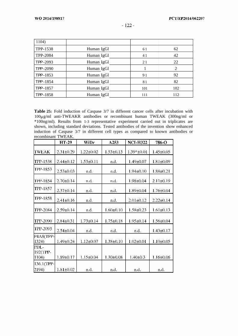
1104)
TPP-1538 Human IgGl 6 1 62
TPP-2084 Human IgGl 4 1 42
ΤΡΡ -2093 Human IgGl 2 1 22
ΤΡΡ -2090 Human IgGl 1 2
ΤΡΡ -1853 Human IgGl 9 1 92
ΤΡΡ -1854 Human IgGl 8 1 82
ΤΡΡ -1857 Human IgGl 101 102
ΤΡΡ -1858 Human IgGl 111 112
Table 25: Fold induction of Caspase 3/7 in different cancer cells after incubation with100 g/ml anti-TWEAKR antibodies or recombinant human TWEAK (300ng/ml or*100ng/ml). Results from 1-3 representative experiment carried out in triplicates areshown, including standard deviations. Tested antibodies of the invention show enhancedinduction of Caspase 3/7 in different cell types as compared to known antibodies orrecombinant TWEAK.

EXAMPLE 5 : Inhibition of proliferation by agonistic anti-TWEAKR antibodies in
cancer cell lines
To investigate whether the efficacy of the antibodies of this invention to induce Caspase
3/7 is also reflected by an efficacious inhibition of proliferation of different cancer cell
lines, anti-proliferative activity was measured after incubation with the antibodies of the
invention as compared to reference antibodies or TWEAK ligand.
Therefore cells were plated in 96well plates in 75µ1 assay medium (growth media from
Table 21, plus lOOng/ml IFN gamma for 786-0 cells) at the following cell numbers:
WiDr cells 3000 cells/well, 786-0 cells 2500 cells/well. 24h later cells were incubated
with anti-TWEAKR antibodies (see Table 26), recombinant human TWEAK or isotype
control IgG (not shown) at the indicated concentrations (antibodies from 0.03-300 g/ml,
TWEAK 100 or 300ng/ml). At the time of antibody addition cell viability was determined
in sister plates (time point zero): therefore 75µ1 ν 11 CTG solution (Promega Cell Titer
Glo solution (catalog # G755B and G756B) was added to the cells, incubated for 10
minutes and luminescence was read on a Victor V (Perkin Elmer). 96h after incubation
with the agents, cell viability was determined in the assay plates by addition of
ΙΟΟµΙ/well CTG solution, 10 min incubation and reading of luminescence. Proliferation
in control wells was calculated by subtracting time zero values from the luminescence
values in the untreated control wells. % of cell proliferation was calculated in the
compound treated wells as compared to the untreated control wells.
The resulting dose response curves representing cell proliferation in the treated cells as
compared to control cells are shown in Figure 14A (WiDr cells) and Figure 14B (786-0
cells). All tested antibodies of the current invention inhibited proliferation of WiDr cells
by 50-60% and of 786-0 cells by 70-80%, which was significantly more than the
proliferation inhibition reached by other known antibodies, e.g. PDL-192(TPP-1 104) and
P4A8(TPP-1324). Thus, the antibodies of the current invention are the most efficacious
anti-proliferative anti-TWEAKR antibodies described to date.

To evaluate, whether this strong anti-proliferative activity can also be observed in a
broader cell panel, in addition LOVO, NCI-H1975, SW-480 (all 3000 cells/well), HT-29
(4000 cells/well), A253 and SK-OV3 (both 2500 cells/well) cells were incubated with
100 g/ml anti-TWEAKR antibodies or TWEAK ligand for the time periods as indicated
in Table 27. All cells were seeded in the growth media indicated in Table 2 1 and for all
cells except for WiDr cells lOOng/ml IFNgamma was added to the assay medium when
seeding the cells. The percentage of growth inhibition for treated cells as compared to
proliferation in untreated control cells was measured and calculated as described above.
As shown in Table 27 the antibodies of the current invention are more efficacious as
compared to other known antibodies in inhibiting proliferation of various cancer cell lines
at 100 g/ml. In most experiments the antibodies of the current invention also show equal
or stronger efficacy as compared to 100-300ng/ml TWEAK ligand. Thus, the antibodies
described in this invention are unique in their activity to induce apoptosis and
proliferation inhibition in a broad panel of cancer cell lines.
Table 26: List of antibodies used for proliferation assay
Table 27: % Inhibition of proliferation induced by incubation with 100 g/ml anti-TWEAKR antibodies or TWEAK ligand (*100ng/ml, **300ng/ml). Incubation time inthe presence of the agents is indicated as time of assay in [h]. Results from 1-3representative experiments carried out in triplicates are shown. Antibodies of theinvention show stronger inhibition of cancer cell proliferation as compared to knownantibodies (PDL-192(TPP-1104), P4A8(TPP-1324)) and equal or stronger activity ascompared to recombinant TWEAK.
time of W E TPP-1538 TPP-2084 |TPP-2090 TPP-2093 PDL-192 P4A8assay [h] |
786-() 96h \2Vr- 75% 68% 71% n.d. 4 /ί 29%

LOVO 72h 53%* 65 '/, n.d. n.d. n.d. 49% -5%
NCI-H1975 72h 56%* 64% n.d. n.d. n.d. 46% 20%
SW480 72 0%* 49 n.d. n.d. n.d. 27% 5%
WiDr 96h n.d. 55% 52% 50% n.d. 12% 4%
HT-29 24h 98%** 97% 98% > 1()0% >100% 62% -10%A253 72 > 1()()%** > 1()0% > 1()0% 99% > 1()0% 87% 37%
SK-OV3 96h n.d. >100% n.d. n.d. n.d. 44% n.d.
EXAMPLE 6 : Cytokine secretion induced by anti-TWEAKR antibodies from
cancer cells and xenograft tumors
Next, it was of interest to investigate whether the strong agonistic activity of the
antibodies of this invention is also seen in the induction of cytokine secretion from cancer
cells.
Therefore A375 cells were plated at 2500 cells/well in 96well plates in growth medium
DMEM (Biochrom;# FG 0435, with stable Glutamin), 10% FCS. 24h later cells were
incubated with anti-TWEAKR antibodies, recombinant human TWEAK at various
concentrations as indicated or corresponding isotype control IgG. 24h after start of the
incubation with the antibodies, cell supernatant or dilutions thereof were added to the
Capture Elisa Plate of the human CXCL8/IL-8 ELISA Kit (R&D Systems DY208) and
incubated over night at 4°C by shaking 300rpm. On the next day, samples were analyzed
by using the human CXCL8/IL-8-ELISA Kit (R&D Systems DY208) according to the
manufacturer's instructions. Optical density was measured at 450 nm (Tecan Spectra,
Rainbow) together with background correction. To calculate absolute levels of IL-8 a
standard curve using recombinant human IL-8 protein was applied according to the
manufacturer's recommendations (R&D Systems).
As shown in Figure 15, the antibodies of this invention showed increased induction of IL-
8 release as compared to other antibodies previously known. At 100 g/ml the antibodies
of this invention TPP-1538/-1854/-2084/-2090 reached 134/129/113/103% of the
activation as compared to 300ng/ml TWEAK ligand respectively. In contrast, the
antibodies used for comparison, PDL-192(TPP-1104)/P4A8(TPP-1324)/136.1(TPP-

2194), reached only 66/29/93%, respectively. Thus, the antibodies of this invention show
the strongest activity with regard to induction of IL-8 secretion as compared to previously
known antibodies and 300ng/ml TWEAK ligand.
To investigate, whether the observed cytokine secretion is also of relevance in xenograft
tumors in mice, human and murine cytokines in serum/plasma from tumor bearing (A375,
WiDr) as well as tumor free mice were investigated. 5xl0 6 A375 cells in Matrigel/PBS
(1:1) or 5xl0 6 WiDr cells in Matrigel/Medium (1:1) were subcutaneously inoculated in
immunodeficient female NMRI nude mice. Parallel to A375-bearing mice, non-tumor
mice were investigated. Treatment started 7d after inoculation with established tumors of
about 40mm 2. Mice were treated by a single intravenous injection of TPP-1538 (10
mg/kg) or TPP-2090 (3 mg/kg) both diluted in PBS into the tail vein. Mice were then
sacrificed at given time -points (0, 6h, 24h for A375-bearing mice and 0, 7h, 24h, 72h,
168h, 240h for WiDr-bearing mice) to harvest serum/plasma samples. Blood was
collected after decapitation and serum was prepared by 30 minutes clotting with
subsequent centrifugation at lOOOxg.
Cytokines were quantified using Luminex® bead immunoassays. Human cytokines were
determined with Human Cytokine Magnetic 25-plex panel (Invitrogen®, Cat-No.
LHC0009M), comprising IL- Ι β, IL-1RA, IL-2, IL-2R, IL-4, IL-5, IL-6, IL-7, IL-8, IL-10,
IL-12 (p40), IL-13, IL-15, IL-17, TNF-a, IFN-a, IFN-γ , GM-CSF, MIP-la, ΜΙΡ -Ι β, IP-
10, MIG, Eotaxin, RANTES, and MCP-1). Murine cytokines were determined with
Mouse Cytokine Magnetic 20-plex panel (Invitrogen®, Cat-No. LHC0006M) comprising
FGF basic, GM-CSF, IFN-γ , IL-la, IL- Ι β, IL-2, IL-4, IL-5, IL-6, IL-10, IL-13, IL-12
(p40/p70), IL-17, IP-10, KC, MCP-1, MIG, MIP-la, TNF-a und VEGF). The assays
were conducted according to the manufacturer's instructions and measured by the
Luminex reader Bio-Plex 200 (Bio-Rad GmbH). Cytokine concentrations were
interpolated from standard curves, as part of the assay procedure, by the operating
software Bio-Plex Manger (Bio-Rad GmbH).

As shown in Figure 16A, human IL-8 is released from WiDr xenograft by a single
treatment with TPP-2090 3mg/kg in a time dependent manner. In addition, induction of
secretion of human MCP-1, IP- 10 and IL-15 was observed after treatment with TPP-2090
(not shown).
To further investigate whether the cytokine induction observed in the plasma of tumor
bearing mice after treatment with agonistic anti-TWEAKR antibodies of the invention
was indeed tumor specific, a similar investigation was carried out in A375 tumor bearing
and tumor free mice and human as well as murine cytokines were measured.
As shown in Figure 16B, human IL-8 is released from A375 xenografts in tumor bearing
mice 6h after treatment with TPP-1538 at 10 mg/kg. In addition, increased levels of
human MCP-1, IP-10 and IL-1RA were observed (not shown). In contrast, in the plasma
of treated tumor free mice no increased secretion of human cytokines was detected
(Figure 16B and not shown). In addition, no increase of murine cytokines including the
murine IL-8 analogue KC was detected in the plasma of neither tumor bearing nor tumor
free mice after treatment with TPP-1538 (data not shown). To summarize, the antibodies
of the present invention potently induce secretion of cytokines from cancer cells and
xenografts in vivo in a tumor specific manner.
EXAMPLE 7 : Internalization of anti-TWEAKR antibodies and usability for drug
conjugate approaches
To investigate, whether anti-TWEAKR antibodies of the current invention are potentially
usable for the generation of antibody drug conjugates (ADC)s, the internalization
capacity of the antibodies was investigated.
To visualize this process the TWEAKR specific antibodies TPP-1538 and TPP-2090 and
an isotype control antibody were selected. The antibodies were conjugated in the presence
of a twofold molar excess of CypHer 5E mono NHS ester (batch 357392, GE Healthcare)
at pH 8.3. After the conjugation the reaction mixture was dialyzed (slide-A-Lyser

Dialysis Cassettes MWCD lOkD, Fa. Pierce) overnight at 4°C to eliminate excess dye
and to adjust the pH-value. Afterwards the protein solution was concentrated (VIVASPIN
500, Fa Sartorius Stedim Biotec). In addition to the pH-dependent fluorescent dye
CypHer5E the ph-independent dye Alexa 488 was used. The dye load of the antibody was
determined with a spectrophotometer (Fa. NanoDrop). The dye load of TPP-1538, TPP-
2090 and an isotype control antibody were in a similar range. The affinity of the labeled
antibodies was tested in a cell binding-assay to ensure that labeling did not alter the
binding to TWEAKR. These labeled antibodies were used in the following internalization
assays. Prior to treatment cells (2xl0 4/well) were seeded in ΙΟΟµΙ medium in a 96-MTP
(fat, black, clear bottom No 4308776, Fa. Applied Biosystems). After 18 h incubation at
37°C/5%C0 2 medium was changed and labeled anti-TWEAKR antibodies TPP-1538 and
TPP-2090 were added in various concentrations (10, 5, 2.5, 1, 0.3, Λ µ /mϊ ) . The
identical treatment was carried out with the isotope control antibody (negative control).
The incubation time was chosen to be 0, 0.5h, lh, 2h, 3h, 6h and 24h. The fluorescence
measurement was performed with the InCellAnalyzer 1000 (Fa. GE Healthcare). Granule
counts per cell and total fluorescence intensity were measured in a kinetic fashion.
A highly specific and significant internalization of TPP-1538 and TPP-2090 was
observed in endogenous TWEAKR expressing cancer cell lines 786-0 (renal cancer) and
HT-29 (colon cancer).
This internalization was target dependent as uptake could only be demonstrated using the
anti-TWEAKR antibodies while no internalization was observed with the isotype control
antibodies. During the first 6h the anti-TWEAKR antibodies showed a 20-40-fold
increase of antibody internalization compared to isotype controls. Isotype control
antibodies showed a minor internalization after a long exposure (>24h).
Internalization of anti-TWEAKR antibodies labeled with Alexa 488 upon binding reveals
that more than 50% of internalized antibodies seem to follow the endocytotic pathway.

In Figure 17, evaluation of the time course of specific internalization of TPP-1538 and
TPP-2090 upon binding to endogenous TWEAKR expressing cells is shown.
Internalization of antibodies ( 1 g/ml) was investigated on renal cancer cell line 786-0.
Granule counts per cell were measured in a kinetic fashion. Rapid internalization could be
observed for TPP-1538 and TPP-2090, whereas the isotype control hlgGl did not
internalize. Additionally, a significantly improved internalization efficacy was seen with
TPP-2090.The higher efficacy of TPP-2090 could be verified in internalization assays
performed using a variety of cancer cells with different receptor levels (not shown).
Additionally, the activity of anti-TWEAKR antibodies to inhibit proliferation of cells
when incubated in the presence of saporin-conjugated secondary antibodies was
evaluated in Hum-Zap assays. Therefore 786-0 cells were plated at 2500 1ΐ8/75µ1 ν 11
in 96 well plates in growth medium (DMEM/Ham'sF12, Biochrom #FG4815 + 10%
FCS). 24h later 40nM antibodies (TPP-1538, TPP-2090 or isotype control antibody) were
incubated in the presence or absence of 40nM saporin-conjugated secondary antibodies
(Hum Zap, Advanced Targeting Systems Cat #IT-22, Lot 59-83) for 15min at room
temperature. After the incubation time 25µ1 of the reaction mix was added to the cells,
resulting in a final concentration of lOnM antibody in the sample wells. At the time of the
antibody addition cell viability was determined in sister plates (time point zero).
Therefore, 75µ1 ν 11 CTG solution (Promega Cell Titer Glo solution (catalog # G755B
and G756B) was added to the cells, incubated for 10 minutes and luminescence was read
on a Victor V ( Perkin Elmer).
48h after start of incubation with the antibody/Zap complex, 100 µΕ/well CTG solution
was added to all test wells, incubated for 10 minutes and luminescence was read on a
VICTOR V.
As shown in Figure 18, incubation of 786-0 cells with anti-TWEAKR antibodies at
lOnM in the presence of saporin-conjugated secondary antibodies almost completely

inhibited cell proliferation, whereas under the same experimental conditions (absence of
IFNgamma, 48h incubation time only) no anti-proliferative activity was observed in the
absence of saporin-conjugated secondary antibodies or with isotype control antibodies.
To summarize, anti-TWEAKR antibodies of the present invention show rapid
internalization and targeted delivery of conjugated payloads and are thus well suitable for
the generation and use as ADCs.
EXAMPLE 8 : Anti-Tumor efficacy of anti-TWEAKR antibodies in xenograft
models in vivo
To investigate, whether anti-TWEAKR antibodies show anti-tumor activity in vivo
xenograft tumors derived from different cancer cell lines or patient derived tumor models
were tested for their sensitivity against tumor growth inhibition by agonistic anti-
TWEAKR antibodies in mono- or combination therapy.
Before start of the in vivo experiments expression of TWEAKR in the selected xenograft
models was evaluated by immunohistochemistry. Therefore, frozen sections (5 µΜ) of
the corresponding xenografts were fixed with acetone for 5 min at 4°C and blocked
against unspecific protein binding and peroxidase activity. Tissue sections were incubated
with rabbit anti-TWEAKR antibody (Fnl4, Epitomics, 3488-1) at room temperature for
60min, followed by peroxidase labeled anti rabbit polymer (DAKO, K4011) incubation
for 30min. Sections were developed with diaminobenzidine and finally counterstained
with hematoxylin. Only models that were positive for expression of the TWEAKR were
used for in vivo experiments
For the investigation of the anti-tumor activity of the anti-TWEAKR antibodies in vivo,
nude mice bearing xenografts from different human tumor cell lines or patient-derived
tumors were treated by repeated intravenous injections.

Tumor cell lines were cultivated as described in the parts above and 100 µΐ containing
cell line specific numbers of tumor cells inoculated subcutaneously (s.c.) into female
athymic nude mice (NMRI nu/nu, 6-8 weeks, 20-25g, Taconic). Mice were housed under
standardized pathogen free conditions and treated according to the animal welfare
guidelines.
After tumor growth to a size of approximately 40mm2 mice were randomized into control
and treatment groups with a respective group size of n=8-10. Mice were treated with
various doses of anti-TWEAKR antibodies diluted in PBS by intravenous injection (i.v.)
into the tail vein with a twice per week schedule (q4dx3: applications twice per week,
three applications in total; q4dx8: applications twice per week, eight applications in total).
Combination therapy partners such as Regorafenib (lOmg/kg daily, per os) and the PI3K-
inhibitor 1 (lOmg/kg, BID, 2d on, 5d off (applications twice daily on two consecutive
days, followed by five days without treatment), i.v.) were diluted in their respective
formulations whereas the standard of care therapies Irinotecan (5mg/kg, 4d on, 3d off
(applications once daily on four consecutive days followed by three days without
treatment) i.v.) and Paclitaxel (16mg/kg, q7dx4 (applications once per week, four
applications in total), i.v.) were diluted in 0.9% NaCl. Animals injected with PBS served
as the control (vehicle) group. The applied volume of the compounds was 5ml/kg body
weight per mouse.
Tumor growth was monitored 2-3 times per week by caliper measurement (length x
width in mm) as well as body weight (in g). At the end of study tumors were dissected,
weighted and used for the calculation of tumor-to-control (T/C) ratios (mean tumor
weight of treated animals divided by mean tumor weight of control/vehicle animals).
In the human renal cell cancer model 786-0 (positive TWEAKR expression) efficacy of
the anti-TWEAKR antibodies TPP-2084 and TPP-2090 was tested at three different low
doses against the isotype control antibody. 2xl0 6 tumor cells in 100% Matrigel were s.c.
inoculated in female nude mice. After 7d established tumors with a size of about 40 mm2

were treated with 0.3 mg/kg, 1 mg/kg and 3 mg/kg of antibodies (i.v., q4dx3: applications
twice per week, three applications in total). At day 40, comparison of tumor weights after
dissection showed dose-dependent efficacy of TPP-2084 and TPP-2090 which was
highest at 3 mg/kg (Figure 19). A clear differentiation against the isotype and vehicle
group (treated with PBS) could be demonstrated. No loss of bodyweight was observed in
any of the groups. Tumor-to-control ratios listed in Table 28 demonstrate good efficacy in
the 786-0 model and in further tumor models (A375, A253, SK-OV-3, Bx-PC3, treated
with 3-10 mg/kg anti-TWEAKR antibodies TPP-1538, TPP-2094 or TPP-2090 in a
q7dx3 (applications once per week, three applications in total) or q4dx3 (applications
twice per week, three applications in total) schedule with the exception of MDA-MB-231
where more intense dosing schedules of anti-TWEAKR antibodies might be required to
reach monotherapy efficacy.
Table 28: Final Tumor-to-Control (T/C) ratios in 786-0 and further tumor models aftertreatment with TPP-1538, TPP-2084 or TPP-2090. Anti-TWEAKR antibodies showstrong anti-tumor activity in a variety of xenograft models from different solid tumorindications.
T/C: tumor-to-control ratio based on final tumor weight after dissection or based on
measurement of tumor area (* ).

Figure 20 shows the efficacy of the anti-TWEAKR antibody TPP-2090 in the human
colon cancer xenograft WiDr (which represents a subclone of the HT-29 tumor cell line)
in monotherapy and combination therapy with Irinotecan and Regorafenib. 5xl0 6 WiDr
cells in Matrigel/Medium (1:1) were s.c. inoculated in immunodeficient NMRI nude
mice. Treatment started 7d after inoculation with established tumors of about 40mm2.
Even 3mg/kg of TPP-2090 (i.v., q4dx7: applications twice per week, seven applications
in total) in monotherapy was strongly effective to control tumor growth. Combination of
3mg/kg TPP-2090 with either Irinotecan (5mg/kg, i.v., 4d on, 3d off) or Regorafenib
(lOmg/kg, p.o., daily) resulted in additive efficacy with tumor regression. All therapeutic
regimens were well tolerated by the mice (max. 4% initial and reversible body weight
loss). Final T/C values are listed in Table 29.
TPP-2090 was also investigated in other colorectal tumor models such as SW480 and the
patient-derived tumor model Co5682 with similar good results (Table 29). A dose of
lOmg/kg TPP-2090 was effective in monotherapy in SW480 to control tumor growth
(T/C 0.49) and to lead to tumor regression in combination with 5mg/kg Irinotecan (T/C
0.22) or lOmg/kg Regorafenib (T/C 0.37). In Co5682 xenografts 3mg/kg of TPP-2090
showed synergistic efficacy with tumor regression in combination with Irinotecan (T/C
0.23) and tumor stasis in combination with Regorafenib (T/C 0.27).
Table 29: Final Tumor-to-Control (T/C) ratios of two colon cancer cell lines WiDr andSW480 and one patient-derived colon cancer xenograft Co5682 after treatment with TPP-2090 and combination partners based on tumor weights at study end.
T/C (final)Tumor
TPP-2090 Combo TPP-2090 Combo TPP-2090 +Model
mono Tx + Irinotecan Regorafenib
WiDr 0.17 0.04 0.07
SW480 0.49 0.22 0.37
Co5682 0.97 0.23 0.27

T/C: tumor-to-control ratio based on final tumor weight after dissection, Tx: therapy,
combo: combination therapy
Figure 2 1 shows the efficacy of the anti-TWEAKR antibody TPP-2090 in the human non-
small-cell lung cancer xenograft NCI-H322 in monotherapy and combination therapy
with Paclitaxel. 5xl0 6 NCI-H322 cells in Matrigel were s.c. inoculated in
immunodeficient NMRI nude mice. Treatment started 14d after inoculation with
established tumors of about 45mm2. At a dose of 5mg/kg TPP-2090 (i.v., q4dx8) was
strongly effective in monotherapy demonstrating tumor regression. Combination of
lOmg/kg TPP-2090 with Paclitaxel (16mg/kg, i.v., q7dx4) resulted in slight additive
efficacy. All therapeutic regimens were well tolerated by the mice (max. 3% reversible
body weight loss). Final T/C values are listed in Table 30.
Again, TPP-2090 was also investigated in other lung cancer models such as NCI-H1975
and the patient-derived tumor models Lu7343 and Lu7433 with comparable results (Table
30). A dose of 3mg/kg TPP-2090 showed additive effects in NCI-H1975 in combination
with 16mg/kg Paclitaxel resulting in tumor regression (T/C 0.08). Similarly, in the
patient-derived NSCLC models Lu7343 and Lu7433 combination of 3mg/kg TPP-2090
with lOmg/kg of the PI3K-inhibitor 1 led to tumor control or regression (T/C 0.18-0.36)
in an additive efficacious manner.
PI3K-inhibitor 1 is (2-amino -N-[7-methoxy-8-(3-morpholin-4-ylpropoxy)-2,3-
dihydroimidazo[l,2-c]quinazolin-5-yl]pyrimidine-5-carboxamide dihydrochloride
Table 30: Final Tumor-to-Control (T/C) ratios of two NSCLC cell lines NCI-H322 and -H1975 and two patient-derived lung cancer xenografts Lu7343 and Lu7433 aftertreatment with TPP-2090 and combination partners based on tumor weights at study end.
T/C (final)
TPP-2090 Combo TPP-2090 Combo TPP-Tumor Model
mono Tx + Paclitaxel 2090 + PI3K-
inhibitor 1
NCI-H322 0.17 0.14 --
NCI-H1975 0.48 0.08 --
Lu7343 0.65 -- 0.18
Lu7433 0.53 -- 0.36
T/C: tumor-to-control ratio based on final tumor weight after dissection, Tx: therapy,
combo: combination therapy
All described in vivo examples demonstrate the strong efficacy of the anti-TWEAKR
antibody TPP-2090 in a broad panel of cell line-derived and patient-derived human tumor
models (all with TWEAKR positive expression) in monotherapy as well as in
combination therapy. TPP-2090 was well tolerated by the mice at all doses used.
EXAMPLE 9 : Mode of action of anti-TWEAKR antibodies in xenograft models
To investigate the mode of action of anti-TWEAKR antibodies in vivo, tumors from
WiDr xenografts were taken at study end as described in Example 8 and investigated by
immunohistochemistry and Western Blot Analysis.
Frozen sections (5 µιη) of WiDr xenografts (tumors from 3 individual animals per group)
were stained immunohistochemically for the proliferation marker protein Ki67 (see
EXAMPLE 8 for details of in vivo experiment). Sections were fixed with freshly
prepared 4% paraformaldehyde for 20 min at 4°C and blocked against unspecific protein
binding and peroxidase activity. Murine anti Ki67 antibody (DAKO, M7240) was labeled
with biotin according to manufacturer's instruction (DAKO, K3954) and incubated at
room temperature for 60 min with the tissue sections, followed by ExtraVidin-peroxidase
(DAKO, K3468) incubation for 30 min. Sections were developed with diaminobenzidine
and finally counterstained with hematoxylin. For quantification, entire tumor sections
were scanned and analyzed using ARIOL automated microscopy version 3.2 (Applied

Imaging, San Jose CA, USA). Representative images of PBS (i.v., q4dx7) and TPP-2090
(lOmg/kg, i.v., q4dx7) treated xenografts stained for Ki67 are shown in Figure 22 A and
B respectively. The quantification using the ARIOL image system, revealed 355+ 59
Ki67 positive cells / mm2 in the group treated with 10 mg/kg TPP-2090 (i.v. q4dx7), and
863 +/-90 Ki67 positive cells / mm2 in the vehicle treated group. Thus, in line with the
observed reduction in tumor volume, treatment with agonistic anti-TWEAKR antibodies
leads to a reduction of the proliferation marker Ki67 in xenograft tumors.
In addition WiDr xenograft tumors (see Example 8 for details of in vivo experiment),
snap frozen at study end were analyzed by Western Blot to evaluate effects of the
antibody treatment on Stat-1 and NF-kappaB signaling pathways. Tumors of 4 individual
animals per group were cut in slices of around 5mm diameter and each slice deposited in
a 2ml Eppendorf tube together with a precooled 5mm steel bull (Qiagen) and 500µ1lysis
buffer (50mM Hepes pH 7.2, 150mM NaCl, ImM MgCl2, lOmM Na4P20 7, lOOmM NaF,
10% Glycerin, 1.5% Triton X-100, freshly added Complete Protease Inhibitor cocktail
(Roche No. 1873580001), 4mM Na3V0 4, pH adjusted to 7.4 with NaOH)). Samples were
lysed for 3 min at 300Hz in a Tissuelyzer (Qiagen) followed by incubation on ice for
30min. In the following, samples were centrifuged for lOmin at 13000 rpm at 4°C in a
Micro-centrifuge (Eppendorf) and supernatants from one original tumor pooled back
together. Protein levels in the tumor lysates were determined by using the BCA protein
assay kit (Novagen, lysates 1:50 diluted in H 2O). Samples were diluted to a final
concentration of 4mg/ml and ΙΟµΙ of sample were mixed with 3.08 µΐ of (10*) Sample
Reducing agend, ΙΟµΙ LO and 7,68µ1 (4*) NuPAGE Sample Buffer (Invitrogen).
Samples corresponding to 40µg of protein were applied to NuPage 4-12% SDS page gels
from Invitrogen and run for 2h45min at 120V. Blotting was carried out by an iBlot
system (Invitrogen) according to the manufacturer's recommendations. Membranes were
blocked for 2h at room temperature in 5% BLOT QuickBlocker in PBST (Invitrogen),
followed by incubation with primary antibodies over night at 4°C. Primary antibodies

were as follows: Phospho-Statl #9167S, Stat-1 #9172, both Cell Signaling Technology,
dilution 1:1000; TWEAKR/Fnl4 #3488-1 Epitomics, dilution 1:10000; NF-kappaB2
pl00/p52 #4882S, Cell Signaling Technology, dilution 1:1000 in in 3% BLOT
QuickBlocker in PBST. On the next day membranes were washed three times in PBST,
followed by incubation with secondary antibodies (Peroxidase -conjugated donkey anti-
rabbit IgG # NA934, GE Healthcare 1:10000 in 3% BLOT QuickBlocker/PBST) for 2h at
room temperature. Subsequently, membranes were washed four times for 10 min with
PBST and signals were detected by chemoluminescence after incubation with ECL
reagent. To detect the loading control, membranes were stripped with stripping solution
Re-Blot Plus strong solution #2504, Milipore (1:10 in Milipore-H20) for 15 min shaking
at room temperature, followed by blocking and detection with anti-GAPDH antibody
(clone6C5, # MAB374, Millipore 1:10000 in 3% QuickBlocker/PBST) and secondary
antibody (Peroxidase -conjugated goat anti-mouse IgG, Jackson Immunoresearch #115-
035-003, 1:10000 in 3% BLOT Quickblocker/PBST).
Representative Blots from tumors of 2 animals per group treated with TPP-2090 3mg/kg
side by side with tumors from vehicle treated animals are shown in Figure 23. Treatment
with TPP-2090 leads to strong increase of total and phosphorylated Stat-1 levels as well
as a strong activation of NF-kappaB2 as indicated by the appearance of the p52 fragment.
Thus, the NF-kappaB2 as wells as Stat-1 pathways are activated by agonistic anti-
TWEAKR antibodies in xenograft tumors and this activation is potentially involved in the
anti-tumor activity of the corresponding antibodies.
Table 31: Protein sequences of the antibodies:

Table 32: DNA sequences of the antibodies of the invention

EXAMPLE 10: Anti-tumor efficacy of anti-TWEAKR antibody TPP-2090 in
further human colorectal cancer models in vivo
Animal studies were conducted as described in example 8 for further human colorectal
cancer tumor cell lines Colo205 and LoVo and for further human colorectal cancer
patient-derived models Co7553, Co5896, Co5676, Co5841, CXF 1103 and CXF 533.
Standard dosing schedule was 10 mg/kg of TPP-2090 twice weekly for 4 weeks in
monotherapy or in combination with regorafenib or the standard of cares (SoCs)
irinotecan (5-15 mg/kg i.p., 4d on, 3d off), oxaliplatin (3-8 mg/kg i.p., twice weekly), 5-
fluorouracil (50-100 mg/kg i.p., once weekly) and cetuximab (15 mg/kg i.p., twice
weekly) . TPP-2090 and cetuximab were formulated in PBS, which was also used as the
vehicle in the control group, and the SoCs were formulated in 0.9% NaCl. The
formulation of regorafenib is described in example 8.
The monotherapeutic efficacy of TPP-2090 in these human colorectal cancer patient-
derived and cell line based models was moderate with final Tumor-to-Control (T/C)
ratios in the range of 0.48-1.07. The combinations of TPP-2090 with SoCs (in particular
5-FU and irinotecan) resulted in significant additive and synergistic effects (see Table 33-
35). In cases where the monotherapeutic efficacy of TPP-2090 in these models were
limited more intense dosing schedules of anti-TWEAKR antibodies might be required to
reach higher monotherapy efficacy, as has been shown in example 8 for colorectal cancer.

Table 33: Final Tumor-to-Control (T/C) ratios of colorectal cancer models treatedwith TPP-2090 in monotherapy or combination with irinotecan and oxaliplatin
Combination TPP-2090 with:
T/C: tumor-to-control ratio based on final tumor weight after dissection or based onmeasurement of tumor area (*).
n.d.: not determined

Table 34: Final Tumor-to-Control (T/C) ratios of colorectal cancer models treatedwith TPP-2090 in combination with 5-FU and regorafenib
Combination TPP-2090 with:
T/C: tumor-to-control ratio based on final tumor weight after dissection or based onmeasurement of tumor area (*).
n.d.: not determined

Table 35: Tumor-to-Control (T/C) ratios of colorectal cancer models treated withTPP-2090 in combination with cetuximab
Combination TPP-2090 with:
T/C: tumor-to-control ratio based on final tumor weight after dissection or based onmeasurement of tumor area (*).
n.d.: not determined

EXAMPLE 11: Anti-tumor efficacy of anti-TWEAKR antibody TPP-2090 in human
bladder cancer models in vivo
Animal studies were conducted as described in example 8 for the human bladder cancer
cell lines SCaBER and KU-19-19 and for the human bladder cancer patient-derived
models BXF1352 and BXF1228. Standard dosing schedule was 10 mg/kg of TPP-2090
twice weekly for 4 weeks in monotherapy or in combination with the standard of cares
(SoCs) gemcitabine (200 mg/kg i.p., once weekly) and cisplatin (3 mg/kg i.p., once
weekly). TPP-2090 was formulated in PBS, which was also used as the vehicle in the
control group, and the standard of cares (SoCs) were formulated in 0.9% NaCl.
Strong monotherapeutic efficacy of TPP-2090 was found in SCaBER xenograft model.
The combination of TPP-2090 in the human bladder cancer patient-derived bladder
cancer models BXF1352 and BXF1228 with SoCs (Cisplatin and Gemcitabine) resulted
in significant synergistic effects (see Table 36). ) . In cases where the monotherapeutic
efficacy of TPP-2090 in these bladder cancer models were limited more intense dosing
schedules of anti-TWEAKR antibodies might be required to reach higher monotherapy
efficacy.
Table 36: Tumor-to-Control (T/C) ratios of human bladder cancer models treatedwith TPP-2090 in monotherapy or combination with cisplatin or gemcitabine
Combination TPP-2090 with :
Tumor-TPP-2090 Oxaliplatin/ Cisplatin Gemcitabine
model
MonoTx Benefit of BenefitMono Combi Mono CombiResponse Combi of Combi
synergistic0.40 1.22 0.28 0.85 0.42 no benefit
SCaBER effect
0.86 n.d. n.d. n.d. 0.07 n.d. n.d.Ku-1 9-1 9
synergistic0.93 0.85 0.4 n.d. n.d. n.d.
BXF1 352 effect

synergistic0.85 0.84 0.36 0.93 0.46 n.d.
BXF1 228 effect
T/C: tumor-to-control ratio based on final tumor weight
n.d.: not determined

EXAMPLE 12: Anti-tumor efficacy of anti-TWEAKR antibody TPP-2090 in
further human cancer models in vivo
Animal studies were conducted as described in example 8 for further human cancer cell
lines of different indications. Standard dosing schedule was 10 mg/kg of TPP-2090 twice
weekly for 2-3 weeks in monotherapy. TPP-2090 was formulated in PBS, which was also
used as the vehicle in the control group.
Strong monotherapeutic efficacy of TPP-2090 was found in SCC4 (head & neck cancer)
and A375 (melanoma) xenografts, and moderate efficacy in BxPC3 (pancreatic cancer)
xenografts (see Table 37). In cases where the monotherapeutic efficacy of TPP-2090 in
certain xenograft models were limited (ACHN (renal cell cancer), PA-1 (ovarian cancer),
NCI-292 (non-small cell lung cancer) and U87MG (glioblastoma)) more intense dosing
schedules of anti-TWEAKR antibodies might be required to reach higher monotherapy
efficacy.
Table 37: Tumor-to-control (T/C) ratios values of further human cancer modelstreated with TPP-2090 in monotherapy
T/C: tumor-to-control ratio based on final tumor weight after dissection or based onmeasurement of tumor area (*).

EXAMPLE 13: Further mode of action of anti-TWEAKR antibodies in xenograft
models
To evaluate if the anti-tumor efficacy of TPP-2090 is dependent on antibody-dependent
cellular cytotoxicity (ADCC) or agonistic activity alone is already sufficient, xenografts
studies in SCID beige mice (Janvier) were conducted, and the aglycosylated variant of
TPP-2090, namely TPP-2658, was investigated in NMRI nude mice.
Animal studies were conducted as described in example 8 in WiDr (human colorectal
cancer) and SCaBER (human bladder cancer) xenografts in SCID beige mice with tumor
growth of control groups comparable to those in NMRI nude mice of previous studies.
Standard dosing schedule was 10 mg/kg of TPP-2090 i.v. (formulated in PBS) twice
weekly for 2 weeks in monotherapy.
A similar strong monotherapeutic efficacy of TPP-2090 in NK-cell lacking SCID beige
mice xenograft models (WiDr and SCaBER) was found as seen in NMRI nudes mice.
This indicats an in vivo mode of action independent from ADCC (see Table 38).
Table 38: Tumor-to-Control (T/C) ratios of WiDr and SCaBER tumors in SCIDbeige mice treated with TPP-2090 in monotherapy (NMRI mice for comparison)
T/C: tumor-to-control ratio based on final tumor weight after dissection or based onmeasurement of tumor area (*).
In vitro analysis showed that HT29 cell binding of TPP-2090 resulted in dose-dependent
ADCC of target cells by NK92V effector cells while the aglycosylated TPP-2658 was not
capable of inducing ADCC (Table 40). lxlO 4 HT-29 target cells were dispensed and the

tested antibodies were added in a final concentration of 25 g/ml; 5 g/ml; 1 g/ml; 0,2
g/ml and 0,04 µg/ml. After a preincubation time of 30 min effector cells were added
(5xl0 4 NK92V effector cells). After 4h incubation at 37°C HT29 cell lysis was
determined with the Cytotoxicity Detection Kit - LDH (Roche)", maximum release was
obtained from cells solubilized in 1% Triton X-100, negative controls were not
preincubated with an antibody. The following formula was used for calculation of %
HT29 lysis: [Ext (sample) - Ext (negative) x 100] / [Ext (Maximum release) - Ext
(negative)]. TPP-2090 resulted in dose-dependent ADCC of target cells by NK92V
effector cells and is dependent on N297 glycosylation.
Whereas in vivo a similar effect was found when an aglycosylated variant of TPP-2090,
namely TPP-2658, was used in either a WiDr- or A375-xenograft model (see Table 39).
The variant TPP-2658 showed equally strong monotherapeutic efficacy as the TPP-2090
in both models indicating an ADCC-independent mode of action.
Table 39: Turnor-to-Control (T/C) ratios of WiDr and A375-xenografts treated withTPP-2658 (TPP-2090 for comparison)
T/C: tumor-to-control ratio based on final tumor weight after dissection.
Table 40: In vitro ADCC assay with HT-29 target cells and NK92V effector cells fortesting antibody TPP-2090 (hlgGl) and TPP-2658 (aglycosylated counterpart ofTPP-2090 - hlgGl N297A):
Lysis [%] for Lysis [%] for TPP-2658Antibody concentration [pg/ml]
TPP-2090 (aglycosylated)

25 16.8 - 1 . 1
5 16.4 - 1 .3
1 15.1 - 1 .3
0.2 9.8 - 1 .4
0.04 6.3 -0.8

CLAIMS:
1. An isolated anti-TWEAKR antibody or an antigen-binding fragment thereof,
which specifically binds to the D at position 47 (D47) of TWEAKR as depicted
in SEQ ID NO: 169.
2. The antibody or an antigen binding fragment thereof according to claim 1
wherein the antibody is an agonistic antibody.
3. The antibody or an antigen binding fragment thereof according to claims 1 or 2,
which comprises:
a variable heavy chain comprising:
(a) a heavy chain CDR1 encoded by an amino acid sequence comprising
the formula PYPMX (SEQ ID NO: 171), wherein X is I or M;
(b) a heavy chain CDR2 encoded by an amino acid sequence comprising
the formula YISPSGGXTHYADSVKG (SEQ ID NO: 172), wherein
X is S or K; and
(c) a heavy chain CDR3 encoded by an amino acid sequence comprising
the formula GGDTYFDYFDY (SEQ ID NO: 173);
and a variable light chain comprising:
(a) a light chain CDR1 encoded by an amino acid sequence comprising
the formula RASQSISXYLN (SEQ ID NO: 174), wherein X is G or
S;
(b) a light chain CDR2 encoded by an amino acid sequence comprising
the formula XASSLQS (SEQ ID NO: 175), wherein X is Q, A, or N;
and
(c) a light chain CDR3 encoded by an amino acid sequence comprising
the formula QQSYXXPXIT (SEQ ID NO: 176), wherein X at
position 5 is T or S, and X at position 6 is T or S, and X at position 8
is G, or F.
4. The antibody or an antigen binding fragment thereof according to anyone of the
preceding claims comprising:
a. a variable heavy chain comprising the variable heavy chain CDR1
sequence as presented by SEQ ID NO: 6, the variable heavy chain CDR2
sequence as presented by SEQ ID NO: 7, and the variable heavy chain
CDR3 sequence as presented by SEQ ID NO: 8, and
a variable light chain comprising the variable light chain CDR1 sequence
presented by SEQ ID NO: 3, the variable light chain CDR2 sequence
presented by SEQ ID NO: 4, and the variable light chain CDR3 sequence
presented by SEQ ID NO: 5, or
b. a variable heavy chain comprising the variable heavy chain CDR1
sequence as presented by SEQ ID NO: 16, the variable heavy chain
CDR2 sequence as presented by SEQ ID NO: 17, the variable heavy
chain CDR3 sequence as presented by SEQ ID NO: 18, and
a variable light chain comprising the variable light chain CDR1 sequence
presented by SEQ ID NO: 13, the variable light chain CDR2 sequence
presented by SEQ ID NO: 14, and the variable light chain CDR3
sequence presented by SEQ ID NO: 15, or
c. a variable heavy chain comprising the variable heavy chain CDR1
sequence as presented by SEQ ID NO: 26, the variable heavy chain
CDR2 sequence as presented by SEQ ID NO: 27, the variable heavy
chain CDR3 sequence as presented by SEQ ID NO:28, and

a variable light chain comprising the variable light chain CDRl sequence
presented by SEQ ID NO: 23, the variable light chain CDR2 sequence
presented by SEQ ID NO: 24, and the variable light chain CDR3
sequence presented by SEQ ID NO:25, or
d. a variable heavy chain comprising the variable heavy chain CDRl
sequence as presented by SEQ ID NO: 36, the variable heavy chain
CDR2 sequence as presented by SEQ ID NO: 37, the variable heavy
chain CDR3 sequence as presented by SEQ ID NO:38, and
a variable light chain comprising the variable light chain CDRl sequence
presented by SEQ ID NO: 33, the variable light chain CDR2 sequence
presented by SEQ ID NO: 34, and the variable light chain CDR3
sequence presented by SEQ ID NO:35, or
e. a variable heavy chain comprising the variable heavy chain CDRl
sequence as presented by SEQ ID NO: 46, the variable heavy chain
CDR2 sequence as presented by SEQ ID NO: 47, the variable heavy
chain CDR3 sequence as presented by SEQ ID NO:48, and
a variable light chain comprising the variable light chain CDRl sequence
presented by SEQ ID NO: 43, the variable light chain CDR2 sequence
presented by SEQ ID NO: 44, and the variable light chain CDR3
sequence presented by SEQ ID NO:45, or
f . a variable heavy chain comprising the variable heavy chain CDRl
sequence as presented by SEQ ID NO: 56, the variable heavy chain
CDR2 sequence as presented by SEQ ID NO: 57, the variable heavy
chain CDR3 sequence as presented by SEQ ID NO:58, and

a variable light chain comprising the variable light chain CDRl sequence
presented by SEQ ID NO: 53, the variable light chain CDR2 sequence
presented by SEQ ID NO: 54, and the variable light chain CDR3
sequence presented by SEQ ID NO:55, or
g. a variable heavy chain comprising the variable heavy chain CDRl
sequence as presented by SEQ ID NO: 66, the variable heavy chain
CDR2 sequence as presented by SEQ ID NO: 67, the variable heavy
chain CDR3 sequence as presented by SEQ ID NO:68, and
a variable light chain comprising the variable light chain CDRl sequence
presented by SEQ ID NO: 63, the variable light chain CDR2 sequence
presented by SEQ ID NO: 64, and the variable light chain CDR3
sequence presented by SEQ ID NO:65, or
h. a variable heavy chain comprising the variable heavy chain CDRl
sequence as presented by SEQ ID NO: 76, the variable heavy chain
CDR2 sequence as presented by SEQ ID NO: 77, the variable heavy
chain CDR3 sequence as presented by SEQ ID NO:78, and
a variable light chain comprising the variable light chain CDRl sequence
presented by SEQ ID NO: 73, the variable light chain CDR2 sequence
presented by SEQ ID NO: 74, and the variable light chain CDR3
sequence presented by SEQ ID NO:75, or
i . a variable heavy chain comprising the variable heavy chain CDRl
sequence as presented by SEQ ID NO: 86, the variable heavy chain
CDR2 sequence as presented by SEQ ID NO: 87, the variable heavy
chain CDR3 sequence as presented by SEQ ID NO:88, and

a variable light chain comprising the variable light chain CDRl sequence
presented by SEQ ID NO: 83, the variable light chain CDR2 sequence
presented by SEQ ID NO: 84, and the variable light chain CDR3
sequence presented by SEQ ID NO:85, or
j . a variable heavy chain comprising the variable heavy chain CDRl
sequence as presented by SEQ ID NO: 96, the variable heavy chain
CDR2 sequence as presented by SEQ ID NO: 97, the variable heavy
chain CDR3 sequence as presented by SEQ ID NO:98, and
a variable light chain comprising the variable light chain CDRl sequence
presented by SEQ ID NO: 93, the variable light chain CDR2 sequence
presented by SEQ ID NO: 94, and the variable light chain CDR3
sequence presented by SEQ ID NO:95, or
k. a variable heavy chain comprising the variable heavy chain CDRl
sequence as presented by SEQ ID NO: 106, the variable heavy chain
CDR2 sequence as presented by SEQ ID NO: 107, the variable heavy
chain CDR3 sequence as presented by SEQ ID NO: 108, and
a variable light chain comprising the variable light chain CDRl sequence
presented by SEQ ID NO: 103, the variable light chain CDR2 sequence
presented by SEQ ID NO: 104, and the variable light chain CDR3
sequence presented by SEQ ID NO: 105 or
1. a variable heavy chain comprising the variable heavy chain CDRl
sequence as presented by SEQ ID NO: 116, the variable heavy chain
CDR2 sequence as presented by SEQ ID NO: 117, the variable heavy
chain CDR3 sequence as presented by SEQ ID NO: 118, and

a variable light chain comprising the variable light chain CDRl sequence
presented by SEQ ID NO: 113, the variable light chain CDR2 sequence
presented by SEQ ID NO: 114, and the variable light chain CDR3
sequence presented by SEQ ID NO: 115,.
5. The antibody or antigen-binding fragment thereof according to anyone of the
preceding claims comprising:
a. a variable heavy chain sequence as presented by SEQ ID NO: 10 and a
variable light chain sequences as presented by SEQ ID NO:9, or
b. a variable heavy chain sequence as presented by SEQ ID NO:20 and a
variable light chain sequences as presented by SEQ ID NO: 19, or
c. a variable heavy chain sequence as presented by SEQ ID NO:30 and a
variable light chain sequences as presented by SEQ ID NO:29, or
d. a variable heavy chain sequence as presented by SEQ ID NO:40 and a
variable light chain sequences as presented by SEQ ID NO:39, or
e. a variable heavy chain sequence as presented by SEQ ID NO:50 and a
variable light chain sequences as presented by SEQ ID NO:49, or
f . a variable heavy chain sequence as presented by SEQ ID NO:60 and a
variable light chain sequences as presented by SEQ ID NO:59, or
g. a variable heavy chain sequence as presented by SEQ ID NO:70 and a
variable light chain sequences as presented by SEQ ID NO:69, or
h. a variable heavy chain sequence as presented by SEQ ID NO: 80 and a
variable light chain sequences as presented by SEQ ID NO:79, or

i . a variable heavy chain sequence as presented by SEQ ID NO:90 and a
variable light chain sequences as presented by SEQ ID NO: 89, or
j . a variable heavy chain sequence as presented by SEQ ID NO: 100 and a
variable light chain sequences as presented by SEQ ID NO:99, or
k. a variable heavy chain sequence as presented by SEQ ID NO: 110 and a
variable light chain sequences as presented by SEQ ID NO: 109, or
1. a variable heavy chain sequence as presented by SEQ ID NO: 120 and a
variable light chain sequences as presented by SEQ ID NO: 119.
6. The antibody according to any one of the preceding claims, which is an IgG
antibody.
7. The antibody according to anyone of the preceding claims comprising:
a. a heavy chain sequence as presented by SEQ ID NO:2 and a light chain
sequences as presented by SEQ ID NO:l, or
b. a heavy chain sequence as presented by SEQ ID NO: 12 and a light chain
sequences as presented by SEQ ID NO: 11, or
c . a heavy chain sequence as presented by SEQ ID NO:22 and a light chain
sequences as presented by SEQ ID NO:21, or
d. a heavy chain sequence as presented by SEQ ID NO:32 and a light chain
sequences as presented by SEQ ID NO:31, or
e . a heavy chain sequence as presented by SEQ ID NO:42 and a light chain
sequences as presented by SEQ ID NO:41, or
f . a heavy chain sequence as presented by SEQ ID NO:52 and a light chain
sequences as presented by SEQ ID NO:51, or

g. a heavy chain sequence as presented by SEQ ID NO:62 and a light chain
sequences as presented by SEQ ID N0:61, or
h. a heavy chain sequence as presented by SEQ ID NO:72 and a light chain
sequences as presented by SEQ ID NO:71, or
i . a heavy chain sequence as presented by SEQ ID NO: 82 and a light chain
sequences as presented by SEQ ID NO:81, or
j . a heavy chain sequence as presented by SEQ ID NO:92 and a light chain
sequences as presented by SEQ ID NO:91, or
k. a heavy chain sequence as presented by SEQ ID NO: 102 and a light
chain sequences as presented by SEQ ID NO: 101, or
1. a heavy chain sequence as presented by SEQ ID NO: 112 and a light
chain sequences as presented by SEQ ID NO: 111.
8. The antigen-binding fragment according to any one of the preceding claims,
which is an scFv, Fab, Fab' fragment or a F(ab')2 fragment.
9. The antibody or antigen-binding fragment according to any one of the preceding
claims, which is a monoclonal antibody or antigen-binding fragment thereof.
10. The antibody or antigen-binding fragment according to any one of the preceding
claims, which is a human, humanized or chimeric antibody or antigen-binding
fragment.
11. An antibody-drug conjugate, comprising an antibody or antigen binding fragment
thereof according to claims 1 to 10.
12. An isolated nucleic acid sequence that encodes the antibody or antigen-binding
fragment according to claims 1 to 10.
13. A vector comprising a nucleic acid sequence according to claim 12.

14. An isolated cell expressing an antibody or antigen-binding fragment according to
any one of the claims 1 to 10 and /or comprising a nucleic acid according to
claim 1 or a vector according to claim 13.
15. An isolated cell according to claim 14, wherein said cell is a prokaryotic or an
eukaryotic cell.
16. A method of producing an antibody or antigen-binding fragment according to any
one of the claims 1 - 10 comprising culturing of a cell according to claim 15 and
purification of the antibody or antigen-binding fragment.
17. An antibody or antigen-binding fragment according to claims 1 - 10 or an
antibody-drug conjugate according to claim 11 for use as a medicament.
18. An antibody or antigen antigen-binding fragment according to claims 1 - 10 for
use as a diagnostic agent.
19. An antibody or antigen-binding fragment according to claims 1 - 10 or an
antibody-drug conjugate according to claim 11 for use as a medicament for the
treatment of cancer.
20. A pharmaceutical composition comprising an antibody or antigen-binding
fragment according to claims 1 - 10 or an antibody-drug conjugate according to
claim 11.
21. A combination of a pharmaceutical composition according to claim 20 and one or
more therapeutically active compounds.
22. A method for treating a disorder or condition associated with the undesired
presence of TWEAKR, comprising administering to a subject in need thereof an
effective amount of the pharmaceutical composition according to claim 20 or a
combination according to claim 21.

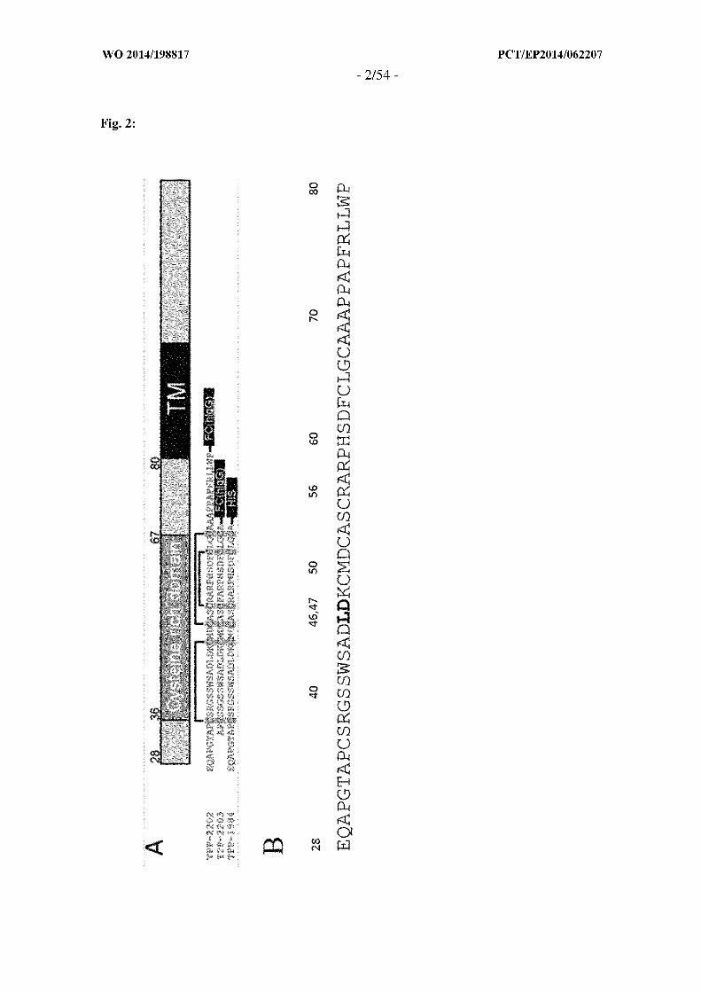


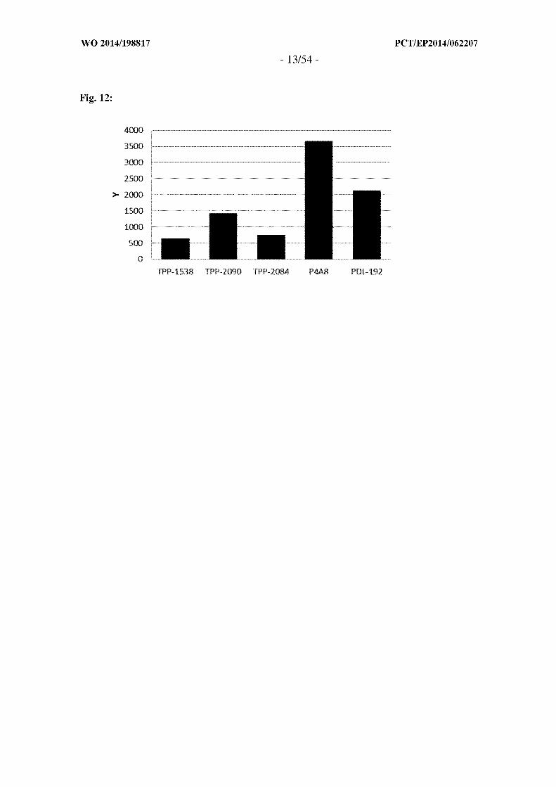

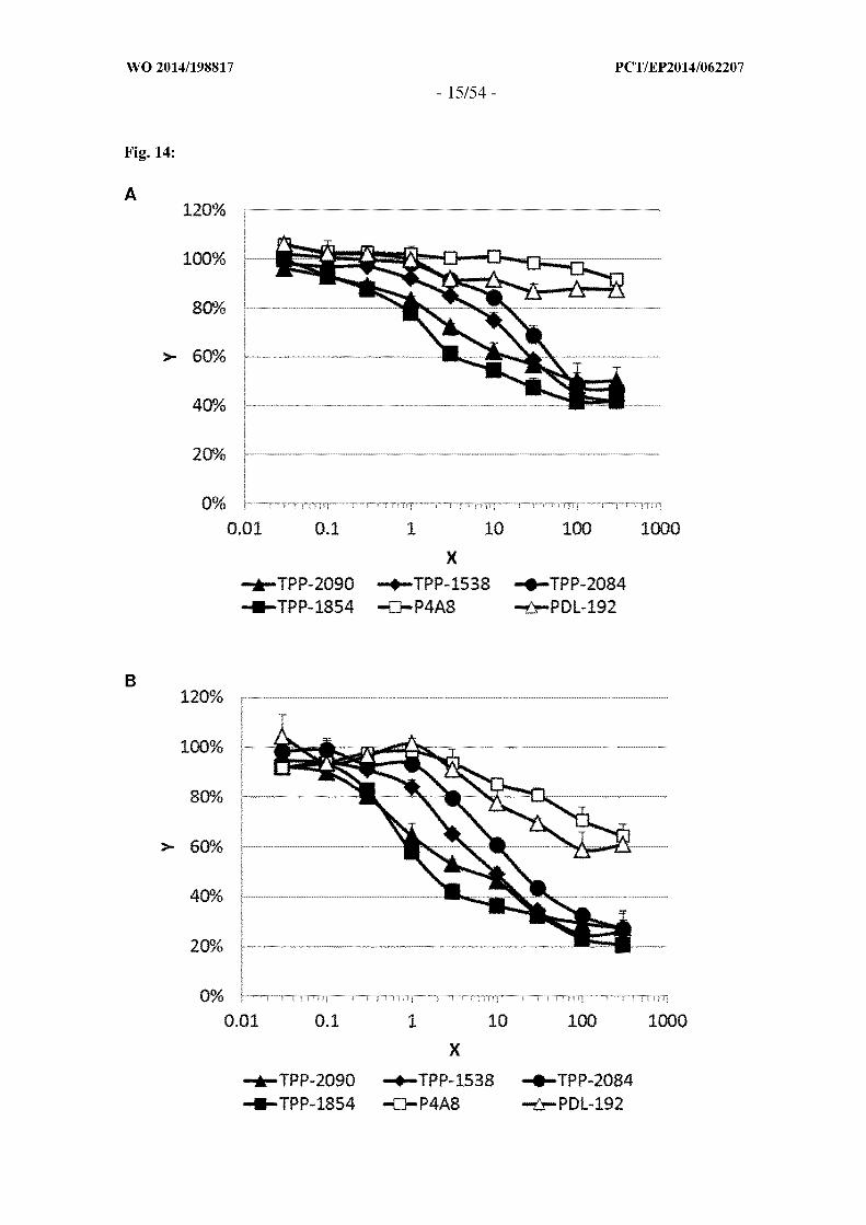


A . CLASSIFICATION O F SUBJECT MATTER
INV. C07K16/28 A61K47/48 A61P35/00ADD.
According to International Patent Classification (IPC) o r to both national classification and IPC
B . FIELDS SEARCHED
Minimum documentation searched (classification system followed by classification symbols)
C07K A61K
Documentation searched other than minimum documentation to the extent that such documents are included in the fields searched
Electronic data base consulted during the international search (name of data base and, where practicable, search terms used)
EPO-Internal
C . DOCUMENTS CONSIDERED TO B E RELEVANT
Category* Citation of document, with indication, where appropriate, of the relevant passages Relevant to claim No.
JENNI FER S. MICHAELSON ET AL: 1-22"Devel opment of an Fnl4 agoni sti c anti bodyas an anti -tumor agent" ,MABS
vol . ' 3 , no. 4 , 1 July 2011 (2011-07-01) ,pages 362-375 , XP055140288,ISSN : 1942-0862 , D0I :10.4161/mabs . 3 .4. 16090page 364, l eft-hand col umn , paragraph 1page 363 , r i ght-hand col umn , l astparagraphpage 366 - page 367 ; f i gure 1
-/-
X| Further documents are listed in the continuation of Box C . □ See patent family annex.
* Special categories of cited documents :"T" later document published after the international filing date o r priority
date and not in conflict with the application but cited to understand"A" document defining the general state of the art which is not considered the principle o r theory underlying the invention
to be of particular relevance
"E" earlier application o r patent but published o n o r after the international "X" document of particular relevance; the claimed invention cannot befiling date considered novel o r cannot be considered to involve an inventive
"L" documentwhich may throw doubts on priority claim(s) orwhich is step when the document is taken alonecited to establish the publication date of another citation o r other "Y" document of particular relevance; the claimed invention cannot bespecial reason (as specified) considered to involve an inventive step when the document is
"O" document referring to an oral disclosure, use, exhibition o r other combined with one o r more other such documents, such combinationmeans being obvious to a person skilled in the art
"P" document published prior to the international filing date but later thanthe priority date claimed "&" document member of the same patent family
Date of the actual completion of the international search Date of mailing of the international search report
16 September 2014 09/10/2014
Name and mailing address of the ISA/ Authorized officer
European Patent Office, P.B. 5818 Patentlaan 2NL - 2280 HV Rijswijk
Tel. (+31-70) 340-2040,Fax: (+31-70) 340-3016 Le Fl ao, Katel l

C(Continuation). DOCUMENTS CONSIDERED TO BE RELEVANT
Category* Citation of document, with indication, where appropriate, of the relevant passages Relevant to claim No.
X MICHAELSON JENNI FER S ET AL: "The 1-22anti -Fnl4 anti body BI IB036 i nhi b i t s tumorgrowth i n xenografts and pati ent deri vedprimary tumor model s and enhances effi cacyof chemotherapeuti c agents i n mul t i p l exenograft model s . " ,CANCER BIOLOGY & THERAPY JUL 2012 ,vol . 13 , no. 9 , July 2012 (2012-07) , pages812-821 , XP002729799 ,ISSN : 1555-8576abstract
X CULP PATRICIA A ET AL: "Anti bodi es t o 1-22TWEAK Receptor Inhi b i t Human Tumor Growththrough Dual Mechani sms" ,CLINICAL CANCER RESEARCH , THE AMERICANASSOCIATION FOR CANCER RESEARCH , US,vol . 16, no. 2 ,1 January 2010 (2010-01-01) , pages497-508, XP009137413 ,ISSN : 1078-0432 , D0I :10. 1158/1078-0432 . CCR-09-1929[retri eved on 2010-01-12]page 502 , l eft-hand col umn - page 504,l eft-hand col umn
X SALZMANN STEFFEN ET AL: " Fi brobl ast 1-22growth factor i nduci b l e ( Fnl4) -speci f i canti bodi es concomi tantly d i spl ay s i gnal i ngpathway-speci f i c agoni sti c andantagoni sti c acti v i ty. " ,THE JOURNAL OF BIOLOGICAL CHEMISTRY 10 MAY2013 ,vol . 288, no. 19 , 10 May 2013 (2013-05-10), pages 13455-13466, XP002729800,ISSN : 1083-351Xpage 13457 , l eft-hand col umnpage 13463 , l eft-hand col umn
A HONG ZHOU ET AL: "The TWEAK Receptor Fnl4 1-22I s a Therapeuti c Target i n Mel anoma:Immunotoxi ns Targeti ng Fnl4 Receptor forMal i gnant Mel anoma Treatment" ,JOURNAL OF INVESTIGATIVE DERMATOLOGY,vol . 133 , no. 4 ,29 November 2012 (2012-11-29) , pages1052-1062 , XP55140642 ,ISSN : 0022-202X, D0I : 1 . 1038/ i . 01 . 02abstract
-/--

C(Continuation). DOCUMENTS CONSIDERED TO BE RELEVANT
Category* Citation of document, with indication, where appropriate, of the relevant passages Relevant to claim No.
A NAKAYAMA M ET AL: " Fi brobl ast growth 1-22factor-i nduci b l e 14 medi ates mul t i p l epathways of TWEAK-i nduced cel l death" ,THE JOURNAL OF IMMUNOLOGY, THE AMERICANASSOCIATION OF IMMUNOLOGISTS, US,vol . 170, no. 1 ,1 January 2003 (2003-01-01) , pages341-348, XP002356992 ,ISSN : 0022-1767the whol e document
A MARK S. CHAPMAN ET AL: "TWEAK s i gnal s 1-22through JAK-STAT t o i nduce tumor cel lapoptosi s " ,CYTOKINE,vol . 61 , no. 1 ,1 January 2013 (2013-01-01) , pages210-217 , XP055140300,ISSN : 1043-4666, D0I :10. 1016/j . cyto.2012 .09 .020the whol e document