WO 2018/129078 Al
146
(12) INTERNATIONAL APPLICATION PUBLISHED UNDER THE PATENT COOPERATION TREATY (PCT) (19) World Intellectual Property Organization International Bureau (10) International Publication Number (43) International Publication Date WO 2018/129078 Al 12 July 2018 (12.07.2018) W!PO PCT (51) International Patent Classification: (71) Applicant: RESEARCH INSTITUTE AT NATION¬ A61K 39/102 (2006.01) C12N 15/62 (2006.01) WIDE CHILDREN'S HOSPITAL [US/US]; 700 A61K 38/00 (2006.01) G01N 33/569 (2006.01) Children's Drive, Columbus, Ohio 43205 (US). A61Q 11/02 (2006.01) A61P 31/04 (2006.01) (72) Inventors: BAKALETZ, Lauren O.; c/o Research Insti A61Q 17/00 (2006.01) A61K 39/40 {2006.01) tute at Nationwide Children's Hospital, 700 Children's Dri C07K 19/00 (2006.01) C07K 16/12 (2006.01) ve, Columbus, Ohio 43205 (US). GOODMAN, Steven D.; C12Q 1/18 (2006.01) c/o Research Institute at Nationwide Children's Hospital, (21) International Application Number: 700 Children's Drive, Columbus, Ohio 43205 (US). PCT/US2018/012235 (74) Agent: KONSKI, Antoinette F. et al; Foley & Lardner (22) International Filing Date: LLP, 3000 K Street N.W., Suite 600, Washington, District 03 January 2018 (03.01.2018) of Columbia 20007-5 109 (US). (25) Filing Language: English (81) Designated States (unless otherwise indicated, for every kind of nationalprotection available): AE, AG, AL, AM, (26) Publication Language: English AO, AT, AU, AZ, BA, BB, BG, BH, BN, BR, BW, BY, BZ, (30) Priority Data: CA, CH, CL, CN, CO, CR, CU, CZ, DE, DJ, DK, DM, DO, 62/442,307 04 January 2017 (04.01.2017) DZ, EC, EE, EG, ES, FI, GB, GD, GE, GH, GM, GT, HN, 62/453,921 02 February 2017 (02.02.2017) HR, HU, ID, IL, IN, IR, IS, JO, JP, KE, KG, KH, KN, KP, 62/455,437 06 February 2017 (06.02.2017) KR, KW, KZ, LA, LC, LK, LR, LS, LU, LY, MA, MD, ME, MG, MK, MN, MW, MX, MY, MZ, NA, NG, NI, NO, NZ, OM, PA, PE, PG, PH, PL, PT, QA, RO, RS, RU, RW, SA, (54) Title: DNABII VACCINES AND ANTIBODIES WITH ENHANCED ACTIVITY FIG. 1 IHF ti£-directed chimeric peptide 'lhfA5-mlhfB4 H chimer' (SEQ ID NO: 50) RPGRNPKTGDWPVSARRWGPSLFSLHHRQPRLGRNPKTGDSV Regions targeted within IHF: 00 o (57) Abstract: The disclosure provides methods and compositions that are useful to lessen and/or cure bacterial biofilms and treat 00 diseases or disorders associated with biofilms using one or more novel polypeptide vaccines, antibodies, antibody fragments and com- positions. Bacteria that cannot form functional biofilms are more readily cleared by the remainder of the host's immune system and/ or traditional antibiotics. [Continued on next page]
-
Upload
khangminh22 -
Category
Documents
-
view
0 -
download
0
Transcript of WO 2018/129078 Al
(12) INTERNATIONAL APPLICATION PUBLISHED UNDER THE PATENT COOPERATION TREATY (PCT)
(19) World Intellectual PropertyOrganization
International Bureau (10) International Publication Number
(43) International Publication Date WO 2018/129078 Al12 July 2018 (12.07.2018) W !P O PCT
(51) International Patent Classification: (71) Applicant: RESEARCH INSTITUTE AT NATION¬A61K 39/102 (2006.01) C12N 15/62 (2006.01) WIDE CHILDREN'S HOSPITAL [US/US]; 700A61K 38/00 (2006.01) G01N 33/569 (2006.01) Children's Drive, Columbus, Ohio 43205 (US).A61Q 11/02 (2006.01) A61P 31/04 (2006.01)
(72) Inventors: BAKALETZ, Lauren O.; c/o Research In stiA61Q 17/00 (2006.01) A61K 39/40 {2006.01)
tute at Nationwide Children's Hospital, 700 Children's DriC07K 19/00 (2006.01) C07K 16/12 (2006.01)ve, Columbus, Ohio 43205 (US). GOODMAN, Steven D.;C12Q 1/18 (2006.01)c/o Research Institute at Nationwide Children's Hospital,
(21) International Application Number: 700 Children's Drive, Columbus, Ohio 43205 (US).PCT/US2018/012235
(74) Agent: KONSKI, Antoinette F. et al; Foley & Lardner(22) International Filing Date: LLP, 3000 K Street N.W., Suite 600, Washington, District
03 January 2018 (03.01.2018) of Columbia 20007-5 109 (US).
(25) Filing Language: English (81) Designated States (unless otherwise indicated, for everykind of national protection available): AE, AG, AL, AM,
(26) Publication Language: EnglishAO, AT, AU, AZ, BA, BB, BG, BH, BN, BR, BW, BY, BZ,
(30) Priority Data: CA, CH, CL, CN, CO, CR, CU, CZ, DE, DJ, DK, DM, DO,62/442,307 04 January 2017 (04.01.2017) DZ, EC, EE, EG, ES, FI, GB, GD, GE, GH, GM, GT, HN,62/453,921 02 February 2017 (02.02.2017) HR, HU, ID, IL, IN, IR, IS, JO, JP, KE, KG, KH, KN, KP,62/455,437 06 February 2017 (06.02.2017) KR, KW, KZ, LA, LC, LK, LR, LS, LU, LY, MA, MD, ME,
MG, MK, MN, MW, MX, MY, MZ, NA, NG, NI, NO, NZ,OM, PA, PE, PG, PH, PL, PT, QA, RO, RS, RU, RW, SA,
(54) Title: DNABII VACCINES AND ANTIBODIES WITH ENHANCED ACTIVITY
FIG. 1
IHF ti£-directed chimeric peptide
'lhfA5-mlhfB4 H chimer' (SEQ ID NO: 50)RPGRNPKTGDWPVSARRWGPSLFSLHHRQPRLGRNPKTGDSV
Regions targetedwithin IHF:
00
o
(57) Abstract: The disclosure provides methods and compositions that are useful to lessen and/or cure bacterial biofilms and treat00 diseases or disorders associated with biofilms using one or more novel polypeptide vaccines, antibodies, antibody fragments and com-
positions. Bacteria that cannot form functional biofilms are more readily cleared by the remainder of the host's immune system and/or traditional antibiotics.
[Continued on nextpage]
WO 2018/129078 Al llll I I I I 11III II I I I I I I I II III I III II I II
SC, SD, SE, SG, SK, SL, SM, ST, SV, SY, TH, TJ, TM, TN,
TR, TT, TZ, UA, UG, US, UZ, VC, VN, ZA, ZM, ZW.
(84) Designated States (unless otherwise indicated, for everykind of regional protection available): ARIPO (BW, GH,
GM, KE, LR, LS, MW, MZ, NA, RW, SD, SL, ST, SZ, TZ,
UG, ZM, ZW), Eurasian (AM, AZ, BY, KG, KZ, RU, TJ,
TM), European (AL, AT, BE, BG, CH, CY, CZ, DE, DK,
EE, ES, FI, FR, GB, GR, HR, HU, IE, IS, IT, LT, LU, LV,
MC, MK, MT, NL, NO, PL, PT, RO, RS, SE, SI, SK, SM,
TR), OAPI (BF, BJ, CF, CG, CI, CM, GA, GN, GQ, GW,
KM, ML, MR, NE, SN, TD, TG).
Declarations under Rule 4.17:— as to applicant's entitlement to apply for and be granted a
patent (Rule 4.1 7(H))
— as to the applicant's entitlement to claim the priority of theearlier application (Rule 4.17(in))
Published:
DNABII VACCINES AND ANTIBODIES WITH ENHANCED ACTIVITY
CROSS-REFERENCE TO RELATED APPLICATIONS
[0001] This application claims the benefit under 35 U.S.C.§ 119(e) to U.S. Provisional
Application Nos. 62/455,437, filed February 6, 2017; 62/453,921, filed February 2, 2017; and
62/442,307, filed January 4, 2017, the contents of each of which are incorporated herein by
reference in their entireties.
STATEMENT OF GOVERNMENT RIGHTS
[0002] This disclosure was made in part with government support under Grant No. NIH
R01 DC1 1818 awarded by the National Institutes of Health. The government has certain
rights in the disclosure.
TECHNICAL FIELD
[0003] The present disclosure generally relates to methods and compositions to lessen
and/or cure bacterial biofilms and treat diseases or disorders associated with biofilms using
one or more novel polypeptide vaccines, antibodies, antibody fragments and compositions.
BACKGROUND
[0004] At least one protein from the DNABII family of proteins is found in all known
eubacteria and is naturally found outside of the bacterial cell. While the family elicits a
strong innate immune response, host subjects fail to naturally produce specific protective
antibody to family members as a result of infection. The DNABII protein and extracellular
DNA (eDNA) contribute to the lattice structure of a "biofilm." The major problem with
bacterial biofilms is the inability of the host immune system and/or antibiotics and other
antimicrobials to gain access to the bacteria protected within the biofilm.
[0005] Biofilms are present in an industrial setting as well. For example, biofilms are
implicated in a wide range of petroleum process problems, from the production field to the
gas station storage tank. In the field, sulfate reducing biofilm bacteria produce hydrogen
sulfide (soured oil). In the process pipelines, biofilm activity develops slimes that impede
filters and orifices. Biofilm and biofilm organisms also cause corrosion of pipeline and
petroleum process equipment. These problems can be manifested throughout an oil or gas
production facility to the point where fouling and corrosive biofilm organisms have even
been found on the surfaces of final product storage tanks.
[0006] In the home, biofilms are found in or on any surface that supports microbial growth,
e.g., in drains, on food preparation surfaces, in toilets, and in swimming pools and spas.
Biofilms are implicated in a wide range of water processes, both domestic and industrial.
They can grow on the surface of process equipment and impede the performance of the
equipment, such as degradation of heat transfer or plugging of filters and membranes.
Biofilms growing on a cooling tower fill can add enough weight to cause collapse of the fill.
Biofilms cause corrosion of even highly specialized stainless steels. Biofilms in a water
process can degrade the value of a final product such as biofilm contamination in a paper
process or the attachment of even a single cell on a silicon chip. Biofilms growing in
drinking water distribution systems can harbor potential pathogenic organisms, corrosive
organisms or bacteria that degrade the aesthetic quality of the water.
[0007] Biofilms also are associated with a number of difficult to treat diseases that plague
animals and humans, for example, chronic non-healing wounds, including venous ulcers and
diabetic foot ulcers, ear infections, sinus infections, urinary tract infections, pulmonary
infections, cystic fibrosis, chronic obstructive pulmonary disease, catheter-associated
infections, infections associated with implanted prostheses, and periodontal disease. Due to
the pervasive nature of biofilms and the inability of the host immune system and/or
antibiotics and other antimicrobials to gain access to the bacteria protected within the biofilm,
a need exists in the art for compositions and methods that are effective to dissolve or disrupt
biofilms in vitro and in vivo. This disclosure is directed to novel compositions and methods
that serve this need.
SUMMARY
[0008] This disclosure provides novel compositions of matter that are shown to be effective
in disrupting or breaking down a biofilm in vitro and in vivo. In one apect, the composition is
a recombinant polypeptide comprising, or consisting essentially of, or yet further consisting
of, two or more isolated conformational tip domains of a DNABII polypeptide or a biological
equivalent of one or more of the conformational tip domains. In one aspect, the amino acid
sequences of the two or more tip domains are the same, or alternatively the amino acids
sequences are different. Non-limiting examples of the conformational tip domains comprise,
or alternatively consist essentially of, or yet further consist of the fragments identified herein
as A5 and mB4, and equivalents of each thereof as well as NPXiT containing fragments of
each thereof as well as those identified in the Sequence Listing, provided below. The
structural orientation of the tip domains can be "head" to "tail"; "tail" to "head" and when the
polypeptide comprises 3 or more tip domains, any combination of heads to tails, e.g., head-
head-head; tail-head-heard; tail-head-tail, wherein the amine terminus of the wild-type
sequence is the "head" and the carboxy terminus of the wild-type sequence is the "tail" of the
polypeptide, and wherein the orientation of each domain sequence is retained (e.g., NPXiT
sequence is unaltered in the amine to carboxy orientation).
[0009] In a further aspect, the recombinant polypeptide further comprises, or alternatively
further consists essentially of, or yet further consists of a linker polypeptide that further
comprises, or alternatively further consists essentially of, or yet further consists of 1 or more
amino acids.
[0010] Also provided are recombinant polypeptides that comprise, or alternatively consist
essentially of, or yet further consist of, between 3 and 5 conformational tip domains that can
be produced by the same or different bacterial species, the amino acids sequences of which
can be the same (e.g., all A5 amino acid sequences) or at least 2 or at least 3 or at least 4 or
all 5 having different amino acid sequences of conformational tip domains (e.g., various
combinations of A5 and mB4 and equivalents of thereof and/or NPXiT containing fragments
of each thereof) wherein is any amino acid or alternatively is selected from the
amino acids Q, R, K, S, or T. The conformational tip domains in the recombinant
polypeptides can be in a linear or a branched conformation. They can further comprise a
detectable and/or a purification label linked thereto. The structural orientation of the tip
domains can be "head" to tail; tail to head wherein the polypeptide comprises 3 or more tip
domains, any combination of head to tails, e.g., head-head-head; tail-head-heard; tail-head-
tail, wherein the amine terminus of the wild-type sequence is the "head" and the carboxy
terminus of the wild-type sequence is the "tail" of the polypeptide. The polyeptide units can
be from 6 to about 25, or alternatively from about 10 to about 25, or alternatibely from about
15 to about 23, or alternately from about 18 to about 23, or alternatively about 20 amino
acids in length. Thus the polypeptides in sum can be between about 2 1 to about 120 amino
acids in length.
[0011] Recombinant polynucleotides encoding the recombinant polypeptides as described
herein are also provided, and the recombinant polynucleotides can optionally further
comprise, or alternatively consist essentially of, or yet further consist of, one or more
regulatory elements operatively linked to the polynucleotide encoding the polyeptide. The
polynucleotides can be contained within an expression or replication vector. In a yet further
aspect, the recombinant polynucleotides can further comprise, or alternatively consist
essentially of, or yet further consist of, a detectable and/or a purification label. The
polynucleotides and/or vectors can be contained within a host cell, e.g., a prokaryotic or
eukaryotic cell, e.g., a mammalian cell. These polynucleotides can be used in methods to
prepare a recombinant polypeptide by culturing a host cell containing a polynucleotide
encoding such under conditions that favor expression of the polynucleotide. In one aspect,
the recombinant polypeptide is isolated from the cell or the cell culture medium.
[0012] Applicant also provides antibodies that bind the recombinant polypeptides as
described herein, or an antigen binding fragments of the antibodies. The antibodies or the
antigen binding fragments can be characterized in that they bind a DNABII polypeptide
and/or prevent formation of, or disrupt a biofilm. Non-limiting examples of the antibodies
are selected from the group of a monoclonal antibody, an isolated polyclonal antibody, a
bispecific antibody, a human antibody, a humanized antibody, a chimeric antibody or a
primatized antibody. The isolated polyclonal antibodies can be from any appropriate species,
e.g., mammalian polyclonal antibodies, e.g., a rabbit polyclonal antibody, a murine
polyclonal antibody, a sheep polyclonal antibody, a canine polyclonal antibody, or a human
polyclonal antibody.
[0013] Non-limiting examples of antigen binding fragments are Fv antibody fragment or a
Fab antibody fragment.
[0014] In one aspect, the antibodies and/or antibody fragments of this disclosure bind an
epitope on a DNABII protein that is conserved across bacterial species, e.g., they disrupt a
biofilm derived from at least two bacterial species including both Gram positive and Gram
negative species. Non-limiting examples of a bacterial DNABII protein is Staphylococcus
aureus DNABII or a fragment thereof, and optionally, wherein the fragment of
Staphylococcus aureus DNABII comprises a beta hairpin conformation. Non-limiting
examples of at least two bacterial species include for example, S. aureus, P. aeruginosa and
K. pneumonia.
[0015] The antibodies or antigen binding fragments as described herein can optionally
further comprise, or consist essentially of, or yet further consist of a detectable and/or a
purification label.
[0016] Also provided herein are methods to obtain antibodies as described herein that
immunoreactive with a DNABII polypeptide or to generate B cells that secrete antibodies
immunoreactive with a DNABII polypeptide. This method comprises, or alternatively
consists essentially of, or yet further consists of administering an effective amount of a
recombinant polypeptide or composition containing such to a subject and subeqeuntly
recovering antibodies or recovering B cells from the subject. The subject can be an animal,
e.g., a mammal such as a human. The method can further comprise screening the B cells
recovered from the subject for secretion of an antibody with high affinity for a DNABII
polyeptpide, thus identifying B cells that secrete antibodies immunoreactive with a DNABII
polypeptide, and optionally isolating DNA or mRNA encoding said antibodies from the B
cells.
[0017] Also provided are polynucleotides encoding the antibodies or antigen binding
fragments as described herein, that optionally can further comprise, or alternatively
consisting essentially of, or yet further consist of, a detectable and/or a purification label.
The polynucleotides as described herein can be contained with an expression or replication
vector and can further comprise one or more regulatory elements operatively linked to the
polynucleotides to drive expression and/or replication of the polynucleotide. The
polynucleotides and/or vectors can be contained within a host cell, e.g., a prokaryotic or
eukaryotic cell, e.g., a mammalian cell. In one aspect, these embodiments can be used in a
method to prepare a polypeptide having the amino acid sequence of an antibody or antigen
binding fragment of an antibody, the method comprising, or alternatively consisting
essentially of, or yet further consisting of, culturing a host cell containing a polynucleotide
encoding such under conditions that favor expression of the polynucleotide. In a further
aspect, the polyeptide produced by the vector and/or host cell is isolated from the cell and/or
the media in which the cells are cultured.
[0018] Compositions comprising, or alternatively consisting essentially of, or yet further
consisting of a carrier and one or more of: a recombinant polypeptide, a polynucleotide, a
vector, an antibody, a host cell, and/or the antigen binding fragments as described herein are
also provided. The compositions can comprise polypeptides having a plurality of
compositions having different constructs, e.g., the polypetpides can have different or the
same primary amino acid sequence and/or confirmation from each other. The compositions
can optionally further comprise a preservative and/or stabilizer, and further optionally at least
one antibiotic or an additional active ingredient.
[0019] Applicant's disclosure also provides vaccine compositions that comprise, or
alternatively consist essentially of, or yet further consist of, an effective amount of a
recombinant polypeptide and a pharmaceutically acceptable carrier. The compositions can
further comprise an adjuvant and optionally a preservative and/or a stabilizer and further
optionally at least one antibiotic or an additional active ingredient. As noted above, in one
aspect, the vaccine composition can comprise an effective amount of a plurality of
recombinant polypeptides wherein two or more different recombinant polypeptides are within
the same vaccine composition in varying ratios to each other. The vaccine compositions can
be formulated for human or animal use. In a further aspect, the composition is formulated for
pediatric administration.
[0020] Also provided are compositions comprising a plurality of antibodies or antigen
binding fragments that may be the same or different from each other, e.g., two or more Fab
antibody fragments of the antibodies as described herein. In one aspect, the two or more Fab
fragments within the plurality are different from each other. The compositions can further
comprise a carrier, optionally a pharmaceutically acceptable carrier and optionally at least
one antibiotic or an additional active ingredient. The compositions can also comprise a
preservative and/or stabilizer. These compositions can comprise a therapeutically effective
amount and be formulated for human or animal use. In a further aspect, the composition
comprises an effective amount for pediatric administration and is optionally formulated for
pediatric administration.
[0021] The compositions as described herein are useful diagnostically, therapeutically and
ex vivo. In one aspect the recombinant polypeptides, antibodies, antigen binding fragments
thereof, compositions and/or vaccines are used in a method to prevent formation of or to
disrupt a biofilm associated with an industrial process, the method comprising, or
alternatively consisting essentially of, or yet further consisting of treating or contacting a
surface susceptible to, or containing a biofilm, with an effective amount of one or more of a
recombinant polypeptide, an antibody, a vaccine, a composition, and/or the antigen binding
fragment as described herein.
[0022] Further provided are methods to disrupt or prevent the formation of a biofilm in a
subject in need thereof, comprising, or alternatively consisting essentially of, or yet further
consists of, administering to the subject an effective amount of one or more of a recombinant
polypeptide, an antibody, an antibody fragment, a vaccine, and/or a composition, as described
herein. In one aspect, the subject is diagnosed as harboring a biofilm or a bacterial infection
associated with a biofilm prior to use of the method.
[0023] Also provided are methods to treat a condition associated with a biofilm in a subject
in need thereof, the method comprising or alternatively consisting essentially of, or yet
further consisting of, administering to the subject an effective amount of one or more of a
recombinant polypeptide, a composition, a vaccine, an antibody, and/or the antigen binding
fragment as described herein. Non-limiting examples of condition include without limitation,
chronic non-healing wounds, including venous ulcers and diabetic foot ulcers, ear infections,
sinus infections, urinary tract infections, pulmonary infections, cystic fibrosis, chronic
obstructive pulmonary disease, catheter-associated infections, infections associated with
implanted prostheses, and periodontal disease. In one aspect, the subject is diagnosed as
harboring a biofilm prior to administration of the method.
[0024] Also provided are methods to induce an anti-inflammatory cytokine response in a
subject in need thereof, the method comprising, or alternatively consisting essentially of, or
yet further consisting of, administering to the subject an effective amount of one or more of a
recombinant polypeptide, an antibody, and/or the antigen binding fragment as disclosed
herein. In one aspect, the anti-inflammatory cytokine response comprises one or more of
inducing or enhancing the production of IL-4, IL-10, or IL-13. In a yet further aspect, the
method further comprising assaying for the level of anti-inflammatory cytokines, prior to or
subsequent to administration. In one aspect, the subject is suffering from a condtion of the
group of: chronic non-healing wounds, including venous ulcers and diabetic foot ulcers, ear
infections, sinus infections, urinary tract infections, pulmonary infections, cystic fibrosis,
chronic obstructive pulmonary disease, catheter-associated infections, infections associated
with implanted prostheses, or periodontal disease.
[0025] As noted above, in some aspects, it may be desirable to detect the presence of a
biofilm and/or an organism known to produce a biofilm in the subject prior to administration.
In one aspect, the detecting is by a method comprising contacting a sample isolated from the
patient suspected of containing the biofilm or infection with an antibody that recognizes and
bind a component of the biofilm and detecting any complex formed between the biofilm in
the sample and the antibody.
[0026] Further provided are non-physiological surfaces coated with one or more of the
composition, the recombinant polypeptide, the isolated antibody, and/or the antigen binding
fragment as described herein, and optionally, wherein the surface is in an industrial setting.
Similar to the therapeutic methods described above, it may be desirable to detect the presence
of a biofilm and/or an organism known to produce a biofilm prior to administration or
contacting the surface. In one aspect, the detecting is by a method comprising contacting a
sample isolated from the surface suspected of containing the biofilm with an antibody that
recognizes and bind a component of the biofilm and detecting any complex formed between
the biofilm in the sample and the antibody.
[0027] Also provided are methods to obtain antisera effective to disrupt biofilm, comprising
immunizing a subject with a recombinant polypeptide and/or vaccine compositions as
described herein, and recovering antiserum from the subject, and optionally isolating
polyclonal antiserum or monoclonal antibodies from the subject. The antisera can be used to
treat or disrupt a biofilm or treat a biofilm-related condition in a subject, by administering an
effective amount of the antisera to a subject in need thereof.
[0028] Kits are further provided for diagnostic or therapeutic use. The components of the
kit will vary with the intended use. Non-limiting examples of the components include one or
more composition, polypeptide, polynucleotide, antibody, antibody fragment, and/or vaccine
as described herein. In one aspect the kit also contains instructions for theh diagnostic,
therapeutic or industrial use of the kit components.
PARTIAL SEQUENCE LISTING
SEQ ID NO. 1: Full Length Wild type (wt) 86-028NP Haemophilus influenzae IhfA;
Genbank Accession No.: AAX88425.1, last accessed Mar. 21, 201 1:
MATITKLDIIEYL SDKYHLSKQDTKNVVENFLEEIRL SLESGQDVKLSGFGNFELRDK
SSRPGRNPKTGD VVPVSARRVVITKPGQKLRARVEKIK.
SEQ ID NO. 2 : Full Length Wild type (wt) 86-028NP Haemophilus influenzae IhfB;
Genbank Accession No.: AAX88699.1, last accessed May 13, 2015:
MTKSELMEKLSAKQPTL SAKEIENMVKDILEFI SQSLENGDRVEVRGFGSFSLUHRQP
RLGRNPKTGDSVNLSAKSVPYFKAGKELKARV DVQA.
SEQ ID NO. 3 : Full Length wt 86-028NP Haemophilus influenzae HU, Genbank Accession
No.: YP_248142.1, last accessed Mar. 21, 201 1 :
MRFVTIFINHAFNSSQVRLSFAQFLRQIRKDTFKESNFLFNRRYKFMNKTDLIDAIAN
AAELNKKQAKAALEATLDAITASLKEGEPVQLIGFGTFKVNERAARTGRNPQTGAEI
QIAASKVPAFVSGKALKDAIK.
SEQ ID NO. 4 : Full Length wt R2846 Haemophilus influenzae IhfA, Genbank Accession
No.: AD096375, last accessed Mar. 21, 201 1 :
MATITKLDIIEYL SDKYHLSKQDTKNVVENFLEEIRL SLESGQDVKLSGFGNFELRDK
SSRPGRNPKTGD VVPVSARRVVTFKPGQKLRARVEKTK.
SEQ ID NO. 5 : Full Length wt Rd Haemophilus influenzae IhfA; Genbank Accession No.:
AAC22959.1, last accessed Mar. 21, 201 1 :
MATITKLDIIEYL SDKYHLSKQDTKNVVENFLEEIRL SLESGQDVKLSGFGNFELRDK
SSRPGRNPKTGD VVPVSARRVVTFKPGQKLRARVEKTK.
SEQ ID NO. 6 : Full Length wt E. coli K12 IhfA; Genbank Accession No. : AAC74782. 1,
last accessed Mar. 21, 201 1 :
MALTKAEMSEYLFDKLGLSKRDAKELVELFFEEIRRALENGEQVKLSGFGNFDLRDK
NQRPGRNPKTGEDIPITARRVVTFRPGQKLKSRVENASPKDE; DNA Genbank No.
NC_000913.
SEQ ID NO. 7 : Full Length wt E. coli K12 IhfB; Genbank Accession No. : BAA35656,
last accessed May 19, 2015:
MTKSELIERLATQQSHIPAKTVEDAVKEMLEHMASTLAQGERIEIRGFGSFSLHYRAP
RTGRNPKTGDKVELEGKYVPHFKPGKELRDRANIYG.
SEQ ID NO. 8 : E. coli hupA, Genbank Accession No. : AP_003 818, Last accessed Mar. 21,
201 1 :
MNKTQLIDVIAEKAELSKTQAKAALESTLAAITESLKEGDAVQLVGFGTFKVNHRAE
RTGRNPQTGKEIKIAAANVPAFVSGKALKDAVK.
SEQ ID NO. 9 : E. coli hupB, Genbank Accession No. : AP 001090. 1, Last accessed Mar.
21, 201 1 :
MNKSQLIDKIAAGADISKAAAGRALDAIIASVTESLKEGDDVALVGFGTFAVKERAA
RTGRNPQTGKEI AAAKVP SFRAGKALKDAVN.
SEQ ID NO. 10: Full Length wt P. aeruginosa PA 0 1 IhfA; Genbank Accession No.:
AAG06 126. 1, last accessed Mar. 21, 201 1 :
MGALTKAEIAERLYEELGLNKREAKELVELFFEEIRQALEHNEQVKLSGFGNFDLRD
KRQRPGRNPKTGEEIPITARRVVTFRPGQKLKARVEAYAGTKS.
SEQ ID NO. 11: Full Length wt P. aeruginosa PA 0 1 IhfB; Genbank Accession No.:
AAF72950.1, last accessed May 19, 2015:
MTKSELIERIVTHQGQLSAKDVELAIKTMLEQMSQALATGDRIEIRGFGSFSLHYRAP
RVGRNPKTGESVRLDGKFVPHFKPGKELRDRVNEPE.
SEQ ID NO. 12: Haemophilus influenzae IhfA, A-3 fragment:
FLEEIRL SLESGQDVKLSGF .
SEQ ID NO. 13: Haemophilus influenzae IhfA, A5 fragment:
RPGRNPKTGDVVPVSARRVV.
SEQ ID NO. 14: Haemophilus influenzae HU, A5 fragment: RTGRNPQTGAEIQIAASKVP.
SEQ ID NO. 15: Haemophilus influenzae IhfB, B2 fragment: TLSAKEIENMVKDILEFISQ.
SEQ ID NO. 16: Haemophilus influenzae IhfB, B4 fragment:
RGFGSF SLHHRQPRLGRNPK.
SEQ ID NO. 17: Haemophilus influenzae IhfB, modified B4 (mB4) fragment:
FSLHHRQPRLGRNPKTGD SV.
SEQ ID NO. 18: Haemophilus influenzae IhfA, A-l fragment:
MATITKLDIIE YL SDKYHLS.
SEQ ID NO. 19: Haemophilus influenzae IhfA, A2 fragment:
KYHL SKQDTKNVVENFLEEI.
SEQ ID NO. 20: Haemophilus influenzae IhfA, A4 fragment:
KLSGFGNFELRDKSSRPGRN.
SEQ ID NO. 2 1: Haemophilus influenzae IhfA, A6 fragment:
ARRVVTFKPGQKLRARVEKTK.
SEQ ID NO. 22: Haemophilus influenzae IhfB, B l fragment:
MTKSELMEKLSAKQPTL SAK .
SEQ ID NO. 23: Haemophilus influenzae IhfB, B3 fragment:
EFISQSLENGDRVEVRGFGS .
SEQ ID NO. 24: Haemophilus influenzae IhfB, B5 fragment:
GRNPKTGDSVNLSAKSVPYF.
SEQ ID NO. 25: Haemophilus influenzae IhfB, B6 fragment:
SVPYFKAGKELKARVDVQA.
SEQ ID NO. 26: Haemophilus influenzae IhfA, A conformational tip domain:
NFELRDKS SRPGRNPKTGD VV.
SEQ ID NO. 27: Haemophilus influenzae IhfB, B conformational tip domain:
SLHHRQPRLGRNPKTGDSVNL.
SEQ ID NO. 28 Haemophilus influenzae HU, fragment:
MNKTDLIDAIANAAELNKKQAK.
SEQ ID NO. 29 Haemophilus influenzae HU, fragment: KKQAKAALEATLDAITASLKEG.
SEQ ID NO. 30 Haemophilus influenzae HU, fragment: SLKEGEPVQLIGFGTFKVNERA.
SEQ ID NO. 3 1 Haemophilus influenzae HU, fragment: VNERAARTGRNPQTGAEIQIAA.
SEQ ID NO. 32 Haemophilus influenzae HU, fragment: IQIAASKVPAFVSGKALKDAIK.
SEQ ID NO. 33: Human IgD constant region, Uniprot: P01880:
APTKAPDVFPIISGCRHPKDNSPVVLACLITGYHPTSVTVTWYMGTQSQPQRTFPEIQ
RRDSYYMTSSQLSTPLQQWRQGEYKCVVQHTASKSKKEIFRWPESPKAQASSVPTA
QPQAEGSLAKATTAPATTRNTGRGGEEKKKEKEKEEQEERETKTPECPSHTQPLGVY
LLTPAVQDLWLRDKATFTCFVVGSDLKDAHLTWEVAGKVPTGGVEEGLLERHSNG
SQSQHSRLTLPRSLWNAGTSVTCTLNHPSLPPQRLMALREPAAQAPVKLSLNLLASS
DPPEAASWLLCEVSGFSPPNILLMWLEDQREVNTSGFAPARPPPQPGSTTFWAWSVL
RVPAPPSPQPATYTCVVSHEDSRTLLNASRSLEVSYVTDHGPMK.
SEQ ID NO. 34: Human IgGl constant region, Uniprot: P01857:
ASTKGPSVFPLAP SSK STSGGTAALGCLVKDYFPEPVTVSWN SGALTSGVHTFPAVL
QSSGLYSLSSWT VPSSSLGTQTYICNVNHKPSNTKVDKKVEPK SCDKTHTCPPCPAP
ELLGGPSVFLFPPKPKDTLMISRTPEVTCVVVDVSHEDPEVKFNWYVDGVEVHNAK
TKPREEQYNSTYRVVSVLTVLHQDWLNGKEYKCKVSNKALPAPIEKTISKAKGQPRE
PQVYTLPPSRDELTKNQVSLTCLVKGFYPSDIAVEWESNGQPENNYKTTPPVLDSDG
SFFLYSKLTVDKSRWQQGNVFSCSVMHEALHNHYTQKSLSLSPGK.
SEQ ID NO. 35: Human IgG2 constant region, Uniprot: P01859:
ASTKGPSVFPLAPCSRSTSESTAALGCLVKDYFPEPVTVSWNSGALTSGVHTFPAVLQ
SSGLYSLSSVVTVPSSNFGTQTYTCNVDHKPSNTKVDKTVERKCCVECPPCPAPPVA
GPSVFLFPPKPKDTLMISRTPEVTCVVVDVSHEDPEVQFNWYVDGVEVHNAKTKPR
EEQFNSTFRVVSVLTVVHQDWLNGKEYKCKVSNKGLPAPIEKTISKTKGQPREPQVY
TLPPSREEMTKNQVSLTCLVKGFYPSDISVEWESNGQPENNYKTTPPMLDSDGSFFL
YSKLTVDKSRWQQGNVFSCSVMHEALHNHYTQKSLSLSPGK.

SEQ ID NO. 36: Human IgG3 constant region, Uniprot: P01860:
ASTKGPSVFPLAPCSRSTSGGTAALGCLVKDYFPEPVTVSWNSGALTSGVHTFPAVL
QSSGLYSLSSWT VPSSSLGTQTYTCNVNHKP SNTKVDKRVELKTPLGDTTHTCPRC
PEPKSCDTPPPCPRCPEPKSCDTPPPCPRCPEPKSCDTPPPCPRCPAPELLGGPSVFLFPP
KPKDTLMISRTPEVTCVVVDVSHEDPEVQFKWYVDGVEVHNAKTKPREEQYNSTFR
VVSVLTVLHQDWLNGKEYKCKVSNKALPAPIEKTISKTKGQPREPQVYTLPPSREEM
TKNQVSLTCLVKGFYPSDIAVEWESSGQPENNYNTTPPMLDSDGSFFLYSKLTVDKS
RWQQGNIFSCSVMHEALHNRFTQKSLSLSPGK.
SEQ ID NO. 37: Human IgM constant region, Uniprot: P01871:
GSASAPTLFPLVSCENSPSDTSSVAVGCLAQDFLPD SITLSWKYKNNSDIS STRGFPSV
LRGGKYAATSQVLLPSKDVMQGTDEHVVCKVQHPNGNKEKNVPLPVIAELPPKVSV
FVPPRDGFFGNPRKSKLICQATGFSPRQIQVSWLREGKQVGSGVTTDQVQAEAKESG
PTTYKVTSTLTIKESDWLGQSMFTCRVDHRGLTFQQNASSMCVPDQDTAIRVFAIPPS
FASIFLTKSTKLTCLVTDLTTYDSVTISWTRQNGEAVKTHTNISESHPNATFSAVGEAS
ICEDDWNSGERFTCTVTHTDLPSPLKQTISRPKGVALHRPDVYLLPPAREQLNLRESA
TITCLVTGFSPADVFVQWMQRGQPLSPEKYVTSAPMPEPQAPGRYFAHSILTVSEEE
WNTGETYTCVAHEALPNRVTERTVDKSTGKPTLYNVSLVMSDTAGTCY.
SEQ ID NO. 38: Human IgG4 constant region, Uniprot: P01861:
ASTKGPSVFPLAPCSRSTSESTAALGCLVKDYFPEPVTVSWNSGALTSGVHTFPAVLQ
SSGLYSLSSWT VPSSSLGTKTYTCNVDHKP SNTKVDKRVESKYGPPCPSCPAPEFLG
GPSVFLFPPKPKDTLMISRTPEVTCVVVDVSQEDPEVQFNWYVDGVEVHNAKTKPR
EEQFNSTYRVVSVLTVLHQDWLNGKEYKCKVSNKGLPSSIEKTISKAKGQPREPQVY
TLPPSQEEMTKNQVSLTCLVKGFYPSDIAVEWESNGQPENNYKTTPPVLDSDGSFFL
YSRLTVDKSRWQEGNVFSCSVMHEALHNHYTQKSLSLSLGK.
SEQ ID NO. 39: Human IgAl constant region, Uniprot: P01876:
ASPTSPKVFPLSLCSTQPDGNVVIACLVQGFFPQEPLSVTWSESGQGVTARNFPPSQD
ASGDLYTTSSQLTLPATQCLAGKSVTCHVKHYTNPSQDVTVPCPVPSTPPTPSPSTPP
TPSPSCCHPRLSLHRPALEDLLLGSEANLTCTLTGLRDASGVTFTWTPSSGKSAVQGP
PERDLCGCYSVSSVLPGCAEPWNHGKTFTCTAAYPESKTPLTATLSKSGNTFRPEVH
LLPPPSEELALNELVTLTCLARGFSPKDVLVRWLQGSQELPREKYLTWASRQEPSQG
TTTFAVTSILRVAAEDWKKGDTFSCMVGHEALPLAFTQKTIDRLAGKPTHVNVSVV
MAEVDGTCY.
SEQ ID NO. 40: Human IgA2 constant region, Uniprot: P01877:
ASPTSPKVFPLSLDSTPQDGNVVVACLVQGFFPQEPLSVTWSESGQNVTARNFPPSQD
ASGDLYTTSSQLTLPATQCPDGKSVTCHVKHYTNPSQDVTVPCPVPPPPPCCHPRLSL
HRPALEDLLLGSEANLTCTLTGLRDASGATFTWTPSSGKSAVQGPPERDLCGCYSVS
SVLPGCAQPWNHGETFTCTAAHPELKTPLTANITKSGNTFRPEVHLLPPPSEELALNE
LVTLTCLARGFSPKDVLVRWLQGSQELPREKYLTWASRQEPSQGTTTFAVTSILRVA
AEDWKKGDTFSCMVGHEALPLAFTQKTIDRMAGKPTHVNVSVVMAEVDGTCY.
SEQ ID NO. 4 1: Human Ig kappa constant region, Uniprot: P01834:
TVAAPSVFIFPPSDEQLKSGTASVVCLLNNFYPREAKVQWKVDNALQSGNSQESVTE
QDSKDSTYSL SSTLTL SKADYEKHK VYACEVTHQ GLSSPVTK SFNRGEC .
SEQ ID NO. 42: Non-limiting exemplary linker: GPSLKL.
SEQ ID NO. 43 : Non-limiting exemplary linker: GGG.
SEQ ID NO. 44: Non-limiting exemplary linker: GPSL.
SEQ ID NO. 45: Non-limiting exemplary linker: GPS.
SEQ ID NO. 46: Non-limiting exemplary linker: PSLK.
SEQ ID NO. 47: Non-limiting exemplary linker: GPSLK.
SEQ ID NO. 48: Non-limiting exemplary linker: SLKL.
SEQ ID NO. 49: Non-limiting exemplary linker: GGSGGS.
SEQ ID NO: 50: Non-limiting exemplary Ih£A5-mIhfB4NTHi chimer recombinant
polypeptide sequence:
RPGRNPKTGD VVPVSARRVVGPSLF SLHHRQPRLGRNPKTGD SV
SEQ ID NO: 51: Non-limiting exemplary Ih£A5-mIhfB4NTHi chimer recombinant
polypeptide sequence having a variable linker:
RPGRNPKTGDVWVSARRVV-X-F SLHHRQPRLGRNPKTGD SV
wherein "X" is an amino acid linker sequence comprising betweenl to 20 amino acids.
SEQ ID NO: 52: Non-limiting exemplary mIhfB4 N THi-mIhfB4 N i-IhfA5 chimer
recombinant polypeptide polypeptide sequence:
FSLHHRQPRLGRNPKTGD SV-X-F SLHHRQPRLGRNPKTGD SV-X-
RPGRNPKTGDVVPVSARRVV
wherein "X" is an amino acid linker sequence comprising between 1 to 20 amino acids.
SEQ ID NO: 53: Non-limiting exemplary mIhfB4 N THi-IhfA5-mIhfB4 N THi chimer
recombinant polypeptide sequence:
FSLHHRQPRLGRNPKTGDSV-X-RPGRNPKTGDVVPVSARRVV-X-
FSLHHRQPRLGRNPKTGD SV
wherein "X" is an amino acid linker sequence comprising between 1 to 20 amino acids.
SEQ ID NO: 54: Non-limiting exemplary IhfA5-mIhfB4 N i-mIhfB4N THi chimer
recombinant polypeptide sequence:
RPGRNPKTGDVVPVSARRVV-X-FSLHHRQPRLGRNPKTGDSV-X-
FSLHHRQPRLGRNPKTGD SV
wherein "X" is an amino acid linker sequence comprising between 1 to 20 amino acids.
SEQ ID NO: 55: Non-limiting exemplary Ih£A5-IhfA5-mIhfB4NTHi chimer recombinant
polypeptide sequence:
RPGRNPKTGDVVPVSARRVV-X-RPGRNPKTGDVVPVSARRVV-X-
FSLHHRQPRLGRNPKTGD SV
wherein "X" is an amino acid linker sequence comprising between 1 to 20 amino acids.
SEQ ID NO: 56: Non-limiting exemplary IhfA5-mIhfB4 -IhfA5 chimer recombinant
polypeptide sequence:
RPGRNPKTGDVWVSARRVV-X-F SLHHRQPRLGRNPKTGD SV-X-
RPGRNPKTGDVVPVSARRVV
wherein "X" is an amino acid linker sequence comprising between 1 to 20 amino acids.
SEQ ID NO: 57: Non-limiting exemplary mIhfB4 H -Ih£A5-IhfA5 -chimer recombinant
polypeptide sequence:
FSLHHRQPRLGRNPKTGD SV-X-RPGRNPKTGD VVPVSARE.VV-X-
RPGRNPKTGDVVPVSARRVV
wherein "X" is an amino acid linker sequence comprising between 1 to 20 amino acids.
SEQ ID NO: 58: Non-limiting exemplary mIhffl4 N THi-mIhffl4 N THi-IhfA5-IhfA5-chimer
recombinant polypeptide sequence:
FSLHHRQPRLGRNPKTGD SV-X-F SLHHRQPRLGRNPKTGD SV-X-
RPGRNPKTGD VVPVSARR VV-X-RPGRNPKTGD VVPVSARR VV
wherein "X" is an amino acid linker sequence comprising between 1 to 20 amino acids.
SEQ ID NO: 59: Non-limiting exemplary mIhffl4 N THi-IhfA5-mIhffl4 N THi-IhfA5 -chimer
recombinant polypeptide sequence:
FSLHHRQPRLGRNPKTGD SV-X-RPGRNPKTGD VVPVSARitVV-X-
FSLHHRQPRLGRNPKTGD SV-X-RPGRNPKTGD VVPVSARRVV
wherein "X" is an amino acid linker sequence comprising between 1 to 20 amino acids.
SEQ ID NO: 60: Non-limiting exemplary IhfA5-mIhfB4 NTH i-IhfA5-mIhfB4 N THi-chimer
recombinant polypeptide sequence:
RPGRNPKTGDVVPVSARRVV-X-FSLHHRQPRLGRNPKTGDSV-X-
RPGRNPKTGDVVPVSARRVV-X-FSLHHRQPRLGRNPKTGDSV
wherein "X" is an amino acid linker sequence comprising between 1 to 20 amino acids.
SEQ ID NO: 61: Non-limiting exemplary mIhfB4 N THi-IhfA5-IhfA5-mIhfB4 N THi-chimer
recombinant polypeptide sequence:
FSLHHRQPRLGRNPKTGDSV-X-RPGRNPKTGDVVPVSARRVV-X-
RPGRNPKTGDVVPVSARRVV-X-FSLHHRQPRLGRNPKTGDSV
wherein "X" is an amino acid linker sequence comprising between 1 to 20 amino acids.
SEQ ID NO: 62: Non-limiting exemplary IhfA5-mIhffl4 N THi-mIhffl4 N THi-IhfA5-chimer
recombinant polypeptide sequence:
RPGRNPKTGDVVPVSARRVV-X-FSLHHRQPRLGRNPKTGDSV-X-
FSLHHRQPRLGRNPKTGDSV-X-RPGRNPKTGDVVPVSARRVV
wherein "X" is an amino acid linker sequence comprising betweenl to 20 amino acids.
SEQ ID NO: 63: Non-limiting exemplary IhfA5-IhfA5-mIhffl4 N THi-mIhffl4 N THi-chimer
recombinant polypeptide sequence:
RPGRNPKTGDVVPVSARRVV-X-RPGRNPKTGDVVPVSARRVV-X-
FSLHHRQPRLGRNPKTGD SV-X-F SLHHRQPRLGRNPKTGD SV
wherein "X" is an amino acid linker sequence comprising between 1 to 20 amino acids.
SEQ ID NO: 64: Non-limiting exemplary Ih£A5-mIhfB4NTHi chimer recombinant
polypeptide sequence having a variable linker:
RPGRNPXiTGDVVPVSARRVV-X-FSLHHRQPRLGRNPXiTGDSV
wherein "X" is an amino acid linker sequence comprising betweenl to 20 amino acids; and
wherein '¾" is any amino acid or alternatively "Xi" is selected from the amino acids Q, R,
K, S, or T.
SEQ ID NO: 65: Non-limiting exemplary mIhfB4 N THi-mIhfB4 N i-IhfA5 chimer
recombinant polypeptide polypeptide sequence:
FSLHHRQPRLGRNPXiTGDSV-X-FSLHHRQPRLGRNPXiTGDSV-X-
RPGRNPXiTGDVVPVSARRVV
wherein "X" is an amino acid linker sequence comprising between 1 to 20 amino acids; and
wherein is any amino acid or alternatively "Xi" is selected from the amino acids Q, R,
K, S, or T.
SEQ ID NO: 66: Non-limiting exemplary mIhfB4N THi-IhfA5-mIhfB4 N THi chimer
recombinant polypeptide sequence:
FSLHHRQPRLGRNPXiTGDSV-X-RPGRNPXiTGDVVPVSARRVV-X-
FSLHHRQPRLGRNPXiTGDSV
wherein "X" is an amino acid linker sequence comprising between 1 to 20 amino acids; and
wherein '¾" is any amino acid or alternatively "Xi" is selected from the amino acids Q, R,
K, S, or T.
SEQ ID NO: 67: Non-limiting exemplary IhfA5-mIhfB4 N THi-mIhfB4N THi chimer
recombinant polypeptide sequence:
RPGRNPXiTGDVVPVSARRVV-X-FSLHHRQPRLGRNPXiTGDSV-X-
FSLHHRQPRLGRNPXiTGDSV
wherein "X" is an amino acid linker sequence comprising between 1 to 20 amino acids; and
wherein '¾" is any amino acid or alternatively "Xi" is selected from the amino acids Q, R,
K, S, or T.
SEQ ID NO: 68: Non-limiting exemplary IhfA5-IhfA5-mIhfB4NTHi chimer recombinant
polypeptide sequence:
RPGRNPXiTGDVVPVSARRVV-X-RPGRNPXiTGDVVPVSARRVV-X-
FSLHHRQPRLGRNPXiTGDSV
wherein "X" is an amino acid linker sequence comprising between 1 to 20 amino acids; and
wherein is any amino acid or alternatively "Xi" is selected from the amino acids Q, R,
K, S, or T.
SEQ ID NO: 69: Non-limiting exemplary IhfA5-mIhfB4 NTHi-IhfA5 chimer recombinant
polypeptide sequence:
RPGRNPXiTGDVVPVSARRVV-X-FSLHHRQPRLGRNPXiTGDSV-X-
RPGRNPXiTGDVVPVSARRVV
wherein "X" is an amino acid linker sequence comprising between 1 to 20 amino acids; and
wherein '¾" is any amino acid or alternatively "Xi" is selected from the amino acids Q, R,
K, S, or T.
SEQ ID NO: 70: Non-limiting exemplary mIhfB4NTHi-IhfA5-IhfA5-chimer recombinant
polypeptide sequence:
FSLHHRQPRLGRNPXiTGDSV-X-RPGRNPXiTGDVVPVSARRVV-X-
RPGRNPXiTGDVVPVSARRVV
wherein "X" is an amino acid linker sequence comprising between 1 to 20 amino acids; and
wherein is any amino acid or alternatively "Xi" is selected from the amino acids Q, R,
K, S, or T.
SEQ ID NO: 71: Non-limiting exemplary mIhfB4N THi-mIhfB4N THi-IhfA5-IhfA5-chimer
recombinant polypeptide sequence:
FSLHHRQPRLGRNPXiTGDSV-X-FSLHHRQPRLGRNPXiTGDSV-X-
RPGRNPXiTGDVVPVSARRVV-X-RPGRNPXiTGDVVPVSARRVV
wherein "X" is an amino acid linker sequence comprising between 1 to 20 amino acids; and
wherein '¾" is any amino acid or alternatively "Xi" is selected from the amino acids Q, R,
K, S, or T.
SEQ ID NO: 72: Non-limiting exemplary mIhfB4NTHi-IhfA5-mIhfB4N THi-IhfA5-chimer
recombinant polypeptide sequence:
FSLHHRQPRLGRNPXiTGDSV-X-RPGRNPXiTGDVVPVSARRVV-X-
FSLHHRQPRLGRNPXiTGDSV-X-RPGRNPXiTGDVVPVSARRVV
wherein "X" is an amino acid linker sequence comprising between 1 to 20 amino acids; and
wherein '¾" is any amino acid or alternatively "Xi" is selected from the amino acids Q, R,
K, S, or T.
SEQ ID NO: 73: Non-limiting exemplary IhfA5-mIhfB4NTHi-IhfA5-mIhfB4N THi-chimer
recombinant polypeptide sequence:
RPGRNPXiTGDVVPVSARRVV-X-FSLHHRQPRLGRNPXiTGDSV-X-
RPGRNPXiTGDVVPVSARRVV-X-FSLHHRQPRLGRNPXiTGDSV
wherein "X" is an amino acid linker sequence comprising between 1 to 20 amino acids; and
wherein is any amino acid or alternatively "Xi" is selected from the amino acids Q, R,
K, S, or T.
SEQ ID NO: 74: Non-limiting exemplary mIhfB4NTHi-IhfA5-IhfA5-mIhfB4N THi-chimer
recombinant polypeptide sequence:
FSLHHRQPRLGRNP XITGDSV-X-RPGRNP XITGDVVPVSARRVV-X- RPGRNP
X1TGDVV VSARRVV-X-FSLHHRQPRLGRNP X1TGD SV
wherein "X" is an amino acid linker sequence comprising between 1 to 20 amino acids; and
wherein is any amino acid or alternatively "Xi" is selected from the amino acids Q, R,
K, S, or T.
SEQ ID NO: 75: Non-limiting exemplary IhfA5-mIhfB4N THi-mIhfB4N THi-IhfA5-chimer
recombinant polypeptide sequence:
RPGRNP XITGDVVPVSARRVV-X-FSLHHRQPRLGRNP X1TGDSV-X-
FSLHHRQPRLGRNP XITGDSV-X-RPGRNP X1TGDVVPVSARRVV
wherein "X" is an amino acid linker sequence comprising between 1 to 20 amino acids; and
wherein '¾" is any amino acid or alternatively "Xi" is selected from the amino acids Q, R,
K, S, or T.
SEQ ID NO: 76: Non-limiting exemplary IhfA5-IhfA5-mIhfB4N THi-mIhfB4N THi-chimer
recombinant polypeptide sequence:
RPGRNP XITGDVVPVSARRVV-X-RPGRNP X1TGDVVPVSARRVV-X-
FSLHHRQPRLGRNP X1TGDSV-X-FSLHHRQPRLGRNP X1TGDSV
wherein "X" is an amino acid linker sequence comprising between 1 to 20 amino acids; and
wherein '¾" is any amino acid or alternatively "Xi" is selected from the amino acids Q, R,
K, S, or T.
SEQ ID NO: 77: Non-limiting exemplary equivalent recombinant polypeptide sequence:
DKSSRPGRNPXiTGDVVAASARR, wherein "Χ is any amino acid or alternatively "Χ is
selected from the amino acids Q, R, K, S, or T.
SEQ ID NO: 78: Non-limiting exemplary equivalent recombinant polypeptide sequence, E.
coli K12-MG1655 HimA fragment:FDLRDKNQRPGRNPKTGEDI
SEQ ID NO: 79: Non-limiting exemplary equivalent recombinant polypeptide sequence,
Salmonella enteric serovar typhi CT18 HimA fragment: FDLRDKNQRPGRNPKTGEDI
SEQ ID NO: 80: Non-limiting exemplary equivalent recombinant polypeptide sequence, V.
cholera El TozN16961 HimA fragment: FDLRDKNERPGRNPKTGEDI
SEQ ID NO: 8 1: Non-limiting exemplary equivalent recombinant polypeptide sequence, P.
aeruginosa HimA fragment: FDLRDKRQRPGRNPKTGEEI
SEQ ID NO: 82: Non-limiting exemplary equivalent recombinant polypeptide sequence, H.
influenzae KW20 Rd HimA fragment: FELRDKSSRPGRNPKTGDVV
SEQ ID NO: 83: Non-limiting exemplary equivalent recombinant polypeptide sequence,
Aggregatibacter actinomycetemcomitans D l lS-1 IHFalpha fragment:
FELRDKASRPGRNPKTGESV
SEQ ID NO: 84: Non-limiting exemplary equivalent recombinant polypeptide sequence,
Moraxella catarrhalis RH4 HimA fragment: FELKDKKPRPGRNPKTGESV
SEQ ID NO: 85: Non-limiting exemplary equivalent recombinant polypeptide sequence, N.
gonorrhoeae FA1090 (Oklahoma) IHFalpha fragment: FQLRDKPQRPGRNPKTGEEV
SEQ ID NO: 86: Non-limiting exemplary equivalent recombinant polypeptide sequence, N.
meningitides MC5B HimA fragment: FQLRDKPQRPGRNPKTGEEV
SEQ ID NO: 87: Non-limiting exemplary equivalent recombinant polypeptide sequence,
Burkholderia cenocepacia HI2424 IHFA fragment: FQLRDKPQRPGRNPKTGEAI
SEQ ID NO: 88: Non-limiting exemplary equivalent recombinant polypeptide sequence,
Burkholderia pseudomallei 668 IHFA fragment: FQLRDKPQRPGRNPNTGEAI
SEQ ID NO: 89: Non-limiting exemplary equivalent recombinant polypeptide sequence,
Bordetella pertusis Tohama 1 IhfA fragment: FQVRDKPPRPGRNPKTGETI
SEQ ID NO: 90: Non-limiting exemplary equivalent recombinant polypeptide sequence,
Prevotella melaninogenica ATCC 25845 HimA fragment: FEVKKRLERVMVNPSTGLRM
SEQ ID NO: 9 1: Non-limiting exemplary equivalent recombinant polypeptide sequence,
Prevotella intermedia 17 HimA fragment: FEVKKRLERIMTNPATGLRM
SEQ ID NO: 92: Non-limiting exemplary equivalent recombinant polypeptide sequence,
Treponemapalladium Nichols Dbp II fragment: FESRVRKASVGKSINTGEVV
SEQ ID NO: 93: Non-limiting exemplary equivalent recombinant polypeptide sequence,
Prevotella melaninogenica ATCC 25845 Hup fragment: FKVQAVKPRESVNVNTGERV
SEQ ID NO: 94: Non-limiting exemplary equivalent recombinant polypeptide sequence,
Prevotella intermedia 17 exemplary fragment: FKVQAVKPRESVNVNTGERV
SEQ ID NO: 95: Non-limiting exemplary equivalent recombinant polypeptide sequence, S.
aureus MW2 HU fragment: FEVRERAARKGRNPQTGKEI
SEQ ID NO: 96: Non-limiting exemplary equivalent recombinant polypeptide sequence, E.
coli K12-MG.1655 hupA fragment: FKVNHRAERTGRNPQTGKEI
SEQ ID NO: 97: Non-limiting exemplary equivalent recombinant polypeptide sequence, S.
epidermidis RP62A Hup fragment: FEVRERAARKGRNPQTGKEI
SEQ ID NO: 98: Non-limiting exemplary equivalent recombinant polypeptide sequence, S.
sobrirms 6715 Hu fragment: FEVRERAARKGRNPQTGAEI
SEQ ID NO: 99: Non-limiting exemplary equivalent recombinant polypeptide sequence, S.
pyogenes MGAS 10270 HU fragment: FEVRERAARKGRNPQTGAEI
SEQ ID NO: 100: Non-limiting exemplary equivalent recombinant polypeptide sequence, S.
gallolyticus UCN34 (S. bovis) HlpA fragment: FEVRERAARKGRNPQTGEEI
SEQ ID NO: 101: Non-limiting exemplary equivalent recombinant polypeptide sequence, S.
agalactiae (Group B Strep)2603 V/R Hup fragment: FEVRERAARKGRNPQTGAEI
SEQ ID NO: 102: Non-limiting exemplary equivalent recombinant polypeptide sequence, S.
pneumoniae R6 HU fragment: FEVRERAERKGRNPQTGKEM
SEQ ID NO: 103: Non-limiting exemplary equivalent recombinant polypeptide sequence, S.
gordonii Challis NCTC7868 HlpA fragment: FEVRERAARKGRNPQTGKEI
SEQ ID NO: 104: Non-limiting exemplary equivalent recombinant polypeptide sequence, S .
mutans UA159 HU fragment: FEVRERAARKGRNPQTGEEI
SEQ ID NO: 105: Non-limiting exemplary equivalent recombinant polypeptide sequence,
Enterococcus faecalis VS83 Hup fragment: FEVRERAARKGRNPQTGQEI
SEQ ID NO: 106: Non-limiting exemplary equivalent recombinant polypeptide sequence, H.
influenzae KW20 Rd HupA fragment: FKVNERAARTGRNPQTGAEI
SEQ ID NO: 107: Non-limiting exemplary equivalent recombinant polypeptide sequence, V.
cholera El Toz N16961 HupA fragment: FKVNHRSARTGRNPQTGEEI
SEQ ID NO: 108: Non-limiting exemplary equivalent recombinant polypeptide sequence, P.
aeruginosa HupB fragment: FAVKERAARTGRNPQTGKPI
SEQ ID NO: 109: Non-limiting exemplary equivalent recombinant polypeptide sequence,
Aggregatibacter actinomycetemcomitans D l lS-1 HU fragment:
FKVNARKARTGRNPQTGAEI
SEQ ID NO: 110: Non-limiting exemplary equivalent recombinant polypeptide sequence, V.
cholera Έ Toz N16961 HupB fragment: FSVRTRAARTGRNPKTGEEI
SEQ ID NO: 111 : Non-limiting exemplary equivalent recombinant polypeptide sequence, E.
coli K12-MG.1655 hupB fragment: FAVKERAARTGRNPQTGKEI
SEQ ID NO: 112: Non-limiting exemplary equivalent recombinant polypeptide sequence,
Moraxella catarrhalis RH4 HupB fragment: FSVKERAARMGRNPKTGEAI
SEQ ID NO: 113: Non-limiting exemplary equivalent recombinant polypeptide sequence,
Bordetella pertusis Tohama 1 HupB fragment: FAVSARAARTGRNPRTGETI
SEQ ID NO: 114: Non-limiting exemplary equivalent recombinant polypeptide sequence,
Moraxella catarrhalis RH4 HimD fragment: FCLHHRSARIARNPRTGESV
SEQ ID NO: 115: Non-limiting exemplary equivalent recombinant polypeptide sequence,
Prevotella melaninogenica ATCC 25845 HupB fragment: FATTERPAHEGINPRSKEKI
SEQ ID NO: 116: Non-limiting exemplary equivalent recombinant polypeptide sequence,
Prevotella intermedia 17 Hup fragment: YSVTERPAHEGINPATKQKI
SEQ ID NO: 117: Non-limiting exemplary equivalent recombinant polypeptide sequence,
Treponema denticola ATCC 35405 HU fragment: DFAVLHGRKNARNPKTGEAV
SEQ ID NO: 118: Non-limiting exemplary equivalent recombinant polypeptide sequence, P.
gingivalis 3 Hup-1 fragment: FSVSERAARKGINPKTKKSI
SEQ ID NO: 119: Non-limiting exemplary equivalent recombinant polypeptide sequence, H.
pylori 26695 Hup fragment: FETAEQKGKEGKVPGSDKTY
SEQ ID NO: 120: Non-limiting exemplary equivalent recombinant polypeptide sequence,
Prevotella melaninogenica ATCC 25845 HupA fragment: SFIVKHRAEKTARNISKNTTI
SEQ ID NO: 121: Non-limiting exemplary equivalent recombinant polypeptide sequence,
Prevotella intermedia 17 Hup-2 fragment: SFIVKHRAEKTARNISKNTTI
SEQ ID NO: 122: Non-limiting exemplary equivalent recombinant polypeptide sequence, P.
gingivalis W83 Hup-2 fragment: FIVKERAEKT ARNI SKQTTI
SEQ ID NO: 123: Non-limiting exemplary equivalent recombinant polypeptide sequence,
Mycobacterium tuberculosis CDC 1551 HU fragment: FEQRRRAARVARNPRTGETV

SEQ ID NO: 124: Non-limiting exemplary equivalent recombinant polypeptide sequence,
Mycobacterium smegmatis MC2 Hup fragment: FEQRRRAARVARNPRTGETV
SEQ ID NO: 125: Non-limiting exemplary equivalent recombinant polypeptide sequence, E.
coliK12-MG1655 HimD fragment: FSLHYRAPRTGRNPKTGDKV
SEQ ID NO: 126: Non-limiting exemplary equivalent recombinant polypeptide sequence,
Salmonella enteric serovar typhi CT18 HimD fragment: FSLHYRAPRTGRNPKTGDKV
SEQ ID NO: 127: Non-limiting exemplary equivalent recombinant polypeptide sequence, V.
cholera El Toz N 16961 HipB fragment: FSLHYREPRVGRNPKTGDKV
SEQ ID NO: 128: Non-limiting exemplary equivalent recombinant polypeptide sequence, P.
aeruginosa HimD fragment: FSLHYRAPRVGRNPKTGES V
SEQ ID NO: 129: Non-limiting exemplary equivalent recombinant polypeptide sequence,
Aggregatibacter actinomycetemcomitans D l lS-1 IHFB fragment:
FSLHCRQPRIGRNPKTGEQ V
SEQ ID NO: 130: Non-limiting exemplary equivalent recombinant polypeptide sequence, N.
gonorrhoeae FA1090 (Oklahoma) IHFp fragment: FDLNHRPARIGRNPKTGERV
SEQ ID NO: 131: Non-limiting exemplary equivalent recombinant polypeptide sequence, N.
meningitides MC5B HimD fragment: FDLNHRPARIGRNPKTGERV
SEQ ID NO: 132: Non-limiting exemplary equivalent recombinant polypeptide sequence,
Burkholderia cenocepacia HI2424 IHFB fragment: FGLNRRPARVGRNPKSGEKV
SEQ ID NO: 133: Non-limiting exemplary equivalent recombinant polypeptide sequence,
Burkholderia pseudomallei 668 IHFB fragment: FGLNRRPARVGRNPKSGEKV
SEQ ID NO: 134: Non-limiting exemplary equivalent recombinant polypeptide sequence,
Bordetella pertusis Tohama 1 IhfB fragment: FSLSQRSPRIGRNPK SGEQV
SEQ ID NO: 135: Non-limiting exemplary equivalent recombinant polypeptide sequence, B.
burgdorferi B31 Hbb fragment: FEVRKRKGRLNARNPQTGEYV
SEQ ID NO: 136: Haemophilus influenzae KW20 i¾ IhfB, modified B4 (mB4) fragment:
FSLHHRQPRLGRNPKTGD SV
BRIEF DESCRIPTION OF THE DRAWINGS
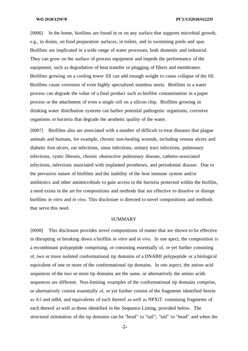
[0029] FIG. 1 depicts an exemplary IHF NTHI tip-directed chimeric recombinant peptide
used to generate polyclonal serum in chinchillas and rabbits. The "IhfA5-mIhfB4 THi
chimer" has an A5 sequence, i.e., Haemophilus influenzae Ih£A5 sequence, followed by a
linker sequence (GPSL) followed by an mB4 polypeptide, i.e., Haemophilus influenzae
mIhfB4 N THi sequence: (SEQ ID NO: 50:
RPGRNPKTGDVVPVSARRVVGPSLFSLHHRQPRLGRNPKTGDSV). The
corresponding structural regions targeted within IHF are shown below the peptide sequence,
as indicated by the arrows.
[0030] FIGS. 2A and 2B show the effectiveness of the chimer recombinant polypeptides.
FIG. 2A shows the reciprocal titers for chinchilla serum and rabbit serum generated using the
IhfA5-mIhfB4 N T Hi chimer recombinant polypeptides. Chinchilla and rabbit serum anti-
IhfA3N THi, anti-IhfA5 N rai, anti-mIhfB4 N rai (IhfA3N rai: SEQ ID NO.: 12, IhfA5N rai: SEQ ID
NO: 13, and mIhfB4 N rai: SEQ ID NO: 17), and anti-IhfA5-mIhfB4 N rai chimer (IhfA5-
mIhfB4 THi chimer: SEQ ID NO: 50) samples were analyzed to assess reactivity with IhfA5-
mIhfB4 NTH i chimer peptide. FIG. 2B depicts the disruption of biofilms formed by
Haemophilus influenzae (NTHI) 86-028NP upon incubation with medium control or various
chinchilla serum as follows: naive serum control, anti-IhfA3 THi, anti-IhfA5 THi, anti-
mIhfB4 THi, and anti-IhfA5-mB4 THi chimer. A 1:50 dilution of chinchilla serum was used.
The reduction in biomass shown is relative to naive serum.
[0031] FIG. 3 depicts the method by which the disruption of biofilms formed by
Haemophilus influenzae (NTHI) 86-028NP was analyzed in the middle ear of adult
chinchillas using Fab fragments generated from polyclonal rabbit anti-IhfB2 THi, anti-
mIhfB4 THi, anti-IhfA5-mIhfB4 THi chimer and naive rabbit serum. Experiments included
cohorts using naive rabbit serum IgG Fab fragments, rabbit anti-IhfB2 N I Fab fragments,
rabbit anti-mIhfB4NTHi Fab fragments, and rabbit anti-IhfA5-mIhfB4NTHi chimer Fab
fragments. On day zero (0), Haemophilus influenzae (NTHI) 86-028NP was inoculated into
the middle ear of chinchillas. Then, either on days 4 and 5 (two dose experiments) or on days
4, 5, and 6 (three dose experiments), Fab fragments were administered by direct delivery into
the middle ear (342 nM). For those administered on day 4 and 5, three (3) chinchillas per
cohort were sacrificed either on day 6 or day 12. For those administered on day 4, 5, and 6,
three (3) chinchillas per cohort were sacrificed on day 13. Following sacrifice, chinchillas
were imaged, middle ear mucosa was collected, adherent biofilm was assessed, middle ear
fluids were collected, quantitation of bacteria was performed, and middle ear fluids were
assessed using a cytokine multiplex assay. The purity of each Fab preparation is also shown
(4 separate preparations: naive, IhfB2, mIhfB4, and IhfA5-mIhfB4 chimer), as confirmed by
10% Bis-Tris PAGE (BioRad) and SYPRO Orange Protein Gel stain (Invitrogen).
[0032] FIG. 4 shows quantitations of colony forming units (CFU) Haemophilus influenzae
(NTHI) 86-028NP per milligram (mg) of mucosal biofilm in chinchillas administered either
rabbit IgGl Fab, rabbit IhfB2 THi Fab, rabbit mIhfB4 THi Fab, or rabbit IhfA5-mIhfB4 THi
chimer Fab after Haemophilus influenzae (NTHI) 86-028NP challenge and biofilm
formation. Quantitations were performed in three different experiments, as follows: (1) two
doses of the respective Fabs were administered, followed by sacrifice 1 day later; (2) two
doses of the respective Fabs were administered, followed by sacrifice 7 days later; and (3)
three doses of the respective Fabs were administered, followed by sacrifice 7 days later.
Doses of Fabs were administered on days 4 and 5 (two doses) or on days 4, 5, and 6 (three
doses) after Haemophilus influenzae (NTHI) 86-028NP challenge. P values are shown: *P <
0.05, ** P < 0.01, and *** P < 0.001.
[0033] FIG. 5 shows quantitations of colony forming units (CFU) Haemophilus influenzae
(NTHI) 86-028NP per milliliter (ml) of middle ear fluid in chinchillas administered either
rabbit IgGl Fab, IhfB2 THi Fab, mIhfB4 THi Fab, or IhfA5-mIhfB4 THi chimer Fab after
Haemophilus influenzae (NTHI) 86-028NP challenge and biofilm formation. Quantitations
were performed in three different experiments, as follows: (1) two doses of the respective
Fabs were administered, followed by sacrifice 1 day later; (2) two doses of the respective
Fabs were administered, followed by sacrifice 7 days later; and (3) three doses of the
respective Fabs were administered, followed by sacrifice 7 days later. Doses of Fabs were
administered on days 4 and 5 (two doses) or on days 4, 5, and 6 (three doses) after
Haemophilus influenzae (NTHI) 86-028NP challenge. P values are shown: *P < 0.05 and **
P < . \ .
[0034] FIG. 6 shows mean mucosal biofilm scores (7 blinded reviewers) for chinchillas
administered either rabbit IgGl Fab, IhfB2 THi Fab, mIhfB4 THi Fab, or IhfA5-mIhfB4 THi
chimer Fab after Haemophilus influenzae (NTHI) 86-028NP challenge and biofilm
formation. Quantitations were performed in three different experiments, as follows: (1) two
doses of the respective Fabs were administered, followed by sacrifice 1 day later; (2) two
doses of the respective Fabs were administered, followed by sacrifice 7 days later; and (3)
three doses of the respective Fabs were administered, followed by sacrifice 7 days later.
Doses of Fabs were administered on days 4 and 5 (two doses) or on days 4, 5, and 6 (three
doses) after Haemophilus influenzae (NTHI) 86-028NP challenge. A mucosal biofilm score
scale was used to rank the residual biofilm within the middle ear space. Using an established
rubric, a score of 0 to 4 was assigned to each image, as follows: zero (0): no biofilm visible;
1 : biofilm fills >0 to <25% of middle ear space; 2 : biofilm fills >25% to <50% of middle ear
space; 3 : biofilm fills >50% to <75% middle ear space; and 4 : biofilm fills >75% to 100%
middle ear space. P values are shown: *P < 0.05 and **P < 0.01 .
[0035] FIG. 7 shows the relative quantity of a panel of pro- and anti-inflammatory
cytokines in clarified middle ear fluids in chinchillas administered either naive rabbit IgG
Fab, rabbit anti-mIhfB2 THi IgG Fab, rabbit anti-mIhfB4 THi IgG Fab, or rabbit anti-IhfA5-
mIh B4 THi chimer IgG Fab after Haemophilus influenzae (NTHI) 86-028NP challenge and
biofilm formation. The pro-inflammatory cytokines measured included: IL- Ι β, IL-6, IL-8,
IL-12p70, IL-17A, TNF, and IFNy. The anti-inflammatory cytokines measured included:
IL-4, IL-10, and IL-13.
[0036] FIGS. 8A-8C show the results of a study evaluating the therapeutic efficacy of
IHF NTHI Fab fragments + HMGB1-C45S.
DESCRIPTION OF TABLES
[0037] Table 1 is a summary of examples of conformational tip domain polypeptides.
[0038] Table 2 is a summary of the scoring methodology used in the otitis media model of
Example 3 to generate a mucosal biomass score.
[0039] Table 3 is a summary of the efficacies of rabbit IgG Fab polyclonal fragments
versus intact rabbit polyclonal IgG.
DETAILED DESCRIPTION
[0040] Unless defined otherwise, all technical and scientific terms used herein have the
same meanings as commonly understood by one of ordinary skill in the art to which this
disclosure belongs. All nucleotide sequences provided herein are presented in the 5' to 3'
direction. Although any methods and materials similar or equivalent to those described
herein can be used in the practice or testing of the present disclosure, particular, non-limiting
exemplary methods, devices, and materials are now described. All technical and patent
publications cited herein are incorporated herein by reference in their entirety. Nothing herein
is to be construed as an admission that the disclosure is not entitled to antedate such
disclosure by virtue of prior invention.
[0041] The practice of the present disclosure will employ, unless otherwise indicated,
conventional techniques of tissue culture, immunology, molecular biology, microbiology, cell
biology and recombinant DNA, which are within the skill of the art. See, e.g., Green and
Sambrook eds. (2012) Molecular Cloning: A Laboratory Manual, 4th edition; the series
Ausubel et al. eds. (2015) Current Protocols in Molecular Biology; the series Methods in
Enzymology (Academic Press, Inc., N.Y.); MacPherson et al. (2015) PCR 1 : A Practical
Approach (IRL Press at Oxford University Press); MacPherson et al. (1995) PCR 2 : A
Practical Approach; McPherson et al. (2006) PCR: The Basics (Garland Science); Harlow
and Lane eds. (1999) Antibodies, A Laboratory Manual; Greenfield ed. (2014) Antibodies, A
Laboratory Manual; Freshney (2010) Culture of Animal Cells: A Manual of Basic Technique,
6th edition; Gait ed. (1984) Oligonucleotide Synthesis; U.S. Pat. No. 4,683,195; Hames and
Higgins eds. (1984) Nucleic Acid Hybridization; Anderson (1999) Nucleic Acid
Hybridization; Herdewijn ed. (2005) Oligonucleotide Synthesis: Methods and Applications;
Hames and Higgins eds. (1984) Transcription and Translation; Buzdin and Lukyanov ed.
(2007) Nucleic Acids Hybridization: Modern Applications; Immobilized Cells and Enzymes
(IRL Press (1986)); Grandi ed. (2007) In Vitro Transcription and Translation Protocols, 2nd
edition; Guisan ed. (2006) Immobilization of Enzymes and Cells; Perbal (1988) A Practical
Guide to Molecular Cloning, 2nd edition; Miller and Calos eds, (1987) Gene Transfer
Vectors for Mammalian Cells (Cold Spring Harbor Laboratory); Makrides ed. (2003) Gene
Transfer and Expression in Mammalian Cells; Mayer and Walker eds. (1987)
Immunochemical Methods in Cell and Molecular Biology (Academic Press, London);
Lundblad and Macdonald eds. (2010) Handbook of Biochemistry and Molecular Biology, 4th
edition; and Herzenberg et al. eds (1996) Weir's Handbook of Experimental Immunology, 5th
edition.
[0042] All numerical designations, e.g., pH, temperature, time, concentration, and
molecular weight, including ranges, are approximations which are varied (+) or (-) by
increments of 1.0 or 0.1, as appropriate or alternatively by a variation of +/- 15%, or
alternatively 10% or alternatively 5% or alternatively 2%. It is to be understood, although
not always explicitly stated, that all numerical designations are preceded by the term "about".
It also is to be understood, although not always explicitly stated, that the reagents described
herein are merely exemplary and that equivalents of such are known in the art.
[0043] As used in the specification and claims, the singular form "a", "an" and "the"
include plural references unless the context clearly dictates otherwise. For example, the term
"a polypeptide" includes a plurality of polypeptides, including mixtures thereof.
[0044] As used herein, the term "comprising" is intended to mean that the compositions and
methods include the recited elements, but do not exclude others. "Consisting essentially of
when used to define compositions and methods, shall mean excluding other elements of any
essential significance to the combination for the intended use. Thus, a composition
consisting essentially of the elements as defined herein would not exclude trace contaminants
from the isolation and purification method and pharmaceutically acceptable carriers, such as
phosphate buffered saline, preservatives (e.g., sodium benzoate, potassium sorbate, and
methyl hydroxybenzoate), and the like. "Consisting of shall mean excluding more than
trace elements of other ingredients and substantial method steps for administering the
compositions disclosed herein. Embodiments defined by each of these transition terms are
within the scope of this disclosure.
[0045] A "biofilm" intends an organized community of microorganisms that at times
adhere to the surface of a structure that may be organic or inorganic, together with the
polymers such as DNA that they secrete and/or release. Biofilms are very resistant to
microbiotics and antimicrobial agents. They live on various organic and inorganic surfaces,
e.g., gingival tissues, teeth and restorations, causing caries and periodontal disease, also
known as periodontal plaque disease. They also cause middle ear infections. They can also
form on the surface of dental implants, stents, catheter lines and contact lenses. They grow
on pacemakers, heart valve replacements, artificial joints and other surgical implants. The
Centers for Disease Control) estimate that over 65% of nosocomial (hospital-acquired)
infections are caused by biofilms. They cause vaginal infections and lead to life-threatening
systemic infections in people with hobbled immune systems. Biofilms also are involved in
numerous diseases, including but not limited to those caused by Aggregatibacter
actinomycetemcomitans, Borrelia burgdorferi (e.g., B31), Bordetella pertussis (e.g., Tohama
I), Burkholderiapseudomallei (e.g., 668), Burkholderia cenocepacia (e.g., HI2424),
Escherichia coli (e.g., K12 MG1655), Enterococcusfaecalis (e.g., V583), Haemophilus
influenzae (e.g., Rd KW20), Helicobacterpylori (e.g., 26695), Klebsiella pneumoniae,
Moraxella catarrhalis (e.g., RH4), Mycobacterium smegmatis (e.g., MC2), Mycobacterium
tuberculosis (e.g., CDC1551), Neisseria gonorrhoeae (e.g., FA1090), Neisseria meningitidis
(e.g., MC58), Pseudomonas aeruginosa, Porphyromonas gingivalis (e.g., W83), Prevotella
intermedia (e.g., 17), Prevotella melaninogenica (e.g., ATCC ® 25845), Staphylococcus
aureus (e.g., MW2), Staphylococcus epidermidis (e.g., RP62A), Streptococcus
agalactiae (e.g., 2603V/R), Streptococcus bovis, Streptococcus gallolyticus (e.g., UCN34),
Streptococcus gordonii (e.g., NCTC 7868 (Challis)), Streptococcus mutans (e.g., UA159),
Streptococcus pneumoniae (e.g., R6), Streptococcus pyogenes (e.g., MGAS10270),
Streptococcus sobrinus (e.g., 6715), Salmonella enterica (e.g., t , CT18), Treponema
denticola (e.g., ATCC ® 35405), Treponema palladum (e.g., Nichols), Vibrio cholera (e.g.,
El Tor, N16961). Additional organisms known to associate with and/or form biofilms
include but are not limited to Campylobacter spp., Candida spp., Legionella pneumophila,
and Listeria monocytogenes. For instance, cystic fibrosis patients
have Pseudomonas infections that often result in antibiotic resistant biofilms. Other diseases
associated with biofilms include, but are not limited to, lung infections of cystic fibrosis
patients, otitis media, post-tympanostomy tube ottorhea, chronic suppurative otitis media,
native valve infectious endocarditis, osteomyelitis, rhinosinositis, prostatitis, urinary tract
infection, wounds, dental caries and periodontitis. Foodborne pathogens, such as but not
limited to some of the above listed organisms (e.g., Listeria monocytogenes, Escherichia coli,
Salmonella enterica) may also form biofilms on the food that they contaminate. Disease
causing biofilms in animals (e.g., Escherichia coli, Salmonella, and Shigella species) may
also cause downstream food contamination and/or disease in human hosts. Further, biofilms
need not be of one homogeneous microbial population and may incorporate other pathogens
and even host cells. In addition to being associated with disease - both nosocomial and
otherwise - and food contamination, biofilms are often causes of industrial contamination,
most notably in relation to process waters and surfaces in contact therewith. Complications
involving organisms that form biofilm as industrial contaminants include but are not limited
to biocorrosion, biofouling, and equipment damage as a result of biofilm formation. Non-
limiting exemplary organisms associated with biofilms in industrial settings include those
disclosed in Ferrera et al. (2015) Biofouling 31(2): 173-180 and Desulfovibrio species.
Additional details regarding biofilms may be found in, for example, Donlan (2002) Emerging
Infectious Diseases 8(9):881-890.
[0046] The term "to prevent formation of a biofilm" intends a prevention in the formation
of, or structure of, the DNA/protein matrix that is a component of a microbial biofilm.
[0047] The terms "to dissolve" or "to disrupt" a biofilm intends a reduction or disruption in
the formation of, or structure of, the DNA/protein matrix that is a component of a microbial
biofilm.
[0048] The term "nucleoid associated protein" or "NAP" as used herein refers to a class of
proteins that affect the dynamic spatial organization of nucleic acids in the nucleoid of
prokaryotic cells. These proteins organize the genome through effecting DNA bending,
binding and aggregation. Certain NAPs are DNA binding proteins and may be associated
with the biofilm including, DNABII proteins, DPS (Genbank Accession No.: CAA49169), H-
NS (Genbank Accession No.: CAA47740), Hfq (Genbank Accession No.: ACE63256), CbpA
(Genbank Accession No.: BAA03950) and CbpB (Genbank Accession No.: NP_418813).
Of the NAPs, DNABII proteins are distinct and generally have strong sequence identity with
alpha helical dimerization domains and may comprise anti-parallel beta ribbons, which often
have NPXiT amino acid motifs, comprising tips that bind and intercalate into the minor
groove of DNA and kink it.
[0049] A "DNABII polypeptide or protein" intends a DNA binding protein or polypeptide
that is composed of DNA-binding domains and thus have a specific or general affinity for
microbial DNA. In one aspect, they bind DNA in the minor grove. Non-limiting examples of
DNABII proteins are an integration host factor (IHF) protein and a histone-like protein (HU).
[0050] The term "Haemophilus influenzae" refers to pathogenic bacteria that can cause
many different infections such as, for example, ear infections, eye infections, and sinusitis.
Many different strains of Haemophilus influenzae have been isolated and have an IhfA gene
or protein. Some non-limiting examples of different strains of Haemophilus
influenzae include Rd KW20, 86-028NP, R2866, PittGG, PittEE, R2846, and 2019.
[0051] An "integration host factor" or "IHF" protein is a bacterial protein that is used by
bacteriophages to incorporate their DNA into the host bacteria. They also bind extracellular
microbial DNA. The genes that encode the IHF protein subunits in E. coli are himA
(Genbank Accession No.: POA6X7.1) and himD (POA6Y1.1) genes. Homologs for these
genes are found in other organisms and as described in in Table 10 of U.S. Patent No.
8,999,291, incorporated herein by reference.
[0052] "HU" or "histone-like protein" refers to a class of heterodimeric proteins typically
associate with E. coli. HU proteins are known to bind DNA Holliday junctions or other bent
structures. Related proteins have been isolated from other microorganisms. The complete
amino acid sequence of E. coli HU was reported by Laine et al. (1980) Eur. J . Biochem
103(3):447-481. The genes that encode the HU protein subunits in E. coli are hupA and
hupB corresponding to SEQ ID NOs. 8 and 9, respectively. A Haemophilus influenzae
homolog derived from non-typeable Haemophilus influenzae (NTHI) corresponds to SEQ ID
NO. 3 . Homologs for these genes are found in other organisms as described in Table 10 of
U.S. Patent No. 8,999,291, incorporated herein by reference.
[0053] "Microbial DNA" intends single or double stranded DNA from a microorganism
that is used to produce the extracellular matrix of a biofilm.
[0054] "A conformational tip domain" of a polypeptide refers to a polypeptide that
comprises a primary amino acid sequence wherein the structure has an anti-parallel beta
ribbon with a sharp turn that is typically mediated by a proline residue. The "tip" of an IHF
polypeptide is shown in Figure 1.
[0055] "Treating an infection" intends a reduction in the number of microbes, e.g., bacteria,
and in one aspect as used herein, the term is intended to be associated with the formation of a
biofilm. Methods to determine if the number of microbes has been reduced are known in the
art and include in vivo and ex vivo assays, as well as a reduction in the clinical symptoms of
an infection. Because bacteria are protected by the biofilms, the bacteria become resistance
to the use of antibacterials. By breaking down the biofilm one can reduce or inhibit bacterial
resistance to antibacterial and other agents as well as treat the bacterial infection.
[0056] A "bent polynucleotide" intends a double strand polynucleotide that contains a small
loop on one strand which does not pair with the other strand. In some embodiments, the loop
is from 1 base to about 20 bases long, or alternatively from 2 bases to about 15 bases long, or
alternatively from about 3 bases to about 12 bases long, or alternatively from about 4 bases to
about 10 bases long, or alternatively has about 4, 5, or 6, or 7, or 8, or 9, or 10 bases.
[0057] "Polypeptides that compete with DNABII proteins in DNA binding" intend proteins
or peptides that compete with IHF or HU in binding bent or distorted DNA structures but do
not form a biofilm with the DNA. Examples, without limitation, include conformational tip
fragments of IHF that include one or more DNA binding domains of the IHF, or the
biological equivalents thereof.
[0058] As used herein, the term "specifically recognize or bind" intends that the binding
agent, e.g., a antibody, antigen binding fragment or a Fab (fragment antigen binding) is more
likely than not to bind to its intended target or binding partner.
[0059] A "subject" of diagnosis or treatment is a cell or an animal such as a mammal, or a
human. A subject is not limited to a specific species and includes non-human animals subject
to diagnosis or treatment and are those subject to infections or animal models, for example,
simians, murines, such as, rats, mice, chinchilla, canine, such as dogs, leporids, such as
rabbits, livestock, sport animals, and pets. Human patients are included within the term as
well.
[0060] The term "protein", "peptide" and "polypeptide" are used interchangeably and in
their broadest sense to refer to a compound of two or more subunit amino acids, amino acid
analogs or peptidomimetics. The subunits may be linked by peptide bonds. In another
embodiment, the subunit may be linked by other bonds, e.g., ester, ether, etc. A protein or
peptide must contain at least two amino acids and no limitation is placed on the maximum
number of amino acids which may comprise a protein's or peptide's sequence. As used herein
the term "amino acid" refers to either natural and/or unnatural or synthetic amino acids,
including glycine and both the D and L optical isomers, amino acid analogs and
peptidomimetics.
[0061] The terms "polynucleotide" and "oligonucleotide" are used interchangeably and
refer to a polymeric form of nucleotides of any length, either deoxyribonucleotides or
ribonucleotides or analogs thereof. Polynucleotides can have any three-dimensional structure
and may perform any function, known or unknown. T he following are non-limiting
examples of polynucleotides: a gene or a gene fragment (for example, a probe, primer, EST
or SAGE tag), exons, introns, messenger RNA (mRNA), transfer RNA, ribosomal RNA,
RNAi, ribozymes, cDNA, recombinant polynucleotides, branched polynucleotides, plasmids,
vectors, isolated DNA of any sequence, isolated RNA of any sequence, nucleic acid probes
and primers. A polynucleotide can comprise modified nucleotides, such as methylated
nucleotides and nucleotide analogs. If present, modifications to the nucleotide structure can
be imparted before or after assembly of the polynucleotide. The sequence of nucleotides can
be interrupted by non-nucleotide components. A polynucleotide can be further modified after
polymerization, such as by conjugation with a labeling component. The term also refers to
both double- and single-stranded molecules. Unless otherwise specified or required, any
embodiment disclosed herein that is a polynucleotide encompasses both the double-stranded
form and each of two complementary single-stranded forms known or predicted to make up
the double-stranded form.
[0062] A polynucleotide is composed of a specific sequence of four nucleotide bases:
adenine (A); cytosine (C); guanine (G); thymine (T); and uracil (U) for thymine when the
polynucleotide is RNA. Thus, the term "polynucleotide sequence" is the alphabetical
representation of a polynucleotide molecule. This alphabetical representation can be input
into databases in a computer having a central processing unit and used for bioinformatics
applications such as functional genomics and homology searching.
[0063] The term "isolated" or "recombinant" as used herein with respect to nucleic acids,
such as DNA or RNA, refers to molecules separated from other DNAs or RNAs, respectively
that are present in the natural source of the macromolecule as well as polypeptides. The term
"isolated or recombinant nucleic acid" is meant to include nucleic acid fragments which are
not naturally occurring as fragments and would not be found in the natural state. The term
"isolated" is also used herein to refer to polynucleotides, polypeptides and proteins that are
isolated from other cellular proteins and is meant to encompass both purified and
recombinant polypeptides. In other embodiments, the term "isolated or recombinant" means
separated from constituents, cellular and otherwise, in which the cell, tissue, polynucleotide,
peptide, polypeptide, protein, antibody or fragment(s) thereof, which are normally associated
in nature. For example, an isolated cell is a cell that is separated from tissue or cells of
dissimilar phenotype or genotype. An isolated polynucleotide is separated from the 3' and 5'
contiguous nucleotides with which it is normally associated in its native or natural
environment, e.g., on the chromosome. As is apparent to those of skill in the art, a non-
naturally occurring polynucleotide, peptide, polypeptide, protein, antibody or fragment(s)
thereof, does not require "isolation" to distinguish it from its naturally occurring counterpart.
[0064] It is to be inferred without explicit recitation and unless otherwise intended, that
when the present disclosure relates to a polypeptide, protein, polynucleotide or antibody, an
equivalent or a biologically equivalent of such is intended within the scope of this disclosure.
As used herein, the term "biological equivalent thereof is intended to be synonymous with
"equivalent thereof when referring to a reference protein, antibody, fragment, polypeptide or
nucleic acid, intends those having minimal homology while still maintaining desired structure
or functionality. Unless specifically recited herein, it is contemplated that any
polynucleotide, polypeptide or protein mentioned herein also includes equivalents thereof. In
one aspect, an equivalent polynucleotide is one that hybridizes under stringent conditions to
the polynucleotide or complement of the polynucleotide as described herein for use in the
described methods. In another aspect, an equivalent antibody or antigen binding polypeptide
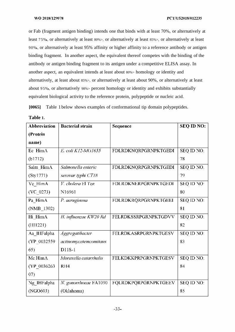
or Fab (fragment antigen binding) intends one that binds with at least 70%, or alternatively at
least 7 5% , or alternatively at least 80%>, or alternatively at least 85%>, or alternatively at least
90% , or alternatively at least 95% affinity or higher affinity to a reference antibody or antigen
binding fragment. In another aspect, the equivalent thereof competes with the binding of the
antibody or antigen binding fragment to its antigen under a competitive ELISA assay. In
another aspect, an equivalent intends at least about 80%> homology or identity and
alternatively, at least about 85%>, or alternatively at least about 90%, or alternatively at least
about 9 5% , or alternatively 98%> percent homology or identity and exhibits substantially
equivalent biological activity to the reference protein, polypeptide or nucleic acid.
[0065] Table 1 below shows examples of conformational tip domain polypeptides.
Table 1.
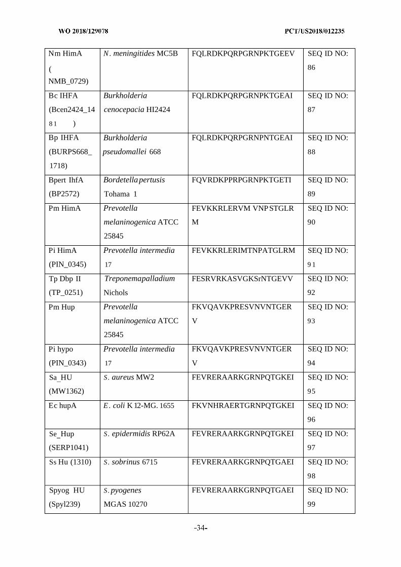
Nm HimA N. meningitides MC5B FQLRDKPQRPGRNPKTGEEV SEQ ID NO:
( 86
NMB_0729)
Bc IHFA Burkholderia FQLRDKPQRPGRNPKTGEAI SEQ ID NO:
(Bcen2424_14 cenocepacia HI2424 87
8 1 )
Bp IHFA Burkholderia FQLRDKPQRPGRNPNTGEAI SEQ ID NO:
(BURPS668_ pseudomallei 668 88
1718)
Bpert IhfA Bordetellapertusis FQVRDKPPRPGRNPKTGETI SEQ ID NO:
(BP2572) Tohama 1 89
Pm HimA Prevotella FEVKKRLERVM VNP STGLR SEQ ID NO:
melaninogenica ATCC M 90
25845
Pi HimA Prevotella intermedia FEVKKRLERIMTNPATGLRM SEQ ID NO:
(PIN_0345) 17 9 1
Tp Dbp II Treponemapalladium FESRVRKASVGKSrNTGEVV SEQ ID NO:
(TP_0251) Nichols 92
Pm Hup Prevotella FKVQAVKPRESVNVNTGER SEQ ID NO:
melaninogenica ATCC V 93
25845
Pi hypo Prevotella intermedia FKVQAVKPRESVNVNTGER SEQ ID NO:
(PIN_0343) 17 V 94
Sa HU S. aureus MW2 FEVRERAARKGRNPQTGKEI SEQ ID NO:
(MW1362) 95
Ec hupA E. coli K 12-MG. 1655 FKVNHRAERTGRNPQTGKEI SEQ ID NO:
96
Se Hup S. epidermidis RP62A FEVRERAARKGRNPQTGKEI SEQ ID NO:
(SERP1041) 97
Ss Hu (1310) S. sobrinus 6715 FEVRERAARKGRNPQTGAEI SEQ ID NO:
98
Spyog HU S. pyogenes FEVRERAARKGRNPQTGAEI SEQ ID NO:
(Spyl239) MGAS 10270 99
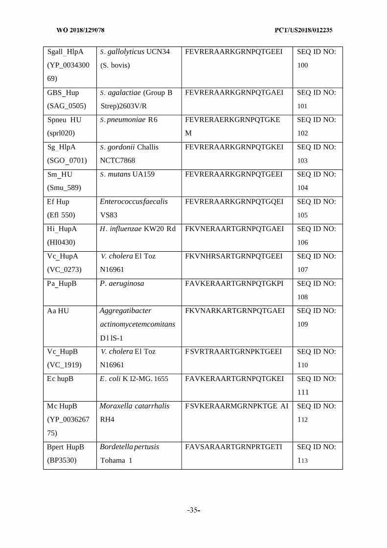
Sgall HlpA S. gallolyticus UCN34 FEVRERAARKGRNPQTGEEI SEQ ID NO:
(YP_0034300 (S. bovis) 100
69)
GBS Hup S. agalactiae (Group B FEVRERAARKGRNPQTGAEI SEQ ID NO:
(SAG_0505) Strep)2603V/R 101
Spneu HU S. pneumoniae R6 FEVRERAERKGRNPQTGKE SEQ ID NO:
(sprl020) M 102
Sg HlpA S. gordonii Challis FEVRERAARKGRNPQTGKEI SEQ ID NO:
(SGO 0701) NCTC7868 103
Sm HU S. mutans UA159 FEVRERAARKGRNPQTGEEI SEQ ID NO:
(Smu_589) 104
Ef Hup Enterococcusfaecalis FEVRERAARKGRNPQTGQEI SEQ ID NO:
(Efl 550) VS83 105
Hi HupA H. influenzae KW20 Rd FKVNERAARTGRNPQTGAEI SEQ ID NO:
(HI0430) 106
Vc HupA V. cholera El Toz FKVNHRSARTGRNPQTGEEI SEQ ID NO:
(VC_0273) N16961 107
Pa HupB P. aeruginosa FAVKERAARTGRNPQTGKPI SEQ ID NO:
108
Aa HU Aggregatibacter FKVNARKARTGRNPQTGAEI SEQ ID NO:
actinomycetemcomitans 109
D l lS-1
Vc HupB V. cholera El Toz FSVRTRAARTGRNPKTGEEI SEQ ID NO:
(VC_1919) N16961 110
Ec hupB E. coli K 12-MG. 1655 FAVKERAARTGRNPQTGKEI SEQ ID NO:
111
Mc HupB Moraxella catarrhalis FSVKERAARMGRNPKTGE AI SEQ ID NO:
(YP_0036267 RH4 112
75)
Bpert HupB Bordetella pertusis FAVSARAARTGRNPRTGETI SEQ ID NO:
(BP3530) Tohama 1 113
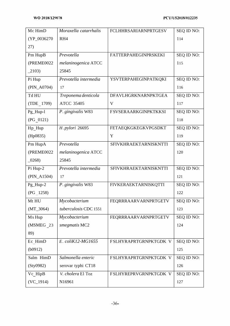
Mc HimD Moraxella catarrhalis FCLHHRSARIARNPRTGESV SEQ ID NO:
(YP_0036270 RH4 114
27)
Pm HupB Prevotella FATTERPAHEGINPRSKEKI SEQ ID NO:
(PREME0022 melaninogenica ATCC 115
_2103) 25845
Pi Hup Prevotella intermedia YSVTERPAHEGINPATKQKI SEQ ID NO:
(PIN_A0704) 17 116
Td HU Treponema denticola DFAVLHGRKNARNPKTGEA SEQ ID NO:
(TDE 1709) ATCC 35405 V 117
Pg_Hup-l P. gingivalis W83 FSVSERAARKGINPKTKKSI SEQ ID NO:
(PG 0121) 118
Hp Hup H. pylori 26695 FETAEQKGKEGKVPGSDKT SEQ ID NO:
(Hp0835) Y 119
Pm HupA Prevotella SFIVKHRAEKTARNISKNTTI SEQ ID NO:
(PREME0022 melaninogenica ATCC 120
_0268) 25845
Pi Hup-2 Prevotella intermedia SFIVKHRAEKTARNISKNTTI SEQ ID NO:
(PIN_A1504) 17 121
Pg_Hup-2 P. gingivalis W83 FIVKERAEKTARNISKQTTI SEQ ID NO:
(PG 1258) 122
Mt HU Mycobacterium FEQRRRAARVARNPRTGETV SEQ ID NO:
(MT_3064) tuberculosis CDC 1551 123
Ms Hup Mycobacterium FEQRRRAARVARNPRTGETV SEQ ID NO:
(MSMEG 23 smegmatis MC2 124
89)
Ec HimD E. coliK12-MG1655 FSLHYRAPRTGRNPKTGDK V SEQ ID NO:
(b0912) 125
Salm HimD Salmonella enteric FSLHYRAPRTGRNPKTGDK V SEQ ID NO:
(Sty0982) serovar typhi CT18 126
Vc HipB V. cholera El Toz FSLHYREPRVGRNPKTGDK V SEQ ID NO:
(VC_1914) N16961 127
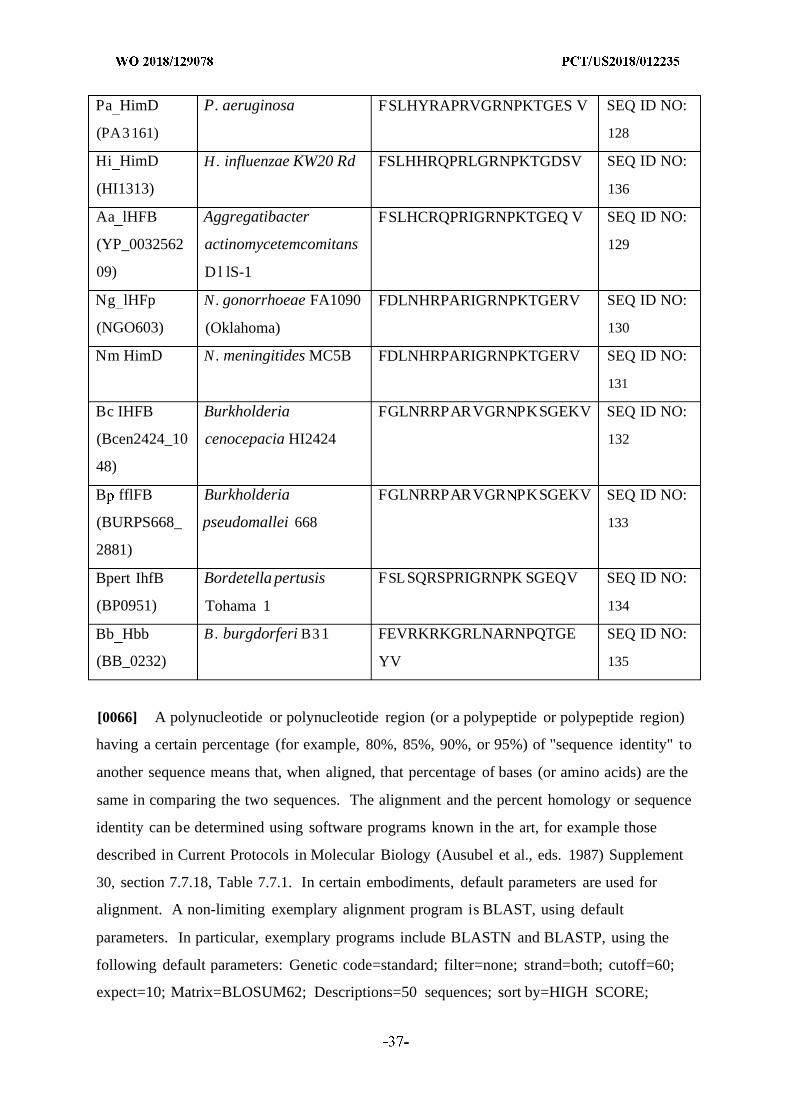
Pa HimD P. aeruginosa FSLHYRAPRVGRNPKTGES V SEQ ID NO:
(PA3161) 128
Hi HimD H. influenzae KW20 Rd FSLHHRQPRLGRNPKTGDSV SEQ ID NO:
(HI1313) 136
Aa lHFB Aggregatibacter FSLHCRQPRIGRNPKTGEQ V SEQ ID NO:
(YP_0032562 actinomycetemcomitans 129
09) D l lS-1
Ng lHFp N. gonorrhoeae FA1090 FDLNHRPARIGRNPKTGERV SEQ ID NO:
(NGO603) (Oklahoma) 130
Nm HimD N. meningitides MC5B FDLNHRPARIGRNPKTGERV SEQ ID NO:
131
Bc IHFB Burkholderia FGLNRRPARVGR PKSGEKV SEQ ID NO:
(Bcen2424_10 cenocepacia HI2424 132
48)
B fflFB Burkholderia FGLNRRPARVGR PKSGEKV SEQ ID NO:
(BURPS668_ pseudomallei 668 133
2881)
Bpert IhfB Bordetella pertusis FSLSQRSPRIGRNPK SGEQV SEQ ID NO:
(BP0951) Tohama 1 134
Bb Hbb B. burgdorferi B31 FEVRKRKGRLNARNPQTGE SEQ ID NO:
(BB_0232) YV 135
[0066] A polynucleotide or polynucleotide region (or a polypeptide or polypeptide region)
having a certain percentage (for example, 80%, 85%, 90%, or 95%) of "sequence identity" to
another sequence means that, when aligned, that percentage of bases (or amino acids) are the
same in comparing the two sequences. The alignment and the percent homology or sequence
identity can be determined using software programs known in the art, for example those
described in Current Protocols in Molecular Biology (Ausubel et al., eds. 1987) Supplement
30, section 7.7.18, Table 7.7.1. In certain embodiments, default parameters are used for
alignment. A non-limiting exemplary alignment program is BLAST, using default
parameters. In particular, exemplary programs include BLASTN and BLASTP, using the
following default parameters: Genetic code=standard; filter=none; strand=both; cutoff=60;
expect=10; Matrix=BLOSUM62; Descriptions=50 sequences; sort by=HIGH SCORE;
Databases=non-redundant, GenBank+EMBL+DDBJ+PDB+GenBank CDS
translations+SwissProtein+SPupdate+PIR. Details of these programs can be found at the
following Internet address: ncbi.nlm.nih.gov/cgi-bin/BLAST. Sequence identity and percent
identity can determined by incorporating them into clustalW (available at the web
address:genome.jp/tools/clustalw/, last accessed on Jan. 13, 2017).
[0067] "Homology" or "identity" or "similarity" refers to sequence similarity between two
peptides or between two nucleic acid molecules. Homology can be determined by comparing
a position in each sequence that may be aligned for purposes of comparison. When a position
in the compared sequence is occupied by the same base or amino acid, then the molecules are
homologous at that position. A degree of homology between sequences is a function of the
number of matching or homologous positions shared by the sequences. An "unrelated" or
"non-homologous" sequence shares less than 40% identity, or alternatively less than 25%
identity, with one of the sequences of the present disclosure.
[0068] "Homology" or "identity" or "similarity" can also refer to two nucleic acid
molecules that hybridize under stringent conditions.
[0069] "Hybridization" refers to a reaction in which one or more polynucleotides react to
form a complex that is stabilized via hydrogen bonding between the bases of the nucleotide
residues. The hydrogen bonding may occur by Watson-Crick base pairing, Hoogstein
binding, or in any other sequence-specific manner. The complex may comprise two strands
forming a duplex structure, three or more strands forming a multi- stranded complex, a single
self-hybridizing strand, or any combination of these. A hybridization reaction may constitute
a step in a more extensive process, such as the initiation of a PCR reaction, or the enzymatic
cleavage of a polynucleotide by a ribozyme.
[0070] Examples of stringent hybridization conditions include: incubation temperatures of
about 25° C . to about 37° C ; hybridization buffer concentrations of about 6><SSC to about
lOxSSC; formamide concentrations of about 0% to about 25%; and wash solutions from
about 4xSSC to about 8><SSC. Examples of moderate hybridization conditions include:
incubation temperatures of about 40° C . to about 50° C ; buffer concentrations of about
9xSSC to about 2><SSC; formamide concentrations of about 30% to about 50%; and wash
solutions of about 5><SSC to about 2><SSC. Examples of high stringency conditions include:
incubation temperatures of about 55° C . to about 68° C ; buffer concentrations of about
lxSSC to about O.lxSSC; formamide concentrations of about 55% to about 75%; and wash
solutions of about IxSSC, O.lxSSC, or deionized water. In general, hybridization incubation
times are from 5 minutes to 24 hours, with 1, 2, or more washing steps, and wash incubation
times are about 1, 2, or 15 minutes. SSC is 0.15 M NaCl and 15 mM citrate buffer. It is
understood that equivalents of SSC using other buffer systems can be employed.
[0071] As used herein, "expression" refers to the process by which polynucleotides are
transcribed into mRNA and/or the process by which the transcribed mRNA is subsequently
being translated into peptides, polypeptides, or proteins. If the polynucleotide is derived from
genomic DNA, expression may include splicing of the mRNA in an eukaryotic cell.
[0072] The term "encode" as it is applied to polynucleotides refers to a polynucleotide
which is said to "encode" a polypeptide if, in its native state or when manipulated by methods
well known to those skilled in the art, it can be transcribed and/or translated to produce the
mRNA for the polypeptide and/or a fragment thereof. The antisense strand is the
complement of such a nucleic acid, and the encoding sequence can be deduced therefrom.
[0073] As used herein, the terms "treating," "treatment" and the like are used herein to
mean obtaining a desired pharmacologic and/or physiologic effect. The effect may be
prophylactic in terms of completely or partially preventing a disorder or sign or symptom
thereof, and/or may be therapeutic in terms of a partial or complete cure for a disorder and/or
adverse effect attributable to the disorder. Methods to determine if treatment has occurred
are known in the art and briefly described herein.
[0074] To prevent intends to prevent a disorder or effect in vitro or in vivo in a system or
subject that is predisposed to the disorder or effect. An example of such is preventing the
formation of a biofilm in a system that is infected with a microorganism known to produce
one.
[0075] A "composition" is intended to mean a combination of active agent and another
compound or composition, inert (for example, a detectable agent or label) or active, such as
an adjuvant.
[0076] A "pharmaceutical composition" is intended to include the combination of an active
agent with a carrier, inert or active, making the composition suitable for diagnostic or
therapeutic use in vitro, in vivo or ex vivo.
[0077] "Pharmaceutically acceptable carriers" refers to any diluents, excipients, or carriers
that may be used in the compositions disclosed herein. Pharmaceutically acceptable carriers
include ion exchangers, alumina, aluminum stearate, lecithin, serum proteins, such as human
serum albumin, buffer substances, such as phosphates, glycine, sorbic acid, potassium
sorbate, partial glyceride mixtures of saturated vegetable fatty acids, water, salts or
electrolytes, such as protamine sulfate, disodium hydrogen phosphate, potassium hydrogen
phosphate, sodium chloride, zinc salts, colloidal silica, magnesium trisilicate, polyvinyl
pyrrolidone, cellulose-based substances, polyethylene glycol, sodium
carboxymethylcellulose, polyacrylates, waxes, polyethylene-polyoxypropylene-block
polymers, polyethylene glycol, microspheres, microparticles, or nanoparticles (comprising
e.g., biodegradable polymers such as Poly(Lactic Acid-co-Glycolic Acid)), and wool fat.
Suitable pharmaceutical carriers are described in Remington's Pharmaceutical Sciences,
Mack Publishing Company, a standard reference text in this field. They may be selected with
respect to the intended form of administration, that is, oral tablets, capsules, elixirs, syrups
and the like, and consistent with conventional pharmaceutical practices.
[0078] A "biologically active agent" or an active agent disclosed herein intends one or
more of an isolated or recombinant polypeptide, an isolated or recombinant polynucleotide, a
vector, an isolated host cell, or an antibody, as well as compositions comprising one or more
of same.
[0079] "Administration" or "delivery" of a therapeutic or other agent can be effected in one
dose, continuously or intermittently throughout the course of treatment. Methods of
determining the most effective means and dosage of administration are known to those of
skill in the art and will vary with the composition used for therapy, the purpose of the
therapy, the target cell being treated, and the subject being treated. Single or multiple
administrations can be carried out with the dose level and pattern being selected by the
treating physician. Suitable dosage formulations and methods of administering the agents are
known in the art. Route of administration can also be determined and method of determining
the most effective route of administration are known to those of skill in the art and will vary
with the composition used for treatment, the purpose of the treatment, the health condition or
disease stage of the subject being treated, and target cell or tissue. Non-limiting examples of
route of administration include oral administration, nasal administration, inhalation, injection,
and topical application. Administration can be for use in industrial as well as therapeutic
applications.
[0080] An agent (an antibody or fragment thereof, a polypeptide, a polynucleotide, a cell, a
composition or a vaccine) of the present disclosure can be administered for its intended use
whether in vitro or in vivo (e.g., therapeutically) by any suitable route of administration. It
will also be appreciated that the optimal route will vary with the condition and age of the
recipient, and the disease being treated. The agent may be used in industrial settings and for
the treatment of animals. When used in industrial settings, the biofilm is contacted with the
agent, e.g., Fab (fragment antigen binding), antibody, polypeptide or vaccine.
[0081] The term "effective amount" refers to a quantity sufficient to achieve a desired
effect. In the context of therapeutic or prophylactic applications, the effective amount will
depend on the type and severity of the condition at issue and the characteristics of the
individual subject, such as general health, age, sex, body weight, species, and tolerance to
pharmaceutical compositions. In the context of this disclosure, in some embodiments the
effective amount is the amount sufficient to result a reduction in biofilm mass or in breaking
down a biofilm. In other aspects, the amount is effective to treat a bacterial infection
associated with a biofilm in a subject. In other embodiments, in the context of a Fab
(fragment antigen binding) or antibody, the effective amount is the amount sufficient to result
in breaking down, diminishing or disrupting a biofilm. In other embodiments, the effective
amount of an agent or an immunogenic composition is the amount sufficient to result in
antibody generation against the antigen. In some embodiments, the effective amount is the
amount required to confer passive immunity on a subject in need thereof. With respect to
compositions, in some embodiments the effective amount will depend on the intended use,
the health/responsiveness of the subject's immune system, in addition to the factors described
above. The skilled artisan will be able to determine appropriate amounts depending on these
and other factors.
[0082] In the case of an in vitro application, in some embodiments the effective amount
will depend on the size and nature of the application in question. It will also depend on the
nature and sensitivity of the in vitro target and the methods in use. The skilled artisan will be
able to determine the effective amount based on these and other considerations. The effective
amount may comprise one or more administrations of a composition depending on the
embodiment.
[0083] The term "potency" as it relates to the potency of a drug, such as an antibiotic refers
to to a measure of drug activity expressed in terms of the amount required to produce an
effect of given intensity. A highly potent drug evokes a given response at low
concentrations, while a drug of lower potency evokes the same response only at higher
concentrations. The potency depends on both the affinity and efficacy.
[0084] The term "conjugated moiety" refers to a moiety that can be added to an isolated
chimeric polypeptide by forming a covalent bond with a residue of chimeric polypeptide.
The moiety may bond directly to a residue of the chimeric polypeptide or may form a
covalent bond with a linker which in turn forms a covalent bond with a residue of the
chimeric polypeptide.
[0085] A "peptide conjugate" or "recombinant polypeptide" refers to the association by
covalent or non-covalent bonding of one or more polypeptides with each other and/or with
another chemical or biological compound. In a non-limiting example, the "conjugation" of a
polypeptide with a chemical compound results in improved stability or efficacy of the
polypeptide for its intended purpose. In one embodiment, the active agents of this disclosure
are conjugated to a carrier, wherein the carrier is a liposome, a micelle, or a pharmaceutically
acceptable polymer.
[0086] "Liposomes" are microscopic vesicles consisting of concentric lipid bilayers. A
liposome is an example of a carrier, e.g., a pharmaceutically acceptable carrier. Structurally,
liposomes range in size and shape from long tubes to spheres, with dimensions from a few
hundred Angstroms to fractions of a millimeter. Vesicle-forming lipids are selected to
achieve a specified degree of fluidity or rigidity of the final complex providing the lipid
composition of the outer layer. These are neutral (cholesterol) or bipolar and include
phospholipids, such as phosphatidylcholine (PC), phosphatidylethanolamine (PE),
phosphatidylinositol (PI), and sphingomyelin (SM) and other types of bipolar lipids including
but not limited to dioleoylphosphatidylethanolamine (DOPE), with a hydrocarbon chain
length in the range of 14-22, and saturated or with one or more double C=C bonds. Examples
of lipids capable of producing a stable liposome, alone, or in combination with other lipid
components are phospholipids, such as hydrogenated soy phosphatidylcholine (HSPC),
lecithin, phosphatidylethanolamine, lysolecithin, lysophosphatidylethanol-amine,
phosphatidyl serine, phosphatidylinositol, sphingomyelin, cephalin, cardiolipin, phosphatidic
acid, cerebrosides, distearoylphosphatidylethan-olamine (DSPE),
dioleoylphosphatidylcholine (DOPC), dipalmitoylphosphatidylcholine (DPPC),
palmitoyloteoylphosphatidylcholine (POPC), palmitoyloleoylphosphatidylethanolamine
(POPE) and dioleoylphosphatidylethanolamine 4-(N-maleimido-triethyl)cyclohexane-l-
carboxylate (DOPE-mal). Additional non-phosphorous containing lipids that can become
incorporated into liposomes include stearylamine, dodecylamine, hexadecylamine, isopropyl
myristate, triethanolamine-lauryl sulfate, alkyl-aryl sulfate, acetyl palmitate, glycerol

ricinoleate, hexadecyl stereate, amphoteric acrylic polymers, polyethyloxylated fatty acid
amides, and the cationic lipids mentioned above (DDAB, DODAC, DMRIE, DMTAP,
DOGS, DOTAP (DOTMA), DOSPA, DPTAP, DSTAP, DC-Chol). Negatively charged lipids
include phosphatidic acid (PA), dipalmitoylphosphatidylglycerol (DPPG),
dioteoylphosphatidylglycerol and (DOPG), dicetylphosphate that are able to form vesicles.
Typically, liposomes can be divided into three categories based on their overall size and the
nature of the lamellar structure. The three classifications, as developed by the New York
Academy Sciences Meeting, "Liposomes and Their Use in Biology and Medicine,"
December 1977, are multi-lamellar vesicles (MLVs), small uni-lamellar vesicles (SUVs) and
large uni-lamellar vesicles (LUVs). The biological active agents can be encapsulated in such
for administration in accordance with the methods described herein.
[0087] A "micelle" is an aggregate of surfactant molecules dispersed in a liquid colloid. A
typical micelle in aqueous solution forms an aggregate with the hydrophilic "head" regions in
contact with surrounding solvent, sequestering the hydrophobic tail regions in the micelle
center. This type of micelle is known as a normal phase micelle (oil-in-water micelle).
Inverse micelles have the head groups at the center with the tails extending out (water-in-oil
micelle). Micelles can be used to attach a polynucleotide, polypeptide, antibody, antigen
binding fragment, vaccine, or composition described herein to facilitate efficient delivery to
the target cell or tissue.
[0088] The phrase "pharmaceutically acceptable polymer" refers to the group of
compounds which can be conjugated to one or more polypeptides or antibodies described
here. It is contemplated that the conjugation of a polymer to the polypeptide or antibody is
capable of extending the half-life of the polypeptide in vivo and in vitro. Non-limiting
examples include polyethylene glycols, polyvinylpyrrolidones, polyvinylalcohols, cellulose
derivatives, polyacrylates, polymethacrylates, sugars, polyols and mixtures thereof. The
biological active agents can be conjugated to a pharmaceutically acceptable polymer for
administration in accordance with the methods described herein.
[0089] A "gene delivery vehicle" is defined as any molecule that can carry inserted
polynucleotides into a host cell. Examples of gene delivery vehicles are liposomes, micelles
biocompatible polymers, including natural polymers and synthetic polymers; lipoproteins;
polypeptides; polysaccharides; lipopolysaccharides; artificial viral envelopes; metal particles;
and bacteria, or viruses, such as baculovirus, adenovirus and retrovirus, bacteriophage,
cosmid, plasmid, fungal vectors and other recombination vehicles typically used in the art
which have been described for expression in a variety of eukaryotic and prokaryotic hosts,
and may be used for gene therapy as well as for simple protein expression.
[0090] A polynucleotide disclosed herein can be delivered to a cell or tissue using a gene
delivery vehicle. "Gene delivery," "gene transfer," "transducing," and the like as used
herein, are terms referring to the introduction of an exogenous polynucleotide (sometimes
referred to as a "transgene") into a host cell, irrespective of the method used for the
introduction. Such methods include a variety of well-known techniques such as vector-
mediated gene transfer (by, e.g., viral infection/transfection, or various other protein-based or
lipid-based gene delivery complexes) as well as techniques facilitating the delivery of
"naked" polynucleotides (such as electroporation, "gene gun" delivery and various other
techniques used for the introduction of polynucleotides). The introduced polynucleotide may
be stably or transiently maintained in the host cell. Stable maintenance typically requires that
the introduced polynucleotide either contains an origin of replication compatible with the host
cell or integrates into a replicon of the host cell such as an extrachromosomal replicon (e.g., a
plasmid) or a nuclear or mitochondrial chromosome. A number of vectors are known to be
capable of mediating transfer of genes to mammalian cells, as is known in the art and
described herein.
[0091] As used herein the term "eDNA" refers to extracellular DNA found as a component
to pathogenic biofilms.
[0092] A "plasmid" is an extra-chromosomal DNA molecule separate from the
chromosomal DNA which is capable of replicating independently of the chromosomal DNA.
In many cases, it is circular and double-stranded. Plasmids provide a mechanism for
horizontal gene transfer within a population of microbes and typically provide a selective
advantage under a given environmental state. Plasmids may carry genes that provide
resistance to naturally occurring antibiotics in a competitive environmental niche, or
alternatively the proteins produced may act as toxins under similar circumstances.
[0093] "Plasmids" used in genetic engineering are called "plasmid vectors". Many
plasmids are commercially available for such uses. The gene to be replicated is inserted into
copies of a plasmid containing genes that make cells resistant to particular antibiotics and a
multiple cloning site (MCS, or polylinker), which is a short region containing several
commonly used restriction sites allowing the easy insertion of DNA fragments at this
location. Another major use of plasmids is to make large amounts of proteins. In this case,
researchers grow bacteria containing a plasmid harboring the gene of interest. Just as the
bacterium produces proteins to confer its antibiotic resistance, it can also be induced to
produce large amounts of proteins from the inserted gene.
[0094] A "yeast artificial chromosome" or "YAC" refers to a vector used to clone large
DNA fragments (larger than 100 kb and up to 3000 kb). It is an artificially constructed
chromosome and contains the telomeric, centromeric, and replication origin sequences
needed for replication and preservation in yeast cells. Built using an initial circular plasmid,
they are linearized by using restriction enzymes, and then DNA ligase can add a sequence or
gene of interest within the linear molecule by the use of cohesive ends. Yeast expression
vectors, such as YACs, Yips (yeast integrating plasmid), and YEps (yeast episomal plasmid),
are extremely useful as one can get eukaryotic protein products with posttranslational
modifications as yeasts are themselves eukaryotic cells, however YACs have been found to
be more unstable than BACs, producing chimeric effects.
[0095] A "viral vector" is defined as a recombinantly produced virus or viral particle that
comprises a polynucleotide to be delivered into a host cell, either in vivo, ex vivo or in vitro.
[0096] Examples of viral vectors include retroviral vectors, adenovirus vectors, adeno-
associated virus vectors, alphavirus vectors and the like. Infectious tobacco mosaic virus
(TMV)-based vectors can be used to manufacturer proteins and have been reported to express
Griffithsin in tobacco leaves (O'Keefe et al. (2009) Proc. Nat. Acad. Sci. USA 106(1 5): 6099-
6104). Alphavirus vectors, such as Semliki Forest virus-based vectors and Sindbis virus-
based vectors, have also been developed for use in gene therapy and immunotherapy. See,
Schlesinger & Dubensky (1999) Curr. Opin. Biotechnol. 5:434-439 and Ying et al. (1999)
Nat. Med. 5(7):823-827. In aspects where gene transfer is mediated by a retroviral vector, a
vector construct refers to the polynucleotide comprising the retroviral genome or part thereof,
and a therapeutic gene. Further details as to modern methods of vectors for use in gene
transfer may be found in, for example, Kotterman et al. (2015) Viral Vectors for Gene
Therapy: Translational and Clinical Outlook Annual Review of Biomedical Engineering 17.
[0097] As used herein, "retroviral mediated gene transfer" or "retroviral transduction"
carries the same meaning and refers to the process by which a gene or nucleic acid sequences
are stably transferred into the host cell by virtue of the virus entering the cell and integrating
its genome into the host cell genome. The virus can enter the host cell via its normal
mechanism of infection or be modified such that it binds to a different host cell surface
receptor or ligand to enter the cell. As used herein, retroviral vector refers to a viral particle
capable of introducing exogenous nucleic acid into a cell through a viral or viral-like entry
mechanism.
[0098] Retroviruses carry their genetic information in the form of RNA; however, once the
virus infects a cell, the RNA is reverse-transcribed into the DNA form which integrates into
the genomic DNA of the infected cell. The integrated DNA form is called a provirus.
[0099] In aspects where gene transfer is mediated by a DNA viral vector, such as an
adenovirus (Ad) or adeno-associated virus (AAV), a vector construct refers to the
polynucleotide comprising the viral genome or part thereof, and a transgene. Adenoviruses
(Ads) are a relatively well characterized, homogenous group of viruses, including over 50
serotypes. Ads do not require integration into the host cell genome. Recombinant Ad
derived vectors, particularly those that reduce the potential for recombination and generation
of wild-type virus, have also been constructed. Such vectors are commercially available from
sources such as Takara Bio USA (Mountain View, CA), Vector Biolabs (Philadelphia, PA),
and Creative Biogene (Shirley, NY). Wild-type AAV has high infectivity and specificity
integrating into the host cell's genome. See, Wold and Toth (2013) Curr. Gene. Ther.
13(6):421-433, Hermonat & Muzyczka (1984) Proc. Natl. Acad. Sci. USA 81:6466-6470,
and Lebkowski et al. (1988) Mol. Cell. Biol. 8:3988-3996.
[0100] Vectors that contain both a promoter and a cloning site into which a polynucleotide
can be operatively linked are well known in the art. Such vectors are capable of transcribing
RNA in vitro or in vivo, and are commercially available from sources such as Agilent
Technologies (Santa Clara, Calif.) and Promega Biotech (Madison, Wis.). In order to
optimize expression and/or in vitro transcription, it may be necessary to remove, add or alter
5' and/or 3' untranslated portions of the clones to eliminate extra, potential inappropriate
alternative translation initiation codons or other sequences that may interfere with or reduce
expression, either at the level of transcription or translation. Alternatively, consensus
ribosome binding sites can be inserted immediately 5' of the start codon to enhance
expression.
[0101] Gene delivery vehicles also include DNA/liposome complexes, micelles and
targeted viral protein-DNA complexes. Liposomes that also comprise a targeting antibody or
fragment thereof can be used in the methods disclosed herein. In addition to the delivery of
polynucleotides to a cell or cell population, direct introduction of the proteins described
herein to the cell or cell population can be done by the non-limiting technique of protein
transfection, alternatively culturing conditions that can enhance the expression and/or
promote the activity of the proteins disclosed herein are other non-limiting techniques.
[0102] As used herein, the terms "antibody," "antibodies" and "immunoglobulin" includes
whole antibodies and any antigen binding fragment or a single chain thereof. Thus the term
"antibody" includes any protein or peptide containing molecule that comprises at least a
portion of an immunoglobulin molecule. The terms "antibody," "antibodies" and
"immunoglobulin" also include immunoglobulins of any isotype, fragments of antibodies
which retain specific binding to antigen, including, but not limited to, Fab, Fab', F(ab)2, Fv,
scFv, dsFv, Fd fragments, dAb, VH, VL, VhH, and V-NAR domains; minibodies, diabodies,
triabodies, tetrabodies and kappa bodies; multispecific antibody fragments formed from
antibody fragments and one or more isolated. Examples of such include, but are not limited to
a complementarity determining region (CDR) of a heavy or light chain or a ligand binding
portion thereof, a heavy chain or light chain variable region, a heavy chain or light chain
constant region, a framework (FR) region, or any portion thereof, at least one portion of a
binding protein, chimeric antibodies, humanized antibodies, species-ized antibodies, single-
chain antibodies, and fusion proteins comprising an antigen-binding portion of an antibody
and a non-antibody protein. The variable regions of the heavy and light chains of the
immunoglobulin molecule contain a binding domain that interacts with an antigen. The
constant regions of the antibodies (Abs) may mediate the binding of the immunoglobulin to
host tissues. The term "anti-" when used before a protein name, anti-IHF, anti-HU, anti-
OMP P5, for example, refers to a monoclonal or polyclonal antibody that binds and/or has an
affinity to a particular protein. For example, "anti-IHF" refers to an antibody that binds to the
IHF protein. The specific antibody may have affinity or bind to proteins other than the
protein it was raised against. For example, anti-IHF, while specifically raised against the IHF
protein, may also bind other proteins that are related either through sequence homology or
through structure homology.
[0103] The antibodies can be polyclonal, monoclonal, multispecific (e.g., bispecific
antibodies), a diabody, and antibody fragments, so long as they exhibit the desired biological
activity. Antibodies can be isolated from any suitable biological source, e.g., a human, a
murine, rat, sheep and canine.
[0104] As used herein, "monoclonal antibody" refers to an antibody obtained from a
substantially homogeneous antibody population. Monoclonal antibodies are highly specific,
as each monoclonal antibody is directed against a single determinant on the antigen. The
antibodies may be detectably labeled, e.g., with a radioisotope, an enzyme which generates a
detectable product, a fluorescent protein, and the like. The antibodies may be further
conjugated to other moieties, such as members of specific binding pairs, e.g., biotin (member
of biotin-avidin specific binding pair), and the like. The antibodies may also be bound to a
solid support, including, but not limited to, polystyrene plates or beads, and the like and can
be used, therapeutically, diagnostically or to isolate a polypeptide.
[0105] Monoclonal antibodies may be generated using hybridoma techniques or
recombinant DNA methods known in the art. A hybridoma is a cell that is produced in the
laboratory from the fusion of an antibody-producing lymphocyte and a non-antibody
producing cancer cell, usually a myeloma or lymphoma. A hybridoma proliferates and
produces a continuous sample of a specific monoclonal antibody. Alternative techniques for
generating or selecting antibodies include in vitro exposure of lymphocytes to antigens of
interest, and screening of antibody display libraries in cells, phage, or similar systems.
[0106] The term "human antibody" as used herein, is intended to include antibodies having
variable and constant regions derived from human germline immunoglobulin sequences. The
human antibodies disclosed herein may include amino acid residues not encoded by human
germline immunoglobulin sequences (e.g., mutations introduced by random or site-specific
mutagenesis in vitro or by somatic mutation in vivo). However, the term "human antibody"
as used herein, is not intended to include antibodies in which CDR sequences derived from
the germline of another mammalian species, such as a mouse, have been grafted onto human
framework sequences. Thus, as used herein, the term "human antibody" refers to an antibody
in which substantially every part of the protein (e.g., CDR, framework, CL , CH domains (e.g.,
CH I , CH2, CH3), hinge, (VL, VH)) is substantially non-immunogenic in humans, with only
minor sequence changes or variations. Similarly, antibodies designated primate (monkey,
baboon, chimpanzee, etc.), rodent (mouse, rat, rabbit, guinea pig, hamster, and the like) and
other mammals designate such species, sub-genus, genus, sub-family, family specific
antibodies. Further, chimeric antibodies include any combination of the above. Such
changes or variations optionally retain or reduce the immunogenicity in humans or other
species relative to non-modified antibodies. Thus, a human antibody is distinct from a
chimeric or humanized antibody. It is pointed out that a human antibody can be produced by
a non-human animal or prokaryotic or eukaryotic cell that is capable of expressing
functionally rearranged human immunoglobulin (e.g., heavy chain and/or light chain) genes.
Further, when a human antibody is a single chain antibody, it can comprise a linker peptide
that is not found in native human antibodies. For example, an Fv can comprise a linker
peptide, such as two to about eight glycine or other amino acid residues, which connects the
variable region of the heavy chain and the variable region of the light chain. Such linker
peptides are considered to be of human origin. Additional non-limiting examples of linker
polypeptides are provided herein.
[0107] As used herein, a human antibody is "derived from" a particular germline sequence
if the antibody is obtained from a system using human immunoglobulin sequences, e.g., by
immunizing a transgenic mouse carrying human immunoglobulin genes or by screening a
human immunoglobulin gene library. A human antibody that is "derived from" a human
germline immunoglobulin sequence can be identified as such by comparing the amino acid
sequence of the human antibody to the amino acid sequence of human germline
immunoglobulins. A selected human antibody typically is at least 90% identical in amino
acids sequence to an amino acid sequence encoded by a human germline immunoglobulin
gene and contains amino acid residues that identify the human antibody as being human when
compared to the germline immunoglobulin amino acid sequences of other species (e.g.,
murine germline sequences). In certain cases, a human antibody may be at least 95%, or
even at least 96%, 97%, 98%, or 99% identical in amino acid sequence to the amino acid
sequence encoded by the germline immunoglobulin gene. Typically, a human antibody
derived from a particular human germline sequence will display no more than 10 amino acid
differences from the amino acid sequence encoded by the human germline immunoglobulin
gene. In certain cases, the human antibody may display no more than 5, or even no more
than 4, 3, 2, or 1 amino acid difference from the amino acid sequence encoded by the
germline immunoglobulin gene.
[0108] A "human monoclonal antibody" refers to antibodies displaying a single binding
specificity which have variable and constant regions derived from human germline
immunoglobulin sequences. The term also intends recombinant human antibodies.
[0109] The term "recombinant human antibody", as used herein, includes all human
antibodies that are prepared, expressed, created or isolated by recombinant means, such as
antibodies isolated from an animal (e.g., a mouse) that is transgenic or transchromosomal for
human immunoglobulin genes or a hybridoma prepared therefrom, antibodies isolated from a
host cell transformed to express the antibody, e.g., from a transfectoma, antibodies isolated
from a recombinant, combinatorial human antibody library, and antibodies prepared,
expressed, created or isolated by any other means that involve splicing of human
immunoglobulin gene sequences to other DNA sequences. Such recombinant human
antibodies have variable and constant regions derived from human germline immunoglobulin
sequences. In certain embodiments, however, such recombinant human antibodies can be
subjected to in vitro mutagenesis (or, when an animal transgenic for human Ig sequences is
used, in vivo somatic mutagenesis) and thus the amino acid sequences of the VH and VL
regions of the recombinant antibodies are sequences that, while derived from and related to
human germline VH and VL sequences, may not naturally exist within the human antibody
germline repertoire in vivo.
[0110] As used herein, chimeric antibodies are antibodies whose light and heavy chain
genes have been constructed, typically by genetic engineering, from antibody variable and
constant region genes belonging to different species.
[0111] As used herein, the term "humanized antibody" or "humanized immunoglobulin"
refers to a human/non-human chimeric antibody that contains a minimal sequence derived
from non-human immunoglobulin. For the most part, humanized antibodies are human
immunoglobulins (recipient antibody) in which residues from a variable region of the
recipient are replaced by residues from a variable region of a non-human species (donor
antibody) such as mouse, rat, rabbit, or non-human primate having the desired specificity,
affinity and capacity. Humanized antibodies may comprise residues that are not found in the
recipient antibody or in the donor antibody. The humanized antibody can optionally also
comprise at least a portion of an immunoglobulin constant region (Fc), typically that of a
human immunoglobulin, a non-human antibody containing one or more amino acids in a
framework region, a constant region or a CDR, that have been substituted with a
correspondingly positioned amino acid from a human antibody. In general, humanized
antibodies are expected to produce a reduced immune response in a human host, as compared
to a non-humanized version of the same antibody. The humanized antibodies may have
conservative amino acid substitutions which have substantially no effect on antigen binding
or other antibody functions. Conservative substitutions groupings include: glycine-alanine,
valine-leucine-isoleucine, phenylalanine-tyrosine, lysine-arginine, alanine-valine, serine-
threonine and asparagine-glutamine. The term "species-ized" refers to antibodies that have
been modified in the same or a similar manner for a non-human species.
[0112] The terms "polyclonal antibody" or "polyclonal antibody composition" as used
herein refer to a preparation of antibodies that are derived from different B-cell lines. They
are a mixture of immunoglobulin molecules secreted against a specific antigen, each
recognizing a different epitope. In some embodiments, the antibody or antigen binding
fragment is not a polyclonal antibody.
[0113] As used herein, the term "antibody derivative", comprises a full-length antibody or a
fragment of an antibody, wherein one or more of the amino acids are chemically modified by
alkylation, pegylation, acylation, ester formation or amide formation or the like, e.g., for
linking the antibody to a second molecule. This includes, but is not limited to, pegylated
antibodies, cysteine-pegylated antibodies, and variants thereof. This disclosure also provided
antibody derivatives of the antibody fragments, e.g., the polypeptides conjugaged to another
molecule, e.g., PEG or further modified by acylation.
[0114] As used herein, the term "immunoconjugate" comprises an antibody, an antibody
fragment or a antibody derivative associated with or linked to a second agent, such as a
cytotoxic agent, a detectable agent, a radioactive agent, a targeting agent, a human antibody,
a humanized antibody, a chimeric antibody, a synthetic antibody, a semisynthetic antibody, or
a multispecific antibody. This disclosure provides immunoconjugates comprising as one
component, an antibody or Fab fragment and the second agent.
[0115] As used herein, the term "label" intends a directly or indirectly detectable compound
or composition that is conjugated directly or indirectly to the composition to be detected, e.g.,
N-terminal histidine tags (N-His), magnetically active isotopes, e.g., Sn, Sn and Sn, a
non-radioactive isotopes such as C and N, polynucleotide or protein such as an antibody
so as to generate a "labeled" composition. The term also includes sequences conjugated to a
polynucleotide that will provide a signal upon expression of the inserted sequences, such as
green fluorescent protein (GFP) and the like. The term also includes purification tags or
labels that aid in the isolation of biological materials from mixed populations. While the term
"label" generally intends compositions covalently attached to the composition to be detected,
in one aspect it specifically excludes naturally occurring nucleosides and amino acids that are
known to fluoresce under certain conditions (e.g., temperature, pH, etc.) when positioned
within the polynucleotide or protein in its native environment and generally any natural
fluorescence that may be present in the composition to be detected. The label may be
detectable by itself (e.g., radioisotope labels or fluorescent labels) or, in the case of an
enzymatic label, may catalyze chemical alteration of a substrate compound or composition
that is detectable. The labels can be suitable for small-scale detection or more suitable for
high-throughput screening. As such, suitable labels include, but are not limited to
magnetically active isotopes, non-radioactive isotopes, radioisotopes, fluorochromes,
chemiluminescent compounds, dyes, and proteins, including enzymes. The label may be
simply detected or it may be quantified. A response that is simply detected generally
comprises a response whose existence merely is confirmed, whereas a response that is
quantified generally comprises a response having a quantifiable (e.g., numerically reportable)
value such as an intensity, polarization, and/or other property. In luminescence or
fluorescence assays, the detectable response may be generated directly using a luminophore
or fluorophore associated with an assay component actually involved in binding, or indirectly
using a luminophore or fluorophore associated with another (e.g., reporter or indicator)
component. Examples of luminescent labels that produce signals include, but are not limited
to bioluminescence and chemiluminescence. Detectable luminescence response generally
comprises a change in, or an occurrence of a luminescence signal. Suitable methods and
luminophores for luminescently labeling assay components are known in the art and
described for example in Haugland, Richard P. (1996) Handbook of Fluorescent Probes and
Research Chemicals (6th ed). Examples of luminescent probes include, but are not limited to,
aequorin and luciferases.
[0116] Examples of suitable fluorescent labels include, but are not limited to, fluorescein,
rhodamine, tetramethylrhodamine, eosin, erythrosin, coumarin, methyl-coumarins, pyrene,
Malacite green, stilbene, Lucifer Yellow, CASCADE BLUE™, and Texas Red.
[0117] In another aspect, the fluorescent label is functionalized to facilitate covalent
attachment to a cellular component present in or on the surface of the cell or tissue such as a
cell surface marker. Suitable functional groups, including, but not are limited to,
isothiocyanate groups, amino groups, haloacetyl groups, maleimides, succinimidyl esters, and
sulfonyl halides, all of which may be used to attach the fluorescent label to a second
molecule. The choice of the functional group of the fluorescent label will depend on the site
of attachment to either a linker, the agent, the marker, or the second labeling agent.
[0118] "Eukaryotic cells" comprise all of the life kingdoms except monera. They can be
easily distinguished through a membrane-bound nucleus. Animals, plants, fungi, and protists
are eukaryotes or organisms whose cells are organized into complex structures by internal
membranes and a cytoskeleton. The most characteristic membrane-bound structure is the
nucleus. Unless specifically recited, the term "host" includes a eukaryotic host, including, for
example, yeast, higher plant, insect and mammalian cells. Non-limiting examples of
eukaryotic cells or hosts include simian, bovine, porcine, murine, rat, avian, reptilian and
human.
[0119] "Prokaryotic cells" that usually lack a nucleus or any other membrane-bound
organelles and are divided into two domains, bacteria and archaea. In addition to
chromosomal DNA, these cells can also contain genetic information in a circular loop called
on episome. Bacterial cells are very small, roughly the size of an animal mitochondrion
(about 1-2 µ in diameter and 10 µιη long). Prokaryotic cells feature three major shapes: rod
shaped, spherical, and spiral. Instead of going through elaborate replication processes like
eukaryotes, bacterial cells divide by binary fission. Examples include but are not limited
to Bacillus bacteria, E. coli bacterium, and Salmonella bacterium.
[0120] A "native" or "natural" antigen is a polypeptide, protein or a fragment which
contains an epitope, which has been isolated from a natural biological source, and which can
specifically bind to an antigen receptor, in particular a T cell antigen receptor (TCR), in a
subject.
[0121] The terms "antigen" and "antigenic" refer to molecules with the capacity to be
recognized by an antibody or otherwise act as a member of an antibody-ligand pair. "Specific
binding" refers to the interaction of an antigen with the variable regions of immunoglobulin
heavy and light chains. Antibody-antigen binding may occur in vivo or in vitro. The skilled
artisan will understand that macromolecules, including proteins, nucleic acids, fatty acids,
lipids, lipopolysaccharides and polysaccharides have the potential to act as an antigen. The
skilled artisan will further understand that nucleic acids encoding a protein with the potential
to act as an antibody ligand necessarily encode an antigen. The artisan will further
understand that antigens are not limited to full-length molecules, but can also include partial
molecules. The term "antigenic" is an adjectival reference to molecules having the properties
of an antigen. The term encompasses substances that are immunogenic, i.e., immunogens, as
well as substances which induce immunological unresponsiveness, or anergy, i.e., anergens.
[0122] An "altered antigen" is one having a primary sequence that is different from that of
the corresponding wild-type antigen. Polypeptide mB4 (SEQ ID NO.: 17) is an example of
an altered B4 antigen (SEQ ID NO.: 16). Altered antigens can be made by synthetic or
recombinant methods and include, but are not limited to, antigenic peptides that are
differentially modified during or after translation, e.g., by phosphorylation, glycosylation,
cross-linking, acylation, proteolytic cleavage, linkage to an antibody molecule, membrane
molecule or other ligand (Ferguson et al. (1988) Ann. Rev. Biochem. 57:285-320).
[0123] A "self-antigen" also referred to herein as a native or wild-type antigen is an
antigenic peptide that induces little or no immune response in the subject due to self-tolerance
to the antigen. An example of a self-antigen is the melanoma specific antigen gplOO.
[0124] "Immune response" broadly refers to the antigen-specific responses of lymphocytes
to foreign substances. The terms "immunogen" and "immunogenic" refer to molecules with
the capacity to elicit an immune response. All immunogens are antigens; however, not all
antigens are immunogenic. An immune response disclosed herein can be humoral (via
antibody activity) or cell-mediated (via T cell activation). The response may occur in vivo or
in vitro. The skilled artisan will understand that a variety of macromolecules, including
proteins, nucleic acids, fatty acids, lipids, lipopolysaccharides and polysaccharides have the
potential to be immunogenic. The skilled artisan will further understand that nucleic acids
encoding a molecule capable of eliciting an immune response necessarily encode an
immunogen. The artisan will further understand that immunogens are not limited to full-
length molecules, but may include partial molecules.
[0125] The term "passive immunity" refers to the transfer of immunity from one subject to
another through the transfer of antibodies. Passive immunity may occur naturally, as when
maternal antibodies are transferred to a fetus. Passive immunity may also occur artificially as
when antibody compositions are administered to non-immune subjects. Antibody donors and
recipients may be human or non-human subjects. Antibodies may be polyclonal or
monoclonal, may be generated in vitro or in vivo, and may be purified, partially purified, or
unpurified depending on the embodiment. In some embodiments described herein, passive
immunity is conferred on a subject in need thereof through the administration of antibodies or
antigen binding fragments that specifically recognize or bind to a particular antigen. In some
embodiments, passive immunity is conferred through the administration of an isolated or
recombinant polynucleotide encoding an antibody or antigen binding fragment that
specifically recognizes or binds to a particular antigen.
[0126] As used herein, the term "inducing an immune response in a subject" is a term well
understood in the art and intends that an increase of at least about 2-fold, at least about 5-fold,
at least about 10-fold, at least about 100-fold, at least about 500-fold, or at least about 1000-
fold or more in an immune response to an antigen (or epitope) can be detected or measured,
after introducing the antigen (or epitope) into the subject, relative to the immune response (if
any) before introduction of the antigen (or epitope) into the subject. An immune response to
an antigen (or epitope), includes, but is not limited to, production of an antigen-specific (or
epitope-specific) antibody, and production of an immune cell expressing on its surface a
molecule which specifically binds to an antigen (or epitope). Methods of determining
whether an immune response to a given antigen (or epitope) has been induced are well known
in the art. For example, antigen-specific antibody can be detected using any of a variety of
immunoassays known in the art, including, but not limited to, ELISA, wherein, for example,
binding of an antibody in a sample to an immobilized antigen (or epitope) is detected with a
detectably-labeled second antibody (e.g., enzyme-labeled mouse anti-human Ig antibody).
[0127] As used herein, the term "anti-inflammatory cytokines" includes immunoregulatory
molecules that control the proinflammatory cytokine response. Cytokines act together with
specific cytokine inhibitors and soluble cytokine receptors to regulate the human immune
response. Major anti-inflammatory cytokines include interleukin (IL)-l receptor antagonist,
IL-4, IL-6, IL-10, IL-1 1, and IL-13. Specific cytokine receptors for IL-1, tumor necrosis
factor-alpha, and IL-1 8 also function as proinflammatory cytokine inhibitors. Methods of
measuring cytokine, including anti-inflammatory cytokine, levels are well known in the art.
For example, serum cytokine levels can be measured using commercially available enzyme-
linked immuno-sorbent assay (ELISA) kits.
[0128] As used herein, "solid phase support" or "solid support", used interchangeably, is
not limited to a specific type of support. Rather a large number of supports are available and
are known to one of ordinary skill in the art. Solid phase supports include silica gels, resins,
derivatized plastic films, glass beads, cotton, plastic beads, alumina gels. As used herein,
"solid support" also includes synthetic antigen-presenting matrices, cells, and liposomes. A
suitable solid phase support may be selected on the basis of desired end use and suitability for
various protocols. For example, for peptide synthesis, solid phase support may refer to resins
such as polystyrene (e.g., PAM-resin obtained from Bachem Inc., ChemPep, Inc., etc.),
PolyHIPE resins, which is a copolymer based on polystyrene with grafted
polydimethylacrylamide; HIPE = high internal phase emulsionpolyamide resin (obtained
from Sigma-Aldrich, St. Louis, MO), polystyrene resin grafted with polyethylene glycol
(TentaGel®, Rapp Polymere, Tubingen, Germany) or polydimethylacrylamide resin
(obtained fromSigma-Aldrich, St. Louis, MO).
[0129] An example of a solid phase support include glass, polystyrene, polypropylene,
polyethylene, dextran, nylon, amylases, natural and modified celluloses, polyacrylamides,
gabbros, and magnetite. The nature of the carrier can be either soluble to some extent or
insoluble. The support material may have virtually any possible structural configuration so
long as the coupled molecule is capable of binding to a polynucleotide, polypeptide or
antibody. Thus, the support configuration may be spherical, as in a bead, or cylindrical, as in
the inside surface of a test tube, or the external surface of a rod. Alternatively, the surface
may be flat such as a sheet, test strip, etc. or alternatively polystyrene beads. Those skilled in
the art will know many other suitable carriers for binding antibody or antigen, or will be able
to ascertain the same by use of routine experimentation.
Modes for Carrying out the Disclosure
Chimer Recombinant Polypeptides
[0130] This disclosure provides a recombinant polypeptide comprising, or consisting
essentially of, or yet further consisting of, two or more isolated conformational tip domains of
a DNABII polypeptide, e.g., a fragment of a DNABII polypeptide that in one aspect, contains
the NPXiT peptide motif, or a biological equivalent of one or more of the conformational tip
domains or fragments thereof, wherein for fragments having the motif, "Xi" is any amino
acid or alternatively '¾" is selected from the amino acids Q, R, K, S, or T. In one aspect,
the amino acid sequences of the two or more tip domains are the same, or alternatively the
amino acids sequences are different. Non-limiting examples of the conformational tip
domains comprise, or alternatively consist essentially of, or yet further consist of the
fragments identified herein as A5 and mB4, and equivalents of each thereof and NPXiT
containing fragments of each thereof wherein '¾" is any amino acid or alternatively '¾" is
selected from the amino acids Q, R, K, S, or T. The structural orientation of the tip domains
can be "head" to tail; tail to head wherein the polypeptide comprises 3 or more tip domains,
any combination of head to tails, e.g., head-head-head; tail-head-heard; tail-head-tail, wherein
the amine terminus of the wild-type sequence is the "head" and the carboxy terminus of the
wild-type sequence is the "tail" of the polypeptide. Non-limiting examples of the
polypeptides include, without limitiation the polypeptides disclosed herein, e.g., the
polypeptides identified in the Sequence Listing.
[0131] Further provided as polyeptides are those disclosed as Al to A4 and A6 and B l to
B6, disclosed above as Sequence ID NOs.: 18, 19, 12, 20, 21, 22, 15, 23, 16, 24, and 25,
which do not contain the conformation tip domain, and equivalents of these polypeptides
from different organisms identified herein that produce a DNABII polypeptide. The
sequences comprise:
MATITKLDIIE YL SDKYHLS (also referred to herein as Al; (SEQ ID NO. 18));
KYHLSKQDTKNVVENFLEEI (also referred to herein as A2; (SEQ ID NO. 19));
FLEEIRL SLESGQDVKLSGF (also referred to herein as A3; (SEQ ID NO. 12));
KLSGFGNFELRDKSSRPGRN (also referred to herein as A4; (SEQ ID NO. 20));
ARRVVTFKPGQKLRARVEKTK (also referred to herein as A6; (SEQ ID NO. 21));
MTKSELMEKLSAKQPTLSAK (also referred to herein as B l (SEQ ID NO. 22));
TLSAKEIENMVKDILEFISQ (also referred to herein as B2 (SEQ ID NO. 15));
EFISQSLENGDRVEVRGF GS (also referred to herein as B3 (SEQ ID NO. 23));
RGFGSF SLHHRQPRLGRNPK (also referred to herein as B4 (SEQ ID NO. 16));
GRNPKTGDSVNLSAKSVPYF (also referred to herein as B5; (SEQ ID NO. 24)); and
SVPYFKAGKELKARVDVQA (also referred to herein as B6; (SEQ ID NO. 25));
[0132] Non-limiting examples of DNABII polypeptides include an IHF or HU alpha or beta
polypeptide; an IHF alpha polypeptide; Moraxella catarrhalis HU; E. coli HupA, HupB,
himA, himD; E.faecalis HU (such as V583).
[0133] In a further aspect, the recombinant polypeptide further comprises, or alternatively
consists essentially of, or yet further consists of a linker polypeptide that further comprises, or
alternatively consists essentially of, or yet further consists of 1 or more amino acids. Non-
limiting examples of linker polypeptides include, without limitiation those identified herein.
[0134] Also provided are recombinant polypeptides that comprise, or alternatively consist
essentially of, or yet further consist of, between 3 and 5 conformational tip domains that can
be produced by the same or different bacterial species, the amino acids sequences of which
can be the same (e.g., all A5 amino acid sequences) or at least 2 or at least 3 or at least 4 or
all 5 having different amino acid sequences (e.g., various combinations of A5 and mB4 and
equivalents and NPXiT containing fragments of each thereof), wherein X1 is any amino acid,
or in one aspect, an amino selected from the amino acids Q, R, K, S, or T. The
conformational tip domains in the recombinant polypeptides can be in a linear or branched
conformation. They can further comprise a detectable and/or a purification label linked
thereto. The structural orientation of the tip domains can be "head" to tail; tail to head
wherein the polypeptide comprises 3 or more tip domains, any combination of head to tails,
e.g., head-head-head; tail-head-heard; tail-head-tail, wherein the amine terminus of the wild-
type sequence is the "head" and the carboxy terminus of the wild-type sequence is the "tail"
of the polypeptide. In one aspet, the polypeptides in sum can be between 4 1 and 120 amino
acids in length.
[0135] Non-limiting examples of equivalent polypeptides, include a polypeptide having at
least 60%, or alternatively at least 65%, or alternatively at least 70%, or alternatively at least
7 5% , or alternatively 80%>, or alternatively at least 85%>, or alternatively at least 90%, or
alternatively at least 95% identity thereto or for polypeptide sequences, or a polypeptide
which is encoded by a polynucleotide or its complement that hybridizes under conditions of
high stringency to a polynucleotide encoding such polypeptide sequences. Conditions of
high stringency are described herein and incorporated herein by reference. Applicants have
determined that the bolded and underlined amino acids are heavily conserved and therefore in
one aspect, are not modified or altered in designing an equivalent polypeptide. Additional
examples of equivalent polypeptides include, for example
DKSSRPGRNPXiTGDVVAASARR (SEQ ID NO.: 77), wherein "Χ is any amino acid or
alternatively is selected from the amino acids Q, R, K, S, or T.
[0136] Equivalent polypeptides also include a polypeptide consisting of, or comprising the
above noted polypeptides with the addition of up to 25, or alternatively 20, or alternatively
15, or alternatively up to 10, or alternatively up to 5 random amino acids on either the amine
or carboxy termini (or on both). In another aspect, they equivalent polypetide includes a
polypeptide consisting of, or comprising the above noted polypeptides with the addition of up
to 25, or alternatively 20, or alternatively 15, or alternatively up to 10, or alternatively up to 5
amino acids on either the amine or carboxy termini (or on both) selected from the adjacent
amino acids of the corresponding wild-type sequence and equivalents of the wild-type
adjacent amino acids.
[0137] The proteins and polypeptides are obtainable by a number of processes known to
those of skill in the art, which include purification, chemical synthesis and recombinant
methods. Polypeptides can be isolated from preparations such as host cell systems by
methods such as immunoprecipitation with antibody, and standard techniques such as gel
filtration, ion-exchange, reversed-phase, and affinity chromatography. For such methodology,
see for example Deutscher et al. (1999) Guide To Protein Purification: Methods In
Enzymology (Vol. 182, Academic Press). Accordingly, this disclosure also provides the
processes for obtaining these polypeptides as well as the products obtainable and obtained by
these processes.
[0138] The polypeptides also can be obtained by chemical synthesis using a commercially
available automated peptide synthesizer such as those manufactured by Perkin/Elmer/ Applied
Biosystems, Inc., Model 430A or 431 A, Foster City, Calif, USA. The synthesized
polypeptide can be precipitated and further purified, for example by high performance liquid
chromatography (HPLC). Accordingly, this disclosure also provides a process for chemically
synthesizing the proteins disclosed herein by providing the sequence of the protein and
reagents, such as amino acids and enzymes and linking together the amino acids in the proper
orientation and linear sequence.
[0139] Alternatively, the proteins and polypeptides can be obtained by well-known
recombinant methods as described, for example, in Sambrook et al. (1989) supra, using a host
cell and vector systems described herein.
[0140] Also provided by this application are the polypeptides described herein conjugated
to a detectable agent for use in the diagnostic methods. For example, detectably labeled
polypeptides can be bound to a column and used for the detection and purification of
antibodies. They also are useful as immunogens for the production of antibodies as described
below. The polypeptides disclosed herein are useful in an in vitro assay system to screen for
agents or drugs, which modulate cellular processes.
[0141] It is well known to those skilled in the art that modifications can be made to the
peptides disclosed herein to provide them with altered properties. As used herein the term
"amino acid" refers to either natural and/or unnatural or synthetic amino acids, including
glycine and both the D or L optical isomers, and amino acid analogs and peptidomimetics. A
peptide of three or more amino acids is commonly called an oligopeptide if the peptide chain
is short. If the peptide chain is long, the peptide is commonly called a polypeptide or a
protein.
[0142] Peptides disclosed herein can be modified to include unnatural amino acids. Thus,
the peptides may comprise D-amino acids, a combination of and L-amino acids, and various
"designer" amino acids (e.g., .beta. -methyl amino acids, C-alpha-methyl amino acids, and N-
alpha-methyl amino acids, etc.) to convey special properties to peptides. Additionally, by
assigning specific amino acids at specific coupling steps, peptides with alpha-helices, beta
turns, beta sheets, gamma-turns, and cyclic peptides can be generated. Generally, it is
believed that .alpha. -helical secondary structure or random secondary structure may be of
particular use.
[0143] The polypeptides disclosed herein also can be combined with various solid phase
carriers, such as an implant, a stent, a paste, a gel, a dental implant, or a medical implant or
liquid phase carriers, such as beads, sterile or aqueous solutions, pharmaceutically acceptable
carriers, pharmaceutically acceptable polymers, liposomes, micelles, suspensions and
emulsions. Examples of non-aqueous solvents include propyl ethylene glycol, polyethylene
glycol and vegetable oils. When used to prepare antibodies or induce an immune response in
vivo, the carriers also can include an adjuvant that is useful to non-specifically augment a
specific immune response. A skilled artisan can easily determine whether an adjuvant is
required and select one. However, for the purpose of illustration only, suitable adjuvants
include, but are not limited to Freund's Complete and Incomplete, mineral salts and
polynucleotides. Other suitable adjuvants include monophosphoryl lipid A (MPL), mutant
derivatives of the heat labile enterotoxin of E. coli, mutant derivatives of cholera toxin, CPG
oligonucleotides, and adjuvants derived from squalene.
[0144] This disclosure also provides a pharmaceutical composition comprising or
alternatively consisting essentially of, or yet further consisting of, any of a polypeptide,
analog, mutein, or fragment disclosed herein, alone or in combination with each other or
other agents, such an antibiotic and an acceptable carrier or solid support. These
compositions are useful for various diagnostic and therapeutic methods as described herein.
Polynucleotides
[0145] This disclosure also provides isolated or recombinant polynucleotides encoding one
or more of the above-identified isolated or recombinant polypeptides and their respective
complementary strands. Vectors comprising the isolated or recombinant polynucleotides are
further provided examples of which are known in the art and briefly described herein. In one
aspect where more than one isolated or recombinant polynucleotide is to be expressed as a
single unit, the isolated or recombinant polynucleotides can be contained within a
polycistronic vector. The polynucleotides can be DNA, RNA, mRNA or interfering RNA,
such as siRNA, miRNA or dsRNA.
[0146] The disclosure further provides the isolated or recombinant polynucleotide
operatively linked to a promoter of RNA transcription, as well as other regulatory sequences
for replication and/or transient or stable expression of the DNA or RNA. As used herein, the
term "operatively linked" means positioned in such a manner that the promoter will direct
transcription of RNA off the DNA molecule. Examples of such promoters are SP6, T4 and
T7. In certain embodiments, cell-specific promoters are used for cell-specific expression of
the inserted polynucleotide. Vectors which contain a promoter or a promoter/enhancer, with
termination codons and selectable marker sequences, as well as a cloning site into which an
inserted piece of DNA can be operatively linked to that promoter are known in the art and
commercially available. For general methodology and cloning strategies, see Gene
Expression Technology (Goeddel ed., Academic Press, Inc. (1991)) and references cited
therein and Vectors: Essential Data Series (Gacesa and Ramji, eds., John Wiley & Sons, N.Y.
(1994)) which contains maps, functional properties, commercial suppliers and a reference to
GenEMBL accession numbers for various suitable vectors.
[0147] In one embodiment, polynucleotides derived from the polynucleotides disclosed
herein encode polypeptides or proteins having diagnostic and therapeutic utilities as
described herein as well as probes to identify transcripts of the protein that may or may not be
present. These nucleic acid fragments can by prepared, for example, by restriction enzyme
digestion of larger polynucleotides and then labeled with a detectable marker. Alternatively,
random fragments can be generated using nick translation of the molecule. For methodology
for the preparation and labeling of such fragments, see, Sambrook et al. (1989) supra.
[0148] Expression vectors containing these nucleic acids are useful to obtain host vector
systems to produce proteins and polypeptides. It is implied that these expression vectors must
be replicable in the host organisms either as episomes or as an integral part of the
chromosomal DNA. Non-limiting examples of suitable expression vectors include plasmids,
yeast vectors, viral vectors and liposomes. Adenoviral vectors are particularly useful for
introducing genes into tissues in vivo because of their high levels of expression and efficient
transformation of cells both in vitro and in vivo. When a nucleic acid is inserted into a
suitable host cell, e.g., a prokaryotic or a eukaryotic cell and the host cell replicates, the
protein can be recombinantly produced. Suitable host cells will depend on the vector and can
include mammalian cells, animal cells, human cells, simian cells, insect cells, yeast cells, and
bacterial cells constructed using known methods. See, Sambrook et al. (1989) supra. In
addition to the use of viral vector for insertion of exogenous nucleic acid into cells, the
nucleic acid can be inserted into the host cell by methods known in the art such as
transformation for bacterial cells; transfection using calcium phosphate precipitation for
mammalian cells; or DEAE-dextran; electroporation; or microinjection. See, Sambrook et al.
(1989) supra, for methodology. Thus, this disclosure also provides a host cell, e.g., a
mammalian cell, an animal cell (rat or mouse), a human cell, or a prokaryotic cell such as a
bacterial cell, containing a polynucleotide encoding a protein or polypeptide or antibody.
[0149] A polynucleotide can comprise modified nucleotides, such as methylated
nucleotides and nucleotide analogs. If present, modifications to the nucleotide structure can
be imparted before or after assembly of the polynucleotide. The sequence of nucleotides can
be interrupted by non-nucleotide components. A polynucleotide can be further modified after
polymerization, such as by conjugation with a labeling component. The term also refers to
both double- and single-stranded molecules. Unless otherwise specified or required, any
embodiment disclosed herein that is a polynucleotide encompasses both the double-stranded
form and each of two complementary single-stranded forms known or predicted to make up
the double-stranded form.
[0150] When the vectors are used for gene therapy in vivo or ex vivo, a pharmaceutically
acceptable vector, such as a replication-incompetent retroviral or adenoviral vector, are
exemplary (but non-limiting) and may be of particular use. Pharmaceutically acceptable
vectors containing the nucleic acids disclosed herein can be further modified for transient or
stable expression of the inserted polynucleotide. As used herein, the term "pharmaceutically
acceptable vector" includes, but is not limited to, a vector or delivery vehicle having the
ability to selectively target and introduce the nucleic acid into dividing cells. An example of
such a vector is a "replication-incompetent" vector defined by its inability to produce viral
proteins, precluding spread of the vector in the infected host cell. An example of a
replication-incompetent retroviral vector is L L6 (Miller et al. (1989) BioTechniques 7:980-
990). The methodology of using replication-incompetent retroviruses for retroviral-mediated
gene transfer of gene markers has been established. (Bordignon (1989) PNAS USA 86:8912-
8952; Culver (1991) PNAS USA 88:3155; and Rill (1991) Blood 79(10):2694-2700).
[0151] This disclosure also provides genetically modified cells that contain and/or express
the polynucleotides disclosed herein. The genetically modified cells can be produced by
insertion of upstream regulatory sequences such as promoters or gene activators (see, U.S.
Pat. No. 5,733,761).
[0152] The polynucleotides can be conjugated to a detectable marker, e.g., an enzymatic
label or a radioisotope for detection of nucleic acid and/or expression of the gene in a cell. A
wide variety of appropriate detectable markers are known in the art, including fluorescent,
radioactive, enzymatic or other ligands, such as avidin/biotin, which are capable of giving a
detectable signal. In one aspect, one will likely desire to employ a fluorescent label or an
enzyme tag, such as urease, alkaline phosphatase or peroxidase, instead of radioactive or
other environmentally undesirable reagents. In the case of enzyme tags, calorimetric indicator
substrates can be employed to provide a means visible to the human eye or
spectrophotometrically, to identify specific hybridization with complementary nucleic acid-
containing samples. Thus, this disclosure further provides a method for detecting a single-
stranded polynucleotide or its complement, by contacting target single-stranded
polynucleotide with a labeled, single-stranded polynucleotide (a probe) which is a portion of
the polynucleotide disclosed herein under conditions permitting hybridization (optionally
moderately stringent hybridization conditions) of complementary single-stranded
polynucleotides, or optionally, under highly stringent hybridization conditions. Hybridized
polynucleotide pairs are separated from un-hybridized, single-stranded polynucleotides. The
hybridized polynucleotide pairs are detected using methods known to those of skill in the art
and set forth, for example, in Sambrook et al. (1989) supra. The polynucleotide embodied in
this disclosure can be obtained using chemical synthesis, recombinant cloning methods, PCR,
or any combination thereof. Methods of chemical polynucleotide synthesis are known in the
art and need not be described in detail herein. One of skill in the art can use the sequence data
provided herein to obtain a desired polynucleotide by employing a DNA synthesizer or
ordering from a commercial service.
[0153] The polynucleotides disclosed herein also can be isolated or replicated using PCR.
The PCR technology is the subject matter of U.S. Pat. Nos. 4,683,195; 4,800,159; 4,754,065;
and 4,683,202 and described in PCR: The Polymerase Chain Reaction (Mullis et al. eds.,
Birkhauser Press, Boston (199.4)) or MacPherson et al. (1991) and (1995) supra, and
references cited therein. Alternatively, one of skill in the art can use the sequences provided
herein and a commercial DNA synthesizer to replicate the DNA. Accordingly, this
disclosure also provides a process for obtaining the polynucleotides disclosed herein by
providing the linear sequence of the polynucleotide, nucleotides, appropriate primer
molecules, chemicals such as enzymes and instructions for their replication and chemically
replicating or linking the nucleotides in the proper orientation to obtain the polynucleotides.
In a separate embodiment, these polynucleotides are further isolated. Still further, one of skill
in the art can insert the poly-nucleotide into a suitable replication vector and insert the vector
into a suitable host cell (prokaryotic or eukaryotic) for replication and amplification. The
DNA so amplified can be isolated from the cell by methods known to those of skill in the art.
A process for obtaining polynucleotides by this method is further provided herein as well as
the polynucleotides so obtained.
[0154] RNA can be obtained by first inserting a DNA polynucleotide into a suitable host
cell. The DNA can be delivered by any appropriate method, e.g., by the use of an appropriate
gene delivery vehicle (e.g., liposome, plasmid or vector) or by electroporation. When the cell
replicates and the DNA is transcribed into RNA; the RNA can then be isolated using methods
known to those of skill in the art, for example, as set forth in Sambrook et al. (1989) supra.
For instance, mRNA can be isolated using various lytic enzymes or chemical solutions
according to the procedures set forth in Sambrook et al. (1989) supra, or extracted by nucleic-
acid-binding resins following the accompanying instructions provided by manufactures.
[0155] Polynucleotides exhibiting sequence complementarity or homology to a
polynucleotide disclosed herein are useful as hybridization probes or as an equivalent of the
specific polynucleotides identified herein. Since the full coding sequence of the transcript is
known, any portion of this sequence or homologous sequences can be used in the methods
disclosed herein.
[0156] It is known in the art that a "perfectly matched" probe is not needed for a specific
hybridization. Minor changes in probe sequence achieved by substitution, deletion or
insertion of a small number of bases do not affect the hybridization specificity. In general, as
much as 20% base-pair mismatch (when optimally aligned) can be tolerated. In some
embodiments, a probe useful for detecting the aforementioned mRNA is at least about 80%
identical to the homologous region. In some embodiments, the probe is 85% identical to the
corresponding gene sequence after alignment of the homologous region; in some
embodiments, it exhibits 90% identity.
[0157] These probes can be used in radioassays (e.g., Southern and Northern blot analysis)
to detect, prognose, diagnose or monitor various cells or tissues containing these cells. The
probes also can be attached to a solid support or an array such as a chip for use in high
throughput screening assays for the detection of expression of the gene corresponding a
polynucleotide disclosed herein. Accordingly, this disclosure also provides a probe
comprising or corresponding to a polynucleotide disclosed herein, or its equivalent, or its
complement, or a fragment thereof, attached to a solid support for use in high throughput
screens.
[0158] The total size of fragment, as well as the size of the complementary stretches, will
depend on the intended use or application of the particular nucleic acid segment. Smaller
fragments will generally find use in hybridization embodiments, wherein the length of the
complementary region may be varied, such as between at least 5 to 10 to about 100
nucleotides, or even full length according to the complementary sequences one wishes to
detect.
[0159] Nucleotide probes having complementary sequences over stretches greater than 5 to
10 nucleotides in length are generally well suited, so as to increase stability and selectivity of
the hybrid, and thereby improving the specificity of particular hybrid molecules obtained. In
certain embodiments, one can design polynucleotides having gene-complementary stretches
of 10 or more or more than 50 nucleotides in length, or even longer where desired. Such
fragments may be readily prepared by, for example, directly synthesizing the fragment by
chemical means, by application of nucleic acid reproduction technology, such as the PCR
technology with two priming oligonucleotides as described in U.S. Patent No. 4,603,102 or
by introducing selected sequences into recombinant vectors for recombinant production. In
one aspect, a probe is about 50-75 or more alternatively, 50-100, nucleotides in length.
[0160] The polynucleotides of the present disclosure can serve as primers for the detection
of genes or gene transcripts that are expressed in cells described herein. In this context,
amplification means any method employing a primer-dependent polymerase capable of
replicating a target sequence with reasonable fidelity. Amplification may be carried out by
natural or recombinant DNA-polymerases such as T7 DNA polymerase, Klenow fragment
of E. coli DNA polymerase, and reverse transcriptase. For illustration purposes only, a primer
is the same length as that identified for probes.
[0161] One method to amplify polynucleotides is PCR and kits for PCR amplification are
commercially available. After amplification, the resulting DNA fragments can be detected by
any appropriate method known in the art, e.g., by agarose gel electrophoresis followed by
visualization with ethidium bromide staining and ultraviolet illumination.
[0162] Methods for administering an effective amount of a gene delivery vector or vehicle
to a cell have been developed and are known to those skilled in the art and described herein.
Methods for detecting gene expression in a cell are known in the art and include techniques
such as in hybridization to DNA microarrays, in situ hybridization, PCR, RNase protection
assays and Northern blot analysis. Such methods are useful to detect and quantify expression
of the gene in a cell. Alternatively expression of the encoded polypeptide can be detected by
various methods. In particular it is useful to prepare polyclonal or monoclonal antibodies that
are specifically reactive with the target polypeptide. Such antibodies are useful for visualizing
cells that express the polypeptide using techniques such as immunohistology, ELISA, and
Western blotting. These techniques can be used to determine expression level of the
expressed polynucleotide.
Antibodies and Derivatives Thereof
[0163] This disclosure also provides an antibody that binds and/or specifically recognizes
and binds an isolated polypeptide for use in the methods disclosed herein. The antibody can
be any of the various antibodies described herein, non-limiting, examples of such include a
polyclonal antibody, a monoclonal antibody, a chimeric antibody, a human antibody, a
veneered antibody, a diabody, a humanized antibody, an antibody derivative, a recombinant
humanized antibody, or a derivative or fragment of each thereof. In one aspect, the fragment
comprises, or alternatively consists essentially of, or yet further consists of the CDR of the
antibody. In one aspect, the antibody is detectably labeled or further comprises a detectable
label conjugated to it. Also provided is a hybridoma cell line that produces a monoclonal
antibody disclosed herein. Compositions comprising or alternatively consisting essentially of
or yet further, consisting of one or more of the above embodiments are further provided
herein. Further provided are polynucleotides that encode the amino acid sequence of the
antibodies and fragments as well as methods to produce recombinantly or chemically
synthesize the antibody polypeptides and fragments thereof. The antibody polypeptides can
be produced in a eukaryotic or prokaryotic cell, or by other methods known in the art and
described herein.
[0164] Antibodies can be generated using conventional techniques known in the art and are
well-described in the literature. Several methodologies exist for production of polyclonal
antibodies. For example, polyclonal antibodies are typically produced by immunization of a
suitable mammal such as, but not limited to, chickens, goats, guinea pigs, hamsters, horses,
mice, rats, and rabbits. An antigen is injected into the mammal, induces the B-lymphocytes to
produce immunoglobulins specific for the antigen. Immunoglobulins may be purified from
the mammal's serum.
[0165] Variations of this methodology include modification of adjuvants, routes and site of
administration, injection volumes per site and the number of sites per animal for optimal
production and humane treatment of the animal. For example, adjuvants typically are used to
improve or enhance an immune response to antigens. Most adjuvants provide for an injection
site antigen depot, which allows for a stow release of antigen into draining lymph nodes.
Other adjuvants include surfactants which promote concentration of protein antigen
molecules over a large surface area and immunostimulatory molecules. Non-limiting
examples of adjuvants for polyclonal antibody generation include Freund's adjuvants, Ribi
adjuvant system, and Titermax. Polyclonal antibodies can be generated using methods known
in the art some of which are described in U.S. Pat. Nos. 7,279,559; 7,1 19,179; 7,060,800;
6,709,659; 6,656,746; 6,322,788; 5,686,073; and 5,670,153.
[0166] Monoclonal antibodies can be generated using conventional hybridoma techniques
known in the art and well-described in the literature. For example, a hybridoma is produced
by fusing a suitable immortal cell line (e.g., a myeloma cell line such as, but not limited to,
Sp2/0, Sp2/0-AG14, NSO, NS1, NS2, AE-1, L.5, P3X63Ag8,653, Sp2 SA3, Sp2 MAI, Sp2
SSI, Sp2 SA5, U397, MIA 144, ACT IV, MOLT4, DA-1, JURKAT, WEHI, K-562, COS,
RAJI, NIH 313, HL-60, MLA 144, NAM AIWA, NEURO 2A, CHO, PerC.6, YB2/0) or the
like, or heteromyelomas, fusion products thereof, or any cell or fusion cell derived there
from, or any other suitable cell line as known in the art (see, those at the following web
addresses, e.g., atcc.org, lifetech.com, last accessed on Nov. 26, 2007), with antibody
producing cells, such as, but not limited to, isolated or cloned spleen, peripheral blood,
lymph, tonsil, or other immune or B cell containing cells, or any other cells expressing heavy
or light chain constant or variable or framework or CDR sequences, either as endogenous or
heterologous nucleic acid, as recombinant or endogenous, viral, bacterial, algal, prokaryotic,
amphibian, insect, reptilian, fish, mammalian, rodent, equine, ovine, goat, sheep, primate,
eukaryotic, genomic DNA, cDNA, rDNA, mitochondrial DNA or RNA, chloroplast DNA or
RNA, hnRNA, mRNA, tRNA, single, double or triple stranded, hybridized, and the like or
any combination thereof. Antibody producing cells can also be obtained from the peripheral
blood or, in particular embodiments, the spleen or lymph nodes, of humans or other suitable
animals that have been immunized with the antigen of interest and then screened for the
activity of interest. Any other suitable host cell can also be used for expressing-heterologous
or endogenous nucleic acid encoding an antibody, specified fragment or variant thereof, of
the present disclosure. The fused cells (hybridomas) or recombinant cells can be isolated
using selective culture conditions or other suitable known methods, and cloned by limiting
dilution or cell sorting, or other known methods.
[0167] Other suitable methods of producing or isolating antibodies of the requisite
specificity can be used, including, but not limited to, methods that select recombinant
antibody from a peptide or protein library (e.g., but not limited to, a bacteriophage, ribosome,
oligonucleotide, cDNA, or the like, display library; e.g., as available from various
commercial vendors such as MorphoSys (Martinsreid/Planegg, Del.), Biolnvent (Lund,
Sweden), Affitech (Oslo, Norway) using methods known in the art. Art known methods are
described in the patent literature some of which include U.S. Pat. Nos. 4,704,692; 5,723,323;
5,763,192; 5,814,476; 5,817,483; 5,824,514; and 5,976,862. Alternative methods rely upon
immunization of transgenic animals (e.g., SCID mice, Nguyen et al. (1977) Microbiol.
Immunol. 41:901-907 (1997); Sandhu et al. (1996) Crit, Rev. Biotechnol. 16:95-1 18; Eren et
al. (1998) Mumma 93:154-161 that are capable of producing a repertoire of human
antibodies, as known in the art and/or as described herein. Such techniques, include, but are
not limited to, ribosome display Wanes et al. (1997) Proc. Natl. Acad. Sci. USA 94:4937-
4942; Hanes et al. (1998) Proc. Natl. Acad. Sci. USA 95:14130-14135); single cell antibody
producing technologies (e.g., selected lymphocyte antibody method ("SLAM") (U.S. Pat. No.
5,627,052; Wen et al. (1987) J . Immunol 17:887-892; Babcook et al. (1996) Proc. Natl. Acad.
Sci. USA 93:7843-7848); gel microdroplet and flow cytometry (Powell et al. (1990)
Biotechnol. 8:333-337; One Cell Systems, (Cambridge, Mass.); Gray et al. (1995) J . Imm.
Meth. 182:155-163; and Kenny et al. (1995) Bio. Technol. 13:787-790); B-cell selection
(Steenbakkers et al. (1994) Molec. Biol. Reports 19:125-134).
[0168] Antibody derivatives of the present disclosure can also be prepared by delivering a
polynucleotide encoding an antibody disclosed herein to a suitable host such as to provide
transgenic animals or mammals, such as goats, cows, horses, sheep, and the like, that produce
such antibodies in their milk. These methods are known in the art and are described for
example in U.S. Pat. Nos. 5,827,690; 5,849,992; 4,873,316; 5,849,992; 5,994,616; 5,565,362;
and 5,304,489.
[0169] The term "antibody derivative" includes post-translational modification to linear
polypeptide sequence of the antibody or fragment. For example, U.S. Pat. No. 6,602,684 B l
describes a method for the generation of modified glycol-forms of antibodies, including
whole antibody molecules, antibody fragments, or fusion proteins that include a region
equivalent to the Fc region of an immunoglobulin, having enhanced Fe-mediated cellular
toxicity, and glycoproteins so generated.
[0170] The antibodies disclosed herein also include derivatives that are modified by the
covalent attachment of any type of molecule to the antibody such that covalent attachment
does not prevent the antibody from generating an anti-idiotypic response. Antibody
derivatives include, but are not limited to, antibodies that have been modified by
glycosylation, acetylation, pegylation, phosphorylation, amidation, derivatization by known
protecting/blocking groups, proteolytic cleavage, linkage to a cellular ligand or other protein,
etc. Additionally, the derivatives may contain one or more non-classical amino acids.
[0171] Antibody derivatives also can be prepared by delivering a polynucleotide disclosed
herein to provide transgenic plants and cultured plant cells (e.g., but not limited to tobacco,
maize, and duckweed) that produce such antibodies, specified portions or variants in the plant
parts or in cells cultured therefrom. For example, Cramer et al. (1999) Curr. Top. Microbol.
Immunol. 240:95-1 18 and references cited therein, describe the production of transgenic
tobacco leaves expressing large amounts of recombinant proteins, e.g., using an inducible
promoter. Transgenic maize have been used to express mammalian proteins at commercial
production levels, with biological activities equivalent to those produced in other
recombinant systems or purified from natural sources. See, e.g., Hood et al. (1999) Adv. Exp.
Med. Biol. 464:127-147 and references cited therein. Antibody derivatives have also been
produced in large amounts from transgenic plant seeds including antibody fragments, such as
single chain antibodies (scFv's), including tobacco seeds and potato tubers. See, e.g., Conrad
et al. (1998) Plant Mol. Biol. 38:101-109 and references cited therein. Thus, antibodies can
also be produced using transgenic plants, according to know methods.
[0172] Antibody derivatives also can be produced, for example, by adding exogenous
sequences to modify immunogenicity or reduce, enhance or modify binding, affinity, on-rate,
off-rate, avidity, specificity, half-life, or any other suitable characteristic. Generally part or all
of the non-human or human CDR sequences are maintained while the non-human sequences
of the variable and constant regions are replaced with human or other amino acids or variable
or contstant regions from other isotypes.
[0173] In general, the CDR residues are directly and most substantially involved in
influencing antigen binding. Humanization or engineering of antibodies can be performed
using any known method such as, but not limited to, those described in U.S. Pat. Nos.
5,723,323; 5,976,862; 5,824,514; 5,817,483; 5,814,476; 5,763,192; 5,723,323; 5,766,886;
5,714,352; 6,204,023; 6,180,370; 5,693,762; 5,530,101; 5,585,089; 5,225,539; and
4,816,567.
[0174] Chimeric, humanized or primatized antibodies of the present disclosure can be
prepared based on the sequence of a reference monoclonal antibody prepared using standard
molecular biology techniques. DNA encoding the heavy and light chain immunoglobulins
can be obtained from the hybridoma of interest and engineered to contain non-reference (e.g.,
human) immunoglobulin sequences using standard molecular biology techniques. For
example, to create a chimeric antibody, the murine variable regions can be linked to human
constant regions using methods known in the art (U.S. Pat. No. 4,816,567). To create a
humanized antibody, the murine CDR regions can be inserted into a human framework using
methods known in the art (U.S. Pat. No. 5,225,539 and U.S. Pat. Nos. 5,530,101; 5,585,089;
5,693,762; and 6,180,370). Similarly, to create a primatized antibody the murine CDR
regions can be inserted into a primate framework using methods known in the art (WO
93/02108 and WO 99/55369).
[0175] Techniques for making partially to fully human antibodies are known in the art and
any such techniques can be used. According to one embodiment, fully human antibody
sequences are made in a transgenic mouse which has been engineered to express human
heavy and light chain antibody genes. Multiple strains of such transgenic mice have been
made which can produce different classes of antibodies. B cells from transgenic mice which
are producing a desirable antibody can be fused to make hybridoma cell lines for continuous
production of the desired antibody. (See for example, Russel et al. (2000) Infection and
Immunity April 2000:1820-1826; Gallo et al. (2000) European J . of Immun. 30:534-540;
Green (1999) J . of Immun. Methods 23 1:11-23; Yang et al. (1999A) J . of Leukocyte Biology
66:401-410; Yang (1999B) Cancer Research 59(6): 1236-1243; Jakobovits (1998) Advanced
Drug Reviews 31:33-42; Green and Jakobovits (1998) J . Exp. Med. 188(3):483-495;
Jakobovits (1998) Exp. Opin. Invest. Drugs 7(4):607-614; Tsuda et al. (1997) Genomics
42:413-421; Sherman-Gold (1997) Genetic Engineering News 17(14); Mendez et al. (1997)
Nature Genetics 15:146-156; Jakobovits (1996) Weir's Handbook of Experimental
Immunology, The Integrated Immune System Vol. IV, 194.1-194.7; Jakobovits (1995)
Current Opinion in Biotechnology 6:561-566; Mendez et al. (1995) Genomics 26:294-307;
Jakobovits (1994) Current Biology 4(8):761-763; Arbones et al. (1994) Immunity 1(4):247-
260; Jakobovits (1993) Nature 362(6417):255-258; Jakobovits et al. (1993) Proc. Natl. Acad.
Sci. USA 90(6):2551-2555; and U.S. Pat. No. 6,075,181.)
[0176] The antibodies disclosed herein also can be modified to create chimeric antibodies.
Chimeric antibodies are those in which the various domains of the antibodies' heavy and light
chains are coded for by DNA from more than one species. See, e.g., U.S. Pat. No. 4,816,567.
[0177] Alternatively, the antibodies disclosed herein can also be modified to create
veneered antibodies. Veneered antibodies are those in which the exterior amino acid residues
of the antibody of one species are judiciously replaced or "veneered" with those of a second
species so that the antibodies of the first species will not be immunogenic in the second
species thereby reducing the immunogenicity of the antibody. Since the antigenicity of a
protein is primarily dependent on the nature of its surface, the immunogenicity of an antibody
could be reduced by replacing the exposed residues which differ from those usually found in
another mammalian species antibodies. This judicious replacement of exterior residues
should have little, or no, effect on the interior domains, or on the interdomain contacts. Thus,
ligand binding properties should be unaffected as a consequence of alterations which are
limited to the variable region framework residues. The process is referred to as "veneering"
since only the outer surface or skin of the antibody is altered, the supporting residues remain
undisturbed.
[0178] The procedure for "veneering" makes use of the available sequence data for human
antibody variable domains compiled by Kabat et al. (1987) Sequences of Proteins of
Immunological interest, 4th ed., Bethesda, Md., National Institutes of Health, updates to this
database, and other accessible U.S. and foreign databases (both nucleic acid and protein).
Non-limiting examples of the methods used to generate veneered antibodies include EP
519596; U.S. Pat. No. 6,797,492; and described in Padlan et al. (1991) Mol. Immunol. 28(4-
5):489-498.
[0179] The term "antibody derivative" also includes "diabodies" which are small antibody
fragments with two antigen-binding sites, wherein fragments comprise a heavy chain variable
domain (VH) connected to a light chain variable domain (VL) in the same polypeptide chain.
(See for example, EP 404,097; WO 93/1 1161; and Hollinger et al. (1993) Proc. Natl. Acad.
Sci. USA 90:6444-6448.) By using a linker that is too short to allow pairing between the two
domains on the same chain, the domains are forced to pair with the complementary domains
of another chain and create two antigen-binding sites. (See also, U.S. Pat. No. 6,632,926 to
Chen et al., which discloses antibody variants that have one or more amino acids inserted into
a hypervariable region of the parent antibody and a binding affinity for a target antigen which
is at least about two fold stronger than the binding affinity of the parent antibody for the
antigen).
[0180] The term "antibody derivative" further includes engineered antibody molecules,
fragments and single domains such as scFv, dAbs, nanobodies, minibodies, Unibodies, and
Affibodies & Hudson (2005) Nature Biotech 23(9): 1126-36; U.S. Pat. Application
Publication No. 2006/021 1088; PCT International Application Publication No. WO
2007/059782; U.S. Pat. No. 5,831,012).
[0181] The term "antibody derivative" further includes "linear antibodies". The procedure
for making linear antibodies is known in the art and described in Zapata et al. (1995) Protein

Eng. 8(1 0): 1057- 1062. Briefly, these antibodies comprise a pair of tandem Ed segments (VH-
CH 1-VH-C H 1) which form a pair of antigen binding regions. Linear antibodies can be
bispecific or monospecific.
[0182] The antibodies disclosed herein can be recovered and purified from recombinant cell
cultures by known methods including, but not limited to, protein A purification, ammonium
sulfate or ethanol precipitation, acid extraction, anion or cation exchange chromatography,
phosphocellulose chromatography, hydrophobic interaction chromatography, affinity
chromatography, hydroxylapatite chromatography and lectin chromatography. High
performance liquid chromatography ("HPLC") can also be used for purification.
[0183] Antibodies of the present disclosure include naturally purified products, products of
chemical synthetic procedures, and products produced by recombinant techniques from a
eukaryotic host, including, for example, yeast, higher plant, insect and mammalian cells, or
alternatively from a prokaryotic host as described above. A number of antibody production
systems are described in Birch & Radner (2006) Adv. Drug Delivery Rev. 58 : 67 1-685 .
[0184] If an antibody being tested binds with protein or polypeptide, then the antibody
being tested and the antibodies provided by this disclosure are equivalent. It also is possible
to determine without undue experimentation, whether an antibody has the same specificity as
the antibody disclosed herein by determining whether the antibody being tested prevents an
antibody disclosed herein from binding the protein or polypeptide with which the antibody is
normally reactive. If the antibody being tested competes with the antibody disclosed herein as
shown by a decrease in binding by the monoclonal antibody disclosed herein, then it is likely
that the two antibodies bind to the same or a closely related epitope. Alternatively, one can
pre-incubate the antibody disclosed herein with a protein with which it is normally reactive,
and determine if the antibody being tested is inhibited in its ability to bind the antigen. If the
antibody being tested is inhibited then, in all likelihood, it has the same, or a closely related,
epitopic specificity as the antibody disclosed herein.
[0185] The term "antibody" also is intended to include antibodies of all immunoglobulin
isotypes and subclasses. Particular isotypes of a monoclonal antibody can be prepared either
directly by selecting from an initial fusion, or prepared secondarily, from a parental
hybridoma secreting a monoclonal antibody of different isotype by using the sib selection
technique to isolate class switch variants using the procedure described in Steplewski et al.
(1985) Proc. Natl. Acad. Sci. USA 82:8653 or Spira et al. (1984) J . Immunol. Methods
74:307. Alternatively, recombinant DNA techniques may be used.
[0186] The isolation of other monoclonal antibodies with the specificity of the monoclonal
antibodies described herein can also be accomplished by one of ordinary skill in the art by
producing anti-idiotypic antibodies. Herlyn et al. (1986) Science 232:100. An anti-idiotypic
antibody is an antibody which recognizes unique determinants present on the monoclonal
antibody of interest.
[0187] In some aspects disclosed herein, it will be useful to detectably or therapeutically
label the antibody. Suitable labels are described supra. Methods for conjugating antibodies to
these agents are known in the art. For the purpose of illustration only, antibodies can be
labeled with a detectable moiety such as a radioactive atom, a chromophore, a fluorophore, or
the like. Such labeled antibodies can be used for diagnostic techniques, either in vivo, or in an
isolated test sample.
[0188] The coupling of antibodies to low molecular weight haptens can increase the
sensitivity of the antibody in an assay. The haptens can then be specifically detected by
means of a second reaction. For example, it is common to use haptens such as biotin, which
reacts avidin, or dinitrophenol, pyridoxal, and fluorescein, which can react with specific anti-
hapten antibodies. See, Harlow and Lane (1988) supra.
[0189] The variable region of the antibodies of the present disclosure can be modified by
mutating amino acid residues within the VH and/or VL CDR 1, CDR 2 and/or CDR 3 regions
to improve one or more binding properties (e.g., affinity) of the antibody. Mutations may be
introduced by site-directed mutagenesis or PCR-mediated mutagenesis and the effect on
antibody binding, or other functional property of interest, can be evaluated in appropriate in
vitro or in vivo assays. In certain embodiments, conservative modifications are introduced
and typically no more than one, two, three, four or five residues within a CDR region are
altered. The mutations may be amino acid substitutions, additions or deletions.
[0190] Framework modifications can be made to the antibodies to decrease
immunogenicity, for example, by "backmutating" one or more framework residues to the
corresponding germline sequence.
[0191] In addition, the antibodies disclosed herein may be engineered to include
modifications within the Fc region to alter one or more functional properties of the antibody,
such as serum half-fife, complement fixation, Fc receptor binding, and/or antigen-dependent
cellular cytotoxicity. Such modifications include, but are not limited to, alterations of the
number of cysteine residues in the hinge region to facilitate assembly of the light and heavy
chains or to increase or decrease the stability of the antibody (U.S. Pat. No. 5,677,425) and
amino acid mutations in the Fc hinge region to decrease the biological half-life of the
antibody (U.S. Pat. No. 6,165,745).
[0192] Additionally, the antibodies disclosed herein may be chemically modified.
Glycosylation of an antibody can be altered, for example, by modifying one or more sites of
glycosylation within the antibody sequence to increase the affinity of the antibody for antigen
(U.S. Pat. Nos. 5,714,350 and 6,350,861). Alternatively, to increase antibody-dependent cell-
mediated cytotoxicity, a hypofucosylated antibody having reduced amounts of fucosyl
residues or an antibody having increased bisecting GlcNac structures can be obtained by
expressing the antibody in a host cell with altered glycosylation mechanism (Shields, R . L . et
al. (2002) J . Biol. Chem. 277:26733-26740; Umana et al. (1999) Nat. Biotech. 17:176-180).
[0193] The antibodies disclosed herein can be pegylated to increase biological half-life by
reacting the antibody or fragment thereof with polyethylene glycol (PEG) or a reactive ester
or aldehyde derivative of PEG, under conditions in which one or more PEG groups become
attached to the antibody or antibody fragment. Antibody pegylation may be carried out by an
acylation reaction or an alkylation reaction with a reactive PEG molecule (or an analogous
reactive water soluble polymer). As used herein, the term "polyethylene glycol" is intended
to encompass any of the forms of PEG that have been used to derivatize other proteins, such
as mono (CI -CIO) alkoxy- or aryloxy-polyethylene glycol or polyethylene glycol-maleimide.
The antibody to be pegylated can be an aglycosylated antibody. Methods for pegylating
proteins are known in the art and can be applied to the antibodies disclosed herein (EP
0154316 and EP 0401384).
[0194] Additionally, antibodies may be chemically modified by conjugating or fusing the
antigen-binding region of the antibody to serum protein, such as human serum albumin, to
increase half-life of the resulting molecule. Such approach is for example described in EP
0322094 and EP 0486525.
[0195] The antibodies or fragments thereof of the present disclosure may be conjugated to a
diagnostic agent and used diagnostically, for example, to monitor the development or
progression of a disease and determine the efficacy of a given treatment regimen. Examples
of diagnostic agents include enzymes, prosthetic groups, fluorescent materials, luminescent
materials, bioluminescent materials, radioactive materials, positron emitting metals using
various positron emission tomographies, and nonradioactive paramagnetic metal ions. The
detectable substance may be coupled or conjugated either directly to the antibody or fragment
thereof, or indirectly, through a linker using techniques known in the art. Examples of
suitable enzymes include horseradish peroxidase, alkaline phosphatase, beta-galactosidase, or
acetylcholinesterase. Examples of suitable prosthetic group complexes include
streptavidin/biotin and avidin/biotin. Examples of suitable fluorescent materials include
umbelliferone, fluorescein, fluorescein isothiocyanate, rhodamine, dichlorotriazinylamine
fluorescein, dansyl chloride or phycoerythrin. An example of a luminescent material includes
luminol. Examples of bioluminescent materials include luciferase, luciferin, and aequorin.
Examples of suitable radioactive material include I, I, Indium-I l l , Lutetium-171,
Bismuth-212, Bismuth-213, Astatine-21 1, Copper-62, Copper-64, Copper-67, Yttrium-90,
Iodine-125, Iodine-131, Phosphorus-32, Phosphorus-33, Scandium-47, Silver-I l l , Gallium-
67, Praseodymium- 142, Samarium-153, Terbium-161, Dysprosium- 166, Holmium-166,
Rhenium-186, Rhenium-188, Rhenium-189, Lead-212, Radium-223, Actinium-225, Iron-59,
Selenium-75, Arsenic-77, Strontium-89, Molybdenum-99, Rhodium-1 105, Palladium- 109,
Praseodymium- 143, Promethium-149, Erbium- 169, Iridium- 194, Gold- 198, Gold- 199, and
Lead-21 1 . Monoclonal antibodies may be indirectly conjugated with radiometal ions through
the use of bifunctional chelating agents that are covalently linked to the antibodies. Chelating
agents may be attached through amities (Meares et al. (1984) Anal. Biochem. 142:68-78);
sulfhydral groups (Koyama (1994) Chem. Abstr. 120:217-262) of amino acid residues and
carbohydrate groups (Rodwell et al. (1986) PNAS USA 83:2632-2636; Quadri et al. (1993)
Nucl. Med. Biol. 20:559-570).
[0196] Further, the antibodies or fragments thereof of the present disclosure may be
conjugated to a therapeutic agent. Suitable therapeutic agents include taxol, cytochalasin B,
gramicidin D, ethidium bromide, emetine, mitomycin, etoposide, tenoposide, vincristine,
vinblastine, colchicin, doxorubicin, daunorubicin, dihydroxy anthracin dione, mitoxantrone,
mithramycin, actinomycin D, 1-dehydrotestosterone, glucocorticoids, procaine, tetracaine,
lidocaine, propranolol, and puromycin, antimetabolites (such as methotrexate, 6-
mercaptopurine, 6-thioguanine, cytarabine, fludarabin, 5-fluorouracil, decarbazine,
hydroxyurea, asparaginase, gemcitabinc, cladribine), alkylating agents (such as
mechlorethamine, thioepa, chloramhucil, melphalan, carmustine (BSNU), lomustine
(CCNU), cyclophosphamide, busulfan, dibromomannitol, streptozotocin, dacarbazine
(DTIC), procarbazine, mitomycin C, cisplatin and other platinum derivatives, such as
carboplatin), antibiotics (such as dactinomycin (formerly actinomycin), bleomycin,
daunorubicin (formerly daunomycin), doxorubicin, idarubicin, mithramycin, mitomycin,
mitoxantrone, plicamycin, anthramycin (AMC)), diphtheria toxin and related molecules (such
as diphtheria A chain and active fragments thereof and hybrid molecules), ricin toxin (such as
ricin A or a deglycosylated ricin A chain toxin), cholera toxin, a Shiga-like toxin (SLT-I,
SLT-II, SLT-IIV), LT toxin, C3 toxin, Shiga toxin, pertussis toxin, tetanus toxin, soybean
Bowman-Birk protease inhibitor, Pseudomonas exotoxin, alorin, saporin, modeccin, gelanin,
abrin A chain, modeccin A chain, alpha-sarcin, Aleurites fordii proteins, dianthin
proteins, Phytolacca americanaprotems (PAPI, PAPII, and PAP-S), momordica charantia
inhibitor, curcin, crotin, sapaonaria officinalis inhibitor, gelonin, mitogellin, restrietocin,
phenomycin, enomycin toxins and mixed toxins.
[0197] Additional suitable conjugated molecules include ribonuclease (RNase), DNase I, an
antisense nucleic acid, an inhibitory RNA molecule such as a siRNA molecule, an
immunostimulatory nucleic acid, aptamers, ribozymes, triplex forming molecules, and
external guide sequences. Aptamers are small nucleic acids ranging from 15-50 bases in
length that fold into defined secondary and tertiary structures, such as stem-loops or G-
quartets, and can bind small molecules, such as ATP (U.S. Pat. No. 5,631,146) and
theophiline (U.S. Pat. No. 5,580,737), as well as large molecules, such as reverse
transcriptase (U.S. Pat. No. 5,786,462) and thrombin (U.S. Pat. No. 5,543,293). Ribozymes
are nucleic acid molecules that are capable of catalyzing a chemical reaction, either
intramolecularly or intermolecularly. Ribozymes typically cleave nucleic acid substrates
through recognition and binding of the target substrate with subsequent cleavage. Triplex
forming function nucleic acid molecules can interact with double-stranded or single-stranded
nucleic acid by forming a triplex, in which three strands of DNA form a complex dependent
on both Watson-Crick and Hoogsteen base-pairing. Triplex molecules can bind target regions
with high affinity and specificity.
[0198] The functional nucleic acid molecules may act as effectors, inhibitors, modulators,
and stimulators of a specific activity possessed by a target molecule, or the functional nucleic
acid molecules may possess a de novo activity independent of any other molecules.
[0199] The therapeutic agents can be linked to the antibody directly or indirectly, using any
of a large number of available methods. For example, an agent can be attached at the hinge
region of the reduced antibody component via disulfide bond formation, using cross-linkers
such as N-succinyl 3-(2-pyridyldithio)proprionate (SPDP), or via a carbohydrate moiety in
the Fc region of the antibody (Yu et al. 1994 Int. J . Cancer 56: 244; Upeslacis et al.,
"Modification of Antibodies by Chemical Methods," in Monoclonal antibodies: principles
and applications, Birch et al. (eds.), pages 187-230 (Wiley-Liss, Inc. 1995); Price,
"Production and Characterization of Synthetic Peptide-Derived Antibodies," in Monoclonal
antibodies: Production, engineering and clinical application, Ritter et al. (eds.), pages 60-84
(Cambridge University Press 1995)).
[0200] Techniques for conjugating therapeutic agents to antibodies are well known (Amon
et al. "Monoclonal Antibodies For Immunotargeting Of Drugs In Cancer Therapy," in
Monoclonal Antibodies And Cancer Therapy; Reisfeld et al. (eds.), pp. 243-56 (Alan R . Liss,
Inc. 1985); Hellstrom et al. "Antibodies For Drug Delivery," in Controlled Drug Delivery
(2nd Ed.); Robinson et al. (eds.), pp. 623-53 (Marcel Dekker, Inc. 1987); Thorpe "Antibody
Carriers Of Cytotoxic Agents In Cancer Therapy: A Review," in Monoclonal Antibodies '84:
Biological And Clinical Applications, Pinchera et al. (eds.), pp. 475-506 (1985); "Analysis,
Results, And Future Prospective Of The Therapeutic Use Of Radiolabeled Antibody in
Cancer Therapy," in Monoclonal Antibodies For Cancer Detection And Therapy, Baldwin et
al. (eds.), pp. 303-16 (Academic Press 1985), and Thorpe et al. "The Preparation And
Cytotoxic Properties Of Antibody-Toxin Conjugates," (1982) Immunol. Rev. 62:1 19-58).
[0201] The antibodies disclosed herein or antigen-binding regions thereof can be linked to
another functional molecule such as another antibody or ligand for a receptor to generate a bi-
specific or multi-specific molecule that binds to at least two or more different binding sites or
target molecules. Linking of the antibody to one or more other binding molecules, such as
another antibody, antibody fragment, peptide or binding mimetic, can be done, for example,
by chemical coupling, genetic fusion, or noncovalent association. Multi-specific molecules
can further include a third binding specificity, in addition to the first and second target
epitope.
[0202] Bi-specific and multi-specific molecules can be prepared using methods known in
the art. For example, each binding unit of the hi-specific molecule can be generated
separately and then conjugated to one another. When the binding molecules are proteins or
peptides, a variety of coupling or cross-linking agents can be used for covalent conjugation.
Examples of cross-linking agents include protein A, carbodiimide, N-succinimidyl-S-acetyl-
thioacetate (SATA), 5,5'-dithiobis(2-nitroberizoic acid) (DTNB), o-phenylenedimaleimide
(oPDM), N-succinimidyl-3-(2-pyridyldithio)propionate (SPDP), and sulfosuccinimidyl 4-(N-
maleimidomethyl)cyclohaxane-I-carboxylate (sulfo-SMCC) (Karpovsky et al. (1984) J . Exp.
Med. 160:1686; Liu et al. (1985) Proc. Natl. Acad. Sci. USA 82:8648). When the binding
molecules are antibodies, they can be conjugated by sulfhydryl bonding of the C-terminus
hinge regions of the two heavy chains.
[0203] The antibodies or fragments thereof of the present disclosure may be linked to a
moiety that is toxic to a cell to which the antibody is bound to form "depleting" antibodies.
These antibodies are particularly useful in applications where it is desired to deplete an NK
cell.
[0204] The antibodies disclosed herein may also be attached to solid supports, which are
particularly useful for immunoassays or purification of the target antigen. Such solid supports
include, but are not limited to, glass, cellulose, polyacrylamide, nylon, polystyrene, polyvinyl
chloride or polypropylene.
[0205] The antibodies also can be bound to many different carriers. Thus, this disclosure
also provides compositions containing the antibodies and another substance, active or inert.
Examples of well-known carriers include glass, polystyrene, polypropylene, polyethylene,
dextran, nylon, amylase, natural and modified cellulose, polyacrylamide, agarose, and
magnetite. The nature of the carrier can be either soluble or insoluble for purposes disclosed
herein. Those skilled in the art will know of other suitable carriers for binding monoclonal
antibodies, or will be able to ascertain such, using routine experimentation.
[0206] In some of the aspects of the antibodies provided herein, the antibody binds a
DNABII protein with a dissociation constant (KD) of less than 10 4 M, 10 M, 10 6 M,
10 M, 10 M, 10 M, 10 0 M, 10 M, or 10 M . In some of the aspects of the
antibodies provided herein, the antigen binding site specifically binds to a DNABII protein.
[0207] In some of the aspects of the antibodies provided herein, the antibody is soluble Fab.
[0208] In some of the aspects of the antibodies provided herein, the antibody is a full-length
antibody.
[0209] In some of the aspects of the antibodies provided herein, the antibody is a
monoclonal antibody.
[0210] In some of the aspects of the antibodies provided herein, the antibody is chimeric or
humanized.
[0211] In some of the aspects of the antibodies provided herein, the antibody is selected
from the group consisting of Fab, F(ab)'2, Fab', scF , and F .
[0212] In some of the aspects of the antibodies provided herein, the antibody comprises an
Fc domain. In some of the aspects of the antibodies provided herein, the antibody is a non-
human animal such as a rat, sheep, bovine, canine, feline or rabbit antibody. In some of the
aspects of the antibodies provided herein, the antibody is a human or humanized antibody or
is non-immunogenic in a human.
[0213] In some of the aspects of the antibodies provided herein, the antibody comprises a
human antibody framework region.
[0214] In other aspects, one or more amino acid residues in a CDR of the antibodies
provided herein are substituted with another amino acid. The substitution may be
"conservative" in the sense of being a substitution within the same family of amino acids.
The naturally occurring amino acids may be divided into the following four families and
conservative substitutions will take place within those families. 1) Amino acids with basic
side chains: lysine, arginine, histidine. 2) Amino acids with acidic side chains: aspartic acid,
glutamic acid. 3) Amino acids with uncharged polar side chains: asparagine, glutamine,
serine, threonine, tyrosine. 4) Amino acids with nonpolar side chains: glycine, alanine, valine,
leucine, isoleucine, proline, phenylalanine, methionine, tryptophan, cysteine.
[0215] In another aspect, one or more amino acid residues are added to or deleted from one
or more CDRs of an antibody. Such additions or deletions occur at the N or C termini of the
CDR or at a position within the CDR.
[0216] By varying the amino acid sequence of the CDRs of an antibody by addition,
deletion or substitution of amino acids, various effects such as increased binding affinity for
the target antigen may be obtained.
[0217] It is to be appreciated that antibodies of the present disclosure comprising such
varied CDR sequences still bind a DNABII protein with similar specificity and sensitivity
profiles as the disclosed antibodies. This may be tested by way of the binding assays.
[0218] In a further aspect, the antibodies are characterized by being both immunodominant
and immunoprotective, as determined using appropriate assays and screens.
Functional Analysis with Antibodies
[0219] Antibodies disclosed herein can be used to purify the polypeptides disclosed herein
and to identify biological equivalent polypeptide and/or polynucleotides. They also can be
used to identify agents that modify the function of the polypeptides disclosed herein. These
antibodies include polyclonal antisera, monoclonal antibodies, and various reagents derived
from these preparations that are familiar to those practiced in the art and described above.
[0220] Antibodies that neutralize the activities of proteins encoded by identified genes can
also be used in vivo and in vitro to demonstrate function by adding such neutralizing
antibodies into in vivo and in vitro test systems. They also are useful as pharmaceutical agents
to modulate the activity of polypeptides disclosed herein.
[0221] Various antibody preparations can also be used in analytical methods such as ELISA
assays or Western blots to demonstrate the expression of proteins encoded by the identified
genes by test cells in vitro or in vivo. Fragments of such proteins generated by protease
degradation during metabolism can also be identified by using appropriate polyclonal antisera
with samples derived from experimental samples.
[0222] The antibodies disclosed herein may be used for vaccination or to boost vaccination,
alone or in combination with peptides or protein-based vaccines or dendritic-cell based
vaccines.
Compositions
[0223] Compositions are further provided. The compositions comprise a carrier and one or
more of an isolated polypeptide disclosed herein, an isolated polynucleotide disclosed herein,
a vector disclosed herein, an isolated host cell disclosed herein, a small molecule or an
antibody or fragment thereof as disclosed herein. The carriers can be one or more of a solid
support or a pharmaceutically acceptable carrier. The compositions can further comprise an
adjuvant or other components suitable for administrations as vaccines. In one aspect, the
compositions are formulated with one or more pharmaceutically acceptable excipients,
diluents, carriers and/or adjuvants. In addition, embodiments of the compositions of the
present disclosure include one or more of an isolated polypeptide disclosed herein, an isolated
polynucleotide disclosed herein, a vector disclosed herein, a small molecule, an isolated host
cell disclosed herein, or an antibody of the disclosure, formulated with one or more
pharmaceutically acceptable substances.
[0224] For oral preparations, any one or more of an isolated or recombinant polypeptide as
described herein, an isolated or recombinant polynucleotide as described herein, a vector as
described herein, an isolated host cell as described herein, a small molecule or an antibody as
described herein can be used alone or in pharmaceutical formulations disclosed herein
comprising, or consisting essentially of, the compound in combination with appropriate
additives to make tablets, powders, granules or capsules, for example, with conventional
additives, such as lactose, mannitol, corn starch or potato starch; with binders, such as
crystalline cellulose, cellulose derivatives, acacia, corn starch or gelatins; with disintegrators,
such as corn starch, potato starch or sodium carboxymethylcellulose; with lubricants, such as
talc or magnesium stearate; and if desired, with diluents, buffering agents, moistening agents,
preservatives and flavoring agents. Pharmaceutically compatible binding agents, and/or
adjuvant materials can be included as part of the composition. The tablets, pills, capsules,
troches and the like can contain any of the following ingredients, or compounds of a similar
nature: a binder such as microcrystalline cellulose, gum tragacanth or gelatin; an excipient
such as starch or lactose, a disintegrating agent such as alginic acid, Primogel, or corn starch;
a lubricant such as magnesium stearate or Sterotes; a glidant such as colloidal silicon dioxide;
a sweetening agent such as sucrose or saccharin; or a flavoring agent such as peppermint,
methyl salicylate, or orange flavoring.
[0225] Pharmaceutical formulations and unit dose forms suitable for oral administration are
particularly useful in the treatment of chronic conditions, infections, and therapies in which
the patient self-administers the drug. In one aspect, the formulation is specific for pediatric
administration.
[0226] The disclosure provides pharmaceutical formulations in which the one or more of an
isolated polypeptide disclosed herein, an isolated polynucleotide disclosed herein, a vector
disclosed herein, an isolated host cell disclosed herein, or an antibody or a fragment thereof
as disclosed herein can be formulated into preparations for injection in accordance with the
disclosure by dissolving, suspending or emulsifying them in an aqueous or nonaqueous
solvent, such as vegetable or other similar oils, synthetic aliphatic acid glycerides, esters of
higher aliphatic acids or propylene glycol; and if desired, with conventional additives such as
solubilizers, isotonic agents, suspending agents, emulsifying agents, stabilizers and
preservatives or other antimicrobial agents. A non-limiting example of such is a antimicrobial
agent such as other vaccine components such as surface antigens, e.g., an OMP P5, OMP 26,
OMP P2, or Type IV Pilin protein (see Jurcisek and Bakaletz (2007) J . of Bacteriology
189(10):3868-3875 and Murphy, T F, Bakaletz, L O and Smeesters, P R (2009) The Pediatric
Infectious Disease Journal, 28:S121-S126) and antibacterial agents. For intravenous
administration, suitable carriers include physiological bacteriostatic water, Cremophor EL™
(BASF, Parsippany, N.J.), or phosphate buffered saline (PBS). In all cases, a composition for
parenteral administration must be sterile and should be fluid to the extent that easy
syringability exists.
[0227] Aerosol formulations provided by the disclosure can be administered via inhalation
and can be propellant or non-propellant based. For example, embodiments of the
pharmaceutical formulations disclosed herein comprise a compound disclosed herein
formulated into pressurized acceptable propellants such as dichlorodifluoromethane, propane,
nitrogen and the like. For administration by inhalation, the compounds can be delivered in the
form of an aerosol spray from a pressurized container or dispenser which contains a suitable
propellant, e.g., a gas such as carbon dioxide, or a nebulizer. A non-limiting example of a
non-propellant is a pump spray that is ejected from a closed container by means of
mechanical force (i.e., pushing down a piston with one's finger or by compression of the
container, such as by a compressive force applied to the container wall or an elastic force
exerted by the wall itself, e.g., by an elastic bladder).
[0228] Suppositories disclosed herein can be prepared by mixing a compound disclosed
herein with any of a variety of bases such as emulsifying bases or water-soluble bases.
Embodiments of this pharmaceutical formulation of a compound disclosed herein can be
administered rectally via a suppository. The suppository can include vehicles such as cocoa
butter, carbowaxes and polyethylene glycols, which melt at body temperature, yet are
solidified at room temperature.
[0229] Unit dosage forms for oral or rectal administration, such as syrups, elixirs, and
suspensions, may be provided wherein each dosage unit, for example, teaspoonful,
tablespoonful, tablet or suppository, contains a predetermined amount of the composition
containing one or more compounds disclosed herein. Similarly, unit dosage forms for
injection or intravenous administration may comprise a compound disclosed herein in a
composition as a solution in sterile water, normal saline or another pharmaceutically
acceptable carrier.
[0230] Embodiments of the pharmaceutical formulations disclosed herein include those in
which one or more of an isolated polypeptide disclosed herein, an isolated polynucleotide
disclosed herein, a vector disclosed herein, a small molecule for use in the disclosure, an
isolated host cell disclosed herein, or an antibody disclosed herein is formulated in an
injectable composition. Injectable pharmaceutical formulations disclosed herein are prepared
as liquid solutions or suspensions; or as solid forms suitable for solution in, or suspension in,
liquid vehicles prior to injection. The preparation may also be emulsified or the active
ingredient encapsulated in liposome vehicles in accordance with other embodiments of the
pharmaceutical formulations disclosed herein.
[0231] In an embodiment, one or more of an isolated polypeptide disclosed herein, an
isolated polynucleotide disclosed herein, a vector disclosed herein, an isolated host cell
disclosed herein, or an antibody or fragment thereof as disclosed herein is formulated for
delivery by a continuous delivery system. The term "continuous delivery system" is used
interchangeably herein with "controlled delivery system" and encompasses continuous (e.g.,
controlled) delivery devices (e.g., pumps) in combination with catheters, injection devices,
and the like, a wide variety of which are known in the art.
[0232] Mechanical or electromechanical infusion pumps can also be suitable for use with
the present disclosure. Examples of such devices include those described in, for example,
U.S. Pat. Nos. 4,692,147; 4,360,019; 4,487,603; 4,360,019; 4,725,852; 5,820,589; 5,643,207;
6,198,966; and the like. In general, delivery of a compound disclosed herein can be
accomplished using any of a variety of refillable, pump systems. Pumps provide consistent,
controlled release over time. In some embodiments, a compound disclosed herein is in a
liquid formulation in a drug-impermeable reservoir, and is delivered in a continuous fashion
to the individual.
[0233] In one embodiment, the drug delivery system is an at least partially implantable
device. The implantable device can be implanted at any suitable implantation site using
methods and devices well known in the art. An implantation site is a site within the body of a
subject at which a drug delivery device is introduced and positioned. Implantation sites
include, but are not necessarily limited to, a subdermal, subcutaneous, intramuscular, or other
suitable site within a subject's body. Subcutaneous implantation sites are used in some
embodiments because of convenience in implantation and removal of the drug delivery
device.
[0234] Drug release devices suitable for use in the disclosure may be based on any of a
variety of modes of operation. For example, the drug release device can be based upon a
diffusive system, a convective system, or an erodible system (e.g., an erosion-based system).
For example, the drug release device can be an electrochemical pump, osmotic pump, an
electroosmotic pump, a vapor pressure pump, or osmotic bursting matrix, e.g., where the drug
is incorporated into a polymer and the polymer provides for release of drug formulation
concomitant with degradation of a drug-impregnated polymeric material (e.g., a
biodegradable, drug-impregnated polymeric material). In other embodiments, the drug release
device is based upon an electrodiffusion system, an electrolytic pump, an effervescent pump,
a piezoelectric pump, a hydrolytic system, etc.
[0235] Drug release devices based upon a mechanical or electromechanical infusion pump
can also be suitable for use with the present disclosure. Examples of such devices include
those described in, for example, U.S. Pat. Nos. 4,692,147; 4,360,019; 4,487,603; 4,360,019;
4,725,852; and the like. In general, a subject treatment method can be accomplished using
any of a variety of refillable, non-exchangeable pump systems. Pumps and other convective
systems may be utilized due to their generally more consistent, controlled release over time.
Osmotic pumps are used in some embodiments due to their combined advantages of more
consistent controlled release and relatively small size (see, e.g., PCT International
Application Publication No. WO 97/27840 and U.S. Pat. Nos. 5,985,305 and 5,728,396).
Exemplary osmotically-driven devices suitable for use in the disclosure include, but are not
necessarily limited to, those described in U.S. Pat. Nos. 3,760,984; 3,845,770; 3,916,899;
3,923,426; 3,987,790; 3,995,631; 3,916,899; 4,016,880; 4,036,228; 4,1 11,202; 4,1 11,203;
4,203,440; 4,203,442; 4,210,139; 4,327,725; 4,627,850; 4,865,845; 5,057,318; 5,059,423;
5,1 12,614; 5,137,727; 5,234,692; 5,234,693; 5,728,396; and the like. A further exemplary
device that can be adapted for the present disclosure is the Synchromed infusion pump
(Medtronic).
[0236] In some embodiments, the drug delivery device is an implantable device. The drug
delivery device can be implanted at any suitable implantation site using methods and devices
well known in the art. As noted herein, an implantation site is a site within the body of a
subject at which a drug delivery device is introduced and positioned. Implantation sites
include, but are not necessarily limited to a subdermal, subcutaneous, intramuscular, or other
suitable site within a subject's body.
[0237] Suitable excipient vehicles for a compound disclosed herein are, for example, water,
saline, dextrose, glycerol, ethanol, or the like, and combinations thereof. In addition, if
desired, the vehicle may contain minor amounts of auxiliary substances such as wetting or
emulsifying agents or pH buffering agents. Methods of preparing such dosage forms are
known, or will be apparent upon consideration of this disclosure, to those skilled in the art.
See, e.g., Remington's Pharmaceutical Sciences, Mack Publishing Company, Easton, Pa.,
17th edition, 1985. The composition or formulation to be administered will, in any event,
contain a quantity of the compound adequate to achieve the desired state in the subject being
treated.
[0238] Compositions of the present disclosure include those that comprise a sustained-
release or controlled release matrix. In addition, embodiments of the present disclosure can
be used in conjunction with other treatments that use sustained-release formulations. As used
herein, a sustained-release matrix is a matrix made of materials, usually polymers, which are
degradable by enzymatic or acid-based hydrolysis or by dissolution. Once inserted into the
body, the matrix is acted upon by enzymes and body fluids. A sustained-release matrix
desirably is chosen from biocompatible materials such as liposomes, polylactides (polylactic
acid), polyglycolide (polymer of glycolic acid), polylactide co-glycolide (copolymers of
lactic acid and glycolic acid), polyanhydrides, poly(ortho)esters, polypeptides, hyaluronic
acid, collagen, chondroitin sulfate, carboxcylic acids, fatty acids, phospholipids,
polysaccharides, nucleic acids, polyamino acids, amino acids such as phenylatanine, tyrosine,
isoleucine, polynucleotides, polyvinyl propylene, polyvinylpyrrolidone and silicone.
Illustrative biodegradable matrices include a polylactide matrix, a polyglycolide matrix, and a
polylactide co-glycolide (co-polymers of lactic acid and glycolic acid) matrix.
[0239] In another embodiment, the agent (as well as combination compositions) is
delivered in a controlled release system. For example, a compound disclosed herein may be
administered using intravenous infusion, an implantable osmotic pump, a transdermal patch,
liposomes, or other modes of administration. In one embodiment, a pump may be used
(Sefton (1987) CRC Crit. Ref. Biomed. Eng. 14:201; Buchwald et al. (1980) Surgery 88:507;
Saudek et al. (1989) N . Engl. J . Med. 321:574). In another embodiment, polymeric materials
are used. In yet another embodiment a controlled release system is placed in proximity of the
therapeutic target, i.e., the liver, thus requiring only a fraction of the systemic dose. In yet
another embodiment, a controlled release system is placed in proximity of the therapeutic
target, thus requiring only a fraction of the systemic. Other controlled release systems are
discussed in the review by Langer (1990) Science 249:1527-1533.
[0240] In another embodiment, the compositions of the present disclosure (as well as
combination compositions separately or together) include those formed by impregnation of
an inhibiting agent described herein into absorptive materials, such as sutures, bandages, and
gauze, or coated onto the surface of solid phase materials, such as surgical staples, zippers
and catheters to deliver the compositions. Other delivery systems of this type will be readily
apparent to those skilled in the art in view of the instant disclosure.
[0241] The present disclosure provides methods and compositions for the administration of
a one or more of a polyeptide, a polynucleotide, a vector, a host cell, an antibody or a
fragment thereof to a host (e.g., a human) for the treatment of a microbial infection. In
various embodiments, these methods disclosed herein span almost any available method and
route suitable for drug delivery, including in vivo and ex vivo methods, as well as systemic
and localized routes of administration.
Vaccine Compositions
[0242] This disclosure also provides compositions and methods of eliciting in an individual
an immune response that disrupts a biofilm and/or prevent or treat an infection associated
with a biofilrm. In certain apsects, the methods elicit an immune response to the chimeric
proteins of the invention. These methods elicit one or more immune responses, including but
not limited to, immune responses which provide the therapeutidc responses disclosed herein.
In one embodiment, the methods comprise a step of administering an immunogenic dose of a
polypeptide composition as described herein.. In another embodiment, the methods comprise
administering an immunogenic dose of a polynucleotide encoding the polypeptide, the vector
or host cells as described herein. The methods may be used in combination in a single
individual. The methods may be used prior or subsequent to infection of an individual
harboring an infection that will lead to a biofilm. The methods and compositions of the
disclosure can be used to treat or prevent any pathological condition associated with a biofilm
non-limiting examples of such include for example, OM, sinusitis, septicemia, and cystic
fibrosis.
[0243] In one aspect, one or more compositions of the disclosure is administered as a
priming dose followed by one or more booster doses. Co-administration of proteins or
polypeptides that beneficially enhance the immune response such as cytokines (e.g., IL-2, IL-
12, GM-CSF), cytokine-inducing molecules (e.g. Leaf) or co-stimulatory molecules is also
contemplated.
[0244] An "immunogenic dose" of a composition of the invention is one that generates,
after administration, a detectable humoral (antibody) and/or cellular (T cell) immune
response in comparison to the immune response detectable before administration or in
comparison to a standard immune response before administration. The invention
contemplates that the immune response resulting from the methods may be protective and/or
therapeutic. In a preferred embodiment, the antibody and/or T cell immune response protects
the individual from an infection that leads to biofilm formation and/or disrupts a biofilm. The
precise dose depends on the patient's state of health and weight, the mode of administration,
the nature of the formulation, etc., but generally ranges from about 1.0 microgram to about
5000 microgram per 70 kilogram patient, more commonly from about 10 to about 500
microgram per 70 kg of body weight.
[0245] Humoral immune response may be measured by many well known methods, such as
Single Radial Immunodiffussion Assay (SRTD), Enzyme Immunoassay (ETA) and
Hemagglutination Inhibition Assay (HAI). In particular, SRTD utilizes a layer of a gel, such
as agarose, containing the immunogen being tested. A well is cut in the gel and the serum
being tested is placed in the well. Diffusion of the antibody out into the gel leads to the
formation of a precipitation ring whose area is proportional to the concentration of the
antibody in the serum being tested. EIA, also known as ELISA (Enzyme Linked
Immunoassay), is used to determine total antibodies in the sample. The immunogen is
adsorbed to the surface of a microtiter plate. The test serum is exposed to the plate followed
by an enzyme linked immunoglobulin, such as IgG. The enzyme activity adherent to the plate
is quantified by any convenient means such as spectrophotometry and is proportional to the
concentration of antibody directed against the immunogen present in the test sample. HAI
utilizes the capability of an immunogen such as viral proteins to agglutinate chicken red
blood cells (or the like). The assay detects neutralizing antibodies, i.e., those antibodies able
to inhibit hemagglutination. Dilutions of the test serum are incubated with a standard
concentration of immunogen, followed by the addition of the red blood cells. The presence of
neutralizing antibodies will inhibit the agglutination of the red blood cells by the immunogen.
Tests to measure cellular immune response include determination of delayed-type
hypersensitivity or measuring the proliferative response of lymphocytes to target immunogen.
[0246] Thus, in one aspect, the disclosure provides compositions suitable for eliciting an
immune response to the polypeptides. As noted above, the compositions comprise one or
more chimeric proteins, cells expressing one or more chimeric proteins, or one or more
polynucleotides encoding one or more chimeric proteins. The compositions may also
comprise other ingredients such as carriers and adjuvants.
[0247] In compositions of the disclosure, a chimeric polypeptide can be fused to another
protein when produced by recombinant methods. In one embodiment, the other protein may
not, by itself, elicit antibodies, but it stabilizes the first protein and fauns a fusion protein
retaining immunogenic activity. In another embodiment, the fusion protein comprises another
protein that is immunogenic, such as Glutathione-S-transferase (GST) or beta-galactosidase,
relatively large co-proteins which solubilize the fusion protein and facilitate production and
purification thereof. The other protein may act as an adjuvant in the sense of providing a
generalized stimulation of the immune system. The other protein may be fused to either the
amino or carboxy terminus of the chimeric proteins as disclosed herein.
[0248] In sum aspects, the polypeptides can be linked to carrier substances. Any method of
creating such linkages known in the art may be used. Linkages can be formed with hetero-
bifunctional agents that generate a disulfide link at one functional group end and a peptide
link at the other, such as a disulfide amide forming agent, e.g., N-succidimidyl-3-(2-
pyridyldithio) proprionate (SPDP) (See, e.g., Jansen et al., Immun. Rev. 62:185, 1982) and
bifunctional coupling agents that form a thioether rather than a disulfide linkage such as
reactive esters of 6-maleimidocaproic acid, 2-bromoacetic acid, 2-iodoacetic acid, 4-(N-
maleimido-methyl)cyclohexane-l-carboxylic acid and the like, and coupling agent which
activate carboxyl groups by combining them with succinimide or 1-hydroxy-2-nitro-4-
sulfonic acid, for sodium salt such as succinimmidyl 4-(N-maleimido-methyl) cyclohexane-1-
carobxylate (SMCC).
[0249] The polypeptides can be formulated as neutral or salt forms. Pharmaceutically
acceptable salts, include the acid addition salts (formed with the free amino groups of the
peptide) and which are formed with inorganic acids such as, e.g., hydrochloric or phosphoric
acids, or such organic acids as acetic, oxalic, tartaric, mandelic. Salts formed with the free
carboxyl groups may also be derived from inorganic bases such as, e.g., sodium, potassium,
ammonium, calcium, or ferric hydroxides, and such organic bases as isopropylamine,
trimethylamine, 2-ethylamino ethanol, histidine, and procaine.
[0250] The compositions of this disclosure can further comprise adjuvants. Known
adjuvants include, for example, emulsions such as Freund's Adjuvants and other oil
emulsions, Bordetella pertussis, MF59, purified saponin from Quillaja saponaria (QS21),
aluminum salts such as hydroxide, phosphate and alum, calcium phosphate, (and other metal
salts), gels such as aluminum hydroxide salts, mycobacterial products including muramyl
dipeptides, solid materials, particles such as liposomes and virosomes. Examples of natural
and bacterial products known to be used as adjuvants include monophosphoryl lipid A
(MPL), RC-529 (synthetic MPL-like acylated monosaccharide), OM-174 which is a lipid A
derivative from E . coli, holotoxins such as cholera toxin (CT) or one of its derivatives,
pertussis toxin (PT) and heat-labile toxin (LT) of E . coli or one of its derivatives, and CpG
oligonucleotides. Adjuvant activity can be affected by a number of factors, such as carrier
effect, depot formation, altered lymphocyte recirculation, stimulation of T-lymphocytes,
direct stimulation of B-lymphocytes and stimulation of macrophages.
[0251] The compositions of the disclosure are typically formulated as injectables, either as
liquid solutions or suspensions; solid forms suitable for solution in, or suspension in, liquid
prior to injection may also be prepared. The preparation may also be emulsified. The active
immunogenic ingredient is often mixed with excipients, which are pharmaceutically
acceptable and compatible with the active ingredient. Suitable excipients are, e.g., water,
saline, dextrose, glycerol, ethanol, or the like and combinations thereof. In addition, if
desired, the vaccine may contain minor amounts of auxiliary substances such as wetting or
emulsifying agents, pH buffering agents, or adjuvants, which enhance the effectiveness of the
vaccine. The vaccines are conventionally administered parenterally, by injection, for
example, either subcutaneously or intramuscularly.
[0252] Additional formulations which are suitable for other modes of administration
include suppositories and, in some cases, oral formulations. For suppositories, traditional
binders and carriers may include, for example, polyalkalene glycols or triglycerides; such
suppositories may be formed from mixtures containing the active ingredient in the range of
0.5% to 10% , preferably 1-2%. Oral formulations include such normally employed excipients
as, for example, pharmaceutical grades of mannitol, lactose, starch, magnesium stearate,
sodium saccharine, cellulose, magnesium carbonate and the like. These compositions take the
form of solutions, suspensions, tablets, pills, capsules, sustained release formulations or
powders and contain 10%>-95%> of active ingredient, preferably 25-70% .
[0253] Compositions may also be administered through transdermal routes utilizing jet
injectors, microneedles, electroporation, sonoporation, microencapsulation, polymers or
liposomes, transmucosal routes and intranasal routes using nebulizers, aerosols and nasal
sprays. Microencapsulation using natural or synthetic polymers such as starch, alginate and
chitosan, D-poly L-lactate (PLA), D-poly DL-lactic-coglycolic microspheres,
polycaprolactones, polyorthoesters, polyanhydrides and polyphosphazenes
polyphosphatazanes are useful for both transdermal and transmucosal administration.

Polymeric complexes comprising synthetic poly-ornithate, poly-lysine and poly-arginine or
amphipathic peptides are useful for transdermal delivery systems. In addition, due to their
amphipathic nature, liposomes are contemplated for transdermal, transmucosal and intranasal
vaccine delivery systems. Common lipids used for vaccine delivery include N-( 1)2,3 -
(dioleyl-dihydroxypropyl)-N,N,N,-trimethylammonium-methyl sulfate (DOTAP),
dioleyloxy-propyl-trimethylammonium chloride DOTMA, dimystyloxypropyl-3-dimethyl-
hydroxyethyl ammonium (DMRIE), dimethyldioctadecyl ammonium bromide (DDAB) and
9N(N',N-dimethylaminoethane) carbamoyl) cholesterol (DC-Choi). The combination of
helper lipids and liposomes will enhance up-take of the liposomes through the skin. These
helper lipids include dioleoyl phosphatidylethanolamine (DOPE),
dilauroylphosphatidylethanolamine (DLPE), dimyristoyl phosphatidylethanolamine (DMPE),
dipalmitoylphosphatidylethanolamine (DPPE). In addition, triterpenoid glycosides or
saponins derived from the Chilean soap tree bark (Quillaja saponaria) and chitosan
(deacetylated chitan) have been contemplated as useful adjuvants for intranasal and
transmucosal vaccine delivery.
[0254] Formulations can be presented in unit-dose or multi-dose containers, for example,
sealed ampules and vials and may be stored in a freeze-dried condition requiring only the
addition of the sterile liquid carrier immediately prior to use.
Screening Assays
[0255] The present disclosure provides methods for screening for equivalent agents, such as
equivalent monoclonal antibodies to a polyclonal antibody as described herein and various
agents that modulate the activity of the active agents and pharmaceutical compositions
disclosed herein or the function of a polypeptide or peptide product encoded by the
polynucleotide disclosed herein. For the purposes of this disclosure, an "agent" is intended to
include, but not be limited to a biological or chemical compound such as a simple or complex
organic or inorganic molecule, a peptide, a protein (e.g., antibody), a polynucleotide anti-
sense) or a ribozyme. A vast array of compounds can be synthesized, for example polymers,
such as polypeptides and polynucleotides, and synthetic organic compounds based on various
core structures, and these are also included in the term "agent." In addition, various natural
sources can provide compounds for screening, such as plant or animal extracts, and the like.
It should be understood, although not always explicitly stated that the agent is used alone or
in combination with another agent, having the same or different biological activity as the
agents identified by the inventive screen.
[0256] Certain embodiments relate to a method for screening small molecules capable of
interacting with a polypeptide, antibody or fragment thereof disclosed herein. For the purpose
of this disclosure, "small molecules" are molecules having low molecular weights (MW) that
are, in one embodiment, capable of binding to a protein of interest thereby altering the
function of the protein. In some embodiments, the MW of a small molecule is no more than
1,000. Methods for screening small molecules capable of altering protein function are known
in the art. For example, a miniaturized arrayed assay for detecting small molecule-protein
interactions in cells is discussed by You et al. (1997) Chem. Biol. 4:961-968.
[0257] To practice the screening method in vitro, suitable cell culture or tissue infected with
the microbial to be treated are first provided. The cells are cultured under conditions
(temperature, growth or culture medium and gas (C0 2)) and for an appropriate amount of
time to attain exponential proliferation without density dependent constraints. It also is
desirable to maintain an additional separate cell culture that is not infected as a control.
[0258] As is apparent to one of skill in the art, suitable cells can be cultured in micro-titer
plates and several agents can be assayed at the same time by noting genotypic changes,
phenotypic changes or a reduction in microbial titer.
[0259] When the agent is a composition other than a DNA or RNA, such as a small
molecule as described above, the agent can be directly added to the cell culture or added to
culture medium for addition. As is apparent to those skilled in the art, an "effective" a mount
must be added which can be empirically determined,
[0260] When the agent is an antibody or antigen binding fragment, the agent can be
contacted or incubated with the target antigen and polyclonal antibody as described herein
under conditions to perform a competitive ELISA. Such methods are known to the skilled
artisan.
[0261] The assays also can be performed in a subject. When the subject is an animal such
as a rat, chinchilla, mouse or simian, the method provides a convenient animal model system
that can be used prior to clinical testing of an agent in a human patient. In this system, a
candidate agent is a potential drug if symptoms of the disease or microbial infection is
reduced or eliminated, each as compared to untreated, animal having the same infection. It
also can be useful to have a separate negative control group of cells or animals that are
healthy and not treated, which provides a basis for comparison.
[0262] The agents and compositions can be used in the manufacture of medicaments and
for the treatment of humans and other animals by administration in accordance with
conventional procedures, such as an active ingredient in pharmaceutical compositions.
Combination Therapy
[0263] The compositions and related methods of the present disclosure may be used in
combination with the administration of other therapies. These include, but are not limited to,
the administration of DNase enzymes, antibiotics, antimicrobials, or other antibodies.
[0264] In some embodiments, the methods and compositions include a deoxyribonuclease
(DNase) enzyme that acts synergistically with the anti-DNABII antibody. A DNase is any
enzyme that catalyzes the cleavage of phosphodiester linkages in the DNA backbone. Three
non-limiting examples of DNase enzymes that are known to target not only cruciform
structures, but also a variety of secondary structure of DNA include DNAse I, T4 EndoVII
and T7 Endo I . In certain embodiments, the effective amount of anti-DNABII antibody
needed to destabilize the biofilm is reduced when combined with a DNase. When
administered in vitro, the DNase can be added directly to the assay or in a suitable buffer
known to stabilize the enzyme. The effective Unit dose of DNase and the assay conditions
may vary, and can be optimized according to procedures known in the art.
[0265] In other embodiments, the methods and compositions can be combined with
antibiotics and/or antimicrobials. Antimicrobials are substances that kill or inhibit the growth
of microorganisms such as bacteria, fungi, or protozoans. Although biofilms are generally
resistant to the actions of antibiotics, compositions and methods described herein can be used
to sensitize the infection involving a biofilm to traditional therapeutic methods for treating
infections. In other embodiments, the use of antibiotics or antimicrobials in combination with
methods and compositions described herein allow for the reduction of the effective amount of
the antimicrobial and/or biofilm reducing agent. Some non-limiting examples of
antimicrobials and antibiotics useful in combination with methods of the current disclosure
include amoxicillin, amoxicillin-clavulanate, cefdinir, azithromycin, and sulfamethoxazole-
trimethoprim. The therapeutically effective dose of the antimicrobial and/or antibiotic in
combination with the biofilm reducing agent can be readily determined by traditional
methods. In some embodiments the dose of the antimicrobial agent in combination with the
biofilm reducing agent is the average effective dose which has been shown to be effective in
other bacterial infections, for example, bacterial infections wherein the etiology of the

infection does not include a biofilm. In other embodiments, the dose is 0.1, 0.15, 0.2, 0.25,
0,30, 0,35, 0.40, 0.45, 0.50, 0.55, 0.60, 0.65, 0.70, 0.75, 0.8, 0.85, 0.9, 0.95, 1.1, 1.2, 1.3, 1.4,
1.5, 1.6, 1.7, 1.8, 1.9, 2.0, 2.5, 3.0 or 5 times the average effective dose. The antibiotic or
antimicrobial can be added prior to, concurrent with, or subsequent to the addition of the anti-
DNABII antibody.
[0266] In other embodiments, the methods and compositions can be combined with
antibodies that treat the bacterial infection. One example of an antibody useful in
combination with the methods and compositions described herein is an antibody directed
against an unrelated outer membrane protein (i.e., OMP P5). Treatment with this antibody
alone does not debulk a biofilm in vitro. Combined therapy with this antibody and a biofilm
reducing agent results in a greater effect than that which could be achieved by either reagent
used alone at the same concentration. Other antibodies that may produce a synergistic effect
when combined with a biofilm reducing agent or methods to reduce a biofilm include anti-
rsPilA anti-OMP26, anti-OMP P2, and anti-whole OMP preparations.
[0267] The compositions and methods described herein can be used to sensitize the
bacterial infection involving a biofilm to common therapeutic modalities effective in treating
bacterial infections without a biofilm but are otherwise ineffective in treating bacterial
infections involving a biofilm. In other embodiments, the compositions and methods
described herein can be used in combination with therapeutic modalities that are effective in
treating bacterial infections involving a biofilm, but the combination of such additional
therapy and biofilm reducing agent or method produces a synergistic effect such that the
effective dose of either the biofilm reducing agent or the additional therapeutic agent can be
reduced. In other instances the combination of such additional therapy and biofilm reducing
agent or method produces a synergistic effect such that the treatment is enhanced. An
enhancement of treatment can be evidenced by a shorter amount of time required to treat the
infection.
[0268] The additional therapeutic treatment can be added prior to, concurrent with, or
subsequent to methods or compositions used to reduce the biofilm, and can be contained
within the same formation or as a separate formulation.
Formulations and Co-formulations
[0269] The disclosure provided herein contemplates specific formulations and co-
formulations of the agents disclosed herein along with a pharmaceutically acceptable
excipient, such as those disclosed herein above.
[0270] In specific aspects, the disclosure provides for formulations or co-formulations
comprising antibodies or antigen binding fragments thereof that specifically recognize or
bind an isolated or recombinant polypeptides. Antibodies disclosed herein may be selected
such that they have a high level of epitope binding specificity and high binding affinity to the
biofilm. In general, the higher the binding affinity of an antibody, the more stringent wash
conditions can be performed in an immunoassay to remove nonspecifically bound material
without removing the target. Accordingly, the antibodies of the present technology useful in
the disclosed methods usually have binding affinities of at least 10 6 , 10 7 , 10 8, 10 9, 10 10, 10
, or 10 M . In certain aspects, the antibodies have a sufficient kinetic on-rate to reach
equilibrium under standard conditions in at least 12 hours, at least 5 hours, at least 1 hour, or
at least 30 minutes. In another aspect, the affinity of the antibody or antigen binding
fragment is less than or about 1000 picoMole (pM), 900 pM, 800 pM, 700 pM, 600 pM, 500
pM, 400 pM, 300 pM, 200 pM, about 100 pM, 50 pM, 40 pM , 30 pM, 20 pM, 10 pM, 9 pM,
8 pM, 7 pM, 6 pM, 5 pM, or 4 pM.
[0271] In some embodiments, the antibodies or antigen binding fragments thereof are
present in the formulation at a concentration from about 0.1 mg/mL to about 200 mg/mL, or
alternatively from about 1 to about 150 mg/mL, or alternatively about 2 mg/mL to about 100
mg/mL, or alternatively about 3 mg/mL to about 80 mg/mL, or alternatively about
4 mg/mL to about 50 mg/mL, or alternatively about 5 mg/mL to about 20 mg/mL. In some
embodiments, the antibodies are present at a concentration of at least about 1 mg/mL, or
alternatively at least about 2 mg/mL, at least about 3 mg/mL, or alternatively at least about
4 mg/mL, or alternatively at least about 5 mg/mL, or alternatively at least about 6 mg/mL, or
alternatively at least about 7 mg/mL, or alternatively at least about 8 mg/mL, or alternatively
at least about 9 mg/mL, or alternatively at least about 10 mg/mL, or alternatively at least
about 15 mg/mL, or alternatively at least about 20 mg/mL, or alternatively at least about
30 mg/mL, or alternatively at least about 40 mg/mL, or alternatively at least about 50 mg/mL,
or alternatively at least about 60 mg/mL, or alternatively at least about 70 mg/mL, or
alternatively at least about 80 mg/mL, or alternatively at least about 90 mg/mL, or
alternatively at least about 100 mg/mL, or alternatively at least about 120 mg/mL, or
alternatively at least about 150 mg/mL or alternatively at least about 200 mg/mL. In some
embodiments, at least one of the plurality of antibodies is present at a concentration of at least
about 1 mg/mL, or alternatively at least about 2 mg/mL, or alternatively at least about
3 mg/mL, or alternatively at least about 4 mg/mL, or alternatively at least about 5 mg/mL, or
alternatively at least about 6 mg/mL, or alternatively at least about 7 mg/mL, or alternatively
at least about 8 mg/mL, or alternatively at least about 9 mg/mL, or alternatively at least
about 10 mg/mL, or alternatively at least about 15 mg/mL, or alternatively at least about
20 mg/mL, or alternatively at least about 30 mg/mL, or alternatively at least about 40 mg/mL,
or alternatively at least about 50 mg/mL, or alternatively at least about 60 mg/mL, or
alternatively at least about 70 mg/mL, or alternatively at least about 80 mg/mL, or
alternatively at least about 90 mg/mL, or alternatively at least about 100 mg/mL, or
alternatively at least about 120 mg/mL, or alternatively at least about 150 mg/mL, or
alternatively at least about 200 mg/mL.
[0272] In some embodiments, wherein multiple different antibodies are included an
antibody co-formulation, the different antibodies may be present in substantially equal
concentrations. In another aspect of such embodiments, the different antibodies one or more
of the antibodies may be present in a substantially higher concentration than the other
antibodies, e.g., ratios of about 1.5:1, or alternatively about 1.5:1:1, or alternatively about
1.5:1:1:1, or alternatively about 2:1, or alternatively about 2:1:1, or alternatively about
2 :1:1:1, or alternatively at least about 2.5: 1, or alternatively at least about 2.5: 1:1, or
alternatively at least about 2.5:1:1:1.
[0273] In some embodiments the co-formulation comprises, or alternatively consists
essentially of, or yet further comprises an antibody that specifically recognizes or binds an
isolated or recombinant polypeptide consisting essentially of a two or more of A5 a fragment
or equivalent thereof (e.g., in duplicate or in combination with another such as mB4) or two
or more of mB4 polyeptide, a fragment or equivalent thereof or mB4 in combination with A5,
a fragment or an equivalent of each thereof. In some embodiments, one or more antibodies in
the formulation is not a polyclonal antibody. In some embodiments, this formulation is used
as a therapeutic.
[0274] In some embodiments the co-formulation comprises, or alternatively consists
essentially of, or yet further comprises an antibody that specifically recognizes or binds an
isolated or recombinant polypeptide consisting essentially of two or more of Al to A4 or A6
or B 1 to B6, or a fragment or equivalent thereof. In some embodiments, one or more
antibodies in the formulation is not a polyclonal antibody. In some embodiments, this
formulation is used as a diagnostic.
[0275] Methods of stably formulating antibody formulations and co-formulations can be
made according to techniques disclosed in the art - see, e.g., U.S. Pat. Publicaiton No. US
201 1/0059079.
Diagnostic and Therapeutic Methods
[0276] Also provided are methods for inhibiting, competing or titrating the binding of a
DNABII polypeptide or protein to a microbial DNA, by contacting the DNABII polypeptide
or protein or the microbial DNA with a composition as described herein. In a further aspect,
the DNABII polypeptide and the microbial DNA are detectably labeled, for example with
luminescent molecules that will emit a signal when brought into close contact with each
other. The contacting can be performed in vitro or in vivo.
[0277] In another aspect, a method for inhibiting, dissolving, preventing or breaking down
a microbial biofilm is provided by contacting the biofilm with an agent or composition as
described herein. In a further aspect, the DNABII polypeptide and the microbial DNA are
detectably labeled, for example with luminescent molecules that will emit a signal when
brought into close contact with each other. The contacting can be performed in vitro or in
vivo.
[0278] When practiced in vitro, the methods are useful to screen for or confirm agents
having the same, similar or opposite ability as the polypeptides, polynucleotides, antibodies
or fragments thereof, host cells, small molecules and compositions disclosed herein.
Alternatively, they can be used to identify which agent is best suited to treat a microbial
infection. For example, one can screen for new agents or combination therapies by having
two samples containing for example, the DNABII polypeptide and microbial DNA and the
agent to be tested. The second sample contains the DNABII polypeptide and microbial DNA
and an agent known to active, e.g., an anti-IHF antibody or a small molecule to serve as a
positive control. In a further aspect, several samples are provided and the agents are added to
the system in increasing dilutions to determine the optimal dose that would likely be effective
in treating a subject in the clinical setting. As is apparent to those of skill in the art, a negative
control containing the DNABII polypeptide and the microbial DNA can be provided. In a
further aspect, the DNABII polypeptide and the microbial DNA are detectably labeled, for
example with luminescent molecules that will emit a signal when brought into close contact

with each other. The samples are contained under similar conditions for an effective amount
of time for the agent to inhibit, compete or titrate the interaction between the DNABII
polypeptide and microbial DNA and then the sample is assayed for emission of signal from
the luminescent molecules. If the sample emits a signal, then the agent is not effective to
inhibit binding.
[0279] In another aspect, the in vitro method is practiced in a miniaturized chamber slide
system wherein the microbial (such as a bacterial) isolate causing an infection could be
isolated from the human/animal then cultured to allow it to grow as a biofilm in vitro, see for
example experiments below. The agent (such as anti-IHF antibody) or potential agent is
added alone or in combination with another agent to the culture with or without increasing
dilutions of the potential agent or agent such as an anti-IHF (or other antibody, small
molecule, agent, etc.) to find the optimal dose that would likely be effective at treating that
patient when delivered to the subject where the infection existed. As apparent to those of skill
in the art, a positive and negative control can be performed simultaneously.
[0280] In a further aspect, the method is practiced in a high throughput platform with the
agent (such as anti-IHF antibody) and/or potential agent (alone or in combination with
another agent) in a flow cell. The agent (such as anti-IHF antibody) or potential agent is
added alone or in combination with another agent to the culture with or without increasing
dilutions of the potential agent or agent such as an anti-IHF (or other antibody, small
molecule, agent, etc.) to find the optimal dose that would likely be effective at treating that
patient when delivered to the subject where the infection existed. Biofilm isolates are
sonicated to separate biofilm bacteria from DNABII polypeptide such as IHF bound to
microbial DNA. The DNABII polypeptide—DNA complexes are isolated by virtue of the
anti-IHF antibody on the platform. The microbial DNA is then be released with e.g., a salt
wash, and used to identify the biofilm bacteria added. The freed DNA is then identified, e.g.,
by PCR sequenced. If DNA is not freed, then the agent(s) successfully performed or bound
the microbial DNA. If DNA is found in the sample, then the agent did not interfere with
DNABII polypeptide-microbial DNA binding. As is apparent to those of skill in the art, a
positive and/or negative control can be simultaneously performed.
[0281] In another aspect one or more of the agents or antibodies disclosed herein are used
in a method of detecting a biofilm in vivo. In further embodiments, the agents or antibodies
are detectably labeled, for example with a luminescent or fluorescent molecule. Further
applications of the methods disclosed herein include methods of use of such agents or
antibodies to image a biofilm using, for example, a detectably labeled primary agent or
antibody which provides a detectable signal upon binding to the biofilm or a detectably
labeled secondary antibody which binds to the primary agent or antibody when it is bound to
the biofilm.
[0282] The above methods also can be used as a diagnostic test since it is possible that a
given bacterial species will respond better to reversal of its biofilm by one agent more than
another, this rapid high throughput assay system could allow one skilled the art to assay a
panel of possible agents to identify the most efficacious of the group.
[0283] The advantage of these methods is that most clinical microbiology labs in hospitals
are already equipped to perform these sorts of assays (i.e., determination of MIC, MBC
values) using bacteria that are growing in liquid culture (or planktonically). As is apparent to
those of skill in die art, bacteria generally do not grow planktonically when they are causing
diseases. Instead they are growing as a stable biofilm and these biofilms are significantly
more resistant to treatment by antibiotics, antibodies or other therapeutics. This resistance is
why most MIC/MBC values fail to accurately predict efficacy in vivo. Thus, by determining
what "dose" of agent could reverse a bacterial biofilm in vitro (as described above)
Applicants' pre-clinical assay would be a more reliable predictor of clinical efficacy, even as
an application of personalized medicine.
[0284] In addition to the clinical setting, the methods can be used to identify the microbe
causing the infection and/or confirm effective agents in an industrial setting.
[0285] In a further aspect of the above methods, an antibiotic or antimicrobial known to
inhibit growth of the underlying infection is added sequentially or concurrently, to determine
if the infection can be inhibited. It is also possible to add the agent to the microbial DNA or
DNABII polypeptide before adding the complex to assay for biofilm inhibition.
[0286] When practiced in vivo in non-human animal such as a chinchilla, the method
provides a pre-clinical screen to identify agents that can be used alone or in combination with
other agents to break down biofilms.
[0287] In another aspect, provided herein is a method of inhibiting, dissolving preventing or
breaking down a biofilm in a subject by administering to the subject an effective amount of a
polypeptide, polynucletide, vector, host cell, antibody or antigen binding fragment thereof,
thereby inhibiting, preventing, dissolving or breaking down the microbial biofilm.
[0288] Alternatively or additionally, methods of inhibiting, dissolving preventing or
breaking down a biofilm may be practiced in vitro and/or ex vivo and involve providing a
sample of the biofilm - taken from a subject or generated in vitro - and administering an
effective amount of a polypeptide, polynucletide, vector, host cell, antibody or antigen
binding fragment thereof, thereby inhibiting, preventing or breaking down the microbial
biofilm. Similarly, the compositions disclosed herein may be used in method embodiments
for inhibiting, preventing, or breaking down microbial biofilms on surfaces colonized by
biofilms such as, but not limited to, hospital instruments, industrial equipment, and other
materials not comprised of living tissue.
[0289] In some embodiments the methods disclosed herein comprise, or alternatively
consist essentially of, or yet further comprise, administering one or more of a polypeptide,
polynucletide, vector, host cell, antibody or antigen binding fragment thereof, alone or in
combination. In further embodiments of the disclosed methods, the agents may be
administered simultaneously. In alternative embodiments, the antibodies are administered
sequentially.
[0290] Also provided herein is a method for inducing an immune response in or conferring
passive immunity on subject in need thereof, comprising, or alternatively consisting
essentially of, or yet further consisting of, administering to the subject an effective amount of
one or more of a polypeptide, polynucletide, vector, host cell, antibody or antigen binding
fragment thereof as disclosed herein.
[0291] In some embodiments, the antibody or antigen binding fragment is not a polyclonal
antibody.
[0292] In a further aspect, the methods further comprise, or alternatively consist essentially
of, or yet further consist of administering to the subject an effective amount of one or more of
an antimicrobial, an antigenic peptide or an adjuvant.
[0293] A non-limiting example of an antimicrobial agent are antibodies directed against
vaccine component such as a surface antigen, e.g., an OMP P5, rsPilA, OMP 26, OMP P2, or
Type IV Pilin protein (see Jurcisek and Bakaletz (2007) J . Bacteriology 189(10):3868-3875;
Murphy et al. (2009) The Pediatric Infectious Disease Journal 28:S121-S126; Novotny et al.
(2015) Mol Microbiol. 96(2):276-92).
[0294] The agents and compositions disclosed herein can be concurrently or sequentially
administered with other antimicrobial agents and/or surface antigens. In one particular aspect,
administration is locally to the site of the infection by direct injection or by inhalation for
example. Other non-limiting examples of administration include by one or more method
comprising transdermally, urethrally, sublingually, rectally, vaginally, ocularly,
subcutaneous, intramuscularly, intraperitoneally, intranasally, by inhalation or orally.
[0295] Microbial infections and disease that can be treated by the methods disclosed herein
include but are not limited to infection by various organisms associated with biofilm
formation, including but not limited to those disclosed in the examples. Non-limiting
examples of relevant organisms (and exemplary strains thereof in parentheses) include:
Aggregatibacter actinomycetemcomitans, Borrelia burgdorferi (e.g., B31), Bordetella
pertussis (e.g., Tohama I), Burkholderia pseudomallei (e.g., 668), Burkholderia cenocepacia
(e.g., HI2424), Escherichia coli (e.g., K12 MG1655), Enterococcus faecalis (e.g., V583),
Haemophilus influenzae (e.g., Rd KW20), Helicobacter pylori (e.g., 26695), Klebsiella
pneumoniae, Moraxella catarrhalis (e.g., RH4), Mycobacterium smegmatis (e.g., MC2),
Mycobacterium tuberculosis (e.g., CDC1551), Neisseria gonorrhoeae (e.g., FA1090),
Neisseria meningitidis (e.g., MC58), Pseudomonas aeruginosa, Porphyromonas gingivalis
(e.g., W83), Prevotella intermedia (e.g., 17), Prevotella melaninogenica (e.g., ATCC ®
25845), Staphylococcus aureus (e.g., MW2), Staphylococcus epidermidis (e.g., RP62A),
Streptococcus agalactiae (e.g., 2603V/R), Streptococcus bovis, Streptococcus gallolyticus
(e.g., UCN34), Streptococcus gordonii (e.g., NCTC 7868 (Challis)), Streptococcus mutans
(e.g., UA159), Streptococcus pneumoniae (e.g., R6), Streptococcus pyogenes (e.g.,
MGAS 10270), Streptococcus sobrinus (e.g., 6715), Salmonella enterica (e.g., typhi, CT18),
Treponema denticola (e.g., ATCC ® 35405), Treponemapalladum (e.g., Nichols), Vibrio
cholera (e.g., El Tor, N16961). Additional organisms known to associate with and/or form
biofilms include but are not limited to Campylobacter spp., Candida spp., Legionella
pneumophila, and Listeria monocytogenes. For example, cystic fibrosis patients
have Pseudomonas infections that often result in antibiotic resistant biofilms. Exemplary
diseases associated with biofilms include, but are not limited to, lung infections of cystic
fibrosis patients, otitis media, native valve infectious endocarditis, osteomyelitis,
rhinosinositis, prostatitis, recurrent urinary tract infection, wounds, dental caries and
periodontitis. Conditions such as an infected artificial device, joint, catheter, stent or other
surgical implant are also associated with biofilm formation.
[0296] These microbial infections may be present in the upper, mid and lower airway
(otitis, sinusitis, bronchitis but also exacerbations of chronic obstructive pulmonary disease
(COPD), chronic cough, complications of and/or primary cause of cystic fibrosis (CF) and
community acquired pneumonia (CAP). Thus, by practicing the in vivo methods disclosed
herein, these diseases and complications from these infections can also be prevented or
treated.
[0297] Infections might also occur in the oral cavity {caries, periodontitis) and caused
by Streptococcus mutans, Porphyromonas gingivalis, Aggregatibacter
actinomvctemcomitans. Infections might also be localized to the skin (abscesses, 'staph'
infections, impetigo, secondary infection of burns, Lyme disease) and caused
by Staphylococcus aureus, Staphylococcus epidermidis, Pseudomonas
aeruginosa and Borrelia burdorferi. Infections of the urinary tract (UTI) can also be treated
and are typically caused by Escherichia coli. Infections of the gastrointestinal tract (GI)
(diarrhea, cholera, gall stones, gastric ulcers) are typically caused by Salmonella enterica
serovar, Vibrio cholerae and Helicobacter pylori. Infections of the genital tract include and
are typically caused by Neisseria gonorrhoeae . Infections can be of the bladder or of an
indwelling device caused by Enterococcus faecalis. Infections associated with implanted
prosthetic devices, such as artificial hip or knee replacements, or dental implants, or medical
devices such as pumps, catheters, stents, or monitoring systems, typically caused by a variety
of bacteria, can be treated by the methods disclosed herein. These devices can be coated or
conjugated to an agent as described herein. Thus, by practicing the in vivo methods disclosed
herein, these diseases and complications from these infections can also be prevented or
treated.
[0298] Infections caused by Streptococcus agalactiae can also be treated by the methods
disclosed herein and it is the major cause of bacterial septicemia in newborns. Infections
caused by Neisseria meningitidis which can cause meningitis can also be treated.
[0299] Thus, routes of administration applicable to the methods disclosed herein include
intranasal, intramuscular, urethrally, intratracheal, subcutaneous, intradermal, topical
application, intravenous, rectal, nasal, oral, inhalation, and other enteral and parenteral routes
of administration. Routes of administration may be combined, if desired, or adjusted
depending upon the agent and/or the desired effect. An active agent can be administered in a
single dose or in multiple doses. Embodiments of these methods and routes suitable for
delivery, include systemic or localized routes. In general, routes of administration suitable for
the methods disclosed herein include, but are not limited to, direct injection, enteral,
parenteral, or inhalational routes.
[0300] Parenteral routes of administration other than inhalation administration include, but
are not limited to, topical, transdermal, subcutaneous, intramuscular, intraorbital,
intracapsular, intraspinal, intrasternal, and intravenous routes, i.e., any route of administration
other than through the alimentary canal. Parenteral administration can be conducted to effect
systemic or local delivery of the inhibiting agent. Where systemic delivery is desired,
administration typically involves invasive or systemically absorbed topical or mucosal
administration of pharmaceutical preparations.
[0301] The agents disclosed herein can also be delivered to the subject by enteral
administration. Enteral routes of administration include, but are not limited to, oral and rectal
(e.g., using a suppository) delivery.
[0302] Methods of administration of the active through the skin or mucosa include, but are
not limited to, topical application of a suitable pharmaceutical preparation, transcutaneous
transmission, transdermal transmission, injection and epidermal administration. For
transdermal transmission, absorption promoters or iontophoresis are suitable methods.
Iontophoretic transmission may be accomplished using commercially available "patches" that
deliver their product continuously via electric pulses through unbroken skin for periods of
several days or more.
[0303] In various embodiments of the methods disclosed herein, the agent will be
administered by inhalation, injection or orally on a continuous, daily basis, at least once per
day (QD), and in various embodiments two (BID), three (TDD), or even four times a day.
Typically, the therapeutically effective daily dose will be at least about 1 mg, or at least about
10 mg, or at least about 100 mg, or about 200—about 500 mg, and sometimes, depending on
the compound, up to as much as about 1 g to about 2.5 g .
[0304] This disclosure provides methods and compositions to inhibit,prevent, or treat
infection of a host or host cell by a bacteria that releases an DNABII protein, the methods and
compositions comprising, or alternatively consisting essentially of, or yet further consisting
of, administering to a tissue exposed to or infected with the bacteria an effective amount of a
polypeptide, polynucletide, vector, host cell, antibody or antigen binding fragment thereof,
thereby inhibiting, preventing, or treating infection of the host or host cell by the bacteria.
The antibody can be polyclonal, monoclonal or a derivative of an antibody that recognizes
and binds the bacteria-relevant DNABII protein, as well as fragments thereof, e.g., a Fab
fragment. Multiple antibodies or fragments thereof can be administered concurrently or
sequentially along with supporting therapies as noted herein.
[0305] This disclosure provides a method to inhibit or prevent infection of a cell by a
bacteria that releases an DNABII protein. The method comprises, or alternatively consists
essentially of, or yet further consists of, administering to a tissue infected with the bacteria an
effective amount of a polypeptide, polynucletide, vector, host cell, antibody or antigen
binding fragment thereof, thereby inhibiting or preventing infection of the bacteria. The
source of antibody against the DNABII proteins can be elicited by either active vaccination of
the host with the polypeptide as described herein or passive transfer of antiserum or an
antibody against proteins of the DNABII protein as disclosed herein. The antibody can be
polyclonal, monoclonal or a derivative of an antibody that recognizes and binds the bacteria-
relevant DNABII protein. Multiple antibodies can be administered concurrently or
sequentially along with supporting therapies as noted herein.
[0306] In some embodiments, the antibody is not a polyclonal antibody.
[0307] The administration can be in vitro in a culture or in vivo, by administration to a
patient infected with the bacteria. When practiced in vivo, the method can be used to treat a
subject infected with the bacteria by administering to the infected subject an effective amount
of the antibody. In addition, when the subject is a non-human animal, the method can be
used to test possible therapies or combination therapies prior to administration to a human.
When practiced in vitro, the method is useful to screen for other therapeutic agents and
combination therapies, such as small molecule drugs, that inhibit or prevent infection of the
bacteria in a tissue.
[0308] Also provided are methods to treat a bacterial infection in subject in need thereof,
wherein the subject is infected with a bacteria that comprises an DNABII protein, the method
comprising, or alternatively consisting essentially of, or yet further consisting of,
administering to the subject an effective amount of a polypeptide, polynucletide, vector, host
cell, antibody or antigen binding fragment thereof, thereby inhibiting or preventing infection
by the bacteria. The antibody can be polyclonal, monoclonal or a derivative of an antibody
that recognizes and binds the bacteria-relevant DNABII protein. The source of antibody
against the DNABII protein can be elicited by either active vaccination of the host with the
polynucleotide or passive transfer of antiserum or an antibody against the polypeptides
disclosed herein. Multiple antibodies can be administered concurrently or sequentially along
with supporting therapies as noted herein.
[0309] In some embodiments, the antibody is not a polyclonal antibody.
[0310] Yet further provided are methods to treat a condition in a subject in need thereof,
wherein the condition is associated with a bacterial infection wherein the bacteria expresses
an DNABII protein, the method comprising, or alternatively consisting essentially of, or yet
further consisting of, administering to the subject an effective amount of a polypeptide,
polynucletide, vector, host cell, antibody or antigen binding fragment thereof, thereby
inhibiting or preventing infection of the bacteria. The source of antibody against the DNABII
protein can be elicited by either active vaccination of the host with a polypeptide as disclosed
herein or passive transfer of antiserum or an antibody or fragment thereof against the
DNABII polypeptide as disclosed herein. The antibody can be polyclonal, monoclonal or a
derivative of an antibody that recognizes and binds the bacteria-relevant DNABII protein.
Multiple antibodies can be administered concurrently or sequentially along with supporting
therapies as noted herein.
[0311] In some embodiments, the antibody is not a polyclonal antibody.
[0312] Any of the above methods can further comprise, or alternatively consist essentially
of, or yet further consist of, administering to the subject or the tissue or cell culture in vitro,
an effective amount of one or more of an antimicrobial, an antigenic peptide or an adjuvant.
The subject, in some aspects, is a non-human animal or a human patient.
[0313] The antibody, antigen binding fragment thereof, polypeptide or composition is
administered locally or systemically by any appropriate method, e.g., to the site of infection
or biofilm, topically, rectally, vaginally, ocularly, subcutaneous, intramuscularly,
intraperitoneally, urethrally, intranasally, by inhalation or orally.
[0314] In some aspects, the subject is a pediatric patient and the antibody is administered in
a formulation for the pediatric patient.
[0315] A screen to identify potential therapeutic agents that inhibit or prevent infection of a
cell by a bacteria that exports a DNABII protein and/or that disrupt or prevent biofilm
formation is also disclosed. The screening method comprises, or alternatively consists
essentially of, or yet consists of, contacting in vitro or administering in vivo to a tissue
infected with the bacteria an agent and determining if the agent binds the DNABII protein.
Methods to determining binding are known in the art and several non-limiting examples are
described herein. In one aspect, if the agent binds the protein, the agent is a potential
therapeutic agent and if the agent does not bind the protein, the agent is not a potential
therapeutic agent. In another aspect, if the infection or biofilm is inhibited, disrupted, or
prevented in vivo, the agent is a potential therapeutic agent and if infection is not inhibited or
prevented, the agent is not a potential therapeutic agent. Methods of determining if the
infection is inhibited or prevented are known in the art and several non-limiting examples are
described herein; methods of determining if a biofilm is disrupted or prevented are known in
the art and further disclosed herein. Non-limiting examples of potential therapeutic agents
are from the group of: an antibody, an antibody derivative or fragment thereof, a polypeptide
or a small molecule. Multiple antibodies can be administered concurrently or sequentially
along with supporting therapies as noted herein.
[0316] In some embodiments, the antibody is not a polyclonal antibody.
[0317] In a further aspect, the agent binds the protein and the binding is compared to the
binding of anti-DNABII antisera to the polypeptide describe herin, e.g., antisera directed
against a polypeptide as described herein.
[0318] It should be appreciated that any of the general properties contemplated with respect
of the agent for inhibiting, titrating, or competing the binding of a DNABII protein to a
microbial DNA should likewise apply to the above disclosed methods relating to bacterial
infection.
[0319] Dosing can be accomplished in accordance with the methods disclosed herein using
capsules, tablets, oral suspension, suspension for intra-muscular injection, suspension for
intravenous infusion, get or cream for topical application, or suspension for intra-articular
injection.
[0320] Dosage, toxicity and therapeutic efficacy of compositions described herein can be
determined by standard pharmaceutical procedures in cell cultures or experimental animals,
for example, to determine the LD50 (the dose lethal to 50% of the population) and the ED50
(the dose therapeutically effective in 50% of the population). The dose ratio between toxic
and therapeutic effects is the therapeutic index and it can be expressed as the ratio
LD50/ED50. In certain embodiments, compositions exhibit high therapeutic indices. While
compounds that exhibit toxic side effects may be used, care should be taken to design a
delivery system that targets such compounds to the site of affected tissue in order to minimize
potential damage to uninfected cells and, thereby, reduce side effects.
[0321] The data obtained from the cell culture assays and animal studies can be used in
formulating a range of dosage for use in humans. The dosage of such compounds lies (in
certain embodiments, within a range of circulating concentrations that include the ED50 with
little or no toxicity. The dosage may vary within this range depending upon the dosage form
employed and the route of administration utilized. For any compound used in the methods,
the therapeutically effective dose can be estimated initially from cell culture assays. A dose
can be formulated in animal models to achieve a circulating plasma concentration range that
includes the IC50 (i.e., the concentration of the test compound which achieves a half-
maximal inhibition of symptoms) as determined in cell culture. Such information can be used
to more accurately determine useful doses in humans. Levels in plasma may be measured, for
example, by high performance liquid chromatography.
[0322] In some embodiments, an effective amount of a composition sufficient for achieving
a therapeutic or prophylactic effect, ranges from about 0.000001 mg per kilogram body
weight per administration to about 10,000 mg per kilogram body weight per administration.
Suitably, the dosage ranges are from about 0.0001 mg per kilogram body weight per
administration to about 100 mg per kilogram body weight per administration. Administration
can be provided as an initial dose, followed by one or more "booster" doses. Booster doses
can be provided a day, two days, three days, a week, two weeks, three weeks, one, two, three,
six or twelve months after an initial dose. In some embodiments, a booster dose is
administered after an evaluation of the subject's response to prior administrations.
[0323] The skilled artisan will appreciate that certain factors may influence the dosage and
timing required to effectively treat a subject, including but not limited to, the severity of the
disease or disorder, previous treatments, the general health and/or age of the subject, and
other diseases present. Moreover, treatment of a subject with a therapeutically effective
amount of the therapeutic compositions described herein can include a single treatment or a
series of treatments.
Kits
[0324] Kits containing the agents and instructions necessary to perform the in vitro and in
vivo methods as described herein also are claimed. Accordingly, the disclosure provides kits
for performing these methods which may include an agen disclosed herein as well as
instructions for carrying out the methods disclosed herein such as collecting tissue and/or
performing the screen, and/or analyzing the results, and/or administration of an effective
amount of an agent as defined herein. These can be used alone or in combination with other
suitable antimicrobial agents.
[0325] For example, a kit can comprise, or alternatively consist essentially of, or yet further
consist of any one or more agent identified above, e.g., an agent of the group of an isolated or
recombinant polypeptide or a fragment or an equivalent of each thereof; an isolated or
recombinant polynucleotide encoding any one of the above noted polypeptides; an antibody
or fragment thereof; or a small molecule that competes with the binding of a DNABII protein
or polypeptide to a microbial DNA, and instructions for use. The kit can further comprising
one or more of an adjuvant, an antigenic peptide or an antimicrobial. Examples of carriers
include a liquid carrier, a pharmaceutically acceptable carrier, a solid phase carrier, a
pharmaceutically acceptable carrier, a pharmaceutically acceptable polymer, a liposome, a
micelle, an implant, a stent, a paste, a gel, a dental implant, or a medical implant.
[0326] The following examples are intended to illustrate, and not limit the embodiments
disclosed herein.
Examples
Example 1: Generation of rabbit and chinchilla polyclonal anti-Ihf A3 T anti-IhfA5 N T
anti-IhfB2NTHT, anti-mIhfB4 N THT, and anti-IhfA5-mIhfB4 N THT antibodies, and generation of
rabbit polyclonal Fab fragments
[0327] An IUF NTHI tip-directed chimeric peptide was used to generate polyclonal serum in
chinchillas and rabbits, as shown in FIG. 1 . The "IhfA5-mIhfB4 THi chimer" used to
generate antibodies has Haemophilus influenzae IhfA5 sequence followed by a linker
sequence (GPSL) followed by Haemophilus influenzae mIhfB4NT i sequence (SEQ ID NO:
50 : RPGRNPKTGD VVP VSARRVVGP SLF SLHHRQPRLGRNPKTGD SV). The
corresponding structural regions targeted within IHF are shown below the peptide sequence,
as indicated by arrows (FIG. 1).
[0328] Polyclonal serum directed against Haemophilus influenzae IhfA3NTHi, IhfA 5NTHI ,
IhfB2 THi, mIhfB4 THi, and IhfA5-mIhfB4 THi chimer were prepared in chinchillas and also
in rabbits according to standard techniques using, individually, each of IhfA3 THi, IhfA5 THi,
IhfB2 N T Hi, mIhfB4 N I, and IhfA5-mIhfB4 N I chimer. The peptides used are as follows.
For IhfA3 N THi, the corresponding sequence is SEQ ID NO: 12 (SEQ ID NO. 12:
Haemophilus influenzae IhfA, A-3 fragment: FLEEIRLSLESGQDVKLSGF). For
IhfA5 THi, the corresponding sequence used was SEQ ID NO: 13 (SEQ ID NO. 13:
Haemophilus influenzae IhfA, A5 fragment: RPGRNPKTGDVVPVSARRVV). For
IhfB2 N THi, the corresponding sequence used was SEQ ID NO: 15 (SEQ ID NO. 15:
Haemophilus influenzae IhfB, B2 fragment: TLSAKEIENMVKDILEFISQ). For
mIhfB4 NTH i, the corresponding sequence used was SEQ ID NO: 17 (SEQ ID NO. 17:
Haemophilus influenzae IhfB, B4 fragment: RGFGSFSLHHRQPRLGRNPK). For IhfA5-
mIhfB4 N THi, the corresponding sequence used was SEQ ID NO: 50 (SEQ ID NO: 50: IhfA5-
mIhfB4 THi chimer recombinant polypeptide sequence:
RPGRNPKTGD VVP VSARRVVGPSLF SLHHRQPRLGRNPKTGD SV).
[0329] In particular, rabbit polyclonal anti-IhfA3 THi, anti-IhfA5 THi, anti -IhfB THI, anti-
mIhfB4 THi, and anti-IhfA5-mIhfB4 THi were prepared as follows. Rabbits were injected
with 250 g of IhfA3 THi peptide, IhfA5 THi peptide, IhfB2 THi peptide, mIhfB4 THi peptide,
or IhfA5-mIhfB4 N I peptide, with Freund's complete adjuvant. Two booster immunizations
of 250 g of IhfA3 N THi, IhfA5 N THi, IhfB2 N rai, mIhfB4 N rai, or IhfA5-mIhfB4 N rai with
Freund's incomplete adjuvant were given at 2 1-day intervals. As determined by ELISA of
IhfA3 THi, IhfA5 THi, IhfB2 THi, mIhfB4 THi, or IhfA5-mIhfB4 THi, sera collected 2 1 days
after the third injection had a reciprocal titer of >40,000 of IhfA3 N I, IhfA5 N I, IhfB2 N I,
mIhfB4 THi, or IhfA5-mIhfB4 THi -reactive material. The antibody was not purified further.
Crude sera was stored at -70° C .
[0330] Chinchilla polyclonal anti-IhfA3 THi, anti-IhfA5 THi, anti-IhfB2 THi, anti-
mIhfB4 THi, and anti-IhfA5-mIhfB4 THi were prepared as follows. Chinchillas were injected
with 50 g IhfA3 NTH i peptide, IhfA5 NTH i peptide, IhfB2 N I peptide, mIhfB4 N I peptide, or
IhfA5-mIhfB4 THi peptide admixed with the adjuvant monophosphoryl lipid A . Two booster
doses of the same formulation were administered at 30 day intervals. Ten days after the final
dose, blood was collected from each animal and crude sera stored at -70°C. Use of the anti-
IhfB2 HTi antibodies are shown in Figure 3 .
[0331] The immunogenicity and function of anti-IhfA5-mIhfB4NTHi was determined. FIG.
2(A) shows the reciprocal titers for chinchilla serum and rabbit serum generated using the
IhfA5-mIhfB4 THi chimer peptide. Chinchilla serum and rabbit serum were analyzed to
assess the following: anti-IhfA3 N I, anti-IhfA5 NTH i, anti-mIhfB4 N I, and anti-IhfA5-
mIhfB4NTHi chimer.
[0332] Rabbit Fab fragments were generated from IgG-enriched polyclonal rabbit anti-
IhfB2 THi, anti-mIhfB4 THi, anti-IhfA5-mIhfB4 THi chimer and naive rabbit serum IgG by 4
hr digestion with agarose-immobilized papain protease + cysteine-HCl (ThermoScientific).
Fragments were then purified via Protein A spin column, dialyzed versus 10 mM phosphate
buffered saline, pH 7.4 and purity of each preparation was confirmed by 10% Bis-Tris PAGE
(BioRad) and SYPRO Orange Protein Gel stain (Invitrogen). The concentration of each Fab
fragment preparation was determined via Nanodrop using an extinction coefficient of 1.4.
Example 2 : Reduction of NTHI biofilm analysis using chinchilla serum
[0333] NTHI 86-028NP colonies were collected from overnight culture on chocolate agar
and suspended in brain heart infusion broth supplemented with 2 g β-NAD and heme per ml
medium (sBHI). The optical density at 490nm was then adjusted to 0.65 and the culture
diluted 1:6 in sBHI prior to incubation at 37°C with 5% C0 2 for 3 hr, static. Next, the culture
was diluted 1:2500 in fresh sBHI and 200 µΐ of the suspension aliquotted into each well of an
8-well chamber slide. The slide was then incubated at 37°C with 5% C0 2 for 3 hr, static.
After 16 hr, 200 µΐ fresh sBHI was added to each well, and the slide incubated an additional 8
hr. At this time point, medium was aspirated from each well and treatments (1. Chinchilla
serum at 1:50 dilution or 2 . polyclonal antibody or Fab fragments at 171 nM) added. The
biofilms were incubated an additional 16 hr. Biofilms were then washed and stained with
FM1-43FX bacterial cell membrane stain (Invitrogen) and fixed overnight at 4 °C in 16%
paraformaldehyde, 2.5% glutaraldehyde, 4.0% acetic acid in 0.1 M phosphate buffer (pH
7.4). Fixative was aspirated an 200 µΐ 0.9% saline was added to each well prior to viewing of
biofilms on a Zeiss 510 Meta-laser scanning confocal microscope. Images were compiled
with Zeiss Zen software and biofilm biomass calculated with COMSTAT2 software.
[0334] The ability of chinchilla serum (naive serum, anti-IhfA3 THi, anti-IhfA5 THi, anti-
mIhfB4 THi, and anti-IhfA5-mB4 THi chimer) to disrupt biofilm was assessed. FIG. 2(B)
shows the disruption of biofilms formed by Haemophilus influenzae (NTHI) 86-028NP upon
incubation with medium control or various chinchilla serum as follows: naive serum control,
anti-IhfA3NTHi, anti-IhfA5NTHi, anti-mIhfB4NTHi, and anti-IhfA5-mB4NTHi chimer. In these
experiments, a 1:50 dilution of chinchilla serum was used. The following results were
observed: a 16% reduction in biofilm was observed for anti-IhfA3 THi serum; a 70%
reduction in biofilm was observed for anti-IhfA5 NTH i serum; a 78% reduction in biofilm was
observed for anti-mIhfB4 THi serum; and an 84% reduction in biofilm was observed for anti-
IhfA5-mIhfB4 THi chimer serum (FIG. 2(B)). The reduction in biomass is relative to naive
serum.
Example 3: Otitis Media experiments using rabbit Fabs
[0335] Middle ear infection (or otitis media, OM) is a highly prevalent disease worldwide,
with the most severe form (called chronic suppurative OM or CSOM) afflicting 50-330
million children globally each year. The socioeconomic burden of OM is also great, with cost
estimates between $5-6 billion in the United States alone annually. All three of the
predominant bacterial pathogens of OM are known to form biofilms both in vitro and in vivo
and recently, clinicians have come to appreciate that the chronicity and recurrence of OM is
due, at least in part, to the formation of bacterial biofilms within the middle ear cavity.
[0336] In fact, results of labeling of otorrhea solids from pediatric patients with
tympanostomy tubes and persistent otorrhea for eDNA and IHF in combination with
microbiological culture indicate that biofilms play a role in chronic otorrhea. Specifically, of
15 pediatric otorrhea samples analyzed, 9 (60%) contained solids positive for labeling IHF in
association with a lattice of eDNA (labeled using rabbit anti-IHF, detected with goat anti-
rabbit IgG conjugated to AlexaFlour 594) and 75% yielded positive bacterial cultures.
Bacterial culture results demonstrated the presence of H. influenzae, MRSA, S. pneumonia,
M . catarrhalis, and P. aeruginosa. These data suggest that DNABII proteins may serve as a
therapeutic target in post-tympaostomy tube otorrhea among other otic disease.
[0337] In one chinchilla model of OM, juvenile chinchillas are first given a viral "cold"
followed a week later by their being challenged intranasally with an inoculum viable bacteria.
Similar to the human condition wherein "my child has a cold and a week later gets an ear
infection" chinchillas will also develop a bacterial OM approximately one week after a
challenge, and while experiencing the viral upper respiratory tract infection. Once bacteria
gain access to the middle ear (either via ascension of the Eustachian tube or following direct
challenge to the middle ear space), they will form a robust biofilm. Applicants thus
contemplate and indeed have already used chinchilla models as reported herein to
demonstrate the protective efficacy of IHF immunization which results in rapid resolution of
existing biofilms. This model is also useful for therapeutic approaches via either passive
delivery of anti-DNABII antibody or via delivery of a small molecule or other agent known
to bind to IHF or other DNABII family members.
[0338] Because the chinchilla model is used for development and pre-clinical testing of
human vaccines, it is important to establish meaningful immunological parallels with the
human host, particularly the child. Applicants have shown that effusions recovered from
children with AOM due to NTHI, and middle ear fluids from chinchillas with experimental
NTHI-induced OM, recognized immunodominant regions of OMP P5 in a similar
hierarchical manner (see for e.g., Novotny et al. (2000) Infect 68(4):21 19-2128; Novotny et
al. (2007) 9t International Symposium on Recent Advances in Otitis Media; St. Pete Beach,
Fla.; Novotny et al. (2002) Vaccine 20(29-30):3590-3597). Applicants have also shown that
chinchillas with experimental OM, children with natural OM, and adults with exacerbations
of COPD, all recognized peptides representing PilA in a highly analogous manner (see, e.g.,
Adams et al. (2007) 107th General Meeting, American Society for Microbiology, 2007,
Toronto, ON; Adams et al. (2007) 9th International Symposium on Recent Advances in Otitis
Media, St. Pete Beach, Fla.). Thus, chinchillas with experimental OM and children with
natural disease respond similarly immunologically to at least two unrelated NTHI protein
adhesins. This parallel was put to the ultimate test recently, when the chinchilla AV-NTHI
superinfection model was used to conduct pre-clinical efficacy testing of a novel 11-valent
Protein D-pneumococcal polysaccharide conjugate vaccine. Data obtained in the chinchilla
predicted an efficacy of 34% whereas, when tested in children, the efficacy obtained
against H influenzae -induced OM was 35.6% (see, e.g., Novotny et al. (2006) Vaccine
24(22):4804-l 1 and Prymula et al. (2006) Lancet. 367(95 12):740-8), thus lending strong
support to the relevancy of this model for the development and testing of OM vaccine
candidates.
Methods:
[0339] Example 1 describes the generation of Fab (fragment antigen binding) fragments
from rabbit polyclonal antibodies directed against the Haemophilus influenzae Ih B2 THi,
mIhfB4 THi, and IhfA5-mIhfB4 THi chimer, as well as from naive rabbit serum IgG. FIG. 3
shows the purity of each Fab preparation (4 separate preparations: naive, IhfB2, mIhfB4, and
IhfA5-mIhfB4 chimer), as confirmed by 10% Bis-Tris PAGE (BioRad) and SYPRO Orange
Protein Gel stain (Invitrogen).
[0340] FIG. 3 depicts the method by which the disruption of biofilms formed by
Haemophilus influenzae (NTHI) 86-028NP was analyzed in the middle ear of adult
chinchillas using Fab fragments generated from polyclonal rabbit anti-IhfB2 N I, anti-
mIhfB4 THi, anti-IhfA5-mIhfB4 THi chimer, and naive rabbit serum. Experiments included
cohorts using native rabbit serum IgG Fab fragments, rabbit anti-IhfB2 THi Fab fragments,
rabbit anti-mIhfB4NTHi Fab fragments, and rabbit anti-IhfA5-mIhfB4NTHi chimer Fab
fragments.
- I l l -
[0341] In order to determine the efficacy of the generated Fab (fragment antigen binding)
fragments from rabbit polyclonal antibodies, NTHI bacteria were injected into the middle ear
space of the chinchillas and allowed to form a biofilm. In particular, adult chinchillas
{Chinchilla lanigera) with no evidence of middle ear disease were procured (Rauscher's
Chinchilla Ranch, LLC) and rested for 7 days prior to transbullar challenge with 1000
colony-forming units (CFU) of nontypeable Haemophilus influenzae #86-028NP per bulla
diluted in sterile, pyrogen-free saline (Day zero (0)) (FIG. 3). Then, either on days 4 and 5 or
on days 4, 5, and 6, a 342 nM Fab fragment solution was infused into each middle ear space
(100 µΐ per bulla; 342 nM) (FIG. 3). For chinchillas administered the Fab solution on days 4
and 5, 3 chinchillas per cohort were sacrificed either on day 6 or day 12 (two dose
experiments) (FIG. 3). For those administered on day 4, 5, and 6, 3 chinchillas per cohort
were sacrificed on day 13 (three dose experiments). Following sacrifice, chinchillas were
imaged, middle ear mucosa was collected, adherent biofilm was assessed, middle ear fluids
were collected, quantitation of bacteria was performed, and middle ear fluids were assessed
using a cytokine multiplex assay.
[0342] Middle ear fluids were collected and an aliquot serially diluted and plated on to
chocolate agar to quantitate the relative planktonic bacterial load. Remaining fluids were
centrifuged at 1000 x g for 5 min, supernatants separated and both cellular pellet and fluid
fractions snap-frozen prior to storage at -80°C. The middle ear mucosa and adherent bacterial
biomass were digitally imaged, collected into pre-weighed microcentrifuge tubes and
homogenized in 1.0 ml sterile 0.9% sodium chloride. Homogenates were also serially diluted
and plated, as before, to quantitate the population of bacteria adherent within the middle ear
space per mg tissue/biomass. Remaining sample was snap-frozen prior to storage at -80°C.
Culture plates were incubated for 24 h at 37°C in a humidified atmosphere prior to
enumeration of bacterial colonies via Protocol2 instrument (Synbiosis).
[0343] Video otoscopy and tympanometry. Video otoscopy using a 0-degree, 3-inch probe
connected to a digital camera system (MedRx, Largo, FL) was utilized to monitor signs of
OM (e.g. tympanic membrane inflammation and/or presence of fluid in the middle ear space).
Tympanometry was performed with a MADSEN Otoflex tympanometer and data analyzed
with OTOsuite software (Otometrics, Schaumburg, IL). Overall signs of OM were blindly
rated on an established 0 to 4+ scale and middle ears with a score of > 2.0 were considered
positive for OM if middle ear fluid was visible behind the tympanic membrane. If the
tympanic membrane could not be visualized due to an obstruction within the ear canal (i.e.
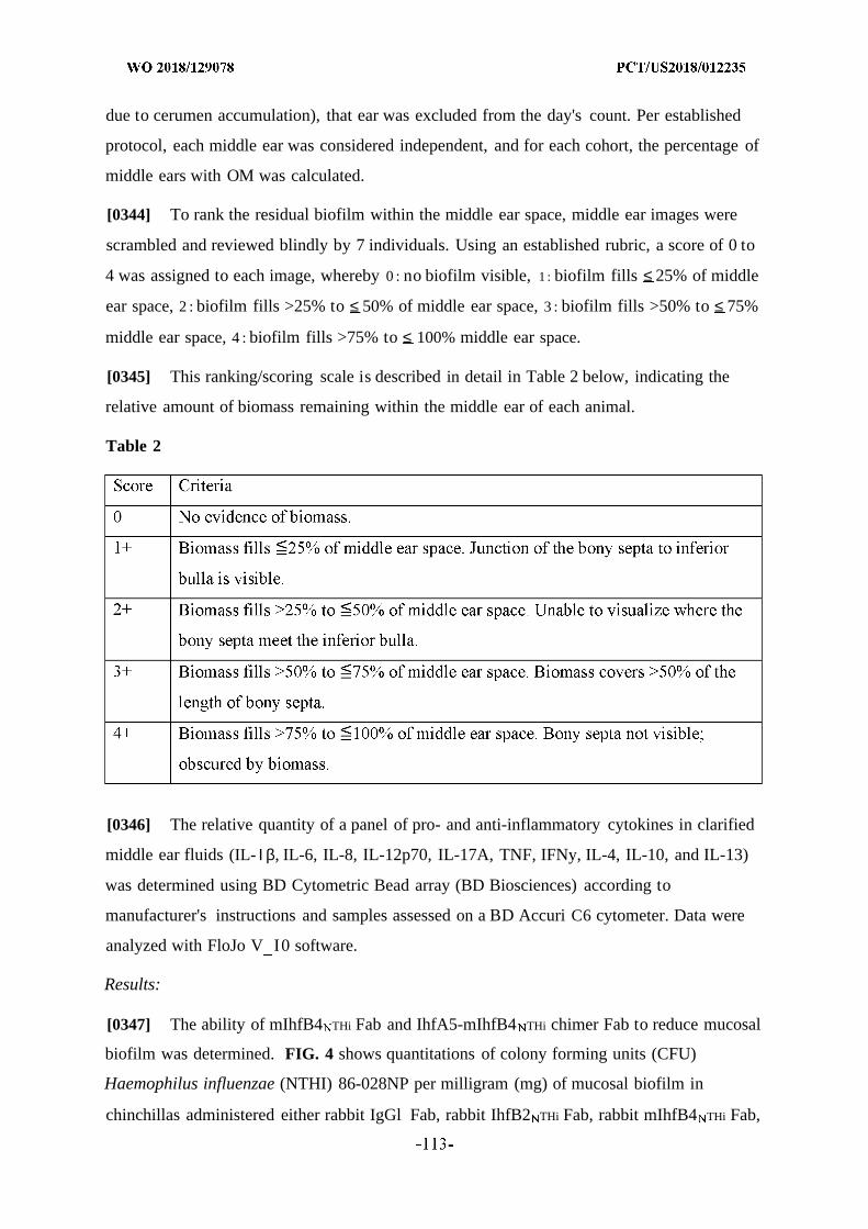
due to cerumen accumulation), that ear was excluded from the day's count. Per established
protocol, each middle ear was considered independent, and for each cohort, the percentage of
middle ears with OM was calculated.
[0344] To rank the residual biofilm within the middle ear space, middle ear images were
scrambled and reviewed blindly by 7 individuals. Using an established rubric, a score of 0 to
4 was assigned to each image, whereby 0 : no biofilm visible, 1 : biofilm fills ≤ 25% of middle
ear space, 2 : biofilm fills >25% to ≤ 50% of middle ear space, 3 : biofilm fills >50% to ≤ 75%
middle ear space, 4 : biofilm fills >75% to ≤ 100% middle ear space.
[0345] This ranking/scoring scale is described in detail in Table 2 below, indicating the
relative amount of biomass remaining within the middle ear of each animal.
Table 2
[0346] The relative quantity of a panel of pro- and anti-inflammatory cytokines in clarified
middle ear fluids (IL- Ι β, IL-6, IL-8, IL-12p70, IL-17A, TNF, IFNy, IL-4, IL-10, and IL-13)
was determined using BD Cytometric Bead array (BD Biosciences) according to
manufacturer's instructions and samples assessed on a BD Accuri C6 cytometer. Data were
analyzed with FloJo V I0 software.
Results:
[0347] The ability of mIhfB4 THi Fab and IhfA5-mIhfB4 THi chimer Fab to reduce mucosal
biofilm was determined. FIG. 4 shows quantitations of colony forming units (CFU)
Haemophilus influenzae (NTHI) 86-028NP per milligram (mg) of mucosal biofilm in
chinchillas administered either rabbit IgGl Fab, rabbit IhfB2 THi Fab, rabbit mIhfB4 THi Fab,
or rabbit IhfA5-mIhfB4 NTHi chimer Fab after Haemophilus influenzae (NTHI) 86-028NP
challenge and biofilm formation. Quantitations were performed for the following
dose/sacrifice combinations: (1) two doses of the respective Fabs were administered,
followed by sacrifice 1 day later; (2) two doses of the respective Fabs were administered,
followed by sacrifice 7 days later; and (3) three doses of the respective Fabs were
administered, followed by sacrifice 7 days later. Doses of Fabs were administered on days 4
and 5 (two doses) or on days 4, 5, and 6 (three doses) after Haemophilus influenzae (NTHI)
86-028NP challenge. In all three dose/sacrifice combinations, significantly fewer
Haemophilus influenzae (NTHI) 86-028NP were adherent to the middle ear mucosa and
present within biofilms after administration of mIhfB4NTHi Fab or IhfA5-mIhfB4NTHi chimer
Fab compared to nonspecific IgGl Fab and IhfB2 THi Fab controls (FIG. 4). Administration
of two doses of mIhfB4 or IhfA5-mIhfB4 chimer Fab fragments was comparable to three
doses in eradicating established middle ear biofilms (FIG. 4). At 7 days following
administration of two treatment doses, animals that had received Fab fragments to the
chimeric immunogen had significantly fewer bacteria within this biological sample from the
middle ear (*P < 0.05, ** P < 0.01, and *** P < 0.001), as shown in FIG. 4 .
[0348] FIG. 5 shows quantitations of colony forming units (CFU) Haemophilus influenzae
(NTHI) 86-028NP per milliliter (ml) of middle ear fluid in chinchillas administered either
IgGl Fab, IhfB2 THi Fab, mIhfB4 THi Fab, or IhfA5-mIhfB4 THi chimer Fab after
Haemophilus influenzae (NTHI) 86-028NP challenge and biofilm formation. Quantitations
were performed for the following dose/sacrifice combinations: (1) two doses of the
respective Fabs were administered, followed by sacrifice 1 day later; (2) two doses of the
respective Fabs were administered, followed by sacrifice 7 days later; and (3) three doses of
the respective Fabs were administered, followed by sacrifice 7 days later. Doses of Fabs
were administered on days 4 and 5 (two doses) or on days 4, 5, and 6 (three doses) after
Haemophilus influenzae (NTHI) 86-028NP challenge. Because antibodies directed against
DNABII protein tips can induce biofilm collapse and release of resident bacteria, a difference
in relative concentration of recoverable NTHI in middle ear fluids one day after
administration of the final dose was not expected and was not observed. However, one week
after receipt of two or three doses of mIhfB4 or IhfA5-mIhfB4 chimer Fab fragments, a
significant reduction in NTHI within middle ear fluids was observed (FIG. 5) (*P < 0.05 and
** P < 0.01). Administration of Fab fragments directed against either mIhfB4 or the IhfA5-
mIhfB4 chimer continued to mediate a therapeutic effect for at least one week following
administration of the last dose of Fab and data suggest this dosing regime is flexible and
optimizable.
[0349] Mean mucosal biofilm scores were determined to assess the ability of rabbit
mIhfB4 NTH i Fab and rabbit IhfA5-mIhfB4 NTH i chimer Fab to disrupt biofilm. FIG. 6 shows
mean mucosal biofilm scores for chinchillas administered either IgGl Fab, IhfB2 THi Fab,
mIhfB4NTHi Fab, or IhfA5-mIhfB4NTHi chimer Fab after Haemophilus influenzae (NTHI) 86-
028 P challenge and biofilm formation. Quantitations were performed for the following
dose/sacrifice combinations: (1) two doses of the respective Fabs were administered,
followed by sacrifice 1 day later; (2) two doses of the respective Fabs were administered,
followed by sacrifice 7 days later; and (3) three doses of the respective Fabs were
administered, followed by sacrifice 7 days later. Doses of Fabs were administered on days 4
and 5 (two doses) or on days 4, 5, and 6 (three doses) Haemophilus influenzae (NTHI)
86-028NP challenge. A mucosal biofilm score scale was used to rank the residual biofilm
within the middle ear space. Using an established rubric, a score of 0 to 4 was assigned to
each image, as follows: zero (0): no biofilm visible; 1 : biofilm fills >0 to <25% of middle ear
space; 2 : biofilm fills >25% to <50% of middle ear space; 3 : biofilm fills >50% to <75%
middle ear space; and 4 : biofilm fills >75% to 100% middle ear space (FIG. 6). As shown in
FIG. 6, a significantly lower mean mucosal biomass score was observed in chinchillas
treated with mIhfB4 THi Fab and IhfA5-mIhfB4 THi chimer Fab compared to chinchillas
administered IgGl Fab or IhfB2 THi Fab controls *P < 0.05 and **P < 0.01). Because
antibody directed against DNABII protein tips induces biofilm collapse, a significant
decrease in mucosal biofilm in the middle ear of animals administered mIhfB4 or IhfA5-
mIhfB4 chimer Fab fragments one day after receipt of two doses was observed (FIG. 6).
This decrease was maintained one week after receipt of two or three doses of mIhfB4 or
IhfA5-mIhfB4 chimer Fab fragments. Administration of Fab fragments directed against
either mIhfB4 or the IhfA5-mIhfB4 chimer continued to mediate a therapeutic effect for at
least one week after receipt of the last dose and data suggest this dosing regime is flexible
and optimizable.
[0350] Pro- and anti-inflammatory cytokines were measured in clarified middle ear fluids
to assess the effects of mIh B4 THi Fab and IhfA5-mIhfB4 chimer Fab on inflammation.
FIG. 7 shows the relative quantity of a panel of pro- and anti-inflammatory cytokines in
clarified middle ear fluids in chinchillas administered either naive rabbit IgG Fab, rabbit anti-
IhfB2 N T Hi IgG Fab, rabbit anti-mIhfB4 NTH i Fab, or rabbit anti-IhfA5-mIhfB4 N I chimer IgG
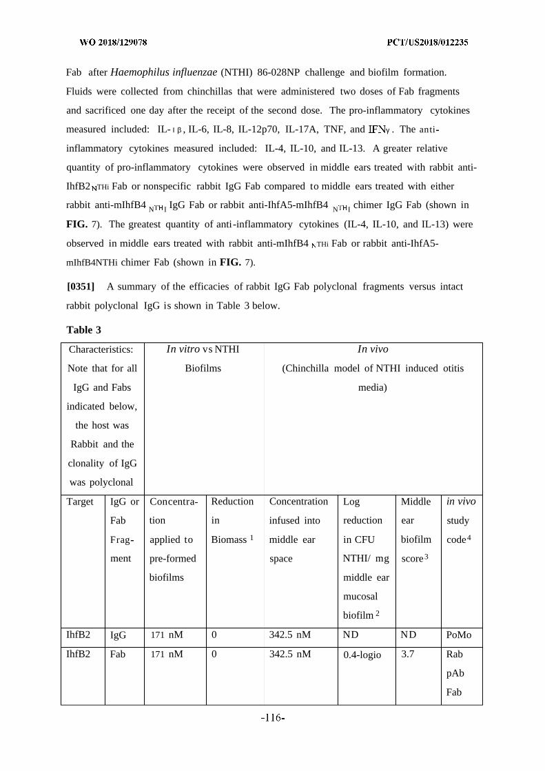
Fab after Haemophilus influenzae (NTHI) 86-028NP challenge and biofilm formation.
Fluids were collected from chinchillas that were administered two doses of Fab fragments
and sacrificed one day after the receipt of the second dose. The pro-inflammatory cytokines
measured included: IL- Ι β , IL-6, IL-8, IL-12p70, IL-17A, TNF, and γ . The anti
inflammatory cytokines measured included: IL-4, IL-10, and IL-13. A greater relative
quantity of pro-inflammatory cytokines were observed in middle ears treated with rabbit anti-
IhfB2 THi Fab or nonspecific rabbit IgG Fab compared to middle ears treated with either
rabbit anti-mIhfB4 N I IgG Fab or rabbit anti-IhfA5-mIhfB4 N I chimer IgG Fab (shown in
FIG. 7). The greatest quantity of anti-inflammatory cytokines (IL-4, IL-10, and IL-13) were
observed in middle ears treated with rabbit anti-mIhfB4 THi Fab or rabbit anti-IhfA5-
mIhfB4NTHi chimer Fab (shown in FIG. 7).
[0351] A summary of the efficacies of rabbit IgG Fab polyclonal fragments versus intact
rabbit polyclonal IgG is shown in Table 3 below.
Table 3
Characteristics: In vitro vs NTHI In vivo
Note that for all Biofilms (Chinchilla model of NTHI induced otitis
IgG and Fabs media)
indicated below,
the host was
Rabbit and the
clonality of IgG
was polyclonal
Target IgG or Concentra Reduction Concentration Log Middle in vivo
Fab tion in infused into reduction ear study
Frag applied to Biomass 1 middle ear in CFU biofilm code4
ment pre-formed space NTHI/ mg score3
biofilms middle ear
mucosal
biofilm 2
IhfB2 IgG 171 nM 0 342.5 nM ND ND PoMo
IhfB2 Fab 171 nM 0 342.5 nM 0.4-logio 3.7 Rab
pAb
Fab

mIhfB4 IgG 171 nM 75% 342.5 nM ND PoMo
mlhffl4 Fab 171 nM 78% 342.5 nM 5.5-logio 1.0 Rab
pAb
Fab
IhfA5- IgG 171 nM 82% 342.5 nM ND ND ND
mlhffl4
chimer
IhfA5- Fab 171 nM 87% 342.5 nM 5.2-logio 0.9 Rab
mlhffl4 pAb
chimer Fab
Relative to respective naive serum for polyclonal sera determined by COMSTAT2.
One day after receipt of the final dose, relative to respective naive serum for polyclonal sera.
Biomass score: 0= no biomass; 1= 0-25% middle ear space filled with bacterial biomass; 2=
>25-50% filled; 3= >50-75% filled; 4=>75-100% filled.
4PoMo: rabbit polyclonal and murine monoclonal antibody efficacy study; Rab pAb Fab:
rabbit polyclonal IgG Fab fragments efficacy study
ND refers to Not done, that is these antibodies were not tested in vivo.
[0352] Table 3 shows the targets (IhfB2, mIhfB4, and IhfA5-mIhfB4 chimer) used to
generate each of various rabbit polyclonal sera, providing data for IgG and Fab fragments for
each of these targets. All experiments shown were performed as highly controlled
therapeutic studies. The table summarizes and provides quantitations regarding the
experiments presented in Examples 2 and 3 above, as well as presents data ("PoMo" study
code) that are published in 'Monoclonal antibodies against DNA-binding tips of DNABII
proteins disrupt biofilms in vitro and induce bacterial clearance in vivo.' (2016)
EBioMedicine. Aug; 10:33-44. For the in vitro vs NTHI biofilms, the methods used were as
described in Example 2, above. The exact concentration of antibodies or Fab fragments as
indicated was applied to pre-formed biofilms. In particular, the concentration used was 171
nM for all antibodies and fragments used in the in vitro analyses. For the in vivo studies, the
methods used were as described in Example 3, above. The exact inoculum {Haemophilus
influenzae (NTHI) 86-028NP) delivered to each animal's ear to initiate biofilm formation was
controlled and infusion of ears was perfomed with specific quantities of antibody or
fragments as indicated. In particular, the concentration used was 342.5 nM for all antibodies
and fragments used in the in vivo analyses.

[0353] In the in vitro biofilm analysis using rabbit antibodies and Fab fragments, rabbit
polyclonal mIhfB4 IgG and rabbit polyclonal IhfA5-mIhfB4 chimer IgG showed 75% and
82% reduction in biomass, respectively. Rabbit polyclonal mIhfB4 Fab and rabbit polyclonal
IhfA5-mIhfB4 chimer Fab showed 78% and 87% reduction in biomass, respectively. IhfB2
IgG (control) and IhfB2 Fab (control) showed 0% and )% reduction in biomass, respectively.
For the in vivo chinchilla model of NTHI induced otitis media, rabbit polyclonal mIhfB4 Fab
and rabbit polyclonal IhfA5-mIhfB4 chimer Fab showed 5.5-log10
and 5.2-log10
reduction in
CFU NTHI/ mg middle ear mucosal biofilm, respectively, whereas rabbit polyclonal IhfB2
Fab (control) showed 0.4-log10
reduction in CFU NTHI/ mg middle ear mucosal biofilm.
These measurements were performed one day after receipt of the final dose, and are relative
to respective naive serum. Rabbit polyclonal mIhfB4 Fab and rabbit polyclonal IhfA5-
mIhfB4 chimer Fab showed middle ear biofilm scores of 1.0 and 0.9, respectively, whereas
rabbit polyclonal IhfB2 Fab (control) showed a middle ear biofilm score of 3.7. The "PoMo"
in vivo study code provided in Table 3 refers to data that are published in 'Monoclonal
antibodies against DNA-binding tips of DNABII proteins disrupt biofilms in vitro and induce
bacterial clearance in vivo.' (2016) EBioMedi cine. Aug; 10:33-44.
Example 4 : Treatment of Oral Disease
[0354] A number of oral bacteria (e.g., Aggregatibacter actinomycetemcomitans,
Porphyromonas gingivalis) have been implicated in the pathogenesis of inflammatory
diseases such as periodontitis and peri-implantitis, which destroy alveolar bone and gingiva.
Investigations of the pathogenesis of these bacteria are hampered by lack of effective animal
models. One of the challenges of investigating the pathogenicity of specific bacteria is the
difficulty of establishing a biofilm when exogenous bacteria are introduced into the oral
cavity of animals. Though animal models of periodontitis have been developed, cultivable
bacteria are rarely recovered from the oral cavity of inoculated animals. Developing an
effective animal model which can assess the pathogenicity of specific bacteria will greatly aid
in elucidating their pathogenic mechanisms.
[0355] The surface of machined titanium dental implants (1.2x4.5 mm) can be modified by
grit blasting with A103 (100 µιη) and HC1 etching (pH 7.8 for 20 min at 80° C). Machined
and nano-textured implants are incubated in TSB medium inoculated with D7S clinical strain
of Aggregatibacter actinomycetemcomitans (Aa) for 1 to 3 days at 37° C . The bacterial
biofilm on the implants are analyzed by SEM, as well as by confocal laser scanning
microscopy following staining with LIVE/DEAD® BacLight™. Implants with and without
established Aa biofilm are transmucosally placed into the alveolar bone of female rats
between premolar and incisor region of the maxillae. To detect the presence of Aa biofilm on
the implants placed in vivo, bacterial samples are collected from saliva and the oral surfaces
of implants after 2 days. Aa can be detected by culture, as well as by PCR analysis. Micro-CT
and histological analysis of peri-implant bone and mucosal tissues can be performed at
various time points, e.g., six weeks after implantation. The methods and compositions
disclosed herein are contemplated to develop both therapeutic as well as preventative
strategies for reduction and/or elimination of these biofilms. A decrease in redness,
inflammation, and bleeding compared to infected controls would indicate biofilm reduction
and/or elimination. In addition, reduced or absent inflammatory or proinflammatory
histology and maintenance of torque removal force for the implant screw compared to
infected controls would indicate biofilm reduction and/or elimination.
Example 5 : Lyme Disease
[0356] This experiment provides a mouse model for pre-clinical testing of agents as
described to treat lyme disease. See Dresser et al. Pathogens 5(12)el000680, Epub 2009 Dec.
4 . Lyme disease is caused by the microorganism Borrelia burgdorferi, a spirochete. B.
burgdorferi is transmitted via the bite of the Ixodes tick and subsequently disseminates, via
the bloodstream, to other tissues and organs.
[0357] In this animal model, C3H/HeN mice are injected with spirochetes via dorsal
subcutaneous and intraperitoneal injection, or via intravenous injection. Blood and biopsy
specimens are recovered at approximately 7 days post infection for evaluation of microbial
burden and assessment of pathology in tissues and organs. The methods and compositions
disclosed herein are contemplated to develop both therapeutic as well as preventative
strategies for reduction and/or elimination of the resulting B. burgdorferi biofilms which
form subsequent to challenge and are believed to contribute to both the pathogenesis and
chronic nature of the disease.
Example 6 : Cystic Fibrosis
[0358] This experiment provides a porcine model for pre-clinical testing of agents to treat
cystic fibrosis. See Stoltz et al. (2010) Science Translational Medicine 2(29):29-31. Cystic
fibrosis is an autosomal recessive disease due to mutations in a gene that encodes the CF
transmembrane conductance regulator (called CFTR) anion channel. In this model, pigs
which have been specifically bred to carry a defect in the genes called "CFTR" and called CF
pigs spontaneously develop hallmark features of CF lung disease that includes infection of
the lower airway by multiple bacterial species. The pigs can be immunized with the agents as
described herein to either: 1) immunize these CF pigs with a polypeptide or other
immunogenic agent thereby inducing the formation of antibodies which will eradicate
bacterial biofilms in the lungs (similarly to how antibodies to IHF eradicated biofilms
resident within the middle ears of chinchillas following active immunization), to deliver
agents to the lungs of these animals by nebulization to assess the amelioration of the signs of
disease and associated pathologies.
Example 7 : Tuberculosis
[0359] Applicants also provide a pre-clinical model for tuberculosis (TB). See Ordway et
al. (2010) Anti. Agents and Chemotherapy 54:1820. The microorganism Mycobacterium
tuberculosis is responsible for a growing global epidemic. Current figures suggest that there
are approximately 8 million new cases of TB and about 2.7 million deaths due to TB
annually. In addition to the role of this microbe as a co-infection of individuals with HIV (of
the ~45 million infected with HIV, estimates are that ~½ are also co-infected with
tuberculosis), its particularly troublesome that isolates have become highly resistant to
multiple drugs and no new drug for TB has been introduced in over a quarter of a century. In
this animal model, SPF guinea pigs are maintained in a barrier colony and infected via
aerosolized spray to deliver ~20 cfu of tuberculosis strain Erdman K01 bacilli into their
lungs. Animals are sacrificed with determination of bacterial load and recovery of tissues for
histopathological assessment on days 25, 50, 75, 100, 125 and 150 days post-challenge.
Unlike mice which do not develop classic signs of TB, guinea pigs challenged in this manner
develop well-organized granulomas with central necrosis, a hallmark of human disease.
Further, like humans, guinea pigs develop severe pyogranulomatous and necrotizing
lymphadenitis of the draining lymph nodes as part of the primary lesion complex. Use of this
model will provide a pre-clinical screen to confirm and identify therapeutic as well as
preventative strategies for reduction and/or elimination of the resulting M .
tuberculosis biofilms which have been observed to form in the lungs of these animals
subsequent to challenge and are believed to contribute to both the pathogenesis and
chronicity of the disease.
Example 8 : Device Application
[0360] Multiple animal models of catheter/indwelling device biofilm infections are known.
See Otto (2009) Nature Reviews Microbiology 7:555. While typically considered normal skin
flora, the microbe Staphylococcus epidermidis has become what many regard as a key
opportunistic pathogen, ranking first among causative agents of nosocomial infections.
Primarily, this bacterium is responsible for the majority of infections that develop on
indwelling medical devices which are contaminated by this common skin colonizer during
device insertion. While not typically life-threatening, the difficulty associated with treatment
of these biofilm infections, combined with their frequency, makes them a serious public
health burden. Current costs associated with treatment of vascular catheter associated
bloodstream infections alone that are due to S. epidermidis amount to $2 billion annually in
the United States. In addition to S. epidermidis, E.faecalis and S. aureus are also
contaminations found on indwelling medical devices. There are several animal models of
catheter-associated S. epidermidis infections including rabbits, mice, guinea pigs and rats all
of which are used to study the molecular mechanisms of pathogenesis and which lend
themselves to studies of prevention and/or therapeutics. Rat jugular vein catheters have been
used to evaluate therapies that interfere with E.faecalis, S. aureus and S. epidermidis biofilm
formation. Biofilm reduction is often measured three ways—(i) sonicate catheter and
calculate CFUs, (ii) cut slices of catheter or simply lay on a plate and score, or (iii) the
biofilm can be stained with crystal violet or another dye, eluted, and OD measured as a proxy
for CFUs.
Example 9 : Vaccine Administration
[0361] Methods described herein may be used to elicit immune responses in humans and
animals. Immunogenic compositions may be administered to a human and animal subjects in
the presence of adjuvants such as but not limited to aluminum salts and liposomes. Those
skilled in the art will understand that any number of pharmaceutically acceptable adjuvants
can also be used. Immunogenic compositions may be administered to a human or animal
subjects intramuscularly, subdermally, intranasally, or through any other suitable route.
Immunogenic compositions may be prepared in a manner consistent with the selected mode
of administration. Immunogenic compositions may take the form of polypeptides, nucleic
acids, or a combination thereof, and may comprise full-length or partial antigens.
Additionally or alternatively, immunogenic compositions may take the form of antigen
presenting cells (APCs) pulsed with a particular antigen, or APCs transfected with one or
more polynucleotides encoding a particular antigen. Administration may comprise a single
dose of an immunogenic composition, or an initial administration, followed by one or more
booster doses. Booster doses may be provided a day, two days, three days, a week, two
weeks, three weeks, one, two, three, six or twelve months, or at any other time point after an
initial dose. A booster dose may be administered after an evaluation of the subject's antibody
titer.
Example 10: Passive Immunity
[0362] Methods described herein may be used to confer passive immunity on a non
immune subject. Passive immunity against a given antigen may be conferred through the
transfer of antibodies or antigen binding fragments that specifically recognize or bind to a
particular antigen. Antibody donors and recipients may be human or non-human subjects.
Additionally or alternatively, the antibody composition may comprise an isolated or
recombinant polynucleotide encoding an antibody or antigen binding fragment that
specifically recognizes or binds to a particular antigen.
[0363] Passive immunity may be conferred in cases where the administration of
immunogenic compositions poses a risk for the recipient subject, the recipient subject is
immuno-compromised, or the recipient subject requires immediate immunity. Immunogenic
compositions may be prepared in a manner consistent with the selected mode of
administration. Compositions may comprise whole antibodies, antigen binding fragments,
polyclonal antibodies, monoclonal antibodies, antibodies generated in vivo, antibodies
generated in vitro, purified or partially purified antibodies, or whole serum. Administration
may comprise a single dose of an antibody composition, or an initial administration followed
by one or more booster doses. Booster doses may be provided a day, two days, three days, a
week, two weeks, three weeks, one, two, three, six or twelve months, or at any other time
point after an initial dose. A booster dose may be administered after an evaluation of the
subject's antibody titer.
Example 11: Synergistic therapeutic efficacy of IH F N I Fab fragments + HMGB 1-C45S
[0364] Chinchillas were challenged by transbullar injection with 1000 CFU of NTHi strain
86-028NP on day 0 . On day 4, chinchillas were treated with formulations noted in FIG. 8A.
On day 5, a subset of chinchillas were treated again was the same formulation. Chinchillas
were euthanized 1 day after final treatment, and middle ears were scored for biofilm (left) and
CFU in the middle ear mucosa were quantified (right). The combination of HMGB1-C45S
and the tail-directed Fab showed efficacy equal to the FDVIGB1-C45S treatment alone, while
the combination of the DNA binding tip-directed Fab and FDVIGB1-C45S displayed increased
efficacy compared to either monotherapy. (See FIGS. 8B and 8C).
Equivalents
[0365] Unless otherwise defined, all technical and scientific terms used herein have the
same meaning as commonly understood by one of ordinary skill in the art to which this
invention belongs.
[0366] The inventions illustratively described herein may suitably be practiced in the
absence of any element or elements, limitation or limitations, not specifically disclosed
herein. Thus, for example, the terms "comprising," "including," "containing," etc. shall be
read expansively and without limitation. Additionally, the terms and expressions employed
herein have been used as terms of description and not of limitation, and there is no intention
in the use of such terms and expressions of excluding any equivalents of the features shown
and described or portions thereof, but it is recognized that various modifications are possible
within the scope of the invention claimed.
[0367] Thus, it should be understood that the materials, methods, and examples provided
here are representative of preferred embodiments, are exemplary, and are not intended as
limitations on the scope of the invention.
[0368] The invention has been described broadly and generically herein. Each of the
narrower species and sub-generic groupings falling within the generic disclosure also form
part of the invention. This includes the generic description of the invention with a proviso or
negative limitation removing any subject matter from the genus, regardless of whether or not
the excised material is specifically recited herein.
[0369] In addition, where features or aspects of the invention are described in terms of
Markush groups, those skilled in the art will recognize that the invention is also thereby
described in terms of any individual member or subgroup of members of the Markush group.
[0370] All publications, patent applications, patents, and other references mentioned herein
are expressly incorporated by reference in their entirety, to the same extent as if each were
incorporated by reference individually. In case of conflict, the present specification,
including definitions, will control.
[0371] Other embodiments are set forth within the following claims.
WHAT IS CLAIMED IS:
1. A recombinant polypeptide comprising two or more isolated conformational tip
domains of a DNABII polypeptide or a fragment or a biological equivalent of one or more of
the conformational tip domains.
2 . The recombinant polypeptide of claim 1, further comprising one or more linker
polypeptides.
3 . The recombinant polypeptide of claim 1 or 2, wherein the polypeptide comprises two
conformational tip domains.
4 . The recombinant polypeptide of claim 1 or 2, wherein the polypeptide comprises
three conformational tip domains.
5 . The recombinant polypeptide of any one of claims 2 to 4, wherein the linker
polypeptide comprises 2 or more amino acids.
6 . The recombinant polypeptide of any one of claims 1 to 5, wherein the conformational
tip domains comprise a linear or a branched polypeptide.
7 . The recombinant polypeptide of any one of claims 1 to 6, further comprising a
detectable and/or a purification label.
8 . A recombinant polynucleotide encoding the recombinant polypeptide of any one of
claims 1 to 7 .
9 . The recombinant polynucleotide of claim 8, further comprising one or more
regulatory elements.
10. The recombinant polynucleotide of claim 7 or 8, further comprising a detectable
and/or a purification label.
11 . A vector comprising the recombinant polynucleotide of any one of claims 8 to 10 that
is optionally operably linked to a regulatory element.
12. An isolated host cell comprising one or more of:
a . the recombinant polypeptide of any one of claims 1 to 7;
b. the recombinant polynucleotide of any one of claims 8 to 10; and/or
c . the vector of claim 11.
13. An antibody that binds the recombinant polypeptide of any one of claims 1 to 7 or an
antigen binding fragment of the antibody.
14. The antibody or the antigen binding fragment of claim 13 that binds a DNABII
polypeptide.
15. The antibody of claim 13 or 14, wherein the antibody prevents formation of, or
disrupts a biofilm.
16. The antibody of any one of claims 13 to 15, wherein the antibody is selected from the
group of a monoclonal antibody, an isolated polyclonal antibody, a bispecific antibody, a
human antibody, a humanized antibody, a chimeric antibody or a primatized antibody.
17. The antibody of any one of claims 13 to 15, wherein the isolated polyclonal antibody
is an isolated mammalian polyclonal antibody.
18. The isolated polyclonal antibody of claim 17, wherein the isolated mammalian
polyclonal antibody is selected from the group of a rabbit polyclonal antibody, a murine
polyclonal antibody, a sheep polyclonal antibody, a canine polyclonal antibody, or a human
polyclonal antibody.
19. The antibody fragment of any one of claims 13 to 18, wherein the antigen binding
fragment is selected from an Fv antibody fragment or a Fab antibody fragment.
20. The antibody or antibody fragment of any one of claims 13 to 18, wherein the
antibody or the antigen binding fragment binds an epitope on the DNABII protein that is
conserved across bacterial species; and/or optionally, wherein the antibody or the antigen
binding fragment prevents or disrupts a biofilm derived from at least two bacterial species
including both Gram positive and Gram negative species.
21. The antibody or the antigen binding fragment of any one of claims 12 to 19, wherein
the DNABII protein is Staphylococcus aureus DNABII or a fragment thereof; and optionally,
wherein the fragment of Staphylococcus aureus DNABII comprises a beta hairpin, or a
biological equivalent of each thereof.
22. The antibody or antigen binding fragment of claim 20, wherein the at least two
bacterial species are selected from S. aureus, P. aeruginosa and K. pneumonia.
23. The antibody or antigen binding fragment of any one of claims 13 to 22, further
comprising a detectable and/or a purification label.
24. A polynucleotide encoding the antibody or antigen binding fragment of any one of
claims 13 to 23.
25. The polynucleotide of claim 24, further comprising a detectable and/or a purification
label.
26. A vector comprising the recombinant polynucleotide of claim 24 and 25, further
comprising one or more regulatory elements.
27. An isolated host cell comprising one or more of:
a . the recombinant polypeptide of any one of claims 1 to 7;
b. the polynucleotide of any one of claims 8 to 10, 24 and 25; and/or
c . the vector of claim 11 or 26.
28. A composition comprising a carrier and one or more of:
a . the recombinant polypeptide of any one of claims 1 to 7;
b. the polynucleotide of any one of claims 8 to 10, 24 and 25;
c . the vector of claim 11 or 26;
d . the antibody or antigen binding fragment of any one of claims 13 to 23;
and/or.
e . the host cell of claim 27.
29. The composition of claim 28, comprising two or more recombinant different
polypeptides of any one of claims 1 to 7 .
30. The composition of claim 28 or 29, further comprising a preservative and/or
stabilizer.
31. A vaccine composition comprising an effective amount of the recombinant
polypeptide of any one of claims 1 to 7 and a pharmaceutically acceptable carrier, and
optionally a preservative and/or a stabilizer and further optionally at least one antibiotic or an
additional active ingredient.
32. A vaccine composition comprising an effective amount of two or more different
recombinant polypeptides of any one of claims 1 to 7 and a pharmaceutically acceptable
carrier, and optionally a preservative and/or a stabilizer and further optionally at least one
antibiotic or an additional active ingredient.
33. The vaccine composition of claim 31 or 32, and further comprising an adjuvant.
34. The vaccine composition of any one of claims 30 to 33, wherein the composition is
formulated for pediatric administration.
35. A composition comprising two or more antigen binding fragments of any one of
claims 13 to 23, wherein the two or more are different from each other.
36. The composition of claim 35, further comprising a carrier, optionally a
pharmaceutically acceptable carrier and optionally at least one antibiotic or an additional
active ingredient.
37. The composition of claim 35 or 36, further comprising a preservative and/or stabilizer
and optionally at least one antibiotic or an additional active ingredient.
38. A method to prevent formation of or to disrupt a biofilm associated with an industrial
process comprising treating a surface susceptible to or containing a biofilm by contacting the
biofilmmwith an effective amount of one or more of a recombinant polypeptide, an antibody,
and/or the antigen binding fragment of any one of claims 1 to 7 and 13 to 23.
39. A method to prepare a recombinant polynucleotide selected from an antibody, an
antigen binding fragment, an antibody fragment, the method comprises culturing the host cell
of any one of claims 12 or 27, and wherein the host cell optionally is a mammalian cell.
40. A method to obtain antibodies immunoreactive with an IHF protein or to generate B
cells that secrete antibodies immunoreactive with an IHF protein, comprising administering
the recombinant polypeptide of any one of claims 1 to 7 to a subject; and recovering
antibodies or recovering B cells from the subject.
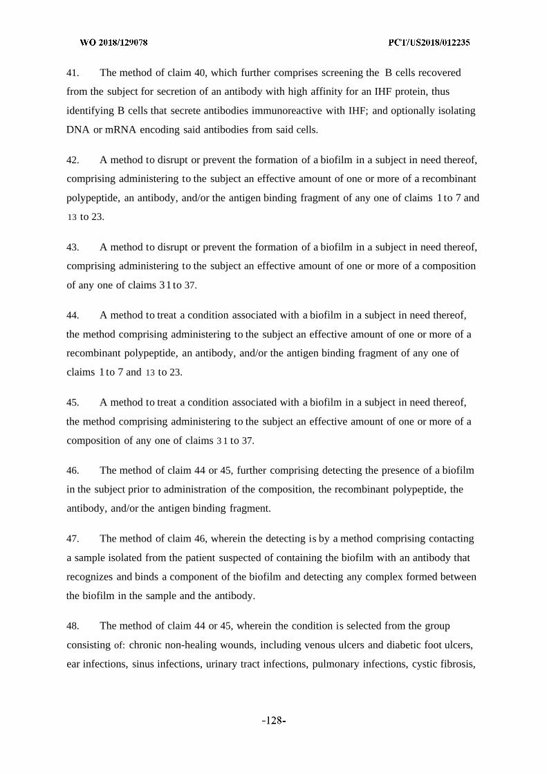
41. The method of claim 40, which further comprises screening the B cells recovered
from the subject for secretion of an antibody with high affinity for an IHF protein, thus
identifying B cells that secrete antibodies immunoreactive with IHF; and optionally isolating
DNA or mRNA encoding said antibodies from said cells.
42. A method to disrupt or prevent the formation of a biofilm in a subject in need thereof,
comprising administering to the subject an effective amount of one or more of a recombinant
polypeptide, an antibody, and/or the antigen binding fragment of any one of claims 1 to 7 and
13 to 23.
43. A method to disrupt or prevent the formation of a biofilm in a subject in need thereof,
comprising administering to the subject an effective amount of one or more of a composition
of any one of claims 31 to 37.
44. A method to treat a condition associated with a biofilm in a subject in need thereof,
the method comprising administering to the subject an effective amount of one or more of a
recombinant polypeptide, an antibody, and/or the antigen binding fragment of any one of
claims 1 to 7 and 13 to 23.
45. A method to treat a condition associated with a biofilm in a subject in need thereof,
the method comprising administering to the subject an effective amount of one or more of a
composition of any one of claims 3 1 to 37.
46. The method of claim 44 or 45, further comprising detecting the presence of a biofilm
in the subject prior to administration of the composition, the recombinant polypeptide, the
antibody, and/or the antigen binding fragment.
47. The method of claim 46, wherein the detecting is by a method comprising contacting
a sample isolated from the patient suspected of containing the biofilm with an antibody that
recognizes and binds a component of the biofilm and detecting any complex formed between
the biofilm in the sample and the antibody.
48. The method of claim 44 or 45, wherein the condition is selected from the group
consisting of: chronic non-healing wounds, including venous ulcers and diabetic foot ulcers,
ear infections, sinus infections, urinary tract infections, pulmonary infections, cystic fibrosis,
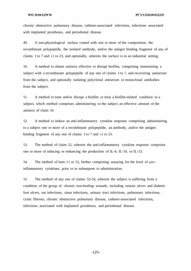
chronic obstructive pulmonary disease, catheter-associated infections, infections associated
with implanted prostheses, and periodontal disease.
49. A non-physiological surface coated with one or more of the composition, the
recombinant polypeptide, the isolated antibody, and/or the antigen binding fragment of any of
claims 1 to 7 and 13 to 23, and optionally, wherein the surface is in an industrial setting.
50. A method to obtain antisera effective to disrupt biofilm, comprising immunizing a
subject with a recombinant polypeptide of any one of claims 1 to 7, and recovering antiserum
from the subject, and optionally isolating polyclonal antiserum or monoclonal antibodies
from the subject.
51. A method to treat and/or disrupt a biofilm or treat a biofilm-related condition in a
subject, which method comprises administering to the subject an effective amount of the
antisera of claim 50.
52. A method to induce an anti-inflammatory cytokine response comprising administering
to a subject one or more of a recombinant polypeptide, an antibody, and/or the antigen
binding fragment of any one of claims 1 to 7 and 13 to 23.
53. The method of claim 52, wherein the anti-inflammatory cytokine response comprises
one or more of inducing or enhancing the production of IL-4, IL-10, or IL-13.
54. The method of laim 5 1 or 53, further comprising assaying for the level of anti
inflammatory cytokines, prior to or subsequent to administration.
55. The method of any one of claims 52-54, wherein the subject is suffering from a
condition of the group of: chronic non-healing wounds, including venous ulcers and diabetic
foot ulcers, ear infections, sinus infections, urinary tract infections, pulmonary infections,
cystic fibrosis, chronic obstructive pulmonary disease, catheter-associated infections,
infections associated with implanted prostheses, and periodontal disease.
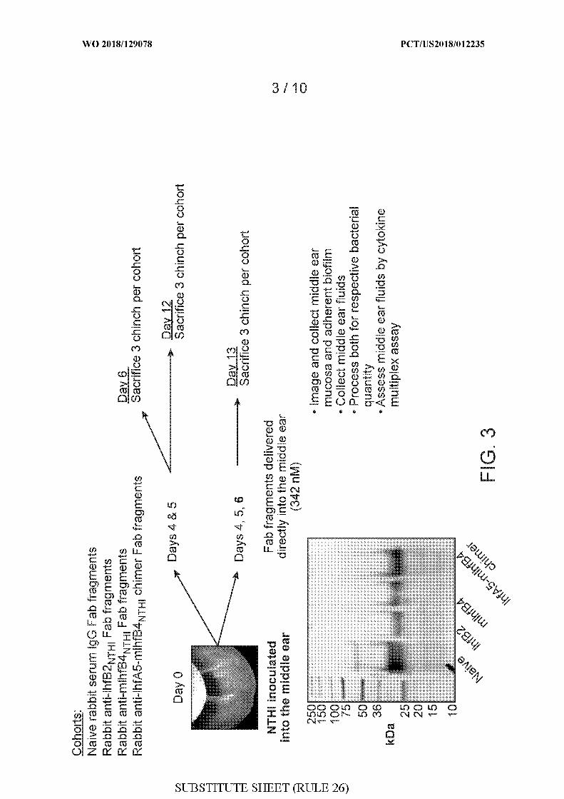
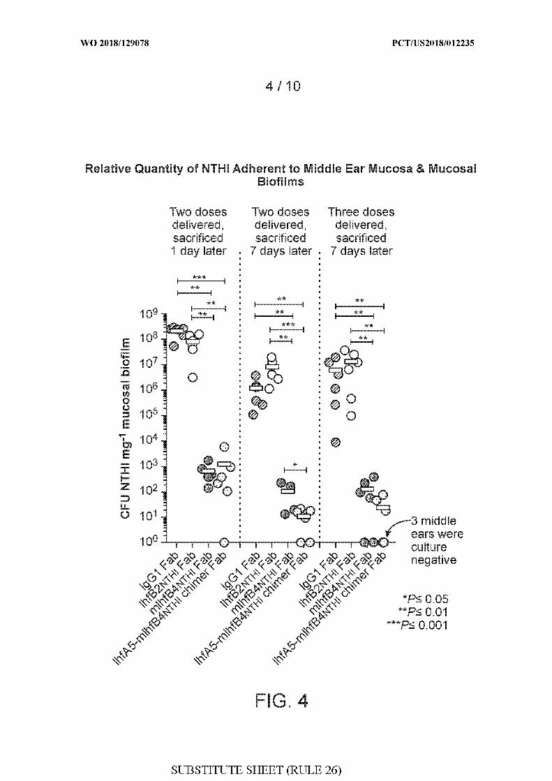
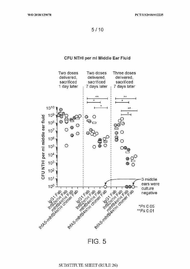
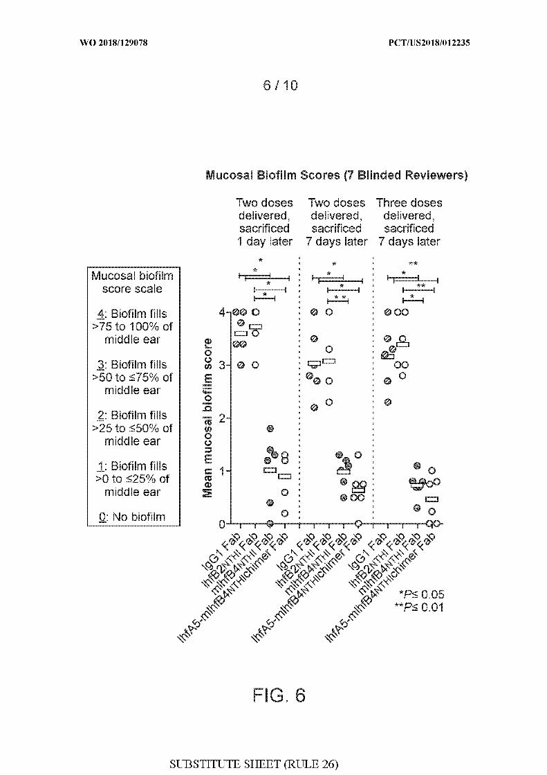
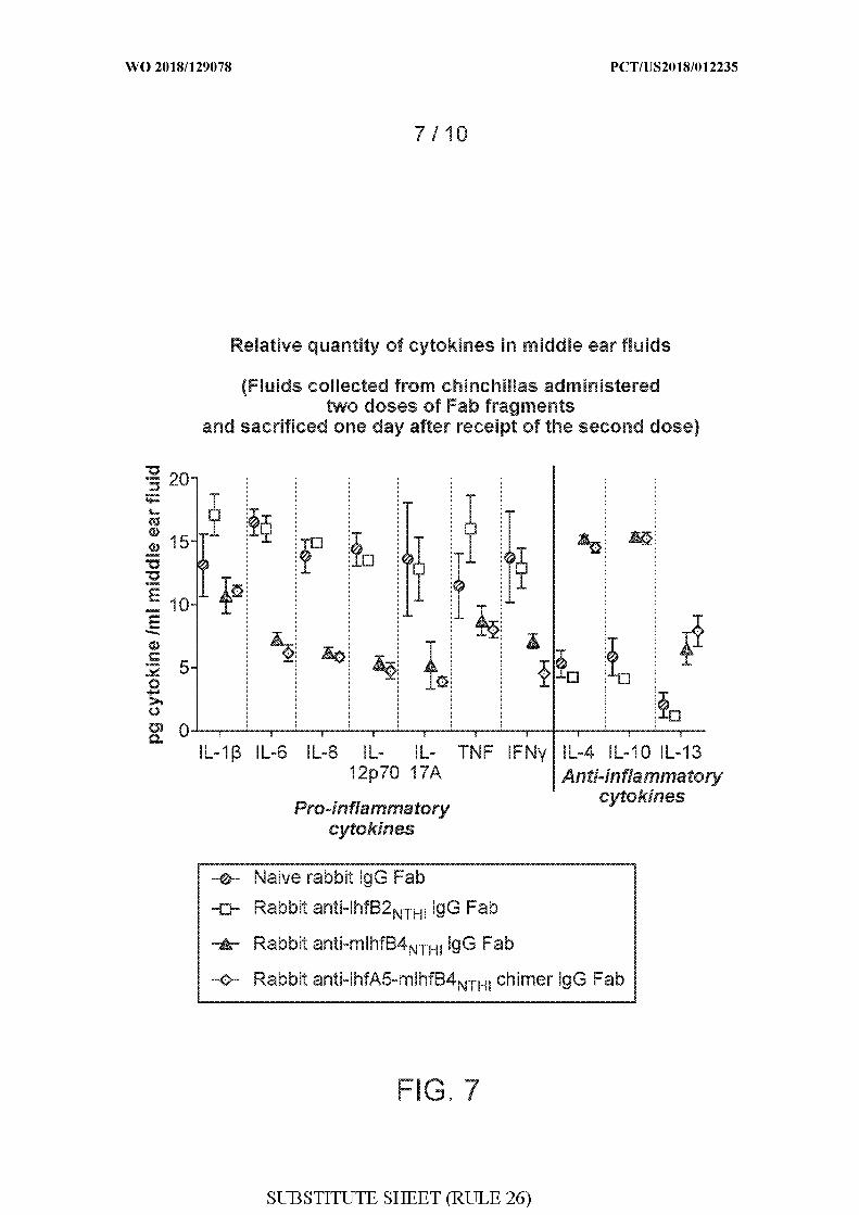
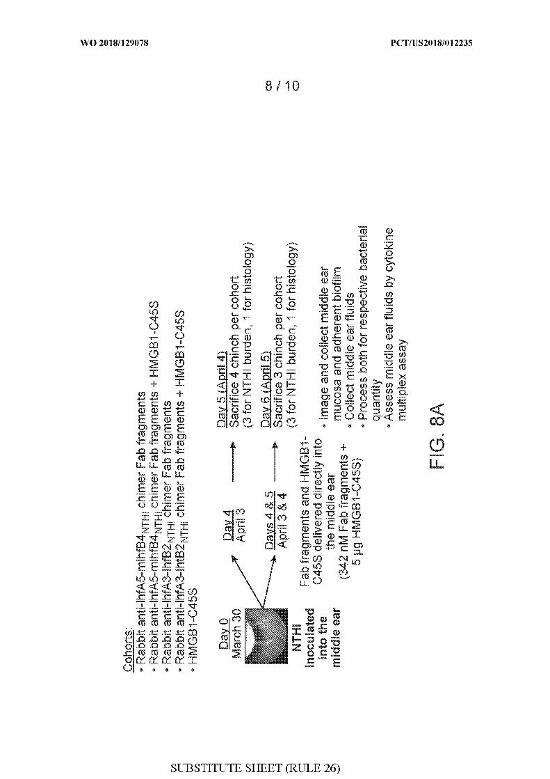
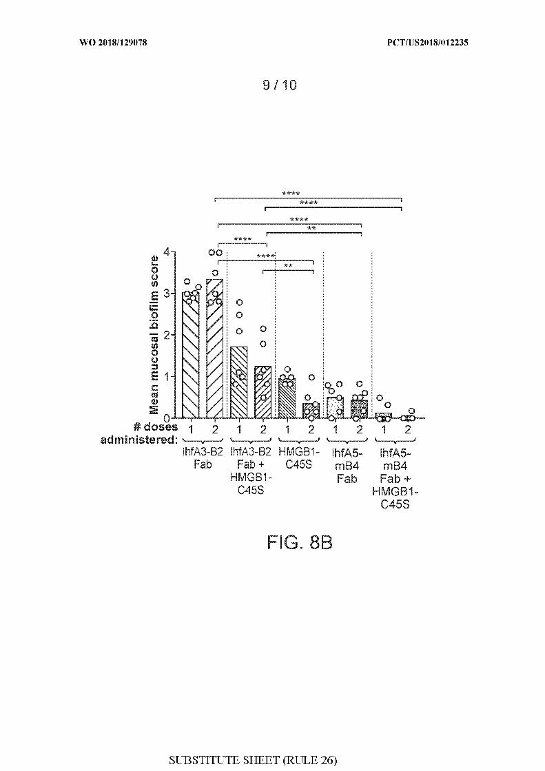
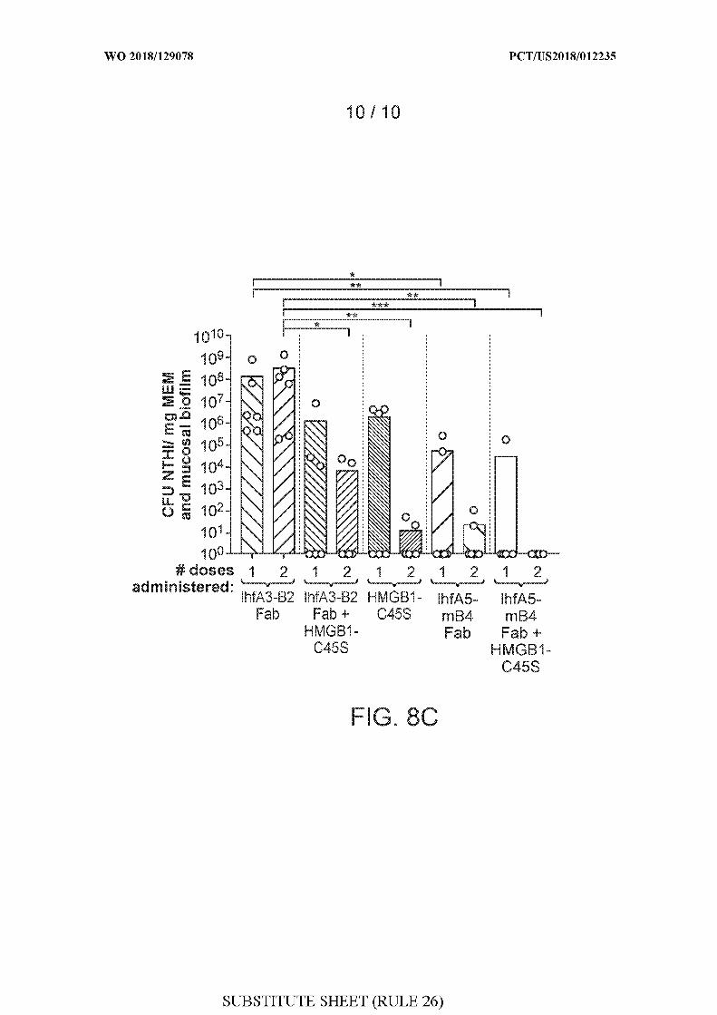
A . CLASSIFICATION O F SUBJECT MATTER
INV. A61K39/102 A61K38/00 A61Q11/02 A61Q17/00 C07K19/00C12Q1/18 C12N15/62 G01N33/569 A61P31/04 A61K39/40C07K16/12
According to International Patent Classification (IPC) or to both national classification and IPC
B . FIELDS SEARCHED
Minimum documentation searched (classification system followed by classification symbols)
A61K C07K
Documentation searched other than minimum documentation to the extent that such documents are included in the fields searched
Electronic data base consulted during the international search (name of data base and, where practicable, search terms used)
EPO-Internal , WPI Data, CHEM ABS Data
C . DOCUMENTS CONSIDERED TO B E RELEVANT
Category* Citation of document, with indication, where appropriate, of the relevant passages Relevant to claim No.
X US 2016/194384 Al (GOODMAN STEVEN D [US] 1-55ET AL) 7 July 2016 (2016-07-07)the whol e documentsee i n parti cul ar abstract; paragraphs[0056] - [0061] , [0071] , [0112] ,[0232] - [0261] , [0282] - [0422] ; Exp. 1-14
X 0 2014/201305 Al (UNIV SOUTHERN 1-55CALI FORNIA [US] ; NATIONWIDE CHI LDRENSHOSPITAL [US] ; 0HI )18 December 2014 (2014-12-18)the whol e documentsee i n parti cul ar Exp. 1-2 ; [0353] - [0354] ;Fi g . 16B-16F
/--
X Further documents are listed in the continuation of Box C . X See patent family annex.
* Special categories of cited documents :T " later document published after the international filing date or priority
date and not in conflict with the application but cited to understand"A" document defining the general state of the art which is not considered the principle or theory underlying the invention
to be of particular relevance
"E" earlier application or patent but published o n or after the international 'X" document of particular relevance; the claimed invention cannot befiling date considered novel o r cannot be considered to involve an inventive
"L" documentwhich may throw doubts on priority claim(s) orwhich is step when the document is taken alonecited to establish the publication date of another citation or other Ύ " document of particular relevance; the claimed invention cannot bespecial reason (as specified) considered to involve an inventive step when the document is
"O" document referring to an oral disclosure, use, exhibition or other combined with one o r more other such documents, such combinationmeans being obvious to a person skilled in the art
"P" document published prior to the international filing date but later thanthe priority date claimed '&" document member of the same patent family
ate of the actual completion of the international search Date of mailing of the international search report
12 Apri l 2018 26/04/2018
Name and mailing address of the ISA/ Authorized officer
European Patent Office, P.B. 5818 Patentlaan 2NL - 2280 HV Rijswijk
Tel. (+31-70) 340-2040,Fax: (+31-70) 340-3016 Si rim, Pi nar
C(Continuation). DOCUMENTS CONSIDERED TO BE RELEVANT
Category* Citation of document, with indication, where appropriate, of the relevant passages Relevant to claim No.
US 2011/236306 Al (GOODMAN STEVEN D [US] 1-55ET AL) 29 September 2011 (2011-09-29)the whol e documentsee i n parti cul ar abstract; paragraphs[0446] , [0447] ; Fi g . 5 , 6A, 6B,7 , 8 , 9A,9B, 10A, 10B, 11 , 12 , 13 , 14A, 14C; Exp.1-3 , 12
LAURA A. N0V0TNY ET AL: "Monocl onal 1-55anti bodi es agai nst DNA-bi ndi ng t i ps ofDNABI I protei ns d i srupt b i ofi lms i n v i troand i nduce bacteri a l c l earance i n v i vo" ,EBI0MEDICINE,vol . 10, 1 August 2016 (2016-08-01) , pages33-44, XP055352908,ISSN : 2352-3964, D0I :10. 1016/j .ebi om.2016.06.022c i ted i n the appl i cati onthe whol e documentsee i n parti cul ar abstract, Fi g . 1-7 ,tabl e 1
M. ELIZABETH BR0CKS0N ET AL: " Eval uati on 1-55of the ki neti c s and mechani sm of acti on ofanti - i ntegrati on host factor-medi ateddi srupti on of bacteri a l b i ofi lms" ,MOLECULAR MICROBIOLOGY,19 September 2014 (2014-09-19) , pagesn/a-n/a, XP055147799 ,ISSN : 0950-382X, D0I : 10. 1111/mmi . 12735the whol e documentsee i n parti cul ar abstract; Fi g . 7 ; pages1252-54
NA0KI G ET AL: "Chimeri c HU-IHF protei ns 1-55that a l ter DNA-bi ndi ng abi l i ty" ,GENE, ELSEVI ER, AMSTERDAM, NL,vol . 118, no. 1,1 September 1992 (1992-09-01) , pages97-102 , XP023539944,ISSN : 0378-1119 , D0I :10. 1016/0378-1119 (92)90254-M[retri eved on 1992-09-01]the whol e documentsee i n parti cul ar abstract; Fi g . 1
-/--
C(Continuation). DOCUMENTS CONSIDERED TO BE RELEVANT
Category* Citation of document, with indication, where appropriate, of the relevant passages Relevant to claim No.
X GOLDENBERG D ET AL: "Geneti c and 1-55b i ochemi cal analysi s of IHF/HU hybri dprotei ns" ,BIOCHIMI E, MASSON , PARIS, FR,vol . 76, no. 10-11 ,1 January 1994 (1994-01-01) , pages941-950, XP025458834,ISSN : 0300-9084, D0I :10. 1016/0300-9084(94)90019-1[retri eved on 1994-01-01]the whol e documentsee i n parti cul ar abstract; Fi g . 1
X L Zul i anel l o ET AL: "The HimA and HimD 1-55subuni t s of i ntegrati on host factor canspeci f i cal l y b i nd t o DNA as homodimers" ,The EMB0 Journal ,1 Apri l 1994 (1994-04-01) , pages1534-1540, XP055464716,ENGLANDD0I : 10. 1002/j . 1460-2075 . 1994. tb06415 . xRetri eved from the Internet:URL: https ://www.ncbi .nlm.ni h .gov/pmc/arti cl es/PMC394982/pdf/emboj00055-0048.pdfthe whol e documentsee i n parti cul ar abstract; Fi g . 1-2 , 5 ;paragr. bri dgi ng pages 1536-37 ; page 1539 ,paragr. 4
A N0V0TNY L A ET AL: " Epi tope mappi ng 1-55immunodomi nant regi ons of the Pi l A protei nof nontypeabl e Haemophi l us i nfl uenzae(NTHI ) t o faci l i tate the desi gn of twonovel chimeri c vacci ne candi dates" ,VACCINE, ELSEVI ER, AMSTERDAM, NL,vol . 28, no. 1 ,10 December 2009 (2009-12-10) , pages279-289 , XP026789470,ISSN : 0264-410X, D0I :1 . 1016/ J . VACCI NE. 2009 . 8 . 017[retri eved on 2009-08-22]the whol e document
A RICE PHOEBE A ET AL: "Crystal Structure 1-55of an IHF-DNA Compl ex: A Protei n-InducedDNA U-Turn" ,CELL,vol . 87 , no. 7 ,27 December 1996 (1996-12-27) , pages1295-1306, XP029186423 ,ISSN : 0092-8674, D0I :10. 1016/50092-8674(00)81824-3the whol e document
-/--
C(Continuation). DOCUMENTS CONSIDERED TO BE RELEVANT
Category* Citation of document, with indication, where appropriate, of the relevant passages Relevant to claim No.
WO 2017/023863 Al (RES INST AT NATIONWIDE 1-55CHI LDREN ' S HOSPITAL [US] )9 February 2017 (2017-02-09)the whol e document
Patent document Publication Patent family Publicationcited in search report date member(s) date
US 2016194384 Al 07-07-2016 NONE
WO 2014201305 Al 18-12-2014 AU 2014278037 Al 07-01-2016CA 2915210 Al 18-12-2014EP 3008088 Al 20-04-2016HK 1222398 Al 30-06-2017
P 2016521752 A 25-07-2016P 2017143841 A 24-08-2017
US 2016175440 Al 23-06-2016O 2014201305 Al 18-12-2014
US 2011236306 Al 29-09-2011 A R 084373 Al 15 -05 -2013AU 2011235296 Al 18 -10 -2012CA 2794305 Al 06 -10 -2011CN 103097399 A 08 -05 -2013CN 107096025 A 29 -08 -2017EP 2552944 Al 06 -02 -2013
P 2013529893 A 25 -07 -2013P 2016178944 A 13 -10 -2016
KR 20130040173 A 23 -04 -2013SG 183816 Al 3 -10 -2012US 2011236306 Al 29 -09 -2011US 2015166641 Al 18 -06 -2015WO 2011123396 Al 06 -10 -2011
W0 2017023863 Al 09-02-2017 AU 2016303688 Al 01-02-2018CA 2993009 Al 09-02-2017W0 2017023863 Al 09-02-2017