Lower Urinary tract Contrast Studies in the Male Dog & Case ...
118
Lower Urinary tract Contrast Studies in the Male Dog & Case Studies A thesis presented to The Faculty of Veterinary Medicine University of Glasgow for the Degree of Master of Veterinary Medicine June 2000 Maria Teresa Mato Pintane © University of Glasgow, Maria Teresa Mato Pintane 2000
-
Upload
khangminh22 -
Category
Documents
-
view
0 -
download
0
Transcript of Lower Urinary tract Contrast Studies in the Male Dog & Case ...

Lower Urinary tract Contrast Studies in the Male Dog
& Case Studies
A thesis presented to
The Faculty of Veterinary Medicine
University of Glasgow
for the Degree of Master of Veterinary Medicine
June 2000
Maria Teresa Mato Pintane
© University of Glasgow, Maria Teresa Mato Pintane 2000

ProQuest Number: 13832127
All rights reserved
INFORMATION TO ALL USERS The quality of this reproduction is dependent upon the quality of the copy submitted.
In the unlikely event that the author did not send a com p le te manuscript and there are missing pages, these will be noted. Also, if material had to be removed,
a note will indicate the deletion.
uestProQuest 13832127
Published by ProQuest LLC(2019). Copyright of the Dissertation is held by the Author.
All rights reserved.This work is protected against unauthorized copying under Title 17, United States C ode
Microform Edition © ProQuest LLC.
ProQuest LLC.789 East Eisenhower Parkway
P.O. Box 1346 Ann Arbor, Ml 48106- 1346

rcUSGUW UNIVERSITY
1 LIBRARY.
Il̂ tf ( Cd I
Table of Contents
Summary.............................................................................................................................iii
List o f Sections................................................................................................................... iv
List o f T ables.................................................................................................................... vii
List o f Figures..................................................................................................................viii
Dedication ...........................................................................................................................x
Declaration..........................................................................................................................xi
Acknowledgements.......................................................................................................... xii

Summary
The value of combining double contrast cystography with retrograde urethrography as a
single study was investigated in 20 male dogs, which had 21 studies. These animals had
been presented with suspected lower urinary tract and/or prostatic disease to Glasgow
University Small Animal Hospital. The most common presenting signs were haematuria
(8/21) and defaecatory tenesmus (8/21). In 18/21 studies there was poor colonic
preparation, but it only affected the radiological interpretation in 6 cases. Air bubbles
were found to be the commonest filling defects in bladder (12/21) and urethra (12/21).
The radiological abnormality most frequently encountered was prostatomegaly (9/21),
though no abnormalities were detected in 8 studies. The prostatic urethra was
adequately distended or just visible in the majority of the studies (18/21) regardless of
the positioning o f the catheter tip. It was invisible in all but one o f the 5 cases where the
bladder neck was intra-pelvic. Extravasation o f contrast into the substance of the
prostate was observed in five o f the 21 studies. Disruption or a ragged appearance to the
prostatic urethra was present in the one prostatic tumour, but also in two o f the four
cases of prostatic/para-prostatic cyst. No evidence o f mineralisation o f prostatic tissue
was found in any of the cases. Disease associated with the prostate was the most
common final clinical diagnosis (14/21). However, benign prostatic hyperplasia was
diagnosed in three cases without demonstrable prostatomegaly. The conclusion reached
was that the combined technique was not superior to double contrast cystography and
retrograde urethrography carried out individually.
In addition, the presentation and management of ten soft tissue cases are described.

List of Sections
Section 1
1. LOWER URINARY TRACT CONTRAST STUDIES IN THE MALE DOG.............................2
1.1 In t r o d u c t io n ............................................................................................................................................................................. 2
1.2 M a t e r ia l s & M e t h o d s ........................................................................................................................................................ 4
1.3 R e s u l t s ...........................................................................................................................................................................................5
1.4 D i s c u s s io n ................................................................................................................................................................................. 2 0
1.5 R e f e r e n c e s ................................................................................................................................................................................2 6
IV
List of Sections
Section 2
1. CHRONIC HYPERTROPHIC PYLORIC GASTROPATHY.....................................................31
1.1 In t r o d u c t io n ..........................................................................................................................................................................31
1.2 C a s e d e t a il s ............................................................................................................................................................................ 33
1.3 D i s c u s s io n ................................................................................................................................................................................ 35
1.4 R e f e r e n c e s ...............................................................................................................................................................................37
2. CANINE INGUINAL HERNIA...................................................................................................... 40
2.1 In t r o d u c t io n ......................................................................................................................................................................... 4 0
2 .2 C a s e D e t a il s ...........................................................................................................................................................................4 2
2 .3 D is c u s s io n ................................................................................................................................................................................43
2 .4 R e f e r e n c e s ...............................................................................................................................................................................4 4
3. BILATERAL ECTOPIC URETER IN A MALE DOG................................................................47
3.1 In t r o d u c t io n ......................................................................................................................................................................... 47
3 .2 C a s e d e t a il s ............................................................................................................................................................................ 4 9
3 .3 D i s c u s s io n ................................................................................................................................................................................ 52
3 .4 R e f e r e n c e s ............................................................................................................................................................................... 54
4. TRAUMATIC DIAPHRAGMATIC RUPTURE...........................................................................57
4.1 In t r o d u c t io n ......................................................................................................................................................................... 57
4 .2 C a s e d e t a il s ............................................................................................................................................................................ 59
4 .3 D i s c u s s io n ................................................................................................................................................................................ 6 0
4 .4 R e f e r e n c e s ...............................................................................................................................................................................61
5. CANINE NASAL ASPERGILLOSIS............................................................................................. 64
5.1 In t r o d u c t io n ..........................................................................................................................................................................64
5 .2 C a s e d e t a il s ............................................................................................................................................................................ 66
5.3 D is c u s s io n ................................................................................................................................................................................ 68
5 .4 R e f e r e n c e s ............................................................................................................................................................................... 69
6. UPPER AIRWAY OBSTRUCTION IN A BRACHYCEPHALIC DOG................................... 74
6.1 In t r o d u c t io n ..........................................................................................................................................................................7 4
6 .2 C a s e d e t a il s ............................................................................................................................................................................ 75
6 .3 D is c u s s io n ................................................................................................................................................................................ 77
6 .4 R e f e r e n c e s ............................................................................................................................................................................... 78
v
7. CRANIAL PUBIC LIGAMENT RUPTURE IN A DOG..............................................................80
7.1 In t r o d u c t io n ..........................................................................................................................................................................80
7 .2 C a s e d e t a il s ............................................................................................................................................................................ 81
7 .3 D is c u s s i o n ................................................................................................................................................................................ 83
7 .4 R e f e r e n c e s ...............................................................................................................................................................................85
8. INTESTINAL ADENOCARCINOMA IN A CAT........................................................................86
8 . 1 In t r o d u c t io n ..........................................................................................................................................................................86
8 .2 C a s e d e t a il s ............................................................................................................................................................................ 87
8 .3 D i s c u s s io n ................................................................................................................................................................................ 89
8 .4 R e f e r e n c e s ...............................................................................................................................................................................90
9. TRACHEAL PUNCTURE IN A DOG...........................................................................................93
9.1 In t r o d u c t io n ......................................................................................................................................................................... 93
9 .2 Ca s e d e t a il s ............................................................................................................................................................................ 94
9 .3 D i s c u s s i o n ................................................................................................................................................................................ 96
9 .4 R e f e r e n c e s ...............................................................................................................................................................................96
10. ABDOMINAL UNILATERAL CRYPTORCHIDISM IN A CA T............................................. 99
10.1 In t r o d u c t io n ......................................................................................................................................................................99
10 .2 C a s e d e t a il s .......................................................................................................................................................................100
10.3 D is c u s s i o n ...........................................................................................................................................................................101
10 .4 R e f e r e n c e s .........................................................................................................................................................................102
vi
List of Tables
TABLE 1. BREED, AGE, SEX AND WEIGHT OF 20 DOGS UNDERGOING 21
COMBINED LOWER URINARY TRACT CONTRAST STUDIES...................................... 6
TABLE 2. CLINICAL SIGNS AND DIAGNOSES MADE IN 20 DOGS BY 21
COMBINED LOWER URINARY TRACT CONTRAST STUDIES...................................... 8
TABLE 3. CATHETER TIP LOCATION AND FILLING DEFECTS IN 20 DOGS
FOLLOWING 21 COMBINED LOWER URINARY TRACT CONTRAST
STUDIES..................................................................................................................................10
TABLE 4. PROSTATIC LOCATION AND SYMMETRY IN 20 DOGS OBTAINED
FROM 21 COMBINED LOWER URINARY TRACT CONTRAST STUDIES................... 12
TABLE 5. DETAILS OF PROSTATIC URETHRA, PROSTATIC SIZE AND
CONTRAST LEAKAGE IN 20 DOGS FROM 21 COMBINED LOWER
URINARY TRACT CONTRAST STUDIES...........................................................................14

List of Figures
Section 1
FIGURE 1. RADIOGRAPH OF A COMBINED CONTRAST STUDY...................................................15
FIGURE 2. RADIOGRAPH OF A COMBINED CONTRAST STUDY...................................................15
FIGURE 3. RADIOGRAPH OF A COMBINED CONTRAST STUDY...................................................16
FIGURE 4. RADIOGRAPH OF A COMBINED CONTRAST STUDY...................................................16
FIGURE 5 RADIOGRAPH OF A COMBINED CONTRAST STUDY...................................................17
FIGURE 6 RADIOGRAPH OF A COMBINED CONTRAST STUDY...................................................17
FIGURE 7. RADIOGRAPH OF A COMBINED CONTRAST STUDY................................................... 18
FIGURE 8. RADIOGRAPH OF A COMBINED CONTRAST STUDY................................................... 18
FIGURE 9. RADIOGRAPH OF A COMBINED CONTRAST STUDY...................................................19
FIGURE 10.RADIOGRAPH OF A COMBINED CONTRAST STUDY..................................................19
viii

List of Figures
Section 2
FIGURE 1.1 CHRONIC HYPERTROPHIC PYLORIC GASTROPATHY, LATERAL ABDOMINAL
RADIOGRAPH.................................................................................................................................. 39
FIGURE 1.2 CHRONIC HYPERTROPHIC PYLORIC GASTROPATHY, ENDOSCOPY..................39
FIGURE 1.3 CHRONIC HYPERTROPHIC PYLORIC GASTROPATHY, LESION............................39
FIGURE 2.1 INGUINAL HERNIA...........................................................................................................46
FIGURE 2.2 INGUINAL HERNIA...........................................................................................................46
FIGURE 3.1 ECTOPIC URETER, ULTRASONOGRAM.......................................................................56
FIGURE 3.2 ECTOPIC URETER, LATERAL ABDOMINAL RADIOGRAPH.................................... 56
FIGURE 4.1 DIAPHRAGMATIC RUPTURE, LATERAL THORAX................................................... 63
FIGURE 5.1 NASAL ASPERGILLOSIS..................................................................................................72
FIGURE 5.2 NASAL ASPERGILLOSIS, DORSO-VENTRAL INTRA-ORAL RADIOGRAPH 72
FIGURE 5.3 NASAL ASPERGILLOSIS, ROSTRO-CAUDAL FRONTAL SINUSES.........................73
FIGURE 5.4 NASAL ASPERGILLOSIS, ROSTRAL RHINOSCOPY.................................................. 73
FIGURE 6.1 ELONGATED SOFT PALATE...........................................................................................79
FIGURE 8.1 INTESTINAL ADENOCARCINOMA, LATERAL ABDOMINAL RADIOGRAPH 92
FIGURE 8.2 INTESTINAL ADENOCARCINOMA................................................................................92
FIGURE 9.1 TRACHEAL RUPTURE, LATERAL THORACIC RADIOGRAPH................................98
FIGURE 10.1 ABDOMINAL CRYPTORCHIDISM, INTRA-OPERATIVE PICTURE....................... 104

Dedication
To my parents -Salvador y Olga- and my sister Maly; without them this would not have
been possible.
x

Declaration
I, Maria Teresa Mato Pintane, do hereby declare that the work carried out in this thesis
is original, was done by myself or with due acknowledgement, and has not been
presented for the award o f a degree at any other University.
xi
Acknowledgements
I would like to express my sincere gratitude to my supervisor, Dr. Martin Sullivan, my
first guide in the surgical world, who has transformed positive criticism into art.
Many thanks to Dr. Tobias Schwarz who patiently taught me the basics of radiographic
contrast techniques and their interpretation.
I wish to thank the anaesthetists and nurses o f the Small Animal Hospital for their
invaluable assistance in theatre during my work for the case book.
I would like to thank my good friends, the old ones and those I found in Glasgow,
specially the “Hellenic section”, for being there with practical and moral support
whenever it was needed. Once more to Alejandro, who gave the initial impulse to this
dream.
Last, but not least, thanks are eternally due to my parents and sister for the continuous
and unconditional encouragement as well as to Grandmother, Chiru, Marite, Teresa,
Marlene and Belen, keeping home warm waiting for my return.
“The best is yet to come... ”
Frank Sinatra

Se c t io n 1
page 1

Section 1
1. Lower Urinary Tract Contrast Studies in the MaleDog ______________
1.1 Introduction
The urinary bladder functions both as a reservoir for urine produced by the kidneys and
as an excretory organ for expulsion o f urine through the urethra. Together, the bladder
and urethra comprise what is referred to as the lower urinary tract (LUT).
Common clinical signs manifested by LUT disease are haematuria, dysuria and urinary
incontinence. Nevertheless, systemic signs such as raised body temperature or elevated
circulating white blood cell count are not usually present, and clinical examination is
often unrewarding. Consequently, diagnostic imaging is one o f the more helpful tools in
localising the disease process in and to the LUT (Park 1981).
Survey caudal abdominal radiographs must be taken prior to any contrast study of the
LUT to avoid masking radio-opaque lesions with contrast. However, the main
limitation o f survey films is the inability to detect intramural and intra-luminal soft
tissue alterations. This is because the bladder wall and urine are of the same density, so
mucosal margins cannot be identified. Further, an empty bladder and the normal urethra
are not visible on survey radiographs (Mahaffey and Barber 1992, Johnston and others
1995).
Retrograde contrast radiographic studies are simple, fast and minimal risk procedures
that highlight the lumen and walls, and thus alterations in the LUT (Park, 1981). A
number of different single contrast studies and combination contrast studies have been
advocated (Park 1981, Burk and Ackerman 1996).
The simplest study is probably the pneumocystogram, where the bladder is distended
with air or a gas such as CO2 or N2 O, allowing the bladder wall to be evaluated.
Unfortunately, insufficient distension may be easily misdiagnosed as bladder thickening
(Park 1974). Although inexpensive, this method provides poor mucosal detail and small
to moderate sized filling defects may not to be noticed (Johnston and others 1995).
The alternative single contrast study is positive contrast cystography, where soluble
organic iodides based on sodium or meglumine salts are used to distend the bladder. It
is the procedure of choice for detecting bladder tears or rupture (Mahaffey and Barber
page 2

Sectio n 1
1992). However, the main disadvantage is the masking of subtle wall thickening and
small luminal filling defects (Johnston and others 1995, Park 1981).
To overcome the disadvantages of single contrast techniques, double contrast techniques
have gained popularity. Double contrast cystography is performed by introducing a
small volume of positive contrast followed by negative contrast injection to complete
bladder distension. This technique provides the best evaluation o f bladder mucosa,
bladder wall thickness and, by means of the contrast puddle in the dependent portion,
serves as an aid in identifying different kinds of luminal filling defects (Park 1974).
The above three studies do not really address satisfactorily the second component of the
LUT, namely the urethra. Retrograde urethrography has been recommended for the
evaluation o f urethral disease (Ticer and others 1980), as an accurate procedure to locate
and determine the extent of urethral ruptures, obstructions and congenital anomalies
(Park 1981) and prostatic disease (Thrall 1994).
A technique that combined positive contrast retrograde urethrography with double
contrast cystography as a means to overcome the individual disadvantages o f each
retrograde contrast study previously described has been proposed.
An analysis of 20 male dogs, in which this combined technique was performed (21
studies), was carried out to test the hypothesis that it is superior to other individual
contrast studies.
page 3

Section 1
1.2 Materials & Methods
An analysis o f radiographs from 20 male dogs presented with suspected bladder,
urethral and/or prostatic disease at Glasgow University Veterinary Hospital during the
period of 1995-2000 was performed.
The cases were extracted from the hospital database by searching for dogs where
combined positive contrast retrograde urethrography with double contrast cystography
was done as part of the diagnostic procedure.
The case record was obtained and data was compiled concerning breed, age, neutering,
presenting sign and body weight. As well as details o f radiological evaluation of
bladder, urethra and prostate, the degree of emptiness o f colon was noted in each case to
assess its influence on the observation of lower urinary tract alterations.
Combined positive contrast retrograde urethrography with double contrast cystography
was carried out in all dogs under general anaesthesia and in lateral recumbence. In all
cases plain films were obtained prior to the contrast study.
Lubricating jelly was used on the tip o f an adequately sized polypropylene urinary
catheter to ease insertion into the urethra. The bladder was drained with a 60 ml syringe
connected to the catheter with a 3 way tap.
A contrast solution was prepared o f 5 to 15 ml, according body size, of sodium
iothalamate (420 mg/ml iodine) diluted 2:1 with sterile water for injection.
Once the bladder was emptied the urethra was occluded with a Doyen bowel clamp
immediately distal to the os penis to avoid leakage. One third o f the solution was
introduced into the bladder, which was gently massaged to ensure good distribution. Air
was insufflated to achieve adequate bladder distension, which was checked by digital
palpation of the bladder through the body wall. The catheter was retracted until, by
digital palpation o f distal membranous urethra, the catheter tip was thought to be located
immediately proximal to the os penis. The remaining solution was then injected and on
completion a radiograph was taken immediately the person injecting had left the
controlled area.
page 4

Section 1
1.3 Results
1.3.1 Breed distribution
The cases analysed in this study included a wide range o f breeds. Although no obvious
predominance was observed, the Boxer was the breed in which the contrast procedure
was performed most often (4/21). Two studies were performed on the same dog 3.6
months apart for recurrence of a prostatic cyst (study 10 and 13). Other breeds observed
in this study are listed in table 1.
1.3.2 Age distribution
The average age o f the dogs analysed was 5.5 years with a range o f 0.10-11 years. The
majority o f animals (61.9%) included in this study were aged between 5.5 and 11 years.
Dogs less than 5.5 years were less frequent (38.1%) (Table 1).
1.3.3 Sex distribution
As prostatic disease is strongly linked to testicular activity, neutering was considered a
factor. Most o f the dogs included in the study were entire (15/21) with only 6 neutered
prior to presentation (Table 1).
1.3.4 Weight distribution
The majority o f the dogs (80.9%) had a body weight ranging between twenty and forty
kilograms (Table 1). The average body weight o f the cases was found to be 23.75 kg.
page 5

Section 1
Study No Breed Age (years) Neutered Weight (kg)
1 German Shepherd Dog 3 Yes 38
2 Basset Hound 0.10 No 23
3 Springer Spaniel 11 No 21.5
4 Irish Setter 5 No 36
5 Dachshund 8 Yes 13
6 Basset Hound 9 Yes 21
7 Belgium Shepherd Dog 10 No 35
8 Old English Shepherd Dog 2 No 28
9 Labrador Retriever 2.3 No 30
10 Boxer 7.6 Yes 29
11 Border Collie 7.8 No 21.7
12 Irish Setter 4.4 No 37
13 Boxer 8.1 Yes 29
14 Jack Russell Terrier 11 No 9.3
15 Crossbred 7 No 26
16 Springer Spaniel 8 No 22.5
17 Boxer 5 No 31
18 Boxer 7 No 31
19 Border Terrier 9 Yes 9
20 Golden Retriever 9 No 39
21 Border Terrier 1 No 8.5
Table 1. Breed, age, sex and weight of 20 dogs undergoing 21 combined lower
urinary tract contrast studies.
1.3.5 1.3.5 Presenting signs
The major presenting features recorded were haematuria (8/21) and defaecatory
tenesmus (8/21). Less frequent were dysuria (2/21), urine dripping (2/21) and penile
bleeding (2/21). In some cases more than one feature was reported as a presenting sign
(Table 2).
page 6
Section 1
1.3.6 Radiological diagnoses
The radiographic finding most frequently identified after subjective evaluation o f the 21
studies was prostatomegaly (9/21). No significant abnormalities were observed in 8 of
the 21 studies. In the remaining four, diagnoses o f different lower urinary alterations,
such as ectopic ureter, bladder mass, urethral diverticulum and hypospadiasis were made
(Table 2).
1.3.7 Clinical diagnoses
Radiographic observations in conjunction with other diagnostic techniques performed in
each case led to a final clinical diagnoses. Prostatic abnormalities were found to be the
cause of the presentation in most o f the cases (14/21); benign prostatic hyperplasia
(BPH) (5/14), prostatic or para-prostatic cyst (Pr. Cyst or PPr. Cyst) (4/14), idiopathic
prostatic haemorrhage (IPH) (3/14), prostatitis (1/14) and prostatic tumour (1/14)
respectively. The urethra was also affected (3/21) with diagnoses of urethral sphincter
mechanism incompetence (USMI), urethral sphincter spasm (USS) and hypospadiasis.
The urinary bladder was involved in two cases, with bladder entrapment in a perineal
hernia and intraluminal polyp respectively. The two remaining cases presented penile
self mutilation and ectopic ureter.
page 7

Section 1
Study Clinical sign Diagnoses
No Radiological Clinical1 Urinary Tenesmus None USMI
2 Perineal stoma Hypospadiasis Hypospadiasis
3 Haematuria Prostatomegaly Pr. Cyst
4 Defaecatory tenesmus None BPH
5 Haematuria and dysuria Prostatomegaly Prostatic tumour
6 Defaecatory tenesmus & haematuria
None BPH
7 Penile bleeding Prostatomegaly IPH
8 Penile bleeding Prostatomegaly Penile mutilation
9 Defaecatory tenesmus None Prostatitis
10 Defaecatory tenesmus Prostatomegaly Pr. Cyst
11 Defaecatory tenesmus Prostatomegaly BPH
12 Dysuria Urethral diverticuli USS
13 Defaecatory tenesmus Prostatomegaly Pr. Cyst
14 Defaecatory tenesmus None BPH
15 Defaecatory tenesmus None Perineal hernia
16 Haematuria and urine dripping
None IPH
17 Haematuria Prostatomegaly PPr. Cyst
18 Haematuria None IPH
19 Haematuria Bladder mass Bladder polyp
20 Haematuria Prostatomegaly BPH
21 Urine dripping Ectopic ureter Ectopic ureter
BPH = benign prostatic hyperplasia, Pr. Cyst or PPr. Cyst, prostatic or para-prostatic cyst, IPH = idiopathic prostatic haemorrhage, USMI= urethral sphincter mechanism incompetence, USS= urethral sphincter spasm
Table 2. Clinical signs and diagnoses made in 20 dogs by 21 combined lower
urinary tract contrast studies.
page 8

Section 1
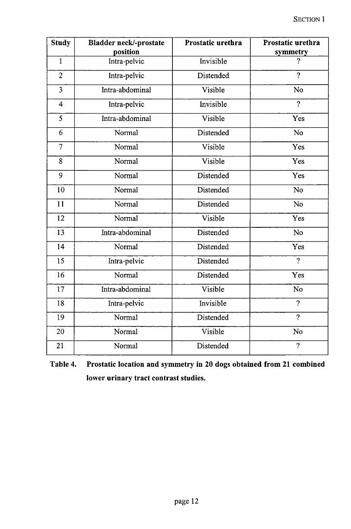
1.3.8 Colon preparation and interference in the radiological interpretation
Enemas were performed prior to the contrast studies and their effectiveness in clearing
the distal portion was assessed. Most o f cases (18/21) had varying degrees o f faecal
content still filling the colon (Figure 1). In only 3 cases was the distal colon empty of
faecal content. However, it was noted that in most cases interpretation was not affected
by the presence o f faeces (15/21). The colonic content had a negative effect on the
evaluation o f the radiographs in 6 studies (Figure 2).
1.3.9 Urethral location of catheter tip
As part o f the study, the catheter was withdrawn to a level which was thought
correspond to just proximal to the os penis. The actual position was recorded and its
effect on interpretation noted. In all studies the catheter had been withdrawn distal to
the prostate (Figure 3 & Figure 4). In 4/21 studies the catheter was present in the
membranous urethra (Table 3). In none o f these cases was interpretation compromised
by the catheter.
1.3.10 Degree of bladder distension
Subjective assessment o f satisfactory bladder distension was made on the basis of the
observation of a normal “pear shaped” bladder (Figure 3 & Figure 4). In only studies 1
and 3 was this feature absent.
1.3.11 Puddle of contrast
Following contrast cystography the puddle of positive contrast material remained in the
dependent portion of the bladder, and this was noticed in 19 of the 21 studies. In study
3 the puddle, predominantly dorso-caudal, almost completely filled the very small
bladder. In study 17 the contrast media was forced caudally by the presence o f a soft
tissue density mass ventral to the bladder (Figure 5).
1.3.12 Bladder wall thickness and severity
Bladder wall thickness was a common finding in the studies, where (47.6%) had some
degree of thickening (Figure 6). The increased thickness could be classified as focal
(4/21) and generalised (6/21). In all the cases where wall abnormalities were observed,
the degree of severity was classified subjectively as mild, compared to normal thickness.
page 9

Section 1
1.3.13 Bladder filling defects
After injection of both contrast agents, the radiographic image showed no filling defects
related to the puddle and its periphery in 7 of the studies . Air bubbles (12/21), blood
clots or cellular debris (2/21), calculi (2/21) and intra-luminal soft tissue structures
attached to the mucosal surface were found (Figure 3, Figure 4 & Figure 9). In some of
the cystograms more than a type o f filling defect was recorded in studies 5, 7 and 13
(Table 3).
Study Catheter tip placement Bladder filling defects
1 Penile None
2 Membranous None
3 Penile Air bubbles
4 Penile None
5 Penile Air bubbles, blood clots
6 Penile None
7 Penile Air bubbles, calculi
8 Penile Air bubbles
9 Penile Air bubbles
10 Penile Air bubbles
11 Membranous Air bubbles
12 Penile None
13 Penile Air bubbles, calculi
14 Membranous Blood clots
15 Penile Air bubbles
16 Penile Air bubbles
17 Penile Air bubbles
18 Penile None
19 Penile Intra-luminal growth
20 Penile None
21 Membranous Air bubbles
Table 3. Catheter tip location and filling defects in 20 dogs following 21
combined lower urinary tract contrast studies.
page 10

Section 1
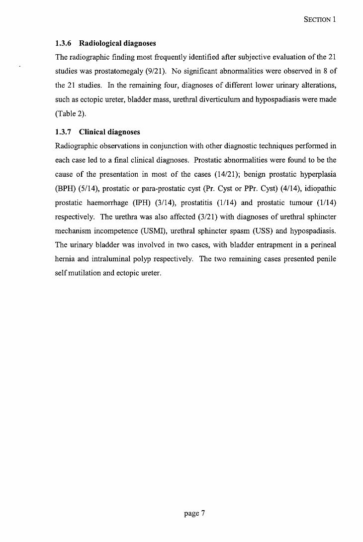
1.3.14 Position bladder neck/prostate
In the majority o f the studies (12/21) bladder neck/ prostate was found at the pelvis inlet
as is considered normal (Table 4). An intra-pelvic location was found in 5/21 (Figure 6)
and a more cranial intra-abdominal position in 4/21 (Figure 10).
1.3.15 Prostatic urethra visualisation and evaluation
The prostatic urethra could not be visualised in only three o f the cases, whereas in the
rest of the studies , this urethral segment was adequately distended (11/21) or just visible
(7/21) (Figure 1 & Figure 3). This allowed full evaluation and detection of
abnormalities in studies 3, 5 and 13 (Table 4).
1.3.16 Prostatic urethra symmetry
In 7 of the studies this evaluation could not be performed due to failure o f the contrast to
show the prostatic urethra and/or prostate gland. In 7 studies the position and
appearance of the prostate was symmetrical. In the remaining 7, the urethra appeared
asymmetric through its course within the prostate (Table 4) (Figure 10).
page 11
Section 1
Study Bladder neck/-prostate position
Prostatic urethra Prostatic urethra symmetry
1 Intra-pelvic Invisible ?
2 Intra-pelvic Distended ?
3 Intra-abdominal Visible No
4 Intra-pelvic Invisible ?
5 Intra-abdominal Visible Yes
6 Normal Distended No
7 Normal Visible Yes
8 Normal Visible Yes
9 Normal Distended Yes
10 Normal Distended No
11 Normal Distended No
12 Normal Visible Yes
13 Intra-abdominal Distended No
14 Normal Distended Yes
15 Intra-pelvic Distended ?
16 Normal Distended Yes
17 Intra-abdominal Visible No
18 Intra-pelvic Invisible ?
19 Normal Distended ?
20 Normal Visible No
21 Normal Distended ?
Table 4. Prostatic location and symmetry in 20 dogs obtained from 21 combined
lower urinary tract contrast studies.
page 12

Section 1
1.3.17 Urethral filling defects
Air bubbles (12/21) and a calculus (1/21) were the only urethral filling defects
encountered (Figure 4) (Table 5).
1.3.18 Prostate size
A subjective assessment of the prostatic volume was done. A small to moderate
enlargement was labelled as BPH-type (8/21), whereas a pronounced enlargement with
obvious displacement of peri-prostatic structures was defined as Cyst-type (3/21). The
size was considered to be normal in 4 studies and in the remaining 6 it was not possible
to evaluate size, due to a failure to adequately visualise the prostate during the contrast
study (Table 5).
1.3.19 Extravasation of contrast
Presence of contrast within the prostatic substance was noticed in a minority of cases
(5/21). Four o f these cases presented a very subtle localised extravasation {Blush-type)
(Figure 7), whereas in case number 5, the positive material formed a more massive and
irregular shape (Figure 9) within the prostatic parenchyma {ragged-type) (Table 5).
1.3.20 Prostatic mineralisation
Although considered as a parameter in the assessment o f the radiological findings, none
of the cases presented signs o f mineralisation of the prostatic tissue.
page 13

Section 1
Study Urethral filling defects Prostate size Contrast extravasation
1 None ? No
2 None ? No
3 Air bubbles Cyst-type Blush
4 None ? No
5 Air bubbles BPH-type Ragged
6 None Normal No
7 Air bubbles BPH-type Blush
8 Air bubbles BPH-type Blush
9 None Normal No
10 Air bubbles BPH-type No
11 Air Bubbles BPH-type No
12 Air bubbles Normal No
13 None Cyst-type No
14 Air Bubbles BPH-type Blush
15 Air bubbles BPH-type No
16 Air bubbles Normal No
17 Air Bubbles Cyst-type No
18 None ? No
19 Calculus ? No
20 None BPH-type No
21 Air bubbles ? No
BPH=benign prostatic hyperplasia, ?= not definable
Table 5. Details of prostatic urethra, prostatic size and contrast leakage in 20
dogs from 21 combined lower urinary tract contrast studies.
page 14
S e c t io n 1
Figure 1. Radiograph of a combined contrast study. The faeces-filled distal
colon/rectum highlights the degree of intestinal compression caused by a prostatic cyst
(arrow). Peri-urethral asymmetry is also evident.
Figure 2. Radiograph of a combined contrast study. The presence of excessive gas
generated by the enemas interferes with the radiological evaluation in this case.
page 15

Section 1
Figure 3. Radiograph of a combined contrast study. The prostatic urethra is widely
distended following penile urethral injection of contrast. There is a generalised increase
in bladder wall thickness. A filling defect created by a bladder polyp has caused
distortion of the contrast puddle cranio-ventrally (arrow).
Figure 4. Radiograph of a combined contrast study. There is a generalised increase in
bladder wall thickness. Radiolucent cystic calculi are located in the centre of the
contrast puddle (arrow). At least one air bubble is present in the distal urethra.
page 16

Section 1
Figure 5 Radiograph of a combined contrast study. There is an intra-abdominal
bladder with compression and displacement due to the presence of a prostatic cyst
(arrow).
Figure 6 Radiograph of a combined contrast study. There is an intra-pelvic bladder
present in this case of hypospadiasis. The prostatic urethra is distended and there is a
generalised increase in bladder wall thickness.
page 17

Section 1
Figure 7. Radiograph of a combined contrast study. The urethra is widely distended
throughout its length following injection from a catheter located in the membranous
portion of the urethra. Contrast extravasation {blush) is evident in a dog with a clinical
diagnosis of benign prostatic hypertrophy (arrow).
Figure 8. Radiograph of a combined contrast study. The urethra is extremely well
distended with contrast and one of the ureters is filled with positive contrast.
page 18

Section 1
Figure 9. Radiograph of a combined contrast study. A ragged prostatic urethra and
massive peri-urethral extravasation of contrast is evident in this case of prostatic
carcinoma. There are air bubbles/blood clots and debris present in the contrast puddle.
Figure 10. Radiograph of a combined contrast study. There is prostatic asymmetry,
urethral displacement and compression caused by a prostatic cyst.
page 19

Sectio n 1
1.4 Discussion
The urinary bladder is a hollow musculo-membranous organ that varies in form, size
and position, depending on the amount o f urine it contains. The prostate, the only
accessory sex gland in the male dog, completely envelops the proximal portion of the
urethra at the neck of the bladder (Evans and Christensen 1993).
Such close relationship between the components of the lower urinary tract and the
prostate is not only merely a matter o f boundaries but of function. Prostatic fluid, acts
as a transport and support medium for sperm during ejaculation, but basal secretion is
constantly entering the prostatic excretory ducts and urethra. When neither micturition
nor ejaculation is occurring, urethral pressure moves this basally secreted fluid cranially
into the bladder (Barsanti and Finco 1986).
The prostate, as well as the other components o f the lower urinary tract, is
radiographically a soft tissue dense structure, and in many instances, clear visualisation
o f the prostatic shadow requires the administration o f contrast material to outline the
lumen and identify the gland (Bartels 1981, Barsanti and Finco 1986).
Johnston and others (1991) recommended that identification o f prostatic urethra and
urinary bladder by positive contrast retrograde urethrography should be performed for
evaluation o f prostatic disease. Positive retrograde urethrography is extremely useful
for assessment o f urethral and urinary bladder integrity after caudal abdominal or pelvic
trauma and for investigation of stranguria, dysuria, incontinence or haematuria (Ticer
and others 1980). Performing this technique with an incompletely distended urinary
bladder causes luminal narrowing of the cranial and mid-regions of the prostatic and
pelvic urethra on radiographs (Johnston and others 1983a, Poogird and Wood 1986).
This is probably due to the longitudinal mucosal folds that project into the urethral
lumen (Evans and Christensen 1993).
Double contrast cystography has been found to be the most complete contrast technique
to assess possible pathologic conditions o f the bladder (Park 1974). It is recommended
that pneumocystography routinely follow positive contrast cystography, since the
mucosal surface of the urinary bladder is best appreciated when the bladder is opacified
and the thickness of its wall is best seen when contrasted with air (Root 1984).
page 20

Section 1
actually presented interpretation problems due to gas or faecal content within the
intestinal lumen, which implies that colonic evacuation is not critical. Indeed Lattimer
(1998) postulated that colon contents often improve the identification of the prostate,
delineating more clearly the degree of colonic compression by an enlarged prostate or
other mass.
Tri-iodinated ionic compounds are recognised as contrast agents o f choice (Holland
1993). Although residual urine and continued excretion will decrease the concentration
of contrast where small volumes (l-4ml) are used (Feeney and others 1999), we
considered that dilution of 1 volume of contrast to 2 volumes o f water would give
satisfactory opacification and reduce the risk of mucosal irritation (lOOmgl/ml).
In an attempt to maintain contrast in the urethra whilst the exposure is being made Park
(1974) suggested a mixture of aqueous lubricant and contrast agent as a mean of
increasing viscosity. However, Johnston and others (1983a,c) found that not only did
this type o f mixture fail to distend all areas of the urethra but also caused lesions in the
bladder. Earlier Ticer and others (1980) obtained satisfactory results with a simple
aqueous solution of contrast medium when the radiographic exposures was made at or
near the end of injection.
Ticer and others (1980) felt that naturally occurring luminal closure o f the membranous
and penile urethra around the catheter prevented significant contrast leakage. Indeed
several reports have suggested that balloon-tipped catheters may cause over-distension
of the bladder and consequently acute and severe reactions in bladder and urethra
(Barsanti and others 1981, Johnston and others 1983abc, Feeney and others 1984a).
Digital palpation of the bladder during injection was used to judge the degree of
distension in this study, as it has been shown that there is no precise method of gauging
volume based on surface area or body weight (Johnston and others 1985). Indeed over
distension can cause thickening due to mild or moderate cystitis to disappear (Mahaffey
and others 1989). Whilst satisfactory distension was achieved in 19/21 studies, in 2
studies distension, as judged by the subsequent film, was not good enough. This was
due in study 1 to overestimating the degree of bladder distension, and in study 3 by the
presence o f a prostatic cyst masking the bladder.
The main filling defect found in the bladder (12/21) was the presence of air in the
positive contrast, seen as coalescent or solitary bubbles at the periphery of the contrast
page 22

Sectio n 1
puddle. This feature needs to be distinguished from emphysematous cystitis where
multiple coalescent gas pockets are identifiable within the bladder wall (Root and Scott
1971, Sherding and Chew 1979). Radiolucent calculi will also produce filling defects in
the puddle but will be constrained by gravity to the centre or dependent part of the
puddle (Park 1981). Rarely, calculi attached to the wall may be encountered in non
dependent parts of the bladder (Johnston and others 1986). Lastly, blood clots may be
located anywhere within the lumen, but are more irregularly shaped and with a more
indistinct border than calculi or bubbles (Park 1981).
The presence o f filling defects in the urethra presents more o f a diagnostic challenge,
and in 12/21 studies air bubbles were identified as filling defects. These are usually
smooth and round compared to calculi, which are often more irregular in shape with
uneven margins (Burk and Ackerman 1996).
One of the goals of this series was to assess the bladder, prostate, prostatic and
membranous urethra. To achieve this the degree o f urethral distension is important.
Ticer and others (1980) suggested that deposition o f contrast medium directly into the
prostatic urethra increases the likelihood o f obtaining adequate luminal distension, but
this means that the catheter is in the membranous urethra. Yet, Thrall (1994) concluded
that size of urethral lumen, evaluated urethrographically, is o f little importance in
formulating a list o f differential diagnoses as an enlarged prostate may make the
prostatic urethra less distensible than normal. In contrast, Feeney and others (1984c),
reported that the ratio between prostatic and membranous urethral diameters is the most
clinically useful parameter in the evaluation o f this area.
The normal urethrogram should outline a smooth, regular tube thorough the entire
urethral length with slight narrowing at the ischial arch and the pelvic brim (Burk and
Ackerman 1996). O f the 17 cases, in which the catheter tip was positioned just cranial
to the os penis, the prostatic urethra was satisfactorily distended in 7 cases and visible
although not entirely distended in another 7 cases, whereas in only 3 radiographs was
the prostatic urethra invisible. When the injection o f contrast was made in a more
proximal position along the membranous urethra (4 cases), the prostatic portion was
distended with the radio-opaque material. In all the cases where there was a failure to
identify the prostatic urethra the bladder neck was intrapelvic. Though the reverse was
page 23

Section 1
not true as evidenced by case 15, where a prostatic urethra was identified despite an
intrapelvic bladder following a penile urethral injection of contrast.
Thrall (1994) took the view that the advantage o f positive contrast retrograde
urethrography was that it allowed evaluation of the peri-urethral symmetry of the
prostate. He and other authors (Stone and others 1978, Feeney and others 1987a) found
that asymmetry o f the prostate was not specific for any type o f disease, but was
indicative o f focal or regional, rather than diffuse, parenchymal disease. These authors
reported that peri-urethral asymmetry was observed only in patients with neoplasia,
parenchymal cavitation (abscess or non-inflammatory non-neoplastic cavitating disease)
or paraprostatic cyst. Surprisingly in this series, asymmetry found in 7 cases, was
present in 3 dogs with BPH.
In 7 of the 21 studies assessment of symmetry was not possible. In five the bladder neck
was intra-pelvic. In the remaining two (19 & 21), despite a normally positioned bladder
neck and satisfactory urethral distension, symmetry could not be assessed. One possible
explanation is poor abdominal contrast due to insufficient abdominal fat (Park 1998),
and specifically the triangular area o f adipose tissue located ventrally between the
prostate gland and the abdominal wall (Mahaffey and Barber 1992).
Prostatic size and its relationship to disease has been a matter of debate for sometime as
a normal sized prostate does not allow identification o f diseases such as infection, cystic
change, or early/mild neoplasia (Barsanti and Finco 1986). Feeney and others (1987a)
used the distance between the sacral promontory and the pubis to measure the ventro
dorsal and cranio-caudal dimensions of the prostate, using parallel and perpendicular
axis respectively. They concluded that if either prostatic dimension exceeded 70% of
the pubic-promontory dimension the prostate gland was considered to be enlarged.
Furthermore, any dimension larger than 90% was considered highly suggestive of
neoplasia, abscess or paraprostatic cyst. Lattimer (1998) found that the prostate may be
as great as 20 or more times normal size, or the degree o f enlargement may be minimal.
In this series all cases of prostatic/paraprostatic cyst and tumour had observable
prostatomegaly. However, only 2/5 cases o f BPH had demonstrable prostatomegaly.
Reflux of contrast from the urethra into the prostate has been mooted as an indicator of
disease. However, Ackerman (1983) postulated that there was no correlation between
prostatic reflux and specific prostatic diseases. Feeney and others (1984b) noted
page 24

Section 1
contrast reflux could be present in healthy dogs and that barium particles were observed
within the secretory (acinar) portion of the prostatic parenchyma after positive
retrograde urethrography, confirming that urethro-prostatic reflux (UPR) extended
beyond the prostatic duct system that directly empties into the urethral lumen.
However, a later study (Feeney and others 1987a), showed that UPR, in non-neoplastic
diseases was usually characterised by more rounded margins, with a tendency for the
refluxed contrast medium to pool, whereas the reflux observed with neoplasia was
usually characterised by an irregular (ragged) and massive coalescent appearance. Only
5 cases in this study showed UPR as a subtle cloudy blush in 4, and more irregular and
massive in case 5 a prostatic tumour.
Neoplasia should be considered, regardless of prostatic size, when there is evidence of
disruption or ragged appearance to the prostatic urethra (Feeney and others 1987a).
However, prostatic urethral abnormalities, less marked, were also found in two cases of
prostatic cyst (cases 3 & 13). On the other hand, the prostatic urethra was unremarkable
in two other cases o f cysts (10 & 17).
Definitive diagnosis of prostatic disease cannot be made by radiographic means.
Radiographic signs must be interpreted in the correct context along with other historical,
clinical and laboratory information. A definitive diagnoses o f prostatic disease is
frequently based upon histopathological examination (Thrall 1994). However, the
introduction of ultrasound has made a major impact on clinicians ability to differentiate
cystic from solid prostatic enlargement (Cartee and Rowles 1983, Feeney and others
1987b), and provides another method o f prostate measurement (Atalan and others
1999).
The intention o f this study was to combine two independent radiographic techniques and
carry them out simultaneously in an effort to get a wide range o f reliable information
from a single radiographic exposure. The results from this study suggest that the
information gathered concerning urethra and prostate is comparable with those achieved
with individual techniques, though not obviously superior.
page 25

Section 1
1.5 References
Ackerman, N. (1983) Prostatic reflux during positive contrast retrograde urethrography in the
dog. Veterinary Radiology 24, 251 -259.
Atalan, G., Barr, F.J. & Holt, P.E. (1999) Comparison of ultrasonographic and radiographic
measurements of canine prostatic dimensions. Veterinary Radiology & Ultrasound 40,
408-412.
Barsanti, J.A., Crowell, W., Losonsky, J. & Talkington, F.D. (1981) Complications of bladder
distension during retrograde urethrography. American Journal o f Veterinary Research
42,819-821.
Barsanti, J.A. & Finco, D.R. (1986) Canine prostatic diseases. Veterinary Clinics o f North
America: Small Animal Practice 16, 587-599.
Bartels, J.E. (1981) Radiology of the genital tract. In: Radiographic Diagnosis o f Abdominal
Disorders in the Dog and Cat. Radiographic interpretation, Clinical Signs,
Pathophysiology, ed T.R. O’Brien. Covell Park Veterinary Company, Davis. p615.
Borthwick, R. & Mackenzie, C.P (1971) The signs and results of treatment of prostatic disease in dogs. Veterinary Record 89, 374-384.
Burk, R.L. & Ackerman, N. (1996) The urinary system. In: Small Animal Radiology and
Ultrasound. A Diagnostic Atlas and Text, ed R.L. Burk & N. Ackerman. W.B. Saunders Company, Philadelphia. p319.
Cartee, R.E. & Rowles, T. (1983) Transabdominal sonographic evaluation of the canine prostate. Veterinary Radiology 24, 156-164.
Evans, H.E. & Christensen, G.C. (1993) The urogenital system. In: M iller’s anatomy o f the
dog. ed H.E. Evans. W.B. Saunders Company, Philadelphia. p494.
Feeney, D.A., Johnston, G.R., Tomlinson, M.J. & Osborne, C.A. (1984a) Effects of sterilized
micropulverized barium sulfate suspension and meglumine iothalamate solution on the
genitourinary tract of healthy male dogs after retrograde urethrocystography. American
Journal o f Veterinary Research 45, 730-738.
Feeney, D.A., Johnston, G.R., Osborne, C.A. & Tomlinson, M.J. (1984b) Maximum distension retrograde urethrocystography in healthy male dogs: Occurrence and radiographic
appearance of urethroprostatic reflux. American Journal o f Veterinary Research 45, 948-952.
page 26

Sectio n 1
Feeney, D.A., Johnston, G.R., Osbome, C.A. & Tomlinson, M.J. (1984c) Dimensions of the prostatic and membranous urethra in normal male dogs during maximum distension retrograde urethrocystography. Veterinary Radiology 25, 249-253.
Feeney, D.A., Johnston, G.R., Klausner, J.S., Perman, V., Leininger, J.R. & Tomlinson, M.J.
(1987a) Canine prostatic disease-comparison of radiographic appearance with
morphologic and microbiologic findings: 30 cases (1981-1985). Journal o f the American
Veterinary Medical Association 190, 1018-1026.
Feeney, D.A., Johnston, G.R., Klausner, J.S., Perman, V., Leininger, J.R. & Tomlinson, M.J.
(1987b) Canine prostatic disease-comparison of ultrasonographic appearance with
morphologic and microbiologic findings: 30 cases (1981-1985). Journal o f the American
Veterinary Medical Association 190, 1027-1034.
Feeney, D.A., Weichselbaum, R.C., Jessen, C.R. & Osbome, C.A. (1999) Imaging canine
urocystoliths. Veterinary Clinics o f North America: Small Animal Practice 29, 59-72.
Gordon, N. (1961) The position of the canine prostate gland. American Journal o f Veterinary
Research 22, 142-146.
Holland, M. (1993) Contrast agents. Veterinary Clinics o f North America: Small Animal
Practice 23, 269-279.
Johnston G.R., Feeney, D.A. & Osbome C.A. (1979) Radiographic findings in urinary tract infection. Veterinary Clinics o f North America: Small Animal Practice 9, 749-774.
Johnston, G.R., Jessen, C.R. & Osbome, C.A. (1983a) Effects of bladder distension on canine and feline retrograde urethrography. Veterinary Radiology 24, 271-277.
Johnston, G.R., Stevens, J.B., Jessen, C.R. & Osbome, C.A. (1983b) Effects of prolonged distension of retention catheters on the urethra of dogs and cats. American Journal o f
Veterinary Research 44, 223-228.
Johnston, G.R., Stevens, J.B., Jessen, C.R. & Osbome, C.A. (1983c) Complications of retrograde contrast urethrography in dogs and cats. American Journal o f Veterinary
Research 44, 1248-1258.
Johnston G.R., Feeney, D.A., Osbome C.A., Johnston, S.D., Smith, F.O. & Jessen, C.R. (1985)
Effects of intravesical hydrostatic pressure and volume on the distensibility of the canine
prostatic portion of the urethra. American Journal o f Veterinary Research 46, 748-751.
Johnston, G.R., Walter, P.A. & Feeney, D.A. (1986) Radiographic and ultrasonographic features of uroliths and other urinary tract filling defects. Veterinary Clinics o f North
America: Small Animal Practice 16, 261-292.
page 27

Sectio n 1
Johnston, G.R., Feeney, D.A., Rivers, B. & Walter, P.A. (1991) Diagnostic imaging of the male
canine reproductive organs. Methods and limitations. Veterinary Clinics o f North
America: Small Animal Practice 21, 553-589.
Johnston, G.R., Walter, P.A. & Feeney, D.A. (1995) Diagnostic imaging of the urinary tract.
In: Canine and Feline Nephrology and Urology, ed C.A. Osbome & D.R. Finco.
Williams & Wilkins, Baltimore. p230.
Lattimer, J.C. (1998) The prostate gland. In: Textbook o f Veterinary Diagnostic Radiology, ed
D.E. Thrall. W.B. Saunders Company, Philadelphia. p499.
Ling, G.V. (1995) Disorders of the prostate. In: Lower Urinary Tract Diseases o f Dogs and
Cats. Diagnosis, Medical Management and Prevention, ed G.V. Ling. Mosby, St Louis.
pl29.
Mahaffey, M.B. & Barber, D.L. (1992) Radiographic and ultrasonographic examination of the
urinary tract. In: Urologic Surgery o f the Dog and Cat. ed E.A. Stone & J.A. Barsanti.
Lea & Febiger, Philadelphia. p53.
Mahaffey, M.B., Barsanti, J.A., Crowell, W.A., Shotts, E. & Barber, D.L. (1989) Cystography: Effect of technique on diagnosis of cystitis in dogs. Veterinary Radiology 30, 261-267.
Park, R.D. (1974) Radiographic contrast studies of the lower urinary tract. Veterinary Clinics
o f North America: Small Animal Practice 4, 863-887.
Park, R.D. (1981) Radiology of the urinary bladder and urethra. In: Radiographic Diagnosis o f
Abdominal Disorders in the Dog and the Cat. Radiographic interpretation, Clinical
Signs, Pathophysiology, ed T.R. O’Brien. Covell Park Veterinary Company, Davis. p543.
Park, R.D. (1998) The urinary bladder. In: Textbook o f Veterinary Diagnostic Radiology, ed D.E. Thrall. W.B. Saunders Company, Philadelphia. p479.
Poogird, W. & Wood, A.K.W. (1986) Radiologic Study of the canine urethra. American
Journal o f Veterinary Research 47, 2491-2497.
Rogers, K.S., Wantschek, L. & Lees, G.E. (1986) Diagnostic evaluation of the canine prostate.
Compendium on Continuing Education for the Practicing Veterinarian 8, 799-811.
Root, C.R. & Scott, R.C. (1971) Emphysematous cystitis and other radiographic manifestations of diabetes mellitus in dogs and cats. Journal o f the American Veterinary Medical
Association 158, 721-728.
Root, C.R. (1984) Contrast radiography of the urinary system. In: Radiographic Technique in
Veterinary Practice, ed J.W. Ticer. W.B. Saunders Company, Philadelphia. p374.
Sherding, R.G. & Chew, D.J. (1979) Nondiabetic emphysematous cystitis in two dogs. Journal
o f the American Veterinary Medical Association 174, 1105-1109.
page 28

Sectio n 1
Stone, E.A., Thrall, D.E. & Barber, D.L. (1978) Radiographic interpretation of prostatic disease in the dog. Journal o f the American Animal Hospital Association 14, 115-118.
Thrall, D.E. (1994) Radiographic aspects of prostatic disease in the dog. In: Radiology in
Practice. The Compendium Selection. Veterinary Learning Systems Co., Trenton, pi 15.
Ticer, J.W., Spencer, C.P. & Ackerman, N. (1980) Positive contrast retrograde Urethrography: A useful procedure for evaluating urethral disorders in the dog. Veterinary Radiology
21 , 2 - 11.
page 29

Se c t io n 2
page 30

Section 2
1. Chronic Hypertrophic Pyloric Gastropathy
1.1 Introduction
The pylorus/antrum is the distal functional area of the stomach. It acts like a pump to
deliver digestive content, after been mixed with gastric secretions, to the duodenum
(Wingfield 1981). In addition, the pylorus prevents reflux of duodenal contents (Sikes
and others 1986).
Obstructive diseases of the pyloric canal may arise from intrinsic or extrinsic lesions.
Intrinsic pyloric obstruction may result from a number of conditions such as muscular
hypertrophy, neoplasia and eosinophilic granuloma diseases. Extrinsic causes include
pancreatic or liver abscesses, neoplastic or inflammatory diseases, which may
infrequently compress the pylorus causing delayed gastric emptying (De Novo 1989).
Chronic hypertrophic gastropathy has been described as a discrete syndrome because of
the marked similarities in the historical, clinical, and pathological features of cases. It is
considered to be the most common cause of pyloric outflow obstruction in the mature
and older dog (Walter and Matthiesen 1993), being due to hypertrophy of the pyloric
circular smooth muscle, hyperplasia of the pyloric mucosa or both (De Novo 1989).
Previous reports have used a variety of names for this syndrome because o f uncertainty
as to the cause and a lack of consensus. Names used include gastric polyps (Happe and
others 1977), hypertrophic gastritis (Happe and others 1981), and chronic hypertrophic
pyloric gastropathy (Walter and others 1985, Matthiesen and Walter 1986, Sikes and
others 1986, Bellenger and others 1990).
Gualtieri and others (1996) described a group of young French Bulldogs, whose
pedigree analysis showed that they were related, with a history o f post-weaning chronic
vomiting and histological changes of foveolar mucosal hyperplasia.
Most dogs affected are middle-aged or old, typically brachycephalic breeds such as
Boxer or Boston Terrier where the lumen diameter of the pylorus is decreased due to
circular smooth muscle hypertrophy (De Novo 1989). The male to female ratio has
been reported as 2:1 ( Matthiesen and Walter 1986, Bellenger and others 1990). A size
predisposition - small dogs under 10kg- is recognised and breeds most likely to suffer
are Lhasa Apso, Shih-Tzu, Maltese, Miniature Poodle, Pekinese or Yorkshire Terrier
page 31

Section 2
(Walter and others 1985, Bellenger and others 1990, Walter and Matthiesen 1993).
Although, there have been case reports in the Doberman Pinscher and Collie (Walter
and others 1985), cats and horses (Walter and Matthiesen 1993).
A putative explanation for the high incidence of the syndrome in small breeds has been
their excitable or vicious nature (Walter and others 1985). It has been speculated that
neuro-endocrine disorders may contribute to the pathogenesis of the gastric outflow
obstruction. Acute stress, inflammatory disease or trauma are believed to be able to
stimulate the sympathetic tone in the stomach, causing decreased gastric motility and
retention of gastric contents. When the pyloric antrum becomes distended, G cells in
the antral mucosa are stimulated to produce gastrin, which in addition to stimulating the
production of hydrochloric acid, has a potent trophic effect on both gastric mucosa and
smooth muscle. Such effects over a prolonged period are believed to result in
hypertrophy (Sikes and others 1986, De Novo 1989). Persistent gastric hyperacidity
stimulates release o f secretin and cholecistokinin, enteric hormones that are trophic to
the pylorus and stimulate contraction contributing to muscular hypertrophy (De Novo
1989). Gualtieri and others (1996) noted that affected dogs were hypogastrinaemic and
there were fewer G cells than expected in histological studies o f the antrum. Another
theory is that there may be decreased numbers o f abnormally functioning myenteric
ganglia cells and nerve fibres in the pylorus as occur in children with pyloric stenosis
(Sikes and others 1986, De Novo 1989).
The types and form of inflammatory cell infiltration into the mucosa and submucosa
have led some authors (Happe and others 1977, Walter and others 1985, Walter and
Matthiesen 1993, Gualtieri and others 1996) to suggest that the disease may be
immunomediated.
Most dogs have a history of post-prandial chronic intermittent vomiting over a period of
several weeks to months (Matthiesen and Walter 1986). Abrupt projectile vomiting
typical of gastric outflow obstruction does not occur commonly, and the content varies
from undigested to partially digested food mixed with fluid and mucus, rarely
containing bile (De Novo 1989).
Radiology is one of the most useful tools in the diagnosis of this disease (Sikes and
others 1986, De Novo 1989). Plain survey radiographs may show a normal to markedly
enlarged fluid or gas filled stomach. Barium studies confirm delayed emptying (Happe
page 32

Section 2
and others 1981). A narrowed antrum or polypoid defects may be visible due to marked
mucosal hyperplasia (De Novo 1989).
Endoscopy has proved effective in diagnosing acquired antral pyloric hypertrophy
syndrome, but if contrast studies have been used, a fasting period of 24 to 48 hours
before undergoing endoscopy is needed. Lesions observed include subtle
circumferential narrowing of the pylorus, with large thickened mucosal folds involving
270 to 360 degrees of the pylorus and diffuse nodular masses o f the distal antrum and
pylorus. The mucosa is usually not ulcerated and insufflation with air fails to distend
the pyloric canal (Fallin and others 1996).
Definitive diagnosis o f chronic hypertrophic gastropathy is based on exploratory
laparotomy and histological examination of fiill-thickness biopsies o f affected tissue
(Walter and Matthiesen 1993).
1.2 Case details
History
A nine year old male Staffordshire Terrier with 17.3 kilograms (kg) o f body weight, was
referred with a five week history of intermittent vomiting and diarrhoea. Initial
treatment, by injection o f penicillin/streptomycin followed with kaolin & neomycin
tablets from the referring veterinarian was largely unsuccessful. However, the owners
had noticed an improvement in faecal consistency.
Approximately three times daily post-prandial vomiting, o f partially digested food
mixed with “green bile”, occurred a few hours after feeding. Prior to vomiting, gagging
and retching were observed. No other signs, except a slight degree o f lethargy and
increase in thirst, were reported by the owner.
The usual diet for this dog was based mainly on pork, lamb and beef, and an excess of
chocolate treats.
Clinical examination
The dog was bright, alert and with a nervous attitude. The only finding was a moderately
tense abdomen on palpation.
page 33

Section 2
Ancillary aids
No significant abnormalities were detected in blood values, apart from a slight increase
in cholesterol (7.1 mmol/1) and albumin (40 g/1) values and an equally moderate
decrease in total globulin (23 g/1) and phosphate (1.05 mmol/1) values.
No abnormalities were found on initial plain films. Barium contrast studies showed
retention o f contrast in the pylorus and, although it could be seen in the small intestine
after 40 minutes, there was no evidence of complete gastric emptying 2 hours post
administration. Barium was still present twenty hours later, with a mottled pattern in the
fundus, probably due to feeding overnight (Figure 1.1). Two days post-administration,
some residual contrast was still present in pyloric region.
Gastroscopy was performed under general anaesthesia. The oesophageal mucosa
appeared reddened and the stomach was coated with a mixture o f mucus and barium
from the previous contrast study. A smooth non-ulcerated mass was observed in the
pylorus (Figure 1.2).
Surgery
The anaesthetic protocol consisted of intra-muscular (IM) pre-medication with 0.5
milligrams (mg) of acetylpromazine and methadone (3.5mg), intravenous (IV) induction
with 200mg of thiopentone, followed by endotracheal intubation and maintenance of
anaesthesia with a halothane/nitrous oxide and oxygen mixture.
The stomach was exposed via cranial midline laparotomy. The pylorus was markedly
thickened circumferentially and a semi-mobile mass was palpable in the antral lumen.
A Heineke-Mikulicz pyloroplasty with sub-mucosal resection o f the mass was carried
out. Using the pylorus as mid point a stab incision was made in the antrum and a 4cm
longitudinal incision extended from the stab along the anti-mesenteric border of the
duodenum. This incision exposed the lumen and the polypoid mass of 2x4 centimetres
(cm) was resected from the mucosa (Figure 1.3). No attempt was made to close the
mucosal defect. Two stay sutures were used to approximate the two ends of the
longitudinal incision, which was closed in a transverse fashion using two layers of
simple interrupted 2 metric polyglactin 910. The laparotomy was closed in a routine
fashion.
page 34

Section 2
Postoperative management consisted of amoxycillin/clavulanic acid 300mg three times
a day (TDD) IV, cimetidine lOmg/kg TDD and continuation of fluid therapy during the
post surgical fasting of 24 hours. Return to oral alimentation was delayed until the
second day, with small soft meals four times a day for 7 days, before returning to a
normal diet.
Follow up
Initially the dog recovered well, but by the third day it started to vomit again. Fluid
therapy was reintroduced and reinforced with metoclopramide lOmg TDD subcutaneous
(SC) and sucralfate 4 millilitres (ml) twice a day (BID).
Six days after surgery and due to persisting vomiting contrast studies were repeated,
which showed gastric emptying but narrowing of the pyloric canal. Endoscopy revealed
marked mucosal swelling, but no hindrance in passage o f the endoscope into the
duodenum. The dog was discharged with cimetidine 200mg four times a day (QDD) and
erythromycin suspension 0.7ml TDD. A select protein diet, in small liquidised meals 6
times daily was also advised.
Four weeks later the owners reported an improvement in the signs, with only four
episodes of vomiting. However, the dog was still on medical and dietary management.
On endoscopic examination, there were several small mucosal folds at the pylorus
indistinguishable from those found in normal animals.
Pathology
The submitted tissue was classified as a benign lesion consisting o f irregular gastric
mucosal hyperplasia and dysplasia.
1.3 Discussion
The history and lack of clinical features were suggestive o f a pyloric outflow
obstruction, though a gastric tumour could not be ruled out. However, gastric
carcinoma classically presents with marked weight loss and polydipsia, in addition to
vomiting on an empty stomach (Sullivan and others 1987). The lack of plain
radiographic findings of pyloric distension and gravel stress the value of barium in
determining gastric emptying. However, the failure to detect the polyp is probably
page 35

Sectio n 2
related to its small size and the ease with which barium can overwhelm small lesions on
radiographs.
The post-surgical response of this case was not as good as expected and the findings on
the second endoscopic examination raised some doubts over the choice o f surgical
technique. With appropriate surgical management, most of dogs affected should have
complete resolution of their clinical signs and about o f 80 per cent o f cases reportedly
have had an excellent clinical response on long-term follow-up (De Novo 1989).
However, the endoscopic examination at 4 weeks post-surgery found what were
considered normal antro-pyloric folds.
Sikes and others (1986) suggested a classification based in the histological origin
(muscular or mucosal) and the exact nature of the changes (hypertrophy, hyperplasia, or
aplasia) o f the process taking place in the pylorus, in order to choose the most accurate
surgical procedure. However, as the surgeon is not able to confirm these characteristics
intra-operatively, the assessment of the outflow diameter, the amount of tissue to be
excised and the perceived ability o f the remaining tissue to expand should be the guide
(Walter and others 1985, Matthiesen and Walter 1986).
Heineke Mikulicz (HM) pyloroplasty has been reported as one o f the simplest and fastest
gastric drainage procedures (Papageorges and others 1987a), used as treatment o f choice
for small isolated polyps (Walter and others 1985, Matthiesen and Walter 1986). In
general all pyloroplasty procedures are designed to make the gastric outlet wider than
the normal pyloric ostium (Van Sluijs 1992). Although easy to perform HM
pyloroplasty may lead to failures in relieving outlet obstruction in cases with generalised
mucosal lesions and/or muscular hypertrophy (Matthiesen and Walter 1986, Sikes and
others 1986).
In dogs with a poor response to surgery, incorrect assessment o f the degree o f gastric
outflow obstruction has usually been made. This indicates that a more radical procedure
such as a gastroduodenostomy (Billroth I) should have been performed (Matthiesen and
Walter 1986). This is done in the belief that radical resection of the pylorus and antrum
with gastroduodenostomy is not associated with increased frequency o f post-operative
complications when compared with less radical procedures (Walter and others 1985).
page 36

Sectio n 2
Some authors, however, still prefer the most “conservative” technique and suggest that
possible failures in achieving the surgical goals, can be avoided by checking the outflow
diameter digitally through a separate antral stab incision (Sikes and others 1986).
However, this requires a great deal of experience in this area, which is usually lacking
due to the uncommon occurrence o f this problem.
Taking the HM pyloroplasty as conservative and the Billroth I as a radical option, a
compromise procedure has been described, the Y-Upyloroplasty (Bright 1998). In this
procedure good exposure o f mucosa occurs and a flap of antrum is advanced into the
duodenum to enlarge the pylorus (Walter and Matthiesen 1993).
The post-operative complication that causes concern in any pyloric surgery is the
stimulation of persist bilious vomiting due to alkaline reflux gastritis secondary to
duodenogastric reflux. However, this usually responds to a 2 to 3 week course of
metoclopramide, systemic antacids and sucralfate (Bright 1998). In studies comparing
different gastric drainage procedures, a slightly increased frequency of enterogastric
reflux on endoscopy was reported following HM pyloroplasty compared with normal
dogs, although the volume detected was higher in other types of techniques
(Papageorges and others 1987b).
The presence of marked muscle hypertrophy and post-operative intra-luminal swelling
suggests that the most suitable technique was not chosen. However, despite the relative
return to normality four weeks after surgery, a more radical surgical procedure such as a
Billroth 1 or II should be carried out if the animal fails to thrive on a diet of normal
consistency.
1.4 References
Bellenger, C.R., Maddison, J.E., Macpherson, G.C. & Ilkiw, J.E. (1990) Chronic hypertrophic
pyloric gastropathy in 14 dogs. Australian Veterinary Journal 67, 317-320.
Bright, R.M. (1998) Y-U antral flap pyloroplasty. In: Current Techniques in Small Animal
Surgery, ed M.J. Bojrab. Williams and Wilkins, Philadelphia. p212.
De Novo, R.C. (1989) Antral pyloric hypertrophy syndrome. In: Current Veterinary Therapy
X. ed R.E. Kirk. W.B. Saunders, Philadelphia. p918.
Fallin, E.A., Leib M.S. & Trevor, P. (1996) Endoscopic case of the month (Hypertrophic pyloric gastropathy in a dog). Veterinary Medicine 91, 202-206.
page 37
Section 2
Happe, R.P., Van der Gaag, I., Wolvekamp, W.Th.C. & Van Toorenburg, J. (1977) Multiple
polyps of the gastric mucosa in two dogs. Journal o f Small Animal Practice 18, 179-189.
Happe, R.P., Van der Gaag, L. & Wolvekamp, W.Th.C. (1981) Pyloric stenosis caused by
hypertrophic gastritis in three dogs. Journal o f Small Animal Practice 22, 7-17.
Matthiesen, D.T. & Walter, M.C. (1986) Surgical treatment of chronic hypertrophic pyloric gastropathy in 45 dogs. Journal o f the American Animal Hospital Association 22, 241- 247.
Papageorges, M., Breton, L. & Bonneau, N.H. (1987a) Gastric drainage procedures: Effects in
normal dogs I. Introduction and description of surgical procedures. Veterinary Surgery
16, 327-331.
Papageorges, M., Breton, L. & Bonneau, N.H. (1987b) Gastric drainage procedures: Effects in
normal dogs II. Clinical observations and gastric emptying. Veterinary Surgery 16, 332- 340.
Sikes, R.I., Birchard, S., Patnaik, A. & Bradley, R. (1986) Chronic hypertrophic pyloric gastropathy: A review of 16 cases. Journal o f the American Animal Hospital Association
22, 99-104.
Sullivan, M., Lee, R., Fisher, E.W., Nash, A.S. & McCandlish, I.A.P. (1987) A study of 31 cases of gastric carcinoma in dogs. Veterinary Record 120, 79-83.
Van Sluijs, F.J. (1992) The Abdomen. In: Atlas o f Small Animal Surgery, ed F.J. Van Sluijs. Churchill Livingstone, New York. p49.
Walter, M.C., Goldschmidt, M.H., Stone, E.A., Dougherty, J.F. & Matthiesen, D.T. (1985) Chronic hypertrophic gastropathy as a cause of pyloric obstruction in the dog. Journal o f
the American Veterinary Medical Association 186, 157-161.
Walter, M.C. & Matthiesen D.T. (1993) Acquired antral pyloric hypertrophy in the dog.
Veterinary Clinics o f North America: Small Animal Practice 23, 547-554
Wingfield, W.E. (1981) The stomach. In: Pathophysiology in Small Animal Surgery, ed M.J. Bojrab. Lea & Febiger, Philadelphia. plOl.
page 38
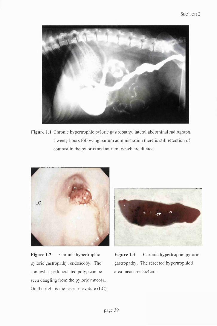
Section 2
Figure 1.1 Chronic hypertrophic pyloric gastropathy, lateral abdominal radiograph.
Twenty hours following barium administration there is still retention of
contrast in the pylorus and antrum, which are dilated.
LC
AFigure 1.2 Chronic hypertrophic
pyloric gastropathy, endoscopy. The
somewhat pedunculated polyp can be
seen dangling from the pyloric mucosa.
On the right is the lesser curvature (LC).
Figure 1.3 Chronic hypertrophic pyloric
gastropathy. The resected hypertrophied
area measures 2x4cm.
page 39

Section 2
2. Canine Inguinal Hernia
2.1 Introduction
Herniation is the abnormal protrusion of an organ or tissue from the cavity where it
normally lies (Read and Bellenger 1993). The exit may be a normal body opening
(Dean and others 1998) or a defect in the body wall (Read and Bellenger 1993), thus
defining false and true hernias respectively (Smeak 1993). True hernias have a
peritoneal sac, whereas false ones do not (Parks 1981).
In inguinal hernias the protrusion occurs through the inguinal canal. This is a passage
between the internal inguinal ring - limits o f which are the caudal edge of the internal
oblique, rectus abdominis and the pre-pubic tendon, and the external inguinal ring - a
slit between pelvic and abdominal tendon portions of external oblique muscle (Ashdown
1963).
During the embryological stage of development the vaginal process, an evagination o f
peritoneum, passes through the inguinal canal. The vaginal ring, which permits
communication between the lumen of the process and the peritoneal cavity, does not
become obliterated, and persists throughout life in domestic animals (Ashdown 1963).
Inguinal hernia may be congenital, though clinical signs may not develop until later in
life (Read and Bellenger 1993).
Inguinal hernia may be considered indirect when abdominal contents enter the vaginal
process, and less commonly direct when passage through the inguinal canal but adjacent
to the vaginal process (Smeak 1993).
Hayes (1974) noted certain breeds, such as Pekinese, Cairn Terrier, Basset Hound and
West Highland White Terrier, have a significantly increased risk of inguinal herniation.
The increased prevalence in some breeds suggests a pattern of familiar inheritance, but
it should not be assumed that all congenital hernias are heritable defects (Read and
Bellenger 1993).
Males are more likely to present with congenital inguinal hernias than females, possibly
because of delayed inguinal ring narrowing from late testicular descent (Fox 1963).
Acquired inguinal hernias are seen primarily in intact females and often in association
with oestrus or pregnancy, suggesting a hormonal factor (Parks 1981). It has been
page 40

Section 2
suggested that sex hormones may change the strength and character o f the connective
tissue, enlarging the diameter of the inguinal rings (Smeak 1993). Thus during oestrus
and pregnancy, the broad ligament and uterus are able to migrate into the inguinal canal
(Parks 1981). Nevertheless, Waters and others (1993) described this disease in six
spayed females, so the role of hormones remains unclear.
Parks (1981) suggested changes in intra-abdominal pressure (coughing, obstipation),
metabolic disease (diabetes mellitus, hyperadrenocorticism) or nutritional muscular-
aponeurotic deficiency o f the abdominal wall leading to its weakening, as causative
factors in herniation. In addition, obesity is a predisposing factor. Accumulation o f fat
encircling the round ligament causes a dilatation in the vaginal process and even o f the
inguinal canal itself (Ashdown 1963).
In general, when the protruding contents o f the hernia can be easily returned to the
cavity, the hernia is classified as reducible. If this is not possible, as a result of adhesion
of the contents in their new location, it is irreducible. Irreducible hernias of hollow
organs can be strangulated, which implies obstruction to blood flow through the organ
(Read and Bellenger 1993).
Incarceration may also obstruct vascular supply to the herniated tissue, usually at the
edge of the defect, and eventually result in necrosis due to strangulation. Strangulated
hernias usually lead to engorgement, fluid and blood loss from the organ affected.
Bacterial overgrowth, toxin production and a decrease in the effectiveness o f tissue
barriers to prevent bacterial invasion are common features in poorly perfused organs,
leading to endo and/or exotoxaemia and septicaemia (Parks, 1981).
The hernia can contain more than one organ, and the contents may vary from time to
time; most often organs in the immediate vicinity are found. However, those organs that
are freely movable on long vascular pedicles can travel to fill the hernia and are prone to
torsion (Parks 1981). Hernial contents include fat, omentum, intestine, peri-prostatic fat
and uterus; also bladder and spleen have been reported (Dean and others 1998).
However, fat and omentum are reported as the most common contents in canine inguinal
hernia (Grier and others 1971). This may explain the usual asymptomatic nature and
benign course, although omentum may become incarcerated, resulting in pain and
depression, having been described some cases in males in which concomitant
hydrocoele was present (Waters and others 1993).
page 41

Se ctio n 2
The main clinical sign is the presence of a painless mass with soft doughy consistency,
which may be bilateral but is more often left sided (Smeak 1993).
In 12 cases analysed by Waters and others (1993), in which hernias contained both small
intestine and uterus, none became incarcerated. The presence o f uterus within a hernia
limits herniation of small intestine.
Incarcerated hernias have to be differentiated from other inguinal masses such as
mammary tumours and cysts, lipomas, enlarged lymph nodes, abscesses and
haematomas. Femoral hernias, due to their vicinity, are usually misdiagnosed as inguinal
hernias. Femoral hernias are palpated caudal to the inguinal ligament and ventro-lateral
to the pelvic brim, whereas inguinal hernias are cranial and medial to the pelvic brim
(Smeak 1993).
Case Details
History
A five month old female English Cocker Spaniel (10kg body weight) was referred with a
four week history of a right sided inguinal swelling. The swelling was intermittent
initially, but had become permanent without causing any apparent signs of discomfort.
The dog also presented with a persistent ocular watery discharge due to bilateral
entropion that had been treated unsuccessfully with temporary Lembert sutures one
month previously.
Clinical examination
The dog was alert and responsive. On examination, a round, soft, fluctuant mass
(approximate size 3x3x4cm) could be palpated in the right inguinal area (Figure 2.1).
The consistency o f the content was fatty, and attempts to reduce the hernia were
unsuccessful. The left inguinal area appeared normal. A diagnosis o f right unilateral
inguinal hernia was made.
Ophthalmologic examination confirmed bilateral entropion causing corneal ulceration
and watery discharge. Imperforate right nasolacrimal puncta and left micropuncta were
also noted.
page 42

Section 2
Surgery
The anaesthetic protocol consisted of IM pre-medication with acetylpromazine (0.3mg)
and methadone (2mg), IV induction with 137.5mg o f thiopentone, followed by
endotracheal intubation and maintenance with a halothane/nitrous oxide and oxygen
mixture.
The dog was placed in dorsal recumbence and the herniated sac was exposed via a
caudal midline skin incision. As the mass was dissected free o f its adhesions the sac
was inadvertently breached. The hernial content, mainly fat, was pushed back into the
abdominal cavity.
The external inguinal ring was closed with pre-placed 2 metric nylon simple interrupted
sutures, except for the most caudal aspect (Figure 2.2). The patency of the external
pudendal vessels was checked.
The left side was examined and the ring was noticed to be slightly enlarged although no
herniation was evident, so three sutures of the same type and material as described were
placed. The wound was closed routinely.
The dog was discharged with chloramphenicol ointment TID and a seven days course of
carprofen 20mgper os (PO), BID.
Postoperative management included restricted lead exercise for 4 weeks.
Follow up
The dog was re-assessed a month after being discharged. The owners reported an
uneventful recovery from the surgery and healing process. On palpation, no inguinal
swelling was present, but some thickening due to fibrous reaction was evident.
At palpation, the closure o f the defect could be noticed, and no inguinal swelling was
present.
2.3 Discussion
Inguinal hernia has been described mainly in female middle-aged dogs (Dean and others
1998) or in young males (Waters and others 1993). This animal is unusual in that it is
young and female. Thus the previously suggested contributory factors o f hormonal
page 43

Section 2
influence, pregnancy and obesity do not apply (Ashdown 1963, Parks 1981). No
evidence o f trauma was noted on exploration of the hernia (Read and Bellenger 1993).
Ventral midline incision was preferred as, even in simple cases, this approach allows
visualisation of both inguinal rings and repair of bilateral herniation through a single
incision. It also avoids the possibility o f damage to the caudal superficial epigastric
vessels and mammary tissue (Peedle 1980, Dean and others 1998).
Pre-placement o f sutures to close the hernia was done to minimise the risk of damage to
the external pudendal vessels and genitofemoral nerve, which exit from the caudo-
medial aspect of the ring (Gourley and Gregory 1991).
The main post-operative complication is swelling, which may be caused by haematoma
due to inadequate haemostasis during hemiorraphy, or ensuing infection. Controlled
walking soon after the surgery and limited exercise until suture removal are encouraged
to decrease postoperative oedema, so that the prognosis for uncomplicated inguinal
hernia repair is good (Smeak 1993).
Patients with large defects may require reinforcement o f the primary hernia repair using
an on-lay polyethylene mesh technique or a cranial sartorious muscle flap (Smeak,
1993). Due to the large dead space in large hernias excessive swelling can be avoided
by means o f caudal abdominal bandage and placement o f drains (Waters and others
1993, Dean and others 1998).
2.4 References
Ashdown, R.R. (1963) The anatomy of the inguinal canal in the domesticated mammals. Veterinary Record IS, 1345-1351.
Dean, P.W., Bojrab, M.J. & Constantinescu, G.M. (1998) Inguinal hernia repair in the dog. In:
Current Techniques in Small Animal Surgery, ed M.J. Bojrab. Lea & Febiger,
Philadelphia. p552.
Fox, M.W. (1963) Inherited inguinal hernia and midline defects in the dog. Journal o f the
American Veterinary Medical Association 143, 602-604.
Gourley, I.M. & Gregory, C.R. (1991) Inguinal hernia. In: Atlas o f Small Animal Surgery, ed E. Greenspan. Gower Medical Publishing, New York p 27.4
Grier, R.L., Hoskins, J.D. & Wahlstrom, J.D. (1971) Inguinal hernia and Ritcher’s hernia in a
dog. Journal o f American Veterinary Medical Association 159, 181-183.
page 44

Section 2
Hayes, H.M. (1974) Congenital umbilical and inguinal hernias in cattle, horses, swine, dogs and cats: Risk by breed and sex among hospital patients. American Journal o f Veterinary
Research 35, 839-842.
Parks, J. (1981) Herniation. In: Pathophysiology in Small Animal Surgery, ed M.J. Bojrab. Lea
& Febiger, Philadelphia. p420.
Peedle, J.F. (1980) Inguinal hernia repair in the dog. Modern Veterinary Practice 61, 859-861.
Read, R.A. & Bellenger, C.R. (1993) Hernias. In: Textbook o f Small Animal Surgery, ed D.
Slatter. W.B. Saunders, Philadelphia. p431.
Smeak, D.D. (1993) Abdominal hernias. In: Textbook o f Small Animal Surgery, ed D.H. Slatter. W.B. Saunders, Philadelphia. p433.
Waters, D.J., Roy, R.G. & Stone, E.A. (1993) A retrospective study of inguinal hernia in 35
dogs. Veterinary Surgery 22, 44-49.
page 45

Section 2
Figure 2.1: Inguinal hernia. There is a marked inguinal swelling on the right with a
more subtle one on the left (black arrow).
Figure 2.2 Inguinal hernia. The hernia is closed using pre-placed monofilament nylon
sutures.
page 46
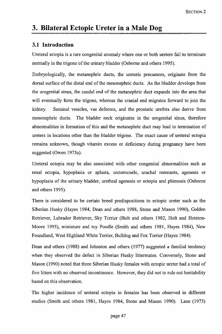
Section 2
3. Bilateral Ectopic Ureter in a Male Dog
3.1 Introduction
Ureteral ectopia is a rare congenital anomaly where one or both ureters fail to terminate
normally in the trigone of the urinary bladder (Osborne and others 1995).
Embryologically, the metanephric ducts, the ureteric precursors, originate from the
dorsal surface of the distal end of the mesonephric ducts. As the bladder develops from
the urogenital sinus, the caudal end o f the metanephric duct expands into the area that
will eventually form the trigone, whereas the cranial end migrates forward to join the
kidney. Seminal vesicles, vas deferens, and the prostatic urethra also derive from
mesonephric ducts. The bladder neck originates in the urogenital sinus, therefore
abnormalities in formation of this and the metanephric duct may lead to termination of
ureters in locations other than the bladder trigone. The exact cause o f ureteral ectopia
remains unknown, though vitamin excess or deficiency during pregnancy have been
suggested (Owen 1973a).
Ureteral ectopia may be also associated with other congenital abnormalities such as
renal ectopia, hypoplasia or aplasia, ureterocoele, urachal remnants, agenesis or
hypoplasia of the urinary bladder, urethral agenesis or ectopia and phimosis (Osborne
and others 1995).
There is considered to be certain breed predispositions to ectopic ureter such as the
Siberian Husky (Hayes 1984, Dean and others 1988, Stone and Mason 1990), Golden
Retriever, Labrador Retriever, Sky Terrier (Holt and others 1982, Holt and Hotston-
Moore 1995), miniature and toy Poodle (Smith and others 1981, Hayes 1984), New
Foundland, West Highland White Terrier, Bulldog and Fox Terrier (Hayes 1984).
Dean and others (1988) and Johnston and others (1977) suggested a familial tendency
when they observed the defect in Siberian Husky littermates. Conversely, Stone and
Mason (1990) noted that three Siberian Husky females with ectopic ureter had a total of
five litters with no observed incontinence. However, they did not to rule out heritability
based on this observation.
The higher incidence of ureteral ectopia in females has been observed in different
studies (Smith and others 1981, Hayes 1984, Stone and Mason 1990). Lane (1973)
page 47

Sectio n 2
the longer urethra and that the commonest site o f ectopic orifice in this gender is
proximal to the urethralis muscle. The sphincter action o f this muscle could be expected
to prevent incontinence.
As ureteral ectopia is a congenital anomaly, the associated urinary incontinence is
usually recognised in young animals. However, in males the onset o f incontinence may
be delayed until adulthood, when urethral muscular tone declines, as some studies have
reported (Lane 1973, Stone and Mason 1990). Therefore, although ureteral ectopia is
the commonest cause o f urinary incontinence in juvenile dogs (Holt 1990), it should be
considered in the differential diagnosis of urinary incontinence in dogs o f all ages (Holt
and Hotston-Moore 1995).
Ureteric ectopia in males is most commonly recognised when they terminate in the
prostatic urethra (Holt and Hotston-Moore 1995) and the cranial portion o f membranous
urethra (Osborne and others 1995), although vas deferens and seminal vesicles are other
possible sites of termination (Dean and others 1988).
Independent o f the termination site ectopic ureters may enter the bladder at the normal
site on the dorsolateral caudal serosal surface, but continue through the bladder
muscularis and submucosa, pass the trigone to an abnormal distal termination. These
are defined as intramural ectopic ureters (Osborne and others 1995). These may open at
the trigone in a normal fashion and then continuing submucosally to a second distal
opening, so called ureteral branching (Dean and others 1988).
Ureteral troughs have been defined as intramural ectopic ureters that may open into the
bladder in a normal or more distal location, but continue as an incomplete structure
(trough) beyond the trigone into the proximal urethral sphincter (Stone and Mason
1990).
One o f the commonest associated abnormalities is hydroureter and hydronephrosis
(Smith and others 1981). The exact reason for this is unclear but may result from the
partial obstruction of distal ureter owing to its abnormally long intramural course,
abnormal ureteral peristalsis or ascending infection (Holt and Hotston-Moore 1995,
Osborne and others 1995).
page 48

Sectio n 2
To diagnose ectopic ureter, the excretory urogram allows evaluation of renal function
and side-by-side comparison of both, right and left, components o f the upper urinary
tract (Smith and others 1981).
Owen (1973b) preferred a large volume slow infusion technique because it can be used
in patients with poor kidney function. The whole urinary tract is visualised at once and
there is no need to compress the abdomen, which may distort normal anatomical
relationships. Apart from obvious by-pass o f the bladder one o f the main findings is
loss o f the normal “J” shaped termination o f the ureter as it enters the bladder (Waldron
1993). The exact site o f the ectopic orifice may be difficult to locate due to masking by
contrast material within bladder, poor renal excretion (Owen 1973b) or in cases of
intramural ectopic ureter (Osborne and others 1995). Alternative techniques are
retrograde contrast studies (Holt and others 1982).
No effective primary medical management is available for urinary incontinence caused
by ectopic ureter. Surgical correction is the only viable option, and the choice of the
procedure is based on the functional status o f ureters, kidneys and the presence of
concomitant abnormalities (Osborne and others 1995).
The creation o f a neostoma in situ with ligation and transection o f the distal portion is
the procedure of choice for intramural ectopic ureter. U reterovesical
anastom osis/ureteroneocystotom y consists o f ureteral transection and reimplantation
(Stone and Mason 1990). Ureteronephrectom y is only indicated in cases of exclusively
unilateral ectopia and when ureter and kidney are irreversibly damaged (Gregory 1996).
Branching distal extension is surgically corrected by occlusion with placement o f simple
interrupted sutures through serosa and muscularis and around the ureter. Closure of
ureteral trough is made with synthetic absorbable suture material in a simple continuous
pattern (Stone and Mason 1990).
3.2 Case details
History
A one year male Border Terrier (8.5 kg body weight) was referred with a history of
urinary incontinence since puppy hood. Constant urine dribbling was noticed, although
the dog was able to produce a normal micturition stream, without signs o f discomfort or
page 49

Section 2
straining. The dribbling was worse when the dog was excited or asleep. Previous
treatments with different courses o f antibiotics and one o f phenylpropanolamine syrup
had failed to improve the condition.
Clinical examination
The dog was alert and responsive. On examination regular urine dribbling was visible,
hair surrounding the prepuce was stained with urine and the skin o f the caudal abdomen
was scalded.
Ancillary aids
A blood sample was taken for haematology and biochemical studies. Increases in white
blood cells (15.2xl09/l), haemoglobin (18.3g/dl), albumin (39.0g/l), and glucose
(5.8mmol/l) values were detected; whereas total globulin (24.0g/l) and phosphorus
(1.11 mmol/1) measurements were below the average range.
Biochemical analysis of urine revealed the presence o f protein, specific gravity (SG) of
1.040 and pH 7.5. A large number of white blood cells and granular casts were found in
the urine as well as the presence of cellular debris and large epithelial cells. Urine
samples were also sent for bacteriological culture and sensitivity, but no bacteria were
isolated.
Ultrasound revealed a dilated urethra, a grossly dilated left ureter with some left
hydronephrosis (Figure 3.1). A double contrast cystogram combined with a retrograde
urethrogram revealed a left enlarged ureter terminating in the prostate area (Figure 3.2).
Surgery
The anaesthetic protocol consisted of IM pre-medication with acetylpromazine (0.2mg)
and methadone (0.2mg), IV induction with methohexitone (3.2ml), followed by
endotracheal intubation and maintenance of anaesthesia with a halothane/nitrous
oxide/oxygen mixture.
Via a midline laparotomy, the bladder was exposed and two stay sutures (2 metric
polyglactin 910) were placed in the apex for atraumatic handling. The left ureter was
dilated entering the distal prostatic urethra and the right ureter terminated in a normal
location.
page 50

Section 2
A ventral cystotomy was performed and the dorsal trigone area inspected, but no
effective urine flow was noticed from the right ureteral orifice. Gentle urethral
occlusion caused bulging o f the right ureteral intramural segment draining into the
proximal prostatic urethra and a diagnosis o f bilateral ectopia was confirmed.
Right ureteral ectopia was repaired by means of creation o f a neostoma in situ. A small
incision was made in the bladder mucosa directly over the ureter to expose its intramural
portion. The ureter was incised at the level of normal ureteral opening and the proximal
edges were spatulated and sutured to the bladder mucosa using a simple interrupted
pattern (0.7 metric polyglactin 910). A 22 gauge catheter was placed through the
neostoma into the distal portion which was occluded by placing sutures through serosa,
muscularis and around the ureter. As the catheter was removed, the sutures were tied to
occlude the ureteral lumen.
The procedure chosen for the left ureter was a ureteroneocystotomy. The ureter was
ligated as close as possible to its termination in the urethra and transected just proximal
to the ligature. After choosing a site for re-implantation, a stab incision was made in the
urothelium and serosal surfaces at the anticipated ureteral entry and exit points. A
tunnel was created by pushing a pair of haemostatic forceps through the bladder wall.
The proximal end of the excised ureter was grasped by means o f a stay suture attached
to haemostatic forceps and passed through the new tunnel. The edges were spatulated
and sutured to the bladder mucosa (0.7 metric polyglactin 910).
The cystotomy was closed using a simple interrupted pattern (2 metric polyglactin 910)
and omentum was secured over cystotomy incision with the same type of suture
material. The laparotomy was closed routinely.
Follow up
The dog recovered well after surgery and when discharged was bright and alert although
a wet bed was noticed postoperatively. On the second day post-surgery the dog went
home with amoxycillin/clavulanic acid (lOOmg) PO BID for 4 weeks, and carprofen
(20mg) PO BID for 5 days.
Four weeks post-surgery the owner reported a remarkable improvement. The dog only
appeared wet sometimes and always when recumbent.
page 51
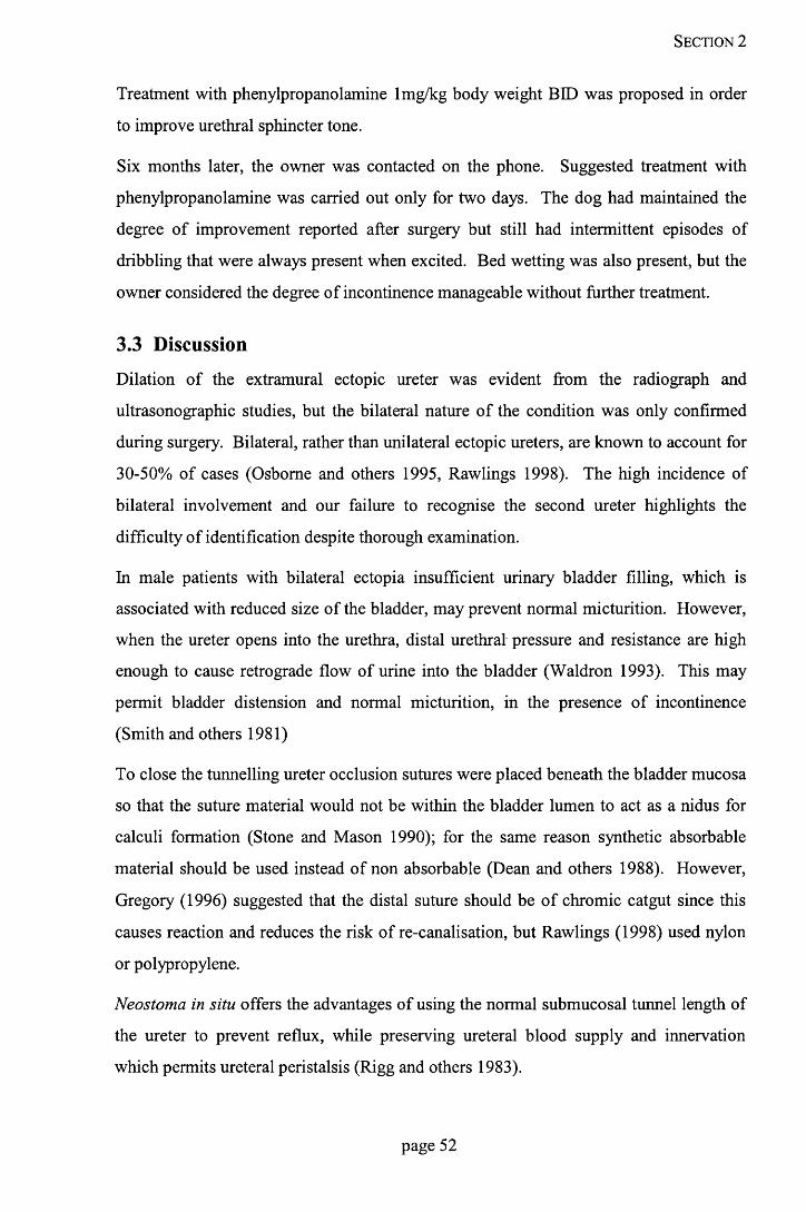
Section 2
Treatment with phenylpropanolamine 1 mg/kg body weight BID was proposed in order
to improve urethral sphincter tone.
Six months later, the owner was contacted on the phone. Suggested treatment with
phenylpropanolamine was carried out only for two days. The dog had maintained the
degree of improvement reported after surgery but still had intermittent episodes of
dribbling that were always present when excited. Bed wetting was also present, but the
owner considered the degree of incontinence manageable without further treatment.
3.3 Discussion
Dilation o f the extramural ectopic ureter was evident from the radiograph and
ultrasonographic studies, but the bilateral nature o f the condition was only confirmed
during surgery. Bilateral, rather than unilateral ectopic ureters, are known to account for
30-50% of cases (Osborne and others 1995, Rawlings 1998). The high incidence of
bilateral involvement and our failure to recognise the second ureter highlights the
difficulty of identification despite thorough examination.
In male patients with bilateral ectopia insufficient urinary bladder filling, which is
associated with reduced size of the bladder, may prevent normal micturition. However,
when the ureter opens into the urethra, distal urethral pressure and resistance are high
enough to cause retrograde flow of urine into the bladder (Waldron 1993). This may
permit bladder distension and normal micturition, in the presence o f incontinence
(Smith and others 1981)
To close the tunnelling ureter occlusion sutures were placed beneath the bladder mucosa
so that the suture material would not be within the bladder lumen to act as a nidus for
calculi formation (Stone and Mason 1990); for the same reason synthetic absorbable
material should be used instead of non absorbable (Dean and others 1988). However,
Gregory (1996) suggested that the distal suture should be o f chromic catgut since this
causes reaction and reduces the risk o f re-canalisation, but Rawlings (1998) used nylon
or polypropylene.
Neostoma in situ offers the advantages of using the normal submucosal tunnel length of
the ureter to prevent reflux, while preserving ureteral blood supply and innervation
which permits ureteral peristalsis (Rigg and others 1983).
page 52

Section 2
Because of the potential hypoplastic bladder in cases o f bilateral ectopia, a one layer
appositional suture was used to close the cystotomy incision and omentum placed over it
to prevent intra-abdominal urine leakage but maximise the bladder lumen as suggested
by Waldron (1993).
Even though a better long-term result will be obtained when the ureters have some
degree of peristalsis and are not extremely dilated, satisfactory results have been
obtained in dogs in which markedly dilated ectopic ureters were re-implanted (Osborne
and others 1995).
Fewer complications are reported after ureteronephrectomy than after ureteral
transplantation, but it is generally considered preferable to salvage a viable kidney. The
re-implantation o f dilated ureters into the bladder may be associated with a certain
degree o f vesicoureteral reflux, which predisposes the patient to ascending infection of
the kidney (Osborne and others 1995). However, postoperative infection of the urinary
tract was described as an uncommon though serious complication (Holt and Hotston-
Moore 1995).
Based on previous results in children, in which post-surgical bladder wall oedema led to
distal ureteral stricture, Holt and others (1982), advised deferring surgery until the dog
was at least 6 months old. Yet, McLaughlin and Miller (1991), concluded that, age at
time of surgical correction had no effect on the rate of post-operative incontinence.
There appears to be a higher incidence o f incontinence after corrective surgery if the
urethra is involved, and it has been suggested that concomitant urethral sphincter
incompetence may be the cause (McLaughlin and Miller 1991). The trigone area is
richly supplied with alpha adrenergic receptors and their stimulation can increase the
tone in this area (Rigg and others 1983).
Oestrogen can increase the response to endogenous alpha adrenergic stimulation (Dean
and others 1988), but anatomical and correctable causes such as patent distal segment,
or reduced bladder capacity, should be ruled out before prescribing lifelong alpha
adrenergics such as ephedrine or phenylpropanolamine hydrochloride, or oestrogen
therapy (Stone and Mason 1990).
Other causes of surgical failure in correcting urinary incontinence might be distal
remnant of an ectopic ureter traversing the bladder neck, thus interfering with normal
page 53
Sectio n 2
sphincter function, and a hypoplastic bladder unable to cope with the increased inflow
of urine (Holt and others 1982).
3.4 References
Dean, P.W., Bojrab, M.J. & Constantinescu, G.M. (1988) Canine ectopic ureter. Compendium
on Continuing Education fo r the Practicing Veterinarian 10, 146-158.
Gregory, S.P. (1996) Management of canine urinary incontinence. In: Manual o f Canine and
Feline Nephrology and Urology, eds J. Bainbridge and J. Elliott. British Small Animal Veterinary Association, Gloucestershire. pl61.
Hayes, H.M. (1984) Breed associations of canine ectopic ureter: a study of 217 female cases. Journal o f Small Animal Practice 25, 501-504.
Holt, P.E. (1990) Urinary incontinence in dog and cat. Veterinary Record 127, 347-350.
Holt, P.E., Gibbs, C. & Pearson, H. (1982) Canine ectopic ureter-a review of twenty-nine cases. Journal o f Small Animal Practice 23, 195-208.
Holt, P.E. & Hotston-Moore, A. (1995) Canine ureteral ectopia: an analysis of 175 cases and
comparison of surgical treatments. Veterinary Record 136, 345-349.
Johnston, G.R., Osborne, C.A., Wilson, J.W. & Yano, B.L. (1977) Familial ureteral ectopia in the dog. Journal o f American Veterinary Medical Association 168, 168-170.
Lane, J.G. (1973) Canine ectopic ureter - two further case reports. Journal o f Small Animal
Practice 14, 555-560.
McLaughlin, R. & Miller, C.W. (1991) Urinary incontinence after surgical repair of ureteral ectopia in dogs. Veterinary Surgery 20, 100-103.
Osborne, C.A., Johnston, G.R. & Kruger, J.M. (1995) Ectopic ureters and ureteroceles. In: Canine and Feline Nephrology and Urology, eds C.A. Osborne and D.R. Finco. Williams
and Wilkins, Baltimore. p608.
Owen, R. ap R. (1973a) Canine ureteral ectopia-a review. 1. Embryology and aetiology.
Journal o f Small Animal Practice 14, 407-417.
Owen, R. ap R. (1973b) Canine ureteral ectopia-a review. 2. Incidence, diagnosis and treatment.
Journal o f Small Animal Practice 14, 419-427.
Rawlings, C.A. (1998) Correction of ectopic ureter. In: Current Techniques in Small Animal
Surgery, ed M.J. Bojrab. Williams and Wilkins, Baltimore. p445.
Rigg, D.L., Zenoble, R.D. & Riedesel, E.A. (1983) Neoureterostomy and phenylpropanolamine
therapy for incontinence due to ectopic ureter in a dog. Journal o f the American Animal
Hospital Association 19, 237-241.
page 54
Section 2
Smith, C.W., Stowater, J.L. & Kneller, S.K. (1981) Ectopic ureter in the dog- a review of cases. Journal o f the American Animal Hospital Association 17, 245-248.
Stone, E.A. & Mason, L.K. (1990) Surgery of ectopic ureters: types, method of correction and
postoperative results. Journal o f the American Animal Hospital Association 26, 81-88.
Waldron, D.R. (1993) Urinary bladder. In: Textbook o f Small Animal Surgery, ed D.H. Slatter.
W.B. Saunders Company, Philadelphia. pl450.
page 55

Section 2
bladder
0 *
ureter
Figure 3.1 Ectopic ureter, ultrasonogram. The dilated ectopic ureter is visible coursing
close to the bladder.
Figure 3.2 Ectopic ureter, lateral abdominal radiograph. The contrast can be seen to
reflux from the prostatic urethra up a dilated ectopic ureter.
page 56

Sectio n 2
4. Traumatic Diaphragmatic Rupture
4.1 Introduction
Diaphragmatic defects in cats and dogs are usually traumatic rather than congenital
(Carb 1975). Typically, the defect results from indirect blunt trauma due to collision
with motor vehicles (Sullivan and Lee 1989), although in a large number of cases there
is no positive history of trauma (Garson and others 1980). Direct diaphragmatic injuries
such as stab, gunshot or bite wounds are rare (Bellenger and others 1975).
During inspiration an increased positive intra-abdominal pressure is generated coupled
with an increasing negative intra-pleural pressure. Upon coughing, straining and
forceful muscular contraction high intra-abdominal pressures are generated. To offset
this, the glottis closes producing a intra-thoracic positive pressure. Sudden pressure
changes that lead to the glottis failing to remain closed may result in a critically high
pleuro-peritoneal pressure gradient (Carb 1975). Boudrieau (1990) speculated that this
gradient underlies the pathophysiology of traumatic diaphragmatic hernia (TDH). The
specific area of the diaphragm on which the large pleuro-peritoneal pressure gradient
focuses, and thus be most susceptible to rupture, depends to a great extent on the
position o f the animal (Carb 1975). However, tears usually affect the weaker muscular
part o f the diaphragm rather than the tendinous portion (Carb 1975, Garson and others
1980). In cats there is a higher incidence o f circumferential tears (Garson and others
1980) and in some cases with muscles tom from the ribs allowing abdominal viscera to
herniate into the ventro-lateral subcutaneous tissue (Wilson and others 1971).
The extent and location of the diaphragmatic rent, together with anatomical proximity
and length of supporting ligaments or mesentery o f individual organs, all influence the
abdominal viscera that may herniate. Carb (1975) noted that any o f the abdominal
viscera may be involved. In a left tear the herniated organs are most commonly the
stomach, spleen or small intestine. Whereas if right sided, small intestine and one or
more lobes o f liver herniate. The presence of liver is most frequently associated with
ascites or hydrothorax (Wilson and others 1971, Garson and others 1980). However,
unusual organ displacement such as the kidney or gravid uterus has been reported
(Sullivan and Lee 1989). The complications associated with herniation, such as
strangulation, obstmction and incarceration o f abdominal organs are often due to
page 57

Section 2
pressure applied by the edge of the diaphragmatic tear as the organs pass over it or result
from the formation of fibrous adhesions (Johnson 1993).
The most common clinical signs are usually related to the respiratory system with
varying degrees of dyspnoea, hyperpnoea and cyanosis due to the reduced tidal volume.
The animal may be clinically normal, but subsequently develop signs o f chronic
respiratory disease such as intermittent chronic cough and fatigue following exercise
(Wilson and others 1971). In some acute cases animals adopt a sitting or standing
position with elbows abducted and head extended due to chest pain and in an attempt to
lessen abdominal organ pressure on the thoracic structures (Carb 1975). Other signs
such as anorexia, pallor, constipation, vomiting or abdominal pain have been reported
(Garson and others 1980, Roe and others 1986). Cornell and others (1993) reported two
cats with icterus due to extra-hepatic biliary tract obstruction subsequent to
diaphragmatic rupture.
There is often a lack o f abnormalities on physical examination. However, on
auscultation abnormal heart sounds are found with muffling in the affected hemithorax
(Wilson and others 1971). Thorax percussion may reveal hyper-resonance if a distended
gas filled stomach is present, or dullness with liver herniation (Walker and Hall 1965).
Borborygmus is not reliable and a wasp waist is not a common sign (Wilson and others
1971).
Wilson and others (1971) found that the greatest number o f deaths occurred prior to
surgical correction, with the critical periods being positioning for radiographs and
induction of anaesthesia. The conclusion is that surgical repair should be delayed until
cardio-respiratory function is stabilised. Furthermore, a compensated patient may
become anoxic as a result of the stress of handling and restraint, as well as inappropriate
radiographic positioning with the affected side downwards or for a ventro-dorsal view
(Fagin 1989, Sullivan and Lee 1989).
Radiography is considered the most useful diagnostic aid (Johnson 1993). Frequent
findings are loss of the diaphragmatic line (Fagin 1989) and masking of the cardiac
shadow (Sullivan and Lee 1989). However, both o f these may be difficult to detect in
the presence of pleural effusion, which is a common sequel to TDH (Roudebush and
Bums 1979). In this situation ultrasonography is useful because fluid is an excellent
ultrasound-transmitting medium and allows detection o f abnormally positioned viscera.
page 58

Section 2
Although it is not always possible to detect gaps in the band of echoes adjacent to the
liver that represent the diaphragm on the sonogram (Fagin 1989, Johnson 1993).
Nevertheless, thoracocentesis allows improved radiographic detail and perhaps more
importantly alleviates hypoventilation (Johnson 1993).
Due to the occurrence o f false positives and negatives contrast radiography such as
barium studies and positive or negative peritoneography are not a substitute for careful
evaluation o f plain films. (Rendano 1979, Stickle 1984, Sullivan and Lee 1989).
4.2 Case details
History
A one year old male domestic short-hair cat (4.1kg body weight) was presented with a
history of road traffic accident in the previous 24 hours. The owners had noticed that
the cat was slightly depressed and was not able to extend the left front leg.
Clinical examination
Palpation of extremities showed a fracture in the left humerus and an abnormal rotation
of the right hock, which also appeared swollen and painful. Pulmonary auscultation was
loud and harsh. No other abnormalities were detected.
Ancillary aids
Blood samples taken for haematological and biochemical analyses showed no
significant abnormalities.
A lateral radiographic view of the thorax showed a fat dense mass lying caudo-ventrally
in front of and overlying the diaphragm, which appeared to be confluent with falciform
fat. Left humeral and right distal tibial fractures were also identified.
Surgery
The anaesthetic protocol consisted o f IM pre-medication with 0.15mg of
acetylpromazine and 0.8mg o f methadone, followed by IV induction with 30mg of
thiopentone. The maintenance of anaesthesia after placement o f an uncuffed 5.5
endotracheal tube was done with a halothane-oxygen mixture. Assisted ventilation was
established by means of a mechanical ventilator.
page 59
Sectio n 2
Midline laparotomy was performed from a point immediately caudal to the xiphoid to the
umbilicus. A circumferential diaphragmatic tear at the costochondral junction was
exposed with displacement of omentum through it. The omentum was gently retracted
allowing examination of the lungs and heart but no further abnormality was noticed.
The defect was closed with pre-placed horizontal mattress sutures o f 3 metric
polypropylene. The sutures incorporated the ribs and intercostal muscles. Prior to
abdominal closure the diaphragm was checked for hidden tears. An intra-thoracic drain
was placed and 2ml of fluid and 10ml of air were removed (Figure 4.1). The drain was
not left in place after definitive surgical closure.
The spleen showed some signs of severe traumatic congestion in its cranial portion, so
the affected area was removed after clamping and vascular ligation (2 metric polyglactin
910)
The laparotomy incision was closed routinely.
Follow up
Treatment with amoxycillin/clavulanic acid (50mg TLD) and carprofen (lOmg BID) was
instituted, as a continuation of the peri-operative management.
Hourly monitoring o f respiratory and cardiac rate as well as temperature and capillary
refill time was done during the first 12 post-operative hours. The cat, bright and eating,
was transferred to the orthopaedics service for fracture repair three days later.
Discussion
Walker and Hall (1965) were o f the opinion that the prognosis for TDH must be
guarded. However, provided each case was assessed and treated on its own merits, a
satisfactory repair could usually be achieved safely through a simple surgical procedure.
More than thirty years later this statement is still valid. The real risk is inappropriate
timing o f surgery in a patient that has not been stabilised, with compromised respiratory
and circulatory function, due to shock and direct lung compression caused by herniated
organs (Bellah 1998). In this cat, the respiratory signs were minimal and the general
state o f the animal did not require an emergency surgical procedure.
Apart from the surgical repiar, anaesthesia for TDH is a challenge as there is a need for
intermittent positive pressure ventilation, and nitrous oxide should be avoided since its
page 60

Sectio n 2
solubility in blood allows this gas to accumulate in the pleural space resulting in further
lung compression and the possibility of diffusion hypoxia at recovery. This will worsen
any initial hypoxia (Johnson 1993). In addition, the diffusion of gas into displaced
stomach or intestinal loops could increase lung compression (Bellah 1998).
Assisted respiration was immediately instituted following induction with positive
pressure not exceeding 20cm to ensure adequate oxygenation, yet prevent barotrauma
with consequent development o f re-expansion pulmonary oedema after over-inflation of
atelectic areas. The latter effect was found to be one o f the main reasons for mortality
(Garson and others 1980, Bellenger and others 1996).
The placement of a chest drain was done to eliminate air and fluid that may complicate
the post-operative recovery period, but no attempts were made to keep the thoracic drain
device after the surgery as indicated by Sullivan and Reid (1990) when repairing
diaphragmatic tears in cats.
As indicated by Boudrieau (1990) the damaged portion o f spleen was resected in situ
without allowing re-establishment of the compromised splenic circulation in order to
prevent toxaemia, which can result from bacterial endotoxins and exotoxins as well as
products of tissue auto lysis.
The tear in this case was circumferential as has been reported previously as common in
cats (Garson and others 1980). The choice of suture material and pattern appears to be a
matter of strong surgeon preference. Sullivan and Reid (1990) preferred silk. Johnson
(1993) was of the opinion that a continuous suture of polyglactin 910 should be used as
this avoided the stiff, sharp, cut ends of knotted polydioxanone sutures possibly injuring
the abdominal viscera. However, our choice of polypropylene was based on the fact that
this material is recommended for general hemiorraphy (Bellah 1998).
4.4 References
Bellah, J.R. (1998) Traumatic diaphragmatic hernia. In: Current Techniques in Small Animal
Surgery, ed M.J. Bojrab. Williams and Wilkins, Baltimore. p315.
Bellenger, C.R., Hunt, G.B., Goldsmid, S.E. & Pearson, M.R.B. (1996) Outcomes of thoracic surgery in dogs and cats. Australian Veterinary Journal 74, 25-30.
Bellenger, C.R., Trim, C. & Sumner-Smith, G. (1975) Bile pleuritis in a dog. Journal o f Small
Animal Practice 16, 575-577.
page 61

Section 2
Boudrieau, R.J. (1990) Traumatic diaphragmatic hernia. In: Current Techniques o f Small
Animal Surgery, ed M.J. Bojrab. Lea and Febiger, Philadelphia. p309.
Carb, A. (1975) Diaphragmatic hernia in the dog and the cat. Veterinary Clinics o f North
America 5, 477-494.
Cornell, K.K., Jakovljevic, S., Waters, D.J., Prostredny, J., Salisbury, S.K. & De Nicola, D.B. (1993) Extrahepatic biliary obstruction secondary to diaphragmatic hernia in two cats.
Journal o f the American Animal Hospital Association 29, 502-507.
Fagin, B. (1989) Using radiography to diagnose traumatic diaphragmatic hernia. Veterinary
Medicine 84, 662-672.
Garson, H.L., Dodman, N.H. & Baker, G.J. (1980) Diaphragmatic hernia. Analysis of fifty-six
cases in dogs and cats. Journal o f Small Animal Practice 21, 469-481.
Johnson, K.A. (1993) Diaphragmatic, pericardial and hiatal hernia. In: Textbook o f Small
Animal Surgery, ed D.H. Slatter. W.B. Saunders Company, Philadelphia. p455.
Rendano, V.T. (1979) Positive contrast peritoneography: An aid in the radiographic diagnosis of diaphragmatic hernia. Veterinary Radiology 20, 67-72.
Roe, S.C., Smith, C.W. & Stowater, J.L. (1986) Diaphragmatic hernia producing gastric outflow obstruction, metabolic alkalosis and hypokalemia in dog. Compendium on
Continuing Education for the Practicing Veterinarian 8, 943-948.
Roudebush, P. & Bums, J. (1979) Pleural effusion as a sequela to traumatic diaphragmatic hernias: A review of four cases. Journal o f the American Animal Hospital Association
15, 699-706.
Stickle, R.L. (1984) Positive contrast celiography (peritoneography) for the diagnosis of
diaphragmatic hernia in dogs and cats. Journal o f the American Veterinary Medical
Association 185, 295-298.
Sullivan, M. & Lee, R. (1989) Radiological features of 80 cases of diaphragmatic rupture.
Journal o f Small Animal Practice 30, 561-566.
Sullivan, M. & Reid, J. (1990) Management of 60 cases of diaphragmatic rupture. Journal o f
Small Animal Practice 31, 425-430.
Walker, R.G. & Hall, L.W. (1965) Rupture of the diaphragm: Report of 32 cases in dogs and cats. Veterinary Record 77, 830-837.
Wilson, G.P., Newton, C.D. & Burt, J.K. (1971) A review of 116 diaphragmatic hernias in dogs
and cats. Journal o f the American Veterinary Medical Association 159, 1142-1145.
page 62

Section 2
Figure 4.1 Diaphragmatic rupture, lateral radiograph. Following surgery the
diaphragmatic line is intact and normal aerated lungflelds are visible.
page 63

Section 2
5. Canine Nasal Aspergillosis
5.1 Introduction
Mycosis has been described as one o f the causes o f chronic rhinitis/sinusitis in the dog
(Bright 1981). The agent is generally Aspergillus fum igatus, which causes a destructive
form of rhinitis (Sullivan and others 1986). Occasionally, Aspergillus nidulans,
Aspergillus niger, Aspergillus flavus and Penicillium spp are incriminated. However,
all are normal nasal microflora and as they cause a similar disease process they are
grouped under the general term of nasal aspergillosis (Sharp 1989).
Pathogenesis remains unclear, only a few cases present with a history o f cranial trauma
or tumour. Cranial surgery that might have left a bone sequestrum to act as a nidus for
infection is very uncommon. Immunosupression, often seen in this disorder, has not
been confirmed to be a cause or result from it, taking into account that canine cancer
patients do not seem predisposed to nasal aspergillosis (Bright 1981, Sharp 1989).
Sharp and others (1991a) suggested that farm dogs might be more susceptible to this
disease due to the large amount of spores that exist in hay or straw. These authors also
reported a higher incidence of the disease in non-brachycephalic breeds, specifically
Collies and Golden Retrievers, between one and seven years o f age. A later study
suggested that male dogs were at greater risk than females (W olf 1992).
Bright (1981) described the action of the fungus as causing an initial mucosal and
submucosal influx of neutrophils, macrophages, eosinophils and plasma cells, followed
by proliferation o f fungal hyphae that invade and occlude the vessels o f the area. The
body attempts to contain the invasion, which results in the formation o f a dense fibrous
layer throughout the submucosa with loss of normal epithelial and submucosal
structures. A chronic irritation with resultant exudate production is the cause for the
copious nasal discharge and chronic sneezing seen clinically. The discharge is
sanguino-purulent and one of the most frequent clinical findings but is not characteristic
(Sullivan and others 1986, Sharp and Sullivan 1989). Consequently, nasal foreign body,
neoplasia, dental disease and chronic idiopathic rhinitis should be included in the list o f
differential diagnosis (Sharp and others 1991a). Where frank epistaxis occurs systemic
causes such as coagulopathy should be considered (Sharp 1989). Nasal discharge may
be absent in a few cases where infection is restricted to the frontal sinus (Sharp and
page 64

Section 2
others, 1991a) and when affected may also show chemosis (Sharp 1989), exopthalmos
(W olf 1992), blepharospasm and epiphora (Sharp and others 1993) due to extension of
fungal infection into soft tissues around the orbit.
Ulceration o f the rhinarium and signs of discomfort around the nostrils or bridge of the
nose are significant clinical signs (Sharp and Sullivan 1989).
The non-specific nature of clinical signs require ancillary aids for definitive diagnosis
with radiology being the single most useful aid for this purpose (Sullivan and others
1986). The radiological features include turbinate loss producing rostral radiolucency,
whereas a mixed density pattern may be observed caudally.
The rostro-caudal view is especially useful in the evaluation o f the frontal sinus, which
should not be overlooked as far as treatment is concerned. The features include opacity
o f the sinus, as well as thickening due to periosteal reaction stimulated by fungus, and a
“moth-eaten appearance” or mottling of the frontal bone due to the osteolytic effect of
the fungus or its toxins (Sullivan and others 1986).
If available, computer assisted tomography (CT) or magnetic resonance imaging (MRI),
provide detailed definition of intranasal structures and lesions particularly in the
ethmoid region (W olf 1992).
Rhinoscopy is well suited to the investigation of aspergillosis due to the large air space
created by the destructive rhinitis. Fungal colonies grow on the turbinate mucosa as
greenish-white plaques having a ’'''white jam mould-like” appearance (Lane and Wamock
1977, Sullivan 1987).
Direct smear and culture of intranasal swabs samples are unreliable in the diagnosis of
nasal aspergillosis (Lane and Wamock 1977). Moreover 40% o f normal dogs, and those
with nasal neoplasia can also yield this fungus on nasal culture (Sharp and Sullivan
1992). A more accurate, but specific test, is serology. The false positive rates for the
various assays such as ELISA, counter immuno-electrophoresis and agar gel
immunodiffusion are approximately 15% (Sharp 1989).
Consequently, Sharp and Sullivan (1992) established the diagnostic criteria for nasal
aspergillosis involving radiology, rhinoscopy, mycology (culture, cytology and
histopathology) and serology, indicating that at least 2, but preferably 3 criteria should
be satisfied for diagnosis.
page 65

Section 2
Results in the trials carried out with systemic therapy showed that drugs like
thiabendazole, ketoconazole or fluconazole tend to be effective in only 43-47% of cases
(Harvey 1984, Sharp and Sullivan 1989, Sharp and others 1991b). The use of
itraconazole has been also suggested as effective agent with few side effects (Legendre
1995).
Topical therapy with enilconazole by means of indwelling tubes implanted through
trephine holes in the frontal sinus, to lie in the mid-nasal chamber has proved to be a
successful treatment with a relatively low cost and few side effects (Sharp and Sullivan
1986).
The necessity for week long treatment generally requires hospitalisation. Treatment
with topical clotrimazole 1% has been described recently by means of a single non
invasive intranasal infusion with one hour contact time (Burbidge and others 1997,
Smith and others 1998).
5.2 Case details
History
A ten year old neutered female Old English Sheepdog (23kg body weight) was referred
with a 2 month history of sneezing and nasal discharge which had progressed from
watery to thick sanguinopurulent secretion. The owner had noticed an increasing
dullness and a marked pain if the nostrils were touched, which was also evident when
the dog chewed food.
Clinical examination
Detailed examination of the nose revealed discomfort on nasal palpation and an obvious
yellow-green, thick bilateral nasal discharge but with marked discoloration o f the left
nasal sill (Figure 5.1). An airflow test was positive and the left submandibular lymph
node was noticeably enlarged.
Ancillary aids
Blood samples were taken for serological, haematological and biochemical studies.
Increases in MHCH (36.7g/dl), white blood cells (17.6xl09/l), neutrophils (14.08xl09/l)
page 66

Section 2
and chloride (118mmol/l) values were found, whereas platelets (195xl09/l) and albumin
(23g/l) were found to be lower than normal..
Radiographic films were taken of the skull using dorso-ventral intra-oral and rostro-
caudal views. There was marked turbinate destruction on the left with an increased
radiolucency, similar but less severe change is evident on the right with some thinning
of the vomer/septum (Figure 5.2). The left frontal sinus was nearly completely
opacified, and the right to a lesser extent (Figure 5.3).
Rostral endoscopy was performed and the presence o f yellow-grey fungal plaques were
confirmed in both nasal chambers (Figure 5.4).
Surgery
The anaesthetic protocol consisted of IM premedication with acetylpromazyne (0.8mg)
and methadone (5mg), IV induction with thiopentone (200mg), followed by
endotracheal intubation and maintenance o f anaesthesia with a halothane/nitrous
oxide/oxygen mixture.
The dog was placed in sternal recumbence and the pharynx was packed with a saline
moistened gauze. A 2cm skin incision was made over the frontal sinus off-midline.
The underlying muscle was separated to expose the periosteum. Using a 5mm
osteotome and a hammer, windows were opened bilaterally at the centre o f a theoretical
triangle formed by skull midline, frontal crest and supra-orbital rim. After the fimgal-
filled frontal sinus was emptied, a tube was passed through the stoma towards the ostia
and into the nasal chamber. A second tube was placed but terminated in the frontal
sinus itself. Two similar tubes were placed contra-laterally.
The skin was closed routinely. Polypropylene tubes were secured with mattress sutures
and Chinese snare (Nylon 3 metric) before anchoring them to the skin behind the ears
with two butterfly tapes. Finally, an Elizabethan collar was placed prior to recovery
from anaesthesia to prevent patient interference with the tubes.
Starting the following day, enilconazole was administered at a dose o f lOmg/kg twice
daily for seven days. The stock solution of the drug (lOOmg/ml) was drawn into a
syringe and mixed with an equal volume of saline. Total volume was divided equally
between both chambers, taking care to position the dog’s nose downwards during
flushing. The drains were removed after treatment was completed and the dog was
page 67

Section 2
discharged. Clinical signs had almost completely resolved by the end of treatment, with
only a slight degree of residual sneezing and occasional nasal discharge, but not as thick
as at the initial presentation.
Follow up
The dog was assessed five weeks later, and still remained asymptomatic, thus the owner
declined further rhinoscopic and radiological exams for a more accurate evaluation of
the nasal cavity.
Discussion
Enilconazole is a highly effective agent against Aspergillus fumigatus, Penicillium Sp.
and several species of Dermatophytes, having its activity in the vapour phase, which
enhances its penetration at the level of the nasal cavity. Side-effects are only related to
the gastrointestinal system, but the drug is not absorbed after oral administration (Sharp
1989). Occasionally profuse salivation for a short time after administration and mild
inappetance have been noted, probably due to the head being more horizontal allowing
pharyngeal run-off. However, these changes are not associated with serum biochemical
or haematological abnormalities (Sharp and others 1993).
The emulsion formed when enilconazole is mixed with saline can solidify within 5
minutes o f preparation (Sharp and others 1993). Richardson and Mathews (1995)
concluded in their studies that some areas of the nasal cavity were not contacted with
medication and suggested that with this type of administration, remaining septal
structures separating different compartments of the rostral sinus, might leave some
affected areas untreated.
Other suggested reasons for enilconazole treatment failure are fungal resistance or
fungal infection extending to the peri-orbital structures. Where the latter problem arises
a combined topical with a systematically active antifungal agent therapy is advocated
such as ketoconazole (Sharp and others 1993) or itraconazole (Legendre 1995).
Drain dislodgement is recognised as the most frequent complication o f topical therapy
(Sharp and others 1993). Richardson and Mathews (1995) suggested that catheter
placement through the ostium may result in impaired sinus drainage, due to granulation
page 68
Section 2
tissue formation, particularly if catheter dislodgement occurs frequently, necessitating
multiple replacements.
Open treatment has been mentioned as alternative as a primary approach to nasal
aspergillosis or for cases that are unresponsive to previous medical management,
excising the diseased turbinate bones and removing the affected lining of the involved
frontal sinuses (Pavletic and Clark 1991). In this case, the therapy with enilconazole
followed the aforementioned technique, thus combining the advantages o f surgical and
medical treatments.
A single non invasive intranasal infusion of clotrimazole is another option in
aspergillosis treatment, with side effects of mild pharyngeal oedema, ptyalism and
sneezing during recovery and post-anaesthetic vomiting (Caulkett and others 1997). To
prevent more serious complications such as aspiration pneumonia or leakage into
gastrointestinal system, the nasopharynx needs to be packed off with Foley catheters and
sponges. In addition, if the cribriform plate is breached the drug could have access to
the central nervous system (Mathews and others, 1996). The authors concluded that this
single dose administration of clotrimazole allowed more consistent distribution of
medication in nasal cavities and sinuses than the enilconazole technique, although Bray
and others (1998) reported a poor therapeutic efficacy using the non-invasive technique
with 10% solution o f enilconazole.
5.4 References
Bray, J.P., White, R.A.S. & Lascelles, B.D.X. (1998) Treatment of canine nasal aspergillosis with a new non-invasive technique. Failure with enilconazole. Journal o f Small Animal
Practice 39, 223-226.
Bright, R.M. (1981) Nasal foreign bodies, tumours and rhinitis/sinusitis. In: Pathophysiology
in Small Animal Surgery, ed M.J. Bojrab. Lea and Febiger, Philadelphia. p335.
Burbidge, H.M., Clark, W.T., Read, R., Labuc, R., Davey, T. & Broome C. (1997) Canine nasal
aspergillosis: Results of treatment using clotrimazole as a topical agent. Australian
Veterinary Practice 27, 79-83.
Caulkett, N., Lew, L. & Fries, C. (1997) Upper airway obstruction and prolonged recovery from anaesthesia following intranasal clotrimazole administration. Journal o f the American
Animal Hospital Association 33, 264-267.
page 69

Sectio n 2
Harvey, C.E. (1984) Nasal aspergillosis and penicilliosis in dogs: Results of treatment with thiabendazole. Journal o f the American Veterinary Medical Association 184, 48-50.
Lane, J.G. & Wamock, D.W. (1977) The diagnosis of Aspergillus fumigatus infection of the nasal chambers of the dog with particular reference to the value of the double diffusion test. Journal o f Small Animal Practice 18, 169-177.
Legendre, A.M. (1995) Antimycotic drug therapy. In: Current Veterinary Therapy XII. ed J.D.
Bonagura & R.W. Kirk. W.B. Saunders Company, Philadelphia. p327.
Mathews, K.G., Koblik, P.D., Richardson, E.F., Davidson, A.P. & Pappagianis, D. (1996)
Computed tomographic assessment of non-invasive intranasal infusions in dogs with fungal rhinitis. Veterinary Surgery 25, 309-319.
Pavletic, M.M. & Clark, G.N. (1991) Open nasal cavity and frontal sinus treatment of chronic canine aspergillosis. Veterinary Surgery 20, 43-48.
Richardson, E.F. & Mathews, K.G. (1995) Distribution of topical agents in the frontal sinuses
and nasal cavity of dogs: Comparison between current protocols for treatment of nasal aspergillosis and a new non-invasive technique. Veterinary Surgery 24, 476-483.
Sharp, N.J.H. (1989) Nasal aspergillosis. In: Current Veterinary Therapy. X. Small Animal
Practice, ed R.W. Kirk. W.B. Saunders, Philadelphia, pi 106.
Sharp, N.J.H., Harvey, C.E. & Sullivan, M. (1991a) Canine nasal aspergillosis and penicilliosis.
Compendium on Continuing Education fo r the Practicing Veterinarian 13, 41-49.
Sharp, N.J.H., Harvey, C.E. & O’Brien, J.A. (1991b) Treatment of canine nasal aspergillosis/penicilliosis with fluconazole. Journal o f Small Animal Practice 32, 513- 516.
Sharp, N.J.H. & Sullivan, M. (1986) Treatment of canine nasal aspergillosis with systemic ketoconazole and topical enilconazole. Veterinary Record 118, 560-561.
Sharp, N.J.H. & Sullivan, M. (1989) Use of ketoconazole in the treatment of canine nasal
aspergillosis. Journal o f the American Veterinary Medical Association 194, 782-786.
Sharp, N.J.H. & Sullivan, M. (1992) Treatment of canine nasal aspergillosis. In Practice 14, 27-31.
Sharp, N.J.H., Sullivan, M., Harvey, C.E. & Webb, T. (1993). Treatment of canine nasal aspergillosis with enilconazole. Journal o f Veterinary Internal Medicine 7, 40-43.
Smith, S.A., Andrews, G. & Biller, D.S. (1998) Management of nasal aspergillosis in a dog
with a single, non-invasive intranasal infusion of clotrimazole. Journal o f the American
Animal Hospital Association 34, 487-492.
page 70

Sectio n 2
Sullivan, M. (1987) Rhinoscopy: A diagnostic aid?. Journal o f Small Animal Practice 28, 839-844.
Sullivan, M., Lee, R., Jakovljevic, S. & Sharp, N.J.H. (1986) The radiological features of
aspergillosis of the nasal cavity and frontal sinuses in the dog. Journal o f Small Animal
Practice 27, 167-180.
Wolf, A.M. (1992) Fungal diseases of the nasal cavity of the dog and cat. Veterinary Clinics o f
North America: Small Animal Practice 22, 1119-1132.
page 71

Section 2
Figure 5.1 Nasal aspergillosis. There is profuse discharge present and ulceration of the
nasal sill is evident.
Figure 5.2 Nasal aspergillosis, dorso-ventral intra-oral radiograph. There is marked
turbinate destruction on the left rostrally and extending into the caudal
portion of the nasal cavity. The vomer/septum is thinned and there is a
degree of turbinate destruction on the right with increased radiolucency.
page 72

Section 2
Figure 5.3 Nasal aspergillosis, rostro-caudal frontal sinuses. There is near complete
opacification of the left frontal sinus, with the right affected to a lesser
degree.
Figure 5.4 Nasal aspergillosis, rostral rhinoscopy. There is extensive turbinate
destruction and a large fungal plaque is visible. The septum (S) has been
partially destroyed so that the right nasal cavity is visible to the left.
page 73

Section 2
6. Upper Airway Obstruction in a Brachycephalic Dog
6.1 Introduction
The brachycephalic head shape is the result o f an inherited developmental defect in the
bones of the skull base, which grow to a normal width but reduced length. The soft
tissues o f the head are not proportionally reduced and often appear redundant. These
anatomic exaggerations result in increased airflow resistance and a bigger inspiratory
effort, causing serious abnormalities in airway function (Hedlund 1998).
The wing, the dorso-lateral portion of the nares, is the most mobile part of the nares
because it is covered by the levator nasolabialis muscle which is responsible for the
increased diameter o f the nostrils on inspiration. In some brachycephalic breeds the
wing collapses medially, causing a degree of nasal occlusion that if severe, causes
inspiratory distress with open mouth breathing. It also interferes with normal olfactory
function, as well as the warming, moisturisation and filtration of inspired air
(Lammerding and Howard 1981). Stenotic nares are rarely present alone. In a study of
28 cases of stenotic nares, overlong soft palate was also diagnosed in all these dogs
(Harvey 1982a).
The normal soft palate, usually extends 1 to 3 mm caudal to the tip of the epiglottis
(Hedlund 1998). Elongated soft palate is a condition found primarily in the Poodle,
Cocker Spaniel, Beagle and all brachycephalic breeds (Bright 1990). English bull dogs
are significantly over-represented and younger at the time of surgery compared with
other breeds (Harvey 1982b).
Although Harvey (1983) found the most common components o f brachycephalic airway
obstruction were stenotic nares and overlong soft palate, there appeared to be two
groups of animals. Young dogs tend to have stenotic nares and overlong soft palate
often with no laryngeal abnormalities. In contrast, older dogs tend to have a worse
prognosis due to the presence of an overlong soft palate and laryngeal disease, despite
the absence of nasal stenosis (Harvey 1982c).
Bright (1990) reported that stenotic nares interferes with laryngeal function, drawing the
already overlong soft palate deeper into the larynx, in a way that the “7o and Fro”
movement o f respiration, will eventually cause inflammation, oedema and increased
page 74

Section 2
thickness not only o f the palate but also the lining o f the laryngeal inlet. Indeed, Butler
(1974) had suggested stenotic nares as an important factor in the development o f airway
obstruction that might be avoided with early surgical correction during puppy-hood.
The glosso-epiglottic mucosa is voluminous and, even in normal dogs, it can be easily
lifted away from the epiglottic cartilage and the base of the tongue. It can be displaced
into the rima glotidis in dogs with chronic inspiratory dyspnoea thus causing further
impairment and triggering an increase in inspiratory effort (Bedford 1983)
Sudden presentation of brachycephalic dogs with acute signs o f respiratory distress,
collapse or cyanosis is very common despite the lifelong presence o f predisposing
anatomy. In such a compromised respiratory system a small additional load can result in
acute de-compensation. This may be caused by exogenous factors (increases in heat and
humidity, exercise, restraint, or sedation) or endogenous ones such as fever, respiratory
infection, neuromuscular disease and abnormal chest or abdominal contents (Hendricks
1992).
Cooling of the patient and oxygen therapy, corticosteroid administration or other
relevant drugs, may sometimes be necessary before a thorough examination or further
diagnostic tests can be done for co-existing abnormalities (Hobson 1995, Hedlund
1998).
The most common indication for surgery is the presence o f respiratory distress (Harvey
1983). In very mild cases, or when owners are not keen on surgical intervention,
treatment of exacerbating factors should be carried out. A single dose corticosteroid
treatment (0.5 to 2mg/kg prednisolone IM/IV) to reduce pharyngeal or laryngeal oedema
or inflammation can relieve temporary swelling (Hendricks 1992).
Case details
History
A six year old neutered female Pug (9kg body weight) was referred with a 18 month
history of gradual onset respiratory distress. The first sign noticed by the owners was a
cough that seemed related to feeding and associated with retching and choking. The
symptoms had worsened gradually, and lately, the owners reported noisy respiration
even when the dog was relaxed, but which worsened with exercise, excitement or heat.
page 75

Sectio n 2
This resulted in exercise intolerance, large amounts o f salivary foam and some episodes
o f collapse. Treatment with corticosteroids had slightly improved the condition but
never cleared it up.
Clinical examination
The dog had a marked harsh inspiratory noise located in the upper respiratory tract with
episodes of coughing/choking, which were linked with a slight degree of lingual
cyanosis. Circling movements, suggestive of respiratory struggling were also evident.
The nostrils were markedly narrowed slit-like and there was no evidence of alar fold
movement.
Ancillary aids
Under general anaesthesia, endoscopic examination confirmed the presence of an
elongated and hypertrophied soft palate, as well as transient displacement of the glosso
epiglottic mucosa during inspiration.
Surgery
The anaesthetic protocol consisted of IM pre-medication with acetylpromazine (0.3mg),
glycopyrrolate (O.lmg) and pethidine (25mg), IV induction with propofol (50mg),
followed by endotracheal intubation and maintenance o f anaesthesia with a
halothane/nitrous oxide/oxygen mixture. Prednisolone (0.5mg/kg) was also
administered peri-operatively IV.
The dog was placed in sternal recumbence and a gag was inserted to hold the mouth
open. A surgical frame was used to fix the head upwards and pharyngeal sponges were
placed to avoid the risk of aspiration.
Bilaterally, a wedge o f rhinarial epithelium and cartilage o f each alar fold was removed
by means o f two connecting incisions, rostro-lateral and caudo-medial respectively.
Epithelial edges were closed with 1.5 metric polyglactin 910 using a simple interrupted
pattern.
Allis tissue forceps were used to draw the soft palate rostrally, and the caudal border of
tonsils was considered as a guide for the level of resection, such that the soft palate
overlapped the epiglottis slightly (Figure 6.1). Stay sutures of 2 metric polyglactin 910
were placed just behind the proposed line of resection. One half of the width of the
page 76
Section 2
palate was excised, followed by apposition of the oropharyngeal and nasopharyngeal
layers of mucosa in a continuous pattern. When the midline was reached, the remaining
palate was incised, the suture continued and finally tied.
The excess glosso-epiglottic mucosa was drawn from beneath the epiglottis and resected
with Metzenbaum scissors from its dorsal point o f attachment on the lateral epiglottis to
its point of reflection on the dorsum of the tongue.
Follow up
The dog recovered uneventfully from anaesthesia. Immediate improvement was noticed
during the post-operative period, with reduction o f inspiratory noise. The dog was
discharged two days after surgery.
In short-term telephone follow-up after five weeks, the owner reported that the dog had
not had any episode of collapse and was able to exercise as before the respiratory
problem started.
Discussion
Postoperative oedema and swelling are major concerns regardless of the specific
technique used in soft palate resection (Clark and Sinibaldi 1994). Harvey (1982b)
considered that despite the fact that electrocautery caused far more postoperative
oedema than scalpel resection, the use of prednisolone pre- and post-surgery minimised
that risk and permitted better control of haemorrhage. However, Bright (1990) felt that
that electrocautery should be avoided entirely.
Glosso-epiglottic mucosa resection caused minimal haemorrhage and was not sutured as
complicating post-operative sub-epiglottic adhesions or granulation have not been
reported using this technique(Bedford 1983)
Stenotic naral resection induces pronounced haemorrhage, which is rapidly controlled
with the placement o f sutures (Harvey 1982a). Alternatively, a fine bladed electro
scalpel can be used to control the haemorrhage. However, this can result in a degree of
depigmentation lasting 4-8 weeks (Hobson 1995). Dehiscence may occur if the patient
frequently licks or rubs its nose with healing occurring by second intention and resulting
in a pink scar (Hedlund 1998). Harvey (1982a) reported a 95% of success rate judged
by improvement or normality in breathing.
page 77

Section 2
Harvey (1982b) observed that post-surgical gagging, retching and vomiting were
common complications of elongated soft palate resection. Hendricks (1992) advocated
that in the postoperative period food and water are withheld 12 to 24 hours, then starting
with small amounts of water with later introduction of frequent small amounts of
liquidised food.
6.4 References
Bedford, P.G.C. (1983) Displacement of the glosso-epiglottic mucosa in canine asphyxiate
disease. Journal o f Small Animal Practice 24, 199-207.
Bright, R.M. (1990) Shortening of elongated soft palate. In: Current Techniques in Small Animal Surgery. Ed M.J. Bojrab. Lea & Febiger, Philadelphia, p i86.
Butler, W.B. (1974) Surgery of the respiratory system. Proceedings o f the Annual Meeting of the American Animal Hospital Association. p27.
Clark, G.N. & Sinibaldi, K.R. (1994) Use of carbon dioxide laser for treatment of elongated soft palate in dogs. Journal o f the American Veterinary Medical Association 11, 1779- 1781.
Harvey, C.E. (1982a) Upper airway obstruction surgery: 1. Stenotic nares surgery in brachycephalic dogs. Journal o f the American Animal Hospital Association 18, 535-537.
Harvey, C.E. (1982b) Upper airway obstruction surgery: 2. Soft palate resection in brachycephalic dogs. Journal o f the American Animal Hospital Association 18, 538-544.
Harvey, C.E. (1982c) Upper airway obstruction surgery: 8. Overview of results. Journal o f the American Animal Hospital Association 18, 567-569.
Harvey, C.E. (1983) Review of results of airway obstruction surgery in the dog. Journal of Small Animal Practice 24, 555-559.
Hedlund, C.S. (1998) Brachycephalic syndrome. In: Current Techniques in Small Animal Surgery, ed M.J. Bojrab. Williams & Wilkins, Baltimore. p357.
Hendricks, J.C. (1992) Brachycephalic airway syndrome. Veterinary Clinics o f North America: Small Animal Practice 22, 1145-1153.
Hobson, H.P. (1995) Brachycephalic syndrome. Seminars in Veterinary Medicine and Surgery (Small Animal) 10, 109-114.
Lammerding, J.J. & Howard, D.R. (1981) Stenotic nares. In: Pathophysiology in Small Animal Surgery, ed M.J. Bojrab. Lea & Febiger, Philadelphia. p333.
page 78
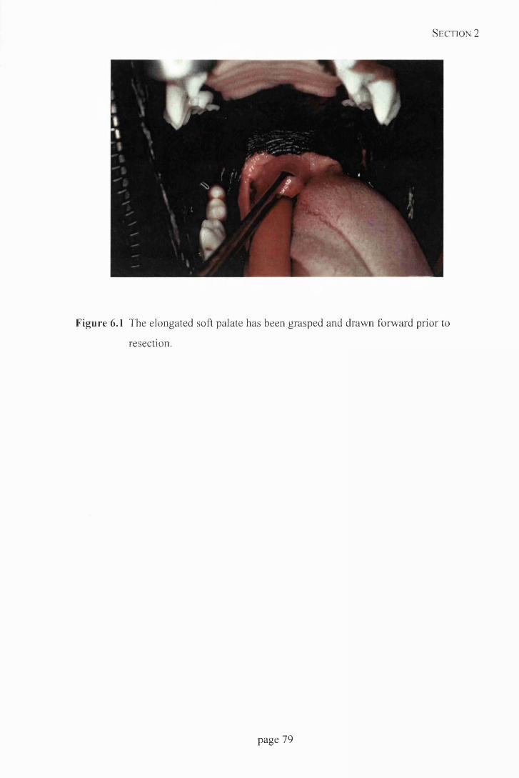
Section 2
Figure 6.1 The elongated soft palate has been grasped and drawn forward prior to
resection.
page 79

Sectio n 2
7. Cranial Pubic Ligament Rupture in a Dog
7.1 Introduction
The cranial pubic ligament, also called the pre-pubic tendon, is a tendinous band which
extends from the ilio-pectineal eminence and the tendon o f the pectineus muscle to the
same structures on the opposite side, into which the rectus abdominis muscle inserts
(Miller and others 1964).
The most frequent site of occurrence and recurrence o f abdominal hernias was found to
be the area of the caudal ventral abdomen, involving the pre-pubic tendon, either being
tom in its body or detached from the pubic brim (Waldron and others 1986) as a result
o f the action of avulsion forces (Smeak 1993).
The main cause of pre-pubic tendon rupture is blunt trauma arising from road traffic
accidents (Waldron and others 1986). Not surprisingly, associated injuries such as
pelvic and femoral fractures, coxofemoral luxation, skin laceration, pelvic limb paresis
and vascular thrombosis were found in cases of pre-pubic tendon rupture (Mann and
others 1986). The effect of blunt trauma is associated with the lack o f elasticity o f the
abdominal wall when compared with that o f the linea alba (Parks 1981).
The most prominent sign of an abdominal hernia is a bulging mass beneath the skin or
asymmetry of the abdominal contour (Smeak 1993). However, the location of the
swelling caused by abdominal content does not necessarily mark the site of the
anatomical defect. Contents can migrate subcutaneously or along fascial planes between
muscular layers (Waldron and others 1986). Organs such as intestine may migrate down
the medial thigh or along the abdominal wall to the thorax (Smeak 1998).
When palpation is difficult due to excessive subcutaneous swelling or pain, radiology is
a useful tool. However, signs found are not pathognomonic o f cranial pubic ligament
rupture, and confirmation is by exploration and finding subcutaneously located
abdominal organs and cranially retracted rectus abdominis muscle (Mann and others
1986).
Radiologically, decreased density outwith the abdominal wall may occur when intestinal
loops are present within the hernia or when subcutaneous emphysema is present due to
perforation o f the intestinal wall (Bartels 1972). The intestines can be recognised by
page 80

Section 2
their curvilinear shape, while subcutaneous emphysema tends to be more linear or
irregularly shaped (Burk and Ackerman 1996). Delineation o f ventral abdominal
hernias usually does not require the use of contrast media. If there is any doubt about
the diagnosis, a small amount of an organic iodine medium can be injected
subcutaneously or into the abdominal cavity. Also barium suspension could be given
orally to outline the hernial contents (Bartels 1972).
Ultrasound is very useful in cases o f fluid-filled hernias that may mimic other soft tissue
masses (Smeak 1993). It is much better than plain radiology at detecting the presence of
urinary bladder within the hernia (Burk and Ackerman 1996) and simpler than using
pneumocystography or double contrast cystography as suggested by Bartels (1972).
7.2 Case details
History
A 4 year old male collie cross (24kg body weight) was referred shortly after having
being reversed over by a forklift truck. The only sign that was noticed afterwards by the
owners was some degree o f abdominal discomfort.
Clinical examination
The dog was depressed and restless. The caudal abdomen was tense and painful on
palpation, but no other neurological or orthopaedic complications were evident.
Ancillary aids
Immediately after admission radiography was performed. The lateral thorax was
unremarkable. However, a lateral abdominal view showed a soft tissue opacity
caudoventrally, due to swelling and displacement of abdominal viscera. The urinary
bladder integrity was checked by means o f retrograde urethrogram, which was intact but
herniated (Fig 2.7).
Haematological studies were performed and WBC (15.9xl09/l), MCH (24.7pg) and
MCHC (37.4g/dl) values were increased, whilst the haematocrit was low (0.361/1)
Urine samples were sent for analyses and large amounts o f red and white cells were
detected. Protein (920mg/dl) and SG (over 1.055) were also high.
page 81

Section 2
A tentative diagnosis o f pre-pubic tendon avulsion was made and analgesia was
provided overnight (methadone, 5mg IM). Peri-operative antibiosis was also started
with amoxycillin/clavulanic acid 250mg TTD.
Surgery
The anaesthetic protocol consisted o f IM pre-medication with acetylpromazine (1.25mg)
and methadone (5mg), IV induction with methohexitone (125mg), followed by
endotracheal intubation and maintenance of anaesthesia with a halothane/nitrous
oxide/oxygen mixture.
As part of the surgical preparation, the bladder was catheterised and drained. The
catheter was sutured to preputial skin and connected to a giving set and empty fluid bag
for urine collection.
With the dog in dorsal recumbence, a caudal midline laparotomy was performed.
Beneath the subcutaneous tissue, abdominal fat, omentum, bladder and some intestinal
loops were found. Prior to returning these to the peritoneal cavity, it was explored but
apart from some blood clots no internal organ damage was found. Remnants of the
prepubic tendon were still attached to the pubis but the rectus abdominis was completely
avulsed. The left inguinal ring appeared enlarged enough to allow the passage of
omentum and fat. Using a drill and bit, several holes were made along the cranial pubic
brim, and polypropylene (3 metric) sutures were pre-placed through them and the rectus
abdominis muscle in a horizontal mattress pattern.
The inguinal hernia was repaired with the same material and pattern, avoiding damage
to the external pudendal vessels and genital nerve. A Penrose drain was placed and the
laparotomy incision closed routinely.
Treatment with carprofen (50mg BID) was initiated immediately after surgery.
Antibiotics were continued for seven days.
The following day the drain had been pulled out. In the succeeding days the surgical
wound became quite swollen and painful. Consequently, supplementary analgesia
(methadone, 5mg IM) was provided. The scrotum was found to be also bruised, swollen
and painful and a cream based on benzoic, malic and salicylic acid was used for local
relief.
page 82

Section 2
On the third day, as the dog was still showing signs o f discomfort, abdominal
radiography was repeated and the bladder was found in an inguinal location, suggestive
o f a breakdown o f the repair. Therefore, the wound was re-explored. All the sutures had
tom from the pubis, though the inguinal repair was still intact. The hemial repair was
repeated using the same suture pattern with nylon (4 metric).
Five days after surgery the dog was discharged with the antibiotics and carprofen
treatment. A Penrose drain was again pulled out by the dog, and use o f Elizabethan
collar was recommended as well as strict exercise restriction for 2 weeks to prevent the
animal attacking the wound and reduce stress on the repair.
Follow up
The dog was reassessed in 4 weeks and no signs of discomfort or herniation were
noticed. The owners reported that for two weeks a persistent firm swelling was noticed
at the wound site, which gradually resolved.
7.3 Discussion
The mere presence of an abdominal hernia in itself, even if very large, is not necessarily
an indication for immediate surgery. It is preferable to delay surgery for 3 to 5 days if
possible, which allows acutely traumatised tissue to revascularise, thereby reducing
infection. Also less inflammation and haemorrhage are present, allowing better
exposure o f structures and suture holding. Emergency surgery is indicated in cases that
do not satisfactorily stabilise or that deteriorate despite intensive medical therapy such
as animals with penetrating wounds or suspected incarceration (Smeak 1993).
Closure o f this type of defect is facilitated by proper patient positioning. With the dog
in dorsal recumbence, hind limbs are flexed and pulled cranially, creating a truncal
ventro-flexion and reducing the size o f the defect, which results in decreased tension
during the repair (Smeak 1998).
Ideally the repair o f a pre-pubic ventral hernia should begin with the identification of all
abdominal muscular layers involved, so that repair o f each layer may be accomplished,
providing a grid-like closure of the wound and a stronger repair (Bojrab and Taylor
1983). Technical failures such as the use o f incorrect suture material or inappropriate
tissue layers are important causes o f recurrence (Parks 1981).
page 83
Section 2
Rupture o f the cranial pubic ligament is often associated with concurrent inguinal
ligament damage, therefore a femoral or inguinal hernia may be present, in which case
the pre-pubic component of the hernia is repaired first to align the tissues correctly for
anatomic reconstruction of other ruptures (Smeak 1998).
Reattachment to the pubis by sutures placed from the pubis may be either through holes
drilled in the pubic bone or through the cranial aspect o f obturator foramina (Mann and
others 1986). Sutures are initially pre-placed and those that are located in the deepest
parts o f the wound tied first. Adjacent sutures are simultaneously tightened during knot
tying to reduce fascial tearing (Smeak 1993).
When a large defect makes approximation o f local tissue impossible without undue
tension, prosthetic implants are used, most commonly made o f polypropylene or
polyethylene. Such materials are well tolerated in wounds, do not disintegrate with age
and can stretch in two directions to distribute the load evenly. Granulation tissue and
capillaries grow through the mesh, building a strong layer of connective tissue. The
disadvantages o f the use o f this material include the possibility o f rejection and irritation
o f adjoining tissue (Read and Bellenger 1993).
The most common complications of surgery are recurrence and wound infection with
skin dehiscence (Waldron and others 1986). An abdominal bandage is recommended
for 5 to 10 days postoperatively to obliterate dead space and to provide external support
(Mann and others 1986). Also gravity dependent drains such as Penrose or closed
suction drain systems should be used. It is advisable to avoid placing an open drain
system directly against buried mesh to reduce the risk o f ascending infection (Smeak
1998)
Aftercare includes placement on padded beds, taking care to turn the animal frequently,
and after patient discharge exercise limitation of at least 2 weeks in order to reduce
tension on the repair site (Smeak 1993).
The prognosis o f this type of herniation after repair is good but it must be borne in mind
that accompanying injuries can influence the outcome (Mann and others 1986).
page 84
Section 2
7.4 References
Bartels, J.E. (1972) How to delineate canine and feline hernias radiographically. Modern
Veterinary Practice 53, 27-33.
Bojrab, M.J. & Taylor, W.J. (1983) Ventral abdominal hernia. In: Current Techniques in Small
Animal Surgery, ed M.J. Bojrab. Lea and Febiger, Philadelphia. p408.
Burk, R.L. & Ackerman, N. (1996) The abdomen. In: Small Animal Radiology and
Ultrasonography. A Diagnostic Atlas and Text. eds. R.L. Burk & N. Ackerman. W.B.
Saunders Company, Philadelphia. p215.
Mann, F.A., Tagner, C.H., Boothe, H.W. & Weber, W.J. (1986) Cranial pubic ligament rupture
in dogs and cats. Journal o f the American Animal Hospital Association 22, 519-524.
Miller, M.E., Christensen, G.C. & Evans, H.E. (1964) Myology. In: Anatomy o f the Dog. ed M.E. Miller, G.C. Christensen & H.E. Evans. W.B. Saunders Company, Philadelphia, pi 83.
Parks, J. (1981) Herniation. In: Pathophysiology in Small Animal Surgery, ed M.J. Bojrab. Lea
& Febiger, Philadelphia. p420.
Read, R.A., Bellenger, C.R. (1993) Hernias. In: Textbook o f Small Animal Surgery, ed D.H. Slatter. W.B. Saunders Company, Philadelphia. p431.
Smeak, D.D. (1993) Abdominal Hernias. In: Textbook o f Small Animal Surgery, ed D.H. Slatter. W.B. Saunders Company, Philadelphia. p433.
Smeak, D.D. (1998) Prepubic hernia repair. In: Current Techniques in Small Animal Surgery.
ed M.J. Bojrab. Williams & Wilkins, Baltimore. p574.
Waldron, D.R., Hedlund, C.S. & Pechman, R. (1986) Abdominal hernias in dogs and cats.
Journal o f the American Animal Hospital Association 22, 817-823.
page 85

Sectio n 2
8. Intestinal Adenocarcinoma in a Cat
8,1 Introduction
Tumours of the intestine are uncommon in domestic animals, and represent less than 1%
of all malignancies (Straw 1996). Adenocarcinoma is the most common non-
haematopoietic gastrointestinal tumour in cats (Turk and others 1981), although it
occurs infrequently compared to the dog and man (Patnaik and others 1976).
The occurrence of intestinal adenocarcinoma in middle-aged to old cats has been
confirmed by several authors (Brodey 1966, Patnaik and others 1976, Turk and others
1981, Cribb 1988, Kosovsky and others 1988). All of them also found that Siamese and
domestic short hair were the breeds most affected. Although occurring sporadically in
other breeds, one case in a Russian blue (Turk and others 1981) and one Persian cat
(Cribb 1988). No sex predilection has been noted, apart from a marked male
predisposition reported by Patnaik and others (1976).
Cachezia (Cribb 1988) and vomiting (Kosovsky and others 1988) have been reported as
the most consistent findings in cases of intestinal adenocarcinoma. Other clinical signs
include depression, anorexia, swollen and palpable masses (Patnaik and others 1976).
Firm, moveable cylindrical masses in the cranial or mid-abdomen are often identified.
As it is an annular tumour it may lead to some degree o f obstruction, thus causing
abdominal pain and fluid or gas accumulation (Crow 1985).
Clinical features of intestinal neoplasia mimic those o f non-neoplastic disorders of the
bowel. As a result it is difficult to diagnose this type o f tumour using routine methods
and they are frequently undetected in the early stages. However, early diagnosis is
especially important because debilitated cats are more susceptible to the surgical stress
and metastasis is more likely to have occurred (Cribb 1988).
Although blood analyses has been reported as rarely contributory in most cases (Crow
1985), Cribb (1988) reported neutrophilia and lymphopaenia as the most common
haematological abnormalities in his series, as well as hypokalemia consistent with
vomiting and decreased food intake. Also hypoproteinaemia and high activity of ALT
and ALP have been described (Kosovsky and others 1988).
page 86

Section 2
Kosovsky and others (1988) found that abdominal palpation and a positive contrast
gastro-intestinal radiological study were the most reliable indicators o f an encircling
mass in the bowel wall causing stenosis o f the lumen and partial obstruction. But final
diagnosis of the cause of obstruction is achieved by means o f exploratory laparotomy
and histological examination (Cribb 1988).
Although Patnaik and others (1976) described the ileum as the commonest site, later
authors reported the jejunum as the most usual location (Turk and others 1981,
Kosovsky and others 1988) followed by ileum and ileo-caeco-colic region.
Ulceration at the primary site, severe desmoplastic reaction, muscular hypertrophy and
luminal stenosis are characteristic features at the annular constriction caused by the
tumour. Wall thickening is commonly present in the intestinal segment proximal to the
neoplastic lesion, being considered to be a compensatory reaction to obstruction o f the
contractile movement. It is due mainly to hypertrophy and hyperplasia of the inner
circular muscle. Post-stenotic dilation is also common and often the mesentery and/or
the omentum adhere to the neoplastic section o f intestine (Patnaik and others 1976).
Metastasis of small intestine adenocarcinoma has been described mainly to the
peritoneum and regional lymph nodes, but also may appear in other sites such as liver,
spleen, kidneys, lungs or diaphragm (Brodey 1966, Patnaik and others 1976).
Surgical resection is the treatment of choice for annular or discrete tubular neoplasia of
the small intestine when unresectable metastases are not detected. Carcinomas that have
metastasised can be reduced in volume by cyto-reductive surgery but have an
unfavourable prognosis (Crow 1985). However, Kosovsky and others (1988) found that
lymph node metastasis and carcinomatosis may not always indicate a poor prognosis
and that recurrence of the primary tumour was the cause o f the death o f most long-term
survivors.
Case details
History
A nine and half year old neutered male Siamese cat (2.7kg body weight) was referred
with a 3 week history of intermittent vomiting and noticeable weight loss, which was
unresponsive to medical management with antibiotics, steroids and metoclopramide.
page 87
Sectio n 2
The owners reported that the vomit looked like undigested food, although on some
occasions had a faecal appearance. The faeces were o f a normal consistency but
produced in small volumes.
Clinical examination
The cat was emaciated, dehydrated but still alert. The intestines were prominent and
excessively gassy on abdominal palpation, as well as hyperperistaltic. Tachycardia and a
soft left systolic murmur were also noticed.
Ancillary aids
Blood and urine samples were taken for analyses. There was elevation in plasma urea
(18mmol/l) combined with non-regenerative anaemia (HCT 0.211/1) and high number of
Heinz bodies. FIV and FeLV antibody tests were negative and urinalysis was
unremarkable.
Contrast radiography, using barium-impregnated polyethylene spheres, was performed
with lateral and ventro-dorsal abdominal views taken 12 hours post-administration
(Figure 8.1). At that time a delay in the passage of some o f the large spheres suggested
decreased gut motility. Exploratory laparotomy was scheduled in order to achieve a
definitive diagnosis.
Surgery
The cat was put on fluids from the day of admission and IV antibiotic therapy was also
started with amoxycillin/clavulanic acid (60mg) and metronidazol (30mg) TED.
The anaesthetic protocol consisted of pre-medication with pethidine (18mg IM) and ten
minutes later diazepam (0.5mg IV). Induction was with propofol (18mg IV), followed
by intubation and maintenance o f anaesthesia with a halothane/nitrous oxide/oxygen
mixture.
Midline ventral laparotomy was performed and a localised concentrically thickened area
about 3 cm long was found at the ileo-colic region (Figure 8.2). Doyen intestinal
forceps were placed with a 2cm healthy wall margin on either side and, after ligature
and transection of mesenteric and arcadian vessels of the area, the bowel and adjacent
mesentery were resected. The intestinal mucosa was trimmed and an end-to-end
anastomosis was carried out in a simple interrupted fashion (2 metric polyglactin 910,
page 88
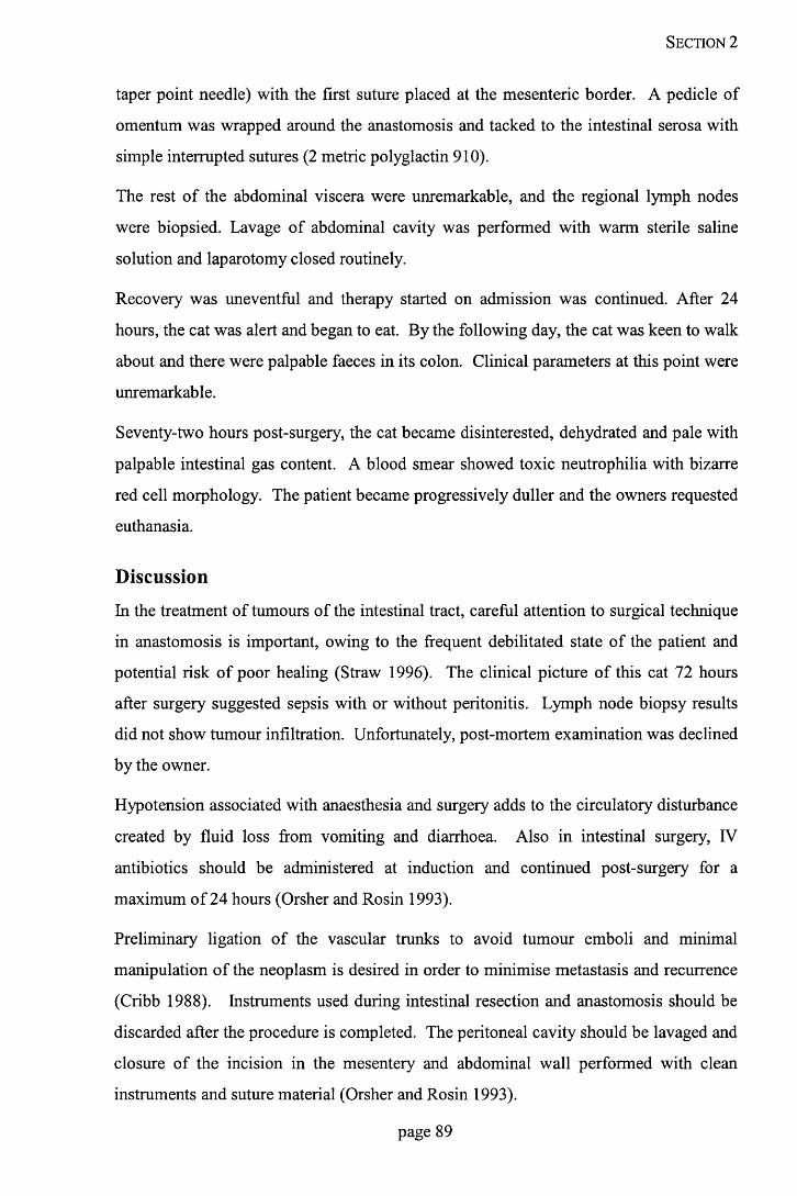
Section 2
taper point needle) with the first suture placed at the mesenteric border. A pedicle of
omentum was wrapped around the anastomosis and tacked to the intestinal serosa with
simple interrupted sutures (2 metric polyglactin 910).
The rest o f the abdominal viscera were unremarkable, and the regional lymph nodes
were biopsied. Lavage o f abdominal cavity was performed with warm sterile saline
solution and laparotomy closed routinely.
Recovery was uneventful and therapy started on admission was continued. After 24
hours, the cat was alert and began to eat. By the following day, the cat was keen to walk
about and there were palpable faeces in its colon. Clinical parameters at this point were
unremarkable.
Seventy-two hours post-surgery, the cat became disinterested, dehydrated and pale with
palpable intestinal gas content. A blood smear showed toxic neutrophilia with bizarre
red cell morphology. The patient became progressively duller and the owners requested
euthanasia.
Discussion
In the treatment of tumours of the intestinal tract, careful attention to surgical technique
in anastomosis is important, owing to the frequent debilitated state of the patient and
potential risk o f poor healing (Straw 1996). The clinical picture o f this cat 72 hours
after surgery suggested sepsis with or without peritonitis. Lymph node biopsy results
did not show tumour infiltration. Unfortunately, post-mortem examination was declined
by the owner.
Hypotension associated with anaesthesia and surgery adds to the circulatory disturbance
created by fluid loss from vomiting and diarrhoea. Also in intestinal surgery, IV
antibiotics should be administered at induction and continued post-surgery for a
maximum of 24 hours (Orsher and Rosin 1993).
Preliminary ligation o f the vascular trunks to avoid tumour emboli and minimal
manipulation o f the neoplasm is desired in order to minimise metastasis and recurrence
(Cribb 1988). Instruments used during intestinal resection and anastomosis should be
discarded after the procedure is completed. The peritoneal cavity should be lavaged and
closure of the incision in the mesentery and abdominal wall performed with clean
instruments and suture material (Orsher and Rosin 1993).
page 89

Section 2
O f all the suture patterns that can be used in end-to-end intestinal anastomosis,
approximating ones seem to have a number of advantages; such as increased lumen
diameter, rapid and precise healing and minimal post-operative intestinal adherence.
However, a simple continuous fashion is often preferred to an interrupted one to
minimise mucosal eversion, provide a better serosal apposition and primary intestinal
healing. Minimal mucosal eversion and proper incorporation o f the tough submucosa
are vital in performing consistent and successful anastomosis (Ellison 1998). A small
taper-cut or reverse cutting needle penetrates the tough submucosa easily, which has a
distinct advantage over a taper point or round bodied needle (Orsher and Rosin 1993).
Synthetic braided absorbable suture material such as 910 polyglactin is acceptable but
has a degree o f tissue drag, which is not present in monofilament absorbable sutures as
polydioxanone or polyglyconate. Omental wrap has the advantage o f isolating potential
anastomotic leakage from the peritoneal cavity. The omentum can seal off infection and
perforations, and impart new blood supply to viscera (Orsher and Rosin 1993).
It is unfortunate that the owners declined post-mortem examination to determine the
cause of death and the state of the enterectomy repair.
8.4 References
Brodey, R.S. (1966) Alimentary tract neoplasms in the cat: A clinico-athologic survey of 46 cases. American Journal o f Veterinary Research 27, 74-80.
Cribb, A. E. (1988) Feline gastrointestinal adenocarcinoma: A review and retrospective study.
Canadian Veterinary Journal 29, 709-712.
Crow, S.E. (1985) Tumours of the alimentary tract. Veterinary Clinics o f North America:
Small Animal Practice 15, 577-596.
Ellison, G.W. (1998) Intestinal Resection and Anastomosis. In: Current Techniques in Small
Animal Surgery, ed M.J. Bojrab. Williams & Wilkins, Baltimore. p248.
Kosovsky, J.E., Matthiesen, D.T. & Patnaik, A.K. (1988) Small intestinal adenocarcinoma in
cats: 32 cases (1978-1985). Journal o f the American Veterinary Medical Association
192, 233-235.
Orsher, RJ. & Rosin, E. (1993) Small Intestine. In: Textbook o f Small Animal Surgery, ed D.H.
Slatter. W.B. Saunders Company, Philadelphia. p593.
Patnaik, A.K., Liu, S. K. & Johnson, G.F. (1976) Feline intestinal adenocarcinoma. A clinicopathologic study of 22 cases. Veterinary Pathology 13, 1-10.
page 90

Sectio n 2
Straw, R.C. (1996) Tumours of the intestinal tract. In: Small Animal Oncology, eds S.J.
Withrow & E.G. MacEwen. W.B. Saunders Company, Philadelphia. p252.
Turk, M.A.M., Gallina, A.M. & Russell, T.S. (1981) Nonhematopoietic gastrointestinal neoplasia in cats: A retrospective study of 44 cases. Veterinary Pathology 18, 614-620.
page 91
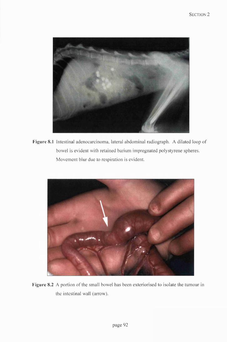
Section 2
Figure 8.1 Intestinal adenocarcinoma, lateral abdominal radiograph. A dilated loop of
bowel is evident with retained barium impregnated polystyrene spheres.
Movement blur due to respiration is evident.
Figure 8.2 A portion of the small bowel has been exteriorised to isolate the tumour in
the intestinal wall (arrow).
page 92

Sectio n 2
9. Tracheal Puncture in a Dog
9.1 Introduction
Injuries of the trachea usually result from blunt trauma to neck and/or thorax, which
often involves hyper-extension of the head and neck (White and Milner 1995).
However, despite the high incidence of this type o f trauma, the occurrence o f airway
injury is low (Kellagher and White 1987). This is because, in most cases, the impact is
lateral and the trachea can move instead of being crushed against the spine (Nelson
1993).
Minor perforations caused by bite wounds lead to few immediate respiratory problems
(Kellagher and White 1987), but some authors have reported complete tracheal avulsion
with formation of a thickened tracheal adventitia and pseudo-airway between severed
segments that allows air intake to the lungs (Kennedy 1976, Barber and Rawlings 1981,
Brouwer and others 1984).
Punctures or lacerations supported by healthy peri-tracheal tissue are self-limiting, even
in the intra-thoracic segment or bronchi (Nelson 1993). However, air leaking into the
peri-tracheal tissue may cause subcutaneous emphysema and even pneumomediastinum
(Kellagher and White 1987).
Traumatic tracheo-bronchial rupture is the most likely cause of generalised
subcutaneous emphysema. This condition may appear days to weeks after the injury and
the cause has been suggested to be a dislodged fibrin seal or a gradual tracheal necrosis
(Bauer and Currie 1988).
Apart from clinical examination, a palpable gap in the trachea or a wound in the skin
may indicate the position o f the rupture (Harvey and O ’Brien 1972) but absence of skin
puncture does not exclude it. Consequently, radiology and endoscopy are essential for
an accurate diagnosis (Bauer and Currie 1988). Major lacerations of the trachea are
normally visible on plain radiographs alone, whereas endoscopic evaluation may be
necessary for the identification of smaller defects (Kellagher and White 1987). Lately,
computed tomography has been suggested as a diagnostic tool for tracheal disruption
(Fingland 1998).
page 93

Section 2
Radiographic findings may include evidence o f emphysema, focal narrowing of tracheal
lumen, lack of tracheal continuity and gas-filled spherical dilation between severed
segments. Pulmonary hyperinflation was reported in a case where a loose necrotic tag
of tracheal wall was present at the orifice of the distal segment, creating a flap-valve
effect (Barber and Rawlings 1981).
The accumulation of large amounts of air within the mediastinum may cause rupture of
the pleura, converting the condition to a pneumothorax (Kellagher and White 1987).
The latter may be controlled by means of suction drainage. Mediastinal and
subcutaneous emphysema can be reduced by placing a tracheostomy tube which
decreases airway resistance. Failure to resolve pneumothorax after 3 to 4 days is a
criterion for surgical intervention (Nelson 1993).
9.2 Case details
History
A five year old female Whippet (10.5kg) was referred, following a dog fight 24 hours
previously, with a left cervical swelling that had gradually developed to a generalised
subcutaneous emphysema.
Clinical examination
The dog was alert and responsive. Mucous membranes were slightly pale, although
CRT was less than 2 seconds. The left cervical area had a large crepitant swelling and
signs o f haematoma. On palpation there was a widespread crepitation all over the dog,
with only the right hind spared. Tracheal auscultation was harsh and especially difficult
to hear on the left side.
Ancillary aids
The dog was admitted and lateral and dorso-ventral thoracic, as well as lateral cervical
radiographs were taken. Pneumomediastinum was confirmed with visualisation of the
internal and external tracheal margins (Figure 9.1). A slight degree of pneumothorax
was also noticed, and in the cervical view cranial tracheal damage could be detected.
A blood sample was taken for haematological and biochemical evaluation. Decreases in
the values of WBC (4.30xl09/l), RBC (5.77xl012/l), lymphocytes (0.34xl09/l), calcium
(1.89mmol/l), albumin (24g/l), globulin (18g/l) and total protein (42g/l) were noticed.
page 94

Section 2
MCH (25.20pg), MCHC (37.20g/dl), chloride (120 mmol/1), ALP (244U/1), AST
(180U/1) and ALT (71U/1) measurements were above normal.
Acetylpromazine (0.2mg) and butorphanol (2mg) were administered IV before
placement o f a chest drain to permit hourly drainage. The first tap produced 235 ml of
air. Fluid therapy was started at a maintenance rate, as well as antibiosis with
metronidazole (lOmg/kg TID IV ) and amoxycillin/clavulanic acid (20mg/kg BID IV).
Endoscopic examination was performed 12 hours after admission and a cervical tracheal
defect was found.
Surgery
The animal was pre-oxygenated and induced with propofol (10ml IV). Following careful
endotracheal intubation, anaesthesia was maintained with a halothane/oxygen mixture.
The dog was placed in dorsal recumbency and the trachea was exposed via a ventral
midline incision caudal to the larynx. A small defect was found at the level of the first
to second tracheal spaces on the left. The defect was closed by passing three simple
interrupted sutures (1.5 metric polypropylene) around the adjacent tracheal rings and a
Penrose drain was placed. The incision was closed routinely.
The chest was drained on three further occasions before drain removal. Small amounts
of soft food were offered every two hours and rapidly eaten, so fluid therapy was
stopped 12 hours after surgery.
Recovery in the hospital was uneventful, and after 4 days the Penrose drain was
removed and the dog sent home with on carprofen (20mg BID) and
amoxycillin/clavulanic acid (250mg BID) for 10 days. There were still signs of
emphysema but with an obvious daily improvement. The owners were advised to
maintain the soft diet and to keep the dog on leash until the stitches were removed.
Follow up
The dog was returned for a re-examination four weeks later. No evidence of
subcutaneous emphysema was present and the owners reported a normal respiratory
pattern and exercise tolerance.
page 95

Sectio n 2
9.3 Discussion
The main feature o f this case was the generalised subcutaneous emphysema that is
associated with the presence o f pneumomediastinum, and is a specific indicator o f upper
airway leakage (Kellagher and White 1987). Nelson (1993) considered that even intra-
thoracic lacerations o f the trachea were self-limiting as long as pneumothorax was
controlled by suction drainage. However, despite the small defect, it was decided to
proceed surgically due to the large degree of emphysema present, a primary concern of
the owners when the dog was referred.
One of the precautions that should be taken is careful intubation, because a forced
manoeuvre or excessive inflation when using cuffed tubes, may worsen original injuries
and even trigger a respiratory crisis (Hill 1974).
The size of the tear avoided the need for more complicated techniques such as the
placement o f tension sutures in order to appose the two segments (Hedlund 1984). With
this small defect, it was not necessary to carry out a large reconstruction with sutures, as
it has to be bome in mind that in tracheal surgery, excessive sutures are always
undesirable. Intra-luminal knots stimulate granulation tissue production at each site
(Nelson 1993), acting as a foreign body in the trachea (Harvey and O’Brien 1972)
Postoperatively respiratory distress can result from laryngeal or pharyngeal oedema,
occlusion of the tracheal lumen at the anastomotic site or iatrogenic laryngeal paralysis
from intra-operative recurrent laryngeal nerve injury (Fingland 1998).
9.4 References
Barber, D.L. & Rawlings, C.A. (1981) Radiographic diagnosis. Veterinary Radiology 22, 258- 261.
Bauer, M.S. & Currie, J. (1988) Generalized subcutaneous emphysema in a dog. Canadian Veterinary Journal 29, 836-837.
Brouwer, G.J., Burbidge, H.M. & Jones, D.E. (1984) Tracheal rupture in a cat. Journal o f Small Animal Practice 25, 71-76.
Fingland, R.B. (1998) Tracheal resection and anastomosis. In: Current Techniques in Small Animal Surgery, ed M.J. Bojrab. Williams and Wilkins, Baltimore. p387.
Harvey, C.E. & O’Brien, J.A. (1972) Management of respiratory emergencies in small animals. Veterinary Clinics o f North America. 2, 243-258.
page 96

Section 2
Hedlund, C.S. (1984) Tracheal anastomosis in the dog. Comparison of two end-to-end techniques. Veterinary Surgery 13, 135-142.
Hill, F.G.W. (1974) Repair of a traumatic rupture in the trachea of a dog. Veterinary Record 95, 265-266.
Kellagher, R.E.B. & White, R.A.S. (1987) Tracheal rupture in a dog. Journal o f Small Animal Practice 28, 29-38.
Kennedy, R.K. (1976) Traumatic tracheal separation with diverticuli in a cat. Veterinary
Medicine/Small Animal Clinician 71, 1384-1385.
Nelson, A.W. (1993) Lower respiratory system. In: Textbook o f Small Animal Surgery, ed D.H.
Slatter. W.B. Saunders Company, Philadelphia, p.777.
White R.N. & Milner, H.R. (1995) Intrathoracic tracheal avulsion in three cats. Journal o f Small Animal Practice 36, 343-347.
page 97

Section 2
Figure 9.1 Tracheal rupture, lateral thoracic radiograph. The cardiac shadow is
elevated from the sternum and there is an increased radioluceny indicative of
pneumothorax. The structures contained in the cranial mediastinum are visible
indicating a pneumomediastinum.
page 98
Section 2
10. Abdominal Unilateral Cryptorchidism in a Cat
10.1 Introduction
Cryptorchidism is a developmental defect with failure o f testicular descent into the
scrotum. It may be bilateral or unilateral (Rhoades and Foley 1977). Unilateral
cryptorchidism, which is more common than bilateral (Millis and others 1992), is often
incorrectly referred to as monocryptorchidism, a term that implies lack of one of the
testicles, regardless of its location (Rhoades and Foley 1977).
In most species, cryptorchidism is thought to be a simple autosomal recessive genetic
defect, where females might be carriers (Burke 1986).
Some o f the factors associated with cryptorchidism include lack of androgens, lack of
gonadotropin releasing factors or incompetent gubemaculum. Testicular descent occurs
in two morphological phases. First is transabdominal translocation, which is non
androgen dependant and during which the gubemaculum does not contract. Thus the
relative position o f the gonad is changed as a result o f foetal growth. Second is inguino-
scrotal descent, which is not only androgen dependant, but under the influence of the
genitofemoral nerve and gubemaculum (Buergelt 1997).
The testicles are just inside or within the internal inguinal ring and they reach the
scrotum after scrotal development, at 10 to 14 days of age. Scrotal development may be
delayed in some breeds of kittens rather than in puppies, especially the Persian, where it
may take up to a year (Burke 1986).
Cryptorchidism in cats has been infrequently reported, although feline prevalence was
found to be similar to that reported in dogs (Millis and others 1992). Other authors
suggested that the defect may also be unnoticed due to the large inguinal fat pad
(Richardson and Mullen 1993).
Retained testicles may be located pre-scrotally in subcutaneous inguinal fat, in the
inguinal ring or within the abdomen. Initially, it was thought that in most cryptorchid
cats the testicles were located subcutaneously (Noffsinger and Carbone 1978). Later
reports have suggested a similar incidence in abdominal and inguinal locations (Millis
and others 1992, Richardson and Mullen 1993). The same authors found the right and
page 99

Sectio n 2
left testicles were equally affected and that abdominal testicles were mostly located near
the bladder, although in some cases they may be found close to the caudal renal pole.
Cryptorchidism implies lack o f spermatogenesis of the affected testicle due to the high
temperature compared with the normal scrotal position, therefore bilateral cryptorchid
animals are sterile. Testosterone production may be somewhat lower than normal but is
still present in the retained gonad (Rhoades and Foley 1977).
Stimulation of testosterone production with intravenous human chorionic gonadotropin
has been used in the diagnosis of feline cryptorchidism in unilaterally castrated cats
(Memon and others 1992). Richardson and Mullen (1993) reported that in 50 cats the
location o f the retained testicle was correctly palpated in only 21 due to interference by
the fat pad or abdominal position.
Cryptorchidism may be linked to undesirable syndromes in affected animals, such as
anal gland adenoma, sertoli cell tumour, seminoma, prostatic hyperplasia and
temperamental behaviour (Wolff 1981). Tumours o f retained testicles are more often
malignant than those o f descended ones (Burke 1986), although testicular tumours are
rare in cats (Feldman and Nelson 1996). There is no evidence to suggest that they are
prone to torsion of the affected testicle by twisting of vas deferens and spermatic vessels
around each other as has been reported in the dog (Guernsey 1984).
Orchiopexy of the ectopic testicle is unethical, because it results in a fertile animal with
a heritable defect (Kirby 1980). Hormonal supplementation, such as megestrol acetate,
has been used unsuccessfully (Richardson and Mullen 1993). Total orchiectomy is the
treatment of choice.
10.2 Case details
History
An 11 month old male Siamese (4.95kg body weight) was admitted for neutering. The
owners reported that, otherwise, the cat was completely healthy.
Clinical examination
The cat was bright and alert. No abnormalities were noticed except for the presence of
only one intra-scrotal testicle. On palpation the left testicle was suspected to lie in the
inguinal region. No other ancillary test were performed and neutering was scheduled.
page 100
Section 2
Surgery
The anaesthetic protocol consisted of IM pre-medication with acetylpromazine (0.1 mg)
and pethidine (27.5mg), IV induction with propofol (40mg), followed by endotracheal
intubation and maintenance of anaesthesia with a halothane/nitrous oxide/oxygen
mixture.
After the cat was clipped for surgical preparation, inguinal palpation was performed
again but the retained testicle could not be identified.
With the cat in dorsal recumbency routine pre-scrotal castration was performed on the
right descended testicle. Caudal ventral midline incision was done to allow dissection
deep into the inguinal fat pad and therefore visualisation o f the external inguinal ring
and better exposure o f testicular vessels and ductus deferens o f what was suspected to
be an inguinal testicle.
The ectopic gonad was not found, so the dissection was deepened to that of celiotomy
for abdominal exploration. A hypoplastic testicle was eventually located laterally, mid
way between bladder and left kidney (Figure 10.1). The testicular vessels and ductus
deferens were clamped, ligated (2 metric polyglactin 910) and divided. The testicle was
removed, the vessels checked for haemorrhage and the abdomen closed in a routine
fashion.
Follow up
The cat was discharged two days after surgery with a treatment o f carprofen (lOmg SID
PO) and amoxycillin/clavulanic acid (50mg BID PO) and placement of Elizabethan
collar advised.
Sutures were removed after 10 days, and no complications were observed during post-
surgical period.
10.3 Discussion
Unilateral cryptorchidism involves the presence of a normally located testicle that
allows the animal to be fertile. However, affected animals should not be used for
breeding as carriers of a genetic defect (Rhoades and Foley 1977). Removal of the
scrotal testicle allows the surgeon to identify the side on which the search should be
concentrated (Hardie 1984).
page 101

Section 2
Ectopic testicles, by producing testosterone, are responsible for undesirable signs of
male behaviour such aggressiveness, urine spraying, loud vocalisation and mounting of
female cats (Memon and others 1992, Richardson and Mullen 1993).
The testicle is only reliably palpated in a pre-scrotal location, caudal to the fat pad
(Richardson and Mullen 1993). Sometimes, the superficial inguinal lymph node can be
confused with a retained testicle (Hardie 1984). When the testicle is palpated, a para
median incision should be made over the inguinal fold (Millis and others 1992).
However, because of the unreliability of palpation, and the fact that approximately half
of the testicles were identified in an abdominal location, the caudal abdominal surgical
approach is recommended if palpation fails to identify the gonad. If the testicle is not
readily evident in the inguinal region, gentle traction of the abdominal portion of the
spermatic cord can help to locate the testicle in this area, thus avoiding unnecessary
dissection of the inguinal fat (Richardson and Mullen 1993).
Millis and others (1992) recommended the caudal ventral mid-line approach as the best
one for orchiectomy of cryptorchid cats allowing progression to standard midline
celiotomy. The same authors reported that in all but one case of abdominal
cryptorchidism the testicle was found near the bladder. Hardie (1984) suggested that in
cases where it is difficult to localise the testicles, the ductus deferens should be located
as it enters the dorsal aspect of the prostate gland. Alternatively, the testicular vessels
may be traced from the caudal aspect of the kidney to the testicle.
Kirby (1980) and Hakala (1984) used the snook hook in abdominal cryptorchidectomy
suggesting that it would reduce anaesthetic time as well as the risk o f postoperative
herniation and evisceration. However, removal of abdominal testicles through small
incisions or use o f spay hook is not recommended because of the risk of damage to the
ureters, which are located near the ductus deferens and may be avulsed from the trigone
o f the bladder or incised (Millis and others 1992).
10.4 References
Buergelt, C.D. (1997) Testes. In: Color Atlas o f Reproductive Pathology o f Domestic Animals. Ed: C.D. Buergelt. Mosby, Saint-Louis. p27.
page 102

Section 2
Burke, T.J. (1986) Causes of infertility. Anatomic abnormalities. In: Small Animal Reproduction and Infertility. A Clinical Approach to Diagnosis and Treatment, ed T.J. Burke. Lea & Febiger, Philadelphia. p227.
Feldman, E.C. & Nelson, R.W. (1996) Feline reproduction. In: Canine and Feline Endocrinology and Reproduction, ed E.C. Feldman & R.W. Nelson. W.B. Saunders
Company, Philadelphia. p741.
Guernsey, G.E. (1984) Torsion of abdominal testicle. Canadian Veterinary Journal 25, 330.
Hakala, J.E. (1984) Reproductive tract abnormalities in two male cats. Modern Veterinary Practice 65, 629.
Hardie, E.M. (1984) Selected surgeries of the male and female reproductive tracts. Veterinary Clinics o f North America: Small Animal practice 14, 109-122.
Kirby, F.D. (1980) A technique for castrating the cryptorchid dog or cat. Veterinary Medicine: Small Animal Clinician 75, 632.
Memon, M.A., Ganjam, V.K., Pavletic, M.M. & Schelling, S.H. (1992) Use of Human chorionic gonadotropin stimulation test to detect a retained testis in a cat. Journal o f the American Veterinary Medical Association 201, 1602.
Millis, D.L., Hauptman, J.G. & Johnson, C.A. (1992) Cryptorchidism and monorchism in cats: 25 cases (1980-1989). Journal o f the American Veterinary Medical Association 200, 1128-1130.
Noffsinger, G.R. & Carbone, M.G. (1978) Nonabdominal approach to castration of the cryptorchid cat. Journal o f the American Veterinary Medical Association 173, 303-304.
Rhoades, J.D. & Foley, C.W. (1977) Cryptorchidism and intersexuality. Veterinary Clinics o f North America: Small Animal Practice 7, 789-794.
Richardson, E.F. & Mullen, H. (1993) Cryptorchidism in cats. Compendium on Continuing
Education for the Practicing Veterinarian 15, 1342-1369.
Wolff, A. (1981) Castration, cryptorchidism and cryptochidectomy in dogs and cats.
Veterinary Medicine/Small Animal Clinician 76,1739-1741.
page 103

Section 2
Figure 10.1 Abdominal cryptorchidism, intra-operative picture. The abdominal
testicle has been exteriorised.
page 104

Section 2
APPENDIX 1: Blood reference values
Parameter Feline Canine
WBC (White Blood Cells) x 109/1 5.5-15.5 6.0-12.0
Neutrophils x 109/1 2.5-12.5 3.0-11.8
Lymphocytes x 109/1 1.5-7.0 1.0-4.8
RBC (Red Blood Cells) x 1012/1 5.0-10.0 5.5-8.5
HB (Haemoglobin), (g/dl) 10.0-15.0 12.0-18.0
HCT (Haematocrit). (1/1) 0.30-0.45 0.37-0.55
MCH (Mean Corpuscular Haemoglobin), (pg) 12.5-17.5 19.5-24.5
MCHC (Mean Corpuscular Haemoglobin Concentration), (g/dl)
30.0-36.0 32.0-36.0
PLT (Platelets), (x 109/1) 300-800 200-500
Chloride, (mmol/1) 94-113 95-115
Phosphate, (mmol/1) 1.29-2.84 1.29-2.90
Calcium, (mmol/1) 1.60-2.56 2.34-3.00
ALP (Alkaline Phosphatase). (IU/1) <100 <230
AST (Aspartate Amino Transferase). (IU/1) <30 <40
ALT (Alanine Amino Transferase). (IU/1) <35 <90
Cholesterol, (mmol/1) 1.8-5.2 2.0-7.0
Glucose, (mmol/1) 2.7-5.5 3.3-5.5
Urea, (mmol/1) 2.1-92 2.5-8.5
Creatinine, (umol/1) 91-180 45-155
Total Protein, (g/1) 60-85 50-78
Globulin, (g/1) 27-45 28-42
Albumin, (g/1) 26-36 29-36
(I) = litres, (g) = grams, (dl) = decilitres, (pg) = pico-grams, (mmol) = mili-mol, (IU) =
international unit, (umol) = micro-mol
page 105
c. . u v ' I,U ; • c o i l ' l 3
LIBRARY j