The p38 mitogen-activated protein kinases modulate endothelial cell survival and tissue repair

Ultrasound in Med. & Biol., Vol. -, No. -, pp. 1–11, 2015Copyright � 2015 World Federation for Ultrasound in Medicine & Biology
Printed in the USA. All rights reserved0301-5629/$ - see front matter
/j.ultrasmedbio.2015.01.014
http://dx.doi.org/10.1016d Original Contribution
LOW-INTENSITY PULSED ULTRASOUND AFFECTS CHONDROCYTEEXTRACELLULAR MATRIX PRODUCTION VIA AN INTEGRIN-MEDIATED
P38 MAPK SIGNALING PATHWAY
PENG XIA, SHASHA REN, QIANG LIN, KAI CHENG, SHIHAO SHEN, MINGXIA GAO, and XUEPING LI
Department of Rehabilitation Medicine, Nanjing First Hospital, Nanjing Medical University, Nanjing, China
(Received 26 August 2014; revised 26 December 2014; in final form 17 January 2015)
ARehabiversity
Abstract—Although low-intensity pulsed ultrasound (LIPUS) regulates p38 mitogen-activated protein kinase(MAPK) and promotes cartilage repair in osteoarthritis, the role of integrin-mediated p38 MAPK in the effectof LIPUS on extracellular matrix (ECM) production of normal and OA chondrocytes remains unknown. Theaim of this studywas to investigate whether LIPUS affects ECMproduction in normal andOA rabbit chondrocytesthrough an integrin–p38 signaling pathway. A rabbit model of OAwas established by anterior cruciate ligamenttransection, and chondrocytes were isolated from normal or OA cartilage and cultured in vitro. Chondrocytes weretreated with LIPUS and then pre-incubated with the integrin inhibitor GRGDSP or the p38 inhibitor SB203580.Expression of type II collagen, MMP-13, integrin b1, p38 and phosphorylated p38 was assessed by Western blotanalysis. We found that type II collagen and integrin b1 were upregulated (p , 0.05), whereas MMP-13 wasdownregulated (p , 0.05) in normal and OA chondrocytes. Furthermore, phosphorylated p38 was upregulated(p , 0.05) in normal chondrocytes, but downregulated (p , 0.05) in OA chondrocytes after LIPUS stimulation.Pre-incubation of chondrocytes with the integrin inhibitor disrupted the effects of LIPUS on normal and OA chon-drocytes. Pre-incubation of chrondocytes with the p38 inhibitor reduced the effects of LIPUS on normal chondro-cytes, but had no impact on OA chondrocytes. Our findings suggest that the integrin–p38 MAPK signalingpathway plays an important role in LIPUS-mediated ECM production in chondrocytes. (E-mail:[email protected]) � 2015 World Federation for Ultrasound in Medicine & Biology.
Key Words: Low-intensity pulsed ultrasound, Osteoarthritis, Chondrocytes, Integrin, p38 MAPK.
INTRODUCTION
Osteoarthritis (OA), caused by articular cartilage degen-eration (Lohmander et al. 2003), manifests as jointpain, stiffness, dysfunction and different degrees of defor-mity in the late stage of the disease (Pearle et al. 2005).The degradation of extracellular matrix (ECM) acceler-ates degeneration in OA cartilage (Sandell and Aigner2001). The pathologic changes are specifically associatedwith the chondrocytes. Chondrocytes play a key role inOA and are very important to maintain the anabolic–cata-bolic balance of matrix maintenance and tissue function(Aigner et al. 2007).
Cartilage ECM is composed mainly of collagen andaggrecan. Type II collagen is the most abundant collagenin cartilage and is a typical marker for chondrocytes. The
ddress correspondence to: Xueping Li, Department oflitation Medicine, Nanjing First Hospital, Nanjing Medical Uni-, Nanjing 210006, China. E-mail: [email protected]
1
hydrolysis of cartilage ECM by proteinase is a key step inthe pathogenesis of OA (Takaishi et al. 2008).Matrixmet-alloproteinases (MMPs) are the most important proteo-lytic enzymes in the degradation of ECM, and MMP-13is themost effective enzyme for degrading type II collagen(Blain 2007). In pathologic conditions, chondrocytes pro-duce MMP-13 to degrade type II collagen, resulting indamaged or defective cartilage and, ultimately, OA(Bramono et al. 2004; Mitchell et al. 1996).
Mechanical stress interactions within the ECM play adominant role in cell regulation. Cells are able to senseforces to which they are exposed and generate forces onthe substrate, which enables a rearrangement ofmatrix pro-teins (Guilak et al. 2009). Integrins are a family of cell sur-face stress receptors mediating cellular interactions withthe ECM as well as cell–cell interactions (Hynes 1992;2002). Integrin b1, the main integrin found on thechondrocyte membrane, regulates the proliferation anddifferentiation of chondrocytes (Kim et al. 2011). Stimula-tion of integrin signals bymechanical stress may transduce
2 Ultrasound in Medicine and Biology Volume -, Number -, 2015
signals across the cell membrane, leading to activation ofdownstream pathways, such as the mitogen-activated pro-tein kinase (MAPK) and phosphatidylinositol 3-kinase(PI3K)/Akt pathway (Kim et al. 2011; MacKenna et al.2000), which play a key role in regulating the productionof MMPs in chondrocytes (Chun 2004). The MAPKsignaling pathway consists of extracellularsignal-regulated kinase (ERK), p38 MAPK (p38) and c-Jun N-terminal kinase (JNK), all of which are constitu-tively expressed inmost cell types, including chondrocytes(Karsdal et al. 2008). The p38 MAPK signaling pathwaymediates chondrocyte–perichondral communication(Stanton and Beier 2007), stabilizes chondrogenic tran-scription factor SOX 9 mRNA (Tew and Hardingham2006) and is necessary for MMP expression and activity(Sondergaard et al. 2010). Therefore, the p38 MAPKsignaling pathway plays amajor role in chondrocyte differ-entiation (Zhen et al. 2001).
Low-intensity pulsed ultrasound (LIPUS) is a form ofmechanical energy that is transmitted through and into tis-sues as pulsed acoustic pressure waves and has been clini-cally used as a non-invasive supplemental therapy topromote fracture and wound healing (Gebauer et al.2005). LIPUS has been reported to stimulate chondrocyteproliferation and matrix production in vitro, suggestingan overall anabolic effect on cartilage formation. There-fore, LIPUS can promote the repair of cartilage after dam-age and can delay cartilage degeneration, leading to OA inthe articular cartilage (Huang et al. 1999; Korstjens et al.2008). Some studies have reported that LIPUS canactivate the p38 MAPK signaling pathway (Ikeda et al.2006; Zhou et al. 2008). Our previous studies alsosuggested that LIPUS can promote cartilage repair of OAchondrocytes through downregulation of MMP-13 andp38 MAPK (Li et al. 2011) and that the protective effectof LIPUS on chondrocytes may be mediated by the integ-rin–PI3K/Akt pathway (Cheng et al. 2014).
There has, however, been no study on the correlationof integrins and p38 MAPK with the effect of LIPUS onECM production of normal and OA chondrocytes. In thisstudy, our aim was to investigate whether LIPUS affectsECM production in normal and OA chondrocytes throughthe integrin–p38 MAPK pathway.
METHODS
ReagentsPhosphate-buffered saline (PBS), 0.25% trypsin,
0.2% type II collagenase, Dulbecco’s modified Eaglemedium-high glucose (DMEM-HG), fetal bovine serum(FBS), an immunohistochemical kit, a total protein extrac-tion kit, toluidine blue and other cell isolation- and culturemedium-related supplies were purchased from KeyGEN(Nanjing, Jiangsu, China). Mouse anti-rabbit monoclonal
antibodies against type II collagen, MMP-13, integrin b1,p38, phosphorylatedp38 (p-p38) orb-actinwerepurchasedfrom Acris (Herford, NRW, Germany), and goat anti-mouse (Fab)2 secondary antibody was purchased fromSanta Cruz (Dallas, TX, USA). The integrin inhibitorGRGDSP (glycine–arginine–glycine–aspartic acid–serine–proline) was purchased from AnaSpec (Fremont,CA, USA), and the p38 inhibitor SB203580was purchasedfrom Selleck (Houston, TX, USA).
Animal experimentsTwenty-two 2.5- to 3.0-kg, 2-mo-old healthy male
New Zealand white rabbits were purchased from the Qin-glongshan Experimental Animal Center of Jiangsu Prov-ince, China. All experimental protocols were approved bythe ethics committee of Nanjing Medical UniversityNanjing Hospital.
A rabbit OA model (right knee, n 5 11) was gener-ated by anterior cruciate ligament transection. Normalrabbits served as the control group (n5 11). Both normaland OA rabbits (4 wk after transection) were euthanizedand subjected to histopathologic observation of femoralcondylar cartilage. Chondrocytes from knee joints ofnormal and OA rabbits were isolated from the cartilageand cultured in vitro, after cartilage changes were evalu-ated using Mankin scores (Mankin et al. 1971; van derSluijs et al. 1992).
Surgical proceduresAnterior cruciate ligament transection (ACLT) of
the OA rabbit model was performed as described previ-ously (Boulocher et al. 2008; Jean et al. 2008). Briefly,rabbits were intravenously anesthetized with 3%sodium pentobarbital (1 mL/kg). The knee joint skinwas disinfected with iodine after the fur was shaved,and a parapatellar skin incision was made on the medialside of the joint. The ACLT was transected using eyescissors, and a positive anterior drawer test was used toensure complete transection of the ligament. The patellawas relocated and the wound closed with a vicryl 4/Obraided absorbable sutures. Penicillin and fentanyl weregiven to prevent bacterial infection and pain,respectively, after the incised skin was closed. Theanterior cruciate ligaments of animals in the normalcontrol (NC) group were not transected.
HistopathologyThe femoral condylar articular cartilage collected
from knee joints was fixed in neutral formalin, decalcifiedin ethylenediaminetetraacetic acid (EDTA) for 3 wk,embedded in paraffin and sectioned into 4-mm-thick sec-tions using a microtome. Pathologic changes in knee jointcartilage samples, including surface irregularities andformation of cracks, were examined under a microscope.

LIPUS affects chondrocyte extracellular matrix d P. XIA et al. 3
Fibrosis, matrix distribution, cartilage loss and chondro-cyte colonization were evaluated in a double-blindfashion by two independent experts using the Mankinscore system (Table 1).
Chondrocyte isolation and cultureArticular cartilage slices obtained from the femoral
condyle of knee joints from OA and normal (control) rab-bits were cut into smaller pieces, washed with PBS anddigested with 0.25% trypsin for 30 min. The resultantchondrocytes were washed again with PBS; incubatedfor approximately 10 h in DMEM supplemented with0.2% type II collagenase, 10% heat-inactivated FBSand antibiotics; washed with culture medium; andcollected by centrifugation at 1,000 rpm for 10 min.The isolated chondrocytes were cultured in tissue culturedishes with complete DMEM in 5% CO2/95% air at37�C. The morphology of the chondrocytes was observedunder a microscope.
ImmunocytochemistryChondrocytes were fixed with 4% paraformalde-
hyde for 30 min, washed with PBS three times, incubatedwith 3% H2O2–methanol solution at room temperaturefor 10 min, washed with PBS three times again andblocked and incubated with goat serum (50–100 mL) atroom temperature for 20 min. The cells were then incu-bated with type II collagen antibodies (50–100 mL,1:200 dilution) at 37�C for 2 h, washed with PBS threetimes before addition of 50 mL of an intensifier andincubated at room temperature for 30 min. Then the cells
Table 1. Mankin scoring scale*
Subgroup 1: Fibrillation1. Even surface2. Uneven surface3. Fibrillated and fissured within superficial zone only4. Fissures and erosions extending below the surface zone, without
extending beyond the radial zone5. Fissures and erosions extending into the deeper zoneSubgroup 2: Matrix distribution1. Normal staining2. Moderate loss in staining3. Severe loss in staining4. No stainingSubgroup 3: Chondrocyte loss1. Loss extending into superficial zone2. Loss extending into midzone3. Loss extending into radial zoneSubgroup 4: Chondrocyte cloning1. No clusters2. Chondrocyte clusters in superficial zone3. Chondrocyte clusters in superficial to midzone (fewer than four cells)4. Chondrocyte clusters of more than four cells located in superficial to
midzone, or chondrocyte clusters in deeper zone
* Grading was performed separately for the medial femoral condyle,lateral femoral condyle, medial tibial plateau and lateral tibial plateau.The minimum total score was 4, and the maximum total score was 16.
were washed with PBS three times, incubated with horse-radish peroxidase-conjugated anti-mouse-(Fab)2 anti-bodies (50 mL) at 37�C for 30 min, washed with PBSthree times and then subjected to color development us-ing diaminobenzidine and hematoxylin staining. Threeculture dishes were read per condition, and the three areaswith most positive staining cells were selected. Type IIcollagen expression was observed under a microscope,and images were acquired. The integrated option density(IOD) was obtained with Image-Pro Plus software(Cheng et al. 2014; Rizzardi et al. 2012).
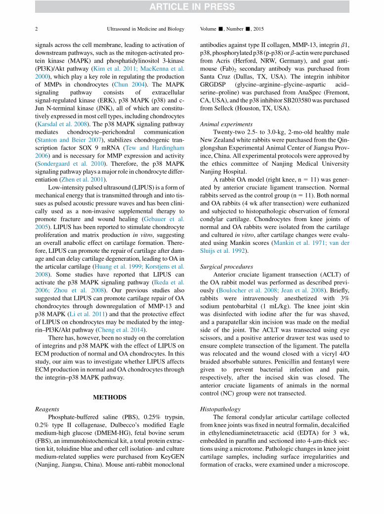
Low-intensity pulsed ultrasound stimulationNormal and OA chondrocytes were subdivided into
the control group and the treated group, which was subdi-vided into four subgroups (1–4) treated with LIPUS(HT2009-1, Ito, Tokyo, Japan) at intensities of 20, 30,40 and 50 mW/cm2, respectively. A layer of couplingagent (,1 mm thick) was applied between the LIPUSprobe and culture dish, and the probe was placed belowthe tissue culture dish (Fig. 1). Chondrocytes were treatedonce daily for 6 d at an on–off ratio of 20%, irradiation of3MHz, for 20 min in a 37�C incubator under a humidifiedatmosphere with 5% CO2 in air (Cheng et al. 2014;Korstjens et al. 2008).
Application of integrin and p38 MAPK inhibitorsTo investigate the role of integrin and p38 in the ef-
fects of LIPUS on normal and OA chondrocytes, thechondrocytes were pre-treated with a 10, 30 or 50 mMconcentration of the integrin inhibitor GRGDSP or a 1,5 or 10 mM concentration of the p38 inhibitorSB203580, or mock treated with dimethyl sulfoxide for4 h before the appropriate intensity of LIPUS stimulation.The optimal concentrations of inhibitors were determinedusing Western blot analysis. Then chondrocytes wereincubated with the optimal concentration of inhibitors,50 mM for GRGDSP or 10 mM for SB203580, andexposed or not exposed to LIPUS for 6 d.
Fig. 1. LIPUS irradiation of chondrocytes in vitro. A layer ofcoupling agent (,1 mm thick) was applied between the LIPUSprobe and culture dish, and the probe was placed below the tis-sue culture dish. Normal and osteoarthritis chondrocytes weresubdivided a control group that did not receive LIPUS stimula-tion and a treated group that was subdivided into four subgroupsthat received LIPUS at intensities of 20, 30, 40 and 50 mW/cm2,respectively. Chondrocytes were treated once a day for 6 d at anon–off ratio of 20%, irradiation of 3 MHz, for 20 min. LIPUS5
low-intensity pulsed ultrasound.

Fig. 2. Comparison of femoral condylar cartilage of normal rabbits and rabbits with OA. (a, b) Identification of normaland OA chondrocytes (3400): (a) Normal femoral condylar cartilage has a smooth articular surface with a bright lusterwith no cracks. (b) The soft articular surface of OA femoral condylar cartilage has crannies (arrows), but lacks the originalluster. (c, d) Hematoxylin–eosin (3100): (c) Smooth and evenly stained normal articular cartilage surface with chondro-cytes arranged in normal order. (d) Apparent articular cartilage fissures extending deep from the surface with significantloss of staining (arrows) and disorganized and fewer chondrocytes in OA femoral condylar cartilage. (e, f) Immunohis-tochemical staining of type II collagen: (e) Normal chondrocytes with abundant cytoplasm and round nuclei (arrows). (f)OA chondrocytes with hypertrophic nuclei mixed with some dendrite-like cells (arrows). (g) Type II collagen expressionin normal and OA chondrocytes. (h) Mankin scores in normal and OA cartilage. n5 11. OA5 osteoarthritis. *p, 0.05.
4 Ultrasound in Medicine and Biology Volume -, Number -, 2015
Western blot analysisAfter LIPUS stimulation for 6 d, chondrocytes were
collected, and expression of type II collagen, MMP-13, in-tegrin b1, p38 and p-p38 was determined by Western blotanalysis. Proteinswere extracted from the chondrocytes us-ing a total protein extraction kit, and 25 mg/mL was loadedonto sodium dodecyl sulfate–polyacrylamide gel for elec-trophoresis and electroblotted onto nitrocellulose mem-branes. The membranes were blocked with skim milk for2 h and incubated with primary antibodies: anti-type IIcollagen (1:500 dilution), anti-MMP-13 (1:500 dilution),anti-integrin b1 (1:500 dilution), anti-p38 (1:1000 dilu-tion), anti-p-p38 (1:1,000 dilution) and anti-b-actin(1:500 dilution) at 4�C overnight. Then the membraneswere subsequently washed three times with Tween 20 inPBS and incubated with peroxidase-conjugated goat anti-mouse IgG secondary antibody (1:5,000 dilution) at 37�Cfor 2 h, followed by another three washes as describedabove. The membranes were developed with the ECL kit.
Statistical analysisAll data were expressed as the mean6 standard de-
viation (SD) and analyzed using SPSS 18.0 software(IBM, Armonk, NY, USA). Differences between groupswere analyzed by single-factor analysis of variance (AN-OVA). Modified Mankin scores were analyzed using theWilcoxon signed rank test. A p-value, 0.05 was consid-ered to indicate statistical significance.
RESULTS
General and histopathologic observationsFourweeks afterACLT, the articular surface ofnormal
femoral condylar cartilage was smooth, had a bright lusterand did not any cracks (Fig. 2a), whereas the articular sur-face ofOA femoral condylar cartilage had become soft, lostits original luster and bore crannies (Fig. 2b). Femoralcondylar cartilage from normal and OA rabbits was stainedwith hematoxylin and eosin and examined under a

Fig. 3. Expression of type II collagen and MMP-13 in normal chondrocytes and chondrocytes from rabbits with OA afterLIPUS stimulation. (a, b) Western blot analysis of type II collagen and MMP-13 in normal and OA chondrocytes withb-actin as a loading control. (c) Quantitative analysis of type II collagen expression in normal chondrocytes. (d) Quan-titative analysis of type II collagen expression in OA chondrocytes. (e) Quantitative analysis of MMP-13 expression innormal chondrocytes. (f) Quantitative analysis of MMP-13 expression in OA chondrocytes. The ratio between thetarget and b-actin was used to normalize the data for comparison. n 5 4. MMP-13 5 matrix metalloproteinase 13;
OA 5 osteoarthritis; LIPUS 5 low-intensity pulsed ultrasound. *p , 0.05.
LIPUS affects chondrocyte extracellular matrix d P. XIA et al. 5
microscope. The surface of articular cartilage from normalrabbits was smooth and evenly stained with the chondro-cytes arranged in normal order (Fig. 2c); however, in artic-ular cartilage from rabbits with OA, there were fissuresextending deep from the surface with significant loss ofstaining (Fig. 2d). The chondrocytes from rabbits withOAwerealsodisorganizedand reduced innumber (Fig. 2d).
Modified Mankin scoresCompared with those of the normal control group,
the Mankin scores of the OA rabbit model group were
overall significantly higher (p , 0.05). Mankin scoresfor subgroups 1–4 of the OA rabbit model group werealso significantly higher (p , 0.05) than those of thenormal control group (Fig. 2h).
Identification of normal and OA chondrocytesThe normal chondrocytes had abundant cytoplasm
and a round nucleus, which grew to full confluencewithin4 to 7 d (arrows in Fig. 2e). The OA chondrocytes hadhypertrophic nuclei, which were mixed with somedendrite-like cells and grew to full confluence within 9

Fig. 4. Expression of integrin b1, p38 and p-p38 in normal and osteoarthritis chondrocytes after LIPUS stimulation. (a)Western blot analysis of integrin b1, p38 and p-p38 with b-actin as a loading control. (b) Quantitative analysis of integrinb1. (c) Ratio of p-p38 to p38. The ratio between the target and b-actin was used to normalize the data for comparison.p-p38 5 phosphorylated p38; LIPUS 5 low-intensity pulsed ultrasound; NC 5 normal control group; N 1 L 5
normal 1 LIPUS group; OA 5 osteoarthritis group; O 1 L 5 osteoarthritis 1 LIPUS group. n 5 4. *p , 0.05.
6 Ultrasound in Medicine and Biology Volume -, Number -, 2015
to 12 d (arrows in Fig. 2f). Immunohistochemical stain-ing of type II collagen in normal chondrocytes (Fig. 2e)was much deeper than that in OA chondrocytes(Fig. 2f). The mean density of type II collagen was signif-icantly higher in normal chondrocytes than in OAchondrocytes (p , 0.05) (Fig. 2g).
Expression of type II collagen and MMP-13 in normaland OA chondrocytes after 20 min of LIPUS stimulationat four different intensities
To determine an appropriate LIPUS intensity fornormal and OA rabbit chondrocytes, we examinedthe expression of type II collagen and MMP-13 inchondrocytes after LIPUS stimulation at four differentintensities (20, 30, 40 and 50 mW/cm2). Western blotanalysis revealed that in normal and OA rabbit chon-drocytes, type II collagen expression was significantlyincreased (p , 0.05) and MMP-13 expression wassignificantly decreased (p , 0.05) after LIPUS stimu-lation at all four intensities. In normal chondrocytes,type II collagen expression was significantly increased(p , 0.05) in the 40 mW/cm2 intensity groupcompared with all other groups, whereas MMP-13expression was significantly decreased (p , 0.05) inthe 40 mW/cm2 intensity group compared with the
20 mW/cm2 intensity (p , 0.05), 50 mW/cm2 intensity(p , 0.05) and control (p , 0.05) groups. However, nosignificant difference (p . 0.05) was observedcompared with the 30 mW/cm2 intensity group. InOA chondrocytes, type II collagen expression wassignificantly increased (p , 0.05) in the 30 mW/cm2
intensity group compared with the 40 mW/cm2 inten-sity group (p , 0.05), 50 mW/cm2 intensity group (p, 0.05) and control group (p , 0.05). However, nosignificant difference (p . 0.05) was observedcompared with the 20 mW/cm2 intensity group.MMP-13 expression was also significantly decreased(p , 0.05) in the 30 mW/cm2 intensity groupcompared with all other groups (Fig. 3).
Integrin b1 expression and p38 phosphorylation innormal and OA rabbit chondrocytes after 20 min ofLIPUS stimulation
We examined integrin b1 expression and p38 phos-phorylation in normal and OA rabbit chondrocytes afterLIPUS stimulation. LIPUS at 40 and 30 mW/cm2 wasused to stimulate normal and OA rabbit chondrocytes,respectively. Integrin b1 expression significantlyincreased after LIPUS stimulation (p , 0.05) in bothnormal and OA chondrocytes, whereas expression in

Fig. 5. Expression of integrin b1 and p-p38 in normal chondrocytes and OA chondrocytes after treatment with the integ-rin inhibitor GRGDSP or the p38 inhibitor SB203580. (a–d)Western blot analyses of integrin b1 and p38 with b-actin as aloading control in normal and OA chondrocytes. (e) Quantitative analysis of integrin b1 after treatment with 10, 30 or 50mM GRGDSP in normal and OA chondrocytes. (f) Quantitative analysis of p-p38 after treatment with 1, 5 or 10 mMSB203580 in normal and OA chondrocytes. The ratio between the target and b-actin was used to normalize the data
for comparison. n 5 3. OA 5 osteoarthritis. *p , 0.05.
LIPUS affects chondrocyte extracellular matrix d P. XIA et al. 7
OA chondrocytes was significantly higher than in normalchondrocytes (p , 0.05). The level of p38 phosphoryla-tion increased significantly after LIPUS stimulation innormal chondrocytes (p , 0.05), but significantlydecreased after LIPUS stimulation in OA chondrocytes(p , 0.05). The level of p38 phosphorylation wassignificantly higher in OA chondrocytes than normalchondrocytes (p , 0.05) (Fig. 4).
Expression of integrin b1 and p-p38 in normal and OArabbit chondrocytes after treatment with integrin andp38 inhibitors
The expression of integrin b1 and p-p38 in normaland OA chondrocytes was examined after treatmentwith inhibitors. Integrin b1 expression in normaland OA rabbit chondrocytes significantly decreased(p , 0.05) after treatment with 10, 30 or 50 mMGRGDSP, with the lowest expression in chondrocytesat 50 mM GRGDSP (p , 0.05). Expression of p-p38
in normal and OA rabbit chondrocytes significantlydecreased (p , 0.05) after treatment with 1, 5 or 10mM SB203580, with the lowest expression at 10 mM(p , 0.05) (Fig. 5).
Expression of type II collagen, MMP-13, p38 and p-p38in normal and OA rabbit chondrocytes after integrininhibitor treatment
Expression of type II collagen, MMP-13, p38 andp-p38 in normal and OA rabbit chondrocytes was exam-ined after treatment with the integrin inhibitor GRGDSPat 50 mM. After GRGDSP treatment, type II collagenexpression significantly increased (p , 0.05), andMMP-13 expression significantly decreased (p , 0.05)in normal and OA chondrocytes. The phosphorylationlevel of p38 significantly increased (p , 0.05) in normalchondrocytes, but significantly decreased (p , 0.05) inOA chondrocytes. Expression of type II collagenexpression significantly increased (p , 0.05) and that

Fig. 6. Expression of type II collagen, MMP-13, p38 and p-p38 in NC with or without LIPUS stimulation after treatmentwith the integrin inhibitor GRGDSP or the p38 inhibitor SB203580. (a) Western blot analysis of type II collagen,MMP-13, p38 and p-p38 with b-actin as a loading control. (b) Quantitative analysis of type II collagen. (c) Quantitativeanalysis of MMP-13. (d) Ratio of p-p38 to p38. The ratio between the target and b-actin was used to normalize the data forcomparison. MMP-13 5 matrix metalloproteinase 13; NC 5 normal chondrocytes; p-p38 5 phosphorylated p38;
LIPUS 5 low-intensity pulsed ultrasound. n 5 4. *p , 0.05.
8 Ultrasound in Medicine and Biology Volume -, Number -, 2015
of MMP-13 significantly decreased (p , 0.05) afterLIPUS stimulation in normal and OA chondrocytes.When GRGDSP was used, there was no significantchange in the expression of type II collagen andMMP-13 in normal and OA chondrocytes (p . 0.05) af-ter LIPUS stimulation. The phosphorylation level of p38did not significantly change (p . 0.05) in normal chon-drocytes and OA chondrocytes after LIPUS stimulation(Figs. 6 and 7).
Expression of type II collagen, MMP-13, p38 and p-p38in normal and OA rabbit chondrocytes after p38 MAPKinhibitor treatment
Expression of type II collagen, MMP-13, p38 andp-p38 in normal and OA rabbit chondrocytes wasexamined after treatment with the p38 MAPK inhibitor.Type II collagen expression significantly increased innormal and OA chondrocytes (p , 0.05). Type IIcollagen expression did not change significantly innormal chondrocytes (p . 0.05), but significantlydecreased in OA chondrocytes (p , 0.05) after LIPUSstimulation following SB203580 treatment. MMP-13expression significantly decreased (p , 0.05) in normaland OA chondrocytes after SB203580 treatment. Onexposure to LIPUS, MMP-13 expression significantlydecreased in OA chondrocytes (p , 0.05), but LIPUS
had no significant effect on normal chondrocytestreated with SB203580 (p . 0.05). The phosphoryla-tion level of p38 significantly decreased (p , 0.05)in normal and OA chondrocytes treated withSB203580. LIPUS stimulation significantly decreasedthe phosphorylation level of p38 in OA chondrocytes(p , 0.05), but had no significant effect in normalchondrocytes (p . 0.05) after LIPUS stimulationfollowing SB203580 treatment (Figs. 6 and 7).
DISCUSSION
In this study, we sought to determinewhether LIPUSaffects ECM production through an integrin–p38 MAPKsignaling pathway in normal and OA chondrocytes. Wefound that LIPUS significantly increases ECM produc-tion and decreases MMP-13 expression through an integ-rin–p38 MAPK signaling pathway.
We found that after LIPUS stimulation, type IIcollagen expression significantly increased (p ,0.05), but MMP-13 expression significantly decreased(p , 0.05). We suggested that the increased levels ofMMP-13 degraded type II collagen in the ECM ofchondrocytes to cause OA and that LIPUS preventedthe type II collagen from being degraded by MMP-13in both normal and OA chondrocytes.

Fig. 7. Expression of type II collagen, MMP-13, p38 and p-p38 in OA chondrocytes with or without LIPUS stimulationafter treatment with the integrin inhibitor GRGDSP or the p38 inhibitor SB203580. (a) Western blot analysis of type IIcollagen, MMP-13, p38 and p-p38 with b-actin as a loading control. (b) Quantitative analysis of type II collagen. (c)Quantitative analysis of MMP-13. (d) Ratio of p-p38 to p38. The ratio between the target and b-actin was used tonormalize the data for comparison. MMP-13 5 matrix metalloproteinase 13; p-p38 5 phosphorylated p38; OA 5 oste-
oarthritis; LIPUS 5 low-intensity pulsed ultrasound. n 5 4. *p , 0.05.
LIPUS affects chondrocyte extracellular matrix d P. XIA et al. 9
We further observed that inhibition of integrin byGRGDSP suppressed MMP-13 expression, which mayprevent the degradation of type II collagen and decreasethe activity of p38 in both normal and OA chondrocytes.Therefore, we speculate that integrin may have a negativeeffect on chondrocytes without LIPUS stimulation. Wealso found that integrin b1 expression in both normaland OA chondrocytes significantly increased (p , 0.05)after LIPUS stimulation. In fact, integrin plays an impor-tant role in mechanical signal transduction in chondro-cytes by participating in the proliferation,differentiation and migration of chondrocytes (Huanget al. 2004; Humphries 2000; Loeser et al. 2000; Luoet al. 2013; Stamenovic and Wang 2000). Fibronectinfragment (Fn-f), on one hand, stimulates MMPproduction to cause chondrocyte degeneration; on theother hand, integrin binds to Fn-f (Guo et al. 2009;Yasuda 2010). Therefore, we deduced that the increasedintegrin expression caused by LIPUS in both normaland OA chondrocytes may be ascribed to the binding toFn-f to eliminate the negative effect of Fn-f on chondro-cyte degradation. In this case, integrins might have abeneficial effect on chondrocytes. To sum up, integrinsplayed different roles under different situations.
We found that after LIPUS stimulation, phosphoryla-tion levels of p38 increased significantly (p , 0.05)in normal chondrocytes, but decreased significantly
(p, 0.05) inOAchondrocytes. This indicates thatwhen in-tegrinwas activated byLIPUS, the downstreamp38MAPKof the integrin–p38MAPKsignalingpathwaywas activatedin normal chondrocytes, but inhibited in OA chondrocytes.In fact, p38 activity ismuchhigher inOAchondrocytes, andinhibition of p38 can curb apoptosis and hypertrophic ter-minal differentiation of cultured OA chondrocytes (Reillyet al. 2005; Takebe et al. 2011), whereas p38 can be aneffector kinase of mechanotransduction in various celltypes and is essential to normal articular cartilage(Sondergaard et al. 2010). On the other hand, in adult artic-ular tissues, p38 is known to play an important role in theinflammatory process (Joos et al. 2009), and some evidencesuggests that p38 is amajor signalingmolecule in the induc-tion of MMP-13 and the activity of p38 can have negativeeffects on cartilage matrix maintenance (Julovi et al.2011; Mengshol et al. 2000; Pei et al. 2006). In addition,moderate mechanical stress such as knee loading reducesMMP-13 activity through the p38 MAPK signalingpathway in normal articular cartilage (Hamamura et al.2013). Therefore,we speculated thatLIPUS inhibited phos-phorylation of p38 in OA chondrocytes, but facilitatedphosphorylation of p38 in normal chondrocytes to promoteadaptation to a moderate mechanical stress.
We found that after application of the integrin in-hibitor GRGDSP, levels of type II collagen, MMP-13and p38 phosphorylation did not significantly change

10 Ultrasound in Medicine and Biology Volume -, Number -, 2015
in both normal and OA chondrocytes after LIPUS stim-ulation, suggesting again that p38 MAPK was down-stream of integrin and that inhibition of integrinexpression disrupted the regulation of p38 and ECMproduction by LIPUS. We also found that p38 inhibi-tion affected both normal and OA chondrocytes andthe effects of LIPUS on normal chondrocytes. Expres-sion of type II collagen significantly increased andexpression of MMP-13 significantly decreased innormal and OA chondrocytes treated with the p38 in-hibitor SB203580, indicating that p38 may be a nega-tive factor in both normal and OA chondrocyteswithout LIPUS stimulation. Our results also indicatedthat expression of type II collagen and MMP-13 didnot significantly change in normal chondrocytes afterLIPUS stimulation following SB203580 treatment,but in OA chondrocytes, LIPUS stimulation signifi-cantly increased type II collagen expression anddecreased MMP-13 expression. Thus, we deducedthat p38 was mainly a protective factor in normal chon-drocytes, but mainly an inflammatory and injuriousfactor in OA chondrocytes under LIPUS stimulation.We found that inhibition of p38 disrupted the protec-tive effects of LIPUS on ECM production in normalchondrocytes. Because LIPUS had the same inhibitoryeffect on p38 as SB203580 in OA chondrocytes, the in-hibition of p38 by SB203580 had no impact on the ef-fects of LIPUS on ECM production in OAchondrocytes. These results indicate that p38 of normaland OA chondrocytes has different roles underdifferent conditions. For the protective effect of LI-PUS, p38 has a more important role in normal chon-drocytes than in OA chondrocytes under LIPUSsimulation. Of note, the protective effect of LIPUSon chondrocytes is exerted not only through the p38MAPK pathway, but also through other pathwayssuch as PI3K/Akt pathway, as described in our previ-ous study (Cheng et al. 2014). Therefore, we specu-lated that the p38 MAPK pathway and PI3K/Aktpathway might have a synergistic effect on ECM pro-duction of chondrocytes under LIPUS stimulation,which should be investigated in a further study.
CONCLUSIONS
Low-intensity pulsed ultrasound was found to haveeffects on the expression of type II collagen and MMP-13 through the integrin–p38 MAPK signaling pathway.Therefore, we deduced that the integrin–p38 MAPKsignaling pathway plays an important role in LIPUS-mediated ECM production in chondrocytes. Our findingsfurther revealed the mechanism underlying the effects ofLIPUS on chondrocytes and provide some basis for thedevelopment of OA treatments using LIPUS.
Acknowledgments—We thank Medjaden Bioscience Limited for editingand proofreading this article. We also thank the National Natural Sci-ence Foundation of China (Grant 81272151) for financial support.
REFERENCES
Aigner T, S€oder S, Gebhard PM, McAlinden A, Haag J. Mechanisms ofdisease: Role of chondrocytes in the pathogenesis of osteoarthritis—Structure, chaos and senescence. Nat Clin Pract Rheumatol 2007;3:391–399.
Blain EJ. Mechanical regulation of matrix metalloproteinases. FrontBiosci 2007;12:507–527.
Boulocher C, Duclos ME, Arnault F, Roualdes O, Fau D, Hartmann DJ,Roger T, Vignon E, Viguier E. Knee joint ultrasonography of theACLT rabbit experimental model of osteoarthritis: Relevance andeffectiveness in detecting meniscal lesions. Osteoarthritis Cartilage2008;16:470–479.
Bramono DS, Richmond JC, Weitzel PP, Kaplan DL, Altman GH. Ma-trix metalloproteinases and their clinical applications in orthopae-dics. Clin Orthop Relat Res 2004;428:272–285.
Cheng K, Xia P, Lin Q, Shen S, Gao M, Ren S, Li X. Effects oflow-intensity pulsed ultrasound on integrin-FAK-PI3 K/Akt mecha-nochemical transduction in rabbit osteoarthritis chondrocytes.Ultrasound Med Biol 2014;40:1609–1618.
Chun JS. Expression, activity, and regulation ofMAP kinases in culturedchondrocytes. Methods Mol Med 2004;100:291–306.
Gebauer D, Mayr E, Orthner E, Ryaby JP. Low-intensity pulsed ultra-sound: Effects on nonunions. Ultrasound Med Biol 2005;31:1391–1402.
Guilak F, Cohen DM, Estes BT, Gimble JM, Liedtke W, Chen CS. Con-trol of stem cell fate by physical interactions with the extracellularmatrix. Cell Stem Cell 2009;5:17–26.
Guo D, Ding L, Homandberg GA. Telopeptides of type II collagen up-regulate proteinases and damage cartilage but are less effective thanhighly active fibronectin fragments. Inflamm Res 2009;58:161–169.
Hamamura K, Zhang P, Zhao L, Shim JW, Chen A, Dodge TR, Wan Q,Shih H, Na S, Lin CC, Sun HB, Yokota H. Knee loading reducesMMP13 activity in the mouse cartilage. BMCMusculoskelet Disord2013;14:312.
Huang H, Kamm RD, Lee RT. Cell mechanics and mechanotransduc-tion: Pathways, probes, and physiology. Am J Physiol Cell Physiol2004;287:1–11.
Huang MH, Yang RC, Ding HJ, Chai CY. Ultrasound effect on level ofstress proteins and arthritic histology in experimental arthritis. ArchPhys Med Rehabil 1999;80:551–556.
Humphries MJ. Integrin structure. Biochem Soc Trans 2000;28:311–339.
Hynes R. Integrins: Bidirectional, allosteric signaling machines. Cell2002;110:673–687.
Hynes RO. Integrins: Versatility, modulation, and signaling in cell adhe-sion. Cell 1992;69:11–25.
Ikeda K, Takayama T, Suzuki N, Shimada K, Otsuka K, Ito K. Effects oflow-intensity pulsed ultrasound on the differentiation of C2 C12cells. Life Sci 2006;79:1936–1943.
Jean YH, Wen ZH, Chang YC, Hsieh SP, Lin JD, Tang CC, Chen WF,Chou AK, Wong CS. Increase in excitatory amino acid concentra-tion and transporters expression in osteoarthritic knees of anteriorcruciate ligament transected rabbits. Osteoarthritis Cartilage 2008;16:1442–1449.
Joos H, Albrecht W, Laufer S, Brenner RE. Influence of p38 MAPK in-hibition on IL-1 beta-stimulated human chondrocytes: A microarrayapproach. Int J Mol Med 2009;23:685–693.
Julovi SM, Ito H, Nishitani K, Jackson CJ, Nakamura T. Hyaluronan in-hibits matrix metalloproteinase-13 in human arthritic chondrocytesvia CD44 and P38. J Orthop Res 2011;29:258–264.
Karsdal MA, Leeming DJ, Dam EB, Henriksen K, Alexandersen P,Pastoureau P, Altman RD, Christiansen C. Should subchondralbone turnover be targeted when treating osteoarthritis? Osteoar-thritis Cartilage 2008;16:638–646.

LIPUS affects chondrocyte extracellular matrix d P. XIA et al. 11
Kim SH, Turnbull J, Guimond S. Extracellular matrix and cell signaling:The dynamic cooperation of integrin, proteoglycan, and growth fac-tor receptor. J Endocrinol 2011;209:139–151.
Korstjens CM, van der Rijt RH, Albers GH, Semeins CM,Klein-Nulend J. Low-intensity pulsed ultrasound affects humanarticular chondrocytes in vitro. Med Biol Eng Comput 2008;46:1263–1270.
LiX,Li J,ChengK,WangD,ZhangH,AnH,GaoM,ChenA.Effect of low-intensity pulsed ultrasound onMMP-13 andMAPKs signaling pathwayin rabbit knee osteoarthritis. Cell Biochem Biophys 2011;61:427–434.
Loeser RF, Sadiev S, Tan L, Goldring MB. Integrin expression by pri-mary and immortalized human chondrocytes: Evidence of a differ-ential role for alpha1 beta1 and alpha2 beta1 integrins inmediating chondrocyte adhesion to types II and VI collagen. Osteo-arthritis Cartilage 2000;8:96–105.
Lohmander LS, Atley LM, Pietka TA, Eyre DR. The release of cross-linked peptides from type II collagen into human synovial fluid isincreased soon after joint injury and in osteoarthritis. ArthritisRheum 2003;48:3130–3139.
Luo S, Shi Q, Zha Z, Yao P, Lin H, Liu N, Wu H, Cai J, Sun S. The rolesof integrin beta1 in phenotypic maintenance and dedifferentiation inchondroid cells differentiated from human adipose-derived stemcells. Nanoscale Res Lett 2013;8:136.
MacKenna D, Summerour SR, Villarreal FJ. Role of mechanical factorsin modulating cardiac fibroblast function and extracellular matrixsynthesis. Cardiovasc Res 2000;46:257–263.
Mankin HJ, Dorfman H, Lippiello L, Zarins A. Biochemical and meta-bolic abnormalities in articular cartilage from osteo-arthritic humanhips: II. Correlation of morphology with biochemical and metabolicdata. J Bone Joint Surg Am 1971;53:523–537.
Mengshol JA, Vincenti MP, Coon CI, Barchowsky A, Brinckerhoff CE.Interleukin-1 induction of collagenase 3 (matrix metalloproteinase13) gene expression in chondrocytes requires p38, c-Jun N-terminalkinase, and nuclear factor kappaB: Differential regulation of colla-genase 1 and collagenase 3. Arthritis Rheum 2000;43:801–811.
Mitchell PG, Magna HA, Reeves LM, Lopresti-Morrow LL, Yocum SA,Rosner PJ, Geoghegan KF, Hambor JE. Cloning, expression, andtype II collagenolytic activity of matrix metalloproteinase-13 fromhuman osteoarthritic cartilage. J Clin Invest 1996;97:761–768.
Pearle AD, Warren RF, Rodeo SA. Basic science of articular cartilageand osteoarthritis. Clin Sports Med 2005;24:1–12.
Pei Y, Harvey A, Yu XP, Chandrasekhar S, Thirunavukkarasu K. Differ-ential regulation of cytokine-induced MMP-1 and MMP-13 expres-sion by p38 kinase inhibitors in human chondrosarcoma cells:Potential role of Runx2 in mediating p38 effects. OsteoarthritisCartilage 2006;14:749–758.
Reilly GC, Golden EB, Grasso-Knight G, Leboy PS. Differential ef-fects of ERK and p38 signaling in BMP-2 stimulated hypertro-phy of cultured chick sternal chondrocytes. Cell CommunSignal 2005;3:3.
Rizzardi AE, Johnson AT, Vogel RI, Pambuccian SE, Henriksen J,Skubitz AP, Metzger GJ, Schmechel SC. Quantitative compar-ison of immunohistochemical staining measured by digital im-age analysis versus pathologist visual scoring. Diagn Pathol2012;7:42.
Sandell LJ, Aigner T. Articular cartilage and changes in arthritis, anintroduction: Cell biology of osteoarthritis. Arthritis Res 2001;3:107–113.
Sondergaard BC, Schultz N, Madsen SH, Bay-Jensen AC, Kassem M,Karsdal MA. MAPKs are essential upstream signaling pathwaysin proteolytic cartilage degradation divergence in pathways leadingto aggrecanase and MMP-mediated articular cartilage degradation.Osteoarthritis Cartilage 2010;18:279–288.
Stamenovic D, Wang N. Invited review: Engineering approaches tocytoskeletal mechanics. J Appl Physiol 2000;89:2085–2090.
Stanton LA, Beier F. Inhibition of p38 MAPK signaling in chondrocytecultures results in enhanced osteogenic differentiation of perichon-dral cells. Exp Cell Res 2007;313:146–155.
Takaishi H, Kimura T, Dalal S, Okada Y, D’Armiento J. Joint diseasesand matrix metalloproteinases: A role for MMP-13. Curr PharmBiotechnol 2008;9:47–54.
Takebe K, Nishiyama T, Hayashi S, Hashimoto S, Fujishiro T,Kanzaki N, Kawakita K, Iwasa K, Kuroda R, Kurosaka M. Regula-tion of p38 MAPK phosphorylation inhibits chondrocyte apoptosisin response to heat stress or mechanical stress. Int J Mol Med2011;27:329–335.
Tew SR, Hardingham TE. Regulation of SOX9 mRNA in human artic-ular chondrocytes involving p38MAPK activation andmRNA stabi-lization. J Biol Chem 2006;281:39471–39479.
van der Sluijs JA, Geesink RG, van der Linden AJ, Bulstra SK, Kuyer R,Drukker J. The reliability of the Mankin score for osteoarthritis.J Orthop Res 1992;10:58–61.
Yasuda T. Comparison of hyaluronan effects among normal, osteoar-thritis, and rheumatoid arthritis cartilages stimulated with fibro-nectin fragment. Biomed Res 2010;31:63–69.
Zhen X, Wei L, Wu Q, Zhang Y, Chen Q. Mitogen-activated protein ki-nase p38 mediates regulation of chondrocyte differentiation by para-thyroid hormone. J Biol Chem 2001;276:4879–4885.
Zhou S, Bachem MG, Seufferlein T, Li Y, Gross HJ,Schmelz A. Low intensity pulsed ultrasound acceleratesmacrophage phagocytosis by a pathway that requires actinpolymerization, Rho, and Src/MAPKs activity. Cell Signal2008;20:695–704.
Copyright © 2022 FDOKUMEN