Low expression of the putative tumour suppressor spinophilin is associated with higher proliferative...
8
Low expression of the putative tumour suppressor spinophilin is associated with higher proliferative activity and poor prognosis in patients with hepatocellular carcinoma A Aigelsreiter 1,6 , A L Ress 2,6 , K Bettermann 1 , S Schauer 1 , K Koller 2 , F Eisner 2 , T Kiesslich 3 , T Stojakovic 4 , H Samonigg 2 , P Kornprat 5 , C Lackner 1 , J Haybaeck 1 and M Pichler * ,2 1 Institute of Pathology, Medical University of Graz, Graz, Austria; 2 Division of Oncology, Department of Internal Medicine, Medical University of Graz, Auenbruggerplatz 15, Graz 8010, Austria; 3 Department of Internal Medicine I, Paracelsus Medical University, Salzburger Landeskliniken, Salzburg, Austria; 4 Clinical Institute of Medical and Chemical Laboratory Diagnostics, Medical University of Graz, Graz, Austria and 5 Division of Visceral Surgery, Department of Surgery, Medical University of Graz, Graz, Austria Background: Spinophilin, a multifunctional intracellular scaffold protein, is reduced in certain types of cancer and is regarded as a novel putative tumour suppressor protein. However, the role of spinophilin in hepatocellular carcinoma (HCC) has never been explored before. Methods: In this study, we determined for the first time the expression pattern of spinophilin in human HCC by immunohistochemistry and quantitative reverse transcriptase–PCR analysis. In addition, we performed immunohistochemical analysis of p53, p14 ARF and the proliferation marker Ki-67. Kaplan–Meier curves and multivariate Cox proportional models were used to study the impact on clinical outcome. Small interfering RNA (siRNA) was used to silence spinophilin and to explore the effects of reduced spinophilin expression on cellular growth. Results: In our study, complete loss of spinophilin immunoreactivity was found in 44 of 104 HCCs (42.3%) and reduced levels were found in an additional 37 (35.6%) cases. After adjusting for other prognostic factors, multivariate Cox regression analysis identified low expression of spinophilin as an independent prognostic factor with respect to disease-free (hazard ratio (HR) ¼ 1.8; 95% confidence interval (CI) ¼ 1.04–3.40; P ¼ 0.043) and cancer-specific survival (HR ¼ 2.0; CI ¼ 1.1–3.8; P ¼ 0.025). Reduced spinophilin expression significantly correlated with higher Ki-67 index in HCC (P ¼ 0.014). Reducing spinophilin levels by siRNA induced a higher cellular growth rate and increased cyclin D2 expression in tumour cells (Po0.05). Conclusion: This is the first study of the expression pattern and distribution of spinophilin in HCC. According to our data, the loss of spinophilin is associated with higher proliferation and might be useful as a prognostic marker in patients with HCC. *Correspondence: Dr M Pichler; E-mail: [email protected] 6 These authors contributed equally to this work. Received 11 October 2012; revised 20 March 2013; accepted 21 March 2013; published online 16 April 2013 & 2013 Cancer Research UK. All rights reserved 0007 – 0920/13 FULL PAPER Keywords: hepatocellular carcinoma; spinophilin; immunohistochemistry; prognosis British Journal of Cancer (2013) 108, 1830–1837 | doi: 10.1038/bjc.2013.165 1830 www.bjcancer.com | DOI:10.1038/bjc.2013.165
-
Upload
independent -
Category
Documents
-
view
3 -
download
0
Transcript of Low expression of the putative tumour suppressor spinophilin is associated with higher proliferative...
Low expression of the putative tumoursuppressor spinophilin is associated withhigher proliferative activity and poorprognosis in patients with hepatocellularcarcinomaA Aigelsreiter1,6, A L Ress2,6, K Bettermann1, S Schauer1, K Koller2, F Eisner2, T Kiesslich3, T Stojakovic4,H Samonigg2, P Kornprat5, C Lackner1, J Haybaeck1 and M Pichler*,2
1Institute of Pathology, Medical University of Graz, Graz, Austria; 2Division of Oncology, Department of Internal Medicine, MedicalUniversity of Graz, Auenbruggerplatz 15, Graz 8010, Austria; 3Department of Internal Medicine I, Paracelsus Medical University,Salzburger Landeskliniken, Salzburg, Austria; 4Clinical Institute of Medical and Chemical Laboratory Diagnostics, MedicalUniversity of Graz, Graz, Austria and 5Division of Visceral Surgery, Department of Surgery, Medical University of Graz, Graz, Austria
Background: Spinophilin, a multifunctional intracellular scaffold protein, is reduced in certain types of cancer and is regarded as anovel putative tumour suppressor protein. However, the role of spinophilin in hepatocellular carcinoma (HCC) has never beenexplored before.
Methods: In this study, we determined for the first time the expression pattern of spinophilin in human HCC byimmunohistochemistry and quantitative reverse transcriptase–PCR analysis. In addition, we performed immunohistochemicalanalysis of p53, p14ARF and the proliferation marker Ki-67. Kaplan–Meier curves and multivariate Cox proportional models wereused to study the impact on clinical outcome. Small interfering RNA (siRNA) was used to silence spinophilin and to explore theeffects of reduced spinophilin expression on cellular growth.
Results: In our study, complete loss of spinophilin immunoreactivity was found in 44 of 104 HCCs (42.3%) and reducedlevels were found in an additional 37 (35.6%) cases. After adjusting for other prognostic factors, multivariate Cox regressionanalysis identified low expression of spinophilin as an independent prognostic factor with respect to disease-free (hazardratio (HR)¼ 1.8; 95% confidence interval (CI)¼ 1.04–3.40; P¼ 0.043) and cancer-specific survival (HR¼ 2.0; CI¼ 1.1–3.8;P¼ 0.025). Reduced spinophilin expression significantly correlated with higher Ki-67 index in HCC (P¼ 0.014).Reducing spinophilin levels by siRNA induced a higher cellular growth rate and increased cyclin D2 expression in tumour cells(Po0.05).
Conclusion: This is the first study of the expression pattern and distribution of spinophilin in HCC. According to ourdata, the loss of spinophilin is associated with higher proliferation and might be useful as a prognostic marker in patients withHCC.
*Correspondence: Dr M Pichler; E-mail: [email protected] authors contributed equally to this work.
Received 11 October 2012; revised 20 March 2013; accepted 21 March 2013; published online 16 April 2013
& 2013 Cancer Research UK. All rights reserved 0007 – 0920/13
FULL PAPER
Keywords: hepatocellular carcinoma; spinophilin; immunohistochemistry; prognosis
British Journal of Cancer (2013) 108, 1830–1837 | doi: 10.1038/bjc.2013.165
1830 www.bjcancer.com | DOI:10.1038/bjc.2013.165
Hepatocellular carcinoma (HCC) is the third leading cause ofcancer-related mortality worldwide (Siegel et al, 2012). Owing toan increased prevalence in causal risk factors, such as obesity andviral liver infections, the incidence of HCC is increasing indeveloped countries (Shariff et al, 2009). Despite the introductionof novel agents, including the multikinase inhibitor sorafenib formetastatic HCC, the disseminated disease stage is incurable. Forlocally confined HCC, resection is the gold standard in the curativetreatment of this disease (Frau et al, 2010). However, manypatients fail a successful complete resection or develop a recurrencein other regions of the organ (Lai et al, 2012). Currently, because ofthe heterogeneity of the underlying molecular pathophysiologicalmechanisms in the tumour as well as in the non-neoplastictransformed adjacent liver (Guichard et al, 2012), the widely usedTNM staging classification system is limited in predicting thecourse of the disease, the recurrence rate and the survival ofpatients with HCC (Pons et al, 2005). Therefore, novel prognosticfactors are of paramount importance to classify adequately HCCpatients with regard to different treatment modalities, to predictindividual outcomes, to report pathologic specimens, to conductpostoperative surveillance and to qualify for inclusion intomultinational epidemiologic, exploratory and interventional clin-ical trials (Minguez and Lachenmayer, 2011). Recently, manystudies have attempted to evaluate clinicopathological features ordiscover novel biomarkers that lead to a better prediction of theclinical course of HCC (Lauwers et al, 2002; Aigelsreiter et al, 2009,2012). In general, the detailed characterisation of the biologicalproperties of a particular pathophysiological factor could provide alogical rationale for testing its potential prognostic significance as abiomarker in HCC. In this context, spinophilin (Spn; also knownas Neurabin 2), a multifunctional protein whose gene is located inthe 17q21.33 chromosomal region, is commonly downregulated orlost in different human cancers, including renal cell carcinoma,adenocarcinoma of the lung and tumours of the central nervoussystem (Carnero, 2012). Furthermore, lower levels of Spn mRNAhave been correlated with higher grades of ovarian carcinoma andthe progression of chronic myelogenous leukaemia to a moreaggressive phenotype (Carnero, 2012). As a consequence, Spn hasbeen proposed to be a novel tumour suppressor in human cancers(Carnero, 2012). Notably, its chromosomal location contains arelatively high density of well-known tumour suppressor genes andis commonly impaired by chromosomal alterations in HCCs(Furge et al, 2005). To date, no study has been published that hasexplored the presence, the distribution or the clinical significanceof Spn expression in human HCC. Therefore, we examined Spnexpression in HCC tissue by immunohistochemistry and quanti-tative reverse transcriptase–PCR (RT–PCR). Correlation analysesof Spn expression and other possible interacting partners includingmicroRNA-106a*, p53 and p14ARF were performed. Consequently,the role of Spn as a valuable novel prognostic marker was evaluatedby correlating its expression level with several clinicopathologicalfeatures as well as disease-free and cancer-specific survival in alarge cohort of HCC patients. Finally, we explored the influence ofreduced Spn expression on proliferation by correlating itsexpression with the Ki-67 proliferation marker in HCC tissue.Functional studies to explore the effects of reduced Spn expressionon cellular growth were performed by small interfering RNA(siRNA)-mediated Spn silencing in cell line experiments.
MATERIALS AND METHODS
Patients and tissue samples. One hundred and four patients withprimary HCC who underwent a curative liver resection at theMedical University of Graz (Austria) were included in thisretrospective study. Tissue samples used in the study were
retrieved from the Institute of Pathology, Medical University ofGraz. We included consecutive patients diagnosed with HCCbetween January 1988 and December 2011. For an independentRT–PCR confirmation step, cryopreserved tissue of 10 HCCsamples and their corresponding surrounding non-neoplastic liverwere selected. The mean follow-up period was 3 years. None of thepatients received neoadjuvant therapy or preoperative localtreatment, and all underwent resection of the primary tumour.Postoperative surveillance was performed, including routineclinical and laboratory examinations every third month, and CTscans of the abdomen and X-rays of the chest every third month,respectively. After 5 years, the examination interval was extendedto 12 months. The 7th edition of the American Joint Committee onCancer (AJCC)/International Union Against Cancer (UICC) TNMsystem was used to classify the patients (Sobin et al, 2009). Thestudy was approved by the local ethics committee of the MedicalUniversity of Graz (ID: 21–135 ex 09/10).
RNA isolation. For extraction of total RNA from cryopreservedliver tissue, 1 ml Trizol reagent (Invitrogen, Lofer, Austria) wasadded to a small piece of liver tissue (10 HCCs and theircorresponding non-neoplastic liver tissue), which were alreadytransferred into a tube filled up with MagNA Lyser Green beads(Roche Diagnostics, Vienna, Austria). Samples were homogenisedby using MagnNA Lyser (Roche Diagnostics) at 6500 r.p.m. for20 s, followed by incubation at room temperature for 5 min.Subsequently, 0.2 ml chloroform were added to each tube, agitatedvigorously by hand for 15 s and incubated at room temperature for3 min. Samples were centrifuged at 12 000 g at 4 1C for 25 min. Theupper phase was transferred to a 1.5 ml eppendorf tube. After-wards, 0.5-ml isopropanol were added, samples were vortexed for15 s and incubated at room temperature for 10 min. Samples werecentrifuged at 12 000 g at 4 1C for 10 min. Supernatants wereremoved and pellets were washed once with 1 ml of 75% ethanol.After centrifugation at 12 000 g at 4 1C for 10 min, supernatantswere removed again and pellets were dried completely. Finally,pellets were dissolved in 30ml Aqua bidest (Fresenius Kabi, Graz,Austria) and incubated at 56 1C for 10 min. RNA yield and qualitywere checked by using the NanoDrop 1000 Spectrophotometer(Peqlab, Erlangen, Germany). Total cellular RNA from HepG2cells was isolated using the RNAeasy Mini kit (Qiagen, Hilden,Germany) according to the manufacturer’s instructions.
Immunohistochemistry. Immunohistochemical analyses for Spn,p53, p14ARF and Ki-67 expression were performed on whole tissueslides of HCC tissue from all of the 104 patients. The 3-mm-thicksections were deparaffinised in xylene and rehydrated with gradedethanol. For Spn detection, the sections were subjected to antigenretrieval in a pressure cooker (Dako, Glostrup, Denmark; Pascal) in0.01 M sodium-citrate buffer, pH 6.0, and subsequently incubatedfor 60 min with a rabbit antibody to human Spn (Millipore; anti-Spn antibody: AB5669 Vienna, Austria) at a 1 : 50 dilution. Thereaction was visualised using the UltraVision LP Large VolumeDetection System HRP Polymer (Thermo Scientific, Vienna,Austria), and all sections were counterstained with haematoxylin.For the negative control, the primary antibody was omitted. Thefrequency and the intensity of Spn-positive cells were estimated bytwo independent experienced observers (AA and MP) on asemiquantitative scoring system. The percentage of positive cellswas rated as follows: 0, negative; 1, 1–10% positive cells; 2, 11–50%positive cells; 3, 51–80% positive cells; and 4, 480% positive cells.The staining intensity was scored as 0, negative; 1, weak; 2,moderate; and 3, intensive. The scores for the percentage ofpositive cells and the scores for the expression intensities weremultiplied to calculate an immunoreactive score (IRS) (Remmeleand Stegner, 1987). We separated the HCC patients into fourgroups according to their cytoplasmic expression of Spn (25th
Spinophilin in HCC BRITISH JOURNAL OF CANCER
www.bjcancer.com | DOI:10.1038/bjc.2013.165 1831
percentiles): group 1, IRS 0–2; group 2, IRS 3 and 4; group 3, IRS6–8; and group 4, IRS 9–12.
For p53 staining, slides were incubated at 60 1C for 1 h anddeparaffinised by xylene two times for 10 min. Sections wererehydrated by decreasing ethanol concentrations from 100–90%to 70% and to 50%, followed by a water bath for 40 min.Immunohistochemistry for p53 was performed on a DAKOautostainer (Dako), where a heat-induced epitope retrieval wasperformed and the antibody against p53 (monoclonal mouse anti-human p53 protein clone DO-7, M7001; Dako) was diluted 1 : 100.Dako Real (Dako; K5001) was used as a detection system. Forvisualisation, AEC Substrate Chromogen Ready-to-Use Kit(K3464; Dako) was utilised.
The percentages of tumour cells with a strong immunoreactivenuclear signal, indicating p53 accumulation in the nucleus, in thetotal number of tumour cells were counted in each of 10 high-power fields (magnification, � 40; 1 high-power field¼ 0.24 mm2).The mean percentage of immunoreactive tumour cells was thensemiquantitatively calculated on a 10-point scale (0–100%).
For p14ARF detection, antigens were unmasked by treating theslides with EDTA-sodium buffer (1 mM at pH¼ 8.0) for 20 min ina microwave. After a cooling phase of 20 min, endogenousperoxidase was blocked with 3% H2O2 (Merck, Darmstadt,Germany) for 10 min at room temperature. A p14ARF antibody(GeneTex; GTX 23642, Irvine, CA, USA) was prepared in a 1 : 20dilution in antibody diluent (Dako) and incubated for 60 min atroom temperature. After washing and incubation with thebiotinylated secondary antibody (K5001; Dako) for 30 min atroom temperature, streptavidin-conjugated horse radish perox-idase (Dako) was added for additional 30 min at room tempera-ture. AEC Substrate Chromogen Ready-to-Use (K3464; Dako) wasused as the detection system for a minimum of 7 min. Colourdevelopment was monitored under a microscope. Sections werecounterstained with Mayer’s hemalaun for 1 min.
The percentages of tumour cells with a strong immunoreactivecytoplasmic signal to p14ARF in the total number of tumour cellswere counted in each of 10 high-power fields (magnification, � 40;1 high-power field¼ 0.24mm2). The mean percentage of immuno-reactive tumour cells was then calculated and scored on a 10-pointscale (0–100%).
The Ki-67 immunohistochemistry was analysed as a marker ofproliferative activity in HCCs. For the detection of Ki-67, theanti-Ki-67 (30-9) antibody (Rabbit-a-confirm Ki-67 MIB Clone30-9,Ready-to-Use; Ventana) was used on an autostainer (Ventana)by applying the automated Ventana staining system program CC1mild (Ventana, Tucson, AZ, USA). The antibody was incubated for32 min and for visualisation the iVIEW DAB Detection system(Ventana) was used. The number of positively stained nuclei oftumour cells was counted in at least 1000 tumour cells. The Ki-67proliferation index was classified in accordance with the percentage ofpositive tumour cells as described previously (Kitamura et al, 2011).
Quantitative RT–PCR analysis of mRNA and miR-106a*. FormRNA quantification, up to 500 ng of total RNA was reversetranscribed into cDNA using the QuantiTect Reverse Trans-cription Kit (Qiagen) according to the manufacturer’s instructions.Quantitative RT–PCR was carried out in triplicates for eachsample using commercially available primers specific for Spn(Hs_PPP1R9B_1_SG QuantiTect Primer Assay; Qiagen), theproliferation marker cyclin D2 (Hs_CCND2_1_SG QuantiTectPrimer Assay; Qiagen) and the two housekeeping genes HMBS(Hs_HMBS_1_SG QuantiTect primer assay) and GAPDH(Hs_GAPDH_1_SG QuantiTect Primer Assay, Qiagen) on aLightCycler 480 Real-Time PCR System (Roche Diagnostics) usingQuantiTect SYBR Green PCR Kit (Qiagen). Geometric mean of thehousekeeping genes HMBS and GAPDH was used for normal-isation, and relative gene expression levels were calculated with the
delta delta CT method as previously described (Aigelsreiter et al,2009). For detection of miR-106a* in cryopreserved HCC tissuesamples, 500 ng of total RNA was reverse transcribed by themiScript II RT Kit (Qiagen) according to the manufacture’sprotocol. The Hs_miR-106a*_1 miScript Primer Assay (maturemicroRNA (miRNA) sequence: 50-CUGCAAUGUAAGCACUU-CUUAC-30; Qiagen), the Hs_miR-98_1 miScript Primer Assay(Qiagen) as a positive control (with previously described expres-sion in liver tissue (Sukata et al, 2011)) and for normalisation of theHs_RNU6-2_1 miScript Primer Assay (Qiagen) were applied on aLightCycler 480 Real-Time PCR System (Roche Diagnostics) usingmiScript SYBR Green PCR Kit (Qiagen).
Cell culture. The human hepatoma cell line HepG2 was purchasedfrom American Type Culture Collection (Manassas, VA, USA). Cellswere grown in Dulbecco’s modified Eagle’s medium (DMEM) (1� )(þ 4.5 g l� 1
D-glucose, L-glutamine, Pyruvat) (both form Gibco,Vienna, Austria), containing 10% foetal bovine serum gold (PAALaboratories, Pasching, Austria) and antibiotics (penicillin andstreptomycin) and were incubated in a 5% CO2 humidifiedincubator at 37 1C.
WST-1 proliferation assay. After standard trypsinisation, 10 000HepG2 cells per well were seeded in 96-well culture plates andincubated in normal growth medium DMEM (Gibco) for 48 h aftertransfection of siRNA. The WST-1 proliferation reagent (RocheApplied Science, Vienna, Austria) was applied according to themanufacturer’s recommendations and after 4 h the colorimetricchanges were measured using a SpectraMax Plus (Molecular Devices,Sunnyvale, CA, USA) at 450 nm with a reference wavelength at620 nm. For each condition, cells were seeded in six-well plates andeach experiment was independently repeated three times.
Transfection of Spn-targeting siRNA. For a transient trans-fection approach to reduce the Spn mRNA expression, HepG2 cellswere transfected using the fast-forward transfection procedure assuggested by the HiPerFect Transfection Reagent (Qiagen) protocolaccording to the manufacturer’s instructions. A specific siRNA, whichhas been previously experimentally validated by the supplier to targetthe Spn mRNA (Hs_PPP1R9B_7 FlexiTube siRNA; Qiagen), wascommercially purchased. For the reference control, we used theAllstars negative control siRNA (Qiagen), which is a validated siRNAwithout any functional consequence or known target sequence inhuman cells. For confirmation of transfections efficacy, the AllStarscell death control (Qiagen) were used within the same concentrationas the target siRNA and reference control (20 nM). For siRNAdelivery, 10 000 HepG2 cells in complete RPMI medium were seededin 96-well plates and siRNA-HiPerFect Reagent transfection com-plexes were added according to the fast-forward transfection asrecommended by the manufacturer (Qiagen). Transfected cells wereincubated under their normal growth conditions (37 1C, 5% CO2)and the effect on Spn gene silencing was measured by quantitativeRT–PCR after 48 h. Effects on cellular growth were monitored after48 h by the WST-1 proliferation reagent assay and cyclin D2expression was measured as described above.
Statistical analysis. All statistical analyses were performed usingSPSS version 17.0 software (SPSS Inc., Chicago, IL, USA). Fisher’sexact test, the w2-test, the Mann–Whitney U-test and the Student’st-test procedure were used where appropriate to analyse Spnprotein expression in relation to each clinicopathological para-meter, cellular growth rates and gene expression. Spearman’scorrelation was calculated to compare immunohistochemical Spnexpression vs Spn mRNA expression, as well as p53, p14ARF andKi-67 staining. Student’s t-test was used to test for differences incellular growth rates. Disease-free survival (DFS) and cancer-specific survival (CSS) of the patients were calculated using theKaplan–Meier method, and the differences were compared bythe log-rank test. Backward stepwise multivariate Cox proportion
BRITISH JOURNAL OF CANCER Spinophilin in HCC
1832 www.bjcancer.com | DOI:10.1038/bjc.2013.165
analysis was performed to determine the influence of Spnexpression, T classification, tumour grade, patient age and genderon DFS and CSS. The hazard ratios (HRs) estimated from the Coxmodels were reported as relative risks with corresponding 95%confidence intervals (CIs). A P-value of o0.05 was consideredsignificant.
RESULTS
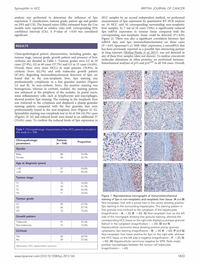
Clinicopathological patient characteristics, including gender, age,tumour stage, tumour grade, growth pattern and presence of livercirrhosis, are detailed in Table 1. Tumour grades were G1 in 29cases (27.9%), G2 in 60 cases (57.7%) and G3 in 15 cases (14.4%).Overall, there were more HCCs in male patients (76.9%), incirrhotic livers (62.5%) and with trabecular growth pattern(87.4%). Regarding immunohistochemical detection of Spn, wefound that in the non-neoplastic liver, Spn staining waspredominantly cytoplasmic in a fine granular manner (Figures1A and B). In non-cirrhotic livers, the positive staining washomogenous, whereas in cirrhotic nodules, the staining patternwas enhanced at the periphery of the nodules. In portal tracts,some inflammatory cells, such as lymphocytes and macrophages,showed positive Spn staining. The staining in the neoplastic liverwas restricted to the cytoplasm and displayed a plump granularstaining pattern compared with the fine granules that werepredominantly found in the non-neoplastic liver (Figures 1C–E).Spinophilin staining was completely lost in 44 of 104 (42.3%) cases(Figures 1F–H) and reduced levels were found in an additional 37(35.6%) cases. To confirm the reduced levels of Spn expression in
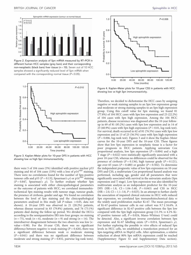
HCC samples by an second independent method, we performedmeasurement of Spn expression by quantitative RT–PCR analysison 10 HCC and 10 corresponding surrounding non-neoplasticliver samples. In 7 out of 10 cases (70%), a significantly reducedSpn mRNA expression in tumour tissue compared with thecorresponding non-neoplastic tissue could be detected (Po0.05;Figure 2). There was also a significant correlation between SpnmRNA data and Spn immunohistochemistry on these cases(P¼ 0.03, Spearman’s r). MiR-106a* expression, a microRNA thathas been previously reported as a possible Spn-interacting partnerin lung tumours (Molina-Pinelo et al, 2011), was not detected inany of these liver samples (data not shown). To analyse concurrentmolecular alterations in other proteins, we performed immuno-histochemical analyses of p53 and p14ARF in all 104 cases. Overall,
Table 1. Clinicopathologic characteristics of the HCC patients included inthis study (n¼ 104)
Clinicopathologicparameters
Patients(n¼104)
Proportion
Gender
Male 80 76.9%Female 24 23.1%
Age at diagnosis (years)
o65 57 54.8%X65 47 45.2%
Tumour stage
T1 15 14.4%T2 33 31.7%T3 45 43.3%T4 11 10.6%
Tumour grade
G1 29 27.9%G2 60 57.7%G3 15 14.4%
Growth pattern
Trabecular 91 87.6%Non-trabecular 13 12.4%
Cirrhosis
Yes 65 62.5%No 39 37.5%
Abbreviation: HCC, hepatocellular carcinoma.
Figure 1. Representative micrographs of immunohistochemicalstaining of Spn in non-neoplastic and neoplastic liver tissue. (A and B)Non-neoplastic liver with a portal tract in the centre showing positiveSpn staining in the surrounding hepatocytes. The staining pattern isfine granular and confined to the cytoplasm of the hepatocytes(magnification – A: �20; B: � 40). (C) Non-neoplastic liver on the leftside of the micrograph showing fine granular staining, whereas thecorresponding HCC tissue on the right side displays a plumper granularfashion in the cytoplasm (magnification – �20). (D and E)Hepatocellular carcinoma tissue showing positive plump granularcytoplasmic Spn staining (magnification – D: � 40; E: �60). (F and G)Non-neoplastic liver tissue positive for Spn on the right side, whereasthe HCC tissue on the left side is negative (magnification – F: � 20; G:�40). (H) Hepatocellular carcinoma negative for SPN. Note singlepositive macrophages between the tumour cell trabeculae(magnification – � 60).
Spinophilin in HCC BRITISH JOURNAL OF CANCER
www.bjcancer.com | DOI:10.1038/bjc.2013.165 1833
there were 5 of 104 cases (5%) identified with positive nuclear p53staining and 44 of 104 cases (33%) with a loss of p14ARF staining.There were no correlations found for the number of Spn-positivetumour cells and p53 (P¼ 0.132, Spearman’s r) or p14ARF staining(P¼ 0.847, Spearman’s r). To further evaluate whether Spnstaining is associated with either clinicopathological parametersor the outcome of patients with HCC, we correlated immunohis-tochemical Spn staining results with tumour stage, tumour grade,the presence of cirrhosis, gender and age. We found no correlationbetween the level of Spn staining and the clinicopathologicalparameters analysed in this study (all P-values 40.05, data notshown). A 10-year DFS was observed in 21 (20.2%) patients,whereas disease recurred in 83 (79.8%) patients, and 76 (73.1%)patients died during the follow-up period. We divided the HCCsaccording to the semiquantitative IRS into four groups: no staining(n¼ 75), weak (n¼ 6), moderate (n¼ 9) and strong (n¼ 14). Theinterobserver disagreement between the two observers was low (k-value¼ 0.98). For the 10-year CSS, there was no significantdifference between negative vs weak staining (P¼ 0.426), there wasa significant difference between weak vs moderate staining(P¼ 0.041) and there was no significant difference betweenmoderate and strong staining (P¼ 0.832, pairwise log-rank tests).
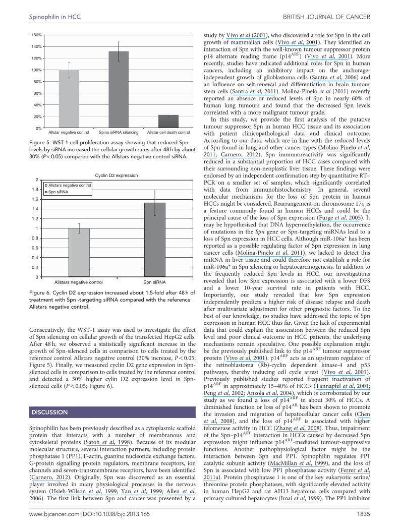
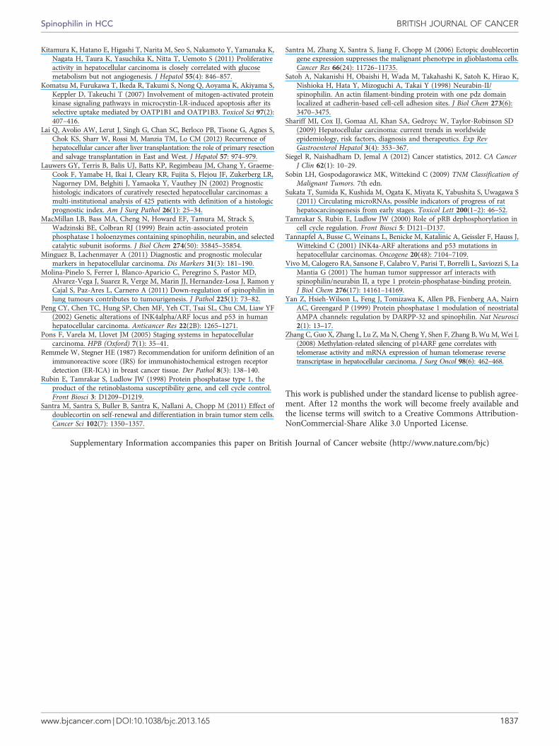
Therefore, we decided to dichotomise the HCC cases by assigningnegative or weak staining samples to an Spn low expression groupand moderate or strong staining samples to an Spn high expressiongroup. Using this cutoff value for Spn staining, we found 81(77.8%) of 104 HCC cases with Spn low expression and 23 (22.2%)of 104 cases with Spn high expression. Among the 104 HCCpatients, disease recurrence was diagnosed after the 10-year follow-up in 69 of 81 (85.2%) cases with Spn low expression and in 14 of23 (60.9%) cases with Spn high expression (P¼ 0.01, log-rank test).For survival, death occurred in 62 of 81 (76.5%) cases with Spn lowexpression and in 13 of 23 (56.5%) cases with Spn high expression(P¼ 0.006, log-rank test). Figures 3 and 4 show the Kaplan–Meiercurves for the 10-year DFS and the 10-year CSS. These figuresshow that low Spn expression in neoplastic tissue is a factor forpoor prognosis in HCC patients. Applying univariate Coxproportional analysis, low Spn expression (P¼ 0.004) and a highT stage (P¼ 0.022) were statistically significantly associated with apoor 10-year CSS, whereas no differences could be observed for thepresence of cirrhosis (P¼ 0.136), high tumour grade (P¼ 0.121),age over 65 years (P¼ 0.480) or gender (P¼ 0.502). To determinethe independent prognostic value of low Spn expression on 10-yearDFS and CSS, a multivariate Cox proportional hazard analysis wasperformed, including age, gender and all parameters that weresignificantly associated with survival in the univariate analysis (Spnexpression and T stage). Low Spn expression was also identified bymultivariate analysis as an independent predictor for the 10-yearDFS (HR¼ 1.8, CI¼ 1.04–3.40, P¼ 0.043) and CSS in HCC(HR¼ 2.0, CI¼ 1.1–3.8, P¼ 0.025). In an attempt to further clarifythe association of impaired clinical outcome in HCC with reducedSpn expression, we correlated Spn immunohistochemical data vsthe widely used proliferation marker Ki-67. The mean percentageof Ki-67-positive tumour cells in our cohort was 9.7±16.6%. Asignificant difference in Ki-67-positive cells between the Spn lowcompared with the Spn high expression group (11.5% vs 4.1% Ki-67-positive tumour cell, P¼ 0.024, Mann–Whitney U-test) couldbe detected. Also, a significant inverse correlation between Spnexpression and Ki-67 labelling exists (P¼ 0.014, Spearman’s r).For further exploring the possible in vitro effects of reduced Spnlevels in HCC cells, we established a transfection protocol for anSpn-targeting siRNA in HepG2 cells. After optimisation, a relativereduction of about 80% Spn mRNA expression could be reached(Supplementary Figure S1 and Supplementary Data section).
0
0.2
0.4
Rel
ativ
e ge
ne e
xpre
ssio
n
0.6
0.8
1
1.2
1.4
1.6
1.8
2TumourNormal
Spinophilin mRNA expression (tumour vs normal tissue)
Figure 2. Expression analysis of Spn mRNA measured by RT–PCR indifferent human HCC samples (grey bars) and their correspondingnon-neoplastic (black bars) liver tissue (n¼10). Seven out of 10 HCCsamples showed a significantly reduced level of Spn mRNA whencompared with the corresponding normal tissue (Po0.05).
0.00 20.00 40.00 60.00 80.00
Time (months)
Low spinophilin expression
High spinophilin expression
P=0.0101.0
0.8
0.6
0.4
0.2
0.0
Pro
babi
lity
to d
isea
se-f
ree
surv
ival
100.00 120.00
Figure 3. Kaplan–Meier plots for 10-year DFS in patients with HCCshowing low vs high Spn immunoreactivity.
0.00 20.00 40.00 60.00 80.00Time (months)
Low spinophilin expression
High spinophilin expression
P=0.0061.0
0.8
0.6
0.4
0.2
0.0
Pro
babi
lity
to s
urvi
ve 1
0 ye
ars
100.00 120.00
Figure 4. Kaplan–Meier plots for 10-year CSS in patients with HCCshowing low vs high Spn immunoreactivity.
BRITISH JOURNAL OF CANCER Spinophilin in HCC
1834 www.bjcancer.com | DOI:10.1038/bjc.2013.165
Consecutively, the WST-1 assay was used to investigate the effectof Spn silencing on cellular growth of the transfected HepG2 cells.After 48 h, we observed a statistically significant increase in thegrowth of Spn-silenced cells in comparison to cells treated by thereference control Allstars negative control (30% increase, Po0.05;Figure 5). Finally, we measured cyclin D2 gene expression in Spn-silenced cells in comparison to cells treated by the reference controland detected a 50% higher cylin D2 expression level in Spn-silenced cells (Po0.05; Figure 6).
DISCUSSION
Spinophilin has been previously described as a cytoplasmic scaffoldprotein that interacts with a number of membranous andcytoskeletal proteins (Satoh et al, 1998). Because of its modularmolecular structure, several interaction partners, including proteinphosphatase 1 (PP1), F-actin, guanine nucleotide exchange factors,G-protein signalling protein regulators, membrane receptors, ionchannels and seven-transmembrane receptors, have been identified(Carnero, 2012). Originally, Spn was discovered as an essentialplayer involved in many physiological processes in the nervoussystem (Hsieh-Wilson et al, 1999; Yan et al, 1999; Allen et al,2006). The first link between Spn and cancer was presented by a
study by Vivo et al (2001), who discovered a role for Spn in the cellgrowth of mammalian cells (Vivo et al, 2001). They identified aninteraction of Spn with the well-known tumour suppressor proteinp14 alternate reading frame (p14ARF) (Vivo et al, 2001). Morerecently, studies have indicated additional roles for Spn in humancancers, including an inhibitory impact on the anchorage-independent growth of glioblastoma cells (Santra et al, 2006) andan influence on self-renewal and differentiation in brain tumourstem cells (Santra et al, 2011). Molina-Pinelo et al (2011) recentlyreported an absence or reduced levels of Spn in nearly 60% ofhuman lung tumours and found that the decreased Spn levelscorrelated with a more malignant tumour grade.
In this study, we provide the first analysis of the putativetumour suppressor Spn in human HCC tissue and its associationwith patient clinicopathological data and clinical outcome.According to our data, which are in line with the reduced levelsof Spn found in lung and other cancer types (Molina-Pinelo et al,2011; Carnero, 2012), Spn immunoreactivity was significantlyreduced in a substantial proportion of HCC cases compared withtheir surrounding non-neoplastic liver tissue. These findings wereendorsed by an independent confirmation step by quantitative RT–PCR on a smaller set of samples, which significantly correlatedwith data from immunohistochemistry. In general, severalmolecular mechanisms for the loss of Spn protein in humanHCCs might be considered. Rearrangement on chromosome 17q isa feature commonly found in human HCCs and could be theprincipal cause of the loss of Spn expression (Furge et al, 2005). Itmay be hypothesised that DNA hypermethylation, the occurrenceof mutations in the Spn gene or Spn-targeting miRNAs lead to aloss of Spn expression in HCC cells. Although miR-106a* has beenreported as a possible regulating factor of Spn expression in lungcancer cells (Molina-Pinelo et al, 2011), we lacked to detect thismiRNA in liver tissue and could therefore not establish a role formiR-106a* in Spn silencing or hepatocarcinogenesis. In addition tothe frequently reduced Spn levels in HCC, our investigationsrevealed that low Spn expression is associated with a lower DFSand a lower 10-year survival rate in patients with HCC.Importantly, our study revealed that low Spn expressionindependently predicts a higher risk of disease relapse and deathafter multivariate adjustment for other prognostic factors. To thebest of our knowledge, no studies have addressed the topic of Spnexpression in human HCC thus far. Given the lack of experimentaldata that could explain the association between the reduced Spnlevel and poor clinical outcome in HCC patients, the underlyingmechanisms remain speculative. One possible explanation mightbe the previously published link to the p14ARF tumour suppressorprotein (Vivo et al, 2001). p14ARF acts as an upstream regulator ofthe retinoblastoma (Rb)-cyclin dependent kinase-4 and p53pathways, thereby inducing cell cycle arrest (Vivo et al, 2001).Previously published studies reported frequent inactivation ofp14ARF in approximately 15–40% of HCCs (Tannapfel et al, 2001;Peng et al, 2002; Anzola et al, 2004), which is corroborated by ourstudy as we found a loss of p14ARF in about 30% of HCCs. Adiminished function or loss of p14AR has been shown to promotethe invasion and migration of hepatocellular cancer cells (Chenet al, 2008), and the loss of p14ARF is associated with highertelomerase activity in HCC (Zhang et al, 2008). Thus, impairmentof the Spn–p14ARF interaction in HCCs caused by decreased Spnexpression might influence p14ARF-mediated tumour-suppressivefunctions. Another pathophysiological factor might be theinteraction between Spn and PP1. Spinophilin regulates PP1catalytic subunit activity (MacMillan et al, 1999), and the loss ofSpn is associated with low PP1 phosphatase activity (Ferrer et al,2011a). Protein phosphatase 1 is one of the key eukaryotic serine/threonine protein phosphatases, with significantly elevated activityin human HepG2 and rat AH13 hepatoma cells compared withprimary cultured hepatocytes (Imai et al, 1999). The PP1 inhibitor
160%
140%
120%
100%
80%
60%
40%
20%
0%Allstar negative control Spino siRNA silencing Allstar cell death control
Figure 5. WST-1 cell proliferation assay showing that reduced Spnlevels by siRNA increased the cellular growth rates after 48 h by about30% (Po0.05) compared with the Allstars negative control siRNA.
0Allstars negative control
Allstars negative control
Cyclin D2 expression
Spn siRNA
Spn siRNA
0.2
0.4
0.6
0.8
1
1.2
1.4
1.6
1.8
2
Figure 6. Cyclin D2 expression increased about 1.5-fold after 48 h oftreatment with Spn -targeting siRNA compared with the referenceAllstars negative control.
Spinophilin in HCC BRITISH JOURNAL OF CANCER
www.bjcancer.com | DOI:10.1038/bjc.2013.165 1835

microcystin-LR selectively induces liver damage and promoteshepatocarcinogenesis (Komatsu et al, 2007). Protein phosphatase 1inhibition in the liver cancer cell line Huh-7 modulates oncogenicRNA alternative splicing to devitalise the cancer cells (Chang et al,2011). Taken together, reduced expression of Spn might lead toaltered PP1 activities that, in turn, would affect PP1-mediatedfunctions in HCC. Interestingly, PP1 is also involved in the mitoticdephosphorylation of phosphorylated Rb protein (Tamrakar et al,2000), as well as in the dephosphorylation of specific residues ofp53 (Rubin et al, 1998), thereby regulating the control of cell cycleprogression (Rubin et al, 1998). A recently published study byFerrer et al (2011b) showed that the loss of Spn reduced PP1activity, resulting in a high level of proproliferative phosphorylatedRb, which in turn leads to a compensatory increase in p14ARF andp53 activity. However, in the absence of p53 or mutated p53,reduced levels of Spn enhanced the tumorigenic potential of thecells, whereas ectopic overexpression of Spn in these humantumour cells greatly reduced cell growth (Ferrer et al, 2011b).Because the nuclear accumulation of p53 largely correlates withmutated p53, we tested whether the loss of Spn staining iscorrelated with nuclear p53 staining but found no significantcorrelation between Spn and p53 or p14ARF immunohistochem-istry. Overall, we found rather low numbers of potential p53-mutated HCC cases in our cohort (5%). As shown previously, theincidence of p53 gene abnormalities in HCC varies in differentgeographical areas, which is due to different underlying environ-mental and genetic risk factors, resulting in less frequentlyencountered p53 mutations in Europe (Boix-Ferrero et al, 1999).Although the mechanisms discussed above could be considered aspotentially involved in HCC, we could not establish any correlationbetween p14ARF or p53 expression and Spn immunoreactivity.Thus, other factors might also be relevant for the pathophysiolocialrole of reduced levels of Spn in tumour progression of HCC.However, these factors need further experimental and clinicalvalidation before a p53-independent tumorigenic role of Spn inHCC could be broadly accepted. Recently, Carnero (2012) reportedthat Spn knockout mice had decreased lifespan with increasedcellular proliferation in different tissues. To explore a possiblepathophysiological role of reduced Spn expression in HCC, wemeasured the widely used proliferation marker Ki-67 on the wholecohort to determine the proliferative activity (Kitamura et al,2011). Overall, we detected a significant inverse correlationbetween Spn expression and proliferative activity in HCC.Therefore, we established an siRNA-based Spn silencing approachto study the effects of reduced Spn levels in tumour cells. Thetransfected cells showed a significant increase of cellular growthand an increased expression of the proliferation marker cyclin D2when Spn mRNA was silenced. These in vivo and in vitro datasupport the hypothesis that a reduced Spn expression might act asa growth-promoting factor in HCC.
In summary, we identified that a substantial number of humanHCCs show reduced or absent Spn immunoreactivity and mRNAexpression, a finding that is compatible with the previouslyobserved loss of Spn in lung adenocarcinoma and other types ofcancer. The low expression of Spn in tumour tissue is anindependent negative prognostic factor for clinical outcome inHCC patients. Spinophilin expression inversely correlates withproliferative activity in vivo and cellular growth in vitro. Furtherpreclinical studies are warranted for the validation of Spn as anovel prognostic biomarker in HCC.
ACKNOWLEDGEMENTS
This study was supported by grants from the Science Funds of theStyrian Government (to AA and MP), of the Hans und Blanca
Moser Foundation (to FE) and from the START Research Initiativeof the Medical University of Graz (to MP). The research leading tothese results has received support from the Innovative MedicinesInitiative Joint Undertaking under grant agreement no. 115234,resources of which are composed of financial contribution from theEuropean Union’s Seventh Framework Programme (FP7/2007–2013) and EFPIA companies’ in kind contribution (to JH).
CONFLICT OF INTEREST
The authors declare no conflict of interest.
REFERENCES
Aigelsreiter A, Haybaeck J, Schauer S, Kiesslich T, Bettermann K,Griessbacher A, Stojakovic T, Bauernhofer T, Samonigg H, Kornprat P,Lackner C, Pichler M (2012) NEMO expression in human hepatocellularcarcinoma and its association with clinical outcome. Hum Pathol 43(7):1012–1019.
Aigelsreiter A, Janig E, Sostaric J, Pichler M, Unterthor D, Halasz J, Lackner C,Zatloukal K, Denk H (2009) Clusterin expression in cholestasis, hepato-cellular carcinoma and liver fibrosis. Histopathology 54(5): 561–570.
Allen PB, Zachariou V, Svenningsson P, Lepore AC, Centonze D, Costa C,Rossi S, Bender G, Chen G, Feng J, Snyder GL, Bernardi G, Nestler EJ, YanZ, Calabresi P, Greengard P (2006) Distinct roles for spinophilin andneurabin in dopamine-mediated plasticity. Neuroscience 140(3): 897–911.
Anzola M, Cuevas N, Lopez-Martinez M, Martinez de Pancorbo M, Burgos JJ(2004) p16INK4A gene alterations are not a prognostic indicator forsurvival in patients with hepatocellular carcinoma undergoing curativehepatectomy. J Gastroenterol Hepatol 19(4): 397–405.
Boix-Ferrero J, Pellin A, Blesa R, Adrados M, Llombart-Bosch A (1999)Absence of p53 gene mutations in hepatocarcinomas from aMediterranean area of Spain. A study of 129 archival tumour samples.Virch Archiv Int J Pathol 434(6): 497–501.
Carnero A (2012) Spinophilin: a new tumor suppressor at 17q21. Curr MolMed 12(5): 528–535.
Chang JG, Yang DM, Chang WH, Chow LP, Chan WL, Lin HH, Huang HD,Chang YS, Hung CH, Yang WK (2011) Small molecule amiloridemodulates oncogenic RNA alternative splicing to devitalize human cancercells. PLoS ONE 6(6): e18643.
Chen YW, Paliwal S, Draheim K, Grossman SR, Lewis BC (2008) P19Arfinhibits the invasion of hepatocellular carcinoma cells by binding toC-terminal binding protein. Cancer Res 68(2): 476–482.
Ferrer I, Blanco-Aparicio C, Peregrina S, Canamero M, Fominaya J, Cecilia Y,Lleonart M, Hernandez-Losa J, Ramon y Cajal S, Carnero A (2011a)Spinophilin acts as a tumor suppressor by regulating Rb phosphorylation.Cell Cycle 10(16): 2751–2762.
Ferrer I, Peregrino S, Canamero M, Cecilia Y, Blanco-Aparicio C, Carnero A(2011b) Spinophilin loss contributes to tumorigenesis in vivo. Cell Cycle10(12): 1948–1955.
Frau M, Biasi F, Feo F, Pascale RM (2010) Prognostic markers and putativetherapeutic targets for hepatocellular carcinoma. Mol Aspects Med 31(2):179–193.
Furge KA, Dykema KJ, Ho C, Chen X (2005) Comparison of array-basedcomparative genomic hybridization with gene expression-based regionalexpression biases to identify genetic abnormalities in hepatocellularcarcinoma. BMC Genom 6: 67.
Guichard C, Amaddeo G, Imbeaud S, Ladeiro Y, Pelletier L, Maad IB,Calderaro J, Bioulac-Sage P, Letexier M, Degos F, Clement B, Balabaud C,Chevet E, Laurent A, Couchy G, Letouze E, Calvo F, Zucman-Rossi J(2012) Integrated analysis of somatic mutations and focal copy-numberchanges identifies key genes and pathways in hepatocellular carcinoma.Nat Genet 44(6): 694–698.
Hsieh-Wilson LC, Allen PB, Watanabe T, Nairn AC, Greengard P (1999)Characterization of the neuronal targeting protein spinophilin and itsinteractions with protein phosphatase-1. Biochemistry 38(14): 4365–4373.
Imai Y, Kakinoki Y, Takizawa N, Nakamura K, Shima H, Kikuchi K (1999)Up-regulation of nuclear PP1alpha and PP1delta in hepatoma cells. Int JOncol 14(1): 121–126.
BRITISH JOURNAL OF CANCER Spinophilin in HCC
1836 www.bjcancer.com | DOI:10.1038/bjc.2013.165
Kitamura K, Hatano E, Higashi T, Narita M, Seo S, Nakamoto Y, Yamanaka K,Nagata H, Taura K, Yasuchika K, Nitta T, Uemoto S (2011) Proliferativeactivity in hepatocellular carcinoma is closely correlated with glucosemetabolism but not angiogenesis. J Hepatol 55(4): 846–857.
Komatsu M, Furukawa T, Ikeda R, Takumi S, Nong Q, Aoyama K, Akiyama S,Keppler D, Takeuchi T (2007) Involvement of mitogen-activated proteinkinase signaling pathways in microcystin-LR-induced apoptosis after itsselective uptake mediated by OATP1B1 and OATP1B3. Toxicol Sci 97(2):407–416.
Lai Q, Avolio AW, Lerut J, Singh G, Chan SC, Berloco PB, Tisone G, Agnes S,Chok KS, Sharr W, Rossi M, Manzia TM, Lo CM (2012) Recurrence ofhepatocellular cancer after liver transplantation: the role of primary resectionand salvage transplantation in East and West. J Hepatol 57: 974–979.
Lauwers GY, Terris B, Balis UJ, Batts KP, Regimbeau JM, Chang Y, Graeme-Cook F, Yamabe H, Ikai I, Cleary KR, Fujita S, Flejou JF, Zukerberg LR,Nagorney DM, Belghiti J, Yamaoka Y, Vauthey JN (2002) Prognostichistologic indicators of curatively resected hepatocellular carcinomas: amulti-institutional analysis of 425 patients with definition of a histologicprognostic index. Am J Surg Pathol 26(1): 25–34.
MacMillan LB, Bass MA, Cheng N, Howard EF, Tamura M, Strack S,Wadzinski BE, Colbran RJ (1999) Brain actin-associated proteinphosphatase 1 holoenzymes containing spinophilin, neurabin, and selectedcatalytic subunit isoforms. J Biol Chem 274(50): 35845–35854.
Minguez B, Lachenmayer A (2011) Diagnostic and prognostic molecularmarkers in hepatocellular carcinoma. Dis Markers 31(3): 181–190.
Molina-Pinelo S, Ferrer I, Blanco-Aparicio C, Peregrino S, Pastor MD,Alvarez-Vega J, Suarez R, Verge M, Marin JJ, Hernandez-Losa J, Ramon yCajal S, Paz-Ares L, Carnero A (2011) Down-regulation of spinophilin inlung tumours contributes to tumourigenesis. J Pathol 225(1): 73–82.
Peng CY, Chen TC, Hung SP, Chen MF, Yeh CT, Tsai SL, Chu CM, Liaw YF(2002) Genetic alterations of INK4alpha/ARF locus and p53 in humanhepatocellular carcinoma. Anticancer Res 22(2B): 1265–1271.
Pons F, Varela M, Llovet JM (2005) Staging systems in hepatocellularcarcinoma. HPB (Oxford) 7(1): 35–41.
Remmele W, Stegner HE (1987) Recommendation for uniform definition of animmunoreactive score (IRS) for immunohistochemical estrogen receptordetection (ER-ICA) in breast cancer tissue. Der Pathol 8(3): 138–140.
Rubin E, Tamrakar S, Ludlow JW (1998) Protein phosphatase type 1, theproduct of the retinoblastoma susceptibility gene, and cell cycle control.Front Biosci 3: D1209–D1219.
Santra M, Santra S, Buller B, Santra K, Nallani A, Chopp M (2011) Effect ofdoublecortin on self-renewal and differentiation in brain tumor stem cells.Cancer Sci 102(7): 1350–1357.
Santra M, Zhang X, Santra S, Jiang F, Chopp M (2006) Ectopic doublecortingene expression suppresses the malignant phenotype in glioblastoma cells.Cancer Res 66(24): 11726–11735.
Satoh A, Nakanishi H, Obaishi H, Wada M, Takahashi K, Satoh K, Hirao K,Nishioka H, Hata Y, Mizoguchi A, Takai Y (1998) Neurabin-II/spinophilin. An actin filament-binding protein with one pdz domainlocalized at cadherin-based cell-cell adhesion sites. J Biol Chem 273(6):3470–3475.
Shariff MI, Cox IJ, Gomaa AI, Khan SA, Gedroyc W, Taylor-Robinson SD(2009) Hepatocellular carcinoma: current trends in worldwideepidemiology, risk factors, diagnosis and therapeutics. Exp RevGastroenterol Hepatol 3(4): 353–367.
Siegel R, Naishadham D, Jemal A (2012) Cancer statistics, 2012. CA CancerJ Clin 62(1): 10–29.
Sobin LH, Gospodagorawicz MK, Wittekind C (2009) TNM Classification ofMalignant Tumors. 7th edn.
Sukata T, Sumida K, Kushida M, Ogata K, Miyata K, Yabushita S, Uwagawa S(2011) Circulating microRNAs, possible indicators of progress of rathepatocarcinogenesis from early stages. Toxicol Lett 200(1–2): 46–52.
Tamrakar S, Rubin E, Ludlow JW (2000) Role of pRB dephosphorylation incell cycle regulation. Front Biosci 5: D121–D137.
Tannapfel A, Busse C, Weinans L, Benicke M, Katalinic A, Geissler F, Hauss J,Wittekind C (2001) INK4a-ARF alterations and p53 mutations inhepatocellular carcinomas. Oncogene 20(48): 7104–7109.
Vivo M, Calogero RA, Sansone F, Calabro V, Parisi T, Borrelli L, Saviozzi S, LaMantia G (2001) The human tumor suppressor arf interacts withspinophilin/neurabin II, a type 1 protein-phosphatase-binding protein.J Biol Chem 276(17): 14161–14169.
Yan Z, Hsieh-Wilson L, Feng J, Tomizawa K, Allen PB, Fienberg AA, NairnAC, Greengard P (1999) Protein phosphatase 1 modulation of neostriatalAMPA channels: regulation by DARPP-32 and spinophilin. Nat Neurosci2(1): 13–17.
Zhang C, Guo X, Zhang L, Lu Z, Ma N, Cheng Y, Shen F, Zhang B, Wu M, Wei L(2008) Methylation-related silencing of p14ARF gene correlates withtelomerase activity and mRNA expression of human telomerase reversetranscriptase in hepatocellular carcinoma. J Surg Oncol 98(6): 462–468.
This work is published under the standard license to publish agree-ment. After 12 months the work will become freely available andthe license terms will switch to a Creative Commons Attribution-NonCommercial-Share Alike 3.0 Unported License.
Supplementary Information accompanies this paper on British Journal of Cancer website (http://www.nature.com/bjc)
Spinophilin in HCC BRITISH JOURNAL OF CANCER
www.bjcancer.com | DOI:10.1038/bjc.2013.165 1837