Long-term safety and feasibility of three-vessel multimodality intravascular imaging in patients...
12
ORIGINAL PAPER Long-term safety and feasibility of three-vessel multimodality intravascular imaging in patients with ST-elevation myocardial infarction: the IBIS-4 (integrated biomarker and imaging study) substudy Masanori Taniwaki • Maria D. Radu • Hector M. Garcia-Garcia • Dik Heg • Henning Kelbæk • Lene Holmvang • Aris Moschovitis • Stephane Noble • Giovanni Pedrazzini • Kari Saunama ¨ki • Jouke Dijkstra • Ulf Landmesser • Peter Wenaweser • Bernhard Meier • Giulio G. Stefanini • Marco Roffi • Thomas F. Lu ¨ scher • Stephan Windecker • Lorenz Ra ¨ber Received: 24 November 2014 / Accepted: 22 February 2015 Ó Springer Science+Business Media Dordrecht 2015 Abstract We assessed the feasibility and the procedural and long-term safety of intracoronary (i.c) imaging for documentary purposes with optical coherence tomography (OCT) and intravascular ultrasound (IVUS) in patients with acute ST-elevation myocardial infarction (STEMI) under- going primary PCI in the setting of IBIS-4 study. IBIS4 (NCT00962416) is a prospective cohort study conducted at five European centers including 103 STEMI patients who underwent serial three-vessel coronary imaging during primary PCI and at 13 months. The feasibility parameter was successful imaging, defined as the number of pullbacks suitable for analysis. Safety parameters included the fre- quency of peri-procedural complications, and major adverse cardiac events (MACE), a composite of cardiac death, myocardial infarction (MI) and any clinically-indi- cated revascularization at 2 years. Clinical outcomes were compared with the results from a cohort of 485 STEMI patients undergoing primary PCI without additional imag- ing. Imaging of the infarct-related artery at baseline (and follow-up) was successful in 92.2 % (96.6 %) of patients using OCT and in 93.2 % (95.5 %) using IVUS. Imaging of the non-infarct-related vessels was successful in 88.7 % (95.6 %) using OCT and in 90.5 % (93.3 %) using IVUS. Periprocedural complications occurred \ 2.0 % of OCT and none during IVUS. There were no differences throughout 2 years between the imaging and control group in terms of MACE (16.7 vs. 13.3 %, adjusted HR1.40, 95 % CI 0.77–2.52, p = 0.27). Multi-modality three-vessel i.c. imaging in STEMI patients undergoing primary PCI is consistent a high degree of success and can be performed safely without impact on cardiovascular events at long- term follow-up. The IBIS4 trial was supported by the Swiss National Science Foundation, and is registered at: http://www.clinicaltrials.gov/ct2/show/NCT00617084. Electronic supplementary material The online version of this article (doi:10.1007/s10554-015-0631-0) contains supplementary material, which is available to authorized users. M. Taniwaki Á A. Moschovitis Á P. Wenaweser Á B. Meier Á G. G. Stefanini Á S. Windecker Á L. Ra ¨ber (&) Department of Cardiology, Swiss Cardiovascular Center Bern, Bern University Hospital, 3010 Bern, Switzerland e-mail: [email protected] M. D. Radu Á H. Kelbæk Á L. Holmvang Á K. Saunama ¨ki Rigshospitalet, Copenhagen, Denmark H. M. Garcia-Garcia Erasmus MC, Rotterdam, The Netherlands D. Heg Clinical Trial Unit, University of Bern, Bern, Switzerland S. Noble Á M. Roffi University Hospital, Geneva, Switzerland G. Pedrazzini Cardiocentro, Lugano, Switzerland J. Dijkstra Leiden University Medical Center, Leiden, Netherlands U. Landmesser Á T. F. Lu ¨scher University Heart Center, Cardiology, University Hospital Zurich, Zurich, Switzerland 123 Int J Cardiovasc Imaging DOI 10.1007/s10554-015-0631-0
-
Upload
independent -
Category
Documents
-
view
3 -
download
0
Transcript of Long-term safety and feasibility of three-vessel multimodality intravascular imaging in patients...
ORIGINAL PAPER
Long-term safety and feasibility of three-vessel multimodalityintravascular imaging in patients with ST-elevation myocardialinfarction: the IBIS-4 (integrated biomarker and imaging study)substudy
Masanori Taniwaki • Maria D. Radu • Hector M. Garcia-Garcia •
Dik Heg • Henning Kelbæk • Lene Holmvang • Aris Moschovitis •
Stephane Noble • Giovanni Pedrazzini • Kari Saunamaki • Jouke Dijkstra •
Ulf Landmesser • Peter Wenaweser • Bernhard Meier • Giulio G. Stefanini •
Marco Roffi • Thomas F. Luscher • Stephan Windecker • Lorenz Raber
Received: 24 November 2014 / Accepted: 22 February 2015
� Springer Science+Business Media Dordrecht 2015
Abstract We assessed the feasibility and the procedural
and long-term safety of intracoronary (i.c) imaging for
documentary purposes with optical coherence tomography
(OCT) and intravascular ultrasound (IVUS) in patients with
acute ST-elevation myocardial infarction (STEMI) under-
going primary PCI in the setting of IBIS-4 study. IBIS4
(NCT00962416) is a prospective cohort study conducted at
five European centers including 103 STEMI patients who
underwent serial three-vessel coronary imaging during
primary PCI and at 13 months. The feasibility parameter
was successful imaging, defined as the number of pullbacks
suitable for analysis. Safety parameters included the fre-
quency of peri-procedural complications, and major
adverse cardiac events (MACE), a composite of cardiac
death, myocardial infarction (MI) and any clinically-indi-
cated revascularization at 2 years. Clinical outcomes were
compared with the results from a cohort of 485 STEMI
patients undergoing primary PCI without additional imag-
ing. Imaging of the infarct-related artery at baseline (and
follow-up) was successful in 92.2 % (96.6 %) of patients
using OCT and in 93.2 % (95.5 %) using IVUS. Imaging
of the non-infarct-related vessels was successful in 88.7 %
(95.6 %) using OCT and in 90.5 % (93.3 %) using IVUS.
Periprocedural complications occurred\2.0 % of OCT and
none during IVUS. There were no differences throughout
2 years between the imaging and control group in terms of
MACE (16.7 vs. 13.3 %, adjusted HR1.40, 95 % CI
0.77–2.52, p = 0.27). Multi-modality three-vessel i.c.
imaging in STEMI patients undergoing primary PCI is
consistent a high degree of success and can be performed
safely without impact on cardiovascular events at long-
term follow-up.
The IBIS4 trial was supported by the Swiss National Science
Foundation, and is registered at:
http://www.clinicaltrials.gov/ct2/show/NCT00617084.
Electronic supplementary material The online version of thisarticle (doi:10.1007/s10554-015-0631-0) contains supplementarymaterial, which is available to authorized users.
M. Taniwaki � A. Moschovitis � P. Wenaweser � B. Meier �G. G. Stefanini � S. Windecker � L. Raber (&)
Department of Cardiology, Swiss Cardiovascular Center Bern,
Bern University Hospital, 3010 Bern, Switzerland
e-mail: [email protected]
M. D. Radu � H. Kelbæk � L. Holmvang � K. Saunamaki
Rigshospitalet, Copenhagen, Denmark
H. M. Garcia-Garcia
Erasmus MC, Rotterdam, The Netherlands
D. Heg
Clinical Trial Unit, University of Bern, Bern, Switzerland
S. Noble � M. Roffi
University Hospital, Geneva, Switzerland
G. Pedrazzini
Cardiocentro, Lugano, Switzerland
J. Dijkstra
Leiden University Medical Center, Leiden, Netherlands
U. Landmesser � T. F. Luscher
University Heart Center, Cardiology, University Hospital Zurich,
Zurich, Switzerland
123
Int J Cardiovasc Imaging
DOI 10.1007/s10554-015-0631-0
Keywords Optical coherence tomography � Intravascular
ultrasound � ST-segment elevation myocardial infarction �Long-term safety
Abbreviations and acronyms
ARC Academic research consortium
CI Confidence interval
DES Drug-eluting stent
MACE Major adverse cardiac event
MI Myocardial infarction
ST Stent thrombosis
TLF Target lesion failure
TLR Target lesion revascularization
IVUS Intravascular ultrasound
IVUS-VH IVUS virtual histology
OCT Optical coherence tomography
PCI Percutaneous coronary intervention
STEMI ST-elevation myocardial infarction
Introduction
Multimodality intracoronary (i.c.) imaging using optical
coherence tomography (OCT), grayscale intravascular ul-
trasound (IVUS), and IVUS-virtual histology (IVUS-VH)
provides means to characterize coronary atherosclerosis
and optimize stent implantation in a comprehensive man-
ner. Based on near-infrared light, OCT visualizes in great
detail the vascular luminal integrity, thrombus, and char-
acteristics of plaque vulnerability and facilitates the de-
tection of geographical miss, stent strut malapposition and
edge dissections following stent implantation [1]. Although
the resolution of IVUS is an order of magnitude lower than
OCT, the technology provides a greater penetration depth
and therefore the ability to assess plaque volume [2].
IVUS-VH is based on radiofrequency analysis and has
been proposed as a method to assist the evaluation of
plaque composition, particularly necrotic core [3]. The
combined use of these sound- and light-based modalities
theoretically increases the accuracy to detect ‘‘vulnerable’’
plaques [4, 5], and may thus provide a complete platform to
exploit future treatment targets. Additionally, these tech-
nologies have the potential to improve cardiovascular
outcomes by optimizing percutaneous coronary interven-
tion (PCI), particularly in STEMI patients [6].
Nevertheless, the use of intracoronary imaging catheters
may be accompanied by procedural complications includ-
ing injury of the endothelium, which may increase the risk
of short- and long-term events. Despite an increasing use of
intracoronary imaging in daily practice, there is insufficient
data on the feasibility and safety of multimodality imaging.
We therefore aimed to analyse the feasibility and short- and
long-term safety of patients enrolled in the IBIS-4 imaging
study, a prospective, observational multicenter study with
the objective to assess the arterial healing after biolimus-
eluting stent implantation versus bare metal stent implan-
tation and to investigate the changes in atherosclerosis on
the background of a high intensity statin therapy in the two
non-infarct related arteries. [7].
Methods
Study population
IBIS 4 (NCT00962416) is a prospective cohort study nested
into the COMFORTABLE-AMI, a trial comparing the safety
and efficacy of a biolimus-eluting stent with a bare metal
stent in 1161 STEMI patients undergoing primary PCI at 11
international centers [8]. A total of 103 patients were en-
rolled in this imaging study using OCT, IVUS and IVUS-VH
to assess the arterial healing response following stent im-
plantation and to quantify changes in atherosclerotic plaque
characteristics in non-infarct related arteries in the presence
of high dose statin therapy (rosuvastatin 40 mg daily).
Patients enrolled in the COMFORTABLE-AMI trial
were eligible for participating in the IBIS 4 study when the
following criteria were met: age\90 years, haemodynamic
stability allowing the administration of nitroglycerine,
preserved renal function (GFR [30 ml/min), CTIMI II
flow of the infarct-related artery at the end of the inter-
vention, and a coronary anatomy considered suitable for
intracoronary imaging (e.g. absence of vessel tortuosity).
Following PCI, multimodality imaging with OCT fol-
lowed by IVUS/IVUS-VH was performed, first addressing
the treated culprit lesion, and then the two non-infarct re-
lated arteries. Care was taken to maintain an activated
clotting time [250 s, and prior to each pullback, i.c. ni-
troglycerine (200 lg) was administered to achieve max-
imal vasodilatation. After complete acquisition, imaging
pullbacks were analyzed offline by an independent Cor-
eLab (Cardialysis B�V., Rotterdam). The study protocol
defined the intracoronary imaging as documentary.
Although intervening physicians were not blinded to the
acquired pullbacks, they were recommended not to perform
any corrective measures based on the imaging findings.
All patients were scheduled for repeat angiography and
the same three vessel multimodality imaging protocol at
13 months. In total, they were followed up to 2 years, and
events were recorded at 30 days, 1 year, and 2 years. Per
protocol, all patients received rosuvastatin 40 mg once
daily throughout the 13 month follow-up period.
The control group of the present study consisted of 485
patients who were simultaneously enrolled in the
Int J Cardiovasc Imaging
123
COMFORTABLE-AMI study at the five imaging centers,
but who did not undergo imaging although they formally
fulfilled the inclusion criteria for the imaging sub-study.
All patients provided written informed consent for the
participation in both the COMFORTABLE-AMI study and
IBIS 4.
OCT
OCT images were acquired using the frequency domain
(FD) C7 console system (Lightlab, St. Jude, Westford, MA,
USA) and the Dragonfly catheter at a pullback speed of
20 mm/sec. Blood clearance was achieved using the non-
obstructive technique with a power injector applying
flushing rates of 3–6 ml/sec depending on vessel size. The
regions of interest included in the culprit vessel: at least
5 mm of the proximal and distal reference vessel segments;
and as much as possible of the proximal non-infarct-related
arteries, though a minimum of 40 mm as measured from
the respective ostia.
IVUS
IVUS-images were acquired with the Volcano s5 system
(Volcano Corp., Rancho Cordova, CA) and the 2.9 French
Eagle Eye catheter, which uses a 20 MHz transducer, at a
continuous motorized pullback rate of 0.5 mm/sec (R-100
pullback device; Volcano Therapeutics). The regions of
interest were the same as for OCT, as were the storage of
pullbacks and transfer to CoreLab for further offline
analysis.
Antithrombotic therapy regimen
All patients were loaded with aspirin (C250 mg) prior to
the procedure. In centers where prasugrel was available, an
initial dose of 60 mg (including patients preloaded with
clopidogrel) was given followed by a daily maintenance
dose of 10 mg. If prasugrel was not available or con-
traindicated, clopidogrel was administered at a loading
dose of 600 mg, followed by 75 mg twice daily for 7 days,
followed by a maintenance dose of 75 mg daily. Dual
antiplatelet therapy was prescribed for the duration of
13 months.
Feasibility parameters
Feasibility was defined as the acquisition of a pullback with
sufficient image quality for independent CoreLab analysis,
e.g. allowing the acquisition of C70 % of the region of
interest and satisfactory blood clearing with visibility of C3
quadrants in C70 % of the pullback length.
Safety parameters
The following procedural complications (directly related to
the imaging or within 24 h of the procedure) were
prospectively collected: coronary artery dissection (angio-
graphic) or vessel perforation, ventricular fibrillation,
symptomatic brady- or tachyarrythmia, thrombus forma-
tion, spasm and air embolism.
The primary clinical safety endpoint was a composite of
cardiac death, myocardial infarction and any clinically
indicated revascularization. Secondary safety endpoints
included death, cardiac death, myocardial infarction, Aca-
demic research consortium (ARC)-defined definite- and
definite or probable stent thrombosis and major bleeding
events according to the Bleeding-ARC (BARC) definition.
All definitions of endpoints used in this study have been
previously reported [9]. Any revascularization refers to all
revascularizations [target-lesion revascularization (TLR),
target-vessel revascularization (TVR), and non-TVR].
Acute renal failure was defined as an absolute increase in
serum creatinine of[1.5 fold from baseline or a glomerular
filtration rate decrease of [25 % within 72 h.
Data management
Independent study monitors verified source data according
to a prespecified monitoring plan. Data were stored in a
central online database. Follow-up appointments were
scheduled at 30 days, 1 and 2 years, and patients were
questioned about the occurrence of angina, any adverse
events. All events were independently adjudicated by a
clinical event committee.
Statistical analysis
Categorical variables are presented as counts with per-
centages and compared using Chi square or Fisher’s tests,
and continuous variables as means with standard deviations
or medians and interquartile ranges, and compared using
the Student t test and Mann–Whitney U-test, respectively.
Lesion-level data were analyzed using general or general-
ized linear mixed models, nesting lesions within patient
identifiers, wherever applicable. Time-to-first event or
composite events were analyzed using Cox’s regression
analysis comparing the Imaging group and the control
group (at 30 days, 1 and 2 years of follow-up), both crude
(presented with Kaplan–Meier graphs), and adjusted (using
inverse-probability of treatment weighting (IPTW), where
the treatment is the imaging performed). IPTW was cal-
culated using the following baseline variables: age, gender,
body mass index, diabetes, hypertension, hypercholes-
terolemia, current smoker, renal insufficiency, anemia, pain
onset within 6 h, Killip II, resuscitation, left ventricular
Int J Cardiovasc Imaging
123
ejection fraction, treatment of a bifurcation lesion, small
vessel B2.5 mm diameter, long lesion C20 mm length,
SYNTAX score, TIMI flow pre-procedure, after 20 times
multiple imputation of missing data using chained equa-
tions (IPTW Adjusted Cox’s Regressions weight the pa-
tients by their IPTW and are based on 20 imputed data-
sets). Sensitivity crude and IPTW adjusted Cox’s regres-
sion analyses were conducted on the primary outcome,
stratifying patients according to whether they received
clopidogrel loading or prasugrel/double loading during the
index procedure. Similarly, event rates at discharge were
analyzed using Crude or IPTW Adjusted Poisson Regres-
sions with robust error variances and reported as Risk
Ratios RR with 95 % confidence intervals (95 % CI).
p values from Fisher’s tests are reported in case of zero
events. All statistical analyses were performed with Stata
12.1 (StataCorp, Texas, USA) and differences were con-
sidered significant at a = 0.05.
Results
A total of 103 STEMI patients undergoing primary PCI
were included at five European centers into the IBIS 4
imaging study between September 2009 and January 2011.
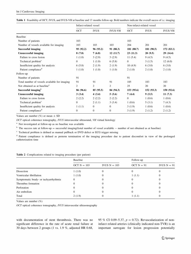
Feasibility of multimodality intracoronary imaging
The feasibility of OCT, grayscale IVUS, and IVUS-VH at
baseline and follow-up imaging is shown in Table 1.
Imaging of the infarct-related artery at baseline (and fol-
low-up) was successful in 92.2 % (96.6 %) patients using
OCT, 93.2 % (95.5 %) patients using grayscale IVUS and
88.3 % (94.3 %) patients using IVUS-VH. Imaging of the
non-infarct related artery at baseline (and follow-up) was
successful in 88.7 % (95.6 %) patients using OCT, 90.5 %
(93.3 %) patients using grayscale IVUS and 83.1 %
(92.6 %) patients using IVUS-VH, respectively. In case an
OCT pullback was successfully acquired, the frequency of
OCT frames not analyzable both at baseline and follow-up
was 0.3 %. Failure of OCT imaging was mainly related to
insufficient image quality due to incomplete vessel flushing
but also due to difficulties to advance the OCT catheter in
the non-infarct related artery. IVUS-VH imaging failure
was mainly caused by technical problems with the ECG
transmission for gating of the acquisition, and by the use of
manual- instead of automatic pullback.
Procedural complications
In the setting of primary PCI, in two out of 103 patients
(1.9 %), the OCT pullback aquisition resulted in a com-
plication at baseline, and in one out of 91 patients (1.1 %)
at follow-up. Specifically, a dissection at the proximal stent
edge was caused by the tip of the OCT imaging catheter in
one of the cases at baseline; whereas the other two com-
plications (one at baseline, one at follow-up) consisted of
ventricular fibrillation induced by vessel flushing with
contrast. Complications were managed by additional stent
implantation in the former case, and successful defibrilla-
tion in the other cases, without further sequelae. IVUS
imaging was performed without complications; and there
were no cases of i.c. related thrombus formation, perfora-
tion, air embolism, or mechanical device failure or spasm
(Table 2).
Safety analysis
A total of 103 imaging patients were compared to a cohort
of 485 control patients.
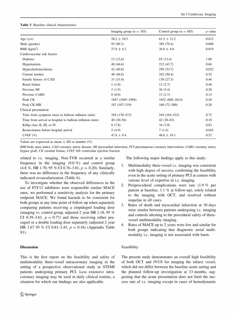
Baseline clinical characteristics of the two groups are
shown in Table 3. Imaging patients were younger, more
frequently male, had a higher BMI and were less likely to
have hypercholesterolemia compared to patients not un-
dergoing i.c. imaging. Imaging patients were more often
resuscitated prior to hospital admission. Prognostically
important characteristics like diabetes, Killip classification
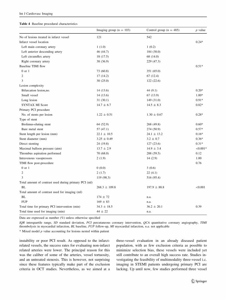
and left ventricular function were similar. Procedural
characteristics are shown in Table 4. Other than a lower
maximal balloon pressure and a higher amount of contrast
used, there was no significant difference between groups.
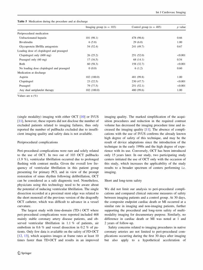
Antithrombotic medication prior or during the procedure
and at hospital discharge is shown in Table 5. Imaging
patients more frequently (58.3 vs. 32.7 %, p \ 0.001) re-
ceived a concomitant loading dose of prasugrel in addition
to clopidogrel and were more frequently discharged with
prasugrel as compared with control patients (77.5 vs.
52.1 %, p \ 0.001), the proportion of patients on double
antiplatelet therapy at discharge, however, was similar.
Short and long term outcomes of patients undergoing
intracoronary imaging
Table 6 presents clinical outcomes at 30 days, 1 and
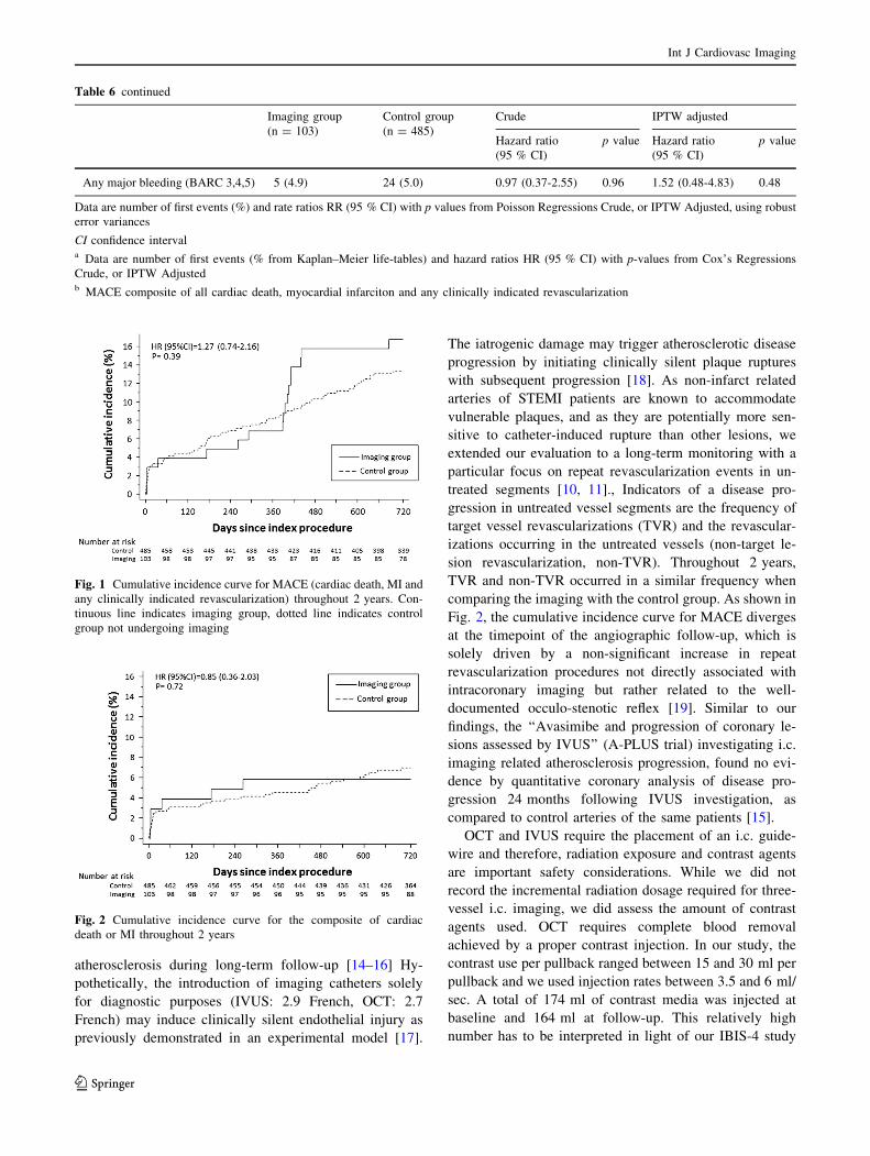
2 years. The primary endpoint MACE was similar for pa-
tients undergoing i.c. imaging and controls at 2 years (16.7
vs. 13.3 %, adjusted HR 1.4, 95 % CI 0.77–2.52, p = 0.27)
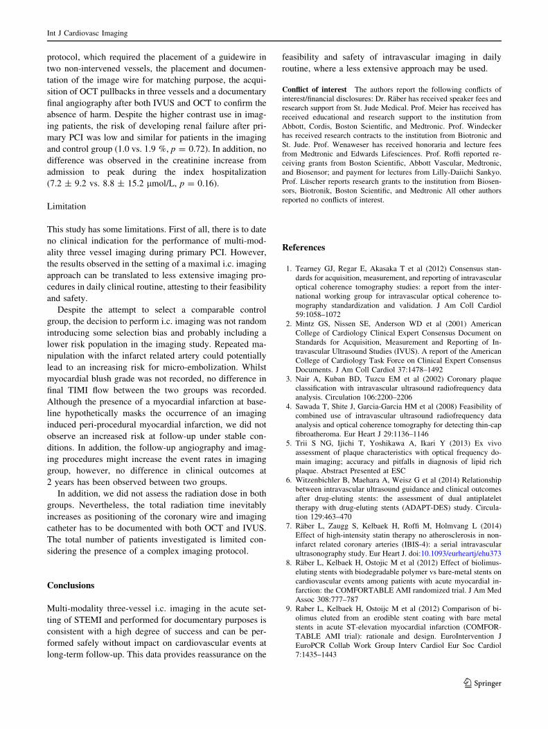
(Fig. 1). Similarly, no differences in the individual end-
points including death, myocardial infarction and any
revascularization were observed at 2 years (Fig. 2). Two
recurrent myocardial infarctions were noted in the imaging
group within 30 days. One event was related to an early
stent thrombosis 4 days after stent implantation in a cal-
cified ostial RCA lesion due to stent underexpansion. The
second event occurred in a patient who was readmitted
with cardiogenic shock due to ischemic mitral regurgitation
Int J Cardiovasc Imaging
123
with documentation of stent thrombosis. There was no
significant difference in the rate of acute renal failure at
30 days between 2 groups (1 vs. 1.9 %, adjusted HR 0.68,
95 % CI 0.09–5.37, p = 0.72). Revascularization of non-
infarct related arteries (clinically indicated non-TVR) is an
important surrogate for lesion progression potentially
Table 1 Feasibility of OCT, IVUS, and IVUS-VH at baseline and 13 months follow-up. Bold numbers indicate the overall sucess of i.c. imaging
Infarct-related vessel Non-infarct-related vessel
OCT IVUS IVUS-VH OCT IVUS IVUS-VH
Baseline
Number of patients 103 103
Number of vessels available for imaging 103 103 103 204 201 201
Successful imaging 95 (92.2) 96 (93.2) 91 (88.3) 181 (88.7) 181 (90.5) 172 (83.1)
Unsuccessful imaging 8 (7.8) 7 (6.8) 12 (11.7) 23 (11.3) 20 (9.5) 29 (14.4)
Failure to cross lesion 1 (1.0) 3 (2.9) 3 (2.9) 11 (5.4) 9 (4.5) 9 (4.5)
Technical problemc 0 1 (1.0) 6 (5.8) 0 3 (1.5) 12 (6.0)
Insufficient quality for analysis 6 (5.8) 2 (1.9) 2 (1.9) 10 (4.9) 6 (3.0) 6 (3.0)
Patient complianced 1 (1.0) 1 (1.0) 1 (1.0) 2 (1.0) 2 (1.0) 2 (1.0)
Follow-up
Number of patients 91 91
Total number of vessels available for imaging 91 91 91 185 183 183
Not obtained as at baselinea 2 2 2 25 20 20
Successful imagingb 86 (96.6) 85 (95.5) 84 (94.3) 153 (95.6) 152 (93.3) 150 (92.6)
Unsuccessful imaging 3 (3.4) 4 (3.4) 5 (5.6) 7 (4.4) 9 (5.5) 11 (7.3)
Failure to cross lesion 2 (2.2) 2 (2.2) 2 (2.2) 0 1 (0.6) 1 (0.6)
Technical problemc 0 2 (1.1) 3 (3.4) 1 (0.6) 5 (3.1) 7 (4.3)
Insufficient quality for analysis 1 (1.1) 0 0 3 (1.9) 1 (0.6) 1 (0.6)
Patient complianced 0 0 0 3 (1.9) 2 (1.2) 2 (1.2)
Values are number (%) or mean ± SD
OCT optical coherence tomography, IVUS intravascular ultrasound, VH virtual histologya Not investigated at follow-up as no baseline was availableb The success rate at follow-up = successful imaging/(total number of vessel available - number of not obtained as at baseline)c Technical problem is defined as manual pullback or DVD defect or ECG trigger missingd Patient compliance is defined as preterm termination of the imaging procedure due to patient discomfort in view of the prolonged
catheterization time
Table 2 Complications related to imaging procedure (per patient)
Baseline Follow-up
OCT N = 103 IVUS N = 103 OCT N = 91 IVUS N = 91
Dissection 1 (1.0) 0 0 0
Ventricular fibrillation 1 (1.0) 0 1 (1.1) 0
Symptomatic brady- or tachyarrhythmia 0 0 0 0
Thrombus formation 0 0 0 0
Perforation 0 0 0 0
Air embolism 0 0 0 0
Total 2 (1.9) 0 1 (1.1) 0
Values are number (%)
OCT optical coherence tomography, IVUS intravascular ultrasonography
Int J Cardiovasc Imaging
123
related to i.c. imaging. Non-TVR occurred at a similar
frequency in the imaging (9.0 %) and control group
(4.8 %, HR 1.70, 95 % CI 0.76–3.81, p = 0.20). Similarly,
there was no difference in the frequency of any clinically
indicated revascularization (Table 6).
To investigate whether the observed differences in the
use of P2Y12 inhibitors were responsible similar MACE
rates, we performed a sensitivity analysis for the primary
endpoint MACE. We found hazards to be consistent for
both groups at any time point of follow-up when separately
comparing patients receiving a clopidogrel loading dose
(imaging vs. control group, adjusted 2 year HR 1.18, 95 %
CI 0.39–3.61, p = 0.77) and those receiving either pra-
sugrel or a double loading dose separately (adjusted 2 year
HR 1.67 95 % CI 0.81–3.43, p = 0.16) (Appendix Table
S1).
Discussion
This is the first report on the feasibility and safety of
multimodality three-vessel intracoronary imaging in the
setting of a prospective observational study in STEMI
patients undergoing primary PCI. Less extensive intra-
coronary imaging may be used in daily clinical routine, a
situation for which our findings are also applicable.
The following major findings apply to this study:
1. Multimodality three-vessel i.c. imaging was consistent
with high degree of success, confirming the feasibility
even in the acute setting of primary PCI at centers with
various level of expertise in i.c. imaging.
2. Periprocedural complications were rare (1.9 % per
patient at baseline, 1.1 % at follow-up), solely related
to the imaging with OCT, and resolved without
sequelae in all cases.
3. Rates of death and myocardial infarction at 30 days
were similar between patients undergoing i.c. imaging
and controls attesting to the procedural safety of three-
vessel multimodality imaging.
4. Rates of MACE up to 2 years were low and similar for
both groups indicating that diagnostic serial multi-
modality i.c. imaging is not associated with harm.
Feasibility
The present study demonstrates an overall high feasibility
of both OCT and IVUS for imaging the infarct vessel,
which did not differ between the baseline acute setting and
the planned follow-up investigation at 13 months, sug-
gesting that the acute presentation does not limit the suc-
cess rate of i.c. imaging except in cases of hemodynamic
Table 3 Baseline clinical characteristics
Imaging group (n = 103) Control group (n = 485) p value
Age (yrs) 58.2 ± 10.5 61.5 ± 12.2 0.012
Male (gender) 93 (90.3) 385 (79.4) 0.008
BMI (kg/m2) 27.8 ± 4.2 26.8 ± 4.0 0.019
Cardiovascular risk factors
Diabetes 13 (12.6) 65 (13.4) 1.00
Hypertension 48 (46.6) 212 (43.7) 0.66
Hypercholesterolemia 42 (40.8) 259 (53.7) 0.022
Current smoker 48 (46.6) 242 (50.4) 0.52
Family history of CAD 31 (31.0) 130 (27.3) 0.46
Renal failure 1 (1.0) 13 (2.7) 0.48
Previous MI 2 (1.9) 26 (5.4) 0.20
Previous CABG 0 (0.0) 13 (2.7) 0.14
Peak CK 1847 (1045–3494) 1652 (800–2814) 0.20
Peak CK-MB 187 (107–319) 160 (72–300) 0.20
Clinical presentation
Time from symptom onset to balloon inflation (min) 258 (170–472) 244 (164–422) 0.72
Time from arrival at hospital to balloon inflation (min) 40 (30–56) 42 (30–63) 0.18
Killip class II, III, or IV 8 (7.8) 34 (7.0) 0.83
Resuscitation before hospital arrival 5 (4.9) 7 (1.4) 0.042
LVEF (%) 47.8 ± 9.4 48.6 ± 10.1 0.52
Values are expressed as mean ± SD or number (%)
BMI body mass index, CAD coronary artery disease, MI myocardial infarction, PCI percutaneous coronary intervention, CABG coronary artery
bypass graft, CK creatine kinase, LVEF left ventricular ejection fraction
Int J Cardiovasc Imaging
123
instability or poor PCI result. As opposed to the infarct-
related vessels, the success rates for evaluating non-infarct
related arteries were lower. The principal reason for this
was the caliber of some of the arteries, vessel tortuosity,
and an untreated stenosis. This is however, not surprising
since these features typically make part of the exclusion
criteria in OCT studies. Nevertheless, as we aimed at a
three-vessel evaluation in an already diseased patient
population, with as few exclusion criteria as possible to
minimize selection bias, these vessels were included yet
still contribute to an overall high success rate. Studies in-
vestigating the feasibility of multimodality three-vessel i.c.
imaging in STEMI patients undergoing primary PCI are
lacking. Up until now, few studies performed three vessel
Table 4 Baseline procedural characteristics
Imaging group (n = 103) Control group (n = 485) p value
No of lesions treated in infarct vessel 121 542
Infarct vessel location 0.24*
Left main coronary artery 1 (1.0) 1 (0.2)
Left anterior descending artery 46 (44.7) 184 (38.0)
Left circumflex artery 18 (17.5) 68 (14.0)
Right coronary artery 38 (36.9) 229 (47.3)
Baseline TIMI flow 0.51*
0 or 1 73 (60.8) 351 (65.0)
2 17 (14.2) 67 (12.4)
3 30 (25.0) 122 (22.6)
Lesion complexity
Bifurcation lesion,no. 14 (13.6) 44 (9.1) 0.20*
Small vessel 14 (13.6) 67 (13.9) 1.00*
Long lesion 31 (30.1) 149 (31.0) 0.91*
SYNTAX MI Score 14.7 ± 6.7 14.5 ± 8.3 0.82*
Primary PCI procedure
No. of stents per lesion 1.22 ± 0.51 1.30 ± 0.67 0.28*
Type of stent
Biolimus-eluting stent 64 (52.9) 268 (49.8) 0.60*
Bare metal stent 57 (47.1) 274 (50.9) 0.57*
Stent length per lesion (mm) 22.1 ± 10.5 24.1 ± 13.2 0.16*
Stent diameter (mm) 3.25 ± 0.49 3.2 ± 0.7 0.36*
Direct stenting 24 (19.8) 127 (23.6) 0.31*
Maximal balloon pressure (atm) 13.7 ± 2.9 14.9 ± 3.4 \0.001*
Thrombus aspiration performed 70 (68.0) 288 (59.5) 0.12
Intravenous vasopressors 2 (1.9) 14 (2.9) 1.00
TIMI flow post-procedure 0.76
0 or 1 0 (0.0) 3 (0.6)
2 2 (1.7) 22 (4.1)
3 119 (98.3) 516 (95.4)
Total amount of contrast used during primary PCI (ml)
BL 268.3 ± 109.8 197.9 ± 88.8 \0.001
Total amount of contrast used for imaging (ml)
BL 174 ± 72 n.a.
FUP 169 ± 83 n.a.
Total time for primary PCI intervention (min) 34.3 ± 18.5 36.2 ± 20.1 0.39
Total time used for imaging (min) 44 ± 22 n.a.
Data are expressed as number (%) unless otherwise specified
IQR interquartile range, SD standard deviation, PCI percutaneous coronary intervention, QCA quantitative coronary angiography, TIMI
thrombolysis in myocardial infarction, BL baseline, FUP follow-up, MI myocardial infarction, n.a. not applicable
* Mixed model p value accounting for lesions nested within patient
Int J Cardiovasc Imaging
123
(single modality) imaging with either OCT [10] or IVUS
[11], however, these reports did not disclose the number of
excluded patients related to imaging failures, thus only
reported the number of pullbacks excluded due to insuffi-
cient imaging quality and safety data is not available.
Periprocedural complications
Peri-procedural complications were rare and solely related
to the use of OCT. In two out of 103 OCT pullbacks
(1.9 %), ventricular fibrillation occurred due to prolonged
flushing with contrast media. Given the overall low fre-
quency of ventricular fibrillation in this patient group
presenting for primary PCI, and in view of the prompt
restoration of sinus rhythm following defibrillation, OCT
can be considered as a safe diagnostic tool. Nonetheless,
physicians using this technology need to be aware about
the potential of inducing ventricular fibrillation. The single
dissection recorded at a proximal stent edge was related to
the short monorail of the previous version of the dragonfly
OCT catheter, which was difficult to advance in a vessel
curvature.
The largest study with time-domain (TD-) OCT where
peri-procedural complications were reported included 468
mainly stable coronary artery disease patients, and ob-
served ventricular fibrillation in 1.1 % of patients, air
embolism in 0.6 % and vessel dissection in 0.2 % of pa-
tients. Only few data is available on the safety of FD-OCT
[12, 13], which acquires images at frame rates at least 10
times faster than TD-OCT and results in an improved
imaging quality. The marked simplification of the acqui-
sition procedures and reduction in the required contrast
volume has decreased the imaging procedure time and in-
creased the imaging quality [11]. The absence of compli-
cations with the use of IVUS confirms the already known
high degree of safety of this technique, and may be the
result of device adaptations since the introduction of the
technique in the early 1990s and the high degree of expe-
rience with its use. Conversely, OCT has been introduced
only 15 years later. In our study, two participating study
centers initiated the use of OCT only with the occasion of
this study, which increases the applicability of the study
results to a broader spectrum of centers performing i.c.
imaging.
Short and long-term safety
We did not limit our analysis to peri-procedural compli-
cations and compared clinical outcome measures of safety
between imaging patients and a control group. At 30 days,
the composite endpoint cardiac death or MI occurred at a
similar rate in imaging and non-imaging patients, further
supporting the procedural and long-term safety of multi-
modality imaging for documentary purpose. Similarly, no
difference in cardiac death or MI was noted at 1 and
2 years of follow-up.
Safety concerns related to imaging procedures in native
coronary arteries are not limited to peri-procedural com-
plications occurring during the first 30 days after imaging
but also apply to a hypothetical acceleration of
Table 5 Medication during the procedure and at discharge
Imaging group (n = 103) Control group (n = 485) p value
Periprocedural medication
Unfractionated heparin 101 (98.1) 478 (98.6) 0.66
Bivalirudin 6 (5.8) 29 (6.0) 1.00
Glycoprotein IIb/IIIa antagonists 54 (52.4) 241 (49.7) 0.67
Loading dose of clopidogrel and prasugrel
Clopidogrel only (600 mg) 26 (25.2) 251 (52.0) \0.001
Prasugrel only (60 mg) 17 (16.5) 68 (14.1) 0.54
Both 60 (58.3) 158 (32.7) \0.001
No loading dose clopidogrel and prasugrel 0 (0.0) 6 (1.2) 0.60
Medication at discharge
Aspirin 102 (100.0) 481 (99.8) 1.00
Clopidogrel 23 (22.5) 230 (47.7) \0.001
Prasugrel 79 (77.5) 251 (52.1) \0.001
Any dual antiplatelet therapy 102 (100.0) 480 (99.6) 1.00
Values are n (%)
Int J Cardiovasc Imaging
123
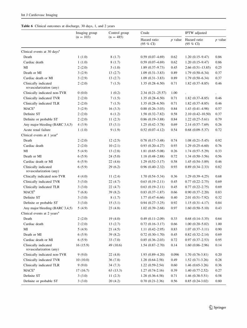
Table 6 Clinical outcomes at discharge, 30 days, 1, and 2 years
Imaging group
(n = 103)
Control group
(n = 485)
Crude IPTW adjusted
Hazard ratio
(95 % CI)
p value Hazard ratio
(95 % CI)
p value
Clinical events at 30 daysa
Death 1 (1.0) 8 (1.7) 0.59 (0.07–4.69) 0.62 1.20 (0.15–9.47) 0.86
Cardiac death 1 (1.0) 8 (1.7) 0.59 (0.07–4.69) 0.62 1.20 (0.15–9.47) 0.86
MI 2 (2.0) 5 (1.0) 1.89 (0.37–9.73) 0.45 2.66 (0.51–13.85) 0.25
Death or MI 3 (2.9) 13 (2.7) 1.09 (0.31–3.83) 0.89 1.79 (0.50–6.34) 0.37
Cardiac death or MI 3 (2.9) 13 (2.7) 1.09 (0.31–3.83) 0.89 1.79 (0.50–6.34) 0.37
Clinically indicated
revascularization (any)
2 (2.0) 7 (1.5) 1.35 (0.28–6.50) 0.71 1.82 (0.37–8.85) 0.46
Clinically indicated non-TVR 0 (0.0) 1 (0.2) 2.34 (0.21–25.57) 1.00 . .
Clinically indicated TVR 2 (2.0) 7 (1.5) 1.35 (0.28–6.50) 0.71 1.82 (0.37–8.85) 0.46
Clinically indicated TLR 2 (2.0) 7 (1.5) 1.35 (0.28–6.50) 0.71 1.82 (0.37–8.85) 0.46
MACEa 3 (2.9) 16 (3.3) 0.88 (0.26–3.03) 0.84 1.43 (0.41–4.98) 0.57
Definite ST 2 (2.0) 6 (1.2) 1.58 (0.32–7.82) 0.58 2.10 (0.42–10.50) 0.37
Definite or probable ST 2 (2.0) 11 (2.3) 0.86 (0.19–3.88) 0.84 1.22 (0.27–5.61) 0.79
Any major bleeding (BARC 3,4,5) 4 (3.9) 15 (3.1) 1.25 (0.42–3.78) 0.69 2.14 (0.57–7.69) 0.26
Acute renal failure 1 (1.0) 9 (1.9) 0.52 (0.07–4.12) 0.54 0.68 (0.09–5.37) 0.72
Clinical events at 1 yeara
Death 2 (2.0) 12 (2.5) 0.78 (0.17–3.48) 0.74 1.08 (0.21–5.45) 0.92
Cardiac death 2 (2.0) 10 (2.1) 0.93 (0.20–4.27) 0.93 1.29 (0.25–6.60) 0.76
MI 5 (4.9) 13 (2.8) 1.81 (0.65–5.08) 0.26 1.74 (0.57–5.29) 0.33
Death or MI 6 (5.9) 24 (5.0) 1.18 (0.48–2.88) 0.72 1.34 (0.50–3.56) 0.56
Cardiac death or MI 6 (5.9) 22 (4.6) 1.29 (0.52–3.17) 0.58 1.45 (0.54–3.89) 0.46
Clinically indicated
revascularization (any)
6 (5.9) 29 (6.2) 0.96 (0.40–2.32) 0.93 0.89 (0.34–2.33) 0.82
Clincally indicated non-TVR 4 (4.0) 11 (2.4) 1.70 (0.54–5.34) 0.36 1.29 (0.39–4.25) 0.68
Clinically indicated TVR 3 (3.0) 22 (4.7) 0.63 (0.19–2.11) 0.45 0.77 (0.22–2.75) 0.69
Clinically indicated TLR 3 (3.0) 22 (4.7) 0.63 (0.19–2.11) 0.45 0.77 (0.22–2.75) 0.69
MACEb 7 (6.8) 39 (8.2) 0.83 (0.37–1.87) 0.66 0.90 (0.37–2.20) 0.83
Definite ST 3 (3.0) 8 (1.7) 1.77 (0.47–6.66) 0.40 2.01 (0.51–7.92) 0.32
Definite or probable ST 3 (3.0) 15 (3.1) 0.94 (0.27–3.25) 0.92 1.15 (0.31–4.17) 0.84
Any major bleeding (BARC 3,4,5) 5 (4.9) 23 (4.8) 1.02 (0.39–2.68) 0.97 1.60 (0.50–5.10) 0.43
Clinical events at 2 yearsa
Death 2 (2.0) 19 (4.0) 0.49 (0.11–2.09) 0.33 0.68 (0.14–3.35) 0.64
Cardiac death 2 (2.0) 13 (2.7) 0.72 (0.16–3.17) 0.66 1.00 (0.20–5.02) 1.00
MI 5 (4.9) 21 (4.5) 1.11 (0.42–2.95) 0.83 1.07 (0.37–3.11) 0.90
Death or MI 6 (5.9) 39 (8.2) 0.72 (0.30–1.70) 0.45 0.82 (0.32–2.14) 0.69
Cardiac death or MI 6 (5.9) 33 (7.0) 0.85 (0.36–2.03) 0.72 0.97 (0.37–2.53) 0.95
Clinically indicated
revascularization (any)
16 (15.9) 49 (10.6) 1.54 (0.87–2.70) 0.14 1.60 (0.86–2.96) 0.14
Clinically indicated non-TVR 9 (9.0) 22 (4.8) 1.93 (0.89–4.20) 0.096 1.70 (0.76-3.81) 0.20
Clinically indicated TVR 10 (10.0) 36 (7.8) 1.28 (0.64-2.58) 0.49 1.52 (0.71-3.26) 0.28
Clinically indicated TLR 9 (9.0) 34 (7.3) 1.22 (0.59-2.54) 0.60 1.46 (0.65-3.26) 0.36
MACEa 17 (16.7) 63 (13.3) 1.27 (0.74-2.16) 0.39 1.40 (0.77-2.52) 0.27
Definite ST 3 (3.0) 11 (2.3) 1.28 (0.36-4.58) 0.71 1.46 (0.38-5.51) 0.58
Definite or probable ST 3 (3.0) 20 (4.2) 0.70 (0.21-2.36) 0.56 0.85 (0.24-3.02) 0.80
Int J Cardiovasc Imaging
123
atherosclerosis during long-term follow-up [14–16] Hy-
pothetically, the introduction of imaging catheters solely
for diagnostic purposes (IVUS: 2.9 French, OCT: 2.7
French) may induce clinically silent endothelial injury as
previously demonstrated in an experimental model [17].
The iatrogenic damage may trigger atherosclerotic disease
progression by initiating clinically silent plaque ruptures
with subsequent progression [18]. As non-infarct related
arteries of STEMI patients are known to accommodate
vulnerable plaques, and as they are potentially more sen-
sitive to catheter-induced rupture than other lesions, we
extended our evaluation to a long-term monitoring with a
particular focus on repeat revascularization events in un-
treated segments [10, 11]., Indicators of a disease pro-
gression in untreated vessel segments are the frequency of
target vessel revascularizations (TVR) and the revascular-
izations occurring in the untreated vessels (non-target le-
sion revascularization, non-TVR). Throughout 2 years,
TVR and non-TVR occurred in a similar frequency when
comparing the imaging with the control group. As shown in
Fig. 2, the cumulative incidence curve for MACE diverges
at the timepoint of the angiographic follow-up, which is
solely driven by a non-significant increase in repeat
revascularization procedures not directly associated with
intracoronary imaging but rather related to the well-
documented occulo-stenotic reflex [19]. Similar to our
findings, the ‘‘Avasimibe and progression of coronary le-
sions assessed by IVUS’’ (A-PLUS trial) investigating i.c.
imaging related atherosclerosis progression, found no evi-
dence by quantitative coronary analysis of disease pro-
gression 24 months following IVUS investigation, as
compared to control arteries of the same patients [15].
OCT and IVUS require the placement of an i.c. guide-
wire and therefore, radiation exposure and contrast agents
are important safety considerations. While we did not
record the incremental radiation dosage required for three-
vessel i.c. imaging, we did assess the amount of contrast
agents used. OCT requires complete blood removal
achieved by a proper contrast injection. In our study, the
contrast use per pullback ranged between 15 and 30 ml per
pullback and we used injection rates between 3.5 and 6 ml/
sec. A total of 174 ml of contrast media was injected at
baseline and 164 ml at follow-up. This relatively high
number has to be interpreted in light of our IBIS-4 study
Table 6 continued
Imaging group
(n = 103)
Control group
(n = 485)
Crude IPTW adjusted
Hazard ratio
(95 % CI)
p value Hazard ratio
(95 % CI)
p value
Any major bleeding (BARC 3,4,5) 5 (4.9) 24 (5.0) 0.97 (0.37-2.55) 0.96 1.52 (0.48-4.83) 0.48
Data are number of first events (%) and rate ratios RR (95 % CI) with p values from Poisson Regressions Crude, or IPTW Adjusted, using robust
error variances
CI confidence intervala Data are number of first events (% from Kaplan–Meier life-tables) and hazard ratios HR (95 % CI) with p-values from Cox’s Regressions
Crude, or IPTW Adjustedb MACE composite of all cardiac death, myocardial infarciton and any clinically indicated revascularization
Fig. 1 Cumulative incidence curve for MACE (cardiac death, MI and
any clinically indicated revascularization) throughout 2 years. Con-
tinuous line indicates imaging group, dotted line indicates control
group not undergoing imaging
Fig. 2 Cumulative incidence curve for the composite of cardiac
death or MI throughout 2 years
Int J Cardiovasc Imaging
123
protocol, which required the placement of a guidewire in
two non-intervened vessels, the placement and documen-
tation of the image wire for matching purpose, the acqui-
sition of OCT pullbacks in three vessels and a documentary
final angiography after both IVUS and OCT to confirm the
absence of harm. Despite the higher contrast use in imag-
ing patients, the risk of developing renal failure after pri-
mary PCI was low and similar for patients in the imaging
and control group (1.0 vs. 1.9 %, p = 0.72). In addition, no
difference was observed in the creatinine increase from
admission to peak during the index hospitalization
(7.2 ± 9.2 vs. 8.8 ± 15.2 lmol/L, p = 0.16).
Limitation
This study has some limitations. First of all, there is to date
no clinical indication for the performance of multi-mod-
ality three vessel imaging during primary PCI. However,
the results observed in the setting of a maximal i.c. imaging
approach can be translated to less extensive imaging pro-
cedures in daily clinical routine, attesting to their feasibility
and safety.
Despite the attempt to select a comparable control
group, the decision to perform i.c. imaging was not random
introducing some selection bias and probably including a
lower risk population in the imaging study. Repeated ma-
nipulation with the infarct related artery could potentially
lead to an increasing risk for micro-embolization. Whilst
myocardial blush grade was not recorded, no difference in
final TIMI flow between the two groups was recorded.
Although the presence of a myocardial infarction at base-
line hypothetically masks the occurrence of an imaging
induced peri-procedural myocardial infarction, we did not
observe an increased risk at follow-up under stable con-
ditions. In addition, the follow-up angiography and imag-
ing procedures might increase the event rates in imaging
group, however, no difference in clinical outcomes at
2 years has been observed between two groups.
In addition, we did not assess the radiation dose in both
groups. Nevertheless, the total radiation time inevitably
increases as positioning of the coronary wire and imaging
catheter has to be documented with both OCT and IVUS.
The total number of patients investigated is limited con-
sidering the presence of a complex imaging protocol.
Conclusions
Multi-modality three-vessel i.c. imaging in the acute set-
ting of STEMI and performed for documentary purposes is
consistent with a high degree of success and can be per-
formed safely without impact on cardiovascular events at
long-term follow-up. This data provides reassurance on the
feasibility and safety of intravascular imaging in daily
routine, where a less extensive approach may be used.
Conflict of interest The authors report the following conflicts of
interest/financial disclosures: Dr. Raber has received speaker fees and
research support from St. Jude Medical. Prof. Meier has received has
received educational and research support to the institution from
Abbott, Cordis, Boston Scientific, and Medtronic. Prof. Windecker
has received research contracts to the institution from Biotronic and
St. Jude. Prof. Wenaweser has received honoraria and lecture fees
from Medtronic and Edwards Lifesciences. Prof. Roffi reported re-
ceiving grants from Boston Scientific, Abbott Vascular, Medtronic,
and Biosensor; and payment for lectures from Lilly-Daiichi Sankyo.
Prof. Luscher reports research grants to the institution from Biosen-
sors, Biotronik, Boston Scientific, and Medtronic All other authors
reported no conflicts of interest.
References
1. Tearney GJ, Regar E, Akasaka T et al (2012) Consensus stan-
dards for acquisition, measurement, and reporting of intravascular
optical coherence tomography studies: a report from the inter-
national working group for intravascular optical coherence to-
mography standardization and validation. J Am Coll Cardiol
59:1058–1072
2. Mintz GS, Nissen SE, Anderson WD et al (2001) American
College of Cardiology Clinical Expert Consensus Document on
Standards for Acquisition, Measurement and Reporting of In-
travascular Ultrasound Studies (IVUS). A report of the American
College of Cardiology Task Force on Clinical Expert Consensus
Documents. J Am Coll Cardiol 37:1478–1492
3. Nair A, Kuban BD, Tuzcu EM et al (2002) Coronary plaque
classification with intravascular ultrasound radiofrequency data
analysis. Circulation 106:2200–2206
4. Sawada T, Shite J, Garcia-Garcia HM et al (2008) Feasibility of
combined use of intravascular ultrasound radiofrequency data
analysis and optical coherence tomography for detecting thin-cap
fibroatheroma. Eur Heart J 29:1136–1146
5. Trii S NG, Ijichi T, Yoshikawa A, Ikari Y (2013) Ex vivo
assessment of plaque characteristics with optical frequency do-
main imaging; accuracy and pitfalls in diagnosis of lipid rich
plaque. Abstract Presented at ESC
6. Witzenbichler B, Maehara A, Weisz G et al (2014) Relationship
between intravascular ultrasound guidance and clinical outcomes
after drug-eluting stents: the assessment of dual antiplatelet
therapy with drug-eluting stents (ADAPT-DES) study. Circula-
tion 129:463–470
7. Raber L, Zaugg S, Kelbaek H, Roffi M, Holmvang L (2014)
Effect of high-intensity statin therapy no atherosclerosis in non-
infarct related coronary arteries (IBIS-4): a serial intravascular
ultrasonography study. Eur Heart J. doi:10.1093/eurheartj/ehu373
8. Raber L, Kelbaek H, Ostojic M et al (2012) Effect of biolimus-
eluting stents with biodegradable polymer vs bare-metal stents on
cardiovascular events among patients with acute myocardial in-
farction: the COMFORTABLE AMI randomized trial. J Am Med
Assoc 308:777–787
9. Raber L, Kelbaek H, Ostoijc M et al (2012) Comparison of bi-
olimus eluted from an erodible stent coating with bare metal
stents in acute ST-elevation myocardial infarction (COMFOR-
TABLE AMI trial): rationale and design. EuroIntervention J
EuroPCR Collab Work Group Interv Cardiol Eur Soc Cardiol
7:1435–1443
Int J Cardiovasc Imaging
123
10. Kato K, Yonetsu T, Kim SJ et al (2012) Nonculprit plaques in
patients with acute coronary syndromes have more vulnerable
features compared with those with non-acute coronary syn-
dromes: a 3-vessel optical coherence tomography study. Circ
Cardiovasc Imaging 5:433–440
11. Hong MK, Mintz GS, Lee CW et al (2004) Comparison of
coronary plaque rupture between stable angina and acute my-
ocardial infarction: a three-vessel intravascular ultrasound study
in 235 patients. Circulation 110:928–933
12. Kataiwa H, Tanaka A, Kitabata H et al (2011) Head to head
comparison between the conventional balloon occlusion method
and the non-occlusion method for optical coherence tomography.
Int J Cardiol 146:186–190
13. Imola F, Mallus MT, Ramazzotti V et al (2010) Safety and fea-
sibility of frequency domain optical coherence tomography to
guide decision making in percutaneous coronary intervention.
EuroIntervention J EuroPCR Collab Work Group on Interv
Cardiol Eur Soc Cardiol 6:575–581
14. Yamaguchi T, Terashima M, Akasaka T et al (2008) Safety and
feasibility of an intravascular optical coherence tomography im-
age wire system in the clinical setting. Am J Cardiol 101:562–567
15. Guedes A, Keller PF, L’Allier PL et al (2005) Long-term safety
of intravascular ultrasound in nontransplant, nonintervened,
atherosclerotic coronary arteries. J Am Coll Cardiol 45:559–564
16. Hausmann D, Erbel R, Alibelli-Chemarin MJ et al (1995) The
safety of intracoronary ultrasound. A multicenter survey of 2207
examinations. Circulation 91:623–630
17. Van der Giessen W (2010) Intracoronary device insertion induces
temporary, but stents induce chronic endothelial damage. Ab-
stract Presented at ESC
18. Burke AP, Kolodgie FD, Farb A et al (2001) Healed plaque
ruptures and sudden coronary death: evidence that subclinical
rupture has a role in plaque progression. Circulation 103:934–940
19. Pinto DS, Stone GW, Ellis SG, Cox DA et al (2006) Impact of
routine angiographic follow-up on the clinical benefits of pacli-
taxel -eluting stents: results from the TAXUS-IV trial. J Am Coll
Cardiol 48:32–36
Int J Cardiovasc Imaging
123