Long-range impact of peripheral joining elements on structure and function of the hepatitis delta...
11
Biol. Chem., Vol. 388, pp. 705–715, July 2007 • Copyright by Walter de Gruyter • Berlin • New York. DOI 10.1515/BC.2007.088 2007/128 Article in press - uncorrected proof Long-range impact of peripheral joining elements on structure and function of the hepatitis delta virus ribozyme Rebecca A. Tinsley and Nils G. Walter* Department of Chemistry, University of Michigan, 930 N. University Ave., Ann Arbor, MI 48109-1055, USA * Corresponding author e-mail: [email protected] Abstract The HDV ribozyme is an RNA enzyme from the human pathogenic hepatitis delta virus (HDV) that has recently also been identified in the human genome. It folds into a compact, nested double-pseudoknot. We examined here the functional relevance of the capping loop L4 and the helical crossover J1/2, which tightly interlace the two helical stacks of the ribozyme. Peripheral structural ele- ments such as these are present in cis-acting, but not trans-acting ribozymes, which may explain the order-of- magnitude decrease in cleavage activity observed in trans-acting ribozymes with promise in gene therapy applications. Comparison of a systematic set of cis- and trans-acting HDV ribozymes shows that the absence of either L4 or J1/2 significantly and independently impacts catalytic activity. Using terbium(III) footprinting and affin- ity studies, as well as distance measurements based on time-resolved fluorescence resonance energy transfer, we find that J1/2 is most important for conferring struc- tural properties similar to those of the cis-acting ribo- zyme. Our results are consistent with a model in which removal of either a helical crossover or surprisingly a capping loop induces greater dynamics and expansion of the catalytic core at long range, impacting local and global folding, as well as catalytic function. Keywords: gene therapy applications; global folding; RNA catalysis; terbium(III) footprinting; time-resolved FRET. Introduction Our increased understanding of RNA enzymes (ribo- zymes) has led to their application as potential gene ther- apeutic agents for the intracellular suppression of viral pathogens and other disease-related genes (Sullenger and Gilboa, 2002; Breaker, 2004). In gene therapy, small endonucleolytic ribozymes may be used to bind target RNAs through specific base-pairing, cleaving them and releasing disabled products; this process can be repeat- ed multiple times through catalytic turnover (Sullenger and Gilboa, 2002). To apply such a ribozyme success- fully, the naturally occurring, self-(cis-)cleaving form must be modified into a trans-cleaving version to bind and cleave an external target strand with a cleavage rate that surpasses the ribozyme’s intracellular degradation rate (Breaker, 2004). Indeed, several naturally occurring ribo- zymes, including the hammerhead and hairpin ribo- zymes, have shown sufficient promise during in vitro studies to be introduced in clinical trials (Sullenger and Gilboa, 2002). Other members of the class of small ribo- zymes, which are all under 200 nucleotides in length and share a common reaction chemistry, are the hepatitis del- ta virus (HDV) and Neurospora Varkud satellite ribozymes (Lilley, 2004; Winkler et al., 2004; Doudna and Lorsch, 2005; Fedor and Williamson, 2005), providing a structur- ally diverse pool of potential gene therapeutic agents. HDV is a small pathogenic RNA satellite of the hepatitis B virus (HBV). Coinfection with HDV and HBV often leads to intensification of the disease symptoms associated with HBV, such as liver cirrhosis. The genome of HDV RNA is single-stranded and circular, with approximately 1700 nucleotides. It contains a high degree of intramo- lecular base pairing (approx. 70%), which results in the formation of an unbranched rod-like structure under physiological conditions. The HDV genome is approxi- mately four- to five-fold longer than a typical viroid RNA and encodes a unique protein, the delta antigen. Double- rolling circle replication of HDV is dependent on self- cleavage of the genomic and complementary antige- nomic RNAs, which is mediated in both strands by a nearly identical HDV ribozyme motif (Lai, 1995; Shih and Been, 2002; Been, 2006; Macnaughton and Lai, 2006). The recent discovery of a similar RNA motif in the human CPEB3 gene raises the intriguing possibility that HDV may have arisen from the human transcriptome (Salehi- Ashtiani et al., 2006). The fact that the HDV ribozyme is the only known endonucleolytic RNA to naturally function in human cells makes it a particularly promising gene therapeutic agent. In prior work, we devised a well-behaving trans-acting HDV ribozyme and characterized its structure-function relationships (Harris et al., 2002; Pereira et al., 2002; Jeong et al., 2003; Tinsley et al., 2003, 2004; Gondert et al., 2006; Sefcikova et al., 2007a). In particular, we per- formed fluorescence resonance energy transfer (FRET) assays on this HDV ribozyme and showed that a signif- icant global conformational change accompanies catal- ysis (Harris et al., 2002; Pereira et al., 2002; Jeong et al., 2003; Tinsley et al., 2004). A similar, if somewhat muted, global extension along the P2–P4 axis was subsequently found to distinguish crystal structures of precursor and 39-products forms of the cis-acting genomic HDV ribo- zyme, where this conformational switch is proposed to control catalysis (Ke et al., 2004; Tinsley et al., 2004). Our solution footprinting studies of the cis-acting antigeno- mic HDV ribozyme, which, like the genomic form, has a fast cleavage rate constant of ca. 30 min -1 (Perrotta and Been, 1998; Perrotta et al., 1999), demonstrated that it undergoes a similar conformational switch to that pro- posed for the genomic ribozyme (Harris et al., 2004).
-
Upload
independent -
Category
Documents
-
view
5 -
download
0
Transcript of Long-range impact of peripheral joining elements on structure and function of the hepatitis delta...
Biol. Chem., Vol. 388, pp. 705–715, July 2007 • Copyright � by Walter de Gruyter • Berlin • New York. DOI 10.1515/BC.2007.088
2007/128
Article in press - uncorrected proof
Long-range impact of peripheral joining elements onstructure and function of the hepatitis delta virus ribozyme
Rebecca A. Tinsley and Nils G. Walter*
Department of Chemistry, University of Michigan, 930N. University Ave., Ann Arbor, MI 48109-1055, USA
* Corresponding authore-mail: [email protected]
Abstract
The HDV ribozyme is an RNA enzyme from the humanpathogenic hepatitis delta virus (HDV) that has recentlyalso been identified in the human genome. It folds into acompact, nested double-pseudoknot. We examined herethe functional relevance of the capping loop L4 and thehelical crossover J1/2, which tightly interlace the twohelical stacks of the ribozyme. Peripheral structural ele-ments such as these are present in cis-acting, but nottrans-acting ribozymes, which may explain the order-of-magnitude decrease in cleavage activity observed intrans-acting ribozymes with promise in gene therapyapplications. Comparison of a systematic set of cis- andtrans-acting HDV ribozymes shows that the absence ofeither L4 or J1/2 significantly and independently impactscatalytic activity. Using terbium(III) footprinting and affin-ity studies, as well as distance measurements based ontime-resolved fluorescence resonance energy transfer,we find that J1/2 is most important for conferring struc-tural properties similar to those of the cis-acting ribo-zyme. Our results are consistent with a model in whichremoval of either a helical crossover or surprisingly acapping loop induces greater dynamics and expansionof the catalytic core at long range, impacting local andglobal folding, as well as catalytic function.
Keywords: gene therapy applications; global folding;RNA catalysis; terbium(III) footprinting; time-resolvedFRET.
Introduction
Our increased understanding of RNA enzymes (ribo-zymes) has led to their application as potential gene ther-apeutic agents for the intracellular suppression of viralpathogens and other disease-related genes (Sullengerand Gilboa, 2002; Breaker, 2004). In gene therapy, smallendonucleolytic ribozymes may be used to bind targetRNAs through specific base-pairing, cleaving them andreleasing disabled products; this process can be repeat-ed multiple times through catalytic turnover (Sullengerand Gilboa, 2002). To apply such a ribozyme success-fully, the naturally occurring, self-(cis-)cleaving form mustbe modified into a trans-cleaving version to bind andcleave an external target strand with a cleavage rate thatsurpasses the ribozyme’s intracellular degradation rate
(Breaker, 2004). Indeed, several naturally occurring ribo-zymes, including the hammerhead and hairpin ribo-zymes, have shown sufficient promise during in vitrostudies to be introduced in clinical trials (Sullenger andGilboa, 2002). Other members of the class of small ribo-zymes, which are all under 200 nucleotides in length andshare a common reaction chemistry, are the hepatitis del-ta virus (HDV) and Neurospora Varkud satellite ribozymes(Lilley, 2004; Winkler et al., 2004; Doudna and Lorsch,2005; Fedor and Williamson, 2005), providing a structur-ally diverse pool of potential gene therapeutic agents.
HDV is a small pathogenic RNA satellite of the hepatitisB virus (HBV). Coinfection with HDV and HBV often leadsto intensification of the disease symptoms associatedwith HBV, such as liver cirrhosis. The genome of HDVRNA is single-stranded and circular, with approximately1700 nucleotides. It contains a high degree of intramo-lecular base pairing (approx. 70%), which results in theformation of an unbranched rod-like structure underphysiological conditions. The HDV genome is approxi-mately four- to five-fold longer than a typical viroid RNAand encodes a unique protein, the delta antigen. Double-rolling circle replication of HDV is dependent on self-cleavage of the genomic and complementary antige-nomic RNAs, which is mediated in both strands by anearly identical HDV ribozyme motif (Lai, 1995; Shih andBeen, 2002; Been, 2006; Macnaughton and Lai, 2006).The recent discovery of a similar RNA motif in the humanCPEB3 gene raises the intriguing possibility that HDVmay have arisen from the human transcriptome (Salehi-Ashtiani et al., 2006).
The fact that the HDV ribozyme is the only knownendonucleolytic RNA to naturally function in human cellsmakes it a particularly promising gene therapeutic agent.In prior work, we devised a well-behaving trans-actingHDV ribozyme and characterized its structure-functionrelationships (Harris et al., 2002; Pereira et al., 2002;Jeong et al., 2003; Tinsley et al., 2003, 2004; Gondert etal., 2006; Sefcikova et al., 2007a). In particular, we per-formed fluorescence resonance energy transfer (FRET)assays on this HDV ribozyme and showed that a signif-icant global conformational change accompanies catal-ysis (Harris et al., 2002; Pereira et al., 2002; Jeong et al.,2003; Tinsley et al., 2004). A similar, if somewhat muted,global extension along the P2–P4 axis was subsequentlyfound to distinguish crystal structures of precursor and39-products forms of the cis-acting genomic HDV ribo-zyme, where this conformational switch is proposed tocontrol catalysis (Ke et al., 2004; Tinsley et al., 2004). Oursolution footprinting studies of the cis-acting antigeno-mic HDV ribozyme, which, like the genomic form, has afast cleavage rate constant of ca. 30 min-1 (Perrotta andBeen, 1998; Perrotta et al., 1999), demonstrated that itundergoes a similar conformational switch to that pro-posed for the genomic ribozyme (Harris et al., 2004).
706 R.A. Tinsley and N.G. Walter
Article in press - uncorrected proof
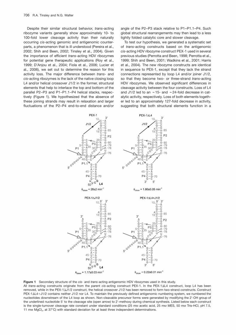
Figure 1 Secondary structure of the cis- and trans-acting antigenomic HDV ribozymes used in this study.All trans-acting constructs originate from the parent cis-acting construct PEX-1. In the PEX-1DL4 construct, loop L4 has beenremoved, while in the PEX-1DJ1/2 construct, the helical crossover J1/2 has been removed to form two-strand constructs. ConstructPEX-1DL4qJ1/2 contains neither J1/2 nor L4. To maintain the previously defined antigenomic numbering system, we numbered thenucleotides downstream of the L4 loop as shown. Non-cleavable precursor forms were generated by modifying the 29-OH group ofthe underlined nucleotide 59 to the cleavage site (open arrow) to 29-methoxy during chemical synthesis. Listed below each constructis the single-turnover cleavage rate constant under standard conditions (25 mM acetic acid, 25 mM MES, 50 mM Tris-HCl, pH 7.5,11 mM MgCl2, at 378C) with standard deviation for at least three independent determinations.
Despite their similar structural behavior, trans-actingribozyme variants generally show approximately 10- to100-fold lower cleavage activity than their naturallyoccurring cis-acting genomic and antigenomic counter-parts, a phenomenon that is ill understood (Pereira et al.,2002; Shih and Been, 2002; Tinsley et al., 2004). Giventhe importance of efficient trans-acting HDV ribozymesfor potential gene therapeutic applications (Roy et al.,1999; D’Anjou et al., 2004; Fiola et al., 2006; Lucier etal., 2006), we set out to determine the reason for thisactivity loss. The major difference between trans- andcis-acting ribozymes is the lack of the native closing loopL4 and/or helical crossover J1/2 in the former, structuralelements that help to interlace the top and bottom of theparallel P2–P3 and P1–P1.1–P4 helical stacks, respec-tively (Figure 1). We hypothesized that the absence ofthese joining strands may result in relaxation and largerfluctuations of the P2–P4 end-to-end distance and/or
angle of the P2–P3 stack relative to P1–P1.1–P4. Suchglobal structural rearrangements may then lead to a lesstightly folded catalytic core and slower cleavage.
To test our hypothesis, we generated a systematic setof trans-acting constructs based on the antigenomiccis-acting HDV ribozyme construct PEX-1 used in severalprevious studies (Perrotta and Been, 1998; Perrotta et al.,1999; Shih and Been, 2001; Wadkins et al., 2001; Harriset al., 2004). The new ribozyme constructs are identicalin sequence to PEX-1, except that they lack the strandconnections represented by loop L4 and/or joiner J1/2,so that they become two- or three-strand trans-actingHDV ribozymes. We observed significant differences incleavage activity between the four constructs. Loss of L4and J1/2 led to an ;15- and ;24-fold decrease in cat-alytic activity, respectively. Loss of both elements togeth-er led to an approximately 127-fold decrease in activity,suggesting that both structural elements function in a
Impact of peripheral elements on the HDV ribozyme 707
Article in press - uncorrected proof
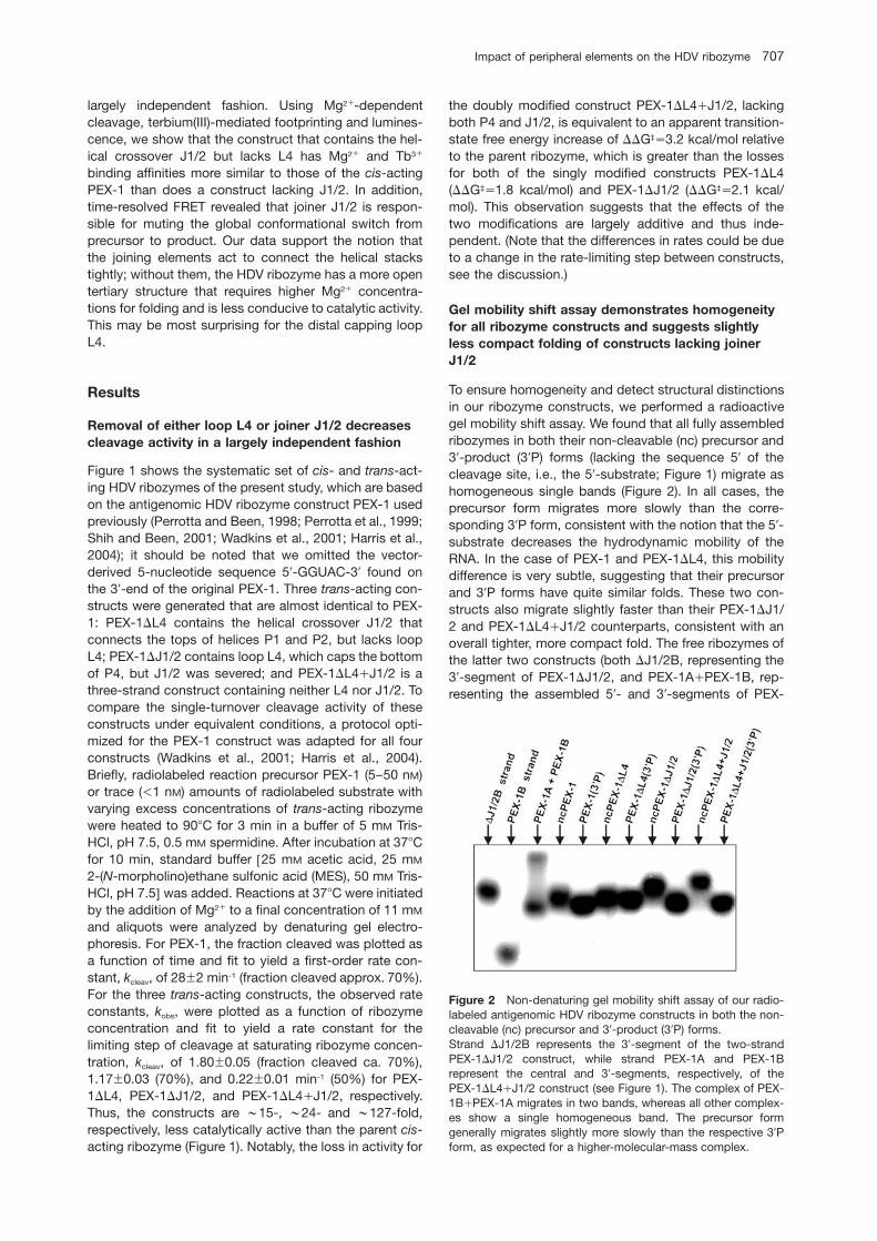
Figure 2 Non-denaturing gel mobility shift assay of our radio-labeled antigenomic HDV ribozyme constructs in both the non-cleavable (nc) precursor and 39-product (39P) forms.Strand DJ1/2B represents the 39-segment of the two-strandPEX-1DJ1/2 construct, while strand PEX-1A and PEX-1Brepresent the central and 39-segments, respectively, of thePEX-1DL4qJ1/2 construct (see Figure 1). The complex of PEX-1BqPEX-1A migrates in two bands, whereas all other complex-es show a single homogeneous band. The precursor formgenerally migrates slightly more slowly than the respective 39Pform, as expected for a higher-molecular-mass complex.
largely independent fashion. Using Mg2q-dependentcleavage, terbium(III)-mediated footprinting and lumines-cence, we show that the construct that contains the hel-ical crossover J1/2 but lacks L4 has Mg2q and Tb3q
binding affinities more similar to those of the cis-actingPEX-1 than does a construct lacking J1/2. In addition,time-resolved FRET revealed that joiner J1/2 is respon-sible for muting the global conformational switch fromprecursor to product. Our data support the notion thatthe joining elements act to connect the helical stackstightly; without them, the HDV ribozyme has a more opentertiary structure that requires higher Mg2q concentra-tions for folding and is less conducive to catalytic activity.This may be most surprising for the distal capping loopL4.
Results
Removal of either loop L4 or joiner J1/2 decreasescleavage activity in a largely independent fashion
Figure 1 shows the systematic set of cis- and trans-act-ing HDV ribozymes of the present study, which are basedon the antigenomic HDV ribozyme construct PEX-1 usedpreviously (Perrotta and Been, 1998; Perrotta et al., 1999;Shih and Been, 2001; Wadkins et al., 2001; Harris et al.,2004); it should be noted that we omitted the vector-derived 5-nucleotide sequence 59-GGUAC-39 found onthe 39-end of the original PEX-1. Three trans-acting con-structs were generated that are almost identical to PEX-1: PEX-1DL4 contains the helical crossover J1/2 thatconnects the tops of helices P1 and P2, but lacks loopL4; PEX-1DJ1/2 contains loop L4, which caps the bottomof P4, but J1/2 was severed; and PEX-1DL4qJ1/2 is athree-strand construct containing neither L4 nor J1/2. Tocompare the single-turnover cleavage activity of theseconstructs under equivalent conditions, a protocol opti-mized for the PEX-1 construct was adapted for all fourconstructs (Wadkins et al., 2001; Harris et al., 2004).Briefly, radiolabeled reaction precursor PEX-1 (5–50 nM)or trace (-1 nM) amounts of radiolabeled substrate withvarying excess concentrations of trans-acting ribozymewere heated to 908C for 3 min in a buffer of 5 mM Tris-HCl, pH 7.5, 0.5 mM spermidine. After incubation at 378Cfor 10 min, standard buffer w25 mM acetic acid, 25 mM
2-(N-morpholino)ethane sulfonic acid (MES), 50 mM Tris-HCl, pH 7.5x was added. Reactions at 378C were initiatedby the addition of Mg2q to a final concentration of 11 mM
and aliquots were analyzed by denaturing gel electro-phoresis. For PEX-1, the fraction cleaved was plotted asa function of time and fit to yield a first-order rate con-stant, kcleav, of 28"2 min-1 (fraction cleaved approx. 70%).For the three trans-acting constructs, the observed rateconstants, kobs, were plotted as a function of ribozymeconcentration and fit to yield a rate constant for thelimiting step of cleavage at saturating ribozyme concen-tration, kcleav, of 1.80"0.05 (fraction cleaved ca. 70%),1.17"0.03 (70%), and 0.22"0.01 min-1 (50%) for PEX-1DL4, PEX-1DJ1/2, and PEX-1DL4qJ1/2, respectively.Thus, the constructs are ;15-, ;24- and ;127-fold,respectively, less catalytically active than the parent cis-acting ribozyme (Figure 1). Notably, the loss in activity for
the doubly modified construct PEX-1DL4qJ1/2, lackingboth P4 and J1/2, is equivalent to an apparent transition-state free energy increase of DDG‡s3.2 kcal/mol relativeto the parent ribozyme, which is greater than the lossesfor both of the singly modified constructs PEX-1DL4(DDG‡s1.8 kcal/mol) and PEX-1DJ1/2 (DDG‡s2.1 kcal/mol). This observation suggests that the effects of thetwo modifications are largely additive and thus inde-pendent. (Note that the differences in rates could be dueto a change in the rate-limiting step between constructs,see the discussion.)
Gel mobility shift assay demonstrates homogeneityfor all ribozyme constructs and suggests slightlyless compact folding of constructs lacking joinerJ1/2
To ensure homogeneity and detect structural distinctionsin our ribozyme constructs, we performed a radioactivegel mobility shift assay. We found that all fully assembledribozymes in both their non-cleavable (nc) precursor and39-product (39P) forms (lacking the sequence 59 of thecleavage site, i.e., the 59-substrate; Figure 1) migrate ashomogeneous single bands (Figure 2). In all cases, theprecursor form migrates more slowly than the corre-sponding 39P form, consistent with the notion that the 59-substrate decreases the hydrodynamic mobility of theRNA. In the case of PEX-1 and PEX-1DL4, this mobilitydifference is very subtle, suggesting that their precursorand 39P forms have quite similar folds. These two con-structs also migrate slightly faster than their PEX-1DJ1/2 and PEX-1DL4qJ1/2 counterparts, consistent with anoverall tighter, more compact fold. The free ribozymes ofthe latter two constructs (both DJ1/2B, representing the39-segment of PEX-1DJ1/2, and PEX-1AqPEX-1B, rep-resenting the assembled 59- and 39-segments of PEX-
708 R.A. Tinsley and N.G. Walter
Article in press - uncorrected proof
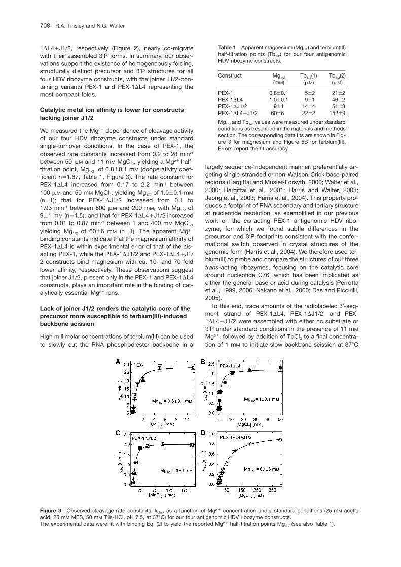
Table 1 Apparent magnesium (Mg1/2) and terbium(III)half-titration points (Tb1/2) for our four antigenomicHDV ribozyme constructs.
Construct Mg1/2 Tb1/2(1) Tb1/2(2)(mM) (mM) (mM)
PEX-1 0.8"0.1 5"2 21"2PEX-1DL4 1.0"0.1 9"1 46"2PEX-1DJ1/2 9"1 14"4 51"3PEX-1DL4qJ1/2 60"6 22"2 152"9
Mg1/2 and Tb1/2 values were measured under standardconditions as described in the materials and methodssection. The corresponding data fits are shown in Fig-ure 3 for magnesium and Figure 5B for terbium(III).Errors report the fit accuracy.
Figure 3 Observed cleavage rate constants, kobs, as a function of Mg2q concentration under standard conditions (25 mM aceticacid, 25 mM MES, 50 mM Tris-HCl, pH 7.5, at 378C) for our four antigenomic HDV ribozyme constructs.The experimental data were fit with binding Eq. (2) to yield the reported Mg2q half-titration points Mg1/2 (see also Table 1).
1DL4qJ1/2, respectively (Figure 2), nearly co-migratewith their assembled 39P forms. In summary, our obser-vations support the existence of homogeneously folding,structurally distinct precursor and 39P structures for allfour HDV ribozyme constructs, with the joiner J1/2-con-taining variants PEX-1 and PEX-1DL4 representing themost compact folds.
Catalytic metal ion affinity is lower for constructslacking joiner J1/2
We measured the Mg2q dependence of cleavage activityof our four HDV ribozyme constructs under standardsingle-turnover conditions. In the case of PEX-1, theobserved rate constants increased from 0.2 to 28 min-1
between 50 mM and 11 mM MgCl2, yielding a Mg2q half-titration point, Mg1/2, of 0.8"0.1 mM (cooperativity coef-ficient ns1.67, Table 1, Figure 3). The rate constant forPEX-1DL4 increased from 0.17 to 2.2 min-1 between100 mM and 50 mM MgCl2, yielding Mg1/2 of 1.0"0.1 mM
(ns1); that for PEX-1DJ1/2 increased from 0.1 to1.93 min-1 between 500 mM and 200 mM, with Mg1/2 of9"1 mM (ns1.5); and that for PEX-1DL4qJ1/2 increasedfrom 0.01 to 0.87 min-1 between 1 and 400 mM MgCl2,yielding Mg1/2 of 60"6 mM (ns1). The apparent Mg2q
binding constants indicate that the magnesium affinity ofPEX-1DL4 is within experimental error of that of the cis-acting PEX-1, while the PEX-1DJ1/2 and PEX-1DL4qJ1/2 constructs bind magnesium with ca. 10- and 70-foldlower affinity, respectively. These observations suggestthat joiner J1/2, present only in the PEX-1 and PEX-1DL4constructs, plays an important role in the binding of cat-alytically essential Mg2q ions.
Lack of joiner J1/2 renders the catalytic core of theprecursor more susceptible to terbium(III)-inducedbackbone scission
High millimolar concentrations of terbium(III) can be usedto slowly cut the RNA phosphodiester backbone in a
largely sequence-independent manner, preferentially tar-geting single-stranded or non-Watson-Crick base-pairedregions (Hargittai and Musier-Forsyth, 2000; Walter et al.,2000; Hargittai et al., 2001; Harris and Walter, 2003;Jeong et al., 2003; Harris et al., 2004). This property pro-duces a footprint of RNA secondary and tertiary structureat nucleotide resolution, as exemplified in our previouswork on the cis-acting PEX-1 antigenomic HDV ribo-zyme, for which we found subtle differences in theprecursor and 39P footprints consistent with the confor-mational switch observed in crystal structures of thegenomic form (Harris et al., 2004). We therefore used ter-bium(III) to probe and compare the structures of our threetrans-acting ribozymes, focusing on the catalytic corearound nucleotide C76, which has been implicated aseither the general base or acid during catalysis (Perrottaet al., 1999, 2006; Nakano et al., 2000; Das and Piccirilli,2005).
To this end, trace amounts of the radiolabeled 39-seg-ment strand of PEX-1DL4, PEX-1DJ1/2, and PEX-1DL4qJ1/2 were assembled with either nc substrate or39P under standard conditions in the presence of 11 mM
Mg2q, followed by addition of TbCl3 to a final concentra-tion of 1 mM to initiate slow backbone scission at 378C
Impact of peripheral elements on the HDV ribozyme 709
Article in press - uncorrected proof
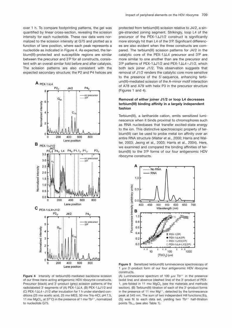
Figure 4 Intensity of terbium(III)-mediated backbone scissionof our three trans-acting antigenomic HDV ribozyme constructs.Precursor (black) and 39-product (grey) scission patterns of theradiolabeled 39-segments of (A) PEX-1DL4, (B) PEX-1DJ1/2 and(C) PEX-1DL4qJ1/2 after incubation for 1 h under standard con-ditions (25 mM acetic acid, 25 mM MES, 50 mM Tris-HCl, pH 7.5,11 mM MgCl2, at 378C) in the presence of 1 mM Tb3q, normalizedto nucleotide G75.
Figure 5 Sensitized terbium(III) luminescence spectroscopy of1 mM 39-product form of our four antigenomic HDV ribozymeconstructs.(A) Luminescence spectrum of 100 mM Tb3q in the presence(solid line) and absence (dashed line) of the 39-product of PEX-1, pre-folded in 11 mM MgCl2 (see the materials and methodssection). (B) Terbium(III) titration of each of the 39-product formsin the presence of 11 mM Mg2q, detected by the luminescencepeak at 545 nm. The sum of two independent Hill functions wEq.(3)x was fit to each data set, yielding two Tb3q half-titrationpoints Tb1/2 (see also Table 1).
over 1 h. To compare footprinting patterns, the gel wasquantified by linear cross-section, revealing the scissionintensity for each nucleotide. These raw data were nor-malized to the scission intensity at G75 and plotted as afunction of lane position, where each peak represents anucleotide as indicated in Figure 4. As expected, the ter-bium(III)-protected and susceptible regions are similarbetween the precursor and 39P for all constructs, consis-tent with an overall similar fold before and after catalysis.The scission patterns are also consistent with theexpected secondary structure; the P2 and P4 helices are
protected from terbium(III) scission relative to J4/2, a sin-gle-stranded joining segment. Strikingly, loop L4 of theprecursor of the PEX-1DJ1/2 construct is significantlymore strongly hit than L4 of the 39P. Significant differenc-es are also evident when the three constructs are com-pared. The terbium(III) scission patterns for J4/2 in thecatalytic core of the PEX-1DL4 precursor and 39P aremore similar to one another than are the precursor and39P patterns of PEX-1DJ1/2 and PEX-1DL4qJ1/2, whichboth lack joiner J1/2. This observation suggests thatremoval of J1/2 renders the catalytic core more sensitiveto the presence of the 59-sequence, enhancing terbi-um(III)-mediated scission of the A-minor motif interactionof A78 and A79 with helix P3 in the precursor structure(Figures 1 and 4).
Removal of either joiner J1/2 or loop L4 decreasesterbium(III) binding affinity in a largely independentfashion
Terbium(III), a lanthanide cation, emits sensitized lumi-nescence when it binds proximal to chromophores suchas RNA nucleobases that transfer excited-state energyto the ion. This distinctive spectroscopic property of ter-bium(III) can be used to probe metal ion affinity over anentire RNA structure (Walter et al., 2000; Harris and Wal-ter, 2003; Jeong et al., 2003; Harris et al., 2004). Here,we examined and compared the binding affinities of ter-bium(III) to the 39P forms of our four antigenomic HDVribozyme constructs.
710 R.A. Tinsley and N.G. Walter
Article in press - uncorrected proof
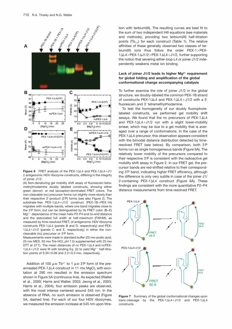
Figure 6 FRET analysis of the PEX-1DL4 and PEX-1DL4qJ1/2 antigenomic HDV ribozyme constructs, differing in the integrityof joiner J1/2.(A) Non-denaturing gel mobility shift assay of fluorescein-tetra-methylrhodamine doubly labeled constructs, showing eithergreen (donor)- or red (acceptor)-dominated FRET colors. Thenon-cleavable (nc) precursor forms run slightly more slowly thantheir respective 39-product (39P) forms (see also Figure 2). Thesubstrate-free PEX-1DL4qJ1/2 construct (PEX-1BqPEX-1A)migrates with multiple bands, where one band migrates close tothe 39P form, but can be distinguished by its FRET color. (B–E)Mg2q dependence of the mean helix P2-P4 end-to-end distanceand the associated full width at half-maximum (FWHM), asmeasured by time-resolved FRET, of antigenomic HDV ribozymeconstructs PEX-1DL4 (panels B and D, respectively) and PEX-1DL4qJ1/2 (panels C and E, respectively) in either the non-cleavable (nc) precursor or 39P form.Measurements were made in standard buffer (25 mM acetic acid,25 mM MES, 50 mM Tris-HCl, pH 7.5) supplemented with 25 mM
DTT at 378C. The mean distances of nc PEX-1DL4 and ncPEX-1DL4qJ1/2 were fit with binding Eq. (2) to yield Mg2q half-titra-tion points of 0.35"0.09 and 2.2"0.3 mM, respectively.
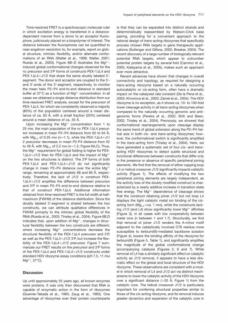
Figure 7 Summary of the global conformational changes upontrans-cleavage by the PEX-1DL4qJ1/2 and PEX-1DL4constructs.
Addition of 100 mM Tb3q to 1 mM 39P form of the pre-annealed PEX-1DL4 construct in 11 mM MgCl2 with exci-tation at 290 nm resulted in the emission spectrumshown in Figure 5A (continuous line). As expected (Walteret al., 2000; Harris and Walter, 2003; Jeong et al., 2003;Harris et al., 2004), four emission peaks are observed,with the most intense centered around 545 nm. In theabsence of RNA, no such emission is observed (Figure5A, dashed line). For each of our four HDV ribozymes,we measured the emission increase at 545 nm upon titra-
tion with terbium(III). The resulting curves are best fit tothe sum of two independent Hill equations (see materialsand methods), providing two terbium(III) half-titrationpoints (Tb1/2) for each construct (Table 1). The relativeaffinities of these generally observed two classes of ter-bium(III) ions thus follow the order PEX-1)PEX-1DL4)PEX-1DJ1/2)PEX-1DL4qJ1/2, further supportingthe notion that severing either loop L4 or joiner J1/2 inde-pendently weakens metal ion binding.
Lack of joiner J1/2 leads to higher Mg2H requirementfor global folding and amplification of the globalconformational change accompanying catalysis
To further examine the role of joiner J1/2 in the globalstructure, we doubly-labeled the common PEX-1B strandof constructs PEX-1DL4 and PEX-1DL4qJ1/2 with a 59
fluorescein and 39 tetramethylrhodamine.To test the homogeneity of our doubly fluorophore-
labeled constructs, we performed gel mobility shiftassays. We found that the nc precursors of PEX-1DL4and PEX-1DL4qJ1/2 run with a slight lower-mobilitysmear, which may be due to a gel mobility that is aver-aged over a range of conformations. In the case of thePEX-1DL4 precursor, this observation appears consistentwith the bimodal distance distribution detected by time-resolved FRET (see below). By comparison, both 39Pforms run as single homogeneous bands (Figure 6A). Therelatively lower mobility of the precursors compared totheir respective 39P is consistent with the radioactive gelmobility shift assay in Figure 2. In our FRET gel, the pre-cursor bands are red-shifted relative to their correspond-ing 39P band, indicating higher FRET efficiency, althoughthe difference is only very subtle in case of the joiner J1/2-containing PEX-1DL4 construct (Figure 6A). Thesefindings are consistent with the more quantitative P2–P4distance measurements from time-resolved FRET.
Impact of peripheral elements on the HDV ribozyme 711
Article in press - uncorrected proof
Time-resolved FRET is a spectroscopic molecular rulerin which excitation energy is transferred in a distance-dependent manner from a donor to an acceptor fluoro-phore, judiciously placed on a biopolymer of interest. Thedistance between the fluorophores can be quantified tonear-angstrom resolution to, for example, report on glob-al structure, intrinsic flexibility, and/or alternate confor-mations of an RNA (Walter et al., 1999; Walter, 2001;Rueda et al., 2003). Figure 6B–D illustrates the Mg2q-induced global conformational changes observed for thenc precursor and 39P forms of constructs PEX-1DL4 andPEX-1DL4qJ1/2 that share the same doubly labeled 39-segment. The donor and acceptor are coupled to the 59-and 39-ends of the 39-segment, respectively, to monitorthe mean helix P2–P4 end-to-end distance in standardbuffer at 378C as a function of Mg2q concentration. In allcases we obtained a single distance distribution from ourtime-resolved FRET analysis, except for the precursor ofPEX-1DL4, for which we consistently observed a majority(80%) of the population centered around a mean dis-tance of ca. 63 A, with a small fraction (20%) centeredaround a mean distance of ca. 35 A.
Upon increasing the Mg2q concentration from 1 to25 mM, the main population of the nc PEX-1DL4 precur-sor increases in mean P2–P4 distance from 62 to 64 A,with Mg1/2 of 0.35 mM (ns1.1), while the PEX-1DL4qJ1/2 precursor decreases in mean P2-P4 distance from 52to 49 A, with Mg1/2 of 2.2 mM (ns1.2; Figure 6A,C). Thus,the Mg2q requirement for global folding is higher for PEX-1DL4qJ1/2 than for PEX-1DL4 and the impact of Mg2q
on the two structures is distinct. The 39P forms of bothPEX-1DL4 and PEX-1DL4qJ1/2 do not significantlychange in mean P2–P4 distance over the entire Mg2q
range, remaining at approximately 68 and 66 A, respec-tively. Therefore, the lack of J1/2 in construct PEX-1DL4qJ1/2 amplifies the difference between precursorand 39P in mean P2–P4 end-to-end distance relative tothat of construct PEX-1DL4. Additional informationobtained from time-resolved FRET is the full width at half-maximum (FWHM) of the distance distribution. Since thedoubly labeled 39-segment is shared between the twoconstructs, we can attribute qualitative differences inFWHM primarily to the intrinsic global flexibility of theRNA (Rueda et al., 2003; Tinsley et al., 2004). Figure 6B,Dindicates that, upon addition of Mg2q, changes in struc-tural flexibility between the two constructs are different,where increasing Mg2q concentrations decrease thestructural flexibility of the PEX-1DL4 precursor and 39P,as well as the PEX-1DL4qJ1/2 39P, but increase the flex-ibility of the PEX-1DL4qJ1/2 precursor. Figure 7 sum-marizes our FRET results on the precursor and 39P formsof the PEX-1DL4 and PEX-1DL4qJ1/2 constructs understandard HDV ribozyme assay conditions (pH 7.5, 11 mM
Mg2q, 378C).
Discussion
Up until approximately 25 years ago, all known enzymeswere proteins. It was only then discovered that RNA iscapable of enzymatic action in the form of ribozymes(Guerrier-Takada et al., 1983; Zaug et al., 1983). Oneadvantage of ribozymes over their protein counterparts
is that they can be separated into distinct strands anddeterministically reassembled by Watson-Crick basepairing, providing for a convenient approach to therational design of trans-acting ribozymes that specificallyprocess chosen RNA targets in gene therapeutic appli-cations (Sullenger and Gilboa, 2002; Breaker, 2004). Therecent discovery of a large number of biologically relevantpotential RNA targets, which appear to outnumberpotential protein targets by several-fold (Carninci et al.,2005; Katayama et al., 2005), makes such an approachever more attractive.
Recent advances have shown that changes in overallconnectivity and topology, as required for designing atrans-acting ribozyme based on a naturally occurringautocatalytic or cis-acting form, often have a dramaticimpact on the catalyzed rate constant (De la Pena et al.,2003; Khvorova et al., 2003; Zamel et al., 2004). The HDVribozyme is no exception, as it shows ca. 10- to 100-foldlower cleavage activity in all trans-acting ribozymes whencompared to the naturally occurring genomic and anti-genomic forms (Pereira et al., 2002; Shih and Been,2002; Tinsley et al., 2004). Previously, we showed thatconformational rearrangements upon cleavage displaythe same trend of global extension along the P2–P4 hel-ical axis in both cis- and trans-acting ribozymes; how-ever, the conformational switch is significantly amplifiedin the trans-acting form (Tinsley et al., 2004). Here, wehave generated a systematic set of four cis- and trans-acting HDV ribozymes to detect subtle structural andfunctional differences between constructs that differ onlyin the presence or absence of specific peripheral joiningelements. We find that the removal of either capping loopL4 or helical crossover J1/2 significantly impacts catalyticactivity (Figure 1). The effects of modifying the twoperipheral joining elements are largely independent, asthe activity loss of the doubly modified construct is char-acterized by a nearly additive increase in transition-statefree energy. The Mg2q dependence of cleavage showsthat the construct retaining joiner J1/2 but not loop L4displays the tight catalytic metal ion binding of the cis-acting form (Mg1/2sca. 1 mM), while the constructs lack-ing J1/2 (and L4) show significantly lower Mg2q affinities(Figure 3), in all cases with low cooperativity betweenmetal ions (n between 1 and 1.7). Structurally, we findthat removal of joiner J1/2 renders the A-minor motifadjacent to the catalytically involved C76 residue moresusceptible to terbium(III)-mediated backbone scission(Figure 4), lowers the binding affinity of the ribozyme forterbium(III) (Figure 5, Table 1), and significantly amplifiesthe magnitude of the global conformational changeaccompanying catalysis (Figures 2, 6 and 7). Whileremoval of L4 has a similarly significant effect on catalyticactivity as J1/2 removal, it appears to have a less dra-matic effect on the global and local structure of the HDVribozyme. These observations are consistent with a mod-el in which removal of L4 and J1/2 act via distinct mech-anisms to lower the catalytic activity of the HDV ribozymeover a significant distance ()20 A, Figure 1) from thecatalytic core. The helical crossover J1/2 is particularlyimportant for conferring structural properties similar tothose of the cis-acting ribozyme, and its removal inducesgreater dynamics and expansion of the catalytic core in
712 R.A. Tinsley and N.G. Walter
Article in press - uncorrected proof
a way that impacts local and global folding, as well ascatalytic function.
How do L4 and J1/2 exert their long-range influenceon folding and function of the HDV ribozyme? Our ter-bium(III)-mediated footprinting suggests that removal ofJ1/2 renders the catalytic core more sensitive to thepresence of the 59-sequence, which we have previouslyshown to form a U-turn motif in the genomic HDV ribo-zyme (Sefcikova et al., 2007b) that wedges between theP2–P3 and P1–P1.1–P4 helical stacks (Ke et al., 2004;Krasovska et al., 2005). Therefore, removal of the con-nector J1/2, which tightly interlaces one end of the twohelical stacks (Figure 1), likely results in larger fluctua-tions in the distance between and relative register of thehelical stacks. Such a model of amplified inter-stackdynamics is consistent with the observed opening of thecatalytic core to terbium(III)-mediated scission, thereduced metal ion affinity, and amplification of the globalconformational change upon cleavage (i.e., upon disso-ciation of the 59-sequence) in the absence of J1/2. Con-versely, lower dynamics and tighter folding around thecatalytic core in the presence of J1/2 is predicted totranslate into faster cleavage rates and lower Mg2q requi-rements, especially in the cis-acting ribozymes, presum-ably by reducing the entropic energy barrier of catalysis.
Perhaps the most surprising observation of the workdescribed here is that removal of capping loop L4 alsohas a profound impact on catalytic efficiency of the HDVribozyme, via a different mechanism than removal ofJ1/2. As a capping loop of a helix that projects away fromthe catalytic core, it is at least 30 A removed from cata-lytic action and must exert any impact through the stableP4 stem with seven Watson-Crick base pairs (Figure 1).Our findings are consistent with earlier work on trans-acting ribozymes lacking L4, which revealed similarcleavage rate constants in the few min-1 range, approxi-mately 10-fold lower than the rate of the cis-acting parentRNA (Luptak et al., 2001). Such long-range impact of alocal change in topology is reminiscent of the dynamicstructural rearrangements throughout the catalytic coreof the hairpin ribozyme in response to site-specific chem-ical modification (Rhodes et al., 2006). It is thought thatsuch structural communication is mediated by hydrogenbonding networks and van der Waals’ packing, whichconnect distal parts of an RNA or protein by coupledmotions to act as an interconnected whole (Benkovic andHammes-Schiffer, 2003; Rueda et al., 2004; Hammes-Schiffer and Benkovic, 2006; Rhodes et al., 2006). In thecase of removal of capping loop L4, the dynamics ofhelix P4 may be altered in a way that impacts the con-formational equilibrium of the entire P1–P1.1–P4 stack,the integrity of which is known to be important for cata-lysis (Wadkins et al., 1999). Since the cleavage site G:Uwobble pair is sandwiched between P1 and P1.1, suchaltered conformational dynamics may impact the cata-lytic core geometry in a way that decreases the rate(probability) of transition-state barrier crossing and thuscatalysis. In this model the effect of L4 would mostly beexerted through the P1–P1.1–P4 stack, making it distinctfrom the impact of J1/2, which most likely acts throughthe relative orientation or register of the P1–P1.1–P4 andP2–P3 stacks. Such differential modes of catalytic inter-ference caused by removal of loop L4 and joiner J1/2
would be most consistent with our experimental data.Our model deserves further testing, as it suggests anunexpectedly significant long-range impact of peripheralelements on the structure and function of biologically rel-evant RNAs such as the HDV ribozyme. It also holds thekey to our ability to design more effective ribozymes forgene therapeutic applications.
Materials and methods
Preparation of RNA oligonucleotides
RNA oligonucleotides for constructs PEX-1DL4, PEX-1DL4qJ1/2, and the substrate and 39P strands for PEX-1DJ1/2 (Figure 1)were purchased from the Howard Hughes Medical Institute Bio-polymer/Keck Foundation Biotechnology Resource RNA Labo-ratory at the Yale University School of Medicine (New Haven,CT, USA) and were purified as previously described (Pereira etal., 2002; Walter, 2002). The unmodified 39P form of constructPEX-1, the substrate strand of PEX-1DL4, and the ribozymestrand of PEX-1DJ1/2 were generated by run-off transcriptionfrom a double-stranded, PCR-amplified template that encodedan upstream T7 promoter. Transcription reactions contained40 mM Tris-HCl (pH 7.5), 15 mM MgCl2, 5 mM dithiothreitol (DTT),2 mM spermidine, 4 mM each rNTP, 5 U/ml inorganic pyrophos-phatase, and 0.1 mg/ml T7 RNA polymerase and were incubatedat 378C overnight (ca. 16 h). The RNA was isolated after dena-turing (8 M urea) 10% (w/v) polyacrylamide gel electrophoresisby UV shadowing, diffusion elution of small gel slices, and eth-anol precipitation.
For cleavage reactions, the radiolabeled precursor form ofPEX-1 was transcribed as described above, except that0.04 mCi of wa-32PxGTP and 0.125 mg/ml DNA oligonucleotidewas added to the reaction mixture. This DNA oligonucleotide hasa sequence fully complementary to the 15 nucleotides at the 59-end of the precursor (G-7 to C8) and was used to increase theyield of the uncleaved precursor RNA (Wadkins and Been, 1997).For the other constructs, 59-32P-labeled substrates were pre-pared by phosphorylation with T4 polynucleotide kinase andwg-32Px-ATP. Unless otherwise noted, unlabeled strands wereadded at a saturating 800 nM excess (39-segment PEX-1B ofconstruct PEX-1DL4qJ1/2 at 1600 nM) in all assays.
Terbium stock solutions
The highest-purity terbium(III) chloride (99.9%) was purchasedfrom Sigma-Aldrich (St. Louis, MO, USA). TbCl3 stock solutionswere prepared at 100 mM in 5 mM cacodylate (pH 5.5) andstored in small aliquots at -208C to prevent formation of in-soluble hydroxide species.
Gel mobility shift assays
Gel mobility shift assays were similar to those previouslydescribed (Pereira et al., 2002; Jeong et al., 2003). Briefly, non-denaturing 10% (w/v) polyacrylamide (19:1 acrylamide/bisacryl-amide ratio) gels containing 50 mM Tris-HOAc (pH 7.5), 25 mM
MES, 25 mM acetic acid and 11 mM Mg(OAc)2 were assembledin the electrophoresis unit and equilibrated to 48C for at least2 h. 39-32P-labeled nc or self-cleaved PEX-1 or ribozyme or sub-strate strand was prepared by ligation with w32PxpCp using T4RNA ligase, followed by desalting through a CentriSep spin col-umn (Princeton Separations, Adelphia, NJ, USA). The radio-labeled RNA (50 000 cpm) was pre-annealed in buffer of 5 mM
Tris-OAc (pH 7.5) and 0.5 mM spermidine, either alone or withcorresponding strands, denatured for 3 min at 908C, and pre-incubated at 378C for 10 min. The reactions were adjusted to

Impact of peripheral elements on the HDV ribozyme 713
Article in press - uncorrected proof
their final pH with a buffer containing 25 mM acetic acid, 25 mM
MES, and 50 mM Tris-OAc (pH 7.5). These mixtures were incu-bated for an additional 5 min, after which 0.1 volumes of 110 mM
Mg(OAc)2 was added to a final concentration of 11 mM. Theribozyme was incubated for an additional 5 min. These sampleswere loaded on the gel, and an electric field of 8 V/cm wasimmediately applied. After typically 15–24 h of electrophoresis,the gel was exposed overnight and quantified using aPhosphorImager Storm 840 with ImageQuant software (Molec-ular Dynamics, Sunnyvale, CA, USA).
For FRET gel mobility assays of PEX-1DL4 and PEX-1DL4qJ1/2, doubly fluorophore-labeled ribozyme strand B(10 pmol) was annealed to strand A (50 pmol for PEX-1DL4qJ1/2) and/or the substrate strand (50 pmol for PEX-1DL4 and100 pmol for PEX-1DL4qJ1/2) and prepared as describedabove. The gel was scanned between low-fluorescence glassplates in a FluorImager SI fluorescence scanner with Image-Quant software (Molecular Dynamics) as previously described(Pereira et al., 2002; Jeong et al., 2003).
Cleavage activity assays
Radiolabeled precursor or ribozyme strands with 59-w32Px-labeledsubstrate strands were heated to 908C for 2 min in a buffer con-taining 5 mM Tris-HCl (pH 7.5), 0.5 mM spermidine. The reactionsmixtures for PEX-1DL4, PEX-1DJ1/2, and PEX-1DL4qJ1/2 weresupplemented with 0.1 mM EDTA. The precursor was then pre-incubated at 378C for 10 min, after which the reactions wereadjusted to the final pH (7.5) with a buffer containing 25 mM
acetic acid, 25 mM MES, 50 mM Tris-HCl. These mixtures wereincubated for an additional 5 min at 378C, followed by additionof 0.25 volumes of a solution (also at 378C) containing 55 mM
MgCl2 and 0.5 mM spermidine to start the reaction (Wadkins etal., 2001). Cleavage kinetics were followed by removing aliquots(2.5 or 5 ml) at specified times and quenching with 10 ml of 80%(v/v) formamide, 0.025% (w/v) xylene cyanol, and 0.025% (w/v)bromophenol blue, and 100 mM EDTA. In some cases, thequenching buffer was supplemented with 7 M urea to completelysuppress cleavage. The reaction products were separated fromthe precursor by gel electrophoresis under denaturing conditionsw8 M urea, 10% or 20% (w/v) polyacrylamide gelsx, quantified,and normalized to the sum of the precursor and 39P bands usingPhosphorImager Storm 840 with ImageQuant software (Molec-ular Dynamics). Time traces of product formation were fit withthe single-exponential first-order rate equation ysy0qA(1-e-t/t),employing Marquardt-Levenberg non-linear regression (Igor Pro5.03, Wavemetrics, Oswego, OR, USA), where A is the amplitudeand t-1 is the (pseudo-)first-order rate constant kobs. To obtainbimolecular cleavage rate constants, the ribozyme concentrationwas varied from 12.5 to 800 nM for PEX-1DL4, from 50 to1600 nM for PEX-1DJ1/2, and from 50 to 2000 nM for PEX-1DL4qJ1/2; the rate constants kobs of the ribozyme dependencewere fit to the following binding equation:
w xRzk sk . (1)obs cleav w xRzqRz1/2
Similarly, the rates for Mg2q dependence were fit to the followingcooperative binding equation:
q2 nw xMgk sA qk , (2)obs 0 max q2 nnw xMg qMg1/2
yielding the cleavage rate constant kcleav under standard condi-tions, the ribozyme and metal ion half-titration points Rz1/2 andMg1/2, respectively, and the cooperativity coefficient n.
Terbium(III)-mediated footprinting
To observe the slow backbone scission mediated byTb(OH)(aq)2q, purified 39-segments of PEX-1DL4, PEX-1DJ1/2,and PEX-1DL4qJ1/2 were 59 32P-phosphorylated with T4 poly-nucleotide kinase and wa-32PxATP, repurified by denaturing (8 M
urea) 10% (w/v) polyacrylamide gel electrophoresis, followed bydiffusion elution into 1 mM EDTA, and by ethanol precipitation.The labeled RNA strand (250 000 cpm per 10-ml reaction vol-ume) was preannealed with the remaining unlabeled strand(s) inbuffer (5 mM Tris-HCl, pH 7.5, 0.5 mM spermidine) by denatur-ation at 908C for 3 min, followed by incubation at 378C for10 min. To form the RNA tertiary structure, Mg2q was added5 min prior to the addition of either 1 or 5 mM Tb3q (final con-centration) and the reaction was incubated for 1 h at 378C. Thescission reaction was stopped by addition of 50 mM EDTA (pH8.0) and ethanol precipitation at -208C. The precipitated RNAwas redissolved in urea loading buffer w80% (v/v) formamide,0.025% (w/v) xylene cyanol, 0.025% (w/v) bromophenol blue,50 mM EDTAx and analyzed on a denaturing (7 M urea) wedged20% (w/v) polyacrylamide sequencing gel alongside sequencingladders from partial digestion with G-specific RNase T1 andalkaline hydrolysis as previously described (Walter et al., 2000;Harris and Walter, 2003, 2005). Product bands were visualizedusing a PhosphorImager Storm 840 with ImageQuant software(Molecular Dynamics) and a line fit was performed. The intensitywas normalized and plotted as a function of the lane position inpixels.
Terbium(III) luminescence measurements
Steady-state luminescence spectra of terbium(III) bound to thepre-annealed and equilibrated 39P forms of all of the constructs(1 mM) in standard reaction buffer (25 mM acetic acid, 25 mM
MES, 50 mM Tris-HCl, pH 7.5, 11 mM MgCl2) at 378C were meas-ured on an Amino-Bowman Series 2 (AB2) spectrofluorimeter(Thermo Spectronic, Madison, WI, USA), while slowly increasingthe Tb3q concentration over several orders of magnitude usingappropriate stock solutions. After each terbium(III) addition, thesolution was equilibrated for 5–10 min until the signal had sta-bilized before an emission spectrum was recorded. Excitationwas at 290 nm (slit width 8 nm), and steady-state emission wasscanned with a slit width of 8 nm. To extract the luminescenceintensity of the major peak at 545 nm, each peak was fitbetween 535 and 555 nm using the following Gaussian distri-bution function:
2(x-x )A 0-2ysy q e (3)20 wyW py2
to yield the peak height as the pre-exponential factor, from whichthe background value in the absence of Tb3q was subtracted.These signals were plotted over varying terbium(III) concentra-tions (wTb3qx) and fit to the Hill equation:
q3 nw xTbysy (4)max q3 nnw xTb qTb1/2
to yield an apparent terbium(III) dissociation constant Tb1/2 anda cooperativity or Hill constant n. For a fit over the entire terbi-um(III) titration range, a sum of two independent Hill equationsproduced the best result, as judged by the x2 deviation andresiduals.
Time-resolved FRET measurements
The global structures of the PEX-1DL4 and PEX-1DL4qJ1/2HDV ribozymes were studied under standard conditions and as
714 R.A. Tinsley and N.G. Walter
Article in press - uncorrected proof
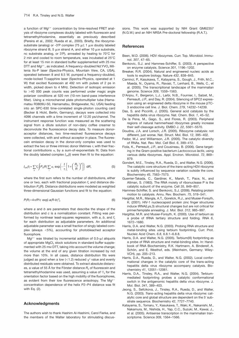
a function of Mg2q concentration by time-resolved FRET anal-ysis of ribozyme complexes doubly labeled with fluorescein andtetramethylrhodamine, essentially as previously described(Pereira et al., 2002; Rueda et al., 2003). Briefly, the ribozyme-substrate (analog) or -39P complex (75 ml; 1 mM doubly labeledribozyme strand B, 5 mM strand A, and either 10 mM substrate,nc substrate analog, or 39P), annealed by heating to 708C for2 min and cooled to room temperature, was incubated at 258Cfor at least 15 min in standard buffer supplemented with 25 mM
DTT and Mg2q, as indicated. A frequency-doubled Nd:YVO4 Mil-lenia Xs-P laser (Spectra-Physics, Mountain View, CA, USA),operated between 8 and 8.5 W, pumped a frequency-doubled,mode-locked Ti:sapphire laser (Spectra-Physics, operated at 1W) that excited fluorescein at 492 nm with pulses of 2 ps inwidth, picked down to 4 MHz. Detection of isotropic emissionto )40 000 peak counts was performed under magic anglepolarizer conditions at 520 nm (10-nm band-pass interferencefilter). Using a microchannel plate photomultiplier tube (Hama-matsu R3809U-50, Hamamatsu, Bridgewater, NJ, USA) feedinginto an SPC-630 time-correlated single-photon counting card(Becker & Hickl, Berlin, Germany), decays were collected into4096 channels with a time increment of 12.20 ps/channel. Theinstrument response function was measured as the scatteringsignal from a dilute solution of non-dairy coffee creamer todeconvolute the fluorescence decay data. To measure donor-acceptor distances, two time-resolved fluorescence decayswere collected, with and without acceptor in place. The fluores-cein emission decay in the donor-only complex was used toextract the two or three intrinsic donor lifetimes ti with their frac-tional contributions ai by a sum-of-exponentials fit. Data fromthe doubly labeled complex IDA(t) were then fit to the equation:
6w zS WB ET Tt R0C FU Xx |I t s f P R a exp - 1q dR, (5)T TŽ . Ž .DA k k i|8 8 D GV Yy ~t Rik i
where the first sum refers to the number of distributions, eitherone or two, each with fractional population fk and distance dis-tribution Pk(R). Distance distributions were modeled as weightedthree-dimensional Gaussian functions and fit to the equation:
22 w xP R s4pR c exp -a R-b , (6)Ž . Ž .
where a and b are parameters that describe the shape of thedistribution and c is a normalization constant. Fitting was per-formed by nonlinear least-squares regression, with a, b, and fk
for each distribution as adjustable parameters. An additionaladjustable parameter was a small fraction of singly labeled com-plex (always -5%), accounting for photobleached acceptorfluorophore.
Mg2q was titrated by incremental addition of 0.5-ml aliquotsof appropriate MgCl2 stock solutions in standard buffer supple-mented with 25 mM DTT, taking into account the volume change;the volume at the end of any given titration increased by notmore than 10%. In all cases, distance distribution fits werejudged as good when a low (-1.2) reduced x2 value and evenlydistributed residuals were obtained. To extract absolute distanc-es, a value of 55 A for the Forster distance R0 of fluorescein andtetramethylrhodamine was used, assuming a value of 2/3 for theorientation factor based on the high mobility of the fluorophores,as evident from their low fluorescence anisotropy. The Mg2q
concentration dependence of the helix P2–P4 distance was fitwith Eq. (2).
Acknowledgments
The authors wish to thank Hashim Al-Hashimi, Carol Fierke, andthe members of the Walter laboratory for stimulating discus-
sions. This work was supported by NIH Grant GM62357(N.G.W.) and an NIH NRSA Pre-doctoral fellowship (R.A.T.).
References
Been, M.D. (2006). HDV ribozymes. Curr. Top. Microbiol. Immu-nol. 307, 47–65.
Benkovic, S.J. and Hammes-Schiffer, S. (2003). A perspectiveon enzyme catalysis. Science 301, 1196–1202.
Breaker, R.R. (2004). Natural and engineered nucleic acids astools to explore biology. Nature 432, 838–845.
Carninci, P., Kasukawa, T., Katayama, S., Gough, J., Frith, M.C.,Maeda, N., Oyama, R., Ravasi, T., Lenhard, B., Wells, C., etal. (2005). The transcriptional landscape of the mammaliangenome. Science 309, 1559–1563.
D’Anjou, F., Bergeron, L.J., Larbi, N.B., Fournier, I., Salzet, M.,Perreault, J.P., and Day, R. (2004). Silencing of SPC2 expres-sion using an engineered delta ribozyme in the mouse bTC-3 endocrine cell line. J. Biol. Chem. 279, 14232–14239.
Das, S. and Piccirilli, J. (2005). General acid catalysis by thehepatitis delta virus ribozyme. Nat. Chem. Biol. 1, 45–52.
De la Pena, M., Gago, S., and Flores, R. (2003). Peripheralregions of natural hammerhead ribozymes greatly increasetheir self-cleavage activity. EMBO J. 22, 5561–5570.
Doudna, J.A. and Lorsch, J.R. (2005). Ribozyme catalysis: notdifferent, just worse. Nat. Struct. Mol. Biol. 12, 395–402.
Fedor, M.J. and Williamson, J.R. (2005). The catalytic diversityof RNAs. Nat. Rev. Mol. Cell Biol. 6, 399–412.
Fiola, K., Perreault, J.P., and Cousineau, B. (2006). Gene target-ing in the Gram-positive bacterium Lactococcus lactis, usingvarious delta ribozymes. Appl. Environ. Microbiol. 72, 869–879.
Gondert, M.E., Tinsley, R.A., Rueda, D., and Walter, N.G. (2006).The catalytic core structure of the trans-acting HDV ribozymeis subtly influenced by sequence variation outside the core.Biochemistry 45, 7563–7573.
Guerrier-Takada, C., Gardiner, K., Marsh, T., Pace, N., andAltman, S. (1983). The RNA moiety of ribonuclease P is thecatalytic subunit of the enzyme. Cell 35, 849–857.
Hammes-Schiffer, S. and Benkovic, S.J. (2006). Relating proteinmotion to catalysis. Annu. Rev. Biochem. 75, 519–541.
Hargittai, M.R., Mangla, A.T., Gorelick, R.J., and Musier-Forsyth,K. (2001). HIV-1 nucleocapsid protein zinc finger structuresinduce tRNA(Lys,3) structural changes but are not critical forprimer/template annealing. J. Mol. Biol. 312, 985–997.
Hargittai, M.R. and Musier-Forsyth, K. (2000). Use of terbium asa probe of tRNA tertiary structure and folding. RNA 6,1672–1680.
Harris, D.A. and Walter, N.G. (2003). Probing RNA structure andmetal-binding sites using terbium footprinting. Curr. Prot.Nucleic Acid Chem. 6.8, 6.8.1–6.8.8.
Harris, D.A. and Walter, N.G. (2005). Terbium(III) footprinting asa probe of RNA structure and metal-binding sites. In: Hand-book of RNA Biochemistry, R.K. Hartmann, A. Bindereif, A.Schon, and E. Westhof, eds. (Weinheim, Germany: Wiley-VCH), pp. 205–213.
Harris, D.A., Rueda, D., and Walter, N.G. (2002). Local confor-mational changes in the catalytic core of the trans-actinghepatitis delta virus ribozyme accompany catalysis. Bio-chemistry 41, 12051–12061.
Harris, D.A., Tinsley, R.A., and Walter, N.G. (2004). Terbium-mediated footprinting probes a catalytic conformationalswitch in the antigenomic hepatitis delta virus ribozyme. J.Mol. Biol. 341, 389–403.
Jeong, S., Sefcikova, J., Tinsley, R.A., Rueda, D., and Walter,N.G. (2003). Trans-acting hepatitis delta virus ribozyme: cat-alytic core and global structure are dependent on the 59 sub-strate sequence. Biochemistry 42, 7727–7740.
Katayama, S., Tomaru, Y., Kasukawa, T., Waki, K., Nakanishi, M.,Nakamura, M., Nishida, H., Yap, C.C., Suzuki, M., Kawai, J.,et al. (2005). Antisense transcription in the mammalian tran-scriptome. Science 309, 1564–1566.
Impact of peripheral elements on the HDV ribozyme 715
Article in press - uncorrected proof
Ke, A., Zhou, K., Ding, F., Cate, J.H., and Doudna, J.A. (2004).A conformational switch controls hepatitis delta virus ribo-zyme catalysis. Nature 429, 201–205.
Khvorova, A., Lescoute, A., Westhof, E., and Jayasena, S.D.(2003). Sequence elements outside the hammerhead ribo-zyme catalytic core enable intracellular activity. Nat. Struct.Biol. 10, 708–712.
Krasovska, M.V., Sefcikova, J., Spackova, N., Sponer, J., andWalter, N.G. (2005). Structural dynamics of precursor andproduct of the RNA enzyme from the hepatitis delta virus asrevealed by molecular dynamics simulations. J. Mol. Biol.351, 731–748.
Lai, M.M. (1995). The molecular biology of hepatitis delta virus.Annu. Rev. Biochem. 64, 259–286.
Lilley, D.M. (2004). The Varkud satellite ribozyme. RNA 10,151–158.
Lucier, J.F., Bergeron, L.J., Briere, F.P., Ouellette, R., Elela, S.A.,and Perreault, J.P. (2006). RiboSubstrates: a web applicationaddressing the cleavage specificities of ribozymes in desig-nated genomes. BMC Bioinformatics 7, 480.
Luptak, A., Ferre-D’Amare, A.R., Zhou, K., Zilm, K.W., and Doud-na, J.A. (2001). Direct pKa measurement of the active-sitecytosine in a genomic hepatitis delta virus ribozyme. J. Am.Chem. Soc. 123, 8447–8452.
Macnaughton, T.B. and Lai, M.M. (2006). HDV RNA replication:ancient relic or primer? Curr. Top. Microbiol. Immunol. 307,25–45.
Nakano, S., Chadalavada, D.M., and Bevilacqua, P.C. (2000).General acid-base catalysis in the mechanism of a hepatitisdelta virus ribozyme. Science 287, 1493–1497.
Pereira, M.J., Harris, D.A., Rueda, D., and Walter, N.G. (2002).Reaction pathway of the trans-acting hepatitis delta virusribozyme: a conformational change accompanies catalysis.Biochemistry 41, 730–740.
Perrotta, A.T. and Been, M.D. (1998). A toggle duplex in hepatitisdelta virus self-cleaving RNA that stabilizes an inactive anda salt-dependent pro-active ribozyme conformation. J. Mol.Biol. 279, 361–373.
Perrotta, A.T., Shih, I., and Been, M.D. (1999). Imidazole rescueof a cytosine mutation in a self-cleaving ribozyme. Science286, 123–126.
Perrotta, A.T., Wadkins, T.S., and Been, M.D. (2006). Chemicalrescue, multiple ionizable groups, and general acid-basecatalysis in the HDV genomic ribozyme. RNA 12, 1282–1291.
Rhodes, M.M., Reblova, K., Sponer, J., and Walter, N.G. (2006).Trapped water molecules are essential to structural dynamicsand function of a ribozyme. Proc. Natl. Acad. Sci. USA 103,13380–13385.
Roy, G., Ananvoranich, S., and Perreault, J.P. (1999). Delta ribo-zyme has the ability to cleave in trans an mRNA. NucleicAcids Res. 27, 942–948.
Rueda, D., Bokinsky, G., Rhodes, M.M., Rust, M.J., Zhuang, X.,and Walter, N.G. (2004). Single-molecule enzymology ofRNA: essential functional groups impact catalysis from a dis-tance. Proc. Natl. Acad. Sci. USA 101, 10066–10071.
Rueda, D., Wick, K., McDowell, S.E., and Walter, N.G. (2003).Diffusely bound Mg2q ions slightly reorient stems I and II ofthe hammerhead ribozyme to increase the probability of for-mation of the catalytic core. Biochemistry 42, 9924–9936.
Salehi-Ashtiani, K., Luptak, A., Litovchick, A., and Szostak, J.W.(2006). A genomewide search for ribozymes reveals an HDV-like sequence in the human CPEB3 gene. Science 313,1788–1792.
Sefcikova, J., Krasovska, M.V., Spackova, N., Sponer, J., andWalter, N.G. (2007a). Impact of an extruded nucleotide oncleavage activity and average catalytic core conformation ofthe HDV ribozyme. Biopolymers 85, 392–406.
Sefcikova, J., Krasovska, M.V., Sponer, J., and Walter, N.G.(2007b). The genomic HDV ribozyme utilizes a previouslyunnoticed U-turn motif to accomplish fast site-specific catal-ysis. Nucleic Acids Res. 35, 1933–1946.
Shih, I.H. and Been, M.D. (2001). Involvement of a cytosine sidechain in proton transfer in the rate-determining step ofribozyme self-cleavage. Proc. Natl. Acad. Sci. USA 98,1489–1494.
Shih, I.H. and Been, M.D. (2002). Catalytic strategies of thehepatitis delta virus ribozymes. Annu. Rev. Biochem. 71,887–917.
Sullenger, B.A. and Gilboa, E. (2002). Emerging clinical appli-cations of RNA. Nature 418, 252–258.
Tinsley, R.A., Harris, D.A., and Walter, N.G. (2003). Significantkinetic solvent isotope effects in folding of the catalytic RNAfrom the hepatitis delta virus. J. Am. Chem. Soc. 125,13972–13973.
Tinsley, R.A., Harris, D.A., and Walter, N.G. (2004). Magnesiumdependence of the amplified conformational switch in thetrans-acting hepatitis delta virus ribozyme. Biochemistry 43,8935–8945.
Wadkins, T.S. and Been, M.D. (1997). Core-associated non-duplex sequences distinguishing the genomic and anti-genomic self-cleaving RNAs of hepatitis delta virus. NucleicAcids Res. 25, 4085–4092.
Wadkins, T.S., Perrotta, A.T., Ferre-D’Amare, A.R., Doudna, J.A.,and Been, M.D. (1999). A nested double pseudoknot isrequired for self-cleavage activity of both the genomic andantigenomic hepatitis delta virus ribozymes. RNA 5,720–727.
Wadkins, T.S., Shih, I., Perrotta, A.T., and Been, M.D. (2001). ApH-sensitive RNA tertiary interaction affects self-cleavageactivity of the HDV ribozymes in the absence of added diva-lent metal ion. J. Mol. Biol. 305, 1045–1055.
Walter, N.G. (2001). Structural dynamics of catalytic RNA high-lighted by fluorescence resonance energy transfer. Methods25, 19–30.
Walter, N.G. (2002). Probing RNA structural dynamics and func-tion by fluorescence resonance energy transfer (FRET). Curr.Prot. Nucleic Acid Chem. 11.10, 11.10.11–11.10.23.
Walter, N.G., Burke, J.M., and Millar, D.P. (1999). Stability of hair-pin ribozyme tertiary structure is governed by the interdo-main junction. Nat. Struct. Biol. 6, 544–549.
Walter, N.G., Yang, N., and Burke, J.M. (2000). Probing non-selective cation binding in the hairpin ribozyme with Tb(III).J. Mol. Biol. 298, 539–555.
Winkler, W.C., Nahvi, A., Roth, A., Collins, J.A., and Breaker, R.R.(2004). Control of gene expression by a natural metabolite-responsive ribozyme. Nature 428, 281–286.
Zamel, R., Poon, A., Jaikaran, D., Andersen, A., Olive, J., DeAbreu, D., and Collins, R.A. (2004). Exceptionally fast self-cleavage by a Neurospora Varkud satellite ribozyme. Proc.Natl. Acad. Sci. USA 101, 1467–1472.
Zaug, A.J., Grabowski, P.J., and Cech, T.R. (1983). Autocatalyticcyclization of an excised intervening sequence RNA is acleavage-ligation reaction. Nature 301, 578–583.
Received February 18, 2007; accepted April 13, 2007