Lecture Notes: Medical Microbiology and Infection - Cimpress
330
MEDICAL MICROBIOLOGY AND INFECTION Lecture Notes Tom Elliott Anna Casey Peter Lambert Jonathan Sandoe 5th Edition LN
-
Upload
khangminh22 -
Category
Documents
-
view
3 -
download
0
Transcript of Lecture Notes: Medical Microbiology and Infection - Cimpress

MEDICAL MICROBIOLOGY AND INFECTIONLecture Notes
Tom ElliottAnna CaseyPeter LambertJonathan Sandoe
5th Edition
Elliott, C
asey, Lambert &
Sandoe
Lecture Notes
ME
DIC
AL M
ICR
OB
IOLO
GY
A
ND
INFE
CTIO
N
MEDICAL MICROBIOLOGY AND INFECTIONLecture Notes 5th Edition
Review of the previous edition“If it isn’t in this book, it is not worth knowing! (Lecture Notes) Medical Microbiology and Infection is a concise text covering all aspects of basic science through to the clinical aspects of diagnosis and management - it is invaluable.” Medical Student, Hull-York Medical School
Medical Microbiology Lecture Notes is ideal for medical students, junior doctors, pharmacy students, junior pharmacists, nurses, and those training in the allied health professions. It presents a thorough introduction and overview of this core subject area.
Whether you need to develop your knowledge for clinical practice, or refresh that knowledge in the run up to examinations, Medical Microbiology and Infection Lecture Notes will help foster a systematic approach to the clinical situation for all medical students and hospital doctors.
Medical Microbiology Lecture Notes has been fully revised and updated to include:
• Chapterswrittenbyleadingexpertsreflectingcurrentresearchandteachingpractice
• NewchapterscoveringDiagnosisofInfectionsandEpidemiologyandPrevention & Management of Infections
• Integratedfull-colourillustrationsandclinicalimages
• Aself-assessmentsectiontotestunderstanding
For information on all the titles in the Lecture Notes series, please visit: www.lecturenoteseries.com
9 781444 334654
ISBN 978-1-4443-3465-4
5th Edition
Titles of related interest
Medical Microbiology and Infection at a Glance, 4th EditionGillespie & Bamford 20129780470655719
Infectious Disease: Clinical Cases UncoveredMcKenzie et al 20099781405168915
For more information on the complete range of Wiley-Blackwell medical student and junior doctor publishing, please visit: www.wileymedicaleducation.com
To receive automatic updates on Wiley-Blackwell books and journals, join our email list. Sign up today at www.wiley.com/email
All content reviewed by students for students
Wiley-Blackwell Medical Education books are designed exactly for their intended audience. All of our books are developed in collaboration with students. This means that our books are always published with you, the student, in mind.
If you would like to be one of our student reviewers, go to www.reviewmedicalbooks.com to find out more.
www.wiley.com/go/medicine
LN

Biomedical Sciences9781405157117
Cardiology 5e9781405157087
Clinical Anaesthesia 3e9781405170383
Clinical Biochemistry 8e9781405193054
Clinical Medicine 7e9781405157148
Clinical Pharmacology and Therapeutics 8e9781405197786
Clinical Skills 4e9780632065110
Dermatology 10e9781405195713
Diseases of the Ear, Nose and Throat 10e9781405145084
Elderly Care Medicine 7e9781405157124
Emergency Medicine 3e9781405122733
Endocrinology and Diabetes9781405153454
Epidemiology and Public Health Medicine 5e9781405106740
Gastroenterology and Hepatology9781405183215
General Surgery 11e9781405139113
Haematology 8e9781405180504
Human Physiology 5e9781405136518
Immunology 6e9781405191364
Infectious Diseases 6e9781405108201
Medical Genetics 3e9781405130035
Medical Law and Ethics9781405118682
Medical Microbiology and Infection 5e9781444334654
Neurology 9e9781405177221
Obstetrics and Gynaecology 3e9781405178013
Oncology 2e9781405195133
Ophthalmology 11e9781444335583
Orthopaedics and Fractures 4e9781405133296
Paediatrics 8e9781405145091
Psychiatry 10e9781405191371
Radiology 3e9781405195140
Respiratory Medicine 8e9780470654422
The Social Basis of Medicine9781405139120
Tropical Medicine 6e9781405180481
Urology 6e9781405122702
The Lecture Notes series
The Lecture Notes series provides concise, yet thorough, introductions to core areas
of the undergraduate curriculum, covering both the basic science and the clinical
approaches that all medical students and junior doctors need to know.
The series so far…
Acute Medicine: Clinical Cases Uncovered978-1-4051-6883-0
Cardiology: Clinical Cases Uncovered978-1-4051-7800-6
Endocrinology and Diabetes: Clinical Cases Uncovered978-1-4051-5726-1
General Practice: Clinical Cases Uncovered978-1-4051-6140-4
Haematology: Clinical Cases Uncovered978-1-4051-8322-2
Hepatology: Clinical Cases Uncovered978-1-4443-3246-9
Infectious Disease: Clinical Cases Uncovered978-1-4051-6891-5
Nephrology: Clinical Cases Uncovered978-1-4051-8990-3
Neurology: Clinical Cases Uncovered978-1-4051-6220-3
No other series is quite like Clinical Cases Uncovered, where you can rehearse for life in clinical practice with easy-to-use and well-instructed walk-through scenarios. Each case is presented as you would see it and the use of real-life experiences means the decisions and outcomes are factually based. Along the way to determining a diagnosis and identifying treatments, you learn about variable symptoms, danger signs and get overviews of all the common, classical and atypical presentations.
Obstetrics and Gynaecology: Clinical Cases Uncovered978-1-4051-8671-1
Paediatrics: Clinical Cases Uncovered978-1-4051-5984-5
Psychiatry: Clinical Cases Uncovered978-1-4051-5983-8
Radiology: Clinical Cases Uncovered978-1-4051-8474-8
Respiratory Medicine: Clinical Cases Uncovered978-1-4051-5895-4
Surgery: Clinical Cases Uncovered 978-1-4051-5898-5
Gastroenterology: Clinical Cases Uncovered978-1-4051-6975-2
Background to the subject and how to approach the patient are covered in an introductory section and once you have worked through the range of cases you can test yourself with a selection of MCQs, EMQs and SAQs. This distinct blend of learning means you will improve time and again, greatly enhancing your decision-making skills. With such a wide range of subjects covered you will soon see the benefit of the CCU approach.
Get the most from clinical practice, with Clinical Cases Uncovered
Biomedical Sciences9781405157117
Cardiology 5e9781405157087
Clinical Anaesthesia 3e9781405170383
Clinical Biochemistry 8e9781405193054
Clinical Medicine 6e9780632065059
Clinical Pharmacology andTherapeutics 8e9781405197786
Clinical Skills 4e9780632065110
Dermatology 10e9781405195713
Diseases of the Ear, Nose andThroat 10e9781405145084
Elderly Care Medicine 7e9781405157124
Emergency Medicine 3e9781405122733
Endocrinology and Diabetes9781405153454
Epidemiology and Public Health Medicine 5e9781405106740
Gastroenterology andHepatology9781405183215
General Surgery 11e9781405139113
Haematology 8e9781405180504
Human Physiology 5e9781405136518
Immunology 6e9781405191364
Infectious Diseases 6e9781405108201
Medical Genetics 3e9781405130035
Medical Law and Ethics9781405118682
Medical Microbiology andInfection 4e9781405129329
Neurology 9e9781405177221
Obstetrics and Gynaecology 3e9781405178013
Oncology 2e9781405195133
Ophthalmology 10e9781405157094
Orthopaedics and Fractures 4e9781405133296
Paediatrics 8e9781405145091
Psychiatry 10e9781405191371
Radiology 3e9781405195140
Respiratory Medicine 7e9781405153447
The Social Basis of Medicine9781405139120
Tropical Medicine 6e9781405180481
Urology 6e9781405122702
The Lecture Notes Series
The Lecture Notes books are designed to provide the core knowledge required by medical students in each
subject area. Emphasis is placed on covering each topic in concise note form, using bullet notes, summary
boxes, and clear line diagrams. These books can be used as concise course textbooks or revision aids.
No other series is quite like Clinical CasesUncovered, where you can rehearse for life inclinical practice with easy-to-use and well-instructedwalk-through scenarios. Each case is presented asyou would see it and the use of real-life experiencesmeans the decisions and outcomes are factuallybased. Along the way to determining a diagnosis and identifying treatments, you learn about variablesymptoms, danger signs and get overviews of all the common, classical and atypical presentations.
Background to the subject and how to approach the patient are covered in an introductory sectionand once you have worked through the range ofcases you can test yourself with a selection of MCQs,EMQs and SAQs. This distinct blend of learningmeans you will improve time and again, greatlyenhancing your decision-making skills. With such a wide range of subjects covered you will soon seethe benefit of the CCU approach.
Get the most from clinical practice, with Clinical Cases Uncovered
The series so far...
Acute Medicine: Clinical Cases Uncovered978-1-4051-6883-0
Cardiology: Clinical Cases Uncovered978-1-4051-7800-6
Endocrinology and Diabetes: Clinical CasesUncovered978-1-4051-5726-1
General Practice: Clinical Cases Uncovered978-1-4051-6140-4
Haematology: Clinical Cases Uncovered978-1-4051-8322-2
Infectious Disease: Clinical Cases Uncovered978-1-4051-6891-5
Nephrology: Clinical Cases Uncovered978-1-4051-8990-3
Obstetrics and Gynaecology: Clinical CasesUncovered978-1-4051-8671-1
Paediatrics: Clinical Cases Uncovered978-1-4051-5984-5
Psychiatry: Clinical Cases Uncovered978-1-4051-5983-8
Radiology: Clinical Cases Uncovered978-1-4051-8474-8
Respiratory Medicine: Clinical Cases Uncovered978-1-4051-5895-4
Surgery: Clinical Cases Uncovered 978-1-4051-5898-5
Coming soon...
Hepatology: Clinical Cases Uncovered9781444332469
Neurology: Clinical Cases Uncovered978-1-4051-6220-3
CLINICAL CASES UNCOVERED

Medical Microbiologyand InfectionLecture Notes
Edited by
Tom Elliott BM BS BMedSci PhD DSc MRCP FRCPath
Consultant Medical Microbiologist
The Queen Elizabeth Hospital
University Hospitals Birmingham NHS Foundation Trust
Birmingham, UK
Anna Casey BSc PhD
Clinical Research Scientist
Department of Clinical Microbiology
The Queen Elizabeth Hospital
University Hospitals Birmingham NHS Foundation Trust
Birmingham, UK
Peter Lambert BSc PhD DSc
Professor of Microbiology
School of Life and Health Sciences
Aston University
Birmingham, UK
Jonathan Sandoe Mb ChB PhD FRCPath
Consultant Microbiologist and Honorary Senior Lecturer
Department of Microbiology
Leeds Teaching Hospitals NHS Trust and University of Leeds
Leeds, UK
Fifth Edition

This edition first published 2011 � 2011 by Blackwell Publishing Ltd
Blackwell Publishing was acquired by John Wiley & Sons in February 2007. Blackwell’s publishing program has been
merged with Wiley’s global Scientific, Technical and Medical business to form Wiley-Blackwell.
Registered office: John Wiley & Sons, Ltd, The Atrium, Southern Gate, Chichester, West Sussex, PO19 8SQ, UK
Editorial offices:
9600 Garsington Road, Oxford, OX4 2DQ, UK
The Atrium, Southern Gate, Chichester, West Sussex, PO19 8SQ, UK
111 River Street, Hoboken, NJ 07030-5774, USA
For details of our global editorial offices, for customer services and for information about how to apply for permission to
reuse the copyrigh t material in this book please see our website at www.wil ey.com/wiley -blackwell.
The right of the author to be identified as the author of this work has been asserted in accordance with the UK Copyright,
Designs and Patents Act 1988.
All rights reserved. No part of this publicationmay be reproduced, stored in a retrieval system, or transmitted, in any form
or by anymeans, electronic, mechanical, photocopying, recording or otherwise, except as permitted by the UK Copyright,
Designs and Patents Act 1988, without the prior permission of the publisher.
First published (As Lecture Notes on Bacteriology) 1967
First Edition 1975
Second Edition 1978
Reprinted 1979, 1983, 1986
Third Edition 1997
International edition 1997
Reprinted 2003, 2004
Fourth Edition 2007
Fifth Edition 2011
Designations used by companies to distinguish their products are often claimed as trademarks. All brand names and
product names used in this book are trade names, service marks, trademarks or registered trademarks of their respective
owners. The publisher is not associatedwith any product or vendormentioned in this book. This publication is designed to
provide accurate and authoritative information in regard to the subjectmatter covered. It is sold on the understanding that
the publisher is not engaged in rendering professional services. If professional advice or other expert assistance is required,
the services of a competent professional should be sought.
Library of Congress Cataloging-in-Publication Data
Lecture notes. Medical microbiology and infection / Tom Elliott . . . [et al.]. – 5th ed.
p. ; cm.
Medical microbiology and infection
Includes bibliographical references and index.
ISBN-13: 978-1-4443-3465-4 (pbk. : alk. paper)
ISBN-10: 1-4443-3465-4 (pbk. : alk. paper) 1. Medical microbiology–Outlines, syllabi, etc. I. Elliott, Tom
(Thomas Stuart Jackson) II. Title: Medical microbiology and infection.
[DNLM: 1. Microbiology. 2. Communicable Diseases–microbiology. QW 4]
QR46.G49 2011
616.9’041–dc22
2011012045
A catalogue record for this book is available from the British Library.
Set in 8.5pt/11pt Utopia font by Thomson Digital, Noida, India.
1 2011

Contents
Preface v
Contributors vii
Basic microbiology
1 Basic bacteriology, 3Peter Lambert
2 Classification of bacteria, 12Peter Lambert
3 Staphylococci, 16Tom Elliott and Peter Lambert
4 Streptococci and enterococcci, 20Anna Casey
5 Clostridia, 26Tony Worthington
6 Other Gram-positive bacteria, 30Anna Casey
7 Gram-negative cocci, 36Jonathan Sandoe
8 Enterobacteriaceae, 40Peter Lambert
9 Haemophilus and other fastidious Gram-negative
bacteria, 45Jonathan Sandoe
10 Pseudomonas, Legionella and other environmental
Gram-negative bacilli, 51Peter Lambert
11 Campylobacter, Helicobacter and Vibrio, 54Martin Skirrow, Cliodna McNulty and Tom Elliott
12 Treponema, Borrelia and Leptospira, 58Susan O’Connell
13 Gram-negative anaerobic bacteria, 62Peter Lambert
14 Chlamydiaceae, Rickettsia, Coxiella, Mycoplasma-
taceae and Anaplasmataceae, 64Jonathan Sandoe
15 Basic virology, 69Peter Mackie
16 Major virus groups, 75Peter Mackie
17 Basic mycology and classification of fungi, 93Elizabeth Johnson
18 Parasitology: protozoa, 101Peter Chiodini
19 Parasitology: helminths, 112Peter Chiodini
Antimicrobial agents
20 Antibacterial agents, 127Peter Lambert
21 Antifungal agents, 144Elizabeth Johnson
22 Antiviral agents, 147Eleni Nastouli
Infection
23 Diagnostic laboratory methods, 157Tony Worthington
24 Epidemiology and prevention of infection, 167Barry Cookson
25 Upper respiratory tract infections, 177Jonathan Sandoe
26 Lower respiratory tract infections, 183Shruti Khurana
27 Tuberculosis and mycobacteria, 189Sumeet Singhania
28 Gastrointestinal infections, 193Tariq Iqbal
29 Liver and biliary tract infections, 202David Mutimer
30 Urinary tract infections, 207Chris Catchpole
31 Genital infections, 210Kaveh Manavi
32 Infections of the central nervous system, 220Erwin Brown

33 Bacteraemia and bloodstream infections, 229Tom Elliott
34 Device-related infections, 233Tom Elliott
35 Cardiovascular infections, 238Richard Watkin
36 Bone and joint infections, 241Jonathan Sandoe
37 Skin and soft-tissue infections, 246Supriya Narasimhan and Rabih Darouiche
38 Infections in the compromised host, 257Tom Elliott
39 Infections caused by antimicrobial-resistant
bacteria, 260David Livermore
40 Perinatal and congenital infections, 264James Gray
41 Human immunodeficiency virus, 271Kaveh Manavi
42 Miscellaneous viral infections, 277John Cheesbrough
Self-assessment
Self-assessment questions, 285
Answers to self-assessment questions, 300
General subject index, 309
Organism index, 317
iv Contents

Preface
The magnitude of recent changes in the field of
medical microbiology has warranted this fifth
edition of Lecture Notes: Medical Microbiology
and Infection. While these changes have been
encompassed in new chapters, this edition
continues to maintain the well-received and
user-friendly format of earlier editions, highlight-
ing the pertinent key facts inmedicalmicrobiology
and providing a sound foundation of knowledge
which students can build on. The book for the first
time is multi-authored, with chapters being
written by recognised experts in their field.
This fifth edition is arranged into three main
sections: basic microbiology, antimicrobial agents
and infection. It covers all aspects ofmicrobiology,
including bacteriology, virology, mycology and
parasitology. As in previous editions, the text
is supported throughout with colour figures to
illustrate the key points.
This book is written specifically for students in
medicine, biomedicine, biology, dentistry, science
and also pharmacology, who have an interest in
medical microbiology at both undergraduate and
postgraduate levels. In addition, this book will
serve as a useful aide memoire for doctors sitting
MRCS and MRCP examinations, as well as other
healthcare professionals, for example biomedical
scientists, working towards state registration.


Contributors
Erwin Brown ConsultantMicrobiologist, Depart-
ment ofMedical Microbiology, Frenchay Hospital,
North Bristol NHS Trust, Bristol, UK
Anna Casey Clinical Research Scientist, Depart-
ment of Clinical Microbiology, The Queen
Elizabeth Hospital, University Hospitals Birming-
ham NHS Foundation Trust, Birmingham, UK
Chris Catchpole Consultant Microbiologist, De-
partment of Clinical Microbiology, Worcestershire
Royal Hospital, Worcestershire Acute Hospitals
NHS Trust, Worcester, UK
John Cheesbrough Consultant Microbiologist,
Department of Microbiology, Royal Preston
Hospital, Lancashire Teaching Hospitals NHS
Foundation Trust, Preston, Lancashire, UK
Peter Chiodini Consultant Parasitologist and
Honorary Professor, The Hospital for Tropical
Diseases and The London School of Hygiene and
Tropical Medicine, London, UK
Barry Cookson Director of the Laboratory of
Healthcare-associated Infection, Health Protec-
tion Agency, Microbiology Services, Colindale,
London, UK
Rabih Darouiche VA Distinguished Service
Professor, Departments of Medicine, Surgery and
Physical Medicine & Rehabilitation and Director,
Center for Prostheses Infection, Baylor College of
Medicine, Houston, Texas, USA
Tom Elliott Consultant Microbiologist, The
Queen Elizabeth Hospital, University Hospitals
NHS Foundation Trust, Birmingham, UK
James Gray Consultant Microbiologist, Depart-
ment of Medical Microbiology, Birmingham
Children’s Hospital NHS Foundation Trust,
Birmingham, UK
Tariq Iqbal Consultant Physician and Gastroen-
terologist, Department of Gastrointestinal Medi-
cine, The Queen Elizabeth Hospital, University
Hospitals Birmingham NHS Foundation Trust,
Birmingham, UK
Elizabeth Johnson Director of the Health Protec-
tion Agency Mycology Reference Laboratory,
Bristol, UK
Shruti Khurana Specialist Registrar, Department
of Respiratory Medicine, Manchester Royal Infir-
mary, Central Manchester University Hospitals
NHS Foundation Trust, Manchester, UK
Peter Lambert Professor of Microbiology, School
of Life and Health Sciences, Aston University,
Birmingham, UK
David Livermore Director, Antibiotic Resistance
Monitoring and Reference Laboratory, Health
Protection Agency, Microbiology Services, Colin-
dale, London, UK
Peter Mackie Consultant Clinical Scientist,
Department ofMicrobiology, The General Infirma-
ry at Leeds, Leeds Teaching Hospitals NHS Trust,
Leeds, UK
Kaveh Manavi Consultant Physician HIV/
Genitourinary Medicine, Department of
Genitourinary Medicine, The Queen Elizabeth
Hospital, University Hospitals Birmingham NHS
Foundation Trust, Birmingham, UK
Cliodna McNulty Consultant Microbiologist,
Health Protection Agency, Microbiology Depart-
ment, Gloucestershire Royal Hospital, Gloucester-
shire Hospitals NHS Trust, Gloucester, UK
David Mutimer Consultant Hepatologist, De-
partment of Liver Medicine, The Queen Elizabeth
Hospital, University Hospitals Birmingham NHS
Foundation Trust, Birmingham, UK
Supriya Narasimhan Assistant Professor,
Division of Infectious Diseases, Department of
Medicine, Drexel University, Pittsburgh, USA
Eleni Nastouli Consultant Virologist and Honor-
ary Consultant in Paediatric Infectious Diseases,
Department of Virology, University College
London Hospitals NHS Trust and Great Ormond
Street Hospital for Children NHS Trust, London,
UK
Susan O’Connell Consultant Microbiologist,
Lyme Borreliosis Unit, Health Protection Agency
Microbiology Laboratory, Southampton General
Hospital, Southampton University Hospitals NHS
Trust, Southampton, UK

Jonathan Sandoe Consultant Microbiologist and
Honorary Senior Lecturer, Department of Micro-
biology, Leeds Teaching Hospitals NHS Trust and
University of Leeds, Leeds, UK
Sumeet Singhania Specialist Registrar, Depart-
ment of Respiratory Medicine, Manchester Royal
Infirmary, Central Manchester University Hospi-
tals NHS Foundation Trust, Manchester, UK
Martin Skirrow Honorary Emeritus Consultant
Microbiologist, Health Protection Agency, Micro-
biology Department, Gloucestershire Royal
Hospital, Gloucestershire Hospitals NHS Trust,
Gloucester, UK
Richard Watkin Consultant Cardiologist, Good
Hope Hospital, Heart of England NHS Foundation
Trust, Sutton Coldfield, Birmingham, UK
Mark Woodhead Consultant in General and
Respiratory Medicine and Honorary Senior
Lecturer, Department of Respiratory Medicine,
Manchester Royal Infirmary, Central Manchester
University Hospitals NHS Foundation Trust and
University of Manchester, Manchester, UK
Tony Worthington Senior Lecturer in Microbiol-
ogy, School of Life and Health Sciences, Aston
University, Birmingham, UK
viii Contributors

Part 1Basic
microbiology

1Basic bacteriologyPeter LambertAston University, Birmingham, UK
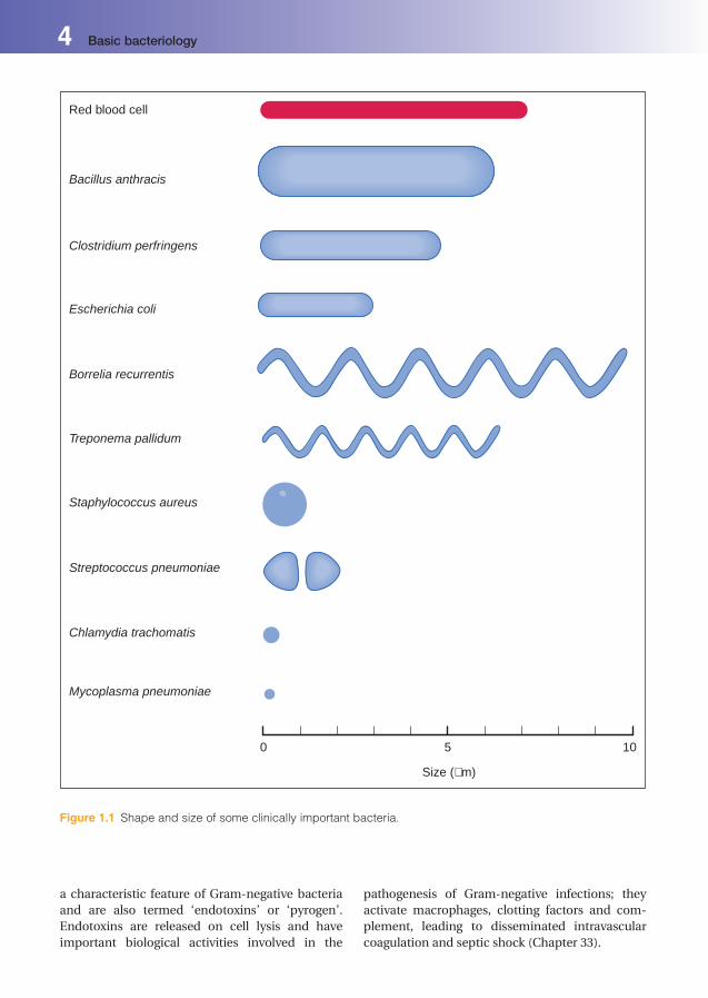
Bacterial structureBacteria are single-celled prokaryotic microorgan-
isms, and their DNA is not contained within a
separate nucleus as in eukaryotic cells. They are
approximately 0.1–10.0 mm in size (Figure 1.1) and
exist in various shapes, including spheres (cocci),
curves, spirals and rods (bacilli) (Figure 1.2). These
characteristic shapes are used to classify and iden-
tify bacteria. The appearance of bacteria following
the Gram stain is also used for identification. Bac-
teria which stain purple/blue are termed Gram-
positive, whereas those that stain pink/red are
termedGram-negative. This difference in response
to the Gram stain results from the composition of
the cell envelope (wall) (Figure 1.3), which are
described below.
Cell envelope
Cytoplasmic membrane
A cytoplasmic membrane surrounds the cytoplasm
of all bacterial cells and are composed of protein
and phospholipid; they resemble the membrane
surrounding mammalian (eukaryotic) cells but
lack sterols. The phospholipids form a bilayer into
which proteins are embedded, some spanning the
membrane. The membrane carries out many
functions, including the synthesis and export of
cell-wall components, respiration, secretion of
extracellular enzymes and toxins, and the uptake
of nutrients by active transport mechanisms.
Mesosomes are intracellular membrane struc-
tures, formed by folding of the cytoplasmic
membrane. They occur more frequently in
Gram-positive than in Gram-negative bacteria.
Mesosomes present at the point of cell division
ofGram-positive bacteria are involved in chromo-
somal separation; at other sites they may be asso-
ciated with cellular respiration and metabolism.
Cell wall
Bacteria maintain their shape by a strong rigid
outer cover, the cell wall (Figure 1.3).
Gram-positive bacteria have a relatively thick,
uniform cell wall, largely composed of peptidogly-
can, a complex molecule consisting of linear re-
peating sugar subunits cross-linked by peptide
side chains (Figure 1.4a). Other cell-wall polymers,
including teichoic acids, teichuronic acids and
proteins, are also present.
Gram-negative bacteria have a thinner peptido-
glycan layer andanadditional outermembrane that
differs in structure fromthe cytoplasmicmembrane
(Figure 1.4b). The outer membrane contains lipo-
polysaccharides on its outer face, phospholipids on
its inner face, proteins and lipoproteins which an-
chor it to the peptidoglycan. Porins are a group of
proteins that form channels through which small
hydrophilic molecules, including nutrients, can
cross theoutermembrane. Lipopolysaccharidesare
Medical Microbiology and Infection Lecture Notes, Fifth Edition. Edited by Tom Elliott, Anna Casey,Peter Lambert and Jonathan Sandoe.� 2011 Blackwell Publishing Ltd. Published 2011 by Blackwell Publishing Ltd.
a characteristic feature of Gram-negative bacteria
and are also termed ‘endotoxins’ or ‘pyrogen’.
Endotoxins are released on cell lysis and have
important biological activities involved in the
pathogenesis of Gram-negative infections; they
activate macrophages, clotting factors and com-
plement, leading to disseminated intravascular
coagulation and septic shock (Chapter 33).
Red blood cell
Bacillus anthracis
Clostridium perfringens
Escherichia coli
Borrelia recurrentis
Treponema pallidum
Staphylococcus aureus
Streptococcus pneumoniae
Chlamydia trachomatis
Mycoplasma pneumoniae
0 5
Size (µm)
10
Figure 1.1 Shape and size of some clinically important bacteria.
4 Basic bacteriology
Mycobacteria have a distinctive cell wall struc-
ture and composition that differs from that of
Gram-positive and Gram-negative bacteria. It
contains peptidoglycan but has large amounts
of high molecular weight lipids in the form of
long chain length fatty acids (mycolic acids)
attached to polysaccharides and proteins. This
high lipid content gives the mycobacteria their
acid fast properties (retaining a stain on
heating in acid), which allows them to be dis-
tinguished from other bacteria (e.g. positive
Ziehl-Neelsen stain).
The cell wall is important in protecting bacteria
against external osmotic pressure. Bacteria with
damaged cell walls, e.g. after exposure to b-lactamantibiotics such as penicillin, often rupture. How-
ever, in an osmotically balancedmedium, bacteria
deficient in cell walls may survive in a spherical
form called protoplasts. Under certain conditions
some protoplasts can multiply and are referred to
as L-forms. Some bacteria, e.g.mycoplasmas, have
no cell wall at any stage in their life cycle.
The cell wall is involved in bacterial division.
After the nuclear material has replicated and sep-
arated, a cell wall (septum) forms at the equator of
the parent cell. The septum grows in, produces a
cross-wall and eventually the daughter cells may
separate. In many species the cells can remain
attached, forming groups, e.g. staphylococci form
clusters and streptococci form long chains
(Figure 1.5).
Capsules
Some bacteria have capsules external to their cell
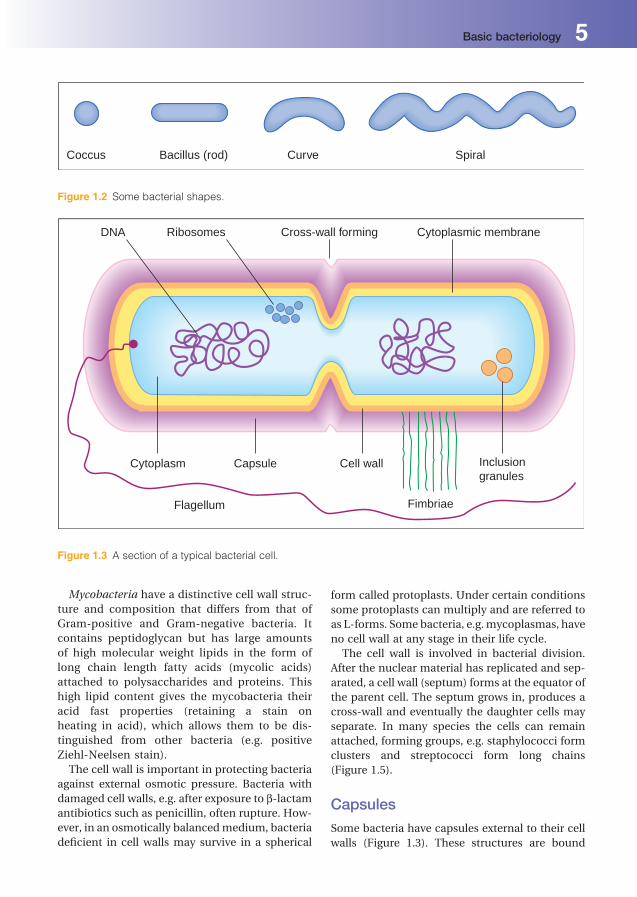
walls (Figure 1.3). These structures are bound
Coccus Curve SpiralBacillus (rod)
Figure 1.2 Some bacterial shapes.
DNA
Cytoplasm Capsule Cell wall
Flagellum Fimbriae
Inclusiongranules
Ribosomes Cross-wall forming Cytoplasmic membrane
Figure 1.3 A section of a typical bacterial cell.
Basic bacteriology 5

to the bacterial cell and have a clearly defined
boundary. They are usually polysaccharides with
characteristic compositions that can be used to
distinguish between microorganisms of the same
species (e.g. in serotyping). Capsular antigens can
beused todifferentiate between strains of the same
bacterial species, e.g. in the typing of Streptococcus
pneumoniae for epidemiological purposes. The
capsules are important virulence determinants in
both Gram-positive and Gram-negative bacteria,
because they may protect the bacteria from host
defences and, in some bacteria, aid attachment to
host cells.
Bacterial slime and biofilm
Extracellular slime layers are produced by some
bacteria. They are more loosely bound to the cell
surface than capsules and do not form a clearly
defined surface boundary. The slime layer is
composed predominantly of complex polysac-
charides (glycocalyx), which acts as a virulence
Figure 1.4 Cell wall and cytoplasmic membrane of (a) Gram-positive bacteria, (b) Gram-negative bacteria and
(c) mycobacteria. The Gram-positive bacterial cell wall has a thick peptidoglycan layer with associated molecules
(teichoic acids, teichuronic acids and proteins). The Gram-negative bacterial cell wall contains lipopolysacchar-
ides,phospholipidsandproteins in anoutermembrane linked toa thin innerpeptidoglycan layer. Themycobacterial
cell wall contains long chain length fatty acids (mycolic acids).
Staphylococci Neisseriae Pneumococci Streptococci
Figure 1.5 Some groups of bacteria.
6 Basic bacteriology

factor through the formation of biofilm, e.g. by
facilitating the attachment of Staphylococcus epi-
dermidis onto artificial surfaces, such as intra-
vascular cannulae (Figure 1.6), replacement
joints and heart valves. Once formed, biofilms
present a major problem for treatment and may
require removal of the biomedical device.
Flagella
Bacterial flagella are spiral-shaped surface fila-
ments consisting mainly of the protein, flagellin.
They are attached to the cell envelope as single
(monotrichous) or multiple (peritrichous) forms
(Figure 1.7).
Flagella facilitate movement (motility) in bacte-
ria by rapid rotation. They can be observed under
the light microscope with special stains. Flagella
are usually detected for diagnostic purposes by
observing motility in a bacterial suspension or by
spreading growth on solid media. The antigenic
nature of the flagella may be used to differentiate
between and identify strains of Salmonella spp.
Fimbriae
Fimbriae (also termed pili) are thin, hair-like ap-
pendages on the surface of many Gram-negative,
and some Gram-positive, bacteria (Figure 1.3).
They are approximately half the width of flagella,
and are composed of proteins called pilins. In
some bacteria they are distributed over the entire
cell surface.
Fimbriae are virulence factors enabling bacteria
to adhere to particular mammalian cell surfaces,
an important initial step in colonisationofmucosal
surfaces, e.g. Neisseria gonorrhoeae produce fim-
briae that bind to specific receptors of cervical
epithelial cells, whereas Streptococcus pyogenes
have fimbriae containing ‘M’ protein, which facil-
itates adhesion to human cells in the pharynx.
Specialised fimbriae are involved in genetic ma-
terial transfer between bacteria, a process called
conjugation.
Figure 1.6 Scanning electronmicrograph of
Staphylococcus epidermidis embedded in slime
attached to a catheter.
Monotrichous Peritrichous
Figure 1.7 Arrangements of bacterial flagella.
Basic bacteriology 7

Intracellular structures
Nuclear material
The bacterial chromosome consists of a single
circular molecule of double-stranded DNA, which
is maintained in a compact form within the cell by
supercoiling. When released from the cell and
uncoiled the DNA would be about 1 mm long
(10 to 100-times the length of the cell). Additional
smaller extra-chromosomal DNA molecules,
called plasmids, may also be present in bacteria.
The chromosomeusually codes for all the essential
functions required by the cell; some plasmids con-
trol important phenotypic properties of pathogen-
ic bacteria, including antibiotic resistance and
toxin production. Extracellular nuclear material
for encoding virulence and antibiotic resistance
may also be transferred between bacteria and
incorporated into the recipient’s chromosome
or plasmid. Transfer of genes encoding for viru-
lence or antibiotic resistance may account for
bacteria becoming resistant to antibiotics and for
low-virulent bacteria becoming pathogenic.
Ribosomes
The cytoplasm has many ribosomes, which con-
tain both ribonucleic acid (RNA) and proteins.
Ribosomes are involved in protein synthesis.
Inclusion granules
Various cellular inclusions, which serve as energy
and nutrient reserves, may be present in the
bacterial cytoplasm. The size of these inclusions
may increase in a favourable environment
and decrease when conditions are adverse, e.g.
Corynebacterium diphtheriae may contain high-
energy phosphate reserves in inclusions termed
‘volutin granules’.
Endospores
Endospores (spores) are small, metabolically dor-
mant cells with a thick, multi-layered coat, formed
intracellularly by members of the genera Bacillus
and Clostridium (Figure 1.8). They are highly re-
sistant to adverse environmental conditions and
may survive desiccation, disinfectants or boiling
water for several hours.
Spores are formed in response to limitations of
nutrients by a complex process (sporulation) in-
volving at least seven stages. When fully formed,
they appear as oval or round cells within the veg-
etative cell. The location is variable, but is constant
in anyonebacterial species (Figure 1.9). Spores can
remain dormant for long periods of time.However,
they are able to revert to actively-growing cells (i.e.
germinate) relatively rapidly in response to certain
conditions such as the presence of specific sugars,
amino acids or bile salts.
Spores also have an important role in the epi-
demiology of certain human diseases, such as
anthrax, tetanus, gas gangrene and infection
caused by Clostridium difficile.
The eradication of spores is of particular im-
portance in some processes, e.g. the production
of sterile products including pharmaceuticals
and surgical instruments, in routine hospital
ward and care centre cleaning, and in food
preservation.
Bacterial growthMost bacteria will grow on artificial culture media
prepared from extracts from animal or plant
tissues, which supply pre-formed nutrients and
vitamins. However, some bacteria, e.g. Mycobac-
terium leprae (leprosy) and Treponema pallidum
Central Terminal Subterminal
Figure 1.8 Size, shape andposition of bacterial spores (from left to right): non-projecting, oval, central, e.g.Bacillus
anthracis; projecting, spherical, terminal, e.g. Clostridium tetani; non-projecting, oval, subterminal, e.g.
C. perfringens.
8 Basic bacteriology

(syphilis), cannot yet be grown in vitro; other
bacteria, e.g. Chlamydia spp. and Rickettsia spp.,
only replicate intracellularly within host cells and
are therefore grown in tissue culture.
Under suitable conditions (nutrients, temper-
ature and atmosphere) a bacterial cell will in-
crease in size and then divide by binary fission
into two identical cells. These two cells are able
to grow and divide at the same rate as the parent
cell, provided that conditions including nutrient
supply remain stable. This results in an expo-
nential or logarithmic growth rate. The time
required for the number of bacteria in a culture
to double is called the generation time, e.g.
Escherichia coli has a generation time of about
20 minutes under optimal conditions. By con-
trast, Mycobacterium tuberculosis has a genera-
tion time of 24 hours.
Requirements for bacterialgrowthMost bacteria of medical importance require car-
bon, nitrogen, water, inorganic salts and a source
of energy for growth. They have various gaseous,
temperature and pH requirements, and can utilise
a range of carbon, nitrogen and energy sources.
Some bacteria also require special growth factors,
including amino acids and vitamins.
Growth requirements are important in selecting
the various culture media required in diagnostic
microbiology and in understanding the tests for
identifying bacteria.
Carbon and nitrogen sources
Bacteria are classified into two main groups ac-
cording to the type of compounds that they can
utilise as a carbon source:
1 Autotrophs utilise inorganic carbon fromcarbon
dioxideandnitrogen fromammonia,nitritesand
nitrates; they are of minor medical importance.
2 Heterotrophs require organic compounds as
their major source of carbon and energy; they
include most bacteria of medical importance.
Atmospheric conditions
Carbon dioxideBacteria require CO2 for growth; adequate
amounts are present in the air or are produced
during metabolism by the microorganisms them-
selves. A few bacteria, however, require addi-
tional CO2 for growth, e.g. Neisseria meningitidis,
Campylobacter jejuni.
OxygenBacteriamay be classified into four groups accord-
ing to their O2 requirements:
1 Obligate (strict) aerobes: grow only in the pres-
ence of oxygen, e.g. Pseudomonas aeruginosa.
2 Microaerophilic bacteria: grow best in low
oxygen concentrations, e.g. Campylobacter
jejuni.
3 Obligate (strict) anaerobes: grow only in the ab-
sence of free oxygen, e.g. Clostridium tetani.
4 Facultative anaerobes: grow in the presence or
absence of oxygen, e.g. Escherichia coli.
Temperature
Most pathogenic bacteria grow best at 37 �C. How-
ever, the optimum temperature for growth is oc-
casionally higher, e.g. for C. jejuni, it is 42 �C. Theability of some bacteria to grow at low tempera-
tures (0–4 �C) is important in food microbiology;
Listeria monocytogenes, a cause of food poisoning,
will grow slowly at 4 �C and has resulted in out-
breaks of food poisoning associated with cook-
chill products.
pH
Most pathogenic bacteria grow best at a slightly
alkaline pH (pH 7.2–7.6). There are a few excep-
tions: Lactobacillus acidophilus, present in the
Figure 1.9 Gram-stain of Clostridium sporogenes
(showing oval subterminal spores) and a Clostridium
tetani with a terminal spore (arrowed).
Basic bacteriology 9
vagina of post-pubescent females, prefers an acid
medium (pH 4.0). It produces lactic acid, which
keeps the vaginal secretions acid, thus preventing
many pathogenic bacteria from establishing infec-
tion. Vibrio cholerae, the cause of cholera, prefers
an alkaline environment (pH 8.5).
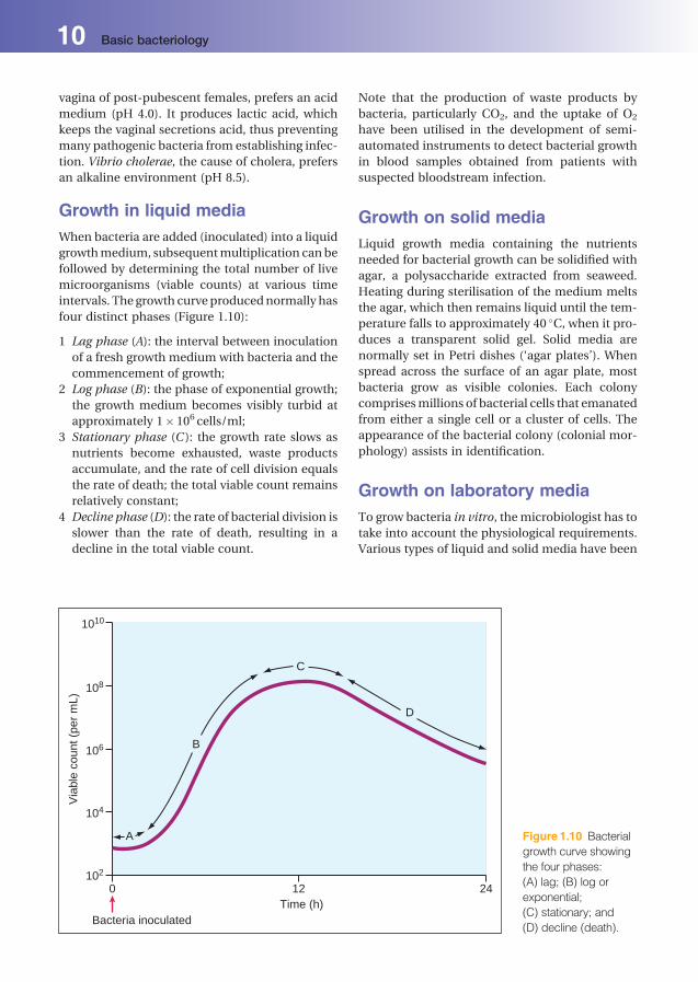
Growth in liquid mediaWhen bacteria are added (inoculated) into a liquid
growthmedium, subsequentmultiplication canbe
followed by determining the total number of live
microorganisms (viable counts) at various time
intervals. The growth curveproducednormally has
four distinct phases (Figure 1.10):
1 Lag phase (A): the interval between inoculation
of a fresh growth medium with bacteria and the
commencement of growth;
2 Log phase (B): the phase of exponential growth;
the growth medium becomes visibly turbid at
approximately 1� 106 cells/ml;
3 Stationary phase (C ): the growth rate slows as
nutrients become exhausted, waste products
accumulate, and the rate of cell division equals
the rate of death; the total viable count remains
relatively constant;
4 Decline phase (D): the rate of bacterial division is
slower than the rate of death, resulting in a
decline in the total viable count.
Note that the production of waste products by
bacteria, particularly CO2, and the uptake of O2
have been utilised in the development of semi-
automated instruments to detect bacterial growth
in blood samples obtained from patients with
suspected bloodstream infection.
Growth on solid mediaLiquid growth media containing the nutrients
needed for bacterial growth can be solidified with
agar, a polysaccharide extracted from seaweed.
Heating during sterilisation of the medium melts
the agar, which then remains liquid until the tem-
perature falls to approximately 40 �C, when it pro-
duces a transparent solid gel. Solid media are
normally set in Petri dishes (‘agar plates’). When
spread across the surface of an agar plate, most
bacteria grow as visible colonies. Each colony
comprisesmillions of bacterial cells that emanated
from either a single cell or a cluster of cells. The
appearance of the bacterial colony (colonial mor-
phology) assists in identification.
Growth on laboratory mediaTo grow bacteria in vitro, themicrobiologist has to
take into account the physiological requirements.
Various types of liquid and solid media have been
0 12Time (h)
Via
ble
coun
t (pe
r m
L)
Bacteria inoculated
A
102
104
106
108
1010
B
C
D
24
Figure 1.10 Bacterial
growth curve showing
the four phases:
(A) lag; (B) log or
exponential;
(C) stationary; and
(D) decline (death).
10 Basic bacteriology

developed for the diagnostic microbiology
laboratory.
Simple media
Many bacteria will grow in or on simplemedia, e.g.
nutrient broth/nutrient agar that contains
‘peptone’ (polypeptides and amino acids from the
enzymatic digestion of meat) and ‘meat extract’
(water-soluble components of meat containing
mineral salts and vitamins).
Enriched media
These contain additional nutrients for the isolation
of more fastidious bacteria that require special
conditions for growth, e.g. agar containing whole
blood (blood agar) or agar containing lysed blood
(chocolate agar).
Selective media
These are designed to facilitate growth of some
bacteria, while suppressing the growth of others,
and include:
. mannitol salt agar which contains increased
NaCl (salt) concentration for the recovery of
staphylococci;
. MacConkey agar, which contains bile salts and
allows the growth of bile-tolerant bacteria only;
and. antibiotics, which are frequently added to media
to allow only certain bacteria to grow while sup-
pressing or killing others.
Indicator media
These are designed to aid the detection and rec-
ognition of particular pathogens. They are often
based on sugar fermentation reactions that result
in production of acid and the subsequent colour
change of a pH indicator, e.g. MacConkey agar
contains lactose and a pH indicator (neutral red);
lactose-fermenting bacteria (e.g. Escherichia coli)
produce acid and form pink colonies, whereas
non-lactose fermenting bacteria (e.g. Salmonella
spp.) do not produce acid and form pale yellow
colonies. This property facilitates the recognition
of possible Salmonella colonies among normal
bowel flora. Note that indicator media may also
contain selective agents including antibiotics or
substances such as bile salts and crystal violet to
suppress growth of most Gram-positive microor-
ganisms. MacConkey agar is therefore both a se-
lective medium and an indicator medium.
Basic bacteriology 11

2Classification of bacteriaPeter LambertAston University, Birmingham, UK
Bacterial taxonomy andnomenclatureThe classification of microorganisms is essential
for the understanding of clinical microbiology.
Bacteria are designated by a binomial system,
with the genus name (capital letter) followed
by the species name (without capital letter),
e.g. Escherichia coli or Staphylococcus aureus.
Names are often abbreviated, e.g. E. coli and S.
aureus.
Many nomenclature problems exist with this
system that can lead to confusion, e.g. ‘bacillus’
refers to any rod-shaped bacteria, whereas the
genus Bacillus includes only the aerobic spore-
bearing rods. Other complications include the
use of alternative terminology. Streptococcus
pneumoniae is referred to as the pneumococcus,
Neisseria meningitidis as the meningococcus and
Neisseria gonnorhoeae as the gonococcus. Occa-
sionally, collective terms are used, e.g. the term
‘coliform’ may indicate E. coli or a closely related
Gram-negative bacillus found within the gut,
and the term ‘coagulase-negative staphylococci’
means staphylococci other than S. aureus. In this
text, conventional terminology is used and, where
appropriate, common alternatives are indicated.
Bacterial classificationMedically important bacteria can be subdivided
into five main groups according to their cell
shape (morphology) and staining reactions. The
basic shapes of bacteria include cocci, bacilli,
and spiral and variable shaped (pleomorphic)
forms. Each of these morphological forms is
further subdivided by their staining reactions,
predominantly the Gram and acid-fast stains
(Table 2.1). Bacteria are divided primarily into
Gram-positive or Gram-negative microorga-
nisms. Other characteristics, including the abil-
ity to grow in the presence (aerobic) or absence
(anaerobic) of oxygen, spore formation and mo-
tility, are used to divide the groups further. Sub-
division of these groups into genera is made on
the basis of various factors, including culture
properties (e.g. conditions required for growth and
Medical Microbiology and Infection Lecture Notes, Fifth Edition. Edited by Tom Elliott, Anna Casey,Peter Lambert and Jonathan Sandoe.� 2011 Blackwell Publishing Ltd. Published 2011 by Blackwell Publishing Ltd.
colonial morphology), antigenic properties and
biochemical reactions. The medically important
genera based on this classification are shown
in Table 2.2 (Gram-positives) and Table 2.3
(Gram-negatives).
Other bacterial groupsSpiral bacteriaThese are relatively slender spiral-shaped fila-
ments, which are classified into three clinically
important genera:
1 Borrelia: these are relatively large, motile spir-
ochaetes and include Borrelia vincenti and Lep-
totrichia buccalis, which cause Vincent’s angina,
Borrelia recurrentis,whichcauses relapsing fever
and Borrelia burgdorferi, which causes Lyme
disease.
2 Treponema: these are thinner and more tightly
spiralled than Borrelia. Examples include Trep-
onema pallidum (causes syphilis) and Trepone-
ma pertenue (causes yaws).
3 Leptospira: these are finer and evenmore tightly
coiled than the Treponema spp. (species plural).
They are classified within the single species of
Table 2.2 Classification of Gram-positive bacterial pathogens
GRAM-POSITIVE BACTERIA
Grouping Aerobic/anaerobicgrowth
Genus Examples of clinicallyimportant species
Gram-positive cocci
Clusters Both Staphylococcus S. aureus, S. epidermidis,S. saprophyticus
Chains/pairs Both Streptococcus and S. pneumoniae, S. pyogenes,
Enterococcus E. faecal is
Chains Anaerobic Peptostreptococcus P. magnus, P. asaccharolyticus
Gram-positive bacill i
Sporing Aerobic Bacillus B. anthracis, B. cereus
Non-sporing Both Corynebacterium C. diphtheriae
Aerobic ormicroaerophilic
L isteria L. monocytogenes
Anaerobic ormicroaerophilic
Lactobacillus L. acidophilus
Sporing Anaerobic Clostridium C. difficile, C. botul inum,C. perfringens, C. tetani
Non-sporing Anaerobic Propionibacterium P. acnes
Branching Anaerobic Actinomyces A. israel i
Aerobic Nocardia N. asteroides
Table 2.1 Main groups of bacteria
I Gram-positive cocci, bacilli and branchingbacteria
II Gram-negative cocci, bacilli and comma-shaped bacteria
III Spiral-shaped bacteria
IV Acid-fast bacteria
V Cell-wall-deficient bacteria
Classification of bacteria 13

Leptospira interrogans, which is divided serolog-
ically into two complexes. There are over 130
serotypes in the interrogans complex, many of
which are pathogenic, including L. icterohae-
morrhagiae (causes Weil’s disease) and L. cani-
cola (causes lymphocytic meningitis).
Acid-Fast BacilliThese include the genusMycobacterium. They are
identifiedbytheiracid-faststainingreaction,which
reflects their ability to resist decolorisation with
acid, after being stained with hot carbol fuchsin,
e.g. Ziehl-Neelsen stain. Mycobacteria are gener-
allydifficult tostainbyGram’smethod.Theycanbe
simply divided into the following main groups:
1 Tubercle bacilli: Mycobacterium tuberculosis
and Mycobacterium bovis (Chapter 27)
2 Leprosy bacillus: Mycobacterium leprae
3 Atypical mycobacteria: some tuberculosis-
like illnesses in humans are caused by other
species of mycobacteria. They are sometimes
also referred to as mycobacteria other than
tuberculosis (MOTT). They can grow at 27�C,
Table 2.3 Classification of Gram-negative bacterial pathogens
GRAM-NEGATIVE BACTERIA
Shape Aerobic/anaerobicgrowth
Major grouping Genus Examples of clinicallyimportant species
Cocci Aerobic Neisseria N. gonorrhoeae
N. meningitidis
Cocci Anaerobic Veillonella V. parvula
Bacilli Enterobacteriaceae(‘Coliforms’)
Enterobacter E. cloacae
Escherichia E. col i
Klebsiella K. pneumoniae
Proteus P. mirabil is
Salmonella S. typhimurium
Serratia S. marcescens
Shigella S. sonnei
Yersinia Y. enterocol itica
Bacilli Aerobic Pseudomonas P. aeruginosa
Comma shaped Both Vibrios Vibrio V. parahaemolyticus
V. cholerae
Campylobacter C. jejuni
Hel icobacter H. pylori
Bacilli Varies with genus Bordetella B. pertussis
Brucella B. abortus
Haemophilus H. influenzae,
H. parainfluenzae
Eikenella E. corrodens
Pasteurella P. mul tocida
Bacilli Aerobic Legionella L. pneumophila
Bacilli Anaerobic Bacteroides B. fragil is
Fusobacterium F. nucleatum
14 Classification of bacteria
42�C or 45�C; some produce pigment when
growing in light and are called photochromo-
gens,whereas others producepigment in light or
darkness and are referred to as scotochromo-
gens.UnlikeM. tuberculosis, othermycobacteria
can be rapid growers. All these species of myco-
bacteria are commonly referred toas theatypical
mycobacteria; examples includeMycobacterium
kansasii (photochromogenic), Mycobacterium
avium-intracellulare (non-pigmented) and My-
cobacterium chelonei (fast growing).
Cell-wall-deficient bacteriaSome bacteria do not form cell walls and are
called mycoplasmas. Pathogenic species include
Mycoplasma pneumoniae and Ureaplasma
urealyticum. It is important to distinguish myco-
plasmas from other cell-wall-deficient forms of
bacteria, which can be defined as either L-forms
or protoplasts:
. L-forms are cell-wall-deficient forms of bacteria,
which are produced by removal of a bacterium’s
cell wall, e.g. with cell-wall-acting antibiotics
such as the b-lactams. L-Forms are able to mul-
tiply and their colonial morphology is similar to
the ‘fried egg’ appearance of the mycoplasmas.. Protoplasts are bacteria that have also had
their cell walls removed. They are metabolically
active and can grow, but are unable to multiply.
They survive only in an osmotically stabilised
medium.
Classification of bacteria 15

3StaphylococciTom Elliott 1 and Peter Lambert 21University Hospitals Birmingham NHS Foundation Trust, Birmingham, UK2Aston University, Birmingham, UK
Of the many species of staphylococci that are
associated with humans, only a limited number
are clinically important; these include Staphylo-
coccus aureus, S. epidermidis and S. saprophyticus.
Their principal characteristics are shown in
Table 3.1.
DefinitionGram-positive cocci; usually arranged in clusters;
non-motile; catalase positive; non-sporing; grow
over a wide temperature range (10–42 �C), with an
optimum of 37 �C; aerobic and facultatively anaer-
obic; grow on simple media.
Classification
1 Colonialmorphology: S. aureus colonies are grey
to golden yellow (Figure 3.1); S. epidermidis and
S. saprophyticus colonies are white. Staphylo-
cocci may produce haemolysins, resulting in
haemolysis on blood agar.
2 Coagulase test: S. aureus possesses the enzyme
coagulase, which acts on plasma to form a
clot. Other staphylococci (e.g. S. epidermidis and
S. saprophyticus) do not possess this enzyme
and are often termed, collectively, ‘coagulase-
negative staphylococci’ (CoNS). There are
three methods to demonstrate the presence
of coagulase:
(a) tube coagulase test: diluted plasma is mixed
with a suspension of the bacteria; after incu-
bation, clot formation indicates S. aureus
(b) slide coagulase test: a more rapid and simple
method inwhichadropofplasma isadded to
a suspension of staphylococci on a glass
slide; visible clumping indicates the pres-
ence of coagulase.
(c) latex agglutination test: cells are mixed
with coated latex particles; visible aggluti-
nation provides simultaneous detection of
staphylococci containing coagulase and/or
protein A.
3 Deoxyribonuclease (DNAase) production: S. au-
reus possesses an enzyme, DNAase, which de-
polymerises and hydrolyses DNA; other staphy-
lococci rarely possess this enzyme.
4 Protein A detection: S. aureus possesses a
cell-wall antigen,proteinA;antibodies toprotein
A agglutinate S. aureus but not other
staphylococci.
5 Novobiocin sensitivity: useful for differentiating
between species of coagulase-negative staphy-
lococci; S. saprophyticus is novobiocin resistant
and S. epidermidis is sensitive.
Medical Microbiology and Infection Lecture Notes, Fifth Edition. Edited by Tom Elliott, Anna Casey,Peter Lambert and Jonathan Sandoe.� 2011 Blackwell Publishing Ltd. Published 2011 by Blackwell Publishing Ltd.
S. aureus
Epidemiology
S. aureus is a relatively common human commen-
sal: nasal carriage occurs in 30–50% of healthy
adults, faecal carriage in about 20% and skin car-
riage in 5–10%, particularly the axilla and perine-
um. S. aureus is spread via droplets and skin scales,
which contaminate clothing, bed linen and other
environmental sources.
Morphology and identification
On microscopy, S. aureus is seen as typical Gram-
positive cocci in ‘grape-like’ clusters. It is both
coagulase and DNAase positive (Figure 3.2). Other
biochemical tests can be performed for full
identification.
Pathogenicity
S. aureus causes disease because of its ability to
adhere to cells, spread in tissues and form abscess-
es, produce extracellular enzymes and exotoxins
(Table 3.2), combat host defences and resist treat-
ment with many antibiotics.
Adhesins
S. aureus has a wide repertoire of adhesins known
as MSCRAMMs (microbial surface components
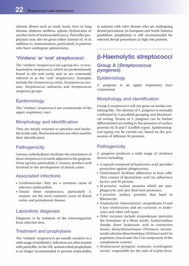
Table 3.1 Main characteristics of staphylococci
Characteristic S. aureus S. epidermidis S. saprophyticus
Coagulase þ � �Deoxyribonuclease þ � �Novobiocin S S R
Colonial appearance Golden-yellow White White
Body sites which maybe colonised
Nose Skin Periurethra
Mucosal surfaces Mucosal surfaces Faeces
Faeces
Skin
Common infections Skin (boils, impetigo,furuncles, woundinfections)
Prosthetic device-related infectionse.g. artificial valves,heart, intravenouscatheters, CSFshunts
Urinary tract infectionsin sexually activeyoung women
Abscesses
Osteomyelitis
Septic arthritis
Sepsis
Infective endocarditis
Prosthetic device-related infections
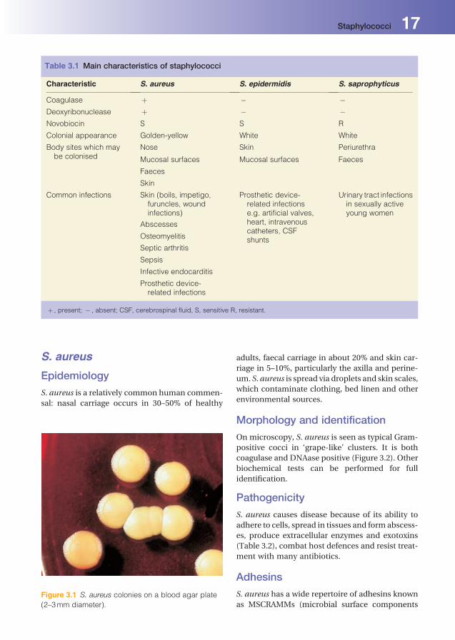
þ , present; � , absent; CSF, cerebrospinal fluid, S, sensitive R, resistant.
Figure 3.1 S. aureus colonies on a blood agar plate
(2–3mm diameter).
Staphylococci 17

recognizing adhesive matrix molecules), which
mediate adherence to host cells; these include
protein A, fibrinogen and fibronectin-binding and
collagen-binding protein.
Exotoxins and enzymes. Coagulase: S. aureus produces coagulase, an en-
zyme that coagulates plasma. Coagulase results
infibrindeposition,which interfereswithphago-
cytosis and increases the ability of the microor-
ganism to invade tissues.. Other enzymes: S. aureus may also produce
staphylokinase (results in fibrinolysis), hyal-
uronidase (dissolves hyaluronic acid), pro-
teases (degrades proteins) and lipases (solubi-
lises lipids).. Haemolysin, leukotoxin and leukocidin: several
exotoxins are produced by S. aureus; a-toxin(haemolysin) lyses erythrocytes and damages
platelets; b-toxin degrades sphingomyelin and
is toxic for many types of cell, including erythro-
cytes; leukocidin (Panton Valentine leukocidin,
PVL) lyses white blood cells and damages mem-
branes and susceptible cells.. Enterotoxins: there are six soluble enterotoxins
that are produced by almost half of all S. aureus
strains. They are heat stable (resistant at 100 �Cfor 30min), unaffected by gastrointestinal en-
zymes and are a cause of food poisoning, prin-
cipally associated with vomiting.. Exfoliative/epidermolytic toxin: some strains
produce a toxin that can result in generalised
Figure 3.2 Plate containing DNA showing clear zones
around DNAase-producing staphylococci (arrowed).
DNAase-negative staphylococci shown below.
Table 3.2 Pathogenicity factors produced by S. aureus
Factor Effect
MSCRAMMs Mediate adherence to host cells
Protein A Evade host defence/inhibits phacocytosis
Fibronectin-binding protein Mediates binding to fibronectin
Fibrinogen-binding protein Clumping factors
Capsule Evade host defences
Coagulase Generates protective fibrin layer around S. aureus
Staphylokinase Fibrinolysis
Proteases Degrade antibacterial proteins and matrix proteins
Lipases Promote interstitial spreading of microorganism
Hyaluronidase Degrades hyaluronic acid
a-Haemolysin Lyses erythrocytes, damages platelets
b-Haemolysin Degrades sphingomyelin/toxic for cells
Leukocidin/leucotoxin Lyse white blood cells
Exotoxins, e.g. enterotoxins Food poisoning with profuse vomiting
Superantigens, e.g. TSST, exfoliative toxin Toxic shock syndrome, scalded skin syndrome
NB: Toxin production varies between strains of S. aureus.
18 Staphylococci

desquamation of the skin (staphylococcal
scalded skin syndrome).. Toxic shock syndrome toxin (TSST ): this is asso-
ciated with shock and desquamation of skin,
and is usually related to an underlying S. aureus
infection.. Staphylococcal enterotoxins, TSSTs and exfolia-
tive toxin are ‘superantigens’, all of which bind
non-specifically to specific white cells, resulting
in over production of cytokines, giving rise to a
toxic shock-like presentation.
Cell envelope
Over 90% of all clinical isolates of S. aureus strains
possess a polysaccharide capsule that interferes
with opsonisation and phagocytosis. S. aureus also
possesses a cell-wall protein (protein A) that binds
the Fc component of the antibody, preventing
complement activation.
Antibiotic resistance
Many strains of S. aureus are resistant to the
antibiotic meticillin and are termed ‘meticillin-
resistant S.aureus’ (MRSA).Most resistancedepends
on theproductionofanadditionalpenicillin-binding
protein,which is encodedbyanacquiredmecA gene.
Many strains of MRSA are now resistant to multiple
antibiotics.
Laboratory diagnosis
Laboratorydiagnosis isbymicroscopicdetectionof
the microorganism in clinical samples, direct iso-
lation from the infected site or blood cultures, and
detection of serum antibodies to staphylococcal
haemolysin and DNAase. S. aureus strains can be
typed (‘fingerprinted’) by conventional methods,
including biotype and antibiogram. S. aureus can
alsobegenotypedbymolecularmethods, including
pulsed field gel electrophoresis (PFGE). Typing of
S. aureus is useful in epidemiological studies.
Treatment and prevention
Antimicrobial agents, suchasflucloxacillin, remain
the first-line treatment for sensitive strains of
S.aureus;however,theincreaseininfectionscaused
byMRSA has required the use of glycopeptide anti-
biotics such as vancomycin. Resistance to vanco-
mycin has been reported but is still rare. MRSA can
cause sepsis, ranging from wound infections to
urinary tract infections and severe sepsis and septic
shock.EpidemicstrainsofMRSA(EMRSA)havealso
been recognised. Prevention of spread through
effective infection control procedures, including
MRSA decolonisation, is therefore important.
Associated infections. Skin: boils, impetigo, furuncles, wound infec-
tions, staphylococcal scalded skin syndrome;. Respiratory: pneumonia, lung abscesses, exacer-
bations of chronic lung disease;. Skeletal: most common cause of osteomyelitis
and septic arthritis;. Invasive: bloodstream infection, infective endo-
carditis, deep abscesses (brain, liver, spleen),
toxic shock syndrome;. Gastrointestinal: toxin-mediated foodpoisoning;. Device related: indwelling catheters, prosthetic
joints and heart valves.
S. epidermidis
. S. epidermidis is both coagulase and DNAase
negative and is present in large numbers on the
human skin and mucous membranes.. S. epidermidis is a causeof bacterial endocarditis,
particularly in patients with prosthetic heart
valves and in drug addicts. It is also amajor cause
of infections of implanted devices such as cere-
brospinal shunts, hip prostheses, central venous
and peritoneal dialysis catheters.. Themicroorganism colonises implanted devices
by attaching firmly onto artificial surfaces. Some
strains also produce a slime layer (glycocalyx),
which appears to facilitate adhesion and protect
the microorganism from antibiotics and host
defences. The increased use of implanted de-
vices, particularly central venous catheters, has
resulted in S. epidermidis becoming one of the
most frequently isolated microorganisms from
blood cultures. S. epidermidis occasionally
causes urinary tract infections, particularly in
catheterised patients. When isolated from hos-
pitalised patients, S. epidermidis is often resis-
tant to antibiotics such as flucloxacillin and
erythromycin, necessitating the use of glycopep-
tide antibiotics (e.g. vancomycin).
S. saprophyticusS. saprophyticus is both coagulase andDNAaseneg-
ative and is frequently associated with urinary tract
infections in sexually active young women, occa-
sionally resulting in severe cystitiswith haematuria.
Staphylococci 19
4Streptococci andenterococciAnna CaseyUniversity Hospitals Birmingham NHS Foundation Trust, Birmingham, UK
Streptococci
Definition
Gram-positive cocci arranged in pairs or chains
(Figure 4.1); facultatively anaerobic; non-sporing;
non-motile; catalase-negative; most are capsulate;
optimum growth at 37 �C; sometimes require en-
riched media; many species exhibit characteristic
haemolysis on blood agar. Many streptococci are
human commensals (most notably of the upper
respiratory tract).
Classification
Streptococci are classified by:
1 The type of haemolysis observed on blood agar:
(a) a-haemolysis: a greenish zone forms around
colonies due to partial haemolysis of
erythrocytes (Figure 4.2). An example of
an a-haemolytic species is Streptococcus
pneumoniae.
(b) b-haemolysis: a clear zone forms around col-
oniesdue tocompletehaemolysisof erythro-
cytes (Figure 4.2).
(c) g-haemolysis: no zone is formed, as erythro-
cytes are not lysed. These streptococci are
more commonly referred to as non-haemo-
lytic streptococci.
2 Serological detection of cell wall antigens: strep-
tococci can be classified alphabetically accord-
ing to thepossessionof specificcellwall antigens
(Lancefield groups A–H and K–V). Antibodies
that react with these antigens are used to group
streptococci and are particularly useful in the
identification of b-haemolytic species. These
groups are important to distinguish, as they can
cause specific infections.
3 Biochemical reactions: some streptococci are
difficult to classify by the above characteristics,
therefore biochemical tests canbeuseful in their
identification.
a-Haemolytic streptococciStreptococcus pneumoniae(pneumococcus)
Epidemiology
S. pneumoniae is a commensal of the upper respi-
ratory tract.
Medical Microbiology and Infection Lecture Notes, Fifth Edition. Edited by Tom Elliott, Anna Casey,Peter Lambert and Jonathan Sandoe.� 2011 Blackwell Publishing Ltd. Published 2011 by Blackwell Publishing Ltd.

Morphology and identification
Cocci are most commonly observed in pairs or
chains and often have a polysaccharide capsule.
Their growth is enhanced in the presence of
additional carbon dioxide (CO2) and colonies are
typically disc shaped with central depressions
(giving a ‘draughtsmen’ appearance) (Figure 4.3).
S. pneumoniae may be differentiated from the
‘viridans’ streptococci by its sensitivity to optochin
and its solubility in bile salts.
Pathogenicity
There are in excess of 80 antigenic types of pneu-
mococcal polysaccharide capsules. A limitednum-
ber of serotypes account for themajority of cases of
infection. The capsule inhibits phagocytosis, un-
less antigen-specific opsonic antibody is present.
Pneumococci also produce pneumolysin, a mem-
brane-damaging exotoxin.
Associated infections. Respiratory tract: otitis media, sinusitis, lower
respiratory tract infection (particularly commu-
nity-acquired pneumonia);. Musculoskeletal: septic arthritis;. Gastrointestinal:spontaneousbacterialperitonitis;. Central nervous system: meningitis.
Laboratory diagnosis
This is by microscopy and microbiological culture
of specimens from the infected site, e.g. sputum,
blood, peritoneal fluid or CSF. Direct detection of
pneumococcal antigen in specimens can be un-
dertaken by various techniques. Pneumococcal
DNA can be detected in blood, CSF and other
samples by PCR.
Treatment and prophylaxis
S. pneumoniae is sensitive to a wide range of anti-
biotics. However, penicillin-resistant strains have
now emerged worldwide (Chapter 39).
A pneumococcal conjugate vaccination (PCV)
schedule is currently recommended in infants. In
addition, a pneumococcal polysaccharide vaccine
(PPV) is recommended for adults 65 years of age
or older, and for patients at particular risk of
infection. Such patients include those with
Figure 4.1 Gram stain of streptococci showing long
chains.
Figure 4.2 Colonies (each 0.5mm diameter) of a- (left)and b-haemolytic (right) streptococci on a blood agar
plate.
Figure 4.3 Streptococcus pneumoniae colonies
(arrowed) with a characteristic ‘draughtsman’-like
appearance (1mm diameter).
Streptococci and enterococci 21
chronic illness such as renal, heart, liver or lung
disease, diabetes mellitus, splenic dysfunction or
another formof immunodeficiency. Penicillin pro-
phylaxis may also be given either instead of, or in
addition to, immunisation, particularly in patients
who have undergone splenectomy.
'Viridans' or 'oral' streptococciThe ‘viridans’ streptococci are agroupofa- ornon-haemolytic streptococci, which are predominantly
found in the oral cavity and so are commonly
referred to as the ‘oral’ streptococci. Examples
include the Streptococcus mitis, Streptococcus mu-
tans, Streptococcus salivarius and Streptococcus
sanguinis groups.
Epidemiology
The ‘viridans’ streptococci are commensals of the
upper respiratory tract.
Morphology and identification
They are mostly resistant to optochin and insolu-
ble inbile salts. Biochemical tests are oftenused for
their identification.
Pathogenicity
Various carbohydrates facilitate the attachment of
these streptococci to teeth adjacent to the gingivae.
Some species, particularly S.mutans, produce acid
involved in the development of dental caries.
Associated infections. Cardiovascular: they are a common cause of
infective endocarditis;. Dental: these streptococci, particularly S.
mutans. are the most common cause of dental
caries and periodontal disease.
Laboratory diagnosis
Diagnosis is by isolation of the microorganism
from infected sites.
Treatment and prophylaxis
The ‘viridans’ streptococci are usually sensitive to a
wide rangeof antibiotics. Infections areoften treated
with penicillin. In theUK, antimicrobial prophylaxis
is no longer recommended to prevent endocarditis
in patients with valve disease who are undergoing
dental procedures. In European and North America
guidelines, prophylaxis is still recommended for
selected dental procedures in high risk patients.
b-Haemolytic streptococciGroup A (Streptococcuspyogenes)
Epidemiology
S. pyogenes is an upper respiratory tract
commensal.
Morphology and identification
Group A streptococci will not grow on media con-
taining bile. The identity of S. pyogenes is normally
confirmed by Lancefield grouping and biochemi-
cal testing. Strains of S. pyogenes can be further
differentiated according to the presence of surface
proteins M, R and T (Griffith types). Epidemiolog-
ical typing can be carried out, based on the pos-
session of different M-proteins.
Pathogenicity
S. pyogenes produces a wide range of virulence
factors including:
. A capsule composed of hyaluronic acid: provides
protection against phagocytosis.. Fimbriae/pili: facilitate adherence to host cells.
They consist of lipoteichoic acid (an adherence
factor) and M-protein.. M-proteins: surface proteins which are anti-
phagocytic and also bind host proteases.. F-proteins: surface proteins that bind to
fibronectin.. Streptolysins (haemolysins): streptolysins O and
S lyse erythrocytes and are cytotoxic to leuko-
cytes and other cell types.. Other enzymes: include streptokinase (prevents
the formation of a fibrin mesh), hyaluronidase
(breaks down hyaluronic acid in connective
tissue), deoxyribonucleases (DNAases), nicotin-
amideadeninedinucleotidase (NADase) andC5a
peptidase (inactivates the C5a component of the
complement system).. Streptococcal pyrogenic exotoxins (erythrogenic
toxins): responsible for the rash of scarlet fever.
22 Streptococci and enterococci

These are ‘superantigens’, which facilitate re-
lease of cytokines, potentially leading to shock.
Associated infections. Respiratory tract: pharyngitis, sinusitis, tonsilli-
tis, otitis media, pneumonia;. Musculoskeletal: septic arthritis;. Gastrointestinal:spontaneousbacterialperitonitis;. Skin and soft tissue: cellulitis, impetigo, erysipe-
las, scarlet fever, wound infection, necrotising
fasciitis;. Genitourinary: puerperal sepsis;. Cardiovascular: infective endocarditis.
Post-infection complications
Antibodies produced as a result of infection with S.
pyogenes may cause non-pyogenic complications
at other anatomical sites post-infection. Indeed,
rheumatic fever andacute glomerulonephritismay
develop up to 3 weeks after the streptococcal in-
fection. Inflammationof the cardiacmuscle occurs
in rheumatic fever, whilst acute glomerulonephri-
tis is characterised by inflammation of the renal
glomerulus.
Laboratory diagnosis
Diagnosis is by isolation of the microorganism
from infected sites (e.g. throat, skin, blood). The
detection of serum antibodies to streptolysin O
(ASOT: anti-streptolysin O titre) is particularly
useful for the diagnosis of post-infection compli-
cations, such as rheumatic fever or acute glomer-
ulonephritis. This is because the microorganism is
often no longer present at the time of clinical
presentation.
Treatment
S. pyogenes is sensitive to many antibiotics.
Penicillin remains the drug of choice for treatment
of infection with this microorganism.
Group B (Streptococcusagalactiae)
Epidemiology
S. agalactiae forms part of the normal faecal, per-
ineal and vaginal flora in females.
Morphology and identification
These microorganisms grow readily on blood
agar and are identified by Lancefield grouping.
Group B streptococci will grow on media contain-
ing bile.
Pathogenicity
The virulence factors for group B streptococci are
less well defined than for group A. However, more
type-specific antigens and lipoteichoic acid are
present in strains isolated from serious infection,
thus these factors appear to be important in its
virulence.
Associated infections. Respiratory tract: pneumonia in neonates and
the elderly;. Musculoskeletal: septic arthritis, osteomyelitis;. Skin and soft tissue: cellulitis;. Genitourinary: (in thepost-partumperiod) septic
abortion, endometritis, urinary tract infections;. Cardiovascular: infective endocarditis;. Central nervous system: neonatal meningitis
(neonatal acquisition of S. agalactiae is most
frequently via transmission from the colonised
mother in utero or at the time of birth.
Laboratory diagnosis
This is by isolation from the infected site. Direct
detection of antigen in body fluids can be under-
taken by various techniques.
Treatment
Penicillin is the drug of choice for treatment of
infection with S. agalactiae.
Group C and G b-haemolyticstreptococci
. Group C and G streptococci contain multiple
species.. They will not grow onmedia containing bile and
are identified by Lancefield grouping and other
commercial identification kits.. Whilst infection with these microorganisms is
less common, it can be severe. Examples of
infections caused by these microorganisms
include respiratory tract, puerperal and skin
Streptococci and enterococci 23

infection, endocarditis, meningitis, bacteraemia
and the post-streptococcal infection complica-
tion, acute glomerulonephritis.. Infections are often treated with penicillin.
Streptococcus anginosusgroup (formerly known asthe Streptococcus millerigroup)
. This group includes Streptococcus anginosus,
Streptococcus constellatus subspecies constella-
tus, S. constellatus subspecies pharynges and
Streptococcus intermedius.
. The S. anginosus group does not fall neatly into
the normal classification of streptococci. They
are often a-haemolytic, but may be b- or non-
haemolytic.. Growth of these microorganisms is enhanced by
low O2 tension and supplemented CO2. Mem-
bers of this group will grow onmedia containing
bile.. Strains most often possess Lancefield group F
antigens, but can possess A, C or G antigens or
none.. Members of the S. anginosus group are commen-
sals of the gastrointestinal and female genital
tract, and are a common cause of brain, chest
and liver abscesses.. These microorganisms are sensitive to penicil-
lin; however, antibiotic combinations such as
penicillin and metronidazole are often required
for the treatment of deep-seated abscesses due
Table 4.1 Examples of infections caused by streptococci and enterococci
Microorganism Examples of clinical infections
a-haemolytic streptococci
S. pneumoniae Otitis media/sinusitis
Pneumonia
Meningitis
‘Viridans’ streptococci Dental caries
Endocarditis
b-haemolytic streptococci
Group A (S. pyogenes) Pharyngitis, otitis media/sinusitis
Cellulitis, erysipelas, necrotising fasciitis, scarlet fever (toxin-mediated), septicarthritis
Streptococcal toxic shock syndrome, puerperal sepsis
Rheumatic fever, acute glomerulonephritis (post-infectious complications)
Group B (S. agalactiae) Neonatal: pneumonia, meningitis, bloodstream infection
Puerperal sepsis
Pneumonia
Group C and G Pharyngitis
Cellulitis,
Puerperal sepsis
Endocarditis
S. anginosus group Deep abscesses
Enterococci Urinary tract infection
(e.g. E. faecalis) Biliary tract infection
Peritonitis
Endocarditis
24 Streptococci and enterococci
to the presence of other bacteria such as
anaerobes.
Enterococci (previouslyclassified asstreptococci)Enterococcus faecalis and Enterococcus faecium are
most commonly associated with human infection;
however, infectionwith other enterococcal species
also occurs.
Definition
Gram-positive cocci arranged in pairs or chains;
facultatively anaerobic; non-sporing; non-motile
(except Enterococcus casseliflavus and Enterococ-
cus gallinarum); mostly catalase-negative (some
strains produce a pseudocatalase); some strains
are encapsulated; can grow over wide temperature
range.
Epidemiology
Enterococci are commensals, most notably of the
gastrointestinal and vaginal tract.
Morphology and identification
On blood agar they appear a-, b- or sometimes
non-haemolytic. Enterococci belong to Lancefield
group D. Speciation can be performed using bio-
chemical tests. Theywill growonmedia containing
bile and hydrolyse aesculin in the presence of 40%
bile. Since group D streptococci (i.e. the Strepto-
coccus bovis group) also share these properties,
differentiation is achieved by the fact that group
D streptococci, unlike enterococci, are unable to
hydrolyse pyrrolidonyl-b-naphthylamide (PYR)
and arginine and are not heat resistant.
Pathogenicity
Enterococcal strains can produce cytolysin, which
causes lysis of a variety of cells, including erythro-
cytes and other mammalian cells. Other virulence
factors include gelatinase, hyaluronidase, aggre-
gation substance, Enterococcal Surface Protein
(a surface adhesin) and extracellular superoxide
production.
Associated infections
They cause a number of (mainly healthcare-
associated) infections. These include:
. Gastrointestinal: peritonitis;
. Genitourinary: urinary tract infection;
. Cardiovascular: infections associated with in-
dwelling catheters, infective endocarditis.
Laboratory diagnosis
Diagnosis is by isolation of the microorganism
from infected sites.
Treatment
Enterococcal infections are often treated with
ampicillin. Enterococci, (particularly E. faecium),
have developed resistance to penicillin. In addi-
tion, the emergence of resistance to glycopeptide,
antibiotics such as vancomycin is of concern.
These bacteria are referred to as ‘vancomycin-
resistant enterococci’ (VRE) (Chapter 39).
Table 4.1 lists the infections most commonly
caused by streptococci and enterococci.
Streptococci and enterococci 25

5ClostridiaTony WorthingtonAston University, Birmingham, UK
DefinitionLarge Gram-positive bacilli (Figure 5.1); strictly an-
aerobic; spore-forming; fermentative. Whilst many
Clostridium species exist, only some are of medical
importance, including C. perfingens, C. difficile, C.
tetani, C. botulinum, C. septicum and C. tertium.
EpidemiologyClostridria are widely distributed in the environ-
ment and in the gastrointestinal tract ofmammals.
They produce highly resistant, transmissible
spores, which can survive desiccation, ultraviolet
and gamma radiation, extreme temperatures, star-
vation and disinfection. Spores of clostridia are the
vector of infection.
ClassificationBased on morphology, biochemical activity, fatty
acid production and gene sequencing techniques.
C. perfringens
Morphology and identification
Non-motile, sub-terminal spores. Forms irregu-
lar, spreading colonies on blood agar surrounded
by a double zone of b-haemolysis (inner zone of
complete lysis due to q-toxin and wider outer
zone of partial haemolysis due to a-toxin). Fivetypes (A to E) of C. perfringens are recognised
based on surface antigens and the types of toxin
produced:
1 Type A strains: commonly found in human
infections; produce only a-toxin
2 Type B to E strains: commonly found in animals
(lambs, goat, cattle); produce a- and other
toxins.
Pathogenicity
Toxin and enzyme production: a-toxin (phos-
pholipase C) is associated with toxaemia seen in
gas gangrene; hyaluronidase breaks down cellu-
lar cement facilitating spread; collagenase/pro-
teinase – liquefaction of muscle; lipase-lipid
breakdown.
Medical Microbiology and Infection Lecture Notes, Fifth Edition. Edited by Tom Elliott, Anna Casey,Peter Lambert and Jonathan Sandoe.� 2011 Blackwell Publishing Ltd. Published 2011 by Blackwell Publishing Ltd.
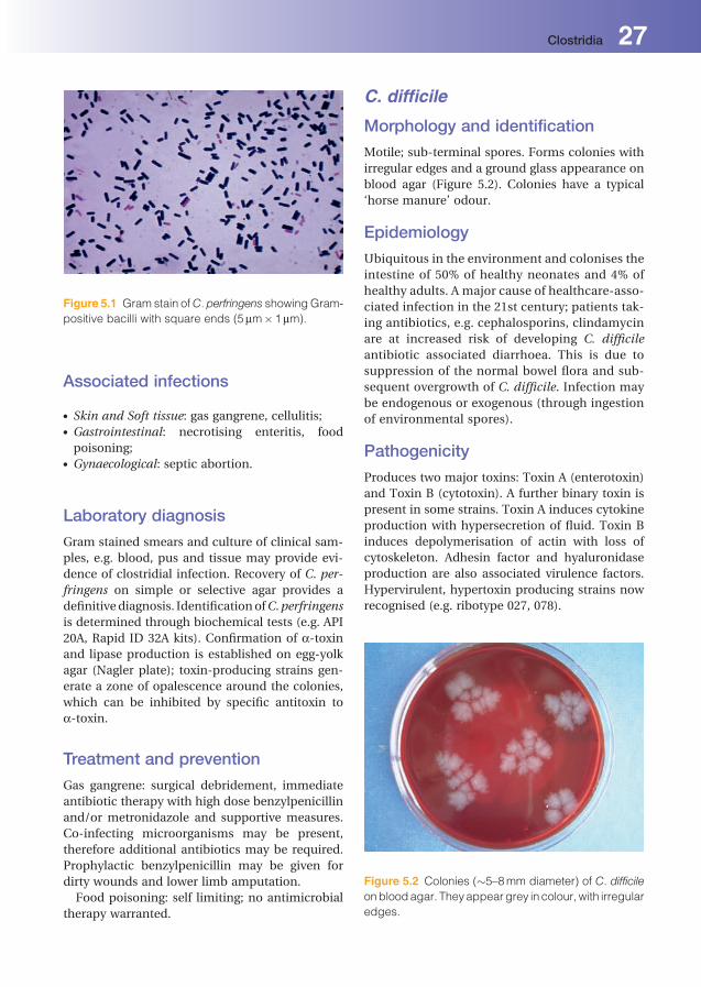
Associated infections
. Skin and Soft tissue: gas gangrene, cellulitis;
. Gastrointestinal: necrotising enteritis, food
poisoning;. Gynaecological: septic abortion.
Laboratory diagnosis
Gram stained smears and culture of clinical sam-
ples, e.g. blood, pus and tissue may provide evi-
dence of clostridial infection. Recovery of C. per-
fringens on simple or selective agar provides a
definitive diagnosis. IdentificationofC.perfringens
is determined through biochemical tests (e.g. API
20A, Rapid ID 32A kits). Confirmation of a-toxinand lipase production is established on egg-yolk
agar (Nagler plate); toxin-producing strains gen-
erate a zone of opalescence around the colonies,
which can be inhibited by specific antitoxin to
a-toxin.
Treatment and prevention
Gas gangrene: surgical debridement, immediate
antibiotic therapy with high dose benzylpenicillin
and/or metronidazole and supportive measures.
Co-infecting microorganisms may be present,
therefore additional antibiotics may be required.
Prophylactic benzylpenicillin may be given for
dirty wounds and lower limb amputation.
Food poisoning: self limiting; no antimicrobial
therapy warranted.
C. difficile
Morphology and identification
Motile; sub-terminal spores. Forms colonies with
irregular edges and a ground glass appearance on
blood agar (Figure 5.2). Colonies have a typical
‘horse manure’ odour.
Epidemiology
Ubiquitous in the environment and colonises the
intestine of 50% of healthy neonates and 4% of
healthy adults. A major cause of healthcare-asso-
ciated infection in the 21st century; patients tak-
ing antibiotics, e.g. cephalosporins, clindamycin
are at increased risk of developing C. difficile
antibiotic associated diarrhoea. This is due to
suppression of the normal bowel flora and sub-
sequent overgrowth of C. difficile. Infection may
be endogenous or exogenous (through ingestion
of environmental spores).
Pathogenicity
Produces two major toxins: Toxin A (enterotoxin)
and Toxin B (cytotoxin). A further binary toxin is
present in some strains. Toxin A induces cytokine
production with hypersecretion of fluid. Toxin B
induces depolymerisation of actin with loss of
cytoskeleton. Adhesin factor and hyaluronidase
production are also associated virulence factors.
Hypervirulent, hypertoxin producing strains now
recognised (e.g. ribotype 027, 078).
Figure 5.1 Gram stain ofC. perfringens showingGram-
positive bacilli with square ends (5mm�1mm).
Figure 5.2 Colonies (�5–8mm diameter) of C. difficile
onbloodagar. Theyappeargrey in colour,with irregular
edges.
Clostridia 27

Associated infections
. Gastrointestinal: antibiotic associated diarrhoea,
pseudomembranous colitis, fulminant colitis.
Laboratory diagnosis
Direct detection of toxin in faeces by variousmeth-
ods: cell toxicity neutralisation assay, commercial
assays (e.g. ELISA, latex agglutination) and poly-
merase chain reaction (PCR). Culture of C. difficile
on selective agar (e.g. Cycloserine Cefoxatime
Fructose Agar); genotyping of isolates by ribotying
where necessary. Assay for glutamate dehydroge-
nase (GDH) and lactoferrin in faecal samples.
Treatment and prevention
Treatment: Oral Vancomycin or Metronidazole.
Prevention of C. difficile is multifactoral and
includes: clinical awareness, judicious use of
antibiotics, infection control strategies, e.g. hand
hygiene, environmental decontamination and
cleanliness.
C. tetani
Morphology and identification
Motile; terminal spore (‘drumstick’ appearance);
produces a thin spreading film of growth without
discrete colonies on blood agar; motile via numer-
ous peritrichous flagella.
Epidemiology
C. tetani is present in mammalian intestines and
the environment (particularly manured soil).
Spores are ubiquitous in nature. Incidence of tet-
anus varies worldwide; more common in develop-
ing tropical and subtropical countries; infection is
inversely related to living standards, preventative
medicine and wound management.
Pathogenicity
Many strains are highly toxigenic, producing oxy-
gen-labile haemolysin (tetanolysin) and a potent
neurotoxin (tetanospasmin). Tetanospasmin
blocks neurotransmitter release, resulting in the
characteristic motor spasms associated with teta-
nus (e.g. lockjaw, arching of the back).
Associated infections
. Neurological: tetanus.
Laboratory diagnosis
Demonstration of characteristic ‘drumstick’ bacilli
in clinical samples, followed by anaerobic culture
on selective or blood agar; serological detection of
circulating neurotoxin by enzyme immunoassay.
Treatment and prevention
Treatment includes administration of human tet-
anus immunoglobulin and benzylpenicillin or
metronidazole. Surgical debridement and cleans-
ing of wounds is important in successful treat-
ment. Prevention includes administration of the
tetanus toxoid vaccine.
C. botulinum
Morphology and identification
Toxin producing; subterminal spores; motile with
peritrichous flagella. Seven main types of C. botu-
linum are recognised (A–G), based on antigenically
distinct toxins with identical actions.
Epidemiology
Ubiquitous saprophyte occurring in soil, vegeta-
tion, fruit and manure. Infection arises through
consumption of contaminated foodorwound con-
tamination. Human infection is commonly caused
by types A, B and D. Infection in the UK is rare.
Pathogenicity
Production of potent neurotoxin which blocks the
release of acetylcholine at neuromuscular junc-
tions, resulting in flaccid paralysis.
Associated infections
. Neurological: botulism, wound botulism, infant
botulism.
Laboratory diagnosis
Detectionof themicroorganismor its toxin in food;
toxin may be demonstrated in patient’s blood by
toxin-antitoxin neutralisation assay.
28 Clostridia

Treatment and prevention
Treatment includes administration of human
tetanus immunoglobulin and benzylpenicillin or
metronidazole. Surgical debridement and cleans-
ing of wounds is important. Prevention includes
administration of the tetanus toxoid vaccine.
Other clostridial infectionsC. septicum is associated with non-traumatic
myonecrosis more often in immunocompromised
patients. C. tertium is associated with traumatic
wound infection.
Clostridia 29

6Other Gram-positivebacteriaAnna CaseyUniversity Hospitals Birmingham NHS Foundation Trust, Birmingham, UK
In addition to the microorganisms presented in
Chapters 3, 4 and 5, various other Gram-positive
bacteria may cause human infection. Some of the
important pathogens are outlined in alphabetical
order in this chapter.
ActinomycesThe Actinomyces genus contains a large number of
species.A. israelii is themost common (but not the
only) cause of human actinomycosis.
Definition
Gram-positive bacilli: varying from short pleomor-
phic bacilli to branching filaments; anaerobic and
micro-aerophilic; non-sporing; non-motile; most
species are catalase-negative; growth is optimal on
enriched media, such as blood agar at 37 �C.
Epidemiology
Pathogenic species do not exist widely in the en-
vironment. They are commensals of the oral cavity,
large intestine and female genital tract.
Morphology and identification
Filaments may aggregate to form visible granules
(sulphur granules) in pus. Specimens from
infected tissues can be placed into sterile water
to separate out these granules. They are then
crushed and subjected to Gram and modified
Ziehl-Neelsen staining and microbiological cul-
ture. Colonies may take 10 days to appear.
Actinomyces are normally non-acid fast and
identification can be confirmed using commer-
cial identification kits, gas-liquid chromatogra-
phy and 16S ribosomal RNA gene sequencing
and analysis.
Pathogenesis
Mycelial masses penetrate human tissue.
Associated infections
. Maxillofacial: mandibular abscesses, often asso-
ciated with dental extraction or trauma may
manifest as discharging skin lesions;. Respiratory: possible role in chronic tonsillitis;. Gastrointestinal: intra-abdominal infection
(may follow appendectomy or spread from
uterine infection);. Thoracic infection;. Genitourinary: uterine infection is associated
with the use of intrauterine contraceptive
devices (A. israelii).
Medical Microbiology and Infection Lecture Notes, Fifth Edition. Edited by Tom Elliott, Anna Casey,Peter Lambert and Jonathan Sandoe.� 2011 Blackwell Publishing Ltd. Published 2011 by Blackwell Publishing Ltd.

Laboratory diagnosis
Direct isolation of themicroorganism fromclinical
specimens.
Treatment
Antibiotic therapy such as penicillin (for up to
12 months) may need to be combined with sur-
gical intervention.
BacillusThe Bacillus genus contains numerous species,
many of which are not of clinical importance. Two
important human pathogens – Bacillus anthracis
and Bacillus cereus cause anthrax and food poi-
soning, respectively.
Definition
Gram-positive bacilli often arranged in chains;
aerobic (some species are obligate aerobes and
some facultative anaerobes); spore-forming; most
species are motile; usually catalase-positive; some
species are capsulate; grow over a wide tempera-
ture range on simple media.
B. anthracis
Epidemiology
Anthrax is principally a zoonotic disease and is
common in some parts of the developing world.
Human infections can be classified as: non-indus-
trial (direct human contact with infected animals)
or industrial (processing of animal products by
humans). Spores can survive in the soil for long
periodsof timeandare relatively resistant to chem-
ical disinfectants and heat.
InfectionwithB. anthracis in theUK is rare but is
normally associated with handling imported ani-
mal products. Recent UK cases have occurred in
intravenous drug users, probably as a result of
contaminated heroin. Anthrax has been used as
a biological weapon.
Morphology and identification
Can grow under anaerobic conditions; its non-
motility allows it to be distinguished from other
Bacillus species; virulent strains are capsulate; do
not produce a zone of haemolysis on blood nor a
zone of precipitation on egg yolk agar (i.e. does not
produce lecithinase). Identification is normally
confirmed by morphological and biochemical
tests.
Pathogenicity
Virulent strains of B. anthracis possess a protein
capsule, which prevents phagocytosis. This
microorganism also produces a plasmid-encoded
exotoxin, which is composed of three proteins:
protective antigen, oedema factor and lethal fac-
tor. Protective antigen is concerned with receptor
binding and therefore the attachment and
translocation of oedema factor and lethal factor
into the cell. Oedema factor causes impairment
of macrophage function and lethal factor lysis of
macrophages.
Associated infections
Types of anthrax infection include:
. Skin and soft tissues: cutaneous anthrax is the
predominant clinical manifestation. Develop-
ment of a necrotic skin lesion (malignant pus-
tule) occurs.. Respiratory: (‘wool-sorter’s disease’): spores are
inhaled (often from wool fibres) causing pulmo-
nary oedema, haemorrhage and commonly,
death.. Gastrointestinal: consumption of contaminated
meat results in haemorrhagic diarrhorea. This
type of anthrax can also result in death.
Laboratory diagnosis
Normally by direct isolation of the microorganism
from infected sites, i.e. sputum or specimens from
skin lesions. A safety cabinet should be used to
handle such specimens.
Treatment and prevention
B. anthracis is sensitive to many antibiotics. Com-
mon therapeutic agents used are penicillin, eryth-
romycin, ciprofloxacin or doxycycline. Ciproflox-
acinordoxycyclinemaybegiven for post-exposure
prophylaxis. Anthrax vaccinations are available for
individuals at high risk, e.g. military personnel,
veterinary practitioners and farm workers. Live-
stock in endemic areas may also be vaccinated.
Other Gram-positive bacteria 31

B. cereusB. cereus can grow under anaerobic conditions, is
motile and it does not produce lecithinase; how-
ever, unlike B. anthracis, it produces a zone of
haemolysis on blood agar. This microorganism is
an important cause of food poisoning, particularly
associated with rice dishes and cereals. Pathoge-
nicity is related to the production of enterotoxins.
Food poisoning can present as the emetic form,
which has a short incubation period (typically
1–6 hours) and is characterised by nausea and
vomiting, or as the diarrhoeal form, for which the
incubation period is longer (typically 8–16 hours).
Food-poisoning is self-limiting, therefore antimi-
crobial therapy is not normally required. B. cereus
is an infrequent cause of non-gastrointestinal in-
fections, including ocular infections, pneumonia
and endocarditis.
CorynebacteriaThe Corynebacterium genus consists of several
species, most of which are human commensals.
Some species cause human infection. These in-
clude Corynebacterium diphtheriae, Corynebacte-
rium pseudotuberculosis, Corynebacterium ulcer-
ans, Corynebacterium jeikeium and Corynebacte-
rium urealyticum.
Definition
Pleomorphic Gram-positive bacilli arranged singly
or in clusters (giving the appearance of Chinese
letters); facultatively anaerobic; non-sporing; non-
motile; catalase-positive; non-capsulate; growth at
optimum of 37 �C; growth improved by addition of
serum or blood.
C. diphtheriaeC. diphtheriae is the cause of diphtheria, an upper
respiratory tract infection with cardiac and neuro-
logical complications.
Epidemiology
Infection is normally spread via nasopharyngeal
secretions frompeoplewho are infected orwhoare
carriers of themicroorganism. Diphtheria is rare in
developed countries.
Morphology and identification
. Microbiological medium containing tellurite,
e.g. Hoyle’s tellurite, is often used for culture
of throat swabs from patients with suspected
diphtheria. This selective medium allows the
growth of Corynebacterium spp. whilst suppres-
sing commensals of the upper respiratory
tract. Colonies of C. diphtheriae are grey-black
in colour.. C. diphtheriae can be differentiated into four sub-
species:var.gravis,var.mitis,var. intermediusand
var. belfanti. This and differentiation from other
species can be achieved by colonial morphology,
biochemical tests and haemolytic activity.. Toxigenic strains can be confirmed by the Elek
test (a precipitation reaction in agar), enzyme
immunoassay (EIA) or polymerase chain reac-
tion (PCR)-based techniques.
Pathogenicity
. All subspecies, except var. belfanti, may produce
the diphtheria exotoxin. It is a heat-stable poly-
peptide comprised of fragment A and fragment
B. Fragment A is involved with the translocation
of fragment B into the cell, the latter fragment
then causes cell death by inhibiting protein
synthesis. The gene which codes for production
of this toxin is carried on a bacteriophage and is
then integrated into the bacterial chromosome.. Toxin-producing strains of C. diphtheriae mul-
tiply in the pharynx and the exotoxin produced
causes necrosis of the epithelial cells.
Associated infections
. Respiratory: Diphtheria: the main clinical symp-
toms are a sore throat, thick pharyngeal pseu-
domembrane, bullneck (oedema of the neck),
fever, fatigue and headache.. Bloodstream infection: The toxinmay then cause
damage to myocardial, neural and renal cells.. Skin and soft tissue: Diphtheria toxin can also
cause cutaneous ulceration.
Laboratory diagnosis
Direct isolation of the microorganism from throat
swabs; however, laboratories in the developed
world do not routinely culture throat swabs for
C. diphtheriae, as cases are increasingly rare.
32 Other Gram-positive bacteria
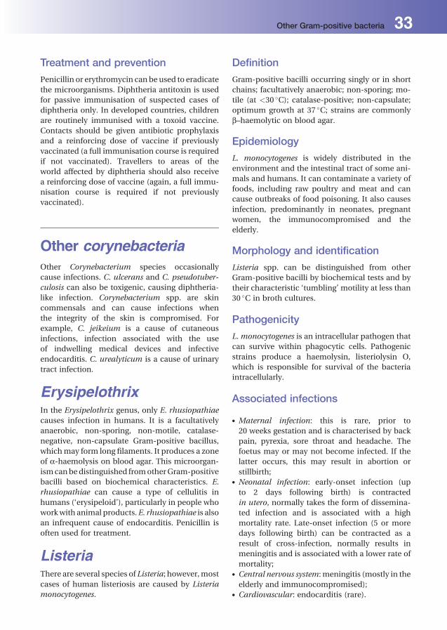
Treatment and prevention
Penicillin or erythromycin can be used to eradicate
the microorganisms. Diphtheria antitoxin is used
for passive immunisation of suspected cases of
diphtheria only. In developed countries, children
are routinely immunised with a toxoid vaccine.
Contacts should be given antibiotic prophylaxis
and a reinforcing dose of vaccine if previously
vaccinated (a full immunisation course is required
if not vaccinated). Travellers to areas of the
world affected by diphtheria should also receive
a reinforcing dose of vaccine (again, a full immu-
nisation course is required if not previously
vaccinated).
Other corynebacteriaOther Corynebacterium species occasionally
cause infections. C. ulcerans and C. pseudotuber-
culosis can also be toxigenic, causing diphtheria-
like infection. Corynebacterium spp. are skin
commensals and can cause infections when
the integrity of the skin is compromised. For
example, C. jeikeium is a cause of cutaneous
infections, infection associated with the use
of indwelling medical devices and infective
endocarditis. C. urealyticum is a cause of urinary
tract infection.
ErysipelothrixIn the Erysipelothrix genus, only E. rhusiopathiae
causes infection in humans. It is a facultatively
anaerobic, non-sporing, non-motile, catalase-
negative, non-capsulate Gram-positive bacillus,
whichmay form long filaments. It produces a zone
of a-haemolysis on blood agar. This microorgan-
ismcanbedistinguished fromotherGram-positive
bacilli based on biochemical characteristics. E.
rhusiopathiae can cause a type of cellulitis in
humans (‘erysipeloid’), particularly in people who
workwith animal products.E. rhusiopathiae is also
an infrequent cause of endocarditis. Penicillin is
often used for treatment.
ListeriaThere are several species of Listeria; however,most
cases of human listeriosis are caused by Listeria
monocytogenes.
Definition
Gram-positive bacilli occurring singly or in short
chains; facultatively anaerobic; non-sporing; mo-
tile (at <30 �C); catalase-positive; non-capsulate;optimum growth at 37 �C; strains are commonly
b–haemolytic on blood agar.
Epidemiology
L. monocytogenes is widely distributed in the
environment and the intestinal tract of some ani-
mals and humans. It can contaminate a variety of
foods, including raw poultry and meat and can
cause outbreaks of food poisoning. It also causes
infection, predominantly in neonates, pregnant
women, the immunocompromised and the
elderly.
Morphology and identification
Listeria spp. can be distinguished from other
Gram-positive bacilli by biochemical tests and by
their characteristic ‘tumbling’ motility at less than
30 �C in broth cultures.
Pathogenicity
L.monocytogenes is an intracellular pathogen that
can survive within phagocytic cells. Pathogenic
strains produce a haemolysin, listeriolysin O,
which is responsible for survival of the bacteria
intracellularly.
Associated infections
. Maternal infection: this is rare, prior to
20 weeks gestation and is characterised by back
pain, pyrexia, sore throat and headache. The
foetus may or may not become infected. If the
latter occurs, this may result in abortion or
stillbirth;. Neonatal infection: early-onset infection (up
to 2 days following birth) is contracted
in utero, normally takes the form of dissemina-
ted infection and is associated with a high
mortality rate. Late-onset infection (5 or more
days following birth) can be contracted as a
result of cross-infection, normally results in
meningitis and is associated with a lower rate of
mortality;. Central nervous system: meningitis (mostly in the
elderly and immunocompromised);. Cardiovascular: endocarditis (rare).
Other Gram-positive bacteria 33

Laboratory diagnosis
By direct isolation of the microorganism from
clinical specimens, such as blood cultures or CSF;
identification is based on the characteristics out-
lined above. Pregnant staff should not work with
suspected cultures of Listeria spp.
Treatment
L.monocytogenes is sensitive to several antibiotics;
penicillin or ampicillin are often prescribed (note:
not sensitive to cephalosporins).
NocardiaNocardia spp. are similar to Actinomyces. They are
non-sporing, non-motile Gram-positive bacilli,
which form branching hyphae; however, they only
grow aerobically, are catalase-positive and are
usually acid-fast. Nocardia species are found
widely in the environment and some are human
oral commensals. Human infection is, however,
rare. Many species exist, several of which cause
nocardiosis. Examples of human pathogens in-
clude N. asteroides and N. brasiliensis. Nocardia
spp. predominantly cause respiratory infection
acquired via inhalation of the bacilli, and also
cutaneous infection arising from inoculation of
the microorganism into the skin. Complications,
such as secondary abscesses in other anatomical
sites,may occur. Themicroorganism can be grown
from various clinical samples but may require
extended incubation. Treatment requires the use
of antibiotics, such as co-trimoxazole or minocy-
cline, for up to 12 months.
PeptostreptococcusStrictly anaerobic Gram-positive cocci from sever-
al genera may be isolated from clinical specimens.
Most clinically significant species belong to the
genus Peptostreptococcus. They are commensals
of many areas of the human body, including the
intestinal and genitourinary tracts. Peptostrepto-
coccus spp. can cause abscesses at many anatom-
ical sites, normally in association with other bac-
teria. They are mostly sensitive to penicillin and
metronidazole.
PropionibacteriaPropionibacteria are facultatively anaerobic, non-
sporing, non-motile, catalase-positive (with the
exception of Propionibacterium propionicum)
Gram-positive pleomorphic bacilli that may bifur-
cate or branch. They grow better anaerobically, are
non-acid fast and can be identified further using
commercial identification kits. Propionibacteria
constitute part of the normal flora of skin, con-
junctiva, oral cavity and intestinal tract. They are
normally non-pathogenic; however, they are an
infrequent cause of a variety of infections in
the immunocompromised and patients with
prosthetic devices.
The most common pathogenic species is Pro-
pionibacterium acnes. Its association with acne
vulgaris is widely accepted and it is also associ-
ated with cases of endophthalmitis, endocarditis,
sarcoidosis and prosthetic joint infection. This
microorganism is susceptible to most antibiotics
(however, not metronidazole).
RhodococcusThis genus contains several species, some ofwhich
cause human disease. They are aerobic, non-spor-
ing, non-motile, catalase-positive Gram-positive
bacilli. They vary from short bacilli to branched
filaments, which then fragment into cocci. These
cocci then produce bacilli when sub-cultured.
They are normally partially acid-fast.
R. equi is the major species of clinical impor-
tance. It is widely distributed in the environment
and is an opportunistic pathogen affecting
severely immunocompromised patients, such
as those infected with AIDS or those who have
undergone organ transplantation. Most infec-
tions are respiratory; however, other infection
types occur, including bacteraemia and abscesses
at various anatomical sites. The diagnosis of
infection is normally made via direct isolation
of the microorganisms from clinical specimens
such as blood or those from bronchoscopy. Sev-
eral antibiotics are used for treatment, including
vancomycin or ciprofloxacin (for �4 weeks).
Tropheryma whippleiT. whipplei, a Gram-positive bacillus is the aetio-
logical agent of Whipple’s disease. This is a rare
multi-system chronic infection involving, most
notably, the gastrointestinal tract but also other
anatomical sites. This disease is more prevalent
in middle-aged Caucasian males and common
34 Other Gram-positive bacteria

symptoms include diarrhoea, malabsorption,
weight loss and arthralgia. Culture of this intracel-
lular pathogen is difficult; however, novel immu-
nohistochemistry and serological tests have been
developed and can assist in establishing the
diagnosis. Antimicrobial therapy for one to two
years with co-trimoxazole is often used to prevent
recurrence.
Other Gram-positive bacteria 35
7Gram-negative cocciJonathan SandoeLeeds Teaching Hospitals NHS Trust and University of Leeds, Leeds, UK
NeisseriaThe genus Neisseria consists of Gram-negative
cocci and includes two important human
pathogens: N. gonorrhoeae (gonococcus) and
N. meningitidis (meningococcus) (Table 7.1).
Some Neisseria species are normal commensals of
the human upper respiratory tract.
N. gonorrhoeae
Definition
Gram-negative kidney-shaped cocci, usually in
pairs (diplococci) (Figure 7.1), aerobic.
Epidemiology. Obligate human parasite;. Manywomenare asymptomatic andmay act as a
reservoir of infection;. Highly transmissible by sexual contact;. Vertical transmission to neonates may occur
during passage through an infected birth canal;. Occurs worldwide;. Infections are most common among sexually
active young adults;. Genotypic (PFGE, AFLP) typing may be under-
taken to differentiate between strains of gono-
cocci, e.g. in child abuse cases.
Laboratory identification
Provisional identification can be made by
microscopy if kidney-shaped Gram-negative dip-
lococci are seen in pus cells. Confirmation is based
oncultures: growthongonococcal selectivemedia,
colonial morphology, Gram-stain appearance,
positive oxidase reaction, catalase production,
biochemical reactions (including carbohydrate
fermentation) and immunological tests (for
detection of specific gonococcal antigens). Gono-
coccal DNA can also be detected directly from
clinical samples,using variousmolecularmethods.
Pathogenicity
Gonococci have cell-surface pili, which aid adher-
ence to mucosal surfaces of the cervix, urethra,
rectum and upper respiratory tract, thus initiating
infection. Other virulence factors include IgA pro-
teases. Gonococci can survive intracellularly with-
in polymorphonucleocytes (PMNs) and some
strains are also able to resist serum lysis. Re-infec-
tions occur, because protective immunity does not
develop.
Associated infections. Genitourinary: urethritis (Figure 7.2), cervicitis;
complications include epididymitis, prostatitis,
Medical Microbiology and Infection Lecture Notes, Fifth Edition. Edited by Tom Elliott, Anna Casey,Peter Lambert and Jonathan Sandoe.� 2011 Blackwell Publishing Ltd. Published 2011 by Blackwell Publishing Ltd.
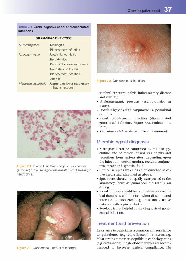
urethral stricture, pelvic inflammatory disease
and sterility;. Gastrointestinal: proctitis (asymptomatic in
many);. Occular: hyper-acute conjunctivitis, periorbital
cellulitis;. Blood: bloodstream infection (disseminated
gonococcal infection, Figure 7.3), endocarditis
(rare);. Musculoskeletal: septic arthritis (uncommon).
Microbiological diagnosis. A diagnosis can be confirmed by microscopy,
culture and/or molecular analysis of pus and
secretions from various sites (depending upon
the infection): cervix, urethra, rectum, conjunc-
tiva, throat and synovial fluid.. Clinical samples are cultured on enriched selec-
tive media and identified as above.. Specimens should be rapidly transported to the
laboratory, because gonococci die readily on
drying.. Blood cultures should be sent before antimicro-
bial therapy is commenced when disseminated
infection is suspected, e.g. in sexually active
patients with septic arthritis.. Serology is not helpful in the diagnosis of gono-
coccal infection.
Treatment and prevention
Resistance to penicillins is commonand resistance
to quinolones (e.g. ciprofloxacin) is increasing.
Most strains remain susceptible to cephalosporins
(e.g. ceftriaxone). Single-dose therapies are recom-
mended to increase patient compliance. No
Table 7.1 Gram-negative cocci and associatedinfections
GRAM-NEGATIVE COCCI
N. meningitidis Meningitis
Bloodstream infection
N. gonorrhoeae Urethritis, cervicitis
Epididymitis
Pelvic inflammatory disease
Neonatal ophthalmia
Bloodstream infection
Arthritis
Moraxella catarrhalis Upper and lower respiratorytract infections
Figure 7.1 Intracellular Gram-negative diplococci
(arrowed) ofNeisseria gonorrhoeae (0.8mmdiameter) in
neutrophils.
Figure 7.2 Gonococcal urethral discharge.
Figure 7.3 Gonococcal skin lesion.
Gram-negative cocci 37

vaccine is available. Prevention of gonorrhoea
includes sex education, promotion of public
awareness and the use of condoms. Contact trac-
ing is essential in preventing further spread of
disease.
N. meningitidis
Definition
Gram-negative kidney-shaped cocci, usually in
pairs (diplococci); aerobic.
Epidemiology. Humans are the only natural host.. Ten percent of the population are asymptomatic
carriers in the upper respiratory tract.. Spread from human to human is via droplets or
direct contact.. Sporadic cases, clusters and epidemics occur
worldwide.. Serotyping (performed by reference laborato-
ries) can be used to identify outbreaks. At least
13 serogroups of meningococci have been
identified. Typing is based on capsular polysac-
charides; the most important groups are A, B, C,
D, X, Y and W-135. In the UK, the majority of
cases are caused by group B and group C is now
uncommon.. Patients with genetic or drug induced defects
of the later components of the complement
system are predisposed to meningococcal
infections.. In temperate climates most infections occur in
patients aged less than 5 years or 15–19 years.
Laboratory identification
Provisional identification is by microscopy when
kidney-shaped, Gram-negative cocci are seen
within polymorphonucleocytes. Confirmation of
identification is based on cultures: colonial mor-
phology, Gram-stain appearance, positive oxidase
test and biochemical reactions (including carbo-
hydrate fermentation).
Pathogenicity
A polysaccharide capsule protects against phago-
cytosis and promotes intracellular survival. Cell-
wall endotoxins (lipopolysaccharide) are impor-
tant in the pathogenesis of severe meningococcal
disease. Colonisation of nasopharynx occurs; local
invasion may follow with bacteraemia and men-
ingeal involvement.
Associated infections. Central nervous system: meningitis;. Blood: bloodstream infection (‘septicaemia’)
without meningitis (less common);. Musculoskeletal: osteomyelitis, septic arthritis;. Occular: conjunctivitis;. Genitourinary: urethritis;. Respiratory: pneumonia (rare);. Cardiovascular: pericarditis (rare).
Microbiological diagnosis. Specimens: cerebrospinal fluid (CSF) or pus for
microscopy, culture, antibiotic susceptibility
testing and PCR;. Blood cultures: should also be sent, in addition to
a blood sample for PCR;. Nasopharyngeal swabs: can determine carriage
of meningococci.
Treatment
Penicillin or cefotaxime are first-line treatments,
chloramphenicol is still used for patients with a
true penicillin allergy; rifampicin or ciprofloxacin
should also be given to eradicate nasopharyngeal
carriage, except in patients treated with certain
cephalosporins.
Prevention includes antibiotic prophylaxis for
close contacts. Meningococcal group C vaccine
(group B vaccine being developed) and tetravalent
(A, C, Y, W135) polysaccharide vaccine are avail-
able for travellers to high incidence areas.
Moraxella catarrhalis
Definition
Gram-negative diplococci and tetrads; aerobic.
Epidemiology. Part of the normal flora of the upper respiratory
tract;. Opportunistic pathogen;. Transmission results from direct contact with
contaminated secretions or droplets;. Most infections occur in the young or patients
with underlying respiratory disease.
38 Gram-negative cocci
Laboratory identification. Microscopy: Gram-negative diplococci;. Colonies may be characteristically nudged
across the surface of the agar plates with a
bacteriological loop much like a ‘hockey
puck’;. Reduces nitrates and oxidase, catalase and
DNAse positive;. Distinguished fromNeisseria species by butyrate
esterase detection (Tributyrin test).
Pathogenicity
Some strains of M. catarrhalis possess fimbriae
that aid adherence to respiratory epithelium.
Cell-wall endotoxin (lipopolysaccharide) may play
a role in the disease process.
Associated infections
Respiratory: otitis media, sinusitis, bronchitis,
pneumonia.
Laboratory diagnosis. Specimens of sputum, bronchoalveolar lavage
fluid, tympanocentesis fluid and sinus aspirates
for culture and microscopy;. Serological tests are not used.
Treatment
Most clinical isolates produce b-lactamase and are
resistant to amoxicillin. Infection may be treated
with co-amoxiclav (amoxicillinplus clavulanate – a
b-lactamase inhibitor), tetracyclines (not children)
or cephalosporins.
Other Gram-negativecocciOther Gram-negative cocci, such as Neisseria
subflava, Neisseria lactamica and Veillonella
species, are part of the normal flora of the upper
respiratory tract and rarely cause disease.
Gram-negative cocci 39
8EnterobacteriaceaePeter LambertAston University, Birmingham, UK
The Enterobacteriaceae include the following
genera: Escherichia, Enterobacter, Klebsiella, Pro-
teus, Serratia, Shigella, Salmonella and Yersinia
(Table 8.1).
They are Gram-negative bacilli, which are found
as commensals in the intestinal tract of mammals.
They are also referred to as coliforms or enteric
bacteria.
Definition
Aerobic and facultatively anaerobic growth; opti-
mal growth normally at 37 �C; grow readily on
simple media; ferment wide range of carbohy-
drates; oxidase-negative; some are motile; bile-
tolerant and grow readily on bile-salt-containing
media, e.g. MacConkey’s agar.
Morphology and identification. Fermentation of lactose to producepink colonies
on MacConkey’s agar is characteristic of Escher-
ichia, Enterobacter and Klebsiella. Salmonella,
Shigella, Serratia, Proteus and Yersinia do not
ferment lactose and form pale colonies on
MacConkey’s agar.. Various tests are used for identification:
(a) oxidase (negative);
(b) carbohydrate fermentation reactions;
(c) production of urease (which splits urea with
release of ammonia);
(d) hydrogen sulphide production;
(e) amino acid decarboxylation;
(f) indole production.
Commercial kits based on these biochemical
tests are available for identification of the
Enterobacteriaceae.. Enterobacteriaceae possess a variety of antigens:
these may include lipopolysaccharide (‘O’), fla-
gellar (‘H’) and capsular polysaccharide (‘K’)
antigens. These are used to subdivide (serotype)
some genera and species, e.g. Escherichia,
Salmonella.
EscherichiaThe genus Escherichia currently contains several
species. However, E. coli (Figure 8.1) is the spe-
cies most frequently isolated from humans.
Epidemiology and associatedinfections
Although E. coli is a harmless commensal of the
human intestine, some strains (identified as par-
ticular O, H, and K serotypes) can cause infections
of the gastrointestinal tract, urinary tract, biliary
tract, lower respiratory tract, bloodstream,
haemolytic-uraemic syndrome (HUS), haemor-
rhagic colitis and neonatal meningitis.
Medical Microbiology and Infection Lecture Notes, Fifth Edition. Edited by Tom Elliott, Anna Casey,Peter Lambert and Jonathan Sandoe.� 2011 Blackwell Publishing Ltd. Published 2011 by Blackwell Publishing Ltd.

Pathogenicity. Specific fimbriae facilitate adherence tomucosal
surfaces and colonisation of the intestinal and
urinary tracts.. The lipopolysaccharide (endotoxin) in the cell
wall is liberated when Gram-negative bacteria
lyse, resulting in production of inflammatory
mediators (cytokines and nitric oxide) and com-
plement activation. This results in endotoxic
shock and intravascular coagulopathy.
. The K1 capsular polysaccharide antigen is asso-
ciated with neonatal meningitis.. A number of distinct infections are mediated by
the different protein toxins produced by E. coli.. VTEC (verocytotoxin-producing E. coli, particu-
larly the O157:H7 serotype, are an important
cause of diarrhoea and HUS. These are also
referred to as enterohaemorrhagic E. coli (EHEC).. Most are sporadic cases and have the following
key features:
T zoonotic infections mainly from cattle, but
also from vegetables washed in contaminated
water;
T low infecting dose;
T acquired by eating undercooked contaminat-
ed meat and vegetables;
T damage gut endothelium, resulting in hae-
morrhagic colitis;
T HUS occurs in about 5% of patients,
which results in renal failure, oliguria,
thrombocytopenia.
Diarrhoea caused by other E. coli:
. Enteropathogenic (EPEC): cause of infantile
diarrhoea;. Enterotoxigenic (ETEC): travellers’ diarrhoea,
non-invasive;
Table 8.1 Enterobacteriaceae infections
Genus/species Common infections
Escherichia coli Urinary tract infection
Intra-abdomal infection
Wound infection
Klebsiella spp Urinary tract infection
Pneumonia
Intravascular catheter-related infection
Enterobacter spp Hospital-acquired pneumonia
Serratia spp Wound infection
Proteus spp Urinary tract infection
Salmonella serotypes Typhi and Paratyphi Enteric fever and bloodstream infection
Other salmonellae Enteritis
Shigella spp Enteritis
Yersinia enterocolitica Enteritis
Yersinia pestis Plague
Yersinia pseudotuberculosis Mesenteric adenitis
Figure 8.1 Gram stain of Escherichia coli showing
Gram-negative bacilli (2mm�0.5mm).
Enterobacteriaceae 41

. Enteroinvasive (EIEC): causes dysentery-like
illness;. Enteroaggregative (EAEC): watery diarrhoea
without fever.
Laboratory diagnosis. Diagnosis is by direct isolation of the microor-
ganism from clinical samples, e.g. faeces, urine
and blood.. Identification of some pathogenic strains, e.g.
VTEC, EPEC, may be achieved by serotyping.
Antibacterial therapy
E. coli is commonly resistant to penicillin and
ampicillin by production of b-lactamase enzymes.
Production of extended spectrum b-lactamases
(ESBLs), which inactivate many penicillins and
cephalosporins, is an increasing problem. Antibio-
tics often used to treat E. coli infections include the
cephalosporins, trimethoprim, ciprofloxacin and
aminoglycosides; strains isolated from hospita-
lised patients are often more resistant to antibio-
tics and therefore local antibiotic sensitivity pat-
terns need to be considered.
KlebsiellaThe genus Klebsiella contains a number of spe-
cies, including K. pneumoniae and K. oxytoca;
these can be distinguished on the basis of bio-
chemical tests. Pathogenicity is associated with
capsular polysaccharide and lipopolysaccharide
(endotoxin) production. There are many capsu-
lar serotypes. Klebsiellae are widespread in the
environment and in the intestinal flora of hu-
mans and other mammals. Infections are often
opportunistic and associated with hospitalisa-
tion, particularly in high-dependency units. They
include pneumonia, urinary tract and wound
infection, and neonatal meningitis. Outbreaks of
healthcare-associated infection occur.
Klebsiella spp. often produce b-lactamases
and are resistant to ampicillin. Cephalosporins
(e.g. cefotaxime), b-lactamase-inhibitor/penicillin
combinations (e.g. co-amoxiclav) and aminogly-
cosides (e.g. gentamicin) can be used to treat
Klebsiella infections, but multiply resistant strains
may limit antibiotic choice. Extensiveuseof broad-
spectrum antibiotics in hospitalised patients has
led to development of multi-drug-resistant strains
that produce ESBLs.
Enterobacter and SerratiaEnterobacter and Serratia spp. are closely related to
Klebsiella spp. Infections occur principally in hos-
pitalised patients and include the lower respiratory
and urinary tracts. Hospital cross-infection with
antibiotic-resistant strains is a particular problem.
ESBL production in strains of Enterobacter is an
increasing problem.
SalmonellaThe genus Salmonella contains a large number of
species (more correctly, serotypes). Salmonella
serotype Typhi and Salmonella serotype Paratyphi
cause enteric fever (typhoid or paratyphoid); other
salmonellae cause enteritis.
Classification. Over 2,000 serotypes are distinguished, most of
which belong to the species S. enterica. However,
many of these have been given binomial names
(e.g. Salmonella typhimurium and Salmonella
enteritidis), although they are not separate spe-
cies. In clinical practice, laboratories identify
microorganisms according to their binomial
name.. Salmonella spp. have both H and O antigens.
There are over 60 different O antigens, and
individual strains may possess several O and
H antigens; the latter can exist in variant forms,
termed ‘phases’. Salmonella serotype Typhi
also has a capsular polysaccharide antigen
referred to as ‘Vi’ (for virulence), which is
related to invasiveness.. Agglutination tests with antisera for different O
and H antigens form the basis for the serological
classification of Salmonella spp. Further strain
differentiation of Salmonella spp. for epidemio-
logical purposes can be achieved by phage
typing.
Epidemiology
Salmonella spp. are commensals ofmany animals,
including poultry, domestic pets, birds and
humans. Transmission is via the faecal-oral route.
The infective dose is relatively high (�106 micro-
organisms) andmultiplication in food is important
42 Enterobacteriaceae

for effective transmission. A chronic carrier state
can occur.
Morphology and identification. Salmonella spp. are motile and produce acid,
and occasionally gas, from glucose and
mannose.. They are resistant to sodium deoxycholate,
which inhibits many other Enterobacteria-
ceae. Deoxycholate agar is used as a selective
media to isolate Salmonella spp. from stool
specimens.. Salmonella spp. do not ferment lactose and form
pale colonies on MacConkey’s medium; on xy-
lose lysine deoxycholate (XLD) agar, many Sal-
monella spp. form pale colonies with black cen-
tres as a result of H2S production. This aids
recognition of Salmonella colonies in mixed
cultures.. Further biochemical tests are required for de-
finitive identification. Serotyping of O and H
antigens by slide agglutination is used for
speciation.
Pathogenicity
Salmonella spp. can survive the acidic pH of the
stomach and invade the gut, resulting in an in-
flammatory response and subsequent diarrhoea.
Associated infections. Salmonella infections: (caused by non-typhoid
salmonellae;. Enterocolitis/gastroenteritis: rarely associated
with bloodstream infection, osteomyelitis, septic
arthritis or abscesses;. Enteric fever: caused by Salmonella serotype
Typhi and Salmonella serotype Paratyphi.
Enteric fever is prevalent in Asia, South America
and Africa; approximately 300 cases per year
in the UK. Salmonella spp. may persist in biliary
and urinary tracts after recovery.
Laboratory diagnosis. Enterocolitis: culture of stool samples on selec-
tive media, e.g. XLD, DCA (deoxycholate citrate
agar), and enrichment media, e.g. selenite broth;
identification of Salmonella spp. by biochemical
andagglutination tests. Phage typing canbeused
for typing individual strains.
. Enteric fever: isolation of Salmonella serotypes
Typhi or Paratyphi from blood cultures (first
week of infection), urine (second week) or faeces
(first week onwards). Serology (Widal’s test) is
now rarely performed, because of unreliable
results.
Treatment and prevention. Enteric fever: ciprofloxacin (though resistance is
increasing);. Enterocolitis: self-limiting; antibiotics (e.g. cip-
rofloxacin and cefotaxime) reserved for
severe or invasive infection, particularly in the
elderly, very young or ‘immunocompromised’
individuals;. Typhoid immunisation; avoidance of contami-
nated water/food.
Shigella
Classification
The main pathogenic species are S. sonnei,
S. boydii, S. dysenteriae and S. flexneri. They are
distinguished by biochemical reactions and anti-
genic characteristics (‘O’ antigens).
Epidemiology
Obligate human pathogens with no animal reser-
voirs; transmission via faecal-oral route with low
infective dose (10–200 microorganisms). Direct
person-to-person spread is common; chronic car-
rier state is rare.
Morphology and identification
Shigella spp. arenon-motile (theyhavenoflagella).
They are resistant to sodium deoxycholate and
grow on deoxycholate agar (see Salmonella). They
are non-lactose or late lactose (S. sonnei) fermen-
ters. Further biochemical tests are carried out for
definitive identification and serotyping by slide
agglutination is used for speciation.
Pathogenicity
Shigella spp. express an intestinal adherence fac-
tor, which aids colonisation within the gut. They
cause disease by invasion and destruction of the
colonic mucosa, and also produce an enterotoxin
(cytotoxin) known as Shiga toxin, which can cause
Enterobacteriaceae 43
microangiopathy, HUS and thrombocytopenic
purpura.
Associated infections
Self-limiting diarrhoeal illness, dysentery (diar-
rhoea with blood and pus, fever, abdominal pain)
(HUS and bloodstream infection are rare).
Laboratory diagnosis
Stool culture on selective media, e.g. XLD.
Treatment
Antibiotics (e.g. ciprofloxacin) reserved for severe
cases (often caused by S. dysenteriae).
ProteusThe genus Proteus contains a number of species,
e.g. P. mirabilis and P. vulgaris. Characteristics
include:
. non-lactose fermenting, produce pale colonies
on MacConkey’s agar;. motile, tendency to ‘swarm’ on blood agar;. important cause of urinary tract andoccasionally
abdominal wound infection.
YersiniaThe genus Yersinia contains three human patho-
gens: Y. pestis, Y. pseudotuberculosis and Y. enter-
ocolitica; these species are identified by biochem-
ical tests.
Y. pestisY. pestis is the cause of plague (black death).
Although mainly of historical interest in Europe,
plague remains endemic in some areas of the
world. It is primarily a pathogen of rodents and is
transmitted to humans via infected fleas; lymph
nodes associated with the flea bite enlarge to form
a bubo (bubonic plague). Bloodstream invasion
and pneumonia may follow (pneumonic plague).
Person-to-person spread via droplets occurs in
pneumonic plague. Y. pestis can be isolated from
blood, bubo aspiration, sputum, throat swabs and
skin scrapings. Treatment for Y. pestis infection is
with anaminoglycoside or tetracycline. Laboratory
diagnosis is by microscopy and culture of clinical
material.
Y. pseudotuberculosisThis is primarily an animal pathogen, but in hu-
mans it is an occasional cause of mesenteric ade-
nitis, rarely septicaemia. It is probably transmitted
to humans via contaminated food. Laboratory di-
agnosis is through culture of faeces on selective
agar, e.g. CIN agar and cold enrichment of faeces.
Y. enterocoliticaY. enterocolitica causes diarrhoeal disease, termi-
nal ileitis and mesenteric adenitis. Infection may
be complicated by septicaemia or reactive poly-
arthritis. A zoonotic infection of domestic andwild
animals, transmission is via the faecal-oral route.
Laboratory diagnosis is by culture of faeces on
selective agar, e.g. CIN agar, cold enrichment of
faeces, culture of blood specimens or serology.
44 Enterobacteriaceae

9Haemophilus and otherfastidious Gram-negativebacteriaJonathan SandoeLeeds Teaching Hospitals NHS Trust and University of Leeds, Leeds, UK
This chapter includes an arbitrary collection of
Gram-negative coccobacilli, which are described
collectively because of common characteristics,
rather than being part of a formal classification
system. They are all Gram-negative bacteria that
can be difficult to culture and usually are mem-
bers of the normal microbial flora of humans or
animals. Most are therefore opportunistic
pathogens. Infections associated with fastidious
Gram-negative bacteria are outlined in
Table 9.1.
Haemophilus influenzaeDescription/definitionGram-negative bacilli; fastidious growth require-
ments; aerobic and anaerobic growth.
Epidemiology
. Common commensal in the upper respiratory
tract;
. Capsulated (more pathogenic) strains are also
carried in a small number of healthy individuals;. Carriers are an important reservoir for invasive
disease;. Transmission is by respiratory secretions and
droplets;. Most infections occur in children and patients
with underlying respiratory disease;. Type b strains (see Pathogenicity) were a com-
mon cause of invasive infection, particularly in
small children (aged6months to 5 years), prior to
introduction of the Hib vaccine.
Laboratory identification
. Gram-negative bacilli;
. Variable oxidase reaction;
. Nutritionally demanding and grows only on en-
riched media containing haemin (factor X) and
nicotinamide adenine dinucleotide (factor V);. Simple nutrient agar contains no X or V factor
and strains ofH. influenzaewill only grow around
paper discs containing both these factors
(Figure 9.1);
Medical Microbiology and Infection Lecture Notes, Fifth Edition. Edited by Tom Elliott, Anna Casey,Peter Lambert and Jonathan Sandoe.� 2011 Blackwell Publishing Ltd. Published 2011 by Blackwell Publishing Ltd.
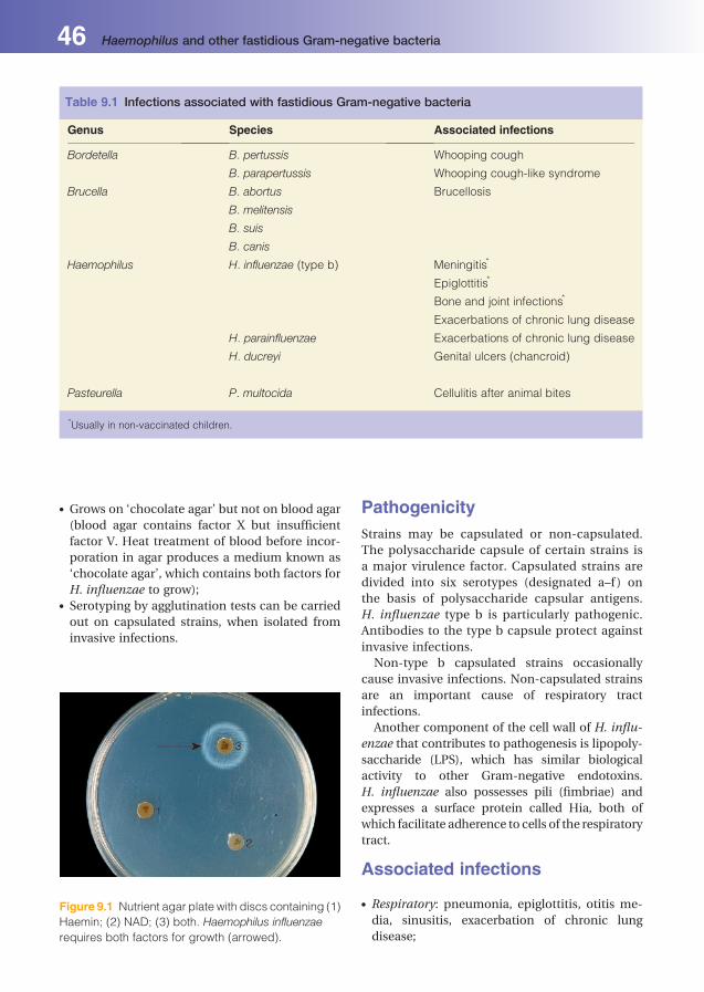
. Grows on ‘chocolate agar’ but not on blood agar
(blood agar contains factor X but insufficient
factor V. Heat treatment of blood before incor-
poration in agar produces a medium known as
‘chocolate agar’, which contains both factors for
H. influenzae to grow);. Serotyping by agglutination tests can be carried
out on capsulated strains, when isolated from
invasive infections.
PathogenicityStrains may be capsulated or non-capsulated.
The polysaccharide capsule of certain strains is
a major virulence factor. Capsulated strains are
divided into six serotypes (designated a–f ) on
the basis of polysaccharide capsular antigens.
H. influenzae type b is particularly pathogenic.
Antibodies to the type b capsule protect against
invasive infections.
Non-type b capsulated strains occasionally
cause invasive infections. Non-capsulated strains
are an important cause of respiratory tract
infections.
Another component of the cell wall of H. influ-
enzae that contributes to pathogenesis is lipopoly-
saccharide (LPS), which has similar biological
activity to other Gram-negative endotoxins.
H. influenzae also possesses pili (fimbriae) and
expresses a surface protein called Hia, both of
which facilitate adherence to cells of the respiratory
tract.
Associated infections
. Respiratory: pneumonia, epiglottitis, otitis me-
dia, sinusitis, exacerbation of chronic lung
disease;
Table 9.1 Infections associated with fastidious Gram-negative bacteria
Genus Species Associated infections
Bordetella B. pertussis Whooping cough
B. parapertussis Whooping cough-like syndrome
Brucella B. abortus Brucellosis
B. melitensis
B. suis
B. canis
Haemophilus H. influenzae (type b) Meningitis*
Epiglottitis*
Bone and joint infections*
Exacerbations of chronic lung disease
H. parainfluenzae Exacerbations of chronic lung disease
H. ducreyi Genital ulcers (chancroid)
Pasteurella P. multocida Cellulitis after animal bites
*Usually in non-vaccinated children.
Figure 9.1 Nutrient agar plate with discs containing (1)
Haemin; (2) NAD; (3) both. Haemophilus influenzae
requires both factors for growth (arrowed).
46 Haemophilus and other fastidious Gram-negative bacteria
. Central nervous system: meningitis;
. Musculoskeletal: osteomyelitis, septic arthritis
(uncommon);. Skin and soft tissues: cellulitis (including orbital
cellulitis);. Cardiovascular: endocarditis (rare).
Laboratory diagnosis
. Respiratory: send respiratory secretions for cul-
ture and sensitivity. Epiglottitic swabs for mi-
croscopy and culture should be collected under
direct vision by an ENT specialist. Sinus wash-
out fluid can be cultured.. Central nervous system: send cerebrospinal fluid
(CSF) for microscopy and culture.. Musculoskeletal: send bone or synovial fluid for
microscopy and culture.. Skin and soft tissues: skin swabs are rarely helpful
in cellulitis.. Cardiovascular: send 3 sets of blood cultures if
endocarditis suspected.
Treatment and prophylaxisb-lactamase production and intrinsic resistance to
ampicillin and many other b-lactam antibiotics is
common, so treatment is directed by susceptibility
testing. Third-generation cephalosporins, e.g. ce-
fotaxime or ceftriaxone, are common empirical
therapy for serious infections.
A vaccine (‘Hib’) to the type b polysaccharide
capsulehas led toa large reduction in thenumberof
invasiveH. influenzae type b infections. Rifampicin
is used for the prophylaxis of contacts of cases of
H. influenzae type b meningitis, and as an adjunct
to therapy to reduce nasopharyngeal carriage.
H. parainfluenzaeand AggregatibacteraphrophilusThese species are members of the normal upper
respiratory tract flora. H. aphrophilus and
H. paraphrophilus have been recently renamed
Aggregatibacter aphrophilus. They are generally
not pathogenic, but occasionally cause infective
endocarditis.
H. aegyptiusH. aegyptius causes an acute purulent conjuncti-
vitis, in seasonal endemics, particularly in hot
climates.
H. ducreyiH. ducreyi causes genital ulcers (chancroid), a
sexually transmitted infection seen in Africa, Asia,
Latin America and many parts of Europe.
HACEKSome species of Haemophilus belong to the group
of Gram-negative bacilli called ‘HACEK’ (Haemo-
philus, Aggregatibacter actinomycetemcomitans
(formerly Actinobacillus actinomycetemcomitans),
Cardiobacteriumhominis,Eikenella corrodens and
Kingella species). This group of microorganisms is
responsible for 5–10% of cases of infective endo-
carditis, involving native valves. The HACEK
group is also associatedwith infections, including
peritonitis, otitis media, bacteraemia, periodon-
tal infection and some species, e.g.Kingella, cause
bone and joint infections in young children
(Chapter 36).
Eikenella corrodensE. corrodens is a Gram-negative bacillus that
forms typical pitting colonies on solid agar. It
grows aerobically and anaerobically on complex
media; coloniesmay take 5 days to appear on agar
plates. The microorganism is found as a normal
commensal of the human upper respiratory tract
and may be isolated from a variety of infections,
including dental abscesses, soft-tissue infections
of the neck, pulmonary and abdominal abscesses
and wound infections after human bites (nor-
mally in association with other microorganisms
such as anaerobes, coliforms and streptococci).
E. corrodens is a rare cause of endocarditis.
Haemophilus and other fastidious Gram-negative bacteria 47
Cardiobacterium hominisC. hominis is a Gram-negative bacillus with taper-
ingends. It hasa tendency to form longfilaments. It
grows best on enrichedmedia in amicroaerophilic
environment with additional CO2. C. hominis is a
normalcommensalof thehumanupperrespiratory
tract and is a rare cause of bacterial endocarditis.
BrucellaDefinitionSmall, Gram-negative coccobacilli; grows slowly
aerobically. The genus Brucella contains four spe-
cies responsible for human infections: Brucella
melitensis, B. abortus, B. suis and B. canis.
Epidemiology
. Worldwide distribution, but great variation in
incidence of infection;. Zoonotic infection (the main reservoir is in ani-
mals and transmission occurs from animals to
humans);. The main reservoirs for infection are goats and
sheep (B. melitensis), cattle (B. abortus), pigs
(B. suis) and dogs (B. canis);. In the UK, B. melitensis is the most common
species isolated, but is rare. (Fewer than 20 cases
are reported per year in the UK);. B. melitensis is found most commonly in the
Mediterranean area;. Infection is associated with close contact with
farm animals, e.g. farm workers, veterinary sur-
geons, or occurs as a result of ingestion of un-
pasteurised cows’ or goats’ milk.
Laboratory identificationMicroscopy: Gram-negative bacilli. Brucella spe-
cies are aerobic; they require CO2 for growth and
are slow growers, requiring 2–3 weeks for colonies
to be visible. Identification is based on Gram stain,
colony morphology, growth characteristics and
biochemical reactions.
PathogenicityBrucella species are intracellular pathogens that
are capable of surviving and replicating within
phagocytic cells. The principal virulence factor is
the cell wall lipopolysaccharide (LPS). Infection
with Brucella is typical of an intracellular patho-
gen,with the formation ofmultiple granulomatous
lesions in several organs.
Associated infectionsBrucellosis is an infection with non-specific symp-
toms including fevers, sweats, general malaise,
headaches, depression and anorexia. Onset can be
acute or insidious. Localised infection can occur:
. Central nervous system: acute or chronic menin-
gitis, brain abscess;. Gastrointestinal: liver abscesses/granulomas,
hepatitis;. Musculoskeletal: vertebral osteomyelitis and dis-
citis, sacroiliitis, septic arthritis;. Cardiovascular: infective endocarditis;. Respiratory: pneumonia, lung abscess.
Laboratory diagnosis
. Diagnosis is made by direct isolation from blood
cultures, CSF or culture of aspirated bone mar-
row after prolonged incubation. Note: routine
blood cultures are normally incubated for
3–7 days, therefore it is important to inform the
laboratory if brucellosis is suspected.. Serological tests, including ELISA (enzyme-
linked immunosorbent assay) to detect serum
antibodies against Brucella are available; these
aid diagnosis becausemany patients present late
in their illness.
Treatment and prevention
Prolonged treatment isnecessarywitha tetracycline
(6 weeks) and an aminoglycoside (2–3 weeks), or
tetracycline and rifampicin (6 weeks). This reflects
the intracellular location of the microorganisms.
Prevention is achieved by eradication ofBrucella
in the animal population and avoidance of raw
milk and associated products.
BordetellaThe genus Bordetella contains eight species
but B. pertussis (the cause of whooping cough)
and B. parapertussis account for most human
infections.
48 Haemophilus and other fastidious Gram-negative bacteria
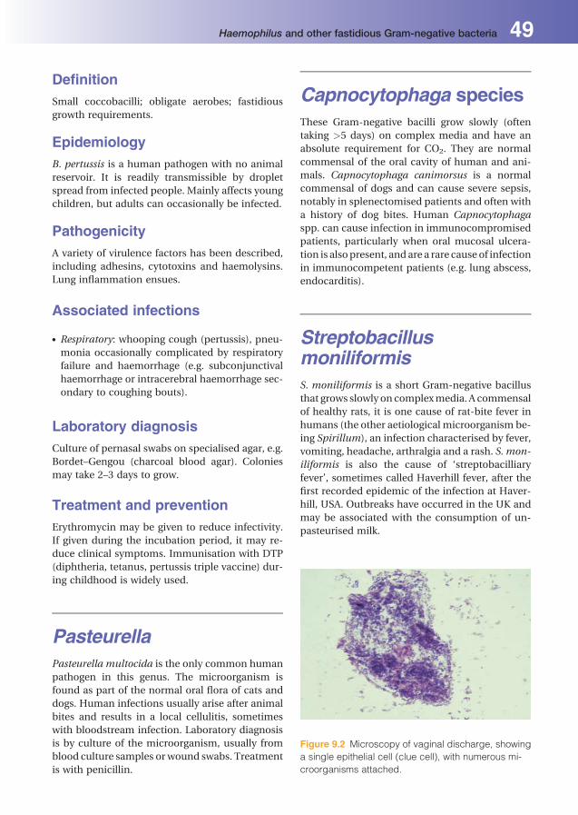
DefinitionSmall coccobacilli; obligate aerobes; fastidious
growth requirements.
EpidemiologyB. pertussis is a human pathogen with no animal
reservoir. It is readily transmissible by droplet
spread from infected people. Mainly affects young
children, but adults can occasionally be infected.
PathogenicityA variety of virulence factors has been described,
including adhesins, cytotoxins and haemolysins.
Lung inflammation ensues.
Associated infections
. Respiratory: whooping cough (pertussis), pneu-
monia occasionally complicated by respiratory
failure and haemorrhage (e.g. subconjunctival
haemorrhage or intracerebral haemorrhage sec-
ondary to coughing bouts).
Laboratory diagnosisCulture of pernasal swabs on specialised agar, e.g.
Bordet–Gengou (charcoal blood agar). Colonies
may take 2–3 days to grow.
Treatment and preventionErythromycin may be given to reduce infectivity.
If given during the incubation period, it may re-
duce clinical symptoms. Immunisation with DTP
(diphtheria, tetanus, pertussis triple vaccine) dur-
ing childhood is widely used.
PasteurellaPasteurella multocida is the only common human
pathogen in this genus. The microorganism is
found as part of the normal oral flora of cats and
dogs. Human infections usually arise after animal
bites and results in a local cellulitis, sometimes
with bloodstream infection. Laboratory diagnosis
is by culture of the microorganism, usually from
blood culture samples or wound swabs. Treatment
is with penicillin.
Capnocytophaga speciesThese Gram-negative bacilli grow slowly (often
taking >5 days) on complex media and have an
absolute requirement for CO2. They are normal
commensal of the oral cavity of human and ani-
mals. Capnocytophaga canimorsus is a normal
commensal of dogs and can cause severe sepsis,
notably in splenectomised patients and often with
a history of dog bites. Human Capnocytophaga
spp. can cause infection in immunocompromised
patients, particularly when oral mucosal ulcera-
tion is alsopresent, andare a rare causeof infection
in immunocompetent patients (e.g. lung abscess,
endocarditis).
StreptobacillusmoniliformisS. moniliformis is a short Gram-negative bacillus
that grows slowly on complexmedia. A commensal
of healthy rats, it is one cause of rat-bite fever in
humans (the other aetiological microorganism be-
ing Spirillum), an infection characterised by fever,
vomiting, headache, arthralgia and a rash. S. mon-
iliformis is also the cause of ‘streptobacilliary
fever’, sometimes called Haverhill fever, after the
first recorded epidemic of the infection at Haver-
hill, USA. Outbreaks have occurred in the UK and
may be associated with the consumption of un-
pasteurised milk.
Figure 9.2 Microscopy of vaginal discharge, showing
a single epithelial cell (clue cell), with numerous mi-
croorganisms attached.
Haemophilus and other fastidious Gram-negative bacteria 49

Gardnerella vaginalisG. vaginalis is a Gram-negative bacillus that occa-
sionally appears Gram positive (‘Gram variable’). It
grows aerobically and anaerobically on complex
media. It is anormalcommensalof thevaginalflora,
but is associated with bacterial vaginosis, a condi-
tioncharacterisedbyan‘ammoniasmelling’vaginal
discharge and thepresenceof clue cells (Figure 9.2).
50 Haemophilus and other fastidious Gram-negative bacteria
10Pseudomonas, Legionellaand other environmentalGram-negative bacilliPeter LambertAston University, Birmingham, UK
This chapter includes bacteria that are found in
the environment, but can colonise and infect
humans.
Pseudomonas,Burkholderia andStenotrophomonasSpecies from the genera Pseudomonas, Burkhol-
deria and Stenotrophomonas were all previously
classified in the Pseudomonas genus. They are
widely distributed in nature; P. aeruginosa is the
most important pathogenic species in humans.
They are sometimes collectively referred to as
‘pseudomonads’.
Definition
Gram-negative bacilli; strict aerobes; grow on sim-
ple media at a wide temperature range; motile;
non-capsulated; some species pigmented.
P. aeruginosa
Epidemiology
. P. aeruginosa is an important microorganism in
healthcare-associated infection. It is a normal
commensal in the human gastrointestinal tract,
but may colonise other sites when host defences
are compromised, including burns and leg ul-
cers, the respiratory tract of patients with cystic
fibrosis or bronchiectasis, and theurinary tract of
patients with long-term, indwelling, urethral
catheters.. P. aeruginosa, as with other members of the
genus, has the ability to grow with minimal
nutrients, e.g. in water and in the presence of
some disinfectants; these properties are the key
to its role as a hospital pathogen.
Morphology and identification
The microorganism grows on most media, pro-
ducing a characteristic greenish pigment. It can
be distinguished from the Enterobacteriaceae by
Medical Microbiology and Infection Lecture Notes, Fifth Edition. Edited by Tom Elliott, Anna Casey,Peter Lambert and Jonathan Sandoe.� 2011 Blackwell Publishing Ltd. Published 2011 by Blackwell Publishing Ltd.
its oxidative metabolism (oxidase-positive) and
the inability to grow anaerobically, except on
nitrate. The different species of the genus Pseu-
domonas can be distinguished by biochemical
tests.
Pathogenicity
P. aeruginosa is relatively non-pathogenic; it
characteristically causes infections in hospita-
lised patients, particularly those who are immu-
nocompromised. The microorganism produces
several enzymes that allow spread through tis-
sues (elastase) and a protease that breaks down
IgA on mucosal surfaces. It also produces cyto-
toxins (exotoxin A) and other factors that inhibit
host defences, including a rhamnolipid and an
alginate exopolysaccharide.
Associated infections
P. aeruginosaprincipally causes opportunist infec-
tions including:
. Skin and soft tissues: infections associated with
burns and cutaneous ulcers;. Respiratory tract: ventilator-associated pneu-
monia and hospital acquired pneumonia
(Chapter 26); and lower respiratory tract infec-
tions in cystic fibrosis and bronchiectasis
patients;. Urinary tract: associatedwith long-term urethral
catheterisation;. Invasive: bloodstream infection can follow pri-
mary infection at any site, but is more common
in immunocompromised patients;. Eye: contact lens-associated infections;. Ear nose and throat: otitis externa.
Laboratory diagnosis
Isolation of themicroorganism from relevant body
sites. The isolation of P. aeruginosa from several
patients may suggest hospital cross-infection. In
such circumstances, strains ofP.aeruginosa canbe
further characterised by serotyping or molecular
biological techniques.
Treatment
One of the characteristics of P. aeruginosa
is resistance to antibiotics; some newer antibio-
tics have been designed specifically to combat
P. aeruginosa. Clinically important anti-pseudo-
monal antibiotics include:
. aminoglycosides (e.g. gentamicin);
. broad-spectrum penicillin/beta-lactamse inhibi-
ter combinations (e.g. piperacillin/tazobactam);. third-generationcephalosporins(e.g.ceftazidime);. carbapenems (e.g. imipenem and meropenem);. monobactams (e.g. aztreonam);. quinolones (e.g. ciprofloxacin).
In hospital units where antibiotics are used fre-
quently (e.g. special care baby units, ICUs, cystic
fibrosis clinics), P. aeruginosa isolatesmay become
resistant to these antibiotics. Isolation of patients
colonised bymulti-resistantP. aeruginosa strains is
an important part of controllinghospital infections.
Burkholderia cenocepacia(formerly cepacia)This microorganism has recently been recognised
as an important cause of lower respiratory tract
infection inpatientswith cystic fibrosis; it occurs in
moist environments and hospitals. Treatment of
infection is with meropenem.
Burkholderia pseudomalleiThis microorganism has a characteristic safety pin
appearance on Gram-staining (bi-polar staining).
It causes melioidosis, is resistant to aminoglyco-
sides, but sensitive to ceftazidime.
Stenotrophomonas maltophilia(formerly Pseudomonas orXanthomonas)Thismicroorganism is a causeof hospital-acquired
infections and is oftenmultiply antibiotic resistant;
it causes pneumonia, particularly in compromised
patients, e.g. ventilated or cancer patients; also
related to intravascular device infections. Treat-
ment with trimethoprim-sulfamethaxole
LegionellaLegionella pneumophila was first recognised after
investigations into an outbreak of respiratory ill-
ness in delegates at an American Legion Conven-
tion in Philadelphia, USA, in 1976.Other Legionella
species can also cause human infection.
52 Pseudomonas, Legionella and other environmental Gram-negative bacilli

Definition
Gram-negative bacilli; obligately aerobic, slow-
growing bacteria with fastidious growth
requirements.
Epidemiology
. Environmental microorganisms, found in soil
and water;. Water systems, particularly heating systems
and water-cooled air-conditioning plants can
be a reservoir for infection and source of
outbreaks;. Although relatively fastidious in its growth re-
quirements, the microorganism grows in water
at temperatures between 20 and 45 �C; this may
be related to growth within amoebae, which are
foundnaturally in the same environment or to its
ability to survive in biofilms;. Legionella species are also relatively chlorine
tolerant,. Transmission occurs through aerosolisation of
water contaminated with legionellae,. Human-to-human transmission has not been
demonstrated;. Cases occur sporadically or as part of point-
source outbreaks;. Hospital or community acquired.
Pathogenicity
Virulent Legionella species adhere to human re-
spiratory epithelial cells via pili (fimbriae). Legio-
nella species can also survive and grow within
phagocytic cells, neutralising the normal bacte-
ria-killing mechanisms.
Associated infections
. Central nervous system: meningoencephalitis;
. Respiratory: pneumonia (community and hospi-
tal acquired);. Cardiovascular: prosthetic valve endocarditis
(very rare).
L. pneumophila can cause a flu-like illness with no
lower respiratory symptoms, called Pontiac fever.
Legionella may also spread to other areas of the
body, including lymph nodes, brain, kidney and
bone marrow.
Laboratory diagnosis
. Diagnosis ismade by culture of sputumor bronch-
oalveolar lavage fluid on specialised media-
buffered charcoal yeast extract agar (BCYE);
colonies take between 2 and 7 days to grow;. Legionellae are classified into species according
to surface antigens;. Legionellae can be detected directly in respira-
tory specimens by direct immunofluorescence
microscopy;. Legionella antigen may be detected in urine
samples by immunoassay;. Serological tests are often used to confirm the
diagnosis of legionellosis; a rise in serum anti-
body titre may take up to 6 weeks.
Treatment and prevention
This is with clarithromycin combined, in severe
cases, with rifampicin. Fluoroquinolones are also
effective.
Prevention isachievedbymonitoringandappro-
priate disinfection in man-made water systems.
AcinetobacterAcinetobacter species share a number of common
features with pseudomonads. They are short,
Gram-negative bacilli, strict aerobes, and grow
well on simple media with minimal nutrients.
However, unlike pseudomonads, they are oxi-
dase-negative. They occur in many environments,
including hospitals, and are an important cause of
healthcare-associated infection (particularlyA.bau-
mannii). Hospital outbreaks, most notably chest
infections, occur particularly in ICUs. Acinetobacter
species frequentlyacquireplasmidsexpressingmul-
tipleantibiotic-resistancepatterns, includingquino-
lones, many aminoglycosides and b-lactam agents.
Meropenem and amikacin may be active. Some
multi-resistant isolates are sensitive only to colistin.
Pseudomonas, Legionella and other environmental Gram-negative bacilli 53

11Campylobacter,Helicobacter and VibrioMartin Skirrow1, Cliodna McNulty1 and Tom Elliott21Health Protection Agency, Gloucestershire Royal Hospital, Gloucester, UK2University Hospitals Birmingham NHS Foundation Trust, Birmingham, UK
Campylobacter
Definition
Spirally curved, Gram-negative bacilli; motile with
single polar flagella; non-sporing; non-capsulated;
microaerophilic.
Classification
There are several species, but only Campylobacter
jejuni and C. coli commonly infect humans (cause
acute enterocolitis). C. fetus, the type species of the
genus, occasionally causes systemic infection in
persons with immune deficiency.
Epidemiology
Campylobacter spp. are a leading cause of acute
infective diarrhoea worldwide. In most industria-
lised countries, they are themost commoncauseof
acute bacterial enterocolitis. The infection is esse-
ntially a zoonosis with a wide range of reservoirs,
including wild birds, poultry, cattle, sheep, pigs
and pets. Modes of transmission are correspond-
ingly varied, but broiler chickens are a major
source, either through handling the raw product
or eating undercooked meat. Raw milk and un-
treated water are established sources of infection.
Transmission from infected subjects is uncom-
mon, except from young children with uncon-
trolled bowel actions.
Morphology and identification. Campylobacter spp. have a characteristic mor-
phology. The long polar flagella are attached to a
unique cone-shaped structure. Like the true
Vibrio spp., Campylobacter spp. are among the
fastestmovingbacteria. It is possible to recognise
them in microscopic examinations of wet pre-
parations of faeces from patients with diarrhoea.. For culture they require microaerobic conditions
with added CO2. Selective media containing spe-
cific antimicrobials and compounds that neutral-
ise free radicals, such as charcoal, are essential
for their isolation from faeces. They are oxidase
and catalase positive, but do not metabolise
Medical Microbiology and Infection Lecture Notes, Fifth Edition. Edited by Tom Elliott, Anna Casey,Peter Lambert and Jonathan Sandoe.� 2011 Blackwell Publishing Ltd. Published 2011 by Blackwell Publishing Ltd.

sugars. Further biochemical tests are used for
speciation.
Pathogenicity
The Campylobacter cell wall contains endotoxins.
Cytopathic extracellular toxins and enterotoxins
have also been demonstrated, but their exact role
is unclear. They cause acute inflammation of the
intestinal mucosa, like that caused by Salmonella
and Shigella spp. Infection is self limiting and
seldom lasts for more than a week. Mean incuba-
tion period is 3 days.
Associated conditions. Campylobacter enteritis: this can mimic acute
appendicitis; occasionally genuine appendicitis
is present. It can also mimic an acute attack of
ulcerative colitis.. Reactive arthritis and Guillain Barr�e syndrome:
(polyneuropathy with paralysis) are complica-
tions that may arise during the recovery phase
of illness.
Laboratory diagnosis. Culture of faeces on selective media at 42 �Cunder microaerophilic conditions;
. Serology for serum antibodies to Campylobacter
spp. can be of value in culture-negative patients
with suspected late sequelae and in the investi-
gation of outbreaks.
Treatment
Rehydration. Erythromycin or ciprofloxacin indi-
cated only for severe illness.
HelicobacterH. pylori
Definition
Curved Gram-negative bacilli; motile; non-spor-
ing; non-capsulated; microaerophilic.
Classification
Helicobacter spp. are in the same broad bacterial
group as Campylobacter spp. There are many He-
licobacter species, each adapted to a particular
animal or niche. H. pylori is the species associated
with man.
Epidemiology
H. pylori infects human gastric epithelial cells and
humans are the main reservoir.
Themicroorganism is acquiredmost commonly
in early childhood and usually becomes chronic,
often life-long. Transmission is by the faecal-oral
or oral–oral routes and is associated with close
contact (intrafamilial) and poor sanitation. Thus,
in developed countries, prevalence is now very low
inchildren (<10years, 1%), but about 25% inadults
more than 50 years of age (reflecting the higher
rates of transmission when they were children).
Prevalence is higher in countries with poor sani-
tation, with 50% of over 50 year olds infected.
Morphology and identification. H.pylori grows slowly at 37 �Conenrichedmedia
in a microaerophilic atmosphere (characteristic
of gastric mucus).. They have four to six mono-polar sheathed fla-
gella (Figure 11.1) that facilitate motility through
gastric mucus.. The rapid urease test is a unique characteristic,
used for laboratory identification and diagnosis
of H. pylori.
Figure 11.1 Negative-stain electronmicrograph of a
single H. pylori (1) showing flagella (2).
Campylobacter, Helicobacter and Vibrio 55

Pathogenicity and associatedinfections
Urease is an important colonisation factor,
enabling the microorganism to neutralise
gastric acid through the production of ammonia
from urea.
Most subjects remain asymptomatic, but
H. pylori is the most common cause of duodenal
ulceration and gastric cancer. H. pylori initially
causes a neutrophilic gastritis, which becomes
chronic (‘chronic active’ gastritis). H. pylori da-
mages antral D cells that release somatostatin,
thereby interrupting the negative feedback inhibi-
tion of gastric acid from the gastric corpus. Gastric
acid thus increases, thereby inducing gastric meta-
plasia in the duodenum. Thesemetaplastic cells are
susceptible to H. pylori infection, leading to duo-
denitis and the risk of ulceration. Extension of H.
pylori into the gastric corpus causes chronic active
inflammation and eventually atrophy of cells, a
precursor to metaplasia and cancer.
Diagnosis. Invasive tests: Gastric biopsy to identify typical
histopathological appearance; culture of H.
pylori on selective medium in a microaerobic
atmosphere; biopsy urease test, which detects
the preformed urease produced byH. pylori by a
rise of pH in urea broth with an indicator. The
latter is a simple, rapid, inexpensive and specific
test, because H. pylori is unique in producing a
urease with a high affinity and rate of activity.. Non-invasive tests: The urea breath test is highly
sensitive and specific. 13C- or 14C-labelled urea is
given to the patient; if H. pylori is present, the
urease splits the urea, producing labelled CO2,
which is detected in expired air. The stool antigen
test, also highly sensitive and specific, detects the
presence of H. pylori in stools using monoclonal
antibodies. Serology is less specific and is not
appropriate in populations with low infection
prevalence.
Treatment
Combination treatment is required due to poor
antibiotic penetration into the gastric mucosa,
gastric acidity and increasing antimicrobial resis-
tance. Clearance in 80%of patients can be attained
with a proton pump inhibitor, e.g. omeprazole,
that reduces gastric acidity, coupled with two
antimicrobials suchas amoxicillin, clarithromycin,
metronidazole, doxycycline, levofloxacin, rifabu-
tin or bismuth salt.
Vibrio
Definition
Gram negative; comma-shaped bacteria; aerobic
and facultatively anaerobic; grow in alkaline
conditions; motile; ferment carbohydrates;
oxidase-positive. The main species of medical
importance are Vibrio cholerae, V. parahaemoly-
ticus, V. vulnificus and V. alginolyticus.
V. cholerae
Classification. V. cholerae strains are subdivided according to
O-antigens.V. choleraeO1 is the causeof cholera.
OtherV. cholerae (non-O1) strainsmayoccasion-
ally cause diarrhoea.. V. choleraeO1 has a number of biotypes, includ-
ing the ‘classic’ strain, which was the principal
cause of cholera until the mid-1960s. The
‘El Tor’ biotype has been responsible for most
cases of cholera over the last two decades. In
the 1990s, O139 caused epidemics in
Southeast Asia.
Epidemiology. Found in water contaminated with human fae-
ces; no animal reservoirs;. Important cause of severe infection in develop-
ing countries, where potable water and sewage
systems are poor;. Transmitted via consumption of contaminated
food or water.
Morphology and identification. V. cholerae is characterised by an ability to grow
in alkaline conditions (pH> 8.0). Alkaline broth
is used to grow the microorganism selectively
from faecal samples. A special selective medium
(thiosulphate–citrate–bile salt–sucrose agar) is
also used. V. cholerae forms characteristic yellow
colonies on this medium.
56 Campylobacter, Helicobacter and Vibrio
. Specific slide agglutination reactions for ‘O’ anti-
gens distinguish V. cholerae from non-cholera
Vibrio spp.. Biochemical tests are used for confirmation.
Pathogenicity
V. cholerae produces an enterotoxin that acts on
intestinal epithelial cells, stimulating adenyl
cyclase activity. This results in water and sodium
ions passing into the gut lumen to produce pro-
fuse, watery diarrhoea (rice-water appearance).
Associated infections
Acute enteritis.
Laboratory diagnosis
Isolation of microorganism from faeces on selec-
tive media.
Prevention
Avoidance of contaminated food; oral vaccines
suitable for travellers.
Treatment
Rehydration; ciprofloxacin shortens duration of
illness.
Non-cholera vibrio sppV. cholerae that do not agglutinate with O1
and O139 antisera are known as non-cholera
Vibrio spp. These microorganisms may also
cause diarrhoea, which is normally much
less severe than that from classic cholera. Occa-
sionally they cause severe cellulitis and necrotis-
ing fasciitis.
V. parahaemolyticusV. parahaemolyticus is a cause of food poisoning
12–18 hours after the ingestion of contaminated
seafood, particularly oysters. The illness is char-
acterised by vomiting and diarrhoea.
Treatment
By rehydration and a quinolone in severe cases.
Campylobacter, Helicobacter and Vibrio 57
12Treponema, Borrelia andLeptospiraSusan O’ConnellHealth Protection Agency Laboratory, Southampton, UK
SpirochaetesThese bacteria, in the order Spirochaetales, are
thin (0.1–0.5� 5.0–20.0 mm), helical and weakly
Gram-negative. Treponema, Borrelia and Leptos-
pira are three genera which cause human diseases
(Table 12.1). Laboratory diagnoses are based pri-
marily on serological tests, as few spirochaetes can
be cultured readily in vitro.
Treponema
Definition
Gram-negative spiral bacteria; motile; non-capsu-
lated; non-sporing; non-culturable in vitro.
Classification
Treponema species that cause human disease are
Treponema pallidum andT. carateum. T.pallidum
subspecies pallidum causes syphilis, T. pallidum
subsp endemicum causes bejel (endemic or non-
venereal syphilis) and another subspecies, T. pal-
lidum subsp pertenue, causes yaws. T. carateum
causes pinta. These treponemes are morphologi-
cally and immunologically indistinguishable, but
have some genomic differences that may be sig-
nificant in relation to pathogenicity.
T. pallidum
Morphology and identification
Themicroorganism is a thin spirochaete, 0.5� 10.0
mm,which cannot be grown in vitro and grows only
slowly in vivo. Motile forms can be visualised in
clinical specimensbydarkfieldmicroscopyorwith
specialised staining.
Associated infection andepidemiology
Syphilis (Chapter 31).
Laboratory diagnosis
Diagnosis is principally based on serology, but
microscopy can be useful in early infection.
MicroscopyPrimary, secondary and congenital syphilis can be
diagnosed by dark field examination for spiro-
chaetes in fresh material from skin lesions or
lymph node aspirates.
Medical Microbiology and Infection Lecture Notes, Fifth Edition. Edited by Tom Elliott, Anna Casey,Peter Lambert and Jonathan Sandoe.� 2011 Blackwell Publishing Ltd. Published 2011 by Blackwell Publishing Ltd.

SerologyDiagnosis of syphilis in most patients is based on
two types of serological tests: non-specific and
specific treponemal assays:
1 Non-specific antibody tests are commonly used,
including the Venereal Disease Research Labo-
ratory (VDRL) and rapid plasma reagin (RPR)
tests. Theycandetect the IgMand IgGantibodies
that develop against lipids released from T. pal-
lidumduringearly stagesofdisease.Cardiolipin-
cholesterol-lecithin, the antigen used in these
assays, was initially extracted from beef heart
and is not derived from treponemes, hence these
tests are non-specific. False-positive results are
common, occurring in the presence of other
conditions including viral infections, autoim-
mune diseases, leprosy, tuberculosis andmalar-
ia. Positive results need confirmation by specific
serological tests.
All antibody testswill benegativeat theearliest
stage of infection. Cardiolipin-based tests may
givepositive results at a slightly earlier stage than
some of the treponema-specific tests, and they
are also helpful in assessing treatment response,
as titres usually drop significantly and can be-
come negative following satisfactory treatment.
2 Specific antibody tests are based on T. pallidum-
derived antigens. Commonly used tests include
syphilis enzyme immunoassays, the fluorescent
treponemal antibody (FTA-abs) test and the
T. pallidum particle or haemagglutination
assays (TPPA, TPHA), in which latex particles or
erythocytes coatedwithT.pallidum antigens are
agglutinated by serum from patients with anti-
bodies toT.pallidum.Positive resultsmayreflect
previous infection, because residual antibodies
can remain after treatment, and can also result
from other treponemal infections, e.g. yaws,
pinta, as themicroorganisms causing these con-
ditions are essentially identical to T. pallidum.
Specific IgM antibody tests are also used to
diagnose congenital infections.
3 Polymerase chain reaction is being developed for
diagnosis.
Treatment
Parenteral penicillin. Detailed guidelines are avail-
able for treatment and follow-up of patients and
management of their sexual contacts.
T. pallidum subsp endemicumThis microorganism causes bejel (endemic syph-
ilis),mainly in children inhotdry climates. Primary
lesions occur as mucous patches in the mouth
or angular stomatitis. A secondary stage can occur,
similar to syphilis. Late infection can cause gum-
mas and affect bone and cartilage.
Table 12.1 Spirochaetes and associated infections
Spirochaete Associated infections
Treponema
T. pallidum subsp pallidum Syphilis
T. pallidum subsp pertenue Yaws
T. pallidum subsp endemicum Bejel (endemic non-venereal syphilis)
T. carateum Pinta
Borrelia
B. recurrentis Louse-borne relapsing fever
B. duttoni and others Tick-borne relapsing fever
B. burgdorferi sensu lato Lyme borreliosis
Leptospira
(Several species and serovars) Flu-like illness; meningitis; uveitis
Weil’s disease (severe multi-system disease)
Treponema, Borrelia and Leptospira 59

Spread is by direct contact or shared feeding
utensils. Laboratory diagnostic procedures and
treatment are as for syphilis.
T. pallidum subsp pertenueT. pertenue causes yaws, which occurs mainly in
children in humid tropical regions in Central Afri-
ca, South America and the Pacific islands. It is
primarily a skin disease; destructive lesions (gran-
ulomata) of the skin, lymph nodes and bone can
occur.
Spread is by direct contact with infected skin
lesions. Laboratory diagnostic procedures and
treatment are as for syphilis.
T. carateumT. carateum causes pinta and is found in remote
parts of Central and South America. It occurs in all
age groups. After a 1- to 30-week incubation peri-
od, small papules develop on the skin. These can
enlarge, last for several years and result in hypo-
pigmented lesions. Laboratory diagnosis and treat-
ment are as for syphilis.
Borrelia
Definition
Gram-negative, spiral bacteria; motile; non-cap-
sulated; non-sporing; require specialisedmedia for
culture; anaerobic or microaerophilic.
Borrelia species are associated with two impor-
tant human infections: relapsing fever (primarily
B. recurrentis andB. duttoni), and Lyme borreliosis
(B. burgdorferi group).
B. recurrentis, B. duttoni andother relapsing fever-associatedBorrelia species
Associated infections andepidemiology
Relapsing feveroccursworldwide; characterisedby
episodes of fever and spirochaetaemia, which can
cause severe multi-system effects, separated by
periodswhen thepatient is apyrexial. The relapsing
picture is related to the ability ofBorrelia species to
vary their outer surface protein antigenic structure
as many as 30 times, thus evading specific host
antibodies for some time:
. Epidemic relapsing fever is caused exclusively by
B. recurrentis and is spread via human body lice,
Pediculus humanus. There is no known non-
human reservoir host. It is strongly associated
with catastrophic events such as war or famine,
resulting in overcrowding, poor hygiene and lice
infestations. An untreated patient may have a
relapse 7–10 days after an initial febrile episode
of 3–6 days. The mortality rate in untreated in-
fections can be 40%.. Endemic relapsing fever is caused by many
Borrelia species and is spread from rodent res-
ervoir hosts via infected Ornithodoros soft ticks
during blood meals. Multiple relapses, tending
to become shorter and less severe with suc-
ceeding episodes, can occur in untreated pa-
tients. The mortality of untreated infection is
about 5%.
Laboratory diagnosis
Borreliae canbe identified in stainedbloodorbuffy
coat smears taken when the patient is pyrexial.
Serological tests are not readily available.
Treatment
Tetracycline is the treatment of choice.
B. burgdorferi
Epidemiology
At least five genospecies of B. burgdorferi sensu
lato cause Lyme borreliosis, which occurs mainly
in forested areas of the temperate northern hemi-
sphere. Borreliae are transmitted to humans by
Ixodes hard ticks that have previously fed on in-
fected mammals and birds. About 1,000 cases are
reported annually in the UK.
Associated infections
Lyme borreliosis is characterised initially by a skin
lesion spreading from the tick bite, but can later
involve thenervous system, joints andother tissues
following bloodstream spread.
60 Treponema, Borrelia and Leptospira
Laboratory diagnosis
Two-stage IgM and IgG antibody tests. Samples
giving reactions in a sensitive screening test are
assessed for specificity by second-stage immuno-
blot, because there is significant risk of false-pos-
itive screening results with other infections and
autoimmune conditions. Tests can be negative in
the first few weeks of infection, but sensitivity in
late stage disease is more than 99%.
Borrelial DNA can be detected in skin and syno-
vial fluid/biopsy samples by PCR.
Treatment
Oral doxycycline or amoxicillin; parenteral ceftri-
axone for encephalomyelitis.
Prevention
Avoidance of tick bites; early removal of attached
ticks.
Leptospira
Definition
Gram-negative, coiled bacteria; non-capsulated;
non-sporing; motile; aerobes that grow slowly in
enriched media.
Morphology and identification
Leptospires are motile microorganisms with two
periplasmic flagella, each anchored at opposite
ends of the microorganism. They can be grown on
specially defined media enriched with serum,
although prolonged incubation may be required.
The genus Leptospira has numerous species and
intraspecies serovars; only some are pathogenic to
human beings, principally the L. interrogans ser-
ovars Icterohaemorrhagiae, Canicola, Pomona,
and L. borgpetersenii serovar Hardjo.
Epidemiology. Occursworldwide, particularly following periods
of high rainfall and flooding;. More prevalent in tropical and subtropical
regions;. Maintained in nature through chronic renal in-
fection in animals, e.g. rats (serovar Icterohae-
morrhagiae), dogs (Canicola), cows (Hardjo) and
pigs (Pomona);. Human infections occur mainly through expo-
sure to water contaminated with infected urine;. Microorganisms can enter through broken skin,
mucous membranes or conjunctiva;. At-risk groups include sewer workers, farmers
and those involved in water sports. About 50
cases/year are notified in the UK.
Associated infections
Leptospirosis ranges from a mild flu-like illness,
uveitis and meningitis to the potentially life-
threateningWeil’s disease, with jaundice and renal
damage.
Laboratory diagnosis
Bloodorurine canbe cultured in specialisedmedia
but takes several weeks. Serology using microag-
glutination (MAT) and other techniques is avail-
able from reference laboratories.
Treatment and prevention. Treatment is with penicillin or doxycycline.. Rodent control measures help to prevent infec-
tion. Groups at risk should be made aware of the
illness and appropriate prevention measures.
Treponema, Borrelia and Leptospira 61

13Gram-negative anaerobicbacteriaPeter LambertAston University, Birmingham, UK
The Gram-negative anaerobic bacilli include the
genera Bacteroides, Prevotella, Porphyromonas,
Fusobacterium and Leptotrichia. Unlike the
Gram-positive anaerobic clostridia, they do not
form spores. Gram-negative anaerobes form ama-
jor component of the normal flora of humans.
Definition
Gram-negative bacilli; non-motile; non-sporing;
strict anaerobes; grow on complex media; some
species capsulate.
Bacteroides, Prevotellaand PorphyromonasThe genus Bacteroides has undergone major taxo-
nomic revision and now consists of the genera
Bacteroides, Prevotella and Porphyromonas. The
Bacteroides genus containsmany species, themost
important beingB. fragilis.Prevotella also includes
many species, including Prevotella melaninogen-
ica, intermedia andnigrescens.Porphyromonashas
fewer species, including P. gingivalis, which is
associated with advanced periodontal disease.
Epidemiology
Species of Bacteroides and related genera make up
the largest component of the bacterial flora of the
human intestine, and are an important part of the
normal flora of themouth and genito-urinary tract.
They protect against colonisation and infection by
more pathogenic bacteria; they can also cause
infections.
Morphology and identification
Occasionally form short filaments. Some species
have a polysaccharide capsule. Visible colonies
appear only after 48 hours of incubation. Some
Porphyromonas and Prevotella species produce a
pigment that results in black colonies.
Associated infections. Gastrointestinal: intra-abdominal abscesses,
peritonitis (often in mixed infections with
coliforms);
Medical Microbiology and Infection Lecture Notes, Fifth Edition. Edited by Tom Elliott, Anna Casey,Peter Lambert and Jonathan Sandoe.� 2011 Blackwell Publishing Ltd. Published 2011 by Blackwell Publishing Ltd.

. Skin and soft tissue: abscesses and surgical
wound infection;. Central nervous system: brain abscesses.
Prevention
Postoperative, intra-abdominal infections can
be prevented by good surgical technique and pro-
phylactic antibiotics.
Treatment
Metronidazole and clindamycin are effective anti-
biotics against almost all Gram-negative anaerobic
bacteria; most species are sensitive to penicillin
but some, particularly B. fragilis, are resistant
because of b-lactamase production.
FusobacteriumFusobacterium species are spindle-shaped, with
pointed ends. They are often nutritionally
demanding. The genus is classified into various
species, F. necrophorum and F. nucleatum being
the most important in human infections.
Fusobacteria are commensals of the oral cavity.
They are one of the causative microorganisms,
along with other anaerobes, of acute ulcerative
gingivitis (primarily F. nucleatum) and other oral
infections. F. necrophorum may occasionally
cause severe sepsis with metastatic abscesses.
LeptotrichiaLeptotrichia species are long, slightly curved bacilli
with pointed ends. The genus consists of a single
species, L. buccalis, which is part of the normal oral
flora and may be associated with oral infections
such as Vincent’s angina.
Gram-negative anaerobiccocciThe role of Gram-negative anaerobic cocci in
disease is not clear. However, Veillonella spp. are
thought to be a component of mixed anaerobic
infections. They are commonly found in supragingi-
val dental plaque and have been occasionally
implicated incasesofosteomyelitis andendocarditis.
Gram-negative anaerobic bacteria 63

14Chlamydiaceae,Rickettsia, Coxiella,Mycoplasmataceae andAnaplasmataceaeJonathan SandoeLeeds Teaching Hospitals NHS Trust and University of Leeds, Leeds, UK
Chlamydiaceae
Definition
Chlamydiaceae are obligate intracellular bacteria
with a unique biphasic life cycle.
Classification
The family Chlamydiaceae contains two genera,
Chlamydia and Chlamydophila. The former con-
tains one human pathogen Chlamydia trachoma-
tis, the latter includes the pathogens Chlamydo-
phila psittaci and Chlamydophila pneumoniae.
Classification is based on antigens, morphology
of intracellular inclusions and disease patterns
(Table 14.1). All chlamydiaceae share a common
group antigen but may be distinguished by
species-specific antigens.
Morphology and identification
Chlamydiaceae have two distinct morphological
forms:
1 The elementary body (EB): an infectious extra-
cellular particle (300–400nm).
2 The reticulate body (RB): an intracellular non-
infectious particle (800–1,000 nm).
They multiply in the cytoplasm of host cells after a
well-defined developmental cycle:
. EBs bind to specific host cell receptors and enter
by endocytosis. Target cells include conjunctival,
urethral, rectal and endocervical epithelial cells.. Intracellular EBs remain within phagosomes and
replicate. Lysosomal fusion with the phagosome-
Medical Microbiology and Infection Lecture Notes, Fifth Edition. Edited by Tom Elliott, Anna Casey,Peter Lambert and Jonathan Sandoe.� 2011 Blackwell Publishing Ltd. Published 2011 by Blackwell Publishing Ltd.

containing EBs is inhibited, probably by chlamy-
dial cell-wall components.. After about 8 h, EBs becomemetabolically active
and form RBs that synthesise DNA, RNA and
protein, utilising host cell adenosine triphos-
phate (ATP) as an energy source.. RBs undergo multiple divisions and the phago-
some becomes an ‘inclusion body’ (visible by
light microscopy).. After about 24 h, the RBs reorganise into smaller
EBs and, after a further 24–48h, the host cell lyses
and infective EBs are released.
C. trachomatis
Epidemiology. C. trachomatis is the most common cause of
sexually transmitted infection in the developed
world.. The majority of C. trachomatis infections are
genital and sexually acquired.. Trachoma is the leading cause of preventable
blindness in the world.. Asymptomatic infections can occur in both men
and women.. C. trachomatis can be divided into different ser-
otypes (‘serovars’): A–C, D–K, and L1, L2 and L3.. C. trachomatis serotypes D–K are found
worldwide.. Lymphogranuloma venereum (LGV) serotypes
L1–3 occur primarily in Africa, Asia and South
America.
. Eye infections probably result from autoinocula-
tion with infected genital secretions.. Similarly, neonates can be infected during birth
from an infected mother.
Pathogenesis
C. trachomatis gains access via disruptedmucous
membranes and can infect mucosal columnar or
transitional epithelial cells, resulting in severe
inflammation. In LGV, the lymph nodes, which
drain the site of the primary infection, are
involved.
Associated infections. Eye: ‘Trachoma’ caused by serotypes A–C; adult
inclusion conjunctivitis, a milder disease than
trachoma, caused by serotypes D–K and associ-
ated with genital infections; neonatal conjuncti-
vitis:C. trachomatis canbe acquired at birth from
an infected mother, associated with serotypes
D–K.. Respiratory: Neonatal pneumonitis: usually pre-
sents 3–12 weeks after birth; often associated
with neonatal conjunctivitis; acquired through
maternal transmission during birth.. Genitourinary: LGV is a sexually transmitted
infection with genital ulcers and suppurative
inguinal adenitis; urethritis and cervicitis are
associated with serotypes D–K
Complications include pelvic inflammatory dis-
ease and perihepatitis in women, and epididymitis
and Reiter’s syndrome in men.
Table 14.1 Classification and common infections of Chlamydiaceae
Species Serotypes Natural host Transmission Common infections
C. trachomatis A–C Humans Hands/eyes/flies Trachoma, conjunctivitis
C. trachomatis D–K Humans Sexual Cervicitis, urethritis
Genito-ocular Conjunctivitis
Sexual Proctitis
Perinatal Pneumonia
C. trachomatis L1, L2, L3 Humans Sexual Lymphogranuloma venereum;
Genital ulcers
C. psittaci Many Birds andsome mammals
Aerosol Pneumonia
C. pneumoniae 1 Humans Aerosol Acute respiratory infection
Chlamydiaceae, Rickettsia, Coxiella, Mycoplasmataceae and Anaplasmataceae 65

Laboratory diagnosis. Chlamydiaceae do not grow on bacterial culture
media.. Cell culture: swabs from the affected site, col-
lected in a special transport medium, are inoc-
ulated into tissue culture. Growth of C. tracho-
matis is recognised by Giemsa staining, showing
intracellular bodies, or by immunofluorescence
staining with specific antisera. This test is largely
confined to specialist laboratories.. Direct antigen detection can be made by
immunofluorescence or enzyme-linked immu-
nosorbent assay (ELISA) methods. Both techni-
ques utilise labelled specific antibodies to
C. trachomatis.. Serology: antibodies to C. trachomatis may be
detected by complement fixation or micro-im-
munofluorescence tests, but this has limited
value because it is difficult to distinguish
between past and current infections.. Nucleic acid amplification tests to detect either
chlamydial DNA or RNA, using polymerase
chain reaction (PCR) or ligase chain reaction,
or chlamydial ribosomal RNA, using transcrip-
tion-mediated amplification, are now widely
used. These are performed on urine and vaginal
swabs. They are more sensitive than culture
and non-invasive.
Treatment. Urogenital infection: doxycylineor azithromycin;. Trachoma: for individual or sporadic cases, doxy-
cycline (azithromycin for children, breast-feed-
ing or pregnant women). Eyelid deformities may
need surgery.. Neonatal conjunctivitis: erythromycin.
C. psittaciC.psittaci causes infection inbirds (e.g. parrots and
budgerigars) and domestic animals.
Epidemiology
Transmission to humans is via inhalation, usually
of dried bird guano. Pet shopworkers, bird fanciers
and poultry farm workers are at particular risk of
developing psittacosis.
Associated infections
Respiratory: flu-like illness, pneumonia.
Laboratory diagnosis
By serology, including immunofluorescence or
complement fixation test. Culture of C. psittaci is
also possible, but is performed only in laboratories
with high-level containment facilities.
Prevention
Treat infected birds; quarantine.
Treatment
A tetracycline (e.g. doxycycline) is the drug of
choice; erythromycin is an alternative.
C. pneumoniaeThis was originally designated TWAR (Taiwan-as-
sociated respiratory disease) agent, but is now
called C. pneumoniae and is a significant cause of
atypical pneumonia.
Infection is spread via direct human contact,
with no apparent animal reservoir. It is associated
with respiratory infections, including pharyngitis,
sinusitis and pneumonia (which can be severe,
particularly in elderly people). Diagnosis is by
serology for antibodies against C. pneumoniae
(detection of C. pneumoniae DNA by PCR is being
developed); treatment includes either a tetracy-
cline or erythromycin.
Rickettsia
Definition
Rickettsiaceae cause a number of important hu-
man infections, including typhus and the related
spotted fevers.
They are small, Gram-negative bacilli (0.2–1 mmdiameter); obligate intracellular pathogens; utilise
ATP fromhost cell; grow only in tissue culture. The
genus contains a number of species that cause
human infection (Table 14.2).
Epidemiology
Rickettsial infections are zoonoses with a variety of
animal reservoirs and insect vectors.
Associated infections
Epidemic or louse-borne typhus, murine typhus,
scrub typhus and spotted fevers.
66 Chlamydiaceae, Rickettsia, Coxiella, Mycoplasmataceae and Anaplasmataceae

Laboratory diagnosis
This is by serology. The Weil–Felix test detects
cross-reacting antibodies to Rickettsiae, which
agglutinate certain strains of Proteus (OX-19 and
OX-2). The test is non-specific and has now been
superseded by more specific serological tests
based on purified rickettsial antigens (immunoflu-
orescence assays and ELISA).
Treatment and prevention
Treatment iswith tetracycline or chloramphenicol.
Infection can be prevented by avoidance of the
various vectors. A vaccine to R. prowazekii is
available.
CoxiellaA pleomorphic coccobacillus with a Gram-nega-
tive cell wall (0.4–0.7mm). Spores are produced
prolonging survival. This is an obligate intracellu-
lar animal pathogen (commonly cattle, sheep;
goats) that occasionally infects humans via tick
bites or inhalation of dried, contaminated matter.
At-risk groups include farmers, vets and abattoir
workers. Urine, faeces, milk and birth products
may be a source. Outbreaks can occur.
Coxiella burnetii causes Q-fever (non-specific
illness of fever, severe headache, chills, severemal-
aise,myalgia and later pneumonitis), endocarditis,
hepatitis and neurological infections. Contact with
animals gives a clue to the diagnosis. Laboratory
diagnosis is by serology (complement fixation,
ELISA and immunofluorescence tests). Treat
with a tetracycline. For endocarditis, treatment is
prolonged and valve replacement may be needed.
A vaccine is being developed for prevention.
Mycoplasmataceae
Definition
Small (0.15–0.25 mm), Gram-negative microor-
ganisms; pleomorphic with no cell wall; grow
slowly on enrichedmedia; aerobic or facultatively
anaerobic.
The family Mycoplasmataceae consists of two
genera: Mycoplasma (69 species) and Ureaplasma
(2 species). Only a few species have been iden-
tified as human pathogens, including: Mycoplas-
ma pneumoniae, M. hominis, M. genitalium,
M. fermentans and Ureaplasma urealyticum.
Epidemiology
M. pneumoniae infection is common worldwide
and is an important cause of acute lower respira-
tory tract infections; epidemics can occur. Immu-
nity to M. pneumoniae is short-lived. Infection is
spread by respiratory secretions. M. hominis and
U. urealyticum colonise the genitourinary tracts.
Associated infections. Respiratory: pharyngitis; community-acquired
pneumonia (M. pneumoniae). Central nervous system: meningitis, encephalitis,
transverse myelitis (M. pneumoniae). Genitourinary (debated): urethritis, pelvic in-
flammatory disease (U.urealyticum,M. hominis,
M. genitalium: urethritis)
Laboratory diagnosis. Culture: mycoplasmas and ureaplasmas can be
grown on enriched media; penicillin is often
Table 14.2 Vectors and infections associated with Rickettsia
Species Principal host/reservoir Vector Disease
R. typhi Rats Fleas Murine typhus
R. prowazekii Humans/squirrels Lice Epidemic or Louse-borne typhus
R. tsutsugamushi Rats Mites Scrub typhus
R. akari Mice Mites Rickettsial pox
R. rickettsiia Dogs Ticks Rocky mountain spotted fever
aTick-borne spotted fevers in areas of the world outside the USA carry a variety of names and are caused by Rickettsiae very
similar to R. rickettsii (e.g. Boutonneuse fever caused by R. conorrii occurs in the Mediterranean region).
Chlamydiaceae, Rickettsia, Coxiella, Mycoplasmataceae and Anaplasmataceae 67

added to inhibit other microorganisms. Al-
though M. pneumoniae can be isolated from
sputum after incubation for up to 3 weeks,
diagnosis is normally by serology. M. hominis
grows after about 4 days, producing colonies
with a ‘fried egg’ appearance. U. urealyticum
requires urea for growth and forms small
colonies.. Serology:M. pneumoniae infections can be diag-
nosed by serological tests (complement fixation
test or enzyme immunoassays) for IgG (4-fold
rise in titres is indicative of current infection) or
IgM.
Treatment
Macrolides (e.g. erythromycin) or tetracycline.
AnaplasmataceaeThe family Anaplasmataceae contains two medi-
cally important genera of small (0.5 mm) Gram-
negative bacteria, Anaplasma and Ehrlichia. They
are obligate intracellular pathogens, usually trans-
mitted via tick bites,whichhave a tropism (affinity)
for monocytes or granulocytes. Infections caused
by these microorganisms occur in the distribution
of their tick vectors and have been identified in
Europe, the Americas, Africa and Asia. Clinical
manifestations are usually non-specific with fever,
general malaise, headache, myalgia; severe multi-
system disease can occur and is more common in
immunocompromised patients. Treatment is with
a tetracycline.
68 Chlamydiaceae, Rickettsia, Coxiella, Mycoplasmataceae and Anaplasmataceae

15Basic virologyPeter MackieLeeds Teaching Hospitals NHS Trust, Leeds, UK
Virus structure(Figure 15.1)
Size and shape
Electron microscopy (approx.� 50,000 magnifi-
cation) is the most useful way to determine the
shape and size of viral particles. Viruses range in
size from20 to 300 nm in diameter andmany have
unique shapes that aid identification. All viruses
have one of two symmetrical shapes: icosahedral
(with two-, three- or five-fold axis of symmetry)
or helical.
Genome
Viruses contain either deoxyribonucleic acid
(DNA) or ribonucleic acid (RNA). Nucleic acids
represent the main component of the virus core
and are associated with core proteins. Viral nucleic
acids range from 1.5� 106 daltons (parvoviruses)
to 200� 106 daltons (poxviruses) andmaybe single
stranded or double stranded, circular or linear, a
single molecule or in segments, and have positive
or negative polarity. Positive sense-RNA is similar
to messenger RNA (mRNA) and can be translated
by the host cell. Negative sense viral RNA must be
converted to positive sense by anRNApolymerase.
Capsid
A protein coat encloses the genome and core pro-
teins, consisting of capsomeres (capsid subunits).
Envelope
Lipid bilayer membrane surrounding the capsid
of some viruses (enveloped viruses); the envelope
carries glycoproteins, which form projections or
spikes. This lipid layer is acquired as the virus
buds through the host cell’s cytoplasmic
membrane.
Virion
This term describes the complete infectious virus
particle; in some cases the particles may lack
nucleic acids (empty particles) or carry defective
genomes (defective particles), which can interfere
with normal replication (defective interfering
particles).
Virus cultivationViruses are obligate intracellular parasites and can
therefore replicate only in living cells. Viruses
utilise the host cell metabolism to assist in the
synthesis of viral nucleic acid, proteins and
Medical Microbiology and Infection Lecture Notes, Fifth Edition. Edited by Tom Elliott, Anna Casey,Peter Lambert and Jonathan Sandoe.� 2011 Blackwell Publishing Ltd. Published 2011 by Blackwell Publishing Ltd.
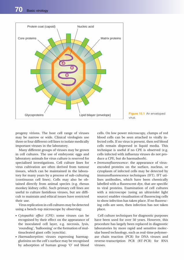
progeny virions. The host cell range of viruses
may be narrow or wide. Clinical virologists use
three or four different cell lines to isolatemedically
important viruses in the laboratory.
Many different groups of viruses may be grown
in cell cultures. The use of embryonic eggs and
laboratory animals for virus culture is reserved for
specialised investigations. Cell culture lines for
virus cultivation are often derived from tumour
tissues, which can be maintained in the labora-
tory for many years by a process of sub-culturing
(continuous cell lines). Cells may also be ob-
tained directly from animal species (e.g. rhesus
monkey kidney cells). Such primary cell lines are
useful to culture fastidious viruses, but are diffi-
cult to maintain and ethical issues have restricted
their use.
Virus replication in cell culturesmaybedetected
using a bench-top microscope by observing:
. Cytopathic effect (CPE) : some viruses can be
recognised by their effect on the appearance of
the inoculated cell layer, e.g. necrosis, lysis,
‘rounding’, ‘ballooning’ or the formation of mul-
tinucleated giant cells (syncitia).. Haemadsorption : viruses expressing haemag-
glutinins on the cell’s surface may be recognised
by adsorption of human group ‘O’ red blood
cells. On low power microscopy, clumps of red
blood cells can be seen attached to virally in-
fected cells. If no virus is present, then red blood
cells remain dispersed in liquid media. This
technique is useful if no CPE is observed (e.g.
cells infected with influenza viruses do not pro-
duce a CPE, but do haemadsorb).. Immunofluorescence : the appearance of virus-
encoded proteins on the surface, nucleus, or
cytoplasm of infected cells may be detected by
immunofluorescence techniques (IFT). IFT uti-
lises antibodies, which have been chemically
labelled with a fluorescent dye, that are specific
to viral proteins. Examination of cell cultures
with a microscope (using an ultraviolet light
source) enables visualisation of fluorescing cells
to show infection has taken place. If no fluoresc-
ing cells are seen, then infection has not taken
place.
Cell culture techniques for diagnostic purposes
have been used for over 50 years. However, this
procedure has largely been replaced in diagnostic
laboratories by more rapid and sensitive molec-
ular based technology, such as real-time polymer-
ase chain reaction (PCR) for DNA viruses and
reverse-transcription PCR (RT-PCR) for RNA
viruses.
Protein coat (capsid)
Core proteins
Nucleic acid
Matrix proteins
Glycoproteins Lipid bilayer (envelope) Figure 15.1 An enveloped
virus.
70 Basic virology

Viral infection of host cellsViral replication in host cells involves the following
steps:
1 Attachment (or ‘adsorption’) of the viral capsid
(naked viruses) or of envelope components
(enveloped viruses) to cell surface molecules
(receptors); this involves specific interaction be-
tween viral glycoproteins (e.g. the haemaggluti-
nin of influenza virus) and host cell surface
components (e.g. N-acetylneuraminic acid for
influenza virus). Many viruses have highly spe-
cific receptors, which limits the range of cell
types that can be infected;
2 Penetration of the virus into the host cell (often
by receptor-mediated endocytosis);
3 Uncoating follows, which involves the enzy-
matic removal of viral protein coat and liber-
ation of nucleic acid and attached core
proteins;
4 Production of virus-specific mRNA, in order to
direct the host cell ribosome to produce viral
proteins (core, capsid). The mechanisms for vi-
rus-specific mRNA production depend on the
viral genome type:
(a) RNA or DNA;
(b) single or double stranded;
(c) positive sense or negative sense.
Examples are shown in Figure 15.2. The
mechanisms for replication of viral nucleic acid
also depend on viral genome type; examples are
given in Figure 15.3;
5 Morphogenesis and maturation occur with as-
sembly of components (nucleic acid and pro-
teins) to form sub-viral particles (pre-core, core
particles) and viral particles (virions, empty
particles);
6 Release of the virus is by bursting of infected cells
(lysis) or by budding through the plasma mem-
brane. Lysis results in cell death but budding
does not necessarily kill cells, allowing viral par-
ticles to be shed for extended periods, e.g. in
hepatitis B infection the host cell remains viable
DNA virusRNA virus
negative sense
Virus-specific mRNA
RNA viruspositive sense
Host DNA dependentRNA polymerase
Viral RNA dependentRNA polymerase
Translatedby host
ribosomeinto viralproteins
Host DNA dependentRNA polymerase
Reverse
transcriptase
Complementary DNAincorporated into
host genome
Used directly asmRNA
Figure 15.2 Synthesis of virus-specific mRNA.
Basic virology 71

and continues to release virus particles or sub-
viral antigens at a slow rate. These persistent
infections act as a continuing source of new
infectious viruses.
During latent infections, the virus does not un-
dergo replication. Viral nucleic acidmay remain in
the host-cell cytoplasm (e.g. episomal Epstein-
Barr virus (EBV) DNA) or become incorporated
RNA viruspositive sense
Negative senseRNA template
Viral replicase
Positive sense RNAvirus genome
Viral reverse transcriptase
cDNA incorporatedinto host cell genome
Host DNA dependentRNA polymerase
Retrovirus RNAvirus genome
Negative senseRNA virus genome
Positive sense RNAtemplate
Transcription byviral RNA polymerase
RNA virusnegative sense
DNA virusgenome
Host or viral DNA polymerase
Incorporated intohost cell genome or
episomal state
Viral replicase
Transcription byviral RNA polymerase
DNA virus
Retrovirusespositive sense RNA
Transcription byviral DNA polymerase
Figure 15.3 Replication strategies of some DNA and RNA viruses.
72 Basic virology

into the host genome (e.g. human immunodefi-
ciency virus, (HIV)). A trigger is required to activate
viral replication, transcription and translation.
This trigger is often associated with periods of
immunosuppression in the host, e.g. during che-
motherapy, organ transplantation, HIV infection
or from an inherited disease.
Antiviral drugs are designed to interfere with
one or more of the above replication steps
(Chapter 22).
Epidemiology. Incubation period: the interval from first acquir-
ing the infection to onset of illness; Generation
time: average period between infection and
transmission of the virus. This period is normally
less than the incubation period, as infections are
transmitted before the onset of illness (e.g. pa-
tients are infectious 48 hrs before a chickenpox
rash appears);. Transmission of viruses: two major patterns
exist:
(a) viruses circulate within a single species
(e.g. influenza B only within human); and
(b) viruses that cross the species barrier
(e.g. rabies and influenza A viruses).
. Person-to-person route: either by horizontal
route including respiratory droplets (>2 mm,
e.g. respiratory syncitial virus (RSV)), aerosol
(<2 mm) produced by coughing and sneezing
(e.g. influenza), gastrointestinal (e.g. noro-
virus); vertical route, such as mother to child
transmission (e.g. HIV), placental-foetal (e.g.
cytomegalovirus (CMV)), at delivery of new
born (e.g. herpes simplex virus (HSV)).
Virus classificationThe official taxonomy of viruses is based on four
characteristics:
1 Nature of the nucleic acid in the virion;
2 Symmetry of the capsid;
3 Presence or absence of an outer lipid envelope;
4 Dimensions of the virion and capsid.
For convenience viruses can also be classified
according to the following:
. Disease or organ system involved (Table 15.1);
Table 15.1 Viral classification according to disease or organ system involved
Organ system involved (disease) Examples of clinically important viruses
Systemic infections Measles virus, rubella virus, varicella-zoster virus (VZV),enteroviruses, retroviruses
Central nervous system Polio- and other enteroviruses; rabies virus, arthropod-borneviruses, herpes simplex virus (HSV), measles virus,mumps virus, retroviruses, cytomegalovirus (CMV)
Respiratory tract (common cold, tracheitis,bronchitis, bronchiolitis, pneumonia)
Influenzavirus, parainfluenzavirus, respiratory syncytial virus(RSV), human metapneumovirus (hMPV), adenovirus,rhinoviruses, (enteroviruses), CMV
Eye (conjunctivitis, retinitis) HSV, adenovirus, CMV
Skin and mucous membranes (rash, warts) HSV, papillomavirus, chickenpox virus, measles virus,rubella virus, parvovirus
Liver (hepatitis) Hepatitis viruses A–E, yellow fever virus, rubella virus, CMV,EBV
Salivary glands Mumps virus, CMV
Gastrointestinal tract (gastroenteritis) Rotaviruses, adenoviruses, astroviruses, norovirus,sapoviruses
Note: this classification is clinically oriented and not virus systematic. The same clinical symptoms can be caused by numerous
viruses, and one virus can cause different clinical syndromes.
Basic virology 73
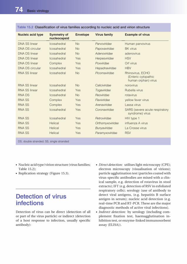
. Nucleic acid type/virion structure (virus families;
Table 15.2);. Replication strategy (Figure 15.3).
Detection of virusinfectionsDetection of virus can be direct (detection of all
or part of the virus particle) or indirect (detection
of a host response to infection, usually specific
antibody):
. Direct detection: utilises light microscopy (CPE);
electron microscopy (visualisation of virions);
particle agglutination test (particles coatedwith
virus-specific antibodies are mixed with a clin-
ical sample, e.g. detection of rotavirus in stool
extracts); IFT (e.g. detection of RSV in exfoliated
respiratory cells); serology (use of antibody to
detect viral antigens, (e.g. hepatitis B surface
antigen in serum); nucleic acid detection (e.g.
real-time PCR and RT-PCR. These are themajor
diagnostic methods of active viral infections).. Indirect detection: by serology (including com-
plement fixation test, haemagglutination in-
hibition test, or enzyme-linked immunosorbent
assay (ELISA)).
Table 15.2 Classification of virus families according to nucleic acid and virion structure
Nucleic acid type Symmetry ofnucleocapsid
Envelope Virus family Example of virus
DNA SS linear Icosahedral No Parvoviridae Human parvovirus
DNA DS circular Icosahedral No Papovaviridae BK virus
DNA DS linear Icosahedral No Adenoviridae adenovirus
DNA DS linear Icosahedral Yes Herpesviridae HSV
DNA DS linear Complex Yes Poxviridae Orf virus
DNA DS circular Icosahedral Yes Hepadnaviridae HBV
RNA SS linear Icosahedral No Picornaviridae Rhinovirus, ECHO(Enteric cytopathichuman orphan) virus
RNA SS linear Icosahedral No Caliciviridae norovirus
RNA SS linear Icosahedral Yes Togaviridae Rubella virus
RNA DS Icosahedral No Reoviridae rotavirus
RNA SS Complex Yes Flaviviridae yellow fever virus
RNA SS Complex Yes Arenaviridae Lassa virus
RNA SS Icosahedral Yes Coronaviridae SARS (severe acute respiratorysyndrome) virus
RNA SS Icosahedral Yes Retroviridae HIV type 1
RNA SS Helical Yes Orthomyxoviridae influenza A virus
RNA SS Helical Yes Bunyaviridae La Crosse virus
RNA SS Helical Yes Paramyxoviridae RSV
DS, double stranded; SS, single stranded
74 Basic virology

16Major virus groupsPeter MackieLeeds Teaching Hospitals NHS Trust, Leeds, UK
DNA viruses (Table 16.1)
Medical Microbiology and Infection Lecture Notes, Fifth Edition. Edited by Tom Elliott, Anna Casey,Peter Lambert and Jonathan Sandoe.� 2011 Blackwell Publishing Ltd. Published 2011 by Blackwell Publishing Ltd.
Table 16.1 Classification of virus families according to nucleic acid and virion structure
CLASSIFICATION OF VIRUS FAMILIES
Nucleic acid type Symmetry ofnucleocapsid
Envelope Virus family
DNA SS linear Icosahedral � Parvoviridae
DNA DS circular Icosahedral � Papovaviridae
DNA DS linear Icosahedral � Adenoviridae
DNA DS linear Icosahedral þ Herpesviridae
DNA DS linear Complex þ Poxviridae
DNA DS circular Icosahedral þ Hepadnaviridae
RNA SS linear Icosahedral � Picornaviridae
RNA SS linear Icosahedral � Caliciviridae
RNA SS linear Icosahedral þ Togaviridae
RNA DS Icosahedral � Reoviridae
RNA SS Complex þ Flaviviridae
RNA SS Complex þ Arenaviridae
RNA SS Icosahedral þ Coronaviridae
RNA SS Icosahedral þ Retroviridae
RNA SS Helical þ Orthomyxoviridae
RNA SS Helical þ Bunyaviridae
RNA SS Helical þ Paramyxoviridae
RNA SS Helical þ Rhabdoviridiae
DS, double stranded; SS, single stranded; � , absent; þ , present.

HerpesvirusesThe herpesviruses comprise a large family of en-
veloped, double-stranded DNA viruses. Following
infection, all herpesviruses establish a latent infec-
tion that persists for life. Reactivation with the
production of progeny virusmay occur at intervals
and produce recurrent symptoms or signs. The
viruses have a comparatively complex genome
coding for several enzymes involved innucleic acid
metabolism.
Herpesviruses (Family – Herpesviridae) include
the following species:
. herpes simplex virus (HSV, type 1 and type 2);
. varicella-zoster virus (VZV);
. cytomegalovirus (CMV);
. Epstein–Barr virus (EBV);
. human herpesviruses (HHV), types 6–8.
Herpes simplex virus (HSV)
Virus
Enveloped DNA viruses, 100–300nm in di-
ameter. There are two serotypes: HSV-1 and
HSV-2, which share 50% nucleic acid sequence
identity.
Epidemiology
HSV is distributed worldwide. Antibodies to HSV-1
are acquired early in childhood and 90% of adults
show serological evidence of past infection (sero-
positive). Antibodies toHSV-2 are acquiredmainly
between 14 and 40 years of age.
Pathogenesis
Spread is by direct contact (sexual activity is the
main routeofHSV-2 transmission).The incubation
period is variable (2–12 days). HSV enters mucosal
surfaces or a break in the skin, replicates produc-
tively inepithelial cells at the siteof inoculationand
spreads to adjacent tissue. There are local IgA, IgM
and IgG responses, but the virus avoids elimination
as its DNA establishes latent infection in sensory
ganglia. Reactivation is associated with various
stimuli (stress, immunocompromised status or
bacterial infection). Infections are either primary
or recurrent (by the same type), with the former
being clinically more severe.
Associated diseases. Skin and soft tissues: herpes labialis ‘cold sores’
(HSV-1, less commonly HSV-2), skin lesions;. Genitourinary: herpes genitalis (HSV-2 and
HSV-1);. Ocular: kerato-conjunctivitis, retinitis;. Respiratory: pneumonitis;. Gastrointestinal: oesophagitis;. Central nervous system: meningitis (mainly
HSV-2), encephalitis (mainly HSV-1, particularly
in neonates and immunocompromised patients).
Microbiological diagnosis. Swab: base of a skin/mucosal lesion, place in
virus transport medium and test by real-time
PCR;. Cerebrospinal fluid (CSF): required if encephali-
tis or meningitis is suspected;. Older less sensitive diagnostic tests include:
electron microscopy of vesicle fluid; direct fluo-
rescent antibody staining of exfoliated cells
around vesicular lesions; isolation of virus in
cell culture;. Serology (e.g. enzyme-linked immunosorbent
assay) is generally not helpful for the diagnosis
of these infections.
Treatment
Aciclovir, valaciclovir, famciclovir, foscarnet,
cidofovir (Chapter 22)
Varicella-zoster virus (VZV)
Virus
Enveloped DNA virus, 100–300nm in diameter.
Epidemiology
VZV has a worldwide distribution. Infections are
acquired early in childhood and 90% of adults are
seropositive. The incidence of chickenpox has fall-
en dramatically in countries such as the USA,
76 Major virus groups

where childhood immunisations include a varicel-
la vaccine.
Pathogenesis
Spread is by respiratory droplets or direct contact
with vesicle fluid. The incubation period is 2 weeks
with a range of 7–23 days. The virus enters via the
respiratory tract, viraemia follows and a general-
ised rash occurs (chickenpox). Virus then becomes
latent in the sensory ganglia; a variety of stimuli
(immunosuppression) may result in reactivation
with vesicles in various dermatomes (shingles).
Associated diseases. Skin and soft tissue: chickenpox (primary infec-
tion); shingles (recurrent infection);. Respiratory: pneumonitis can complicate
chickenpox;. Central nervous system: encephalitis can compli-
cate chickenpox;. Ocular: ‘shingles’ can affect the eye – ophthalmic
zoster; retinitis;. Foetal: congenital varicella syndrome;. Multiple organs: Disseminated VZV infection (all
body system involvement) can occur in immu-
nocompromised individuals and neonates, with
devastating consequences if untreated.
Diagnosis. Swab: skin/mucosal lesion and place in virus
transport medium for testing by real-time PCR;. CSF: required if encephalitis or meningitis is
suspected;. Clotted blood: VZV IgM for evidence of recent
infection, VZV IgG indicates past infection and
immunity;. Less sensitive testing includes electronmicrosco-
py of vesicle fluid; direct fluorescent antibody
staining of vesicular material; isolation in cell
culture.
Treatment
Aciclovir, valaciclovir, famciclovir, foscarnet.
Prevention. Passive: zoster immunoglobulin (ZIG) for protec-
tion of contacts with no evidence of past infec-
tion (seronegative) and at risk of serious VZV
infection, i.e. pregnant women and immuno-
compromised patients;. Active: live attenuated vaccine given in early
childhood (US) and for healthcare workers in
contact with vulnerable patient groups.
Cytomegalovirus (CMV)
Virus
Enveloped DNA virus, 200nm in diameter.
Epidemiology
CMV has a worldwide distribution. In the devel-
oping world 90% of adults are seropositive. In the
developed world only 60–70% of adults are CMV
seropositive.
Pathogenesis
Spread is by contactwith infected urine, secretions
(oropharyngeal) and through intrauterine and
perinatal transmission (vaginal secretions and
breast milk); the virus may also be acquired by
blood transfusion, haematopoetic stem cell trans-
plantation or by organ transplantation. The incu-
bationperiod is 4–8weeks. Theprimary infection is
often asymptomatic. CMV remains latent in vari-
ous myeloid cell types and can cause low level
persistent infection in salivary glands and kidneys.
Recurrent infection in immunocompromised in-
dividuals following solid organ or stem cell trans-
plants is common.
Associated diseases. Blood: infectious mononucleosis-like syndrome
in immunocompetent patients (fever, pharyngi-
tis, lymphadenopathy);. Multiple organs: disseminated infection occurs
in immunocompromised individuals (e.g. trans-
plant recipients,HIV infectedpatients) leading to
pneumonitis, gastritis, hepatitis or retinitis;. Foetal: congenital infections occur in less than
1% pregnancies and are associated with an in-
creased risk of impaired intellectual develop-
ment and hearing loss.
Diagnosis. EDTA-blood and urine: (whole blood) for CMV
DNA by real-time PCR. This will give a result that
Major virus groups 77

indicates the viral load (copies/mL) in the sam-
ple and can be monitored over time.. Other methods include: isolation in cell culture;
serology (IgM for active disease, IgG for immune
status and IgG avidity to differentiate recent or
past infection). Histopathologists examine tissue
biopsy samples by staining for typical intra-nu-
clear inclusion bodies. (‘Owl’s eye’ inclusion
bodies).
Treatment
Ganciclovir, foscarnet and cidofovir (Chapter 22).
In immunocompromised patients, frequent mon-
itoring of CMV load by PCR assists in the decision
to use antiviral drugs before the disease becomes
established (pre-emptive therapy).
Epstein–Barr virus (EBV)
Virus
Enveloped DNA virus, 150–200nm in diameter.
Epidemiology
EBV has a worldwide distribution. The proportion
of the population who are seropositive increases
with age, reaching more than 90% in older adults.
Two peaks of infection occur: 1–6 and 14–20 years.
Pathogenesis
Spread is by contact with infected saliva; the pri-
mary site of infection is the epithelium of the
pharynx. B lymphocytes become infected and EBV
may lie dormant in these cells. The incubation
period is 30–50 days. Atypical monocytes seen in
a blood-film (‘infectiousmononucleosis’) are EBV-
specific cytotoxic T lymphocytes essential for the
control of EBV proliferation in the host.
Associated diseases. Blood: glandular fever syndrome (‘infectious
mononucleosis’), glandular fever may be com-
plicated by widespread EBV infection in a few
patients (hepatitis, myocarditis and meningo-
encephalitis).. Lymphoid tissue: EBV is implicated in the
pathogenesis of several tumours, including
Burkitt’s lymphoma, nasopharyngeal carcinoma
and post-transplant lympho-proliferative dis-
ease (PTLD).
Diagnosis. Serum: detection of EBV specific IgM by enzyme
immunoassay; detection of heterophile antibo-
dies (‘Paul–Bunnell test’ – antibodies that appear
early in EBV infection and cross-react with sheep
or horse erythrocytes, resulting in agglutination);. Peripheral blood smear: detection of atypical
mononuclear cells by microscopy;. EDTA-blood: real-time PCR for EBV specificDNA
is useful when an antibody response may not be
elicited, e.g. following transplantation or in HIV
patients who have a low CD4 cell count. EBV
levels should be monitored in immunocompro-
mised patients to assess the risk of PTLD.
Treatment
No effective antiviral treatment available. Rituxi-
mab, a monoclonal antibody specific for CD20 on
B-lymphocytes has been used to control EBV in-
fections in transplant patients; there is currently no
licence for this use in the UK.
Humanherpesvirus (HHV) 6 and 7
Viruses
Enveloped DNA viruses, 150–200nm. The two vi-
ruses show 90% DNA sequence homology. HHV-6
isolates are classified into two closely related
groups: variants A and B. No disease has been
clearly associated with HHV6-A.
Epidemiology
Worldwide distribution with initial infection oc-
curring early in childhood; 90% of children are
seropositive for HHV6 by 2 years of age and for
HHV7 by 5 years of age.
Pathogenesis
Infection can result fromdirect contact with saliva,
via blood products and via organ transplantation.
Viruses infect mainly T lymphocytes where they
establish latent infection. Other cell targets include
macrophages, natural killer cells and astrocytes.
Associated disease. Skin and soft tissues: roseola infantum (exan-
them subitum);. Central nervous system: febrile convulsions in
infants and young children meningitis,
encephalitis;
78 Major virus groups

. Gastrointestinal: hepatitis in immunocompro-
mised patients;. Respiratory: pneumonitis can occur in immuno-
compromised patients, particularly haemopoie-
tic stem cell (HSCT) transplant recipients.
Diagnosis. EDTA-blood: detection of virus specific DNA by
real-time PCR. Results are given as a viral load,
e.g. 3,000 copies/ml, which can bemonitored for
rise or fall in levels over time.
Treatment
Antipyretics are often used in symptomatic chil-
dren. Antiviral drugs are not indicated for immu-
nocompetent patients.
Ganciclovir, foscarnet and cidofovir have been
used in immunocompromised patients.
Human herpesvirus (HHV) 8
Virus
Enveloped DNA virus 150–200nm.
Epidemiology
This virus is endemic in Africa and some parts of
Italy, Greece, Spain and Brazil. The prevalence is
more than 40% in adults. Higher rates are seen in
cohorts of men who have sex with men (MSM) in
the developed world and in association with HIV
infection.
Pathogenesis
Transmission in endemic areas is through close
contactwith infected saliva. Innon-endemic areas,
sexual transmission is important. Accidental trans-
mission from receiving infected blood products
and after organ transplantation also occurs.
Associated disease. Skin and soft tissue: Kaposi’s sarcoma, rare forms
of B-cell lymphomas seen in association with
HIV infection (body cavity-associated lympho-
ma, Castleman’s disease).
Diagnosis. EDTA-blood: detection of virus specific DNA by
real-time PCR;
. Serum: serology, detection of antibodies by IFT
and EIA.
Treatment
No established antiviral treatment.
Adenoviruses
Virus
Non-enveloped double-stranded DNA virus,
70–100nm in diameter. There are 51 human ade-
novirus serotypes.
Epidemiology
Adenoviruses are endemic worldwide. Infection is
spread either by the faecal-oral route, inhalation of
respiratory droplets and aerosols or by direct in-
oculation of virus from contaminated fingers into
the eye. Epidemics have been observed among
closed communities, such as boarding schools and
military recruits. Frequent infection occurs in
transplant recipients. These infections arise be-
cause of a damaged immune system permitting
unrestricted viral replication, which results in dis-
seminated infections.
Pathogenesis
The incubation period is 2–15 days. Receptors for
adenoviruses are found on a wide range of cell
types and vary between serotypes. Some serotypes
are more invasive than others. For example, ade-
novirus types 40 and 41 are associated only with
gastroenteritis (Chapter 28), whilst other species
are associated with urinary tract infections and
types 8, 19 and 37 with eye infections.
Associated diseases. Respiratory tract: pharyngitis, pharyngoconjunc-
tival fever, pneumonia (infants, young children
and military recruits);. Ocular: conjunctivitis;. Gastrointestinal: gastroenteritis in infants and
young children;. Genitourinary: haemorrhagic cystitis;. Multiple organs: common/severe in transplant
recipients.
Major virus groups 79

Diagnosis. EDTA-blood: detection of virus specific DNA by
real-time PCR;. Throat swabs: in virus transport medium for
investigation of acute infection by real-time
PCR;. Eye swabs: for diagnosis of ‘red eye’ conjunctivi-
tis, this may occur in small outbreaks;. Faeces: 1 g faecal fluid for PCR;. Urine: 1 mL in sterile container for PCR.
Other less sensitive laboratory methods include:
adenovirus isolation in cell culture; direct detec-
tion of viral antigen in respiratory secretions by
immunofluorescence (nasopharyngeal aspirate
(NPA) required) or in stool samples by EIA; detec-
tion by electron microscopy (faeces and urine);
detection of viral antibody by serology (e.g. com-
plement fixation test).
Treatment
Mainly symptomatic care in otherwise healthy
patients
Cidofovir is used in immunocompromised pa-
tients who may be susceptible to life-threatening
disseminated infections.
Parvoviruses
Virus
Small, round, non-enveloped DNA viruses of di-
ameter 18–25nm; single serotype: human parvo-
virus B19 or erythrovirus.
Epidemiology
Parvoviruses are endemic worldwide. Infection is
common in children aged between 4 and 10
years. About 40% of children are seropositive by
the age of 15 years. In the elderly, more than 90%
have detectable antibodies to parvovirus, indicat-
ing past exposure and often asymptomatic
infection.
Pathogenesis
The incubation period is up to 17 days. Transmis-
sion is by respiratory secretions and blood
products. The virus replicates in rapidly dividing
erythroid progenitor cells.
Associated diseases. Skin and soft tissues: erythema infectiosum or
fifth disease is characterised by fever, chills and
myalgia, followed by a maculopapular rash at
around 17 days.. Musculoskeletal: arthropathy ismore common in
adults, particularly women.. Blood: aplastic crisis can occur in patients with
chronic haemolytic anaemias (e.g. thalassaemia,
spherocytosis). Chronic infection can cause
pure red cell aplasia in immunocompromised
patients.. Foetal: hydrops foetalis, spontaneous abortions
and intrauterine death may result from infec-
tions in pregnancy.
Diagnosis. Clotted blood: EIA to detect parvovirus specific
IgM indicating recent infection or specific IgG for
evidence of past infection and subsequent
immunity;. EDTA-blood: real-time PCR to detect parvovirus
DNA in peripheral blood PCR is a more appro-
priate test than serology for immunocompro-
mised patients.
RNA viruses (Table 16.1)Influenza viruses
Virus
Enveloped single-stranded RNA virus of
80–120nm diameter; segmented genome (8 seg-
ments); there are 3 types, A, B andC, determinedby
the nucleoprotein; type A infects humans and
animals, whereas types B and C infect humans
only. The envelope contains two glycopro-
teins: haemagglutinin (HA), which attaches the
virus to cellular receptors and neuraminidase
(NA), which mediates the release of newly
formed viruses from infected cells. Variations
in HA and NA determine subtypes (in humans:
H1, H2, H3, N1, N2). Nomenclature is
80 Major virus groups
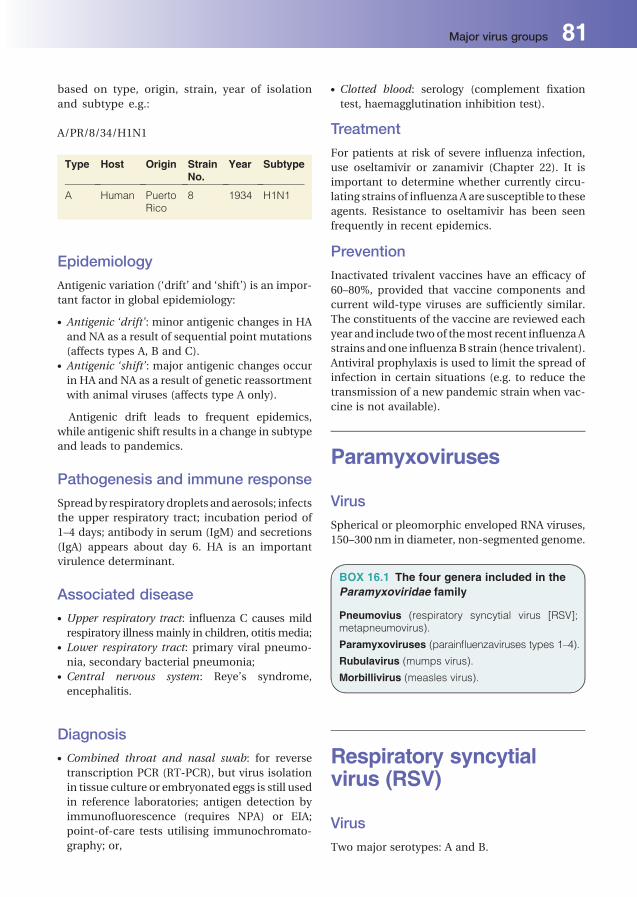
based on type, origin, strain, year of isolation
and subtype e.g.:
A/PR/8/34/H1N1
Type Host Origin StrainNo.
Year Subtype
A Human PuertoRico
8 1934 H1N1
Epidemiology
Antigenic variation (‘drift’ and ‘shift’) is an impor-
tant factor in global epidemiology:
. Antigenic ‘drift’: minor antigenic changes in HA
and NA as a result of sequential point mutations
(affects types A, B and C).. Antigenic ‘shift’: major antigenic changes occur
in HA and NA as a result of genetic reassortment
with animal viruses (affects type A only).
Antigenic drift leads to frequent epidemics,
while antigenic shift results in a change in subtype
and leads to pandemics.
Pathogenesis and immune response
Spreadby respiratorydroplets andaerosols; infects
the upper respiratory tract; incubation period of
1–4 days; antibody in serum (IgM) and secretions
(IgA) appears about day 6. HA is an important
virulence determinant.
Associated disease. Upper respiratory tract: influenza C causes mild
respiratory illnessmainly in children, otitis media;. Lower respiratory tract: primary viral pneumo-
nia, secondary bacterial pneumonia;. Central nervous system: Reye’s syndrome,
encephalitis.
Diagnosis. Combined throat and nasal swab: for reverse
transcription PCR (RT-PCR), but virus isolation
in tissue culture or embryonated eggs is still used
in reference laboratories; antigen detection by
immunofluorescence (requires NPA) or EIA;
point-of-care tests utilising immunochromato-
graphy; or,
. Clotted blood: serology (complement fixation
test, haemagglutination inhibition test).
Treatment
For patients at risk of severe influenza infection,
use oseltamivir or zanamivir (Chapter 22). It is
important to determine whether currently circu-
lating strains of influenzaA are susceptible to these
agents. Resistance to oseltamivir has been seen
frequently in recent epidemics.
Prevention
Inactivated trivalent vaccines have an efficacy of
60–80%, provided that vaccine components and
current wild-type viruses are sufficiently similar.
The constituents of the vaccine are reviewed each
year and include twoof themost recent influenzaA
strains andone influenzaB strain (hence trivalent).
Antiviral prophylaxis is used to limit the spread of
infection in certain situations (e.g. to reduce the
transmission of a new pandemic strain when vac-
cine is not available).
Paramyxoviruses
Virus
Spherical or pleomorphic enveloped RNA viruses,
150–300nm in diameter, non-segmented genome.
Respiratory syncytialvirus (RSV)
Virus
Two major serotypes: A and B.
BOX 16.1 The four genera included in theParamyxoviridae family
Pneumovius (respiratory syncytial virus [RSV];metapneumovirus).
Paramyxoviruses (parainfluenzaviruses types 1–4).
Rubulavirus (mumps virus).
Morbillivirus (measles virus).
Major virus groups 81

Epidemiology
RSV has aworldwide distribution; seasonal activity
during winter months in temperate climates and
throughout the year in warmer climates. Infects
more than 50%of infants aged less than 1 year, and
by 3 years of age all children have experienced an
RSV infection.
Pathogenesis
Transmission is via respiratory droplets or fomites;
incubation period 2–8 days; infects epithelium of
upper respiratory tract and then spreads to lower
respiratory tract, causing bronchiolitis. IgA, IgG
and IgM responses are not protective against re-
current infections and IgE may play a role in the
pathogenesis of disease.
Associated diseases. Lower respiratory tract: bronchiolitis. Complica-
tions include pneumonia, exacerbation of asth-
ma. Severe disease occurs in pre-term infants,
infants with underlying pulmonary or cardiac
disease and all immunocompromised patients.
Diagnosis. Nasopharyngeal aspirate or combined throat and
nasal swabs: detection of viral RNA by reverse
transcriptase real-timePCR (RT-PCR); isolation in
cell culture; antigendetectionby immunofluores-
cence (requires nasopharyngeal aspirate) or EIA;
point-of-care tests utilising immunochromato-
graphy or serology (complement fixation test).
Treatment
Generally symptomatic care.
Aerosolised ribavirin can be used for infants and
adults at risk of severe RSV infection (e.g. RSV
infection recently following an allogeneic haema-
topoetic stem cell transplant).
Prevention
Palivizumab is an RSV specific humanised mouse
monoclonal antibody indicated for use in children
at risk of life-threatening RSV infection. Monthly
injections are required during the annual RSV
epidemic period. Recipients of palivizumab are
normally premature infants with chronic lung dis-
ease. Attempts to develop an RSV vaccine have so
far been unsuccessful.
Parainfluenza viruses
Virus
Four serotypes.
Epidemiology
Type 3 causes annual epidemics during spring and
summer, type 1bi-annual outbreaks duringwinter.
Fiftyper centof infants are infectedby1yearof age.
Pathogenesis
Transmission is by respiratory droplets and
fomites; incubation period around 2–8 days. Rep-
lication occurs in nasopharyngeal epithelium
followed by spread to tracheobronchial tree.
Humoral immune response is not protective and
recurrent infections are common.
Associated diseases. Upper respiratory tract: croup, otitis media;. Lower respiratory tract: bronchiolitis, pneumo-
nia. Severe disease occurs in immunocompro-
mised children and adults.
Diagnosis. Nasopharyngeal aspirate or combined throat and
nasal swab: detection of viral specific RNAbyRT-
PCR; isolation in cell culture; antigen detection
by immunofluorescence (requires NPA)
Treatment
Symptomatic; aerosolised ribavirin in severely im-
munocompromised patients.
Prevention
No vaccine available.
Measles virus
Virus
Single serotype.
82 Major virus groups
Epidemiology
Measles virus has worldwide distribution, but
prevalence is low where uptake of MMR vaccina-
tion is more than 85%. Infections depend on the
state of immunity and size of thepopulation. Rapid
epidemics occur when the virus is introduced into
isolated, susceptible communities.
Pathogenesis
Transmission is via large droplets and aerosol;
incubation period 10–12 days to prodromal symp-
toms and 10–21 days to appearance of rash. In-
fection starts in epithelium of upper respiratory
tract followed by replication in local draining
lymph nodes, spreads to viscera and skin via the
blood. IgA, IgG and IgM responses lead to life-
long immunity.
Associated diseases. Upper respiratory tract: characterised by a febrile
illness with a rash and pharyngitis, conjunctivi-
tis, otitis media;. Lower respiratory tract: complications include
severe pneumonia;. Central nervous system: encephalitis, subacute
sclerosing panencephalitis (SSPE).
Severity of illness increases in adults and immu-
nocompromised patients.
Diagnosis. Clotted blood: detection of measles specific IgM;. Nasopharyngeal aspirate: measles RT-PCR;. CSF: formeasles IgG levels (SSPE) and RT-PCR to
detect current infection.
Treatment
Symptomatic care
Prevention
Live attenuated virus vaccine (part of MMR) is
used in the childhood immunisation schedule.
The vaccine is more than 95% effective after 2
doses. The first immunisation is given at the age
of 15 months (to avoid failure as a result of the
presence of maternal measles antibody); earlier
administration is given in developing countries,
because of the high measles mortality rate in
children less than 1 year old. A second MMR
vaccine should be given prior to the start of school
aged 5 years (UK).
Mumps
Virus
Single serotype.
Epidemiology
Mumps virus has a worldwide distribution. High
incidence in children between 4 and 10 years of
age; peak activity between January and May in
temperate climates.
Pathogenesis
Transmission is via large droplets and fomites;
incubation period 16–18 days. Infection starts in
the epithelium of upper respiratory tract followed
by replication in local draining lymph nodes and
spread to viscera by blood. IgA, IgG and IgM re-
sponses lead to life-long immunity.
Associated diseases. Upper respiratory tract: characterised by a febrile
illness, with upper respiratory tract symptoms
and swollen parotid salivary glands;. Endocrineorgans:pancreatitis,oophoritis,orchitis;. Central nervous system: meningitis, encephalitis,
which can result in deafness.
Diagnosis. Clotted blood: detection of mumps specific IgM
by EIA.
Treatment
Symptomatic care.
Prevention
Live attenuated virus vaccine is available as the
monovalent form (mumps only), or in combina-
tion with the rubella and measles vaccine (MMR).
See measles section for timing of vaccine.
Major virus groups 83

Rhinoviruses
Viruses
Members of the Picornaviridae family of viruses.
Icosahedral, non-enveloped RNA viruses 28–30nm
in diameter.
The picornavirus families are a diverse group of
human pathogens, including the enteroviruses
and rhinoviruses. There are more than 100 sero-
types of human rhinoviruses, divided in to 3
groups: A, B and C.
Epidemiology
Rhinoviruses are endemic worldwide. In temper-
ate climates, two peaks of infection are documen-
ted in autumn and early spring.
Pathogenesis
Transmission is by inhalation of droplets and
aerosols containing virus or by exposure to the
virus in respiratory secretions during close con-
tact. The incubation period is 2–4 days. Replica-
tion takes place in the nasal mucosa, with the
release of several pro-inflammatory cytokines,
leading to characteristic symptoms of the com-
mon cold.
Associated disease. Upper respiratory tract: cause of the common
cold syndrome (30–60% of cases, Chapter 25).
Complications: acute otitis media, severe respi-
ratory illness in infants with bronchopulmonary
dysplasia and immunocompromised patients,
exacerbation of asthma;. Lower respiratory tract: recent evidence suggests
that group C rhinoviruses are associated with
more serious lower respiratory tract infections
Diagnosis. Nasopharyngeal aspirate or combined throat and
nasal swabs: detection of rhinovirus specific RNA
by RT-PCR; isolation in cell culture
Treatment
Symptomatic care.
Coronaviruses
Virus
Enveloped RNA viruses 120nm in diameter.
Surface projections look like a crown under elec-
tronmicroscopy, hence the name – coronaviruses.
Threemajor strains: human coronaviruses (HCoV)
229E and OC43 and severe acute respiratory syn-
drome coronavirus (SARS-CoV).
Epidemiology
HCoV have a worldwide distribution and infec-
tions occur throughout the year, affecting all age
groups equally. A SARS-CoV epidemic started in
2002 in southern China, from where it spread to
Hong Kong, Vietnam, Singapore, Canada, Taiwan,
Thailand and other parts of the world. The epi-
demic ended in 2003, affecting 8,500 patients (95%
in Asia) with a mortality rate of 9.5%.
Pathogenesis
The incubation period is 2–5 days for HCoV and
2–10 days for SARS-CoV. Transmission is by
respiratory droplets, aerosols and possible fae-
cal-oral route for SARS-CoV. Replication takes
place in the epithelial cells of the respiratory
tract and gut with development of strain-specific
antibody. Asymptomatic shedding in faeces is
common.
Associated infection. Upper respiratory tract: commoncold syndrome:
HCoV is responsible for 25% of cases;. Lower respiratory tract: pneumonia;. Gastrointestinal tract: diarrhoea.
Diagnosis. Throat and combined nasal swab: real-time RT-
PCR, growth in cell culture, electronmicroscopy;. Clotted blood: serology (IFA, EIA and
immunoblot).
Treatment
Symptomatic care.
84 Major virus groups
Control
No specific vaccine.
Enteroviruses
Viruses
Members of the Picornaviridae family of viruses.
cosahedral, non-envelopedRNA viruses, 28–30nm
in diameter
Enteroviruses include five genera (Box 16.2).
Epidemiology
Enteroviruses have worldwide distribution. Infec-
tions occur in infancy in developing countries and
in early childhood in developed countries.
Pathogenesis
The incubation period is from 2–40 days; normal
habitat and primary site of replication are the
upper respiratory tract and intestinal tract (hence
‘enteroviruses’; followed by viraemia and infection
of target organs, i.e. CNS (brain, meninges and
spinal cord) or muscle tissue (heart, skeletal
muscle) and skin. Long-lasting local IgA and hu-
moral IgM/IgG response develops after natural
infection. Transmission is primarily via faecal-oral
route.
Associated diseases. Upper respiratory tract: pharyngitis, herpangina,
‘hand, foot and mouth disease’, conjunctivitis;. Central nervous system: poliomyelitis, aseptic
meningitis, encephalitis;
. Heart: myocarditis, pericarditis;
. Neonate: severe systemic infection.
Diagnosis. Throat swab, and/or faeces and/or CSF: detection
of specific RNA by RT-PCR; isolation in cell cul-
ture. Serology is generally not helpful.
Prevention and control
Both live (Sabin) and killed (Salk) vaccines are
available and used successfully by the World
Health Organization (WHO) in its poliomyelitis
eradication programme. Only four countries in
the world are NOT free of poliomyelitis. The in-
activated vaccine has been used in the UK since
2006.
Treatment
Symptomatic care.
Rubella virus (Chapter 40)
Virus
EnvelopedRNA virus, diameter 60 nmwith a single
serotype.
Epidemiology
Rubella virus has worldwide distribution. Prior to
the introduction of MMR vaccination, rubella was
a frequent childhood infection. In the developing
world, 25% of women of child-bearing age are
susceptible to rubella.
Pathogenesis
Transmission is by large droplets. The incubation
period is 13–20 days. Primary replication occurs in
epithelial cells of the upper respiratory tract and
the cervical lymph nodes followed by a viraemic
phase. The rash coincides with the appearance of
serum antibody. Lifelong immunity follows natu-
ral infection. Primary infection during the first
12 weeks of pregnancy often leads to generalised
foetal infection and congenital rubella syndrome
(cataracts, deafness, cardiac abnormalities, hepa-
tosplenomegaly, purpura or jaundice).
BOX 16.2 Genera include:
Polioviruses (types 1, 2, 3)
Coxsackie A viruses 23 types, (types 1–22, 24)
Coxsackie B viruses 6 types, (types 1–6)
Echoviruses 31 types, (types 1–9, 11-27, 29-33)
Enteroviruses 4 types, (types 68–71).
Major virus groups 85

Associated disease. Upper respiratory tract: rubella is usually char-
acterised by amild illness with upper respiratory
tract symptoms, rash, lymphadenopathy and
transient arthralgia;. Central nervous system: encephalitis, post-
infectious encephalopathy;. Neonates: congenital rubella syndrome if infec-
tion occurs in first 12 weeks of pregnancy.
Diagnosis. Clotted blood: for acute infection, specific IgM
antibody is determined by EIA; specific IgG in-
dicates current immune status (e.g. ante-natal
screening);. Foetal blood: to detect evidence of intra-
uterine infection by RT-PCR and cell culture (see
Chapter 40 for further details).
Treatment
Symptomatic care.
Proven infection during the first 3 months of
pregnancy is an indication for therapeutic abortion.
Human normal immunoglobulin in cases of
persistent infection in immunocompromised
patients.
Prevention and control
Live attenuated MMR vaccine is used. Immunisa-
tion programmes exist in most developed coun-
tries. Women found to have no rubella antibodies
in their ante-natal screening blood, should receive
MMR immunisation after the delivery of the baby
(post-partum).
Rotaviruses
Virus
RNA virus, 75nm in diameter with wheel-like
structure. Segmented genome (11 segments).
Five groups (A–E), and within group A at least
two subgroups (I, II) and various serotypes (in-
cluding G and P types); G1–G4 viruses cause over
90% of human infections.
Epidemiology
Rotaviruses occur worldwide with different sero-
types co-circulating, usually affecting children of
less than 2 years, with a winter peak in temperate
climates.
Pathogenesis
Infection is via the faecal-oral route, with an incu-
bation period of 1–2 days. Virus replication occurs
in the epithelium of the small intestine and results
in the release of large numbers of particles in
human faeces (1� 1011 particles/gram). There are
local (IgA) and humoral (IgM/IgG) serotype-spe-
cific and cross-reactive immune responses, which
confer limited protective immunity.
Associated disease. Gastrointestinal: self-limiting diarrhoea and vo-
miting lasting 4–7 days; mild-to-severe dehydra-
tion can occur (depending on strain). In resource
poor countries, there is a significant infant mor-
tality rate associated with these infections.
Diagnosis. Faeces: clarified faecal extract is tested by an
antigen (virus protein) capture assay; reverse-
transcription PCR; point-of-care-tests based on
immunochromatography.
Treatment
Rehydration with oral rehydration fluid.
Prevention and control
Increasedpersonalandwaterhygienehelpscontrol
outbreaks. Rotavirus vaccine is now universally
used as part of the childhood schedule in the USA.
Norovirusand sapoviruses
Viruses
Small non-enveloped RNA viruses, diameter
27–35nm.
86 Major virus groups

Members of the Caliciviridae family, which
show a characteristic cup-shaped depression on
surface of particle when viewed by electron
microscopy.
Epidemiology
Noroviruses and sapoviruses have a worldwide
distribution. Outbreaks occur year round butmore
frequently during winter months in temperate cli-
mates. Outbreaks associated with contaminated
water supplies and food. More than 30% of chil-
dren are seropositive by 3 years of age, and more
than 80% are seropositive in early adulthood. Nor-
ovirus infections are the most common cause of
gastroenteritis outbreaks in hospitals and also of-
ten associated with homes for the care of the
elderly. Such outbreaks result in ward closures and
restrict hospital admission during the busiest win-
ter months.
Pathogenesis
Transmission by faecal-oral route, possibly aero-
sol due to projectile vomiting; incubation period
is 16–48h. Virus replication takes place in epithe-
lial cells of the villi of the small intestine. Noro-
virus infection does not confer lasting protective
immunity, because of frequent mutation in the
virus.
Associated diseases. Gastrointestinal: gastroenteritis characterised by
projectile vomiting and diarrhoea lasting for
12–60h.
Diagnosis. Detection in stool by electron microscopy, EIA
and RT-PCR.
Treatment
Symptomatic care.
Prevention
No vaccine available. Strict source isolation of
infected patients is required to limit nosocomial
infections.
Hepatitis viruses (includesDNA and RNA viruses)(Chapter 29)Hepatitis A virus (HAV)
Virus
Member of Picornaviridae family of viruses, genus
Hepatovirus. Small non-enveloped RNA virus
28–32nm (previously enterovirus 72); only one
serotype.
Epidemiology
Most infections in endemic areas (developing
world) occur before 5 years of age and themajority
are asymptomatic. In the developed world, most
clinical cases occur in adults. Common source
outbreaks result from contamination of drinking
water and food. Infections acquired frequently by
travellers from non-endemic to endemic areas.
Antibody prevalence in young adults is 30–60%,
higher in lower socioeconomic groups.
Pathogenesis
Transmission is via the faecal-oral route. The in-
cubation period is 2–6 weeks (mean 30 days). All
age groups are susceptible to hepatitis A infection
and disease severity increases with age. In most
cases, there is complete recovery and a specific
antibody response persists lifelong. There is no
chronic disease or carrier state.
Associated disease. Liver: Acute hepatitis.
Diagnosis. Clotted blood: serology (EIA), testing for specific
HAV IgM (acute infection) or IgG (immune
status);. EDTA-blood: reverse-transcriptionPCR is carried
out at a few reference laboratories to confirm
difficult to interpret serology results.
Treatment
Symptomatic care.
Major virus groups 87

Prevention
A killed HAV vaccine is available and recom-
mended for high risk groups.
Hepatitis B virus (HBV)
Virus
Member of the Hepadnaviridae family of viruses.
DNA virus, 42 nm in diameter, consisting of:
. Core: DNA, partially circular genome;
. Nucleocapsid (hepatitis B core antigen,HBcAg);
‘e’ antigen (HBeAg) is a cleavage product of
the core antigen found on infected cells or free
in serum;. Envelope (hepatitis B surface antigen, HBsAg);. Also exists as 22 nm spherical or filamentous
particle consisting of hepatitis B surface antigen,
HBsAg.
Epidemiology
Hepatitis B virus has a worldwide distribution,
withmore than 360million carriers (prevalence in
north and mid-Europe and North America
0.1–0.5%; southern Europe 2–5%; Africa and
Southeast Asia 6–20%).
Pathogenesis. Transmission is via exposure to blood and blood
products containing the virus, by sexual inter-
course and vertically at birth (this is the main
route of transmission in Asia and Africa);. Incubation period of 6 weeks–6 months;. Viral replication in liver results in lysis of hepa-
tocytes by cytotoxic T cells in those who mount
an effective immune response;. Hepatic damage is reversed in 8–12 weeks in
�90% cases; 2–10% become chronic carriers
(persistence of HBsAg for >6 months);. 95% of newborns of carrier mothers become
carriers if untreated;. The four phases of chronic infection include:
T immune tolerance (usually infections ac-
quired at birth);
T immune clearance (many patients clear HBe
antigen andcovert toHBe antibodypositivity);
T inactive carrier (HBe antibody positive with
low levels of virus present in blood);
T hBeAg-negative chronic hepatitis B (HBe an-
tigen negative, HBe antibody positive & HBV
DNA detected).
Not all patients go through each of the four phases.
Associated diseases. Liver : acute, chronic carrier or rarely fulminant
(rapidly progressive, with liver failure); cirrhosis;
hepatocellular carcinoma.
Diagnosis. Clotted blood: serological tests are made by im-
munoassays detecting HBsAg, HBeAg and anti-
bodies to HBcAg (IgM and IgG), anti-HBeAg and
anti-HBsAg;. EDTA-blood: for quantitative nucleic acid detec-
tion to determine viral load and genotype
assessment.
Prevention
HBV vaccines and/or HBV immunoglobulin
(HBIG) for post-exposure prophylaxis and babies
born to carrier mothers.
Treatment
Lamivudine, adefovir, entecavir, telbivudine, cle-
vudine and tenofovir.
Hepatitis C virus (HCV)
Virus
RNA virus, 40–50nm in diameter; Member of the
Flaviviridae family of viruses.
There are six main genotypes, which respond
differently to antiviral treatment.
Epidemiology
HCV occurs worldwide. Antibody prevalence varies
betweenlessthan1%intheUSAandWesternEurope
and 2% in southern Italy, Spain and central Europe.
Higher prevalence rates, up to 20%, are detected in
Egypt. There is high prevalence of infection among
injecting drug users in most parts of the world.
Associated diseases. Liver : HCV has a similar pathogenesis to HBV;
infections are followed by chronic hepatitis
in 60–80% of cases; cirrhosis, hepatocellular
carcinoma.
Diagnosis. Clotted blood: serology by EIA to detect HCV
antibodies, antigens and by RT-PCR;. EDTA-blood: to determine genotype and viral
load.
88 Major virus groups

Prevention
Since 1991, routine testing of all blood and
organ donors for HCV antibody has been under-
taken, to prevent transmission by transfusion or
transplantation.
Treatment
Pegylated interferon-a, ribavirin.
Delta agent (‘hepatitis D virus’,HDV)
Virus
Defective RNA virus; 36nm in diameter, which
replicates only in HBV-infected cells.
Epidemiology
Worldwide distribution; high prevalence in the
Mediterranean area, Africa, South America, Japan
and the Middle East.
It has similar transmission and affects the same
risk groups to HBV.
Associated disease. Liver: infections are either a co-infection with
HBVor a superinfectionof chronicHBV infection
leading to aggravation of HBV disease.
Diagnosis. Clotted blood: serology (EIA) can be used to
detect HDV antibody and HDV antigen; nucleic
acid detection methods are available in hepatitis
reference facilities.
Prevention
HBV vaccination and subsequent immunity to
HBV reduces the risk of acquiring the delta agent.
Hepatitis E virus (HEV)
Virus
Non-enveloped, positive-strand RNA virus, it re-
presents the only species in the Hepeviridae
family. There are 4 known genotypes that infect
certain mammals including man.
Epidemiology
Endemic in Indian subcontinent, Southeast Asia,
Middle East, North Africa and Central America.
Common source outbreaks caused by contami-
nated water or food are common. In developed
countries, sporadic cases are detected among tra-
vellers returning from endemic areas.More recent-
ly strains (genotypes 3 and 4) of hepatitis E have
been detected in domestic animals in industrialized
countries and are implicated in zoonotic infections.
Pathogenesis
Transmission by the faecal-oral route and rarely by
blood transfusion in endemic countries
Incubation period is up to 6 weeks. Specific IgG
and IgM antibodies are produced.
Associated diseases. Liver: acute self-limiting hepatitis with no evi-
dence of chronic infection;. Highmortality rate (10–20%) in pregnantwomen
has been observed in India.
Diagnosis. Clotted blood: serology (detection of IgG and IgM
antibodies by EIA); nucleic acid detection in
reference laboratories.
Treatment
Symptomatic care.
Prevention
Vaccine under development.
Human retrovirusesHuman immunodeficiency virus(HIV) (Chapter 41)
Virus
A member of the Lentivirinae subfamily of the
Retroviridae family. Enveloped RNA viruses with
100–150nm in diameter virion.
Replicates through DNA intermediates utilising
the RNA-directed DNA polymerase (reverse
transcriptase)
There are two types: HIV-1 and HIV-2 (40%
sequence homology). HIV-1 is divided into three
groups: M (11 subtypes A–K), and O and N groups.
Group M subtypes include 95% of the global virus
isolates and have worldwide distribution, whereas
Major virus groups 89

groups O and N are confined to parts of West and
Central Africa. Generally HIV isolates, even from a
single individual, show a high level of variation.
The termquasi-species is used todescribe this pool
of diverse and changing viruses present in an
individual.
Epidemiology
There has been worldwide spread of HIV since
1981. Estimates of prevalence worldwide vary
greatly. By the end of 2010, 60 million people
worldwide were estimated to be living with HIV
(25 million in Africa and 7.5 million in Asia); 20
million people have died ofHIV since the epidemic
started in 1981.
The main risk groups for HIV infection are:
. men who have sex with men (MSM);
. intravenous drug users;
. people with haemophilia and blood transfusion
recipients before 1985;. sexually promiscuous individuals;. children born to HIV-infected mothers;. heterosexual contacts of HIV-infected individuals.
Pathogenesis and immune response
Transmission is mainly through sexual inter-
course, via bloodor bloodproducts or frommother
to child during delivery and/or breast feeding. HIV
directly infects and kills cells that are essential for
an effective immune response. The virus attaches
to the cellular receptor CD4 and chemokine cor-
eceptor CCR5 or CXCR4. By infecting the key cells
of the adaptive immune response, this explains the
main clinical feature of disease as being profound
immune suppression. This results in early im-
pairment of various CD4-cell functions followed
by a decrease in CD4-cell numbers. A humoral
antibody response against most HIV-specific pro-
teins develops. In later stages, deficiency of both
the humoral and the cellular immune responses
leads to the development of AIDS.
Associated diseases. Immune system: Acquired immunodeficiency
syndrome (AIDS) is characterised by severe dis-
ease resulting from generalised infections
with bacteria including: mycobacteria, viruses
(e.g. HSV, CMV and VZV), fungi (e.g. Pneumo-
cystis, Candida, Aspergillus and Cryptococcus),
protozoa including Toxoplasma and with asso-
ciated tumours (Kaposi’s sarcoma, lymphomas).
Diagnosis. Clotted blood: detection ofHIV-specific antibody
and antigen by passive particle agglutination
tests (PPAT), fourth generation EIA and western
blotting (WB).. EDTA-blood: molecular techniques for detecting
HIV, quantifying viral load; typing and drug
resistance genotyping have become a significant
part of patient diagnosis and clinicalmanagement.
Treatment
Five classes of drugs are available:
1 nucleoside reverse transcriptase inhibitors (e.g.
zidovudine, lamivudine);
2 non-nucleoside reverse transcriptase inhibitors
(e.g. nevarapine and delavirdine);
3 protease inhibitors (e.g. indinavir, atazanavir);
4 fusion inhibitors (enfuvirtide);
5 integration inhibitors (raltegravir).
A combination of three drugs (highly active
antiretroviral therapy or HAART) is the accepted
standard form of treatment. Antiretroviral drug
resistance is a continuing problem and patients
require follow-up to monitor HIV load and CD4
counts.
Prevention and control. There is no antiviral cure.. No effective vaccine has been developed; there
are problems as a result of viral variability, la-
tency and evasion to immune response.. Testing of all blood and organ donors prevents
transmission from those sources.. Information campaigns, needle exchange pro-
grammes and condom use.. Post-exposure prophylaxis with HAART is re-
commended after sexual exposure or exposure
in healthcare settings.
Human T-cell lymphotropicviruses (HTLV-I and HTLV-II)
Virus
Members of the delta-retroviruses genus of the
Retroviridae family.
90 Major virus groups

The RNA genome is less variable than that of
HIV; HTLV-II shows 60–70% sequence homology
with HTLV-I.
Epidemiology
HTLV-I is endemic in Japan, the Caribbean, Mel-
anesia, part of sub-Saharan Africa and Brazil.
HTLV-II is endemic among Native American In-
dians in North, Central and South America. A high
proportion of intravenous drug users in North
America are seropositive.
Pathogenesis
Transmission is by blood exposure, sexual inter-
course and from mother to child through breast-
feeding. HTLV-I can immortalise T lymphocytes
in vitro and can transactivate T cells in vivo, but
no oncogenes have been identified. No similar
characteristics have been proved for HTLV-II.
Most infected individuals remain asymptomatic
carriers for life. Only 2–4% of infected people
develop adult T-cell leukemia and lymphoma
(ATLL) after many decades of infection. HTLV-
I can invade the CNS and the resulting inflam-
matory immune response is responsible for the
pathogenesis of HTLV-associated myopathy
(HAM).
Associated diseases. Blood: adult T-cell leukemia and lymphoma;. Central nervous system: HTLV-associated
myopathy;. Eye: uveitis.
Diagnosis. Clotted blood: detection of HTLV-I-specific an-
tibody is made by EIA, particle agglutination
assays and western blotting;. EDTA-blood: nucleic acid detection methods.
Prevention and control
Testing of all blood donors is undertaken in coun-
tries, such as the USA and the UK.
Treatment
No antiviral drugs are available.
Human papillomavirus(HPV)
Virus
Non-envelopedDNA virus, 55nm in diameter with
over 100 genotypes recognised.
Epidemiology
HPV occurs worldwide.
Pathogenesis
Transmission is by close direct contact, sexual
intercourse and perinatal transmission through
the birth canal. Infects and replicates in squamous
epithelium on both keratinised and mucosal sur-
faces. Infection results in persistent and latent
infection, which can result in malignant
transformation.
Associated disease. Skin and soft tissue: warts (e.g. plantar warts) and
other skin conditions (epidermodysplasia verru-
ciformis), genital warts (e.g. condyloma acumi-
nata) and cervical, penile and anal carcinoma;
laryngeal papillomatosis in young children.
Diagnosis. Genital swabs and biopsy samples: real-time PCR
for limited number of high risk genotypes.
Therapy
Suspicious areas of dysplasia are subject to surgery
or cauterisation or cryotherapy and laser therapy;
interferon, imiquimod and podophyllum are indi-
cated for the treatment of genital warts.
Prevention
Two HPV vaccines are currently available against
HPV-16 and -18. These HPV types can cause cer-
vical and other types of genital cancer. The current
strategy in economically developed countries is to
immunise younger women prior to the age of
sexual activity. Prevenar 13 has now superseded
Prevanar 7.
Major virus groups 91
Rabies virus
Virus
Member of the Rhabdoviridae family
It is a bullet-shaped RNA virus (75–180 nm).
Several genotypes (include classic rabies,
European, Australian bat rabies and rare African
genotypes). All known to cause disease in humans,
but the most common source is classic rabies.
Epidemiology
Rabies virus has awide animal reservoir (e.g. foxes,
skunks, racoons, bats) and is enzootic in terrestrial
mammal species worldwide. Some parts of the
world are considered free from rabies, i.e. Ireland,
New Zealand. The UK used to be free from rabies
but recently three isolations of bat rabies were
made, including a fatal case in a Scottish bat
conservationist. Human infections occur mostly
in Asia, Africa and South America and dogs are the
cause of more than 90% of these infections.
Pathogenesis
Infectionmainly occurs through the bite of a rabid
animal and rarely after corneal transplant from
infected patients. Incubation period can be as
short as 4 days to 19 years, but is generally 20–90
days.Multiplication takes place inmuscle cells and
at neuromuscular junctions. The virus migrates
along peripheral nerves to the CNS. Replication in
the CNS (mainly basal ganglia, pons, andmedulla)
causes nerve cell destruction and inclusion bodies
(Negri bodies). Finally, migration occurs from the
CNS along peripheral nerves to different tissues,
including peripheral nerves, skeletal and cardiac
muscles and salivary glands.
Associated disease. Central nervous system: paraesthesiae; anxiety;
hydrophobia; paralysis; coma. Mortality rate is
100%.
Diagnosis. Clotted blood and CSF: detection of virus specific
RNA by RT-PCR; serology (rabies antibodies in
blood and CSF);. Animal brain: investigated at specialist veteri-
nary reference laboratory using histological and
molecular procedures;. Hair follicles: detection of viral antigens by
immunofluorescence,
Therapy and control
Pre-exposure prophylaxis: immunisation of at-risk
groups (veterinary personnel, laboratory workers,
travellers to endemic areas) – 3 doses (0, 7, 28 days)
of rabies vaccine followed by a booster dose 1 year
later.
Post-exposure prophylaxis: 5 doses of vaccine
(0, 3, 7, 14, 28 days), plus 1 dose of rabies
immunoglobulin.
Spread can be controlled by quarantine of ani-
mals in the UK and immunisation of domestic
animals, such as dogs and cats in endemic areas.
92 Major virus groups

17Basic mycologyand classification of fungiElizabeth JohnsonHealth Protection Agency Mycology Reference Laboratory, Bristol, UK
CharacteristicsFungi are eukaryotic microorganisms, distinct
from plants in not containing chlorophyll and are
classified in a kingdom of their own. Fungi have
macroscopic fruiting bodies (mushrooms and
toadstools) or are microscopic (moulds and
yeasts). Although many fungi are capable of caus-
ing disease in a sufficiently immunocompromised
patient, only a few species cause humandisease on
a regular basis. Fungi are non-motile; they may
grow as single cells (yeasts) or multi-septate,
branching filamentous hyphae. An intertwined
network of hyphae is a mycelium.
ClassificationThe classification of fungi is based largely on their
method of sexual reproduction. Those that do not
reproduce sexually were previously referred to as
‘fungi imperfecti’. Those that reproduce sexually
may be self-fertile or require strains of an opposite
type to allow sexual fusion to occur. The fungi that
cause human disease can be broadly split into
yeasts, moulds or dimorphic fungi, which have
both yeast andmould forms, depending on growth
temperature (Table 17.1).
Fungal infectionsFungal infections can be divided clinically into
three groups: superficial, sub-cutaneous and
deepmycoses.Most deepmycoses are opportunist
infections occurring in immunocompromised pa-
tients. Tables 17.2 and 17.3 outline some superfi-
cial and deep mycoses, respectively.
Laboratory diagnosis. Direct microscopy of clinical material from
deep or superficial sites; the characteristic
morphology of fungal elements facilitates
identification.. Culture: fungi can be grown on most routine
media, but may require prolonged incubation.
Antibiotic-containing selective media (e.g.
Sabouraud’s glucose agar), which inhibit bacte-
rial growth, are often used. Incubation at
both 37 �C and 28 �C facilitates the isolation of
common filamentous fungi and yeasts.
Medical Microbiology and Infection Lecture Notes, Fifth Edition. Edited by Tom Elliott, Anna Casey,Peter Lambert and Jonathan Sandoe.� 2011 Blackwell Publishing Ltd. Published 2011 by Blackwell Publishing Ltd.

. Serology: serological tests both for antibody and
antigen detection are available for the diagnosis
of some fungal infections (e.g. cryptococcosis,
candidiasis and aspergillosis), but thesemay lack
specificity and sensitivity. Testing for beta-
glucan has a good negative predictive value.
Examples of fungalpathogensCryptococcus
. C. neoformans, C. grubii and C. gattii are
encapsulated yeasts that cause cryptococcal
meningitis. The first two are found worldwide,
whilst C. gattii is associated with Eucalyptus and
is most common in Australia and parts of Africa.
Their natural habitat is soil, particularly soil con-
taminated with pigeon droppings, which due to
the presence of high molecular weight nitroge-
nous compounds, act as a natural selective
medium.. Cryptococcal species have polysaccharide cap-
sules that can be visualised in clinical speci-
mens, such as cerebrospinal fluid, bymixing the
fluid with Indian ink. They can be isolated from
sputum, bronchoalveolar lavage fluid, tissue
biopsies and other clinical specimens, or the
capsular antigen can be detected by sensitive
and specific latex agglutination tests on CSF
or serum.
Table 17.1 Groups of fungi that cause human disease
Group Comment Examples
Yeasts Have round or oval cells that multiply by budding or growpredominantly as yeasts that bud; but may also formpseudohyphae or even true hyphae in some species
Cryptococcus neoformans
Candida albicans
Filamentousfungi
Grow as hyphae and produce a mycelium; produceasexual spores (conidia or sporangiospores), which maybesingleormulti-celled.Conidiacanbeproduced in longchains on aerial hyphae (conidiophores), as wet masseson phialides (a conidiogenous cell that produces conidiawithout increasing in length) or as budding aleuriospores(a spore produced as a result of the expanding of theends of conidiophores or hyphae).
Aspergillus spp.Fusarium spp.Trichophyton spp.
Sporangiospores are produced in sporangia on aerialhyphae known as sporangiophores
Rhizopus spp. Mucor spp.
Dimorphicfungi
Have two forms of growth: filamentous at 28 �C (saprophyticphase) and yeast like at 37 �C (parasitic phase)
Blastomyces, Histoplasmacapsulatum, Penicillummarneffei
Table 17.2 Superficial mycoses
Fungi Type of fungus Principal infections Epidemiology
Candida albicans Yeast Oral thrush, vaginitis, cutaneouscandidiasis
WorldwideDermatophytes Filamentous Tinea (ringworm) of skin,
nail and hairWorldwide
Epidermophyton
Microsporum
Trichophyton
Malassezia furfur Yeast Pityriasis versicolor Worldwide, mostcommon in the tropics
94 Basic mycology and classification of fungi

. Infection is thought to be acquired by inhalation
of yeast cells, which become desiccated and are
blownonair currents.Human infections are rare;
most occur in immunocompromised indivi-
duals, including those with HIV.. Primary infection occurs in the lungs and is
usually asymptomatic. Acute pneumonia may
occur with fungaemia and infection in various
organs, particularly the brain and meninges.
Treatment of cryptococcal meningitis is with
amphotericin and flucytosine in combination,
followed by fluconazole.
Candida
. The genus Candida contains a large number of
species, about eight of which are regularly found
causing infection. C. albicans remains the most
frequently isolated pathogen, although infection
with C. glabrata appears to be increasing and C.
parapsilosis and C. tropicalis are also frequently
encountered. C. albicans is a commensal of the
mouth and gastrointestinal tract.. Superficial Candida infections are common and
include vaginal and oral candidiasis (thrush),
skin andnail infections, which can arise inwarm,
moist areas such as skin folds or because hands
are immersed in water for long periods, and as a
complication of antibiotic therapy that tempo-
rarily reduces the bacterial flora.. Invasive Candida infections usually arise from
the patients own commensal flora following sur-
gery, use of broad spectrum antibiotics or intra-
vascular catheters and can affect any organ of the
body. Candidaemia may result in abscesses in
various organs (e.g. brain, liver, spleen). These
infections occur primarily in immunocompro-
mised patients. Endophthalmitis is another
potential consequence of candidaemia.. Candida can also colonise prosthetic materials,
e.g. intravascular catheters and peritoneal dial-
ysis catheters, resulting in bloodstream infection
and peritonitis, respectively. Candida spp. are a
rare cause of endocarditis.
Laboratory diagnosis
By direct microscopy of appropriate clinical ma-
terial for oval Gram-positive cells, some of which
may be budding or producing pseudohyphae (a
string of elongated cells produced as a result of
budding in some yeasts); culture; and serology for
Candida antibodies or antigen in patients with
deep-seated infections. Budding yeast and pseu-
domycelia (a mycelium-like mass of pseudohy-
phae) may also be seen with appropriate stains in
histopathological specimens.
Table 17.3 Deep mycoses
Fungi Type of fungus Principal infections Epidemiology
Aspergillus fumigatus Filamentous Lung infection, disseminatedaspergillosis
Worldwide
Candida albicans Yeast Oesophagitis, endocarditis,candidaemia with disseminatedcandidiasis
Worldwide
Cryptococcusneoformans
Yeast Meningitis Worldwide
Histoplasma capsulatum Dimorphic Lung infection USA
Coccidioides immitis Dimorphic Lung infection Central/South America
Blastomyces dermatidis Dimorphic Lung infection Africa, America
Paracoccidioidesbrasiliensis
Dimorphic Lung infection South America
Rhizopus arrhizus Filamentous Rhinocerebral infection, lung infection,(zygomycosis or mucormycosis)
Worldwidedisseminated infection
Fusarium Filamentous Lung, sinus, fungaemia Worldwide
Scedosporium Filamentous Lung, disseminated Worldwide
Basic mycology and classification of fungi 95
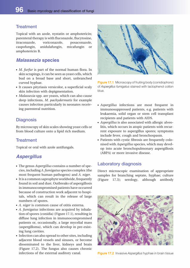
Treatment
Topical with an azole, nystatin or amphotericin;
parenteral therapy iswith fluconazole, flucytosine,
itraconazole, voriconazole, posaconazole,
caspofungin, anidulafungin, micafungin or
amphotericin B.
Malassezia species
. M. furfur is part of the normal human flora. In
skin scrapings, it can be seen as yeast cells, which
bud on a broad base and short, unbranched
curved hyphae.. It causes pityriasis versicolor, a superficial scaly
skin infection with depigmentation.. Malassezia spp. are yeasts, which can also cause
deep infections, M. pachydermatis for example
causes infection particularly in neonates receiv-
ing parenteral nutrition.
Diagnosis
Bymicroscopy of skin scales showing yeast cells or
from blood culture onto a lipid rich medium.
Treatment
Topical or oral with azole antifungals.
Aspergillus
. The genus Aspergillus contains a number of spe-
cies, includingA. fumigatus species complex (the
most frequent human pathogens) and A. niger.. It is a commonsaprophyteworldwide, frequently
found in soil and dust. Outbreaks of aspergillosis
in immunocompromised patients have occurred
because of construction work adjacent to hospi-
tals, which can result in the release of large
numbers of spores.. A. niger is common cause of otitis externa.. A. fumigatus infections are acquired by inhala-
tion of spores (conidia) (Figure 17.1), resulting in
diffuse lung infection in immunocompromised
patients or, occasionally, a large mycelial mass
(aspergilloma), which can develop in pre-exist-
ing lung cavities.. Infection can also spread to other sites, including
adjacent blood vessels and sinuses, or become
disseminated to the liver, kidneys and brain
(Figure 17.2). The fungus also causes chronic
infections of the external auditory canal.
. Aspergillus infections are most frequent in
immunosuppressed patients, e.g. patients with
leukaemia, solid organ or stem cell transplant
recipients and patients with AIDS.. Aspergillus is also associated with allergic alveo-
litis, which occurs in atopic patients with recur-
rent exposure to aspergillus spores; symptoms
include fever, cough and bronchospasm.. Patients with cystic fibrosis are frequently colo-
nised with Aspergillus species, which may devel-
op into acute bronchopulmonary aspergillosis
(ABPA) or more invasive disease.
Laboratory diagnosis
Direct microscopic examination of appropriate
samples for branching septate, hyphae; culture
(Figure 17.3); serology, although antibody
Figure 17.1 Microscopy of fruiting body (conidiophore)
of Aspergillus fumigatus stained with lactophenol cotton
blue.
Figure 17.2 Invasive Aspergillus hyphae in brain tissue
96 Basic mycology and classification of fungi

detection is of limited value in immunocompro-
mised patients, antigen detection for galactoman-
nan or b-glucan may be useful and Aspergillus-
specific PCR for the detection of circulating
genomic fragments is helpful in some patients.
Aspergillus may also be seen in histopathological
specimens and is the gold standard confirmation
of infection.
Treatment
Amphotericin B, itraconazole, voriconazole, posa-
conazole or echinocandins.
DermatophytesThe dermatophytes are a group of related filamen-
tous fungi, also referred to as the ringworm fungi,
which infect the keratinised tissues; skin, hair and
nails. Three clinically important genera have been
described (Table 17.4) (Figures 17.4 and 17.5).
Epidemiology
The natural habitat is humans, animals or soil;
human infection results from spread from any of
these reservoirs. Dermatophyte infections are
found worldwide, with different species predomi-
nating in various climates.
Laboratory diagnosis. Skin scrapings, hair or nail clippings from active
lesions are examined microscopically in 20–30%
potassium hydroxide on a glass slide; the pres-
ence of hyphae confirms the diagnosis. Optical
brighteners, such as calcofluor or blankophor,
can enhance the detection of fungal elements
when viewed under a fluorescence microscope.
Occasionally, the dermatophyte species can be
identified by typical morphology.. Samples can be cultured on Sabouraud’s medi-
um at 28 �C. Subsequent species identification is
based on growth rate, colony appearance and
microscopic morphology.. Infected hair may fluoresce under ultraviolet
light (Wood’s light) and is characteristic of cer-
tain infections, e.g. Microsporum canis.
Treatment
Depends on the site and severity of infection. Op-
tions include topical imidazoles (e.g. clotrimazole,
Figure 17.3 Aspergillus fumigatus (left) on Sabouraud’s
agar. Strains produce green colonies with white
periphery.Aspergillus niger (right) demonstrating dense
black colonies with white periphery.
Table 17.4 Clinically important genera of dermatophytes
Genera Infection
Epidermophyton E. floccosum, the only species.
It infects the skin (tinea corporis), nails (tinea unguium), groin (tinea cruris) and feet (tineapedis) (Plate 36)
Microsporum M. audouini causes epidemic ringworm of the scalp (tinea capitis) in children.M. canis, which predominantly affects cats and dogs, occasionally causes ringwormin children
Trichophyton T. mentagrophytes var. interdigitale is the most common cause of tinea pedis
T. rubrum causes severe recurrent skin and nail infections T. violaceum is a common
cause of tinea capitis in Asian subcontinents, North America and the West Indies
T. tonsurans is an increasingly common cause of scalp ringworm, especially in the Afro-Caribbean population
Basic mycology and classification of fungi 97

miconazole); oral griseofulvin, itraconazole,
fluconazole or terbinafine.
MucormycosisThe term ‘mucormycosis’ (or ‘zygomycosis’) refers
to infections caused by a variety of filamentous
fungi, most of which are currently classified in
the sub-phylum mucoromycotina. Medically im-
portant species include Rhizopus arrhizus and
Lichtheimia corymbifera (previously Absidia
corymbifera).
Epidemiology
Mucoraceous fungi are found worldwide, in soil
and decaying organic matter. Infections occur
primarily in uncontrolled diabetic or immuno-
compromised individuals following inhalation of
spores, but wound infections are also seen. As
with Aspergillus, hospital outbreaks of infection
have occurred in association with building work
or contaminated fomites.
Risk factors
Diabetes mellitus, burns, neutropenia.
Infection. Pulmonary: an often fatal infection of immuno-
compromised patientsmay result in disseminat-
ed infection (e.g. brain, liver, gastrointestinal
tract).. Rhinocerebral: an infection of the nasal sinuses
may spread rapidly to involve the face, orbit and
brain. It occurs particularly in uncontrolled dia-
betes mellitus and is often fatal if treatment is
delayed.. Necrotic tissue from burns.
Laboratory diagnosis
Microscopy and culture of appropriate specimens
are undertaken (including tissue from necrotic
lesions,sputum,bronchoalveolar lavage(BAL)).On
microscopic examination, this group of fungi are
characterised by the formation of broad, ribbon-
like, hyphae with few septae and are fast growing
in culture often filling all the air-space inside petri
dishes. Tissue should not be homogenised, as this
will decrease the chances of isolation of viable
fungi.
Treatment
Amphotericin B, especially higher doses of lipid
formulations and posaconazole, plus urgent
d�ebridement of necrotic tissue in rhinocerebral
infection and stabilisation of underlying predis-
posing factors.
Fusarium and ScedosporiumspeciesEnvironmental fungi found as plant pathogens or
in pollutedwater, usually acquiredby inhalation or
aspiration of polluted water:
. Infectionwith Fusarium species is predominant-
ly seen in neutropenic patients and mimics in-
vasive aspergillosis. Skin lesions occur in 60–70%
of infected patients.. Scedosporium species can infect otherwise
healthy individuals following aspiration of
Figure 17.4 Colony of Epidermophyton floccosum
illustrating its undulating surface.
Figure 17.5 Colony of Trichophyton mentagraphytes
illustrating its powdery white surface.
98 Basic mycology and classification of fungi
polluted water during near-drowning accidents,
also infects neutropenic individuals.. Infection with either is primarily pulmonary but
can become disseminated.
Laboratory diagnosis
Microscopy and culture of appropriate specimens.
Blood culture is often positive in Fusarium infec-
tion. Identification in culture depends on exami-
nation of the form of spores and spore production.
Branching, septate hyphae resemble those of
Aspergillus.
Treatment
Voriconazole, posaconazole.
Coccidioides immitisand C. posadasiiC. immitis and C. posadasii are closely related
dimorphic fungi found in the soil in hot arid
areas of southwest USA, and Central and South
America.
Infection
This follows inhalation of arthrospores (a spore
produced by fragmentation of a hypha at
the septum) and is often subclinical. A mild, self-
limiting pneumonia (Valley fever), often accom-
panied by a maculopapular rash, occurs in some
patients. Severe progressive disseminated disease
(e.g. meningitis, osteomyelitis) may occur, princi-
pally in immunosuppressed patients. Pulmonary
infection may occur in AIDS patients who have
lived in or travelled through endemic regions but
otherwise healthy individuals are also at risk.
Laboratory diagnosis
Direct microscopy for the presence of endospore-
containing spherules and culture of appropriate
specimens (this is classified as a higher-risk path-
ogen and culture should be attempted only in
specialised centresbecauseof the risk of infection);
serology (e.g. complement fixation and immuno-
precipitation) for IgM and IgG antibodies is avail-
able at specialist laboratories.
Treatment
Amphotericin, itraconazole and fluconazole.
Blastomyces dermatitidisB. dermatitidis is a dimorphic fungus found in
the soil in North and South America, and Africa.
Humans are probably infected by inhalation.
Infections
Primary pulmonary infection may be complicated
by haematogenous spread to involve the skin
(granulomatous ulcers), bone and joints, brain and
other organs.
Laboratory diagnosis
Microscopy and culture of appropriate specimens
are undertaken in specialised containment facili-
ties. As this fungus is dimorphic, yeast cells are
seen in samples from infected patients and at
high temperatures (37 �C) in vitro, whilst amycelial
form is found in the environment and when cul-
tured at lower temperatures. Serology is available
at specialist laboratories, but is of limited value.
Treatment
Amphotericin B or itraconazole.
Histoplasma capsulatumH. capsulatum is a dimorphic fungus found world-
wide, but infections occur most commonly in
North, Central and South America. The natural
habitat is soil, particularly in sites enriched with
bird or bat droppings (e.g. caves).
Infections
Infection is acquired by inhalation of conidia,
which germinate in the lung to produce budding
yeast cells. Pulmonary infection is normally self-
limiting, but chronic pulmonary disease with
cavitations (similar to tuberculosis) may occur in
patientswith underlying lung disease. Disseminat-
ed histoplasmosis (liver, bone, brain, skin) may
occur, particularly in immunocompromised
patients.
Laboratory diagnosis
Microscopy of stained blood films or histological
sections of tissue (oval yeast forms seen within
mononuclear phagocytes); culture, using specia-
lised containment facilities. As this fungus is
dimorphic, yeast cells are seen in samples from
Basic mycology and classification of fungi 99
infected patients and at high temperatures (37 �C)in vitro, whilst a mycelial form is found in the
environment andwhen cultured at lower tempera-
tures; serology (e.g. complement fixation test).
Treatment
Amphotericin, itraconazole or fluconazole.
Paracoccidioides brasiliensisA high risk pathogen, which is a dimorphic fungus
found in the soil in Central and South America.
P. brasiliensis causes pulmonary infection and
mucocutaneous lesions, including ulceration of
the mucous membranes of the nasal and oral
pharynx, which may progress to destruction of the
palate and nasal septum. Disseminated infections
occur with haematogenous spread to various sites,
including the spleen, liver, bone and brain.
Laboratory diagnosis
Directmicroscopy of pus, sputumor tissue biopsy;
culture; serology (e.g. immunodiffusion test for
precipitating antibodies).
Treatment
Itraconazole, or amphotericin plus sulphadiazine.
Pneumocystis jirovecii(previously named P. carinii )
Classification
A unicellular fungus; forms endospores and cysts.
Epidemiology
Worldwide distribution; transmission is probably
by inhalation, but understanding of the epidemi-
ology is incomplete. One theory suggests that
P. jirovecii colonises the lungs of many individuals
and infection in immunocompromised individuals
represents reactivation. Extrapulmonary infection
at other sites occurs rarely.
Clinical conditions
Opportunist lung infection is seen in immunocom-
promised individuals and severely malnourished
children; extra-pulmonary infections occur rarely
(e.g. heart, liver, kidneys and eyes). In HIV infec-
tion, risk of pneumonia increases if CD4 cell count
is less than 200mm3.
Diagnosis
By direct examination of bronchial biopsies,
washings or bronchoalveolar aspirates or open
lung biopsies. Cysts are identified by induced
sputum, histopathological stains or specific fluo-
rescein-labelled antibodies.
Treatment
Co-trimoxazole.
Prevention
Prophylaxis required for certain immunocompro-
mised patients (e.g. AIDS patients, after transplan-
tation); use of co-trimoxazole or nebulised
pentamidine.
100 Basic mycology and classification of fungi

18Parasitology: protozoaPeter ChiodiniThe Hospital for Tropical Diseases and The London School of Hygiene and Tropical Medicine,London, UK
ClassificationParasites include:
1 Protozoa: unicellular eukaryotic microorganisms;
2 Helminths: multicellular microorganisms with
organ systems.
Protozoa
Classification
Protozoa are classified into four groups according
to their structure (Tables 18.1 and 18.2).
Amoebae
Classification
Many amoebae are human commensals (e.g.
Entamoeba coli and Endolimax nana). Entamoeba
histolytica is an important human pathogen. Some
free-living amoebae, e.g. Naegleria fowleri and
Acanthamoeba species, are opportunistic human
pathogens.
Structure/physiology
Amoebae are unicellular microorganisms, with a
simple two-stage life cycle:
1 Trophozoite: actively motile pleomorphic feed-
ing stage;
2 Cyst: infective stage.
Division occurs by binary fission of the tropho-
zoite or production of multiple trophozoites in a
multinucleated cyst. Amoebae are motile via the
formation of pseudopods. Cyst formation occurs
under adverse conditions.
Entamoeba histolytica
Epidemiology
E. histolytica has worldwide distribution, primarily
in subtropical and tropical regions. Infected cases
(symptomatic or asymptomatic carriers) act as a
reservoir. Spread is usually via water or food con-
taminated with cysts, and occasionally via oral-
anal sex.
Pathogenesis
Cysts are ingested and gastric acid promotes release
of thetrophozoite inthesmall intestine.Trophozoites
multiply and may cause necrosis and ulceration in
the large intestine. Invasion through the gutwall into
Medical Microbiology and Infection Lecture Notes, Fifth Edition. Edited by Tom Elliott, Anna Casey,Peter Lambert and Jonathan Sandoe.� 2011 Blackwell Publishing Ltd. Published 2011 by Blackwell Publishing Ltd.

the peritoneal cavity, and bloodstream spread to
other organs (primarily the liver) may occur.
Clinical manifestations
Intestinal amoebiasis, ranging frommilddiarrhoea
(oftenbloody) to severe colitiswith systemic symp-
toms. Chronic infection can mimic inflammatory
bowel disease (intermittent diarrhoeawith abdom-
inal pain andweight loss). Liver abscesses and, less
commonly, lung and brain abscesses are found.
Laboratory diagnosis
Microscopy of freshly passed stools for the presence
of trophozoites. Cysts may not be seen in acute
dysentery. Multiple stool specimens should be ex-
amined. For cyst passers, it is important to
distinguish cysts of E. histolytica from those of non-
pathogenic amoebae. Serological tests (e.g. indirect
fluorescent antibody test (IFAT)) are the diagnostic
methodof choice for amoebic liver abscess. Serology
is also helpful in the diagnosis of amoebic dysentery,
but is less valuable in endemic areas, because anti-
bodies persist formonthsor years. Serology is almost
always positive in amoebic liver abscesses.
Treatment
Intestinal amoebiasis and hepatic amoebiasis
are treated with metronidazole. Asymptomatic
carriage may be treated with diloxanide.
Table18.1 Classificationofmedically importantparasites
Subkingdom Associated organisms
Protozoa Amoebae
Ciliates
Sporozoa
Flagellates
Helminths Nematodes (roundworms)
Cestodes (tapeworms)
Trematodes (flukes)
Table 18.2 Medically important protozoa
Microorganism Principal infections
Amoebae
Entamoeba histolytica Dysentery; invades intestine, with ulceration;may spread to other organs, includingliver and lungs
Naegleria Amoebic meningoencephalitis
Acanthamoeba Granulomatous amoebic encephalitis
Ciliates
Balatidium coli Diarrhoea
Sporozoa
Cryptosporidium species Diarrhoea
Isospora belli Diarrhoea
Toxoplasma gondii Glandular fever syndrome
Congenital infections, with central nervous system defects; encephalitis inimmunocompromised individuals
Plasmodium species Malaria
Flagellates
Giardia intestinalis Diarrhoea; malabsorption
Trichomonas vaginalis Urogenital infections
Trypanosoma species Sleeping sickness; Chagas disease
Leishmania species Visceral leishmaniasis
Cutaneous leishmaniasis
102 Parasitology: protozoa

Prevention
Stop food and water contamination with human
faeces. Chlorination of water is not sufficient to kill
cysts; filtration is required for cyst removal. Boiling
water kills cysts.
Free-living amoebaeInfection may follow swimming in water contam-
inated with Naegleria and Acanthamoeba species.
Naegleria fowleriThis is a rare cause of meningoencephalitis.
Infection follows swimming in fresh water con-
taminated with N. fowleri. The amoebae colonise
the nasopharynx and then invade the central
nervous system (CNS) via the cribriform plate.
Cerebrospinal fluid (CSF) examination shows
polymorphs and motile amoebae. Infection is fre-
quently fatal, although some cases have been trea-
ted successfully with amphotericin B.
Acanthamoeba speciesThis is a very rare cause of granulomatous enceph-
alitis in immunocompromised patients. Eye infec-
tions, particularly keratitis and corneal ulceration,
may result from contact lens-cleaning solutions
contaminated with Acanthamoeba species. Diag-
nosis of eye infections may be made by culture of
corneal scrapings. Treatment is with topical mi-
conazole, propamidine isethionate, and neomycin
in combination orwith propamidine plus polyhex-
amethylene biguanide (or chlorhexidine). Corneal
grafting may be needed to restore normal vision.
CiliatesThe only species pathogenic to humans is
Balantidium coli, a common pathogen of pigs.
Pathogenesis
B. coli produces proteolytic cytotoxins, which facil-
itate tissue invasion and intestinal mucosal
ulceration.
Epidemiology
B. coli is found worldwide, with pigs and cattle as
important reservoirs. Infection is via the faecal-
oral route, with occasional outbreaks following
contamination of water supplies. Person-to-per-
son spreadmayoccur.B. coli cysts are ingested and
trophozoites form,which invade themucosa of the
large intestine and terminal ileum.
Clinical manifestations
An asymptomatic carrier state can occur. B. coli
causes gastrointestinal infection; symptoms in-
clude abdominal pain and watery diarrhoea con-
taining blood and pus.Mucosal ulceration is rarely
followed by invasive infection.
Laboratory diagnosis
Microscopy for trophozoites and cysts in faeces.
Treatment
Tetracycline or metronidazole.
Sporozoa
Classification
The Sporozoa include the Coccidia (Cryptosporid-
ium species, Isospora belli, Toxoplasma gondii)
and Plasmodium species. They undergo asexual
(schizogony) and sexual (gametogony) reproduc-
tion, and have a variety of hosts, including
humans.
Cryptosporidium hominis
Structure and life cycle
Mature oocysts containing sporozoites are in-
gested. The sporozoites are released and attach to
the intestinal epitheliumandmature (schizogony).
Sexual formsdevelop (gametogeny) anda fertilised
oocyst is produced, which is passed in the faeces.
Laboratory diagnosis
Microscopy of faecal specimens by a modified
acid-fast stain for the presence of oocysts. PCR for
species identification and typing.
Isospora belliI. belli causes diarrhoea and malabsorption, espe-
cially in immunocompromised patients. Treat-
ment is co-trimoxazole.
Parasitology: protozoa 103
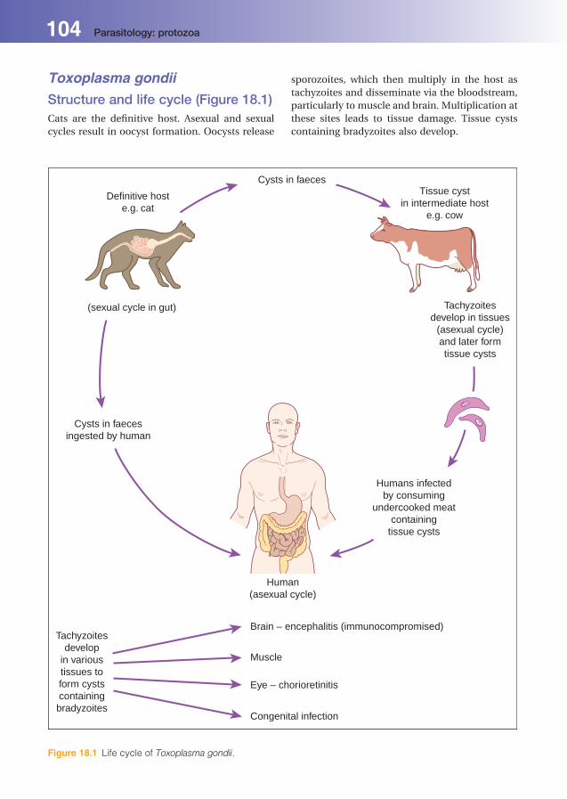
Toxoplasma gondiiStructure and life cycle (Figure 18.1)Cats are the definitive host. Asexual and sexual
cycles result in oocyst formation. Oocysts release
sporozoites, which then multiply in the host as
tachyzoites and disseminate via the bloodstream,
particularly to muscle and brain. Multiplication at
these sites leads to tissue damage. Tissue cysts
containing bradyzoites also develop.
Definitive hoste.g. cat
Cysts in faecesTissue cyst
in intermediate hoste.g. cow
Tachyzoitesdevelop in tissues
(asexual cycle)and later formtissue cysts
Humans infectedby consuming
undercooked meatcontaining
tissue cysts
(sexual cycle in gut)
Cysts in faecesingested by human
Human(asexual cycle)
Tachyzoitesdevelop
in varioustissues toform cystscontainingbradyzoites
Brain – encephalitis (immunocompromised)
Muscle
Eye – chorioretinitis
Congenital infection
Figure 18.1 Life cycle of Toxoplasma gondii.
104 Parasitology: protozoa

Epidemiology
Human infection with T. gondii is common,
occurring worldwide. A wide variety of animals
carry the microorganism. Humans become in-
fected from:
. ingestion of undercookedmeat containing tissue
cysts;. ingestion of oocysts from cat faeces;. transplacental transmission (maternal infection
in pregnancy);. cardiac transplantation, the recipient receives a
heart containing Toxoplasma cysts;. reactivation caused by immunosuppression, e.g.
AIDS.
Clinical manifestations. Most infections are asymptomatic.. In immunocompetent hosts: glandular fever-like
syndrome;. In immunocompromised hosts: myocarditis,
choroidoretinitis, meningoencephalitis;. Congenital infection: choroidoretinitis, hydro-
cephalus and intracerebral calcification.
Diagnosis. Serology (e.g. ELISA): by demonstration of rising
IgG antibodies in paired sera, or IgM antibodies
to differentiate between active and previous in-
fection. Serology in AIDS is not useful, because
IgG antibodies do not distinguish between reac-
tivated and latent infection.. PCR (polymerase chain reaction) detection of
parasite DNA in blood, CSF or amniotic fluid
may be useful.. MRI (magnetic resonance imaging) of the brain
shows characteristic multiple, dense, ring-en-
hancing lesions with contrast.
Histology
By examination of appropriate biopsies for tachy-
zoites or tissue cysts.
Treatment
Pyrimethamine, plus sulphonamide and folinic
acid. Indefinite suppressive treatment in AIDS.
Prevention
Cooking meat. Avoiding contact with cat faeces.
PlasmodiumPlasmodia are causative of malaria and are para-
sites of erythrocytes with two hosts:
1 Mosquitoes, where sexual reproductive stages
(gametogony) take place;
2 Humans or other animals, where asexual repro-
ductive stages occur (schizogony).
There are several species of plasmodia, which
vary in detailed life cycle and clinical features, and
can be separated morphologically (some are out-
lined in Table 18.3).
Life cycle
All plasmodia share a common life cycle
(Figure 18.2), but with some important variations:
1 Anopheles mosquitoes bite humans and infec-
tious plasmodia (sporozoites) are introduced
into the bloodstream.
2 The sporozoites are carried to liver parenchymal
cells, where asexual reproduction (schizogony)
occurs in the liver to form merozoites (pre-
erythrocytic cycle, 7–28 days).
3 Liver hepatocytes rupture, liberating mero-
zoites, which attach to and penetrate erythro-
cytes. Note: P. vivax andP. ovale have a dormant
hepatic phase, with sporozoites becoming
Table 18.3 Species ofmalaria infecting humans
Species Fevercycle (h)
Clinical condition
Plasmodiumfalciparum
36–48 Malignant tertianmalaria
Plasmodiummalariae
72 Quartan malaria
Plasmodiumovale
36–48 Benign tertianmalaria
Plasmodiumvivax
36–48 Benign tertianmalaria
Plasmodiumknowlesi
24 Quotidian malaria
Parasitology: protozoa 105

hypnozoites that do not divide and can result in
relapsemonths, veryoccasionally years, after the
initial disease.
4 Asexual reproduction occurs in the erythrocytes;
merozoites are released and infect further ery-
throcytes. Some merozoites develop within ery-
throcytes into male and female gametocytes.
P. falciparummay be associatedwith high levels
of parasitaemia (up to 30%ormore of circulating
erythrocytes); lower levels (<5%) are found with
other species.
5 The mosquito bites a host and ingests mature
male and female gametocytes.
6 The sexual reproductive cycle occurs in the
mosquito’s digestive system. Sporozoites form,
migrate to salivary glands and are inoculated
into a new host.
Epidemiology
. In tropical and subtropical areas, plasmodia are
dependent on the correct conditions for breed-
ing of anopheles mosquitoes. P. falciparum is
responsible for more than 80% of cases in trop-
ical areas.. In endemic areas, repeated infections or expo-
sure results in relative immunity and less severe
disease. Visitors to endemic areas are more se-
verely affected.
Oocyst in gutmucosa
Sexual reproductionin gut
Bite
Schizontdevelops
in redblood cell
Trophozoiteforms in redblood cell
Merozoites releasedenter red blood cell
Some merozoites mature intomale and female gametocytes
Bite
Salivaryglands
Sporozoite
Human
Mosquito
Produce many sporozoiteswhich migrate to:-
Liver
Liverschizont
Schizontbursts
Entersliver cells
Merozoites in schizont
Merozoitesformed
Released
Figure 18.2 Life cycle of Plasmodium falciparum.
106 Parasitology: protozoa
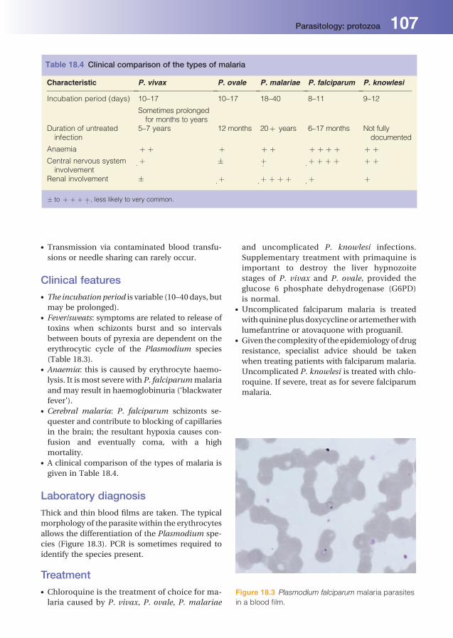
. Transmission via contaminated blood transfu-
sions or needle sharing can rarely occur.
Clinical features. The incubation period is variable (10–40days, but
may be prolonged).. Fever/sweats: symptoms are related to release of
toxins when schizonts burst and so intervals
between bouts of pyrexia are dependent on the
erythrocytic cycle of the Plasmodium species
(Table 18.3).. Anaemia: this is caused by erythrocyte haemo-
lysis. It is most severe with P. falciparummalaria
and may result in haemoglobinuria (‘blackwater
fever’).. Cerebral malaria: P. falciparum schizonts se-
quester and contribute to blocking of capillaries
in the brain; the resultant hypoxia causes con-
fusion and eventually coma, with a high
mortality.. A clinical comparison of the types of malaria is
given in Table 18.4.
Laboratory diagnosis
Thick and thin blood films are taken. The typical
morphology of the parasite within the erythrocytes
allows the differentiation of the Plasmodium spe-
cies (Figure 18.3). PCR is sometimes required to
identify the species present.
Treatment. Chloroquine is the treatment of choice for ma-
laria caused by P. vivax, P. ovale, P. malariae
and uncomplicated P. knowlesi infections.
Supplementary treatment with primaquine is
important to destroy the liver hypnozoite
stages of P. vivax and P. ovale, provided the
glucose 6 phosphate dehydrogenase (G6PD)
is normal.. Uncomplicated falciparum malaria is treated
withquinineplusdoxycycline or artemetherwith
lumefantrine or atovaquone with proguanil.. Given the complexity of the epidemiology of drug
resistance, specialist advice should be taken
when treating patients with falciparum malaria.
Uncomplicated P. knowlesi is treated with chlo-
roquine. If severe, treat as for severe falciparum
malaria.
Table 18.4 Clinical comparison of the types of malaria
Characteristic P. vivax P. ovale P. malariae P. falciparum P. knowlesi
Incubation period (days) 10–17 10–17 18–40 8–11 9–12
Sometimes prolongedfor months to years
Duration of untreatedinfection
5–7 years 12 months 20þ years 6–17 months Not fullydocumented
Anaemia þ þ þ þ þ þ þ þ þ þ þCentral nervous systeminvolvement
.þ � .þ .þ þ þ þ þ þ
Renal involvement � .þ .þ þ þ þ .þ þ
� to þ þ þ þ. , less likely to very common.
Figure 18.3 Plasmodium falciparum malaria parasites
in a blood film.
Parasitology: protozoa 107

Prevention
This is important for travellers to endemic areas.
Avoidance of mosquito bites is necessary
(e.g. covering exposed limbs, use of mosquito nets
and repellants). Prophylactic anti-malarials may
be taken. The exact regimen is dependent on
whether resistance is present in the area being
visited; examples include atovaquoneplus progua-
nil; doxycycline;mefloquine; and chloroquine plus
proguanil.
Flagellates
Classification
Human pathogenic flagellates include Giardia
intestinalis (gastrointestinal tract), Trichomonas
vaginalis (genital tract), Trypanosoma species
(blood/tissues) and Leishmania species (blood/
tissues).
Giardia intestinalis
Pathogenesis and life cycle
Cysts are ingested and gastric acid stimulates the
release of trophozoites in the small intestine,
which then multiply by binary fission. Tropho-
zoites attach to the intestinal villi by a sucking disc.
Inflammation of the epithelium may occur, but
invasion is rare. Trophozoites divide by binary
fission. Cyst formation occurs as the microorgan-
isms move through the colon.
Epidemiology
Giardia occurs worldwide. Transmission is via
ingestion of contaminated water or food, or
direct person-to-person spread via the faecal-
oral route.
Clinical manifestations
Asymptomatic carriage is common. Active infec-
tion causes diarrhoea and malabsorption occa-
sionally. Chronic infection may cause failure to
thrive in children.
Laboratory diagnosis
Stool microscopy for cysts and trophozoites. If clin-
ical suspicion is high and stool examination is neg-
ative, duodenal aspirationor biopsymaybehelpful.
Treatment
Tinidazole or metronidazole.
Prevention
The cysts resist routine levels of chlorination.
Water can be decontaminated by boiling or
filtration.
Trichomonas vaginalis
Structure
T. vaginalis is a pear-shaped protozoan with four
flagella. No cyst stage has been recognised.
Epidemiology
It has a worldwide distribution, with sexual inter-
course being the primary method of transmission.
Pathogenesis/clinical conditions
T. vaginalis is a parasite of the human urogenital
system; there are no animal reservoirs. Infection
results in a watery vaginal discharge, although
extensive inflammation and erosion of the epithe-
lium can occur. Males are usually asymptomatic
carriers; however, occasionally, urethritis and
prostatitis can occur.
Laboratory diagnosis
This is by microscopy of vaginal or urethral dis-
charge for trophozoites or isolation in special
media.
Treatment
Metronidazole for affected patients and sexual
partner(s).
Trypanosoma speciesTrypanosomes are haemoflagellates living in the
blood and tissue of human hosts. The life cycle
involves two hosts: blood-sucking insects and
mammals. It causes two diseases:
1 African trypanosomiasis (sleeping sickness)
caused by T. brucei gambiense and T. brucei
rhodesiense and transmitted by the tsetse fly:
2 American trypanosomiasis (Chagas disease)
caused by T. cruzi, and transmitted by reduviid
bugs.
108 Parasitology: protozoa

Trypanosoma brucei gambiense(West African sleeping sickness)
Life cycle
1 The infective stage (trypomastigote) is present in
the salivary glands of the tsetse fly. The trypo-
mastigote has a flagellum and an undulating
membrane along the length of the body allowing
motility.
2 Trypomastigotes enter the host via insect bites
and reach the lymphatic system, blood andCNS.
Non-flagellate forms (amastigotes) develop in
some tissues (e.g. heart and muscle).
3 Tsetse flies feed on the infected host and take
in trypomastigotes, which multiply in the intes-
tine; epimastigotes subsequently form in the
salivary glands and develop into infective
trypomastigotes.
4 The tsetse fly remains infective for life.
Epidemiology
Limited to West and Central Africa. Asymptomatic
humans are the main reservoirs, though animals
may become infected.
Clinical condition
The incubation period is 2–3 weeks. A nodule or
chancre at the site of the bite is very rare in West
African trypanosomiasis. The parasite enters the
bloodstream and results in lymphadenopathy, ir-
regular fever and myalgia. CNS involvement
(sleeping sickness) may occur with lethargy and
encephalitis, leading to convulsions, hemiplegia,
coma and occasionally death.
Diagnosis
Microscopy of thick and thin blood films, lymph
node aspirates and CSF for trypomastigotes;
serology (e.g. IFAT).
Treatment
Eflornithine, suramin or pentamidine for blood
stage infection. Eflornithine, if there is CNS
involvement.
Prevention
Vector control, insect repellents, and insecticide-
impregnated clothing.
Trypanosoma bruceirhodesiense (East Africansleeping sickness)
Life cycle
The life cycle is similar to that of T. brucei gam-
biense, with trypomastigote and epimastigote
stages. Transmission is by the tsetse fly.
Epidemiology
It is found in East Africa. Domestic and game
animals are the main reservoirs. Asymptomatic
human carriers are not normally a source of
infection.
Clinical conditions
It has a shorter incubation than T. brucei gam-
biense. A chancre at the site of the bite is more
commonly seen. The illness is more severe and
progresses rapidly and may involve the heart, kid-
ney and CNS.Mortality is greater than for theWest
African form.
Diagnosis and prevention
As above.
Treatment
Suramin. Melarsoprol under steroid cover if CNS
involvement.
Trypanosoma cruzi(Chagas disease)
Life cycle
1 The life cycle is similar to other trypanosomes,
except when the infected bug bites humans,
trypomastigotes are released simultaneously in
the faeces; these enter the wound after
scratching.
2 Spread is via the lymphatic and blood systems
andresults in invasionofmanyorgans, including
the liver, heart, muscles and brain.
3 Within host cells, trypomastigotes transform in-
to amastigotes, which multiply by binary fission
and form either further amastigotes or trypo-
mastigotes; the latter are ingested by a feeding
insect, multiply in the intestine and are then
passed in the faeces.
Parasitology: protozoa 109
Epidemiology
T. cruzi is found in North, Central and South
America. Reservoirs are humans and domestic
animals.
Clinical disease
Chagas disease may be asymptomatic, acute or
chronic. An oedematous swelling may form at
the site of the bite. Acute infection results in high
fever, erythematous rash, oedema and myocar-
ditis. Chronic disease may develop years after
the initial infection. Microorganisms proliferate
in various organs, including the brain, spleen,
liver, heart and lymph nodes, resulting in
lymphadenopathy, hepatosplenomegaly, myo-
carditis and cardiomegaly. Granulomata and
cysts may form in the brain. Chronic gastroin-
testinal disease produces symptoms resembling
achalasia or Hirschsprung’s disease. Cardiac
disease is the most common presentation, in-
cluding congestive cardiac failure, conduction
abnormalities and aneurysm formation. Trans-
placental transmission may occur in chronically
infected women. Transmission by blood trans-
fusion has occurred.
Laboratory diagnosis
Microscopy of biopsies of affected tissues; thick
blood films for trypomastigotes in the acute phase;
serological tests (e.g. ELISA and IFAT) for chronic
disease; PCR amplification of parasite DNA from
blood or body fluids.
Treatment
Benznidazole or nifurtimox; supportive measures
for complications.
Prevention
Insecticides; avoidance of insect bites, sleeping
under netting; improvement in living conditions.
Blood for transfusion should be screened for anti-
bodies to T. cruzi.
Leishmania speciesLeishmania species areobligate, intracellular para-
sites transmitted to mammalian hosts by sand
flies. An estimated 12 million people are infected
worldwide.
Classification
There are three main types of human disease:
visceral (Leishmania donovani complex), cutaneous
(L. tropica, L. major) and mucosal (L. braziliensis)
(Table 18.5).
Life cycle
1 A promastigote stage is present in the saliva of
infected sand flies and is inoculated into the bite
site.
Table 18.5 Syndromes caused by Leishmania species
Microorganism Reservoir Clinical condition Location Averageincubation period
L. donovanicomplex
Humans or dog Visceral leishmaniasis(kala-azar)
Mediterranean 3 months
Indian subcontinent
South America
Africa
South Asia
L. tropica Humans or dog Cutaneousleishmaniasis(oriental sore)
Africa 1–2 months
Asia
Middle East
L. major Rodents Mediterranean
L. braziliensis Sloths and relatedspecies
Mucosal leishmaniasis Central and South A few weeksto monthsAmerica
110 Parasitology: protozoa

2 Promastigotes lose their flagella, enter the
amastigote stage, are engulfed by tissue
macrophages, and carried via the reticuloen-
dothelial system to the bone marrow, spleen
and liver. Amastigotes multiply, resulting in
tissue damage.
3 Amastigotes are taken up by the sand fly during
feeding, develop into promastigotes, multiply in
the mid-gut and then migrate to the salivary
glands.
Visceral leishmaniasisThis is caused by L. donovani complex. The infec-
tionoccurs in India,China, southernRussia, Africa,
the Mediterranean basin and Latin America.
Clinical conditions
Fever, weight loss and diarrhoea are present, with
hepatosplenomegaly and renal involvement.
Darkening of the skin, mainly the face and hands
(kala-azar), malabsorption and anaemia may oc-
cur. Untreated, visceral leishmaniasis is fatal.
Those who are treated and recover are unlikely to
relapse unless their cell-mediated immunity is
suppressed (e.g. AIDS).
Diagnosis
Histological examination of appropriate tissue
biopsies; splenic aspirate, bone marrow or lymph
nodes for amastigotes; serology. PCRamplification
of parasite DNA increases sensitivity. Serology
(e.g. direct agglutination test).
Treatment
Liposomal amphotericin B is the agent of choice.
Sodium stibogluconate is an alternative.
Cutaneous leishmaniasisCaused by L. tropica and L. major in southern
Europe, Asia and Africa, and by L. mexicana com-
plex in Latin America.
Clinical conditions
Cutaneous disease with a papule at the bite site,
followed by necrosis of epidermis and ulceration.
Infections remain localised, although lesions may
be multiple.
Diagnosis
Histological examination for amastigotes or DNA
detection in smears or biopsy material. Serology is
unhelpful in cutaneous leishmaniasis.
Treatment
Sodium stibogluconate.
Mucosal leishmaniasisCausedbyL.braziliensis complex inLatinAmerica.
Clinical conditions
Infection mainly involves the mucous membranes
of the upper respiratory tract (palate, nose) and
related tissue, with ulceration, tissue destruction
and a resulting disfigurement.
Diagnosis
Histological examination or DNA detection in
biopsies.
Treatment
Sodium stibogluconate or amphotericin B.
Parasitology: protozoa 111

19Parasitology: helminthsPeter ChiodiniThe Hospital for Tropical Diseases and The London School of Hygiene and Tropical Medicine,London, UK
NematodesThe common nematodes of medical importance
are listed in Table 19.1.
Ascaris lumbricoides(roundworm)
Structure and life cycle (Figure 19.1)
. Adult worms are cylindrical; the female is up to
35 cm long and the male up to 30 cm long.. After ingestion of an infective egg, larval worms
are released into the duodenum, and penetrate
the intestinal wall to reach the bloodstream,
passing via the liver and heart to the pulmonary
circulation. These larval worms pass into the
alveoli, migrate via the bronchi, trachea and
pharynx, are swallowed and return to the small
intestine.. Male and female worms mature and mate in the
intestine; up to 200,000 eggs per day are pro-
duced by the female and passed in stools.
Epidemiology
Ascaris lumbricoides is found throughout the
world, but particularly in areas of poor sanitation
where faecal-oral transmission may occur. Eggs
can survive for long periods (several years),making
Ascaris the most common pathogenic helminth
worldwide.
Clinical manifestations/complications
Victims are asymptomatic when the worm load is
light. Vomiting and abdominal discomfort may
occur. Complications include:
. pneumonitis (following larval migration in the
lungs);. intestinal obstruction with mature worms
(children);. malnutrition in children;. perforation of the intestine and hepatic ab-
scesses (rare).
Laboratory diagnosis
Stool microscopy for the presence of fertilised or
unfertilised eggs. An adult worm is occasionally
passed in faeces or vomit. Larvae are sometimes
found in sputum in the lung migration phase.
Treatment and prevention
Treatment is with mebendazole, albendazole or
piperazine. Surgical or endoscopic relief of intes-
tinal obstruction. Control is by improved sanita-
tion and avoidance of food contaminated with
human faeces.
Medical Microbiology and Infection Lecture Notes, Fifth Edition. Edited by Tom Elliott, Anna Casey,Peter Lambert and Jonathan Sandoe.� 2011 Blackwell Publishing Ltd. Published 2011 by Blackwell Publishing Ltd.
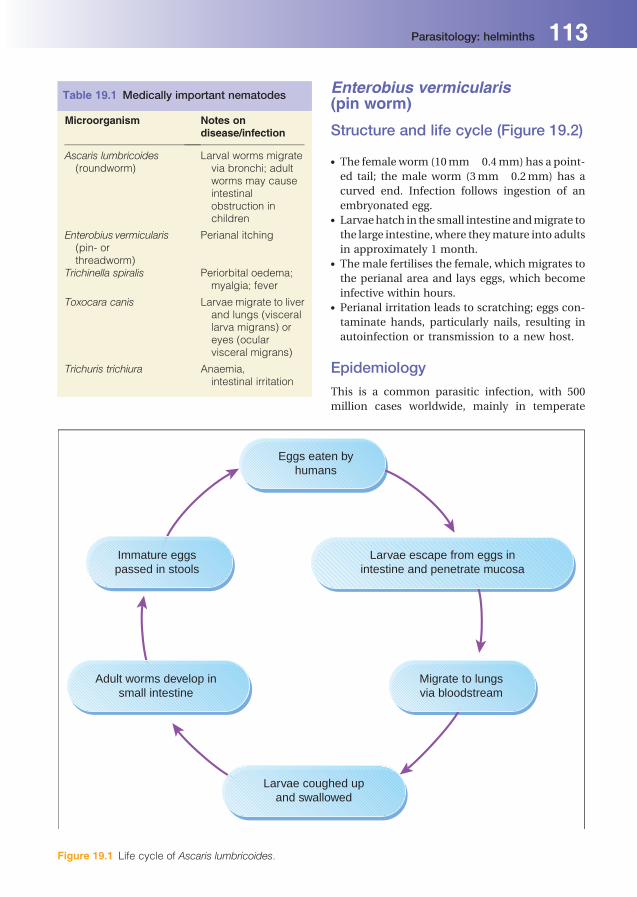
Enterobius vermicularis(pin worm)
Structure and life cycle (Figure 19.2)
. The female worm (10mm� 0.4mm) has a point-
ed tail; the male worm (3mm� 0.2mm) has a
curved end. Infection follows ingestion of an
embryonated egg.. Larvaehatch in the small intestine andmigrate to
the large intestine, where theymature into adults
in approximately 1 month.. Themale fertilises the female, which migrates to
the perianal area and lays eggs, which become
infective within hours.. Perianal irritation leads to scratching; eggs con-
taminate hands, particularly nails, resulting in
autoinfection or transmission to a new host.
Epidemiology
This is a common parasitic infection, with 500
million cases worldwide, mainly in temperate
Table 19.1 Medically important nematodes
Microorganism Notes ondisease/infection
Ascaris lumbricoides(roundworm)
Larval worms migratevia bronchi; adultworms may causeintestinalobstruction inchildren
Enterobius vermicularis(pin- orthreadworm)
Perianal itching
Trichinella spiralis Periorbital oedema;myalgia; fever
Toxocara canis Larvae migrate to liverand lungs (viscerallarva migrans) oreyes (ocularvisceral migrans)
Trichuris trichiura Anaemia,intestinal irritation
Eggs eaten byhumans
Immature eggspassed in stools
Larvae escape from eggs inintestine and penetrate mucosa
Migrate to lungsvia bloodstream
Larvae coughed upand swallowed
Adult worms develop insmall intestine
Figure 19.1 Life cycle of Ascaris lumbricoides.
Parasitology: helminths 113

regions. Person-to-person or environment-to-
person spread is responsible for transmission.
Clinical manifestationsand complications
Individuals are frequently asymptomatic. Patients
allergic to worm secretions may experience peri-
anal pruritus. In heavily infected females, worm
migration into the vagina may occur.
Laboratory diagnosis
Bymicroscopy; the bestmethod is a Sellotape slide
applied around the perianal region to pick up eggs.
Adult worms may be found in stool specimens.
Treatment and prevention
Mebendazole is given to treat the entire family
simultaneously. Reinfestation may occur, because
eggs may survive in the environment for 3 weeks.
Improved personal hygiene is advised, including
cutting of fingernails and regular washing of bed-
clothes and towels.
Toxocara canis andToxocara cati
Structure and life cycle. T. canis and T. cati are parasites of dogs and cats,
respectively. Male and female worms develop in
the intestine and produce eggs that are ingested
by other dogs or cats and the cycle continues.. Humans are an unintentional host; on ingestion,
the eggs hatch into larval forms, which can pen-
etrate the intestinal mucosa and migrate to var-
ious organs, particularly the lung, liver and eyes.
Larvae are unable to develop further and gran-
uloma formation results.
Epidemiology
It is associated with infected dogs and cats; world-
wide distribution.
Adult worm developsin large intestine
Larvae hatchin intestine
Female lays eggs inperianal area
Eggs become infectivewithin hours
Ingested by human(faecal-oral)
Figure 19.2 Life cycle of Enterobius vermicularis.
114 Parasitology: helminths

Clinical manifestationsand complications
Toxocariasis may result in pneumonitis, hepatos-
plenomegaly, eosinophilia and retinal granuloma,
which may present with retinal detachment.
Laboratory diagnosis
Serology by ELISA. Biopsies of the liver or other
affected organs may show eosinophilic granulo-
mata, but are not usually needed for diagnosis.
Treatment
Often unnecessary; albendazole for visceral larva
migrans; steroids are also used in severe cases.
Laser photocoagulation may kill larvae in the
eye.
Prevention
Young pets should be treated routinely
(‘deworming’); avoid contact with animal faeces.
Trichuris trichiura (whipworm)
Structure and life cycle. Ingested eggs develop into larvae in the small
intestine, which then migrate to the large intes-
tine and mature into adult worms. The fertilised
female worm produces eggs.. Eggs are excreted and mature in the soil.
Epidemiology
Trichuris trichiura has a wide distribution in the
tropics and subtropics, particularly in areas with
poor sanitation. No animal reservoir has been
identified.
Clinical manifestationsand complications
Patients are often asymptomatic, but heavy worm
infection may produce abdominal distension and
diarrhoea with weight loss; anaemia and eosino-
philia. Bloody diarrhoea (the Trichuris Dysentery
Syndrome)andanalprolapseare seenoccasionally.
Laboratory diagnosis
Microscopy of stools to look for characteristic eggs.
Treatment
Mebendazole or albendazole.
Prevention
Good personal hygiene and adequate sanitation
can help to prevent infection.
Ancylostoma duodenaleand Necator americanus(hookworm)
Structure and life cycle. Infective larvae penetrate intact skin, enter the
circulation and are carried to the lungs; after
maturation, larvae are expectorated and swal-
lowed, and develop into adult worms in the small
intestine.. The adult worms lay eggs, which are excreted
and, in hot humid conditions, develop rapidly
into infective larvae that may infect a new host
by penetration of exposed skin, particularly
bare feet.
Epidemiology
Found primarily in subtropical and tropical
regions.
Clinical manifestations andcomplications. There may be a rash at the entry site (ground
itch); occasionally pneumonitis occurs during
the larval migration.. Adult worms produce gastrointestinal symp-
toms, including epigastric pain. Bloody diar-
rhoea may occur in infants.. Chronic anaemia may result from blood loss,
particularly in malnourished individuals.
Laboratory diagnosis
Stool microscopy for eggs.
Treatment and prevention
Mebendazole for treatment; preventive measures
include improved sanitation and wearing of shoes
to prevent soil contact in endemic areas.
Parasitology: helminths 115

Strongyloides stercoralis
Structure and life cycle. Strongyloides stercoralis is similar to hook-
worms, except that the eggs hatch into larvae
in the intestinal mucosa before being passed in
the faeces. The larvae mature directly into in-
fective larvae that are able to penetrate skin, or
enter a non-parasitic cycle outside the human
host.. A special feature of Strongyloides is that larvae
may also reinfect the host by penetrating
the intestinal mucosa (autoinfection); this
is more common in immunocompromised
patients.. Infectionmay be quiescent for up to 30 years and
then reactivatewhen the immune system is com-
promised (e.g. organ transplantation), resulting
in disseminated infection.
Epidemiology
They are found in warm, moist environments,
similar to hookworm.
Clinical manifestations
Pneumonitis, diarrhoea, malabsorption and gut
ulceration may occur. Immunosuppressed pa-
tients may develop severe infection affecting mul-
tiple organs.
Laboratory diagnosis
Stool microscopy for larvae; sampling of proximal
small intestine with string capsules or by endo-
scopic aspiration may be needed in low-level in-
fections. In disseminated infection, filariform lar-
vae may be found in stool, duodenal contents,
sputum and bronchial washings. Serology; 85%
sensitive in uncomplicated infection, but there are
cross-reactions with other intestinal nematode
infections.
Treatment
Ivermectin.
Prevention
As for hookworm.
Trichinella spiralis
Structure and life cycle. Human infection occurs accidentally, because
this parasite is found primarily in carnivorous
animals. Humans may become affected after
ingestion of encysted larvae in undercooked
meat, particularly pork.. Larvae are released in the small intestine, where
they mature into adult worms that produce fur-
ther larvae. The larvae penetrate the intestinal
wall and are released into the circulation, be-
coming encysted in various tissues, particularly
striated muscle.. The life cycle is completed in animals that are
infected after ingestion of striated muscle con-
taining encysted larvae.
Epidemiology
Worldwide distribution; associated with eating
pork or the meat of wild carnivores.
Clinical manifestations
Trichinosis presents with muscle pain, tenderness
and peri-orbital oedema. Encephalitis and pneu-
monitis may occur. Infection is occasionally fatal,
usually as a result of myocarditis.
Laboratory diagnosis
Eosinophilia, the presence of encysted larvae in
implicated meat or biopsied muscle from the pa-
tient, and serology can be used.
Treatment
Mebendazole kills adultwormsbuthasnoeffect on
encysted larvae; albendazole can also be used in
the early stages; steroids plus mebendazole or
albendazole are used in severe allergic reactions
or myocarditis.
Prevention
Meat should be cooked thoroughly.
Wuchereria bancrofti and Brugiamalayi (filarial worms)These are transmitted by mosquitoes.
116 Parasitology: helminths

Structure and life cycle. Infective larvae are introduced after a bite from
an infected mosquito; they migrate via the lym-
phatics to regional lymph nodes.. Adult worms mature in the lymphatics, fertilisa-
tion occurs and microfilariae are produced.
These enter the circulation and are ingested by
feeding mosquitoes.. In the mosquito, larvae migrate via the stomach
to the mouthparts to complete the cycle.
Epidemiology
The worms are found in tropical and subtropical
Africa, Asia and South America.
Clinical manifestationsand complications
Early signs include flu-like illness, lymphangitis
and lymphadenopathy. Chronic infection results
in blockage of the lymphatics with peripheral
oedema, and eventually limb fibrosis
(elephantiasis).
Laboratory diagnosis
By detection of microfilariae in blood films (taken
at night as microfilariae show nocturnal periodic-
ity) and serology.
Treatment
Diethylcarbamazine.
Prevention
Mosquito control; avoiding bites.
Loa loa
Structure and life cycle. Loa loa is similar to Wuchereria bancrofti. The
insect vector is Chrysops, the mango fly.. In humans, larvae mature and migrate subcuta-
neously. Fertilisation results in microfilariae,
which pass into the bloodstream and are in-
gested by feeding flies.
Epidemiology
Loa loa infection is found in tropical Africa.
Clinical manifestations
There is swelling where the worms migrate sub-
cutaneously. Adult worms may be seen migrating
under the conjunctiva, hence the name ‘eye-
worm’.
Laboratory diagnosis
By detection of microfilariae in blood and
serology.
Treatment and prevention
Treatment is with diethylcarbamazine or ivermec-
tin under steroid cover. Albendazole is preferred in
patients with highmicrofilarial counts. Prevention
is by protection from insect bites.
Onchocerca volvulusInfection follows a bite from black flies which
breed near fast-flowing rivers. Larvae develop
into adult worms in subcutaneous nodules.
Onchocerca volvulus is found in Africa and cen-
tral and South America. Chronic infection results
in atrophic skin and may cause blindness as
a result of microfilariae migrating to the eyes.
Diagnosis is by skin biopsy or slit-lamp exami-
nation of the anterior chamber of the eye for
microfilariae. Bites can be reduced by clothing
and insect repellents. Annually administered
ivermectin effectively controls disease and de-
creases transmission of the parasite. Ivermectin
is also used for individual case management.
CestodesTapeworms are ribbon like and up to 30 feet in
length. A head or scolex is present and some spe-
cies also have a crown of hooklets, facilitating
attachment. Segments of tapeworms are called
proglottids. All tapeworms are hermaphrodites,
with both male and female reproductive organs
present in each segment. Food is absorbed from
the host intestine through the body wall. Most
tapeworms have complex life cycles, with interme-
diate hosts. Clinically important cestodes are listed
in Table 19.2.
Parasitology: helminths 117

Taenia soliumStructure and life cycle (Figure 19.3)
Humans ingest pork containing larvae (cysticerci)
and the larvae develop in the small intestine into
the adult form. The worm produces proglottids
which mature sexually, producing eggs
(Figure 19.4) that pass in the faeces. Pigs ingest
the eggs and larval forms develop that disseminate
via the circulation to muscle to produce cysticerci.
Table 19.2 Clinically important cestodes
Cestode Common name Reservoir for larvae Definitive host for adult worm
Taenia solium Pork tapeworm Pigs Humans
Taenia saginata Beef tapeworm Cattle Humans
Diphyllobothrium latum Fish tapeworm Crustacea, fish Humans, cats, dogs
Echinococcus granulosus Hydatid cyst Herbivores Canines
Hymenolepis nana Dwarf tapeworm Rodents, humans Rodents, humans
Proglottids and eggspassed in faeces
Ingestedby human
Hatch inintestine
Larvae
Disseminate andform cysticerci
in organse.g. brain, muscle
Human ingestsraw/undercooked
pork
Adultworm
developsin smallintestine
Pigs ingest eggs;larvae are
liberated in intestine
Larvaepenetrate
blood vessels,circulate
to muscles
Cysticerci developin muscle
Cysticerci released,scolex attaches to intestine
Figure 19.3 Life cycle of Taenia solium.
118 Parasitology: helminths

Epidemiology
Tapeworm infection is related to eating under-
cooked pork. Cysticercosis (see below) is con-
tracted by ingesting eggs of the pork tapeworm. It
is common in Africa, India, Southeast Asia and
Central America.
Clinical manifestations
Abdominal discomfort and diarrhoea.
Diagnosis
Stoolmicroscopy for proglottids and eggs; serology
for cysticercosis (but not for the intestinal worm).
Treatment and prevention
Treatment of the adult tapeworm is with prazi-
quantel or niclosamide. Pork should be cooked
thoroughly and improved sanitation is important
for prevention.
CysticercosisHumans may become infected with the larval
stage of T. solium after ingestion of eggs from
human faeces. Larvae penetrate the intestinal
wall, enter the circulation and are carried to
muscles, the brain, lungs and occasionally eyes.
The inflammatory response to the presence of
these larvae in human tissues eventually results
in calcification. Infection is often subclinical, but
may present with neurological or ophthalmic
manifestations.
Diagnosis
Muscle or brain radiograph may show calcified
cysts; computed tomography (CT) or MRI is often
diagnostic; histology; serology (immunoblot).
Treatment
Albendazole or praziquantel, but is not necessary if
cysts are not viable. Steroids and anticonvulsants
maybenecessary to control symptoms if CNS cysts
are degenerating. Obstructive hydrocephalus may
need surgery.
Taenia saginata
Structure and life cycle
The life cycle is similar to Taenia solium, with
infection resulting from ingestion of cysticerci
present in insufficiently cooked beef. Human fae-
ces contaminated with eggs are ingested by cattle
and cysticerci develop in beef.
Epidemiology
Worldwide distribution.
Clinical manifestations
Abdominal discomfort or no symptoms.
Laboratory diagnosis
Proglottids and eggs are detected by stool
microscopy.
Treatment and prevention
Treatment with praziquantel or niclosamide. Im-
proved sanitation aids prevention. Beef should be
cooked thoroughly.
Diphyllobothrium latum (fishtapeworm)
Structure and life cycle
There are two intermediate hosts: freshwater crus-
taceae and freshwater fish. Infection occurs in
humans after ingestion of infected freshwater fish.
The adult worm develops in the intestine, attach-
ing to the mucosa via lateral grooves. Proglottids
develop and produce eggs. In fresh water, the eggs
develop into larvae, which infect crustacea. Fish
Figure 19.4 An egg of the tapeworm Taenia in faeces.
Parasitology: helminths 119
eat the crustacea and larvae develop that are in-
fective for humans.
Epidemiology
Infection occurs in temperate climates, particular-
ly Scandinavia.
Clinical manifestations
Usually asymptomatic. Mild diarrhoea and occa-
sionally intestinal obstruction. Megaloblastic
anaemia and vitamin B12 deficiency can occur.
Treatment and prevention
Praziquantel is the drug of choice. Avoid eating raw
or undercooked fish.
Echinococcus granulosus(dog tapeworm)
Structure and life cycle. The adult tapeworms are found in canines, nor-
mally dogs or foxes. In the canine intestine, the
adult tapewormsproduce eggs, which are passed
in the faeces.. Herbivores, the normal intermediate hosts, in-
gest the eggs and larvae develop in various or-
gans, resulting in cyst formation. Carnivores be-
come infected after consuming contaminated
carcasses.. Human infection occurs when humans become
the accidental intermediate host. Eggs are in-
gested, and larvae develop and penetrate the
intestinal wall; hydatid cysts develop, principally
in the liver. These cysts accumulate fluid and
enlarge over several years; a germinal layer lines
the cyst, which produces larval tapeworms
(protoscoleces).
Epidemiology
Echinococcus granulosus is associated with sheep
farming in Europe, Australasia, South America and
the USA. Human infection follows ingestion of
water or vegetation contaminated with eggs.
Clinical manifestations
Cysts in the liver growovermany years, resulting in
hepatomegaly. Cysts in the lung, bone and brain
may result in various clinical presentations.
Diagnosis
CT or ultrasonography may be diagnostic; micros-
copy of cyst contents; serology.
Treatment and prevention
Treatment is by a combination of medical treat-
ment with albendazole and resection of cysts. An
alternative is treatment with albendazole plus as-
piration under ultrasound guidance and injection
of ethanol into the cyst to act as a scolicidal agent.
Avoid ingestion of contaminated food.
Hymenolepis nanaThis is a common, often asymptomatic infection
occurring particularly in Asia.Hymenolepis nana is
a small (2–5 cm) worm. Eggs are produced in the
small intestine and may reinfect the same host or
spread to other hosts by the faecal-oral route.
Heavy infection may result in diarrhoea, abdom-
inal discomfort and weight loss. Treatment is with
praziquantel.
TrematodesTrematodes (flukes) are flatworms with oral and
ventral suckers. Most flukes are hermaphrodites,
containing both male and female reproductive
organs in a single body. Schistosomes differ in
having separate male and female worms.
All flukes require an intermediate host for
completion of their life cycle. The intermediate
hosts are molluscs, in which the sexual repro-
duction cycle occurs. Some flukes need a further
intermediate host. The medically important tre-
matodes are summarised in Table 19.3.
Fasciola hepatica (sheep liverfluke)
Structure and life cycle. F. hepatica is a parasite of herbivores (sheep,
cattle) and occasionally humans.. Human infection starts with ingestion of water-
cress or similar vegetation contaminated with
encysted metacercariae, which excyst in the du-
120 Parasitology: helminths

odenum. The larval flukes migrate through the
duodenal wall, across the peritoneal cavity and
into the bile ducts via the liver. They mature into
adult worms and produce eggs that are passed in
faeces.. Snails become infected and act as intermediate
hosts. Cercariae (infective larvae) develop and
contaminate watercress, where they encyst to
form metacercariae.
Epidemiology
F. hepatica occurs worldwide, especially in sheep-
rearing areas.
Clinical manifestations
Migration of the larval worm in the liver causes
tender hepatomegaly. Worms in the bile duct may
cause biliary obstruction.
Laboratory diagnosis
Stool microscopy for eggs.
Treatment and prevention
Triclabendazole. Avoid eating watercress grown in
areas frequented by sheep and cattle.
Opisthorchis sinensis (Chineseliver fluke; formerly knownas Clonorchis sinensis)
Structure and life cycle
O. sinensis has a similar life cycle to F. hepatica,
except there are two intermediate hosts. Eggs are
eaten by a snail and then hatch to release
miracidia, which develop into cercariae after a
process of multiplication. Free-swimming cercar-
iae leave the snail and enter freshwater fish by
penetrating under scales where they develop into
metacercariae.Humansbecome infectedbyeating
contaminated raw fish. Larval forms penetrate the
duodenum, migrate to the bile duct and develop
into adult worms.
Epidemiology
O. sinensis is found in China, Japan, Korea and
Southeast Asia.
Clinical manifestations
People are usually asymptomatic, but severe in-
fections can result in hepatomegaly, biliary ob-
struction and jaundice.
Laboratory diagnosis
Stool microscopy for eggs.
Treatment and prevention
Praziquantel is the drug of choice. Avoid eating
uncooked fish. Improved sanitation is important
in prevention.
Paragonimus westermani(lung fluke)
Structure and life cycle. Embryonated eggs in water produce miracidia,
which penetrate snails and develop into cercar-
iae. The latter infect crabs and crayfish.
Table 19.3 Medically important trematodes (flukes)
Common name Intermediate host Source of humaninfection
Definitive host
Fasciola hepatica Sheep liver fluke Snail Aquatic plants(e.g. watercress)
Sheep, cattle
Opisthorchis sinensis(formerly Clonorchissinensis)
Chinese liver fluke Snail, freshwater fish Uncooked fish Dogs, cats,humans
Paragonimuswestermani
Lung fluke Snail, crayfish Uncooked crabs,crayfish
Humans
Schistosoma species Blood flukes Snail Water contact Humans
Parasitology: helminths 121
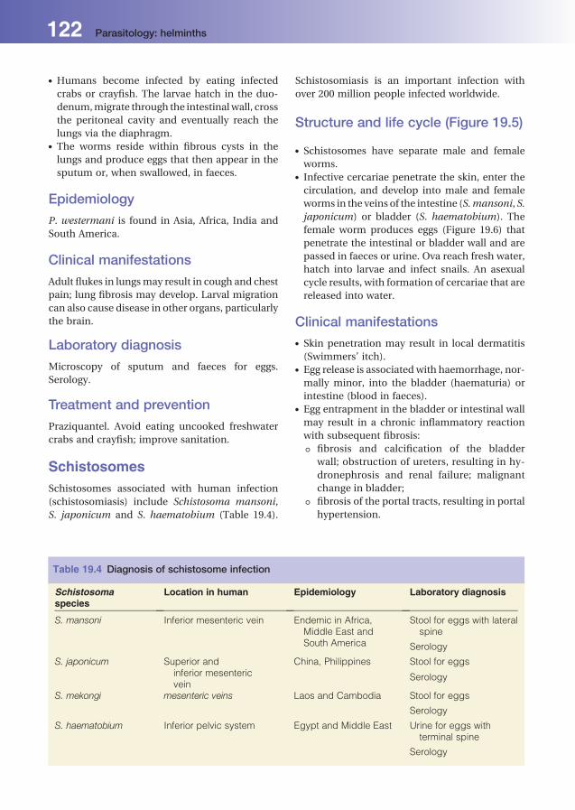
. Humans become infected by eating infected
crabs or crayfish. The larvae hatch in the duo-
denum,migrate through the intestinalwall, cross
the peritoneal cavity and eventually reach the
lungs via the diaphragm.. The worms reside within fibrous cysts in the
lungs and produce eggs that then appear in the
sputum or, when swallowed, in faeces.
Epidemiology
P. westermani is found in Asia, Africa, India and
South America.
Clinical manifestations
Adult flukes in lungsmay result in cough and chest
pain; lung fibrosis may develop. Larval migration
can also cause disease in other organs, particularly
the brain.
Laboratory diagnosis
Microscopy of sputum and faeces for eggs.
Serology.
Treatment and prevention
Praziquantel. Avoid eating uncooked freshwater
crabs and crayfish; improve sanitation.
SchistosomesSchistosomes associated with human infection
(schistosomiasis) include Schistosoma mansoni,
S. japonicum and S. haematobium (Table 19.4).
Schistosomiasis is an important infection with
over 200 million people infected worldwide.
Structure and life cycle (Figure 19.5)
. Schistosomes have separate male and female
worms.. Infective cercariae penetrate the skin, enter the
circulation, and develop into male and female
worms in the veins of the intestine (S.mansoni, S.
japonicum) or bladder (S. haematobium). The
female worm produces eggs (Figure 19.6) that
penetrate the intestinal or bladder wall and are
passed in faeces or urine. Ova reach fresh water,
hatch into larvae and infect snails. An asexual
cycle results, with formation of cercariae that are
released into water.
Clinical manifestations. Skin penetration may result in local dermatitis
(Swimmers’ itch).. Egg release is associated with haemorrhage, nor-
mally minor, into the bladder (haematuria) or
intestine (blood in faeces).. Egg entrapment in the bladder or intestinal wall
may result in a chronic inflammatory reaction
with subsequent fibrosis:
T fibrosis and calcification of the bladder
wall; obstruction of ureters, resulting in hy-
dronephrosis and renal failure; malignant
change in bladder;
T fibrosis of the portal tracts, resulting in portal
hypertension.
Table 19.4 Diagnosis of schistosome infection
Schistosomaspecies
Location in human Epidemiology Laboratory diagnosis
S. mansoni Inferior mesenteric vein Endemic in Africa,Middle East andSouth America
Stool for eggs with lateralspine
Serology
S. japonicum Superior andinferior mesentericvein
China, Philippines Stool for eggs
Serology
S. mekongi mesenteric veins Laos and Cambodia Stool for eggs
Serology
S. haematobium Inferior pelvic system Egypt and Middle East Urine for eggs withterminal spine
Serology
122 Parasitology: helminths
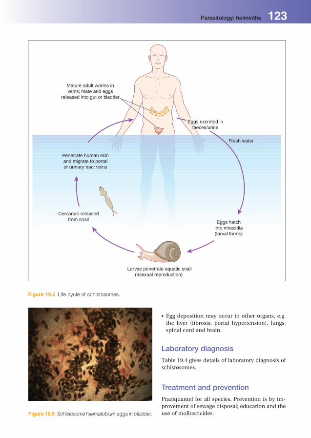
. Egg deposition may occur in other organs, e.g.
the liver (fibrosis, portal hypertension), lungs,
spinal cord and brain.
Laboratory diagnosis
Table 19.4 gives details of laboratory diagnosis of
schistosomes.
Treatment and prevention
Praziquantel for all species. Prevention is by im-
provement of sewage disposal; education and the
use of molluscicides.
Mature adult worms inveins; mate and eggs
released into gut or bladder
Eggs excreted infaeces/urine
Fresh water
Cercariae releasedfrom snail
Penetrate human skinand migrate to portalor urinary tract veins
Eggs hatchinto miracidia(larval forms)
Larvae penetrate aquatic snail(asexual reproduction)
Figure 19.5 Life cycle of schistosomes.
Figure 19.6 Schistosomahaematobium eggs in bladder.
Parasitology: helminths 123


Part 2Antimicrobial
agents


20Antibacterial agentsPeter LambertAston University, Birmingham, UK
Definitions
Antimicrobial agent
Substance with inhibitory properties against mi-
croorganisms, but with minimal effects on mam-
malian cells (includes antibiotics and synthetic
compounds).
Antibiotic
Substance produced by microorganisms that
inhibits the growth of (bacteriostatic) or kills (bacte-
ricidal) other bacteria. The term ‘antibiotic’ is often
incorrectly used to include all antimicrobial agents,
some of which are synthetic, e.g. sulphonamides,
trimethoprim,metronidazole andfluoroquinolones.
Semi-synthetic antimicrobials
Antibiotics chemically altered to improve proper-
ties, e.g. stability, activity, spectrum of activity and
pharmacokinetics.
Mechanism of action
How an antimicrobial exerts its effects.
Interactions between antimicrobials. Antimicrobials used in combination may be:
additive: combined effect equal to the sum of
the individual agents;. Synergistic: combined effect greater than
achievedwith addition; e.g. gentamicinpluspen-
icillin; or. Antagonistic: drugs inhibit the action of each
other.
Empirical therapy
Refers to the antimicrobial regimen used when a
clinical diagnosis of infection has been made and
delay in initiating therapy to await microbiological
resultswouldbe inappropriate. (e.g. cefotaxime for
meningitis).
Directed therapy
Directed antimicrobial regimens are prescribed to
target a specific pathogen (or pathogens), usually
informed by the results of microbiological inves-
tigations (e.g. benzylpenicillin for a penicillin-
susceptible pneumococcal pneumonia or fluclox-
acillin for a meticillin-susceptible S. aureus septic
arthritis).
Medical Microbiology and Infection Lecture Notes, Fifth Edition. Edited by Tom Elliott, Anna Casey,Peter Lambert and Jonathan Sandoe.� 2011 Blackwell Publishing Ltd. Published 2011 by Blackwell Publishing Ltd.

Monotherapy
Treatment with an antimicrobial on its own.
Combination therapy
Concurrent treatment with two or more
antimicrobials.
Resistance
The ability of a microorganism to avoid the harm-
ful effects of an antimicrobial by destroying it,
transporting it out of the cell, or undergoing
changes that block its effects.
Susceptibility
The level of vulnerability of a microorganism to an
antimicrobial.
Principles of antimicrobialtherapy
. The need for antimicrobial therapy should be
carefully considered; mild infections often do
not require treatment.. Viral infections are not affected by antibacterial
agents; many common infections are caused by
viruses (e.g. upper respiratory tract infections,
see Chapter 25).. The need for urgent empirical therapy should be
assessed; for patients with severe sepsis, appro-
priate antimicrobial therapy should be started as
soon as possible.. In some patients, particularly in the absence of
severe sepsis when the diagnosis is unclear, it
may be appropriate to delay antimicrobial ther-
apy pending a definitive clinical or microbiolog-
ical diagnosis.. Appropriate microbiological sampling
should be undertaken prior to commencing
antimicrobials.. Selection of an antimicrobial(s) should take into
account the patient’s allergy, drug and medical
history.. Selection of empirical antimicrobial(s) should
take into account the likely pathogen(s) and their
usual antimicrobial sensitivity pattern.
. The appropriate route of administration should
be determined by pharmacokinetics, severity of
infection (intravenous antimicrobials are usually
required for severe infections) and patient fac-
tors (e.g. ability to swallow).. An appropriate dose should be prescribed; this is
influenced by site and severity of infection and
patient factors (e.g. renal and hepatic function).. Length of treatment depends on the severity and
type of infection. Formany infections, treatment
can be stopped when symptoms are resolved/
resolving. Exceptions include infective endocar-
ditis, osteomyelitis.. Combination therapy may be required:
T to give enhanced bacterial killing (e.g.
infective endocarditis treatment, Chapter 35);
T to broaden the spectrum of antimicrobial
cover, (e.g. cefuroxime and metronidazole
may be used to treat abdominal sepsis to
cover coliform microorganisms and anae-
robes, respectively);
T to prevent development of resistance, (e.g.
tuberculosis therapy);
T to overcome antimicrobial degradation by
bacterial enzymes (e.g. co-amoxiclav contains
amoxicillin plus theb-lactamase inhibitor cla-
vulanic acid in the same preparation, piper-
acillin plus tazobactam is another example).. When empirical antimicrobial therapy has been
used, the accuracy of the initial diagnosis
should be reviewed regularly and treatment
altered to directed therapy or stopped as appro-
priate (e.g. whenmicrobiological results become
available).. Antimicrobials should only be used to prevent
infections (prophylaxis) when there is proved
effectiveness. Prophylactic antimicrobials are
used most frequently for preventing infection
after certain types of surgery.
Mechanism of action ofantimicrobial agentsAntimicrobials kill or inhibit the growth of micro-
organisms by interfering with vital structures and
processes. Selectivity of action depends upon key
differences in these between microbial and mam-
malian cells. The targets for antimicrobial action
are shown in Box 20.1 and Table 20.1.
128 Antibacterial agents

Resistance toantimicrobial agentsPatients do not become resistant to antimicrobial
agents, bacteria do.
Resistance mechanisms
Alteration of the target site. Alteration of the target site can reduce or elim-
inate binding of the antimicrobial.. Typically affects antimicrobials that act on the
ribosome (e.g. macrolides), RNA polymerase
(rifampicin) or topoisomerases (quinolones).. Resistance occurs when a spontaneous bacterial
mutant arises from within a previously sensitive
bacterial population; in the presence of the
antimicrobial the resistant mutant is ‘selected
out’, i.e. can persist and multiply.. For example, alteration of transpeptidases (the
targets of the b-lactams) results in the microor-
ganism becoming resistant to all b-lactam anti-
microbials (e.g. MRSA has acquired additional
genes,whichencode transpeptidases that arenot
affected by b-lactam antimicrobials).
Destruction/inactivation ofantimicrobial. Mediated by bacterial enzymes;. b-lactamases cause hydrolysis of the b-lactamring of penicillins and cephalosporins to form
an inactive product. Over 300 different b-lacta-mase enzymes have been described;
. Aminoglycoside-modifying enzymes: inactivate
aminoglycosides by the addition of chemical
groups (phosphate, acetyl or adenylyl groups).
Interference with drug transport. Resistance to some antimicrobials involves
interference with the transport of these drugs
BOX 20.1 The targets for antimicrobialaction on bacteria
1. Cell wall
2. Cell membrane
3. Protein synthesis
4. Nucleic acid synthesis
Table 20.1 Sites of action of differentantimicrobial agents
Site of action Examples ofantimicrobials
Cell wall Penicillins
Cephalosporins
Carbapenems
Monobactams
Glycopeptides
Cell membrane Polymyxins
Daptomycin
Protein synthesis Aminoglycosides
Tetracyclines
Macrolides
Chloramphenicol
Lincosamides
Oxazolidinones
Mupirocin
Pleuromutilins
DNA synthesis and function Sulphonamides
Trimethoprim
Quinolones
Nitroimidazoles
RNA synthesis Rifampicin
BOX 20.2 Six main mechanisms by whichbacteria become resistant to antimicrobialagents
1. Alteration of the target site.
2. Destruction/inactivation of the antimicrobial.
3. Restricted transport of the agent into the cell.
4. Metabolic bypass.
5. Active removal of drug from the cell by activity ofefflux pumps.
6. Protection of the target site by a bacterial protein.
Antibacterial agents 129

into the bacterial cell, e.g. exclusion of glycopep-
tides by Gram-negative bacteria or failure to
transport fusidic acid ormupirocin into the cells.
Metabolic bypass. Bacteria may become resistant by providing a
replacement for the metabolic step inhibited by
an antimicrobial.. For example, bacteria resistant to trimethoprim
synthesise a trimethoprim-insensitive dihydro-
folate reductase, which allows the microorgan-
ism to bypass the action of the antimicrobial.
Sulphonamide resistance is mediated by a sim-
ilar mechanism.
Efflux:
. Drug efflux occurs in a wide range of Gram-
positive and Gram-negative bacteria.. It is caused by increased expression of chro-
mosomally encoded proteins, whose normal
function may be to remove toxic metabolites
from the cells.. Efflux tends to result in low level resistance, but
may occur with other mechanisms to produce
high-level, clinically relevant resistance.. Most classes of antimicrobial can be removed by
efflux pumps including b-lactams, quinolones,
tetracyclines, macrolides and aminoglycosides.
Target site protection. Access of an antimicrobial to its target may be
blocked by production of protective bacterial
proteins. This mechanism contributes to resis-
tance to tetracyclines and fluoroquinolones.
The genetics of bacterialresistance toantimicrobialsResistance of bacteria to antimicrobials is based on
genetic changes, which allow the microorganism
to avoid the action of the antimicrobial agent.
These genetic changes canoccurdenovo as a result
of mutation or can occur via acquisition of new
genetic material (antimicrobial resistance genes)
from another microorganism.
Mutations occur at a low frequency during
every round of cell division and chromosome
replication. If a mutation happens to occur in
the gene for an antimicrobial target, then this will
favour survival and spread of the mutant in the
presence of the antimicrobial (so called ‘selection
pressure’). This type of resistance is particularly
common with rifampicin, due to mutations in
the target RNA polymerase gene and is the
reason why rifampicin should not be used as
monotherapy.
Antimicrobial-resistant genes may be acquired
via a number of different vectors:
. Plasmids are extra-chromosomal pieces of DNA
containing genes that confer on the bacteria a
number of different properties. These genes are
often non-essential, but allow the microorgan-
ism to survive under a variety of different
conditions, giving the microorganism a survival
advantage. Resistance to antimicrobials is one of
a number of properties that may be conferred on
bacteria by plasmids.. Transposons are small genetic elements consist-
ing of individual or small groups of genes. These
genetic elements have no ability to replicate,
but can move from one replicating piece of DNA
to another, e.g. from the bacterial chromosome
to a plasmid, and vice versa. Some transposons
also behave like plasmids. Transposons play an
important part in the evolution of bacterial
resistance.. Bacteriophages are viruses that infect bacteria
and may act as a vector in carrying bacterial
genes from one bacterial cell to another.. Integrons consist of blocks of several antimicro-
bial resistance genes; they usually include
sulphonamide and aminoglycoside-resistance
genes. New resistance genes may readily be
added to an integron.
The existence of mechanisms for transferring
genetic information between bacteria of the
same and of different species (‘horizontal
transmission’) by plasmids, transposons and
bacteriophages, potentiates rapid spread of resis-
tance genes.
Epidemiology of antimicrobialresistanceThe principle underlying the emergence and
spread of antimicrobial resistance among bacteria
is that the prevalence of resistance is directly
130 Antibacterial agents

proportional to the amount of antimicrobial used.
This is illustrated by the increased antimicrobial
resistance found in countries with unrestricted use
of antimicrobials, in hospitals compared with the
community, and in intensive care units compared
with generalwards. Problemareas in antimicrobial
prescribing include:
. the use of antimicrobials without prescription;
. the uncontrolled use of antimicrobials as growth
promoters in livestock;. poor prescribing habits;. the absence of antimicrobial policies, particularly
in hospitals.
Adverse reactions andantimicrobials (Table 20.2)Antimicrobials may be associated with side effects
common to all drugs, including allergic reactions
and toxicity to various organs (e.g. hepatotoxicity,
nephrotoxicity).
Specific complications associated with antimi-
crobials relate to their depressive effect on normal
commensal flora, e.g. oral candidiasis and
Clostridium difficile infection.
Laboratory aspects ofantimicrobial therapyAssessment of bacterialsensitivityA number of techniques are available to assess
susceptibility of bacteria to antimicrobials in vitro:
. Disc diffusion tests: antimicrobial-containing
filter paper discs are placed on agar plates inoc-
ulated with the bacteria to be tested. After
incubation, fully susceptible bacteria will show
a large zone of growth inhibition around the disc;
less sensitive (intermediate sensitive) isolates
will show a smaller inhibition zone and resistant
bacteria grow up to the disc edge (Figure 20.1).
Results are recorded as S (sensitive), I (interme-
diate sensitive) or R (resistant).. Minimum inhibitory concentration (MIC) assays:
these give a more detailed assessment of
bacterial susceptibility. Suspensions of the test
bacteria are incubated overnight with doubling
dilutions of the antimicrobial; the lowest con-
centration that inhibits growth is the MIC.
Alternatively, plastic strips containing a calibrat-
ed concentration gradient of antimicrobial (e.g.
E-test strips) are applied to inoculated plates.
Table 20.2 Adverse reactions of someantimicrobials
Antimicrobial Adverse reaction
Penicillins Allergic reactions
Cephalosporins Allergic reactions
Carbapenems Allergic reactions
Aminoglycosides Nephrotoxicity
Ototoxicity
Vancomycin Nephrotoxicity
Ototoxicity
Sulphonamides Folate deficiency
Marrow depression
Stephens–Johnsonsyndrome
Rifampicin Hepatotoxicity
Isoniazid Hepatotoxicity
Fucidin Hepatotoxicity
Chloramphenicol Aplastic anaemiaFigure 20.1 Disc diffusion sensitivity test.
Antibacterial agents 131

TheMIC can be determined by direct inspection
of the pattern of growth inhibition around the
calibrated strip (Figure 20.2).. Minimum bactericidal concentration (MBC)
assay: this is similar to theMIC but the end-point
is bacterial killing rather than growth inhibition;
rarely used in diagnostic laboratories.. Agardilution susceptibility tests: an antimicrobial
is incorporated into the medium in an agar plate
at a set concentration. The test microorganism is
applied to the plate in a standardised amount.
Whether or not the microorganism grows will
determine whether it is categorised as sensitive
or resistant. Using plates containing a series of
concentrations the MIC may be determined.. Growth rate-based susceptibility tests: the test
microorganism is placed in a container with a
set concentration of antimicrobial. The rate of
growth of themicroorganism is measured. Slow-
growing microorganisms are sensitive to the
antimicrobial; faster-growing ones are resistant.
Therapeutic drug monitoringMeasurement of levels in the blood is routinely
carried out for a small number of antimicrobials.
This is required to:
. avoid levels associated with toxicity, e.g. amino-
glycosides (including amikacin, gentamicin,
tobramycin);. ensure therapeutic levels are achieved, e.g.
glycopeptides (including vancomycin).
The results are used to adjust subsequent
dosing of the patient. Patients with renal or
hepatic insufficiency may require monitoring of
other antimicrobials.
Antibacterial agentsAntibacterials can be classified according to their
site of action and their molecular structure.
Inhibitors of cell-wall synthesisThe cell wall of bacteria is essential for their growth
and survival. Antimicrobial agents that block the
synthesis of thebacterial cellwall are the b-lactams
and glycopeptides. These agents have selective
activity against bacteria, becausemammalian cells
have no cell wall.
b-lactam antimicrobials
Structure
This is a large groupof compounds, all containing a
b-lactam ring (Figure 20.3). The structure of the
rings attached to the b-lactam ring determines the
class of antimicrobial. Cephalosporins have a six-
membered ring, whereas penicillins and carbape-
nems have five-membered rings, and monobac-
tams have no additional ring. Many modifications
have been made to the penicillins and cephalos-
porins by variation of side chains attached to the
rings. These changes have significant effects upon
the activity and pharmacokinetic properties.
Other b-lactams include clavulanic acid, sulbac-
tam and tazobactam. These agents do not have
useful antimicrobial activity, but they inhibit
b-lactamases. They are therefore used in combi-
nation with antimicrobial b-lactams to protect
against inactivation by these enzymes.
Mechanism of action
These antimicrobials interfere with the assembly
of the cell-wall polymer peptidoglycan by inhibit-
ing the transpeptidase enzymes that cross-link the
linear sugar chains of the polymer. This weakens
the cell wall and results in rupture (lysis) of the
microorganism (Figure 20.4).
PenicillinsThe penicillins comprise a large group of
mainly semi-synthetic compounds based on
Figure 20.2 E-test to determineMICof linezolid against
an enterococcal isolate.
132 Antibacterial agents

benzylpenicillin. They can be divided into the
following groups:
1 Narrow spectrum: benzylpenicillin, phenoxy-
methylpenicillin, e.g. active against streptococci;
2 Moderate spectrum: amoxicillin, e.g. active
against a range of Gram-positive and Gram-
negative bacteria;
3 Penicillins resistant to staphylococcal b-lacta-
mase, e.g. flucloxacillin;
4 Extended spectrum penicillins: with activity
against streptococci and many coliforms, e.g.
ticarcillin;
5 Penicillins with activity against Pseudomonas
aeruginosa, e.g. piperacillin.
Toxicity
Hypersensitivity reactions (e.g. immediate-
type (IgE mediated)) allergy and delayed-type
hypersensitivity.
Antibacterial resistance. b-lactamase production: these bacterial enzymes
hydrolyse the b-lactam ring; over 300 different
types have been described; some b-lactams
are largely unaffected by b-lactamases (e.g.
flucloxacillin).. Alteration in target site: e.g. meticillin-resistant
Staphylococcus aureus (MRSA) produces a
transpeptidase with low affinity for b-lactams
and therefore continues to function in the
CH2 R”ON
COOH
SR’ CONH
(B) Semisynthetic cephalosporins
S
CH3
CH3
COOH
CONH
ON
R
SO3–
CONH
ON
R
(C) Aztreonam
(A) Semisynthetic penicillins
S
ON
HHO
H3C
COOH
NH
NH
(D) Imipenem (+ cilastatin)
(E) Meropenem
S
O
O
N
HHO
H3C
COOHN
N
H
CH3
CH3
CH3
Figure 20.3 Structure of b-lactam antimicrobials:
penicillins (A); cephalosporins (B); monobactams
aztreonam (C); carbapenems, imipenem (D); and
meropenem (E).
Figure 20.4 Electronmicrograph illustrating the effect
of penicillin breaking the cell wall of a susceptible
bacterium (arrowed).
Antibacterial agents 133

presence of b-lactam antimicrobials (including
flucloxacillin).
Examples of penicillins
Benzylpenicillin. Pharmacokinetics: unstable in acid anddestroyed
in the stomach; can be given intravenously (i.v.)
or intramuscularly (i.m.). Widely distributed in
the body. Excretion is predominantly renal;. Antimicrobial spectrum of activity: effective
mainly on Gram-positive microorganisms (e.g.
streptococci) andGram-negative cocci (Neisseria
meningitis), some anaerobes and Treponema
pallidum;. Clinical applications: streptococcal infections,
including pneumococcal pneumonia, infective
endocarditis, cellulitis, meningococcal meningi-
tis, syphilis and some more unusual infections
such as actinomycosis.
Flucloxacillin. Pharmacokinetics: this is a semi-synthetic pen-
icillin. It is well absorbed orally. Partmetabolised
in the liver, part excreted in urine;. Antimicrobial spectrum of activity: flucloxacillin
is active against most b-lactamase-positive and
Gram-negative strains of Staphylococcus aureus
and Streptococcus pyogenes, but is not active
against meticillin-resistant Staphylococcus
aureus (MRSA).. Clinical applications: Staphylococcus aureus in-
fections, including cellulitis, pneumonia, endo-
carditis, septic arthritis and osteomyelitis.
Ampicillin and amoxicillin
. Pharmacokinetics: semi-synthetic penicillins;
acid stable and well absorbed orally. Excretion
is predominantly renal. Amoxicillin is derived
from a minor chemical change in ampicillin,
which improves oral absorption.. Antimicrobial spectrum of activity: ampicillin
and amoxicillin have almost identical antimicro-
bial activity, similar to benzylpenicillin, butmore
active against Enterococcus faecalis, Haemophi-
lus influenzae and some Gram-negative aerobic
bacilli; activity against many b-lactamase-pro-
ducing bacteria can be enhanced by the co-ad-
ministration of b-lactamase inhibitors, such as
clavulanic acid.. Clinical applications: urinary infections caused
by susceptible coliforms; respiratory tract
infections caused by H. influenzae; Listeria
monocytogenes infections (e.g. meningitis); in-
fections with enterococci (e.g. endocarditis).
Piperacillin with tazobactam
. Pharmacokinetics: a semi-synthetic penicillin,
piperacillin is not absorbed orally. The principal
route of excretion is via the kidneys. It is used
with tazobactam, a b-lactamase inhibitor with
matching pharmacokinetics.. Antimicrobial spectrum of activity: piperacillin
is moderately active against many Gram-
positive microorganisms, Neisseria spp. and
H. influenzae. It has wider activity than ampi-
cillin against coliforms and is active against
Pseudomonas aeruginosa. It is usually used
in combination with the b-lactamase inhibitor
tazobactam, to give a broad spectrum
antimicrobial.. Clinical application: severe Gram-negative aer-
obic bacillary infections (e.g. bloodstream infec-
tion, peritonitis, neutropenic sepsis) caused by
coliforms and P. aeruginosa.
CephalosporinsThe cephalosporins are classified as:
1 First-generation: early compounds with good
activity against Staph. aureus and some
Gram-negative species, including Haemophilus
spp. and coliforms (e.g. cephaloridine and
cephalexin);
2 Second-generation: resistant to some b-
lactamases, enhanced Gram-negative activity
(e.g. cefaclor and cefuroxime);
3 Third-generation: enhanced anti-Gram-negative
activity, usually at the expense of Gram-positive
activity (e.g. cefotaxime and ceftazidime);
4 Fourth-generation: enhanced Gram-positive
activity compared to third-generation cephalos-
porins with broad anti-Gram-negative activity
maintained (e.g. cefepime and cefpirome).
Toxicity/side effects
Hypersensitivity reactions (as for penicillins).
Patients with a penicillin allergy are more likely to
react to a first-generation cephalosporin but cross
reactivity with other cephalosporins is rare.
Resistance
Similar to the penicillins.
134 Antibacterial agents

Examples of cephalosporins
Cefuroxime. Pharmacokinetics: available in oral and paren-
teral forms. Wide distribution in the body with
renal excretion;. Antimicrobial spectrum of activity: Staphylococ-
cus aureus, most streptococci, coliforms, (note
thatMRSA, P. aeruginosa, enterococci and anae-
robes are resistant);. Clinical applications: urinary tract infections,
soft tissue and chest infections.
Ceftazidime. Pharmacokinetics: not absorbed orally, given i.v.
Distributed into many body tissues and fluids.
Excretion is exclusively renal;. Antimicrobial spectrum of activity: less activity
against Gram-positive bacteria, but wide activity
against Gram-negative aerobic bacteria includ-
ing P. aeruginosa;. Clinical application: severe Gram-negative aer-
obic bacillary infections (e.g. hospital acquired
pneumonia, bloodstream infection) caused by
coliforms and P. aeruginosa.
Cefotaxime. Pharmacokinetics: not absorbed orally, given i.v.
Good CSF and brain penetration;. Antimicrobial spectrum of activity: broad spec-
trum of activity including P. aeruginosa;. Clinical application: severe Gram-negative aer-
obic bacillary infections (e.g. hospital acquired
pneumonia, bloodstream infection) caused by
coliforms and P. aeruginosa. Use in meningitis
and other CNS infections.
CarbapenemsThese agents have potent, broad spectrum activity
and are generally used for the treatment of severe
infections or for infection caused by multiply-
resistant bacteria.
Toxicity/side effects
Hypersensitivity reactions (as for penicillins).
Significant cross-reactivity with penicillin allergy.
Resistance
As a result of hydrolysis by carbapenemases
(b-lactamases specific for carbapenems) or
reduced uptake by cell (this may be acquired dur-
ing treatment).
Examples of carbapenems
Imipenem with cilastatin,and meropenem. Pharmacokinetics: must be given i.v. Renal
excretion. Cilastatin is combinedwith imipenem
to inhibit renal dehydropeptidase and improve
imipenem blood levels. Imipenem/cilastatin is
more likely to provoke convulsions than mero-
penem.Meropenem is thus favoured for treating
central nervous system infections.. Antimicrobial spectrum of activity: active against
Enterobacteriaceae (including those producing
extended-spectrum beta-lactamase), Gram-
positive bacteria (e.g. Staphylococcus aureus and
streptococci), anaerobes and many Pseudomo-
nas spp. and Acinetobacter spp. Stenotrophomo-
nas maltophilia is resistant.. Clinical application: empiric treatment of severe
hospital acquired infections and neutropenic
sepsis. Directed therapy for infections caused by
Gram-negative pathogens resistant to other
antimicrobials.
Ertapenem. Pharmacokinetics: once daily iv administration;. Antimicrobial spectrum of activity: active against
Enterobacteriaceae (including those producing
extended-spectrumb-lactamase),Gram-positive
bacteria (e.g. Staphylococcus aureus and strepto-
cocci), anaerobes but not active against Pseudo-
monas aeruginosa.. Clinical application: severe Gram-negative
bacillary infections, (e.g. peritonitis) if Pseudo-
monas spp. or Acinetobacter spp. are not likely to
bepresent.Oncedaily dosing allows treatment in
the community, e.g. for urinary tract infections
caused by multi-resistant Enterobacteriaceae.
Doripenem. Pharmacokinetics: i.v. administration every 8
hours;. Spectrum: broad spectrum, includingPseudomo-
nas aeruginosa and Acinetobacter spp. Stable to
many b-lactamases, including ESBLs;. Clinical application: complicated intra-
abdominal and complicated urinary tract infec-
tions, nosocomial pneumonia.
Antibacterial agents 135
MonobactamsActivity is restricted to certain Gram-negative
bacteria; these agents are resistant to many
b-lactamases.
Toxicity/side effects
Hypersensitivity reactions are uncommon, no
significant cross-reactivity in patients with
immediate-type penicillin allergy.
Aztreonam. Pharmacokinetics: must be given i.v.;. Antimicrobial spectrum of activity: good activity
against Gram-negative bacteria, including
Pseudomonas aeruginosa; no activity against
Gram-positive bacteria;. Clinical application: used for Gram-negative and
Pseudomonas aeruginosa infections in patients
who are penicillin allergic.
Glycopeptides
Examples
Vancomycin, teicoplanin.
Mechanism of action
Block cell wall synthesis by binding to the end of
the peptide chains of the peptidoglycan precur-
sors; this prevents their incorporation into the cell
wall by the transglycosylase enzyme. This results in
slow cell lysis and death.
Antimicrobial spectrum of activity
Glycopeptides are bactericidal against Gram-
positive bacteria, including staphylococci. They
have no activity against Gram-negative bacteria.
Toxicity
Vancomycin occasionally causes nephrotoxicity
and rarely causes ototoxicity. Rapid infusion can
cause an erythematous rash (‘redman syndrome’).
Pharmacokinetics
Not absorbed from the gastrointestinal tract and
must be administered i.v. for systemic infections.
Widely distributed, but poor penetration into
cerebrospinal fluid (CSF) and relatively poor
penetration into lungs. Renally excreted.
Resistance
This is well-recognised with vancomycin resistant
enterococci (VRE). Staphylococcus aureus with
reduced (intermediate) sensitivity (VISA) are
increasingly reported, but true resistance to van-
comycin (VRSA) is currently very rare.
Clinical applications
Many infections caused by Gram-positive bacte-
ria (e.g. cellulitis, wound infections) in patients
with b-lactam allergy or resistant pathogens. Used
for severe staphylococcal infections (especially
MRSA), including endocarditis and infections of
prosthetic devices caused by coagulase-negative
staphylococci. Also used as oral treatment of
severe Clostridium difficile infection. Serum
levels need monitoring to ensure therapeutic
levels.
Antibacterial agentsactingon cell membranesOnly a few agents target the microbial membrane.
These include polymyxins and lipopeptides.
Polymyxins (e.g. colistin) are similar to deter-
gents, they disrupt the outer and cell membranes
of some Gram-negative bacteria, resulting in loss
of cytoplasmic content. They are nephrotoxic
and usually used as topical agents, e.g. in gut
sterilisation regimens. Colistin is used to treat
Pseudomonas aeruginosa lung infections in
cystic fibrosis and occasionally used to treat
multi-resistant Gram-negative bacilli, e.g.
Acinetobacter spp.
Lipopeptides (e.g. daptomycin) disrupt the
cytoplasmic membrane of Gram-positive bacteria
(including staphylococci), resulting in leakage of
cell contents. Daptomycin has been developed
relatively recently to combat increasingly resistant
Gram-positive bacteria, e.g. MRSA. It is used
clinically for skin and soft tissue infections and
S. aureus infections, including bloodstream infec-
tion and endocarditis.
Inhibitors of protein synthesisThis group includes the aminoglycosides, tetracy-
clines,macrolides, chloramphenicol, lincosamides,
oxazolidinones, fusidic acid, mupirocin and
136 Antibacterial agents

pleuromutilins, which target differences between
human and bacterial protein synthesis:
. Aminoglycosides: e.g. gentamicin and tobramy-
cin, bind to the 30S ribosomal subunit and
interfere with their proof reading function; this
results in misreading of the mRNA and produc-
tion of toxic proteins through incorporation of
the wrong amino acids;. Tetracyclines: bind to the 30S subunit, preventing
binding of aminoacyl-transfer RNA to the accep-
tor site in the ribosome, thereby inhibiting amino
acid chain elongation;. Macrolides: e.g. erythromycin, bind to the 50S
subunit of ribosomal RNA and inhibit the forma-
tion of initiation complexes;. Chloramphenicol: binds to the 50S subunit and
interferes with the linkage of amino acids in the
peptide chain formation (peptidyl transferase
reaction);. Lincosamides: also bind to the 50S subunit and
interferes with the linkage of amino acids in the
peptide chain formation;. Oxazolidinones: block the initiation stage of pro-
tein synthesis in which the 30S ribosomal sub-
unit binds to mRNA;. Fusidic acid: inhibits the function of elongation
factor G, a factor that generates energy for the
ribosome by hydrolysis of GTP;. Mupirocin: inhibits the coupling of isoleucine
(1 of the 20 different amino acids found in pro-
teins) to its tRNA;. Pleuromutilins: (retapamulin) block interaction
of peptidyltransferase on 50S subunit with ami-
noacyl-transfer RNA.
Aminoglycosides
Examples
Amikacin, gentamicin, netilmicin, streptomycin,
tobramycin.
Antimicrobial spectrum of activity
Active against staphylococci and Gram-negative
aerobic bacilli including Pseudomonas spp; they
have no activity against anaerobes. They are
bactericidal and enhance the antibacterial affects
of some antimicrobials (e.g. b-lactams and vanco-
mycin) against staphylococci, streptococci and
enterococci.
Toxicity/side effects
Hypersensitivity, ototoxicity and nephrotoxicity.
Pharmacokinetics
Poorly absorbed from the gut; water soluble, so
poor penetration into fatty tissue; excretion is
almost entirely renal. Serum levels require moni-
toring with careful dosage adjustment particularly
in renal failure.
Resistance
Mechanisms include changes in ribosomal bind-
ing of the drug, decreased permeability, increased
efflux and, most importantly, inactivation by ami-
noglycoside-modifying enzymes.
Clinical applications
Severe infection caused by coliforms and other
Gram-negative aerobic bacilli. Also used in com-
bination with cell-wall acting antimicrobials for
streptococcal and enterococcal endocarditis.
Tetracyclines
Examples
Tetracycline, doxycycline.
Antimicrobial spectrum of activity
Broad spectrum of activity against many Gram-
positive and some Gram-negative bacteria; and
also Chlamydia trachomatis, Chlamydophila spp.,
Mycoplasma spp and Rickettsia spp. All tetracy-
clines have very similar spectra of activity.
Toxicity/side effects
Gastrointestinal intolerance; deposition in devel-
oping bones and teeth precludes use in young
children and in pregnancy; hypersensitivity occa-
sionally, with skin rashes.
Pharmacokinetics
Can be given orally or i.v. Penetrate well into body
fluids and tissues as a result of lipid solubility.
Excretion is via the kidney and bile duct. Doxycy-
cline may be given once a day.
Resistance
Efflux from cell; less commonly by protection of
target from the tetracycline.
Antibacterial agents 137
Clinical application
Important in treatment of infections caused by
Mycoplasma spp., Rickettsia spp. Coxiella burnetii
and Chlamydia trachomatis.
GlycylcyclinesOnly one representative of this group is in clinical
use: tigecycline. Tigecycline is a new antimicrobial
derived from tetracycline and was developed to
combat the increase in multi-resistant bacteria.
Tigecycline has broad activity against many
Gram-positive andGram-negative bacteria includ-
ing multi-resistant, non-fermenters such as Acine-
tobacterspp.,but isnotactiveagainstPseudomonas
aeruginosa
Macrolides andlincosamidesMacrolides
Examples
Clarithromycin, azithromycin and erythromycin.
Antimicrobial spectrum of activity
Gram-positive microorganisms; also Haemophi-
lus, Bordetella and Neisseria spp. and some
anaerobes. Many coliforms are resistant. Active
against Chlamydia trachomatis, Chlamydophila
spp., Rickettsia spp. and mycoplasmas.
Toxicity/side effects
Gastrointestinal upsets, rashes, hepatic damage
(rare).
Pharmacokinetics
Absorbed following oral administration; also given
i.v. They are well distributed and excretion is
mainly in the bile.
Resistance
This isbyalterationof theRNAtarget,which results
in cross-resistance with clindamycin, or drug
efflux (with no cross-resistance to clindamycin).
Clinical applications
Streptococcal and staphylococcal soft tissue infec-
tions; respiratory infections including those
caused by Mycoplasma pneumoniae, Legionella
spp. and Chlamydophila pneumoniae; Campylo-
bacter enteritis.
Lincosamides
Examples
Lincomycin (not in clinical use) and clindamycin.
Antimicrobial spectrum of activity
Similar to macrolides, but more active against
anaerobes.
Toxicity
Associated with pseudomembranous colitis, but
probably nomore thanmany other antimicrobials.
Pharmacokinetics
Usually given orally, but can be given i.v. or i.m.
Penetrates well into the bone; metabolised in the
liver.
Resistance
See macrolides.
Clinical applications
Clindamycin is primarily used to treat cellulitis and
osteomyelitis caused by susceptible microorgan-
isms, as an alternative to flucloxacillin.
Oxazolidinones
Example
Linezolid.
Antimicrobial spectrum of activity
Active against a wide range of Gram-positive bac-
teria, including MRSA and glycopeptide-resistant
enterococci and some anaerobes. Not active
against Gram-negative bacteria.
Toxicity
Following prolonged use (>2 weeks) may cause
thrombocytopenia and anaemia (check blood
138 Antibacterial agents

counts). Can interact with monoamine oxidase
inhibitors. Rarely causes optic neuropathy.
Pharmacokinetics
Excellent oral absorption; oral dose is equivalent to
intravenous. Can be used in renal failure. Good
penetration of skin and soft tissues, bones, joints
and the central nervous system (CNS).
Resistance
Found rarely in Staphylococcus aureus and entero-
cocci. Usually the result of ribosomal mutations.
May be acquired during treatment.
Clinical applications
Treatment of infections caused by MRSA and
glycopeptide-resistant enterococci, including skin
and soft tissue infections; ventilator-associated
pneumonia; bone and joint, and CNS infections.
Chloramphenicol
Antimicrobial spectrum of activity
Effective against a wide range of microorganisms,
including Gram-negative and Gram-positive
bacteria, chlamydiae, mycoplasma and rickettsiae.
Toxicity and side effects
Chloramphenicol has a dose-related, but revers-
ible, depressant effect on bone marrow; rarely,
irreversible, potentially fatal, marrow aplasia.
Toxicity in neonates causes grey baby syndrome.
Pharmacokinetics
Rapidly absorbed after oral administration, with
good penetration into many tissues, including the
CSF and brain. Metabolised in the liver to inactive
metabolites, which are excreted via the kidney.
Resistance
This occurs as a result of inactivation by an induc-
ible acetylase enzyme; reduced permeability.
Clinical applications
Used infrequently in the UK because of concerns
about toxicity. Can be used for treatment of men-
ingitis and brain abscesses (e.g. in penicillin aller-
gic patients); used topically for eye infections.
Fucidin (fusidic acid)
Antimicrobial spectrum of activity
Active against staphylococci.
Toxicity and side effects
Causes hepatic damage.
Pharmacokinetics
Well-absorbed, good penetration into bone and
joints.
Resistance
Develops rapidly if used as monotherapy.
Clinical applications
Used in combination with flucloxacillin for serious
staphylococcal infections (e.g. osteomyelitis,
endocarditis).
Mupirocin
Antimicrobial spectrum of activity
Active against staphylococci and streptococci.
Toxicity and side effects
Causes hepatic damage.
Pharmacokinetics
Topical only.
Resistance
Develops rapidly if used continuously.
Clinical applications
Topical treatment of bacterial skin infections and
removal of nasal carriage of MRSA.
Pleuromutilins (retapamulin)
Antimicrobial spectrum of activity
Active against staphylococci and streptococci.
Pharmacokinetics
Topical only.
Antibacterial agents 139

Clinical applications
Topical treatment of impetigo (Staphylococcus
aureus and Streptococcus pyogenes).
Inhibition of nucleic acidsynthesis and functionSeveral classes of antimicrobial agents act on mi-
crobial nucleic acid, including the quinolones,
sulphonamides, diaminopyrimidines, rifampicin
and nitroimidazoles.
Quinolones
Examples
Nalidixic acid andfluoroquinolones, e.g. ciproflox-
acin, ofloxacin, norfloxacin, levofloxacin and
moxifloxacin.
Mechanism of action
Inhibit the action of bacterial DNA gyrases (topoi-
somerases), which are important in ‘supercoiling’
(folding and unfolding of DNA during chromo-
some replication).
Nalidixic acid
Antimicrobial spectrum of activity
Bacteriostatic against a wide range of Enterobac-
teriaceae; no activity against Pseudomonas aerugi-
nosa or Gram-positive microorganisms.
Toxicity and side effects
Gastrointestinal and neurological disturbances.
Pharmacokinetics
Rapidly absorbed after oral administration and
almost entirely excreted in the urine.
Resistance
Changes in DNA gyrases result in reduced affinity
for nalidixic acid; efflux pumps; less commonly
may be caused by target protection. Resistance
may be acquired during treatment as a result of
chromosomal mutation.
Clinical application
Uncomplicated urinary tract infections.
Fluoroquinolones
Antimicrobial activity
Fluoroquinolones with a Gram-negative spec-
trum, e.g. ciprofloxacin, have a wider spectrum
of activity than nalidixic acid; includes Pseudo-
monas spp, Enterobacteriaceae, legionellae,
chlamydiae, mycoplasmas and rickettsiae; poor
activity against pneumococci. Fluoroquinolones
with better Gram-positive activity (moxifloxacin,
levofloxacin) have activity against pneumococci.
None of the fluroquinolones have any useful
activity against streptococci, enterococci or
anaerobes.
Toxicity and side effects
Gastrointestinal disturbances, photosensitivity,
rashes, neurological disturbances, including
seizures (rare) and ruptured Achilles’ tendon.
Possible effect on growing cartilage relatively con-
traindicates use of fluoroquinolones in children.
Pharmacokinetics
Generally good absorption after oral administra-
tion; penetrate well into body tissues and fluids.
Eliminated by renal excretion and liver metabo-
lism, with some excretion in bile.
Resistance
Has developed extensively through overuse;
mechanisms involve mutation in the target DNA
gyrase and active removal from the cells (efflux).
Clinical applications
For fluoroquinolones with Gram-negative
spectrum: urinary tract infections, gonorrhoea,
respiratory and other infections caused by
Gram-negative aerobic bacilli, including Pseudo-
monas aeruginosa; also effective for enteritis
caused by Shigella spp. and Salmonella spp.; for
prophylaxis in contacts of meningococcal disease.
For fluoroquinolones with more Gram-positive
spectrum: community-acquired pneumonia,
exacerbation of chronic obstructive pulmonary
disorder (COPD), sinusitis, otitis media.
140 Antibacterial agents

Sulphonamides
Example
Sulfamethoxazole.
Mechanism of action
Inhibits folic acid synthesis in bacteria as a com-
petitive antagonist of p-aminobenzoic acid, result-
ing in inhibition of purine and thymidine synthesis
(Figure 20.5). Note: trimethoprim also acts on the
folic acid synthesis in thenext step in themetabolic
pathway.
Pharmacokinetics
Well absorbed after oral administration. Distrib-
uted throughout the body and excreted mainly in
the urine.
Antimicrobial spectrum of activity
Broad spectrum of activity, including streptococci,
neisseriae, H. influenzae.
Toxicity
Hypersensitivity reactions, causing renal damage,
rash; rarely Stevens-Johnson syndrome (a serious
Para-aminobenzoicacid
Sulphonamides
Dihydropteroate synthetase
Dihydrofolate reductase
Trimethoprim
Tetrahydrofolicacid
Dihydropteroicacid
Dihydrofolicacid
Nucleic acids
Purines andpyrimidines
Figure 20.5 Action of sulphonamides and trimethoprim.
Antibacterial agents 141
form of erythema multiforme); bone marrow
failure.
Resistance
Many Enterobacteriaceae are now resistant, as
a result of production of an altered dihydrop-
teroate synthetase with decreased affinity for
sulphonamide.
Clinical applications
Limited by resistance and toxicity, but include
treatment of Nocardia infections and also occa-
sionally used in combination with trimethoprim
(‘co-trimoxazole’) for treatment of non-severe uri-
nary tract, respiratory tract or soft tissue infections
caused by pathogens resistant to commonly used
antibacterials.
Diaminopyrimidines
Example
Trimethoprim.
Mechanism of action
Prevents synthesis of tetrahydrofolic acid by inhi-
biting dihydrofolate reductase (Figure 20.5).
Antimicrobial spectrum of activity
Broad spectrum of activity against many Gram-
positive and Gram-negative bacteria. Pseudomo-
nas spp. are resistant.
Toxicity
Folate deficiency.
Pharmacokinetics
Rapidly absorbed after oral administration. Excre-
tion is via urine.
Resistance
Mainly as a result of modification or production of
multiple copies of the target enzyme, dihydrofolate
reductase.
Clinical applications
Urinary tract infections.
Co-trimoxazole(trimethoprim combinedwith sulfamethoxazole)This was prescribed commonly in general practice
for the treatment of urinary tract and respiratory
infections; however, it is now recognised that tri-
methoprim alone is probably just as effective and
does not have the toxicity problems associated
with sulphonamides.
Rifamycins
Example
Rifampicin.
Mechanism of action
Binds to RNA polymerase and blocks synthesis of
mRNA.
Antimicrobial spectrum of activity
Active against many staphylococci, streptococci,
H. influenzae, neisseriae, legionellae, mycobacter-
ia and many anaerobes. Enterobacteriaceae and
P. aeruginosa are resistant.
Toxicity
Adverse reactions include skin rashes and tran-
sient liver function abnormalities; a rare cause of
hepatic failure. Potent inducer of hepatic enzymes,
reducing the effect of warfarin and oral
contraceptives.
Pharmacokinetics
Well absorbed after oral administration; widely
distributed in the body, including CSF. Metabo-
lised in the liver and elimination primarily by
biliary secretion.
Resistance
Resistant mutants emerge during therapy, when
used as a single agent; resistance is the result of
the change in a single amino acid of the RNA
polymerase target site. Often used in combination
with other agents, to prevent the development of
resistance.
142 Antibacterial agents
Clinical applications
Tuberculosis (as part of combination therapy); in
combination with other antimicrobials for endo-
carditis and osteomyelitis; in combinations for
some MRSA infections; as prophylaxis for close
contacts of meningococcal and H. influenzae
meningitis.
Nitroimidazoles
Example
Metronidazole.
Mechanism of action
Metabolised by nitroreductases to active inter-
mediates, which cause DNA breakage.
Antimicrobial spectrum of activity
It is active against Bacteroides spp., Clostridium
spp. andother anaerobes. (Note: also active against
Giardia lamblia, Trichomonas vaginalis and other
parasites.)
Toxicity
Nausea, metallic taste; rarely peripheral
neuropathy.
Pharmacokinetics
Well absorbed orally or per rectum and distributed
into most tissues; metabolised in the liver and
metabolites excreted in the urine.
Resistance
Rare, butmayoccur as a result of reduceduptakeor
decreased nitroreductase activity.
Clinical applications
Treatment of anaerobic infections.
Other antimicrobialsNitrofurantoinThis agent is only used in uncomplicated urinary
tract infections, because no significant blood levels
are achieved.
Antimycobacterial agentsRifampicin and the specific antimycobacterial
agents, isoniazid, pyrazinamide and ethambutol
are used in treatment of tuberculosis.
Antibacterial agents 143
21Antifungal agentsElizabeth JohnsonHealth Protection Agency Mycology Reference Laboratory, Bristol, UK
As fungi are eukaryotic like their human hosts,
there are fewer potential targets for antifungal
agents but certain groups of agents do have the
required selective toxicity. Potential targets in the
fungal cell are ergosterol in the fungal cell mem-
brane, which fungal cells contain in place of
cholesterol in human cell membranes and com-
ponents of the fungal cell wall, which human cells
lack. The principal agents used in clinical practice
are the azoles, the echinocandins, flucytosine,
griseofulvin, terbinafine and the polyenes.
Azoles
. Mechanism of action: azoles alter the fungal cell
membrane by blocking biosynthesis of ergoster-
ol, resulting in build-up of toxic intermediary
metabolites, leakage of cell contents and reduced
function of membrane-bound enzymes.. Toxicity: theymaycause transient abnormalities
of liver function; severe hepatotoxicity is a rare
complication of ketoconazole therapy.
Fluconazole
. Antifungal activity: active against Candida
species (some species such as C. krusei are
resistant or have reduced susceptibility, such as
C. glabrata),Cryptococcus species andHistoplas-
ma capsulatum, dermatophytes; ineffective
againstmost othermoulds, including Aspergillus
and Mucorales.. Pharmacokinetics: oral and intravenous pre-
parations are available; good penetration into
various body sites, including CSF and the eye.
Fluconazole is excreted unchanged in urine.
Adverse effects uncommon.. Clinical applications: mucocutaneous and inva-
sive candidiasis; cryptococcal infections, anti-
fungal prophylaxis in immunocompromised
patients (e.g. AIDS patients and transplant
recipients).
Itraconazole
. Antifungal activity: active against Candida spp.,
Histoplasma capsulatum, Aspergillus spp.,
Scedosporium spp., some mucorales and
dermatophytes;. Pharmacokinetics: oral and intravenous pre-
parations are available. It is highly protein bound
and degraded into an activemetabolite hydroxy-
itraconazole and a large number of inactive
metabolites and excreted in bile. Accumulates
in keratinised tissue, so good for treatment of
onychomycosis;. Clinical applications: dermatophytoses, muco-
cutaneous and invasive candidiasis, aspergillo-
sis, histoplasmosis, blastomycosis , prophylaxis.
Medical Microbiology and Infection Lecture Notes, Fifth Edition. Edited by Tom Elliott, Anna Casey,Peter Lambert and Jonathan Sandoe.� 2011 Blackwell Publishing Ltd. Published 2011 by Blackwell Publishing Ltd.

KetoconazoleAvailable as oral or topical forms and used princi-
pally as topical therapy for dermatophyte infec-
tions and cutaneous candidiasis.
MiconazoleAvailable as topical, oral and parenteral forms;
used principally as topical therapy for dermato-
phyte infections and mucosal or cutaneous
candidiasis.
Voriconazole
. Antifungal therapy: active against a wide
rangeofmoulds and yeasts, includingAspergillus
spp., Fusarium spp., some Scedosporium
spp., Candida spp. andmany other yeast genera.
The spectrum excludes members of the
mucorales.. Pharmacokinetics: oral and intravenousprepara-
tions available. Clearedmainly by hepaticmetab-
olism; has several notable drug interactions (e.g.
warfarin, ciclosporins); generally well tolerated;
can cause visual disturbances, hepatotoxicity.. Clinical applications: invasive aspergillosis, oth-
er invasive mould infections, invasive yeast
infections.
Posaconazole
. Antifungal therapy: active against a wide range
of moulds and yeasts, including the mucorales;. Pharmacokinetics: oral preparation only, few
side effects and a limited number of drug
interactions;. Clinical applications: salvage invasive aspergil-
losis, other mould infections, including mucrac-
eous moulds, prophylaxis.
Flucytosine (5-fluorocytosine)
. Synthetic fluorinated pyrimidine;
. Mechanism of action: deaminated in fungal cells
to 5-fluorouracil, which is incorporated into RNA
in place of uracil, resulting in abnormal protein
synthesis and inhibition of DNA synthesis.
Resistance may arise during treatment;. Pharmacokinetics: intravenous and oral forms
available; good tissue and cerebrospinal fluid
(CSF) penetration; excreted via the urine;
. Antimicrobial spectrum of activity: effective
against most yeasts, including Candida (some
innate resistance in C. krusei and C. tropicalis)
and Cryptococcus neoformans. Resistance may
occur during treatment, so used in combination
therapy;. Toxicity: hepatotoxicity and marrow aplasia;
need to monitor serum levels;. Clinical applications: combinedwithamphoter-
icin B in the treatment of cryptococcal meningi-
tis and some forms of systemic candidiasis.
Griseofulvin
. Mechanism of action: inhibition of nucleic acid
synthesis and damage to cell wall by inhibiting
chitin synthesis; resistance is rare;. Pharmacokinetics: available in oral preparation
only; well absorbed and concentrates in keratin,
so useful in the treatment of dermatophytosis.
Griseofulvin is metabolised in the liver and me-
tabolites are excreted in the urine;. Antimicrobial spectrum of activity: restricted to
dermatophytes;. Toxicity and side effects: generally well tolerated;
headaches and urticarial rashes may occur;. Clinical applications: dermatophytoses.
Terbinafine
. Mechanism of action: inhibition of ergosterol
biosynthesis;. Pharmacokinetics: available as an oral prepara-
tion or cream for topical administration;. Antimicrobial spectrum: for the treatment of
dermatophytosis, including scalp and nail infec-
tion. Some activity against other moulds;. Toxicity and side effects: may cause
hepatotoxicity;. Clinical applications: dermatophytoses.
Amphotericin B
. Mechanism of action: damages the fungal cell
membrane by binding to sterols; this results in
leakage of cellular components and cell death.. Pharmacokinetics:
– topical preparations (lozenges/mouth
washes) for oral candidiasis;
– parenteral preparation for intravenous
administration; highly protein boundwith low
Antifungal agents 145

penetration into many body sites, including
CSF; metabolised in the liver.. Antimicrobial activity: broad spectrum of activ-
ity, including Aspergillus, Candida, Blastomyces
and Coccidioides, spp., Cryptococcus neofor-
mans, Histoplasma capsulatum and mucorac-
eous moulds. Resistance is rare.. Toxicity:
– anaphylactic reactions occur in some patients
(a small test dose should be given before start-
ing therapeutic doses); fever and chills are
common.
– renal tubular damage is a serious problem;
renal function should be monitored regularly;
doses should be increased slowly.
– lipid amphotericin preparations (amphoteri-
cin complexed tomicroscopic lipidmicelles or
strings) have been developed that allow higher
doses of amphotericin tobeusedwith reduced
toxicity.. Clinical applications: amphotericin remains an
effective therapy for systemicmycoses, including
disseminated candidiasis, cryptococcosis, asper-
gillosis and mucormycoses.
Nystatin
. Mechanism of action: interferes with the perme-
ability of the cell membrane of sensitive fungi by
binding to sterols;. Antifungal activity: mainly against Candida
spp.;
. Pharmakinetics: poorly absorbed via gastroin-
testinal tract; not absorbed via skin or mucous
membrane when applied topically;. Clinical application: used for treatment of oral,
cutaneous and mucosal candidiasis.
Echinocandin antifungal agents
. Mechanism of action: inhibit synthesis of cell
wall, resulting in cell lysis and death of Candida
spp., destroys the growing tipsofAspergillus spp.;. Includes anidulafungin, caspofungin and
micafungin;. Antifungal activity: active against Candida and
Aspergillus spp.; not active against Cryptococcus
neoformans, other basidiomycete yeasts, some
other moulds including mucorales species;. Pharmakinetics: intravenous use only. Caspo-
fungin andmicafungin aremetabolized by spon-
taneous degradation and in liver, anidulafungin
by spontaneous degradation only. Drug interac-
tions with caspofungin and micafungin include
cyclosporin, rifampicin; anidulafungin, no sig-
nificant drug interactions; all echinocandins
side effects infrequent; not hepatotoxic or
nephrotoxic;. Clinical application: all echinocandins treat-
ment of invasive candidiasis, caspofungin sal-
vage aspergillosis. Caspofungin license includes
empirical treatment of neutropenic fever; mica-
fungin license includes prophylaxis and treat-
ment of neonates.
146 Antifungal agents

22Antiviral agentsEleni NastouliUniversity College London Hospitals NHS Trust and Great Ormond Street Hospital for Children NHSTrust, London, UK
A number of antiviral agents are licensed for treat-
ment and new drugs are being developed. Antivir-
als are mainly used for the treatment of human
immunodeficiency virus (HIV), hepatitis B and C,
herpesviruses (herpes simplex virus (HSV), vari-
cella-zoster virus (VZV), cytomegalovirus (CMV),
human herpesviruses (HHV-6 and-7)), influenza
virus and respiratory syncytial virus (RSV).
Mechanisms of actionAntiviral agents can:
1 inactivate intact viruses (viricidal) including:
ether, detergents, ultraviolet (UV) light, but these
are not used for treating patients;
2 inhibit viral replication (antivirals);
3 modify host responses (immunomodulators).
Antivirals inhibit:
. virus attachment to cell receptors;
. virus uncoating;
. viral genome transcription and replication;
. virus maturation;
. virion assembly and release.
Immunomodulators enhance host-immune
responses.
Drug resistanceResistance results frompointmutations within the
viral genome, and testing for resistance is an es-
sential part of the clinical management of patients
treated with antivirals. It is mainly encountered
when treating immunocompromised patients.
Transmission of drug resistant viruses iswell docu-
mented, particularly for HIV.
Anti-HIV compoundsCurrent treatment guidelines for HIV infection
suggest the use of three active drugs from at least
two classes, i.e. two nucleoside reverse transcrip-
tase inhibitors (NRTI) and a non-nucleoside
reverse transcriptase inhibitor (NNRTI) or two
NNRTI and a protease inhibitor (PI).
Nucleoside analogue reversetranscriptase inhibitors (NRTI)Examples include: zidovudine (AZT), didanosine
(ddI), zalcitabine (ddC), stavudine (d4T), lamivu-
dine (3TC), abacavir (ABC)andemtricitabine (FTC).
Medical Microbiology and Infection Lecture Notes, Fifth Edition. Edited by Tom Elliott, Anna Casey,Peter Lambert and Jonathan Sandoe.� 2011 Blackwell Publishing Ltd. Published 2011 by Blackwell Publishing Ltd.

Structure
Analogues of naturally occurring nucleosides
(Table 22.1)
Mechanism of action
Activated by phosphorylation by cellular enzymes
to the triphosphate form. Inhibition of viral RNA-
dependent DNA polymerase (reverse transcrip-
tase, RT) and chain termination of the viral RT
reaction.
Activity
HIV-1 and -2, hepatitis B (3TC and FTC only).
Clinical applications
HIV treatment and post-exposure prophylaxis.
Route of administration
Orally.
Toxicity-side effects. Lactic acidosis, steatosis, lipoatrophy (effects of
mitochondrial toxicity);. Abacavir can cause hypersensitivity reactions in
HLA-B�5701 positive individuals, therefore HLA
testing is indicated prior to treatment;. AZT: granulocytopenia and/or anaemia;. d4T, ddI: peripheral neuropathy, pancreatitis.
Nucleotide analogue reversetranscriptase inhibitor (NtRTI)i.e. tenofovir.
Structure
Acyclic phosphonate analogue of adenosine
monophosphate.
Mechanism of action
Activated by phosphorylation by cellular enzymes
to the diphosphate form and then as above.
Activity
HIV-1 and -2, hepatitis B.
Clinical applications
HIV infection.
Route of administration
Orally.
Toxicity-side effects
Renal tubular dysfunction, bone demineralisation.
Non-nucleoside reversetranscriptase inhibitors (NNRTI)Examples include nevirapine, efavirenz and etra-
virine (the newest in the class, active against
viruses resistant to other NNRTI).
Mechanism of action
Non-competitive inhibitors of RT. They bind to a
hydrophobic pocket on RT, separate from the
active site.
Activity
HIV-1. Not active against HIV-2.
Clinical applications
HIV infection.
Route of administration
Orally.
Toxicity-side effects. Nevirapine: rash, hepatitis, Stevens-Johnson
syndrome, usually first 12 weeks;. Efavirenz: mood changes, vivid dreams, com-
mon but usually short lived;
Table 22.1 Nucleoside analogue reversetranscriptase inhibitors
Naturallyoccurring nucleoside
Druganalogue
Thymidine AZT, d4T
Cytidine ddC, 3TC, FTC
Adenosine ddl
Guanosine ABC
148 Antiviral agents

. Etravirine: Stevens-Johnson syndrome, toxic
epidermal necrolysis, erythema multiforme,
organ dysfunction, such as hepatic failure.
Protease inhibitorsExamples include saquinavir, ritonavir, indinavir,
nelfinavir, amprenavir, lopinavir, atazanavir, tipra-
navir and darunavir.
Structure
Oligopeptide analogues of the natural substrate
of viral protease.
Mechanism of action
Binding to the protease active site and inhibition.
Activity
HIV-1 and -2.
Clinical applications
HIV infection.
Route of administration
Orally.
Toxicity-side effects
Lipodystrophy, hyperlipidaemia, diabetesmellitus,
important interactions with a range of other drugs.
Atazanavir can cause jaundice and elevated total
bilirubin.
Viral entry inhibitors(fusion inhibitors)Enfuvirtide (T20)
Structure
A peptide made up of 36 amino acids correspond-
ing to amino acid residues of the viral glycoprotein
precursor gp160 and gp41.
Mechanism of action
Inhibition of virus-cell fusion,mimicking a homol-
ogous region in gp41.
Activity
HIV-1.
Clinical applications
HIV infection.
Route of administration
Subcutaneous injection.
Toxicity-side effects
Local injection site reactions common.
Maraviroc
Structure
A cyclohexane carboxamide.
Mechanism of action
An antagonist of human chemokine receptor
CCR5, a coreceptor used by the virus for cell entry.
Co-receptor tropism of the virus infecting the pa-
tient needs to be determined with a specific assay.
This is because HIV-1 can also use CXCR4, usually
in advanced infection. Maraviroc is not effective
against CXCR4-tropic, dual- or mixed-tropic virus.
Activity
HIV-1.
Clinical applications
Treatment experiencedHIV infected patients – not
currently recommended as first-line therapy.
Route of administration
Orally.
Integrase inhibitorsExamples: raltegravir.
Structure
A pyrimidine carboxamide potassium salt.
Mechanism of action
Inhibition of viral integrase, the enzyme integrat-
ing viral linear DNA to the host cell genome.
Antiviral agents 149

Inhibition of provirus formation, a critical step in
viral life cycle.
Activity
HIV-1.
Clinical applications
HIV infection.
Route of administration
Orally.
Drug combinationsFormulations of fixed dose drug combinations in
one tablet, i.e. NRTI (AZT-3TC, tenofovir-FTC,
abacavir-3TC, abacavir-3TC-AZT) and multi-class
combinations (tenofovir-FTC-efavirenz).
Resistance inHIV infectionThis develops gradually, resulting in high-level re-
sistance.Usually relates toongoingviral replication
due to suboptimal adherence. Resistant mutants
can be transmitted. NNRTI compared to PI exhibit
lower genetic barrier to resistance, i.e. the virus has
an easier route to high level resistance, in some
cases requiring only one point mutation.
Anti-hepatitisB compoundsNucleoside analogue reversetranscriptase inhibitors (NRTI)
Lamivudine (3TC) andEmtricitabine (FTC)See above, as for anti-HIV compounds.
Entecavir
Structure
A guanosine nucleoside analogue.
Mechanism of action
Phosphorylation by cellular enzymes to the tri-
phosphate form. A chain terminator in the RT
reaction.
Activity
Hepatitis B, HIV.
Clinical applications
Hepatitis B. Inadequate activity for use against HIV.
Route of administration
Orally.
Nucleotide analogue reversetranscriptase inhibitor (NtRTI)
Adefovir
Structure
Acyclic phosphonate analogue of adenosine
monophosphate.
Mechanism of action
Phosphorylation by cellular enzymes to the di-
phosphate form. Inhibition of RT and chain ter-
mination in the RT reaction.
Activity
Hepatitis B, HIV and other retroviruses and to
lesser extent also herpes viruses.
Clinical applications
HBV infection, particularly for the treatment of
lamivudine-resistant HBV infections. Inadequate
activity for clinical treatment of HIV infection
Route of administration
Orally.
TenofovirAs above in anti-HIV compounds.
Resistance in hepatitis BLamivudine exhibits low, adefovir intermediate,
and tenofovir/entecavir higher genetic barrier to
150 Antiviral agents

resistance. Therefore, both the latter are the pre-
ferred first-line options. Patients with hepatitis B
and HIV requiring treatment should receive fully
active antiretroviral treatment, including an agent
active against hepatitis B, i.e. tenofovir. Entecavir
should not be used for HBV infection in HIV/HBV
co-infected patients, without full treatment ofHIV.
Anti-hepatitis CcompoundsInterferon-a and pegylatedinterferon-aInterferons are host cell proteins synthesised by
eukaryotic cells that can inhibit growth of viruses.
There are three major classes: a, b and g .
Mechanism of action
Complex direct antiviral effects and enhancement
of immune responses.
Activity
Mainly hepatitis B and C and papillomaviruses.
Pegylated interferon is interferon-a attached to
polyethylene glycol, slowing down the elimination
rate and enabling less frequent dosing.
Clinical applications
Chronic hepatitis C; other possible applications
include papillomavirus infections.
Route of administration
Subcutaneuos injection.
Toxicity-side effects
May cause or aggravate neuropsychiatric, autoim-
mune, ischaemic, infectious disorders.
Ribavirin
Structure
A synthetic triazole guanosine analogue.
Mechanism of action
Interference with viral mRNA production.
Activity
Inhibits a wide range of RNA and DNA viruses, in
particular paramyxoviruses (RSV), arenaviruses
(Lassa, Junin) and adenoviruses.
Resistance
Not recognised, since activity is thought to be
mediated through cellular processes
Clinical applications
Orally, in combination with interferon-a for the
treatment of hepatitis C and as an aerosol for the
treatment of RSV infections in high-risk patients.
Route of administration
Orally.
Toxicity-side effects
Haemolytic anaemia. Teratogenicity.
STAT-CSpecifically Targeted Antiviral Treatment for hep-
atitis C (STAT-C) includes drugs that directly in-
hibit viral replication (protease and polymerase
inhibitors) in combination with interferon-a and
ribavirin. Currently in phase 3 clinical trials.
Anti-herpesvirus compounds
Aciclovir and valaciclovir
Structure
A deoxyguanosine analogue. Valaciclovir is the
L-valine ester of aciclovir; a pro-drug of aciclovir
with better absorption.
Mechanism of action
Activation by viral thymidine phosphokinases and
cellular kinases to the triphosphate form. Inhibition
of viral DNA polymerases and chain termination.
Activity
HSV-1 and -2 and VZV.
Resistance
Several mechanisms exist, including deletion/al-
teration of viral thymidine kinase and altered DNA
Antiviral agents 151

polymerase. Mainly in immunocompromised
patients.
Clinical applications
Mucosal, cutaneous and systemic HSV-1 and -2
infections, i.e. keratitis, encephalitis, genital her-
pes, neonatal herpes and for VZV infections in-
cluding chickenpox and shingles.
Route of administration
Aciclovir (orally and intravenous IV infusion), va-
laciclovir (orally).
Penciclovir and famciclovir
Structure
Penciclovir is a deoxyguanosine analogue. Famci-
clovir is the diacetyl ester of penciclovir, a pro-drug
of penciclovir with increased bioavailability.
Mechanism of action
Similar to aciclovir. Inhibition of viral DNA
synthesis.
Activity
HSV-1 and -2 and VZV.
Resistance
Mainly in immunocompromised patients. Cross
resistance with aciclovir resistance.
Clinical applications
HSV-1 and -2 and VZV infections.
Route of administration
Orally.
Ganciclovir and valganciclovir
Structure
Deoxyguanosine analogues. Valganciclovir is the
L-valine ester of ganciclovir, apro-drugwithhigher
tissue concentration.
Mechanism of action
Activation by viral HSV thymidine phosphoki-
nases, CMV protein kinases and cellular kinases
to the triphosphate form. Inhibition of viral DNA
polymerases and chain termination.
Toxicity and side effects
Myelosuppression: neutropenia (up to 40% of pa-
tients); thrombocytopenia (up to 20%); reversible
on cessation.
Activity
CMV, HHV-6 and -7, HSV-1 and -2.
Resistance
Mainly in immunocompromised patients.
Clinical applications
CMV infections, particularly in immunocompro-
mised patients; prevention of CMV disease after
organ transplantation.
Route of administration
Ganciclovir (I.V.), valganciclovir (orally).
Foscarnet
Structure
An inorganic pyrophosphate analogue. It inhibits
herpesviruses andmost ganciclovir-resistant CMV
and aciclovir-resistant HSV and VZV mutants.
Mechanism of action
Interference with the binding of pyrophosphate to
its binding site and direct inhibition of the viral
DNA polymerase.
Toxicity and side effects
Nephrotoxicity; hypo- and hypercalcaemia and
hypokalaemia.
Activity
HSV-1 and-2, VZV, CMV and HHV-6 and -7.
Resistance
Mainly in immunocompromised patients.
Clinical applications
CMV infections, aciclovir-resistant HSV and VZV
infections.
152 Antiviral agents

Route of administration
I.V.
Cidofovir
Structure
A deoxycytosine analogue.
Mechanism of action
Phosphorylation by cellular enzymes to the di-
phosphate form. Inhibition of viral DNA polymer-
ase and chain termination.
Toxicity
Nephrotoxic.
Activity
HSV-1 and -2, VZV, CMV, HHV-6 and -7, papillo-
ma-, polyoma-, adeno- and poxviruses.
Resistance
Mainly in immunocompromised patients.
Clinical applications
CMV, HHV-6 and -7 infections, aciclovir-resistant
HSV and VZV infections, laryngeal and cutaneous
papillomatous lesions, adenovirus infections.
Route of administration
I.V.
Anti-influenza compoundsAmantadine/rimantadine
Structure
Tricyclic amines.
Mechanism of action
Inhibition of uncoating of viral RNA.
Activity
Influenza A.
Resistance
Readily selected; up to 30% of treated patients
develop resistance.
Clinical applications
Treatment and prevention of influenza A virus
infection.
Route of administration
Orally.
Neuraminidase inhibitors(Zanamivir, Oseltamivir,Peramivir)
Structure
N-Acetylneuraminic acid (sialic acid) analogues.
Mechanism of action
Inhibition of viral neuraminidase, prevention of
release of progeny virus particles from infected
cells.
Activity
Influenza A and B.
Resistance
Rare unless immunocomprmised.
Clinical application
Treatment and prevention of influenza A and B
infections.
Route of administration
Zanamivir by inhalation and I.V., oseltamivir
orally. Peramivir can be given I.V.
Toxicity-side effects
Zanamivir can cause paradoxic bronchospasm.
Antiviral agents 153


Part 3Infection

23Diagnostic laboratorymethodsTony WorthingtonAston University, Birmingham, UK
This chapter summarises the common diagnostic
techniques and procedures undertaken by the
Clinical Microbiology Laboratory. The role of the
laboratory in the diagnosis of infections can
be considered in six major steps. A diagnostic
template outlining the sequence of events is given
in Figure 23.1.
Clinical request formAdetailed request form that accompanies a clinical
sample is pivotal in ensuring the appropriate
diagnostic procedures are undertaken; informa-
tion should include: patient details, clinical diag-
nosis, onset of symptoms, sample type, time of
collection, treatment history and concurrent anti-
microbial therapy and anyhealth and safety issues.
Supporting clinical information, including travel
history and contact with infected individuals, is
also important.
Sample collectionSamples for microbiological investigation require
careful collection, without contamination from
external sources (e.g. healthcare worker collect-
ing sample/clinical environment) or the patient’s
own flora. Table 23.1 demonstrates body sites
that have a normal microbiological flora and
those areas that are sterile. When taking clinical
samples, it is important to: use sterile, leak-proof
containers; label the specimen correctly; take
care whilst obtaining samples through an area
containing a normal flora, e.g. venepuncture
through skin to obtain blood culture (Figure 23.2);
in this instance the skin should be decontami-
nated with an appropriate antiseptic prior to
collecting blood. If possible, specimens for mi-
crobiological culture should be taken prior to
commencement of antibiotic therapy, to avoid
false-negative results.
Medical Microbiology and Infection Lecture Notes, Fifth Edition. Edited by Tom Elliott, Anna Casey,Peter Lambert and Jonathan Sandoe.� 2011 Blackwell Publishing Ltd. Published 2011 by Blackwell Publishing Ltd.

Transport of clinicalsamplesRapid transport of samples to the microbiology
laboratory is essential, as many fastidious mi-
croorganisms, such as Neisseria gonorrhoeae
and Haemophilus influenzae, die during transit.
Furthermore overgrowth by contaminating nor-
mal flora obscuring the pathogen may also
occur. To minimise these complications, the
microbiology department may adopt several
strategies: use of transport media (Table 23.2);
refrigeration of sample at 4 �Cwhen a short delay
in processing is anticipated, freezing at � 70 �C
or below in the presence of a stabilising fluid,
e.g. glycerol or serum.
Non-culture techniquesFollowing receipt of the sample, the microbiology
laboratory may utilise non-culture techniques to
provide rapid clinical information, which may
benefit patient management. There are several
situations where non-culture techniques are of
importance:
. Microorganism cannot be readily cultured
in vitro;
Clinicalrequest and
samplecollection
Sampletransport
Non-culturetechniques
Culturetechniques
Identificationand
sensitivitytesting
Additionaldiagnosticprocedures
Figure 23.1 Diagnostic template.
Table 23.1 Sterile and non-sterile sites of the human body
Normally sterile sites of the body Sites having a normal flora
Blood Upper respiratory tract: oral (‘viridians’) streptococci,Candida albicans, anaerobesBone marrow
Skin: coagulase-negative staphylococci, diphtheroidsSubcutaneous tissue/muscle/bone
Cerebrospinal fluid/brain Gastrointestinal tract: Coliforms, e.g. Escherichia coli,anaerobes, enterococci (‘faecal flora’)
Lower respiratory tract
HeartVagina: lactobacilli, anaerobes
Bladder/kidneys/prostate/uterusUrethra: skin microorganisms and faecal flora
Liver/pancreas/adrenals/gall bladder
158 Diagnostic laboratory methods

. Microorganism is slow-growing;
. Rapid laboratory diagnosis significantly influ-
ences clinical management of the patient.
Non-culture techniques include direct micros-
copy, immunological methods, serology, and nu-
cleic acid amplification techniques (NAAT).
Direct microscopyMany clinical samples may be visualised by direct
microscopy; unstained wet mounts, stained pre-
parations (e.g. Gram, Ziehl-Neelsen, auramine,
acridine orange) or fluorescent antibody stains can
be examined.
Unstained wet mounts
Commonly used to identify the presence of
motile Trichomonas vaginalis in fresh high vag-
inal specimens (HVS) and cysts/ova/larvae of
gastrointestinal parasites in faecal samples,
e.g. Ascaris lumbricoides, Giardia lamblia, Ent-
amoeba histolytica, Strongyloides stercoralis.
This may also be of value in the diagnosis of
some fungal infections, e.g. dermatophyte infec-
tion through detection of fungal filaments in
clinical samples.
Stained preparations
Gram stain
Most widely used stain; classifies bacteria
into Gram-positive cocci/bacilli, Gram-negative
cocci/bacilli and establishes the presence of
Figure 23.2 Aerobic and anaerobic blood culture
bottles.
Table 23.2 Common transport media
Microorganismtype
Medium Comments
Bacteria Stuart’s transport medium Contains charcoal to inactivate toxic by-products ofbacterial growth
1.8% (w/v) boric acid Prevents bacterial multiplication in urine
Viruses Viral transport medium (VTM) Buffered salt solution containing serum and alsoantimicrobials to inhibit bacterial and fungalgrowth
Chlamydiae Chlamydial transport medium Similar to VTM but without antimicrobials detrimentalto Chlamydiae
Parasites Merthiolate-iodine-formalin Preserves ova andcysts and kills bacteria. Unsuitablefor protozoal trophozoites
Diagnostic laboratory methods 159

inflammatory cells (e.g. polymorphonucleocytes)
(box 23.1). Also facilitates the diagnosis of many
fungal infections, e.g. budding cells and pseudo-
mycelium of Candida albicans. Particularly useful
in diagnosing infections from sterile sites, e.g.
Gram-negative diplococci (Neisseria meningitidis)
in cerebrospinal fluid (CSF) or when microorgan-
isms have a characteristic morphology, e.g. Strep-
tococcus pneumoniae in sputum samples.
Ziehl-Neelsen (ZN) stain
ZN stains can be performed on many clinical
samples, principally to identify mycobacterial in-
fection when suspected (box 23.2).
Fluorescent stains
Auramine: most widely used for detection of
Mycobacterium tuberculosis in clinical specimens
and Cryptosporidium parvum in faecal samples.
Acridine orange: commonly used to detect Trich-
omonas vaginalis in HVS.
Fluorescent antibody stains
Monoclonal or polyclonal antibodies are com-
plexed with fluorescent molecules for diagnosis of
several infections (Figure 23.3) including Pneumo-
cystis jirovecii pneumonia, syphilis and Legionella
pneumophila pneumonia.
Electron microscopy (EM)EM is used to visualise and rapidly identify viruses
in some clinical samples, e.g. rotavirus/norovirus
in faecal samples or herpes simplex and varicella
zoster in blister fluids.
Immunological methodsImmunological methods may be used for the de-
tection of bacterial antigen in clinical samples,
identification of specific bacterial antigen post-
culture or serological detection of antibody/anti-
gen in the patient’s serum. The antibody-antigen
reactionmust bemade visible and can be achieved
by various techniques.
Antigen detection in clinicalsamples (Table 23.3)
Some microbial antigens may be detected in
clinical samples using commercially available
kits to provide a rapid diagnosis of infection,
e.g. Cryptococcus neoformans capsular antigen
in CSF samples, using latex beads coated with
specific antibody; Streptococcus pneumoniae
antigen in urine samples using specific antibody
to S. pneumonia bound to a nitrocellulose base;
Clostridium difficile toxin in faecal samples; fluo-
rescent antibody stains (see above). Specific anti-
bodies may also be labelled with enzymes
(enzyme-linked immunosorbent assay (ELISA))
which, after binding, can be visualised by the
addition of a substrate that is converted to a
coloured product, e.g. for the detection of rota-
virus in faeces.
Serology (estimation of serumantibody levels)
Detection of antibody by labelled antibody tech-
niques requires an additional step. These assays
BOX 23.1 Gram stain method
1 Spread specimen thinly on glass microscope
slide and heat fix;
2 Stain with crystal violet (purple);
3 Add Lugols iodine (this fixes the dye in
Gram-positive bacteria only);
4 Decolourise with acetone or alcohol (only
Gram-negative bacteria are decolourised;
Gram-positive remain purple);
5 Counter stain; carbol fuchsin (stains
Gram-negative bacteria pink; Gram-positive
bacteria remain purple).
BOX 23.2 Ziehl–Neelsen stain
1 Spread specimen thinly on glass microscope
slide and heat fix;
2 Stain with HOT carbol–fuchsin dye (all bac-
teria are stained pink);
3 Decolourise with 20% sulphuric acid (myco-
bacteria retain dye and are therefore ‘acid-
fast’);
4 Counter stain with methylene blue or mala-
chite green to reveal non-mycobacterial mi-
croorganisms. Mycobacteria are stained
pink, non-mycobacteria are blue or green.
160 Diagnostic laboratory methods

Specimen treatedto extract
microbial antigen
Specimen extractspread onglass slide
Coated slide treatedwith fluorescein-labelled specific
antibody
Slide washedto remove
non-adherentantibody
Slide viewed byfluorescencemicroscopy;
positive sampleshows fluorescent foci
F F F
F
F F
F
F F F F
F F F F
2
3
4
5
1
Figure 23.3 Direct detection of microorganisms or their antigens by fluorescein-labelled antibodies.
Diagnostic laboratory methods 161

are often carried out in 96-well microtitre trays.
Individual wells are precoated with known mi-
crobial antigen and the patient’s serum added.
The well is washed to remove unbound antibody
and the remaining bound antibody is detected
by adding antibody to human immunoglobulin-
labeled with fluorescein or enzyme (ELISA,
Figure 23.4).
Antibodies of the various human classes,
IgG, IgM or IgA can be detected individually.
Another method for capturing specific IgG and
IgM antibodies is to coat wells with anti-human
immunoglobulins. The patient’s serum is added
to the wells and bound antibodies are detected
by the addition of specific antigens, followed by
a labelled antibody (Figure 23.4). Serological
diagnosis is important for detecting infections
caused by pathogens that are difficult to
culture by standard laboratory methods, e.g.
Mycoplasma pneumoniae, Chlamydophila psit-
taci, Treponema pallidum, Coxiella burnetii,
hepatitis A, B and C (HAV, HBV, HCV) and
HIV. Active infection is diagnosed by either
detection of specific IgM or a four-fold increase
in IgG antibodies in paired sera (acute and
convalescent samples) taken 10–14 days apart.
Immune status testing (e.g. hepatitis B immu-
nisation) is performed by detection of IgG
antibodies. Many serological assays are now
automated or semi-automated in modern diag-
nostic laboratories.
Co-agglutination reactions
Specific antibody or antigen fixed to a carrier, e.g.
latex beads. Positive results are indicated by visible
agglutination. May be used for identifying group-
specific carbohydrate antigen of b-haemolytic
streptococci (Lancefield grouping), detection of
bacterial antigen in CSF for diagnosingmeningitis,
e.g. Neisseria meningitidis, and for confirming
culture isolates of Staphylococcus aureus/MRSA
(Figure 23.5).
Nucleic Acid Amplification Tests(NAAT)Molecular biology techniques for the detection
and amplification of microbial nucleic acids in
specimens are now widely used for diagnosis of
a large number of infections. NAAT are useful for
microorganisms that are difficult or slow to culture
(e.g. mycobacteria, Hepatitis B/C, HIV) or where
rapid results are necessary, e.g sexually transmit-
ted infection due to Chlamydia trachomatis or
Neisseria gonorrhoeae, or for MRSA screening.
NAAT have greater sensitivity than many serolog-
ical methods. Many NAAT are now available as
commercial kits and in automated systems.
Available techniques include:
. Polymerase chain reaction (PCR): uses thermo-
stable DNA polymerase enzyme to extend
Table 23.3 Direct detection of bacterial antigens in clinical samples
Clinical specimen Microorganism/antigen Technique
Respiratory samples Influenza/Parainfluenza virus Immunofluorescence
Respiratory syncytial virus (RSV)
Adenovirus
Legionella pneumophila
Pneumocystis jirovecii;
Urogenital specimens Chlamydia trachomatis ELISA, Immunofluorescence
Streptococcus pneumoniae Enzyme immunoassay (EIA)
Cerebrospinal fluid Haemophilus influenzae Co-agglutination
Neisseria meningitidis
Streptococcus pneumoniae
Faeces Rotavirus ELISA/EIA
C. difficile toxin
162 Diagnostic laboratory methods

AgAg
Ag
Ab-E
Ab-E
Ab-E
Ab-E
Ab-E
Ab-E
1
Add patient’sserum
Well of microtitration trayprecoated with microbial
antigen (Ag)
Any antigen-specificantibody (Ab) in patient’s serum
binds to antigen. Wash to removenon-specific binding
Bound antibody detected byaddition of antibody to human
immunoglobulin (raised in rabbits or goats)labelled with an enzyme (Ab-E)
Addition of enzyme substrateresults in coloured product
which can be detected in anoptical density reader
2
3
Substrate Coloured product
4
Ab
Ag
Ab
Ag
Ab
Ag
Ab
Ag
Ab
Ag
Ab
Ag
Ab
Ag
Ab
Ag
Ab
Ag
Figure 23.4 Detection of antibodies by ELISA.
Diagnostic laboratory methods 163

oligonucleotide primers complementary to
the target nucleic acid in consecutive cycles
of denaturing, annealing and extension. This
results in amplification of target DNA
(Figure 23.6). The technique can be used as
amultiplex PCR to detect several targets within
a specimen in one PCR reaction. Real-time
PCR allows the reaction to be undertaken with-
in a closed system, using very rapid tempera-
ture cycling times and results in completion of
a PCR reaction and detection of the product
within minutes.. Ligase chain reaction (LCR): involves hybridisa-
tion of two oligonucleotide probes at adjacent
positions on a strand of target DNA, which are
subsequently joined by a thermostable ligase
enzyme.. Nucleic acid sequence-based amplification
(NASBA): uses RNA as a target and utilises three
enzymes simultaneously: reverse transcriptase,
RNAaseHandDNA-dependent RNApolymerase.
. Strand displacement amplification (SDA): uti-
lises oligonucleotide primers containing a re-
striction enzyme site, DNA polymerase and a
restriction enzyme at a constant temperature,
to produce exponential amplification of the
target.
Culture techniquesMost medically important bacteria and fungi can
be cultured on laboratory agar or in enrichment
broths. Recovery of pathogenicmicroorganismsby
culture remains the definitive method of confirm-
ing a diagnosis of infection. Various types of
microbiological media exist:
. Basic agar: e.g. nutrient agar: recovers the ma-
jority of medically important bacteria;. Enriched agar: e.g. horse blood/chocolate agar:
recovers clinically important bacteria including
fastidious microorganisms, e.g. Neisseria gonor-
rhoeae, Haemophilus influenzae;. Selective agar: contains antimicrobial agents to
suppress growth of normal flora from sites that
are normally colonised, allowing recovery
of specific pathogens from amixture of bacteria
(Table 23.1), e.g. cefoxitin cycloserin fructose
agar (CCFA) for recovery of Clostridium difficile
from faeces;. Differential agar: contains an indicator (usually
a dye) to allow differentiation of bacteria, e.g.
cysteine lactose electrolyte deficient (CLED)
agar, which differentiates lactose fermentingmi-
croorganisms from non-lactose fermenters by
formation of yellow colonies;
Figure23.5 Latexagglutination test forStaphylococcus
aureus: (1) positive agglutination; (2) no agglutination.
Denaturing Primersannealing
Extension
94°C 55°C 72°C
Figure 23.6 Nucleic acid amplification by PCR.
164 Diagnostic laboratory methods

. Enrichment broth: nutritious broth, which
allows recovery and enrichment of small num-
bers of microorganisms within a clinical sample,
e.g. brain heart infusion (BHI), cooked meat
broth;. Selective enrichment broth: enrichment broth
containing antimicrobial agents allowing re-
covery and enrichment of specific pathogens
from non-sterile sites, e.g. sodium selenite
broth for selective enrichment of Salmonella
from faeces.
Following inoculation, microbiological media
are incubated under specific atmospheric condi-
tions to allow growth of microorganisms, e.g. at
37 �C under anaerobic (Figure 23.7) or aerobic
conditions usually for 24–48 hrs.
Bacterial identificationTo ensure correct and appropriate treatment of
infection, is it essential to accurately identify
pathogens recovered from samples. Identification
techniques include:
. Growth characteristics, e.g. strictly aerobic/an-
aerobic, facultative anaerobe; speed of growth;. Colonial characteristics, e.g. size, shape, haemo-
lysis, odour;. Microsopical appearance, e.g. Gram positive/
negative; presence of hyphae/spores; bacillus/
coccus;. Antigenic characteristics, e.g. group specific car-
bohydrates (Lancefield groupingofb-haemolytic
streptococci), presence of clumping factor/
protein A (S. aureus), somatic/flagella antigens
(identification of Salmonella spp.);. Biochemical properties, e.g. fermentation of su-
gars, enzyme production (Figure 23.8);. Specific growth factor requirement, e.g. X and V
dependency of Haemophilus influenzae;. Gene probes, e.g. specific gene probes to identify
bacterial DNA/RNA, e.g. identification of
mycobacteria;. Toxin production, e.g. Nagler test forClostridium
perfringens, ELISA for C. difficile toxins.
Antibiotic Susceptibility Testing(AST)Microorganisms of clinical significance are inves-
tigated for antibiotic susceptibility. Various AST
methods are used in the laboratory and include
disc diffusion susceptibility test (Figure 23.9), de-
termination of minimum inhibitory/bactericidal
concentrations (MIC/MBC), and agar dilution
assay. Many modern laboratories now incor-
porate semi- or fully automated systems for
rapid (same day) bacterial identification and
susceptibility results, e.g. VITEK�2 (bioM�erieux,
Figure 23.10). In the UK, susceptibility testing is
undertaken in line with the British Society for
Antimicrobial Chemotherapy (BSAC) guidelines.
Additional diagnostic procedures
. Further identification: Identification to genus
or species level is normally satisfactory in rou-
tine diagnostic laboratories; however, further
characterisation (typing) may be necessary in
Figure 23.7 Anaerobic cabinet used for growing
anaerobic bacteria.
Figure 23.8 Commercial biochemical-based
identification system (API).
Diagnostic laboratory methods 165

outbreak situations to identify the relatedness
of strains. Microorganisms can be further
characterised by phenotypic methods, e.g.
antibiogram profiling, biochemical profiling,
phage typing (e.g. S. aureus), serotyping (e.g.
Salmonella spp.) or by genotypic techniques,
e.g. ribotyping (C. difficile), pulsed field gel
electrophoresis (PGFE), multilocus sequence
typing (MLST).. Further AST: Multi–resistant microorganisms
mayneed tobe tested against an additional panel
of further antibiotics prior to issuing the final
report.
. Reporting of infectious disease: Notification of
a number of specified infectious diseases, e.g.
tuberculosis, meningitis and food poisoning is
required under the Public Health (Infectious
Diseases) 1988 Act and the Public Health
(Control of Diseases) 1984 Act. In the UK,
notifications are sent to the Office of National
Statistics (ONS) and theCommunicableDisease
Surveillance Centre (CDSC).
Figure 23.10 Vitek�2 machine and cards used for
automated identification and antibiotic sensitivity
testing.
Figure 23.9 Antibiotic sensitivities of a group A
streptococcus (S. pyogenes) by the BSAC disc
diffusion susceptibility test.
166 Diagnostic laboratory methods

24Epidemiologyand prevention of infectionBarry CooksonHealth Protection Agency, Microbiology Services, Colindale, London, UK
Community-acquired infections are those ac-
quired in the community, rather than as a result
of admission to healthcare establishments such as
hospitals. Hospital-acquired (also called nosoco-
mial) infections are defined as those infections
presenting during hospitalisation (at least 2 days
after admission) by a patient who was not incu-
bating that infection upon admission. Patients
may also present after their discharge with a hos-
pital-acquired infection to their GP, or to follow-up
care establishments (including outpatients) or if
they are re-admitted to a hospital.
The incidence of hospital-acquired infection is
between5and10%of all inpatients in theUK; it can
be much higher in less well resourced countries.
A UK prevalence study in 2006 demonstrated that
the principal sites of infection were of:
. the urinary tract (20%);
. surgical sites (15%);
. lower respiratory tract (14%);
. skin (11%);
. bloodstream (7%);
. gastro-intestinal tract (21%) (e.g. Norovirus and
Clostridium difficile).
Increased risk of infection is related toprolonged
hospitalisation, intensive care, the use of invasive
prosthetic devices (e.g. intravenous catheters,
urinary catheters, endotracheal tubes) and immu-
nocompromised patients, including those with
impaired local host defences (e.g. burns, trauma).
The widespread use of antimicrobials in hospitals
results in the selection of antimicrobial-resistant
microorganisms in the hospital environment;
patients in hospital who receive antimicrobials
frequently become colonised with these micro-
organisms, some of which may subsequently
cause infection.
Strategies to prevent and control infection in the
community and healthcare establishments, in-
cluding hospitals, require an understanding of the
sources andmodes of transmission ofmicroorgan-
isms causing these infections and the dynamics of
pathogenesis.
Epidemiology of infectionEpidemiology of infection is the study of the
aetiology and occurrence of infections in popula-
tions. It includes knowledge of the ‘time, place and
person’ of thosepatients infected or colonisedwith
the implicated microorganisms. Infections may
be either sporadic, or occur in outbreaks, with
two or more related patients, suggesting that
Medical Microbiology and Infection Lecture Notes, Fifth Edition. Edited by Tom Elliott, Anna Casey,Peter Lambert and Jonathan Sandoe.� 2011 Blackwell Publishing Ltd. Published 2011 by Blackwell Publishing Ltd.

transmission has occurred. Typing of microorgan-
isms isolated from infected or colonised patients
enables the hypotheses of spread to be confirmed
or refuted. Factors affecting the epidemiology of
infections are shown in Figure 24.1.
Definitions. The incidence rate is the number of new cases of
acute disease in a specified population over a
defined period, and is expressed as a proportion
of the total population studied.. The prevalence rate is the total number of cases
present in a defined population at a certain time
point or over a certain period. Prevalence ex-
ceeds incidence. However, for healthcare asso-
ciated infection studies, as length of stay in these
establishments decreases, so the two become
more similar.. The attack rate is the proportion of the total
population at risk, who became infected during
an outbreak.. Infections that remain present in a population
are described as endemic; an increase in inci-
dence above the endemic level is described as
hyper-endemic.
The cycle of infectionThis term describes the classical cycle for any
infection and is used by epidemiologists when
investigating possible outbreaks of infection.
This is shown for hospital-acquired infection in
Figure 24.2, but can be used for any infection.
The reservoir comprises potential locations of the
causative microorganism. The epidemiology of
the outbreak may suggest several sources of in-
fection, one of which may be supported by the
typing of microorganisms isolated from an out-
break. For a source to be a focus of infection,
there has to be a ‘route of transmission’ and a
‘portal of entry’ into the subjects involved. Those
exposed may then become infected or merely
colonised by the microorganism. In either case,
these individuals add to the reservoir of the
microorganism and may become additional
sources for new infections or colonisations. For
many infections, especially those occurring in
hospitals, about three times as many patients
are colonised as infected. An important point
regarding control of outbreaks, especially in
healthcare establishments, is that colonised and
infected patients need to be identified, as either
can be the source for further transmissions.
Sources of infection
Endogenous
These are infections caused by microorganisms
that are part of the host’s normal flora.
Exogenous
These infections are caused by microorganisms
acquired from other humans, animals or the
environment:
. Humans: infections are commonly acquired
from other humans, who may be infected or
carry a microorganism asymptomatically; car-
riers may be convalescent (i.e. recovered from
the infection, but continuing to excrete the
microorganism, e.g. typhoid carriage in the gall
bladder); or healthy carriers who have not
had an infection (e.g. a third of the population
persistently carry Staphylococcus aureus in
the nose).. Animals: human infections acquired from ani-
mals are termed ‘zoonoses’. Both domestic and
wild animals may be sources of infection; the
animal may be infected or carry the microorga-
nism asymptomatically.
Underlyingdisease
Patient’s age Medical care
Occupation
Nutrition
Infection inindividual orcommunity
Geneticsusceptibility
Climate
Socialinfrastructure
Figure 24.1 Factors affecting the epidemiology of
infection.
168 Epidemiology and prevention of infection

. Environment: large numbers of microorganisms
are found in soil and water, but very few are
associated with human infection (e.g. Clostridi-
um tetani; Aspergillus spp, Legionella spp).
Routes of spreadof infectionAirborneAirborne spread can occur via:
. Droplets: produced by patients with a respiratory
infection (e.g. influenza virus, Mycobacterium
tuberculosis), from ventilator nebulisers (e.g.
Pseudomonas aeruginosa) or fromair-condition-
ing systems (e.g. Legionella pneumophila);
. Shed squamous epithelial cells: which may act
as airborne vehicles for microorganisms (e.g.
staphylococci);. Aerosolised spores: (e.g. Aspergillus fumigatus)
from contaminated air-handling plants.
Contact transfer (Tables 24.1and 24.2)Sources of microorganisms (e.g. respiratory secre-
tions, exudates from wounds and other skin
lesions, urine, faeces) may be transferred to an
individual either directly or via a vector such as
thehands of staff or objects (‘fomites’), e.g. surgical
instruments, utensils, medical equipment. Other
vectors include food, water and insects (e.g. flies).
Many infections can also be spread via oral-oral or
sexual contact. Faecal-oral transmission is a very
important mechanism of spread of infection.
Reservoir
Source(e.g. a patient)
Portal of entrye.g. venous catheter site
Infected patients(or staff)
Colonised patients(or staff)
Figure 24.2 Cycle of Infection.
Epidemiology and prevention of infection 169
Vertical transmission(transmission from motherto foetus/baby)This can occur either as a blood borne transmis-
sion in utero via the placenta e.g. rubella or via
contact transmission in the birth canal during
delivery (e.g. herpes simplex virus, HIV).
Direct inoculationAnother portal of entry is that of injection, e.g.
needles, infusions (drugs, blood, fluids), tick or
mosquito bites.
Examples of factorsaffecting transmissionof microorganismsSurvival in the environmentRespiratory viruses survive poorly outside the host;
successful transmission requires the production of
large numbers of infectious virions. However, the
number of virions that are sufficient to cause
infection may be small.
M. tuberculosis and C. difficile can survive out-
side the host, and infection can follow the trans-
mission of only a few bacteria.
Neisseria gonorrhoeae dies rapidly outside the
host, so transmission is dependent on direct mu-
cous membrane contact (e.g. sexual contact).
Establishment on new hostTo attach and survive on a new host, a micro-
organism must overcome the physical, chemical
and immunological barriers that protect the
host at each port of entry (e.g. skin, respiratory,
gastrointestinal and urogenital tracts). Microor-
ganisms have a variety of virulence factors that
promote attachment and survival. When con-
sidering the pathogenesis of infection, an anal-
ogy can be made of the interactions between the
seed (infecting microorganism), the soil (the
patient or member of staff) and the climate (all
aspects of the environment, e.g. antimicrobials,
intravenous devices, the healthcare delivery
system).
Key strategies to preventinfectionsReduce or eliminate exposureto sources of infectionComplete elimination of an infection worldwide
is complex, but has been achieved with smallpox
and is aimed for poliomyelitis and measles.
Examples of strategies for reducing the source
of infection are shown in Table 24.3. The de-
creased incidence of many infections in devel-
oped countries is associated with improved
Table 24.1 Examples of oral transmissionof infectious agents
Vector Infection (example)
Direct; personto person
Shigellosis
Rotavirus infection
Food Salmonellosis
Campylobacter enteritis
Milk Brucellosis
Tuberculosis
Water Cholera
Typhoid
Table 24.2 Examples of transmission by directcontact or transfer
Contact Infection
Skin Dermatophyte infection
Leprosy
Oral Glandular fever
Sexual Gonorrhoea
Chlamydia
HIV
Hepatitis B
Inoculation injury frominfected source
Hepatitis B, Hepatitis Cand HIV
Insect transmission Malaria frominfected mosquitoes
170 Epidemiology and prevention of infection

social conditions, thereby reducing airborne,
faecal-oral and direct contact transmission.
Prevent transmission of infection
Airborne
Isolation of patients with infections that spread by
respiratory droplets is feasible for a limited num-
ber of conditions (e.g. preventing hospital spread
of tuberculosis).
Water
Separate sewage from drinking water and provi-
sion of potable water.
Food
Food contamination is an important public
health problem; many cases of food poisoning
occur per annum in the UK. Preventative mea-
sures include:
. Efficient refrigeration of certain foods, e.g.
milk;. Separate storage and preparation areas for pos-
sibly contaminated foods (e.g. uncooked poul-
try) from cooked produce;. Thorough cooking and reheating procedures;
complete defrosting before cooking;. Catering facilities should have staff with high
standards of personal hygiene; exclude from
work any staff with infections (e.g. staphylo-
coccal skin infections, diarrhoea, Salmonella
carriers).
Direct and contact transfer. Good personal hygiene and isolate patients, e.g.
for enteropathogenic pathogens, to prevent fae-
co-oral spread;. Sexually transmitted infections can be prevented
by safe sex, contact tracing and health education
campaigns;. Blood-borne virus infections (e.g. hepatitis virus-
es B and C, and HIV) can be prevented (e.g. safe
sharps practice, provision of sterile needles for
intravenous (IV) drug abusers, and screening of
blood and organ donors).
Increasing host resistanceMalnutrition and parasitic infections are impor-
tant factors that weaken host resistance to other
infections.
Immunisation against a variety of potential
diseases; including Haemophilus influenzae, per-
tussis (whooping cough), mumps, diphtheria, po-
lio, Streptococcus pneumoniae (pneumococcus),
Neisseria meningitidis and Clostridium tetani
(tetanus).
Antimicrobial prophylaxisIncludes:
. Preoperative for certain surgical procedures,
long-term for specific conditions, e.g. post sple-
nectomy to prevent pneumococcal infection;. Immunocompromised patients, e.g. AIDS pa-
tients to prevent Pneumocystis pneumonia;. Transplant patients to prevent CMV infection;. Short-term for travellers going to endemic areas,
e.g. anti-malarials.
Table 24.3 Examples of actions which reduce potential sources of infection
Source Infection Action
Human Salmonellosis Screening of food handlers for Salmonella carriage followed byisolation and treatment as appropriate
Tuberculosis Isolation; follow-up cases; contact tracing; investigation andtreatment of contacts
Animal Rabies Quarantine; immunisation of pets
Anthrax Burning infected dead animals; immunisation of animals
Environment Legionellosis Improved design and maintenance of humidification systemsand air conditioning systems including chlorination
Epidemiology and prevention of infection 171

Healthcare-associatedinfectionsInfection prevention and control (IPC) is an im-
portant aspect of patient care in the community,
as well as in the hospital environment. Infections
may result from medical interventions (e.g. a
haemodialysis line infection) that are associated
with medical care, but not necessarily related to a
hospital admission, hence the term healthcare
associated infection. Infections not present at the
time of admission to a hospital can be either
derived in the community (community-acquired
infections) or acquired during hospitalisation
(hospital-acquired infections).
The source of microorganisms causing HCAI
are predominantly from the patient’s own body
microflora (endogenous), and may be prevented
by protecting the potential portals of entry.
Prevention includes good aseptic technique and
the appropriate use of antimicrobial prophylaxis
for certain procedures, e.g. colonic or orthopae-
dic operations. Infections may also be acquired
from external sources. There are two main
sources of these. Firstly, cross infection from
other patients; these are prevented by interrupt-
ing the route of transmission. Methods include
appropriate patient isolation and staff hand
hygiene measures, e.g. alcohol hand rubs for
unsoiled hands. Secondly, acquisition of micro-
organisms can be from the environment. These
can be prevented by correct decontamination
procedures, e.g. sterilisation of equipment in
hospital sterilisation and decontamination
units, or regular cleaning of the hospital
environment and equipment, particularly in the
patient’s immediate environment.
Specific microorganismsassociated with healthcare-acquired infectionMicroorganisms causing HCAI may be more
resistant to antimicrobial agents and include
meticillin-resistant S. aureus (MRSA), Pseudomo-
nas aeruginosa and vancomycin-resistant enter-
ococci (VRE). These need specific preventative
and control strategies, in addition to approaches
outlined above.
Examples of healthcare-associated infectionExamples of sources and microorganisms associ-
ated with different healthcare-associated infec-
tions are given in Table 24.4. and are described in
more detail below.
Urinary tract infectionThis, in many surveys, is the most common nos-
ocomial infection, and is often a complication
of urethral catheterisation or other urinary tract
operative procedures.
The source of the infecting microorganism may
be endogenous or exogenous (being introduced
during urological procedures or manipulation of
catheter drainage systems). Prevention includes
the use of sterile instruments, closed-drainage
urinary catheter systems, and careful aseptic tech-
nique during urinary catheterisation and sub-
sequent catheter care.
Table 24.4 Examples of sources andmicroorganisms associatedwith healthcare-associated infection
Type Common pathogen Source/spread
Urinary tract infection Coliforms Urinary catheterisation, patient’s own flora
Post-operativewound infections
Staphylococcusaureus
Patients own flora, environment,hand spread, carriage by staff
Ventilation-associatedrespiratory tract infection
Coliforms Hospital source or patient’sown flora
Intravascular catheterrelated infection
Coagulase-negativestaphylococci
Patient’s skin flora
172 Epidemiology and prevention of infection

Surgical site infectionThe incidence of surgical site (wound) infection
varies according to the type of surgery (Table 24.5).
Causative microorganisms depend on the site and
type of procedure and include:
. S. aureus: the most common cause after surgery;
. Coliforms and anaerobes: associated most com-
monly with operations involving the gastrointes-
tinal tract;. Coagulase-negative staphylococci: a frequent
cause of device-associated infections;. Clostridium perfringens: may result in gas gan-
grene, now rare in well resourced countries, but
still remaining a problem in less well resourced
(developing) countries;. Beta-haemolytic streptococci, e.g. Streptococ-
cus pyogenes: less common than staphylococ-
cal infections, but may result in rapid local
spread (cellulitis or fasciitis) and bloodstream
infection.
Surgical wound infections are frequently endog-
enous. Exogenous acquisition may occur intra-
operatively or post-operatively, either by the
airborne route or by direct contact (cross-
infection).
Prevention includes:
. Preoperative measures:
� decontamination of skin surfaces pre-opera-
tively with antiseptics;
� prophylactic antimicrobials for operations
with a significant risk of wound infection.
The right dose of the right antimicrobial(s)
have to be given at the right time pre-operatively
and for the appropriate period (usually one
dose);. Intra-operative measures:
� modern operating theatres provide positive
pressure ventilation and deliver filtered air to
clean areas first (e.g operating theatre) and
then to less cleanareas (e.g. anaesthetic room);
the ventilation systems reduce the number of
microorganisms entering the theatres and, by
directing the airflowaway from thepatient, the
number of microbial-carrying particles gener-
ated around the operative site is minimised;
� the use of sterile instruments, careful hand-
washing techniques and the wearing of sterile
surgical gloves, headwear and close-woven
operation gowns;
� good surgical technique to reduce devitalisa-
tion of tissue and haematoma formation.. Postoperative measures:
� ‘no-touch’ techniques in the cleaning and
dressing of wounds and manipulating drains;
� the use of specially ventilated ward areas for
dressing of extensive wounds, such as burns;
� isolation of patients with heavily infected
wounds, particularly with antimicrobial-
resistant microorganisms.
Lower respiratory tract infectionThis important cause ofmorbidity andmortality in
hospitalised patients is often a complication of
intubation (anaesthesia, intensive care), which
by-passes the normal physical defenses of the
respiratory tract (e.g. mucus trapping, ciliated
epithelium).
Table 24.5 Incidence of wound infections relative to surgery undertaken
Type of surgical wound Example of surgery Maximal incidenceof wound infections (%)*
‘Clean’: does not involvegastrointestinal, respiratory orgenitourinary tracts
Orthopaedic orcardiac surgery
5
‘Contaminated’: involves site withnormal flora (apart from skin)
Operations on gall bladder,genitourinary tract
Up to 20
‘Infected’: operation site infectedat time of surgery
Emergency surgery for burst,infected appendix
Up to 50
*There should be systems in place to ensure that rates of infection are far lower than these potential maxima.
Epidemiology and prevention of infection 173

Prevention
Many infections are endogenous and isolation of
infected patients is of limited value. The use of
‘closed’ suction and single-use suction catheters,
sterile anaesthetic circuits with inspiratory and
expiratory microbial filters reduces the chance of
cross-infection.
Intravascular catheter infectionsThe use of both peripheral and central vascular
catheters (CVC) is associated with infection, rang-
ing from insertion site infections to bloodstream
infection and severe sepsis. The source of infection
is primarily from thepatient’s own skinmicroflora,
with microorganisms gaining access to the cathe-
ter at the time of insertion, from the skin insertion
site or via connectors.
Isolation proceduresHospitals require a policy for isolating patients
with specific transmissible infections (source iso-
lation) and for isolating certain immunocompro-
mised patients (e.g. bone marrow transplant reci-
pients) to prevent them acquiring infection (pro-
tective isolation):
. Source isolation for conditions such as chicken-
pox, measles, infections with MRSA and other
multiple antimicrobial-resistant microorgan-
isms, especially if airborne spread is likely (as
determined by risk assessment): includes single-
room accommodation with closed door and a
ventilation system that provides a negative air
pressure within the room compared to outside;
disposable gloves and aprons should be worn
when attending the patient, and hands should be
washed before leaving the room. For enteric
infections, procedures are required for safe dis-
posal of faeces and urine.
Where side rooms are in limited supply,
patients with MRSA and other multi-resistant
microorganisms, who are not likely to spread the
microorganisms by the air-borne route, can be
nursed together, e.g. in four-bedded bays. Some
hospitals have specific infectious diseases wards
or isolation wards for such patients.. Strict isolation is required for highly dangerous
conditions, e.g. Lassa fever, viral haemorrhagic
fever: these patients require treatment in
designated isolation hospitals with special facili-
ties (so-called category IV wards).. Protective isolation: to protect compromised pa-
tients (e.g. bone marrow transplant recipients)
from infection. Facilities include single-room
accommodation, with a ventilation system that
provides a positive pressure within the room and
highly efficient particulate (HEPA) filtered air.
Gowns, aprons and masks need to be worn by
all those entering the room; hands should be
washed before and after entering.
Thepreventionandcontrolof infection in hospitalsEach hospital should have an IPC team and IPC
committee. Within the team there are various key
members. These are outlined below:
. IPCdoctor: a registeredmedical practitionerwith
experience in hospital infection; usually a Con-
sultantMedicalMicrobiologist, who takes a lead-
ing role in the effective functioning of the IPC
team (see below), and is involved in producing
policies and preventive programmes for health-
care-associated infections;. IPC nurse: is a registered nurse with additional
qualifications in IPC. Serves as an adviser to the
IPC team, providing specialist nursing input into
the identification, prevention, monitoring and
control of infection;. Consultant in communicable disease control
(CCDC): involved in surveillance, prevention and
control of communicablediseases and infections
within the boundaries of a health authority. They
have responsibility for ensuring adequate
arrangements for prevention of communicable
disease, IPC in thecommunity andwill liaisewith
the hospital IPC team;. IPC link staff: IPC is the responsibility of all
healthcare workers. A legal code of practice is in
place, which ensures this and the Care Quality
Commission inspects healthcare establishments
against this code. It is recommended that all wards
have designated IPC staff (e.g. link nurses), which
have additional training in IPC and report to the
IPC team. Duties can include the audit of hand
hygiene procedures. This Code is now extended
into all healthcare establishments in England;. Director of Infection Prevention and Control
(DIPC): A senior manager or IPC doctor or nurse,
174 Epidemiology and prevention of infection
who is either a member of the board or reports
directly to the Chief Executive with a responsi-
bility for ensuring that there is an annual IPC
programme in place, and reports regularly to the
Board on howwell its agreed goals are beingmet.
TheDIPC also ensures that infection policies and
procedures are in place and audited regularly,
and results of these audits reported back to the
appropriate staff that need to be informed.
IPC teamIs responsible for the operational running of an IPC
programme. The team produces policies and pro-
cedures in IPC with regular updates, and is also
involved in education of staff in IPC and surveil-
lance of infection. Provides advice on all matters
relating to the prevention and spread of infection.
IPC committeeHas responsibility for the strategic planning and
evaluation of matters relating to IPC. This involves
senior management of the healthcare organisa-
tion. In addition, occupational health should be
represented on this committee, to ensure that the
risk of infection to healthcare workers is mini-
mised. Local Health Protection Units are usually
represented as well.
SurveillanceIt is important in IPC that an appropriate surveil-
lance programme is in place to identify early cases
of possible sources of transmissible infections and
to prevent spread of infection by ensuring that
appropriate IPC measures are implemented. Sur-
veillance can also help in determining trends, so
that IPC measures can be directed towards partic-
ular problems. There are various methods of sur-
veillance, including laboratory-based surveillance,
e.g. numbers of MRSA bacteraemia or ward sur-
veillance, e.g. numbers of cases of Clostridium
difficile diarrhoea. The surveillance of some infec-
tions, e.g. MRSA bacteraemia, is mandatory and
has to be reported to aNational Database. England
is unusual in also setting and reviewing targets for
reductions in infections included in some of the
mandatory surveillance activities. Lengths of hos-
pital stay are now so short for some conditions
(including day surgical cases) that surveillance
methods need to consider ways of detecting infec-
tions occurring after patient discharge into the
community. Methods include patients carrying
infection reporting cards that they give to health-
care workers, e.g. general practitioner staff, district
nurses, midwives, healthcare workers in outpati-
ents or Accident and Emergency, whom they see
when suffering from such infections and recording
systems when re-admitted to hospitals or other
healthcare establishments.
Major outbreaksIt is important that hospitals, as well as the com-
munity, have in place systems for managing major
outbreaks. Medical staff have a statutory duty to
notify certain infectious diseases to the CCDC
(Table 24.6), to ensure that any infections thatmay
affect both the hospital and the community are
coordinated in terms of control. If an outbreak
occurs, there are several stages in the investigation,
including collection of epidemiological data,
screening of hospital cases and putting in place
an appropriate outbreak control management
plan. Most outbreaks of healthcare-associated in-
fection include colonised and infected patients.
Screening measures are designed to include all
potentially exposed cases.
Table 24.6 List of some of the infectiousdiseases that must be notified to a CCDC
Common Less common
Confirmed measles Acute poliomyelitis
E. coli O157 Psittacosis
Hepatitis B Leptospirosis
All meningitis Relapsing fever
Typhoid andParatyphoid fever
Yellow fever
AnthraxDiphtheria
CholeraMeningococcal disease PlagueConfirmed mumps TetanusLegionnaires disease Viral haemorrhagic feverConfirmed rubella Botulism
Leprosy
Rabies
Typhus
Epidemiology and prevention of infection 175

Biological warfarePlans need to be in place, both in hospitals and the
community, in case of contamination with biolog-
icalmaterial, including smallpox and anthrax. Pre-
cautions include appropriate protective clothing,
prophylaxis and immunisation.
Prevention of infectionto healthcare workersHealthcare workers can protect themselves from
acquiring infections by several mechanisms:
. Immunisation: against various infections includ-
ing hepatitis B;. Barrier protection: healthcare workers should
carry out a risk assessment when dealing with
any patient, and should assume that a patient
may be potentially infectious and apply uni-
versal precautions. These are precautions that
should be taken for every patient contact to
prevent the spread of infection. They include,
e.g. the use of gloves when directly handling
a patient. Avoidance of needlestick injuries
is important in the spread of blood-borne
viruses;. Strict isolation: this is required for highly infec-
tious or dangerous pathogens. For high risk in-
fections, e.g. Lassa fever, these patients need to
be located in designated high-risk isolation
hospitals.
176 Epidemiology and prevention of infection
25Upper respiratory tractinfectionsJonathan SandoeLeeds Teaching Hospitals NHS Trust and University of Leeds, Leeds, UK
Infections of the upper respiratory tract are very
common, but vary greatly in aetiology, pathogen-
esis, anatomical site of infection and severity. The
spectrum of severity ranges from self-limiting viral
infections that resolve without medical consulta-
tion to life threatening systemic bacterial illness or
acute airway compromise.
In this text, the ‘upper respiratory tract’ is
separated from the ‘lower respiratory tract’ at
the level of the vocal cords. Above the cords, the
mucosal surfaces are colonised by bacteria, be-
low the cords is normally maintained free of
microorganisms by body defences. Infections of
the upper respiratory tract therefore include
those affecting the nose, pharynx, tonsils,
sinuses, larynx and ears.
Viral infectionsThe common cold syndrome
Definition
An acute, self-limiting syndrome comprising co-
ryza (clear nasal discharge), accompanied by other
upper respiratory tract symptoms and signs.
Aetiology
Rhinovirusesarea frequentcauseofcommoncolds.
Other associated viruses are adenoviruses, corona-
viruses,enteroviruses, influenzaviruses,metapneu-
moviruses, parainfluenza viruses and respiratory
syncytial virus (RSV). A number of systemic viral
infections may present with similar upper respira-
tory tract symptoms, e.g. measles, mumps and
rubella. Re-infections are common as a result of
antigenic diversity within each of the viral groups.
Epidemiology. The common cold occurs worldwide.. Peak incidence in temperate regions is in the
winter, whereas in the tropics, incidence peaks
during the rainy season.. Transmission is by aerosol or large droplets or via
virus-contaminated hands. Nasal discharge
causes irritation and results in sneezing, facili-
tating spread.. The usual incubation period is 1–3 days.
Clinical features. The predominant symptom is coryza, which is
often accompanied by cough, sneezing and
Medical Microbiology and Infection Lecture Notes, Fifth Edition. Edited by Tom Elliott, Anna Casey,Peter Lambert and Jonathan Sandoe.� 2011 Blackwell Publishing Ltd. Published 2011 by Blackwell Publishing Ltd.

non-specific symptoms, such as headache and
malaise; fever is not a prominent feature.. Nasal blockage is common and sinusitis is part of
the common cold syndrome. Nasal discharge
may become purulent.. The common cold should resolve spontaneously
within 7–10 days.. Secondary bacterial infection may result in bac-
terial sinusitis or otitis media (see below).. Pharyngitis and conjunctivitis may occur, with
enterovirus or adenovirus infection.
Investigations
Routine microbiological investigation of the com-
mon cold is not usually warranted, because it does
not changemanagement. In the hospital setting or
in immunocompromised patients, nasopharyn-
geal aspirates can be examined for respiratory
viruses by PCR. These results may be used to
instigate therapy (immunocompromised patients)
or to inform infection control procedures. When
mumps, measles and rubella are suspected,
serology can be useful (Chapter 15).
Treatment and prevention
In immunocompetent patients, antiviral agents
are not indicated and symptomatic treatment only
is used. In immunocompromised patients, anti-
viral agents are available for some viruses, e.g.
influenza. Handwashing and using a tissue when
sneezing can help prevent transmission.
Acute pharyngitis, tonsillitis(including glandular fever)
Definition
Acute inflammation of the pharynx and/or tonsils,
usually caused by infection.
Aetiology. Viral: Most cases of acute pharyngitis are caused
by viruses. Many of the viruses associated with
the common cold syndrome may also result in
pharyngitis and include rhinoviruses, corona-
viruses, enteroviruses, adenoviruses, influenza
and parainfluenza viruses and RSV.
Epstein–Barr virus (EBV) and cytomegalovirus
(CMV)may also cause acute pharyngitis (glandular
fever).
Bacterial infections
Streptococcus pyogenes and occasionally other
b-haemolytic streptococci (groups C and G). Other
bacteria (e.g. Arcanobacterium haemolyticum,
Neisseria gonorhoeae) can uncommonly cause
pharyngitis.
Epidemiology. Viral: Transmission is by aerosol and direct
contact;. Bacterial: Approximately 5–10% of the popula-
tion carry Streptococcus pyogenes in the pharynx;
carriage rates are higher among children, partic-
ularly during thewinter. Spread is by aerosol and
direct contact, and is common within families.
Epidemics of streptococcal pharyngitis may
occur occasionally in institutions, e.g. boarding
schools.
Clinical features. There are no clinical features that reliably dis-
tinguish viral and bacterial causes.. Acute pharyngitis is characterised by acute onset
of sore throat.. Sore throat may be mild or severe (stopping
eating/and drinking).. Fever and systemic symptoms may be present
(e.g. influenza, group A streptococcal infection).. Examination reveals pharyngeal inflammation,
tonsillar enlargement, occasionally with an
exudate (Figure 25.1).
Figure 25.1 Pharyngitis.
178 Upper respiratory tract infections
. Vesicles on the soft palate suggest herpes simplex
virus (HSV) or Coxsackie virus infection.. Concurrent conjunctivitis suggests influenza or
adenovirus infection.. Glandular fever syndrome: persistent pharyngi-
tis with tiredness, malaise and cervical lymph-
adenopathy. This syndrome may be associated
with an enlarged liver and/or spleen and jaun-
dice. A maculopapular rash may occur after am-
picillin or amoxicillin.. Peritonsillar abscesses (quinsy), scarlet fever;
rheumatic fever; acute glomerulonephritis and
Henoch–Sch€onlein purpura may complicate
group A streptococcal pharyngitis.
Investigations. Routine throat swabbing is not recommended.. Direct antigen detection tests are available for
rapid diagnosis of group A streptococcal phar-
yngitis, but are not recommended for routine use
because of poor specificity.. In suspected glandular fever, EBV and CMV in-
fectionsmaybediagnosed by specific serological
assays.. Throat swabs should be sent for culture when
unusual pathogens are suspected, e.g. Neisseria
gonorrhoea, and the laboratory informed.. Serology (detection of anti-streptolysin O anti-
bodies – ‘ASO titres’) may be useful in patients
presenting with post-streptococcal complica-
tions (rheumatic fever, glomerulonephritis).
Treatment
For viral pharyngitis; symptomatic treatment
only.
Specific bacterial pathogens may require anti-
microbial therapy. Penicillin, or erythromycin for
penicillin-allergic patients, is appropriate for
group A streptococcal pharyngitis when treatment
is required. Symptomatic treatment, including an-
algesia, is usually required.
Laryngotracheobronchitis(croup)
Definition
Acute inflammation of the larynx, trachea and
bronchi caused by infection. Note: this infection
involves the upper and lower respiratory tract.
Aetiology
The majority of cases are caused by viruses. Para-
influenza viruses are the most frequent cause of
croup; other viruses associated with upper respi-
ratory tract infections can occasionally cause
croup.
Epidemiology
Transmission is by aerosol spread. Most cases
occur in the autumn and winter and the infection
is largely restricted to children aged less than
5 years.
Clinical features
Distinctive deep cough (‘bovine cough’), frequent-
ly with inspiratory stridor, dyspnoea and, in severe
cases, cyanosis.
Investigations
Virus isolation, antigen detection or PCR on naso-
pharyngeal aspirates.
Treatment
Symptomatic relief bymist therapy is often recom-
mended, but there is no scientific proof of its
effectiveness; antibiotics are of no benefit, except
when croup is complicated by bacterial infection.
In severe cases, hospitalisation is required and
ventilatory support may be necessary.
Epiglottitis
Definition
Acute inflammation of the epiglottis caused by
infection, usually bacterial.
Aetiology
Since the introduction of the Hib vaccine, this
infection has become rare in the UK. Causes of
epiglottitis include non-capsulated H. influenzae,
S. pneumoniae and b-haemolytic streptococci.
Epidemiology/Pathogenesis
A rare infection,more common in children, usually
less than 5 years. Adults are occasionally affected.
Direct infection of the epiglottis by pathogenic
bacteria results in swelling of the epiglottis, which
Upper respiratory tract infections 179

can cause fatal airway obstruction and usually
causes severe systemic illness.
Clinical features
There is acute onset of fever, sore throat and stridor
or difficulty in breathing. Patientsmaybedrooling,
unable to swallow oral secretions. Examination of
the pharynx is often normal. When epiglottitis is
suspected, direct visualisation of the epiglottis
should not be attempted, except where facilities
for immediate intubation are available, because
acute airway obstruction can be precipitated.
When visualised, the epiglottis is inflamed and
markedly swollen (Figure 25.2).
Investigations
Throat swabs should not be taken, because swab-
bing can precipitate acute airway obstruction. Epi-
glottic swabs for culture should be taken under
direct vision by specialists. Blood cultures should
also be sent. Lateral radiographs of the neck may
show the enlarged epiglottis.
Treatment and prevention
Intubation and ventilation may be necessary. In-
travenous antibiotic therapy should be instituted
immediately; third-generation cephalosporins
(e.g. cefotaxime) are the current treatment of
choice. Ampicillin should not be used as sole ther-
apy, because 10% of strains of H. influenzae
produce b-lactamase. Family contacts of cases of
H. influenzae b infection should be given rifampi-
cin prophylaxis when there is a child in the family
aged less than 5 years old.
Vincent's anginaThis is an uncommon throat infection caused by
amixture of anaerobic bacteria (e.g. fusobacteria,
spirochaetes) normally found in the mouth.
As with acute ulcerative gingivitis, with which it
may coexist, it is associated with poor oral
hygiene and underlying conditions such as
immunodeficiency.
Clinical features include pharyngitis with a
necrotic pharyngeal exudate. Microscopy of
the exudate shows typical fusobacteria and spir-
ochaetes. Treatment is with penicillin or
metronidazole.
Diphtheria
Definition
An acute upper respiratory tract or skin infection,
caused by toxin-producing strains of Corynebac-
terium diphtheriae with central nervous system
and cardiac complications.
Aetiology
Diphtheria is caused by toxigenic strains of Cory-
nebacterium diphtheriae.
Epidemiology/Pathogenesis. After the introduction of immunisation, diphthe-
ria became rare in the developed world, but still
common in developing countries.. Diphtheria has occurred in intravenous drug
users.. The microorganism is transmitted by aerosol
spread, but infected skin lesions can also act as
reservoirs for infection.. The microorganism colonises the pharynx, mul-
tiplies and produces toxin.. The toxin produced by pathogenic strains of
C. diphtheriae inhibits protein synthesis.. It acts locally to destroy epithelial cells and
phagocytes, resulting in the formation of a
prominent exudate, sometimes termed a ‘false
membrane’.. Cervical lymph nodes become grossly enlarged
(‘bull-neck’ appearance).. The membrane and swelling can cause airway
obstruction and death.. The toxin also spreads via the lymphatics and
blood, resulting in myocarditis and polyneuritis.
Figure 25.2 Epiglottitis.
180 Upper respiratory tract infections
Clinical features
Fever and pharyngitis with false membrane
(Figure 25.3) that may cause airway obstruction;
enlarged cervical lymph nodes; myocarditis with
cardiac failure; and polyneuritis. In skin infec-
tions chronic ulcers with a membrane form and
toxicity is mild.
Laboratory diagnosis
By isolation of the microorganism from throat
swabs, followed by demonstration of toxin pro-
duction by the Elek test, or more recently
detection of toxin genes by PCR. PCR may also
be used to detect toxin genes directly from
throat swabs. Demonstrating toxin or the pres-
ence of the toxin gene is essential, because non-
toxin-producing strains of C. diphtheriae do not
cause diphtheria.
Treatment and prevention
Patients with suspected diphtheria should be
isolated in hospital. In suspected cases, treat-
ment should be commenced before laboratory
confirmation and includes antitoxin and antibio-
tics (penicillin or erythromycin). Diphtheria is a
notifiable infection in the UK. Close contacts
should be investigated for the carriage of the
microorganism, given prophylactic antibiotics
(erythromycin) and immunised. Childhood im-
munisation with diphtheria toxoid has resulted
in the virtual disappearance of diphtheria from
developed countries.
Acute otitis media
Definition
Acute inflammation of the middle ear caused by
infection.
Aetiology and pathogenesis
Upper respiratory tract infection may result in
oedema and blockage of the eustachian tube, with
subsequent impaired drainage of middle-ear fluid,
predisposing to viral or bacterial infection (acute
otitis media). About 50% are caused by respiratory
viruses; common bacterial causes include Strepto-
coccus pneumoniae, H. influenzae, group A beta-
haemolytic streptococci and S. aureus.
Epidemiology
It occurs worldwide and is most common in chil-
dren aged less than 5 years, with an increased
incidence in winter months.
Clinical features
Fever and earache, aural discharge. The eardrum
appears reddened and bulging and, if untreated,
drum perforations with subsequent purulent dis-
charge may occur.
Laboratory diagnosis
Acute otitis media is a clinical diagnosis, but if
there is a purulent ear discharge, this can be cul-
tured. Needle aspiration of middle ear fluid
(tympanocentesis) is performed occasionally.
Treatment and prevention
Most infections are self limiting but antibiotics
such as amoxicillin or erythromycin may be re-
quired. Follow-up is important, because residual
fluid in the middle ear (‘glue ear’) can result in
hearing impairment.
Otitis externaInfections of the external auditory canal are fre-
quently caused by S. aureus and P. aeruginosa.
Topical treatment with antibiotic-containing ear-
drops may be required in severe cases. ‘Malignant
otitis externa’ is a severe form, involving skull base
and cranial nerves, systemic antimicrobial therapy
is required in these cases.
Figure 25.3 Case of diphtheria showing false
membrane on tonsils.
Upper respiratory tract infections 181

Acute sinusitisAcute sinusitis is often part of the common cold
syndrome and resolves spontaneously. Secondary
bacterial infection of the sinuses by pathogens
such as S. pneumonia or H. influenza may com-
plicate viral upper respiratory tract infections. It is
also predisposed to by cystic fibrosis, nasal polyps,
septal deviation and dental abscess. It presents
with fever, facial pain and tenderness over affected
sinuses; radiographsmay showfluid-filled sinuses.
Management is with antibiotics and occasionally
sinus drainage is required.
182 Upper respiratory tract infections
26Lower respiratory tractinfectionsShruti KhuranaCentral Manchester University Hospitals NHS Foundation Trust, Manchester, UK
As described in Chapter 25, the ‘upper respiratory
tract’ is separated from the ‘lower respiratory tract’
at the level of the vocal cords. Lower respiratory
tract infections (LRTI) therefore include those
affecting the trachea, bronchi, bronchioles and
lung parenchyma.
PneumoniaPneumonia is common, with a significantmorbid-
ity and mortality.
Definition
It is an infection of the lung substance with focal
chest signs and radiological shadowing. The most
clinically useful classification is:
. community-acquired pneumonia;
. hospital-acquired pneumonia;
. pneumonia in immunocompromised individuals;
. aspiration pneumonia.
Community-acquiredpneumonia
Epidemiology
This is an important infection worldwide. It is
most common in the winter months; the overall
incidence may vary in relation to epidemics
(e.g. Mycoplasma pneumonia and influenza).
Clinical features
Symptoms and signs: malaise, fever and shortness
of breath, productive cough, pleuritic chest pain,
tachypnoea and tachycardia, focal chest signs
e.g. crackles, cyanosis, hypoxia and confusion.
There can be a preceding history of viral infection.
Severe cases lead to bloodstream infection and
severe sepsis with circulatory collapse and multi-
organ failure.
Complications
Lung abscess and empyema (infected pleural
effusion).
Medical Microbiology and Infection Lecture Notes, Fifth Edition. Edited by Tom Elliott, Anna Casey,Peter Lambert and Jonathan Sandoe.� 2011 Blackwell Publishing Ltd. Published 2011 by Blackwell Publishing Ltd.

Investigations. Sputum specimens for culture and microscopy
(Gram stain) – only occasionally useful, not rou-
tinely recommended;. Blood cultures (positive in 15% of cases);. Urine antigen detection for pneumococcal and
Legionella infection;. Polymerase chain reaction (PCR) now common-
ly replacing serology for viruses, Mycoplasma,
Chlamydophila and Coxiella infections;. Serology (viruses and Mycoplasma, Chlamydia,
Coxiella and Legionella species);. Bronchoalveolar lavage (BAL) specimens ob-
tained by bronchoscopy for microscopy, culture
and direct immunofluorescence tests;. Chest radiograph: lobar, patchy or diffuse sha-
dowing. Radiological changes often lag behind
clinical course and are not predictive of the
microbiological cause.
Treatment
Management of gas exchange, fluid balance and
antibiotic therapy are important. Clinical judge-
ment supported by objective severity scoring (e.g.
CURB65 score) should guidemanagement. Empir-
ical antibiotics of choice are amoxicillin for non-
severe, and clarithromycin plus co-amoxiclav for
severe illness. Usual duration of antimicrobial
therapy 7–14 days, depending upon severity.
Specific causes ofcommunity-acquiredpneumoniaStreptococcus pneumoniaeThe most common cause of community-acquired
pneumonia (30–50%) and occurs in all age groups.
Patients with chronic lung disease, splenectomised
patients and immunocompromised individuals (in-
cludingHIV infection) aremost at risk. Chest radio-
graph may show lobar consolidation (Figure 26.1).
Laboratorydiagnosis isbysputumandbloodculture
and urine antigen detection. Clinically relevant an-
tibiotic resistance is rare in the UK. Penicillin
is the treatment of choice. Alternatives: amacrolide
(e.g. clarithromycin) or tetracycline or a respiratory
fluoroquinolone (levofloxacin or moxifloxacin).
Conjugatevaccine isavailable for childrenandpoly-
saccharide vaccine for other risk groups.
Mycoplasma pneumoniaeIncidence varies (<1–20%) as a result of epidemics;
occurs primarily in young adults. Generalised
symptoms may precede a dry cough, thus resem-
bling influenza. Extrapulmonary complications
candominate the clinical findings (rash, arthralgia,
myocarditis,meningoencephalitis). Laboratory di-
agnosis is by serology or PCR. Treatment includes
macrolides or tetracycline or fluoroquinolones.
Haemophilus influenzaeH. influenzae type b (Hib) vaccine has consider-
ably reduced this cause of pneumonia in all age
groups. Most cases are now caused by non-
typable strains. Laboratory diagnosis is by sputum
culture. ß-lactamase production in 15–20% com-
promises the effectiveness of amoxicillin. Treat-
ment: aminopenicillin, co-amoxiclav, tetracycline
or fluoroquinolones.
Staphylococcus aureusMost common after influenza and in intravenous
drug users. An important cause of severe
Figure 26.1 Right lower lobe consolidation on chest
X-ray associated with S. pneumoniae pneumonia.
184 Lower respiratory tract infections
pneumonia. Strains producing Panton-Valentine
Leucocidin (PVL) toxin cause severe illness. Labo-
ratory diagnosis is by sputum and blood culture.
Treatment is with flucloxacillin.
Legionella speciesL. pneumophila serogroup 1 results in 95% of UK
cases caused by this species. It is transmitted via
aerosols from contaminated sources, e.g. air-
conditioning systems or showers. Less than 5% of
pneumonias are caused by L. pneumophila but
incidence varies according to outbreaks. Hospital
and community outbreaks may occur. Sporadic
cases can be associated with recent travel. Smokers
and the immunocompromised are particularly at
risk. There are no reliable clinical indicators, but it
should always be considered in severe pneumonia.
Laboratory diagnosis is by urine antigen detection
(L. pneumophila serogroup 1 only). Direct immu-
nofluorescenceonBALspecimens, sputumculture
and serologymay also be used. Treatment is with a
fluoroquinolone or macrolide.
Chlamydophila pneumoniaeIts importance is uncertain due to lack of ‘gold
standard’ for diagnosis. C. pneumoniae is trans-
ferred from person to person mainly in age groups
of 5–35 years. Symptoms are similar to those of
Mycoplasma pneumonia. Laboratory diagnosis is
by serology. Treatment is with a macrolide, tetra-
cycline or a fluoroquinolone.
Chlamydophila psittaciThis is associated with contact with infected birds,
including pigeons and parrots. Laboratory diagno-
sis is by serology. Treatment is with a tetracycline
or macrolide.
Coxiella burnetiiC. burnetii is a rare cause of pneumonia and is
usually confined to farm workers and vets. Labo-
ratory diagnosis is by serology. Treatment is with a
tetracycline.
Mycobacterium tuberculosisMay mimic bacterial pneumonia, but patients
usually have a longer duration of symptoms
(Chapter 27).
Viral causes ofcommunity-acquiredpneumoniaPrimary viral pneumonia occurs mainly in chil-
dren, elderly people and immunocompromised
patients, with an increased incidence in winter.
Underlying cardiopulmonary diseases are recog-
nised risk factors for viral pneumonia in children
and adults.
Clinical manifestations in children vary consid-
erably, but typically include fever, difficulty in
breathing or apnoeic episodes in young infants,
non-productive cough, wheezing or increased
breath sounds. Common viral causes include
Respiratory syncytial virus (RSV), Parainfluenza
viruses, Influenza A and B, Adenoviruses, Measles
and Human metapneumovirus. Influenza A sub-
type H1N1 presents with symptoms similar to
those of seasonal influenza.
Primary viral pneumonia in adults is charac-
terised by non-productive cough, cyanosis and
hypoxia, fever, rhinitis, increased respiratory rate,
wheezes and diffuse bilateral interstitial infiltrates
on chest radiograph. Consider Varicella-zoster vi-
rus (VZV), if vesicular rash.
Diagnosis of viral pneumonia is by PCR or cul-
ture of viral agents in, e.g. nasopharyngeal aspi-
rates, throat and nasal swabs and BALs. Viral
antigens can be detected by immunofluorescence
techniques.
Specific antiviral therapy is available for some of
the viruses associated with viral pneumonia (see
Chapter 22). Ribavirin is used for the treatment of
some cases of RSV and Parainfluenza infections,
and for Adenovirus infections in immunocompro-
mised patients. The neuraminidase inhibitors
(oseltamivir and zanamivir) are available for the
treatment of InfluenzaA (includingH1N1 subtype)
and B infections.
Hospital-acquiredpneumonia (HAP)Aetiology
Commonly Streptococcus pneumoniae and Hae-
mophilus influenzae in early-onset infections (first
Lower respiratory tract infections 185

4 days of admission), thereafter Gram-negative
bacteria (e.g. Escherichia coli, Klebsiella and
Serratia spp.) and in ventilator-associated
pneumonia especially meticillin-resistant Staphy-
lococcus aureus (MRSA) and multiresistant Gram-
negative bacteria, including Acinetobacter and
Pseudomonas aeruginosa.
Epidemiology
HAP is a pneumonia presenting two or more days
after admission to hospital. Pneumonia is one of
the most common nosocomial infections, affect-
ing about 0.5%ofhospitalisedpatients. Risk factors
include endotracheal intubation and ventilation,
immune compromise and pre-existing pulmonary
disease.
Ventilator-associated pneumonia (VAP) is a sub-
type of hospital acquired pneumonia, which oc-
curs in people who are on mechanical ventilation
through an endotracheal or tracheostomy tube for
at least 48 hours.
Investigations
Sputum and blood cultures. For VAP, broncho-
scopically-collected respiratory secretions or blind
bronchoalveolar lavage.
Treatment
Broad spectrum antimicrobial therapy usually
required, because of large number of potential
pathogens. Treatment usually guided by local an-
tibiotic policies.
Pneumoniain immunocompromised patientsImmunocompromised means having an immune
system that has been impaired by disease or treat-
ment, resulting in an increased risk of infection.
Patient immunity may be impaired temporarily or
permanently as a result of either an immunodefi-
ciency state (congenital or acquired) or induced
immunosuppression, due to a disease state or its
managementusing cytotoxic, immunosuppressive
or radiation therapy.
Aetiology
These patients may become infected with classic
chest pathogens, e.g. S. pneumoniae, M. pneumo-
niae, or the important opportunistic lung
pathogens e.g. Pneumocystis jirovecii (formerly
carinii), Aspergillus fumigatus, Actinomyces
israelii, Nocardia asteroides, Cytomegalovirus,
Mycobacterium avium-intercellulare and Crypto-
coccus neoformans.
Immunocompromised patients are at increased
risk of severe viral pneumonia by viruses that are
typical causes of upper respiratory tract disease in
normal hosts (e.g. Rhinoviruses) and other more
opportunistic viral pathogens. Cytomegalovirus
(CMV) is the most frequent cause of pneumonitis
in immunosuppressed patients, particularly trans-
plant recipients. Other common causes include
Herpes simplex virus (HSV), VZV, Adenoviruses,
RSV, and Influenza A and B.
Investigations
Consider early bronchoscopy for BAL fluid to be
examined by:
. microscopy (Gram and Ziehl–Neelsen stains);
. culture (bacteria, fungi, mycobacteria, viruses);
. direct immunofluorescence (Pneumocystis jiro-
veci and Legionella spp.);. PCR for Pneumocystis, viruses and non-
commensal bacteria;. also urinary antigen (Legionella spp.), serology
(e.g. M. pneumoniae and L. pneumophila),
galactomanan enzyme immunoassay (Invasive
aspergillosis) and CT chest.
Treatment
Effective treatment relies on deciphering the caus-
ative microorganism rapidly. In the absence of
clear aetiology, empirical therapy is often required.
The choice depends on clinical presentation,
laboratory data, underlying disease and previous
antibiotic therapy. Ganciclovir is frequently used
for the treatment of CMV pneumonia, whereas
acyclovir and foscarnet are used for HSV and VZV
infections.
Aspiration pneumoniaPatientsmayaspirateoropharyngealorgastriccon-
tents into their upper and lower airways. The initial
insult is a chemicalpneumonitis, but infectionmay
develop later. It should be considered in pneumo-
nia complicating vomiting, swallowing disorders
or impairment of consciousness. Microbial cause
186 Lower respiratory tract infections

seldom found, but oropharyngeal anaerobes prob-
ably important. Treat established pneumonia with
antimicrobials appropriate for hospital acquired
pneumonia.Antimicrobialsarenotusually indicat-
ed for acute aspiration.
BronchiolitisBronchiolitis is caused by RSV, and to lesser extent
by parainfluenza and influenza viruses, and results
in obstruction of bronchioles by mucosal oedema.
Infectionoccursmainly in childrenaged less than2
years; it is particularly severe in infants aged less
than 6 months and with underlying cardiopulmo-
nary disease. Spread is by droplets. Clinical man-
ifestations include fever, cough, dyspnoea, respi-
ratory distress and cyanosis. Laboratory diagnosis
is by immunofluorescence or enzyme immunoas-
say for RSV antigen in nasopharyngeal aspirates,
culture and PCR. Management is mainly support-
ive, but treatment with nebulised ribavarin is
occasionally used.
Acute bronchitisThis is principally viral (rhinoviruses, corona-
viruses, influenza and parainfluenza viruses); rare-
ly caused by M. pneumoniae. Clinical manifesta-
tions include fever, malaise, dry cough, wheeze
and dyspnoea. Allergens and irritants can produce
a similar picture. Laboratory diagnosis is by PCR,
culture or serology, but this is rarely performed.
Treatment symptomatic; antimicrobials usually
not helpful.
Infective exacerbation of chronicobstructive pulmonary disease
Aetiology
About 50% viral (rhinovirus, coronavirus) and 50%
bacterial (H. influenzae, S. pneumoniae,Moraxella
catarrhalis).
Clinical features
Usually mucoid sputum becomes purulent, often
with increased dyspnoea and wheeze.
Investigations
Sputum culture for bacterial pathogens. Results
need careful interpretation, because causative
agents are also commensals.
Treatment
Antimicrobial treatment should beprescribed only
if sputum purulent or severe illness. Amoxicillin
(resistant H. influenzae is an increasing problem),
tetracycline or co-amoxiclav.
BronchiectasisUnderlying lung pathology leads to irreversible
damage to the terminal bronchial and bronchi-
olar walls. The abnormal permanently dilated
airways act as a site of chronic infection. Patho-
gens include H. influenzae, S. pneumoniae,
S. aureus, Moraxella catarrhalis, P. aeruginosa
and anaerobes. Clinical features are similar to
chronic bronchitis but may also have halitosis,
clubbing and haemoptysis; antibiotic therapy is
guided by sputum results and the patient’s clin-
ical condition.
Whooping cough (pertussis)
Aetiology
This is caused by Bordetella pertussis.
Epidemiology
Transmission is by aerosol. It is principally an
infection of childhood, but adults may occasion-
ally be infected. Immunisation has dramatically
reduced the incidence in many countries.
Clinical features
The initial catarrhal phase is indistinguishable
from common upper respiratory tract infections.
In the second phase, a severe cough develops;
bouts of coughing are frequently followed by an
inspiratory whoop.
Investigations
Culture of nasopharyngeal swabs, also PCR assay
and antigen detection.
Treatment and prevention
Treatment is with macrolides, which reduce infec-
tivity if given early, but has limited effect on the
clinical course. The acellular component vaccine is
given in childhood.
Lower respiratory tract infections 187
LRTI in cystic fibrosis patientsThis is the most common autosomal recessive dis-
easeamongCaucasians,withanoccurrenceof1case
per 3,300 live births. In cystic fibrosis, there is an
alteration in the viscosity and tenacity of mucous
production at epithelial surfaces. This usually pre-
sents with bronchopulmonary infection and pan-
creaticinsufficiencyinchildhoodandearlyadult life.
Colonisation with pathogenic bacteria, includ-
ing H. influenzae, S. aureus, P. aeruginosa and
Burkholderia spp., causes sinopulmonary dis-
ease. This presents as a chronic cough with spu-
tum production, airway obstruction manifested
by wheezing and air trapping, nasal polyps and
digital clubbing. Regular physiotherapy, bronch-
odilators and intermittent antibiotic treatment
(oral or intravenous) may be sufficient initially.
For persistant infection with Pseudomonas spp.,
aerosolised antibiotics, e.g. colomycin or tobra-
mycin are used.
188 Lower respiratory tract infections
27Tuberculosisand mycobacteriaSumeet SinghaniaCentral Manchester University Hospitals NHS Foundation Trust, Manchester, UK
Definition
Gram-positive bacilli; acid-fast; obligate aerobes;
non-capsulated; non-motile; grow slowly on spe-
cialised media; cell walls have high lipid content.
Classification. The genus includes:Mycobacterium tuberculosis
and M. bovis (tuberculosis); M. leprae (leprosy);
the environmental (atypical) mycobacteria, over
100 species including M. avium-intracellulare,
M. kansasii and M. marinum.. Mycobacteria stain poorly by Gram stain as a
result of cell-wall mycolic acid. They are recognis-
ed by the Ziehl–Neelsen (ZN) stain (Figure 27.1).. Mycobacteria grow slowly; visible colonies ap-
pear on solid media after 1–12 weeks depending
on the species.. Classification is based on culture characteristics,
includingnutritional requirements, rateofgrowth,
pigmentation and biochemical properties.
M. tuberculosisCommonly called the tubercle bacillus (primary
host humans), is the usual causative agent for
tuberculosis (TB).
Epidemiology. One-third of the world’s population is infected
with M. tuberculosis.. Increase in incidence is related to poverty, pop-
ulation displacement, HIV and drug resistance,
mostly in Asia and Africa. InWestern Europe and
North America, the incidence had declined over
the last 30 years as a result of improvements in
nutrition, housing and preventive measures,
including Bacille Calmette–Gu�erin (BCG) immu-
nisation. More recently, this decline has reached
a plateau, and in somedeveloped countries there
has been an increase. In the UK, about 8,400 new
cases, with about 270 associated deaths, are
recognised each year; infection is more common
in Asian and African immigrants, drug addicts,
alcoholics, HIV-infected and other immuno-
compromised patients.. M. tuberculosis infections are spread usually by
inhalation of ‘droplet nuclei’ (droplets contain-
ing mycobacteria, aerosolised by coughing or
sneezing, dry in the air and remain suspended
for long periods) and rarely by ingestion.
Incubation period is 4–16 weeks. TB is highly
infectious and outbreaks may occur.. Mycobacteria are able to survive for long periods
in the environment, because theywithstand drying.
Medical Microbiology and Infection Lecture Notes, Fifth Edition. Edited by Tom Elliott, Anna Casey,Peter Lambert and Jonathan Sandoe.� 2011 Blackwell Publishing Ltd. Published 2011 by Blackwell Publishing Ltd.

Pathogenesis. Primary TB: inhalation of M. tuberculosis results
in amild acute inflammatory reaction in the lung
parenchyma, with phagocytosis of bacilli by
alveolar macrophages. M. tuberculosis, in com-
mon with other mycobacteria, is an intracellular
pathogen; its survival within macrophages
is related to its ability to prevent phagosome-
lysosome fusion. Bacilli survive and multiply
within the macrophages and are carried to the
hilar lymphnodes,which enlarge. The local lesion
and enlarged lymph nodes are called the primary
complex (referred to as the ‘Ghon focus’).. The host response to mycobacterial infection is
via the cell-mediated immune systemand results
in the formation of granulomata. Histologically,
granulomas consist of epithelial cells and giant
cells, which eventually undergo caseous necro-
sis. Inmany individuals, the immune systemkills
the bacteria and the complex becomes fibrotic
and calcified. In a small number of cases, a
defensive barrier is built round the infection but
the TB bacteria are not killed and lie dormant.
This is called latent tuberculosis; the person is
not ill and is not infectious. In some patients,
particularly immunocompromised individuals,
microorganisms spread locally and via the
bloodstream to other organs, causing wide-
spread disease (miliary TB). Some sites become
dormant and may reactivate years later.. Secondary TB: may arise in two ways:
1 Dormant mycobacteria may reactivate, often
as a result of lowered immunity in the patient;
reactivation occurs most commonly in
the lung apex, but may occur in other organs
(e.g. kidney, bone).
2 A patientmay become re-infected after further
exposure to an exogenous source.
As with primary TB, local and distant dissemina-
tion may occur.
Clinical features. Pulmonary TB: chronic cough, haemoptysis,
weight loss, malaise and night sweats. Chest
radiograph: apical shadowing, oftenwith cavities. Extrapulmonary TB:
� pleural tuberculosis: pleural effusion, tuber-
culous empyema;
� lymph glands: the most common site of non-
pulmonary TB, typically cervical lymph nodes,
particularly in children;
� genitourinary: sterile pyuria, with haematur-
ia, pyrexia and malaise;
� meningitis: insidiousonset,withhighmortality;
� bone and joints: most commonly affects the
lumbar spine;
� abdominal: pain and ascites;
� miliary: multisystem involvement.
Laboratory diagnosis
Diagnosis of active infection:
. Microscopy of relevant specimens, including
sputum, bronchoscopy material, pleural fluid,
urine, joint fluid, biopsy tissue and cerebrospinal
fluid. ZN stain – appearance as thin bacilli with
beads (positive in <60% pulmonary; <25%
extrapulmonary). A fluorescent rhodamine-aur-
amine dye can also be used.. Culture on special media for up to 12 weeks, e.g.
Lowenstein–Jensenmedium,which contains egg
yolk, glycerol and mineral acids plus inhibitors,
such as malachite green, to reduce growth of
other bacteria. Specimens, e.g. sputum, contam-
inated with normal flora, are pre-treated with
alkali to reduce microbial contamination.
Liquid cultures are now preferred due to short-
er turnaround time. Antibiotic sensitivity can
be obtained in about 2 weeks using radiometric
and non-radiometric automated systems.
Biochemical tests, pigment production or DNA
probes are used to confirm species identifica-
tion and for rifampicin and isoniazid resistance
detection.
Figure 27.1 Ziehl-Neelsen stain of Mycobacterium
tuberculosis (stained red, arrowed). Acid-fast slender
rods, 3mm� 0.3mm.
190 Tuberculosis and mycobacteria
Diagnosis of latent infection
1 Tuberculin skin test (e.g.Mantoux test): based on
the inoculation of purified protein derivative
(PPD), derived fromtubercle culturefiltrate, into
the patient’s skin to demonstrate cell-mediated
immunity toM. tuberculosis. Relatively inexpen-
sive and easy to perform, but false positives
occur with prior BCG and environmental myco-
bacterial exposure.
2 Interferon Gamma test: measures the release of
interferon-gamma from lymphocytes in whole
blood in response to stimulation by specific
tuberculous bacterial antigens. More specific
than skin test, but predictive value for future
active disease not yet known.
Treatment and prevention. Combinations of up to four (used to prevent
emergence of resistance) anti-mycobacterial
drugs (e.g. rifampicin, isoniazid, pyrazinamide
and ethambutol) for 2 months (initial phase),
followed by 4 months (continuous phase) of ri-
fampicin and isoniazid). However, multiple drug
resistant strains of M. tuberculosis (MDRTB) are
nowemerging (<5% in theUK). Second-lineanti-
mycobacterial agents include fluoroquinolones,
macrolides, cycloserine, amikacin, kanamycin
and capreomycin. Treatment beyond 6 months
is indicated for meningitis (usually 1 year), resis-
tant strains andwhere anybreak in treatment has
occurred. Directly observed therapy for non-
compliant patients may be instigated 3 times
weekly. Addition of steroids indicated in central
nervous system, ureteric and pericardial disease.. Strategies for prevention include improving liv-
ing standards (housing, nutrition); immunisa-
tion with a live attenuated vaccine (BCG), and
isolation plus prompt treatment of cases as ap-
propriate, and chemoprophylaxis when latent
infection is found.
M. bovisPrimary host is cattle. Human infection with
M. bovis is acquired via contaminated milk.
. Infection is typically localised to bone marrow
and cervical ormesenteric lymph nodes. Clinical
picture, pathogenesis and treatment are similar
to M. tuberculosis, although it is always pyrazi-
namide resistant.
. Infectionispreventedbydestructionoftuberculin-
positive cattle and milk pasteurisation.
M. lepraeM. leprae (Hansen’s bacillus) causes leprosy.
Epidemiology. There are over 210,000 cases of leprosy world-
wide, mainly in Asia, Africa and South America.
Armadillos may be an animal reservoir in the
USA.. Transmission follows prolonged exposure to
shedders of the bacilli via respiratory secretions
or ulcer discharges. The incubation period is
2–10 years; without prophylaxis up to 10%
develop the disease.
Morphology and identification
M. leprae is an acid-fast bacillus. It can be grown in
the footpads of mice or armadillos, from tissue
culture and on artificial media.
Associated infections
There are two major types of leprosy: leproma-
tous and tuberculoid, with various intermediate
stages:
1 Lepromatous: progressive infection, resulting in
nodular skin lesions (granulomata) and nerve
involvement; associated with a poor prognosis.
2 Tuberculoid: a more benign, non-progressive
form that involves macular skin lesions and
severe asymptomatic nerve involvement.
Spontaneous healing usually results after tissue
and nerve destruction.
Laboratory diagnosis
Microscopy of scrapings from skin or nasal muco-
sa, or skin biopsies, examined by ZN staining.
Treatment and control. Treatment: combination of dapsone plus rifam-
picin and/or clofazimine;. Chemoprophylaxis is needed for close contacts
of infected individuals, and is particularly im-
portant for young children in contact with adults
with leprosy.
Tuberculosis and mycobacteria 191

Environmental (atypical)mycobacteriaThese grow at a variety of temperatures; some are
rapid growing (3–4 days), whereas others are
slower (>8 weeks). Some produce pigmented col-
onies in light (photochromogens) or in light and
dark (scotochromogens).
They are transmitted to humans primarily from
environmental or animal sources. Associated in-
fections are listed in Table 27.1. Some species, e.g.
M. avium-intracellulare, are resistant to first-line
anti-mycobacterial agents.
Laboratory diagnosis
ZN positive and resembleM. tuberculosis. Distinc-
tion by gene probe or culture characteristics.
Treatment
Clinical response unrelated to in vitro antibiotic
resistance pattern. Usually combination of rifam-
picin, ethambutol and clarithromycin continued
until culture negative for one year.
Table 27.1 Infections associated with environmental mycobacteria
Mycobacterium species Associated infections
M. avium-intracellulare Cervical lymphadenopathy in children; colonises damaged lungs and causesinvasive infections in HIV infected patients
M. chelonei Cutaneous abscesses; occasionally disseminated infection in immuno-compromised individuals
M. kansasii Lung disease
M. marinum Granulomatous skin lesions associatedwith cleaning fish tanks and swimmingpools
M. ulcerans Skin ulcers (tropics)
192 Tuberculosis and mycobacteria
28Gastrointestinal infectionsTariq IqbalConsultant Gastroenterologist, University Hospitals Brimingham NHS Foundation Trust,Birmingham, UK
This chapter outlines the common causes and
pathogenesis of infective gastroenteritis and food
poisoning. There is also a brief description of
enteric fever which, although transmitted via the
faecal-oral route, does not cause typical gastroen-
teritis. Finally, Clostridium difficile infection (CDI)
is described. Which has become a major threat to
the health of hospitalised patients in the Western
world over the last decade.
DefinitionsInfective gastroenteritisThis is an inflammation of the gastrointestinal
(GI) tract caused by microorganisms. It is spread
principally by the faecal-oral route, either directly
or via vectors such as food or water. Infective
gastroenteritis may result from the direct effect
of microorganisms on the intestinal mucosa or
from the indirect effect of toxins produced by
these pathogens. Symptoms include diarrhoea,
vomiting, abdominal pain and fever.
Food poisoningThis is an acute illness resulting fromconsumption
of food contaminated with pathogenic microor-
ganisms and/or their toxins. It mainly affects
the GI tract, but can affect other sites (e.g. Listeria
monocytogenes – meningitis; Clostridium botuli-
num – paralysis). In the UK, statutory notification
of food poisoning is made to the Consultant in
Communicable Disease Control (CCDC) for the
area of residence of the affected patient.
It is difficult to distinguish with confidence, on
clinical grounds alone, between microbiological
aetiologies of either infective gastroenteritis or
foodpoisoning.However, knowledgeof risk factors
and incubation periods may hint at the aetiology
and are essential in public health management to
prevent further cases.
Bacterial causesof infective gastroenteritisShigella spp.
Epidemiology
Shigella spp. are highly host adapted and infect
only humans. The microorganism is spread by the
faecal-oral route and the infective dose is very low;
food is an occasional vector. The microorganism
can be harboured by inanimate objects (e.g. toilet
seats). In the developing world, both adults and
children are affected (e.g. with S. dysenteriae and
Medical Microbiology and Infection Lecture Notes, Fifth Edition. Edited by Tom Elliott, Anna Casey,Peter Lambert and Jonathan Sandoe.� 2011 Blackwell Publishing Ltd. Published 2011 by Blackwell Publishing Ltd.

S. boydii). In developed countries, the disease pre-
dominantly affects children (typically outbreaks in
nurseries) and the commonest strains are S. sonnei
and S. flexneri.
Pathogenesis
Shigella spp. invade the colonic mucosa, causing
inflammation and ulcers. S. dysenteriae produces a
potent exotoxin, which disrupts absorption. Bac-
teraemia is rare.
Clinical features
The incubation period is 1–4 days. There is bloody
diarrhoeawithmucuswith abdominal cramps and
the frequent passage of small volume stools.
Complications
S. sonnei and S. flexneri cause a mild self-limiting
disease; S. dysenteriae can lead to renal failure (the
haemolytic uraemic syndrome), seizures, toxic
megacolon and rarely (associated with HLA B27)
reactive arthritis.
Salmonella spp. (other thans. typhi and s. paratyphi – seebelow-typhoid fever)
Epidemiology
Salmonella spp. are commonly carried by domes-
tic and wild animals. Spread is by the faecal-oral
route, with food and animals being the most
important source and inadequate cooking of meat
the main hazard. Salmonella spp. survive deep-
freezing, so that inadequate thawing of meat prior
to preparation is hazardous. Salmonella infections
account for a small proportion of hospital admis-
sions with diarrhoea. Institutional outbreaks can
occur. Long-term carriage (biliary tract) may
follow infection.
Pathogenesis
Salmonella spp. multiply in the gut and cause
directmucosal damage. The overall increasedfluid
loss causes diarrhoea.
Clinical features
The incubation period is 18–48 hours. Clinical
features are diarrhoea (occasionally bloody) with
abdominal pain. Headache, malaise and fever are
common. The disease is usually self limiting, abat-
ing after a few days.
Complications
Dependant on severity. Elderly and immunocom-
promised patients in particular, may develop
bacteraemia and (rarely) metastatic spread (e.g.
to bone). Reactive large joint arthritis is HLA
B27-associated.
Campylobacter spp.
Epidemiology and pathogenesis
See Chapter 11.
Clinical features
The incubation period is 2–3 days. Illness is char-
acterisedbydiarrhoeawithblood,mucous andpus.
Fever and abdominal pain are prominent. May
mimic appendicitis. Symptoms last for 5–7 days.
Complications
Guillain-Barre syndrome (rare).
Vibrio spp.
Epidemiology
Cholera (V. cholera) is one of the most devastating
human diarrhoeal diseases. Although humans are
the only host, the pathogen can survive in brackish
water and is spread by the faecal-oral route due to
contaminated water, shellfish and other seafood.
Person-to-person spread is possible and indivi-
duals with blood group O are most susceptible.
Outbreaks exhibit seasonality in endemic areas.
V. parahaemolyticus is associated with the con-
sumption of shellfish. This is a rare cause of a self-
limiting febrile, diarrhoeal disease usually affect-
ing travellers to endemic areas.
Pathogenesis
See Chapter 11.
Clinical features
The incubation period is 1–3 days. The majority of
affected individuals have a mild diarrhoeal illness.
However, severe cholera can develop abruptly
(usually overnight), leading to the production of
a copious volume (20 litres/day) of isotonic, mu-
cous (rice-water) stool, initially associated with
vomiting. Untreated the mortality rate is 20–50%,
due to dehydration and acidosis.
194 Gastrointestinal infections

Escherichia coli (E. coli )
Epidemiology and pathogenesis
Although E. coli is part of the normal flora of the
colon, some strains can cause enteritis by a variety
of mechanisms. It is an important pathogen caus-
ing diarrhoea in travellers. There are four main
types of enteropathogenic E. coli, which cause
enteritis by different mechanisms:
. Enteropathogenic E. coli (EPEC): This causes di-
rect damage to the intestinal wall and causes a
diarrhoeal disease among infants, with bloody,
mucous stools.. Enterotoxigenic E. coli (ETEC): An important
cause of travellers’ diarrhoea and in children in
developing countries. It produces enterotoxins
(heat stable-ST and heat labile-LT) which, sim-
ilar to cholera toxin, produce profuse watery
diarrhoea.. Enteroinvasive E. coli (EIEC): This produces a
Shigella-like infection, with invasion of the in-
testinal mucosa, resulting in diarrhoea contain-
ing blood, pus and mucous.. Verocytotoxin-producing E. coli (VTEC): VTEC is
harboured in the gastrointestinal tract of cattle
and transmission is by undercooked beef, un-
pasteurised milk and contact with farm animals.
The infective dose is very low, so person-person
spread is common. Prolonged excretion is un-
common but can occur in children. The com-
monest serotype is 0157. The toxin is similar to
that of Shigella dysenteriae type 1.
Clinical features of E. coligastroenteritis
The incubation period is 3–4 days.
ETEC causes watery diarrhoea, up to 20 times
per day. This is the most common presentation
and the differential diagnosis in travellers includes
diarrhoea due to rotavirus, norovirus or Campylo-
bacter spp. EPEC (rarely-in children) EIEC or VTEC
cause dysentery (blood and mucous) and the dif-
ferential diagnosis in travellers includes Shigella
spp., Salmonella spp. and Campylobacter spp.
VTEC causes two distinct conditions: haemorrha-
gic colitis due to the action of the toxin locally on
the gut or haemolytic uraemic syndrome in which
the toxin causes haemolysis secondary to systemic
microvascular angiopathy leading to renal failure.
VTEC can also cause thrombocytopaenic purpura
in adults.
Yersinia enterocolitica
Epidemiology
This is a comparatively rare cause of bacterial gas-
troenteritis. Pigs are the important reservoir of hu-
mandisease.Themicroorganismcangrowat� 4 �C,so prolonged refrigeration of pork is a risk factor.
Pathogenesis
Themicroorganism invades the epithelial lining of
the terminal ileum and colon. The microorganism
unusually does not produce iron-binding com-
pounds and thus individuals with iron-overload
are prone to infection.
Clinical features and complications
The incubation period is 4–10 days and may result
in a self-limiting acute febrile bloody gastroenter-
itis or mesenteric adenitis, which mimics acute
appendicitis. Bloodstream spread leading to ex-
tra-intestinal pyogenic syndromes may occur in
immunocompromised individuals or those with
iron overload. The infection can cause an (HLA
B27 associated) reactive arthritis and erythema
nodosum is not uncommon.
Diagnosis of bacterialcauses of infectivegastroenteritisThe diagnosis is established by stool culture on
selectivemedia. Themicroorganism canbe further
differentiated by typing, which may be useful in
outbreak investigations.Choleramaybediagnosed
‘in the field’ using microscopy of stool samples to
recognise vibrios.
Diagnosis of Yersinia species may be by culture
of blood and stools and occasionally mesenteric
nodes orperitoneal fluid. Correlationwith elevated
and falling serum antibody titres will prevent false
positive diagnosis related to the incidental finding
of non-pathogenic bacteria in stool.
Food poisoningThis is the illness resulting from consumption of
food contaminated with pathogenic microorgan-
isms and/or their toxins. The common causes are
outlined below:
Gastrointestinal infections 195

Staphylococcus aureus
Epidemiology
S. aureus contaminates foods (typically cooked
meats and cream cakes) and produces an entero-
toxin, which is responsible for the associated en-
teritis. The source of the contamination is often a
food handler who is colonised with a toxigenic
strain of S. aureus.
Clinical features
The incubation period is 1–6 hours. Severe abdom-
inal pain and vomiting ensue (diarrhoea is rare).
The severity of the illness is dependent on the
amount of contaminated food and toxin ingested,
individual susceptibility and the general condition
of the patient. The illness lasts 12–24 hours with no
long-term sequelae.
Diagnosis
Culture and also serological techniques to detect
the presence of enterotoxin in the suspect food.
Clostridium perfringens
Epidemiology
C. perfringens is widely present in the environment,
in the intestine of humans and domestic animals
and can contaminate meat during preparation for
consumption. Small numbers of microorganisms
may survive subsequent cooking particularly in
large pieces of meat, and multiply during the cool-
ing down and storage resulting in food poisoning. A
more serious but rare illness (necrotising enteritis
or pig-bel disease) is caused by ingesting food
contaminated with Type C strains.
Clinical features
The incubation period is 8–24 hours. The common
form of the disease associated with diarrhoea and
abdominal cramps is self-limiting and usually over
in 24 hours. Necrotising enteritis (caused by inges-
tion of large numbers of the causativemicroorgan-
ism of Type C) is often fatal.
Diagnosis
Detection of toxin in faeces. Bacterial confirmation
by isolating the causative microorganism in foods
and/or faeces of patients.
Bacillus cereus
Epidemiology
The heat-resistant spores of B. cereus are wide-
spread and contaminate rice and other cereals.
After surviving boiling of rice, the spores germi-
nate if left at room temperature. A heat-labile
toxin can also be produced which can survive
“flash frying”.
Clinical features
There are two clinical syndromes produced by the
heat stable or heat labile toxin:
1 Heat stable toxin-vomiting disease: Incubation
period 0.5–6 hours followed by an illness similar
to that due to S. aureus, although occasionally
diarrhoea and cramps can occur. The illness is
usually self-limiting and over in 24 hours
2 Heat labile toxin: Incubation period 6–15 hours
followed by an illness similar to that seenwithC.
perfringens. The diarrhoea and abdominal
cramps may be associated with nausea (vomit-
ing is rare) but are over in 24 hours.
Diagnosis
This is by the isolation ofB. cereus from the suspect
food, as well as from the stool or vomitus of the
patient.
Clostridium botulinum
Epidemiology
Spores of C. botulinum are widespread and heat
resistant and can survive in foods that have been
incorrectly or minimally processed. Canned
foods are often implicated. The spores produce
a potent neurotoxin which is heat-labile and
can be destroyed if food is heated to 80 �C for
�10 minutes.
Clinical features
Food-borne botulism is a rare but severe type of
food poisoning caused by the ingestion of food
containing the neurotoxin. After an incubation
period of 18–36 hours there is marked lassitude
weakness and vertigo followed by lower motor
neuron dysfunction affecting first the cranial
nerves (diplopia, dysphagia, dysarthria) and pro-
gressing caudally.
196 Gastrointestinal infections

Diagnosis
The detection of botulinum toxin in serum, faeces
and suspected food.
Treatment
Early administration of botulinum antitoxin and
supportive treatment, which may require a period
of assisted ventilation in an intensive care setting.
Listeria monocytogenes
Epidemiology
L. monoctyogenes is found widely in soil and car-
ried by mammals, fish and birds. It is a hardy
bacterium, which survives freezing, drying, heat-
ing and can grow at temperatures as low as 3 �C,hence being able to proliferate in refrigerated food.
Sources of infection include raw milk, unpas-
teurised soft cheese, ice cream, raw vegetables,
raw meat, raw and cooked poultry or smoked fish.
Pregnant women are at risk, as are infants in the
perinatal period and immunocompromised
individuals.
Clinical features
Although healthy individuals will remain
asymptomatic, the bacterium can cause blood-
stream infection, meningitis, meningo-enceph-
alitis in susceptible people and intra-uterine
infections in pregnant women, which can lead
to spontaneous abortion. Serious infections are
variably preceded by nausea, vomiting and
diarrhoea.
Complications
Listeria meningitis and perinatal infection has a
high mortality rate. Penicillin is an effective treat-
ment, often combined with gentamicin.
Management of bacterialcauses of infectivegastroenteritis and foodpoisoningMaintenance of hydration is essential for all pa-
tients with diarrhoea. Most diarrhoea in small
children is viral, hence antibiotics are rarely
ever necessary. In most cases of bacterial gas-
troenteritis, antibiotic therapy only slightly
shortens the duration of symptoms and might
select for resistance. Ciprofloxacin therapy may
be considered for patients with continuing se-
vere diarrhoea, caused by Shigella, Salmonella
or Campylobacter spp., particularly if there are
systemic symptoms such as fever, or in immu-
nocompromised patients. However, many
strains of these microorganisms have devel-
oped resistance to quinolones and other anti-
biotics. Antibiotics are indicated for invasive
salmonellosis (ciprofloxacin) and cholera
(ciprofloxacin or tetracycline reduces duration
of symptoms and excretion). Antibiotics do
not influence the outcome of uncomplicated
yersinia enteritis.
Antibiotics should not be used in the treatment
of disease due to VTEC, as patients with this
infection are more likely to develop associated
serious complications if so treated. This is be-
cause antibiotic exposure causes a ‘stress’ re-
sponse in VTEC, which results in increased toxin
production.
Bacterial pathogens causing food poisoning
usually result in a self-limiting disease and only
supportive measures are required. An exception
is botulism, for which early administration of
antitoxin has a beneficial effect on outcome.
Parenteral penicillin is used for serious listeria
infections.
Anti-motility drugs should be avoided, except
perhaps for pragmatic reasons as a short course in
travellers’ diarrhoea when abroad. Anti-motility
drugs should particularly be avoided in young
children, as they can allow pathogens to
proliferate.
Prevention of bacterialcauses of infectivegastroenteritis and foodpoisoningProviding safe drinking water, proper disposal of
sewage and food hygiene, are very important
in preventing gastrointestinal infections. Raw and
cooked food should be handled separately, frozen
food thawed thoroughly and previously prepared
Gastrointestinal infections 197

food reheated thoroughly. Careful attention
should be given to the preparation and storage of
canned foods and meat should not be kept refrig-
erated for long periods.
When patients are symptomatic, high standards
of personal hygiene and providing hand-washing
facilities are essential to prevent secondary cases.
Travellers should be advised to take precautions,
such as avoiding tap water, ice, uncooked fruit
and vegetables and to consider the use of water
purification tablets. In hospitals, patients should
be put in ‘source isolation’ until asymptomatic for
48 hours. Similarly, in the community, cases
should be excluded from work or school until
asymptomatic for 48 hours. Food handlers should
have three negative consecutive stool samples
at weekly intervals, before being allowed to
resume work.
In the UK, statutory notification of food poison-
ing, other causes of gastroenteritis and typhoid is
made to the local Consultant in Communicable
Disease Control (CCDC). The CCDC will coordi-
nate the investigation of cases, and advise on the
management of outbreaks and exclusion criteria
for affected people.
Immunisation does not have amajor role to play
in the prevention of gastrointestinal infection.
Parenteral, killed, whole-cell cholera vaccines pro-
vide short-lived protection in only a proportion of
recipients and are of little practical use. Oral chol-
era vaccines, suitable for use by travellers, have
recently been developed.
Viral causes of infectivegastroenteritisViruses frequently result in gastroenteritis, partic-
ularly in children; in the developing countries,
they are a major cause of death. Up to a million
infants worldwide die of viral gastroenteritis every
year. There is no specific treatment, except fluid
replacement. In small children in developing
countries, oral fluid replacement with electrolyte
solutions is an extremely important part of
management.
Laboratory diagnosis is by electron microscopy,
by the direct detection of virus particles by immu-
noassays, e.g. ELISA or PCR, depending on the
virus being detected.
Rotaviruses
Epidemiology
Many different serotypes exist. Different sero-
types cause diarrhoeal disease in other mammals,
including cats, dogs, cattle, sheep and pigs. The
infecting dose is small and spread is generally
person to person. It is most common in children
between 6 and 24 months old, but elderly people
can be affected. Rotavirus infections are most
common in the winter, with epidemics occurring
occasionally in nurseries, elderly care homes and
hospitals.
Pathogenesis
Rotaviruses damage jejunal enterocytes and dis-
rupt enterocyte transport mechanisms, resulting
in loss of water and electrolytes and diarrhoea.
Clinical features
The incubation period is 1–2 days. Vomiting and
watery diarrhoea occur, often preceeded by upper
respiratory tract symptoms. Infections are self-
limiting and there is no specific treatment, apart
from fluid replacement.
Noroviruses and sapoviruses
Epidemiology
Cause ‘winter vomiting disease’. Humans are the
only reservoir of infection. Both sporadic infec-
tions and outbreaks are common, but clinical
cases are usually diagnosed in outbreaks. Out-
breaks occur year round but more frequently
during the winter months in temperate climates.
Outbreaks can be associated with contaminated
water supplies and food. They develop rapidly in
institutions, including hospitals and affect
patients and staff. The vomiting caused by these
viruses results in direct spread by aerosol and
indirectly by environmental contamination. The
viruses are very stable in the environment.
Control of outbreaks requires good hygiene and
environmental cleaning.
Pathogenesis and clinical features
Infects mainly the small intestinal villi, but the
exact mechanism of diarrhoea production is
unknown. The hallmark of infection is the acute
198 Gastrointestinal infections

onset of vomiting and diarrhoea. Duration of
the illness is from 12–60 hours. Treatment is
symptomatic.
Other viral causes
Adenoviruses (types 40 and 41)
This is the second most common cause of acute
diarrhoea in young children. Incubation period is
8–10 days. Diarrhoea is milder but of longer dura-
tion comparedwith rotavirus gastroenteritis. Fever
and vomiting are common. Transmission to family
contacts is rare. Diagnosis is by electron micros-
copy (EM), enzyme immunoassay (EIA) and PCR.
Treatment is symptomatic.
Astroviruses
These viruses have a distinctive star-like surface
appearance by electron microscopy. They have
worldwide distribution. The peak incidence of
infection is during the winter months in temper-
ate climates. Transmission is by contaminated
water or food. The incubation period is 24–36
hours. It is a common cause of diarrhoea in
infants and young children, and in elderly and
institutionalised patients. Diarrhoea may be ac-
companied by fever, vomiting or abdominal pain.
The illness lasts for 2–3 days. Diagnosis is by EM,
EIA and PCR. Treatment is symptomatic.
Protozoal causes ofinfective gastroenteritisGiardia lamblia
Epidemiology
G. lamblia has a worldwide distribution. It is
spread by the faecal-oral route and is frequently
associated with drinking water contaminated with
cysts. These cysts may persist for months in water
or soil surviving many environmental hazards
outside the host. Person to person spread in
institutions is can occur.
Host-parasite interactions
The life cycle of Giardia lamblia is described in
Chapter 18. Trophozoites attach to the mucosa of
the small intestine: epithelial cells are damaged,
affecting transport mechanisms, impairing ab-
sorption and resulting in diarrhoea.
Clinical features
Infants and children are most susceptible to
infection. The incubation period is 1–3 weeks.
Infection can lead to an asymptomatic chronic
carrier state, an acute self-limiting diarrhoea,
mild abdominal pain lasting 7–10 days or, in
compromised individuals, a chronic infection
with malabsorption, progressive weight loss and
anorexia.
Laboratory diagnosis
Microscopy of stool samples, duodenal aspirates
or duodenal mucosal biopsies.
Treatment and prevention
Treatment is with metronidazole. Public health
measures are important in prevention by ensuring
clean drinking water.
Cryptosporidium hominis
Aetiology
Cryptosporidium hominis is a well-recognised
cause of diarrhoea in farm (especially calves) and
wild animals. It also infects humans. The parasite’s
complex life cycle has asexual and sexual phases of
development in the same host (see Chapter 18).
Epidemiology
Transmission is by the ingestion of cysts acquired
either from direct contact with farm animals or
infected cases, or via contaminated water or milk.
Clinical features
Diarrhoea is normallymild and lasts severalweeks.
It can be severe in immunocompromised patients,
particularly patients with acquired immune defi-
ciency syndrome (AIDS), and can continue for
months or be fatal. An asymptomatic carrier state
may occur.
Laboratory diagnosis
By microscopy of faecal samples stained by a
modified acid-fast stain.
Gastrointestinal infections 199

Treatment and prevention
There is no recognised effective treatment for
cryptosporidial diarrhoea. Prevention includes
care with personal hygiene when handling farm
animals. As the cysts are not killed by chlorination,
filtration is important inpreventing contamination
of drinking water supplies. Cysts will be killed if
drinking water is boiled.
Entamoeba histolyticaInfections with E. histolytica are endemic in sub-
tropical and tropical countries. The life cycle and
associated infections are described in Chapter 18.
Typhoid and paratyphoid fever(enteric fever)These infections, although not primarily gastroin-
testinal infections, are acquired by the faecal-oral
route. Although the causative microorganisms,
Salmonella serotype Typhi and Salmonella serotype
Paratyphi belong to the genus Salmonella, they are
distinguished fromother salmonellaeby thenature
of the infection that they cause.
Epidemiology
Enteric fever is prevalent in the developing world,
reflecting the poor standards of water supply and
sanitation; paratyphoid is endemic in southeast
Europe. In developed countries, enteric fever is
mostly imported. Humans are the source of the
infection and transmission is through food or
water contaminated by faeces or urine. Raw fruit
and vegetables are important sources in countries
in which human faeces is used as a fertiliser.
Shellfish from sewage-polluted water are also an
important source of infection. Person to person
spread is rare. Long-term carriage may occur.
Pathogenesis
The microorganisms invade the intestine and
spread to local lymph nodes and then to the liver
and spleen where they multiply. The infection
enters a bacteraemic phase that coincides with the
start of a fever. During this phase, in addition to the
monocyte-macrophage system (Peyer’s patches,
liver, spleen, bone marrow) the gall bladder
becomes involved. After recovery, the biliary tract
may become the site of chronic carriage, although
the urinary tract can also harbour themicroorgan-
ism. Inflammation and ulceration of Peyer’s
patches result in diarrhoea and may lead to hae-
morrhage and perforation. Abscesses in bone or
the spleen may occur.
Clinical features
The first week of typhoid produces non-specific
symptoms associated with the bacteraemia (head-
ache, fever, constipation, relative bradycardia).
The spleen is palpable towards the end of the first
week. Althoughdiarrhoea can be a feature from the
onset, generally gastrointestinal features appear
during the second week. They include diarrhoea,
abdominal distension and the characteristic “rose
spots” on the skin of the lower chest and upper
abdomen. The patient also usually has a cough.
During the third week, the “typhoid state” char-
acterised by apathy, toxaemia, delirium and
perhaps coma is seen and gastrointestinal compli-
cations of bleeding and perforation may occur.
During the fourth week, the temperature settles
and the patient improves. Paratyphoid results in a
much milder infection.
Laboratory diagnosis
Definitive diagnosis is from blood culture during
the bacteraemic phase; cultures being positive in
90% of cases in the first week. Stool and urine
cultures may be positive in the second week when
the gastrointestinal tract is affected but, because of
the possibility of chronic carriage, must be inter-
preted in light of the clinical picture.
Treatment and prevention
Multi-drug resistant salmonella are endemic in
Southeast Asia. The use of ciprofloxacin (empirical
and first choice) or ceftriaxone should be guided
by sensitivity tests. Amoxicillin, co-trimoxazole or
chloramphenicol are cheaper alternatives when
microorganisms are sensitive. Surgery may be
needed for perforation. Chronic carriage may be
cured by ciprofloxacin for 1 month. Typhoid and
paratyphoid are notifiable infections in the UK.
Three vaccines are available: killed whole cell (sys-
temic side effects common), Vi capsular polysac-
charide and the oral attenuated Ty21a vaccine.
200 Gastrointestinal infections
Clostridium difficileAlthough C. difficile is a normal component of the
human colon, overgrowth of enterotoxin-produc-
ing C. difficile causes CDI, which has become a
serious cause of morbidity and mortality in hospi-
talised people in the Western world. Recently the
emergence of virulent strains (NAP1/BI/027) has
led to a dramatic increase in the prevalence,
severity and refractoriness of CDI.
Epidemiology
C. difficile is spread indirectly via the faecal-oral
route through spores left on surfaces. Intestinal
colonisation of C. difficile is facilitated by disrup-
tion of normal microflora due to antimicrobial
treatment. The most common antibiotics impli-
cated are clindamycin, penicillins, cephalospor-
ins and fluoroquinolones. Following germination
C. difficile produces at least two distinct cytotox-
ins (A and B), which are both enteropathic. They
bind to receptors on intestinal epithelial cells and
cause inflammation which leads to diarrhoea.
Although CDI can affect healthy individuals who
have not used antibiotics, the condition is seen
typically in institutions where antibiotics are
used and where the environment is contaminat-
ed by spores. Predisposing factors apart from
prior antibiotic use include malignancy, im-
mune-suppression, chronic obstructive
pulmonary disease and renal failure. Peri-partum
women and individuals on long-term proton
pump inhibitors are at increased risk.
Clinical features
The incubation period is 48–72 hours with the
diarrhoea typically occurring 4–9 days after dis-
continuation of the antibiotic, although the typical
watery diarrhoea can occur much longer after an
antibiotic has been used (2–3 months). The diar-
rhoea is profuse and watery but can become
bloody with the development of pseudomembra-
nous colitis.
Complications
Fulminant colitis develops in about 5% and is
associated with high mortality. A toxic megacolon
and bowel perforation can occur.
Laboratory diagnosis
The diagnosis is made by detecting an enterotoxin
in a stool sample from a patient with diarrhoea.
Usually both toxins A and B are detectable in a
patient with CDI. Flexible sigmoidoscopy with
biopsy may be useful if there is high clinical sus-
picion in the presence of a negative stool toxin
result. Often typical pseudomembranes are seen,
usually in the sigmoid colon.
Treatment
Stop antibiotics for other clinical indications
where possible. Metronidazole is first line; vanco-
mycin being used for metronidazole failures and
severe CDI. Approximately 20%of cases of CDIwill
relapse after treatment. Metronidazole may be
used for first recurrence.
In the case of the development of a toxic mega-
colon or acute abdomen, early surgical treatment
can be life-saving, although this is associatedwith
a high mortality especially in the elderly.
Recent trials with faecal bacteriotherapy using
faeces from healthy individuals to replace the ab-
normal flora in those with recurrent CDI have
shown promise in reducing the likelihood of
recurrence.
Gastrointestinal infections 201

29Liver and biliary tractinfectionsDavid MutimerUniversity Hospitals Birmingham NHS Foundation Trust, Birmingham, UK
The liver has an important role in removingmicro-
organisms and their products from the circulation.
Patients with chronic liver disease often have im-
paired immune defences and aremore susceptible
to infection.
Cholecystitis is infection of the gallbladder; cho-
langitis is infection of thebile duct; there is oftenan
overlap between these two infections. Hepatitis is
infection of the liver parenchyma, while a liver
abscess is a focal suppurative (pus-forming) pro-
cess within the liver.
The liver may become infected via the blood-
stream (the dual blood supply of the liver via the
hepatic artery and theportal vein results in the liver
being particularly vulnerable to blood-borne in-
fection) or via the common bile duct, particularly
when bile drainage is obstructed. It may be in-
volved in infection as a primary site (e.g. viral
hepatitis) or as part of a multi-system infection
(e.g. miliary tuberculosis, metastatic abscesses).
Liver dysfunction (indicated by jaundice and
deranged liver enzymes)may be a feature of severe
sepsis arising at other sites.
Aetiology andhost–parasite interactionsCholecystitis and cholangitisThe incidence of cholecystitis and cholangitis in-
creases with age and is more frequent in women,
as a result of an association with gallstones. Other
factors predisposing to cholangitis are biliary sur-
geryor instrumentation(e.g.endoscopicretrograde
cholangio-pancreatography (ERCP)), biliary stric-
ture or tumour, andpancreatitis.Obstructionof the
bile duct results in stagnant bile, which becomes
infected with commensal bacteria from the intesti-
nal tract. Coliforms and enterococci are the most
common microorganisms isolated; infections are
frequently mixed and may include anaerobes.
Some parasites (see Chapters 18 and 19) are
associated with biliary tract infection. Fasciola
hepatica may cause biliary obstruction and
cirrhosis. Schistosome eggs may lodge in the liver,
Medical Microbiology and Infection Lecture Notes, Fifth Edition. Edited by Tom Elliott, Anna Casey,Peter Lambert and Jonathan Sandoe.� 2011 Blackwell Publishing Ltd. Published 2011 by Blackwell Publishing Ltd.
resulting in liver fibrosis and subsequent portal
hypertension.
Liver abscess – bacterial andamoebicBacterial liver abscessesmay result from spread via
the systemic circulation, via the bile duct (i.e.
secondary to cholangitis), or via the portal vein
(e.g. complicating appendicitis or diverticulitis).
There are multiple abscesses present in about a
third of cases.
Amoebic abscess is a complication of intesti-
nal amoebiasis caused by Entamoeba histolytica
(see Chapter 18). Severe intestinal disease allows
microorganisms to spread to the liver via the
portal vein. The abscess increases in size and
liver tissue is destroyed; amoebae are present at
the margin of the abscess, the centre being filled
with blood-stained pus. Abscesses are often
single and more commonly in the right lobe of
the liver.
The dog tapeworm, Echinococcus granulosus,
may infect humans and result in hydatid cysts,
frequently developing in the liver (see Chapter 19).
Viral hepatitisViruses that primarily affect the liver are described
as hepatotropic viruses. Currently five are recog-
nised: hepatitis A, B, C, D and E. Viral hepatitis is a
notifiable infection in the UK (see Chapter 16).
Hepatitis A virus (HAV)HAV is an RNA virus and a member of the picor-
naviridae family.
Epidemiology
HAV is transmitted by the faecal-oral route. It is
excreted in the faeces about 1 week before symp-
toms appear and up to 1week after the appearance
of jaundice. Outbreaks have been associated with
infected food handlers and ingestion of contami-
nated shellfish.
Incidence is highest in the areas of the world
with poor sanitation. In these locations the virus is
ubiquitous and infection is likely to occur in
childhood.
Clinical features
An incubation period of 2–6 weeks is followed by
malaise, anorexia, nausea and right upper quad-
rant pain.
Jaundice appears during the second week and
normally lasts several weeks, but may be pro-
longed. Many infections do not cause jaundice
(called anicteric infection). Infections during
childhood are generally asymptomatic and symp-
toms are more severe in older patients. Fulminant
liver failure occurs in about 0.1% of cases. Chronic
infection does not occur, so chronic liver disease
(and progression to cirrhosis) is not seenwith HAV
infection.
Laboratory diagnosis
Liver function tests showmarkedly elevated serum
levels of liver enzymes (aminotransferases) and
bilirubin.
Diagnosis is confirmedby thedetectionof serum
IgM antibodies to HAV. Measurement of anti-HAV
IgG is used to confirm immunity to HAV (repre-
senting past infection or immunisation).
Treatment and prevention
There is no specific antiviral treatment and hospi-
talisation is rarely necessary. A killed hepatitis A
vaccine is very effective. For those who are unre-
sponsive or allergic to vaccination, passive immu-
nisation with normal immunoglobulin provides
protection for about 6 months.
Hepatitis E virus (HEV)HEV is an RNA virus, formally unclassified but
considered a calicivirus.
Epidemiology
Endemic in the Indian subcontinent, Southeast
Asia, Middle East, North Africa and central Amer-
ica. Spread by the faecal-oral route, often via
water; large waterborne epidemics have occurred
in India. It is rare in the developed world, except
in travellers from endemic areas. Infection is
increasingly observed in the developed world in
the absence of travel and without an obvious
source of exposure. Phylogenetic analysis of iso-
lates suggests that pigs may be the responsible
source.
Liver and biliary tract infections 203

Clinical features
The incubation period is up to 6weeks, followed by
mild hepatitis. Severe fulminant HEV hepatitis
may occur in pregnant women (10–20% of cases).
Infections do not become chronic.
Diagnosis
Detection of IgG and IgM antibodies and by
nucleic acid detection.
Treatment and prevention
Treatment is symptomatic. The results of vaccina-
tion appear promising, though a vaccine is not yet
commercially available.
Hepatitis B virus (HBV)HBV is a DNA virus and a member of the hepad-
naviridae family.
Epidemiology
Transmission of HBV is by percutaneous and per-
mucosal routes. There are three important me-
chanisms of transmission:
1 Maternal–infant transmission: across the pla-
centa or during delivery (on a global scale, this
is themost importantmeansof transmissionand
accounts for the majority of infection).
2 Contact transmission: via bodily secretions (e.g.
blood, semen and vaginal fluid). This is themost
common means of acute infection in Western
society.
3 Percutaneous transmission: at-risk groups in-
clude parenteral drug abusers and healthcare
workers involved in needlestick injuries. Infec-
tion via transfusion of contaminated blood pro-
ducts is rare in most countries, because of
screening of blood donors.
Prevalence patterns of HBV infection vary. In
developed countries, prevalence is less than 1%,
but in some endemic areas (e.g. North Africa, Asia)
prevalence of the carrier state may be as high as
20%. Migration from areas of high prevalence is
increasing the prevalence of hepatitis B infection
in countries that previously had a low incidence
and prevalence.
In high prevalence areas, maternal-infant trans-
mission is important, whereas, in areas of low
prevalence, percutaneous and sexual routes are
more important modes of transmission.
Clinical features and complications
An incubation period of 6 weeks to 6 months is
followed by malaise, anorexia and jaundice.
Symptoms associated with immune complex dis-
ease occur rarely, e.g. arthralgia, vasculitis, glo-
merulonephritis. Acute infection may cause ful-
minant liver failure (<1% of cases). Infectionmay
be asymptomatic. Likelihood of symptoms in-
creases with age, so asymptomatic infection may
be observed in more than 90% of children but
fewer than 50% of adults. Infection may become
chronic. Risk for chronicity depends on the age at
time of infection. Infection of the infant (vertical
infection) is very likely to become chronic. Infec-
tion in adulthood is likely to resolve without
chronicity.
Laboratory diagnosis in acute andchronic infection (Table 29.1)
Three HBV antigens and the corresponding anti-
bodies are used in the diagnosis of acute and
chronic HBV infection:
1 Hepatitis B surface antigen (HBsAg): the first
serological marker of acute HBV infection, ap-
pearing several weeks before symptoms. HBsAg
is always present in the serum of patients with
chronic infection. Presence of antibody to
HBsAg (anti-HBs) indicates past infection with
HBV or previous immunisation.
2 Hepatitis B ‘e’ antigen (HBeAg): appears soon
after HBsAg and disappears early in the recovery
fromacutehepatitis B. In chronic hepatitis B, the
detection of HBeAg in serum is associated with
very high viral titres.
3 Hepatitis B core antigen: never detectable in the
serum. Serum antibodies to core antigen (anti-
HBc) simply indicate that a patient has been
exposed to hepatitis B at some time. Anti-HBc
will be detectable in the serum of individuals
with past resolved acute infection, and in the
serumof patients with acute or chronic hepatitis
infection. Anti-HBc of IgM class indicates re-
cently acquired infection.
Treatment and prevention of acutehepatitis B
Antiviral treatment is not indicated for acute hep-
atitis B. Themajority of infections diagnosed at this
stage will resolve without clinical sequelae. A mi-
nority becomes chronic. Prevention is by screening
204 Liver and biliary tract infections
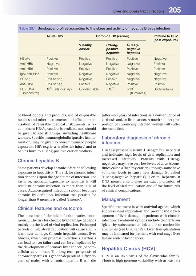
of blood donors and products, use of disposable
needles and other instruments and efficient ster-
ilisation of re-usable medical instruments. A re-
combinant HBsAg vaccine is available and should
be given to at-risk groups, including healthcare
workers. Specific immunoglobulin (passive immu-
nisation) may be given to non-immunised people
exposed toHBV (e.g. in a needlestick injury) and to
babies born to HBeAg-positive carrier mothers.
Chronic hepatitis B
Some patients develop chronic infection following
exposure to hepatitis B. The risk for chronic infec-
tion depends upon the age at time of infection. For
instance, neonatal exposure to hepatitis B will
result in chronic infection in more than 90% of
cases. Adult-acquired infection seldom becomes
chronic. By definition, infection that persists for
longer than 6 months is called ‘chronic’.
Clinical features and outcome
The outcome of chronic infection varies enor-
mously. The risk for chronic liver damage depends
mainly on the level of viral replication. Prolonged
periods of high-level replication will cause signif-
icant liver damage. Chronic hepatitis causes liver
fibrosis, which can progress to cirrhosis. Cirrhosis
can lead to liver failure and can be complicated by
the development of primary liver cancer (hepato-
cellular carcinoma). The long-term prognosis of
chronic hepatitis B is gender-dependent. Fifty per-
cent of males with chronic hepatitis B will die
(after �50 years of infection) as a consequence of
cirrhosis and/or liver cancer. A much smaller pro-
portion of chronically infected women will suffer
the same fate.
Laboratory diagnosis of chronicinfection
HBsAg is present in serum.HBeAgmay also persist
and indicates high levels of viral replication and
increased infectivity. Patients with HBeAg-
negativity may have very low levels of virus (some-
times called a ‘healthy carrier’), though some have
sufficient levels to cause liver damage (so-called
‘HBeAg-negative hepatitis’). Serum hepatitis B
DNA measurement gives an exact indication of
the level of viral replication and of the future risk
of clinical complications.
Management
Specific treatment is with antiviral agents, which
suppress viral replication and prevent the devel-
opment of liver damage in patients with chronic
infection. Treatment options include a-interferon(given by subcutaneous injection) or nucleoside
analogues (see Chapter 22). Liver transplantation
may be indicated for patients with end-stage liver
failure and/or liver cancer.
Hepatitis C virus (HCV)HCV is an RNA virus of the flaviviridae family.
There is high genome variability with at least six
Table 29.1 Serological profiles according to the stage and activity of hepatitis B virus infection
Acute HBV Chronic HBV (carrier) Immune to HBV(past exposure)
‘Healthycarrier ’
HBeAg-positivehepatitis
HBeAg-negativehepatitis
HBsAg Positive Positive Positive Positive Negative
Anti-HBs Negative Negative Negative Negative Positive
Anti-HBc Positive Positive Positive Positive Positive
IgM anti-HBc Positive Negative Negative Negative Negative
HBeAg Pos or neg Negative Positive Negative Negative
Anti-HBe Pos or neg Positive Negative Positive Positive
HBV DNA(virions/ml)
106 (falls quickly) Undetectable >107 >105
(fluctuates)Undetectable
Liver and biliary tract infections 205

different genotypes (1–6) and several subtypes
(1a, 1b, 2a, 2b, etc.).
Epidemiology
Infection is typically acquired via the parenteral
route. InWestern countries, the commonest mode
of transmission is intravenous drug use. In devel-
oping countries with limited healthcare resources,
the commonestmode of transmission is via needle
re-use by healthcare practitioners. Though vertical
transmission and sexual transmissionmay explain
a minority of cases, these modes of infection are
numerically much less important.
Clinical features
The incubation period is 2–6 months. Most infec-
tions are asymptomatic (80%) and in symptomatic
cases the hepatitis is often mild. In contrast with
the natural history of adult-acquired hepatitis B
infection (which seldom becomes chronic),
approximately 70% of cases of acute hepatitis C
infection will become chronic. Chronic infection
causes chronic liver damage and can progress to
cirrhosis after many years (at least 20 years). Cir-
rhosis can be complicated by the development of
primary liver cancer.
Laboratory diagnosis
This is by serology, for the presence of antibody to
HCV and molecular techniques for the detection
of HCV RNA.
Treatment and prevention
Avoidance ofneedle re-usebydrugusers and in the
healthcare setting is the mainstay of prevention.
A vaccine has not been developed. Treatment
is with a-interferon and ribavirin for up to
12 months, and will cure infection (permanent
eradication of infection) in 50% of cases. HCV
genotypes, in addition to other factors, can deter-
mine the response to treatment: genotype 1 is the
least responsive. New and potent HCV-specific
drugs are in development.
Hepatitis D virus (HDV)The hepatitis D virus or delta agent is a defective
RNA virus that can replicate only in HBV-
infected cells. Transmission is via infected blood
and sexual intercourse. HDV accentuates HBV
infection, resulting in more severe liver disease.
Laboratory diagnosis is by HDV antibody detec-
tion and infection can be confirmed by HDV
RNA detection. Treatment is with a-interferon,but response to treatment is disappointing.
Prevention of hepatitis B (by vaccination) will
also prevent HDV infection (since HDV can only
infect HBV-positive cells).
Other causes of viralhepatitis. Ebstein-Barr virus (EBV) can cause hepatitis dur-
ing acute infectious mononucleosis. Usually a
mild self-limiting infection, but can be severe.. Cytomegalovirus (CMV) can cause hepatitis dur-
ing acute infections or reactivation of latent virus
in immunocompromised patients.. Human herpesvirus 6 (HHV6) can cause hepati-
tis in acute infections or following reactivation of
latent virus in immunocompromised patients.. Herpes simplex virus (HSV) can cause hepatitis
in generalised infection in neonates or immuno-
compromised patients.. Varicella-Zoster virus (VZV) causes hepatitis as a
complication of chickenpox or disseminated
zoster in immunocompromised patients.. Rubella can cause hepatitis in congenital or
acquired infections.. Viral haemorrhagic fever viruses can cause hep-
atitis (Yellow fever, rift valley fever virus, Ebola
and Marburg viruses, Lassa fever virus, Junin
and Machupo viruses).
206 Liver and biliary tract infections

30Urinary tract infectionsChris CatchpoleWorcestershire Acute Hospitals NHS Trust, Worcester, UK
Clinical definitionsThere are three clinical syndromes associated with
urinary tract infection (UTI).
Lower UTI (frequency dysuriasyndrome)The clinical features of lower UTI commonly in-
clude dysuria (pain on passing urine), urinary
frequency, urgency and fever. Non-specific symp-
toms, including confusion, unsteadiness, irritabil-
ity and poor weight gain, may predominate in
children and the elderly. These symptoms are
caused by one of the following:
. Bacterial cystitis with bacteriuria (the presence
of bacteria in urine), often associatedwith pyuria
and haematuria due to acute inflammation.. Abacterial cystitis (‘urethral syndrome’) where
no microbial cause is identified. Some cases
may be associated with microorganisms that
are not cultured by routine laboratory methods
(Chapter 14). Unlike urethritis caused by sexu-
ally transmitted infections, this syndrome is not
associated with urethral discharge.
Lower UTI, if untreated, may resolve spontane-
ously; however, infection may progress to, or be
complicated by, pyelonephritis (see below), blood-
stream infection, epididymitis or prostatitis.
Upper UTI (acute bacterialpyelonephritis)This condition is caused by infection of the kidney,
and clinical presentation includes loin pain and
tenderness, pyrexia and symptoms of lower UTI
accompanied by bacteriuria and pyuria. This
potentially serious infection may progress to pe-
ri-nephric abscess formation or bloodstream in-
fection and, in chronic cases, to scarring and renal
failure.
Asymptomatic (covert)bacteriuriaThis is defined as significant numbers of bacteria
present in the urine of apparently healthy people,
with no associated symptoms. Urinary catheteri-
sation is commonly associated with this finding,
and does not usually require antibiotic therapy.
Asymptomatic bacteriuria may, however, be of
clinical importance in childhood and pregnancy,
where treatment may be warranted to prevent
complications should infection develop.
Microbial aetiology andhost–parasite interactionsThe bacteria that commonly cause UTI are com-
mensals (normal flora) of the perineum or lower
Medical Microbiology and Infection Lecture Notes, Fifth Edition. Edited by Tom Elliott, Anna Casey,Peter Lambert and Jonathan Sandoe.� 2011 Blackwell Publishing Ltd. Published 2011 by Blackwell Publishing Ltd.

intestine, and include Escherichia coli (the most
commoncause),Proteusmirabilis (associatedwith
urinary tract stones) and other enterobacteriaceae
(e.g. Klebsiella spp. Enterobacter spp. and Serratia
spp.) Other microorganisms are associated with
particular patient groups, e.g. Staphylococcus sa-
prophyticus with sexually active young women,
and Pseudomonas aeruginosa with urethral cathe-
terisation (Table 30.1).Non-bacterial causes ofUTI
are shown in Table 30.2.
Bacterial virulence factors include:
. Fimbriae: certain serotypes of E. coli have spe-
cific fimbriae (pili) that facilitate colonisation
and adherence to the periurethral areas, urethra
and bladder wall;. Capsules: some strains of E. coli produce a poly-
saccharide capsule that inhibits phagocytosis
and these are associated with the development
of pyelonephritis.
Host factorsDefence mechanisms, which protect against UTI,
include:
. hydrodynamic forces: the flow of urine removes
microorganisms from the bladder and urethra;. phagocytosis of microorganisms by polymorphs
on the bladder surface;. the presence of IgA antibody on the bladder wall;. a mucin layer on the bladder wall prevents
bacterial adherence;. urinary pH.
The following are important host risk factors
for UTI:
. Short urethra in females: sexual intercourse fa-
cilitates the passage of microorganisms up the
urethra (‘honeymoon cystitis’);. Structural and functional abnormalities: causing
outflow obstruction (e.g. prostatic enlargement,
pregnancy, tumours, neurogenic bladder). These
result in residual urine in the bladder, which can
act as a focus for infection. Reflux of urine from
the bladder up the ureter (vesicoureteric reflux)
into the kidney can also result from anatomical
abnormalities and cause pyelonephritis and re-
nal scarring. Renal stones can also be associated
with pyelonephritis;. Increasing age: post-menopausal increase in
vaginal pH and shift of normal vaginal flora
to coliforms, bladder prolapse and prostatic
disease.
Other host risk factors for UTI include:
. diabetes mellitus;
. immunosuppression (e.g. steroids, cytotoxic
drugs);. instrumentation (e.g. surgery or the use of uri-
nary catheters); bacteria may be introduced into
the bladder on catheter insertion, ormaymigrate
up the catheter into the bladder at a later stage.
UTI associated with urinary catheterisation is
an important cause of healthcare associated
infection.
Table 30.1 Common bacterial causes of UTI(approximate percentage)
Microorganism Communityacquired (%)
Hospitalacquired (%)
Escherichia coli 75 40
Proteus mirabilis 3 10
Otherenterobacteriaceae 5 25
Pseudomonasaeruginosa
1 5
Enterococcus faecalis 5 8
Staphylococcussaprophyticus
5 1
Staphylococcusepidermidis
2 3
Table 30.2 Non-bacterial causes of UTI
Group Microorganism Infection
Viruses
Adenoviruses Haemorrhagic cystitis
Humanpolyomavirus
Infections inkidney andureter
Parasites
Trichomonasvaginalis
Urethritis
Schistosomahaematobium
Bladderinflammation
Fungi
Candidaalbicans
UTI inimmunocompromisedpatient
208 Urinary tract infections
Sample collection
For non-catheterised patients, a well taken mid-
stream specimen of urine (MSSU) should be col-
lected, avoiding perineal contamination, into a
sterile universal container. Samples taken from
catheterised patients should be collected directly
from the catheter tubing, not from the drainage
bag. In infants, an adhesive bag or supra-pubic
aspiration (SPA) may be used. Ideally samples
should be processed as soon after collection as
possible, to prevent bacterial overgrowth. If a delay
in processing is unavoidable, samples should be
refrigerated at 4�C, or transported in boric acid or
(agar-coated) dipslide collection bottles.
Laboratory diagnosis
The use of commercial dipstick tests is popular as a
point of care test (POCT) for screening for UTI.
Infection can be confidently excluded in samples
that test negative for both leucocyte-esterase and
nitrites on dipstick testing, but positive results
should be confirmed by microscopy and culture
if clinically indicated.
The mainstay of laboratory diagnosis of UTI is
assessment for the presence of:
. white and red blood cells, epithelial cells and
casts by microscopy or flow cytometry; and. bacteriuria by semi-quantitative culture of a
freshly collected urine sample.
However, results should always be interpreted in
the context of the clinical picture.
Significantbacteriuriaiscommonlydefinedasthe
presence of �105 bacteria/mL (equivalent to �108
bacteria per litre) of anMSSU sample; occasionally,
urine from patients with UTI symptoms may have
fewer microorganisms, possibly as a result of dilu-
tion. Bacteriuria between 104 and 105 microorgan-
isms/mL should be correlated with clinical symp-
toms, antibiotic use, and fluid intake to determine
whether infection is likely.Whenbacteriuriaof<104
microorganisms/mLisdetected, inpatientswhoare
not receiving antibiotics, infection is unlikely.
White blood cells at a concentration of 10/mL or
greater indicates significant pyuria; however, neu-
tropaenic patientsmay not produce high numbers
of cells in urine.
The presence of epithelial cells indicates peri-
neal/peri-urethral contamination, whichmay give
a false positive culture result. Sample contamina-
tion should also be suspected when culture yields
more than two bacterial species.
Blood cultures should also be taken, if pyelone-
phritis or bloodstream infection is suspected.
Antibiotic sensitivity tests are undertaken on
bacterial isolates, which are considered to be of
potential clinical significance. The results of these
tests should be used as a guide to appropriate
therapy.
Management
Non-antibiotic management includes increasing
fluid intake to ‘flush out’ microorganisms. In un-
complicated lower UTI, a short course (e.g. 3 days)
of first-line empirical oral antibiotic therapy
should be considered, and choices should take
account of local susceptibility patterns; options
may include trimethoprimor nitrofurantoin. Ther-
apy should then be amended as required, based on
the results of antibiotic sensitivity tests. Patients
with upper UTI or UTI associated bloodstream
infection may require parenteral therapy initially,
and a more prolonged course of antibiotics is
indicated (7–10 days). Failure to respond to appar-
ently appropriate therapy is an indication to repeat
urine culture.
As with all antibiotic therapy, choice of agent
should be guided by causative microorganism, his-
tory of known drug allergy, presence of other un-
derlying medical conditions, and risk of antibiotic
adverse effects or drug interactions, particularly in
the elderly, e.g. Clostridium difficile infection.
It is also important to recognise that the pres-
ence of bacteriuria and/or pyuria is common,
particularly in the elderly, and that it does not
always indicate an infection that requires antibi-
otic therapy. However, in pregnancy, asymptom-
atic bacteriuria should be treated to prevent com-
plications, which include pyelonephritis and pre-
term delivery.
Sterile pyuria (routine urine culture is negative
in the presence of significant numbers of white
blood cells) may require further investigation:
causes include concomitant antibiotic therapy,
inflammatory renal disease, the presence of for-
eign bodies, tumours or infectionwith uncommon
bacterial (e.g. Mycoplasma spp.) or non-bacterial
pathogens (Table 30.2).
Urinary tract infections 209
31Genital infectionsKaveh ManaviUniversity Hospitals Birmingham NHS Foundation Trust, Birmingham, UK
This chapter outlines some of the more common
genital infections.
Vaginitis
Definition
The term applies to inflammation of vaginal mu-
cosa thatmay extend to the vulva and is commonly
associated with a white or yellow discharge. Occa-
sionally vaginitis may be associated with dysuria
and dyspareunia.
Clinically the term mainly applies to unusual
vaginal symptoms (i.e. unusual discharge or any
form of abnormal vaginal sensation) with or with-
out mucosal inflammation. Many women seek
medical treatment for these symptoms; however,
a large number are treated incorrectly in the ab-
sence of appropriate laboratory diagnosis.
The most common causes of vaginal symptoms
are bacterial vaginosis, Trichomonas vaginitis and
candidiasis. Less common causes of vaginitis, in-
cluding foreign bodies, lichen planus, chemical
irritation and atrophic vaginitis are not discussed
in this chapter.
Bacterial vaginosis (BV)
Aetiology. BV is believed to be the result of reduction of
vaginal lactobacilli and colonisation of vaginal
mucosa by groups of abnormal microorganisms
rather than one single pathogen. Some of the
bacteria associated with BV include Gardnerella
vaginalis, anaerobes such Bacteroides spp., and
Peptostreptococcus spp., Mycoplasma hominus,
and Ureaplasma urealyticum.. BV isnot associatedwith inflammationof vaginal
mucosa.. BV has been reported to facilitate HIV
acquisition.
Epidemiology. BV is the most common cause of vaginal
discharge.. It has been estimated that a third of womenmay
develop BV sometime in their life.. In 2008, more than 110,000 BV episodes were
diagnosed in GUM clinics in England.. Young age, smoking, vaginal douching, and hav-
ingmultiple sexual partners have been separate-
ly associated with BV.
Medical Microbiology and Infection Lecture Notes, Fifth Edition. Edited by Tom Elliott, Anna Casey,Peter Lambert and Jonathan Sandoe.� 2011 Blackwell Publishing Ltd. Published 2011 by Blackwell Publishing Ltd.
. BV is not a sexually transmitted infection (STI)
and there is no equivalent entity in men.
Clinical features. Malodorous ‘fishy’ vaginal discharge has been
proposed as characteristic of BV in the past.
However, this is not the case, as many women
with BV may not complain of such discharge.. Most women with BV are asymptomatic; up to
75% of women with BV deny any foul smelling
discharge.. BV has been associated with post-partum endo-
metritis, chorioamnionitis, pre-term labour and
pelvic inflammatory disease (PID).
Diagnosis. Diagnosis of BV relies on clinical andmicroscop-
ic findings; no single clinical manifestation can
reliably assist.. Amsel’s criteria have been used by many clin-
icians; BV requires the presence of three of the
four criteria:
1 Viscid abnormal vaginal discharge;
2 Positive ‘whiff test’; fishy odour of vaginal
fluid after addition of 10% potassium
hydroxide;
3 Presence of clue cells (epithelial cells embed-
ded with bacteria with unidentified border) on
microscopy of vaginal fluid;
4 Vaginal fluid pH of more than 4.5.. Hay–Ison Criteria; a new diagnostic system for
BV and includes the following:
� Grade 1: normal lactobacilli, Grade 2: mixed
flora with some lactobacilli, and Gardenerella;
Grade 3: mostly Gardnerella
� In symptomatic women, grades 2, or 3 are
consistent with BV.
Presence of more than one polymorphonuclear
cell (PMN) inmicroscopy of vaginal fluid raises the
possibility of TV or cervicitis (gonococcal or
chlamydial).
Treatment and prevention in adults. Oral metronidazole (multiple or single dose);. Clindamycin or metronidazole cream are
alternatives;. Prolonged oral metronidazole for recurrent BV;. Prolonged metronidazole gel use for recurrent
BV.
Trichomonas vaginitis (TV)
Aetiology. The protozoan parasite Trichomonas vaginalis is
the cause of TV. TV facilitates HIV acquisition and has been
reported tobe strongly associatedwith other STIs.. Men infected with Trichomonas vaginalis are
mostly asymptomatic; mild dysuria may be re-
ported by some infected men.. During pregnancy, TV can cause pre-term deliv-
ery and low birthweight. Because treatment of TV
during pregnancy with metronidazole has been
reported to be associated with similar unfavour-
able outcomes, routine screening of asymptom-
aticwomen for TV is not currently recommended.
Epidemiology. In GUM clinics in England, 5,600 episodes of TV
were diagnosed in 2008.. Worldwide, 170 million cases of TV are reported
each year.. Trichomonas vaginalis is transmitted by sexual
intercourse, hence it is an STI.
Clinical features. Most women with TV are believed to be
asymptomatic.. Abnormal vaginal discharge, vulval andperi-anal
itching are the most common symptoms.. Erythema of vagina with diffuse petechiae on the
cervix (strawberry cervix) have been considered
pathognomonic for TV. Strawberry cervix is not
commonly observed in women with symptom-
atic TV.
Diagnosis. Wet mount microscopy of vaginal fluid shows
motile Trichomonas vaginalis.. Vaginal fluid of women with TV may also have
PMN with a pH of higher than 4.5. Inoculation of vaginal fluid for Trichomonas
vaginalis culture has higher sensitivity and
should be performed for women considered at
risk of infection.. Use of nucleic acid amplification assays for diag-
nosis of TV has been approved. These assays have
higher sensitivity and specificity than TV culture
and may be used as a standard test in future.
Genital infections 211
Treatment and prevention in adults. Multiple or single dose of oral metronidazole;. Sexual partners of women with TV need to be
treated as well;
Vulvo-vaginal candidiasis (VC);(‘Thrush’)
Aetiology. VC is caused by Candida albicans in themajority
of cases. Infections with C. glabrata or C. tropi-
calis often result in recalcitrant VC.. VC is a common cause of vaginitis.. Immunosuppression (secondary to HIV, or im-
munosuppressive agents), diabetes mellitus,
pregnancy, use of broad spectrum antibiotics
can each predispose women to VC.. Many women incorrectly relate their vulvo-
vaginal symptoms to VC.
Epidemiology. More than 70,000 cases of VC were diagnosed in
GUM clinics in England in 2008.. The number of episodes treated for assumed VC
by over the counter medicines remains unclear.. VC is not a STI and affects most women at least
once in their life time.
Clinical features. Itching and irritation, vaginal discharge, dyspar-
eunia and dysuria are the most common com-
plaints patients present with.
Diagnosis. Microscopical examination of vaginal fluid (wet
mount, or Gram stained) reveals yeasts.. Yeast culture is the gold standard method for
diagnosis of candidiasis and species identifica-
tion. Yeast culture should only be performed in
symptomatic women.
Treatment and prevention in adults. Topical azole antifungal agents (as pessary or
creams) can be used; clotrimazole, miconazole;. A single oral dose of fluconazole is an alternative
treatment option. This is contraindicated for
pregnant women.
Urethritis and cervicitisGonococcal urethritis andcervicitis
Epidemiology. The gonococcus, Neisseria gonorrhoea is an
obligate human parasite;. Many women are asymptomatic and may act as
a reservoir of infection;. Highly transmissible by sexual contact;. Occurs worldwide;. Infections are most common among sexually
active young adults.
Pathogenesis
See Chapter 7.
Clinical features. Urethral discharge in men and cervicitis in
women;. Complications include epididymitis, prostatitis,
urethral stricture, Bartholin’s abscess, pelvic in-
flammatory disease and infertility.
Laboratory diagnosis. A diagnosis can be confirmed by microscopy,
culture and/or molecular analysis (nucleic acid
amplification tests, NAAT) of pus and secretions
from various sites (depending upon the infec-
tion): cervix, urethra, rectum, conjunctiva, throat
and synovial fluid.. Clinical samples are cultured on enriched selec-
tive media.. Specimens requiring culture should be rapidly
transported to the laboratory, because gonococci
die readily on drying.. Blood cultures should be sent before antimicro-
bial therapy is commenced when disseminated
infection is suspected, e.g. in sexually active
patients with septic arthritis, concurrent with
NAAT.. Serology is not helpful in the diagnosis of gono-
coccal infection.
Treatment and prevention
Resistance to penicillins, tetracyclines and quino-
lones is common in the UK and worldwide. Most
212 Genital infections

strains remain susceptible to third-generation
cephalosporins (e.g. ceftriaxone or cefixime).
Single-dose therapies are recommended to
increase patient compliance. Prevention of gonor-
rhoea includes sex education, promotion of public
awareness and the use of condoms. Contact trac-
ing is essential in preventing further spread and the
disease. No vaccine is available.
Non-gonococcal urethritisand cervicitisChlamydia trachomatis (seeChapter 14)
Epidemiology. C. trachomatis is themost common bacterial STI
in the UK and the world.. It has been estimated that 10% of women and
13% ofmen younger than 25 years in theUKmay
be infected with chlamydia.. Themajority of infected (70% of female and 50%
of male) patients are asymptomatic.. Untreated chlamydial infection can cause
pelvic inflammatory disease (PID) in 10–40% of
women.. Each year a substantial amount of health budgets
around the world are spent on management of
the long-term consequences of untreated chla-
mydial infections.
Pathogenesis
See Chapter 14.
Clinical features
Female genital tractIn women, chlamydial infection can cause symp-
tomatic cervicitis (Figure 31.1), but most cases are
asymptomatic. It is likely that a significant propor-
tion of infected women may develop PID. PID can
cause infertility, ectopic pregnancy and chronic
pelvic pain syndrome in affected women.
Male genital tractC. trachomatis is the most common cause of ure-
thritis in men. Persistent chlamydial infection in
men can lead to epididymo-orchitis, painful swell-
ing of the epididymis that is mostly unilateral.
Sexually acquired reactivearthritis (SARA)One of the auto-immune manifestations of persis-
tent chlamydial infection presents as pain of lower
limb joints mostly in asymmetrical fashion. The
symptoms commonly startwithin 30days of sexual
contact. Box 31.1 summarises the clinical mani-
festations of SARA.
Figure 31.1 Chlamydial cervicitis.
BOX 31.1 Clinical presentations ofsexually acquired reactive arthritis (SARA). Circinate balanitis or vulvitis (typical psoriaticlesions of the glans penis or labia)
. Guttate cutaneous psoriasis
. Pustular psoriasis on the soles of the feet
. Psoriasiform rash
. Subcutaneous nodules
. Geographical tongue
. Nail dystrophy
. Conjunctivitis
. Enthesopathy (pain at the sites of tendon or fasciaattachments, especially the Achilles tendon andplantar fascia attachments to the calcaneum).
. Tenosynovitis (tenderness over tendon sheaths ofthe lower limbs).
. Sacroiliitis (low back pain and stiffness)
. Pericarditis and aortic valve disease
. Thrombophlebitis of the lower limbs
. Renal pathology (proteinuria, haematuria andaseptic pyuria)
. Nervous system involvement including meningo-encephalitis and nerve palsies
Genital infections 213

Laboratory diagnosis. NAAT are the most sensitive and specific tests
for detection of C. trachomatis infections. These
assays can be used on self-collected specimens
(urine or vaginal swabs) enabling patients to be
tested for C. trachomatis infection without the
need for genital examination.. Polymerase chain reaction, transcription medi-
ated amplification and strand displacement
amplification are some of the NAAT available for
detection of C. trachomatis.
Treatment and prevention
Uncomplicated C. trachomatis can be treated
with tetracyclines, macrolides or quinolones (ex-
cept ciprofloxacin). At least one week of treat-
ment with any of those agents is required. Treat-
ment of complicated chlamydial infection (PID,
epididymo-orchitis or LGV) require longer dura-
tion of antibiotic therapy. Treatment of chlamyd-
ial infectionhas been significantly simplifiedwith
the advent of azithromycin. A single dose of
azithromycin will be adequate for treatment of
uncomplicated chlamydial infection. The most
effective method for prevention of genital trans-
mission of chlamydial infection is consistent use
of male latex condoms. Partners of patients with
chlamydial infection need chlamydial screening
and treatment.
Other causes of urethritisUreaplasma spp., Mycoplasma hominis, TV
and Herpes simplex virus may also cause
urethritis.
Genital ulcersThere are generally three main causes for
genital ulcer disease (GUD); infections, inflam-
matory diseases and malignancies. In the devel-
oped countries, herpes simplex virus (types 1
or 2) and syphilis are the most common infec-
tious causes of GUD. Chancroid caused by
Haemophilus ducreyi is another infectious
cause of GUD, mostly diagnosed in developing
countries.
Herpes simplex virus (HSV)infection
Aetiology. Although classically HSV-2 has been associated
with genital herpes, HSV-1 has become themain
cause of genital herpes in developed countries.. HSV results in a chronic infection thatmay cause
recurrent episodes.
Epidemiology. HSV is the most common cause of GUD in the
world.. HSV is transmitted through direct skin to skin
contact.. It is believed that a significant number of patients
infected with HSV remain undiagnosed, as their
symptoms are short-lived and atypical.. Viral shedding can occur without active
ulceration.. Withuse of nucleic acid amplification tests,more
cases of HSV have been diagnosed.
Clinical features. Primary HSV episode: patients without prior HSV
infection may develop systemic illness with gen-
ital blisters. The systemic illness manifests as flu
like symptoms, fever, lymphadenopathy, aseptic
meningitis and atonic bladder.. Herpetic blisters: can appear in clusters, dry out
over 2 weeks and develop a crust. Finally the
ulcers heal without scarring.. Initial HSV episode: the first clinical presentation
of genital herpes. Patients mostly complain of
painful genital blisters and herpetic blisters.. Recurrent HSV episode: the subsequent episodes
of herpes attacks are manifested with herpetic
ulcerations that heal within 3–5 days. Patients
reportmilder symptoms during recurrent herpes
episodes.. Thenumber of recurrent herpes episodes is high-
est in the first year.. Patients with frequent herpes attacks may ben-
efit from suppressive therapy.
Laboratory diagnosis. Use of polymerase chain reaction (PCR) assays
has become the standard method of diagnosis
in recent years. This testing method has high
214 Genital infections

sensitivity and specificity. Herpes PCR can also
confirm the type of HSV as well.. HSV culture is still considered the gold standard
method of HSV diagnosis, but is no longer used
in most diagnostic laboratories. It is not as sen-
sitive as PCR and thereforemaymiss a number of
positive cases.. HSV-serology is generally not helpful in the
diagnosis of genital HSV. Type specific HSV
serology can diagnose previous infections with
HSV-2 and may be useful in asymptomatic
patients.
Treatment and prevention. HSV-DNA polymerase inhibitors (aciclovir, vala-
ciclovir, famciclovir) used systemically are highly
effective in controlling HSV episodes. These
agents do not eradicate infection.. Aciclovir is used for recurrent episodes.. For patients with frequent HSV recurrence epi-
sodes (more than 5 episodes a year), suppressive
therapy can be initiated. This includes acyclovir
or valaciclovir once a day. The duration of sup-
pressive therapy is 12 months.. Use of condoms reduce rate of HSV transmission
but does not completely prevent it. Counselling
of sexual partners of patients is an important
component of prevention of HSV transmission
strategy.
Syphilis
Aetiology
Syphilis is an infection caused by the spirochete,
Treponema pallidum.
Epidemiology. Syphilis is mainly an STI.. The number of patients with primary and sec-
ondary syphilis (see Pathogenesis) in the UK has
increased significantly since 2000; more than
12,500 cases have been diagnosed between
2004 and 2008.. Globally, syphilis has been increasingly diag-
nosed in countries with a high rate of HIV infec-
tion. Syphilis significantly increases the rate of
HIV transmission; its diagnosis and treatment
may therefore be an important strategy for pre-
vention of HIV infection.
. Pregnantwomenwithuntreated syphilis canalso
pass infection to their foetus at any point during
pregnancy or delivery.. If a pregnant woman is in the early stages of
infection, her foetus is at greatest risk of congen-
ital syphilis unless treated for syphilis.. The rate of congenital infection significantly
reduces if the mother is treated before the 16th
week of pregnancy.
Pathogenesis
T. pallidum is a spirochaete capable of movement
within extra cellular human tissues. It is able to
penetrate skin ormucosa throughmicro-abrasions.
Once inside the dermal or sub-mucosal tissues, it
replicates rapidly and enters the circulation.
The majority of syphilitic presentations are
related to plasma cell infiltration of infected sites
that leads to permanent occlusion of small arter-
ioles responsible for feeding the wall of main
arteries and nervous system.
Clinical features
Untreated syphilis is an illnesswith clinically recog-
nisable stages. The first two years are called ‘early
syphilis’ and includes primary, secondary and early
latent stages of infection. Infection after the second
year is known as the late stage of syphilis:
. Primary syphilis: 3–6 weeks after exposure;
punched out ulcers with an indurated edge devel-
op (chancre) at the site of inoculation. This period
is called primary syphilis. Syphilitic chancres are
typically painless, however super-infection or
concomitant herpes ulcers may make them ten-
der. Chancres are highly infectious. Although
chancres resolve completely after 4–6 weeks,
T. pallidum remains in the body.. Secondary syphilis: 4–6 weeks after resolution
of the chancre, secondary syphilis develops as
the result of widespread treponemal replication
throughout the body. This stage can present as
any of multitude of signs (Table 31.1), including
a widespread macular rash and palmar rash
(Figures 31.2 and 31.3). These signs also resolve
spontaneously after 4–6 weeks.. Early latent syphilis: After the secondary stage,
infection becomes mostly asymptomatic.. Late syphilis: presents as late latent (asymptom-
atic), neurosyphilis, cardiovascular disease and
parenchymal diseases of syphilis.
Genital infections 215

Table 31.1 Summary of clinical stages of syphilis if untreated in chronological order
Stage of syphilis Clinical manifestation Duration
Incubation None 3–6 weeks
A. Primary syphilis Typical lesions: single painless indurated genital ulcer, withlocal lymphadenopathy
4–6 weeks
Variants: multiple painful purulent destructive extragenital(mainly oral) ulcers
Severe syphilitic balanitis of FollmanAsymptomatic neurosyphilis: abnormal CSF with no signs/
symptoms; unknown significanceAsymptomatic stage 4–6 weeks
B. Secondarysyphilis
4–6 weeks
B.1. Skinmanifestations
Typical presentation: maculopapular lesions symmetricallydistributed over the body, may involve palms, soles and oralmucosae
Atypical presentation: papulo-squamous or pustular lesions(resembling psoriasis) that can be itchy
Condylomata latum (Figure 31.3): painless, highly infectiouswart-like, grey-white lesions that develop inwarm,moist sites(e.g. peri-anal skin). They are mistaken as peri-anal wart bythe untrained eye
Patchy alopecia: including scalp, facial hair and eye brows
‘Split pea’ lesions: non-tender lesions in the inter-triginousareas: mouth, nasolabial folds, post. Auricular
Mucosa: patches, husky voice, snail track ulcers
B.2. Systemicpresentations ofsecondary syphilis
Lymphadenopathy: non-tender, rubbery
Fever, malaiseEye: uveitis, iritis: clinical in 5–10%; sub-clinical in >50%CNS: meningitis, cranial nerve palsies, meningovascular
disease, Asymptomatic neurosyphilis: abnormal CSF with nosigns/symptoms; unknown significance
Musculoskeletal: painful osteitis, arthralgia, myalgiaGastronintestinal: hepatitis, syphilitic jaundice, splenomegalyRenal: nephritis (proteinuria, haematuria)
C. Asymptomatic(Early latent)
Up to 2 years
D. Asymptomatic(Late latent)
D.1 Asymptomatic neurosyphilis: abnormal CSF with no signs/symptoms; unknown significance
After 2 years
D.2 Meningovascular syphilis: focal arteritis inducing infarction/meningeal inflammation
After 2 years
D.3 General paresis of insane: Gradual decline in memory andcognitive functions, emotional lability, personality change,psychosis and dementia. Seizures and hemiparesis can belate complications
2–7 years
D.4 Tabes dorsalis: Plasma arteritis of arteries feeding spinaldorsal column/nerve roots; lightening pains, areflexia,paraesthesia, sensory ataxia, Charcot’s joints, malperforans, optic atrophy, pupillary changes (e.g. ArgyllRobertson pupil)
10–20 years
D.5Cardiovascular: aortitis (ascendingusually); asymptomatic,substernal pain, aortic regurgitation, heart failure, coronaryostial stenosis, angina, aneurysm
15–25 years
D.6 Gummatous: Granulomatous destructive lesions that canoccur in any organ, but most commonly affect bone and skin
10–30 years
216 Genital infections
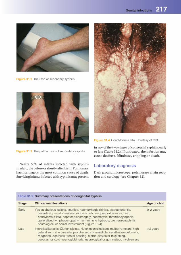
Nearly 50% of infants infected with syphilis
in utero, die before or shortly after birth. Pulmonary
haemorrhage is the most common cause of death.
Surviving infants infectedwith syphilismaypresent
in any of the two stages of congenital syphilis, early
or late (Table 31.2). If untreated, the infection may
cause deafness, blindness, crippling or death.
Laboratory diagnosis
Dark ground microscopy, polymerase chain reac-
tion and serology (see Chapter 12).
Figure 31.2 The rash of secondary syphilis.
Figure 31.3 The palmar rash of secondary syphilis.
Table 31.2 Summary presentations of congenital syphilis
Stage Clinical manifestations Age of child
Early Vesiculobullous lesions, snuffles, haemorrhagic rhinitis, osteochondritis,periostitis, pseudoparalysis, mucous patches, perioral fissures, rash,condylomata lata, hepatosplenomegaly, haemolysis, thrombocytopenia,generalised lymphadenopathy, non-immune hydrops, glomerulonephritis,neurological or ocular involvement (Figure 13.4)
0–2 years
Late Interstitial keratitis, Clutton’s joints, Hutchinson’s incisors, mulberrymolars, highpalatal arch, short maxilla, protuberance of mandible, saddlenose deformity,rhagades, deafness, frontal bossing, sterno-clavicular thickening,paroxysmal cold haemoglobinuria, neurological or gummatous involvement
>2 years
Figure 31.4 Condylomata lata. Courtesy of CDC.
Genital infections 217

Treatment and prevention
Anyano-genitalulcer shouldbeconsideredas syph-
ilitic, unless proven otherwise. Penicillin remains
the treatment of choice of syphilis. Benzathine pen-
icillin G intramuscular, single injection, or pro-
caine penicillin G intramuscular, daily for 10 days,
are treatments of choice for early syphilis.
Doxycycline or ceftriaxone are alternative antibio-
tics for treatment of syphilis. Treatment of neuro-
syphilis or late latent syphilis requires a longer
duration of penicillin therapy.
Infants with congenital syphilis must be treated
for syphilis, although most of the developmental
signs will be permanent.
Sexual transmission of syphilis occurs with di-
rect skin-to-skin contact. In this respect,male latex
condoms may prevent transmission of syphilis as
long as they cover the infected skin/site. It is likely
that consistent use of male latex condoms may
prevent some but not all cases of syphilis. Con-
genital syphilis can be effectively prevented with
diagnosis and treatment of infected mothers at
early stages of pregnancy.
Genital warts
Epidemiology. In the UK, more than 90,000 new cases of genital
warts were diagnosed between 2008 and 2009.. Genital warts are a common diagnosis amongst
patients attending genitourinary medicine
clinics. Theyare considered as themost common
viral STI in theUK. They affect both sexes equally
and are mostly diagnosed in patients aged
16–30 years.. Genital warts are caused by infection with hu-
manpapilloma virus (HPV), which is transmitted
through close skin contact.
Pathogenesis. HPV is a double stranded DNA virus.. There aremore than 200 types ofHPV;more than
40 are mostly transmitted sexually. The role of
HPV in causing cervical cancer is well estab-
lished. Because of their strong association with
malignancy, HPV types 16, 18, and 31 are con-
sidered high risk.. The virus infects basal layer cells of the epidermis
through skin micro-abrasions. After a latency of
3 weeks to 9 months, the infected cell starts
production of viral particles. Infected cells un-
dergo changes that result in koilocyte formation;
a dysplastic squamous formation.. E6 and E7 genes of high risk HPV types code for
proteins that block host cells’ tumour suppres-
singgenesp53, p21andRB.Over time, becauseof
eratic mitosis, some infected cells will develop
cancerous mutants that may lead to develop-
ment of cancer, if untreated.
Clinical features. Warts can affect any part of ano-genital skin, in
addition to the anal canal and cervical mucosa.
HPV types 6 and 11 are responsible for more than
80%of genitalwarts. It is important tonote that the
site of infection may not relate to sexual practice;
peri-anal warts should not be considered as an
evidence of history of receptive anal intercourse.. Genital warts may present in different numbers
and sizes. They are mostly asymptomatic but
may cause itching. Genital warts are caused by
low risk HPV types (6 or 11) in more than 85% of
cases. They therefore do not cause long-term
complications.. Warts caused by high risk HPV types (16 or 18)
may evolve in pre-cancerous and cancerous skin
lesions. Cervical warts should be referred for
colposcopical examination and treatment.. HPV infections of genital skinmayalsopresent as
bowenoid papules or seborrheic keratoses.. Because the majority of humans infected with
HPV do not present with genital warts, partner
notification for genitalwarts isnot recommended.
Diagnosis
Diagnosis of warts is clinical. They are occasionally
diagnosed aftermicroscopy examination of biopsy
of skin lesions.
Although HPV typing can be performed for wart
lesions, this is not commonly used.
Treatment
Patients should be advised that treatment of warts
does not eradicate HPV and they may therefore
experience recurrences in future. There are differ-
ent treatment options for genital warts, none of
which is considered superior.
Medical therapy of genital warts can be achieved
by topical application of podophyllotoxin (twice
218 Genital infections

daily, 3 days a week for a maximum of 5 weeks), or
imiquimod (alternative nights for maximum of
4 months). Because topical therapy of warts can
be applied by patients, they are the treatment of
choice for many.
Podophyllotoxin is an anti-mitotic agent
through inhibition of tubulin polymerisation.
Imiquimod is a topical activator of toll-like
receptor 7 (TLR-7), leading to local production
of interleukin 6, tumour necrosis factor and inter-
feron alpha. However, the anti-tumour activation
of imiquimod may not be related to its immune
activation feature. Upgrade in opioid growth factor
receptor is considered to be imiquimod’s anti-
neoplastic activity.
Ablation therapy with cryotherapy (liquid nitro-
gen), laser or surgical excision are other treatment
options for warts.
Genital infections 219
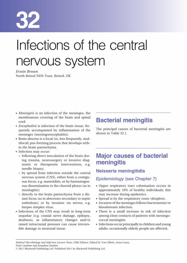
32Infections of the centralnervous systemErwin BrownNorth Bristol NHS Trust, Bristol, UK
. Meningitis is an infection of the meninges, the
membranous covering of the brain and spinal
cord.. Encephalitis is infection of the brain tissue, fre-
quently accompanied by inflammation of the
meninges (meningoencephalitis).. Brain abscess is a focal (or, less frequently, mul-
tifocal) pus-forming process that develops with-
in the brain parenchyma.. Infection may occur:
� following direct inoculation of the brain dur-
ing trauma, neurosurgery or invasive diag-
nostic or therapeutic interventions, e.g.
needle biopsy;
� by spread from infection outside the central
nervous system (CNS), either from a contigu-
ous focus, e.g. mastoiditis, or by haematogen-
ous dissemination to the choroid plexus (as in
meningitis);
� directly to the brain parenchyma from a dis-
tant focus (as in abscesses secondary to septic
embolism); or by invasion via nerves, e.g.
herpes simplex virus.. Infections of the CNS may result in long-term
sequelae (e.g. cranial nerve damage, epilepsy,
deafness), as inflammatory changes and/or
raised intracranial pressure can cause irrevers-
ible damage to neuronal tissue.
Bacterial meningitisThe principal causes of bacterial meningitis are
shown in Table 32.1.
Major causes of bacterialmeningitisNeisseria meningitidis
Epidemiology (see Chapter 7). Upper respiratory tract colonisation occurs in
approximately 10% of healthy individuals; this
may increase during epidemics.. Spread is by the respiratory route (droplets).. Invasionof themeninges followsbacteraemia/or
bloodstream infection.. There is a small increase in risk of infection
among close contacts of patients with meningo-
coccal meningitis.. Infectionsoccurprincipally inchildrenandyoung
adults, occasionally elderly people are affected.
Medical Microbiology and Infection Lecture Notes, Fifth Edition. Edited by Tom Elliott, Anna Casey,Peter Lambert and Jonathan Sandoe.� 2011 Blackwell Publishing Ltd. Published 2011 by Blackwell Publishing Ltd.

. In the USA and Europe, cases of meningococcal
meningitis are sporadic and caused mainly by
serotype B; epidemics of N. meningitidis ser-
otypes A and C occur in Africa and South
America.
Clinical features. Usually rapid onset, headache, fever andmenin-
gism (neck stiffness, irritability, photophobia).
Typical petechial rash, which may become hae-
morrhagic – this clinical feature is not pathogno-
monic of meningococcal disease, a similar rash
occurs in asplenic patients with overwhelming
Streptococcus pneumoniae or Haemophilus
influenzae type b infections. Other clinical fea-
tures include signs of cerebral dysfunction (con-
fusion, delirium or a falling level of conscious-
ness –ranging from lethargy to coma), focal neu-
rological deficits, including cranial nerve palsies
(particularly III, VI, VII and VIII), hemiparesis
and papilloedema.. In fulminant cases, endotoxin release results in
shock, with disseminated intravascular coagulo-
pathy and multi-organ failure. In a minority of
cases, haemorrhagic necrosis of the adrenal
glands and intracranial bleeding, which is invari-
ably fatal (Waterhouse–Friderichsen syndrome)
may occur.. Presentation in the elderlymay be insidious with
lethargy or obtundation, confusion, no fever and
variable signs of meningeal inflammation.
Treatment and prevention. Intravenous penicillin; cefotaxime or ceftriaxone
(chloramphenicol may be used in penicillin-
allergic patients).
. In the developed world epidemics are rare; how-
ever, related cases in families or institutions can
occur and prophylaxis with rifampicin or cipro-
floxacin is recommended for close contacts.. Healthcare workers in contact with cases are not
at increased risk, but prophylaxis is recom-
mended for those who have administered
mouth-to-mouth resuscitation.. A vaccine is available for protection against ser-
otypes A and C, and is recommended for pro-
phylaxis of close contacts of meningococcal
cases caused by these serotypes.
Haemophilus influenzae type b
Epidemiology (see Chapter 9). Occurs principally in children aged <5 years.. Spread is by the respiratory route and symptom-
less upper respiratory colonisation is common.. Cases are often sporadic, but small outbreaks,
particularly in nurseries, may occur.. Number of cases has decreased dramatically
since introductionofH. influenzae typebvaccine.
Clinical features
Similar to meningococcal meningitis, except that
onset is often less acute (1–2 days) and, although a
petechial rash can occur rarely, it is much less
common.
Treatment and prevention
Antibiotic therapy with cefotaxime, ceftriaxone or
amoxicillin (if the strain is susceptible) or chlor-
amphenicol. Rifampicin is used for prophylaxis of
close contacts. A vaccine is available and should be
considered for at-risk household contacts.
Table 32.1 Causative microorganisms of acute bacterial meningitis according to age group
Age Causative microorganism
<1 month Group_B b-haemolytic streptococcus, E. coli, Citrobacter spp., Klebsiella spp.,
Enterobacter spp., P. aeruginosa, L. monocytogenes, S. aureus, S. epidermidis
1–23months
Group_B b-haemolytic streptococcus, E. coli, H. influenzae, S. pneumoniae,
N. meningitidis
2–50 years S. pneumoniae, N. meningitidis
�50 years S. pneumoniae, N. meningitidis, L. monocytogenes, aerobic Gram-negative bacilli(coliforms)
Infections of the central nervous system 221

Streptococcus pneumoniae
Epidemiology. Pneumococci are common commensals of the
upper respiratory tract.. Infection is most common in young (<2 years of
age) and elderly individuals.. Asplenic patients (including thosewith sickle cell
disease) are at particular risk.. Meningitis may be secondary to pneumococcal
pneumonia and bloodstream infection or skull
fracture (direct spread).. Patients with pneumococcal meningitis.
Clinical features
Similar to other causes, but S. pneumoniae infec-
tion may be acute or insidious in presentation.
Complications (e.g. deafness) are more common
than with other pathogens.
Treatment and prevention. Strains with a reduced susceptibility to penicil-
lin have emerged. Patients should therefore
receive either cefotaxime or ceftriaxone empir-
ically, but they can be converted to benzylpe-
nicillin, if the strain is subsequently found to be
susceptible.. Contacts of cases of pneumococcal meningitis
are not at increased risk and prophylaxis with
antibiotics is not recommended.. A vaccine against many of the pneumococcal
serotypes is available and is recommended for
splenectomised andother immunocompromised
patients and those with base of skull fractures/
cerebrospinal fluid (CSF) leaks.
Laboratory diagnosisof bacterial meningitisMeningitis is a medical emergency and laboratory
investigations are urgent:
. Blood cultures should ideally be obtained before
administration of empirical antibiotic therapy.. Lumbar puncture (LP) is usually performed
after raised intracranial pressure has been ex-
cluded by neuroimaging. Typical features of
CSF microscopy are a raised polymorph count,
although a predominance of lymphocytes can
occur, especially in Gram-negative bacillary
meningitis in neonates and in patients with
meningitis caused by Listeria monocytogenes.
A low glucose (compared with blood glucose)
and an increased protein concentration occurs
(Table 32.2). Gram stainingmay reveal the caus-
ative microorganism; culture is important for
confirmation.. Note: a normal CSF white blood cell count may
be seen in patients who present within 12 hours
of the onset of the disease, in some cases of
neonatalmeningitis and inneutropenic patients.. Antigen detection tests are available.. PCR can detect DNA of Neisseria meningitidis
and Streptococcus pneumoniae.
Table 32.2 Typical CSF changes in meningitis
Meningitis
Normal Bacterial Tuberculous Viral
Leukocytes (cells/mL) <5 100–10,000 <500 50–500
Polymorphs (%) >80 20a 5a
Lymphocytes (%) 10 80 95
Protein (g/L) 0.2–0.45 " " (>50) " " " (>50) "Glucose (mmol/L) 4.5–8.5b # #c # #c Normal
aPolymorphs may predominate in the early stagesbAbout 80% of blood glucose levelc<40% of blood glucose level
222 Infections of the central nervous system

. Blood cultures are positive in about 40%of cases.
. Serology may give a retrospective diagnosis of
meningococcal meningitis, but is rarely used in
clinical practice.
Treatment of bacterialmeningitisThe fundamental principle in the treatment of
bacterial meningitis is early, parenteral antibiotics
in high dosages. Intramuscular penicillin admin-
istered early by primary care physicians has been
shown to reduce the incidences of morbidity and
mortality associated with meningococcal menin-
gitis. Antibiotic therapy for common causes of
bacterial meningitis in children and adults is
shown in Table 32.3. There is some evidence that
steroids, when initiated immediately before or
concurrent with antimicrobial therapy, are
beneficial.
Neonatal meningitis
Aetiology
Predominantly caused by Escherichia coli and
occasionally other coliforms, Pseudomonas aer-
uginosa, group B b-haemolytic streptococci,
Listeria monocytogenes, Staphylococcus epider-
midis and Staphylococcus aureus. Bacteria which
colonise the upper respiratory tracts of neonates –
S. pneumoniae,H. influenzae andN.meningitidis
may also cause this infection.
Epidemiology
Acquisition is normally from the maternal genital
or alimentary tract, at or around the time of
delivery:
. E. coli is a common gut commensal and fre-
quently contaminates the perineal area.. Group B streptococci are part of the vaginal and
perineal flora of about 30% of mothers.. L. monocytogenes is a gut commensal in a small
proportion of healthy mothers.. Neonates with prolonged hospital stay may ac-
quire these microorganisms from nosocomial
sources. Prematurity, low birth weight and pro-
longed ruptured membranes are important risk
factors for neonatal meningitis.
Clinical features
Neonates do not usually exhibit meningism or
other signs referable to the CNS, the clinical fea-
tures tend to be those of sepsis. Diagnostic clues
include temperature instability (hypothermia or
hyperthermia), listlessness, lethargy, irritability,
failure to feed, weak suck, jaundice, vomiting,
diarrhoea and respiratory distress. A bulging fon-
tanelle is seen in 33% (late in the course of the
illness) and seizures in 40% of cases.
Treatment
Early empirical therapy is imperative. Commonly-
used regimens include a combination of penicillin
and an aminoglycoside (e.g. gentamicin) or cefo-
taxime and amoxicillin.
Table 32.3 Antimicrobial therapy for common causes of bacterial meningitis
Microorganism Treatment
Aetiology unknowna Benzylpenicillin; or cefotaxime/ceftriaxone or chloramphenicol
N. meningitidis Benzylpenicillin (chloramphenicol or cefotaxime/ceftriaxone if penicillin allergic)
H. influenzae type b Cefotaxime/ceftriaxone or chloramphenicol
S. pneumoniae Benzylpenicillin (cefotaxime/ceftriaxone or chloramphenicol if penicillin allergic orif isolate is resistant to penicillin)
Note: that rifampicin is given for 48 h or ciprofloxacin (single dose) to patients with meningitis caused by N. meningitidis or
H. influenzae to eliminate nasopharyngeal carriage.aEmpirical treatment will be directed by the most likely causative microorganism and local antimicrobial sensitivity pattern.
Infections of the central nervous system 223

Post-operative meningitisThe incidence of bacterial meningitis complicat-
ing neurosurgical procedures is low. E. coli,
Klebsiella pneumoniae, P. aeruginosa, Acineto-
bacter spp. and S. aureus are the most common
aetiological agents. In patients with defects of
the dura and CSF leaks (rhinorrhoea or otor-
rhoea), who have not received prophylactic anti-
biotics, S. pneumoniae is the predominant path-
ogen. Post-operative meningitis usually presents
within the first 7–10 days following surgery and
the clinical features are often indistinguishable
from those of community-acquired meningitis.
In some cases the onset is insidious and it may
be difficult to distinguish meningitis from any
underlying neurological disease or post-opera-
tive condition. Examination of the CSF is the
definitive diagnostic procedure. A CT scan is
frequently performed to determine that a lum-
bar puncture is safe. When a lumbar puncture is
considered unsafe, a ventricular puncture or
empirical treatment may be considered. The
CSF protein concentration and leukocyte count
are usually elevated. The glucose concentration
is normally depressed in the presence of infec-
tion, but a Gram’s stain may be negative in up to
70% of patients with culture-positive infections.
However, the CSF parameters (both cellular and
biochemical) may be altered in the post-opera-
tive period due to the surgery, especially in the
presence of subarachnoid haemorrhage.
Isolation of a pathogen from the CSF of post-
operative neurosurgical patients remains the de-
finitive diagnostic test. All patients who develop
post-operativemeningitis should therefore receive
empirical therapy with a broad-spectrum antibi-
otic. If the CSF is subsequently shown to be sterile
(usually after 2–3days), antibiotic treatment canbe
discontinued. Third-generation cephalosporins
(cefotaxime or ceftriaxone) are often used for em-
pirical therapy;meropenemor ceftazidimemay be
required for more resistant pathogens.
Tuberculous meningitis
. Mycobacterium tuberculosis is a rare cause of
bacterial meningitis in developed countries, but
remains an important complication of tubercu-
losis (TB) in many areas of the world.. It is a complication of either primary TB (nor-
mally pulmonary) or post-primary reactivation.. The disease tends to run an indolent course:
� Stage I: Patients typically present with non-
specific prodromal signs and symptoms of
2–8-weeks’ duration, e.g. malaise, fatigue,
anorexia, headache, nausea and vomiting.
� Stage II: Patients develop signs and symptoms
of meningeal irritation, with fever and minor
focal neurological deficits (e.g. cranial nerve
palsies).
� Stage III: In the final stage, patients are se-
verely ill and present with gross neurological
deficits (pareses), stupor or coma, behavioural
changes or involuntary movements. Seizures,
which may be focal, temporal or generalised,
can occur at any stage, more frequently in
children than in adults.. Both the peripheral white blood cell count and
the ESR may be normal or only minimally
elevated. Hyponatraemia may occur.. In a patient with lymphocytic meningitis, ra-
diological evidence of active pulmonary tuber-
culosis is of diagnostic importance. However, if
absent, the diagnosis depends almost entirely
on examination of CSF. Lumbar puncture is
safe in patients with tuberculous meningitis
and can be carried out, even in the presence
of papilloedema. The CSF is typically clear or
slightly opalescent. A pleiocytosis is an almost
invariable finding, with cell counts usually less
than 500/mm3 and uncommonly more than
1,000/mm3. Counts can occasionally be normal
in severely ill patients, those who are immuno-
compromised and those who are receiving ster-
oids. Lymphocytes characteristically predomi-
nate, although the majority of cells in the early
stage of infection may be polymorphonuclear
leukocytes. The glucose concentration is typi-
cally low and the protein concentration is
typically moderately elevated (Table 32.2). The
identification of acid-fast bacilli in CSF is diag-
nostic. Immunodiagnostic techniques have
proved promising in terms of facilitating more
rapid diagnosis and higher diagnostic yields. Of
these tests, conventional PCR and a technique
based on the amplification of ribosomal RNA
derived from M. tuberculosis, followed by rec-
ognition using a gene probe, are the latest
diagnostic tools.. Treatment follows the principles of anti-tuber-
culous triple therapy (rifampicin, isoniazid and
pyrazinamide).. If untreated, it is virtually always fatal. Patients
presenting with more advanced disease (stage
III) at the onset of treatment have a much worse
224 Infections of the central nervous system

prognosis than do those in whom treatment is
initiated in the early stage (stage I). Early diag-
nosis and initiation of therapy are therefore im-
portant prognostic factors. Long-term complica-
tions are frequent because of the dense fibrous
exudate; steroids appear to reduce the risk of
complications.
Viral meningitisViral meningitis, the most common form of men-
ingitis, is often mild and full recovery is usual.
Aetiology
Important causes are enteroviruses (echoviruses,
Coxsackieviruses, polioviruses, enterovirus 71, see
Chapter 16), mumps in countries without routine
childhood immunisation coverage (MMR), herpes
simplex virus, particularly in association with
primary genital herpes (4–8% of cases) and arbo-
viruses in countries where they are endemic (see
Chapter 42).
Clinical features
Influenza-like illness, followed by meningism
(neck stiffness, headache and photophobia).
Investigations
Lumbar puncture: CSFmicroscopy, which shows a
lymphocytosis (Table 32.2); viral nucleic acid de-
tection by molecular techniques (e.g. PCR). Viral
culture of CSF, stool and throat swabs is under-
taken less frequently.
Treatment
There is no specific antiviral therapy, except aci-
clovir for herpes simplex virus.
Fungal meningitisFungal meningitis occurs principally in immuno-
compromised patients, but can occur rarely in
immunocompetent patients. Pathogens include
Cryptococcus neoformans, Aspergillus spp. and
Candida spp.
Protozoal meningitisVery rarely, the free-living amoeba, Naegleria
fowleri, causes meningitis. Patients normally
have a history of swimming in warm, brackish
water 1–2 weeks before presentation. Direct
invasion of the meninges via the cribriform plate
results in severe meningitis with a high inci-
dence of mortality. Amoebae may be seen in
unstained CSF preparations. Treatment is with
amphotericin B.
EncephalitisEncephalitis is mainly caused by viruses, but
other microorganisms are occasionally responsi-
ble (Table 32.4). Clinical signs include fever and
vomiting, followed by decreased level of con-
sciousness, focal neurological signs and eventu-
ally coma. Some important causes are outlined
below; other causes are described in chapters
dealing with individual pathogens.
Herpes simplex virus (HSV)
Epidemiology. Encephalitis is an uncommon complication of
HSV infection. The incidence is increased in
immunocompromised patients, neonates and
elderly individuals.. HSV encephalitis may complicate primary (in-
fancy) infection or follow viral reactivation
(adults). Invasion of the temporal lobe is a prom-
inent feature.
Investigations
CSF may contain a few lymphocytes, but culture
for HSV is often negative. PCR is probably themost
useful diagnostic test for HSV encephalitis. Brain
biopsy is the definitive diagnostic test, but is dif-
ficult to justify.
Electroencephalographic (EEG) studies and
computed tomography (CT) may be helpful.
Treatment and prevention
Treatment is with intravenous aciclovir. Neonatal
HSV encephalitis is normally acquired from the
maternal genital tract during labour and pregnant
women with active genital lesions at term should
undergo caesarean section. Immunocompro-
mised patients, particularly transplant recipients,
often receive aciclovir prophylaxis to prevent in-
vasive HSV disease.
Infections of the central nervous system 225

Brain abscessesIt is the secondmost common infection of the CNS
after bacterial meningitis and is themost common
space-occupying infection of the CNS. It is a rel-
atively rare disease and occurs more frequently in
immunocompromised patients, particularly those
with Acquired Immunodeficiency Syndrome
(AIDS, see Chapter 41).
Aetiology
Nearly half of all brain abscesses are caused by
more than one microorganism. The range of
pathogens reflects the broad spectrum of primary
sources of infection. Streptococci are the most
commonly recovered microorganisms, Aerobic
bacteria have been isolated frommost lesions and
anaerobes from 25–50%. Abscesses that are
secondary to penetrating trauma are caused by
S. aureus, coliforms or P. aeruginosa. Proteus spp.
and P. aeruginosa are also common pathogens in
patients with otogenic brain abscesses. A broad
range of other bacterial species are occasionally
isolated, particularly in immunocompromised
patients, and include mycobacteria, L. monocyto-
genes, Actinomyces spp. and Nocardia spp. Often
they are culture-negative, either because the pa-
tient has already received antibiotics or use of
suboptimal laboratory techniques.
Pathogenesis
Brain abscesses develop as consequences of im-
plantation in the brain of bacteria or bacterial
emboli from either local or distant septic foci.
In the majority of cases, microorganisms gain
Table 32.4 Principal causes of encephalitis and meningoencephalitis
Cause Comments
Viruses:
Herpes simplex virus Most common cause of viral encephalitis
Mumps virus Rare complication of mumps parotitis
Varicella-zoster virus Complication of chickenpox and zoster
Cytomegalovirus Immunosuppressed patients (e.g. AIDS, transplant recipients)
Human herpesvirus 6 Rare complication of primary infection in young children and afterprimary infection or reactivation in immunocompromised patients
Polio- and other enteroviruses Important cause in developing world
Measles virus Rarely in acute measles; disease develops 6–8 years later, causingsubacute sclerosing panencephalitis (SSPE)
Rabies virus Zoonosis
Arboviruses Important cause of meningitis and encephalitis in endemic areas
Retroviruses HTLV-I and HIV Endemic in some areas of the world; must be distinguished from othercauses in AIDS patients
Bacteria:
Treponema pallidum Tertiary syphilis
Mycoplasma pneumoniae Rare complication of mycoplasma pneumonia
Borrelia burgdorferi Part of multisystem infection (Lyme disease)
Protozoa and fungi:
Toxoplasma gondii Immunocompromised individuals
Cryptococcus neoformans Meningoencephalitis, mainly immunocompromised individuals
Plasmodium falciparum Cerebral malaria
Trypanosomes
Toxocara Larvae in brain; granulomata form
226 Infections of the central nervous system

access by direct spread from contiguous infected
foci, e.g. sinusitis, acute or chronic otitis media
(with or without mastoiditis), dental infections
and, rarely, meningitis. Microorganisms may also
gain access to the brain following a penetrating
wound of the head, which may be traumatic or
iatrogenic, e.g. following neurosurgery. Most ab-
scesses (75–90%) are solitary, whereas multiple
lesions, which account for 5–25% of abscesses, are
almost always metastatic.
Clinical features
The clinical manifestations of brain abscesses
range from fulminating to indolent and can vary
in duration from hours to weeks. Headache is the
most common symptom, but nausea and vomit-
ing, fever, dizziness, impaired consciousness,
papilloedema, focal neurological signs, coma,
generalised seizures, behavioural disturbances or
confusion may occur. Meningism may be second-
ary to concomitant meningitis or rupture of the
abscess into a ventricle or the subarachnoid space.
Other clinical features may reflect the extracranial
underlying disease, e.g. ear or nasal discharge.
Investigations
The principal diagnostic procedures are radiolog-
ical and culture of an aspirate of the abscess.
The peripheral white blood cell (WBC) count is
raised in 30–60% of patients and the serum CRP
concentration will be markedly elevated in many
cases; however, normal values of these parameters
should not rule out the diagnosis. Blood cultures
are positive in 10–20% of patients and are essential
investigations if a systemic focus is suspected; they
may be particularly helpful in patients with mul-
tiple (metastatic) abscesses.
Treatment
Excision or drainage of the lesion(s) and empirical
antibiotic therapy, which should be reviewed
following culture and antibiotic susceptibility test
results.
Encephalopathy associated withscrapie-like agents. Degenerative diseases caused by abnormal prion
proteins. Prions are proteins found in normal
cells but, in genetically predisposed individuals,
they transform into apathogenic abnormal form.
Prions resist heat and chemicals, including
formaldehyde.. The infectious agent infects a range of mam-
mals, including humans, scrapie in sheep and
bovine spongiform encephalopathy (BSE) in
cattle, and replicates slowly with a long incu-
bation period.. Diagnosis is based on histological changes in the
brain. No treatment or vaccine is available.
Creutzfeldt–Jakob disease (CJD)This is a rare encephalopathy of humans with an
uncertain mode of transmission. Transmission
has been associated with corneal grafts, neuro-
surgery and human growth hormone prepara-
tions. Variant CJD (vCJD), first reported in the
UK, is thought to have resulted from transmission
of infection from BSE in cattle to humans via
contaminated beef. Prions are not destroyed by
normal sterilisation cycles in autoclaves; killing
requires 134 �C for 18min or exposure to sodium
hydroxide.
KuruTransmission is from human to human by canni-
balism and recordedmainly in PapuaNewGuinea.
Tetanus. Tetanus toxin is produced by Clostridium tetani
in infected wounds. The toxin is carried to the
peripheral nerve axons and CNS where it blocks
inhibition of spinal synapses, resulting in over-
activity of motor neurons with exaggerated re-
flexes, muscle spasms, lock-jaw (trismus), neck
stiffness and opisthotonos (spasm of muscle of
back, causing arching of trunk).. Treatment is with anti-tetanus immunoglobulin
and penicillin, and excision of the wound.
Respiratory support is needed and the incidence
of mortality is high.. Prevention is by immunisation with toxoid
vaccine.
Chronic meningitis. Diagnosedwhen theneurological symptoms and
signs persist or progress for more than 4 weeks.. Causes include amoebae, Brucella spp., Crypto-
coccus neoformans, Lyme disease, syphilis and
Infections of the central nervous system 227

TB; partially-treated bacterial meningitis may
also present as chronic infection.. Discrete (focal) lesions in the brain can occur
with certain infections, including toxoplasmosis
and aspergillosis.
. Non-infectious diseases can also give a similar
clinical appearance, including: neoplastic men-
ingitis, sarcoidosis, connective tissue diseases
and chronic lymphocytic meningitis.
228 Infections of the central nervous system

33Bacteraemiaand bloodstream infectionTom ElliottUniversity Hospitals Birmingham NHS Foundation Trust, Birmingham, UK
DefinitionsBacteraemiaThe presence of bacteria in the bloodstream;
there is no implication of clinical significance. A
patient with a bacteraemia may or may not have
symptoms or signs of infection. Note: activities
of daily life (such as tooth-brushing) can cause a
bacteraemia, which is not usually associated with
any symptoms.
Bloodstream infectionThe presence of bacteria in the bloodstream asso-
ciated with symptoms or signs of infection. These
may be systemic (e.g. tachycardia, fever, sweats,
confusion, hypotension) or localised (e.g. signs of
pneumonia or osteomyelitis).
Note: consensus definitions of the more severe
manifestations of infection have been developed
to facilitate communication and research in
this area.
Systemic inflammatory responsesyndrome (SIRS)The presence of two or more of:
. temperature >38 � C or <36 �C;
. pulse rate >90 beats per minute;
. respiratory rate >20 breaths per minute;
. white cell count >12� 109/L peripheral blood.
Many conditions can cause a systemic inflamma-
tory response, e.g. infection, myocardial infarc-
tion, pancreatitis.
Sepsis
SIRS caused by infection.
Severe sepsis
Sepsis with evidence of end-organ hypoperfusion
(e.g. acute kidney injury).
Septic shock
Sepsis with hypotension not responding to fluid
resuscitation.
Medical Microbiology and Infection Lecture Notes, Fifth Edition. Edited by Tom Elliott, Anna Casey,Peter Lambert and Jonathan Sandoe.� 2011 Blackwell Publishing Ltd. Published 2011 by Blackwell Publishing Ltd.

AetiologyThe common causes of bloodstream infection
and the likely microorganisms are shown in
Tables 33.1–33.4. Isolation and identification
of bacteria from the blood is an important
aid to the diagnosis of infection and
enables directed antimicrobial therapy (see
Chapter 20).
Table 33.1 Causes of bloodstream infection: Gram-positive cocci
Microorganism Common focus of infection Notes
Staphylococcus aureus Deep abscesses, osteomyelitis,septic arthritis, intravascularcatheter infection, endocarditis
Coagulase-negativestaphylococci (e.g.Staphylococcus epidermidis)
Intravascular catheter infection;other prosthetic deviceinfections
Frequent contaminant of bloodcultures, therefore assessmentimportant in confirming diagnosis
Endocarditis Often post-cardiac valvereplacement
b-haemolytic streptococcigroup A
Cellulitis, necrotising fasciitis(NF), puerperal sepsis, pharyngitis
NF can have a severe acutepresentation; high mortality
b-haemolytic streptococcigroup B
Sepsis; meningitis Important pathogen in neonatalperiod
b-haemolytic streptococcigroups C/G
Cellulitis
Streptococcus anginosusgroup
Deep abscesses Particularly liver, intra-abdominal,lung and brain
Streptococcus pneumoniae Pneumonia, otitis media,meningitis
More frequent in splenectomisedpatients
‘Viridans streptococci’ Endocarditis Often subacute presentation
Table 33.2 Causes of bloodstream infection: Gram-positive bacilli
Microorganism Common focus of infection Notes
Diphtheroids Prosthetic device infection Less common thanStaphylococcus epidermidis
Listeria monocytogenes Primary focus oftenunknown, meningitis
Important in neonatesand immunocom-promised patients
Clostridium perfringens Gas-gangrene, anaerobic cellulitis Uncommon, sometimes associatedwith diabetes mellitus
Table 33.3 Causes of bloodstream infection: Gram-negative cocci
Microorganism Common focus of infection Notes
Neisseria meningitidis Sepsis (without focus), meningitis Often acute and severe with purpuric rash;endotoxic shock a common feature
Neisseria gonorrhoeae Genital infection Sepsis is rare (<3% of cases of gonorrhoea)associated with arthritis and skin rash
230 Bacteraemia and bloodstream infection
Pathogenesis andepidemiologyA bacteraemia may be transient, intermittent
or constant.
Transient bacteraemias are short-lived
(<1 hour) and can occur as a result of normal
activities of daily living (such as tooth brushing);
may be caused by medical investigations (e.g.
colonoscopy) or as a result of infection (e.g.
flushing a colonised vascular catheter). Some
infections, e.g. abscesses or pneumonia result
in intermittent bacteraemia, while infections,
such as endocarditis, result in a constant
bacteramia.
Bloodstream infection is almost invariably a
complication of localised infection (e.g. pneumo-
nia). Invasion of bacteria from a localised focus of
infection into the bloodstream is often amarker of
more severe infection. The presence of microor-
ganisms or their components in blood can stimu-
late a systemic inflammatory response.
Exposure to lipopolysaccharide (or endotoxin,
see Chapter 1), a component of the cell walls of
Gram-negative bacteria, can result in septic shock
(also known as endotoxic shock). The presence of
endotoxin leads to activation of various inflamma-
tory cascades. Release of cytokines results in va-
sodilatation and increased vascular permeability,
causing a fall in blood pressure (septic shock).
Activation of the clotting cascade may result
in disseminated intravascular coagulopathy
(DIC), with bleeding and thrombosis occurring
simultaneously.
Endotoxaemia can occur in the absence of
culturable bacteria in the bloodstream, e.g. from
Gram-negative bacteria causing infection else-
where. The lipoteichoic acid in the cell walls of
Gram-positive microorganisms, e.g. pneumococci,
can also activate an inflammatory cascade to pro-
duce septic shock. Improved understanding of the
immunological pathways leading to endotoxic
shock have led to the design of monoclonal anti-
bodies and drugs that may block the development
of endotoxic shock.
The relative incidence of microorganisms
causing bloodstream infection varies in different
countries and with patients’ age and circum-
stances, e.g.:
. Coagulase-negative staphylococci (CoNS). most
frequent blood culture isolate in hospitalised
patients in developed countries; related to fre-
quent use of intravascular catheters;. Staphylococcus aureus. usually in the top five
causes of bloodstream infection in hospitalised
patients;. E. coli. frequent cause of bloodstream infection
in elderly patients with urinary tract infection;. Salmonella spp. uncommon in developed cou-
ntries, but an important cause of bloodstream
infection in developing countries.
Clinical diagnosisThe symptoms of bloodstream infection include
fevers, chills, sweats (including night sweats),
Table 33.4 Causes of bloodstream infection: Gram-negative bacilli
Microorganism Common focus of infection Notes
Escherichia coli and othercoliforms
Urinary tract infection; intra-abdominal sepsis (e.g.appendicitis, cholangitis);nosocomial pneumonia
Pseudomonas aeruginosa Nosocomial infections Pneumonia in ventilated patients
Haemophilusinfluenzae type b
Meningitis, epiglottitis Formerly in children < 5 years; nowuncommon with Hib immunisation
Salmonella serotypesTyphi and Paratyphi
Primary focus often unknown Rare in UK, cases normally imported
Other Salmonella spp. Enteritis Complication of salmonella enteritis;associated with HIV infection
Brucella spp. Primary focus often unknown,vertebral osteomyelitis
Chronic illness often presenting as apyrexia of unknown origin
Bacteraemia and bloodstream infection 231

vomiting, confusion and the clinical manifesta-
tions of sepsis, severe sepsis and septic shock.
Laboratory diagnosis
. Blood for culture should be obtained aseptically.
Bacteria that are part of the normal skin flora
(e.g. CoNS) may contaminate blood cultures
therefore careful sampling technique is manda-
tory to reduce contamination. Contaminating
bacteria must be distinguished from true infec-
tion by clinical assessment.. The length of time before growth is detectable
depends on the type of microorganism, the vol-
ume of blood cultured, the number of bacteria
present andwhether antibiotics were in the orig-
inal sample; most blood cultures become posi-
tive within 48 hours. Sampling the correct vol-
ume of blood is critical, to avoid both false neg-
ative and false positive results.. Avarietyof automatedbloodculture instruments
are now available that detect bacterial growth by
various techniques, e.g. the detection of carbon
dioxide by radiometric or optical methods.. When bacterial growth is detected, Gram stain-
ing of the blood culture gives a presumptive
indication of the likely microorganism. Further
identification and antibiotic susceptibility test-
ing are carried out after the microorganism has
been grown.
. Fungi, e.g. Candida spp., can also cause sepsis
and be grown from blood cultures.
ManagementManagement of bloodstream infection involves
supportive therapy (e.g. intravenous fluids to pre-
vent shock) and antimicrobial agents, which must
be commenced without delay. The choice of anti-
microbial treatment depends on a number of
factors:
. the clinical symptoms and signs suggesting the
likely focus and cause of infection;. the patient’s underlying condition;. the blood culture results, including the Gram
stain (after initial incubation);. the severity of the patient’s condition;. patient allergies;. severely ill patients require high-dose broad-
spectrum antibiotics to cover a range of likely
pathogens, including those that may be antibi-
otic resistant.
Antibiotic therapy canbe adjusted according to the
results of culture, bacterial identification and an-
tibiotic susceptibility. The therapy may also need
altering if a patient is in renal or liver failure and
depends on how the antimicrobial is metabolised
or excreted.
232 Bacteraemia and bloodstream infection
34Device-related infectionsTom ElliottUniversity Hospitals Birmingham NHS Foundation Trust, Birmingham, UK
Clinical practice increasingly involves the use of
temporary or permanent medical devices inserted
into patients (Figure 34.1). Medical devices are
foreign bodies and are associated with specific
infectious complications. Microorganisms may
gain access to the device during implantation, or
after implantation e.g. via haematogenous spread
from a distant focus. Medical devices widely used
in clinical practice are shown in Table 34.1.
Pathogenesis ofdevice-related infectionDevice-related infections (DRI) result from a com-
bination of bacterial, host and device factors:
. Bacterial factors: access to the device by a mi-
croorganism is the first stage in development of
DRI. After access, the microorganism needs to
adhere to the device. Some bacteria are able
to produce molecules, e.g. glycoproteins and/or
have specific bacterial structures, e.g. pili, fim-
briae, whichmay assist adhesion. Other bacteria,
e.g. Staphylococcus epidermidis, secrete a poly-
saccharide ‘slime’ (glycocalyx) that facilitates
adhesion and cell aggregation. Bacteria growing
on devices commonly form a biofilm, which
consists of microorganisms, the polysaccharide
they produce and host derived components,
such as fibronectin. The microorganisms asso-
ciated with DRI are shown in Table 34.2.. Host factors: may increase risk of DRI; include
immunosuppression, diabetes, site of prosthesis.. Device factors: these include: type of device ma-
terial, e.g. synthetic, biomaterial, those made
from antimicrobial materials, e.g. silver contain-
ing intravascular catheters; surface of device, e.g.
textured or smooth.
Examples of device-related infectionsIntravascular catheter-relatedinfection, including centralvenous catheters (CVC)and peripheral catheters
Pathogenesis
Microorganisms can gain access to intravascular
catheters predominantly via the external or inter-
nal surfaces of the device.
Clinical features
Localised (at site of catheter insertion - see figures
34.2 and 34.3) and/or systemic infection.
Medical Microbiology and Infection Lecture Notes, Fifth Edition. Edited by Tom Elliott, Anna Casey,Peter Lambert and Jonathan Sandoe.� 2011 Blackwell Publishing Ltd. Published 2011 by Blackwell Publishing Ltd.

Localised infection
Pain, oedema, erythema and purulent discharge at
the catheter exit site (Figures 34.2–34.3).
Systemic infection
Fever, often low grade (�38.5 �C), rigors on flush-
ing catheter, no other obvious focus of infection.
The clinical diagnosis of intravascular catheter-
related infection may be difficult to establish as a
result of the non-specific clinical presentation.
Laboratory diagnosis. Localised infection: culture of exit site swab (high
negative predictive value);. Systemic infection: semi-quantitative/quantita-
tive culture of explantedCVC tip. If systemicCVC
infection is suspected, paired blood cultures (via
separate peripheral venepuncture and CVC)
should be taken for analysis.
Treatment
Depends on causative microorganism; if Staph-
ylococcus aureus, Candida albicans or coliforms,
then appropriate antimicrobial therapy and cath-
eter removal; if coagulase-negative staphylococci
(CoNS), may attempt antibiotic treatment with-
out catheter removal.
Prevention
Good aseptic technique pre- and post-insertion
aids in reducing intravenous catheter infection.
Figure 34.1 Liver transplant patient with numerous
intravascular and drainage catheters.
Table 34.1 Examples of medical devices frequently used in medicine
Medical device Example of clinical use
Central venous catheters Administration of drugs, fluids, chemotherapy; venousblood sampling; total parenteral nutrition
Peripheral venous catheters Drug and fluid administration
Continuous ambulatory peritoneal dialysis
(CAPD) catheters Renal dialysis
Urinary catheters Maintenance of urine flow
Prosthetic joints Replacement joints, e.g. hip, knee
Prosthetic heart valves Replacement of failing heart valves
CSF shunts Intracranial pressure control
Epidural catheters Administration of fluids, e.g. anaesthetic, steroids
Endotracheal tubes Provision of artificial ventilation
234 Device-related infections
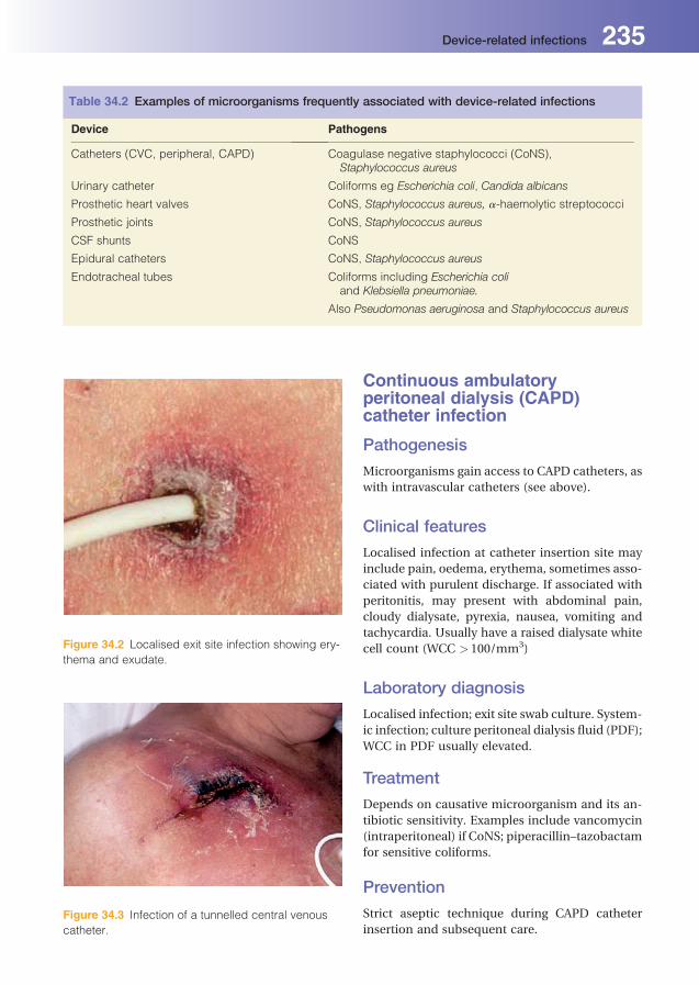
Continuous ambulatoryperitoneal dialysis (CAPD)catheter infection
Pathogenesis
Microorganisms gain access to CAPD catheters, as
with intravascular catheters (see above).
Clinical features
Localised infection at catheter insertion site may
include pain, oedema, erythema, sometimes asso-
ciated with purulent discharge. If associated with
peritonitis, may present with abdominal pain,
cloudy dialysate, pyrexia, nausea, vomiting and
tachycardia. Usually have a raised dialysate white
cell count (WCC > 100/mm3)
Laboratory diagnosis
Localised infection; exit site swab culture. System-
ic infection; culture peritoneal dialysis fluid (PDF);
WCC in PDF usually elevated.
Treatment
Depends on causative microorganism and its an-
tibiotic sensitivity. Examples include vancomycin
(intraperitoneal) if CoNS; piperacillin–tazobactam
for sensitive coliforms.
Prevention
Strict aseptic technique during CAPD catheter
insertion and subsequent care.
Table 34.2 Examples of microorganisms frequently associated with device-related infections
Device Pathogens
Catheters (CVC, peripheral, CAPD) Coagulase negative staphylococci (CoNS),Staphylococcus aureus
Urinary catheter Coliforms eg Escherichia coli, Candida albicans
Prosthetic heart valves CoNS, Staphylococcus aureus, a-haemolytic streptococci
Prosthetic joints CoNS, Staphylococcus aureus
CSF shunts CoNS
Epidural catheters CoNS, Staphylococcus aureus
Endotracheal tubes Coliforms including Escherichia coliand Klebsiella pneumoniae.
Also Pseudomonas aeruginosa and Staphylococcus aureus
Figure 34.3 Infection of a tunnelled central venous
catheter.
Figure 34.2 Localised exit site infection showing ery-
thema and exudate.
Device-related infections 235
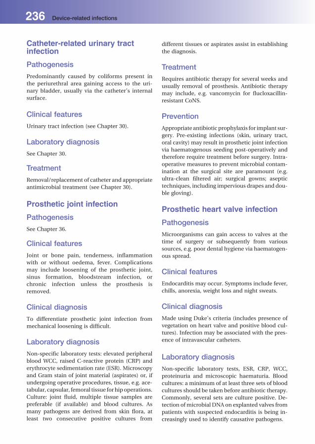
Catheter-related urinary tractinfection
Pathogenesis
Predominantly caused by coliforms present in
the periurethral area gaining access to the uri-
nary bladder, usually via the catheter’s internal
surface.
Clinical features
Urinary tract infection (see Chapter 30).
Laboratory diagnosis
See Chapter 30.
Treatment
Removal/replacement of catheter and appropriate
antimicrobial treatment (see Chapter 30).
Prosthetic joint infection
Pathogenesis
See Chapter 36.
Clinical features
Joint or bone pain, tenderness, inflammation
with or without oedema, fever. Complications
may include loosening of the prosthetic joint,
sinus formation, bloodstream infection, or
chronic infection unless the prosthesis is
removed.
Clinical diagnosis
To differentiate prosthetic joint infection from
mechanical loosening is difficult.
Laboratory diagnosis
Non-specific laboratory tests: elevated peripheral
blood WCC, raised C-reactive protein (CRP) and
erythrocyte sedimentation rate (ESR). Microscopy
and Gram stain of joint material (aspirates) or, if
undergoing operative procedures, tissue, e.g. ace-
tabular, capsular, femoral tissue for hipoperations.
Culture: joint fluid, multiple tissue samples are
preferable (if available) and blood cultures. As
many pathogens are derived from skin flora, at
least two consecutive positive cultures from
different tissues or aspirates assist in establishing
the diagnosis.
Treatment
Requires antibiotic therapy for several weeks and
usually removal of prosthesis. Antibiotic therapy
may include, e.g. vancomycin for flucloxacillin-
resistant CoNS.
Prevention
Appropriate antibiotic prophylaxis for implant sur-
gery. Pre-existing infections (skin, urinary tract,
oral cavity) may result in prosthetic joint infection
via haematogenous seeding post-operatively and
therefore require treatment before surgery. Intra-
operative measures to prevent microbial contam-
ination at the surgical site are paramount (e.g.
ultra-clean filtered air; surgical gowns; aseptic
techniques, including impervious drapes and dou-
ble gloving).
Prosthetic heart valve infection
Pathogenesis
Microorganisms can gain access to valves at the
time of surgery or subsequently from various
sources, e.g. poor dental hygiene via haematogen-
ous spread.
Clinical features
Endocarditis may occur. Symptoms include fever,
chills, anorexia, weight loss and night sweats.
Clinical diagnosis
Made using Duke’s criteria (includes presence of
vegetation on heart valve and positive blood cul-
tures). Infection may be associated with the pres-
ence of intravascular catheters.
Laboratory diagnosis
Non-specific laboratory tests, ESR, CRP, WCC,
proteinuria and microscopic haematuria. Blood
cultures: a minimum of at least three sets of blood
cultures should be taken before antibiotic therapy.
Commonly, several sets are culture positive. De-
tection ofmicrobial DNA on explanted valves from
patients with suspected endocarditis is being in-
creasingly used to identify causative pathogens.
236 Device-related infections
Cardiological investigation
Echocardiogram.
Treatment
Depends on causative pathogen, usually CoNS.
Prevention
Strict intra-operative measures (as for prosthetic
joint infection); prophylaxis for specificprocedures.
Epidural catheter-associatedinfection
Pathogenesis
Associated with epidural injections.
Immunosuppression (diabetes, HIV, alcoholism) is
a predisposing factor for infection. Epidural infec-
tion may cause extensive damage to the associated
structures. Spinal epidural abscesses may occur.
Clinical features
Early presentation may be subtle and clinical di-
agnosis difficult to establish. Symptoms include
fever, localised back pain, neurological deficit.
Laboratory diagnosis
Non-specific tests (CRP, ESR, WCC) and blood
cultures.
Treatment
Commonly associated with CoNS that require in-
travenous vancomycin therapy if resistant to
flucloxacillin.
Prevention
Good aseptic technique on insertion. Antibiotic
treatment to eradicate existing infections, which
may lead to haematogenous seeding of the
device.
CSF shunt infection
Pathogenesis
See Chapter 32.
Clinical features
CSF shunts are commonly inserted via the ventri-
culoperitoneal (VP) route or rarely through the
ventriculoatrial (VA) route. Average incidence of
infection is approximately 10%.
VP infectionmay present within a fewmonths of
operation. Symptoms include vomiting, headache
and visual disturbance, sometimes accompanied
by abdominal distension.
VA shunt infections often do not appear for
months or years after insertion. Early cases are
characterised by fever, tachycardia and rigors.
Delayed cases may present with intermittent/
rare fever, rash arthralgia, anaemia and muscle
aches.
Laboratory diagnosis
Blood culture; cerebrospinal fluid (CSF) for mi-
croscopy and culture. Non-specific laboratory
tests, including CRP and ESR, may provide sup-
porting evidence of infection.
Treatment
Shunt removal and treatment with antimicrobials
according to causative pathogen.
Prevention
Aseptic techniques on insertion and antimicrobial
prophylaxis; antimicrobial shunts are also being
used in some centres.
Device-related infections 237

35Cardiovascular infectionsRichard WatkinHeart of England NHS Foundation Trust, Sutton Coldfield, West Midlands, UK
Infective endocarditis
Definition
Infection of the endocardium, usually involving
the heart valves.
Epidemiology
The incidence in the UK is estimated at between
1.0 and 7.0 per 100,000 population per year. In
hospital, mortality is 20%. Up to 50% of patients
require valvular surgery. Risk factors for develop-
ing infective endocarditis include congenital and
acquired structural cardiac defects and intracardi-
ac prostheses.
Pathogenesis
The characteristicmanifestation of infective endo-
carditis is the vegetation. A vegetation forms when
bacteria from the blood colonise microthrombi
present on damaged endothelium on the valve
surface. Endothelial damage occurs due to abnor-
mal intracardiac jets and thepresenceof prosthetic
material. The mitral and aortic valves are
most commonly involved. Right-sided infective
endocarditis occurs in up to 10% of cases and is
more common in intravenous drug users.
Clinical features
Often symptoms of infective endocarditis are non-
specific. They include fever, night sweats, malaise
anddyspnoea. Theclinical coursemaybe indolent,
or ‘subacute’, but often is rapidly progressive with
considerable valvular destruction and regurgita-
tion. Clinical signs may include a heart murmur,
signs of left ventricular dysfunction, splenomegaly,
splinter haemorrhages, digital clubbing, Osler’s
nodes (painful nodular lesions in the pulps of the
fingers), Janeway lesions (painless petechiae on
the palms and soles) and Roth’s spots (retinal
haemorrhages). Pyrexia often will not settle for at
least 72 hours after treatment initiation. Compli-
cations of infective endocarditis include embolic
phenomena,which can be fatal when involving the
cerebrovascular system. Glomerulonephritis is
common and causes microscopic haematuria.
Aetiology
Infective endocarditis is commonly caused by
Gram-positive microorganisms. Staphylococci are
the commonest causative microorganisms (both
Staphylococcus aureus and coagulase negative
Medical Microbiology and Infection Lecture Notes, Fifth Edition. Edited by Tom Elliott, Anna Casey,Peter Lambert and Jonathan Sandoe.� 2011 Blackwell Publishing Ltd. Published 2011 by Blackwell Publishing Ltd.

staphylococci), followed by ‘viridans’ streptococci
and Enterococcus spp. Culture negative infective
endocarditis occurs in up to 20% of cases when
there is failure to identify a causative microorgan-
ism by blood culture. This is commonly caused by
prior antibiotic administration orwhen infection is
due to fastidious microorganisms.
Laboratory diagnosis. Non-specific tests. Inflammatory markers are al-
most always elevated, including C reactive pro-
tein (CRP) and erythrocyte sedimentation rate
(ESR), neutrophilia is common.. Cardiac tests. Transthoracic and transoesopha-
geal echocardiography visualise vegetations and
evaluate valvular integrity.. Microbiological tests. Blood cultures are ex-
tremely important. As the number of bacteria
circulating in the blood is small, it is important
to inoculate the maximum permitted amount of
blood into each culture bottle to optimise the
yield. Three sets of blood cultures should be
obtained within the first 24 hours. Serological
assays need to be used to diagnose infective
endocarditis caused by Coxiella burnettii, Bru-
cella spp. and Bartonella spp. Histology and
molecular analysis (e.g. broad range polymerase
chain reaction) of explanted heart valve tissue
may also aid in the identification of microorgan-
isms. The diagnosis of infective endocarditis is
established using the Duke criteria, which is
based mainly on culture results and presence of
a vegetation.
Management
Synergistic bactericidal antibiotics are needed for
prolonged periods of time (4–6 weeks). Clinical
response to therapy should be monitored through
measurement of CRP. Surgical debridement
and valvular surgery may be required when re-
sponse to treatment is poor or valvular destruction
is extensive.
Prevention
Previously antibiotics havebeen recommended for
all those considered to be at risk of infective en-
docarditis prior to bacteraemia producing proce-
dures and operations. Currently, most guidelines
recommend antibiotic prophylaxis for only those
patients at the very highest risk (previous endo-
carditis, prosthetic heart valves, and complex con-
genital heart disease), although limited evidence
exists supporting ongoing antibiotic prophylaxis
for any at risk group.
Pacemaker and devicerelated infection
Definition
Infection of pacing leads, device or within the
device ‘pocket’. Vegetations may be visible on
echocardiography, adherent to the intracardiac
portion of pacing leads.
Epidemiology
Intracardiac device infection occurs in 1–2% of
implanted devices per year. It is associated with
significant morbidity and mortality.
Pathogenesis
It is likely that device related infections are caused
by bacteria being introduced during implantation.
Adherence of blood-borne bacteria to devices in
situ is less likely.
Clinical features
Clinical features vary from non-specific signs such
as fever, malaise and night sweats to those asso-
ciated with infective endocarditis.
Aetiology
Device related infections are most commonly
caused by coagulase negative staphylococci and
S. aureus.
Laboratory diagnosis
Frequently, blood cultures are negative as
are wound swabs, usually due to prior antibiotic
use. The CRP is usually elevated, as is the white
cell count.
Cardiovascular infections 239

Management
Antibiotic treatment is as for infective endocardi-
tis. The pacing leads and device usually need
explantation.
Myocarditis
Definition
Infection or inflammation of the heart muscle.
Clinical features
Myocarditis is often preceded by a ‘flu-like’ illness.
Symptoms are often non specific but include
chest pain and fever. Left ventricular dysfunction
will cause breathlessness and orthopnea. Cardiac
arrhythmia can precipitate palpitations and
collapse.
Aetiology
Myocarditis is commonly viral in origin, although
toxicmyocarditis occurs in diphtheria and chronic
myocarditis is a feature of trypanosomiasis and
chlamydial infection. Coxsackie B is the common-
est causative agent, although Epstein-Barr (EBV),
mumps virus, parvovirus B19 and human herpes
virus 6 can also cause myocarditis.
Laboratory diagnosis
A viral aetiology can only be proved if virus is
detected in altered myocardium through endocar-
dial biopsy or at necropsy. This usually requires
detection of viral nucleic acid by molecular meth-
ods. Echocardiography may demonstrate left ven-
tricular dysfunction. Cardiac enzymes and tropo-
nins may be elevated.
Management
This is usually supportive with heart failure
treatments. Cardiac transplantation is occasional-
ly necessary. Prognosis is usually good in the
long term.
Pericarditis
Definition
Infection or inflammation of the pericardium.
Clinical features
Sharp localised chest pain, which may be altered
by posture. A pericardial friction rub may present.
Signs of right-sided heart failure (raised JVP, pe-
ripheral oedema and ascites) may indicate signif-
icant pericardial effusion. The ECG shows global
‘saddle’-shaped ST elevation. Secondary myocar-
ditis may cause left ventricular dysfunction. Chest
X-ray may demonstrate an enlarged globular
heart.
Aetiology
Acute pericarditis has many causes, although
frequently none is found. Infective causes are
predominantly viral (EBV, enteroviruses, CMV),
bacterial (localised spread from primary lung
infection, haematogenous spread from a distant
primary infection, tuberculosis) or fungal (histo-
plasma, aspergillus, Candida spp.). Fungal
pericarditis occurs predominantly in immuno-
compromised individuals.
Laboratory diagnosis. Viral: nucleic acid detection or isolation from
pharyngeal washings, faeces or pericardial fluid
(see Chapter 16);. Bacterial: Culture of pericardial fluid and blood;. Echocardiography may show a bright pericardi-
um and the presence of pericardial fluid.
Management
Acute treatment is with analgesia with non-
steroidal anti-inflammatory drugs. Clinical or
echocardiographic features of cardiac tamponade
require emergency pericardiocentesis. Antifungal
and antibacterial drugs should be given, depen-
ding on the causative microorganism.
240 Cardiovascular infections

36Bone and joint infectionsJonathan SandoeLeeds Teaching Hospitals NHS Trust and University of Leeds, Leeds, UK
Osteomyelitis
Definition
Infection of bone is termed osteomyelitis.
Aetiology
Staphylococcus aureus is the most common cause
of osteomyelitis, but other microorganisms can be
important in certain patients groups or situations.
The microbial aetiology of osteomyelitis is shown
in Table 36.1.
Pathogenesis and epidemiology
Osteomyelitis can occur in awide variety of patient
groups and any bone can be affected, but a rela-
tively small number of pathogens are involved.
Some patient groups are susceptible to infections
in particular bones (Table 36.2).
Bone infection can result from:
. haematogenous spread (following transient bac-
teraemia or from a source of infection elsewhere,
e.g. endocarditis);. direct spread from infected tissue (‘contiguous
focus’ osteomyelitis, e.g. tarsal osteomyelitis in a
diabetic patient with foot ulcers (Figure 36.1));. direct inoculation (e.g. trauma, such as open
fracture or surgery involving bones).
There are no clear definitions of acute or chronic
osteomyelitis. In acute ostemyelitis, symptoms are
usually present for days or a fewweeks, but chronic
osteomyelitis usually refers tocaseswhere infection
has persisted formonths or years in spite of antimi-
crobial therapy(andsometimessurgery);deadbone
is often present. Frequently, this situation results
from inadequately treated acute osteomyelitis.
Clinical features. Clinical findings vary with the age of the patient
and location of infection.. Osteomyelitis can present acutely or develop
slowly over a period of weeks to months, or even
years.. Pain and fever are the most common presenting
features.. If the affected bone is close to the skin there may
be swelling, pain, erythema and tenderness over
the affected area.. In infants and young children, reduced use of an
affected limb is an important sign.. The duration of symptoms often varies with the
virulence or growth characteristics of the caus-
ative microorganism (e.g. symptoms may be
present for many months in patients withMyco-
bacterium tuberculosis osteomyelitis, but just a
few days if Staphylococcus aureus is the cause).. Discharging sinuses from the infected bone to
the skin may occur in chronic Staphylococcus
aureus osteomyelitis.
Medical Microbiology and Infection Lecture Notes, Fifth Edition. Edited by Tom Elliott, Anna Casey,Peter Lambert and Jonathan Sandoe.� 2011 Blackwell Publishing Ltd. Published 2011 by Blackwell Publishing Ltd.
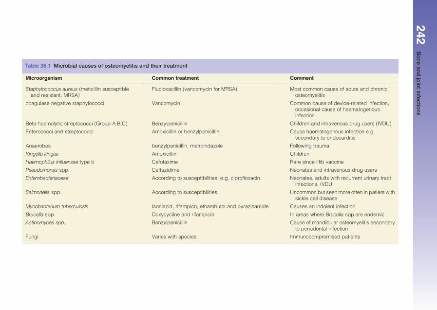
Table 36.1 Microbial causes of osteomyelitis and their treatment
Microorganism Common treatment Comment
Staphylococcus aureus (meticillin susceptibleand resistant, MRSA)
Flucloxacillin (vancomycin for MRSA) Most common cause of acute and chronicosteomyelitis
coagulase negative staphylococci Vancomycin Common cause of device-related infection,occasional cause of haematogenousinfection
Beta-haemolytic streptococci (Group A,B,C) Benzylpenicillin Children and intravenous drug users (IVDU)
Enterococci and streptococci Amoxicillin or benzylpenicillin Cause haematogenous infection e.g.secondary to endocarditis
Anaerobes benzylpenicillin, metronidazole Following trauma
Kingella kingae Amoxicillin Children
Haemophilus influenzae type b Cefotaxime Rare since Hib vaccine
Pseudomonas spp. Ceftazidime Neonates and intravenous drug users
Enterobacteriaceae According to susceptibilities, e.g. ciprofloxacin Neonates, adults with recurrent urinary tractinfections, IVDU
Salmonella spp. According to susceptibilities Uncommon but seen more often in patient withsickle cell disease
Mycobacterium tuberculosis Isoniazid, rifampicn, ethambutol and pyrazinamide Causes an indolent infection
Brucella spp. Doxycycline and rifampicin In areas where Brucella spp.are endemic
Actinomyces spp. Benzylpenicillin Cause of mandibular osteomyelitis secondaryto periodontal infection
Fungi Varies with species. Immunocompromised patients
242Bone
andjoint
infections

Investigations. Peripheral blood white cell count (WCC), eryth-
rocyte sedimentation rate (ESR) and C-reactive
protein (CRP) may be raised, but all these tests
lack sensitivity and specificity.. Blood cultures are positive in approximately
40–60% of cases.. Radiological investigations are important in con-
firming a diagnosis; plain X-rays, computed to-
mography (CT) andmagnetic resonance imaging
(MRI) can all be useful.. Radiological changes are often delayed, even
with MRI, the most sensitive investigation.. Radiologically-guided or operative bone samples
for culture are the diagnostic procedures of
choice.. A Gram’s stain and Ziehl–Neelsen staining (for
mycobacteria) of biopsy samplesmay be helpful.. Routine culture, mycobacterial culture and
fungal culture of operative or biopsy samples is
necessary.
Treatment
Antibiotic treatment is normally for at least 6
weeks. Some commonly used regimens are shown
in Table 36.1. Surgery, to allow drainage of pus and
d�ebridement of dead bone, may be necessary,
particularly in chronic osteomyelitis. Antibiotic
treatment depends on the causative microorgan-
ism, highlighting the need for appropriate micro-
biological investigation.
Table 36.2 Examples of bones affected by osteomyelitis in different patient groups
Patient group/scenario Bone(s) affected
Diabetic patient with foot ulcers (Figure 36.1) Tarsals, metatarsals and digits
Neonates and children Long bones
Post sternotomy for cardiac surgery (Figure 36.2) Sternum
Patients with endocarditis Vertebral bodies (and intervertebral discs)
Figure 36.1 ‘Contiguous focus’ osteomyelitis: tarsal
osteomyelitis in a diabetic patient with foot ulcers.Figure 36.2 Sternal osteomyelitis secondary to cardiac
surgery.
Bone and joint infections 243
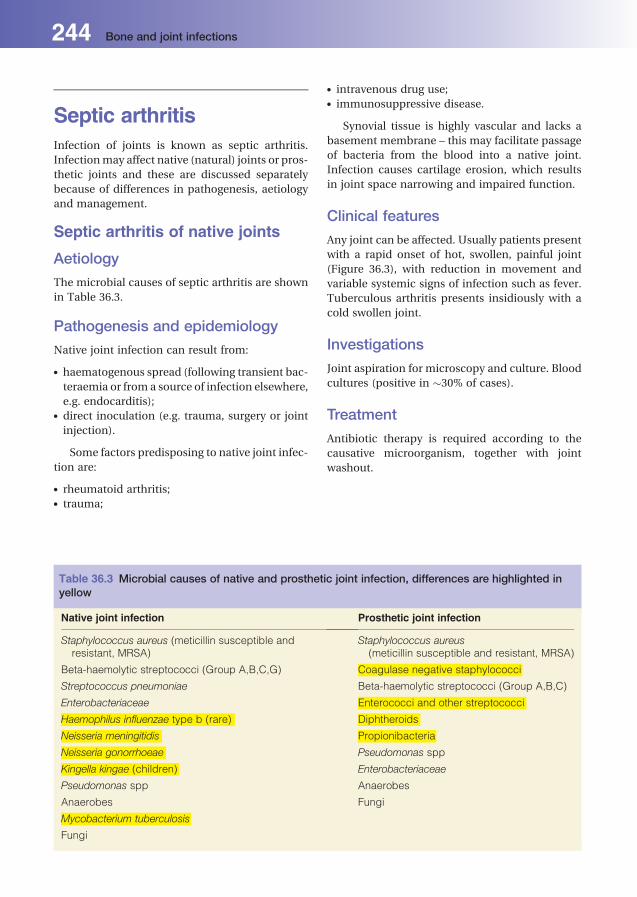
Septic arthritisInfection of joints is known as septic arthritis.
Infectionmay affect native (natural) joints or pros-
thetic joints and these are discussed separately
because of differences in pathogenesis, aetiology
and management.
Septic arthritis of native joints
Aetiology
The microbial causes of septic arthritis are shown
in Table 36.3.
Pathogenesis and epidemiology
Native joint infection can result from:
. haematogenous spread (following transient bac-
teraemia or from a source of infection elsewhere,
e.g. endocarditis);. direct inoculation (e.g. trauma, surgery or joint
injection).
Some factors predisposing to native joint infec-
tion are:
. rheumatoid arthritis;
. trauma;
. intravenous drug use;
. immunosuppressive disease.
Synovial tissue is highly vascular and lacks a
basement membrane – this may facilitate passage
of bacteria from the blood into a native joint.
Infection causes cartilage erosion, which results
in joint space narrowing and impaired function.
Clinical features
Any joint can be affected. Usually patients present
with a rapid onset of hot, swollen, painful joint
(Figure 36.3), with reduction in movement and
variable systemic signs of infection such as fever.
Tuberculous arthritis presents insidiously with a
cold swollen joint.
Investigations
Joint aspiration for microscopy and culture. Blood
cultures (positive in �30% of cases).
Treatment
Antibiotic therapy is required according to the
causative microorganism, together with joint
washout.
Table 36.3 Microbial causes of native and prosthetic joint infection, differences are highlighted inyellow
Native joint infection Prosthetic joint infection
Staphylococcus aureus (meticillin susceptible andresistant, MRSA)
Staphylococcus aureus(meticillin susceptible and resistant, MRSA)
Beta-haemolytic streptococci (Group A,B,C,G) Coagulase negative staphylococci
Streptococcus pneumoniae Beta-haemolytic streptococci (Group A,B,C)
Enterobacteriaceae Enterococci and other streptococci
Haemophilus influenzae type b (rare) Diphtheroids
Neisseria meningitidis Propionibacteria
Neisseria gonorrhoeae Pseudomonas spp
Kingella kingae (children) Enterobacteriaceae
Pseudomonas spp Anaerobes
Anaerobes Fungi
Mycobacterium tuberculosis
Fungi
244 Bone and joint infections

Prosthetic joint infection
Aetiology
The causes of prosthetic joint infection are shown
in Table 36.3.
Pathogenesis
Prosthetic joint infection can result from:
. haematogenous spread (following transient
bacteraemia or from a source of infection else-
where, e.g. endocarditis);. microbial contamination of the prosthesis at
implantation;. surgicalwound infection following implantation.
Any type of prosthesis can become infected.
Microorganisms capable of attaching to man-
made materials and forming an adherent film of
bacteria (called biofilm) are most important.
Clinical features
Can present acutely with a hot, swollen, painful
joint with reduction in movement and variable
systemic signs of infection such as fever, but more
commonly presents insidiously with pain and loss
of function.
Investigations. Joint aspiration for microscopy and culture and
multiple tissue samples at the time of definitive
surgery for culture. These help to distinguish
between skin contaminants and causative
microorganisms.. Blood cultures are positive in less than 50% of
cases.. Plain X-rays may show loosening of the
prosthesis.
Treatment
Prosthetic joint infection usually requires prosthe-
sis removal. A new prosthesis may be implanted at
the time of removal (single-stage procedure), but
higher success rates can be achieved by removing
the prosthesis followed by a period of antimicro-
bial therapy (usually 6 weeks) and then implanta-
tion of a new prosthesis at a later date (two-stage
procedure). Antibiotic therapy can be adminis-
tered systemically or locally in cement spacers or
beads.
Reactive arthritisThis is immunologically mediated, resulting from
infection at a distant site, e.g. Yersinia enterocoli-
tica gastroenteritis or non-specific urethritis
caused by Chlamydia trachomatis. Usually, more
than one joint is affected.
Viral arthritisA number of virusesmay cause arthritis as part of a
generalised infection, e.g. hepatitis B virus, HIV,
rubella virus, mumps virus and parvovirus.
Figure 36.3 Septic arthritis of the knee.
Bone and joint infections 245

37Skin and soft-tissueinfectionsSupriya Narasimhan1 and Rabih Darouiche21Drexel University, Pittsburgh, USA2Baylor College of Medicine, Houston, Texas, USA
Normal flora of skinThe normal skin is colonised mainly by Gram-
positive microorganisms (especially coagulase-
negative staphylococci and diphtheroids), some
Gram-negatives and yeasts, which can survive the
relatively harsh conditions – dryness, acidity and
the presence of fatty acids, sebum and salt. The
normal skin flora protects against infection by
other microorganisms that must compete to sur-
vive in this ecological niche.
Pathogenesis of skinand soft-tissue infections
. Superficial skin infections affect the epidermis
and dermis, e.g. impetigo and erysipelas.. Deeper infections affect the subcutaneous tissue,
e.g. cellulitis (Figures 37.1 and 37.2) and fasciitis.. Infections of skin structures cause carbuncles
and folliculitis (Figure 37.3).
. Infections may arise from pathogens that gain
entry through damaged skin (streptococcal
cellulitis) or via haematogenous spread (menin-
gococcal skin rash).. Bacterial toxins produced by infections at other
sites may also result in skin changes (group A
streptococci causing scarlet fever).
Skin and soft-tissueinfections (SSTI)ClassificationThere aremany classifications.One such scheme is
as follows:
. Uncomplicated SSTI: have a low risk for life- or
limb-threatening infection and many can be
treated with empiric oral antibiotics, with occa-
sional surgical drainage (folliculitis, furuncles,
carbuncles, acne, impetigo, erysipelas and cel-
lulitis). Severe infections require IV antibiotics.. Complicated SSTI: have a high risk for life- or
limb-threatening infection, andoftennecessitate
Medical Microbiology and Infection Lecture Notes, Fifth Edition. Edited by Tom Elliott, Anna Casey,Peter Lambert and Jonathan Sandoe.� 2011 Blackwell Publishing Ltd. Published 2011 by Blackwell Publishing Ltd.
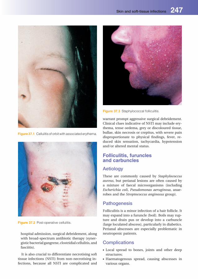
hospital admission, surgical debridement, along
with broad-spectrum antibiotic therapy (syner-
gisticbacterial gangrene, clostridial cellulitis, and
fasciitis).
It is also crucial to differentiate necrotising soft
tissue infections (NSTI) from non-necrotising in-
fections, because all NSTI are complicated and
warrant prompt aggressive surgical debridement.
Clinical clues indicative of NSTI may include ery-
thema, tense oedema, grey or discoloured tissue,
bullae, skin necrosis or crepitus, with severe pain
disproportionate to physical findings, fever, re-
duced skin sensation, tachycardia, hypotension
and/or altered mental status.
Folliculitis, furunclesand carbuncles
Aetiology
These are commonly caused by Staphylococcus
aureus, but perianal lesions are often caused by
a mixture of faecal microorganisms (including
Escherichia coli, Pseudomonas aeruginosa, anae-
robes and the Streptococcus anginosus group).
Pathogenesis
Folliculitis is a minor infection of a hair follicle. It
may expand into a furuncle (boil). Boils may rup-
ture and drain pus or develop into a carbuncle
(large loculated abscess), particularly in diabetics.
Perianal abscesses are especially problematic in
neutropenic patients.
Complications. Local spread to bones, joints and other deep
structures;. Haematogenous spread, causing abscesses in
various organs.
Figure 37.1 Cellulitis of orbit with associatederythema.
Figure 37.2 Post-operative cellulitis.
Figure 37.3 Staphylococcal folliculitis.
Skin and soft-tissue infections 247
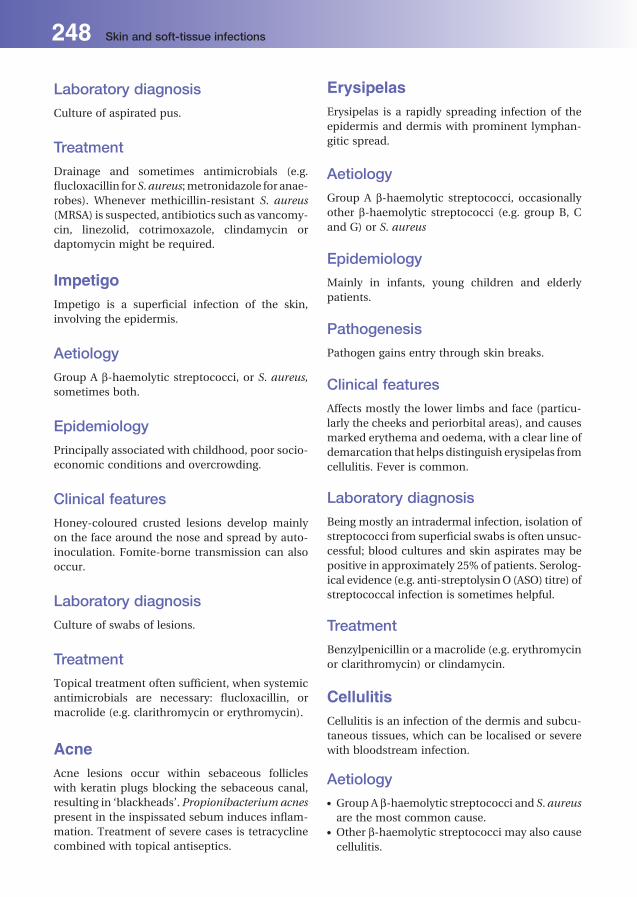
Laboratory diagnosis
Culture of aspirated pus.
Treatment
Drainage and sometimes antimicrobials (e.g.
flucloxacillin for S.aureus;metronidazole for anae-
robes). Whenever methicillin-resistant S. aureus
(MRSA) is suspected, antibiotics such as vancomy-
cin, linezolid, cotrimoxazole, clindamycin or
daptomycin might be required.
ImpetigoImpetigo is a superficial infection of the skin,
involving the epidermis.
Aetiology
Group A b-haemolytic streptococci, or S. aureus,
sometimes both.
Epidemiology
Principally associated with childhood, poor socio-
economic conditions and overcrowding.
Clinical features
Honey-coloured crusted lesions develop mainly
on the face around the nose and spread by auto-
inoculation. Fomite-borne transmission can also
occur.
Laboratory diagnosis
Culture of swabs of lesions.
Treatment
Topical treatment often sufficient, when systemic
antimicrobials are necessary: flucloxacillin, or
macrolide (e.g. clarithromycin or erythromycin).
AcneAcne lesions occur within sebaceous follicles
with keratin plugs blocking the sebaceous canal,
resulting in ‘blackheads’. Propionibacterium acnes
present in the inspissated sebum induces inflam-
mation. Treatment of severe cases is tetracycline
combined with topical antiseptics.
ErysipelasErysipelas is a rapidly spreading infection of the
epidermis and dermis with prominent lymphan-
gitic spread.
Aetiology
Group A b-haemolytic streptococci, occasionally
other b-haemolytic streptococci (e.g. group B, C
and G) or S. aureus
Epidemiology
Mainly in infants, young children and elderly
patients.
Pathogenesis
Pathogen gains entry through skin breaks.
Clinical features
Affects mostly the lower limbs and face (particu-
larly the cheeks and periorbital areas), and causes
marked erythema and oedema, with a clear line of
demarcation that helps distinguish erysipelas from
cellulitis. Fever is common.
Laboratory diagnosis
Being mostly an intradermal infection, isolation of
streptococci from superficial swabs is often unsuc-
cessful; blood cultures and skin aspirates may be
positive in approximately 25% of patients. Serolog-
ical evidence (e.g. anti-streptolysinO (ASO) titre) of
streptococcal infection is sometimes helpful.
Treatment
Benzylpenicillin or a macrolide (e.g. erythromycin
or clarithromycin) or clindamycin.
CellulitisCellulitis is an infection of the dermis and subcu-
taneous tissues, which can be localised or severe
with bloodstream infection.
Aetiology. Group A b-haemolytic streptococci and S. aureus
are the most common cause.. Other b-haemolytic streptococci may also cause
cellulitis.
248 Skin and soft-tissue infections

. S. aureus may produce cellulitis associated with
wounds (Figure 37.2) or abscess formation.. Haemophilus influenzae type b commonly
caused cellulitis in children aged less than5 years
prior to the introduction of Hib vaccination.. Facial and orbital cellulitis may follow sinusitis
and involve respiratory pathogens (e.g. Strepto-
coccus pneumoniae). Erysipelothrix rhusiopathiae, an unusual cause
of cellulitis, seen in meat and fish handlers.. Non-cholera vibrios (e.g. Vibrio vulnificus) are a
rare cause of cellulitis complicatingwounds con-
taminated with seawater, particularly in patients
with underlyingmedical problems (e.g. cirrhosis,
diabetes).. Anaerobic cellulitis can complicate bites (oral
anaerobes) or devitalised tissue (clostridia) in
presence of trauma, diabetesmellitus or vascular
insufficiency and in intravenous drug users
(Figure 37.4).
Pathogenesis
Bacteria gain entry through inapparent skin
breaks to incite infection. Lymphatic obstruction
(e.g. after lymph node resection for breast cancer)
is a predisposing factor in some cases.
Clinical features
Erythematous, hot, swollen skin with a poorly
defined edge (contrast with erysipelas), along with
fever and regional lymphadenopathy. Anaerobic
cellulitis may be foul smelling, with skin crepitus
and necrosis.
Laboratory diagnosis
Cultures of skin swab/aspirate (positive in�25%of
cases) and blood (occasionally positive). Skin
swabs are of limited value, particularly when the
skin is unbroken.
Treatment. High-dose flucloxacillin (active against strepto-
cocci andstaphylococci) is appropriate empirical
therapy,unless risk factors forunusualpathogens
(e.g. bite or MRSA colonisation);. Penicillin for streptococci/Erysipelothrix species;. Flucloxacillin/vancomycin for staphylococci.
Clindamycin, an alternative in penicillin-allergic
patients, is especially useful in decreasing toxin
production;. Anaerobes can be treated with metronidazole
and d�ebridement of devitalised tissue;. H. influenzae type b is treated with cefotaxime.
Synergistic bacterial gangreneThis is an infection of superficial fasciae and sub-
cutaneous fat, but not muscles. It may involve the
abdominal wall or male genitalia (Fournier’s
gangrene).
Aetiology/pathogenesis
Frequently, mixed infection involving S. aureus,
microaerophilic or anaerobic streptococci and,
occasionally, Gram-negative anaerobes (e.g.
Bacteroides spp.). Predisposing conditions include
local trauma, abdominal and genital surgery, and
diabetes mellitus.
Clinical features
Rapidly spreading cellulitis with black necrotic
areas, pain, fever and systemic toxicity.
Laboratory diagnosis
Culture of affected tissue or pus.
Treatment
Radical excision of necrotic tissue plus antibiotic
therapy with flucloxacillin (S. aureus), penicillin
(streptococci) and/or metronidazole (anaerobes).
Broad-spectrum Gram-negative coverage (genta-
micin or ciprofloxacin) is needed, especially in
diabetics.Figure 37.4 Gas gangrene of the leg.
Skin and soft-tissue infections 249

Necrotising fasciitisLife-threatening infection of skin, subcutaneous
tissue, deeper fascia and potentially muscles.
Aetiology. Type I: caused by mixtures of non-group A aer-
obic streptococci, aerobic Gram-negative bacilli
and anaerobes. Often complicates deep surgical
and traumatic wounds with compromised vas-
cular supply;. Type II: monomicrobial, classically caused by
Group A b-haemolytic streptococci. Rarer causes
include Vibrio vulnificus, Aeromonas hydrophila
and S. aureus.
Pathogenesis
Bacteria produce enzymes and toxins that cause
local tissue destruction and systemic toxicity.
Diabetes mellitus confers high risk.
Clinical features
Rapidly spreading cellulitis with necrosis, fever,
systemic toxicity, hypotension and shock; high
mortality.
Laboratory diagnosis
Cultures of tissue, pus or blood.
Treatment
Urgent extensive debridement of affected tissues
plus broad-spectrumantibiotics, including combi-
nations of high-dose benzylpenicillin, gentamicin
(or ciprofloxacin) and metronidazole or carbape-
nems; clindamycin is an alternative to penicillin in
allergic patients.
Clostridial cellulitisand clostridial myonecrosis(gas gangrene)
Epidemiology
Now rare in developed countries. Peripheral vas-
cular disease and diabetes mellitus are important
risk factors.
Pathogenesis
Contamination of traumatic or surgical wounds by
clostridia, especially C. perfringens, results in gas
gangrene or myonecrosis. Clostridia produce en-
zymes and exotoxins that facilitate rapid spread
through tissue plains and result in haemolysis and
septic shock.
Clinical features
Cellulitis with necrotic areas, bullae with foul-
smelling drainage and gangrene (Figure 37.4).
Tissue crepitus due to gas production, fever and
toxaemia evolve as myositis develops.
Laboratory diagnosis
Isolation of clostridia from cultures of tissue/
swabs or blood. However, isolation of clostridia
from wounds does not necessarily indicate gas
gangrene, because clostridia can contaminate
woundswithout infection. Gas gangrene is primar-
ily a clinical diagnosis.
Treatment
Early intervention when suspected is vital. Exten-
sive surgical d�ebridement of devitalised tissue and
penicillin and/or metronidazole.
Prevention
Woundswith devitalised tissue should be d�ebrided
thoroughly and carefully monitored during heal-
ing. Prophylactic antibiotics (e.g. penicillin)
should be prescribed for patients undergoing am-
putations for peripheral vascular disease.
Venous ulcers/pressure soresVarious conditions (diabetes mellitus, peripheral
vascular disease, venous insufficiency, and contin-
uous pressure) can reduce cutaneous blood supply
and predispose to formation of cutaneous ulcers.
Ulcers commonly develop around the foot and
ankle (e.g. diabetic foot ulcers and venous ulcers,
respectively) or at pressure areas in bedridden
patients (‘bedsores’, e.g. over the sacrum or heal).
These invariably become colonised with bacteria
and may become infected.
Typical culture results include:
. Mixed growth of coliforms and anaerobes: nor-
mally not clinically relevant;. Pseudomonas aeruginosa: may reflect antibiot-
ic-induced replacement flora and occasionally
requires antibiotic therapy;
250 Skin and soft-tissue infections
. Staphylococcus aureus: may represent colonisa-
tion or infection;. b-haemolytic streptococci groups C and G: may
colonise and occasionally infect venous ulcers;. b-haemolytic streptococci group A: frequently
cause infection.
Distinguishing colonisation from infection is
important to avoid antibiotic treatment of colo-
nised ulcers; spreading cellulitis around the ulcer
suggests infection. Deep infection including
osteomyelitismayoccur.When systemic antibiotic
therapy is needed, broad-spectrum aerobic/
anaerobic coverage should be initiated, with
subsequent culture-based modification.
BurnsLike venous ulcers, burns may become colonised
with bacteria, including S. aureus, Pseudomonas
spp. and coliforms. The need for antibiotic treat-
ment is dictated by clinical condition (viability of
skin grafts) and microorganism(s) cultured (group
A b-haemolytic streptococci always require treat-
ment). Colonisingmicroorganismsmay eventually
cause bacteraemia.
Wound infections
Aetiology. Accidental wounds: S. aureus, b-haemolytic
streptococci, clostridia;. Bites: oral commensal bacteria, including anae-
robes, Eikenella corrodens and streptococci;
Pasteurella multocida and Capnocytophaga spp.
(animal bites);. Surgical wounds: (see below).
Laboratory diagnosis
Culture of pus or wound swab.
Treatment
Prompt administration of prophylactic antimicro-
bials is recommended for human and some animal
bites (e.g. amoxicillin-clavulanate), because mi-
croorganisms may be inoculated into deeper tis-
sues and cause rapid spread of infection. Ultimate
antibiotic choice depends on culture results.
Surgical debridement may be necessary.
Surgical wound infections
Predisposing factors. Underlying conditions: e.g. diabetes mellitus
andperipheral vascular disease, resulting inpoor
blood supply;. Type of operation: surgery involving contami-
nated areas (e.g. gastrointestinal tract) or in-
fected tissue (e.g. perforated appendicitis) is
more likely to result in post-operative wound
infections;. Surgical technique: e.g. poor technique in
wound closure may result in haematoma or de-
vitalised tissue, predisposing towound infection;. Postoperative factors: cross-infection fromother
infected cases.
Aetiology. Upper body: S. aureus; group A b-haemolytic
streptococci;. Lower body (particularly abdominal surgery):
coliforms, occasionally mixed with anaerobes;
P. aeruginosa.
Clinical features
Fever; wound exudate; cellulitis; may progress to
dehiscence and/or bloodstream infection.
Laboratory diagnosis
Cultures of wound swab, pus or blood.
Treatment
Antibiotic treatment depends on causative
microorganisms. Surgical debridement may be
required.
Prevention. Preoperative preparation: treatment of infection
before operation when possible; decontamina-
tion of operative area (e.g. bowel preparation)
and use of prophylactic antibiotics for clean-
contaminated, contaminatedordirty operations;. Good surgical techniques and aseptic theatre
practices;. Measures topreventpostoperativecontamination
of wounds (e.g. ‘aseptic non-touch technique’).
Skin and soft-tissue infections 251

Toxin-mediated skinconditionsScarlet feverCaused by a strain of group A Streptococcus that
causes throat infection and produces an erythro-
genic toxin, which causes erythematous, blanch-
ing, ‘sand-paper’ rash with a red (‘strawberry’)
tongue and circumoral pallor. As the rash fades,
the skin desquamates. Treatment is penicillin or
erythromycin.
Staphylococcal scalded skinsyndrome (SSSS)Causes the most severe, systemic manifestation of
infection at a distant site with S. aureus strains
producing an exfoliative toxin, resulting in wide-
spread bullae and skin exfoliation. This occurs in
small infants (Ritter’s disease) and occasionally in
older children and adults (toxic epidermal necro-
lysis or Lyell’s disease). Diagnosis is clinical and is
supported by isolating a toxin-producing strain of
S. aureus. Treatment is with flucloxacillin, or other
anti-staphylococcal antibiotics.
Toxic shock syndromeToxic shock syndrome is caused by S. aureus
strains that produce toxic shock syndrome
toxin-1 (TSST-1) or staphylococcal enterotoxins,
which act as super-antigens causing hypotension,
desquamative skin lesions and dysfunction of
three or more organ systems.
Panton-Valentine leukocidinproducing StaphylococcusaureusThese isolates are a particularly virulent form of
S.aureus, which are associatedwith tissuedamage.
Dermatophyte fungal infectionsWorldwide distribution. Transmission by direct
contact or via fomites (e.g. towels, hairbrushes).
Infection may be acquired from animals. See
Table 37.1 for aetiology and clinical features.
Laboratory diagnosis
Microscopy and culture of appropriate specimens
(skin scrapings, nail clippings, hair roots).
Table 37.1 Common dermatophyte (ringworm) infections
Infection Site Dermatophyte Clinical features
Tinea corporis Trunk Epidermophyton floccosum Pruritus; circular erythematous lesion, scaling,clearing from centre
Limbs Trichophyton spp.
Microsporum spp.
Tinea cruris Groin Epidermophyton floccosum Pruritus; lesions similar to tinea corporis
Trichophyton rubrum
Tinea pedis Feet Epidermophyton floccosum Pruritus; cracking and scaling between toes
Trichophyton spp.
Tinea unguium Nails Trichophyton spp. Thick, yellowish nails with surroundingerythema
Tinea capitis Hair Trichophyton tonsurans Erythematous patches on scalp with scaling;hairs break, leaving bald patches
Microsporum audounii
Microsporum canis
252 Skin and soft-tissue infections

Table 37.2 Common viral skin rashes
Disease Virus Rash distribution Associated symptomsand signs
Diagnosis Treatment
Maculopapular rashes
Measles Measles Start from hairlinedownwards all overthe body
Fever, coryza,conjunctivitis, Koplik’sspots
Serology (IgM), antigendetection inrespiratoryspecimens, culture
Rubella Rubella Start from hairlinedownwards all overthe body
Fever, lymphadenopathy(suboccipital, periauri-cular and cervical,arthritis in adults)
Serology (IgM), culture
Erythemainfectiosum(fifth disease)
Parvovirus B19 Starts on cheeks(slapped cheek),then diffuses alace-like rash onbody
Fever, arthritis in adults Serology (IgM),polymerase chainreaction (PCR)
Exanthem subitum(roseolainfantum)
Human herpesvirus6 (HHV6)
Face, trunk 2–3 days of feverdisappearing withappearance of rash
Serology, PCR Generally asymptomaticbut ganciclovir orfoscarnet incomplicated cases
Enterovirusinfections
SeveralCoxsackievirusesand echoviruses
Scattered all overthe body
Fever, myalgia, headache,cervicallymphadenopathy
Culture
Primary HIVinfection
HIV Scattered all overthe body
Glandular fever-like illness,meningitis, encephalitis
Serology, PCR HAART
Dengue fever Dengue virus Scattered all over body Fever, myalgia, headache PCR, Serology (IgM),culture
(continued )
Skin
andso
ft-tissueinfectio
ns253

Table 37.2 (Continued )
Disease Virus Rash distribution Associated symptomsand signs
Diagnosis Treatment
Vesicular rashes
Chickenpox Varicella-zostervirus (VZV)
Centripetal distribution Fever, itching Electron microscopy,antigen detection invesicular fluid,serology (IgM), PCR
Generally symptomaticbut aciclovir,valaciclovir orfamciclovir incomplicated cases
Shingles(Figure 37.5)
VZV Dermatomal but couldbe disseminated
Post-herpetic neuralgia PCR antigen detectionin vesicular fluid,serology (IgM), PCR
Aciclovir, valacicloviror famciclovir
Disseminatedherpes simplex
Herpes simplexvirus (HSV)
Wide distribution Neonatal herpes, highlyimmunosuppressedpatients
Electron microscopy,antigen detection invesicular fluid, PCR
Aciclovir, foscarnet
Smallpox Smallpox Centrifugal distribution Fever, headache Electron microscopy,PCR
Hand foot andmouth disease
Enteroviruses(CoxsackievirusA16, A4, A5,A9, B2, B5)
Hands and feet Mouth ulcers, fever Culture
254Skin
andso
ft-tissueinfectio
ns

Treatment
Topical azole antifungals. Oral terbinafine can be
used in case of nail involvement or severe infec-
tions that failed to respond to topical azoles.
Treatment may be required for several weeks to
months.
Cutaneous candidiasisSkin infection usually caused by the yeast Can-
dida albicans. This affects moist skin areas
(groin, axilla, skin folds, nailfolds) or damaged
skin (e.g. irritant dermatitis in nappy area of
babies). It causes erythema, maceration, and
‘satellite’ vesicles and pustules. Treatment is
with topical or oral azoles (e.g. miconazole,
fluconazole).
Pityriasis versicolorMild skin infection caused by the dimorphic fun-
gus, Malassezia furfur. Found worldwide, most
commonly in humid tropics. Dark, scaling lesions,
particularly in the upper body. In dark-skinned
persons, the affected areas become depigmented.
Treated with topical or oral azoles.
Viral skin rashesExamples of common viral skin rashes are outlined
in Table 37.2.
Infections of the eyeEyelidEyelash follicle infections (styes) are usually
caused by S. aureus.
Orbital cellulitisCellulitis of periorbital tissues are caused mainly
by b-haemolytic streptococci, Streptococcus pneu-
moniae, S. aureus and, rarely, H. influenzae type b
in children (<5 years) (Figure 37.1)
Conjunctivitis
. Viral conjunctivitis: aetiology includes adeno-
viruses and measles virus. Laboratory diagnosis
by nucleic acid detection techniques. No specific
treatment;. Neonatal conjunctivitis: (ophthalmia neonator-
um), caused by Neisseria gonorrhoeae/Chlamyd-
ia trachomatis infection acquired frommother’s
birth canal;. Bacterial causes: include S. aureus,H. influenzae
and pneumococci. It is most common in young
infants. Treatment with topical antibiotics is
normally effective.
Herpes virus infections of the eye
. Herpes simplex virus: conjunctivitis and painful
corneal (‘dendritic’) ulcers that may cause cor-
neal scarring. Latency with recurrent infection
may occur. Treated with aciclovir ointment;. Varicella-zoster virus: ophthalmic zoster. The
virus is latent in ophthalmic ganglia and reacti-
vates to affect the periorbital area (unilateral).
Treated with systemic aciclovir;. Cytomegalovirus: choroidoretinitis. Associated
with AIDS. Treated with ganciclovir.
TrachomaChlamydia trachomatis eye infection (conjuncti-
vitis and corneal lesions) is prevalent in developing
countries in the tropics and is the leading infec-
tious cause of blindness worldwide. Spreads by
direct hand-eye contact. Treated with tetracycline.
Mass treatment campaigns helped eradicate tra-
choma in some endemic areas.Figure 37.5 Shingles on face.
Skin and soft-tissue infections 255
Other eye infections
. Pseudomonas aeruginosa: may cause serious
deep eye infections after trauma or surgery.
Treatment includes parenteral antibiotics.. Toxoplasma gondii: causes choroidoretinitis,
normally as part of congenital toxoplasmosis.. Toxocara canis: causes choroidoretinitis.
. Onchocerciasis: is a nematode infection that
causes river blindness. Microfilariae invade the
anterior chamber of the eye. Second leading
infectious cause of blindness. Endemic areas
include West Africa and Central America.. Acanthamoeba sp.: results in an amoebic infec-
tion causing keratitis. Usually associated with
contact lens use.
256 Skin and soft-tissue infections
38Infections in thecompromised hostTom ElliottUniversity Hospitals Birmingham NHS Foundation Trust, Birmingham, UK
The immune system consists of innate barriers,
including skin and mucous membranes, comple-
ment and phagocytic cell mechanisms and adap-
tive barriers (cellular and humoral). When any of
theseprotectivemechanisms is reduced, thehost is
considered to be immunocompromised. Such pa-
tients aremore liable to have severe infections and
can become infected with either a conventional
pathogen (which can also cause disease in a non-
compromised individual) or opportunistic patho-
gens (microorganisms that are usually unable to
cause a disease in a healthy person). Various types
of defect in the immune defence mechanisms can
predispose to infections with certain pathogens.
Infections associated withdefects in innate immunesystems (Table 38.1)Infections associated withmedical devices
. Intravascular and peritoneal dialysis catheters.
these catheters breach the skin barrier, allowing
commensal microorganisms access to the
deeper sites. These infections are commonly
associatedwith coagulase-negative staphylococ-
ci, Staphylococcus aureus and, less frequently,
coliforms and Candida spp.. Prosthetic valves and joints. coagulase-negative
staphylococci are the most common pathogens,
gaining access, usually during surgery or from a
distant infection via haematogenous spread. The
microorganism can attach to the prosthesis in
biofilms, acting as a nidus of infection.
Infections associated with burnsBurns can destroy extensive areas of the mech-
anical barriers of the body and also result in
abnormalities in localised neutrophil function and
antibody response. The burn exudates produce
nutrition for microorganisms, allowing colonisa-
tion, which may result in infection. The usual
pathogens include Pseudomonas aeruginosa,
S. aureus and Streptococcus pyogenes.
Infections associated withdeficiencies in normal clearancemechanisms
. Urinary catheters allow microorganisms access
to the bladder, overcoming defences, including
Medical Microbiology and Infection Lecture Notes, Fifth Edition. Edited by Tom Elliott, Anna Casey,Peter Lambert and Jonathan Sandoe.� 2011 Blackwell Publishing Ltd. Published 2011 by Blackwell Publishing Ltd.
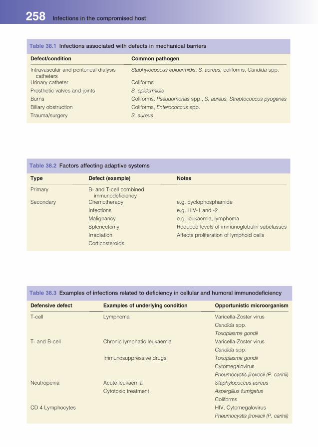
Table 38.1 Infections associated with defects in mechanical barriers
Defect/condition Common pathogen
Intravascular and peritoneal dialysiscatheters
Staphylococcus epidermidis, S. aureus, coliforms, Candida spp.
Urinary catheter Coliforms
Prosthetic valves and joints S. epidermidis
Burns Coliforms, Pseudomonas spp., S. aureus, Streptococcus pyogenes
Biliary obstruction Coliforms, Enterococcus spp.
Trauma/surgery S. aureus
Table 38.2 Factors affecting adaptive systems
Type Defect (example) Notes
Primary B- and T-cell combinedimmunodeficiency
Secondary Chemotherapy e.g. cyclophosphamide
Infections e.g. HIV-1 and -2
Malignancy e.g. leukaemia, lymphoma
Splenectomy Reduced levels of immunoglobulin subclasses
Irradiation Affects proliferation of lymphoid cells
Corticosteroids
Table 38.3 Examples of infections related to deficiency in cellular and humoral immunodeficiency
Defensive defect Examples of underlying condition Opportunistic microorganism
T-cell Lymphoma Varicella-Zoster virus
Candida spp.
Toxoplasma gondii
T- and B-cell Chronic lymphatic leukaemia Varicella-Zoster virus
Candida spp.
Immunosuppressive drugs Toxoplasma gondii
Cytomegalovirus
Pneumocystis jirovecii (P. carinii)
Neutropenia Acute leukaemia Staphylococcus aureus
Cytotoxic treatment Aspergillus fumigatus
Coliforms
CD 4 Lymphocytes HIV, Cytomegalovirus
Pneumocystis jirovecii (P. carinii)
258 Infections in the compromised host

urinewashout. Bacteria cangrowup the inside or
outside surface of a urinary catheter. The infec-
tions are commonly associated with Gram-neg-
ative aerobic bacilli from the patient’s own faecal
or periurethral flora.. Stones in the kidney, common bile and salivary
ducts can result in infections proximal to the
obstruction.. Respiratory tract: there may be damage to the
cilia, e.g. with cystic fibrosis patients, which
predisposes to pneumonia withmicroorganisms
such as S. aureus, Haemophilus influenzae and
P. aeruginosa. An endotracheal tube, required
during mechanical ventilation, bypasses many
upper respiratory defences and, when colonised,
can deliver microorganisms directly into the
trachea.
Infections associated withdefects in complementand phagocytic activityThese defects are normally congenital. Defects in
phagocytic function may affect chemotaxis,
phagocytosis or bacterial killing; associated infec-
tions often involve pyogenic bacteria, e.g. S. aure-
us. Defects in the complement system are associ-
ated with bacterial infections, e.g. patients with
defects in the later complement components
(C7–C9) have an increased risk of meningococcal
infections.
Infections related to cellularand humoral immunodeficiencyThe type of underlying immunodeficiency
(Table 38.2)may determine the nature and severity
of any subsequent infection (Table 38.3). Increas-
ingly, these infections are iatrogenic and caused
by opportunistic pathogens. The length of time
that a patient is immunosuppressedmay influence
the type of associated infection.
Infections in some immunocompromised pa-
tients can be multifactorial and associated with
several factors, e.g. in recipients of solid organ
transplants, these include the type of organ trans-
plant, the level of immune suppression required to
prevent rejection of a grafted organ and the patho-
gens to which a recipient is exposed, including
from the donor organ and the environment. Some
infections associated with organ transplant reci-
pients are shown in Table 38.4.
Generally, conventional pathogens cause infec-
tions in pre-transplant patients. However, when
these patients are immunosuppressed as part of
their treatment, infections with opportunistic mi-
croorganisms, including Aspergillus species start
to occur.
Table 38.4 Examples of opportunistic pathogens associated with solid organ transplant recipients
Bacteria Fungi Protozoa Parasites Viruses
Staphylococcus aureus Candida spp. Toxoplasma gondii Strongyloidesstercoralis
Herpes simplexviruses
Coagulase-negativestaphylococci
Aspergillus spp. Cytomegalovirus
Mycobacteriumavium-intracellulare
Pneumocystis jirovecii Varicella-Zoster
Coliforms Epstein–Barr
Infections in the compromised host 259
39Infections causedby antimicrobial-resistantbacteriaDavid LivermoreHealth Protection Agency, Microbiology Services, Colindale, London, UK
IntroductionAntimicrobials exert a Darwinian selection in fa-
vour of those bacteria that have inherent resistance
or which have acquired resistance as a result of
random genetic events. Inherent resistance is
where all members of a species or group are resis-
tant, e.g. all Gram-negative bacteria are resistant to
vancomycin because it is too large to cross their
outer membrane. Acquired resistance emerges in
hitherto susceptible species via mutations (ran-
dom errors occurring in DNA sequence as it is
copied), or by gene transfer.
. Gene transfer is most often via plasmids, i.e.
loops of DNA separate from the chromosome.
Some plasmids are ‘promiscuous’, meaning that
they can spread among numerous species.. Some resistant strains or ‘clones’ are biologically
‘fit’ and can spread among patients, hospitals,
and even between countries.
The processes of mutation, DNA transfer and
strain selection have led to major increases in the
prevalence of resistance in many bacterial patho-
gens. This chapter details the currently-critical
resistance problems. Preventing a return to the
‘pre-antibiotic era’ when simple infections were
frequent killers andmuchmodernmedicinewould
have been impossible requires:
. judicious use of antimicrobials in humans and
animals;. preventionof the spreadof resistantmicroorgan-
isms among patients; and. reinvigoration of discovery of new antimicrobials.
Meticillin-resistantstaphylococcus aureusMeticillin-resistant Staphylococcus aureus (MRSA)
has the highest profile of any resistant bacterium.
Its resistance depends on the mecA gene, which
codes a supplementary cell-wall synthesizing en-
zyme, PBP-2’,which enablesMRSA to ‘by-pass’ the
cell-wall synthetic steps normally targeted by
Medical Microbiology and Infection Lecture Notes, Fifth Edition. Edited by Tom Elliott, Anna Casey,Peter Lambert and Jonathan Sandoe.� 2011 Blackwell Publishing Ltd. Published 2011 by Blackwell Publishing Ltd.

meticillin. Most MRSA belong to a few widely-
disseminated clones. Transfer of mecA among
strains is rare but does occur.
Epidemiology. MRSA were first described in the 1960s, soon
after meticillin entered clinical use as an answer
to penicillin-resistant S. aureus. The early MRSA
strains did not spread, presumably because they
werebiologically unfit. FitterMRSAstrains began
to proliferate in hospitals during the 1980s, but
rarely caused severe infections. These were fol-
lowed, in the early 1990s, by the current UK
‘epidemic’ clones, EMRSA-15 and EMRSA-16,
which are strongly associated with severe infec-
tion, including bloodstream infections. At their
peak in 2003, MRSA (95% EMRSA-15 or -16
strains) accounted for 40% of S. aureus bacter-
aemias and 8% of all bacteraemias. Subsequent-
ly, their prevalence has been approximately
halved, largely by infection prevention practices.. In Scandinavia, MRSA rates have been consis-
tently low owing to rigorous infection control
and contact tracing; rates in many other parts of
the world remain high and stable.. Although EMRSA-15 and -16 mostly cause hos-
pital infections, they also colonise individuals
in the community, e.g. care home residents.
Carriers are more vulnerable to infection during
subsequent hospitalisation.. True ‘community’ MRSA strains have proliferat-
ed recently in theUS, less so elsewhere. Theymay
be transmitted via vigorous physical contact, e.g.
during sports activities. They can cause aggres-
sive skin and soft tissue infections, rarely pneu-
monias. Most produce a toxin called Panton–
Valentine leukocidin, but this is not exclusive to
community MRSA and is more often seen in
meticillin-susceptible S. aureus in the UK. It is
not new, having been known since the 1930s.
Detection. MRSA are detected by conventional antibiotic
susceptibility tests, usually with oxacillin, as me-
ticillin itself is no longer available. The media
used must allow expression of mecA, induced
by salt and low temperature. Detection is also
possible by polymerase chain reaction (PCR) for
mecA.. S. aureus isolates, which are oxacillin resistant, or
mecA-positive, should be assumed to be resistant
to all penicillin, cephalosporin or related drugs
presently available (June 2011). New anti-MRSA
cephalosporins (ceftobiprole and ceftaroline)
are, however, anticipated; these bind and inhibit
PBP-2’.
Control of MRSA. Hand hygiene is crucial; its improvement pre-
vents up to 50% of hospital-acquired MRSA in-
fections. It decreases the chance of staphylococci
being carried from patient to patient via the
healthcare workers.. Environmental cleanliness and equipment de-
contamination reduces the likelihood of patients
acquiring MRSA directly, and the hands of
healthcare workers becoming contaminated.. Screening of hospital admissions allows carriers
to be identified; attempts can then be made to
decolonise them, e.g. with mupirocin in the
nose and chlorhexidine on the skin. This may
reduce shedding of microorganisms into the
environment.. Patient isolation or cohorting reinforces the need
for hand hygiene and segregates infected or co-
lonised patients from others.. Prophylactic antibiotics can be used to prevent
infection in surgery.. The Department of Health (in England) uses
MRSA bacteraemia rates as a measure of infec-
tion control performance, publishing tables of
these rates for each hospital trust.
Treatment. Glycopeptides (vancomycin and teicoplanin) are
the mainstay against serious MRSA infections.
Vancomycin resistance in MRSA is extremely
rare, with no reports from the UK. The few re-
corded cases were caused by the acquisition of
plasmid-borne vanA genes fromenterococci (see
below); a few further MRSA isolates have inter-
mediate resistance contingent on a thickened
cell wall.. Newer alternatives for severe MRSA infections
include daptomycin, linezolid and tigecycline.
Depending on the clone, susceptibility may also
be retained to various older agents, including
trimethoprim, co-trimoxazole, tetracyclines,
fusidic acid and rifampicin; these may be used
alone or in combination in less severe infections,
or as oral follow-on to i.v. vancomycin, linezolid,
tigecycline or daptomycin.
Infections caused by antimicrobial-resistant bacteria 261

Vancomycin-resistantenterococci (VRE)
Epidemiology. Vancomycin-resistant enterococci (VRE, also
called glycopeptide-resistant enterococci, GRE)
emerged in the late 1980s. They are classified as
VanA (commonest), VanB or VanC phenotypes.. In the UK, VRE are mostly confined to specialist
renal, haematology and oncology units; aminor-
ity of tertiary hospitals account for most cases.
VRE are more widespread in US hospitals.. Most vancomycin resistant Enterococcus faecium
belong to clonal complexCC17, ahuman-adapted
lineage that can cause hospital outbreaks.
Mechanism of resistanceand detection. VanA and VanB are plasmid- or transposon-
mediated systems that have spread among
Enterococcus faecium and, less so, E. faecalis;
VanC is intrinsic to E. casseliflavus and E.
gallinarum – rarer species.. Resistance reflects the use of alternative cell wall
precursors, thereby depriving glycopeptides of
their normal target site of action.. Enterococci with VanA are resistant to vancomy-
cin and teicoplanin, as both induce VanA expres-
sion. VanB and VanC strains are resistant to
vancomycin, but not teicoplanin, which does not
act as an inducer.
Control. Hand hygiene, equipment and environmental
decontamination, as for MRSA (see above).
Treatment. Virtually all E. faecalis remain susceptible to
ampicillin, linezolid and tigecycline; E. faecium
is generally resistant to ampicillin, but suscepti-
ble to linezolid, quinupristin/dalfopristin and
tigecycline. Nitrofurantoin is often active but
appropriate only for lower urinary infections.. A problem arises in endocarditis, where syner-
gistic bactericidal activity between a b-lactam or
glycopeptide and an aminoglycoside is sought,
but where around 40% of enterococci now have
streptomycin and gentamicin resistance, pre-
cluding this synergy. High dose ampicillin or
daptomycin may be alternatives.
Streptococcuspneumoniae. S. pneumoniae is the commonest and most se-
rious cause of community-acquired pneumonia
(see Chapter 26). Historically, it was very suscep-
tible to penicillin and other antibiotics but resis-
tance gradually emerged via mutation and gene
modification, followed by clonal spread of resis-
tant strains belonging to serotypes 6B, 9V, 14, 19F
and 23F.. Introduction of a conjugate vaccine targeting
these serotypes (and 2 others) is reducing their
role in invasive disease, and thereby also the
prevalence of resistance, both in the children,
who are vaccinated, and in the elderly, who
typically acquire infection from children.
Resistance to penicillin is emerging in other
‘non-vaccine’ serotypes, e.g. 19A, particularly in
the USA, but these are covered by a second-
generation vaccine.
Gram-negative bacilliEpidemiology. Escherichia coli is the commonest causative
agent of urinary tract infections and is part of
the mixed aerobic/anaerobic flora that may
cause intra-abdominal infections arising from
leakage of gut contents.. Other Enterobacteriaceae, including Klebsiella
and Enterobacter spp., also non-fermenters, e.g.
Pseudomonas aeruginosa and Acinetobacter
baumannii, are important as opportunistic hos-
pital pathogens.P. aeruginosa also colonises the
lungs of cystic fibrosis patients, giving chronic
infections that accumulate resistance by
mutation.. ResistantKlebsiella and A. baumanniimay cause
hospital outbreaks, with transmission via the
hands of staff or via contaminated equipment.
By contrast, E. coli infections usually are endog-
enous, with bacteria from the gut transferred
to the urinary tract or the abdominal cavity.
262 Infections caused by antimicrobial-resistant bacteria
Mechanisms of resistance. Resistance to older penicillins, e.g. ampicillin, is
inherent and associated with chromosomally-
mediated b-lactamases in Klebsiella spp., Enter-
obacter spp., Pseudomonas aeruginosa and
A. baumannii. In E. coli, it is largely caused by
the TEM-1 b-lactamases. First recorded in 1963,
this plasmid-mediated enzyme has now spread
to 50–60%of clinical isolates of E. coli, both in the
community and in hospitals.. Modern ‘third-generation’ cephalosporins, such
as cefotaxime and ceftazidime were designed to
be stable to TEM-1 and other b-lactamases but
have, in turn, selected for extended-spectrum
b-lactamases (ESBLs), which hydrolyse and inac-
tivate them. Early ESBLs were mutants of TEM-1
and a similar enzyme, SHV-1, and were mostly
found in hospital Klebsiella. A new ESBL family,
CTX-M, has recently become dominant, spread-
ingalso incommunityE. coli. CTX-M-E. colioften
infect the urinary tract of elderly patients.. Resistance to fluoroquinolones has also in-
creasedmarkedly in Enterobacteriaceae, includ-
ing E. coli, and many ESBL producers are multi-
resistant to these drugs also to aminoglycosides.
This accumulation of multi-resistance is leading
to increased need to use carbapenems, which
previously were reserved antibiotics.. Carbapenem use, in turn, is selecting for
carbapenemases (i.e. carbapenem-destroying
b-lactamases), including theKPC andNDMtypes
in Enterobacter and Klebsiella, also OXA carbape-
nemases in Acinetobacter spp. Carbapenem re-
sistance in P. aeruginosa is mostly contingent on
impermeability and efflux, not b-lactamase
Control. Standard infection prevention can limit hospital
spread of multi-resistant Klebsiella spp., A. bau-
mannii and P. aeruginosa clones. Key factors
include decontamination of hands and equip-
ment, cohorting or isolation of patients and the
use of gloves and gowns. The utility of these
measures is less clear for multi-resistant E. coli,
where the source of infection is usually the pa-
tient’s own gut microflora.
Treatment. Most quinolone-resistant and ESBL-producing
E. coli, Klebsiella spp. and Enterobacter spp.
remain susceptible to the carbapenems, imipe-
nem, meropenem, doripenem and ertapenem,
which are the drugs of choice in severe infec-
tions. Alternatives in less severe disease varywith
the site of infection and the individual isolate;
they often include tigecycline and (for lower
urinary tract infections) fosfomycin or
nitrofurantoin.. Polymyxin is often the sole option for severe
infections caused by Enterobacteriaceae or
non-fermenters with carbapenemases.
Neisseria gonorrhoeae. A common sexually-transmitted pathogen,
N. gonorrhoeae historically was very susceptible
and in the 1940s gonorrhoea was treatable with
low doses of penicillin. Susceptibility later de-
clined, largely through target modification and
penicillin doses of 3 g were needed by the 1970s.
Penicillin-untreatable strains then emerged, and
were found to have acquired the same TEM-1
b-lactamase that had proliferated in E. coli (see
above).. By the 1990s, ciprofloxacin had replaced peni-
cillin as the standard treatment but, again, resis-
tance emerged and is now seen in 20–30% of US
andUK isolates. These developments have led to
cephalosporins, principally cefixime and ceftri-
axone, becoming the new standard therapies,
but gonococci are showing progressive reduc-
tions in susceptibility, as occurredwith penicillin
between the 1940s and 1970s.. Resistance to penicillin and ciprofloxacin can
also arise in N. meningitidis, but is much rarer
than in N. gonorrhoeae.
The British Society for Antimicrobial Chemo-
therapy resistance surveillance (http://www.bsac-
surv.org) provides extensive information from
antibiotic resistance surveillance programmes for
respiratory infections and bacteraemias in the
UK and Ireland. The European Antimicrobial
Resistance Surveillance Network (http://www
.ecdc.europa.eu/en/activities/surveillance/EARS-
Net/Pages/index.aspx) is aEuropean-widenetwork
of national surveillance systems, providing refer-
ence data on antimicrobial resistance for public
health purposes.
Infections caused by antimicrobial-resistant bacteria 263

40Perinatal and congenitalinfectionsJames GrayBirmingham Children’s Hospital NHS Foundation Trust, Birmingham, UK
DefinitionsCongenital infections are infections that are ac-
quired in utero. They may be acquired through
transplacental spread or by ascent ofmicroorgan-
isms from the maternal genital tract. The mani-
festations of congenital infection are protean.
Perinatal infections are acquired around the time
of delivery (usually from the maternal genital
tract). Bacterial infections that are perinatally
acquired usually present within the first 48–72
hours of life. After that, an increasing proportion
of neonatal bacterial infections are acquired from
other sources, including family members, health-
care workers, other patients and the hospital
environment. Viral infections usually have a lon-
ger incubation period, and may present much
later. Some agents are only important as causes
of either congenital or perinatal infections; other
infections can be acquired during either period
(Table 40.1).
Congenital infectionsGeneral principles of diagnosis
Presentation of congenital infection. Occasionally maternal illness may be present,
but often the mother is asymptomatic or has
sub-clinical illness.. Abnormalities may be detected on routine foetal
scanning.. Infections may result in pregnancy loss.. Infections may present at birth, or with sequelae
that develop only months or years after birth.. Many infections are asymptomatic.
Microbiological investigations. Diagnosis of maternal infection during pregna-
ncy, principally by serological tests, is the usual
starting point. However, this does not confirm
that infection has been transmitted to the baby.
Medical Microbiology and Infection Lecture Notes, Fifth Edition. Edited by Tom Elliott, Anna Casey,Peter Lambert and Jonathan Sandoe.� 2011 Blackwell Publishing Ltd. Published 2011 by Blackwell Publishing Ltd.
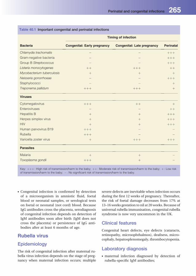
. Congenital infection is confirmed by detection
of a microorganism in amniotic fluid, foetal
blood or neonatal samples, or serological tests
on foetal or neonatal (not cord) blood. Because
IgG antibodies cross the placenta, serodiagnosis
of congenital infection depends on detection of
IgM antibodies soon after birth (IgM does not
cross the placenta) or persistence of IgG anti-
bodies after at least 6 months of age.
Rubella virus
Epidemiology
The risk of congenital infection after maternal ru-
bella virus infection depends on the stage of preg-
nancy when maternal infection occurs; multiple
severe defects are inevitable when infection occurs
during the first 12 weeks of pregnancy. Thereafter,
the risk of foetal damage decreases from 17% at
13–16weeks gestation to nil at 20weeks. Because of
universal rubella immunisation, congenital rubella
syndrome is now very uncommon in the UK.
Clinical features
Congenital heart defects, eye defects (cataracts,
retinopathy, microophthalmos), deafness, micro-
cephaly, hepatosplenomegaly, thrombocytopenia.
Laboratory diagnosis. maternal infection diagnosed by detection of
rubella-specific IgM antibodies;
Table 40.1 Important congenital and perinatal infections
Timing of infection
Bacteria Congenital: Early pregnancy Congenital: Late pregnancy Perinatal
Chlamydia trachomatis � � þþþGram-negative bacteria � � þþþGroup B Streptococcus � � þþþListeria monocytogenes þþ þþþ þþMycobacterium tuberculosis þ þ þNeisseria gonorrhoeae � � þþþStaphylococci � � þTreponema pallidum þþþ þþþ þ
Viruses
Cytomegalovirus þþþ þþ þEnteroviruses � � þþHepatitis B þ þ þþþHerpes simplex virus þ þ þþþHIV þ þ þþþHuman parvovirus B19 þþþ � �Rubella þþþ � �Varicella zoster virus þ þþþ þþþ
Parasites
Malaria þ þ �Toxoplasma gondii þþþ þ �
Key: þþþ : High risk of transmission/harm to the baby; þþ : Moderate risk of transmission/harm to the baby; þ : Low risk
of transmission/harm to the baby; � : No significant risk of transmission/harm to the baby.
Perinatal and congenital infections 265
. prenatal diagnosis of foetal infection not usually
undertaken;. infection in the neonate diagnosed by detection
of virus in urine, blood or respiratory samples, or
bydetection of rubella-specific IgMantibodies in
neonatal blood.
Treatment and prevention. No specific treatment exists for rubella.. Rubella is included in the MMR vaccine that is
part of the routine childhood immunisation
schedule.. Antenatal screening of pregnant women, with
post-partum immunisation of seronegative
cases.
Cytomegalovirus (CMV)
Epidemiology
Congenital CMV infection is the most common
intrauterine infection (maternal infection rate in
the UK is 5–7 per 1,000 pregnancies). Congenital
infection usually follows primary maternal CMV
infection: the risk of transmission to the foetus is
around40%, but 90–95%of infected foetuseswill be
normal at birth. There is also a small but unquan-
tified risk of congenital infection following mater-
nal re-infection or reactivation of latent infection.
Clinical features
Eighty-five percent of congenitally infected babies
are unaffected. Five percent are symptomatic at
birth: symptoms include pneumonitis, hepatos-
plenomegaly, thrombocytopenia, anaemia, low
birth weight, microcephaly, cerebral palsy, deaf-
ness and choroidoretinitis. Five to ten percent are
asymptomatic at birth, but develop later sequelae
(cognitive, motor, visual or hearing defects).
Laboratory diagnosis. Maternal infection is diagnosed by detection of
CMV-specific IgM antibodies.. Foetal infection is diagnosed prenatally by de-
tection of CMV in amniotic fluid (usually by PCR)
or of IgM antibodies in foetal blood.. Infection in the neonate is diagnosed by detec-
tion of CMV-specific IgM or detection of the
virus (by culture or PCR) in throat swabs, blood
or urine. Congenital infection can only be
diagnosed with certainty on samples collected
within the first three weeks of life.
Treatment and prevention. Ganciclovir or valganciclovir should be consid-
eredfortreatmentofcongenitally infectedbabies:
this may alleviate sensorineural hearing loss.. There are no specific preventative measures.
However, babies with congenital CMV infection
in hospital should be isolated while in hospital,
because large amounts of virus are excreted
in urine and respiratory secretions for months
or years.
Toxoplasmosis
Epidemiology
In the UK, maternal toxoplasmosis affects about
0.5%of pregnancies. The risk of transmission to the
foetus increases from 25% in the first trimester to
65% in the third. However the risk of serious effects
decreases from 75–5%.
Clinical features
Stillbirth, choroidoretinitis, microcephaly, convul-
sions, intracerebral calcification with resultant hy-
drocephalus, myocarditis, hepatosplenomegaly
and thrombocytopenia.
Laboratory diagnosis. Maternal infection is diagnosed by detection of
T.gondii-specificIgMantibodies:bewarethat IgM
positivitypersists formanymonthsafter infection.. For prenatal diagnosis of foetal infection and
diagnosis of infection in the neonate, detection
ofT. gondii in amniotic fluidorneonatal bloodby
PCR offers the best sensitivity. Only one-third of
congenitally infected infants are IgM-positive.
Treatment and prevention. Maternal treatment with spiramycin or with
pyrimethamine and sulphadiazine during preg-
nancy, and treatment of the neonate with pyri-
methamine and sulphadiazine, may reduce the
severity of sequelae.. Pregnant women should be advised to avoid
contact with cat faeces during pregnancy, and
given dietary advice. Antenatal screening is per-
formed in some countries, but not in the UK.
266 Perinatal and congenital infections

Varicella-zoster virus (VZV)
Epidemiology
Around90%ofUK-bornadults are immune tochick-
enpox, but the proportion of immune adults who
were brought up in other countries may be lower.
Clinical features
Small risk (<3%) of foetal malformation (congen-
ital varicella syndrome) where maternal chicken-
poxoccurs in thefirst trimester. Theotherperiodof
high risk is where maternal chickenpox occurs
between 7 days before and 7 days after delivery:
the baby is at risk of severe systemic neonatal
infection, because it will not acquire passive im-
munity from its mother.
Diagnosis. Chickenpox is usually diagnosed clinically.. Wherenecessary, thediagnosis canbeconfirmed
bydirect detectionof the virus in clinicalmaterial
by various methods.. Serology has no part in diagnosis of acute chick-
enpox, but is needed to assess the immunity of
persons who have been exposed to chickenpox.
Treatment and prevention. high dose aciclovir for neonatal varicella
infection;. non-immune pregnant women should avoid
contact with chickenpox; contacts should be
treated with varicella zoster immune globulin
(VZIG).
Human parvovirus B19
Epidemiology
Around40%of adults in theUKarenon-immune to
parvovirusB19.Up to15%ofmaternal infections in
the first 20 weeks of pregnancy result in foetal
infection: infections after 20 weeks do not cause
serious morbidity.
Clinical features
Whenmaternal infectionoccurs in thefirst20weeks
of pregnancy, there is a 10% risk of spontaneous
abortion, and a further 3% risk of hydrops fetalis
(anaemia and cardiac failure resulting from the
virus infecting erythrocyte precursors); congenital
malformations have not been reported.
Laboratory diagnosis. maternal infection diagnosed by detection of
specific anti-parvovirus IgM antibodies;. where necessary, foetal infection diagnosed pre-
natally by detection of parvovirus in amniotic
fluid or foetal blood by PCR or by detection of
IgM antibodies in foetal blood;. diagnosis of infection in the neonatal period not
usually relevant.
Treatment and prevention. no specific antiviral therapy;. where parvovirus infection has been diagnosed,
enhanced monitoring of the pregnancy is re-
quired, with intrauterine blood transfusion for
severe foetal anaemia as required;. pregnant women should avoid contact with chil-
dren with parvovirus infection.
Syphilis
Epidemiology
Congenital syphilis is now rare in developed coun-
tries, but remains a serious problem in developing
countries. Where a mother has untreated syphilis,
the risk of congenital infection is 80–90% in thefirst
year after infection, falling to less than 10% four
years after infection.
Clinical features
Stillbirth, hepatosplenomegaly, lymphadenopa-
thy, skin rashes, nasal discharge. Bone, tooth and
cartilage defects and deafness are later sequelae.
Laboratory diagnosis
Congenital syphilis usually diagnosed by detection
of anti-Treponema pallidum IgM antibodies in
neonatal serum. Treponemes may be detectable
inmaterial fromskinormucousmembrane lesions
by darkground microscopy.
Treatment and prevention. Treatment is with high dose penicillin.. Pregnant women are routinely screened for anti-
treponemal antibodies.
Perinatal and congenital infections 267

ListeriosisMaternal infection (asymptomatic or influenza-
like illness) is usually acquired from contami-
nated food (soft cheeses, raw vegetables or sal-
ads). Maternal listeriosis may cause intrauterine
infection, resulting in abortion or stillbirth. In-
fection later in pregnancy or during delivery may
result in neonatal bloodstream infection and/or
meningitis. Treatment is with ampicillin or
amoxicillin plus gentamicin. Prevention is by
avoiding high-risk foods in pregnancy.
Tuberculosis (TB)Congenital TB is rare: may result in pregnancy loss
or symptomatic neonatal TB. Perinatal TB due to
aspiration of infected amniotic fluid is even rarer.
MalariaCongenital or neonatal malaria occurs most fre-
quently when the mother becomes infected for
the first time during pregnancy. Infection may
result in abortion or stillbirth or pre-term deliv-
ery. Neonates may not develop symptoms of
malaria for several weeks after delivery.
Neonatal infectionsGroup B b-haemolyticstreptococci
Epidemiology
GroupB streptococci are a common commensal of
the genital and gastrointestinal tracts of 20–30% of
pregnant women. Around 50% of babies born to
colonised mothers acquire this microorganism
during delivery.
Clinical features
Most colonised babies are unaffected. Around 1%
of babies born to mothers colonised develop seri-
ous systemic infection (bacteraemia, meningitis,
pneumonia) in the first week of life (usually within
the first 48h). Late onset sepsis (1 week–6 months
after delivery) presents more often as meningitis:
the bacterium may come from sources other than
the mother.
Laboratory diagnosis
Cultures of blood, CSF, respiratory secretions, and
any other suspected sites of infection.
Treatment and prevention. Treatment is penicillin with gentamicin.. Intrapartum antibiotic prophylaxis is offered to
women identified as carrying, or with risk factors
for, Group B streptococci (e.g. prematurity, ma-
ternal fever in labour, prolonged rupture of
membranes).
Gram-negative aerobic bacilli
Epidemiology
Escherichia coli is usually acquired from the ma-
ternal intestinal tract during delivery. Invasive in-
fections are associated with certain serotypes,
particularly K1. Other Gram-negative bacilli, such
as Klebsiella spp., Enterobacter spp., Serratia spp.
andPseudomonas aeruginosa, aremainly acquired
in hospital and are usually associated with stay
in neonatal units.
Clinical features
E. coli is an important cause of early-onset neona-
tal meningitis and bloodstream infection. Infec-
tions associated with other Gram-negative aerobic
bacilli include pneumonia, bloodstream infection,
meningitis and urinary tract infections.
Laboratory diagnosis
Cultures of blood, CSF, respiratory secretions,
urine and any other suspected sites of infection.
Treatment and prevention. Empiric antibiotic therapy should be based on
local antibiotic sensitivity patterns: often in-
cludes an aminoglycoside. Definitive treatment
depends on antibiotic sensitivity test results.. No specific preventative measures.
Staphylococci
Epidemiology
Staphylococcus aureus is a very common neona-
tal pathogen; it can also form part of the normal
268 Perinatal and congenital infections
vaginal microbial flora. Infection can be acquired
perinatally from the vagina, but most are
acquired after birth via contaminated hands.
Coagulase-negative staphylococci (CoNS) are
now the commonest isolates from neonatal
blood cultures, and are principally nosocomial
pathogens in neonatal units. Once considered
low virulence pathogens, it is now recognised
that coagulase-negative staphylococcal infec-
tions can be serious.
Clinical features
S. aureus is a common cause of mild infections of
superficial sites such as skin and eyes, but is also an
important cause ofmore serious infections (blood-
stream infection, pneumonia, osteomyelitis).
CoNS cause bloodstream infection, often associ-
ated with intravascular catheters.
Laboratory diagnosis
Cultures of blood, respiratory secretions, skin
swabs and any other suspected sites of infection.
Treatment and prevention. S.aureus is usually treatedwithflucloxacillin. For
meticillin-resistant strains (MRSA) and CoNS,
glycopeptides (vancomycin, teicoplanin) are the
mainstay of treatment.. Screening of mothers and babies is increasingly
being used to control MRSA.
Neisseria gonorrhoeaeand Chlamydia Trachomatis
Epidemiology
These infections are acquired from the maternal
genital tract.
Clinical features. N. gonorrhoeae causes severe conjunctivitis
(ophthalmia neonatorum) with pus and perior-
bital inflammation that usually presents within
48 hours of birth.. C. trachomatis conjunctivitis is less severe and
tends to present later (3–14 days after delivery):
untreated,Chlamydiapneumonitismayoccur as
a late (1–3 months) complication.
Laboratory diagnosis
Gonococcal infection is usually diagnosed by cul-
ture, although accurate molecular techniques are
now available. Nucleic acid amplification techni-
ques are the mainstay of diagnosis of chlamydial
infections. In the case of both infections, mothers
should be referred to a Genitourinary Medicine
clinic for investigation and treatment.
Treatment
Both gonococcal and chlamydia ophthalmia neo-
natorum require systemic therapy: ceftriaxone for
N. gonorrhoeae and erythromycin for chlamydial
infection.
Herpes simplex virus (HSV)
Epidemiology
Classically acquired from thematernal genital tract
during delivery, usuallywhen themother has active
lesions of primary genital herpes; can also be ac-
quired from mucocutaneous lesions from anyone
with active herpes. Rarely transplacental spread,
which almost always leads to loss of the pregnancy.
Clinical features
Features of neonatal herpes simplex infection in-
clude encephalitis or meningoencephalitis, general-
ised infection with hepatitis and mucocutaneous
lesions.
Laboratory diagnosis
Detection of virus in clinical material by PCR.
Treatment and prevention
Treatment of herpes encephalitis with high doses
of aciclovir has reduced themortality rate to under
25%. Preventative measures include Cesarean sec-
tion where primary maternal genital herpes infec-
tion presents in the final 6weeks of pregnancy, and
exclusion of persons with active herpes lesions
from contact with neonates.
Human immunodeficiencyvirus (HIV)
Epidemiology
HIV transmission from infected mothers to their
babies can occur in utero (<1% of cases) during
Perinatal and congenital infections 269

delivery or via breast milk. Without preventative
measures, the risk of vertical transmission is
15–40%: with careful management the risk falls to
almost zero.
Clinical features
Infected neonates are usually asymptomatic at
birth, but the illness can progress rapidly in infan-
cy. Poor weight gain, lymphadenopathy, hepatos-
plenomegaly and recurrent infections are early
signs of infection.
Laboratory diagnosis
Detection of HIV RNA or proviral DNA in neonatal
blood by nucleic acid amplification techniques.
Because early diagnosis is important, infants born
to HIV-positive mothers are normally screened at
birth and at 2, 6, 12 and 26 weeks after birth.
Prevention
Pregnant women are routinely screened for anti-
HIV antibodies. Infected cases are treated with
antiretroviral drugs; Caesarean section if antiviral
therapy fails to suppress maternal viral load; new-
borns are also treated with antiretroviral drugs;
breast-feeding is contraindicated.
Hepatitis B virus (HBV)
Epidemiology
Worldwide, vertical transmission is the most
important route of infection of HBV. Acquisition
rates vary according to hepatitis Be antigen sta-
tus of the mother (70–90% if the mother is
HBeAg-positive and 10–40% if HBeAg-negative).
Transmission can occur in utero, but is usually
perinatal.
Clinical features
Neonatal infection is often sub-clinical, but 90%
of infected infants will become chronic carriers,
with a high risk of subsequent cirrhosis and liver
cancer.
Laboratory diagnosis
Detection of seromarkers of hepatitis B (especially
surface antigen (HBsAg)) in neonatal blood.
Prevention
Pregnant women are routinely screened for
HBsAg. Hepatitis B vaccination for all babies born
to hepatitis B-infected mothers; administration
of specific HBV immunoglobulin in addition to
babies whose mothers are not anti-HBe-positive.
EnterovirusesNeonatal enterovirus infection is a serious disease,
with high mortality rates (>10%). Infections may
be acquired perinatally or postnatally: the signif-
icance of congenital enterovirus infections is con-
troversial. Clinical presentation includes fever,
vomiting, maculopapular rash and signs of men-
ingitis. Later manifestations may include hepatic
necrosis, myocarditis and encephalitis. Diagnosis
is by isolation of viruses or detection of viral
RNA by PCR in throat swabs, nasopharyngeal as-
pirate (NPA), stools or CSF. Treatment is mainly
symptomatic.
Maternal infections andperinatal infectionsPostpartum maternal infections are not usually
an indication to investigate or treat neonates who
are clinically well. However, it is advisable to
observe the neonate closely for the development
of signs of infection during the first few days of
life. The main exception is where the mother has
puerperal sepsis with Streptococcus pyogenes
(group A b-haemolytic Streptococcus): because of
the risk of transmission of life-threatening infec-
tion to the baby, antibiotic prophylaxis or treat-
ment is always indicated.
270 Perinatal and congenital infections

41Human immunodeficiencyvirusKaveh ManaviUniversity Hospitals Birmingham NHS Foundation Trust, Birmingham, UK
DefinitionsHIV is a RNA retrovirus with close genetic links
with simian immunodeficiency viruses (SIV) that
infect different species of primates in Africa. Most
SIV are non-pathogenic in their natural hosts.
HIV viral anatomyEach viral particle contains two short viral RNA
chains and at least three viral enzymes; reverse
transcriptase, integrase and protease. Viral parti-
cles contain an outer layer that is penetrated by
pin-like viral structures. These structures are gly-
coproteins (gp) and are a combination of gp-120
(as the head of the pin) and gp-41 (as the stem of
the pin), which play an important role in HIV
attachment to target cells.
HIV life cycleand associatedantiretroviral agentsThe replication cycle of HIV-1 is outlined in
Figure 41.1. HIV only infects cells that express
CD4 and one type of chemokine receptor
(CCR5 or CXCR4) on their surface. Attachment
of gp-120 to CD4 receptor leads to a structural
re-configuration of gp-120; exposing gp-41 to
the cell membrane. This results in the formation
of a tunnel in the membrane, through which
HIV genome (RNA) and enzymes cross and
enter into the cytoplasm. Agents that prevent
HIV attachment to its host (target) cells are
called entry inhibitors. Some may block gp-41
(e.g. enfuvertide) while others may block che-
mokine receptors (e.g. maraviroc).
Medical Microbiology and Infection Lecture Notes, Fifth Edition. Edited by Tom Elliott, Anna Casey,Peter Lambert and Jonathan Sandoe.� 2011 Blackwell Publishing Ltd. Published 2011 by Blackwell Publishing Ltd.
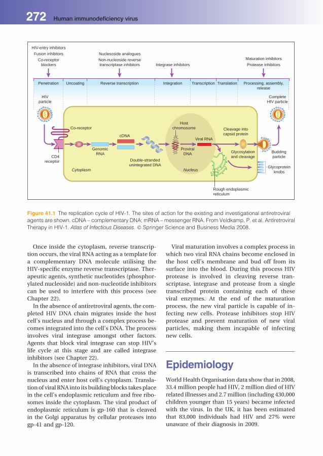
Once inside the cytoplasm, reverse transcrip-
tion occurs, the viral RNA acting as a template for
a complementary DNA molecule utilising the
HIV-specific enzyme reverse transcriptase. Ther-
apeutic agents, synthetic nucleotides (phosphor-
ylated nucleoside) and non-nucleotide inhibitors
can be used to interfere with this process (see
Chapter 22).
In the absence of antiretroviral agents, the com-
pleted HIV DNA chain migrates inside the host
cell’s nucleus and through a complex process be-
comes integrated into the cell’s DNA. The process
involves viral integrase amongst other factors.
Agents that block viral integrase can stop HIV’s
life cycle at this stage and are called integrase
inhibitors (see Chapter 22).
In the absence of integrase inhibitors, viral DNA
is transcribed into chains of RNA that cross the
nucleus and enter host cell’s cytoplasm. Transla-
tion of viral RNA into its building blocks takes place
in the cell’s endoplasmic reticulum and free ribo-
somes inside the cytoplasm. The viral product of
endoplasmic reticulum is gp-160 that is cleaved
in the Golgi apparatus by cellular proteases into
gp-41 and gp-120.
Viral maturation involves a complex process in
which two viral RNA chains become enclosed in
the host cell’s membrane and bud off from its
surface into the blood. During this process HIV
protease is involved in cleaving reverse tran-
scriptase, integrase and protease from a single
transcribed protein containing each of these
viral enzymes. At the end of the maturation
process, the new viral particle is capable of in-
fecting new cells. Protease inhibitors stop HIV
protease and prevent maturation of new viral
particles, making them incapable of infecting
new cells.
EpidemiologyWorld Health Organisation data show that in 2008,
33.4 million people had HIV, 2 million died of HIV
related illnesses and 2.7million (including 430,000
children younger than 15 years) became infected
with the virus. In the UK, it has been estimated
that 83,000 individuals had HIV and 27% were
unaware of their diagnosis in 2009.
HIV-entry inhibitors
Fusion inhibitors
Co-receptorblockers
Penetration Uncoating Reverse transcription
Nuclesoside analogues
Non-nucleoside reversetranscriptase inhibitors Integrase inhibitors
Integration Transcription Translation Processing, assembly,release
Maturation inhibitors
Protease inhibitors
CompleteHIV particle
Buddingparticle
HIVparticle
Co-receptor
CD4receptor
GenomicRNA
Double-strandedunintegrated DNA
Cytoplasm Nucleus
Hostchromosome
Viral RNA
ProviralDNA
Rough endoplasmicreticulum
Glycosylationand cleavage
Cleavage intocapsid protein
Glycoproteinknobs
cDNA
Figure 41.1 The replication cycle of HIV-1. The sites of action for the existing and investigational antiretroviral
agents are shown. cDNA – complementary DNA; mRNA – messenger RNA. From Veldkamp, P. et al. Antiretroviral
Therapy in HIV-1. Atlas of Infectious Diseases. � Springer Science and Business Media 2008.
272 Human immunodeficiency virus

Routes of HIV transmissionHIV is a blood borne virus that can be transmitted
sexually, which is the main route of ongoing epi-
demics. Historically, sexual transmission of HIV
was mostly reported amongst men who have sex
withmen (MSM) in developed nations. Heterosex-
ual transmission of HIV has been themain route of
its transmission amongst developing nations.
In the UK, heterosexual transmission of HIV has
been the main route of transmission since 2000.
This trend has been mostly (but not exclusively)
related to immigrationofpeople fromsub-Saharan
African countries.
HIV can also be transmitted during pregnancy
and delivery from an infected mother to the child.
However, successful antenatal screening pro-
grammes in many countries have significantly
reduced the rate of mother to child transmission.
Because of robust screening programmes and
inactivation processes, transmission of HIV
through blood products in the UK has also been
extremely low since 1993.
Sharing of injection equipment between intra-
venous drug users (IVDU) is another identified
route of HIV transmission.
PathogenesisHIV infected CD4-containing lymphocytes are no
longer able to function, leading to severe disrup-
tion in the immune response to infections, espe-
cially those requiring cellular immunity.
Clinical featuresFigure 41.2 outlinesHIVcopiesandCD4counts ina
human over the course of a treatment-naive HIV
infection. Acute infection with HIV may present
with non-specific symptoms and signs of viral ill-
nesses and therefore may be misdiagnosed. These
include fever, erythematous or maculopapular
rash, pharyngitis, myalgia, lymphadenopathy,
headache and asymptomatic meningitis in rare
cases. The acute phase of HIV infection lasts for
about12weeks,afterwhichpatientsremainasymp-
tomatic for an average of 8 years (latency period of
HIV infection). During this latency period patients
retainastableCD4count,despitehavingahighHIV
1200
1100
1000
900
800
700
600
500
400
300
200
100
0
CD
4+ T
lym
phoc
yte
coun
t (ce
lls/m
m3 )
0 3 6 9 12 1 2 3 4 5 6 7 8 9 10 11
107
106
105
104
103
102
Weeks Years
Primaryinfection
Acute HIV syndromeWide dissemination of virusSeeding of lymphoid organs Clinical latency
Constitutionalproblems
Opportunisticdiseases
Death
HIV
RN
A c
opie
s pe
r m
L pl
asm
a
Figure 41.2 HIV copies and CD4 counts in a human over the course of a treatment-naive HIV infection.
Human immunodeficiency virus 273
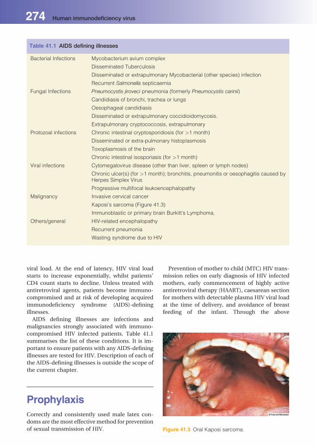
viral load. At the end of latency, HIV viral load
starts to increase exponentially, whilst patients’
CD4 count starts to decline. Unless treated with
antiretroviral agents, patients become immuno-
compromised and at risk of developing acquired
immunodeficiency syndrome (AIDS)-defining
illnesses.
AIDS defining illnesses are infections and
malignancies strongly associated with immuno-
compromised HIV infected patients. Table 41.1
summarises the list of these conditions. It is im-
portant to ensure patients with any AIDS-defining
illnesses are tested for HIV. Description of each of
the AIDS-defining illnesses is outside the scope of
the current chapter.
ProphylaxisCorrectly and consistently used male latex con-
doms are themost effectivemethod for prevention
of sexual transmission of HIV.
Prevention of mother to child (MTC) HIV trans-
mission relies on early diagnosis of HIV infected
mothers, early commencement of highly active
antiretroviral therapy (HAART), caesarean section
for mothers with detectable plasma HIV viral load
at the time of delivery, and avoidance of breast
feeding of the infant. Through the above
Figure 41.3 Oral Kaposi sarcoma.
Table 41.1 AIDS defining illnesses
Bacterial Infections Mycobacterium avium complex
Disseminated Tuberculosis
Disseminated or extrapulmonary Mycobacterial (other species) infection
Recurrent Salmonella septicaemia
Fungal Infections Pneumocystis jiroveci pneumonia (formerly Pneumocystis carinii)
Candidiasis of bronchi, trachea or lungs
Oesophageal candidiasis
Disseminated or extrapulmonary coccidioidomycosis.
Extrapulmonary cryptococcosis, extrapulmonary
Protozoal infections Chronic intestinal cryptosporidiosis (for >1 month)
Disseminated or extra-pulmonary histoplasmosis
Toxoplasmosis of the brain
Chronic intestinal isosporiasis (for >1 month)
Viral infections Cytomegalovirus disease (other than liver, spleen or lymph nodes)
Chronic ulcer(s) (for >1 month); bronchitis, pneumonitis or oesophagitis caused byHerpes Simplex Virus
Progressive multifocal leukoencephalopathy
Malignancy Invasive cervical cancer
Kaposi’s sarcoma (Figure 41.3)
Immunoblastic or primary brain Burkitt’s Lymphoma,
Others/general HIV-related encephalopathy
Recurrent pneumonia
Wasting syndrome due to HIV
274 Human immunodeficiency virus
approaches, the rate of MTC transmission of HIV
has declined to 1.2% in the UK.
Needle exchange programmes and methadone
replacement therapy has resultedmore recently in
a low rate ofHIV infection amongst IVDU in theUK
and now accounts for approximately only 2% of
new HIV diagnoses.
Male circumcisionhas been shown to reduce the
rate of HIV infection amongst heterosexual men in
Africa and is the second most effective HIV pre-
ventive measure after use of male latex condoms.
Male circumcision does not reduce rate of HIV
transmission amongst MSM or women.
Some data suggest that patients on HAART with
undetectable plasma viral load may not transmit
HIV infection. It has therefore been proposed that
universal voluntary HIV testing, identification of
mostHIV infectedpatients and immediateHAART,
combined with present prevention approaches,
could help control HIV epidemics in the future.
In most developed countries, post-exposure
prophylaxis after sexual exposure (PEPSE) is also
available. PEPSE policies are based on provision
of HAART within 72 hours after exposure to HIV
infection. The duration of treatment is usually
28 days. PEPSE should not replace consistent con-
dom use.
Post-exposure prophylaxis after occupational
exposures has been offered for more than 10 years
and involves commencing HAART regimes within
72 hours of significant injuries sustained at health
settings from HIV infected patients (e.g. needle
stick injuries).
Novel preventive interventions, such as use of
vaginal gel preparation of tenofovir, and pre-
exposure prophylaxis (PreP) for HIV, have pro-
duced encouraging results in reducing sexual
transmission of HIV to women and men who have
sexwithmen, respectively. Furtherdevelopmentof
an HIV vaccine has also received a major boost
after 30% reduction of HIV transmission by use of
two experimental HIV vaccines in a study carried
out in Thailand.
Laboratory diagnosis
. Serology, including immunoassays for HIV anti-
gen (p24) and antibody. There is an interval be-
tween infection and development of detectable
amounts of anti-HIV antibodies during which
HIV infected patientsmay test antibody negative.
More recentHIVassaysdetectHIVantibodiesand
HIV antigen (p24) simultaneously.. Identification of HIV RNA in plasma. This is a
nucleic acid amplification test (NAAT) that in-
volves amplification and detection of HIV RNA.
Patients with acute HIVmay only have a positive
HIV RNA test, so it is therefore important to
confirm their HIV infection within a week with
immunoassays.. Point of care (POC) HIV assays. which can pro-
duce reliable resultswithin 20minutes havebeen
developed. They require only blood drops from
finger pricks, or saliva. The advent of POC HIV
assays has improved the uptake of HIV tests in
certain settings. All positive POC tests need to be
confirmed by laboratory based HIV tests.
TreatmentThe advent of HAART has revolutionised the prog-
nosis of HIV infected patients; turning a fatal in-
fection into a chronic infection. Until 1996, anti-
retroviral agents were used alone in HIV infected
patients. The use of combination antiretroviral
drugs was subsequently shown to be superior.
HAART regimens have beenmodified asmore data
has become available.
Surrogate markers in HIVmedicine and commencementof HAARTCD4 count is used to determine when to com-
mence HAART. Following commencement, pa-
tients need to take HAART indefinitely. Late start
of HAART may lead to immunosuppression and
development of AIDS. Current guidelines suggest
that patients with a CD4 count of less than
350 cells/mm3 should be on HAART. Once
started, the aim for any patient on HAART is
to have a plasma HIV viral load of less than
50 copies/mL.
Resistance assay
Poor adherence to HAART will result in sub-opti-
mal drug concentrations subsequently allowing
HIV replication under the influence of only one
agent to which it will become resistant.
Resistance tests are able to detect certain muta-
tions in HIV that make antiretroviral agents inef-
fective. With a patient’s resistance profile,
Human immunodeficiency virus 275

virologists and HIV physicians are able to predict
treatment response to antiretroviral agents and to
propose themost effective HAART combinations.
It is therefore prudent to start a HAART regime
only after consideration of the HIV resistance
profile.
The impact of poor adherenceto HAART
Because of the risk of resistance to any HAART
regime, patients must be adherent. This requires
strong and sustainedmotivation and commitment
that depend on patients’ social and mental status
at any time.
Is HAART cost effective?
Provision of HIV care is expensivemainly, because
of the cost of antiretroviral drugs. Studies suggest
that HAART is cost effective and should be widely
available to infected patients.
Despite marked improvement in survival of HIV
infected patients more than a decade after use of
HAART, it is too early to predict the long-term
prognosis of HIV infected patients.
276 Human immunodeficiency virus

42Miscellaneous viralinfectionsJohn CheesbroughLancashire Teaching Hospitals NHS Trust, Preston, UK
ArbovirusesArboviruses (Arthropodborne virus) are so-named
to reflect the role that blood-sucking arthropods
(insects and ticks) play as transmission vectors in
nature. Humans are themain, natural host for only
a few arboviruses. For most, their maintenance in
nature depends on a vertebrate host, often a bird or
rodentwith humans acting as incidentally infected
‘dead-end’ hosts. Domestic animals, if infected,
can act as ‘amplifying hosts’; increasing the risk
to humans via shared arthropod vectors.
The disease spectrum in the vertebrates varies
from clinically inapparent infection, commonly
the case in natural hosts, to severe disease and
death.
Vector control and avoidance of insect bites
(clothing, DEET insecticide, ‘mosquito’ netting
when sleeping) can reduce the risk of infection
with all of these viruses, while vaccination offers
further protection to some.
All medically important arboviruses are
members of three families of single stranded
RNA viruses: Togaviridae, Flaviviridae and
Bunyaviridae.
Dengue fever
Virus
Genus: Flavivirus in the family Flaviviridae; four
serotypes: Dengue 1, 2, 3 and 4.
Epidemiology. Transmitted by mosquitoes, especially Aedes ae-
gypti; which bites mainly during daylight hours;. Humans are the main host;. Predominantly an urban disease with regular
epidemics;. Once infected humans acquire long-term type-
specific immunity, which may then enhance
the severity of later infection with a different
serotype;. The most important arboviral cause of disease
and death in humans and a common cause of
fever in travellers returning from endemic areas;. The disease is endemic in subtropical and trop-
ical regions of the world, especially the Indian
sub-continent, Southeast Asia and Central
America;. It has spread rapidly over the last 30 years and is
estimated to infect 50 million people annually.
Medical Microbiology and Infection Lecture Notes, Fifth Edition. Edited by Tom Elliott, Anna Casey,Peter Lambert and Jonathan Sandoe.� 2011 Blackwell Publishing Ltd. Published 2011 by Blackwell Publishing Ltd.

Clinical features
Incubation period 4–10 days. Majority of infec-
tions are asymptomatic. Clinical disease charac-
terised by abrupt onset of high fever, headache,
nausea, vomiting and severe back pain. Facial
swelling and widespread maculopapular rashmay
be present. Most cases resolve after 5–7 days.
However, others, despite defervescence, remain
unwell, with vomiting and abdominal pain. Wide-
spread oedema and/or mucosal bleeding devel-
ops, heralding the onset of severe dengue with
shock and/or haemorrhage. Leukopenia develops,
platelet levels fall and haematocrit rises as intra-
vascular fluid is lost into the tissues. There is a
1–2% overall mortality, with survivors usually
starting to improve after 48 hours.
Diagnosis. Antigen detection in blood; by EIA;
IgM and IgG antibodies in blood;. Nucleic acid amplification tests on blood;. Virus isolation only in specialised laboratories.
Treatment and prevention. Symptomatic treatment with early fluid replace-
ment in severe disease. Prevention by vector
control. Vaccines are under development and
may be commercially available by 2015.
Yellow fever
Virus
Genus: Flavivirus.
Epidemiology
Transmitted by mosquitoes, major vertebrate host
is the monkey.
Three transmission cycles:
1 Sylvatic (Jungle) cycle: betweenmosquitoes and
monkeys. Humans only occasionally infected
when venturing into the forest.
2 Intermediate: This accounts for the majority of
human infections. Once infection has been in-
troduced by forest mosquitoes from monkeys
into humans, onward transmission by semi-
domestic mosquitoes sustains the epidemic.
3 Urban yellow fever cycle: Transmission between
humans maintained by domestic mosquitoes,
mainly Aedes aegypti.. It is estimated that around 200,000 cases occur
annually, with 30,000 deaths (WHO 2010).
Most of these are in West Africa.. The disease occurs in the tropics on both sides
of the Atlantic.. Mosquito control terminated urban yellow
fever in the western hemisphere, but an aver-
age of 100 cases/year of jungle yellow fever are
still reported.. In Africa, regular outbreaks of intermediate
disease, mainly in villages in humid parts of
West Africa, have increased over the last
20 years.
Clinical features
Incubation period is 3–6 days. There are two
phases of the disease; an initial non-specific phase
of fever, headache, muscle pain and nausea lasting
3–4days. After abrief periodof recovery, about 15%
of cases go on to a second severe toxic phase with a
high fever, vomiting and jaundice. Bleeding from
GI tract, nose, mouth, skin and eyes can occur and
renal failure ensue. Survival at this stage is 50%.
Diagnosis. Histology and immunocytochemistry at autopsy;. IgM or IgG antibodies in blood;. Nucleic acid amplification tests on blood;. Virus isolation only in specialised laboratories.
Treatment and prevention
Symptomatic and supportive treatment only.
Vector control. A highly effective live attenuated
vaccine (17D vaccine) is available for travellers to
endemic areas and foruse in response tooutbreaks.
Japanese encephalitis
Virus
Genus: Flavivirus; three genotypes.
Epidemiology. Transmitted by mosquitoes, mainly Culex spp.,
between wild birds, the natural host;
278 Miscellaneous viral infections

. Domestic pigs can become infected, amplifying
the reservoir of infected mosquitoes, which then
bite humans;. Humans are a dead-end host;. Endemic in an area stretching from Southern
Japan and Korea across China into Southeast
Asia and to the Indian Sub-continent;. The leading cause of viral encephalitis in Asia
with 30,000–50,000 reports annually.
Clinical features
Incubation period is 5–15 days. Whilst most
cases are sub-clinical or just a non-specific fever,
1–2% present as an aseptic meningitis or
meningoencephalitis.
Complications
Twenty-five percent of encephalitis cases die rap-
idly and 30% will be left with varying degrees of
permanent neuropsychiatric sequelae.
Diagnosis. IgM antibodies in blood and cerebrospinal fluid
(CSF);. IgG may be positive, due to prior sub-clinical
infection and not relevant to current illness;. Nucleic acid amplification tests on blood (this
will detect some early IgM negative cases);. Virus isolation only in specialised laboratories.
Treatment and prevention
Treatment is symptomatic. A killed vaccine is
widely used in endemic areas and for travellers.
Vector control.
West Nile
Virus
Genus: Flavivirus.
Epidemiology. Transmitted by mosquitoes, especially Culex
spp;. Hosts include birds, domestic and wild animals,
humans and sub-human primates;. Humans are a dead-end host;
. No person-to-person transmission occurs, but
transmission through blood and organ dona-
tions and intrauterine transmissions has been
reported;. The virus is found throughout Africa, Asia, Eur-
ope, the USA and Canada. Spread into western
hemisphere occurred in 1999 with an outbreak
causing 7 deaths in New York and is now en-
demic across North America.
Clinical features
Incubationperiod 2–14days. Themajority (80%)of
infections are sub-clinical. A non-specific mild
febrile illness is usual in the remainder, but aseptic
meningitis or more serious meningioencephalitis
occurs in 0.7% of patients. These severe infections
are more common in patients over the age of
50 years.
Diagnosis. IgM antibodies in blood and CSF;. Nucleic acid amplification is not useful in clinical
cases, because viraemia is brief but it is used to
screen blood products and prevent transmission
by blood transfusion (all blood donated in the
USA is tested by this method);. Virus isolation only in specialised reference
laboratories.
Treatment and control
Treatment is symptomatic. No vaccine available.
Prevention is by vector control and blood donation
screening in USA
Chikungunya
Virus
Genus Alphavirus, in the family Togavirida.
Epidemiology. Transmission is by mosquito, mainly Aedes
aegypti, a domestic daytime biting mosquito.. Primary host is humans. Monkeys and possibly
other animals may also be reservoirs.. Infections occur in Africa, Indian subcontinent
andSoutheast Asia. Large epidemics are a feature
of this infection, oftenwith long gaps (7–20 years)
between peaks of activity.
Miscellaneous viral infections 279

. Outbreaks in Italy in 2006 reflect its transmission
by an Aedes albopictus ‘Asian Tiger mosquito’,
whose range has increased recently.
Clinical features
Incubation period 2–12 days, typically 3–7 days.
Sudden onset of fever, headache, fatigue, nausea,
vomiting,muscle pain, rash and joint pain. Hard to
distinguish from Dengue at this stage. Fever
typically resolves in 5–7 days. Infection confers
long-term immunity.
Complications
Joint pains, (can be severe) and fatigue can
continue for months after acute phase resolves.
Mortality is very low, as haemorrhage and shock
are not features of this infection. May cause first
trimester abortion and congential infections if
viraemic at delivery.
Diagnosis. IgM and IgG antibodies in blood by EIA;. Nucleic acid amplification tests on blood;. Virus isolation only in specialised laboratories.
Treatment and control
Symptomatic treatment only. Prolonged use of
non-steroidal anti-inflammatory drugs may be re-
quired in convalescence. Avoidance of insect bites
and vector control. Preventing mosquitoes biting
active cases should reduce onward transmission.
No vaccine is available.
Viral haemorrhagic fever virusesViral haemorrhagic fevers (VHF) are severe life
threatening diseases caused by a range of viruses
(Table 42.1). Four (bold in table) of these viruses
are readily capable of person-to-person spread
and therefore present a major public health risk.
Diagnosis of these viruses is carried out under the
highest level of bio-safety, typically only at na-
tional reference laboratories. The diagnosis must
be considered in anyone with a significant febrile
illness with an onset within 21 days of returning
from an endemic area. Most will not have VHF,
but isolation and strict barrier precautions should
be maintained while a definitive diagnosis is
sought, to minimise risk of transmission in
hospital.
Lassa fever
Virus
Member of the Arenaviridae family of viruses.
Epidemiology. The natural reservoir is the rat Mastomys
natalensis.. The virus causes a persistent asymptomatic in-
fection in this rat,which freelyexcretesLassavirus
in urine and other body fluids throughout its life.. Humans become infected through contact with
infected rodents or their excreta.
Table 42.1 Agents of viral haemorrhagic fevers
Agent Sourceor Vector
Arenaviridae
Lassa fever Rodents
Argentine haemorrhagicfever (Junin)
Rodents
Bolivian haemorrhagic fever(Machupo)
Rodents
Venezuelan haemorrhagicfever (Guanarito)
Rodents
Brazil (Sabia) unknown
Zambia (Lujo virus) unknown
Bunyaviridae
Crimean–Congo haemorrha-gic fever
Ticks
Haemorrhagic fever withrenal syndrome(Hantaan viruses)
Rodents
Rift Valley fever Mosquitoes
Filoviridae
Ebola Unknown
Marburg Unknown
Falviviridae
Dengue types 1–4 Mosquitoes
Yellow fever Mosquitoes
Kyasanur Forest disease Ticks
Omsk haemorrhagic fever Ticks
Togaviridae
Chikungunya Mosquitoes
280 Miscellaneous viral infections

. Person-to-person transmission can occur
in overcrowded conditions, particularly in hos-
pitals, with suboptimal infection control
precautions.. There is an estimated up to 500,000 cases annu-
ally, with 5,000 deaths.. Lassa is endemic in West Africa, with outbreaks
reported mainly in Sierra Leone and Nigeria.
Clinical features
Incubation period is 5–21 days. Spectrum of dis-
ease ranges from sub-clinical to fulminating fatal
infection. Symptomatic patients typically present
with fever, sore throat, myalgia, abdominal pain
and vomiting lasting between 7 and 17 days.
Complications
Haemorrhages, deafness (in 30% acutely and per-
sistent in 10%), encephalitis and spontaneous
abortion. Mortality rate is between 1 and 2%.
Diagnosis. Antigen detection in blood by EIA;. IgM and IgG antibodies in blood by EIA or IFAT.
IgG antibodies may reflect past exposure;. Nucleic acid amplification tests on blood;. Virus isolation only in specialised laboratories.
Treatment and prevention
Intravenous ribavirin within 6 days of onset of
illness.
Oral ribavarinmay be used for prophylaxis in close
contacts with needlestick injury.
No vaccine available.
Ebola andMarburg haemorrhagicfever
Virus
Members of the Filoviridae family of viruses.
Epidemiology. Nonatural reservoir has been identified and their
natural history is unknown.
. Recent serological studies have implicated vari-
ous bats species as possible hosts.. The viruses are endemic in Africa with most
recent cases from central African countries.. A largeoutbreakof Ebolawith425cases in2000/1
occurred in Uganda. Marburg only gives rise to
infrequent sporadic cases.. They cause severe infection in non-human pri-
mates (chimpanzees, gorillas, monkeys) and
contact with dead primates appears to have ini-
tiated some human outbreaks.. During outbreaks, humans are infected through
close contacts with either infected patients or
infected material, such as needles, blood or
secretions.
One subtype of Ebola “Reston” is endemic in
the Phillipines and causes outbreaks of disease
in captive monkeys but only sub-clinical
infection in humans. A reservoir in pigs and
pig-human transmission has been reported for
this subtype.
Clinical features
Incubation period is 2–21 days. Infections are al-
ways symptomatic, starting abruptly with fever,
headache, myalgia, arthralgia, conjunctivitis, sore
throat, abdominal pain, nausea and vomiting.
Complications
Haemorrhages, encephalitis. Mortality rate is
between 55 and 88%.
Diagnosis. Antigen detection in blood by EIA;. IgM antibodies in blood by EIA;. Nucleic acid amplification tests on blood;. Virus isolation only in specialised laboratories.
Treatment and prevention
Treatment is symptomatic. No vaccine available.
Crimean–Congo haemorrhagicfever
Virus
Member of the Bunyaviridae family of viruses.
Miscellaneous viral infections 281

Epidemiology. Vector is the ixodid tick.. Hosts include humans, domestic and wild
animals.. Humans become infected through tick bites or
contact with blood, tissue or excreta of infected
animals.. The virus is distributed over Eastern Europe,
(particularly in the former Soviet Union)
around the Mediterranean, in northwestern
China, Africa, the Middle East and the Indian
subcontinent.. Animal herders and slaughterhouse workers are
at greatest risk.. Person-to-person spread is usually confined
to healthcare workers looking after severe
cases.
Clinical features
Incubation period is 1–14 days. Sudden onset of
fever, severe headache, sore throat, nausea and
vomiting, general fatigue and malaise. Flushed
face, injected conjunctiva and palatal petechiae
may be seen.
Complication
Bleeding tendency from venepuncture sites, epi-
staxis, haematuria, haematemesis and melaena
fromday4of illnessonwards.Mortality rate is9–50%.
Diagnosis. Antigen detection in blood by EIA;. IgM and IgG antibody in blood by EIA;. Nucleic acid amplification tests on blood;. Virus isolation only in specialised Laboratories.
Treatment and prevention
Treatment is generally symptomatic and sup-
portive. The antiviral drug ribavirin is active
in vitro and has shown some benefit particularly
if given within 5 days of onset.
Vector control in risk occupations. No vaccine
available.
282 Miscellaneous viral infections

Part 4Self-assessment


Self-assessmentquestions
Some questions may have more than one correct
answer.
Chapter 1 Basic bacteriology
1.1 Which one of the following statements
concerning the basic structure of bacterial
cells is incorrect?
A Most bacteria are surrounded by a rigid
cell envelope (cell wall) containing
peptidoglycan.
B The cell membrane of bacteria does not
contain sterols,whichare found inmam-
malian cell membranes.
C Bacterial DNA is contained within the
nucleus, surrounded by a nuclear
membrane.
D Some bacteria are motile, their move-
ment being controlled by rotating
flagella.
E Somebacteria formspores thatarehighly
resistant to harsh environmental condi-
tions, such as heat, UV light and
desiccation.
1.2 Which of the following statements concern-
ing bacterial cells is incorrect?
A Lipopolysaccharide is a component of
the cell envelope of Gram-positive
bacteria.
B Some bacteria produce capsules that
usually ‘contain’ polysaccharides, which
surround the cells and provide a protec-
tive layer.
C In some bacteria (e.g. Streptococcus
pneumoniae) the capsular material var-
ies in composition and can be used in
serotyping to distinguish between differ-
ent types of the same species of
microorganism.
D Some bacteria can stick to the surface of
materials and grow to form a biofilm that
is highly resistant to treatment with
antibiotics.
E Fimbriae are thin, hair-like structures
producedonthesurfaceofsomebacteria,
enabling them to adhere to host tissues.
1.3 Which of the following statements con-
cerning the growth of bacteria in laboratory
media is incorrect?
A All bacteria can be cultured in laboratory
media, but certain microorganisms re-
quire specific nutrients to be supplied.
B Bacteria that growonly in thepresenceof
oxygen are called obligate aerobes, those
growingonly in theabsenceofoxygenare
called obligate anaerobes, and those
growing in the presence or absence of
oxygen are called facultative anaerobes.
C Some bacteria, including Neisseria
meningitidis and Campylobacter jejuni,
require additional CO2 for laboratory
growth.
D Mannitol salt agar plates are an example
of selective growth media used for the
recovery and growth of staphylococci
from clinical samples.
E Chocolate agar, containing lysed blood,
is an enriched growth medium used for
cultureof somebacteria thathave special
growth requirements supplied by the
blood.
Chapter 2ClassificationofbacteriaSelect the correct options describing each of the
following microorganisms:
2.1 Staphylococcus aureus is a: Gram-positive/
Gram-negative, spore-forming/non-spore-
Medical Microbiology and Infection Lecture Notes, Fifth Edition. Edited by Tom Elliott, Anna Casey,Peter Lambert and Jonathan Sandoe.� 2011 Blackwell Publishing Ltd. Published 2011 by Blackwell Publishing Ltd.
forming, coagulase-positive/coagulase-
negative, coccus/bacillus, capable of growth
under aerobic/anaerobic/aerobic and anaer-
obic conditions.
2.2 Escherichia coli is a: Gram-positive/
Gram-negative, spore-forming/non-spore-
forming, coccus/bacillus, capable of growth
under aerobic/anaerobic/aerobic and anaer-
obic conditions.
2.3 Clostridium difficile is a: Gram-positive/
Gram-negative, spore-forming/non-spore-
forming, coccus/bacillus, capable of growth
under aerobic/anaerobic/aerobic anaerobic
conditions.
Chapter 3 Staphylococci
3.1 Identify which of the following properties
can be used to distinguish Staphylococcus
aureus from other potentially pathogenic
staphylococci:
A Coagulase
B DNAase
C Protein A
D Haemolysins
E Toxic shock syndrome toxin (TSST)
3.2 With which of the following infections is
S. saprophyticus usually associated?
A Endocarditis
B Urinary tract infection
C Osteomyelitis
D Skin infection
E Respiratory tract infection
3.3 Which of the following statements con-
cerning the sensitivity of staphylococci to
antibiotics is incorrect?
A Flucloxacillin is used to treat many
S. aureus infections.
B MRSA isolates (meticillin resistant
S. aureus) are sensitive to flucloxacillin.
C Some S. epidermidis isolates are resistant
to flucloxacillin.
D MRSAand S. epidermidis are usually sen-
sitive to vancomycin.
E S. epidermidis is sensitive to novobiocin.
Chapter 4 Streptococci andenterococci
4.1 Which one of the following statements
about the streptococci is incorrect?
A They are Gram-positivemicroorganisms.
B They commonly exhibit haemolysis
when cultured on blood agar.
C They produce spores.
D They commonly cause respiratory tract
infections.
E Infections caused by streptococci are
commonly treated with penicillin.
4.2 Which one of the following statements
about the enterococci is correct?
A They are commensals of the gastrointes-
tinal tract.
B They are always non-haemolytic.
C They are obligate anaerobes.
D All species are motile.
E They are always sensitive to vancomycin.
Chapter 5 Clostridia
5.1 Which two statements about clostridia are
correct?
A They are Gram-negative bacilli.
B They are obligate aerobes.
C They produce spores.
D Patients infected with Clostridium
perfringens may present with ‘lockjaw’.
E Clostridial infections are often treated
with penicillin and/or metronidazole.
5.2 Which two statements about Clostridium
difficile are correct?
A The use of certain antibiotics is a risk
factor for development of infection with
this microorganism.
B It produces 4major toxins (A,B,CandD).
C Infection with C. difficile is often treated
with IV vancomycin.
D It colonises the intestine of 50% of
healthy adults.
E It is a significant cause of healthcare-
associated infection.
Chapter 6 Other Gram-positivebacteria
6.1 Which two statements about Bacillus are
correct?
A Bacillus species are spore-producing ob-
ligate anaerobes.
286 Self-assessment questions

B B. anthracis is non-motile, allowing it to
be distinguished from other Bacillus
species.
C It is safe to handleB. anthracis in anopen
laboratory.
D No vaccine exists for B. anthracis.
E B. cereus is an important cause of food
poisoning.
6.2 Which two statements regarding coryne-
bacteria are correct?
A Colonies of C. diphtheriae appear green
on Hoyle’s tellurite medium.
B Diphtheria is common in developed
countries.
C AllC.diphtheriae subspeciesproduce the
diphtheria exotoxin.
D In developed countries, children are
routinely immunised with a toxoid
vaccine.
E Several species of Corynebacterium are
skin commensals.
6.3 Which one of the following statements re-
garding Listeria is incorrect?
A Listeria monocytogenes can cause out-
breaks of food poisoning.
B They can be distinguished from other
Gram-positive bacilli by their character-
istic ‘tumbling’motility at<30�Cinbroth
cultures.
C They are intracellular pathogens,
which can survive within phagocytic
cells.
D Listeria monocytogenes produce a
haemolysin.
E It is safe for laboratory staff who are
pregnant toworkwithsuspectedcultures
of Listeria.
6.4 Which two of the following statements
about these other Gram-positive microor-
ganisms are incorrect?
A Actinomyces species are non-sporing
bacilli.
B Nocardia species are aerobic.
C Erysipelothrix rhusiopathiae can cause
erysipeloid cellulitis.
D Peptostreptococcus species are obligate
aerobes andcancause abscesses atmany
anatomical sites.
E Propionibacteria are part of the normal
flora of the skin.
F Rhodococcus equi is an opportunistic
pathogen.
G Tropheryma whipplei is an intracellular
pathogen, which causes disease more
prevalent in females.
Chapter 7 Gram-negative cocci
7.1 Which of the following microorganisms is
not a Gram-negative coccus?
A Meningococcus
B Gonococcus
C Pneumococcus
D Moraxella catarrhalis
E Veillonella species
7.2 Which of the following statements concern-
ing Neisseria gonorrhoeae is incorrect?
A It is an obligate human parasite; its car-
riage can be asymptomatic.
B It can survive inside polymorphonucleo-
cytes (PMNs).
C It is oxidase- and catalase-positive.
D Resistance to penicillin is very rare.
E Re-infection can occur because no im-
munity develops.
7.3 Which of the following statements concern-
ing Neisseria meningitidis is incorrect?
A Ten percent of humans are asymptom-
atic carriers of the microorganism in the
nasopharynx.
B It produces capsular polysaccharides
that can be used as the basis of
serotyping.
C The microorganism can be found inside
polymorphonucleocytes.
D No vaccines are available.
E First-line treatment is penicillin or
cefotaxime.
Chapter 8 Enterobacteriaceae
8.1 Give the names of 5 genera of the
Enterobacteriaceae
8.2 MacConkey’s agar is used to distinguish
between some members of the Enterobac-
teriaceae;whichof the followinggeneragive
pink colonies on MacConkey’s agar?
A Escherichia only
B Enterobacter only
C Klebsiella only
D Escherichia, Enterobacter and Klebsiella
E Salmonella, Shigella, Serratia, Proteus
and Yersinia
Self-assessment questions 287

8.3 Which of the following statements correctly
describes Salmonella spp?
A Non-motile Gram negative rods
B Encapsulated Gram-negative rods
C Sensitive to acid and do not survive pas-
sage through the stomach
D Formpale colonies with black centres on
XLD agar
E Sensitive to deoxycholate
Chapter 9 Haemophilus and otherfastidious Gram-negative bacteria
9.1 Which of the following media will support
growth of Haemophilus influenzae ?
A Nutrient agar containing ATP and cAMP
B Nutrient agar supplemented with hae-
min (factor X) and nicotinamide adenine
dinucleotide (NAD, factor V)
C Chocolate agar
D Blood agar
E MacConkey agar
9.2 Which of the following antibiotics are com-
monly used in empirical therapy ofHaemo-
philus influenzae ?
A Ampicillin
B Metronidazole
C Third-generation cephalosporins (e.g.
cefotaxime or ceftriaxone)
D Chloramphenicol
E Flucloxacillin
9.3 Which of the following are members of the
HACEK group of Gram-negative bacilli?
A Haemophlius influenzae
B Aggregatibacter actinomycetemcomitans
(formerly Actinobacillus actinomycetem-
comitans)
C Cardiobacterium hominis
D Eikenella corrodens
E Klebsiella pneumoniae
9.4 Which of the following statements concern-
ing Bordetella pertussis is incorrect?
A It causes whooping cough.
B It can be isolated frompernasal swabs on
Bordet-Gengou agar.
C It is highly transmissible and erythomy-
cin is used to reduce infectivity.
D The DTP triple vaccine is used to protect
against whooping cough.
E It forms resistant spores that are difficult
to eradicate from the environment.
9.5 Which of the following statements about
Brucella species are incorrect?
A They are Gram-positive rods.
B Infectionsareconfined toMediterranean
countries and the Middle East.
C Infections are acquired by ingestion of
contaminated food.
D They cannot be grown in routine blood
culture systems.
E Infections are treated with 2 weeks tetra-
cycline and aminoglycoside.
Chapter 10 Pseudomonas,Legionella andother environmentalGram-negative bacilli
10.1 Pseudomonas aeruginosa is notable for its
resistance to many antibiotics; which of
the following would not be considered for
treating infections caused by this
microorganism?
A Gentamicin
B Ceftazidime
C Amoxicillin
D Imipenem
E Piperacillin/tazobactam
10.2 Which of the following statements concern-
ing Legionella pneumophila is incorrect?
A It is an environmental microorganism
found in soil and water.
B It causes respiratory infections
(Legionnaires’ disease and Pontiac
fever).
C It can survive inside phagocytic cells and
amoebae.
D It can be cultured on nutrient agar in
2–7 days.
E L. pneumophila antigen can be detected
in the urine of infected patients
10.3 Which of the following properties distin-
guishes Acinetobacter species from other
pseudomonads?
A Acinetobacter spp. are generally more
sensitive to antibiotics than other
pseudomonads.
B Acinetobacter spp. are oxidase-negative,
other pseudomonads are oxidase-
positive.
C Unlike other pseudomonads, Acineto-
bacter spp. do not survive well in the
environment.
288 Self-assessment questions
D Unlike other pseudomonads, Acineto-
bacter spp. require complex media for
growth.
E Unlike other pseudomonads, Acineto-
bacter spp. can growth anaerobically.
Chapter 11 Campylobacter,Helicobacter and vibrios
11.1 Which two statements about Campylobac-
ter are incorrect?
A They are spirally curved Gram-negative
bacilli.
B They are motile, with long polar flagella.
C They are the most common cause of
acute bacterial enterocolitis, and infec-
tion is often caused by the handling of
raw chickens or consumption of the un-
dercooked meat.
D Human-to-human transmission is
common.
E All cases of infection require antibiotic
therapy.
11.2 Which two statements about Helicobacter
are incorrect?
A Helicobacter pylori is the most common
cause of duodenal ulceration and gastric
cancer.
B Infection withHelicobacter pylori occurs
commonly in early childhood and usu-
ally becomes chronic, often life-long.
C The prevalence of infection with Helico-
bacter pylori is low in countrieswith poor
sanitation.
D Helicobacter pylori are urease positive.
E Treatment of infection often involves a
proton pump inhibitor to reduce
gastric acidity coupled with one
antimicrobial.
11.3 Which one of the following statements
about Vibrios is incorrect?
A Vibrio cholerae 01 is the cause of cholera,
which is characterised by profuse watery
diarrhoea.
B Vibrio cholerae is found inwater and food
contaminated with human faeces.
C Vibrio cholerae forms characteristic red
colonies on thiosulphate-citrate-bile
salt-sucrose agar.
D Treatment of cholera is with rehydration
and often ciprofloxacin to shorten the
duration of illness.
E Non-cholera Vibrio species may also
cause diarrhoea and occasionally cause
cellulitis.
Chapter 12 Treponema, Borreliaand Leptospira
12.1 Which of the following statements is
incorrect?
A Treponema, Borrelia and Leptospira are
all spirochaetes.
B Treponema, Borrelia and Leptospira are
all helical-shaped Gram-negatives.
C Few spirochaetes can be cultured and
diagnosis is usually made by serological
tests.
D Treponema pallidum is not motile.
E Spirochaetes, like Treponema pallidum,
canbevisualised inclinical specimensby
dark field microscopy.
12.2 Which of the following statements con-
cerning tests for Treponema pallidum is
incorrect?
A The non-specific antibody test detects
antibody to lipids (cardiolipin) released
from the microorganism during early
infection.
B Detection of antibody to cardiolipin is
specific for syphilis.
C More specific antibody tests for T. palli-
dum are available,whichdetect antibody
to selectedT. pallidum-derived antigens.
D T. pallidum subspecies endemicum
causes bejel in children.
E T. pallidum subspecies pertenue causes
yaws in children.
12.3 Which of the following microorganisms
causes Lyme disease?
A Treponema carateum
B Borrelia recurrentis
C Borrelia duttoni
D Borrelia burgdorferi
E Leptospira spp.
Chapter 13 Gram-negativeanaerobic bacilli
13.1 Which of the following bacterial genera
make up the major component of the flora
of the human intestine?
A Bacteroides
B Prevotella
Self-assessment questions 289

C Porphyromonas
D Fusobacterium
E Leptotrichia
13.2 Which of the following microorganisms is
associated with advanced periodontal
disease?
A Veillonella spp
B Prevotella melaninogenica
C Prophyromonas gingivalis
D Fusobacterium necrophorum
E Leptotrichia buccalis
Chapter 14 Chlamydiaceae,Rickettsia, Coxiella,Mycoplasmataceae andAnanplasmataceae
14.1 Which two statements about the Chlamy-
diaceae are incorrect?
A They are obligate intracellular bacteria.
B They have three morphological forms.
C Chlamydia trachomatis is the most
common cause of sexually transmitted
infections in the developed world.
D Infection with Chlamydia trachomatis
is often treated with doxycycline or
azithromycin.
E Chlamydophilapsittaci is also acommon
cause of sexually transmitted infection.
14.2 Which two of the following statements are
incorrect?
A Rickettsial infections are zoonoseswith a
variety of insect vectors.
B Rickettsia cause tetanus.
C Coxiella burnetii causes RockyMountain
spotted fever.
D Mycoplasmataceae have no cell wall.
E Anaplasmataceae are usually transmit-
ted via tick bites.
Chapter 15 Basic virology
15.1 Which two of the following statements
about viruses are incorrect?
A They are icosahedral, helical or more
complex in shape.
B They all contain DNA.
C Viral nucleic acids can be single- or dou-
ble-stranded, circular or linear, and have
negative or positive polarity.
D They are obligate intracellular parasites
and can be grown in cell culture.
E Viral infection is always diagnosed
by direct detection of all or part of the
virus.
15.2 Whichof the followingmethodsareused for
the direct detection of viruses?
A Polymerase chain reaction.
B Direct immunofluorescence.
C Electron microscopy.
D Complement fixation test.
E Enzyme-linked immunosorbent anti-
body detection.
15.3 The incubation period of a viral infection is
(select one correct answer):
A The time fromacquisitionofavirus to the
resolution of symptoms.
B The time from onset of symptoms to
resolution of symptoms.
C The time fromacquisitionofavirus to the
onset of symptoms.
D The time it takes to cultivate a virus in the
laboratory.
E The period during which an infected pa-
tient can transmit the virus.
Chapter 16 Major virus groups
16.1 Which two of the following statements
about Herpesviruses are incorrect?
A Herpesviruses are non-enveloped, dou-
ble stranded DNA viruses.
B Following infection, a latent infection
that persists for life is established.
C Herpes simplex virus (HSV) is spread
mainly via sexual activity.
D Varicella-Zoster virus (VZV) is the cause
of glandular fever (‘infectious
mononucleosis’).
E Recurrent infection with Cytomegalovi-
rus (CMV) is common in immunocom-
promised patients, following solid organ
or stem cell transplants.
F Epstein-Barr virus (EBV) is implicated
in the pathogenesis of several tumours,
including Burkitt’s lymphoma.
G Human herpesvirus 8 (HHV8) can cause
Kaposi’s sarcoma.
16.2 Which three of the following statements
about these other DNA viruses are
incorrect?
A Adenoviruses and Human Papilloma-
viruses (HPV) are non-enveloped
viruses with double stranded DNA.
290 Self-assessment questions
B Treatment of infection with adeno-
viruses is normally symptomatic;
however, cidofovir can be used in immu-
nocompromised patients.
C Parvoviruses are enveloped double-
stranded DNA viruses.
D There are four parvovirus serotypes.
E Parvoviruses can cause spontaneous
abortions and intrauterine death.
F HPV is commonly sexually transmitted
and can lead to carcinoma.
G No vaccine for HPV is currently
available.
16.3 Which one of the following statements
about Influenza viruses is incorrect?
A They are enveloped viruses.
B There are two types of influenza virus: A
and B.
C They commonly cause respiratory
infection.
D Severe influenza infection is often
treated with oseltamivir or zanamivir.
E Influenza vaccines exist.
16.4 Which one of the following statements
about Paramyxoviruses is incorrect?
A They are enveloped viruses.
B There is a vaccine for the prevention of
infection with respiratory syncytial virus
(RSV).
C There are four serotypesof Parainfluenza
virus.
D A live attenuated virus vaccine is avail-
able for the prevention of infection with
the Measles virus.
E Infection with the Mumps virus is nor-
mally characterised by swollen parotid
salivary glands.
16.5 Which two of the following statements
about these other RNA viruses are
incorrect?
A Rhinoviruses, Enteroviruses, Rota-
viruses, Noroviruses, Sapoviruses and
the Rubella virus are non-enveloped
viruses.
B Rhinoviruses and Coronaviruses are
causes of the common cold syndrome.
C Coronaviruses and the Rabies virus are
enveloped.
D SARS is caused by a Rhinovirus.
E Infection with Rotaviruses, Noroviruses
and Sapoviruses is characterised by diar-
rhoea and vomiting.
F Mortality without treatment for rabies is
100%.
16.6 Which two of the following statements
about Hepatitis viruses are incorrect?
A The Hepatitis A, B, C, D and E viruses are
RNA viruses.
B The Hepatitis A virus (HAV) and Hep-
atitis E virus (HEV) are acquired via the
faecal-oral route and causes acute
hepatitis.
C The Hepatitis B virus (HBV) is acquired
via exposure tobloodandbloodproducts
containing the virus, by sexual inter-
course or vertically at birth.
D HBV and Hepatitis C virus (HCV) cause
acute, chronic and fulminant hepatitis,
cirrhosis and hepatocellular
carcinoma.
E Delta agent (Hepatitis D virus (HDV))
replicates only in HCV-infected cells,
therefore prevention of HCV infection
prevents infection with HDV.
16.7 Which three of the following statements
about these retroviruses are incorrect?
A They are single stranded RNA enveloped
viruses.
B Transmission of theHuman Immunode-
ficiency virus (HIV) and Human T-cell
lymphotropic viruses (HTLV) is mainly
through the faecal-oral route.
C Infection with HIV and the development
of AIDS is characterised by profound
immunosuppression and infection with
a variety of microorganisms.
D No antiretroviral drugs are available for
the treatment of infection with HIV.
E Aminority of people infectedwithHTLV-
1 may develop adult T-cell leukaemia or
lymphoma.
F Antiviral drugs are available for the treat-
ment of infection with HTLV.
Chapter 17 Basic mycology andclassification of fungi
17.1 Which two of the following statements
about fungi are incorrect?
A They are motile.
B Most fungi that commonly cause human
disease can be categorised into yeasts,
moulds/filamentous or dimorphic.
Self-assessment questions 291

C The taxonomic status of Pneumocystis
jirovecii is well established.
D Most deep mycoses are opportunistic
infections occurring in immunocompro-
mised patients.
E Laboratory diagnosis of fungal infection
is mostly by direct microscopy or micro-
biological culture
17.2 Which two of the following statements
about yeasts are incorrect?
A Most cryptococcal infections occur in
immunocompetent individuals.
B Cryptococcal species have polysaccha-
ride capsules that can be visualised by
mixing fluid specimens with Indian ink.
C Candida albicans is a commensal of the
mouth and gastrointestinal tract.
D Cryptococcal species are the cause of
‘thrush’.
E Malassezia furfur is the causeofpityriasis
versicolor, which results in scaly skin and
depigmentation.
17.3 Which one of the following statements
about filamentous fungi is incorrect?
A Outbreaks of aspergillosis in immuno-
compromised patients have occurred,
due to construction work adjacent to
hospitals.
B Patients with cystic fibrosis are frequent-
ly colonised with Aspergillus species,
which may then lead to infection.
C Dermatophytes, including the genera
Epidermophyton, Microsporum and Tri-
chophyton, often infect the lungs
D The sub-phylum mucoromycotina in-
cludes the medically important species
Rhizopus arrhizus and Absidia
corymbifera.
E Infection with Fusarium and Scedospor-
ium species is often associated with in-
halation or aspiration of polluted water
17.4 Which one of the following statements
about dimorphic fungi is incorrect?
A Dimorphic fungi have both yeast and
mould forms, depending on growth
temperature.
B Coccidioides immitis andC. posadasii are
causes of ‘valley fever’, characterisedbya
mild, self-limiting pneumonia.
C Granulomatous ulcers may be a compli-
cation of pulmonary infection caused by
Blastomyces dermatitidis.
D Pulmonary infection with Histoplasma
capsulatum always requires antifungal
therapy.
E Paracoccidioides brasiliensismay lead to
destruction of the palate and nasal
septum.
Chapter 18 Parasitology: protozoa
18.1 Which of the following statements concern-
ing Entamoeba histolytica is incorrect?
A It causes intestinal amoebiasis and liver
abscesses.
B It is spread via water or food contami-
nated with cysts.
C It can be diagnosed in the laboratory by
microscopic examination of freshly
passed stools.
D No serological tests are available for Ent-
amoeba histolytica.
E Treatment of infection by Entamoeba
histolytica is with metronidazole.
18.2 Which of the following statements concern-
ing Toxoplasma gondii is incorrect?
A Human infection can result from inges-
tion of the microorganism as cysts con-
tained in cat faeces and undercooked
meat.
B It can cause a glandular fever-like syn-
drome in immunocompetent hosts.
C It causes serious conditions, including
myocarditis, choroidoretinitis and
meningoencephalitis in immunocom-
promised hosts.
D It can be reactivated by
immunosuppression.
E No serological tests are available to aid
diagnosis.
18.3 Which of the following species of Plasmo-
dium is responsible for malignant tertian
malaria?
A Plasmodium falciparum.
B Plasmodium malariae.
C Plasmodium vivax.
D Plasmodium ovale.
E Plasmodium knowlesi.
18.4 Match the following microorganisms (A–E)
to the diseases (F–J):
A Chagas disease
B West African sleeping sickness
C East African sleeping sickness
D Visceral leishmaniasis
292 Self-assessment questions

E Cutaneous leishmaniasis
F Trypanosoma brucei rhodesiense
G Trypanosoma brucei gambiense
H Leishmania donovani complex
I Leishmania tropica, Leishmania major
complexes and Leishmania mexicana
J Trypanosoma cruzi
Chapter 19 Parasitology:helminths
19.1 Which three of the following statements
about nematodes are incorrect?
A Ascaris lumbricoides (roundworm) is the
most common pathogenic helminth
worldwide.
B Infection with Enterobius vermicularis
(pin worm) often presents with perianal
itching.
C Toxocara canis and Toxocara cati are
parasites of rodents.
D Infection with Trichuris trichiura can
present with anaemia, intestinal irrita-
tion and occasionally, anal prolapse.
E Ancylostoma duodenale, Necator ameri-
canus (hookworm) and Strongyloides
stercoralis can penetrate intact skin.
F Trichinella spiralis infection isassociated
with eating shellfish.
G Wuchereria bancrofti andBrugliamalayi
are filarial worms, which can cause
elephantitis.
H Thevector ofLoa loa is themosquito;Loa
loamigrates under the conjuctiva, giving
it the name ‘eye worm’.
I The vector of Onchocerca volvulus is the
black fly and can cause ‘river blindness’.
J Infectionwithnematodes is often treated
with mebendazole or albendazole.
19.2 Which two of the following statements
about cestodes (tapeworms) are incorrect?
A They have either male or female repro-
ductive organs.
B They can be up to 30 feet long.
C Cestodes have two reservoirs in their life
cycle, one for larvae and one for adult
worms.
D Pigs are the reservoir for Taenia solium
larvae.
E Cows are the reservoir for Taenia sagina-
ta larvae.
F Crustacea and fish are the reservoirs for
Diphyllobothrium latum larvae.
G Humans are the reservoir for Echinococ-
cus granulosus adult worms.
H Rodents and humans are the reservoirs
for Hymenolepis nana adult worms.
I Infection with cestodes is often treated
with praziquantel.
19.3 Which two of the following statements
about trematodes (flukes) are incorrect?
A They are flatworms with oral and ventral
suckers.
B Most arehermaphrodites – schistosomes
have separate male and female worms.
C They require one intermediate host.
D Infection with Fasciola hepatica and
Opisthorchis sinensis may result in
hepatomegaly.
E Infection with Paragonimus westermani
is normally associated with the lungs.
F Infection with S. mansoni and S. japoni-
cum are characterised with clinical
manifestations in the bladder and S.
haematobium in the intestine.
Chapter 20 Antibacterial agents
20.1 Which of the following statements concern-
ing b-lactam antibiotics is incorrect?
A They act by interferingwith the synthesis
of the bacterial cell wall.
B They include the penicillins, cephalos-
porins, carbapenemsandmonobactams.
C Flucloxacillin is stable to many
b-lactamases, but is inactive against
meticillin-resistant Staphylococcus
aureus (MRSA).
D Piperacillin is used with tazobactam to
protect piperacillin from destruction by
some b-lactamases.
E The monobactam, aztreonam, has a
broad spectrum of antibacterial action.
20.2 Which of the following is not a member of
the aminoglycoside antibiotics?
A Gentamicin.
B Streptomycin.
C Amikacin.
D Kanamycin.
E Vancomycin.
20.3 Which of the following agents is active
against a wide range of anaerobic microor-
ganisms, including bacteria (Clostridia and
Self-assessment questions 293
Bacteroides) and protozoa (Giardia and
Trichomonas)?
A Metronidazole.
B Ciprofloxacin.
C Rifampicin.
D Trimethoprim.
E Nalidixic acid.
Chapter 21 Antifungal agents
21.1 Which two of the following statements
about the azoles are incorrect?
A Their mechanism of action is prevention
of glucan synthesis.
B They may cause transient liver function
abnormalities.
C Fluconazole, itraconazole and voricona-
zoleareavailableasoral, intravenousand
topical preparations.
D Voriconazole has several drug interac-
tions and can cause visual disturbances.
E Some species of Candida are resistant to
or have reduced susceptibility to
fluconazole.
21.2 Which two of the following statements
about these other antifungal agents are
incorrect?
A Flucytosine and griseofulvin inhibit nu-
cleic acid synthesis.
B Flucytosine is used as monotherapy.
C Side effects of amphotericin B include
anaphylactic reactions and renal tubular
damage.
D The echinocandins are available in an
oral preparation.
E Terbafine may cause hepatotoxicity.
Chapter 22 Antiviral agents
22.1 Which of the following do antiviral agents
inhibit?
A Virus attachment to cell receptors.
B Virus uncoating.
C Viral genome transcription.
D Virus mutation.
E Virion assembly and release.
22.2 Which of the following statements about
nucleotide analogue reverse transcriptase
inhibitors are incorrect?
A They are acyclic phosphonate analogues
of adenosine monophosphate.
B They require phosphorylation by cellular
enzymes before they can function.
C They inhibit viral DNA-dependent
RNA polymerase producing chain
termination.
D They must be given parenterally (by
injection).
E They are active against HIV-1 and -2 and
hepatitis B.
Chapter 23 Diagnostic laboratorymethods
23.1 Which of the following statements about
culture techniques is correct?
A Most medically important bacteria and
fungi cannot be cultured in the diagnos-
tic laboratory.
B Enriched agar media includes: horse
blood, chocolate agar and CLED (cyste-
ine lactose electrolyte deficient) agar.
C Selective agar contains an indicator (usu-
ally a dye) to allow differentiation of
bacteria.
D Differential agar contains antimicrobial
agents tosuppress growthofnormalflora
from sites that are normally colonised.
E Enrichment broth is a nutritious broth,
whichallows recovery andenrichment of
small numbers of microorganisms with-
in a clinical sample.
23.2 Which of the following body sites are nor-
mally sterile (free from colonising
bacteria)?
A Blood.
B Bone marrow.
C Subcutaneous tissue/muscle/bone.
D Cerebrospinal fluid/brain.
E Upper respiratory tract.
Chapter 24 Epidemiology,prevention of infections
24.1 Which of the following statements about
protective isolation rooms are correct?
A They are designed to protect compro-
mised patients from infection.
B They are under negative pressure.
C They have particulate filtered air.
D Hands only need to bewashedon leaving
the room.
294 Self-assessment questions

E Gowns and aprons do not need to be
worn in the room.
24.2 Which of the following statements about
notifiable diseases are correct?
A Medical staff have a statutory duty to
notify them.
B The CCDC should be notified.
C They include measles.
D They include E coli O157.
E They include all cases of meningitis.
Chapter 25 Upper respiratory tractinfections
25.1 Whichof the followingstatementsabout the
common cold syndrome is incorrect?
A It is most frequently caused by
Rhinoviruses.
B It occurs worldwide.
C It can be transmitted via virus-contami-
nated hands.
D It usually has an incubation period of
7–10 days.
E It resolves spontaneously within 7–10
days.
25.2 Which of the following statements about
laryngo-tracheobronchitis (croup) are
incorrect?
A It involves the upper and lower respira-
tory tract.
B It is predominantly caused by bacteria.
C It is largely restricted to children aged<5
years.
D It causes a distinctive paroxismal cough
(‘whooping cough’).
E It should be empirically treated with
antibiotics.
Chapter 26 Lower respiratory tractinfections
26.1 Which of the following statements about
pneumonia are incorrect?
A It commonly occurs without radiological
shadowing.
B It is most common in summer.
C It is associated with positive blood cul-
tures in 50% of cases.
D It is caused by S. pneumoniae, Legionella
and Mycoplasma, which can be diag-
nosed by urine antigen detection.
E It is usually treated for 3–4 weeks, de-
pending upon severity.
26.2 Whichof thesestatementsaboutviralpneu-
monia are incorrect?
A Primary viral pneumonia occurs mainly
in children, elderly people and immuno-
compromised patients.
B Common causes include: respiratory
syncytial virus (RSV), norovirus, rubella
virus and human metapneumovirus.
C Primary viral pneumonia in adults is
characterised by diffuse bilateral inter-
stitial infiltrates on chest radiograph.
D Diagnosis of viral pneumonia is by PCR
or culture of viral agents in, e.g. nasopha-
ryngeal aspirates, throat and nasal swabs
and/or bronchoalveolar lavage.
E Ribavirin isusedempirically for the treat-
ment of viral pneumonia.
Chapter 27 Tuberculosis andmycobacteria
27.1 Which of the following statements regard-
ing Mycobacterium tuberculosis are
incorrect?
A It stains well by the Gram stain.
B It can be identified in clinical samples by
the ZN stain.
C It causes only chest infections.
D Associated infections need antibiotic
treatment for several months.
E Infectionwith thismicroorganismcanbe
prevented by vaccination.
27.2 Which of the following statements about
Mycobacterium leprae are incorrect?
A It can cause leprosy.
B It has animal reservoirs.
C Transmission of infection with this mi-
croorganism can be from a discharge
from skin lesions.
D The incubation period for infection with
this microorganism is up to 10 years.
E It can cause skin lesions and also nerve
involvement.
Chapter 28 Gastrointestinalinfections
28.1 Which of the following statements about
Salmonella species are incorrect?
Self-assessment questions 295

A They are commonly carried by domestic
and wild animals.
B Long-term asymptomatic human car-
riage with these microorganisms does
not occur.
C The incubation period for infection with
this microorganism is 18–48 hours.
D Headache, malaise and fever can be as-
sociated with infection.
E Associated infections do not cause a
bacteraemia.
28.2 Which of the following statements about
Cholera is incorrect?
A It is caused by Vibrio cholerae.
B Associated infections are spread by the
faecal-oral route.
C Animals can be host carriers of the caus-
ative microorganism.
D It has a short incubation period of 1–3
days.
E If untreated has a high related mortality.
28.3 Which of the following statements about
Staphylococcus aureus food poisoning are
incorrect?
A It is associated with an enterotoxin pro-
duced by the microorganism.
B It has a short incubation period of 1–6
hours.
C It lasts for up to 7 days.
D It is caused by bacterial multiplication in
the intestine.
E It can cause vomiting and diarrhoea.
28.4 Which of the following statements about
Norovirus are incorrect?
A It is a causeofwinter vomiting infections.
B It is carried by animals.
C It rarely spreads from patient to patient.
D Itcausesvomitinganddiarrhoea forupto
60 hours.
E It only affects children.
Chapter 29 Liver and biliary tractinfections
29.1 Whichof the followingstatementsabout the
Hepatitis B virus is incorrect?
A It is a DNA virus.
B Transfer can occur across the placenta.
C Needlestick injuries can transfer infec-
tion with this virus.
D Incubation period is up to 6 months.
E Liver failure is common in acute
infection.
29.2 Whichof the followingstatementsabout the
Hepatitis C virus are incorrect?
A It is a DNA virus.
B It is commonly spread by intravenous
drug abuse.
C Incubation period is up to 6 months.
D Most infected patients are symptomatic.
E Chronic infections can occur.
Chapter 30 Urinary tract infections
30.1 Which of the following microorganisms
commonly cause urinary tract infections?
A Escherichia coli
B Proteus mirabilis
C Staphylococcus saprophyticus
D Pseudomonas aeruginosa
E Staphylococcus aureus
30.2 Which of the following are non-bacterial
causes of urinary tract infections?
A Adenoviruses
B Human polyoma viruses
C Candida albicans
D Trichomonas vaginalis
E Schistosoma haematobium
Chapter 31 Genital infections
31.1 Which of the following statements about
syphilis are incorrect?
A It is an infection caused by the spiro-
chete, Treponema pallidum.
B Thenumberofpatientswithprimary and
secondary syphilis in the UK has de-
creased since 2000.
C Pregnant women with untreated syph-
ilis can pass infection to their foetus at
any point during pregnancy or
delivery.
D Secondary syphilis occurs 3–6 weeks af-
ter exposure and presents as punched
out ulcers.
E A widespread macular rash and palmar
rash are signs of primary syphilis
31.2 Which of these statements about the diag-
nosis of vaginitis is incorrect?
A Diagnosis of bacterial vaginosis can be
made on microscopic findings alone.
296 Self-assessment questions

B Polymorphonuclear cells in microscopy
of vaginal fluid raises thepossibility of TV
or cervicitis.
C Microscopy of vaginal fluid can be used
to diagnose Trichomonas vaginalis.
D Inoculation of vaginal fluid for Tricho-
monas vaginalis culture has higher sen-
sitivity than microscopy.
E Yeast culture is thegold standardmethod
for diagnosis of vaginal candidiasis.
Chapter 32 Infections of the centralnervous system
32.1 Which of the following statements about
Neisseria meningitidis meningitis is
incorrect?
A It occurs mainly in the elderly.
B It is associated with a petechial rash.
C It can spread by respiratory droplets.
D Close contacts may need prophylactic
antibiotics.
E It is mainly caused by serotype B in the
UK.
32.2 Which of the following statements about
meningitis is incorrect?
A It requires treatment with high dose par-
enteral antibiotics.
B Antibiotics should be given early.
C Steroids should not be given.
D Blood cultures should be taken.
E PCR tests on the CSF are available to
confirm the diagnosis.
32.3 Which of the following statements about
brain abscesses are incorrect?
A They are the second commonest central
nervous system infection.
B In any patient, they are usually caused by
one microorganism.
C Streptococci are themost commonly iso-
lated pathogen.
D Patients normally have multiple rather
than single abscesses.
E Treatmentwithantibioticsonly isusually
required.
Chapter 33 Bacteraemia andbloodstream infections
33.1 Which of the following statements about
bloodstream infections are incorrect?
A They are usually a complication of loca-
lised infection in the body.
B They only occur in immunocompro-
mised patients.
C They are predominantly caused by
Gram-negative bacteria.
D They can result in septic shock.
E They are often diagnosed by serology.
33.2 Which of the following statements about
bacteraemia are incorrect?
A It is defined as the presence of bacteria in
the bloodstream.
B It may not be associated with an
infection.
C It does not normally occur in everyday life.
D It always requires antibiotic treatment.
E It can result in infection.
Chapter 34 Device-relatedinfections
34.1 Which of the following statements about
intravascular catherer-related infections
are incorrect?
A They are a common cause of blood-
stream infections.
B They are commonly caused by coagu-
lase-negative staphylococci.
C They are not caused by fungi.
D They are not associated with local infec-
tions at the catheter skin insertion site.
E They do not require the catheter to be
removed.
34.2 Which of the following statements about
prosthetic joint infections is incorrect?
A They are difficult to differentiate from
mechanical loosening.
B They may cause bloodstream infections.
C They may require joint removal.
D They require long-term antibiotic
treatment.
E Associatedmicrobiological culture of tis-
sue samples does not assist in making
the diagnosis.
Chapter 35 Cardiovascularinfections
35.1 Which of the following statements about in-
fective endocarditis are incorrect?
A It is an infection of the endocardium.
Self-assessment questions 297

B Up to 50% of patients require valvular
surgery.
C It may result in vegetations forming on a
heart valve.
D It is most commonly caused by
staphylococci.
E It requires prolonged antibiotic
treatment.
Chapter 36 Bone and jointinfections
36.1 Appropriate methods for the microbiolog-
ical diagnosis of osteomyelitis include:
A Blood cultures.
B Culture of affected bone.
C Plain X-ray.
D Immunofluorescence microscopy.
E Complement fixation tests.
36.2 Which of the following statements concern-
ing the clinical presentation of osteomyeli-
tis are correct?
A Clinical findings are not affected by the
age of the patient.
B Clinical findings are affected by the lo-
cation of infection.
C Osteomyelitis can develop slowly over a
period of weeks to months or even years.
D Pain is anuncommonpresenting feature.
E Fever is an uncommon presenting
feature.
Chapter 37 Skin and soft tissueinfections
37.1 Which of the following are members of the
normal microbial flora of skin?
A Coagulase negative staphylococci
B Staphylococcus epidermidis
C Diphtheroids
D Enterococci
E Streptococcus pneumoniae
37.2 Which of the following statements about
cellulitis are correct?
A It is most commonly caused by group B
b-haemolytic streptococci and S. aureus.
B It is commonly caused by Erysipelothrix
rhusiopathiae.
C It is caused by Vibrio cholera in the de-
veloping world.
D Treatment with high-dose flucloxacillin
is appropriate empirical therapy, unless
risk factors for unusual/resistant patho-
gens are present.
E When caused by MRSA, it can be treated
with vancomycin.
Chapter 38 Infections in thecompromised host
38.1 The following devices predispose to infec-
tion by bypassing normal defence
mechanisms:
A urinary catheters
B endotracheal tubes
C central venous catheters
D prosthetic heart valves
E surgical drain
38.2 Which of the following statements about
immune function is incorrect?
A T-cell Lymphomaandchronic lymphatic
leukaemia predisposes to Varicella-
zoster virus and Candida infection.
B Neutropeniapredisposes toStaphylococ-
cus aureus infection.
C Steroid therapy affects immune
function.
D CD 4 Lymphocyte dysfunction (e.g. HIV
infection) predisposes to Pneumocystis
jirovecii (P. carinii).
E Defects in the late complement compo-
nents (C7–C9) increase the risk of Myco-
bacterium tuberculosis infections.
Chapter 39 Infections caused byantimicrobial-resistant bacteria
39.1 Which of the following statements about
meticillin resistant Staphylococcus aureus
(MRSA) are incorrect?
A Meticillin-resistance depends on a func-
tioning mecB gene.
B MRSA synthesises the enzyme PBP-2’,
enabling the bacterium to ‘by-pass’ the
normally target of meticillin.
C Hand hygiene does not play a role in
reducing the spread of MRSA.
D Prophylactic antibiotics can be used to
prevent infection following surgery.
E The Department of Health (in England)
uses MRSA bacteraemia rates as a
298 Self-assessment questions

measure of infection control
performance.
39.2 Which of the following statements about
vancomycin-resistant enterococci (VRE)
are incorrect?
A In the UK, infection with VRE is mostly
confined to elderly care wards.
B They use alternative cell wall precursors,
thereby depriving glycopeptides of their
normal target site of action.
C VanA – the commonest resistance type.–
are resistant to vancomycin, teicoplanin
and daptomycin.
D They are controlled by good hand hy-
giene; equipment and environmental
decontamination.
E Most E. faecalis (including VRE) remain
susceptible to ampicillin.
Chapter 40 Perinatal andcongenital infections
40.1 Which of the following statements about
congenital infections are incorrect?
A They can occur, evenwhen thewoman is
asymptomatic or has subclinical illness.
B They may result in pregnancy loss.
C They may present months or years after
birth.
D They can be confirmed by serological
tests on the mother.
E IgG antibodies can be used to diagnose
infection in an unborn baby.
40.2 Which of the following statements about
toxoplasmosis are incorrect?
A It affects about 15%of pregnancies in the
UK.
B The risk of transmission to the foetus
increases from 25% in the first trimester
to 65% in the third.
C Congenital infectioncancause: stillbirth,
choroidoretinitis and microcephaly.
D Acute infection is diagnosed in the
mother by detection of T. gondii IgG
antibodies.
E Prenatal diagnosis of fetal infection
can be made using PCR on amniotic
fluid.
Chapter 41 Humanimmunodeficiency viruses
41.1 Which of the following statements about
Human immunodeficiency viruses are
incorrect?
A Each viral particle contains at least three
enzymes: reverse transcriptase, integrase
and protease.
B In the UK, sexual transmission of HIV is
most commonly reported amongst men
who have sex with men.
C The acute phase of HIV infection lasts
about 12 weeks, after which patients
remain asymptomatic for an average of
8 years.
D When HIV viral load increases exponen-
tially and patients’ CD4 counts decline,
patients may develop AIDS.
E Following commencement, HIV-
infected individuals need to take HAART
for 6 months.
41.2 Which of the following are not AIDS-
defining illnesses?
A Disseminated tuberculosis
B Extrapulmonary cryptococcosis
C Invasive cervical cancer
D Kaposi’s sarcoma
E Toxoplasmosis of the brain
Chapter 42 Miscellaneous viralinfections
42.1 Which of the following statements about
dengue fever are correct?
A It is transmitted by mosquitoes.
B Humans are not the main host.
C It is endemic in southern Europe.
D It is a rare infection worldwide.
E It is diagnosed in routine laboratories by
viral culture.
42.2 Which of the following statements about
yellow fever are incorrect?
A It is transmitted by mosquitoes.
B Its major host is man.
C Most cases occur in the tropics.
D It is associated with a high mortality.
E No vaccine is available.
Self-assessment questions 299

Answers toself-assessmentquestions
Chapter 1 Basic bacteriology
1.1 C
The key feature that distinguishes bacteria
(prokaryotic cells) from mammalian cells
(eukaryotic cells) is their lack of a defined and
discrete nucleus. Their DNA exists as a coiled
structure in the cytoplasm. See Figures 1.3 and 1.4.
1.2 A
Lipopolysaccharide is found only in the envelope
of Gram-negative bacteria and is not found in
Gram-positive bacteria or mycobacteria.
1.3 A
There are some bacteria that cannot be cultured in
any laboratory media (e.g. Mycobacterium leprae
andTreponemapallidum). Othermicroorganisms,
such as chlamydia, only grow intracellularly and
can only be grown in host cells grown in tissue
culture media.
Chapter 2Classificationofbacteria
2.1 Staphylococcus aureus is a Gram-positive, non-
spore-forming, coagulase-positive coccus, capable
of growth under aerobic and anaerobic conditions.
2.2 Escherichia coli is a Gram-negative, non-
spore-forming bacillus, capable of growth under
aerobic and anaerobic conditions.
2.3 Clostridiumdifficile is a: Gram-positive, spore-
forming bacillus, capable of growth under anaer-
obic conditions.
Key characteristics of bacteria are listed in
Tables 2.2 and 2.3.
Chapter 3 Staphylococci
3.1 A, B, C
Coagulase, DNAase and protein A are characteris-
tically produced only by S. aureus and are useful in
distinguishing S. aureus from S. saprophyticus and
coagulase negative staphylococci, such as S. epi-
dermidis. Haemolysins are produced by many
staphylococci and TSST is not produced by all
isolates of S. aureus.
3.2 B
S. saprophyticus is associated with UTI in young
women, and sometimes results in severe cystitis
and haematuria.
3.3 B
Flucloxacillin is a b-lactamase-stable penicillin.
Note that novobiocin sensitivity is useful in distin-
guishing between S. saprophyticus (resistant) and
S. epidermidis (sensitive).
Chapter 4 Streptococci andenterococci
4.1 C
Streptococci do not produce spores.
4.2 A
On blood agar Enterococci can appear a-, b- or
sometimes non-haemolytic, they are facultatively
anaerobic, non-motile (except Enterococcus casse-
liflavus and Enterococcus gallinarum) and the
emergence of ‘vancomycin-resistant enterococci’
(VRE) is of concern.
Medical Microbiology and Infection Lecture Notes, Fifth Edition. Edited by Tom Elliott, Anna Casey,Peter Lambert and Jonathan Sandoe.� 2011 Blackwell Publishing Ltd. Published 2011 by Blackwell Publishing Ltd.
Chapter 5 Clostridia
5.1 C and E
Clostridia are Gram-positive bacilli, they are obli-
gate anaerobes and ‘lockjaw’ is caused byClostrid-
ium tetani.
5.2 A and E
C. difficile produces 2 major toxins (A and B), is
often treated with oral vancomycin or metronida-
zole and colonises the intestine of 4% of healthy
adults and 50% of healthy neonates.
Chapter 6 Other Gram-positivebacteria
6.1 B and E
Bacillus species are spore-producing aerobes; B.
anthracis shouldbehandled in a safety cabinet and
a vaccine is available for individuals at high risk of
infection.
6.2 D and E
Colonies of C. diphtheriae appear grey-black on
Hoyle’s tellurite medium, Diphtheria is uncom-
mon in developed countries and all C. diphtheriae
subspecies except var. belfanti produce the diph-
theria exotoxin.
6.3 E
It is not safe for laboratory staffwhoarepregnant to
work with suspected cultures of Listeria, as this
microorganism can cause infection in pregnant
women.
6.4 D and G
Peptostreptococcus species are anaerobic and
Whipple’s disease (caused by Tropheryma whip-
plei) is more prevalent in middle-aged Caucasian
males.
Chapter 7 Gram-negative cocci
7.1 C
The pneumococcus is a term used for the Gram-
positive microorganism, Streptococcus
pneumoniae.
7.2 D
Resistance to penicillin is now common and resis-
tance to quinolones is increasing. Neisseria gonor-
rhoeae remains sensitive to cephalosporins and
single dose ceftriaxone is commonly used for
compliance.
7.3 D
There are effective vaccines for group C and a
tetravalent vaccine (for groups A, C, Y and W135).
Chapter 8 Enterobacteriaceae
8.1 Answer from the following genera:Escherichia,
Enterobacter, Klebsiella, Proteus, Serratia, Shigella,
Salmonella and Yersinia (Table 8.1).
8.2 D
Escherichia,Enterobacter andKlebsiella all ferment
lactose to produce pink colonies; Salmonella, Shi-
gella, Serratia, Proteus and Yersinia do not ferment
lactose and produce pale colonies.
8.3 D
Only option D is correct. XLD (xylose lysine deox-
ycholate) agar is used to identify Salmonella spp.
Chapter 9 Haemophilus and otherfastidious Gram-negative bacteria
9.1 B and C
Haemophilus influenzae requires haemin and
NAD and it will grow on plates supplemented with
these factors. NAD is not available in sufficient
quantities to support growth on blood agar plates.
Byheatingbloodbeforemaking theagarplates (the
process used in production of chocolate agar) both
factors are available.
9.2 C
Many Haemophilus influenzae isolates produce
b-lactamase and are not sensitive to ampicillin;
they retain sensitivity to third generation cepha-
losporins. Theother options (B,DandE)wouldnot
be appropriate.
9.3 A, B, C, D
Options A–D are all correct, these provide the first
four letters of the acronym, HACEK. E is incorrect;
the K letter refers to Kingella spp.
9.4 A, B, C, D
A–D are all correct, E is incorrect. B. pertussis is a
Gram-negative microorganism and does not form
spores.
9.5 A, B, D and E.
Brucella species are Gram-negative coccobacilli
andcanbegrown in routinebloodculture.Brucella
infections have aworldwide distribution, but there
is great variation in the incidenceof infection. Such
infections are treated with a tetracycline (6 weeks).
Answers to self-assessment questions 301
and an aminoglycoside (2–3weeks), or tetracycline
and rifampicin (6 weeks).
Chapter 10 Pseudomonas,legionellaandother environmentalgram-negative bacilli
10.1 C
Amoxicillin does not have activity against P.
aeruginosa, because of its inactivation by b-lactamases. The third-generation cephalosporin
ceftazidime retains activity, as do the carbape-
nems (imipenem, meropenem) and monobac-
tams (aztreonam). Piperacillin is active when
used with the b-lactamase inhibitor, tazobactam.
The aminoglycosides, gentamicin and tobramy-
cin, are still generally effective against P.
aeruginosa.
10.2 D
Despite being found inwater, it cannot be cultured
on standardmedia such as nutrient agar; specialist
media such as buffered charcoal yeast extract
(BCYE) are needed, the charcoal absorbing inhibi-
tors and yeast extract supplying essential cysteine.
10.3 B
All other statements are incorrect. Acinetobacter
spp. are resistant to many antibiotics, they do
survive in the environment, grow on minimal me-
dia and are strict aerobes.
Chapter 11 Campylobacter,helicobacter and vibrio
11.1 D and E
Human-to-human transmission is uncommon,
except from young children with uncontrolled
bowel actions, and only severe cases of infection
require antibiotic therapy.
11.2 C and E
The prevalence of Helicobacter pylori is high in
countries with poor sanitation, and treatment of
infection often involves a proton pump inhibitor to
reduce gastric acidity coupled with two
antimicrobials.
11.3 C
Vibrio cholerae forms characteristic yellow colo-
nies on thiosulphate-citrate-bile salt-sucrose
agar.
Chapter 12 Treponema, Borreliaand Leptospira
12.1 D
T. pallidum is motile and motility of the microor-
ganism is clearly seen on dark field microscopy.
Treponema, Borrelia and Leptospira are all spiro-
chaetes with helical shapes, staining as Gram-ne-
gatives, although some give a very weak stain and
are difficult to see under bright field microscopy
after staining.
12.2 B
Antibody to cardiolipin is also associated with
conditions, including viral infections, TB, autoim-
mune diseases and malaria.
12.3 D
Option D is correct. A causes yaws, B and C cause
relapsing fever (transmitted by ticks and lice), and
E causes a range of diseases including flu-like
illness; meningitis, uveitis and Weil’s disease.
Chapter 13 Gram-negativeanaerobic bacilli
13.1 A
Correct answer is A, Bacteroides species.
13.2 C
Note that the other options are also capable of
causing oral infections but Porphyromonas gingi-
valis is the most important.
Chapter 14 Chlamydiaceae,Rickettsia, Coxiella,Mycoplasmataceae andAnaplasmataceae
14.1 B and E
The Chlamydiaceae have two distinct morpholog-
ical forms, the elementary body (EB) and the re-
ticulate body (RB), and Chlamydophila psittaci is
not a cause of sexually transmitted infection; it is
transmitted via inhalation, normally of dried bird
guano.
14.2 B and C
The Rickettsiaceae cause typhus and spotted fe-
vers, Coxiella burnetii causes Q-fever and Rocky
Mountain spotted fever is caused by Rickettsia
rickettsii.
302 Answers to self-assessment questions
Chapter 15 Basic virology
15.1 B and E
Not all viruses contain DNA, some contain RNA
and viral infection can also be diagnosed by indi-
rect detection of the virus (detection of the host
response to infection).
15.2 A, B, C
15.3 C
Chapter 16 Major virus groups
16.1 A and D
Herpes viruses are enveloped, double-stranded
DNA viruses and VZV is the cause of chickenpox
and shingles.
16.2 C, D, G
Parvoviruses are non-enveloped single-stranded
DNA viruses; there is one single parvovirus sero-
type and HPV vaccines are currently available and
targeted at younger women.
16.3 B
There are three types of influenza virus (A, B and
C).
16.4 B
No vaccine exists for the prevention of infection
with respiratory syncytial virus (RSV).
16.5 A, D
Rhinoviruses, Enteroviruses, Rotaviruses, Noro-
viruses and Sapoviruses are non-enveloped,
whereas the Rubella virus is enveloped; SARS is
caused by a Coronavirus.
16.6 A and E
HBV is a DNA virus, Delta agent (HDV) replicates
only in HBV-infected cells, therefore prevention of
HBV infection prevents infection with HDV.
16.7 B, D and F
Transmission of HIV and HTLV is mainly through
sexual intercourse, blood exposure or frommother
to child during breast feeding. Several antiretrovi-
ral drugs are available for the treatment of infection
withHIVandnoantiviral drugs are available for the
treatment of infection with HTLV.
Chapter 17 Basic mycology andclassification of fungi
17.1 A and C
Fungi are non-motile and the taxonomic status of
Pneumocystis jirovecii is uncertain as, like proto-
zoa, it forms endospores and cysts.
17.2 A and D
Most cryptococcal infections occur in immuno-
compromised individuals, such as those with HIV
and Candida species are the cause of ‘thrush’,
which can arise as a complication of antibiotic
therapy.
17.3 C
Dermatophytes infect keratinised tissues.
17.4 D
Pulmonary infection with Histoplasma capsula-
tum is normally self-limiting.
Chapter 18 Parasitology: protozoa
18.1 D
Serological tests are available and useful for diag-
nosis of amoebic liver abscess.
18.2 E
Serological tests are available; rising levels of IgG in
paired sera, or IgM antibodies can help to differ-
entiate between active and previous infection.
Note that serology is not helpful in AIDS because
IgG antibodies do no distinguish between reacti-
vated and latent infection.
18.3 A
Answer is A, the others cause different forms of
malaria.
18.4 A þ J
B þ G
C þ F
D þ H
E þ I (microorganism depending on geographical
association).
Chapter 19 Parasitology:helminths
19.1 C, F, H
Toxocara canis and Toxocara cati are not parasites
of rodents, Trichinella spiralis infection is associ-
ated with eating undercooked meat, particularly
pork and the vector of Loa loa is the mango fly.
19.2 A and G
Cestodes are hermaphrodites and canines are the
reservoir for Echinococcus granulosus adult
worms.
19.3 C and F
Many flukes require two intermediate hosts and
infection with S. mansoni and S. japonicum are
characterised with clinical manifestations in the
intestine and S. haematobium in the bladder.
Answers to self-assessment questions 303
Chapter 20 Antibacterial agents
20.1 E
Aztreonam is active againstGram-negative andnot
against Gram-positive bacteria; it is used chiefly to
treat Pseudomonas aeruginosa infections.
20.2 E
Vancomycin is a glycopeptide antibiotic that in-
hibits cell wall synthesis, the others are all amino-
glycosides that work by interference with protein
synthesis.
20.3 A
The others are only active against bacteria and not
specifically active against anaerobic bacteria.
Chapter 21 Antifungal agents
21.1 A and C
Their mechanism of action is prevention of ergos-
terol synthesis and fluconazole, itraconazole and
voriconazole are not available as topical
preparations.
21.2 B and D
Flucytosine is used in combination with another
antifungals, as resistance can occur during thera-
py. The echinocandins are only available in intra-
venous preparations.
Chapter 22 Antiviral agents
22.1 A, B, C and E
Viralmutation is a spontaneous and unpredictable
process that results in changes to viral nucleic acid.
This process can result in a virus being less sus-
ceptible to antiviral agents.
22.2 C, D
They inhibit viral RNA-dependent DNA polymer-
ase, producing chain termination and are only
given orally.
Chapter 23 Diagnotic laboratorymethods
23.1 E
Most medically important bacteria and fungi can
be cultured in the diagnostic laboratory. Horse
blood and chocolate agar are both enriched;
however, CLED agar is a differential media that
contains an indicator to allow differentiation of
bacteria. In this case, it differentiates lactose fer-
menting microorganisms from non-lactose
fermenters, by formation of yellow colonies.
Selective agar contains antimicrobial agents to
suppress growth of normal flora from sites that
are normally colonised.
23.2 A, B, C, D
The upper respiratory tract has a normal flora,
which can include viridans streptococci, Candida
albicans and anaerobic bacteria.
Chapter 24 Epidemiology andprevention of infection
24.1 A and C
Protective isolation rooms are under positive pres-
sure to help keep airborne microorganisms from
entering; hands alsoneed tobewashedon entering
the rooms and gowns and aprons should be worn
in the rooms, all of which are designed to protect
the vulnerable patient.
24.2 A, B, C, D, E
All statements are correct. A wide range of infec-
tions must be notified to the Consultant in Com-
municable Disease Control.
Chapter 25 Upper respiratory tractinfections
25.1 D
Statement D is incorrect; the usual incubation
period is 1–3 days.
25.2 B, D, and E
The majority of cases are caused by viruses.
Parainfluenza viruses are the most frequent
cause of croup; other viruses associated with
upper respiratory tract infections can occasion-
ally cause croup. Croup causes a distinctive deep
cough (‘bovine cough’), frequently with inspira-
tory stridor, dyspnoea and, in severe cases, cy-
anosis. Symptomatic relief by mist therapy is
often recommended, but there is no scientific
proof of its effectiveness; antibiotics are of no
benefit except when croup is complicated by
bacterial infection. In severe cases, hospitalisa-
tion is required and ventilatory support may be
necessary. In immunocompetent patients, anti-
viral agents are not indicated and symptomatic
treatment only is used. In immunocompromised
patients, antiviral agents are available for some
viruses, e.g. influenza. Hand washing and using
a tissue when sneezing can help prevent
transmission.
304 Answers to self-assessment questions

Chapter 26 Lower respiratory tractinfections
26.1 A, B, C, D, E
All statements are incorrect. Chest radiography in
pneumonia may show lobar, patchy or diffuse
shadowing. Radiological changes often lag behind
clinical course and are not predictive of the mi-
crobiological cause. Pneumonia is most common
in winter months. Blood cultures are positive in
15% of cases. There is no urinary antigen test for
Mycoplasma pneumoniae. Usual duration of anti-
microbial therapy is 7–14 days, depending upon
severity.
26.2 B and E
Common causes include: Respiratory syncytial vi-
rus (RSV), Parainfluenza viruses, InfluenzaAandB,
Adenoviruses, Measles and Humanmetapneumo-
virus. Influenza A subtype H1N1 presents with
symptoms similar to those of seasonal influenza.
Ribavirin is used for the treatment of some cases of
confirmed RSV and Parainfluenza infections, and
for Adenovirus infections in immunocompro-
mised patients. The neuraminidase inhibitors
(oseltamivir and zanamivir) are available for the
treatment of InfluenzaA (includingH1N1 subtype)
and B infections.
Chapter 27 Tuberculosis andmycobacteria
27.1 A and C
Mycobacterium tuberculosis stains poorly with the
Gram stain, due to cell wall mycolic acid. It can
cause widespread infections in the body. BCG is a
live attenuated vaccine, which can result in pro-
tection from infection.
27.2 B
M. leprae is not found in animals; it is also trans-
mitted by respiratory droplets; note its long incu-
bation period.
Chapter 28 Gastrointestinalinfections
28.1 B and E
Salmonella can be associated with long-term car-
riage in humans. Commonly, patients with gastro-
enteritis have diarrhoea but may also have a fever,
headacheandmalaise. The infectioncanoccasion-
ally cause severe sepsis, including metastatic
spread.
28.2 C
Only humans are hosts, not animals.
28.3 C, D, E
Toxigenic strains of S. aureus cause food poison-
ing. The toxin contaminates food resulting in
the food poisoning. Multiplication in the intestine
does not cause the condition. Diarrhoea is rare
and the symptoms usually lasts for only
12–24 hours.
28.4 B, C and E
Noroviruses can rapidly spread from person to
person and also includes adults. Humans are the
only reservoirs.
Chapter 29 Liver and biliary tractinfections
29.1 E
Fulminant liver failure occurs in less than 1% of
patients with acute hepatitis B infection.
29.2 A and D
TheHepatitis C virus is anRNAvirus;most infected
patients are asymptomatic.
Chapter 30 Urinary tract infections
30.1 A, B, C, D
E. coli is the commonest cause of UTI; the others
less frequently. S. aureus is an infrequent cause.
30.2 A, B, C, D, E
A wide range of pathogens potential can cause
urinary tract infections.
Chapter 31 Genital infections
31.1 B, D, E
The number of patients with primary and second-
ary syphilis in the UK has increased significantly
since 2000. Three to six weeks after exposure,
punched out ulcers with an indurated edge devel-
op (chancre) at the site of inoculation. This period
is called primary syphilis. Syphilitic chancres are
typically painless; however, super-infection or
concomitant herpes ulcersmaymake them tender.
Chancres are highly infectious. Although chancres
resolve completely after 4–6 weeks, T. pallidum
remains in the body. Four to six weeks after reso-
lution of the chancre, secondary syphilis develops
as the result of widespread treponemal replication
throughout the body. This stage can present as any
of a multitude of signs, including a widespread
macular rash and palmar rash.
Answers to self-assessment questions 305

31.2 A
The diagnosis of bacterial vaginosis cannot be
made on microscopic findings alone.
Chapter 32 Infections of the centralnervous system
32.1 A
Statement A is false; it occurs mainly in children
and young adults.
32.2 C
Steroids are indicated in certain types of menin-
gitis with the first dose of antibiotics.
32.3 D and E
Most brain abscesses are single lesions; they may
need drainage and/or excision in addition to
antibiotics.
Chapter 33 Bacteraemia andbloodstream infections
33.1 B, C and E
Bloodstream infections can occur in patients with
normal immunity; are also caused by Gram-pos-
itive bacteria and fungi such as Candida species.
Blood cultures are the main diagnostic laboratory
tool.
33.2 C and D
Bacteraemia can result from some everyday activ-
ities such as tooth brushing. Unless it causes an
infection, it does not need antibiotic treatment.
Chapter 34 Device-relatedinfections
34.1 C and E
Fungi such as Candida species can cause this type
of infection. Often a catheter needs to be removed,
particularly when the infection is caused bymicro-
organisms such as Staphylococcus aureus or Can-
dida albicans.
34.2 E
Multiple samples from a joint can assist in estab-
lishing the diagnosis.
Chapter 35 Cardiovascularinfections
35.1 A, B, C, D, E
All statements are correct. Staphylococci have
more recently overtaken streptococci as the
commonest cause of infective endocarditis. Pa-
tientsmay require treatment forweeks; some types
require even longer.
Chapter 36 Bone and jointinfections
36.1 A, B, C
In the vastmajority of cases, serology is not helpful.
36.2 B and C
Clinical findings are affected by the age of the
patient and both pain and fever are common pre-
senting features of osteomyelitis.
Chapter 37 Skin and soft tissueinfections
37.1 A, B, C
Staphylococcus epidermidis is one species of many
in the group of staphylococci known as coagulase
negative staphylococci. Enterococci can transient-
ly colonise skin but would not be considered nor-
mal skin flora. Streptococcus pneumoniae is unable
to survive in the harsh environment of human skin.
37.2 D and E
Cellulitis is most commonly caused by group A
b-haemolytic streptococci and S. aureus. CelluIi-
tis caused by Erysipelothrix rhusiopathiae is un-
usual, but may be seen in meat and fish hand-
lers. Vibrio cholera causes a diarrhoeal illness.
Non-cholera vibrios (e.g. Vibrio vulnificus) are a
rare cause of cellulitis, complicating wounds
contaminated with seawater, particularly in pa-
tients with underlying medical problems (e.g.
cirrhosis, diabetes); cirrhotic persons are espe-
cially vulnerable.
Chapter 38 Infections in thecompromised host
38.1 A, B, C, E
Prosthetic valves are a totally implanted device in a
normally sterile site. They may become infected
during implantationor at any timeafter, if a patient
suffers a bacteraemia but they do not, in them-
selves, breach normal defence mechanisms.
38.2 E
Defects in the late complement components
(C7–C9) increase the risk of meningococcal
infection.
306 Answers to self-assessment questions

Chapter 39 Infections caused byantimicrobial-resistant bacteria
39.1 A, C
Meticillin-resistance depends on a functioning
mecA gene and hand hygiene does play a role in
reducing the spread of MRSA.
39.2 A and C
In the UK, infection with VRE ismostly confined to
specialist renal, haematology and oncology units.
Vancomycin and teicoplanin are glycopepetides
that are ineffective against VRE. Daptomycin is a
lipopeptide, with a different mechanism of action;
many VRE remain susceptible to daptomycin.
Chapter 40 Perinatal andcongenital infections
40.1 D and E
Maternal infection during pregnancy, principally
by serological tests, is the usual starting point for
the diagnosis of congenital infection, but a positive
finding does not confirm that infection has been
transmitted to the baby. IgG antibodies cross the
placenta, so serodiagnosis of congenital infection
depends on detection of IgM antibodies in the
baby (IgM does not cross the placenta).
40.2 A and D
Toxoplasmosis affects about 0.5% of pregnancies
in the UK. It is diagnosed in the mother by detec-
tion of T. gondii IgM antibodies: beware that IgM
positivity persists formanymonths after infection.
Chapter 41 Humanimmunodeficiency viruses
41.1 B and E
In the UK, heterosexual transmission of HIV is the
main route of transmission,which has beenmostly
but not exclusively related to immigration of peo-
ple from sub-Saharan African countries. Following
commencement, HIV-infected individuals need to
take HAART indefinitely.
41.2 A, B, C, D, E
A–E are all AIDS defining illnesses.
Chapter 42 Miscellaneous viralinfections
42.1 A
Dengue fever is endemic in Southeast Asia; mil-
lions of cases occur per year. It should be diag-
nosed using specialist laboratories only.
42.2 B and E
Monkeys are the major host. A highly effective
vaccine is available.
Answers to self-assessment questions 307


General subject index
Page numbers in italics denote figures, those in bold denote tables.
abacavir 147, 148
abscess
brain 139, 220, 226–7
Brucella spp. 48
Candida spp. 95
Eikenella corrodens 47
liver 102, 202, 203
lung 183
mandibular 30
Mycobacterium spp. 192
Nocardia spp. 34
Peptostreptococcus spp. 34
perianal 247
Rhodococcus spp. 34
staphylococcal infection 17, 18, 19, 230
streptococcal infection 24, 25, 179
aciclovir 76, 77, 151, 215
acid-fast bacilli 14–15
acne vulgaris 248
acridine orange 160
actinomycosis 30, 134
adaptive systems, factors affecting 258
adefovir 150
adenoviruses 79–80, 162, 178, 199, 208
adhesins 17–18
adult T-cell leukemia and lymphoma 91
aerobes 9
agar 164
agar dilution susceptibility tests 132
agglutination reactions 16, 42–3
AIDS see HIV/AIDS
airborne infection 169, 171
albendazole 112, 115, 116, 117, 119, 120
alcohol disinfectants 172
allergic reactions 38, 128, 131, 133, 134, 135, 136, 179,
209, 221, 250
amantadine 153
amikacin 53, 132, 137, 191
aminoglycosides 137
adverse reactions 131
amoebae 101–3, 102
amoxicillin 56, 61, 221, 242
amphotericin B 96, 98, 100, 145–6
amplification techniques 162, 164, 164
amprenavir 149
anaerobes 9
anidulafungin 96
anthrax 31, 171, 175, 176
anti-hepatitis B compounds 150–1
anti-hepatitis C compounds 151
anti-herpesvirus compounds 151–2
anti-HIV drugs 147–9, 148, 275–6
see also individual classes and drugs
anti-influenza compounds 153–4
antibiotics 127, 128, 132–43
acting on cell membranes 136–8
adverse reactions 131, 131
bacterial sensitivity 131–2, 131
cell-wall synthesis inhibitors 132–6
co-trimoxazole 34, 100, 142
interactions between 127
mechanism of action 127, 128–9, 129
in media 11
nucleic acid inhibition 140–2
prophylactic 171
therapeutic drug monitoring 132
see also individual drugs
antibiotic resistance 128, 129–31, 260–3
epidemiology 130–1
genetics of 130–1
MRSA 19, 260–1
N. gonorrhoeae 263
S. pneumoniae 262
Staphylococcus spp. 19
VRE 262
antibiotic susceptibility testing 165, 166
antibodies 46, 48, 53, 55
non-specific tests 59
specific tests 59
antifungal agents 144–6
see also individual drugs
antigen detection 160, 162
antigenic drift 81
antigenic shift 81
antimicrobial agents see antibiotics
antimycobacterial agents 143
antiretroviral agents see anti-HIV drugs; antiviral
agents
antiviral agents 147–53
anti-hepatitis B compounds 150–1
Medical Microbiology and Infection Lecture Notes, Fifth Edition. Edited by Tom Elliott, Anna Casey,Peter Lambert and Jonathan Sandoe.� 2011 Blackwell Publishing Ltd. Published 2011 by Blackwell Publishing Ltd.
antiviral agents (Continued)
anti-hepatitis C compounds 151
anti-herpesvirus compounds 151–2
anti-HIV drugs 147–9, 148
anti-influenza compounds 153–4
drug combinations 150
mechanisms of action 147
STAT-C 151
see also individual drugs
antiviral resistance 147, 150
hepatitis B virus 150–1
aplastic crisis 80
arboviruses 226, 277–82
arenaviruses 151
arthritis
reactive 55, 213, 245
septic 244–5, 244, 245
viral 245
aspergillosis 94, 95, 96–7, 96
aspiration pneumonia 186–7
astrovirus infection 199
asymptomatic bacteriuria 207
atazanavir 90, 149
atovaquone 107
attack rate 168
auramine 160
autoclaving 227
autotrophs 9
azithromycin 66, 138
azoles 144–6
aztreonam 52, 133, 136
B lymphocytes 78
bacilli 12, 13, 14, 27, 30, 31, 32, 33
acid-fast 14–15
see also individual organisms
bacteraemia 229–32
bacteria
classification 12–13, 13, 14
Gram-negative see Gram-negative bacteria
Gram-positive see Gram-positive bacteria
growth 8–11
media 10–11, 10
requirements 9–10
identification 165, 165
size/shape 4
structure 3–8
taxonomy and nomenclature 12
bacterial endocarditis 238–9
bacterial infections
gastrointestinal 193–8
meningitis 220–5, 221
upper respiratory tract 178–82, 178
bacterial slime 6–7, 7
bacteriophages 130
beef tapeworm 118, 119
benznidazole 110
benzylpenicillin 27, 28, 29, 134, 223, 242, 250
beta-lactams 132, 133
biliary tract infections see liver and biliary tract infections
biofilms 6–7
biological warfare 176
bismuth salts 56
blood culture bottles 159
bloodstream infections 229–32
causes 230–1
laboratory diagnosis 232
management 232
pathogenesis and epidemiology 231–2
bone and joint infections 241–5
brain abscess 139, 220, 226–7
bronchiectasis 187
bronchiolitis 187
bronchitis 187
brucellosis 48
bubonic plague 44
burns 251
infections of 257
candidiasis
cutaneous 255
vulvo-vaginal 212
capsid 69
capsule 5–6, 5
carbapenems 135
adverse reactions 131
carbon dioxide, bacterial growth requirements 9
carbuncles 247–8
cardiovascular infections 238–40
caspofungin 96
catheter-related infections 174, 233–5, 234, 235,
235, 257
continuous ambulatory peritoneal dialysis 235
epidural catheters 237
urinary tract 236
cefixime 213
cefotaxime 38, 135, 221, 222, 223, 242
ceftazidime 52, 135, 242
ceftriaxone 37, 61, 213, 221, 222, 223
cefuroxime 135
cell wall 3–5, 5
cell-wall-deficient bacteria 15
cellular immune system defects 258, 259
cellulitis 247, 248–9, 249
clostridial 250
orbital 255
central nervous system infections 220–8
see also individual infections
cephalosporins 134–5
adverse reactions 131
cervicitis
gonococcal 212–13
non-gonococcal 213–14, 213
cestodes 117–20, 118
Chagas disease 109–10
chickenpox 254
310 General subject index

Chikungunya virus 279–80
chloramphenicol 38, 137, 139, 221, 223
adverse reactions 131
chloroquine 107
cholangitis 202–3
cholecystitis 202–3
cholera 10, 56–7
cidofovir 76, 78, 79, 153
cilastatin 135
ciliates 102, 103
ciprofloxacin 37, 38, 52, 55, 140, 242, 250
clarithromycin 53, 56, 138, 248
clindamycin 63, 138, 248, 249
clinical request form 157
clostridial infections 26–9
clostridial myonecrosis 250
clotrimazole 97, 212
co-agglutination reactions 162, 164
co-amoxiclav 39
co-trimoxazole 34, 100, 142
coagulase 18, 18
coagulase test 16
cocci 12, 13, 14
combination therapy 128
common cold syndrome 177–8
community-acquired pneumonia 183–6, 184
bacterial causes 184–5
viral causes 185–6
complement system defects 259
condylomata lata see genital warts
congenital infections 264–8, 265
conjunctivitis 255
consultant in communicable disease
control 174
contact transfer 169–70, 170
coronaviruses 84–5, 178
coryza 177–8
cotrimoxazole 248
Creutzfeldt-Jakob disease 227
Crimean-Congo haemorrhagic fever 281–2
croup 179
CSF shunt infections 237
culture techniques 164–5, 165
cycle of infection 168, 169
cystic fibrosis, lower respiratory tract infection 188
cytomegalovirus (CMV) 77–8, 178, 186, 226, 255,
258, 265
CNS infection 226
hepatitis 206
neonatal 266
cytopathic effect 70
cytoplasmic membrane 3
cytotoxic drug therapy 258
daptomycin 248, 261
darunavir 149
delavirdine 90
delta agent 89
Dengue fever 253, 277–8
dermatophytes 97–8, 97, 252–5, 252–4
device-related infections 233–7, 234, 239–40, 257
catheters 233–5, 234, 235, 235
continuous ambulatory peritoneal dialysis 235
epidural catheters 237
urinary tract 236
CSF shunts 237
pathogenesis 233
prosthetic heart valves 236–7, 257
prosthetic joints 236, 257
diaminopyrimidines 142
diarrhoeal illness 44
didanosine 147
diethylcarbamazine 117
diphtheria 32–3, 180–1, 181
directed therapy 127
director of infection prevention and control 174–5
disc diffusion tests 131
DNA viruses 72, 74, 75–9, 75
DNAase 16
dog tapeworm 120
doripenem 135
doxycycline 56, 61, 66, 242
duodenal ulcer 56
dwarf tapeworm 118, 120
dysentery 44
Ebola virus 206, 281
echinocandins 146
echovirus 85, 225, 253
efavirenz 148
electron microscopy 160
ELISA 163
empirical therapy 127
emtricitabine 147, 150
encephalitis 225, 226
endemicity 168
endocarditis 238–9
endospores 8, 8
enfuvirtide 90, 149
enriched media 11
enrichment broth 165
entecavir 150
enteric fever 43
enteritis, Campylobacter 55
enterococcal infections 24, 25
enterocolitis 43
enterotoxins 18
enterovirus infections 85, 178, 254, 265, 270
epidemiology of infection 167–8, 168
epiglottitis 179–80, 180
Epstein-Barr virus 78, 178
hepatitis 206
ertapenem 135
erysipelas 248
erysipeloid 33
erythema infectiosum 80, 253
General subject index 311

erythromycin 33, 49, 55, 66, 138, 248
ethambutol 143, 242
etravirine 148, 149
exanthem subitum 253
exotoxins 18–19, 18
eye infections 255–6
famciclovir 76, 77, 152
fibrogen-binding protein 18
fibronectin-binding protein 18
fifth disease 80, 253
fimbriae 7
fish tapeworm 119–20
flagella 7, 7
flagellates 102, 108–11
flaviviruses 277
flucloxacillin 134, 242, 248, 249
fluconazole 96, 98, 99, 100, 144, 212
flucytosine 96, 145
flukes
liver 121, 121
lung 121–2, 121
fluorescent antibody stains 160, 161
fluorescent stains 160
fluoroquinolones 53, 140
folinic acid 105
folliculitis 247–8
fomites 169
food contamination 171
food poisoning 193, 195–7
management 197
prevention 197–8
foscarnet 76, 77, 78, 79, 152–3
frequency dysuria syndrome 207
fucidin 137, 139
adverse reactions 131
fungal infections 93–4, 94, 95
meningitis 225
fungal pathogens 94–100
see also individual species
fungi 93–100
characteristics 93
classification 93, 94
furuncles 247–8
fusion inhibitors 90, 149–50
ganciclovir 78, 79, 152, 266
gangrene
gas 250
synergistic bacterial 249
gas gangrene 27
gastroenteritis 43, 193
management 197
prevention 197–8
gastrointestinal infections 193–201
bacterial causes 193–8
definitions 193
protozoal 199–201
viral 198–9
see also individual infections
genital infections 210–19
see also individual infections
genital ulcers 214
genital warts 217, 218–19
genome, viral 69
gentamicin 52, 250
glandular fever 178
glycopeptides 136
glycylcyclines 138
gonorrhoea 12, 36–8, 37
Gram stain 159–60
Gram-negative bacteria 3–4, 6, 14, 230–1
anaerobic 62–3
antimicrobial-resistant 262–3
cocci 36–9, 63
congenital/perinatal infection 265, 268
see also individual species
Gram-positive bacteria 3, 6, 13, 30–5, 230
see also individual species
griseofulvin 98, 145
growth rate-based susceptibility tests 132
Guillain-Barr�e syndrome 55
HAART therapy 275–6
haemadsorption 70
haemolysin 18, 18
hand, foot and mouth disease 254
Hansen’s disease 8, 14, 191–2
healthcare-associated infection 167, 172–4, 172, 173
helminths 112–23
cestodes 117–20, 118
nematodes 112–17, 113
trematodes 120–3, 121
hepatitis 203–6
hepatitis A virus 87–8, 203
hepatitis B virus 88, 204–5, 205
drug resistance 150–1
drug therapy 150–1
hepatitis C virus 88–9, 205–6
treatment 151
hepatitis D virus 89, 206
hepatitis E virus 89, 203–4
herpes simplex virus (HSV) 75–6, 206, 214–15, 225, 254,
255, 265, 269
CNS infection 225
eye infections 255
hepatitis 206
urethritis 214–15
herpesvirus infections 78–9, 226, 253
heterotrophs 9
HIV/AIDS 89–90, 253, 265, 269–70, 271–6
clinical features 273–4, 273, 274, 274
definitions 271
epidemiology 272
Kaposi sarcoma 79, 274
laboratory diagnosis 275
312 General subject index

pathogenesis 273
prophylaxis 274–5
transmission routes 273
treatment 147–9, 148, 275–6
honeymoon cystitis 208
hookworm 115–16
hospital-acquired pneumonia 185–6
host resistance 171
human herpesvirus (HHV) 6/7 78–9
CNS infection 226
hepatitis 206
human herpesvirus (HHV) 8 79
humoral immune system defects 258, 259
hyaluronidase 18
hydrops foetalis 80
imipenem 52, 133, 135
imiquimod 219
immunocompromised host 257–9
immunofluorescence 70
immunological detection 160
immunosuppressive drugs 258
impetigo 248
incidence rate 168
inclusion granules 8
indicator media 11
indinavir 90, 149
infection control
in hospitals 174–5
isolation procedures 174
surveillance 175, 175
infection prevention 170–2, 171
healthcare workers 176
infection see individual infections and systems
influenza virus 80–1, 162, 178
treatment 153–4
innate immune system defects 257–8,
257–9
inoculation 170
integrase inhibitors 149–50
integration inhibitors 90
integrons 130
interferon-alpha 151
pegylated 89, 151
interferon-gamma test 191
IPC team 174–5
isolation procedures 174
isoniazid 143, 242
adverse reactions 131
itraconazole 96, 98, 99, 100, 144
Janeway lesions 238
Japanese encephalitis 278–9
Junin virus 206
Kaposi sarcoma 79, 274
ketoconazole 145
kuru 227
L-forms 15
laboratory diagnosis 157–66, 158
clinical request form 157
non-culture techniques 158–66
sample collection 157, 158
sample transport 158, 159
see also individual techniques
laboratory media 10–11
lamivudine 90, 147, 150
laryngotracheobronchitis 179
Lassa fever 206, 280–1
latex agglutination test 16
leishmaniasis 102, 110–11, 110
cutaneous 111
mucosal 111
visceral 111
leprosy 8, 14, 191–2
leukaemia 258
leukocidin 18, 18
Panton-Valentine 252
leukotoxin 18, 18
levofloxacin 56, 140
ligase chain reaction 164
lincomycin 138
lincosamides 137, 138
linezolid 138, 248, 261
lipase 18
liquid media 10, 10
listeriosis 268
liver abscess 203
liver and biliary tract infections 202–6
aetiology and host-parasite interactions
202–3
see also individual infections
liver flukes 121, 121
lopinavir 149
lower respiratory tract infections 183–8
healthcare-associated 173–4
see also individual infections
lumefantrine 107
lung abscess 183
lung flukes 121–2, 121
lymphoma 258
MacConkey agar 11
Machupo virus 206
macrolides 137, 138
malaria 102, 105–8, 105, 106, 107, 107,
265, 268
mandibular abscess 30
mannitol salt agar 11
Mantoux test 191
maraviroc 149
Marburg virus 206, 281
maternal infections 270
measles virus 82–3, 226, 253
mebendazole 112, 114, 115, 116
melarsoprol 109
General subject index 313

meningitis 38
bacterial 220–5, 221
chronic 227–8
fungal 225
neonatal 223
post-operative 224
protozoal 225
tuberculous 224–5
viral 225
meropenem 52, 53, 133, 135
mesosomes 3
metabolic bypass 130
metronidazole 27, 28, 29, 34, 56, 63, 201, 242, 250
micafungin 96
miconazole 98, 145, 212
microscopy
direct 159
electron 160
minimum bactericidal concentration (MBC) 132
minimum inhibitory concentration (MIC)
131–2, 132
minocycline 34
monobactams 136
monotherapy 128
moxifloxacin 140
mRNA, virus-specific 71
MRSA 19, 260–1
MSCRAMMS 18
mucormycosis 98
mupirocin 137, 139
myocarditis 240
nalidixic acid 140
necrotising fasciitis 250
nelfinavir 149
nematodes 112–17, 113
neonatal infections 268–70
neuraminidase inhibitors 153
nevirapine 90, 148
niclosamide 119
nifurtimox 110
nitrofurantoin 143
nitroimidazoles 143
nomenclature 12
non-nucleoside reverse transcriptase inhibitors 90,
148–9
norfloxacin 140
norovirus infections 86–7, 198–9
nosocomial infection see healthcare-associated infection
novobiocin, sensitivity to 16
nucleic acid amplification tests 162, 164, 164
nucleic acid sequence-based amplification 164
nucleoside reverse transcriptase inhibitors 90, 147–8,
148, 150
nucleotide analogue reverse transcriptase
inhibitors 150
nulear material 8
nystatin 96, 146
ofloxacin 140
omeprazole 56
onchocerciasis 256
opportunistic pathogens 259
orbital cellulitis 255
organ transplantation 259
oseltamivir 81, 153
Osler’s nodes 238
osteomyelitis 241–3, 242, 243, 243
otitis externa 181
otitis media 181
oxazolidonones 137, 138–9
oxygen, bacterial growth requirements 9
pacemaker infections 239–40
palivizumab 82
Panton-Valentine leukocidin producing S. aureus 252
papillomavirus (HPV) 91, 218–19
parainfluenza virus 82, 162, 178
paramyxovirus 81
parasitology
helminths 112–23
protozoa 101–11
paratyphoid fever 200
parvovirus 80, 253, 265, 267
Paul-Bunnell test 78
penciclovir 152
penicillins 33, 132–4, 133, 249
adverse reactions 131
allergy 38, 131, 134, 135, 136, 179, 221, 250
pentamidine 100
peramivir 153
perianal abscess 333
pericarditis 240
pertussis 48–9, 187
pH, bacterial growth requirements 9–10
phagocytosis defects 259
pharyngitis 178
piperazine 112
pityriasis versicolor 255
plague 44
plasmids 130
pleuromutilins 137, 139–40
pneumococci 6
pneumonia
aspiration 186–7
community-acquired see community-acquired
pneumonia
hospital-acquired 185–6
in immunocompromised patients 186
ventilator-assisted 186
podophyllotoxin 219
poliovirus 226
polymerase chain reaction 162, 164
polyoma virus 208
pork tapeworm 118–19, 118, 118
posaconazole 96, 98, 99, 145
praziquantel 119, 120, 121, 122
314 General subject index

pressure sores 250–1
prevalence rate 168
primaquine 107
prion diseases 227
proguanil 107
prosthetic heart valve infection 236–7, 257
prosthetic joint infection 236, 244, 245, 257
protease 18
protease inhibitors 90, 149
protein A 16, 18
protoplasts 15
protozoal infections 101–11
classification 101, 102
gastrointestinal 199–201
meningitis 225
pyelonephritis 207
pyrazinamide 143, 242
pyrimethamine 105
quinine 107
quinolones 140
rabies 92, 226
raltegravir 90
rashes 253–4
reactive arthritis 55, 213, 245
sexually acquired 213
resistance see antibiotic resistance; antiviral resistance
respiratory syncytial virus 81–2, 162, 178
respiratory tract infections 173–4, 177–82, 178, 183–8
retapamulin 139–40
retrovirus infections 89–91, 226
see also HIV/AIDS
rhinovirus infections 84, 177, 178
ribavirin 82, 89, 151
ribosomes 8
rifabutin 56
rifampicin 38, 53, 143, 221, 242
adverse reactions 131
rifamycins 142–3
Rift Valley fever 206
rimantadine 153
ritonavir 149
rituximab 78
RNA viruses 72, 74, 80–1
roseola infantum 253
rotavirus infections 86, 162, 198
Roth’s spots 238
rubella 85–6, 253, 265–6, 265
hepatitis 206
sample collection 157, 158
sample transport 158, 159
sapovirus infections 86–7, 198–9
saquinavir 149
scarlet fever 252
scotochromogens 15
selective media 11
sepsis 229
septic arthritis 244, 244, 245
septic shock 229
serology 160, 162, 163
shingles 254, 255
simple media 11
sinusitis 182
skin flora 246
skin/soft tissue infections 246–56
classification 246–51
pathogenesis 246, 247
toxin-mediated 252–5
see also individual infections
sleeping sickness
East African 109
West African 109
smallpox 254
solid media 10
sources of infection 168–9
spiral bacteria 13–14
spirochaetes 58–60, 59
sporozoa 102, 103–8
spread of infection 169–70, 170
stains 159–60
staphylococcal infection 16–19
abscess 17, 18, 19, 230
adhesins 17–18
antibiotic resistance 19
see also MRSA
associated infections 19
cell envelope 19
cellulitis 247, 249
epidemiology 17
exotoxins and enzymes 18–19
laboratory diagnosis 19
methicillin-resistant see MRSA
morphology/identification 17, 18
Panton-Valentine leukocidin producing 252
pathogenicity 17, 18
treatment/prevention 19
staphylococcal scalded skin syndrome 252
staphylokinase 18
STAT-C 151
stavudine 147
strand displacement amplification 164
streptococcal infection 20–5, 24
abscess 24, 25, 179
alpha-haemolytic 20–2, 21
beta-haemolytic 22–4, 230, 242, 265, 268
streptococci 6
sulphadiazine 100
sulphonamides 105, 141–2, 141
adverse reactions 131
superantigens 18, 23
suramin 109
surgical site infection 173, 173
surgical wound infections 251
surveillance 175, 175
General subject index 315

susceptibility 128
swimmer’s itch 122
syphilis 215–18, 216, 217, 217
congenital 267
systemic inflammatory response syndrome (SIRS) 229
taxonomy 12
tazobactam 52
teicoplanin 136, 261
temperature, bacterial growth requirements 9
tenofovir 148, 150
terbinafine 98, 145, 255
tetanus 227
tetracyclines 39, 60, 137–8
thrush 212
tigecycline 261
tipranavir 149
tonsillitis 178
toxic shock syndrome 19, 252
toxoplasmosis 266
trachoma 65, 255
transmission of infection 170
transport media 159
transposons 130
trematodes 120–3, 121
triclabendazole 121
trimethoprim 142
tuberculin skin test 191
tuberculosis 9, 14, 160, 170, 185, 189–91, 190, 268
typhoid fever 200
upper respiratory tract infections 177–82
bacterial 178–82, 178
viral 177–8
see also individual infections
urease 56
urethral syndrome 207
urethritis 37
gonoccal 212–13
herpes simplex virus 214–15
non-gonococcal 213–14, 213
urinary tract infections 207–9
asymptomatic bacteriuria 207
catheter-related 236
healthcare-associated 172
host-parasite interactions 207–8, 208
laboratory diagnosis 209
lower 207
management 209
microbial aetiology 207–8, 208
sample collection 209
upper 207
vaginitis 210–12
Trichomonas 211–12
vaginosis, bacterial 210–11
valaciclovir 76, 77, 151, 215
valganciclovir 152, 266
vancomycin 28, 136, 242, 248, 249, 261
adverse reactions 131
varicella-zoster virus 76–7
CNS infection 226
hepatitis 76–7
venous ulcers 250–1
ventilator-assisted pneumonia 186
vertical transmission 170
Vincent’s angina 13, 180
viral entry inhibitors 149–50
viral haemorrhagic fevers 206, 280–2,
280
viral infections 71–3, 71, 72
detection of 74
gastrointestinal 198–9
meningitis 225
pneumonia 185
rashes 253–4
upper respiratory tract 177–8
viridans streptococci 22
virion 69
viruses
capsid 69
classification 73–4, 73, 74
cultivation 69–70
DNA 72, 74, 75–9, 76
envelope 69
genome 69
RNA 72, 74, 80–1
size/shape 69, 70
virion 69
see also individual viruses and types
volutin granules 8
voriconazole 96, 99, 145
VRE 262
vulvo-vaginal candidiasis 212
water-borne infection 171
Weil’s disease 14
West Nile virus 279
wet mounts, unstained 159
whipworm 115
whooping cough 48–9, 187
wool-sorter’s disease 31
wound infections 251
surgical wounds 251
yeasts 94
yellow fever 206, 278
zalcitabine 147
zanamivir 81, 153
zidovudine 90, 147
Ziehl-Neelsen stain 160
316 General subject index
Organism index
Page numbers in italics denote figures, those in bold denote tables.
Acanthamoeba spp. 102, 103, 256
Acinetobacter spp. 53, 186, 224
Actinomyces spp. 30–1
Actinomyces israelii 30, 186
adenovirus 79–80, 162, 178, 199, 208
Aeromonas hydrophila 250
Aggregatibacter actinomycetemcomitans 47
Aggregatibacter aphrophilus 47
Anaplasmataceae 68
Ancylostoma duodenale 115–16
arbovirus 226, 277–82
Arcanobacterium haemolyticum 178
arenavirus 151
Ascaris lumbricoides 112–13, 113, 159
Aspergillus spp. 96–7, 96, 97, 225
Aspergillus fumigatus 95, 97, 186, 258
Aspergillus niger 97
astrovirus 199
Bacillus spp. 31–2
Bacillus anthracis 4, 8, 31
Bacillus cereus 32, 196
Bacteroides spp. 62–3, 210
Bacteroides fragilis 14, 62
Balatidium coli 102
Blastomyces dermatidis 95, 99
Bordetella spp. 46, 48–9
Bordetella parapertussis 48–9
Bordetella pertussis 14, 48–9
Borrelia spp. 13
Borrelia burgdorferi 13, 60–1, 226
Borrelia duttoni 60
Borrelia recurrentis 4, 13, 60
Borrelia vincenti 13
Brucella spp. 46, 48, 231, 242
Brucella abortus 14, 48
Brucella canis 48
Brucella melitensis 48
Brucella suis 48
Brugia malayi 116–17
Bunyaviridae 277
Burkholderia cenocepacia 52
Burkholderia pseudomallei 52
Caliciviridae 87
Campylobacter spp. 54–5, 194
Campylobacter coli 54
Campylobacter fetus 54
Campylobacter jejuni 9, 14, 54
Candida spp. 95–6, 225, 258
Candida albicans 94, 95, 160, 208, 212, 234, 235, 255
Candida glabrata 212
Candida tropicalis 212
Capnocytophaga spp. 49, 251
Capnocytophaga canimorsus 49
Cardiobacterium hominis 47, 48
Chlamydia trachomatis 4, 64, 65–6, 65, 162, 213, 213,
245, 255, 265, 269
Chlamydiaceae 64–6, 65
Chlamydophila pneumoniae 64, 65, 66, 185
Chlamydophila psittaci 64, 65, 66, 185
Clostridium spp. 26–9, 27
Clostridium botulinum 28–9, 196–7
Clostridium difficile 27–8, 27, 160, 162, 170, 201
Clostridium perfringens 4, 8, 26–7, 26, 196,
230, 250
Clostridium septicum 29
Clostridium sporogenes 9
Clostridium tertium 29
Clostridium tetani 8, 9, 28
Coccidioides immitis 95, 99
Coccidioides posadasii 99
coronavirus 84–5, 178
Corynebacterium spp. 32–3
Corynebacterium diphtheriae 8, 32–3
Corynebacterium jeikeium 32, 33
Corynebacterium pseudotuberculosis 32, 33
Corynebacterium ulcerans 32, 33
Corynebacterium urealyticum 32, 33
Coxiella burnetii 67, 185
Cryptococcus neoformans 95, 160, 186,
225, 226
Cryptosporidium spp. 102
Cryptosporidium hominis 103, 199–200
Cryptosporidium parvum 160
cytomegalovirus (CMV) 77–8, 178, 186, 226,
255, 258, 265
Medical Microbiology and Infection Lecture Notes, Fifth Edition. Edited by Tom Elliott, Anna Casey,Peter Lambert and Jonathan Sandoe.� 2011 Blackwell Publishing Ltd. Published 2011 by Blackwell Publishing Ltd.

Diphyllobothrium latum 118, 119–20
Echinococcus granulosus 118, 120, 203
echovirus 85, 225, 253
Eikenella corrodens 14, 47, 251
Entamoeba histolytica 101–3, 102, 159,
200, 203
Enterobacter spp. 40–4, 41, 42, 242
Enterobacter cloaca 14
Enterobius vermicularis 113–14, 114
Enterococcus spp. 25, 242, 258
vancomycin-resistant see VRE
Enterococcus casseliflavus 25
Enterococcus faecalis 25, 208
Enterococcus faecium 25
Enterococcus gallinarum 25
Epidermophyton 94, 97
Epidermophyton floccosum 98, 252
Epstein-Barr virus 78, 178
Erysipelothris spp. 33
Erysipelothrix rhusiopathiae 33, 249
Escherichia coli 4, 9, 12, 14, 40–2, 41, 41, 186, 195, 208,
223, 224, 231, 235
Fasciola hepatica 120–1, 121, 202
flavivirus 277
Fusarium 95, 98–9
Fusobacterium necrophorum 63
Fusobacterium nucleatum 14, 63
Gardnerella vaginalis 50, 210
Giardia intestinalis 102, 108
Giardia lamblia 159, 199
HACEK organisms 47
Haemophilus aegyptius 47
Haemophilus ducreyi 47
Haemophilus influenzae 14, 45–7, 46, 46, 158, 162, 184,
186, 221, 223, 242, 249
Haemophilus parainfluenzae 14, 47
Helicobacter pylori 14, 55–6, 55
Hepadnaviridae 88
hepatitis A virus 87–8, 203
hepatitis B virus 88, 150–1, 204–5, 205, 265, 270
hepatitis C virus 88–9, 151, 205–6
hepatitis D virus 89, 206
hepatitis E virus 89, 203–4
herpes simplex virus (HSV) 75–6, 206, 214–15, 225, 254,
255, 265, 269
Histoplasma capsulatum 95, 99–100
human herpesvirus (HHV) 6/7 78–9, 226, 253
human herpesvirus (HHV) 8 79
human immunodeficiency virus (HIV) 89–90, 147–9,
148, 253, 265, 269–70, 271–6, 272
human papillomavirus (HPV) 91, 218–19
human polyoma virus 208
human T-cell lymphotropic virus 90–1
Hymenolepsis nana 118, 120
icosahedral viruses 69, 74, 75, 84
influenza virus 80–1, 162, 178
Isospora belli 102, 103
Junin virus 206
Kingella spp. 47
Kingella kingae 242
Klebsiella spp. 41, 42, 186
Klebsiella pneumoniae 14, 224, 235
Lactobacillus acidophilus 9
Lassa virus 206, 280–1
Legionella spp. 185
Legionella pneumophila 14, 52–3, 160, 162, 185
Leishmania spp. 102, 110–11, 110
Leishmania braziliensis 110
Leishmania donovani 110
Leishmania major 110
Leishmania tropica 110
Leptospira spp. 13, 61
Leptospira borgpetersenii 61
Leptospira canicola 14
Leptospira icterohaemorrhagiae 14
Leptospira interrogans 14, 61
Leptotrichia buccalis 13, 63
Listeria spp. 33
Listeria monocytogenes 33–4, 197, 222, 223, 230, 265
Loa loa 117
Machupo virus 206
Malassezia spp. 96
Malassezia furfur 94, 255, 281
Marburg virus 206, 281
measles virus 82–3, 226, 253
meticillin-resistant S. aureus (MRSA) 19, 260–1
Microsporum spp. 94, 97, 252
Microsporum audounii 252
Microsporum canis 252
Moraxella catarrhalis 38–9
morbillivirus (measles) 19, 260–1
Mucor spp. 94, 146
mumps virus 81, 83, 226
Mycobacterium spp. 5, 14, 189–92
Mycobacterium avium-intracellulare 15, 186, 192, 259
Mycobacterium bovis 191
Mycobacterium chelonei 192
Mycobacterium kansasii 15, 192
Mycobacterium leprae 8, 14, 191–2
Mycobacterium marinum 192
Mycobacterium tuberculosis 9, 14, 160, 170, 185, 189–91,
190, 224–5, 242, 265
Mycobacterium ulcerans 192
Mycoplasma fermentans 67
Mycoplasma genitalium 67
Mycoplasma hominis 67, 210
Mycoplasma pneumoniae 4, 15, 67–8, 184, 226
Mycoplasmataceae 67–8
318 Organism index
Naegleria spp. 102
Naegleria fowleri 103, 225
Necator americanus 115–16
Neisseria spp. 6, 36–8
Neisseria gonorrhoeae 12, 14, 36–8, 37, 158, 170, 178,
212–13, 255, 265, 269
antimicrobial-resistant 263
Neisseria lactamica 39
Neisseriameningitidis 9, 12, 14, 38, 160, 162, 220–5, 221,
223, 230
Neisseria subflava 39
Nocardia spp. 34
Nocardia asteroides 34, 186
Nocardia brasiliensis 34
norovirus 86–7, 198–9
Onchocerca volvulus 117
Opisthorchis sinensis 121, 121
papillomavirus 91, 218–19
Paracoccidioides brasiliensis 95, 100
Paragonimus westermani 121–2, 121
parainfluenza virus 82, 162, 178
paramyxovirus 81
parvovirus 80, 253, 265, 267
Pasteurella spp. 46, 49
Pasteurella multocida 14, 49, 251
Peptococcus spp. 13
Peptostreptococcus spp. 13, 34, 210
Picornaviridae 84, 85, 87
Plasmodium spp. 102, 105–8, 105, 106, 107, 107
Plasmodium falciparum 226
Plasmodium knowlesi 105
Plasmodium malariae 105
Plasmodium ovale 105
Plasmodium vivax 105
Pneumocystis jirovecii 100, 160, 162, 186, 258
poliovirus 226
polyoma virus 208
Porphyromonas spp. 62–3
Porphyromonas gingivalis 62
Prevotella spp. 62–3
Prevotella intermedia 62
Prevotella melaninogenica 62
Prevotella nigrescens 62
Propionibacterium spp. 34
Propionibacterium acnes 34, 248
Propionibacterium propionicum 34
Proteus spp. 41, 44
Proteus mirabilis 14, 208
Pseudomonas spp. 51–3, 258
Pseudomonas aeruginosa 14, 51–2, 186, 208, 223, 224,
231, 242, 256
rabies virus 92, 226
respiratory syncytial virus 81–2, 162, 178
retrovirus 89–91, 226
Rhabdoviridae 92
rhinovirus 84, 177, 178
Rhizopus arrhizus 95
Rhodococcus spp. 34
Rhodococcus equi 34
Rickettsia spp. 66–8, 67
Rickettsia akari 67
Rickettsia prowazekii 67
Rickettsia rickettsii 67
Rickettsia tsutsugamushi 67
Rickettsia typhi 67
rotavirus 86, 162, 198
rubella virus 85–6, 253, 265–6, 265
rubulavirus (mumps) 81, 83, 226
Salmonella spp. 41, 42, 194, 231, 242
Salmonella enterica 42
Salmonella enteritidis 42
Salmonella typhimurium 14, 42
sapovirus 86–7, 198–9
Scedosporium 95, 98–9
Schistosoma spp. 121, 122–3, 122, 123
Schistosoma haematobium 122, 208
Schistosoma japonicum 122
Schistosoma mansoni 122
Schistosoma mekongi 122
Serratia spp. 14, 41, 42, 186
Serratia marcescens 14
Shigella spp. 41, 43–4, 193–4
Shigella sonnei 14
smallpox virus 254
Staphylococcus spp. 6, 16–19, 17, 17, 268–9
Staphylococcus aureus 4, 12, 17–19, 17, 184–5, 186, 196,
223, 224, 230, 234, 235, 241, 242, 258, 259
Staphylococcus epidermidis 7, 7, 19, 208, 223, 230,
233, 258
Staphylococcus saprophyticus 19, 208
Stenotrophomonas maltophilia 52
Streptobacillus moniliformis 49, 49
Streptococcus spp. 20–5, 21, 242
Streptococcus agalactiae 23
Streptococcus anginosus 24–5, 230
Streptococcus mitis 22
Streptococcus mutans 22
Streptococcus pneumoniae 4, 6, 12, 20–2, 21, 160, 162,
184, 186, 222, 223, 230, 249, 262
Streptococcus pyogenes 22–3, 178
Streptococcus salivarius 22
Streptococcus sanguinis 22
Strongyloides stercoralis 116, 159
Taenia saginata 118, 119
Taenia solium 118–19, 118, 118, 119
Tinea capitis 252
Tinea corporis 252
Tinea cruris 252
Tinea pedis 252
Tinea unguium 252
Togaviridae 277
Organism index 319

Toxocara spp. 226
Toxocara canis 114–15
Toxocara cati 114–15, 256
Toxoplasma gondii 102, 104–5, 104, 226, 256,
258, 265
Treponema spp. 13, 58–60
Treponema carateum 60
Treponema pallidum 4, 8–9, 13, 58–9, 215, 226, 265
subsp. endemicum 59–60
subsp. pertenue 60
Trichinella spiralis 116
Trichomonas vaginalis 102, 108, 159, 160, 208,
211–12
Trichophyton spp. 94, 97, 252
Trichophyton mentagraphytes 98
Trichophyton tonsurans 97
Trichuris trichiura 115
Tropheryma whipplei 34–5
Trypanosoma spp. 102, 108
Trypanosoma brucei gambiense 109
Trypanosoma brucei rhodesiense 109
Trypanosoma cruzi 109–10
Ureaplasma urealyticum 15, 67, 210
varicella-zoster virus 76–7, 226, 254, 255, 258, 265, 267
Veillonella spp. 39, 63
Veillonella parva 14
Vibrio spp. 194
Vibrio alginolyticus 56
Vibrio cholerae 10, 14, 56–7
Vibrio parahaemolyticus 14, 56, 57
Vibrio vulnificus 56, 249, 250
Wuchereria bancrofti 116–17
Yersinia enterocolitica 14, 41, 44,
195, 245
Yersinia pestis 41, 44
Yersinia pesudotuberculosis 41, 44
320 Organism index
Nikki
Typewritten Text
UPLOADED BY [STORMRG]
Nikki
Typewritten Text
Nikki
Typewritten Text