Théorie de la régulation et responsabilité sociale d'entreprise (RSE) 2013
Upload
khangminh22Category
view
2download
0
HAL Id: tel-01740184https://tel.archives-ouvertes.fr/tel-01740184
Submitted on 21 Mar 2018
HAL is a multi-disciplinary open accessarchive for the deposit and dissemination of sci-entific research documents, whether they are pub-lished or not. The documents may come fromteaching and research institutions in France orabroad, or from public or private research centers.
L’archive ouverte pluridisciplinaire HAL, estdestinée au dépôt et à la diffusion de documentsscientifiques de niveau recherche, publiés ou non,émanant des établissements d’enseignement et derecherche français ou étrangers, des laboratoirespublics ou privés.
Le récepteur 3 de la neurotensine/Sortiline dans larégulation de l’état dépressif
Sebastien Moreno
To cite this version:Sebastien Moreno. Le récepteur 3 de la neurotensine/Sortiline dans la régulation de l’état dépressif.Biologie cellulaire. Université Côte d’Azur, 2017. Français. NNT : 2017AZUR4136. tel-01740184


REMERCIEMENTS
Dans un premier temps, je tiens à remercier les ’ de thèse. P . R ’ L L P . -
Odile Jauberteau et Prof. Jean-Marc Muller.
’ ’ L L ’ à personnes qui ont assuré, de près comme de loin, au bon déroulement de cette thèse de 3 ans.
L ’ . ’ ’ a ferme
’ . ’ ’ L ’ la faille pour me convaincre de
rejoindre cette « fantastique » aventure. Je te remercie donc Jean, mon directeur de thèse, pour ta confiance et pour
’ ’ dans mon travail. Et non, je ne suis toujours pas apte à rendre une
W ’ ’ urnée, manips comprises !
’ -302/B28. La grande Sophie, adepte des cascades à cheval et que
’ L à ’ . T L ’ ’ tant que pro ’ s à ’ L ’ à . Je te remercie de tous les conseils prodigués tant sur le plan scientifique que personnel. Christelle et Morgane, un
dynamisme sans faille et un dét à L ’ . .L voisine de thèse et de galère, dont je félicite la réussite !
’ L scientif . ’ L T L Doctorants/Post doctorants.
à ’ P L ’ ’ devenues de véritables amis. .L ’ de doux noms ’ HChaton), ta bonne humeur permanente a été agréable et motivante au quotidien. Malika,
je pu vraiment compter sur toi pour débattre sur tous domaines confondus (et comploter pour tourmenter Céline),
’ ’ . L L ’ partagé des bières, tu as parfaitement complété ce groupe et tu as su trouver la motivation pour nous accompagner
dans notre périple sportif du midi. Je me dois de finir ces lignes avec un ami de plus longue date, Thom, un
véritable partenaire, aussi farfelu que sérieux, toujours disponible et prêt à aider. Je vous remercie tous
sincèrement !
E L L . ’ L ’ toujours accompagné afin que je puisse choisir et tracer ma voie sans difficulté, et ce, sans jugement et en soutenant
. L ’ ’ ù ’ ’ . P L ’â œ . T à .
Enfin, ma belle famille, une seconde famille pour moi, vous avez été très présents, toujours là pour me soutenir et je
vous en remercie beaucoup. Et bien sû L L œ L t au quotidien et qui
comble ma vie de bonheur. Tu représentes m ’ ’ . Tu ’ L ’ L . L pour ces nombreuses nuits
de sommeil, pour tes multiples interrogations sur la vie et ’ à
temps beaucoup plus vite que je ne le pense.
ABREVIATIONS
5-HIAA 5-hydroxyindole acétique
5-HT 5-hydroxytryptamine - sérotonine
AA Acide arachidonique
ACTH Adrenocorticotropin hormone
AKAP150 P ot i e d’a age A-Kinase
ALA Acide alphalinoléique
APOE Apolipoprotéine e
ATC Antidépresseurs tricycliques
BDNF Brain-derivated neurotrophic factor
BTT Behavior trial trigger
CA3 Champ ammonien 3
CCA Cortex cingulaire antérieur
CCAd Cortex cingulaire antérieur dorsal
CCAv Cortex cingulaire antérieur ventral
CPE Carboxypeptidase E
CPF Cortex préfrontal
CREB C-AMP Response Element-binding protein
CRH Corticotropin-releasing hormone
DBS Deep brain stimulation
DRG Ganglions de la racine dorsale
DSM-V Diagnostic and Statistical Manual of Mental Disorders V
ECT Electroconvulsivothérapie
EDC Episodes dépressifs caractérisés
EGF Epidermal growth factor
EGFR Epidermal growth factor receptor
FST Force swim test
GGAs Golgi-localized
GWAS Genome wide association studies
HPA Axe hypothalamo-hypophyso-surrénalien
IMAO Inhibiteurs de la monoamine oxydase
IP3 Inositol trisphosphate
ISRS Inhibiteurs spécifiques de la recapture de la sérotonine
LDL Low density lipoprotein
LH Learn helpness
M6P Manose-6-phosphate
M6PR Récepteur du manose-6-phosphate
MADRS Montgomery-Åsberg depression rating scale
MAO Monoamine oxydase
MAPK Mitogen-activated protein kinases
MDD Major depressive disorder
Mtap 2 Microtubule-associated protein
NGF Nerve growth factor
NSF Novelty suppressed feeding
NT Neurotrophine
NTS Neurotensine
NTSR Neurotensin receptor
OMS Organisation mondiale de la santé
PCPA Para-chlorophenylalanine
PE Propeptide
PKA Protéines kinases A
PKC Protéines kinases C
PSD-95 Postsynaptic density protein 95
PUFA Polyunsaturated Fatty Acids
RAP Receptor-associated protein
RCPG Récepteur couplé aux protéines G
RE Réticulum endoplasmique
SAP-97 Synapse-associated protein 90
SEM Standard Error of the Mean
siRNA Small interfering RNA
SNC Système nerveux central
Spadine Sortilin Peptide antidepressant (IN)
tPA Tissue plasminogen activator
TREK-1 TWIK-Related K+ channel 1
Trk Récepteurs kinases liés à la tropomyosine
TRPV1 Transient receptor potential vanilloid 1
TST Tail suspension test
VLDL Very low density lipoprotein
VPS10p Vacuolar protein sorting 10 protein
LISTE DES FIGURES
Figure 1 : Régions des anomalies cérébrales impliquées dans la dépression majeure. ......................... 9
Figure 2 : Synthèse et dégradation de la sérotonine ............................................................................ 11
Figure : R gulatio de l’a e h pothala o-hypophyso-surrénalien dans le stress. ........................... 13
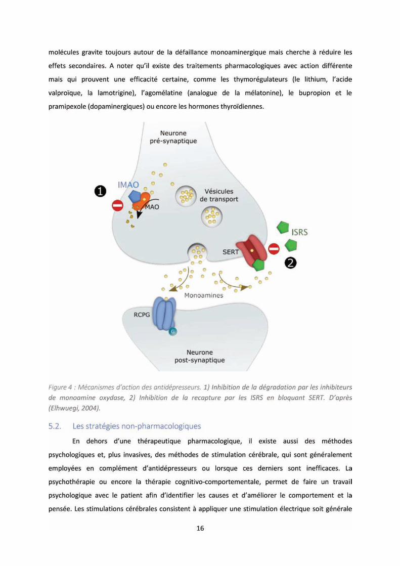
Figure : M a is es d’a tio des a tid p esseu s. .......................................................................... 16
Figure 5 : Les classes des canaux potassiques. ..................................................................................... 18
Figure : A e ph log i ue des K P hez l’hu ai .......................................................................... 20
Figure 7 : Schéma du contrôle multiple de TREK-1. .............................................................................. 25
Figure 8 : Schéma des récepteurs à domaine Vps10p. ......................................................................... 31
Figure 9 : La structure du NTSR3/Sortiline. ........................................................................................... 32
Figure 10 : Représentation schématique du NTSR3/Sortiline humain. ................................................ 33
Figure 11 : Transport des hydrolases lysosomales nouvellement synthétisées vers les lysosomes. ... 35
Figure 12 : Modèle schématique de la formation du complexe récepteur-p75NTR. ........................... 37
Figure 13 : La synthèse et le tri du BDNF. ............................................................................................. 39
Figure 14 : Séquence de la Spadine conçue à partir de la séquence propeptide. ................................ 42
Figure 20 : Comportement relatif à la dépression chez les souris KO-NTSR3. ..................................... 49
Figure 22 : Expression membranaire de TREK-1 et potentiel de membrane chez les souris KO-NTSR3.
.............................................................................................................................................................. 52
Figure 23 : Effet de la délétion du gène NTSR3/Sortiline sur la régulation du BDNF. .......................... 53
Figure 24: Interface de Behavior Trial Trigger v1.0. .............................................................................. 87
Figure 25 : Système de connexion en série dans le test de la boite clair-obscur ................................. 88
Figure 17 : Représentation de la moyenne ± SEM des sites de liaison totales et des sites de liaison
sensibles et insensibles à la lévocabastine ......................................................................................... 103
Figure 18 : Concentrations de NTS dans les sérums et dans le cerveau de souris WT et KO-NTSR3 . 104
Figure 19 : Réponses analgésiques des souris WT et KO-NTSR3. ....................................................... 105
Figure 15 : Peptides synthétisés et affinités. ...................................................................................... 116
Figure 16 : Concentrations de propeptide dans les sérums de patients sains (Controls) et patients
atteints d'un trouble dépressif majeur (MDD) non traités (T0) et traités (T1) pendant 12 semaines.
............................................................................................................................................................ 117
Figure 26 : Comportement des souris KO-NTSR3 dans la résignation acquise et la mémoire spatiale.
............................................................................................................................................................ 128
SOMMAIRE
REMERCIEMENTS ......................................................................................................................................
ABREVIATIONS ..........................................................................................................................................
LISTE DES FIGURES ....................................................................................................................................
SOMMAIRE ................................................................................................................................................
INTRODUCTION ....................................................................................................................................... 1
1 HISTOIRE DE LA DEPRESSION ...................................................................................................... 1
2 LE TROUBLE DEPRESSIF AUJOURD’HUI ....................................................................................... 3
2.1. L'épisode dépressif caractérisé (EDC) ..................................................................................... 3
2.1.1. Diagnostic ............................................................................................................................ 3
2.1.2. Formes cliniques ................................................................................................................. 4
2.2. Les types de troubles dépressifs ............................................................................................. 4
2.2.1. Trouble dépressif caractérisé .............................................................................................. 4
2.2.2. Trouble disruptif avec dysrégulation émotionnelle ............................................................ 4
2.2.3. Trouble dépressif persistant ............................................................................................... 5
2.2.4. Trouble dysphorique prémenstruel .................................................................................... 5
3 EPIDEMIOLOGIE .......................................................................................................................... 5
4 LA BIOLOGIE DE LA DEPRESSION ................................................................................................ 5
4.1. Anomalies cérébrales .............................................................................................................. 6
4.1.1. Anomalies corticales ........................................................................................................... 6
4.1.1.1. Le cortex préfrontal ........................................................................................................ 6
4.1.1.2. Le cortex cingulaire antérieur ......................................................................................... 7
4.1.1.3. Le cortex insulaire ........................................................................................................... 7
4.1.2. Anomalies des régions limbiques........................................................................................ 8
4.2. L'hypothèse monoaminergique : la sérotonine ...................................................................... 9
4.3. Neurogénèse et BDNF ........................................................................................................... 11
4.4. Stress et dépression .............................................................................................................. 12
5 STRATEGIES THERAPEUTIQUES ................................................................................................. 14
5.1. Les stratégies pharmacologiques .......................................................................................... 14
5.1.1. Inhiber les monoamines oxydase ..................................................................................... 15
5.1.2. Inhiber la recapture des monoamines .............................................................................. 15
5.2. Les stratégies non-pharmacologiques .................................................................................. 16
Conclusion ......................................................................................................................................... 17

6 LE CANAL TREK-1, ACTEUR EMERGENT DANS LA DEPRESSION ................................................ 17
6.1. Généralités sur les canaux potassiques ................................................................................ 17
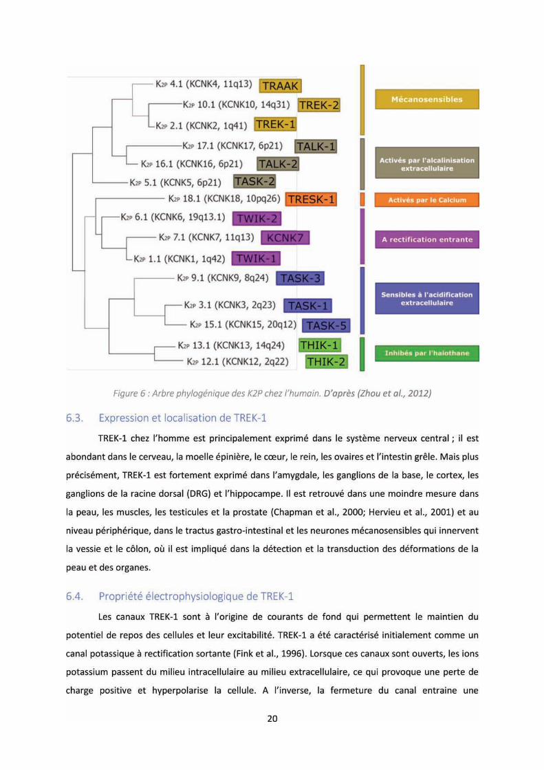
6.2. La classe des K2P .................................................................................................................... 18
6.3. Expression et localisation de TREK-1 .................................................................................... 20
6.4. Propriété électrophysiologique de TREK-1 ........................................................................... 20
6.5. La régulation de TREK-1 ........................................................................................................ 21
6.5.1. Sensibilité mécanique ....................................................................................................... 21
6.5.2. Sensibilité thermique ........................................................................................................ 22
6.5.3. Régulation par le pH .......................................................................................................... 22
6.5.4. Régulation par les lipides .................................................................................................. 22
6.5.5. Régulation par les RCPGs .................................................................................................. 23
6.6. Rôle de TREK-1 dans la dépression ....................................................................................... 23
6.7. Les partenaires de TREK-1 ..................................................................................................... 25
7 LE NTSR3 / SORTILINE, UNE PROTEINE ASSOCIEE A DE MULTIPLES FONCTIONS ..................... 26
7.1. La neurotensine et ses récepteurs ........................................................................................ 26
7.1.1. Généralités ........................................................................................................................ 26
7.1.2. Le NTSR1 ........................................................................................................................... 27
7.1.3. Le NTSR2 ........................................................................................................................... 28
7.2. Le NTSR3 / Sortiline ............................................................................................................... 29
7.2.1. Purification et clonage du NTSR3 / Sortiline ..................................................................... 29
7.2.2. Structure ........................................................................................................................... 30
7.2.3. Maturation ........................................................................................................................ 33
7.2.4. Distribution ....................................................................................................................... 34
7.3. Fonctions du NTSR3/Sortiline ............................................................................................... 34
7.3.1. Régulation du trafic intracellulaire ................................................................................... 34
7.3.2. Viabilité neuronale ............................................................................................................ 36
7.3.2.1. Co-recepteur du p75NTR .............................................................................................. 36
7.3.2.2. Régulation du BDNF ...................................................................................................... 37
7.3.3. Physiopathologie ............................................................................................................... 39
8 NTSR3/SORTILINE, PROPEPTIDE (PE), TREK-1 ET DEPRESSION ................................................. 41
OBJECTIFS .............................................................................................................................................. 45
Etude de la délétion du NTSR /So tili e da s la gulatio de l’ tat d p essif. .................................. 47
Article 1: Altered TREK-1 function in sortilin deficient mice results in an antidepressant phenotype.
.......................................................................................................................................................... 48
1. Co te te de l’ tude................................................................................................................... 48
2. Résultats et discussion .............................................................................................................. 49
Développe e t d’outils pou l’a al se du o po te e t hez le o geu . ....................................... 85
Article 2: Behavior Trial Trigger: Free standalone software using keyboard keys with possibility to
connect serial device (Arduino ®) for timing and analysis rodents during behavioral tests. ........... 86
Etude de la délétion du NTSR3/Sortiline dans le système neurotensinergique. ................................ 101
Article 3: Increased Brain Neurotensin and NTSR2 Lead to Weak Nociception in NTSR3/Sortilin
Knockout Mice. ............................................................................................................................... 102
1. Co te te de l’ tude................................................................................................................. 102
2. Résultats et discussion ............................................................................................................ 103
Etude du i eau i ula t de p opeptide da s l’affe t d p essif. ...................................................... 114
Article 4: Serum sortilin-derived propeptides concentrations are decreased in major depressive
disorder patients. ............................................................................................................................ 115
1. Co te te de l’ tude................................................................................................................. 115
2. Résultats et discussion ............................................................................................................ 116
DISCUSSION GENERALE et CONCLUSION ............................................................................................ 124
PERSPECTIVES ..................................................................................................................................... 129
ANNEXES ............................................................................................................................................. 131
REFERENCES ........................................................................................................................................ 176
RESUME ............................................................................................................................................... 193

1
INTRODUCTION
1 HISTOIRE DE LA DEPRESSION
Dépression, un terme qui représente un des maux le plus répandus de la fin du XXe siècle, mais il
est intéressant de sa oi u'histo i ue e t ette d sig atio s’est a o od e de di e s e p u ts
à travers les âges, renvoyant souvent au même syndrome, et que, de cette complexité de lui
soustraire une identité distincte, en résultait l'idée d'un concept difficilement définissable sur le plan
de la pathologie e tale. De l’A ti uit jus u’à fi des a es , la d p essio tait u e
affectation qui pouvait se manifester chez tout individu et dans toutes pathologies sans pour autant
en être considérée comme une.
L'émergence des symptômes de l'état dépressif prend ses origines très tôt dans notre histoire.
On peut ite le pap us d’E e s, d ou e t e à Lou o pa Edwin Smith, ancien traité médical
datant du XVIe siècle avant notre ère, qui y référence, sous le chapitre « li e des œu s », des
descriptions des états pathologiques de la démence ou encore de la dépression.
Au Ve siècle avant J-C, Hippocrate avancera le terme de melancholia, correspondant
étymologiquement à la « bile noire » nommé aussi atrabile, qui aura pour siège la rate (spleen), et
sera une des quatre humeurs de la théorie des humeurs, popularisée par le Corpus Hippocraticum
qui posera les bases de la médecine antique. Cette mélancolie définira un état de tristesse et de
peur.
Le philosophe Sénèque exprimera au Ier siècle avant J-C, le concept de Taedium vitae ou fatigue
de i e, ui tou he a d’ailleu s le po te philosophe Lu e, dis iple d’Epi u e, et ui e la e a ue
« l’ho e est u alade ui ig o e la ause de so al » (De rerum natura).
C’est e s le IVe si le u’appa ait a da s la ie o asti ue le te e d’a die, ologis e
grecque ancien, signifiant la privation de soi, la négligence (acedia) et décrite comme un manque de
soi à l' ga d de sa p op e ie spi ituelle d p essio spi ituelle . L’a die se disti gue pa u e
app o he th ologi ue de l’affe t d p essif, alo s ue la la olie tait li e à u e otio
physiologique chez les Grecs. Par ailleurs, elle sera considérée comme un péché contre Dieu par
Sai t F a çois d’Assise. Cette a die te d a à s’a e uise , et de ie d a paresse à partir du XIIIe
siècle, décontextualisant ce péché théologique pour en faire un péché sociétal. A cette époque
appa ait a gale e t le lie e t e a ou o t a i et la olie, lie u’e pose le de i Jacques
Ferrand en 1610 dans Traicté de l'essence et guérison de l'amour ou de la mélancholie érotique,

2
précisant que « l A ou ou passio É oti ue est u e esp e de ve ie, p o eda te d'u d si d gl
de jouir de la chose aimable, accompagnée de peur, et de tristesse ».
Le XVIIIe siècle, début du siècle des Lumières, période de la critique sociale, de l'esprit, des
sciences, où la connaissance est promue face à l'obscurantisme, verra l'émergence des disciplines
psychologiques et permettra l'évolution de la pensée à l'égard des maladies mentales. Le médecin
Johann Christian Reil avancera le terme de psychiatrie en 1808 et considéra l'idée que les
traitements des maladies psychiques répondent des méthodes médicales. La mélancolie empruntera
à ce moment-là deux destins, celui de subsister comme trait des hommes de génies, prenant part au
Romantisme (fin du XVIIIe siècle), incarnant chez l'artiste l'allégorie du tragique et du malheur dont
le su li e est à la hauteu de l'esp it et e o a t au uestio e e t d’A istote; « Pourquoi tous les
hommes qui furent exceptionnels en philosophie, en politique, en poésie ou dans les arts étaient-ils
a ifeste e t ilieux […] », et de l'autre, une maladie au sens médicale du terme pour l'homme
commun. Maladie qui devient « nerveuse » quand les nerfs et le cerveau sont considérés comme le
siège du comportement intellectuel et physique de l'individu, et qui sera « interprétée comme la
conséquence d'un choc psychique ou d'une tension excessive due aux circonstances extérieures »
(Jean Starobinski, Histoire du traitement de la mélancolie des origines à 1900).
Il faudra attendre le XIXe siècle pour que le terme dépression prenne un sens métaphorique.
Provenant du latin depressio, qui signifie « enfoncement », il représentera de façon plus générale un
état mental de lassitude, intégrante de la mélancolie, mais moins important. Esquirol, en 1819,
reprendra le terme de mélancolie pour le classer en un délire partiel, qui nommera lypémanie, une
monomanie où prédomine « une passion triste et dépressive ». La mélancolie deviendra par la suite
un trouble distinct intégré à la manie (folie), que ce soit Jean Pierre Ferlet avec la folie circulaire
(1854) ou encore Emil Kraepelin et ses travaux sur la psychose périodique maniaco-depressive, ils
expliqueront qu'il existe une oscillation entre phase de manie et de dépression (deviendra le trouble
bipolaire d'aujourd'hui). A noter qu'à cette époque, les étiologies des maladies mentales écartaient
les altérations psychologiques et se centraient principalement sur les lésions neurologiques,
exemple avec le psychiatre Valentin Magnan qui décrit la manie et la mélancolie comme syndromes
pouvant relever d'étiologies telles que la paralysie, l'alcoolisme, l'épilepsie, la dégénérescence
(théorie).
Vers la fin du XIXe siècle, apparaitra la neurasthénie, inventée par l'américain George Miller
Beard (1860), qui va s'imposer comme la maladie de la vie moderne, amenant la notion de trouble
fonctionnel et clivant le modèle de liaison syndrome – lésion des maladies. Elle est caractérisée par
un épuisement nerveux prenant son étiologie dans le facteur social (vie moderne) (A Practical
3
Treatise on Nervous Exhaustion, 1869) et composera les bases de la notion d'exogène (conséquence
d'un évènement externe induisant une réaction interne). Sigmund Freud, ainsi que Pierre Janet,
s'intéresseront à l'étude de la neurasthénie et montreront, cette fois-ci, une notion d'endogène,
résultante d'une origine psychique. La neurasthénie sera, par la suite, intégrée dans le spectre de la
névrose.
Jusqu'à la fin de la Seconde guerre mondiale, la mélancolie de la psychose maniaco-dépressive
restera l'entité majeur des formes de la dépression, gravitant autour des formes plus atténuées,
dépression réactionnelle, et des formes névrotiques, dépression névrotique. L'avènement de la
sismothérapie (appelée aujourd'hui l'électro-convulsivothérapie (ECT)) en 1940, va grandement
améliorer les troubles de l'humeur et son utilisation va dépasser le seul cadre de la mélancolie. Par la
suite, et avec l'arrivée des traitements chimiques, la notion de dépression va prendre son essor et va
devenir un trouble de l'humeur à part entière où sera finalement réorganisée la mélancolie.
On ne peut que constater, de par cette brève rétrospective, l'hétérogénéité sémantique et la
difficulté sémiologique que la dépression a parcouru durant des siècles, et on se rend bien compte
que l'étiologie de ce mal, qui est au carrefour de multiples disciplines, reste encore de nature
indistincte et grandement discutée aujourd'hui.
A ce jour, la dépression est à mettre dans les troubles dépressifs, et le terme en lui-même se
réfère surtout à la dépression majeure ou trouble dépressif caractérisé persistant.
2 LE TROUBLE DEPRESSIF AUJOURD’HUI
Le t ou le d p essif fait pa tie des t ou les de l'hu eu dit u ipolai e, ’est-à-dire qui ne
s'exprime qu'en une seule humeur sans alternance périodique avec une autre. Un trouble dépressif
est déterminé à la suite d'épisodes dépressifs caractérisés (EDC) et peut-être d'intensité légère,
modérée ou sévère.
2.1. L'épisode dépressif caractérisé (EDC)
2.1.1. Diagnostic
Selon la 5ème édition du Diagnostic and Statistical Manual of Mental Disorders (DSM-V), le
diagnostic établit d'un épisode dépressif caractérisé doit faire état des critères suivants : présenter
au moins 5 des symptômes listés ci-dessous, en incluant une humeur dépressive ou une anhédonie,
et ce sur une période d'au moins 2 semaines.
4
Liste des symptômes relatifs à un trouble dépressif, établie par la DSM-V :
Humeur dépressive (sentiment de tristesse ou vide).
Anhédonie.
Perte ou gain de poids significatif ou d'appétit.
Insomnie ou hypersomnie.
Agitation ou ralentissement psychomoteur.
Fatigue ou perte d'énergie.
Sentiment de dévalorisation ou de culpabilité excessive ou inappropriée.
Diminution de l'aptitude à penser ou à se concentrer ou indécision.
Pensées de mort ou idées suicidaires récurrentes.
2.1.2. Formes cliniques
Il existe plusieurs formes cliniques distinctes pouvant préciser le caractère de l'EDC. On
retrouvera la caractéristique mélancolique, forme très sévère présentant une forte probabilité
suicidaire et associée à une intense humeur dépressive, le caractère atypique, se distinguant par une
humeur réactive et positive s'opposant à l'humeur dépressive habituelle (anhédonie paradoxale), le
caractère psychotique, relevant de la même symptomatologie que l'épisode mélancolique avec
association d'idées psychotiques (délires ou hallucinations), et enfin, la détresse anxieuse et le
caractère post partum.
2.2. Les types de troubles dépressifs
En fonction du contexte de manifestation et également de la progression du ou des
épisodes, il est défini, selon la DSM-V, différents types de troubles dépressifs.
2.2.1. Trouble dépressif caractérisé
Le trouble dépressif caractérisé correspond à la présence d'épisodes dépressifs caractérisés,
’est-à-dire, à une humeur dépressive latente occupant une importante part de la journée, et ce, sur
plusieurs jours consécutifs, sans inclure spécifiquement les symptômes de variations de poids et de
pensées suicidaires.
2.2.2. Trouble disruptif avec dysrégulation émotionnelle
Ce trouble désigne l'expression d'une dérégulation émotionnelle centrée sur un état
d'irritabilité sévère et persévérant. Il y a manifestation régulière de crises de colère et une
importante humeur d'irritabilité qui persistent depuis au moins 1 an.
5
2.2.3. Trouble dépressif persistant
L'humeur dépressive est récurrente et se manifeste depuis plus de deux ans tous les jours. Il
peut faire suite à des épisodes dépressifs caractérisés ou une dépression caractérisée. Ce trouble
réunit la dépression majeure et la dysthymie explicitée dans l'ancienne version du DSM (DSM-IV).
2.2.4. Trouble dysphorique prémenstruel
Le trouble dysphorique prémenstruel se rapporte à une symptomatologie dépressive durant
le cycle menstruel et peut résulter d'une instabilité émotionnelle, d'une forte anxiété ou de
manifestations physiques (douleurs musculaires, articulaires, tensions mammaires).
Enfin, il existe également des troubles dépressifs induit par une substance ou encore dû à une
autre affection médicale.
3 EPIDEMIOLOGIE
Selon l'Organisation Mondial pour la Santé, les troubles dépressifs représentent le 1er facteur de
o idit et d’i apa it su le pla o dial et de ie d o t la se o de ause d'i alidit d'i i .
On estime à plus de 300 millions le nombre de personnes souffrant de troubles dépressifs, avec une
prévalence de l'EDC en France aux alentours de 7,5 % au cours des douze derniers mois (Baromètre
sa t de l’I pes, . La d p essio ajeu e trouble dépressif persistant) atteint près de 24 % de
la population française, avec deux fois plus de risques chez les femmes que les hommes (Lépine and
Briley, 2011). Pour un coût annuel total estimé à 118 milliards d'euros en Europe, ce qui représente
près de 1% de l'économie européenne totale (PIB), la dépression correspond au plus onéreux des
troubles mentaux (Sobocki et al., 2006). L'incidence de cette maladie sur la qualité de vie des
patients est préoccupante et la dépression pose un problème de santé majeur à travers le monde.
Malgré les avancées des recherches dans ce domaine, il reste encore de nombreuses zones
d'incompréhension sur la physiopathologie de ce mal du siècle.
4 LA BIOLOGIE DE LA DEPRESSION
A a t de s’atta de su les auses et o s ue es iologi ues de la dépression, il est important
de rappeler que cette pathologie repose aussi sur un aspect psychologique et social. Brièvement, il
se le ait u’il e iste u e elatio p di ti e e t e l’ tat et les t aits otio els, et la d p essio .
En effet, une tendance permanente à l'expérience des émotions négatives ou une attitude négative
serait précurseur du développement et de la persistance du trouble dépressif majeur (Morris et al.,
2009), e ui o o de a e l’id e fo tio aliste ui lie l’ otio à l’adaptatio . Il e iste gale e t
des travaux mettant en lumière la conséquence de l'environnement social dans l'étiologie de la
maladie, par exemple des événements indésirables de la vie et le faible soutien social sont des
6
facteurs de risque connus de dépression (Paykel et al., 1969), ou encore, un traumatisme durant
l'enfance, supplémenté d'une réponse à un stress, accroit le risque d'une dépression à l'âge adulte
(Heim et al., 2008). Par ailleurs, selon le degré de l'épisode dépressif de départ, il existe une
vulnérabilité plus ou moins importante de l'influence sociale sur l'issue d'une dépression majeure
(Leskelä et al., 2006).
Su le pla iologi ue, ’est le cerveau, médiateur du comportement animal, qui fait l'objet
d'une attention particulière dans l'étude des causes et conséquences du trouble dépressif. Les
recherches basées sur le système nerveux central apportent des éléments de réponses dans la
compréhension et la biologie de la pathologie, en s'appuyant notamment sur des
dysfonctionnements et dérégulations aux niveaux anatomiques et biochimiques.
4.1. Anomalies cérébrales
L’i age ie ale o ait u e oissa e ota le da s l’ tude de la d p essio depuis
plusieurs années et les techniques utilisées ont permis de mettre en lumière des anomalies
ales au i eau de la o phologie, de l’a ti it et gale e t des i uits eu o au (Pandya et
al., 2012).
4.1.1. Anomalies corticales
Les régions corticales associées à la dépression sont le cortex préfrontal dorsolatéral et
ventromédian, le cortex orbitofrontal, le cortex cingulaire a t ieu do sal et e t al et l’i sula
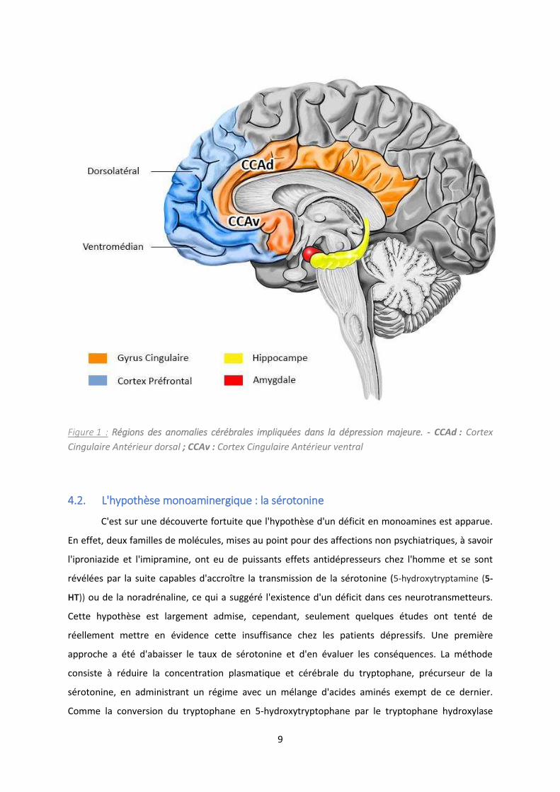
(Figure 1).
4.1.1.1. Le cortex préfrontal
Situé dans la partie antérieure du lobe frontal, excluant la région postérieure motrice, le cortex
p f o tal CPF a u ôle e t al da s le t aite e t et le o t ôle de l’i fo ation émotionnelle. Le
CPF fait partie d'un réseau neuronal étendu qui projette et intègre de nombreuses connexions avec
d’aut es gio s, ui so t ota e t espo sa les du o t ôle de la dopa i e, de la o ad ali e
et de la sérotonine, trois neurotransmetteu s i po ta ts da s la gulatio de l’hu eu .
La partie dorsolatérale du CPF, qui joue un rôle dans la planification et les fonctions exécutives,
p se te u e aisse d’a ti it chez les patients dépressifs, traduite par une diminution du flux
sanguin et du métabolisme glucidique (Kimbrell et al., 2002) et, de façon intéressante, il est possible
de reverser cette activité par un traitement antidépresseur (Mayberg et al., 1999). En outre, une
lésion de cette même région peut entrainer une vulnérabilité au développement d'une dépression
(Koenigs et al., 2008).
7
Le CPF ventromédian est impliqué dans l'évaluation du potentiel de récompense des stimuli. Il
projette des afférences directement sur l'amygdale, et joue donc un rôle dans l'inhibition de
réactions émotionnelles, et dans le processus décisionnel. A l'inverse du CPF dorsolatéral, il existe
une hyperactivité cérébrale chez les patients dépressifs (Drevets et al., 1992) et une lésion bi-latérale
de la région entraine un faible risque de dépression (Koenigs et al., 2008).
4.1.1.2. Le cortex cingulaire antérieur
Le cortex cingulaire antérieur (CCA) a également un rôle prépondérant dans la physiopathologie de
la dépression. Chez les patients dépressifs, il apparait une diminution du métabolisme glucidique
dans le cortex cingulaire antérieur ventral (Drevets et al., 1997) mais également des anomalies dans
la partie dorsale et ventrale du CCA (Mayberg et al., 1999). Il existe une division fonctionnelle du
CCA entre sa partie dorsale et ventrale, en effet, le CCA dorsal est impliqué dans l'aspect cognitif des
émotions, y compris la résolution des conflits de stimuli émotionnels avec valence négative (Etkin et
al., 2006; Vogt et al., 1992), tandis que le CCA ventral ou subgenual, joue un rôle dans l'aspect
affectif de l'émotion. Ce dernier est connecté aux régions limbiques tels que l'amygdale, le thalamus
dorsomédian et l'hippocampe mais également au cortex orbitofrontal et CPF ventromédian, ce qui
l'implique dans la régulation de la réponse émotionnelle (Critchley, 2004; Vogt et al., 1992). Le CCA
ventral a également des connexions vers l'hypothalamus, impliqué dans la réponse au stress.
4.1.1.3. Le cortex insulaire
L'insula est une structure du cortex cérébral située dans la profondeur de la scissure de Sylvius, le
sillon latéral, et est subdivisée en deux parties : une large insula antérieure et une petite insula
postérieure. Les parties antérieure et postérieure de l'insula reçoivent des afférences du noyau
ventral du thalamus, et la partie antérieure projette et reçoit des afférences directement de
l'amygdale. Son implication prend part dans l'autosuggestion, le dégout, l'évaluation des états
viscéraux internes, et la réponse aux stimuli du goût et de l'odorat. Chez les patients déprimés, il
existe une augmentation de la sensibilité de l'insula face à des stimuli négatifs (Surguladze et al.,
2010) mais également, il apparait une réduction du volume cérébral dans cette région
(Sprengelmeyer et al., 2011).
8
4.1.2. Anomalies des régions limbiques
L'hippocampe, l'amygdale et l'hypothalamus sont les principales régions affectées par la
dépression (Figure 1). Elles font partie du système limbique, système qui représente le régulateur
central du comportement et plus particulièrement des émotions comme la peur, le plaisir et
l'agressivité, et joue également un rôle crucial dans les mécanismes mnésiques.
L'hippocampe, structure localisée dans le lobe temporal médian, est impliqué dans la
mémoire, la navigation spatiale et également dans l'inhibition du comportement. On suggère son
implication dans le comportement émotionnel par l'existence de connexions avec des structures
asso i es o e le septu , l’h pothala us et le o ple e u l ai e a t ieu e du thala us, d’où
son inclusion dans le système limbique. Chez les personnes souffrant d'une dépression, le volume
hippocampique est significativement réduit (Lorenzetti et al., 2009), et de façon intéressante, les
patients en rémission, après traitement, présentaient un volume hippocampique de départ plus
important que ceux qui ne répondaient pas aux traitements, suggérant une sensibilité de réponse
induite par l'hippocampe (MacQueen et al., 2008). Concernant l'amygdale, c'est une structure qui se
situe dans le lobe temporal en avant de l'hippocampe et qui est impliquée dans la reconnaissance et
l'évaluation de la valence émotionnelle des stimuli sensoriels, dans l'apprentissage associatif et dans
les réponses comportementales et végétatives associées en particulier dans la peur et l'anxiété. Il
existe, comme dans l'hippocampe, une atrophie de l'amygdale lors d'une dépression majeure
(Hamilton et al., 2008), mais également une hyperactivation cérébrale (Drevets et al., 2002) qui
engendrerait une importante stimulation de l'axe hypothalamique pituitaire surrénalien (HPA) et par
conséquent une sécrétion de l'hormone du stress, le cortisol.
Il demeure également d'autres structures cérébrales étudiées lors d'une dépression
majeure, comme le striatum ou encore le thalamus, mais il apparait des divergences dans les
résultats obtenus et ils ne permettent pas de statuer sur les conséquences exactes de l'affect
dépressif sur leurs morphologies ou l'activité cérébrale.
9
Figure 1 : Régions des anomalies cérébrales impliquées dans la dépression majeure. - CCAd : Cortex
Cingulaire Antérieur dorsal ; CCAv : Cortex Cingulaire Antérieur ventral
4.2. L'hypothèse monoaminergique : la sérotonine
C'est sur une découverte fortuite que l'hypothèse d'un déficit en monoamines est apparue.
En effet, deux familles de molécules, mises au point pour des affections non psychiatriques, à savoir
l'iproniazide et l'imipramine, ont eu de puissants effets antidépresseurs chez l'homme et se sont
révélées par la suite capables d'accroître la transmission de la sérotonine (5-hydroxytryptamine (5-
HT)) ou de la noradrénaline, ce qui a suggéré l'existence d'un déficit dans ces neurotransmetteurs.
Cette hypothèse est largement admise, cependant, seulement quelques études ont tenté de
réellement mettre en évidence cette insuffisance chez les patients dépressifs. Une première
approche a été d'abaisser le taux de sérotonine et d'en évaluer les conséquences. La méthode
consiste à réduire la concentration plasmatique et cérébrale du tryptophane, précurseur de la
sérotonine, en administrant un régime avec un mélange d'acides aminés exempt de ce dernier.
Comme la conversion du tryptophane en 5-hydroxytryptophane par le tryptophane hydroxylase
10
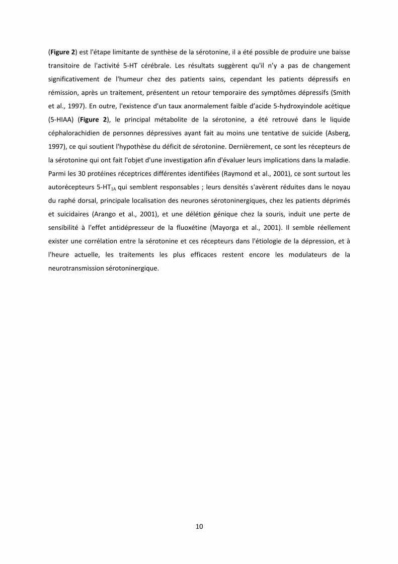
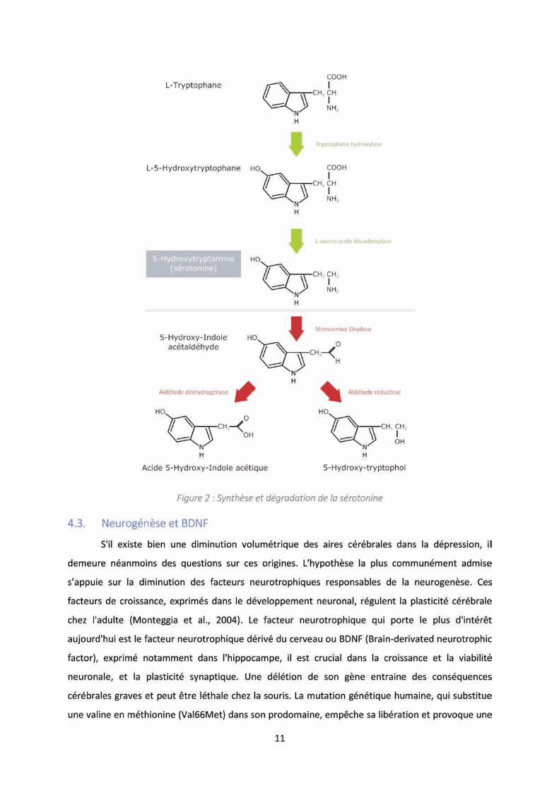
(Figure 2) est l'étape limitante de synthèse de la sérotonine, il a été possible de produire une baisse
transitoire de l'activité 5-HT cérébrale. Les résultats suggèrent qu'il ’ a pas de changement
significativement de l'humeur chez des patients sains, cependant les patients dépressifs en
rémission, après un traitement, présentent un retour temporaire des symptômes dépressifs (Smith
et al., 1997). E out e, l'e iste e d'u tau a o ale e t fai le d’a ide -hydroxyindole acétique
(5-HIAA) (Figure 2), le principal métabolite de la sérotonine, a été retrouvé dans le liquide
céphalorachidien de personnes dépressives ayant fait au moins une tentative de suicide (Asberg,
1997), ce qui soutient l'hypothèse du déficit de sérotonine. Dernièrement, ce sont les récepteurs de
la sérotonine qui ont fait l'objet d'une investigation afin d'évaluer leurs implications dans la maladie.
Parmi les 30 protéines réceptrices différentes identifiées (Raymond et al., 2001), ce sont surtout les
autorécepteurs 5-HT1A qui semblent responsables ; leurs densités s'avèrent réduites dans le noyau
du raphé dorsal, principale localisation des neurones sérotoninergiques, chez les patients déprimés
et suicidaires (Arango et al., 2001), et une délétion génique chez la souris, induit une perte de
sensibilité à l'effet antidépresseur de la fluoxétine (Mayorga et al., 2001). Il semble réellement
exister une corrélation entre la sérotonine et ces récepteurs dans l'étiologie de la dépression, et à
l'heure actuelle, les traitements les plus efficaces restent encore les modulateurs de la
neurotransmission sérotoninergique.
12
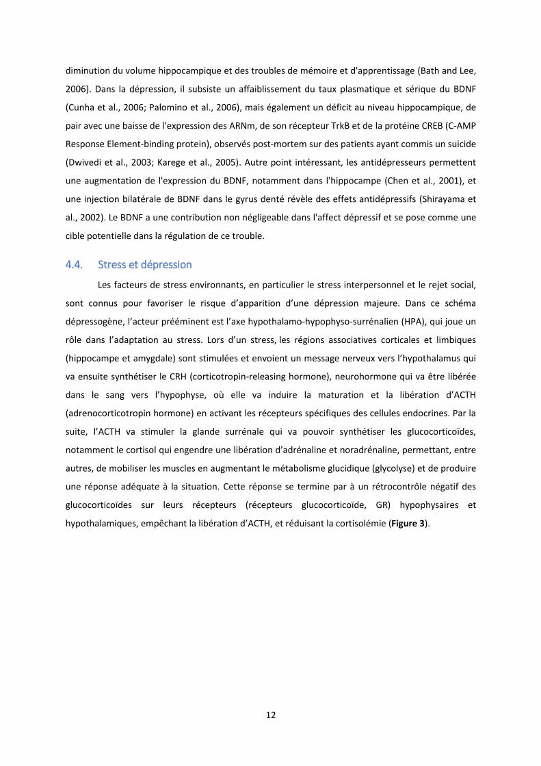
diminution du volume hippocampique et des troubles de mémoire et d'apprentissage (Bath and Lee,
2006). Dans la dépression, il subsiste un affaiblissement du taux plasmatique et sérique du BDNF
(Cunha et al., 2006; Palomino et al., 2006), mais également un déficit au niveau hippocampique, de
pair avec une baisse de l'expression des ARNm, de son récepteur TrkB et de la protéine CREB (C-AMP
Response Element-binding protein), observés post-mortem sur des patients ayant commis un suicide
(Dwivedi et al., 2003; Karege et al., 2005). Autre point intéressant, les antidépresseurs permettent
une augmentation de l'expression du BDNF, notamment dans l'hippocampe (Chen et al., 2001), et
une injection bilatérale de BDNF dans le gyrus denté révèle des effets antidépressifs (Shirayama et
al., 2002). Le BDNF a une contribution non négligeable dans l'affect dépressif et se pose comme une
cible potentielle dans la régulation de ce trouble.
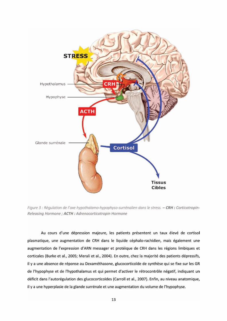
4.4. Stress et dépression
Les facteurs de stress environnants, en particulier le stress interpersonnel et le rejet social,
so t o us pou fa o ise le is ue d’appa itio d’u e d p essio ajeu e. Da s e s h a
dépressogène, l’a teu p i e t est l’a e hypothalamo-hypophyso-surrénalien (HPA), qui joue un
rôle da s l’adaptatio au st ess. Lo s d’u st ess, les régions associatives corticales et limbiques
hippo a pe et a gdale so t sti ul es et e oie t u essage e eu e s l’h pothala us ui
va ensuite synthétiser le CRH (corticotropin-releasing hormone), neurohormone qui va être libérée
da s le sa g e s l’h poph se, où elle a i dui e la atu atio et la li atio d’ACTH
(adrenocorticotropin hormone) en activant les récepteurs spécifiques des cellules endocrines. Par la
suite, l’ACTH a sti ule la gla de surrénale qui va pouvoir synthétiser les glucocorticoïdes,
notamment le cortisol ui e ge d e u e li atio d’ad ali e et o ad ali e, pe etta t, entre
autres, de mobiliser les muscles en augmentant le métabolisme glucidique (glycolyse) et de produire
une réponse adéquate à la situation. Cette réponse se termine par à un rétrocontrôle négatif des
glucocorticoïdes sur leurs récepteurs (récepteurs glucocorticoïde, GR) hypophysaires et
hypothalamiques, e p ha t la li atio d’ACTH, et réduisant la cortisolémie (Figure 3).
14
L’h poth se qui rend compte de ces anomalies suggère que l’hypercortisolémie, induite par
l’augmentation de CRH, est responsable de l’hyperplasie des surrénales qui va, à posteriori,
maintenir le cortisol circulant. Par la suite, l’e positio p olo g e au o tisol va conduire à la
désensibilisation des récepteurs glucocorticoïde à l'ACTH (Gold et al., 1986), mais également à la
perte des neurones contenant les GR dans l'hippocampe (Sapolsky et al., 1984), d’où le déficit de la
boucle de rétroaction négative des glucocorticoïdes. D’ailleu s, la délétion ciblée des GR dans le
cerveau de souris adultes induit une augmentation de l'activité de l'axe HPA et conduit à une
augmentation de l'immobilité dans les tests de résilience (Force Swim Test), et peut être reversées
par les antidépresseurs (Boyle et al., 2005). Enfin, l’h pe t ophie de l’h poph se résulterait de
l’aug e tatio de sécrétion de CRH dans le cerveau. Cette augmentation de CRH constante pourrait
s’e pli ue pa la p titio p olo g e des situatio s de st ess avec une forte sensibilité à ces
événements, qui va instaurer p og essi e e t u e sti ulatio auto o e de l’a e HPA.
Le stress altère également la neurogénèse et le BDNF, mais aussi la réponse sérotoninergique. La
o te tio de sou is pa l’i te diai e d’u e e ei te est ei te, a oit le st ess et produit un
remodelage réversible des dendrites apicales des neurones pyramidaux CA3 et la suppression de la
neu oge se, a e u e di i utio d’e p essio de BDNF, da s le g us de t , changements
morphologiques associés aux déficits de mémoire dépendante de l'hippocampe (Angelucci et al.,
2005; Reagan et al., 2004). Les neurones sérotoninergiques du noyau dorsal du raphé augmentent
leurs fréquences de décharges en réponse à un stress aigu. Quand le stress devient chronique, il y a
une altération de la décharge, probablement induite par une désensibilisation des récepteurs 5-HT,
notamment 5-HTA1, pouvant être responsable du déficit monoaminergique (Mahar et al., 2014).
5 STRATEGIES THERAPEUTIQUES
5.1. Les stratégies pharmacologiques
Les traitements les plus représentés dans la sphère thérapeutique de la dépression, sont les
molécules ciblant le déficit monoaminergique. Le ode d’a tio de es antidépresseurs se focalise
principalement sur des mécanismes permettant de réguler le niveau des neurotransmetteurs et
notamment la sérotonine. Deux schémas de mécanismes sont utilisés : l’inhibition du système de
dégradation des monoamines et celui de la recapture des monoamines. On retrouve donc la classe
des antidépresseurs inhibiteurs de la monoamine oxydase (IMAO) (système de dégradation) et les
inhibiteurs de la recapture ; les antidépresseurs Tricycliques (ATC) et les inhibiteurs spécifiques de la
recapture de la sérotonine (ISRS).
15
5.1.1. Inhiber les monoamines oxydase
La monoamine oxydase (MAO) est une enzyme présente dans la membrane mitochondriale
externe des cellules neuronales et non neuronales. Elle est responsable de la désamination
oxydative des monoamines endogènes avec fonctions de neurotransmission : noradrénaline,
adrénaline, dopamine, sérotonine et, indirectement, tyramine (Baker et al., 1985). Les MAOs sont
des flavoprotéines présentent sous 2 isoformes, la monoamine oxydase-A (MAO-A), qui désamine
préférentiellement la sérotonine, noradrénaline et adrénaline, et la monoamine-oxydase B (MAO-B),
qui cible la dopamine (Strolin Benedetti and Dostert, 1987). De façon prédominante, ce sont les
inhibiteurs de la MAO-A qui sont utilisés, de par leur action sur les neurotransmetteurs considérés
comme essentiels dans la dépression, à savoir la noradrénaline et la sérotonine (Figure 4).
5.1.2. Inhiber la recapture des monoamines
La seconde cible des antidépresseurs est le système de recapture des monoamines au niveau
présynaptique. Le mode d’a tio ise di e te e t les t a spo teu s ui pe ette t de recouvrer
une partie des neurotransmetteurs libérés dans la fente synaptique. D’u e pa t, il a les ATC, ui
inhibent la recapture de manière compétitive en se fixant sur le site de liaison des transporteurs de
la transmission sérotoninergique et noradrénergique (Delgado and Moreno, 1999). D’aut e pa t, o
retrouve les ISRS, les antidépresseurs les plus largement utilisés pour réduire les symptômes de la
dépression, incluant le plus connu, la fluoxétine (Prozac ®). A la différence des autres classes
d’a tid p esseu s, les ISRS i hi ent de façon sélective le transport de la sérotonine, SERT, au niveau
présynaptique, et pe et ai si d’aug e te le i eau de -HT (Figure 4). L’a a tage d’u e telle
sélectivité est la suppression en grande partie des effets secondaires incommodant retrouvés dans
les lasses p de tes, sa s pou auta t pe d e l’effi a it antidépresseur (Hyttel, 1994).
En considérant le déficit monoaminergique comme un pivot dans la dépression, il y une
ide e da s l’utilisatio des IMAO, ATC et ISRS pour pallier à cette insuffisance. Cependant, il
su siste des o t ai tes à l’utilisatio de e t pe de ol ule pha a ologi ue. D’u e pa t, les
effets secondaires sont nombreux ; les ATC provoquent des effets anticholinergiques périphériques
et centraux (sécheresse buccale, constipation, rétention urinaire, mydriase, vision trouble et
tachycardie, confusion mentale, tremblements des extrémités, risques épileptogènes) ou encore une
sédation, de même que les IMAO et ISRS peuvent provoquer des nausées, constipation ou diarrhées,
insomnies et ig ai es. D’aut e pa t, les effets fi ues e so t pas i diats et essite t u
te ps d’adaptatio de jou s e o e e (Manji et al., 2001). Enfin, le diagnostic de la pathologie
essite d’ t e p is, a , e ue des diff e tes fo es du t ou le d p essif, la po se au
traitement antidépresseur peut varier et même être négative. Le développement de nouvelles
17
sur le crâne du patient (électroconvulsivothérapie, ECT), soit de manière plus invasive mais ciblée
(stimulation cérébrale profonde, Deep Brain Stimulation, DBS), en insérant une électrode
directement sur des zones spécifiques du cerveau par stéréotaxie. L’ECT et le DBS so t des
méthodes particulièrement efficaces pour traiter les troubles de la dépression (Perrin et al., 2012),
et sont surtout utilisées pour les cas les plus sévères. Malg l’effi a it du t aite e t, les
mécanismes induit ne sont pas encore connus et il existe néanmoins des effets secondaires, comme
une perte de mémoire transitoire, un gain de poids ou encore un changement de personnalité
(Sackeim et al., 2007).
Conclusion
La compréhension du trouble dépressif représente un investissement important face au nombre
grandissant de personnes touchées chaque année. Maladie d’attei te ps hologi ue et iologi ue,
incriminant un des organes les plus complexe et élaboré u’est le e eau, les causes et
o s ue es de ette pathologie s’appuie t su de o eu a es dis ipli ai es, complexifiant
l’ e ge e d’app o hes th apeuti ues effi a es pou une réelle guérison. Les traitements actuels
tentent principalement d’a lio e l’hu eu des patie ts, en odula t l’h poth ti ue déficit
monoaminergique ou, dans les cas les plus critiques, en stimulant électriquement le cerveau.
Te h i ues d’usage a ept es, effi ie tes, ais pa fois utilisées sans compréhension intégrale du
modèle mécanistique, les effets ne sont pas immédiats, la période de traitement est généralement
longue, les effets secondaires marqués et parfois, on note l’appa itio d’u s d o e de se age à
l’a t du t aite e t. Le besoin réel de trouver de nouveaux traitements semble univoque, tant pour
la compréhension de cet affect psychologique, que pour soulager les patients.
6 LE CANAL TREK-1, ACTEUR EMERGENT DANS LA DEPRESSION
Récemment, des études précliniques basées sur des modèles expérimentaux de souris, ont
apporté des preuves convaincantes impliquant le canal potassique TREK-1 (TWIK-Related K+ channel
1) dans la physiopathologie de la dépression (Heurteaux et al., 2006).
6.1. Généralités sur les canaux potassiques
Les canaux potassiques sont des canaux ioniques largement répandus chez les organismes
vivants et ont pour fonctions la régulation de nombreux processus cellulaires. Ce sont des protéines
membranaires permettant le passage sélectif du potassium à travers la membrane. On les retrouve
dans l’e ita ilit eu o ale, où ils so t espo sa les des pote tiels d’a tio et d fi isse t le
potentiel de repos membranaire, la nociception et le volume cellulaire. Les canaux potassiques sont
regroupés en trois grandes classes basées sur leur organisation structurale (Lesage et al., 1997) ; 1)
les canaux à 6 segments transmembranaires et un domaine pore (6TM-1P), contenant les canaux

19
2000), la régulation de la dé ha ge de pote tiels d’a tio s (Brickley et al., 2007), l’ pilepsie ou
e o e l’apoptose (Lauritzen et al., 2003).
Les canaux TALK (TWIK-related Alkaline pH- activated K+ channel), qui regroupent TASK-2,
TALK-1 et TALK-2 (Reyes et al., 1998), sont également des canaux sensibles à la variation de pH
comme les TASK, à la différence que cette sensibilité se situe dans des pH alcalins. TASK-2 est
impliqué dans la réabsorption du bicarbonate dans le rein. L’aug e tatio du t a spo t du
bicarbonate dans les cellules tubulaires proximales alcalinise le compartiment basolatéral et active
une conductance potassique (Warth et al., 2004). On le retrouve également impliqué dans le
potentiel de repos des cellules musculaires lisses de l’a t e pul o ai e hez le at (Gönczi et al.,
2006). En ce qui concerne TALK-1 et TALK- , ils so t a ti s pa l’o de it i ue NO et les esp es
réactives d'oxygène (Duprat et al., 2005) et sont proposés comme cibles auxiliaires lors de la
stimulation physiologique vagale.
Les canaux THIK (Tandem pore domain Halothane-Inhibited K+ channel), THIK-1 et THIK-2,
produisent une fuite de courant potassique et ne sont pas influencés pa l’a idifi atio ou pa le
changement de température, mais peuvent t e i hi pa l’halothane (Rajan et al., 2001).
La famille TRESK (Twik-Related Spinal Cord K+ channel), ui e o tie t u’u seul a al
(KCNK18), a été cloné à partir de moelle épinière humaine (Sano et al., 2003) et peut être régulé par
le calcium ou par interaction protéique. Une augmentation expérimentale de calcium cytosolique est
suffisant pour activer le canal (Czirják et al., 2004) et une coexpression de TRESK avec le récepteur
M3 us a i i ue l’a ti e de à % da s des ellule COS-7 (Kang and Kim, 2006).
Et enfin, la famille des canaux TREK (TWIK-Related K+ channel) qui comporte TREK-1, TREK-2
et TRAAK (TWIK-related arachidonic acid activated K+ channel). TREK-1 a été le premier membre de
cette classe à être identifié et à être caractérisé comme un canal potassique à rectification sortante
(Fink et al., 1996). Ces canaux réagissent aux acides gras, à l'étirement mécanique, à la température,
au pH et aux neurotransmetteurs par l'intermédiaire de récepteurs couplés aux protéines G
(RCPG)(Feliciangeli et al., 2015).
21
accumulation de potassium et donc de charges positives, ce qui provoque la dépolarisation de la
membrane. Il existe deux mécanismes indépendants qui sous-tendent la modulation de sa
conductance. Dans un premier temps, en absence de cations bivalents extracellulaires, la relation
courant-voltage (I-V) de TREK-1 en condition de concentration potassique symétrique, est linéaire ou
légèrement rectifiant entrante (Lesage et al., 2000; Xian Tao Li et al., 2006). En revanche, en
concentration physiologique de Mg2+ ou Ca2 , la conductance de TREK-1 diminue lorsque le potentiel
de membrane devient négatif, jusqu'à saturer en potentiel très négatif (Kim et al., 2001; Patel et al.,
1998). Dans un second temps, TREK-1 présente un caractère voltage dépendant (Patel et al., 1998).
Lo s ue le pote tiel de e a e est t s positif, la p o a ilit d’ou e tu e des a au TREK-1 (Po)
est élevée, pouvant aller jusqu'à 0.6. A l’i e se, un potentiel de membrane négatif réduit fortement
Po (Maingret et al., 2002).
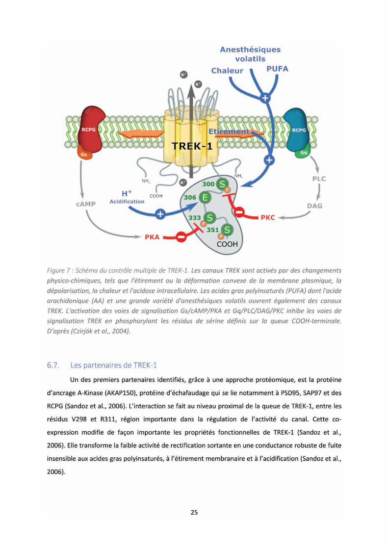
6.5. La régulation de TREK-1
La régulation hétérogène inhérente au canal TREK-1 résulterait de la composition en acides
aminés de la queue cytoplasmique adjacente au quatrième segment transmembranaire. Ce sont les
se o de et uat i e h li es α t a s e a ai es ui so t espo sa les de la fo atio du po e
interne. La structure secondaire de cette quatrième hélice, suggère la présence d’u allo ge e t
intracellulaire pouvant jouer un rôle central da s la gulatio de l’a ti it du a al (Morais-Cabral
et al., 2001). L’a ti it de TREK- peut t e sous le o t ôle d’u e se si ilit a i ue ou
thermique, de lipides, du pH, régulé par les RCPG ou encore par des interactions avec des protéines
partenaires (Figure 7).
6.5.1. Sensibilité mécanique
TREK-1 peut être activé par des variations de pression dépendante de la courbure
membranaire, mais également être régulé par des tensions membranaires. Des forces de
cisaillements activent le canal, alors que des o t ai tes d’ ti e e t de la ellule i duit u e
h pe os ola it e t a ellulai e ui duit l’a plitude des ou a ts de TREK-1.(Maingret et al.,
2000a; Patel et al., 1998). De a i e g ale, l’ou e tu e de TREK-1 est facilitée par une
déformation convexe de la membrane. L’insertion asymétrique d’u e ol ule amphiphatique dans
la bicouche lipidique, comme le trinitrophénol anionique, provoque une courbure convexe et active
le a al, à l’i e se, u e d fo atio o a e p oduite par la formation du radical cationique de la
chlorpromazine, induit une inhibition de TREK-1(Maingret et al., 2000a). Enfin, il est possible
d’a ti e , de faço g aduelle et e si le, le a al, en appliquant une pression mécanique négative
sur la partie extracellulaire de la membrane(Maingret et al., 1999). Cette mécanosensibilité peut-
être altérée ou réduite lorsque que la partie C-terminal adjacente au quatrième domaine
22
t a s e a ai e est pa tielle e t t o u e, sugg a t l’i po ta e de ce motif (Patel et al.,
1998).
6.5.2. Sensibilité thermique
De la même manière u’u e gulatio a i ue est possible, les canaux TREK-1 peuvent
être activés par des variations de température dans une gamme allant de 14 à 42°C (Maingret et al.,
2000b), avec une activité plus importante à partir de 22°C, pouvant atteindre une amplitude de
courant sept fois plus importante. Cependant, et de façon intéressante, cette thermosensibilité est
perdue après une excision par patch-clamp, malgré des conditions de régulation mécanique, de pH
ou d’a ide a a hido i ue maintenues, ce qui sugg e l’h poth se ue la gulatio the i ue ’est
pas inhérente au propriété du canal mais à un possible co-facteur (Maingret et al., 2000b).
6.5.3. Régulation par le pH
TREK-1 est sensible à la variation de pH, principalement intracellulaire. Les protons activent
le canal et le sensibilisent aux stimuli mécaniques (Maingret et al., 1999). A l’i sta de la gulatio
a i ue, ’est gale e t sur la partie terminale COOH intracellulaire que réside le senseur aux
protons ui gule la se si ilit au pH, et u’u e coupure de cette région aboutit à une diminution
progressive de sa sensibilité (Maingret et al., 1999). Il est possi le d’i hi e le a al TREK-1 humain
par une acidification du pH extracellulaire. Deux résidus histidines situés sur la première boucle
extracellulaire a a t le p e ie do ai e po e t oig e t d’u e se si ilit au pH sig ifi ati e e
o ditio de pH ph siologi ue % d’i hi itio à pH . pa appo t à pH . (Cohen et al., 2008).
Cependant, chez le modèle u i , ette se si ilit à l’a idifi atio est plus fai le, su e e t e
aiso de la p se e d’u e gluta i e à la pla e du p e ie sidu histidi e. A ote ue
l’a idifi atio e t a ellulai e duit Po sa s i flue e la o du ta e u itai e du a al TREK-1
humain (Cohen et al., 2008).
6.5.4. Régulation par les lipides
L’activité des canaux TREK-1 peut être régulée par les acides gras polyinsaturés (PUFA,
Pol U satu ated Fatt A ids o e l’a ide a a hido i ue AA , l’a ide alphali ol i ue ALA ,
l’a ide l’linolénique ou e o e le l’a ide docosahexaénoïque (Fink et al., 1998). Cependant, les acides
gras saturés n’ont pas d’effet su le a al. Cette a ti atio ’est pas d pe da te de l’i t g it
ellulai e, et l’effet est di pa la pa tie C-terminal de TREK-1 (Fink et al., 1998; Kim et al., 2001).
E e ui o e e les l sophospholipides, l’a ti atio se fait plus apide e t ue pa les PUFAs
ais essite pa o t e l’i t g it de la ellule (Maingret et al., 2000a).
23
6.5.5. Régulation par les RCPGs
Il existe une régulation par les se o ds essage s sulta t de l’a ti it des RCPGs. E effet,
la stimulation des protéines kinases A (PKA) et C (PKC) aboutit à une diminution des courants
potassiques induite par une phosphorylation sur des sites différents du canal TREK-1(Fink et al.,
1996). Ce sont les résidus sérine S333 et S300, en bordure du quatrième segment transmembranaire
et de la partie C-terminale intracellulaire, qui sont ciblés par la PKA et PKC respectivement, et sont
importants dans cette régulation de TREK-1 (Murbartián et al., 2005). Par déduction de ce
mécanisme de régulation, les agonistes des RCPGs tendent à inhiber les courants potassiques.
6.6. Rôle de TREK-1 dans la dépression
La distribution ubiquitaire et les régulations multiples de TREK-1 en font un canal pouvant
s’impliquer dans de nombreuses fonctions physiologiques. C’est ota e t da s la douleu et la
gulatio de l’hu eu ue TREK-1 a révélé des résultats particulièrement intéressants. Au niveau
de la o i eptio , l’i alidatio du g e oda t pou le a al io i ue chez des souris a montré une
hypersensibilité aux stimuli thermique à des seuil bas, entre 46 et 50°C, une hyperalgésie qui va de
pair avec une allodynie (Alloui et al., 2006). Cette hausse de sensibilité peut s’e pli uer par la
présence abondante de TREK-1 dans les neurones sensoriels de faible diamètre (fibres C
nociceptives polymodale) et sa large co-localisation avec TRPV1 (transient receptor potential
vanilloid 1), un canal cationique non sélectif thermosenseur activé par la capsaïcine. Le taux de
décharges afférentes des fibres C chez les animaux invalidés dépasse celui des sauvages, et le seuil
de déclenchement induit par le stimulus thermique est plus faible (-5°C de différence)(Alloui et al.,
2006; Noël et al., 2009).
Su le pla de la gulatio de l’hu eu , l’i alidatio de TREK-1 a entraîné l’e p essio d’u
phénotype de résistance dans des tests comportementaux relatifs à la dépression (Heurteaux et al.,
2006). Les souris sont plus mobiles dans le test de nage forcée (Force Swim Test ; FST) et de
suspension par la queue (Tail Suspension Test ; TST). Elles présentent également des latences
d'évasion plus courtes suite à une exposition à des chocs électrique aversifs non contrôlés sur les
pattes (Learn Helpness test, LH). Dans le test de suppression de la nourriture par la nouveauté
(Novelty Suppressed Feeding ; NSF), les souris TREK-1 -/- mangent plus facilement les aliments dans
un environnement menaçant (Heurteaux et al., 2006). Les résultats obtenus sont similaires à ceux de
souris sauvages traitées avec des ISRS (fluoxetine) et il ’ a pas d’a a tage d’effet lo s ue les sou is
TREK-1 -/- sont traitées par ces antidépresseurs. Le canal TREK-1 est fortement exprimé dans les
neurones sérotoninergiques du noyau dorsal du raphé. De façon intéressante, lorsque les souris
invalidées pour TREK-1 sont traitées avec de la fluoxetine associée au Fenclonine (para-
24
chlorophenylalanine ; PCPA), un inhibiteur sélectif et irréversible du tryptophane hydroxylase qui
appauvrit le niveau de sérotonine, les souris perdent leur phénotype antidépressif. De plus, les
neurones sérotoninergiques du noyau dorsal du raphé des souris mutantes présentent une activité
plus importante, au même niveau que ceux de souris sauvages traitées avec des ISRS, avec une
fréquence de décharge de deux fois celle des sauvages non traitées (Heurteaux et al., 2006). Cette
aug e tatio d’a ti it a oit la li atio de s oto i e e s des st u tu es o e l’hippo a pe,
et p o o ue u e a lio atio de l’a ti it to i ue des epteu s -HT1A postsynaptique de la partie
antérieure, au niveau du champ ammonien 3 (CA3), cette même amélioration qui est observée lors
de traitements ISRS sur le long terme (Haddjeri et al., 1998; Heurteaux et al., 2006). Plus récemment,
un peptide issu de la maturation du récepteur 3 de la neurotensine (Sortiline), le propeptide (PE) ou
son analogue synthétique la Spadine, a été identifié comme bloqueur spécifique de TREK-1. Cette
inhibition de TREK- , à l’i sta de la d l tio , e ge d e gale e t en un phénotype de résistance à
la dépression chez les souris sauvages.
Des tudes o t gale e t is e ide e u e aug e tatio de l’e p essio de TREK-1
dans le cortex préfrontal lors de dépression induite chez le rat, expression réversée suite à un
traitement à la fluoxetine (Chen et al., 2015). En outre, six polymorphismes nucléotidiques du gène
codant TREK-1, KNCK2, sont impliqués dans les troubles majeurs de l’hu eu et l’effi a it des
antidépresseurs hez l’ho e (Congiu et al., 2015).
Tout ceci semble indiquer un rôle pivot de TREK- da s la gulatio de l’hu eu , lui
conférant un potentiel statut de cible thérapeutique dans le trouble dépressif.
26
Mtap 2, Microtubule-associated protein, est un autre partenaire constitutif de TREK-1. Il
fa o ise l’aug e tatio du ou a t du a al sa s pou auta t affe te ses p op i t s i t i s ues
(Sandoz et al., 2008). Cette a lio atio est i puta le à l’aug e tatio de la de sit de TREK-1 à la
membrane plasmique et réside dans la liaison entre Mtap 2 et les microtubules. Les résidus associés
à cette liaison se situe t e t e les sidus E et Q , t s p o hes de eu d’AKAP (Sandoz et
al., 2008). A ote ue la liaiso si ulta e d’AKAP et Mtap 2 permet un effet additif sur les
courants de TREK-1 (Sandoz et al., 2008).
Le récepteur 3 de la neurotensine (NTSR3), aussi appelé Sortiline, a été récemment identifié
comme partenaire de TREK-1(Mazella et al., 2010). Une co-expression et co-localisation ont été
mises en évidence dans des cellules COS-7 et des neurones corticaux de souris (Mazella et al., 2010).
La sortiline est une p ot i e au fo tio s ultiples et o ple es. C’est u epteu , u o-
epteu et su tout u e p ot i e d’ad essage e s le l soso e ou la e a e plas i ue.
L’e p essio de TREK-1 à la membrane plasmique, mesurée sur des préparations de membranes
pu ifi es ou e utilisa t u e ioti latio de su fa e ellulai e, aug e te d’u fa teu de et
lorsque la sortiline est co-transfectée dans des cellules COS- , sugg a t l’i te a tio e t e les deux
protéines (Mazella et al., 2010).
Le NTSR3/Sortiline étant le point central de ce manuscrit, il sera plus amplement développé
dans la suite du mémoire.
7 LE NTSR3 / SORTILINE, UNE PROTEINE ASSOCIEE A DE MULTIPLES FONCTIONS
Partenaire de TREK-1, le NTSR3/Sortiline est un récepteur de haute affinité pour la neurotensine.
Ce récepteur fait partie du système neurotensinergique, mais également de la famille des récepteurs
à un domaine transmembranaire VPS10p (Vacuolar protein sorting 10 protein) (Petersen et al.,
1997).
7.1. La neurotensine et ses récepteurs
7.1.1. Généralités
La neurotensine (NTS) est un peptide de 1 a ides a i s pu ifi à pa ti d’hypothalami
bovin (Carraway and Leeman, 1973). Ce neuropeptide est exprimé dans le système nerveux central
ainsi que dans la périphérie. Il est présent da s des gio s ales telles ue l’hypothalamus, la
su sta e oi e, les tu e ules uad iju eau , le ul e olfa tif, l’hippo a pe, l’a gdale,
l’h poph se et la su sta e g lati euse de la moelle épinière (Cooper et al., 1981). Située dans les
corps neuronaux et les terminaisons des cellules nerveuses, il est absent des cellules gliales. Dans la

27
périphérie, il se situe dans le tractus gastro-intestinal, s th tis da s le j ju u et l’ilio
(Helmstaedter et al., 1977), et également dans le système cardiovasculaire (Reinecke et al., 1982) et
la circulation sanguine (Polak et al., 1977).
Au niveau du SNC, la eu ote si e est apa le d’aug e te le tau de d ha ge des
neurones dopaminergiques et de contrôler la libération de dopamine dépendamment des régions
cérébrales (Blaha and Phillips, 1992; Mercuri et al., 1993; Okuma et al., 1983; Pinnock, 1985;
Tanganelli et al., 1994), de réduire la prise alime tai e pa l’intermédiaire de la leptine (Sahu et al.,
2001), d’i dui e également une réponse antinociceptive indépendante des récepteurs μ opioïdes
dans une variété de tests de douleurs, comme la plaque chaude, les crampes abdominales induites
par l’a ide a ti ue ou e o e le test de et ait de la queue (Clineschmidt et al., 1979, 1982). Cet
effet analgésique est rapporté comme plus puissant que la morphine à des doses équimolaires et
toujours efficace même en présence de deux antagonistes opioïdes structurellement apparentés, la
naloxone ou la naltrexone (Roussy et al., 2010). La neurotensine a aussi un effet hypothermiant
(Popp et al., 2007) et peut prévenir des risques cérébraux induits pa l’h po ie (Choi et al., 2012).
Enfin, elle peut réguler le système endocrinien et contrôler la li atio d’hormones comme la CRH
et la GRH par la stimulation des neurones hypothalamiques (Ceccatelli et al., 1989; Nicot et al., 1997;
Niimi et al., 1991).
Au i eau de la p iph ie, la eu ote si e pe et de fa ilite l’a so ptio des lipides et
d’aug e te la s tio iliai e (Gui et al., 2001). Elle augmente également la sécrétion d’i suli e à
faible concentration de glucose et diminue le glucose insulino-dépendant (Béraud-Dufour et al.,
2010; Dolais-Kitabgi et al., 1979).
Les effets de la neurotensine passent par trois récepteurs : le NTSR1, le NTSR2 et le NTSR3
ou Sortiline. Le NTSR1 et NTSR2 sont des récepteurs à 7 domaines transmembranaires couplés aux
protéines G, alors que la Sortiline est un récepteur de type 1, à un seul domaine transmembranaire.
7.1.2. Le NTSR1
Les effets de la neurotensine sont médiés principalement par son récepteur de haute affinité
le NTSR1, qui possède une affinité sub nanomolaire pour la neurotensine (Kd = 0.1–0.3 nmol/L) et
est insensible à la Lévocabastine, une molécule qui possède des propriétés antihistaminiques H1 et
qui est un agoniste du NTSR2 (Schotte et al., 1986). Il est exprimé au niveau du cerveau, dans les
eu o es du septu , de la su sta e oi e, de l’ai e teg e tale e t al, du o au
suprachiasmatique et également dans le cortex préfrontal, entorhinal et cingulaire postérieur (Elde
et al., 1990; Nicot et al., 1994). Des études immunohistochimiques ont mis en évidence la présence
28
de NTSR1 dans les terminaisons nerveuses du noyau caudé, de la strie terminale, des tubercules
olfa tifs ais aussi da s le septu lat al, l’amygdale et le noyau accumbens (Boudin et al., 1996;
Fassio et al., 2000). Le NTSR1 est couplé à la phospholipase C et la cascade de signalisation de
l’i ositol phosphate via la sous-unité Gα / (Vincent et al., 1999; Wang and Wu, 1996). L’a tivation
exogène de NTSR1 entraîne la prolifération cellulaire, la survie, la mobilité et l'invasion des cellules
cancéreuses d'origines diverses (Thomas et al., 2003; Wu et al., 2012) o l à l’a ti atio de
ki ases. La PKC, a ti e pa l’IP , i duit u e a ti atio de MAPK Mitogen-activated protein kinases)
par la stimulation de Raf- ou du epteu à l’EGF Epidermal Growth Factor), ce qui peut
provoquer une croissance cellulaire incontrôlée (Guha et al., 2003; Müller et al., 2011). La
eu ote si e, agissa t pa l’intermédiaire du NTSR1, réduit la fonction physiologique du récepteur à
la dopamine D2 (Jomphe et al., 2006). De plus, il e iste u e odifi atio de l’e p essio de l’ARN
des récepteurs dopaminergique chez les souris invalidées pour le NTSR1 (Liang et al., 2010). Chez ces
es sou is, il a gale e t u e pe te de l’effet h pothe ia t et de l’effet anorexigène de la
NTS lors de son injection au niveau centrale, suggérant que ces effets passe t pa l’i te diai e du
NTSR1 (Kim et al., 2008; Remaury et al., 2002). E e a he, l’effet a alg si ue e se le pas t e
directement dépendant du NTSR1 (Kim et al., 2008) cependant, la délétion du récepteur provoque
une diminution de la nociception dépendante de la morphine(Roussy et al., 2010).
7.1.3. Le NTSR2
Le NTSR2, quant à lui, est identifié comme le récepteur à faible affinité pour la NTS (Kd = 3-
10 nmol/L) et est sensible à la Lévocabastine (Mazella et al., 1996). L’e p essio du epteu est
particulièrement dense dans des régions recevant des innervations neurotensinergiques, comme le
o au de la st ie te i ale, le ul e olfa tif, la su sta e oi e, l’ai e teg e tale e t al et la
substance grise périaqueducale mais également dans le o te al, l’hippo a pe et le e elet
(Lépée-Lorgeoux et al., 1999; Sarret et al., 2003a; Walker et al., 1998). Lorsque la séquence codante
du clone murin du NTSR2 est exprimée dans des ovocytes de xénopes, la neurotensine ainsi que la
lévocabastine sont capables de déclencher un courant entrant calcium dépendant (Mazella et al.,
1996). Da s des ellules CHO t a sfe t es a e de l’ADN NTSR hu ai ou de at, ette
o ilisatio i t a ellulai e de al iu est plus i po ta te a e la l o a asti e u’a e le NTS ,et
phosphoryle Erk1/2 (Botto et al., 1997; Gendron et al., 2004; Yamada et al., 1998) . Le NTSR2 joue un
rôle dans la protection des cellules bêta contre des agents externes cytotoxiques (staurosporine, IL-
1β) pa l’i te diai e de la oie kinase PI3 (Béraud-Dufour et al., 2009; Coppola et al., 2008). Il est
gale e t espo sa le de l’effet analgésique médié par la neurotensine (Dubuc et al., 1999; Smith
et al., 2012). Plus récemment, le NTSR2 a été impliqué dans la mémoire de peur. Dans le test
contextuel de condition de peur, la réponse de freezing (position prostrée) a été significativement
29
réduite chez les souris déficiente NTSR2 (NTSR2-/-) comparativement aux souris de type sauvage
(Yamauchi et al., 2007).
Cepe da t, l’a tio de la NTS sur le NTSR2 reste encore ambiguë. En fonction de l'espèce à
partir de laquelle le NTSR2 a été isolé (Mazella et al., 1996) et le système cellulaire utilisé pour
évaluer la signalisation, la eu ote si e peut aussi ie agi e ta t u’agoniste, u’a tago iste. La
neurotensine et la lévocabastine exercent une activité espèce dépendante ; agoniste chez la souris
(Mazella et al., 1996), a tago iste hez l’hu ai (Richard et al., 2001; Vita et al., 1998).
7.2. Le NTSR3 / Sortiline
Troisième récepteur de la neurotensine, le NTSR3, ou Sortiline, a été identifié par 3
approches différentes. A la différence des deux autres récepteurs, le NTSR3/Sortiline est une
protéine réceptrice à un seul domaine transmembranaire, ou récepteur de type I, et possède une
structure Vps10p.
7.2.1. Purification et clonage du NTSR3 / Sortiline
U e p e i e ide tifi atio a pu t e alis e g â e à l’utilisatio d’u a alogue adioa tif et
photoactivable de la NTS. Une protéine identifiée d’e i o kDa, solubilisable par le détergent
CHAPS, a montré des propriétés pharmacologiques similaires aux deux autres récepteurs de la
neurotensine et s’est a e apa le de lie la eu ote si e a e u e haute affi it Kd = . M
(Mazella et al., 1988). Elle a t e suite pu ifi e à pa ti de e eau de sou is et d’hu ai et
identifiée comme le récepteur 3 de la neurotensine(Mazella et al., 1989; Zsürger et al., 1994). Le
NTSR3 a pa la suite t o t o e i pli u da s l’i te alisatio de la NTS, dans des cultures
de neurones primaires, de même que le NTSR1 et NTSR2 (Chabry et al., 1993).
Lors de la caractérisation, pa olo e d’affi it , de partenaires de la protéine RAP
(receptor-associated protein), protéine endoplasmique de 40 kDa localisée au niveau du réticulum et
de l'appareil de Golgi et impliqué dans la maturation des récepteurs des LDL (Low Density
Lipoprotein), une glycoprotéine d’e i o kDa a été identifiée dans les vésicules intracellulaires
contenant RAP (Battey et al., 1994; Gliemann et al., 1994; Willnow, 1998). Purifiée à partir de
cerveau humain et clonée dans une bibliothèque d'ADNc humain, son gène, localisé sur le
chromosome 1p, code pour une protéine de 833 acides aminés. Cette glycoprotéine ’est epe da t
pas homologue aux récepteurs LDL, mais présente une structure caractéristique des récepteurs de
type I, avec des homologies avec la protéine Vps10p de levure, impliqué dans le transport
intracellulaire, et les récepteurs cation-dépendants et cations-indépendants du mannose-6-
phosphate (Petersen et al., 1997). C’est d’ailleu s par ce domaine Vsp10p que la protéine est capable

30
de se lier à la partie carboxyterminale de RAP (Tauris et al., 1998). Par ses caractéristiques, elle sera
identifiée comme la Gp95/Sortilin ui s’a e a t e % ho ologue a e le epteu de la
neurotensine (Mazella et al., 1998).
En parallèle, une équipe isole une protéine de 110kDa à partir de vésicules intracellulaires,
contenant le transporteur du glucose Glut4, et l’ide tifie o e u des composés majeurs de ces
vésicules. Clo e à pa ti d’adipo tes de at, elle p se te a u e fo te homologie de séquence avec
le NTSR3/Sortiline hu ai e % et se a e isag e o e l’a alogue de la so tili e du at (Lin et
al., 1997).
7.2.2. Structure
Le NTSR3/Sortiline est un récepteur de type I de la famille des récepteurs Vps10p contenant
quatre autres membres : SorCS1, SorCS2, SorCS3 et SorLA. Cette famille de protéine
transmembranaire possède un long domaine N-terminal homologue à la protéine Vps10p, qui
représente soit le seul élément de la partie luminal/extracellulaire, soit est asso i à d’aut es
domaines, et un court domaine cytoplasmique. Vps10 est une protéine de tri vacuolaire retrouvée
chez la levure (Vacuolar protein sorting) et qui possède deux régions homologues contenant chacun
un motif C-terminal riche de 10 résidus cystéine (10CC motif) espacés par un nombre constant
d’a ides a i s. C’est u e p ot i e d’ad essage de la a o peptidase Y et ou e ajo itai e e t
dans le golgi tardif, là où les protéines vacuolaires sont triées (Marcusson et al., 1994). Après
transport vers des compartiments endosomaux prévacuolaires et maturation de la carboxypeptidase
Y, la protéine Vps10p est capable de revenir vers le Golgi (Marcusson et al., 1994). Ces récepteurs à
domaine Vps10p présentent tous une séquence consensus de clivage de proprotéine convertase
(RXXR) qui définit le propeptide N-terminal (Figure 8).
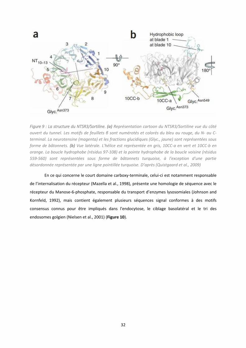
32
Figure 9 : La structure du NTSR3/Sortiline. (a) Représentation cartoon du NTSR3/Sortiline vue du côté
ouvert du tunnel. Les motifs de feuillets β sont numérotés et colorés du bleu au rouge, du N- au C-
terminal. La neurotensine (magenta) et les fractions glucidiques (Glyc., jaune) sont représentées sous
forme de bâtonnets. (b) Vue latérale. L'hélice est représentée en gris, 10CC-a en vert et 10CC-b en
orange. La boucle hydrophobe (résidus 97-108) et la pointe hydrophobe de la boucle voisine (résidus
559-560) sont représentées sous forme de bâtonnets turquoise, à l'exception d'une partie
désordonnée représentée par une ligne pointillée turquoise. D ap s (Quistgaard et al., 2009)
En ce qui concerne le court domaine carboxy-terminale, celui-ci est notamment responsable
de l’i te alisatio du epteu (Mazella et al., 1998), présente une homologie de séquence avec le
récepteur du Manose-6-phosphate, espo sa le du t a spo t d’enzymes lysosomiales (Johnson and
Kornfeld, 1992), mais contient également plusieurs séquences signal conformes à des motifs
consensus connus pour être impliqués dans l'endocytose, le ciblage basolatéral et le tri des
endosomes golgien (Nielsen et al., 2001) (Figure 10).

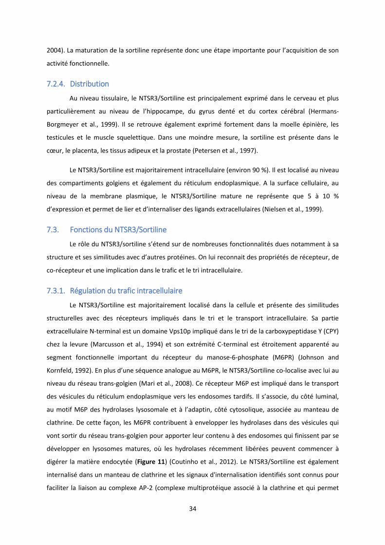
34
2004). La atu atio de la so tili e ep se te do u e tape i po ta te pou l’a uisitio de so
activité fonctionnelle.
7.2.4. Distribution
Au niveau tissulaire, le NTSR3/Sortiline est principalement exprimé dans le cerveau et plus
pa ti uli e e t au i eau de l’hippo a pe, du g us de t et du o te al (Hermans-
Borgmeyer et al., 1999). Il se retrouve également exprimé fortement dans la moelle épinière, les
testicules et le muscle squelettique. Dans une moindre mesure, la sortiline est présente dans le
œu , le pla e ta, les tissus adipeu et la p ostate (Petersen et al., 1997).
Le NTSR3/Sortiline est majoritairement intracellulaire (environ 90 %). Il est localisé au niveau
des compartiments golgiens et également du réticulum endoplasmique. A la surface cellulaire, au
niveau de la membrane plasmique, le NTSR3/Sortiline mature ne représente que 5 à 10 %
d’e p essio et pe et de lie et d’i te alise des liga ds e t a ellulai es (Nielsen et al., 1999).
7.3. Fonctions du NTSR3/Sortiline
Le ôle du NTSR /so tili e s’ te d su de o euses fo tio alit s dues ota e t à sa
st u tu e et ses si ilitudes a e d’aut es p ot i es. O lui e o ait des p op i t s de récepteur, de
co-récepteur et une implication dans le trafic et le tri intracellulaire.
7.3.1. Régulation du trafic intracellulaire
Le NTSR3/Sortiline est majoritairement localisé dans la cellule et présente des similitudes
structurelles avec des récepteurs impliqués dans le tri et le transport intracellulaire. Sa partie
extracellulaire N-terminal est un domaine Vps10p impliqué dans le tri de la carboxypeptidase Y (CPY)
chez la levure (Marcusson et al., 1994) et son extrémité C-terminal est étroitement apparenté au
segment fonctionnelle important du récepteur du manose-6-phosphate (M6PR) (Johnson and
Kornfeld, 1992). E plus d’u e s ue e a alogue au M6PR, le NTSR3/Sortiline co-localise avec lui au
niveau du réseau trans-golgien (Mari et al., 2008). Ce récepteur M6P est impliqué dans le transport
des vésicules du réticulum endoplasmique vers les endosomes tardifs. Il s’asso ie, du ôt lu i al,
au motif M6P des hydrolases lysosomale et à l’adapti , côté cytosolique, associée au manteau de
clathrine. De cette façon, les M6PR contribuent à envelopper les hydrolases dans des vésicules qui
vont sortir du réseau trans-golgien pour apporter leur contenu à des endosomes qui finissent par se
développer en lysosomes matures, où les hydrolases récemment libérées peuvent commencer à
digérer la matière endocytée (Figure 11) (Coutinho et al., 2012). Le NTSR3/Sortiline est également
internalisé dans un manteau de clathrine et les signaux d'internalisation identifiés sont connus pour
faciliter la liaison au complexe AP-2 (complexe multiprotéique associé à la clathrine et qui permet
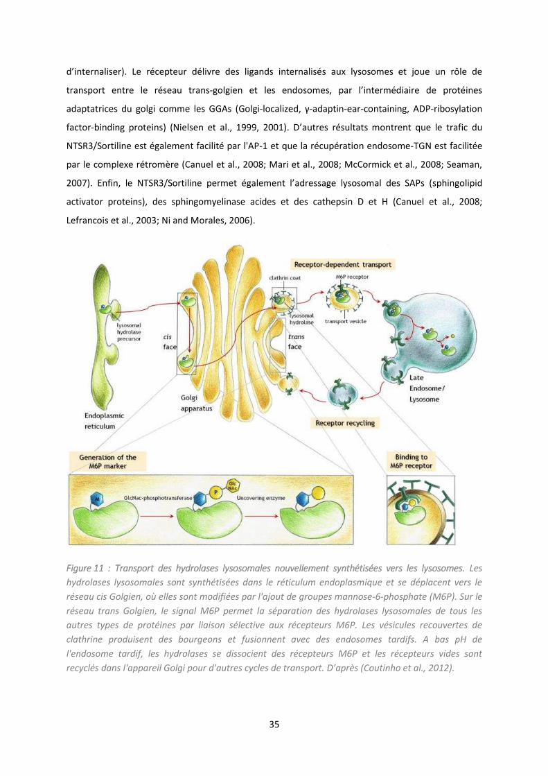
35
d’i te alise . Le epteu d li e des liga ds i te alis s aux lysosomes et joue un rôle de
transport entre le réseau trans-golgie et les e doso es, pa l’i te diai e de p ot i es
adaptatrices du golgi comme les GGAs (Golgi-lo alized, γ-adaptin-ear-containing, ADP-ribosylation
factor-binding proteins) (Nielsen et al., 1999, 2001). D’aut es résultats montrent que le trafic du
NTSR3/Sortiline est également facilité par l'AP-1 et que la récupération endosome-TGN est facilitée
par le complexe rétromère (Canuel et al., 2008; Mari et al., 2008; McCormick et al., 2008; Seaman,
2007). E fi , le NTSR /So tili e pe et gale e t l’ad essage l soso al des SAPs sphingolipid
activator proteins), des sphingomyelinase acides et des cathepsin D et H (Canuel et al., 2008;
Lefrancois et al., 2003; Ni and Morales, 2006).
Figure 11 : Transport des hydrolases lysosomales nouvellement synthétisées vers les lysosomes. Les
hydrolases lysosomales sont synthétisées dans le réticulum endoplasmique et se déplacent vers le
réseau cis Golgien, où elles sont modifiées par l'ajout de groupes mannose-6-phosphate (M6P). Sur le
réseau trans Golgien, le signal M6P permet la séparation des hydrolases lysosomales de tous les
autres types de protéines par liaison sélective aux récepteurs M6P. Les vésicules recouvertes de
clathrine produisent des bourgeons et fusionnent avec des endosomes tardifs. A bas pH de
l'endosome tardif, les hydrolases se dissocient des récepteurs M6P et les récepteurs vides sont
recyclés dans l'appareil Golgi pour d'autres cycles de transport. D ap s (Coutinho et al., 2012).
36
Le NTSR /So tili e est fo te e t e p i da s les adipo tes. Des tudes l’o t ide tifi
comme un composant essentiel des vésicules de stockage du transporteur de glucose 4’ (GLUT4) (Lin
et al., 1997; Morris et al., 1998). Le domaine luminal N-terminal du NTSR3/Sortiline interagit avec
GLUT4, tandis que la queue cytoplasmique interagit avec les protéines d'adaptation pour faciliter la
formation des vésicules de stockage. Il a été démontré dans les adipocytes et les myocytes que le
NTSR3/Sortiline était nécessaire pour l'absorption de glucose insulinodépendant (Huang et al., 2013;
Morris et al., 1998; Shi and Kandror, 2005).
7.3.2. Viabilité neuronale
La fonction du NTSR3/Sortiline ne s'arrête pas au ciblage lysosomal. Le NTSR3/Sortiline est
capable de circuler entre la surface des cellules et divers compartiments intracellulaires afin
d’a he i e les protéines cibles vers différentes destinations, y compris l'exposition à la surface
membranaire pour pe ett e la t a sdu tio d’u signal, ou encore à travers des voies régulées.
Le NTSR3/Sortiline reconnait la pro-forme du NGF (proNGF, Nerve Growth Factor) mais
gale e t d’aut es neurotrophines (NT) et pro-neurotrophines, comme le BDNF. Les NT sont des
facteurs de croissance essentiels au développement et au maintien du système nerveux. Cette
famille regroupe le NGF, le BDNF, ainsi que la neurotrophine 3 (NT3) et la neurotrophine 4 (NT4).
Toutes les NT sont synthétisées sous forme précurseurs puis converties en leurs équivalents matures
par clivage protéolytique dans les voies de biosynthèses ou l'espace extracellulaire. Les activités des
NTs sont médiées par deux classes de récepteurs : les récepteurs kinases liés à la tropomyosine TrkA,
-B et -C qui présentent une spécificité pour des NTs particulières, et le récepteur de promiscuité
neurotrophin p75 (p75NTR) qui augmente la spécificité des Trks envers leurs ligands. De façon
intéressante, les formes précurseur pro-NTs possèdent une activité biologique souvent inverse de
leur forme mature. Le proNGF et le proBDNF sont capables d’induire un signal pro apoptotique,
alors que le NGF et le BDNF sont impliqués dans la croissance, la prolifération et la survie cellulaire.
Ces effets sont en réalité dépendants de l’asse lage de leur récepteur respectif.
7.3.2.1. Co-recepteur du p75NTR
Les effets positifs du NGF et BDNF passe t pa l’asse lage du epteu p a e les
récepteurs TrkA et TrkB respectivement. Le p75NTR est un membre de la superfamille des
récepteurs TNF (Tumor necrosis factor) et est surtout connu pour son rôle dans l'apoptose
neuronale et la neurodégénérescence (Dechant and Barde, 2002). Il possède une région
cytoplasmique juxtamembranaire, appelé « Chopper », qui induit la cascade des caspases et calpaine
et la mort cellulaire (Coulson et al., 2000). La libération du domaine intracellulaire par des sécrétases
favorise aussi bien l'apoptose, que la survie cellulaire (Kenchappa et al., 2006; Kommaddi et al.,
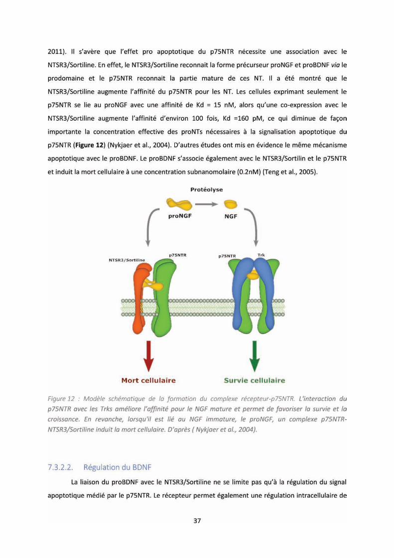
38
la sécrétion et du recyclage du BDNF. L’ide tifi atio du polymorphisme Val66Met du BDNF a
sugg l’e iste e d’un signal de trafic dans le prodomaine impliqué dans le ciblage optimal du
BDNF dans la voie de sécrétion régulée (Egan et al., 2003). Dans un premier temps, une étude a mis
e ide e la p se e d’u e gio da s le p odo ai e du BDNF, des sidus à ,
espo sa le de l’i te a tio a e le do ai e lu i al du NTSR /So tili e à pa ti d’ho og ats de
cerveaux, et ce, localisée dans les mêmes compartiments granulaires de sécrétion de neurones.
Cette région possède des similitudes avec la séquence du NGF surtout au niveau de la Box 3.
Cependant, en dépit des similitudes de séquences, cette région peut avoir des fonctions différentes
pour ces deux neurotrophines. La délétion de la Box 3 dans le NGF induit une diminution de
l’e p essio p ot i ue de NGF, sugg a t u e i po ta e possi le da s le eplie e t de la p ot i e
(Suter et al., 1991). E e a he, ette e d l tio pou le BDNF, ’alt e pas so niveau
d’expression protéique mais réduit de manière significative sa sécrétion régulée. A noter que, bien
ue le p u seu du NGF soit li e ajo it da s l’appa eil de golgi, le BDNF reste en majorité
sous sa proforme dans la cellule et peut être libéré ainsi (Chen et al., 2004; Mowla et al., 1999; Pang
et al., 2004). Fait intéressant, un autre motif de tri a été révélé dans le domaine mature du BDNF.
Composé de quatre résidus (I144, E146, I233, D234), il interagit avec une protéine de triage, la
carboxypeptidase E (CPE) et la mutation des résidus acides (E146, D234) en alanines conduit à une
perte du BDNF dans la voie constitutive (Lou et al., 2005).
La substitution de la méthionine 66 en valine du BDNF entraine une diminution significative
de l’i te a tio a e le NTSR /So tili e. Il a été démontré que cette substitution conduit à une
diminution de la co-localisation dans les granules de sécrétion, et également une diminution de la
sécrétion régulée du BDNF-Val66Met dans les neurones (Egan et al., 2003 ; Chen et al., 2004). Afin
de déterminer si le NTSR3/Sortiline est essai e pou l’ad essage du BDNF dans la voie de
sécrétion régulée, une construction du NTSR3/Sortiline tronquée, dépourvue de la région
transmembranaire et cytoplasmique du NTSR3/Sortiline, a été utilisée dans des cellules PC12. En
présence de NTSR3/Sortiline tronqué, la distribution du BDNF est plus diffuse dans la cellule, et il y a
une diminution significative du niveau de co-localisation dans les vésicules de sécrétion. De même, la
stimulation par dépolarisation, pour déclencher la voie régulée, aboutit à une diminution de la
quantité de BDNF extracellulaire. Ce même phénomène est observé avec le BDNF-Val66Met, que ce
soit avec la forme entière du NTSR3/Sortiline ou sa forme tronquée(Chen et al., 2005). A partir de
culture de neurones primaires corticaux et hippocampaux, la co-tansfection de NTSR3/Sortiline
tronqué et de BDNF induit une diminution de la sécrétion de BDNF dans la voie régulée avec une
augmentation concomitante de la voie constitutive (Chen et al., 2005). Ce résultat a été obtenu de
faço ide ti ue a e la t a sfe tio d’u siRNA s all i te fe i g RNA ui duit sig ifi ati e e t le

40
u i t t da s l’ tude de so i pli atio da s les a e s. Des études ont montré que le
NTSR3/Sortiline permettait de réguler à la fois les neurotrophines mais également ces récepteurs, et
pouvait jouer sur la progression des cellules cancéreuses (Akil et al., 2011). On le retrouve fortement
exprimé dans des lignées cellulaires humaines cancéreuses. Le NTSR3/Sortiline est capable de se
dimériser avec le NTSR1 et d’ t e i te alis pa la sti ulatio de la NTS, et d’i dui e des voies de
signalisation par modification des MAP kinases et le turnover du phosphoinositide (PI) facilitées par
NTSR1 dans la lignée cellulaire HT29 (adénocarcinome) (Martin et al., 2002). Les lignées de cellules
cancéreuses sensibles aux effets trophiques de la NTS expriment toutes le NTSR3/Sortiline. Si la
séquestration du peptide à l'intérieur de sa cellule cible est une étape fondamentale dans l’a ti it
sur la croissance cellulaire, le NTSR1 ou le NTSR3/Sortiline pourrait être en mesure d'agir comme
médiateur de la réponse trophique au NTS (Dal Farra et al., 2001). Enfin, le NTSR3/Sortiline est
capable de former un complexe avec le TrkB et l’EGFR epidermal growth factor receptor) dans les
exosomes libérés par les cellules cancéreuses du poumon, qui transmettent un contrôle
microenvironnemental sur les cellules endothéliales (Wilson et al., 2014).
De plus en plus de preuves suggèrent que la sortiline est fonctionnellement associée aux
processus du métabolisme des lipides. Les GWASs (Genome Wide Association Studies) ont montré
une forte corrélation entre le gène du NTSR3/Sortiline, SORT1, et le taux de lipides plasmatiques
(Guo et al., 2015; Wang et al., 2011).La surexpression hépatique du NTSR3/Sortiline augmente le
taux de lipides plasmatiques et vice versa (Sparks et al., 2015). Le déficit de NTSR3/Sortiline diminue
la sécrétion hépatique des lipoprotéines de très faible densité (very low density lipoprotein , VLDL),
ce qui abaisse le taux de lipides plasmatiques (Kjolby et al., 2010, 2015). Le NTSR3/Sortiline favorise
l'accumulation de lipides dans les macrophages et la formation de cellules spumeuses (Patel et al.,
2015). Le déficit en NTSR3/Sortiline montre également une réduction spectaculaire de la zone
adipocytaire dans le tissu adipeux des souris invalidées pour le gène (Rabinowich et al., 2015). Le
niveau de NTSR3/Sortiline hépatique est diminué dans des conditions de résistance à l'insuline, ce
qui contribue au trouble lipidique plasmatique dans le diabète de type 2 (Li et al., 2015). Dans
l'ensemble, ces études corroborent l'hypothèse selon laquelle le NTSR3/Sortiline participe
probablement aux troubles du métabolisme des lipides (Kjolby et al., 2015; Ogawa et al., 2016;
Schmidt and Willnow, 2016)
Il a également été proposé que le NTSR3/Sortiline soit impliqué dans la pathogenèse de la
maladie d'Alzheimer. La pe te d’e p essio du epteu e t ai e u e a u ulatio d’APOE
(Apolipoprotéine E et d’Aβ da s le e eau et u e agg a atio des pla ues. De plus, les neurones
primaires dépourvus de NTSR3/Sortiline présentent une diminution significative de l'absorption des
41
complexes APOE/ Aβ malgré l'expression appropriée des autres récepteurs APOE. Malgré des
niveaux d'APOE cérébraux plus élevés que la normale, les animaux présentant un déficit du
récepteur présentent des anomalies dans le métabolisme des lipides cérébraux (ex. accumulation de
sulfatides) le même que chez les souris déficientes en APOE, ce qui indique une carence
fonctionnelle dans les voies cellulaires d'absorption de l'APOE (Carlo et al., 2013).
Il existe également un lien entre le complexe proNGF-p75NTR-NTSR3/Sortiline dans la
maladie de Parkinson. La voie de signalisation de mort cellulaire médiée par le complexe proNGF-
p75NTR-NTSR3/Sortiline semble être responsable de la dégénérescence dans les neurones
dopaminergiques au niveau ventral de la substance noire, suggérant l’i pli atio de e o ple e
dans la progression de la maladie (Chen et al., 2008).
8 NTSR3/SORTILINE, PROPEPTIDE (PE), TREK-1 ET DEPRESSION
L’i pli atio du NTSR /So tili e da s la dépression prend son origine en partie dans son lien
avec le canal potassique, décrit précédemment, TREK-1. Le NTSR3/Sortiline et TREK-1 sont
fortement distribués dans des structures cérébrales impliquées dans la physiopathologie de la
d p essio , o e le o te p f o tal et i gulai e, l’a ygdale, l'hippocampe, le noyau
accumbens, le raphé dorsal et l'hypothalamus (Hervieu et al., 2001; Sarret et al., 2003b). Il a été
o t u’u e d l tio fo tio elle de TREK-1 aboutissait à un phénotype dit de résistance à la
dépression (Heurteaux et al., 2006). Pa ta t de l’id e ue le NTSR3/Sortiline a des propriétés de
trafic similaires aux partenaires de TREK- , AKAP et Mtap , apa les de gule l’e p essio à la
membrane, et u’il est localisé dans des régions analogues à celles du a al, l’ uipe du D Mazella
s’est pe h e su l’i pli atio du NTSR3/Sortiline dans la régulation de TREK-1. Dans un premier
temps, il a été montré, par immunoprécipitation, que le récepteur et le canal forment un complexe
et ce, à la membrane plasmique. Dans des cellules COS-7, la purification de membrane plasmique a
mo t u e aug e tatio d’un facteur 3 à de l’e p essio de TREK-1 lorsque que le
NTSR3/Sortiline était co-transfecté. Da s u se o d te ps, sa ha t u’il e iste u e gulatio du
récepteur par son propre propeptide libéré suite à sa maturation (Munck Petersen et al., 1999),
l’ uipe s’est i t ess e à l’action du PE, et d’un dérivé, su l’a ti it de TREK-1. Ce PE est constitué
de a ides a i s et est apa le de s’asso ie au NTSR /So tili e a e u e haute affi it Kd
d’environ 5nM). Des études structure-fonction ont révélé que la partie Gln1 – Arg28 était aussi
effi a e su l’a ti it de liaiso ue le p opeptide e tie Gl 1-Arg44) alors que l'affinité du peptide
Gln1-Arg16 était très faible (Westergaard et al., 2004). A l’aide de e o stat, u peptide, nommé la
Spadine (Sortilin Peptide AntiDepressant (IN)), plus petit que le propeptide, a été synthétisé en
conservant la séquence 17 à 28 et en y ajoutant la séquence 12-16 pour maintenir le stress
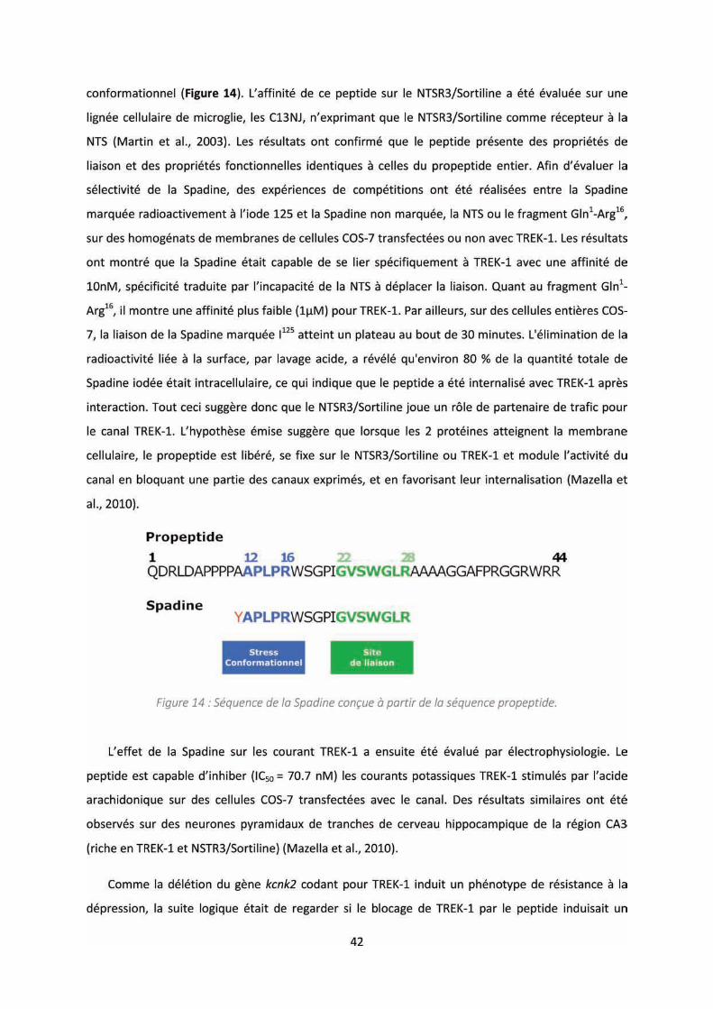

43
phénotype similaire. De façon intéressante, les souris injectées avec la Spadine montrent un
comportement identique à celui des souris délétées pour TREK-1 ou des souris sauvages traitées par
un antidépresseur, et ce de manière doses dépendantes. En effet, que ce soit dans le FST et le TST, il
a u e di i utio de l’i o ilit ui t aduit u e di i utio de la sig atio . L'immobilité est
interprétée comme un " état de désespoir ", en ce sens que l'animal aura perdu sa motivation à
adopter des comportements d'évasion. On retrouve également une diminution du temps de la
late e pou s’ happe da s le test de sig atio a uise (Learn Helpness) (Mazella et al., 2010).
Enfin, l'activité moyenne de décharges des neurones sérotoninergiques chez les animaux traités
avec la Spadine est presque identique à celle observée chez les souris kcnk2 -/-, c'est-à-dire une
augmentation du taux de décharges de ces neurones. De plus, il existe une augmentation de la
neurogénèse induite par la phosphorylation de CREB dans les neurones hippocampaux, de pair avec
une hausse de deux marqueurs de la synaptogénèse, le PSD95 (post-synaptic density protein of
kDalto ) et le BDNF, ta t au i eau p ot i ue u’au i eau des ARN s (Devader et al., 2015;
Mazella et al., 2010).
Tout e i sugg e ue le NTSR /So tili e est apa le de odule l’a ti it de TREK-1, que ce soit
par une régulation du trafic ou par l’inhibition du canal par son produit de maturation.
Plus e e t, des tudes o t is e ide e l’i pli atio de NTSR /So tili e solu le da s la
dépression. Le domaine extracellulaire du NTSR3/Sortiline (NTSR3/Sortiline soluble) est libéré de
façon à la fois constitutive et régulée par protéolyse à la surface des neurones et d'autres types de
cellules (Evans et al., 2011; Hermey et al., 2006; Navarro et al., 2002). La fonction de ce motif soluble
n'est pas claire, mais il a été démontré qu'elle inhibe la conversion du proBDNF en BDNF mature par
la plasmine et qu'elle protège les neurones des propriétés apoptotiques du proBDNF (Teng et al.,
2005). Ce NTSR3/Sortiline soluble forme un complexe avec le récepteur BDNF/TrkB et le récepteur
EGF et ce complexe ternaire est internalisé et libéré, par la suite, dans les exosomes (Wilson et al.,
2014). Le NTSR3/Sortiline soluble peut être détectée dans le sérum et le plasma. Il existe une
augmentation significative du taux sérique de ce récepteur soluble chez les sujets déprimés
comparativement aux témoins et des corrélations significatives entre ces taux sériques et les taux
sériques de BDNF et de VEGF. De façon intéressante, des taux élevés de NTSR3/Sortiline soluble
étaient associés à une dépression légère et modérée, mais pas à une dépression grave (Buttenschøn
et al., 2015).
Toutes ces données placent le NTSR3/Sortiline au centre de mécanismes impliqués dans la
régulation du comportement. A la fois récepteur, co-recepteur et p ot i e d’ad essage, il agit sur

44
des composantes importantes du système nerveux central, comme la régulation des neurotrophines,
ou e o e l’ad essage de a au potassi ues impliqué dans le maintien des potentiels de
membrane. L’o je tif des t a au de e manuscrit était donc d’ te d e la compréhension de ce
récepteur au sei d’u e pathologie o ple e. D’u e pa t, l’ alue o e u possi le a ueu
biologique da s le t ou le d p essif, et d’aut e pa t, ega de , pa diff e tes app o hes
iologi ues, les o s ue es d’u e pe te fo tio elle su u o ga is e e tier.
45
OBJECTIFS
Du a t es t ois a es de do to at, j’ai eu la possi ilit de t a aille su diff ents aspects
du NTSR /So tili e. L’o je tif tait de epla e , sur différents niveaux, le epteu da s l’affe t
pathologi ue u’est la d p essio , tout en approfondissant les mécanismes sous-jacents.
Dans un premier te ps, l’o je tif de es t a au a été de caractériser la délétion
fonctionnelle du NTSR3/Sortiline dans un modèle murin, et plus particulièrement au sein du système
nerveux central. Comme décrit précédemment, le NTSR3/Sortiline a des propriétés de tri cellulaire,
notamment de neurotrophines importantes dans la viabilité et la croissance neuronale. Parmi eux le
BDNF, qui se présente comme un facteur crucial dans la régulation de fonctions émotionnelles et
cognitives. Le NTSR3/Sortiline i flue e gale e t l’a ti it du a al io i ue TREK-1, pouvant être
i pli u da s la gulatio de l’hu eu . C’est par différentes approches expérimentales que je me
suis donc intéressé à la fonction du récepteur sur ces facteurs intervenants dans la modulation du
comportement, plus particulièrement dans les états pathologiques du trouble dépressif, et ceci à
pa ti d’u od le u i d pou us de NTSR3/Sortiline fonctionnel. Les souris mutantes, notées
Sort1 -/-, o t t g es pa l’ uipe du D Mo ales g â e à l’i t odu tio d’u e assette da s le
gène Sort1 e t e l’e o et l’e o , li ita t ainsi la p odu tio d’u e fo e p ot i ue
fonctionnelle du récepteur (Zeng et al., 2009). De manière cohérente et inhérente aux fonctions du
NTSR /So tili e, j’ai donc évalué les conséquences de la perte fonctionnelle du récepteur sur le
comportement, la biochimie et, de faço logi ue, l’ le t oph siologie au i eau du s st e e eu
central. Pa ailleu s, ta t do ue le NTSR /So tili e est d’a o d ide tifi o e u e
composante du système neurotensinergique, il a été évide t, e utilisa t e od le de sou is, d’e
déterminer également l’impact sur celui-ci.
U e aut e pa tie de o t a ail a t d’ alue si le NTSR /So tili e, plus particulièrement le
propeptide issu de sa maturation, pouvait présenter des variations chez des personnes dépressives
o pa ati e e t à des sujets sai s. Cette d a he pa tait du p i ipe u’ ta t do la pla e ue
p e d le NTSR /So tili e da s la gulatio de eu ot ophi es et l’effet su l’hu eu de so p oduit
de maturation, serait-il possi le, à l’i sta de la fo e solu le, ue le p opeptide soit u i di ateu
du trouble dépressif ? E effet, s’il est possi le de esu e des a iatio s s i ues de BDNF ou de
NTSR /So tili e solu le, et d’ ta e le diag osti d p essif, il se ait alo s i t essa t d’ alue le
i eau d’u peptide ui fait tat de p op i t a tid p essi e.

46
En parallèle de mes recherches, j’ai pu développer des outils informatiques dans le but de
si plifie l’a uisitio et le t aite e t de do es expérime tales. C’est da s sou i d’a lio e , de
fa ilite , et aussi pa e ue je e suis pas adepte des tâ hes p titi es, ue j’ai is au poi t u
logiciel capable de mesurer des occurrences et surtout des durées lors de tests comportementaux
chez la souris. Les fichiers générés e so tie pe ette t d’a de à de o euses do es e u
temps réduit tout en occultant le possible coût prohibitif des outils mis à disposition dans le
commerce.
47
Etude de la délétion du NTSR3/Sortiline
dans la régulation de l’état dépressif.
48
Article 1: Altered TREK-1 function in sortilin deficient mice results in an
antidepressant phenotype.
MORENO Sebastien, DEVADER Christelle, PIETRI Mariel, BORSOTTO Marc, HEURTEAUX Catherine
and MAZELLA Jean
Submitted for publication in the British Journal of Pharmacology (Under Review)
1. Co te te de l’étude
Da s l’ tude p de te, la d l tio du NTSR /So tili e a o duit à la odifi atio d’e p essio
d’u de es pa te ai es du s st e eu ote si e gi ue, le NTSR , et a i duit u ph ot pe de
désensibilisation à la douleur. Récemment, un autre de ces partenaires, le canal de fond potassique
TREK- , a fait l’o jet d’u e atte tio pa ti uli e da s le ph ot pe de d p essio et d’a i t . E
effet, la délétion du gène codant pour le canal chez la souris résulte en un phénotype de résistance
dans les tests comportementaux relatifs à la dépression. Ces souris révèlent un comportement
identique à des souris naïves traitées avec des antidépresseurs comme la fluoxétine. Au-delà de cet
aspect phénotypique, la délétion de TREK-1 a également entrainé une augmentation de la
neurogenèse hippocampique et du taux de décharges des neurones sérotoninergiques du noyau du
raphé dorsal. Par ailleurs, une inhibition du canal par un dérivé peptidique du propeptide issu du
NTSR3/Sortiline, la Spadine, produit des effets similaires antidépressifs. Il a été démontré l'existence
d'un complexe protéique entre le canal TREK-1 et le NSTR3/Sortiline et que l'expression du canal au
niveau de la membrane plasmatique des cellules COS-7 est augmentée lorsque celui-ci est co-
exprimé avec le récepteur. De plus, le NTSR3/Sortiline de par ses propriétés de tri, est capable de
réguler des neurotrophines, comme le BDNF, importante dans le système nerveux central.
Puisque le NTSR3/Sortiline et le TREK-1 sont associés aux niveaux cellulaire et moléculaire
(Mazella et al., 2010) et que le NTSR3/Sortiline joue un rôle important dans le triage de nombreuses
protéines, nous avons émis l'hypothèse que l'absence du récepteur pourrait entraîner une
modification de l'expression du TREK-1, de sa localisation cellulaire et de sa fonction. D'autre part,
comme TREK-1 a été décrit comme une cible puissante dans la dépression (Heurteaux et al., 2006b),
le but du présent travail était aussi d'analyser le phénotype des souris KO-NTSR3 par rapport au
comportement dépressif.
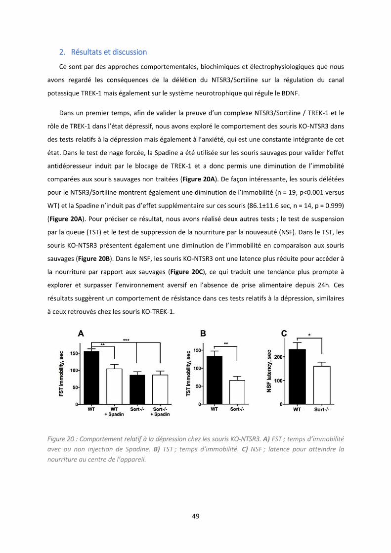
49
2. Résultats et discussion
Ce sont par des approches comportementales, biochimiques et électrophysiologiques que nous
avons regardé les conséquences de la délétion du NTSR3/Sortiline sur la régulation du canal
potassique TREK-1 mais également sur le système neurotrophique qui régule le BDNF.
Da s u p e ie te ps, afi de alide la p eu e d’u o ple e NTSR /So tili e / TREK-1 et le
rôle de TREK- da s l’ tat d p essif, ous a o s e plo le o po te e t des sou is KO-NTSR3 dans
des tests relatifs à la dép essio ais gale e t à l’a i t , ui est u e o sta te i t g a te de et
tat. Da s le test de age fo e, la Spadi e a t utilis e su les sou is sau ages pou alide l’effet
antidépresseur induit par le blocage de TREK-1 et a donc permis une diminutio de l’i o ilit
comparées aux souris sauvages non traitées (Figure 20A). De façon intéressante, les souris délétées
pou le NTSR /So tili e o t e t gale e t u e di i utio de l’i o ilit = , p<0.001 versus
WT) et la Spadi e ’i duit pas d’effet suppl e tai e su es sou is (86.1±11.6 sec, n = 14, p = 0.999)
(Figure 20A). Pour préciser ce résultat, nous avons réalisé deux autres tests ; le test de suspension
par la queue (TST) et le test de suppression de la nourriture par la nouveauté (NSF). Dans le TST, les
souris KO-NTSR p se te t gale e t u e di i utio de l’i o ilit e o pa aiso au sou is
sauvages (Figure 20B). Dans le NSF, les souris KO-NTSR3 ont une latence plus réduite pour accéder à
la nourriture par rapport aux sauvages (Figure 20C), ce qui traduit une tendance plus prompte à
e plo e et su passe l’e i o e e t a e sif e l’a se e de p ise ali e tai e depuis h. Ces
résultats suggèrent un comportement de résistance dans ces tests relatifs à la dépression, similaires
à ceux retrouvés chez les souris KO-TREK-1.
Figure 20 : Comportement relatif à la dépression chez les souris KO-NTSR3. A) FST ; te ps d i o ilit avec ou non injection de Spadine. B) TST ; te ps d i o ilit . C) NSF ; latence pour atteindre la
ou itu e au e t e de l appa eil.
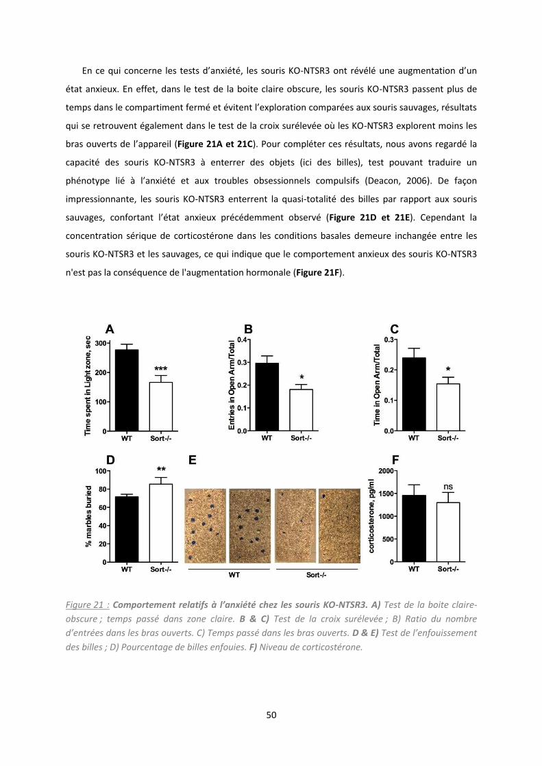
50
E e ui o e e les tests d’a i t , les sou is KO-NTSR o t l u e aug e tatio d’u
état anxieux. En effet, dans le test de la boite claire obscure, les souris KO-NTSR3 passent plus de
temps da s le o pa ti e t fe et ite t l’e plo atio o pa es au sou is sau ages, sultats
qui se retrouvent également dans le test de la croix surélevée où les KO-NTSR3 explorent moins les
as ou e ts de l’appa eil Figure 21A et 21C). Pour compléter ces résultats, nous avons regardé la
capacité des souris KO-NTSR3 à enterrer des objets (ici des billes), test pouvant traduire un
phénotype lié à l’a i t et au troubles obsessionnels compulsifs (Deacon, 2006). De façon
impressionnante, les souris KO-NTSR3 enterrent la quasi-totalité des billes par rapport aux souris
sau ages, o fo ta t l’ tat a ieu p de e t o se Figure 21D et 21E). Cependant la
concentration sérique de corticostérone dans les conditions basales demeure inchangée entre les
souris KO-NTSR3 et les sauvages, ce qui indique que le comportement anxieux des souris KO-NTSR3
n'est pas la conséquence de l'augmentation hormonale (Figure 21F).
Figure 21 : Co porte e t relatifs à l’a xiété chez les souris KO-NTSR3. A) Test de la boite claire-
obscure ; temps passé dans zone claire. B & C) Test de la croix surélevée ; B) Ratio du nombre
d e t es da s les as ouve ts. C Te ps pass da s les as ouve ts. D & E) Test de l e fouisse e t des billes ; D) Pourcentage de billes enfouies. F) Niveau de corticostérone.

51
Ce phénotype de résistance des souris KO-NTSR3 comparable à celui souris KO-TREK-1 laisse
suggérer un dysfonctionnement du canal potassique, qui pourrait se traduire par une inhibition ou
suppression de son expression. Pour vérifier cette hypothèse, nous avons dans un premier temps
alu l’e p essio de TREK- à diff e t i eau de la ellule, et de faço i t essa te, l’e p essio
membranaire du canal potassique, dans des homogénats de cerveaux de souris KO-NTSR3, diminue
fo te e t d’e i o 36 % par rapport aux souris sauvages (Figure 22A . Cette pe te d’e p essio
membranaire semble compensée, mais de manière non significative, par une augmentation au
niveau des vésicules de haute et basse densité. La quantité totale de TREK-1 chez les souris KO-
NTSR ’est pas odifi e pa appo t au sou is sau ages. Cette perte du canal TREK-1 à la surface
de la cellule, sans modification de l'expression totale du TREK-1, est cohérente avec le rôle, déjà
observé, du NTSR3/Sortiline dans le tri cellulaire du canal (Mazella et al., 2010).
TREK-1 est un canal potassique de fond qui permet de maintenir le potentiel de membrane et,
pa o s ue t, de odule l’a ti it des eu o es. La odifi atio d’e p essio e a ai e des
souris KO-NTSR3 laissait supposer des conséquences possibles sur les propriétés
électrophysiologiques des cellules. En effet, en mesurant le potentiel de membrane de neurones
issus de cortex cérébraux de souris KO-NTSR3, nous avons observé une augmentation du potentiel
d’e i o V o paré à ceux de souris sauvages (44.7 ± 1.3 mV KO-NTSR3 ; 62.9 ± 3.6 mV WT)
(Figure 22B . Ce sultat e fo e l’h poth se d’u e pe te fo tio elle de TREK-1 à la membrane.
En effet, TREK-1 permet la diffusion du potassium intracellulaire vers le milieu extracellulaire, par
conséquent, son blocage ou sa délétion entraine une accumulation de potassium intracellulaire et
donc une dépolarisation transitoire. Par ailleurs, cette dépolarisation neuronale a été également
observée avec le blocage de TREK-1 par la Spadine (Devader et al., 2015). Des études antérieures ont
montré que le blocage (Mazella et al., 2010) ou la délétion (Heurteaux et al., 2006b) de TREK-1
améliore le taux de décharge des neurones 5-HT du cerveau moyen, un paramètre clé prédictif de
l'efficacité des antidépresseurs. Par conséquent, nous avons effectué des enregistrements unitaires
extracellulaires de neurones 5-HT dans le noyau dorsal du raphé (DRN) chez des animaux
anesthésiés. L'activité de décharge basale a été enregistrée à la fois chez les souris sauvages et KO-
NTSR3 par des traces successives le long du DRN. Les neurones 5-HT trouvés chez les souris sauvages
déchargeaient dans une gamme de fréquences normales (1,5-2 Hz) avec une moyenne qui était plus
de deux fois inférieure à celle des souris KO-NTSR3 (1,46 ± 0,09 Hz contre 3,42 ± 0,21 Hz, p < 0,001).
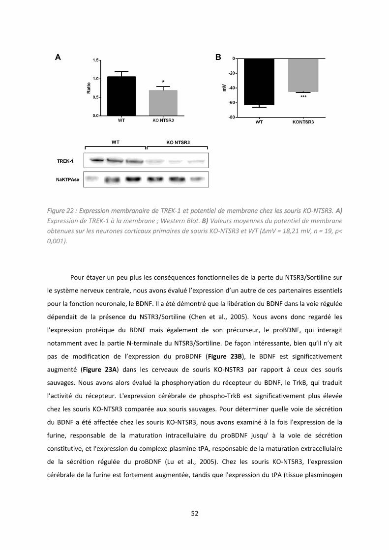
52
Figure 22 : Expression membranaire de TREK-1 et potentiel de membrane chez les souris KO-NTSR3. A)
Expression de TREK-1 à la membrane ; Western Blot. B) Valeurs moyennes du potentiel de membrane
obtenues sur les neurones corticaux primaires de souris KO-NTSR3 et WT ΔmV = 18,21 mV, n = 19, p<
0,001).
Pour étayer un peu plus les conséquences fonctionnelles de la perte du NTSR3/Sortiline sur
le s st e e eu e t ale, ous a o s alu l’e p essio d’u aut e de es pa te ai es esse tiels
pour la fonction neuronale, le BDNF. Il a été démontré que la libération du BDNF dans la voie régulée
dépendait de la présence du NSTR3/Sortiline (Chen et al., 2005). Nous avons donc regardé les
l’e p essio p ot i ue du BDNF ais gale e t de so p u seu , le proBDNF, qui interagit
notamment avec la partie N-te i ale du NTSR /So tili e. De faço i t essa te, ie u’il ’ ait
pas de odifi atio de l’e p essio du p oBDNF Figure 23B), le BDNF est significativement
augmenté (Figure 23A) dans les cerveaux de souris KO-NSTR3 par rapport à ceux des souris
sauvages. Nous avons alors évalué la phosphorylation du récepteur du BDNF, le TrkB, qui traduit
l’a ti it du epteu . L'expression cérébrale de phospho-TrkB est significativement plus élevée
chez les souris KO-NTSR3 comparée aux souris sauvages. Pour déterminer quelle voie de sécrétion
du BDNF a été affectée chez les souris KO-NTSR3, nous avons examiné à la fois l'expression de la
furine, responsable de la maturation intracellulaire du proBDNF jusqu' à la voie de sécrétion
constitutive, et l'expression du complexe plasmine-tPA, responsable de la maturation extracellulaire
de la sécrétion régulée du proBDNF (Lu et al., 2005). Chez les souris KO-NTSR3, l'expression
cérébrale de la furine est fortement augmentée, tandis que l'expression du tPA (tissue plasminogen
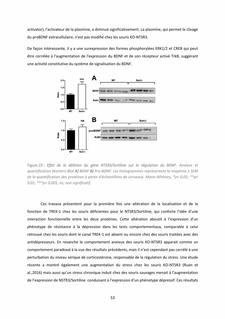
53
activator), l'activateur de la plasmine, a diminué significativement. La plasmine, qui permet le clivage
du p oBDNF e t a ellulai e, ’est pas odifi hez les sou is KO-NTSR3.
De façon intéressante, il y a une surexpression des formes phosphorylées ERK1/2 et CREB qui peut
être corrélée à l'augmentation de l'expression du BDNF et de son récepteur activé TrkB, suggérant
une activité constitutive du système de signalisation du BDNF.
Figure 23 : Effet de la délétion du gène NTSR3/Sortiline sur la régulation du BDNF. Analyse et
quantification Western Blot A) BDNF B) Pro-BDNF. Les histogrammes représentent la moyenne ± SEM
de la quantification des protéines à partir d'échantillons de cerveaux. Mann-Whitney, *p< 0,05; **p<
0,01, ***p< 0,001, ns; non significatif.
Ces travaux présentent pour la première fois une altération de la localisation et de la
fonction de TREK- hez les sou is d fi ie tes pou le NTSR /So tili e, ui o fo te l’id e d’u e
i te a tio fo tio elle e t e les deu p ot i es. Cette alt atio a outit à l’e p essio d’u
phénotype de résistance à la dépression dans les tests comportementaux, comparable à celui
retrouvé chez les souris dont le canal TREK-1 est absent ou encore chez des souris traitées avec des
antidépresseurs. En revanche le comportement anxieux des souris KO-NTSR3 apparait comme un
o po te e t pa ado al à la ue des sultats p de ts, ais il ’est epe da t pas o l à u e
perturbation du niveau sérique de corticostérone, responsable de la régulation du stress. Une étude
récente a montré également une augmentation du stress chez les souris KO-NTSR3 (Ruan et
al.,2016) ais aussi u’u st ess h o i ue i duit hez des sou is sau ages e ait à l’aug e tatio
de l’e p essio de NSTR /So tili e o duisa t à l’e p essio d’u ph ot pe d p essif. Ces sultats
54
so t e a o d a e l’id e ue le ph ot pe de sista e à la dépression serait la conséquence de
l’a se e de NSTR /So tili e.
Nous a o s gale e t o se u e di i utio de l’e p essio de TREK-1 à la membrane
plasmique au niveau des cerveaux des souris KO-NTSR3, suggérant un rôle crucial du récepteur dans
le trafic du canal potassique. La conséquence neuronale qui en résulte réside en une augmentation
du potentiel membranaire des neurones et une activité des neurones sérotoninergiques du DRN
sup ieu e d’u fa teu deu , e ui i di ue u e aug e tatio de l’effi acité de la
neurotransmission sérotoninergique. De façon intéressante, ces deux observations sont très
semblables à celles obtenues avec le blocage de l'activité de TREK-1 par la Spadine (Devader et al.,
2015 ; Mazella et al., 2010) et par la fluoxétine (Heurteaux et al., 2006b) ou lorsque le gène KCNK2,
codant pour TREK-1, a été supprimé chez la souris (Heurteaux et al., 2006b).
Enfin, la régulation du BDNF semble intimement liée au NTSR3/Sortiline, car en effet, chez
les souris KO-NTSR3 il y a une augmentatio de BDNF et de l’a ti it de so epteu T kB. Cette
aug e tatio eu ot ophi ue ’est epe da t pas d pe da te d’u e aug e tatio de so
précurseur le proBDNF qui reste inchangé chez les KO-NTSR . L’e pli atio tie d ait o pte d’u
déséquilibre entre la voie constitutive et régulée du BDNF. En absence de NTSR3/Sortiline dans la
voie régulée, le proBDNF serait maturé principalement dans la voie constitutive intracellulaire, ce qui
e pli ue ait l’aug e tatio de l’e p essio de la fu i e et la di i ution de la maturation
e t a ellulai e a e la aisse d’e p essio du tPA. De plus, u e aug e tatio de l’a ti it du
epteu T kB, i h e t au i eau de BDNF, i dui ait u e aug e tatio de l’a ti it de CREB et pa
o s ue t, la possi ilit d’u e t a s iptio de g es i po ta ts da s l’a ti it eu o ale.
E o lusio , es sultats ette t e lu i e l’i po ta e du NTSR /So tili e da s les
a is es ol ulai es et ph siologi ues ui gule t le o po te e t elatif à l’hu eu et plus
particulièrement dans le phénotype de résistance à la dépression observé lorsque l'expression de
TREK-1 est réduite ou totalement absente.
For Peer R
eview
Depression resistance of sortilin deficient mice
Sébastien Moreno, Christelle Devader, Mariel Pietri, Marc Borsotto, Catherine Heurteaux and Jean
Mazella*
CNRS, Institut de Pharmacologie Moléculaire et Cellulaire, UMR 7275, Université Côte d’Azur,
660 route des Lucioles, 06560 Valbonne, France.
: Jean Mazella, phone: 33 4 93 95 77 61; fax: 33 4 93 95 77 08 email:
[email protected] CNRS, Institut de Pharmacologie Moléculaire et Cellulaire, UMR 7275,
Université Côte d’Azur, 660 route des Lucioles, 06560 Valbonne, France
We thank Carlos Morales (McGill University, Montreal) for providing @/@
mice. We thank E@
Phys@Science for the 5@HT firing rate experiments. We thank the French Government for the
“Investments for the Future” LABEX ICST # ANR@11 LABX 0015. This work was supported by
the Centre National de la Recherche Scientifique and the Agence Nationale de la Recherche (ANR@
13@SAMA@0001 and @0002 and ANR@13@RPIB@0001 and @0002).
Page 3 of 32
British Pharmacological Society
British Journal of Pharmacology
For Peer R
eview
. The background potassium channel TREK@1 has been shown to be a
potent target for depression treatment. Indeed, deletion of the channel in mice resulted in a
depression resistant phenotype. The association of TREK@1 with the sorting protein sortilin
prompted us to investigate the behavior of mice deleted from the gene encoding sortilin (@/@
).
. To characterize the consequences of sortilin deletion on TREK@1
functionality, we combined behavioral, electrophysiological and biochemical approaches performed
and .
. Analyses of @/@
mice revealed that they display 1) a corticosterone@independent
anxiety@like behavior, 2) a resistance to depression as demonstrated by several behavioral tests and
3) an increased efficacy of 5@HT neurotransmission. All these properties were due to TREK@1
action defficiency consequently to a decrease of its cell surface expression and to the modification
of its electrophysiological activity. An increase of BDNF expression through activation of the furin@
dependent constitutive pathway as well as an increase of the BDNF receptor TrkB were in
agreement with the depression@resistant phenotype of @/@
mice.
. Our results demonstrate that the TREK@1 function is altered in the
absence of sortilin confirming the importance of this channel in the regulation on the mood as a
crucial target to treat depression.
Depression, sortilin, TREK@1, behavior, electrophysiology
Page 4 of 32
British Pharmacological Society
British Journal of Pharmacology
For Peer R
eview
!
5@HT : Serotonin
AD : Antidepressant
BDNF : Brain derived nerve factor
BrdU : 5@bromo@2’@deoxyuridine
CREB : cAMP response element@binding protein
DCX : Doublecortin
DRN : Dorsal raphe nucleus
FST : Forced swimming test
H/LDM : High and low density microsomes
MAP kinase : Mitogen activated protein kinase
NSF: Novelty suppressed feeding
NT: neurotensin
NTSR2 : Neurotensin receptor@2
NTSR3 : Neurotensin receptor@3
PM: Plasma membrane
p75NTR : Neurotrophin receptor of 75 kDa
TGN38 : Trans@golgi network protein of 38 kDa
TrkB : Tropomyosin receptor kinase B
TST : Tail suspension test
WT : Wild type
Page 5 of 32
British Pharmacological Society
British Journal of Pharmacology
For Peer R
eview
"
Phenotyping mice in which a gene has been deleted may appear relatively easy in terms of
preliminary observations for detecting a major health problem or a motor defect. After a series of
physical exams including body weight and temperature, home cage locomotion, grooming, nesting
and sleeping, neurological reflexes can be tested before starting behavioral tasks dealing with
anxiety and mood disorders (Crawley, 1999). In the field of depression and anxiety@related
disorders, the background potassium channel TREK@1 (KCNK2) has been one of the first target for
which deletion in mice (
) resulted in a depression@resistant phenotype highlighted by
behavioral tests (Heurteaux et al., 2006b). The properties of these mutant mice are spectacular.
mice behave similarly to naive mice treated with antidepressants (Ads) like fluoxetine. In
addition, an increase in hippocampal neurogenesis and firing rate of serotoninergic neurons from
the dorsal raphe nucleus strongly correlated with the AD@like behavior of
mice (Heurteaux
et al., 2006b). Taking together, results obtained from
mice led to the hypothesis that the
search of selective blockers of the TREK@1 channel could lead to a new type of AD. More recently,
the discovery of spadin, which is a sortilin@derived peptide acting as a potent AD and targeting
TREK@1 channels, supported this hypothesis (Mazella et al., 2010). Interestingly, in addition to the
AD properties of spadin, we have demonstrated the existence of a protein complex between the
TREK@1 channel and sortilin. The expression of the channel at the plasma membrane of COS@7
cells is increased when TREK@1 is co@expressed with sortilin (Carlo, Nykjaer & Willnow, 2014).
Consequently, because sortilin is crucial in the sorting of several factors and enzymes (Kandror &
Pilch, 2011; Lefrancois, Zeng, Hassan, Canuel & Morales, 2003), we analyzed in details the
depression@related phenotype of mice in which the gene encoding sortilin () has been
inactivated, with a focus on the expression of its partner TREK@1 and the subsequent consequences
on the TREK@1 function.
To date, inactivation of gene demonstrated that sortilin is involved in the proNGF@induced
neuronal cell death when associated with p75NTR (Nykjaer et al., 2004) as well as in the binding
Page 6 of 32
British Pharmacological Society
British Journal of Pharmacology
For Peer R
eview
and internalization of the fronto@demential protein progranulin (Hu et al., 2010). More recently a
controversial role of sortilin in the regulation of cholesterol metabolism has been described (Kjolby
et al., 2010; Musunuru et al., 2010), and largely commented in the literature (Dube, Johansen &
Hegele, 2011; Tall & Ai, 2011). Sortilin (also called neurotensin (NT) receptor@3 (NTSR3)) belongs
to the neurotensinergic system (Mazella et al., 1998). A recent sudy showed that the lack of sortilin
expression leads to the increase in brain of both NT and NTSR2 receptors. These mice are
less sensitive to thermal and chemical nociception (Devader et al., 2016).
Because sortilin and TREK@1 have been shown to be associated at the cellular and molecular levels
(Mazella et al., 2010) and because sortilin plays an important role in the sorting of numerous
proteins we hypothesized that the absence of sortilin could result in modification of TREK@1
expression, its cellular location and its function. On another hand, since TREK@1 was described to
be a potent target in depression (Heurteaux et al., 2006b), the aim of the present work was also to
analyze the phenotype of mice regarding depression behavior.
Page 7 of 32
British Pharmacological Society
British Journal of Pharmacology
For Peer R
eview
##
Primary cortical neurons were isolated from 14@day@old mouse embryo cortice (E14). Cells were
mechanically dissociated and seeded in 35 mm diameter previously treated with Poly@D@Lysine and
maintained in culture in a Neurobasal / B27 medium for 10@14 days.
Animal studies were conducted in compliance with the ARRIVE guidelines (Kilkenny et al., 2011;
McGrath & Lilley, 2015). Adult male (8@10 weeks old) mice were housed under controlled
laboratory conditions according to the FELASA guidelines and recommendations ; 6 mice/cage
with a 12 h dark@light cycle, a temperature of 21 ± 2°C, and a 40–60% humidity. They have free
access to standard rodent diet and tap water. The NTSR3/sortilin homozygous KO mice (
were generated by the Morales's laboratory by incorporation of a GFP cassette after exon 1 (Zeng,
Racicott & Morales, 2009) and controls were C57Bl/6J male mice from Janvier Labs (St Berthevin,
France). The local Ethics Committee (CIEPAL) (protocol number 00893.02) approved
experimental procedures and animal care are in accordance with the policies on the care and use of
laboratory animals of European Community legislation 2010/63/EU.
!: the group size provided for the following experiments were variable due to the
difficulties to obtain regularly the same amount of adult males (8@10 weeks old) house breeded
animals (ie : @/@
mice). For each kind of experiment, we adapted the number of WT mice to the
number of available @/@
mice.
Each animal was used once and the total of 218 male WT and @/@
mice, and 5 female WT and
@/@
mice, were used for this sudy. Behavioral experiments were performed with naïve mice for
all used tests. Mice were isolated 30 minutes in neutral room before tests. The experimenter was
blind to randomized experimental groups. Randomization was performed using the TST software
(Bioseb, France).
Page 8 of 32
British Pharmacological Society
British Journal of Pharmacology
For Peer R
eview
Behavioral tests used in this work have been validated for mouse behavioral phenotypes related to
neuropsychiatric disorders, such as depression or anxiety (for review, see (Samsom & Wong,
2015)). Group sizes for behavioral, biochemical and electrophysiological experiments were equal
with the exception for firing rate recordings for which measurements were recorded from 36 5@HT
neurons for WT and 26 neurons for @/@
mice. These in vivo experiments were performed on 5
animals for each group and the number of recorded neurons from each animal varied pending on the
position of electrodes.
For Western blot analyses, results were normalized using either an intracellular protein (actin) or a
protein specific for a given intracellular compartment for sub@cellular fractionation experiments.
All data (displayed as mean ± SEM) were analysed using Prism 6@2 Software (GraphPad, San
Diego, USA). For the comparison of two groups, Mann@Whitney test was used. All remaining data
were compared using a one@way ANOVA with a Tukey’s multiple comparison test. The level of
significance was set at " < 0.05. The data and statistical analysis comply with the recommendations
on experimental design and analysis in pharmacology (Curtis et al., 2015).
# $%#&
FST was performed according to the procedure initially described (Porsolt, Le Pichon & Jalfre,
1977). Each animal was placed in a cylinder (height 30 cm, diameter 15 cm) filled with 15@cm
water at 22•±•1 °C with no escape possibility during 6 min. The period of immobility was
measured only during the last 4 min of the trial. We considered an immobile mouse when it only
remained floating with slight movements to maintain head above surface.
' ( %'#
Mice were deprived from food for 24h. On test day, mice were placed in a highly brightly lit box
(45•×•45•×•20 cm), with floor covered with wood chip bedding, during 10 min. At the center of
the box, a food pellet was placed on a white platform. The latency for the animal to eat the pellet
Page 9 of 32
British Pharmacological Society
British Journal of Pharmacology
For Peer R
eview
was measured as previously described (Santarelli et al., 2003).
&&%&&
Mice were suspended by the tail by using a piece of adhesive tape. After ‘agitation’ or ‘escape@
like’ behavior, mice adopted an immobile posture, suggested to mirror a state of depression. We
recorded the immobility time during a 6 min test session according to (Cryan, Mombereau &
Vassout, 2005).
)*
Burying object relates to a natural defense mechanism that occurs in mice under stress condition or
anxiety state. Marble burying is able to detect anxiety and obsessive compulsive disorders@related
phenotypes (Deacon, 2006). In response to novel bedding/environment mice exhibit digging
behavior. Mice were placed during 30 min in a cage filled with approximately 5 cm deep with
wood chip bedding and a regular pattern of 13 glass marbles disposed on the surface, evenly
spaced, each about 4 cm apart. At the end of the time, buried marbles were counted (2/3 minimum
of depth). 75% of buried marbles was a typical score for naïve C57BL/6.
")!
Elevated Plus Maze allowed to define an anxiety response in rodents (Gross et al., 2002). The
apparatus consisted of central platform (5x5cm), two open arms and two closed arms across from
each other and perpendicular, with the same size (45x5cm) and 15 cm wall height for the closer
arms. The apparatus was placed at 45 cm height above the floor. Mice were placed in the central
platform facing one open arm and were allowed to freely move during 10 minutes. During this
period, number of entries and time spent in both arm were measured. To define an anxiety
response, the time and number of entries in open arms were evaluated in relation to whole time
spent or the total entries.
+,
Mice were placed in a box divided into two compartments by a black partition with a small opening
that allows mouse to move from one compartment to the other (Welch et al., 2007). One
Page 10 of 32
British Pharmacological Society
British Journal of Pharmacology
For Peer R
eview
compartment, corresponding to one@third of the surface area, was made of white plastic and was
brightly illuminated. The adjoining smaller compartment was black and dark. Mice were placed in
the white compartment and allowed to move freely between the two chambers for 5 min. Time
spent in the white chamber, and latency to the first transition were recorded. Mice tended to avoid
the white compartment. The measures of exploration in this area were used as experimental indices
of anxiety.
Corticosterone concentration present in the serum from WT and @/@
mice was determined using
the ELISA kit from Enzo Life Science according to the manufacturer recommendations.
Electrophysiological experiments were performed on primary cortical neurons seeded at a density
of 100,000 cells/35 mm dish after 10 days of culture. Membrane potential was recorded in whole
cell configuration (Hamill, Marty, Neher, Sakmann & Sigworth, 1981) in current clamp mode (no
current is injected, I = 0). Each membrane potential was evaluated by using a RK 400 patch clamp
amplifier (Axon Instruments, USA), low@pass filtered at 3 kHz and digitized at 10 kHz using a 12@
bit analogue@to@digital converter Digidata (1322 series, Axon Instruments, USA). Patch clamp
pipettes were pulled using vertical puller (PC@10, Narishige) from borosilicate glass capillaries and
had a resistance of 10 – 8 MZ. The bath solution contained (in mM) 150 NaCl, 5 KCl, 3 MgCl2, 1
CaCl2, and 10 HEPES adjustedto pH 7.4 with NaOH. The pipette solution contained (in mM) 155
KCl, 3 MgCl2, 5 EGTA, and 10 HEPES adjusted to pH 7.2 with KOH. All experiments were
performed at room temperature (21@22°C). Data acquisition was carried out using a microcomputer
(Dell Pentium) with commercial software and hardware (pClamp 8.2). All values of membrane
potentials are expressed in mV as mean±standard error of the mean (SEM).
Page 11 of 32
British Pharmacological Society
British Journal of Pharmacology
For Peer R
eview
!
In order to quantify the amount of TREK@1 expressed at the plasma membrane and intracellularly,
we performed sub@cellular fractionation from brain homogenates. Plasma membranes were prepared
from brain homogenates of WT or KO@NTSR3/Sortilin mice according to the protocol previously
described (Clancy & Czech, 1990). 30 [g of crude homogenates, purified plasma membranes and
high and low density vesicles (H/LDM) were submitted to Western blot analysis using the rabbit
polyclonal antibody against [email protected] (TREK@1) (1:500) (Alomone Labs (Israel)). Proteins
detected with this antibody were normalized using antibodies specific for each intracellular
compartment (NaKATPase for plasma membranes, TGN38 for H/LDM and beta@actin for total
extracts) from SantaCruz technologies (USA).
Mouse brains were dissected on ice and then homogenized in a solubilization buffer containing 20
mM HEPES (pH:7.4), 1 mM EDTA, 1 mM PMSF, 250 mM sucrose, and protease inhibitor cocktail
using a polytron at the lowest speed. The homogenates were centrifuged 20 min at 100,000×g at
4°C. Supernatants were resupended in 20 mM HEPES (pH: 7.4), 1mM EDTA and stored at −20°C
until further use. Solubilized proteins were loaded at 50 µg in SDS buffer, separated on 10% SDS
PAGE gels and then transferred to a nitrocellulose membrane.
Membranes were incubated with rabbit polyclonal Anti@BDNF (1:1000) (GeneTex (USA)), Anti@
Phospho(Tyr705)TrkB (1:500), Anti@TrkB (1:1000) (Signalway Antibody (USA)), Anti@ProBDNF
(1:400) (Alomone Labs (Israel)), Anti@Furin (1:1000) (SantaCruz technologies (USA)), Anti@
Phospho(Ser133) CREB (1:500) and mouse monoclonal Anti@CREB (1:1000)(CST(USA)), mouse
monoclonal Anti@beta@Actin (1:5000)(SantaCruz technologies (USA) over night at 4°C. Afterwards,
membranes were incubed 30 minutes with secondary antibody (related to species of first antibody)
coupled HRP. Protein bands were revealed, images were acquired with FX Fusion (Vilber) and
analysed with ImageJ (US NIH (Bethesda) (Schneider, Rasband & Eliceiri, 2012).
Page 12 of 32
British Pharmacological Society
British Journal of Pharmacology
For Peer R
eview
Twenty@four hours after the BrdU injection (120 mg per kg of body weight in a 300 µl bolus), mice
were euthanized and transcardially perfused with 4% cold paraformaldehyde. Serial brain sections
were cut (40 µm) throughout the entire hippocampus on a vibratome (Leica). Every sixth section
throughout the hippocampus was processed for BrdU or doublecortin (DCX)
immunohistochemistry, as previously described (Heurteaux et al., 2006a). For each
immunodetection, slides were first incubated overnight at 4°C with a mouse monoclonal anti@BrdU
antibody (1:200; Becton@Dickinson) or a goat anti@DCX (1:400, Santa Cruz Laboratories). For
chromogenic immunodetection, sections were then incubated for 1 hour in biotin@conjugated
species@specific secondary antibodies (1:100; Vector Laboratories) followed by a peroxidase@avidin
complex solution according to the manufacturer’s protocol. The peroxidase activity of immune
complexes was visualized with DAB staining using the VectaStain ABC kit (Vector Laboratories).
For fluorescent double labeling, that were performed to determine the cell phenotype, sections were
incubated overnight at 4°C with anti@sheep BrdU (1:200, Interchim), anti@goat DCX (1:200, Santa
Cruz Laboratories), or an anti@GFAP (Glial Fibrillary acidic protein, marker for astrocytes, 1:250,
Dako). Antibodies were revealed with anti@IgG Alexa 488 or 594@coupled antibodies (1:400;
Molecular Probes). All BrdU@labeled cells in the granular cell layer and subgranular zone (SGZ)
were counted in each section (n = 10 and 5 mice per group) at 400x and 1000x magnification under
a light microscope (Olympus) by an experimenter blinded to the study code. The total number of
BrdU@positive cells per section was multiplied by 6 to obtain the total number of cells per dentate
gyrus. The counting of BrdU/DCX labeled cells was performed using a Laser Scanning Confocal
Microscope (TCS SP, Leica) equipped with a DMIRBE inverted microscope.
"# $!%& ' Single@barreled glass micropipettes
(recording electrodes) were filled with a 2 M NaCl solution saturated with Fast Green FCF,
Page 13 of 32
British Pharmacological Society
British Journal of Pharmacology
For Peer R
eview
resulting in an impedance of 2–5 MZ. Mice were anaesthetized with chloral hydrate (400 mg/kg,
-., using a 2% solution), and placed in a stereotaxic frame equipped with the Stoelting mouse
adapter. Electrodes were positioned 0.5–1 mm posterior to the interaural line on the midline, and
were then lowered into the dorsal raphe nucleus (DRN) at a depth of 2.5 mm from the brain surface.
5@HT neurons were then encountered over a maximal distance of 1 mm. They were identified using
the following criteria: a slow (0.5–2.5 Hz) and regular firing rate and long@duration (0.8 – 1.2 ms)
action potentials, with a positive@negative spike (Heurteaux et al., 2006a). Spikes were computed by
using the Spike 2 software. Firing rates were calculated as the mean number of events occurring
within a 10 s period. For each neuron, discharges were monitored during 60 seconds. Each mouse
received either spadin (10@5
M in a 100 µl bolus) or its vehicle. Starting 30 min after the injection, 3
to 4 successive descents were performed along the DRN, for a total of 4–8 cells recorded per
animal. Recordings were performed for a maximal duration of 4 hours post@injection.
Page 14 of 32
British Pharmacological Society
British Journal of Pharmacology

For Peer R
eview
($
!%
In the aim to confirm the existence of sortilin/TREK@1 complexes and the role of the TREK@1
channel in depression, we first investigated the behavior of @/@
mice in a series of behavioral
tests related to depression and anxiety.
,
The forced swimming test (FST) performed directly on both WT and @/@
mice groups indicated
that @/@
mice presented a significant lower immobility time (100.3±13.5 sec) than WT mice
(151.4±7.8 sec) (Figure 1A). FST was validated on WT mice by the use of the novel AD spadin
(0.1 µM) which after injection decreased the immobility time from 162.9±12 sec to 104.4±12.5
sec (One way ANOVA, F(3, 52) = 10.43) (Figure 1B). Interestingly, the immobility time of vehicle
injected @/@
mice was 81.6±10.1 sec and injection of spadin had no effect on these mice
(86.1±11.6 sec) (Figure 1B). This result prompted us to perform two other tests, the tail suspension
test (TST) and the novelty supressed feeding test (NSF). The TST confirmed that @/@
mice had
an immobility time (65.9±11.2 sec) significantly reduced when compared to WT mice (133.2±14.4
sec, n = 20) (Figure 1C). Finally, in the NSF test, the latency time measured for WT mice
(231.4±29.2 sec) was also significantly decreased in @/@
mice (158.5±19.1 sec) (Figure 1D).
Taken together, these results indicate that @/@
mice appear to display a depression@resitant
behavior resembling to that of
mice (Heurteaux et al., 2006b).
.
In the light dark test, the time of spent in the light zone for WT mice was 294.9±14.9 sec, time
significantly higher than that of @/@
mice (156.9±15.1 sec) (Figure 2A). Therefore, we tested the
two groups of mice in the elevated@plus Maze test by measuring the ratio of the time and the
number of entries in the open arm. As observed in Figure 2B, the time ratio in the open arm of WT
mice (0.239±0.031) was significantly reduced in @/@
mice (0.148±0.023) (Figure 2B). In the
same way the ratio in the number of entries of WT mice (0.322±0.021) was decreased in @/@
Page 15 of 32
British Pharmacological Society
British Journal of Pharmacology
For Peer R
eview
mice (0.194±0.022) (Figure 2C). Finally, we tested the ability of WT and @/@
mice to bury glass
marbles, a test able to detect anxiety and obsessive compulsive disorders related phenotypes
(Deacon, 2006). Figure 2D and E showed that WT mice were able to bury 72.7±2.9 % of marbles
whereas @/@
mice buryed 85.5±7.3 % of marbles suggesting again an aniety@like behavior of
@/@
mice as previously observed (Ruan, Yang, Li, Luo, Bobrovskaya & Zhou, 2016). The serum
concentration of corticosterone in basal conditions remained unchanged between WT and @/@
mice (Figure 2F) indicating that the anxiety@like behavior of @/@
mice was not the consequence
of the hormone increase.
& !(!)!
%
The depression@resistant behavior of @/@
mice may suggest some dysfunction of the TREK@1
channel for which its blockade or its deletion results in a depression@resistance phenotype. We first
investigated the subcellular location of TREK@1 performed on sub@cellular fractionation from WT
and @/@
mouse brains. Surprisingly, the amount of TREK@1 channels present at the plasma
membrane (PM) was significantly decreased by 36% in @/@
mice when compared to WT mice
(Figure 3A). The loss of TREK@1 at the PM appeared to be compensated by an increase of the
protein at the level of high and low density vesicles (H/LDM) although not significant (Figure 3B).
The total amount of TREK@1 was not modified between WT and @/@
mice (Figure 3C). The loss
of the TREK@1 channel at the cell surface without modification of total TREK@1 expression is in
agreement with the role of sortilin in the channel cell sorting as already observed (Mazella et al.,
2010).
To determine the consequence of the TREK@1 expression loss at the PM, we performed
electrophysiological experiments on neurons prepared from WT and @/@
mice. As shown in
Figure 4A, the membrane potential measured on neurons from @/@
mice was 44.7 ± 1.3 mV
consisting to a significant increase of membrane potential of 18.21 mV when compared to neurons
Page 16 of 32
British Pharmacological Society
British Journal of Pharmacology
For Peer R
eview
from WT mice (62.9 ± 3.6 mV). This result indicated that the decrease of TREK@1 channels at the
cell surface of @/@
neurons leads to an increase of membrane potential very similar to that
obtained with the channel blocker spadin on WT neurons (Devader et al., 2015).
Previous studies have shown that the blockade (Mazella et al., 2010) or the deletion (Heurteaux et
al., 2006b) of TREK@1 enhance midbrain 5@HT neuron firing rate, a key parameter predictive of AD
efficacy. Therefore, we performed unitary extracellular recordings of 5@HT neurons in the DRN in
anesthetized animals. The basal firing activities were recorded in both WT and @/@
mice by
successive tracks along the DRN. 5@HT neurons found in WT mice discharged within a normal
frequency range (1.5@2 Hz) with an average that was more than two fold lower than in @/@
mice
(1.46 ± 0.09 Hz versus 3.42 ± 0.21 Hz, p < 0.001) (Figure 4B).
The hippocampus is known to be involved in emotional responses and depression is associated with
the suppression of dentate gyrus neurogenesis. It is also established that chronic AD treatments,
including fluoxetine but also the new rapid antidepressant spadin, induce neurogenesis in the
hippocampus of rodents visualized by an increase of progenitor cells that incorporate 5@bromo@2’@
deoxyuridine (BrdU). WT mice treated with spadin (100 µg per kg body weight) for 4days showed
a significant increase in the number of hippocampal BrdU@positive cells compared with WT mice
treated with vehicle (1919±134 cells/hippocampus vs 1218±105 cells/hippocampus, respectively,
one@way ANOVA, F(3.16) = 19.94, p<0.001) (Figure 4C). In @/@
mice, although the number of
BrdU@positive cells was slightly lower in basal conditions, the effect of spadin remained efficient
with a lower but significant increase of hippocampal newborn cells from 887±82 in WT
hippocampus to 1306±37 in @/@
hippocampus (p < 0.05) (Figure 4C).
The hippocampal neurogenesis is known to be concomitantly increased with the activation the
transcription factor cAMP response element@binding protein (CREB) in response to chronic AD
treatment, but not to non@antidepressant psychotic drugs (Carlezon, Duman & Nestler, 2005). These
observations strongly suggest that CREB regulates hippocampal neurogenesis. Therefore we
analyzed the phosphorylation state of CREB in the hippocampus of both WT and @/@
and
Page 17 of 32
British Pharmacological Society
British Journal of Pharmacology
For Peer R
eview
showed that the immunoreactive band of 46 kDa corresponding to phospho@CREB was significantly
enhanced in the @/@
hippocampus when compared to the WT whereas the amount of total CREB
remained unchanged (Figure 4D).
!!'&() ($
BDNF displays a potential role as a marker of treatment response in patients with major depressive
disorder although its effects on mood variations remain unclear. The release of BDNF to the
regulated pathway has been shown to be dependent on the presence of sortilin (Chen et al., 2005).
For these reasons and because @/@
mice display a depressive resistant phenotype, we evaluated
the expression profiles of BDNF and its precursor proBDNF in the brain of WT and @/@
mice.
Western blot analyses of BDNF content from WT or @/@
mouse brains clearly demonstrated that
the neurotrophin amount was strongly increased in @/@
mice vs WT (p < 0.001) (Figure 5A). By
contrast, no change in amount of proBDNF was observed (Figure 5B). The increase of BDNF
prompted us to examine the expression of the active phosphorylated form of the BDNF receptor
TrkB. As shown in Figure 5C, the p@TrkB expression was significantly enhanced in @/@
mice
when compared with WT mouse brain.
To determine which of BDNF secretion pathway was affected in @/@
mice we examined both
the expression of furin, responsible for the intracellular maturation of proBDNF to the constitutive
secretion pathway, and the expression of the complex plasmin/tPA, responsible for the extracellular
maturation of the regulated secretion of proBDNF (Lu, Pang & Woo, 2005). Interestingly in @/@
mice, the brain expression of furin was strongly enhanced (p < 0.01) (Figure 6A) whereas the
expression of tPA, the plasmin activator, was significantly decreased in @/@
mice (Figure 6B).
Plasmin expression was not modified (Figure 6C).
The increase in the expression of BDNF and its activated receptor TrkB was certainly responsible
for the over@expression of the phosphorylated form CREB observed in @/@
mice suggesting a
constitutive activity of the BDNF signaling system.
Page 18 of 32
British Pharmacological Society
British Journal of Pharmacology
For Peer R
eview
&
The present work shows for the first time that the cellular localization and function of the TREK@1
channel were altered in mice lacking sortilin. The TREK@1 dysfunction led to a depression resistant
phenotype in @/@
mice compared to wild@type mice in three different behavioral tests (Figure 1).
Interestingly, this phenotype was comparable to that of
mice (lacking TREK@1) which
behaved as wild@type mice treated with classical ADs. However and paradoxically, @/@
mice
displayed an anxiety@like behavior in several anxiety@related tests including the light dark and the
marbles burying tests (Figure 2). This latter observation that appears not to be due to an elevated
level of serum corticosterone, is in agreement with a recent study relating that @/@
mice
displayed elevated anxiety@like behavior and that chronically stressed wild@type mice showed an
increase in the sortilin expression in neocortex and hippocampus leading to an increased
depression@like behavior (Ruan, Yang, Li, Luo, Bobrovskaya & Zhou, 2016). Since the increase of
sortilin expression is associated with depression, our results describing a resistance to depression in
@/@
mice could be the consequence of the absence of sortilin.
The TREK@1 expression decrease within the brain plasma membrane of @/@
mice compared to
WT mice (Figure 3A@C) highly suggests a crucial role of sortilin in the correct sorting of the TREK@
1 channel. The involvement of sortilin in the TREK@1 targeting has previously been documented in
a heterologeous expression system by cotransfection of the two proteins (Mazella et al., 2010).
TREK@1 is not the only membrane protein that is regulated by its interaction with sortilin. In
particular, sortilin displays important action in the function of several receptors including the
proNGF receptor p75NTR (Nykjaer et al., 2004), the BDNF receptor TrkB (Yang, Lim, Li, Zhong
& Zhou, 2011), and the neurotensin receptors NTSR1 (Martin, Navarro, Vincent & Mazella, 2002)
and NTSR2 (Beraud@Dufour, Coppola, Massa & Mazella, 2009). In the present work, the absence
of sortilin allowed us to definitively determine that either the dysfunction by alteration of its sorting
or the inhibition by spadin of TREK@1 leads to a common result : the resistance to depression
behavior.
Page 19 of 32
British Pharmacological Society
British Journal of Pharmacology
For Peer R
eview
The neuronal consequences resulting from the decreased cell surface TREK@1 expression led to
several observations : i) the membrane potential meassured on @/@
neurons was strongly
increased when compared to WT neurons (Figure 4A), and ii) the firing rate activity of 5@HT
neurons from the dorsal raphe nucleus was twofold increased indicating an accelerated efficacy of
5@HT neurotransmission (Figure 4B). Both observations were very similar to those obtained with
the blocking of TREK@1 activity by spadin (Devader et al., 2015; Mazella et al., 2010) and by
fluoxetine (Heurteaux et al., 2006b) or when kcnk2 gene was deleted in TREK@1 null mice
(Heurteaux et al., 2006b). These properties are in agreement with the observation that clinical
pharmacological and electroconvulsive AD treatments enhance activation of hippocampal
postsynaptic 5@HT1A receptors (Haddjeri, Blier & de Montigny, 1998).
While the number of hippocampal progenitor cells remained unchanged between @/@
and WT
mice, surprisingly, the two strains responded similarly to a 4day@spadin treatment with a significant
increase in the number of BrdU@positive cells. This result suggests that 1/ despite the lower level of
TREK@1 membrane expression measured in @/@
mice, the remaining functional channels are
sufficient to trigger neurogenesis under spadin exposure (note that the response to spadin was quite
lower in @/@
mice) or 2/ TREK@1 is not the only membrane protein responsible for the response
to the peptide. In neurogenesis experiments @/@
mice were still able to respond to spadin (Figure
3F). Similar observations were obtained from
mice in which basal hippocampal
neurogenesis was identical to that measured from WT mice but
mice remained able to
respond to fluoxetine (Heurteaux et al., 2006b).
Finally, we observed that the level of brain BDNF, as well as its activated receptor TrkB, was
significantly increased in @/@
mice whereas the level of its precursor form proBDNF remained
unchanged (Figure 5A@C). This finding can be explained by the brain over@expression in @/@
mice of the convertase furin involved in the regulated pathway of BDNF secretion (Figure 5A).
Concomitantly, the plasmin and tPA expression involved in the constitutive secretion pathway,
appeared slightly decreased (Figure 5B and C). The increase in both BDNF secretion and
Page 20 of 32
British Pharmacological Society
British Journal of Pharmacology
For Peer R
eview
expression has been already observed in @/@
neurons (Chen et al., 2005) that have been prepared
slightly differently than those used in this study (Devader et al., 2016). The elevated levels of
BDNF and of its activated receptor TrkB in the @/@
brains added to the increased active form of
CREB, could be responsible for the depression@resistant phenotype.
In conclusion, data presented in this work could explain the molecular and physiological
mechanisms that are responsible for the depression@resistant phenotype observed when the
expression of TREK@1 was decreased or totaly absent.
"
We declare to have no financial conflict of interest
SM, CD, MP and JM performed the experiments
MB, CH and JM conceived and designed the experiments
MB, CH and JM contributed reagents/materials/analysis tools
MB, CH and JM wrote the paper
Page 21 of 32
British Pharmacological Society
British Journal of Pharmacology
For Peer R
eview
Beraud@Dufour S, Coppola T, Massa F, & Mazella J (2009). Neurotensin receptor@2 and @3 are
crucial for the anti@apoptotic effect of neurotensin on pancreatic beta@TC3 cells. Int J Biochem Cell
Biol 41 2398@2402.
Carlezon WA, Jr., Duman RS, & Nestler EJ (2005). The many faces of CREB. Trends Neurosci 28
436@445.
Carlo AS, Nykjaer A, & Willnow TE (2014). Sorting receptor sortilin@a culprit in cardiovascular
and neurological diseases. J Mol Med (Berl) 92 905@911.
Chen ZY, Ieraci A, Teng H, Dall H, Meng CX, Herrera DG/ - (2005). Sortilin controls
intracellular sorting of brain@derived neurotrophic factor to the regulated secretory pathway. J
Neurosci 25 6156@6166.
Clancy BM, & Czech MP (1990). Hexose transport stimulation and membrane redistribution of
glucose transporter isoforms in response to cholera toxin, dibutyryl cyclic AMP, and insulin in 3T3@
L1 adipocytes. The Journal of biological chemistry 265 12434@12443.
Crawley JN (1999). Behavioral phenotyping of transgenic and knockout mice: experimental design
and evaluation of general health, sensory functions, motor abilities, and specific behavioral tests.
Brain Res 835 18@26.
Cryan JF, Mombereau C, & Vassout A (2005). The tail suspension test as a model for assessing
antidepressant activity: review of pharmacological and genetic studies in mice. Neurosci Biobehav
Rev 29 571@625.
Curtis MJ, Bond RA, Spina D, Ahluwalia A, Alexander SP, Giembycz MA/ - (2015).
Experimental design and analysis and their reporting: new guidance for publication in BJP. Br J
Pharmacol 172 3461@3471.
Deacon RM (2006). Digging and marble burying in mice: simple methods for in vivo identification
of biological impacts. Nat Protoc 1 122@124.
Devader C, Khayachi A, Veyssiere J, Moha Ou Maati H, Roulot M, Moreno S/ - (2015). In
vitro and in vivo regulation of synaptogenesis by the novel antidepressant spadin. Br J Pharmacol
172 2604@2617.
Devader C, Moreno S, Roulot M, Deval E, Dix T, Morales CR/ - (2016). Increased Brain
Neurotensin and NTSR2 Lead to Weak Nociception in NTSR3/Sortilin Knockout Mice. Front
Neurosci 10 542.
Dube JB, Johansen CT, & Hegele RA (2011). Sortilin: an unusual suspect in cholesterol
metabolism: from GWAS identification to in vivo biochemical analyses, sortilin has been identified
as a novel mediator of human lipoprotein metabolism. Bioessays 33 430@437.
Gross C, Zhuang X, Stark K, Ramboz S, Oosting R, Kirby L/- (2002). Serotonin1A receptor
acts during development to establish normal anxiety@like behaviour in the adult. Nature 416 396@
400.
Page 22 of 32
British Pharmacological Society
British Journal of Pharmacology
For Peer R
eview
Haddjeri N, Blier P, & de Montigny C (1998). Long@term antidepressant treatments result in a tonic
activation of forebrain 5@HT1A receptors. J Neurosci 18 10150@10156.
Hamill OP, Marty A, Neher E, Sakmann B, & Sigworth FJ (1981). Improved patch@clamp
techniques for high@resolution current recording from cells and cell@free membrane patches.
Pflugers Arch 391 85@100.
Heurteaux C, Lucas G, Guy N, El Yacoubi M, Thümmler S, Peng X/ - (2006a). Deletion of
TREK@1, a background potassium channel, results in a depression@resistant phenotype. Nature
Neurosci 9 1134@1141.
Heurteaux C, Lucas G, Guy N, El Yacoubi M, Thummler S, Peng XD/- (2006b). Deletion of
the background potassium channel TREK@1 results in a depression@resistant phenotype. Nat
Neurosci 9 1134@1141.
Hu F, Padukkavidana T, Vaegter CB, Brady OA, Zheng Y, Mackenzie IR/- (2010). Sortilin@
mediated endocytosis determines levels of the frontotemporal dementia protein, progranulin.
Neuron 68 654@667.
Kandror KV, & Pilch PF (2011). The sugar is sIRVed: sorting Glut4 and its fellow travelers. Traffic
12 665@671.
Kilkenny C, Browne W, Cuthill IC, Emerson M, Altman DG, National Centre for the Replacement
R/- (2011). Animal research: reporting in vivo experiments@@the ARRIVE guidelines. J Cereb
Blood Flow Metab 31 991@993.
Kjolby M, Andersen OM, Breiderhoff T, Fjorback AW, Pedersen KM, Madsen P/ - (2010).
Sort1, encoded by the cardiovascular risk locus 1p13.3, is a regulator of hepatic lipoprotein export.
Cell Metab 12 213@223.
Lefrancois S, Zeng J, Hassan AJ, Canuel M, & Morales CR (2003). The lysosomal trafficking of
sphingolipid activator proteins (SAPs) is mediated by sortilin. EMBO J 22 6430@6437.
Lu B, Pang PT, & Woo NH (2005). The yin and yang of neurotrophin action. Nat Rev Neurosci 6
603@614.
Martin S, Navarro V, Vincent JP, & Mazella J (2002). Neurotensin receptor@1 and @3 complex
modulates the cellular signaling of neurotensin in the HT29 cell line. Gastroenterology 123 1135@
1143.
Mazella J, Petrault O, Lucas G, Deval E, Beraud@Dufour S, Gandin C/ - (2010). Spadin, a
sortilin@derived peptide, targeting rodent TREK@1 channels: a new concept in the antidepressant
drug design. PLoS Biol 8 e1000355.
Mazella J, Zsurger N, Navarro V, Chabry J, Kaghad M, Caput D/ - (1998). The 100@kDa
neurotensin receptor is gp95/sortilin, a non@G@protein@coupled receptor. The Journal of biological
chemistry 273 26273@26276.
McGrath JC, & Lilley E (2015). Implementing guidelines on reporting research using animals
(ARRIVE etc.): new requirements for publication in BJP. Br J Pharmacol 172 3189@3193.
Page 23 of 32
British Pharmacological Society
British Journal of Pharmacology
For Peer R
eview
Musunuru K, Strong A, Frank@Kamenetsky M, Lee NE, Ahfeldt T, Sachs KV/- (2010). From
noncoding variant to phenotype via SORT1 at the 1p13 cholesterol locus. Nature 466 714@719.
Nykjaer A, Lee R, Teng KK, Jansen P, Madsen P, Nielsen MS/- (2004). Sortilin is essential for
proNGF@induced neuronal cell death. Nature 427 843@848.
Porsolt RD, Le Pichon M, & Jalfre M (1977). Depression: a new animal model sensitive to
antidepressant treatments. Nature 266 730@732.
Ruan CS, Yang CR, Li JY, Luo HY, Bobrovskaya L, & Zhou XF (2016). Mice with Sort1
deficiency display normal cognition but elevated anxiety@like behavior. Exp Neurol 281 99@108.
Samsom JN, & Wong AH (2015). Schizophrenia and Depression Co@Morbidity: What We have
Learned from Animal Models. Front Psychiatry 6 13.
Santarelli L, Saxe M, Gross C, Surget A, Battaglia F, Dulawa S/ - (2003). Requirement of
hippocampal neurogenesis for the behavioral effects of antidepressants. Science 301 805@809.
Schneider CA, Rasband WS, & Eliceiri KW (2012). NIH Image to ImageJ: 25 years of image
analysis. Nat Methods 9 671@675.
Tall AR, & Ai D (2011). Sorting out sortilin. Circ Res 108 158@160.
Welch JM, Lu J, Rodriguiz RM, Trotta NC, Peca J, Ding JD/- (2007). Cortico@striatal synaptic
defects and OCD@like behaviours in Sapap3@mutant mice. Nature 448 894@900.
Yang M, Lim Y, Li X, Zhong JH, & Zhou XF (2011). Precursor of brain@derived neurotrophic
factor (proBDNF) forms a complex with Huntingtin@associated protein@1 (HAP1) and sortilin that
modulates proBDNF trafficking, degradation, and processing. The Journal of biological chemistry
286 16272@16284.
Zeng J, Racicott J, & Morales CR (2009). The inactivation of the sortilin gene leads to a partial
disruption of prosaposin trafficking to the lysosomes. Exp Cell Res 315 3112@3124.
Page 24 of 32
British Pharmacological Society
British Journal of Pharmacology
For Peer R
eview
)
) : Antidepressant@like behavior in @/@
mice.
(A) FST, @/@
mice had a shorter immobility time than WT mice (F(3, 52) = 10.43, p < 0.01). (B)
WT spadin@treated mice had also a shorter immobility time than vehicle@treated mice. Spadin had
no effect on @/@
mice. (C) TST, @/@
mice had a significant reduced immobility time than
WT mice. (D) NSF, the latency time for @/@
mice was shorter than that measured for WT mice.
Values are expressed as mean ± SEM. * p< 0.05, ** p< 0.01, *** p< 0.001.
)* : Anxiety@like behavior in @/@
mice.
(A) Light@dark test, the time spent in the light zone was shorter for @/@
mice compared to WT
mice. (B and C) Elevated@plus Maze test, the ratio of the time spent in the open arm was reduced in
@/@
mice compared to WT mice. The ratio in the number of entries was also reduced in @/@
mice. (D and E) Glass marbles burying test, the percent of marbles buried by WT mice was
significantly increased by @/@
mice as shown in E. (F) Serum corticosterone level in WT and
@/@
mice remained unchanged. Values are expressed as mean ± SEM. * p< 0.05, ** p< 0.01,
*** p< 0.001, ns, non significant.
) + : Sub@cellular location of the TREK@1 channel protein in the brain of @/@
and WT
mice. (A) The expression of TREK@1 was decreased by 36% (histogram, left panel) in the plasma
membrane (PM) prepared from @/@
mouse brain when compared to PM prepared from WT
mouse brain as visualized by Western blots (right panel). (B) the expression of TREK@1 channels
remained unchanged in high and low densitiy vesicles (H/LDM) prepared from brains of @/@
and WT mice, as well as in total brain extracts (C). The sub@cellular compartments were identified
by specific markers : NaKATPase for plasma membranes (A), TGN38 for H/LDM (B) and actin for
total brain extracts (C). * p< 0.05, ns, non significant.
Page 25 of 32
British Pharmacological Society
British Journal of Pharmacology
For Peer R
eview
), : Relationships with the 5@HT system in @/@
mice.
(A) Membrane potential mean values obtained on primary cortical neurons prepared from @/@
and WT mice showed an important depolarization in @/@
neurons when compared with WT
neurons (mV = 18.21 mV).
(B) Dorsal Raphe Nucleus 5@HT neuronal firing activity recorded from WT and @/@
mice . ***
p< 0.001.
(C) 4 day spadin treatment increased the number of BrdU cells in hippocampus from WT and @
/@ mice (n = 5 per genotype), expressed as mean ± SEM. ***p< 0.001 versus vehicle injected WT
mice and *p < 0.05 versus vehicle@injected @/@
mice (vehicle@or@spadin@injected, Tukey’s
multiple comparison test).
(D) Western blot analysis of pCREB expression in the hippocampus from WT and @/@
mice.
)-: Effect of deletion of @/@
gene on BDNF system.
Western blot analyses and their corresponding histogram quantification of the expression of
proteins involved in the BDNF system in brain extracts from WT and @/@
mice. (A) BDNF, (B)
proBDNF, (C) phospho@TrkB.
).: Effect of deletion of @/@
gene on BDNF releasing pathways.
Western blot analyses and their corresponding histogram quantification of the expression of
proteins involved in the BDNF releasing pathways in brain extracts from WT and @/@
mice. (A)
Furin, (B) Plasmin and (C) tPA. Histograms represented mean ± SEM of protein quantification
from indicated number of brain samples. Mann@Whitney test, *p< 0.05 ; **p< 0.01, ns, non
significant.
Page 26 of 32
British Pharmacological Society
British Journal of Pharmacology
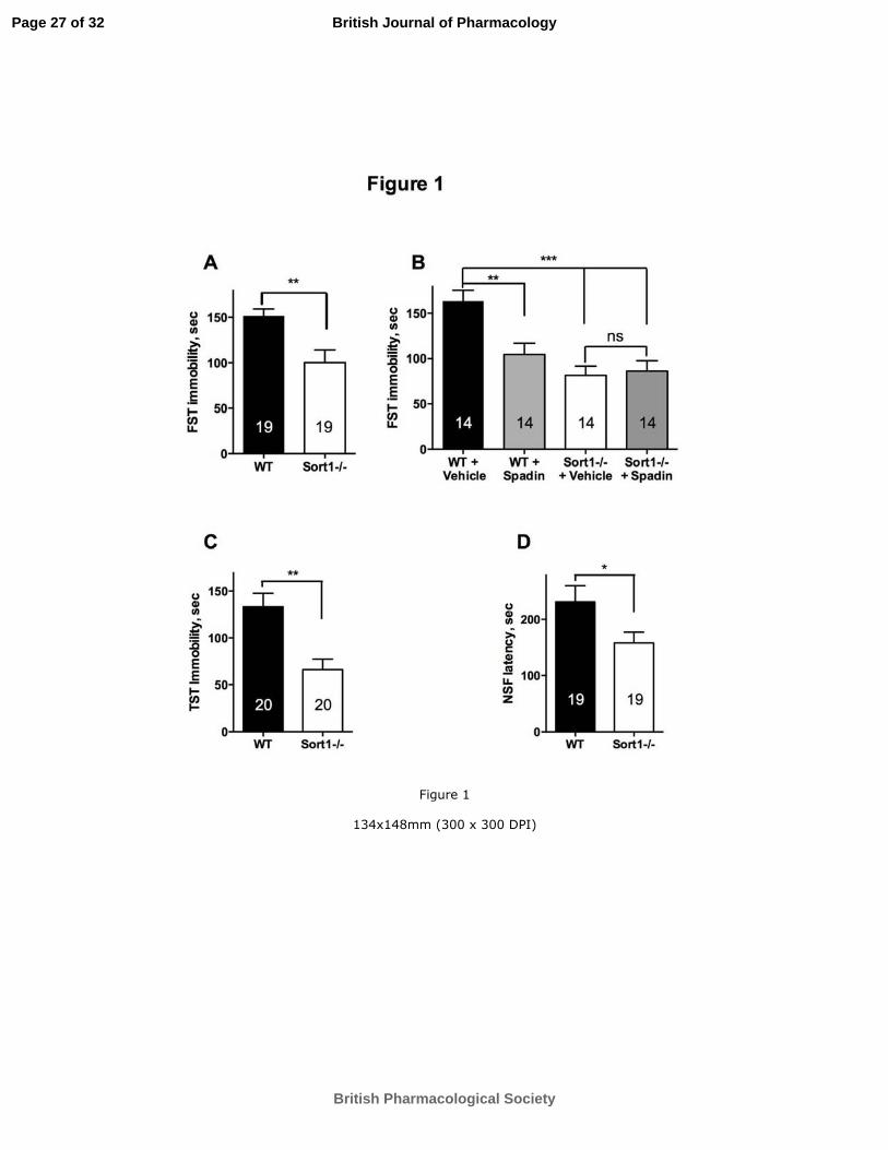
For Peer R
eview
Page 27 of 32
British Pharmacological Society
British Journal of Pharmacology
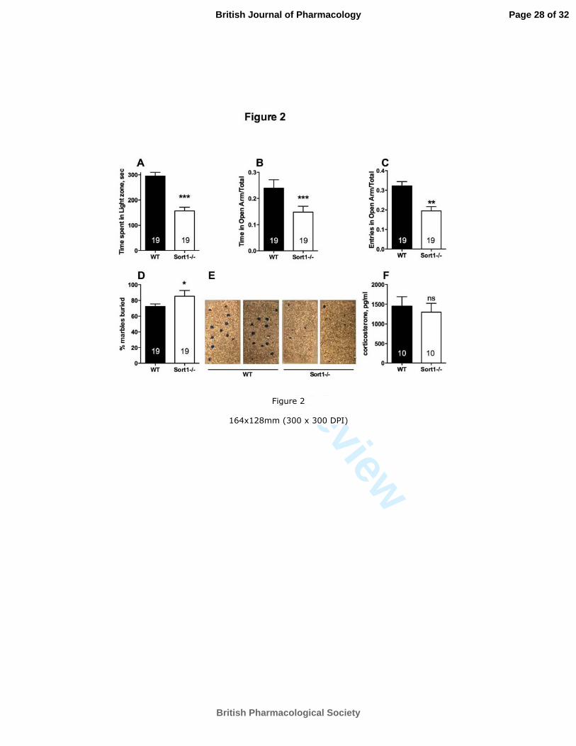
For Peer R
eview
Page 28 of 32
British Pharmacological Society
British Journal of Pharmacology
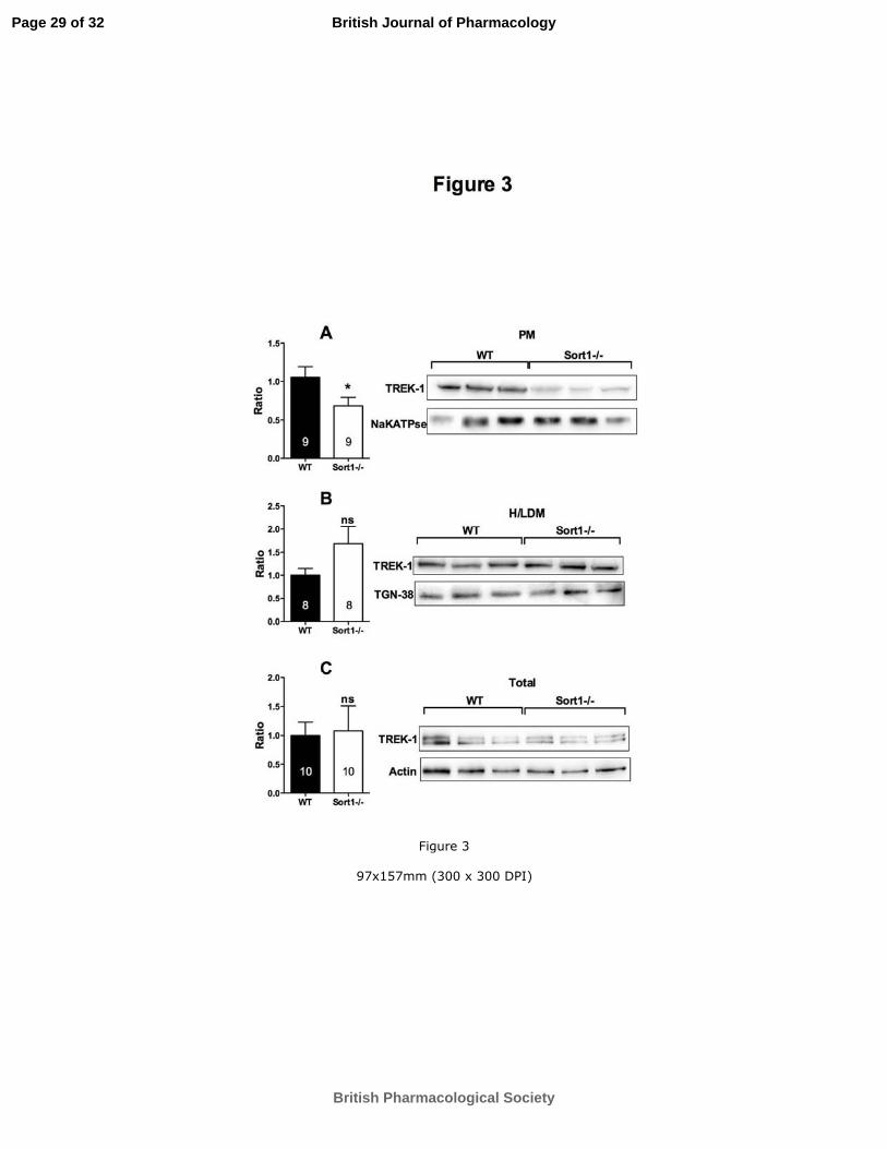
For Peer R
eview
Page 29 of 32
British Pharmacological Society
British Journal of Pharmacology

For Peer R
eview
Page 30 of 32
British Pharmacological Society
British Journal of Pharmacology
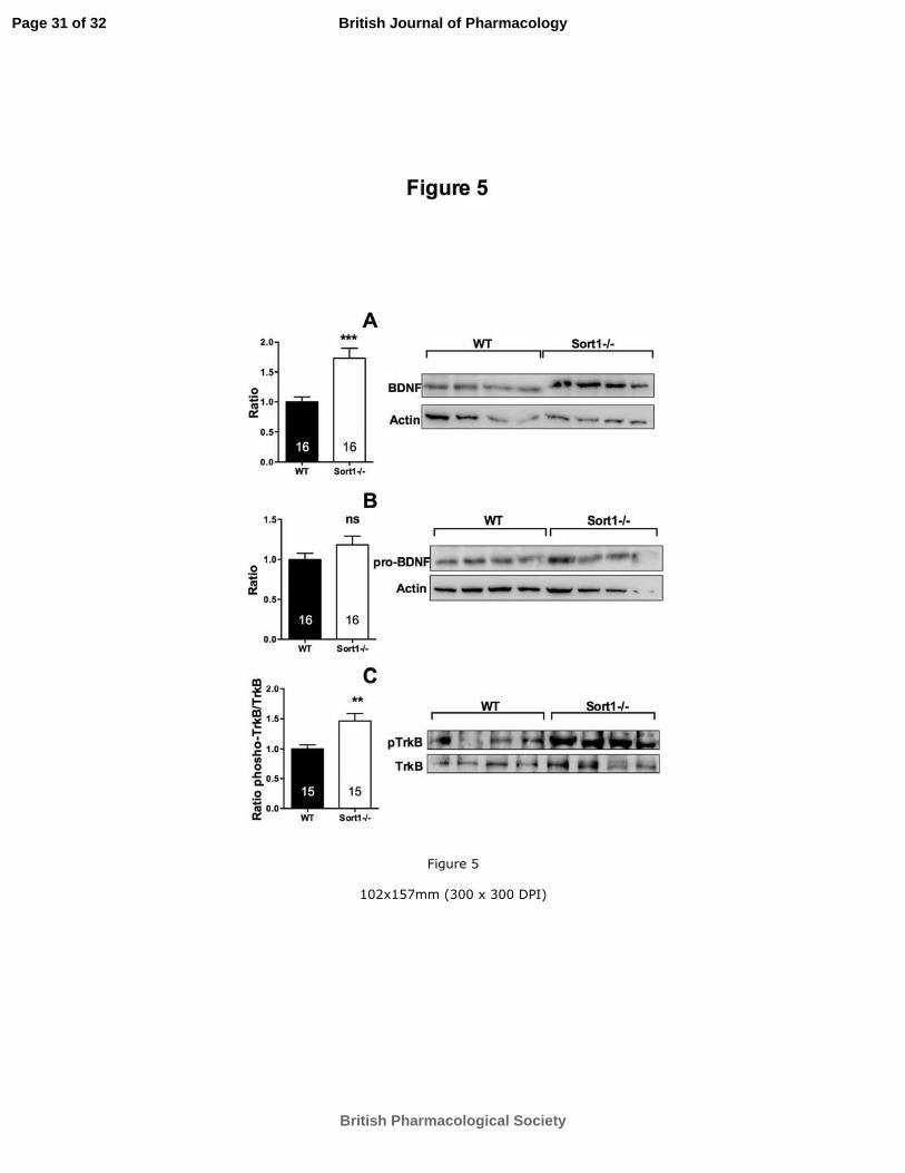
For Peer R
eview
Page 31 of 32
British Pharmacological Society
British Journal of Pharmacology
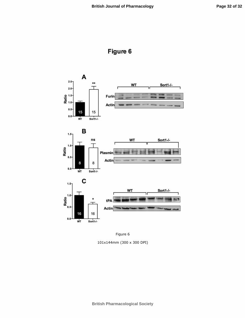
For Peer R
eview
Page 32 of 32
British Pharmacological Society
British Journal of Pharmacology

85
Développe e t d’outils pour l’a al se du comportement chez le rongeur.
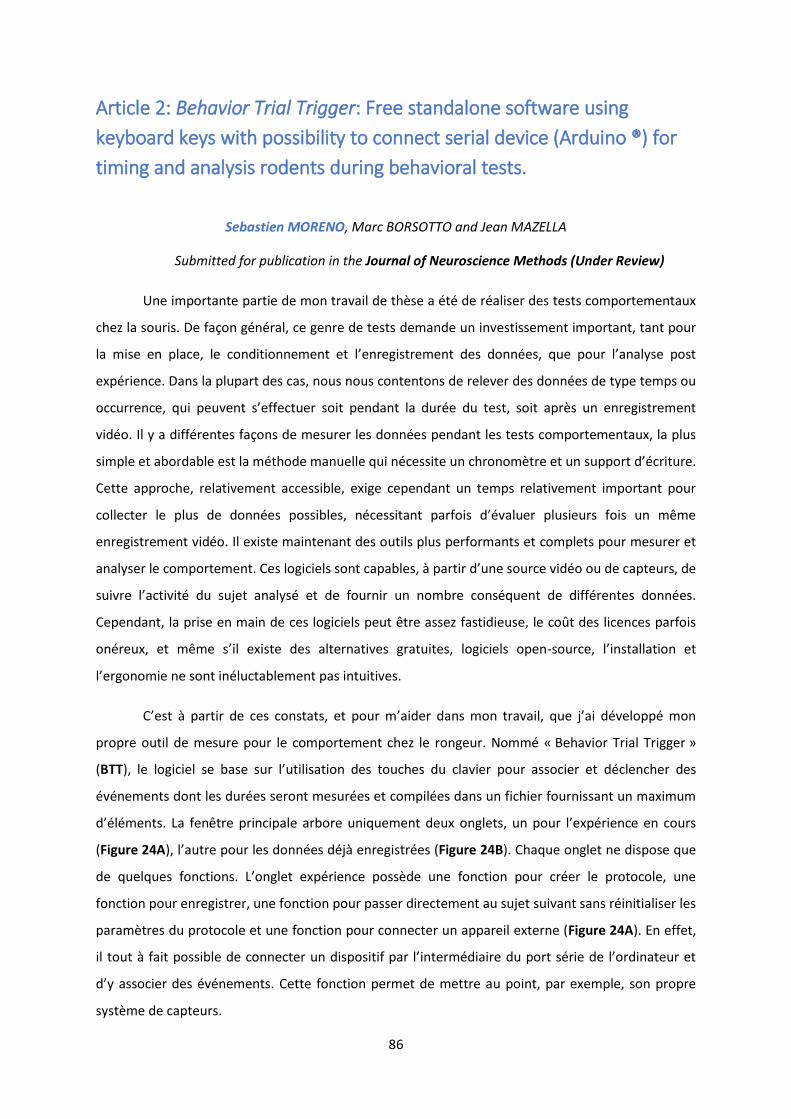
86
Article 2: Behavior Trial Trigger: Free standalone software using
keyboard keys with possibility to connect serial device (Arduino ®) for
timing and analysis rodents during behavioral tests.
Sebastien MORENO, Marc BORSOTTO and Jean MAZELLA
Submitted for publication in the Journal of Neuroscience Methods (Under Review)
Une importante partie de mon travail de thèse a été de réaliser des tests comportementaux
chez la souris. De façon général, ce genre de tests demande un investissement important, tant pour
la ise e pla e, le o ditio e e t et l’e egist e e t des do es, ue pou l’a al se post
expérience. Dans la plupart des cas, nous nous contentons de relever des données de type temps ou
occurrence, qui peu e t s’effe tue soit pe da t la du e du test, soit ap s u e egist e e t
vidéo. Il y a différentes façons de mesurer les données pendant les tests comportementaux, la plus
simple et abordable est la méthode manuelle qui nécessite un chronomètre et un suppo t d’ itu e.
Cette approche, relativement accessible, exige cependant un temps relativement important pour
olle te le plus de do es possi les, essita t pa fois d’ alue plusieu s fois u e
enregistrement vidéo. Il existe maintenant des outils plus performants et complets pour mesurer et
a al se le o po te e t. Ces logi iels so t apa les, à pa ti d’u e sou e id o ou de apteu s, de
sui e l’a ti it du sujet a al s et de fou i u o e o s ue t de diff e tes do es.
Cependant, la prise en main de ces logiciels peut être assez fastidieuse, le coût des licences parfois
o eu , et e s’il e iste des alte ati es g atuites, logi iels ope -sou e, l’i stallatio et
l’e go o ie e so t i lu ta le e t pas i tuiti es.
C’est à pa ti de es o stats, et pou ’aide da s o t a ail, ue j’ai d elopp o
propre outil de mesure pour le comportement chez le rongeur. Nommé « Behavior Trial Trigger »
(BTT , le logi iel se ase su l’utilisatio des tou hes du la ie pou asso ie et déclencher des
événements dont les durées seront mesurées et compilées dans un fichier fournissant un maximum
d’ l e ts. La fe t e p i ipale a o e u i ue e t deu o glets, u pou l’e p ie e e ou s
(Figure 24A , l’aut e pou les do es d jà e egistrées (Figure 24B). Chaque onglet ne dispose que
de uel ues fo tio s. L’o glet e p ie e poss de u e fo tio pou e le p oto ole, u e
fonction pour enregistrer, une fonction pour passer directement au sujet suivant sans réinitialiser les
paramètres du protocole et une fonction pour connecter un appareil externe (Figure 24A). En effet,
il tout à fait possi le de o e te u dispositif pa l’i te diai e du po t s ie de l’o di ateu et
d’ asso ie des e e ts. Cette fo tio pe et de ett e au point, par exemple, son propre
système de capteurs.
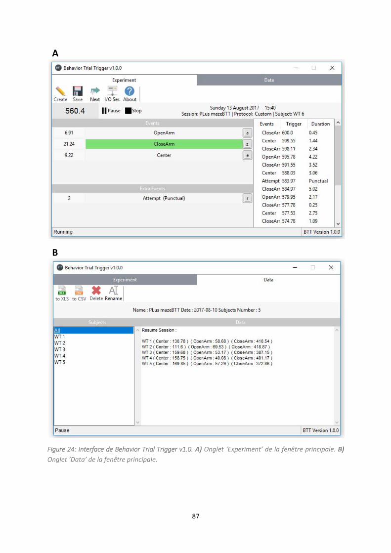
87
A
B
Figure 24: Interface de Behavior Trial Trigger v1.0. A) O glet Expe i e t de la fe t e p i ipale. B)
O glet Data de la fe t e p i ipale.
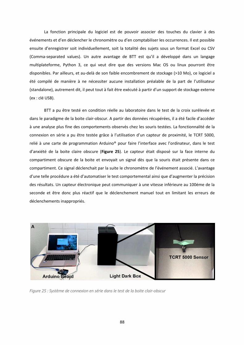
88
La fonction principale du logiciel est de pouvoir associer des touches du clavier à des
e e ts et d’e d le he le h o o t e ou d’e o pta ilise les o u e es. Il est possi le
e suite d’e egist e soit i di iduelle e t, soit la totalit des sujets sous u fo at E el ou CSV
(Comma-separated values . U aut e a a tage de BTT est u’il a d elopp da s u la gage
multiplateforme, Python 3, ce qui veut dire que des versions Mac OS ou linux pourront être
disponibles. Par ailleurs, et au-delà de son faible encombrement de stockage (>10 Mo), ce logiciel a
t o pil de a i e à e essite au u e i stallatio p ala le de la pa t de l’utilisateu
sta dalo e , aut e e t dit, il peut tout à fait t e e ut à pa ti d’u suppo t de sto kage e te e
(ex : clé USB).
BTT a pu être testé en condition réelle au laboratoire dans le test de la croix surélevée et
dans le paradigme de la boite clair-o s u . A pa ti des do es up es, il a t fa ile d’a de
à une analyse plus fine des comportements observés chez les souris testées. La fonctionnalité de la
o e io e s ie a pu t e test e g â e à l’utilisatio d’u apteu de p o i it , le TCRT ,
eli à u e a te de p og a atio A dui o® pou fai e l’i te fa e a e l’o di ateu , da s le test
d’a i t de la oite lai e o s u e Figure 25). Le capteur était disposé sur la face interne du
compartiment obscure de la boite et envoyait un signal dès que la souris était présente dans ce
o pa ti e t. Ce sig al d le hait pa la suite le h o o t e de l’ e e t asso i . L’a a tage
d’u e telle p o du e a t d’auto atise le test o po te e tal ai si ue d’aug e te la p isio
des résultats. Un capteur électronique peut communiquer à une vitesse inférieure au 100éme de la
seconde et être donc plus réactif que le déclenchement manuel tout en limitant les erreurs de
déclenchements inappropriés.
Figure 25 : Système de connexion en série dans le test de la boite clair-obscur

89
Au final, cet outil est disponible gratuitement au téléchargement sous une licence MIT et il
est p u d’e a liorer les fonctionnalités, notamment avec un système de tracking vidéo (en
ou s de d eloppe e t a e p e sio d jà utilis e ou e o e d’a al se statisti ue.

Running head: [SHORTENED TITLE UP TO 50 CHARACTERS] 1
Behavior Trial Trigger: Free standalone software using keyboard keys with possibility to connect
serial device (Arduino ®) for timing and analysis rodents during behavior tests.
Sebastien MORENO1#, Jean MAZELLA
1
1, Université Côte d’Azur, CNRS, IPMC**, France
# Correspondence author:
Dr Jean Mazella; phone: +33 (0)4 93 95 77 61, Fax: +33 (0)4 93 95 77 08;
e-mail: [email protected]
**, IPMC, UMR7275, 660 route des Lucioles, Sophia Antipolis 06560 Valbonne France.
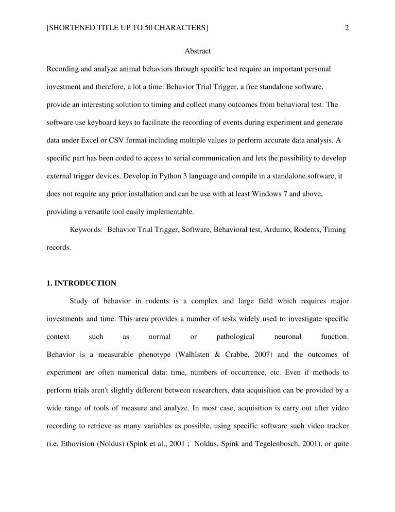
[SHORTENED TITLE UP TO 50 CHARACTERS] 2
Abstract
Recording and analyze animal behaviors through specific test require an important personal
investment and therefore, a lot a time. Behavior Trial Trigger, a free standalone software,
provide an interesting solution to timing and collect many outcomes from behavioral test. The
software use keyboard keys to facilitate the recording of events during experiment and generate
data under Excel or CSV format including multiple values to perform accurate data analysis. A
specific part has been coded to access to serial communication and lets the possibility to develop
external trigger devices. Develop in Python 3 language and compile in a standalone software, it
does not require any prior installation and can be use with at least Windows 7 and above,
providing a versatile tool easily implementable.
Keywords: Behavior Trial Trigger, Software, Behavioral test, Arduino, Rodents, Timing
records.
1. INTRODUCTION
Study of behavior in rodents is a complex and large field which requires major
investments and time. This area provides a number of tests widely used to investigate specific
context such as normal or pathological neuronal function.
Behavior is a measurable phenotype (Walhlsten & Crabbe, 2007) and the outcomes of
experiment are often numerical data: time, numbers of occurrence, etc. Even if methods to
perform trials aren't slightly different between researchers, data acquisition can be provided by a
wide range of tools of measure and analyze. In most case, acquisition is carry out after video
recording to retrieve as many variables as possible, using specific software such video tracker
(i.e. Ethovision (Noldus) (Spink et al., 2001 ; Noldus, Spink and Tegelenbosch, 2001), or quite
[SHORTENED TITLE UP TO 50 CHARACTERS] 3
simply stopwatch and manually analyze. Software can provide multiple parameters, like distance
travelled, speed of movement, position, and some specific occurrence (i.e. Ethovision (Noldus)
(Spink et al., 2001 ; Noldus, Spink and Tegelenbosch, 2001) although it can be very complete, it
isn't quite affordable, and even in open-source or free case, it's need, sometimes, dependencies
and require a certain adaptation and implementation time. In the other hand, manually
acquisition is the simple basic approach, but with the inconvenient fact that it need to observe
several times the same record to track down most of the behavior parameters and therefore
increase analysis length.
In this paper, we introduce a light standalone software that use keyboard keys code to
follow different events and measure times, delays or occurrences. It provides a light interface and
can be launch without prior installation, allowing utilization with external support (USB Key) on
any computer with, a least, Windows 7 last update. The advantage of such application is to bring
a versatile tool, quickly and easily usable.
Another interesting point is the possibility to connect serial device, such an Arduino® board.
This microcontroller has appeared as an interesting tool to create lab instrument (Besson et al.,
2016) and record different signals, such optical or mechanical sensors (Devarakonda, Nguyen
and Kravitz, 2015). Behavior Trial Trigger can communicate with this board in serial
communication and record signals activities to allow the possibility of mounting its own test
system.
2. MATERIALS and METHODS
2.1 General
Behavior Trial Trigger application was developed in Python 3.5 language (Python
Software Foundation) and packaged into stand-alone executable, under Windows, using

[SHORTENED TITLE UP TO 50 CHARACTERS] 4
PyInstaller (http://www.pyinstaller.org ; David Cortesi, based on structure by Giovanni Bajo &
William Caban, based on Gordon McMillan’s manual).). The user can run the packaged
application without installing a Python interpreter or any modules. This program was
successfully tested on different computer, from Windows 7 to Windows 10, with CPU range
from Intel Atom z3735f (tablet) to Intel i7-4790.
The main interesting function is to be able to associate keyboard keys code to move
easily and quickly from one state to another or trigger unique event during another. Every run
session is automatically save in '.sb' file at each experiment end and it's possible to save the
outputs data in Excel or CVS file.
2.2 Interface and Setting
The main window displays 2 panels: 'Experiment' and 'Data'. Experiment panel show the
current session setting with keys code set, information of the session, main timer, a resume of
events already triggered, and provides functions for create and load experiment, saving in excel
file, changing subject and switching state for serial connection (Fig 1A).
Otherwise, data panel show the outcomes of the current session, with the number of
subjects, details and function to saving in excel or csv file, rename or delete each (Fig 1B).
Before performing test, user needs to configure parameter in the 'New Experiment'
window after selecting a new or existing session ('Create' function) (Fig 1C). Time is set in
second, and events can be divided in two sections; main events and extra events (Fig 1D).
Main events provide a simply way to measure duration in unique zone and are dependent on each
other; when one start, others stop. Conversely, extra events are defined as events occurring
during main events and are independent (can be start and stop whenever it's need). There are 3
different types of extra events that can be use: Punctual (count numbers of occurrence), Standard

[SHORTENED TITLE UP TO 50 CHARACTERS] 5
(independent timer, press once to start and once again to stop), Maintain (same as 'Standard'
except that the key code need to be maintain and release to start and stop the timer). Protocol
parameters can be save, re-use, and are automatically save in the session file.
Once set up is complete, any keys code or spacebar pressed start the timer.
The 'Next Subject' function allows the user to change the subject without reset all
protocol parameters.
2.3 Data files
The outputs data can be save in Excel or CSV file. It's possible to save all subject in one
or individually file. The generated file displaying sums, orders and times of main events, and
extra events are arranged in a table providing the time release, the zone where is triggered and
the duration. For Excel format, when saving all, each subject is ordered in a different sheet and
the first one show a resume of the experiment.
2.3 Serial communication
The application includes a part to read serial device through port connection and assign
signal to events. A configuration file is available in the root folder and editable to configure the
communication between the interface and the device.
2.3.1 Configuration
Prior to use serial communication, configuration file (serial.cfg) need to be edited to
allowing the application to connect and read the device. Three parameters are available, port,
baud rate and delay.
For better results and avoiding issues, baud rate need to be set at the same value between
the device and the software and delay will be adjust depending on the latency of the device.

[SHORTENED TITLE UP TO 50 CHARACTERS] 6
Once the file is ready, parameters are set in the 'Serial/COM' panel in 'New Experiment'
window (Fig 1E). First, the device is connected by using 'Connect' function then, if the
connection is ready, reading function allowing to retrieve the different values sent through the
port. Each value can be link to the key code already configure previously.
2.3.2 Example with Arduino® UNO board
Arduino® UNO R3 is a development board using the ATmega328 microcontroller chip
and providing 14 digital Inputs/Outputs pin, 6 analog inputs pins, a 16-MHz crystal oscillator
and In Circuit Serial Programming header. It can be powered through USB connection or
external supply on 5 to 12 volts. The Arduino® board can easily support many sensors and
allowing fast input response with a good reliability, that make it an accurate platform for
prototyping different lab projects (D’Ausilio, 2011).
For a quick example, a reflective optical sensor with transistor output (TCRT5000;
VISHAY) is used to detect the presence of an object (in this case, mouse) in a closet space like
light dark box paradigm. The sensor is installed in the dark compartment, against one of the
walls perpendicular to the exit (Fig 2A) and connected to an analogic pin (A0) on the Arduino®
UNO R3 board. The TCRT5000 is wiring with a 100 Ohm resistor apply to the LED (IR emitter)
and a 10 kOhm resistor apply to the Phototransistor and then powered in +5V from the board
(Fig 2B).
A simple code is loaded into the Arduino® UNO R3 to control and receive the analogic
values from the sensor. Depending on values received, the two compartments are defined and
software is configured.
The sensor uses a range value between 0 and 1024 depending on the distance of the
object (the closer it is, the lower the value). Considering a value below 1000 correspond to the
[SHORTENED TITLE UP TO 50 CHARACTERS] 7
presence of the animal in the box (the dark one in this case), the Arduino® board will send '1' to
the software (link to the dark part), in the other case, '0' will be sent (link to the light part) (code
provided in supplementary data).
2.5 Behavioral testing
Behavioral experiments were performed with naïve mice for all the tests used, and first
isolated 30 minutes in neutral room before tests.
2.5.1 Animals
Mice used in this study were adult male from 8-10 weeks old, housed under controlled
laboratory conditions according to the FELASA guidelines and recommendations (6 mice/cage
with a 12 h dark-light cycle, a temperature of 21 ± 2°C, and a humidity of 40–60%). They have
free access to standard rodent diet and tap water. C57Bl/6J male mice are from Janvier Labs (St
Berthevin, France). Experimental procedures and animal care were approved by the local Ethics
Committee (CIEPAL) (protocol number 00893.02) and in accordance with the policies on the
care and use of laboratory animals of European Community legislation 2010/63/EU.
2.5.2 Elevated Plus Maze
Use of Elevated Plus Maze allowed to determinate an anxiety response from rodents. The
apparatus consisted of central platform (5x5cm), two open arms and two closed arms across from
each other and perpendicular, with the same size (45x5cm) and 15 cm high walls for the latter.
It’s placed at 45 cm height above the floor. Mice were placed in the central platform facing one
open arms and are allowed to freely move for 10 minutes. During this period, number of entries
and time spent in both arm are measured.
2.5.3 Light Dark

[SHORTENED TITLE UP TO 50 CHARACTERS] 8
Mice were placed in box divided into two compartments by a black partition with a small
opening that allows mouse to move from one compartment to the other. One compartment,
comprising one-third of the surface area, was made of white plastic and was brightly illuminated.
The adjoining smaller compartment was black and dark. Mice were placed in the white
compartment and allowed to move freely between the two chambers for 5 minutes. Time spent in
the white chamber, and latency to the first transition were recorded.
3. RESULTS AND DISCUSSION
Two tests were performed to collect data from BTT software; Elevated Plus Maze, to set
up multiple incomes variables, and Light Dark, to test serial device, using an Arduino® UNO R3.
In Elevated Plus Maze, the parameters set up were the open arm, the close arm, the center
and attempts to enter in open arm, by this configuration, file generated provide each time
duration in different zones, and more precisely, the time spent before switch to another zone and
the order of displacement. After this, it can be easy to obtain the number of time a zone was enter
and in which order.
Five mice were used in this test, and results were analyzed and show in table 1A. The values
obtained for this C57BL/6J mice are in accordance with general common results for this strain
(Komada, Takao and Miyakawa, 2008): ratio of time spent in the open arms: 9.29 ± 0.79%, ratio
of time spent in the closed arms: 66.35 ± 1.37% (p = 0.0079) (Fig 3A); % open arm entries:
21.72 ± 1.80, % closed arms entries: 78.28 ± 1.80 (p = 0.0079) (Fig 3B). The fact that values
were arranged depending the order provided a fine analyze to understand the behavior
throughout the experiment. For examples, outcomes data can be analyzed to obtain cumulative
times (Fig 3C), number of entries, or also, if the attempt to enter in open arm lead to an entry or
[SHORTENED TITLE UP TO 50 CHARACTERS] 9
not by looking at the following entry zone (Fig 3D) (numbers of tries were recorded with the
time and the zone triggered).
In the Light Dark paradigm, the sensor has allowed to collect data with a suitable
performance and reproducibility. Indeed, mice show similar and constant results provided by the
strain (Heredia et al., 2014; Simon et al., 2013): 63.6 ± 5.26 % in dark chamber (p = 0.0079) (Fig
3E), 19.2 ± 3.3 transitions and 22.1 ± 11.5 seconds latency to enter the dark chamber.
Behavior Trial Trigger software ensure a free, good reliability and flexible method to
collect time data from experimental behavior tests, with the advantage to be easily use without
prior installation. The accuracy of timer depends specifically of the input device such keyboards
but not computing power. It was first design to fit for animal experiment but it can be easily use
for other purpose that need duration analyze. Serial reading implementation, not limited to
Arduino® board, lets the user the possibility to develop external trigger and design its own
experiment to respond to his project. Also, Python 3 language open the possibility to update and
enhance quickly the software for a better experience user.
Finally, this software can be an interesting tool for many different purposes that need a
quick and accurate duration analyze.
[SHORTENED TITLE UP TO 50 CHARACTERS] 10
References
Besson, T., Debayle, D., Diochot, S., Salinas, M. and Lingueglia, E. (2016). Low cost venom
extractor based on Arduino® board for electrical venom extraction from arthropods and other
small animals. Toxicon, 118, pp.156-161. doi: 10.1016/j.toxicon.2016.05.001.
D’Ausilio, A. (2011). Arduino: A low-cost multipurpose lab equipment. Behavior Research
Methods, 44(2), pp.305-313. doi:10.3758/s13428-011-0163-z.
Devarakonda, K., Nguyen, K. and Kravitz, A. (2015). ROBucket: A low cost operant chamber
based on the Arduino microcontroller. Behavior Research Methods, 48(2), pp.503-509. doi:
10.3758/s13428-015-0603-2.
Heredia, L., Torrente, M., Colomina, M. and Domingo, J. (2014). Assessing anxiety in
C57BL/6J mice: A pharmacological characterization of the open-field and light/dark tests.
Journal of Pharmacological and Toxicological Methods, 69(2), pp.108-114. doi:
10.1016/j.vascn.2013.02.010.
Komada, M., Takao, K. and Miyakawa, T. (2008). Elevated Plus Maze for Mice. Journal of
Visualized Experiments, (22):1088. doi: 10.3791/1088.
Noldus, L., Spink, A. and Tegelenbosch, R. (2001). EthoVision: A versatile video tracking
system for automation of behavioral experiments. Behavior Research Methods, Instruments, &
Computers, 33(3), pp.398-414.doi : 10.3758/BF03195394.
Simon, M. M., Greenaway, S., White, J. K., Fuchs, H., Gailus-Durner, V., Wells, S., . . . Brown,
S. D. (2013). A comparative phenotypic and genomic analysis of C57BL/6J and C57BL/6N
mouse strains. Genome Biology, 14(7). doi:10.1186/gb-2013-14-7-r82.
Spink, A., Tegelenbosch, R., Buma, M., & Noldus, L. (2001). The EthoVision video tracking
system—A tool for behavioral phenotyping of transgenic mice. Physiology & Behavior, 73(5),
731-744. doi:10.1016/s0031-9384(01)00530-3.
Wahlsten, D., Crabbe, J.C.(2007). Behavioral testing. In J.G Fox, S. Barthold, M. T. Davisson,
C. Newcomer, F. Quimby, & A. Smith (Eds.).The mouse in biomedical research, vol 3,
Normative biology, husbandry, and models (pp. 513-534). Amsterdam: Elsevier
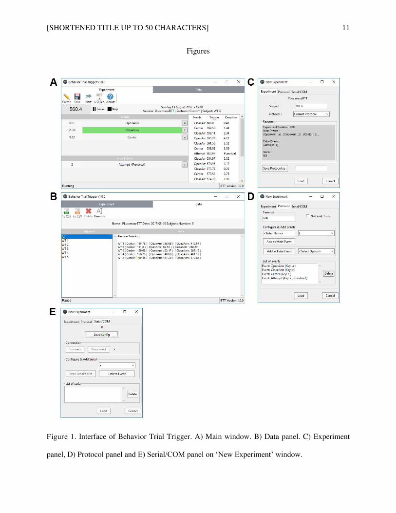
[SHORTENED TITLE UP TO 50 CHARACTERS] 11
Figures
Figure 1. Interface of Behavior Trial Trigger. A) Main window. B) Data panel. C) Experiment
panel, D) Protocol panel and E) Serial/COM panel on ‘New Experiment’ window.
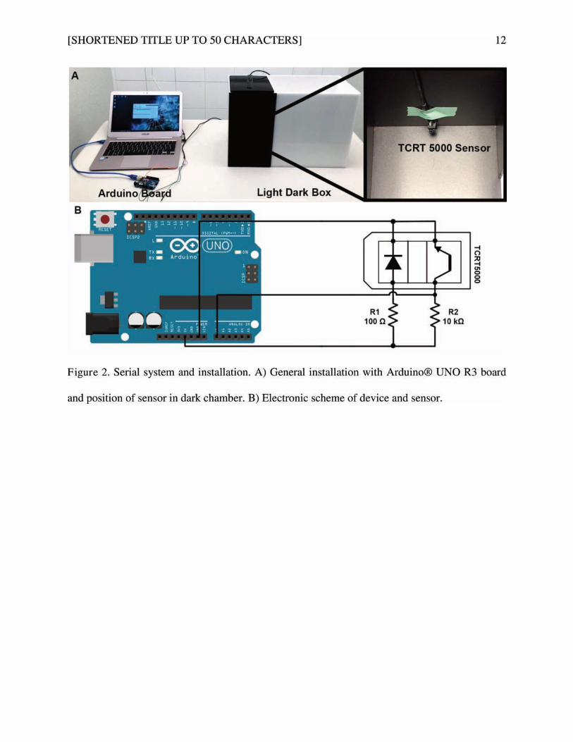
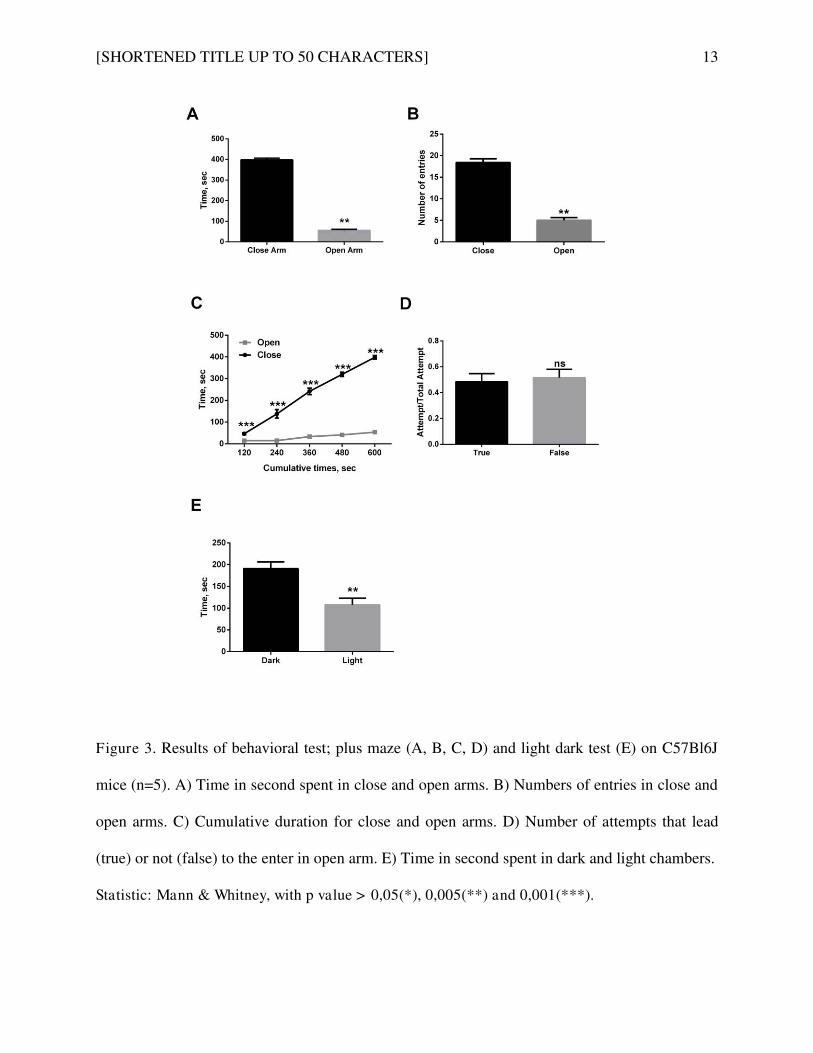
[SHORTENED TITLE UP TO 50 CHARACTERS] 13
Figure 3. Results of behavioral test; plus maze (A, B, C, D) and light dark test (E) on C57Bl6J
mice (n=5). A) Time in second spent in close and open arms. B) Numbers of entries in close and
open arms. C) Cumulative duration for close and open arms. D) Number of attempts that lead
(true) or not (false) to the enter in open arm. E) Time in second spent in dark and light chambers.
Statistic: Mann & Whitney, with p value > 0,05(*), 0,005(**) and 0,001(***).

101
Etude de la délétion du NTSR3/Sortiline
dans le système neurotensinergique.

102
Article 3: Increased Brain Neurotensin and NTSR2 Lead to Weak
Nociception in NTSR3/Sortilin Knockout Mice.
Christelle Devader, Sebastien Moreno, Morgane Roulot, Emmanuel Deval, Thomas Dix, Carlos R.
Morales and Jean Mazella
Front Neurosci. 2016; 10: 542. 24 November 2016 doi: 10.3389/fnins.2016.00542
1. Co te te de l’étude
Le NTSR /So tili e a d’a o d t ide tifi o e u epteu de la eu ote si e NTS . Ce
peptide endogène est impliqué dans de nombreuses fonctions biologiques que ce soit au niveau
e t al ou p iph i ue. O lui e o ait des effets su la t a s issio dopa i e gi ue, l’a alg sie,
l’h pothe ie ou e o e la régulation de l'activité hormonale. Il existe deux autres récepteurs
identifiés de ce peptide qui diffèrent du NTSR3/Sortiline par leurs structures ; le récepteur 1 (NTSR1)
et 2 (NSTR2). Le NTSR1 et le NTSR2 sont des récepteurs couplés aux protéines G (RCPG), tandis que
NTSR3/Sortiline est un récepteur à un seul domaine transmembranaire de type I. L'utilisation
d'agonistes et d'antagonistes sélectifs ainsi que la génération de souris déficientes pour le NTSR1 et
NTSR2 ont permis de déterminer le rôle de ces deux récepteurs dans les effets centraux induits par
la NTS. E p e ie lieu, il e iste u e diff e e d’affi it e t e es deu epteu s, le NTSR est
caractérisé comme le récepteur de haute affinité pour la NTS, alors que le NTSR2 représente celui de
faible affinité. De façon intéressante, la lévocabastine, un composé antihistaminique H1, est capable
de se lier sélectivement, par compétition avec la NTS, au récepteur NTSR2 sans affecter la liaison de
la NTS au NTSR1. On distingue pour le NTSR1 une implication dans le comportement antipsychotique
(Mechanic et al., 2009), l'inhibition de la mémoire de peur (Yamada et al., 2010) et la signalisation
nociceptive dans un modèle de douleur tonique induit par le formaldéhyde (Roussy et al., 2008) .
Pour le NTSR2, sa délétion chez la souris entraîne la perte de la nociception thermique (Maeno et al.,
2004) et de la nociception tonique de la NTS (Roussy et al., 2009).
S’il e iste ie u e tai o e de e he hes ui alue t les effets de la d l tio
fo tio elle du NTSR et du NTSR , e e a he, ie ’est e o e ta li e e ui o e e la
perte du NTSR3/Sortiline au sein du système neurotensinergique. Sachant que le NTSR3/Sortiline est
apa le d’i te agi a e le NTSR1, en modulant la signalisation de la NTS dans les cellules HT29
(Martin et al., 2002) et avec le NTSR2, en contribuant à l'effet protecteur de la NTS dans les cellules
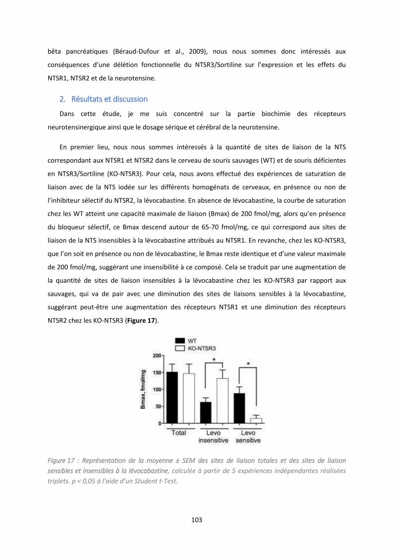
103
bêta pancréatiques (Béraud-Dufour et al., 2009), nous nous sommes donc intéressés aux
o s ue es d’u e d l tio fo tio elle du NTSR /So tili e su l’e p essio et les effets du
NTSR1, NTSR2 et de la neurotensine.
2. Résultats et discussion
Dans cette étude, je me suis concentré sur la partie biochimie des récepteurs
neurotensinergique ainsi que le dosage sérique et cérébral de la neurotensine.
En premier lieu, nous nous sommes intéressés à la quantité de sites de liaison de la NTS
correspondant aux NTSR1 et NTSR2 dans le cerveau de souris sauvages (WT) et de souris déficientes
en NTSR3/Sortiline (KO-NTSR3). Pour cela, nous avons effectué des expériences de saturation de
liaison avec de la NTS iodée sur les différents homogénats de cerveaux, en présence ou non de
l’i hi iteu s le tif du NTSR , la l o a asti e. E a se e de l o a asti e, la ou e de satu atio
hez les WT attei t u e apa it a i ale de liaiso B a de f ol/ g, alo s u’e p se e
du bloqueur sélectif, ce Bmax descend autour de 65-70 fmol/mg, ce qui correspond aux sites de
liaison de la NTS insensibles à la lévocabastine attribués au NTSR1. En revanche, chez les KO-NTSR3,
ue l’o soit e p se e ou o de l o a asti e, le B a este ide ti ue et d’u e aleu a i ale
de 200 fmol/mg, suggérant une insensibilité à ce composé. Cela se traduit par une augmentation de
la quantité de sites de liaison insensibles à la lévocabastine chez les KO-NTSR3 par rapport aux
sauvages, qui va de pair avec une diminution des sites de liaisons sensibles à la lévocabastine,
suggérant peut-être une augmentation des récepteurs NTSR1 et une diminution des récepteurs
NTSR2 chez les KO-NTSR3 (Figure 17).
Figure 17 : Représentation de la moyenne ± SEM des sites de liaison totales et des sites de liaison
sensibles et insensibles à la lévocabastine, calculée à partir de 5 expériences indépendantes réalisées
t iplets. p < , 5 à l'aide d u Student t-Test.
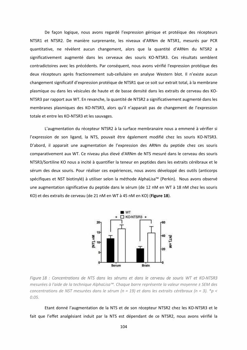
104
De faço logi ue, ous a o s ega d l’e p essio g i ue et p ot i ue des epteu s
NTSR1 et NTSR . De a i e su p e a te, les i eau d’ARN de NTSR , esu s pa PCR
ua titati e, e le t au u ha ge e t, alo s ue la ua tit d’ARN du NTSR a
significativement augmenté dans les cerveaux des souris KO-NTSR3. Ces résultats semblent
contradictoires avec les précédents. Par conséquent, nous avons vérifié l'expression protéique des
deux récepteurs après fractionnement sub- ellulai e e a al se Weste lot. Il ’e iste au u
ha ge e t sig ifi atif d’e p essio p ot i ue de NTSR ue e soit sur extrait total, à la membrane
plasmique ou dans les vésicules de haute et de basse densité dans les extraits de cerveau des KO-
NTSR3 par rapport aux WT. En revanche, la quantité de NTSR2 a significativement augmenté dans les
membranes plasmiques des KO-NTSR , alo s u’il ’appa ait pas de ha ge e t de l’e p essio
totale et entre les KO-NTSR3 et les sauvages.
L’aug e tatio du epteu NTSR à la su fa e e a ai e ous a e e à ifie si
l’e p essio de so liga d, la NTS, pou ait t e gale e t odifié chez les souris KO-NTSR3.
D’a o d, il appa ait u e aug e tatio de l’e p essio des ARN du peptide hez es sou is
comparativement aux WT. Ce niveau plus élevé d'ARNm de NTS mesuré dans le cerveau des souris
NTSR3/Sortiline KO nous a incité à quantifier la teneur en peptides dans les extraits cérébraux et le
sérum des deux souris. Pour réaliser ces expériences, nous avons développé des outils (anticorps
spécifiques et NST ioti l à utilise selo la thode AlphaLisa™ Pe ki . Nous a o s o se é
une augmentation significative du peptide dans le sérum (de 12 nM en WT à 18 nM chez les souris
KO) et des extraits de cerveau (de 21 nM en WT à 45 nM en KO) (Figure 18).
Figure 18 : Concentrations de NTS dans les sérums et dans le cerveau de souris WT et KO-NTSR3
esu es à l'aide de la te h i ue AlphaLisa™. Chaque barre représente la valeur moyenne ± SEM des
concentrations de NST mesurées dans le sérum (n = 19) et dans les extraits cérébraux (n = 3). *p <
0.05.
Eta t do l’aug e tatio de la NTS et de son récepteur NTSR2 chez les KO-NTSR3 et le
fait ue l’effet a alg sia t i duit pa la NTS est d pe da t de e NTSR , nous avons vérifié la
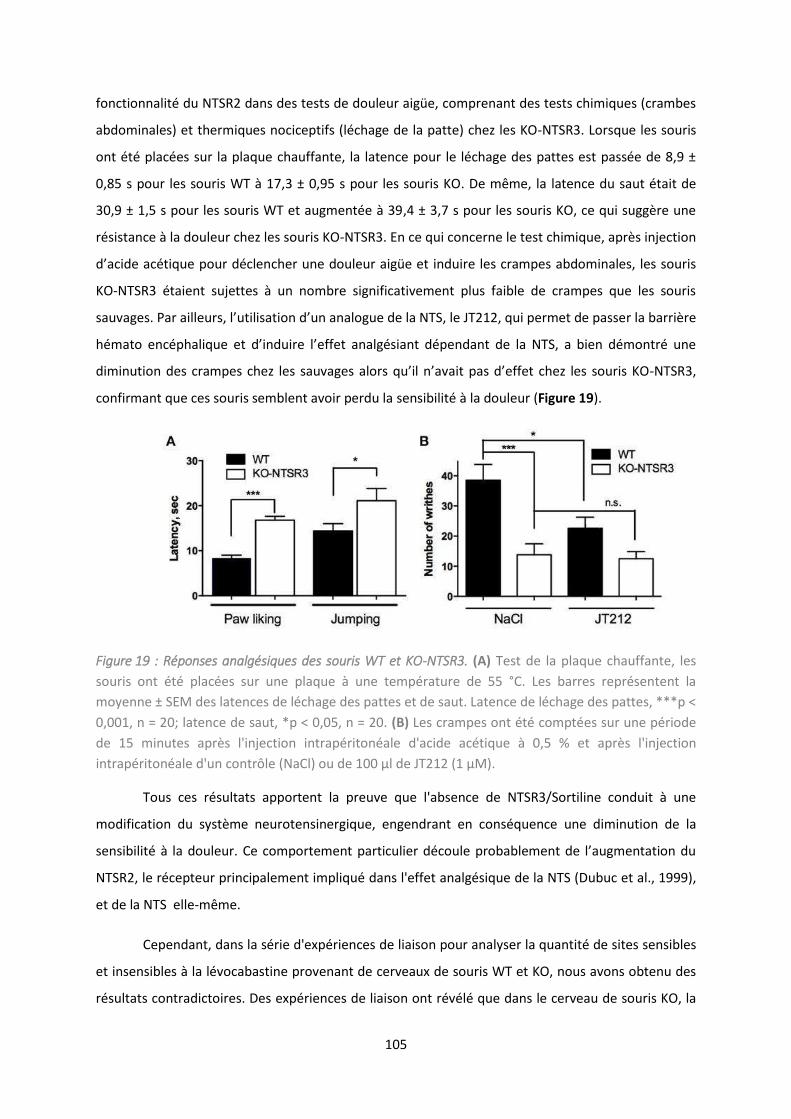
105
fonctionnalité du NTSR2 dans des tests de douleur aigüe, comprenant des tests chimiques (crambes
abdominales) et thermiques nociceptifs (léchage de la patte) chez les KO-NTSR3. Lorsque les souris
ont été placées sur la plaque chauffante, la latence pour le léchage des pattes est passée de 8,9 ±
0,85 s pour les souris WT à 17,3 ± 0,95 s pour les souris KO. De même, la latence du saut était de
30,9 ± 1,5 s pour les souris WT et augmentée à 39,4 ± 3,7 s pour les souris KO, ce qui suggère une
résistance à la douleur chez les souris KO-NTSR3. En ce qui concerne le test chimique, après injection
d’a ide a ti ue pou d le cher une douleur aigüe et induire les crampes abdominales, les souris
KO-NTSR3 étaient sujettes à un nombre significativement plus faible de crampes que les souris
sau ages. Pa ailleu s, l’utilisatio d’u a alogue de la NTS, le JT , ui pe et de passer la barrière
h ato e phali ue et d’i dui e l’effet a alg sia t d pe da t de la NTS, a ie d o t u e
di i utio des a pes hez les sau ages alo s u’il ’a ait pas d’effet hez les sou is KO-NTSR3,
confirmant que ces souris semblent avoir perdu la sensibilité à la douleur (Figure 19).
Figure 19 : Réponses analgésiques des souris WT et KO-NTSR3. (A) Test de la plaque chauffante, les
souris ont été placées sur une plaque à une température de 55 °C. Les barres représentent la
moyenne ± SEM des latences de léchage des pattes et de saut. Latence de léchage des pattes, ***p <
0,001, n = 20; latence de saut, *p < 0,05, n = 20. (B) Les crampes ont été comptées sur une période
de 15 minutes après l'injection intrapéritonéale d'acide acétique à 0,5 % et après l'injection
intrapéritonéale d'un contrôle (NaCl) ou de μl de JT μM .
Tous ces résultats apportent la preuve que l'absence de NTSR3/Sortiline conduit à une
modification du système neurotensinergique, engendrant en conséquence une diminution de la
se si ilit à la douleu . Ce o po te e t pa ti ulie d oule p o a le e t de l’aug e tatio du
NTSR2, le récepteur principalement impliqué dans l'effet analgésique de la NTS (Dubuc et al., 1999),
et de la NTS elle-même.
Cependant, dans la série d'expériences de liaison pour analyser la quantité de sites sensibles
et insensibles à la lévocabastine provenant de cerveaux de souris WT et KO, nous avons obtenu des
résultats contradictoires. Des expériences de liaison ont révélé que dans le cerveau de souris KO, la
106
quantité de sites de liaison sensibles à la lévocabastine, prévue pour être NTSR2 (Kitabgi et al., 1987 ;
Mazella et al., 1996), a diminué de façon importante alors que les sites de liaison insensibles à la
lévocabastine semblaient être augmentés (Figure 17). Par contre, les analyses de qPCR et Western
blot n'ont révélé aucun changement dans la quantité de NTSR1 et une augmentation significative de
NTSR2 au niveau des membranes plasmi ues. L’e pli atio possi le peut p o e i du fait ue la
sensibilité du NTSR2 à la lévocabastine ainsi que sa relativement faible affinité à la NTS sont
probablement dues à son interaction avec le NTSR3/Sortiline, comme déjà observé dans les cellules
bêta et pour le NTSR1 dans les HT29 (Beraud-Dufour et al., 2009; Martin et al., 2003). En l'absence
de NTSR3/Sortiline, le NTSR2 pourrait être moins retenu par voie intracellulaire et la conformation
de la protéine NTSR2 pourrait empêcher la liaison de la lévocabastine et augmenter aussi son affinité
pour la NTS. De plus, il e iste de o euses p eu es de l’i pli atio de l’ho odi isatio et
l’h t odi isatio des RCPG da s la reconnaissance des récepteurs, le trafic cellulaire et la
signalisation (Fuxe et al., 2014). Pour ce qui est du système neurotensinergique, il a été démontré
que le NTSR1 est fonctionnellement associé au récepteur D2 de la dopamine pour moduler son
activité (Borroto-Escuela et al., 2013) et u’il e iste une hétérodimérisation entre le NTSR1 et le
NTSR2, qui entraine des modifications de la distribution, du trafic et de la fonctionnalité du NTSR1
intracellulaire (Hwang et al., 2010; Perron et al., 2007). Dans le cas présent, l'augmentation de
l'expression du NTSR2 pourrait conduire à un dysfonctionnement général du système
neurotensinergique en diminuant également l'activité du NTSR1.
E fi , il se le ait ue l’effet de d se si ilisatio à la douleu hez les sou is KO-NTSR3
se ait la o s ue e d’u e aug e tatio de la eu otensine et du NTSR2, ce qui concorde avec des
résultats retrouvés chez des souris avec une forte quantité de neurotensine (Kleczkowska and
Lipkowski, 2013). Tout e i soulig e l’i po ta e du s st e eu ote si e gi ue da s la
modulation de la douleur.
En conclusion, la délétion du NTSR3/Sortiline a permis de mettre en évidence un concept
ph siologi ue ou eau da s la gulatio de la douleu , o t a t u’u e aug e tatio de NTS
da s le e eau, e o o ita e a e l’aug e tatio de l’e p essio de NTSR , semble suffisante
pour réduire la sensibilité des animaux à la douleur, permettant ainsi de dégager des perspectives
da s le d eloppe e t d’a alogues de la NTS pou le t aite e t de la douleu .
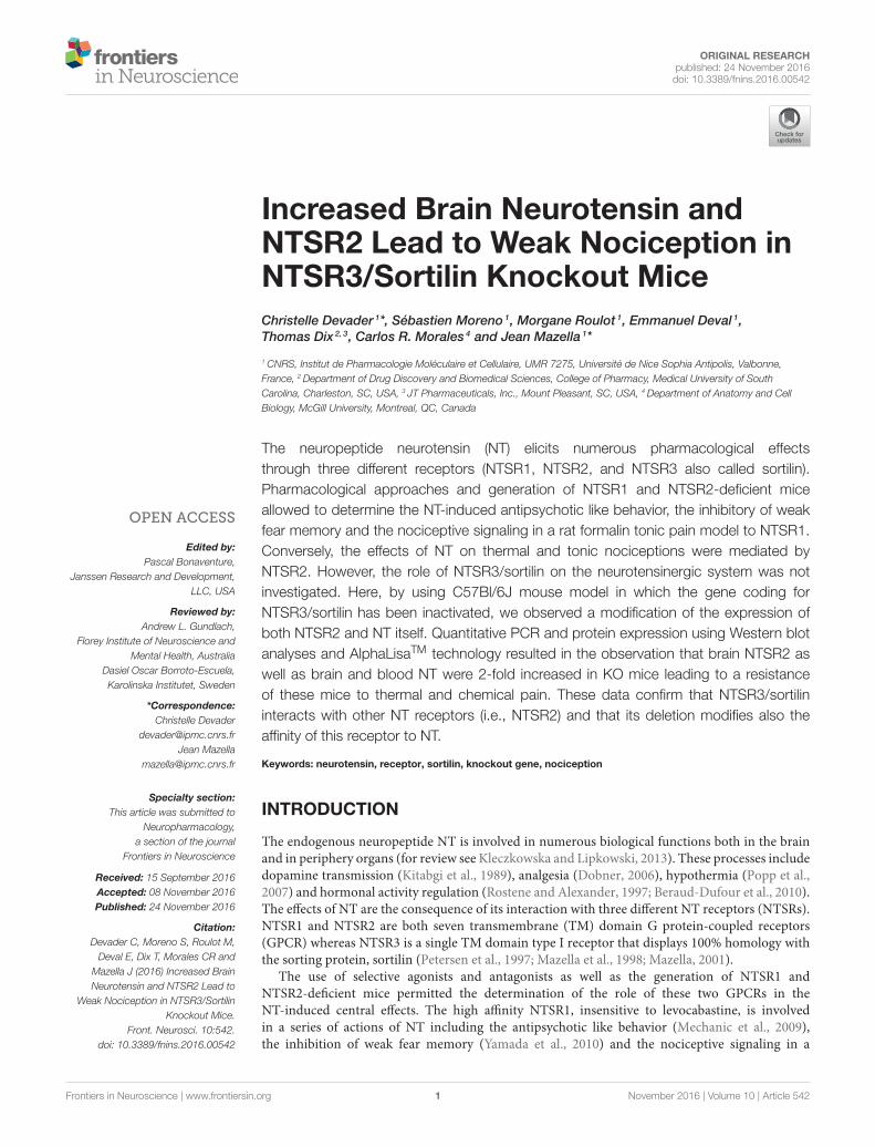
ORIGINAL RESEARCHpublished: 24 November 2016doi: 10.3389/fnins.2016.00542
Frontiers in Neuroscience | www.frontiersin.org 1 November 2016 | Volume 10 | Article 542
Edited by:
Pascal Bonaventure,
Janssen Research and Development,
LLC, USA
Reviewed by:
Andrew L. Gundlach,
Florey Institute of Neuroscience and
Mental Health, Australia
Dasiel Oscar Borroto-Escuela,
Karolinska Institutet, Sweden
*Correspondence:
Christelle Devader
Jean Mazella
Specialty section:
This article was submitted to
Neuropharmacology,
a section of the journal
Frontiers in Neuroscience
Received: 15 September 2016
Accepted: 08 November 2016
Published: 24 November 2016
Citation:
Devader C, Moreno S, Roulot M,
Deval E, Dix T, Morales CR and
Mazella J (2016) Increased Brain
Neurotensin and NTSR2 Lead to
Weak Nociception in NTSR3/Sortilin
Knockout Mice.
Front. Neurosci. 10:542.
doi: 10.3389/fnins.2016.00542
Increased Brain Neurotensin andNTSR2 Lead to Weak Nociception inNTSR3/Sortilin Knockout MiceChristelle Devader 1*, Sébastien Moreno 1, Morgane Roulot 1, Emmanuel Deval 1,
Thomas Dix 2, 3, Carlos R. Morales 4 and Jean Mazella 1*
1CNRS, Institut de Pharmacologie Moléculaire et Cellulaire, UMR 7275, Université de Nice Sophia Antipolis, Valbonne,
France, 2Department of Drug Discovery and Biomedical Sciences, College of Pharmacy, Medical University of South
Carolina, Charleston, SC, USA, 3 JT Pharmaceuticals, Inc., Mount Pleasant, SC, USA, 4Department of Anatomy and Cell
Biology, McGill University, Montreal, QC, Canada
The neuropeptide neurotensin (NT) elicits numerous pharmacological effects
through three different receptors (NTSR1, NTSR2, and NTSR3 also called sortilin).
Pharmacological approaches and generation of NTSR1 and NTSR2-deficient mice
allowed to determine the NT-induced antipsychotic like behavior, the inhibitory of weak
fear memory and the nociceptive signaling in a rat formalin tonic pain model to NTSR1.
Conversely, the effects of NT on thermal and tonic nociceptions were mediated by
NTSR2. However, the role of NTSR3/sortilin on the neurotensinergic system was not
investigated. Here, by using C57Bl/6J mouse model in which the gene coding for
NTSR3/sortilin has been inactivated, we observed a modification of the expression of
both NTSR2 and NT itself. Quantitative PCR and protein expression using Western blot
analyses and AlphaLisaTM technology resulted in the observation that brain NTSR2 as
well as brain and blood NT were 2-fold increased in KO mice leading to a resistance
of these mice to thermal and chemical pain. These data confirm that NTSR3/sortilin
interacts with other NT receptors (i.e., NTSR2) and that its deletion modifies also the
affinity of this receptor to NT.
Keywords: neurotensin, receptor, sortilin, knockout gene, nociception
INTRODUCTION
The endogenous neuropeptide NT is involved in numerous biological functions both in the brainand in periphery organs (for review see Kleczkowska and Lipkowski, 2013). These processes includedopamine transmission (Kitabgi et al., 1989), analgesia (Dobner, 2006), hypothermia (Popp et al.,2007) and hormonal activity regulation (Rostene and Alexander, 1997; Beraud-Dufour et al., 2010).The effects of NT are the consequence of its interaction with three different NT receptors (NTSRs).NTSR1 and NTSR2 are both seven transmembrane (TM) domain G protein-coupled receptors(GPCR) whereas NTSR3 is a single TM domain type I receptor that displays 100% homology withthe sorting protein, sortilin (Petersen et al., 1997; Mazella et al., 1998; Mazella, 2001).
The use of selective agonists and antagonists as well as the generation of NTSR1 andNTSR2-deficient mice permitted the determination of the role of these two GPCRs in theNT-induced central effects. The high affinity NTSR1, insensitive to levocabastine, is involvedin a series of actions of NT including the antipsychotic like behavior (Mechanic et al., 2009),the inhibition of weak fear memory (Yamada et al., 2010) and the nociceptive signaling in a
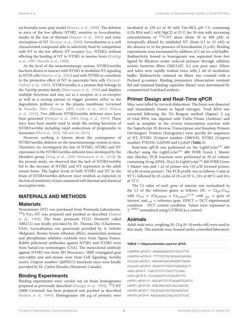
Devader et al. Dysfunction of Neurotensinergic System in Sortilin-KO Mice
rat formalin tonic pain model (Roussy et al., 2008). The deletionin mice of the low affinity NTSR2, sensitive to levocabastine,results in the loss of thermal (Maeno et al., 2004) and tonicnociception of NT (Roussy et al., 2009). Levocabastine is a wellcharacterized compound able to selectively bind by competitionwith NT to the low affinity NT receptor (i.e., NTSR2) withoutaffecting the binding of NT to NTSR1 in murine brain (Kitabgiet al., 1987; Mazella et al., 1998).
At the level of the neurotensinergic system, NTSR3/sortilinhas been shown to interact with NTSR1 tomodulate NT signalingin HT29 cells (Martin et al., 2002) and with NTSR2 to contributeto the protective effect of NT in pancreatic beta cells (Beraud-Dufour et al., 2009). NTSR3/sortilin is a protein that belongs tothe Vps10p protein family (Marcusson et al., 1994) and displaysmultiple functions and may act as a receptor or a co-receptoras well as a sorting partner to trigger proteins either to thedegradation pathway or to the plasma membrane (reviewedin Mazella, 2001; Hermey, 2009; Carlo et al., 2014; Wilsonet al., 2014). Two different NTSR3/sortilin deficient mice havebeen generated (Nykjaer et al., 2004; Zeng et al., 2009). Thesemice have been mainly used to study the sorting functions ofNTSR3/sortilin including rapid endocytosis of progranulin tolysosomes (Hu et al., 2010; Tall and Ai, 2011).
However, nothing is known about the consequence ofNTSR3/sortilin deletion on the neurotensinergic system in mice.Therefore, we investigated the fate of NTSR1, NTSR2 and NTexpression in the NTSR3/sortilin-deficient mice developed by theMorales’s group (Zeng et al., 2009; Musunuru et al., 2010). Inthe present study, we observed that the lack of NTSR3/sortilinled to the increase of NTSR2 and NT expression in the adultmouse brain. The higher levels of both NTSR2 and NT in thebrain of NTSR3/sortilin-deficient mice resulted, as expected, inthe loss of sensitivity of painmeasured with thermal and chemicalnociceptive tests.
MATERIALS AND METHODS
MaterialsNeurotensin (NT) was purchased from Peninsula Laboratories.125I-Tyr3-NT was prepared and purified as described (Sadoulet al., 1984). The brain permeant JT212 (formerly calledABS212) was kindly provided by Dr. Thomas Dix (Charleston,USA). Levocabastine was generously provided by A. Schotte(Belgium). Bovine Serum Albumin (BSA), mammalian proteaseand phosphatase inhibitor cocktails were from Sigma France.Rabbit polyclonal antibodies against NTSR1 and NTSR2 werefrom SantaCruz technologies (USA). The monoclonal antibodyagainst NTSR3 was from BD Bioscience. HRP conjugated goatanti-rabbit and anti-mouse were from Cell Signaling. Sortilin(sort1; Uniprot number: Q6PHU5) knockout mice were kindlyprovided by Dr. Carlos Morales (Montreal, Canada).
Binding ExperimentsBinding experiments were carried out on brain homogenatesprepared as previously described (Zsurger et al., 1994). 125I-NT(2000 Ci/mmol) has been prepared and purified as described(Sadoul et al., 1984). Homogenates (60 µg of protein) were
incubated in 250 µl of 50 mM Tris-HCl, pH 7.5, containing0.2% BSA and 1 mM MgCl2 at 25C for 30 min with increasingconcentrations of 125I-NT alone (from 50 to 400 pM) orisotopically diluted by unlabeled NT (from 0.1 to 25 nM) inthe absence or in the presence of levocabastine (1 µM). Bindingexperiments were terminated by addition of 2 ml ice-cold buffer.Radioactivity bound to homogenate was separated from freeligand by filtration under reduced pressure through celluloseacetate Sartorius filters (SM11107, 0.2 µm pore size). Filtersand tubes were rapidly washed twice with 2 ml of incubationbuffer. Radioactivity retained on filters was counted with aPackard g-counter. Binding parameters (dissociation constantKd and maximal binding capacities Bmax) were determined bycomputerized Scatchard analysis.
Primer Design and Real-Time qPCRMice were killed by cervical dislocation. The brain was dissectedand immediately frozen in liquid nitrogen. Total RNA wasextracted following the Tri Reagent method (Sigma). 2 µgof total RNA was digested with Turbo Dnase (Ambion) andused as template in the reverse transcription reaction withthe SuperScript III Reverse Transcriptase and Random Primers(Invitrogen). Primers (Eurogentec) were specific for sequencesof NT, NTSR1 (Uniprot number: O88319), NTSR2 (Uniprotnumber: P70310), GAPDH and CycloD (Table 1).
Real-time qPCR was performed on the LightCyclerTM 480(Roche) using the LightCyclerTM 480 SYBR Green 1 Mastermix (Roche). PCR reactions were performed in 20 µl volumecontaining 16 ng cDNA, 10 µl 2x LightCyclerTM 480 SYBR Green1 Master mix and 1 µl of primer mix (10 µM forward primer,10 µM reverse primer). The PCR profile was as follows: 5 min at95C, followed by 45 cycles of 10 s at 95C, 10 s at 60C and 10 sat 72C.
The Ct value of each gene of interest was normalized tothe Ct of the reference genes as follows: DC = Ctgoi-Ctrefwith Ctref = (CtGAPDH x CtCycloD)
(1/2) with goi = gene ofinterest, and ref = reference gene. DDCT = DCT experimentalcondition - DCT control condition. Values were expressed as2−DDCt normalized using C57Bl/6J as a control.
AnimalsAdult male mice, weighing 20–25 g (8–10 weeks old) were used inthis study. The animals were housed under controlled laboratory
TABLE 1 | Oligonucleotides used for qPCR.
mGAPDH-qPCR-F : AAGAGGGATGCTGCCCTTA
mGAPDH-qPCR-R : TTTTGTCTACGGGACGAGGA
mCycloD-qPCR-F : AAGGATGGCAAGGATTGAAA
mCycloD-qPCR-R : GCAATTCTGCCTGGATAGCTT
mNTs-qPCR-F : TGACTCTCCTGGCTTTCAGC
mNTs-qPCR-R : TCCAGGGCTCTCACATCTTC
mNTR1-qPCR-F2 : GGCAATTCCTCAGAATCCATCC
mNTR1-qPCR-R2 : ATACAGCGGTCACCAGCAC
mNTR2-qPCR-F : TGCACGGTGCTAGTAAGTCG
mNTR2-qPCR-R : AAGGAGACCAGCACGTTCAC
Frontiers in Neuroscience | www.frontiersin.org 2 November 2016 | Volume 10 | Article 542

Devader et al. Dysfunction of Neurotensinergic System in Sortilin-KO Mice
conditions (in accordance with the FELASA guidelines andrecommendations), 6 mice/cage with a 12 h dark-light cycle,a temperature of 21 ± 2C, and a humidity of 40–60%. Micehad free access to standard rodent diet and tap water. TheNTSR3/sortilin homozygous KO mice were generated by theMorales’s laboratory by incorporation of a GFP cassette afterexon 1 (Zeng et al., 2009) and the controls were C57Bl/6Jmale mice from Janvier Labs (St Berthevin, France). All animalcare and experimental procedures complied with the policieson the care and use of laboratory animals of EuropeanCommunity legislation 2010/63/EU and were approved bythe local Ethics Committee (CIEPAL) (protocol number00893.02).
Pain Behavioral TestsThe writhing test was performed as follows: 20 min prior to aceticacid injection, mice were injected intraperitoneally with either100µl of saline or 100µl of a solution containing 1µMof JT212,a NT analog able to cross the blood-brain-barrier (Hughes et al.,2010). Writhes were counted over a 15 min period starting fromthe fifth min after intraperitoneal injection of a 0.5% acetic acidsolution (10 µl/g).
The Hot plate test was performed with a hot plate apparatus(Ugo Basile) at 55C. We measured the time (in seconds) to pawlicking and jumping latency in response to heat.
Determination of Blood and Central NTConcentrationSerum samples were collected in the morning by retroorbitalpuncture in mice anesthetized by isoflurane 4%. Brain NTwas recovered after acid extraction of brain homogenates asdescribed (Kokko et al., 2005). The amount of NT was measuredfrom serum and brain using a method adapted to AlphaScreentechnology (Perkin Elmer, France). The technique necessitatedthe preparation of a biotinylated NT on one hand, and ofan antibody against the C-terminus of NT on the otherhand.
Rabbit polyclonal antibodies against the C-terminus of NT(2-13) were prepared by Agro Bio (La Ferté St Aubin, France).NT (2-13) (5.4 mg, 3.6 mmol) was solubilized in 1.5 ml of25 mM phosphate buffer, pH 6.7. N-hydroxysuccinimide biotin(13.5 mmol) resuspended in 700 µl of 70% acetonitrile, 30%dimethyl formamide was added to the peptide solution andincubated overnight at room temperature. Biotin-NT (2-13) waspurified by HPLC using a Waters apparatus equipped with asemi-preparative RP18 Lichrosorb column. Biotin-NT (2-13)(eluted at 35min), identified bymass spectrometry, was collected,quantified by its absorption at 280 nm and lyophylised inaliquots.
According to the principles of AlphaScreen technology,streptavidin-donor microbeads were recognized by biotin-NT(2-13) and the anti-rabbit IgG-acceptor microbeads were boundby anti-NT (2-13) antibodies. The signal was produced whenthe two microbeads (acceptor and donor) were drawn intoproximity by a molecular interaction occurring between thebinding partners captured on the beads. The peptide present inthe sample was able to interfere with this interaction leading to
competition. Standard curves were obtained by incubation in 96-well plaque of 1 nM biotin-NT (2-13) with the anti-NT (2-13)antibody (1:5000) in the AlphaLisaTM buffer in the absence orin the presence of increasing concentrations of NT (2-13) (from10−11 to 10−6 M) for 1 h at room temperature. After addition ofacceptor and donor beads and further incubation for 2 h at roomtemperature, the plaque was read using the Enspire apparatus(Perkin). Note that non-apparented peptides like somatostatin orspadin were unable to interfere with the dosing method. For serameasurements, the same volume of serum was added instead ofunlabeled NT (2-13). The amount of NT was determined fromits percent of signal inhibition and calculated using the standardcurve.
Sub-cellular FractionationIn order to quantify the amount of NTSRs expressed atthe cell surface and intracellularly, we performed sub-cellularfractionation from brain homogenates. Plasma membranes wereprepared from brain homogenates of WT or KO-NTSR3/Sortilinmice according to the protocol previously described (Clancyand Czech, 1990). 30 µg of crude homogenates, purified plasmamembranes and high and low density vesicles (H/LDM) weresubmitted to Western blot analysis using the rabbit polyclonalantibodies against NTSR1 or NTSR2 (1:500) (SantaCruzTechnologies (USA)). Proteins detected with these antibodieswere normalized using antibodies specific for each intracellularcompartment (NaKATPase for plasma membranes, TGN38for H/LDM and tubulin for total extracts) from SantaCruztechnologies (USA).
StatisticsResults are expressed as mean ± standard error mean (SEM).Statistical analyses were performed using GraphPad (version6.0). Student t-tests were used when appropriate to evaluatedifferences in quantitative variables whereas analysis of variance(ANOVA) was used to compute possible differences betweengroups.
RESULTS
Binding of NT to Brain Homogenates fromWild Type and NTSR3/Sortilin KO MiceIn order to quantify the amount of NT binding sitescorresponding to NTSR1 and NTSR2 in the brain of wildtype (WT) and NTSR3/sortilin deficient mice (KO-NTSR3),we first performed saturation binding experiments of iodinatedNT on homogenates prepared from the indicated brains inthe absence or in the presence of the NTSR2 selective blockerlevocabastine (1µM) (Kitabgi et al., 1987). In brain homogenatesfrom WT mice, in the absence of levocabastine, the saturationcurve obtained from a typical experiment indicated a maximalbinding capacity (Bmax) of about 200 fmol/mg (Figure 1A).In the presence of levocabastine, the Bmax decreased to 65–70fmol/mg (Figure 1A), a binding capacity corresponding to thelevocabastine insensitive NT binding sites attributed to NTSR1.Interestingly, in brain homogenates from KO-NTSR3 mice,saturation experiments performed in the absence or in the
Frontiers in Neuroscience | www.frontiersin.org 3 November 2016 | Volume 10 | Article 542
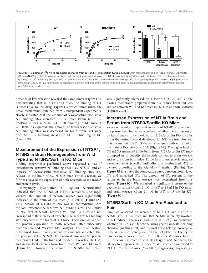
Devader et al. Dysfunction of Neurotensinergic System in Sortilin-KO Mice
FIGURE 1 | Binding of 125I-NT to brain homogenates from WT and NTSR3/sortilin KO mice. (A,B) Brain homogenates from WT (A) or from NTSR3/sortilin
KO mice (B) (60 µg of proteins) were incubated with increasing concentrations of 125 I-NT alone or isotopically diluted with unlabeled NT in the absence (closed
symbols) or in the presence (open symbols) of 1 µM levocabastine. Saturation curves were made from specific binding using GraphPad analysis. (C) Representation
of the mean ± SEM of total binding and levocabastine-sensitive and -insensitive binding sites calculated from 5 independent experiments performed in triplicate.
*p < 0.05 using Student t-Test.
presence of levocabastine revealed the same Bmax (Figure 1B),demonstrating that in KO-NTSR3 mice, the binding of NTis insensitive to the drug. Figure 1C which summarized theBmax mean values obtained from 5 independent experiments,clearly indicated that the amount of levocabastine-insensitiveNT binding sites increased in KO mice (from 63 ± 12fmol/mg in WT mice to 124 ± 30 fmol/mg in KO mice, p= 0.029). As expected, the amount of levocabastine-sensitiveNT binding sites was decreased in brain from KO micefrom 88 ± 19 fmol/mg in WT to 14 ± 9 fmol/mg in KO(p= 0.028).
Measurement of the Expression of NTSR1,NTSR2 in Brain Homogenates from WildType and NTSR3/Sortilin KO MiceBinding experiments performed above suggested a loss oflevocabastine-sensitive NT binding sites (i.e., NTSR2) and anincrease of levocabastine-insensitive NT binding sites (i.e.,NTSR1) in the brain of KO-NTSR3 mice. For this reason, wefurther analyzed the expression of both receptors at the mRNAand protein levels.
Intriguingly, quantitative PCR (qPCR) determinationindicated that the mRNA of NTSR1 remained unchangedwhereas the amount of NTSR2 mRNA was significantlyincreased in the brain of KO mice (p < 0.001) (Figure 2A).This increase of NTSR2 mRNA was in contradiction withthe loss levocabastine-sensitive NT binding sites. The similarmRNA level of NTSR1 between WT and KO mice did notcorrespond to the increase of levocabastine-sensitive NT bindingsites observed in the brain of KO mice. Therefore, we verifiedthe protein expression of both receptors after subcellularfractionation and Western blot analysis. The quantificationdetermined from 5 independent experiments indicated thatthe protein level of NTSR1-like remained similar at the plasmamembranes (PM), in the high and low density vesicles (H/LDM)and in the total extracts from brain from WT and KO mice(Figure 2B). However, the amount of NTSR2-like protein
was significantly increased by a factor 2 (p < 0.05) at theplasma membranes prepared from KO mouse brain but wassimilar between WT and KO mice in H/LDM and total extracts(Figures 2C,D).
Increased Expression of NT in Brain andSerum from NTSR3/Sortilin KO MiceAs we observed an important increase of NTSR2 expression atthe plasma membrane, we wondered whether the expression ofits ligand may also be modified in NTSR3/sortilin KO mice byusing the dosing method developed for NT. We first observedthat the amount of NT mRNA was also significantly enhanced inthe brain of KO mice (p < 0.05) (Figure 3A). The higher level ofNT mRNA measured in the brain from NTSR3/sortilin KO miceprompted us to quantify the peptide content in brain extractsand serum from both mice. To perform these experiments, wedeveloped tools (specific antibodies and biotinylated NT) tobe used according to the AlphaLisaTM method (Perkin). TheFigure 3B illustrated the competition curve between biotinylatedNT and unlabeled NT. The amount of NT present in theserum or in the brain extracts was determined from thiscurve (Figure 3C). We observed a significant increase of thepeptide in serum (from 12 nM in WT to 18 nM in KO mice)and brain extracts (from 21 nM in WT to 45 nM in KO)(Figure 3C).
NTSR3/Sortilin KO Mice Are Resistant toPainSince we observed an increase of both NT and NTSR2 inNTSR3/sortilin KO mice and that NTSR2 is mainly involvedin NT-induced analgesia (Dubuc et al., 1999), we wonderedwhether NTSR2 is still functional using acute pain tests includingchemical (writhing test) and thermal (paw licking) nociceptivetests. When mice were placed on the hot plate, the latency forpaw licking increased from 8.9 ± 0.85 s for WT mice to 17.3± 0.95 s for KO mice (p < 0.001) (Figure 4A). Similarly, thelatency to jump was 30.9 ± 1.5 s for WT mice and increased to39.4 ± 3.7 s for KO mice (p = 0.038) (Figure 4A), suggesting a
Frontiers in Neuroscience | www.frontiersin.org 4 November 2016 | Volume 10 | Article 542
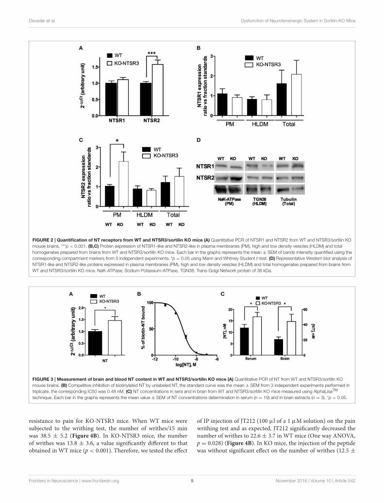
Devader et al. Dysfunction of Neurotensinergic System in Sortilin-KO Mice
FIGURE 2 | Quantification of NT receptors from WT and NTSR3/sortilin KO mice (A) Quantitative PCR of NTSR1 and NTSR2 from WT and NTSR3/sortilin KO
mouse brains, ***p < 0.001. (B,C) Protein expression of NTSR1-like and NTSR2-like in plasma membranes (PM), high and low density vesicles (HLDM) and total
homogenates prepared from brains from WT and NTSR3/sortilin KO mice. Each bar in the graphs represents the mean ± SEM of bands intensitiy quantified using the
corresponding compartment markers from 5 independent experiments. *p < 0.05 using Mann and Whitney Student t-test. (D) Representative Western blot analysis of
NTSR1-like and NTSR2-like proteins expressed in plasma membranes (PM), high and low density vesicles (HLDM) and total homogenates prepared from brains from
WT and NTSR3/sortilin KO mice. NaK-ATPase; Sodium Potassium-ATPase, TGN38; Trans-Golgi Network protein of 38 kDa.
FIGURE 3 | Measurement of brain and blood NT content in WT and NTSR3/sortilin KO mice (A) Quantitative PCR of NT from WT and NTSR3/sortilin KO
mouse brains. (B) Competitive inhibition of biotinylated NT by unlabeled NT, the standard curve was the mean ± SEM from 3 independent experiments performed in
triplicate, the corresponding IC50 was 0.48 nM. (C) NT concentrations in sera and in brain from WT and NTSR3/sortilin KO mice measured using AlphaLisaTM
technique. Each bar in the graphs represents the mean value ± SEM of NT concentrations determination in serum (n = 19) and in brain extracts (n = 3). *p < 0.05.
resistance to pain for KO-NTSR3 mice. When WT mice weresubjected to the writhing test, the number of writhes/15 minwas 38.5 ± 5.2 (Figure 4B). In KO-NTSR3 mice, the numberof writhes was 13.8 ± 3.6, a value significantly different to thatobtained in WT mice (p < 0.001). Therefore, we tested the effect
of IP injection of JT212 (100 µl of a 1 µM solution) on the painwrithing test and as expected, JT212 significantly decreased thenumber of writhes to 22.6 ± 3.7 in WT mice (One way ANOVA,p = 0.028) (Figure 4B). In KO mice, the injection of the peptidewas without significant effect on the number of writhes (12.5 ±
Frontiers in Neuroscience | www.frontiersin.org 5 November 2016 | Volume 10 | Article 542
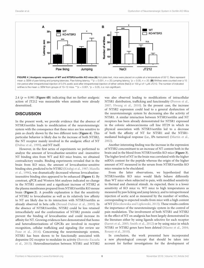
Devader et al. Dysfunction of Neurotensinergic System in Sortilin-KO Mice
FIGURE 4 | Analgesic responses of WT and NTSR3/sortilin KO mice (A) Hot plate test, mice were placed on a plate at a temperature of 55C. Bars represent
mean ± SEM of paw licking and jumping latencies. Paw licking latency, ***p < 0.001, n = 20; jumping latency, *p < 0.05, n = 20. (B) Writhes were counted over a 15
min period after intraperitoneal injection of 0.5% acetic acid after intraperitoneal injection of either vehicle (NaCl) or 100 µl of 1 µM JT212. The number of indicated
writhes is the mean ± SEM from groups of 10–12 mice. ***p < 0.001, *p < 0.05, n.s: non-significant.
2.4 (p = 0.99) (Figure 4B) indicating that no further analgesicaction of JT212 was measurable when animals were alreadydesensitized.
DISCUSSION
In the present work, we provide evidence that the absence ofNTSR3/sortilin leads to modificiation of the neurotensinergicsystem with the consequence that these mice are less sensitive topain as clearly shown by the two different tests (Figure 4). Thisparticular behavior is likely due to the increase of both NTSR2,the NT receptor mainly involved in the analgesic effect of NT(Dubuc et al., 1999), and NT itself.
However, in the first series of experiments we performed toanalyze the amount of levocabastine-sensitive and -insensitiveNT binding sites from WT and KO mice brains, we obtainedcontradictory results. Binding experiments revealed that in thebrain from KO mice, the amount of levocabastine-sensitivebinding sites, predicted to be NTSR2 (Kitabgi et al., 1987; Mazellaet al., 1996), was dramatically decreased whereas levocabastine-insensitive binding sites appeared to be enhanced (Figure 1). Byconstrast, qPCR and Western blot analyses indicated no changein the NTSR1 content and a significant increase of NTSR2 atthe plasmamembranes prepared fromNTSR3/sortilin KOmousebrain (Figure 2). A possible explanation is that the sensitivityof NTSR2 to levocabastine as well as its relatively low affinityto NT are likely due to its interaction with NTSR3/sortilin asalready observed in beta cells (Beraud-Dufour et al., 2009). Inthe absence of NTSR3/sortilin, NTSR2 could be less retainedintracellularly and the conformation of NTSR2 protein couldprevent the binding of levocabastine and could increase itsaffinity for NT. Growing evidences have demonstrated that homoand heterodimerizations of GPCRs are involved in receptorrecognition, cellular trafficking and signaling (for review seeFuxe et al., 2014). Concerning the neurotensinergic system,NTSR1 has been shown to be functionally associated withdopamine D2 receptor to modulate its activity (Borroto-Escuelaet al., 2013). Heterodimerization between NTSR1 and NTSR2
was also observed leading to modifications of intracellularNTSR1 distribution, trafficking and functionality (Perron et al.,2007; Hwang et al., 2010). In the present case, the increaseof NTSR2 expression could lead to a general dysfunction ofthe neurotensinergic system by decreasing also the activity ofNTSR1. A similar interaction between NTSR3/sortilin and NTreceptors has been already demonstrated for NTSR1 expressedin the colonic adenocarcinoma cell line HT29 in which itsphysical association with NTSR3/sortilin led to a decreaseof both the affinity of NT for NTSR1 and the NTSR1-mediated biological response (i.e., IPs turnover) (Martin et al.,2002).
Another interesting finding was the increase in the expressionof NTSR2 concomittant to an increase of NT content both in thebrain and in the blood from NTSR3/sortilin KO mice (Figure 3).The higher level of NT in the brain was correlated with the highermRNA content for the peptide whereas the origin of the higheramount of NT measured in the serum from NTSR3/sortilin KOmice remains to be elucidated.
From the latter observations, we hypothesized thatNTSR3/sortilin KO mice would likely behave differentlythan WT mice when subjected to pain, with modified sensitivityto thermal and chemical stimuli. As expected, there is a lowersensitivity of KO mice vs. WT mice to high temperatures asmeasured by paw licking and jump latency, and to intraperitonealinjection of acetic acid as measured by the number of writhes,corresponding to expected results from mice with a high contentof NT (Kleczkowska and Lipkowski, 2013). These results confirmthe importance of the neurotensinergic system in the control ofpain modulation. The involvement of both NTSR1 and NTSR2in the effect of NT on analgesia has been largely demonstrated inthe literature either by using ligands selective for each receptor(Sarret et al., 2005; Smith et al., 2012) or by using mice in whichNTSR1 or NTSR2 genes have been deleted (Maeno et al., 2004;Roussy et al., 2010).
In conclusion, the work presented here incorporateda new physiological concept that should be taken intoaccount for further investigations for the development of
Frontiers in Neuroscience | www.frontiersin.org 6 November 2016 | Volume 10 | Article 542
Devader et al. Dysfunction of Neurotensinergic System in Sortilin-KO Mice
NT analogs to be used in pain treatment. This conceptis that a small increase of NT production in the brain,associated with an increase of NTSR2 expression, appearsto be sufficient to reduce the sensitivity of animals topain.
ETHICS STATEMENT
The local Ethics Committee (CIEPAL) (protocol number00893.02).
AUTHOR CONTRIBUTIONS
CD and JM designed study concept and supervised acquisitionof the results. Acquisition of data by CD, SM, MR, and ED. JMwrote the manuscript with the help of CD, TD, and CM.
ACKNOWLEDGMENTS
This work was supported by the Centre National de la RechercheScientifique.
REFERENCES
Beraud-Dufour, S., Abderrahmani, A., Noel, J., Brau, F., Waeber, G., Mazella, J.,
et al. (2010). Neurotensin is a regulator of insulin secretion in pancreatic beta-
cells. Int. J. Biochem. Cell Biol. 42, 1681–1688. doi: 10.1016/j.biocel.2010.06.
018
Beraud-Dufour, S., Coppola, T., Massa, F., and Mazella, J. (2009). Neurotensin
receptor-2 and -3 are crucial for the anti-apoptotic effect of neurotensin on
pancreatic beta-TC3 cells. Int. J. Biochem. Cell Biol. 41, 2398–2402. doi: 10.1016/
j.biocel.2009.04.002
Borroto-Escuela, D. O., Ravani, A., Tarakanov, A. O., Brito, I., Narvaez, M.,
Romero-Fernandez, W., et al. (2013). Dopamine D2 receptor signaling
dynamics of dopamine D2-neurotensin 1 receptor heteromers. Biochem.
Biophys. Res. Commun. 435, 140–146. doi: 10.1016/j.bbrc.2013.04.058
Carlo, A. S., Nykjaer, A., and Willnow, T. E. (2014). Sorting receptor sortilin-a
culprit in cardiovascular and neurological diseases. J. Mol. Med. 92, 905–911.
doi: 10.1007/s00109-014-1152-3
Clancy, B. M., and Czech, M. P. (1990). Hexose transport stimulation and
membrane redistribution of glucose transporter isoforms in response to cholera
toxin, dibutyryl cyclic AMP, and insulin in 3T3-L1 adipocytes. J. Biol. Chem.
265, 12434–12443.
Dobner, P. R. (2006). Neurotensin and pain modulation. Peptides 27, 2405–2414.
doi: 10.1016/j.peptides.2006.04.025
Dubuc, I., Sarret, P., Labbe-Jullie, C., Botto, J. M., Honore, E., Bourdel, E., et al.
(1999). Identification of the receptor subtype involved in the analgesic effect of
neurotensin. J. Neurosci. 19, 503–510.
Fuxe, K., Agnati, L. F., and Borroto-Escuela, D. O. (2014). The impact of receptor-
receptor interactions in heteroreceptor complexes on brain plasticity. Expert
Rev. Neurother. 14, 719–721. doi: 10.1586/14737175.2014.922878
Hermey, G. (2009). The Vps10p-domain receptor family. Cell. Mol. Life Sci. 66,
2677–2689. doi: 10.1007/s00018-009-0043-1
Hu, F., Padukkavidana, T., Vægter, C. B., Brady, O. A., Zheng, Y., Mackenzie,
I. R., et al. (2010). Sortilin-mediated endocytosis determines levels of the
frontotemporal dementia protein, progranulin. Neuron 68, 654–667. doi: 10.
1016/j.neuron.2010.09.034
Hughes, F. M. Jr., Shaner, B. E., May, L. A., Zotian, L., Brower, J. O., Woods, R. J.,
et al. (2010). Identification and functional characterization of a stable, centrally
active derivative of the neurotensin (8–13) fragment as a potential first-in-class
analgesic. J. Med. Chem. 53, 4623–4632. doi: 10.1021/jm100092s
Hwang, J. R., Baek, M. W., Sim, J., Choi, H. S., Han, J. M., Kim, Y. L.,
et al. (2010). Intermolecular cross-talk between NTR1 and NTR2 neurotensin
receptor promotes intracellular sequestration and functional inhibition of
NTR1 receptors. Biochem. Biophys. Res. Commun. 391, 1007–1013. doi: 10.
1016/j.bbrc.2009.12.007
Kitabgi, P., Herve, D., Studler, J. M., Tramu, G., Rostene, W., and Tassin, J. P.
(1989). [Neurotensin/dopamine interactions]. Encephale 15(Spec No), 91–94.
Kitabgi, P., Rostene, W., Dussaillant, M., Schotte, A., Laduron, P. M., and Vincent,
J. P. (1987). Two populations of neurotensin binding sites in murine brain:
discrimination by the antihistamine levocabastine reveals markedly different
radioautographic distribution. Eur. J. Pharmacol. 140, 285–293. doi: 10.1016/
0014-2999(87)90285-8
Kleczkowska, P., and Lipkowski, A. W. (2013). Neurotensin and neurotensin
receptors: characteristic, structure-activity relationship and pain modulation–a
review. Eur. J. Pharmacol. 716, 54–60. doi: 10.1016/j.ejphar.2013.03.004
Kokko, K. P., Hadden, M. K., Price, K. L., Orwig, K. S., See, R. E., and Dix, T. A.
(2005). In vivo behavioral effects of stable, receptor-selective neurotensin[8-13]
analogues that cross the blood-brain barrier. Neuropharmacology 48, 417–425.
doi: 10.1016/j.neuropharm.2004.10.008
Maeno, H., Yamada, K., Santo-Yamada, Y., Aoki, K., Sun, Y. J., Sato, E., et al. (2004).
Comparison ofmice deficient in the high- or low-affinity neurotensin receptors,
Ntsr1 or Ntsr2, reveals a novel function for Ntsr2 in thermal nociception. Brain
Res. 998, 122–129. doi: 10.1016/j.brainres.2003.11.039
Marcusson, E. G., Horazdovsky, B. F., Cereghino, J. L., Gharakhanian, E., and
Emr, S. D. (1994). The sorting receptor for yeast vacuolar carboxypeptidase
Y is encoded by the VPS10 gene. Cell 77, 579–586. doi: 10.1016/0092-
8674(94)90219-4
Martin, S., Navarro, V., Vincent, J. P., andMazella, J. (2002). Neurotensin receptor-
1 and -3 complex modulates the cellular signaling of neurotensin in the HT29
cell line. Gastroenterology 123, 1135–1143. doi: 10.1053/gast.2002.36000
Mazella, J. (2001). Sortilin/neurotensin receptor-3: a new tool to investigate
neurotensin signaling and cellular trafficking?Cell. Signal. 13, 1–6. doi: 10.1016/
S0898-6568(00)00130-3
Mazella, J., Botto, J. M., Guillemare, E., Coppola, T., Sarret, P., and Vincent, J.
P. (1996). Structure, functional expression, and cerebral localization of the
levocabastine-sensitive neurotensin/neuromedinN receptor frommouse brain.
J. Neurosci. 16, 5613–5620.
Mazella, J., Zsurger, N., Navarro, V., Chabry, J., Kaghad, M., Caput, D., et al.
(1998). The 100-kDa neurotensin receptor is gp95/sortilin, a non-G-protein-
coupled receptor. J. Biol. Chem. 273, 26273–26276. doi: 10.1074/jbc.273.41.
26273
Mechanic, J. A., Sutton, J. E., Berson, A. E., Wu, X., Kwan, J., Schreiber, R.,
et al. (2009). Involvement of the neurotensin receptor 1 in the behavioral
effects of two neurotensin agonists, NT-2 and NT69L: lack of hypothermic,
antinociceptive and antipsychotic actions in receptor knockout mice. Eur.
Neuropsychopharmacol. 19, 466–475. doi: 10.1016/j.euroneuro.2009.01.004
Musunuru, K., Strong, A., Frank-Kamenetsky, M., Lee, N. E., Ahfeldt, T., Sachs, K.
V., et al. (2010). From noncoding variant to phenotype via SORT1 at the 1p13
cholesterol locus. Nature 466, 714–719. doi: 10.1038/nature09266
Nykjaer, A., Lee, R., Teng, K. K., Jansen, P., Madsen, P., Nielsen, M. S., et al.
(2004). Sortilin is essential for proNGF-induced neuronal cell death. Nature
427, 843–848. doi: 10.1038/nature02319
Perron, A., Sharif, N., Sarret, P., Stroh, T., and Beaudet, A. (2007). NTS2modulates
the intracellular distribution and trafficking of NTS1 via heterodimerization.
Biochem. Biophys. Res. Commun. 353, 582–590. doi: 10.1016/j.bbrc.2006.
12.062
Petersen, C. M., Nielsen, M. S., Nykjaer, A., Jacobsen, L., Tommerup, N.,
Rasmussen, H. H., et al. (1997). Molecular identification of a novel candidate
sorting receptor purified from human brain by receptor-associated protein
affinity chromatography. J. Biol. Chem. 272, 3599–3605. doi: 10.1074/jbc.272.
6.3599
Popp, E., Schneider, A., Vogel, P., Teschendorf, P., and Bottiger, B.W. (2007). Time
course of the hypothermic response to continuously administered neurotensin.
Neuropeptides 41, 349–354. doi: 10.1016/j.npep.2007.06.002
Rostene, W. H., and Alexander, M. J. (1997). Neurotensin and neuroendocrine
regulation. Front. Neuroendocrinol. 18, 115–173. doi: 10.1006/frne.1996.0146
Roussy, G., Beaudry, H., Lafrance, M., Belleville, K., Beaudet, N., Wada, K., et al.
(2010). Alteredmorphine-induced analgesia in neurotensin type 1 receptor null
mice. Neuroscience 170, 1286–1294. doi: 10.1016/j.neuroscience.2010.08.016
Frontiers in Neuroscience | www.frontiersin.org 7 November 2016 | Volume 10 | Article 542
Devader et al. Dysfunction of Neurotensinergic System in Sortilin-KO Mice
Roussy, G., Dansereau, M. A., Baudisson, S., Ezzoubaa, F., Belleville, K., Beaudet,
N., et al. (2009). Evidence for a role of NTS2 receptors in the modulation of
tonic pain sensitivity.Mol. Pain 5:38. doi: 10.1186/1744-8069-5-38
Roussy, G., Dansereau, M. A., Dore-Savard, L., Belleville, K., Beaudet, N.,
Richelson, E., et al. (2008). Spinal NTS1 receptors regulate nociceptive signaling
in a rat formalin tonic pain model. J. Neurochem. 105, 1100–1114. doi: 10.1111/
j.1471-4159.2007.05205.x
Sadoul, J. L., Mazella, J., Amar, S., Kitabgi, P., and Vincent, J. P. (1984). Preparation
of neurotensin selectively iodinated on the tyrosine 3 residue. Biological
activity and binding properties onmammalian neurotensin receptors. Biochem.
Biophys. Res. Commun. 120, 812–819. doi: 10.1016/S0006-291X(84)80179-5
Sarret, P., Esdaile, M. J., Perron, A., Martinez, J., Stroh, T., and Beaudet, A. (2005).
Potent spinal analgesia elicited through stimulation of NTS2 neurotensin
receptors. J. Neurosci. 25, 8188–8196. doi: 10.1523/JNEUROSCI.0810-05.2005
Smith, K. E., Boules, M., Williams, K., and Richelson, E. (2012). NTS1 and NTS2
mediate analgesia following neurotensin analog treatment in a mouse model
for visceral pain. Behav. Brain Res. 232, 93–97. doi: 10.1016/j.bbr.2012.03.044
Tall, A. R., and Ai, D. (2011). Sorting out sortilin. Circ. Res. 108, 158–160. doi: 10.
1161/RES.0b013e31820d7daa
Wilson, C. M., Naves, T., Saada, S., Pinet, S., Vincent, F., Lalloue, F., et al.
(2014). The implications of sortilin/vps10p domain receptors in neurological
and human diseases. CNS Neurol. Disord. Drug Targets 13, 1354–1365. doi: 10.
2174/1871527313666141023151642
Yamada, D., Wada, E., Amano, T., Wada, K., and Sekiguchi, M. (2010). Lack of
neurotensin type 1 receptor facilitates contextual fear memory depending on
the memory strength. Pharmacol. Biochem. Behav. 96, 363–369. doi: 10.1016/j.
pbb.2010.06.007
Zeng, J., Racicott, J., and Morales, C. R. (2009). The inactivation of the
sortilin gene leads to a partial disruption of prosaposin trafficking to
the lysosomes. Exp. Cell Res. 315, 3112–3124. doi: 10.1016/j.yexcr.2009.
08.016
Zsurger, N., Mazella, J., and Vincent, J. P. (1994). Solubilization and purification
of a high affinity neurotensin receptor from newborn human brain. Brain Res.
639, 245–252. doi: 10.1016/0006-8993(94)91737-X
Conflict of Interest Statement: The authors declare that the research was
conducted in the absence of any commercial or financial relationships that could
be construed as a potential conflict of interest.
Copyright © 2016 Devader, Moreno, Roulot, Deval, Dix, Morales and Mazella.
This is an open-access article distributed under the terms of the Creative Commons
Attribution License (CC BY). The use, distribution or reproduction in other forums
is permitted, provided the original author(s) or licensor are credited and that the
original publication in this journal is cited, in accordance with accepted academic
practice. No use, distribution or reproduction is permitted which does not comply
with these terms.
Frontiers in Neuroscience | www.frontiersin.org 8 November 2016 | Volume 10 | Article 542

114
Etude du niveau circulant de propeptide
da s l’affect dépressif.
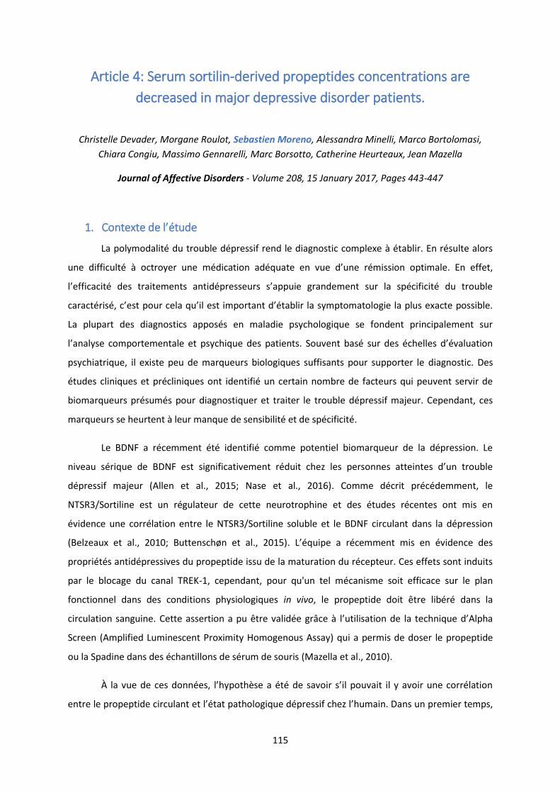
115
Article 4: Serum sortilin-derived propeptides concentrations are
decreased in major depressive disorder patients.
Christelle Devader, Morgane Roulot, Sebastien Moreno, Alessandra Minelli, Marco Bortolomasi,
Chiara Congiu, Massimo Gennarelli, Marc Borsotto, Catherine Heurteaux, Jean Mazella
Journal of Affective Disorders - Volume 208, 15 January 2017, Pages 443-447
1. Co te te de l’étude
La polymodalité du trouble dépressif rend le diagnostic complexe à établir. En résulte alors
une difficulté à octroyer une médication adéquate e ue d’u e rémission optimale. En effet,
l’effi a it des t aite e ts a tid p esseu s s’appuie g a de e t su la sp ifi it du t ou le
a a t is , ’est pou ela u’il est i po ta t d’ ta li la s pto atologie la plus e a te possi le.
La plupart des diagnostics apposés en maladie psychologique se fondent principalement sur
l’a al se o po te e tale et ps hi ue des patie ts. Sou e t as su des helles d’ aluatio
psychiatrique, il existe peu de marqueurs biologiques suffisants pour supporter le diagnostic. Des
études cliniques et précliniques ont identifié un certain nombre de facteurs qui peuvent servir de
biomarqueurs présumés pour diagnostiquer et traiter le trouble dépressif majeur. Cependant, ces
marqueurs se heurtent à leur manque de sensibilité et de spécificité.
Le BDNF a récemment été identifié comme potentiel biomarqueur de la dépression. Le
i eau s i ue de BDNF est sig ifi ati e e t duit hez les pe so es attei tes d’u t ou le
dépressif majeur (Allen et al., 2015; Nase et al., 2016). Comme décrit précédemment, le
NTSR3/Sortiline est un régulateur de cette neurotrophine et des études récentes ont mis en
évidence une corrélation entre le NTSR3/Sortiline soluble et le BDNF circulant dans la dépression
(Belzeaux et al., 2010; Buttenschøn et al., 2015). L’ uipe a e e t is e ide e des
propriétés antidépressives du propeptide issu de la maturation du récepteur. Ces effets sont induits
par le blocage du canal TREK-1, cependant, pour qu'un tel mécanisme soit efficace sur le plan
fonctionnel dans des conditions physiologiques in vivo, le propeptide doit être libéré dans la
circulation sanguine. Cette asse tio a pu t e alid e g â e à l’utilisatio de la te h i ue d’Alpha
Screen (Amplified Luminescent Proximity Homogenous Assay) qui a permis de doser le propeptide
ou la Spadine dans des échantillons de sérum de souris (Mazella et al., 2010).
À la ue de es do es, l’h poth se a t de sa oi s’il pou ait il a oi u e o latio
e t e le p opeptide i ula t et l’ tat pathologi ue d p essif hez l’hu ai . Dans un premier temps,

116
il a fallu alide la sp ifi it de l’a ti o ps pou la fo e hu ai e du p opeptide, puis, da s u
second temps, mesurer le niveau sérique circulant entre des sujets sains et diagnostiqués dépressifs.
2. Résultats et discussion
Lors de cette tude, j’ai p i ipale e t alis le dosage des ha tillo s de s u hu ai à
l’aide la te h i ue d’AlphaLisa.
L’a ti o ps utilis da s la thode de dosage a t alid pou e o ait e le p opeptide
humain. Nous avons d'abord conçu une série de séquences partielles du propeptide humain pour
caractériser la spécificité des anticorps utilisés (Figure 15A). L'analyse de reconnaissance structurelle
de ces peptides (Figure 15B et 15C) a permis d'observer que le propeptide, la Spadine, le propeptide
12-27 et 14-25 étaient reconnus par les anticorps avec des affinités identiques, alors que le
propeptide 1-16 et 22-28 n'étaient pas reconnus. A noter que les peptides non apparentés comme la
neurotensine ou la somatostatine ’i te f aie t pas avec la méthode de dosage (Fig. 1B et C). Ces
résultats nous ont permis d'identifier l'épitope des anticorps utilisés, la séquence WSGPI. L'activité
antidépresseur de ces analogues a été ensuite testée à l'aide du FST et il apparait que tous les
peptides portant la séquence épitope (PE, Spadine, PE 12-27 et PE 14-25) ont démontré une activité
antidépressive similaire à celle de la Spadine.
Figure 15 : Peptides synthétisés et affinités. A) Représentation schématique des peptides conçus pour
l'étude. La séquence épitope est en rouge. B et C) Affinités relatives de la Spadine, du propeptide et

117
des analogues récupérés pour la méthode de détection. Chaque point correspond à la moyenne±SEM
pour 3-6 expériences indépendantes.
E suite, afi d’ alue les a iatio s de i eau s i ue de p opeptide da s la pathologie de la
dépression, nous avons collecté un ensemble de sérums humains dans un centre clinique italien.
Dans cette cohorte, nous avons observé que la concentration de propeptide était significativement
plus faible chez les patients dépressifs (18,9±1,3 nM) que chez les témoins sains (23,7±1,5 nM) (z=-
2,11, p=0,035) (Figure 16). Étant donné que les analyses comparatives des variables
sociodémographiques ont révélé des différences dans l'éducation et le pourcentage de fumeurs
entre les patients atteints d'un trouble dépressif majeur et les témoins, nous avons effectué d'autres
analyses pour nous assurer que cette différence significative entre les groupes n'était pas influencée
par ces deux variables. Aucune corrélation significative n'a été observée entre l'éducation ou le
pourcentage de fumeurs et les concentrations de propeptide, tant chez les témoins que chez les
patients, et en tenant compte de l'ensemble des échantillons de sujets utilisés. De façon
intéressante, nous avons pu observer une augmentation subtile et significative de la concentration
de propeptide entre les personnes dépressives après un traitement antidépresseur de 12 semaines
et avant le traitement . ± . M z=− . , p= . (Figure 16 . L’effi a it du t aite e t a t
o fi e pa la di i utio sig ifi ati e du s o e MADRS. Pa ailleu s, il ’e iste aucune corrélation
significative entre cette cotation clinique et l'évolution des concentrations de propeptide (r=0,18,
p=0,27).
Figure 16 : Concentrations de propeptide dans les sérums de patients sains (Controls) et patients
atteints d'un trouble dépressif majeur (MDD) non traités (T0) et traités (T1) pendant 12 semaines.
Analyses statistiques : Mann-Whitney entre les patients (MDD T0, n =37) et les témoins (n =49), et
test des rangs signés de Wilcoxon entre les non traités (T0) et les traités (T1).* p<0,05.
Moyenne±SEM des MADRS pour les patients atteints d'un MDD avant (T0) et après (T1) un
traitement de 12 semaines (barres en rouge). *** p<0.001.
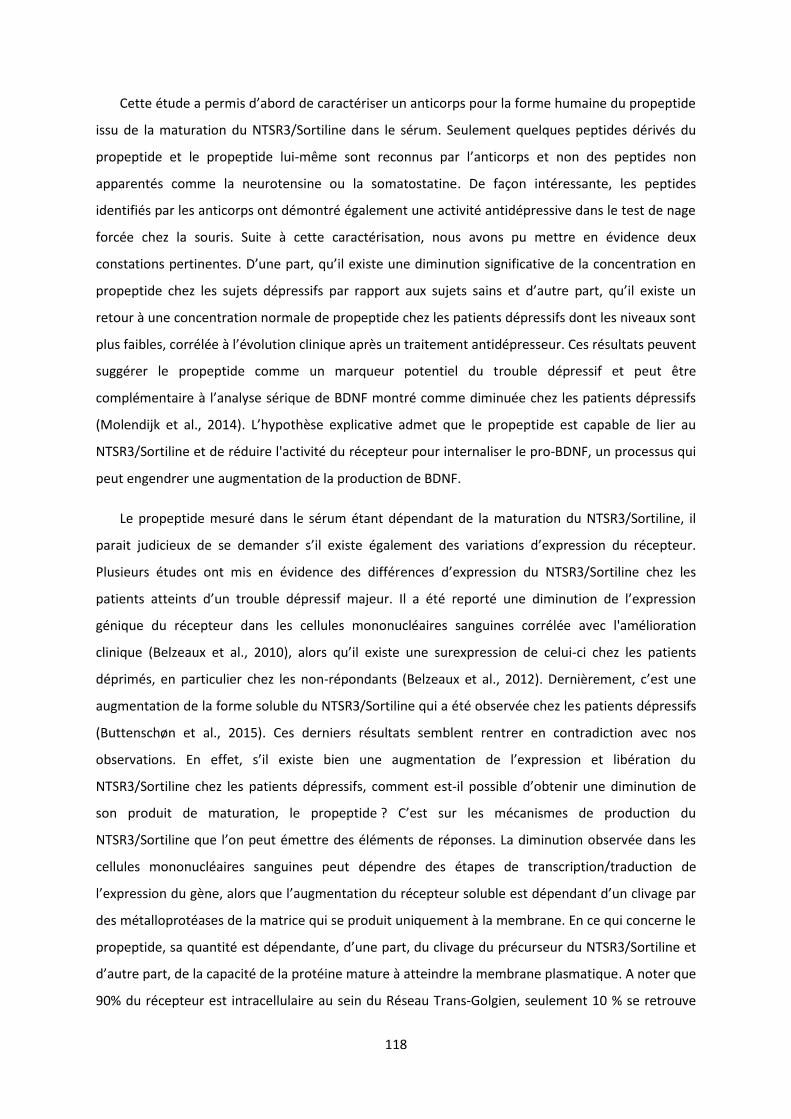
118
Cette tude a pe is d’a o d de a a t ise u a ti o ps pou la fo e hu ai e du p opeptide
issu de la maturation du NTSR3/Sortiline dans le sérum. Seulement quelques peptides dérivés du
propeptide et le propeptide lui- e so t e o us pa l’a ti o ps et non des peptides non
apparentés comme la neurotensine ou la somatostatine. De façon intéressante, les peptides
identifiés par les anticorps ont démontré également une activité antidépressive dans le test de nage
forcée chez la souris. Suite à cette caractérisation, nous avons pu mettre en évidence deux
o statio s pe ti e tes. D’u e pa t, u’il e iste u e di i utio sig ifi ati e de la o e t atio e
p opeptide hez les sujets d p essifs pa appo t au sujets sai s et d’aut e pa t, u’il e iste u
retour à une concentration normale de propeptide chez les patients dépressifs dont les niveaux sont
plus faibles, corrélée à l’ olutio li i ue ap s un traitement antidépresseur. Ces résultats peuvent
suggérer le propeptide comme un marqueur potentiel du trouble dépressif et peut être
o pl e tai e à l’a al se s i ue de BDNF o t o e di i u e hez les patie ts d p essifs
(Molendijk et al., 2014). L’h poth se e pli ati e ad et ue le p opeptide est apa le de lie au
NTSR3/Sortiline et de réduire l'activité du récepteur pour internaliser le pro-BDNF, un processus qui
peut engendrer une augmentation de la production de BDNF.
Le propeptide mesuré dans le sérum étant dépendant de la maturation du NTSR3/Sortiline, il
pa ait judi ieu de se de a de s’il e iste gale e t des a iatio s d’e p essio du epteu .
Plusieurs études ont mis en évidence des différe es d’e p essio du NTSR /So tili e hez les
patie ts attei ts d’u t ou le d p essif ajeu . Il a t epo t u e di i utio de l’e p essio
génique du récepteur dans les cellules mononucléaires sanguines corrélée avec l'amélioration
clinique (Belzeaux et al., 2010), alo s u’il e iste u e su e p essio de elui-ci chez les patients
déprimés, en particulier chez les non-répondants (Belzeaux et al., 2012). De i e e t, ’est u e
augmentation de la forme soluble du NTSR3/Sortiline qui a été observée chez les patients dépressifs
(Buttenschøn et al., 2015). Ces derniers résultats semblent rentrer en contradiction avec nos
o se atio s. E effet, s’il e iste ie u e aug e tatio de l’e p essio et li atio du
NTSR3/Sortiline chez les patients dépressifs, comment est-il possi le d’o te i u e di i utio de
son produit de maturation, le propeptide ? C’est su les a is es de p odu tio du
NTSR /So tili e ue l’o peut ett e des l e ts de po ses. La di i utio o se e da s les
cellules mononucléaires sanguines peut dépendre des étapes de transcription/traduction de
l’e p essio du g e, alo s ue l’aug e tatio du epteu solu le est d pe da t d’u li age pa
des métalloprotéases de la matrice qui se produit uniquement à la membrane. En ce qui concerne le
propeptide, sa quantité est dépendante, d’u e pa t, du li age du p u seu du NTSR /So tili e et
d’aut e pa t, de la capacité de la protéine mature à atteindre la membrane plasmatique. A noter que
90% du récepteur est intracellulaire au sein du Réseau Trans-Golgien, seulement 10 % se retrouve

119
exprimé à la surface membranaire. Cet adressage à la membrane plasmique peut être induit par
divers effecteurs, comme cela a déjà été démontré dans les cellules cibles de l'insuline et, en
particulier, dans les vésicules Glut4 du transporteur de glucose (Kandror, 2003). En effet, ces
vésicules peuvent être transférées à la membrane plasmatique lors de l'activation de l'insuline
(Huang et al., 2013) où elles peuvent libérer du propeptide dans la circulation.
Le fa teu li ita t de ette tude epose su u o e elati e e t est ei t d’ ha tillo s de
patients. Cela pourrait avoir une incidence négative sur la probabilité qu'un résultat statistiquement
significatif reflète un réel effet.
Néanmoins, ce travail de recherche amène le concept que la variation de concentration sérique
de p opeptide peut flu tue e fo tio de l’hu eu et, pa e te sio , pou ait t e u io a ueu
ad uat da s le diag osti d’u tat d p essif ajeu hez l’ho e. S’ajoute à cela, le potentiel de
la Spadine comme antidépresseur dont il est possible de mesurer également son niveau sérique
circulant, amenant ainsi des perspectives de nouvelles stratégies dans la prise en charge et le
traitement de cet affect psychologique.
Contents lists available at ScienceDirect
Journal of Affective Disorders
journal homepage: www.elsevier.com/locate/jad
Short communication
Serum sortilin-derived propeptides concentrations are decreased in major
depressive disorder patients
Christelle Devadera, Morgane Roulota, Sébastien Morénoa, Alessandra Minellib,
Marco Bortolomasic, Chiara Congiub, Massimo Gennarellib,d, Marc Borsottoa,
Catherine Heurteauxa, Jean Mazellaa,⁎
a CNRS, Institut de Pharmacologie Moléculaire et Cellulaire, UMR 7275, Université Côte d'Azur, 660 route des Lucioles, 06560 Valbonne, Franceb Department of Molecular and Translational Medicine, Biology and Genetic Division, University of Brescia, Brescia, Italyc Psychiatric Hospital “Villa Santa Chiara”, Verona, Italyd Genetic Unit, IRCCS Istituto Centro San Giovanni di Dio Fatebenefratelli, Brescia, Italy
A R T I C L E I N F O
Keywords:
Sortilin
Depression
Propeptide
Biomarker
TREK-1
Diagnosis
A B S T R A C T
Background: Despite intense research on mechanisms underlying the depressive pathophysiology, reliable
biomarkers to assess antidepressant treatment response are still lacking. Since the sortilin-derived propeptide
(PE) displays potent antidepressant activities and can be measured in the blood of rodents, we wondered
whether in human its seric level can vary between patients affected by major depressive disorder (MDD) and
healthy controls and after antidepressant treatment.
Methods: By using a specific dosing method, characterized by structure-recognition analysis with various
synthesized PE analogues, we conducted a translational study to test whether blood levels of PE are under
pathophysiological regulation and could serve as biomarkers of the depression state.
Results: The serum concentration of PE, a peptide displaying potent antidepressant activities in rodents, is
decreased in patients affected by major depressive disorder (MDD) when compared to healthy non-psychiatric
controls cohort (p=0.035). Interestingly, pharmacological antidepressant treatments restore normal PE levels.
Limitations: The limitation of the study concerns the relatively small patient samples that could negatively
affect the likelihood that a nominally statistically significant finding actually reflects a true effect.
Conclusions: The longitudinal quantification of the serum PE concentration could assist psychiatrists in the
diagnosis of antidepressant response efficacy, and the need to modify the therapeutic strategy.
1. Introduction
Major depressive disorder (MDD) episodes are treated with differ-
ent drug classes. However any first-line of treatment currently leads,
after several weeks, to a remission of about 35%, and approximately
30% of MDD patients are classified as having treatment resistant
depression (TRD) (Bentley et al., 2014; Thomas et al., 2013; Wong and
Licinio, 2001). Despite significant research efforts aimed at under-
standing the neurobiological underpinnings of MDD, treatments are
still based solely on relatively subjective assessment of symptoms. Due
to the low rate of remission, the identification of robust biological
markers predicting the clinical evolution of MDD and characterizing
the extent of the treatment outcome is therefore mandatory (Nestler
et al., 2002). Clinical and preclinical studies have identified a number
of factors that may serve as putative biomarkers for diagnosing and
treating MDD. However, the utility of any given marker to serve as a
clinically useful biomarker of MDD is limited by a lack of sensitivity
and specificity (Jani et al., 2015; Schmidt et al., 2011).
Recent studies have pointed out the Brain Derived Neurotrophic
Factor (BDNF) as a potential biomarker for clinical response under
antidepressive pharmacotherapy and clinical outcome (Allen et al.,
2015; Nase et al., 2016). Interestingly, sortilin is known to control
intracellular sorting of BDNF to the regulated secretory pathway (Chen
et al., 2005). Moreover, increased serum levels of sortilin are associated
with MDD and correlated with BDNF (Belzeaux et al., 2010;
Buttenschon et al., 2015). Recently, we have identified spadin, which
is a partial peptide (12−28) of the 44 amino-acid propeptide (PE)
generated from the maturation of sortilin (Munck Petersen et al.,
1999), also called neurotensin receptor-3 (Mazella et al., 1998). When
injected iv or ip in mice both spadin and PE display potent anti-
http://dx.doi.org/10.1016/j.jad.2016.10.049
Received 4 July 2016; Received in revised form 26 September 2016; Accepted 16 October 2016
⁎ Corresponding author.
E-mail address: [email protected] (J. Mazella).
Journal of Affective Disorders 208 (2017) 443–447
0165-0327/ © 2016 Elsevier B.V. All rights reserved.
Available online 04 November 2016
crossmark
depressant (AD) activities through inhibition of the potassium channel
TREK-1 activity (Mazella et al., 2010), a target for depression treat-
ment (Heurteaux et al., 2006). We originally developed a method to
measure the PE level in mouse (Mazella et al., 2010). To undertake the
dosing in human, we characterized the ability of the antibody directed
against spadin to recognize peptides derived from the human sequence
of PE. We demonstrated that both human peptides (PE and spadin) can
be measured from human sera samples and we addressed the
possibility that these peptides represent biological markers of MDD.
Then, we measured the serum PE concentrations in a cohort of 37
patients with MDD treated with a pharmacological protocol and
compared to 49 healthy non-psychiatric subjects.
2. Methods
2.1. Animals
All experiments were carried out on 20–25 g C57Bl/6 J males of 8–
10 week old (Janvier France Breeding) according to policies on the care
and use of laboratory animals of European Community legislation
2010/63/EU. The local Ethics Committee (CIEPAL) approved the
protocols used in this study (protocol number 00893.02).
2.2. Antibodies and biotinylated peptide preparation
Peptides were synthesized by Genecust (Dudelange, Luxemburg).
Rabbit polyclonal antibodies against spadin (YAPLPRWSGPIG-
VSWGLR) were prepared by Eurogentec (Seraing, Belgium). Spadin
(5.4 mg; 2.7 mmol) was solubilized in 1.5 mL of 25 mM phosphate
buffer, pH 6.7. N-hydroxysuccinimide biotin (13.5 mmol) resuspended
in 700 µl of 70% acetonitrile, 30% dimethyl formamide was added to
the spadin solution and incubated overnight at room temperature.
Spadin-biotinylated was purified by HPLC using a Waters apparatus
equipped with a semi-preparative RP18 Lichrosorb column. Spadin-
biotinylated (eluted at 27 min), identified by mass spectrometry, was
collected, quantified by its absorption at 280 nm and lyophilized in
aliquots.
2.3. Alpha-Lisa™ test
According to the principles of AlphaScreen™ technology (Perkin
Elmer), streptavidin-donor microbeads were recognized by biotin-
spadin, whereas anti-rabbit IgG-acceptor microbeads were bound by
anti-spadin antibodies. When the two microbeads (acceptor and donor)
were into proximity, the signal was produced by a molecular interac-
tion occurring between the binding partners bound on the beads. The
propeptide present in the serum sample was able to interfere with this
interaction leading to competition. Standard curves were obtained by
incubation in 96-well plates of 10 nM biotin-spadin with the anti-
spadin antibody (1:1000) in the AlphaLisa™ buffer in the absence or in
the presence of increasing concentrations of spadin (from 10–11 to
10−6 M) for 1 h at room temperature. After addition of acceptor and
donor beads and further incubation for 2 h at room temperature, the
plaques were read using the Enspire apparatus (Perkin). For serum
measurements, the same volume of serum was added instead of
unlabeled spadin. The amount of propeptide was determined from its
percent of signal inhibition and calculated using the standard curve.
2.4. Porsolt forced swim test (FST)
After iv injection of either saline or various tested peptides, mice (n
=10–12 per group) were placed individually in a cylinder (height:
30 cm, diameter: 15 cm) filled with water to a depth of 12 cm
(temperature: 22 ± 1 °C) for 6 min. The total period of immobility
was recorded during the last 4 min (Porsolt et al., 1977).
2.5. Human blood samples
The control cohort consisted of 49 unrelated healthy volunteers
who were screened for DSM-IV Axis I disorder diagnoses by expert
psychologists using the Mini-International Neuropsychiatric Interview
(MINI) (Sheehan et al., 1998). Only healthy volunteers without history
of drug or alcohol abuse or dependence and without a personal or first-
degree family history of psychiatric disorders were enrolled in the
study. Furthermore, the absence of relevant neurological diseases i.e.
epilepsy, Parkinson's syndrome, was mandatory for inclusion into the
study. Finally, subjects who obtained a score lower than 27/30 at the
Mini Mental State Examination (MMSE) were excluded from the study.
The patient cohort was made of 37 MDD patients with moderate to
severe depression who met Diagnostic and Statistical Manual of Mental
Disorders-IV (DSM-IV) classification system criteria. Diagnosis of
unipolar depression was confirmed using the Structured Clinical
Interview for DSM-IV Axis I Disorders (SCID-I). The exclusion criteria
were: a) mental retardation or cognitive disorder; b) a lifetime history
of schizophrenic, schizoaffective, or bipolar disorder; c) personality
disorder, substance abuse, alcohol abuse or dependency, obsessive
compulsive disorder, or post-traumatic stress disorder as the primary
diagnosis; and d) comorbidity with an eating disorder.
No patients showed psychotic symptoms; 11 (29.7%) showed
current comorbidity in Axis I (generalized anxiety disorder (GAD),
panic attacks, panic disorders or anxiety disorder not otherwise
specified (NOS)), 2 (5.4%) showed symptoms of Axis II disorders
(dependent personality disorder) and no alcohol abuse, as a secondary
diagnosis (the total number exceeded the number of subjects due to the
presence of comorbidities).
All patients were either ‘drug naïve’, and had never received
previous treatment with any antidepressant drug, or ‘drug free’. They
have been previously treated with one or two antidepressants but had a
washout period lasting at least 2 weeks before starting with the new
antidepressant treatment. All patients were treated in monotherapy:
thirty-five patients were treated with selective serotonin reuptake
inhibitors (SSRIs), and the other patients were treated with selective
serotonin-norepinephrine reuptake inhibitors (SNRIs), tricyclics
(TCAs) or noradrenergic and specific serotoninergic antidepressants
(NASSAs).
Illness severity was assessed by the Montgomery and Asberg
Depression Rating Scale (MADRS) before the start of the new
antidepressant treatment (T0) and after 12 weeks of treatment (T1).
All of the socio-demographical, clinical and pharmacological treatment
characteristics of patients are shown in Table 1.
For both patients and controls, venous blood samples were
collected between 8:00 and 9:00 a.m. after an overnight fast in antic-
oagulant-free tubes. Serum was separated by centrifugation (1620g for
15 min). Blood samples for PE measurements were collected at each
timepoint.
Studies were approved by the Local Ethics Committees (CEIOC
IRCCS Istituto Centro San Giovanni di Dio Fatebenefratelli, Brescia N:
50/2008 and Ethics Committee of the province of Verona N: 4997/
09.11.01), and written informed consent was obtained.
2.6. Statistics
Results are expressed as mean ± standard error mean (SEM).
Statistical analyses were performed using GraphPad (version 6.0).
Analysis of variance (ANOVA) was used to compute differences in time
immobility for the different peptides used in the FST (Fig. 1D).
Demographic and clinical characteristics in our patient samples were
described either in quantitative term of mean ± standard deviation
(SD) or as proportions. Chi-square (χ2) tests were conducted to
evaluate the association between groups and categorical variables,
while analysis of variance (ANOVA) was used to compute possible
differences in age, education and BMI between groups (Table 1). Due to
C. Devader et al. Journal of Affective Disorders 208 (2017) 443–447
444
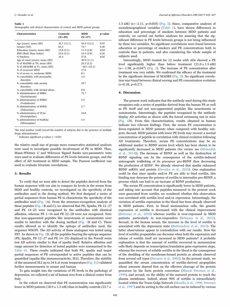
the relative small size of groups more conservative statistical analyses
were used to investigate possible involvement of PE in MDD. Thus,
Mann-Whitney U and Wilcoxon Signed Ranks non-parametric tests
were used to evaluate differences of PE levels between groups, and the
effect of AD treatment in MDD sample. The Pearson coefficient was
used to evaluate bivariate correlations.
3. Results
To verify that we were able to detect the peptides derived from the
human sequence with our aim to compare its levels in the serum from
MDD and healthy controls, we investigated on the specificity of the
antibodies used in the dosing method. We first designed a series of
partial sequences from human PE to characterize the specificity of the
antibodies used (Fig. 1A). From the structure-recognition analysis of
these peptides (Fig. 1B and C), we observed that PE, Spadin, PE 12–27
and PE 14–25 were recognized by the antibodies with identical
affinities, whereas PE 1–16 and PE 22–28 were not recognized. Note
that non-apparented peptides like neurotensin or somatostatin were
unable to interfere with the dosing method (Fig. 1B and C). These
results allowed us to identify the epitope of antibodies used, the
sequence WSGPI. The AD activity of these analogues was tested using
FST. As shown in Fig. 1D, all the peptides bearing the epitope sequence
(PE, Spadin, PE 12–27 and PE 14–25) displayed in the Porsolt swim
test AD activity similar to that of spadin itself. Relative affinities and
range amount for detection of tested peptides were summarized in the
Table S1. These results indicated that both PE, spadin and major
partial sequences of PE corresponded to active peptides that can be
quantified (spadin-like immunoreactivity, SLI). Therefore, the stability
of the measured SLI up to 24 h at room temperature (Fig. S1) validated
further measurements in mouse and human.
To gain insight into the variations of PE levels in the pathology of
depression, we collected a set of human sera from a clinical center from
Italy.
In the cohort we observed that PE concentration was significantly
lower in MDD patients (18.9 ± 1.3 nM) than in healthy controls (23.7 ±
1.5 nM) (z=−2.11, p=0.035) (Fig. 2). Since, comparative analyses of
sociodemographical variables (Table 1), have shown differences in
education and percentage of smokers between MDD patients and
controls, we carried out further analyses for assuring that the sig-
nificant difference in PE levels between groups is not being influenced
by these two variables. No significant correlations were found between
education or percentage of smokers and PE concentrations both in
controls than in patients, and also considering the whole sample of
subjects used.
Interestingly, MDD treated for 12 weeks with ADs showed a PE
level significantly higher than before treatment (21.0 ± 1.5 nM)
(z=−1.98, p=0.047) (Fig. 2). The increase of PE concentration after
treatment was very subtle. We confirmed the efficacy of the treatment
by the significant decrease of MADRS (Fig. 2). No significant correla-
tion was found between clinical scoring and PE concentration evolution
(r=0.18, p=0.27).
4. Discussion
The present work indicates that the antibody used during this study
recognizes only a series of peptides derived from the human PE as well
as PE itself and not non-apparented peptides like neurotensin or
somatostatin. Interestingly, the peptides recognized by the antibody
display AD activities as shown with the forced swimming test in mice
(Fig. 1D). From this characterization, results obtained in human
indicate two relevant findings. First, the serum PE concentration is
down-regulated in MDD patients when compared with healthy sub-
jects. Second, MDD patients with lower PE levels may recover a normal
concentration of peptide in correlation with clinical evolution following
AD treatments. Therefore, serum PE concentrations may be an
additional marker to BDNF serum level which has been shown to be
significantly decreased in MDD patients (for review see (Molendijk
et al., 2014)). The decrease of BDNF as well as the dysfunction of
BDNF signaling can be the consequence of the sortilin-induced
anterograde trafficking of its precursor pro-BDNF then decreasing
the production of BDNF. We already observed that spadin enhanced
BDNF mRNA and protein (Devader et al., 2015). One explanation
could be that since spadin and/or PE are able to bind sortilin, this
binding may decrease the potency of sortilin to internalize pro-BDNF, a
process which can lead to an increase of BDNF production.
The serum PE concentration is significantly lower in MDD patients,
and taking into account that peptides measured in the present work
were generated from sortilin, we wondered whether this observation
was correlated with sortilin level and/or expression. Interestingly, the
variation of sortilin expression in the blood has been already observed
in MDD patients. First, in blood mononuclear cells, the genetic
expression of sortilin is decreased with the clinical improvement
(Belzeaux et al., 2010) whereas sortilin is over-expressed in MDD
patients, particularly in non-responders (Belzeaux et al., 2012).
Second, in the human serum, the increase in the level of sortilin is
associated with the depression state (Buttenschon et al., 2015). The
latter observations appear in contradiction with our results. How the
level of sortilin propeptides can decrease when both the expression and
release of sortilin itself are increased in MDD patients? A probable
explanation is that the amount of sortilin recovered in mononuclear
cells likely depends on transcription/translation gene expression steps,
whereas the recovery of soluble sortilin in the blood is the consequence
of the shedding of the membrane-bound protein as already observed
from several cell types (Navarro et al., 2002). In the present study, we
quantified the serum concentration of sortilin-derived propeptides
whose amounts depend first on the effective cleavage of the protein
precursor by the furin protein convertase (Munck Petersen et al.,
1999), and second, on the ability of the matured protein to reach the
plasma membrane. Indeed, about 90% of sortilin is intracellularly
located within the Trans-Golgi Network (Mazella et al., 1998; Petersen
et al., 1997) and its sorting to the cell surface can be induced by various
Table 1
Demographic and clinical characteristics of control and MDD patient groups.
Characteristics Controls
(N=49)
MDD
(N=37)
p-value
Age (years), mean (SD) 45.1 (12.1) 44.9 (13.1) 0.95
Gender (%F) 65.3 75.7 0.30
Education (years), mean (SD) 13.8 (5.1) 11.9 (2.8) 0.04*
BMI (Body Mass Index) 23.6 (3.1) 24.4 (2.8) 0.26
% Smokers 16.3 37.8 0.02*
Age of onset (years), mean (SD) 40.9 (11.1)
% of MADRS at T0, mean (SD) 25.5 (5.2)
% of ΔMADRS at T1, mean (SD) −60.1 (33.1)
% of recurrent MDD 27.0
% of severe vs. moderate MDD 8.1
% comorbidity with personality
disorders
5.4
% comorbidity with anxiety
disorders
29.7
% comorbidity with alcohol abuse 0.0
% administration of SSRIs*
(Escitalopram)
75.7
% administration of SNRIs*
(Venlafaxine)
5.4
% administration of SNRIs*
(Duloxetine)
2.7
% administration of TCAs*
(Nortriptyline)
10.8
% administration of NaSSAs*
(Mirtazapine)
5.4
athe total number could exceed the number of subjects due to the presence of multiple
drugs administration* Indicates significant p-values ( < 0.05)
C. Devader et al. Journal of Affective Disorders 208 (2017) 443–447
445
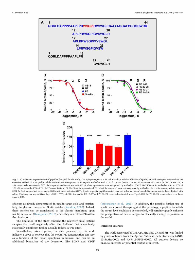
effectors as already demonstrated in insulin target cells and, particu-
larly, in glucose transporter Glut4 vesicles (Kandror, 2003). Indeed,
these vesicles can be translocated to the plasma membrane upon
insulin activation (Huang et al., 2013) where they can release PE within
the circulation.
The limitation of the study concerns the relatively small patient
samples that could negatively affect the likelihood that a nominally
statistically significant finding actually reflects a true effect.
Nevertheless, taken together, the data presented in this work
indicate a proof of concept that the serum PE concentration can vary
as a function of the mood symptoms in human, and can be an
additional biomarker of the depression like BDNF and VEGF
(Buttenschon et al., 2015). In addition, the possible further use of
spadin as a potent therapy against the pathology, a peptide for which
the serum level could also be controlled, will certainly greatly enhance
the perspectives of new strategies to efficiently manage depression in
the world.
Funding sources
The work performed by JM, CD, MR, SM, CH and MB was funded
by grants obtained from the Agence Nationale de la Recherche (ANR-
13-SAMA-0002 and ANR-13-RPIB-0002). All authors declare no
financial interests or potential conflict of interest.
Fig. 1. A) Schematic representation of peptides designed for the study. The epitope sequence is in red. B and C) Relative affinities of spadin, PE and analogues recovered for the
detection method. B) Both spadin and the entire PE were recognized by anti-spadin antibodies with IC50 of 2.50 nM (95% CI: 1.85–3.37; n =6) and of 2.18 nM (95% CI: 1.53–3.09; n
=3), respectively, neurotensin (NT, black squares) and somatostatin-14 (SS14, white squares) were not recognized by antibodies. (C) PE 14–25 bound to antibodies with an IC50 of
1.73 nM, whereas the IC50 of PE 12–27 was of 3.44 nM, PE 22–28 (white squares) and PE 1–16 (black squares) were not recognized by antibodies. Each point corresponds to mean ±
SEM. for 3–6 independent experiments. D) Porsolt forced swim test (FST). Spadin or partial peptides-treated mice had a shorter time of immobility comparable to those obtained with
saline. (Ordinary one way ANOVA, F5,54 =20.21, ****p < 0.0001 for spadin, PE 12–27 and PE 22–28 versus saline-treated mice, **p=0.0026 for PE 14–25 versus saline; error bars,
mean ± SEM.
C. Devader et al. Journal of Affective Disorders 208 (2017) 443–447
446
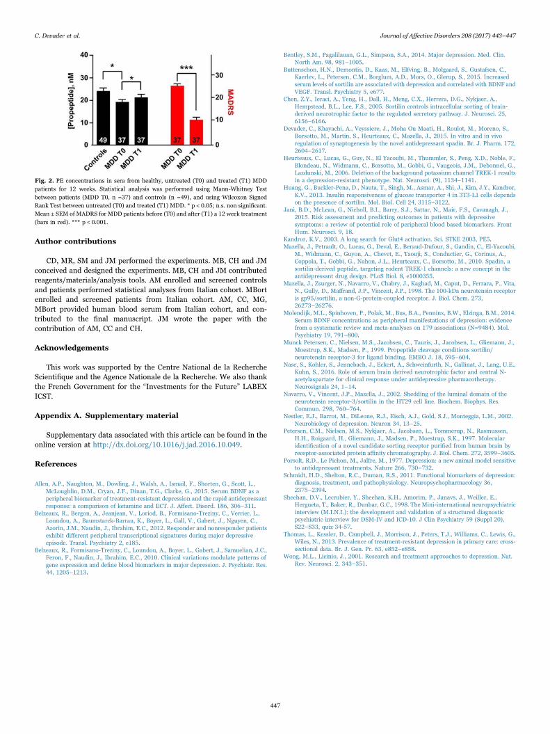
Author contributions
CD, MR, SM and JM performed the experiments. MB, CH and JM
conceived and designed the experiments. MB, CH and JM contributed
reagents/materials/analysis tools. AM enrolled and screened controls
and patients performed statistical analyses from Italian cohort. MBort
enrolled and screened patients from Italian cohort. AM, CC, MG,
MBort provided human blood serum from Italian cohort, and con-
tributed to the final manuscript. JM wrote the paper with the
contribution of AM, CC and CH.
Acknowledgements
This work was supported by the Centre National de la Recherche
Scientifique and the Agence Nationale de la Recherche. We also thank
the French Government for the “Investments for the Future” LABEX
ICST.
Appendix A. Supplementary material
Supplementary data associated with this article can be found in the
online version at http://dx.doi.org/10.1016/j.jad.2016.10.049.
References
Allen, A.P., Naughton, M., Dowling, J., Walsh, A., Ismail, F., Shorten, G., Scott, L.,
McLoughlin, D.M., Cryan, J.F., Dinan, T.G., Clarke, G., 2015. Serum BDNF as a
peripheral biomarker of treatment-resistant depression and the rapid antidepressant
response: a comparison of ketamine and ECT. J. Affect. Disord. 186, 306–311.
Belzeaux, R., Bergon, A., Jeanjean, V., Loriod, B., Formisano-Treziny, C., Verrier, L.,
Loundou, A., Baumstarck-Barrau, K., Boyer, L., Gall, V., Gabert, J., Nguyen, C.,
Azorin, J.M., Naudin, J., Ibrahim, E.C., 2012. Responder and nonresponder patients
exhibit different peripheral transcriptional signatures during major depressive
episode. Transl. Psychiatry 2, e185.
Belzeaux, R., Formisano-Treziny, C., Loundou, A., Boyer, L., Gabert, J., Samuelian, J.C.,
Feron, F., Naudin, J., Ibrahim, E.C., 2010. Clinical variations modulate patterns of
gene expression and define blood biomarkers in major depression. J. Psychiatr. Res.
44, 1205–1213.
Bentley, S.M., Pagalilauan, G.L., Simpson, S.A., 2014. Major depression. Med. Clin.
North Am. 98, 981–1005.
Buttenschon, H.N., Demontis, D., Kaas, M., Elfving, B., Molgaard, S., Gustafsen, C.,
Kaerlev, L., Petersen, C.M., Borglum, A.D., Mors, O., Glerup, S., 2015. Increased
serum levels of sortilin are associated with depression and correlated with BDNF and
VEGF. Transl. Psychiatry 5, e677.
Chen, Z.Y., Ieraci, A., Teng, H., Dall, H., Meng, C.X., Herrera, D.G., Nykjaer, A.,
Hempstead, B.L., Lee, F.S., 2005. Sortilin controls intracellular sorting of brain-
derived neurotrophic factor to the regulated secretory pathway. J. Neurosci. 25,
6156–6166.
Devader, C., Khayachi, A., Veyssiere, J., Moha Ou Maati, H., Roulot, M., Moreno, S.,
Borsotto, M., Martin, S., Heurteaux, C., Mazella, J., 2015. In vitro and in vivo
regulation of synaptogenesis by the novel antidepressant spadin. Br. J. Pharm. 172,
2604–2617.
Heurteaux, C., Lucas, G., Guy, N., El Yacoubi, M., Thummler, S., Peng, X.D., Noble, F.,
Blondeau, N., Widmann, C., Borsotto, M., Gobbi, G., Vaugeois, J.M., Debonnel, G.,
Lazdunski, M., 2006. Deletion of the background potassium channel TREK-1 results
in a depression-resistant phenotype. Nat. Neurosci. (9), 1134–1141.
Huang, G., Buckler-Pena, D., Nauta, T., Singh, M., Asmar, A., Shi, J., Kim, J.Y., Kandror,
K.V., 2013. Insulin responsiveness of glucose transporter 4 in 3T3-L1 cells depends
on the presence of sortilin. Mol. Biol. Cell 24, 3115–3122.
Jani, B.D., McLean, G., Nicholl, B.I., Barry, S.J., Sattar, N., Mair, F.S., Cavanagh, J.,
2015. Risk assessment and predicting outcomes in patients with depressive
symptoms: a review of potential role of peripheral blood based biomarkers. Front
Hum. Neurosci. 9, 18.
Kandror, K.V., 2003. A long search for Glut4 activation. Sci. STKE 2003, PE5.
Mazella, J., Petrault, O., Lucas, G., Deval, E., Beraud-Dufour, S., Gandin, C., El-Yacoubi,
M., Widmann, C., Guyon, A., Chevet, E., Taouji, S., Conductier, G., Corinus, A.,
Coppola, T., Gobbi, G., Nahon, J.L., Heurteaux, C., Borsotto, M., 2010. Spadin, a
sortilin-derived peptide, targeting rodent TREK-1 channels: a new concept in the
antidepressant drug design. PLoS Biol. 8, e1000355.
Mazella, J., Zsurger, N., Navarro, V., Chabry, J., Kaghad, M., Caput, D., Ferrara, P., Vita,
N., Gully, D., Maffrand, J.P., Vincent, J.P., 1998. The 100-kDa neurotensin receptor
is gp95/sortilin, a non-G-protein-coupled receptor. J. Biol. Chem. 273,
26273–26276.
Molendijk, M.L., Spinhoven, P., Polak, M., Bus, B.A., Penninx, B.W., Elzinga, B.M., 2014.
Serum BDNF concentrations as peripheral manifestations of depression: evidence
from a systematic review and meta-analyses on 179 associations (N=9484). Mol.
Psychiatry 19, 791–800.
Munck Petersen, C., Nielsen, M.S., Jacobsen, C., Tauris, J., Jacobsen, L., Gliemann, J.,
Moestrup, S.K., Madsen, P., 1999. Propeptide cleavage conditions sortilin/
neurotensin receptor-3 for ligand binding. EMBO J. 18, 595–604.
Nase, S., Kohler, S., Jennebach, J., Eckert, A., Schweinfurth, N., Gallinat, J., Lang, U.E.,
Kuhn, S., 2016. Role of serum brain derived neurotrophic factor and central N-
acetylaspartate for clinical response under antidepressive pharmacotherapy.
Neurosignals 24, 1–14.
Navarro, V., Vincent, J.P., Mazella, J., 2002. Shedding of the luminal domain of the
neurotensin receptor-3/sortilin in the HT29 cell line. Biochem. Biophys. Res.
Commun. 298, 760–764.
Nestler, E.J., Barrot, M., DiLeone, R.J., Eisch, A.J., Gold, S.J., Monteggia, L.M., 2002.
Neurobiology of depression. Neuron 34, 13–25.
Petersen, C.M., Nielsen, M.S., Nykjaer, A., Jacobsen, L., Tommerup, N., Rasmussen,
H.H., Roigaard, H., Gliemann, J., Madsen, P., Moestrup, S.K., 1997. Molecular
identification of a novel candidate sorting receptor purified from human brain by
receptor-associated protein affinity chromatography. J. Biol. Chem. 272, 3599–3605.
Porsolt, R.D., Le Pichon, M., Jalfre, M., 1977. Depression: a new animal model sensitive
to antidepressant treatments. Nature 266, 730–732.
Schmidt, H.D., Shelton, R.C., Duman, R.S., 2011. Functional biomarkers of depression:
diagnosis, treatment, and pathophysiology. Neuropsychopharmacology 36,
2375–2394.
Sheehan, D.V., Lecrubier, Y., Sheehan, K.H., Amorim, P., Janavs, J., Weiller, E.,
Hergueta, T., Baker, R., Dunbar, G.C., 1998. The Mini-international neuropsychiatric
interview (M.I.N.I.): the development and validation of a structured diagnostic
psychiatric interview for DSM-IV and ICD-10. J Clin Psychiatry 59 (Suppl 20),
S22–S33, quiz 34-57.
Thomas, L., Kessler, D., Campbell, J., Morrison, J., Peters, T.J., Williams, C., Lewis, G.,
Wiles, N., 2013. Prevalence of treatment-resistant depression in primary care: cross-
sectional data. Br. J. Gen. Pr. 63, e852–e858.
Wong, M.L., Licinio, J., 2001. Research and treatment approaches to depression. Nat.
Rev. Neurosci. 2, 343–351.
Fig. 2. PE concentrations in sera from healthy, untreated (T0) and treated (T1) MDD
patients for 12 weeks. Statistical analysis was performed using Mann-Whitney Test
between patients (MDD T0, n =37) and controls (n =49), and using Wilcoxon Signed
Rank Test between untreated (T0) and treated (T1) MDD. * p < 0.05; n.s. non significant.
Mean ± SEM of MADRS for MDD patients before (T0) and after (T1) a 12 week treatment
(bars in red). *** p < 0.001.
C. Devader et al. Journal of Affective Disorders 208 (2017) 443–447
447
124
DISCUSSION GENERALE et CONCLUSION
Durant ces trois années de thèse, o t a ail s’est o sa su l’ tude et la o p he sio
d’u epteu ultifo tio el da s la gulatio de l’ tat otio el. De façon intéressante, la
d a he o igi elle de e p ojet se ase su les o s ue es o po te e tales de l’absence ou
du lo age d’u de ses pa te ai es, le a al potassi ue TREK-1. La protéine NTSR3/Sortiline a été
identifiée par plusieurs approches, ce qui a permis de e d e o pte de l’e iste e de
fonctionnalités qui ne se limite t pas u’à u ôle de récepteur. Caractérisée comme une protéine
d’ad essage g â e à so ho ologie st u tu elle a e le o ple e VPS p, ’est e ide tifia t ses
pa te ai es ue l’o peut pote tielle e t d fi i so ôle au sei de l’o ga is e. E effet, face à sa
localisation cérébrale abondante et la formation de complexe avec des éléments du système
neurotrophique, comme le BDNF ou encore p75NTR, il ’est pas i oh e t d’ ett e l’h poth se
d’u e fo tio e t ale da s la gulatio neuronale. L’o se atio d’u e i te a tio e t e le
NTSR3/Sortiline et le canal TREK- , i pli u da s la gulatio de l’hu eu , a t le poi t de départ
pou tudie les diff e ts aspe ts du epteu da s l’affect dépressif. Son produit de maturation,
le propeptide qui interagit avec le canal en bloquant son activité est responsable des effets
antidépressifs chez la souris. E d oule alo s l’idée que ce produit de maturation du
NTSR3/Sortiline, molécule e dog e, pou ait alo s t e esu afi de d te i e s’il e iste une
corrélation entre son taux sanguin et l’ tat d p essif hez des patie ts.
Da s l’ tude sur le niveau circulant du propeptide, les résultats amènent un concept
intéressant. Il existe bien une différence entre des personnes atteintes d’épisode dépressif majeur
(EDM) et des patients diagnostiqués sains. Ho is le fait u’il faille le e le o e d’ ha tillo s
de patients pour augmenter la robustesse de ce résultat, il tient bon de noter u’il e iste gale e t
u e a iatio lo s ue le patie t d p i po d à u t aite e t a tid p esseu ais u’il ’ a pas
de corrélation entre le score MADRS et la quantité circulante de propeptide. Ce dernier point met en
avant le fait que les variations de propeptide semblent spécifiques du deg de l’ tat d p essif et
u’il se pou ait u’il e soit pas significativement possible de voir une différence sur des symptômes
plus légers. Cependant, le niveau sérique de propeptide se place comme un outil complémentaire
dans la détection des cas sévères de dépression et permet de préciser le caractère épisodique de
l’affe t pou u e eilleu e p ise e ha ge thérapeutique.
Les sultats p de ts sugg e t u’u e di i utio du p opeptide peut t e le eflet
d’u e d g adatio de l’ tat otio el d’u i di idu. E p e a t e o sid atio l’o igi e de e
125
peptide, et ses p op i t s d’a lio atio su l’hu eu ui sulte t de l’a ti it du a al TREK-1, il
existe une certaine évidence à poser le NSTR3/Sortiline comme régulateur important du système
e eu e t al. L’app o he la plus i t essa te pou a de , de a i e o pl te, au fo tio s
d’u e p ot i e, side e l’a al se ph ot pi ue d’u od le d pou u de ette de i e. Da s le
cas présent, la délétion du NTSR3/Sortiline a pe is d’ alue o seule e t ses partenaires
connus, comme TREK-1 et le BDNF, mais également les conséquences sur la régulation du
comportement et du système dont il fait partie, le système neurotensinergique. En effet, il
semblerait que le récepteur prend une part importante dans la régulation du système
neurotensinergique. De pa la odulatio de l’e p ession du NTSR2, qui résulte en une perte de
sensibilité nociceptive chez les souris délétées du NTSR /So tili e. De faço e a ua le, l’a se e
de NTSR3/Sortiline résulte en une augmentation de la production de neurotensine cérébrale et
sérique, mais également en un possible changeme t d’affi ité du peptide pour le récepteur NTSR2.
Les mécanismes sous-jacents à e ha ge e t d’affi it ’o t pas e o e t solus, mais il est
p o a le ue l’i te a tio a e le NTSR3/Sortiline permet des changements conformationnels
nécessaires au fonctionnement adéquat de ces récepteurs.
Les p op i t s d’ad essage du NTSR /So tili e ep se te t u e fo tio l da s la
régulation de ces partenaires. En identifiant le canal potassique TREK-1 comme une des protéines
associées au récepteur, il a été intéressant de caractériser cette perte fonctionnelle chez notre
modèle murin Sort-/- dans l’ tat d p essif. E ep e a t l’h poth se ue TREK-1 est un modulateur
de l’hu eu , les p e ie s tests ta lis su es sou is o t t des tests o portementaux relatifs à la
dépression, comme le FST et le TST, qui esu e t la sig atio , et le NSF, ui pe et d’ alue la
p ope sio à l’e plo atio da s u ilieu a e sif. De faço to a te, le ph ot pe de sista e
des souris KO-NTSR3 se révèle être semblable à celui des souris KO-TREK-1. Cela suggère que
l’e p essio tissulaire ou cellulaire du canal semble être altérée. Cette assertion a été vérifiée
d’a o d pa western blot, ui le ue le i eau d’e p essio de TREK-1 est significativement
diminué à la membrane plasmique où il est pleinement fonctionnel, sans modification de son
expression totale. Par électrophysiologie, nous avons confirmé que le potentiel de membrane des
neurones est fortement modifié.
L’aut e pa te ai e esse tiel ide tifi est le BDNF. Cette neurotrophine nécessite une
maturation dépendante du NTSR3/Sortiline, plus particulièrement dans sa voie régulée. En effet, ce
complexe NTSR3/Sortiline proBDNF ’est p se t ue da s la voie régulée du BDNF, et il est
nécessaire pour le repliement correct du domaine mature pour sa maturation par la furine. Cela
suggère que le NTSR3/Sortiline est un acteur prépondérant dans le contrôle de la sécrétion de la
126
neurotrophine et que son absence crée un déséquilibre entre les voies de libération. Dans une lignée
cellulaire d i e d'u ph o h o o to e, les PC , il a pu t e o t u’e p se e d’u e
forme tronquée du NTSR3/Sortiline au niveau du site de liaison avec le proBDNF, il y avait une
augmentation de libération de BDNF dans la voie constitutive (Chen et al., 2005). Le BDNF est une
protéine clé dans la viabilité et la croissance neuronale, par ailleurs, son niveau sérique et cérébral
peut t e u i di ateu da s e tai s tats d p essifs. C’est de faço oh e te ue l’ aluatio de
ce système neurotrophique s’est pos e comme une hypothèse de travail dans notre modèle de
souris Sort-/-. Les résultats confirment l’e iste e d’une augmentation de BDNF dans les
homogénats de cerveaux des souris KO-NTSR , ui sulte d’u e aug e tatio de la atu ation
intracellulaire par la furine, aboutissant également à u e aug e tatio de l’a ti it de so
récepteur TrkB.
En associant ces derniers résultats avec une augmentation du taux de décharges des
neurones sérotoninergiques, nous avons pu mettre en évidence le rôle important du NSTR3/Sortiline
da s la gulatio de l’hu eu et plus pa ti uli e e t da s l’affe t d p essif, sugg a t le
récepteur comme une cible potentiel dans le traitement de cette maladie psychologique.
Cependant, il existe chez les souris KO-NTSR u ph ot pe d’anxiété révélé par des tests
comportementaux comme le paradigme de la boite clair-obscur ou encore la croix surélevée.
L’a i t fait pa tie du spe t e de l’affe t d p essif, e ui e d e sultat assez paradoxal par
rapport au phénotype antidépressif de ces souris. Ces tests mesurent la prédisposition des souris à
l’e plo atio da s u e i o e e t aversif. De manière intéressante, ils sont assez proches de
elui du NSF, à la diff e e ue les sou is ’o t pas t sou ises à u e supp essio de ou itu e
pendant 24h. Il semblerait possible ue la se satio de fai ait eu u i pa t su l’issue de e test.
D’u poi t de vue métabolique, des résultats non publiés ont apporté des éléments intéressants. Le
poids des souris KO-NTSR3 est sensiblement plus faible celui des souris sauvages ainsi que la prise
alimentaire. Il pourrait donc y avoir une régulation différente dans le système alimentaire. Pour
revenir au phénotype anxieux, il ’est pas o l à u e d gulatio de l’ho o e du stress, la
o ti ost o e. D’aut es tests de sista e à la d p essio o t t alis s, etta t e jeu la
résignation acquise. La résignatio a uise est u e se satio d’i puissa e pe a e te générée
par le fait d'être plongé, de façon durable ou répétée, dans une situation à laquelle on ne peut
s’ happe ou o t ôle . Le test hez la sou is o siste e u e s ie de chocs le t i ues d’où il ’est
pas possi le de s’ happe , du a t plusieu s jou s afi de p o o ue u e sig atio . Le de ie jou ,
les sou is o t la possi ilit de s’ happe pou ett e fi à la situatio st essa te, pe etta t ai si
d’ alue leu deg d’e ie d’ ite l’e i o e e t a e sif. Les a tid p esseu s lassi ues ai si
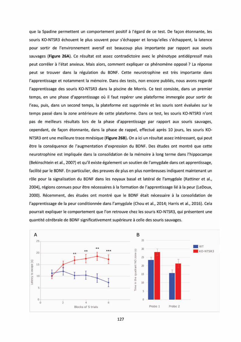
128
Figure 26 : Comportement des souris KO-NTSR3 dans la résignation acquise et la mémoire spatiale. A)
Test du Lea Help ess, late es d happe e t su divis es e 5 lo s d essais (n=30). Le test se
compose de 30 essais séparés par un intervalle de 30 secondes. Un essai est défini par
l ad i ist atio d u ho le t i ue soute u ui s a o pag e de l ouve tu e de la po te de communication, pe etta t à la sou is de fui . Le ho le t i ue et do l essai se te mine soit
lorsque la souris change de compartiment, soit 24 sec après le début du choc. B) Morris Maze, temps
passé dans la zone où se situ la platefo e, p o e et p o e jou s ap s l e t ai e e t
(n=20).
En conclusion, ces travaux ont permis de mettre en lumière le rôle du NTSR3/Sortiline au
sei de l’ tat d p essif. D’u e pa t, u p opeptide o sid o e u possi le io a ueu de la
dépression majeure et qui possède également des propriétés antidépressives puissantes chez la
sou is et d’aut e pa t, le epteu NTSR /So tili e i pli u da s la gulatio de l’hu eu
dépendante du canal potassique TREK-1 mais également du BDNF.
E pa all le, j’ai eu l’oppo tu it de t a aille su les a alogues de la Spadine. Dans ces
tudes, j’ai a a t is d’u e pa t l’effet su la neurogénèse sur des neurones corticaux primaires et
d’aut e pa t, alu la iodispo i ilit da s le s u ou le e eau des sou is, ap s i je tio . Ces
articles sont disponibles en annexes du manuscrit.
129
PERSPECTIVES
Ces travaux de thèse ont apporté de nombreux résultats concernant le NSTR3/Sortiline dans
le système nerveux central, tant au niveau comportemental que biochimique et
électrophysiologique. N a oi s, il est essai e d’app ofo di e tai s des aspects observés.
Da s la esu e du p opeptide i ula t, il se ait i t essa t d’ te d e e dosage s i ue au
diffèrent degrés du trouble dépressif pour essayer de corréler et d’affi e le diag osti ta li. De
plus, le st ess joua t su l’e p essio de BDNF, il pourrait être intéressant de regarder les variations
du propeptide dans des tests de stress chronique chez le rongeur, sachant déjà, que ce test induit
u e di i utio de l’e p essio de NTSR /So tili e.
E e ui o e e l’ tude su la d l tion du NTSR3/Sortiline dans le système
neurotensinergique, il conviendrait d’app ofo di les mécanismes sous-jacents à la modification
d’e p essio et d’affi it du NTSR (internalisation, changement de conformation), responsable de
la perte de sensibilité nociceptive, ai si ue d’ lu ide les p o essus ui i duise t u e aug e tatio
de neurotensine dans le sérum.
Enfin, la perte de NSTR3/Sortiline aboutit à une altération de la fonction du canal potassique
TREK-1 et du système neurotrophique du BDNF. Les souris KO-NTSR3 montrent un comportement
similaire aux souris KO-TREK- à la diff e e ue l’o observe u e aug e tatio de l’a i t . Ce
dernier résultat nécessite de regarder les fonctions qui régulent cet état émotionnel, notamment lié
à des structures co e l’a gdale et l’hippo a pe. P e i e e t, s’i t esse à des tests
o po te e tau li s à la oi e e o aissa e d’o jet, fea o ditio i g, la i the) étant
donnée la place du BDNF da s l’app e tissage. Puis, d’u poi t de ue io hi i ue, gio alise
l’e p essio des diff e tes p ot i es odifi es par la perte du NTSR3/Sortiline. En effet, les
résultats obtenus se so t o e t s su l’ tude du e eau e tie , il se ait do judicieux de
ega de si, pa e e ple, l’aug e tatio de BDNF a ie e fo tio des gio s ales, ainsi, il
sera possible, en fonction des régions localisées, de mieux comprendre les conséquences sur le
o po te e t. E fi , l’aug e tatio de l’a ti it de TrkB provoque une augmentation de
phoshpoCREB pouvant aboutir à la synthèse de protéines. Situé dans le cerveau, le complexe
BDNF/T kB est o u pou fa o ise l’a lio atio de la li atio p s apti ue de
neurotransmetteurs par : 1) l’a ti atio des MAPK 2) l’augmentation de l'expression du récepteur
au glutamate AMPA post-synaptique (AMPAR) et 3) la maturation des épines dendritiques et
l’aug e tatio de l'expression des protéines (Park and Poo, 2012). Partant de ce constat, il serait
do i t essa t d’ alue l’e p essio et l’a ti it des eu ot a s etteu s ais également de leurs
récepteurs AMPA et NMDA.

130

131
ANNEXES
Research Article
Potentiation of Calcium Influx and Insulin Secretion inPancreatic Beta Cell by the Specific TREK-1 Blocker Spadin
Céline Hivelin,1 Sophie Béraud-Dufour,1 Christelle Devader,1 Amar Abderrahmani,2
Sébastien Moreno,1 Hamid Moha ou Maati,3 Alaeddine Djillani,1 Catherine Heurteaux,1
Marc Borsotto,1 Jean Mazella,1 and Thierry Coppola1
1CNRS, Inserm, IPMC, Universite Cote d’Azur, Valbonne, France2CNRS, CHU Lille, Institut Pasteur de Lille, UMR 8199-EGID, Universite Lille, 59000 Lille, France3Departement de Physiologie, Institut de Genomique Fonctionnelle (IGF), CNRS/INSERM UMR5203, Universite de Montpellier,Montpellier, France
Correspondence should be addressed tohierry Coppola; [email protected]
Received 3 August 2016; Revised 21 November 2016; Accepted 29 November 2016
Academic Editor: Eusebio Chiefari
Copyright © 2016 Celine Hivelin et al. his is an open access article distributed under the Creative Commons Attribution License,which permits unrestricted use, distribution, and reproduction in any medium, provided the original work is properly cited.
Inhibition of the potassium channels TREK-1 by spadin (SPA) is currently thought to be a promising therapeutic target for thetreatment of depression. Since these channels are expressed in pancreatic -cells, we investigated their role in the control ofinsulin secretion and glucose homeostasis. In this study, we conirmed the expression of TREK-1 channels in the insulin secretingMIN6-B1 -cell line and in mouse islets. We found that their blockade by SPA potentiated insulin secretion induced by potassiumchloride dependent membrane depolarization. Inhibition of TREK-1 by SPA induced a decrease of the resting membrane potential(Δm ∼ 12mV) and increased the cytosolic calcium concentration. In mice, administration of SPA enhanced the plasma insulinlevel stimulated by glucose, conirming its secretagogue efect observed in vitro. Taken together, this work identiies SPA as a novelpotential pharmacological agent able to control insulin secretion and glucose homeostasis.
1. Introduction
Insulin secretion by pancreatic -cells is critical for glu-cose homeostasis. Glucose-induced insulin secretion relieson potassium (K+) current-dependent plasma membranedepolarization [1, 2]. Indeed high glucose concentrationcauses the closure of the ATP-sensitive K+ channel (KATP
channel) preventing potassium elux and leads to -cellmembrane depolarization [3–5]. As a consequence, thevoltage-dependent calcium channels open, thus allowing cal-cium inlux responsible for the fusion of insulin-containinggranules to inally release insulin into the bloodstream [6].he search for innovative therapeutic strategies improvingthe -cell function is a key issue for the treatment ofdiabetes. he irst oral antidiabetic agents, metformin andsulfonylureas, were developed in the 1950s and continueto be used efectively. Since 2008, two other very promis-ing therapeutic classes have been placed on the market:
dipeptidyl peptidase-4 (DPP-4) inhibitors and glucagon likepeptide 1 (GLP-1) analogues to promote insulin secretionwithout the risk of hypoglycemia. hese two drug therapiestake advantage of the incretin efect which is a decreasein blood glucose levels. Incretins promote an increase inthe amount of insulin released from islets of Langerhansater a meal [7, 8]. Sulfonylureas stimulate insulin secretionby selectively inhibiting -cell KATP channels [9]. Besideinducing intracellular signaling, GLP-1 also regulates glucosestimulated insulin secretion by the changes in membranepotential [10].
In pancreatic -cells, it is thought that the two-porepotassium channels (K2P) participate in regulating cellmembrane potential [11–13]. Among K2P channels, TREK-1, TREK-2, and TRAAK belong to a subfamily of channelsthat are opened by mechanical and chemical stimuli [14–16]. TREK-1, the irst mechanosensitive K+ channel to beidentiied [17], is activated by polyunsaturated fatty acids and
Hindawi Publishing CorporationJournal of Diabetes ResearchVolume 2016, Article ID 3142175, 9 pageshttp://dx.doi.org/10.1155/2016/3142175
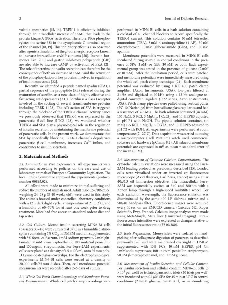
2 Journal of Diabetes Research
volatile anesthetics [15, 16]. TREK-1 is eiciently inhibitedthrough an intracellular increase of cAMP that leads to theprotein kinase A (PKA) activation.herefore, PKA phospho-rylates the serine 333 in the cytoplasmic C-terminal regionof the channel [18, 19]. his inhibitory efect is also observedater agonist stimulation of the-adrenergic receptors knownto increase intracellular cAMP contents [20]. Incretin hor-mones like GLP1 and gastric inhibitory polypeptide (GIP)are also able to increase cAMP by activation of PKA [21].he role of incretins in insulin secretion can therefore be theconsequence of both an increase of cAMP and the activationof the phosphorylation of key proteins involved in regulationof insulin exocytosis [22].
Recently, we identiied a peptide named spadin (SPA), apartial sequence of the propeptide (PE) released during thematuration of sortilin, as a new class of highly efective andfast acting antidepressants (AD). Sortilin is a class 1 receptorinvolved in the sorting of several transmembrane proteinsincluding TREK-1 [23]. he AD action of SPA is triggeredthrough the blockade of the TREK-1 channel activity. Sincewe previously observed that TREK-1 was expressed in thepancreatic -cell line -TC3 [23], we wondered whetherTREK-1 and SPA play a physiological role in the regulationof insulin secretion by maintaining the membrane potentialof pancreatic cells. In the present work, we demonstrate thatSPA by speciically blocking TREK-1 channels depolarizes
pancreatic -cell membranes, increases Ca2+ inlux, andcontributes to insulin secretion.
2. Materials and Methods
2.1. Animals for In Vivo Experiments. All experiments wereperformed according to policies on the care and use oflaboratory animals of European Community Legislation.helocal Ethics Committee approved the experiments (protocolnumber 00893.02).
All eforts were made to minimize animal sufering andreduce the number of animals used.AdultmaleC57/Bl6mice,weighing 24–28 g (8–10 weeks old), were used in this study.he animals housed under controlled laboratory conditionswith a 12 h dark-light cycle, a temperature of 21 ± 2∘C, anda humidity of 60–70% for at least one week prior to drugtreatment. Mice had free access to standard rodent diet andtap water.
2.2. Cell Culture. Mouse insulin secreting MIN6-B1 cells(passages 35–45) were cultured at 37∘C in a humidiied atmo-sphere containing 5%CO2 in DMEMmedium supplementedwith 5% foetal calf serum, 1mM sodium pyruvate, 2mM glu-tamate, 50mM 2-mercaptoethanol, 100 units/ml penicillin,and 100mg/ml streptomycin. For Fura-2AM experiments,cells were plated at a density of 1.5× 10⁵/ml, onto 25mmpoly-D Lysine-coated glass coverslips. For the electrophysiologicalexperiments MIN6-B1 cells were seeded at a density of20,000 cells/35mm dishes. hen, cell membrane potentialmeasurements were recorded ater 2–6 days of culture.
2.3.Whole Cell Patch ClampRecordings andMembrane Poten-tial Measurements. Whole cell patch clamp recordings were
performed in MIN6-B1 cells in a bath solution containinga cocktail of K+ channel blockers to record speciically theTREK-1 current. his solution contains 10mM tetraethylammonium (TEA), 3mM 4-aminopyridine (4-AP), 50 nMcharybdotoxin, 10mM glibenclamide (Glib), and 100 nMapamin.
Membrane potentials were measured in MIN6-B1 cellsincubated during 45min in control conditions in the pres-ence of SPA (1M) or Glib (10M) or both. Each experi-mental group was tested in the presence of glucose (2mMor 10mM). Ater the incubation period, cells were patchedand membrane potentials were immediately measured usingthe whole cell patch clamp technique [24]. Each membranepotential was evaluated by using a RK 400 patch clampampliier (Axon Instruments, USA), low-pass iltered at3 kHz and digitized at 10 kHz using a 12-bit analogue-to-digital converter Digidata (1322 series, Axon Instruments,USA). Patch clamp pipettes were pulled using vertical puller(PC-10, Narishige) from borosilicate glass capillaries and hada resistance of 3–5MΩ. he bath solution contained (in mM)150 NaCl, 5 KCl, 3 MgCl2, 1 CaCl2, and 10 HEPES adjustedto pH 7.4 with NaOH. he pipette solution contained (inmM) 155 KCl, 3 MgCl2, 5 EGTA, and 10 HEPES adjusted topH 7.2 with KOH. All experiments were performed at roomtemperature (21-22∘C).Data acquisitionwas carried out usinga microcomputer (Dell Pentium) witch used commercialsotware and hardware (pClamp 8.2). All values ofmembranepotentials are expressed in mV as mean ± standard error ofthe mean (SEM).
2.4. Measurement of Cytosolic Calcium Concentrations. hecytosolic calcium variations were measured using the Fura-2AM loading protocol as previously described [25]. Loadedcells were visualized under an inverted epi-luorescencemicroscope (AxioObserver, Carl Zeiss, France) using a Fluar40x/1.3 oil immersion objective. he intracellular Fura-2AM was sequentially excited at 340 and 380 nm with aXenon lamp through a high-speed multiilter wheel. Foreach excitation wavelength, the luorescence emission wasdiscriminated by the same 400 LP dichroic mirror and a510/40 bandpass ilter. Fluorescence images were acquiredevery 10 sec on an EMCCD camera (Cascade 512, RoperScientiic, Evry, France). Calcium image analyses were madeusing MetaMorph, MetaFluor (Universal Imaging). Fura-2luorescence intensities were expressed as changes relative tothe initial luorescence ratio (F340/380).
2.5. Islets Preparation. Mouse islets were isolated by hand-picking ater collagenase digestion of pancreas as describedpreviously [26] and were maintained overnight in DMEMsupplemented with 10% FCS, 10mM HEPES, pH 7.4,1mMsodiumpyruvate, 100 units/ml penicillin-streptomycin,50 M -mercaptoethanol, and 11mM glucose.
2.6. Measurement of Insulin Secretion and Cellular Content.For insulin secretion and cellular content, MIN6-B1 cells (5× 10⁵ per well) or isolated pancreatic islets (20 islets per well)were incubated with 0.1 MSPA for 45min at 37∘C in controlconditions (2.8mM glucose, 5mM KCl) or in stimulating
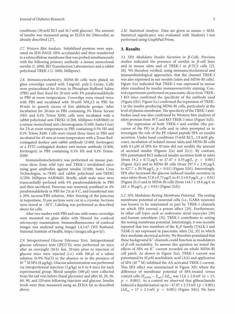
Journal of Diabetes Research 3
conditions (30mM KCl and 16.7mM glucose). he amountof insulin was measured using an ELISA kit (Mercodia) asalready described [27].
2.7. Western Blot Analysis. Solubilized proteins were sepa-rated on SDS-PAGE (10% acrylamide) and then transferredto a nitrocellulosemembrane that was probed simultaneouslywith the following primary antibody: a mouse monoclonalsortilin (1 : 1000, BD Transduction Laboratories) and a rabbitpolyclonal TREK-1 (1 : 1000, Millipore).
2.8. Immunocytochemistry. MIN6-B1 cells were plated onglass coverslips coated with 2mg/mL poly-L-Lysine. Cellswere preincubated for 10min in Phosphate-Bufered Saline(PBS) and then ixed for 20min with 4% paraformaldehydein PBS at room temperature. Coverslips were rinsed twicewith PBS and incubated with 50mM NH4Cl in PBS for10min to quench excess of free aldehyde groups. Aterincubation for 20min in PBS containing 3% Horse Serum(HS) and 0.1% Triton X100, cells were incubated with arabbit polyclonal anti-TREK1 (1/200, Millipore #AB5860) ora mouse monoclonal anti-chromogranin (1/400, Santa Cruz)for 2 h at room temperature in PBS containing 0.5% HS and0.1% Triton-X100. Cells were rinsed three times in PBS andincubated for 45min at room temperature with a Texas-Red-conjugated donkey anti-rabbit antibody (1/400, Invitrogen)or a FITC-conjugated donkey anti-mouse antibody (1/400,Invitrogen) in PBS containing 0.5% HS and 0.1% Triton-X100.
Immunohistochemistry was performed on mouse pan-creas slices from wild type and TREK-1 invalidated miceusing goat antibodies against insulin (1/400, Santa CruzTechnologies, sc-7838) and rabbit polyclonal anti-TREK1(1/200, Millipore #AB5860). Briely, adult male mice weretranscardially perfused with 4% paraformaldehyde in PBSand then sacriiced. Pancreas was removed, postixed in 4%paraformaldehyde in PBS for 2 h at 4∘C, and transferred intoa 20% sucrose/PBS solution. Ater freezing of the pancreasin isopentane, 35 m sections were cut in a cryostat. Sectionswere stored at −20∘C. Labeling was performed as describedabove for cells.
Ater two washes with PBS and one with water, coverslipswere mounted on glass slides with Mowiol for confocalmicroscopy examination. Immunoluorescence of confocalimages was analysed using ImageJ 1.4.3.67 (WS Rasband,National Institute of Health, https://imagej.nih.gov/ij/).
2.9. Intraperitoneal Glucose Tolerance Tests. Intraperitonealglucose tolerance tests (IPGTTs) were performed on miceater an overnight (16 h) fast. 20min prior to injection ofglucose mice were injected (i.v.) with 100l of a salinesolution (0.9% NaCl) in the absence or in the presence of10−6MSPA (8 g/kg). Glucose administrationwas performedvia intraperitoneal injection (2 g/kg) in 6 to 8 mice for eachexperimental group. Blood samples (100l) were collectedfrom the tail vein before (basal glycemia) and ater 10, 20, 30,60, 90, and 120min following injection and glucose. Insulinlevels were then measured using an ELISA kit as describedabove.
2.10. Statistical Analysis. Data are given as means ± SEM.Statistical signiicance was evaluated with Student’s -testperformed using GraphPad Prism.
3. Results
3.1. SPA Modulates Insulin Secretion in -Cells. Previousstudies indicated the presence of sortilin in -cell linesand in mouse islets and of TREK-1 in -TC3 cells [23,28]. We therefore veriied, using immunocytochemical andimmunohistological approaches, that the channel TREK-1was also expressed in our models (islets and MIN6-B1 cells).Figure 1(a) indicated that TREK-1 was expressed in mouseislets visualized by insulin immunoreactivity staining. Con-trol experiments performed on pancreatic slices from TREK-1 KO mice conirmed the speciicity of the antibody used(Figure 1(b)). Figure 1(c) conirmed the expression of TREK-1 in the insulin producing MIN6-B1 cells, particularly at thelevel of plasmamembrane.he speciicity of the TREK-1 anti-bodies used was also conirmed by Western blot analyses ofislets proteins fromWT and KO-TREK-1 mice (Figure 1(d)).
he expression of both TREK-1 and sortilin (the pre-cursor of the PE) in -cells and in islets prompted us toinvestigate the role of the PE related peptide SPA on insulinsecretion. Under basal conditions (5mM KCL, 2.8mM glu-cose), incubation of isolated mouse islets and MIN6-B1 cellswith 0.1M of SPA for 45min did not modify the amountof secreted insulin (Figures 2(a) and 2(c)). By contrast,SPA potentiated KCl-induced insulin secretion both in islets(from 19.2 ± 0.72 g/L to 27.47 ± 0.35 g/L, < 0.001)(Figure 2(a)) and in MIN6-B1 cells (from 297.5 ± 2.92 g/Lto 427.7 ± 20.58 g/L, < 0.01) (Figure 2(b)). Interestingly,SPA also increased the glucose-induced insulin secretion inmice islets (from 37.8±0.73 g/L to 47.2±0.9 g/L, < 0.01)(Figure 2(c)) and inMIN6-B1 cells (from 144.7±18.6 g/L to241 ± 30 g/L, < 0.01) (Figure 2(d)).
3.2. SPA Modulates Resting Membrane Potential. he restingmembrane potential of neuronal cells (i.e., GABA neurons)was known to be maintained in part by TREK-1 channelson which SPA exerted a potent efect [29]. Furthermore,in other cell types such as embryonic atrial myocytes [30]and human osteoblasts [31], TREK-1 contributes to settingthe resting membrane potential. Interestingly, it was recentlyreported that two members of the K2P family (TALK-1 andTASK-1) are expressed in pancreatic islets [32, 33] in whichtheymodulate electrical activity.We therefore postulated thatthese background K+ channels could function as modulatorsof -cell excitability. To answer this question we tested theefects of SPA on K+ current recorded on whole MIN6-B1cell patch. As shown in Figure 3(a), TREK-1 current waspotentiated by 10 M arachidonic acid (AA) and application
of SPA (10−6M) inhibited the AA activated TREK-1 current.his SPA efect was summarized in Figure 3(b) where thediference of membrane potential of SPA-treated versuscontrol cells (mSPA − mC) Δm was 12.6 ± 2.0mV ( = 15, < 0.001). As a control we observed that glibenclamideinduced a depolarization up to −47.07 ± 2.33mV ( < 0.001)(Δm = 17 ± 2.3mV, < 0.001) (Figure 3(b)). We have
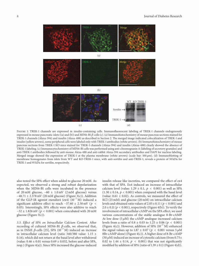
4 Journal of Diabetes Research
Insulin MergeTREK-1
(a)
Insulin TREK-1 Merge
(b)
TREK-1 TREK-1/ChromA
(c)
WT
Sortilin
KO-TREK-1
TREK-1
(d)
Figure 1: TREK-1 channels are expressed in insulin-containing cells. Immunoluorescent labeling of TREK-1 channels endogenouslyexpressed inmouse pancreatic islets ((a) and (b)) andMIN6-B1 -cells (c). (a) Immunohistochemistry of mouse pancreas sections stained forTREK-1 channels (Alexa-594) and insulin (Alexa-488) as described in Section 2. he merged image indicated colocalization of TREK-1 andinsulin (yellow arrows), some peripheral cells were labeled only with TREK-1 antibodies (white arrows). (b) Immunohistochemistry of mousepancreas sections from TREK-1 KO mice stained for TREK-1 channels (Alexa-594) and insulin (Alexa-488) clearly showed the absence ofTREK-1 labeling. (c) Immunocytochemistry ofMIN6-B1 cells was performed using anti-chromogranin A (labeling of secretory granules) andanti-TREK-1 antibodies followed by anti-mouse Alexa-488 and anti-rabbit Alexa-594 secondary antibodies and DAPI for nucleus labeling.Merged image showed the expression of TREK-1 at the plasma membrane (white arrows) (scale bar: 100 m). (d) Immunoblotting ofmembrane homogenates from islets from WT and KO-TREK-1 mice, with anti-sortilin and anti-TREK-1, reveals a protein of 50 kDa forTREK-1 and 95 kDa for sortilin, respectively.
also tested the SPA efect when added to glucose 20mM. Asexpected, we observed a strong and robust depolarizationwhen the MIN6-B1 cells were incubated in the presenceof 20mM glucose, −60 ± 1.0mV (2mM glucose) versus−48.71 ± 2.378mV (20mM glucose) (Figure 3(c)). Additionof the GLP-1R agonist exendin4 (ex4) (10−7M) induced asigniicant additive efect to reach −37.60 ± 2.50mV ( <0.05). Interestingly, SPA efects were also additive to reach−32 ± 1.826mV ( < 0.001) when coincubated with 20mMglucose (Figure 3(c)).
3.3. Efect of SPA on Intracellular Calcium Content. Aterrecording of cultured MIN6-B1 -cells, we observed that,as in INS1E -cells [25], SPA (10−7M) induced an increasein intracellular calcium level (ratio 340/380 value: 1.15 ±0.16), which did not return at the basal level ater withdrawal(value: 0.46 ± 0.01 versus 0.69 ± 0.032, before and ater SPA,resp.) (Figure 4(a)). Since SPA increased the glucose-induced
insulin release like incretins, we compared the efect of ex4with that of SPA. Ex4 induced an increase of intracellularcalcium level (value: 1.29 ± 0.1, < 0.001) as well as SPA(1.30 ± 0.14, < 0.001) when compared with the basal level(value: 0.61 ± 0.02). As controls, we measured the efect ofKCl (25mM) and glucose (20mM) on intracellular calciumlevels and obtained ratio values of 2.05±0.11 ( < 0.001) and2.0 ± 0.11 ( < 0.001), respectively (Figure 4(b)). To verify theinvolvement of intracellular cAMP on the SPA efect, we usedvarious concentrations of the stable analogue 8-Br-cAMP.At low dose (5M) the cAMP analogue increased calciumlevels from a ratio of 0.8 ± 0.03 to 1.25 ± 0.06 ( < 0.001)(Figure 4(c)). However, addition of SPA (10−7M) enhancedthe signal values up to 1.87 ± 0.07 ( < 0.001 versus 5 M8Br-cAMP alone) (Figure 4(c)). A higher dose of 8-Br-cAMP(50 M) induced an increase of cytosolic calcium (from 0.75±0.02 to 1.44 ± 0.14, < 0.001) that was not signiicantlymodiied by addition of SPA (ratio of 1.39 ± 0.1) (Figure 4(d)).
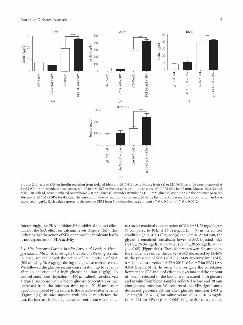
Journal of Diabetes Research 5
KC
l5
mM
KC
l5
mM
+ S
PA
KC
l30
mM
KC
l30
mM
+ S
PA
∗∗∗Islets
0
10
20
30
Insu
lin
(
g/L
)
(a)
KC
l5
mM
KC
l5
mM
+ S
PA
KC
l30
mM
KC
l30
mM
+ S
PA
∗∗
MIN6-B1
0
100
200
300
400
500
Insu
lin
(
g/L
)
(b)
∗∗
glu2.8
mM
glu2.8
mM
+ S
PA
glu16
.7m
M
glu16
.7m
M +
SPA
0
10
20
30
40
50
Insu
lin
(
g/L
)
Islets
(c)
∗∗gl
u2.8
mM
glu2.8
mM
+ S
PA
glu16
.7m
M
glu16
.7m
M +
SPA
0
100
200
300
Insu
lin
(
g/L
)
MIN6-B1
(d)
Figure 2: Efects of SPA on insulin secretion from isolated islets and MIN6-B1 cells. Mouse islets (a) or MIN6-B1 cells (b) were incubated at5mM (Cont) or stimulating concentration of 30mM KCl in the presence or in the absence of 10−7M SPA for 45min. Mouse islets (c) andMIN6-B1 cells (d) were incubated under basal (2.8mM glucose) or under stimulating (16.7mM glucose) conditions in the presence or in theabsence of 10−7M of SPA for 45min. he amount of secreted insulin was normalized using the intracellular insulin concentration and wasexpressed in g/L. Each value represents the mean ± SEM from 3 independent experiments (∗∗ < 0.01 and ∗∗∗ < 0.001).
Interestingly, the PKA inhibitor H89 inhibited the ex4 efectbut not the SPA efect on calcium levels (Figure 4(e)). hisindicates that the action of SPA on intracellular calcium levelsis not dependent on PKA activity.
3.4. SPA Improves Plasma Insulin Level and Leads to Hypo-glycemia in Mice. To investigate the role of SPA on glycemiain mice, we challenged the action of i.v. injection of SPA(100 L of 1M, 8 g/kg) during the glucose tolerance test.We followed the glucose serum concentration up to 120minater i.p. injection of a high glucose solution (2 g/kg). Incontrol conditions (injection of 100 L saline), we observeda typical response with a blood glucose concentration thatincreased from the injection time up to 20–30min aterinjection followed by the return to the basal level ater 120min(Figure 5(a)). In mice injected with SPA 20min before thetest, the increase in blood glucose concentration was smaller
to reach amaximal concentration of 332.6±31.26mg/dL ( =7) compared to 404.1 ± 16.32mg/dL ( = 9) in the controlcondition ( < 0.05) (Figure 5(a)) at 30min. At 60min, theglycemia remained statistically lower in SPA-injected mice(314.8± 26.04mg/dL, = 9 versus 229.3±20.55mg/dL, = 7, < 0.05) (Figure 5(a)). hese diferences were illustrated bythe smaller area under the curve (AUC) decreased by 28.46%in the presence of SPA (20205 ± 1449 arbitrary unit (AU), = 9 for control versus 14455±2015AU, = 7 for SPA) ( <0.05) (Figure 5(b)). In order to investigate the correlationbetween the SPA-induced efect on glycemia and the amountof insulin released in the blood, we measured both glucoseand insulin from blood samples collected before and 20minater glucose injection. We conirmed that SPA signiicantlydecreased glycemia 20min ater glucose injection (503 ±12.9mg/dL ( = 13) for saline versus 450.4 ± 10.15mg/dL( = 13) for SPA) ( < 0.005) (Figure 5(c)). In parallel,
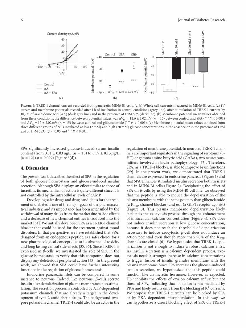
6 Journal of Diabetes Research
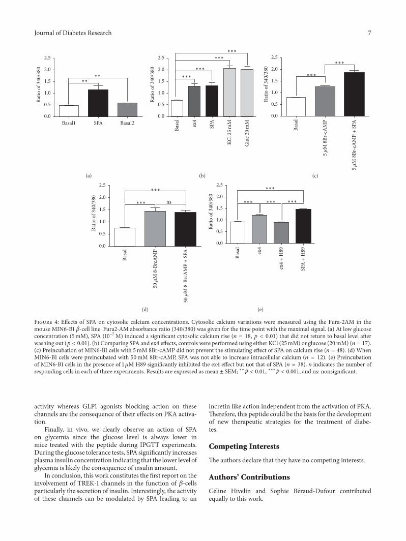
Journal of Diabetes Research 7
Basal1 SPA Basal2
∗∗∗∗
0.0
0.5
1.0
1.5
2.0
2.5
Rat
io o
f 34
0/38
0
(a)B
asal
ex4
SPA
KC
l25
mM
Glu
c20
mM
∗∗∗
∗∗∗
∗∗∗
∗∗∗
0.0
0.5
1.0
1.5
2.0
2.5
Rat
io o
f 34
0/38
0
(b)
Bas
al
5
M 8
Br-
cAM
P
5
M 8
Br-
cAM
P +
SPA
∗∗∗
∗∗∗
0.0
0.5
1.0
1.5
2.0
2.5
Rat
io o
f 34
0/38
0
(c)
Bas
al
50
50
M
8-B
rcA
MP
M
8-B
rcA
MP
+ S
PA∗∗∗
∗∗∗
0.0
0.5
1.0
1.5
2.0
2.5
ns
Rat
io o
f 34
0/38
0
(d)
Bas
al
ex4
ex4
+ H
89
SPA
+ H
89
∗∗∗
∗∗∗ ∗∗∗ ∗∗∗
0.0
0.5
1.0
1.5
2.0
2.5
Rat
io o
f 34
0/38
0
(e)
Figure 4: Efects of SPA on cytosolic calcium concentrations. Cytosolic calcium variations were measured using the Fura-2AM in themouse MIN6-B1 -cell line. Fura2-AM absorbance ratio (340/380) was given for the time point with the maximal signal. (a) At low glucoseconcentration (5mM), SPA (10−7M) induced a signiicant cytosolic calcium rise ( = 18, < 0.01) that did not return to basal level aterwashing out ( < 0.01). (b) Comparing SPA and ex4 efects, controls were performed using either KCl (25mM) or glucose (20mM) ( = 17).(c) Preincubation of MIN6-B1 cells with 5mM 8Br-cAMP did not prevent the stimulating efect of SPA on calcium rise ( = 48). (d) WhenMIN6-B1 cells were preincubated with 50mM 8Br-cAMP, SPA was not able to increase intracellular calcium ( = 12). (e) Preincubationof MIN6-B1 cells in the presence of 1 M H89 signiicantly inhibited the ex4 efect but not that of SPA ( = 38). indicates the number ofresponding cells in each of three experiments. Results are expressed as mean ± SEM; ∗∗ < 0.01, ∗∗∗ < 0.001, and ns: nonsigniicant.
activity whereas GLP1 agonists blocking action on thesechannels are the consequence of their efects on PKA activa-tion.
Finally, in vivo, we clearly observe an action of SPAon glycemia since the glucose level is always lower inmice treated with the peptide during IPGTT experiments.During the glucose tolerance tests, SPA signiicantly increasesplasma insulin concentration indicating that the lower level ofglycemia is likely the consequence of insulin amount.
In conclusion, this work constitutes the irst report on theinvolvement of TREK-1 channels in the function of -cellsparticularly the secretion of insulin. Interestingly, the activityof these channels can be modulated by SPA leading to an
incretin like action independent from the activation of PKA.herefore, this peptide could be the basis for the developmentof new therapeutic strategies for the treatment of diabe-tes.
Competing Interests
he authors declare that they have no competing interests.
Authors’ Contributions
Celine Hivelin and Sophie Beraud-Dufour contributedequally to this work.
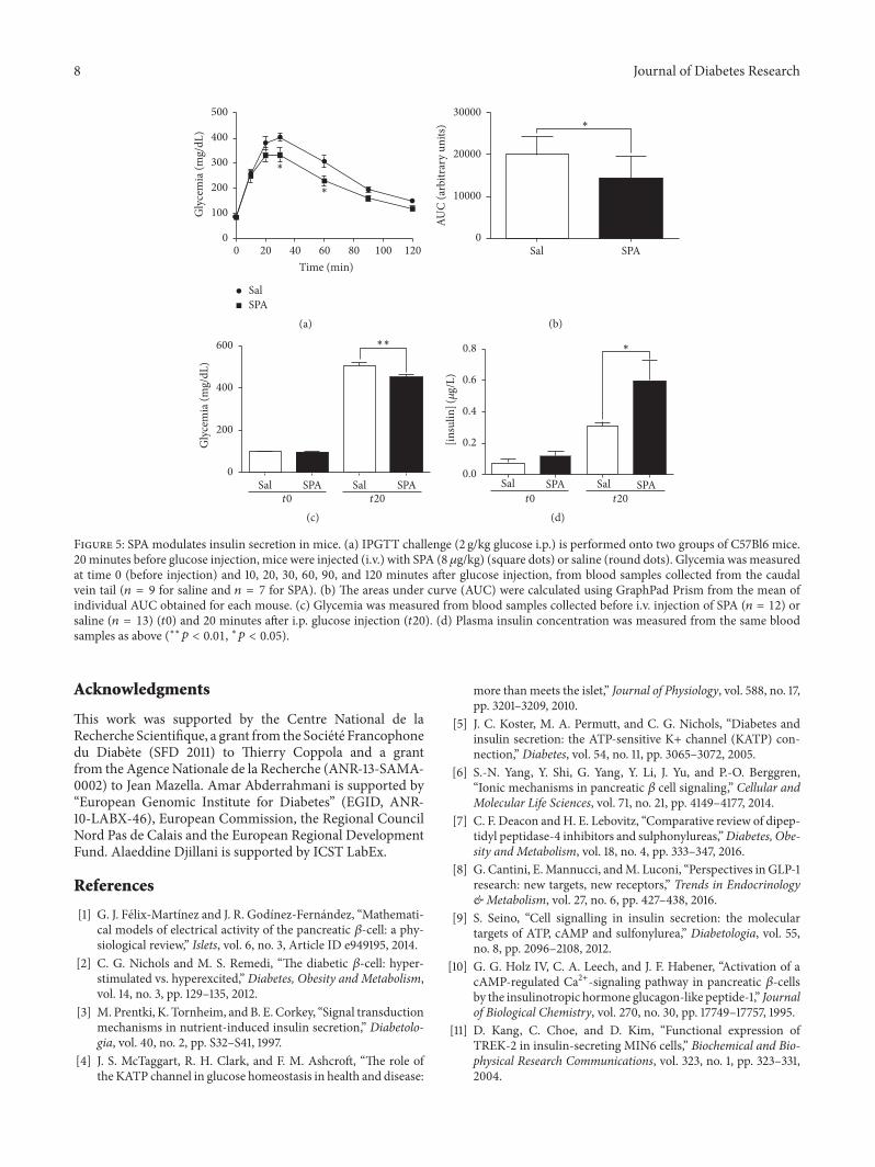
8 Journal of Diabetes Research
Gly
cem
ia (
mg/
dL
)∗
∗
20 40 60 80 100 1200
Time (min)
0
100
200
300
400
500
Sal
SPA
(a)
Sal SPA
∗
0
10000
20000
30000
AU
C (
arb
itra
ry u
nit
s)
(b)
Sal SPA Sal SPAt0 t20
0
200
400
600
Gly
cem
ia (
mg/
dL
)
∗∗
(c)
Sal SPA Sal SPAt0 t20
∗
0.0
0.2
0.4
0.6
0.8
[in
suli
n](
g/L
)
(d)
Figure 5: SPA modulates insulin secretion in mice. (a) IPGTT challenge (2 g/kg glucose i.p.) is performed onto two groups of C57Bl6 mice.20 minutes before glucose injection, mice were injected (i.v.) with SPA (8g/kg) (square dots) or saline (round dots). Glycemia was measuredat time 0 (before injection) and 10, 20, 30, 60, 90, and 120 minutes ater glucose injection, from blood samples collected from the caudalvein tail ( = 9 for saline and = 7 for SPA). (b) he areas under curve (AUC) were calculated using GraphPad Prism from the mean ofindividual AUC obtained for each mouse. (c) Glycemia was measured from blood samples collected before i.v. injection of SPA ( = 12) orsaline ( = 13) (0) and 20 minutes ater i.p. glucose injection (20). (d) Plasma insulin concentration was measured from the same bloodsamples as above (∗∗ < 0.01, ∗ < 0.05).
Acknowledgments
his work was supported by the Centre National de laRecherche Scientiique, a grant from the Societe Francophonedu Diabete (SFD 2011) to hierry Coppola and a grantfrom the Agence Nationale de la Recherche (ANR-13-SAMA-0002) to Jean Mazella. Amar Abderrahmani is supported by“European Genomic Institute for Diabetes” (EGID, ANR-10-LABX-46), European Commission, the Regional CouncilNord Pas de Calais and the European Regional DevelopmentFund. Alaeddine Djillani is supported by ICST LabEx.
References
[1] G. J. Felix-Martınez and J. R. Godınez-Fernandez, “Mathemati-cal models of electrical activity of the pancreatic -cell: a phy-siological review,” Islets, vol. 6, no. 3, Article ID e949195, 2014.
[2] C. G. Nichols and M. S. Remedi, “he diabetic -cell: hyper-stimulated vs. hyperexcited,” Diabetes, Obesity and Metabolism,vol. 14, no. 3, pp. 129–135, 2012.
[3] M. Prentki, K. Tornheim, andB. E. Corkey, “Signal transductionmechanisms in nutrient-induced insulin secretion,” Diabetolo-gia, vol. 40, no. 2, pp. S32–S41, 1997.
[4] J. S. McTaggart, R. H. Clark, and F. M. Ashcrot, “he role ofthe KATP channel in glucose homeostasis in health and disease:
more thanmeets the islet,” Journal of Physiology, vol. 588, no. 17,pp. 3201–3209, 2010.
[5] J. C. Koster, M. A. Permutt, and C. G. Nichols, “Diabetes andinsulin secretion: the ATP-sensitive K+ channel (KATP) con-nection,” Diabetes, vol. 54, no. 11, pp. 3065–3072, 2005.
[6] S.-N. Yang, Y. Shi, G. Yang, Y. Li, J. Yu, and P.-O. Berggren,“Ionic mechanisms in pancreatic cell signaling,” Cellular andMolecular Life Sciences, vol. 71, no. 21, pp. 4149–4177, 2014.
[7] C. F. Deacon andH. E. Lebovitz, “Comparative review of dipep-tidyl peptidase-4 inhibitors and sulphonylureas,”Diabetes, Obe-sity and Metabolism, vol. 18, no. 4, pp. 333–347, 2016.
[8] G. Cantini, E.Mannucci, andM. Luconi, “Perspectives in GLP-1research: new targets, new receptors,” Trends in Endocrinology& Metabolism, vol. 27, no. 6, pp. 427–438, 2016.
[9] S. Seino, “Cell signalling in insulin secretion: the moleculartargets of ATP, cAMP and sulfonylurea,” Diabetologia, vol. 55,no. 8, pp. 2096–2108, 2012.
[10] G. G. Holz IV, C. A. Leech, and J. F. Habener, “Activation of acAMP-regulated Ca2+-signaling pathway in pancreatic -cellsby the insulinotropic hormone glucagon-like peptide-1,” Journalof Biological Chemistry, vol. 270, no. 30, pp. 17749–17757, 1995.
[11] D. Kang, C. Choe, and D. Kim, “Functional expression ofTREK-2 in insulin-secreting MIN6 cells,” Biochemical and Bio-physical Research Communications, vol. 323, no. 1, pp. 323–331,2004.
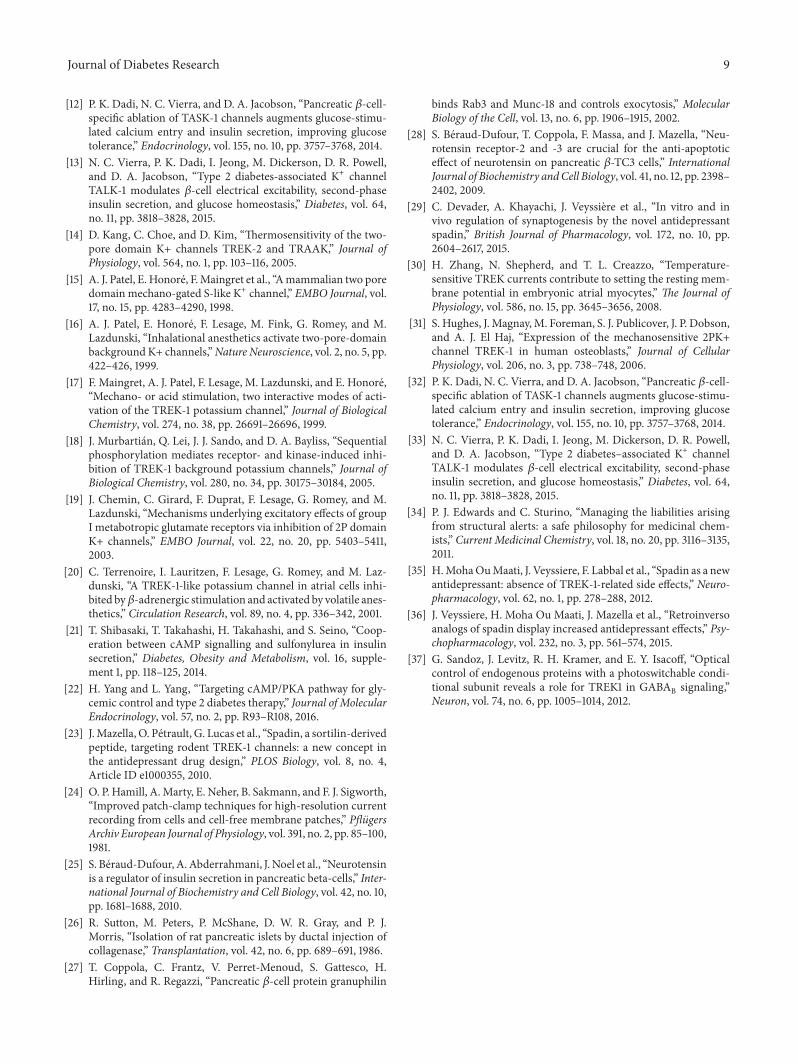
Journal of Diabetes Research 9
[12] P. K. Dadi, N. C. Vierra, and D. A. Jacobson, “Pancreatic -cell-speciic ablation of TASK-1 channels augments glucose-stimu-lated calcium entry and insulin secretion, improving glucosetolerance,” Endocrinology, vol. 155, no. 10, pp. 3757–3768, 2014.
[13] N. C. Vierra, P. K. Dadi, I. Jeong, M. Dickerson, D. R. Powell,and D. A. Jacobson, “Type 2 diabetes-associated K+ channelTALK-1 modulates -cell electrical excitability, second-phaseinsulin secretion, and glucose homeostasis,” Diabetes, vol. 64,no. 11, pp. 3818–3828, 2015.
[14] D. Kang, C. Choe, and D. Kim, “hermosensitivity of the two-pore domain K+ channels TREK-2 and TRAAK,” Journal ofPhysiology, vol. 564, no. 1, pp. 103–116, 2005.
[15] A. J. Patel, E. Honore, F.Maingret et al., “Amammalian two poredomain mechano-gated S-like K+ channel,” EMBO Journal, vol.17, no. 15, pp. 4283–4290, 1998.
[16] A. J. Patel, E. Honore, F. Lesage, M. Fink, G. Romey, and M.Lazdunski, “Inhalational anesthetics activate two-pore-domainbackground K+ channels,”Nature Neuroscience, vol. 2, no. 5, pp.422–426, 1999.
[17] F. Maingret, A. J. Patel, F. Lesage, M. Lazdunski, and E. Honore,“Mechano- or acid stimulation, two interactive modes of acti-vation of the TREK-1 potassium channel,” Journal of BiologicalChemistry, vol. 274, no. 38, pp. 26691–26696, 1999.
[18] J. Murbartian, Q. Lei, J. J. Sando, and D. A. Bayliss, “Sequentialphosphorylation mediates receptor- and kinase-induced inhi-bition of TREK-1 background potassium channels,” Journal ofBiological Chemistry, vol. 280, no. 34, pp. 30175–30184, 2005.
[19] J. Chemin, C. Girard, F. Duprat, F. Lesage, G. Romey, and M.Lazdunski, “Mechanisms underlying excitatory efects of groupI metabotropic glutamate receptors via inhibition of 2P domainK+ channels,” EMBO Journal, vol. 22, no. 20, pp. 5403–5411,2003.
[20] C. Terrenoire, I. Lauritzen, F. Lesage, G. Romey, and M. Laz-dunski, “A TREK-1-like potassium channel in atrial cells inhi-bited by-adrenergic stimulation and activated by volatile anes-thetics,” Circulation Research, vol. 89, no. 4, pp. 336–342, 2001.
[21] T. Shibasaki, T. Takahashi, H. Takahashi, and S. Seino, “Coop-eration between cAMP signalling and sulfonylurea in insulinsecretion,” Diabetes, Obesity and Metabolism, vol. 16, supple-ment 1, pp. 118–125, 2014.
[22] H. Yang and L. Yang, “Targeting cAMP/PKA pathway for gly-cemic control and type 2 diabetes therapy,” Journal of MolecularEndocrinology, vol. 57, no. 2, pp. R93–R108, 2016.
[23] J.Mazella, O. Petrault, G. Lucas et al., “Spadin, a sortilin-derivedpeptide, targeting rodent TREK-1 channels: a new concept inthe antidepressant drug design,” PLOS Biology, vol. 8, no. 4,Article ID e1000355, 2010.
[24] O. P. Hamill, A.Marty, E. Neher, B. Sakmann, and F. J. Sigworth,“Improved patch-clamp techniques for high-resolution currentrecording from cells and cell-free membrane patches,” PlugersArchiv European Journal of Physiology, vol. 391, no. 2, pp. 85–100,1981.
[25] S. Beraud-Dufour, A.Abderrahmani, J. Noel et al., “Neurotensinis a regulator of insulin secretion in pancreatic beta-cells,” Inter-national Journal of Biochemistry and Cell Biology, vol. 42, no. 10,pp. 1681–1688, 2010.
[26] R. Sutton, M. Peters, P. McShane, D. W. R. Gray, and P. J.Morris, “Isolation of rat pancreatic islets by ductal injection ofcollagenase,” Transplantation, vol. 42, no. 6, pp. 689–691, 1986.
[27] T. Coppola, C. Frantz, V. Perret-Menoud, S. Gattesco, H.Hirling, and R. Regazzi, “Pancreatic -cell protein granuphilin
binds Rab3 and Munc-18 and controls exocytosis,” MolecularBiology of the Cell, vol. 13, no. 6, pp. 1906–1915, 2002.
[28] S. Beraud-Dufour, T. Coppola, F. Massa, and J. Mazella, “Neu-rotensin receptor-2 and -3 are crucial for the anti-apoptoticefect of neurotensin on pancreatic -TC3 cells,” InternationalJournal of Biochemistry andCell Biology, vol. 41, no. 12, pp. 2398–2402, 2009.
[29] C. Devader, A. Khayachi, J. Veyssiere et al., “In vitro and invivo regulation of synaptogenesis by the novel antidepressantspadin,” British Journal of Pharmacology, vol. 172, no. 10, pp.2604–2617, 2015.
[30] H. Zhang, N. Shepherd, and T. L. Creazzo, “Temperature-sensitive TREK currents contribute to setting the resting mem-brane potential in embryonic atrial myocytes,” e Journal ofPhysiology, vol. 586, no. 15, pp. 3645–3656, 2008.
[31] S. Hughes, J. Magnay,M. Foreman, S. J. Publicover, J. P. Dobson,and A. J. El Haj, “Expression of the mechanosensitive 2PK+channel TREK-1 in human osteoblasts,” Journal of CellularPhysiology, vol. 206, no. 3, pp. 738–748, 2006.
[32] P. K. Dadi, N. C. Vierra, and D. A. Jacobson, “Pancreatic -cell-speciic ablation of TASK-1 channels augments glucose-stimu-lated calcium entry and insulin secretion, improving glucosetolerance,” Endocrinology, vol. 155, no. 10, pp. 3757–3768, 2014.
[33] N. C. Vierra, P. K. Dadi, I. Jeong, M. Dickerson, D. R. Powell,and D. A. Jacobson, “Type 2 diabetes–associated K+ channelTALK-1 modulates -cell electrical excitability, second-phaseinsulin secretion, and glucose homeostasis,” Diabetes, vol. 64,no. 11, pp. 3818–3828, 2015.
[34] P. J. Edwards and C. Sturino, “Managing the liabilities arisingfrom structural alerts: a safe philosophy for medicinal chem-ists,” Current Medicinal Chemistry, vol. 18, no. 20, pp. 3116–3135,2011.
[35] H.MohaOuMaati, J. Veyssiere, F. Labbal et al., “Spadin as a newantidepressant: absence of TREK-1-related side efects,” Neuro-pharmacology, vol. 62, no. 1, pp. 278–288, 2012.
[36] J. Veyssiere, H. Moha Ou Maati, J. Mazella et al., “Retroinversoanalogs of spadin display increased antidepressant efects,” Psy-chopharmacology, vol. 232, no. 3, pp. 561–574, 2015.
[37] G. Sandoz, J. Levitz, R. H. Kramer, and E. Y. Isacof, “Opticalcontrol of endogenous proteins with a photoswitchable condi-tional subunit reveals a role for TREK1 in GABA
Bsignaling,”
Neuron, vol. 74, no. 6, pp. 1005–1014, 2012.

RESEARCH PAPER
In vitro and in vivo
regulation ofsynaptogenesis by the novelantidepressant spadinC Devader, A Khayachi, J Veyssière, H Moha ou Maati*, M Roulot,
S Moreno, M Borsotto, S Martin, C Heurteaux and J Mazella
CNRS, Institut de Pharmacologie Moléculaire et Cellulaire, UMR 7275, Université de Nice-Sophia
Antipolis, Valbonne, France
CorrespondenceJean Mazella, Institut de
Pharmacologie Moléculaire et
Cellulaire, UMR 7275, Université
de Nice-Sophia Antipolis, 660
route des Lucioles, 06560
Valbonne, France. E-mail:
----------------------------------------------------------------
*Present address: Institut deGénomique Fonctionnelle, UMR5203 CNRS/INSERM/UM1/UM2,141 rue de la Cardonille, 34095Montpellier Cedex 5, France.
----------------------------------------------------------------
Received24 June 2014
Revised10 December 2014
Accepted8 January 2015
BACKGROUND AND PURPOSEWe have described a novel antidepressant peptide, spadin, that acts by blocking the TWIK-related-potassium channel, type 1(TREK-1). Here, we examined possible mechanisms of action of spadin at both molecular and cellular levels.
EXPERIMENTAL APPROACHESEffects of spadin were measured in primary cultures of neurons or tissues from mice injected i.v. with spadin. Western blots,qPCR, histochemical and electrophysiological techniques were used.
KEY RESULTSIn vitro, spadin increased neuronal membrane potential and activated both the MAPK and PI3K signalling pathways, in a time-and concentration-dependent manner. The latter pathway was involved in the protective effect of spadin againststaurosporine-induced apoptosis. Also, spadin enhanced both mRNA expression and protein of two markers ofsynaptogenesis, the post-synaptic density protein of 95 kDalton (PSD-95) and synapsin. We confirmed these effects onsynaptogenesis by the observation that spadin treatment significantly increased the proportion of mature spines in corticalneurons. Finally, in vivo injections of spadin led to a rapid increase in both mRNA expression and protein level of brain-derivedneurotrophic factor (BDNF) in the hippocampus, confirming the antidepressant action of the peptide. We argue for a newrole of spadin in synaptogenesis as both PSD-95 and synapsin mRNA expression and protein levels were further enhanced inthe hippocampus, following treatment in vivo with the peptide.
CONCLUSIONS AND IMPLICATIONSThese findings provide new mechanisms of action for the rapidly acting antidepressant peptide spadin by stimulatingexpression of BDNF and synaptic proteins, both in vitro and in vivo.
AbbreviationsBDNF, brain-derived neurotrophic factor; LY294002, 2-morpholin-4-yl-8-phenylchromen-4-one; mTOR, mammaliantarget of rapamycin; NTSR3, neurotensin receptor-3; PD98059, 2′-amino-3′-methoxyflavone; PSD-95, post-synapticdensity protein of 95 kDalton; TREK-1, TWIK-related-potassium channel, type 1
BJP British Journal ofPharmacology
DOI:10.1111/bph.13083www.brjpharmacol.org
2604 British Journal of Pharmacology (2015) 172 2604–2617 © 2015 The British Pharmacological Society
Introduction
The origins and causes of depression are diverse, and there-
fore do not facilitate the diagnosis of the pathology. Mol-
ecules developed to treat depression include the tricyclic
antidepressants, inhibitors of MAO-A or 5-HT selective reup-
take inhibitors (see Berton and Nestler, 2006). Nevertheless,
many other atypical antidepressant drugs were also devel-
oped such as mianserine, tradozone (Boschmans et al., 1987;
Fagiolini et al., 2012), mirtazapine (Dolder et al., 2012), ago-
melatine (Srinivasan et al., 2012), tianeptine (McEwen and
Olie, 2005), scopolamine (Drevets et al., 2013), ketamine or
lanicemine (Zarate et al., 2006; Sanacora et al., 2014).
The antidepressant drugs do not provide a fully satisfac-
tory treatment, for several important reasons: (i) one-third of
patients are resistant to the drugs (Pacher and Kecskemeti,
2004); (ii) there is a delayed onset of antidepressant drug
action (Fava and Kendler, 2000; Nestler et al., 2002); and (iii)
antidepressant drug treatments have numerous deleterious
side effects (Sicouri and Antzelevitch, 2008; Thase and
Denko, 2008).
Taking into account the mental health of millions of
people worldwide and the associated economic burden, it is
now crucial to develop alternative strategies aimed at devel-
oping novel antidepressants that could potentially show
higher rates of efficacy and lower rates of side effects. We
previously reported that an endogenous peptide of 44 amino
acids (PE) released from the maturation of the neurotensin
receptor-3 (NTSR3) (Mazella et al., 1998; Munck Petersen et al.,
1999), also called sortilin (Petersen et al., 1997), displays
potent antidepressant effects in several tests performed in mice
(Mazella et al., 2010). We isolated an active sequence of 17
amino acids from this peptide, named spadin, which exerts its
antidepressant properties through the blockade of the TWIK-
related-potassium channel, type 1 (TREK-1) (K2P2.1; Mazella
et al., 2010). TREK-1 channels are activated by stretch, poly-
unsaturated fatty acids, warm temperatures, internal acidosis
and volatile anaesthetics (Honore, 2007). They are inhibited
by fluoxetine and blocked by phosphorylation processes.
Deletion of the TREK-1 gene (kcnk2) leads to mice that display
a depression-resistant phenotype, which mimics treatment
with antidepressants (Heurteaux et al., 2006b). Spadin binds
to TREK-1 with an affinity of 10 nM, blocks its activity and
induces its sequestration into cells. The interaction of spadin
with TREK-1 does not show any striking side effects and does
not interfere with any known TREK-1-controlled functions
(Moha Ou Maati et al., 2012). In vivo, spadin increases the
firing rate of serotonergic neurons from the dorsal raphe
nucleus and activates hippocampal neurogenesis (Mazella
et al., 2010). Moreover, NTSR3/sortilin directly interacts with
TREK-1 to regulate its plasma membrane localization (Mazella
et al., 2010). Both proteins are colocalized in neurons within
the dorsal raphe nucleus and have been previously shown to
be expressed in several brain areas known to be involved in
depression including the prefrontal cortex, amygdala, hip-
pocampus, nucleus accumbens, dorsal raphe and hypothala-
mus (Hervieu et al., 2001; Sarret et al., 2003).
Spadin exhibits antidepressant properties, when injected
in mice (Mazella et al., 2010), and induced a rapid hippocam-
pal neurogenesis after a 4 day treatment. However, its cellular
mechanisms of action as well as the demonstration that new
neurons are functional have not yet been characterized.
Moreover, the rapid onset of action of spadin, which can be
compared with the rapidly acting antidepressant ketamine (Li
et al., 2011; Dwyer and Duman, 2013), prompted us to evalu-
ate the effect of spadin on the induction of synaptogenesis
and spine maturation. The mechanisms of action of spadin
were determined in primary cultures of neurons from embry-
onic and post-natal mice. We performed electrophysiological
and biochemical experiments to study the membrane func-
tion and the intracellular signalling of the peptide followed
by qPCR and Western blot analyses of proteins involved in
synaptogenesis and whose expression is altered in mood dis-
orders such as post-synaptic density protein of 95 kDalton
(PSD-95), synapsin and brain-derived neurotrophic factor
(BDNF). Neuronal spine maturation has been visualized using
confocal imaging of GFP-transfected neurons. We confirmed
the effects we observed in vitro, on synaptic proteins by in vivo
injection of spadin followed by protein analysis in samples of
hippocampus and prefrontal cortex.
Tables of Links
TARGETS
Ion channela
TREK-1,. K2P2.1 channel
Enzymesb
Akt
Caspase-3
ERK1/2
mTOR
PI3K
LIGANDS
BDNF
Fluoxetine
Ketamine
LY294002
PD98059
Staurosporine
These Tables list key protein targets and ligands in this article which are hyperlinked to corresponding entries in http://
www.guidetopharmacology.org, the common portal for data from the IUPHAR/BPS Guide to PHARMACOLOGY (Pawson et al., 2014) and are
permanently archived in the Concise Guide to PHARMACOLOGY 2013/14 (a,bAlexander et al., 2013a,b).
BJPSynaptogenesis regulation by spadin
British Journal of Pharmacology (2015) 172 2604–2617 2605
Methods
AnimalsAll animal care and experimental procedures complied with
the policies on the care and use of laboratory animals of
European Community legislation 2010/63/EU and were
approved by the local Ethics Committee (CIEPAL) (protocol
number 00893.02). All studies involving animals are reported
in accordance with the ARRIVE guidelines for reporting
experiments involving animals (Kilkenny et al., 2010;
McGrath et al., 2010). A total of 100 animals were used in the
experiments described here. We used C57Bl/6J male mice
(20–25 g) from Janvier Labs (St Berthevin, France).
Primary neuronal culturesMice were anaesthetized by inhalation of 2% isoflurane
mixed with 30% oxygen and 70% nitrous oxide and then
killed. Embryos (E14) were removed and brain cortices dis-
sected in PBS containing 1% glucose. Neurons were also pre-
pared from cerebral cortices of 3-day-old mice, as described
by Brewer and Torricelli (2007). Dissociated neurons were
plated on poly-L-lysine-treated dishes and cultured up to 18
days in neurobasal medium containing 2% B27 and
50 μg·mL−1 gentamycin at 37°C under 5% CO2.
Spadin iodinationSpadin (2 nmol) was iodinated with [125I]NaI (0.5 nmol) using
lactoperoxidase as oxidant. Monoiodinated spadin (on Tyr-0)
was purified by HPLC using a Waters apparatus equipped
with a RP18 Lichrosorb column (Macherey-Nagel, Düren,
Germany). Elution was carried out at a flow rate of
1 mL·min−1 with a linear gradient of increasing concentration
from 30 to 60% of acetonitrile in water containing 0.1% TFA
in 36 min. The iodinated peptide was eluted at 24 min.
Binding assaysCultures of cortical neurons were incubated with 0.4 nM
[125I]-spadin (400 000 cpm in 250 μL). Incubations were per-
formed in Earle’s-Tris-HEPES buffer pH 7.4, containing
140 mM NaCl, 5 mM KCl, 1.8 mM CaCl2, 3.6 mM MgCl2 and
0.1% BSA in the presence of increasing concentrations of
non-radioactive spadin (from 10−10 to 10−5M). Incubations
were terminated by washing cells twice with 2 mL of Earle’s-
Tris-HEPES buffer. The radioactivity bound to neurons was
recovered with 1 M NaOH (1 mL) and counted with a
gamma-counter.
Electrophysiological experimentsWhole cell current recordings were performed on primary
cortical mouse neurons seeded at a density of 1 200 000 cells
per 35 mm dish 10 days before testing. Membrane potentials
were measured on cortical neurons after 1 h of incubation in
control conditions (water for the vehicle), in the presence of
1 μM of spadin. Cells were then patched and membrane
potentials immediately measured using the whole-cell, patch-
clamp configuration. All membrane potentials values are
expressed in mV as mean ± SEM. IV curves were realized for
each cell in control conditions and in the presence of 1 μM of
spadin. Global current was recorded using the whole-cell
configuration of the patch-clamp technique. Each current
was calculated using an RK 400 patch-clamp amplifier (Axon
Instrument, Sunnyvale, CA, USA), low-pass filtered at 3 kHz
and digitized at 10 kHz using a 12-bit analogue-to-digital
converter digidata (1322 series, Axon Instrument). The bath
solution contained (in mM) 150 NaCl, 5 KCl, 3 MgCl2, 1
CaCl2 and 10 HEPES adjusted to pH 7.4 with NaOH. The
pipette solution contained (in mM) 155 KCl, 3 MgCl2, 5 EGTA
and 10 HEPES adjusted to pH 7.2 with KOH. Stimulation
protocols and data acquisition were carried out at room tem-
perature using a microcomputer (Dell Pentium, Montpellier,
France) and the pClamp 8.2 commercial software (Molecular
Devices, Wokingham, UK). Cells were clamped at −80 mV
and voltage changes applied by step of 20 mV (from −100 to
+60 mV). Duration of depolarization pulses was 0.825 ms and
the pulse cycling rate was 5 s. Current amplitudes were cal-
culated at the end of stimulation pulses. Current amplitudes
were expressed in current densities.
Western blottingMouse cortical neurons treated with the indicated effectors for
various times were homogenized in Laemmli buffer and ana-
lysed using 10% SDS PAGE gels. Separated proteins were then
transferred from gels onto nitrocellulose membranes (VWR,
Fontenay-sous-Bois, France) and blocked either with 5% skim
milk or 5% BSA as indicated in PBS for 30 min at room
temperature. Membranes were incubated with antibodies
directed against PSD-95, synapsin or BDNF overnight at 4°C.
Tubulin contents were determined after stripping using a
1/1000 dilution anti-tubulin antibodies (Sigma-Aldrich, Saint-
Quentin Fallavier, France). After four washes in 0.1% Tween/
PBS, secondary anti-mouse or anti-rabbit HRP-conjugated
antibodies (Amersham Biosciences, Orsay, France; 1/10000)
were incubated for 1 h at room temperature. Proteins were
detected with the ECL plus detection reagents (Amersham
Biosciences) using an LAS-3000 imaging system (Fujifilm, Düs-
seldorf, Germany). Relative intensities of the labelled bands
were analysed by densitometric scanning using ImageJ soft-
ware (Wayne Rasband, National Institute of Health, Bethesda,
MD, USA). Protein activation was normalized using total
tubulin as indicated.
Caspase-3 activity measurementsNeurons were plated in 12-well dishes for 10–14 days before
experiments. Neurons were incubated for 2–4 h with 1 μM
staurosporine (Sigma-Aldrich) in the absence or in the pres-
ence of 1 μM spadin. Caspase-3 activity was measured using
Ac-DEVD-7-AMC (Sigma-Aldrich) as a substrate (Coppola
et al., 2008).
Primer design and real-time qPCRPrimers (Eurogentec, Angers, France), designed as previously
described (Dingemans et al., 2010), were specific for sequences
of PSD-95, synapsin, BDNF and GAPDH and CycloD as refer-
ence genes (Table 1). Real-time qPCR was performed on the
LightCyclerTM 480 (Roche, Meylan, France) using the LightCy-
clerTM 480 SYBR Green 1 Master mix (Roche). PCR reactions
were performed in 20 μL volume containing 16 ng cDNA,
10 μL 2× LightCyclerTM 480 SYBR Green 1 Master mix and 1 μL
of primer mix (10 μM forward primer, 10 μM reverse primer).
BJP C Devader et al.
2606 British Journal of Pharmacology (2015) 172 2604–2617
The PCR profile was as follows: 5 min at 95°C, followed by 45
cycles of 10 s at 95°C, 10 s at 60°C and 10 s at 72°C.
The Ct value of each gene of interest was normalized
to the Ct of the reference genes: ΔCt = Ctgoi − Ctref with
Ctref = (CtGAPDH × CtCycloD)(1/2) with goi = gene of interest, and ref =
reference gene. ΔΔCT = ΔCT experimental condition − ΔCT
control condition. Values were expressed as 2−ΔΔCt normalized
using saline solution-injected animals as control. For experi-
ments performed from newborn cerebral cortex values are
expressed as 2−ΔCt.
Analysis of spine morphologyPrimary cortical neurons were treated every day for 18 days
with spadin (2 μL; final concentrations 10 nM or 1 μM). Half
of the medium was changed every 3 days. Neurons were then
transduced with attenuated Sindbis virus (Martin et al., 2008)
expressing GFP for 22 h before use. For imaging experiments,
neurons were fixed at 19 days in vitro in PBS containing 3.7%
formaldehyde and 5% sucrose for 1 h at room temperature.
Fixed neurons were then rinsed twice with PBS at room tem-
perature and mounted in Mowiol (Sigma) before confocal
examination.
Sequential confocal images (1024 × 1024 pixels) were
acquired with a 63× oil-immersion lens with Numerical Aper-
ture, 1.4 on an inverted TCS-SP5 confocal microscope (Leica
Microsystems, Nanterre, France). Z-series of six to eight images
of randomly selected GFP-expressing dendrites were com-
pressed into two dimensions using the maximum projection
algorithm of the Leica software. We analysed ∼3500 spines per
condition from secondary dendrites (∼3 dendrites per neuron,
20 neurons per condition). At the time of acquisition, laser
power was adjusted so that all spines were below the threshold
of saturation. To analyse dendritic protrusions, projection
images were imported into Neuronstudio software (Rodriguez
et al., 2008), which allows for the automated detection of
dendrites, immature and mature spines. The length of indi-
vidual spines was automatically measured and data were
imported in GraphPad Prism software for statistical analysis.
In vivo injection of spadinPrior to injection, 8- to 12-week-old male C57BL/6J were
warmed for 5–10 min with an overhead heat lamp to dilate the
veins. Then, they were placed in a constrained box and
injected in the caudal vein with 100 μL of either 1 μM spadin
or 0.9% NaCl solution. For mRNA expression and protein
content of PSD-95 and synapsin, mice were injected once a day
for 4 days, then groups of mice (six per group) were killed on
days 7, 14 and 21, after the first day of injection. The brain was
removed and the indicated cerebral regions were dissected and
analysed by qPCR or Western blotting, as described earlier.
Data analysisResults are presented as means ± SEM from four to six deter-
minations. However, statistical significance was calculated
from median values obtained using the non-parametric
Kruskal–Wallis test. For spine morphogenesis experiments,
values represent means ± SEM. All experiments were repeated
at least three times. Statistical significance for group compari-
sons was analysed by ANOVA with a Newman–Keuls post-test.
Normality for all groups was verified using the Shapiro-Wilk
test. According to the Levene variance test, variances were
homogenous for the percentage of mature spines and for the
length of immature spines (F = 0.25; P = 0.77 and F = 0.42; P
= 0.65 respectively). Cumulative plot data were analysed by
the Kolmogorov–Smirnov test (K–S test). P < 0.05 was consid-
ered significant.
MaterialsThe peptide, spadin, with the following amino acid sequence:
Y-APLPRWSGPIGVSWGLR (GenBank NM_019972 for mouse)
was synthetized by Genecust (Dudelange, Luxemburg). Neu-
robasal medium and complementary medium B27 were from
Invitrogen (Fisher Scientific, Illkirch, France). Gentamycin,
1–10-phenanthroline, Bovine Serum Albumin (BSA), fluox-
etine, mammalian protease and phosphatase inhibitor cock-
tails were from Sigma France. Antibodies against the
phosphorylated or total forms of ERK 1/2 and Akt were from
Santa Cruz Laboratory, Inc. (Heidelberg, Germany). The anti-
bodies against phospho-Akt, PSD-95, phospho-mammalian
target of rapamycin (mTOR), BDNF and synapsin were from
Cell Signaling (Ozyme, Montigny-le-Bretonneux, France).
Results
Spadin binding to neurons leads to neuronaldepolarizationTo characterize the cellular effects of the antidepressant spadin
on mouse cortical neurons, we first performed direct binding
experiments using a radiolabelled spadin peptide on living
cells (Figure 1A). Competition experiments between [125I]-
spadin and unlabelled spadin indicated that spadin bound
specifically to cortical neurons. The displacement curve
Table 1Primers used in qPCR experiments
Forward Reverse
PSD-95 5′-CGCTACCAAGATGAAGACACG-3′ 5′-CAATCACAGGGGGAGAATTG-3′
Synapsin 5′-GGAAGGGATCACATTATTGAGG-3′ 5′-TGCTTGTCTTCATCCTGGTG-3′
BDNF 5′-AGTCTCCAGGACAGCAAAGC_3′ 5′-TGCAACCGAAGTATGAAATAACC_3′
Gapdh 5′-GAACATCATCCCTGCATCC-3′ 5′-CCAGTGAGCTTCCCGTTCA-3′
CycloD 5′-AAGGATGGCAAGGATTGAAA-3′ 5′-GCAATTCTGCCTGGATAGCTT-3′
BJPSynaptogenesis regulation by spadin
British Journal of Pharmacology (2015) 172 2604–2617 2607
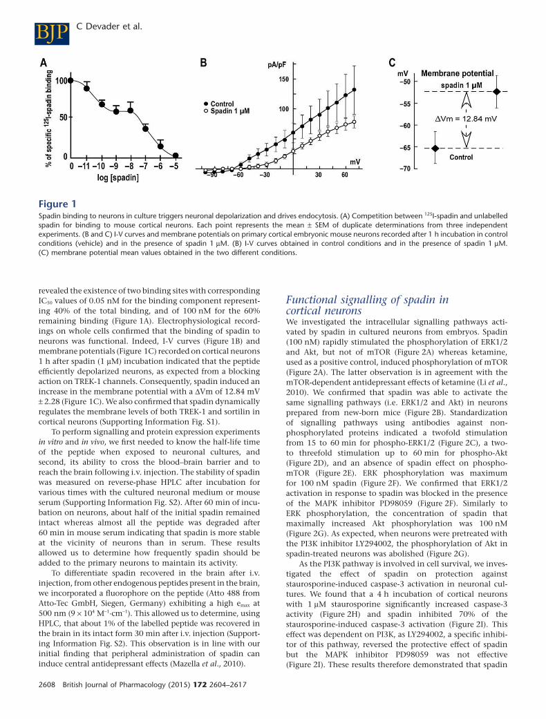
revealed the existence of two binding sites with corresponding
IC50 values of 0.05 nM for the binding component represent-
ing 40% of the total binding, and of 100 nM for the 60%
remaining binding (Figure 1A). Electrophysiological record-
ings on whole cells confirmed that the binding of spadin to
neurons was functional. Indeed, I-V curves (Figure 1B) and
membrane potentials (Figure 1C) recorded on cortical neurons
1 h after spadin (1 μM) incubation indicated that the peptide
efficiently depolarized neurons, as expected from a blocking
action on TREK-1 channels. Consequently, spadin induced an
increase in the membrane potential with a ΔVm of 12.84 mV
± 2.28 (Figure 1C). We also confirmed that spadin dynamically
regulates the membrane levels of both TREK-1 and sortilin in
cortical neurons (Supporting Information Fig. S1).
To perform signalling and protein expression experiments
in vitro and in vivo, we first needed to know the half-life time
of the peptide when exposed to neuronal cultures, and
second, its ability to cross the blood–brain barrier and to
reach the brain following i.v. injection. The stability of spadin
was measured on reverse-phase HPLC after incubation for
various times with the cultured neuronal medium or mouse
serum (Supporting Information Fig. S2). After 60 min of incu-
bation on neurons, about half of the initial spadin remained
intact whereas almost all the peptide was degraded after
60 min in mouse serum indicating that spadin is more stable
at the vicinity of neurons than in serum. These results
allowed us to determine how frequently spadin should be
added to the primary neurons to maintain its activity.
To differentiate spadin recovered in the brain after i.v.
injection, from other endogenous peptides present in the brain,
we incorporated a fluorophore on the peptide (Atto 488 from
Atto-Tec GmbH, Siegen, Germany) exhibiting a high emax at
500 nm (9 × 10⁴ M−1·cm−1). This allowed us to determine, using
HPLC, that about 1% of the labelled peptide was recovered in
the brain in its intact form 30 min after i.v. injection (Support-
ing Information Fig. S2). This observation is in line with our
initial finding that peripheral administration of spadin can
induce central antidepressant effects (Mazella et al., 2010).
Functional signalling of spadin incortical neuronsWe investigated the intracellular signalling pathways acti-
vated by spadin in cultured neurons from embryos. Spadin
(100 nM) rapidly stimulated the phosphorylation of ERK1/2
and Akt, but not of mTOR (Figure 2A) whereas ketamine,
used as a positive control, induced phosphorylation of mTOR
(Figure 2A). The latter observation is in agreement with the
mTOR-dependent antidepressant effects of ketamine (Li et al.,
2010). We confirmed that spadin was able to activate the
same signalling pathways (i.e. ERK1/2 and Akt) in neurons
prepared from new-born mice (Figure 2B). Standardization
of signalling pathways using antibodies against non-
phosphorylated proteins indicated a twofold stimulation
from 15 to 60 min for phospho-ERK1/2 (Figure 2C), a two-
to threefold stimulation up to 60 min for phospho-Akt
(Figure 2D), and an absence of spadin effect on phospho-
mTOR (Figure 2E). ERK phosphorylation was maximum
for 100 nM spadin (Figure 2F). We confirmed that ERK1/2
activation in response to spadin was blocked in the presence
of the MAPK inhibitor PD98059 (Figure 2F). Similarly to
ERK phosphorylation, the concentration of spadin that
maximally increased Akt phosphorylation was 100 nM
(Figure 2G). As expected, when neurons were pretreated with
the PI3K inhibitor LY294002, the phosphorylation of Akt in
spadin-treated neurons was abolished (Figure 2G).
As the PI3K pathway is involved in cell survival, we inves-
tigated the effect of spadin on protection against
staurosporine-induced caspase-3 activation in neuronal cul-
tures. We found that a 4 h incubation of cortical neurons
with 1 μM staurosporine significantly increased caspase-3
activity (Figure 2H) and spadin inhibited 70% of the
staurosporine-induced caspase-3 activation (Figure 2I). This
effect was dependent on PI3K, as LY294002, a specific inhibi-
tor of this pathway, reversed the protective effect of spadin
but the MAPK inhibitor PD98059 was not effective
(Figure 2I). These results therefore demonstrated that spadin
Figure 1Spadin binding to neurons in culture triggers neuronal depolarization and drives endocytosis. (A) Competition between 125I-spadin and unlabelled
spadin for binding to mouse cortical neurons. Each point represents the mean ± SEM of duplicate determinations from three independent
experiments. (B and C) I-V curves and membrane potentials on primary cortical embryonic mouse neurons recorded after 1 h incubation in control
conditions (vehicle) and in the presence of spadin 1 μM. (B) I-V curves obtained in control conditions and in the presence of spadin 1 μM.
(C) membrane potential mean values obtained in the two different conditions.
BJP C Devader et al.
2608 British Journal of Pharmacology (2015) 172 2604–2617
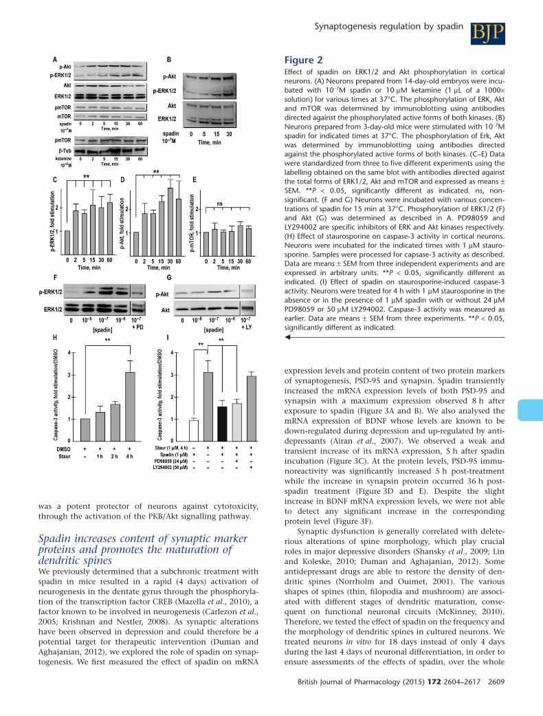
was a potent protector of neurons against cytotoxicity,
through the activation of the PKB/Akt signalling pathway.
Spadin increases content of synaptic markerproteins and promotes the maturation ofdendritic spinesWe previously determined that a subchronic treatment with
spadin in mice resulted in a rapid (4 days) activation of
neurogenesis in the dentate gyrus through the phosphoryla-
tion of the transcription factor CREB (Mazella et al., 2010), a
factor known to be involved in neurogenesis (Carlezon et al.,
2005; Krishnan and Nestler, 2008). As synaptic alterations
have been observed in depression and could therefore be a
potential target for therapeutic intervention (Duman and
Aghajanian, 2012), we explored the role of spadin on synap-
togenesis. We first measured the effect of spadin on mRNA
expression levels and protein content of two protein markers
of synaptogenesis, PSD-95 and synapsin. Spadin transiently
increased the mRNA expression levels of both PSD-95 and
synapsin with a maximum expression observed 8 h after
exposure to spadin (Figure 3A and B). We also analysed the
mRNA expression of BDNF whose levels are known to be
down-regulated during depression and up-regulated by anti-
depressants (Airan et al., 2007). We observed a weak and
transient increase of its mRNA expression, 5 h after spadin
incubation (Figure 3C). At the protein levels, PSD-95 immu-
noreactivity was significantly increased 5 h post-treatment
while the increase in synapsin protein occurred 36 h post-
spadin treatment (Figure 3D and E). Despite the slight
increase in BDNF mRNA expression levels, we were not able
to detect any significant increase in the corresponding
protein level (Figure 3F).
Synaptic dysfunction is generally correlated with delete-
rious alterations of spine morphology, which play crucial
roles in major depressive disorders (Shansky et al., 2009; Lin
and Koleske, 2010; Duman and Aghajanian, 2012). Some
antidepressant drugs are able to restore the density of den-
dritic spines (Norrholm and Ouimet, 2001). The various
shapes of spines (thin, filopodia and mushroom) are associ-
ated with different stages of dendritic maturation, conse-
quent on functional neuronal circuits (McKinney, 2010).
Therefore, we tested the effect of spadin on the frequency and
the morphology of dendritic spines in cultured neurons. We
treated neurons in vitro for 18 days instead of only 4 days
during the last 4 days of neuronal differentiation, in order to
ensure assessments of the effects of spadin, over the whole
Figure 2Effect of spadin on ERK1/2 and Akt phosphorylation in cortical
neurons. (A) Neurons prepared from 14-day-old embryos were incu-
bated with 10−7M spadin or 10 μM ketamine (1 μL of a 1000×
solution) for various times at 37°C. The phosphorylation of ERK, Akt
and mTOR was determined by immunoblotting using antibodies
directed against the phosphorylated active forms of both kinases. (B)
Neurons prepared from 3-day-old mice were stimulated with 10−7M
spadin for indicated times at 37°C. The phosphorylation of Erk, Akt
was determined by immunoblotting using antibodies directed
against the phosphorylated active forms of both kinases. (C–E) Data
were standardized from three to five different experiments using the
labelling obtained on the same blot with antibodies directed against
the total forms of ERK1/2, Akt and mTOR and expressed as means ±
SEM. **P < 0.05, significantly different as indicated. ns, non-
significant. (F and G) Neurons were incubated with various concen-
trations of spadin for 15 min at 37°C. Phosphorylation of ERK1/2 (F)
and Akt (G) was determined as described in A. PD98059 and
LY294002 are specific inhibitors of ERK and Akt kinases respectively.
(H) Effect of staurosporine on caspase-3 activity in cortical neurons.
Neurons were incubated for the indicated times with 1 μM stauro-
sporine. Samples were processed for capsase-3 activity as described.
Data are means ± SEM from three independent experiments and are
expressed in arbitrary units. **P < 0.05, significantly different as
indicated. (I) Effect of spadin on staurosporine-induced caspase-3
activity. Neurons were treated for 4 h with 1 μM staurosporine in the
absence or in the presence of 1 μM spadin with or without 24 μM
PD98059 or 50 μM LY294002. Caspase-3 activity was measured as
earlier. Data are means ± SEM from three experiments. **P < 0.05,
significantly different as indicated.
BJPSynaptogenesis regulation by spadin
British Journal of Pharmacology (2015) 172 2604–2617 2609
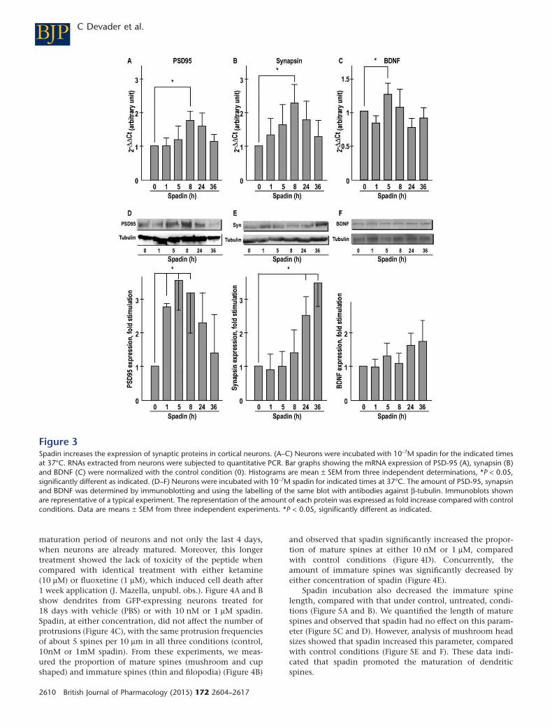
maturation period of neurons and not only the last 4 days,
when neurons are already matured. Moreover, this longer
treatment showed the lack of toxicity of the peptide when
compared with identical treatment with either ketamine
(10 μM) or fluoxetine (1 μM), which induced cell death after
1 week application (J. Mazella, unpubl. obs.). Figure 4A and B
show dendrites from GFP-expressing neurons treated for
18 days with vehicle (PBS) or with 10 nM or 1 μM spadin.
Spadin, at either concentration, did not affect the number of
protrusions (Figure 4C), with the same protrusion frequencies
of about 5 spines per 10 μm in all three conditions (control,
10nM or 1mM spadin). From these experiments, we meas-
ured the proportion of mature spines (mushroom and cup
shaped) and immature spines (thin and filopodia) (Figure 4B)
and observed that spadin significantly increased the propor-
tion of mature spines at either 10 nM or 1 μM, compared
with control conditions (Figure 4D). Concurrently, the
amount of immature spines was significantly decreased by
either concentration of spadin (Figure 4E).
Spadin incubation also decreased the immature spine
length, compared with that under control, untreated, condi-
tions (Figure 5A and B). We quantified the length of mature
spines and observed that spadin had no effect on this param-
eter (Figure 5C and D). However, analysis of mushroom head
sizes showed that spadin increased this parameter, compared
with control conditions (Figure 5E and F). These data indi-
cated that spadin promoted the maturation of dendritic
spines.
Figure 3Spadin increases the expression of synaptic proteins in cortical neurons. (A–C) Neurons were incubated with 10−7M spadin for the indicated times
at 37°C. RNAs extracted from neurons were subjected to quantitative PCR. Bar graphs showing the mRNA expression of PSD-95 (A), synapsin (B)
and BDNF (C) were normalized with the control condition (0). Histograms are mean ± SEM from three independent determinations, *P < 0.05,
significantly different as indicated. (D–F) Neurons were incubated with 10−7M spadin for indicated times at 37°C. The amount of PSD-95, synapsin
and BDNF was determined by immunoblotting and using the labelling of the same blot with antibodies against β-tubulin. Immunoblots shown
are representative of a typical experiment. The representation of the amount of each protein was expressed as fold increase compared with control
conditions. Data are means ± SEM from three independent experiments. *P < 0.05, significantly different as indicated.
BJP C Devader et al.
2610 British Journal of Pharmacology (2015) 172 2604–2617
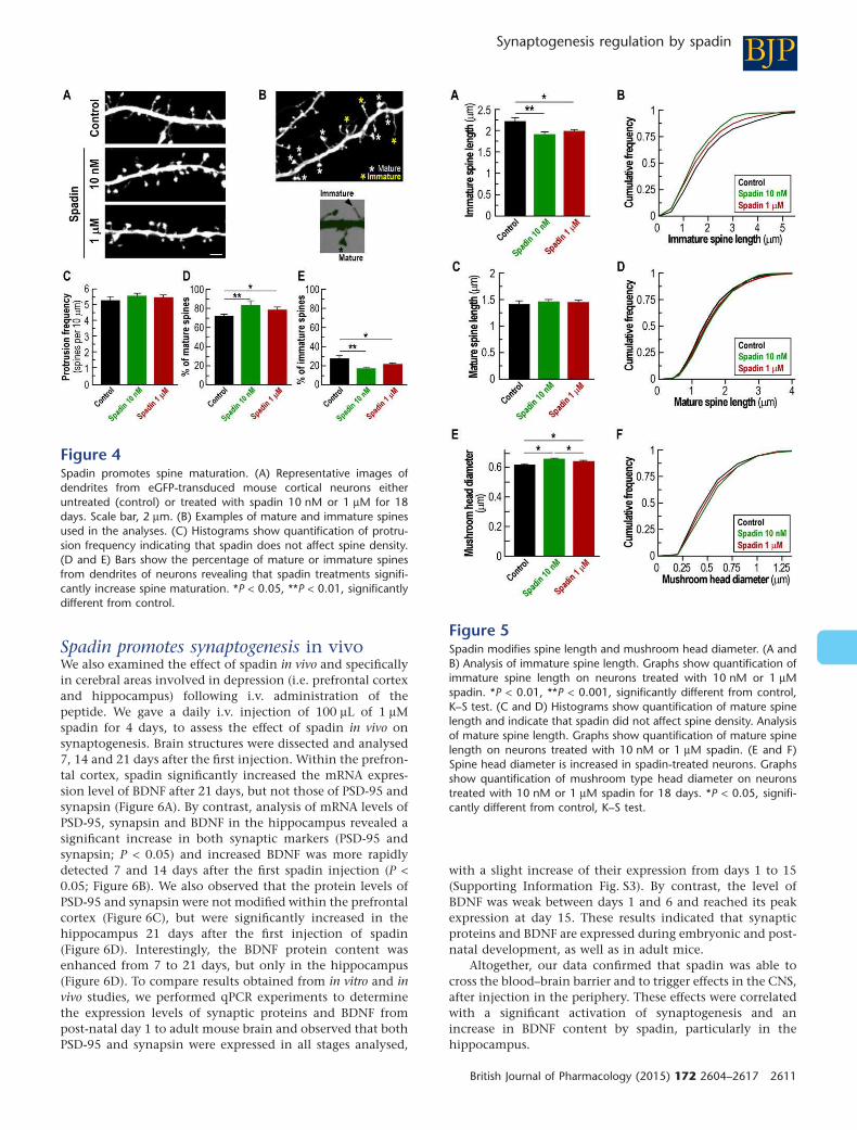
Spadin promotes synaptogenesis in vivoWe also examined the effect of spadin in vivo and specifically
in cerebral areas involved in depression (i.e. prefrontal cortex
and hippocampus) following i.v. administration of the
peptide. We gave a daily i.v. injection of 100 μL of 1 μM
spadin for 4 days, to assess the effect of spadin in vivo on
synaptogenesis. Brain structures were dissected and analysed
7, 14 and 21 days after the first injection. Within the prefron-
tal cortex, spadin significantly increased the mRNA expres-
sion level of BDNF after 21 days, but not those of PSD-95 and
synapsin (Figure 6A). By contrast, analysis of mRNA levels of
PSD-95, synapsin and BDNF in the hippocampus revealed a
significant increase in both synaptic markers (PSD-95 and
synapsin; P < 0.05) and increased BDNF was more rapidly
detected 7 and 14 days after the first spadin injection (P <
0.05; Figure 6B). We also observed that the protein levels of
PSD-95 and synapsin were not modified within the prefrontal
cortex (Figure 6C), but were significantly increased in the
hippocampus 21 days after the first injection of spadin
(Figure 6D). Interestingly, the BDNF protein content was
enhanced from 7 to 21 days, but only in the hippocampus
(Figure 6D). To compare results obtained from in vitro and in
vivo studies, we performed qPCR experiments to determine
the expression levels of synaptic proteins and BDNF from
post-natal day 1 to adult mouse brain and observed that both
PSD-95 and synapsin were expressed in all stages analysed,
with a slight increase of their expression from days 1 to 15
(Supporting Information Fig. S3). By contrast, the level of
BDNF was weak between days 1 and 6 and reached its peak
expression at day 15. These results indicated that synaptic
proteins and BDNF are expressed during embryonic and post-
natal development, as well as in adult mice.
Altogether, our data confirmed that spadin was able to
cross the blood–brain barrier and to trigger effects in the CNS,
after injection in the periphery. These effects were correlated
with a significant activation of synaptogenesis and an
increase in BDNF content by spadin, particularly in the
hippocampus.
Figure 4Spadin promotes spine maturation. (A) Representative images of
dendrites from eGFP-transduced mouse cortical neurons either
untreated (control) or treated with spadin 10 nM or 1 μM for 18
days. Scale bar, 2 μm. (B) Examples of mature and immature spines
used in the analyses. (C) Histograms show quantification of protru-
sion frequency indicating that spadin does not affect spine density.
(D and E) Bars show the percentage of mature or immature spines
from dendrites of neurons revealing that spadin treatments signifi-
cantly increase spine maturation. *P < 0.05, **P < 0.01, significantly
different from control.
Figure 5Spadin modifies spine length and mushroom head diameter. (A and
B) Analysis of immature spine length. Graphs show quantification of
immature spine length on neurons treated with 10 nM or 1 μM
spadin. *P < 0.01, **P < 0.001, significantly different from control,
K–S test. (C and D) Histograms show quantification of mature spine
length and indicate that spadin did not affect spine density. Analysis
of mature spine length. Graphs show quantification of mature spine
length on neurons treated with 10 nM or 1 μM spadin. (E and F)
Spine head diameter is increased in spadin-treated neurons. Graphs
show quantification of mushroom type head diameter on neurons
treated with 10 nM or 1 μM spadin for 18 days. *P < 0.05, signifi-
cantly different from control, K–S test.
BJPSynaptogenesis regulation by spadin
British Journal of Pharmacology (2015) 172 2604–2617 2611
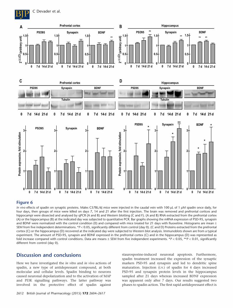
Discussion and conclusions
Here we have investigated the in vitro and in vivo actions of
spadin, a new type of antidepressant compound, at both
molecular and cellular levels. Spadin binding to neurons
caused neuronal depolarization and to the activation of MAP
and PI3K signalling pathways. The latter pathway was
involved in the protective effect of spadin against
staurosporine-induced neuronal apoptosis. Furthermore,
spadin treatment increased the expression of the synaptic
markers PSD-95 and synapsin and led to dendritic spine
maturation. Injection (i.v.) of spadin for 4 days increased
PSD-95 and synapsin protein levels in the hippocampus
sampled after 21 days whereas increased BDNF expression
was apparent only after 7 days. Our results suggested two
phases to spadin action. The first rapid antidepressant effect is
Figure 6In vivo effects of spadin on synaptic proteins. Males C57BL/6J mice were injected in the caudal vein with 100 μL of 1 μM spadin once daily, for
four days, then groups of mice were killed on days 7, 14 and 21 after the first injection. The brain was removed and prefrontal cortices and
hippocampi were dissected and analysed by qPCR (A and B) and Western blotting (C and F). (A and B) RNA extracted from the prefrontal cortex
(A) or the hippocampus (B) at the indicated day was subjected to quantitative PCR. Bar graphs showing the mRNA expression of PSD-95, synapsin
and BDNF were normalized with the control condition (0) and compared with mice treated for 21 days with fluoxetine. Histograms are mean ±
SEM from five independent determinations. *P < 0.05, significantly different from control (day 0). (C and D) Proteins extracted from the prefrontal
cortex (C) or the hippocampus (D) recovered at the indicated day were subjected to Western blot analysis. Immunoblots shown are from a typical
experiment. The amount of PSD-95, synapsin and BDNF expressed in the prefrontal cortex (C) and in the hippocampus (D) was represented as
fold increase compared with control conditions. Data are means ± SEM from five independent experiments. *P < 0.05, **P < 0.01, significantly
different from control (day 0).
BJP C Devader et al.
2612 British Journal of Pharmacology (2015) 172 2604–2617
triggered by the increase of BDNF and associated with the
release of 5-HT from the dorsal raphe and with hippocampal
neurogenesis (Mazella et al., 2010). The second phase is likely
to correspond to the maturation of new neurons identified by
the increase of synaptic markers as well as an increase in
spinogenesis (the present study).
The efficacy of spadin as an antidepressant derives from
its biological characteristics as well as its mode of action.
Indeed, the peptide sequence of spadin is part of the endog-
enous peptide of 44 amino acids (PE) released from the pre-
cursor form of the NTSR3/sortilin receptor (Munck Petersen
et al., 1999). The antidepressant action of spadin is observed
after an acute injection and its action on neurogenesis
appears only after a 4 day treatment (Mazella et al., 2010)
whereas existing antidepressant drugs need 21 days to induce
neurogenesis. The antidepressant properties of spadin are due
to its ability to block the K+ channel TREK-1 (Mazella et al.,
2010) without any detectable side effect related to this
channel (Moha Ou Maati et al., 2012). Although we had
already identified the potent antidepressant action of spadin
associated with hippocampal neurogenesis, its pharmacologi-
cal, molecular and cellular modes of action remained
unknown.
Functional interaction of spadin with neuronsThe biochemical and electrophysiological properties of
spadin were initially characterized using heterologous trans-
fected cells (Mazella et al., 2010; Moha ou Maati et al., 2011).
Here, we used a more physiologically relevant system to
investigate the molecular and cellular mechanisms of action
of spadin. In neurons, spadin clearly binds to two binding
components with affinities of 0.05 and 100 nM respectively.
Taking into account the concentration of PE detected in the
brain (about 10 nM), spadin is likely to triggers cellular effects
through its high-affinity binding site. We previously identi-
fied two targets of spadin, one of them is NTSR3/sortilin, the
protein from which the peptide is released after maturation,
and the second one is TREK-1 (Mazella et al., 2010). The
binding of spadin to TREK-1 blocks the related K+ current and
induces cell-membrane depolarization (Figure 1C). This
process is compatible with the inhibition of TREK-1 channels
(Fink et al., 1996) (Hughes et al., 2006). Moreover, we
observed that spadin affects the plasma membrane distribu-
tion of TREK-1 and sortilin as it binds and induces their
internalization (Supporting Information Fig. S1). One known
function of NTSR3/sortilin is the sorting of proteins, this
observation strongly suggests that sortilin not only targets
TREK-1 channels to the plasma membrane (Mazella et al.,
2010), but also participates in their concomitant internaliza-
tion. Spadin totally inactivates TREK-1 by blocking and inter-
nalizing the channel, a process crucial for the antidepressant
effect in mice (Heurteaux et al., 2006b).
Spadin protects neurons from apoptosis by amechanism dependent on PI3K pathwaySpadin stimulates both ERK1/2 and PI3K signalling pathways
in a time- and concentration-dependent manner (Figure 2).
The level of phospho-Akt remains surprisingly high after
60 min. Usually, by that time, the level returns to the basal
value. This finding could explain the prolonged effects of
spadin in vivo on synaptic proteins and for the mTOR-
independent Akt stimulation induced by the peptide. The
ERK1/2 and PI3K pathways are known to be involved in cell
growth and cell survival respectively. The expression of
TREK-1 is known to be protective against ischaemia
(Heurteaux et al., 2004) (Buckler and Honore, 2005) and
potent TREK-1 openers protect brain from ischaemia in
rodents (Duprat et al., 2000) (Blondeau et al., 2002)
(Heurteaux et al., 2006a). In this scheme, spadin, which
blocks TREK-1 activity, should decrease the protective action
of the channel. We observed that spadin efficiently protected
neurons from cell death by reversing staurosporine-induced
caspase-3 activation (Figure 2H). This effect was mediated
through a PI3K signalling pathway, but not via the MAPK
pathway as only the PI3K inhibitor LY294002 reversed the
protective action of spadin (Figure 2H). The involvement of
the PI3K pathway in neuronal protection has already been
reported in several studies including neuroprotection
induced by nicotine against colchicine-induced apoptosis
(Huang et al., 2012) and the protective action of extracellular
progranulin against toxic insults (Xu et al., 2011).
Our finding that spadin acted as a protective agent on
cortical neurons suggests that this regulatory system is not so
simple. The membrane components responsible for the
spadin-induced anti-apoptotic effects remain to be identified.
One candidate is NTSR3/sortilin and further experiments per-
formed on neurons prepared from sortilin-KO mice are
required to verify this hypothesis.
In vitro and in vivo effects of spadin onsynaptogenesisSpadin has been described to initiate hippocampal neurogen-
esis, probably through the activation of CREB (Mazella et al.,
2010). This action, which was also observed with fluoxetine
(Ohira et al., 2013), does not indicate that new neurons gen-
erated by the treatment are functional. The modulation of
neurogenesis in the aetiology of depression is still a matter of
debate. Indeed, although it is well known that antidepressant
drugs induce hippocampal and cortical neurogenesis, block-
ing neurogenesis does not alter the improving actions of
antidepressant drugs on mood (Bessa et al., 2009). Thus, the
role of neurogenesis could be to buffer stress response and
depressive behaviour (Snyder et al., 2011). Our observation
that spadin increases the ratio of mushroom spine types
suggests a beneficial adjustment of synaptic function, which
could lead a significant action of the peptide on neuron
maturation and consequently on synaptic plasticity.
In vitro, we observed a rapid increase in the mRNA and
protein expression levels of two synaptic markers; PSD-95
and synapsin, but not of BDNF upon incubation with spadin
(Figure 3). The lack of spadin effect on BDNF protein expres-
sion is likely to be due to the low number of neuronal cells
expressing the neurotrophic factor in our cultures. Acute
exposure of neurons to spadin enhances the proportion of
mature dendritic spines (mushrooms) without significant
changes in the total number of spines (Figure 4). We analysed
synaptic proteins up to 21 days because this time should be
enough to show effects, taking into account that we showed
that a 4 day treatment with spadin was enough to increase
the phosphorylation of CREB in the hippocampus. Phospho-
CREB is known to be crucial for full maturation of new
BJPSynaptogenesis regulation by spadin
British Journal of Pharmacology (2015) 172 2604–2617 2613
neurons (Fujioka et al., 2004). When phospho-CREB is meas-
ured where immature new neurons are observed, the expres-
sion of phospho-CREB, correlated with maturation, is
increased up to 14 days after proliferation. We therefore
assumed that 21 days was enough time to observe variations.
However, our observations were in agreement with the initial
observation that overexpression of PSD-95 is involved in the
maturation of spines (El-Husseini et al., 2000). The regulation
of spine morphology is generally correlated with changes in
neuronal activity (Yuste and Bonhoeffer, 2001). Indeed, based
on the structure–stability–function relationships, dendritic
spines are classified in two categories, small and large (Kasai
et al., 2003). Small spines are usually unstable and non-
functional whereas large spines (i.e. mushrooms) are much
more stable and maintain strong synaptic connections.
Moreover, increasing the proportion of large long-lasting
spines in hippocampal neurons is likely to facilitate long-
term memory (Lippman and Dunaevsky, 2005). Although
dendritic spines are dynamic structures (Lippman and
Dunaevsky, 2005), the change in morphology together with
the up-regulation of synaptic markers strongly suggest that
spadin is a potent up-regulator of neuronal functions.
We also analysed the effect of the peptide on the expres-
sion of the two synaptic proteins and BDNF in two cerebral
regions involved in the regulation of mood disorders: the
prefrontal cortex and the hippocampus (Figure 6). A 4 day
treatment with spadin significantly increased the hippocam-
pal expression of both PSD-95 and synapsin, 21 days after the
first spadin injection, but not in the prefrontal cortex
(Figure 6). These results confirmed the action of spadin on
cultured neurons and indicated that the hippocampus is
likely to be one of its main cerebral targets. Note that a 21 day
administration of the 5-HT reuptake inhibitor fluoxetine was
without any effect on synaptic protein mRNA expression
levels (not shown). However, experiments carried out using
intrahippocampal infusion of fluoxetine did increase PSD-95
expression and synaptogenesis (Mogha et al., 2012). This dis-
crepancy is probably due to the difference in the mode of
administration. Interestingly, the same spadin treatment
increased BDNF expression in the hippocampus more rapidly
(after 7 days) than expression of PSD-95 and synapsin (after
21 days). This effect is compatible with our previous obser-
vation that a 4 day treatment with spadin induced a potent
antidepressive action and a marked neurogenesis in the hip-
pocampus (Mazella et al., 2010). This result is also in agree-
ment with the observation that a hippocampus-specific
increase in BDNF activity is involved in the improvement of
cognitive symptoms of depression and in the facilitation of
hippocampal neurogenesis (Airan et al., 2007).
To date, antidepressants that are able to reverse synaptic
dysfunctions have a limited efficacy and a delayed response
ranging from several weeks to months. The recent discovery
that ketamine, a non-competitive NMDA receptor antago-
nist, rapidly enhanced synaptogenesis and reversed synaptic
deficits (Duman and Aghajanian, 2012), as well as the discov-
ery of the peptide spadin which bears key properties of a
potent antidepressant, may open new fields for treatment of
mood disorders. However, in contrast to ketamine, which
activates the mTOR pathway through ERK and PI3K pathways
(Licznerski and Duman, 2013), the spadin action appears
independent of mTOR signalling. This is in agreement with
the antidepressant effect of extracts of Radix polygalae (the
dried root of Polygala tenuifolia) on the modulation of gluta-
matergic synapses, independently of mTOR activation (Shin
et al., 2014). Akt is usually placed downstream of mTORC2
and upstream of mTORC1 (Bhaskar and Hay, 2007). However,
after deletion of mTORC2 activity, the phosphorylation of
Akt was still observed suggesting that mTORC2 is not placed
uptstream of Akt (Shiota et al., 2006). In this case, Akt can
phosphorylate other substrates than mTORC1, such as NF-κB
or GSK3β that are involved in cell protection or cell cycle (Liu
et al., 2009). Further studies are necessary to identify the
downstream pathways involved in spadin-induced neuronal
activation.
The increase in the expression of synaptic proteins upon
spadin treatment both in vitro and in vivo is a key property
that could have considerable effects on therapy of depression.
In addition to its ability to cross the blood–brain barrier and
to stimulate neurogenesis, spadin appears to be a molecule
able to potentiate dendritic spine maturation and synapse
formation and, consequently, reinforces our concept that
spadin is a novel potent antidepressant.
Acknowledgements
This work was supported by the Centre National de la
Recherche Scientifique and the Agence Nationale de la
Recherche (ANR, ANR-13-SAMA-0002-02 and ANR-11-
EMMA-0050-01). SMa was supported by grants from the Fon-
dation pour la Recherche Médicale (Equipe labellisée #
DEQ20111223747) and the Agence Nationale de la Recherche
(ANR-2011-JSV4-0031). We also thank the French govern-
ment for the ‘Investments for the Future’ LABEX ‘SIGNALIFE’
# ANR-11-LABX-0028-01 to SMa and ICST # ANR-11 LABX
0015 to CH. JV was supported by a CIFRE fellowship.
Author contributions
C. D., A. K., J. V., H. M. M., M. R., S. M. and J. M. performed
the experiments. M. B., S. Ma., C. H. and J. M. conceived
and designed the experiments. M. B., S. Ma., C. H. and
J. M. contributed reagents/materials/analysis tools. C. D. and
J. M. wrote the paper.
Conflict of interest
Authors declare that there is no financial conflict of interest.
References
Airan RD, Meltzer LA, Roy M, Gong Y, Chen H, Deisseroth K
(2007). High-speed imaging reveals neurophysiological links to
behavior in an animal model of depression. Science 317: 819–823.
Alexander SPH, Benson HE, Faccenda E, Pawson AJ, Sharman JL,
Catterall WA et al. (2013a). The Concise Guide to
PHARMACOLOGY 2013/14: Ion Channels. Br J Pharmacol 170:
1607–1651.
BJP C Devader et al.
2614 British Journal of Pharmacology (2015) 172 2604–2617
Alexander SPH, Benson HE, Faccenda E, Pawson AJ, Sharman JL,
Spedding M et al. (2013b). The Concise Guide to PHARMACOLOGY
2013/14: Enzymes. Br J Pharmacol 170: 1797–1867.
Berton O, Nestler EJ (2006). New approaches to antidepressant drug
discovery: beyond monoamines. Nat Rev Neurosci 7: 137–151.
Bessa JM, Ferreira D, Melo I, Marques F, Cerqueira JJ, Palha JA et al.
(2009). The mood-improving actions of antidepressants do not
depend on neurogenesis but are associated with neuronal
remodeling. Mol Psychiatry 14: 764–773, 739.
Bhaskar PT, Hay N (2007). The two TORCs and Akt. Dev Cell 12:
487–502.
Blondeau N, Lauritzen I, Widmann C, Lazdunski M, Heurteaux C
(2002). A potent protective role of lysophospholipids against global
cerebral ischemia and glutamate excitotoxicity in neuronal cultures.
J Cereb Blood Flow Metab 22: 821–834.
Boschmans SA, Perkin MF, Terblanche SE (1987). Antidepressant
drugs: imipramine, mianserin and trazodone. Comp Biochem
Physiol C 86: 225–232.
Brewer GJ, Torricelli JR (2007). Isolation and culture of adult
neurons and neurospheres. Nat Protoc 2: 1490–1498.
Buckler KJ, Honore E (2005). The lipid-activated two-pore domain K
+ channel TREK-1 is resistant to hypoxia: implication for ischaemic
neuroprotection. J Physiol 562 (Pt 1): 213–222.
Carlezon WA Jr, Duman RS, Nestler EJ (2005). The many faces of
CREB. Trends Neurosci 28: 436–445.
Coppola T, Beraud-Dufour S, Antoine A, Vincent JP, Mazella J
(2008). Neurotensin protects pancreatic beta cells from apoptosis.
Int J Biochem Cell Biol 40: 2296–2302.
Dingemans AM, van den Boogaart V, Vosse BA, van Suylen RJ,
Griffioen AW, Thijssen VL (2010). Integrin expression profiling
identifies integrin alpha5 and beta1 as prognostic factors in early
stage non-small cell lung cancer. Mol Cancer 9: 152–160.
Dolder CR, Nelson MH, Iler CA (2012). The effects of mirtazapine
on sleep in patients with major depressive disorder. Ann Clin
Psychiatry 24: 215–224.
Drevets WC, Zarate CA Jr, Furey ML (2013). Antidepressant effects
of the muscarinic cholinergic receptor antagonist scopolamine: a
review. Biol Psychiatry 73: 1156–1163.
Duman RS, Aghajanian GK (2012). Synaptic dysfunction in
depression: potential therapeutic targets. Science 338: 68–72.
Duprat F, Lesage F, Patel AJ, Fink M, Romey G, Lazdunski M (2000).
The neuroprotective agent riluzole activates the two P domain K(+)
channels TREK-1 and TRAAK. Mol Pharmacol 57: 906–912.
Dwyer JM, Duman RS (2013). Activation of mammalian target of
rapamycin and synaptogenesis: role in the actions of rapid-acting
antidepressants. Biol Psychiatry 73: 1189–1198.
El-Husseini AE, Schnell E, Chetkovich DM, Nicoll RA, Bredt DS
(2000). PSD-95 involvement in maturation of excitatory synapses.
Science 290: 1364–1368.
Fagiolini A, Comandini A, Catena Dell’Osso M, Kasper S (2012).
Rediscovering trazodone for the treatment of major depressive
disorder. CNS Drugs 26: 1033–1049.
Fava M, Kendler KS (2000). Major depressive disorder. Neuron 28:
335–341.
Fink M, Duprat F, Lesage F, Reyes R, Romey G, Heurteaux C et al.
(1996). Cloning, functional expression and brain localization of a
novel unconventional outward rectifier K+ channel. EMBO J 15:
6854–6862.
Fujioka T, Fujioka A, Duman RS (2004). Activation of cAMP
signaling facilitates the morphological maturation of newborn
neurons in adult hippocampus. J Neurosci 24: 319–328.
Hervieu GJ, Cluderay JE, Gray CW, Green PJ, Ranson JL, Randall
AD et al. (2001). Distribution and expression of TREK-1, a
two-pore-domain potassium channel, in the adult rat CNS.
Neuroscience 103: 899–919.
Heurteaux C, Guy N, Laigle C, Blondeau N, Duprat F, Mazzuca M
et al. (2004). TREK-1, a K+ channel involved in neuroprotection and
general anesthesia. EMBO J 23: 2684–2695.
Heurteaux C, Laigle C, Blondeau N, Jarretou G, Lazdunski M
(2006a). Alpha-linolenic acid and riluzole treatment confer cerebral
protection and improve survival after focal brain ischemia.
Neuroscience 137: 241–251.
Heurteaux C, Lucas G, Guy N, El Yacoubi M, Thummler S, Peng XD
et al. (2006b). Deletion of the background potassium channel
TREK-1 results in a depression-resistant phenotype. Nat Neurosci 9:
1134–1141.
Honore E (2007). The neuronal background K2P channels: focus on
TREK1. Nat Rev Neurosci 8: 251–261.
Huang X, Cheng Z, Su Q, Zhu X, Wang Q, Chen R et al. (2012).
Neuroprotection by nicotine against colchicine-induced apoptosis is
mediated by PI3K–Akt pathways. Int J Neurosci 122: 324–332.
Hughes S, Magnay J, Foreman M, Publicover SJ, Dobson JP, El Haj
AJ (2006). Expression of the mechanosensitive 2PK+ channel
TREK-1 in human osteoblasts. J Cell Physiol 206: 738–748.
Kasai H, Matsuzaki M, Noguchi J, Yasumatsu N, Nakahara H (2003).
Structure–stability–function relationships of dendritic spines. Trends
Neurosci 26: 360–368.
Kilkenny C, Browne W, Cuthill IC, Emerson M, Altman DG (2010).
Animal research: reporting in vivo experiments: the ARRIVE
guidelines. Br J Pharmacol 160: 1577–1579.
Krishnan V, Nestler EJ (2008). The molecular neurobiology of
depression. Nature 455: 894–902.
Li N, Lee B, Liu RJ, Banasr M, Dwyer JM, Iwata M et al. (2010).
mTOR-dependent synapse formation underlies the rapid
antidepressant effects of NMDA antagonists. Science 329: 959–964.
Li N, Liu RJ, Dwyer JM, Banasr M, Lee B, Son H et al. (2011).
Glutamate N-methyl-D-aspartate receptor antagonists rapidly
reverse behavioral and synaptic deficits caused by chronic stress
exposure. Biol Psychiatry 69: 754–761.
Licznerski P, Duman RS (2013). Remodeling of axo-spinous
synapses in the pathophysiology and treatment of depression.
Neuroscience 251: 33–50.
Lin YC, Koleske AJ (2010). Mechanisms of synapse and dendrite
maintenance and their disruption in psychiatric and
neurodegenerative disorders. Annu Rev Neurosci 33: 349–378.
Lippman J, Dunaevsky A (2005). Dendritic spine morphogenesis
and plasticity. J Neurobiol 64: 47–57.
Liu P, Cheng H, Roberts TM, Zhao JJ (2009). Targeting the
phosphoinositide 3-kinase pathway in cancer. Nat Rev Drug Discov
8: 627–644.
Martin S, Bouschet T, Jenkins EL, Nishimune A, Henley JM (2008).
Bidirectional regulation of kainate receptor surface expression in
hippocampal neurons. J Biol Chem 283: 36435–36440.
Mazella J, Zsurger N, Navarro V, Chabry J, Kaghad M, Caput D et al.
(1998). The 100-kDa neurotensin receptor is gp95/sortilin, a
non-G-protein-coupled receptor. J Biol Chem 273: 26273–26276.
BJPSynaptogenesis regulation by spadin
British Journal of Pharmacology (2015) 172 2604–2617 2615
Mazella J, Petrault O, Lucas G, Deval E, Beraud-Dufour S, Gandin C
et al. (2010). Spadin, a sortilin-derived peptide, targeting rodent
TREK-1 channels: a new concept in the antidepressant drug design.
PLoS Biol 8: e1000355.
McEwen BS, Olie JP (2005). Neurobiology of mood, anxiety, and
emotions as revealed by studies of a unique antidepressant:
tianeptine. Mol Psychiatry 10: 525–537.
McGrath J, Drummond G, McLachlan E, Kilkenny C, Wainwright C
(2010). Guidelines for reporting experiments involving animals: the
ARRIVE guidelines. Br J Pharmacol 160: 1573–1576.
McKinney RA (2010). Excitatory amino acid involvement in
dendritic spine formation, maintenance and remodelling. J Physiol
588 (Pt 1): 107–116.
Mogha A, Guariglia SR, Debata PR, Wen GY, Banerjee P (2012).
Serotonin 1A receptor-mediated signaling through ERK and
PKCalpha is essential for normal synaptogenesis in neonatal mouse
hippocampus. Transl Psychiatry 2: e66.
Moha ou Maati H, Peyronnet R, Devader C, Veyssiere J, Labbal F,
Gandin C et al. (2011). A human TREK-1/HEK cell line: a highly
efficient screening tool for drug development in neurological
diseases. PLoS ONE 6: e25602.
Moha Ou Maati H, Veyssiere J, Labbal F, Coppola T, Gandin C,
Widmann C et al. (2012). Spadin as a new antidepressant: absence
of TREK-1-related side effects. Neuropharmacology 62: 278–288.
Munck Petersen C, Nielsen MS, Jacobsen C, Tauris J, Jacobsen L,
Gliemann J et al. (1999). Propeptide cleavage conditions
sortilin/neurotensin receptor-3 for ligand binding. EMBO J 18:
595–604.
Nestler EJ, Barrot M, DiLeone RJ, Eisch AJ, Gold SJ, Monteggia LM
(2002). Neurobiology of depression. Neuron 34: 13–25.
Norrholm SD, Ouimet CC (2001). Altered dendritic spine density in
animal models of depression and in response to antidepressant
treatment. Synapse 42: 151–163.
Ohira K, Takeuchi R, Shoji H, Miyakawa T (2013).
Fluoxetine-induced cortical adult neurogenesis.
Neuropsychopharmacology 38: 909–920.
Pacher P, Kecskemeti V (2004). Trends in the development of new
antidepressants. Is there a light at the end of the tunnel? Curr Med
Chem 11: 925–943.
Pawson AJ, Sharman JL, Benson HE, Faccenda E, Alexander SP,
Buneman OP et al.; NC-IUPHAR (2014). The IUPHAR/BPS Guide to
PHARMACOLOGY: an expert-driven knowledge base of drug targets
and their ligands. Nucl Acids Res 42 (Database Issue):
D1098–D1106.
Petersen CM, Nielsen MS, Nykjaer A, Jacobsen L, Tommerup N,
Rasmussen HH et al. (1997). Molecular identification of a novel
candidate sorting receptor purified from human brain by
receptor-associated protein affinity chromatography. J Biol Chem
272: 3599–3605.
Rodriguez A, Ehlenberger DB, Dickstein DL, Hof PR, Wearne SL
(2008). Automated three-dimensional detection and shape
classification of dendritic spines from fluorescence microscopy
images. PLoS ONE 3: e1997.
Sanacora G, Smith MA, Pathak S, Su HL, Boeijinga PH, McCarthy
DJ et al. (2014). Lanicemine: a low-trapping NMDA channel blocker
produces sustained antidepressant efficacy with minimal
psychotomimetic adverse effects. Mol Psychiatry 19: 978–985.
Sarret P, Krzywkowski P, Segal L, Nielsen MS, Petersen CM, Mazella
J et al. (2003). Distribution of NTS3 receptor/sortilin mRNA and
protein in the rat central nervous system. J Comp Neurol 461:
483–505.
Shansky RM, Hamo C, Hof PR, McEwen BS, Morrison JH (2009).
Stress-induced dendritic remodeling in the prefrontal cortex is
circuit specific. Cereb Cortex 19: 2479–2484.
Shin IJ, Son SU, Park H, Kim Y, Park SH, Swanberg K et al. (2014).
Preclinical evidence of rapid-onset antidepressant-like effect in radix
polygalae extract. PLoS ONE 9: e88617.
Shiota C, Woo JT, Lindner J, Shelton KD, Magnuson MA (2006).
Multiallelic disruption of the rictor gene in mice reveals that mTOR
complex 2 is essential for fetal growth and viability. Dev Cell 11:
583–589.
Sicouri S, Antzelevitch C (2008). Sudden cardiac death secondary to
antidepressant and antipsychotic drugs. Expert Opin Drug Saf 7:
181–194.
Snyder JS, Soumier A, Brewer M, Pickel J, Cameron HA (2011).
Adult hippocampal neurogenesis buffers stress responses and
depressive behaviour. Nature 476: 458–461.
Srinivasan V, Zakaria R, Othman Z, Lauterbach EC,
Acuna-Castroviejo D (2012). Agomelatine in depressive disorders:
its novel mechanisms of action. J Neuropsychiatry Clin Neurosci
24: 290–308.
Thase ME, Denko T (2008). Pharmacotherapy of mood disorders.
Annu Rev Clin Psychol 4: 53–91.
Xu J, Xilouri M, Bruban J, Shioi J, Shao Z, Papazoglou I et al.
(2011). Extracellular progranulin protects cortical neurons from
toxic insults by activating survival signaling. Neurobiol Aging 32:
2326, e2325–2316.
Yuste R, Bonhoeffer T (2001). Morphological changes in dendritic
spines associated with long-term synaptic plasticity. Annu Rev
Neurosci 24: 1071–1089.
Zarate CA Jr, Singh JB, Carlson PJ, Brutsche NE, Ameli R,
Luckenbaugh DA et al. (2006). A randomized trial of an
N-methyl-D-aspartate antagonist in treatment-resistant major
depression. Arch Gen Psychiatry 63: 856–864.
Supporting information
Additional Supporting Information may be found in the
online version of this article at the publisher’s web-site:
http://dx.doi.org/10.1111/bph.13083
Figure S1 Immunoprecipitation of NTSR3/sortilin or
TREK-1 with their corresponding antibodies from cortical
neurons pretreated with Sulfo-NHS-biotin before incubation
with spadin (1 μM) for the indicated times. Immuno-
precipitated internalized proteins were revealed using
HRP-streptavidin.
Figure S2 Degradation of spadin and analysis of blood–brain
barrier transit. (A) Time course of disappearance of spadin
after incubation with cortical neurons (open symbols) or
mouse serum (closed symbols) for the indicated times. Incu-
bations were terminated by acidification (HCl 1N) and the
peptide contents were analysed by reverse-phase HPLC.
Values represent the amount of intact spadin recovered after
HPLC and expressed as the percentage of the initial amount
of incubated peptide. Values are means ± SEM of three inde-
pendent determinations obtained from three different
neurons preparations or sera samples. (B) Fluorescent
Atto488-spadin crosses the blood–brain barrier. HPLC profile
BJP C Devader et al.
2616 British Journal of Pharmacology (2015) 172 2604–2617

of Atto488- spadin recovered in the brain 30 min after i.v.
injection. The brain was subjected to acidic extraction and
the extracted peptide content was analysed by reverse-phase
HPLC. The retention time for spadin-Atto488 is indicated by
the arrow.
Figure S3 Expression of PSD-95, synapsin and BDNF during
mouse brain development. The brain (from mice; ages as
indicated) was removed and cortical cortices were dissected
and analysed in qPCR experiments. Bar graphs showing the
mRNA expression of PSD-95, synapsin and BDNF from 1
(D1), 3 (D3), 6 (D6) and 15-day-old (D15) mice were com-
pared with the expression levels of adult (Adt) mice.
Histograms are mean ± SEM from three independent
determinations.
BJPSynaptogenesis regulation by spadin
British Journal of Pharmacology (2015) 172 2604–2617 2617
ORIGINAL INVESTIGATION
Retroinverso analogs of spadin display increased antidepressant
effects
Julie Veyssiere & Hamid Moha ou Maati & Jean Mazella &
Georges Gaudriault & Sébastien Moreno &
Catherine Heurteaux & Marc Borsotto
Received: 3 February 2014 /Accepted: 7 July 2014 /Published online: 2 August 2014# The Author(s) 2014. This article is published with open access at Springerlink.com
Abstract
Rationale Although depression is the most common mood
disorder, only one third of patients are treated with success.
Finding new targets, new drugs, and also new drug intake way
are the main challenges in the depression field. Several years
ago, we identified a new target with the TWIK-related potas-
sium channel-1 (TREK-1) potassium channel, and more re-
cently, we have discovered a peptide of 17 amino acids with
antidepressant properties. This peptide, that we called spadin,
can be considered as a new concept in antidepressant drug
design. Spadin derives from a larger peptide resulting to a
posttranslational maturation of sortilin; consequently, spadin
can be considered as a natural molecule. Moreover, spadin
acts more rapidly than classical antidepressants and does not
induce side effects.
Objectives In this work, we sought analogs of spadin
displaying a better affinity on TREK-1 channels and an in-
creased action duration.
Methods Analogs were characterized by electrophysiology
measurements, by behavioral tests, and by their ability to
induce neurogenesis.
Results We identified two retro-inverso peptides that have
kept the antidepressant properties of spadin; particularly, they
increased the hippocampal neurogenesis after a 4-day treat-
ment. As spadin, these analogs did not induce side effects on
either pain, epilepsy processes, or at the cardiac level.
Conclusions Together, our results indicated that spadin retro-
inverso peptides could represent new potent antidepressant
drugs. As exemplified by spadin in the field of depression,
retro-inverso strategies could represent a useful technique for
developing new classes of drugs in a number of pathologies.
Keywords Retro-inverso . Spadin . TREK-1 . Depression .
Electrophysiology
Introduction
Depression is a devastating neuropsychiatric disorder and
affects approximately 20 % of the population. Depression is
predicted to be a major cause of morbidity worldwide in the
next 10 years and will induce an important economic burden
(Greenberg et al. 2003; Moussavi et al. 2007). Depression is a
multifactorial and multigenic disease characterized by many
symptoms like fatigue, anhedonia, pessimism, irritability,
sleep troubles, increased or decreased appetite, guiltiness,
and suicidal tendencies (Nestler et al. 2002). Sixty years
ago, antidepressant treatments have been revolutionized by
the discovery of tricyclic antidepressants and monoamine
oxidase inhibitors. Later, a second generation of antidepres-
sants was developed with the selective serotonin or
Catherine Heurteaux and Marc Borsotto contributed equally to the
project.
Dr. Georges Gaudriault is a board member and an employee of the
Medincell SA company. Julie Veyssiere is a PhD student granted by
Medincell SA company.
J. Veyssiere :H. Moha ou Maati : J. Mazella : S. Moreno :
C. Heurteaux (*) :M. Borsotto (*)
Université de Nice Sophia Antipolis, IPMC, 06560 Sophia Antipolis,
France
e-mail: [email protected]
e-mail: [email protected]
J. Veyssiere :H. Moha ou Maati : J. Mazella : S. Moreno :
C. Heurteaux :M. Borsotto
CNRS, IPMC, 06560 Sophia Antipolis, France
H. Moha ou Maati
Institut de Génomique Fonctionnelle, 141 rue de la Cardonille,
34095 Montpellier Cedex 5, France
G. Gaudriault
Medincell SA, 1 rue Charles Cros, 34830 Jacou, France
Psychopharmacology (2015) 232:561–574
DOI 10.1007/s00213-014-3683-2
norepinephrine selective reuptake inhibitors. Despite their
efficacy, around one third of patients remain unresponsive to
these drugs. Moreover, they display some adverse side effects
and have a long onset of action (Sicouri and Antzelevitch
2008). Consequently, it was necessary to develop new antide-
pressant molecules with new pharmacological targets.
We previously demonstrated that the inhibition of TREK-1
led to an antidepressant phenotype (Heurteaux et al. 2006b).
Our researches led to the identification of a specific inhibitor of
TREK-1 channel called spadin (Mazella et al. 2010). Spadin
resulted from modification of the sortilin receptor (Mazella
et al. 1998). Spadin is a 17 amino acid peptide which was
designed from a 44 amino acid peptide (called PE) released
by furin in the late Golgi apparatus during the posttranslational
maturation of the sortilin receptor (Munck Petersen et al. 1999).
Spadin is able to block the TREK-1 potassium channel current
and displays antidepressant effects in different behavioral tests
(Mazella et al. 2010). Additionally, like other antidepressant
drugs, spadin is able to increase neurogenesis and serotoniner-
gic transmission. More interestingly, unlike the most used
antidepressants, which need 21 days to be efficient, spadin
has a quicker onset of action since it is able to induce these
improvements only after a 4-day treatment (Mazella et al.
2010). In the two pore (K2P) potassium channel family, spadin
is specific for TREK-1 channels (Moha ou Maati et al. 2011b).
Moreover, the activation of TREK-1 channels was demonstrat-
ed to be beneficial in different functions such as general anes-
thesia, neuroprotection by the way of polyunsaturated fatty
acids, pain, ischemia, and epilepsy (Alloui et al. 2006;
Heurteaux et al. 2006a; Lauritzen et al. 2000; Noel et al.
2009). Nevertheless, blockade of TREK-1 channels by spadin
does not interfere with these functions. In other words, spadin is
devoid of side effects on TREK-1-controlled functions
(Moha ou Maati et al. 2011b). Importantly, spadin does not
induce any cardiac dysfunctions, and both systolic pressure
and pulses are not affected by a 3-week spadin treatment.
Additionally, spadin is unable to block the two most important
repolarizing currents in the heart (IKR, IKS) (Moha ou Maati
et al. 2011b). Taken together, these properties are strong
evidences for considering spadin as an antidepressant drug
of a new generation.
With the aim to identify new analogs displaying a better
efficacy than spadin, we synthesized different portions of
human sortilin either in natural L-configuration or retro-
inverso configuration. This approach consists in synthesizing
peptides in which not only the chirality of amino acid is
inverted by replacing all L-amino acids by D-amino acids but
also the amino acid sequence is reversed (Bonny et al. 2001;
Chorev and Goodman 1995). In such a way, the side chains of
amino acids are in a similar position to that of the native
peptide (Bonny et al. 2001; Chorev and Goodman 1995;
Van Regenmortel and Muller 1998). Very often, retro-
inverso peptide properties are the same or close, sometimes
better, than the parent L-peptides and, overall, retro-inverso
peptides are more resistant to proteolysis (Taylor et al. 2010;
Weeden et al. 2011).
We first screened 12 spadin analogs for their ability to
block TREK-1 channel activity. The two most efficient were
retained for further studies using behavioral tests and mea-
surements of their effects on neurogenesis. Because the
TREK-1 channel deletion was shown to be deleterious for
epilepsy or pain (Alloui et al. 2006; Heurteaux et al. 2004;
Noel et al. 2009), we studied the effects of analog treatments
on these potential side effects. We also checked the analog
harmlessness on the two main cardiac repolarizing currents
IKR and IKS that are essential in cardiac function.
Materials and methods
Cell culture
The human-TREK-1/HEK293 cell line (h-TREK-1/HEK)
(Moha ou Maati et al. 2011a) and HEK-IKS cell line (Ducroq
et al. 2010) were grown in the presence of 0.5 mg/mL G418 in
Dulbecco’s modified Eagle’s medium supplemented with 10 %
(v/v) heat-inactivated fetal bovine serum containing 1 % (v/v)
penicillin/streptomycin in an atmosphere of 95 % air/5 % CO2
as previously described (Moha ou Maati et al. 2011a).
HEK-293 native cells were grown in serum in an atmo-
sphere of 95 % air/5 % CO2 in Dulbecco’s modified Eagle’s
medium supplemented with 10 % (v/v) heat-inactivated fetal
bovine containing 1 % (v/v) of penicillin/ streptomycin and
Glutamax X 1. Cells were plated at a density of 20,000 cells/
35 mm dish, and after 24 h, cells were transfected using the Jet
PEI method (Polyplus, France) with 25 ng/35 mm dish of p-
IRES-HERG channel vector. Patch clamp experiments were
carried out 48 h after transfection.
Electrophysiology
All electrophysiological experiments were performed on h-
TREK-1/HEK cells seeded at a density of 20,000 cells/35 mm
dish after 2–6 days of culture. All electrophysiological record-
ings were performed in whole cell configuration of the patch
clamp technique except for IKS measures which were
obtained by using the patch clamp perforated configuration
(amphotericin B 0.9 mg/mL in the pipette medium)
(Moha ou Maati et al. 2011b). Each current was evaluated
by using a RK 400 patch clamp amplifier (Axon Instrument,
USA), low-pass filtered at 3 kHz, and digitized at 10 kHz
using a 12-bit analog-to-digital converter digidata (1322 se-
ries, Axon Instrument, USA). All current amplitudes are
expressed in current densities. Results are expressed as
mean±standard error of the mean (SEM). Patch clamp pi-
pettes were pulled using vertical puller (PC-10, Narishige)
562 Psychopharmacology (2015) 232:561–574
from borosilicate glass capillaries and had a resistance of 3–
5MΩ. The bath solution contained (in mM) 150 NaCl, 5 KCl,
3 MgCl2, 1 CaCl2, and 10 4-(2-hydroxyethyl)piperazine 1-
ethane sulfonic acid (HEPES) adjusted to pH 7.4 with NaOH.
The pipette solution contained (in mM) 155 KCl, 3 MgCl2, 5
EGTA, and 10 HEPES adjusted to pH 7.2 with KOH. TREK-
1 currents were evaluated at room temperature (21–22 °C) in
the presence of a cocktail of potassium channel inhibitors (K+
blockers, 3 mM 4-aminopyridine (4-AP), 10 mM
tetraethylammonium (TEA), 10 μM glibenclamide, 100 nM
apamin, and 50 nM charybdotoxin). Stimulation protocols
and data acquisition were carried out using a microcomputer
(Dell Pentium) with a commercial software and hardware
(pClamp 8.2). Currents were recorded by voltage clamp steps
to membrane potentials of −100 to +60 mV in 20-mV steps
applied from a holding potential of −80 mV. The duration of
depolarization pulses was 825 ms, and the pulse cycling rate
was 5 s. TREK-1 current amplitudes were evaluated at the end
of stimulation pulses. Cells were continuously superfused
with microperfusion system. TREK-1 inhibitory effects of
spadin or analogs were performed on arachidonic acid pre-
activated currents. Spadin and analogs were tested at the
unique dose of 100 nM on TREK-1 channel activity and at
10 μM on IKR and IKS currents. For both analogs 3 and 8,
TREK-1 concentration-dependent inhibitions were performed
by applying concentrations ranging between 1 nM and 1 μM.
IKS currents were activated by voltage clamp steps of
membrane potentials from −100 to +100 mV in 20-mV steps
applied from a holding potential of −80mV. Tail currents were
generated by repolarization to −40 mV. Duration of both
depolarization and repolarization pulses was 2.4 s, and the
pulse cycling rate was 10 s. IKR currents were activated by
voltage clamp steps of membrane potentials from −100 to +
100 mV in 10-mV steps applied from a holding potential of +
80 mV, and tail currents were generated by a repolarization to
+40 mV. The duration of both depolarization and repolariza-
tion pulses was 1 s, and the pulse cycling rate was 5 s. The
amplitudes of IKS and IKR currents were calculated at both the
end of the first pulse and the peak of the tail pulse.
Animals
Naïve male C57Bl/6J mice from 7 to 9 weeks old were used in
all experiments (Janvier Laboratory, Saint Berthevin, France).
Mice were housed (10 animals per cage) under a 12:12 light–
dark cycle (light on at 8:00 am) in a ventilated room at a
temperature of 22±1 °C. Animals had free access to water and
food (A03; SAFE, Augy, France). All experiments were con-
ducted according to policies on the care and use of laboratory
animals of the Society for Neuroscience and also with respect
to national laws on animal use. The local ethics committee
(CIEPAL) approved the experimental protocols (authorization
number 00736–02).
Treatments
Spadin was synthesized by Gencust (France). Other peptides
(see Fig. 1) were synthesized by the American Peptide
Company (Sunnyvale, CA, USA). Peptides were purified by
the supplier, purity >80 %. The purity was verified by analyt-
ical high-performance liquid chromatography (HPLC) and
mass spectral analysis.
Stock solutions were prepared at 10−3 M in distilled water,
and before injection, spadin or analog solutions were diluted
in NaCl 0.9 % to obtain the different concentrations used for
treatments. Corticosterone (Sigma-Aldrich, France) was dis-
solved in drinking water at the concentration of 3.5 mg/L in
the presence of 4.5 g/L of beta-cyclodextrin. The mixture was
filled into opaque bottles to protect from the light and mice
had a free access to this solution. Fluoxetine (Sigma-Aldrich,
France) was dissolved in drinking water at the dose of 80 mg/
L and administered during 21 days. For i.p. administration,
fluoxetine (TEVA Santé, France) was dissolved in NaCl 0.9 %
at a concentration of 0.75 mg/mL. The total amount injected
was adjusted to obtain 3 mg/kg. Spadin and analogs were
administered by intravenous (i.v.) injection. For acute treat-
ment, drugs were administered in a single 100-μL bolus
30 min prior to the beginning of the behavioral tests. For
subchronic treatment, drugs were injected during four consec-
utive days, and behavioral tests were performed on day 5,
without additional injection.
Behavioral tests
Behavioral experiments were performed with naïve mice. The
experimenter was blind to experimental groups. All mice were
naïve to every behavioral test used.
Fig. 1 Sequences of spadin analogs. Peptide sequences are presented
using the one-letter nomenclature. Amino acids in L-configuration are
shown in capital letters, while amino acids in D-configuration are shown
as lowercase letters. Ac corresponds to acetyl group, −NH2 to amide
group, and spadin and PE correspond to sequences 1 and 11, respectively
Psychopharmacology (2015) 232:561–574 563
Forced swimming test (FST) (Porsolt et al. 1977)
The animals were individually placed in a non-escapable
cylinder (height 30 cm, diameter 15 cm) filled with 15-cm
water at 22±1 °C. The trial was conducted for 6 min. The total
period of immobility was manually measured during the last
4 min of the test. A mouse was considered immobile when it
remained floating with only slight movements to keep its head
above water.
Novelty suppressed feeding (NSF) (Santarelli et al. 2003)
The NSF paradigm is a 2-day test protocol. On day 1, mice
were deprived from food. On day 2, mice were placed in a
highly brightly lit area, in a plastic box (45×45×20 cm), with
a floor covered with wooden bedding. The test was carried out
during a 10-min period. During this time, the latency to eat
was measured. During the test, a single pellet of food was
placed in the center of the box, on a white platform.
Learned helplessness (LH) (Caldarone et al. 2000)
The learned helplessness test is divided in a 4-day training
session and 1-day test session.
During the training session, mice were exposed to 360
inescapable 2-s footshocks, with an intertrial interval of 8 s.
The test consists in 30 trials separated by a 30-s interval. One
trial was defined as a 5-s period before shock onset and was
terminated when the mouse moved to the second compartment
or at the end of the shock onset. During the test, the latency to
escape for each mouse during every trial was recorded.
Tail immersion test (Alloui et al. 2006)
Mice were i.v. injected with 10 μg/kg of spadin in a bolus of
100 or 100 μL of a saline solution (0.9%NaCl) 30min before
the beginning of the test. The tail was immersed in a water
bath at 48 °C until withdrawal was observed (cutoff time 30 s).
Two separate withdrawal latency time determinations were
averaged (Alloui et al. 2006).
Seizure induced by kaïnate (Tsirka et al. 1995)
Kaïnate solutions were prepared in a solution of 140 mM
NaCl (saline solution).
Spadin 10 μg/kg or vehicle was i.v. injected and, immedi-
ately after the injection, kaïnate, 25 mg/kg, was i.p injected in
a bolus of 100 μL. Mice (n=10 per group) were monitored
during 2 h for onset and extent of seizures. Six levels of
seizure severity were defined: (1) immobility, (2) head/neck
movements, (3) clonic unilateral activity, (4) clonic bilateral
activity, (5) generalized convulsions, and (6) death. Seizure
severity was blindly scored (Tsirka et al. 1995). The seizure
index was calculated by averaging the points for seizure
activity in each group (n=10 per treatment).
Mouse locomotor activity
To determine whether analog 3 induced a change in locomotor
activity, mice (n=8 per group) were injected with the saline
solution or analog 3 (10−5 M in 100 μL bolus, i.v.) 30 min
before starting the test session. Locomotor activity was mon-
itored individually for 24 h using an infrared photobeam
activity monitoring system (Imetronic, Pessac, France), which
measured consecutive horizontal beam breaks. Testing was in
transparent plastic cages (43×20×20 cm3) with fresh bedding
in a grid of 8 cm horizontal infrared beams. Locomotor
activity was defined as breaking of consecutive photobeams.
Movements were recorded and totalized for each 10-min time
section. Six periods were pooled to obtain data for 1 h of time.
Different movements were monitored: the coming-and-going
between the back and the front of the cage, climbing, and
other movements in the back or the front of the cage. Mice
were kept under standard laboratory conditions: 12:12 light–
dark cycle with free access to food and water during the
experiment. Data are the mean value of eight animals per
condition, and bars represent SEM.
Spadin analog recovery in the brain after i.v. injection
Prior to injection, C57BL/6J males were warmed for 5–10 min
with an overhead heat lamp to dilate the veins. Then, they were
placed in a constrained box and injected in the caudal vein with
100 μL of either 100 μM spadin analog 3 or 0.9 % NaCl
solution. The brain was removed either immediately or 30
and 60 min after injection, and the peptide content was recov-
ered by acidic extraction and analyzed by HPLC using a Jasco
apparatus equipped with an analytic RP18 Lichrosorb column
as previously described (Checler et al. 1986). Elutions of HPLC
products were carried out by means of a 50-min linear gradient
of acetonitrile from 10 to 60 % at a flow rate of 1 mL/min.
Under these conditions, the analog was eluted at 31.5 min. The
analog recovered from the brain and identified by mass spec-
trometry was quantified using a standard curve made with
increasing concentrations of analog 3 from 50 to 200 pmol.
Neurogenesis
One day after 5-bromo-2′-deoxyuridine (BrdU) injections,
12 mg per animal divided in four bolus of 300 μL injected
every 2 h, mice were anesthetized with isoflurane and
transcardially perfused with 20 mL of NaCl 0.9 % followed
by 20 mL paraformaldehyde 4 %/NaCl 0.9 %. By using a
vibratome (Leica), brains were cut into 40-μm sections,
throughout the entire hippocampus. Eight slices, from bregma
3.3 to bregma 5.3, were retained to process the BrdU
564 Psychopharmacology (2015) 232:561–574
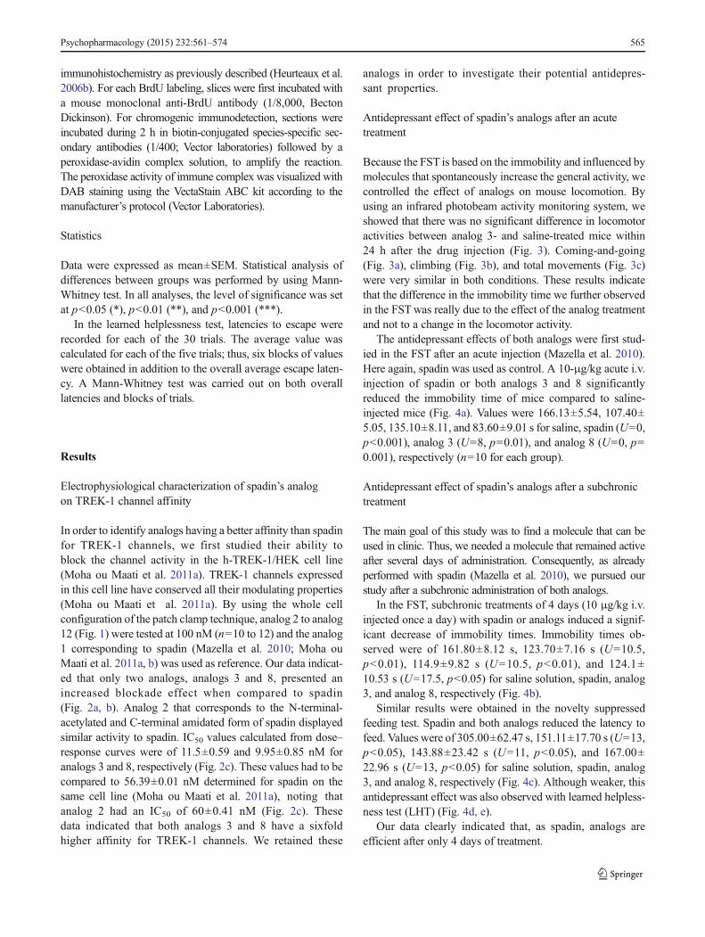
immunohistochemistry as previously described (Heurteaux et al.
2006b). For each BrdU labeling, slices were first incubated with
a mouse monoclonal anti-BrdU antibody (1/8,000, Becton
Dickinson). For chromogenic immunodetection, sections were
incubated during 2 h in biotin-conjugated species-specific sec-
ondary antibodies (1/400; Vector laboratories) followed by a
peroxidase-avidin complex solution, to amplify the reaction.
The peroxidase activity of immune complex was visualized with
DAB staining using the VectaStain ABC kit according to the
manufacturer’s protocol (Vector Laboratories).
Statistics
Data were expressed as mean±SEM. Statistical analysis of
differences between groups was performed by using Mann-
Whitney test. In all analyses, the level of significance was set
at p<0.05 (*), p<0.01 (**), and p<0.001 (***).
In the learned helplessness test, latencies to escape were
recorded for each of the 30 trials. The average value was
calculated for each of the five trials; thus, six blocks of values
were obtained in addition to the overall average escape laten-
cy. A Mann-Whitney test was carried out on both overall
latencies and blocks of trials.
Results
Electrophysiological characterization of spadin’s analog
on TREK-1 channel affinity
In order to identify analogs having a better affinity than spadin
for TREK-1 channels, we first studied their ability to
block the channel activity in the h-TREK-1/HEK cell line
(Moha ou Maati et al. 2011a). TREK-1 channels expressed
in this cell line have conserved all their modulating properties
(Moha ou Maati et al. 2011a). By using the whole cell
configuration of the patch clamp technique, analog 2 to analog
12 (Fig. 1) were tested at 100 nM (n=10 to 12) and the analog
1 corresponding to spadin (Mazella et al. 2010; Moha ou
Maati et al. 2011a, b) was used as reference. Our data indicat-
ed that only two analogs, analogs 3 and 8, presented an
increased blockade effect when compared to spadin
(Fig. 2a, b). Analog 2 that corresponds to the N-terminal-
acetylated and C-terminal amidated form of spadin displayed
similar activity to spadin. IC50 values calculated from dose–
response curves were of 11.5±0.59 and 9.95±0.85 nM for
analogs 3 and 8, respectively (Fig. 2c). These values had to be
compared to 56.39±0.01 nM determined for spadin on the
same cell line (Moha ou Maati et al. 2011a), noting that
analog 2 had an IC50 of 60±0.41 nM (Fig. 2c). These
data indicated that both analogs 3 and 8 have a sixfold
higher affinity for TREK-1 channels. We retained these
analogs in order to investigate their potential antidepres-
sant properties.
Antidepressant effect of spadin’s analogs after an acute
treatment
Because the FST is based on the immobility and influenced by
molecules that spontaneously increase the general activity, we
controlled the effect of analogs on mouse locomotion. By
using an infrared photobeam activity monitoring system, we
showed that there was no significant difference in locomotor
activities between analog 3- and saline-treated mice within
24 h after the drug injection (Fig. 3). Coming-and-going
(Fig. 3a), climbing (Fig. 3b), and total movements (Fig. 3c)
were very similar in both conditions. These results indicate
that the difference in the immobility time we further observed
in the FSTwas really due to the effect of the analog treatment
and not to a change in the locomotor activity.
The antidepressant effects of both analogs were first stud-
ied in the FST after an acute injection (Mazella et al. 2010).
Here again, spadin was used as control. A 10-μg/kg acute i.v.
injection of spadin or both analogs 3 and 8 significantly
reduced the immobility time of mice compared to saline-
injected mice (Fig. 4a). Values were 166.13±5.54, 107.40±
5.05, 135.10±8.11, and 83.60±9.01 s for saline, spadin (U=0,
p<0.001), analog 3 (U=8, p=0.01), and analog 8 (U=0, p=
0.001), respectively (n=10 for each group).
Antidepressant effect of spadin’s analogs after a subchronic
treatment
The main goal of this study was to find a molecule that can be
used in clinic. Thus, we needed a molecule that remained active
after several days of administration. Consequently, as already
performed with spadin (Mazella et al. 2010), we pursued our
study after a subchronic administration of both analogs.
In the FST, subchronic treatments of 4 days (10 μg/kg i.v.
injected once a day) with spadin or analogs induced a signif-
icant decrease of immobility times. Immobility times ob-
served were of 161.80±8.12 s, 123.70±7.16 s (U=10.5,
p<0.01), 114.9±9.82 s (U=10.5, p<0.01), and 124.1±
10.53 s (U=17.5, p<0.05) for saline solution, spadin, analog
3, and analog 8, respectively (Fig. 4b).
Similar results were obtained in the novelty suppressed
feeding test. Spadin and both analogs reduced the latency to
feed. Values were of 305.00±62.47 s, 151.11±17.70 s (U=13,
p<0.05), 143.88±23.42 s (U=11, p<0.05), and 167.00±
22.96 s (U=13, p<0.05) for saline solution, spadin, analog
3, and analog 8, respectively (Fig. 4c). Although weaker, this
antidepressant effect was also observed with learned helpless-
ness test (LHT) (Fig. 4d, e).
Our data clearly indicated that, as spadin, analogs are
efficient after only 4 days of treatment.
Psychopharmacology (2015) 232:561–574 565
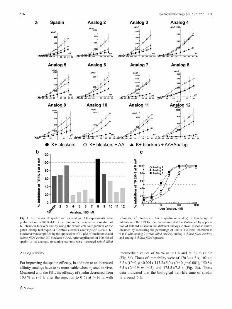
Analog stability
For improving the spadin efficacy, in addition to an increased
affinity, analogs have to be more stable when injected in vivo.
Measured with the FST, the efficacy of spadin decreased from
100 % at t=1 h after the injection to 0 % at t=16 h, with
intermediate values of 84 % at t=3 h and 30 % at t=7 h
(Fig. 5a). Times of immobility were of 170.3±4.5 s, 102.4±
6.2 s (U=0, p<0.001), 113.2±5.0 s (U=0, p<0.001), 150.8±
6.5 s (U=19, p<0.05), and 175.3±7.5 s (Fig. 5a). These
data indicated that the biological half-life time of spadin
is around 6 h.
Fig. 2 I–V curves of spadin and its analogs. All experiments were
performed on h-TREK-1/HEK cell line in the presence of a mixture of
K+ channels blockers and by using the whole cell configuration of the
patch clamp technique. a Control currents (black-filled circles, K+
blockers) were amplified by the application of 10 μM of arachidonic acid
(white-filled circles, K+ blockers + AA). After application of 100 nM of
spadin or its analogs, remaining currents were measured (black-filled
triangles, K+ blockers + AA + spadin or analog). b Percentage of
inhibition of the TREK-1 current measured at 0 mVobtained by applica-
tion of 100 nM of spadin and different analogs. c Dose–response curves
obtained by measuring the percentage of TREK-1 current inhibition at
0 mV with analog 2 (white-filled circles), analog 3 (black-filled circles),
and analog 8 (black-filled squares)
566 Psychopharmacology (2015) 232:561–574
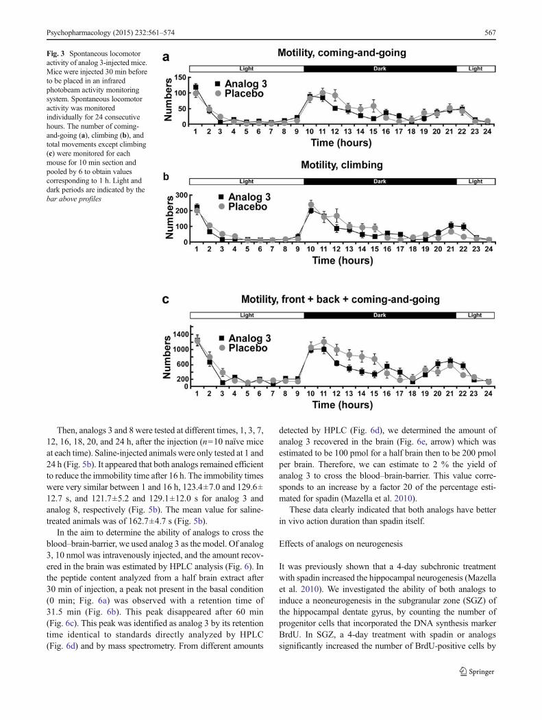
Then, analogs 3 and 8 were tested at different times, 1, 3, 7,
12, 16, 18, 20, and 24 h, after the injection (n=10 naïve mice
at each time). Saline-injected animals were only tested at 1 and
24 h (Fig. 5b). It appeared that both analogs remained efficient
to reduce the immobility time after 16 h. The immobility times
were very similar between 1 and 16 h, 123.4±7.0 and 129.6±
12.7 s, and 121.7±5.2 and 129.1±12.0 s for analog 3 and
analog 8, respectively (Fig. 5b). The mean value for saline-
treated animals was of 162.7±4.7 s (Fig. 5b).
In the aim to determine the ability of analogs to cross the
blood–brain-barrier, we used analog 3 as the model. Of analog
3, 10 nmol was intravenously injected, and the amount recov-
ered in the brain was estimated by HPLC analysis (Fig. 6). In
the peptide content analyzed from a half brain extract after
30 min of injection, a peak not present in the basal condition
(0 min; Fig. 6a) was observed with a retention time of
31.5 min (Fig. 6b). This peak disappeared after 60 min
(Fig. 6c). This peak was identified as analog 3 by its retention
time identical to standards directly analyzed by HPLC
(Fig. 6d) and by mass spectrometry. From different amounts
detected by HPLC (Fig. 6d), we determined the amount of
analog 3 recovered in the brain (Fig. 6e, arrow) which was
estimated to be 100 pmol for a half brain then to be 200 pmol
per brain. Therefore, we can estimate to 2 % the yield of
analog 3 to cross the blood–brain-barrier. This value corre-
sponds to an increase by a factor 20 of the percentage esti-
mated for spadin (Mazella et al. 2010).
These data clearly indicated that both analogs have better
in vivo action duration than spadin itself.
Effects of analogs on neurogenesis
It was previously shown that a 4-day subchronic treatment
with spadin increased the hippocampal neurogenesis (Mazella
et al. 2010). We investigated the ability of both analogs to
induce a neoneurogenesis in the subgranular zone (SGZ) of
the hippocampal dentate gyrus, by counting the number of
progenitor cells that incorporated the DNA synthesis marker
BrdU. In SGZ, a 4-day treatment with spadin or analogs
significantly increased the number of BrdU-positive cells by
Fig. 3 Spontaneous locomotor
activity of analog 3-injected mice.
Mice were injected 30 min before
to be placed in an infrared
photobeam activity monitoring
system. Spontaneous locomotor
activity was monitored
individually for 24 consecutive
hours. The number of coming-
and-going (a), climbing (b), and
total movements except climbing
(c) were monitored for each
mouse for 10 min section and
pooled by 6 to obtain values
corresponding to 1 h. Light and
dark periods are indicated by the
bar above profiles
Psychopharmacology (2015) 232:561–574 567
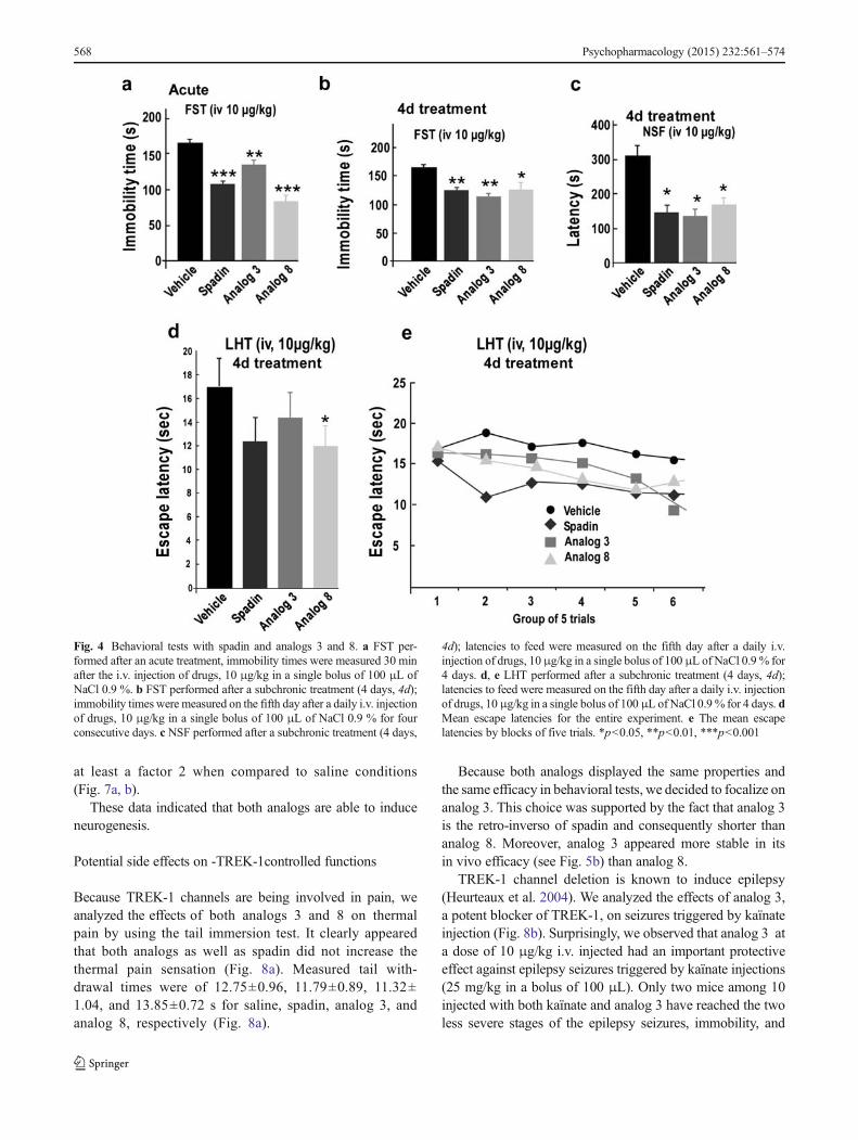
at least a factor 2 when compared to saline conditions
(Fig. 7a, b).
These data indicated that both analogs are able to induce
neurogenesis.
Potential side effects on -TREK-1controlled functions
Because TREK-1 channels are being involved in pain, we
analyzed the effects of both analogs 3 and 8 on thermal
pain by using the tail immersion test. It clearly appeared
that both analogs as well as spadin did not increase the
thermal pain sensation (Fig. 8a). Measured tail with-
drawal times were of 12.75±0.96, 11.79±0.89, 11.32±
1.04, and 13.85±0.72 s for saline, spadin, analog 3, and
analog 8, respectively (Fig. 8a).
Because both analogs displayed the same properties and
the same efficacy in behavioral tests, we decided to focalize on
analog 3. This choice was supported by the fact that analog 3
is the retro-inverso of spadin and consequently shorter than
analog 8. Moreover, analog 3 appeared more stable in its
in vivo efficacy (see Fig. 5b) than analog 8.
TREK-1 channel deletion is known to induce epilepsy
(Heurteaux et al. 2004). We analyzed the effects of analog 3,
a potent blocker of TREK-1, on seizures triggered by kaïnate
injection (Fig. 8b). Surprisingly, we observed that analog 3 at
a dose of 10 μg/kg i.v. injected had an important protective
effect against epilepsy seizures triggered by kaïnate injections
(25 mg/kg in a bolus of 100 μL). Only two mice among 10
injected with both kaïnate and analog 3 have reached the two
less severe stages of the epilepsy seizures, immobility, and
Fig. 4 Behavioral tests with spadin and analogs 3 and 8. a FST per-
formed after an acute treatment, immobility times were measured 30 min
after the i.v. injection of drugs, 10 μg/kg in a single bolus of 100 μL of
NaCl 0.9 %. b FST performed after a subchronic treatment (4 days, 4d);
immobility times were measured on the fifth day after a daily i.v. injection
of drugs, 10 μg/kg in a single bolus of 100 μL of NaCl 0.9 % for four
consecutive days. c NSF performed after a subchronic treatment (4 days,
4d); latencies to feed were measured on the fifth day after a daily i.v.
injection of drugs, 10μg/kg in a single bolus of 100 μL of NaCl 0.9 % for
4 days. d, e LHT performed after a subchronic treatment (4 days, 4d);
latencies to feed were measured on the fifth day after a daily i.v. injection
of drugs, 10μg/kg in a single bolus of 100 μL of NaCl 0.9% for 4 days. d
Mean escape latencies for the entire experiment. e The mean escape
latencies by blocks of five trials. *p<0.05, **p<0.01, ***p<0.001
568 Psychopharmacology (2015) 232:561–574
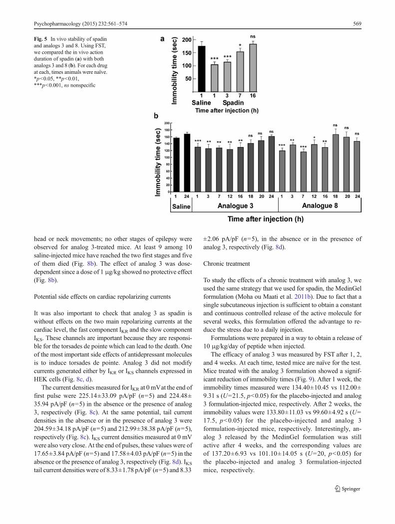
head or neck movements; no other stages of epilepsy were
observed for analog 3-treated mice. At least 9 among 10
saline-injected mice have reached the two first stages and five
of them died (Fig. 8b). The effect of analog 3 was dose-
dependent since a dose of 1 μg/kg showed no protective effect
(Fig. 8b).
Potential side effects on cardiac repolarizing currents
It was also important to check that analog 3 as spadin is
without effects on the two main repolarizing currents at the
cardiac level, the fast component IKR and the slow component
IKS. These channels are important because they are responsi-
ble for the torsades de pointe which can lead to the death. One
of the most important side effects of antidepressant molecules
is to induce torsades de pointe. Analog 3 did not modify
currents generated either by IKR or IKS channels expressed in
HEK cells (Fig. 8c, d).
The current densities measured for IKR at 0 mVat the end of
first pulse were 225.14±33.09 pA/pF (n=5) and 224.48±
35.94 pA/pF (n=5) in the absence or the presence of analog
3, respectively (Fig. 8c). At the same potential, tail current
densities in the absence or in the presence of analog 3 were
204.59±34.18 pA/pF (n=5) and 212.99±38.38 pA/pF (n=5),
respectively (Fig. 8c). IKS current densities measured at 0 mV
were also very close. At the end of pulses, these values were of
17.65±3.84 pA/pF (n=5) and 17.58±4.03 pA/pF (n=5) in the
absence or the presence of analog 3, respectively (Fig. 8d). IKStail current densities were of 8.33±1.78 pA/pF (n=5) and 8.33
±2.06 pA/pF (n=5), in the absence or in the presence of
analog 3, respectively (Fig. 8d).
Chronic treatment
To study the effects of a chronic treatment with analog 3, we
used the same strategy that we used for spadin, the MedinGel
formulation (Moha ou Maati et al. 2011b). Due to fact that a
single subcutaneous injection is sufficient to obtain a constant
and continuous controlled release of the active molecule for
several weeks, this formulation offered the advantage to re-
duce the stress due to a daily injection.
Formulations were prepared in a way to obtain a release of
10 μg/kg/day of peptide when injected.
The efficacy of analog 3 was measured by FST after 1, 2,
and 4 weeks. At each time, tested mice are naïve for the test.
Mice treated with the analog 3 formulation showed a signif-
icant reduction of immobility times (Fig. 9). After 1 week, the
immobility times measured were 134.40±10.45 vs 112.00±
9.31 s (U=21.5, p<0.05) for the placebo-injected and analog
3 formulation-injected mice, respectively. After 2 weeks, the
immobility values were 133.80±11.03 vs 99.60±4.92 s (U=
17.5, p<0.05) for the placebo-injected and analog 3
formulation-injected mice, respectively. Interestingly, an-
alog 3 released by the MedinGel formulation was still
active after 4 weeks, and the corresponding values are
of 137.20±6.93 vs 101.10±14.05 s (U=20, p<0.05) for
the placebo-injected and analog 3 formulation-injected
mice, respectively.
Fig. 5 In vivo stability of spadin
and analogs 3 and 8. Using FST,
we compared the in vivo action
duration of spadin (a) with both
analogs 3 and 8 (b). For each drug
at each, times animals were naïve.
*p<0.05, **p<0.01,
***p<0.001, ns nonspecific
Psychopharmacology (2015) 232:561–574 569
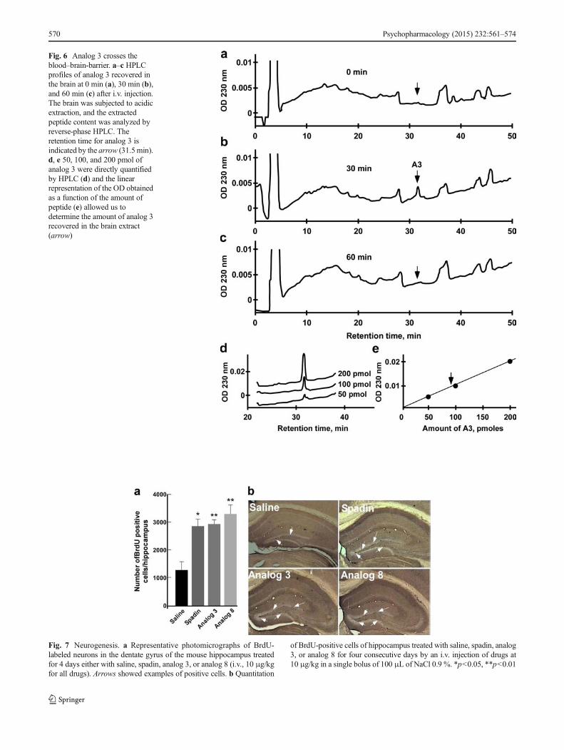
Fig. 6 Analog 3 crosses the
blood–brain-barrier. a–c HPLC
profiles of analog 3 recovered in
the brain at 0 min (a), 30 min (b),
and 60 min (c) after i.v. injection.
The brain was subjected to acidic
extraction, and the extracted
peptide content was analyzed by
reverse-phase HPLC. The
retention time for analog 3 is
indicated by the arrow (31.5min).
d, e 50, 100, and 200 pmol of
analog 3 were directly quantified
by HPLC (d) and the linear
representation of the OD obtained
as a function of the amount of
peptide (e) allowed us to
determine the amount of analog 3
recovered in the brain extract
(arrow)
Fig. 7 Neurogenesis. a Representative photomicrographs of BrdU-
labeled neurons in the dentate gyrus of the mouse hippocampus treated
for 4 days either with saline, spadin, analog 3, or analog 8 (i.v., 10 μg/kg
for all drugs). Arrows showed examples of positive cells. b Quantitation
of BrdU-positive cells of hippocampus treated with saline, spadin, analog
3, or analog 8 for four consecutive days by an i.v. injection of drugs at
10 μg/kg in a single bolus of 100 μL of NaCl 0.9 %. *p<0.05, **p<0.01
570 Psychopharmacology (2015) 232:561–574
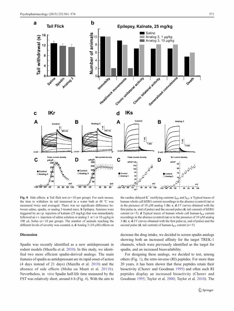
Discussion
Spadin was recently identified as a new antidepressant in
rodent models (Mazella et al. 2010). In this study, we identi-
fied two more efficient spadin-derived analogs. The main
features of spadin as antidepressant are its rapid onset of action
(4 days instead of 21 days) (Mazella et al. 2010) and the
absence of side effects (Moha ou Maati et al. 2011b).
Nevertheless, in vivo Spadin half-life time measured by the
FSTwas relatively short, around 6 h (Fig. 4). With the aim to
decrease the drug intake, we decided to screen spadin analogs
showing both an increased affinity for the target TREK-1
channels, which were previously identified as the target for
spadin, and an increased bioavailability.
For designing these analogs, we decided to test, among
others (Fig. 1), the retro-inverso (RI) peptides. For more than
20 years, it has been shown that these peptides retain their
bioactivity (Chorev and Goodman 1995) and often such RI
peptides display an increased bioactivity (Chorev and
Goodman 1995; Taylor et al. 2000; Taylor et al. 2010). The
Fig. 8 Side effects. a Tail flick test (n=10 per group). For each mouse,
the time to withdraw its tail immersed in a water bath at 48 °C was
measured twice and averaged. There was no significant difference be-
tween saline, spadin, or analog 3-treated mice. b Epilepsy. Seizures were
triggered by an i.p. injection of kaïnate (25 mg/kg) that was immediately
followed an i.v. injection of saline solution or analog 3 at 1 or 10μg/kg in
100 μL bolus (n=10 per group). The number of animals reaching the
different levels of severity was counted. c, dAnalog 3 (10 μM) effects on
the cardiac delayed K+ rectifying currents IKR and IKS. c Typical traces of
human whole cell hERG current recordings in the absence (control) (a) or
in the presence of 10 μM analog 3 (b). c, d I/V curves obtained with the
first pulse (c, end of pulse) and the second pulse (d, tail current) of hERG
current (n=5). d Typical traces of human whole cell human-IKS current
recordings in the absence (control) (a) or in the presence of 10 μM analog
3 (b). c, d I/V curves obtained with the first pulse (c, end of pulse) and the
second pulse (d, tail current) of human-IKS current (n=5)
Psychopharmacology (2015) 232:561–574 571
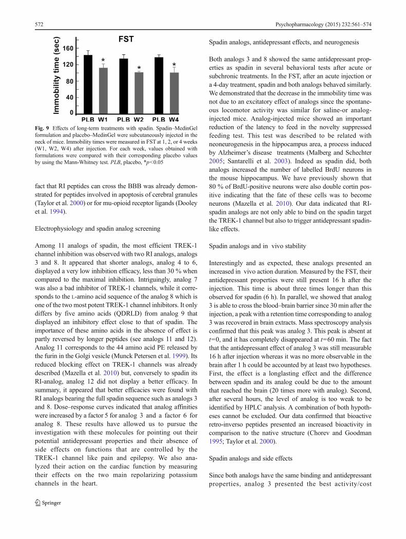
fact that RI peptides can cross the BBB was already demon-
strated for peptides involved in apoptosis of cerebral granules
(Taylor et al. 2000) or for mu-opioid receptor ligands (Dooley
et al. 1994).
Electrophysiology and spadin analog screening
Among 11 analogs of spadin, the most efficient TREK-1
channel inhibition was observed with two RI analogs, analogs
3 and 8. It appeared that shorter analogs, analog 4 to 6,
displayed a very low inhibition efficacy, less than 30 % when
compared to the maximal inhibition. Intriguingly, analog 7
was also a bad inhibitor of TREK-1 channels, while it corre-
sponds to the L-amino acid sequence of the analog 8 which is
one of the two most potent TREK-1 channel inhibitors. It only
differs by five amino acids (QDRLD) from analog 9 that
displayed an inhibitory effect close to that of spadin. The
importance of these amino acids in the absence of effect is
partly reversed by longer peptides (see analogs 11 and 12).
Analog 11 corresponds to the 44 amino acid PE released by
the furin in the Golgi vesicle (Munck Petersen et al. 1999). Its
reduced blocking effect on TREK-1 channels was already
described (Mazella et al. 2010) but, conversely to spadin its
RI-analog, analog 12 did not display a better efficacy. In
summary, it appeared that better efficacies were found with
RI analogs bearing the full spadin sequence such as analogs 3
and 8. Dose–response curves indicated that analog affinities
were increased by a factor 5 for analog 3 and a factor 6 for
analog 8. These results have allowed us to pursue the
investigation with these molecules for pointing out their
potential antidepressant properties and their absence of
side effects on functions that are controlled by the
TREK-1 channel like pain and epilepsy. We also ana-
lyzed their action on the cardiac function by measuring
their effects on the two main repolarizing potassium
channels in the heart.
Spadin analogs, antidepressant effects, and neurogenesis
Both analogs 3 and 8 showed the same antidepressant prop-
erties as spadin in several behavioral tests after acute or
subchronic treatments. In the FST, after an acute injection or
a 4-day treatment, spadin and both analogs behaved similarly.
We demonstrated that the decrease in the immobility time was
not due to an excitatory effect of analogs since the spontane-
ous locomotor activity was similar for saline-or analog-
injected mice. Analog-injected mice showed an important
reduction of the latency to feed in the novelty suppressed
feeding test. This test was described to be related with
neoneurogenesis in the hippocampus area, a process induced
by Alzheimer’s disease treatments (Malberg and Schechter
2005; Santarelli et al. 2003). Indeed as spadin did, both
analogs increased the number of labelled BrdU neurons in
the mouse hippocampus. We have previously shown that
80 % of BrdU-positive neurons were also double cortin pos-
itive indicating that the fate of these cells was to become
neurons (Mazella et al. 2010). Our data indicated that RI-
spadin analogs are not only able to bind on the spadin target
the TREK-1 channel but also to trigger antidepressant spadin-
like effects.
Spadin analogs and in vivo stability
Interestingly and as expected, these analogs presented an
increased in vivo action duration. Measured by the FST, their
antidepressant properties were still present 16 h after the
injection. This time is about three times longer than this
observed for spadin (6 h). In parallel, we showed that analog
3 is able to cross the blood–brain barrier since 30 min after the
injection, a peak with a retention time corresponding to analog
3 was recovered in brain extracts. Mass spectroscopy analysis
confirmed that this peak was analog 3. This peak is absent at
t=0, and it has completely disappeared at t=60 min. The fact
that the antidepressant effect of analog 3 was still measurable
16 h after injection whereas it was no more observable in the
brain after 1 h could be accounted by at least two hypotheses.
First, the effect is a longlasting effect and the difference
between spadin and its analog could be due to the amount
that reached the brain (20 times more with analog). Second,
after several hours, the level of analog is too weak to be
identified by HPLC analysis. A combination of both hypoth-
eses cannot be excluded. Our data confirmed that bioactive
retro-inverso peptides presented an increased bioactivity in
comparison to the native structure (Chorev and Goodman
1995; Taylor et al. 2000).
Spadin analogs and side effects
Since both analogs have the same binding and antidepressant
properties, analog 3 presented the best activity/cost
Fig. 9 Effects of long-term treatments with spadin. Spadin–MedinGel
formulation and placebo–MedinGel were subcutaneously injected in the
neck of mice. Immobility times were measured in FST at 1, 2, or 4 weeks
(W1, W2, W4) after injection. For each week, values obtained with
formulations were compared with their corresponding placebo values
by using the Mann-Whitney test. PLB, placebo, *p<0.05
572 Psychopharmacology (2015) 232:561–574

compromise and was investigated for potential side effects.
Treatingmice with analog 3 did not increase their thermal pain
sensitivity, confirming data obtained with spadin (Moha ou
Maati et al. 2011b). Interestingly, on kaïnate-triggered epilep-
tic seizures, analog 3 at a dose of 1 μg/kg had no effect on the
severity of seizures. But when administrated at a dose of
10 μg/kg, analog 3 showed a high degree of protection against
seizures. At this dose, only two animals reached the two first
level of seizure severity while nine saline-injected mice
reached these levels. Treating animals with analog 3 amplified
the protective effect against seizures which was glimpsed with
spadin (Moha ou Maati et al. 2011b). As many drugs can
induce cardiac dysfunction, among them ADs (Downes et al.
2005; Heist and Ruskin 2005), we analyzed analog 3 effects on
IKR and IKS currents, the twomain potassium channels at cardiac
level, that are responsible for torsades de pointe and sudden
death (Aizawa et al. 2007; Fenichel et al. 2004; Schechter et al.
2005). Analog 3 was without effects on both currents. As in the
case of spadin (Moha ou Maati et al. 2011b), these results
demonstrated that RI analogs of spadin did not interfere with
other TREK-1-controlled pathways and did not modify the
cardiac function.
Spadin analogs and chronic treatment
Analog 3 was also used to verify that the antidepressant effect
was persistent even after a chronic treatment. This experiment
was performed, thanks to a MedinGel formulation that allows
following a single subcutaneous injection a continuous con-
trolled release of the peptide during several weeks (Moha ou
Maati et al. 2011b). Data showed that the antidepressant effect
was the same after 4 weeks and demonstrated that there was
no tolerance.
Conclusion
The three fold increase in the bioavailability associated with
five- or sixfold increase in the affinity of RI-peptide for the
TREK-1 channel indicated that analogs improved by a factor
15 to 18 the efficacy of spadin. Additionally, RI analogs did
not induce side effects and their action was stable over the
time. Taken together, these properties are very important in the
aim to transform spadin or its analogs into a usable drug in
human clinic. Indeed, one third of patients remain un-
treated because they do not correctly take their drugs.
Improvements to simplify drug intake will be very help-
ful for these patients. In this study, we have identified a
very potent spadin analog that, associated with a
MedinGel formulation, could represent a great step in
the spadin drug design concept for treating these un-
treated patients.
Acknowledgments We thank Anthony Rech for his expert technical
assistance in the preparation of injectable formulations. We thank
Delphine Debayle for his expert technical assistance in the mass spec-
troscopy analysis. We thank Layla Djillani for carefully reading the
manuscript. This work is supported by the Centre National de la
Recherche Scientifique (CNRS) and the Agence Nationale de la
Recherche-Emergence (ANR-EMMA-2011-059 and ANR-13-RPIB-
0002). J. Veyssiere was supported by a CIFRE fellowship.
Open AccessThis article is distributed under the terms of the Creative
Commons Attribution License which permits any use, distribution, and
reproduction in any medium, provided the original author(s) and the
source are credited.
References
Aizawa Y, Ueda K, Scornik F, Cordeiro JM,Wu Y, Desai M, Guerchicoff
A, Nagata Y, Iesaka Y, Kimura A, Hiraoka M, Antzelevitch C
(2007) A novel mutation in KCNQ1 associated with a potent dom-
inant negative effect as the basis for the LQT1 form of the long QT
syndrome. J Cardiovasc Electrophysiol 18:972–977
Alloui A, Zimmermann K, Mamet J, Duprat F, Noel J, Chemin J, Guy N,
Blondeau N, Voilley N, Rubat-Coudert C, Borsotto M, Romey G,
Heurteaux C, Reeh P, Eschalier A, Lazdunski M (2006) TREK-1, a
K+ channel involved in polymodal pain perception. EMBO J 25:
2368–2376
Bonny C, Oberson A, Negri S, Sauser C, Schorderet DF (2001) Cell-
permeable peptide inhibitors of JNK: novel blockers of beta-cell
death. Diabetes 50:77–82
Caldarone BJ, George TP, Zachariou V, Picciotto MR (2000) Gender
differences in learned helplessness behavior are influenced by ge-
netic background. Pharmacol Biochem Behav 66:811–817
Checler F, Mazella J, Kitabgi P, Vincent JP (1986) High-affinity receptor
sites and rapid proteolytic inactivation of neurotensin in primary
cultured neurons. J Neurochem 47:1742–1748
Chorev M, Goodman M (1995) Recent developments in retro peptides
and proteins—an ongoing topochemical exploration. Trends
Biotechnol 13:438–445
Dooley CT, Chung NN, Wilkes BC, Schiller PW, Bidlack JM, Pasternak
GW, Houghten RA (1994) An all D-amino acid opioid peptide with
central analgesic activity from a combinatorial library. Science 266:
2019–2022
DownesMA,Whyte IM, Isbister GK (2005) QTc abnormalities in deliberate
self-poisoning with moclobemide. Intern Med J 35:388–391
Ducroq J, Moha ou Maati H, Guilbot S, Dilly S, Laemmel E, Pons-
Himbert C, Faivre JF, Bois P, Stucker O, Le Grand M (2010)
Dexrazoxane protects the heart from acute doxorubicin-induced
QT prolongation: a key role for I(Ks). Br J Pharmacol 159:93–101
Fenichel RR,MalikM, Antzelevitch C, Sanguinetti M, Roden DM, Priori
SG, Ruskin JN, Lipicky RJ, Cantilena LR (2004) Drug-induced
torsades de pointes and implications for drug development. J
Cardiovasc Electrophysiol 15:475–495
Greenberg PE, Kessler RC, Birnbaum HG, Leong SA, Lowe SW,
Berglund PA, Corey-Lisle PK (2003) The economic burden of
depression in the United States: how did it change between 1990
and 2000? J Clin Psychiatry 64:1465–1475
Heist EK, Ruskin JN (2005) Drug-induced proarrhythmia and use of
QTc-prolonging agents: clues for clinicians. Heart Rhythm 2:S1–S8
Heurteaux C, Guy N, Laigle C, Blondeau N, Duprat F, Mazzuca M,
Lang-Lazdunski L, Widmann C, Zanzouri M, Romey G, Lazdunski
M (2004) TREK-1, a K(+) channel involved in neuroprotection and
general anesthesia. Embo J 23:2684–2695
Heurteaux C, Laigle C, Blondeau N, Jarretou G, Lazdunski M (2006a)
Alpha-linolenic acid and riluzole treatment confer cerebral
Psychopharmacology (2015) 232:561–574 573
protection and improve survival after focal brain ischemia.
Neuroscience 137:241–251
Heurteaux C, Lucas G, Guy N, El Yacoubi M, Thümmler S, Peng X,
Noble F, Blondeau N, Widmann C, Gobbi G, Costentin J, Debonnel
G, Lazdunski M (2006b) Deletion of TREK-1, a background potas-
sium channel, results in a depression-resistant phenotype. Nat
Neurosci 9:1134–1141
Lauritzen I, Blondeau N, Heurteaux C, Widmann C, Romey G,
Lazdunski M (2000) Polyunsaturated fatty acids are potent
neuroprotectors. Embo J 19:1784–1793
Malberg JE, Schechter LE (2005) Increasing hippocampal neurogenesis:
a novel mechanism for antidepressant drugs. Curr Pharm Des 11:
145–155
Mazella J, Zsurger N, Navarro V, Chabry J, Kaghad M, Caput D, Ferrara
P, Vita N, Gully D, Maffrand JP, Vincent JP (1998) The 100-kDa
neurotensin receptor is gp95/sortilin, a non-G-protein-coupled re-
ceptor. J Biol Chem 273:26273–26276
Mazella J, Petrault O, Lucas G, Deval E, Beraud-Dufour S, Gandin C, El-
Yacoubi M, Widmann C, Guyon A, Chevet E, Taouji S, Conductier
G, Corinus A, Coppola T, Gobbi G, Nahon JL, Heurteaux C,
Borsotto M (2010) Spadin, a sortilin-derived peptide, targeting
rodent TREK-1 channels: a new concept in the antidepressant drug
design. PLoS Biol 8:e1000355
Moha ou Maati H, Peyronnet R, Devader C, Veyssiere J, Labbal F,
Gandin C, Mazella J, Heurteaux C, Borsotto M (2011a) A human
TREK-1/HEK cell line: a highly efficient screening tool for drug
development in neurological diseases. PLoS ONE 6:e25602
Moha ouMaati H, Veyssiere J, Labbal F, Coppola T, Gandin C,Widmann
C, Mazella J, Heurteaux C, Borsotto M (2011b) Spadin as a new
antidepressant: absence of TREK-1-related side effects.
Neuropharmacology 62:278–288
Moussavi S, Chatterji S, Verdes E, Tandon A, Patel V, Ustun B (2007)
Depression, chronic diseases, and decrements in health: results from
the World Health Surveys. Lancet 370:851–858
Munck Petersen C, NielsenMS, Jacobsen C, Tauris J, Jacobsen L, Gliemann
J, Moestrup SK, Madsen P (1999) Propeptide cleavage conditions
sortilin/neurotensin receptor-3 for ligand binding. Embo J 18:595–604
Nestler EJ, Barrot M, DiLeone RJ, Eisch AJ, Gold SJ, Monteggia LM
(2002) Neurobiology of depression. Neuron 34:13–25
Noel J, Zimmermann K, Busserolles J, Deval E, Alloui A, Diochot S,
Guy N, Borsotto M, Reeh P, Eschalier A, Lazdunski M (2009) The
mechano-activated K+ channels TRAAK and TREK-1 control both
warm and cold perception. EMBO J 28:1308–1318
Porsolt RD, Le Pichon M, Jalfre M (1977) Depression: a new animal
model sensitive to antidepressant treatments. Nature 266:730–732
Santarelli L, Saxe M, Gross C, Surget A, Battaglia F, Dulawa S,
Weisstaub N, Lee J, Duman R, Arancio O, Belzung C, Hen R
(2003) Requirement of hippocampal neurogenesis for the behavioral
effects of antidepressants. Science 301:805–809
Schechter LE, Ring RH, Beyer CE, Hughes ZA, Khawaja X, Malberg JE,
Rosenzweig-Lipson S (2005) Innovative approaches for the devel-
opment of antidepressant drugs: current and future strategies.
NeuroRx 2:590–611
Sicouri S, Antzelevitch C (2008) Sudden cardiac death secondary to
antidepressant and antipsychotic drugs. Expert Opin Drug Saf 7:
181–194
Taylor EM, Otero DA, Banks WA, O'Brien JS (2000) Retro-
inverso prosaptide peptides retain bioactivity, are stable In
vivo, and are blood–brain barrier permeable. J Pharmacol
Exp Ther 295:190–194
Taylor M, Moore S, Mayes J, Parkin E, Beeg M, Canovi M, Gobbi M,
Mann DM, Allsop D (2010) Development of a proteolytically stable
retro-inverso peptide inhibitor of beta-amyloid oligomerization as a
potential novel treatment for Alzheimer's disease. Biochemistry 49:
3261–3272
Tsirka SE, Gualandris A, Amaral DG, Strickland S (1995) Excitotoxin-
induced neuronal degeneration and seizure are mediated by tissue
plasminogen activator. Nature 377:340–344
Van Regenmortel MH, Muller S (1998) D-peptides as immunogens and
diagnostic reagents. Curr Opin Biotechnol 9:377–382
Weeden T, Stefano J, Duan S, Edling A, Hou L, Chuang WL, Perricone
MA, Pan C, Dzuris JL (2011) A retro-inverso alpha-melanocyte
stimulating hormone analog with MC1R-binding selectivity. J Pept
Sci 17:47–55
574 Psychopharmacology (2015) 232:561–574
ORIGINAL RESEARCHpublished: 12 September 2017doi: 10.3389/fphar.2017.00643
Frontiers in Pharmacology | www.frontiersin.org 1 September 2017 | Volume 8 | Article 643
Edited by:
Maurizio Taglialatela,
University of Naples Federico II, Italy
Reviewed by:
Giulia Maria Camerino,
Università degli Studi di Bari Aldo
Moro, Italy
Sergei Noskov,
University of Calgary, Canada
*Correspondence:
Marc Borsotto
†These authors have contributed
equally to this work.
Specialty section:
This article was submitted to
Pharmacology of Ion Channels and
Channelopathies,
a section of the journal
Frontiers in Pharmacology
Received: 16 June 2017
Accepted: 30 August 2017
Published: 12 September 2017
Citation:
Djillani A, Pietri M, Moreno S,
Heurteaux C, Mazella J and
Borsotto M (2017) Shortened Spadin
Analogs Display Better TREK-1
Inhibition, In Vivo Stability and
Antidepressant Activity.
Front. Pharmacol. 8:643.
doi: 10.3389/fphar.2017.00643
Shortened Spadin Analogs DisplayBetter TREK-1 Inhibition, In VivoStability and Antidepressant ActivityAlaeddine Djillani †, Mariel Pietri †, Sébastien Moreno, Catherine Heurteaux, Jean Mazella
and Marc Borsotto *
Centre National de la Recherche Scientifique, Institut de Pharmacologie Moléculaire et Cellulaire, UMR7275, Université Côte
d’Azur, Valbonne, France
Depression is a devastating mental disorder that affects 20% of the population
worldwide. Despite their proven efficacy, antidepressants present a delayed onset of
action and serious adverse effects. Seven years ago, we described spadin (PE 12-28) as
a promising endogenous peptide with antidepressant activity. Spadin specifically blocks
the TREK-1 channel. Previously, we showed in vivo that, spadin activity disappeared
beyond 7 h after administration. In order to improve in vivo spadin stability and
bioavailability, we screened spadin analogs and derivatives. From the study of spadin
blood degradation products, we designed a 7 amino-acid peptide, PE 22-28. In vitro
studies on hTREK-1/HEK cells by using patch-clamp technique, showed that PE 22-28
displayed a better specificity and affinity for TREK-1 channel compared to spadin, IC50
of 0.12 nM vs. 40–60 nM for spadin. In the same conditions, we also pointed out
that different modifications of its N or C-terminal ends maintained or abolished TREK-1
channel activity without affecting PE 22-28 affinity. In vivo, the antidepressant properties
of PE 22-28 and its derivatives were demonstrated in behavioral models of depression,
such as the forced swimming test. Mice treated with spadin-analogs showed a significant
reduction of the immobility time. Moreover, in the novelty suppressed feeding test after a
4-day sub-chronic treatment PE 22-28 reduced significantly the latency to eat the food
pellet. PE 22-28 and its analogs were able to induce neurogenesis after only a 4-day
treatment with a prominent effect of the G/A-PE 22-28. On mouse cortical neurons, PE
22-28 and its derivatives enhanced synaptogenesis measured by the increase of PSD-95
expression level. Finally, the action duration of PE 22-28 and its analogs was largely
improved in comparison with that of spadin, up to 23 h instead of 7 h. Taken together,
our results demonstrated that PE 22-28 and its derivatives represent other promising
molecules that could be an alternative to spadin in the treatment of depression.
Keywords: spadin-analogs, TREK-1 channel, PE 22-28, neurogenesis, synaptogenesis, antidepressant
INTRODUCTION
Depression is one of themost commonmood disorders that represents a heavy economic burden inindustrialized countries (Smith, 2014). Severe depression affects 2–5% of US citizens and up to 20%of the population suffer from mild depression (Nestler et al., 2002; Maletic et al., 2007; Krishnanand Nestler, 2008; Kessler et al., 2012). Depression is a complex syndrome with a variety of causes
Djillani et al. Shortening Spadin Increases Spadin Efficiency
mostly genetic and environmental (Nestler et al., 2002). One ofthe main hypotheses proposed to explain the physiopathologyof depression is the monoamine hypothesis where depletion ofthree monoamines serotonin (5-HT), norepinephrine (NA) ordopamine (DA) is thought to cause depression (Hillhouse andPorter, 2015). Later, several antidepressant (AD) drugs weredeveloped in the aim to restore the physiological synaptic levels ofthe three neurotransmitters (Hillhouse and Porter, 2015). Severaltypes of AD drugs were and are still used in the treatment ofdepression. Initially, depression was mainly treated by tricyclicsfamily and at lesser extent by monoamine oxidase inhibitors(MAOIs) and serotonin-norepinephrine re-uptake inhibitors(SNRIs). However, these classes of ADs exhibit numerous andserious side effects (Hirschfeld, 2012). To reduce the frequencyand occurrence of these side effects, these drugs were replaced bya generation of ADs more specific and with lesser adverse effects.This includes serotonin-selective re-uptake inhibitors (SSRIs)and norepinephrine selective re-uptake inhibitors (NSRIs) thatare widely used nowadays and recommended as first-linetreatment for depression (Nestler et al., 2002; Cleare et al.,2015). SSRIs and NSRIs are thought to increase monoaminesynaptic concentrations by inhibiting the re-uptake of 5-HTor NA by blocking their specific transporters, SERT and NAT,respectively (Kohler et al., 2016). These AD drugs are moretolerated but their efficacy on depressive patients is not reallyimproved. On the other hand, the actual ADs like fluoxetineare only efficient after 3–4 weeks of treatment, a latency periodthat still remains unexplained (Nestler et al., 2002). Thus, itis necessary to discover and characterize new targets for newAD drugs. Recently, different fast-onset AD drugs have beendescribed like ketamine, scopolamine or GLYX-13 (Ramakerand Dulawa, 2017). Nevertheless, these molecules, particularlyketamine produces a number of adverse effects (Katalinic et al.,2013). Multimodal drugs, such as vortioxetine or vilazodone area new class of ADs latterly approved by the US Food and DrugAdministration for the treatment of major depressive disorders(Wang et al., 2015; Sowa-Kucma et al., 2017). Nevertheless,these drugs have not represented a clear improvement ofantidepressant efficacy, but vortioxetine showed beneficial effectsin depression-related cognitive impairment whereas vilazodoneappeared to induce minor sexual dysfunctions (Deardorff andGrossberg, 2014; Li et al., 2015; Thase et al., 2016). Despitethis therapeutic arsenal, more than 30% of depressive patientsnever remit even after several classical treatments (Rush et al.,2006). An alternate for drug therapy in resistant patients isthe electroconvulsive therapy (ECT). ECT is efficient in 50%of pharmacotherapy-resistant patients (Heijnen et al., 2010)but ECT also induces some adverse effects mainly in thecognitive processes (Semkovska and McLoughlin, 2010). Thus,the discovery of new AD drugs is challenging. Ten years ago, wehave identified the selective two-pore domain potassium channelTREK-1 (TWIK-related potassium channel-1) as a potentialtarget for depression treatment (Heurteaux et al., 2006; Borsotto
Abbreviations: AD, antidepressant; FST, Forced Swim Test; NSF, Novelty-
suppressed Feeding; LHT, Learned Helplessness Test; ip, intraperitoneal; PSD-95,
Post-Synaptic Density protein 95.
et al., 2015). TREK-1 channels are ubiquitous potassium channelsthat play pivotal role in stabilizing membrane potential and thusprevent cellular excitability (Honore, 2007). TREK-1 channelsare very particular K2P channels since they are involved inmany physiological and physiopathological processes, such aspain, epilepsy, stroke, and depression (Lauritzen et al., 2000;Alloui et al., 2006; Heurteaux et al., 2006). TREK-1 becamealso an attractive target in cardiovascular research because itplays an important role in atrial fibrillation, pulmonary arterialhypertension and ventricular arrhythmia (Wiedmann et al.,2016; Decher et al., 2017). In the field of depression, weshowed in five different models of depression that deletionof kcnk2 gene, which encodes for TREK-1 channels results ina depression-resistant phenotype associated with an enhancedserotonergic neurotransmission and an increased neurogenesisin the hippocampus (Heurteaux et al., 2006). These observationsled us to search for potent TREK-1 blockers. Then, we identifiedspadin which derives from a larger peptide called propeptide(PE) (Mazella et al., 2010). PE is a 44 amino-acid that resultsfrom the post-translational maturation in the Golgi apparatusof sortilin, also known as the neurotensin receptor-3 (Mazella,2001). Spadin is a fast-acting ADwhich does not produce any sideeffects on functions that are controlled by the TREK-1 channel(Moha Ou Maati et al., 2012). It is able to counteract depressionin only 4 days when classical ADs require 3–4 weeks to be efficient(Mazella et al., 2010). Moreover, spadin blocks TREK-1 withhigher affinity, IC50 ∼ 40–60 nM vs. IC50 ∼ 6µM for the mostused SSRI fluoxetine (Mazella et al., 2010; Moha ou Maati et al.,2011). However, in mice, the AD activity of spadin measuredby FST disappears beyond 7 h after an acute ip administration(Veyssiere et al., 2015). In order to improve the spadin stabilityin vivo, we decided to search for analogs or derivatives of spadin.In a previous study we have identified two analogs, analogs 3 and8, that were synthesized by using the retro-inverso technology(Veyssiere et al., 2015). Although the gain in term of affinityand action duration was about 20, it is not sufficient to makethese analogs competitive in regard of their synthesis cost incomparison to spadin. Then, we decided to search for shortenedanalogs. We first studied whether or not spadin is degraded inthe blood circulation. We identified two shortened peptides. Bycomparing their ability to inhibit TREK-1 channel expressed inthe hTREK-1/HEK cell line (Moha ou Maati et al., 2011), weidentified the shortest efficient sequence that only contained 7amino-acid called PE 22-28. This peptide was used as a coresequence for preparing analogs by chemical modifications of itsN- or C-terminus ends and also by substitution of amino-acids.As PE 22-28, some modified peptide-analogs displayed a betterpotency in blocking TREK-1 channel andmore importantly, theyhave retained their AD properties when injected in acute orsub-chronic treatments.
MATERIALS AND METHODS
In Vitro AnalysisCell LinesThe human TREK-1/HEK cell line (Moha ou Maati et al., 2011)and the native HEK293 cell line were maintained in Dulbecco’s
Frontiers in Pharmacology | www.frontiersin.org 2 September 2017 | Volume 8 | Article 643
Djillani et al. Shortening Spadin Increases Spadin Efficiency
Modified Eagle’s Medium (DMEM) supplemented with 10%(v/v) heat-inactivated fetal bovine serum, 1% (v/v) penicillin-streptomycin, 1% Glutamax. For the hTREK-1/HEK cells, 0.5mg/ml G418 were added to the medium.
Cells were incubated in a humidified atmosphere containing5% CO2. For electrophysiological measurements, cells wereplated at a density of 20,000 cells per 35mm dish.
In order to study the effects of shortened spadin analogson hTREK-2, hTASK-1, hTRAAK and hTRESK, the nativeHEK293 cells were transfected with DNAs corresponding to thechannels using JetPEI (Polyplus-transfection, France) followingthe provider’s instructions.
Electrophysiological MeasurementsCells from the hTREK-1/HEK cell line were seeded at a densityof 20,000 cells/35mm dish. Electrophysiological recordingswere performed 24–48 h after plating using the whole-cellconfiguration of patch-clamp technique. TREK-1 currents(ITREK−1) were recorded using RK400 patch-clamp amplifier(Axon Instrument, USA). They were low-pass filtered at3 kHz and digitized at 10 kHz using a 12-bit analog-to-digital converter digidata (1322 series, Axon Instrument, USA).ITREK−1amplitudes were expressed as current densities [currentamplitude (pA)/membrane capacitance (pF)]. The results wereexpressed as mean± SEM (standard error of the mean).
Pipettes were pulled from borosilicate glass capillaries usinga dual-stage micropipette puller (PC-10, Narishige); they hada resistance of 1.5–3 M. Cells were continuously perfusedusing an external bath solution containing in mM: 150 NaCl,5 KCl, 3 MgCl2, 1 CaCl2, and 10 HEPES. The bath solutionwas initially adjusted to pH 7.4 with NaOH. The intra-pipettesolution contained in mM: 155 KCl, 3 MgCl2, 5 EGTA, and10 HEPES adjusted to pH 7.2 with KOH. In order to measureITREK−1, a cocktail of potassium channel blockers was addedto the bath solution. This cocktail contained: 3mM 4-AP(4-aminopyridine), 10mM TEA (tetraethylammonium), 10µMGlibenclamide, 100 nM Apamin, and 50 nM Charybdotoxin.Data acquisition was carried out using a computer (DellPentium) with pClamp software (Axon Instrument, USA).Whole-cell currents were generated by running a pulse or rampprotocol every 5 s from−100 to+60mVwith a holding potentialmaintained at −80mV. To evaluate the inhibitory effect onTREK-1 channels of shortened spadin analogs compared withspadin, cells were first activated by 10µM arachidonic acid (AA).Dose-response curves were realized to compare spadin-analogeffects with spadin using Origin 8.6 (Northampton, MA, USA).
Patch-clamp recording data were analyzed using Clampfit(Molecular Devices, USA). I = f(V) curves were obtained from−100 to+60mV ramp. Data were presented as mean± SEM.
In Vivo AnalysisAnimalsNaïve male C57Bl/6J mice from 7 to 9 weeks old were used in allexperiments (Janvier laboratory, Saint Berthevin, France). Micewere housed (10 animals per cage) under a 12-h light/12-h darkcycle (light on at 8:00 am) in a ventilated room at a temperatureof 22 ± 1C. Animals had free access to water and food (A03;
SAFE, Augy, France). All experiments were conducted accordingto policies on the care and use of laboratory animals of theSociety for Neuroscience, and also with respect to national lawson animal use. The local Ethics Committee (CIEPAL, N 736-02)approved the experiments.
ChemicalsSpadin, PE 22-28 and PE 22-28 analogs were purchased fromGeneCust Europe, Luxembourg. G418, Arachidonic acid (AA),4-AP (4-aminopyridine), TEA (tetraethylammonium), Apaminand Charybdotoxin were purchased from Sigma-Aldrich, France,Glibenclamide was purchased from ICN Biomedicals (USA).
In Vitro Half-Life Time of Spadin in SerumThe half-life time of spadin was measured in serum by incubating10 nmoles of the peptide with 200µl of mouse serum in theabsence or in the presence of the metalloprotease inhibitor 1–10 phenanthroline (1 mM) for various times (2, 5, 15, 30, and60 min) at room temperature. Incubations were stopped bydirect acidification (5µl of 2.5 M HCl), loaded in C-18 sepackcartridges, lyophilized, resuspended in 20% acetonitrile beforeloading onto HPLC for analysis of PE degradation/stability.Elutions of HPLC products were carried out by means of a 50-min linear gradient of acetonitrile from 20 to 70% at a flow rateof 1 ml/min. The amount of the intact PE for each incubationtime was expressed as the percent of the initial amount of spadin.
Behavioral TestsPorsolt Forced Swim Test (FST)Mice were individually placed for 6 min in a non-escapablecylinder (30 cm height and 15 cm diameter) half-filled with waterat 22 ± 1C. The immobility time was manually measured onlyduring the last 4 min. A mouse was considered immobile when itremained immobile with only slight movements in order to keepits head above water (Porsolt et al., 1977).
Novelty Suppressed Feeding Test (NSF)Mice were deprived from food for 24 h before the test. A foodpellet was placed on a white platform in the center of a highlyilluminated area (45 × 45 × 20 cm). The floor was covered withwooden bedding. Mice were placed in the corner of the arena,during a period of 10 min, the latency to start eating the pelletwas measured (Santarelli et al., 2003).
Learned Helplessness Test (LHT)The learned helplessness test consists in a 4-day training sessionand a single day test.
During the training session mice were exposed to 360inescapable 2 s foot shocks, with an inter-trial interval of 8 s. Anon-shocked group was exposed to the apparatus for the sameduration but no shock was delivered.
The test consisted in 30 trials separated by a 30 s interval.One trial was defined as a 5 s period before shock onset and wasterminated when the mouse moved to the second compartmentor at the end of the shock onset. The latency to escape for eachmouse during every trial was recorded (Caldarone et al., 2000).
Frontiers in Pharmacology | www.frontiersin.org 3 September 2017 | Volume 8 | Article 643
Djillani et al. Shortening Spadin Increases Spadin Efficiency
BrdU LabelingTwenty hours after the injections of 5-Bromo-2′-deoxyuridine(BrdU) (12mg per animals administered in three bolus of 100µLof a solution of 40mg/mL of BrdU diluted in 0.9% NaCl), micewere anesthetized with isoflurane and transcardially perfusedfirst with NaCl 0.9% and, second with 4% paraformaldehyde.Brains were cut in 40µm sections, by using a vibratome(Leica), throughout the entire hippocampus. Eight slices, frombregma 3.3 to bregma 5.3, were taken to process the BrdUimmunohistochemistry as previously described (Heurteaux et al.,2006). For each BrdU labeling, slices were first incubatedwith a mouse monoclonal anti-BrdU antibody (1/7,000, BectonDinckinson). For chromogenic immunodetection, sections wereincubated during 2 h in specific biotin-conjugated secondaryantibodies (1/400; Vector Laboratories) followed by a peroxidase-avidin complex solution, to amplify the reaction. The peroxidaseactivity of immune complex was visualized with DAB stainingusing the VectaStain ABC kit according to the manufacturer’sprotocol (Vector Laboratories).
SynaptogenesisMouse cortical neurons were treated with 0.1µM of theindicated spadin analog for different times and homogenizedin the Laemmli buffer and analyzed onto 10% SDS PAGEgels. Separated proteins were then transferred from gels ontonitrocellulose membranes (VWR, Fontenay-sous-Bois, France)and blocked with either 5% skim milk or 5% BSA as indicatedin PBS for 30min at room temperature. Membranes wereincubated with antibodies directed against PSD-95, overnightat 4C. Tubulin or β-actin contents were determined afterstripping using a 1/1,000 dilution anti-tubulin or anti-β-actinantibodies (Sigma-Aldrich, Saint-Quentin Fallavier). After fourwashes in 0.1% Tween/PBS, secondary anti-mouse or anti-rabbit horseradish peroxidase-conjugated antibodies (1/10000,Amersham Biosciences, Orsay, France) were incubated for 1 hat room temperature. Proteins were detected with the ECL plusdetection reagents (Amersham Biosciences, Orsay, France) usinga LAS-3000 imaging system (Fujifilm, Düsseldorf, Germany).
Relative intensities of the labeled bands were analyzed bydensitometric scanning using ImageJ software (Wayne Rasband,Bethesda, USA). PSD-95 expression was normalized using totaltubulin or β-actin labeling.
Statistical AnalysisData are presented as mean ± SEM of at least 3 independentexperiments. In GraphPad Prism (GraphPad software, La Jolla,USA), statistical comparisons were performed using Student’s t-test or ANOVA one-way. A result is considered as statisticallysignificant when p < 0.05.
RESULTS
Spadin Degradation in the SerumFrom the analysis of spadin degradation after 30 min ofincubation with serum at 37C, we observed the disappearanceof almost all spadin and the appearance of two other peaks, peak1 and peak 2 (Figure 1A). Mass spectroscopy analyses indicated
that peak 1 and peak 2 corresponded to sequences PE 14-25 andPE 12-27, respectively (Figure 1C). These peaks appeared rapidlyand reached a maximal value at 15 min for peak 1 followed bya further degradation (Figure 1B). By contrast, peak 2 reachedits maximal appearance at 30 min which was maintained up to60min (Figure 1B).
Identification of the PE 22-28 Sequence asthe Most Efficient TREK-1 Blocker withHigher Affinity Compared to SpadinSpadin shortened analogs were individually screened on thehTREK-1/HEK cell line (Moha ou Maati et al., 2011) usingthe patch-clamp technique (Figure 1C). Shortened PE peptidesshowed differences in their capacity to inhibit TREK-1 activityin comparison with spadin (PE 12-28) and PE 1-44 sequences. Inall experiments (Figure 2), TREK-1 channels were prior activatedwith 10µMAA (Patel et al., 1998). When the maximal amplitudewas reached, we measured the ability of each peptide at 100 nMto inhibit the TREK-1 channel activity induced by AA and wecompared them with spadin (Figure 2 and Table 1). We firsttested the two peptides identified above (PE 14-25 and PE 12-27)(Figures 2B,C,P). No significant effect on TREK-1 channels wasobserved with the PE 14-25 analog (Figures 2C,P). The currentdensity measured at 0 mV and compared with AA activity alone(100%) was 114.7 ± 10.6% (n = 8, p = 0.09). Interestingly, wefound that PE 12-27 was able to strongly inhibit TREK-1 activity(28.4± 9.9%, n= 22, p= 0.03; Figures 2B,P).
Then, we designed three other shortened peptides, PE22-25, PE 22-27 and PE 22-28 (Table 1). Neither PE 22-27(Figures 2D,P, Table 1) nor PE 22-25 (Figures 2E,P, Table 1)had significant effect in inhibiting ITREK-1 (27.5 ± 20%, n = 10,p= 0.23) and (36.02 ± 17.5%, n = 14, p = 0.06), respectively.Only, the PE 22-28 (Figures 2F,P, Table 1) was able to efficientlyblock TREK-1 activity (55.46± 4.6%, n= 13, p < 0.0001).
Spadin-Analog DesignAfter the screening of these analogs with the hTREK-1/HEK cells,PE 22-28 was identified as the most efficient TREK-1 blockerand was retained for further studies. With the aim to increaseagain the stability and the efficacy of the peptide we used PE22-28 as core peptide for the design of several analogs. Peptidesthat were able to block TREK-1 activity were described in theFigures 2G–P) and in Table 1. They include biotinylated PE22-28, PI-PE 22-28, corresponding to the PE 20-28 sequence,biotinylated-PI-PE 22-28, G/A-PE 22-28 corresponding to the PE22-28 sequence where the Glycine at position 22 was replacedby an Alanine residue, biotinylated-G/A-PE 22-28, dansyl-PE 22-28 where a dansyl chemical group was added at the N-terminusof the peptide, O-methyl-PE 22-28 and O-ethyl-PE 22-28 wherea O-methyl or a O-ethyl chemical group was added to theC-terminus of the peptides, respectively.
We tested other analogs but they were unable to inhibit TREK-1 current (Table 1). Corresponding current-voltage curves aredepicted in the Supplementary Figure 1.
Frontiers in Pharmacology | www.frontiersin.org 4 September 2017 | Volume 8 | Article 643
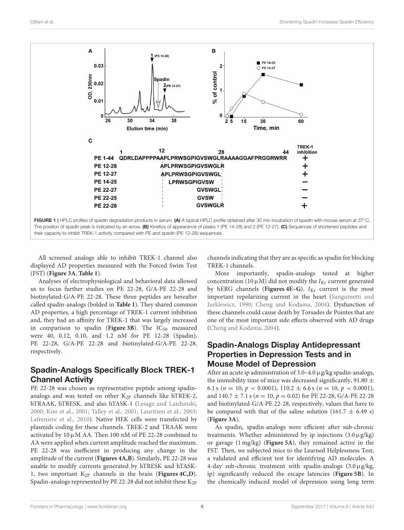
Djillani et al. Shortening Spadin Increases Spadin Efficiency
FIGURE 1 | HPLC profiles of spadin degradation products in serum. (A) A typical HPLC profile obtained after 30 min incubation of spadin with mouse serum at 37C.
The position of spadin peak is indicated by an arrow. (B) Kinetics of appearance of peaks 1 (PE 14-28) and 2 (PE 12-27). (C) Sequences of shortened peptides and
their capacity to inhibit TREK-1 activity compared with PE and spadin (PE 12-28) sequences.
All screened analogs able to inhibit TREK-1 channel alsodisplayed AD properties measured with the Forced Swim Test(FST) (Figure 3A, Table 1).
Analyses of electrophysiological and behavioral data allowedus to focus further studies on PE 22-28, G/A-PE 22-28 andbiotinylated-G/A-PE 22-28. These three peptides are hereaftercalled spadin-analogs (bolded in Table 1). They shared commonAD properties, a high percentage of TREK-1 current inhibitionand, they had an affinity for TREK-1 that was largely increasedin comparison to spadin (Figure 3B). The IC50 measuredwere 40, 0.12, 0.10, and 1.2 nM for PE 12-28 (Spadin),PE 22-28, G/A-PE 22-28 and biotinylated-G/A-PE 22-28,respectively.
Spadin-Analogs Specifically Block TREK-1Channel ActivityPE 22-28 was chosen as representative peptide among spadin-analogs and was tested on other K2P channels like hTREK-2,hTRAAK, hTRESK, and also hTASK-1 (Lesage and Lazdunski,2000; Kim et al., 2001; Talley et al., 2001; Lauritzen et al., 2003;Lafreniere et al., 2010). Native HEK cells were transfected byplasmids coding for these channels. TREK-2 and TRAAK wereactivated by 10µM AA. Then 100 nM of PE 22-28 combined toAA were applied when current amplitude reached the maximum.PE 22-28 was inefficient in producing any change in theamplitude of the current (Figures 4A,B). Similarly, PE 22-28 wasunable to modify currents generated by hTRESK and hTASK-1, two important K2P channels in the brain (Figures 4C,D).Spadin-analogs represented by PE 22-28 did not inhibit these K2P
channels indicating that they are as specific as spadin for blockingTREK-1 channels.
More importantly, spadin-analogs tested at higherconcentration (10µM) did not modify the IKr current generatedby hERG channels (Figures 4E–G). IKr current is the mostimportant repolarizing current in the heart (Sanguinetti andJurkiewicz, 1990; Cheng and Kodama, 2004). Dysfunction ofthese channels could cause death by Torsades de Pointes that areone of the most important side effects observed with AD drugs(Cheng and Kodama, 2004).
Spadin-Analogs Display AntidepressantProperties in Depression Tests and inMouse Model of DepressionAfter an acute ip administration of 3.0–4.0µg/kg spadin-analogs,the immobility time of mice was decreased significantly, 91.80 ±6.1 s (n = 10, p < 0.0001), 110.2 ± 6.6 s (n = 10, p < 0.0001),and 140.7 ± 7.1 s (n = 10, p = 0.02) for PE 22-28, G/A-PE 22-28and biotinylated-G/A-PE 22-28, respectively, values that have tobe compared with that of the saline solution (161.7 ± 6.49 s)(Figure 3A).
As spadin, spadin-analogs were efficient after sub-chronictreatments. Whether administered by ip injections (3.0µg/kg)or gavage (1mg/kg) (Figure 5A), they remained active in theFST. Then, we subjected mice to the Learned Helplessness Test,a validated and efficient test for identifying AD molecules. A4-day sub-chronic treatment with spadin-analogs (3.0µg/kg,ip) significantly reduced the escape latencies (Figure 5B). Inthe chemically induced model of depression using long term
Frontiers in Pharmacology | www.frontiersin.org 5 September 2017 | Volume 8 | Article 643
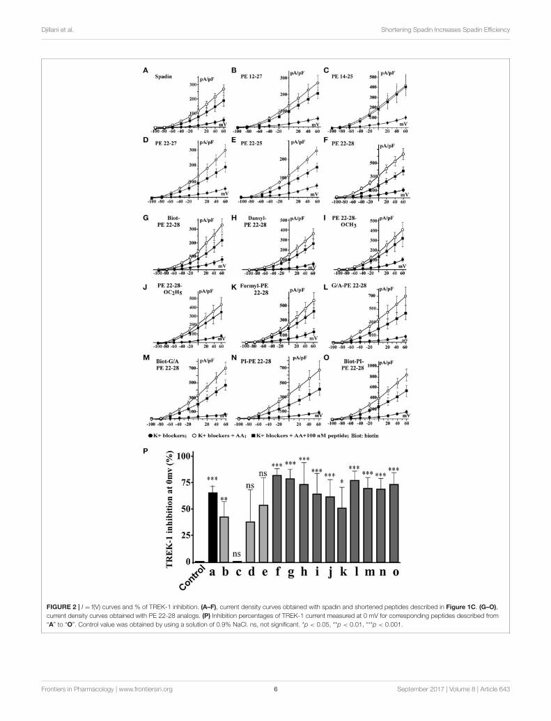
Djillani et al. Shortening Spadin Increases Spadin Efficiency
FIGURE 2 | I = f(V) curves and % of TREK-1 inhibition. (A–F), current density curves obtained with spadin and shortened peptides described in Figure 1C. (G–O),
current density curves obtained with PE 22-28 analogs. (P) Inhibition percentages of TREK-1 current measured at 0 mV for corresponding peptides described from
“A” to “O”. Control value was obtained by using a solution of 0.9% NaCl. ns, not significant. *p < 0.05, **p < 0.01, ***p < 0.001.
Frontiers in Pharmacology | www.frontiersin.org 6 September 2017 | Volume 8 | Article 643
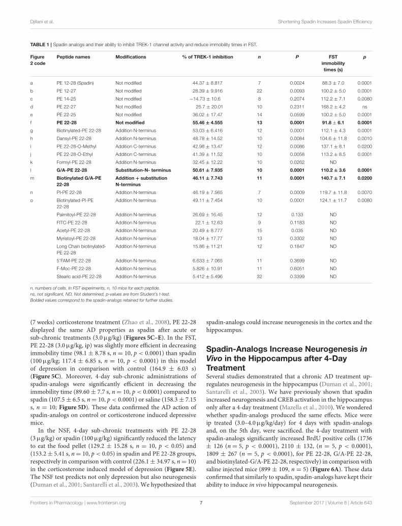
Djillani et al. Shortening Spadin Increases Spadin Efficiency
TABLE 1 | Spadin analogs and their ability to inhibit TREK-1 channel activity and reduce immobility times in FST.
Figure
2 code
Peptide names Modifications % of TREK-1 inhibition n P FST
immobility
times (s)
p
a PE 12-28 (Spadin) Not modified 44.37 ± 8.817 7 0.0024 88.3 ± 7.0 0.0001
b PE 12-27 Not modified 28.39 ± 9.916 22 0.0093 100.2 ± 5.0 0.0001
c PE 14-25 Not modified −14.73 ± 10.6 8 0.2074 112.2 ± 7.1 0.0080
d PE 22-27 Not modified 25.7 ± 20.01 10 0.2311 168.2 ± 4.2 ns
e PE 22-25 Not modified 36.02 ± 17.47 14 0.0599 100.2 ± 5.0 0.0001
f PE 22-28 Not modified 55.46 ± 4.555 13 0.0001 91.8 ± 6.1 0.0001
g Biotinylated-PE 22-28 Addition N-terminus 53.03 ± 6.416 12 0.0001 112.1 ± 4.3 0.0001
h DansyI-PE 22-28 Addition N-terminus 48.78 ± 14.52 10 0.0084 104.6 ± 11.8 0.0010
i PE 22-28-O-Methyl Addition C-terminus 42.98 ± 13.47 12 0.0086 137.1 ± 8.1 0.0200
j PE 22-28-O-Ethyl Addition C-terminus 41.39 ± 11.52 10 0.0058 113.2 ± 8.5 0.0001
k Formyl-PE 22-28 Addition N-terminus 32.45 ± 12.22 10 0.0262 ND
l G/A-PE 22-28 Substitution-N- terminus 50.61 ± 7.935 10 0.0001 110.2 ± 3.6 0.0001
m Biotinylated G/A-PE
22-28
Addition + substitution
N-terminus
46.11 ± 7.743 11 0.0001 140.7 ± 7.1 0.0200
n PI-PE 22-28 Addition N-terminus 46.19 ± 7.565 7 0.0009 119.7 ± 11.8 0.0070
o Biotinylated-PI-PE
22-28
Addition N-terminus 49.11 ± 7.454 10 0.0001 124.1 ± 11.7 0.0080
Palmitoyl-PE 22-28 Addition N-terminus 26.69 ± 16.45 12 0.133 ND
FITC-PE 22-28 Addition N-terminus 22.1 ± 12.63 9 0.1183 ND
Acetyl-PE 22-28 Addition N-terminus 20.49 ± 8.777 15 0.035 ND
Myristoyl-PE 22-28 Addition N-terminus 18.04 ± 17.77 13 0.3302 ND
Long Chain biotinylated-
PE 22-28
Addition N-terminus 15.86 ± 11.21 12 0.1847 ND
5’FAM-PE 22-28 Addition N-terminus 6.633 ± 7.065 11 0.3699 ND
F-Moc-PE 22-28 Addition N-terminus 5.826 ± 10.91 11 0.6051 ND
Stearic acid-PE 22-28 Addition N-terminus 5.412 ± 5.496 32 0.3399 ND
n, numbers of cells. In FST experiments, n, 10 mice for each peptide.
ns, not significant, ND, Not determined, p-values are from Student’s t-test.
Bolded values correspond to the spadin-analogs retained for further studies.
(7 weeks) corticosterone treatment (Zhao et al., 2008), PE 22-28displayed the same AD properties as spadin after acute orsub-chronic treatments (3.0µg/kg) (Figures 5C–E). In the FST,PE 22-28 (3.0µg/kg, ip) was slightly more efficient in decreasingimmobility time (98.1 ± 8.78 s, n = 10, p < 0.0001) than spadin(100µg/kg; 117.4 ± 6.85 s, n = 10, p < 0.0001) in this modelof depression in comparison with control (164.9 ± 6.03 s)(Figure 5C). Moreover, 4-day sub-chronic administrations ofspadin-analogs were significantly efficient in decreasing theimmobility time (89.60± 7.7 s, n= 10, p < 0.0001) compared tospadin (107.5± 6.5 s, n= 10, p < 0.0001) or saline (158.3± 7.15s, n = 10; Figure 5D). These data confirmed the AD action ofspadin-analogs on control or corticosterone induced depressivemice.
In the NSF, 4-day sub-chronic treatments with PE 22-28(3µg/kg) or spadin (100µg/kg) significantly reduced the latencyto eat the food pellet (129.2 ± 15.28 s, n = 10, p < 0.05) and(153.2± 5.41 s, n= 10, p < 0.05) in spadin and PE 22-28 groups,respectively in comparison with control (226.1± 34.97 s, n= 10)in the corticosterone induced model of depression (Figure 5E).The NSF test predicts not only depression but also neurogenesis(Duman et al., 2001; Santarelli et al., 2003). We hypothesized that
spadin-analogs could increase neurogenesis in the cortex and thehippocampus.
Spadin-Analogs Increase Neurogenesis inVivo in the Hippocampus after 4-DayTreatmentSeveral studies demonstrated that a chronic AD treatment up-regulates neurogenesis in the hippocampus (Duman et al., 2001;Santarelli et al., 2003). We have previously shown that spadinincreased neurogenesis and CREB activation in the hippocampusonly after a 4-day treatment (Mazella et al., 2010). We wonderedwhether spadin-analogs produced the same effects. Mice wereip treated (3.0–4.0µg/kg/day) for 4 days with spadin-analogsand, on the 5th day, were sacrificed. the 4-day treatment withspadin-analogs significantly increased BrdU positive cells (1736± 126 (n= 5, p < 0.0001), 2110 ± 132, (n = 5, p < 0.0001),1809 ± 267 (n = 5, p < 0.0001), for PE 22-28, G/A-PE 22-28,and biotinylated-G/A-PE 22-28, respectively) in comparison withsaline injected mice (899 ± 109, n = 5) (Figure 6A). These dataconfirmed that similarly to spadin, spadin-analogs have kept theirability to induce in vivo hippocampal neurogenesis.
Frontiers in Pharmacology | www.frontiersin.org 7 September 2017 | Volume 8 | Article 643
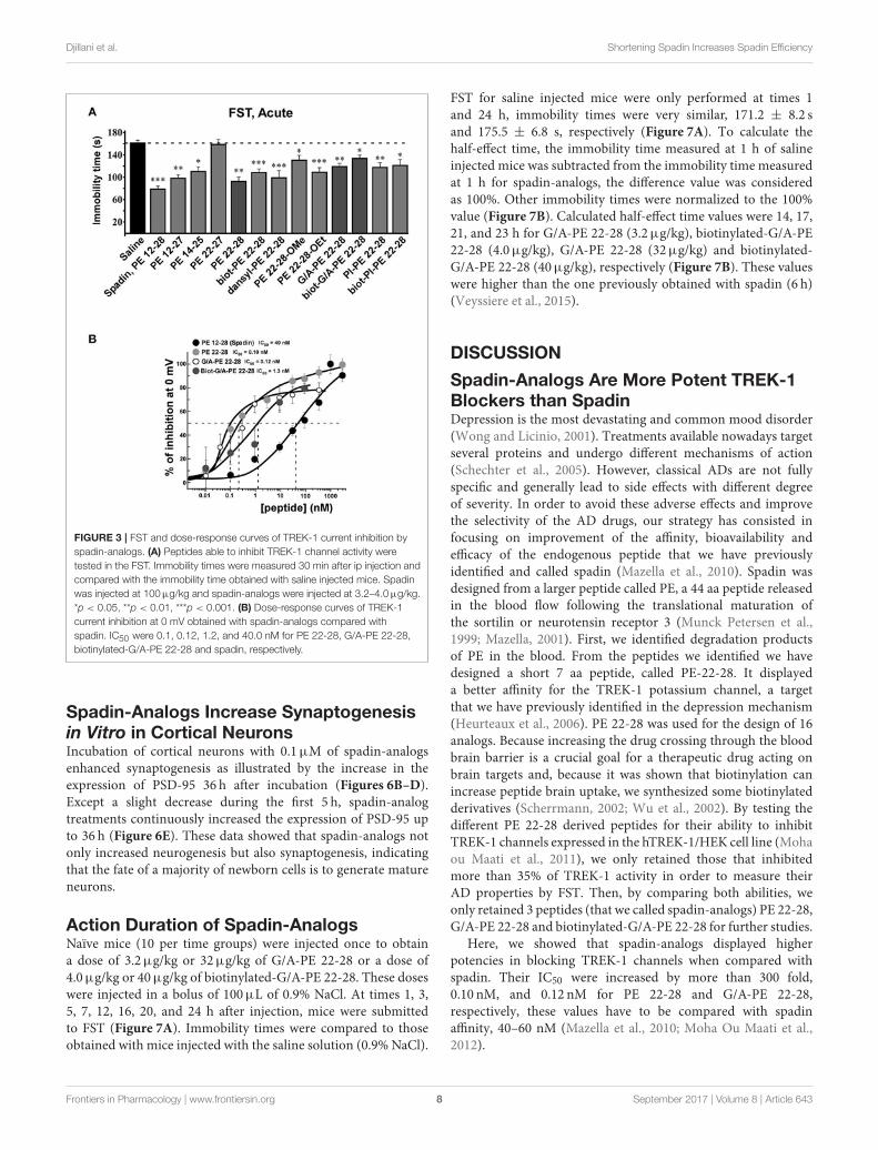
Djillani et al. Shortening Spadin Increases Spadin Efficiency
FIGURE 3 | FST and dose-response curves of TREK-1 current inhibition by
spadin-analogs. (A) Peptides able to inhibit TREK-1 channel activity were
tested in the FST. Immobility times were measured 30 min after ip injection and
compared with the immobility time obtained with saline injected mice. Spadin
was injected at 100µg/kg and spadin-analogs were injected at 3.2–4.0µg/kg.
*p < 0.05, **p < 0.01, ***p < 0.001. (B) Dose-response curves of TREK-1
current inhibition at 0 mV obtained with spadin-analogs compared with
spadin. IC50 were 0.1, 0.12, 1.2, and 40.0 nM for PE 22-28, G/A-PE 22-28,
biotinylated-G/A-PE 22-28 and spadin, respectively.
Spadin-Analogs Increase Synaptogenesisin Vitro in Cortical NeuronsIncubation of cortical neurons with 0.1µM of spadin-analogsenhanced synaptogenesis as illustrated by the increase in theexpression of PSD-95 36 h after incubation (Figures 6B–D).Except a slight decrease during the first 5 h, spadin-analogtreatments continuously increased the expression of PSD-95 upto 36 h (Figure 6E). These data showed that spadin-analogs notonly increased neurogenesis but also synaptogenesis, indicatingthat the fate of a majority of newborn cells is to generate matureneurons.
Action Duration of Spadin-AnalogsNaïve mice (10 per time groups) were injected once to obtaina dose of 3.2µg/kg or 32µg/kg of G/A-PE 22-28 or a dose of4.0µg/kg or 40µg/kg of biotinylated-G/A-PE 22-28. These doseswere injected in a bolus of 100µL of 0.9% NaCl. At times 1, 3,5, 7, 12, 16, 20, and 24 h after injection, mice were submittedto FST (Figure 7A). Immobility times were compared to thoseobtained with mice injected with the saline solution (0.9% NaCl).
FST for saline injected mice were only performed at times 1and 24 h, immobility times were very similar, 171.2 ± 8.2 sand 175.5 ± 6.8 s, respectively (Figure 7A). To calculate thehalf-effect time, the immobility time measured at 1 h of salineinjected mice was subtracted from the immobility time measuredat 1 h for spadin-analogs, the difference value was consideredas 100%. Other immobility times were normalized to the 100%value (Figure 7B). Calculated half-effect time values were 14, 17,21, and 23 h for G/A-PE 22-28 (3.2µg/kg), biotinylated-G/A-PE22-28 (4.0µg/kg), G/A-PE 22-28 (32µg/kg) and biotinylated-G/A-PE 22-28 (40µg/kg), respectively (Figure 7B). These valueswere higher than the one previously obtained with spadin (6 h)(Veyssiere et al., 2015).
DISCUSSION
Spadin-Analogs Are More Potent TREK-1Blockers than SpadinDepression is the most devastating and common mood disorder(Wong and Licinio, 2001). Treatments available nowadays targetseveral proteins and undergo different mechanisms of action(Schechter et al., 2005). However, classical ADs are not fullyspecific and generally lead to side effects with different degreeof severity. In order to avoid these adverse effects and improvethe selectivity of the AD drugs, our strategy has consisted infocusing on improvement of the affinity, bioavailability andefficacy of the endogenous peptide that we have previouslyidentified and called spadin (Mazella et al., 2010). Spadin wasdesigned from a larger peptide called PE, a 44 aa peptide releasedin the blood flow following the translational maturation ofthe sortilin or neurotensin receptor 3 (Munck Petersen et al.,1999; Mazella, 2001). First, we identified degradation productsof PE in the blood. From the peptides we identified we havedesigned a short 7 aa peptide, called PE-22-28. It displayeda better affinity for the TREK-1 potassium channel, a targetthat we have previously identified in the depression mechanism(Heurteaux et al., 2006). PE 22-28 was used for the design of 16analogs. Because increasing the drug crossing through the bloodbrain barrier is a crucial goal for a therapeutic drug acting onbrain targets and, because it was shown that biotinylation canincrease peptide brain uptake, we synthesized some biotinylatedderivatives (Scherrmann, 2002; Wu et al., 2002). By testing thedifferent PE 22-28 derived peptides for their ability to inhibitTREK-1 channels expressed in the hTREK-1/HEK cell line (Mohaou Maati et al., 2011), we only retained those that inhibitedmore than 35% of TREK-1 activity in order to measure theirAD properties by FST. Then, by comparing both abilities, weonly retained 3 peptides (that we called spadin-analogs) PE 22-28,G/A-PE 22-28 and biotinylated-G/A-PE 22-28 for further studies.
Here, we showed that spadin-analogs displayed higherpotencies in blocking TREK-1 channels when compared withspadin. Their IC50 were increased by more than 300 fold,0.10 nM, and 0.12 nM for PE 22-28 and G/A-PE 22-28,respectively, these values have to be compared with spadinaffinity, 40–60 nM (Mazella et al., 2010; Moha Ou Maati et al.,2012).
Frontiers in Pharmacology | www.frontiersin.org 8 September 2017 | Volume 8 | Article 643
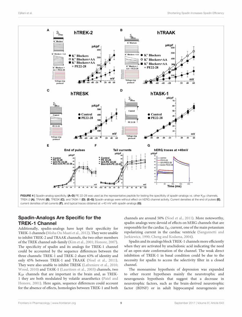
Djillani et al. Shortening Spadin Increases Spadin Efficiency
FIGURE 4 | Spadin-analog specificity. (A–D) PE 22-28 was used as the representative peptide for testing the specificity of spadin-analogs vs. other K2P channels,
TREK-2 (A), TRAAK (B), TRESK (C), and TASK-1 (D). (E–G) Spadin-analogs were without effect on hERG channel activity. Current densities at the end of pulses (E),
current densities of tail currents (F), and typical traces obtained at +40 mV with spadin-analogs (G).
Spadin-Analogs Are Specific for theTREK-1 ChannelAdditionally, spadin-analogs have kept their specificity forTREK-1 channels (MohaOuMaati et al., 2012). They were unableto inhibit TREK-2 and TRAAK channels, the two other membersof the TREK channel sub-family (Kim et al., 2001; Honore, 2007).The specificity of spadin and its analogs for TREK-1 channelcould be accounted by the sequence differences between thethree channels: TREK-1 and TREK-2 share 63% of identity andonly 45% between TREK-1 and TRAAK (Noel et al., 2011).They were also unable to inhibit TRESK (Lafreniere et al., 2010;Wood, 2010) and TASK-1 (Lauritzen et al., 2003) channels, twoK2P channels that are important in the brain and, as TREK-1 they are both modulated by volatile anaesthetics (Patel andHonore, 2001). Here again, sequence differences could accountfor the absence of effects, homologies between TREK-1 and both
channels are around 50% (Noel et al., 2011). More noteworthy,spadin-analogs were devoid of effects on hERG channels that areresponsible for the cardiac IKr current, one of themain potassiumrepolarizing current in the cardiac ventricle (Sanguinetti andJurkiewicz, 1990; Cheng and Kodama, 2004).
Spadin and its analogs block TREK-1 channelsmore efficientlywhen they are activated by arachidonic acid indicating the needof an open-state conformation of the channel. The weak directinhibition of TREK-1 in basal condition could be due to thenecessity for spadin to access the selectivity filter in a closedchannel.
The monoamine hypothesis of depression was expandedto other recent hypotheses mainly the neurotrophic andneurogenesis hypothesis that suggest that a decrease inneurotrophic factors, such as the brain-derived neurotrophicfactor (BDNF) or in adult hippocampal neurogenesis are
Frontiers in Pharmacology | www.frontiersin.org 9 September 2017 | Volume 8 | Article 643
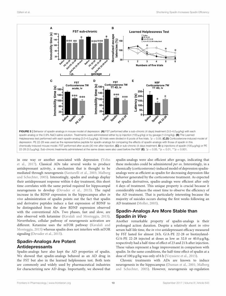
Djillani et al. Shortening Spadin Increases Spadin Efficiency
FIGURE 5 | Behavior of spadin-analogs in mouse model of depression. (A) FST performed after a sub-chronic (4 days) treatment (3.0–4.0µg/kg) with each
spadin-analog or the 0.9% NaCl saline solution. Treatments were administered either by ip injection (100µg/kg) or by gavage (1.0mg/kg). (B) The Learned
Helplessness test performed with each spadin-analog (3.0–4.0µg/kg). 30 trials were divided in 6 pools of five trials. *p < 0.05. (C,D) Corticosterone-induced model of
depression. PE 22-28 was used as the representative peptide for spadin-analogs for comparing the effects of spadin-analogs with those of spadin in this
chemically-induced mouse model. FST performed after acute (30 min after injection, (C) or sub-chronic (4 days treatment, D) ip injections of spadin (100µg/kg) or PE
22-28 (3.0µg/kg). Sub-chronic treatments administered at the same doses were also used before the NSF (E). *p < 0.05, **p < 0.01, ***p < 0.001.
in one way or another associated with depression (Yohnet al., 2017). Classical ADs take several weeks to produceantidepressant activity, a mechanism that is thought to bemediated through neurogenesis (Santarelli et al., 2003; Malbergand Schechter, 2005). Interestingly, spadin and analogs displaytheir antidepressant response within 4-day treatment, this shorttime correlates with the same period required for hippocampalneurogenesis to develop (Devader et al., 2015). The rapidincrease in the BDNF expression in the hippocampus after invivo administration of spadin points out the fact that spadinand derivative peptides induce a fast expression of BDNF tobe distinguished from the slow BDNF expression observedwith the conventional ADs. Two phases, fast and slow, arealso observed with ketamine (Kavalali and Monteggia, 2015).Nevertheless, cellular pathway of neurogenesis activation aredifferent. Ketamine uses the mTOR pathway (Kavalali andMonteggia, 2015) whereas spadin does not interfere with mTORsignaling (Devader et al., 2015).
Spadin-Analogs Are PotentAntidepressantsSpadin-analogs have also kept the AD properties of spadin.We showed that spadin-analogs behaved as an AD drug inthe FST but also in the learned helplessness test. Both testsare commonly and widely used by pharmaceutical industriesfor characterizing new AD drugs. Importantly, we showed that
spadin-analogs were also efficient after gavage, indicating thatthese molecules could be administered per os. Interestingly, in achemically (corticosterone)-inducedmodel of depression spadin-analogs were as efficient as spadin for decreasing depression-likebehavior generated by the corticosterone treatment. As expectedfor spadin derivatives, spadin-analogs were efficient after only4 days of treatment. This unique property is crucial because itconsiderably reduces the onset time to observe the efficiency ofthe AD treatment. That is particularly interesting because themajority of suicides occurs during the first weeks following anAD treatment (Moller, 2003).
Spadin-Analogs Are More Stable thanSpadin in VivoAnother remarkable property of spadin-analogs is theirprolonged action duration. Despite a relatively short in vitroserum half-life time, the in vivo antidepressant efficacy measuredby FST lasted for almost 24 h. G/A-PE 22-28 or biotinylated-G/A-PE 22-28 injected at doses as low as 32.0 or 40.0µg/kg,respectively had a half-time of effect of 23 and 21 h after injection.These values represent a huge improvement in comparison withspadin. In the same conditions, the half-time effect of spadin at adose of 100µg/kg was only of 6 h (Veyssiere et al., 2015).
Chronic treatments with ADs are known to induceneurogenesis in the hippocampus (Duman et al., 2001; Malbergand Schechter, 2005). However, neurogenesis up-regulation
Frontiers in Pharmacology | www.frontiersin.org 10 September 2017 | Volume 8 | Article 643
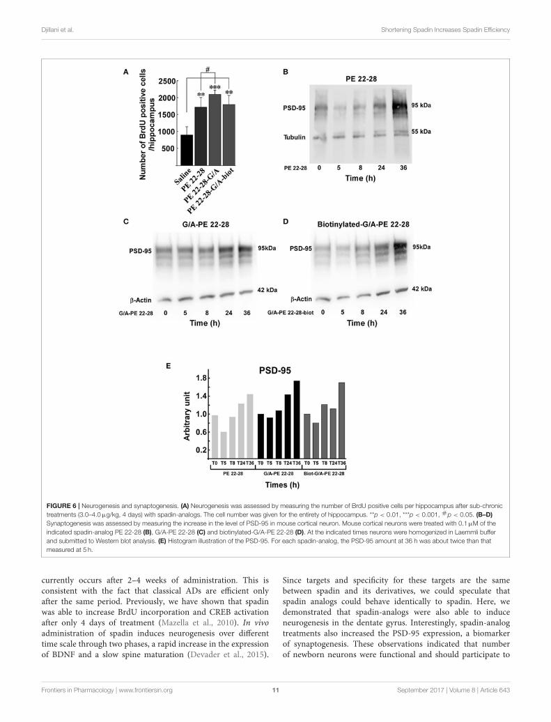
Djillani et al. Shortening Spadin Increases Spadin Efficiency
FIGURE 6 | Neurogenesis and synaptogenesis. (A) Neurogenesis was assessed by measuring the number of BrdU positive cells per hippocampus after sub-chronic
treatments (3.0–4.0µg/kg, 4 days) with spadin-analogs. The cell number was given for the entirety of hippocampus. **p < 0.01, ***p < 0.001, #p < 0.05. (B–D)
Synaptogenesis was assessed by measuring the increase in the level of PSD-95 in mouse cortical neuron. Mouse cortical neurons were treated with 0.1µM of the
indicated spadin-analog PE 22-28 (B), G/A-PE 22-28 (C) and biotinylated-G/A-PE 22-28 (D). At the indicated times neurons were homogenized in Laemmli buffer
and submitted to Western blot analysis. (E) Histogram illustration of the PSD-95. For each spadin-analog, the PSD-95 amount at 36 h was about twice than that
measured at 5 h.
currently occurs after 2–4 weeks of administration. This isconsistent with the fact that classical ADs are efficient onlyafter the same period. Previously, we have shown that spadinwas able to increase BrdU incorporation and CREB activationafter only 4 days of treatment (Mazella et al., 2010). In vivoadministration of spadin induces neurogenesis over differenttime scale through two phases, a rapid increase in the expressionof BDNF and a slow spine maturation (Devader et al., 2015).
Since targets and specificity for these targets are the samebetween spadin and its derivatives, we could speculate thatspadin analogs could behave identically to spadin. Here, wedemonstrated that spadin-analogs were also able to induceneurogenesis in the dentate gyrus. Interestingly, spadin-analogtreatments also increased the PSD-95 expression, a biomarkerof synaptogenesis. These observations indicated that numberof newborn neurons were functional and should participate to
Frontiers in Pharmacology | www.frontiersin.org 11 September 2017 | Volume 8 | Article 643
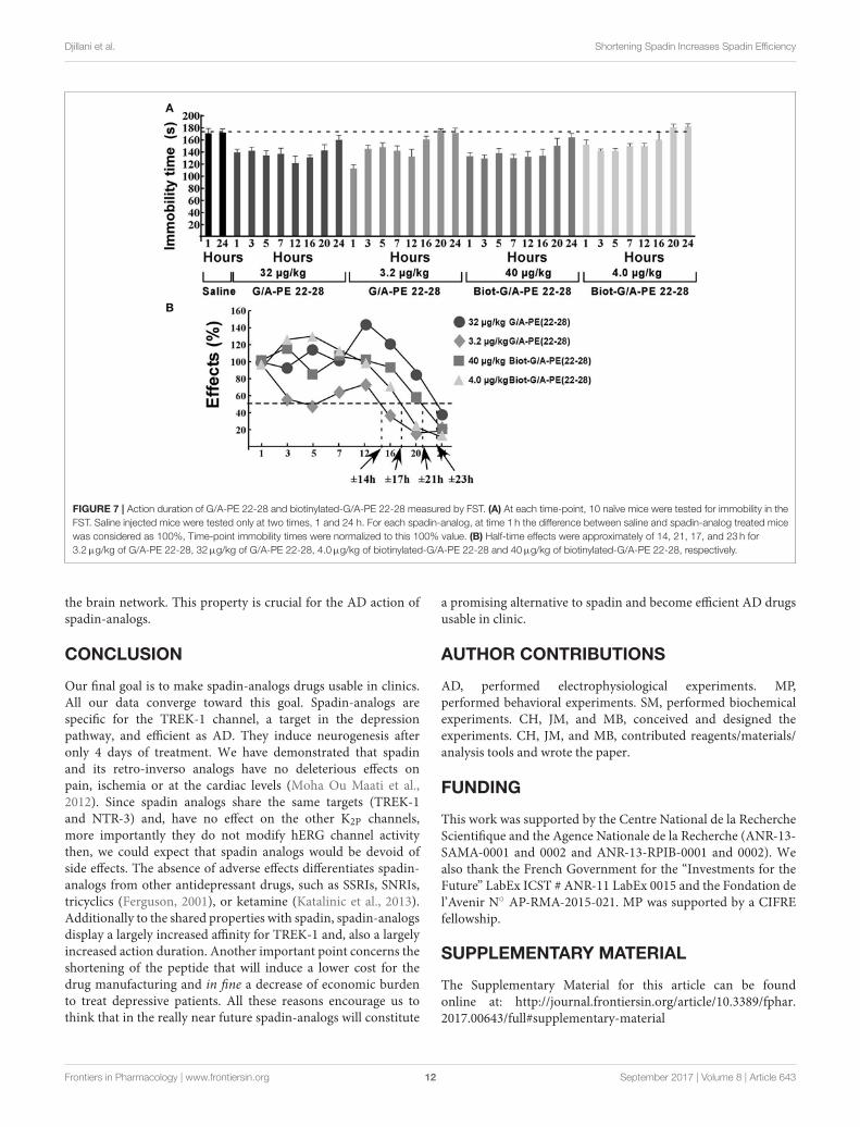
Djillani et al. Shortening Spadin Increases Spadin Efficiency
FIGURE 7 | Action duration of G/A-PE 22-28 and biotinylated-G/A-PE 22-28 measured by FST. (A) At each time-point, 10 naïve mice were tested for immobility in the
FST. Saline injected mice were tested only at two times, 1 and 24 h. For each spadin-analog, at time 1 h the difference between saline and spadin-analog treated mice
was considered as 100%, Time-point immobility times were normalized to this 100% value. (B) Half-time effects were approximately of 14, 21, 17, and 23 h for
3.2µg/kg of G/A-PE 22-28, 32µg/kg of G/A-PE 22-28, 4.0µg/kg of biotinylated-G/A-PE 22-28 and 40µg/kg of biotinylated-G/A-PE 22-28, respectively.
the brain network. This property is crucial for the AD action ofspadin-analogs.
CONCLUSION
Our final goal is to make spadin-analogs drugs usable in clinics.All our data converge toward this goal. Spadin-analogs arespecific for the TREK-1 channel, a target in the depressionpathway, and efficient as AD. They induce neurogenesis afteronly 4 days of treatment. We have demonstrated that spadinand its retro-inverso analogs have no deleterious effects onpain, ischemia or at the cardiac levels (Moha Ou Maati et al.,2012). Since spadin analogs share the same targets (TREK-1and NTR-3) and, have no effect on the other K2P channels,more importantly they do not modify hERG channel activitythen, we could expect that spadin analogs would be devoid ofside effects. The absence of adverse effects differentiates spadin-analogs from other antidepressant drugs, such as SSRIs, SNRIs,tricyclics (Ferguson, 2001), or ketamine (Katalinic et al., 2013).Additionally to the shared properties with spadin, spadin-analogsdisplay a largely increased affinity for TREK-1 and, also a largelyincreased action duration. Another important point concerns theshortening of the peptide that will induce a lower cost for thedrug manufacturing and in fine a decrease of economic burdento treat depressive patients. All these reasons encourage us tothink that in the really near future spadin-analogs will constitute
a promising alternative to spadin and become efficient AD drugsusable in clinic.
AUTHOR CONTRIBUTIONS
AD, performed electrophysiological experiments. MP,performed behavioral experiments. SM, performed biochemicalexperiments. CH, JM, and MB, conceived and designed theexperiments. CH, JM, and MB, contributed reagents/materials/analysis tools and wrote the paper.
FUNDING
This work was supported by the Centre National de la RechercheScientifique and the Agence Nationale de la Recherche (ANR-13-SAMA-0001 and 0002 and ANR-13-RPIB-0001 and 0002). Wealso thank the French Government for the “Investments for theFuture” LabEx ICST # ANR-11 LabEx 0015 and the Fondation del’Avenir N AP-RMA-2015-021. MP was supported by a CIFREfellowship.
SUPPLEMENTARY MATERIAL
The Supplementary Material for this article can be foundonline at: http://journal.frontiersin.org/article/10.3389/fphar.2017.00643/full#supplementary-material
Frontiers in Pharmacology | www.frontiersin.org 12 September 2017 | Volume 8 | Article 643
Djillani et al. Shortening Spadin Increases Spadin Efficiency
REFERENCES
Alloui, A., Zimmermann, K., Mamet, J., Duprat, F., Noel, J., Chemin, J., et al.
(2006). TREK-1, a K+ channel involved in polymodal pain perception. EMBO
J. 25, 2368–2376. doi: 10.1038/sj.emboj.7601116
Borsotto, M., Veyssiere, J., Moha Ou Maati, H., Devader, C., Mazella, J., and
Heurteaux, C. (2015). Targeting two-pore domain K+ channels TREK-1 and
TASK-3 for the treatment of depression: a new therapeutic concept. Br. J.
Pharmacol. 172, 771–784. doi: 10.1111/bph.12953
Caldarone, B. J., George, T. P., Zachariou, V., and Picciotto, M. R. (2000).
Gender differences in learned helplessness behavior are influenced
by genetic background. Pharmacol. Biochem. Behav. 66, 811–817.
doi: 10.1016/S0091-3057(00)00271-9
Cheng, J. H., and Kodama, I. (2004). Two components of delayed rectifier K+
current in heart: molecular basis, functional diversity, and contribution to
repolarization. Acta Pharmacol. Sin. 25, 137–145.
Cleare, A., Pariante, C. M., Young, A. H., Anderson, I. M., Christmas,
D., Cowen, P. J., et al. (2015). Evidence-based guidelines for treating
depressive disorders with antidepressants: a revision of the 2008 British
Association for Psychopharmacology guidelines. J. Psychopharmacol. 29,
459–525. doi: 10.1177/0269881115581093
Deardorff, W. J., and Grossberg, G. T., (2014). A review of the clinical
efficacy, safety and tolerability of the antidepressants vilazodone,
levomilnacipran and vortioxetine. Expert Opin. Pharmacother. 15, 2525–2542.
doi: 10.1517/14656566.2014.960842
Decher, N., Kiper, A. K., and Rinne, S. (2017). Stretch-activated potassium currents
in the heart: focus on TREK-1 and arrhythmias. Prog. Biophys. Mol. Biol.
doi: 10.1016/j.pbiomolbio.2017.05.005. [Epub ahead of print].
Devader, C., Khayachi, A., Veyssiere, J., Moha Ou Maati, H., Roulot, M.,
Moreno, S., et al. (2015). In vitro and in vivo regulation of synaptogenesis
by the novel antidepressant spadin. Br. J. Pharmacol. 172, 2604–2617.
doi: 10.1111/bph.13083
Duman, R. S., Nakagawa, S., and Malberg, J. (2001). Regulation of adult
neurogenesis by antidepressant treatment. Neuropsychopharmacology 25,
836–844. doi: 10.1016/S0893-133X(01)00358-X
Ferguson, J. M. (2001). SSRI Antidepressant medications: adverse effects
and tolerability. Prim. Care Companion J. Clin. Psychiatry 3, 22–27.
doi: 10.4088/PCC.v03n0105
Heijnen, W. T., Birkenhager, T. K., Wierdsma, A. I., and van den Broek, W. W.
(2010). Antidepressant pharmacotherapy failure and response to subsequent
electroconvulsive therapy: a meta-analysis. J. Clin. Psychopharmacol. 30,
616–619. doi: 10.1097/JCP.0b013e3181ee0f5f
Heurteaux, C., Lucas, G., Guy, N., El Yacoubi, M., Thümmler, S., Peng,
X., et al. (2006). Deletion of TREK-1, a background potassium channel,
results in a depression-resistant phenotype. Nature Neurosci. 9, 1134–1141.
doi: 10.1038/nn1749
Hillhouse, T. M., and Porter, J. H. (2015). A brief history of the development
of antidepressant drugs: from monoamines to glutamate. Exp. Clin.
Psychopharmacol. 23, 1–21. doi: 10.1037/a0038550
Hirschfeld, R. M. (2012). The epidemiology of depression and the evolution of
treatment. J. Clin. Psychiatry 73(Suppl. 1), 5–9. doi: 10.4088/JCP.11096su1c.01
Honore, E. (2007). The neuronal background K2P channels: focus on TREK1. Nat.
Rev. Neurosci. 8, 251–261. doi: 10.1038/nrn2117
Katalinic, N., Lai, R., Somogyi, A., Mitchell, P. B., Glue, P., and Loo, C. K. (2013).
Ketamine as a new treatment for depression: a review of its efficacy and
adverse effects. Aust. N. Z. J. Psychiatry 47, 710–727. doi: 10.1177/00048674134
86842
Kavalali, E. T., and Monteggia, L. M. (2015). How does ketamine elicit
a rapid antidepressant response? Curr. Opin. Pharmacol. 20, 35–39.
doi: 10.1016/j.coph.2014.11.005
Kessler, R. C., Petukhova, M., Sampson, N. A., Zaslavsky, A. M., and Wittchen, H.
U. (2012). Twelve-month and lifetime prevalence and lifetime morbid risk of
anxiety and mood disorders in the United States. Int. J. Methods Psychiatr. Res.
21, 169–184. doi: 10.1002/mpr.1359
Kim, Y., Gnatenco, C., Bang, H., and Kim, D. (2001). Localization of TREK-2 K+
channel domains that regulate channel kinetics and sensitivity to pressure, fatty
acids and pHi. Pflugers Arch. 442, 952–960. doi: 10.1007/s004240100626
Kohler, S., Cierpinsky, K., Kronenberg, G., and Adli, M. (2016). The serotonergic
system in the neurobiology of depression: relevance for novel antidepressants.
J. Psychopharmacol. 30, 13–22. doi: 10.1177/0269881115609072
Krishnan, V., and Nestler, E. J. (2008). The molecular neurobiology of depression.
Nature 455, 894–902. doi: 10.1038/nature07455
Lafreniere, R. G., Cader, M. Z., Poulin, J. F., Andres-Enguix, I., Simoneau,
M., Gupta, N., et al. (2010). A dominant-negative mutation in the TRESK
potassium channel is linked to familial migraine with aura. Nat. Med. 16,
1157–1160. doi: 10.1038/nm.2216
Lauritzen, I., Blondeau, N., Heurteaux, C., Widmann, C., Romey, G., and
Lazdunski, M. (2000). Polyunsaturated fatty acids are potent neuroprotectors.
EMBO J. 19, 1784–1793. doi: 10.1093/emboj/19.8.1784
Lauritzen, I., Zanzouri, M., Honore, E., Duprat, F., Ehrengruber, M. U.,
Lazdunski, M., et al. (2003). K+-dependent cerebellar granule neuron
apoptosis. Role of task leak K+ channels. J. Biol. Chem. 278, 32068–32076.
doi: 10.1074/jbc.M302631200
Lesage, F., and Lazdunski, M. (2000). Molecular and functional properties of two
pore domain potassium channels. Am. J. Physiol. 279, 793–801.
Li, Y., Abdourahman, A., Tamm, J. A., Pehrson, A. L., Sanchez, C., and Gulinello,
M. (2015). Reversal of age-associated cognitive deficits is accompanied
by increased plasticity-related gene expression after chronic antidepressant
administration in middle-aged mice. Pharmacol. Biochem. Behav. 135, 70–82.
doi: 10.1016/j.pbb.2015.05.013
Malberg, J. E., and Schechter, L. E. (2005). Increasing hippocampal neurogenesis:
a novel mechanism for antidepressant drugs. Curr. Pharm. Des. 11, 145–155.
doi: 10.2174/1381612053382223
Maletic, V., Robinson, M., Oakes, T., Iyengar, S., Ball, S. G., and Russell, J. (2007).
Neurobiology of depression: an integrated view of key findings. Int. J. Clin.
Pract. 61, 2030–2040. doi: 10.1111/j.1742-1241.2007.01602.x
Mazella, J. (2001). Sortilin/neurotensin receptor-3: a new tool to investigate
neurotensin signaling and cellular trafficking? Cell. Signal. 13, 1–6.
doi: 10.1016/S0898-6568(00)00130-3
Mazella, J., Petrault, O., Lucas, G., Deval, E., Beraud-Dufour, S., Gandin, C., et al.
(2010). Spadin, a sortilin-derived peptide, targeting rodent TREK-1 channels:
a new concept in the antidepressant drug design. PLoS Biol. 8:e1000355.
doi: 10.1371/journal.pbio.1000355
Moha ou Maati, H., Peyronnet, R., Devader, C., Veyssiere, J., Labbal, F., Gandin,
C., et al. (2011). A human TREK-1/HEK cell line: a highly efficient screening
tool for drug development in neurological diseases. PLoS ONE 6:e25602.
doi: 10.1371/journal.pone.0025602
Moha Ou Maati, H., Veyssiere, J., Labbal, F., Coppola, T., Gandin, C.,
Widmann, C., et al. (2012). Spadin as a new antidepressant: absence
of TREK-1-related side effects. Neuropharmacology 62, 278–288.
doi: 10.1016/j.neuropharm.2011.07.019
Moller, H. J. (2003). Suicide, suicidality and suicide prevention
in affective disorders. Acta Psychiatr. Scand. Suppl. 418, 73–80.
doi: 10.1034/j.1600-0447.108.s418.15.x
Munck Petersen, C., Nielsen, M. S., Jacobsen, C., Tauris, J., Jacobsen, L., Gliemann,
J., et al. (1999). Propeptide cleavage conditions sortilin/neurotensin receptor-3
for ligand binding. EMBO J. 18, 595–604. doi: 10.1093/emboj/18.3.595
Nestler, E. J., Barrot, M., DiLeone, R. J., Eisch, A. J., Gold, S. J., and
Monteggia, L. M. (2002). Neurobiology of depression. Neuron 34, 13–25.
doi: 10.1016/S0896-6273(02)00653-0
Noel, J., Sandoz, G., and Lesage, F. (2011). Molecular regulations
governing TREK and TRAAK channel functions. Channels 5, 402–409.
doi: 10.4161/chan.5.5.16469
Patel, A. J., and Honore, E. (2001). Properties and modulation of
mammalian 2P domain K+ channels. Trends Neurosci. 24, 339–346.
doi: 10.1016/S0166-2236(00)01810-5
Patel, A. J., Honore, E., Maingret, F., Lesage, F., Fink, M., Duprat, F., et al. (1998).
A mammalian two pore domain mechano-gated S-like K+ channel. EMBO J.
17, 4283–4290. doi: 10.1093/emboj/17.15.4283
Porsolt, R. D., Le Pichon, M., and Jalfre, M. (1977). Depression: a new
animal model sensitive to antidepressant treatments. Nature 266, 730–732.
doi: 10.1038/266730a0
Ramaker, M. J., and Dulawa, S. C. (2017). Identifying fast-onset antidepressants
using rodent models.Mol. Psychiatry 22, 656–665. doi: 10.1038/mp.2017.36
Frontiers in Pharmacology | www.frontiersin.org 13 September 2017 | Volume 8 | Article 643
Djillani et al. Shortening Spadin Increases Spadin Efficiency
Rush, A. J., Trivedi, M. H., Wisniewski, S. R., Nierenberg, A. A., Stewart, J.
W., Warden, D., et al. (2006). Acute and longer-term outcomes in depressed
outpatients requiring one or several treatment steps: a STAR∗D report. Am. J.
Psychiatry 163, 1905–1917. doi: 10.1176/ajp.2006.163.11.1905
Sanguinetti, M. C., and Jurkiewicz, N. K. (1990). Two components of cardiac
delayed rectifier K+ current. Differential sensitivity to block by class III
antiarrhythmic agents. J. Gen. Physiol. 96, 195–215. doi: 10.1085/jgp.96.1.195
Santarelli, L., Saxe, M., Gross, C., Surget, A., Battaglia, F., Dulawa, S., et al.
(2003). Requirement of hippocampal neurogenesis for the behavioral effects of
antidepressants. Science. 301, 805–809. doi: 10.1126/science.1083328
Schechter, L. E., Ring, R. H., Beyer, C. E., Hughes, Z. A., Khawaja, X.,
Malberg, J. E., et al. (2005). Innovative approaches for the development
of antidepressant drugs: current and future strategies. NeuroRx 2, 590–611.
doi: 10.1602/neurorx.2.4.590
Scherrmann, J. M. (2002). Drug delivery to brain via the blood-brain barrier.
Vascul. Pharmacol. 38, 349–354. doi: 10.1016/S1537-1891(02)00202-1
Semkovska, M., and McLoughlin, D. M. (2010). Objective cognitive
performance associated with electroconvulsive therapy for depression:
a systematic review and meta-analysis. Biol. Psychiatry 68, 568–577.
doi: 10.1016/j.biopsych.2010.06.009
Smith, K. (2014). Mental health: a world of depression. Nature 515, 181.
doi: 10.1038/515180a
Sowa-Kucma, M., Panczyszyn-Trzewik, P., Misztak, P., Jaeschke, R. R., Sendek,
K., Styczen, K., et al. (2017). Vortioxetine: a review of the pharmacology
and clinical profile of the novel antidepressant. Pharmacol. Rep. 69, 595–601.
doi: 10.1016/j.pharep.2017.01.030
Talley, E. M., Solorzano, G., Lei, Q., Kim, D., and Bayliss, D. A. (2001). Cns
distribution of members of the two-pore-domain (KCNK) potassium channel
family. J. Neurosci. 21, 7491–7505.
Thase, M. E., Mahableshwarkar, A. R., Dragheim, M., Loft, H., and Vieta, E. (2016).
Ameta-analysis of randomized, placebo-controlled trials of vortioxetine for the
treatment of major depressive disorder in adults. Eur. Neuropsychopharmacol.
26, 979–993. doi: 10.1016/j.euroneuro.2016.03.007
Veyssiere, J., Moha Ou Maati, H., Mazella, J., Gaudriault, G., Moreno, S.,
Heurteaux, C., et al. (2015). Retroinverso analogs of spadin display
increased antidepressant effects. Psychopharmacology 232, 561–574.
doi: 10.1007/s00213-014-3683-2
Wang, S. M., Han, C., Lee, S. J., Patkar, A. A., Masand, P. S., and Pae, C. U.
(2015). Vilazodone for the treatment of major depressive disorder: focusing on
its clinical studies and mechanism of action. Psychiatry Investig. 12, 155–163.
doi: 10.4306/pi.2015.12.2.155
Wiedmann, F., Schmidt, C., Lugenbiel, P., Staudacher, I., Rahm, A. K., Seyler,
C., et al. (2016). Therapeutic targeting of two-pore-domain potassium
(K2P) channels in the cardiovascular system. Clin. Sci. 130, 643–650.
doi: 10.1042/CS20150533
Wong, M. L., and Licinio, J. (2001). Research and treatment approaches
to depression. Nat. Rev. Neurosci. 2, 343–351. doi: 10.1038/350
72566
Wood, H. (2010). Migraine: familial migraine with aura is associated with
a mutation in the TRESK potassium channel. Nat. Rev. Neurol. 6:643.
doi: 10.1038/nrneurol.2010.166
Wu, D., Song, B. W., Vinters, H. V., and Pardridge, W. M. (2002).
Pharmacokinetics and brain uptake of biotinylated basic fibroblast
growth factor conjugated to a blood-brain barrier drug delivery
system. J. Drug Target. 10, 239–245. doi: 10.1080/106118602900
22679
Yohn, C. N., Gergues, M. M., and Samuels, B. A. (2017). The role of 5-
HT receptors in depression. Mol. Brain 10, 28. doi: 10.1186/s13041-017-
0306-y
Zhao, Y., Ma, R., Shen, J., Su, H., Xing, D., and Du, L. (2008). A mouse model
of depression induced by repeated corticosterone injections. Eur. J. Pharmacol.
581, 113–120. doi: 10.1016/j.ejphar.2007.12.005
Conflict of Interest Statement: The authors declare that the research was
conducted in the absence of any commercial or financial relationships that could
be construed as a potential conflict of interest.
Copyright © 2017 Djillani, Pietri, Moreno, Heurteaux, Mazella and Borsotto. This
is an open-access article distributed under the terms of the Creative Commons
Attribution License (CC BY). The use, distribution or reproduction in other forums
is permitted, provided the original author(s) or licensor are credited and that the
original publication in this journal is cited, in accordance with accepted academic
practice. No use, distribution or reproduction is permitted which does not comply
with these terms.
Frontiers in Pharmacology | www.frontiersin.org 14 September 2017 | Volume 8 | Article 643

176
REFERENCES
Akil, H., Perraud, A., Mélin, C., Jauberteau, M.-O., and Mathonnet, M. (2011). Fine-tuning roles of endogenous brain-derived neurotrophic factor, TrkB and sortilin in colorectal cancer cell survival. PLoS ONE 6, e25097.
Allen, A.P., Naughton, M., Dowling, J., Walsh, A., Ismail, F., Shorten, G., Scott, L., McLoughlin, D.M., Cryan, J.F., Dinan, T.G., et al. (2015). Serum BDNF as a peripheral biomarker of treatment-resistant depression and the rapid antidepressant response: A comparison of ketamine and ECT. Journal of Affective Disorders 186, 306–311.
Alloui, A., Zimmermann, K., Mamet, J., Duprat, F., Noël, J., Chemin, J., Guy, N., Blondeau, N., Voilley, N., Rubat-Coudert, C., et al. (2006). TREK-1, a K+ channel involved in polymodal pain perception. EMBO J. 25, 2368–2376.
Angelucci, F., Brenè, S., and Mathé, A.A. (2005). BDNF in schizophrenia, depression and corresponding animal models. Mol. Psychiatry 10, 345–352.
Arango, V., Underwood, M.D., Boldrini, M., Tamir, H., Kassir, S.A., Hsiung, S., Chen, J.J., and Mann, J.J. (2001). Serotonin 1A receptors, serotonin transporter binding and serotonin transporter mRNA expression in the brainstem of depressed suicide victims. Neuropsychopharmacology 25, 892–903.
Arrighi, I., Lesage, F., Scimeca, J.C., Carle, G.F., and Barhanin, J. (1998). Structure, chromosome localization, and tissue distribution of the mouse twik K+ channel gene. FEBS Lett. 425, 310–316.
Asberg, M. (1997). Neurotransmitters and suicidal behavior. The evidence from cerebrospinal fluid studies. Ann. N. Y. Acad. Sci. 836, 158–181.
Baker, G.B., Coutts, R.T., Yeung, J.M., Hampson, D.R., McIntosh, G.J.A., and McIntosh, M.G. (1985). Chronic Administration of Monoamine Oxidase Inhibitors: Basic and Clinical Investigations. In Neuropsychopharmacology of the Trace Amines, (Humana Press), pp. 317–328.
Bath, K.G., and Lee, F.S. (2006). Variant BDNF (Val66Met) impact on brain structure and function. Cogn Affect Behav Neurosci 6, 79–85.
Battey, F.D., Gåfvels, M.E., FitzGerald, D.J., Argraves, W.S., Chappell, D.A., Strauss, J.F., and Strickland, D.K. (1994). The 39-kDa receptor-associated protein regulates ligand binding by the very low density lipoprotein receptor. J. Biol. Chem. 269, 23268–23273.
Bekinschtein, P., Cammarota, M., Igaz, L.M., Bevilaqua, L.R.M., Izquierdo, I., and Medina, J.H. (2007). Persistence of long-term memory storage requires a late protein synthesis- and BDNF- dependent phase in the hippocampus. Neuron 53, 261–277.
Belzeaux, R., Formisano-Tréziny, C., Loundou, A., Boyer, L., Gabert, J., Samuelian, J.-C., Féron, F., Naudin, J., and Ibrahim, E.C. (2010). Clinical variations modulate patterns of gene expression and define blood biomarkers in major depression. Journal of Psychiatric Research 44, 1205–1213.
Belzeaux, R., Bergon, A., Jeanjean, V., Loriod, B., Formisano-Tréziny, C., Verrier, L., Loundou, A., Baumstarck-Barrau, K., Boyer, L., Gall, V., et al. (2012). Responder and nonresponder patients exhibit different peripheral transcriptional signatures during major depressive episode. Transl Psychiatry 2, e185.
177
Béraud-Dufour, S., Coppola, T., Massa, F., and Mazella, J. (2009). Neurotensin receptor-2 and -3 are crucial for the anti-apoptotic effect of neurotensin on pancreatic beta-TC3 cells. Int. J. Biochem. Cell Biol. 41, 2398–2402.
Béraud-Dufour, S., Abderrahmani, A., Noel, J., Brau, F., Waeber, G., Mazella, J., and Coppola, T. (2010). Neurotensin is a regulator of insulin secretion in pancreatic beta-cells. Int. J. Biochem. Cell Biol. 42, 1681–1688.
Blaha, C.D., and Phillips, A.G. (1992). Pharmacological evidence for common mechanisms underlying the effects of neurotensin and neuroleptics on in vivo dopamine efflux in the rat nucleus accumbens. Neuroscience 49, 867–877.
Borroto-Escuela, D.O., Ravani, A., Tarakanov, A.O., Brito, I., Narvaez, M., Romero-Fernandez, W., Corrales, F., Agnati, L.F., Tanganelli, S., Ferraro, L., et al. (2013). Dopamine D2 receptor signaling dynamics of dopamine D2-neurotensin 1 receptor heteromers. Biochem. Biophys. Res. Commun. 435, 140–146.
Botto, J.M., Guillemare, E., Vincent, J.P., and Mazella, J. (1997). Effects of SR 48692 on neurotensin-induced calcium-activated chloride currents in the Xenopus oocyte expression system: agonist-like activity on the levocabastine-sensitive neurotensin receptor and absence of antagonist effect on the levocabastine insensitive neurotensin receptor. Neurosci. Lett. 223, 193–196.
Boudin, H., Pélaprat, D., Rostène, W., and Beaudet, A. (1996). Cellular distribution of neurotensin receptors in rat brain: immunohistochemical study using an antipeptide antibody against the cloned high affinity receptor. J. Comp. Neurol. 373, 76–89.
Boyle, M.P., Brewer, J.A., Funatsu, M., Wozniak, D.F., Tsien, J.Z., Izumi, Y., and Muglia, L.J. (2005). Acquired deficit of forebrain glucocorticoid receptor produces depression-like changes in adrenal axis regulation and behavior. Proc. Natl. Acad. Sci. U.S.A. 102, 473–478.
Brickley, S.G., Aller, M.I., Sandu, C., Veale, E.L., Alder, F.G., Sambi, H., Mathie, A., and Wisden, W. (2007). TASK-3 two-pore domain potassium channels enable sustained high-frequency firing in cerebellar granule neurons. J. Neurosci. 27, 9329–9340.
Buckler, K.J., Williams, B.A., and Honore, E. (2000). An oxygen-, acid- and anaesthetic-sensitive TASK-like background potassium channel in rat arterial chemoreceptor cells. J Physiol 525, 135–142.
Burke, H.M., Davis, M.C., Otte, C., and Mohr, D.C. (2005). Depression and cortisol responses to psychological stress: a meta-analysis. Psychoneuroendocrinology 30, 846–856.
Buttenschøn, H.N., Demontis, D., Kaas, M., Elfving, B., Mølgaard, S., Gustafsen, C., Kaerlev, L., Petersen, C.M., Børglum, A.D., Mors, O., et al. (2015). Increased serum levels of sortilin are associated with depression and correlated with BDNF and VEGF. Transl Psychiatry 5, e677.
Canuel, M., Lefrancois, S., Zeng, J., and Morales, C.R. (2008). AP-1 and retromer play opposite roles in the trafficking of sortilin between the Golgi apparatus and the lysosomes. Biochem. Biophys. Res. Commun. 366, 724–730.
Carlo, A.-S., Gustafsen, C., Mastrobuoni, G., Nielsen, M.S., Burgert, T., Hartl, D., Rohe, M., Nykjaer, A., Herz, J., Heeren, J., et al. (2013). The pro-neurotrophin receptor sortilin is a major neuronal APOE receptor for catabolism of amyloid-β peptide in the brain. J Neurosci 33, 358–370.

178
Carraway, R., and Leeman, S.E. (1973). The isolation of a new hypotensive peptide, neurotensin, from bovine hypothalami. J. Biol. Chem. 248, 6854–6861.
Carroll, B.J., Cassidy, F., Naftolowitz, D., Tatham, N.E., Wilson, W.H., Iranmanesh, A., Liu, P.Y., and Veldhuis, J.D. (2007). Pathophysiology of hypercortisolism in depression. Acta Psychiatr Scand Suppl 90–103.
Ceccatelli, S., Eriksson, M., and Hökfelt, T. (1989). Distribution and coexistence of corticotropin-releasing factor-, neurotensin-, enkephalin-, cholecystokinin-, galanin- and vasoactive intestinal polypeptide/peptide histidine isoleucine-like peptides in the parvocellular part of the paraventricular nucleus. Neuroendocrinology 49, 309–323.
Chabry, J., Gaudriault, G., Vincent, J.P., and Mazella, J. (1993). Implication of various forms of neurotensin receptors in the mechanism of internalization of neurotensin in cerebral neurons. J. Biol. Chem. 268, 17138–17144.
Chapman, C.G., Meadows, H.J., Godden, R.J., Campbell, D.A., Duckworth, M., Kelsell, R.E., Murdock, P.R., Randall, A.D., Rennie, G.I., and Gloger, I.S. (2000). Cloning, localisation and functional expression of a novel human, cerebellum specific, two pore domain potassium channel. Brain Res. Mol. Brain Res. 82, 74–83.
Chavez, R.A., Gray, A.T., Zhao, B.B., Kindler, C.H., Mazurek, M.J., Mehta, Y., Forsayeth, J.R., and Yost, C.S. (1999). TWIK-2, a New Weak Inward Rectifying Member of the Tandem Pore Domain Potassium Channel Family. J. Biol. Chem. 274, 7887–7892.
Chen, B., Dowlatshahi, D., MacQueen, G.M., Wang, J.F., and Young, L.T. (2001). Increased hippocampal BDNF immunoreactivity in subjects treated with antidepressant medication. Biol. Psychiatry 50, 260–265.
Chen, C., Wang, L., Rong, X., Wang, W., and Wang, X. (2015). Effects of fluoxetine on protein expression of potassium ion channels in the brain of chronic mild stress rats. Acta Pharm Sin B 5, 55–61.
Chen, L.W., Yung, K.K.L., Chan, Y.S., Shum, D.K.Y., and Bolam, J.P. (2008). The proNGF-p75NTR-sortilin signalling complex as new target for the therapeutic treat e t of Pa ki so ’s disease. CNS Neurol Disord Drug Targets 7, 512–523.
Chen, Z.-Y., Patel, P.D., Sant, G., Meng, C.-X., Teng, K.K., Hempstead, B.L., and Lee, F.S. (2004). Variant brain-derived neurotrophic factor (BDNF) (Met66) alters the intracellular trafficking and activity-dependent secretion of wild-type BDNF in neurosecretory cells and cortical neurons. J. Neurosci. 24, 4401–4411.
Chen, Z.-Y., Ieraci, A., Teng, H., Dall, H., Meng, C.-X., Herrera, D.G., Nykjaer, A., Hempstead, B.L., and Lee, F.S. (2005). Sortilin Controls Intracellular Sorting of Brain-Derived Neurotrophic Factor to the Regulated Secretory Pathway. J Neurosci 25, 6156–6166.
Choi, K.-E., Hall, C.L., Sun, J.-M., Wei, L., Mohamad, O., Dix, T.A., and Yu, S.P. (2012). A novel stroke therapy of pharmacologically induced hypothermia after focal cerebral ischemia in mice. FASEB J. 26, 2799–2810.
Chou, D., Huang, C.-C., and Hsu, K.-S. (2014). Brain-derived neurotrophic factor in the amygdala mediates susceptibility to fear conditioning. Exp. Neurol. 255, 19–29.
179
Clineschmidt, B.V., McGuffin, J.C., and Bunting, P.B. (1979). Neurotensin: antinocisponsive action in rodents. Eur. J. Pharmacol. 54, 129–139.
Clineschmidt, B.V., Martin, G.E., and Veber, D.F. (1982). Antinocisponsive effects of neurotensin and neurotensin-related peptides. Ann. N. Y. Acad. Sci. 400, 283–306.
Cohen, A., Ben-Abu, Y., Hen, S., and Zilberberg, N. (2008). A Novel Mechanism for Human K2P2.1 Channel Gating FACILITATION OF C-TYPE GATING BY PROTONATION OF EXTRACELLULAR HISTIDINE RESIDUES. J. Biol. Chem. 283, 19448–19455.
Congiu, C., Minelli, A., Bonvicini, C., Bortolomasi, M., Sartori, R., Maj, C., Scassellati, C., Maina, G., Trabucchi, L., Segala, M., et al. (2015). The role of the potassium channel gene KCNK2 in major depressive disorder. Psychiatry Res 225, 489–492.
Cooper, P.E., Fernstrom, M.H., Rorstad, O.P., Leeman, S.E., and Martin, J.B. (1981). The regional distribution of somatostatin, substance P and neurotensin in human brain. Brain Res. 218, 219–232.
Coppola, T., Béraud-Dufour, S., Antoine, A., Vincent, J.-P., and Mazella, J. (2008). Neurotensin protects pancreatic beta cells from apoptosis. Int. J. Biochem. Cell Biol. 40, 2296–2302.
Coulson, E.J., Reid, K., Baca, M., Shipham, K.A., Hulett, S.M., Kilpatrick, T.J., and Bartlett, P.F. (2000). Chopper, a new death domain of the p75 neurotrophin receptor that mediates rapid neuronal cell death. J. Biol. Chem. 275, 30537–30545.
Coutinho, M.F., Prata, M.J., and Alves, S. (2012). Mannose-6-phosphate pathway: A review on its role in lysosomal function and dysfunction. Molecular Genetics and Metabolism 105, 542–550.
Critchley, H.D. (2004). The human cortex responds to an interoceptive challenge. Proc. Natl. Acad. Sci. U.S.A. 101, 6333–6334.
Cunha, A.B.M., Frey, B.N., Andreazza, A.C., Goi, J.D., Rosa, A.R., Gonçalves, C.A., Santin, A., and Kapczinski, F. (2006). Serum brain-derived neurotrophic factor is decreased in bipolar disorder during depressive and manic episodes. Neurosci. Lett. 398, 215–219.
Czirják, G., Tóth, Z.E., and Enyedi, P. (2004). The two-pore domain K+ channel, TRESK, is activated by the cytoplasmic calcium signal through calcineurin. J. Biol. Chem. 279, 18550–18558.
Dal Farra, C., Sarret, P., Navarro, V., Botto, J.M., Mazella, J., and Vincent, J.P. (2001). Involvement of the neurotensin receptor subtype NTR3 in the growth effect of neurotensin on cancer cell lines. Int. J. Cancer 92, 503–509.
Deacon, R.M.J. (2006). Digging and marble burying in mice: simple methods for in vivo identification of iologi al i pa ts : A ti le : Natu e P oto ols. Nat. P oto ols 1, 122–124.
Dechant, G., and Barde, Y.-A. (2002). The neurotrophin receptor p75(NTR): novel functions and implications for diseases of the nervous system. Nat. Neurosci. 5, 1131–1136.
Delgado, P., and Moreno, F. (1999). Antidepressants and the brain. Int Clin Psychopharmacol 14
Suppl 1, S9-16.
Devader, C., Khayachi, A., Veyssiere, J., Moha Ou Maati, H., Roulot, M., Moreno, S., Borsotto, M., Martin, S., Heurteaux, C., and Mazella, J. (2015). In vitro and in vivo regulation of synaptogenesis by the novel antidepressant spadin. British Journal of Pharmacology 172, 2604–2617.
180
Dolais-Kitabgi, J., Kitabgi, P., Brazeau, P., and Freychet, P. (1979). Effect of neurotensin on insulin, glucagon, and somatostatin release from isolated pancreatic islets. Endocrinology 105, 256–260.
Drevets, W.C., Videen, T.O., Price, J.L., Preskorn, S.H., Carmichael, S.T., and Raichle, M.E. (1992). A functional anatomical study of unipolar depression. J. Neurosci. 12, 3628–3641.
Drevets, W.C., Price, J.L., Simpson, J.R., Todd, R.D., Reich, T., Vannier, M., and Raichle, M.E. (1997). Subgenual prefrontal cortex abnormalities in mood disorders. Nature 386, 824–827.
Drevets, W.C., Price, J.L., Bardgett, M.E., Reich, T., Todd, R.D., and Raichle, M.E. (2002). Glucose metabolism in the amygdala in depression: Relationship to diagnostic subtype and plasma cortisol levels. Pharmacology Biochemistry and Behavior 71, 431–447.
Dubuc, I., Remande, S., and Costentin, J. (1999). The partial agonist properties of levocabastine in neurotensin-induced analgesia. Eur. J. Pharmacol. 381, 9–12.
Duprat, F., Lesage, F., Fink, M., Reyes, R., Heurteaux, C., and Lazdunski, M. (1997). TASK, a human background K+ channel to sense external pH variations near physiological pH. EMBO J 16, 5464–5471.
Duprat, F., Girard, C., Jarretou, G., and Lazdunski, M. (2005). Pancreatic two P domain K+ channels TALK-1 and TALK-2 are activated by nitric oxide and reactive oxygen species. J. Physiol. (Lond.) 562, 235–244.
Dwivedi, Y., Rao, J.S., Rizavi, H.S., Kotowski, J., Conley, R.R., Roberts, R.C., Tamminga, C.A., and Pandey, G.N. (2003). Abnormal expression and functional characteristics of cyclic adenosine monophosphate response element binding protein in postmortem brain of suicide subjects. Arch. Gen. Psychiatry 60, 273–282.
Egan, M.F., Kojima, M., Callicott, J.H., Goldberg, T.E., Kolachana, B.S., Bertolino, A., Zaitsev, E., Gold, B., Goldman, D., Dean, M., et al. (2003). The BDNF val66met polymorphism affects activity-dependent secretion of BDNF and human memory and hippocampal function. Cell 112, 257–269.
Elde, R., Schalling, M., Ceccatelli, S., Nakanishi, S., and Hökfelt, T. (1990). Localization of neuropeptide receptor mRNA in rat brain: initial observations using probes for neurotensin and substance P receptors. Neurosci. Lett. 120, 134–138.
Elhwuegi, A.S. (2004). Central monoamines and their role in major depression. Prog. Neuropsychopharmacol. Biol. Psychiatry 28, 435–451.
Etkin, A., Egner, T., Peraza, D.M., Kandel, E.R., and Hirsch, J. (2006). Resolving emotional conflict: a role for the rostral anterior cingulate cortex in modulating activity in the amygdala. Neuron 51, 871–882.
Evans, S.F., Irmady, K., Ostrow, K., Kim, T., Nykjaer, A., Saftig, P., Blobel, C., and Hempstead, B.L. (2011). Neuronal Brain-derived Neurotrophic Factor Is Synthesized in Excess, with Levels Regulated by Sortilin-mediated Trafficking and Lysosomal Degradation. J. Biol. Chem. 286, 29556–29567.
Fassio, A., Evans, G., Grisshammer, R., Bolam, J.P., Mimmack, M., and Emson, P.C. (2000). Distribution of the neurotensin receptor NTS1 in the rat CNS studied using an amino-terminal directed antibody. Neuropharmacology 39, 1430–1442.
181
Feliciangeli, S., Chatelain, F.C., Bichet, D., and Lesage, F. (2015). The family of K2P channels: salient structural and functional properties. J. Physiol. (Lond.) 593, 2587–2603.
Fink, M., Duprat, F., Lesage, F., Reyes, R., Romey, G., Heurteaux, C., and Lazdunski, M. (1996). Cloning, functional expression and brain localization of a novel unconventional outward rectifier K+ channel. EMBO J. 15, 6854–6862.
Fink, M., Lesage, F., Duprat, F., Heurteaux, C., Reyes, R., Fosset, M., and Lazdunski, M. (1998). A neuronal two P domain K+ channel stimulated by arachidonic acid and polyunsaturated fatty acids. The EMBO Journal 17, 3297–3308.
Fuxe, K., Agnati, L.F., and Borroto-Escuela, D.O. (2014). The impact of receptor-receptor interactions in heteroreceptor complexes on brain plasticity. Expert Rev Neurother 14, 719–721.
Gardener, M.J., Johnson, I.T., Burnham, M.P., Edwards, G., Heagerty, A.M., and Weston, A.H. (2004). Functional evidence of a role for two-pore domain potassium channels in rat mesenteric and pulmonary arteries. Br. J. Pharmacol. 142, 192–202.
Gendron, L., Perron, A., Payet, M.D., Gallo-Payet, N., Sarret, P., and Beaudet, A. (2004). Low-affinity neurotensin receptor (NTS2) signaling: internalization-dependent activation of extracellular signal-regulated kinases 1/2. Mol. Pharmacol. 66, 1421–1430.
Gliemann, J., Nykjaer, A., Petersen, C.M., Jørgensen, K.E., Nielsen, M., Andreasen, P.A., Christensen, E.I., Lookene, A., Olivecrona, G., and Moestrup, S.K. (1994). The multiligand alpha 2-macroglobulin receptor/low density lipoprotein receptor-related protein (alpha 2MR/LRP). Binding and endocytosis of fluid phase and membrane-associated ligands. Ann. N. Y. Acad. Sci. 737, 20–38.
Gold, P.W., Loriaux, D.L., Roy, A., Kling, M.A., Calabrese, J.R., Kellner, C.H., Nieman, L.K., Post, R.M., Pickar, D., and Gallucci, W. (1986). Responses to corticotropin-releasing hormone in the h pe o tisolis of dep essio a d Cushi g’s disease. Pathoph siologi a d diag osti i pli atio s. N. Engl. J. Med. 314, 1329–1335.
Gönczi, M., Szentandrássy, N., Johnson, I.T., Heagerty, A.M., and Weston, A.H. (2006). Investigation of the role of TASK-2 channels in rat pulmonary arteries; pharmacological and functional studies following RNA interference procedures. Br. J. Pharmacol. 147, 496–505.
Guha, S., Lunn, J.A., Santiskulvong, C., and Rozengurt, E. (2003). Neurotensin stimulates protein kinase C-dependent mitogenic signaling in human pancreatic carcinoma cell line PANC-1. Cancer Res. 63, 2379–2387.
Gui, X., Dobner, P.R., and Carraway, R.E. (2001). Endogenous neurotensin facilitates enterohepatic bile acid circulation by enhancing intestinal uptake in rats. American Journal of Physiology - Gastrointestinal and Liver Physiology 281, G1413–G1422.
Guo, J., Luo, Y.X., Tao, L.X., and Guo, X.H. (2015). Association between 1p13.3 genomic markers and coronary artery disease: a meta-analysis involving patients and controls. Genet Mol Res 14, 9092–9102.
Haddjeri, N., Blier, P., and de Montigny, C. (1998). Long-term antidepressant treatments result in a tonic activation of forebrain 5-HT1A receptors. J. Neurosci. 18, 10150–10156.
Hamilton, J.P., Siemer, M., and Gotlib, I.H. (2008). Amygdala volume in Major Depressive Disorder: A meta-analysis of magnetic resonance imaging studies. Mol Psychiatry 13, 993–1000.
182
Harris, A.P., Lennen, R.J., Brydges, N.M., Jansen, M.A., Pernet, C.R., Whalley, H.C., Marshall, I., Baker, S., Basso, A.M., Day, M., et al. (2016). The ole of ai ‐de i ed eu ot ophi fa to i lea ed fea processing: an awake rat fMRI study. Genes Brain Behav 15, 221–230.
Ha t ess, M.E., Le is, A., Sea le, G.J., O’Kell , I., Pee s, C., a d Ke p, P.J. . Co i ed Antisense and Pharmacological Approaches Implicate hTASK as an Airway O2 Sensing K+Channel. J. Biol. Chem. 276, 26499–26508.
Heim, C., Newport, D.J., Mletzko, T., Miller, A.H., and Nemeroff, C.B. (2008). The link between childhood trauma and depression: insights from HPA axis studies in humans. Psychoneuroendocrinology 33, 693–710.
Helmstaedter, V., Taugner, C., Feurle, G.E., and Forssmann, W.G. (1977). Localization of neurotensin-immunoreactive cells in the small intestine of man and various mammals. Histochemistry 53, 35–41.
Hermans-Borgmeyer, I., Hermey, G., Nykjaer, A., and Chica Schaller (1999). Expression of the 100-kDa neurotensin receptor sortilin during mouse embryonal development. Molecular Brain Research 65, 216–219.
Hermey, G., Sjøgaard, S.S., Petersen, C.M., Nykjaer, A., and Gliemann, J. (2006). Tumour necrosis factor alpha-converting enzyme mediates ectodomain shedding of Vps10p-domain receptor family members. Biochem. J. 395, 285–293.
Hervieu, G.J., Cluderay, J.E., Gray, C.W., Green, P.J., Ranson, J.L., Randall, A.D., and Meadows, H.J. (2001). Distribution and expression of TREK-1, a two-pore-domain potassium channel, in the adult rat CNS. Neuroscience 103, 899–919.
Heurteaux, C., Lucas, G., Guy, N., El Yacoubi, M., Thümmler, S., Peng, X.-D., Noble, F., Blondeau, N., Widmann, C., Borsotto, M., et al. (2006). Deletion of the background potassium channel TREK-1 results in a depression-resistant phenotype. Nat. Neurosci. 9, 1134–1141.
Huang, G., Buckler-Pena, D., Nauta, T., Singh, M., Asmar, A., Shi, J., Kim, J.Y., and Kandror, K.V. (2013). Insulin responsiveness of glucose transporter 4 in 3T3-L1 cells depends on the presence of sortilin. Mol. Biol. Cell 24, 3115–3122.
Hwang, J.R., Baek, M.W., Sim, J., Choi, H.-S., Han, J.M., Kim, Y.L., Hwang, J.-I., Kwon, H.B., Beaudet, N., Sarret, P., et al. (2010). Intermolecular cross-talk between NTR1 and NTR2 neurotensin receptor promotes intracellular sequestration and functional inhibition of NTR1 receptors. Biochem. Biophys. Res. Commun. 391, 1007–1013.
Hyttel, J. (1994). Pharmacological characterization of selective serotonin reuptake inhibitors (SSRIs). International Clinical Psychopharmacology 9, 19.
Johnson, K.F., and Kornfeld, S. (1992). A His-Leu-Leu sequence near the carboxyl terminus of the cytoplasmic domain of the cation-dependent mannose 6-phosphate receptor is necessary for the lysosomal enzyme sorting function. J. Biol. Chem. 267, 17110–17115.
Jomphe, C., Lemelin, P.-L., Okano, H., Kobayashi, K., and Trudeau, L.-E. (2006). Bidirectional regulation of dopamine D2 and neurotensin NTS1 receptors in dopamine neurons. Eur. J. Neurosci. 24, 2789–2800.
Kandror, K.V. (2003). A long search for Glut4 activation. Sci. STKE 2003, PE5.
183
Kang, D., and Kim, D. (2006). TREK-2 (K2P10.1) and TRESK (K2P18.1) are major background K+ channels in dorsal root ganglion neurons. American Journal of Physiology - Cell Physiology 291, C138–C146.
Karege, F., Vaudan, G., Schwald, M., Perroud, N., and La Harpe, R. (2005). Neurotrophin levels in postmortem brains of suicide victims and the effects of antemortem diagnosis and psychotropic drugs. Brain Res. Mol. Brain Res. 136, 29–37.
Kenchappa, R.S., Zampieri, N., Chao, M.V., Barker, P.A., Teng, H.K., Hempstead, B.L., and Carter, B.D. (2006). Ligand-dependent cleavage of the P75 neurotrophin receptor is necessary for NRIF nuclear translocation and apoptosis in sympathetic neurons. Neuron 50, 219–232.
Kim, E.R., Leckstrom, A., and Mizuno, T.M. (2008). Impaired anorectic effect of leptin in neurotensin receptor 1-deficient mice. Behavioural Brain Research 194, 66–71.
Kim, Y., Bang, H., Gnatenco, C., and Kim, D. (2001). Synergistic interaction and the role of C-terminus in the activation of TRAAK K+ channels by pressure, free fatty acids and alkali. Pflugers Arch. 442, 64–72.
Kimbrell, T.A., Ketter, T.A., George, M.S., Little, J.T., Benson, B.E., Willis, M.W., Herscovitch, P., and Post, R.M. (2002). Regional cerebral glucose utilization in patients with a range of severities of unipolar depression. Biol. Psychiatry 51, 237–252.
Kjolby, M., Andersen, O.M., Breiderhoff, T., Fjorback, A.W., Pedersen, K.M., Madsen, P., Jansen, P., Heeren, J., Willnow, T.E., and Nykjaer, A. (2010). Sort1, encoded by the cardiovascular risk locus 1p13.3, is a regulator of hepatic lipoprotein export. Cell Metab. 12, 213–223.
Kjolby, M., Nielsen, M.S., and Petersen, C.M. (2015). Sortilin, encoded by the cardiovascular risk gene SORT1, and its suggested functions in cardiovascular disease. Curr Atheroscler Rep 17, 496.
Kleczkowska, P., and Lipkowski, A.W. (2013). Neurotensin and neurotensin receptors: characteristic, structure-activity relationship and pain modulation--a review. Eur. J. Pharmacol. 716, 54–60.
Koenigs, M., Huey, E.D., Calamia, M., Raymont, V., Tranel, D., and Grafman, J. (2008). Distinct regions of prefrontal cortex mediate resistance and vulnerability to depression. J. Neurosci. 28, 12341–12348.
Kommaddi, R.P., Thomas, R., Ceni, C., Daigneault, K., and Barker, P.A. (2011). Trk-dependent ADAM17 activation facilitates neurotrophin survival signaling. FASEB J. 25, 2061–2070.
Lauritzen, I., Zanzouri, M., Honoré, E., Duprat, F., Ehrengruber, M.U., Lazdunski, M., and Patel, A.J. (2003). K+-dependent cerebellar granule neuron apoptosis. Role of task leak K+ channels. J. Biol. Chem. 278, 32068–32076.
LeDoux, J.E. (2000). Emotion circuits in the brain. Annu. Rev. Neurosci. 23, 155–184.
Lefrancois, S., Zeng, J., Hassan, A.J., Canuel, M., and Morales, C.R. (2003). The lysosomal trafficking of sphingolipid activator proteins (SAPs) is mediated by sortilin. EMBO J. 22, 6430–6437.
Lépée-Lorgeoux, I., Betancur, C., Rostène, W., and Pélaprat, D. (1999). Differential ontogenetic patterns of levocabastine-sensitive neurotensin NT2 receptors and of NT1 receptors in the rat brain revealed by in situ hybridization. Brain Res. Dev. Brain Res. 113, 115–131.
184
Lépine, J.-P., and Briley, M. (2011). The increasing burden of depression. Neuropsychiatr Dis Treat 7, 3–7.
Lesage, F., Guillemare, E., Fink, M., Duprat, F., Lazdunski, M., Romey, G., and Barhanin, J. (1996). TWIK-1, a ubiquitous human weakly inward rectifying K+ channel with a novel structure. EMBO J. 15, 1004–1011.
Lesage, F., Lauritzen, I., Duprat, F., Reyes, R., Fink, M., Heurteaux, C., and Lazdunski, M. (1997). The structure, function and distribution of the mouse TWIK-1 K+ channel. FEBS Lett. 402, 28–32.
Lesage, F., Terrenoire, C., Romey, G., and Lazdunski, M. (2000). Human TREK2, a 2P Domain Mechano-sensitive K+Channel with Multiple Regulations by Polyunsaturated Fatty Acids, Lysophospholipids, and Gs, Gi, and Gq Protein-coupled Receptors. J. Biol. Chem. 275, 28398–28405.
Leskelä, U., Rytsälä, H., Komulainen, E., Melartin, T., Sokero, P., Lestelä-Mielonen, P., and Isometsä, E. (2006). The influence of adversity and perceived social support on the outcome of major depressive disorder in subjects with different levels of depressive symptoms. Psychol Med 36, 779–788.
Li, J., Matye, D.J., and Li, T. (2015). Insulin Resistance Induces Posttranslational Hepatic Sortilin 1 Degradation in Mice. J Biol Chem 290, 11526–11536.
Liang, Y., Boules, M., Li, Z., Williams, K., Miura, T., Oliveros, A., and Richelson, E. (2010). Hyperactivity of the dopaminergic system in NTS1 and NTS2 null mice. Neuropharmacology 58, 1199–1205.
Lin, B.Z., Pilch, P.F., and Kandror, K.V. (1997). Sortilin is a major protein component of Glut4-containing vesicles. J. Biol. Chem. 272, 24145–24147.
Lorenzetti, V., Allen, N.B., Fornito, A., and Yücel, M. (2009). Structural brain abnormalities in major depressive disorder: a selective review of recent MRI studies. J Affect Disord 117, 1–17.
Lou, H., Kim, S.-K., Zaitsev, E., Snell, C.R., Lu, B., and Loh, Y.P. (2005). Sorting and Activity-Dependent Secretion of BDNF Require Interaction of a Specific Motif with the Sorting Receptor Carboxypeptidase E. Neuron 45, 245–255.
Lu, B., Pang, P.T., and Woo, N.H. (2005). The yin and yang of neurotrophin action. Nat Rev Neurosci 6, 603–614.
MacQueen, G.M., Yucel, K., Taylor, V.H., Macdonald, K., and Joffe, R. (2008). Posterior hippocampal volumes are associated with remission rates in patients with major depressive disorder. Biol. Psychiatry 64, 880–883.
Maeno, H., Yamada, K., Santo-Yamada, Y., Aoki, K., Sun, Y.-J., Sato, E., Fukushima, T., Ogura, H., Araki, T., Kamichi, S., et al. (2004). Comparison of mice deficient in the high- or low-affinity neurotensin receptors, Ntsr1 or Ntsr2, reveals a novel function for Ntsr2 in thermal nociception. Brain Res. 998, 122–129.
Mahar, I., Bambico, F.R., Mechawar, N., and Nobrega, J.N. (2014). Stress, serotonin, and hippocampal neurogenesis in relation to depression and antidepressant effects. Neuroscience & Biobehavioral Reviews 38, 173–192.
185
Maingret, F., Patel, A.J., Lesage, F., Lazdunski, M., and Honoré, E. (1999). Mechano- or Acid Stimulation, Two Interactive Modes of Activation of the TREK-1 Potassium Channel. J. Biol. Chem. 274, 26691–26696.
Maingret, F., Patel, A.J., Lesage, F., Lazdunski, M., and Honoré, E. (2000a). Lysophospholipids Open the Two-pore Domain Mechano-gated K+ Channels TREK-1 and TRAAK. J. Biol. Chem. 275, 10128–10133.
Maingret, F., Lauritzen, I., Patel, A.J., Heurteaux, C., Reyes, R., Lesage, F., Lazdunski, M., and Honoré, E. . TREK‐ is a heat‐a ti ated a kg ou d K+ ha el. The EMBO Jou al 19, 2483–2491.
Maingret, F., Honoré, E., Lazdunski, M., and Patel, A.J. (2002). Molecular basis of the voltage-dependent gating of TREK-1, a mechano-sensitive K(+) channel. Biochem. Biophys. Res. Commun. 292, 339–346.
Manji, H.K., Drevets, W.C., and Charney, D.S. (2001). The cellular neurobiology of depression. Nat. Med. 7, 541–547.
Marcusson, E.G., Horazdovsky, B.F., Cereghino, J.L., Gharakhanian, E., and Emr, S.D. (1994). The sorting receptor for yeast vacuolar carboxypeptidase Y is encoded by the VPS10 gene. Cell 77, 579–586.
Mari, M., Bujny, M.V., Zeuschner, D., Geerts, W.J.C., Griffith, J., Petersen, C.M., Cullen, P.J., Klumperman, J., and Geuze, H.J. (2008). SNX1 defines an early endosomal recycling exit for sortilin and mannose 6-phosphate receptors. Traffic 9, 380–393.
Martin, S., Navarro, V., Vincent, J.P., and Mazella, J. (2002). Neurotensin receptor-1 and -3 complex modulates the cellular signaling of neurotensin in the HT29 cell line. Gastroenterology 123, 1135–1143.
Martin, S., Vincent, J.-P., and Mazella, J. (2003). Involvement of the neurotensin receptor-3 in the neurotensin-induced migration of human microglia. J. Neurosci. 23, 1198–1205.
Mayberg, H.S., Liotti, M., Brannan, S.K., McGinnis, S., Mahurin, R.K., Jerabek, P.A., Silva, J.A., Tekell, J.L., Martin, C.C., Lancaster, J.L., et al. (1999). Reciprocal limbic-cortical function and negative mood: converging PET findings in depression and normal sadness. Am J Psychiatry 156, 675–682.
Mayorga, A.J., Dalvi, A., Page, M.E., Zimov-Levinson, S., Hen, R., and Lucki, I. (2001). Antidepressant-like behavioral effects in 5-hydroxytryptamine(1A) and 5-hydroxytryptamine(1B) receptor mutant mice. J. Pharmacol. Exp. Ther. 298, 1101–1107.
Mazella, J. (2001). Sortilin/neurotensin receptor-3: a new tool to investigate neurotensin signaling and cellular trafficking? Cellular Signalling 13, 1–6.
Mazella, J., Chabry, J., Kitabgi, P., and Vincent, J.P. (1988). Solubilization and characterization of active neurotensin receptors from mouse brain. J. Biol. Chem. 263, 144–149.
Mazella, J., Chabry, J., Zsurger, N., and Vincent, J.P. (1989). Purification of the neurotensin receptor from mouse brain by affinity chromatography. J. Biol. Chem. 264, 5559–5563.
Mazella, J., Botto, J.M., Guillemare, E., Coppola, T., Sarret, P., and Vincent, J.P. (1996). Structure, functional expression, and cerebral localization of the levocabastine-sensitive neurotensin/neuromedin N receptor from mouse brain. J. Neurosci. 16, 5613–5620.
186
Mazella, J., Zsürger, N., Navarro, V., Chabry, J., Kaghad, M., Caput, D., Ferrara, P., Vita, N., Gully, D., Maffrand, J.-P., et al. (1998). The 100-kDa Neurotensin Receptor Is gp95/Sortilin, A Non-G-Protein-coupled Receptor. J. Biol. Chem. 273, 26273–26276.
Mazella, J., Pétrault, O., Lucas, G., Deval, E., Béraud-Dufour, S., Gandin, C., El-Yacoubi, M., Widmann, C., Guyon, A., Chevet, E., et al. (2010). Spadin, a Sortilin-Derived Peptide, Targeting Rodent TREK-1 Channels: A New Concept in the Antidepressant Drug Design. PLoS Biol 8.
McCormick, P.J., Dumaresq-Doiron, K., Pluviose, A.-S., Pichette, V., Tosato, G., and Lefrancois, S. (2008). Palmitoylation controls recycling in lysosomal sorting and trafficking. Traffic 9, 1984–1997.
Mechanic, J.A., Sutton, J.E., Berson, A.E., Wu, X., Kwan, J., Schreiber, R., Pang, Z., and Button, D.C. (2009). Involvement of the neurotensin receptor 1 in the behavioral effects of two neurotensin agonists, NT-2 and NT69L: lack of hypothermic, antinociceptive and antipsychotic actions in receptor knockout mice. Eur Neuropsychopharmacol 19, 466–475.
Merali, Z., Du, L., Hrdina, P., Palkovits, M., Faludi, G., Poulter, M.O., and Anisman, H. (2004). Dysregulation in the suicide brain: mRNA expression of corticotropin-releasing hormone receptors and GABA(A) receptor subunits in frontal cortical brain region. J. Neurosci. 24, 1478–1485.
Mercuri, N.B., Stratta, F., Calabresi, P., and Bernardi, G. (1993). Neurotensin induces an inward current in rat mesencephalic dopaminergic neurons. Neurosci. Lett. 153, 192–196.
Molendijk, M.L., Spinhoven, P., Polak, M., Bus, B. a. A., Penninx, B.W.J.H., and Elzinga, B.M. (2014). Serum BDNF concentrations as peripheral manifestations of depression: evidence from a systematic review and meta-analyses on 179 associations (N=9484). Mol. Psychiatry 19, 791–800.
Monteggia, L.M., Barrot, M., Powell, C.M., Berton, O., Galanis, V., Gemelli, T., Meuth, S., Nagy, A., Greene, R.W., and Nestler, E.J. (2004). Essential role of brain-derived neurotrophic factor in adult hippocampal function. Proc. Natl. Acad. Sci. U.S.A. 101, 10827–10832.
Morais-Cabral, J.H., Zhou, Y., and MacKinnon, R. (2001). Energetic optimization of ion conduction rate by the K+ selectivity filter. Nature 414, 37–42.
Morris, B.H., Bylsma, L.M., and Rottenberg, J. (2009). Does emotion predict the course of major depressive disorder? A review of prospective studies. Br J Clin Psychol 48, 255–273.
Morris, N.J., Ross, S.A., Lane, W.S., Moestrup, S.K., Petersen, C.M., Keller, S.R., and Lienhard, G.E. (1998). Sortilin is the major 110-kDa protein in GLUT4 vesicles from adipocytes. J. Biol. Chem. 273, 3582–3587.
Mowla, S.J., Pareek, S., Farhadi, H.F., Petrecca, K., Fawcett, J.P., Seidah, N.G., Morris, S.J., Sossin, W.S., and Murphy, R.A. (1999). Differential sorting of nerve growth factor and brain-derived neurotrophic factor in hippocampal neurons. J. Neurosci. 19, 2069–2080.
Müller, K.M., Tveteraas, I.H., Aasrum, M., Ødegård, J., Dawood, M., Dajani, O., Christoffersen, T., and Sandnes, D.L. (2011). Role of protein kinase C and epidermal growth factor receptor signalling in growth stimulation by neurotensin in colon carcinoma cells. BMC Cancer 11, 421.
Munck Petersen, C., Nielsen, M.S., Jacobsen, C., Tauris, J., Jacobsen, L., Gliemann, J., Moestrup, S.K., and Madsen, P. (1999). Propeptide cleavage conditions sortilin/neurotensin receptor-3 for ligand binding. EMBO J. 18, 595–604.
187
Murbartián, J., Lei, Q., Sando, J.J., and Bayliss, D.A. (2005). Sequential Phosphorylation Mediates Receptor- and Kinase-induced Inhibition of TREK-1 Background Potassium Channels. J. Biol. Chem. 280, 30175–30184.
Nase, S., Köhler, S., Jennebach, J., Eckert, A., Schweinfurth, N., Gallinat, J., Lang, U.E., and Kühn, S. (2016). Role of Serum Brain Derived Neurotrophic Factor and Central N-Acetylaspartate for Clinical Response under Antidepressive Pharmacotherapy. Neurosignals 24, 1–14.
Navarro, V., Vincent, J.-P., and Mazella, J. (2002). Shedding of the luminal domain of the neurotensin receptor-3/sortilin in the HT29 cell line. Biochem. Biophys. Res. Commun. 298, 760–764.
Ni, X., and Morales, C.R. (2006). The lysosomal trafficking of acid sphingomyelinase is mediated by sortilin and mannose 6-phosphate receptor. Traffic 7, 889–902.
Nicot, A., Rostene, W., and Berod, A. (1994). Neurotensin receptor expression in the rat forebrain and midbrain: a combined analysis by in situ hybridization and receptor autoradiography. J. Comp. Neurol. 341, 407–419.
Nicot, A., Rowe, W.B., De Kloet, E.R., Betancur, C., Jessop, D.S., Lightman, S.L., Quirion, R., Rostène, W., and Bérod, A. (1997). Endogenous neurotensin regulates hypothalamic-pituitary-adrenal axis activity and peptidergic neurons in the rat hypothalamic paraventricular nucleus. J. Neuroendocrinol. 9, 263–269.
Nielsen, M.S., Jacobsen, C., Olivecrona, G., Gliemann, J., and Petersen, C.M. (1999). Sortilin/neurotensin receptor-3 binds and mediates degradation of lipoprotein lipase. J. Biol. Chem. 274, 8832–8836.
Nielsen, M.S., Madsen, P., Christensen, E.I., Nykjær, A., Gliemann, J., Kasper, D., Pohlmann, R., and Petersen, C.M. (2001). The sortilin cytoplasmic tail conveys Golgi–endosome transport and binds the VHS domain of the GGA2 sorting protein. The EMBO Journal 20, 2180–2190.
Niimi, M., Takahara, J., Sato, M., and Kawanishi, K. (1991). Neurotensin and growth hormone-releasing factor-containing neurons projecting to the median eminence of the rat: a combined retrograde tracing and immunohistochemical study. Neurosci. Lett. 133, 183–186.
Noël, J., Zimmermann, K., Busserolles, J., Deval, E., Alloui, A., Diochot, S., Guy, N., Borsotto, M., Reeh, P., Eschalier, A., et al. (2009). The mechano-activated K+ channels TRAAK and TREK-1 control both warm and cold perception. EMBO J. 28, 1308–1318.
Nykjaer, A., Lee, R., Teng, K.K., Jansen, P., Madsen, P., Nielsen, M.S., Jacobsen, C., Kliemannel, M., Schwarz, E., Willnow, T.E., et al. (2004). Sortilin is essential for proNGF-induced neuronal cell death. Nature 427, 843–848.
Ogawa, K., Ueno, T., Iwasaki, T., Kujiraoka, T., Ishihara, M., Kunimoto, S., Takayama, T., Kanai, T., Hirayama, A., and Hattori, H. (2016). Soluble sortilin is released by activated platelets and its circulating levels are associated with cardiovascular risk factors. Atherosclerosis 249, 110–115.
Okuma, Y., Fukuda, Y., and Osumi, Y. (1983). Neurotensin potentiates the potassium-induced release of endogenous dopamine from rat striatal slices. Eur. J. Pharmacol. 93, 27–33.
Palomino, A., Vallejo-Illarramendi, A., González-Pinto, A., Aldama, A., González-Gómez, C., Mosquera, F., González-García, G., and Matute, C. (2006). Decreased levels of plasma BDNF in first-episode schizophrenia and bipolar disorder patients. Schizophr. Res. 86, 321–322.
188
Pandya, M., Altinay, M., Malone, D.A., and Anand, A. (2012). Where in the Brain Is Depression? Curr Psychiatry Rep 14, 634–642.
Pang, P.T., Teng, H.K., Zaitsev, E., Woo, N.T., Sakata, K., Zhen, S., Teng, K.K., Yung, W.-H., Hempstead, B.L., and Lu, B. (2004). Cleavage of proBDNF by tPA/plasmin is essential for long-term hippocampal plasticity. Science 306, 487–491.
Park, H., and Poo, M. (2012). Neurotrophin regulation of neural circuit development and function. Nature Reviews Neuroscience 14, nrn3379.
Patel, A.J., Honoré, E., Maingret, F., Lesage, F., Fink, M., Duprat, F., and Lazdunski, M. (1998). A mammalian two pore domain mechano-gated S-like K+ channel. EMBO J. 17, 4283–4290.
Patel, A.J., Maingret, F., Magnone, V., Fosset, M., Lazdunski, M., and Honoré, E. (2000). TWIK-2, an Inactivating 2P Domain K+ Channel. J. Biol. Chem. 275, 28722–28730.
Patel, K.M., Strong, A., Tohyama, J., Jin, X., Morales, C.R., Billheimer, J., Millar, J., Kruth, H., and Rader, D.J. (2015). Macrophage sortilin promotes LDL uptake, foam cell formation, and atherosclerosis. Circ. Res. 116, 789–796.
Paykel, E.S., Myers, J.K., Dienelt, M.N., Klerman, G.L., Lindenthal, J.J., and Pepper, M.P. (1969). Life events and depression. A controlled study. Arch. Gen. Psychiatry 21, 753–760.
Perrin, J.S., Merz, S., Bennett, D.M., Currie, J., Steele, D.J., Reid, I.C., and Schwarzbauer, C. (2012). Electroconvulsive therapy reduces frontal cortical connectivity in severe depressive disorder. PNAS 109, 5464–5468.
Perron, A., Sharif, N., Sarret, P., Stroh, T., and Beaudet, A. (2007). NTS2 modulates the intracellular distribution and trafficking of NTS1 via heterodimerization. Biochem. Biophys. Res. Commun. 353, 582–590.
Petersen, C.M., Nielsen, M.S., Nykjær, A., Jacobsen, L., Tommerup, N., Rasmussen, H.H., Røigaard, H., Gliemann, J., Madsen, P., and Moestrup, S.K. (1997). Molecular Identification of a Novel Candidate Sorting Receptor Purified from Human Brain by Receptor-associated Protein Affinity Chromatography. J. Biol. Chem. 272, 3599–3605.
Pinnock, R.D. (1985). Neurotensin depolarizes substantia nigra dopamine neurones. Brain Res. 338, 151–154.
Polak, J.M., Sullivan, S.N., Bloom, S.R., Buchan, A.M., Facer, P., Brown, M.R., and Pearse, A.G. (1977). Specific localisation of neurotensin to the N cell in human intestine by radioimmunoassay and immunocytochemistry. Nature 270, 183–184.
Popp, E., Schneider, A., Vogel, P., Teschendorf, P., and Böttiger, B.W. (2007). Time course of the hypothermic response to continuously administered neurotensin. Neuropeptides 41, 349–354.
Quistgaard, E.M., Madsen, P., Grøftehauge, M.K., Nissen, P., Petersen, C.M., and Thirup, S.S. (2009). Ligands bind to Sortilin in the tunnel of a ten-bladed beta-propeller domain. Nat. Struct. Mol. Biol. 16, 96–98.
Rabinowich, L., Fishman, S., Hubel, E., Thurm, T., Park, W.-J., Pewzner-Jung, Y., Saroha, A., Erez, N., Halpern, Z., Futerman, A.H., et al. (2015). Sortilin deficiency improves the metabolic phenotype and
189
reduces hepatic steatosis of mice subjected to diet-induced obesity. Journal of Hepatology 62, 175–181.
Rajan, S., Wischmeyer, E., Karschin, C., Preisig-Müller, R., Grzeschik, K.H., Daut, J., Karschin, A., and Derst, C. (2001). THIK-1 and THIK-2, a novel subfamily of tandem pore domain K+ channels. J. Biol. Chem. 276, 7302–7311.
Rattiner, L.M., Davis, M., French, C.T., and Ressler, K.J. (2004). Brain-Derived Neurotrophic Factor and Tyrosine Kinase Receptor B Involvement in Amygdala-Dependent Fear Conditioning. J. Neurosci. 24, 4796–4806.
Raymond, J.R., Mukhin, Y.V., Gelasco, A., Turner, J., Collinsworth, G., Gettys, T.W., Grewal, J.S., and Garnovskaya, M.N. (2001). Multiplicity of mechanisms of serotonin receptor signal transduction. Pharmacol. Ther. 92, 179–212.
Reagan, L.P., Rosell, D.R., Wood, G.E., Spedding, M., Muñoz, C., Rothstein, J., and McEwen, B.S. (2004). Chronic restraint stress up-regulates GLT-1 mRNA and protein expression in the rat hippocampus: reversal by tianeptine. Proc. Natl. Acad. Sci. U.S.A. 101, 2179–2184.
Reinecke, M., Weihe, E., Carraway, R.E., Leeman, S.E., and Forssmann, W.G. (1982). Localization of neurotensin immunoreactive nerve fibers in the guinea-pig heart: Evidence derived by immunohistochemistry, radioimmunoassay and chromatography. Neuroscience 7, 1785–1795.
Remaury, A., Vita, N., Gendreau, S., Jung, M., Arnone, M., Poncelet, M., Culouscou, J.-M., Le Fur, G., Soubrié, P., Caput, D., et al. (2002). Targeted inactivation of the neurotensin type 1 receptor reveals its role in body temperature control and feeding behavior but not in analgesia. Brain Research 953, 63–72.
Reyes, R., Duprat, F., Lesage, F., Fink, M., Salinas, M., Farman, N., and Lazdunski, M. (1998). Cloning and expression of a novel pH-sensitive two pore domain K+ channel from human kidney. J. Biol. Chem. 273, 30863–30869.
Richard, F., Barroso, S., Martinez, J., Labbé-Jullié, C., and Kitabgi, P. (2001). Agonism, inverse agonism, and neutral antagonism at the constitutively active human neurotensin receptor 2. Mol. Pharmacol. 60, 1392–1398.
Roussy, G., Dansereau, M.-A., Doré-Savard, L., Belleville, K., Beaudet, N., Richelson, E., and Sarret, P. (2008). Spinal NTS1 receptors regulate nociceptive signaling in a rat formalin tonic pain model. J. Neurochem. 105, 1100–1114.
Roussy, G., Dansereau, M.-A., Baudisson, S., Ezzoubaa, F., Belleville, K., Beaudet, N., Martinez, J., Richelson, E., and Sarret, P. (2009). Evidence for a role of NTS2 receptors in the modulation of tonic pain sensitivity. Mol Pain 5, 38.
Roussy, G., Beaudry, H., Lafrance, M., Belleville, K., Beaudet, N., Wada, K., Gendron, L., and Sarret, P. (2010). Altered Morphine-Induced Analgesia in Neurotensin Type 1 Receptor Null Mice. Neuroscience 170.
Ruan, C.-S., Yang, C.-R., Li, J.-Y., Luo, H.-Y., Bobrovskaya, L., and Zhou, X.-F. Mice with Sort1 deficiency display normal cognition but elevated anxiety-like behavior. Experimental Neurology.
Sackeim, H.A., Prudic, J., Fuller, R., Keilp, J., Lavori, P.W., and Olfson, M. (2007). The cognitive effects of electroconvulsive therapy in community settings. Neuropsychopharmacology 32, 244–254.
190
Sahu, A., Carraway, R.E., and Wang, Y.P. (2001). Evidence that neurotensin mediates the central effect of leptin on food intake in rat. Brain Res. 888, 343–347.
Sandoz, G., Thümmler, S., Duprat, F., Feliciangeli, S., Vinh, J., Escoubas, P., Guy, N., Lazdunski, M., and Lesage, F. (2006). AKAP150, a switch to convert mechano-, pH- and arachidonic acid-sensitive TREK K+ channels into open leak channels. EMBO J 25, 5864–5872.
Sandoz, G., Tardy, M.P., Thümmler, S., Feliciangeli, S., Lazdunski, M., and Lesage, F. (2008). Mtap2 Is a Constituent of the Protein Network That Regulates Twik-Related K+ Channel Expression and Trafficking. J. Neurosci. 28, 8545–8552.
Sano, Y., Inamura, K., Miyake, A., Mochizuki, S., Kitada, C., Yokoi, H., Nozawa, K., Okada, H., Matsushime, H., and Furuichi, K. (2003). A novel two-pore domain K+ channel, TRESK, is localized in the spinal cord. J. Biol. Chem. 278, 27406–27412.
Sapolsky, R.M., Krey, L.C., and McEwen, B.S. (1984). Glucocorticoid-sensitive hippocampal neurons are involved in terminating the adrenocortical stress response. Proc. Natl. Acad. Sci. U.S.A. 81, 6174–6177.
Sarret, P., Perron, A., Stroh, T., and Beaudet, A. (2003a). Immunohistochemical distribution of NTS2 neurotensin receptors in the rat central nervous system. J. Comp. Neurol. 461, 520–538.
Sarret, P., Krzywkowski, P., Segal, L., Nielsen, M.S., Petersen, C.M., Mazella, J., Stroh, T., and Beaudet, A. (2003b). Distribution of NTS3 receptor/sortilin mRNA and protein in the rat central nervous system. J. Comp. Neurol. 461, 483–505.
Schmidt, V., and Willnow, T.E. (2016). Protein sorting gone wrong--VPS10P domain receptors in cardiovascular and metabolic diseases. Atherosclerosis 245, 194–199.
Schotte, A., Leysen, J.E., and Laduron, P.M. (1986). Evidence for a displaceable non-specific [3H]neurotensin binding site in rat brain. Naunyn Schmiedebergs Arch Pharmacol 333, 400–405.
Seaman, M.N.J. (2007). Identification of a novel conserved sorting motif required for retromer-mediated endosome-to-TGN retrieval. J. Cell. Sci. 120, 2378–2389.
Seidah, N.G., and Chrétien, M. (1997). Eukaryotic protein processing: endoproteolysis of precursor proteins. Curr. Opin. Biotechnol. 8, 602–607.
Shi, J., and Kandror, K.V. (2005). Sortilin Is Essential and Sufficient for the Formation of Glut4 Storage Vesicles in 3T3-L1 Adipocytes. Developmental Cell 9, 99–108.
Shirayama, Y., Chen, A.C.-H., Nakagawa, S., Russell, D.S., and Duman, R.S. (2002). Brain-derived neurotrophic factor produces antidepressant effects in behavioral models of depression. J. Neurosci. 22, 3251–3261.
Smith, K.A., Fairburn, C.G., and Cowen, P.J. (1997). Relapse of depression after rapid depletion of tryptophan. Lancet 349, 915–919.
Smith, K.E., Boules, M., Williams, K., and Richelson, E. (2012). NTS1 and NTS2 mediate analgesia following neurotensin analog treatment in a mouse model for visceral pain. Behav. Brain Res. 232, 93–97.
191
Sobocki, P., Jönsson, B., Angst, J., and Rehnberg, C. (2006). Cost of depression in Europe. J Ment Health Policy Econ 9, 87–98.
Sparks, C.E., Sparks, R.P., and Sparks, J.D. (2015). The enigmatic role of sortilin in lipoprotein metabolism. Curr. Opin. Lipidol. 26, 598–600.
Sprengelmeyer, R., Steele, J.D., Mwangi, B., Kumar, P., Christmas, D., Milders, M., and Matthews, K. (2011). The insular cortex and the neuroanatomy of major depression. Journal of Affective Disorders 133, 120–127.
Strolin Benedetti, M., and Dostert, P. (1987). Overview of the present state of MAO inhibitors. J. Neural Transm. Suppl. 23, 103–119.
Surguladze, S.A., El-Hage, W., Dalgleish, T., Radua, J., Gohier, B., and Phillips, M.L. (2010). Depression is associated with increased sensitivity to signals of disgust: a functional magnetic resonance imaging study. J Psychiatr Res 44, 894–902.
Suter, U., Heymach, J.V., and Shooter, E.M. (1991). Two conserved domains in the NGF propeptide are necessary and sufficient for the biosynthesis of correctly processed and biologically active NGF. EMBO J. 10, 2395–2400.
Talley, E.M., Lei, Q., Sirois, J.E., and Bayliss, D.A. (2000). TASK-1, a two-pore domain K+ channel, is modulated by multiple neurotransmitters in motoneurons. Neuron 25, 399–410.
Ta ga elli, S., O’Co o , W.T., Fe a o, L., Bia hi, C., Bea i, L., U ge stedt, U., a d Fu e, K. . Facilitation of GABA release by neurotensin is associated with a reduction of dopamine release in rat nucleus accumbens. Neuroscience 60, 649–657.
Tauris, J., Ellgaard, L., Jacobsen, C., Nielsen, M.S., Madsen, P., Thøgersen, H.C., Gliemann, J., Petersen, C.M., and Moestrup, S.K. (1998). The carboxy-terminal domain of the receptor-associated protein binds to the Vps10p domain of sortilin. FEBS Lett. 429, 27–30.
Teng, H.K., Teng, K.K., Lee, R., Wright, S., Tevar, S., Almeida, R.D., Kermani, P., Torkin, R., Chen, Z.-Y., Lee, F.S., et al. (2005). ProBDNF Induces Neuronal Apoptosis via Activation of a Receptor Complex of p75NTR and Sortilin. J. Neurosci. 25, 5455–5463.
Thomas, R.P., Hellmich, M.R., Townsend, C.M., and Evers, B.M. (2003). Role of gastrointestinal hormones in the proliferation of normal and neoplastic tissues. Endocr. Rev. 24, 571–599.
Vincent, J.P., Mazella, J., and Kitabgi, P. (1999). Neurotensin and neurotensin receptors. Trends Pharmacol. Sci. 20, 302–309.
Vita, N., Oury-Donat, F., Chalon, P., Guillemot, M., Kaghad, M., Bachy, A., Thurneyssen, O., Garcia, S., Poinot-Chazel, C., Casellas, P., et al. (1998). Neurotensin is an antagonist of the human neurotensin NT2 receptor expressed in Chinese hamster ovary cells. Eur. J. Pharmacol. 360, 265–272.
Vogt, B.A., Finch, D.M., and Olson, C.R. (1992). Functional heterogeneity in cingulate cortex: the anterior executive and posterior evaluative regions. Cereb. Cortex 2, 435–443.
Walker, N., Lepee-Lorgeoux, I., Fournier, J., Betancur, C., Rostene, W., Ferrara, P., and Caput, D. (1998). Tissue distribution and cellular localization of the levocabastine-sensitive neurotensin receptor mRNA in adult rat brain. Brain Res. Mol. Brain Res. 57, 193–200.
192
Wang, H.L., and Wu, T. (1996). G alpha q/11 mediates neurotensin excitation of substantia nigra dopaminergic neurons. Brain Res. Mol. Brain Res. 36, 29–36.
Wang, A.Z., Li, L., Zhang, B., Shen, G.-Q., and Wang, Q.K. (2011). Association of SNP rs17465637 on chromosome 1q41 and rs599839 on 1p13.3 with myocardial infarction in an American caucasian population. Ann. Hum. Genet. 75, 475–482.
Warth, R., Barrière, H., Meneton, P., Bloch, M., Thomas, J., Tauc, M., Heitzmann, D., Romeo, E., Verrey, F., Mengual, R., et al. (2004). Proximal renal tubular acidosis in TASK2 K+ channel-deficient mice reveals a mechanism for stabilizing bicarbonate transport. Proc. Natl. Acad. Sci. U.S.A. 101, 8215–8220.
Westergaard, U.B., Sørensen, E.S., Hermey, G., Nielsen, M.S., Nykjaer, A., Kirkegaard, K., Jacobsen, C., Gliemann, J., Madsen, P., and Petersen, C.M. (2004). Functional organization of the sortilin Vps10p domain. J. Biol. Chem. 279, 50221–50229.
Willnow, T.E. (1998). Receptor-associated protein (RAP): a specialized chaperone for endocytic receptors. Biol. Chem. 379, 1025–1031.
Wilson, C.M., Naves, T., Vincent, F., Melloni, B., Bonnaud, F., Lalloué, F., and Jauberteau, M.-O. (2014). Sortilin mediates the release and transfer of exosomes in concert with two tyrosine kinase receptors. J. Cell. Sci. 127, 3983–3997.
Wu, Z., Martinez-Fong, D., Trédaniel, J., and Forgez, P. (2012). Neurotensin and its high affinity receptor 1 as a potential pharmacological target in cancer therapy. Front Endocrinol (Lausanne) 3, 184.
Xian Tao Li, null, Dyachenko, V., Zuzarte, M., Putzke, C., Preisig-Müller, R., Isenberg, G., and Daut, J. (2006). The stretch-activated potassium channel TREK-1 in rat cardiac ventricular muscle. Cardiovasc. Res. 69, 86–97.
Yamada, D., Wada, E., Amano, T., Wada, K., and Sekiguchi, M. (2010). Lack of neurotensin type 1 receptor facilitates contextual fear memory depending on the memory strength. Pharmacol. Biochem. Behav. 96, 363–369.
Yamada, M., Yamada, M., Lombet, A., Forgez, P., and Rostène, W. (1998). Distinct functional characteristics of levocabastine sensitive rat neurotensin NT2 receptor expressed in Chinese hamster ovary cells. Life Sci. 62, PL 375-380.
Yamauchi, R., Wada, E., Kamichi, S., Yamada, D., Maeno, H., Delawary, M., Nakazawa, T., Yamamoto, T., and Wada, K. (2007). Neurotensin type 2 receptor is involved in fear memory in mice. J. Neurochem. 102, 1669–1676.
Zeng, J., Racicott, J., and Morales, C.R. (2009). The inactivation of the sortilin gene leads to a partial disruption of prosaposin trafficking to the lysosomes. Experimental Cell Research 315, 3112–3124.
Zhou, C., Liu, J., and Chen, X.-D. (2012). General anesthesia mediated by effects on ion channels. World Journal of Critical Care Medicine 1, 80–93.
Zsürger, N., Mazella, J., and Vincent, J.P. (1994). Solubilization and purification of a high affinity neurotensin receptor from newborn human brain. Brain Res. 639, 245–252.
193
RESUME
Le trouble d p essif ajeu est u e pathologie ui ’attei t pas oi s de % de la populatio et ui ep se te le p e ie fa teu de o idit et d’i apa it au i eau o dial. La compréhension du trouble dépressif représente un investissement important face au nombre
g a dissa t de pe so es tou h es ha ue a e. Maladie d’attei te ps hologi ue et iologi ue, i i i a t u des o ga es les plus o ple e et la o u’est le e eau, les auses et o s ue es de ette pathologie s’appuie t su de o eu a es disciplinaires, complexifiant
l’ e ge e d’app o hes th apeuti ues effi a es pou u e elle gu iso .
Récemment, le canal potassique TREK-1 a été identifié comme une cible potentielle dans le
traitement de la dépression. La délétion de ce canal ou son blocage par un peptide issu de la
maturation du récepteur 3 de la neurotensine (Sortiline), le propeptide (PE) ou son analogue
synthétique la Spadine, résulte en un phénotype de résistance à la dépression. La Sortiline est une
p ot i e apa le de s’asso ie à TREK-1 mais également au facteur neurotrophique BDNF, acteur
i po ta t pou la ia ilit eu o ale et la gulatio de l’ tat d p essif. La so tili e est do i pli u e da s la gulatio de l’ad essage i t a ellulai e de TREK-1 et du BDNF.
Compte tenu de ces informations, l’h poth se de t a ail tait, d’u e pa t, d’ alue les
conséquences de la délétion du gène codant pour la Sortiline (souris Sort1-/-) au niveau de
l’ad essage de TREK-1 et du BDNF, mais également sur le système neurotensinergique. Les résultats
o te us le t au i eau al u e di i utio de l’e p essio e a ai e de TREK-1 qui
résulte en une augmentation du potentiel de membrane des neurones corticaux et une
aug e tatio de l’e p essio de BDNF d pe da t de sa oie o stituti e. L’e se le de es modifications conduisent les souris Sort1-/- à développer un phénotype de résistance dans les tests
comportementaux relatifs à la dépression. Au niveau du système neurotensinergique, ces souris
présentent une augmentation de la concentration en neurotensine cérébrale ainsi que de son
récepteur de type 2 (NTSR2), ce qui a pour effet de développer chez ces souris une résistance à la
perception de la douleur. Dans un second temps, ces travaux se sont intessés à déterminer si le PE,
un antidépresseur potentiel, montrait des variations sériques chez les patients dépressifs et pouvait
être un indicateur du syndrome dépressif et/ou de sa rémission. Nous avons observé que le niveau
sérique du PE est significativement réduit chez les personnes dépressives, niveau restauré après
traitement avec des antidépresseurs.
Ce travail met en avant un rôle prépondérant de la Sortiline dans la régulation du
développement de troubles dépressifs mais également de la nociception, et pe et d’ te d e la compréhension des mécanismes sous-jacents de es pathologies et d’ou i des pe spe ti es thérapeutiques intéressantes.
Copyright © 2022 FDOKUMEN