Layer-shaped alginate hydrogels enhance the biological performance of human adipose-derived stem...
14
RESEARCH Open Access Layer-shaped alginate hydrogels enhance the biological performance of human adipose-derived stem cells Bianca Galateanu 1 , Doina Dimonie 2 , Eugeniu Vasile 3 , Sorin Nae 4 , Anisoara Cimpean 1* and Marieta Costache 1* Abstract Background: The reconstruction of adipose tissue defects is often challenged by the complications that may occur following plastic and reconstructive surgery, including donor-site morbidity, implant migration and foreign body reaction. To overcome these problems, adipose tissue engineering (ATE) using stem cell-based regeneration strategies has been widely explored in the last years. Mounting evidence has shown that adipose-derived stem cells (ADSCs) represent a promising cell source for ATE. In the context of a small number of reports concerning adipose tissue regeneration using three-dimensional (3-D) systems, the present study was designed to evaluate the biological performance of a novel alginate matrix that incorporates human ADSCs (hADSCs). Results: Culture-expanded cells isolated from the stromal vascular fraction (SVF), corresponding to the third passage which showed the expression of mesenchymal stem cell (MSC) markers, were used in the 3-D culture systems. The latter represented a calcium alginate hydrogel, obtained by the diffusion of calcium gluconate (CGH matrix), and shaped as discoid-thin layer. For comparative purposes, a similar hADSC-laden alginate hydrogel cross-linked with calcium chloride was considered as reference hydrogel (RH matrix). Both hydrogels showed a porous structure under scanning electron microscopy (SEM) and the hADSCs embedded displayed normal spherical morphologies, some of them showing signs of mitosis. More than 85% of the entrapped cells survived throughout the incubation period of 7 days. The percentage of viable cells was significantly higher within CGH matrix at 2 days post-seeding, and approximately similar within both hydrogels after 7 days of culture. Moreover, both alginate-based hydrogels stimulated cell proliferation. The number of hADSC within hydrogels has increased during the incubation period of 7 days and was higher in the case of CGH matrix. Cells grown under adipogenic conditions for 21 days showed that both analyzed 3-D culture systems support adipogenic differentiation in terms of neutral lipid accumulation and perillipin expression. Furthermore, the cells encapsulated in CGH matrix displayed a more differentiated phenotype. Conclusions: The results of this study suggest that both CGH and RH matrices successfully support the survival and adipogenesis of hADSC. An enhancement of biological performance was detected in the case of CGH matrix, suggesting its promising application in ATE. Keywords: Adipose tissue engineering, hADSCs, Alginate hydrogel, 3-D culture, Adipogenesis, Viability * Correspondence: [email protected]; marietacostache@yahoo. com 1 Department of Biochemistry and Molecular Biology, University of Bucharest, 91-95 Splaiul Independentei, sect 5, Bucharest, Romania Full list of author information is available at the end of the article © 2012 Galateanu et al;licensee BioMed Central Ltd. This is an Open Access article distributed under the terms of the Creative Commons Attribution License (http://creativecommons.org/licenses/by/2.0), which permits unrestricted use, distribution, and reproduction in any medium, provided the original work is properly cited. Galateanu et al. BMC Biotechnology 2012, 12:35 http://www.biomedcentral.com/1472-6750/12/35
-
Upload
independent -
Category
Documents
-
view
1 -
download
0
Transcript of Layer-shaped alginate hydrogels enhance the biological performance of human adipose-derived stem...

Galateanu et al. BMC Biotechnology 2012, 12:35http://www.biomedcentral.com/1472-6750/12/35
RESEARCH Open Access
Layer-shaped alginate hydrogels enhance thebiological performance of human adipose-derivedstem cellsBianca Galateanu1, Doina Dimonie2, Eugeniu Vasile3, Sorin Nae4, Anisoara Cimpean1* and Marieta Costache1*
Abstract
Background: The reconstruction of adipose tissue defects is often challenged by the complications that may occurfollowing plastic and reconstructive surgery, including donor-site morbidity, implant migration and foreign bodyreaction. To overcome these problems, adipose tissue engineering (ATE) using stem cell-based regenerationstrategies has been widely explored in the last years. Mounting evidence has shown that adipose-derived stem cells(ADSCs) represent a promising cell source for ATE. In the context of a small number of reports concerning adiposetissue regeneration using three-dimensional (3-D) systems, the present study was designed to evaluate thebiological performance of a novel alginate matrix that incorporates human ADSCs (hADSCs).
Results: Culture-expanded cells isolated from the stromal vascular fraction (SVF), corresponding to the third passagewhich showed the expression of mesenchymal stem cell (MSC) markers, were used in the 3-D culture systems. The latterrepresented a calcium alginate hydrogel, obtained by the diffusion of calcium gluconate (CGH matrix), and shaped asdiscoid-thin layer. For comparative purposes, a similar hADSC-laden alginate hydrogel cross-linked with calcium chloridewas considered as reference hydrogel (RH matrix). Both hydrogels showed a porous structure under scanning electronmicroscopy (SEM) and the hADSCs embedded displayed normal spherical morphologies, some of them showing signs ofmitosis. More than 85% of the entrapped cells survived throughout the incubation period of 7 days. The percentage ofviable cells was significantly higher within CGH matrix at 2 days post-seeding, and approximately similar within bothhydrogels after 7 days of culture. Moreover, both alginate-based hydrogels stimulated cell proliferation. The number ofhADSC within hydrogels has increased during the incubation period of 7 days and was higher in the case of CGH matrix.Cells grown under adipogenic conditions for 21 days showed that both analyzed 3-D culture systems support adipogenicdifferentiation in terms of neutral lipid accumulation and perillipin expression. Furthermore, the cells encapsulated in CGHmatrix displayed a more differentiated phenotype.
Conclusions: The results of this study suggest that both CGH and RH matrices successfully support the survival andadipogenesis of hADSC. An enhancement of biological performance was detected in the case of CGH matrix, suggestingits promising application in ATE.
Keywords: Adipose tissue engineering, hADSCs, Alginate hydrogel, 3-D culture, Adipogenesis, Viability
* Correspondence: [email protected]; [email protected] of Biochemistry and Molecular Biology, University of Bucharest,91-95 Splaiul Independentei, sect 5, Bucharest, RomaniaFull list of author information is available at the end of the article
© 2012 Galateanu et al;licensee BioMed Central Ltd. This is an Open Access article distributed under the terms of the CreativeCommons Attribution License (http://creativecommons.org/licenses/by/2.0), which permits unrestricted use, distribution, andreproduction in any medium, provided the original work is properly cited.
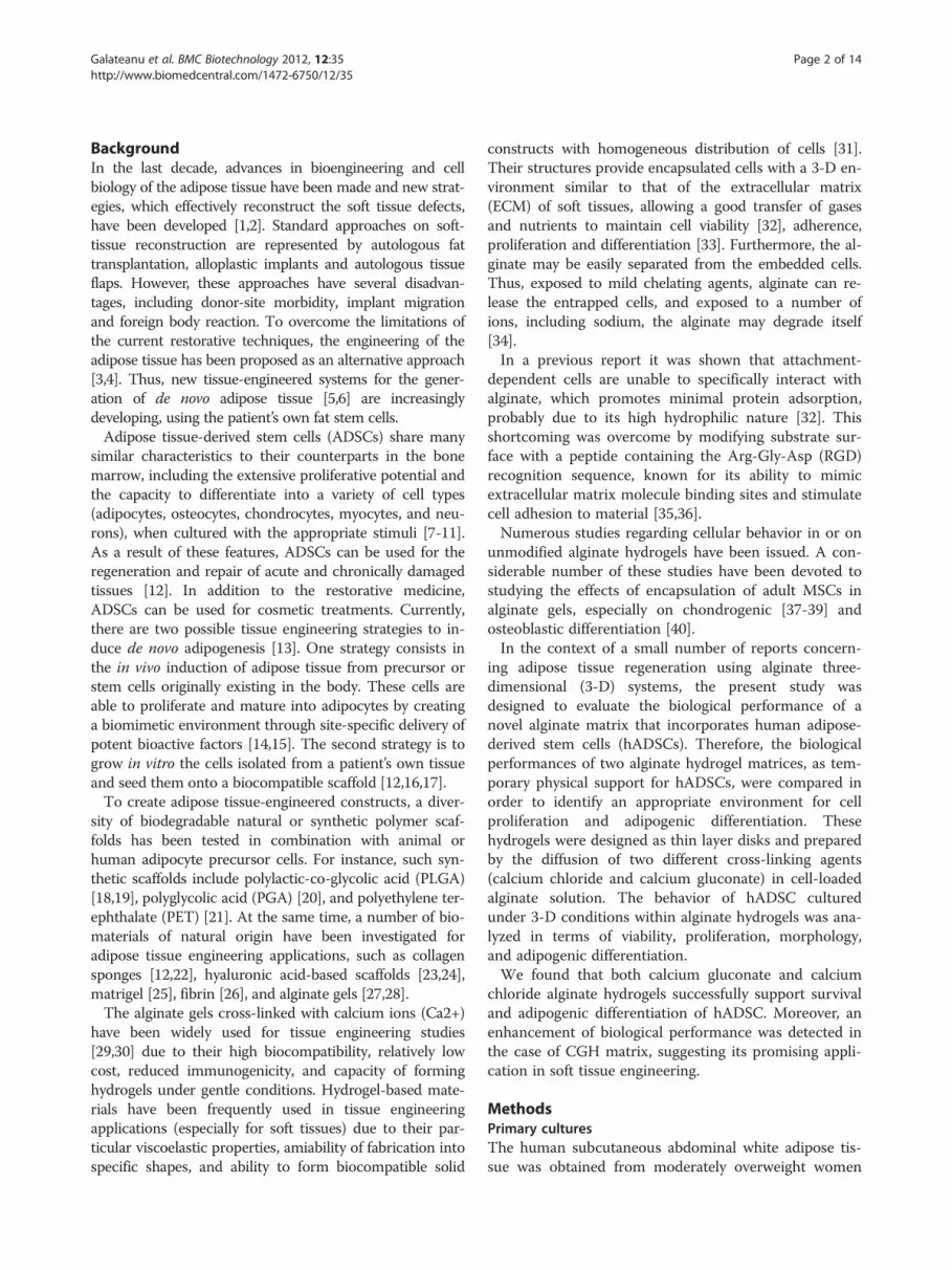
Galateanu et al. BMC Biotechnology 2012, 12:35 Page 2 of 14http://www.biomedcentral.com/1472-6750/12/35
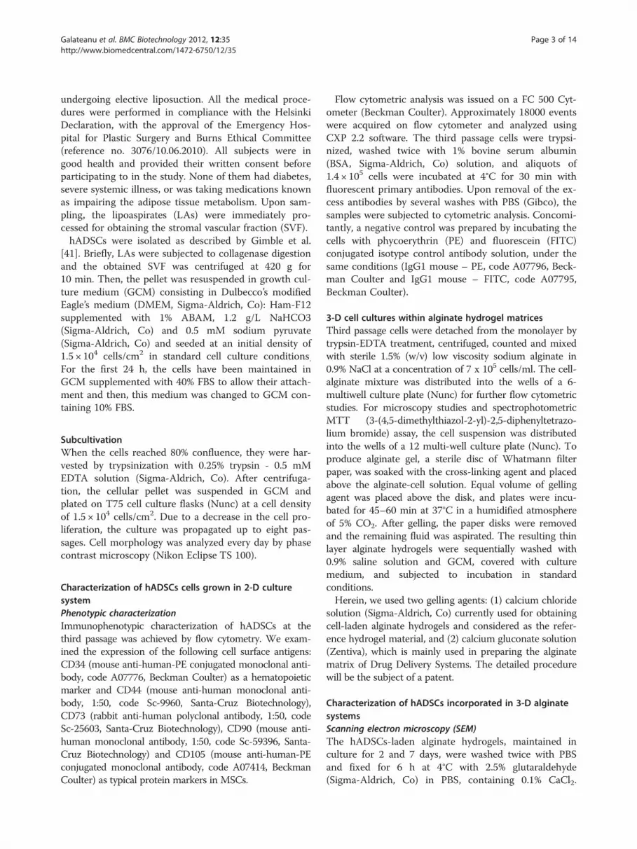
BackgroundIn the last decade, advances in bioengineering and cellbiology of the adipose tissue have been made and new strat-egies, which effectively reconstruct the soft tissue defects,have been developed [1,2]. Standard approaches on soft-tissue reconstruction are represented by autologous fattransplantation, alloplastic implants and autologous tissueflaps. However, these approaches have several disadvan-tages, including donor-site morbidity, implant migrationand foreign body reaction. To overcome the limitations ofthe current restorative techniques, the engineering of theadipose tissue has been proposed as an alternative approach[3,4]. Thus, new tissue-engineered systems for the gener-ation of de novo adipose tissue [5,6] are increasinglydeveloping, using the patient’s own fat stem cells.Adipose tissue-derived stem cells (ADSCs) share many
similar characteristics to their counterparts in the bonemarrow, including the extensive proliferative potential andthe capacity to differentiate into a variety of cell types(adipocytes, osteocytes, chondrocytes, myocytes, and neu-rons), when cultured with the appropriate stimuli [7-11].As a result of these features, ADSCs can be used for theregeneration and repair of acute and chronically damagedtissues [12]. In addition to the restorative medicine,ADSCs can be used for cosmetic treatments. Currently,there are two possible tissue engineering strategies to in-duce de novo adipogenesis [13]. One strategy consists inthe in vivo induction of adipose tissue from precursor orstem cells originally existing in the body. These cells areable to proliferate and mature into adipocytes by creatinga biomimetic environment through site-specific delivery ofpotent bioactive factors [14,15]. The second strategy is togrow in vitro the cells isolated from a patient’s own tissueand seed them onto a biocompatible scaffold [12,16,17].To create adipose tissue-engineered constructs, a diver-
sity of biodegradable natural or synthetic polymer scaf-folds has been tested in combination with animal orhuman adipocyte precursor cells. For instance, such syn-thetic scaffolds include polylactic-co-glycolic acid (PLGA)[18,19], polyglycolic acid (PGA) [20], and polyethylene ter-ephthalate (PET) [21]. At the same time, a number of bio-materials of natural origin have been investigated foradipose tissue engineering applications, such as collagensponges [12,22], hyaluronic acid-based scaffolds [23,24],matrigel [25], fibrin [26], and alginate gels [27,28].The alginate gels cross-linked with calcium ions (Ca2+)
have been widely used for tissue engineering studies[29,30] due to their high biocompatibility, relatively lowcost, reduced immunogenicity, and capacity of forminghydrogels under gentle conditions. Hydrogel-based mate-rials have been frequently used in tissue engineeringapplications (especially for soft tissues) due to their par-ticular viscoelastic properties, amiability of fabrication intospecific shapes, and ability to form biocompatible solid
constructs with homogeneous distribution of cells [31].Their structures provide encapsulated cells with a 3-D en-vironment similar to that of the extracellular matrix(ECM) of soft tissues, allowing a good transfer of gasesand nutrients to maintain cell viability [32], adherence,proliferation and differentiation [33]. Furthermore, the al-ginate may be easily separated from the embedded cells.Thus, exposed to mild chelating agents, alginate can re-lease the entrapped cells, and exposed to a number ofions, including sodium, the alginate may degrade itself[34].In a previous report it was shown that attachment-
dependent cells are unable to specifically interact withalginate, which promotes minimal protein adsorption,probably due to its high hydrophilic nature [32]. Thisshortcoming was overcome by modifying substrate sur-face with a peptide containing the Arg-Gly-Asp (RGD)recognition sequence, known for its ability to mimicextracellular matrix molecule binding sites and stimulatecell adhesion to material [35,36].Numerous studies regarding cellular behavior in or on
unmodified alginate hydrogels have been issued. A con-siderable number of these studies have been devoted tostudying the effects of encapsulation of adult MSCs inalginate gels, especially on chondrogenic [37-39] andosteoblastic differentiation [40].In the context of a small number of reports concern-
ing adipose tissue regeneration using alginate three-dimensional (3-D) systems, the present study wasdesigned to evaluate the biological performance of anovel alginate matrix that incorporates human adipose-derived stem cells (hADSCs). Therefore, the biologicalperformances of two alginate hydrogel matrices, as tem-porary physical support for hADSCs, were compared inorder to identify an appropriate environment for cellproliferation and adipogenic differentiation. Thesehydrogels were designed as thin layer disks and preparedby the diffusion of two different cross-linking agents(calcium chloride and calcium gluconate) in cell-loadedalginate solution. The behavior of hADSC culturedunder 3-D conditions within alginate hydrogels was ana-lyzed in terms of viability, proliferation, morphology,and adipogenic differentiation.We found that both calcium gluconate and calcium
chloride alginate hydrogels successfully support survivaland adipogenic differentiation of hADSC. Moreover, anenhancement of biological performance was detected inthe case of CGH matrix, suggesting its promising appli-cation in soft tissue engineering.
MethodsPrimary culturesThe human subcutaneous abdominal white adipose tis-sue was obtained from moderately overweight women
Galateanu et al. BMC Biotechnology 2012, 12:35 Page 3 of 14http://www.biomedcentral.com/1472-6750/12/35
undergoing elective liposuction. All the medical proce-dures were performed in compliance with the HelsinkiDeclaration, with the approval of the Emergency Hos-pital for Plastic Surgery and Burns Ethical Committee(reference no. 3076/10.06.2010). All subjects were ingood health and provided their written consent beforeparticipating to in the study. None of them had diabetes,severe systemic illness, or was taking medications knownas impairing the adipose tissue metabolism. Upon sam-pling, the lipoaspirates (LAs) were immediately pro-cessed for obtaining the stromal vascular fraction (SVF).hADSCs were isolated as described by Gimble et al.
[41]. Briefly, LAs were subjected to collagenase digestionand the obtained SVF was centrifuged at 420 g for10 min. Then, the pellet was resuspended in growth cul-ture medium (GCM) consisting in Dulbecco’s modifiedEagle’s medium (DMEM, Sigma-Aldrich, Co): Ham-F12supplemented with 1% ABAM, 1.2 g/L NaHCO3(Sigma-Aldrich, Co) and 0.5 mM sodium pyruvate(Sigma-Aldrich, Co) and seeded at an initial density of1.5 × 104 cells/cm2 in standard cell culture conditions.For the first 24 h, the cells have been maintained inGCM supplemented with 40% FBS to allow their attach-ment and then, this medium was changed to GCM con-taining 10% FBS.
SubcultivationWhen the cells reached 80% confluence, they were har-vested by trypsinization with 0.25% trypsin - 0.5 mMEDTA solution (Sigma-Aldrich, Co). After centrifuga-tion, the cellular pellet was suspended in GCM andplated on T75 cell culture flasks (Nunc) at a cell densityof 1.5 × 10⁴ cells/cm2. Due to a decrease in the cell pro-liferation, the culture was propagated up to eight pas-sages. Cell morphology was analyzed every day by phasecontrast microscopy (Nikon Eclipse TS 100).
Characterization of hADSCs cells grown in 2-D culturesystemPhenotypic characterizationImmunophenotypic characterization of hADSCs at thethird passage was achieved by flow cytometry. We exam-ined the expression of the following cell surface antigens:CD34 (mouse anti-human-PE conjugated monoclonal anti-body, code A07776, Beckman Coulter) as a hematopoieticmarker and CD44 (mouse anti-human monoclonal anti-body, 1:50, code Sc-9960, Santa-Cruz Biotechnology),CD73 (rabbit anti-human polyclonal antibody, 1:50, codeSc-25603, Santa-Cruz Biotechnology), CD90 (mouse anti-human monoclonal antibody, 1:50, code Sc-59396, Santa-Cruz Biotechnology) and CD105 (mouse anti-human-PEconjugated monoclonal antibody, code A07414, BeckmanCoulter) as typical protein markers in MSCs.
Flow cytometric analysis was issued on a FC 500 Cyt-ometer (Beckman Coulter). Approximately 18000 eventswere acquired on flow cytometer and analyzed usingCXP 2.2 software. The third passage cells were trypsi-nized, washed twice with 1% bovine serum albumin(BSA, Sigma-Aldrich, Co) solution, and aliquots of1.4 × 105 cells were incubated at 4°C for 30 min withfluorescent primary antibodies. Upon removal of the ex-cess antibodies by several washes with PBS (Gibco), thesamples were subjected to cytometric analysis. Concomi-tantly, a negative control was prepared by incubating thecells with phycoerythrin (PE) and fluorescein (FITC)conjugated isotype control antibody solution, under thesame conditions (IgG1 mouse – PE, code A07796, Beck-man Coulter and IgG1 mouse – FITC, code A07795,Beckman Coulter).
3-D cell cultures within alginate hydrogel matricesThird passage cells were detached from the monolayer bytrypsin-EDTA treatment, centrifuged, counted and mixedwith sterile 1.5% (w/v) low viscosity sodium alginate in0.9% NaCl at a concentration of 7 x 10⁵ cells/ml. The cell-alginate mixture was distributed into the wells of a 6-multiwell culture plate (Nunc) for further flow cytometricstudies. For microscopy studies and spectrophotometricMTT (3-(4,5-dimethylthiazol-2-yl)-2,5-diphenyltetrazo-lium bromide) assay, the cell suspension was distributedinto the wells of a 12 multi-well culture plate (Nunc). Toproduce alginate gel, a sterile disc of Whatmann filterpaper, was soaked with the cross-linking agent and placedabove the alginate-cell solution. Equal volume of gellingagent was placed above the disk, and plates were incu-bated for 45–60 min at 37°C in a humidified atmosphereof 5% CO2. After gelling, the paper disks were removedand the remaining fluid was aspirated. The resulting thinlayer alginate hydrogels were sequentially washed with0.9% saline solution and GCM, covered with culturemedium, and subjected to incubation in standardconditions.Herein, we used two gelling agents: (1) calcium chloride
solution (Sigma-Aldrich, Co) currently used for obtainingcell-laden alginate hydrogels and considered as the refer-ence hydrogel material, and (2) calcium gluconate solution(Zentiva), which is mainly used in preparing the alginatematrix of Drug Delivery Systems. The detailed procedurewill be the subject of a patent.
Characterization of hADSCs incorporated in 3-D alginatesystemsScanning electron microscopy (SEM)The hADSCs-laden alginate hydrogels, maintained inculture for 2 and 7 days, were washed twice with PBSand fixed for 6 h at 4°C with 2.5% glutaraldehyde(Sigma-Aldrich, Co) in PBS, containing 0.1% CaCl2.

Galateanu et al. BMC Biotechnology 2012, 12:35 Page 4 of 14http://www.biomedcentral.com/1472-6750/12/35
After rinsing with double distilled water, the sampleswere dried at 20°C and 0.1 mbar pressure for 4 hours ina 24-LSC Martin Christ laboratory freeze dryer. Then,the samples were coated with gold and imaged using aFEI Quanta Inspect F with field emission gun (FEG), op-erating in Scanning Electron Microscopy (SEM) mode.The microscope was driven with an acceleration voltageof 30 kV and a working distance of 10 mm detecting sec-ondary electrons.
Assessment of cell viability and proliferative activityCell viability within the alginate-based 3-D culture sys-tems was evaluated by flow cytometric determination oflive versus dead cells recovered from the hydrogel andstained using Live&Dead Kit (Invitrogen). This methodallows the simultaneous detection of both live and deadcells with calcein acetoxymethyl calcein AM and eth-idium bromide dyes provided in the kit. Calcein AM is anon-fluorescent and permeable reagent, which is con-verted by the intracellular esterases to the intenselygreen fluorescent calcein (ex/em: ~ 495 nm/~ 635 nm).Ethidium bromide enters the cells with damaged mem-brane, producing a bright red fluorescence when bindingto nucleic acids (ex/em: ~ 495 nm/~ 635 nm).Briefly, at 2 and 7 days post-seeding, the two alginate-
based hydrogels were first solubilized by incubation for15 min at 37°C in 94 mM NaCl solution containing350 mM sodium citrate (Sigma-Aldrich, Co) and 35 mMMOPS (Sigma-Aldrich, Co). After 10 min centrifugationat 265 g, 1.4 x 10⁵ cells were incubated at roomtemperature in the dark with calcein AM and ethidiumbromide solutions contained in the kit. A mean value of10.000 events was acquired by FC500 flow cytometer(Beckman Coulter) and analyzed with CXP 2.2 software(Beckman Coulter).The proliferation capacity of the cells within the alginate
hydrogels was assessed by MTT assay at 2 and 7 dayspost-seeding. Briefly, both RH and CGH matrices wereincubated for 15 h in MTT solution (1 mg/ml in serumfree culture medium) in standard conditions of cultivation.Before dissolving the formazan crystals produced by themetabolically active cells with isopropanol, the images ofMTT-stained culture were taken using an Olympus IX71inverted microscope. Solubilization was followed by spec-trophotometric quantification at 550 nm.
Assessment of adipogenic differentiationThe third passages ADSCs embedded in either RH orCGH matrices were analyzed for their capacity to differ-entiate towards the adipogenic lineage. The adipogenicdifferentiation was induced by culturing subconfluentcells in a new formula of adipogenic differentiationmedium (ADM), consisting in GCM supplemented withan adipogenic cocktail. This experiment was conducted
over a 21-day incubation period. The adipogenic differ-entiation was quantified by the cellular capacity to accu-mulate intracellular lipids and by perilipin expressionlevel. A detailed description of the ADM medium and ofthe entire experimental procedure will be provided in apatent application.The accumulation of cytoplasmic droplets of neutral
lipids was assessed by Oil Red O staining. The alginate-embedded cells cultured in 12-well plates were allowedto accommodate for 3–4 days in the new 3-D micro-environment. Subsequently, the GCM was supplementedwith the adipogenic cocktail.The cytoplasmic lipid droplets visualization was issued
at different time points, as described by Brandl et al.[15], with modifications. Briefly, the cell-laden alginatehydrogels were washed with PBS and fixed for 8 h with4% PFA. After permeabilization with 2% BSA/0.1% Tri-ton X-100 solution, both CGH and RH matrices wereincubated with Oil Red O (5 mg/ml in 60% isopropanol,diluted 3:2 with tap water) for 24 h at 4°C. Upon re-moval of the Oil Red O solution, the hydrogels werewashed several times to eliminate the traces of stainingsolution. The assessment of lipid droplets accumulationwas revealed by phase contrast microscopy (OlympusIX71) and Cell F Imaging Software (Olympus).For flow cytometric detection of perilipin at 3, 7, 15 and
21 days after adipogenic induction, 3-D culture systemswere first solubilized as previous described. After 10 mincentrifugation at 265 g, 1.4 x 10⁵ cells were fixed with 4%PFA and permeabilized with 2% BSA/0.1% Triton X-100solution. After a short washing step, the cells were incu-bated overnight with rabbit polyclonal anti-perilipinantibody solution (1:200, Santa-Cruz Biotechnology) andfor 30 min with FITC conjugated goat anti-rabbit IgG1secondary antibody (1:50, Santa-Cruz Biotechnology). Amean value of 10000 events was acquired and analyzedwith CXP 2.2 software (Beckman Coulter). In order tokeep the same parameters throughout the entire cultureperiod, the cytometer was calibrated with Flow Checkfluorescent beads (Beckman Coulter) before eachdetermination.
ResultsPrimary culture and characterization of hADSC in a 2-Dculture systemhADSCs cultures were isolated and purified from humansubcutaneous adipose tissue, which was obtained byelective liposuction. The primary culture was obtainedby collagenase digestion of the adipose tissue, followedby the seeding of the heterogeneous cell suspension, andsubsequent removal of non-adherent cells. As the cellswere propagated in monolayer culture, they showed amore uniform fibroblast-like morphology, suggesting theexistence of a homogenous ADSC cell population.

Figure 1 Immunophenotypic analysis of the third passage hADSCs by flow cytometry. The fluorescence level detected for the stromalstem cells markers was 95.9% for CD44, 92.04% for CD73, 92.86% for CD90 and 94.6% for CD105, respectively, whereas for the hematopoieticstem cells marker CD34 only 0.3% of the gated cells were fluorescently labeled. The positive zones in the histograms were marked with B in thefluorescence channel 1 (FL1), and with C in the fluorescence channel 2 (FL2), respectively, based on the isotype control antibody.
Galateanu et al. BMC Biotechnology 2012, 12:35 Page 5 of 14http://www.biomedcentral.com/1472-6750/12/35
hADSCs cell surface antigen expressionGiven the cell heterogeneity of LAs and of primary cul-ture, serial passages were analyzed for the expression ofhADSC-associated markers. The third passage cells weredetermined as being the most suitable for the incorpor-ation into the alginate matrix (data not shown).The flow cytometric data revealed that the third passage
hADSCs are negative for the hematopoietic marker CD34and positive for the stromal cell markers CD44, CD73,CD90 and CD105, which are common to human bonemarrow-multipotent MSCs (hBM-MSCs) (Figure 1). Thus,more than 90% of these cells purified by conventional cul-ture methods expressed CD44, CD73, CD90 and CD105respectively, while only 0.26% of the cells expressed CD34,which is selectively expressed on hematopoietic progenitorcells and vascular endothelium.These data suggest that the third passage hADSCs ex-
hibit a MSC origin, and that they are not interminglingwith hematopoietic cells. Based on these results, we
established that the most suitable hADSCs for incorpor-ation into the alginate matrix were the cells correspond-ing to the third cell passage.
In vitro behavior of hADSC embedded into alginatehydrogelsMacroscopic and microscopic evaluation of the cell-ladenalginate hydrogelsThe hADSC-laden hydrogels were poured into 12 and 6-well plates and were constructed as disks of 5 mmthickness. Throughout the entire culture period thesematrices have been stable, have not exhibited any con-traction, and could be easily handled. A schematicdiagram of the 3-D cell culture system preparation isshown in Figure 2.The morphology of the cell-laden alginate hydrogels
was examined using SEM after 2 and 7 days of culture,respectively. The investigation of the construct surfaceafter 2 days of cultivation revealed the presence of
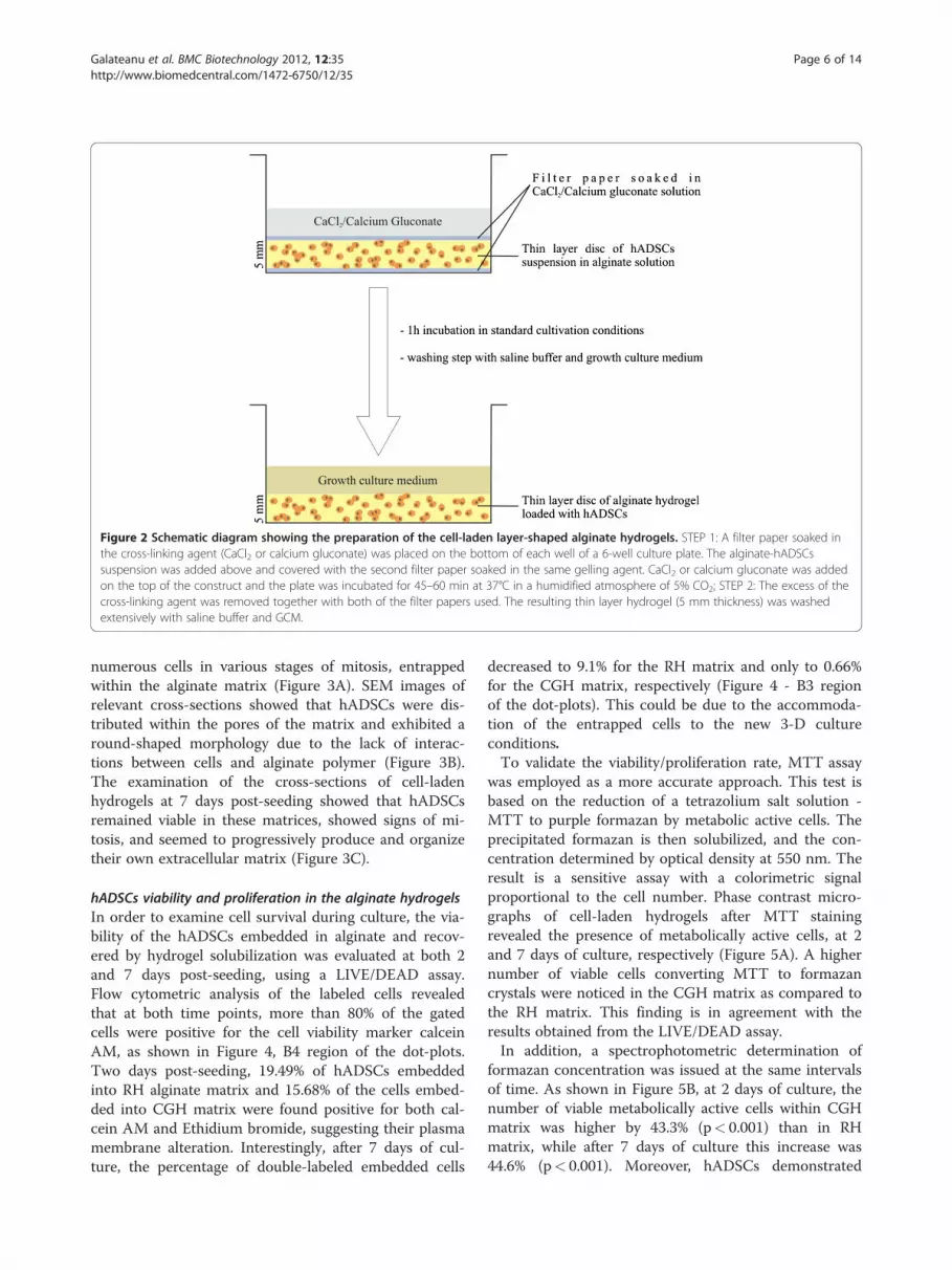
Figure 2 Schematic diagram showing the preparation of the cell-laden layer-shaped alginate hydrogels. STEP 1: A filter paper soaked inthe cross-linking agent (CaCl2 or calcium gluconate) was placed on the bottom of each well of a 6-well culture plate. The alginate-hADSCssuspension was added above and covered with the second filter paper soaked in the same gelling agent. CaCl2 or calcium gluconate was addedon the top of the construct and the plate was incubated for 45–60 min at 37°C in a humidified atmosphere of 5% CO2; STEP 2: The excess of thecross-linking agent was removed together with both of the filter papers used. The resulting thin layer hydrogel (5 mm thickness) was washedextensively with saline buffer and GCM.
Galateanu et al. BMC Biotechnology 2012, 12:35 Page 6 of 14http://www.biomedcentral.com/1472-6750/12/35
numerous cells in various stages of mitosis, entrappedwithin the alginate matrix (Figure 3A). SEM images ofrelevant cross-sections showed that hADSCs were dis-tributed within the pores of the matrix and exhibited around-shaped morphology due to the lack of interac-tions between cells and alginate polymer (Figure 3B).The examination of the cross-sections of cell-ladenhydrogels at 7 days post-seeding showed that hADSCsremained viable in these matrices, showed signs of mi-tosis, and seemed to progressively produce and organizetheir own extracellular matrix (Figure 3C).
hADSCs viability and proliferation in the alginate hydrogelsIn order to examine cell survival during culture, the via-bility of the hADSCs embedded in alginate and recov-ered by hydrogel solubilization was evaluated at both 2and 7 days post-seeding, using a LIVE/DEAD assay.Flow cytometric analysis of the labeled cells revealedthat at both time points, more than 80% of the gatedcells were positive for the cell viability marker calceinAM, as shown in Figure 4, B4 region of the dot-plots.Two days post-seeding, 19.49% of hADSCs embeddedinto RH alginate matrix and 15.68% of the cells embed-ded into CGH matrix were found positive for both cal-cein AM and Ethidium bromide, suggesting their plasmamembrane alteration. Interestingly, after 7 days of cul-ture, the percentage of double-labeled embedded cells
decreased to 9.1% for the RH matrix and only to 0.66%for the CGH matrix, respectively (Figure 4 - B3 regionof the dot-plots). This could be due to the accommoda-tion of the entrapped cells to the new 3-D cultureconditions.To validate the viability/proliferation rate, MTT assay
was employed as a more accurate approach. This test isbased on the reduction of a tetrazolium salt solution -MTT to purple formazan by metabolic active cells. Theprecipitated formazan is then solubilized, and the con-centration determined by optical density at 550 nm. Theresult is a sensitive assay with a colorimetric signalproportional to the cell number. Phase contrast micro-graphs of cell-laden hydrogels after MTT stainingrevealed the presence of metabolically active cells, at 2and 7 days of culture, respectively (Figure 5A). A highernumber of viable cells converting MTT to formazancrystals were noticed in the CGH matrix as compared tothe RH matrix. This finding is in agreement with theresults obtained from the LIVE/DEAD assay.In addition, a spectrophotometric determination of
formazan concentration was issued at the same intervalsof time. As shown in Figure 5B, at 2 days of culture, thenumber of viable metabolically active cells within CGHmatrix was higher by 43.3% (p< 0.001) than in RHmatrix, while after 7 days of culture this increase was44.6% (p< 0.001). Moreover, hADSCs demonstrated
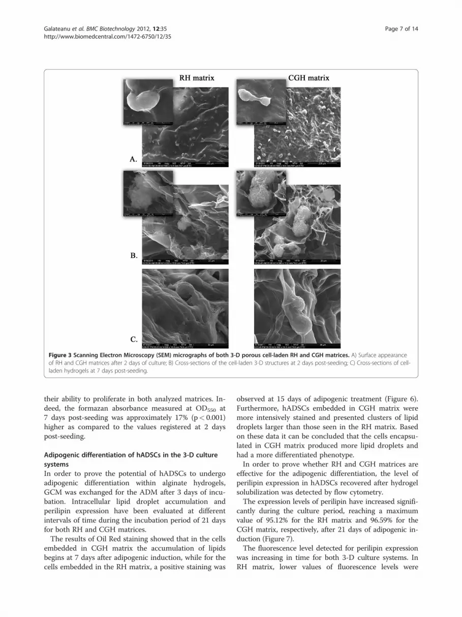
Figure 3 Scanning Electron Microscopy (SEM) micrographs of both 3-D porous cell-laden RH and CGH matrices. A) Surface appearanceof RH and CGH matrices after 2 days of culture; B) Cross-sections of the cell-laden 3-D structures at 2 days post-seeding; C) Cross-sections of cell-laden hydrogels at 7 days post-seeding.
Galateanu et al. BMC Biotechnology 2012, 12:35 Page 7 of 14http://www.biomedcentral.com/1472-6750/12/35
their ability to proliferate in both analyzed matrices. In-deed, the formazan absorbance measured at OD550 at7 days post-seeding was approximately 17% (p< 0.001)higher as compared to the values registered at 2 dayspost-seeding.
Adipogenic differentiation of hADSCs in the 3-D culturesystemsIn order to prove the potential of hADSCs to undergoadipogenic differentiation within alginate hydrogels,GCM was exchanged for the ADM after 3 days of incu-bation. Intracellular lipid droplet accumulation andperilipin expression have been evaluated at differentintervals of time during the incubation period of 21 daysfor both RH and CGH matrices.The results of Oil Red staining showed that in the cells
embedded in CGH matrix the accumulation of lipidsbegins at 7 days after adipogenic induction, while for thecells embedded in the RH matrix, a positive staining was
observed at 15 days of adipogenic treatment (Figure 6).Furthermore, hADSCs embedded in CGH matrix weremore intensively stained and presented clusters of lipiddroplets larger than those seen in the RH matrix. Basedon these data it can be concluded that the cells encapsu-lated in CGH matrix produced more lipid droplets andhad a more differentiated phenotype.In order to prove whether RH and CGH matrices are
effective for the adipogenic differentiation, the level ofperilipin expression in hADSCs recovered after hydrogelsolubilization was detected by flow cytometry.The expression levels of perilipin have increased signifi-
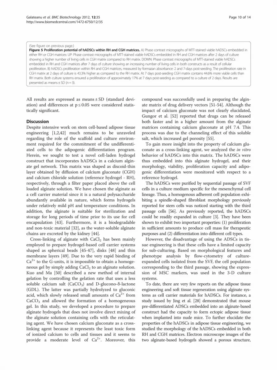
cantly during the culture period, reaching a maximumvalue of 95.12% for the RH matrix and 96.59% for theCGH matrix, respectively, after 21 days of adipogenic in-duction (Figure 7).The fluorescence level detected for perilipin expression
was increasing in time for both 3-D culture systems. InRH matrix, lower values of fluorescence levels were

Figure 4 Flow cytometric evaluation of hADSCs viability in both RH and CGH matrices. Fluorescence channel 1 (FL1)/Fluorescence channel2 (FL2) plots showing Ethidium Bromide positive cells (B1), both Ethidium Bromide and Calcein AM positive cells (B2), unmarked debris (B3) andCalcein AM positive cells (B4).
Galateanu et al. BMC Biotechnology 2012, 12:35 Page 8 of 14http://www.biomedcentral.com/1472-6750/12/35
recorded for 7 and 10 days of adipogenic induction ascompared to CGH matrix: 58.27% and 59.1% compared to63.81% (p< 0.001) and 72.14% (p< 0.001) respectively.Constant values of perilipin expression were recorded be-tween CGH and RH matrices after 2 weeks of adipogen-esis: 75.12% in RH matrix compared to 73.22% in CGH
matrix after 15 days and 95.12% in RH compared to96.59% in CGH after 21 days of induction.The results of MTT spectrophotometric assay and flow
cytometric data of perilipin expression were statisticallyanalyzed with GraphPrism 3.03. Software using One-wayANOVA with Bonferroni’s multiple comparison tests.

Figure 5 (See legend on next page.)
Galateanu et al. BMC Biotechnology 2012, 12:35 Page 9 of 14http://www.biomedcentral.com/1472-6750/12/35
(See figure on previous page.)Figure 5 Proliferation potential of hADSCs within RH and CGH matrices. A) Phase contrast micrographs of MTT-stained viable hADSCs embedded ineither RH or CGH matrices: UP: Phase contrast micrographs of MTT-stained viable hADSCs embedded in RH and CGH matrices after 2 days of cultureshowing a higher number of living cells in CGH matrix compared to RH matrix. DOWN: Phase contrast micrographs of MTT-stained viable hADSCsembedded in RH and CGH matrices after 7 days of culture showing an increasing number of living cells in both constructs as a result of cellularproliferation. B) hADSCs proliferation within RH and CGH matrices, measured by formazan absorbance: 2 and 7-days post-seeding. The proliferation rate inCGH matrix at 2 days of culture is 43.3% higher as compared to the RH matrix. At 7 days post-seeding CGH matrix contains 44.6% more viable cells thanRH matrix. Both culture systems ensured a proliferation of approximately 17% at 7 days post-seeding as compared to a culture of 2 days. Results arepresented as means±SD (n=3).
Galateanu et al. BMC Biotechnology 2012, 12:35 Page 10 of 14http://www.biomedcentral.com/1472-6750/12/35
All results are expressed as means ± SD (standard devi-ation) and differences at p ≤ 0.05 were considered statis-tically significant.
DiscussionDespite intensive work on stem cell-based adipose tissueengineering [1,2,42] much remains to be unraveledregarding the role of the scaffold and culture environ-ment required for the commitment of the undifferenti-ated cells to the adipogenic differentiation program.Herein, we sought to test a novel cell-laden hydrogelconstruct that incorporates hADSCs in a calcium algin-ate gel network. This matrix was shaped as discoid-thinlayer obtained by diffusion of calcium gluconate (CGH)and calcium chloride solution (reference hydrogel - RH),respectively, through a filter paper placed above the cellloaded alginate solution. We have chosen the alginate asa cell carrier material since it is a natural polysaccharideabundantly available in nature, which forms hydrogelsunder relatively mild pH and temperature conditions. Inaddition, the alginate is suitable for sterilization andstorage for long periods of time prior to its use for cellencapsulation [43]. Furthermore, it is a biodegradableand non-toxic material [32], as the water-soluble alginatechains are excreted by the kidney [44].Cross-linking of alginate with CaCl2 has been mainly
employed to prepare hydrogel-based cell carrier systemsshaped as spherical beads [45-47], disks [48] and thinmembrane layers [49]. Due to the very rapid binding ofCa2+ to the G-units, it is impossible to obtain a homoge-neous gel by simply adding CaCl2 to an alginate solution.Kuo and Ma [50] described a new method of internalgelation by controlling the gelation rate that uses a lesssoluble calcium salt (CaCO3) and D-glucono-δ-lactone(GDL). The latter was partially hydrolyzed to gluconicacid, which slowly released small amounts of Ca2+ fromCaCO3 and allowed the formation of a homogeneousgel. In this study, we developed a procedure to preparealginate hydrogels that does not involve direct mixing ofthe alginate solution containing cells with the reticulat-ing agent. We have chosen calcium gluconate as a cross-linking agent because it represents the least toxic formof ionized calcium to cells and tissues and it seems toprovide a moderate level of Ca2+. Moreover, this
compound was successfully used in preparing the algin-ate matrix of drug delivery vectors [51-54]. Although theimpact of calcium gluconate was not clearly elucidated,Gungor et al. [52] reported that drugs can be releasedboth faster and in a higher amount from the alginatematrices containing calcium gluconate at pH 7.4. Thisprocess was due to the channeling effect of this solublesalt, which increased gel porosity [55].To gain more insight into the property of calcium glu-
conate as a cross-linking agent, we analyzed the in vitrobehavior of hADSCs into this matrix. The hADSCs werethus embedded into this alginate hydrogel, and theirmorphology, viability, proliferation capacity and adipo-genic differentiation were monitored with respect to areference hydrogel.The hADSCs were purified by sequential passage of SVF
cells in a culture medium specific for the mesenchymal celllineage. Thus, a homogenous adherent cell population exhi-biting a spindle-shaped fibroblast morphology previouslyreported for stem cells was noticed starting with the thirdpassage cells [56]. As previously reported, the hADSCscould be readily expanded in culture [3]. They have beenshown to exhibit two important properties: (1) proliferationin sufficient amounts to produce cell mass for therapeuticpurposes and (2) differentiation into different cell types.However, the disadvantage of using the ADSCs in tis-
sue engineering is that these cells have a limited capacityfor sub-culturing. Based on morphological features andphenotype analysis by flow-cytometry of culture-expanded cells isolated from the SVF, the cell populationcorresponding to the third passage, showing the expres-sion of MSC markers, was used in the 3-D culturesystems.To date, there are very few reports on the adipose tissue
engineering and soft tissue regeneration using alginate sys-tems as cell carrier materials for hADSCs. For instance, astudy issued by Jing et al. [28] demonstrated that mousepre-differentiated ADSCs embedded into an alginate-basedconstruct had the capacity to form ectopic adipose tissuewhen implanted into nude mice. To further elucidate theproperties of the hADSCs in adipose tissue engineering, westudied the morphology of the hADSCs embedded in bothRH and CGH matrices. Electron microscope images of thetwo alginate-based hydrogels showed a porous structure,

Figure 6 Phase contrast images of hADSCs embedded in RH and CGH and subjected to adipogenesis. Both cell-laden alginate constructswere stained with Oil Red O after 3, 7, 10, 15 and 21 days of adipogenesis, to visualize the size and the amount of cytoplasmic lipid droplets.
Galateanu et al. BMC Biotechnology 2012, 12:35 Page 11 of 14http://www.biomedcentral.com/1472-6750/12/35
whereby the hADSCs display normal spherical morpholo-gies, some of them showing signs of mitosis.In order to examine cell survival during culture, the via-
bility of the encapsulated hADSCs in the alginate hydrogelswas evaluated after 2 and 7 days of culture by a flowcytometry-based LIVE/DEAD assay. At both analyzedtime-points, most of the entrapped cells (over 80%)
successfully survived within RH and CGH matrices. Fur-thermore, the percentage of viable cells was significantlyhigher within CGH matrix at 2 days post-seeding and ap-proximately similar within both hydrogels after 7 days ofculture. These results are in keeping with the observationthat the alginate matrices present a structure formed byinterconnected pores, which is suitable to accommodate
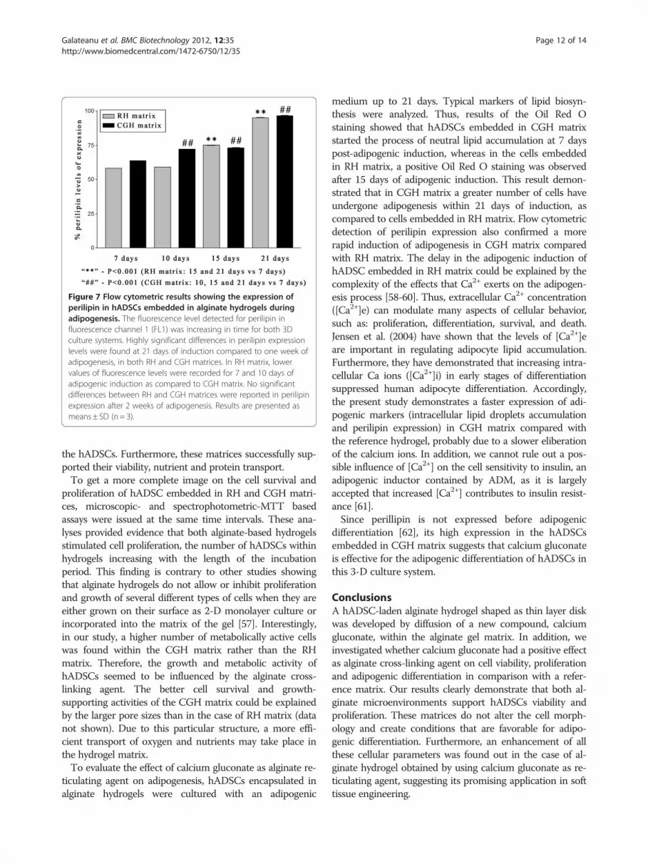
Figure 7 Flow cytometric results showing the expression ofperilipin in hADSCs embedded in alginate hydrogels duringadipogenesis. The fluorescence level detected for perilipin influorescence channel 1 (FL1) was increasing in time for both 3Dculture systems. Highly significant differences in perilipin expressionlevels were found at 21 days of induction compared to one week ofadipogenesis, in both RH and CGH matrices. In RH matrix, lowervalues of fluorescence levels were recorded for 7 and 10 days ofadipogenic induction as compared to CGH matrix. No significantdifferences between RH and CGH matrices were reported in perilipinexpression after 2 weeks of adipogenesis. Results are presented asmeans ± SD (n= 3).
Galateanu et al. BMC Biotechnology 2012, 12:35 Page 12 of 14http://www.biomedcentral.com/1472-6750/12/35
the hADSCs. Furthermore, these matrices successfully sup-ported their viability, nutrient and protein transport.To get a more complete image on the cell survival and
proliferation of hADSC embedded in RH and CGH matri-ces, microscopic- and spectrophotometric-MTT basedassays were issued at the same time intervals. These ana-lyses provided evidence that both alginate-based hydrogelsstimulated cell proliferation, the number of hADSCs withinhydrogels increasing with the length of the incubationperiod. This finding is contrary to other studies showingthat alginate hydrogels do not allow or inhibit proliferationand growth of several different types of cells when they areeither grown on their surface as 2-D monolayer culture orincorporated into the matrix of the gel [57]. Interestingly,in our study, a higher number of metabolically active cellswas found within the CGH matrix rather than the RHmatrix. Therefore, the growth and metabolic activity ofhADSCs seemed to be influenced by the alginate cross-linking agent. The better cell survival and growth-supporting activities of the CGH matrix could be explainedby the larger pore sizes than in the case of RH matrix (datanot shown). Due to this particular structure, a more effi-cient transport of oxygen and nutrients may take place inthe hydrogel matrix.To evaluate the effect of calcium gluconate as alginate re-
ticulating agent on adipogenesis, hADSCs encapsulated inalginate hydrogels were cultured with an adipogenic
medium up to 21 days. Typical markers of lipid biosyn-thesis were analyzed. Thus, results of the Oil Red Ostaining showed that hADSCs embedded in CGH matrixstarted the process of neutral lipid accumulation at 7 dayspost-adipogenic induction, whereas in the cells embeddedin RH matrix, a positive Oil Red O staining was observedafter 15 days of adipogenic induction. This result demon-strated that in CGH matrix a greater number of cells haveundergone adipogenesis within 21 days of induction, ascompared to cells embedded in RH matrix. Flow cytometricdetection of perilipin expression also confirmed a morerapid induction of adipogenesis in CGH matrix comparedwith RH matrix. The delay in the adipogenic induction ofhADSC embedded in RH matrix could be explained by thecomplexity of the effects that Ca2+ exerts on the adipogen-esis process [58-60]. Thus, extracellular Ca2+ concentration([Ca2+]e) can modulate many aspects of cellular behavior,such as: proliferation, differentiation, survival, and death.Jensen et al. (2004) have shown that the levels of [Ca2+]eare important in regulating adipocyte lipid accumulation.Furthermore, they have demonstrated that increasing intra-cellular Ca ions ([Ca2+]i) in early stages of differentiationsuppressed human adipocyte differentiation. Accordingly,the present study demonstrates a faster expression of adi-pogenic markers (intracellular lipid droplets accumulationand perilipin expression) in CGH matrix compared withthe reference hydrogel, probably due to a slower eliberationof the calcium ions. In addition, we cannot rule out a pos-sible influence of [Ca2+] on the cell sensitivity to insulin, anadipogenic inductor contained by ADM, as it is largelyaccepted that increased [Ca2+] contributes to insulin resist-ance [61].Since perillipin is not expressed before adipogenic
differentiation [62], its high expression in the hADSCsembedded in CGH matrix suggests that calcium gluconateis effective for the adipogenic differentiation of hADSCs inthis 3-D culture system.
ConclusionsA hADSC-laden alginate hydrogel shaped as thin layer diskwas developed by diffusion of a new compound, calciumgluconate, within the alginate gel matrix. In addition, weinvestigated whether calcium gluconate had a positive effectas alginate cross-linking agent on cell viability, proliferationand adipogenic differentiation in comparison with a refer-ence matrix. Our results clearly demonstrate that both al-ginate microenvironments support hADSCs viability andproliferation. These matrices do not alter the cell morph-ology and create conditions that are favorable for adipo-genic differentiation. Furthermore, an enhancement of allthese cellular parameters was found out in the case of al-ginate hydrogel obtained by using calcium gluconate as re-ticulating agent, suggesting its promising application in softtissue engineering.

Galateanu et al. BMC Biotechnology 2012, 12:35 Page 13 of 14http://www.biomedcentral.com/1472-6750/12/35
AbbreviationsADSC: adipose-derived stem cells; ABAM: antibiotic antimycothic;ADM: adipogenic differentiation medium; BSA: bovine serum albumin;CD: cluster of differentiation; CGH matrix: calcium gluconate hydrogel matrix;FBS: Fetal Bovine Serum; FITC: Fluorescein isothiocyanate; GCM: GrowthCulture Medium; GDL: D-glucono-δ-lactone; hADSC: human adipose-derivedstem cells; hBM-MSCs: human bone marrow-multipotent mesenchymal stemcells; LAs: Lipoaspirates; MOPS: 3-(N-morpholino)propanesulfonic acid;MSC: Mesenchymal Stem Cells; MTT: 3-(4,5-Dimethylthiazol-2-yl)-2,5-diphenyltetrazolium bromide; O.D.: Optic density; PBS: Phosphate Buffer Salt;PE: Phycoeritrine; PET: Polyethylene terephthalate; PGA: polyglycolic acid;PLA: Processed lipoaspirate cells; PLGA: Polylactic-co-glycolic acid;RGD: Arginine-glycine-aspartic acid; RH matrix: Reference Hydrogel matrix;SEM: Scanning Electron Microscopy; SVF: Stromal Vascular Fraction.
Competing interestsHereby, we Bianca Galateanu, Anisoara Cimpean and Marieta Costachedeclare that we are University of Bucharest employees main grant recipientof project PCCE 248/2010 whereby this study was financially supported andwhereby this paper fee will be assured in case of acceptance. Therefore wehave no competing interests.Hereby, the undersigned Doina Dimonie I declare that the 3-D systempreparation method is protected by a national patent request. Therefore Ihave no competing interests.Hereby, Eugeniu Vasile and Sorin Nae declare that we have no competinginterests.
AcknowledgementsThis research was supported by Romanian CNCSIS – UEFISCSU, ComplexExploratory Research Project (Grant No.: PCCE248/2010). We thank Prof. Dr.Dana Iordachescu (University of Bucharest, Department of Biochemistry andMolecular Biology) for project idea and the invaluable help in conceptionand design of the experiments and for the initiation of the draft manuscript.We also thank Dr. Mihaela Stefania Diaconu (University of Bucharest,Department of Biochemistry and Molecular Biology) for critical editing of themanuscript and Eng. Chem. Inna Georgeta Trandafir for her involvement inthe preparation of the 3-D alginate based constructs.
Author details1Department of Biochemistry and Molecular Biology, University of Bucharest,91-95 Splaiul Independentei, sect 5, Bucharest, Romania. 2Research andDevelopment National Institute for Chemistry and Petrochemistry, 202 SplaiulIndependentei, sect 6, Bucharest, Romania. 3METAV CD, 31 C. A. RosettiStreet, Bucharest, Romania. 4Emergency Hospital of Plastic Surgery and Burns,218 Calea Grivitei Street, sect 1, Bucharest, Romania.
Authors’ contributionsBG carried out the isolation of human adipose-derived stem cells fromlipoaspirate and all the procedures performed in the cell culture field. Sheperformed the phase contrast and fluorescence microscopy, the flowcytometry and the spectrophotometric assays and also carried out thestatistical analysis. She was involved in the hydrogels design and preparationand helped to draft the manuscript. DD is responsible for the design of the3-D thin layer alginate hydrogels. EV carried out the SEM assay. SN providedthe lipoaspirate in accordance with the EU ethical standards. MC and ACcontributed equally to this work. They were responsible for the coordinationof the whole study, for the interpretation of the results and for drafting themanuscript. They also assured a good communication between authors. Allauthors read and approved the final manuscript.
Authors’ informationBG is a PhD Student in the last stage of the internship and researcher in theDepartment of Biochemistry and Molecular Biology. Her PhD thesis involvesthe development of new strategies in adipose tissue engineering usingprecursor cells and natural biocompatible polymers. Her interest in the fieldof tissue bioengineering was well evaluated at International Congresses.DD is senior researcher at ICECHIM. She has competences in obtaining andstudying new eco - friendly polymeric materials for eco - friendlyapplications. She has a long experience in this field that means papers asmain author in ISI coated journals, patents as main author, new productsand technologies as project manager.
EV is PhD Engineer senior researcher with high specialization in microscopy.SN is a physician in the field of Plastic Surgery and also scientific researcher.He works within the Emergency Hospital for Plastic and ReconstructiveSurgery and Burns in Bucharest and he collaborates with numerous aestheticsurgery private clinics.AC is associate professor in the University of Bucharest and ScientificResearcher in the Department of Biochemistry and Molecular Biology. Sheattended a post-doc fellowship in Leuven, Belgium and leads researchprojects concerning the field of tissue bioengineering.MC is full professor at the University of Bucharest, Head of the Departmentof Biochemistry and Molecular Biology and director of numerous nationaland international grants. One of the most important research projects led byher is the one financing this study. She obtained her PhD degree inMolecular Biology of the Cell in Paris, France, in 1997.
Received: 6 December 2011 Accepted: 29 June 2012Published: 29 June 2012
References1. Patrick CW Jr: Tissue engineering strategies for adipose tissue repair. Anat
Rec 2001, 263(4):361–366.2. Niemelä C, Miettinen S, Sarkanen JR, Ashammakhi N: Adipose Tissue and
Adipocyte Differentiation: Molecular and Cellular Aspects and TissueEngineering Applications. In Topics in Tissue Engineering. Volume 4. Editedby Ashammakhi N, Reis R, Chiellini F.: ; 2008:1–26.
3. Gomillion CT, Burg KJL: Stem cells and adipose tissue engineering.Biomaterials 2006, 27(36):6052–6063.
4. Parker AM, Katz AJ: Adipose-derived stem cells for the regeneration ofdamaged tissues. Expert Opin Biol Ther 2006, 6(6):567–578.
5. Vermette M, Trottier V, Ménard V, Saint-Pierre L, Roy A, Fradette J:Production of a new tissue-engineered adipose substitute from humanadipose-derived stromal cells. Biomaterials 2007, 28(18):2850–2860.
6. Flynn L, Prestwich GD, Semple JL, Woodhouse KA: Adipose tissueengineering with naturally derived scaffolds and adipose-derived stemcells. Biomaterials 2007, 28(26):3834–3842.
7. Gimble JM, Guilak F: Differentiation potential of adipose derived adultstem (ADAS) cells. Curr Top Dev Biol 2003, 58:137–160.
8. Hicok KC, Du Laney TV, Zhou YS, Halvorsen YD, Hitt DC, Cooper LF, GimbleJM: Human adipose-derived adult stem cells produce osteoid in vivo.Tissue Eng 2004, 10(3–4):371–380.
9. Skalska U, Burakowski T, Janicka I, Kornatka A, Maldyk P, Maslinski W, KontnyE: Chondrogenic and osteogenic potential of adipose derived stem cellsfrom RA and OA patients. Ann Rheum Dis 2011, 70(Suppl 2):A25.
10. Gwak SJ, Bhang SH, Yang HS, Kim SS, Lee DH, Lee SH, Kim BS: In vitrocardiomyogenic differentiation of adipose-derived stromal cells usingtransforming growth factor-beta1. Cell Biochem Funct 2009, 27(3):148–154.
11. Safford KM, Hicok KC, Safford SD, Halvorsen YD, Wilkison WO, Gimble JM,Rice HE: Neurogenic differentiation of murine and human adipose-derived stromal cells. Biochem Biophys Res Commun 2002, 294(2):371–379.
12. Kimura Y, Ozeki M, Inamoto T, Tabata Y: Adipose tissue engineeringbased on human preadipocytes combined with gelatin microspherescontaining basic fibroblast growth factor. Biomaterials 2003, 24(14):2513–2521.
13. Katz AJ, Llull R, Hedrick MH, Futrell JW: Emerging approaches to the tissueengineering of fat. Clin Plast Surg 1999, 26(4):587–603.
14. Kawaguchi N, Toriyama K, Nicodemou-Lena E, Inou K, Torii S, Kitagawa Y:De Novo adipogenesis in mice at the site of injection of basementmembrane and basic fibroblast growth factor. Proc Natl Acad Sci USA1998, 95(3):1062–1066.
15. Hiraoka Y, Yamashiro H, Yasuda K, Kimura Y, Inamoto T, Tabata Y: In situregeneration of adipose tissue in rat fat pad by combining a collagenscaffold with gelatin microspheres containing basic fibroblast growthfactor. Tissue Eng 2006, 12(6):1475–1487.
16. Vashi AV, Abberton KM, Thomas GP, Morrison WA, O’Connor AJ, Cooper-White JJ, Thompson EW: Adipose tissue engineering based on thecontrolled release of fibroblast growth factor-2 in a collagen matrix.Tissue Eng 2006, 12(11):3035–3043.
17. Girandon L, Kregar-Velikonja N, Bozikov K, Barlik A: In vitro models foradipose tissue engineering with adipose-derived stem cells usingdifferent scaffolds of natural origin. Folia Biologica (Praha) 2011, 57(2):47–56.
Galateanu et al. BMC Biotechnology 2012, 12:35 Page 14 of 14http://www.biomedcentral.com/1472-6750/12/35
18. Patrick CW, Zheng B, Johnston C, Reece GP: Long-term implantation ofpreadipocyte-seeded PLGA scaffolds. Tissue Eng 2002, 8(2):283–293.
19. Choi YS, Park SN, Suh H: Adipose tissue engineering using mesenchymalstem cells attached to injectable PLGA spheres. Biomaterials 2005, 26(29):5855–5863.
20. Fischbach C, Spruss T, Weiser B, Neubauer M, Becker C, Hacker M, Göpferich A,Blunk T: Generation of mature fat pads in vitro and in vivo utilizing 3-Dlong-term culture of 3 T3-L1 preadipocytes. Exp Cell Res 2004, 300(1):54–64.
21. Kang X, Xie Y, Kniss DA: Adipose tissue model using threedimensionalcultivation of preadipocytes seeded onto fibrous polymer scaffolds.Tissue Eng 2005, 11(3–4):458–468.
22. von Heimburg D, Zachariah S, Heschel I, Kuhling H, Schoof H, Hafemann B,Pallua N: Human preadipocytes seeded on freeze-dried collagen scaffoldsinvestigated in vitro and in vivo. Biomaterials 2001, 22(5):429–438.
23. Halbleib M, Skurk T, de Luca C, von Heimburg D, Hauner H: Tissueengineering of white adipose tissue using hyaluronic acid-basedscaffolds. I: in vitro differentiation of human adipocyte precursor cells onscaffolds. Biomaterials 2003, 24(18):3125–3132.
24. Stillaert FB, Di Bartolo C, Hunt JA, Rhodes NP, Tognana E, Monstrey S,Blondeel PN: Human clinical experience with adipose precursor cellsseeded on hyaluronic acid-based spongy scaffolds. Biomaterials 2008, 29(29):3953–3959.
25. Walton RL, Beahm EK, Wu L: De novo adipose formation in a vascularizedengineered construct. Microsurgery 2004, 24(5):378–384.
26. Cho SW, Kim I, Kim SH, Rhie JW, Choi CY, Kim BS: Enhancement of adiposetissue formation by implantation of adipogenic-differentiatedpreadipocytes. Biochem Biophys Res Commun 2006, 345(2):588–594.
27. Marler JJ, Guha A, Rowley J, Koka R, Mooney D, Upton J, Vacanti JP: Soft-tissue augmentation with injectable alginate and syngeneic fibroblasts.Plast Reconstr Surg 2000, 105(6):2049–2058.
28. Jing W, Lin Y, Wu L, Li X, Nie X, Liu L, Tang W, Zheng X, Tian W: Ectopicadipogenesis of preconditioned adipose-derived stromal cells in analginate system. Cell Tissue Res 2007, 330(3):567–572.
29. Paige KT, Cima LG, Yaremchuk MJ, Schloo BL, Vacanti JP, Vacanti CA: Denovo cartilage generation using calcium alginate-chondrocyteconstructs. Plast Reconstr Surg 1996, 97(1):168–180.
30. Masuda K, Sah RL, Hejna MJ, Thonar EJ: A novel two-step method for theformation of tissue-engineered cartilage by mature bovinechondrocytes: the alginate-recovered-chondrocyte (ARC) method. JOrthop Res 2003, 21(1):139–148.
31. Ahearne M, Yang Y, El Jaj AJ, Then KY, Liu KK: Characterizing theviscoelastic properties of thin hydrogel-based constructs for tissueengineering applications. J R Soc Interface 2005, 2(5):455–463.
32. Rowley JA, Madlambayan G, Mooney DJ: Alginate hydrogels as syntheticextracellular matrix materials. Biomaterials 1999, 20(1):45–53.
33. Drury JL, Mooney DJ: Hydrogels for tissue engineering: scaffold designvariables and applications. Biomaterials 2003, 24(24):4337–4351.
34. Ikada Y: Challenges in tissue engineering. J R Soc Interface 2006, 3(10):589–601.
35. Rowley JA, Mooney DJ: Alginate type and RGD density control myoblastphenotype. J Biomed Mater Res 2002, 60(2):217–223.
36. Chandler EM, Berglund CM, Lee JS, Polacheck WJ, Gleghorn JP, Kirby BJ,Fischbach C: Stiffness of photocrosslinked RGD- alginate gels regulatesadipose progenitor cell behavior. Biotechnol Bioeng 2011, 108(7):1683–1692.
37. Jin XB, Sun YS, Zhang K, Wang J, Ju XD, Lou SQ: Neocartilage formationfrom predifferentiated human adipose derived stem cells in vivo. ActaPharmacol Sin 2007, 28(5):663–671.
38. Diekman BO, Rowland CR, Lennon DP, Caplan AI, Guilak F: Chondrogenesisof adult stem cells from adipose tissue and bone marrow: induction bygrowth factors and cartilage-derived matrix. Tissue Eng Part A 2010, 16(2):523–533.
39. Hsu SH, Huang TB, Cheng SJ, Weng SY, Tsai CL, Tseng CS, Chen DC, Liu TY,Fu KY, Yen BL: Chondrogenesis from human placenta-derivedmesenchymal stem cells in three-dimensional scaffolds for cartilagetissue engineering. Tissue Eng Part A 2011, 17(11-12):1549–1560.
40. Wang L, Shelton RM, Cooper PR, Lawson PR, Triffitt JT, Barralet JE:Evaluation of sodium alginate for bone marrow cell tissue engineering.Biomaterials 2003, 24(20):3475–3481.
41. Gimble JM, Katz AJ, Bunnell BA: Adipose-derived stem cells forregenerative medicine. Circ Res 2007, 100(9):1249–1260.
42. Ogawa R: The importance of adipose-derived stem cells and vascularizedtissue regeneration in the field of tissue transplantation. Curr Stem CellRes Ther 2006, 1(1):13–20.
43. d’Ayala GG, Malinconico M, Laurienzo P: Marine derived polysaccharidesfor biomedical applications: chemical modification approaches. Molecules2008, 13(9):2069–2106.
44. Al-Shamkhani A, Duncan R: Radioiodination of alginate via covalently-boundtyrosinamide allows for monitoring of its fate in vivo. J Bioact Compat Polym1995, 10(1):4–13.
45. Chubinskaya S, Huch K, Schulze M, Otten L, Aydelotte MB, Cole AA: Humanarticular chondrocytes cultured in alginate beads maintain their geneexpression. Cells Materials 1998, 8:151–160.
46. Li X, Liu T, Song K, Yao L, Ge D, Bao C, Ma X, Cui Z: Culture of neural stemcells in calcium alginate beads. Biotechnol Prog 2006, 22(6):1683–1689.
47. Yamaoka H, Asato H, Ogasawara T, Nishizawa S, Takahashi T, Nakatsuka T,Koshima I, Nakamura K, Kawaguchi H, Chung UI, Takato T, Hoshi K: Cartilagetissue engineering using human auricular chondrocytes embedded indifferent hydrogel materials. J Biomed Mater Res A 2006, 78(1):1–11.
48. Awad HA, Wickham MQ, Leddy HA, Gimble JM, Guilak F: Chondrogenicdifferentiation of adipose-derived adult stem cells in agarose, alginate,and gelatin scaffolds. Biomaterials 2004, 25(16):3211–3222.
49. Katsanakis N, Katsivelis A, Kintzios S: Immobilization of electroporated cells forfabrication of cellular biosensors: physiological effects of the shape ofcalcium alginate matrices and foetal calf serum. Sensors 2009, 9:378–385.
50. Kuo CK, Ma PX: Ionically crosslinked alginate hydrogels as scaffold fortissue engineering: part 1. Structure, gelation rate and mechanicalproperties. Biomaterials 2001, 22(6):511–521.
51. Kuo CK, Ma PX: Maintaining dimensions and mechanical properties ofionically crosslinked alginate hydrogel scaffolds in vitro. J Biomed MaterRes 2008, 84(4):899–907.
52. Güngör S, Yildiz A, Ozsoy Y, Cevher E, Araman A: Investigations onmefenamic acid sustained release tablets with water-insoluble gel.Farmaco 2003, 58(5):397–340.
53. Giunchedi P, Gavini E, Moretti MDL, Pirisino G: Evaluation of alginatecompressed matrices as prolonged drug delivery systems. AAPSPharmSciTech 2000, 1(3):31–36.
54. Gilhotra RM, Gilhotra N, Mishra DN: A hydrogel–forming bioadhesiveocular minitablet for the management of microbial Keratitis. Asian JPharm Sci 2010, 5(1):19–25.
55. Pongjanyakul T, Puttipipatkhachorn S: Modulating drug release and matrixerosion of alginate matrix capsules by microenvironmental interactionwith calcium ion. Eur J Pharm Biopharm 2007, 67(1):187–195.
56. Steck E, Bertram H, Abel R, Chen B, Winter A, Richter W: Induction ofintervertebral disc-like cells from adult mesenchymal stem cells. StemCells 2005, 23(3):403–411.
57. Novikova LN, Mosahebi A, Wiberg M, Terenghi G, Kellerth JO, Novikov LN:Alginate hydrogel and matrigel as potential cell carriers forneurotransplantation. J Biomed Mater Res 2006, 77(2):242–252.
58. Hang S, Halvorsen YD, Ellis PN, Wilkison WO, Zemel MB: Role ofintracellular calcium in human adipocyte differentiation. PhysiolGenomics2000, 3(2):75–82.
59. Jensen B, Farach-Carson MC, Kenaley E, Akanbi KA: High extracellularcalcium attenuates adipogenesis in 3 T3-L1 preadipocytes. Exp Cell Res2004, 301(2):280–292.
60. Draznin B: Cytosolic calcium and insulin resistance. Am J Kidney Dis 1993,21(3):32–38.
61. Fruhbeck G, Gomez-Ambrosi J, Muruzabal FJ, Burrell MA: The adipocyte: amodel for integration of endocrine and metabolic signaling in energymetabolism regulation. Am J Physiol Endocrinol Metab 2001, 280(6):827–847.
62. Arimura N, Horiba T, Imagawa M, Shimizu M, Sato R: The peroxisomeproliferator-activated receptor γ regulates expression of the perilipingene in adipocytes. J Biol Chem 2004, 279(11):10070–10076.
doi:10.1186/1472-6750-12-35Cite this article as: Galateanu et al.: Layer-shaped alginate hydrogelsenhance the biological performance of human adipose-derived stemcells. BMC Biotechnology 2012 12:35.