
LATERAL FLOW FORMAT, MATERIALS AND METHODS
56
Note: Within nine months of the publication of the mention of the grant of the European patent in the European Patent Bulletin, any person may give notice to the European Patent Office of opposition to that patent, in accordance with the Implementing Regulations. Notice of opposition shall not be deemed to have been filed until the opposition fee has been paid. (Art. 99(1) European Patent Convention). Printed by Jouve, 75001 PARIS (FR) (19) EP 1 733 233 B1 TEPZZ_7¥¥ ¥¥B_T (11) EP 1 733 233 B1 (12) EUROPEAN PATENT SPECIFICATION (45) Date of publication and mention of the grant of the patent: 12.12.2012 Bulletin 2012/50 (21) Application number: 05732716.5 (22) Date of filing: 30.03.2005 (51) Int Cl.: G01N 33/543 (2006.01) G01N 33/558 (2006.01) (86) International application number: PCT/US2005/010678 (87) International publication number: WO 2005/098439 (20.10.2005 Gazette 2005/42) (54) LATERAL FLOW FORMAT, MATERIALS AND METHODS SEITENSTROMFORMAT, MATERIALIEN UND VERFAHREN ET FORMAT D’ECOULEMENT LATERAL, MATERIAUX ET PROCEDES (84) Designated Contracting States: AT BE BG CH CY CZ DE DK EE ES FI FR GB GR HU IE IS IT LI LT LU MC NL PL PT RO SE SI SK TR (30) Priority: 30.03.2004 US 557851 P (43) Date of publication of application: 20.12.2006 Bulletin 2006/51 (73) Proprietor: GE Healthcare Bio-Sciences Corp. Piscataway, N.J. 08855 (US) (72) Inventors: • JONES, Kevin Boonton, NJ 07005 (US) • COX, David West Bridgford, Nottingham NG2 7EP (GB) (74) Representative: Aldenbäck, Ulla Christina et al GE Healthcare Bio-Sciences AB Patent Department Björkgatan 30 751 84 Uppsala (SE) (56) References cited: EP-A- 0 381 173 WO-A1-88/08534 US-A- 5 141 850 US-A1- 2003 175 991 • SURR F.: "Whatman FUSION 5? One Material, Five Functions" WHATMAN DATASHEET, [Online] 24 March 2004 (2004-03-24), XP002477492 Retrieved from the Internet: URL: http://www.whatman.com/References/What manFusion5.pdf> [retrieved on 2008-04-21] • MILLIPORE: ’A Short Guide to Developing Immunochromatographic Test Strip’ 1996, pages 1 - 36, XP008079520
-
Upload
khangminh22 -
Category
Documents
-
view
3 -
download
0
Transcript of LATERAL FLOW FORMAT, MATERIALS AND METHODS

Note: Within nine months of the publication of the mention of the grant of the European patent in the European PatentBulletin, any person may give notice to the European Patent Office of opposition to that patent, in accordance with theImplementing Regulations. Notice of opposition shall not be deemed to have been filed until the opposition fee has beenpaid. (Art. 99(1) European Patent Convention).
Printed by Jouve, 75001 PARIS (FR)
(19)E
P1
733
233
B1
TEPZZ_7¥¥ ¥¥B_T(11) EP 1 733 233 B1
(12) EUROPEAN PATENT SPECIFICATION
(45) Date of publication and mention of the grant of the patent: 12.12.2012 Bulletin 2012/50
(21) Application number: 05732716.5
(22) Date of filing: 30.03.2005
(51) Int Cl.:G01N 33/543 (2006.01) G01N 33/558 (2006.01)
(86) International application number: PCT/US2005/010678
(87) International publication number: WO 2005/098439 (20.10.2005 Gazette 2005/42)
(54) LATERAL FLOW FORMAT, MATERIALS AND METHODS
SEITENSTROMFORMAT, MATERIALIEN UND VERFAHREN
ET FORMAT D’ECOULEMENT LATERAL, MATERIAUX ET PROCEDES
(84) Designated Contracting States: AT BE BG CH CY CZ DE DK EE ES FI FR GB GR HU IE IS IT LI LT LU MC NL PL PT RO SE SI SK TR
(30) Priority: 30.03.2004 US 557851 P
(43) Date of publication of application: 20.12.2006 Bulletin 2006/51
(73) Proprietor: GE Healthcare Bio-Sciences Corp.Piscataway, N.J. 08855 (US)
(72) Inventors: • JONES, Kevin
Boonton, NJ 07005 (US)• COX, David
West Bridgford,Nottingham NG2 7EP (GB)
(74) Representative: Aldenbäck, Ulla Christina et alGE Healthcare Bio-Sciences AB Patent Department Björkgatan 30751 84 Uppsala (SE)
(56) References cited: EP-A- 0 381 173 WO-A1-88/08534US-A- 5 141 850 US-A1- 2003 175 991
• SURR F.: "Whatman FUSION 5? One Material, Five Functions" WHATMAN DATASHEET, [Online] 24 March 2004 (2004-03-24), XP002477492 Retrieved from the Internet: URL:http://www.whatman.com/References/What manFusion5.pdf> [retrieved on 2008-04-21]
• MILLIPORE: ’A Short Guide to Developing Immunochromatographic Test Strip’ 1996, pages 1 - 36, XP008079520
EP 1 733 233 B1
2
5
10
15
20
25
30
35
40
45
50
55
Description
FIELD OF THE INVENTION
[0001] The present invention provides a lateral flow format and materials and methods for using the format in a varietyof applications.
BACKGROUND OF THE INVENTION
[0002] A typical lateral flow test utilizes the concept of lateral liquid or suspension flow in order to transport a givensample to the test. These types of tests are used for a wide variety of applications, including diagnostics (e.g., pregnancyand other types of medical testing) and environmental testing.[0003] Typically, a lateral flow test may require as many as five separate materials in order to optimize the test. Thematerials serve as a wick to transport the sample to the test; as a filtration material to remove unwanted particles; as aconjugate release pad where the detection reagent(s) is immobile when dry but mobilized when wet; as a reaction matrixwhere the capture reagents are immobilized; and as an absorbent where the sample is absorbed and the liquid is drivento flow along the test format.[0004] Despite the wide array of usages, lateral flow tests are frequently subject to flow problems and are complicatedto manufacture. These tests are complex, multipart assays performed on a series of overlapping pads of different typesof materials aligned on a test strip. Problems arise from material incompatibility, contact issues, and imperfect materialcharacteristics. Boundaries found between segments can adversely affect flow characteristics. Different materials mayhave widely different flow, or wicking, rates and may have different effects on molecules flowing through them.[0005] Currently, different materials are used for each part of the test, due to the vastly different physical characteristicsneeded for each component. For example, the sample wick must be fast wicking and have a very open structure; thefiltration material must have a pore size of the correct size to remove the unwanted particles; the conjugate release mustbe non-protein binding; the reaction matrix must be protein binding and consistent. Due to the different properties required,it is normal for a test to be made up of overlapping pads of several different materials. Generally, a membrane, such asa nitrocellulose membrane, is used for the reaction matrix; glass fiber or man-made fibers (e.g., cellulose) are used forthe sample application/filtration layer arid for the conjugate release layer; and cellulose or glass fiber materials are usedfor the absorbent (Whatman plc).[0006] Typically, a sample is placed on a sample application wick (e.g., glass fiber, cast cellulose acetate, fused PE,or cellulose fiber), where the wicking process begins. Optionally, the sample runs through the wick and into and througha filtration pad (e.g., glass fiber, glass membrane, cellulose fiber, cast cellulose acetate, fused PE, man-made fibers,and mixtures of man-made fibers and glass fibers), which may be used to remove contaminants or, for example, toremove erythrocytes (red blood cells) in a blood sample in order to eliminate them from the sample or to prevent theirred coloration from interfering with a downstream color indicator. Next, the sample wicks into a conjugate pad (e.g.,glass fiber or polyester), where the sample liquid or suspension mixes with the colored conjugate reagent (e.g., anantibody), causing the conjugate reagent to be released. If the sample is positive, the conjugate will bind to the analyte.Both bound and unbound conjugate will flow laterally through the conjugate pad into capture area pad, which is typicallynitrocellulose. In some examples, the capture area pad may comprise two lines of protein striped perpendicularly ontothe nitrocellulose membrane. One line (test) binds to the analyte (if present), while the other (control) binds to theconjugate in order to indicate that the test itself has been successful, regardless of positive or negative result. Therefore,a successful positive test shows two lines (test and control), while a successful negative test shows one line (controlonly). The absorbent pad, which is typically cellulose or glass fiber, acts as an absorbent to pull the liquid through thestrip. The entire assembly of overlapping pads, each having one or more layers, is attached to an assembly sheet, whichmay be made of various types of materials (e.g., plastic) and which does not interact with the test.[0007] WO 88/08534 A1 describes an analytical test device with a dry porous carrier within a hollow casting. This ishowever a multi-component system in that the sample application zone is not part of a monolithic hydrophilic matrix.[0008] US 5141850 describes a solid phase strip form assay device with a carrier material and a wick member. It doeshowever not disclose any monolithic hydrophilic matrix comprising five zones.[0009] Millipore: A short guide to Developing Immunochromatographic Test Strip 1996, pages 1-36, describes Immu-nochromatographic strips with a conjugate pad at one end and an absorbent pad at the other end. Both these pads areentities separate from the main body of the strip.[0010] URL: http://64,233.183.104/search?Q=cache.-F IO-kHUdxqgJ:www.whatman.com/References/WhatmanFu-sions.pd+fusion+5+membranehl=e&ct=cink&c d=1&gl=nl&dlent-firefox-a>[Online] 24 March 2004 was not available tothe public before 6th July 2004.[0011] US 2003/175991 describes an optically assisted lateral flow matrix which consists of a nitrocellulose membrane,an application pad, a conjugate release pad, a capture zone and an absorbent pad. The labeled secondary antibody is
EP 1 733 233 B1
3
5
10
15
20
25
30
35
40
45
50
55
contained within a conjugated release zone and hence does not form part of the monolithic hydrophilic matrix whereinthe labelled binding reagent is dry on the test strip prior to application.[0012] EP 0 381173 A relates to a bibulous test strip comprised of a porous matrix. There is no disclosure of a capturecontrol zone, wherein a third complex is formed of labeled binding reagent and capture control reagent.[0013] It would be desirable to have a single-layer lateral flow format to reduce flow problems due to material incom-patibility and contact issues, to decrease development time, to improve accuracy and efficiency of lateral flow test results,to provide superior performance, to lower manufacturing costs, and to aid in the ease of use of the format.
SUMMARY OF THE INVENTION
[0014] In one another aspect, the present invention provides a test strip for detecting the possible presence of ananalyte in a liquid sample applied to the test strip, the test strip comprising a dry porous medium comprising a singlemonolithic hydrophilic matrix, wherein the single monolithic hydrophilic matrix comprises:
a. a network of fibers comprising a mixture of polymer and glass fibre or glass microfibre, andb. a series of zones comprising:
i. a sample application zone;ii. a conjugate release zone, preferably comprising a labeled binding reagent, wherein
- the labeled binding reagent specifically binds to the analyte to form a first complex comprising the labeledbinding reagent and the analyte;
- the labeled binding reagent comprises a label; and- the labeled binding reagent is dry on the test strip prior to application of the liquid sample and is released
into mobile form upon contact with the liquid sample;
iii. a capture test zone, preferably comprising a capture test reagent, wherein
- the capture test reagent specifically binds either to the analyte or to the first complex to form a secondcomplex comprising the labeled binding reagent, the analyte, and the capture test reagent; and
- the capture test reagent is dry on the test strip prior to application of the liquid sample and is largely immobile;
iv. a capture control zone, preferably comprising a capture control reagent, wherein
- the capture control reagent binds to the labeled binding reagent to form a third complex comprising thelabeled binding reagent and the capture control reagent; and
- the capture control reagent is dry on the test strip prior to application of the liquid sample and is largelyimmobile; and
v. an absorbent zone, wherein the absorbent zone draws the liquid sample through the dry porous medium ofthe test strip by capillary action.
[0015] In another aspect, the present invention provides a device for detecting the possible presence of an analyte ina liquid sample, wherein the device comprises:
a. the test strip described above;b. a housing containing the test strip, wherein the housing comprises
i. at least one opening to expose the surface of the test strip in the application zone for application of the liquidsample ;ii. an opening to expose the surface of the test strip in the capture test zone and capture control zone for detectionof test results andiii. indicia identifying the sample application zone, the capture test zone and the capture control zone.
[0016] In another aspect, the present invention provides a method for making a test strip for detecting the possiblepresence of an analyte in a liquid sample applied to the test strip, the test strip comprising a dry porous medium comprisinga single hydrophilic matrix comprising a monolithic hydrophilic matrix, wherein the method comprises:
EP 1 733 233 B1
4
5
10
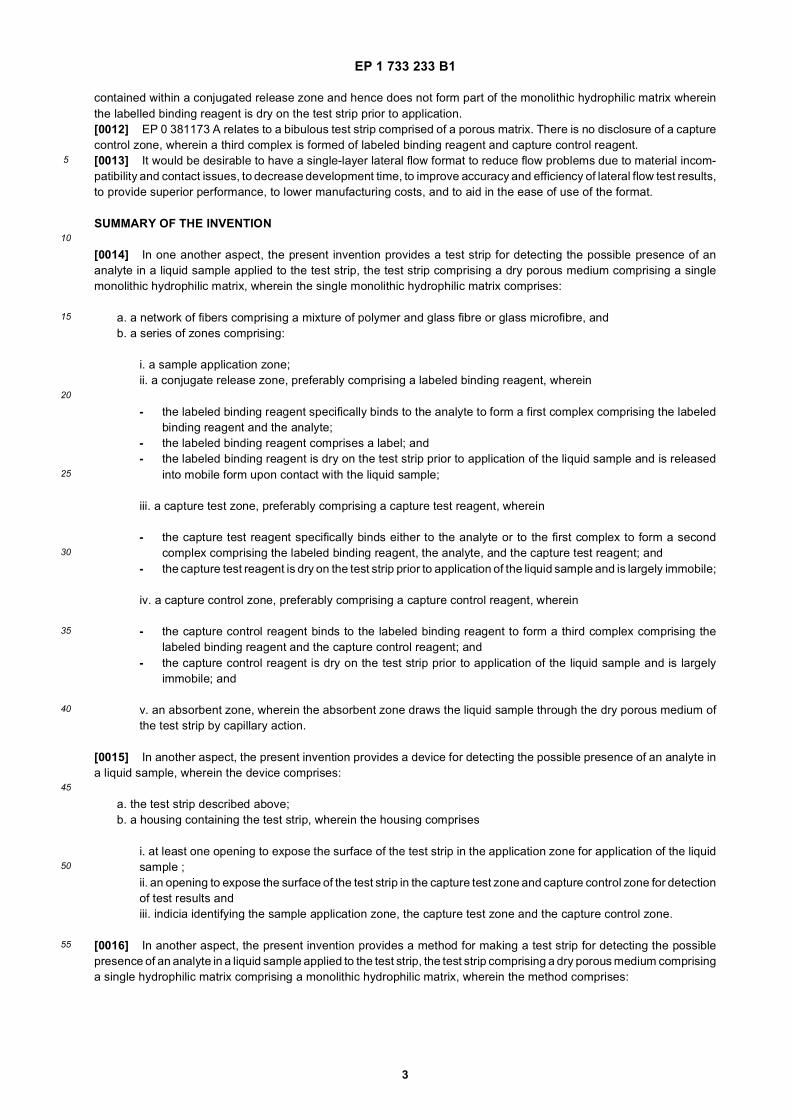
15
20
25
30
35
40
45
50
55
a. providing a monolithic hydrophilic matrix, wherein the monolithic hydrophilic matrix comprises a network of fiberscomprising a mixture of polymer and glass fibre or glass microfibre;b. creating a conjugate release zone on the monolithic hydrophilic matrix by:
i. providing a labeled binding reagent, wherein the labeled binding reagent comprises:
- a label;- a solid substrate comprising a carrier bead, wherein the carrier bead is mobile within the matrix when the
carrier bead and matrix are wet; and- a ligand that specifically binds to the analyte to form a first complex comprising the labeled binding reagent
and the analyte; and
ii. suspending the labeled binding reagent in a buffer;iii. applying the labeled binding reagent suspension to a first zone of the monolithic hydrophilic matrix;
c. creating a capture test zone on the monolithic hydrophilic matrix by:
i. providing a capture test reagent, wherein the capture test reagent comprises:
- a solid substrate comprising a capture test bead;- a ligand that specifically binds to the analyte or to the first complex to form a second complex comprising
the labeled binding reagent, the analyte, and the capture test reagent; and
ii. suspending the capture test reagent in a buffer;iii. applying the capture test reagent suspension to a second zone of the monolithic hydrophilic matrix, whereinthe second zone is downstream from the first zone; and
d. creating a capture control zone on the monolithic hydrophilic matrix by:
i. providing a capture control reagent, wherein the capture control reagent comprises:
- a solid substrate comprising a capture control bead; and- a ligand that binds to the labeled binding reagent to forma third complex comprising the labeled binding
reagent and the capture control reagent; and
ii. suspending the capture control reagent in a buffer;iii. applying the capture control reagent suspension to a third zone of the monolithic hydrophilic matrix, whereinthe third zone is downstream from the second zone; and
e. drying the monolithic hydrophilic matrix to yield a dry porous medium.
[0017] In still another aspect of the invention, the present invention provides a method of using a test strip to detectthe possible presence of an analyte in a liquid sample applied to the test strip, wherein the method comprises:
a. providing the test strip describe aboveb. applying a liquid sample to the sample application zone of the test strip;c. wicking the liquid sample through the single hydrophilic matrix to the conjugate release zone;d. contacting the labeled binding reagent with the liquid sample to mobilize the labeled binding reagent and to permitformation of the first complex if the liquid sample comprises analyte;e. wicking the liquid sample and the labeled binding reagent, whether alone or in the first complex, through the singlehydrophilic matrix to the capture test zone;f. contacting the capture test reagent with the liquid sample and the labeled binding reagent, whether alone or inthe first complex, to permit formation of the second complex if the first complex is present;g. concentrating the second complex in the network of fibers in the capture test zone of the single hydrophilic matrix;h. detecting the presence of the second complex in the capture test zone;i. wicking the liquid sample and the labeled binding reagent through the single hydrophilic matrix to the capturecontrol zone;j. contacting the capture control reagent with the liquid sample and the labeled binding reagent to permit formation
EP 1 733 233 B1
5
5
10
15
20
25
30
35
40
45
50
55
of the third complex;k. concentrating the third complex in the network of fibers in the capture control zone of the single hydrophilic matrix;andl. detecting the presence of the third complex in the capture control zone.
[0018] In yet another aspect, the present invention provides the method described above, herein the method is fordiagnosing disease, a phenotype, a genotype, or a physiological condition in an organism by detecting the presence ofan analyte associated with the disease, the phenotype, the genotype, or the physiological condition in a liquid biologicalsample, wherein the liquid sample is a biological sample and wherein the method further comprises:
diagnosing the disease, the phenotype, the genotype, or the physiological condition of the organism.
[0019] In another aspect, the present invention provides the test strip described above for detecting the possiblepresence of any one of multiple analytes in a liquid sample applied to the test strip, wherein the conjugate release zonecomprises a plurality of labelled binding reagents and the capture test zone comprises a plurality of capture test reagents.
BRIEF DESCRIPTION OF THE DRAWINGS
[0020]
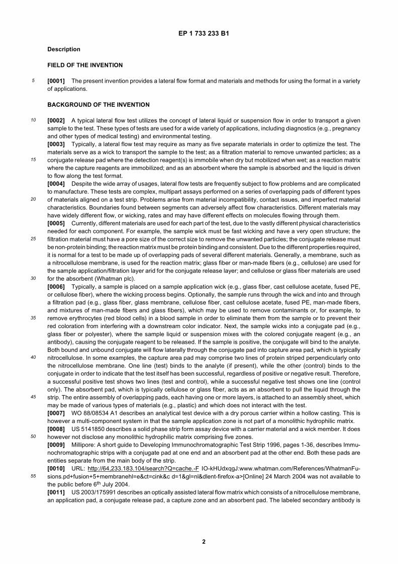
Figure 1A depicts an exploded view of one embodiment of a single-layer lateral flow format test strip in a housing.
Figure 1B depicts an exploded view of one embodiment of a single-layer lateral flow format test strip on a support strip.
Figures 2A-2D depict a method of using a preferred embodiment of a single-layer lateral flow format.
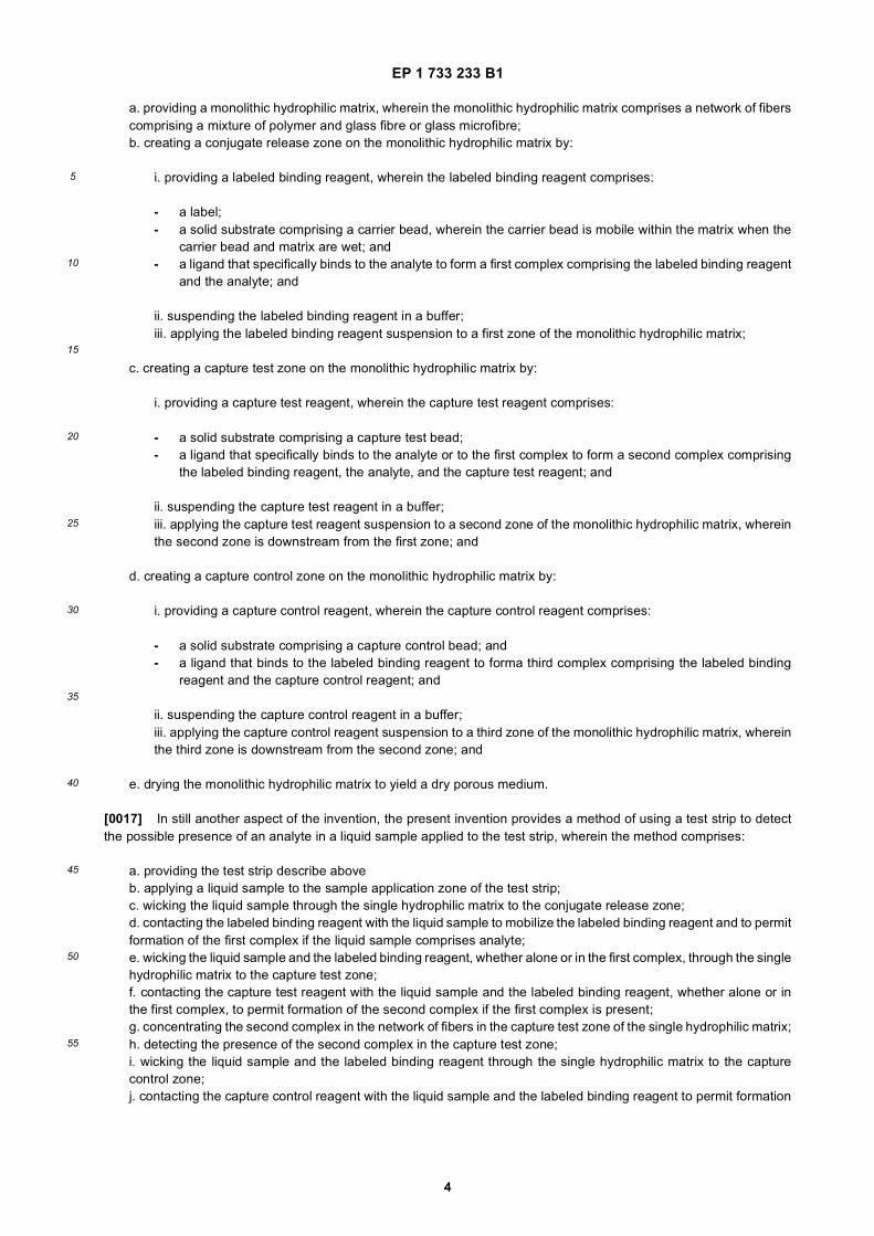
Figure 3 is a bar graph depicting the results of an experiment to measure the waster absorbency (mg/cm2) of oneembodiment of the test strip hydrophilic matrix compared to other materials.
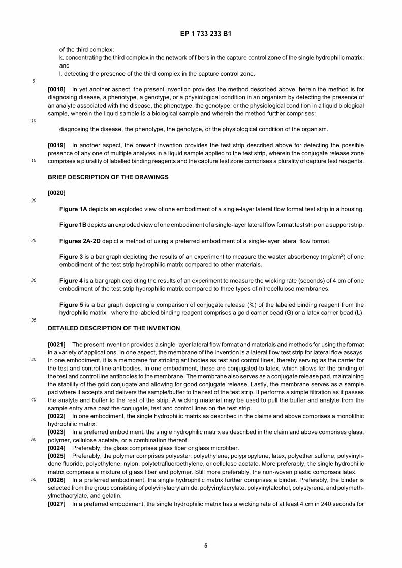
Figure 4 is a bar graph depicting the results of an experiment to measure the wicking rate (seconds) of 4 cm of oneembodiment of the test strip hydrophilic matrix compared to three types of nitrocellulose membranes.
Figure 5 is a bar graph depicting a comparison of conjugate release (%) of the labeled binding reagent from thehydrophilic matrix , where the labeled binding reagent comprises a gold carrier bead (G) or a latex carrier bead (L).
DETAILED DESCRIPTION OF THE INVENTION
[0021] The present invention provides a single-layer lateral flow format and materials and methods for using the formatin a variety of applications. In one aspect, the membrane of the invention is a lateral flow test strip for lateral flow assays.In one embodiment, it is a membrane for stripling antibodies as test and control lines, thereby serving as the carrier forthe test and control line antibodies. In one embodiment, these are conjugated to latex, which allows for the binding ofthe test and control line antibodies to the membrane. The membrane also serves as a conjugate release pad, maintainingthe stability of the gold conjugate and allowing for good conjugate release. Lastly, the membrane serves as a samplepad where it accepts and delivers the sample/buffer to the rest of the test strip. It performs a simple filtration as it passesthe analyte and buffer to the rest of the strip. A wicking material may be used to pull the buffer and analyte from thesample entry area past the conjugate, test and control lines on the test strip.[0022] In one embodiment, the single hydrophilic matrix as described in the claims and above comprises a monolithichydrophilic matrix.[0023] In a preferred embodiment, the single hydrophilic matrix as described in the claim and above comprises glass,polymer, cellulose acetate, or a combination thereof.[0024] Preferably, the glass comprises glass fiber or glass microfiber.[0025] Preferably, the polymer comprises polyester, polyethylene, polypropylene, latex, polyether sulfone, polyvinyli-dene fluoride, polyethylene, nylon, polytetrafluoroethylene, or cellulose acetate. More preferably, the single hydrophilicmatrix comprises a mixture of glass fiber and polymer. Still more preferably, the non-woven plastic comprises latex.[0026] In a preferred embodiment, the single hydrophilic matrix further comprises a binder. Preferably, the binder isselected from the group consisting of polyvinylacrylamide, polyvinylacrylate, polyvinylalcohol, polystyrene, and polymeth-ylmethacrylate, and gelatin.[0027] In a preferred embodiment, the single hydrophilic matrix has a wicking rate of at least 4 cm in 240 seconds for
EP 1 733 233 B1
6
5
10
15
20
25
30
35
40
45
50
55
water. More preferably, the single hydrophilic matrix has a wicking rate of at least 4 cm in 100 seconds for water. Morepreferably, the wicking rate is in the range of at least 4 cm in 60 seconds for water. Still more preferably, the wickingrate is in the range of at least 4 cm in 50 seconds for water.[0028] In another preferred embodiment, the single hydrophilic matrix has an average micrometers micrometers poresize in the range of 1.5 micrometers to 25.0 micrometers. More preferably, the average pore size is in the range of 2.0micrometers to 7.0 micrometers. Still more preferably, the average pore size is in the range of 3.0 micrometers to 6.0micrometers.[0029] In another preferred embodiment, the single hydrophilic matrix has a thickness of between 50 microns and1000 microns. More preferably, the thickness is between 150 microns and 500 microns.[0030] In another preferred embodiment, the labeled binding reagent as described in the claims and above furthercomprises a ligand that specifically binds to the analyte. More preferably, the labeled binding reagent further comprisesa solid support to which the ligand is attached and the solid support comprises gold, latex, selenium, platinum, copper,or iron. Still more preferably, the solid support comprises a carrier bead. In a particularly preferred embodiment, the sizeof the carrier bead allows the carrier bead to move through the matrix, and the bead is mobile within the matrix whenthe bead and matrix are wet. More preferably, the diameter of the carrier bead is 10% or less than the average poresize of the matrix.[0031] In a preferred embodiment,
a. the average pore size of the matrix is in the range of 4.0 to 6.0 micrometers; andb. the carrier bead comprises a gold bead having a diameter in the range of 20 to 80 nanometers.
[0032] In a preferred embodiment,
a. the average pore size of the matrix is in the range of 4.0 to 6.0 micrometers; andb. the carrier bead comprises a latex bead having a diameter in the range of 100 to 800 nanometers.
[0033] In a preferred embodiment, the carrier bead comprises a latex bead comprising a colorimetric dye or a fluorescentdye. In another preferred embodiment, the carrier bead comprises a latex bead comprising a paramagnetic cores, aplasmon resonant particle, or a quantum dot.[0034] In a preferred embodiment, the label of the labeled binding reagent comprises a colorimetric indicator, a fluo-rescent indicator, a photometric indicator, a radioactive indicator, or an immunological indicator. More preferably, thelabel comprises a dye.[0035] In a preferred embodiment, the ligand comprises:
a. a polypeptide, an oligopeptide, an antigen, an antibody, or a prion;b. a nucleic acid or a peptide nucleic acid;c. a drug, an analog of a drug, or a drug metabolite; ord. an imprinted polymer.
[0036] Preferably, the nucleic acid comprises DNA, PNA, or RNA. Preferably, the DNA comprises genomic DNA,cDNA, a protein binding site, an oligonucleotide, or a primer, or the DNA comprises a protein binding site comprising apromoter element or a transcriptional activation domain. Preferably, the DNA comprises single-stranded DNA. Preferably,the RNA comprises messenger RNA (mRNA) or short interfering RNA (siRNA).[0037] In a preferred embodiment,
a. the analyte comprises a nucleic acid having a target sequence of interest; andb. the ligand comprises a nucleic acid having a sequence of at least 65% complementarity to the target sequenceof interest.
[0038] Preferably, the ligand comprises a nucleic acid having a sequence of at least 75% complementarity to the targetsequence of interest; more preferably, at least 85% complementarity to the target sequence of interest; still more pref-erably, at least 95% complementarity to the target sequence of interest; yet more preferably, at least 97% complementarityto the target sequence of interest; and even more preferably, at least 99% complementarity to the target sequence ofinterest.[0039] In another preferred embodiment,
a. the analyte comprises an antigen; andb. the ligand comprises an antibody that specifically binds to the antigen.
EP 1 733 233 B1
7
5
10
15
20
25
30
35
40
45
50
55
[0040] Alternatively,
a. the ligand comprises an antigen; andb. the analyte comprises an antibody that specifically binds to the antigen.
[0041] In another preferred embodiment,
a. the ligand comprises an oligopeptide; andb. the analyte comprises a protein that binds to the oligopeptide.
[0042] In still another preferred embodiment,
a. the analyte comprises a drug or an analog of a drug; andb. the ligand comprises a protein that binds to the drug.
[0043] Alternatively,
a. the ligand comprises a drug or an analog of a drug; andb. the analyte comprises a protein that binds to the drug.
[0044] In still another preferred embodiment,
a. the ligand comprises an imprinted polyer; andb. the analyte comprises:
i. a polypeptide, an oligopeptide, an antigen, an antibody, or a prion;ii. a nucleic acid or a peptide nucleic acid; oriii. a drug, an analog of a drug or a drug metabolite.
[0045] In another preferred embodiment, the capture test reagent further comprises a ligand that specifically binds tothe analyte or to the first complex.[0046] In another preferred embodiment, the capture test reagent further comprises a solid support to which the ligandis attached. Preferably, the solid support comprises latex, silica, glass, alumina, cellulose, or a sugar.[0047] In a particularly preferred embodiment, the solid support comprises a capture test bead. Preferably, the capturetest bead comprises a sulfate terminated latex bead.[0048] In one preferred embodiment, the sulfate terminated latex bead physically binds proteins.[0049] In another preferred embodiment, the capture test bead comprises a covalent binding latex bead.[0050] In yet another preferred embodiment, the size of the capture test bead largely inhibits its movement throughthe matrix. Preferably, the size of the capture test bead is in the range of 20% to 70% of the average pore size of thematrix. More preferably, the size of the capture test bead is in the range of 30% to 60% of the average pore size of the matrix.[0051] More preferably,
a. the average pore size of the matrix is in the range of 4.0 to 6.0 micrometers; andb. the capture test bead comprises a latex bead having a diameter in the range of 1.5 to 2.5 nanometers.
[0052] In a preferred embodiment, the binding of the ligand to the analyte or to the first complex concentrates thelabeled binding reagent to enable detection of the label indicating the presence of the second complex. Preferably, thecapture test bead comprises a latex capture bead comprising an agglutinating agent. More preferably, the agglutinatingagent comprises polyethylene glycol (PEG).[0053] In a preferred embodiment, the ligand comprises:
a. a polypeptide, an oligopeptide, an antigen, or an antibody; orb. a nucleic acid or peptide nucleic acid;c. a drug, an analog of a drug, or a drug metabolite; ord. an imprinted polymer.
[0054] Preferably, the nucleic acid comprises DNA, PNA, or RNA. Preferably, the DNA comprises genomic DNA,cDNA, a protein binding site, an oligonucleotide, or a primer, or the DNA comprises a protein binding site comprising a

EP 1 733 233 B1
8
5
10
15
20
25
30
35
40
45
50
55
promoter element or a transcriptional activation domain. Preferably, the DNA comprises single-stranded DNA. Preferably,the RNA comprises messenger RNA (mRNA).[0055] In a preferred embodiment,
a. the analyte comprises a nucleic acid having an exposed target sequence of interest; andb. the ligand comprises a nucleic acid having a sequence of at least 65% complementarity to the target sequenceof interest.
[0056] Preferably, the ligand comprises a nucleic acid having a sequence of at least 75% complementarity to the targetsequence of interest; more preferably, at least 85% complementarity to the target sequence of interest; still more pref-erably, at least 95% complementarity to the target sequence of interest; yet more preferably, at least 97% complementarityto the target sequence of interest; even more preferably, at least 99% complementarity to the target sequence of interest.[0057] In a preferred embodiment,
a. either the analyte or the first complex comprises an antigen; andb. the ligand comprises an antibody that specifically binds to the antigen, wherein the antibody does not significantlybind to the labeled binding reagent in the absence of the analyte.
[0058] Alternatively,
a. the ligand comprises an antigen; andb. either the analyte or the first complex comprises an antibody that specifically binds to the antigen, wherein thelabeled binding reagent does not significantly bind to the antigen in the absence of the analyte.
[0059] In a preferred embodiment,
a. the ligand comprises an oligopeptide; andb. either the analyte or the first complex comprises a protein that binds to the oligopeptide, wherein the labeledbinding reagent does not significantly bind to the oligopeptide in the absence of the analyte.
[0060] In another preferred embodiment,
a. the ligand comprises an imprinted polymer; andb. either the analyte or the first complex comprises a substance that binds to the imprinted polymer, wherein thelabeled binding reagent does not significantly bind to the imprinted polymer in the absence of the analyte.
[0061] More preferably, the substance that binds to the imprinted polymer comprises a protein.[0062] In another embodiment, the capture control reagent further comprises a ligand that specifically binds to thelabeled binding reagent. Preferably, the capture control reagent further comprises a solid support to which the ligand isattached. More preferably, the solid support comprises latex, silica, glass, alumina, cellulose, or a sugar.[0063] In a particularly preferred embodiment, the solid support comprises a capture control bead. Preferably, thecapture control bead comprises a sulfate terminated latex bead. More preferably, the sulfate terminated latex beadphysically binds proteins. Preferably, the capture control bead comprises a covalent binding latex bead.[0064] In a preferred embodiment, the size of the capture control bead largely inhibits its movement through the matrix.Preferably, the size of the capture control bead is in the range of 20% to 70% of the average pore size of the matrix.More preferably, the size of the capture control bead is in the range of 30% to 60% of the average pore size of the matrix.[0065] In a particularly preferred embodiment,
a. the average pore size of the matrix is in the range of 4.0 to 6.0 micrometers; andb. the capture control bead comprises a latex bead having a diameter in the range of 1.5 to 2.5 nanometers.
[0066] In another preferred embodiment, the binding of the ligand to the labeled binding reagent concentrates thelabeled binding reagent to enable detection of the label indicating the presence of the third complex. Preferably, thecapture control bead comprises a latex capture bead comprising an agglutinating agent. More preferably, the agglutinatingagent comprises polyethylene glycol (PEG).[0067] In another preferred embodiment,
a. the labeled binding reagent has an overall negative charge; and
EP 1 733 233 B1
9
5
10
15
20
25
30
35
40
45
50
55
b. the capture control reagent has an overall positive charge.
[0068] More preferably,
a. the labeled binding reagent comprises a negatively charged gold carrier bead; andb. the capture control reagent comprises:
i. a positively charged polymer; orii. a positively charged ligand.
[0069] In another preferred embodiment,
a. the labeled binding reagent has an overall positive charge; andb. the capture control reagent has an overall negative charge.
[0070] In another preferred embodiment, the ligand comprises:
a. a polypeptide, an oligopeptide, an antigen, or an antibody; orb. a nucleic acid or peptide nucleic acid;c. a drug, an analog of a drug, or a drug metabolite; ord. an imprinted polymer.
[0071] Preferably, the nucleic acid comprises DNA, PNA, or RNA. Preferably, the DNA comprises genomic DNA,cDNA, a protein binding site, an oligonucleotide, or a primer, or the DNA comprises a protein binding site comprising apromoter element or a transcriptional activation domain. Preferably, the DNA comprises single-stranded DNA. Preferably,the RNA comprises messenger RNA (mRNA).[0072] In a preferred embodiment,
a. the labeled binding reagent comprises a nucleic acid having an exposed target sequence of interest; andb. the ligand comprises a nucleic acid having a sequence of at least 65% complementarity to the target sequenceof interest.
[0073] Preferably, the ligand comprises a nucleic acid having a sequence of at least 75% complementarity to the targetsequence of interest; more preferably, at least 85% complementarity to the target sequence of interest; still more pref-erably, at least 95% complementarity to the target sequence of interest.[0074] In a preferred embodiment,
a. the labeled binding reagent comprises an antigen; andb. the ligand comprises an antibody that specifically binds to the antigen.
[0075] Alternatively,
a. the ligand comprises an antigen; andb. the labeled binding reagent comprises an antibody that specifically binds to the antigen.
[0076] In a preferred embodiment,
a. the ligand comprises an oligopeptide; andb. the labeled binding reagent comprises a protein that binds to the oligopeptide.
[0077] In another preferred embodiment,
a. the ligand comprises an imprinted polymer; andb. the labeled binding reagent comprises a substance that binds to the imprinted polymer.
[0078] More preferably, the substance that binds to the imprinted polymer comprises a protein.[0079] In another embodiment, the liquid sample comprises blood, plasma; serum; mucus; urine; saliva; semen; vaginaldischarge; sweat; tears; lymph; gastrointestinal fluid, suspension or colloidal mixture; cerebrospinal fluid; a bacterial
EP 1 733 233 B1
10
5
10
15
20
25
30
35
40
45
50
55
culture; a tissue culture; a phage lysate; water; a beverage; an organic solvent; an aqueous or organic solution; asuspension of cells, viruses, or other replicative entities; or a colloidal mixture.[0080] In still another embodiment, the analyte comprises a polypeptide, an oligonucleotide, an antibody, an antigen,a prion, a nucleic acid, a peptide nucleic acid, a drug, an analog of a drug, or a drug metabolite. Preferably, the presenceor absence of the analyte comprises a marker for a physiological condition. More preferably, the physiological conditioncomprises pregnancy, nursing, a disease, a phenotype, genotype, or a normal or abnormal physiological condition.[0081] In another preferred embodiment, the analyte comprises a nucleic acid and the physiological condition comprisesa genotype. More preferably, the nucleic acid comprises a genetic mutation, or a polymorphism.[0082] In another preferred embodiment, the analyte comprises a drug or an analog of a drug.[0083] In a preferred embodiment,
a. the analyte comprises an antigen from a first mammalian species; andb. the labeled binding reagent comprises a ligand comprising an antibody that specifically binds to the antigen,wherein the antibody is from a second mammalian species.
[0084] More preferably,
c. the capture control reagent comprises an ligand comprising an antibody from a third mammalian species.
[0085] In one particularly preferred embodiment,
a. the analyte comprises human chorionic gonadotropin (hCG);b. the labeled binding reagent comprises a monoclonal mouse anti-human chorionic gonadotropin antibody;c. the capture control reagent comprises a non-human, non-murid mammalian anti-mouse antibody; andd. the presence of human chorionic gonadotropin in the liquid sample is a marker of the physiological condition ofpregnancy.
[0086] In one embodiment, more than one disease, phenotype, genotype, or physiological condition can be diagnosedas described in the claims and above simultaneously, preferably, wherein each analyte is used to diagnose a differentdisease, phenotype, genotype, or physiological condition, including, but not limited to, a disease, phenotype, genotype,or physiological condition selected from the group consisting of the following pregnancy, cancer, heart disease, hyper-tension, elevated cholesterol level, hyperglycemia, hypoglycemia, diabetes, malaria, tuberculosis, acquired immunedeficiency syndrome (AIDS), a sexually transmitted disease (e.g., syphilis, gonorrhea, herpes), dengue fever, Ebola,Lassa fever, hepatitis, pneumonia (e.g., bacterial, viral), and a genetic disease.[0087] In one embodiment, the method tests for the presence of each of the following pathogens:
a. human immunodeficiency virus;b. tuberculosis; andc. malaria.
[0088] Preferably, the monolithic hydrophilic matrix as described in the claims and above comprises a network offibers, the negatively-charged solid substrate of the reagent comprises a bead, the positively-charged Ligand comprisesa protein. More preferably, the protein comprises an antibody or an antigen.[0089] In one aspect, the present invention provides a single lateral flow function layer. In preferred embodiments, thelayer comprises a matrix, more preferably a monolithic matrix, of glass fiber, non-woven polymer, or a combination thereof.[0090] In one preferred embodiment, the present invention provides a single lateral flow function layer comprising amixture of polymer and glass fiber, which can act as a suitable matrix. This mixture had previously been shown to workas a blood separator and conjugate release, as well as a sample pad and absorbent. With the greatest surprise, however,the teaching of the present invention shows that the same material can be used as a reaction matrix in a single-layerlateral flow function format.[0091] The hydrophilic composite is low protein binding, and the capture reagents will not normally bind very well.However if the capture reagents are initially immobilized on a carrier particle (such as a latex bead) whose diameter isbetween 40 and 70% of the nominal pore size (or greater than the size for which there is a 98% absorption efficiencybut small enough to enter the matrix) the latex beads will be efficiently retained within the matrix. The sensitivity of thetest will depend upon the numbers of carrier beads added, their surface area (related to bead diameter) and the amountof capture reagent immobilized on their surface. The system can be further improved by the use of an agglutinatingagent, such as polyethylene glycol, on the capture beads (both test and control) or by use of a positively-charged ligandon a negatively-charged capture bead, as described infra.
EP 1 733 233 B1
11
5
10
15
20
25
30
35
40
45
50
55
[0092] The use of this type of this material has a number of advantages:
1) The composite is naturally hydrophilic and therefore does not require blocking (unlike membranes);2) The composite is low protein so the background signal is low and therefore lowest detectable signal is reduced3) In membranes as the wicking area increases (related to pore size) the available membrane surface area (andhence test sensitivity) is reduced. The composite signal is related to the surface area of the beads, not the composite.It would therefore be able to control the signal by varying the beads for the same pore size material.4) Small antigens will not attach well to membranes, however they can be covalently linked to the beads. Thecomposite can therefore be used-for any test.5) The wicking rate of the composite is much higher then membranes, test will therefore run more quickly (desiredby many test developers)6) The immobilization to beads will allow better control of linkage chemistry, resulting in increased test shelf life.Alternatively, other materials, which have longer shelf life, can be attached.
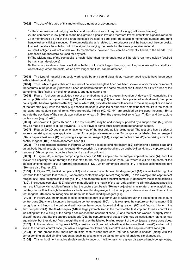
[0093] The type of material that could work could be any bound glass fiber, however good results have been seenwith a latex-bound glass.[0094] Thus, while a glass fiber or a mixture of polymer and glass fiber has been shown to work for one or more ofthe features in the past, only now has it been demonstrated that the same material can function for all five areas at thesame time. This finding is novel, unexpected, and quite surprising.[0095] Figure 1A shows an exploded view of an embodiment of the present invention. A device (10) comprising thetest strip (20) within an upper housing (30) and lower housing (32) is shown. In a preferred embodiment, the upperhousing (30) has two apertures (34, 36), one of which (34) provides the user with access to the sample application zoneof the test strip (20), while the other (36) enables the user to visualize or otherwise detect the test results in the capturetest zone and capture control zone. More preferably, indicia (40, 42, 44) are provided on the upper housing (30) toindicate the positions of the sample application zone (e.g., S (40)), the capture test zone (e.g., T (42)), and the capturecontrol zone (e.g., C (44)).[0096] As shown in Figures 1A and 1B, the test strip (20) may be additionally supported by a support strip (50), whichmay be made of plastic (e.g., polystyrene, PET, or vinyl) or some other appropriate material for support.[0097] Figures 2A-2D depict a schematic top view of the test strip as it is being used. The test strip has a series ofzones comprising a sample application zone (A), a conjugate release zone (B) comprising a labeled binding reagent(80), a capture test zone (C) comprising a capture test reagent (90), a capture control zone (D) comprising a capturecontrol reagent (100), and an absorbent zone (E).[0098] The embodiment depicted in Figures 2A shows a labeled binding reagent (80) comprising a carrier bead andan antibody ligand, a capture test reagent (90) comprising a capture bead and an antibody ligand, and a capture controlreagent (100) comprising a capture bead and an antibody ligand.[0099] In Figure 2B, a sample comprising an analyte (110) is applied to the sample application zone (A), and it iswicked via capillary action through the test strip to the conjugate release zone (B), where it will bind to some of thelabeled binding reagent (80) to form the first complex (120), which comprises analyte (110) and labeled binding reagent(80) (see also Figures 2C).[0100] In Figure 2C, the first complex (120) and some unbound labeled binding reagent (80) are wicked through thetest strip to the capture test zone (C), where they contact the capture test reagent (90). In this example, the capture testreagent (90) /also recognizes the analyte (110) and, therefore, binds the first complex (120) to form the second complex(130). The second complex (130) is largely immobilized in the matrix of the test strip and forms a line indicating a positivetest result. "Largely immobilized" means that the capture test beads (90) may be jostled, may rotate, or may agglutinate,but they do not flow through the matrix as the labeled binding reagent of the conjugate release zone does. The capturetest reagent (90) does not recognize the unbound labeled binding reagent (80).[0101] In Figure 2D, the unbound labeled binding reagent (80) continues to wick through the test strip to the capturecontrol zone (D), where it contacts the capture control reagent (100). In this example; the capture control reagent (100)recognizes and binds to the unbound antibody on the unbound labeled binding reagent (80) and finds to it to form thethird complex (140). The third complex (140) is largely immobilized in the matrix of the test strip and forms a control lineindicating that the wicking of the sample has reached the absorbent zone (E) and that test has worked. "Largely immo-bilized" means that, like the capture test beads (90), the capture control beads (100) may be jostled, may rotate, or mayagglutinate, but they do not flow through the matrix as the labeled binding reagent of the conjugate release zone does.[0102] In the test shown in Figures 2A-2D, a positive result has both a test line at the control test zone (C) and a controlline at the capture control zone (D), while a negative result has only a control line at the capture control zone (D).[0103] In one embodiment, there are multiple capture lines that each test for a separate analyte (along with thecorresponding labeled binding reagents), enabling a sample to be tested for several analytes simultaneously.[0104] This embodiment enables single sample to undergo multiple tests for a given disease, phenotype, genotype,
EP 1 733 233 B1
12
5
10
15
20
25
30
35
40
45
50
55
or physiological condition. This type of testing capability is particularly useful, e.g., where the tests for a given disease,phenotype, genotype, or physiological condition have a high rate of false positives or false negatives.[0105] Alternatively, a single sample can undergo tests for multiple diseases, phenotypes, genotypes, or physiologicalconditions. This type of testing capability is useful in a wide range of applications, including, but not limited to, routinescreening in an annual medical physical; for medical testing in remote areas, where physician access to populationsand/or patient access to physicians is limited (e.g., in rural areas both in the industrialized world and in developingnations); for large-scale testing settings, such as for epidemics or for large impoverished urban populations; for fieldwork with patients; and for testing where only a limited quantity of sample material is available (e.g., for forensic purposesor for trauma patients and other patients who have lost significant amounts of blood or who have a reduced amount ofblood or other bodily fluids). For example, in many parts of the world, there is a need for a simple method of testing forone or more of a number of diseases, particularly infectious diseases, in a rapid, low-cost, and efficient manner (with adevice or materials that are easily transportable and less susceptible to degradation) in locations where there is little orno refrigeration, laboratory equipment, or transportation available. In regions or situations where multiple diseasescommonly infect the same patient, a single sample could be tested for several diseases simultaneously. For example,it is envisioned that the present invention could be used to test infection with human immunodeficiency virus (HIV;associated with the development of acquired immune deficiency syndrome (AIDS)), tuberculosis, Ebola, malaria, Lassafever, hepatitis (A, B, C, D, or E) and/or dengue fever. Alternatively, where a patient has a disease, such as a genetichemoglobin disease, the present invention could test the genotype for sickle cell anemia and several types of thalassemiassimultaneously.[0106] In another embodiment, the test format is used to detect genetic mutations or polymorphisms.[0107] In another embodiment, the test format comprises a competitive assay, where the binding reagent or one ofthe capture reagents is the same as (or an analog of) the analyte. This competitive assay format is particularly usefulin tests for small analytes (such as drugs) where it would be difficult to form a "sandwich" assay due to steric problems.[0108] In a preferred embodiment, the invention provides a single lateral flow function layer comprising a material thatis low protein binding, has a fast wicking rate, and comprises a network of fibers. Preferably, the material is hydrophilic.More preferably, the material comprises a network of glass fibers or microfibers. Still more preferably, the materialcomprises a network of glass fibers in combination with a polymer. The network of glass or glass-polymer fibers iscapable of retaining beads (e.g., latex, gold) to which the conjugate is attached or beads (e.g., latex) for use as capturetest reagents or as capture control reagents. Other substances include cast or moulded cellulose acetate and fusedpolyethylene. The size of the pores (e.g., between a network of fibers) depends on the size and purpose (i.e., conjugationvs. capture) of the beads and also depends on the nature of the sample and whether any separation or filtration isdesired. If removal of erythrocytes (red blood cells) is desired, the material would have to have a pore size that retainsred blood cells (a 98% retention efficiency - below about 3.5 microns); however if removal of red cells is not desired, thepore size could be larger. The larger the pore size, the larger the size of the bead needed to become entrapped. However,as the size of the bead increases, the available surface area for protein immobilization is reduced, resulting in decreasedsensitivity.[0109] Materials less suitable for the present invention include cellulose and nitrocellulose. Cellulose would not allowthe sample to flow through the material quickly and would not allow the conjugate to release. It also does not functionwell as a blood separator in embodiments requiring blood separation. A nitrocellulose membrane is protein binding andtends towards hydrophobicity. The material would need to be blocked to make it work. The pore size needed for bloodseparation would be small and therefore test time would be too high. Also, nitrocellulose does not have the correctabsorbency properties to act as a sample wick and absorbent. Time to flow along the material increases exponentiallyas the wicking distance increases. For example, flow for 0.5 cm takes 5 seconds, flow for 1 cm takes 15 seconds, flowfor 1.5 cm takes 30 seconds, flow for 2 cm takes 90 seconds, etc. Moreover, it would not function well as a wick for largevolumes of samples. While it can work for low volumes (e.g., <30 microliters) however it would not work for largervolumes. For a strip comprising only nitrocellulose, the length of material required would be approximately 6-8 cm, andtest time would be quite lengthy. At this rate, there would be a danger of the sample drying before wicking the length ofthe test strip. For test results comparing the wicking rate of three nitrocellulose membranes with that of an embodimentof the present invention, see Example 2 and Figure 4.[0110] Nonetheless, nitrocellulose and cellulose materials, such as cellulose acetate, may still be useful in someembodiments of the invention. Foams, including wholly or partially open-celled foams, and particulates, including im-mobilized particulates may also be useful in the present invention.[0111] In addition to glass fiber, either alone or in combination with polymers and non-woven plastics, basically anyhydrophilic material with the correct pore size (e.g., hydrophilic membranes, such as polyester sulfone (PES), polyvi-nylidene fluoride (PVDF), fused polyethylene (PE), non-woven materials, and moulded cellulose acetate). If the poresize is too large (e.g., polypropylene mesh), the membrane would not function as there would not be efficient particletrapping. As a result, many materials, which are known in the art to be successful for conjugate release, would notnecessarily function well in the present invention.
EP 1 733 233 B1
13
5
10
15
20
25
30
35
40
45
50
55
[0112] In a more preferred embodiment, the present invention provides a single-layer lateral flow format comprisinga polymer-glass fiber matrix, which is naturally hydrophilic, substantially non-protein binding, and fast flowing, which hashigh sensitivity and a low background and is simple to manufacture. More preferably, the polymer-glass fiber matrixcomprises a latex-bound (e.g., polystyrene (PS) or polymethylmethacrylate (PMMA)) glass fiber matrix. In this embod-iment, the matrix serves simultaneously as a sample wick, a filter/separator (e.g., a blood separator), a conjugate releasepad, a reaction membrane, and an absorbent. (An additional absorbant pad may be added, but is not necessary.).Preferably, the degree of natural hydrophilicity obviates any need for blocking. In a more preferred embodiment, thematerial comprises a Whatman SLF5™ single-layer lateral flow format comprising a Whatman FUSION 5™ matrix, alatex-bound glass fiber, having the properties shown in Table 1.
[0113] A conjugate in an appropriate buffer is striped onto the test strip. The nature of the conjugate will depend onthe nature of the test being performed and is discussed more fully, infra.[0114] In a preferred embodiment, the ligand is attached to carrier beads to form the conjugate. The carrier beadsused as labeled binding reagents must be retained within the structure of the network (e.g., via physical sorption), butmust be capable of being released into mobile form upon contact with the liquid sample. The beads are preferably goldand should be capable of protein or nucleotide binding. Alternatively, the beads may be latex, selenium, or other suitablematerials. The binding reagent (e.g., an antibody or oligonucleotide) is immobilized to the carrier beads, which are striped(as a colloidal mixture in an appropriate buffer) onto the lateral flow test strip. (Alternatively, the mixture may be dottedor may take any other shape appropriate to the use of a single-layer lateral flow format.) Generally, however, the detectionbeads do not have to have any particular shape; rather, the shape of the bead application is primarily relevant to thecapture line(s). In one preferred embodiment, the carrier bead comprises a 40-80 nm gold bead. In another preferredembodiment, the carrier bead comprises a 100-800 nm latex bead.[0115] The capture beads used as capture test reagents and capture control reagents must be retained within thestructure of the network (e.g., via physical entrapment) and must remain substantially immobile during use of the lateralflow test strip, even when contacted with the liquid sample. The beads are preferably latex, but may be any material thatdoes not interfere with the label on the carrier beads, that is, without any inherent color or without a color that will showup against the strip itself Other potential materials include silica, glass, alumina, cellulose, or sugar (e.g., dextrose, etc.).Ideally, the capture beads form a tight formation so that the label on the carrier beads captured by the capture beads iseasily readable. The capture test reagent or capture control reagent (e.g., an antibody or oligonucleotide) is immobilizedto the capture test beads or capture control beads, respectively, which are striped (as colloidal mixtures in an appropriatebuffer) onto the lateral flow test strip. (Alternatively, the mixture may be dotted or may take any other shape, such as aplus or X-shape or any other shape appropriate to the use of a single-layer lateral flow format.)[0116] In a preferred embodiment, the latex beads (e.g., PMMA, PS, etc.) comprise sulfate terminated beads. Thesematerials bind proteins due to physical binding (charge and hydrophobicity). Alternatively, a covalent binding latex beadmay be used. Examples of preferred embodiments include latex beads manufactured by Estapor Microspheres or BangsLaboratories, Inc. The bead size must be small enough to enter the material, but large enough to become trapped. ForFUSION 5™ (Whatman), the optimal bead size is approximately 2 microns; the FUSION 5™ material has a 98% retentionefficiency for beads of approximately 2.5 microns. Beads of 2.5 microns would not generally enter the matrix, whereasbeads of below 1.5 microns would be washed out of the matrix.[0117] A typical protocol for applying the beads would be as follows: At the conjugate release zone apply 40 nm goldcolloid that has been conjugated to monoclonal anti-beta hCG (labeled binding reagent), concentrated or OD520= 10.
TABLE 1
KEY PROPERTY SPECIFICATION
IDEAL RANGE
Grammage, gsm 75 65-85
Thickness, mm @, 53kPa 370 Max 400
Gurley sec/ 100ml/01.1 sq in 16 14-22
M/D Tensile, N/15mm Min 15
M/D Wet Tensile, N/15mm Min 5
Pressure Drop, mm H2O @ 10.5 fpm 16 13-18
Mean pore size, mm 5.1 4.6-5.6
EP 1 733 233 B1
14
5
10
15
20
25
30
35
40
45
50
55
Antibodies are often mouse monoclonal antibodies. Apply to the FUSION 5™ from a borate buffer pH 8.2 containing 1%Tween 20, 0.5% PVA and 0.2% BSA. At the capture zone (capture test zone and capture control zone, respectively)apply two separate lines, one being a 2 micron latex bead conjugated to anti-alpha hCG (capture test reagent), thesecond being a 2 micron latex bead conjugated to anti-mouse IgG (capture control reagent). The control antibodies areoften anti-mouse Ab (e.g., goat anti-mouse) on and sometimes something that sticks to gold. Dry the test. After dryingapply the sample. While in some embodiments, 100 ml aliquots of beads have been used, the amount of sample andthe amounts of beads applied will vary considerably depending on the materials used.[0118] In some embodiments, it may be possible for the capture control beads to bind to the first complex (e.g., thecomplex comprising the analyte and the labeled binding reagent), particularly if there is an excess of the first complexand if the binding by the capture control beads is less specific or is capable of taking place both in the presence, and inthe absence, of the analyte. Because the test sample reaches the capture test zone first, the binding at the capturecontrol zone will not affect the results of the test, and one purpose of the capture control zone is to demonstrate that thesample has flowed, or wicked, beyond the capture test zone to provide assurance that the result-does not comprise afalse negative test result. (The capture test reagent is carefully chosen for specificity in order to avoid a false positivetest result.)[0119] In a preferred embodiment, polyethylene glycol (PEG) is added to improve agglutination of beads. The amountof capture reagent is proportional to the surface area of the latex bead used. To improve sensitivity it would be necessaryto increase the surface area; however this approach would entail using smaller beads, which would not stick to thematrix. To solve this problem, it is possible to use a smaller bead that agglutinates on drying. Self agglutination of latexbeads can be achieved either by including an agent to make the beads stick (e.g., PEG), by working at a pH below thepI of the protein on the bead surface, or by working at a high ionic strength (e.g., high salt concentration). The additiveidea relies upon the fact that as the beads dry, water leaves the system. Therefore, if the original concentration of theadditive will not cause agglutination, as the system dries, the effective concentration of the additive increases, untileventually the concentration reaches a critical point when the bead auto-agglutinates. The agglutinating agent is pref-erably PEG or some other hydrophilic agent or polymer, or it can be a reagent that adheres to the protein on the beadsurface. With respect to the pH alteration, the charge repulsion of beads normally keeps them apart, but if the proteinson the bead surface attract other conjugates, the beads would stick together. Typically latex beads are negativelycharged, therefore to make them attract, the proteins are positively charged by reducing the pH of the solution. Alterna-tively, the use of electrolytes (i.e., high salt concentration) could also cause agglutination of the beads. Where agglutinatingagents or procedures are used, the size of the capture test beads and capture control beads may be decreased. Withoutwishing to be bound by theory, a lumpy conglomerate comprised of a clump of beads will have a higher surface areathan a large bead. The presence of an increased concentration of salt will also cause agglutination due to the reductionof the zeta potential of the colloidal particles.[0120] Preferably, the invention is used for the treatment of vertebrates; for the treatment of vertebrate cells, cell lines,tissues, or organs; for research purposes relating thereto; or for any other purposes encompassed by the descriptionabove. More preferably, the invention is used for the treatment of mammals; for the treatment of mammal cells, celllines, tissues, or organs; for research purposes relating thereto; or for any other purposes encompassed by the descriptionabove. Still more preferably, the invention is used for the treatment of mammals; for the treatment of mammal cells, celllines, tissues, or organs; for research purposes relating thereto; or for any other purposes encompassed by the descriptionabove.[0121] The following definitions are provided for specific terms, which are used in the written description.[0122] As used in the specification and claims, the singular form "a", "an" and "the" include plural references unlessthe context clearly dictates otherwise. For example, the term "a molecule" also includes a plurality of molecules.[0123] As used herein, an "analyte" is the element of the sample to be detected by the test strip. The analyte specificallybinds the labeled binding reagent in the conjugate release zone of the test strip. In some embodiments, the presenceor absence of the analyte may be used to determine the physiological condition of an organism from which the samplewas obtained. Alternatively, the presence or absence, of the analyte may be used to detect, for example, contaminationof a sample. A wide range of other uses will occur to one of skill in the art.[0124] As used herein, a "porous medium" may have uniform or non-uniform pores. Alternatively, it may comprise, forexample, a "matrix" or a "network of fibers" through which appropriately smaller sized materials can pass.[0125] As used herein, the term " pore size" refers to the minimum size of particles that will be retained on or in themembrane. Thus, a membrane with a pore size of about 0.45 microns means that particles greater than about 0.45microns will be retained on or in the membrane, those less than about 0.45 microns will pass through and will not beretained. In a network of fibers, the pore size is more variable than in a membrane or medium with regularly sized pores.The "average pore size" may be expressed as a range, and the "maximum pore size" and "minimum pore size" mayvary considerably.[0126] As used herein, "stably associated" with a substrate refers to an interaction between polymerized, crosslinkedsurface-modifying molecules and a substrate that remains intact after one or more washes in an aqueous solution and/or
EP 1 733 233 B1
15
5
10
15
20
25
30
35
40
45
50
55
an organic solvent (such as an alcohol), and preferably, remains intact, after at least about 5, or at least about 10 washes.Preferably, a molecule which is "stably associated" with a substrate is one which remains attached to the substrate afterexposure to at least about 90°C, for at least about 2 hours. "Stable associations" can be monitored by evaluating thewettability (i.e., hydrophilicity) of a substrate which is coated with difunctional surface-modifying molecules according tothe invention.[0127] As used herein, "hydrophilic" substance is one that absorbs or adsorbs water, while a "hydrophobic" substancesis one that does not absorb or adsorb water.[0128] As used herein, "wettable" refers to a membrane which is wetted across its entire surface without phobic patches.[0129] As used herein, "a flow-through method" refers to a method where a solution is flowed through a substrate tocoat the substrate with the solution.[0130] As used herein, the term "functionally associated with" means that the coating is disposed, sorbed, or otherwiseassociated with the support of the present invention such that the support and coating function together. That is, thecoating can be adsorbed, absorbed, coated over, or otherwise disposed in functional relationship with the media.[0131] The media can be combined with a "binder," which holds the fibers together. Some examples of binders well-known in the art are polyvinylacrylamide, polyvinylacrylate, polyvinylalcohol (PVA), polystyrene (PS), polymethylmeth-acrylate (PMMA), and gelatin.[0132] As used herein, an "imprinted polymer" is a polymer composed into the fibrous matrix during its manufacture(e.g., a polymer put into a glass fiber matrix or other fiber matrix).[0133] As used herein, a "monolithic hydrophilic matrix" is a hydrophilic matrix that is cast as a single piece. Alternatively,it is a hydrophilic matrix that is formed or composed of material without joints or seams, or a hydrophilic matrix consistingof or constituting a single unit.[0134] As used herein, the "sample application zone" refers to the portion of the test strip to which the sample is applied.[0135] As used herein, the "conjugate release zone" refers to the portion of the test strip initially comprising the"conjugate," such as a "labeled binding reagent," which recognizes the analyte when the analyte is present.[0136] As used herein; the "capture zone" refers to the portion of the test strip comprising the "capture test zone" andthe "capture control zone." The "capture test zone" refers to the portion of the test strip comprising the "capture testreagent," which recognizes either the analyte or the first complex comprising the analyte and the labeled binding reagent.The "capture control zone" refers to the portion of the test strip comprising the "capture control reagent," which recognizesthe labeled binding reagent, either with or without the analyte.[0137] As used herein, the "absorbent zone" refers to the portion of the test strip which draws the liquid sample throughthe test strip by wicking or capillary action.[0138] As used herein, "specificity" refers to the ability of an antibody to discriminate between antigenic determinants.It also refers to the precise determinants recognized by a particular receptor or antibody. It also refers to the ability of areceptor to discriminate between substrates, such as drugs. With respect to nucleic acids, it refers to identity or com-plementarity as a function of competition or recognition/binding, respectively. "Specificity" of recognition or binding maybe affected by the conditions under which the recognition or binding takes place (e.g., pH, temperature, salt concentration,and other factors known in the art).[0139] As used herein, the term "largely immobile" or "largely immobilized" means that the substrate, such as a capturebead, may be jostled, may rotate, or may agglutinate, but does not flow or wick through the matrix.[0140] As used herein, "agglutination" or "self-agglutination" refers to the clumping, clustering, agglomeration, oraccumulation of moieties or substrates, including, but not limited to, beads.[0141] As used herein, "wicking" is achieved by "capillary action," resulting from the "capillarity" of the sample on thetest strip. "Capillarity" refers to the attraction between molecules, similar to surface tension, which results in the wettingof a solid by a liquid.[0142] The "wicking rate" of a material can be measure as a function of wetting of a particular distance of the materialover the course of a time period. The wicking rate depends on the nature of the material, the nature of the substanceused for the wetting, and a variety of other conditions. The "wicking rates" of various materials can be compared.[0143] As used herein, "line ramping" refers to the time taken for the rate of liquid flow through the striper to reach aconstant rate after the start of the line application. It can be influenced by the rate of acceleration of the plunger in theapplicator.[0144] As used herein, a "ligand" is a molecule or molecular complex that can be bound by another molecule ormolecular complex. The ligand may be, but is not limited to, a molecule or molecular complex bound by a receptor or acomplementary fragment of nucleic acid.[0145] As used herein, a "chimeric DNA" is at least two identifiable segments of DNA the segments being in anassociation not found in nature. Allelic variations or naturally occurring mutational events do not give rise to a chimericDNA as defined herein.[0146] As used herein, a "chimeric protein" or "fusion protein" is a protein with at least two identifiable segments, thesegments being in an association not found in nature. In one embodiment, a chimeric protein may arise, for example,

EP 1 733 233 B1
16
5
10
15
20
25
30
35
40
45
50
55
from expression of a chimeric DNA capable of being expressed as a protein and having at least two segments of DNAoperably linked to enable expression of at least a portion of each segment as a single protein. Other embodiments willsuggest themselves to one of ordinary skill in the pertinent art.[0147] As used herein, the terms "polynucleotide" and "nucleic acid molecule" are used interchangeably to refer topolymeric forms of nucleotides of any length, which may have any three-dimensional structure, and may perform anyfunction, known or unknown. The polynucleotides may contain deoxyribonucleotides (DNA), ribonucleotides (RNA),and/or their analogs, including, but not limited to, single-, double-stranded and triple helical molecules, a gene or genefragment, exons, introns, messenger RNA (mRNA), transfer RNA (tRNA), ribosomal RNA (rRNA), small interfering RNA(siRNA), ribozymes, antisense molecules, complementary DNA (cDNA), genomic DNA (gDNA), recombinant polynu-cleotides, branched polynucleotides, aptamers, plasmids, vectors, isolated DNA of any sequence, isolated RNA of anysequence, nucleic acid probes, peptide nucleic acids (PNA), and primers. A nucleic acid molecule may also comprisemodified nucleic acid molecules (e.g., comprising modified bases, sugars, and/or internucleotide linkers).[0148] "Nucleic materials" and "materials from the nucleus" include the nuclear envelope and the contents of thenucleus, including genomic DNA (gDNA) or plasmid DNA. The "non-nucleic acid contents of the nucleus" include thecomponents of the nuclear envelope and any other proteins or other substances of the nucleus that are not nucleic acids.[0149] - "Nucleic acids" include deoxyribonucleic acids (DNA) and ribonucleic acids (RNA) of various types, includinggenomic DNA (gDNA) and messenger RNA (mRNA) and derivatives thereof, such as modified DNA or RNA, includingpeptide nucleic acids (PNA). "Peptide nucleic acid" (PNA) is a polynucleotide analog in which the sugar-phosphatebackbone is replaced by amide bonds. "Genetic material" comprise genomic DNA (gDNA), which is one type of DNAand encodes genetic information, or genetic RNA.[0150] As used herein, a "genetic modification" refers to any addition, deletion or disruption to a cell’s normal nucle-otides. Any method which can achieve the genetic modification of antigen presenting cells (APC) are within the spiritand scope of this invention. Art recognized methods include viral mediated gene transfer, liposome mediated transfer,transformation, transfection and transduction.[0151] As used herein, a "genetic mutation" is a genetic alteration and is a type of "genetic modification."[0152] As used herein, a "polymorphism" or "genetic polymorphism" is a genetic variation and includes, but is notlimited to, a single nucleotide polymorphism (SNP).[0153] As used herein, a "genotype" is the genetic composition of an organism, and a "phenotype" is the physicalappearance or characteristics of an organism.[0154] A "peptide" is a compound of two or more subunit amino acids, amino acid analogs, or peptidomimetics. Thesubunits may be linked by peptide bonds or by other bonds (e.g., as esters, ethers, and the like).[0155] An "amino acid" refers to either natural and/or unnatural or synthetic amino acids, including glycine and bothD or L optical isomers, and amino acid analogs and peptidomimetics. "Amino acids" also includes imino acids. An"oligopeptide" refers to a short peptide chain of three or more amino acids. If the peptide chain is long (e.g., greater thanabout 10 amino acids), the peptide is a "polypeptide" or a "protein." While the term "protein" encompasses the term"polypeptide", a "polypeptide" may be a less than full-length protein.[0156] A "tag peptide sequence" is a short peptide or polypeptide chain of 3 or more amino acids, which is attachedto a protein of interest. In a preferred embodiment, a polypeptide, protein, or chimeric protein comprises a tag peptidesequence, which is used for purification, detection, or some other function, such as by specific binding to an antibody.The antibody may be in solution or bound to a surface (e.g., a bead, filter, or other material). The tag peptide sequenceshould not interfere with the function of the rest of the polypeptide, protein, or chimeric protein. An example of a tagpeptide sequence useful in the present invention is a short c-Myc tag with six His residues fused at the carboxyl-terminus.Other examples will be well-known to those of ordinary skill in the pertinent art.[0157] As used herein, "expression" refers to the process by which polynucleotides are transcribed into mRNA and/ortranslated into peptides, polypeptides, or proteins. If the polynucleotide is derived from genomic DNA, expression mayinclude, but is not required to include, splicing of the mRNA transcribed from the genomic DNA, capping of the 5’ endof the mRNA, polyadenylation of the 3’ end of the mRNA, or other processing modifications or events.[0158] As used herein, "signal sequence," or "secretory sequence" denotes the endoplasmic reticulum translocationsequence. This sequence encodes a "signal peptide," "secretory peptide," or "secretory domain" that communicates toa cell to direct a polypeptide to which it is linked (e.g., via a chemical bond) to an endoplasmic reticulum vesicularcompartment, to enter an exocytic/endocytic organelle, to be delivered either to a cellular vesicular compartment, thecell surface or to secrete the polypeptide. This signal sequence may be excised by the cell during the maturation of aprotein. Secretory sequences and domains of various species are well known ion the art.[0159] A "domain" is a region of a protein or polypeptide having a significant tertiary structure.[0160] "Conservatively modified variants" of domain sequences also can be provided within the scope of the invention.With respect to particular nucleic acid sequences, conservatively modified variants refers to those nucleic acids whichencode identical or essentially identical amino acid sequences, or where the nucleic acid does not encode an aminoacid sequence, to essentially identical sequences. Specifically, degenerate codon substitutions can be achieved by
EP 1 733 233 B1
17
5
10
15
20
25
30
35
40
45
50
55
generating sequences in which the third position of one or more selected (or all) codons is substituted with mixed-baseand/or deoxyinosine residues. Alternatively, one or more amino acids may be substituted with an amino acid having asimilar structure, activity, charge, or other property. Conservative substitution tables providing functionally similar aminoacids are well-known in the art (see, e.g., Proc.Natl.Acad.Sci. USA 89: 10915-10919 (1992)).[0161] The source of the nucleic acid or protein can be a biological sample containing whole cells. The whole cellscan be, but are not restricted to, blood, bacterial culture, bacterial colonies, yeast cells, tissue culture cells, saliva, urine,drinking water, plasma, stool samples, semen, vaginal samples, sputum, plant cell samples, or various other sourcesof cells known in the scientific, medical, forensic, and other arts. The samples can be collected by various means knownin the art, transported to the test strip, and then applied thereto.[0162] A "host organism" is an organism or living entity, which may be prokaryotic or eukaryotic, unicellular or multi-cellular, and which is desired to be, or has been, a recipient of exogenous nucleic acid molecules, polynucleotides,and/or proteins. Preferably, the "host organism" is a bacterium, a yeast, or a eukaroytic multicellular living entity (preferablyan animal, more preferably a mammal, still more preferably a human).[0163] "Mammals" include, but are not limited to, murines, simians, humans, farm animals, sport animals, and pets.[0164] The terms "polypeptide" and "protein" are used interchangeably and refer to any polymer of amino acids (dipep-tide or greater) linked through peptide bonds or modified peptide bonds. Thus, the terms "polypeptide" and "protein"include oligopeptides, protein fragments, fusion proteins and the like. It should be appreciated that the terms "polypeptide"and "protein", as used herein, includes moieties such as lipoproteins and glycoproteins.[0165] An "antibody" (Ab) is protein that binds specifically to a particular substance, known as an "antigen" (Ag)(described infra). An "antibody" is any immunoglobulin, including antibodies and fragments thereof, that binds a specificepitope. The term encompasses polyclonal, monoclonal, and chimeric antibodies (e.g., multispecific antibodies). Innature, antibodies are generally produced by lymphocytes in response to immune challenge, such as by infection orimmunization. An "antibody combining site" is that structural portion of an antibody molecule comprised of heavy andlight chain variable and hypervariable regions that specifically binds antigen. Exemplary antibody molecules are intactimmunoglobulin molecules, substantially intact immunoglobulin molecules, and those portions of an immunoglobulinmolecule that contains the paratope, including Fab, Fab’, F(ab’)2 and F(v) portions. A small single-chain F(v) comprisingthe variable (V) region of a light chain may be used, particularly when tissue penetration is desired.[0166] An "antigen" (Ag) is any substance that reacts specifically with antibodies or T lymphocytes (T cells). An "antigen-binding site" is the part of an immunoglobulin molecule that specifically binds an antigen. Additionally, an antigen-bindingsite includes any such site on any antigen-binding molecule, including, but not limited to, an MHC molecule or T cellreceptor. "Antigen processing" refers to the degradation of an antigen into fragments (e.g., the degradation of a proteininto peptides) and the association of one or more of these fragments (e.g., via binding) with MHC molecules for pres-entation by "antigen-presenting cells" to specific T cells.[0167] The term "antigenic material" covers any substance that will elicit an innate or adaptive immune response. Asused herein, "a portion of an antigenic material" covers any antigenic material or fragment thereof, which is capable ofeliciting an innate or adaptive immune response, even if the fragment is an incomplete representation or subset of theantigenic material as a whole. In one embodiment, it includes the minimal antigen sequence required to elicit a specificimmune response (preferably approximately 8-15 amino acid residues in length) when bound to an MHC recognized bya T cell.[0168] An "epitope" or "antigenic determinant" is a structure, usually made up of a short peptide sequence or oligosac-charide, that is specifically recognized or specifically bound by a component of the immune system. It is the site on anantigen recognized by an antibody. For example, as described supra, a T cell epitope is at least a portion of a shortpeptide derived from a protein antigen during antigen processing by an antigen-presenting cell. T-cell epitopes havegenerally been shown to be linear oligopeptides. Two epitopes correspond to each other if they can be specifically boundby the same antibody. Two epitopes correspond to each other if both are capable of binding to the same B cell receptoror to the same T cell receptor, and binding of one antibody to its epitope substantially prevents binding by the other epitope.[0169] A "chemokine" is a small cytokine involved in the migration and activation of cells, including phagocytes andlymphocytes, and plays a role in inflammatory responses. Examples of chemokines include, but are not limited to, IL8,RANTES, MDC, IP10, MIP1α, and MIPβ.[0170] A "cytokine" is a protein made by a cell that affect the behavior of other cells through a "cytokine receptor" onthe surface of the cells the cytokine effects. Cytokines manufactured by lymphocytes are sometimes termed "lym-phokines." Examples of cytokines include, but are not limited to, IL1α, IL1β, TNF, IL6, IL 12 (p40), and IFNγ.[0171] Chemokines and cytokines can bind to "receptors," which range in specificity from broad recognition (i.e.,binding many types of chemokines, cytokines, or other molecules) to highly specific recognition (e.g., binding a smallgroup of related molecules, binding only closely related molecules or only one type of molecule). Examples of "chemokinereceptors" include, but are not limited to, CCR2, CCR5, CCR6, and CCR7. Examples of "surface receptors" of interestto the present invention include, but are not limited to, mannose receptor (e.g., C type 1), macrophage scavenger receptor(e.g., scavenger R2), and prolactin receptor.
EP 1 733 233 B1
18
5
10
15
20
25
30
35
40
45
50
55
[0172] Expression or secretion of a chemokine, cytokine, receptor, marker, or other protein of interest may be measured,either directly or indirectly, using a wide range of methods known to those of ordinary skill in the art. Methods includeprotein assays, immunoprecipitation methods, Western blotting and other types of direct or indirect immunoblotting,spectophotometry or ultraviolet (UV) methods. Antibodies specific to cytokines and chemokines, as well as to cell surfaceantigens and other markers, are commercially available. Depending on the method used, detection may take place usinga tagged or labeled protein, a reporter plasmid, a radiolabel (e.g., using a radioactive isotope, such as 35S-Met or 35S-Cys), a chemical label or stain, a fluorescent label, an immunolabel, or by other detection methods known in the art. Inone preferred embodiment, the detection will be quantitative or capable of quantitation in order to measure levels of theprotein. For example, the protein may be detected in blood, in a sample of isolated blood cells (e.g., leukocytes), inlymph, in saliva, or in other types of biological samples. These methods are particularly useful for medical applicationsof the present invention.[0173] Alternatively, the level of the corresponding mRNA for a given cytokine, chemokine, receptor, marker or otherprotein of interest may be detected or measured, either directly or indirectly, via a variety of methods known to those ofordinary skill in the art. Similarly, the presence of a DNA having a sequence of interest (e.g., for genetic testing, for DNAfingerprinting, for detection for mutations, and for other purposes known in the art) may be detected or measured.Moreover, the test may also detect or measure the binding of a protein (e.g., a polymerase, an activator, or an inhibitor)to a DNA or mRNA fragment (e.g., a promoter or an enhancer). The test may also detect or measure the binding of asubstance (e.g., a drug) to a protein. These methods include, but are not limited to, Northern blotting, hybridizationdetection (e.g., with oligonucleotides or longer nucleic acid sequences, which may be radiolabeled, chemical labeled,immunolabeled, or fluorescence labeled), or polymerase chain reaction (PCR). PCR methods may be qualitative or,more preferably, quantitative (e.g., quantitative PCR). The mRNA may be detected in vivo, in situ, or in vitro. For example,the protein may be detected in blood, in a sample of isolated blood cells (e.g., leukocytes), in lymph, in saliva, or in othertypes of biological samples (including cell samples (e.g., bone marrow, lymph nodes). Nucleic acids used for hybridizationor for PCR may be specific or degenerate. In addition, they may correspond to the species of animal from which thesample is taken, or the sequence may correspond to a different species (e.g., use of a mouse sequence to probe a rat,human, or chicken sample).[0174] An "isolated" or "purified" population of c ells is substantially free of cells and materials with which it is associatedin nature. By substantially free or substantially purified APCs is meant at least 50% of the population are APCs, preferablyat least 70%, more preferably at least 80%, and even more preferably at least 90% free of non-APCs cells with whichthey are associated in nature.[0175] An "immunogen" is a substance capable of eliciting an immune response. Each immunoglobulin molecule canpotentially bind a variety of antibodies directed at its unique features, or "idiotype," which is comprised of a series of"idiotopes." An "idiotope" is a single antigenic determinant on a variable region of an antibody or T cell receptor. It is theset of idiotopes on an antibody which comprise the idiotype that makes that antibody unique. The "dominant idiotype"is the idiotype found on the major fraction of antibodies generated in response to an antigen.[0176] A "vector" is a replicon, such as plasmid, phage or cosmid, to which another DNA segment may be attachedso as to bring about the replication of the attached segment. A "replicon" is any genetic element (e.g-, plasmid, chro-mosome, virus) that functions as an autonomous unit of DNA replication in vivo, i.e., capable of replication under its owncontrol.[0177] A "prion" is a protein or protein fragment capable of replicating.[0178] A "pathogenic organism" includes a virus, microorganism, or a parasite. A pathogenic organism is capable oftriggering an abnormal physiological condition or disease or an abnormal physiological response. A pathogenic organismmay be infectious.[0179] "Biological sample" includes samples of -tissues, cells, blood, fluid, or other materials obtained from a biologicalorganism. It also includes a biological organism, cell, virus, or other replicative entity. Also included are solid cultures(such as bacterial or tissue cultures). Also included are solid samples, including, but not limited to, food, powder, andother solids, including non-biological solids, containing a biological organism, cell, virus, or other replicative entity. Alsoincluded are washing, homogenizations, sonications, and similar treatments of solid samples. Likewise, the term includesnon-solid biological samples.[0180] "Non-solid biological samples" include those that are not a tissue or an organ. Examples include, but are notlimited to, blood, plasma, serum, mucus, urine, saliva, semen, vaginal discharge, sweat, tears, lymph, gastrointestinalsuspensions or fluids, and cerebrospinal fluid. Also included are cultures (such as bacterial or tissue cultures) and phagelysates. Also included are fluid samples, including, but not limited to, water and beverages containing a biologicalorganism, cell, virus, or other replicative entity. Also included are suspensions and colloidal mixt[0181] "Non-biological samples" include samples not obtained from a biological organism, except in instances in whichsuch a sample is contaminated by a biological sample. Non-biological samples may be in liquid, gaseous, or solid form.They may include, but are not limited to, non-biological solid samples, liquid samples, gaseous samples, solutions,suspensions, colloidal mixtures, and aerosols.
EP 1 733 233 B1
19
5
10
15
20
25
30
35
40
45
50
55
[0182] "Non-biological solid samples" include samples from a wide variety of items, including, but not limited to, wood,concrete, dirt, plastics, and any other solids that have the potential to become contaminated. These samples may bepulverized, sonicated, minced, chopped, ground, or otherwise broken down into fine particles, and then prepared as acolloidal mixture or suspension prior to separation in the device. More preferably, the non-biological solid sample isdissolved into a solution.[0183] "Non-biological liquid samples" include a wide range of samples, which include, but are not limited to, water,organic solvents, aqueous or organic solutions, and the like.[0184] Methods of dissociating cells, such as cells in tissues, organs, or multicellular organisms, include physical,chemical, and enzymatic methods. Examples include, but are not limited to, mincing, homo genizing, sonicating, andgrinding, preferably in a physiological buffer, such as described in this specification or known to those of ordinary skillin the art.[0185] One of ordinary skill in the art would easily adapt some of these methods for - use in the preparation of non-biological samples.[0186] Preferably, the cells are selected from the group consisting of white blood cells, epithelial cells, buccal cells,tissue culture cells, semen, vaginal cells, urinary tract cells, colorectal cells, plant cells, bacterial cells, and yeast cells.[0187] In one embodiment, the present method may be applied advantageously to any whole cell suspension. Alter-natively, the cells may be lysed to release organelles and/or nucleic acid prior to exposure to the test strip.[0188] The detection process may comprise use of an indicator. The signal generated by the indicator of the presentinvention provides positive identification of the presence of a given nucleic acid or protein on the substrate. For example,nucleic acids can be detected (and preferably quantified) by the use of a specific or nonspecific nucleic acids probe orother signal generators and one of the versions of immunoassay. Proteins can be detected (and preferably quantified)by the use of an immunoassay. Preferably, the indicator comprises a fluorescent indicator, a color indicator, or a pho-tometric indicator. Alternatively, antibodies conjugated with biotin and polyavidin-horse radish peroxidase (HRP) maybe used, or an assay using polyethyleneimine-peroxidase conjugate (PEI-PO), which interacts with DNA, may be used,as known in the art. Other methods of detection will occur to those of ordinary skill in the art.[0189] In some embodiments, particularly in photosensitive embodiments, it may be necessary to provide an upperhousing and a lower housing that inhibit exposure to light in general and/or to certain wavelengths of light in particular.If the indicator is not already present on the test strip, it may be added and, if necessary, incubated with the test stripmaterial in the housing. The indicator is easily drawn through the test strip material and discarded. Blocking agents andwashes may likewise be circulated through the test strip material, although in preferred embodiments, blocking is notnecessary. When the preparation steps are complete, the housing is opened in the absence of light (or in the absencelight of the wavelength for the indicator reaction), and the test strip material is the exposed to the light of the desiredwavelength to trigger the photometric reaction.[0190] A "physiological condition" may be normal or abnormal. The physic ological condition may result from thegenetic make-up of the organism (including, but not limited to, the expression of various proteins), from environmentalfactors (including, but not limited to, the ingestion of drugs, poisons, food, and beverages and the exposure of an organismto toxic or non-toxic substances), from disease (bo th infectious or non-infectious), from an injury, from a metabolicdisorder, from pregnancy or nursing, and from a wide range of other circumstances known in the art. Examples include,but are not limited to, pregnancy, nursing, acquired immune deficiency syndrome (AIDS; such as by infection with humanimmunodeficiency virus (HIV)) or other sexually transmitted diseases (e.g., syphilis, gonorrhea, herpes), tuberculosis,Ebola, malaria, Lassa fever, hepatitis (A; B, C, D, or E), dengue fever, pneumonia (e.g., bacterial, viral), and geneticdiseases; syndromes, or polymorphisms with respect to the genotype and/or phenotype of the organism.[0191] For example, it is well-known in the art that a pregnant human female has elevated levels of human chorionicgonadotropin (hCG) in her blood and urine, and that hCG can be used as a marker for pregnancy testing.[0192] Examples of an "abnormal physiological condition or disease" and an "abnormal physiological response" include,but are by no means limited to, cancer or growth of a non-immunogenic tumor, allergy, asthma, an autoimmune disease,an infectious disease, and inflammation. Cancer and non-immunogenic tumor cells are often characterized by abnormalprotein expression, including expression of proteins encoded by mutated nucleotide sequences, abnormal levels ofprotein expression, or inappropriate expression of proteins. Allergies and asthma (especially allergy-related asthma)are often characterized by aberrant accumulation of mast cells, bone marrow-derived cells which degranulate to releasehistamines and which synthesize histamines in response to aberrant activation by a number of stimuli (e.g., IgE) inresponse to allergens. Autoimmune diseases are directed against "shelf" antigens and are characterized by abnormallevels ofMHC class II cells and autoreactive T cells (especially CD4+ and CD8+ T cells). Infection by an infectious diseasetriggers an immune response. Inflammation, which may be due to an infection, an injury, or an autoimmune disorder,triggers a response similar to the immune response. These conditions are characterized by up-regulation of someproteins and down-regulation of others.[0193] The terms "cancer" and "neoplasm" are used interchangeably and in either the singular or plural form, refer tocells that have undergone a malignant transformation that makes them pathological to the host organism. The definition
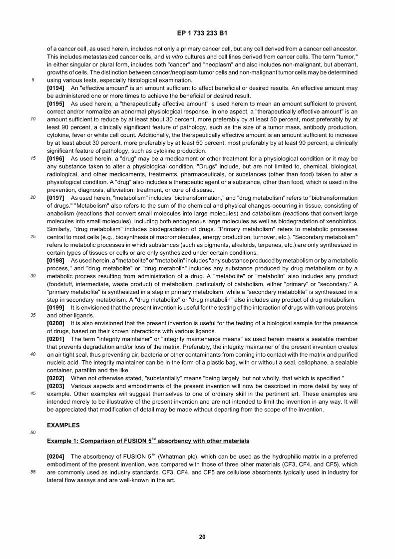
EP 1 733 233 B1
20
5
10
15
20
25
30
35
40
45
50
55
of a cancer cell, as used herein, includes not only a primary cancer cell, but any cell derived from a cancer cell ancestor.This includes metastasized cancer cells, and in vitro cultures and cell lines derived from cancer cells. The term "tumor,"in either singular or plural form, includes both "cancer" and "neoplasm" and also includes non-malignant, but aberrant,growths of cells. The distinction between cancer/neoplasm tumor cells and non-malignant tumor cells may be determinedusing various tests, especially histological examination.[0194] An "effective amount" is an amount sufficient to affect beneficial or desired results. An effective amount maybe administered one or more times to achieve the beneficial or desired result.[0195] As used herein, a "therapeutically effective amount" is used herein to mean an amount sufficient to prevent,correct and/or normalize an abnormal physiological response. In one aspect, a "therapeutically effective amount" is anamount sufficient to reduce by at least about 30 percent, more preferably by at least 50 percent, most preferably by atleast 90 percent, a clinically significant feature of pathology, such as the size of a tumor mass, antibody production,cytokine, fever or white cell count. Additionally, the therapeutically effective amount is an amount sufficient to increaseby at least about 30 percent, more preferably by at least 50 percent, most preferably by at least 90 percent, a clinicallysignificant feature of pathology, such as cytokine production.[0196] As used herein, a "drug" may be a medicament or other treatment for a physiological condition or it may beany substance taken to alter a physiological condition. "Drugs" include, but are not limited to, chemical, biological,radiological, and other medicaments, treatments, pharmaceuticals, or substances (other than food) taken to alter aphysiological condition. A "drug" also includes a therapeutic agent or a substance, other than food, which is used in theprevention, diagnosis, alleviation, treatment, or cure of disease.[0197] As used herein, "metabolism" includes "biotransformation," and "drug metabolism" refers to "biotransformationof drugs." "Metabolism" also refers to the sum of the chemical and physical changes occurring in tissue, consisting ofanabolism (reactions that convert small molecules into large molecules) and catabolism (reactions that convert largemolecules into small molecules), including both endogenous large molecules as well as biodegradation of xenobiotics.Similarly, "drug metabolism" includes biodegradation of drugs. "Primary metabolism" refers to metabolic processescentral to most cells (e.g., biosynthesis of macromolecules, energy production, turnover, etc.). "Secondary metabolism"refers to metabolic processes in which substances (such as pigments, alkaloids, terpenes, etc.) are only synthesized incertain types of tissues or cells or are only synthesized under certain conditions.[0198] As used herein, a "metabolite" or "metabolin" includes "any substance produced by metabolism or by a metabolicprocess," and "drug metabolite" or "drug metabolin" includes any substance produced by drug metabolism or by ametabolic process resulting from administration of a drug. A "metabolite" or "metabolin" also includes any product(foodstuff, intermediate, waste product) of metabolism, particularly of catabolism, either "primary" or "secondary." A"primary metabolite" is synthesized in a step in primary metabolism, while a "secondary metabolite" is synthesized in astep in secondary metabolism. A "drug metabolite" or "drug metabolin" also includes any product of drug metabolism.[0199] It is envisioned that the present invention is useful for the testing of the interaction of drugs with various proteinsand other ligands.[0200] It is also envisioned that the present invention is useful for the testing of a biological sample for the presenceof drugs, based on their known interactions with various ligands.[0201] The term "integrity maintainer" or "integrity maintenance means" as used herein means a sealable memberthat prevents degradation and/or loss of the matrix. Preferably, the integrity maintainer of the present invention createsan air tight seal, thus preventing air, bacteria or other contaminants from coming into contact with the matrix and purifiednucleic acid. The integrity maintainer can be in the form of a plastic bag, with or without a seal, cellophane, a sealablecontainer, parafilm and the like.[0202] When not otherwise stated, "substantially" means "being largely, but not wholly, that which is specified."[0203] Various aspects and embodiments of the present invention will now be described in more detail by way ofexample. Other examples will suggest themselves to one of ordinary skill in the pertinent art. These examples areintended merely to be illustrative of the present invention and are not intended to limit the invention in any way. It willbe appreciated that modification of detail may be made without departing from the scope of the invention.
EXAMPLES
Example 1: Comparison of FUSION 5™ absorbency with other materials
[0204] The absorbency of FUSION 5™ (Whatman plc), which can be used as the hydrophilic matrix in a preferredembodiment of the present invention, was compared with those of three other materials (CF3, CF4, and CF5), whichare commonly used as industry standards. CF3, CF4, and CF5 are cellulose absorbents typically used in industry forlateral flow assays and are well-known in the art.
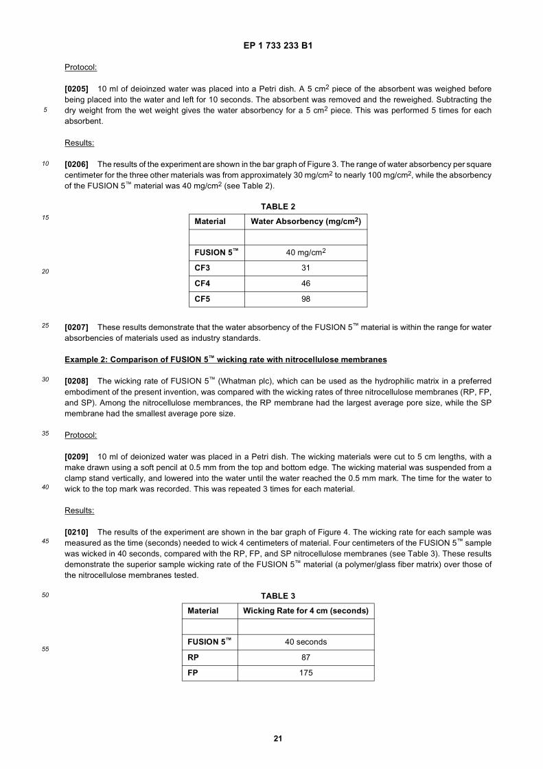
EP 1 733 233 B1
21
5
10
15
20
25
30
35
40
45
50
55
Protocol:
[0205] 10 ml of deioinzed water was placed into a Petri dish. A 5 cm2 piece of the absorbent was weighed beforebeing placed into the water and left for 10 seconds. The absorbent was removed and the reweighed. Subtracting thedry weight from the wet weight gives the water absorbency for a 5 cm2 piece. This was performed 5 times for eachabsorbent.
Results:
[0206] The results of the experiment are shown in the bar graph of Figure 3. The range of water absorbency per squarecentimeter for the three other materials was from approximately 30 mg/cm2 to nearly 100 mg/cm2, while the absorbencyof the FUSION 5™ material was 40 mg/cm2 (see Table 2).
[0207] These results demonstrate that the water absorbency of the FUSION 5™ material is within the range for waterabsorbencies of materials used as industry standards.
Example 2: Comparison of FUSION 5™ wicking rate with nitrocellulose membranes
[0208] The wicking rate of FUSION 5™ (Whatman plc), which can be used as the hydrophilic matrix in a preferredembodiment of the present invention, was compared with the wicking rates of three nitrocellulose membranes (RP, FP,and SP). Among the nitrocellulose membrances, the RP membrane had the largest average pore size, while the SPmembrane had the smallest average pore size.
Protocol:
[0209] 10 ml of deionized water was placed in a Petri dish. The wicking materials were cut to 5 cm lengths, with amake drawn using a soft pencil at 0.5 mm from the top and bottom edge. The wicking material was suspended from aclamp stand vertically, and lowered into the water until the water reached the 0.5 mm mark. The time for the water towick to the top mark was recorded. This was repeated 3 times for each material.
Results:
[0210] The results of the experiment are shown in the bar graph of Figure 4. The wicking rate for each sample wasmeasured as the time (seconds) needed to wick 4 centimeters of material. Four centimeters of the FUSION 5™ samplewas wicked in 40 seconds, compared with the RP, FP, and SP nitrocellulose membranes (see Table 3). These resultsdemonstrate the superior sample wicking rate of the FUSION 5™ material (a polymer/glass fiber matrix) over those ofthe nitrocellulose membranes tested.
TABLE 2
Material Water Absorbency (mg/cm2)
FUSION 5™ 40 mg/cm2
CF3 31
CF4 46
CF5 98
TABLE 3
Material Wicking Rate for 4 cm (seconds)
FUSION 5™ 40 seconds
RP 87
FP 175
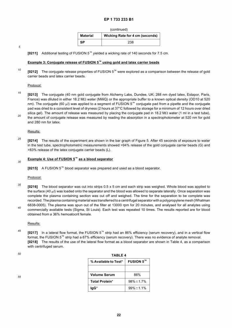
EP 1 733 233 B1
22
5
10
15
20
25
30
35
40
45
50
55
[0211] Additional testing of FUSION 5™ yielded a wicking rate of 140 seconds for 7.5 cm.
Example 3: Conjugate release of FUSION 5™ using gold and latex carrier beads
[0212] The conjugate release properties of FUSION 5™ were explored as a comparison between the release of goldcarrier beads and latex carrier beads.
Protocol:
[0213] The conjugate (40 nm gold conjugate from Alchemy Labs, Dundee, UK; 288 nm dyed latex, Estapor, Paris,France) was diluted in either 18.2 MΩ water (MilliQ) or the appropriate buffer to a known optical density (OD10 at 520nm). The conjugate (60 ml) was applied to a segment of FUSION 5™ conjugate pad from a pipette and the conjugatepad was dried to a consistent level of dryness (2 hours at 37°C followed by storage for a minimum of 12 hours over driedsilica gel). The amount of release was measured by placing the conjugate pad in 18.2 MΩ water (1 ml in a test tube),the amount of conjugate release was measured by reading the absorption in a spectrophotometer at 520 nm for goldand 280 nm for latex.
Results:
[0214] The results of the experiment are shown in the bar graph of Figure 5. After 45 seconds of exposure to waterin the test tube, spectrophotometric measurements showed >94% release of the gold conjugate carrier beads (G) and>83% release of the latex conjugate carrier beads (L).
Example 4: Use of FUSION 5™ as a blood separator
[0215] A FUSION 5™ blood separator was prepared and used as a blood separator.
Protocol:
[0216] The blood separator was cut into strips 0.5 x 5 cm and each strip was weighed. Whole blood was applied tothe surface (40 ml) was loaded onto the separator and the blood was allowed to separate laterally. Once separation wascomplete the plasma containing section was cut off and weighed. The time for the separation to be complete wasrecorded. The plasma containing material was transferred to a centrifugal separator with a polypropylene mesh (Whatman6838-0005). The plasma was spun out of the filter at 13000 rpm for 20 minutes, and analysed for all analytes usingcommercially available tests (Sigma, St Louis). Each test was repeated 10 times. The results reported are for bloodobtained from a 36% hemoatocrit female.
Results:
[0217] In a lateral flow format, the FUSION 5™ strip had an 86% efficiency (serum recovery), and in a vertical flowformat, the FUSION 5™ strip had a 67% efficiency (serum recovery). There was no evidence of analyte removal.[0218] The results of the use of the lateral flow format as a blood separator are shown in Table 4, as a comparisonwith centrifuged serum.
(continued)
Material Wicking Rate for 4 cm (seconds)
SP 238
TABLE 4
% Available to Test* FUSION 5™
Volume Serum 86%
Total Protein* 98%61.7%
IgG* 99%61.1%
EP 1 733 233 B1
23
5
10
15
20
25
30
35
40
45
50
55
Example 5: Additional properties of FUSION 5™ material
[0219] Additional properties of FUSION 5™ were studied. Results are shown in Table 5.
Example 6: Use of FUSION 5™ with whole blood for a pregnancy test
A. Preparation of FUSION 5™ for a pregnancy test:
[0220] Take a reel of FUSION 5™ that is 8 cm wide and 50 m long. Laminate the FUSION 5™ matrix to a PE cardcoated with a pressure sensitive to provide additional mechanical strength during handling.
Test line:
[0221] At the capture zone (approximately 2.5 cm from the edge of the material) apply 2 micron latex beads conjugatedto a monoclonal anti-alpha hCG antibody to the FUSION 5™ matrix from a suitable buffer (e.g., 10 mM phosphate pH 7.2).
Control line:
[0222] At the control zone (approximately 2.7 cm from the edge of the material) apply 2 micron latex beads conjugatedto a goat anti-Mouse IgG antibody to the FUSION 5™ matrix from a suitable buffer (e.g., 10 mM phosphate pH 7.2).
Conjugate release:
[0223] At the conjugate release zone (approximately 1 cm from the edge of the material) apply a 150 nm blue dyedlatex colloid conjugated to a monoclonal anti-beta hCG antibody to the FUSION 5™ matrix from a suitable buffer (seeabove).[0224] After application of the lines (that may be done simultaneously) dry the striped FUSION 5™ material for at least3 hours at 37°C.[0225] Cut the FUSION 5™ into 5 mm wide strips, and place in a plastic housing.
(continued)
% Available to Test* FUSION 5™
Cholesterol* 101%62.1%
*Versus centrifuged serum
TABLE 5
TEST/PROPERTY RESULT(S)
Thickness (mm @ 5 KPa) 370
Klemm Wicking (7.5 cm) 2:40 (min/sec)
Maximum Pore Size (mm) 11.0
Average Pore Size (mm) 4.6-5.6
Water Absorption (mg/cm2) 40
Particle Retention (mm) 2.3
% Release of Gold Conjugate >94
% Release of Latex Conjugate >83
% Available Serum Obtained 86
EP 1 733 233 B1
24
5
10
15
20
25
30
35
40
45
50
55
B. Use of the FUSION 5™ pregnancy test strip with whole blood
[0226] Apply 100 microlitres of whole blood to the test strip. As the whole blood sample wicks up the strip, the cellularcomponent of the blood will be captured by the FUSION 5™, allowing the acellular component to flow without the redcells. The acellular component will reach the conjugate release zone and resuspend the blue latex conjugate, and anyhCG present will intercat with the anti-beta hCG present on the conjugate. The sample and resuspended conjugate willcontinue to flow to the capture line, where any hCG present will attach to the anti-alpha hCG antibody present on thecapture line, forming a sandwich resulting in the dyed latex being retained at the capture line. A positive result (indicatedby the appearance of a blue line at the capture line) will indicate the presence of elevated levels of hCG (a hormoneassociated with pregnancy in humans). At the control line the anti-mouse IgG will interact with the monoclonal antibodyon the conjugate. The control line will turn blue whether there are elevated levels of hCG present or not.
Example 7: Protocol for use of FUSION 5™ with urine for a pregnancy test
A. Preparation of FUSION 5™ for a pregnancy test:
[0227] Take a reel of FUSION 5™ that is 8 cm wide and 50 m long. Laminate the FUSION 5™ matrix to a PE cardcoated with a pressure sensitive to provide additional mechanical strength during handling.
Test line:
[0228] At the capture zone (approximately 2.5 cm from the edge of the material) apply 2 micron latex beads conjugatedto a monoclonal anti-alpha hCG antibody to the FUSION 5™ matrix from a suitable buffer (e.g., 10 mM phosphate pH 7.2).
Control line:
[0229] At the control zone (approximately 2.7 cm from the edge of the material) apply 2 micron latex beads conjugatedto a goat anti-mouse IgG antibody to the FUSION 5™ matrix from a suitable buffer (e.g., 10 mM phosphate pH 7.2).
Conjugate release:
[0230] At the conjugate release zone (approximately 1 cm from the edge of the material) apply a 40 nm gold colloidconjugated to a monoclonal anti-beta hCG antibody to the FUSION 5™ matrix from a suitable buffer (see above).[0231] After application of the lines (that may be done simultaneously) dry the striped FUSION 5™ material for at least3 hours at 37°C.[0232] Cut the FUSION 5™ into 5mm wide strips, and place in a plastic housing.
B. Use of the FUSION 5™ pregnancy test strip with urine
[0233] Apply 100 microliters of urine to the test strip. The urine will flow up the strip until it reaches the capture zone.The urine will resuspend the conjugate, and allow interaction of the urine sample with the conjugate. Any hCG presentwill bind to the gold conjugate present. The samples and resuspended gold conjugate will flow towards the capture line,where a positive result (indicated by the appearance of a red line at the capture line) will indicate the presence of elevatedlevels of hCG (a hormone associated with pregnancy in humans). The control line will turn red whether there are elevatedlevels of hCG present or not.[0234] Any excess gold will flow past the lines and be retained at the top of the test strip.
Example 8: Dispensing Test and Control Lines, Spraying and Drying of Protein A Gold Conjugate for Human IgG Assay on FUSION 5™ Membrane
[0235] FUSION 5™ test strips were made using the following protocol.
A. Items needed:
[0236]

EP 1 733 233 B1
25
5
10
15
20
25
30
35
40
45
50
55
(Optical density (O.D.) was measured at 520 nm.)
B. Equipment Needed:
[0237] BIODOT™ AD5000™ with 100 mm ceramic tip, vacuum pump and wash station Script to control the applicationof the liquids onto FUSION 5™ membraneProbe sonicatorMini-Vortexer96-well source plateXYZ3050 (an XY bed that moves the membrane relative to the print heads to create a line) with AIRJETQUANTI3000™
dispenserDrying ovenBIODOT™ CM4000™ CutterSlitter or paper cutter5 mm CassettesRockerDesiccantFoil pouch(es)
C. Test Card/Strip Preparation
[0238]
1. Record the details of the batch above.
2. Cut FUSION 5™ membrane to 44 mm strip(s).
3. Laminate a 44 mm strip of FUSION 5™ membrane onto the backing card so that it is aligned with the bottom edgeof the card.
4. Turn on AD5000 and computer. Load script to control the application of the liquids onto the FUSION 5™ membrane.
5. Sonicate the test and control line reagents for 1 minute each. Vortex. Pipette each reagent into the appropriatesource well of the 96-well plate. Each card will use about 200 ml of each reagent.
6. Run the script.
7. Add 20% w/v Sucrose and 5% w/v Trehalose to both the Protein A gold conjugate at OD=20.0 and the Goat anti-mouse gold conjugate at OD=10.0. (Here, DCN storage buffer was used. A 10 mM borate solution, pH 8.2, couldalso be used. Often a conjugate manufacturer will provide, designate, or define a buffer.) Place on rocker until
Name Suggested Manufacturer
FUSION 5™ membrane Whatman
60 mm backing G&L (Los Angeles, CA)
Latex-Protein A @ 20 DCN (San
mg/ml Diego, CA)
Latex-Mouse IgG @ 20 mg/ml DCN
Cellulose grade 470 Schleicher & Schuell
Protein A Gold Conjugate, OD=20.0 DCN
Goat anti-mouse Gold Conjugate, OD=10.0 DCN
Appropriate buffer and stabilizing agents, as described below Sigma or other provider
EP 1 733 233 B1
26
5
10
15
20
25
30
35
40
45
50
55
completely dissolved or suspended.
8. Spray the Protein A gold conjugate at OD=20.0 at a rate of 10 ml/cm, psi∼1, micrometer opening=1.0. Perform 1overspray again at 10 ml/cm.
9. Dry card(s) for 1 hour at 37°C.
10. Spray the Goat anti-mouse gold conjugate at OD=10.0 at a rate of 10 ml/cm, psi∼1, micrometer opening=1.0.Perform 1 overspray again at 10 ml/cm.
11. Dry card(s) for 1 hour at 37°C.
12. Cut the cellulose grade 470 into 18 mm strip(s) and laminate it so that it is aligned with the top of the card.
13. Because of line ramping issues, mark off the first 15 mm of each line. Orient the card so that the wick is on top.Align a ruler on the wick so that ’0’ is on the left side of the card. Mark off 15 mm lines starting at these points: 58mm, 115 mm, 173 mm, and 231 mm. Discard these sections after cutting the card into strips with cutter.
14. Cut card into 5 mm strips.
15. Store in a heat-sealed, desiccated foil pouch until ready to test.
Example 9: Testing Human IgG Assay with FUSION 5™ test strips
A. Items needed:
Name
[0239]
B. Equipment Needed:
[0240]
PipettesTest tubes
C. Testing
[0241]
1. For every positive strip to be run, pre-mix 1 ml of normal human plasma or serum with 149 ml of running bufferand add to the sample entry port of each cassette.
2. For every negative strip to be run, add 150 ml of running buffer.
3. Run each strip for 15 minutes; record results after the strip has run for 15 minutes.
Human IgG Assay, FUSION 5™ Test Strips
Normal Human Plasma
1X PBS/0.1% TWEEN™ 20*
*1X Phosphate Buffered Saline (PBS)/0.1% TWEEN™ 20 is prepared from 10X PBS (137mM NaCl; 2.7 mM KCl; 5.4mM Na2HPO4; 1.8 mM KH2PO4; pH 7.4) and TWEEN™ 20. TWEEN™ 20 (Sigma) is known by the names of sorbitanmono-9octadecenoate poly(oxy-1,1-ethanedlyl), polyoxyethylenesorbitan monolaurate, and polyoxyethylene (20)sorbitan monolaurate.
EP 1 733 233 B1
27
5
10
15
20
25
30
35
40
45
50
55
Example 10: Human Immunoglobulin Assay on FUSION 5™ Membrane
Material Specifications:
[0242] Sample/Conjugate pad: FUSION 5™, width=44 mm (Whatman)
(Optical density (O.D.) was measured at 520 nm.)
Example 11: Human IgG assay on a membrane - standard operating procedure
A. FUSION 5™ Membrane - Material Specifications
[0243] Sample/Conjuate pad/: FUSION 5™, width=44 mm, Whatman
B. Latex-Protein A Conjugate and Latex-Mouse IgG Conjugate
[0244] The capture antibodies are conjugated to latex. The antibodies will serve to capture the analyte (Human IgG)and the gold conjugates, while the latex serves to anchor the antibodies to their position on the test and control lines.The latex is white and is barely visible against the white background of the FUSION 5™ membrane.[0245] These conjugates will be denatured when run through the micro-solenoid valve of the BIOJET™ valve. For thisreason, the reagent should be aspirated for dispensing, and there is a limited amount of volume that can be aspiratedbefore the reagent reaches the micro-solenoid valve. Therefore, the test and control lines are striped by juxtaposingshorter ∼58 mm lines. Typically, aspiration and dispense operations entail the use of a ’pre-dispense’ that primes thedispenser to dispense the actual rate indicated before dispensing onto the actual card. This procedure limits the amountof ’line-ramping’ on the test card. Because there is a limited amount of reagent, this step must be by-passed, and anextra step after striping must be added in order to discard the area where ’line-ramping’ usually occurs. A specially
MembraneTest line reagent: Latex-Protein A @ 20 mg/ml & 2.016 ml/cm, DCNControl line reagent: Latex-Mouse IgG @ 20 mg/ml & 2.016 ml/cm, DCN
Gold conjugates: Protein A Gold Conjugate, OD=20.0, DCN Goat anti-mouse Gold Conjugate, OD=10.0, DCN (Optical density (OD) was measured at 520 nm.)
Top pad: Cellulose 470, width=18 mm, Schleicher & Schuell/WhatmanBacking card 60 mm backing, G &LCassettes: 5 mm Cassettes, DCN
Name Vendor Suggested (if any) Part Number
Protein A Gold Conjugate, OD=20.0 DCN PACO-O10
Goat anti-mouse Gold Conjugate, OD=10.0 DCN PACG-010
Appropriate buffer and stabilizing agents, as described below Sigma
1xPBS Sigma P-3813
TWEEN™ 20 Sigma P-1379
MembraneTest Line antibody: Latex-Protein A @ 20 mg/ml & 2.016 ml/cm, DCN
Control line antibody: Latex-Mouse IgG @ 20 mg/ml & 2.016 ml/cm, DCNTop pad: Cellulose 470, with=18 mm, S&SBacking card 60mm backing, G&LCassettes: 5 mm Cassettes, DCNRunning Buffer: 1xPBS, pH=7.4, 0.1% Tween 20
EP 1 733 233 B1
28
5
10
15
20
25
30
35
40
45
50
55
equipped BIODOT™ AD5000 should enable the user to aspirate more volume before the reagent enters the micro-solenoid valve, which would thus limit the amount of ’line-ramping’ on the test card.
C. 5 mm Cassettes
[0246] Use of a 5 mm cassette permits a strip configuration that lacks a sample pad. On standard lateral flow teststrips, one important function of the sample pad is to slowly introduce analyte and buffer to the rest of the strip. "Flooding"of the strip can occur in its absence, causing analyte and buffer to flow over the membrane rather than through it. Thecassette serves to meter the flow of testing fluid allowing it to slowly run up the strip. It does this by compacting the stripafter the sample entry area and thus gradually introducing the testing fluids to the rest of the strip.
D. Drying
[0247] Drying helps lengthen shelf life. Incomplete drying will result in test deterioration. Every component should befully dry, especially the desiccants. Every desiccant is re-dried and the use and exposure of the desiccants is controlled.Packing should be done at a low humidity, ideally <20% relative humidity (R.H.). Test and desiccant exposure shouldbe minimal, preferably a few minutes as a maximum.
E. Assay Procedure
[0248] For a positive test result kit, pre-mix 1 ml of normal human plasma with 149 ml of running buffer. Add the entireamount to the sample entry port. For a negative test result kit, add 150 ml of running buffer to the sample entry port.Allow the strip to develop for 15 minutes. Where a wicking pad is used, top the assay by peeling off the wicking pad atthe top of the strip (removing strip from the inside of the cassette). Read the results at approximately 15 minutes.
Examples 12-14: Different capture antibodies or nucleotides on the same platform
[0249] The test strip can have multiple capture lines, each with a different capture antibody. In Example 12, eachcapture line is directed to a different test for the same disease. In Example 13, each capture line is directed to a differenttest, with each test directed to a different variant or strain of a disease or to a different disease in a family of closelyrelated diseases. In Example 14, each capture line is directed to a test for a different disease.
Example 15: Simultaneous testing: of HIV, tuberculosis, and/or malarial infection
[0250] The test strip has multiple capture lines for patient testing for infectious disease using a single blood sample.One line tests for infection of the patient with human immune deficiency virus (HIV), which causes acquired immunedeficiency syndrome (AIDS); another tests for infection with tuberculosis; and another tests for malaria.
REFERENCES
[0251]
1. U.S. Patent 5,622,871 (granted 22 April 1997)2. U.S. Patent 4,477,575 (granted 16 October 1984)3. U.S. Patent 4,703,017 (granted 27 October 1987)4. U.S. Patent 5,075,078 (granted 24 December 1991)5. U.S. Patent 4,313,734 (granted 2 February 1982)
Claims
1. A test strip for detecting the possible presence of an analyte in a liquid sample applied to the test strip, the test stripcomprising a dry porous medium comprising a single monolithic hydrophilic matrix, wherein the single monolithichydrophilic matrix comprises:
a. a network of fibers comprising a mixture of polymer and glass fibre or glass microfibre; andb. a series of zones comprising:

EP 1 733 233 B1
29
5
10
15
20
25
30
35
40
45
50
55
i. a sample application zone;ii. a conjugate release zone comprising a labeled binding reagent, wherein
- the labeled binding reagent specifically binds to the analyte to form a first complex comprising thelabeled binding reagent and the analyte;- the labeled binding reagent comprises a label; and- the labeled binding reagent is dry on the test strip prior to application of the liquid sample and isreleased into mobile form upon contact with the liquid sample;
iii. a capture test zone comprising a capture test reagent, wherein
- the capture test reagent specifically binds either to the analyte or to the first complex to form a secondcomplex comprising the labeled binding reagent, the analyte, and the capture test reagent; and- the capture test reagent is dry on the test strip prior to application of the liquid sample and is largelyimmobile;
iv. a capture control zone comprising a capture control reagent, wherein
- the capture control reagent binds to the labeled binding reagent to form a third complex comprisingthe labeled binding reagent and the capture control reagent; and- the capture control reagent is dry on the test strip prior to application of the liquid sample and is largelyimmobile; and
v. an absorbent zone, wherein the absorbent zone draws the liquid sample through the dry porous mediumof the test strip by capillary action.
2. The test strip of claim 1, wherein the single hydrophilic matrix comprises a mixture of glass fiber and polymer.
3. The test strip of claim 2, wherein the polymer comprises latex.
4. The test strip of claim 1, wherein the single hydrophilic matrix further comprises a binder.
5. The test strip of claim 4, wherein the binder is selected from the group consisting of polyvinylacrylamide, polyviny-lacrylate, polyvinylalcohol, polystyrene, polymethylmethacrylate, and gelatin.
6. The test strip of claim 1, wherein the single hydrophilic matrix has a wicking rate of at least 4 cm in 240 seconds.
7. The test strip of claim 1, wherein the single hydrophilic matrix has an average pore size in the range of 1.5 micrometersto 25.0 micrometers.
8. The test strip of claim 1, wherein the single hydrophilic matrix has a thickness of between 50 micrometers and 1000micrometers
9. The test strip of claim 1, wherein the labeled binding reagent further comprises a ligand that specifically binds tothe analyte.
10. The test strip of claim 9, wherein the labeled binding reagent further comprises a solid support to which the ligandis attached.
11. The test strip of claim 10, wherein the solid support comprises gold, latex, selenium, platinum, copper, or iron.
12. The test strip of claim 10, wherein the solid support comprises a carrier bead.
13. The test strip of claim 12, wherein the size of the carrier bead allows the carrier bead to move through the matrixand wherein the bead is mobile within the matrix when the bead and the matrix are wet.
14. The test strip of claim 12, wherein the diameter of the carrier bead is 10% or less than the average pore size of thematrix.
EP 1 733 233 B1
30
5
10
15
20
25
30
35
40
45
50
55
15. The test strip of claim 12, wherein
a. the average pore size of the matrix is in the range of 4 to 6 micrometers; andb. the carrier bead comprises a gold bead having a diameter in the range of 20-80 nanometers.
16. The test strip of claim 12, wherein
a. the average pore size of the matrix is in the range of 4 to 6 micrometers; andb. the carrier bead comprises a latex bead having a diameter in the range of 100-800 nanometers.
17. The test strip of claim 12, wherein the carrier bead comprises a latex bead comprising a colorimetric dye, a fluorescentdye, a paramagnetic core, a plasmon resonant particle, or a quantum dot.
18. The test strip of claim 1, wherein the label of the labeled binding reagent comprises a colorimetric indicator, afluorescent indicator, a photometric indicator, a radioactive indicator, an immunological indicator, or a dye.
19. The test strip of claim 15, wherein the ligand comprises:
a. a polypeptide, an oligopeptide, an antigen, an antibody, or a prion;b. a nucleic acid or a peptide nucleic acid;c. a drug, an analog of a drug, or a drug metabolite; ord. an imprinted polymer.
20. The test strip of claim 9 wherein:
a. the analyte comprises a nucleic acid having a target sequence of interest, and the ligand comprises a nucleicacid having a sequence of at least 65% complementarity to the target sequence of interest;b. the analyte comprises an antigen, and the ligand comprises an antibody that specifically binds to the antigen;c. the ligand comprises an antigen, and the analyte comprises an antibody that specifically binds to the antigen;d. the ligand comprises an oligopeptide, and the analyte comprises a protein that binds to the oligopeptide;e. the analyte comprises a drug or an analog of a drug, and the ligand comprises a protein that binds to the drug;f. the ligand comprises a drug or an analog of a drug, and the analyte comprises a protein that binds to the drug; org. the ligand comprises an imprinted polymer, and the analyte comprises:
i. a polypeptide, an oligopeptide, an antigen, an antibody, or a prion;ii. a nucleic acid or peptide nucleic acid; oriii. a drug , an analog of a drug, or a drug metabolite.
21. The test strip of claim 1, wherein the capture test reagent further comprises a ligand that specifically binds to theanalyte or to the first complex.
22. The test strip of claim 21, wherein the binding of the ligand to the analyte or to the first complex concentrates theanalyte or the first complex.
23. The test strip of claim 21, wherein
a. the analyte comprises a nucleic acid having an exposed target sequence of interest, and the ligand comprisesa nucleic acid having a sequence of at least 65% complementarity to the target sequence of interest;b. either the analyte or the first complex comprises an antigen, and the ligand comprises an antibody thatspecifically binds to the antigen, wherein the antibody does not significantly bind to the labeled binding reagentin the absence of the analyte;c. the ligand comprises an antigen, and either the analyte or the first complex comprises an antibody thatspecifically binds to the antigen, wherein the labeled binding reagent does not significantly bind to the antigenin the absence of the analyte;d. the ligand comprises an oligopeptide, and either the analyte or the first complex comprises a protein thatbinds to the oligopeptide, wherein the labeled binding reagent does not significantly bind to the oligopeptide inthe absence of the analyte; ore. the ligand comprises an imprinted polymer, and either the analyte or the first complex comprises a substance

EP 1 733 233 B1
31
5
10
15
20
25
30
35
40
45
50
55
that binds to the imprinted polymer, wherein the labeled binding reagent does not significantly bind to theimprinted polymer in the absence of the analyte.
24. The test strip of claim 1, wherein the capture control reagent further comprises a ligand that specifically binds to thelabeled binding reagent.
25. The test strip of claim 24, wherein the binding of the ligand to the labeled binding reagent concentrates the labeledbinding reagent to enable detection of the label indicating the presence of the third complex.
26. The test strip of claim 25, wherein:
a. the labeled binding reagent has an overall negative charge; andb. the capture control reagent has an overall positive charge.
27. The test strip of claim 25, wherein:
a. the labeled binding reagent comprises a negatively charged gold carrier bead; andb. the capture control reagent comprises:
i. a positively charged polymer; orii. a positively charged ligand.
28. The test strip of claim 25, wherein:
a. the labeled binding reagent has an overall positive charge; andb. the capture control reagent has an overall negative charge.
29. The test strip of claim 21 or 24, wherein the ligand comprises:
a. a polypeptide, an oligopeptide, an antigen, or an antibody; orb. a nucleic acid or a peptide nucleic acid;c. a drug, an analog of a drug, or a drug metabolite; ord. an imprinted polymer.
30. The test strip of claim 29, wherein:
a. the labeled binding reagent comprises a nucleic acid having an exposed target sequence of interest, and theligand comprises a nucleic acid having a sequence of at least 65% complementarity to the target sequence ofinterest;b. the labeled binding reagent comprises an antigen, and the ligand comprises an antibody that specificallybinds to the antigen;c. the ligand comprises an antigen, and the labeled binding reagent comprises an antibody that specificallybinds to the antigen;d. the ligand comprises an oligopeptide, ande. the labeled binding reagent comprises a protein that binds to the oligopeptide; orf. the ligand comprises an imprinted polymer, and the labeled binding reagent comprises a substance that bindsto the imprinted polymer.
31. The test strip of claim 1, wherein either the capture test reagent or the capture control reagent further comprises asolid support to which a ligand is attached.
32. The test strip of claim 31, wherein the solid support comprises latex, silica, glass, alumina, cellulose, or a sugar.
33. The test strip of claim 32, comprising at least one of the following:
a. the capture test reagent comprises a solid support comprising a capture test bead; orb. the capture control reagent comprises a solid support comprising a capture control bead.
EP 1 733 233 B1
32
5
10
15
20
25
30
35
40
45
50
55
34. The test strip of claim 33, wherein:
a. the capture test bead or the capture control bead comprises a sulfate terminated latex bead that physicallybinds proteins; orb. the capture test bead or the capture control bead comprises a covalent binding latex bead.
35. The test strip of claim 33, wherein the size of the capture test bead or the capture control bead is in the range of20% to 70% of the average pore size of the matrix.
36. The test strip of claim 33, wherein
a. the average pore size of the matrix is in the range of 4.0 to 6.0 micrometers; andb. the capture test bead or the capture control bead comprises a latex bead having a diameter in the range of1.5 to 2.5 nanometers.
37. The test strip of claim 31, wherein the capture test reagent further comprises a capture test bead and a ligand thatspecifically binds to the analyte or to the first complex, and wherein the binding of the ligand to the analyte or to thefirst complex concentrates the labeled binding reagent to enable detection of the label indicating the presence ofthe second complex.
38. The test strip of claim 37, wherein the capture test bead comprises a latex capture bead comprising an agglutinatingagent.
39. The test strip of claim 31, wherein the capture control reagent further comprises a capture control bead, whichcomprises a latex capture bead comprising an agglutinating agent.
40. The test strip of claim 38 or 39, wherein the agglutinating agent comprises polyethylene glycol (PEG).
41. The test strip of claim 1, wherein the liquid sample comprises blood, plasma; serum; mucus; urine; saliva; semen;vaginal discharge; sweat; tears; lymph; gastrointestinal fluid, suspension or colloidal mixture; cerebrospinal fluid; abacterial culture; a tissue culture; a phage lysate; water; a beverage; an organic solvent; an aqueous or organicsolution; a suspension of cells, viruses, or other replicative entities; or a colloidal mixture.
42. The test strip of claim 1, wherein the analyte comprises a polypeptide, an oligonucleotide, an antibody, an antigen,a prion, a nucleic acid, a drug, or an analog of a drug.
43. The test strip of claim 1, wherein the presence or absence of the analyte comprises a marker for a physiologicalcondition.
44. The test strip of claim 1, wherein the analyte comprises a drug or an analog of a drug.
45. The test strip of claim 1, wherein:
a. the analyte comprises human chorionic gonadotropin (hCG);b. the labeled binding reagent comprises a monoclonal mouse anti-human chorionic gonadotropin antibody;c. the capture control reagent comprises a non-human, non-murid mammalian anti-mouse antibody; andd. the presence of human chorionic gonadotropin in the liquid sample is a marker of the physiological conditionof pregnancy.
46. A device for detecting the possible presence of an analyte in a liquid sample, wherein the device comprises:
a. the test strip of claim 1;b. a housing containing the test strip, wherein the housing comprises:
i. at least one opening to expose the surface of the test strip in the application zone for application of theliquid sample;ii. an opening to expose the surface of the test strip in the capture test zone and capture control zone fordetection of test results; and
EP 1 733 233 B1
33
5
10
15
20
25
30
35
40
45
50
55
iii. indicia identifying the sample application zone, the capture test zone, and the capture control zone.
47. A method of using a test strip to detect the possible presence of an analyte in a liquid sample applied to the teststrip, wherein the method comprises:
a. providing the test strip of claim 1;b. applying a liquid sample to the sample application zone of the test strip;c. wicking the liquid sample through the single hydrophilic matrix to the conjugate release zone;d. contacting the labeled binding reagent with the liquid sample to mobilize the labeled binding reagent and topermit formation of the first complex if the liquid sample comprises analyte;e. wicking the liquid sample and the labeled binding reagent, whether alone or in the first complex, through thesingle hydrophilic matrix to the capture test zone;f. contacting the capture test reagent with the liquid sample and the labeled binding reagent, whether alone orin the first complex, to permit formation of the second complex if the first complex is present;g. concentrating the second complex in the network of fibers in the capture test zone of the single hydrophilicmatrix;h. detecting the presence of the second complex in the capture test zone;i. wicking the liquid sample and the labeled binding reagent through the single hydrophilic matrix to the capturecontrol zone;j. contacting the capture control reagent with the liquid sample and the labeled binding reagent to permit formationof the third complex;k. concentrating the third complex in the network of fibers in the capture control zone of the single hydrophilicmatrix; andl. detecting the presence of the third complex in the capture control zone.
48. The method of claim 47, wherein the test strip is used in a competitive assay.
49. A test strip according to claim 1, wherein the single hydrophilic matrix comprises a monolithic hydrophilic matrixcomprising a network of fibers; andthe labeled binding reagent comprises:
(i) a label;(ii) a solid substrate comprising a carrier bead, wherein the carrier bead is mobile within the matrix when thecarrier bead and matrix are wet; and(iii) a ligand that specifically binds to the analyte to form a first complex comprising the labeled binding reagentand the analyte; and
the capture test reagent comprises:
(i) a solid substrate comprising a capture test bead; and(ii) a ligand that specifically binds to the analyte or to the first complex to form a second complex comprisingthe labeled binding reagent, the analyte, and the capture reagent; and
the capture control reagent comprises:
(i) a solid substrate comprising a capture control bead; and(ii) a ligand that specifically binds to the labeled binding reagent to form a third complex comprising the labeledbinding reagent and the capture control reagent.
50. A method for making a test strip for detecting the possible presence of an analyte in a liquid sample applied to thetest strip, the test strip comprising a dry porous medium comprising a single hydrophilic matrix comprising a monolithichydrophilic matrix, wherein the method comprises:
a. providing a monolithic hydrophilic matrix, wherein the monolithic hydrophilic matrix comprises a network offibers comprising a mixture of polymer and glass fibre or glass microfibre;b. creating a conjugate release zone on the monolithic hydrophilic matrix by:
i. providing a labeled binding reagent, wherein the labeled binding reagent comprises:
EP 1 733 233 B1
34
5
10
15
20
25
30
35
40
45
50
55
- a label;- a solid substrate comprising a carrier bead, wherein the carrier bead is mobile within the matrix whenthe carrier bead and matrix are wet; and- a ligand that specifically binds to the analyte to form a first complex comprising the labeled bindingreagent and the analyte; and
ii. suspending the labeled binding reagent in a buffer;iii. applying the labeled binding reagent suspension to a first zone of the monolithic hydrophilic matrix;
c. creating a capture test zone on the monolithic hydrophilic matrix by:
i. providing a capture test reagent, wherein the capture test reagent comprises:
- a solid substrate comprising a capture test bead;- a ligand that specifically binds to the analyte or to the first complex to form a second complex comprisingthe labeled binding reagent, the analyte, and the capture test reagent; and
ii. suspending the capture test reagent in a buffer;iii. applying the capture test reagent suspension to a second zone of the monolithic hydrophilic matrix,wherein the second zone is downstream from the first zone; and
d. creating a capture control zone on the monolithic hydrophilic matrix by:
i. providing a capture control reagent, wherein the capture control reagent comprises:
- a solid substrate comprising a capture control bead; and- a ligand that binds to the labeled binding reagent to form a third complex comprising the labeledbinding reagent and the capture control reagent; and
ii. suspending the capture control reagent in a buffer;iii. applying the capture control reagent suspension to a third zone of the monolithic hydrophilic matrix,wherein the third zone is downstream from the second zone; and
e. drying the monolithic hydrophilic matrix to yield a dry porous medium.
51. A test strip according to claim 1 for detecting the possible presence of any one of multiple analytes in a liquid sampleapplied to the test strip, wherein the conjugate release zone comprises a plurality of labelled binding reagents andthe capture test zone comprises a plurality of a capture test reagents.
52. The method of claim 47, wherein the method is for diagnosing a disease, a phenotype, the genotype, or a normalor abnormal physiological condition in an organism by detecting the presence of at least one analyte associatedwith the disease, the phenotype, the genotype, or the normal or abnormal physiological condition in a liquid biologicalsample, wherein the method comprises: the liquid sample is a biological sample and wherein the method furthercomprises;
m. diagnosing the disease, the phenotype, the genotype, or the normal or abnormal physiological condition ofthe organism.
53. The method of claim 52, wherein more than one disease, phenotype, genotype, or normal or abnormal physiologicalcondition can be diagnosed simultaneously.
54. The method of claim 52, wherein the each analyte is used to diagnose a different disease, phenotype, genotype,or normal or abnormal physiological condition.
55. The method of claim 52, wherein at least one disease, phenotype, genotype, or physiological condition is selectedfrom the group consisting of the following:
pregnancy, nursing, cancer, heart disease, hypertension, elevated cholesterol level, hyperglycemia, hypogly-
EP 1 733 233 B1
35
5
10
15
20
25
30
35
40
45
50
55
cemia, diabetes, malaria, tuberculosis, acquired immune deficiency syndrome (AIDS), a sexually transmitteddisease, dengue fever, Ebola, Lassa fever, hepatitis, pneumonia, a genetic polymorphism, and a genetic disease.
56. The method of 54, wherein the method tests for the presence of each of the following pathogens:
a. human immunodeficiency virus;b. tuberculosis; andb. malaria.
57. The method of claim 47, wherein the method is for separating components in a blood sample, wherein the test stripdetects at least one of the following:
i. protein;ii. immunoglobulin (IgG);iii. cholesterol;
The liquid sample is a blood sample and the method further comprises
m. removing plasma from the strip.
Patentansprüche
1. Teststreifen zum Nachweisen des möglichen Vorhandenseins eines Analyten in einer auf den Teststreifen appli-zierten flüssigen Probe, wobei der Teststreifen ein trockenes poröses Medium umfasst, das eine monolithischehydrophile Einzelmatrix umfasst, wobei die monolithische hydrophile Einzelmatrix umfasst:
a. ein Netzwerk aus Fasern, die ein Gemisch aus Polymer- und Glasfaser oder Glasmikrofaser umfassen; undb. eine Aufeinanderfolge von Zonen, umfassend:
i. eine Probenapplizierungszone;ii. eine Konjugatfreisetzungszone, die ein markiertes Bindungsreagens umfasst,wobei
- das markierte Bindungsreagens spezifisch an den Analyten bindet, um einen ersten Komplex zubilden, der das markierte Bindungsreagens und den Analyten umfasst;- das markierte Bindungsreagens eine Markierung umfasst; und- das markierte Bindungsreagens auf dem Teststreifen trocken ist vor Applizierung der flüssigen Probeund in mobiler Form freigesetzt wird auf Kontakt mit der flüssigen Probe hin;
iii. eine Fängertestzone, die ein Fängertestreagens umfasst, wobei
- das Fängertestreagens spezifisch an entweder den Analyten oder den ersten Komplex bindet, umeinen zweiten Komplex zu bilden, der das markierte Bindungsreagens, den Analyten und das Fänger-testreagens umfasst; und- das Fängertestreagens auf dem Teststreifen trocken ist vor Applizierung der flüssigen Probe undweitgehend immobil ist;
iv. eine Fängerkontrollzone, die ein Fängerkontrollreagens umfasst,wobei
- das Fängerkontrollreagens an das markierte Bindungsreagens bindet, um einen dritten Komplex zubilden, der das markierte Bindungsreagens und das Fängerkontrollreagens umfasst; und- das Fängerkontrollreagens auf dem Teststreifen trocken ist vor Applizierung der flüssigen Probe undweitgehend immobil ist; und
v. eine absorbierende Zone, wobei die absorbierende Zone die flüssige Probe durch das trockene poröseMedium des Teststreifens durch Kapillarwirkung zieht.
EP 1 733 233 B1
36
5
10
15
20
25
30
35
40
45
50
55
2. Teststreifen nach Anspruch 1, wobei die hydrophile Einzelmatrix ein Gemisch aus Glasfaser und Polymer umfasst.
3. Teststreifen nach Anspruch 2, wobei das Polymer Latex umfasst.
4. Teststreifen nach Anspruch 1, wobei die hydrophile Einzelmatrix weiterhin einen Binder umfasst.
5. Teststreifen nach Anspruch 4, wobei der Binder aus jener Gruppe gewählt ist, die aus Polyvinylacrylamid, Polyvi-nylacrylat, Polyvinylalkohol, Polystyrol, Polymethylmethacrylat und Gelatine besteht.
6. Teststreifen nach Anspruch 1, wobei die hydrophile Einzelmatrix eine Kapillartransportgeschwindigkeit von minde-stens 4 cm in 240 Sekunden aufweist.
7. Teststreifen nach Anspruch 1, wobei die hydrophile Einzelmatrix eine durchschnittliche Porengröße im Bereich von1,5 Mikrometern bis 25,0 Mikrometern aufweist.
8. Teststreifen nach Anspruch 1, wobei die hydrophile Einzelmatrix eine Dicke zwischen 50 Mikrometern und 1000Mikrometern aufweist.
9. Teststreifen nach Anspruch 1, wobei das markierte Bindungsreagens weiterhin einen Liganden umfasst, der spe-zifisch an den Analyten bindet.
10. Teststreifen nach Anspruch 9, wobei das markierte Bindungsreagens weiterhin einen festen Träger umfasst, anden der Ligand gebunden ist.
11. Teststreifen nach Anspruch 10, wobei der feste Träger Gold, Latex, Selen, Platin, Kupfer oder Eisen umfasst.
12. Teststreifen nach Anspruch 10, wobei der feste Träger ein Trägerkügelchen umfasst.
13. Teststreifen nach Anspruch 12, wobei die Größe des Trägerkügelchens zulässt, dass sich das Trägerkügelchendurch die Matrix bewegt, und wobei das Kügelchen innerhalb der Matrix mobil ist, wenn das Kügelchen und dieMatrix feucht sind.
14. Teststreifen nach Anspruch 12, wobei der Durchmesser des Trägerkügelchens 10% oder weniger der durchschnitt-lichen Porengröße der Matrix beträgt.
15. Teststreifen nach Anspruch 12, wobei
a. die durchschnittliche Porengröße der Matrix im Bereich von 4 bis 6 Mikrometern liegt; undb. das Trägerkügelchen ein Goldkügelchen mit einem Durchmesser im Bereich von 20-80 Nanometern umfasst.
16. Teststreifen nach Anspruch 12, wobei
a. die durchschnittliche Porengröße der Matrix im Bereich von 4 bis 6 Mikrometern liegt; undb. das Trägerkügelchen ein Latexkügelchen mit einem Durchmesser im Bereich von 100-800 Nanometernumfasst.
17. Teststreifen nach Anspruch 12, wobei das Trägerkügelchen ein Latexkügelchen umfasst, das einen kolorimetrischenFarbstoff, einen fluoreszierenden Farbstoff, einen paramagnetischen Kern, ein plasmonresonantes Teilchen odereinen Quantenpunkt umfasst.
18. Teststreifen nach Anspruch 1, wobei die Markierung des markierten Bindungsreagens einen kolorimetrischen Indi-kator, einen fluoreszierenden Indikator, einen photometrischen Indikator, einen radioaktiven Indikator, einen immu-nologischen Indikator oder einen Farbstoff umfasst.
19. Teststreifen nach Anspruch 15, wobei der Ligand umfasst:
a. ein Polypeptid, ein Oligopeptid, ein Antigen, einen Antikörper oder ein Prion;b. eine Nukleinsäure oder eine Peptidnukleinsäure;
EP 1 733 233 B1
37
5
10
15
20
25
30
35
40
45
50
55
c. ein Arznei- bzw. Rauschmittel, ein analoges Mittel zu einem Arznei- bzw. Rauschmittel oder einen Arznei-bzw. Rauschmittelmetaboliten; oderd. ein geprägtes Polymer.
20. Teststreifen nach Anspruch 9, wobei:
a. der Analyt eine Nukleinsäure mit einer Zielsequenz von Interesse umfasst, und der Ligand eine Nukleinsäureumfasst, die eine Sequenz mit mindestens 65%-iger Komplementarität zur Zielsequenz von Interesse aufweist;b. der Analyt ein Antigen umfasst, und der Ligand einen Antikörper umfasst, der spezifisch an das Antigen bindet;c. der Ligand ein Antigen umfasst, und der Analyt einen Antikörper umfasst, der spezifisch an das Antigen bindet;d. der Ligand ein Oligopeptid umfasst, und der Analyt ein Protein umfasst, das an das Oligopeptid bindet;e. der Analyt ein Arznei- bzw. Rauschmittel oder ein analoges Mittel zu einem Arznei- bzw. Rauschmittel umfasst,und der Ligand ein Protein umfasst, das an das Arznei- bzw. Rauschmittel bindet;f. der Ligand ein Arznei- bzw. Rauschmittel oder ein analoges Mittel zu einem Arznei- bzw. Rauschmittel umfasst,und der Analyt ein Protein umfasst, das an das Arznei- bzw. Rauschmittel bindet; oderg. der Ligand ein geprägtes Polymer umfasst, und der Analyt umfasst:
i. ein Polypeptid, ein Oligopeptid, ein Antigen, einen Antikörper oder ein Prion;ii. eine Nukleinsäure oder Peptidnukleinsäure; oderiii. ein Arznei- bzw. Rauschmittel, ein analoges Mittel zu einem Arznei- bzw. Rauschmittel oder einen Arznei-bzw. Rauschmittelmetaboliten.
21. Teststreifen nach Anspruch 1, wobei das Fängertestreagens weiterhin einen Liganden umfasst, der spezifisch anden Analyten oder den ersten Komplex bindet.
22. Teststreifen nach Anspruch 21, wobei die Bindung des Liganden an den Analyten oder an den ersten Komplex denAnalyten oder den ersten Komplex konzentriert.
23. Teststreifen nach Anspruch 21, wobei
a. der Analyt eine Nukleinsäure mit einer exponierten Zielsequenz von Interesse umfasst, und der Ligand eineNukleinsäure umfasst, die eine Sequenz mit mindestens 65%-iger Komplementarität zur Zielsequenz von In-teresse aufweist;b. entweder der Analyt oder der erste Komplex ein Antigen umfasst, und der Ligand einen Antikörper umfasst,der spezifisch an das Antigen bindet, wobei der Antikörper nicht erheblich an das markierte Bindungsreagensbindet bei Nichtvorhandensein des Analyten;c. der Ligand ein Antigen umfasst, und entweder der Analyt oder der erste Komplex einen Antikörper umfasst,der spezifisch an das Antigen bindet, wobei das markierte Bindungsreagens nicht erheblich an das Antigenbindet bei Nichtvorhandensein des Analyten;d. der Ligand ein Oligopeptid umfasst, und entweder der Analyt oder der erste Komplex ein Protein umfasst,das an das Oligopeptid bindet, wobei das markierte Bindungsreagens nicht erheblich an das Oligopeptid bindetbei Nichtvorhandensein des Analyten; odere. der Ligand ein geprägtes Polymer umfasst, und entweder der Analyt oder der erste Komplex eine Substanzumfasst, die an das geprägte Polymer bindet, wobei das markierte Bindungsreagens nicht erheblich an dasgeprägte Polymer bindet bei Nichtvorhandensein des Analyten.
24. Teststreifen nach Anspruch 1, wobei das Fängerkontrollreagens weiterhin einen Liganden umfasst, der spezifischan das markierte Bindungsreagens bindet.
25. Teststreifen nach Anspruch 24, wobei die Bindung des Liganden an das markierte Bindungsreagens das markierteBindungsreagens konzentriert, um einen Nachweis der Markierung, die das Vorhandensein des dritten Komplexesanzeigt, zu ermöglichen.
26. Teststreifen nach Anspruch 25, wobei:
a. das markierte Bindungsreagens eine negative Gesamtladung aufweist; undb. das Fängerkontrollreagens eine positive Gesamtladung aufweist.
EP 1 733 233 B1
38
5
10
15
20
25
30
35
40
45
50
55
27. Teststreifen nach Anspruch 25, wobei:
a. das markierte Bindungsreagens ein negativ geladenes Goldträgerkügelchen umfasst; undb. das Fängerkontrollreagens umfasst:
i. ein positiv geladenes Polymer; oderii. einen positiv geladenen Liganden.
28. Teststreifen nach Anspruch 25, wobei:
a. das markierte Bindungsreagens eine positive Gesamtladung aufweist; undb. das Fängerkontrollreagens eine negative Gesamtladung aufweist.
29. Teststreifen nach Anspruch 21 oder 24, wobei der Ligand umfasst:
a. ein Polypeptid, ein Oligopeptid, ein Antigen oder einen Antikörper; oderb. eine Nukleinsäure oder eine Peptidnukleinsäure;c. ein Arznei- bzw. Rauschmittel, ein analoges Mittel zu einem Arznei- bzw. Rauschmittel oder einen Arznei-bzw. Rauschmittelmetaboliten; oderd. ein geprägtes Polymer.
30. Teststreifen nach Anspruch 29, wobei:
a. das markierte Bindungsreagens eine Nukleinsäure mit einer exponierten Zielsequenz von Interesse umfasst,und der Ligand eine Nukleinsäure umfasst, die eine Sequenz mit mindestens 65%-iger Komplementarität zurZielsequenz von Interesse aufweist;b. das markierte Bindungsreagens ein Antigen umfasst, und der Ligand einen Antikörper umfasst, der spezifischan das Antigen bindet;c. der Ligand ein Antigen umfasst, und das markierte Bindungsreagens einen Antikörper umfasst, der spezifischan das Antigen bindet;d. der Ligand ein Oligopeptid umfasst, unde. das markierte Bindungsreagens ein Protein umfasst, das an das Oligopeptid bindet; oderf. der Ligand ein geprägtes Polymer umfasst, und das markierte Bindungsreagens eine Substanz umfasst, diean das geprägte Polymer bindet.
31. Teststreifen nach Anspruch 1, wobei entweder das Fängertestreagens oder das Fängerkontrollreagens weiterhineinen festen Träger umfasst, an den ein Ligand gebunden ist.
32. Teststreifen nach Anspruch 31, wobei der feste Träger Latex, Siliciumdioxid, Glas, Aluminiumoxid, Cellulose odereinen Zucker umfasst.
33. Teststreifen nach Anspruch 32, der zumindest eins vom Folgenden umfasst:
a. das Fängertestreagens umfasst einen festen Träger, der ein Fängertestkügelchen umfasst; oderb. das Fängerkontrollreagens umfasst einen festen Träger, der ein Fängerkontrollkügelchen umfasst.
34. Teststreifen nach Anspruch 33, wobei:
a. das Fängertestkügelchen oder das Fängerkontrollkügelchen ein Sulfatterminiertes Latexkügelchen umfasst,das Proteine physikalisch bindet; oderb. das Fängertestkügelchen oder das Fängerkontrollkügelchen ein kovalent bindendes Latexkügelchen umfasst.
35. Teststreifen nach Anspruch 33, wobei die Größe des Fängertestkügelchens oder des Fängerkontrollkügelchens imBereich von 20% bis 70% der durchschnittlichen Porengröße der Matrix liegt.
36. Teststreifen nach Anspruch 33, wobei
a. die durchschnittliche Porengröße der Matrix im Bereich von 4,0 bis 6,0 Mikrometern liegt; und
EP 1 733 233 B1
39
5
10
15
20
25
30
35
40
45
50
55
b. das Fängertestkügelchen oder das Fängerkontrollkügelchen ein Latexkügelchen mit einem Durchmesser imBereich von 1,5 bis 2,5 Nanometern umfasst.
37. Teststreifen nach Anspruch 31, wobei das Fängertestreagens weiterhin ein Fängertestkügelchen und einen Ligan-den umfasst, der spezifisch an den Analyten oder den ersten Komplex bindet, und wobei die Bindung des Ligandenan den Analyten oder den ersten Komplex das markierte Bindungsreagens konzentriert, um einen Nachweis derMarkierung, die das Vorhandensein des zweiten Komplexes anzeigt, zu ermöglichen.
38. Teststreifen nach Anspruch 37, wobei das Fängertestkügelchen ein ein Agglutinierungsmittel umfassendes Latex-fängerkügelchen umfasst.
39. Teststreifen nach Anspruch 31, wobei das Fängerkontrollreagens weiterhin ein Fängerkontrollkügelchen umfasst,das ein ein Agglutinierungsmittel umfassendes Latexfängerkügelchen umfasst.
40. Teststreifen nach Anspruch 38 oder 39, wobei das Agglutinierungsmittel Polyethylenglykol (PEG) umfasst.
41. Teststreifen nach Anspruch 1, wobei die flüssige Probe Blut, Plasma; Serum; Schleim; Urin; Speichel; Sperma;Fluor vaginalis; Schweiß; Tränen; Lymphe; gastrointestinales Fluid, Suspension oder kolloidales Gemisch; Gehirn-Rückenmarks-Flüssigkeit; eine Bakterienkultur; eine Gewebekultur; ein Phagenlysat; Wasser; ein Getränk; ein or-ganisches Lösungsmittel; eine wässrige oder organische Lösung; eine Suspension von Zellen, Viren oder sonstigenreplikativen Gebilden; oder ein kolloidales Gemisch umfasst.
42. Teststreifen nach Anspruch 1, wobei der Analyt ein Polypeptid, ein Oligonukleotid, einen Antikörper, ein Antigen,ein Prion, eine Nukleinsäure, ein Arznei- bzw. Rauschmittel oder ein analoges Mittel zu einem Arznei- bzw. Rausch-mittel umfasst.
43. Teststreifen nach Anspruch 1, wobei das Vorhandensein oder Nichtvorhandensein des Analyten einen Marker füreinen physiologischen Zustand umfasst.
44. Teststreifen nach Anspruch 1, wobei der Analyt ein Arznei- bzw. Rauschmittel oder ein analoges Mittel zu einemArznei- bzw. Rauschmittel umfasst.
45. Teststreifen nach Anspruch 1, wobei:
a. der Analyt humanes Choriongonadotropin (hCG) umfasst;b. das markierte Bindungsreagens einen monoklonalen Maus-antihumanes-Choriongonadotropin-Antikörperumfasst;c. das Fängerkontrollreagens einen nicht humanen, nicht muroiden Säuger-anti-Maus-Antikörper umfasst; undd. das Vorhandensein von humanem Choriongonadotropin in der flüssigen Probe ein Marker des physiologi-schen Zustands der Schwangerschaft ist.
46. Vorrichtung zum Nachweisen des möglichen Vorhandenseins eines Analyten in einer flüssigen Probe, wobei dieVorrichtung umfasst:
a. den Teststreifen nach Anspruch 1;b. ein Gehäuse, das den Teststreifen enthält, wobei das Gehäuse umfasst:
i. zumindest eine Öffnung, um die Oberfläche des Teststreifens in der Applizierungszone zwecks Applizie-rung der flüssigen Probe freizulegen;ii. eine Öffnung, um die Oberfläche des Teststreifens in der Fängertestzone und der Fängerkontrollzonezwecks Nachweises von Testergebnissen freizulegen; undiii. Markierungszeichen, die die Probenapplizierungszone, die Fängertestzone und die Fängerkontrollzonekenntlich machen.
47. Verfahren zur Verwendung eines Teststreifens, um das mögliche Vorhandensein eines Analyten in einer auf denTeststreifen applizierten flüssigen Probe nachzuweisen, wobei das Verfahren umfasst:
a. Bereitstellen des Teststreifens nach Anspruch 1;
EP 1 733 233 B1
40
5
10
15
20
25
30
35
40
45
50
55
b. Applizieren einer flüssigen Probe auf die Probenapplizierungszone des Teststreifens;c. kapillares Transportieren der flüssigen Probe durch die hydrophile Einzelmatrix zur Konjugatfreisetzungszone;d. Inkontaktbringen des markierten Bindungsreagens mit der flüssigen Probe, um das markierte Bindungsrea-gens zu mobilisieren und um Bildung des ersten Komplexes zu ermöglichen, falls die flüssige Probe den Analytenumfasst;e. kapillares Transportieren der flüssigen Probe und des markierten Bindungsreagens, ob allein oder im erstenKomplex, durch die hydrophile Einzelmatrix zur Fängertestzone;f. Inkontaktbringen des Fängertestreagens mit der flüssigen Probe und dem markierten Bindungsreagens, oballein oder im ersten Komplex, um Bildung des zweiten Komplexes zu ermöglichen, falls der erste Komplexvorhanden ist;g. Konzentrieren des zweiten Komplexes im Netzwerk aus Fasern in der Fängertestzone der hydrophilen Ein-zelmatrix;h. Nachweisen des Vorhandenseins des zweiten Komplexes in der Fängertestzone;i. kapillares Transportieren der flüssigen Probe und des markierten Bindungsreagens durch die hydrophileEinzelmatrix zur Fängerkontrollzone;j. Inkontaktbringen des Fängerkontrollreagens mit der flüssigen Probe und dem markierten Bindungsreagens,um Bildung des dritten Komplexes zu ermöglichen;k. Konzentrieren des dritten Komplexes im Netzwerk aus Fasern in der Fängerkontrollzone der hydrophilenEinzelmatrix; undl. Nachweisen des Vorhandenseins des dritten Komplexes in der Fängerkontrollzone.
48. Verfahren nach Anspruch 47, wobei der Teststreifen in einem kompetitiven Assay verwendet wird.
49. Teststreifen nach Anspruch 1, wobei die hydrophile Einzelmatrix eine monolithische hydrophile Matrix umfasst, dieein Netzwerk aus Fasern umfasst; unddas markierte Bindungsreagens umfasst:
(i) eine Markierung;(ii) ein festes Substrat, das ein Trägerkügelchen umfasst, wobei das Trägerkügelchen innerhalb der Matrixmobil ist, wenn das Trägerkügelchen und die Matrix feucht sind; und(iii) einen Liganden, der spezifisch an den Analyten bindet, um einen ersten Komplex zu bilden, der das markierteBindungsreagens und den Analyten umfasst; unddas Fängertestreagens umfasst:
(i) ein festes Substrat, das ein Fängertestkügelchen umfasst; und(ii) einen Liganden, der spezifisch an den Analyten oder den ersten Komplex bindet, um einen zweitenKomplex zu bilden, der das markierte Bindungsreagens, den Analyten und das Fängertestreagens umfasst;unddas Fängerkontrollreagens umfasst:
(i) ein festes Substrat, das ein Fängerkontrollkügelchen umfasst; und(ii) einen Liganden, der spezifisch an das markierte Bindungsreagens bindet, um einen dritten Komplexzu bilden, der das markierte Bindungsreagens und das Fängerkontrollreagens umfasst.
50. Verfahren zur Herstellung eines Teststreifens zum Nachweisen des möglichen Vorhandenseins eines Analyten ineiner auf den Teststreifen applizierten flüssigen Probe, wobei der Teststreifen ein trockenes poröses Medium um-fasst, das eine hydrophile Einzelmatrix umfasst, die eine monolithische hydrophile Matrix umfasst, wobei das Ver-fahren umfasst:
a. Bereitstellen einer monolithischen hydrophilen Matrix, wobei die monolithische hydrophile Matrix ein Netzwerkaus Fasern umfasst, die ein Gemisch aus Polymer- und Glasfaser oder Glasmikrofaser umfassen;b. Schaffen einer Konjugatfreisetzungszone auf der monolithischen hydrophilen Matrix durch:
i. Bereitstellen eines markierten Bindungsreagens, wobei das markierte Bindungsreagens umfasst:
- eine Markierung;- ein festes Substrat, das ein Trägerkügelchen umfasst, wobei das Trägerkügelchen innerhalb derMatrix mobil ist, wenn das Trägerkügelchen und die Matrix feucht sind; und
EP 1 733 233 B1
41
5
10
15
20
25
30
35
40
45
50
55
- einen Liganden, der spezifisch an den Analyten bindet, um einen ersten Komplex zu bilden, der dasmarkierte Bindungsreagens und den Analyten umfasst; und
ii. In-Suspension-Bringen des markierten Bindungsreagens in einem Puffer;iii. Applizieren der Suspension mit dem markierten Bindungsreagens auf eine erste Zone der monolithischenhydrophilen Matrix;
c. Schaffen einer Fängertestzone auf der monolithischen hydrophilen Matrix durch:
i. Bereitstellen eines Fängertestreagens, wobei das Fängertestreagens umfasst:
- ein festes Substrat, das ein Fängertestkügelchen umfasst;- einen Liganden, der spezifisch an den Analyten oder den ersten Komplex bindet, um einen zweitenKomplex zu bilden, der das markierte Bindungsreagens, den Analyten und das Fängertestreagensumfasst; und
ii. In-Suspension-Bringen des Fängertestreagens in einem Puffer;iii. Applizieren der Fängertestreagenssuspension auf eine zweite Zone der monolithischen hydrophilenMatrix, wobei die zweite Zone der ersten Zone nachgelagert ist; und
d. Schaffen einer Fängerkontrollzone auf der monolithischen hydrophilen Matrix durch:
i. Bereitstellen eines Fängerkontrollreagens, wobei das Fängerkontrollreagens umfasst:
- ein festes Substrat, das ein Fängerkontrollkügelchen umfasst; und- einen Liganden, der an das markierte Bindungsreagens bindet, um einen dritten Komplex zu bilden,der das markierte Bindungsreagens und das Fängerkontrollreagens umfasst; und
ii. In-Suspension-Bringen des Fängerkontrollreagens in einem Puffer;iii. Applizieren der Fängerkontrollreagenssuspension auf eine dritte Zone der monolithischen hydrophilenMatrix, wobei die dritte Zone der zweiten Zone nachgelagert ist; und
e. Trocknen der monolithischen hydrophilen Matrix, damit sich ein trockenes poröses Medium ergibt.
51. Teststreifen nach Anspruch 1 zum Nachweisen des möglichen Vorhandenseins eines beliebigen von multiplenAnalyten in einer auf den Teststreifen applizierten flüssigen Probe, wobei die Konjugatfreisetzungszone eine Mehr-zahl markierter Bindungsreagenzien umfasst und die Fängertestzone eine Mehrzahl von Fängertestreagenzienumfasst.
52. Verfahren nach Anspruch 47, wobei das Verfahren dazu dient, eine Erkrankung, einen Phänotyp, den Genotyp odereinen normalen oder anormalen physiologischen Zustand in einem Organismus zu diagnostizieren, indem dasVorhandensein zumindest eines Analyten, der mit der Erkrankung, dem Phänotyp, dem Genotyp oder dem normalenoder anormalen physiologischen Zustand assoziiert ist, in einer flüssigen biologischen Probe nachgewiesen wird,wobei das Verfahren umfasst: die flüssige Probe ist eine biologische Probe und wobei das Verfahren weiterhinumfasst
m. Diagnostizieren der Erkrankung, des Phänotyps, des Genotyps oder des normalen oder anormalen physio-logischen Zustands des Organismus.
53. Verfahren nach Anspruch 52, wobei mehr als eine Erkrankung, ein Phänotyp, ein Genotyp oder ein normaler oderanormaler physiologischer Zustand gleichzeitig diagnostiziert werden kann.
54. Verfahren nach Anspruch 52, wobei jeder Analyt verwendet wird, um eine(n) andere(n) Erkrankung, Phänotyp,Genotyp oder normalen oder anormalen physiologischen Zustand zu diagnostizieren.
55. Verfahren nach Anspruch 52, wobei zumindest eine Erkrankung, ein Phänotyp, ein Genotyp oder ein physiologischerZustand aus jener Gruppe gewählt ist, die sich aus Folgendem zusammensetzt: Schwangerschaft, stillend, Krebs,Herzerkrankung, Hypertonie, erhöhte Cholesterinwerte, Hyperglykämie, Hypoglykämie, Diabetes, Malaria, Tuber-
EP 1 733 233 B1
42
5
10
15
20
25
30
35
40
45
50
55
kulose, erworbenes Immundefektsyndrom (AIDS), sexuell übertragbare Erkrankung, Dengue-Fieber, Ebola, Lassa-Fieber, Hepatitis, Lungenentzündung, genetischer Polymorphismus und genetische Erkrankung.
56. Verfahren nach Anspruch 54, wobei das Verfahren auf das Vorhandensein jedes der folgenden Erreger testet:
a. humanes Immunschwächevirus,b. Tuberkulose; undc. Malaria.
57. Verfahren nach Anspruch 47, wobei das Verfahren zum Trennen von Komponenten in einer Blutprobe dient, wobeider Teststeifen zumindest eines der Folgenden nachweist:
i. Protein;ii. Immunglobulin (IgG);iii. Cholesterin;
die flüssige Probe eine Blutprobe ist und das Verfahren weiterhin umfasst
m. Entfernen von Plasma vom Streifen.
Revendications
1. Bandelette de test destinée à détecter la présence éventuelle d’un analyte dans un échantillon liquide appliqué surla bandelette de test, la bandelette de test comprenant un milieu poreux sec comprenant une matrice hydrophilemonolithique simple, dans laquelle la matrice hydrophile monolithique simple comprend :
a. un réseau de fibres comprenant un mélange de polymère et de fibres de verre ou de microfibres de verre ; etb. une série de zones comprenant :
i. une zone d’application d’échantillon ;ii. une zone de libération de conjugué comprenant un réactif de liaison marqué, dans laquelle
- le réactif de liaison marqué se lie de manière spécifique à l’analyte afin de former un premier complexecomprenant le réactif de liaison et l’analyte ;- le réactif de liaison marqué comprend un marqueur ; et- le réactif de liaison marqué est séché sur la bandelette de test avant l’application de l’échantillonliquide et est libéré sous forme mobile lors du contact avec l’échantillon liquide ;
iii. une zone de test de capture comprenant un réactif de test de capture, dans laquelle
- le réactif de test de capture lie de manière spécifique soit à l’analyte soit au premier complexe afinde former un deuxième complexe comprenant le réactif de liaison marqué, l’analyte, et le réactif detest de capture ; et- le réactif de test de capture est séché sur la bandelette de test avant l’application de l’échantillonliquide et est sensiblement immobile ; et
iv. une zone de commande de capture comprenant un réactif de commande de capture, dans laquelle
- le réactif de commande de capture se lie au réactif de liaison marqué afin de former un troisièmecomplexe comprenant le réactif de liaison marqué et le réactif de commande de- capture; et- le réactif de commande de capture est séché sur la bandelette de test avant l’application de l’échantillonliquide et est sensiblement immobile ; et
v. une zone absorbante, dans laquelle la zone absorbante aspire l’échantillon liquide à travers le milieuporeux sec de la bandelette de test par action capillaire.

EP 1 733 233 B1
43
5
10
15
20
25
30
35
40
45
50
55
2. Bandelette de test selon la revendication 1, dans laquelle la matrice hydrophile simple comprend un mélange defibres de verre et de polymère.
3. Bandelette de test selon la revendication 2, dans laquelle le polymère comprend du latex.
4. Bandelette de test selon la revendication 1, dans laquelle la matrice hydrophile simple comprend, en outre, un agentde liaison.
5. Bandelette de test selon la revendication 4, dans laquelle l’agent de liaison est sélectionné à partir du groupeconstitué par le polyvinylacrylamide, le polyvinylacrylate, le polyvinylalcohol, le polystyrène, le polyméthylmétha-crylate, et la gélatine.
6. Bandelette de test selon la revendication 1, dans laquelle la matrice hydrophile simple présente une vitesse dedrainage d’au moins 4 cm en 240 secondes.
7. Bandelette de test selon la revendication 1, dans laquelle la matrice hydrophile simple présente une taille de poremoyenne comprise dans la plage de 1,5 micromètres à 25 micromètres.
8. Bandelette de test selon la revendication 1, dans laquelle la matrice hydrophile simple présente une épaisseurcomprise entre 50 micromètres et 1000 micromètres.
9. Bandelette de test selon la revendication 1, dans laquelle le réactif de liaison marqué comprend, en outre, un ligandqui se lie de manière spécifique à l’analyte.
10. Bandelette de test selon la revendication 9, dans laquelle le réactif de liaison marqué comprend, en outre, un supportsolide auquel le ligand est lié.
11. Bandelette de test selon la revendication 10, dans laquelle le support solide comprend de l’or, du latex, du sélénium,du platine, du cuivre, ou du fer.
12. Bandelette de test selon la revendication 10, dans laquelle le support solide comprend une bille porteuse.
13. Bandelette de test selon la revendication 12, dans laquelle la taille de la bille porteuse permet à la bille porteuse dese déplacer à travers la matrice et dans laquelle la bille est mobile à l’intérieur de la matrice lorsque la bille et lamatrice sont humides.
14. Bandelette de test selon la revendication 12, dans laquelle le diamètre de la bille porteuse est inférieur ou égal à10 % de la taille de pore moyenne de la matrice.
15. Bandelette de test selon la revendication 12, dans laquelle
a. la taille de pore moyenne de la matrice est comprise dans la plage de 4 à 6 micromètres ; etb. la bille porteuse comprend une bille d’or présentant un diamètre compris dans la plage de 20 à 80 nanomètres.
16. Bandelette de test selon la revendication 12, dans laquelle
a. la taille de pore moyenne de la matrice est comprise dans la plage de 4 à 6 micromètres ; etb. la bille porteuse comprend une bille de latex présentant un diamètre compris dans la plage de 100 à 800nanomètres.
17. Bandelette de test selon la revendication 12, dans laquelle la bille porteuse comprend une bille de latex comprenantune teinte colorimétrique, une teinte fluorescente, un noyau paramagnétique, une particule de résonance de plas-mon, ou un point quantique.
18. Bandelette de test selon la revendication 1, dans laquelle le marqueur du réactif de liaison marqué comprend unindicateur colorimétrique, un indicateur fluorescent, un indicateur photométrique, un indicateur radioactif, un indi-cateur immunologique, ou une teinte.
EP 1 733 233 B1
44
5
10
15
20
25
30
35
40
45
50
55
19. Bandelette de test selon la revendication 15, dans laquelle le ligand comprend :
a. un polypeptide, un oligopeptide, un antigène, un anticorps, ou un prion ;b. un acide nucléique ou un acide nucléique peptidique ;c. un médicament, un analogue de médicament, ou un métabolite de médicament ; oud. un polymère imprimé.
20. Bandelette de test selon la revendication 9, dans laquelle :
a. l’analyte comprend un acide nucléique présentant une séquence cible d’intérêt, et le ligand comprend unacide nucléique présentant une séquence avec au moins 65 % de complémentarité par rapport à la séquencecible d’intérêt ;b. l’analyte comprend un antigène, et le ligand comprend un anticorps qui se lie de manière spécifique àl’antigène ;c. le ligand comprend un antigène, et l’analyte comprend un anticorps qui se lie de manière spécifique àl’antigène ;d. le ligand comprend un oligopeptide, et l’analyte comprend une protéine qui se lie à l’oligopeptide ;e. l’analyte comprend un médicament ou un analogue de médicament, et le ligand comprend une protéine quise lie au médicament ;f. le ligand comprend un médicament ou un analogue de médicament, et l’analyte comprend une protéine quise lie au médicament ; oug. le ligand comprend un polymère imprimé, et l’analyte comprend :
i. un polypeptide, un oligopeptide, un antigène, un anticorps, ou un prion ;ii. un acide nucléique ou acide nucléique peptidique ; ouiii. un médicament, un analogue de médicament, ou un métabolite de médicament.
21. Bandelette de test selon la revendication 1, dans laquelle le réactif de test de capture comprend, en outre, un ligandqui se lie de manière spécifique à l’analyte ou au premier complexe.
22. Bandelette de test selon la revendication 21, dans laquelle la liaison du ligand à l’analyte ou au premier complexeconcentre l’analyte ou le premier complexe.
23. Bandelette de test selon la revendication 21, dans laquelle
a. l’analyte comprend un acide nucléique présentant une séquence cible d’intérêt exposée et le ligand comprendun acide nucléique présentant une séquence avec au moins 65 % de complémentarité par rapport à la séquencecible d’intérêt ;b. l’un ou l’autre de l’analyte ou du premier complexe comprend un antigène, et le ligand comprend un anticorpsqui se lie de manière spécifique à l’antigène, dans laquelle l’anticorps ne se lie pas de manière significative auréactif de liaison marqué en l’absence de l’analyte ;c. le ligand comprend un antigène, et l’un ou l’autre de l’analyte ou du premier complexe comprend un anticorpsqui se lie de manière spécifique à l’antigène, dans laquelle le réactif de liaison marqué ne se lie pas de manièresignificative à l’antigène en l’absence de l’analyte ;d. le ligand comprend un oligopeptide, et l’un ou l’autre de l’analyte ou du premier complexe comprend uneprotéine qui se lie à l’oligopeptide, dans laquelle le réactif de liaison marqué ne se lie pas de manière significativeà l’oligopeptide en l’absence de l’analyte ; oue. le ligand comprend un polymère imprimé, et l’un ou l’autre de l’analyte ou du premier complexe comprendune substance qui se lie au polymère imprimé, dans laquelle le réactif de liaison marqué ne se lie pas de manièresignificative au polymère imprimé en l’absence de l’analyte.
24. Bandelette de test selon la revendication 1, dans laquelle le réactif de commande de capture comprend, en outre,un ligand qui se lie de manière spécifique au réactif de liaison marqué.
25. Bandelette de test selon la revendication 24, dans laquelle la liaison du ligand au réactif de liaison marqué concentrele réactif de liaison marqué afin de permettre la détection du marqueur indiquant la présence du troisième complexe.
26. Bandelette de test selon la revendication 25, dans laquelle :
EP 1 733 233 B1
45
5
10
15
20
25
30
35
40
45
50
55
a. le réactif de liaison marqué présente une charge globale négative ; etb. le réactif de commande de capture présente une charge globale positive.
27. Bandelette de test selon la revendication 25, dans laquelle :
a. le réactif de liaison marqué comprend une bille porteuse d’or chargée négativement ; etb. le réactif de commande de capture comprend :
i. un polymère chargé positivement ; ouii. un ligand chargé positivement.
28. Bandelette de test selon la revendication 25, dans laquelle :
a. le réactif de liaison marqué présente une charge globale positive ; etb. le réactif de commande de capture présente une charge globale négative.
29. Bandelette de test selon la revendication 21 ou 24, dans laquelle le ligand comprend :
a. un polypeptide, un oligopeptide, un antigène, ou un anticorps ; oub. un acide nucléique ou un acide nucléique peptidique ;c. un médicament, un analogue de médicament, ou un métabolite de médicament ; oud. un polymère imprimé.
30. Bandelette de test selon la revendication 29, dans laquelle :
a. le réactif de liaison marqué comprend un acide nucléique présentant une séquence cible d’intérêt exposée,et le ligand comprend un acide nucléique présentant une séquence avec au moins 65 % de complémentaritépar rapport à la séquence cible d’intérêt ;b. le réactif de liaison marqué comprend un antigène et le ligand comprend un anticorps qui se lie de manièrespécifique à l’antigène ;c. le ligand comprend un antigène, et le réactif de liaison marqué comprend un anticorps qui se lie de manièrespécifique à l’antigène ;d. le ligand comprend un oligopeptide, ete. le réactif de liaison marqué comprend une protéine qui se lie à l’oligopeptide ; ouf. le ligand comprend un polymère imprimé, et le réactif de liaison marqué comprend une substance qui se lieau polymère imprimé.
31. Bandelette de test selon la revendication 1, dans laquelle soit le réactif de test de capture soit le réactif de commandede capture comprend, en outre, un support solide auquel un ligand est lié.
32. Bandelette de test selon la revendication 31, dans laquelle le support solide comprend du latex, de la silice, duverre, de l’alumine, de la cellulose, ou un sucre.
33. Bandelette de test selon la revendication 32, comprenant au moins l’un des éléments suivants :
a. le réactif de test de capture comprend un support solide comprenant une bille de test de capture ; oub. le réactif de commande de capture comprend un support solide comprenant une bille de commande de capture.
34. Bandelette de test selon la revendication 33, dans laquelle :
a. la bille de test de capture ou la bille de commande de capture comprend une bille de latex à terminaisonsulfate qui se lie de manière spécifique aux protéines ; oub. la bille de test de capture ou la bille de commande de capture comprend une bille de latex à liaison covalente.
35. Bandelette de test selon la revendication 33, dans laquelle la taille de la bille de test de capture ou de la bille decommande de capture est comprise dans la plage de 20 % à 70 % de la taille de pore moyenne de la matrice.
36. Bandelette de test selon la revendication 33, dans laquelle

EP 1 733 233 B1
46
5
10
15
20
25
30
35
40
45
50
55
a. la taille de pore moyenne de la matrice est comprise dans la plage de 4,0 à 6,0 micromètres ; etb. la bille de test de capture ou la bille de commande de capture comprend une bille de latex présentant undiamètre compris dans la plage de 1,5 à 2,5 nanomètres.
37. Bandelette de test selon la revendication 31, dans laquelle le réactif de test de capture comprend, en outre, unebille de test de capture et un ligand qui se lie de manière spécifique à l’analyte ou au premier complexe, et danslaquelle la liaison du ligand à l’analyte ou au premier complexe concentre le réactif de liaison marqué afin depermettre la détection du marqueur indiquant la présence du deuxième complexe.
38. Bandelette de test selon la revendication 37, dans laquelle la bille de test de capture comprend une bille de capturede latex comprenant un agent d’agglutination.
39. Bandelette de test selon la revendication 31, dans laquelle le réactif de commande de capture comprend, en outre,une bille de commande de capture, qui comprend une bille de capture de latex comprenant un agent d’agglutination.
40. Bandelette de test selon la revendication 38 ou 39, dans laquelle l’agent d’agglutination comprend du glycol poly-éthylène (PEG).
41. Bandelette de test selon la revendication 1, dans laquelle l’échantillon liquide comprend du sang, du plasma ; dusérum ; du mucus ; de l’urine ; de la salive ; du sperme ; de la sécrétion vaginale ; de la sueur ; des larmes ;de lalymphe ; du fluide gastro-intestinal, une suspension ou un mélange colloïdal ; du fluide cérébro-spinal ; une culturebactérienne ; une culture de tissu ; un lysat de phage ; de l’eau ; une boisson ; un solvant organique ; une solutionaqueuse ou organique ; une suspension de cellules, des virus, ou d’autres entités reproductives ; ou un mélangecolloïdal.
42. Bandelette de test selon la revendication 1, dans laquelle l’analyte comprend un polypeptide, un oligonucléotide,un anticorps, un antigène, un prion, un acide nucléique, un médicament, ou un analogue d’un médicament.
43. Bandelette de test selon la revendication 1, dans laquelle la présence ou l’absence de l’analyte comprend unmarqueur pour un état physiologique.
44. Bandelette de test selon la revendication 1, dans laquelle l’analyte comprend un médicament ou un analogue d’unmédicament.
45. Bandelette de test selon la revendication 1, dans laquelle :
a. l’analyte comprend de la gonadotropine chorionique humaine (hCG) ;b. le réactif de liaison marqué comprend un anticorps de gonadotropine chorionique antihumaine de sourismonoclonal ;c. le réactif de commande de capture comprend un anticorps anti-souris de mammifère non muridé non humain ;etd. la présence de la gonadotropine chorionique humaine dans l’échantillon liquide est un marqueur de l’étatphysiologique de la grossesse.
46. Dispositif de détection de la présence éventuelle d’un analyte dans un échantillon liquide, dans lequel le dispositifcomprend :
a. la bandelette de test selon la revendication 1 ;b. un boîtier contenant la bandelette de test, dans lequel le boîtier comprend :
i. au moins une ouverture destinée à exposer la surface de la bandelette de test dans la zone d’applicationafin d’assurer l’application de l’échantillon liquide ;ii. une ouverture destinée à exposer la surface de la bandelette de test dans la zone de test de capture etla zone de commande de capture afin d’assurer la détection des résultats de test ; etiii. un repère identifiant la zone d’application d’échantillon, la zone de test de capture, et la zone de commandede capture.
47. Procédé d’utilisation d’une bandelette de test afin de détecter la présence éventuelle d’un analyte dans un échantillon
EP 1 733 233 B1
47
5
10
15
20
25
30
35
40
45
50
55
liquide appliqué sur la bandelette de test, dans lequel le procédé comprend :
a. la préparation de la bandelette de test selon la revendication 1 ;b. l’application d’un échantillon liquide sur la zone d’application d’échantillon de la bandelette de test ;c. le drainage de l’échantillon liquide à travers la matrice hydrophile simple vers la zone de libération de conjugué ;d. la mise en contact du réactif de liaison marqué avec l’échantillon liquide afin de mobiliser le réactif de liaisonmarqué et de permettre la formation du premier complexe si l’échantillon liquide comprend un analyte ;e. le drainage de l’échantillon liquide et du réactif de liaison marqué, soit seul soit dans le premier complexe,à travers la matrice hydrophile simple vers la zone de test de capture ;f. la mise en contact du réactif de test de capture avec l’échantillon liquide et le réactif de liaison marqué, soitseul soit dans le premier complexe, afin de permettre la formation du deuxième complexe si le premier complexeest présent ;g. la concentration du deuxième complexe sur le réseau de fibres dans la zone de test de capture de la matricehydrophile simple ;h. la détection de la présence du deuxième complexe dans la zone de test de capture ;i. le drainage de l’échantillon liquide et du réactif de liaison marqué à travers la matrice hydrophile simple versla zone de commande de capture ;j. la mise en contact du réactif de commande de capture avec l’échantillon liquide et le réactif de liaison marquéafin de permettre la formation du troisième complexe ;k. la concentration du troisième complexe sur le réseau de fibres dans la zone de commande de capture de lamatrice hydrophile simple ; etl. la détection de la présence du troisième complexe dans la zone de commande de capture.
48. Procédé selon revendication 47, dans lequel la bandelette de test est utilisée dans un dosage comparatif.
49. Bandelette de test selon la revendication 1, dans laquelle la matrice hydrophile simple comprend une matricehydrophile monolithique comprenant un réseau de fibres ; etle réactif de liaison marqué comprend :
(i) un marqueur ;(ii) un substrat solide comprenant une bille porteuse, dans lequel la bille porteuse est mobile à l’intérieur de lamatrice lorsque la bille porteuse et la matrice sont humides ; et(iii) un ligand qui se lie de manière spécifique à l’analyte afin de former un premier complexe comprenant leréactif de liaison marqué et l’analyte ; etle réactif de test de capture comprend :
(i) un substrat solide comprenant une bille de test de capture ; et(ii) un ligand qui se lie de manière spécifique à l’analyte ou au premier complexe afin de former un deuxièmecomplexe comprenant le réactif de liaison marqué, l’analyte, et le réactif de capture ; et
le réactif de commande de capture comprend :
(i) un substrat solide comprenant une bille de commande de capture ; et(ii) un ligand qui se lie de manière spécifique au réactif de liaison marqué afin de former un troisièmecomplexe comprenant le réactif de liaison marqué et le réactif de commande de capture.
50. Procédé de réalisation d’une bandelette de test destinée à assurer la détection de la présence éventuelle d’analytedans un échantillon liquide appliqué sur la bandelette de test, la bandelette de test comprenant un milieu poreuxsec comprenant une matrice hydrophile simple comprenant une matrice hydrophile monolithique, dans lequel leprocédé comprend :
a. la production d’une matrice hydrophile monolithique, dans lequel la matrice hydrophile monolithique comprendun réseau de fibres comprenant un mélange de polymère et de fibres de verre ou microfibres de verre ;b. la création d’une zone de libération de conjugué sur la matrice hydrophile monolithique par :
i. la préparation d’un réactif de liaison marqué, dans lequel le réactif de liaison marqué comprend :
- un marqueur ;
EP 1 733 233 B1
48
5
10
15
20
25
30
35
40
45
50
55
- un substrat solide comprenant une bille porteuse, dans lequel la bille porteuse est mobile à l’intérieurde la matrice lorsque la bille porteuse et la matrice sont humides ; et- un ligand qui se lie de manière spécifique à l’analyte afin de former un premier complexe comprenantle réactif de liaison marqué et l’analyte ; et
ii. la mise en suspension du réactif de liaison marqué dans un tampon ;iii. l’application de la suspension de réactif de liaison marqué sur une première zone de la matrice hydrophilemonolithique ;
c. la création d’une zone de test de capture sur la matrice hydrophile monolithique par :
i. la préparation d’un réactif de test de capture, dans lequel le réactif de test de capture comprend :
- un substrat solide comprenant une bille de test de capture ;- un ligand qui se lie de manière spécifique à l’analyte ou au premier complexe afin de former undeuxième complexe comprenant le réactif de liaison marqué, l’analyte, et le réactif de test de capture ; et
ii. la mise en suspension du réactif de test de capture dans un tampon ;iii. l’application de la suspension de réactif de test de capture sur une seconde zone de la matrice hydrophilemonolithique, dans laquelle la seconde zone est en aval par rapport à la première zone ; et
d. la création d’une zone de commande de capture sur la matrice hydrophile monolithique par :
i. la préparation d’un réactif de commande de capture, dans lequel le réactif de commande de capturecomprend :
- un substrat solide comprenant une bille de commande de capture ;et- un ligand qui se lie au réactif de liaison marqué afin de former un troisième complexe comprenant leréactif de liaison marqué et le réactif de commande de capture ; et
ii. la mise en suspension du réactif de commande de capture dans un tampon ;iii. l’application de la suspension du réactif de commande de capture sur une troisième zone de la matricehydrophile monolithique, dans lequel la troisième zone est en aval par rapport à la deuxième zone ; et
e. le séchage de la matrice hydrophile monolithique de manière à obtenir un milieu poreux sec.
51. Bandelette de test selon la revendication 1 destinée à assurer la détection de la présence éventuelle de l’un quel-conque de multiples analytes dans un échantillon liquide appliqué sur la bandelette de test, dans laquelle la zonede libération de conjugué comprend une pluralité de réactifs de liaison marqués et la zone de test de capturecomprend une pluralité de réactifs de test de capture.
52. Procédé selon la revendication 47, dans lequel le procédé est destiné à assurer le diagnostic d’une maladie, d’unphénotype, du génotype, ou d’un état physiologique normal ou anormal sur un organisme par détection de laprésence d’au moins un analyte associé à la maladie, au phénotype, au génotype, ou à l’état physiologique normalou anormal dans un échantillon biologique liquide, dans lequel le procédé comprend : l’échantillon liquide étant unéchantillon biologique et dans lequel le procédé comprend, en outre :
m. le diagnostic de la maladie, du phénotype, du génotype, ou de l’état physiologique anormal normal del’organisme.
53. Procédé selon la revendication 52, dans lequel plus d’une maladie, d’un phénotype, d’un génotype ou d’un étatphysiologique normal ou anormal peuvent être diagnostiqués simultanément.
54. Procédé selon la revendication 52, dans lequel chaque analyte est utilisé afin de diagnostiquer une maladie, unphénotype, un génotype, ou un état physiologique normal ou anormal différent.
55. Procédé selon la revendication 52, dans lequel au moins une maladie, un phénotype, un génotype, ou un état

EP 1 733 233 B1
49
5
10
15
20
25
30
35
40
45
50
55
physiologique est sélectionné à partir du groupe constitué par les suivants : la grossesse, l’allaitement, le cancer,une maladie du coeur, l’hypertension, un niveau de cholestérol élevé, l’hyperglycémie, l’hypoglycémie, les diabète,la malaria, la tuberculose, le syndrome d’insuffisance immune acquis (SIDA), une maladie transmise sexuellement,la fièvre dengue, l’Ebola, la fièvre Lassa, l’hépatite, la pneumonie, un polymorphisme génétique, et une maladiegénétique.
56. Procédé selon la revendication 54, dans lequel le procédé teste la présence de chacun des pathogènes suivants :
a. le virus de l’immuno-déficience humaine ;b. la tuberculose ; etb. la malaria.
57. Procédé selon la revendication 47, dans lequel le procédé est destiné à assurer la séparation de composants dansun échantillon de sang, dans lequel la bandelette de test détecte au moins l’un des suivants :
i. une protéine ;ii. l’immunoglobuline (IgG) ;iii. le cholestérol ;
l’échantillon liquide est un échantillon de sang et le procédé comprend, en outre :
m. l’élimination du plasma de la bandelette.
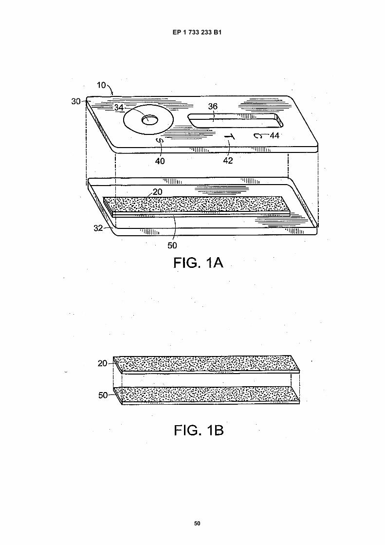
EP 1 733 233 B1
50
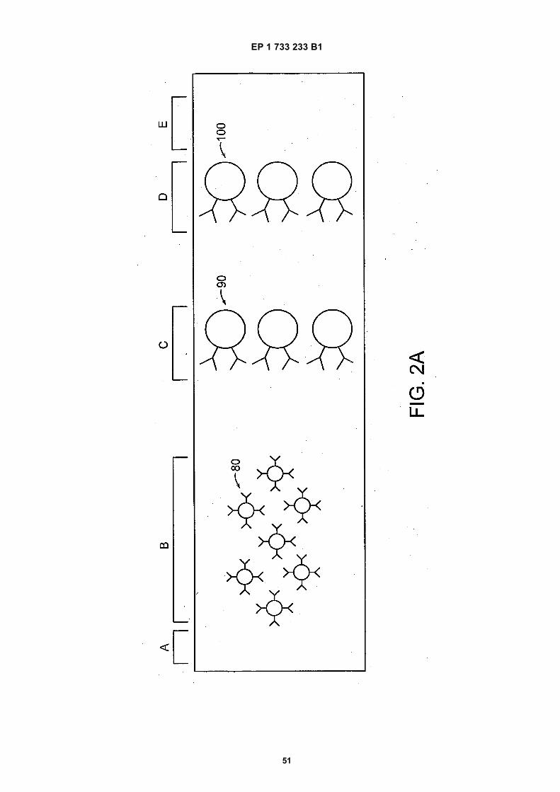
EP 1 733 233 B1
51
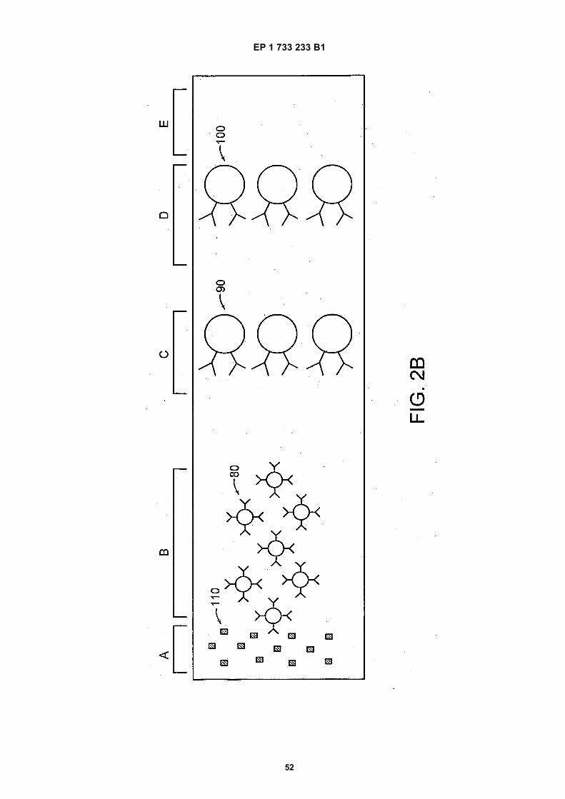
EP 1 733 233 B1
52
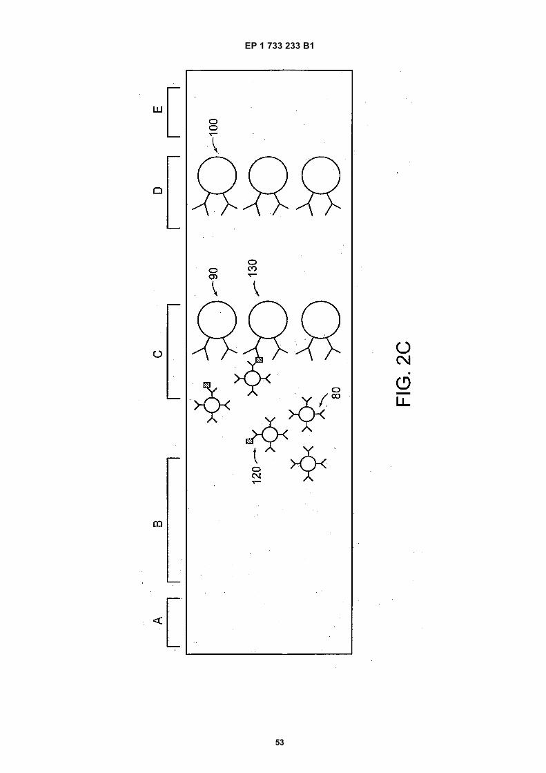
EP 1 733 233 B1
53
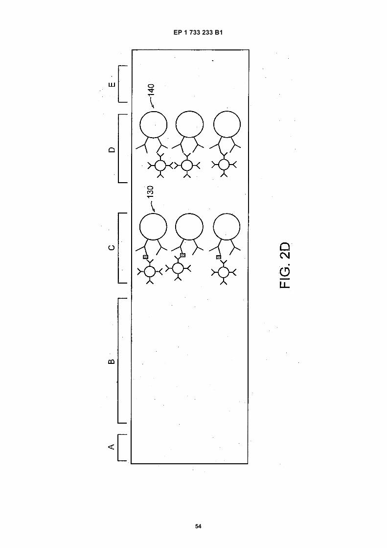
EP 1 733 233 B1
54
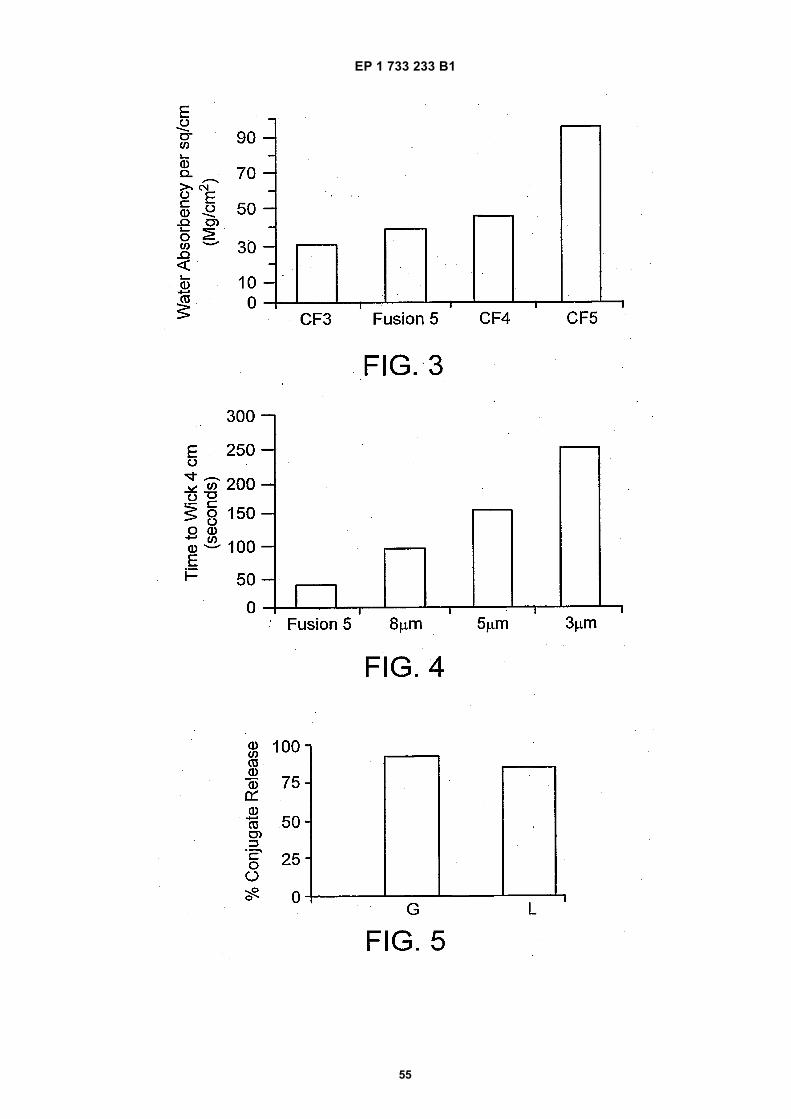
EP 1 733 233 B1
55
EP 1 733 233 B1
56
REFERENCES CITED IN THE DESCRIPTION
This list of references cited by the applicant is for the reader’s convenience only. It does not form part of the Europeanpatent document. Even though great care has been taken in compiling the references, errors or omissions cannot beexcluded and the EPO disclaims all liability in this regard.
Patent documents cited in the description
• WO 8808534 A1 [0007]• US 5141850 A [0008]• US 2003175991 A [0011]• EP 0381173 A [0012]• US 5622871 A [0251]
• US 4477575 A [0251]• US 4703017 A [0251]• US 5075078 A [0251]• US 4313734 A [0251]
Non-patent literature cited in the description
• Millipore: A short guide to Developing Immunochro-matographic Test Strip, 1996, 1-36 [0009]
• Proc.Natl.Acad.Sci. USA, 1992, vol. 89,10915-10919 [0160]