Kinetics and longevity of φC31 integrase in mouse liver and cultured cells
12
Kinetics and Longevity of fC31 Integrase in Mouse Liver and Cultured Cells Christopher L. Chavez, 1 Annahita Keravala, 1 Lauren E. Woodard, 1 Robert T. Hillman, 1 Timothy R. Stowe, 2 Jacqueline N. Chu, 1 and Michele P. Calos 1 Abstract The fC31 integrase system provides genomic integration of plasmid DNA that may be useful in gene therapy. For example, the fC31 system has been used in combination with hydrodynamic injection to achieve long-term expression of factor IX in mouse liver. However, a concern is that prolonged expression of fC31 integrase within cells could potentially stimulate chromosome rearrangements or an immune response. Western blot and immu- nofluorescence analyses were performed to investigate the duration of fC31 integrase expression in mouse liver. Integrase was expressed within 2 to 3 hr after hydrodynamic injection of a plasmid expressing fC31 integrase. Expression peaked between 8 and 16 hr and fell to background levels by 24–48 hr postinjection. Analysis of the amount of integrase plasmid DNA present in the liver over time suggested that the brief period of integrase expression could largely be accounted for by rapid loss of the bulk of the plasmid DNA, as well as by silencing of plasmid expression. PCR analysis of integration indicated that fC31 integrase carried out genomic integration of a codelivered attB-containing plasmid by 3 hr after plasmid injection. Integrase was expressed for longer times and at higher levels in transfected cultured cells compared with liver. Inhibitor studies suggested that the enzyme had a short half-life and was degraded by the 26S proteasome. The short duration of integrase expression in liver and rapid integration reaction appear to be features favorable for use in gene therapy. Introduction fC31 integrase is a sequence-specific recombinase orig- inating in a bacteriophage of Streptomyces. This integrase normally catalyzes the precise unidirectional integration of the phage genome into the bacterial chromosome (Kuhstoss and Rao, 1991; Rausch and Lehmann, 1991). The enzyme recognizes and recombines two *30-bp DNA sequences termed attB and attP, without a requirement for additional cofactors (Thorpe and Smith, 1998). fC31 integrase has also been shown to direct this reaction efficiently in mammalian cells (Groth et al., 2000). Although mammalian genomes do not contain perfect att sites, they possess related sequences, called pseudo-att sites (Thyagarajan et al., 2001). Integration for gene therapy can be carried out by placing an attB site on a plasmid containing the therapeutic gene, cotransfecting with a plasmid encoding integrase, and taking advantage of integrase-mediated recombination with naturally occurring pseudo-attP sites in the host genome. Because fC31 integrase requires a specific, relatively long recognition sequence in order to perform recombination, the number of potential integration sites is orders of magnitude lower than randomly integrating systems such as transposons and retroviruses (Chalberg et al., 2006). Furthermore, the sites appear to be in safe locations (Chalberg et al., 2006). Thus, the risks of in- sertional mutagenesis are reduced. Furthermore, the fC31 integrase system is not limited by vector size (Venken et al., 2006). The potential of fC31 integrase in gene therapy has been demonstrated in numerous studies in animals (reviewed in Calos, 2006). The enzyme is also useful for the creation of transgenic animals (Calos, 2006). To date, there have been no reports of cancer associated with fC31 integrase. Trans- genic mice producing fC31 integrase are viable, disease free, and have normal development and fertility (Belteki et al., 2003; Raymond and Soriano, 2007), and integrase has caused no acceleration of tumors in a MYC model of liver cancer (Woodard et al., 2010b). Nevertheless, at least un- der the artificial conditions of tissue culture, expression of fC31 integrase elevated the frequency of chromosomal rearrangements and large deletions (Chalberg et al., 2006; Ehrhardt et al., 2006; Liu et al., 2006). No evidence of chro- mosome rearrangements due to fC31 expression have been observed in vivo, either in gene therapy studies or in fC31 integrase-engineered transgenic animals (Bischof et al., 2007). Even so, prolonged fC31 integrase expression would be 1 Department of Genetics and 2 Department of Biological Sciences, Stanford University School of Medicine, Stanford, CA 94305. HUMAN GENE THERAPY 21:1287–1297 (October 2010) ª Mary Ann Liebert, Inc. DOI: 10.1089/hum.2010.049 1287
-
Upload
independent -
Category
Documents
-
view
6 -
download
0
Transcript of Kinetics and longevity of φC31 integrase in mouse liver and cultured cells

Kinetics and Longevity of fC31 Integrasein Mouse Liver and Cultured Cells
Christopher L. Chavez,1 Annahita Keravala,1 Lauren E. Woodard,1 Robert T. Hillman,1
Timothy R. Stowe,2 Jacqueline N. Chu,1 and Michele P. Calos1
Abstract
The fC31 integrase system provides genomic integration of plasmid DNA that may be useful in gene therapy. Forexample, the fC31 system has been used in combination with hydrodynamic injection to achieve long-termexpression of factor IX in mouse liver. However, a concern is that prolonged expression of fC31 integrase withincells could potentially stimulate chromosome rearrangements or an immune response. Western blot and immu-nofluorescence analyses were performed to investigate the duration of fC31 integrase expression in mouse liver.Integrase was expressed within 2 to 3 hr after hydrodynamic injection of a plasmid expressing fC31 integrase.Expression peaked between 8 and 16 hr and fell to background levels by 24–48 hr postinjection. Analysis of theamount of integrase plasmid DNA present in the liver over time suggested that the brief period of integraseexpression could largely be accounted for by rapid loss of the bulk of the plasmid DNA, as well as by silencing ofplasmid expression. PCR analysis of integration indicated that fC31 integrase carried out genomic integration of acodelivered attB-containing plasmid by 3 hr after plasmid injection. Integrase was expressed for longer times andat higher levels in transfected cultured cells compared with liver. Inhibitor studies suggested that the enzyme hada short half-life and was degraded by the 26S proteasome. The short duration of integrase expression in liver andrapid integration reaction appear to be features favorable for use in gene therapy.
Introduction
fC31 integrase is a sequence-specific recombinase orig-inating in a bacteriophage of Streptomyces. This integrasenormally catalyzes the precise unidirectional integration ofthe phage genome into the bacterial chromosome (Kuhstossand Rao, 1991; Rausch and Lehmann, 1991). The enzymerecognizes and recombines two *30-bp DNA sequencestermed attB and attP, without a requirement for additionalcofactors (Thorpe and Smith, 1998). fC31 integrase has alsobeen shown to direct this reaction efficiently in mammaliancells (Groth et al., 2000). Although mammalian genomes donot contain perfect att sites, they possess related sequences,called pseudo-att sites (Thyagarajan et al., 2001). Integrationfor gene therapy can be carried out by placing an attB site ona plasmid containing the therapeutic gene, cotransfectingwith a plasmid encoding integrase, and taking advantage ofintegrase-mediated recombination with naturally occurringpseudo-attP sites in the host genome. Because fC31 integraserequires a specific, relatively long recognition sequence inorder to perform recombination, the number of potentialintegration sites is orders of magnitude lower than randomlyintegrating systems such as transposons and retroviruses
(Chalberg et al., 2006). Furthermore, the sites appear to be insafe locations (Chalberg et al., 2006). Thus, the risks of in-sertional mutagenesis are reduced. Furthermore, the fC31integrase system is not limited by vector size (Venken et al.,2006).
The potential of fC31 integrase in gene therapy has beendemonstrated in numerous studies in animals (reviewed inCalos, 2006). The enzyme is also useful for the creation oftransgenic animals (Calos, 2006). To date, there have beenno reports of cancer associated with fC31 integrase. Trans-genic mice producing fC31 integrase are viable, diseasefree, and have normal development and fertility (Beltekiet al., 2003; Raymond and Soriano, 2007), and integrase hascaused no acceleration of tumors in a MYC model of livercancer (Woodard et al., 2010b). Nevertheless, at least un-der the artificial conditions of tissue culture, expressionof fC31 integrase elevated the frequency of chromosomalrearrangements and large deletions (Chalberg et al., 2006;Ehrhardt et al., 2006; Liu et al., 2006). No evidence of chro-mosome rearrangements due to fC31 expression have beenobserved in vivo, either in gene therapy studies or in fC31integrase-engineered transgenic animals (Bischof et al., 2007).Even so, prolonged fC31 integrase expression would be
1Department of Genetics and 2Department of Biological Sciences, Stanford University School of Medicine, Stanford, CA 94305.
HUMAN GENE THERAPY 21:1287–1297 (October 2010)ª Mary Ann Liebert, Inc.DOI: 10.1089/hum.2010.049
1287

considered undesirable, because it could increase the prob-ability of chromosomal instability or of an immune responseto this prokaryotic protein.
It was unknown how long fC31 integrase protein wasexpressed, either in cultured cells or in the cells of the liverafter in vivo plasmid DNA delivery. Integrase longevity inthe liver was of particular interest, because of the therapeuticimportance of this target tissue for gene therapy and robustpreclinical data on therapeutic levels of human factor IX(hFIX) production with fC31 integrase in mouse liver (Oli-vares et al., 2002). To investigate these questions, we deter-mined the time course of fC31 integrase protein expressionwithin mouse liver by Western blot analysis, as well as bythe relative number of liver cells expressing integrase andhFIX over the same time course by immunofluorescence. Inaddition, we examined the kinetics of the in vivo integrationreaction by PCR and used several methodologies to establishthe retention time within mouse liver of injected plasmidDNA. Related studies were carried out in cultured cells. Thiswork provides new data that enhance our understanding offC31 integrase activity and safety.
Materials and Methods
Plasmids
pCSI-HA was constructed by introducing the sequencecoding the hemagglutinin (HA) tag, YPYDVPDYA, onto theC-terminal end of the fC31 integrase gene by PCR, as de-scribed (Keravala et al., 2009). pVFB has been described(Keravala et al., 2009) and carries the human factor IX (hFIX)gene driven by a liver-specific promoter comprising the hu-man a1-antitrypsin promoter with an apolipoprotein E en-hancer. pInt-EGFP was made by cloning the fC31 integrasegene, carrying an altered stop codon, into the SacI andBamHI sites of pEGFP-N2 (BD Biosciences, San Jose, CA).fC31 integrase with an altered stop codon was generatedby PCR, using pCSI as template and primers PhiC31-F-aattcccgggtcgacgagctcactagtcgtagggtcgcc and PhiC31-R-cccctcgagggatccgggtgtctcgcaacgccgctacgt, and an annealingtemperature of 708C. The PCR product and pEGFP-N2 weredigested with SacI and BamHI, and the integrase gene wasligated into pEGFP-N2 to give pInt-EGFP. pRFP-pericentrinwas a gift from the laboratory of S. McConnell (StanfordUniversity, Stanford, CA) and has been described previously(Gillingham and Munro, 2000). Briefly, the plasmid containsa cytomegalovirus (CMV) promoter driving expression of afusion protein of red fluorescent protein and pericentrin, al-lowing visualization of the centrosome.
Mice
Eight- to 10-week-old C57BL/6 mice were ordered fromCharles River (Wilmington, MA) or Jackson Laboratory (BarHarbor, ME). Mice were kept in the Stanford Research AnimalFacility and given water and food ad libitum. The StanfordResearch Animal Committee approved all animal studies.
Hydrodynamic tail vein injection
A DNA solution was prepared that diluted 20 mg of bothpCSI-HA and pVFB in 1.8 ml of Hanks’ balanced salt solu-tion (HBSS; Invitrogen, Carlsbad, CA). All DNA was pre-pared with an EndoFree plasmid maxi kit (Qiagen, Valencia,
CA) according to the manufacturer’s protocol, with the ex-ception that the DNA pellets were reconstituted in HBSS. Topromote tail vein dilation, mice were placed under a heatlamp. Once the vein was dilated, mice were placed in a re-strainer (Braintree Scientific, Braintree, MA) and injectedwith 1.8 ml of the DNA solution over 3–5 sec, using a 3-mlsyringe and 27-gauge butterfly needle (BD Biosciences).
Western blotting
Mouse livers were harvested over a time course and keptfrozen at �808C until use. Total liver protein extracts wereprepared by adding 1 ml of tissue protein extraction reagent(Thermo Scientific, Rockford, IL) with added protease inhibi-tor cocktail tablets (Roche, Indianapolis, IN) to 0.1 g of mouseliver tissue and homogenizing, using Pellet Pestles and a PelletPestle motor (Daigger, Vernon Hills, IL). Protein sample con-centration was determined by the Bradford assay (Bio-Rad,Hercules, CA). Controls, molecular weight markers, and timepoint samples (150mg of total protein from liver and 50mg oftotal protein from tissue culture cells) were run on 7.5% Tris-HCl Ready Gels (Bio-Rad) and transferred to nitrocelluloseTrans-Blot transfer medium (Bio-Rad). Membranes wereblocked with 5% nonfat dry milk in 1�phosphate-bufferedsaline (PBS). The membranes were cut at the 50-kDa marker.The top half was incubated for 1 hr at room temperature withrat anti-HA high-affinity antibody (cat. no. 11-867-423-001;Roche Applied Science, Indianapolis, IN) diluted 1:1000 in 5%milk solution, and then washed three times for 5 min in 1�PBSfollowed by addition of goat anti-rat horseradish peroxidase(HRP)-conjugated secondary antibody (cat. no. 401416; Cal-biochem, Darmstadt, Germany) diluted in 1:10,000 milk so-lution. The remaining half was incubated for 1 hr at roomtemperature with mouse anti-b-actin (NB600-501; NovusBiologicals, Littleton, CO) diluted 1:1000 in milk solution, andthen washed three times for 5 min in 1�PBS, followed by ad-dition of goat anti-mouse HRP-conjugated secondary anti-body (Calbiochem) diluted 1:10,000 in milk solution. The blotswere then washed three times in 1�PBS, and incubated withSuperSignal West Pico substrate (Thermo Scientific, Waltham,MA) and imaged with Kodak BioMax film (CarestreamHealth, Rochester, NY). Densitometry was performed withImageJ software (National Institutes of Health, Bethesda, MD).
Polymerase chain reaction
Total DNA was isolated from 25 mg of mouse liver tissue,using a DNeasy blood & tissue kit (Qiagen). For detection ofintegration at the mpsL1 pseudo-attP site, primers and con-ditions were used as described (Olivares and Calos, 2003).For integrase gene amplification, 100 ng of total DNA wasused as the template to generate a 382-base pair fragment ofthe fC31 integrase gene. The primers used were Int-F (846)(50-ggcaaccgttatgcgaatcc) and Int-R (505) (50-ctcgacacgaagaaccttcagc). PCR conditions were as follows: an initial de-naturation step at 948C for 1 min and then 948C for 30 sec,558C for 30 sec, and 728C for 30 sec, repeated for 30 cycles,with a final extension of 2 min and a final hold at 48C.
Southern blot analysis and bacterial colony count
Five micrograms of total liver DNA was digested withNdeI, which cuts the vector once. The digests were electro-
1288 CHAVEZ ET AL.

phoresed through a 0.8% agarose gel and transferred to anitrocellulose membrane. Integrase DNA was detected afterhybridization with a digoxigenin-labeled integrase probeand chemiluminescence (Roche). For the bacterial colonycount assay, 0.1–1mg of total liver DNA was transformedinto DH10B Escherichia coli cells, and the cells were spread onampicillin or kanamycin selection plates and incubated at378C overnight. The next day, the numbers of colonies werecounted.
Immunofluorescence staining of liver sections
For immunostaining, liver tissues were paraffin embeddedand sectioned (Histo-Tec Laboratory, Hayward, CA). Thestaining procedure was performed as described (Keravalaet al., 2009), with the following modifications. For double-stained slides, livers were fixed, after administration of thefirst secondary antibody, with 4% paraformaldehyde in 1�PBS for 15 min, washed, reblocked, and incubated with thesecond primary antibody for 16 hr at 48C. Next, the secondsecondary antibody was applied for 1 hr at room temperature.Slides were subsequently mounted with ProLong Gold anti-fade mounting medium with 40,6-diamidino-2-phenylindole(DAPI; Invitrogen). The antibodies used were rat anti-HAhigh affinity (cat. no. 11-867-423-001; Roche), goat anti-hFIX(GAFIX-AP; Affinity Biologicals, Ancaster, ON, Canada),Alexa Fluor 488-conjugated goat anti-rabbit (Invitrogen), andAlexa Fluor 594-conjugated goat anti-rat (Invitrogen). All an-tibody dilutions were 1:500 in serum buffer. Serum bufferconsisted of 1�PBS plus 0.1% Tween 20 and 10% goat serum.Pictures of the stained cells, viewed with an Axioskop 2 plusmicroscope with a FluoArc power supply, were taken with anAxioCam MRc camera (Zeiss, Thornwood, NY).
Cell culture and protein extraction
HeLa cells (CCL-2; American Type Culture Collection[ATCC], Manassas, VA) were grown in Dulbecco’s modifiedEagle’s medium (DMEM; Invitrogen) supplemented with 9%fetal bovine serum and 1% penicillin–streptomycin (Invitro-gen) on 60-mm plates. Plates of cells were transfected withDNA prepared with a Qiagen Maxi kit according to themanufacturer’s instructions, using FuGENE 6 (Roche) at a 3:1reagent (ml)-to-DNA (mg) ratio. For cycloheximide treatment,medium containing cycloheximide at 100 mg/ml (Calbio-chem, Beeston, UK) was added to cells 2 days posttransfec-tion. At the time points indicated, plates of cells wereharvested for protein as described previously (Woodard et al.,2010a). For cycloheximide plus MG132 treatment, transfectedHeLa cells were incubated with cycloheximide (100 mg/ml)and 1, 5, 10, or 15mM MG132 (Calbiochem), and protein washarvested 10 hr later as described previously.
Immunofluorescence staining of cultured cells
NIH/3T3 mouse embryonic fibroblast cells (CRL-1568;ATCC) were seeded onto 6-well plates containing 12-mm-diameter coverslips (VWR, West Chester, PA) coated in poly-l-lysine. Cells were then transfected as described previously.The next evening 25mM MG132 was added to the indicatedcells. Ten hours later cells were fixed by removing the me-dium and adding 2 ml of ice-cold methanol to each well for10 min at �208C. Coverslips were then removed from the
wells and placed in a dark humidified chamber. Cells wererehydrated with PBS for 10 min and blocked in PBS-BT (PBSplus 3% bovine serum albumin [BSA], 0.1% Triton X-100,0.02% sodium azide) for 30 min. The following antibodieswere then sequentially incubated on the coverslip, diluted inPBS-BT at the given dilutions: mouse anti-g-tubulin (GTU-88;Sigma-Aldrich, St. Louis, MO), 1:500; Alexa Fluor 488-conjugated goat anti-mouse (Invitrogen), 1:250; rat anti-HA(3F10; Roche), 1:500; and Alexa Fluor 594-conjugated goatanti-rat (Invitrogen), 1:250. Each antibody was incubated forat least 30 min and then three PBS-BT washes were done.DAPI (0.05 mg/ml; Invitrogen) was added, and cells weremounted onto slides with glycerol-based antifade mountingmedium, sealed with nail polish, and allowed to dry. Cellswere visualized with a Zeiss Axioskop microscope.
Imaging of live cells
HeLa cells grown in 6-well dishes were transfected withequal amounts of pInt-EGFP and pRFP-pericentrin as de-scribed previously. The next morning cells were treated withMG132 or dimethyl sulfoxide (DMSO) alone. Eight hourslater, cells were washed three times with DMEM withoutphenol red (Invitrogen), containing MG132 if indicated. Cellswere visualized with a Zeiss Axiovert 200M microscope.
Results
Time course of fC31 integrase expressionwithin mouse liver
We wanted to determine the length of time that fC31integrase protein was expressed in vivo within mouse liver,after delivery of plasmid DNA by high-pressure tail veininjection (Liu et al., 1999; Zhang et al., 1999). Two plasmids at20mg each were injected, comprising pVFB, an attB-containinghFIX expression plasmid (Keravala et al., 2009), and pCSI-HA(Woodard et al., 2010a), containing the fC31 integrase genewith the nine-amino acid hemagglutinin (HA) epitope at thecarboxy-terminal end and driven by the human cytomega-lovirus (CMV) immediate/early promoter/enhancer. ThisHA-tagged version of fC31 integrase was used for easydetection of the protein and has similar activity to wild-typefC31 integrase (Woodard et al., 2010a). Serum levels of hFIXon day 1 were monitored to determine the quality of theinjections, with a cutoff of >1000 ng/ml used to indicateadequate transfection. Animals with lower hFIX serum levelswere excluded from the study. Mice were then killed over atime course and their livers were extracted for protein andDNA analysis.
Total protein isolated from mouse livers harvested atvarious time points was subjected to Western blot analysis(Fig. 1A). These data revealed that fC31 integrase was ex-pressed by 3 hr after injection and that protein levels con-tinued to increase up to a peak at 16 hr. By 24 hrpostinjection, only trace levels of integrase remained. By48 hr, integrase was undetectable by Western blot.
From the same liver samples that were used for Westernblot analysis, sections were prepared for immunofluores-cence staining to visualize the number of cells expressingfC31 integrase. As seen in Fig. 1B, fC31 integrase proteinwas first detectable within the cells of the liver by 2 hr afterinjection, with 4.8% of cells positive for integrase. Subsequently,
KINETICS AND LONGEVITY OF /C31 INTEGRASE 1289

integrase levels rose within the liver, to peak at 19.6 and14.9% of cells expressing integrase at 8 and 16 hr, respec-tively, postinjection. A sharp drop-off in the number offC31-positive cells ensued, so that only rare positive cells,0.9% or less, were detectable after 16 hr. The immunofluo-rescence analysis was thus in close agreement with theWestern blot data.
Time course of genomic integration within mouse liver
Several studies have shown that in mouse liver, fC31integrase has a preferred integration site within the mousegenome (Olivares et al., 2002; Held et al., 2005; Keravala et al.,2009). This site, termed the mouse pseudo-site L1 (mpsL1) islocated on chromosome 2. Integration at this site can be usedto monitor fC31-mediated integration into the genome bydetection of a specific PCR band comprising the junctionbetween the integrated attB plasmid and the chromosomalmpsL1 site. As seen in Fig. 2, this PCR band indicating in-
tegration of pVFB, carrying attB and the hFIX gene, wasdetectable at the mpsL1 site by 3 hr after injection of pVFBand integrase plasmids. This result correlated well withWestern blot and immunofluorescence data showing thatintegrase protein was present within liver cells by 2 to 3 hrafter injection (Fig. 1). Taken together, these data indicatedthat fC31 integrase was expressed by 2 hr postinjection andhad carried out integration within the mouse genome by 3 hrafter injection.
Time course of retention of integrase plasmidDNA within mouse liver
To determine the length of time that plasmid DNA en-coding fC31 integrase was detectable within the liver ofmice injected with pCSI-HA, three separate approaches wereemployed. First, we conducted Southern blot analysis ontotal DNA from the mouse liver samples (Fig. 3A). Thesedata revealed that the integrase-encoding plasmid was de-
FIG. 1. Time course of fC31 integrase protein expression within mouse liver. Twenty micrograms each of pCSI-HA andpVFB were hydrodynamically injected into the tail vein of mice. At various time points over a 6-month time course, the micewere killed, their livers were removed, and total protein and DNA were isolated. (A) Western blot analysis of total proteinisolated from mouse liver showed the presence of fC31 integrase at 3 hr postinjection. Integrase protein levels increased up toa peak at 16 hr and were undetectable by 24 hr. Below each lane of the blot is the percentage of total fC31 integrase proteinpresent, as quantified by densitometry. (B) Immunofluorescence staining for fC31 integrase in mouse liver sections obtainedover a time course. The top row shows DAPI-stained nuclei, whereas the bottom row shows staining for fC31 integrase.Numbers inside the images correspond to the percentage of cells that are expressing fC31 integrase protein. Color imagesavailable online at www.liebertonline.com/hum.
1290 CHAVEZ ET AL.

tectable within the mouse liver at the first time point taken,1 hr after injection. The amount of plasmid DNA detectedwithin the liver increased over the next four time points,peaking in concentration at 8 hr, and then fell rapidly andwas undetectable by Southern blot under these conditions by48 hr postinjection.
In the second approach, 1 mg of total mouse liver DNA wasused to transform E. coli. Because pCSI-HA carries the am-picillin resistance gene, bacterial cells transformed with totalliver DNA were spread on ampicillin plates and incubated,
and colonies were counted, reflecting the relative amount ofunintegrated, circular integrase-encoding plasmid DNA thatwas present in the samples. The results (Fig. 3B) indicated thatpCSI-HA-derived ampicillin-resistant colonies were detected1 hr after injection, and there was an increase in the number ofampicillin-resistant colonies over the next four time points.Peak numbers of colonies were reached at 8 hr. Colonynumbers then fell rapidly to low levels after 24 hr.
Last, we subjected total DNA from the mouse livers toPCR analysis, using primers that would detect the pCSI-HA
FIG. 2. Integration of donor plasmid occurs rapidly after plasmid injection. DNA extracted from liver was subjected to PCRusing primers that detect the junction between the attB site of the donor pVFB plasmid and mpsL1, a commonly used pseudo-attP site in the mouse genome. Lane 1, molecular weight markers; lane 2, no DNA; lane 3, liver DNA sample known to haveintegration at mpsL1; lane 4, liver that received saline alone; lanes 5–25, time course of DNA samples from mice receivingpVFB and pCSI-HA, after the specified time in hours (1–48 hr), followed by days (3–180 days).
FIG. 3. Analysis of fC31 integrase plasmid DNA retention within mouse liver. fC31 integrase expression plasmid pCSI-HA was hydrodynamically injected into the tail vein of mice. At various points over a time course, the mice were killed, theirlivers were removed, and total DNA was isolated. (A) Southern blot analysis using a probe to the fC31 integrase geneshowed an increase in signal over the first 8 hr postinjection, followed by a decrease, and a loss of signal by 48 hr. Below eachlane of the blot is the percentage of total fC31 integrase plasmid present, as quantified by densitometry. (B) Total DNAtransformed into bacterial cells showed an increase in colony number over the first 8 hr after injection, followed by a decrease,until few colonies were detected after day 7. (C) PCR detected the presence of the fC31 integrase gene over the 180-day timecourse.
KINETICS AND LONGEVITY OF /C31 INTEGRASE 1291

plasmid, whether extrachromosomal or integrated (Fig. 3C).PCR is an extremely sensitive technique, capable of detectingsmall amounts of integrase-encoding DNA. Integrase DNAwas detectable through the period of 180 days. However, theintensity of the PCR band decreased over time, especiallyafter 4 days, suggesting a reduction in the amount of in-tegrase plasmid present within the livers over time.
Time course of hFIX expression and plasmidretention in mouse liver
Along with the integrase expression plasmid pCSI-HA, wealso introduced pVFB, which expresses hFIX from the liver-specific human a1-antitrypsin promoter (hAAT) and carriesan attB site for integration. As seen in Fig. 4A, hFIX proteinwas first detectable, within 0.8% of the liver cells, by 4 hrafter injection. hFIX levels then rose within the liver, to apeak of 19.5% of cells expressing hFIX at 24 hr postinjection.hFIX protein expression dropped to 5.1% of the cells ex-pressing at 30 days postinjection and stabilized at about 4%of the liver cells over the 6 months of the experiment.
To analyze persistence of pVFB DNA in the liver, 0.1 mg oftotal mouse liver DNA was used to transform E. coli. Thekanamycin resistance gene is carried by pVFB. Therefore,bacterial cells transformed with total liver DNA were spreadon kanamycin-containing plates and incubated, and colonieswere counted. The colony numbers reflected the relativeamount of unintegrated, intact hFIX plasmid that remainedin the samples. Kanamycin-resistant colonies derived frompVFB were detected 1 hr postinjection (Fig. 4B). The numberof kanamycin-resistant colonies increased over the next fourtime points. At 8 hr postinjection, peak numbers of colonieswere reached. After 24 hr, colony numbers fell rapidly to lowlevels.
Time course of fC31 integrase expressionand plasmid retention in cultured cells
We were interested in whether the longevity of integraseexpression in cultured cells was similar to what was ob-served in liver. Two human cell lines, HeLa and 293, weretransfected with the pCSI-HA plasmid. Cells were harvestedover a time course, total cell protein extracts were prepared,and fC31 integrase protein was detected by Western blotanalysis using an anti-HA antibody. The results are shown inFig. 5A and B. fC31 integrase was expressed at high levelsfor 3 days in HeLa cells and for 1 week in 293 cells. Fur-thermore, the levels of integrase protein present, as judgedby the intensity of the signal on the Western blot, normalizedto the amount of total protein, were approximately 3-foldhigher in the cultured cells, compared with the integraselevels in liver (Fig. 1A).
To analyze how much integrase plasmid DNA remainedin the cells during the time course, total DNA was isolatedfrom cells at each time point. The resulting DNA (0.1 mg) wastransformed into E. coli, and bacteria were grown on ampi-cillin plates. Ampicillin-resistant colonies were counted after24 hr. Colony numbers reflect the amount of free plasmidDNA present in the cells. The results (Fig. 5C) indicated thatintegrase plasmid DNA became less abundant in parallelwith the decline in integrase expression in HeLa cells, con-sistent with progressive plasmid loss during cell divisionbeing a primary cause of loss of integrase expression. In
293 cells, fC31 integrase protein levels remained high, evenas plasmid pCSI-HA levels fell, consistent with the strongand prolonged expression of transfected DNA characteristicof 293 cells.
Inhibitor studies to elucidate fC31 integrasehalf-life and degradation location
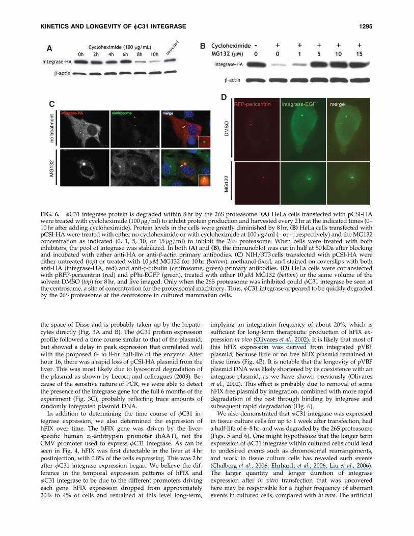
To elucidate the half-life of fC31 integrase protein inmammalian cells and to investigate its site of degradation,we used the ribosomal inhibitor cycloheximide (Colomboet al., 1965) and the proteasomal inhibitor MG132 (Palombellaet al., 1994). Transfected HeLa cells were treated with cyclo-heximide and then lysed every 2 hr to obtain protein. Theamount of integrase protein present in the same amount oftotal protein lysate as determined by the Bradford methodwas examined by immunoblotting, using an antibody to theHA tag. The pool of integrase protein was found to remainconstant until 6 hr after ribosomal inhibition was initiated,followed by a decrease to barely detectable levels at 8 hrpostinhibition (Fig. 6A). Therefore, the half-life of fC31 in-tegrase was estimated to be between 6 and 8 hr (Fig. 6A). Thisrelatively short half-life led us to investigate whether fC31integrase might be actively degraded by the 26S proteasome.
After 10 hr of cycloheximide treatment, most of the in-tegrase protein was gone. By contrast, in HeLa cells receivingboth cycloheximide and 5–15 mM levels of the proteasomalinhibitor MG132 for 10 hr, an amount equal to the startingamount of integrase protein was still present in the cell(Fig. 6B). This result suggested that the 26S proteasome wasresponsible for the degradation of fC31 integrase.
A well-established site of proteasome localization is thecentrosome (Wigley et al., 1999). The centrosome acts as anorganizing structure and common meeting point for the ac-tive proteasome, chaperone proteins, and misfolded or ubi-quitinated protein substrates (Fabunmi et al., 2000). Tofurther establish proteasomal degradation of fC31 integrase,we imaged untreated and proteasome-inhibited cells in anattempt to colocalize the enzyme and the centrosome. NIH/3T3 cells were used because they are flatter than HeLa cells,allowing for better visualization. Staining for integrase-HAin untreated cells revealed what appeared to be inclusionbodies throughout the cytoplasm (Fig. 6C). This result im-plied that proteasomal degradation of integrase might occurthrough nonspecific, low levels of ubiquitination of themisfolded aggregates, as has been the case with other pro-teins (Dantuma and Lindsten, 2010). Indeed, integrase-HAand the centrosomal marker g-tubulin colocalized in theproteasome-inhibited cells (Fig. 6C). To reduce potential ar-tifacts and confirm these results, we also used a live-cellimaging approach. Constructs expressing two fusion pro-teins, integrase–EGFP and RFP–pericentrin, were transfectedinto HeLa cells. The latter protein marks the centrosome inlive cells (Gillingham and Munro, 2000). Once again, fC31integrase was localized to the centrosome in proteasome-inhibited cells (Fig. 6D).
Discussion
In this study, we determined the kinetics of fC31 in-tegrase protein expression within mouse liver. Western blotand immunofluorescence analyses indicated that the major-
1292 CHAVEZ ET AL.

FIG. 4. Analysis of hFIX expression in mouse liver. (A) Immunofluorescence staining for hFIX in mouse liver sectionsobtained over a time course. Twenty micrograms each of pCSI-HA and pVFB were hydrodynamically injected into the tailvein of mice. At various time points over a 6-month time course, the mice were killed and their livers were removed,sectioned, and stained with anti-hFIX antibody. The top row shows DAPI-stained nuclei, whereas the bottom row showsstaining for hFIX. Numbers inside the images correspond to the percentage of cells that were expressing hFIX protein. (B)Total DNA transformed into bacterial cells showed an increase in colony numbers over the first 8 hr after injection, followedby a decrease, until few colonies were detected after day 3. Color images available online at www.liebertonline.com/hum.
KINETICS AND LONGEVITY OF /C31 INTEGRASE 1293

ity of fC31 integrase protein expression was confined to a22-hr period after hydrodynamic injection (hours 2–24)(Fig. 1). The 2-hr lag time between plasmid injection and theaccumulation of sufficient fC31 integrase protein for detec-tion is in agreement with the observations of Rossmanith andcolleagues (2002), where b-galactosidase activity was de-tected in mouse liver 2 hr after hydrodynamic injection. Overthe next 6 hr (hours 2–8), we observed a sharp rise in thefC31 integrase protein levels within the liver, as seen byWestern blot and immunofluorescence staining (Fig. 1Aand B). Peak expression levels were reached at 8–16 hr, when19.6 and 14.9% of the hepatocytes were positive for fC31integrase protein. This elevation in fC31 integrase proteinlevels correlated with increased intracellular pCSI-HA levelsas seen by Southern blot and colony count assay (Fig. 3Aand B). It therefore appeared that after hydrodynamic in-jection, plasmid DNA accumulated within the liver over an8-hr period.
Western blot and immunofluorescence data showed thatthe fC31 integrase protein levels were fairly constant fromhours 8–16 (Fig. 1A and B). However, there was a large dropin expression between 16 and 24 hr after injection. Southernblot analysis and colony count data showed that after 8 hr,pCSI-HA levels began to drop somewhat, but were fairlystable up to 24 hr (Fig. 3A and B). Therefore, the drop inexpression between 16 and 24 hr was not due predominantlyto plasmid loss, but was more likely due to silencing of theCMV promoter. One hour after fC31 integrase protein wasfirst detected in the liver and within 3 hr of injection, pVFBintegration was observed by PCR (Fig. 2). The increasedconcentration of plasmid pCSI-HA within the liver over thefirst 8 hr after injection (Fig. 5) could be due to the accumu-lation of free plasmid circulating in the blood within theliver. High pressure and volume during the injection processresults in transport of plasmid through the fenestrae of thesinusoidal endothelial cells, where the plasmid DNA enters
FIG. 5. Duration of integrase expression and plasmid retention in two human cell lines. One microgram of pCSI-HA wastransfected into two tissue culture cell lines. At various time points over a 9-day time course, the cells were harvested andtotal protein and DNA were isolated. (A) Western blot analyses of total protein isolated from HeLa cells showed the presenceof fC31 integrase protein at 1 day posttransfection. Integrase protein levels were stably expressed over a 3-day period, untilthey dropped below detection levels on day 4. (B) Western blot analyses of total protein isolated from 293 cells showed thepresence of fC31 integrase protein at 1 day posttransfection. Integrase protein levels reached a peak 4 days posttransfectionand then declined over a 3-day period, until they dropped below detection levels on day 8. b-Actin was used as a loadingcontrol. (C) Total DNA transformed into bacterial cells from HeLa (solid columns) and 293 cells (shaded columns) showed themaximal colony number 1 day after transfection, followed by a rapid decline, until few colonies were detected after day 6.
1294 CHAVEZ ET AL.
the space of Disse and is probably taken up by the hepato-cytes directly (Fig. 3A and B). The fC31 protein expressionprofile followed a time course similar to that of the plasmid,but showed a delay in peak expression that correlated wellwith the proposed 6- to 8-hr half-life of the enzyme. Afterhour 16, there was a rapid loss of pCSI-HA plasmid from theliver. This was most likely due to lysosomal degradation ofthe plasmid as shown by Lecocq and colleagues (2003). Be-cause of the sensitive nature of PCR, we were able to detectthe presence of the integrase gene for the full 6 months of theexperiment (Fig. 3C), probably reflecting trace amounts ofrandomly integrated plasmid DNA.
In addition to determining the time course of fC31 in-tegrase expression, we also determined the expression ofhFIX over time. The hFIX gene was driven by the liver-specific human a1-antitrypsin promoter (hAAT), not theCMV promoter used to express fC31 integrase. As can beseen in Fig. 4, hFIX was first detectable in the liver at 4 hrpostinjection, with 0.8% of the cells expressing. This was 2 hrafter fC31 integrase expression began. We believe the dif-ference in the temporal expression patterns of hFIX andfC31 integrase to be due to the different promoters drivingeach gene. hFIX expression dropped from approximately20% to 4% of cells and remained at this level long-term,
implying an integration frequency of about 20%, which issufficient for long-term therapeutic production of hFIX ex-pression in vivo (Olivares et al., 2002). It is likely that most ofthis hFIX expression was derived from integrated pVBFplasmid, because little or no free hFIX plasmid remained atthese times (Fig. 4B). It is notable that the longevity of pVBFplasmid DNA was likely shortened by its coexistence with anintegrase plasmid, as we have shown previously (Olivareset al., 2002). This effect is probably due to removal of somehFIX free plasmid by integration, combined with more rapiddegradation of the rest through binding by integrase andsubsequent rapid degradation (Fig. 6).
We also demonstrated that fC31 integrase was expressedin tissue culture cells for up to 1 week after transfection, hada half-life of 6–8 hr, and was degraded by the 26S proteasome(Figs. 5 and 6). One might hypothesize that the longer termexpression of fC31 integrase within cultured cells could leadto undesired events such as chromosomal rearrangements,and work in tissue culture cells has revealed such events(Chalberg et al., 2006; Ehrhardt et al., 2006; Liu et al., 2006).The larger quantity and longer duration of integraseexpression after in vitro transfection that was uncoveredhere may be responsible for a higher frequency of aberrantevents in cultured cells, compared with in vivo. The artificial
FIG. 6. fC31 integrase protein is degraded within 8 hr by the 26S proteasome. (A) HeLa cells transfected with pCSI-HAwere treated with cycloheximide (100 mg/ml) to inhibit protein production and harvested every 2 hr at the indicated times (0–10 hr after adding cycloheximide). Protein levels in the cells were greatly diminished by 8 hr. (B) HeLa cells transfected withpCSI-HA were treated with either no cycloheximide or with cycloheximide at 100mg/ml (– orþ, respectively) and the MG132concentration as indicated (0, 1, 5, 10, or 15 mg/ml) to inhibit the 26S proteasome. When cells were treated with bothinhibitors, the pool of integrase was stabilized. In both (A) and (B), the immunoblot was cut in half at 50 kDa after blockingand incubated with either anti-HA or anti-b-actin primary antibodies. (C) NIH/3T3 cells transfected with pCSI-HA wereeither untreated (top) or treated with 10mM MG132 for 10 hr (bottom), methanol-fixed, and stained on coverslips with bothanti-HA (integrase-HA, red) and anti-g-tubulin (centrosome, green) primary antibodies. (D) HeLa cells were cotransfectedwith pRFP-pericentrin (red) and pPhi-EGFP (green), treated with either 10 mM MG132 (bottom) or the same volume of thesolvent DMSO (top) for 8 hr, and live imaged. Only when the 26S proteasome was inhibited could fC31 integrase be seen atthe centrosome, a site of concentration for the proteasomal machinery. Thus, fC31 integrase appeared to be quickly degradedby the 26S proteasome at the centrosome in cultured mammalian cells.
KINETICS AND LONGEVITY OF /C31 INTEGRASE 1295
conditions of cell culture, including elevated oxygen con-centration, may also contribute (Atkuri et al., 2007).
Cytosolic proteins are most often degraded by the 26Sproteasome, with half-lives ranging from only a few minutes(p53) to several days (cardiac myosin) (Khoo et al., 2009;Dantuma and Lindsten, 2010). However, many proteins, suchas those that are extracellular, membrane-bound, or onlymonoubiquitinated, are degraded by the lysosomal pathway(Raiborg et al., 2003). Because fC31 integrase is a protein thatevolved for expression in Streptomyces species, but has beenartificially expressed in mammalian cells for the purposes ofgene therapy, it was not known whether it might possesssignals or other cues that would target it to a particular pro-tein degradation pathway. Here, we demonstrated that fC31integrase was expressed quickly in great quantity from theconstitutive CMV promoter in the mammalian cells, resultingin inclusion bodies of misfolded protein throughout the cy-toplasm (Fig. 6C). This misfolded fraction, comprising half ofthe total pool of cytoplasmic fC31 integrase in transfectedHeLa cells, could also be detected by Western blot in the in-soluble protein fraction (Woodard et al., 2010a). Rather thanthe inclusion bodies being endocytosed and lysosomally de-graded, stabilization of the amount of integrase and coloca-lization with the centrosome under conditions of MG132treatment point toward proteasomal degradation of the fC31integrase along a time course generally recognized as short(half-life less than 10 hr) (Berger et al., 1988). This short half-lifemay be desirable for gene therapy, in that the length of ex-posure of the mammalian genome to the integrase is limited,reducing the need for a system of integrase regulation.
Acknowledgments
C.L.C. was supported by a Dean’s Postdoctoral Fellow-ship from Stanford University School of Medicine, andL.E.W. was supported by grant CA09302 from the NCI. Thiswork was supported by NIH grant HL68012 to M.P.C.M.P.C. is an inventor on Stanford-owned patents coveringfC31 integrase.
References
Atkuri, K., Herzenberg, L., Niemi, A., Cowan, T., and Herzen-berg, L. (2007). Importance of culturing primary lymphocytesat physiological oxygen levels. Proc. Natl. Acad. Sci. U.S.A.104, 4547–4552.
Belteki, G., Gertsenstin, M., Ow, D.W., and Nagy, A. (2003). Site-specific cassette exchange and germline transmission withmouse ES cells expressing the fC31 integrase. Nat. Biotechnol.21, 321–324.
Berger, J., Eisenhauer, D., and Taylor, A. (1988). Intracellularprotein degradation in cultured bovine lens epithelial cells. InVitro Cell Dev. Biol. 24, 990–994.
Bischof, J., Maeda, R.K., Hediger, M., Karch, F., and Basler, K.(2007). An optimized transgenesis system for Drosophila usinggerm-line-specific fC31 integrases. Proc. Natl. Acad. Sci.U.S.A. 104, 3312–3317.
Calos, M.P. (2006). The fC31 integrase system for gene therapy.Curr. Gene Ther. 6, 633–645.
Chalberg, T.C., Portlock, J.L., Olivares, E.C., Thyagarajan, B.,Kirby, P., Hillman, R.T., Hoelters, J., and Calos, M.P. (2006).Integration specificity of phage fC31 integrase in the humangenome. J. Mol. Biol. 357, 28–48.
Colombo, B., Felicetti, L., and Baglioni, C. (1965). Inhibition ofprotein synthesis by cycloheximide in rabbit reticulocytes.Biochem. Biophys. Res. Commun. 18, 389–395.
Dantuma, N., and Lindsten, K. (2010). Stressing the ubiquitin/proteasome system. Cardiovasc. Res. 85, 263–271.
Ehrhardt, A., Engler, J., Xu, H., Cherry, A., and Kay, M. (2006).Molecular analysis of chromosomal rearrangements in mam-malian cells after fC31-mediated integration. Hum. GeneTher. 17, 1077–1094.
Fabunmi, R., Wigley, W., Thomas, P., and Demartino, G. (2000).Activity and regulation of the centrosome-associated protea-some. J. Biol. Chem. 275, 409–413.
Gillingham, A., and Munro, S. (2000). The PACT domain, aconserved centrosomal targeting motif in the coiled-coil pro-teins AKAP450 and pericentrin. EMBO Rep. 1, 524–529.
Groth, A.C., Olivares, E.C., Thyagarajan, B., and Calos, M.P.(2000). A phage integrase directs efficient site-specific integra-tion in human cells. Proc. Natl. Acad. Sci. U.S.A. 97, 5995–6000.
Held, P.K., Olivares, E.C., Aguilar, C.P., Finegold, M., Calos,M.P., and Grompe, M. (2005). In vivo correction of murinehereditary tyrosinemia type I by fC31 integrase-mediatedgene delivery. Mol. Ther. 11, 399–408.
Keravala, A., Lee, S., Thyagarajan, B., Olivares, E.C., Gabrovsky,V., Woodard, L.E., and Calos, M.P. (2009). Mutational deriv-atives of fC31 integrase with enhanced efficiency and speci-ficity. Mol. Ther. 17, 112–120.
Khoo, K., Mayer, S., and Fersht, A. (2009). Effects of stability onthe biological function of p53. J. Biol. Chem. 284, 30974–30980.
Kuhstoss, S., and Rao, R.N. (1991). Analysis of the integrationfunction of the streptomycete bacteriophage fC31. J. Mol.Biol. 222, 897–908.
Lecocq, M., Andrianaivo, F., Warnier, M., Wattiaux-De Coninck,S., Wattiauz, R., and Jadot, M. (2003). Uptake by mouse liverand intracellular fate of plasmid DNA after a rapid tail veininjection of a small or a large volume. J. Gene Med. 5, 142–156.
Liu, F., Song, Y.K., and Liu, D. (1999). Hydrodynamics-basedtransfection in animals by systemic administration of plasmidDNA. Gene Ther. 6, 1258–1266.
Liu, J., Jeppesen, I., Nielsen, K., and Jensen, T.G. (2006). fC31integrase induces chromosomal aberrations in primary humanfibroblasts. Gene Ther. 13, 1188–1190.
Olivares, E.C., Hollis, R.P., Chalberg, T.W., Meuse, L., Kay,M.A., and Calos, M.P. (2002). Site-specific genomic integrationproduces therapeutic factor IX levels in mice. Nat. Biotechnol.20, 1124–1128.
Olivares, E.C., and Calos, M.P. (2003). Phage fC31 integrase-mediated site-specific integration for gene therapy. Gene Ther.Regul. 2, 103–120.
Palombella, V., Rando, O., Goldberg, A., and Maniatis, T. (1994).The ubiquitin–proteasome pathway is required for processingthe NF-kB1 precursor protein and the activation of NF-kB.Cell 78, 8773–8785.
Raiborg, C., Rusten, T., and Stenmark, H. (2003). Protein sortinginto multivesicular endosomes. Curr. Opin. Cell Biol. 4, 446–455.
Rausch, H., and Lehmann, M. (1991). Structural analysis of theactinophage fC31 attachment site. Nucleic Acids Res. 19,5187–5189.
Raymond, C.S., and Soriano, P. (2007). High-efficiency FLP andfC31 site-specific recombination in mammalian cells. PLoSOne 2, e162.
Rossmanith, W., Chabicovsky, M., Herkner, K., and Schulte-Hermann, R. (2002). Cellular gene dose and kinetics of geneexpression in mouse livers transfected by high-volume tail-vein injection of naked DNA. DNA Cell Biol. 21, 837–845.
1296 CHAVEZ ET AL.

Thorpe, H.M., and Smith, M.C.M. (1998). In vitro site-specificintegration of bacteriophage DNA catalyzed by a recombinaseof the resolvase/invertase family. Proc. Natl. Acad. Sci. U.S.A.95, 5505–5510.
Thyagarajan, B., Olivares, E.C., Hollis, R.P., Ginsburg, D.S., andCalos, M.P. (2001). Site-specific genomic integration in mam-malian cells mediated by phage fC31 integrase. Mol. Cell.Biol. 21, 3926–3934.
Venken, K.J.T., He, Y., Hoskins, R.A., and Bellen, H.J. (2006).P[acman]: A BAC transgenic platform for targeted insertion oflarge DNA fragments in D. melanogaster. Science 314, 1747–1751.
Wigley, W., Fabunmi, R., Lee, M., Marino, C., Muallem, S., De-martino, G., and Thomas, P. (1999). Dynamic association of pro-teasomal machinery wtih the centrosome. J. Cell Biol. 145, 481–490.
Woodard, L., Hillman, R., Keravala, A., Lee, S., and Calos, M.(2010a). Effect of nuclear localization and hydrodynamicdelivery-induced cell divisin on fC31 integrase activity. GeneTher. 17, 217–226.
Woodard, L., Keravala, A., Jung, W., Wapinski, O., Yang, Q.,Felsher, D., and Calos, M. (2010b). Impact of hydrodynamic
injection and fC31 integrase on tumor latency in a mousemodel of MYC-induced hepatocellular carcinoma. PLoS One5, e11367.
Zhang, G., Budker, V., and Wolff, J.A. (1999). High levels offoreign gene expression in hepatocytes after tail vein injectionsof naked plasmid DNA. Hum. Gene Ther. 10, 1735–1737.
Address correspondence to:Dr. Michele P. Calos
Department of GeneticsStanford University School of Medicine
Stanford, CA 94305-5120
E-mail: [email protected]
Received for publication March 12, 2010;accepted after revision May 24, 2010.
Published online: September 16, 2010.
KINETICS AND LONGEVITY OF /C31 INTEGRASE 1297