The Kw Recombinase, an Integrase from Kluyveromyces Waltii
10
Eur. J. Biochem. 248. 903-912 (1997) 0 FEBS 1997 The Kw recombinase, an integrase from KZuy veromyces waltii Leonie RINGROSE, Pierre-Olivier ANGRAND and A. Francis STEWART European Molecular Biology Laboratory, Heidelberg, Germany (Received 5 May 1997) - EJB 97 0643/4 Site-specific recombinases of the integrase family share limited amino-acid-sequence similarity, but use a common reaction mechanism to recombine distinct DNA target sites. Here we report the characteri- sation of the Kw site-specific recombinase, encoded on the 2 p-like plasmid pKWS1 from the yeast Kluyveromyces waltii. Using in vitro-translated Kw recombinase, we show that the protein is able to bind and to recombine its putative DNA target site. Recombination is conservative and the Kw target site has a spacer of seven base pairs. We show that Kw recombinase is able to mediate recombination in a mammalian cell line, thus, it has potential for use as a tool for genomic manipulation in heterologous systems. Keywords: site-specific recombinase ; FLP recombinase ; Kluyveromyces waltii, Kw recombinase. Site-specific recombinases mediate DNA rearrangements at specific target sites, and can be classified into several distinct families on the basis of their amino acid sequences and certain features of their reaction mechanisms. The integrase family of site-specific recombinases shows limited amino-acid-sequence similarity ([ 1, 21 ; Fig. I), but those members that have been characterised share four catalytic residues and use a common reaction mechanism to recombine distinct DNA target sites in various biological contexts. Well-characterised examples of this family are i integrase from bacteriophage L, and Cre recombi- nase from bacteriophage PI 13). A further subfamily within the integrase family is the yeast family, whose members share some- what higher amino-acid-sequence similarity [4]. The best char- acterised member of this family is the FLP site-specific recombi- nase encoded on the 2 p circle from Succharomyces cerevisiae [5]. A second member, the R recombinase from Zygosaccharo- myces rouxii, has been characterised in vivo [6] and in vitro [7]. By analogy to the FLP system, other yeast-family recombinases and their recombination target sites have been identified from the 2 p-like plasmids of other yeast species [4, 8, 91. Common features that have emerged from the characterisa- tion of the FLP and R recombinases and their target sites are that the recombination reaction is carried out by a single protein, and that the minimal target site required for efficient recombina- tion is quite simple, consisting of two 12-bp or 13-bp repeats in inverted orientation, separated by a non-palindromic spacer of 7 bp or 8 bp [6, 7, 10, 111. The outcome of recombination be- tween two DNA target sites is dictated by the orientation of the spacers with respect to one another 1121. Furthermore, these recombinases catalyse intramolecular recombination (excision or inversion) and intermolecular recombination (integration or translocation). All of the above features are shared by the Cre- recombinase system from bacteriophage P1. These characteris- tics have proved to be useful in the development of genomic- Corresporzdence to A. F. Stewart, European Molecular Biology Lab- Fux: +49 6221 387 518. E-mail: [email protected] Ahht-rviations. FRT, FLP-recogntion target; SV40, simian virus 40; oratory, Meyerhofstr. 1, D-69 117 Heidelberg, Germany X-Gal, 5-bromo-4-chloro-3-indolyl-~-~-~alactopyranoside. engineering strategies, in which all three recombinases have been used to direct DNA rearrangements in living organisms, including bacteria, yeast, plants, flies and mammals [13]. The applications of site-specific-recombinase technology are increas- ing, both in range and in subtlety. In this context, the identifica- tion of other recombinases is desirable. The simplicity of these recombination systems means that they are amenable to in vitro studies of the recombination reac- tion itself, certain features of which are not fully understood 13, 141. The presence in yeasts of the recombinase coding sequence and the DNA target sequences on the same plasmid, facilitates the identification of other recombinases, whose characterisation would add to the understanding of the details of site-specific recombination, and of the relationships among the yeast and in- tegrase families. In this paper, we report the characterisation of a member of the yeast family of site-specific recombinases, the Kw recombi- nase, identified from the pKWl plasmid of the yeast Kluyvero- myces waltii. [9]. Our results show that the Kw protein is alone sufficient to promote intermolecular and intramolecular recom- bination in vitro of a DNA target site consisting of two 18-bp elements separted by a 7-bp spacer. In addition, we present re- sults demonstrating that Kw recombinase functions in mamma- lian cells in culture. The results are discussed in the context of comparison of Kw recombinase with other yeast-family mem- bers, and in connection with its potential use as a tool for geno- mic manipulation. MATERIALS AND METHODS Plasmids. pBSKw contains the coding sequence of the Kw recombinase isolated as a BspEI-Scal partial fragment from plasmid pKWS1 [9] ligated into pBluescribe (Stratagene) down- stream of the T3 promoter. pET22BKw contains a PCR-generated Kw coding sequence ligated into pET22B + (Novagen) such that the plasmid contains the entire Kw recombinase coding sequence downstream of the T7 promoter, fused at its C terminus to the amino acid sequence AAALEHHHHHH.
-
Upload
independent -
Category
Documents
-
view
5 -
download
0
Transcript of The Kw Recombinase, an Integrase from Kluyveromyces Waltii

Eur. J. Biochem. 248. 903-912 (1997) 0 FEBS 1997
The Kw recombinase, an integrase from KZuy veromyces waltii Leonie RINGROSE, Pierre-Olivier ANGRAND and A. Francis STEWART
European Molecular Biology Laboratory, Heidelberg, Germany
(Received 5 May 1997) - EJB 97 0643/4
Site-specific recombinases of the integrase family share limited amino-acid-sequence similarity, but use a common reaction mechanism to recombine distinct DNA target sites. Here we report the characteri- sation of the Kw site-specific recombinase, encoded on the 2 p-like plasmid pKWS1 from the yeast Kluyveromyces waltii. Using in vitro-translated Kw recombinase, we show that the protein is able to bind and to recombine its putative DNA target site. Recombination is conservative and the Kw target site has a spacer of seven base pairs. We show that Kw recombinase is able to mediate recombination in a mammalian cell line, thus, it has potential for use as a tool for genomic manipulation in heterologous systems.
Keywords: site-specific recombinase ; FLP recombinase ; Kluyveromyces waltii, Kw recombinase.
Site-specific recombinases mediate DNA rearrangements at specific target sites, and can be classified into several distinct families on the basis of their amino acid sequences and certain features of their reaction mechanisms. The integrase family of site-specific recombinases shows limited amino-acid-sequence similarity ([ 1 , 21 ; Fig. I), but those members that have been characterised share four catalytic residues and use a common reaction mechanism to recombine distinct DNA target sites in various biological contexts. Well-characterised examples of this family are i integrase from bacteriophage L, and Cre recombi- nase from bacteriophage PI 13). A further subfamily within the integrase family is the yeast family, whose members share some- what higher amino-acid-sequence similarity [4]. The best char- acterised member of this family is the FLP site-specific recombi- nase encoded on the 2 p circle from Succharomyces cerevisiae [5] . A second member, the R recombinase from Zygosaccharo- myces rouxii, has been characterised in vivo [6] and in vitro [7]. By analogy to the FLP system, other yeast-family recombinases and their recombination target sites have been identified from the 2 p-like plasmids of other yeast species [4, 8, 91.
Common features that have emerged from the characterisa- tion of the FLP and R recombinases and their target sites are that the recombination reaction is carried out by a single protein, and that the minimal target site required for efficient recombina- tion is quite simple, consisting of two 12-bp or 13-bp repeats in inverted orientation, separated by a non-palindromic spacer of 7 bp or 8 bp [6, 7, 10, 111. The outcome of recombination be- tween two DNA target sites is dictated by the orientation of the spacers with respect to one another 1121. Furthermore, these recombinases catalyse intramolecular recombination (excision or inversion) and intermolecular recombination (integration or translocation). All of the above features are shared by the Cre- recombinase system from bacteriophage P1. These characteris- tics have proved to be useful in the development of genomic-
Corresporzdence to A. F. Stewart, European Molecular Biology Lab-
Fux: +49 6221 387 518. E-mail: [email protected] Ahht-rviations. FRT, FLP-recogntion target; SV40, simian virus 40;
oratory, Meyerhofstr. 1, D-69 117 Heidelberg, Germany
X-Gal, 5-bromo-4-chloro-3-indolyl-~-~-~alactopyranoside.
engineering strategies, in which all three recombinases have been used to direct DNA rearrangements in living organisms, including bacteria, yeast, plants, flies and mammals [13]. The applications of site-specific-recombinase technology are increas- ing, both in range and in subtlety. In this context, the identifica- tion of other recombinases is desirable.
The simplicity of these recombination systems means that they are amenable to in vitro studies of the recombination reac- tion itself, certain features of which are not fully understood 13, 141. The presence in yeasts of the recombinase coding sequence and the DNA target sequences on the same plasmid, facilitates the identification of other recombinases, whose characterisation would add to the understanding of the details of site-specific recombination, and of the relationships among the yeast and in- tegrase families.
In this paper, we report the characterisation of a member of the yeast family of site-specific recombinases, the Kw recombi- nase, identified from the pKWl plasmid of the yeast Kluyvero- myces waltii. [9]. Our results show that the Kw protein is alone sufficient to promote intermolecular and intramolecular recom- bination in vitro of a DNA target site consisting of two 18-bp elements separted by a 7-bp spacer. In addition, we present re- sults demonstrating that Kw recombinase functions in mamma- lian cells in culture. The results are discussed i n the context of comparison of Kw recombinase with other yeast-family mem- bers, and in connection with its potential use as a tool for geno- mic manipulation.
MATERIALS AND METHODS
Plasmids. pBSKw contains the coding sequence of the Kw recombinase isolated as a BspEI-Scal partial fragment from plasmid pKWS1 [9] ligated into pBluescribe (Stratagene) down- stream of the T3 promoter.
pET22BKw contains a PCR-generated Kw coding sequence ligated into pET22B + (Novagen) such that the plasmid contains the entire Kw recombinase coding sequence downstream of the T7 promoter, fused at its C terminus to the amino acid sequence AAALEHHHHHH.
904 Ringrose et al. ( E m J . Biochern. 248)
h int 356 aa
Cre 343 aa
FLP 423 aa
R 485 aa
KW 433 aa
R H R Y I
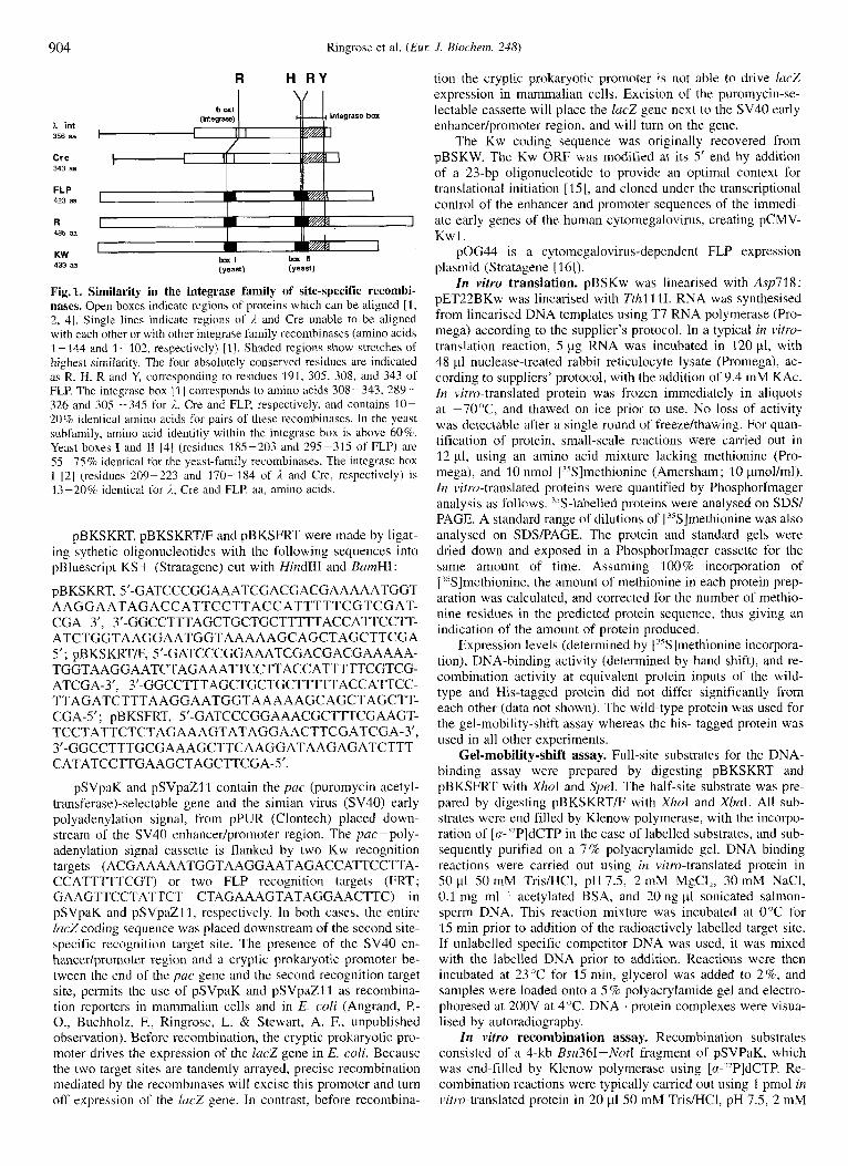
Fig. 1. Similarity in the integrase family of site-specific recombi- nases. Open boxes indicate regions of proteins which can he aligned [ l , 2, 41. Single lines indicate regions of 2 and Cre unable to he aligned with each other or with other integrase family recombinases (amino acids 1-144 and 1-102, respectively) [I]. Shaded regions show stretches of highest similarity. The four absolutely conserved residues are indicated as R, H, R and Y, corresponding to residues 191, 305, 308, and 343 of FLP. The integrase box [ I ] corresponds to amino acids 308-343, 289- 326 and 305 -345 for i,, Cre and FLP, respectively, and contains 10- 20% identical amino acids for pairs of these recomhinases. In the yeast subfamily, amino acid identitiy within the integrase box is above 60%. Yeast boxes I and 11 [4] (residues 185-203 and 295-315 of FLP) are 55 -75 % identical for the yeast-family recombinases. The integrase box I [2] (residues 209-223 and 170-184 of i and Cre, respcctively) is 13-20% identical for A, Cre and FLP. aa. amino acids.
pBKSKKT, pBKSKRT/F and pBKSFRT were made by ligat- ing sythetic oligonucleotides with the following sequences into pBluescript KS+ (Stratagene) cut with Hind111 and BamHI:
pBKSKRT, S'-GATCCCGGAAATCGACGACGAAAAATGGT AAGGAATAGACCATTCCTTACCATTTTTCGTCGAT- CGA 3', 3'GGCCTTTAGCTGCTGCTTTTTACCATTCCTT- ATCTGGTAAGGAATGGTAAAAAGCAGCTAGCTTCGA 5'; pBKSKRTIF, 5'-GATCCCGGAAATCGACGACGAAAAA- TGGTAAGGAATCTAGAAATTCCTTACCATTTTTCGTCG- ATCGA-3', 3'-GGCCTTTAGCTGCTGCTTTTTACCATTCC- TTAGATCTTTAAGGAATGGTAAAAAGCAGCTAGCTT- CGA-5'; pBKSFKT, S'-GATCCCGGAAACGCTTTCGAAGT- TCCTATTCTCTAGAA AGTATAGGAACTTCGATCGA-3',
CATATCCTTGAAGCTAGCTTCGA-5'. 3'-GGCCTTTGCGA AAGCTTC AAGGATAAG AGATCTTT-
pSVpaK and pSVpaZl 1 contain the pac (puromycin acetyl- transferase)-selectable gene and the simian virus (SV40) early polyadenylation signal, from pPUR (Clontech) placed down- stream of the SV40 enhancer/promoter region. The pac-poly- adenylation signal cassette is flanked by two Kw recognition targets (ACGAAAAATGGTAAGGAATAGACCATTCCTTA- CCATTTTTCGT) or two FLP recognition targets (FRT; GAAGTTCCTATTCT CTAGAAAGTATAGGAACTTC) in pSVpaK and pSVpaZl1, respectively. In both cases, the entire laczcoding sequence was placed downstream of the second site- specific recognition target site. The presence of the SV40 en- hancerlpromoter region and a cryptic prokaryotic promoter be- tween the end of the pac gene and the second recognition target site, permits the use of pSVpaK and pSVpaZll as recombina- tion reporters in mammalian cells and in E. coli (Angrand, P.- O., Buchholz, F., Ringrose, L. & Stewart, A. F., unpublished observation). Before recombination, the cryptic prokaryotic pro- moter drives the expression of the lac2 gene in E. coli. Because the two target sites are tandemly arrayed, precise recombination mediated by the recombinases will excise this promoter and turn off expression of the lucZ gene. In contrast, before recombina-
tion the cryptic prokaryotic promoter is not able to drive lacZ expression in mammalian cells. Excision of the puromycin-se- lectable cassette will place the lac2 gene next to the SV40 early enhancer/promoter region, and will turn on the gene.
The Kw coding sequence was originally recovered from pBSKW. The Kw ORF was modified at its Sf end by addition of a 23-bp oligonucleotide to provide an optimal context for translational initiation [IS], and cloned under the transcriptional control of the enhancer and promoter sequences of the immedi- ate early genes of the human cytomegalovirus, creating pCMV- Kwl.
pOG44 is a cytomegalovirus-dependent FLP expression plasniid (Stratagene [16]).
In vitro translation. pBSKw was linearised with Asp71 8; pET22BKw was linearised with Tthll 11. RNA was synthesised from linearised DNA templates using T7 RNA polymerase (Pro- mega) according to the supplier's protocol. In a typical in vitro- translation reaction, 5 pg RNA was incubated in 120 pl, with 48 pl nuclease-treated rabbit reticulocyte lysate (Promega), ac- cording to suppliers' protocol, with the addition of 9.4 mM KAc. In vitro-translated protein was frozen immediately in aliquots at -7O"C, and thawed on ice prior to use. No loss of activity was detectable after a single round of freezehhawing. For quan- tification of protein, small-scale reactions were carried out in 12 yl, using an amino acid mixture lacking methionine (Pro- mega), and 10 nmol ["Slmethionine (Amersham; 10 ymol/ml). I n vitro-translated proteins were quantified by PhosphorImager analysis as follows. 35S-labelled proteins were analysed on SDS/ PAGE. A standard range of dilutions of ["S]methionine was also analysed on SDS/PAGE. The protein and standard gels were dried down and exposed in a PhosphorImager cassette for the same amount of time. Assuming 100% incorporation of ["Slmethionine. the amount of methionine in each protein prep- aration was calculated, and corrected for the number of methio- nine residues in the predicted protein sequence, thus giving an indication of the amount of protein produced.
Expression levels (determined by ['5S]methionine incorpora- tion), DNA-binding activity (determined by band shift), and re- combination activity at equivalent protein inputs of the wild- type and His-tagged protein did not differ significantly from each other (data not shown). The wild-type protein was used for the gel-mobility-shift assay whereas the his- tagged protein was used in all other experiments.
Gel-mobility-shift assay. Full-site substrates for the DNA- binding assay were prepared by digesting pBKSKRT and pBKSFRT with XhoI and SpeI. The half-site substrate was pre- pared by digesting pBKSKRT/F with XlzoI and XbaI. All sub- strates were end filled by Klenow polymerase, with the incorpo- ration of [a-'2P]dCTP in the case of labelled substrates, and sub- sequently purified on a 7% polyacrylamide gel. DNA binding reactions were carried out using in vitro-translated protein in SO pl 50mM Tris/HCI, pH7.5, 2mM MgCI,, 30mM NaCI, 0.1 mg ml ' acetylated BSA, and 20 ng p1 sonicated salmon- sperm DNA. This reaction mixture was incubated at 0°C for 15 inin prior to addition of the radioactively labelled target site. If unlabelled specific competitor DNA was used, it was mixed with the labelled DNA prior to addition. Reactions were then incubated at 23°C for 15 min, glycerol was added to 2%, and samples were loaded onto a 5 % polyacrylamide gel and electro- phoresed at 200V at 4°C. DNA . protein complexes were visua- lised by autoradiography.
In vitro recombination assay. Recombination substrates consisted of a 4-kb Bsu36I-Not1 fragment of pSVPaK, which was end-filled by Klenow polymerase using [nPP]dCTP. Re- combination reactions were typically carried out using 1 pmol in vitro-translated protein in 20 yl SO mM Tris/HCl, pH 7.5, 2 mM
Ringrose et al. (ELK J . Biochem. 248) 905
MgCI,, 30 mM NaCl and 0.1 mg/ml-’ acetylated BSA. Reac- tions were incubated at 30°C for 60 min unless otherwise stated. Reactions were terminated by addition of SDS to 0.05 %, EDTA to 10 mM and protease K to 0.5 mg/ml followed by incubation at 37°C for 30 min. The samples were phenoUchloroforrn ex- tracted, and the aqueous phase was loaded onto a 0.6% agarose gel. Recombination products were visualised by autoradiography and quantified by Phosphorhager analysis. For time-course ex- periments at different temperatures, reactions were carried out in 100 pl. Reaction mixtures were incubated at various temper- atures for 2 min, and recombination reactions were started by the addition of protein. 10 pl aliquots were taken at various times, and reactions were terminated and analysed as described above.
Strand-exchange assay. The strand-exchange assay is based on intermolecular recombination between the unlabelled circular substrate pBKSKRT, and a linear oligonucleotide substrate car- rying a single inverted repeat Kw target site, which is labelled at selected positions. The synthesis of oligonucleotide substrates labelled at selected positions was carried out as follows. Two pairs of synthetic oligonucleotides were made, containing the following sequences : top-strand substrates, 5’-CGACGA AAA- ATGGTAAGGA ( 5 1 bases) 3’GCTGCTTTTTACCATTCCTT- ATCTGGTAAGGAATGGTAAAAAGCAGC-5’ ( 104 bases) ; and bottom-strand substrates, 5’-CGACGAAAAATGGTAA- GGA (44 bases) 3’-GCTGCTTTTTACCATTCCTTACCAGAT- AAGGAATGGTAAAAAGCAGC-5’ ( 1 04 bases).
For the labelling reaction, 5 pmol of each oligonucleotide pair was mixed with 6.67 pmol of the desired [a-”P]dNTP (Amersham) in PCR buffer (Cetus). If the labelled base to be incorporated was an adenine, then no other dNTP was added to this reaction. If the labelled base to be incorporated was, for instance, a thymine, then unlabelled dATP was also provided. As a control, a reaction was set up with [u-”P]dCTP, and no other dNTP. A full list of labelled substrates is given in Fig. 6b. The labelling reaction was carried out using Amplitaq (Cetus) DNA polymerase, in a single PCR cycle with 1 min denaturation at 94”C, 3 min annealing at 62°C for the top-strand substrates and 3 niin at 55 “C for the bottom-strand substrates, followed by 210 s elongation at 72°C. The amount of total label incorporated was verified by TLC (20-50% for all legitimate nucleotides; less than 1 % in the dCTP control). Unincorporated dNTPs were removed by precipitation. The DNA was suspended in PCR buffer, with 0.4 mM each of all four unlabelled dNTPs, and the remainder of the second strand was synthesised using Amplitaq polymerase in a single PCR cycle, essentially the same as the first reaction except that an elongation step of 5 min was used. The labelled, double-stranded oligonucleotides thus synthesised were gel purified and used as substrates for recombination.
Typically, intermolecular-recombination assays were carried out in 100 pl, in the standard in vitro recombination buffer. 75 fmol of labelled oligonucleotide target site and 75 fmol (150 ng) pBKSKRT were incubated with 10 pniol of irz vitro- translated Kw protein for 60 min at 30°C. Reactions were stopped as described above. After phenol/chloroform extraction the DNA was ethanol precipitated, and suspended in Scd-diges- tion buffer. Half of each sample was retained for analysis of recombination products, whilst the other half was digested with Scal. Recombination products and digestion products were ana- lysed on a 0.8% agarose gel and were visualised by autoradiog- raphy.
Kw-mediated recombination in mammalian cells. 293 cells (ATCC CRL-1573) were grown in Dulbecco’s modified Eagle’s medium supplemented with 10% fetal calf serum, 2 mM L-glutamine, 100 U/ml penicillin and 100 mg/ml streptomycin, at 37°C in a humidity-saturated 5% CO, atmosphere.
5x10’ cells were plated into 6-cm dishes, grown for 24 h and transfected by the calcium phosphate procedure [ 171. Cells were incubated overnight after addition to the medium of 500 pl precipitate containing 6 yg DNA (3 pg target vector, pSVpaKl or pSVpaZl1, and 3 pg of the effector plasmid pCMV-Kwl or pOG44). The medium plus precipitate were removed and re- placed with fresh medium. 24 h later, cells were fixed in 137 mM NaCI, 2.7 mM KCI, 4.3 mM Na,HPO,, 1.4 mM KH,PO,, pH 7.4 (NaCVP,) containing 1% formaldehyde (Merck) and 0.2% glutaraldehyde (Sigma), and lucZ expression detected in situ by staining with 5-bromo-4-chloro-3-indoy1-~-~- galactopyranoside (X-Gal). Cells were incubated overnight with NaCI/P, containing 5 mM potassium ferricyanide, 5 mM potas- sium ferrocynide, 2 mM MgCl,, 0.4 mg/ml X-Gal.
General methods. All restriction enzymes were purchased from from New England Biolabs. All plasmids were grown in E. coli strains XL-1 blue or HB101. Plasmid transformations, plasmid DNA isolations, DNA ligations, restriction digests, and gel electrophoresis were carried out according to published pro- cedures [ 181. All constructs were verified by sequence analysis. Sequence determination was carried out using the Sequenase kit from USB.
RESULTS
Kw binds specifically to its target site. The 2 p-like plasmid, pKWl from K. waltii contains four ORF A, B, C and D. Chen et al. [9] suggested that the A gene encodes a recombinase, on the basis of similarity of its deduced amino acid sequence with that of the FLP gene of 2 p and its equivalent in other yeast plasmids.
Like other 2 p plasmids, pKWl has two inverted repeat se- quences of about 300 bp. Within this region, the putative recom- bination target sites have been identified by Chen et al. [9] on the basis of their sequence structure. Each putative target site contains three 18-bp perfect repeat elements, two of which are inverted, separated by a non-palindromic stretch of 7-bp. The third repeat element is in direct orientation to the second, and is separated from it by 26 bp. This is reminiscent of the situation in the S. cerevisiae 2 p plasmid, in which the FLP recombination target region also contains an inverted repeat with a single adja- cent direct repeat element [19]. With FLP. however, the direct repeat is separated from the inverted repeat by 2 bp. The in- verted repeat of two 13-bp elements is sufficient for efficient FLP recombination in v i m [ I l l and in vivo [lo].
To determine whether the putative Kw recombinase would bind its putative DNA target site, we tested in vitro-translated Kw protein in a gel-mobility-shift assay.
In vitro translation of Kw protein from pBSKw or pET22BKw gave a protein of approximately 45 kDa determined by SDS/PAGE, which corresponds to the molecular mass pre- dicted from the amino acid sequence (data not shown).
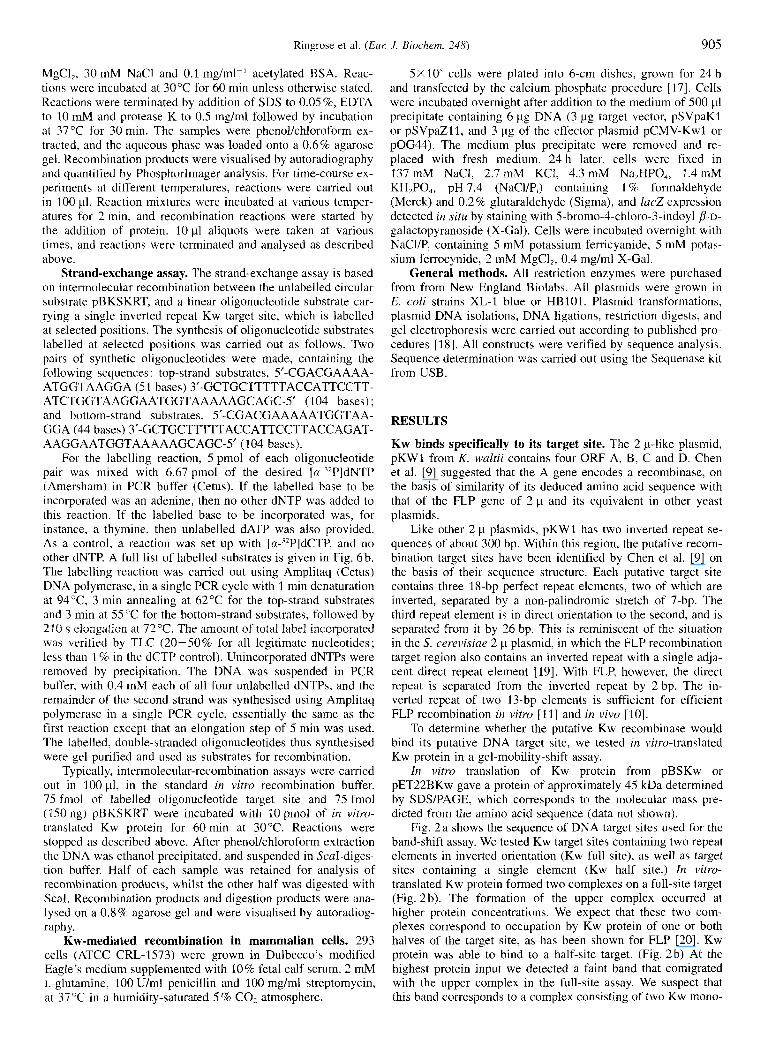
Fig. 2 a shows the sequence of DNA target sites used for the band-shift assay. We tested Kw target sites containing two repeat elements in inverted orientation (Kw full site), as well as target sites containing a single element (Kw half site.) In vitro- translated Kw protein formed two complexes on a full-site target (Fig. 2b). The formation of the upper complex occurred at higher protein concentrations. We expect that these two com- plexes correspond to occupation by Kw protein of one or both halves of the target site, as has been shown for FLP [20]. Kw protein was able to bind to a half-site target. (Fig. 2b) At the highest protein input we detected a faint band that comigrated with the upper complex in the full-site assay. We suspect that this band corresponds to a complex consisting of two Kw mono-
906
A C G A A A A A T G G T A A G G A A T A G A C C A
a
T T C C T T A C C A
Kw half site: 70 bp
5 A C G A A A A A T G G T A A G G A A T C T A G
FRT site : 160 bp 3
5 @ A A G T T C C T A T T C ~ T C T A G A A A ~ G T A T A G G A A C T T C ~
C 1 2 3 4 5 6 7 8 9 1 0 1 2 3 4 5 6 7
b protein
protein + Kw site
substrate competitor
4
free probe +
free probe 4
Fig.2. Binding of in vitro-translated Kw recombinase to its DNA target site. (a) Sequence of DNA target site oligonucleotides used for gel- mobility-shift assay. Preparation of "P-labelled oligonucleotides is described in Materials and Methods. Open boxes indicate repeat elements of the Kw target and FRT sites. (b) Gel-mobility-shift assay. 1n virro-translated-Kw-protein binding to target sites carrying one or two repeat elements. Lanes 1-4, 0.1 nM full-site oligonuclcotide was incubated in 20 p1 with 2, 4, 8 or 16 pl Kw protein translated in v i m from pBSKW (0.92 pmoll pl); lanes 5-8, 0.1 nM half-site oligonucleotide was incubated with 2, 4, 8 or 16 p1 Kw protein; lane 9, 8 pl Kw protein incubated with 0.1 nM FRT site oligonucleotide; lane 10, 8 pI mock translation with 0.1 nM KW hall site. (c) Competition with unlabelled oligonucleotide target sites. 4 p1 Kw protein was bound to 0.1 nM labelled oligonucleotide lull-site in the presence of excess unlabelled Kw full-site oligonucleotide. Lane 1, mock translation; lane 2, Kw, no competitor; lanes 3-6, 5-, lo-, 20- and 50-fold molar excess of competitor DNA; lane 7, 50-fold molar excess of unlabelled FRT oligonucleotide.
mers, each bound to a single DNA site. Similar complexes of FLP monomers bound to half-site targets have been observed
Fig. 2 b demonstrates that Kw protein did not bind the FRT site in our assay. To confirm the specificity of binding we carried out a competition experiment (Fig. 2c). This experiment demon- strated that an excess of unlabelled Kw full site specifically competed for Kw protein binding. With increasing concentration of competitor DNA, from 5-fold to 50-fold excess, the upper complex, then the lower complex, was lost. Kw binding to its site was not competed by SO-fold excess FRT site. In a similar experiment with FLP, we demonstrated that FLP did not bind the Kw target site, and binding was competed only by the FRT (data not shown).
These results demonstrated that Kw protein specifically binds its putative recombination target site, and suggested that the stoichiometry of its binding is identical to that shown for FLP.
[21, 221.
Kw protein recombines its target site in vitro. To assay the Kw protein for recombinase activity, we designed a recombination substrate, pSVpaK, which carries two Kw full sites arranged with their spacers in direct orientation. Kw-mediated recombina- tion of this substrate should result in excision of the 1.1 kb be-
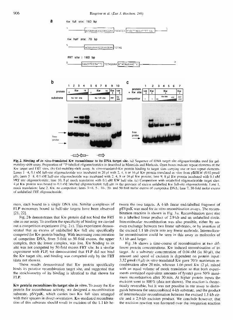
tween the two targets. A 4-kb linear end-labelled fragment of pSVpaK was used for in vitrn recombination assays. The recom- bination reaction is shown in Fig. 3a. Recombination gave rise to a labelled linear product of 2.9 kb and an unlabelled circle. Intermolecular recornbination was also possible, either by un- even exchange between two linear substrates, or by insertion of the excised 1 . l -kb circle into any linear molecule. Intermolecu- lar recombination could be seen in this assay as molecules of 5.1 kb and larger.
Fig. 3 b shows a time-course of recombination at two dif- ferent protein concentrations. Kw induced recombination of its target. At a substrate concentration of 0.04 nM (in 80 PI), the amount and speed of excision is dependent on protein input: 3.32 pmol (4 pl) in vim-translated Kw gave 70% maximum re- combination after 20 min, whereas 1.66 pmol Kw (2 pl, mixed with an equal volume of mock translation so that both experi- ments contained equivalent amounts of lysate) gave 50% maxi- mum recombination after 30 min. At higher protein inputs the excision went to 100% (data not shown). The reaction is theore- tically reversible, but it was not possible in our assay to distin- guish between the unrecombined 4-kb substrate, and the product of intermolecular recombination between the excised 1.1 -kb cir- cle and a 2.9-kb excision product. We conclude however, that the excision reaction was favoured over the integration reaction
Ringrose et a]. (ELK J. Biochenz. 248) 907
a *-* 4 kb substrate
*-* 2.9 kb +
1 . 1 kb circle 0 2' 5 ' 10' 20' 30' 60' 120'
b
4 kb+ mum- 2Pl
2 9kb-) - " .
C
4 kb+
2.9kb*
8 0 10 20 30 40 50 60
time (mlns)
1 2 3 4 5 6 7 8 9 1 0 1 1 1 2 BXClSlO" - mlerrnole~~lao ; .- ;fyJ recornblnauon
.- = 20
2 0
8 10 30 50 70 90 110
[NaCI] (mM)
d
0 10 20 30 4 0 5 0 60
time (mins)
+ + 4
t
-A-
+
2 3%
30°C
35%
3 7 2
39%
42%
Fig. 3. In vitro recombination assay. (a) Recombination substrates and products. Preparation of end-filled labelled recombination substrates is described in Materials and Methods. See text for description of assay. (b) Recombination time course. 2 pl (plus 2 pl mock translation) or 4 pI Kw protein translated in v i m from pET22BKW (0.83 pmol/pI) was incubated in 80 p1 with 0.04 nM substrate DNA. 10.~1 aliquots were taken at 2, 5 , 10, 20, 30, 60 and 120 min. (c) Effect of NaCl on recombination. 1 p1 Kw protein was incubated in 20 p1 with 0.04 nM substrate DNA for 1 h at different NaCl concentrations, with 2 mM MgCI,. Lanes 1-11, 10, 20, 30,40, 50, 60, 70, 80, 90, 100, and 110 mM NaCI; lane 12, mock translation in 30 mM NaCI. (d) Effect of MgC1, Experimental conditions were as for (c), but the MgCI, concentration was varied. NaCl concentration was 30 mM. Lanes 1-6, 0, 1, 2, 3, 4 and 5 mM MgCl,, lane 7, mock translation in 2 mM MgCI,. (e) Effect of temperature on Kw recombinase activity. Recombination reactions were carried out at 23, 30, 35, 37, 39 and 42°C. Reaction mixtures were warmed for 2 min prior to the addition of protein. 6 pl Kw protein translated in v i m from pET22BKW (0.83 pmol/pl) was added to 0.04 nM substrate DNA in 60 pl, and 10.~1 aliquots were taken at 2, 5 , 10, 20, 30 and 60 min.
under our assay conditions, firstly because it was possible to observe 100% excision, and secondly because bands of 5.1 kb and larger, corresponding to intermolecular recombination prod- ucts, although visible, were much fainter than the 2.9-kb exci- sion-product band. These bands are more prevalent at higher substrate concentrations (data not shown).
Subsequent experiments were carried out to determine the optimum buffer conditions for the in vitro recombination reac- tion. The reaction occurs within a pH range from 6 to 8, with optimum at pH 7.5. The recombination reaction is inhibited by Taps (data not shown).
Fig. 3c shows the effect of NaCl concentration on Kw activ- ity. The optimum NaCl concentration for excision was 30 mM, and the reaction was strongly inhibited at concentrations above 50 mM. The amount of intermolecular recombination observable was highest at 10 mM NaC1, and was reduced rapidly as the NaCl concentration increased, being undetectable above 40 mM.
Fig. 3 d shows the effect of MgCI, concentration on excisive recombination. The activity was sensitive to MgC12, having an optimum at 2 mM, and being completely inhibited at 6 mM.
The effect of temperature on the in vitro recombination reac- tion was assessed (Fig. 3e). We tested recombination at 23, 30,
908 Ringrose et al. ( E m J. Biochem. 248)
a
b
pSVpaK 7.2kb
on
Xmni
unrecombined : blue colony
1 2
Xmnl
recombined : white colony
+
c A G C T
0 1.1 kb
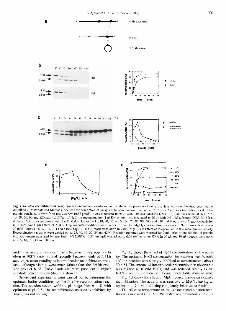
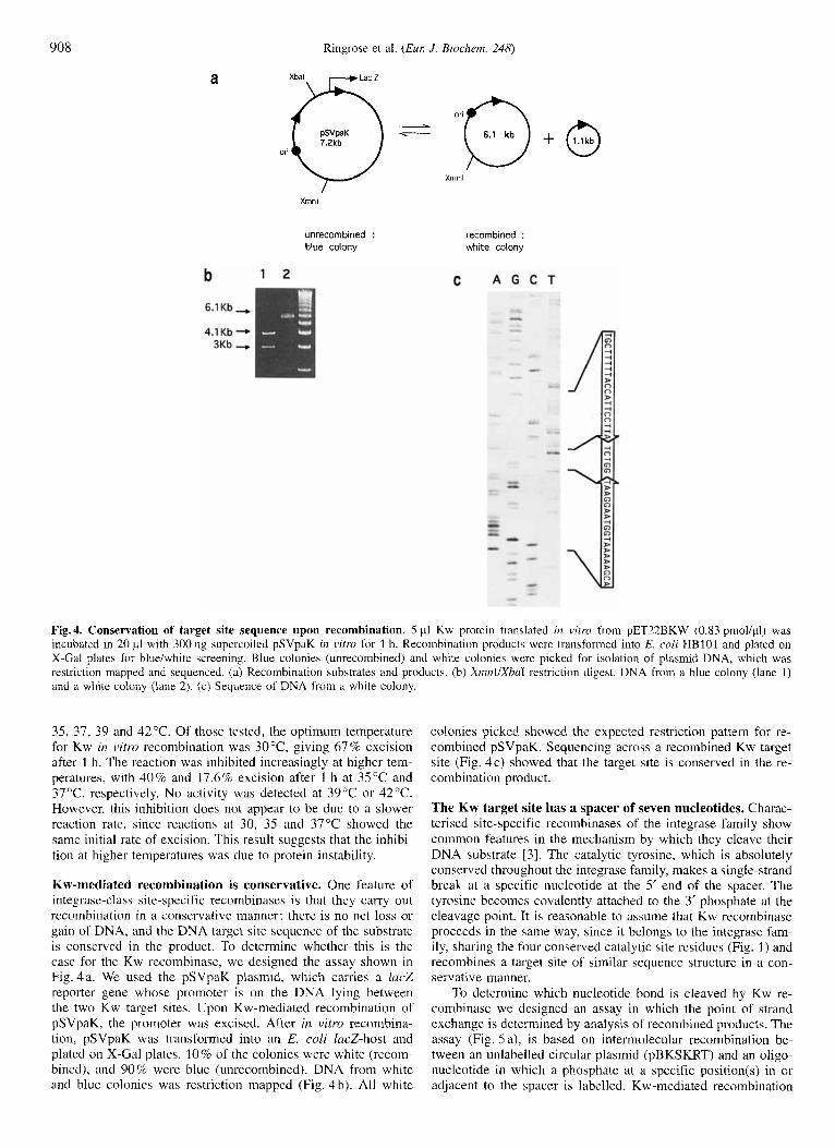
Fig.4. Conservation of target site sequence upon recombination. 5 p1 Kw protein translated in vitro from pET22BKW (0.83 prnol/pl) was incubated in 20 p1 with 300 ng supercoiled pSVpaK in v i m for 1 h. Recombination products were transformed into E. coli HBlOl and plated on X-Gal plates for blue/white screening. Blue colonies (unrecombined) and white colonies were picked for isolation of plasmid DNA, which was restriction mapped and sequenced. (a) Recombination substrates and products. (b) XnznI/XbnI restriction digest. DNA from a blue colony (lane 1) and a white colony (lane 2). (c) Sequence of DNA from a white colony
35, 37, 39 and 42°C. Of those tested, the optimum temperature for Kw in vitro recombination was 30°C, giving 67% excision after 1 h. The reaction was inhibited increasingly at higher tem- peratures, with 4 0 % and 17.6% excision after 1 h at 35°C and 37"C, respectively. No activity was detected at 39°C or 42°C. However, this inhibition does not appear to be due to a slower reaction rate, since reactions at 30, 35 and 37°C showed the same initial rate of excision. This result suggests that the inhibi- tion at higher temperatures was due to protein instability.
Kw-mediated recombination is conservative. One feature of integrase-class site-specific recombinases is that they carry out recombination in a conservative manner: there is no net loss or gain of DNA, and the DNA target site sequence of the substrate is conserved in the product. To determine whether this is the case for the Kw recombinase, we designed the assay shown in Fig. 4a. We used the pSVpaK plasmid, which carries a lac2 reporter gene whose promoter is on the DNA lying between the two Kw target sites. Upon Kw-mediated recombination of pSVpaK, the promoter was excised. After irz vitro recombina- tion, pSVpaK was transformed into an E. coli lacZ-host and plated on X-Gal plates. 10% of the colonies were white (recom- bined), and 90% were blue (unrecombined). DNA from white and blue colonies was restriction mapped (Fig. 4 b). All white
colonies picked showed the expected restriction pattern for re- combined pSVpaK. Sequencing across a recombined Kw target site (Fig. 4c) showed that the target site is conserved in the re- combination product.
The Kw target site has a spacer of seven nucleotides. Charac- terised site-specific recombinases of the integrase family show common features in the mechanism by which they cleave their DNA substrate 131. The catalytic tyrosine, which is absolutely conserved throughout the integrase family, makes a single-strand break at a specific nucleotide at the 5' end of the spacer. The tyrosine becomes covalently attached to the 3' phosphate at the cleavage point. It is reasonable to assume that Kw recombinase proceeds in the same way, since it belongs to the integrase fam- ily, sharing the four conserved catalytic site residues (Fig. 1) and recombines a target site of similar sequence structure in a con- servative manner.
To determine which nucleotide bond is cleaved by Kw re- combinase we designed an assay in which the point of strand exchange is determined by analysis of recombined products. The assay (Fig. 5 a), is based on intermolecular recombination be- tween an unlabelled circular plasmid (pBKSKRT) and an oligo- nucleotide in which a phosphate at a specific position(s) in or adjacent to the spacer is labelled. Kw-mediated recombination

Ringrose et al. ( E M J . Biochem. 248)
a oligo: 104 bp 1.9 Kb 1.2Kb
909
b Top strand substrates
1 , ~ l G ~ ~ ,
Label is 5' of cleavage
Label is 3' of cleavage
d Scat digest
4 )I 1 b A C C A 7 1 G C l G C l I l l l A C C A l l (11 A l C l G C l AA A A l C C l A A A A A G C A C C
C Bottom strand substrates
A B 5 ( C C * C G I * ~ * * ~ C G T * * O O * I \ I T I \ O P C C A , ~ I
6 I:F:::::::Zw:;:;gTmI G C l G C f T l T l A C C A l T C C l T AWTGGT AAGGAATGGTAAAAAGCAGC
7 ~ l ~ ~ ~ ~ . ~ . ~ ~ l Fig. 5. Assay to determine the point of strand cleavage. (a) Recombination reaction and restriction analysis of recombination products. Arrows indicate inverted-repeat elements of the Kw target site. Kw-mediated intermolecular recombination between a labelled oligonucleotide substrate and an unlabelled 3-kb plasmid carrying a single Kw target site (pBKSKRT; see Materials and Methods) gave a single linear molecule of 3.1 kb. Restriction digest of linear recombination products with ScaI gives fragments of 1.9 kb and 1.2 kb (see text for full description of assay). (b, c) Oligonucleotide substrates. Preparation of oligonucleotide recombination substrates in which selected phosphates are labelled, is described in Materi- als and Methods. Open boxes indicate the repeat elements of the Kw target site; labelled nucleotides used in synthesis are indicated with bold type; radioactive phosphates are indicated with asterisks. d), e) ScaI digest of recombination products. The assay was carried out using 25 p1 Kw protein translated in vifro from pET22BKW (0.83 pmol/pl) and incubated with 0.75 nM pBKSKRT and 0.75 nM of each of substrates 1-7, in 100 pl. Recombination products were digested with ScuI. Lanes 1-7, Scar digests of substrates 1-7.
between the oligonucleotide and the plasmid generates a linear molecule of 3.1 kb. If the labelled phosphate is on the 5'side of the strand cleavage point, or becomes covalently attached to the catalytic tyrosine, then it will partition to the left side of the linear 3.1-kb molecule (Fig. 5a). If the labelled phosphate is on the 3'side of the cleavage point, then it will partition to the right side. Restriction digestion of linear recombination products with ScaI reveals the position of the labelled phosphate, and thus the point at which the oligonucleotide substrate was cleaved. Bands of 1.9 kb and 1.2 kb were generated (Fig. 5 a).
We made the oligonucleotide substrates shown in Fig. 5 b and c by labelling various positions in or adjacent to the Kw target site spacer on each strand by primer extension using [a- "PIdNTPs as described in Materials and Methods. Upon strand cleavage, when the catalytic tyrosine becomes covalently at- tached to a labelled 3' phosphate, this phosphate and the base which originally carried it, will be separated.
Kw protein was incubated in separate reactions with pBKSKRT and an equimolar quantity of each of the seven la- belled oligonucleotide substrates. In each case, intermolecular recombination gave a labelled linear product of 3.1 kb (data not
shown). The recombination products were digested with ScaI (Fig. 5 d and e). ScaI digestion of substrate 1 yielded a single labelled band of 1.9 kb (Fig. 5b), which was labelled at a single position, namely the phosphate on the 5' side of the first T in the spacer. This result demonstrates that this phosphate was situ- ated on the 5' side of the point of strand exchange, as it remained attached to the left arm of the target site (Fig. 5a). Substrate 2 (Fig. 5 d) yielded both bands of 1.9 kb and 1.2 kb. In this sub- strate, three consecutive nucleotides, ATA were labelled. There- fore the three labelled phosphates must distribute either 3 : 0, 2: 1, 1 :2 or 0:3 across the cleavage site. The relative intensities of the 1.9-kb and 1.2-kb bands indicated approximately 2: 1 dis- tribution. Since substrate no. 1 demonstrates that the phosphate from the T was on the 5' side of the strand-exchange point, then the first A, which is itself on the 5' side of this T, must also be on the 5' side. Therefore we expect that the 1.9-kb band contains two labelled phosphates from the first A and the T, and that the 1.2-kb band contains the phosphate from the second A. Thus we reason, that the point of cleavage on the upper strand is between the last A of the repeat element and the first T of the spacer (Fig. 5 b).
910
0
FLP (pOG44)
Kw (PCMV-KWI)
Ringrose et al. (EUK J. Biochem. 248)
pSVpa%l1 pSVpdK
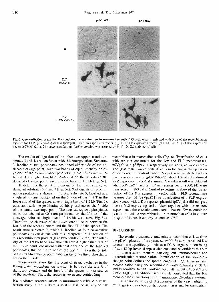
Fig. 6. Cotransfection assay for Kw-mediated recombination in mammalian cells. 293 cells were transfected with 3 pg of the recombination reporter for FLP (pSVpaZ11) or Kw (pSVpaK), with no expression vector (O), 3 pg FLP expression vector (pOG44). or 3 pig of Kw expression vector (pCMW-Kwl). 24 h after transfection, lric.7 expression was assayed by in sitrr X-Gal staining of cells.
The results of digestion of the other two upper-strand sub- strates, 3 and 4, are consistent with this interpretation. Substrate 3, labelled at two phosphates positioned either side of the de- duced cleavage point, gave two bands of equal intensity on di- gestion of the recombination product (Fig. 5d). Substrate 4, la- belled at a single phosphate positioned on the 3’ side of the deduced cleavage point, gave a single band of 1.2 kb (Fig. 5 c).
To determine the point of cleavage on the lower strand, we designed substrates 5 , 6 and 7 (Fig. 5 c). ScaI digests of recombi- nation products are shown in Fig. 5e. Substrate 5 , labelled at a single phosphate, positioned on the 5’ side of the first T in the lower strand of the spacer, gave a single band of 1.2 kb (Fig. 5 ) , consistent with the positioning of this phosphate on the 5‘ side of the strand-exchange point. The two subsequent phosphates (substrate labelled at GG) are positioned on the 3’ side of the cleavage point (a single band of 1.9 kb was seen, Fig. 5e). Therefore the cleavage of the lower strand occurs between the last A of the repeat element and the first ‘T’ of the spacer. The result from substrate 7, which is labelled at four consecutive phosphates, is consistent with this interpretation. Digestion of this recombination product gave two bands (Fig. 5 e). The inten- sity of the 1.9-kb band was about threefold higher than that of the 1.2-kb band, consistent with that only one of the labelled phosphates, that on the 5’ side of the first ‘T’, is on the 5’ side of the strand-exchange point, whereas the other three phosphates are on the 3’ side.
These results show that the point of strand exchange in the Kw-mediated recombination reaction is between the last A of the repeat element and the first T of the spacer in both strands of the substrate. Thus, the spacer is seven nucleotides long.
Kw mediates recombination in mammalian cells. A cotrans- fection assay in 293 cells was used to test the activity of Kw
recombinase in mammalian cells (Fig. 6). Transfection of cells with reporter constructs for the Kw and FLP recombinases, pSVpaK and pSVpaZl1 respectively did not give lacZ expres- sion (less than 1 lacZ’ cell/lOh cells in the transient-expression experiments). In contrast, when pSVpaK was transfected with a Kw expression vector (pCMV-Kwl), about 1 % of cells showed lacZ expression by X-Gal staining. A similar result was obtained when pSVpaZl1 and a FLP expression vector (pOG44) were transfected in 293 cells. Control experiments showed that trans- fection of the Kw expression vector with a FLP recombinase reporter plasmid (pSVpaZ11) or transfection of a FLP expres- sion vector with a Kw reporter plasmid (pSVpaK) did not give rise to lacZ-expressing cells. Taken together with our in vitro experiments, these results demonstrate that the Kw recombinase is able to mediate recombination in mammalian cells in culture in spite of its weak activity in vitro at 37OC.
DISCUSSION
The results presented characterise a recombinase, Kw, from the pKWl plasmid of the yeast K. waltii. In vitro-translated Kw recoinbinace specifically binds to a DNA target site consisting of two 18-bp inverted-repeat elements, and recombines this site i n a conservative manner, carrying out intramolecular and intermolecular recombination. Identification of the strand-ex- change point defines the spacer length as 7 bp. In an in vitm recombination assay, the recombinase works optimally at 30 “C, and is sensitive to salt, working optimally at 30 mM NaCl and 2 mM MgCI,. In addition, we have demonstrated that the Kw recombinase is functional in a mammalian-cell-culture system.
The characterisation of this member of the yeast subfamily of integrase-class site-specific recombinases enables comparison
Ringrose et al. ( E L K J . Biochem. 248) 91 1
with other members of the family, and reveals the extent of simi- larity among them. Two other members of the yeast family, FLP from S. cerevisiue and R from 2. rouxii [71 have been identified as recornbinases, and FLP has been extensively characterised [S]. Other members of the integrase family that have been well characterised are the integrase and the Cre recombinase from E. coli phage P I [3] .
The Kw DNA target site identified confirms the putative target site identified by Chen et al. [9]. The inverted repeat of two 18-bp elements is sufficient as a target for efficient recombi- nation in vitro and in vivo. That the third repeat element found in pKWl is not required, raises the question of its physiological role. Whether it hdS any effect on the efficiency of Kw recombi- nation remains to be seen. It has also been shown for FLP that although all three repeat elements are bound by FLP [23], the third repeat element is not required for recombination. It has no effect on recombination rates in vitro 1111. Its presence can, however, apparently enhance the rate of intermolecular recombi- nation in E. coli [lo]. In the 2 p-like circle from Z. rouxii (which encodes the R recombinase) the recombination target contains an inverted repeat adjacent to four direct repeats. All six repeats are shown to be bound by R protein by DNAse-I footprinting [7]. The inverted repeat element is sufficient for recombination [24], but the effect of the other four repeat elements has not been evaluated.
The Kw target site repeat element of 18 bp is longer than that of the FRT (13 bp) and the R target site (12 bp). It is un- likely that the entire sequence is involved in specific recognition and binding by Kw. Results from methylation protection and ethylation interference suggest that FLP makes specific contacts to only the 10 bp of the target site adjacent to the spacer [25]. Mutation analysis of the FRT has shown that only point muta- tions at four positions within the 7 bp adjacent to the spacer have a negative effect on in vitro recombination [26]. Further analysis of DNA-protein interactions in the Kw system and comparison with those already identified for FLP may reveal the the basis of DNA-binding specificity in these recombinases.
The strand-cleavage point was determined by identifying the point of strand exchange. The length of the Kw target site spacer, 7 bp, falls within the 6-8 bp range identified for other integrase- family recombinases. The consistency of the spacer length in this family may reflect a requirement for a certain distance be- tween cleavage points, or a certain phasing of those cleavage points with respect to the faces of the DNA helix, imposed by the stuctural constraints within the recombination synapse. Whether Kw recombinase follows the FLP paradigm [27], in which the catalytic tyrosine of one recombinase subunit cleaves a DNA strand that is bound by another subunit, remains to be seen. The R recombinase appears to follow this model [28], whereas the 3. integrase cleaves the half site to which it is bound
The high-salt sensitivity of Kw recombinase is in contrast to that reported for FLP, which tolerates a broad range of salt conditions, from 0 to 3 0 0 m M NaCl, in the presence of up to 20 mM MgCl,, with an optimum at 200-300 mM NaCI in the absence of MgClz [29-311. The R recombinase, on the other hand, is also reported to be salt sensitive 1321. The temperature at which Kw shows optimum recombination in vitro is 3 0 ° C which is consistent with its origins as a yeast enzyme. The loss of activity at higher temperatures appears to be due to protein instability. This effect has also been observed for FLP recombi- nase [33].
The characterisation of a site-specific recombinase has im- plications not only for the further understanding of the family to which it belongs, but also for the field of genome-manipulation technology. The site-specific recombinases FLP and Cre have
1141.
been used extensively in a variety of organisms to engineer spe- cific DNA rearrangements at defined sites [13]. The characteri- sation of Kw recombinase widens the repertoire of tools avail- able for this kind of experiment. We have shown in a cotransfec- tion experiment in 293 cells, that the Kw recombinase can re- combine its target site in a mammalian-cell-culture system, thus demonstrating its potential for in vivo genome manipulation. In this context, the preference of Kw recombinase for temperatures below 37°C may restrict the choice of organism in which it is used. The possibility that this effect is mainly due to protein instability (Fig. 3e) suggests that it may be overcome by ex- pressing sufficient levels of the protein. A further useful detail of the Kw system is that the inverted-repeat target site, like those of FLP, R and Cre, can be read in both orientations without encountering a stop codon, a feature which is necessary if the site is to be placed in an ORE
We thank H. Fukuhara, for the gift of plasmid PKWS1, and R. Aas- land, T. Gibson and P. Mitchell, for helpful discussions. P.-0. A. is a recipient of an European Union. Training and Mobility fellowship. This work was supported in part by a grant from the Volkswagen Stifrung.
REFERENCES 1. Argos, P., Landy, A., Abremski, K., Egan, J. B., Haggard, L. E.,
Hoess, R. H., Kahn, M. L., Kalionis, B., Narayana, S. V., Pierson, L. S., Sternberg, N. & Leong, J. M. (1986) The integrase family of site-specific recombinases: regional similarities and global di- versity, EMBO J. 5, 433-440.
2. Abreniski, K. E. & Hoess, R. H. (1992) Evidence for a second con- served arginine residue in the integrase family of recombination proteins, Protein Eng. 5, 87-91.
3. Stark, W. M., Boocock, M. R. & Sherratt, D. J. (1992) Catalysis by site-specific recombinases, Trends Genet. 8, 432-439.
4. Utatsu, I., Sakamoto, S., Imura, T. & Toh, E. A. (1987) Yeast plas- mids resembling 2 micron DNA: regional similarities and diversi- ties at the molecular level, J. Bucteriol. 169, 5537-5545.
5. Sadowski, P. D. (1995) The Flp recombinase of the 2-microns plas- mid of Saccharomyes cerevisiae, Prog. Nucleic Acid Res. Mol. Biol. 51, 53-91.
6. Matsuraki, H., Araki, H. & Oshima, Y. (1988) Gene conversion associated with site-specific recombination in yeast plasmid pSR1, Mol. Cell. Biol. 8 , 955-962.
7. Araki, H., Nakanishi, N., Evans, B. R., Matsuraki, H., Jayaram, M. & Oshima, Y. (1992) Site-specific recombinase, R, encoded by yeast plasmid pSR1, 1. Mol. Biol. 225, 25-37.
8. Chen, X . J., Saliola, M., Falcone, C., Bianchi, M. M. & Fukuhara, H. (1986) Sequence organization of the circular plasmid pKDl from the yeast Kluyveronz~ces droscJphilarum, Nucleic Acids Res. 14, 4471-4481.
9. Chen, X. J., Cong, Y. S. , Wesolowski, L. M., Li, Y. Y. & Fukuhara. H. (1992) Characterization of a circular plasmid from the yeast Kluyveromyces wultii, J. Gen. Microbiol. 138, 337 -345.
10. Jayaram, M. (1985) Two-micrometer circle site-specific recombina- tion: the minimal substrate and the possible role of flanking se- quences, Proc. Nutl Acad. Sci. USA 82, 5875-5879.
11. Senecoff, J. F., Bruckner, R. C. & Cox, M. M. (1985) The FLP recombinase of the yeast 2-micron plasmid: characterization of its recombination site, Proc. Natl Acad. Sci. USA 82, 7270-7274.
12. Senecoff, J. F. & Cox, M. M. (1986) Directionality in FLP protein- promoted site-specific recombination is mediated by DNA-DNA pairing, J . Biol. Chem. 261, 7380-7386.
13. Kilby, N. J., Snaith, M. R. & Murray, J. A. (1993) Site-specific recombinases : tools for genome engineering, Trends Genet. 9,
24. Stark, W. M. & Boocock, M. R. (1995) Gatecrashers at the catalyric party, Trends Genet. 11, 123 -123.
15. Kozak, M. (1984) Compilation and analysis of sequences upstream from the translational start site in eukaryotic mRNAs, Nucleic Acids Rex 12, 857-872.
413-421.

912 Ringrose et al. (Eur: J. Biochem. 248)
16. O’Gorman, S., Fox. D. T. & Wahl, G. M. (1991) Recombinase- mediated gene activation and site-specific integration in mamma- lian cells, Science 251, 1351-1355.
17. Wigler, M., Sweet, R., Sim, G. K., Wold, B., Pellicer, A., Lacy, E., Maniatis, T., Silverstein, S. & Axel, R. (1992) Transformation of mammalian cells with genes from prokaryotes and eukaryotes, Bio-technology (N.E) 24, 444-452.
18. Sambrook, J., Fritsch, E. F. & Maniatis, T. (1989) Molecular don- ing, a laboratory manual, 2nd edn, Cold Spring Harbor Labora- tory, Cold Spring Harbor NY.
19. Broach, J. R., Guarascio, V. R. & Jayaram, M. (1982) Recombina- tion within the yeast plasmid 2mu circle is site-specific, Cell 29, 227-234.
20. Andrews, B. J., Beatty, L. G. & Sadowski, P. D. (1987) Isolation of intermediates in the binding of the FLP recombinase of the yeast plasmid 2-micron circle to its target sequence, J. Mol. Biol. 193, 345-358.
21. Qian, X. H., Inman, R. B. & Cox, M. M. (1990) Protein-based asyni- metry and protein-protein interactions i n FLP recombinase-medi- ated site-specific recombination, J. Biol. Chem. 265, 21 779- 21 788.
22. Serre, M. C. & Jayaram, M. (1992) Half-site strand transfer by step- arrest mutants of yeast site-specific recombinase Flp, J. Mol. Biol. 225, 643 - 649.
23. Andrews, B. J., Proteau, G. A., Beatty, L. G. & Sadowski, P. D. (1985) The FLP recombinase of the 2 micron circle DNA of yeast: interaction with its target sequences, Cell 40, 795-803.
24. Serre, M. C.. Evans, B. R., Araki, H., Oshima, Y. & Jayaram, M. (1992) Half-site recombinations mediated by yeast site-specific recombinases Flp and R, ,I. Mol. B id . 225, 621 -642.
25. Bruckner, R. C. & Cox, M. M. (1986) Specific contacts between the FLP protein of the yeast 2-micron plasmid and its recombination site, 1. B i d . Chem. 261. 11 798 - 11 807.
26. Senecoff, J . F., Rossmeissl, P. J. & Cox, M. M. (1988) DNA recogni- tion by the FLP recombinase of the yeast 2 mu plasmid. A muta- tional analyhis of the FLP binding site, J. Mol. Biol. 201, 405- 421.
27. Chen, J. W., Lee, J. & Jayaram, M. (1992) DNA cleavage in trans by the active site tyrosine during Flp recombination : switching protein partners before exchanging strands, Cell 69, 647 -658.
28. Yang, S. H. & Jayaram, M. (1994) Generality of the shared active site among yeast family site-specific recombinases. The R site- specific recombinase follows the Flp paradigm, J. Bid . Chem.
29. Meyer, L. L., Senecoff, J. F., Bruckner, R. C. & Cox, M. M. (1984) Site-specific genetic recombination promoted by the FLP protein of the yeast 2-micron plasmid in vitro, Cold Spring Harbor Symp. Quunt. Biol. 49, 797-804.
30. Babineau, D., Vetter, D., Andrews, B. J., Gronostajski, R. M., Pro- teau, G. A., Beatty, L. G. & Sadowski, P. D. (1985) The FLP protein of the 2-micron plasmid of yeast. Purification of the pro- tein from Escherichia coli cells expressing the cloned FLP gene, .I. Biol. Chem. 260, 12313- 12319.
31. Meyer, L. L.. Gates, C. A,, Attwood, J. M., Wood, E. A. & Cox, M. M. (1987) Purification of the FLP site-specific recombinase by affinity chromatography and re-examination of basic properties of the system, Nucleic Acids Rex 15, 6469-6488.
32. Lee, J., Serre, M. C., Yang, S. H., Whang, I., Araki, H.: Oshima, Y. & Jayaram, M. (1992) Functional analysis of box I1 mutations in yeast site-specific recombinases Flp and R. Significance of amino acid conservation within the Int family and the yeast sub- family, .I. Mol. Riol. 228, 1091 - 1103.
33. Buchholz, F., Ringrose, L., Angrand, P.-O., Rossi, F. & Stewart, A. F. (1996) Different thermostabilities of FLP and Cre recombi- nases : implications for applied site-specific recombination, Nu- cleic Acids Rex 24, 4256-4262.
269, 12789- 12796.