Environment Building - against residue burning - ATARI, Zone-1
Upload
independentCategory
view
3download
0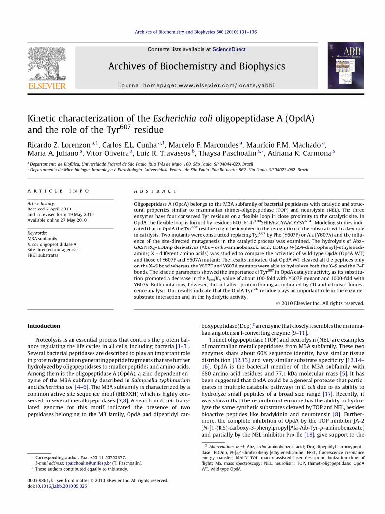
Archives of Biochemistry and Biophysics 500 (2010) 131–136
Contents lists available at ScienceDirect
Archives of Biochemistry and Biophysics
journal homepage: www.elsevier .com/ locate/yabbi
Kinetic characterization of the Escherichia coli oligopeptidase A (OpdA)and the role of the Tyr607 residue
Ricardo Z. Lorenzon a,1, Carlos E.L. Cunha a,1, Marcelo F. Marcondes a, Maurício F.M. Machado a,Maria A. Juliano a, Vitor Oliveira a, Luiz R. Travassos b, Thaysa Paschoalin a,*, Adriana K. Carmona a
a Departamento de Biofísica, Universidade Federal de São Paulo, Rua Três de Maio, 100, São Paulo, SP 04044-020, Brazilb Departamento de Microbiologia, Imunologia e Parasitologia, Universidade Federal de São Paulo, Rua Botucatu, 862, São Paulo, SP 04023-062, Brazil
a r t i c l e i n f o
Article history:Received 7 April 2010and in revised form 19 May 2010Available online 27 May 2010
Keywords:M3A subfamilyE. coli oligopeptididase ASite-directed mutagenesisFRET substrates
0003-9861/$ - see front matter � 2010 Elsevier Inc. Adoi:10.1016/j.abb.2010.05.025
* Corresponding author. Fax: +55 11 55755877.E-mail address: [email protected] (T. Pascho
1 These authors contributed equally to this study.
a b s t r a c t
Oligopeptidase A (OpdA) belongs to the M3A subfamily of bacterial peptidases with catalytic and struc-tural properties similar to mammalian thimet-oligopeptidase (TOP) and neurolysin (NEL). The threeenzymes have four conserved Tyr residues on a flexible loop in close proximity to the catalytic site. InOpdA, the flexible loop is formed by residues 600–614 (600SHIFAGGYAAGYYSY614). Modeling studies indi-cated that in OpdA the Tyr607 residue might be involved in the recognition of the substrate with a key rolein catalysis. Two mutants were constructed replacing Tyr607 by Phe (Y607F) or Ala (Y607A) and the influ-ence of the site-directed mutagenesis in the catalytic process was examined. The hydrolysis of Abz–GXSPFRQ–EDDnp derivatives (Abz = ortho-aminobenzoic acid; EDDnp N-[2,4-dinitrophenyl]-ethylenedi-amine; X = different amino acids) was studied to compare the activities of wild-type OpdA (OpdA WT)and those of Y607F and Y607A mutants The results indicated that OpdA WT cleaved all the peptides onlyon the X–S bond whereas the Y607F and Y607A mutants were able to hydrolyze both the X–S and the P–Fbonds. The kinetic parameters showed the importance of Tyr607 in OpdA catalytic activity as its substitu-tion promoted a decrease in the kcat/Km value of about 100-fold with Y607F mutant and 1000-fold withY607A. Both mutations, however, did not affect protein folding as indicated by CD and intrinsic fluores-cence analysis. Our results indicate that the OpdA Tyr607 residue plays an important role in the enzyme-substrate interaction and in the hydrolytic activity.
� 2010 Elsevier Inc. All rights reserved.
2
IntroductionProteolysis is an essential process that controls the protein bal-ance regulating the life cycles in all cells, including bacteria [1–3].Several bacterial peptidases are described to play an important rolein protein degradation generating peptide fragments that are furtherhydrolyzed by oligopeptidases to smaller peptides and amino acids.Among them is the oligopeptidase A (OpdA), a zinc-dependent en-zyme of the M3A subfamily described in Salmonella typhimuriumand Escherichia coli [4–6]. The M3A subfamily is characterized by acommon active site sequence motif (HEXXH) which is highly con-served in several metallopeptidases [7,8]. A search in E. coli trans-lated genome for this motif indicated the presence of twopeptidases belonging to the M3 family, OpdA and dipeptidyl car-
ll rights reserved.
alin).
boxypeptidase (Dcp), an enzyme that closely resembles the mamma-lian angiotensin I-converting enzyme [9–11].
Thimet oligopeptidase (TOP) and neurolysin (NEL) are examplesof mammalian metallopeptidases from M3A subfamily. These twoenzymes share about 60% sequence identity, have similar tissuedistribution [12,13] and very similar substrate specificity [12,14–16]. OpdA is the bacterial member of the M3A subfamily with680 amino acid residues and 77.1 kDa molecular mass [5]. It hasbeen suggested that OpdA could be a general protease that partic-ipates in multiple catabolic pathways in E. coli due to its ability tohydrolyze small peptides of a broad size range [17]. Recently, itwas shown that the recombinant enzyme has the ability to hydro-lyze the same synthetic substrates cleaved by TOP and NEL, besidesbioactive peptides like bradykinin and neurotensin [8]. Further-more, the complete inhibition of OpdA by the TOP inhibitor JA-2(N-[1-(R,S)-carboxy-3-phenylpropyl]Ala-Aib-Tyr-p-aminobenzoate)and partially by the NEL inhibitor Pro-Ile [18], give support to the
2 Abbreviations used: Abz, ortho-aminobenzoic acid; Dcp, dipeptidyl carboxypepti-dase; EDDnp, N-[2,4-dinitrophenyl]ethylenediamine; FRET, fluorescence resonanceenergy transfer; MALDI-TOF, matrix assisted laser desorption ionization–time offlight; MS, mass spectroscopy; NEL, neurolisin; TOP, thimet-oligopeptidase; OpdAWT, wild type OpdA.
132 R.Z. Lorenzon et al. / Archives of Biochemistry and Biophysics 500 (2010) 131–136
similar structural/functional activity between the mammalian andthe bacterial oligopeptidase [8].
An important characteristic of M3A family members is that thecatalytic site is located in a deep channel that provides access onlyto short peptides [19,20]. The broad substrate specificity displayedby TOP and NEL are explained in part by the presence of a flexibleloop lined with the enzyme binding site of these peptidases[15,16,20–23]. Site-directed mutagenesis in specific amino acidresidues in the conserved flexible loop have demonstrated theimportance of this region for the enzyme-substrate interactionand showed that the loop flexibility is essential for catalysis [22–25]. Such flexible loop has 13 amino acid residues in which TOPand NEL share 12 of them, with only one difference at the ninth po-sition where NEL has a Gly residue instead of an Ala present in TOP.Interestingly, OpdA flexible loop differs from TOP and NEL, but thethree enzymes have four conserved Tyr residues. Comparison ofthe crystallographic structures of TOP [20] and NEL [19] with thatof E. coli dipeptidyl carboxypeptidase (Dcp) [26] suggests that thebacterial members of M3 family may also undergo a large hingemovement upon substrate or inhibitor binding closing their deepchannels around the substrate or inhibitor.
In the present work we carried out OpdA modeling studiesbased on crystal structures of homologous enzymes [20,26]. Theresult indicated that the OpdA Tyr607, present in the flexible loopformed by the residues 600–614 (600SHIFAGGYAAGYYSY614), mightbe involved in the recognition of the substrate having a key role inthe enzyme selectivity. To address this possibility, we constructedtwo mutants where the Tyr607 residue was replaced by Phe (Y607F)or Ala (Y607A) and studied the influence of this site-directed muta-genesis in the enzyme catalytic activity and protein folding in or-der to reveal the importance of the aromatic ring and/or of thehydroxyl group on the enzyme activity. Kinetic studies with OpdAWT, and both mutants were carried out using fluorescence reso-nance energy transfer (FRET) bradykinin-related peptides as sub-strates. In addition, CD and intrinsic fluorescence analysis wereperformed with OpdA WT enzyme and both mutant enzymes.Our results suggest that both aromatic ring and hydroxyl groupare crucial for substrate catalysis.
Materials and methods
Protein model generation
The 3D model of OpdA was created using the FASTA sequence ofOpdA itself (GI 16131370), the crystal structures of TOP (1S4B,2O36), Dcp (1Y79), NEL (2O3E, 1I1I) and ACE2 (1R42, 1R4L)through the Swiss Model Algorithm (http://swissmodel.exp-asy.org/). These structures were checked for aberrations usingtwo online validation servers: WHATIF (http://swift.cmbi.kun.nl/whatif/) and PRO_CHECK (http://www.ebi.ac.uk/thornton-srv/soft-ware/PROCHECK/). Finally those structures were refined usingWHAT_CHECK (http://swift.cmbi.ru.nl/gv/whatcheck/), and thenthey were validated all over again. After this repetitive proceduretwo models were obtained: one of OpdA in its ‘‘open” state, and an-other of OpdA in its ‘‘closed” state.
Site-directed mutagenesis and protein expression and purification
The QuikChange site-directed mutagenesis kit (Stratagene, LaJolla, CA, USA) was used to introduce punctual mutations inE. coli OpdA WT cDNAs, as previously described [27]. The specificmutations were all confirmed by DNA sequencing [27] and identi-fied as OpdA Y607F and OpdA Y607A, here named Y607F andY607A, respectively. The enzymes OpdA WT, Y607F and Y607Awere expressed in E. coli DH5a using the pHis3 plasmid, a modified
pET vector, containing cDNA encoding the desired protein (GEHealthcare, Chalfont St. Giles, UK), as previously described [27]. Re-combinant proteins were purified to homogeneity by affinity chro-matography on a nickel-Sepharose column (GE Healthcare,Chalfont St. Giles, UK). After purification, recombinant proteinswere analyzed by SDS–PAGE followed by staining with CoomassieBlue. Protein batches with a homogeneity >95% were stored at 4 �Cand used in all subsequent analyses.
Circular dichroism (CD) and thermal stability
Far UV-CD spectra were recorded on a Jasco J-810 spectropolar-imeter with a Peltier system for controlling cell temperature. Thesystem was routinely calibrated with an aqueous solution of twicecrystallized d-10 camphorsulfonic acid. Ellipticity was recorded asthe mean residue molar ellipticity [h] (deg cm2 dmol�1). The spec-trometer conditions typically included a sensitivity of 100 mdeg,resolution of 0.5 nm, response time of 4 s, scan rate of 20 nm/min, and four accumulations at 37 �C. The kinetics of denaturationof the OpdA WT and mutant peptidases (Y607F and Y607A) weremonitored by following the intrinsic fluorescence during incuba-tion at 50 �C. The buffer used in the assays was 50 mM Tris–HCl,pH 7.4. The intrinsic fluorescence was monitored continuously(see kinetic assays) with excitation at 280 nm and emission at330 nm (2.5 nm slit width in both cases).
Peptide synthesis
The FRET peptides containing the fluorescent group ortho-amino-benzoic acid (Abz) and the acceptor N-[2,4-dinitrophenyl]ethylene-diamine (EDDnp) were synthesized at the Biophysics Department ofFederal University of São Paulo (São Paulo–Brazil). The synthesisused the Fmoc procedure in an automated bench-top simultaneousmultiple solid-phase peptide synthesizer (PSSM 8 system from Shi-madzu, Tokyo, Japan) as described elsewhere [28]. The final depro-tected peptides were purified by semi-preparative HPLC using anEconosil C18 column (10 lm, 22.5 � 250 mm) and a two-solventssystem: (A) trifluoroacetic acid (TFA)/H2O (1:1000, v/v) and (B)TFA/acetonitrile (ACN)/H2O (1:90:10, v/v). The column was elutedat a flow rate of 5 mL/min with a 10 (or 30)% to 50 (or 60)% gradientof solvent B over 30 or 45 min. Analytical HPLC was run using a bin-ary HPLC system from Shimadzu fitted with SPD-10AV ShimadzuUV–vis detector and Shimadzu RF-535 fluorescence detector. Thesystem was coupled to an Ultrasphere C18 column (5 lm,4.6 � 150 mm) that was eluted with solvent systems A and B at aflow rate of 1 mL/min and 10–80% gradient of B over 20 min. The elu-tion profile of the peptides was monitored by the absorbance at220 nm and by the fluorescence emission at 420 nm following exci-tation at 320 nm. The molecular mass and purity of the synthesizedpeptides (94% or higher) were checked by matrix assisted laserdesorption ionization/time of flight (MALDI-TOF) mass spectrome-try (TofSpec-E, Micromass, Manchester, UK) and peptide sequencingwith a PPSQ-23 protein sequencer (Shimadzu, Tokyo, Japan). Stocksolutions of the peptides were prepared in DMSO, and the concentra-tions were measured spectrophotometrically using the molarextinction coefficient of 17,300 M�1cm�1 at 365 nm.
Kinetic assays
The hydrolysis of fluorescence-quenched bradykinin derivativesby OpdA WT and by Y607F and Y607A mutants was continuouslymonitored in a Hitachi F-2000 fluorimeter (Tokyo, Japan), measuringthe fluorescence at kem = 420 nm and kex = 320 nm. Peptides withgeneral sequence Abz–GXSPFRQ–EDDnp (Abz = ortho-aminoben-zoic acid; EDDnp N-[2,4-dinitrophenyl]-ethylenediamine), whereX represents Ala, Gly, Leu, Phe, Tyr, Ser, Gln, Asn, Glu, Asp, His, Arg,
Fig. 1. Crystal structure of Dcp (yellow), the generated model of OpdA (red) in itsopen (A) and closed (B) conformation, with the zinc ion (gray) coordinated by twohistidines and one glutamic acid residue. In black, modeled peptides sitting in theactive site, with a larger emphasis on the P1 residue. (A) the Tyr607 in the flexibleloop (green) is shown to be far apart the active site (10 Å) and the residue sitting onthe P1 position (6 Å); (B) OpdA in closed conformation, Tyr607 is much closer to theactive site (5 Å) and even closer to the residue sitting on P1 (4 Å) as compared to theopen conformation. (For interpretation of the references to color in this figure, thereader is referred to the web version of the article.)
R.Z. Lorenzon et al. / Archives of Biochemistry and Biophysics 500 (2010) 131–136 133
Lys and Pro were tested. The kinetic parameters of peptide hydroly-sis were determined at 37 �C in 50 mM Tris–HCl buffer, pH 7.4. Thereaction was monitored continuously based on the fluorescence ofthe released product. The rate of fluorescence increase was con-verted in lmoles of substrate hydrolyzed per minute based on thefluorescence curves of standard peptide solutions before and aftertotal enzymatic hydrolysis. The enzyme concentration for initial ratedeterminations was chosen so that <5% of the substrate was hydro-lyzed. The inner-filter effect was corrected using an empirical equa-tion [29]. The data were analyzed and equations were fitted usingGrafit program [30].
Determination of peptide cleavage sites
The hydrolysis of the FRET peptides was analyzed by LC/ESI-MSusing a Shimadzu apparatus (Shimadzu Corporation, Tokyo, Japan),model 2010 with a SPD-20A Shimadzu UV/vis detector and RF-10AXL fluorescence detector coupled with an Ultrasphere C-18 col-umn (5 lm, 4.6 � 250 mm). A linear gradient of 10–80% of solventB was run for 20 min after 8 min of isocratic flow. Solvent A: 0.1%TFA in H2O; solvent B: 0.1% TFA in CH3CN/H2O (75:25).
The effect of ionic strength and pH on catalytic activity
The pH dependence on Abz–GFSPFRQ–EDDnp hydrolysis wasdetermined under pseudo-first-order conditions, over a pH rangeof 5.5–10.0. Determinations were carried out at 37 �C using afour-component buffer comprised of 25 mM glycine, 25 mM aceticacid, 25 mM Mes and 75 mM Tris [31]. Eq. (1), was used in the non-linear regression analysis using Grafit program [30].
K ¼ Limit1 þ Limit2 � 10ðpH�pK1Þ
10ðpH�pK1Þ þ 1
Limit2 þ Limit3 � 10ðpH�pK2Þ
10ðpH�pK2Þ þ 1ð1Þ
Eq. (1) fits data when the pH–activity profile depends upon twoionizing groups (double pKa) and does not assume that the activityis zero at high pH values. Limit1 represents the limit of the acidlimb (low pH), Limit2 is the pH-independent maximum rate con-stant, K1 and K2 are the dissociation constants of a catalyticallycompetent base and acid, respectively, and Limit3 is the limit ofthe alkaline limb (high pH) k = kcat/Km.
The influence of NaCl on the catalytic activity of OpdA WT,Y607F and Y607A was investigated using Abz–GFSPFRQ–EDDnpas substrate, at 37 �C, in 50 mM Tris–HCl, pH 7.4, over a NaCl rangeof 0–2 M. The enzymatic activity was followed as described above.
Results and discussion
Modeling studies
The 3D structural model of TOP [20], NEL [19] and Dcp [26] re-vealed that some residues undergo a major conformation change,mainly those directly involved the catalytic reaction itself. However,other residues placed far from the catalytic site of these enzymesalso underwent significant distortion, mainly those present in a loop,which is thought to have a role on substrate selectivity. This is thecase of residue Tyr605 in TOP and Tyr606 in NEL [23]. This fact led usto investigate if the corresponding Tyr607 in OpdA is involved in sub-strate recognition, which is coherent with the role of this conservedflexible loop present in all members of M3A family.
A comparative modeling study of OpdA in open conformation(Fig. 1A) and in closed conformation (Fig. 1B) indicated that thereis a great displacement of the loop, moving the Tyr607 towardsthe active site when OpdA assumes its closed conformation. InFig. 1A, Tyr607 is shown to be far apart the active site (10 Å) andalso far from the residue sitting in P1 position (6 Å). In Fig. 1B, how-
ever, when OpdA assumes the closed conformation, Tyr607 movesto a position closer to the active site and even closer to the residueon P1 (4 Å). These modeling data suggested that the residue Tyr607
is important for the OpdA catalytic activity as demonstrated for thecorresponding Tyr residues in other members of M3A family [23].In order to confirm our modeling finding two mutants wereconstructed substituting Tyr607 by Phe (Y607F) and Ala (Y607A).
Enzyme expression, conformational and thermal stability studies
OpdA WT and the mutants Y607F and Y607A were expressed inE. coli and purified in a nickel-Sepharose column. The homogeneityof the purified proteins (95% purity) was assessed by polyacryl-amide gel electrophoresis after staining with Coomassie Blue (datanot shown).
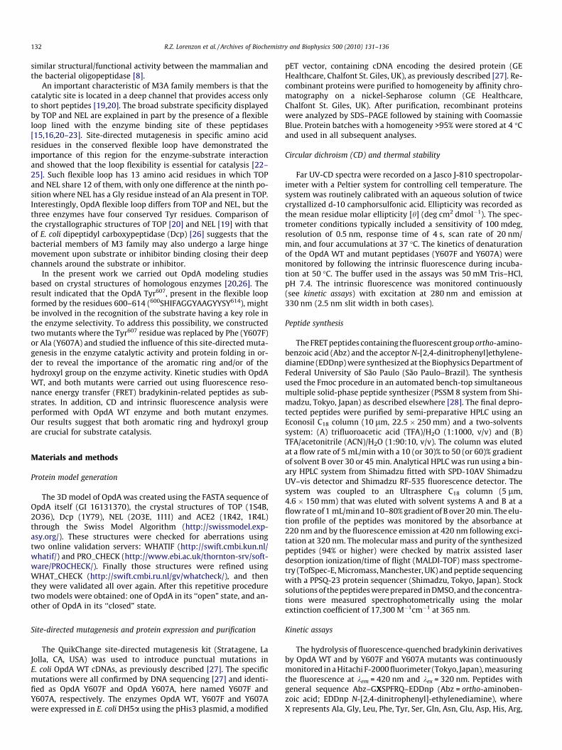
To further characterize the expressed enzymes, Far-UV-CDspectra was performed showing that OpdA and its mutants showedpredominant a-helical structures (Fig. 2A). The spectra obtainedfor both Y607F and Y607A mutants were similar to the spectrumfor the wild-type enzyme. In the same way, no significant

Fig. 2. (A) Far-UV-CD spectra of OpdA WT and Y607F and Y607A mutants. (B)Intrinsic fluorescence (kex = 280 nm and kem = 330 nm) of OpdA WT (s), Y607F (d)and Y607A (h) during incubation at 50 �C.
134 R.Z. Lorenzon et al. / Archives of Biochemistry and Biophysics 500 (2010) 131–136
difference was observed in the intrinsic fluorescence (Fig. 2B) ofthe mutant peptidases when compared with the wild-type en-zyme. Therefore, the site-directed mutagenesis of the Tyr607 resi-due did not promote significant changes in the secondarystructure of the proteins.
Hydrolysis of synthetic fluorogenic substrates
To compare the catalytic activity of OpdA WT and that of Y607Fand Y607A mutants, FRET peptides based on the general structureAbz–GXSPFRQ–EDDnp (X = Ala, Pro, Leu, Gly, Phe, Tyr, Ser, Asn,Gln, Lys, Arg, His, Glu or Asp) were used. The leader peptide ofthe series, Abz–GFSPFRQ–EDDnp, contains six C-terminal aminoacids of bradykinin (RPPGFSPFR) which is as an efficient substratefor OpdA [8]. The kinetic parameters determined and the cleavagesites are shown in Table 1. The substitution of OpdA Tyr607 residueby Phe or Ala significantly altered the kinetic parameters for thehydrolysis of Abz–GXSPFRQ–EDDnp derivatives in comparisonwith the OpdA WT. In addition, the OpdA mutants showed differ-ent cleavage sites indicating that the Tyr607 residue is importantto define the P1 residue in the substrate hydrolysis.
OpdA WT hydrolyzed all the substrates on the X–Ser bond only.However, a different pattern of cleavage was observed for the mu-tants. Although OpdA mutant Y607F cleaved most of the peptideson the X–Ser bond, the peptides Abz–GPSPFRQ–EDDnp, Abz–GGSPFRQ–EDDnp, Abz–GESPFRQ–EDDnp and Abz–GDSPFRQ–EDDnp were hydrolyzed on the Pro–Phe bond, just like TOP and
NEL [23]. The peptide Abz–GKSPFRQ–EDDnp was hydrolyzed byY607F on both peptide bonds X–Ser (42.3%) and Pro–Phe (57.7%).
OpdA Y607A mutant cleaved peptides Abz–GYSPFRQ–EDDnp,Abz–GRSPFRQ–EDDnp, Abz–GHSPFRQ–EDDnp and Abz–GFSPFRQ–EDDnp on the X–Ser bond only. Peptides containing the residuesGly, Leu and Ser on the X position were hydrolyzed on the Pro–Phebond. The substrates Abz–GASPFRQ–EDDnp (Ala–Ser = 70% andPro–Phe = 30%); Abz–GLSPFRQ–EDDnp (Leu–Ser = 50% and Pro–Phe = 50%); Abz–GQSPFRQ–EDDnp (Gln–Ser = 50% and Pro–Phe =50%); Abz–GNSPFRQ–EDDnp (Asn–Ser = 58% and Pro–Phe = 42%);and Abz–GKSPFRQ–EDDnp (Lys–Ser = 40% and Pro–Phe = 60%) werecleaved on both X–Ser and Pro–Phe bonds. Moreover, the peptidescontaining the negatively charged amino acids Asp and Glu, as wellas Pro in the X position were resistant to hydrolysis even at high con-centration of enzyme. Such different behavior according with thesubstrate including changes of the cleavage site where also observedwith TOP Y605F, TOP Y605A [23,24], NEL Y606F and NEL 606A [23].This observation suggests a similar role for OpdA Y607 residue insubstrate hydrolysis to that of Y605/Y606 of TOP/NEL [23,24].
The kinetic parameters obtained for the hydrolysis of Abz–GXSPFRQ–EDDnp derivatives by OpdA and Y607F and Y607A mu-tants indicated that the OpdA WT has high affinity for the basicresidue Arg at P1 position. Substrate Abz–GRSPFRQ–EDDnp washydrolyzed by OpdA at the highest catalytic efficiency in allpeptide series (kcat/Km = 10,358 mM�1 s�1). Surprisingly, thepeptides Abz–GHSPFRQ–EDDnp (kcat/Km = 1910 mM�1 s�1) andAbz–GKSPFRQ–EDDnp (kcat/Km = 1095 mM�1 s�1) that contain inP1 the positively charged residues His and Lys, respectively, werenot as suitable for hydrolysis as Abz–GRSPFRQ–EDDnp. The hydro-phobic aromatic residues Phe (kcat/Km = 1584 mM�1 s�1) and Tyr(kcat/Km = 2191 mM�1 s�1) were also well accepted by the S1 sub-site. In contrast, peptides containing Asp and Glu in the P1 positionwere poorly hydrolyzed by OpdA, while neutral polar residues,Asn, Ser and Gln, and polar residues, Pro, Gly and Leu, did not showany important interaction with the active site.
The kinetic parameters determined for Y607F and Y607A mu-tants demonstrated that the presence the Tyr607 residue is essen-tial for the OpdA catalytic activity. Table 1 and Fig. 3 show adecrease in catalytic activity of about 100-fold for Y607F mutantand 1000-fold for Y607A mutant. These lower kcat/Km values arein response mainly to the drastic kcat decrease. Peptides containingPro, Asp and Glu residues on the X position were resistant tohydrolysis up to 2.1 lM enzyme concentration. This confirms theimportance, not only of the hydroxyl group, but mainly of the aro-matic ring on the 607th position for OpdA activity. Both mutant en-zymes showed their highest kcat/Km values with peptides bearingaromatic amino acids on the P1 position, especially the Tyr residue.
Influence of NaCl and pH on OpdA WT, and Y607F and Y607A mutantactivities
The influence of NaCl concentration on Abz–GFSPFRQ–EDDnphydrolysis by OpdA WT, Y607F and Y607A mutants was studiedover a range of 0–2.0 M NaCl. For each salt concentration, thekcat/Km value was determined under pseudo-first-order conditionsusing the fluorimetric assay described above (Fig. 4). Increase ofsalt concentration rendered a significant rise of kcat/Km values forboth mutants mainly after the addition of 1 M NaCl. For Y607Fand Y607A, the kcat/Km values at high molar concentrations werenearly 10 times higher than that with the OpdA WT enzyme. Thealterations seen in the salt curve are coherent with the idea thatthis specific Tyr residue in the OpdA’s flexible loop is strongly re-lated to the enzyme catalytic activity. For native OpdA however,the increase in salt concentration and therefore the change in thesolvent did not significantly modify the enzyme activity.

Table 1Kinetic parameters for the hydrolysis of FRET peptides derived from Abz–GXSPFRQ–EDDnp sequence by OpdA WT and Y607F and Y607A mutants.
X OpdA WT Y607F Y607A
Km (lM) kcat (s�1) kcat/Km (mM s)�1 Km (lM) kcat (s�1) kcat/Km (mM s)�1 Km (lM) kcat (s�1) kcat/Km (mM s)�1
A 0.8 0.5 595 2.2 0.014 18 kcat/Km = 0.6 (X–S; P–F)*G 1.1 0.2 194 1.3 0.008 6 (P–F) 9.4 0.02 0.3 (P–F)L 1.8 1.5 844 8.6 0.2 2.1 8.9 0.032 4 (P–F)F 0.7 1.1 1584 2.7 0.1 36 6.1 0.025 4Y 0.7 1.5 2191 1.2 0.065 55 6.5 0.064 10S 0.5 0.3 590 0.5 0.004 7 10 0.003 0.3 (P–F)Q 2.2 0.6 258 5 0.05 10 kcat/Km = 0.6 (X–S; P–F)*N 1.8 0.5 273 2.6 0.02 7 kcat/Km = 0.2 (X–S; P–F)*E 1.8 0.08 42 kcat/Km = 0.25 (P–F)* ResistantD 4.2 0.2 49 4.7 0.002 0.5 (P–F) ResistantH 0.4 0.74 1910 4 0.08 20 kcat/Km = 0.6*R 0.2 2.4 10,358 0.7 0.034 49 1.7 0.005 3K 0.4 0.4 1095 kcat/Km = 7 (X–S; P–F)* kcat/Km = 0.5 (X–S; P–F)*P 0.7 0.1 159 kcat/Km = 0.8 (P–F)* Resistant
a If unspecified, the peptides were hydrolyzed on the X–S bond; * indicates that kcat/Km was determined under pseudo first-order conditions. The errors were less than 10% forany value.Resistant = No hydrolysis occurred up to 2.1 lM enzyme concentration.
0.0001
0.001
0.01
0.1
1
A G L F Y S Q N E D H R K PAmino acid (X)
k cat
/Km
mut
ant /
kca
t/Km
WT
Fig. 3. Relative kcat/Km ratios for the hydrolysis of peptides derived from Abz–GXSPFRQ–EDDnp sequence by OpdA WT and Y607F and Y607A mutants. Black bars – Y607F/OpdA WT, white bars – Y607A/OpdA WT.
Fig. 4. NaCl influence in the hydrolysis of Abz–GFSPFRQ–EDDnp by OpdA WT(s),Y607F (d) and Y607A (h). In the ratio (k/k0), k0 is the kcat/Km value obtained in50 mM Tris–HCl, pH 7.4, in the absence of salt and k is the kcat/Km value obtained ina defined salt concentration.
Fig. 5. pH dependence for hydrolysis of Abz–GFSPFRQ–EDDnp by OpdA WT (d),Y607F (s) and Y607A (j). The assays were performed as described in Material andmethods section. Values are presented as kcat/Km.
R.Z. Lorenzon et al. / Archives of Biochemistry and Biophysics 500 (2010) 131–136 135
The pH dependence on the hydrolysis of Abz–GFSPFRQ–EDDnpby OpdA WT and by both mutants was determined over a pH rangeof 5.5–10.0. Fig. 5 shows the pH-curve for the three enzymes, witha pH optimum around 7.0 for the OpdA WT (pKe1 = 6.5 ± 0.1;
pKe2 = 6.9 ± 0.1) and Y607F (pKe1 = 6.0 ± 0.1; pKe2 = 7.5 ± 0.1), andof 6.0 for the Y607A (pKe1 = 6.0 ± 0.1; pKe2 = 6.3 ± 0.1) mutant. Suchdifference in the optimum pH parallels the different affinity withthe assayed substrates shown for the Y607A mutant enzyme. Thus,

136 R.Z. Lorenzon et al. / Archives of Biochemistry and Biophysics 500 (2010) 131–136
the low catalytic efficiency shown in both mutants reflects the lackof Tyr607 and the importance of the contacts involving the aromaticand hydroxyl groups of the tyrosine residue.
This difference on the catalytic properties and also the recogni-tion of the correct cleavage site shows that important chemicalinteraction performed by the Tyr607 residue are critical for catalyticactivity. This is clearly shown by the results obtained with the mu-tants Y607F and Y607A which cannot perform their hydrolyticactivity as well as the wild-type enzyme. Therefore, the low cata-lytic efficiency presented by both mutants reflects the importanceof the contacts performed by the aromatic and hydroxyl groups ofthe tyrosine residue that are essential to anchor the substrate inthe enzyme active site.
Taken together, the present work shows the importance ofTyr607 in the catalytic efficiency of OpdA and the resemblance ofthis enzyme with the other members of the M3A subfamily. Simi-larly to what happens in other metallopeptidases as astacin andthermolysin [32,33], this specific Tyr residue might be crucial forsubstrate catalysis and could be relevant to stabilize the oxyanionintermediate formed after nucleophilic attack by water.
Acknowledgments
This work was supported by Conselho Nacional de Desenvolvi-mento Científico e Tecnológico (CNPq) and Fundação de Amparo aPesquisa do Estado de São Paulo (FAPESP).
References
[1] M. Muller, Experientia 48 (1992) 118–129.[2] S. Gottesman, Annu. Rev. Cell Dev. Biol. 19 (2003) 565–587.[3] S. Gottesman, Annu. Rev. Genet. 30 (1996) 465–506.[4] P. Novak, P.H. Ray, I.K. Dev, J. Biol. Chem. 261 (1986) 420–427.[5] C.A. Conlin, C.G. Miller, J. Bacteriol. 174 (1992) 1631–1640.[6] C.A. Conlin, N.J. Trun, T.J. Silhavy, C.G. Miller, J. Bacteriol. 174 (1992) 5881–5887.[7] T.G. Chu, M. Orlowski, Biochemistry 23 (1984) 3598–3603.[8] T. Paschoalin, A.K. Carmona, V. Oliveira, L. Juliano, L.R. Travassos, Arch.
Biochem. Biophys. 441 (2005) 25–34.
[9] A. Yaron, Methods Enzymol. 45 (1976) 599–610.[10] A. Yaron, D. Mlynar, A. Berger, Biochem. Biophys. Res. Commun. 47 (1972)
897–902.[11] C.E. Cunha, F. Magliarelli Hde, T. Paschoalin, A.T. Nchinda, J.C. Lima, M.A.
Juliano, P.B. Paiva, E.D. Sturrock, L.R. Travassos, A.K. Carmona, Biol. Chem. 390(2009) 931–940.
[12] A.J. Barrett, M.A. Brown, P.M. Dando, C.G. Knight, N. McKie, N.D. Rawlings, A.Serizawa, Methods Enzymol. 248 (1995) 529–556.
[13] F. Checler, H. Barelli, P. Dauch, V. Dive, B. Vincent, J.P. Vincent, MethodsEnzymol. 248 (1995) 593–614.
[14] V. Rioli, A. Kato, F.C. Portaro, G.K. Cury, K. te Kaat, B. Vincent, F. Checler, A.C.Camargo, M.J. Glucksman, J.L. Roberts, S. Hirose, E.S. Ferro, Biochem. Biophys.Res. Commun. 250 (1998) 5–11.
[15] V. Oliveira, M. Campos, J.P. Hemerly, E.S. Ferro, A.C. Camargo, M.A. Juliano, L.Juliano, Anal. Biochem. 292 (2001) 257–265.
[16] V. Oliveira, M. Campos, R.L. Melo, E.S. Ferro, A.C. Camargo, M.A. Juliano, L.Juliano, Biochemistry 40 (2001) 4417–4425.
[17] E.R. Vimr, L. Green, C.G. Miller, J. Bacteriol. 153 (1983) 1259–1265.[18] J.D. Fontenele-Neto, E.E. Massarelli, P.A. Gurgel Garrido, A. Beaudet, E.S. Ferro,
J. Comp. Neurol. 438 (2001) 399–410.[19] C.K. Brown, K. Madauss, W. Lian, M.R. Beck, W.D. Tolbert, D.W. Rodgers, Proc.
Natl. Acad. Sci. USA 98 (2001) 3127–3132.[20] K. Ray, C.S. Hines, J. Coll-Rodriguez, D.W. Rodgers, J. Biol. Chem. 279 (2004)
20480–20489.[21] J.A. Sigman, T.H. Patwa, A.V. Tablante, C.D. Joseph, M.J. Glucksman, A.J.
Wolfson, Biochem. J. 388 (2005) 255–261.[22] M.F. Machado, M.F. Marcondes, V. Rioli, E.S. Ferro, M.A. Juliano, L. Juliano, V.
Oliveira, Biochem. Biophys Res. Commun. 394 (2010) 429–433.[23] M.F. Machado, V. Rioli, F.M. Dalio, L.M. Castro, M.A. Juliano, I.L. Tersariol, E.S.
Ferro, L. Juliano, V. Oliveira, Biochem. J. 404 (2007) 279–288.[24] L.A. Bruce, J.A. Sigman, D. Randall, S. Rodriguez, M.M. Song, Y. Dai, D.E. Elmore,
A. Pabon, M.J. Glucksman, A.J. Wolfson, FEBS J. 275 (2008) 5607–5617.[25] V. Oliveira, M.C. Araujo, V. Rioli, A.C. de Camargo, I.L. Tersariol, M.A. Juliano, L.
Juliano, E.S. Ferro, FEBS Lett. 541 (2003) 89–92.[26] M. Comellas-Bigler, R. Lang, W. Bode, K. Maskos, J. Mol. Biol. 349 (2005) 99–
112.[27] V. Rioli, F.C. Gozzo, A.S. Heimann, A. Linardi, J.E. Krieger, C.S. Shida, P.C. Almeida,
S. Hyslop, M.N. Eberlin, E.S. Ferro, J. Biol. Chem. 278 (2003) 8547–8555.[28] L. Juliano, J.R. Chagas, I.Y. Hirata, E. Carmona, M. Sucupira, E.S. Oliveira, E.B.
Oliveira, A.C. Camargo, Biochem. Biophys. Res. Commun. 173 (1990) 647–652.[29] M.C. Araujo, R.L. Melo, M.H. Cesari, M.A. Juliano, L. Juliano, A.K. Carmona,
Biochemistry 39 (2000) 8519–8525.[30] R.J. Leatherbarrow, GraFit Version 5, Erithacus Software Ltd., Horley, UK, 2004.[31] L. Polgar, Biochemistry 38 (1999) 15548–15555.[32] I. Yiallouros, E.G. Berkhoff, W. Stocker, FEBS Lett. 484 (2000) 224–228.[33] C. Marie-Claire, E. Ruffet, G. Tiraboschi, M.C. Fournie-Zaluski, FEBS Lett. 438
(1998) 215–219.
Copyright © 2022 FDOKUMEN