
Modified Firearm Discharge Residue Analysis utilizing ...
273
Graduate Theses, Dissertations, and Problem Reports 2021 Modified Firearm Discharge Residue Analysis utilizing Advanced Modified Firearm Discharge Residue Analysis utilizing Advanced Analytical Techniques, Complexing Agents, and Quantum Analytical Techniques, Complexing Agents, and Quantum Chemical Calculations Chemical Calculations William J. Feeney West Virginia University, [email protected] Follow this and additional works at: https://researchrepository.wvu.edu/etd Part of the Analytical Chemistry Commons, Physical Chemistry Commons, Probability Commons, Statistical Models Commons, and the Theory and Algorithms Commons Recommended Citation Recommended Citation Feeney, William J., "Modified Firearm Discharge Residue Analysis utilizing Advanced Analytical Techniques, Complexing Agents, and Quantum Chemical Calculations" (2021). Graduate Theses, Dissertations, and Problem Reports. 10302. https://researchrepository.wvu.edu/etd/10302 This Dissertation is protected by copyright and/or related rights. It has been brought to you by the The Research Repository @ WVU with permission from the rights-holder(s). You are free to use this Dissertation in any way that is permitted by the copyright and related rights legislation that applies to your use. For other uses you must obtain permission from the rights-holder(s) directly, unless additional rights are indicated by a Creative Commons license in the record and/ or on the work itself. This Dissertation has been accepted for inclusion in WVU Graduate Theses, Dissertations, and Problem Reports collection by an authorized administrator of The Research Repository @ WVU. For more information, please contact [email protected].
-
Upload
khangminh22 -
Category
Documents
-
view
0 -
download
0
Transcript of Modified Firearm Discharge Residue Analysis utilizing ...
Graduate Theses, Dissertations, and Problem Reports
2021
Modified Firearm Discharge Residue Analysis utilizing Advanced Modified Firearm Discharge Residue Analysis utilizing Advanced
Analytical Techniques, Complexing Agents, and Quantum Analytical Techniques, Complexing Agents, and Quantum
Chemical Calculations Chemical Calculations
William J. Feeney West Virginia University, [email protected]
Follow this and additional works at: https://researchrepository.wvu.edu/etd
Part of the Analytical Chemistry Commons, Physical Chemistry Commons, Probability Commons,
Statistical Models Commons, and the Theory and Algorithms Commons
Recommended Citation Recommended Citation Feeney, William J., "Modified Firearm Discharge Residue Analysis utilizing Advanced Analytical Techniques, Complexing Agents, and Quantum Chemical Calculations" (2021). Graduate Theses, Dissertations, and Problem Reports. 10302. https://researchrepository.wvu.edu/etd/10302
This Dissertation is protected by copyright and/or related rights. It has been brought to you by the The Research Repository @ WVU with permission from the rights-holder(s). You are free to use this Dissertation in any way that is permitted by the copyright and related rights legislation that applies to your use. For other uses you must obtain permission from the rights-holder(s) directly, unless additional rights are indicated by a Creative Commons license in the record and/ or on the work itself. This Dissertation has been accepted for inclusion in WVU Graduate Theses, Dissertations, and Problem Reports collection by an authorized administrator of The Research Repository @ WVU. For more information, please contact [email protected].

Graduate Theses, Dissertations, and Problem Reports
2021
Modified Firearm Discharge Residue Analysis utilizing Advanced Modified Firearm Discharge Residue Analysis utilizing Advanced
Analytical Techniques, Complexing Agents, and Quantum Analytical Techniques, Complexing Agents, and Quantum
Chemical Calculations Chemical Calculations
William J. Feeney
Follow this and additional works at: https://researchrepository.wvu.edu/etd
Part of the Analytical Chemistry Commons, Physical Chemistry Commons, Probability Commons,
Statistical Models Commons, and the Theory and Algorithms Commons

Modified Firearm Discharge Residue Analysis utilizing Advanced
Analytical Techniques, Complexing Agents, and Quantum Chemical
Calculations
William Jansen Feeney
Dissertation submitted to the
Eberly College of Arts and Sciences
at West Virginia University
in partial fulfillment of the requirements for the degree of
Doctor of Philosophy in Chemistry
Suzanne Bell, Ph.D., Co-Chair
Tatiana Trejos, Ph.D., Co-Chair
Glen Jackson, Ph.D.
Blake Mertz, Ph.D.
Stephen Valentine, Ph.D.
C. Eugene Bennett Department of Chemistry
Morgantown, West Virginia
2021
Key Words: gunshot residue, firearm discharge residue, host molecules, orbitrap, ESI,
complexing, crown ether, molecular modelling, liquid chromatography
Copyright 2021 William Jansen Feeney

Abstract
Modified Firearm Discharge Residue Analysis utilizing Advanced Analytical Techniques,
Complexing Agents, and Quantum Chemical Calculations
William Jansen Feeney
The use of gunshot residue (GSR) or firearm discharge residue (FDR) evidence faces some
challenges because of instrumental and analytical limitations and the difficulties in evaluating and
communicating evidentiary value. For instance, the categorization of GSR based only on elemental
analysis of single, spherical particles is becoming insufficient because newer ammunition
formulations produce residues with varying particle morphology and composition. Also, one
common criticism about GSR practitioners is that their reports focus on the presence or absence
of GSR in an item without providing an assessment of the weight of the evidence. Such reports
leave the end-used with unanswered questions, such as “Who fired the gun?” Thus, there is a
critical need to expand analytical capabilities and enhance the impact of the forensic scientist’s
conclusions. To maximize the evidential value of GSR evidence, detection methods exploiting
modern advancements in instrumentation must be explored and developed.
The research here addresses multiple concerns within the community by increasing the
evidentiary value of GSR while addressing limitations about current understanding of the behavior
and interactions of GSR traces in various scenarios. Presented here is a sequential investigation of
1) the existing practices for GSR analysis, 2) the development and validation of an alternative
analytical technique for enhanced detection of inorganic and organic GSR, 3) the occurrence of
IGSR and OGSR in various subpopulations and the use of probabilistic interpretation of evidence,
and 4) the use of theoretical calculations to study the host-guest chemistry involved in the proposed
analytical method.
This collection of work reviews the current literature review and illustrates a trend to
investigate emerging methods to enhance IGSR analysis with a wider emphasis on OGSR
compounds. Combining IGSR and OGSR components increases the confidence of detecting GSR
on a collected sample.
In this study, we demonstrate use of LC-MS/MS and host-guest chemistry to detect IGSR and
OGSR components in a single instrument. One advantage afforded by the proposed method is the
dual detection of IGSR and OGSR on the same sample under 20 minutes, which is about an order
of magnitude faster than existing techniques, like scanning electron microscopy-energy-dispersive
X-ray Spectrometry (SEM-EDS) analysis. Also, the wide use of LC-MS/MS technology at crime
laboratories enables future technology transfer and implementation. This strategy is employed in
a population study of over 400 authentic specimens to differentiate shooter from non-shooters from
samples taken from a subject’s hands. The prevalence of organic and inorganic gunshot residue is
evaluated within two main subpopulations, 1) non-shooters, including groups with low- and high-
risk of containing GSR-like residues, and 2) individuals involved in a firing event (shooters,
bystanders, and shooters performing post-shooting activities). The subpopulations were
investigated using both simple exploratory analyses to monitor the occurrence of GSR and

machine learning algorithms for classification and class-prediction. Additionally, the probabilistic
outputs resulting from the machine learning algorithm (neural networks) were used to assess the
weight of evidence using likelihood ratios. Accuracy ranging from 90-99% was obtained,
depending on the population of interest with larger LRs observed in shooter’s sets, proving
substantial progress to conventional categorical approaches
Finally, the host-guest chemical interactions from this study were further investigated using
theoretical calculations. A quantum mechanical approach (DFT) was utilized to monitor the
noncovalent, electrostatic interactions between the 18-crown-6-ethers and other metallic ions
including the alkali, alkali earth, and pnictogen groups. Additionally, oxygen atoms were replaced
with other heteroatoms in these macrocycles to study the thermochemical binding. It was found
that electronegativity proved to be the greatest factor influencing the strength of binding followed
by the size of the interacting cation.
Overall, the development of novel analytical methods for GSR detection, the application of
ground-breaking statistical methods to interpret GSR evidence using artificial intelligence (neural
networks) and likelihood ratios to estimate the weight of the evidence, and the understating of the
host-guest chemistry of GSR species is anticipated to provide a needed leap of knowledge in the
community.

iv
Funding and Disclaimer
This collection of works would not have been possible without the generous funding of the
National Institute of Justice (NIJ). The funding opportunity associated with these works is Award
#2019-R2-CX-044, NIJ-STEM Graduate Research Fellowship, which was titled Validation of a
Single Instrument, Single Protocol for the Detection of the Inorganic and Organic Constituents of
Firearm Discharge Residues. This project was funded through January 2020-December 2021. The
individuals associated with the fellowship were Tatiana Trejos (PI), Suzanne Bell (Chair) and
William Feeney (PhD Student Fellowship Recipient). Importantly, the conclusions from each
section were founded by me and the coauthors and disseminated to the scientific community in the
form of review and research manuscripts. Hence, the opinions, findings, and conclusions are those
of the authors and do not necessarily reflect those of the Department of Justice.

v
Dedication
Dedicated to my loved ones who are with me every day and are with me in spirit.
Erin – I know being with me hasn’t been easy (and I don’t make it that way). Thank you for
keeping me alive – in more ways than you can possibly imagine.
Mark – Words cannot express how much I miss you. Even now, I write this remorseful as I know
that you will never read this. I can’t thank you enough for making me think outside of the box and
truly inspiring me. I’m sorry and miss you.
Gran and Papap– Everything ended too quickly and on terrible terms. Just wanted to say to both
of you, thank you for being in my corner and helping me enjoy the simpler things in life. I miss
you both every day.
Mom, Chris, and Joe – Thanks for holding down the fort and not getting too angry with me.
Dad – Thank you for reminding me how tranquil nature is.
Don’t give up. We have all gone home in tears, we’ve all second-guessed why we do what we do.
We’ve all felt like failures at times, and we have all wanted to quit at some point. Just remember,
it’s graduate school. You’re smart and strong and you’ve made it this far. You can’t expect it to
be easy.

Acknowledgements
Graduate school has been a trying and stressful journey. I certainly would not have made it on my
own and with that, a special thanks and acknowledgements to the following groups and individuals
is in order:
Family and Friends: Your journeys and successes gave me with inspiration and encouragement.
Brittany (Yeager) Stephens, Stephen Raso, and Kristin Kelly – You’ve made life in lab way more
enjoyable in my first years. Watching you all leave was harder than you think. Thank you for being
my pseudo family “Momma B”, “Uncle Steve” and “Sister K” and opening your arms when I first
entered this strange research world.
To my friends that are graduated and onto bigger and better things – There are so many of you to
thank. Just know that you all helped me deal with my insecurities and helped me laugh.
To my newly adopted research family – You all made the transition into a new group easy. Thank
you for being there and walking with me throughout me journey in the academic research realm.
Know that you can call anytime, and I will assist in any way that I can.
To the C/Kourtneys – You both are great people. Unfortunately, the long nights in lab won’t be
the same without you there. Thank you for the FNAFs and laughs.
Faculty:
Becky Secrist - You are an amazing individual and have impacted so many graduate student’s lives.
The chemistry department was very lucky to have you. Thank you for everything you have helped
me with over the years. I miss your smiling face and I hope you are enjoying the time with your
family.
(Big) Bill Cunningham - You are… I guess an awesome individual to joke and vent to. The
Forensics department is very lucky to have you. Thank you and I know you’ll miss my face
popping in your office at any time.
Last, but certainly not least, a special thanks goes to every one of my committee members - Dr.
Suzanne Bell, Dr. Blake Mertz, Dr. Glen Jackson, Dr. Stephen Valentine, and Dr. Tatiana Trejos
for reviewing my dissertation as well as serving on my graduate committee. Thank you all for your
patience and wisdom throughout the years.
Dr. Bell, thank you for pushing and pushing me up and over the mountain of graduate school and
reminding me every day that I forgot to check the “easy box”.
Dr. Trejos, thank you for accepting this “head case” into your laboratory and teaching me how to
become a better scientist. These past years have been the most inspiring times of my life and I am
extremely fortunate to have learned from you.
Without you, this would not have been possible.

Table of Contents
Abstract ........................................................................................................................................... ii
Funding and Disclaimer ................................................................................................................. iv
Dedication ....................................................................................................................................... v
Acknowledgements ........................................................................................................................ vi
Table of Contents .......................................................................................................................... vii
List of Figures ................................................................................................................................ xi
List of Tables ................................................................................................................................ xii
List of Abbreviations, Symbols, and Nomenclature .................................................................... xiii
CHAPTER 1: Problem Statement, Objectives, and Dissertation Structure ................................ 1
1.1. Problem Statement and Goals .............................................................................................. 2
1.2. Objectives ............................................................................................................................... 4
Objective 1 and respective tasks ..................................................................................................... 4
Objective 2 and respective tasks ..................................................................................................... 4
Objective 3 and respective tasks ..................................................................................................... 5
Objective 4 and respective tasks ..................................................................................................... 6
1.3. Deliverables ............................................................................................................................ 7
1.4. Dissertation Structure ........................................................................................................... 8
1.5. Implications for Criminal Justice and Practice in the United States ............................. 10
1.6. References for Chapter 1 .................................................................................................... 12
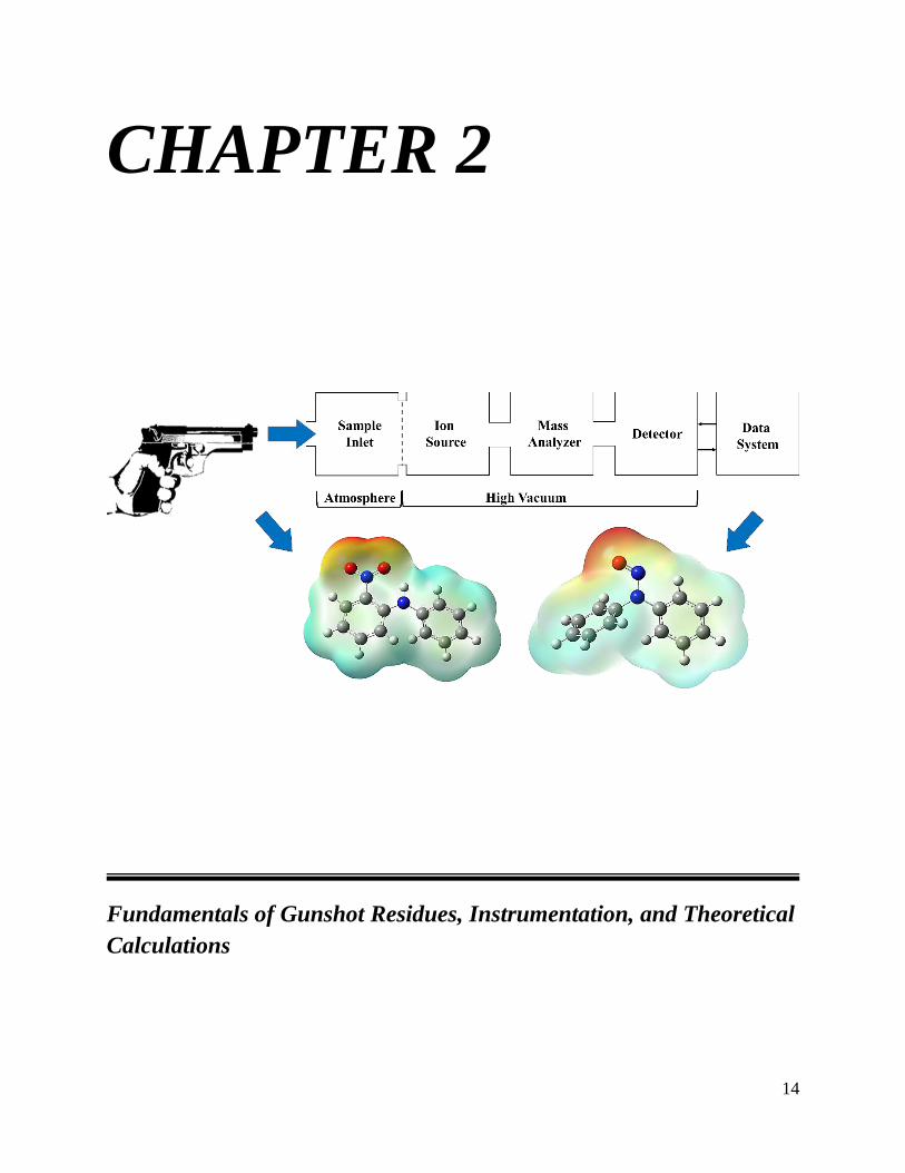
CHAPTER 2: Fundamentals of Gunshot Residues, Instrumentation, and Theoretical
Calculations .................................................................................................................................. 14
2.1. Gunshot Residue (GSR) ...................................................................................................... 15
2.2. Liquid Chromatography-Tandem Mass Spectrometry (LC-MS/MS) ........................... 16
2.2.1. Liquid Chromatography ...................................................................................................... 16
2.2.2. Electrospray Ionization (ESI) ............................................................................................. 19
2.2.3. Triple Quadrupole Mass Analyzer (QQQ or QqQ) ............................................................ 24
2.2.4. High-Resolution mass spectrometry – Orbitrap ................................................................. 27
2.3. Overcoming challenges for metal analysis in LC-MS/MS ............................................... 28
2.3.1. Macrocycles - Crown ethers ............................................................................................... 29
2.4. Strategies for investigating interactions of host-guest chemistry ................................... 30

viii
2.4.1. Experimental approaches ................................................................................................... 30
2.4.2. Theoretical calculations - Density Functional Theory (DFT) ............................................ 31
2.5. Summary .............................................................................................................................. 33
2.6. References for Chapter 2 .................................................................................................... 35
CHAPTER 3: Trends in Composition, Collection, Persistence, and Analysis of IGSR and
OGSR: A Review ......................................................................................................................... 41
3.1. Introduction ......................................................................................................................... 43
3.1. Inorganic and Organic Compositions of Modern Ammunitions ..................................... 48
3.2. Common Gunshot Residue Sampling Media and Extractions ........................................ 54
3.1.1. Adhesives ............................................................................................................................. 54
3.1.2. Cloth…… ............................................................................................................................. 55
3.1.3. Polymer… ........................................................................................................................... 55
3.1.4. Extractions .......................................................................................................................... 56
3.3. Deposition and Transfer Trends of Organic and Inorganic Gunshot Residues ............ 57
3.3.1. Transfer of Organic Gunshot Residue Compounds ............................................................ 59
3.3.2. Inorganic Gunshot Residue Particles ................................................................................. 60
3.4. Persistence of Both Organic and Inorganic Components of Gunshot Residue ............. 63
3.4.1. Organic Components .......................................................................................................... 64
3.4.2. Inorganic Particles ............................................................................................................. 65
3.5. Conclusions and Future Considerations ........................................................................... 76
3.6. References for Chapter 3 .................................................................................................... 78
CHAPTER 4: Detection of Organic and Inorganic Gunshot Residues from Hands using
Complexing Agents and LC-MS/MS ........................................................................................... 96
4.1. Introduction ......................................................................................................................... 98
4.1.1. Background ......................................................................................................................... 98
4.1.2. Inorganic Particulate Analysis ........................................................................................... 99
4.1.3. Organic Compound Analysis ............................................................................................ 100
4.1.4. Combined Analysis Methodologies ................................................................................... 101
4.2. Materials and Methods ..................................................................................................... 103
4.2.1. Consumables ..................................................................................................................... 103
4.2.2. Firearms, ammunition, and protocols .............................................................................. 104
4.2.3. LC-MS/MS methods .......................................................................................................... 106
4.2.4. Comparisons with various analytical methods ................................................................. 109

ix
4.2.4.1. LIBS…. .......................................................................................................................... 109
4.2.4.2. Electrochemical analysis ................................................................................................ 110
4.2.5. Extraction and collection of authentic samples for LC-MS/MS and multi-technique
approach………........................................................................................................................... 110
4.2.6. ICP-MS analysis for recovery study of collection substrates ........................................... 113
4.3. Results and Discussion ...................................................................................................... 113
4.3.1. OGSR: Chromatography, CID experimentation, and observations ................................. 114
4.3.2. IGSR: Formation and identification of M-L complexes using HRMS .............................. 117
4.3.3. Analytical validation and figures of merit ........................................................................ 123
4.3.4. Background and collection substrate considerations: recovery study and comparison to
ICP-MS digestion method ........................................................................................................... 125
4.3.5. Cross-validation of recovery of IGSR via ICP-MS ........................................................... 128
4.3.6. Workflow and cross-validation with screening methods .................................................. 129
4.3.7. Performance rates and criteria for authentic samples ..................................................... 131
4.4. Conclusions ........................................................................................................................ 135
4.5. References for Chapter 4 .................................................................................................. 138
CHAPTER 5: Evaluation of Organic and Inorganic Gunshot Residues in Various
Populations using LC-MS/MS .................................................................................................. 149
5.1. Introduction ....................................................................................................................... 151
5.1.1. Current state and challenges of GSR detection ................................................................ 151
5.1.2. Population considerations ................................................................................................ 153
5.1.3. Neural Networks and likelihood ratios for GSR interpretation ........................................ 154
5.2. Materials and Methods ..................................................................................................... 156
5.2.1. Consumables ..................................................................................................................... 156
5.2.2. Firearms, ammunition, and protocols .............................................................................. 157
5.2.3. Sample extraction and collection ...................................................................................... 159
5.2.4. Population study ............................................................................................................... 159
5.2.5. Data analysis .................................................................................................................... 160
5.2.6. Machine Learning model .................................................................................................. 161
5.3. Results and Discussion ...................................................................................................... 162
5.3.1. Occurrence of OGSR and IGSR on the sampled populations ........................................... 162
5.3.2. Neural network machine learning for GSR identification ................................................ 170
5.3.3. Likelihood ratios (LR) as probabilistic assessments of the weight of evidence ................ 172

x
5.4. Conclusions ........................................................................................................................ 178
5.5. References for Chapter 5 .................................................................................................. 182
CHAPTER 6: Investigations into host-guest interactions with metal ions using DFT for
applications using mass spectrometry ...................................................................................... 191
6.1. Introduction ....................................................................................................................... 193
6.2. Experimental Section ........................................................................................................ 197
6.2.3. Natural bond orbital (NBO) calculations ......................................................................... 201
6.2.4. Second order perturbation theory ..................................................................................... 201
6.3. Results/Discussion ............................................................................................................. 202
6.3.1. Confirmation of Structures via High-resolution Mass Spectrometry ............................... 202
6.3.2. Evidence of Sandwich complexes...................................................................................... 205
6.3.3. Influence of solvent effects on structure ............................................................................ 207
6.3.4. Effect of donor atoms and charge transfers ...................................................................... 210
6.3.5. Metal guest influences on reactivity descriptors .............................................................. 214
6.4. Conclusions ........................................................................................................................ 217
6.5. References for Chapter 6 .................................................................................................. 221
CHAPTER 7: Overall Conclusion and Future Directions ...................................................... 233
7.1. Overall Conclusion and Future Directions ..................................................................... 234
7.1.1. Objective 1: Review of the current literature associated with GSR ................................. 234
7.1.2. Objective 2: Validation and evaluation of a single instrument for sequential analysis of
GSR components ......................................................................................................................... 235
7.1.3. Objective 3: Investigation of the presence and prevalence of GSR in West Virginia within
different populations ................................................................................................................... 237
7.1.4. Objective 4: Investigations into host-guest interactions with metal ions using DFT for
applications using mass spectrometry ........................................................................................ 238
7.2. Future Work ...................................................................................................................... 240
7.3. References for Chapter 7 .................................................................................................. 244
Appendices .................................................................................................................................. 245
Appendix I – Chapter 4: Detection of Organic and Inorganic Gunshot Residues from Hands
using Complexing Agents and LC-MS/MS – Supplemental Information .................................... 245
Appendix II – Chapter 5: Evaluation of Organic and Inorganic Gunshot Residues in Various
Populations using LC-MS/MS – Supplemental Information ....................................................... 248
Appendix III – Chapter 6: Investigations into host-guest interactions with metal ions using DFT
for applications using mass spectrometry – Supplemental Information ..................................... 250
Appendix IV – Professional CV .................................................................................................. 253

xi
List of Figures
Figure 2.1: General schematic of electrospray ionization and Taylor cone formation……………... 21
Figure 2.2: Representation of two established electrospray ionization models – IEM and CRM…… 22
Figure 2.3: Recently proposed chain-ejection model by Konermann group….................................... 23
Figure 2.4: Quadrupole mass filter representing ions following the set trajectory…......................... 24
Figure 2.5: Schematic of QQQ with ESI source with a continuous multiplier tube…......................... 25
Figure 2.6: Mathieu stability diagram for the stable regions for ions…............................................... 26
Figure 2.7: Simple schematic of orbitrap mass analyzer with oscillating frequency…........................ 27
Figure 2.8: Example of a “sandwich” structure with metal ion…........................................................ 31
Figure 3.1: Common sampling areas and techniques for gunshot residue evidence …........................ 56
Figure 4.1: Breakdown of extraction and analysis process and cross-validation techniques….…..... 112
Figure 4.2: Chromatography separation of the seven OGSR compounds using PFP column……...... 115
Figure 4.3: Orbitrap confirmation of 18C6 with Ba and Pb natural isotope distributions…................ 117
Figure 4.4: Flow injection analysis of the various tartrate complexes with Sb in ESI- mode………... 122
Figure 5.1: Boxplot comparisons between the low-risk, high-risk, and baseline shooter…...……... 163
Figure 5.2: Boxplot comparisons between low-risk, post-shooting activity, and baseline shooter…. 165
Figure 5.3: Boxplot comparisons between the low-risk, bystander, and baseline shooter…............... 167
Figure 5.4: OGSR and IGSR population comparison among all analytes tested….............................. 169
Figure 5.5: Histogram and Tippet plot of shooter (H1) and low-risk (H2) subpopulation................... 175
Figure 5.6: Histogram and Tippet plot of shooter (H1), low-risk (H2), and high-risk (H2)………… 176
Figure 5.7: Histograms and Tippet plot of shooter (H1), low-risk (H2), and post-shooting (H1)…... 177
Figure 5.8: Histograms and Tippet plot of shooter (H1), low-risk (H2), and bystander (H1)…….... 178
Figure 5.9: Neural Network structure to train and validate authentic and low-risk populations........... 179
Figure 6.1: Skeletal structure design of varying crown ethers simulated…......................................... 199
Figure 6.2: Collected and simulated isotopic abundances from orbitrap of crown ether complexes… 204
Figure 6.3: Responses for barium and lead nitrate in the presence of crown ether via LC-MS/MS..... 206
Figure 6.4: Experimental example of a 1:2 M-L complex to a 1:1 M-L complex in ESI (+) mode…. 209
Figure 6.5: Electrostatic distributions calculated for 18C6, aza-, thia-, and partial 18C6………........ 211
Figure 6.6: HOMO and LUMO of 18C6 with Ba, Pb, and Sb….......................................................... 216
Figure 7.1: Typical locations for group substitution FRCs….............................................................. 241
Figure 7.2: Preliminary evaluation and interaction of carfentanil with cucurbit[7]uril …................... 242

xii
List of Tables
Table 3.1: Organic compounds that may contribute to GSR……………………………... 50
Table 3.2: Inorganic analytes that may contribute to GSR……………………………… 53
Table 3.3a: OGSR focused methodologies………………………………….……………. 68
Table 3.3b: IGSR focused methodologies………………………………………………... 70
Table 3.3c: Combined methodologies for GSR analysis…………………………………. 75
Table 4.1: Orbitrap fragmentation patterns of metal-ligand complexes…….……………. 118
Table 4.2: Figures of merit for OGSR compounds…………………………….…………. 123
Table 4.3: Figures of merit for IGSR particulates………………………………………... 124
Table 4.4: OGSR recoveries from carbon tape and tesa ® Tack…………………………. 127
Table 4.5: IGSR recoveries from carbon tape and tesa ® Tack……………….…………. 128
Table 4.6: Authentic shooting samples and success rates………………………………… 133
Table 4.7: Activity samples with combined rates………………………………………… 134
Table 5.1: General breakdown of each population analyzed by LC-MS/MS analysis….... 160
Table 6.1: Metal cation characteristics that may affect binding………………………...... 206
Table 6.2: NBO analysis and charge transfers from 18C6 to Pb, Ba, and Sb…….………. 213
Table 6.3: Fully substituted macrocycles with oxygen, nitrogen, and sulfur atoms………. 215

xiii
List of Abbreviations, Symbols, and Nomenclature
%RSD Percent Relative Standard Deviation
18C6 18-crown-6-ether
2,4-DNT 2,4-dinitrotoluene
2-NDPA 2-nitrodiphenylamine
4-NDPA 4-nitrodiphenylamine
Å Angstrom
ACN Acetonitrile
ASTM American Society for Testing and Materials
Ba Barium
CE Collision Energy
CID Collision Induced Dissociation
CRM Charged-residue model
APCI Atmospheric Pressure Chemical Ionization
Da Dalton
DPA Diphenylamine
EC Ethyl Centralite
EDS or EDX Energy Dispersive X-ray Spectrometry
ESI Electrospray Ionization
eV Electron Volt
FDR Firearm Discharge Residue
GC Gas Chromatography
GSR Gunshot Residue
H Enthalpy
H2O Water
H-G Host-Guest
eV Electron Volt
FDR Firearm Discharge Residue
FTIR Fourier-transform infrared spectroscopy
GC Gas Chromatography
GSR Gunshot Residue
H Enthalpy
H-G Host-Guest
HPLC High-Performance Liquid Chromatography
HRMS High-Resolution Mass Spectrometry
ICP Inductively Couple Plasma
IEM Ion Evaporation Model
IRB Institutional Review Board
kV Kilovolt
LC Liquid Chromatography
LOD Limit of Detection
LOQ Limit of Quantitation
m/z Mass to Charge Ratio
MC Methyl Centralite
mg Milligram
min Minute

xiv
mL Milliliter
IRB Institutional Review Board
LIBS Laser Induced Breakdown Spectroscopy
LR Likelihood Ratio
MeOH Methanol
MESP Molecular Electrostatic Potential
M-L Metal-Ligand
mm Millimeter
MRM Multiple Reaction Monitoring
MS Mass Spectrometry
MSn or MS/MS Tandem Mass Spectrometry
ng Nanograms
NG Nitroglycerin
NN Neural Network
N-NDPA N-nitrosodiphenylamine
NBO Natural bond orbital
OGSR Organic Gunshot Residue
Pb Lead
ppm Parts Per Million
Q1 Quadrupole 1
q2 Quadrupole 2
Q3 Quadrupole 3
QC Quality Control
QqQ or QQQ Triple Quadrupole
Q-ToF Quadrupole Time of Flight
RF Radio Frequency
RMS Root Mean Square
RoME Rates of Misleading Evidence
Rt Retention Time
s Second
S.D. or sd Standard Deviation
S/N or SNR Signal-to-Noise Ratio
Sb Antimony
SEM Scanning Electron Microscopy
SIMS Secondary Ion Mass Spectrometry
T Temperature
ToF Time of Flight
UHPLC Ultra-High-Performance Liquid Chromatography
μg Microgram
μL Microliter
μm Micrometer

1
CHAPTER 1
Problem Statement, Objectives, and Dissertation Structure

2
1.1. Problem Statement and Goals
SEM-EDS is currently the gold standard for gunshot residue (GSR) analysis as per ASTM
E1588-20 [1]. Nonetheless, with the prevalence of modern ammunition, the identification of
particle morphology and elemental composition by SEM-EDS needs further orthogonal
confirmatory tests. Collectively, the inorganic and organic components produced during the
discharge of a firearm are known as GSR and combined provide stronger support of the
differentiation of gunshot residues from other environmental and background residues. A
comprehensive analysis of GSR often requires the use of two separate methodologies. For
inorganic components (IGSR), SEM/EDS is the standard method used for morphology and
elemental identification [1-3]. For organic molecules (OGSR), a consensus method has not been
established, but recent research efforts have focused primarily on gas and liquid chromatography
mass spectrometry for identification and quantitation [2,3].
Other methods include the detection via ion mobility spectrometry (IMS), Raman
spectroscopy, electrochemistry (EC), Laser-Induced Breakdown Spectroscopy (LIBS), Laser
Ablation ICP-MS, and Time-of-Flight Mass Spectrometry [4]. The scientific community has
hypothesized that evidentiary value increases by simultaneously detecting inorganic and organic
components from a single sample. As a result, several approaches have been proposed to examine
OGSR and IGSR using combinations of techniques, such as LIBS with electrochemistry, CMV-
GC/MS with LIBS, and LC-MS/MS with SEM/EDS [4]. The disadvantage of using multiple
techniques is the significant time and resources required for preparation of additional samples,
which are limited in casework. By using a single instrument, the shortcomings can be overcome
through elimination of operations by multiple analysts and maintenance of several instruments.
This collection of work addresses the recent years’ Technical Support Working
Group (TSWG) list of research interests in trace evidence regarding the “evaluation of the

3
detection and utility of organic gunshot primer residue,” and the NIST-OSAC GSR subcommittee
research needs on “comprehensive feasibility of organic gunshot residue analysis” and the
“development of novel GSR methods for specific identification of shooters” [5,6].
The mechanisms of transfer and persistence of GSR are complex, and therefore the
differentiation of residues deposited on shooters versus bystanders is not possible with current
methodologies. The goal of this research is to validate an LC-MS/MS method for testing GSR
evidence through the sequential analysis of organic and inorganic components using metal-ligand
(M-L) complexes. This approach will provide a new tool for GSR analysts with the use of a
common instrument found in most forensic laboratories.
The main hypothesis of this study is that the capabilities of the proposed method for
simultaneous semi-quantitative detection of IGSR and OGSR components will help differentiate
residues left on known shooters and bystanders and will enhance the overall reliability of firearm-
related evidence. A large population set with known “ground truth” regarding the individual’s
involvement with the discharge of a firearm (i.e., known shooter versus known non-shooter
background population) was analyzed. The data was evaluated by categorical thresholds as well
as machine learning algorithms and probabilistic frameworks.
Finally, simulation studies provided understanding of the complexing mechanisms
between metal and ligand interactions. By utilizing quantum mechanical calculations, further
insight can be gained about the sensitivity and binding affinities of various metal guests.
Additionally, other factors including solvation polarizability (methanol, acetonitrile, water) and
heteroatom substitution was investigated to monitor the effects of binding affinities and
thermochemical values.

4
Collectively, the fundamental and population studies, and the novel analytical and
interpretation approaches proposed here are anticipated to increase capacity and narrow some of
the knowledge gaps in the field of gunshot residues.
1.2. Objectives
The overall goal of this project was to develop a comprehensive strategy to enhance the reliability
of the analysis and interpretation of gunshot residues (GSR). To accomplish this goal, four main
objectives were outlined as described below.
Objective 1 and respective tasks
Objective 1. Perform an in-depth investigation on the major findings of current research
approaches and practices within the most recent years regarding inorganic and organic
gunshot residues collection, extraction, and detection.
Task 1.1. Generate a compilation of the main compounds and byproducts found on OGSR
and IGSR of modern ammunition. Identify the use of each product within current
traditional and non-traditional ammunition.
Task 1.2. Report and compare instrumentation utilized and their respective capabilities to
detect OGSR, IGSR, or both. Additionally, report the findings, conditions, and parameters
for ease of access and reference by practitioners and end-users.
Task 1.3. Identify deposition, transfer, and persistence challenges of IGSR particulates and
OGSR compounds and evaluate where the current trends are shifting.
Objective 2 and respective tasks
Objective 2. Test the feasibility for dual characterization and validation of both OGSR and
IGSR constituents using a single analytical instrument, liquid chromatography tandem mass
spectrometry (LC-MS/MS).

5
Task 2.1. Identify and confirm the major CID pathways of the studied OGSR compounds
and self-assembled metal-ligand complexes using high- and low-resolution mass
spectrometry instrumentation.
Task 2.2. Evaluate the figures of merit of the triple quadrupole mass spectrometry for all
analytes under investigation using the Eurachem guideline [7]. Address the collection
efficiencies of substrate used in previous proof-of-concept study (tesa Tack®) and the
standard substrate (carbon adhesive mounted on an aluminum stub) using both LC-MS/MS
and ICP-MS.
Task 2.3. Test the performance rates of the validated method with authentic shooting
sample sets (with and without post-shooting activity). Test the feasibility of analyzing one
sample with screening methodologies (LIBS and electrochemical methods) in conjunction
with the newly developed confirmatory method.
Objective 3 and respective tasks
Objective 3: Conduct a population study of GSR collected from known shooters and non-
shooters
With this objective, we evaluate the feasibility to answer a major concern within the
forensics community by observing the rate and prevalence of GSR within the general population.
Furthermore, assess the performance rates and utilizing machine learning algorithms for
generating likelihood ratios for more comprehensive objective evaluations by forensic
practitioners.
Task 3.1. With recently validated instrument (LC-MS/MS), collect from various
individuals from the general population of West Virginia. Collect from individuals who are
either associated or dissociated with a firearm-related event. Additionally, collect from

6
individuals whose professions may yield false-positive signals like GSR such as
mechanics, farmer hands, and police officers.
Task 3.2. Collect and analyze samples from five subpopulations, including various
background and shooters subsets: 1) 75 low-risk individuals who never interacted with or
handled a firearm within the past 24 hours, 2) 75 non-shooter individuals that are
considered high-risk due to their professions, 3) 75 samples collected from authentic
shooters after recently discharging a firearm, 4) 75 samples from individuals performing
activities after a discharge event, and 5) 75 samples collected from bystanders of a firing
event.
Based on preliminary data, the sample size was selected for an estimated resulting
power of this test of 95%, providing a representative information of the prevalence of GSR
within the WV population, and ultimately, assess the benefit of combining OGSR and
IGSR data.
Task 3.3. Evaluate a data analysis model for the probabilistic assessment of the evidence.
Curate the population dataset and further evaluate it through exploratory statistical analysis
and machine learning algorithms. Neural networks are used to classify the samples and
calculate the probability outputs of belonging to one of the five populations groups and
monitored for their overall error rates. From those resulting probabilities, likelihood ratios
are then computed to evaluate the presence of GSR at the source level.
Objective 4 and respective tasks
Objective 4: Investigate binding affinities and sensitivities of guest-host species using density
functional theory

7
Throughout this study, binding agents such as crown ethers have been utilized for IGSR
detection for unique mass spectral signatures and transportation through the column and collision
cells. However, their inability to associate with antimony (Sb) is a glaring issue since it is a primary
element utilized in traditional ammunition. Hence, a computational approach is used to highlight
the underlying factors affecting binding of Sb to crown ether.
Task 4.1. Generate the chemical structures x, y, z coordinates (with and without metals)
for both gas and solution phases (acetonitrile, water, methanol) to determine potential
solvent effects. Additionally, heteroatoms such as N and S are incorporated in different
positions of the ring structure to invoke and observe the effects of ring sizes and
electronegativities.
Task 4.2. Employ density functional theory to calculate the non-covalent electrostatic
interactions between the crown ether and heterocycles that interact with the metal ions.
Task 4.3. Evaluate the calculations to identify trends amongst different periodic groups
using covariance and correlation exploratory analysis to understand the M-L behaviors.
1.3. Deliverables
This project was primarily funded by the National Institute of Justice (NIJ) under award
#2019-R2-CX-044 and this doctoral dissertation corresponds to one of the deliverables expected
upon completion of this fellowship, which ends in December 2021. Objectives 1, 2 and 3 respond
to the NIJ proposal. In addition to the financial, progress, and scholarly products required by the
funding agency (i.e., publications and dissertation), other deliverables of this research include the
dissemination of data collected within the forensic examiners and stakeholders. All data sets and
methods for data processing created during this study will be made available to interested
stakeholders and archived by the NIJ. As part of the dissemination strategy, we have published
three scientific publications in peer-review journals, shared research results at six scientific
8
meetings in the form of posters and oral presentations, and an additional publication about the
computer simulations is in preparation.
1.4. Dissertation Structure
This dissertation describes the accomplishments of each of the major goals into four main
chapters. Chapter 2 is a general introduction that highlights the main principles and practices
utilized in the forensic community for GSR analysis as well as the primary techniques utilized
throughout this study. Here, the reader is guided through the current understanding of gunshot
residue, ionization sources and separation techniques, low- and high-resolution mass analyzers,
theoretical calculations, and finally the species which sparked and assisted in this work. Also, this
chapter describes the major findings and outcomes for the detection of organic and inorganic
compounds of interest, as well as their shortcomings and future considerations for GSR evidence.
Chapter 3 describes the current findings and efforts displayed in the forensic research
community for GSR detection, with a focus in composition, collection, persistence, and analysis
of IGSR and OGSR. Here, the most recent articles pertaining to transfer, persistence, and
combining inorganic and organic components for increased evidentiary value are summarized.
Additionally, the articles’ findings are reported by instrumentation and respective units of
concentration or particle counts for a quick reference for both researchers and crime laboratory
practitioners. This review fulfilled a gap of information for the current standings of GSR analysis
as well as a brief overview for the direction and challenges experienced by the community. The
content of this chapter was published in Forensic Chemistry as an open-access article in May 2020
(W. Feeney, C. Vander Pyl, S. Bell, T. Trejos, Trends in composition, collection, persistence, and
analysis of IGSR and OGSR: A review, Forensic Chem. 19 (2020) 100250.
https://doi.org/10.1016/j.forc.2020.100250) [4].

9
As a response to critical needs identified in the previous chapter, Chapter 4 describes the
expansion and validation of a previous proof-of-concept study of our group for the dual detection
of IGSR and OGSR using a single instrument, LC-MS/MS [8]. Here, the method was further
optimized and validated, and the improved figures of merit are reported. Additionally, the
interaction between the crown ethers and metal ions are further explained using a high-resolution
mass analyzer. By exploring the feasibility of gathering data of inorganic and organic components
from a single sample, one can reduce backlogs and expand knowledge on GSR analytes and
enhance confidence in the results. Moreover, the validation of the method included the
examination of authentic samples collected from shooters and non-shooters. For the authentic
samples, a workflow was evaluated in conjunction with developing screening techniques for rapid
and cost-effective approach that can permit triaging management of cases. The LC-MS/MS
technique allowed sequential detection of OGSR then IGSR on the same sample. However, since
OGSR are not routinely monitored in crime laboratories, the study also proposed a categorical
thresholds-based criterion to identify GSR, and the respective performance rates are reported.
Finally, several scenarios were evaluated to test the effectiveness of the newly developed method
including individuals performing vigorous hand rubbing and cleansing, as well as running for a
short duration after a firing event. The results of this work are published in Analytical Methods as
an open-access journal in June 2021 [8]. This publication was selected as a Hot article and
published in the journal’s cover page ( W. Feeney, K. Menking-Hoggatt, C. Vander Pyl, C.E. Ott,
S. Bell, L. Arroyo, T. Trejos, Detection of Organic and Inorganic Gunshot Residues from Hands
using Complexing Agents and LC-MS/MS, Anal. Methods. (2021).
https://doi.org/10.1039/d1ay00778e.)

10
Chapter 5 expands to the validation study (Chapter 4) incorporating a larger population
set of over 400 samples and reports the prevalence of OGSR and IGSR in various subpopulations,
including bystanders and post-shooting sets, as well as background sets with low and high risk of
containing GSR-like particles. Exploratory analysis was utilized to observe trends in the analytes
within and between groups. A study of this size also allowed for the use of predictive machine
learning methods for probabilistic interpretation of the data. Also, likelihood ratios were estimated
and evaluated as a statistical approach to describe the weight of the evidence. This chapter was
submitted for publication in Forensic Chemistry in October 2021.
Chapter 6 further explores the host-guest (H-G) interactions of the analytes of interest for
this research by utilizing quantum mechanical calculations [8,9]. This study outlines the use of
density functional theory (DFT) to monitor the noncovalent, electrostatic interactions in both gas
and solution phases of various macrocycles. In this work, the thermochemical binding affinities
were calculated using slew of metal cationic guests and were recorded to highlight potential trends
and factors associated with complexation. This chapter will be submitted for publication in
Analytical Chemistry.
Finally, Chapter 7 summarizes the overall conclusions of this dissertation and proposes
future work and the application knowledge gained from the work to GSR and other forensic
analytes – illicit drugs. This excerpt shows how screening methods can be benefitted from host-
guest chemistry and how DFT can help visualize the interaction in play. Furthermore, theoretical,
and experimental data can both be utilized to start building databases for additional GSR
interpretation and future isomer identification of other analytes like drugs-of-abuse.
1.5. Implications for Criminal Justice and Practice in the United States
This research addressed specific research gaps identified by the Forensic Science TSWG
and NIST-OSAC GSR subcommittee regarding the need for improvement of methods of detection

11
and interpretation of gunshot residues. This study validated a novel method for simultaneously
detecting IGSR and OGSR from the same sample and on a single instrument, expanding
capabilities at crime laboratories. This study also provides the scientific community with a much-
needed body of knowledge regarding the occurrence of organic gunshot residues and the feasibility
of incorporating OGSR into their workflow. The identification of orthogonal markers (inorganic
and organic) is anticipated to reduce false positives and false-negative results. Moreover,
probabilistic models will assist with quantitative assessment of the weight of the evidence,
strengthening the analyst conclusions when writing reports and presenting evidence in court. The
adoption of this methodology will modernize and streamline the current examination of GSR and
enhance the reliability of evidence in court.

12
1.6. References for Chapter 1
[1] S. Practice, S. Handbook, S. Practice, Standard Practice for Gunshot Residue Analysis by
Scanning Electron Microscopy/Energy Dispersive X-Ray Spectrometry, ASTM Int. i (2020) 1–5.
https://doi.org/10.1520/E1588-20.
[2] M. Manganelli, C. Weyermann, A.L. Gassner, Surveys of organic gunshot residue
prevalence: Comparison between civilian and police populations, Forensic Sci. Int. 298 (2019)
48–57. https://doi.org/10.1016/j.forsciint.2019.02.050.
[3] C. Hofstetter, M. Maitre, A. Beavis, C.P. Roux, C. Weyermann, A.L. Gassner, A study of
transfer and prevalence of organic gunshot residues, Forensic Sci. Int. 277 (2017) 241–251.
https://doi.org/10.1016/j.forsciint.2017.06.013.
[4] W. Feeney, C. Vander Pyl, S. Bell, T. Trejos, Trends in composition, collection,
persistence, and analysis of IGSR and OGSR: A review, Forensic Chem. 19 (2020) 100250.
https://doi.org/10.1016/j.forc.2020.100250.
[5] O.G. Subcommittee, Comprehensive Feasibility of Organic Gunshot Residue Analysis,
(2021). https://www.nist.gov/osac/osac-research-and-development-needs.
[6] O.G. Subcommittee, Development of New/Novel GSR Method(s) for Specific
Identification of Shooters, (2021). https://www.nist.gov/osac/osac-research-and-development-
needs.
[7] Eurachem, The Fitness for Purpose of Analytical Methods – A Laboratory Guide to
Method Validation and Related Topics (2nd ed. 2014), Eurachem Guid. ISBN 978-91-87461-59-
0. (2014) 1–70. https://doi.org/10.1016/S0014-2999(99)00500-2.
[8] W. Feeney, K. Menking-Hoggatt, C. Vander Pyl, C.E. Ott, S. Bell, L. Arroyo, T. Trejos,
Detection of Organic and Inorganic Gunshot Residues from Hands using Complexing Agents
and LC-MS/MS, Anal. Methods. (2021). https://doi.org/10.1039/d1ay00778e.

13
[9] S. Bell, W. Feeney, Single shot, single sample, single instrument detection of IGSR and
OGSR using LC/MS/MS, Forensic Sci. Int. 299 (2019) 215–222.
https://doi.org/10.1016/j.forsciint.2019.04.002.
14
CHAPTER 2
Fundamentals of Gunshot Residues, Instrumentation, and Theoretical
Calculations

15
2.1. Gunshot Residue (GSR)
Ever since the evolution from the flintlock pistol to Samuel Colt’s multishot revolver, the
interest of firearms has increased tremendously. However, it was not until the late 1970s when
forensic scientists were truly interested in the clues obtained after a firing event. When a weapon
is fired, residues are deposited on nearby surfaces, with the shooter’s hand being of primary
forensic interest. The deposited materials include vapor condensates, particles of unburned and
partially burned propellant, and primer particulates. As such, it is a rich source of physical and
chemical evidence that, to date, has not been exploited to its fullest probative value. For purposes
of this work, GSR is divided into two categories: inorganic gunshot residue (IGSR) and organic
compounds (OGSR).
These analytes arise from different components of the ammunition, OGSR compounds
originate from the propellant and lubricant, whereas IGSR particulates emanate from the primer,
bullet, and cartridge casing [1]. After a deflagration event, those analytes can be dispersed and
spread onto surrounding surfaces, including hair, clothing, and hands. Due to the constituents’
nature and various environmental factors, proficient collection and storage of the samples are
essential to preserve the GSR compounds and increase the likelihood of detection. Typical
indicators for IGSR are Pb, Ba, and Sb, which are formed from the initial products lead styphnate
(C6HN3O8Pb), barium nitrate (Ba(NO3)2), and antimony trisulfide (Sb2S3). Some of the more
common OGSR analytes are diphenylamine (DPA), nitroglycerin (NG), ethyl centralite (EC), and
2,4-dinitrotoluene (2,4-DNT) [1]. Other compounds monitored, primarily formed by the
combustion event and degradation of DPA, include 2-dinitrodiphenylamine (2-NDPA), 4-
nitrodiphenylamine (4-NDPA), and N-nitrosodiphenylamine (N-NDPA) [1]. These compounds'

16
functional roles vary from detonation or blasting agents (explosives, oxidizers, fuel) to binding
and performance materials (stabilizers and plasticizers).
Recent developments in instrumentation and reliable databases have allowed researchers
and practitioners to create and methods to analyze said components. Currently, SEM-EDS is the
preferred instrument for GSR detection and still resides as a standard consensus-based method (r
ASTM E1588-20 standard practice) [2]. This technique only analyzes IGSR particulate
considering the morphology and chemical composition within single particles. However, novel
methods and strategies have expanded the repertoire and capabilities of crime laboratories and how
GSR behaves in different settings.
In this study, we performed exhaustive measurements and generated hypotheses using a
common instrument found mainly in toxicology and drug crime laboratories, LC-ESI-MS/MS. In
the following sections are some fundamentals of the operation of LC-MS/MS and how we
overcame challenges with inorganic analysis and understanding those underlying mechanisms.
2.2. Liquid Chromatography-Tandem Mass Spectrometry (LC-MS/MS)
2.2.1. Liquid Chromatography
Liquid chromatography (LC) is a separation technique in which analytes interact with the
stationary phase of the column and the mobile phase. Current liquid chromatography practices
utilize packing particles (stationary phase) with various properties that invoke different responses.
Because of these finely packed materials and column’s dimensions itself, researchers can apply
relatively high pressures, referred to as high-performance liquid chromatography (HPLC), while
simultaneously achieving a high theoretical plate number for increased peak resolution [3]. Several
factors influence the resolution given by a specified column, including 1) the mobile phase
polarity, 2) the major chemistry interaction between analytes and the stationary phase, and 3) the

17
column's dimensions and features. Hence, these factors can be further explained by the following
formulae:
Equation 2.1
𝑲 =𝒄𝒔𝒕𝒂𝒕𝒊𝒐𝒏𝒂𝒓𝒚
𝒄𝒎𝒐𝒃𝒊𝒍𝒆
Where K is the distribution factor and is dependent on the composition of the stationary phase
(cstationary) and the characteristics of the mobile phase (cmobile). Additionally, Equation 2.1 can be
further expanded to consider an analyte’s retention given the parameters of the stationary and
mobile phases. This consideration is represented in Equation 2.2:
Equation 2.2
𝒌𝑨′ =
𝒕𝑹 − 𝒕𝟎
𝒕𝟎
Where k’ is the retention factor of each analyte in a mixture and the terms tR and t0 represent the
retention times of a retained peak versus a sample eluting in the void volume, respectively. Since
a column may possess unique qualities, one must consider how well analytes with similar
properties or structures can be separated, which can be represented by Equation 2.3:
Equation 2.3
𝜶 =𝑲𝑩
𝑲𝑨=
𝒌𝑩′
𝒌𝑨′ =
𝒕𝑹(𝑩) − 𝒕𝟎
𝒕𝑹(𝑨) − 𝒕𝟎
Where α is the selectivity or separation factor and is dependent on the retention times of analytes
(tR(B) or tR(A)). In other words, the selectivity is the ratio of two peak retention factors. Finally,
researchers must consider how well a column’s stationary phase is packed, i.e., the number of
theoretical plates it possesses. These conceptual “plates” evaluate the effectiveness and
performance of the column, specifically, and can be calculated by Equation 2.4:

18
Equation 2.4
𝑵 = 𝟏𝟔 [𝒕𝑹
𝑾]
𝟐
The theoretical plates (N) are tied to the column's length but are more impacted by the width of an
analyte’s peak W in relation to its retention time (tR). With all these factors considered, the
resolution can be calculated in Equation 2.5
Equation 2.5
𝑹𝒔 = (𝒌
𝒌 + 𝟏) ∗ (
𝜶 − 𝟏
𝜶) ∗ (
√𝑵
𝟒)
Where the retention factor 𝒌
𝒌+𝟏 , the selectivity
𝜶−𝟏
𝜶 , and the number of theoretical plates
√𝑵
𝟒,
influence peak shape and height as well as the quality of the data.
HPLC is often divided into two main operation modes, which corresponds to the polarity
of the mobile and stationary phases – normal phase and reversed-phase. Instances where the
stationary phase is more polar than the mobile phase is called normal phase liquid chromatography
(NPLC) whereas the opposite is true for reversed-phase liquid chromatography (RPLC) [4,5].
Among these two strategies, RPLC is more popular due to the wide applicability and robust nature
of the inexpensive C18 columns. However, if these columns do not assist with the separation of a
particular species of interest, other columns have been adapted to help separation of species within
a mixture. Some of these specialty column types include affinity, ion-exchange, size-exclusion,
and hydrophilic interactions (HILIC) chromatography [3].
These various column strategies offer unique interactions and sometimes better separation
versus the traditional nonpolar C18. For instance, affinity chromatography utilizes selective, non-
covalent interactions between an analyte and specific molecules [6]. This type of chromatography
is often used in biochemical applications to purify proteins bound to taggants. For ion-exchange

19
chromatography, an ion exchange mechanism separates analytes centered around their respective
charges and can be categorized into two main mechanisms – cation- and anion-exchange [7,8].
Conventionally, the stationary phase is an ion-exchange resin that carries charged functional
groups that interact with oppositely charged groups of the compound to retain. In cation-exchange
the stationary phase is negatively charged and interacts with a cation, whereas anion-exchange has
a positively charged stationary phase and interacts with an anion.
Hydrophilic interaction chromatography (HILIC) utilizes a polar stationary phase in
conjunction with an aqueous mobile phase. In HILIC, hydrophilic, polar, and charged compounds
are retained preferentially compared with hydrophobic neutral compounds [9]. Finally, in size-
exclusion chromatography (SEC), molecules are separated according to their size [10]. For this
strategy, the stationary phase is comprised of porous beads where smaller analytes molecules are
trapped and removed from the flow. Larger molecules like biomolecules and proteins are not
captured within the stationary phase and continue towards the detector. The smaller, trapped
analytes then elute through the mobile phase.
Hence, countless types of column specific interactions are being tested and employed today
depending on a laboratories’ research needs. These unique adaptations have allowed liquid
chromatography to become one of the most versatile separation techniques in analytical chemistry.
Additionally, the flexibility of LC to couple to a wide variety detectors and mass analyzers has
made it invaluable to many disciplines.
2.2.2. Electrospray Ionization (ESI)
An important component on mass spectrometry is the ionization source. Ever since the first
conception to couple an ionization source to a mass analyzer in the late 1880s, the ability to
efficiently ionize analytes of interest has allowed many scientists to break barriers and overcome
challenges never thought possible. Hard ionization methods employ energic species (atoms or

20
electrons) to bombard and fragment various functional groups or exposed sites on analytes. One
of the most popular sources is electron impact (EI) ionization which utilizes a beam of electrons
formed by the filament and directed through the source at 70 eV [11]. This mechanism removes
electrons to form positive radical molecular ions and fragments which are often reproducible and
are used to form extensive mass spectral libraries. This technique is widely popular in different
fields and is often utilized in the GC/MS. However, this analytical strategy is limited due to the
mass range (< 1000 Da) and the analyte’s volatility.
From these earlier iterations of small molecule monitoring, scientists started to expand into
other strategies to study complex analytes such as large biomolecules and proteins without major
fragmentation. However, the primary concern was to preserve the natural state of these analytes to
then investigate their primary functions and reactions in more complex systems. It was not until
1984 where Dole’s and colleagues’ concept of electrospray ionization (ESI) (Figure 2.1) was
coupled to a mass analyzer (quadrupole) by Fenn and Yamashita to investigate the behavior of this
soft ionization source [12].
In ESI, the analyte is dissolved in a solvent to be volatilized and transported to a capillary
needle where a high positive or negative potential is applied. Because the ion formation involves
extensive desolvation, volatile organic solvents such as methanol and acetonitrile, are often mixed
with water. Furthermore, these solvents typically contain compounds like formic acid (FA) or
acetic acid (AA) to not only increase conductivity but to also provide a source of protons to help
facilitate the ionization process.

21
Figure 2.1: General schematic of electrospray ionization. As the charged solvent exits the end of
the capillary, a high potential is applied and forms a Taylor cone. The charged droplets then are
attracted to the orifice of the mass analyzer. Reproduced with permissions from [11]. J.H. Gross,
Mass spectrometry, 2nd ed., Springer-Verlag Berlin Heidelberg, 2011. https://doi.org/10.1007/978-
3-642-10711-5_4.
As the solvent traverses the capillary, the end of capillary is electrically charged with high
electric potential in reference to a counter electrode (Figure 2.1). The end of the capillary is kept,
typically, at 3-4 kV and the exposed liquid is exposed to an additional electric field [11]. That
electric field causes charge separation and forms a cone termed Taylor cone. Once the solvent
evaporates from a charged droplet, it becomes unstable upon reaching its Rayleigh limit which is
the maximum charge a liquid droplet can carry. The electrostatic repulsion of similar charges
becomes more powerful than the surface tension holding a droplet together. When this
phenomenon occurs, Coulombic fission occurs whereby the original droplet 'explodes' creating
many smaller, more stable droplets which then reoccurs numerous times.

22
Currently, there are two major theories that can highlight the final production of gas-phase ions -
the ion evaporation model (IEM) and the charge residue model (CRM) (Figure 2.2).
Figure 2.2: Representation of the two established models of electrospray ionization – ion
evaporation and charge residue models. The IEM (top) describes ions (red) as they evaporate from
a solvent sphere (blue). The CRM (bottom) describes a large biomolecule (red) having the solvent
sphere evaporating around it causing multiply charged sites. Figure adapted from reference [11].
Reproduced with permissions from [11]. J.H. Gross, Mass spectrometry, 2nd ed., Springer-Verlag
Berlin Heidelberg, 2011. https://doi.org/10.1007/978-3-642-10711-5_4.
The IEM suggests that as a droplet reaches a certain radius, the field strength at the surface
of the droplet becomes large enough to assist the field desorption of solvated ions [13]. The CRM
suggests that electrospray droplets undergo evaporation and fission cycles, eventually leading the
resulting droplets that contain on average one analyte ion or less [13]. The gas-phase ions form
after the remaining solvent molecules evaporates, leaving the analyte with the charges that the
droplet carried. The ions observed can exist in many forms such as molecular ions with hydrogen
[M + H]+ or another salt adducts such as a sodium [M + Na] + or potassium [M + K] +. For negative

23
mode, the removal of a hydrogens can be observed [M − H] −. For larger proteins and biomolecules,
there can exist multiple charges in various sites on said species yielding [M + nH] n+.
Current evidence illustrates that small molecules are liberated into the gas phase through
the IEM, while larger ions (usually folded proteins or biomolecules) form by CRM [14,15].
Recently, a third model combines the charged residue-field emission has been proposed for
polymers or unfolded proteins – called the chain-ejection model (CEM) (Figure 2.3) [14,15]. For
large macromolecules, there can be many charge states, resulting in a characteristic charge state
envelope. All these are even-electron ion species: electrons (alone) are not added or removed,
unlike in some other ionization sources.
Figure 2.3: Recently proposed chain-ejection model by Konermann group modeling a large
biomolecule stretching a leaving the solvent sphere (figure adapted from references [14,15]).
The efficiency of generating the gas phase ions ESI varies depending on the compound
structure, the solvents used, and instrumental parameters. Liquid chromatography couples well
with electrospray ionization because they both preserve the native state of proteins as well as other
species which can then be analyzed by mass analyzers. Electrospray ionization is also utilized in
studying noncovalent gas phase interactions. The electrospray process is thought to be capable of
transferring liquid-phase noncovalent complexes into the gas phase without disrupting the

24
noncovalent interaction. Just like LC, ESI can be coupled to a variety of mass analyzers to provide
information from a variety of instrumentation methodologies.
2.2.3. Triple Quadrupole Mass Analyzer (QQQ or QqQ)
The quadrupole design is simplistic in nature, in that, four rods are arranged in a symmetric
array where opposite rods are connected electrically (Figure 2.4).
Figure 2.4: Quadrupole mass filter representing ions following the set trajectory (green) and being
ejected (orange).
Essentially, a triple quadrupole (QQQ) mass spectrometer operates under the same
principle as the single quadrupole mass analyzer with additional sets of mass filters (Q1 and Q3)
and a collision cell. Both Q1 and Q3 are controlled by direct current (DC) and radio frequency (RF)
potentials, while the collision cell, (Q2), is only subjected to RF potential. The RF potential
associated with the collision cell allows all ions that were selected to pass through it. In some
instruments, the normal quadrupole collision cell has been replaced by hexapole or octopole
collision cells which improve the filtering efficiency. The QQQ follows the tandem-in-space
arrangement, starting with an ambient electrospray ionization, primary mass selection (Q1),
collision induced dissociation (CID) (Q2), mass analysis of fragments produced during CID (Q3),
and detection occurring in separate segments of the instrument (Figure 2.5).
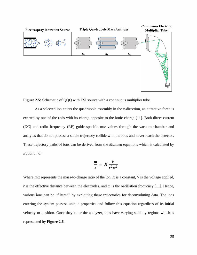
25
Figure 2.5: Schematic of QQQ with ESI source with a continuous multiplier tube.
As a selected ion enters the quadrupole assembly in the z-direction, an attractive force is
exerted by one of the rods with its charge opposite to the ionic charge [11]. Both direct current
(DC) and radio frequency (RF) guide specific m/z values through the vacuum chamber and
analytes that do not possess a stable trajectory collide with the rods and never reach the detector.
These trajectory paths of ions can be derived from the Mathieu equations which is calculated by
Equation 6:
𝒎
𝒛= 𝑲
𝑽
𝒓𝟐𝝎𝟐
Where m/z represents the mass-to-charge ratio of the ion, K is a constant, V is the voltage applied,
r is the effective distance between the electrodes, and ω is the oscillation frequency [11]. Hence,
various ions can be “filtered” by exploiting these trajectories for deconvoluting data. The ions
entering the system possess unique properties and follow this equation regardless of its initial
velocity or position. Once they enter the analyzer, ions have varying stability regions which is
represented by Figure 2.6.
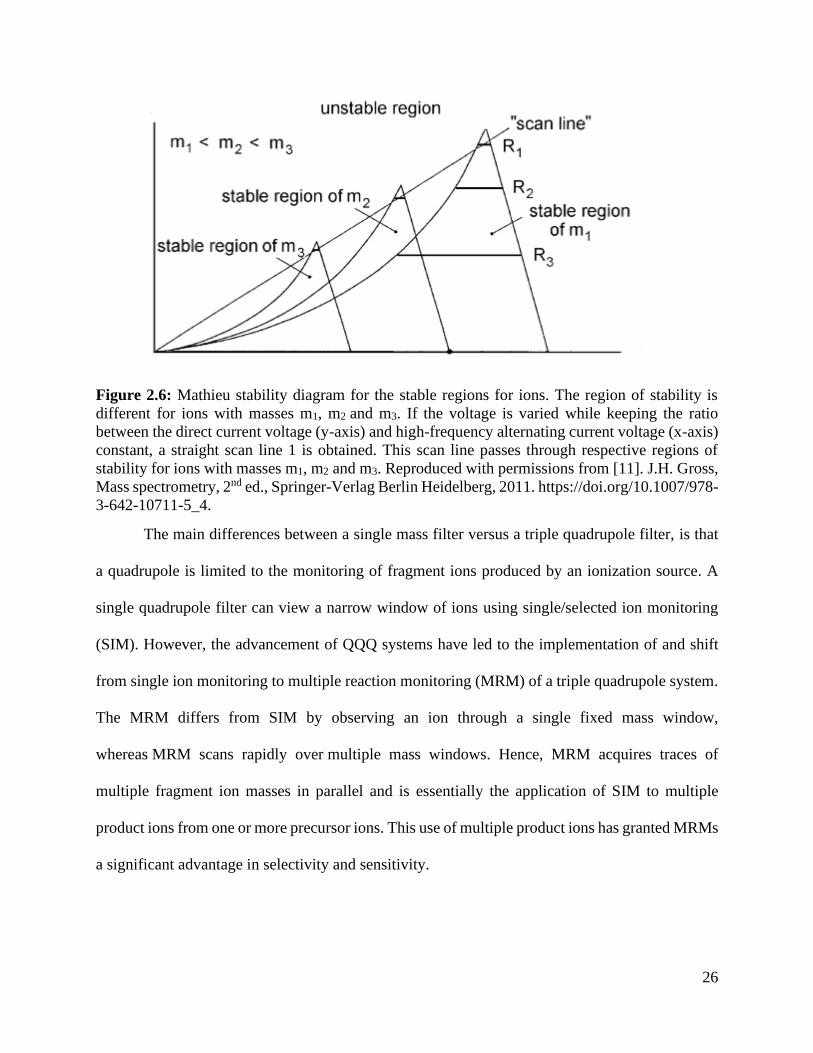
26
Figure 2.6: Mathieu stability diagram for the stable regions for ions. The region of stability is
different for ions with masses m1, m2 and m3. If the voltage is varied while keeping the ratio
between the direct current voltage (y-axis) and high-frequency alternating current voltage (x-axis)
constant, a straight scan line 1 is obtained. This scan line passes through respective regions of
stability for ions with masses m1, m2 and m3. Reproduced with permissions from [11]. J.H. Gross,
Mass spectrometry, 2nd ed., Springer-Verlag Berlin Heidelberg, 2011. https://doi.org/10.1007/978-
3-642-10711-5_4.
The main differences between a single mass filter versus a triple quadrupole filter, is that
a quadrupole is limited to the monitoring of fragment ions produced by an ionization source. A
single quadrupole filter can view a narrow window of ions using single/selected ion monitoring
(SIM). However, the advancement of QQQ systems have led to the implementation of and shift
from single ion monitoring to multiple reaction monitoring (MRM) of a triple quadrupole system.
The MRM differs from SIM by observing an ion through a single fixed mass window,
whereas MRM scans rapidly over multiple mass windows. Hence, MRM acquires traces of
multiple fragment ion masses in parallel and is essentially the application of SIM to multiple
product ions from one or more precursor ions. This use of multiple product ions has granted MRMs
a significant advantage in selectivity and sensitivity.

27
2.2.4. High-Resolution mass spectrometry – Orbitrap
In terms of mass accuracy, a triple quadrupole mass analyzer provides low to medium
resolution (~10 ppm) for analytes whereas instrumentation methods like the orbitrap provides 1-5
ppm resolution. Orbitrap typically utilizes ESI because of its ability to preserve the natural state
of the species under investigation. Unlike the QQQ which forces ions to follow a more linear
trajectory, the orbitrap keeps ions in an oscillating movement around the trap represented by
Figure 2.7.
Figure 2.7: Simple schematic of orbitrap mass analyzer with oscillating frequency where ions are
moving in spirals around a spindle-like-shaped central electrode. The terms r and z are cylindrical
coordinates (z = 0 being the plane of symmetry of the field). Figure adapted from [11]. J.H. Gross,
Mass spectrometry, 2nd ed., Springer-Verlag Berlin Heidelberg, 2011. https://doi.org/10.1007/978-
3-642-10711-5_4.
The different setup of orbitrap systems utilize a series quadrupole and octapole focusing
lenses to filter unwanted species. Ions enter the radiofrequency (RF)-only bent quadrupole of the
C-trap and get stored there as they lose energy in gentle collisions with the bath gas [11]. The RF
voltage is then ramped down and a high-voltage pulse is applied across the trap, ejecting ions
orthogonally to its curved axis. Ion packets enter the Orbitrap analyzer at an offset from its equator
and experience strong radial and axial fields. The axial component of the field forces axial

28
acceleration and hence so-called excitation by injection, while the radial component sets ions on a
circular orbit around the central electrode. As the voltage on this electrode increases during the
injection process, the radius of ion packet rotation gets squeezed down. Because of the strong
dependence of the rotational frequencies on the ion energies, angles, and initial positions, each ion
packet soon spreads over the angular coordinate and forms a thin rotating ring. After voltages are
stabilized, the differential amplifier detects a current induced by these rings on the split outer
electrodes of the trap. Orbitraps can employ different fragmentation energies which can produce
unique product ions for furthering the detection and confirmation of unknown and novel analytes.
2.3. Overcoming challenges for metal analysis in LC-MS/MS
As stated previously, LC is a separation technique which works by altering the polarity of
the mobile phases and column chemistry. Typically, small organic molecules and proteins are the
primary analytes studied, however, metal species can be monitored by coupling to techniques like
ICP-MS or ICP-OES [16,17]. Further separation of these analytes can be achieved by utilizing
specific metallic-binding molecules known as chelating agents. The term chelation refers to the
bonding of metal ions to form two or more separate coordinate bonds between
a polydentate ligand and a single central atom [18,19]. Some examples of chelating agents are
ethylenediaminetetraacetic acid (EDTA), dimercaptosuccinic acid (DMSA), and tartaric acid (TT)
which have been employed for various application such as pharmaceutical, water treatment, and
supplement delivery. Depending on the metal species, these agents can orient themselves into
different conformations which can then be elucidated further using the various column strategies
mentioned previously [20-22]. However, these strategies are sensitive to pH changes and can alter
the unique isotopic signatures that metal ions yield which can be difficult to monitor and control

29
without an ICP-type instrument. Thus, host-guest chemical strategies such as macrocycles have
been employed to preserve these natural isotopic ratios.
2.3.1. Macrocycles - Crown ethers
Macrocycles are a subclass of supramolecules which are defined by IUPAC "a cyclic
macromolecule or a macromolecular cyclic portion of a molecule." One subclass of macrocycles
are cryptates which may possess various heteroatoms or other functional groups to increase the
effectiveness of interacting with a guest molecule or species. One of the most studied macrocycles,
crown ether, was discovered in 1967 by Pedersen [23,24] who reported on their synthesis from
glycols and effectiveness to complex with metals. Because of these initial observations, crown
ethers have been utilized in various disciplines, such as industrial, medicinal, chemical, and, more
recently, forensics [24,25]. These ionophores have the unique ability to self-assemble and associate
with a broad range of analytes, or guests, for simple molecular recognition, transportation, and
extraction.
Initially, the formation of these host-guest systems, it was thought that the “lock-and-key”
principle described the mechanism of the guest species with the crown ether. Even though this idea
can be applied for more rigid interactions such as proteins with potential drug molecules, other
mechanisms have been proposed to provide more insight to this specific host-guest (H-G)
interaction. The main principle believed for this specific H-G system is noncovalent, electrostatic
interactions.
Opposite of covalent interactions, both molecules involved in noncovalent interactions keep
their respective electrons and are not shared [26]. Another interesting aspect with this phenomenon
is that it allows molecules to undergo various changes in conformation, which provides specific
conditions for substances that bind and react with each other. For crown ethers, the oxygen donor
atoms bind the alkali metal cation which have a high tendency to form noncovalent bonds.

30
Furthermore, noncovalent interactions encompass additional electrostatic interactions which refer
to electron-rich (oxygen) or -deficient (metal cations) atoms or ions. By substituting the donor
atoms within the cyclic structure, different binding affinities of metal ions can be achieved. Hence,
the adaptability of crown ethers has led to multiple strategies to further investigate and understand
the relationship between hosts and guests.
2.4. Strategies for investigating interactions of host-guest chemistry
2.4.1. Experimental approaches
A popular approach to monitor host-guest interactions is utilizing ESI source coupled to
mass analyzers as this soft ionization preserves the integrity of the complex and desolvates the
solvent media [27,28]. The main interests have focused on binding affinity and selectivity when
in the presence of multiple ionic species with variable charge states or sizes. For these experimental
designs, a set concentration was created for specified metallic species measured based on the
relative intensities both separately and in a mixture [29-33]. It was discussed that size (both the
inner cavity and cation) is a relevant factor as crown ethers can form “sandwich” structures which
is a 2:1 H-G ratio depicted in Figure 2.8. However, these structures appear most often when a
species of interest extends beyond the flexibility of the host [31].

31
Figure 2.8: Example of a “sandwich” structure with metal ion.
Experimental methods utilizing both low- and high-resolution mass analyzers have given
scientists a general concept and insights to help explain this H-G phenomenon. The collective
response has concluded that crown ethers interact with guests by the noncovalent, electrostatic
interactions [24,34-36]. However, technological advances have provided scientists the flexibility
to explore structural conformations and to simulate different conditions and scenarios using
theoretical calculations.
2.4.2. Theoretical calculations - Density Functional Theory (DFT)
There are two types of theoretical methodologies for investigating interactions across
variable systems - molecular dynamics and quantum mechanics. Molecular dynamics (MD)
modelling observes interactions by employing Newtonian physics and calculated force fields
[37,38]. This strategy is less computationally expensive and describes the motions of large
biomolecules and proteins more accurately than quantum mechanical approaches. However, MD
simulations are less suited for systems where quantum effects are important, i.e., transition metal
32
binding [38]. Hence, quantum mechanical (QM) approaches like density functional theory have
been used for crown ether complexation with metal ions [18,39-42].
Density-functional theory (DFT) is a computational quantum mechanical modelling
method used in physics, chemistry, and materials science to investigate the electronic structure (or
nuclear structure) and the ground state of many-body systems, in particular atoms, molecules, and
the condensed phases [43]. Using this theory, the properties of a many-electron system can be
determined by using functionals, i.e., functions of another function. In the case of DFT, these are
functionals of the spatially dependent electron density. DFT is popular due to its ratio between
performance and computational cost. It is very fast than other many wavefunction methods,
comparatively to other quantum mechanical calculations, while possessing the same level of
accuracy and is applied to a variety of fields including condensed-matter physics, computational
physics, and computational chemistry.
As usual in many-body electronic structure calculations, the nuclei of the treated molecules
or clusters are seen as fixed (the Born–Oppenheimer approximation), generating a static external
potential V, in which the electrons are moving [43,44]. A stationary electronic state is then
described by a wavefunction Ψ (r1, …, rN) satisfying the many-electron time-independent
Schrödinger equation (Equation 2.7)
Equation 2.7
�̂�Ψ = [�̂� + �̂� + �̂�]Ψ = [∑ (−ℏ2
2𝑚𝑖∇𝑖
2)𝑁𝑖=1 + ∑ 𝑉(𝑟𝑖) + ∑ 𝑈(𝑟𝑖, 𝑟𝑗) 𝑁
𝑖<𝑗𝑁𝑖=1 ] Ψ = 𝐸Ψ
where, for the N-electron system, Ĥ is the Hamiltonian, E is the total energy, T̂ is the kinetic
energy, V̂ is the potential energy from the external field due to positively charged nuclei, and Û is
the electron–electron interaction energy. The operators T̂ and Û are called universal operators, as
they are the same for any N-electron system, while V̂ is system-dependent. This complicated

33
many-particle equation is not separable into simpler single-particle equations because of the
interaction term Û.
DFT utilizes various mathematical functions represented as basis sets which use partial
differential equations to represent electronic wave functions. These basis sets can be used to
calculate the ground state between molecules and atoms and yields different results including
binding energies, emission wavelengths, transition states, and others [45]. Depending on the
specific needs of the research and the size of the molecules under investigation, basis sets and even
hybrids are used to accurately represent the interactions.
The macrocycles in this study utilize the B3LYP/6-311G++ (2d, p) which describes the
valence and core electrons of carbon (C), hydrogen (H), nitrogen (N), oxygen (O), and sulfur (S)
atoms. For the metal species, the Los Alamos National Laboratory (LANL2DZ) basis set covers
the electrons for the alkali, alkali earth, and transition metals used in this study to measure their
thermochemical properties [41,46-50]. The information gathered will address the inability to
observe an Sb-18C6 complex as well as the sensitivity of said macrocycle for other common
transition metals. This is crucial for further development of heavy metal extractions in wastewater
treatment or drug delivery systems.
2.5. Summary
The utilization of each of the strategies above (both experimental and theoretical) have
been incorporated into this dissertation to further the understanding of not only the abilities of H-
G chemistry but also electrostatic interactions. This entity further delves into the practicality and
flexibility of liquid chromatography and mass analyzers for monitoring various species and
strategies on deconvoluting data. Additionally, it presents an alternative approach to highlight the
potential of chemical methods and alterations to by applied to various applications and fields. This

34
dissertation starts with a condensed review of the current practices of gunshot residue researchers
as well as the current limitations and lack of knowledge. It then addresses these shortcomings by
introducing an alternative method and interpretation of GSR information using LC-MS/MS
methodology. The previous sections laid the foundational knowledge

35
2.6. References for Chapter 2
[1] W. Feeney, C. Vander Pyl, S. Bell, T. Trejos, Trends in composition, collection,
persistence, and analysis of IGSR and OGSR: A review, Forensic Chem. 19 (2020) 100250.
https://doi.org/10.1016/j.forc.2020.100250.
[2] S. Practice, S. Handbook, S. Practice, Standard Practice for Gunshot Residue Analysis by
Scanning Electron Microscopy/Energy Dispersive X-Ray Spectrometry, ASTM Int. i (2020) 1–5.
https://doi.org/10.1520/E1588-20.
[3] D.A. Skoog, J.F. Holler, S.R. Crouch, Principles of Instrumental Analysis, 7th ed., 2018.
[4] V. D’Atri, A. Murisier, S. Fekete, J.L. Veuthey, D. Guillarme, Current and future trends
in reversed-phase liquid chromatography-mass spectrometry of therapeutic proteins, TrAC -
Trends Anal. Chem. 130 (2020) 115962. https://doi.org/10.1016/j.trac.2020.115962.
[5] G. Purcaro, S. Moret, L. Conte, Hyphenated liquid chromatography-gas chromatography
technique: Recent evolution and applications, J. Chromatogr. A. 1255 (2012) 100–111.
https://doi.org/10.1016/j.chroma.2012.02.018.
[6] D.J. Winzor, Determination of binding constants by affinity chromatography, J.
Chromatogr. A. 1037 (2004) 351–367. https://doi.org/10.1016/j.chroma.2003.11.092.
[7] J.S. Fritz, Ion Chromatography, Anal. Chem. 59 (1987).
https://doi.org/10.1021/ac00131a002.
[8] C.A. Lucy, Evolution of ion-exchange: From Moses to the Manhattan Project to Modern
Times, J. Chromatogr. A. 1000 (2003) 711–724. https://doi.org/10.1016/S0021-9673(03)00528-
4.
[9] B. Buszewski, S. Noga, Hydrophilic interaction liquid chromatography (HILIC)-a
powerful separation technique, Anal. Bioanal. Chem. 402 (2012) 231–247.
https://doi.org/10.1007/s00216-011-5308-5.

36
[10] S. Paul-Dauphin, F. Karaca, T.J. Morgan, M. Millan-Agorio, A.A. Herod, R. Kandiyoti,
Probing size exclusion mechanisms of complex hydrocarbon mixtures: The effect of altering
eluent compositions, Energy and Fuels. 21 (2007) 3484–3489.
https://doi.org/10.1021/ef700410e.
[11] J.H. Gross, Mass spectrometry, 2nd ed., Springer-Verlag Berlin Heidelberg, 2011.
https://doi.org/10.1007/978-3-642-10711-5_4.
[12] M. Yamashita, J.B. Fenn, Electrospray ion source. Another variation on the free-jet
theme, J. Phys. Chem. 88 (1984) 4451–4459. https://doi.org/10.1021/j150664a002.
[13] J.H. Gross, Instrumentation, in: Mass Spectrom., 2nd ed., Springer Berlin Heidelberg,
Berlin, Heidelberg, 2011: pp. 117–221. https://doi.org/10.1007/978-3-642-10711-5_4.
[14] D.J.D. Pimlott, L. Konermann, Using covalent modifications to distinguish protein
electrospray mechanisms: Charged residue model (CRM) vs. chain ejection model (CEM), Int. J.
Mass Spectrom. 469 (2021) 116678. https://doi.org/10.1016/j.ijms.2021.116678.
[15] H. Metwally, Q. Duez, L. Konermann, Chain Ejection Model for Electrospray Ionization
of Unfolded Proteins: Evidence from Atomistic Simulations and Ion Mobility Spectrometry,
Anal. Chem. 90 (2018) 10069–10077. https://doi.org/10.1021/acs.analchem.8b02926.
[16] C.D. Quarles, M. MacKe, B. Michalke, H. Zischka, U. Karst, P. Sullivan, M.P. Field,
LC-ICP-MS method for the determination of “extractable copper” in serum, Metallomics. 12
(2020) 1348–1355. https://doi.org/10.1039/d0mt00132e.
[17] E. Matsumoto-Tanibuchi, T. Sugimoto, T. Kawaguchi, N. Sakakibara, M. Yamashita,
Determination of inorganic arsenic in seaweed and seafood by LC-ICP-MS: Method validation,
J. AOAC Int. 102 (2019) 612–618. https://doi.org/10.5740/jaoacint.18-0148.
[18] N. Yasarawan, K. Thipyapong, Complexation reactions, electronic properties, and

37
reactivity descriptors of cysteamine-based ligands in aqueous solution: a PCM/DFT study,
Struct. Chem. 29 (2018) 1723–1737. https://doi.org/10.1007/s11224-018-1151-z.
[19] N. Yasarawan, K. Thipyapong, V. Ruangpornvisuti, Substituent effect on the proton-
related phenomena and chelation behavior of hydroxypicolinic compounds: A DFT
investigation, Struct. Chem. 27 (2016) 505–524. https://doi.org/10.1007/s11224-015-0579-7.
[20] C. Sarzanini, E. Mentasti, Determination and speciation of metals by liquid
chromatography, J. Chromatogr. A. 789 (1997) 301–321. https://doi.org/10.1016/S0021-
9673(97)00988-6.
[21] W. Beck, K. Sünkel, Metal Complexes of Indigo and of Some Related Ligands,
Zeitschrift Für Anorg. Und Allg. Chemie. (n.d.). https://doi.org/10.1002/zaac.201900363.
[22] M. Tella, G.S. Pokrovski, Antimony(III) complexing with O-bearing organic ligands in
aqueous solution: An X-ray absorption fine structure spectroscopy and solubility study,
Geochim. Cosmochim. Acta. 73 (2009) 268–290. https://doi.org/10.1016/j.gca.2008.10.014.
[23] J.S. Bradshaw, R.M. Izatt, Crown Ethers: The Search for Selective Ion Ligating Agents,
Acc. Chem. Res. 30 (1997) 338–345. https://doi.org/10.1021/ar950211m.
[24] I. V. Kolesnichenko, E. V. Anslyn, Practical applications of supramolecular chemistry,
Chem. Soc. Rev. 46 (2017) 2385–2390. https://doi.org/10.1039/c7cs00078b.
[25] W. Feeney, K. Menking-Hoggatt, C. Vander Pyl, C.E. Ott, S. Bell, L. Arroyo, T. Trejos,
Detection of Organic and Inorganic Gunshot Residues from Hands using Complexing Agents
and LC-MS/MS, Anal. Methods. (2021). https://doi.org/10.1039/d1ay00778e.
[26] P.A. Kollman, Noncovalent Interactions, Acc. Chem. Res. 10 (1977) 365–371.
https://doi.org/10.1021/ar50118a003.
[27] M. Frańska, A. Michalak, Gas-phase relative stabilities of Tl+-crown ether complexes

38
and Rb+-crown ether complexes, Eur. J. Mass Spectrom. 24 (2018) 279–288.
https://doi.org/10.1177/1469066717752794.
[28] S.M. Blair, J.S. Brodbelt, A.P. Marchand, K.A. Kumar, H.-S. Chong, Evaluation of
Binding Selectivities of Caged Crown Ligands toward Heavy Metals by Electrospray
Ionization/Quadrupole Ion Trap Mass Spectrometry, Anal. Chem. 72 (2000) 2433–2445.
https://doi.org/10.1021/ac991125t.
[29] S.M. Blair, E.C. Kempen, J.S. Brodbelt, Determination of binding selectivities in host-
guest complexation by electrospray/quadrupole ion trap mass spectrometry, J. Am. Soc. Mass
Spectrom. 9 (1998) 1049–1059. https://doi.org/10.1016/s1044-0305(98)00082-8.
[30] S.M. Blair, J.S. Brodbelt, A.P. Marchand, H.S. Chong, S. Alihodží, Evaluation of alkali
metal binding selectivities of caged aza-crown ether ligands by microelectrospray
ionization/quadrupole ion trap mass spectrometry, J. Am. Soc. Mass Spectrom. 11 (2000) 884–
891. https://doi.org/10.1016/S1044-0305(00)00166-5.
[31] C.L. Sherman, J.S. Brodbelt, A.P. Marchand, B. Poola, Electrospray ionization mass
spectrometric detection of self-assembly of a crown ether complex directed by π-stacking
interactions, J. Am. Soc. Mass Spectrom. 16 (2005) 1162–1171.
https://doi.org/10.1016/j.jasms.2005.03.014.
[32] C.L. Sherman, J.S. Brodbelt, Partitioning Model for Competitive Host−Guest
Complexation in ESI-MS, Anal. Chem. 77 (2005) 2512–2523.
https://doi.org/10.1021/ac048518z.
[33] S.M. Williams, J.S. Brodbelt, A.P. Marchand, D. Cal, K. Mlinaric-Majerski, Metal
complexation of thiacrown ether macrocycles by electrospray ionization mass spectrometry,
Anal. Chem. 74 (2002) 4423–4433. https://doi.org/10.1021/ac011227v.

39
[34] H.J. Schneider, A.K. Yatsimirsky, Selectivity in supramolecular host-guest complexes,
Chem. Soc. Rev. 37 (2008) 263–277. https://doi.org/10.1039/b612543n.
[35] Y. Tao, R.R. Julian, Factors that influence competitive intermolecular solvation of
protonated groups in peptides and proteins in the gas phase, J. Am. Soc. Mass Spectrom. 24
(2013) 1634–1640. https://doi.org/10.1007/s13361-013-0684-z.
[36] A. McKenzie-Coe, S. Bell, F. Fernandez-Lima, Detection of firearm discharge residue
from skin swabs using trapped ion mobility spectrometry coupled to mass spectrometry, Anal.
Methods. 10 (2018) 4219–4224. https://doi.org/10.1039/c8ay00658j.
[37] D.M. Zuckerman, Statistical physics of biomolecules: An introduction, Stat. Phys.
Biomol. An Introd. (2010) 1–325. https://doi.org/10.1201/b18849.
[38] S.B. Choi, B.K. Yap, Y.S. Choong, H. Wahab, Molecular dynamics simulations in drug
discovery, Encycl. Bioinforma. Comput. Biol. ABC Bioinforma. 1–3 (2018) 652–665.
https://doi.org/10.1016/B978-0-12-809633-8.20154-4.
[39] S. Hadisaputra, H.D. Pranowo, R. Armunanto, Extraction of strontium(ii) by crown ether:
Insights from density functional calculation, Indones. J. Chem. 12 (2012) 207–216.
https://doi.org/10.22146/ijc.21332.
[40] B. Çiçek, A. Yildiz, Synthesis, metal ion complexation and computational studies of thio
oxocrown ethers, Molecules. 16 (2011) 8670–8683. https://doi.org/10.3390/molecules16108670.
[41] Z. Jing, G. Wang, Y. Zhou, D. Pang, F. Zhu, H. Liu, Selectivity of 18-crown-6 ether to
alkali ions by density functional theory and molecular dynamics simulation, J. Mol. Liq. 311
(2020). https://doi.org/10.1016/j.molliq.2020.113305.
[42] A.A. Bagatur’yants, A.Y. Freidzon, M. V Alfimov, E.J. Baerends, J.A.K. Howard, L.G.
Kuz’mina, DFT calculations on the electronic and geometrical structure of 18-crown-6

40
complexes with Ag+, Hg2+, Ag-0, Hg+, (HgAgNO3)-Ag-0, and HgX2 (X = Cl, Br, and I), J.
Mol. Struct. 588 (2002) 55–69. https://doi.org/10.1016/s0166-1280(02)00119-7.
[43] J.F. Stanton, A Chemist’s Guide to Density Functional Theory By Wolfram Koch
(German Chemical Society, Frankfurt am Main) and Max C. Holthausen (Humbolt University
Berlin). Wiley-VCH: Weinheim. 2000. ISBN 3-527-29918-1, 2001.
https://doi.org/10.1021/ja004799q.
[44] K. Capelle, A bird’s-eye view of density-functional theory, Brazilian J. Phys. 36 (2006)
1318–1341. https://doi.org/10.1590/s0103-97332006000700035.
[45] K.A. Baseden, J.W. Tye, Introduction to density functional theory: Calculations by hand
on the helium atom, J. Chem. Educ. 91 (2014) 2116–2123. https://doi.org/10.1021/ed5004788.
[46] R. Puchta, R. Van Eldik, Host-guest complexes of oligopyridine cryptands: Prediction of
ion selectivity by quantum chemical calculations, Eur. J. Inorg. Chem. (2007) 1120–1127.
https://doi.org/10.1002/ejic.200600715.
[47] Yahmin, H.D. Pranowo, R. Armunanto, Theoretical study on 15-crown-5 complex with
some metal cations, Indones. J. Chem. 12 (2012) 135–140. https://doi.org/10.22146/ijc.21353.
[48] Y. Zhang, X.Y. Wang, B.H. Luo, Y. Xia, DFT study of crown ether-bridged Z-stilbenes
and their complexes with alkali metal cations, J. Organomet. Chem. 699 (2012) 31–38.
https://doi.org/10.1016/j.jorganchem.2011.10.032.
[49] A. Üngördü, N. Tezer, DFT study on metal-mediated uracil base pair complexes, J. Saudi
Chem. Soc. 21 (2017) 837–844. https://doi.org/10.1016/j.jscs.2017.04.003.
[50] N. Islam, S.S. Chimni, Binding and selectivity of phenazino-18-crown-6-ether with
alkali, alkaline earth and toxic metal species: A DFT study, J. Mol. Struct. (2017).
https://doi.org/10.1016/j.molstruc.2016.10.100.

41
CHAPTER 3
Trends in Composition, Collection, Persistence, and Analysis of IGSR
and OGSR: A Review
42
Overview: Presented is a review of scientific literature from the past two decades on the various
aspects affecting deposition, persistence, and collection of organic and inorganic gunshot residue
(OGSR and IGSR, respectively). With market changes in the manufacture of modern ammunition
affecting typical elemental composition, morphology of particles, and complex transfer and
persistence mechanisms, the forensic community is investigating means to use a more
comprehensive approach that incorporates IGSR and OGSR detection. Once collected, inorganic
components are more persistent over time than OGSR. Conversely, exogenous compounds that
mimic OGSR markers are less prevalent in the environment than IGSR-like elements. As a result,
the combined detection of IGSR and OGSR is expected to decrease the occurrence of false
positives and false negatives and bring superior confidence in the interpretation of results. This
manuscript also compiles information on over 180 primary IGSR and OGSR components reported
in the literature for modern ammunition (standard and non-toxic) and their respective analyses.
Also, this review offers a discussion of the capabilities and limitations of novel methods and future
opportunities to adopt changes in the management of forensic investigations to enhance the
versatility, reliability, and response time in the discipline.
The following chapter is an adaptation of a previously published article ©2020: William
Feeney, Courtney Vander Pyl, Suzanne Bell, Tatiana Trejos. Trends in composition, collection,
persistence, and analysis of IGSR and OGSR: A review. Forensic Chemistry. Published online
May 2020, https://doi.org/10.1016/j.forc.2020.1002508E

43
3.1. Introduction
With the advancement of instrumentation and understanding of the formation of analytes
after a combustion event, firearm discharge residue (FDR) or gunshot residue (GSR) has had an
increased interest amongst the forensics community. Until recently, analysts have focused on the
primer residues, commonly referred to as inorganic gunshot residue (IGSR) [1,2]. However, this
is just one aspect of GSR. The second group of compounds originates from the propellant and
lubricant and, in turn, is referred to as organic gunshot residue (OGSR) [3-7]. There have been
numerous reviews over the past 25 years that cover analysis, distribution, collection, and
instrumentation [1,8-10]. In light of this, this review has two main goals. The first is to compile a
comprehensive list of IGSR and OGSR analytes present in “standard” (containing lead, barium,
antimony) and “non-standard” (lead-free, non-toxic) ammunition. The second is to collate the
information gathered by previous reviews and manuscripts that focus on emerging col- lection
techniques, persistence information, and amounts (particle count or concentration) detected.
Although analytical methods are essential to address, they are not the primary focus of this review.
Current research reports describe the combined detection of IGSR and OGSR compounds
to increase the evidentiary value of suspected samples using single techniques or a combination of
orthogonal methods [5,11-16]. For detection of inorganic gunshot residues (IGSR), Scanning
Electron Microscopy Energy Dispersive X-Ray Spectroscopy (SEM/EDS) is currently considered
the gold standard. The ASTM E1588-17 standard practice utilizes SEM/EDS to classify the
inorganic particulates based on their elemental composition and morphological features (shape and
size) [2]. Indeed, this capability of SEM/EDS to characterize spheroid morphology and conduct
elemental analysis on a single microscopic particle is what makes it a highly reliable method for
GSR examination. SEM/EDS recognizes these particulates by bombarding electrons onto the
surface. Similarly, although much less common at forensic laboratories, a time-of-flight secondary

44
ion mass spectrometer (TOF-SIMS) applies an ion beam (i.e., Bi, Ga) to the surface to generate
secondary ions. Both techniques can detect small particle ranges and produce high-resolution
images for confirmation. TOF-SIMS offers lower limits of detection and provides mass spectral
data that helps to resolve signal overlaps encountered by SEM/EDS.
One of the limitations of SEM/EDS is the time required for processing the automated
particle analysis and further conducting the manual review of the data by the expert scientist. For
instance, analysis of one sample typically takes between 4 and 10 h, depending on the number,
size, composition, and abundance of the IGSR. Considering that a ty- pical case consists of at least
four specimens per individual of interest (i.e., palm, back, right, and left hands) plus negative and
positive quality controls, the laboratory analysis can quickly become a bottle- neck. With recent
scientific advancements of detectors (Silicon Drift Detectors vs. Silicon Lithium Detectors) and
brighter sources (i.e., CeB6 vs. W filament) SEM/EDS instruments are becoming smaller and
faster. A recent study reports a desktop SEM/EDS able to reduce 30–50% analysis time while still
maintaining the ability to perform non- destructive particle counting and elemental mapping with
sensitivity comparable to traditional instruments [17-19].
Recent manuscripts have reported several considerations for the interpretation of elemental
composition of IGSR particulates and morphological features because of observed shape
irregularities and the risk of false positives due to an individual’s environment [16,20-25].
Interpretation of IGSR evidence should be evaluated on a case-by-case basis with an understanding
of possible elemental combinations of particulates that could arise from an individual’s
surroundings. Indeed, the ASTM E5188-17 warns examiners not to consider morphology alone as
a criterion for the identification of GSR due to its large variability. For instance, welding,
fireworks, electrical, and mechanical career fields have an increased likelihood of possessing

45
common IGSR analytes on their person [20-28]. Also, the introduction of non-toxic ammunition
brings a series of challenges to SEM/EDS analysis. For instance, some of the compounds are more
prevalent in the environment (i.e., Zn, Ti, K) and the detection of lower atomic number elements
requires different optimal settings than those established for typical heavy metals. As a potential
solution, few studies have reported the incorporation of Cathodoluminescence and Raman to
SEM/EDS instruments for expanded capabilities to identify non-toxic GSR [18,19,29]. As another
alternative, several groups advocate monitoring OGSR markers in conjunction with IGSR to
reduce the rate of false positives [5,11,13-16].
Other methodologies for IGSR analysis include Neutron Activation Analysis (NAA),
Inductively Coupled Plasma-based methods (ICP), and Laser-Induced Breakdown Spectroscopy
(LIBS), which can provide similar elemental information as SEM/EDS [13,30-36]. However,
unlike SEM/EDS, these methods don’t have the ability for single-particle analysis.
The effort to increase confidence with GSR information generates emerging techniques
that can lead to fast analysis times and minimal sample preparation. For instance, LA-ICP/MS and
LIBS take minutes, whereas traditional SEM/EDS lasts several hours for a single sample [11,37]
and therefore are attractive methods that could complement current practice. These two techniques
employ pulsed laser beams for direct qualitative and quantitative results for increased selectively
and simultaneous multi-elemental detection in the low ppm levels [14,31,32,38-40]. Recent
advancements with LIBS also allow for portability and versatility at crime scenes and laboratories
[34]. Although laser-based techniques can screen for inorganic gunshot residue elements, standard
confirmation by SEM/EDS is still required for positively identifying IGSR particles. The potential
value of these methods, however, is its utility to make informed decisions at the scene and at the
laboratory that can lead to more effective case management. The speed of analysis can also open

46
opportunities for more holistic case interpretation by adding alternative specimens that otherwise
would be tedious with conventional methods, such as clothing, vehicle surfaces, face areas. A
recent study demonstrated the ability of LIBS to provide spatial information along with
simultaneous detection of multiple markers (i.e., Pb, Ba, and Sb) from the same location (~50–100
µm). This ablation approach increases the confidence in the results that the markers come from a
particle or group of particles, located in a small area rather than somewhere else within the trace
[41]. This spatial information does not match the morphologic information but provides added
value over bulk analysis. Although the capability of LIBS and LA- ICP-MS to analyze a single
particle has not been explored, it is technically possible, as some configurations allow laser beams
as small as 4 µm. It will be interesting to see if scientists take future studies in this direction. These
methods have been combined with different techniques, such as electrochemistry, to compensate
for the lack of morpho- logical features [11].
The OGSR detection has been evaluated by diverse methodologies, including
chromatographic separation with mass spectrometry detection, Raman spectroscopy,
electrochemistry, and ion mobility spectrometry (IMS) [3,6,42-49].
Electrochemical detection employs potential ramps to induce redox reactions at the surface
of an electrode. The benefits of this practice are its rapid analysis time (minutes), low production
costs, and the ability to detect both IGSR and OGSR analytes. Additionally, it is non-destructive
and portable. One obstacle is the inability to detect barium due to its high electrochemical potential.
Recently, the combined methods of LIBS and electrochemistry demonstrated high classification
rates for IGSR and OGSR [11].
Other quick analysis approaches, such as Raman spectroscopy and FTIR, display their
proficiency for complementary information of organic and inorganic compounds [50,51]. Both

47
practices implement non- destructive, vibrational frequencies for characterization and imaging of
particulates. FTIR provides information for OGSR markers, specifically nitro-compounds such as
2-nitrodiphenylamine (2-NDPA) and 4-nitrodiphenylamine (4-NDPA), but it lacks sensitivity at
trace levels [50]. Although fluorescence interferences suppress Raman spectroscopy signals, the
scattering and resonance capabilities allow for the dual detection of macro- and micro-sized GSR
particles [51]. However, a drawback of both techniques is their inability to detect and identify
particles that contain solely metal components (i.e., Pb, Sb, Al), which are important markers of
IGSR for interpretation of evidence.
In OGSR analysis, instrumentation techniques characterize compounds based on molecular
properties and formations of product ions. For instance, ion mobility spectrometry (IMS) separates
and identifies organic molecules based on their mobility and collisional cross-sections inside a
drift tube [42]. Alteration of the drift tube and the ability to couple it with various mass analyzers
make it extremely versatile. Typical IMS analysis times can range from the millisecond to sub-
minute timescales and requires minimum resources for sample preparation and operation
[6,42,52,53]. More traditional OGSR practices such as gas chromatography (GC) and liquid
chromatography (LC), rely on volatility and polarity of molecules, respectively. Analysts that
apply GC/MS identify analytes by the molecules’ Kovats retention index and its probability match
scores of the product ions. Mass spectral libraries generate match scores and compare the ratios of
formed product ions against a “neat” standard. Scientists that use LC/MS systems manipulate
various mobile phase and column conditions to separate similarly structured compounds. The
ability to exchange and pair several ionization sources (ESI, APCI, CI, DART) with different mass
analyzers (QQQ, TOF, orbitrap) allows LC/MS instrumentation to be adaptable and detect ions in

48
the ppb and ppt range. Recently, a study demonstrated the capability to monitor IGSR and OGSR
from a single sample [12].
Different reviews highlight the change in interpretation of GSR because of the introduction
of “non-toxic” or lead-free ammunition [54- 57]. These modern ammunitions replace traditional
primers mixtures of lead, barium, and antimony with bismuth, manganese, aluminum, zinc,
titanium, or organic compounds [58-60]. This work compiles lists of both ammunition types and
their components while also expanding on information about persistence, collection, and detected
amounts illustrated by previous reviews and publications [1,8,10,61,62].
3.1. Inorganic and Organic Compositions of Modern Ammunitions
Modifications in modern ammunition broaden the scope of novel IGSR and OGSR
markers. Further investigations regarding persistence and prevalence highlight the relevancy of
new components as indicators for GSR evidence. Additionally, the capabilities of analytical
methods to characterize these diverse analytes are essential to increase the confidence of a
collected sample.
The composition of propellants, lubricants, and primers in traditional ammunition contains
numerous OGSR compounds with various functions. Explosives in the propellent consist of
nitrocellulose (NC), nitroglycerin (NG), and nitroguanidine (NGu), while compounds like ethyl
centralite (EC), diphenylamine (DPA), and diphenylamine derivatives act as plasticizers and
stabilizers [63]. The purpose of these additives is to prevent thermal decomposition and to lower
the viscosity of the ammunition. Post-firing, these analytes adhere to multiple sur- faces such as
hands and clothing due to their lipophilic nature.
Traditional primers contain lead styphnate, barium nitrate, and antimony sulfide, which
serve as the explosive, the oxidizer, and the fuel, respectively. Other targeted metals and alloys

49
originate from the core and jacket of a cartridge or bullet. To lessen the exposure to toxic lead
ammunition, manufacturers substitute heavy metals with polymer- coated bullets and lead-free
primers. Byproducts from the primer, core, or barrel attach to the environment and can form a dark
grey or black ring around a target area known as a “bullet wipe” [64].
Below are two comprehensive lists of traditional and modern ammunition OGSR and IGSR
components (Tables 3.1 and 3.2, respectively) frequently reported in the literature. Typical
nomenclature, chemical formulae, and functionality of components in various ammunitions are
also listed.

50
Table 3.1: Organic compounds that may contribute to gunshot residues (*compounds typically monitored).
Compound Function Structure
Acenaphthene Combustion
byproduct
Acenaphthylene Combustion
byproduct
Acetophenone Combustion
byproduct
Akardite I (AKI) Stabilizer
Akardite II (AKII) Stabilizer
Akardite III (AKIII) Stabilizer
2-Amino-4,6-
dinitrotoluene Explosive
4-Amino-2,6-
dinitrotoluene Explosive
Compound Function Structure
Aniline Combustion
byproduct
Anthracene Combustion
byproduct
Benzaldehyde Combustion
byproduct
Benzene Combustion
byproduct
Benzo[a]pyrene Combustion
byproduct
Benzo[b]thiophene Combustion
byproduct
Benzonitrile Combustion
byproduct
Benzophenone Combustion
byproduct
Benzothiazole Combustion
byproduct
Carbazole Carbazole
Hexahydro-1,3,5-
trinitro-1,3,5- triazine
Cyclonite (RDX)
Explosive
Dextrin Sensitizer
Diamyl phthalate Plasticizer
Carbazole Carbazole
Hexahydro-1,3,5-
trinitro-1,3,5- triazine
Cyclonite (RDX)
Explosive
Dextrin Sensitizer
Diamyl phthalate Plasticizer
Diazodinitrophenol
Initiating
explosive
(primer)
Dibutyl phthalate (DBP) Plasticizer
1,2-Dicyanobenzene Explosive
1,3-Dicyanobenzene Explosive
1,4-Dicyanobenzene Explosive
Diethylene glycol
dinitrate
Stabilizer,
plasticizer
Diethyl phthalate (DEP) Plasticizer
2,3-Dimethyl-2,3-
dinitrobutane
Stabilizer,
plasticizer
1,4-
Dimethylnaphthalene
Combustion
byproduct
Compound Function Structure
Aniline Combustion
byproduct
Anthracene Combustion
byproduct
Benzaldehyde Combustion
byproduct
Benzene Combustion
byproduct
Benzo[a]pyrene Combustion
byproduct
Benzylnitrile Combustion
byproduct
Biphenyl Combustion
byproduct
Biphenylene Combustion
byproduct
Butyl centralite (BC) Stabilizer,
plasticizer
Carbanilide Stabilizer,
plasticizer
Compound Function Structure Compound Function Structure Compound Function Structure Compound Function Structure
2,3-Dimethyl-2,3-
dinitrobutane
Stabilizer,
plasticizer
1,4-
Dimethylnaphthalene
Combustion
byproduct
2,6-
Dimethylnaphthalene
Combustion
byproduct
Dimethyl phthalate
(DMP) Plasticizer
Dimethyl sebacate Stabilizer,
plasticizer
3,5-Dinitroaniline
Explosive,
flash
suppressor
2,4-Dinitroanisole
(DNAN) Explosive
1,3-Dinitrobenzene Explosive
2,2′-
Dinitrodiphenylamine
(2,2-DNDPA)
Stabilizer
(derivative
of DPA)
2,4-
Dinitrodiphenylamine
Stabilizer
(derivative
of DPA)
Stabilizer
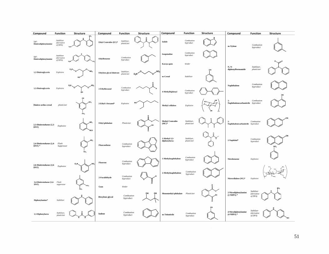
51
Compound Function Structure Compound Function Structure Compound Function Structure Compound Function Structure
2,4′-
Dinitrodiphenylamine
Stabilizer
(derivative
of DPA)
4,4′-
Dinitrodiphenylamine
Stabilizer
(derivative
of DPA)
1,2-Dinitroglycerin Explosive
1,3-Dinitroglycerin Explosive
Dinitro-ortho-cresol plasticizer
2,3-Dinitrotoluene (2,3-
DNT) Explosive
2,4-Dinitrotoluene (2,4-
DNT) *
Flash
Suppressor
2,6-Dinitrotoluene (2,6-
DNT) Explosive
Ethyl Centralite (EC)* Stabilizer,
plasticizer
Ethylbenzene Combustion
byproduct
Ethylene glycol dinitrate Stabilizer,
plasticizer
2-Ethylhexanal Combustion
byproduct
2-Ethyl-1-hexanol* Explosive
Ethyl phthalate Plasticizer
Fluoranthene Combustion
byproduct
Fluorene Combustion
byproduct
2-Furaldehyde Combustion
byproduct
Gum binder
Indole Combustion
byproduct
Isoquinoline Combustion
byproduct
Karaya gum binder
m-Cresol Stabilizer
4-Methylbiphenyl Combustion
byproduct
Methyl cellulose Explosive
Methyl Centralite
(MC)*
Stabilizer,
plasticizer
1-Methyl-3,3-
diphenylurea
Stabilizer,
plasticizer
1-Methylnaphthalene Combustion
byproduct
m-Xylene Combustion
byproduct
N, N-
diphenylformamide
Stabilizer,
plasticizer
Naphthalene Combustion
byproduct
1-
Naphthalenecarbonitrile
Combustion
byproduct
2-
Naphthalenecarbonitrile
Combustion
byproduct
2-Naphthol* Combustion
byproduct
Nitrobenzene Explosive
Nitrocellulose (NC)* Explosive
2-Methylnaphthalene Combustion
byproduct
Monomethyl-phthalate Plasticizer
m-Tolunitrile Combustion
byproduct
m-Xylene Combustion
byproduct
3,4-Dinitrotoluene (3,4-
DNT)
Flash
suppressor
Diphenylamine* Stabilizer
1,3-Diphenylurea Stabilizer,
plasticizer
Hexylene glycol Combustion
byproduct
Indene Combustion
byproduct
2-Nitrodiphenylamine
(2-NDPA) *
Stabilizer
(derivative
of DPA)
4-Nitrodiphenylamine
(4-NDPA) *
Stabilizer
(derivative
of DPA)
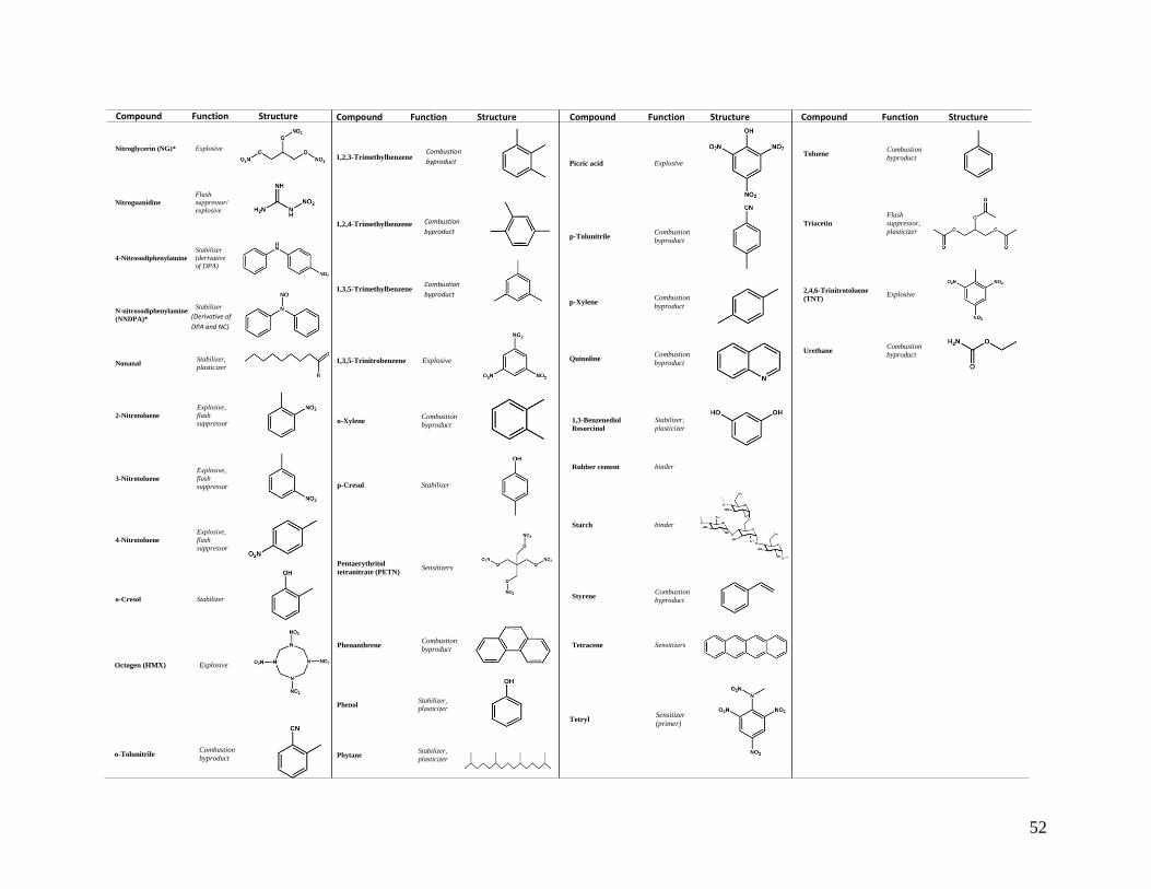
52
Compound Function Structure Compound Function Structure Compound Function Structure Compound Function Structure
Nitroglycerin (NG)* Explosive
Nitroguanidine
Flash
suppressor/
explosive
4-Nitrosodiphenylamine
Stabilizer
(derivative
of DPA)
N-nitrosodiphenylamine
(NNDPA)*
Stabilizer
(derivative
of DPA)
Nonanal Stabilizer,
plasticizer
2-Nitrotoluene
Explosive,
flash
suppressor
3-Nitrotoluene
Explosive,
flash
suppressor
4-Nitrotoluene
Explosive,
flash
suppressor
o-Cresol Stabilizer
Octogen (HMX) Explosive
o-Tolunitrile Combustion
byproduct
1,2,3-Trimethylbenzene Explosive
1,2,4-Trimethylbenzene Explosive
1,3,5-Trimethylbenzene Explosive
1,3,5-Trinitrobenzene Explosive
o-Xylene Combustion
byproduct
p-Cresol Stabilizer
Pentaerythritol
tetranitrate (PETN) Sensitizers
Phenanthrene Combustion
byproduct
Phenol Stabilizer,
plasticizer
Phytane Stabilizer,
plasticizer
Picric acid Explosive
p-Tolunitrile Combustion
byproduct
p-Xylene Combustion
byproduct
Quinoline Combustion
byproduct
1,3-Benzenediol
Resorcinol
Stabilizer,
plasticizer
Rubber cement binder
Starch binder
Styrene Combustion
byproduct
Tetracene Sensitizers
Tetryl Sensitizer
(primer)
Toluene Combustion
byproduct
Toluene Combustion
byproduct
Triacetin
Flash
suppressor,
plasticizer
2,4,6-Trinitrotoluene
(TNT) Explosive
Urethane Combustion
byproduct
(Derivative of
DPA and NC)
Combustion
byproduct
Combustion
byproduct
Combustion
byproduct

53
Table 3.2: Inorganic compounds that may contribute to gunshot residue.
Compound Function/Origin Compound Function/Origin
Aluminum powder* Reducing agent Lead peroxide (PbO2) Oxidizer
Aluminum oxide* (Al2O3) Oxidizer Lead styphnate (PbC6HN3O8) Explosive
Aluminum sulfide* (Al2S3) Fuel Lead thiocyanate (Pb(SCN)2) Explosive
Aluminum silicate* (Al2SiO5) Stabilizer Magnesium (Mg) Reducing agent
Antimony (Sb) Projectile Manganese* (Mn) Fuel
Antimony sulfide (Sb2S) Fuel/ friction agent Manganese oxide* (MnO) Fuel
Antimony sulfite (Sb2(SO3)3 Fuel Mercury (Hg) Explosive
Antimony trisulfide (Sb2S3) Fuel Mercury fulminate Hg(CNO)2) Explosive
Barium nitrate (Ba(NO3)2) Oxidizer Molybdenum trioxide* (MoO3) Oxidizer
Barium peroxide (BaO2) Oxidizer Nickle (Ni) Cartridge case
Beryllium (Be) Fuel Phosphorous (P) Cartridge case
Bismuth oxide* (Bi2O3) Oxidizer Phosphoric acid* (H3PO4) Oxidizer
Bismuth sulfide* (Bi2S3) Fuel Potassium chlorate (KClO4) Oxidizer
Boron powder* (B) Reducing agent Potassium nitrate* (KNO3) Oxidizer
Brass Cartridge case Silicon monoxide* (SiO) Fuel
Bronze Cartridge case Sodium nitrate (NaNO3) Oxidizer
Calcium carbonate* CaCO3) Oxidizer Sodium sulfate (Na2SO4) Stabilizer
Calcium silicide (CaSi2) Fuel Silicon monoxide* (SiO) Oxidizer
Chromium (Cr) Fuel Sodium nitrate (NaNO3) Oxidizer
Chromium oxide (Cr2O3) Oxidizer Stannic oxide* (SnO2) Oxidizer
Copper (Cu) Cartridge case Strontium nitrate (Sr(NO3)2 Oxidizer
Copper thiocyanate (CuSCN) Explosive Sulfur (S) Fuel
Copper (II) azide (Cu(N3)2) Explosive Tin (Sn) Cartridge case
Cupro-nickel Bullet casing Titanium powder Reducing agent
Ground Glass* Friction agent Tungsten (W) From bullet
Iron (Fe) Projectile Tungsten trioxide* (WO3) Oxidizer
Lead (Pb) Projectile Zinc (Zn) Cartridge case
Lead azide (Pb(N3)2 Explosive Zinc peroxide (ZnO2) Oxidizer
Lead oxide (PbO) Oxidizer Zinc sulfide* (ZnS) Fuel
Lead nitrate (Pb(NO3)2 Oxidizer Zirconium powder* (Zr) Reducing agent
* Also suggested in patents as significant constituents of non-toxic primers.

54
3.2. Common Gunshot Residue Sampling Media and Extractions
Law enforcement, researchers, and forensic scientists employ numerous substrates to
recover GSR for analysis [65-69]. Typical sample areas include hands, exposed skin, hair, and
clothing, as shown in Figure 3.1. Factors that affect the collection efficiency of a sampling medium
include the stability of OGSR and IGSR components and their deposition location [70-76]. For
example, OGSR compounds can permeate in the skin, which presents time constraints for analysts
to monitor and report their findings. Because IGSR particulates do not absorb into the skin, they
are more susceptible to environmental conditions. Therefore, ideal substrates must be compatible
with various instrumentation techniques and efficiently recover both components despite these
challenges.
3.1.1. Adhesives
The primary collection substrate used in GSR analysis is a carbon-based adhesive. These
adhesives collect both IGSR particulates and OGSR compounds because of their flat, porous
surface. The tape is typically attached to an aluminum stub on a capped sample holder to reduce
the interaction between the sample and collector, which reduces the risk of possible cross-
contamination and sample loss. A collection kit consists of four different stubs, one for each side
of each hand (i.e., left-palm, right-palm, left-back, right-back), as shown in Figure 3.1. Other
advantages of carbon-based tapes include the possibility for sequential analysis on a single sample
starting with non-destructive (i.e., electrochemistry, Raman) or minimally destructive (i.e., LIBS)
screening tests and then towards confirmatory methods (i.e., GC/MS, LC/MS, SEM/EDS)
[11,14,16,68,75,77,78]. Researchers have explored chirurgic and acrylic transparent tapes, which
are variations of traditional adhesives [76]. However, the ASTM E1588 method outlines carbon
adhesive stubs as the collection substrate due to the requirements of SEM/EDS [2,79].

55
3.1.2. Cloth
Another material used for casework and research is cloth or cotton swabs [66]. This method
applies a cloth or swab soaked with organic solvent (i.e., isopropanol, acetone) directly on the area
interest. By creating an organic extraction, this combinational approach focuses on the OGSR
compounds [16,80]. The thread count of fibers influences the effectiveness of IGSR capture and
can cause additional challenges for analysts. For instance, fibers can shield the particulates,
causing signal suppression or even charging effects for SEM/EDS.
3.1.3. Polymer
A third substrate used for the collection of GSR is polymers. Polymers typically consist of
polydimethylsiloxane (PDMS) or similar silicone-based material and exhibit “adhesive-like”
qualities [12,81]. This method, like adhesives, requires minimal sample preparation and can amass
both components of GSR because of its porous surface [12,82]. However, for IGSR, analysis
problems arise from polymers because of degassing in the vacuum chamber of an SEM/EDS.
Therefore, this collection technique should be limited to other analytical methods such as LC/MS.

56
Figure 3.1: Common sampling areas and techniques for gunshot residue evidence collection from
hands, hair, face, nostrils, and clothing [51–55].
3.1.4. Extractions
After collection, various solvents and mixtures extract analytes from substrates. Non-toxic
organic solvents like methanol, acetonitrile, iso- propanol, and aqueous buffers extract OGSR
compounds based on the partition coefficient (log P) values [6,85]. These solvents complement
techniques such as electrochemistry, GC/MS, and LC/MS [14,44,86-88]. For IGSR analytes,
concentrated acid solutions (typically HNO3 and HCl) solubilize the particulates for analysis on
different instrumentation techniques such as ICP/MS and LC/MS [14,33,66,89].
For volatile OGSR molecules, scientists use solid-phase micro-extraction (SPME), head
space sorptive extraction, and capillary micro- extraction of volatiles (CMV) techniques coupled
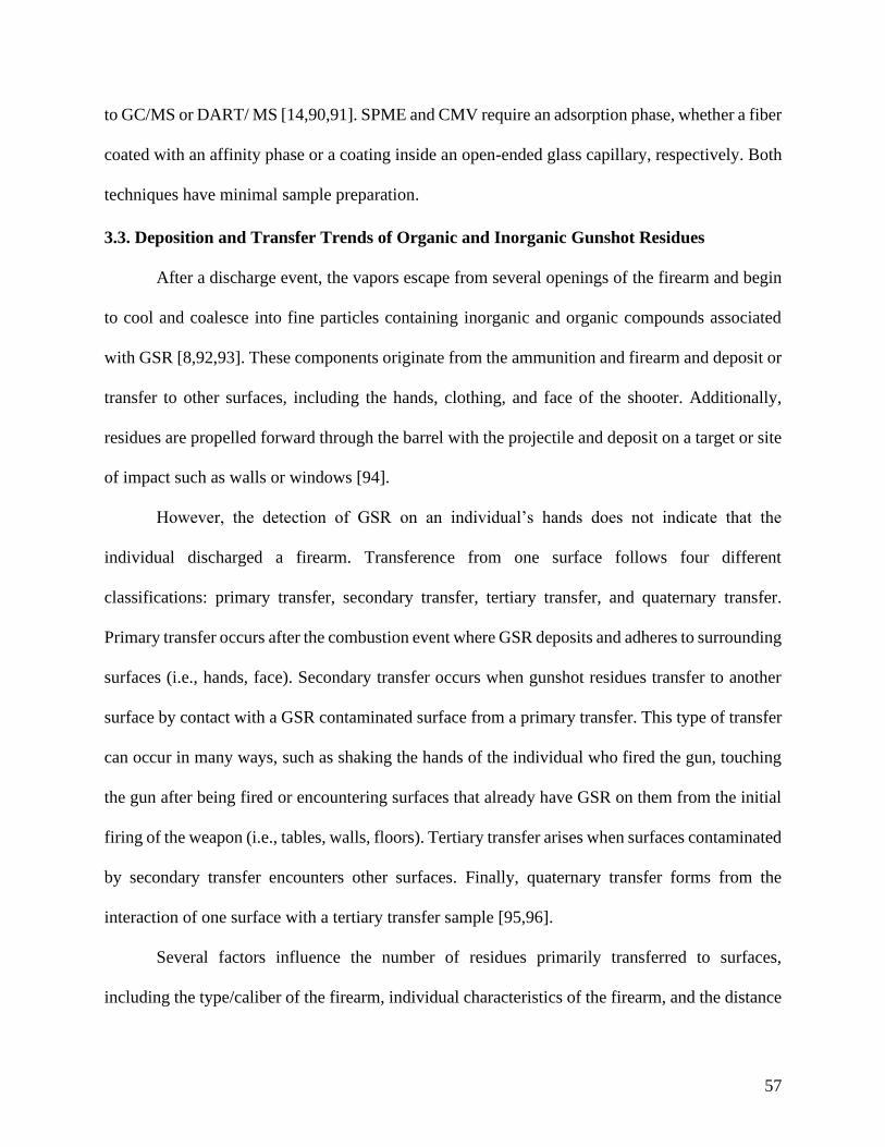
57
to GC/MS or DART/ MS [14,90,91]. SPME and CMV require an adsorption phase, whether a fiber
coated with an affinity phase or a coating inside an open-ended glass capillary, respectively. Both
techniques have minimal sample preparation.
3.3. Deposition and Transfer Trends of Organic and Inorganic Gunshot Residues
After a discharge event, the vapors escape from several openings of the firearm and begin
to cool and coalesce into fine particles containing inorganic and organic compounds associated
with GSR [8,92,93]. These components originate from the ammunition and firearm and deposit or
transfer to other surfaces, including the hands, clothing, and face of the shooter. Additionally,
residues are propelled forward through the barrel with the projectile and deposit on a target or site
of impact such as walls or windows [94].
However, the detection of GSR on an individual’s hands does not indicate that the
individual discharged a firearm. Transference from one surface follows four different
classifications: primary transfer, secondary transfer, tertiary transfer, and quaternary transfer.
Primary transfer occurs after the combustion event where GSR deposits and adheres to surrounding
surfaces (i.e., hands, face). Secondary transfer occurs when gunshot residues transfer to another
surface by contact with a GSR contaminated surface from a primary transfer. This type of transfer
can occur in many ways, such as shaking the hands of the individual who fired the gun, touching
the gun after being fired or encountering surfaces that already have GSR on them from the initial
firing of the weapon (i.e., tables, walls, floors). Tertiary transfer arises when surfaces contaminated
by secondary transfer encounters other surfaces. Finally, quaternary transfer forms from the
interaction of one surface with a tertiary transfer sample [95,96].
Several factors influence the number of residues primarily transferred to surfaces,
including the type/caliber of the firearm, individual characteristics of the firearm, and the distance

58
an individual from a target. For instance, a cylinder may allow for greater deposition of gunshot
residues compared to an enclosed chamber (i.e., revolver and pistol, respectively) [1,97]. The
cylinder’s design allows for vapors to escape easily after discharge, whereas the ejection port on
pistols be- comes the essential factor that affects the number of GSR. The location of an ejected
cartridge case (i.e., vertically or a particular side) and the duration of the ejection port (opening
and remaining open) significantly affects the number of residues that can escape and deposit [97].
The barrel length also plays a role in the amount of GSR deposited on an individual’s hands. For
example, a longer barrel increases the distance between the plume and a shooter’s hands upon
discharge, as opposed to a shorter barrel [93,97,98]. After firing a rifle, forensic scientists
anticipate a large number of residues to be transferred to target materials with fewer residues on
hands, while the likelihood of transfer onto the face and clothing increases [98,99].
The number of residues transferred to target materials is reliant on firearm type, as well as
the distance from which the individual discharging the firearm is standing (i.e., how far the muzzle
of the firearm was from the target). A contact shot often possesses a high amount of GSR localized
around the bullet hole entrance. Short-range shooting distances result in a broader spread of
gunshot residues on a target with a relatively lower concentration, whereas a long-range shot,
shows fewer residues reaching a target. With different types of shootings, a scarce pattern of
particles exists that covers the area surrounding the bullet hole [73,100-103]. One recent study
shows that IGSR particles can still deposit on surfaces after a projectile passes through other
materials or surfaces [99]. To illustrate this, the authors fired one shot through a series of materials,
including a glass window, a mannequin, and a sheet of drywall. Replicate test fires were performed
and samples from the window, the clothing on the dummy near the bullet entry point, and the area
surrounding the bullet hole on the drywall were collected. Out of five test shots under the same

59
conditions, the authors found 14–192 characteristics particles on the window, 18–125 particles on
the shirt of the dummy, and 4–21 particles on the drywall. This study shows that while the number
of detectable residues found decreases as the projectile moves through obstacles, it is possible to
find GSR on a final target after impacting other targets (i.e., other victims or building surfaces)
[99]. In light of this, several transfer studies specific to the type of gunshot residue (organic vs.
inorganic) is presented below.
3.3.1. Transfer of Organic Gunshot Residue Compounds
One recent study of OGSR from Hofstetter et al. tested the transfer of residues after the
discharge of a 9 mm on skin, clothing, and in the surrounding environment using IMS-QTOF and
UHPLC-QTrap instrumentation. This group analyzed 27 individuals from the general population
who do not typically handle firearms, 25 forensic scientists, and the transfer amongst their shooters
[54]. They reported that there were no OGSR detected from the general population, and only two
scientists had levels of OGSR [54]. Large organic species concentration was located on the hands
and forearms of the shooters. Conversely, lower concentrations were detected on the face and hair,
while higher concentrations were identified on the clothing of the upper body, highlighting the
importance of testing multiple areas of a suspect.
Another study by Gassner et al. studied OGSR secondary transfer in three different
scenarios. The first situation simulated the use of an accomplice or innocent bystander that comes
and interacts with a discharged firearm. The second scenario included a handshake between the
shooter and another individual [104]. A third situation simulated an individual not interacting with
a firearm, being arrested by a shooter just after discharge. Throughout this study, the authors were
monitoring AK II, N, N-DPF, EC, MC, DPA, N-NDPA, 2-NDPA, and 4-NDPA from two separate
ammunition types (Geco and Thun). Most of the compounds in the first scenario were detected,

60
except for MC in the Geco ammunition, and AK II in the Thun ammunition, supporting the
hypothesis that someone can possess trace amounts of OGSR after handling a firearm. In the
second scenario, the non-shooter samples had OGSR compounds detected at least once at the 10ng
level except for N, N-diphenylformamide (N, N-DPF), which was never detected. It should be
noted that N, N-DPF while reported by Gassner et al. several times, is not a common marker for
OGSR [54,104,105]. For the handcuffed non-shooter, the observations in the third simulation were
similar to the second simulation. The authors note, however, that if the third scenario required
more force in the arrest, the amount of OGSR might increase, concluding that forensic scientists
need to be aware that high levels of transfer can occur depending on the firearm used.
3.3.2. Inorganic Gunshot Residue Particles
French et al. demonstrated the process of particulate transfer using ultraviolet powder
particles through a series of five participants [106]. An individual had particulates deposited on
their hand and then proceeded to shake the hand of another person. The handshake recipient then
shook the next participant’s hand and so on. The study showed that the number of detectable
particles drastically decreased as the transfer process continued. The final individual in the series
of five only had 0.4%-2% of the number of original particles recovered while the first handshake
transferred up to 54% of particles [106].
French et al. further tested this theory using authentic gunshot residues instead of UV
powder [95,96]. Three scenarios included a primary transfer onto a shooter, secondary transfer via
handshake, and a secondary transfer by firearm exchange. Results showed that primary transfer
resulted in the highest particle count by SEM/EDS (206-443 particles), followed by secondary
transfer by the handshake (30-129), with the least number of particles transferred during the
firearm exchange (14-18). Scenarios testing the effectiveness of secondary transfer through

61
firearm handling, handshaking, and clothing handling produced similar results to those reported
by French [95].
Merli et al. also investigated the possible transfer of GSR in nose cavities of several
individuals, including those who work closely with firearms daily and those who do not handle
firearms [56]. Using NAA, individuals who fired more shots showed higher concentrations of Ba
and Sb in their mucus. For instance, an individual who fired 200 shots had 35.045 µg and 50.321
µg of Sb and Ba, respectively. However, the individual who only fired one shot had 0.667 µg and
0.908 µg of Sb and Ba, respectively [76].
Other studies reported the transfer of GSR to individuals during scenarios where police
officers are involved. The high exposure of GSR daily can lead to unknown transfer mechanisms
to other individuals, which could lead to samples being flagged as a “positive” even if the
individual in question was not related to the firearm incident. Sources of GSR transfer during the
detaining of an individual, the arresting process, police vehicles, and detention centers were
characterized as possible contamination sites. Charles et al. investigated low contamination and
high contamination risks in two different arresting scenarios related to the clothing of an officer
[107]. The study compared officers wearing civilian clothing (low-risk) versus wearing technical
vests and gloves (high-risk). The low-risk scenario showed a lower number of particles on the
arrestee’s hands than in the high-risk scenario. The highest contamination during the high-risk
scenarios originated from the gloves the police officers with a particle count of 320 [107]. A
separate study by Gerard et al. showed the potential of contamination from standard police officer
equipment, concluding that roughly 24% of the sampled police officer equipment (n=24 sleeves,
n=18 batons, n=26 handcuffs) tested positive for gunshot residues [108].
62
Several studies, like Berk et al., tested the likelihood of contamination from various
surfaces to a suspect. They investigated the potential contamination to an arrested individual who
was loaded into the police car and detained at a police station in Chicago, Illinois and found the
vehicles contributed a negligible amount (0-2 particles) [109]. A similar study confirmed this
finding, where only one particle was detected from 18 different Toronto, Durham, and York police
vehicles [108]. Surprisingly, bars on holding cells and tabletops in the detention facilities were
more prone to contamination compared to police official vehicles (20 and 34 particles,
respectively) [109]. Ali et al. demonstrated that most surfaces in a Pittsburgh, Pennsylvania police
station only contained particles that were commonly associated with GSR, which have
compositions consisting of only one of the three characteristic elements: Pb, Ba and Sb [16]. Diaz
et al. investigated the amount of airborne gunshot residue particles present in Brazil ballistics
laboratory located in Sao Paulo [33]. Filters collecting the lab's air over six days were digested and
analyzed using ICP/MS. The levels of Sb, Ba, and Pb in the surrounding air did not lead to
contamination of the lab personnel [33].
Investigations for other potential contamination sources from police officials’ hands to a
suspect’s person have been conducted. One study testing the hands of police officers after the start
of their shifts showed that out of 33 officers, 28 of the samples (85%) tested positive for GSR
particles. Conversely, out of 17 officers, only one officer had a single particle after washing their
hands directly after handling their firearm, indicating that hand-washing eliminates almost all
contamination [110]. Lucas et al. randomly sampled firearm-handling officers in the South
Australia Police and concluded that 7.9% of all officers sampled (n = 76) had at least one
characteristic GSR particle on their hands without firing their weapon that day and only one officer
had 14 characteristic particles [111]. Twenty-one of the officers then simulated an arrest situation

63
after firing two rounds from their firearm, and all 21 arrest situations resulted in the transfer of
GSR particles to the person of interest. The number of particles transferred ranged from 2 to 110
particles for the simulation [111].
These studies demonstrate that the detection of GSR on the hands of an individual does not
definitively mean a suspect discharged a firearm. These studies raise two necessary precautions:
1) the need for police officers to wash their hands or use disposable gloves before an arrest, and 2)
conduct sampling at a scene when possible before bringing the individual inside a police car or to
jail. The studies also serve as a precedent that perhaps GSR investigations should be focused or
complemented with detection of residues from surfaces that are not as exposed to contact or
activity contamination, such as hair and nostrils.
Demonstrated above are several scenarios of secondary transfer where an individual did
not discharge a firearm. However, an individual can possess GSR on their hands through primary
transfer being near a discharged weapon. French et al., showed that individuals standing one meter
behind a shooting incident still had GSR particles primarily transferred to their hands [96]. Out of
three test fires, the highest number of particles found on a bystander’s hands was 36 characteristic
particles. Lindsay et al. conducted a similar study where bystanders were positioned one meter
away on multiple sides (right, left, and back) of the shooter. In all incidents, the left and right hands
of the individual bystanders were positive for GSR particles [84]. Additional studies also show
that individuals close to a shooting incident can have ample amounts of GSR deposited on their
hands [112,113].
3.4. Persistence of Both Organic and Inorganic Components of Gunshot Residue
Persistence in GSR refers to the duration of OGSR and IGSR components to remain on
surfaces after deposition or the time to remove these analytes from surfaces after discharge. Several

64
aspects influence this property of the components, such as the surface composition, environmental
conditions, and physical and chemical properties of the compounds.
3.4.1. Organic Components
For the OGSR components, the chemical composition, and other properties such as
lipophilicity and boiling points significantly affect the interaction. Several temporal studies have
explored OGSR detection by various instruments such as GC/MS, LC/MS, and IMS. For instance,
Arndt et al. investigated N-NDPA, DMP, and DPA and their persistence on the skin after a firing
event using IMS [42]. The longevity of the organic compounds was tested after a shooting at
specific time intervals (1, 2, 3, 4 hours). The individuals did not wash their hands and concluded
that organic molecules are less prone to secondary transfer and could persist on the skin up to four
hours via a handheld IMS unit.
Meng and Caddy summarized Lloyd’s work where residue molecules, like nitroglycerin,
were detected up to seven hours on the face, throat, and hands of an individual. With suicide
victims, organic residues could persist on unwashed hands up to 48 hours [61]. However, a
contradictory study under the same review, Douse reported no presence of nitroglycerin on hands
after 11 test firings. On clothing, nitroglycerin, nitrocellulose, and diphenylamine compounds were
detected six hours after firing, leading to the conclusion that clothing is more effective in retaining
some organics than hands.
One of the most recent studies involving persistence reported by Maitre et al. [114] tested
the persistence of EC, DPA, and N-NDPA. Shooting samples were collected from both hands
using carbon adhesive stubs and tested at different time points (30 min, 1, 2, and 3 h) and analyzed
using an LC-QQQ. In more than 70% of the samples, OGSR compounds were detected up to four
hours after the firing event [114]. Factors influencing their persistence include environmental

65
conditions (i.e., airflow) and conditions of the shooter (i.e., skin, hair thickness, clothing). Two
different firearms were used, a Glock 221 caliber .40 S&W and a .357 Magnum (.357 Mag) S&W
revolver model 686 (4” barrel) with Winchester WinClean1 (180 Gr. Brass Enclosed Base) and
traditional primer: PPU Ammunition1 (158 Gr. Semi- Jacketed Hollow point), respectively. For
both the revolver and the semi-automatic, all the organic compounds' signals decreased to 8% of
the initial peak height after 4 hours, which highlights the necessity of quick and effective collection
techniques and storage conditions.
3.4.2. Inorganic Particles
For IGSR, authentic hand samples were tested using multiple types of instrumentation such
as SEM/EDS, NAA, ICP (MS and OES), and LIBS. Rosenberg and Dockery were one of the first
research groups to evaluate the persistence of GSR on an individual’s hands after the dis- charge
of a firearm using alternative methods such as LIBS [32]. Individuals fired six consecutive rounds
of ammunition and collected using 3 M 5490 PTFE tape from the shooters at different periods over
a week. GSR elements were still detectable by LIBS up to five days after the initial shooting.
Interestingly, this time of persistence is very lengthy and does not agree with other literature values
reported [32].
While LIBS is a valuable technique for the detection of GSR, the most notable instrument
to study the persistence of residues on an individual’s hands is SEM/EDS because of the ASTM
E1588 guideline. Jalanti et al. collected a sequence of hand samples from a shooter at different
time intervals, including - immediately after firing and two, four, and six hours after firing [115].
The highest number of particles detected in each scenario originated from samples collected im-
mediately after discharging the firearm (160–187 particles). However, in all three scenarios, large
numbers of GSR particles (4–111 particles) were still detectable after six hours of regular activity.

66
These findings illustrate that particles can be detected after six hours after discharge, and the
activity can decrease the number of particles persisting on hands. In turn, several groups have
evaluated the relation of activity type to the persistence of these residues [30,115-119].
For instance, Kilty [116] reported that an individual who washed their hands with soap and
water and dried their hands on a rough surface (i.e., paper towel) removed all detectable amounts
of GSR. However, a person who only briefly washed their hands without soap and wiped their
hands on a clothing item did not remove all GSR particles, which indicates the robustness of
washing influences the persistence of GSR [116]. Conversely, minimal to no activity after a
discharge leads to more substantial retention of GSR, which was studied with suicide victims. The
most notable manuscripts compare suicide cases versus simulated suicides; GSR particles are still
detected any- where from 2 to 60 days after the shooting incident occurred [30,118-121].
A study by Brozek-Mucha found that particles embedded in the hair and located on the
face of an individual persisted longer than samples collected from and individual’s hands (over 4
hours) [83]. These results agree with a Zeichner and Levin’s studies where GSR particles were
still detectable on in hair samples 24 hours after discharge but non-detectable on their hands at the
same time of collection [78,122]. Both studies demonstrate the need for further persistence studies
on various surfaces because the loss of particles on hands occurs faster than the hair or face.
Clothing and different fabric types also play a role in the retention of GSR post-discharge.
Charles et al. studied the effect of four different fabric types (i.e., cotton, worn cotton, leather, and
wool) and their ability to retain GSR over time [72]. Fabric samples were placed two meters away
from the firearm and samples were collected 10 minutes after shooting. Results showed that the
recovery of GSR was highest from the leather textile, followed by cotton, worn cotton, and wool.
The group also tested the ability of the wool and leather textiles to GSR particles after shaking the

67
garment. For both cases, similar numbers of particles were collected off the shaken and non-shaken
sample. This study showed that the durability of fabric influences the persistence of GSR on its
surface. However, Vinkurov et al. further went on to prove that brushing or washing of clothing
targets significantly diminishes the number of particles retained on the item and should be taken
into consideration when dealing with such situations [123].
The information provided can be cumbersome and difficult to interpret because of the
magnitude of studies on these different factors and methods. Therefore, Table 3.3 compiles
essential studies contributing to the understanding of GSR formation, illustrated collection
techniques, developed instrumental methodologies, and reported findings of modern ammunition.
The comprehensive list presented below aims to compare method capabilities and limitations
under the specific experimental conditions, including firearm type, sampling area, collection
medium, storage, and instrumentation. The information was divided into three categories, as per
the method’s ability to detect OGSR (Table 3.3a), IGSR (Table 3.3b), or both (Table 3.3c).
Although a direct comparison of the methods detection limits or the number of particles detected
is not possible, due to differences in calculations and reporting of those figures, the information is
still useful as an overall assessment of their relative sensitivities.

68
Table 3.3a: Organic focused methods.
Author Firearm # of
Shots
Sampling
Area
Collection
Media Storage Instrumentation
Analysis
Time
Compounds
Detected
Method
DL
MacCrehan
[57]
.22 caliber revolver
Winchester
Wildcat or Federal
lightning long- rifle
5 Nasal
Mucus Nylon Mesh NR CE <15 min
NG, NB, 2,4-DNT,
2,6-DNT, 4-NT,
DPA, EC
NR
Khandasammy
[58] CCI Blazer 9 mm NR Cloth
Carbon
Adhesive
Tape
NR Raman Imaging
Microscope <60 min NR NR
Lopez [59] 9 mm Luger NR Cloth
Carbon
Adhesive
Tape
NR Raman imaging
microscope NR DPA, NC NR
Lopez [60] NR NR Cloth
NA
(Analyzed
Cloth)
NR Raman imaging
microscope NR
DPA, N-NDPA, 2-
NDPA, 4-NDPA, EC NR
Roberts [61] 25 different
ammunitions NR NR NR NR GC-MS <25 min NG. 2,4-DNT, DPA
0.34-1.4
mM
Arndt [62]
Glock Model 19 9
mm
Browning GP35 Hi
Power 9 mm
3
Hands
(top and
sides)
Cloth
acetone
4˚C
freezer IMS < 1 min
DMP, DPA, N-
NDPA, 2,2-DNDPA,
4,4-DNDPA
10 ng
Yeager [63] NR NR Hands Cotton
Swabs -26C IMS <30 s EC, MC, DMT, DPA
1 -100
ng
Hofstetter [53] 9 mm 1-3
Hands,
Wrists,
Face, hair,
T-shirt,
Gloves,
Carbon
Adhesive
Tape
Swabbing
4˚C
freezer IMS-QTOF 9 min.
AK II, MC, N, N-
DPF, EC, DPA,2-
DPA, 4-NDPA, N-
NDPA
0.005 –
0.5
ng/mL
Tong [64] gunpowder NA NA NA NR GC–TEA <10min
DPA, N-NDPA, 4-
NDPA, 2-NDPA,
2,4-2NDPA
0.05–1
ng
Laza [65] 9 mm Para
ammunitions 5 Hands
Cotton
Swabs
Stored
freezer LC–MS < 10 min
DPA, AKII, 2-
NDPA, 4-NDPA, EC,
MC, N-NDPA
Low ng
levels
Perret [66] NR Explosives NR Hands Cotton
Swabs NR LC-QQQ <10min
NG, TNT, PETN,
RDX
2 µg-
2710 µg

69
Stevens [67]
Glock 9 mm
semiautomatic
pistol
Smith and Wesson
.38 revolver
1- 5
shots Hands
Cotton
Swabs NR TD-GCMS <20min
DMP. 2,4-DNT,
DPA, MC,
Carbazole, EC, DBP,
2-NDPA, 4-NDPA
0.05-
500ng
Gassner [68]
9mm Luger
handguns semi-
auto pistol Sig
Sauer P226 S&W
3 Hands,
Wrists
Carbon
Adhesive
Tape
Swabbing
4˚C
freezer UHPLC -QTrap 7 min
AK II, MC, N-N-
DPF, EC, DPA, 2-
NDPA, 4-NDPA, N-
NDPA
0.005 –
0.5
ng/mL
Maitre [69]
Glock 22, .40
S&W caliber
WinClean® ammo
(180grains)
NR Hands
Carbon
Adhesive
Tape
Swabbing
Stored
at 4˚C
freezer
UPLC MS/MS 27 min EC, DPA, N-NDPA NR
Maitre [70]
Glock 22 caliber
.40 S&W
.357 Magnum
(.357 Mag) S&W
Revolver model
686 (4" barrel)
NR Hands
Carbon
Adhesive
Tape
NR UPLC-QQQ <20 min N-NDPA, DPA, MC,
EC
0.01-5
ppm
Zhao [71]
7.26 mm
semiautomatic 77#
pistols
NR Hands,
Hair NR NR DESI-MS/MS <2 min EC, MC
8-70
pg/cm3
Zeichner [72]
9 mm FN
semiautomatic
pistol
NR Hands,
Hair
Carbon
Adhesive
Tape
NR IMS
GC–TEA <20 min
NG, 2,4-DNT, 2,6-
DNT
0.1–1
ng
Moran [73] Glock Model 19 NR NR PDMS
Membrane NR GC/MS NR
DPA, 2-NDPA, 4-
NDPA, DMP, EC NR
Moran [74] Glock Model 19 NR NR PDMS
Membrane NR IMS <1 min
DPA, N-NDPA, 2-
NDPA, EC NR
Zeichner [75]
9-mm FN
semiautomatic
pistol
NR Clothing Vacuum
Sampling NR
IMS
<20 min
NG, 2,4-DNT, 2,6-
DNT, DPA, DNDPA,
N-NDPA, EC
0.3 ng
GC–TEA 0.05–1
ng
GC–MS Low ng
levels
Mahoney [76] NA NA Smokeless
and black
Adhesive
Tape NR TOF-SIMS <10 min DBP, EC, NC, NG NR

70
gunpowde
r
1
Table 3b: Inorganic focused methods
Author Firearm(s) # of
Shots
Sampling
Area
Collection
Media Storage Instrumentation
Analysis
Time
Elements
Detected
Reported Concentration
OR Particle
Ranges/Samples
French
[50] 9 mm Luger 95 grain 5 Hands
Carbon
Adhesive
Tape
Room
Temp SEM/EDS NR NR 0-691 particles
Lindsay
[87]
Colt Model New
Frontier Buntline
revolver
Smith & Wesson
Model 66 revolver
Glock Model 22
Para-Ordnance Model
P-12 45 pistol
6-10 Hands
Carbon
Adhesive
Tape
NR SEM/EDS NR Pb, Ba, Sb 156-4486 particles
Wrobel
[70] NR NR NR
Carbon
Adhesive
Tape
NR SEM/EDS NR
Al, Si, Ca,
S, K, Cl, P,
Na
NR
Degaetano
[72]
Smith and Wesson
Model 10-8 revolver 1-3 Hands
Carbon
Adhesive
Tape
-4C SEM/EDS 60 min Pb, Ba, Sb,
Zn, Cu 1-7 particles
Toal [7] Beretta Px4 Storm
pistol 2 Hands
Carbon
Adhesive
Tape XCAT
GSR-600
sampling
card
NR SEM/EDS NR
Ba, S, Pb,
Sb, Cu, Al,
W, Zn
NR
Brozek-
Mucha [51]
Browning 1906 & P-
64; P-83 & Browning
1900 & Beretta 9 mm
& Margolin & TT-33
pistols
3 Hands
Carbon
Adhesive
Tape
NR SEM/EDS NR Pb, Ba, Sb,
Sn 100-4000 particles
Brozek-
Mucha [54] Luger 9 mm pistol 1 Hands
Carbon
Adhesive
Tape
NR SEM/EDS 0-4 hrs Pb, Ba, Sb 161-11,080 particle range
(3,439 average)
Zeichner
[82]
9 mm FN semi-
automatic pistol 1
Hands &
Hair
Carbon
Adhesive
Tape
NR SEM/EDS NR Pb, Ba, Sb 21-185 particles

71
1
Coumbaros
[55]
Model 18/3 Smith
and Wesson revolver NR
Cartridge
case NR NR
TOF-SIMS NR Pb, Ba, Sb,
Cu NR
SEM/EDS NR Pb, Ba, Sb,
Cu 20 particles analyzed
Chohra [43]
38 SP Smith &
Wesson Revolver
Geco
1
Hands,
Sleeve,
Shoulder,
Hair
Carbon
Adhesive
Tape
NR
NAA 2 hrs
Pb, Ba, Br,
Sb, Sn, Cu,
Ti, Fe, Bi,
Zn, Na
NR
SEM/EDS NR Pb, Ba, Sb,
Si, Ca 1-645 particles
Kage [44]
0.38-caliber revolver
& semiautomatic
pistol
1-19 Hands &
Clothing
Carbon
Adhesive
Tape
NR SEM/WDS 3 hrs Pb, Ba, Sb NR
French [99]
SIG Sauer P226 9-
mm self-loading
pistol
5 Hands
Carbon
Adhesive
Tape
NR SEM/EDS NR Pb, Ba, Sb 14-443 particles
Brozek-
Mucha [134] NR NR Hands
Carbon
Adhesive
Tape
NR SEM/EDS 4-5 hrs Pb, Ba, Sb,
Sn 1-70 particles
Kara [73]
Sarsilmaz Kilinc
Mega 2000 brand
pistols
3 Hands
Carbon
Adhesive
Tape
NR SEM/EDS NR Pb, Ba, Sb 1603-3911 particles
Rijnders [98] 9 mm Sig Sauer
P228 2
Hands,
sleeves,
cloth,
barrel
Cotton Cloth
& Carbon
Adhesive
Tape
NR SEM/EDS 10 min Pb, Ba, Sb,
Ti, Zn, Gd 5-894 particles
Brozek-
Mucha [106] Škorpion 61E pistol 6
Cloth &
Skin
Carbon
Adhesive
Tape
NR SEM/EDS NR Pb, Ba, Sb,
Cu 50-7000 particles
Izzharif [78]
0.38 Revolver Smith
& Wesson & 9 mm
semi-automatic
Yavuz 16 Compact
NR Cloth
Carbon
Adhesive
Tape
NR SEM/EDS NR Pb NR

72
1
Charles
[77]
9 mm model CZ 75
pistol NR fabric
Carbon
Adhesive
Tape
NR SEM/EDS NR Pb, Ba, Sb 3-167 particles
Schwartz
[75] NR 1 Nose
Blowing Into
Fabric NR SEM/EDS NR Pb, Ba, Sb 11-542 particles
Reyes [76]
Pistol & Revolver &
Shotgun &
Submachine gun
2 Nose
Carbon
Adhesive
Tape
NR SEM/EDS 6 hrs Pb, Ba, Sb 0-1756 particles
Kara [80]
Mega 2000 Kilinc
model semi-automatic
9 mm Parabellum
pistol & 7.65 mm
Browning pistol
NR Hands
Carbon
Adhesive
Tape
NR SEM/EDS NR Pb, Ba, Sb 43- 279 particles
Charles
[110] NR NR Hands
Carbon
Adhesive
Tape
NR SEM/EDS NR Pb, Ba, Sb, Ti,
Zn 0-1550 particles
Gerard
[115]
Glock 19 semi-
automatic pistol 1-2 Firearm
Carbon
Adhesive
Tape
NR SEM/EDS NR NR 0-36 particles
Fojtášek
[116] CZ 85, 9 mm Luger 1 Targets
Carbon
Adhesive
Tape
NR SEM/EDS NR Pb, Ba, Sb 0-3020 particles
Jalanti
[118]
Sig Sauer, model
P220, 9 mm Luger
semi-automatic pistol
1 Hands
Carbon
Adhesive
Tape
NR SEM/EDS NR Pb, Ba, Sb 0-187 particles
Romanò
[135] Glock Model 17 2 Hands
Carbon
Adhesive
Tape
NR SEM/EDS NR
Pb, Ba, Sb, Zn,
Ti, Ca, Cu, K,
P, S, Mg
5-48 particles
Merli [71] 9 mm Beretta 98 FS NR Entry
points
Carbon
Adhesive
Tape Cotton
swabs
NR
ICP-MS NR Ba, Cu, Pb, Sb 0.19-1.72 µg
ICP-OES NR Ba, Cu, Pb, Sb NR

73
1
Merli [71] 9 mm Beretta 98 FS NR Entry points
Carbon
Adhesive Tape
Cotton swabs
NR
ICP-MS NR Ba, Cu, Pb, Sb 0.19-1.72 µg
ICP-OES NR Ba, Cu, Pb, Sb NR
Costa [45] 0.40 caliber pistol &
0.38 caliber revolver 1-7 Hands
Carbon
Adhesive Tape NR ICP/MS NR
Pb, Ba, Sb, Al,
Ti, Cr, Mo, Cu,
Zn, Sr
0.119-10.9 ng/mL
(Pb, Ba, Sb)
Diaz [33] .38, .32, .22, & 9
mm 1-62 Airborne
Polycarbonate
Filters NR ICP/MS NR Pb, Ba, Sb
0.002 µg/m3-58.928
µg/m3
Lagoo [122] 9 mm Glock
handgun 11 Larvae
Carbon
Adhesive Tape NR ICP/MS NR NR 0.04-2.3 µg/g
Halim [93]
Smith and Wesson
pistol 0.4-inch
caliber
1 Hands Moistened
Swabs NR ICP/OES NR Pb, Ba, Cu 0.098 – 6.476 µg/mL
Merli [81] Pistol 1-200 Nostrils Cloth NR INAA NR Ba, Sb 0.005-50.321 µg
Gibelli [30] Franchi revolver 3 Pig Cotton Swabs NR NAA NR Sb 0.07-13.89 µg
Yuksel [52] 9 mm pistol NR Hands Carbon
Adhesive Tape NR GFAAS 30 min Pb, Ba, Sb 35-800 ng/swab
Schumacher
[136] DNAG Sintox 9 mm 1 Cloth N/A NR m-XRF 2-10 hrs
Pb, Sb, Ba, Sn,
Zn, Cu, Ti NR
Rosenberg
[32]
.357 caliber Colt
Trooper MK III
revolver
6 Hands Carbon
Adhesive Tape NR LIBS NR Pb, Ba, Sb NR
Menking-
Hoggatt [41]
Sccy CPX2 TT &
Taurus .38 special 5 Hands
Carbon
Adhesive Tape
Room
Temp LIBS
< 3
minutes
Pb, Ba, Sb, Cu,
Al NR
Dona-
Fernández
[34]
NR NR Hands Carbon
Adhesive Tape NR
Portable LIBS NR Pb, Ba, Sb NR
SEM/EDS NR Pb, Ba, Sb 1-100

74
1
Fambro
[35] N/A N/A
Bomb
Calorimeter
Collection
Cards NR
LIBS NR Ca, Na, Sr, K,
Ba, S, Fe NR
SEM/EDS NR K, Cl, Ba, S, Sr NR
Fambro
[36] Walther CCP 9 mm 1 or 5 Hands
Carbon
Adhesive
Tape
NR
LIBS NR Ba, Ca, Na, K NR
SEM/EDS NR Al, Si, K, Ba NR
Szynkowsk
am [137] FAM 12 mm shotgun N/A
Steel,
laminated
wood, glass,
cell phone
Scotch
Tape,
Remco
Adhesive,
Filmolux
Adhesive
NR
TOF-SIMS NR Na, K, Ba, Pb NR
SEM/EDS NR Al, Si, Ca, Ba,
Pb NR
Seyfang
[26] 10/22 Ruger pistol 1 Hands
Carbon
Adhesive
Tape
NR
TOF-SIMS 3 min
Na, Mg, Al, Si,
K, Ca, Ti, Mn,
Fe, Cu, Li, Sr,
Zr, Pb, Sb,
NR
SEM/EDS NR
K, Ca, Ba, Cu,
Na, Ga, Mg, Pt,
Pb, Si, Al, Sn
NR
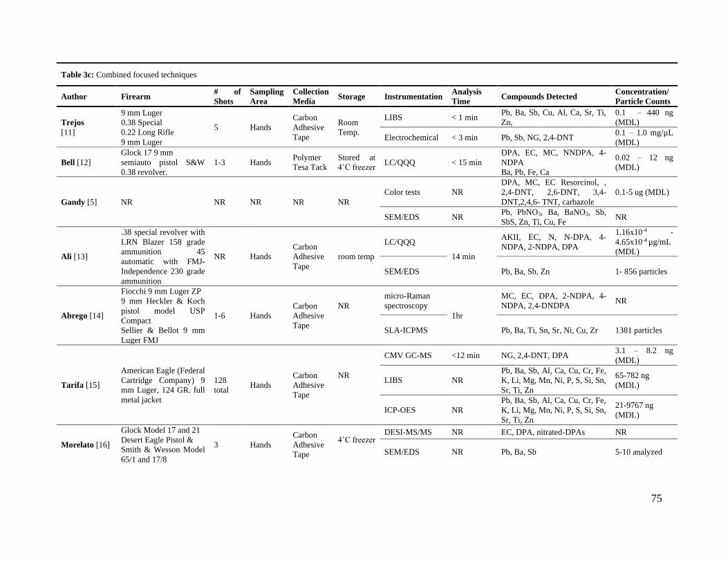
75
1
Table 3c: Combined focused techniques
Author Firearm # of
Shots
Sampling
Area
Collection
Media Storage Instrumentation
Analysis
Time Compounds Detected
Concentration/
Particle Counts
Trejos
[11]
9 mm Luger
0.38 Special
0.22 Long Rifle
9 mm Luger
5 Hands
Carbon
Adhesive
Tape
Room
Temp.
LIBS < 1 min Pb, Ba, Sb, Cu, Al, Ca, Sr, Ti,
Zn,
0.1 – 440 ng
(MDL)
Electrochemical < 3 min Pb, Sb, NG, 2,4-DNT 0.1 – 1.0 mg/µL
(MDL)
Bell [12]
Glock 17 9 mm
semiauto pistol S&W
0.38 revolver.
1-3 Hands Polymer
Tesa Tack
Stored at
4˚C freezer LC/QQQ < 15 min
DPA, EC, MC, NNDPA, 4-
NDPA
Ba, Pb, Fe, Ca
0.02 – 12 ng
(MDL)
Gandy [5] NR NR NR NR NR
Color tests NR
DPA, MC, EC Resorcinol, ,
2,4-DNT, 2,6-DNT, 3,4-
DNT,2,4,6- TNT, carbazole
0.1-5 ug (MDL)
SEM/EDS NR Pb, PbNO3, Ba, BaNO3, Sb,
SbS, Zn, Ti, Cu, Fe NR
Ali [13]
.38 special revolver with
LRN Blazer 158 grade
ammunition 45
automatic with FMJ-
Independence 230 grade
ammunition
NR Hands
Carbon
Adhesive
Tape
room temp
LC/QQQ
14 min
AKII, EC, N, N-DPA, 4-
NDPA, 2-NDPA, DPA
1.16x10⁻⁴ -
4.65x10⁻⁴ µg/mL
(MDL)
SEM/EDS Pb, Ba, Sb, Zn 1- 856 particles
Abrego [14]
Fiocchi 9 mm Luger ZP
9 mm Heckler & Koch
pistol model USP
Compact
Sellier & Bellot 9 mm
Luger FMJ
1-6 Hands
Carbon
Adhesive
Tape
NR
micro-Raman
spectroscopy 1hr
MC, EC, DPA, 2-NDPA, 4-
NDPA, 2,4-DNDPA NR
SLA-ICPMS Pb, Ba, Ti, Sn, Sr, Ni, Cu, Zr 1381 particles
Tarifa [15]
American Eagle (Federal
Cartridge Company) 9
mm Luger, 124 GR. full
metal jacket
128
total Hands
Carbon
Adhesive
Tape
NR
CMV GC-MS <12 min NG, 2,4-DNT, DPA 3.1 – 8.2 ng
(MDL)
LIBS NR
Pb, Ba, Sb, Al, Ca, Cu, Cr, Fe,
K, Li, Mg, Mn, Ni, P, S, Si, Sn,
Sr, Ti, Zn
65-782 ng
(MDL)
ICP-OES NR
Pb, Ba, Sb, Al, Ca, Cu, Cr, Fe,
K, Li, Mg, Mn, Ni, P, S, Si, Sn,
Sr, Ti, Zn
21-9767 ng
(MDL)
Morelato [16]
Glock Model 17 and 21
Desert Eagle Pistol &
Smith & Wesson Model
65/1 and 17/8
3 Hands
Carbon
Adhesive
Tape
4˚C freezer DESI-MS/MS NR EC, DPA, nitrated-DPAs NR
SEM/EDS NR Pb, Ba, Sb 5-10 analyzed

76
3.5. Conclusions and Future Considerations
Understanding the variables affecting OGSR and IGSR after discharging a firearm, plays
a crucial role in the interpretation of the evidence. Considering the differing ways GSR distributes,
transfers, and persists aids in the sampling and analysis of evidence. However, challenges
associated with this include accurate sampling and collection of GSR, performing rapid yet reliable
analytical methods, and providing ample information about a crime scene sample for appropriate
interpretation of results.
While there are several types of GSR sampling media, the most commonly chosen in
research and casework is the carbon adhesive tape attached to a standard SEM stub, due to its
versatility and established use by crime scene personnel. As proven above, OGSR compounds and
IGSR particles can transfer and persist on areas of the body other than the hands. Carbon adhesive
stubs eliminate the need for multiple collection tools, as they are applied to not only hands, but
other surfaces including a person’s face, hair, clothing, and most recently, their nostrils. Future in-
field collection should focus on other sampling areas where GSR particles tend to persist longer,
such as the forehead of a person’s face, the hair located on the top of their head, and roughly
textured clothing.
After understanding the behavior of GSR for proper collection, integration of multiple
instrumental methods becomes crucial. Advances in GSR analysis and complexity of
interpretation, leads to the use of rapid combined strategies for the detection of both organic and
inorganic components. This is illustrated by the incorporation of organic analyses, such as LC/MS
or DESI/MS, in conjunction with confirmatory SEM/EDS analysis. Additionally, exploration of
novel combinations such as LIBS with electrochemistry, CMV-GC/MS with LIBS, FTIR with
Raman, and LC-MS/MS with the use of complexing agents, have shown great promise for

77
identifying both types of GSR markers from a single sample. The orthogonal analysis is vital for
the evolving market of non-toxic ammunition, which is replacing typical IGSR heavy metals with
other materials, such as organic compounds.
Although SEM remains the standard for confirmatory GSR analysis, several studies have
been conducted suggesting the use of other instrumentation methods. Instruments such as LIBS,
GC/MS, LC/MS, and Raman are already standard in crime labs for multiple sample types and
provide additional screening and confirmatory options to compliment SEM/EDS. While these
methods are not able to perform single particle analysis, identification of important OGSR and
IGSR markers is still possible down to ppm and ppt levels. Further research would allow for
increased incorporation of faster instrumentation techniques in casework. The improvement of
GSR detection, analysis, and interpretation lies in the combination of rapid, simultaneous, and
complimentary IGSR and OGSR analytical methods on a single sample to increase understanding,
confidence, and the value of results.
The interest of the forensic community to increase the body of knowledge in this area and
produce complementary consensus-based methods is reflected by current efforts of expert GSR
groups such as the Organization of Scientific Area Committees (NIST-OSAC), the National
Institute of Justice’s Forensic Science Research and Development Technology Working Group
(TWG) and the European Network of Forensic Science Institute (ENFSI). We anticipate this
review will serve as a valuable reference to the forensic practitioners and researchers in this field,
particularly in the areas of collection, persistence, and analysis of IGSR and OGSR of modern
ammunition.

78
3.6. References for Chapter 3
[1] L.S. Blakey, G.P. Sharples, K. Chana, J.W. Birkett, Fate and behavior of gunshot
residue—A review, J. Forensic Sci. 63 (2018) 9–19, https://doi.org/10.1111/1556-4029.13555.
[2] ASTM, Standard Practice for Gunshot Residue Analysis by Scanning Electron
Microscopy/Energy Dispersive X-Ray Spectrometry, B. Stand. (2016) 1–5.
https://doi.org/10.1520/E1588-17.
[3] M. Roberts, N. Petraco, M. Gittings, Novel method for the detection of nitroglycerin in
smokeless powders, Sci. Justice. 55 (2015) 467–471,
https://doi.org/10.1016/j.scijus.2015.08.001.
[4] M. López-López, C. García-Ruiz, Recent non-chemical approaches to estimate the
shooting distance, Forensic Sci. Int. 239 (2014) 79–85,
https://doi.org/10.1016/j.forsciint.2014.03.023.
[5] L. Gandy, K. Najjar, M. Terry, C. Bridge, A novel protocol for the combined detection of
organic, inorganic gunshot residue, Forensic Chem. 8 (2018) 1–10,
https://doi.org/10.1016/j.forc.2017.12.009.
[6] B. Yeager, K. Bustin, J. Stewart, R. Dross, S. Bell, Evaluation and validation of ion
mobility spectrometry for presumptive testing targeting the organic constituents of firearms
discharge residue, Anal. Methods. 7 (2015) 9683–9691, https://doi.org/10.1039/c5ay02417j.
[7] S. Toal, W. Niemeyer, S. Conte, D. Montgomery, G. Ericksen, Spie, Confirmatory
analysis of field-presumptive GSR test sample using SEM/EDS, in: Conf. Scanning Microsc.,
Spie-Int Soc Optical Engineering, BELLINGHAM (2014), https://doi.org/10.1117/12.2074212.
[8] O. Dalby, D. Butler, J. Birkett, Analysis of gunshot residue and associated materials - A
review, J. Forensic Sci. 55 (2010) 924–943, https://doi.org/10.1111/j.1556-4029.2010.01370.x.

79
[9] E. Goudsmits, G. Sharples, J. Birkett, Recent trends in organic gunshot residue analysis,
TrAC - Trends Anal. Chem. 74 (2015) 46–57, https://doi.org/10.1016/j.trac.2015.05.010.
[10] K. Chang, P. Jayaprakash, C. Yew, A. Abdullah, Gunshot residue analysis and its
evidential values: A review, Aust. J. Forensic Sci. 45 (2013) 3–23,
https://doi.org/10.1080/00450618.2012.691546.
[11] T. Trejos, C. Vander Pyl, K. Menking-Hoggatt, A. Alvarado, L. Arroyo, Fast
identification of inorganic and organic gunshot residues by LIBS and electrochemical methods,
Forensic Chem. 8 (2018) 146–156, https://doi.org/10.1016/j.forc.2018.02.006.
[12] S. Bell, W. Feeney, Single shot, single sample, single instrument detection of IGSR and
OGSR using LC/MS/MS, Forensic Sci. Int. 299 (2019) 215–222,
https://doi.org/10.1016/j.forsciint.2019.04.002.
[13] Z. Abrego, N. Grijalba, N. Unceta, M. Maguregui, A. Sanchez, A. Fernandez-Isla, M.
Goicolea, R. Barrio, A novel method for the identification of inorganic and organic gunshot
residue particles of lead-free ammunitions from the hands of shooters using scanning laser
ablation-ICPMS and Raman micro-spectroscopy, Analyst 139 (2014) 6232–6241,
https://doi.org/10.1039/c4an01051e.
[14] A. Tarifa, J.R. Almirall, Fast detection and characterization of organic and in- organic
gunshot residues on the hands of suspects by CMV-GC-MS and LIBS, Sci. Justice. 55 (2015)
168–175, https://doi.org/10.1016/j.scijus.2015.02.003.
[15] M. Morelato, A. Beavis, A. Ogle, P. Doble, P. Kirkbride, C. Roux, Screening of gunshot
residues using desorption electrospray ionisation-mass spectrometry (DESI-MS), Forensic Sci.
Int. 217 (2012) 101–106, https://doi.org/10.1016/j.forsciint.2011.10.030.

80
[16] L. Ali, K. Brown, H. Castellano, S. Wetzel, A Study of the presence of gunshot residue in
Pittsburgh police stations using SEM/EDS and LC-MS/MS, J. Forensic Sci. 61 (2016) 928–938,
https://doi.org/10.1111/1556-4029.13077.
[17] Y. Cao, R. Linnen, D. Good, I. Samson, R. Epstein, The application of portable XRF and
Benchtop SEM-EDS to Cu-Pd exploration in the Coldwell alkaline complex, Ontario, Canada,
Geochem. Explor. Environ. Anal. 16 (2016) 193–212, https:// doi.org/10.1144/geochem2015-
394.
[18] K. Mason, R. Wuhrer, Phenom Desktop SEM for Gunshot Residue and
Cathodoluminescence Imaging and Analysis, Microsc. Microanal. 23 (2017) 1074–1075,
https://doi.org/10.1017/s1431927617006031.
[19] K. Mason, A forensic mineralogy toolbox –The next generation of instrumentation for
forensic applications, in: Int. Microsc. Congr. (IMC Prague 2014), 2014: pp. 1–2.
[20] R. Berk, Automated SEM/EDS analysis of airbag residue I: Particle identification, J.
Forensic Sci. 54 (2009) 60–68, https://doi.org/10.1111/j.1556-4029.2008.00918.x.
[21] R. Berk Automated, SEM/EDS analysis of airbag residue. II: Airbag residue as a source
of percussion primer residue particles, J. Forensic Sci. 54 (2009) 69–76,
https://doi.org/10.1111/j.1556-4029.2008.00919.x.
[22] R. Popelka-Filcoff, K. Kirkbride, K. Seyfang, N. Lucas, W. Tucker, Gunshot residue and
brake pads: Compositional and morphological considerations for forensic casework, Forensic
Sci. Int. 270 (2016) 76–82, https://doi.org/10.1016/j.
[23] G. Ingo, M. D’Uffizi, G. Falso, G. Bultrini, G. Padeletti, Thermal and microchemical
investigation of automotive brake pad wear residues, Thermochim. Acta. 418 (2004) 61–68,
https://doi.org/10.1016/j.tca.2003.11.042.

81
[24] P. Mosher, M. McVicar, E. Randall, E. Sild, Gunshot residue-similar particles produced
by fireworks, J. Can. Soc. Forensic Sci. 31 (1998) 157–168,
https://doi.org/10.1080/00085030.1998.10757115.
[25] Z. Brozek-Mucha, Chemical and physical characterisation of welding fume particles for
distinguishing from gunshot residue, Forensic Sci. Int. 254 (2015) 51–58,
https://doi.org/10.1016/j.forsciint.2015.06.033.
[26] K. Seyfang, N. Lucas, K. Redman, R. Popelka-Filcoff, H. Kobus, K. Kirkbride, Glass-
containing gunshot residues and particles of industrial and occupational origins: Considerations
for evaluating GSR traces, Forensic Sci. Int. 298 (2019) 284–297,
https://doi.org/10.1016/j.forsciint.2019.03.010.
[27] K. Seyfang, N. Lucas, R. Popelka-Filcoff, H. Kobus, K. Redman, K. Kirkbride, Methods
for analysis of glass in glass-containing gunshot residue (gGSR) particles, Forensic Sci. Int. 298
(2019) 359–371, https://doi.org/10.1016/j.forsciint.2019.03.019.
[28] M. Trimpe, Analysis of fireworks for particles of the type found in primer residue (GSR),
Int. Assoc. Microanal. 4 (2003) 1–7.
[29] M. Donghi, K. Mason, F.S. Romolo, Detecting Gunshot Residue from Sellier & Bellot
Nontox Heavy Metal-free Primer by in situ Cathodoluminescence, J. Forensic Sci. 64 (2019)
1658–1667, https://doi.org/10.1111/1556-4029.14110.
[30] D. Gibelli, A. Brandone, S. Andreola, D. Porta, E. Giudici, M. Grandi, C. Cattaneo,
Macroscopic, microscopic, and chemical assessment of gunshot lesions on de- composed pig
skin, J. Forensic Sci. 55 (2010) 1092–1097, https://doi.org/10.1111/j.1556-4029.2010.01378.x.
[31] S. Goode, C. Dockery, M. Bachmeyer, A. Nieuwland, S. Morgan, Detecting gunshot
residue by laser induced breakdown spectroscopy, Laser Induc, Plasma Spectrosc. App. (2002).

82
[32] M. Rosenberg, C. Dockery, Determining the lifetime of detectable amounts of gunshot
residue on the hands of a shooter using laser-induced breakdown spectroscopy, Appl. Spectrosc.
62 (2008) 1238–1241, https://doi.org/10.1366/000370208786401473.
[33] E. Diaz, J. Souza Sarkis, S. Viebig, P. Saldiva, Measurement of airborne gunshot
particles in a ballistics laboratory by sector field inductively coupled plasma mass spectrometry,
Forensic Sci. Int. 214 (2012) 44–47, https://doi.org/10.1016/j.forsciint.2011.07.016.
[34] A. Doña-Fernández, I. de Andres-Gimeno, P. Santiago-Toribio, E. Valtuille-Fernández,
F. Aller-Sanchez, A. Heras-González, Real-time detection of GSR particles from crime scene: A
comparative study of SEM/EDX and portable LIBS system, Forensic Sci. Int. 292 (2018) 167–
175, https://doi.org/10.1016/j.forsciint.2018.09.021.
[35] L. Fambro, E. Miller, D. Vandenbos, C. Dockery, Characterization of lead-free gunshot
residue analogs, Anal. Methods. 8 (2016) 3132–3139, https://doi.org/10.1039/c6ay00725b.
[36] L. Fambro, D. Vandenbos, M. Rosenberg, C. Dockery, Laser-induced breakdown
spectroscopy for the Rapid characterization of lead-free gunshot residues, Appl. Spectrosc. 71
(2017) 699–708, https://doi.org/10.1177/0003702816689099.
[37] M. Sampedro, M. Aliste, S. Arranz, S. Alicia, N. Unceta, G. Alberto, A. Vallejo, Particle
analysis for the detection of gunshot residue (GSR) in nasal samples using scanning Laser
Ablation and Inductively Coupled Plasma-Mass Spectrometry, J. Forensic Sci. (2020),
https://doi.org/10.1111/1556-4029.14278.
[38] D. Hahn, N. Omenetto, Laser-Induced Breakdown Spectroscopy (LIBS), part II: Review
of instrumental and methodological approaches to material analysis and applications to different
fields, Appl. Spectrosc. 66 (2012) 347–419, https://doi.org/10.1366/11-06574.

83
[39] M. Silva, J. Cortez, C. Pasquini, R. Honorato, A. Paim, M. Pimentel, Gunshot residues:
Screening analysis by laser-induced breakdown spectroscopy, J. Braz. Chem. Soc. 20 (2009)
1887–1894, https://doi.org/10.1590/S0103-50532009001000017.
[40] C. Dockery, S. Goode, Laser-induced breakdown spectroscopy for the detection of
gunshot residues on the hands of a shooter, Appl. Opt. 42 (2007) 6153,
https://doi.org/10.1364/ao.42.006153.
[41] K. Menking-Hoggatt, L. Arroyo, J. Curran, T. Trejos, Novel LIBS method for micro-
spatial chemical analysis of inorganic gunshot residues, J. Chemom. (2019) 1–13,
https://doi.org/10.1002/cem.3208.
[42] J. Arndt, S. Bell, L. Crookshanks, M. Lovejoy, C. Oleska, T. Tulley, D. Wolfe,
Preliminary evaluation of the persistence of organic gunshot residue, Forensic Sci. Int. (2012)
137–145, https://doi.org/10.1016/j.forsciint.2012.05.011.
[43] J. Moran, S. Bell, Skin permeation of organic gunshot residue: Implications for sampling
and analysis, Anal. Chem. 86 (2014) 6071–6079, https://doi.org/10.1021/ac501227e.
[44] B. Stevens, S. Bell, K. Adams, Initial evaluation of inlet thermal desorption GC-MS
analysis for organic gunshot residue collected from the hands of known shooters, Forensic
Chem. 2 (2016) 55–62, https://doi.org/10.1016/j.forc.2016.10.001.
[45] M. Lopez-Lopez, M. De La Ossa, C. Garcia-Ruiz, Fast analysis of complete macroscopic
gunshot residues on substrates using Raman imaging, Appl. Spectrosc. 69 (2015) 889–893,
https://doi.org/10.1366/14-07816.
[46] M. López-López, J.J. Delgado, C. García-Ruiz, Ammunition identification by means of
the organic analysis of gunshot residues using Raman spectroscopy, Anal. Chem. 84 (2012)
3581–3585, https://doi.org/10.1021/ac203237w.

84
[47] M. Maitre, K. Kirkbride, M. Horder, C. Roux, A. Beavis, Thinking beyond the lab:
Organic gunshot residues in an investigative perspective, Aust. J. Forensic Sci. 50 (2018) 659–
665, https://doi.org/10.1080/00450618.2018.1457718.
[48] M. Zhao, S. Zhang, C. Yang, Y. Xu, Y. Wen, L. Sun, X. Zhang, Desorption
electrospray tandem MS (DESI-MSMS) analysis of methyl centralite and ethyl centralite as
gunshot residues on skin and other surfaces, J. Forensic Sci. 53 (2008) 807–811,
https://doi.org/10.1111/j.1556-4029.2008.00752.x.
[49] A. Zeichner, B. Eldar, A novel method for extraction and analysis of gunpowder residues
on double-side adhesive coated stubs, J. Forensic Sci. 49 (2004) 1–13,
https://doi.org/10.1520/jfs2004022.
[50] J. Bueno, I. Lednev, Attenuated total reflectance-FT-IR imaging for rapid and automated
detection of gunshot residue, Anal. Chem. 86 (2014) 3389–3396,
https://doi.org/10.1021/ac4036718.
[51] J. Bueno, L. Halamkova, A. Rzhevskii, I.K. Lednev, Raman microspectroscopic mapping
as a tool for detection of gunshot residue on adhesive tape, Anal. Bioanal. Chem. 410 (2018)
7295–7303, https://doi.org/10.1007/s00216-018-1359-1.
[52] J. Moran, S. Bell, Analysis of organic gunshot residue permeation through a model skin
membrane using ion mobility spectrometry, Int. J. Ion Mobil. Spectrom. 16 (2013) 247–258,
https://doi.org/10.1007/s12127-013-0138-0.
[53] A. Zeichner, B. Eldar, B. Glattstein, A. Koffman, T. Tamiri, D. Muller, Vacuum
collection of gunpowder residues from clothing worn by shooting suspects, and their analysis by
GC/TEA, IMS, and GC/MS, J. Forensic Sci. 48 (2003) 2002390,
https://doi.org/10.1520/jfs2002390.

85
[54] C. Hofstetter, M. Maitre, A. Beavis, C. Roux, C. Weyermann, A. Gassner, A study of
transfer and prevalence of organic gunshot residues, Forensic Sci. Int. 277 (2017) 241–251,
https://doi.org/10.1016/j.forsciint.2017.06.013.
[55] Z. Brozek-Mucha, Z. Brożek-Mucha, Trends in analysis of gunshot residue for forensic
purposes, Anal. Bioanal. Chem. 409 (2017) 5803–5811, https://doi.org/10.1007/s00216-017-
0460-1.
[56] M. Houck, S. Charles, B. Nys, N. Guensens, 18th INTERPOL International Forensic
Science Managers Symposium, (2016) 69–89.
[57] M. Houck, S. Charles, N. Guensens, E. Vergalito, B. Nys, 19th INTERPOL International
Forensic Science Managers Symposium, (2019) 39–67.
[58] L. Filho, US6786986B2 Non-toxic composition for priming mixture for small ca- liber
arms ammunition, 2004. https://doi.org/10.1038/incomms1464.
[59] I. Weber, M. Albino de Carvalho, C. Pasquini, S. Junior, J. Amigo, M. Pimentel, M.
Talhavini, NIR hyperspectral images for identification of gunshot residue from tagged
ammunition, Anal. Methods. 10 (2018) 4711–4717, https://doi.org/10.1039/c8ay01341a.
[60] F. Romolo, M. Bailey, J. de Jesus, L. Manna, M. Donghi, Unusual sources of Sn in GSR.
An experimental study by, SEM and IBA, Sci. Justice. 59 (2019) 181–189,
https://doi.org/10.1016/J.SCIJUS.2018.10.009.
[61] H. Meng, B. Caddy, Gunshot residue analysis: A Review, J Forensic Sci. 42 (1997) 553–
570.
[62] M. Maitre, K. Kirkbride, M. Horder, C. Roux, A. Beavis, Current perspectives in the
interpretation of gunshot residues in forensic science: A review, Forensic Sci. Int. 270 (2017) 1–
11, https://doi.org/10.1016/j.forsciint.2016.09.003.

86
[63] R. Taudte, A. Beavis, L. Blanes, N. Cole, P. Doble, C. Roux, Detection of gunshot
residues using mass spectrometry, Biomed Res. Int. 2014 (2014) 1–16,
https://doi.org/10.1155/2014/965403.
[64] J. Bailey, Analysis of bullet wipe patterns on cloth targets, J. Forensic Identif. 55 (2005)
448–460.
[65] H. Wrobel, J. Millar, M. Kijek, M. Millar, M. Kijek, Comparison of properties of
adhesive tapes, tabs, and liquids used for the collection of gunshot residue and other trace
materials for SEM analysis, J. Forensic Sci. 43 (1998) 178–181,
https://doi.org/10.1520/jfs16107j.
[66] D. Merli, A. Amadasi, D. Mazzarelli, A. Cappella, E. Castoldi, S. Ripa, L. Cucca, C.
Cattaneo, A. Profumo, Comparison of different swabs for sampling inorganic gunshot residue
from gunshot wounds: Applicability and reliability for the de- termination of firing distance, J.
Forensic Sci. 64 (2019) 558–564, https://doi.org/10.1111/1556-4029.13870.
[67] D. Degaetano, J. Siegel, K. Klomparens, A comparison of three techniques developed for
sampling and analysis of gunshot residue by Scanning Electron Microscopy Energy Dispersive
X-ray analysis (SEM-EDX), J. Forensic Sci. 37 (1992) 281–300.
[68] I. Kara, The influence of different skin types on GSR sampling by tape lifting for SEM
analysis, Microsc. Res. Tech. 80 (2017) 1310–1314, https://doi.org/10.1002/jemt.22942.
[69] R. Taudte, C. Roux, L. Blanes, M. Horder, K. Kirkbride, A. Beavis, The development
and comparison of collection techniques for inorganic and organic gunshot residues, Anal.
Bioanal. Chem. 408 (2016) 2567–2576, https://doi.org/10.1007/s00216-016-9357-7.
[70] R. Schwartz, C. Zona, A recovery method for airborne gunshot residue retained in human
nasal mucus, J. Forensic Sci. 40 (1995) 13845J, https://doi.org/10.1520/jfs13845j.

87
[71] L. Reyes, C. Lopez, A. Barrios, C. Soto, C. Ibanez, F. Diaz, Development and ap-
plication of a new nose hairs sample collection device for GSR Particles by scanning electron
microscopy with energy dispersive X-ray spectroscopy (SEM-EDS), Forensic Sci. Int. 290
(2018) 42–48, https://doi.org/10.1016/j.forsciint.2018.06.029.
[72] S. Charles, M. Lannoy, N. Geusens, Influence of the type of fabric on the collection
efficiency of gunshot residues, Forensic Sci. Int. 228 (2013) 42–46,
https://doi.org/10.1016/j.forsciint.2013.02.022.
[73] M. Izzharif, A. Halim, U. Kalthom, C. Yew, M. Koey, Analysis of gunshot residue
deposited on cotton cloth target at close range shooting distances, J. Forensic Sci. 1 (2010) 48–
53.
[74] W. MacCrehan, K. Ricketts, R. Baltzersen, W. Rowe, Detecting organic gunpowder
residues from handgun use, Investig. Forensic Sci. Technol. 3576 (1999) 116–124,
https://doi.org/10.1117/12.334522.
[75] I. Kara, S. Lisesivdin, M. Kasap, E. Er, U. Uzek, The relationship between the surface
morphology and chemical composition of gunshot residue particles, J. Forensic Sci. 60 (2015)
1030–1033, https://doi.org/10.1111/1556-4029.12785.
[76] D. Merli, A. Brandone, A. Amadasi, C. Cattaneo, A. Profumo, The detection of gunshot
residues in the nasal mucus of suspected shooters, Int. J. Legal Med. 130 (2016) 1045–1052,
https://doi.org/10.1007/s00414-016-1375-7.
[77] S. Kage, K. Kudo, A. Kaizoji, J. Ryumoto, H. Ikeda, N. Ikeda, A simple method for
detection of gunshot residue particles from hands, hair, face, and clothing using Scanning
Electron Microscopy/Wavelength Dispersive X-Ray (SEM/WDX), J. Forensic Sci. 46 (2001)
15054J, https://doi.org/10.1520/jfs15054j.

88
[78] A. Zeichner, N. Levin, Collection efficiency of gunshot residue (GSR) particles from hair
and hands using double-side adhesive tape, J. Forensic Sci. 38 (1993) 571–584,
https://doi.org/10.1520/jfs13441j.
[79] M. Trimpe, Scientific Working Group on Gunshot Residue (SWGGSR): A progress
report, in: Conf. Scanning Microsc. 2011 - Adv. Microsc. Technol. Defense, Homel. Secur.
Forensic, Life, Environ. Ind. Sci., Spie-Int Soc Optical Engineering, BELLINGHAM, 2011.
Doi:10.1117/12.887503.
[80] R. Thompson, D. Fetterolf, M. Miller, R. Mothershead, Aqueous recovery from cotton
swabs of organic explosives residue followed by solid phase extraction, J. Forensic Sci. 44
(1999) 795–804, https://doi.org/10.1520/jfs14555j.
[81] E. Pereira, C. Caceres, F. Rivera, B. Rivas, P. Saez, Preparation of molecularly imprinted
polymers for diphenylamine removal from organic gunshot residues, J. Chil. Chem. Soc. 59
(2014) 2731–2736.
[82] O. Black, R. Cody, D. Edwards, J. Cizdziel, Identification of polymers and organic
gunshot residue in evidence from 3D-printed firearms using DART-mass spectrometry: A
feasibility study, Forensic Chem. 5 (2017) 26–32, https://doi.org/10.1016/j.forc.2017.05.003.
[83] Z. Brozek-Mucha, Chemical and morphological study of gunshot residue persisting on
the shooter by means of Scanning Electron Microscopy and Energy Dispersive X-Ray
Spectrometry, Microsc. Microanal. 17 (2011) 972–982,
https://doi.org/10.1017/s1431927611012141.
[84] E. Lindsay, M. McVicar, R. Gerard, E. Randall, J. Pearson, Passive exposure and
persistence of gunshot residue (GSR) on bystanders to a shooting: Comparison of shooter and

89
bystander exposure to GSR, J. Can. Soc. Forensic Sci. 44 (2011) 89–96,
https://doi.org/10.1080/00085030.2011.10768144.
[85] Y. Tong, Z. Wei, C. Yang, J. Yu, X. Zhang, S. Yang, X. Deng, Y. Xu, Y. Wen,
Determination of diphenylamine stabilizer and its nitrated derivatives in smoke- less gunpowder
using a tandem MS method, Analyst. 126 (2001) 480–484, https://doi.org/10.1039/b010183o.
[86] D. Perret, S. Marchese, A. Gentili, R. Curini, A. Terracciano, E. Bafile, F. Romolo, LC-
MS-MS determination of stabilizers and explosives residues in hand-swabs, Chromatographia.
68 (2008) 517–524, https://doi.org/10.1365/s10337-008-0746-8.
[87] D. Laza, B. Nys, J. De Kinder, A. Mesmaeker, C. Moucheron, Development of a
quantitative LC-MS/MS method for the analysis of common propellant powder stabilizers in
gunshot residue, J. Forensic Sci. 52 (2007) 842–850, https://doi.org/10.1111/j.1556-
4029.2007.00490.x.
[88] A. O’Mahony, J. Wang, Electrochemical detection of gunshot residue for forensic
analysis: A Review, Electroanalysis. 25 (2013) 1341–1358,
https://doi.org/10.1002/elan.201300054.
[89] M.I. Halim, M. Safian, E. Elias, S. Shazali, Identification of gunshot residue from trace
element by using ICP/OES, in, Comput. Informatics (2013).
[90] M. Gallidabino, F. Romolo, C. Weyermann, Characterization of volatile organic gunshot
residues in fired handgun cartridges by headspace sorptive extraction, Anal. Bioanal. Chem. 407
(2015) 7123–7134, https://doi.org/10.1007/s00216-015-8874-0.
[91] R. Williamson, S. Gura, A. Tarifa, J. Almirall, The coupling of capillary micro-
extraction of volatiles (CMV) dynamic air sampling device with DART-MS analysis for the

90
detection of gunshot residues, Forensic Chem. 8 (2018) 49–56,
https://doi.org/10.1016/j.forc.2018.01.005.
[92] T. Warlow, Firearms, the Law and Forensic Ballistics, 1st ed., CRC Press, London, 1996.
[93] A. Schwoeble, D. Exline, Current methods in forensic gunshot residue analysis, CRC
Press, 2000.
[94] M. Rijnders, A. Stamouli, A. Bolck, Comparison of GSR composition occurring at
different locations around the firing position, J. Forensic Sci. 55 (2010) 616–623,
https://doi.org/10.1111/j.1556-4029.2009.01292.x.
[95] J. French, R. Morgan, J. Davy, The secondary transfer of gunshot residue: A
experimental investigation carried out with SEM-EDX analysis, X-Ray Spectrom. 43 (2014) 56–
61, https://doi.org/10.1002/xrs.2498.
[96] J. French, R. Morgan, An experimental investigation of the indirect transfer and
deposition of gunshot residue: Further studies carried out with SEM-EDX analysis, Forensic Sci.
Int. 247 (2015) 14–17, https://doi.org/10.1016/j.forsciint.2014.10.023.
[97] H. Ditrich, Distribution of gunshot residues - The influence of weapon type, Forensic Sci.
Int. 220 (2012) 85–90, https://doi.org/10.1016/j.forsciint.2012.01.034.
[98] G. Migeot, J. De Kinder, Gunshot residue deposits on the gas pistons of assault rifles, J.
Forensic Sci. 47 (2002) 808–810.
[99] D. Greely, E. Weber, Transfer and distribution of gunshot residue through glass
windows, J. Forensic Sci. 62 (2017) 869–873, https://doi.org/10.1111/1556-4029.13331.
[100] I. Stone, V. DiMaio, C. Petty, Gunshot wounds: Visual and analytical procedures, J.
Forensic Sci. 23 (1978) 361–367, https://doi.org/10.1520/jfs10769j.
[101] R. Nichols, Expectations regarding gunpowder depositions, Afte. 30 (1998) 91–101.

91
[102] V. DiMaio, C. Petty, I. Stone, An experimental study of powder tattooing of the skin, J.
Forensic Sci. 21 (1976) 376–1372, https://doi.org/10.1520/jfs10505j.
[103] Z. Brożek-Mucha, A study of gunshot residue distribution for close-range shots with a
silenced gun using optical and scanning electron microscopy, X-ray microanalysis and infrared
spectroscopy, Sci. Justice. (2017), https://doi.org/10.1016/j.scijus.2016.11.004.
[104] A. Gassner, M. Manganelli, D. Werner, D. Rhumorbarbe, M. Maitre, A. Beavis, C. Roux,
C. Weyermann, Secondary transfer of organic gunshot residues: Empirical data to assist the
evaluation of three scenarios, Sci. Justice. 59 (2019) 58–66,
https://doi.org/10.1016/j.scijus.2018.08.007.
[105] A.L. Gassner, C. Ribeiro, J. Kobylinska, A. Zeichner, C. Weyermann, Organic gunshot
residues: Observations about sampling and transfer mechanisms, Forensic Sci. Int. 266 (2016)
369–378, https://doi.org/10.1016/j.forsciint.2016.06.029.
[106] J. French, R. Morgan, P. Baxendell, P. Bull, Multiple transfers of particulates and their
dissemination within contact networks, Sci. Justice. 52 (2012) 33–41,
https://doi.org/10.1016/j.scijus.2011.05.001.
[107] S. Charles, N. Geusens, A study of the potential risk of gunshot residue transfer from
special units of the police to arrested suspects, Forensic Sci. Int. 216 (2012) 78–81,
https://doi.org/10.1016/j.forsciint.2011.08.022.
[108] R. Gerard, E. Lindsay, M. McVicar, E.D. Randall, A. Gapinska, Observations of gunshot
residue associated with police officers, their equipment, and their vehicles, J. Can. Soc. Forensic
Sci. 45 (2012) 57–63, https://doi.org/10.1080/00085030.2012.10757183.

92
[109] R. Berk, S. Rochowicz, M. Wong, M. Kopina, Gunshot residue in Chicago police
vehicles and facilities: An empirical study, J. Forensic Sci. 52 (2007) 838–841,
https://doi.org/10.1111/j.1556-4029.2007.00457.x.
[110] M. Cook, Gunshot residue contamination of the hands of police officers following start-
of-shift handling of their firearm, Forensic Sci. Int. 269 (2016) 56–62,
https://doi.org/10.1016/j.forsciint.2016.11.002.
[111] N. Lucas, M. Cook, K. Kirkbride, H. Kobus, Gunshot residue background on police
officers: Considerations for secondary transfer in GSR evidence evaluation, Forensic Sci. Int.
297 (2019) 293–301, https://doi.org/10.1016/j.forsciint.2019.02.017.
[112] R. Gerard, M. McVicar, E. Lindsay, E. Randall, E. Harvey, The long range de- position
of gunshot residue and the mechanism of its transportation, J. Can. Soc. Forensic Sci. 44 (2011)
97–104, https://doi.org/10.1080/00085030.2011.10768145.
[113] L. Fojtášek, J. Vacínová, P. Kolář, M. Kotrlý, Distribution of GSR particles in the
surroundings of shooting pistol, Forensic Sci. Int. 132 (2003) 99–105,
https://doi.org/10.1016/S0379-0738(03)00018-5.
[114] M. Maitre, M. Horder, K. Kirkbride, A. Gassner, C. Weyermann, C. Roux, A. Beavis, A
forensic investigation on the persistence of organic gunshot residues, Forensic Sci. Int. 292
(2018) 1–10, https://doi.org/10.1016/j.forsciint.2018.08.036.
[115] T. Jalanti, P. Henchoz, A. Gallusser, M. Bonfanti, The persistence of gunshot residue on
shooters’ hands, Sci. Justice. 39 (1999) 48–52, https://doi.org/10.1016/s1355-0306(99)72014-9.
[116] J. Kilty, Activity after shooting and its effects on the retention of primer residue, J.
Forensic Sci. 20 (1975) 219–230, https://doi.org/10.1520/jfs10268j.

93
[117] J. Douse, R. Smith, Trace analysis of explosives and firearm discharge residues in the
metropolitan police forensic science laboratory, J. Energ. Mater. 4 (1986) 169–186.
[118] W. Zech, B. Kneubühl, M. Thali, S. Bolliger, Pistol thrown to the ground by shooter after
fatal self-inflicted gunshot wound to the chest, J. Forensic Leg. Med. 18 (2011) 88–90,
https://doi.org/10.1016/j.jflm.2010.11.005.
[119] L. Lagoo, L. Schaeffer, D. Szymanski, R. Smith, Detection of gunshot residue in blowfly
larvae and decomposing porcine tissue using inductively coupled plasma mass spectrometry
(ICP-MS), J. Forensic Sci. 55 (2010) 624–632, https://doi.org/10.1111/j.1556-
4029.2010.01327.x.
[120] N. Lucas, M. Cook, J. Wallace, K. Kirkbride, H. Kobus, Quantifying gunshot residues in
cases of suicide: Implications for evaluation of suicides and criminal shootings, Forensic Sci. Int.
266 (2016) 289–298, https://doi.org/10.1016/j.forsciint.2016.06.006.
[121] N. Lucas, H. Brown, M. Cook, K. Redman, T. Condon, H. Wrobel, K. Kirkbride, H.
Kobus, A study into the distribution of gunshot residue particles in the random population,
Forensic Sci. Int. 262 (2016) 150–155, https://doi.org/10.1016/j.forsciint.2016.02.050.
[122] A. Zeichner, N. Levin, Casework experience of GSR detection in Israel, on samples from
hands, hair, and clothing using an auto search SEM/EDX system, J. Forensic Sci. 40 (1995)
1082–1085, https://doi.org/10.1520/jfs13881j.
[123] A. Vinokurov, A. Zeichner, B. Glattstein, A. Koffman, N. Levin, A. Rosengarten,
Machine washing or brushing of clothing and its influence on shooting distance estimation, J.
Forensic Sci. 46 (2001) 928–933, https://doi.org/10.1520/jfs15072j.
[124] S. Khandasammy, A. Rzhevskii, I. Lednev, A novel two-step method for the detection of
organic gunshot residue for forensic purposes: Fast fluorescence imaging followed by raman

94
microspectroscopic identification, Anal. Chem. 91 (2019) 11731–11737,
https://doi.org/10.1021/acs.analchem.9b02306.
[125] C. Mahoney, G. Gillen, A. Fahey, Characterization of gunpowder samples using time-of-
flight secondary ion mass spectrometry (TOF-SIMS), Forensic Sci. Int. 158 (2006) 39–51,
https://doi.org/10.1016/j.forsciint.2005.02.036.
[126] Z. Brozek-Mucha, A. Jankowicz, Evaluation of the possibility of differentiation between
various types of ammunition by means of GSR examination with SEM- EDX method, Forensic
Sci. Int. 123 (2001) 39–47, https://doi.org/10.1016/s0379-0738(01)00518-7.
[127] J. Coumbaros, K.P. Kirkbride, G. Klass, W. Skinner, Characterisation of 0.22 ca- liber
rimfire gunshot residues by time-of-flight secondary ion mass spectrometry (TOF-SIMS): A
preliminary study, Forensic Sci. Int. 119 (2001) 72–81, https://doi./10.1016/S0379-
0738(00)00421-7.
[128] M. Chohra, B. Beladel, L. Ahmed, M. Mouzai, D. Akretche, A. Zeghdaoui, A. Mansouri,
M. Benamar, Study of gunshot residue by NAA and ESEM/EDX using several kinds of weapon
and ammunition, J. Radiat. Res. Appl. Sci. 8 (2015) 404–410,
https://doi.org/10.1016/j.jrras.2015.02.012.
[129] Z. Brozek-Mucha, On the prevalence of gunshot residue in selected populations: An
empirical study performed with SEM-EDX analysis, Forensic Sci. Int. 237 (2014) 46–52,
https://doi.org/10.1016/j.forsciint.2014.01.020.
[130] S. Romanò, F. Degiorgio, C. Onofrio, L. Gravina, S. Abate, Characterisation of gunshot
residues from non-toxic ammunition and their persistence on the shooter’s hands, Int. J. Legal
Med. 134 (2020) 1083–1094.

95
[131] R. Costa, L. Motta, C. Destefani, R. Rodrigues, K. Santo, G. Aquije, R. Boldrini, G.
Athayde, M. Carneiro, W. Romao, Gunshot residues (GSR) analysis of clean range ammunition
using SEM/EDX, colorimetric test and ICP-MS: A comparative approach between the analytical
techniques, Microchem. J. 129 (2016) 339–347, https://doi.org/10.1016/j.microc.2016.07.017.
[132] B. Yuksel, A. Ozler-Yigiter, T. Bora, N. Sen, Z. Kayaalti, B. Yüksel, A. Ozler-Yigiter, T.
Bora, N. Sen, Z. Kayaalti, GFAAS determination of antimony, barium, and lead levels in
gunshot residue swabs: An application in forensic chemistry, At. Spectrosc. 37 (2016) 164–169.
[133] R. Schumacher, M. Barth, D. Neimke, L. Niewohner, Investigation of Gunshot Residue
Patterns using milli-XRF-Techniques: First Experiences in Casework, in: Conf. Scanning
Microsc., Spie-Int Soc Optical Engineering, Bellingham, 2010. Doi: 10.1117/12.853852.
[134] M. Szynkowska, A. Parczewski, K. Szajdak, J. Rogowski, Examination of gunshot
residues transfer using ToF-SIMS, Surf. Interface Anal. 45 (2013) 596–600,
https://doi.org/10.1002/sia.5142.

96
CHAPTER 4
Detection of Organic and Inorganic Gunshot Residues from Hands
using Complexing Agents and LC-MS/MS

97
Overview: Gunshot residue (GSR) refers to a conglomerate consisting of both organic molecules
(OGSR) and inorganic species (IGSR). Historically, forensic examiners have focused only on
identifying the IGSR particles by their morphology and elemental composition. Nonetheless,
modern ammunition formulations and challenges with the GSR transference (such as secondary
and tertiary transfer) have driven research efforts for more comprehensive examinations, requiring
alternative analytical techniques. This study proposes the use of LC-MS/MS for chromatographic
separation and dual detection of inorganic and organic residues. The detection of both target
species in the same sample increases the confidence that chemical profiles came from a gun's
discharge instead of non-firearm-related sources. This strategy implements supramolecular
molecules that complex with the IGSR species, allowing them to elute from the column towards
the mass spectrometer while retaining isotopic ratios for quick and unambiguous identification.
The macrocycle (18-crown-6-ether) complexes with lead and barium, while antimony complexes
with a chelating agent (tartaric acid). The total analysis time for OGSR and IGSR in one sample
is under 20 minutes. This manuscript expands from a previous proof-of-concept publication by
improving figures of merit, increasing the target analytes, testing the method's feasibility through
a more extensive set of authentic specimens collected from the hands of both shooters and non-
shooters, and comparing performance with other analytical techniques such as ICP-MS,
electrochemical methods and LIBS. The linear dynamic ranges (LDR) spread across the low ppb
range for OGSR (0.3-200 ppb) and low ppm range (0.1-6.0 ppm) for IGSR. The method's accuracy
increased overall when both organic and inorganic profiles were combined.
The following chapter is an adaptation of a previously published article ©2021: William
Feeney, Korina Menking-Hoggatt, Courtney Vander Pyl, Colby E. Ott, Suzanne Bell, Luis Arroyo,
Tatiana Trejos. Detection of Organic and Inorganic Gunshot Residues from Hands using

98
Complexing Agents and LC-MS/MS. Analytical Methods. Published online June 2021, 13, 3024
https://doi.org/10.1039/D1AY00778E
4.1. Introduction
Trace Analysis Disciplines – Gunshot Residue
4.1.1. Background
Forensic trace examiners investigate a broad range of materials, including tape, hair, fibers,
paint, fire debris, gunshot residues, and many others [1]. This type of evidence occurs from a
physical transference event between two or more objects or persons and was famously coined by
Edmond Locard stating “every contact leaves a trace” [1]. One of the most studied and debated
trace materials within the forensic community is residue released during the discharge of a firearm,
due to its complex transfer and persistence mechanisms.
Gunshot residue (GSR) comprises two main components, organic (OGSR) and inorganic
(IGSR), which arise from different locations within the ammunition. The OGSR compounds
originate from the propellant and lubricant, whereas IGSR particulates emanate from the primer,
bullet, and cartridge casing. After a deflagration event, those analytes can be dispersed and spread
onto various surrounding surfaces, including hair, clothing, and hands. Due to the constituents’
nature and various environmental factors, proficient collection and storage of the samples are
essential to preserve the GSR compounds and increase the likelihood of detection. Typical
indicators for IGSR are Pb, Ba, and Sb which are formed from the initial products lead styphnate
(C6HN3O8Pb), barium nitrate (Ba(NO3)2), and antimony trisulfide (Sb2S3). Some of the more
common OGSR analytes are diphenylamine (DPA), nitroglycerin (NG), ethyl centralite (EC), and
2,4-dinitrotoluene (2,4-DNT) [2,3]. Other compounds monitored, primarily formed by the
combustion event and degradation of DPA, include 2-dinitrodiphenylamine (2-NDPA), 4-

99
nitrodiphenylamine (4-NDPA), and N-nitrosodiphenylamine (N-NDPA). These compounds'
functional roles vary from detonation or blasting agents (explosives, oxidizers, fuel) to binding
and performance materials (stabilizers and plasticizers) [2,4].
Recently, manufacturers have introduced variants of ammunition labeled as “green,” non-
toxic, heavy-metal-free, or lead-free. These products incorporate different starting materials to
achieve similar results to traditional ammunition while reducing exposure of the shooter and
environment to harmful heavy metals. Although this type of ammunition is not widely observed
in casework yet, its emergence has required researchers to characterize and adapt interpretation
criteria for non-toxic primers [5–8].
4.1.2. Inorganic Particulate Analysis
Under the ASTM E1588-20 guideline, the standard instrument for identifying GSR is
Scanning Electron Microscopy Energy Dispersive X-Ray Spectrometry (SEM/EDS) [9]. This non-
destructive technique identifies the presence or absence of GSR based on the morphology and
elemental composition of a single particle. The current guideline provides instructions for the
proper identification of IGSR and uses terminology to indicate the degree of confidence in the
identification of IGSR. Currently, SEM/EDS remains the only confirmatory standard for GSR [9].
The discrimination power of identifying particulates is founded on the elemental profiles
categorized by three levels of discriminating power alongside distinctive spheroid morphologies.
The terms used to describe the confidence in differentiating GSR from other non-GSR
environmental sources are: “characteristic,” “consistent,” and “commonly associated particles”.
Even though SEM/EDS is efficient in characterizing micron-sized inorganic particles, the
method is not compatible with further sequential examination for OGSR, as factors such as high
vacuum conditions, operating parameters, and compound volatility can cause substantial analyte
100
loss. Hence if SEM/EDS analysis is to be used in conjunction with another analysis technique, the
OGSR constituents must be collected first. Another disadvantage is that the analysis time for
SEM/EDS typically takes 2-8 hours per sample, depending on the sample's nature and instrumental
configurations. Additionally, hand residues associated with occupations such as electricians,
welders, and mechanics can lead to false-positives and higher error rates [10]. A recent study has
proposed a solution to OGSR loss by first analyzing the OGSR from the stub using mild solvent
extraction with UHPLC-ESI-MS/MS followed by IGSR examination from the same SEM
collection stub [11]. These authors demonstrated that by using gentle mechanistic motions from a
pipette, the IGSR particulates are not significantly disturbed.
Current advancements in instrumentation techniques have shown great promise for
evaluating GSR evidence. Instruments such as LIBS, LA-ICP-MS, and TOF-SIMS offer non- or
minimally destructive analysis, small particle detection, and the capacity to produce high-quality
images with multielement or isotopic composition information [12–14]. High-resolution
instruments such as ICP-MS can provide additional isotopic and elemental information in the low
part-per-billion (ppb) range [15,16].
4.1.3. Organic Compound Analysis
Unlike IGSR, there is no established guideline for characterizing and interpreting the data
for the OGSR constituents. However, some initial efforts to classify and select relevant OGSR
constituents are based on their prevalence in the environment, expected occurrence due to its use
outside the ammunition market, and existing knowledge of published compounds, mainly by GC-
MS and LC-MS methods [17]. These techniques provide sufficient distinctions between
compounds such as Kovats’ retention indices, chromatographic separation, fragmentation
pathways, and mass spectral data libraries. One difference between OGSR and IGSR analysis is

101
extensive sample preparation. Extraction protocols deviate from the ASTM E1588-20 guideline
by implementing aqueous buffers or organic solvents, such as methanol (MeOH) or acetonitrile
(ACN). Depending on the chemical composition, specific methodologies may more effectively
characterize and detect OGSR constituents by exploiting properties like polarity or volatility.
Because of parameters and instrumentation flexibilities, factors like total analysis times,
sensitivities, and spectral information are more prone to fluctuate. This variation and capabilities,
in turn, are the main challenges for forensic laboratories to create and develop an inclusive,
standard guideline for OGSR. Moreover, detection limits of some GC-MS configurations may not
be applicable to concentrations typically found in GSR specimens.
4.1.4. Combined Analysis Methodologies
Because of the additional and valuable evidentiary information gained from OGSR
analysis, there has been a shift to analyze both IGSR and OGSR components from a single sample.
Techniques including electrochemistry, FTIR, and Raman spectroscopy offer rapid, cost-effective
multicomponent analysis alternatives, but lack the sensitive/selective power compared to mass
spectrometry [18,19]. Therefore, various research groups combine screening and confirmatory
methodologies to increase confidence in the results [12,13,20–32]. Such combinations include
LIBS with electrochemistry, LC-MS/MS with SEM/EDS, Raman spectroscopy with LA-ICP/MS,
and CMV-GC/MS with LIBS [11,12,30,33,34].
LIBS, electrochemistry, and Raman spectroscopy facilitate sample screenings for quick
characterizations with minimal sample preparation, and the sample remains almost unaltered.
Additionally, the GSR stub used in these methods is compatible with the confirmatory (SEM/EDS)
analysis, allowing further sample manipulation.

102
Extraction for OGSR constituents use common solvents (methanol and acetonitrile),
whereas IGSR elemental identification requires a more rigorous heavy metal digestion (nitric acid
and hydrochloric acid). Therefore, methods like capillary electrophoresis and LC-MS/MS are
classified as destructive methods [35,36]. However, if these methods were incorporated in a
comprehensive workflow, the morphology analysis required can be conducted after organic
extractions and instrumental analysis (via capillary electrophoresis, DESI, or LC-MS/MS) and
before the losing OGSR due to vacuum conditions via SEM/EDS [11,37-40].
Due to the structure of IGSR particles, further sample manipulations are required to
identify and quantify samples properly via LC-MS/MS. Ideally, these chemical analyses should
provide unique spectral signatures without altering the core information of the analyte of interest.
This consideration led to the investigation of complexation chemistry. Host-guest chemistry
applies larger organic molecules to self-assemble and form complexes called metal-ligand (M-L)
complexes. Macrocycles like 18-crown-6-ether (18C6) encapsulate metal ions through
noncovalent, electrostatic interactions [35,41,42]. Several benefits from this interaction include
transportation through both the column and mass analyzer, an extensive range of metal analytes,
and retaining of natural isotopic abundance patterns. Other molecules like EDTA and tartaric acid
employ a different strategy for cation binding, known as chelation. Chelation involves the
formation of physical coordinate bonds between a ligand and a single central atom. Although this
can provide similar benefits as host-guest chemistry, more extensive factors like pH must be
considered.
The goal of this work addresses key points which include: 1) validate our previous proof-
of-concept study by enhancing figures of merit such as detection limits (LOD), quantitation limits
(LOQ), bias, and expanding the evaluation of performance rates with authentic specimens [35], 2)

103
investigate interactions and expand upon antimony detection, and 3) establish an identification
criterion for a “positive” GSR sample based on baseline authentic samples. This work also
evaluates two substrates, including tesa® Tack and the traditional carbon adhesive tape.
Furthermore, we investigate various sample types, including samples collected from shooters, non-
shooter skin backgrounds, and post-shooting activity.
4.2. Materials and Methods
4.2.1. Consumables
Optima® LC/MS grade methanol (MeOH), acetonitrile (ACN), and water (H2O), all
containing 0.1% formic acid (FA), were obtained from Thermo-Fisher Scientific (Watham, MA)
and used either as extraction solvents or mobile phases. Standard (neat) organic constituents used
in this study included: akardite II (AK 2), ethyl centralite (EC), methyl centralite (MC),
diphenylamine (DPA), N-nitrosodiphenylamine (N-NDPA), 2-nitrodiphenylamine (2-NDPA),
and 4-nitrodiphenylamine (4-NDPA), all obtained from Sigma–Aldrich and Accustandard® (New
Haven, CT). Calibration curves were generated from working stock solutions and diluted in MeOH
(≥95%). Complexing agents 18-crown-6-ether (18C6) and tartaric acid (TT) were purchased from
Sigma–Aldrich (St. Louis, MO 99% purity) and diluted using MeOH and water, respectively.
Metal IGSR standards used to form complexes utilized ICP-MS metal standards (VHG
Laboratories, Manchester, NH) and methanol. Micro-bulk digestions utilized nitric (HNO3) and
hydrochloric (HCl) acids (ultrapure grade, Thermo-Fisher Scientific, Waltham, MA). Two internal
standards were used throughout the study including deuterated diphenylamine-d10 (D10-DPA)
which was purchased from CDN Isotopes (Quebec, Canada) and used as the internal standard for
the organic molecules and thallium (Tl) for the IGSR (VHG Laboratories, Manchester, NH) and
complexing agents.
104
4.2.2. Firearms, ammunition, and protocols
Hands from the hands of volunteers, both from shooters and non-shooter background
individuals were collected under our institutional IRB protocol #1506706336 using carbon
adhesive. Aluminum SEM stubs were prepared with two layers of carbon adhesive tape and
covered with plastic stub storage containers (Ted Pella, Inc.).
In this study, three different firearms were used for the collection of authentic shooter
samples: 9 mm Springfield XD9 semi-automatic pistol, 40 caliber Springfield XD40 semi-
automatic pistol, and a Taurus 357 Magnum revolver. The ammunition was either loaded by the
manufacturer (Remington®, Blazer®, Federal®) or in-house (Winchester®, CCI®). Each
specialty ammunition consisted of brass Starline™ cartridge cases loaded with either
Winchester® or CCI® small pistol primers, Winchester® 231 propellant, and Speer® 9mm total
metal jacketed bullets. All firing events were performed at the indoor WVU ballistics laboratory
and the respective analysis conducted at the Oglebay Hall research building.
Before each session, the working areas were cleaned and covered with butcher paper, and
samples manipulated with disposable nitrile gloves. To minimize cross-contamination, our
research team established a workflow within five separate laboratory areas. One room is dedicated
to preparing the sampling stubs before collection, which is located in the building within our trace
laboratory. After collection, the samples are stored in a refrigerator until extraction, which is
conducted in a separate laboratory room equipped with dedicated hoods and benchtops. Finally,
instrumental analysis is performed on the third floor for the LC-MS/MS, ICP-MS, LIBS, on the
second floor for EC, and on the ground level for the SEM-EDS examinations.
Moreover, the shooting range is located in another building a few miles apart from the
research laboratory, and firearms and ammunition are stored in dedicated safety rooms away from

105
research spaces. Within the ballistics lab, firearm discharge occurs inside the shooting range, while
collection from the individual’s hands is done on the annexed laboratory. Also, working areas are
cleaned daily before any sample preparation or analysis. The team members have specific
scheduled roles to avoid that an individual who has recently fired or manipulated a firearm enters
the laboratory areas. The collection team members and shooters dressed in Tyvek® suits for
additional protection from carryover.
Our standard operating procedure includes several reagent blanks and negative control
samples to monitor any potential contamination. Reagent blanks are analyzed between each sample
to monitor any carryover or unexpected contamination. Negative controls are prepared from clean
carbon stubs that had not been used for hands collection, but that undergo the same extraction
protocol as the respective sample batch. In addition, negative controls from the hands of the
collecting individuals undergo the whole analytical process from collection to extraction to
analysis. Negative control samples are obtained from the hands of the collection team at the
beginning and end of each collection session.
The shooters discharged five consecutive shots inside the WVU ballistics range. Although
the number of shots fired per shooter varies on a case-to-case basis, the number of discharges were
chosen in this study based on previously reported literature as well as the casework experience of
colleagues, who reported that typically a shooter fires 3-5 shots when using revolvers and pistols
in criminal activities [10,43]. After firing 5 shots, the firearm was cleared and placed with the
range officer. The shooters then proceeded to the collection laboratory, where both left- and right
hands (palm and back) were stubbed 15 times in the areas from the index finger to the thumb. Each
firing event generated four samples, which is outlined in Figure 4.1. After sample collection, the
shooters washed their hands and repeated the process. After the firing session concluded, the

106
collection team obtained additional negative control samples to test potential secondary transfers
or unintended contamination. Once all samples were stored, the Tyvek® suits, parchment paper,
and gloves were discarded.
For the activity sessions, three different activities were performed after 5 successive
discharges including hand rubbing, hand sanitizer application, and running. For the first activity,
the subjects clasped both hands together and vigorously rubbed for approximately 30 seconds.
After that time, the collectors proceeded to stub the subjects index and thumb areas. The second
activity followed the similar motion as the first activity except for hand sanitizer application. For
the final activity, the subjects exited the range and proceeded to run for approximately 60 seconds
outside of the range. The collection team then proceeded to stub theirs hands after they entered the
ballistics laboratory.
4.2.3. LC-MS/MS methods
4.2.3.1. Mass Spectrometry confirmation via LC-MS/MS and Inorganic Isotopic
Pattern Identification using Q-Exactive Orbitrap
Before initial authentic sample collection, both OGSR and IGSR standards were subjected
to rigorous characterization and optimizations using different mass analyzers. Both an Agilent
1290 Infinity II liquid chromatography coupled to a 6470-triple quadrupole (QQQ) mass analyzer
and a Thermo Fisher Scientific Q-Exactive® orbitrap analyzed compounds using flow-injection
analysis (FIA). The OGSR only utilized the 6470 QQQ and the Agilent Optimizer® software to
determine fragmentation patterns and compared them to the NIST mass spectral database. On the
other hand, due to the more complex nature of the M-L structures, IGSR compounds were further
analyzed by Q-Exactive Orbitrap. The Q-Exactive Orbitrap was utilized to observe the isotopic
distribution of the inorganic elements as present when exposed to the crown complex. No

107
chromatographic analysis was conducted since the Orbitrap was utilized as a confirmation tool
using direct infusion approach. The observation of the natural abundances for studied elements
serve as confirmation of their presence in the complex agent. Further confirmation was performed
with the chromatographic analysis using the tandem mass spectrometer.
4.2.3.2. LC-MS/MS flow and column conditions
An Agilent 1290 Infinity II liquid chromatography housing an Agilent pentafluorophenyl
(PFP) Poroshell® 120 column (2.7 µm 2.1 x 50 mm) separated OGSR compounds. The binary
flow parameters consisted of water with 0.1% FA (A) and acetonitrile with 0.1% (B) with a flow
rate of 0.350 mL/min. Initial conditions were 80%A/20%B and ramping to 5% A/95% B for nine
minutes (Table S4.1). Additionally, the source conditions are described in Table S4.2. The total
injection volume was 1.0 µL.
For the inorganics, a Hamilton PRP-X100 cation exchange guard column (10 µm 2.1 x 33
mm) was added to the LC system. The crown ether complexes eluted from the column using an
isocratic flow at 90% A/10% B in positive electrospray (ESI) conditions. At 4 minutes, the source
polarity switched to negative ESI (ESI-) mode, and the composition of mobile phases switched to
98%A/2%B for the tartaric acid complexes. The injection volume for the IGSR method was 10 µL.
4.2.3.3. LC-MS/MS mass spectral analysis
Two classifications are used for MRM identification for precursor ions: quantifiers and
qualifiers ions. Quantifier ions represent the most intense fragment ions formed from ionization
used for quantitation. Qualifier ions are comprised of other abundant ions to differentiate from
possible interferences present in authentic samples. For the self-assembled metal-ligand
complexes, the product ions were further monitored based on the bare metal’s naturally occurring
isotopes (discussed in detail below).

108
4.2.3.4. Performance checks
Before every worklist or sequence run, the QQQ was subjected to two performance checks,
including a CheckTune and calibration curve for instrument and column monitoring. This ensured
high-quality data collection and characterizations for potential signal loss. The calibration curves
consisted of nine levels spanning from 1-200 ppb and 0.1-6 ppm for OGSR and IGSR,
respectively. Several method blanks consisting of methanol (OGSR) and acid mixture subjected
to the entire digestion process (IGSR) allowed for carryover monitoring to establish the data
acquisition method performance. Furthermore, negative controls from the hands of non-shooters
are monitored as explained in Section 4.2.2. Positive controls consisting of standard mixtures of
known composition and concentration and in-house characterized micro-particle GSR standards
were monitored to check for extraction and detection efficiency before the authentic samples’
batch.
4.2.3.5. LC-MS/MS validation with standards and authentic samples
Numerous analytical guidelines describe procedures to evaluate and increase the overall
effectiveness of a validation method. After careful consideration, the Eurachem guideline The
Fitness for Purpose of Analytical Methods [44] was chosen due to its broad expanse of analytical
practices (definitions in Table S4.3). This study estimated the figures of merit such as analytical
selectivity, LOD, LOQ, sensitivity, working range, and bias. Calibration curves were run in five
replicates in three different days. Also, we evaluated performance rates based on a population of
hands from volunteer shooters and non-shooters that resemble casework samples. Performance
rates included sensitivity, selectivity, and accuracy.
To further address the bias of this method, a primer-only standard was used to evaluate the
percent recovery of the micro-bulk digestion method. This was compared to the validated ICP-MS

109
method [45] to further compare the concentrations observed in the LC-MS/MS (see Section 4.4.4).
The parameters of the ICP-MS parameters and compared digestion methodology are further
detailed by Menking-Hoggatt et al. [45].
4.2.4. Comparisons with various analytical methods
Our research group has published various innovative methods that can complement current
practice. The techniques have been selected to be compatible and complementary to SEM-EDS
while providing faster and informative data for case triaging. These include LIBS and
electrochemistry, which have demonstrated overall accuracy greater than 90% in several
population sets of authentic hand specimens. A goal of the methods, or a combination of them, is
to become adoptable in the laboratory and on-site crime scene settings for more effective and
streamline decision-making processes. In this study, LC-MS/MS is presented as a powerful tool to
provide confirmatory information by dual detection of OGSR and IGSR components. To compare
the method's performance, the results from authentic sets are compared to practically non-
destructive LIBS and ED methods, and the feasibility of using LC-MS/MS alone or in combination
with fast pre-screening tools is evaluated. Detailed information on the methods is described below.
On the other hand, this study aims to compare the performance of tesa® Tack polymer versus
traditional carbon stubs for the collection of IGSR and OGSR. The LC-MS/MS results were
compared to ICP-MS, as detailed below, to evaluate the recovery efficiency.
4.2.4.1. LIBS analysis
The LIBS analysis of the stubs was conducted using a 266 nm 10ns-Nd: YAG LIBS system
(J200 Tandem Model, Applied Spectra, CA). The system was operated with a six‐channel Czerny‐
Turner spectrometer with a spectral range from 190 to 1040 nm and a CCD‐based broadband
detector. The method fires two laser shots per ablation spot (100 μm laser spot size), leaving the
110
sample practically intact for further analysis. The ablation is repeated 25 times per stub, collecting
25 spectra per sample with spatial (x, y location) and spectrochemical information of multiple
emission lines in under 1.5 minutes. More detailed information on the optimized parameters can
be found in the previous publication. After LIBS analysis, the same stub was submitted to
electrochemical testing (see Section 4.4.2.), followed by LC-MS/MS.
4.2.4.2. Electrochemical analysis
Square-Wave Anodic Stripping Voltammetric (SWASV) was used for the analysis of the
stubs using the parameters described in Ott et al., [46] disposable screen-printed carbon electrodes
(SPCEs) model type DRP-110 (Metrohm DropSens, USA) and an Autolab PGSTAT128N
potentiostat and the NOVA software (version 2.1.4, from Metrohm USA). Simultaneous
electrochemical detection of IGSR and OGSR compounds was achieved in under 5 minutes per
sample. Due to the non-destructive nature of EC, the sample aliquots were analyzed by LC-MS/MS
in Section 4.4.4.
4.2.5. Extraction and collection of authentic samples for LC-MS/MS and multi-technique
approach
Samples were collected following the ASTM E1588-20 guideline [9] and stored in a 4°C
freezer to prevent sample loss and cross-contamination. A total of four samples are generated from
a single firing event, including both dominant and non-dominant hands. Conversely, only two
stubs were collected from the hands of the collectors (negative controls), one per hand. The
collection areas typically stubbed include the index finger, thumb region, and the webbing between
them, palm and back of the hands.
For LC-MS/MS analysis, six consecutive washes of methanol are taken directly onto the
surface substrate (6 x 50 µL) for a total of 300 µL. The aliquots were transferred to a 0.2 µm
111
filtration vial and centrifuged for three minutes. The washings were aliquoted into a second filter
apparatus (0.45 µm) to ensure no residual polymer or adhesive surfactants could suppress analyte
signals. For OGSR analysis, 100 µL of the filtered solution was aliquoted into a separate vial, and
D10-DPA was spiked to yield a final concentration of 150 ppb. The remaining 200 µL was dried
under a steady stream of N2 until ~50 µL remained in the centrifuge vial. A 500 µL acid mixture
of 2:1 concentrated acid solution (HCl: HNO3) was applied to the substrate surface and gently
pipetted on the carbon adhesive surface to remove any remaining particulates. This acid mixture
was added to the 50 µL remnant of the organic wash and heated at 85°C for 10 minutes inside a
sterile, plastic centrifuge tube for micro-bulk digestion. After 10 minutes, the solution cooled, and
a high concentration of the complexing agents (1 ppm) was added to that mixture to ensure pairings
of the solubilized particulates with host agents could self-assemble and form complexes.
Also, to test the capability of the LC-MS/MS method to be applied as a confirmatory tool
after fast screening. A subset of samples was analyzed by a newly developed approach in our group
by LIBS and electrochemistry, followed by LC-MS/MS confirmation on the same stub. LIBS first
characterized the four stubs for the IGSR elemental information. Micro-spatial information was
obtained about the samples, and multiple wavelengths were monitored GSR elements of interest
using a ~15 mJ pulse laser as per a previously validated method [45,47]. After LIBS analysis, the
samples were extracted using organic solvents and aqueous buffers for electrochemical methods.
First, 100 µL of acetonitrile with gentle mechanical pipette washings was applied to the ablated
area of the stub. This extract was split into 50 µL aliquots in 650 µL microcentrifuge tubes. One
aliquot was dried under a steady stream of N2 gas and the other was saved for LC-MS/MS analysis.
While the drying was occurring, 50 µL of a 0.1 M acetate buffer (pH 4.0) was distributed on the
stub’s surface with gentle mechanical washings. This aliquot was used to reconstitute the dried

112
down organic fraction. After vortexing, the combined 50 µL drop, encompassing the organic and
inorganic fractions, was placed on a carbon electrode for electrochemical analysis by square-wave
voltammetry and monitored for both OGSR and IGSR simultaneously [12,46,47].
After electrochemistry, the organic extract was run on LC-MS/MS to confirm and detect
any OGSR. After analyzing the organic samples, the combined acetonitrile and buffer samples was
subjected to the micro-bulk method mentioned above. The complexing agents were added to the
mixture after cooling. Each extract was injected once for each hand area, for four replicates per
individual (left, right, palm and back). Figure 4.1 illustrates the entire combined extraction and
analysis process from a single firing event and the same sample.
Figure 4.1: Breakdown of the extraction and analysis process, including cross-validation from
screening techniques using a single sample set from a discharge event. From one firing session,
each hand is stubbed in the areas highlighted in orange to generate four samples. LIBS first
characterizes all four stubs using elemental analysis obtained from the IGSR particulates. The
same samples are then followed by electrochemical methods where both IGSR and OGSR are both
monitored. The organic extracts from those tests are then tested via LC-MS/MS for OGSR
separation and then micro-bulk digestion and complexation for IGSR characterization.

113
4.2.6. ICP-MS analysis for recovery study of collection substrates
Two digestion methods were compared for collection efficiencies of the substrates using
ICP standards and the WIN p-GSR micro-particle standard. Both digestions consisted of utilizing
heavy acids (HNO3 and HCl) and heating for different times. The surface digestion utilized
multiple washings for a final volume of 500 μL. These washings were then heated at 85° for 10
minutes. The ICP-MS protocol fully submerged the substrates in 10% nitric acid (ICP-MS grade)
at 80° for 60 minutes. A hot block acid digestion (Environmental Express, SC) followed by ICP-
MS method was used as comparative bulk analysis to characterize and quantify the elemental
composition of the GSR residues on carbon and tesa® Tack stubs.
An ICP-MS instrument (Agilent 7800, Santa Clara, CA) with a MicroMist nebulizer and
double pass quartz Scott-type spray chamber was used for the analysis using the parameters
reported by Menking-Hoggatt et al. [78]. After these digestions, both acid mixtures were diluted
to 2% nitric acid mixtures where they would be compared to a calibration curve ranging from 0-
300 ppb within the same matrix of 2% nitric acid. For the ICP standards and the WIN p-GSR
standard, we performed three replicates across three days (n=9). We spiked Indium at 150 ppb for
the internal standard and performed quality control (QC) runs at two concentrations (25 and 50
ppb) to monitor any loss of response for the instrument. Additionally, we performed method blanks
where no analytes were spiked onto the substrate surfaces. We applied the acids to the bare
substrates to monitor any potential enhancements or suppression of signal.
4.3. Results and Discussion
Identification, characterization, and optimization of OGSR and IGSR
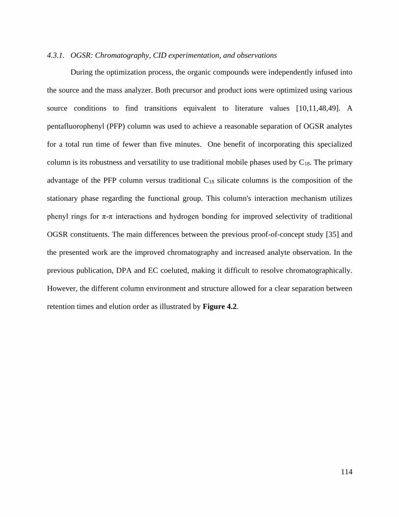
114
4.3.1. OGSR: Chromatography, CID experimentation, and observations
During the optimization process, the organic compounds were independently infused into
the source and the mass analyzer. Both precursor and product ions were optimized using various
source conditions to find transitions equivalent to literature values [10,11,48,49]. A
pentafluorophenyl (PFP) column was used to achieve a reasonable separation of OGSR analytes
for a total run time of fewer than five minutes. One benefit of incorporating this specialized
column is its robustness and versatility to use traditional mobile phases used by C18. The primary
advantage of the PFP column versus traditional C18 silicate columns is the composition of the
stationary phase regarding the functional group. This column's interaction mechanism utilizes
phenyl rings for π-π interactions and hydrogen bonding for improved selectivity of traditional
OGSR constituents. The main differences between the previous proof-of-concept study [35] and
the presented work are the improved chromatography and increased analyte observation. In the
previous publication, DPA and EC coeluted, making it difficult to resolve chromatographically.
However, the different column environment and structure allowed for a clear separation between
retention times and elution order as illustrated by Figure 4.2.

115
Figure 4.2: Chromatography separation of the seven OGSR compounds using the
pentafluorophenyl column. Standards of OGSR are eluted (minutes) based on their polarities and
interactions with the pentafluorophenyl column in the following order: 1) Akardite II (2.00 min),
2) Methyl Centralite (2.67 min), 3) Ethyl Centralite (3.19 min), 4) N-Nitrosodiphenylamine (3.49
min), 5) Diphenylamine (3.75 min), 6) 4-Nitrodiphenylamine (3.76 min), and 7) 2-
Nitrodiphenylamine (4.00 min). Each OGSR compound was measured with an electrospray source
in (+) mode.
There are over one hundred OGSR compounds that have been reported in different studies.
However, we selected seven major compounds (Figure 4.2) that are more indicative of gunshot
residue as they are not common in the environment nor prevalent in non-shooter populations [10].
One advantage of LC-MS methods is they can be easily expanded to additional GSR analytes as
long as they are compatible with the ionization requirements. Amongst the constituent list is AK2,
which differs from DPA by an amide addition, and 2-NDPA, a positional isomer to 4-NDPA.
These OGSR additions to our method were made due to shifts in modern propellant formulations
and potential degradation and deflagration entropy of more prominent indicators such as DPA.
Interestingly, compounds like MC and EC or 4-NDPA and 2-NDPA, which only differ by methyl
groups or the nitro-group position, can be identified by elution time alone.

116
Source conditions were also optimized for improved sensitivities of the OGSR compounds.
These conditions were modeled after Ali et al., who utilized a similar LC-MS/MS system [33].
However, the discernable difference between the two methodologies is the increased transitions
for all compounds in our method, barring 2-NDPA, and the lowered injection volume (1 µL vs. 5
µL). As expected, most OGSR analytes were suitable to ESI(+). However, it is important to note
that two typical OGSR markers, nitroglycerin (NG) and 2,4-dinitrotoluene (2,4-DNT), were not
observed using electrospray ionization in positive mode. Several technical publications
documented this observation using the same instrument source setup: the Agilent Jet Stream® ESI
(AJS-ESI) [50]. The limitation in ionization is potentially due to the chemical environment
conditions of the analyte, which showed very low efficiency in positive ionization. Although ESI
(-) mode can detect these compounds, it requires the assistance of signal enhancers to promote
adduct formation (salt forms) [51]. These anionic additives include acetates, chlorides, iodides,
and other nitrate sources [51]. Varying sources like atmospheric pressure chemical ionization
(APCI-), UV detectors, and recently direct sample analysis (DSA) can circumvent signal problems
and observe these compounds easily [52-55]. The addition of Na or K to the medium may be useful
in some instances, but it was not considered a viable option in our study since these two elements
are expected to be widely distributed in the environment and in the skin surfaces sampled for GSR.
Also, Na and K are easy to ionize and may cause in-source ionization competition that can affect
the efficacy of the metal clusters response. Unfortunately, these options require either dopants or
other sources (i.e., APPI) which may lead to additional costs and complications to a forensic
laboratory. Therefore, a decision was made to maintain the method's simplicity by using ESI in
positive mode while monitoring seven key OGSR components. Alternatively, our approach can

117
incorporate rapid screening with electrochemical methods, one can identify and characterize these
two troublesome compounds (NG and 2,4-DNT) [12].
4.3.2. IGSR: Formation and identification of M-L complexes using HRMS
Before initial validation and dual detection protocols, the M-L complexes first had to be
confirmed and readily reproducible under concentrated acidic digestion conditions. First, ICP
standards were mixed with the complexing agents and were monitored via the QQQ using the Q1
scan mode. Preliminary observations demonstrated unique signals in the mass spectrum showing
similar isotopic patterns that coincided with the investigated metals. A more in-depth investigation
revealed these metals formed nitrated adducts since the ICP standards possessed 2% nitric acid
within the solution. Additionally, other mass spectrum structures showed various complexes with
similar distributions related to water and sodium adducts.
Figure 4.3: Orbitrap confirmation of 18C6 [Ba-NO3] with Ba natural abundance (left) and [Pb-
NO3] with Pb natural abundance (right) patterns correlating to the simulated M-L isotopic
distribution.

118
A Q-Exactive® orbitrap was utilized to corroborate the findings and confirm the M-L
complexes' identity from the QQQ full scan. Flow injection analysis (FIA) and manual
manipulations of the source conditions revealed the presence of 1:1 as well as 1:2 ratios metal to
ligand complexes. Additionally, water adducts could also be observed but were less abundant than
the nitrated species. A full fragmentation breakdown and isotopic ratio observation are illustrated
in Figure 4.3 and Table 4.1 for barium and lead species. Unfortunately, an Sb complex could not
be formed with the chosen macrocycle; however, a different mechanism and complexing agent
unlocked the identification and will be discussed later.
Table 4.1: Orbitrap confirmation of fragmentation patterns of nitrated and lone metal species.
After orbitrap verification, source conditions and optimizations were performed on the
QQQ using the Optimizer® software. Fragmentor and collision energy voltages were monitored
from 10-100V and 10-250V with 10V increments to yield the highest signal possible, respectively.
The most abundant product ions for the barium-complex were the nitrated species (200.1 Da) and
the lone metal species (208.1 Da) for the lead complex. Because of the mechanism and factors for
complex formation, the decreasing m/z values must be included when building the acquisition
method. For instance, the barium-complex forms various precursor ion m/z values consistent with
Identification Ions (m/z) observed Identification MS1 (nitrated forms) MSn (lone metals)
Barium
BaNO3 (200 Da)
18C6 (264 Da)
C12
H24
O6
464.0498 [L + 138
Ba + NO3]
+ 200.9003 [
138Ba + NO
3]
+ 137.9003 [
138Ba]
+
463.0504 [L + 137
Ba + NO3]
+ 199.9009 [
137Ba + NO
3]
+ 136.9009 [
137Ba]
+
462.0491 [L + 136
Ba + NO3]
+ 198.8997 [
136Ba + NO
3]
+ 135.8997 [
136Ba]
+
461.0502 [L + 135
Ba + NO3]
+ 197.9008 [
135Ba + NO
3]
+ 134.9008 [
135Ba]
+
460.0491 [L + 134
Ba + NO3]
+ 196.8972 [
134Ba + NO
3]
+ 133.8972 [
134Ba]
+
Lead
PbNO3 (270 Da)
18C6 (264 Da)
C12
H24
O6
534.0498 [L + 208
Pb + NO3]
+ 270.1003 [
208Pb+ NO
3]
+ 208.1003 [
208Pb]
+
533.0504 [L + 207
Pb + NO3]
+ 269.0109 [
207Pb+ NO
3]
+ 207.0109 [
207Pb]
+
532.0491 [L + 206
Pb+ NO3]
+ 268.0097 [
206Pb+ NO
3]
+ 206.0097 [
206Pb]
+
530.0491 [L + 204
Pb + NO3]
+ 266.0012 [
204 Pb+ NO
3]
+ 204.0012 [
204Pb]
+
119
its isotope values at 464.1 (138), 463.1 (137), 462.1 (136), 461.1 (135), and 460.1 (134). Therefore,
the acquisition method must account for these values to gain confidence in the observed structure,
which is not required for OGSR analysis.
Choices of complexing agents
Our goal was to select a single complexing agent to capture the three critical elemental
constituents of GSR (barium, lead, and antimony). However, extensive literature review and
experimentation demonstrated that this was not possible. Antimony (a metalloid like arsenic and
bismuth) demonstrated different complexation characteristics that necessitated selection of a
separate chelating agent for this element. While this adds another ingredient to the complexing
solution, we demonstrated that it is still possible to detect Ba, Pb, and Sb via LC-MS/MS.
4.3.2.1. Crown ethers
Interest in supramolecular chemistry has expanded from its early conceptions by van der
Waals into Pedersen’s initial discovery of simple macrocycles. These compounds are integrated
into various disciplines such as nanotechnology, medicinal chemistry, chemo-sensors, and others
[56]. Depending on the guests and applications, modifications such as heteroatom substitutions or
functional group additions provide scientists various avenues for analyte characterization. Because
of their versatility to encompass a myriad of guest molecules easily, crown ethers are one of the
most utilized and studied macrocycles in the field via self-assembly.
Crown ethers employ various binding mechanisms, including electrostatic interactions,
hydrogen bonding, and/or van der Waals forces, depending on the guest analyte. Like other host
compounds, these molecules have shown their effectiveness in various fields, including forensics
for explosive and gunshot residue analysis to toxic metal removal in wastewater [35,57,58]. The
strength of complexation is dependent on various factors, including the internal cavity size of the

120
crown ether, the atomic radii, and the charge of analytes. The appealing aspect of these interactions
involves identifying metal ions and their robust nature in forming metal-ligand complexes. The
creation of these complexes increases an analyte’s molecular weight while maintaining natural
isotopic abundances. Additionally, the host macrocycles provide transportation and interactions
through a column for MS/MS detection.
Characterizing complexes relies heavily on the instrumentation parameters. Previous
experiments performed in our group focused on mobilities via ion mobility spectrometry CID
experimentation via LC-MS/MS [35,59]. For instance, when inspecting Pb, the addition of 18C6
increases the molecular weight by 264.1 Da yielding 472.1 Da for the lone metal and 534.1 Da
when nitrated. When the 534.1 Da complex is fragmented, the Pb-NO3 (270 Da) product ion is
predominant and displays similar isotopic distributions to the lead ion. When the 270 Da is
fragmented further, the 208.1 (52%), 207.1 (22%), 206.1 (24%), and 204.1 (2%) ions are observed,
thus solidifying high confidence for the precursor complex in question.
Other aspects like charge state and size of ions are essential when forming complexes.
Additionally, competition for the macrocycle's internal cavity can ultimately affect the response
of the metal detection. Because of validation protocols, internal standards should be similar in
structure and exhibit comparable ionization energies to measure analyte concentrations accurately.
For the OGSR method, D10-diphenylamine was coeluted with DPA but possessed unique
transitions for simple identification. The IGSR method used thallium (Tl) as the internal standard
since it is typically not found in GSR and is similar in size (150 pm) to both Ba2+ (135 pm) and
Pb2+ (119 pm). Without Tl+ present, 18C6 showed preferential binding for Ba versus Pb, illustrated
in Figure S4.1. Thallium lowered the intensities for IGSR constituents yielding the selectivity
trend of Tl > Ba > Pb. In contrast, antimony (76 pm) showed no efficient binding affinity with

121
18C6. This observation shows that the size and charge of the guest play a crucial role in binding
selectivity.
4.3.2.2. Tartaric acid
In our studies, however, antimony (Sb) has not been observed to self-assemble with various
crown ether species, including 12-crown-4-ether, 15-crown-5-ether, 18-crown-6-ether, and
dibenzo-18-crown-6-ether. Various research groups have explored and observed tartaric acid’s
effectiveness in binding with antimony through a process known as chelation [60-62]. Chelation
occurs by forming two or more coordinate bonds between a polydentate ligand (tartaric acid) and
a central atom (metal cations). This ligand is naturally found in fruits and is utilized in ceramics
and pharmaceuticals. Several groups have investigated the interaction between various metal
cations and tartaric acid structures under negative ionization modes [62-64].
Additionally, tartaric acid can yield enantiomeric complexes (D- and L-) and be separated by
HPLC and capillary electrophoresis [65]. For adequate separations, the tartrate complexes can be
dissolved in water and salt solvents and further yet, diluted in various working stock solutions
with specific solvents such as methanol. Tartaric acid is readily dissolvable in water but not in
methanol, which is why more water adducts are observed versus other salts.

122
Figure 4.4: Flow injection analysis of the various tartrate complexes with Sb in ESI- mode.
Adducts formation is still prevalent, and because of the nature of the ligand, multiple Sb ions can
bind to multiple ligands. For instance, the difference between 415 Da and 433 Da is an m/z value
of 18, a water molecule. However, from 415 Da to 537 Da is the addition of an Sb ion.
However, the adducts may also be attributed to altering voltages of an ESI source. This can
cause the isotope ratios to differ from recorded natural abundances 57% (121Sb) and 43% (123Sb ),
outlined by Schug et al. and other groups (Figure 4.4) [61,62,66-69]. For instance, by altering the
nozzle voltage from 500 V to 2500 V, the complex at 537 m/z appears and disappears, respectively.
This investigation showed that the structure of the tartrate complexes can shift in not only the
adducts but also in the charge states. This finding has been outlined by Schug and others by altering
the voltages of both the fragmentor and within the collision chamber [62,64]. The tartrate
complexes can undergo homolytic cleavages as the complex reorients throughout the mass
analyzer. Thus, resulting in the different losses of peroxides and water adducts. From these
observations, the ligand tends to favor the complex with 2- charge (267 Da) than the 1- charged
complexes (415, 433, 450, and 537 Da) illustrated in Figure 4.4.

123
Prior knowledge of complexation illustrated that 18C6 was not as sensitive to Sb versus
Ba and Pb; therefore, tartaric acid was tested as the possible “one host” solution. Although both
Ba and Pb formed complexes with the tartaric acid, their signals were vastly smaller than Sb.
Ultimately, we decided to incorporate both complexing agents, tartaric acid and 18C6, in equal
parts for the final solution.
Analytical validation and figures of merit
4.3.3. Analytical validation and figures of merit
Tables 4.2 and 4.3 represent both OGSR and IGSR constituents' figures of merit, including
the LOD, LOQ, %RSD (intra- and inter-day), and linear dynamic range (LDR). The OGSR
compounds LDR expanded from 1-200 ppb (1-200 pg), and the 1:1 IGSR complexes 100 ppb to
25 ppm (1.0-250 ng). The mean, standard deviation, and 95% confidence intervals are measured
against the D10-DPA and Tl quantifier ion ratios for the OGSR and IGSR, respectively. The Agilent
MassHunter® Quantitative Analysis software was used to calculate the bias, LDR, and other
figures of merit. The absolute LOD and LOQ (ppb and ppm) were calculated for a 1 µL injection
and 10 µL injection for OGSR and IGSR, respectively. The estimated LOD and LOQ (pg and ng)
were calculated and adjusted for the injections previously mentioned.
Table 4.2: Figures of merit of organic compounds. Included is information pertaining to LOD,
LOQ, LDR, the precursor ions, and the ion ratio of the most abundant qualifier ion.
Group Constituent LOD (ppb) LOD (pg) LOQ (ppb) LOQ (pg) m/z MRM [M+H]+
Precursor: Product
Ion ratio%
(n=15) R2
%RSD
INTRA
%RSD
INTER LDR (pg)
OG
SR
AK 2 0.3 0.3 0.90 0.90 227:170:93 70.7 0.999 1.3 4.8 1-200
DPA 3.4 3.4 10.1 10.1 170:93:65 20.4 0.998 3.7 10.4 10-200
EC 1.0 1.0 3.0 3.0 269:148:120 91.6 0.999 1.1 9.3 5-200
MC 0.30 0.30 0.90 0.90 241:134:106 56.0 0.999 2.7 11.0 1-200
N-NDPA 4.6 4.6 13.9 13.9 199:169:65 12.7 0.997 4.8 6.0 25-200
4-NDPA 3.0 3.0 9.0 9.0 215:198:167 92.0 0.999 7.9 6.4 10-200
2-NDPA 2.7 2.7 8.2 8.2 215:180 100.0 0.997 4.6 4.0 10-200

124
Table 4.3: Figures of merit for inorganic compounds. Included is information pertaining to LOD,
LOQ, LDR, the precursor ions, and the ion ratio of the most abundant qualifier ion.
As mentioned before, the LOD and LOQ values were improved from the previous proof-
of-concept study for both IGSR and OGSR. For OGSR, we included an additional qualifier ion
barring 2-NDPA, while Ali et al. served as a guide for source parameters due to similar
instrumentation conditions [33]. Improvement of the IGSR constituents was contributed to manual
optimization and understanding of the complexation process. If more agent is present, more guest
analyte can be encapsulated for lower detection. The LDR for Sb is higher due to its formation
nature and a greater ionization potential and energies required to dissociate the complex. It is
important to note that the 50-250 ng range for Sb is on the cusp of the range when referencing
authentic samples. Therefore, we continue to monitor its presence but will need further
investigations to circumvent the complex's ionization energy.
The linearity of each compound was assessed through calibration curves and intra- and
interday studies. All compounds and elements showed a linear response with an R2 value ≥ 0.974.
The working calibration ranges were 1 ppb to 200 ppb (OGSR) and 100 ppb to 25 ppm (IGSR).
Further statistical analysis evaluated an accurate quantitation, including R2 coefficients and
residual plots with less than 10% RSD. Residuals plots assess the nature of the samples collected
using several considerations: 1) randomly distributed, 2) the variances are equal, and 3) values are
normally distributed across predicted values. The residuals’ variance did not increase with
Group Constituent LOD (ppm) LOD (ng) LOQ (ppm) LOQ (ng) m/z MRM
Precursor: Product
Ion ratio
% (n=15) R2
%RSD
INTRA
%RSD
INTER LDR (ng)
IGS
R
Ba 0.10 1 0.30 3.0
464:200:138
463:199:137
462:198:136
461:197:135
71.70 (138)
11.23 (137) 7.85 (136)
6.59 (135)
0.998 8.4 4.6 1-60
Pb 0.20 2 0.60 6.0 534:270:208
533:269:207
532:268:206
52.40 (208)
22.10 (207)
24.10 (206)
0.982 6.4 5.7 2-60
Sb 5.0 50 15.0 150 267:121
269:123
57.21 (121)
42.79 (123) 0.974 2.7 11.0 50-250

125
concentration until after 200 ppb and 6 ppm (Ba and Pb) and 25 ppm (Sb). This observation
confirms homoscedasticity across data points, thus ensuring randomness. The relationship between
the independent and dependent variables and their variances is displayed in Figure S4.2.
4.3.4. Background and collection substrate considerations: recovery study and comparison to
ICP-MS digestion method
A critical aspect of the method validation was to consider the performance and practicality
of the substrate used for the collection and retention of the inorganic and organic components of
interest from the hands of an individual. For this purpose, we compared the recovery of IGSR and
OGSR from tesa® Tack polymer and the more universal sampling media carbon adhesive. Our
goal was to evaluate if the LC-MS/MS method can incorporate the use of carbon stubs to be more
compatible with current practice and avoid the change of sampling protocols widely used in the
GSR community by both law enforcement and laboratory personnel.
The surface extraction of each substrate was evaluated for any possible suppressants. In
part, this concern was due to the substrates possessing both polymeric and adhesive qualities that
could potentially interfere with the column performance. We were less concerned with the
selectivity against the substrates due to choosing specific m/z values and MRM transitions unique
to elements and compounds.
Our previous study successfully used tesa® Tack polymer for GSR collection by fully
submerging and exposing it to organic solvents and harsh acidic conditions [35]. This polymer was
very attractive in our initial study because it was commercially sold (http://www.tesa.com) as a
non-greasy, putty and suggested by BKA colleagues based on their experiences (Dr. Ludwieg
Niewoehner) and adapted for the previous project [35]. However, because we want this method to
comply with GSR cases, without changing current sampling protocols, the universal method of
126
carbon tape stubs was evaluated. Thus, a newly developed extraction method was optimized for
both OGSR and IGSR components for the carbon tape by utilizing surface washings. These
washings displayed suppression of analytical signals, which led to the incorporation of filters. The
OGSR extraction used 100% methanol, where the IGSR analysis used a micro-bulk method that
needed heat and concentrated acids as microliter volumes.
One substantial difference between this work and our previous proof-of-concept study is
the OGSR and IGSR recovery assessment from the substrates. Following the Eurachem validation
guideline, we conducted a recovery comparison experiment using ESI involving both carbon tape
and tesa® Tack [35]. Bias or percent recovery is not only crucial to indicate potential trends or
underlying characteristics/properties present but addresses the effectiveness of a method by
measuring the “closeness” of extraction results versus the reference or “true” spiked value onto
substrates.
There are two critical differences between the tesa® Tack and carbon tape. First, the
polymer exhibits porous properties similar to the skin's epidermal layer, allowing for significant
absorption of OGSR. Secondly, the thickness is vastly different compared to the carbon tape. Since
tesa® Tack is thicker than the carbon tape, the OGSR components can adsorb into the tack's inner
layers, creating a more significant challenge for extraction. Three different concentration levels
(low, medium, high) assessed the repeatability and reproducibility and determined the variability
in the results; 25 ppb, 50 ppb, and 165 ppb for OGSR (Table 4.4) and 1 ppm, 3 ppm, and 5 ppm
for IGSR (Table 4.5). The low, medium, and high values were chosen to represent concentrations
that can be fully quantified from the validated method and represent concentrations observed in
authentic items.

127
Table 4.4: Summary of OGSR analyte recoveries on both carbon tape and tesa® Tack substrate.
From the recovery experiment, the OGSR constituents' results indicated that all
components had less than 10% % RSD, barring 2-NDPA at 25 ppb for the tesa® Tack, 11.2%. The
percent recovery values ranged from 35.7-86.2% for carbon tape and 17.3-57.7% for tesa® Tack.
A one-tailed t-test (at p=0.05) showed the carbon tape has significantly higher recoveries than the
tesa® Tack at all concentration ranges tested.
It is worth mentioning the extraction methods are not exhaustive as we designed the method
for surface washing rather than a complete submersion extraction. The rationale for this decision
was two-fold: 1) it minimizes potential undesired contribution from the adhesive that can be
detrimental for LC-MS, and 2) gentle to mild washings further increase the possibility of not
displacing particles for further analysis, as demonstrated by Bonnar et al. [11].
Conversely, the IGSR percent recoveries were comparable for both substrates, showing no
significant differences between Ba and Pb observed using a one-tail t-test. The recovery range for
the carbon tape was 44-85%, and the tesa® Tack ranged from 43-86%, which is illustrated by
Table 4.5. As a result, the carbon tape was chosen for the remaining of the study as it improved
OGSR recovery while not having a detriment effect in the IGSR, as compared to the tesa® Tack.
Expected
Conc. Substrate
MC
% Recovery
EC
% Recovery
DPA
% Recovery
N-NDPA
% Recovery
4-NDPA
% Recovery
2-NDPA
% Recovery
25 ppb Carbon Tape 41.2 ± 0.74 42.1 ± 2.81 59.9 ± 3.29 85.0 ± 2.34 44.8 ± 5.87 83.4 ± 5.33
tesa® Tack 28.8 ± 1.49 17.3 ± 1.59 36.9 ± 1.69 55.2 ± 3.83 17.8 ± 8.45 52.9 ± 11.2
50 ppb Carbon Tape 77.5 ± 1.47 67.8 ± 1.36 74.2 ± 1.71 83.5 ± 2.43 40.4 ± 1.38 86.2 ± 1.60
tesa® Tack 38.6 ± 0.40 27.1 ± 0.95 55.6 ± 0.51 48.5 ± 9.03 23.4 ± 1.15 57.7 ± 1.49
165 ppb Carbon Tape 61.7 ± 2.59 80.5 ± 1.58 57.6 ± 1.25 54.9 ± 1.77 35.7 ± 0.77 53.5 ± 1.00
tesa® Tack 30.5 ± 2.52 27.1 ± 1.86 35.5 ± 1.24 24.7 ± 1.70 20.6 ± 1.29 23.5 ± 0.78

128
Table 4.5: Summary of IGSR analyte recoveries using ICP-MS standards on both substrates.
This procedure utilized ICP-MS standards directly spiked onto each substrate and allowed
to dry completely. After drying, surface extractions followed, which used concentrated acids and
the micro-bulk procedure mentioned previously. However, this only answers one part of the
recovery process as those standards do not truly represent a particulates' nature because they are
already solubilized. Therefore, a standard that corresponds to the morphological characteristics
and composition relating to ASTM E1588-20 would better represent authentic samples and
monitor the behavior of IGSR in concentrated acid washings and digestion, as discussed below.
4.3.5. Cross-validation of recovery of IGSR via ICP-MS
With OGSR compounds, variables like lipophilicity and evaporation rates play a
significant role in the recovery from exposed skin and other sampling areas. Because the
environment heavily influences IGSR formation and placement, sufficient analysis is more
dependent on factors like extraction procedures and substrate surfaces versus skin absorption.
Additionally, OGSR standards correlate more with authentic samples than ICP-MS standards
spiked onto substrate surfaces. Therefore, intact IGSR particulates offer a greater evaluation of
factors like extraction protocols and interactions.
Tailor-made primer-only GSR (p-GSR) standards originating from a Winchester primer
was previously developed and characterized in our group and was used to evaluate the extraction
protocol's effectiveness [45]. These standards are made of p-GSR obtained from the actual
Expected Conc. Substrate Ba % Recovery Pb % Recovery
1 ppm Carbon Tape 67.1 ± 0.97 57.8 ± 3.28
tesa® Tack 57.9 ± 1.67 45.5 ± 2.98
3 ppm Carbon Tape 74.7 ± 2.44 44.4 ± 3.13
tesa® Tack 83.3 ± 4.53 44.6 ± 1.39
5 ppm Carbon Tape 84.7 ± 3.12 44.3 ± 1.27
tesa® Tack 86.1 ± 1.14 43.5 ± 1.05

129
discharge of a primer in a firearm, therefore providing similar conditions and concentrations that
an analyst would expect from an authentic sample. These “matrix-matched” microparticle
standards were characterized via ICP-MS, SEM/EDS, and LIBS methods to confirm the
concentration, stability, and elemental composition and morphology of the particles. Results
showed that this standard correlates to the expected concentration found in the primers [45].
Since IGSR is more susceptible to secondary and tertiary transfer, factors like substrate
composition and extraction protocol can affect practitioners' analysis. We address both by
substrate exposure to heavy acids with multiple washings to a final volume of 500 μL. Therefore,
both the extraction and substrate considerations were compared via digestion methods; 85° for 10
minutes in concentrated acid (LC-MS/MS) versus 60 minutes with dilution to 10% nitric acid
(ICP-MS). Our findings demonstrated that the percent recoveries were higher with the 60-minute
digestion protocol but with less than a 10% difference. Furthermore, there was no statistically
significant difference between tesa® Tack and carbon tape regarding the recovery of Pb and Ba.
This difference explains more of the interaction with the 18-crown-6-ether, where the binding
energy is dependent on the size and charge of the metallic species present in a solution.
4.3.6. Workflow and cross-validation with screening methods
Within the forensics community, there is an increased interest to incorporate rapid,
preliminary techniques for IGSR and OGSR, followed by confirmatory analyses [13,24,34]. By
introducing these methodologies, analysts can filter presumptively “negative” results and focus on
confirmation of those positive items, ultimately making better-informed decisions and reducing
backlogs. With this increased interest, we tested the applicability of implementing multiple
screening techniques followed by the proposed LC-MS/MS methodology from a single firing
event and sample.

130
After the collection via carbon-adhesive stub, LIBS was used as the first instrument for
analysis. LIBS has several appealing aspects, including ambient conditions, quick analysis times
(< 2 minutes/stub), and compositional information comparable to SEM/EDS [47]. Because of the
ambient and laser conditions, the OGSR constituents are not substantially lost, and the overall
IGSR morphology remains practically uncompromised. The preservation of both constituents
allows for further analysis, such as electrochemical methods. By washing a portion of the sample's
surface with two solvents (acetonitrile and aqueous buffer), OGSR and IGSR are effectively
extracted. With an analysis time comparable to LIBS (< 5-10 minutes), the advantage of
electrochemical methods is the capability to simultaneously detecting both constituents [46].
Furthermore, combining these screening methods has demonstrated their effectiveness in
increasing confidence in GSR samples with accuracies superior to 95%, with the advantage that
analysis can be followed by SEM/EDS confirmation [12]. Because electrochemical methods
generate extracts compatible with LC-MS/MS, this study explored if these washings could be used
with the micro-bulk digestion and complexing agents.
Because LC-MS/MS offers superior LOD and LOQ and consumes a small sample volume
(1 µL), the remaining aliquot can be used for further confirmation methodologies, if needed. After
running the organic extract from electrochemistry, the aqueous buffer aliquot with the organic
extract can be combined and pushed through with the micro-bulk digestion and complexing agent
addition allowing OGSR and IGSR identification by LC-MS/MS. Additionally, we tested the
electrochemical extraction collection efficiency by running subsequent washings from an extracted
stub. Our findings showed that the subsequent washings show either no signal or signal below the
LOQ, which confirms that the electrochemical extractions are very efficient in collecting both

131
constituents. These results illustrate the potential of utilizing various instrumentation techniques
to gain more informative data from a single sample without compromising the entire stub.
4.3.7. Performance rates and criteria for authentic samples
Since dual detection of IGSR and OGSR is not routinely conducted at forensic laboratories,
a criterion outlining the requirements for identifying “true positive” and “true negative” results
must be established. The lack of guidelines for OGSR compounds versus the well-established
ASTM protocol for IGSR is the primary contributing factor. Therefore, we highlight the
considerations to call a sample positive for GSR when: 1) evidence of both OGSR compounds and
IGSR elements must be present above LOD and background population-based critical thresholds,
2) a minimum of three components, either two OGSR components, and one IGSR analyte or vice
versa. For example, if the sample contains EC, DPA, and Ba, the sample is labeled “positive for
GSR.” If the sample only contains one of the GSR components, then the sample is labeled
“potential GSR” and needs further analysis. For example, if only OGSR compounds such as EC,
DPA, and 2-NDPA are present without the detection of Pb, Ba, or Sb, that sample is characterized
as “potential GSR”. In the ASTM E1588-20, “potential GSR” is referred to as consistent with or
commonly associated with GSR; however, due to the lack of consensus at this moment concerning
the confirmatory value of OGSR/IGSR profiles, we followed a more conservative two-category
scale for positive results (characteristic or positive, and potential). Our experience with the
interpretation of large population sets with LIBS and EC demonstrates that machine learning
algorithms outperform the categorical critical threshold approach used here, with the added
advantage of providing a probabilistic output for a more objective interpretation of the evidence.
We are currently collecting a more extensive population to apply this probabilistic approach to
LC-MS/MS data. Larger sample sizes are needed to split the data into training and

132
testing/validation sets with enough statistical power. Nonetheless, the critical threshold method is
valuable as an exploratory tool to learn about the distribution of the data in different subpopulations
and provide a basis for interpreting computerized approaches.
We tested this criterion against two types of authentic sample data sets and compare them
with LIBS and electrochemical methods. The two data sets consisted of 95 baseline shooter
samples (Table 4.6) and 78 activity samples (Table 4.7). The baseline samples involved shooting
in the WVU ballistics range and immediately collecting after a shooting event. These samples were
extracted using the LC-MS/MS methodology involving the filters and concentrated acid surface
washings. The baseline results are represented in Table 4.6 and include success rates and detection
for LC-MS/MS. The collection methodology involves generating four separate carbon stub
samples labelled as such, left-palm (LP), left-back (LB), right-palm (RP), and right-back (RB).
The first shooting session used a single type of ammunition (Winchester), while the second
shooting session utilized a mixture of ammunition and firearms (Federal, Blazer, 9 mm and 40
caliber pistols). Moreover, a subset of 30 non-shooter background samples was monitored to assess
potential false-positives and establish critical thresholds. All background samples resulted in 100%
true-negative rate, where all potential peaks were below LOD and LOQ.

133
Table 4.6: Success rates of the baseline (no-activity) shooter samples in two separate shooting
sessions using LC-MS/MS surface extraction procedure. The indicator “ND” refers to analytes
not detected.
Session Analyte Range (ppb) Mean (ppb) Median (ppb) Positive Set Combined Constituent
data
1
OG
SR
AK2 0.50-296 15.4 6.5
18/20
90%
19/20
95%
MC ND
EC 1.1-350 13.8 2.8
DPA 4.5-498 102 74
N-NDPA 6.8-271 58 36
4-NDPA 3.7-30 10.9 8.2
2-NDPA 6.2-58 16.7 9.7
IGS
R
Range
(ppm) Mean (ppm) Median (ppm)
20/20
100%
Ba 1.1-8.9 4.07 3.5
Pb 1.1-5.2 1.83 1.4
Sb 6.1-10.1 5.6 6.1
2
OG
SR
AK2 28-55 42.8 42.1
33/75
44%
75/75
100%
MC ND
EC 15.4-81.5 42.1 40.3
DPA 11.2-65.3 30.1 25.1
N-NDPA ND
4-NDPA 9.3-38.2 20.4 16.8
2-NDPA 9.1-57.9 25.5 20.8
IGS
R
Range
(ppm) Mean (ppm) Median (ppm)
50/75
66.7% Ba 1.7-4.5 2.9 2.8
Pb 0.75-2.5 1.5 1.2
Sb ND ND ND
Interestingly, the second session decreased overall OGSR and IGSR separate performance
rates but demonstrated the GSR detection potential when combined (100% true positive rate).
These differences may be attributed a myriad of factors including the cartridge ejection port
locations, composition of the powder and primers, or the randomness of each firing event. By using
various ammunition formulae, the amounts and types of IGSR and OGSR deposited in hands was
variable and at some extent, representative of casework.
The 78 activity samples were utilized to monitor the behavior of GSR for more realistic
scenarios. These activities included vigorous hand rubbing, the application of hand sanitizer, and

134
running for one minute after discharge events. These samples were cross-validated with LIBS and
electrochemical methods and report the success rates and detection for LC-MS/MS compared to
LIBS and electrochemistry, represented in Table 4.7. These results were compared to 10 baseline
(no-activity) shooting samples and eight negative control (non-shooter) samples. The negative
control samples were collected from the collection team before and after the shooting session to
monitor any potential contamination.
Table 4.7: Success rates of activity samples (where n is equal to the number of individuals) in
separate shooting sessions. Additionally, the LC-MS/MS results were compared with LIBS and
EC cross-reference results.
LC-MS/MS LIBS LIBS + EC
Session Activity OGSR (%) OGSR+IGSR (%) IGSR (%) OGSR+IGSR (%)
1
Rub (n=13) 15.4 84.6 0 23.1
Hand Sanitizer (n=13) 7.7 53.8 15.4 30.8
Running (n=13) 15.4 23.1 15.4 15.4
Baseline (no activity) (n=5) 100 100 60 100
Background (non-shooters) (n=4) 0 0 0 0
2
Rub (n=13) 30.8 84.6 46.2 92.3
Hand Sanitizer (n=13) 69.2 92.3 38.5 84.6
Running (n=13) 23.1 38.5 61.5 92.3
Baseline (no activity) (n=5) 100 100 60 100
Background (non-shooters) (n=4) 0 0 0 0
Additionally, we saw the effect of weather and its role in the recovery of these analytes.
The first shooting session involved inclement weather in the form of rain. The shooter’s hands
showed significant amounts of water when they entered the lab after the running activity. The
results indicated the OGSR compounds were more affected versus the IGSR particulates ranging
from 7.7-15.4% for LC-MS/MS. This consideration presents another point when researching more
“real-world” scenarios where weather could be impactful. For all samples, methyl centralite was
not detected in any samples, which may be largely attributed to a shift in propellant formulation.
On the other hand, we observed an increase in AK 2 detection in these items.

135
Observing the analyte response versus activity under clearer weather conditions, the OGSR
responses for the LC-MS/MS were more significant for the hand sanitizer versus hand rubbing and
running. This may be primarily due to a “pseudo” organic extraction using isopropyl alcohol in
direct contact with the hands. Conversely, running for ~ one minute produced the lowest response
for OGSR, which may be due to sweat formation allowing for the potential of increased absorption
into the epidermal layer. Looking at the LIBS elemental responses, we observed an inverse
relationship for the IGSR particulates where running was the greatest response followed by
rubbing then hand sanitizer. This may indicate that the IGSR particulates are more influenced by
physical interactions with hands in the forms of washing or sanitizing. Although these remarks and
comments are speculative, the results illustrate the need to expand research efforts and interest in
the persistence and fate of GSR analytes after a discharge event. Also, the preliminary post-
shooting activity data demonstrates that the detection of OGSR and IGSR by LC-MS/MS was
feasible even for samples exposed to factors that decrease the chances for detection.
4.4. Conclusions
Herein, we demonstrate the use of host-guest chemistry to reach application in another
discipline. Crown ethers are incredibly versatile not only for their transportation properties but
also for their ability to retain isotopic ratios of the metals [35,41,70,71]. The method proved
efficient for the detection of Pb and Ba. On the other hand, the inability to detect antimony in
authentic samples is primarily due to the chelated metal complex's detection limitations and
ionization. Current research efforts focus on investigating optimal complexing agents to
encompass IGSR analytes to lower the ionization potentials. Finding host molecules suitable for
complexing all IGSR metals is inherently difficult because of the wide variety of complexing

136
agents available, and the intricacies associated with the interactions between the metal species and
host molecules.
More importantly, we investigated and validated an LC-MS/MS method to integrate into a
broader workflow of GSR analysis. LC-MS/MS is a widely found technique in crime laboratories
that primarily analyze drugs of abuse or toxicology samples. The figures of merit established
represent this technique's capability but does not explicitly envelop all instrumentation models.
This methodology's appealing aspect is the ability to use one sample for various screening tests
like LIBS or electrochemistry followed by confirmatory techniques such as LC-MS/MS
[12,30,33]. Following the ASTM E1588-20 guideline for GSR analysis allows this research to
apply a universal collection substrate, compositional considerations, and an established standard
instrument for reference.
However, without considering both components, the evidentiary value and understanding
of GSR behavior may remain stagnant. The key to this method's successful deployment in the
forensics field is to adopt LC-MS/MS approaches to increase confidence in the identification of
GSR on a sample with both IGSR and OGSR information. The figures of merit presented
demonstrate the ability of the method to identify and quantitate both OGSR and IGSR. With a
further understanding and additional research concerning elemental and isotopic data when bound
to host molecules, an instrumental technique like LC-MS/MS can provide additional quantitation
for practitioners. Not only does this method increase the accuracy in GSR analysis and
interpretation, but it yields an alternative avenue and resolves the need to choose between IGSR
or OGSR. Finally, the approaches proposed in this study can be applied in other fields, monitoring
exposure and environmental compartments near ammunition manufacturing or shooting facilities.

137
Author Contributions
The manuscript was written through the contributions of all authors. All authors have given
approval to the final version of the manuscript.
Conflicts of interest
The authors declare that they have no known competing financial interests or personal
relationships that could have appeared to influence the work reported in this paper.
Acknowledgments
We would like to acknowledge both Dr. Keith Morris and James Hamilton for their support
during the collection of authentic samples and access to the WVU ballistics laboratory. We would
also like to acknowledge Kourtney Dalzell for her assistance in collection and analysis of the
electrochemical results. Additionally, we would like to acknowledge Dr. Callee Walsh and Dr.
Sandra Majuta for assistance and access to the orbitrap instrument in the BNRF.
This project is sponsored by Award No. #2019-R2-CX-044 from the National Institute of
Justice to West Virginia University. The opinions, findings, and conclusions are those of the
authors and do not necessarily reflect those of the Department of Justice.

138
4.5. References for Chapter 4
[1] T. Trejos, S. Koch, A. Mehltretter, Scientific foundations and current state of trace
evidence—A review, Forensic Chem. 18 (2020) 100223.
https://doi.org/10.1016/j.forc.2020.100223.
[2] L.S. Blakey, G.P. Sharples, K. Chana, J.W. Birkett, Fate and Behavior of Gunshot
Residue-A Review, J. Forensic Sci. 63 (2018) 9–19. https://doi.org/10.1111/1556-4029.13555.
[3] Z. Brozek-Mucha, Z. Brożek-Mucha, Trends in analysis of gunshot residue for forensic
purposes, Anal. Bioanal. Chem. 409 (2017) 5803–5811. https://doi.org/10.1007/s00216-017-
0460-1.
[4] O. Dalby, D. Butler, J.W. Birkett, Analysis of Gunshot Residue and Associated
Materials-A Review, J. Forensic Sci. 55 (2010) 924–943. https://doi.org/10.1111/j.1556-
4029.2010.01370.x.
[5] M.A. de Carvalho, M. Talhavini, M.F. Pimentel, J.M. Amigo, C. Pasquini, S. Alves, I.T.
Weber, M. Albino de Carvalho, M. Talhavini, M.F. Pimentel, J.M. Amigo, C. Pasquini, S.A.
Junior, I.T. Weber, NIR hyperspectral images for identification of gunshot residue from tagged
ammunition, Anal. Methods. 10 (2018) 4711–4717. https://doi.org/10.1039/c8ay01341a.
[6] F.S. Romolo, M.J. Bailey, J. de Jesus, L. Manna, M. Donghi, Unusual sources of Sn in
GSR. An experimental study by SEM and IBA, Sci. Justice. 59 (2019) 181–189.
https://doi.org/10.1016/j.scijus.2018.10.009.
[7] M. Donghi, K. Mason, F.S. Romolo, Detecting Gunshot Residue from Sellier & Bellot
Nontox Heavy Metal-free Primer by in situ Cathodoluminescence, J. Forensic Sci. 64 (2019)
1658–1667. https://doi.org/10.1111/1556-4029.14110.

139
[8] K. Mason, R. Wuhrer, Phenom Desktop SEM for Gunshot Residue and
Cathodoluminescence Imaging and Analysis, Microsc. Microanal. 23 (2017) 1074–1075.
https://doi.org/10.1017/s1431927617006031.
[9] S. Practice, S. Handbook, S. Practice, Standard Practice for Gunshot Residue Analysis by
Scanning Electron Microscopy/Energy Dispersive X-Ray Spectrometry, ASTM Int. i (2020) 1–5.
https://doi.org/10.1520/E1588-20.
[10] W. Feeney, C. Vander Pyl, S. Bell, T. Trejos, Trends in composition, collection,
persistence, and analysis of IGSR and OGSR: A review, Forensic Chem. 19 (2020) 100250.
https://doi.org/10.1016/j.forc.2020.100250.
[11] C. Bonnar, E.C. Moule, N. Lucas, K.E. Seyfang, R.P. Dunsmore, R.S. Popelka-Filcoff,
K. Redman, K. Paul Kirkbride, Tandem detection of organic and inorganic gunshot residues
using LC–MS and SEM-EDS, Forensic Sci. Int. 314 (2020) 110389.
https://doi.org/10.1016/j.forsciint.2020.110389.
[12] T. Trejos, C. V Pyl, K. Menking-Hoggatt, A.L. Alvarado, L.E. Arroyo, Fast identification
of inorganic and organic gunshot residues by LIBS and electrochemical methods, Forensic
Chem. 8 (2018) 146–156. https://doi.org/10.1016/j.forc.2018.02.006.
[13] M. Lopez-Lopez, C. Alvarez-Llamas, J. Pisonero, C. Garcia-Ruiz, N. Bordel, An
exploratory study of the potential of LIBS for visualizing gunshot residue patterns, Forensic Sci.
Int. 273 (2017) 124–131. https://doi.org/10.1016/j.forsciint.2017.02.012.
[14] J. Coumbaros, K.P. Kirkbride, G. Klass, W. Skinner, Characterisation of 0.22 caliber
rimfire gunshot residues by time-of-flight secondary ion mass spectrometry (TOF-SIMS): A
preliminary study, Forensic Sci. Int. 119 (2001) 72–81. https://doi.org/10.1016/S0379-
0738(00)00421-7.

140
[15] G. Vanini, R.M. Souza, C.A. Destefani, B.B. Merlo, T.M. Piorotti, E.V.R. de Castro, M.
Carneiro, W. Romao, Analysis of gunshot residues produced by .38 caliber handguns using
inductively coupled plasma-optical emission spectroscopy (ICP OES), Microchem. J. 115 (2014)
106–112. https://doi.org/10.1016/j.microc.2014.03.003.
[16] M.I.A. Halim, M.F. Safian, E. Elias, S.S. Shazali, Ieee, Identification of Gunshot Residue
from Trace Element by Using ICP/OES, 2013. %3CGo.
[17] E. Goudsmits, G.P. Sharples, J.W. Birkett, Preliminary classification of characteristic
organic gunshot residue compounds, Sci. Justice. 56 (2016) 421–425.
https://doi.org/10.1016/j.scijus.2016.06.007.
[18] S.R. Khandasammy, A. Rzhevskii, I.K. Lednev, A Novel Two-Step Method for the
Detection of Organic Gunshot Residue for Forensic Purposes: Fast Fluorescence Imaging
Followed by Raman Microspectroscopic Identification, Anal. Chem. 91 (2019) 11731–11737.
https://doi.org/10.1021/acs.analchem.9b02306.
[19] J. Bueno, I.K. Lednev, Attenuated Total Reflectance-FT-IR Imaging for Rapid and
Automated Detection of Gunshot Residue, Anal. Chem. 86 (2014) 3389–3396.
https://doi.org/10.1021/ac4036718.
[20] D. Muller, A. Levy, A. Vinokurov, M. Ravreby, R. Shelef, E. Wolf, B. Eldar, B.
Glattstein, A novel method for the analysis of discharged smokeless powder residues, J. Forensic
Sci. 52 (2007) 75–78. https://doi.org/10.1111/j.1556-4029.2006.00309.x.
[21] B. Stevens, S. Bell, K. Adams, Initial evaluation of inlet thermal desorption GC-MS
analysis for organic gunshot residue collected from the hands of known shooters, Forensic
Chem. 2 (2016) 55–62. https://doi.org/10.1016/j.forc.2016.10.001.
141
[22] C. Weyermann, V. Belaud, F. Riva, F.S. Romolo, Analysis of organic volatile residues in
9 mm spent cartridges, Forensic Sci. Int. 186 (2009) 29–35.
https://doi.org/10.1016/j.forsciint.2009.01.005.
[23] S. Benito, Z. Abrego, A. Sanchez, N. Unceta, M.A. Goicolea, R.J. Barrio, A. Sánchez, N.
Unceta, M.A. Goicolea, R.J. Barrio, Characterization of organic gunshot residues in lead-free
ammunition using a new sample collection device for liquid chromatography-quadrupole time-
of-flight mass spectrometry, Forensic Sci. Int. 246 (2015) 79–85.
https://doi.org/10.1016/j.forsciint.2014.11.002.
[24] A.L. Gassner, C. Weyermann, LC-MS method development and comparison of sampling
materials for the analysis of organic gunshot residues, Forensic Sci. Int. 264 (2016) 47–55.
https://doi.org/10.1016/j.forsciint.2016.03.022.
[25] A.M. O’Mahony, J.R. Windmiller, I.A. Samek, A.J. Bandodkar, J. Wang, “Swipe and
Scan”: Integration of sampling and analysis of gunshot metal residues at screen-printed
electrodes, Electrochem. Commun. 23 (2012) 52–55.
https://doi.org/10.1016/j.elecom.2012.07.004.
[26] A. Dona-Fernandez, I. de Andres-Gimeno, P. Santiago-Toribio, E. Valtuille-Fernandez,
F. Aller-Sanchez, A. Heras-Gonzalez, A. Doña-Fernández, I. de Andres-Gimeno, P. Santiago-
Toribio, E. Valtuille-Fernández, F. Aller-Sanchez, A. Heras-González, Real-time detection of
GSR particles from crime scene: A comparative study of SEM/EDX and portable LIBS system,
Forensic Sci. Int. 292 (2018) 167–175. https://doi.org/10.1016/j.forsciint.2018.09.021.
[27] L.A. Fambro, D.D. Vandenbos, M.B. Rosenberg, C.R. Dockery, Laser-Induced
Breakdown Spectroscopy for the Rapid Characterization of Lead-Free Gunshot Residues, Appl.
Spectrosc. 71 (2017) 699–708. https://doi.org/10.1177/0003702816689099.

142
[28] E. Hondrogiannis, D. Andersen, A.W. Miziolek, The Evaluation of a New Technology
for Gunshot Residue (GSR) Analysis in the Field, in: Conf. Next-Generation Spectrosc. Technol.
VI, Spie-Int Soc Optical Engineering, BELLINGHAM, 2013: p. 87260P.
https://doi.org/10.1117/12.2017900.
[29] M.B. Rosenberg, C.R. Dockery, Determining the Lifetime of Detectable Amounts of
Gunshot Residue on the Hands of a Shooter Using Laser-Induced Breakdown Spectroscopy,
Appl. Spectrosc. 62 (2008) 1238–1241. https://doi.org/10.1366/000370208786401473.
[30] Z. Abrego, N. Grijalba, N. Unceta, M. Maguregui, A. Sanchez, A. Fernandez-Isla, M.A.
Goicolea, R.J. Barrio, A. Fernández-Isla, M.A. Goicolea, R.J. Barrió, A. Fernandez-Isla, M.A.
Goicolea, R.J. Barrio, A novel method for the identification of inorganic and organic gunshot
residue particles of lead-free ammunitions from the hands of shooters using scanning laser
ablation-ICPMS and Raman micro-spectroscopy, Analyst. 139 (2014) 6232–6241.
https://doi.org/10.1039/c4an01051e.
[31] M. Gallidabino, C. Weyermann, F.S. Romolo, F. Taroni, Estimating the time since
discharge of spent cartridges: A logical approach for interpreting the evidence, Sci. Justice. 53
(2013) 41–48. https://doi.org/10.1016/j.scijus.2011.12.004.
[32] J.W. Moran, S. Bell, Skin Permeation of Organic Gunshot Residue: Implications for
Sampling and Analysis, Anal. Chem. 86 (2014) 6071–6079. https://doi.org/10.1021/ac501227e.
[33] L. Ali, K. Brown, H. Castellano, S.J. Wetzel, A Study of the Presence of Gunshot
Residue in Pittsburgh Police Stations using SEM/EDS and LC-MS/MS, J. Forensic Sci. 61
(2016) 928–938. https://doi.org/10.1111/1556-4029.13077.

143
[34] A. Tarifa, J.R. Almirall, Fast detection and characterization of organic and inorganic
gunshot residues on the hands of suspects by CMV-GC-MS and LIBS, Sci. Justice. 55 (2015)
168–175. https://doi.org/10.1016/j.scijus.2015.02.003.
[35] S. Bell, W. Feeney, Single shot, single sample, single instrument detection of IGSR and
OGSR using LC/MS/MS, Forensic Sci. Int. 299 (2019) 215–222.
https://doi.org/10.1016/j.forsciint.2019.04.002.
[36] W.A. MacCrehan, K.D. Smith, W.F. Rowe, Sampling protocols for the detection of
smokeless powder residues using capillary electrophoresis, J. Forensic Sci. 43 (1998) 119–124.
[37] M. Morelato, A. Beavis, A. Ogle, P. Doble, P. Kirkbride, C. Roux, Screening of gunshot
residues using desorption electrospray ionisation–mass spectrometry (DESI–MS), Forensic Sci.
Int. 217 (2012) 101–106. https://doi.org/10.1016/j.forsciint.2011.10.030.
[38] V. Redouté Minzière, D. Werner, D. Schneider, M. Manganelli, B. Jung, C. Weyermann,
A.L. Gassner, Combined Collection and Analysis of Inorganic and Organic Gunshot Residues, J.
Forensic Sci. (2020). https://doi.org/10.1111/1556-4029.14314.
[39] E. Bernal Morales, A.L. Revilla Vázquez, Simultaneous determination of inorganic and
organic gunshot residues by capillary electrophoresis, J. Chromatogr. A. 1061 (2004) 225–233.
https://doi.org/10.1016/j.chroma.2004.10.083.
[40] R. V Taudte, C. Roux, L. Blanes, M. Horder, K.P. Kirkbride, A. Beavis, The
development and comparison of collection techniques for inorganic and organic gunshot
residues, Anal. Bioanal. Chem. 408 (2016) 2567–2576. https://doi.org/10.1007/s00216-016-
9357-7.

144
[41] S.M. Blair, E.C. Kempen, J.S. Brodbelt, Determination of binding selectivities in host-
guest complexation by electrospray/quadrupole ion trap mass spectrometry, J. Am. Soc. Mass
Spectrom. 9 (1998) 1049–1059. https://doi.org/10.1016/s1044-0305(98)00082-8.
[42] Y. Tao, R.R. Julian, Factors that influence competitive intermolecular solvation of
protonated groups in peptides and proteins in the gas phase, J. Am. Soc. Mass Spectrom. 24
(2013) 1634–1640. https://doi.org/10.1007/s13361-013-0684-z.
[43] D.C. Reedy, C.S. Koper, Impact of handgun types on gun assault outcomes: A
comparison of gun assaults involving semiautomatic pistols and revolvers, Inj. Prev. 9 (2003)
151–155. https://doi.org/10.1136/ip.9.2.151.
[44] Eurachem, The Fitness for Purpose of Analytical Methods, 1998. https://doi.org/978-91-
87461-59-0.
[45] K. Menking-Hoggatt, C. Martinez, C. Vander, E. Heller, E. Chip, L. Arroyo, T. Trejos,
C. Vander Pyl, E. Heller, E. “Chip” Pollock, L. Arroyo, T. Trejos, Development of tailor-made
inorganic gunshot residue (IGSR) microparticle standards and characterization with a multi-
technique approach, Talanta. 225 (2021) 121984. https://doi.org/10.1016/j.talanta.2020.121984.
[46] C.E. Ott, K.A. Dalzell, P.J. Calderón-Arce, A.L. Alvarado-Gámez, T. Trejos, L.E.
Arroyo, Evaluation of the Simultaneous Analysis of Organic and Inorganic Gunshot Residues
Within a Large Population Data Set Using Electrochemical Sensors*,†, J. Forensic Sci. 65
(2020) 1935–1944. https://doi.org/10.1111/1556-4029.14548.
[47] K. Menking-Hoggatt, L. Arroyo, J. Curran, T. Trejos, Novel LIBS method for micro-
spatial chemical analysis of inorganic gunshot residues, J. Chemom. 35 (2021) 1–13.
https://doi.org/10.1002/cem.3208.

145
[48] M. Maitre, M. Horder, K.P. Kirkbride, A.L. Gassner, C. Weyermann, C. Roux, A.
Beavis, A forensic investigation on the persistence of organic gunshot residues, Forensic Sci. Int.
292 (2018) 1–10. https://doi.org/10.1016/j.forsciint.2018.08.036.
[49] D. Laza, B. Nys, J. De Kinder, A.K.D. Mesmaeker, C. Moucheron, Development of a
quantitative LC-MS/MS method for the analysis of common propellant powder stabilizers in
gunshot residue, J. Forensic Sci. 52 (2007) 842–850. https://doi.org/10.1111/j.1556-
4029.2007.00490.x.
[50] E.M. Thurman, I. Ferrer, Adapting EPA Method 8330B for Analysis of Explosives in
Water to SPE and LC/MS/MS Application Note, (2012) 1–8.
[51] G. Gaiffe, M.C. Bridoux, C. Costanza, R.B. Cole, A systematic tandem mass
spectrometric study of anion attachment for improved detection and acidity evaluation of
nitrogen-rich energetic compounds, J. Mass Spectrom. 53 (2018) 21–29.
https://doi.org/10.1002/jms.4034.
[52] C. Bonnar, R. Popelka-Filcoff, K.P. Kirkbride, Armed with the Facts: A Method for the
Analysis of Smokeless Powders by Ambient Mass Spectrometry, J. Am. Soc. Mass Spectrom. 31
(2020) 1943–1956. https://doi.org/10.1021/jasms.0c00193.
[53] M. Roberts, N. Petraco, M. Gittings, Novel method for the detection of nitroglycerin in
smokeless powders, Sci. Justice. 55 (2015) 467–471.
https://doi.org/10.1016/j.scijus.2015.08.001.
[54] S. Park, J. Lee, S.G. Cho, E.M. Goh, S. Lee, S.S. Koh, J. Kim, Mass spectrometric
analysis of eight common chemical explosives using ion trap mass spectrometer, Bull. Korean
Chem. Soc. 34 (2013) 3659–3664. https://doi.org/10.5012/bkcs.2013.34.12.3659.

146
[55] D. Perret, S. Marchese, A. Gentili, R. Curini, A. Terracciano, E. Bafile, F. Romolo, LC-
MS-MS determination of stabilizers and explosives residues in hand-swabs, Chromatographia.
68 (2008) 517–524. https://doi.org/10.1365/s10337-008-0746-8.
[56] I. V. Kolesnichenko, E. V. Anslyn, Practical applications of supramolecular chemistry,
Chem. Soc. Rev. 46 (2017) 2385–2390. https://doi.org/10.1039/c7cs00078b.
[57] S. Kafashi, M. Yaftian, A. Zamani, Binding Ability of Crown Ethers Towards Pb(II) Ions
in Binary Water/Organic Solvents Using Solvent Extraction Method, J. Solution Chem. 44
(2015) 1798–1811. https://doi.org/10.1007/s10953-015-0378-1.
[58] N. Islam, S.S. Chimni, Binding and selectivity of phenazino-18-crown-6-ether with
alkali, alkaline earth and toxic metal species: A DFT study, J. Mol. Struct. (2017).
https://doi.org/10.1016/j.molstruc.2016.10.100.
[59] B. Yeager, K. Bustin, J. Stewart, R. Dross, S. Bell, Evaluation and validation of ion
mobility spectrometry for presumptive testing targeting the organic constituents of firearms
discharge residue, Anal. Methods. 7 (2015) 9683–9691. https://doi.org/10.1039/c5ay02417j.
[60] M. Tella, G.S. Pokrovski, Antimony(III) complexing with O-bearing organic ligands in
aqueous solution: An X-ray absorption fine structure spectroscopy and solubility study,
Geochim. Cosmochim. Acta. 73 (2009) 268–290. https://doi.org/10.1016/j.gca.2008.10.014.
[61] A.B. Wijeratne, S.E. Spencer, J. Gracia, D.W. Armstrong, K.A. Schug, Antimony(III)-D,
L-Tartrates Exhibit Proton-Assisted Enantioselective Binding in Solution and in the Gas Phase,
J. Am. Soc. Mass Spectrom. 20 (2009) 2100–2105. https://doi.org/10.1016/j.jasms.2009.07.011.
[62] A.B. Wijeratne, K.A. Schug, Molecular recognition properties of tartrates and metal-
tartrates in solution and gas phase, J. Sep. Sci. 32 (2009) 1537–1547.
https://doi.org/10.1002/jssc.200900064.

147
[63] J.M. Casas, G. Crisóstomo, L. Cifuentes, Antimony Solubility and Speciation in Aqueous
Sulphuric Acid Solutions at 298 K, Can. J. Chem. Eng. 82 (2008) 175–183.
https://doi.org/10.1002/cjce.5450820122.
[64] A.B. Wijeratne, S.H. Yang, D.W. Armstrong, K.A. Schug, Solvent Molecules Undergo
Homolytic Cleavage and Radical Recombination Processes during Negative-Mode Electrospray
Ionization: Adduct Formation with Antimony(III)-Tartrate Dianion, Anal. Chem. 82 (2010)
5141–5146. https://doi.org/10.1021/ac1003344.
[65] H.F. Lau, N.M. Quek, W.S. Law, J.H. Zhao, P.C. Hauser, S.F.Y. Li, Optimization of
separation of heavy metals by capillary electrophoresis with contactless conductivity detection,
Electrophoresis. 32 (2011) 1190–1194. https://doi.org/10.1002/elps.201000603.
[66] A.B. Wijeratne, J. Gracia, S.H. Yang, P. Kroll, D.W. Armstrong, K.A. Schug, New
structural insight for antimony(III)-tartrate, Inorg. Chem. Commun. 13 (2010) 1504–1508.
https://doi.org/10.1016/J.INOCHE.2010.08.028.
[67] D. Marcovich, R.E. Tapscott, 13C NMR Studies on Arsenic(III) and Antimony(III)
Dihydroxydicarboxylate Complexes, J. Am. Chem. Soc. 102 (1980) 5712–5717.
https://doi.org/10.1021/ja00538a002.
[68] R.C. Palenik, K.A. Abboud, G.J. Palenik, Bond valence sums and structural studies of
antimony complexes containing Sb bonded only to O ligands, Inorganica Chim. Acta. 358 (2005)
1034–1040. https://doi.org/10.1016/J.ICA.2004.11.013.
[69] X. Li, T. Reich, M. Kersten, C. Jing, Low-Molecular-Weight Organic Acid
Complexation Affects Antimony(III) Adsorption by Granular Ferric Hydroxide, Environ. Sci.
Technol. 53 (2019) 5221–5229. https://doi.org/10.1021/acs.est.8b06297.

148
[70] Y. Inokuchi, O. V. Boyarkin, R. Kusaka, T. Haino, T. Ebata, T.R. Rizzo, Ion selectivity
of crown ethers investigated by UV and IR spectroscopy in a cold ion trap, J. Phys. Chem. A.
116 (2012) 4057–4068. https://doi.org/10.1021/jp3011519.
[71] V. Rudiger, H.J. Schneider, V.P. Solov’ev, V.P. Kazachenko, O.A. Raevsky, Crown
ether-ammonium complexes: Binding mechanisms and solvent effects, European J. Org. Chem.
(1999) 1847–1856.
149
CHAPTER 5
Evaluation of Organic and Inorganic Gunshot Residues in Various
Populations using LC-MS/MS

150
Overview: This work investigated the prevalence of organic and inorganic gunshot residue within
two main subpopulations, 1) non-shooters, including groups with low- and high-risk of potentially
containing GSR-like residues, and 2) individuals involved in a firing event (shooters, bystanders,
and shooters performing post-shooting activities). The study analyzed over 400 samples via a
liquid chromatography-mass spectrometry (LC-MS/MS) methodology with complexing agents.
Exploratory statistical tools and machine learning algorithms (neural networks, NN) were used to
evaluate the resulting mass spectral and quantitative data. This study observed lower occurrences
of OGSR compounds in the non-shooter populations compared to IGSR analytes. The presence of
GSR on authentic shooters versus other potential sources of false positives, such as bystanders and
professions including police officers, agricultural workers, and mechanics, were further assessed
by utilizing machine learning algorithms trained with the observed OGSR/IGSR traces. The
probability of false negatives was also estimated with groups who performed regular activities
after firing. Additionally, the low-risk background set allowed documentation of GSR occurrence
in the general population. The probabilistic outputs of the neural network models were utilized to
calculate likelihood ratios (LR) to measure the weight of the evidence. Using both the IGSR and
OGSR profiles, the NN model’s accuracy ranged from 90 to 99%, depending on the subpopulation
complexity. The log-LR histograms and Tippet plots show the method can discriminate between
each sub-population and low rates of misleading evidence, suggesting that the proposed approach
can be effectively used for a probabilistic interpretation of GSR evidence.

151
5.1. Introduction
5.1.1. Current state and challenges of GSR detection
Differentiation and classification of trace materials are critical tasks in many criminal
investigations yet still present as a challenge for forensic science practitioners [1]. For firearm-
related cases, establishing relationships between a person of interest and a series of activities
surrounding a shooting event is vital. Gunshot residues deposited on surfaces such as the victim,
suspect, or crime scene may provide valuable information. A scientist must consider different
aspects associated with transfer and persistence when interpreting the evidence as the collected
samples are often recovered some time after the actual firing event. Likewise, it is important to
understand how traces are formed and their commonality in the background environment.
GSR evidence is categorized into two constituents – inorganic particulates (IGSR) and
organic compounds (OGSR). These analytes are formed after a firearm’s firing pin strikes the
primer of the ammunition (a shock sensitive explosive), which creates sparks and ignites, the
propellant and expels a projectile at high velocities. When the deflagration event occurs, a plume
comprised of burnt and unburnt residues disperses and interacts with objects in the immediate
environment.
The nature of IGSR particulates and OGSR compounds leads to differences in both
persistence and transfer mechanisms dependent on various factors, such as collection protocols,
the randomness of the discharge event, the type of ammunition and firearm, and the substrate
where the GSR deposit [2–5]. Additionally, both the presence and identification of GSR depend
on the nature of the constituent of interest. For instance, the OGSR compounds are less prone to
secondary transfer due to their volatility and lipophilic nature, their ability to be absorbed into the
epidermal layer of the skin [3,5,6]. Conversely, the IGSR particulates adhere to the top layer of

152
the skin and clothing but are more susceptible to secondary and even tertiary transfer when
physically disrupted by mechanical forces [4,5,7]. These observations have led to numerous
studies primarily geared towards the proper detection of IGSR particulates versus OGSR [2,4,8].
Currently, IGSR is measured using Scanning Electron Microscopy Energy Dispersive X-
ray Spectrometry (SEM/EDS) based on the standard guideline ASTM E1588-20 [9]. The ASTM
framework for interpretation of gunshot residues is based on a categorical approach and relies upon
particle morphology and elemental composition to describe the confidence when determining GSR
versus GSR-like particles from environmental sources [9]. The morphological characteristics of
an IGSR particulate typically include sizes ranging from 0.5 µm to 5.0 µm in diameter and possess
a spheroid structure [9]. The chemical compositions further comprises three categories based on
elements observed; 1) characteristic of GSR – lead (Pb), barium (Ba), and antimony (Sb) are all
present within a particle, 2) consistent with GSR – combinations such as PbBa or BaSb, and 3)
commonly associated with GSR – the presence of particles with one element such as Ba, Pb, or Sb,
listed from most to least discriminating classification [9].
Nonetheless, this categorical interpretation approach becomes problematic when particles
do not exhibit spheroid properties or contain unusual profiles like heavy metal-free ammunition,
which can vary greatly, unlike leaded ammunition [10]. Moreover, the method does not provide
an assessment of the weight of the evidence. As a result, instrumental techniques such as LIBS,
LA-ICP/MS, Raman, and TOF-SIMS have been explored to complement information provided by
SEM/EDS [5,11,12]. Likewise, there has been an increased interest in evaluating the benefit of
using OGSR compounds information to develop consensus-based guidelines [13]. Currently, gas
(GC) and liquid chromatography (LC) coupled to mass spectrometers (MS) are widely accepted

153
techniques that inspect both burnt and unburned propellant and powder remnants after a
deflagration event [5,14–16].
Proposed alternatives have been reported incorporating inorganic and organic information
to increase confidence in collected samples to suit casework needs [17–20]. These approaches
have included cost-effective, rapid screening techniques such as Raman, electrochemistry, and
FTIR for simultaneous analysis [18,21–23]. The alternative to these “one-step” methods is
merging instruments that benefit from lower critical thresholds, detection, and quantitation limits.
These combinations include LIBS with electrochemistry, CMV-GC/MS with LIBS, and LC-
MS/MS with SEM/EDS [19,24–26]. Recently, a confirmatory analysis technique (LC-MS/MS)
was adapted to quantify and identify IGSR and OGSR using chromatographic approaches in
conjunction with host-guest chemistry [17]. That study highlights factors like varying ammunition
formulations and weather conditions that may influence the detection of analytes.
5.1.2. Population considerations
An overarching concern for GSR analysts is the transfer and persistence of analytes within
the general population and the risk of potential false-positives and cross-contamination [3,27–29].
Since a particle may not possess a typical “spheroid” shape, GSR analysts need to rely more on
the chemical composition [9,30,31]. Because physical interactions affect IGSR particles more than
OGSR compounds, some studies have considered observing possible sources and transfer rates
from apprehending officers, the surfaces in police stations, and even their patrol cars [24,32–36].
Additionally, several studies have noted that other professions and individuals who work
consistently with materials such as brake pads and heavy machinery (mechanics and agricultural
workers) possess particles with similar chemical compositions [37–39]. For instance, brake pads
rely on friction and thus need to be coated with metal sulfides like antimony trisulfide (Sb2S3) to

154
stabilize the heat and act as a lubricant [37]. For agricultural workers, the chemicals used for lead-
based paints and heavy machinery can resemble the elemental signatures of GSR and may be
transferred due to aging or application, making them common sources of heavy metals.
The occurrence of IGSR in the environment has been extensively studied whereas the same
cannot be stated for OGSR [5,27,40–42]. However, it is known that two common OGSR analytes,
nitroglycerin and diphenylamine, also appear in products ranging from pharmaceutical uses to
improve blood flow, or dyes, fungicides, and industrial antioxidants, respectively [41]. Such
information has sparked investigations into estimating the prevalence of other OGSR compounds
in background (known non-shooter) populations [3,23,28,43,44]. Hence, these scenarios may
influence future decisions to incorporate OGSR evidence with IGSR information when
investigating a firearm-related event [12,17–19,23,25,26]. Additionally, knowledge of the
prevalence of OGSR in populations of interest is critical in any (statistical) evaluation of the weight
of evidence. It is unsurprising that the presence of a GSR compound should be considered stronger
evidence if its prevalence in the non-shooter population is low, and weak evidence if its prevalence
in the non-shooter population is high.
5.1.3. Neural Networks and likelihood ratios for GSR interpretation
Combining OGSR and IGSR increases confidence in identification of firearm discharge
residues and classification of results into populations such a shooter, non-shooter, and more
complicated scenarios in the case of bystanders and high-risk individuals [5,15,17,19,21].
Previously, our group employed a categorical criterion for identifying a GSR-containing sample.
This criterion labeled a sample positive for GSR when at least three combined OGSR/IGSR
components were detected per sample (i.e., two OGSR compounds and one IGSR analyte or vice

155
versa) [5,15,17,19,21]. However, with more complex samples from various populations, more
comprehensive approaches are needed for accurate and rapid classification.
Numerous strategies have been proposed for this purpose including artificial intelligence,
case-by-case interpretation approaches, likelihood ratios, and Bayesian networks
[2,23,51,52,28,29,45–50]. Our group has explored using complex machine learning methods, such
as neural networks (NN), and widely used machine learning methods, such as logistic regression
models to obtain probabilistic classifications based on GSR evidence [12,23]. Neural networks are
linked mathematic structures designed to predict the class of a questioned sample by first training
the network using representative data sets of known samples [28,53]. Training occurs by iterative
adjustment of coefficients (weighting factors) incorporated within one or more well-understood
mathematical functions [53]. Cross-validation can be used to estimate how the model accuracy
will vary, and hence how the model might perform with unseen data – i.e., data not used for
training. Both neural networks and logistic regression give probabilities which can be used to
generate likelihood ratios.
Likelihood ratios in GSR-related research measures the weight of the evidence and are
used to evaluate the probability of detecting GSR given two mutually exclusive hypotheses
[48,51]. The calculation of the LR is represented by Equation 5.1
𝐿𝑅 =P (𝐸|𝐻1)
P (𝐸|𝐻2) Equation 5.1
Where E is the evidence, i.e., the GSR, and H1 and H2 are mutually exclusive explanations for the
presence of the evidence. If H1 and H2 are formulated at the source level [54], the H1 may be the
hypothesis is that traces originated from gunshot (and hence, are GSR) whereas H2 represents the
hypothesis that the traces originated from some non-firearm source (and hence, are not GSR). The
156
further LR is above 1, the greater the support for identifying the traces as GSR (H1) and vice versa.
As an example, if the LR is 100, then the evidence is 100 times more likely if the trace is GSR
than if it is not. Conversely, if the LR is 0.01, then the evidence is 100 times more likely if they
are not GSR1.
This study aims to increase the existing knowledge on IGSR and OGSR in various
subpopulations. It also aims to provide a statistical interpretation of the evidence using a Bayesian
(or likelihood ratio) based approach. Statistical interpretations can lead to more objective decisions
and assessment of the significance of the evidence. A Bayesian approach allows the scientist to
contrast the value of the evidence with respect to at least two competing explanations. This
approach is compatible with the legal framework to provide the prosecution and the defense a
testimony on the value of the evidence that is easily interpreted and does not over or understate
the said value. Additionally, the previously validated LC-MS/MS work will be tested for its
accuracy and discriminating power for GSR-based evidence while also assessing the probability
of finding analytes of GSR in the environment through a neural network model.
5.2. Materials and Methods
5.2.1. Consumables
Optima® LC/MS grade methanol (MeOH), acetonitrile (ACN), and water (H2O), all
containing 0.1% formic acid (FA), were obtained from Thermo-Fisher Scientific (Watham, MA)
and used either as extraction solvents or mobile phases. Standard (neat) organic constituents used
in this study included: akardite II (AK 2), ethyl centralite (EC), methyl centralite (MC),
diphenylamine (DPA), N-nitrosodiphenylamine (N-NDPA), 2-nitrodiphenylamine (2-NDPA),
and 4-nitrodiphenylamine (4-NDPA) were obtained from Sigma–Aldrich and Accustandard®
1 It is traditional when the LR is less than one to take the reciprocal and provide the interpretation with respect to H2.

157
(New Haven, CT). Calibration curves were generated from working stock solutions and diluted in
MeOH (≥95%). Complexing agents 18-crown-6-ether (18C6) and tartaric acid (TT) were
purchased from Sigma–Aldrich (St. Louis, MO 99% purity) and diluted using MeOH and water
respectively. Metal IGSR standards used to form complexes utilized ICP-MS metal standards
(VHG Laboratories, Manchester, NH) and methanol. Micro-bulk digestions utilized nitric (HNO3)
and hydrochloric (HCl) acids (ultrapure grade, Thermo-Fisher Scientific). Two internal standards
were used throughout the study, including deuterated diphenylamine-d10 (D10-DPA), which was
purchased from CDN Isotopes (Quebec, Canada) and used for the organic molecules and thallium
(Tl) for the IGSR (VHG Laboratories, Manchester, NH) and complexing agents.
5.2.2. Firearms, ammunition, and protocols
Two different firearms were used to collect authentic shooter samples: a 9 mm Springfield
XD9 semi-automatic pistol and a 40 caliber Springfield XD40 semi-automatic pistol. The
ammunition was either loaded by the manufacturer (Remington®, Blazer®, Federal®) or loaded
in-house (Winchester®, CCI®). Each specialty ammunition consisted of brass Starline™ cartridge
cases loaded with either Winchester® or CCI® small pistol primers, Winchester® 231 propellant,
and Speer® 9 mm full metal jacketed bullets. All firing events were performed at the indoor WVU
ballistics laboratory.
The shooters discharged five consecutive shots inside the WVU ballistics range. After
firing, the firearm was cleared and placed with the range officer. The shooters then proceeded to
the collection laboratory, where both left- and right hands (palm and back) were stubbed ~15 times
in the areas from the index finger to the thumb. After sample collection, the shooters washed their
hands and repeated the process. The collection of samples was conducted in a separate room.
Before each session, working areas were cleaned and covered with butcher paper. Negative control

158
samples were obtained from the collection team to establish any potential presence of constituents.
Additionally, the collection team members and shooters dressed in Tyvek® suits for additional
protection from carryover.
For the post-shooting activity data set, one of three actions were performed after five
successive discharges, including hand rubbing, hand sanitizer application, running, and running in
the rain. For hand rubbing, the subjects clasped both hands together and vigorously rubbed for
approximately 30 seconds. After that time, the collectors proceeded to stub the subjects' index and
thumb areas. The second activity followed a similar motion as the first activity with a hand
sanitizer application. For the third activity, the shooter exited the range and proceeded to run
outdoors for approximately 60 seconds. The collectors then proceeded to stub their hands after
they entered the laboratory.
For the bystander population set, volunteers stood in the relative vicinity of a discharge
event (approximately 3-feet) at three varying locations (left, right, and behind the shooter)
illustrated in Figure S5.1. Similarly, after 5 shots, the firearm was cleared and placed with the
range officer, to which the bystanders proceeded to the collection laboratory, and both hands (palm
and back) were stubbed 15 times. Immediately following, the shooter’s hands were stubbed to
gather positive controls of the firing event. After sample collection, both bystanders and shooters
washed their hands and repeated the process. After the firing session concluded, the collection
team obtained additional negative control samples from the collectors to test potential secondary
transfers or unintended contamination. Once all samples were stored, the Tyvek® suits, parchment
paper, and gloves were discarded.
For both the high-risk and low-risk sample sets, volunteers were asked a series of questions
concerning their activities within the past 24 hours. The questions centered around recent firearms

159
handling and participation in activities such as welding, agricultural, or mechanic work activities
(i.e., body shop, brake repairs). If the answer was no, the sample was classified as low-risk and
inversely labeled as high-risk if yes. No personal information was collected from the volunteers,
and both hands (palm and back) were stubbed ~15 times.
5.2.3. Sample extraction and collection
Samples were collected following the ASTM E1588-20 guideline [9] and stored in a -20°C
freezer to prevent sample loss and cross-contamination. A total of two samples are generated from
a single firing event, including both dominant and non-dominant hands. The extraction and
instrumental parameters followed the same methodology outlined in our previous publication [17].
5.2.4. Population study
The population study consists of hand swabs collected from over 400 individuals and
approved by the Institutional Review Board (WVU IRB protocol #1506706336). The collection
team sampled from various Morgantown, WV areas, including local police stations, agricultural
greenhouses, and graduation events. Of the 75 low-risk individuals, none reported discharging a
firearm within 48 hours preceding sampling. The high-risk samples were collected within the
ballistics research laboratory at West Virginia University from those individuals either actively
handling cartridges, reloading ammunition, or within the same working areas as the individuals
handling firearm-related equipment (20 volunteers). For the remaining samples of the high-risk
population, 34 police officers were sampled from the headquarters of the West Virginia University
police, 18 mechanics samples were collected from several “auto repair” shops located in and
around the Morgantown area, and 21 agricultural worker samples originated from farms and
greenhouses owned by WVU and were located on- and off-campus. Table 5.1 further describes
the number of samples contributing to the overall sub-population sets.
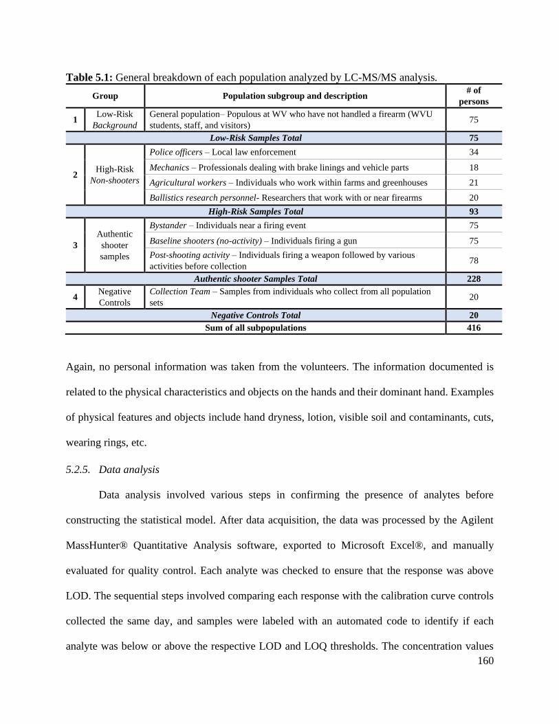
160
Table 5.1: General breakdown of each population analyzed by LC-MS/MS analysis.
Group Population subgroup and description # of
persons
1 Low-Risk
Background
General population– Populous at WV who have not handled a firearm (WVU
students, staff, and visitors) 75
Low-Risk Samples Total 75
2 High-Risk
Non-shooters
Police officers – Local law enforcement 34
Mechanics – Professionals dealing with brake linings and vehicle parts 18
Agricultural workers – Individuals who work within farms and greenhouses 21
Ballistics research personnel- Researchers that work with or near firearms 20
High-Risk Samples Total 93
3
Authentic
shooter
samples
Bystander – Individuals near a firing event 75
Baseline shooters (no-activity) – Individuals firing a gun 75
Post-shooting activity – Individuals firing a weapon followed by various
activities before collection 78
Authentic shooter Samples Total 228
4 Negative
Controls
Collection Team – Samples from individuals who collect from all population
sets 20
Negative Controls Total 20
Sum of all subpopulations 416
Again, no personal information was taken from the volunteers. The information documented is
related to the physical characteristics and objects on the hands and their dominant hand. Examples
of physical features and objects include hand dryness, lotion, visible soil and contaminants, cuts,
wearing rings, etc.
5.2.5. Data analysis
Data analysis involved various steps in confirming the presence of analytes before
constructing the statistical model. After data acquisition, the data was processed by the Agilent
MassHunter® Quantitative Analysis software, exported to Microsoft Excel®, and manually
evaluated for quality control. Each analyte was checked to ensure that the response was above
LOD. The sequential steps involved comparing each response with the calibration curve controls
collected the same day, and samples were labeled with an automated code to identify if each
analyte was below or above the respective LOD and LOQ thresholds. The concentration values

161
were then used for exploratory statistical analysis and input data for the neural network model and
subsequent likelihood ratio calculations.
5.2.6. Machine Learning model
The LC-MS/MS data were transformed into quantitative values via external calibration
curves with internal standard, and analyzed to train, validate, and test the neural network algorithm.
The classification outputs were compared to targets through dynamic programming via a
backpropagation strategy in which the weights improve the prediction [28,53]. The
backpropagation processes the inputted data numerous times using differential calculus until a
user-desired minimum error is achieved or the root mean square (RMS) error across the network
is negligible.
Before inputting data for NN evaluation, data was preprocessed using feature
normalization strategies such as min-max normalization (scaling all values on a 0-1 relative scale)
and z-scoring (centering on the mean and scaling based on standard deviation values. In addition
to producing unitless input values, scaling is essential to avoid over-emphasis on large
concentration values and underrepresentation of low, but still significant, concentration values.
Once trained, the network then can be evaluated using data that is separate from the training set,
including the validation and testing sets. For samples with concentration values below the LOD,
those values were treated as “0”.
For this study, min-max normalization was utilized as it produced the best results and the
data was split randomly using a 60:20:20 ratio — 60% for training, 20% for validation, and 20%
for testing. The JMP Pro® statistical software version 14.0.0 and R Studio © (version 1.4.1717)
were used to construct the neural networks and estimate the likelihood ratios. Also, we followed a
similar criterion and mimicked the previous publication to estimate the positive responses of the

162
analytes, using a categorical approach considering a positive sample when detecting at least two
OGSR compounds and one IGSR analyte or vice versa [17]. This categorical criterion was used to
estimate performance rates, as compared to the NN approach.
5.3. Results and Discussion
5.3.1. Occurrence of OGSR and IGSR on the sampled populations
5.3.1.1. Comparison of low-risk, high-risk, and shooter sets
Exploratory data analysis methods, such as descriptive statistics (Table S5.1) and boxplots,
compared the distribution of analytes in the five populations and were used to elucidate OGSR and
IGSR patterns. The baseline shooters (no-activity) and low-risk (background) populations served
as upper and lower limits for the remaining populations due to their substantial concentration
differences (Figures 5.1-5.3). The low-risk (background) population displayed little to no OGSR
or IGSR concentrations above detection or quantitation limits barring ethyl centralite (EC) which
only showed a signal above LOD (1.0 ppb) but below LOQ for approximately 42% of the samples.
Again, minimal to no OGSR signals were observed in the high-risk set consisting of hand
samples of police officers, mechanics, and farmhands, except for EC (Figure 5.1). However, the
IGSR signals were higher for barium (Ba) and lead (Pb) compared to the low-risk population. For
the high-risk individuals, signals for EC, Ba, and Pb were observed on approximately 40%, 90%,
and 34%, respectively. Of those samples, only 34% contained two analytes (Ba and Pb), while
only 9% possessed a combination of three constituents (EC, Ba, and Pb) on a single sample. These
results indicate that IGSR is more prevalent than OGSR amongst this subgroup and that the
combined detection of IGSR/OGSR decreased the number of false positives.
In contrast, samples collected from baseline shooters shortly after discharging a firearm
presented both IGSR and OGSR at significantly higher concentrations than the low and high-risk
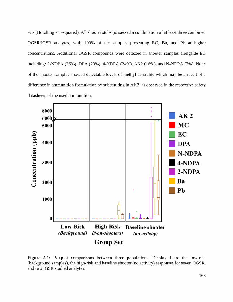
163
sets (Hotelling’s T-squared). All shooter stubs possessed a combination of at least three combined
OGSR/IGSR analytes, with 100% of the samples presenting EC, Ba, and Pb at higher
concentrations. Additional OGSR compounds were detected in shooter samples alongside EC
including: 2-NDPA (36%), DPA (29%), 4-NDPA (24%), AK2 (16%), and N-NDPA (7%). None
of the shooter samples showed detectable levels of methyl centralite which may be a result of a
difference in ammunition formulation by substituting in AK2, as observed in the respective safety
datasheets of the used ammunition.
Figure 5.1: Boxplot comparisons between three populations. Displayed are the low-risk
(background samples), the high-risk and baseline shooter (no activity) responses for seven OGSR,
and two IGSR studied analytes.

164
All the baseline shooter samples contained EC at an average concentration of 38 ppb versus
0.6 ppb and 1.0 ppb for low- and high-risk sets, respectively. Only 43% of these samples possessed
at least two OGSR compounds, with EC being the most frequent, followed by 2-NDPA.
Additionally, 26%, 24%, and 22% of the shooter samples contained at least three, four, or five
OGSR compounds on the same stub, respectively.
5.3.1.2. Post-shooting activity
The second subpopulation consisted of individuals who discharged a firearm and
performed one of three activities immediately after the event to mimic “real-world” scenarios
(Figure 5.2). These post-shooting activities involved 1) vigorously rubbing the hands together for
30 seconds, 2) applying hand sanitizer and rubbing for 30 seconds, or 3) running outdoors for
approximately 1 minute. As expected, the post-shooting activity samples demonstrated higher
signals of both IGSR and OGSR compared to the low-risk backgrounds but lower levels than the
baseline shooter responses. This observation indicates a significant loss of analyte signal but not
a complete depletion of the IGSR/OGSR. Out of the three activities, the hand sanitizer collected a
significant amount of OGSR analytes. One possible explanation for this observation is the organic
pseudo-extraction caused by applying isopropanol directly on the areas, producing a pre-
concentration effect before stubbing the hands. Out of the remaining activities running produced
the lowest responses for OGSR, which may result from sweat formation allowing for the potential
of increased absorption into the epidermal layer and/or more loss from evaporation due to an
increase in body temperature [17].

165
Figure 5.2: Boxplot comparisons between three populations. The low-risk (background samples),
the post-shooting activity, and baseline shooter (no activity) responses for seven OGSR and two
IGSR studied analytes are displayed.
To accurately compare this set with baseline shooting samples, positive control samples
(shooter without activity) were collected on the same days for quality control monitoring. These
positive controls showed a statistically significant difference (Hotelling’s T-squared) for all OGSR
compounds, barring 4-NDPA, once an activity was performed. However, there was no statistical
difference between the IGSR signals of the positive controls and the activity samples. This result
may be due to evaporation rates of the OGSR compounds as compared to more persistent IGSR
particulates. Among the seven studied OGSR compounds, the concentration levels decreased

166
anywhere from 76% - 99%, with AK 2 experiencing the most significant loss. This observation
further illustrates that OGSR components are more sensitive to factors like weather conditions,
skin absorption, and/or temperature [6,55]. Although the activities yielded lower concentrations
of the analytes of interest, IGSR elements were detected on all samples and 88% presented more
than two OGSR compounds.
5.3.1.3. Bystander
For the last subpopulation set, we wanted to evaluate the levels of GSR that may be
deposited on individuals within the vicinity of a discharge event. Because of the ballistics range’s
dimensions and safety concerns, bystanders were oriented at three different locations (left, right,
and behind) approximately three feet away from the shooter. Very few GSR compounds were
detected in the bystander population set (Figure 5.3), except for Ba (94% of the samples) and EC
(12% of the samples). Comparing these findings to the Low-Risk and negative control samples, it
appears that both analytes are originating from the discharge event. Another important observation
is the presence of barium being more prevalent than lead in this subpopulation. This may be
attributed to the crown ethers being more sensitive to barium metal [17] and the amount of lead
being below the LOD. However, these observations may have been influenced by a myriad of
factors, including the shooting range conditions, the time gap between firing and sampling, and
the positioning of the bystanders in respect to the discharge event.
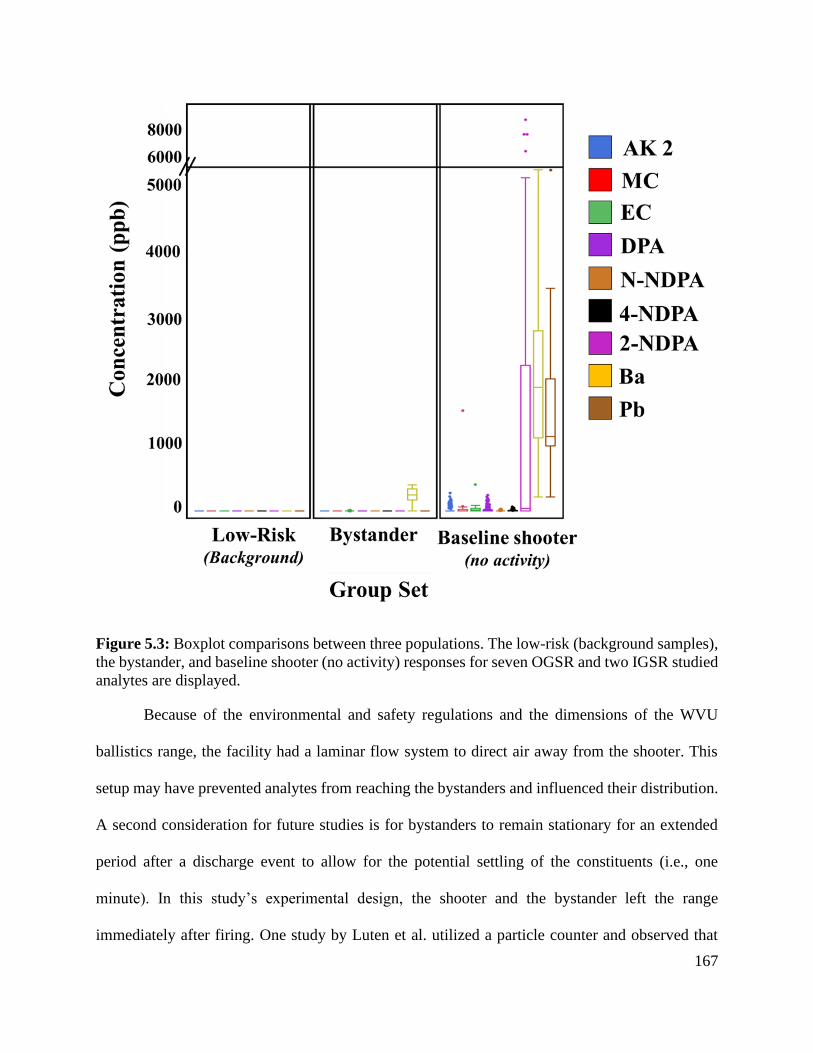
167
Figure 5.3: Boxplot comparisons between three populations. The low-risk (background samples),
the bystander, and baseline shooter (no activity) responses for seven OGSR and two IGSR studied
analytes are displayed.
Because of the environmental and safety regulations and the dimensions of the WVU
ballistics range, the facility had a laminar flow system to direct air away from the shooter. This
setup may have prevented analytes from reaching the bystanders and influenced their distribution.
A second consideration for future studies is for bystanders to remain stationary for an extended
period after a discharge event to allow for the potential settling of the constituents (i.e., one
minute). In this study’s experimental design, the shooter and the bystander left the range
immediately after firing. One study by Luten et al. utilized a particle counter and observed that

168
GSR can remain airborne several hours due to their size and dynamics [56]. Therefore, it is
hypothesized that insufficient time was allowed for the GSR components to settle on the
bystanders [56–58]. Hence, future experiments should be geared towards sedimentation rates of
varying particle sizes and compounds. Nonetheless, the results illustrate the importance of
considering environmental conditions, including airflows in indoor settings as well as time
between discharge and collection for the interpretation of the data.
5.3.1.4. Summary of exploratory analysis and frequency of occurrence of IGSR and
OGSR
The data was further categorized into two groups labeled as Known Shooter and Known
Non-Shooter. The authentic shooters, activity, and bystander were grouped into the Known
Shooter since we want to consider an association with a firearm-related incident, whereas the high-
and low-risk were placed in the Known Non-Shooter set. As shown in Figure 5.4, the known
shooters produce higher IGSR and OGSR signal responses. Ultimately, the IGSR analytes for the
high-risk and the bystander populations displayed higher signatures versus the low-risk population.
The agricultural workers, police officers, and mechanics displayed residues with similar chemical
compositions to the shooter set but at lower reported concentrations. The low-risk population set
revealed less IGSR particulates than the high-risk and bystander sets and was comparable to the
negative controls of the collection team.

169
Figure 5.4: Figure 5.4a) OGSR population comparison among all analytes tested. Seven OGSR
analytes were monitored, including akardite II (blue), methyl centralite (red), ethyl centralite
(green), diphenylamine (dark purple), N-nitrosodiphenylamine (orange), 4-nitrodiphenylamine
(turquoise), and 2-nitrodiphenylamine (magenta). Overall, the authentic shooting populations
(shooter and activity) showed higher concentrations of the analytes versus the remaining
populations (high- and low-risk and bystander). However, there were low concentration levels for
both diphenylamine and its byproducts in the low- and high-risk populations. Figure 5.4b) IGSR
population comparison among the populations tested. Ultimately two IGSR analytes were
monitored, including barium (blue) and lead (red). Similarly, the authentic shooting populations
(shooter and activity) showed higher concentrations of the analytes versus the remaining
populations (high- and low-risk and bystander). However, there were comparable concentration
levels for both IGSR and in the high-risk population.
Looking closer at Figure 5.4, there is some overlap in the IGSR signals for the Non-shooter
set versus the Known Shooter populations which can lead to higher false positive rates if
considered as the only signature for GSR identification. For the OGSR analytes, Known Shooter
samples not only show more analytes present, but the response levels indicate minimal overlap
with samples from the Non-Shooter population. However, the OGSR signals demonstrate a larger
170
dispersion, especially for AK 2 and DPA. This could be the result of multiple factors including
varying propellant composition across manufacturers used in this study, the volatile nature of the
constituents, or simply the entropic nature of a deflagration event.
These findings indicate that it is not uncommon to find multiple OGSR analytes on the
same sample. However, the combination of OGSR and IGSR provides a better indication of the
presence of GSR. Most importantly, by using an IGSR/OGSR detection criterion, the identification
of GSR (as potential false positives) decreased. Thus, further stressing the relevancy of using more
comprehensive profiles for decision making.
The benefit of any exploratory method, like the one used in this study, is that data
visualization permits a deeper understanding of the factors influencing the data distribution. On
the other hand, the amount of time required and the difficulty to correctly evaluate and characterize
each dataset can be troublesome based on the complexity of a sample. Additionally, it does not
address a significant concern of GSR evidence, which centers around the probability of finding
evidence given there was a or was not firing event - the two mutually exclusive hypotheses. Hence,
techniques like machine learning algorithms can provide more comprehensive classifications when
closely monitored and rigorously tested.
5.3.2. Neural network machine learning for GSR identification
Neural networks (NN), also known as artificial neural networks (ANNs), are a subset of
machine learning and are the heart of deep learning algorithms. Their name and structure are
inspired by the human brain which mimic the signaling and communication of biological neurons
[53]. Neural networks are comprised of node layers, including an input layer, one or more hidden
layer(s), and output layers. For this study, the concentration levels for detected analytes were
reported in parts-per-billion (ppb) for each population set. Neural networks can be created for

171
different purposes and serve as both classification and prediction systems. Here, the
predictive/supervised learning approach was implemented to 1) monitor the effectiveness and
success rates of the method and 2) generate probabilistic outputs to calculate the likelihood ratios.
The subpopulations were grouped further because the goal is to ultimately differentiate shooters
from non-shooters using a single methodology. The two populations used for non-shooters were
the low-risk and high-risk sample sets, whereas the authentic shooter comprised of the shooters
(no-activity), bystanders, and post-shooting activity sample sets.
After exploratory analysis, the calculated concentrations of the five subpopulations were
first preprocessed using min-max normalization. Each feature value i.e., analyte response, in each
subpopulation was evaluated for its minimum and maximum values highlighted by Equation 5.2
𝑥𝑖𝑗 ⇐𝑥𝑖𝑗−𝑚𝑖𝑛𝑖
𝑚𝑎𝑥𝑗−𝑚𝑖𝑛𝑗 Equation 5.2
One can regard this as shifting all the observations so that the minimum value is zero, and
then dividing by either the maximum of the shifted data, or equivalently the range of the original
data. It also scaled all observations so that the minimum possible value is zero and the maximum
is one. Min-max normalization is typical in neural network modeling and ensures that compound
with comparatively large concentrations such as Ba and EC in this case do not control the network
training. Rather, scaling ensures that the network training focuses on relative concentration
differences rather that absolute values. The 0-1 scale is compatible with network node outputs that
are also scaled as 0-1.
After choosing the min-max normalization approach, those scaled values were then
introduced to the model using a 60:20:20 training:validation:testing structure. Furthermore, to
estimate the variability in the accuracy of the test, a 10-fold cross-validation was repeated 10 times

172
on the combined data set. The performance of the scaled data was estimated for the NN’s
misclassification rates (Table S5.2) and by calculating the respective coefficient of variation (CV)
which considers both the standard deviation (σ) and the means (µ) to measure of dispersion of a
probability distribution or frequency distribution, outlined by Equation 5.3
𝐶𝑉 =𝜎
𝜇 Equation 5.3
In this application, the CV values are a ratio of the variability in accuracy to the average
accuracy. A high average accuracy coupled with a low CV is desirable as this indicates the method
has performed well on many different training sets. Note that, as with all cross-validation methods,
the picture will likely be more rosey than reality, but understanding and accounting for variability
in prediction accuracy is preferrable to simply choosing the method with the highest accuracy
without assessing its variability.
The method's accuracy demonstrated very low false-positive and false-negative rates
(Table S5.2) for identifying the baseline shooters against the other subpopulations. Hence, an
adaptive model that learns through vigorous trials such as neural networks is appropriate for
analyzing newly acquired data. Moreover, the comparable accuracy within each subpopulations’
training, testing and validation sets is a good indicator the model is not overfitting the data. The
overall structure of the NN used in this study is outlined in Table S5.3.
5.3.3. Likelihood ratios (LR) as probabilistic assessments of the weight of evidence
Each subpopulation’s probability outputs were calculated using a neural network model
which allowed for the estimation of likelihood ratios. Although relatively small, the overall dataset
in this study generated experimental observations that may closely resemble casework specimens
and data. With that goal in mind, the low- and high-risk background samples express simple to
complex situations, respectively. For the authentic shooting set, we wanted to mimic situations
173
where analysts may encounter in firearm-related cases. The complexity of the shooter scenarios
ranged from samples recovered immediately after a discharge event (simple) to situations where a
suspect performed post-shooting activities, and even those near a shooting event (complex). For
the latter two, the expected challenges were high analyte loss (represented by the post-shooting
activities set) and increased difficulty when differentiating from shooters (represented by the
bystander set). In this study, the H1 and H2 hypotheses at the source level are defined as:
H1: The trace originated from a discharged firearm (and hence, are GSR)
H2: The trace originated from a non-firearm source (i.e., other environmental source, and hence
are not GSR)
For purposes of the hypotheses, the low-risk background samples were considered as the
Ground Truth for non-GSR cases, while the Baseline shooter (no activity) set was considered the
Ground Truth for GSR-containing samples. To evaluate the performance of the LR, we examined
how often the LR was > 1 (or log LR > 0) when samples were anticipated to have GSR present.
Contrarily, we also monitored how often the LR was < 1 (or log LR < 0) when true non-GSR
datasets showed an absence of GSR. Those observations are displayed as histograms that depict
the frequency when H1 or H2 is true (y-axis) versus the log LR (x-axis). These histograms can
further assess the performance of the LR.
The discriminating power of this methodology can distinguish cases where H1 and H2 are
respectively true. For instance, the greater the separation between a subpopulation’s histogram, the
better the discriminating power, which increases the confidence of a given sample. Similarly, if
one subpopulation’s resulting histogram overlaps another histogram, the greater the misleading
evidence rates, which also lowers the confidence. Therefore, the larger the LR, the evidence is
more likely (i.e., stronger support) if the trace originated from GSR than other sources.

174
An alternative strategy for assessing the performance of the likelihood ratios is using Tippet
plots. A Tippet plot is a plot of two empirical “complementary” cumulative distribution functions
(ECCDFs). That is, a ECCDF is an estimate of a complementary cumulative distribution function
and given a sample of data (of size n) is computed by
�̂�(𝑥) =1
𝑛∑ 𝐼𝑛
𝑖=1 [𝑥𝑖 ≥ 𝑥] Equation 5.4
where 𝐼[𝑥𝑖 ≥ 𝑥] is an indicator function that takes the value one if 𝑥𝑖 ≥ 𝑥, and zero otherwise.
A Tippet plot is a plot of these functions versus x where x is the LR computed under H1
and H2 – that is there are two ECCDFs in a Tippet plot [59]. Tippet plots can graphically display
discriminating power by consideration of the vertical separation of true-H1 and true-H2 curves at
a given value of log LR axis. The more separated at a given LR, the higher discrimination at that
value. Additionally, the rates of misleading evidence (RoMEs) can be visually estimated by
considering the area above (or under) the curves to left (or right) of the line log10
(𝐿𝑅) = 0,
depending on whether H1 and H2 is of interest [59]. For instance, when H1 and H2 are true (Log10
LR > 0 and Log10 LR < 0, respectively), the H2 curve can help visualize false positives (i.e., Log10
LR > 0 when no GSR should be present). Oppositely, the misleading rates for the H1 curve
represent false negatives and are estimated when Log10 LR < 0 (i.e., when GSR should be present).
5.3.3.1. Baseline shooters (no activity) versus Low-Risk Background likelihood ratios
The first assessment of the LR was conducted using the two subpopulations of low-risk
(known non-shooters) and the baseline shooter (no activity). For this case, we expected minimal
to no overlap when OGSR and IGSR information was combined. Figure 5.5 shows a clear
separation between baseline shooter (no activity) (H1) and low-risk (H2) histograms.

175
Figure 5.5: Figure 5.5a) Histograms of Baseline shooter (no activity) (H1) and low-risk (H2)
subpopulation. The Baseline shooter displays a broader spread due to varying concentration levels
of OGSR and IGSR signals. The low-risk subpopulation has a condensed range and is centralized
at -10. Figure 5.5b) Tippet plot illustrating no instances of either false positives or false negatives.
Additionally, the Tippet plots reflect the highest discriminating ability to differentiate
between the subpopulations with minimal RoME. This result is similar to the observation during
the initial exploratory analysis. Because of this optimum distinction, these subpopulations were
regarded as the lower and upper bounds for further comparisons.
5.3.3.2. High-Risk likelihood ratios
After the Baseline shooter and low-risk populations bounds were established, the
remaining samples, starting with the high-risk set (H2), baseline shooter (H1), and the low-risk sets
(H2), were tested against them to monitor the rates of misleading evidence in more complex
situations. The Figure 5.6a histogram illustrates there was a separation between all three
populations, with larger dispersion of logLRs of the high-risk set towards less negative values as
compared to the low-risks, which is due to the relative presence of EC in some of the samples as
well as IGSR elements in this subpopulation. Nonetheless, the profiles observed in the high-risk

176
set were easily distinguished from the shooter set by the NN. As a result, the Tippet plot in Figure
5.6b shows low rates of misleading evidence even for samples with high contamination risk.
Figure 5.6: Figure 5.6a) Histogram comparison of Baseline shooter (no activity), low-risk, and
high-risk subpopulations. The high-risk population displays a broader spread for its histogram
range due to varying concentration levels of the analytes. Figure 5.6b) Tippet plot illustrating the
RoME compared to the Baseline shooter (no activity).
5.3.3.3. Post-shooting activity likelihood ratios
This approach continued to investigate the remaining subpopulations to evaluate and
attempt to discriminate between low-risk background (H2) versus post-shooting activity set (H1)
samples where some sample loss is expected. Figure 5.7a shows some activity samples presenting
log LR lower than zero. Although no false positives are observed (Figure 5.7b), some false
negatives are present due to the loss of GSR traces (RoME 1.28%). Interestingly, there is notable
overlap with the baseline shooter sample set. This overlap is a result of this subpopulation
possessing OGSR and IGSR signals in a single sample. As expected, the log LR of the post-
shooting activity set were spread out towards lower values than the shooter sets, However, the data

177
still indicates that LR can be used to assess the weight of evidence, with strong support for GSR
presence observed in samples that have gone through everyday activities such as rubbing, cleaning
with hand sanitizer, or running outdoors.
Figure 5.7: Figure 5.7a) Histogram comparison of Baseline shooter (no activity), low-risk, and
post-shooting activity subpopulations. The post-shooting activity population displays a broader
spread for its histogram range due to varying concentration levels of the analytes with heavy
overlap with the Baseline shooter (no activity) samples. Figure 5.7b) Tippet plot illustrating the
negatives compared to the low-risk background.
5.3.3.4. Bystander likelihood ratios
Finally, the bystanders (considered as the subpopulation set for H1) were compared and
were separated from the baseline shooter sample set (H1) represented in Figure 5.8. Additionally,
there is no overlap between the bystander set and the low-risk sets which is displayed by the Tippet
plots having low RoMEs. These observations can be explained based on the general substantial
decrease of the concentrations of both OGSR and IGSR in the bystander’s stubs. Significant
relative differences of inorganic elements and organic compounds were observed between
individuals who fired the gun and those standing a distance away from a discharge event, except
for persistence of Ba and EC.

178
Caution should be taken in interpreting results from this subpopulation as we learned from
exploratory data analysis that there might be uncontrolled factors in our experiments biasing the
results. Future studies will address this issue by controlling experimental factors that would mimic
different environmental conditions and deposition settling times for individuals in the vicinity of
the firing event. As a result, the findings on this subpopulation cannot be generalized at this point.
Nonetheless, it highlights important aspects that may play a role on GSR transfer to bystanders
when assessing casework data, such as airflow conditions, settling times, and time between
discharge and collection.
Figure 5.8: Figure 5.8a) Histogram comparison of Baseline shooter (no activity), low-risk, and
Bystander subpopulations. The Bystander population displays a smaller spread for its histogram
range due to varying concentration levels of the analytes. Figure 5.8b) Tippet plot illustrating the
negatives compared to the low-risk background.
5.4. Conclusions
The LC-MS/MS is a highly versatile technique found in many crime laboratories, which is
primarily used to analyze controlled substances (such as illegal narcotics), explosives, or
toxicology samples. Our research has demonstrated that the applications of this instrument can be
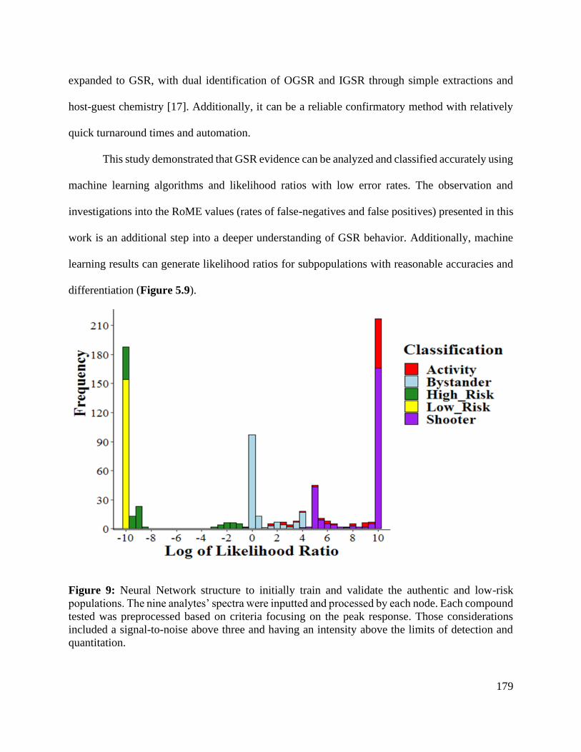
179
expanded to GSR, with dual identification of OGSR and IGSR through simple extractions and
host-guest chemistry [17]. Additionally, it can be a reliable confirmatory method with relatively
quick turnaround times and automation.
This study demonstrated that GSR evidence can be analyzed and classified accurately using
machine learning algorithms and likelihood ratios with low error rates. The observation and
investigations into the RoME values (rates of false-negatives and false positives) presented in this
work is an additional step into a deeper understanding of GSR behavior. Additionally, machine
learning results can generate likelihood ratios for subpopulations with reasonable accuracies and
differentiation (Figure 5.9).
Figure 9: Neural Network structure to initially train and validate the authentic and low-risk
populations. The nine analytes’ spectra were inputted and processed by each node. Each compound
tested was preprocessed based on criteria focusing on the peak response. Those considerations
included a signal-to-noise above three and having an intensity above the limits of detection and
quantitation.

180
These preliminary results highlight the potential to distinguish complex groups such as
bystanders and high-risk individuals (to a degree) versus low-risk and baseline shooter
populations. Also, it shows that identification of GSR is possible even after a shooter engages in
some typical post-shooting activities. In terms of the bystander and post-shooting activity
populations, the overlap between those populations was not surprising. This is due to the transfer
of GSR components near a firing event. The region of overlap is the greatest risk of false positives
(in the case of bystanders) or false negatives (shooter with post-shooting activity). More
importantly, there is a clear distinction between high-risk backgrounds (non-shooters) and
authentic shooters. This was possible because combining IGSR and OGSR information yields
higher confidence in classifications and provides enhanced evidentiary value to GSR evidence.
Thus, researchers and practitioners would benefit from 1) implementing techniques that can
perform IGSR/OGSR analysis and 2) evaluating the weight of the evidence as part of the data
interpretation.
This study demonstrates the potential of using IGSR/OGSR combined profiles and
machine learning algorithms to estimate likelihood ratios as a more comprehensive framework to
assess the weight of firearms discharge the evidence. More extensive studies and collaboration
efforts should be one objective for the forensic research community and additional bystander
studies to monitor the underlying mechanism associated with transfer and persistence and further
apply the LR at the activity level. Notably, a machine learning model’s probabilistic data
assessment may open new avenues for more comprehensive interpretative frameworks for GSR
evidence.

181
Author Contributions
The manuscript was written through the contributions of all authors. All authors have given
approval to the final version of the manuscript.
Conflicts of interest
The authors declare that they have no known competing financial interests or personal
relationships that could have appeared to influence the work reported in this paper.
Acknowledgments
We want to acknowledge Dr. Keith Morris and James Hamilton for collecting authentic
samples and accessing the WVU ballistics laboratory. We also want to acknowledge Kourtney
Dalzell, Colby Ott, and Courtney Vander Pyl to collect authentic samples from all populations.
Additionally, the authors thank Meghan Prusinowski for her assistance in programming.
This project is sponsored by Award No. 2018-DU-BX-0186 and 2019-R2-CX-044 from the
National Institute of Justice to West Virginia University. The opinions, findings, and conclusions
are those of the authors and do not necessarily reflect those of the Department of Justice.
182
5.5. References for Chapter 5
[1] M.D. Gallidabino, L.P. Barron, C. Weyermann, F.S. Romolo, Quantitative profile-profile
relationship (QPPR) modelling: A novel machine learning approach to predict and associate
chemical characteristics of unspent ammunition from gunshot residue (GSR), Analyst. 144
(2019) 1128–1139. https://doi.org/10.1039/c8an01841c.
[2] M. Maitre, K.P. Kirkbride, M. Horder, C. Roux, A. Beavis, Current perspectives in the
interpretation of gunshot residues in forensic science: A review, Forensic Sci. Int. 270 (2017) 1–
11. https://doi.org/10.1016/j.forsciint.2016.09.003.
[3] C. Hofstetter, M. Maitre, A. Beavis, C.P. Roux, C. Weyermann, A.L. Gassner, A study of
transfer and prevalence of organic gunshot residues, Forensic Sci. Int. 277 (2017) 241–251.
https://doi.org/10.1016/j.forsciint.2017.06.013.
[4] L.S. Blakey, G.P. Sharples, K. Chana, J.W. Birkett, Fate and Behavior of Gunshot
Residue—A Review, J. Forensic Sci. 63 (2018) 9–19. https://doi.org/10.1111/1556-4029.13555.
[5] W. Feeney, C. Vander Pyl, S. Bell, T. Trejos, Trends in composition, collection,
persistence, and analysis of IGSR and OGSR: A review, Forensic Chem. 19 (2020) 100250.
https://doi.org/10.1016/j.forc.2020.100250.
[6] J. Moran, S. Bell, Analysis of organic gunshot residue permeation through a model skin
membrane using ion mobility spectrometry, Int. J. Ion Mobil. Spectrom. 16 (2013) 247–258.
https://doi.org/10.1007/s12127-013-0138-0.
[7] J. French, R. Morgan, J. Davy, The secondary transfer of gunshot residue: an
experimental investigation carried out with SEM-EDX analysis, X-Ray Spectrom. 43 (2014) 56–
61. https://doi.org/10.1002/xrs.2498.

183
[8] J.C. French, R.M. Morgan, P. Baxendell, P.A. Bull, Multiple transfers of particulates and
their dissemination within contact networks, Sci. Justice. 52 (2012) 33–41.
https://doi.org/10.1016/j.scijus.2011.05.001.
[9] S. Practice, S. Handbook, S. Practice, Standard Practice for Gunshot Residue Analysis by
Scanning Electron Microscopy/Energy Dispersive X-Ray Spectrometry, ASTM Int. i (2020) 1–5.
https://doi.org/10.1520/E1588-20.
[10] K. Menking-Hoggatt, C. Martinez, C. Vander, E. Heller, E. Chip, L. Arroyo, T. Trejos,
C. Vander Pyl, E. Heller, E. “Chip” Pollock, L. Arroyo, T. Trejos, Development of tailor-made
inorganic gunshot residue (IGSR) microparticle standards and characterization with a multi-
technique approach, Talanta. 225 (2021) 121984. https://doi.org/10.1016/j.talanta.2020.121984.
[11] Z. Abrego, A. Ugarte, N. Unceta, A. Fernández-Isla, M.A. Goicolea, R.J. Barrio,
Unambiguous characterization of gunshot residue particles using scanning laser ablation and
inductively coupled plasma-mass spectrometry, Anal. Chem. 84 (2012) 2402–2409.
https://doi.org/10.1021/ac203155r.
[12] K. Menking-Hoggatt, L. Arroyo, J. Curran, T. Trejos, Novel LIBS method for micro-
spatial chemical analysis of inorganic gunshot residues, J. Chemom. 35 (2021) 1–13.
https://doi.org/10.1002/cem.3208.
[13] E. Goudsmits, G.P. Sharples, J.W. Birkett, Recent trends in organic gunshot residue
analysis, Trac-Trends Anal. Chem. 74 (2015) 46–57. https://doi.org/10.1016/j.trac.2015.05.010.
[14] B. Stevens, S. Bell, K. Adams, Initial evaluation of inlet thermal desorption GC-MS
analysis for organic gunshot residue collected from the hands of known shooters, Forensic
Chem. 2 (2016) 55–62. https://doi.org/10.1016/j.forc.2016.10.001.

184
[15] R.V. Taudte, C. Roux, D. Bishop, L. Blanes, P. Doble, A. Beavis, Development of a
UHPLC method for the detection of organic gunshot residues using artificial neural networks,
Anal. Methods. 7 (2015) 7447–7454. https://doi.org/10.1039/c5ay00306g.
[16] C. Bonnar, R. Popelka-Filcoff, K.P. Kirkbride, Armed with the Facts: A Method for the
Analysis of Smokeless Powders by Ambient Mass Spectrometry, J. Am. Soc. Mass Spectrom. 31
(2020) 1943–1956. https://doi.org/10.1021/jasms.0c00193.
[17] W. Feeney, K. Menking-Hoggatt, C. Vander Pyl, C.E. Ott, S. Bell, L. Arroyo, T. Trejos,
Detection of Organic and Inorganic Gunshot Residues from Hands using Complexing Agents
and LC-MS/MS, Anal. Methods. (2021). https://doi.org/10.1039/d1ay00778e.
[18] S.R. Khandasammy, A. Rzhevskii, I.K. Lednev, A Novel Two-Step Method for the
Detection of Organic Gunshot Residue for Forensic Purposes: Fast Fluorescence Imaging
Followed by Raman Microspectroscopic Identification, Anal. Chem. 91 (2019) 11731–11737.
https://doi.org/10.1021/acs.analchem.9b02306.
[19] C. Bonnar, E.C. Moule, N. Lucas, K.E. Seyfang, R.P. Dunsmore, R.S. Popelka-Filcoff,
K. Redman, K. Paul Kirkbride, Tandem detection of organic and inorganic gunshot residues
using LC–MS and SEM-EDS, Forensic Sci. Int. 314 (2020) 110389.
https://doi.org/10.1016/j.forsciint.2020.110389.
[20] V. Redouté Minzière, D. Werner, D. Schneider, M. Manganelli, B. Jung, C. Weyermann,
A.L. Gassner, Combined Collection and Analysis of Inorganic and Organic Gunshot Residues, J.
Forensic Sci. (2020). https://doi.org/10.1111/1556-4029.14314.
[21] Z. Abrego, N. Grijalba, N. Unceta, M. Maguregui, A. Sanchez, A. Fernandez-Isla, M.A.
Goicolea, R.J. Barrio, A. Fernández-Isla, M.A. Goicolea, R.J. Barrió, A. Fernandez-Isla, M.A.
Goicolea, R.J. Barrio, A novel method for the identification of inorganic and organic gunshot

185
residue particles of lead-free ammunitions from the hands of shooters using scanning laser
ablation-ICPMS and Raman micro-spectroscopy, Analyst. 139 (2014) 6232–6241.
https://doi.org/10.1039/c4an01051e.
[22] J. Bueno, I.K. Lednev, Attenuated Total Reflectance-FT-IR Imaging for Rapid and
Automated Detection of Gunshot Residue, Anal. Chem. 86 (2014) 3389–3396.
https://doi.org/10.1021/ac4036718.
[23] C.E. Ott, K.A. Dalzell, P.J. Calderón-Arce, A.L. Alvarado-Gámez, T. Trejos, L.E.
Arroyo, Evaluation of the Simultaneous Analysis of Organic and Inorganic Gunshot Residues
Within a Large Population Data Set Using Electrochemical Sensors*,†, J. Forensic Sci. 65
(2020) 1935–1944. https://doi.org/10.1111/1556-4029.14548.
[24] L. Ali, K. Brown, H. Castellano, S.J. Wetzel, A Study of the Presence of Gunshot
Residue in Pittsburgh Police Stations using SEM/EDS and LC-MS/MS, J. Forensic Sci. 61
(2016) 928–938. https://doi.org/10.1111/1556-4029.13077.
[25] A. Tarifa, J.R. Almirall, Fast detection and characterization of organic and inorganic
gunshot residues on the hands of suspects by CMV-GC-MS and LIBS, Sci. Justice. 55 (2015)
168–175. https://doi.org/10.1016/j.scijus.2015.02.003.
[26] T. Trejos, C. Vander Pyl, K. Menking-Hoggatt, A.L. Alvarado, L.E. Arroyo, C. V Pyl, K.
Menking-Hoggatt, A.L. Alvarado, L.E. Arroyo, Fast identification of inorganic and organic
gunshot residues by LIBS and electrochemical methods, Forensic Chem. 8 (2018) 146–156.
https://doi.org/10.1016/j.forc.2018.02.006.
[27] Z. Brozek-Mucha, On the prevalence of gunshot residue in selected populations - An
empirical study performed with SEM-EDX analysis, Forensic Sci. Int. 237 (2014) 46–52.
https://doi.org/10.1016/j.forsciint.2014.01.020.

186
[28] S. Bell, L. Seitzinger, From binary presumptive assays to probabilistic assessments:
Differentiation of shooters from non-shooters using IMS, OGSR, neural networks, and likelihood
ratios, Forensic Sci. Int. 263 (2016) 176–185. https://doi.org/10.1016/j.forsciint.2016.04.020.
[29] N. Lucas, H. Brown, M. Cook, K. Redman, T. Condon, H. Wrobel, K.P. Kirkbride, H.
Kobus, A study into the distribution of gunshot residue particles in the random population,
Forensic Sci. Int. 262 (2016) 150–155. https://doi.org/10.1016/j.forsciint.2016.02.050.
[30] Z. Brozek-Mucha, Chemical and morphological study of gunshot residue persisting on
the shooter by means of scanning electron microscopy and energy dispersive X-ray
spectrometry, Microsc. Microanal. 17 (2011) 972–982.
https://doi.org/10.1017/S1431927611012141.
[31] R.A. Costa, L.C. Motta, C.A. Destefani, R.R.T. Rodrigues, K.S.D. Santo, G. Aquije, R.
Boldrini, G.P.B. Athayde, M. Carneiro, W. Romao, Gunshot residues (GSR) analysis of clean
range ammunition using SEM/EDX, colorimetric test and ICP-MS: A comparative approach
between the analytical techniques, Microchem. J. 129 (2016) 339–347.
https://doi.org/10.1016/j.microc.2016.07.017.
[32] A. Zeichner, B. Eldar, B. Glattstein, A. Koffman, T. Tamiri, D. Muller, Vacuum
collection of gunpowder residues from clothing worn by shooting suspects, and their analysis by
GC/TEA, IMS, and GC/MS, J. Forensic Sci. 48 (2003) 961–972.
https://doi.org/10.1520/jfs2002390.
[33] M. Cook, Gunshot residue contamination of the hands of police officers following start-
of-shift handling of their firearm, Forensic Sci. Int. 269 (2016) 56–62.
https://doi.org/10.1016/j.forsciint.2016.11.002.
187
[34] R. V. Gerard, E. Lindsay, M.J. McVicar, E.D. Randall, A. Gapinska, Observations of
gunshot residue associated with police officers, their equipment, and their vehicles, J. Can. Soc.
Forensic Sci. 45 (2012) 57–63. https://doi.org/10.1080/00085030.2012.10757183.
[35] S. Charles, N. Geusens, E. Vergalito, B. Nys, Interpol review of gunshot residue 2016–
2019, Forensic Sci. Int. Synerg. 2 (2020) 416–428. https://doi.org/10.1016/j.fsisyn.2020.01.011.
[36] A. Stamouli, L. Niewöhner, M. Larsson, B. Colson, S. Uhlig, L. Fojtasek, F. Machado, L.
Gunaratnam, Survey of gunshot residue prevalence on the hands of individuals from various
population groups in and outside Europe, Forensic Chem. 23 (2021) 1–8.
https://doi.org/10.1016/j.forc.2021.100308.
[37] W. Tucker, N. Lucas, K.E. Seyfang, K.P. Kirkbride, R.S. Popelka-Filcoff, Gunshot
residue and brakepads: Compositional and morphological considerations for forensic casework,
Forensic Sci. Int. 270 (2017) 76–82. https://doi.org/10.1016/j.forsciint.2016.11.024.
[38] Ç. Aksoy, T. Bora, N. Şenocak, F. Aydın, F. Aydin, A new method to reduce false
positives due to antimony in detection of gunshot residues, Forensic Sci. Int. 250 (2015) 87–90.
https://doi.org/10.1016/j.forsciint.2015.03.006.
[39] G.M. Ingo, M. D’Uffizi, G. Falso, G. Bultrini, G. Padeletti, Thermal and microchemical
investigation of automotive brake pad wear residues, Thermochim. Acta. 418 (2004) 61–68.
https://doi.org/10.1016/j.tca.2003.11.042.
[40] Z.P. Wu, Y. Tong, J.Y. Yu, X.R. Zhang, C.X. Pan, X.Y. Deng, Y.C. Xu, Y.X. Wen,
Detection of N,N’-diphenyl-N,N’-dimethylurea (methyl centralite) in gunshot residues using
MS-MS method, Analyst. 124 (1999) 1563–1567. https://doi.org/10.1039/a904917g.
[41] Y. Tong, Z. Wu, C.D. Yang, J.Y. Yu, X.R. Zhang, S.J. Yang, X.Y. Deng, Y.C. Xu, Y.X.
Wen, Z.P. Wei, C.D. Yang, J.Y. Yu, X.R. Zhang, S.J. Yang, X.Y. Deng, Y.C. Xu, Y.X. Wen,

188
Determination of diphenylamine stabilizer and its nitrated derivatives in smokeless gunpowder
using a tandem MS method, Analyst. 126 (2001) 480–484. https://doi.org/10.1039/b010183o.
[42] Z. Brożek-Mucha, A study of gunshot residue distribution for close-range shots with a
silenced gun using optical and scanning electron microscopy, X-ray microanalysis and infrared
spectroscopy, Sci. Justice. 57 (2017) 87–94. https://doi.org/10.1016/j.scijus.2016.11.004.
[43] M. Manganelli, C. Weyermann, A.L. Gassner, Surveys of organic gunshot residue
prevalence: Comparison between civilian and police populations, Forensic Sci. Int. 298 (2019)
48–57. https://doi.org/10.1016/j.forsciint.2019.02.050.
[44] M. Maitre, S. Chadwick, K.P. Kirkbride, A.L. Gassner, C. Weyermann, A. Beavis, C.
Roux, An investigation on the secondary transfer of organic gunshot residues, Sci. Justice. 59
(2019) 248–255. https://doi.org/10.1016/j.scijus.2019.01.007.
[45] A. Biedermann, S. Bozza, F. Taroni, Probabilistic evidential assessment of gunshot
residue particle evidence (Part I): Likelihood ratio calculation and case pre-assessment using
Bayesian networks, Forensic Sci. Int. 191 (2009) 24–35.
https://doi.org/10.1016/j.forsciint.2009.06.004.
[46] F.S. Romolo, P. Margot, Identification of gunshot residue: a critical review, Forensic Sci.
Int. 119 (2001) 195–211.
[47] B. Cardinetti, C. Ciampini, S. Abate, C. Marchetti, F. Ferrari, D. Di Tullio, C. D’Onofrio,
G. Orlando, L. Gravina, L. Torresi, G. Saporita, A proposal for statistical evaluation of the
detection of gunshot residues on a suspect, Scanning. 28 (2006) 142–147.
https://doi.org/10.1002/sca.4950280302.

189
[48] N.K. Damary, M. MandelY, N. Levin, E. Izraeli, Calculation of likelihood ratios for
gunshot residue evidence-statistical aspects, Law, Probab. Risk. 15 (2016) 107–125.
https://doi.org/10.1093/lpr/mgw001.
[49] T.J. Hannigan, S.D. McDermott, C.M. Greaney, J. O’Shaughnessy, C.M. O’Brien,
Evaluation of gunshot residue (GSR) evidence: Surveys of prevalence of GSR on clothing and
frequency of residue types, Forensic Sci. Int. 257 (2015) 177–181.
https://doi.org/10.1016/j.forsciint.2015.08.003.
[50] A. Bolck, A. Stamouli, Likelihood Ratios for categorical evidence; Comparison of LR
models applied to gunshot residue data, Law, Probab. Risk. 16 (2017) 71–90.
https://doi.org/10.1093/lpr/mgx005.
[51] A. Biedermann, S. Bozza, F. Taroni, Probabilistic evidential assessment of gunshot
residue particle evidence (Part II): Bayesian parameter estimation for experimental count data,
Forensic Sci. Int. 206 (2011) 103–110. https://doi.org/10.1016/j.forsciint.2010.07.009.
[52] L.S. Blakey, G.P. Sharples, K. Chana, J.W. Birkett, Fate and Behavior of Gunshot
Residue-A Review, J. Forensic Sci. 63 (2018) 9–19. https://doi.org/10.1111/1556-4029.13555.
[53] C.C. Aggarwal, Neural Networks and Deep Learning, Springer, 2018.
https://doi.org/10.1201/b22400-15.
[54] R. Cook, I.W. Evett, G. Jackson, P.J. Jones, J.A. Lambert, A hierarchy of propositions:
Deciding which level to address in casework, Sci. Justice - J. Forensic Sci. Soc. 38 (1998) 231–
239. https://doi.org/10.1016/S1355-0306(98)72117-3.
[55] B. Yeager, K. Bustin, J. Stewart, R. Dross, S. Bell, Evaluation and validation of ion
mobility spectrometry for presumptive testing targeting the organic constituents of firearms
discharge residue, Anal. Methods. 7 (2015) 9683–9691. https://doi.org/10.1039/c5ay02417j.

190
[56] R. Luten, D. Neimke, M. Barth, L. Niewoehner, Investigating airborne GSR particles by
the application of impactor technology, Forensic Chem. 8 (2018) 72–81.
https://doi.org/10.1016/j.forc.2018.02.005.
[57] L. Fojtášek, T. Kmječ, Time periods of GSR particles deposition after discharge-final
results, Forensic Sci. Int. 153 (2005) 132–135. https://doi.org/10.1016/j.forsciint.2004.09.127.
[58] E. Diaz, J.E. Souza Sarkis, S. Viebig, P. Saldiva, Measurement of airborne gunshot
particles in a ballistics laboratory by sector field inductively coupled plasma mass spectrometry,
Forensic Sci. Int. 214 (2012) 44–47. https://doi.org/10.1016/j.forsciint.2011.07.016.
[59] G. Zadora, A. Martyna, D. Ramos, C. Aitken, Statistical Analysis in Forensic Science:
Evidential Values of Multivariate Physicochemical Data, 2014.
https://doi.org/10.1002/9781118763155.

191
CHAPTER 6
Investigations into host-guest interactions with metal ions using DFT
for applications using mass spectrometry

192
Abstract: The versatility and applicability of supramolecular (host-guest) chemistry span multiple
disciplines such as environmental, medical, and, more recently, forensic applications. Among
these molecules, crown ethers can associate with metal ions which can be characterized and
confirmed using high-resolution mass spectrometry techniques. However, in specific situations,
host-guest interactions in complex matrices are complicated by competitive binding and other
factors that influence complex formation. For example, metalloids like antimony do not readily
associate with macrocycles whilst in the presence of other metals such as barium and lead. Two
main questions arise from this observation: 1) Which host molecule binds the most efficiently to
metal species? 2) What are the main factors most affected by binding? Here, density functional
theory (DFT) simulations were utilized with LC-MS/MS to predict behaviors in common solvents
and monitor binding selectivities. Structures were generated and modeled after 18-crown-6-ether,
including hexacyclen, several aza-crowns, and thia-crown ethers. Additionally, different
heteroatoms mixtures were included to monitor the effects on the binding energies. Strategies like
natural bond orbital (NBO) analysis, HOMO-LUMO energy gaps, and electrostatic potentials
contributed to describing interactions in the metal-ligand complexes. Metal species with varying
metal charges such as +1, +2, and +3 were tested in gas and solvent phases at varying protic and
aprotic strengths (water, methanol, acetonitrile). Out of the factors investigated, the chemical
hardness and reactivity of the metal species affected the binding of 18C6.
Keywords: Host-guest interactions, NBO analysis, Exploratory analysis, binding energies

193
6.1. Introduction
Various disciplines, including environmental, medical, and even forensic sciences
(specifically gunshot residue (GSR)), often require the monitoring and characterization of
inorganic and organic constituents from an unknown sample [1–4]. Typically, these analyses
require multiple instruments and methodologies, which while effective, can be cumbersome and
labor-intensive. The typical approach for organic analysis combines chromatographic techniques
with mass spectrometry analyzers (most commonly liquid chromatography-tandem mass
spectrometry (LC-MS/MS)) for comprehensive and quick analysis of complex mixtures. In cases
where inorganic species are targeted, strategies such as chelation or host-guest complexes can be
utilized to expand the detection of LC-MS systems [5,6]. The challenges associated with those two
approaches, however, lie within the matrix in which multiple species compete for host molecules.
When developing systems for multi-analyte analysis, specific assays require careful
selection to encompass complexing agents as well as an understanding of competitive interactions
and stoichiometry. Additionally, choosing one “optimal” complexing agent is inherently
challenging as there are not only a myriad of complexing agents but multiple variants of those
agents available [7,8]. One technique that can alleviate the screening process is via molecular
simulation/modeling which can ultimately predict potential host-guest complexation. Such
strategies can assess competitive interactions, shed light on how complexes are likely to be
solvated in a liquid chromatographic environment, and can create novel/unique structures that can
address specific research inquiries. Amongst one of the most commonly macrocycles or host
agents is the crown ether species, which is the central focus of this study.
The discovery of crown ethers through Pedersen’s, Cram’s, and Lehn’s works sparked
interest within the scientific community and promoted the expansion of supramolecular chemistry
194
into other fields [8]. Crown ethers belong to a subclass of macromolecules known as cryptands or
cryptates, which are cyclic structures containing heteroatoms (oxygen, nitrogen, sulfur) and other
functional groups modifications to capture analytes or guests. The simplicity of a crown ether
structure allows for extreme flexibility and versatility, facilitating its use across various disciplines
[9–13]. More complex macrocycles such as cucurbit[n]urils and cyclodextrins build upon the
knowledge gained from crown ethers and have been utilized in research and medical needs ranging
from drug delivery to chemo-sensing and point-of-care devices to encapsulate larger guests
[8,14,15].
The mechanisms associated with macrocycles vary depending on the guests present in the
surrounding environment and how they interact via intermolecular forces such as ion-dipole, non-
covalent, electrostatic, and van der Waals forces [8,16]. These interactions are influenced by lone
electron pairs from the contributing heteroatoms (oxygens) and create non-covalent, electrostatic
bonds between cations. Depending on the macromolecule's size and internal composition, binding
selectivity and affinities affect guests differently [13,17–20]. Additionally, these simple
ionophores can form “sandwich” structures, which are stoichiometric ratios of a 2:1 host-to-guest
combination. However, the 1:1 H-G structures are more common when associated with metal ions
due to the flexibility of simplistic macrocycle composition [21].
Crown ether macrocycles, whose inner cavities are large enough to house guests, can self-
assemble to create combinations with metals in the form of metal-ligand (M-L) complexes.
Experimentalists have observed M-L complexes by preserving precursor structures analysis using
electrospray ionization mass spectrometry (ESI-MS) [11,18,21–23]. This technique is appealing
in several ways, including low sample consumption, the survival of many types of weakly bound
complexes, use of common solvents, and the versatility of coupling to various instrumentation

195
methods. These phenomena observed by ESI-MS offers crucial insights into binding affinities and
structural confirmations. However, recent advances in molecular modeling may give a more in-
depth investigation into the underlying forces of binding heavy metals. Hence, these approaches
were utilized for the investigation of a complex mixture of inorganic species found in gunshot
residue (GSR). As a result of heavy use of firearms in the U.S., these residues can become
important for forensic, environmental, and public safety scientists.
Recent work performed focused on leveraging host-guest interactions for forensic
applications to monitor isotopic distributions of three heavy metals (lead (Pb), barium (Ba), and
antimony (Sb)) for gunshot residue classification [2,11]. Additionally, these metals exist in various
forms including sulfides, nitrates, hydroxides, as well as others further complicating potential
characterization [1]. The combination of these elements exists as a physical conglomerate and thus,
must be dissolved via concentrated acids like HCl and HNO3 for interactions to exist. Both Pb and
Ba readily self-assembled with 18-crown-6-ether (18C6) with Ba being more readily bound versus
Pb ions. Both Pb and Ba self-assembled with 18-crown-6-ether (18C6) with Ba being more readily
bound versus Pb ions. Previous LC-ESI-MS/MS studies demonstrated Sb did not readily interact
with the 18C6 macrocycle as well as other hosts like 15C5 and hexacyclen. However, Sb forms
with a multidentate chelating agent, such as tartaric acid [2]. This observation was highlighted by
Wijeratne’s work which combined information obtained from negative ESI (-) mode in
conjunction with DFT [24–27].
Computational strategies like quantum mechanical and molecular dynamic methods offer
structural insights for crown ethers with metal ions in both gas and solution phases as well as
membrane systems [28–31]. Most groups complexed with crown ethers span from alkali metals
(Group I) and alkaline earth metals (Group II) to other transition metals [2,32–34]. However,

196
molecular dynamic studies estimate transition metals' properties based on calculated force fields
which originate from Newton’s second law [35]. Therefore, a metal not described by a force field
may lead to inadequate descriptions of the M-L complexes and furthermore the underlying
mechanisms for formation. Quantum mechanical models like density functional theory (DFT)
circumvent these shortcomings by applying various functionals to describe an electron’s density.
Evidence of DFT analysis' effectiveness has been demonstrated by various groups and
fields in describing the electronic structure of atoms, molecules, and other uses such as drug
encapsulation and spectral monitoring [30,36–40]. Amongst ab initio literature for crown ethers,
the predominant basis set for the base macrocyclic structure is the Becke three-parameter exchange
functional engineered from Lee, Yang, and Parr (B3LYP) [33,41,42]. The accuracy for these
functionals increases by hybridizing with pseudo-potentials on the heavy elements, and the valence
basis set augmented with polarization functions. For example, the 6-311G++(2d, p) basis set
characterizes the crown ether (C12H24O6) and potential heteroatoms such as nitrogen (N) and sulfur
(S), whereas the basis set developed by Los Alamos National Laboratory (LANL2DZ) describes
the metallic species. Using this combined approach, one can investigate a multitude of properties
including thermochemical characteristics, natural bond orbital (NBO) analysis, HOMO-LUMO
energy gaps, and molecular electrostatic potentials (MESP) which can describe factors influencing
binding affinity. In addition, other properties such as electronegativities (χ), hardness (η), and
energy gaps can provide more insight into transition metal interactions.
For computational approaches, gas-phase calculations are the most performed methods to
visualize and study these interactions. However, the solvation of these species should also be
studied to potentially investigate structural effects in more realistic conditions. There are two types
of solvation strategies that can describe analytes in solution – explicit and implicit strategies.

197
Explicit solvation models employ hundreds of solvent molecules acting within a system whereas
implicit models treat solvents as a singular polarizable medium [43,44]. Here, the implicit strategy
called Solvation Model based on Density (SMD) was employed which treats the solvent as a
dielectric constant of the selected solvent [43,45,46]. SMD has proven to be an effective solvation
model for use in both charged and uncharged systems and is able to predict accurate solvation
energies for various functional groups [46].
The present work aims to expand upon the current knowledge of the crown ethers'
versatility and selectivity in complex and competitive environments using barium (Ba), antimony
(Sb), and lead (Pb) as the example analytes. Additionally, the formation of “sandwich” structures
was investigated by observing the displacement of the metal ion within the inner cavity of the
18C6 and heteroatoms. Here, heteroatom substitutions were monitored using density functional
theory (DFT) to observe the potential reactions and limitations amongst affinities for different
ions. To evaluate these interactions more objectively, exploratory analysis methods and
correlations will be drawn using variables such as metal species size, charge, and the host
molecule's composition (number of O, N, S) atoms were used to evaluate possible correlations.
6.2. Experimental Section
6.2.1. High-Performance Computing Cluster and instrumentation methodology
All simulations were performed using the High-Performance Cluster (HPC) Thorny Flat at
West Virginia University (WVU). Gaussian16® and NBO 3.1® were performed on the cluster and
visualized using GaussView6®. For the experimental portion, both an Agilent 1290 Infinity II
liquid chromatography coupled to a 6470-triple quadrupole (QQQ) mass analyzer and a Thermo
Fisher Scientific Q-Exactive® orbitrap analyzed compounds using flow-injection analysis (FIA).
The Q-Exactive Orbitrap was utilized to observe the isotopic distribution of the inorganic elements

198
as present when exposed to the crown complex. No chromatographic analysis was conducted since
the Orbitrap was utilized as a confirmation tool using direct infusion approach. The observation of
the natural abundances for studied elements serve as confirmation of their presence in the complex
agent.
6.2.2. First-Principles Simulation Methodology
For the core structures of the ligands, the B3LYP functional with a 6-311++G (2d, p) was
utilized in both gas and solution phases for carbon (C), hydrogen (H), oxygen (O), nitrogen (N),
and sulfur (S). The LANL2DZ effective core potential (ECP) was employed for all transition
metals to monitor the inorganic complexes in both gas and solution phases. Structures were
constructed using the 18C6 base skeletal structure as a reference, outlined in Figure 6.1, in the
Avogadro (open-source version 1.2) software for initial connectivity and energy minimization.
The .mol2 files were then converted to the gaussian format and were analyzed using Gaussian 16®
(Gaussian, Inc., Wallingford, CT, USA). Both geometry optimizations and frequency calculations
were performed both in gas and solution phases (water, acetonitrile, and methanol) and were
visualized using GaussView 6.0®.

199
Figure 6.1: Skeletal structure design of varying crown ethers simulated. Starting from the base
structure of 18-crown-6-ether (18C6), nitrogen and sulfur heteroatoms were added incrementally.
The binding energy (BEE) or binding affinity accounts for the energy contributed by the
host agent and the guest analyte. This phenomenon is calculated by Equation 6.1:
𝐵𝐸𝐸 = 𝐸𝐴𝐵 − 𝐸(𝐴𝑛+) − 𝐸(𝑙𝑖𝑔𝑎𝑛𝑑) Equation 6.1
where EAB is energy of [M(18C6)]n+ complex, E(An+) is the energy of metal cation under
consideration, and E(ligand) is the energy of 18-crown-6 macrocycle. These interaction energies
were calculated without zero-point energy (ZPE) correction, ZPE. An important note when
calculating and detailing the intricacies with M-L interactions is mixing basis sets. Typically,
atoms are susceptible to basis set superposition error (BSSE), which occurs when two atoms
approach and overlap each other [47]. This effect increases as the atoms orient themselves closer,
creating an effectively varying basis set against interatomic distance. Equation 6.1 accounts for
the interaction error by considering the energy contributions from the LANL2DZ and 6-311++G
(2d, p) basis sets.

200
Solutions were kept constant to monitor any potential distortions while simultaneously
mimicking potential sample environments. The aqueous and organic solvent environments were
modeled using an implicit polarizable continuum model called Solvation Model based on Density
(SMD) to help monitor electrostatics contributing from solvents. Three different solvent
environments water, methanol, and acetonitrile with varying polarizabilities (ε = 78.39, 32.61, and
35.69, respectively) were used to monitor effects in structures and binding affinities. Additionally,
the host molecules and M-L complexes solvation are calculated (Equation 6.2) which involves
three aspects when calculating: electrostatics, dispersion/repulsion forces, and the cavitation
energy.
𝐺𝑠𝑜𝑙𝑣𝑎𝑡𝑖𝑜𝑛 = 𝐺𝑒𝑙𝑒𝑐𝑡𝑟𝑜𝑠𝑡𝑎𝑡𝑖𝑐 + 𝐺𝑑𝑖𝑠𝑝𝑒𝑟𝑠𝑖𝑜𝑛−𝑟𝑒𝑝𝑢𝑙𝑠𝑖𝑜𝑛 + 𝐺𝑐𝑎𝑣𝑖𝑡𝑎𝑡𝑖𝑜𝑛 Equation 6.2
From there, the free energies of the entire interaction, both gas and solution phases, can be
calculated by
∆𝐺𝑠𝑜𝑙𝑣𝑎𝑡𝑖𝑜𝑛 = 𝐺𝑠𝑜𝑙𝑣𝑒𝑛𝑡 − 𝐺𝑔𝑎𝑠−𝑝ℎ𝑎𝑠𝑒 + 𝐵𝑆𝑆𝐸 Equation 6.3
Where the thermochemical responses from the solution and gas phases while also considering the
basis set superposition error. Other factors under observation include the electronegativity,
chemical hardness, energy gaps between HOMO and LUMO, ionization potential, and potential
transfer of charges using natural bond orbital (NBO) analysis. Although these factors may
contribute less to the overall binding affinity for groups such as the alkali and alkali earth metals,
these factors may be more influential for the pnictogens or metalloid. Therefore, combination of
oxa-, aza-, thia-, and different combinations of heteroatoms within the macrocycles’ structure was
also investigated to elucidate which factors invoke different responses.

201
6.2.3. Natural bond orbital (NBO) calculations
To further explain the factors contributing to metal binding selectivity, NBO analysis was
employed to investigate frontier molecular orbitals (LUMO–HOMO) energies for 18C6 with the
selected transition metal ions to assess the stability of the metal binding pocket. The NBO 3.1
program along with NBO the Gaussian© 16 calculated the energy eigenvalues based on the
molecular orbital theory. Semi-quantitative data from simulations such as chemical
hardness/softness can be studied using Klopman’s theorem [48] and the finite difference
approximation. Parr and Pearson have defined the chemical hardness (η) [49] as
𝜂 =1
2(𝐼𝐸 − 𝐸𝐴) Equation 6.4
where IE and EA are the vertical ionization potential and vertical electron affinity, respectively.
The energy of the highest occupied orbital (EHOMO) is referred to as the ionization potential (IE)
and the lowest unoccupied orbital (ELUMO) to electron affinity (EA). Other factors like chemical
softness (S) stems from the hard/soft acids/bases (HSABs) principle made by Pearson and to help
explain stabilities in Lewis acid/base reactions [49,50]. Later, Klopman tried to quantify the HSAB
principle by using terms from ionic interaction (contribution from charge-controlled reaction) and
covalent interaction (contribution from frontier molecular orbital) [50]. This indicator can assist in
understanding electron localization over the covalent bonding of the metal ion complexes and can
be defined as:
𝑆 =1
2𝜂 Equation 6.5
6.2.4. Second order perturbation theory
The values of the stabilization energies in NBO analysis, namely the second-order
interaction energies, Eij2, are closely related to the strength of the coordination interaction. A
complex will, generally, be more stable if it has a large corresponding stabilization energy E2. The

202
natural bond orbital (NBO) analysis provides a description of a structure by a set of localized bond,
anti-bond and Rydberg extra valence orbitals [51–53]. Delocalization of electron density between
occupied Lewis-type (bond or lone pair) NBO orbitals and formally unoccupied (anti-bond or
Rydberg) non-Lewis NBO orbitals corresponds to a stabilizing donor–acceptor interaction, which
is taken into consideration by examining all possible interactions between filled (donor) and empty
(acceptor) orbitals, and then evaluating their energies by second-order perturbation theory [33,53].
For each donor (i) and acceptor (j), the delocalization is estimated by the following equation:
𝐸2 = ∆𝐸𝑖𝑗 =𝑞𝑖𝐹(𝑖,𝑗)2
𝜀𝑗−𝜀𝑖 Equation 6.6
Where qi refers to the ith donor orbital occupancy, ɛi and ɛj are the diagonal elements (orbital
energies), and F(i,j) is the off-diagonal element. The higher the value of E(2), the more the inter-
molecular orbital interactions and consequently greater charge transfer between the electron
donors and the electron acceptors (between the binding pocket of the macrocycles and metal ions).
This type of analysis helps illustrate the interaction energies by calculating the magnitude
of charge transfers between a host and guest. This can help detail the contributing factors for
electrostatic interactions by investigating lone pairs (LP), bonding orbitals (BD), and core
electrons (CR) as well as antibonding properties (LP*) [54]. Through this, trends between
structural changes and binding energies can be observed which then can lead to developing
selective ligands.
6.3. Results/Discussion
6.3.1. Confirmation of Structures via High-resolution Mass Spectrometry
Within our laboratory, initial interest to investigate these complex mixtures stemmed from
forensics applications for efficient dual detection and chemical characterization of GSR. After a
discharge event, two sets of constituents disperse into the surrounding environment [1]. These
203
analytes comprise of both organic molecules and inorganic species in which latter involves three
essential elements (Pb, Ba, and Sb) [1]. These inorganic analytes exist as a conglomerate of
physical particulates house more elements, which presents the possibility for competition for these
macrocycles. Recently, a common instrumentation technique (LC-MS/MS) was validated as it
demonstrated its effectiveness to identify and characterize the IGSR analytes complexed with
18C6 [2].
For additional confirmation of these M-L complexes, preliminary experiments involved
the use of an orbitrap high-resolution mass analyzer [2,11]. These observations verified the ability
of 18C6 to self-assemble into a 1:1 host:guest stoichiometric ratio with two metal species, Ba and
Pb. Figure 6.2 shows mass spectra of two equimolar solutions of BaNO3:18C6 and PbNO3:18C6
from ICP-MS standards containing trace amounts of nitric acid. To compare the efficiency of the
M-L complexes, all major isotopes were compared for both metal analytes. The total ion signal
across the major isotopes of the 1:1 BaNO3:18C6 complex was approximately 1.3 x 10⁹ whereas
the 1:1 PbNO3:18C6 complex was approximately 1.0 x 10⁹. This difference in absolute abundances
suggests that the complex stability and/or ionization efficiency are ~30% greater for the crown
ether complex containing Ba relative to Pb.
With the confirmation via collision induced dissociation (CID), the LC-MS/MS was
validated and tested amongst various scenarios Thallium (Tl) was introduced to a mixture as an
internal standard since it is not observed in either traditional or non-toxic ammunition. Two metal
ions (Ba2+ and Tl+) were introduced in an acetonitrile solution at equal concentrations and were
then exposed to 18C6 at a stoichiometric ratio of 2:1:1. The overall response of Ba was lowered
with the addition of Tl, represented in Figure S6.1, suggesting that competition between guests is
prevalent and all respective signals should be considered when evaluating selectivities of

204
macrocycles. Upon further investigation, the 18C6 preferred Tl+ > Ba2+ > Pb2+ which has been
observed by Zhao et al.[13] comparing Tl+ and Pb2+.
Figure 6.2: Collected and simulated isotopic abundances from orbitrap of crown ether complexes
for [BaNO3] (left) and [PbNO3] (right).
Throughout those preliminary investigations, however, it was concluded that Sb did not
complex with 18C6 or other macrocycles such as 15C5 or even hexacyclen [2,11]. However,
scientists successfully identified and characterized Sb through the process of chelation which
forms physical bonds with electron-donor atoms such as sulfur, nitrogen, and/or oxygen [3]. This
strategy has been used for the removal of heavy metals in a variety of applications including
wastewater treatments and pharmaceutical uses [3,6,55]. One commonly used chelator is
ethylenediaminetetraacetic acid (EDTA) which has effective removed different metals from
various scenarios [56]. In terms of identifying antimony, tartaric acid follows this mechanism by
forming multidentate bonds, which can be observed in negative ESI (-) mode [25]. This
phenomenon was illustrated by Schug’s group where tartaric acid was investigated both
experimentally and computationally [24–27].
Recently, this chelating agent was used for forensic uses but with limited success achieved
due to potential low concentrations of Sb present in GSR particulates [2,11]. Interestingly, when

205
Ba, Pb, and Sb were present in one solution, tartaric acid was more selective for Sb than the other
metals. However, Sb-TT in ESI (-) mode would be difficult to identify in complex matrices
because the isotopic distributions are altered from radical recombination and homolytic cleavages
from the ligand [24]. Thus, efforts shifted to find an optimum macrocycle that not only can uptake
these analytes but to also preserve their natural isotopic abundances.
6.3.2. Evidence of Sandwich complexes
Initially, the selectivity of crown ethers to metal cations were often described as a “best-fit”
or “lock-and-key” principle based on the metal cation size and a crown ether’s inner cavity [57].
However, the high flexibility of these ionophores makes this view limited when presented with a
larger inner cavity such as 18C6. Other factors that have shown influential to affect binding affinity
are charge on the most prominent ion charge (z), energy for ionization (Ei), and their
electronegativities (χ). Depending on these characteristics, there is a possibility to form a 2:1
complex due to the cation’s positioning in relation to the size of the ionophore inner cavity
[21,32,58]. Interestingly, these structures preserve the natural isotopic abundances of the captured
metal species, shown in Figure 6.3, which again illustrates the ease of identifying structures.
However, these complexes are dependent on stoichiometric ratios within a solution and thus are
less observed [21,58]. Therefore, the remaining structures focused on the 1:1 M-L stoichiometric
ratios and their response to various solvents and heteroatom substitutions.

206
Figure 6.3: Experimental example of the formation and CID results from a 1:2 M-L complex to a
1:1 M-L complex in ESI (+) mode. In both instances, the natural abundances of Pb are preserved
even after the loss of one host molecule
The geometry optimizations of the lone ligands were calculated at the 6-311++G (2d, p) level
whereas the M-L structures utilized a hybrid electron core potential (ECP) using the LANL2DZ
basis set for the metal species. Several cations were selected due their varying charges and
properties that may contribute to selectivity of the macrocycles, recorded in Table 6.1. Note that
cations such Na, K, and Ca are often found in a range of samples and thus should be considered as
competitive species in most assays.
Table 6.1: Detailed properties of cations under investigation to interact with the generated ligands.
Cation z Ei χ d z/d Ei/d χ /d
Na+ 1.00 0.496 0.93 1.94 0.515 0.256 0.479
K+ 1.00 0.419 0.82 2.74 0.365 0.153 0.299
Tl+ 1.00 0.589 1.62 3.00 0.333 0.196 0.540
Ba2+ 2.00 0.965 0.89 2.70 0.741 0.357 0.330
Pb2+ 2.00 1.451 2.33 2.38 0.840 0.610 0.979
Sr2+ 2.00 1.064 0.95 2.36 0.847 0.451 0.403
Mg2+ 2.00 1.45 1.31 1.14 1.754 1.272 1.149
Ca2+ 2.00 1.145 1.00 2.00 1.000 0.573 0.500
Ga3+ 3.00 2.963 1.81 0.94 3.191 3.152 1.926
In3+ 3.00 2.704 1.78 1.24 2.419 2.181 1.4 35
Sb3+ 3.00 2.44 2.05 1.52 1.974 1.605 1.349

207
Where z represents the most prominent ion charge, Ei is the ionization energy, χ is the
electronegativities, and d is the diameter given in angstroms (Å). Additionally, each of those
properties were calculated for the ratio to the cation diameter to provide insight into potential
factors that can affect the binding affinity of the generated macrocycles. Those factors were then
related to the cation’s diameter to monitor which property is affected by the internal diameter of
the ligand.
The ions outlined in Table 6.1 were chosen for multiple reasons expanding from forensic
analytes-of-interest to proteomic concerns and environmental pollutants. For instance, analytes
studied using ESI typically form Na and K adducts which can originate from a variety of sources
including leaching from glassware or additives found in mobile phases [59]. These ions, when not
checked, can cause hinderances to not only analysis of larger biomolecules (i.e., supercharging
proteins) but can damage LC and source conditions [60,61]. Elements such as calcium (Ca) and
magnesium (Mg) are of interest as they are important to health and pharmaceutical considerations.
Indium (In) and gallium (Ga) were utilized to help monitor the size and charge contribution for
Sb. Finally, the remaining elements (strontium (Sr), barium (Ba), lead (Pb), and thallium (Tl)) are
of interest because these metals can be found in wastewater. Hence, the need for treating and
extracting these species without further contaminating the sources is critical.
6.3.3. Influence of solvent effects on structure
When investigating host-guest interactions, an important aspect to consider is how the
solvent influences a structure’s conformation as well as binding energy. There are two strategies
in which can describe an analyte within a solvent system – explicit and implicit. The main
difference between the two are as follows – Implicit solvent models treat solvents as a continuous
medium surrounding the solute, whereas explicit models take into account the movements and
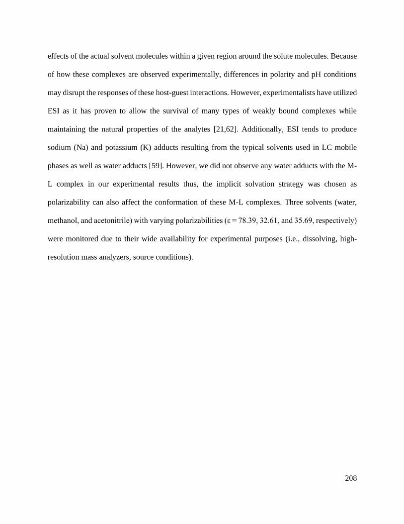
208
effects of the actual solvent molecules within a given region around the solute molecules. Because
of how these complexes are observed experimentally, differences in polarity and pH conditions
may disrupt the responses of these host-guest interactions. However, experimentalists have utilized
ESI as it has proven to allow the survival of many types of weakly bound complexes while
maintaining the natural properties of the analytes [21,62]. Additionally, ESI tends to produce
sodium (Na) and potassium (K) adducts resulting from the typical solvents used in LC mobile
phases as well as water adducts [59]. However, we did not observe any water adducts with the M-
L complex in our experimental results thus, the implicit solvation strategy was chosen as
polarizability can also affect the conformation of these M-L complexes. Three solvents (water,
methanol, and acetonitrile) with varying polarizabilities (ε = 78.39, 32.61, and 35.69, respectively)
were monitored due to their wide availability for experimental purposes (i.e., dissolving, high-
resolution mass analyzers, source conditions).

209
Figure 6.4: Energy minimized structures of various M-L complexes with different charges of
guests in both solvent (SMD) and gas phases. Both phases (gas and solution) were modeled to
investigate any potential structural and binding effects. The solvents acetonitrile (magenta),
methanol (green), and water (blue) were chosen for their commonality in dissolving crown ethers
in instrumentation parameters.
Initially, 18C6 was investigated due to its relatively larger inner diameter (2.7-3.2 Å) [63],
its high flexibility, and work previously performed in our laboratory. Crown ethers and other larger
macrocycles like the cyclodextrins and cucurbit[n]urils possess the capabilities to self-assembled
with a variety of guest species [16,64,65]. To highlight these properties using quantum mechanical
models, however, solvation strategies must be utilized to monitor any conformational changes
within each scenario. Depending on the solvation strategy (implicit or explicit), researchers can

210
investigate different responses of larger biomolecules and even spectroscopy sensing structures
[38,44]. Here, however, the solvent was treated as smooth continuous model to determine the
differences between the surface of the simple macrocycles within polarizable mediums [66].
The structures of the 18C6 and the M-L with various metal charges freely reorient and form
around the metal species. In both gas and solution phases, Ba showed the largest displacement
from the internal cavity of 18C6 (Figure 6.4, row four), whereas Pb showed the largest difference
between the acetonitrile and methanol. This is particularly interesting as these two analytes were
both capable of forming “sandwich” complexes (Figure 6.4). Like the experimental results in
Figure S6.1, 18C6 favors Tl over Ba and Pb yielding energies (Hartree) of the complex of -974.48,
-947.86, and -925.97 respectively giving a trend of Tl > Ba > Pb. Furthermore, 18C6 demonstrated
that it could encapsulate Sb in all scenarios without deviating from the initial gas phase structure.
However, when a 1:1 H-G ratio was tested experimentally, no Sb complex was observed which
may indicate other factors contributing to binding like charge distributions and the HSAB
principle. Thus, nitrogen and sulfur heteroatoms were substituted in the 18C6 ring structure.
6.3.4. Effect of donor atoms and charge transfers
Several strategies can be employed to understand electrostatic properties and factors that
govern specific paths for guests not only within M-L complexes but with lone ligands. One strategy
is molecular electrostatic potential maps (MESP or ESP) assist in the understanding of charge
distributions of a molecule and predict the properties of an interaction site. This type of analysis is
a powerful tool for identifying the possible interaction sites between a nucleophile and electrophile
[67,68]. A MESP visually depicts several properties of structures with heteroatoms such as O (red),
N (blue), and S (yellow) according to color. In Figure 6.5, different colors and hues extend
gradually starting from red (electrophiles) to orange and eventually blue (nucleophiles). This not

211
only depicts where electron-rich/deficient regions exist, but it also illustrates the boundaries of the
electron density.
Figure 6.5: (Left to Right) Electrostatic distributions calculated for 18C6, hexacyclen, thia-crown,
and partially substituted 18C6. Red regions represent electrophiles which contain an abundance of
electrons (typically cause from lone pairs). Blue regions indicate nucleophiles where there may be
a lower quantity of electrons.
The 18C6 ligand clearly depicts and directs guests towards a relatively large (~3.1Å)
electron-rich inner cavity. For the hexacyclen macrocycle, there is an occurrence of more
nucleophilic properties (blue region) as well as a smaller inner cavity (~2.1Å). This observation is
primarily contributed to the nitrogen atoms maintaining their respective hydrogen which are free
to reorient and interact for different interactions. For the thia-macrocycle, although a smaller cavity
was observed, it is an electron-rich region originating from the lone pairs of electrons from the
sulfur atoms. Finally, investigating the effects on mixtures of heteroatoms can affect the size and
directionality of binding sites. For Figure 6.5, one nitrogen and one sulfur were substituted in the
ring structure of the 18C6 macrocycle. While it retains a similarly sized hole compared to the
18C6, the nitrogen atom is pulling the electrons from the oxygen and sulfur atoms. This helps
confirm the nature of nitrogen’s nucleophilicity. This is ideal when trying to synthesize future
capturing agents and concluding interactions.
An additional strategy to confirm the effects and contribution of various atoms in a ligand
is using NBO analysis. This calculation assists in the investigation into interaction energies, 𝐸2,

212
of host–guest molecules (M-L complexes). In terms of the GSR studies using Ba, Pb, and Sb, the
contributing energies were attributed to the lone pair electrons of O atoms. These factors are
depicted by LP and LP* where the 1-center valence lone pair (LP1 and LP2) originate from the
two lone pairs of each oxygen atom and the 1-center valence anti-bond lone pair on the metal,
respectively.
Interestingly, Table 6.2 portrays a relationship between interactions energy and
conformation changes amongst the major metal analytes found in GSR and 18C6. Here, the
primary force contributing to interactions were the lone pairs LP* spx orbitals of the crown’s
oxygen atoms (varying between 1.95 and 2.39) and the unoccupied 6s orbital of the cation of the
oxygen atoms. To determine the atoms that contribute most to the transfer energies, the calculation
of the second-order perturbation focuses on the energy between two species, “Unit 1” (host) to
“Unit 2” (guest), which is outlined in Table S6.2. It can be shown that as the 𝐸2 energy increases,
the 18C6 ligand becomes more convoluted for different metal species. For the 18C6-Ba complex,
a more linear conformation is observed while the E2 energies from the contributing oxygen atoms
are relatively low (~ 2.13). With the introduction of Pb, the 𝐸2 increased causing further structural
change and encapsulation especially between O1 and O22, which are located on opposite positions
of the ring. This observation could result from the value of the cation’s electronegativity and
diameter relationship (χ/d) being approximately 3X larger than the 18C6-Ba complex.
Table 6.2: NBO analysis of the contributing atoms to the charge transfer of ligand to metal guest.
The driving mechanisms are identified by observing the energy contributions between “unit 1 to
unit 2”.

213
With the simulated 18C6-Sb complex data, the driving mechanism is the LP from two
oxygen atoms (O1 and O22) in which the E2 energies are the highest at 27.74 and 27.81,
respectively. Notably, by reorienting above the Sb atom, the energies from the remaining oxygen
atoms increase (ranging from 13.85 to 14.36) as the distance to the metal decreases. These
differences yield a large structural change from a relatively linear lone crown ether to a boat
configuration. Even though 18C6 may have the capacity to bind to Sb, the large structural change
may not be favorable as there is a small energy difference (Δ 5.09) from the 18C6-Sb complex (-
927.32 compared to -922.23 Hartrees) between the metal and lone crown ether macrocycle.
Additionally, different combinations and substitutions of heteroatoms were added to the ring
structure to observe their effects on binding to antimony and other metal ions for potentially a
more selective response. All proposed macrocycles demonstrated the potential to interact with Sb;
however, there was no occurrence of this complex forming experimentally. Hence, these instances
may be attributed to the HSAB principle where hard/soft atoms interact with their respective
hard/soft species.

214
6.3.5. Metal guest influences on reactivity descriptors
When understanding the association and conformation of M-L complexes, the properties
of the metal species and ligands as well as the mechanisms of their reactions can be explained
using the hard-soft acid-base (HSAB) concept. This theory stemmed from Pearson’s qualitative
concept where cations which are smaller in diameter and possess higher charge states are deemed
“hard” acids or bases. These species are thus, less polarizable and typically form ionic bonds
whereas, “softer” species form covalent bonds and are more polarizable, larger, and uncharged.
However, “borderline” acids and bases exist where species possess intermediate properties such
as high charge states and smaller cationic diameters (i.e., Sb). Thus, the strategy of utilizing
noncovalent interactions becomes more difficult as there exists numerous macrocycles with
various heteroatoms and functional groups.
To investigate potential macrocycles to encapsulate ions that observe HSAB properties, a
more quantitative metric can be calculated to describe a more quantitative metric to describe
interactions of hard and soft nucleophiles and electrophiles is to calculate the energies of HOMOs,
LUMOs, and the energy gaps [66–68]. Conceptually, the narrower the HOMO-LUMO gap (low
ΔE), the higher the chemical reactivity as it is favorable either to add an electron to a low-lying
LUMO or to extract electrons from a high-lying HOMO [70,72]. Thus, the global reactivity
descriptors of three fully substituted oxa-macrocycles were first calculated in both gas and solution
phase, summarized in Table 6.3.

215
Table 6.3: Fully substituted macrocycles with oxygen (C12H24O6), nitrogen (C12H30N6), and sulfur
(C12H24S6) atoms.
Ligand Phase ELUMO EHOMO ΔE η S
C12H24O6
Gas 0.307 -0.281 0.588 0.294 3.401
ACN 0.309 -0.285 0.594 0.297 3.367
MeOH 0.296 -0.299 0.595 0.298 3.361
H2O 0.294 -0.300 0.594 0.297 3.367
C12H30N6
Gas 0.372 -0.267 0.639 0.319 3.129
ACN 0.371 -0.286 0.657 0.329 3.044
MeOH 0.370 -0.288 0.658 0.329 3.039
H2O 0.367 -0.294 0.661 0.331 3.025
C12H24S6
Gas 0.178 -0.176 0.354 0.177 5.695
ACN 0.159 -0.225 0.384 0.192 5.208
MeOH 0.160 -0.225 0.385 0.193 5.194
H2O 0.160 -0.226 0.386 0.193 5.181
For the global hardness (η), the fully substituted thia-macrocycle exhibited the smallest
energy gap (~0.38) compared to the fully substituted oxa-, and aza-macrocycles (~0.58 and ~0.65,
respectively). Sulfur is the chemically softest atom due to its large atomic radius and, in turn, its
valence electrons are the most polarizable. Both N- and O-rich macrocycles were inversed to the
thia-species and possessed similar responses in terms of these factors which can be contributed to
their size and distance of their valence electron. The sulfur species also demonstrated the greatest
affinity for the Sb species, followed by the oxa- and aza-macrocycles.
Relating to experimental observations, the newly formed M-L complex of the internal
standard of Tl and the GSR analytes (Ba, Pb, and Sb) had energy gaps of 6.405, 6.865, 5.486, and
5.476 eV, respectively. In regard to the 18C6 host, the differences were 0.067, -0.393, 0.986, and
0.996 eV, respectively. Amongst the three polarizable solvents, the 18C6 energy gap did not differ
significantly (6.472 eV). With the addition of cations in solution, the energy gaps are altered which
is primarily caused by the metal’s orbitals extending the LUMO of the ligand (depicted in Figure
6.6). For electrospray ionization, more energy would be required to dissociate the highly reactive
Tl+ and Ba2+ as opposed to the Pb2+ and Sb3+ cationic species.
216
Figure 6.6: (Left to Right) HOMO and LUMO of lone 18C6 macrocycle with the addition of
various metals and charge states (+1,+2,+3). It can be shown that (for all instances) the HOMO
mainly resided on the oxygen atoms whereas the LUMO exists on the metal cation.
The lone macrocycles orbitals were compared against the M-L complexes. As a metal
cation was introduced, the traditional LUMO p-orbital structure can be observed for the guest
analytes. Amongst these species, however, the LUMO density for barium seems to be vastly
different. This may be attributed to the electron configuration of the 2+ cation as electrons are lost
from the 6s orbital and thus, yielding a [Xe] configuration. From the NBO analysis, the barium
species associates with noncovalent, electrostatic interactions via anti-bonding properties (LP*).
It is important to note that as cations increase in size and possess more electrons in their outer
shells (i.e., p and d), the ability to distinguish between these orbitals becomes increasingly difficult
as hybridization and overlap is possible. Thus, for the barium cation, this irregular orbital may be
the product of this hybridization from the spxdy shells which has been recorded previously [71-73].
Amongst the studied cations, Tl+ and Ba2+ demonstrated the largest displacement of the
LUMO energy in the water due to the polarizability factor. Measuring from O15 and O36, the lone
crown ether exhibited small radii cavity differences from gas phase (3.06 Å) in water (2.95 Å). For
the Tl+ and Ba2+ complexes, both radii were similar (2.95 Å) but hovered above the cavity’s center

217
at approximately 2.39 Å and 3.11 Å, respectively. Even though Pb2+ complex displayed similar
LUMO energy displacement but was mitigated due to the crown ether’s encapsulation which
altered to 4.37 Å for O15 and O36. Finally, Sb3+ cation exhibited the most interaction with the
energies and the most distortion (4.64 Å) of the 18C6 species. This suggests that the degree and
magnitude of the LUMO energy contributes to the formation of “sandwich” structures as these
cations have shown evidence to form these structures [58,74].
For these analytes, the thia-macrocycles exhibited structural similarities for the Pb2+ and
Sb3+ complexes; however, the displacement for the Tl+ and Ba2+ were displacement father from
the center of the cavity in solution phase as opposed to gas phase. This observation further
illustrates the importance of monitoring these complexes in solution phase. Additionally,
introducing counterions such as nitrates, hydroxides, and ammonium ions in solution may prove
valuable to calculate since they are prevalent in a variety of mobile phases and may affect the
formation of various macrocycles.
6.4. Conclusions
The use of computational methods has been utilized across various disciplines including
chemo-sensing, extraction, and drug-design [49,69]. Recently, there has been a growing interest
into utilization of these strategies for forensic purposes relating to drugs-of-abuse and explosives
[36,40,75]. This work explored the possibility of utilizing this theoretical approach for the
explanation of interactions and selectivity of a conglomeration typically found in GSR trace
analysis [1,2]. Additionally, DFT was utilized to observe the main contributing factors for the
inability to detect antimony within this particulate structure using ESI-MS/MS.
By using 18C6 as the starting structure, other simple macrocycles were simulated to
elucidate the possible factors and interactions that take place in solution. In the presence of metal

218
species such as Ba2+ or Pb2+, there is considerable displacement of these ions when exposed to
common solvents such as methanol, acetonitrile, and water. Because of this, these metal ions are
more prone to forming “sandwich” or 1:2 M-L structures. With the addition of Tl+ (for purposes
as an internal standard), the overall response was lowered suggesting 18C6 has selectivity of Tl >
Ba > Pb which correspond to DFT findings. From this study, findings indicate that crown ethers
can complex with the metalloid Sb but to an unfavorable distortion of the base structure of the
ligand.
It was also found, in both this study and others, that by altering the number of heteroatoms
to either N or S atoms adjusts the selectivity for various metal cations [13,76–78]. These
substitutions affect simple macrocycles by changing the inner cavity size and the lone pairs of
electrons interacting with guest analytes. Furthermore, energy gaps exhibit important binding
affinity trends with varying levels of chemical hardness amongst guests. This factor matters as
scientists can manipulate and gain selectivity advantages when developing relatively inexpensive
analytical tools such as point-of-care systems or even capillary columns.
One aspect that should be addressed in future studies is the effect of a counterion to binding
affinity as well as the formation of “sandwich” structures. This is important due to the chemical
composition of the conglomerate IGSR particles, and the procedure required to dissolve and
expose these analytes to macrocycles in solution (i.e., heavy acid digestion) which has been
observed in ESI (+) mode [2]. Additionally, using either an explicit or hybrid solvation model may
introduce other factors into the competition between hosts and guests making it essential for
MS/MS analysis to determine what complex(es) to target [43,44].
The overarching theoretical challenge in describing such flexible systems stems from the
wealth of parameters required to describe their energetic, geometric, and chemical characteristics

219
and the ease by which these parameters change. Electrospray ionization has allowed
experimentalists to observe and calculate affinities through a myriad of factors which can then be
compared to computational strategies. By observing positive relationships between experimental
and theoretical data, scientists can further predict and design molecules to preserve physical
resources. In this study, simple macrocyclic structures have the capacity to undergo high
distortions albeit low energy changes when exposed to heavy metals whilst preserving their natural
isotope distributions. These distortions relate to the strength of non-bonded interactions between
the host and guest, which then teaches about the overall host−guest interaction energy. This work
demonstrates the value of DFT to probe for valuable information and structures obtained from
ESI-MS/MS experiments.
Author Contributions
The manuscript was written through the contributions of all authors. All authors have given
approval to the final version of the manuscript.
Conflicts of interest
The authors declare that they have no known competing financial interests or personal
relationships that could have influenced the work reported in this paper.
Acknowledgments
We want to acknowledge Dr. Kathleen Schwarz for troubleshooting and computational
expertise for density functional calculations.
The authors acknowledge use of the supercomputing system Thorny Flat at West Virginia
University (WVU), which is funded in part by the National Science Foundation EPSCoR Research
Infrastructure Improvement Cooperative Agreement #1003907, National Science Foundation

220
Major Research Instrumentation Program (MRI) Award #1726534, the state of West Virginia
(WVEPSCoR via the Higher Education Policy Commission) and WVU.
This project is sponsored by Award No. #2019-R2-CX-044 from the National Institute of
Justice to West Virginia University. The opinions, findings, and conclusions are those of the
authors and do not necessarily reflect those of the Department of Justice.

221
6.5. References for Chapter 6
[1] W. Feeney, C. vander Pyl, S. Bell, T. Trejos, Trends in composition, collection,
persistence, and analysis of IGSR and OGSR: A review, Forensic Chemistry. 19 (2020) 100250.
https://doi.org/10.1016/j.forc.2020.100250.
[2] W. Feeney, K. Menking-Hoggatt, C. vander Pyl, C.E. Ott, S. Bell, L. Arroyo, T. Trejos,
Detection of Organic and Inorganic Gunshot Residues from Hands using Complexing Agents
and LC-MS/MS, Analytical Methods. (2021). https://doi.org/10.1039/d1ay00778e.
[3] M.E. Sears, Chelation: Harnessing and enhancing heavy metal detoxification - A review,
The Scientific World Journal. 2013 (2013). https://doi.org/10.1155/2013/219840.
[4] B.E. Igiri, S.I.R. Okoduwa, G.O. Idoko, E.P. Akabuogu, A.O. Adeyi, I.K. Ejiogu,
Toxicity and Bioremediation of Heavy Metals Contaminated Ecosystem from Tannery
Wastewater: A Review, Journal of Toxicology. (n.d.). https://doi.org/10.1155/2018/2568038.
[5] C. Sarzanini, E. Mentasti, Determination and speciation of metals by liquid
chromatography, Journal of Chromatography A. 789 (1997) 301–321.
https://doi.org/10.1016/S0021-9673(97)00988-6.
[6] M.J. Kosnett, The Role of Chelation in the Treatment of Arsenic and Mercury Poisoning,
Journal of Medical Toxicology. 9 (2013) 347–354. https://doi.org/10.1007/s13181-013-0344-5.
[7] A. Ciesielski, P. Samori, Supramolecular Approaches to Graphene: From Self-Assembly
to Molecule-Assisted Liquid-Phase Exfoliation, Advanced Materials. 28 (2016) 6030–6051.
https://doi.org/10.1002/adma.201505371.
[8] I. v. Kolesnichenko, E. v. Anslyn, Practical applications of supramolecular chemistry,
Chemical Society Reviews. 46 (2017) 2385–2390. https://doi.org/10.1039/c7cs00078b.
222
[9] J.J. Guo, J. Lee, C.I. Contescu, N.C. Gallego, S.T. Pantelides, S.J. Pennycook, B.A.
Moyer, M.F. Chisholm, Crown ethers in graphene, Nature Communications. 5 (2014) 6.
https://doi.org/10.1038/ncomms6389.
[10] S. Kafashi, M. Yaftian, A. Zamani, Binding Ability of Crown Ethers Towards Pb(II) Ions
in Binary Water/Organic Solvents Using Solvent Extraction Method, Journal of Solution
Chemistry. 44 (2015) 1798–1811. https://doi.org/10.1007/s10953-015-0378-1.
[11] S. Bell, W. Feeney, Single shot, single sample, single instrument detection of IGSR and
OGSR using LC/MS/MS, Forensic Science International. 299 (2019) 215–222.
https://doi.org/10.1016/j.forsciint.2019.04.002.
[12] Y. Inokuchi, O. v. Boyarkin, R. Kusaka, T. Haino, T. Ebata, T.R. Rizzo, UV and IR
spectroscopic studies of cold alkali metal ion-crown ether complexes in the gas phase, Journal of
the American Chemical Society. 133 (2011) 12256–12263. https://doi.org/10.1021/ja2046205.
[13] Z. Zhao, H. Tian, M. Zhang, Y. Yang, H. Zhang, Molecular design of macrocyclic
compounds for complete removal of thallium(I) from wastewater, Environmental Science and
Pollution Research. 25 (2018) 34550–34558. https://doi.org/10.1007/s11356-018-3393-0.
[14] X. Du, H. Hao, A. Qin, B.Z. Tang, Highly sensitive chemosensor for detection of
methamphetamine by the combination of AIE luminogen and cucurbit[7]uril, Dyes and
Pigments. 180 (2020) 108413. https://doi.org/https://doi.org/10.1016/j.dyepig.2020.108413.
[15] F. Ma, X. Zheng, J. Xie, Z. Li, Binding properties of cucurbit[7]uril to neutral and
protonated amino acids: A theoretical study, International Journal of Quantum Chemistry. (n.d.).
https://doi.org/10.1002/qua.26491.
223
[16] L. Barrientos, S. Miranda‐Rojas, F. Mendizabal, Noncovalent interactions in inorganic
supramolecular chemistry based in heavy metals. Quantum chemistry point of view,
International Journal of Quantum Chemistry. 119 (2019). https://doi.org/10.1002/qua.25675.
[17] H.J. Schneider, A.K. Yatsimirsky, Selectivity in supramolecular host-guest complexes,
Chemical Society Reviews. 37 (2008) 263–277. https://doi.org/10.1039/b612543n.
[18] S.M. Blair, E.C. Kempen, J.S. Brodbelt, Determination of binding selectivities in host-
guest complexation by electrospray/quadrupole ion trap mass spectrometry, Journal of the
American Society for Mass Spectrometry. 9 (1998) 1049–1059. https://doi.org/10.1016/s1044-
0305(98)00082-8.
[19] Y. Tao, R.R. Julian, Factors that influence competitive intermolecular solvation of
protonated groups in peptides and proteins in the gas phase, Journal of the American Society for
Mass Spectrometry. 24 (2013) 1634–1640. https://doi.org/10.1007/s13361-013-0684-z.
[20] V. Rudiger, H.J. Schneider, V.P. Solov’ev, V.P. Kazachenko, O.A. Raevsky, Crown
ether-ammonium complexes: Binding mechanisms and solvent effects, European Journal of
Organic Chemistry. (1999) 1847–1856. %3CGo.
[21] C.L. Sherman, J.S. Brodbelt, A.P. Marchand, B. Poola, Electrospray ionization mass
spectrometric detection of self-assembly of a crown ether complex directed by π-stacking
interactions, Journal of the American Society for Mass Spectrometry. 16 (2005) 1162–1171.
https://doi.org/10.1016/j.jasms.2005.03.014.
[22] S.M. Blair, J.S. Brodbelt, A.P. Marchand, K.A. Kumar, H.-S. Chong, Evaluation of
Binding Selectivities of Caged Crown Ligands toward Heavy Metals by Electrospray
Ionization/Quadrupole Ion Trap Mass Spectrometry, Analytical Chemistry. 72 (2000) 2433–
2445. https://doi.org/10.1021/ac991125t.
224
[23] A. McKenzie-Coe, S. Bell, F. Fernandez-Lima, Detection of firearm discharge residue
from skin swabs using trapped ion mobility spectrometry coupled to mass spectrometry,
Analytical Methods. 10 (2018) 4219–4224. https://doi.org/10.1039/c8ay00658j.
[24] A.B. Wijeratne, S.H. Yang, D.W. Armstrong, K.A. Schug, Solvent Molecules Undergo
Homolytic Cleavage and Radical Recombination Processes during Negative-Mode Electrospray
Ionization: Adduct Formation with Antimony(III)-Tartrate Dianion, Analytical Chemistry. 82
(2010) 5141–5146. https://doi.org/10.1021/ac1003344.
[25] A.B. Wijeratne, S.E. Spencer, J. Gracia, D.W. Armstrong, K.A. Schug, Antimony(III)-D,
L-Tartrates Exhibit Proton-Assisted Enantioselective Binding in Solution and in the Gas Phase,
Journal of the American Society for Mass Spectrometry. 20 (2009) 2100–2105.
https://doi.org/10.1016/j.jasms.2009.07.011.
[26] A.B. Wijeratne, J. Gracia, S.H. Yang, P. Kroll, D.W. Armstrong, K.A. Schug, New
structural insight for antimony(III)-tartrate, Inorganic Chemistry Communications. 13 (2010)
1504–1508. https://doi.org/10.1016/J.INOCHE.2010.08.028.
[27] A.B. Wijeratne, K.A. Schug, Molecular recognition properties of tartrates and metal-
tartrates in solution and gas phase, Journal of Separation Science. 32 (2009) 1537–1547.
https://doi.org/10.1002/jssc.200900064.
[28] N. Islam, S.S. Chimni, Binding and selectivity of phenazino-18-crown-6-ether with
alkali, alkaline earth and toxic metal species: A DFT study, Journal of Molecular Structure.
(2017). https://doi.org/10.1016/j.molstruc.2016.10.100.
[29] Z. Jing, G. Wang, Y. Zhou, D. Pang, F. Zhu, H. Liu, Selectivity of 18-crown-6 ether to
alkali ions by density functional theory and molecular dynamics simulation, Journal of
Molecular Liquids. 311 (2020). https://doi.org/10.1016/j.molliq.2020.113305.

225
[30] K.J. Hintze, A. Lützen, T. Bredow, Structure and stability of supramolecular crown ether
complexes, Journal of Computational Chemistry. 36 (2015) 1467–1472.
https://doi.org/10.1002/jcc.23950.
[31] S. Hadisaputra, H.D. Pranowo, R. Armunanto, Extraction of strontium(ii) by crown ether:
Insights from density functional calculation, Indonesian Journal of Chemistry. 12 (2012) 207–
216. https://doi.org/10.22146/ijc.21332.
[32] N.N. Karaush, G. v. Baryshnikov, V.A. Minaeva, B.F. Minaev, A DFT and QTAIM
study of the novel d-block metal complexes with tetraoxa[8]circulene-based ligands, New
Journal of Chemistry. 39 (2015) 7815–7821. https://doi.org/10.1039/c5nj01255d.
[33] R. Behjatmanesh-Ardakani, F. Pourroustaei-Ardakani, M. Taghdiri, Z.M. Kotena, DFT-
B3LYP study of interactions between host biphenyl-1-aza-18-crown-6 ether derivatives and
guest Cd2+: NBO, NEDA, and QTAIM analyses, Journal of Molecular Modeling. 22 (2016).
https://doi.org/10.1007/s00894-016-3012-2.
[34] K. Reuter, M.R. Buchner, G. Thiele, C. von Hänisch, Stable Alkali-Metal Complexes of
Hybrid Disila-Crown Ethers, Inorganic Chemistry. 55 (2016) 4441–4447.
https://doi.org/10.1021/acs.inorgchem.6b00267.
[35] D.M. Zuckerman, Statistical physics of biomolecules: An introduction, Statistical Physics
of Biomolecules: An Introduction. (2010) 1–325. https://doi.org/10.1201/b18849.
[36] M. Jaidann, S. Roy, H. Abou-Rachid, L.S. Lussier, A DFT theoretical study of heats of
formation and detonation properties of nitrogen-rich explosives, Journal of Hazardous Materials.
176 (2010) 165–173. https://doi.org/10.1016/j.jhazmat.2009.10.132.
[37] S.J.J.J.J. Mary, M.U.M. Siddique, S. Pradhan, V. Jayaprakash, C. James, Quantum
chemical insight into molecular structure, NBO analysis of the hydrogen-bonded interactions,

226
spectroscopic (FT–IR, FT–Raman), drug likeness and molecular docking of the novel anti
COVID-19 molecule 2-[(4,6-diaminopyrimidin-2-yl)sulfanyl]-N-(4-fluo, Spectrochimica Acta -
Part A: Molecular and Biomolecular Spectroscopy. 244 (2021) 118825.
https://doi.org/10.1016/j.saa.2020.118825.
[38] E. Bahojb Noruzi, M. Molaparast, M. Zarei, B. Shaabani, Z. Kariminezhad, B. Ebadi, V.
Shafiei-Irannejad, M. Rahimi, J. Pietrasik, E.B. Noruzi, M. Molaparast, M. Zarei, B. Shaabani,
Z. Kariminezhad, B. Ebadi, V. Shafiei-Irannejad, M. Rahimi, J. Pietrasik, Para-
sulfonatocalix[n]arene-based biomaterials: Recent progress in pharmaceutical and biological
applications, European Journal of Medicinal Chemistry. 190 (2020).
https://doi.org/10.1016/j.ejmech.2020.112121.
[39] L. Isaacs, Stimuli responsive systems constructed using cucurbit[ n ]uril-type molecular
containers, Accounts of Chemical Research. 47 (2014) 2052–2062.
https://doi.org/10.1021/ar500075g.
[40] M. Vujović, V. Ragavendran, B. Arsić, E. Kostić, M. Mladenović, DFT calculations as
an efficient tool for prediction of Raman and infra-red spectra and activities of newly synthesized
cathinones, Open Chemistry. 18 (2020) 185–195. https://doi.org/10.1515/chem-2020-0021.
[41] A.A. Al-Kahtani, N.A. Al-Jallal, A.A. El-Azhary, Conformational and vibrational
analysis of 18-crown-6-alkali metal cation complexes, Spectrochimica Acta - Part A: Molecular
and Biomolecular Spectroscopy. 132 (2014) 70–83. https://doi.org/10.1016/j.saa.2014.04.170.
[42] Y. Zhang, X.Y. Wang, B.H. Luo, Y. Xia, DFT study of crown ether-bridged Z-stilbenes
and their complexes with alkali metal cations, Journal of Organometallic Chemistry. 699 (2012)
31–38. https://doi.org/10.1016/j.jorganchem.2011.10.032.

227
[43] J. Zhang, H. Zhang, T. Wu, Q. Wang, D. van der Spoel, Comparison of Implicit and
Explicit Solvent Models for the Calculation of Solvation Free Energy in Organic Solvents,
(2017). https://doi.org/10.1021/acs.jctc.7b00169.
[44] M. Wang, Y. Mei, U. Ryde, Host-Guest Relative Binding Affinities at Density-
Functional Theory Level from Semiempirical Molecular Dynamics Simulations, Journal of
Chemical Theory and Computation. 15 (2019) 2659–2671.
https://doi.org/10.1021/acs.jctc.8b01280.
[45] N. Kircheva, S. Dobrev, L. Dasheva, I. Koleva, V. Nikolova, S. Angelova, T. Dudev, B.
Supramolecular, Complexation of biologically essential (mono- And divalent) metal cations to
cucurbiturils: a DFT/SMD evaluation of the key factors governing the host-guest recognition,
RSC Advances. 10 (2020) 28139–28147. https://doi.org/10.1039/d0ra04387g.
[46] E.S.H. Gwee, Z.L. Seeger, D.R.T. Appadoo, B.R. Wood, E.I. Izgorodina, Influence of
DFT Functionals and Solvation Models on the Prediction of Far-Infrared Spectra of Pt-Based
Anticancer Drugs: Why Do Different Complexes Require Different Levels of Theory?, ACS
Omega. 4 (2019) 5254–5269. https://doi.org/10.1021/acsomega.8b03455.
[47] R. Behjatmanesh-Ardakani, DFT-B3LYP and SMD study on the interactions between
aza-, diaza-, and triaza-12-crown-4 (A n -12-crown-4, n = 1, 2, 3) with Na+ in the gas phase and
acetonitrile solution, Structural Chemistry. 25 (2014) 919–929. https://doi.org/10.1007/s11224-
013-0366-2.
[48] K.A. Baseden, J.W. Tye, Introduction to density functional theory: Calculations by hand
on the helium atom, Journal of Chemical Education. 91 (2014) 2116–2123.
https://doi.org/10.1021/ed5004788.

228
[49] B. Honarparvar, S. Kanchi, K. Bisetty, Theoretical insights into the competitive metal
bioaffinity of lactoferrin as a metal ion carrier: A DFT study, New Journal of Chemistry. 43
(2019) 16374–16384. https://doi.org/10.1039/c9nj03786a.
[50] H. Xu, D.C. Xu, Y. Wang, Natural indices for the chemical hardness/softness of metal
cations and ligands, ACS Omega. 2 (2017) 7185–7193.
https://doi.org/10.1021/acsomega.7b01039.
[51] R.P. Gangadharana, S.S. Krishnanb, Natural Bond Orbital (NBO) population analysis of
1-azanapthalene-8-ol, Acta Physica Polonica A. 125 (2014) 18–22.
https://doi.org/10.12693/APhysPolA.125.18.
[52] W. Li, B. Lu, F. Chen, F. Yang, Z. Wang, Host-guest complex of cypermethrin with β-
cyclodextrin: A spectroscopy and theoretical investigation, Journal of Molecular Structure. 990
(2011) 244–252. https://doi.org/10.1016/j.molstruc.2011.01.053.
[53] F. Weinhold, C.R. Landis, E.D. Glendening, What is NBO analysis and how is it useful?,
International Reviews in Physical Chemistry. 35 (2016) 399–440.
https://doi.org/10.1080/0144235X.2016.1192262.
[54] S. Hirata, X. He, M.R. Hermes, S.Y. Willow, Second-order many-body perturbation
theory: An eternal frontier, Journal of Physical Chemistry A. 118 (2014) 655–672.
https://doi.org/10.1021/jp410587b.
[55] N. Yasarawan, K. Thipyapong, V. Ruangpornvisuti, Substituent effect on the proton-
related phenomena and chelation behavior of hydroxypicolinic compounds: A DFT
investigation, Structural Chemistry. 27 (2016) 505–524. https://doi.org/10.1007/s11224-015-
0579-7.

229
[56] A. Hassan, M. Mahmoud, B.S. Bageri, M.S. Aljawad, M.S. Kamal, A.A. Barri, I.A.
Hussein, Applications of chelating agents in the upstream oil and gas industry: A review, Energy
and Fuels. 34 (2020) 15593–15613. https://doi.org/10.1021/acs.energyfuels.0c03279.
[57] H.J. Schneider, Limitations and Extensions of the Lock-and-Key Principle: Differences
between Gas State, Solution and Solid State Structures, International Journal of Molecular
Sciences. 16 (2015) 6694–6717. https://doi.org/10.3390/ijms16046694.
[58] H.R. Yu, J.Q. Hu, X.H. Lu, X.J. Ju, Z. Liu, R. Xie, W. Wang, L.Y. Chu, Insights into the
effects of 2:1 “sandwich-type” crown-ether/metal-ion complexes in responsive host - guest
systems, Journal of Physical Chemistry B. 119 (2015) 1696–1705.
https://doi.org/10.1021/jp5079423.
[59] A. Kruve, K. Kaupmees, Adduct Formation in ESI/MS by Mobile Phase Additives,
Journal of the American Society for Mass Spectrometry. 28 (2017) 887–894.
https://doi.org/10.1007/s13361-017-1626-y.
[60] L. Konermann, H. Metwally, Q. Duez, I. Peters, Charging and supercharging of proteins
for mass spectrometry: Recent insights into the mechanisms of electrospray ionization, Analyst.
144 (2019) 6157–6171. https://doi.org/10.1039/c9an01201j.
[61] E.M. Panczyk, J.D. Gilbert, G.S. Jagdale, A.Q. Stiving, L.A. Baker, V.H. Wysocki, Ion
Mobility and Surface Collisions: Submicrometer Capillaries Can Produce Native-like Protein
Complexes, Analytical Chemistry. 92 (2020) 2460–2467.
https://doi.org/10.1021/acs.analchem.9b03666.
[62] C.L. Sherman, J.S. Brodbelt, Partitioning Model for Competitive Host−Guest
Complexation in ESI-MS, Analytical Chemistry. 77 (2005) 2512–2523.
https://doi.org/10.1021/ac048518z.

230
[63] C.J. Pedersen, The Discovery of Crown Ethers, Science. 241 (1988) 536–540.
http://www.jstor.org/stable/1701735.
[64] M.R. Sambrook, J.C. Vincent, J.A. Ede, I.A. Gass, P.J. Cragg, Experimental and
computational study of the inclusion complexes of beta-cyclodextrin with the chemical warfare
agent soman (GD) and commonly used simulants, Rsc Advances. 7 (2017) 38069–38076.
https://doi.org/10.1039/c7ra03328a.
[65] A.I. Lazar, F. Biedermann, K.R. Mustafina, K.I. Assaf, A. Hennig, W.M. Nau,
Nanomolar Binding of Steroids to Cucurbit[n]urils: Selectivity and Applications, Journal of the
American Chemical Society. 138 (2016) 13022–13029. https://doi.org/10.1021/jacs.6b07655.
[66] A. v. Marenich, C.J. Cramer, D.G. Truhlar, Universal solvation model based on solute
electron density and on a continuum model of the solvent defined by the bulk dielectric constant
and atomic surface tensions, Journal of Physical Chemistry B. 113 (2009) 6378–6396.
https://doi.org/10.1021/jp810292n.
[67] M.D. Esrafili, Nitrogen-doped (6,0) carbon nanotubes: A comparative DFT study based
on surface reactivity descriptors, Computational and Theoretical Chemistry. 1015 (2013) 1–7.
https://doi.org/10.1016/j.comptc.2013.04.003.
[68] M.D. Esrafili, F. Mohammadian-Sabet, An ab initio study on chalcogen-chalcogen bond
interactions in cyclic (SHX)3 complexes (X = F, Cl, CN, NC, CCH, OH, OCH3, NH2),
Chemical Physics Letters. 628 (2015) 71–75. https://doi.org/10.1016/j.cplett.2015.04.013.
[69] M. Ramalingam, V. Sethuraman, N. Sundaraganesan, Molecular structure, vibrational
spectroscopic, first order hyperpolarizability and HOMO-LUMO studies of 7-amino-8-oxo-3-
vinyl-5-thia-1- azabicyclo[4.2.0]oct-2-ene-2-carboxylic acid, Spectrochimica Acta - Part A:

231
Molecular and Biomolecular Spectroscopy. 78 (2011) 660–669.
https://doi.org/10.1016/j.saa.2010.11.027.
[70] S.K. Seth, I. Saha, C. Estarellas, A. Frontera, T. Kar, S. Mukhopadhyay, Supramolecular
self-assembly of M-IDA complexes involving lone-pair ⋯φ Interactions: Crystal structures,
hirshfeld surface analysis, and DFT calculations [H2IDA = iminodiacetic acid, M = Cu(II),
Ni(II)], Crystal Growth and Design. 11 (2011) 3250–3265. https://doi.org/10.1021/cg200506q.
[71] X. Wu, L. Zhao, D. Jiang, I. Fernández, R. Berger, M. Zhou, G. Frenking, Barium as
Honorary Transition Metal in Action: Experimental and Theoretical Study of Ba(CO)+ and
Ba(CO)−, Angewandte Chemie - International Edition. 57 (2018) 3974–3980.
https://doi.org/10.1002/anie.201713002.
[72] K.L. Woon, W.S. Wong, N. Chanlek, H. Nakajima, S. Tunmee, V.S. Lee, A. Ariffin, P.
Songsiriritthigul, Work function modification of PEDOT:PSS by mixing with barium
acetylacetonate, RSC Advances. 10 (2020) 17673–17680. https://doi.org/10.1039/d0ra02575e.
[73] R. Oubouaza, M. Benson, J. Wojciechowski, S. Chtita, M. Tridane, S. Belaaouad,
Synthesis, crystal structure, vibrational study and DFT computation of barium
dihydrogenomonophosphate Ba(H2PO4)2, Biointerface Research in Applied Chemistry. 12
(2022) 1120–1133. https://doi.org/10.33263/BRIAC121.11201133.
[74] K.I. Tugashov, D.A. Gribanyov, F.M. Dolgushin, A.F. Smol’yakov, A.S. Peregudov,
M.K. Minacheva, I.A. Tikhonova, V.B. Shur, Coordination Chemistry of Anticrowns. Synthesis
and Structures of Double-Decker Sandwich Complexes of the Three-Mercury Anticrown (o-
C6F4Hg)(3) with Halide Anions Containing and Not Containing Coordinated Dibromomethane
Molecules, Organometallics. 35 (2016) 2197–2206.
https://doi.org/10.1021/acs.organomet.6b00231.
232
[75] J. Tyler Davidson, E.L. Piacentino, Z.J. Sasiene, Y. Abiedalla, J. DeRuiter, C.R. Clark,
G. Berden, J. Oomens, V. Ryzhov, G.P. Jackson, Identification of novel fragmentation pathways
and fragment ion structures in the tandem mass spectra of protonated synthetic cathinones,
Forensic Chemistry. 19 (2020) 100245. https://doi.org/10.1016/j.forc.2020.100245.
[76] M. Frańska, A. Michalak, Gas-phase relative stabilities of Tl+-crown ether complexes
and Rb+-crown ether complexes, European Journal of Mass Spectrometry. 24 (2018) 279–288.
https://doi.org/10.1177/1469066717752794.
[77] S.M. Williams, J.S. Brodbelt, A.P. Marchand, D. Cal, K. Mlinaric-Majerski, Metal
complexation of thiacrown ether macrocycles by electrospray ionization mass spectrometry,
Analytical Chemistry. 74 (2002) 4423–4433. https://doi.org/10.1021/ac011227v.
[78] V. Rüdiger, H.-J. Schneider, V.P. Solov’ev, V.P. Kazachenko, O.A. Raevsky, Crown
Ether–Ammonium Complexes: Binding Mechanisms and Solvent Effects, European Journal of
Organic Chemistry. 1999 (1999) 1847–1856. https://doi.org/10.1002/(sici)1099-
0690(199908)1999:8<1847::aid-ejoc1847>3.3.co;2-h.
.

233
CHAPTER 7
Overall Conclusion and Future Directions

234
7.1. Overall Conclusion and Future Directions
This study met our overall goal of enhancing the reliability of GSR evidence through the
development of rapid analytical tools that provided an increased body of knowledge on the
chemical composition of modern ammunition residues and a novel interpretation framework for
the quantitative assessment of the evidential value of GSR. We anticipate that the methodologies
and the IGSR/OGSR population dataset will become accessible to forensic practitioners to
complement and modernize current practice. The main findings and achievements of this study are
discussed in the following sections.
7.1.1. Objective 1: Review of the current literature associated with GSR
The literature review condenses relevant literature within the past 20 years associated with
gunshot residue’s collection and analysis. Most investigations primarily focused on the IGSR
particulate information using SEM-EDS instrumentation and reported data on particle counts and
composition. The forensic community benefits from international standard practices that are
broadly implemented and studied. Also, a guideline for validation of GSR examinations by SEM-
EDS was recently published, assisting further the community to stringent quality controls. The
research groups that did investigate OGSR compound information utilized various instruments
ranging from GC/MS, LC-MS/MS, IMS, Raman, and TOF-MS. The status of OGSR detection is
still in research stages and therefore the emerging tools reported in the literature have not been
widely adopted in the field. Consensus-based method are not yet implemented for combined
OGSR to IGSR examinations, but it is a leading research topic in this discipline. Because of this,
practitioners and researchers have just scraped the surface of the information that can be obtained
from utilizing combined OGSR and IGSR information from a single sample.

235
The review paper (Chapter 3) highlighted some of those existing gaps and summarized
the scientific foundations and current state for the deposition, persistence, and collection of organic
and inorganic gunshot residue. One of the outcomes of this manuscript was to provide an extensive
summary of the main OGSR and IGSR components reported in the literature and a comparative
analysis of the limitations and capabilities of methods of collection, preservation, and examination
of GSR.
Regardless of numerous studies reporting transfer and persistence of IGSR and OGSR, this
topic remains challenging, and several gaps of knowledge still exist. Advances and adoption of
novel analytical methods, and an increased body of knowledge on background occurrence and
transfer mechanisms will lead the field to more encompassing interpretation models.
The research conducted in this dissertation is anticipated to contribute to a needed
knowledge in this field. First, the development of novel methods of IGSR/OGSR detection
(Objective 2) and their application to population datasets (Objective 3) and simulation
computational models (Objective 4) increases our understanding of the chemistry and the
mechanisms of prevalence and persistence of GSR. Second, this study proposed using machine
learning algorithms to assist in the probabilistic interpretation of the evidence (objective 3),
opening modern and more encompassing venues to assist the trier of fact.
7.1.2. Objective 2: Validation and evaluation of a single instrument for sequential analysis of
GSR components
In Chapter 4, an LC-MS/MS method was developed, optimized, and validated for dual
detection of IGSR and OGSR from a single sample, using a single instrument. Because of the
properties of host-guest chemistry are less known to the forensics community, further confirmation
was required to demonstrate the identification and characterization of the IGSR analytes when

236
associated with the crown ethers. Hence, the high-resolution orbitrap was implemented to identify
the products and natural abundances of the metal ions.
The analytical figures of merit (LOD, LOQ, LDR, intra- and interday variabilities,
selectivity) and the performance rates derived from ground truth specimens from shooters and non-
shooters demonstrated the method fit for purpose. Also, the efficiency of two collection and
extraction protocols were compared for this study, tesa Tack® and the ASTM carbon adhesive
recommended for IGSR. Extractions were performed side-by-side and the percent recoveries were
analyzed by ANOVA. The OGSR compounds were adequately analyzed via LC-MS/MS after the
organic extraction step. However, because of the crown ether mechanisms, the recovery of IGSR
was cross-evaluated by ICP-MS with a more exhaustive digestion method. Hence, an ICP-MS
methodology was used to evaluate the concentrations of the metal ions using the different
extraction procedures. The results indicate the carbon adhesive provided superior performance
while conveniently being compatible with current collection protocols for SEM-EDS.
A set of authentic samples were extracted and analyzed from the carbon adhesives using
the LC-MS/MS method. Because this technique sequentially analyzes IGSR and OGSR, a
conservative criterion was established as a first step, following a categorical approach similar to
the ASTM E1588-20. This criterion indicated that a sample was positive if at least three GSR
components were present above the cut-off (one OGSR and one IGSR, and vice versa.)
Furthermore, by noting the lack of investigations using screening methodologies in GSR-
related casework, a workflow was developed to incorporate LIBS and electrochemical methods
before LC-MS/MS confirmatory analysis. Samples from authentic shootings and post-shooting
activities were evaluated to test value of combining information. In both situations, the knowledge
of a sample increased after incorporating both OGSR and IGSR information. LC-MS/MS provided

237
superior performance and was able to detect both inorganic and organic components from the
sample after quick screening with LIBS/EC, demonstrating it can be used as a stand-alone method
and as a confirmatory method when large volume of items in a case may require some triage
screening alternatives.
In this work, it was concluded that environmental factors greatly affect the recovery
efficiency, and thus, the overall reporting of GSR. This work also illustrated that practitioners and
crime laboratories do not need to choose between collecting and analyzing OGSR and IGSR
information, providing a breakthrough alternative approach for this comprehensive examination
of organic and inorganic residues.
7.1.3. Objective 3: Investigation of the presence and prevalence of GSR in West Virginia within
different populations
The goal of this work was to investigate the prevalence and presence of GSR in the
population of West Virginia using the validated LC-MS/MS method. Because of the presence of
potential false-positive and false-negative samples, the ability to differentiate and classify among
casework specimens are critical and challenging for forensic scientists. Additionally, the presence
and identification of GSR are reliant on the nature of the constituent and the surrounding
environmental conditions. The OGSR compounds are less prone to secondary transfer due to their
lipophilic nature and ability to absorb into the epidermal layer of the skin. The IGSR particulates
adhere to the top layer of the skin and clothing but are more susceptible to secondary and even
tertiary transfer when physically disrupted by outside forces. Because of these key features,
individuals from five separate populations were sampled and analyzed by the LC-MS/MS.
Over 400 individuals were stubbed from different locations surrounding Morgantown and
included background individuals with no association to firearms and individuals who work in

238
professions such as police-officers, farmers, and mechanics that can contain GSR and lead to false-
positive identifications. The other sets consisted of bystanders that were near a shooting event,
post-shooting activities, and authentic shooters. The authentic shooters and low-risk sample sets
were used as upper and lower limits, respectively, to compare the three remaining population sets.
Exploratory analysis was first utilized to identify any trends in the subgroups. It was concluded
that OGSR was mostly present after a discharge event (shooting and post-shooting activities)
whereas signals for the IGSR analytes were present in all populations but in varying relative
concentrations. Most importantly, the combined occurrence of IGSR/OGSR was only observed in
shooters sets and not in background samples, demonstrating the relevance of using the combined
compound strategies for GSR interpretation.
Data was further evaluated by machine learning algorithms such as neural networks and
logistic regression models. Here, the probabilities of each population were calculated and utilized
to construct likelihood ratios for more comprehensive evaluations and assessment of the weight of
the evidence. It was found that the neural network algorithm produced low error rates (< 5%) for
population classification. Moreover, the use of LR demonstrated a valuable metric of the probative
strength of the evidence on the various populations.
7.1.4. Objective 4: Investigations into host-guest interactions with metal ions using DFT for
applications using mass spectrometry
Another goal of this work was to investigate the underlying factors influencing the inability
of crown ether to interact with an important analyte found in GSR, antimony. By utilizing density
functional theory, a quantum mechanical approach to assist in solving the famous Schrodinger
equation, scientists can relate and more importantly visualize the effects of changing parameters.

239
Hence the overarching goal of this study was to highlight the main effects that may increase
or decrease the interactions for specific metallic ions, both in gas and solution phases. Ultimately,
18C6 and other simple macrocycles were simulated to elucidate the possible factors and
interactions that take place in solution. In this study, we exposed the possible variations of not only
the conformers of 18C6, but also with varying numbers of heteroatom substitutions and
positioning. Additionally, we simulated the response of simple macrocycles with O, N, and S
within the same ring system. Our observations demonstrated that these simple macrocyclic
structures have the capacity to undergo high distortions albeit low energy changes.
By using 18C6 as the starting structure, other simple macrocycles were simulated to
elucidate the possible factors and interactions that take place in solution. In the presence of metal
species such as Ba2+ or Pb2+, there is considerable displacement of these ions when exposed to
common solvents such as methanol, acetonitrile, and water. Because of this, these metal ions are
more prone to forming “sandwich” or 1:2 M-L structures. The results also indicate 18C6 has
selectivity of Tl > Ba > Pb which correspond to DFT findings. Finally, the findings show that
crown ethers can complex with Sb but to an unfavorable distortion of the base structure of the
ligand.
Utilizing macrocycles for simplistic determinations in ESI-MS experiments is beneficial
as they can preserve the natural abundances of metallic species. However, the selectivity of
macrocycles can be altered via heteroatom substitution within the ring structure. These distortions
relate to the strength of non-bonded interactions between the host and guest, which then teaches
about the overall host−guest interaction energy. It was found through DFT calculations that sulfur
atoms were effective in binding Sb3+ and Tl+ cations due to the chemical softness of both the ligand
and guests. Additionally, using solvation strategies and calculating molecular descriptors (HOMO

240
and LUMO), further insight into the factors governing the formation of “sandwich” structures and
selectivity can be achieved. Hence, this work demonstrates the value of DFT to probe for valuable
information and structures obtained from ESI-MS/MS experiments.
7.2. Future Work
The use of gunshot residue as a means of informative and important evidence has suffered
due to problematic formulation shifts, lengthy analysis times, and difficulties of incorporating all
IGSR and OGSR information from a collected sample. This collection of work, as well as the
efforts made by other researchers, have started adapting and investigating the underlying
complexities involved in a deflagration event and following post-shooting activities. However,
there is still more room for further considerations and investigations in GSR. One possible
expansion is the need of larger datasets that incorporate various geographical locations. By
comparing and investigating different areas for the commonality of GSR in the environment,
participating laboratories can provide more objective information in the form of likelihood ratios.
Another avenue that would assist the GSR and forensics communities is to ultimately
assess the use of the proposed method and interpretation approach with casework samples. This
can be achieved through interlaboratory/pilot collaborations with crime labs. This is important
because the current practices must adapt to the shift of “environmentally” friendly formulations
and increase in backlogs. Hence, more rapid instrumentation and more objective statistical
methodologies should be implemented to alleviate the burden placed on these labs.
Finally, there is one major nuisance and question when regarding other individuals
associated with or near (bystanders) firearm-related incidents. These bystander studies could have
high GSR counts and concentrations on their hands which may either lead into false positive rates
or worse, wrongful convictions. Therefore, researchers should consider expanding current

241
investigations to transfer rates of GSR particulates, including OGSR compounds. Once possible
way to study these instances is by having individuals encountering various surfaces and objects
after a discharge event has occurred. Another avenue is studying the dispersion and expansion of
these analytes using high-resolution imaging strategies. Thus, if these questions are properly
addressed and answered, law enforcement would have a greater understanding of the events
occurring while also evaluating several hypotheses.
Shifting from trace evidence, the strategy of host-guest chemistry can be further expanded
into other areas in forensic science as well as broader analytical applications. For the forensics
community, the strategies outlined in previous chapters can be applied to other materials, such as
drugs-of-abuse; more specifically fentanyl-related compounds (FRCs). The concern of FRCs is
their ability to mimic opiate responses within the nervous system [1]. These responses are achieved
by slightly altering the core structure’s functional groups “R-groups” (outlined in Figure 7.1) and
expands into other novel psychoactive substance drug classes, like cathinones and cannabinoids.
Therefore, slight modifications can result in hundreds of unique isomers unclassified by law
enforcement.
Figure 7.1: Typical locations for group substitution FRCs.
While instrumentation like gas chromatography-mass spectrometry (GC-MS) and liquid
chromatography-tandem mass spectrometry (LC-MS/MS) can detect many of these modifications,

242
isomeric species produce similar mass spectra and identical retention/elution times. Conversely,
forensic drug laboratories possess IR or Raman capabilities but only deploy them in specific cases
such as explosive detection [2]. These spectral and chromatographic similarities decrease the
confidence in classification and require additional exploration using statistical methodologies.
Furthermore, adulterants found in complex mixtures can further complicate data analysis by
suppressing analyte signals.
To circumvent some of these challenges, selective host-guest interactions can produce
unique traits to circumvent signal suppression easily and unlock definitive isomeric identification
in both screening and confirmatory methods as it leverages a large organic macromolecule (host)
to interact, or bind, with an analyte. A guest, in this case, an FRC, may orient and position itself in
the most stable conformations within the host’s inner cavity as seen in Figure 7.2.
Figure 7.2: Preliminary evaluation and interaction of carfentanil with cucurbit[7]uril.
Depending on the host’s chemical composition and environment, various interactions can occur,
including ion-dipole, non-covalent, electrostatic, and van der Waals forces. In turn, this causes the
host to restructure to the lowest energy conformation and yield unique spectral responses. This

243
approach allows for the FRC’s chemical composition to be maintained while simultaneously
allowing shifts in m/z values (for MS) and spectral signals (for Raman and IR). Depending on the
interactions that occur, these shifts can be sufficient to differentiate previously indistinguishable
isomers. Fortunately, DFT calculations can provide insights to the most favorable energy
conformation as well as simulated IR and Raman spectra with high accuracy.
In short, host-guest chemistry can provide additional strategies for not only chemists but
for forensic scientists. The most commonly studied host molecules are crown ethers, first
introduced in the forensics community to detect gunshot residue whereas more complex
macrocycles include cucurbit[n]urils and cyclodextrins and have been efficient for drug delivery
systems [3,4]. Strategies such as quantum mechanics (DFT) can filter out inefficient hosts and
reduce physical costs and resources to decrease the time searching for optimum hosts. Therefore,
by leveraging DFT calculations, host-guest chemistry, and machine learning and multivariate
methods, can assist laboratories to potentially unlock confident isomer identification. Furthermore,
the knowledge gained through these works allows for a base for future scientists to expand and
improve upon.

244
7.3. References for Chapter 7
[1] C.A.F. De Oliveira Penido, M.T.T. Pacheco, I.K. Lednev, L. Silveira, Raman spectroscopy
in forensic analysis: Identification of cocaine and other illegal drugs of abuse, J. Raman Spectrosc.
47 (2016) 28–38. doi:10.1002/jrs.4864.
[2] E. Sisco, J. Verkouteren, J. Staymates, J. Lawrence, Rapid detection of fentanyl, fentanyl
analogues, and opioids for on-site or laboratory-based drug seizure screening using thermal
desorption DART-MS and ion mobility spectrometry, Forensic Chem. 4 (2017) 108–115.
doi:10.1016/j.forc.2017.04.001.
[3] S.R. Ahmed, R. Chand, S. Kumar, N. Mittal, S. Srinivasan, A.R. Rajabzadeh, Recent
biosensing advances in the rapid detection of illicit drugs, TrAC - Trends Anal. Chem. 131 (2020).
doi:10.1016/j.trac.2020.116006.
[4] A.I. Lazar, F. Biedermann, K.R. Mustafina, K.I. Assaf, A. Hennig, W.M. Nau, Nanomolar
Binding of Steroids to Cucurbit[n]urils: Selectivity and Applications, J. Am. Chem. Soc. 138
(2016) 13022–13029. doi:10.1021/jacs.6b07655.

245
Appendices
Appendix I – Chapter 4: Detection of Organic and Inorganic Gunshot Residues from Hands
using Complexing Agents and LC-MS/MS – Supplemental Information
Table S4.1: Detailed description of the mobile phase composition and times for the LC.
Time (min) Water w/ 0.1% FA (A%) Acetonitrile w/ 0.1% FA (B%)
0.00 80 20
1.30 55 45
2.00 50 50
2.40 45 55
3.30 35 65
4.20 32 68
4.50 30 70
5.30 28 72
6.50 25 75
8.00 5 95
9.00 90 10
Table S4.2: Detailed description of the source parameters for the MS/MS.
Source Parameters
Gas Temperature 300 °C
Gas Flow 10 l/min
Nebulizer 20 psi
Sheath Gas
Temperature 250 °C
Sheath Gas Flow 7 l/min
Capillary Positive Negative
4500 V 2500 V
Nozzle Positive Negative
2000 V 2000 V

246
Figure S4.1: Comparison of responses of barium and lead within one solution using 18C6
complexing agent. Additionally, the measured response and proposed theoretical structure of
thallium and 18C6.
Figure S4.2: Example of intra- and interday variability of methyl centralite along with the residual
plot of the working range. These figures demonstrate the linearity and response of this OGSR
compound and the randomness and homoscedasticity of the points across all concentration levels.

247
Table S4.3: Detailed definitions of the Figures of Merit outlined by the Eurachem guidelines. Included are the equations associated with
the corresponding validation parameter.
Figure of
Merit Definition Equation
Selectivity
The ability of a method to distinguish analytes without interferences. Techniques such as liquid
chromatography and mass spectrometric data are helpful to distinguish both elution and
fragmentation patterns. Selectivity testing measures standards against other independent methods
and test the samples against possible interferences.
N/A
Limit of
Detection
The lowest concentration of an analyte detectable at a specified confidence level. Ten blank
measurements typically determine it without analytes or 10 replicates of low concentrations.
Equation 1 refers to the lowest value at which an accurate detection of an analyte is present or
absent qualitatively.
𝐋𝐎𝐃 = 𝟑 ∗ 𝐒
𝐍
Limit of
Quantitation
The lowest concentration of an analyte performance is deemed acceptable for a specified
application. Ten blank measurements typically determine it without analytes or 10 replicates of low
concentrations. The lowest value at which the analyte concentration can accurately quantitatively.
𝐋𝐎𝐃 = 𝟏𝟎 ∗𝐒
𝐍
Sensitivity A change in instrument response, which corresponds to a change in the measured quantity. N/A
Working
Range
Interval over which the method provides results with an acceptable uncertainty. The lower end of a
working range is restricted to the LOQ, while significant anomalies in concentrations affecting the
analytical sensitivity define the upper end. In this case, residual plots allow for more refined
explanations and limitations of calibration curves as they must fall below ± 20% of expected
concentrations.
N/A
Bias
Comparing the mean of the results (x̄) from the candidate method with a suitable reference value
(xref) and calculated. Bias typically describes the percent recovery of a particular extraction
procedure, such as collection substrates.
𝑹 (%)
=�̅�
𝒙𝒓𝒆𝒇∗ 𝟏𝟎𝟎

248
Appendix II – Chapter 5: Evaluation of Organic and Inorganic Gunshot Residues in Various
Populations using LC-MS/MS – Supplemental Information
Table S5.1: Descriptive statistics (mean and standard deviations) of concentrations of OGSR
(µg/L) and IGSR (µg/mL) compounds on the various subpopulations. Sets are comprised of
various numbers of hand samples collected (either 2 (left- and right hands) or 4 (left (palm and
back) and right (palm and back) hands).
Analyte Shooter baseline
(n=246 )
Post-shooting
activity (n=78)
Bystanders
(n=151 )
Low-risk
background (n=157)
High-risk
background (n=97 )
AK 2 17±43 7.3±29 ND ND ND
MC ND ND ND ND ND
EC 38±98 20±50 0.4±1.6 1.0±1.0 1.0±1.0
DPA 16±45 27±56 ND ND ND
N-NDPA 2.0±13 21±58 ND ND ND
4-NDPA 3.0±7.0 3.0±8.8 ND ND ND
2-NDPA 38±229 2.2±7.0 ND ND ND
Ba 2728±1115 801±222 240±101 ND 454±276
Pb 1354±597 479±472 ND ND 88±120
Table S5.2: Average correct classification rates for the neural network using Min-Max scaling.
Here, 10 replicate random iterations were tested and averaged using the 60:20:20 ratio of
training, validation, and testing. Data represents the percent accuracy and respective coefficient
of variation across the iterations.
Group Set 60 % Training 20% Validation 20% Testing
Shooter 99.6±0.5 99.1±0.6 99±1
Activity 93 ±13 93±5 93±5
Low-Risk 98.7±0.3 98.8±0.6 99±1
High-Risk 91±3 91±2 90±3
Bystander 97±0.9 95±1 96±2
Table S5.3: The JMP Pro ® software divides the input data into three categories internal to the
network creation and optimization process: training, testing, and internal validation. The user sets
these percentages, 60%:20%:20% respectively. This means if a data file consisted of 100 spectra,
60 would be used in the iterative adjusting of weights (training), 20 would be used to test the
resulting network solutions and back propagate errors, and 20 would be used to measure the
network performance for comparison to user-specified acceptance criteria. This practice is used to
ensure that a network solution (based on training and test data) can successfully generalize the
solution to spectra not involved in the training/testing process.
Setting Values
%Training 60
%Validation 20
%Testing 20
Hidden Layer Structure Sigmoid
Nodes 3

249
Figure S5.1: Graphical representation of the bystander shooting set.

250
Appendix III – Chapter 6: Investigations into host-guest interactions with metal ions using DFT
for applications using mass spectrometry – Supplemental Information
Figure S6.1: Linear responses for BaNO3 and PbNO3 (LEFT) and BaNO3 and Tl (RIGHT) in the
presence of 18C6 analyzed by LC-MS/MS.
Table S6.1: Molecular orbital energies and global reactivity descriptors of ligands. All data are
based on different phases of DFT calculations at B3LYP/6-311++G (2d, p) level. Here some of
the heteroatom substituted macrocycles properties such as LUMO and HOMO energies, difference
of the orbitals ΔE = ELUMO − EHOMO, the chemical hardness (η), and global softness (S) of the
ligand.
Ligand Phase ELUMO EHOMO ΔE η S
C12H24O6
Gas 0.307 -0.281 0.588 0.294 3.401
ACN 0.309 -0.285 0.594 0.297 3.367
MeOH 0.296 -0.299 0.595 0.2975 3.361
H2O 0.294 -0.300 0.594 0.297 3.367
C12H25NO5
Gas 0.313 -0.267 0.580 0.29 3.448
ACN 0.007 -0.222 0.222 0.111 3.409
MeOH 0.297 -0.293 0.590 0.295 3.389
H2O 0.294 -0.298 0.592 0.296 3.378
C12H26N2O4
Gas -0.007 -0.204 0.197 0.0985 3.452
ACN 0.004 -0.213 0.213 0.1065 3.389
MeOH 0.297 -0.287 0.584 0.292 3.424
H2O -0.00037 -0.22 0.220 0.109815 3.106
C12H27N3O3
Gas -0.008 -0.207 0.199 0.0995 3.050
ACN 0.008 -0.214 0.214 0.107 3.345
MeOH 0.004 -0.216 0.216 0.108 3.259
H2O -0.00038 -0.219 0.219 0.10931 3.148
C12H28N4O2
Gas -0.009 -0.206 0.197 0.0985 3.152
ACN -0.007 -0.214 0.214 0.107 3.345
MeOH -0.006 -0.215 0.215 0.1075 3.302
H2O -0.008 -0.218 0.218 0.109 3.174
C12H29N5O
Gas 0.313 -0.265 0.578 0.289 3.460
ACN 0.315 -0.282 0.597 0.2985 3.350
MeOH 0.304 -0.287 0.591 0.2955 3.384
H2O 0.301 -0.294 0.595 0.2975 3.361
C12H30N6 Gas 0.372 -0.267 0.639 0.3195 3.129
ACN 0.371 -0.286 0.657 0.3285 3.044

251
MeOH 0.37 -0.288 0.658 0.329 3.039
H2O 0.367 -0.294 0.661 0.3305 3.025
C12H24O5S
Gas -0.0067 -0.204 0.197 0.09865 5.136
ACN 0.164 -0.223 0.387 0.0935 5.167
MeOH 0.161 -0.226 0.387 0.0935 5.167
H2O 0.160 -0.227 0.387 0.0935 5.167
C12H24O4S2
Gas 0.166 -0.214 0.380 0.19 5.263
ACN 0.161 -0.224 0.385 0.1925 5.194
MeOH 0.16 -0.225 0.385 0.1925 5.194
H2O 0.159 -0.226 0.385 0.1925 5.194
C12H24O3S3
Gas 0.156 -0.211 0.367 0.1835 5.449
ACN 0.155 -0.223 0.378 0.189 5.291
MeOH 0.158 -0.225 0.383 0.1915 5.221
H2O 0.158 -0.226 0.384 0.192 5.208
C12H24O2S4
Gas 0.155 -0.221 0.376 0.188 5.319
ACN 0.157 -0.224 0.381 0.1905 5.249
MeOH 0.158 -0.225 0.383 0.1915 5.221
H2O 0.158 -0.226 0.384 0.192 5.208
C12H24OS5
Gas 0.151 -0.216 0.367 0.1835 5.449
ACN 0.156 -0.224 0.380 0.19 5.263
MeOH 0.158 -0.225 0.383 0.1915 5.221
H2O 0.154 -0.227 0.381 0.1905 5.249
C12H24S6
Gas -0.0981 -0.358 0.260 0.12995 7.695
ACN 0.159 -0.225 0.384 0.192 5.208
MeOH 0.160 -0.225 0.385 0.1925 5.194
H2O 0.160 -0.226 0.386 0.193 5.181

252
Table S6.2: Description of the interaction energy contributing from the 18C6 to the metal guest
(Ba). E(2) is the interaction energy, ɛi and ɛj are the diagonal elements (orbital energies), F(i,j) is
the off-diagonal element, LP and LP* are the lone pair contributors (bonding and antibond), and
RY is the Ryberg factor of electrons moving between energy levels.
18C6 Factor Ba Factor E(2) kcal/mol E(j)-E(i) F(i,j)
LP(1)O1 LP*(1)Ba 2.14 0.76 0.036
LP(1)O1 RY*(4)Ba 0.22 6.47 0.034
LP(1)O1 RY*(6)Ba 0.16 2.89 0.019
LP(2)O1 RY*(3)Ba 0.06 0.84 0.006
LP(1)O8 LP*(1)Ba 2.25 0.78 0.038
LP(1)O8 RY*(4)Ba 0.38 6.48 0.045
LP(1)O8 RY*(6)Ba 0.24 2.9 0.024
LP(1)O8 RY*(7)Ba 0.06 1.09 0.007
LP(2)O8 RY*(3)Ba 0.07 0.84 0.007
LP(1)O15 LP*(1)Ba 2.11 0.77 0.036
LP(1)O15 RY*(4)Ba 0.08 0.92 0.008
LP(1)O15 RY*(6)Ba 0.3 6.48 0.04
LP(1)O15 RY*(7)Ba 0.21 2.89 0.022
LP(2)O15 RY*(1)Ba 0.1 0.64 0.007
LP(1)O22 LP*(1)Ba 2.2 0.77 0.037
LP(1)O22 RY*(4)Ba 0.31 6.48 0.04
LP(1)O22 RY*(6)Ba 0.19 2.89 0.021
LP(1)O22 RY*(7)Ba 0.09 1.08 0.009
LP(2)O22 RY*(1)Ba 0.13 0.64 0.008
LP(1)O29 LP*(1)Ba 2.09 0.76 0.036
LP(1)O29 RY*(4)Ba 0.21 6.47 0.033
LP(1)O29 RY*(6)Ba 0.14 2.89 0.018
LP(1)O36 LP*(1)Ba 2.35 0.77 0.038
LP(1)O36 RY*(1)Ba 0.08 0.92 0.008
LP(1)O36 RY*(4)Ba 0.38 6.48 0.045
LP(1)O36 RY*(6)Ba 0.27 2.89 0.025
LP(1)O36 RY*(7)Ba 0.06 1.08 0.007
LP(2)O36 RY*(1)Ba 0.11 0.64 0.008

253
Appendix IV – Professional CV
William Jansen Feeney
ORCID: 0000-0001-7218-3449
EDUCATION
PROFESSIONAL WORK EXPERIENCE
Graduate Research Assistant – Chemistry | June 2016 – Present
Dissertation Title: Exploration and Evaluation of Analytical Methodologies Utilizing Host-Guest
Interactions including Macromolecules and Binding Agents with Multiple Metal Species in
Firearm Discharge Residue
• Support and mentor undergraduates through the experimental design process
• Troubleshoot and maintain instrumentation on multiple vendor platforms, communicating with
manufacturers and technical support when required
• Determine interaction pathways of guest analytes with host molecules via binding coefficients,
and intensities
• Monitored interactions pathways and binding using quantum chemical models (density
functional theory (DFT)
• Identify characteristic fragmentation patterns for species of interest and determine statistical
viability for monitoring for proof-of-concept study
• Develop and validate HPLC and GCMS methods for explosive materials (firearm discharge
residue)
Graduate Teaching Assistant – Chemistry │ January 2017 to May 2018
West Virginia University
• Controlled and accounted for controlled substances before, during, and after laboratory
exercises
• Developed experimental methods for GCMS and FTIR laboratory exercises and instructed
students on basic software functions and data collation
• Demonstrated proper use of laboratory equipment and ensured students adhered to safety
guidelines while performing experiments
• Prepared materials such as quizzes, supplementary handouts, and procedures
• Evaluated students and provided constructive feedback on assignments and laboratory
notebooks
PhD, Chemistry BS, Chemistry BS, Forensic and Investigative Sciences
2016-2021 2012-2016 2012-2016
GPA- 3.884 GPA- 3.33 GPA- 3.33
West Virginia University West Virginia University West Virginia University
Morgantown, WV Morgantown, WV Morgantown, WV

254
Undergraduate Research Assistant – Chemistry │ December 2015 to May 2016
West Virginia University
• Developed GCMS methods for the analysis of organic molecules related to explosive
materials. Identified and characterized potential organic analytes of interest related to firearm
discharge residue
Undergraduate Research Assistant – Chemistry │ July 2014 to December 2014 West Virginia University
• Monitored the effects of endocrine disrupting chemicals on zebrafish utilizing capillary
electrophoresis
• Developed extraction method for endocrine disrupting chemicals in water
Conducted regular instrument maintenance and performed data collection on capillary
electrophoresis
ANALYTICAL, THEORETICAL, AND STATISTICAL TECHNIQUES
• Analytical Instrumentation
o Liquid chromatography (LC) (separation)
o Gas chromatography (GC) (separation)
o Ion Mobility Spectrometry (IMS) (separation)
o Infrared spectroscopy (IR)
o Mass Spectrometry (MS) (low- and high-resolution)
▪ Triple quadrupole (low)
▪ Orbitrap (high)
• Theoretical
o Density Functional Theory (DFT) (quantum chemical calculations)
• Multivariate Statistical Analysis
o Analysis of Variance (ANOVA), multiple regression, principal components analysis,
discriminant analysis), machine learning algorithm (e.g., logistic regression, neural
networks), experimental design
• Software Proficiencies
o JMP Pro ®, R-programming, Gaussian © modeling software, AutoDock Vina,
MassHunter® (qualitative and quantitative mass spectral analysis), ChemDraw,
Xcalbur (mass spectral analysis)
PUBLICATIONS
Bell, S.*; Feeney, W. (2019) Single shot, single sample, single instrument detection of IGSR and
OGSR using LC/MS/MS. Submitted to Forensic Science International, October 2018.
DOI: 10.1016/j.forsciint.2019.04.002
Feeney, W*; Vander Pyl, C; Bell, S.; Trejos, T. (2020) Trends in Composition, Collection,
Persistence and Analysis of IGSR and OGSR: A Review. Submitted to Forensic Chemistry, April
2020. DOI: 10.1016/j.forc.2020.100250

255
Feeney, W*; Menking-Hoggatt, K; Vander Pyl, C; Ott, C; Arroyo, L; Bell, S; Trejos, T.
(2021) Detection of Organic and Inorganic Gunshot Residues from Hands using Complexing
Agents and LCMSMS. Submitted to Analytical Methods, June 2021. DOI: 10.1039/d1ay00778e
IN PROGRESS
Feeney, W*; Menking-Hoggatt, K; Curran, J; Arroyo, L; Bell, S; Trejos, T. (2021) Evaluation
of Organic and Inorganic Gunshot Residues in Various Populations using LC-MS/MS. Submitted
to Forensic Chemistry, October 2021.
Feeney, W*; Bell, S; Trejos, T. (2021) Investigations into host-guest interactions with metal ions
using DFT for applications using mass spectrometry. In preparation to Journal of Physical
Chemistry A, November 2021.
PRESENTATIONS
1. October 25-28, 2021. Korina Menking-Hoggatt; William Feeney*. Study of various types of
Gunshot Residue and Background Populations using LIBS, Electrochemistry, SEM-
EDS, LC/MS/MS and probabilistic Interpretation. Brazilian Winter School Program.
Webinar Presentation.
2. April 2, 2021. William Feeney*. Furthering the understanding and analysis of gunshot
residue with host-guest chemistry and mass spectrometry techniques. Friday Science
Short Division 643. NIST 2021. Webinar Presentation.
3. January 2021. William Feeney*. Detection of OGSR and IGSR from the
Same Collection Stub using Complexing Agents and LC/MS/MS. Crossing Forensic
Borders Seminar Series. Seminar Presentation.
4. December 2020. William Feeney*. Recent Advances and Incorporation of Combinational
Analysis for OGSR and IGSR Identification. ASCLD Lightning Talk. Virtual
Presentation/Invited Speaker.
5. February 2020. William Feeney*. Validation Detection Methodology from a Single Sample
for OGSR and IGSR. American Academy of Forensic Science. Seminar Presentation.
6. April 2019. William Feeney*, Suzanne Bell PhD. Development of a Simultaneous Detection
System for FDR. NIST 2019. Seminar Presentation.
7. October 2018. William Feeney, BS*; Suzanne Bell, PhD. Simultaneous Detection of Organic
and Inorganic Firearm Discharge Residue using HPLC-QQQ and Host-Guest
Chemistry. SciX 2018. Seminar Presentation.
8. August 2017. William Feeney, BS*; Suzanne Bell, PhD. Exhaustive Characterization of
Firearm Discharge Residue using Mass Spectral Imaging and Time-of-Flight Secondary
Ion Mass Spectrometry. 254th ACS National Meeting and Exposition. Seminar Presentation.
9. February 2017 William Feeney, BS*; Sydney Brooks, BS; Suzanne Bell, PhD. A Single
Extraction, Consecutive Detection of Organic and Inorganic Firearms Discharge Residue
Using Thermal Desorption Gas Chromatography/Mass Spectrometry (TD-GC/MS) and
Inductively Coupled Plasma/Mass Spectrometry (ICP/MS). American Academy of
Forensic Science. Poster Presentation.
10. February 2017. William Feeney*, BS; Lindsay Cheeseman; Suzanne Bell, PhD. Direct
Thermal Desorption Coupled to Gas Chromatography/Mass Spectrometry (GC/MS) for

256
the Characterization of Organic Firearm Discharge Residues. American Academy of
Forensic Science. Poster Presentation.
11. February 2017. William Feeney, BS*; Sydney Brooks, BS; Brittany Yeager, BS; Suzanne Bell,
PhD. An Elemental Characterization of Firearms Discharge Residue Using Complexing
Agents and Triple Quadrupole Mass Spectrometry (QQQ MS). American Academy of
Forensic Science. Poster Presentation.
HONORS AND AWARDS
2020 NIJ - GRF Fellowship: Validation of a Single Instrument, Single Sample Protocol for the
Detection of the Inorganic and Organic Constituents of Firearms Discharge Residue
2019 C. Eugene Bennett Fellowship
2018 C. Eugene Bennett Fellowship
PROFESSIONAL AFFILIATIONS
2020 AAFS Member
2019 Student-Run Seminar Series Organizer
2019 ForensicBites.org Author
2018 AAFS Member
2015 Phi Lambda Upsilon Member
MENTORED UNDERGRADUATE RESEARCHERS
Declan Revenew: Trained on LC-QQQ and performed daily maintenance. Analyzed comparisons
between LC and GC
Lindsay Cheeseman: Trained on TD-GCMS software platforms and regular instrument
maintenance. Helped develop extraction protocols and performed authentic firearm discharge
residue sample collection. Collated data for efficient analysis.
Victoria Zeger: Trained on multiple instrumentation software platforms and collated data for
efficient statistical analysis
Kylea Mathison: Trained on LC-MS and fluorescence reader software platforms. Assisted with
extractions of authentic firing event samples and data collection. Developed methodology for
analysis of metal-ligand complexes on microplate reader.
Caroline McCarthy: Trained on LC-QQQ software platforms and assisted with extractions of
authentic firing event samples and data collection. Refined and created figures used in poster and
seminar presentations and performed advanced data analysis.
Justin Chinsky: Performed daily tasks of sample preparation, data analysis, and statistical
modeling for LC/MS.
Rachel Rutter: Trained on LC-QQQ and performed daily maintenance. Ran complex mixtures
via infusion and developed methods for M-L complexes.
Amanda Meier: Trained on multiple instrumentation software platforms and collated data.

257
Emily Halpern: Trained on LC-MS and fluorescence reader software platforms. Assisted with
extractions of authentic firing event samples and data collection. Assisted in running organic
compounds on plate reader and LC-MS.
WORKSHOPS AND CONTINUING EDUCATION
2020 Current Trends in Forensic Trace Analysis (3rd Annual Virtual Symposium)
2020 FIS 703: Research Design for Forensic Science
2019 Host of Student-Run Seminar Series (Dr. Edward Sisco Research Chemist, National
Institute of Standards and Technology
2019 Writing in the Sciences
2018 Basic Chemometrics
2018 Building a Vibrant Research Program
2018 Analytical Quality Control in a Microbrewery
2018 Agilent Sci-Dev Day 2018: Developing and Accelerating Your HPLC Expertise
2018 West Virginia University Workshop on Laser Ablation ICP-MS (LA-ICP-MS) and Laser
Induced Breakdown Spectroscopy (LIBS)
2018 Webinar Agilent: OpenLab CDS Introductory Demonstration
2018 Webinar Agilent: Making the Leap: Small Molecules to Biomolecules
2018 Webinar ACS: Exceptional Presentations In Spite of PowerPoint
2018 Webinar ACS: Exceptional Presentations In Spite of PowerPoint: The Sequel
2018 Webinar Agilent: Dedicated and Verified Cannabinoid Potency Testing using the Agilent
1220 Infinity II HPLC System
2017 Agilent 6400 Series QQQ LC/MS Techniques and Operation - 4 Day (R1893A)
Agilent Technologies